US11672866B2 - Osteotropic nanoparticles for prevention or treatment of bone metastases - Google Patents
Osteotropic nanoparticles for prevention or treatment of bone metastases Download PDFInfo
- Publication number
- US11672866B2 US11672866B2 US16/068,235 US201716068235A US11672866B2 US 11672866 B2 US11672866 B2 US 11672866B2 US 201716068235 A US201716068235 A US 201716068235A US 11672866 B2 US11672866 B2 US 11672866B2
- Authority
- US
- United States
- Prior art keywords
- bone
- cancer
- inhibitor
- msnps
- protocells
- Prior art date
- Legal status (The legal status is an assumption and is not a legal conclusion. Google has not performed a legal analysis and makes no representation as to the accuracy of the status listed.)
- Active, expires
Links
Images


Classifications
-
- A—HUMAN NECESSITIES
- A61—MEDICAL OR VETERINARY SCIENCE; HYGIENE
- A61K—PREPARATIONS FOR MEDICAL, DENTAL OR TOILETRY PURPOSES
- A61K47/00—Medicinal preparations characterised by the non-active ingredients used, e.g. carriers or inert additives; Targeting or modifying agents chemically bound to the active ingredient
- A61K47/50—Medicinal preparations characterised by the non-active ingredients used, e.g. carriers or inert additives; Targeting or modifying agents chemically bound to the active ingredient the non-active ingredient being chemically bound to the active ingredient, e.g. polymer-drug conjugates
- A61K47/51—Medicinal preparations characterised by the non-active ingredients used, e.g. carriers or inert additives; Targeting or modifying agents chemically bound to the active ingredient the non-active ingredient being chemically bound to the active ingredient, e.g. polymer-drug conjugates the non-active ingredient being a modifying agent
- A61K47/54—Medicinal preparations characterised by the non-active ingredients used, e.g. carriers or inert additives; Targeting or modifying agents chemically bound to the active ingredient the non-active ingredient being chemically bound to the active ingredient, e.g. polymer-drug conjugates the non-active ingredient being a modifying agent the modifying agent being an organic compound
- A61K47/548—Phosphates or phosphonates, e.g. bone-seeking
-
- A—HUMAN NECESSITIES
- A61—MEDICAL OR VETERINARY SCIENCE; HYGIENE
- A61K—PREPARATIONS FOR MEDICAL, DENTAL OR TOILETRY PURPOSES
- A61K47/00—Medicinal preparations characterised by the non-active ingredients used, e.g. carriers or inert additives; Targeting or modifying agents chemically bound to the active ingredient
- A61K47/06—Organic compounds, e.g. natural or synthetic hydrocarbons, polyolefins, mineral oil, petrolatum or ozokerite
- A61K47/08—Organic compounds, e.g. natural or synthetic hydrocarbons, polyolefins, mineral oil, petrolatum or ozokerite containing oxygen, e.g. ethers, acetals, ketones, quinones, aldehydes, peroxides
- A61K47/10—Alcohols; Phenols; Salts thereof, e.g. glycerol; Polyethylene glycols [PEG]; Poloxamers; PEG/POE alkyl ethers
-
- A—HUMAN NECESSITIES
- A61—MEDICAL OR VETERINARY SCIENCE; HYGIENE
- A61K—PREPARATIONS FOR MEDICAL, DENTAL OR TOILETRY PURPOSES
- A61K47/00—Medicinal preparations characterised by the non-active ingredients used, e.g. carriers or inert additives; Targeting or modifying agents chemically bound to the active ingredient
- A61K47/50—Medicinal preparations characterised by the non-active ingredients used, e.g. carriers or inert additives; Targeting or modifying agents chemically bound to the active ingredient the non-active ingredient being chemically bound to the active ingredient, e.g. polymer-drug conjugates
- A61K47/69—Medicinal preparations characterised by the non-active ingredients used, e.g. carriers or inert additives; Targeting or modifying agents chemically bound to the active ingredient the non-active ingredient being chemically bound to the active ingredient, e.g. polymer-drug conjugates the conjugate being characterised by physical or galenical forms, e.g. emulsion, particle, inclusion complex, stent or kit
- A61K47/6921—Medicinal preparations characterised by the non-active ingredients used, e.g. carriers or inert additives; Targeting or modifying agents chemically bound to the active ingredient the non-active ingredient being chemically bound to the active ingredient, e.g. polymer-drug conjugates the conjugate being characterised by physical or galenical forms, e.g. emulsion, particle, inclusion complex, stent or kit the form being a particulate, a powder, an adsorbate, a bead or a sphere
- A61K47/6923—Medicinal preparations characterised by the non-active ingredients used, e.g. carriers or inert additives; Targeting or modifying agents chemically bound to the active ingredient the non-active ingredient being chemically bound to the active ingredient, e.g. polymer-drug conjugates the conjugate being characterised by physical or galenical forms, e.g. emulsion, particle, inclusion complex, stent or kit the form being a particulate, a powder, an adsorbate, a bead or a sphere the form being an inorganic particle, e.g. ceramic particles, silica particles, ferrite or synsorb
-
- A—HUMAN NECESSITIES
- A61—MEDICAL OR VETERINARY SCIENCE; HYGIENE
- A61K—PREPARATIONS FOR MEDICAL, DENTAL OR TOILETRY PURPOSES
- A61K9/00—Medicinal preparations characterised by special physical form
- A61K9/14—Particulate form, e.g. powders, Processes for size reducing of pure drugs or the resulting products, Pure drug nanoparticles
- A61K9/16—Agglomerates; Granulates; Microbeadlets ; Microspheres; Pellets; Solid products obtained by spray drying, spray freeze drying, spray congealing,(multiple) emulsion solvent evaporation or extraction
-
- A—HUMAN NECESSITIES
- A61—MEDICAL OR VETERINARY SCIENCE; HYGIENE
- A61K—PREPARATIONS FOR MEDICAL, DENTAL OR TOILETRY PURPOSES
- A61K9/00—Medicinal preparations characterised by special physical form
- A61K9/48—Preparations in capsules, e.g. of gelatin, of chocolate
- A61K9/50—Microcapsules having a gas, liquid or semi-solid filling; Solid microparticles or pellets surrounded by a distinct coating layer, e.g. coated microspheres, coated drug crystals
- A61K9/51—Nanocapsules; Nanoparticles
- A61K9/5107—Excipients; Inactive ingredients
- A61K9/5115—Inorganic compounds
-
- A—HUMAN NECESSITIES
- A61—MEDICAL OR VETERINARY SCIENCE; HYGIENE
- A61K—PREPARATIONS FOR MEDICAL, DENTAL OR TOILETRY PURPOSES
- A61K9/00—Medicinal preparations characterised by special physical form
- A61K9/48—Preparations in capsules, e.g. of gelatin, of chocolate
- A61K9/50—Microcapsules having a gas, liquid or semi-solid filling; Solid microparticles or pellets surrounded by a distinct coating layer, e.g. coated microspheres, coated drug crystals
- A61K9/51—Nanocapsules; Nanoparticles
- A61K9/5107—Excipients; Inactive ingredients
- A61K9/5123—Organic compounds, e.g. fats, sugars
-
- A—HUMAN NECESSITIES
- A61—MEDICAL OR VETERINARY SCIENCE; HYGIENE
- A61K—PREPARATIONS FOR MEDICAL, DENTAL OR TOILETRY PURPOSES
- A61K9/00—Medicinal preparations characterised by special physical form
- A61K9/48—Preparations in capsules, e.g. of gelatin, of chocolate
- A61K9/50—Microcapsules having a gas, liquid or semi-solid filling; Solid microparticles or pellets surrounded by a distinct coating layer, e.g. coated microspheres, coated drug crystals
- A61K9/51—Nanocapsules; Nanoparticles
- A61K9/5192—Processes
-
- A—HUMAN NECESSITIES
- A61—MEDICAL OR VETERINARY SCIENCE; HYGIENE
- A61P—SPECIFIC THERAPEUTIC ACTIVITY OF CHEMICAL COMPOUNDS OR MEDICINAL PREPARATIONS
- A61P35/00—Antineoplastic agents
-
- C—CHEMISTRY; METALLURGY
- C12—BIOCHEMISTRY; BEER; SPIRITS; WINE; VINEGAR; MICROBIOLOGY; ENZYMOLOGY; MUTATION OR GENETIC ENGINEERING
- C12N—MICROORGANISMS OR ENZYMES; COMPOSITIONS THEREOF; PROPAGATING, PRESERVING, OR MAINTAINING MICROORGANISMS; MUTATION OR GENETIC ENGINEERING; CULTURE MEDIA
- C12N15/00—Mutation or genetic engineering; DNA or RNA concerning genetic engineering, vectors, e.g. plasmids, or their isolation, preparation or purification; Use of hosts therefor
- C12N15/09—Recombinant DNA-technology
- C12N15/87—Introduction of foreign genetic material using processes not otherwise provided for, e.g. co-transformation
- C12N15/88—Introduction of foreign genetic material using processes not otherwise provided for, e.g. co-transformation using microencapsulation, e.g. using amphiphile liposome vesicle
-
- A—HUMAN NECESSITIES
- A61—MEDICAL OR VETERINARY SCIENCE; HYGIENE
- A61K—PREPARATIONS FOR MEDICAL, DENTAL OR TOILETRY PURPOSES
- A61K2300/00—Mixtures or combinations of active ingredients, wherein at least one active ingredient is fully defined in groups A61K31/00 - A61K41/00
-
- A—HUMAN NECESSITIES
- A61—MEDICAL OR VETERINARY SCIENCE; HYGIENE
- A61K—PREPARATIONS FOR MEDICAL, DENTAL OR TOILETRY PURPOSES
- A61K45/00—Medicinal preparations containing active ingredients not provided for in groups A61K31/00 - A61K41/00
- A61K45/06—Mixtures of active ingredients without chemical characterisation, e.g. antiphlogistics and cardiaca
-
- B—PERFORMING OPERATIONS; TRANSPORTING
- B82—NANOTECHNOLOGY
- B82Y—SPECIFIC USES OR APPLICATIONS OF NANOSTRUCTURES; MEASUREMENT OR ANALYSIS OF NANOSTRUCTURES; MANUFACTURE OR TREATMENT OF NANOSTRUCTURES
- B82Y5/00—Nanobiotechnology or nanomedicine, e.g. protein engineering or drug delivery
Definitions
- Prostate cancer is the most common non-cutaneous malignancy in men (1 in 14 ages 60-69) and with about 23,600 newly diagnosed cases and 4,000 deaths estimated in Canada during 2014. These deaths are primarily due to the emergence of prostate cancer bone metastases present in the axial skeleton, vertebral column, and major shaft bones years later (Chun et al., 2006; Han et al., 2001; Bianco et al., 2005; Kupelian et al., 1997; Pound et al., 1999). Metastatic prostate cancer is unique because of its predilection to the bone marrow, as determined by Tc-99m radiographic imaging; the knowledge of which is very important for the clinical management of these patients.
- radiopharmaceuticals such as Radium-223 chloride have emerged as significant therapeutic options which target the bone directly (Parker et al., 2013). Radium-223 exhibits a high affinity for hydroxyapatite, the mineralized component of bone, due to its chemical similarity to calcium (Parker et al., 2013). Benefits include an advantage in all major endpoints such as an overall survival benefit (Parker et al., 2013) and is now approved for use in metastatic prostate cancer patients and is anticipated to become standard of care (Shirley and McCormack, 2014). Unfortunately, this radiopharmaceutical is not available to many patients due to a worldwide shortage of radiopharmaceuticals, as well as difficulty in providing the infrastructure for provision of this medication, and its high cost.
- the present disclosure provides for osteotropic (bone-specific) nanoparticle drug delivery system that when administered, preferentially accumulate in bone.
- nanoparticles After targeting to bone, nanoparticles elute an anticancer cargo, e.g., a multi-platform therapeutic payload (small molecules and/or siRNA) into the tumor microenvironment thus maximizing the anti-cancer effect and minimize exposure to normal tissues outside of bone.
- an anticancer cargo e.g., a multi-platform therapeutic payload (small molecules and/or siRNA) into the tumor microenvironment thus maximizing the anti-cancer effect and minimize exposure to normal tissues outside of bone.
- an anticancer cargo e.g., a multi-platform therapeutic payload (small molecules and/or siRNA) into the tumor microenvironment thus maximizing the anti-cancer effect and minimize exposure to normal tissues outside of bone.
- an anticancer cargo e.g., a multi-platform therapeutic payload (small molecules and/or siRNA) into the tumor microenvironment thus maximizing the anti-can
- Bisphosphonates are widely used to inhibit bone loss/formation and covalently bind to hydroxyapatite (Russell et al., 2007), the mineralized calcium-based component of bone, and therefore bone targeting using bisphosphonate-decorated nanoparticles is a logical goal. After nanoparticle homing to bone, the payload is released in a slow and controlled manner into the surrounding bone marrow microenvironment. The ability of these nanoparticles to be incorporated into newly formed bone is highly convenient given the osteoblastic nature of metastatic prostate cancer.
- drug/biologics/siRNA payloads are specifically incorporated into sites of bone formation induced by bone metastases (prostate cancer, breast cancer, lung cancer, ovarian cancer, among others) while levels in the general hematogenous circulation can be minimized.
- MSNPs Mesoporous silica nanoparticles
- drug/biologics/imaging agents are generally made of silicon dioxide and in various forms have FDA-approval.
- MSNPs exhibit negligible toxicities in the human body. See the FDA website fda.gov/food/ingredients packaging labeling/gras/scogs/ucm261095.htm.
- the lack of toxicity of MSNPs is based on the fact that each of the silica bonds are able to hydrolyze in vivo, releasing drug into surrounding areas while the solubilized silica is readily passed by the kidney and excreted via urine.
- MNSPs range in size considerably, but in one embodiment diameters include 50-80 run (in certain embodiments, about 150 to about 200 nm in diameter), which exhibit a half-life of 5-7 days, continuously releasing drug/cargo into the immediate microenvironment (Lin et al., 2015).
- the implications of this bone-specific drug delivery system are potentially significant because present FDA-approved drugs for treatment of CRPC can be specifically delivered in these nanoparticles, at a much lower dose and cost and minimum side-effects because of the targeting feature of the nanoparticles.
- Payloads that will be used for incorporation with the nanoparticles include chemotherapy (Docetaxel) and siRNA specific for Androgen Receptor (AR(I6)), an important target for metastatic prostate cancer.
- this osteotropic drug delivery system could maximize the effectiveness of siRNA therapeutics designed to target CRPC while minimizing the loss of siRNA in the hematogenous circulation due to the short serum half-life of siRNA.
- Experiments in this project are designed to fully elucidate the relationship between target and non-target exposure as a consequence of bone targeting technology.
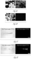
- FIGS. 1 A-C Ultrastructure of various mesoporous silica nanoparticles (MSNPs) and their ability to hold different types of cargo.
- TEM Transmission electron microscopy
- A TEM of MSNPs loaded with magnetite core (contrast agent) shown in B.
- B TEM of MSNPs with large pores (C).
- Scale bar is 50 nm.
- FIG. 2 Mechanism of action for osteotrophic mesoporous silica Nanoparticles (MSNPs).
- MSNPs osteotrophic mesoporous silica Nanoparticles
- FIGS. 3 A-B Chemical structures of Alendronate-Cy5 and NOTdronate-Cy5.
- A) Cy5 is conjugated to Alendronate which is a bisphosphonate.
- B) Cy5 is conjugated to a molecule lacking the bisphosphonate.
- FIGS. 4 A-B A) Brightfield (left) and fluorescent (right) images of Alendronate MSNPs. B) Brightfield (left) and fluorescent (right) images of carboxylic acid MSNPs, (right) images of NOTdronate-FITC stained hydroxyapatite particles.
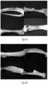
- FIGS. 5 A-F Binding affinity of Fluorescently Labelled Alendronate to hydroxyapatite and mouse bone sections.
- D Half of mouse long bone sections stained with Alendronate-FITC. Scale bar is 0.2 mm.
- FIGS. 6 A-F A-C) images of bone.
- FIGS. 8 A-B Deposition of Osteotrophic Mesoporous Nanoparticles (MSNPs) to Osseous Sites.
- FIGS. 9 A-D Plasma degradation effects on rhodamine-labeled MSNPs over time. Rhodamine-labeled MSNPs were incubated in healthy volunteer plasma at various time points and then nanoscale flow cytometry (A-C) was performed on plasma to enumerate the concentration of MSNPs (D). There was no decrease in MSNPs and they accumulated plasma proteins on their surface, increasing their size.
- FIGS. 10 A-D Intravital imaging of circulating MSNPs in the chorioallantoic membrane of the avian embryo. Injection of MSNPS (D, red signal), Hoechst (B, blue signal), and lectin (C, green signal) permits the visualization of MSNP microcirculation within the CAM capillary bed (A).
- MSNPS red signal
- Hoechst B, blue signal
- lectin C, green signal
- FIG. 11 Proposed whole-body structure of the PBPK model for MSNP deposition. Organs/tissue/blood pools are connected in an anatomical manner with blood (solid lines) and lymph (dashed lines) flows. Organ sub-compartmentalization is shown in FIG. 12 . Central blood pools are separated into blood cell and plasma space
- FIG. 12 Organ level PBPK model structure for MSNPs. Additional processes may be included based on data from avian embryos on uptake in blood and tissue cells.
- O organ blood flow
- L lymph flow
- ov vascular reflection coefficient (representative of organ specific vascular permeability);
- ai interstitial reflection coefficient (representative of particle movement in the extracellular matrix);
- Kon & Koff association and dissociation constants for the MSNP-hydroxyapatite coordinate covalent binding process
- kdeg rate of degradation of MSNPs in plasma (assumed similar in interstitial space)
- kdes rate of phagocytic uptake due to the reticuloendothelial system (RES) included in lung, liver and spleen.
- RES reticuloendothelial system
- FIGS. 13 A-B Transmission electron microscopy (TEM) image of MSNPs treated with LaC13 solution.
- MSNPs with COOH-modification (A) do not have electron dense Lanthanum crystal formation, however, MSNPs with Bisphosphonate modification (B) show crystal formation on the majority of particles imaged.
- Lanthanum crystal growth on the bisphosphonate modified MSNPs supports successful MSNP conjugation process.
- FIG. 15 MSNPs functionalized with a bisphosphonate molecule, e.g., Alendronate (alendronate treatment slows bone loss) using post-modification or co-condensation methods.
- a bisphosphonate molecule e.g., Alendronate (alendronate treatment slows bone loss) using post-modification or co-condensation methods.
- the use of surface modified MSNPs may reduce or eliminate non-specific interactions with healthy tissues.
- FIGS. 16 A-C A) Post-modification of MSNPs using heterobifunctional crosslinkers or click chemistry. B) TEM of COOH-modified (top) or DBCO-modified (bottom) MSNP cores. Hexagonal prism synthesis was used for COOH modified cores (PD2). 1 hour after TEOS addition, 50 ⁇ L of COOH-silane was added and stirred for about 1 hour. Standard methods were used for purification. For DBCO modified cores (PD42), biphase synthesis was used.
- DBCO-NHS was dissolved in DMF and three different aminated silanes (APTES (3 ethoxide groups), APDMES (2 ethoxide groups) and APDMES (1 ethoxide group)) were added to the aqueous phase before the organic phase was added, Standard methods for purification were then employed.
- APTES ethoxide groups
- APDMES ethoxide groups
- APDMES ethoxide group
- FIGS. 18 A-C Hydroxyapatite (HA) binding of modified MSNPs.
- Hydroxyapatite is a naturally occurring mineral form of calcium apatite [Ca 10 (PO 4 ) 6 (OH) 2 ] with a positive charge.
- COOH modified MSNPs have about a ⁇ 50 mV zeta potential
- DBCO-modified MSNPs have about a ⁇ 30 mV zeta potential.
- FIG. 19 Exemplary reagents to decrease non-specific binding.
- FIGS. 20 A-C Particles with a zwitterionic coating are stable in water.
- FIGS. 21 A-B Hydroxyapatite test shows increased fluorescence with Alendronate modified MSNPs. About 50 mg of HA was suspended in PBS then 25 ⁇ g of MSNPs were added and incubated for 15 minutes. The sample was then washed 3 times in PBS then placed on glass coverslip before imaging (150 ms exposure). Left image shows HA+PD47 (targeted) and right image shows HA+PD48 (non-targeted) (same exposure time).
- FIGS. 22 A-B A) Summary of LaCl 3 tests. B) TEM of PD42-DBCO modified particles.
- FIGS. 23 A-C Bright field and fluorescent staining of PD47 in tibia (A), liver (B) and heart (C).
- a cutaway shows views in three orthogonal planes. The coronal section was the cutting plane whereas the axial and sagittal sections have been digitally extracted.
- Embodiments are directed to particles and protocells for specific targeting of cells, in particular aspects, cancer bone cells, especially metastatic cancer bone cells.
- the protocells are useful in diagnostic, therapeutic and therapeutic monitoring applications.
- the present disclosure is directed to a bone cell-targeting porous particles and protocells comprising a mesoporous silica nanoparticle optionally with a supported lipid bilayer coating said nanoparticle, at least one bisphosphonate moiety conjugated to the surface of the the lipid bilayer and at least one cargo selected from the group consisting of at least one anticancer small molecule, at least one RNA molecule selected from the group consisting of small interfering RNA (siRNA), small hairpin RNA (shRNA), microRNA or a mixture thereof, e.g., a siRNA with anticancer activity, a peptide including an anticancer peptide and a reporter (e.g., diagnostic agent such as an imaging agent, which imaging agent may also function as a therapeutic agent); and optionally, at least one cell penetrating peptide (e.g., a fusogenic peptide that promotes endosomal escape of particles and protocells and encapsulated DNA, if present),
- particles e.g., MSNPs, or MSNP supported lipid bilayers (protocells) having a nanoporous silica core with a supported lipid bilayer either or both of which is conjugated to a bisphosphonate moiety for targeting the protocell to bone cancer cells; a cargo comprising at least one therapeutic agent which optionally facilitates cancer cell death such as a traditional small molecule, a macromolecular cargo (e.g.
- siRNA such as S565, S7824 and/or S 10234, among others, shRNA, a radionuclide complexed with a chelating moiety such as DOTA, among others, a protein toxin such as a ricin toxin A-chain or diphtheria toxin A-chain) and/or a packaged plasmid DNA (in certain embodiments—histone packaged) disposed within the nanoporous silica core (e.g., supercoiled as otherwise described herein in order to more efficiently package the DNA into protocells as a cargo element) which is optionally modified with a nuclear localization sequence to assist in localizing/presenting the plasmid within the nucleus of the cancer cell and the ability to express peptides involved in therapy (e.g., apoptosis/cell death of the cancer cell) or as a reporter (fluorescent green protein, fluorescent red protein, a fluorescent dye, a radionuclide among others, as otherwise described herein) for diagnostic applications, including monitoring
- Protocells may further include a targeting peptide which targets cells for therapy (e.g., cancer cells in tissue to be treated) such that binding of the protocell to the targeted cells is specific and further enhanced and a fusogenic peptide that promotes endosomal escape of protocells and encapsulated DNA.
- Protocells may be used in therapy, in diagnostics and/or monitoring therapy, more specifically to treat bone cancer, especially metastatic bone cancer.
- protocells use binding peptides which selectively bind to cancer tissue (especially bone cancer, including especially metastatic bone cancer) for therapy and/or diagnosis of cancer, including the monitoring of cancer treatment and drug discovery.
- a porous nanoparticle may comprise a nanoporous silica core optionally with a supported lipid bilayer.
- the particle or protocell comprises a targeting peptide which is or contains a cancer binding moiety, often in combination with a cell penetrating peptide such as a fusogenic peptide on the surface of the particle or protocell.
- the particle or protocell may be loaded with various therapeutic and/or diagnostic cargo, including for example, small molecules (therapeutic anticancer and/or diagnostic, macromolecules including polypeptides and nucleotides, including RNA (shRNA and especially siRNA) or plasmid DNA which may be supercoiled and histone-packaged including a nuclear localization sequence, which may be therapeutic and/or diagnostic which may include a small molecule fluorescent day, or other reporter molecule (including a reporter molecule such as a fluorescent peptide, including fluorescent green protein/FGP, fluorescent red protein/FRP, among others), or a chelating compound such as DOT A or a related radionuclide chelator in combination with a radionuclide which may be used for diagnostic and/or therapeutic purposes.
- small molecules therapeutic anticancer and/or diagnostic, macromolecules including polypeptides and nucleotides, including RNA (shRNA and especially siRNA) or plasmid DNA which may be supercoiled and histone-packaged including a nuclear local
- compositions comprise a population of particles, e.g., MSNPs, or protocells as otherwise described herein in combination with a carrier, additive and/or excipient. These pharmaceutical compositions may be used in diagnostic and/or therapeutic applications, including applications related to the monitoring of therapy, especially cancer therapy.
- Methods for treating bone cancer comprise administering a therapeutically effective number of bone-targeted particles or protocells comprising at least one anticancer agent or an effective amount of a pharmaceutical composition comprising the bone-targeted particles or protocells which comprise at least one anticancer agent, often multiple cancer anticancer agents.
- Methods for diagnosing bone cancer especially including metastatic bone cancer (for example, secondary to a primary cancer such as prostate cancer, breast cancer, lung cancer and ovarian cancer, among numerous others, comprise administering an effective amount of a population of particles or protocells as described herein which bind to bone cancer cells and include at least one reporter or other diagnostic agent in the particle or protocell to a patient suspected of having cancer or known to have primary cancer, determining the number or amount of said particles or protocells or a diagnostic agent contained in said particles or protocells which bind to or are incorporated into bone tissue of said patient and comparing the number or amount of said particles or protocells or said diagnostic agent which bind to or are incorporated into said bone tissue in said patient to a standard (which standard may include a standard obtained from one or more healthy patients, including the patient being diagnosed, a standard obtained from one or more patients with bone cancer, including metastatic bone cancer) and comparing the binding of the particles or protocells and/or diagnostic agent in the patent with the standard wherein a level above or below the standard is indicative
- the present disclosure is also directed to a method of monitoring therapy of cancer, including metastatic bone cancer in a patient in need, the method comprising administering to a patient at least twice at different times during therapy for said cancer a diagnostic effective amount of a population of particles or protocells which bind to bone tissue and which contain a reporter (e.g., a diagnostic agent), determining the number or amount of said particles or protocells or said reporter which binds to or is incorporated into bone tissue in said patient at said times and comparing the binding/incorporation of said particles or protocells or said reporter at said different times to determine whether therapy in said patient is progressing.
- a reporter e.g., a diagnostic agent
- the patient is administered said particles or protocells at about the same time that therapy is commenced and at least one time thereafter to determine the number of amount of said particles or protocells or said diagnostic agents which bind to bone tissue in said patient at the start of therapy and alter a period of therapy, wherein a reduction in the binding/incorporation of said particles or protocells and/or said reporter after a period of treatment is indicative that the therapy is favorably treating the cancer.
- patient or “subject” is used throughout the specification within context to describe an animal, generally a mammal, especially including a domesticated animal or a human, to whom treatment, including prophylactic treatment (prophylaxis), with the compounds or compositions is provided.
- treatment including prophylactic treatment (prophylaxis)
- patient refers to that specific animal.
- the patient or subject is a human patient of either or both genders.
- compound is used herein to describe any specific compound or bioactive agent disclosed herein, including any and all stereoisomers (including diastereomers), individual optical isomers (enantiomers) or racemic mixtures, pharmaceutically acceptable salts and prodrug forms.
- compound herein refers to stable compounds. Within its use in context, the term compound may refer to a single compound or a mixture of compounds as otherwise described herein.
- bioactive agent refers to any biologically active compound or drug which may be formulated for use in an embodiment.
- exemplary bioactive agents include the compounds which are used to treat cancer or a disease state or condition which occurs secondary to cancer and may include antiviral agents, especially anti-HIV, anti-HBV and/or anti-HCV agents (especially where hepatocellular cancer is to be treated) as well as other compounds or agents which are otherwise described herein.
- treat are used synonymously to refer to any action providing a benefit to a patient at risk for or afflicted with a disease, including improvement in the condition through lessening, inhibition, suppression or elimination of at least one symptom, delay in progression of the disease, prevention, delay in or inhibition of the likelihood of the onset of the disease, etc.
- viral infections these terms also apply to viral infections and may include, in certain particularly favorable embodiments the eradication or elimination (as provided by limits of diagnostics) of the virus which is the causative agent of the infection.
- Treatment encompasses both prophylactic and therapeutic treatment, principally of cancer, but also of other disease states.
- Compounds can, for example, be administered prophylactically to a mammal in advance of the occurrence of disease to reduce the likelihood of that disease, especially metastasis of bone cancer.
- Prophylactic administration is effective to reduce or decrease the likelihood of the subsequent occurrence of disease in the mammal, or decrease the severity of disease (inhibition) that subsequently occurs, especially including metastasis of cancer.
- compounds can, for example, be administered therapeutically to a mammal that is already afflicted by disease.
- administration of the present compounds is effective to eliminate the disease and produce a remission or substantially eliminate the likelihood of metastasis of a cancer.
- Administration of the compounds is effective to decrease the severity of the disease or lengthen the lifespan of the mammal so afflicted, as in the case of cancer, or inhibit or even eliminate the likelihood of disease, especially the metastasis of cancer to become metastatic bone cancer.
- pharmaceutically acceptable means that the compound or composition is suitable for administration to a subject, including a human patient, to achieve the treatments described herein, without unduly deleterious side effects in light of the severity of the disease and necessity of the treatment.
- inhibitor refers to the partial or complete elimination of a potential effect, while inhibitors are compounds/compositions that have the ability to inhibit.
- prevention when used in context shall mean “reducing the likelihood” or preventing a disease, condition or disease state from occurring as a consequence of administration or concurrent administration of one or more compounds or compositions, alone or in combination with another agent. It is noted that prophylaxis will rarely be 100% effective; consequently the terms prevention and reducing the likelihood are used to denote the fact that within a given population of patients or subjects, administration with compounds will reduce the likelihood or inhibit a particular condition or disease state (in particular, the worsening of a disease state such as the growth or metastasis of cancer) or other accepted indicators of disease progression from occurring.
- porous nanoparticle is used to describe a porous nanoparticle which is made of a material comprising silica, polystyrene, alumina, titania, zirconia, or generally metal oxides, organometallates, organosilicates or mixtures thereof.
- nanoparticulate and “porous nanoparticulate” are used interchangeably herein and such particles may exist in a crystalline phase, an amorphous phase, a semicrystalline phase, a semi amorphous phase, or a mixture thereof.
- phrases “effective average particle size” as used herein to describe a multiparticulate means that at least 50% of the particles therein are of a specified size. Accordingly, “effective average particle size of less than about 2,000 nm in diameter” means that at least 50% of the particles therein are less than about 2000 nm in diameter.
- nanoparticulates have an effective average particle size of less than about 2,000 nm (i.e., 2 microns), less than about 900 nm, less than about 1,800 nm, less than about 1,700 nm, less than about 1,600 nm, less than about 1,500 nm, less than about 1,400 nm, less than about 1,300 nm, less than about 1,200 nm, less than about 1,100 nm, less than about 1,000 nm, less than about 900 nm, less than about 800 nm, less than about 700 nm, less than about 600 nm, less than about 500 nm, less than about 400 nm, less than about 300 nm, less than about 250 nm, less than about 200 nm, less than about 150 nm, Jess than about 100 nm, less than about 75 nm, or Jess than about 50 nm, as measured by light-scattering methods, microscopy, or other appropriate methods.
- “Amine-containing silanes” include, but are not limited to, a primary amine, a secondary amine or a tertiary amine functionalized with a silicon atom, and may be a monoamine or a polyamine such as diamine.
- the amine-containing silane is N-(2-aminoethyl)-3-aminopropyltrimethoxysilane (AEPTMS).
- AEPTMS N-(2-aminoethyl)-3-aminopropyltrimethoxysilane
- Non-limiting examples of amine-containing silanes also include 3-aminopropyltrimethoxysilane (APTMS) and 3-aminopropyltriethoxysilane (APTS), as well as an amino-functional trialkoxysilane.
- Protonated secondary amines, protonated tertiary alkyl amines, protonated amidines, protonated guanidines, protonated pyridines, protonated pyrimidines, protonated pyrazines, protonated purines, protonated imidazoles, protonated pyrroles, quaternary alkyl amines, or combinations thereof, can also be used. These are used to modify the charge (Zeta potential) of the nanoparticle, which typically has a negative Zeta charge to something which is more neutral or even more positive in character.
- Bone cancer is used to describe a primary cancer of the bone. Bone cancer is an uncommon cancer that begins in a bone. Bone cancer can begin in any bone in the body, but it most commonly affects the long bones that make up the arms and legs. Several types of bone cancer exist. Some types of bone cancer occur primarily in children, while others affect mostly adults.
- the term “metastatic bone cancer” is used to describe cancers that begin elsewhere in the body and spread (metastasize) to the bone. These cancers are often named for the tissue where they began, such as prostate cancer or breast cancer that has metastasized to the bone. “Bone metastasis” occurs when cancer cells spread from their original site to a bone. Nearly all types of cancer can spread (metastasize) to the bones.
- Bone metastasis can occur in any bone but more commonly occurs in the spine, pelvis and thigh. Bone metastasis may be the first sign that you have cancer, or bone metastasis may occur years after cancer treatment.
- compositions/agents may be administered at the same time, agents may be administered at times such that effective concentrations of both (or more) compositions/agents appear in the patient at the same time for at least a brief period of time.
- each coadministered composition/agent exhibit its inhibitory effect at different times in the patient, with the ultimate result being the inhibition and treatment of cancer, especially including bone cancer, especially metastatic bone cancer, as well as the reduction or inhibition of other disease states, conditions or complications.
- cancer especially including bone cancer, especially metastatic bone cancer
- the present compounds may be combined with other agents to treat that other infection or disease or condition as required.
- anticancer agent or “additional anticancer agent” is used to describe mean a chemotherapeutic agent such as an agent selected from the group consisting of microtubule stabilizing agents, microtubule-disruptor agents, alkylating agents, antimetabolites, epidophyllotoxins, antineoplastic enzymes, topoisomerase inhibitors, inhibitors of cell cycle progression, and platinum coordination complexes.
- chemotherapeutic agent such as an agent selected from the group consisting of microtubule stabilizing agents, microtubule-disruptor agents, alkylating agents, antimetabolites, epidophyllotoxins, antineoplastic enzymes, topoisomerase inhibitors, inhibitors of cell cycle progression, and platinum coordination complexes.
- a FLT-3 inhibitor may be selected from the group consisting of a FLT-3 inhibitor, a VEGFR inhibitor, an EGFR TK inhibitor, an aurora kinase inhibitor, a PK-1 modulator, a Bcl-2 inhibitor, an HDAC inhibitor, a c-MET inhibitor, a PARP inhibitor, a Cdk inhibitor, an EGFR TK inhibitor, an IGFR-TK inhibitor, an anti-HGF antibody, a P13 kinase inhibitors, an AKT inhibitor, a JAK/ST AT inhibitor, a checkpoint-I or 2 inhibitor, a focal adhesion kinase inhibitor, a Map kinase (mek) inhibitor, a VEGF trap antibody, everolimus, trabectedin, abraxane, TLK 286 (canfosfamide), AV-299 (ficlatuzumab), DN-101 (calcitriol), pazopanib, GSK690693 (4-2-(4-amino-1
- cell penetration peptide meansogenic peptide
- endosomolytic peptide is used to describe a peptide which aids particle or protocell translocation across a lipid bilayer, such as a cellular membrane or endosome lipid bilayer and is optionally crosslinked onto a lipid bilayer surface of the particles or protocells.
- Endosomolytic peptides are a sub-species of fusogenic peptides as described herein.
- the nonendosomolytic fusogenic peptides e.g., electrostatic cell penetrating peptide such as R8 octaarginine
- the nonendosomolytic fusogenic peptides are incorporated onto the particles or protocells at the surface of the particle or protocell in order to facilitate the introduction of particles or protocells into targeted cells (APCs) to effect an intended result (to instill an immunogenic and/or therapeutic response as described herein).
- the endosomolytic peptides may be incorporated in the surface lipid bilayer of the particle or protocell or in a lipid sublayer of the multilamellar in order to facilitate or assist in the escape of the particle or protocell from endosomal bodies.
- Representative and exemplary electrostatic cell penetration (fusogenic) peptides for use in particles or protocells include an 8-mer polyarginine (H 2 N-RRRRRRRR-COOH, SEQ ID NO: 1), among others known in the art, which are included in particles or protocells in order to enhance the penetration of the particle or protocell into cells.
- endosomolytic fusogenic peptides include H5WYG peptide, H 2 N-GLFHAIAHFIHGGWHGLIHGWYGGC-COOH (SEQ ID NO:2) or H 2 N-GLFHAIAHFIHGGWHGLIHGWYGGGC-COOH (SEQ ID NO:7), or a portion thereof, e.g., GLFHAIAHFIHGGWHGLIHGWY (SEQ ID NO:8), RALA peptide (NH 2 -WEARLARALARALARHLARALARALRAGEA-COOH, SEQ ID NO:3), KALA peptide (NH 2 -WEAKLAKALAKALAKHLAKALAKALKAGEA-COOH), SEQ ID NO:4), GALA, (NH2-WEAALAEALAEALAEHLAEALAEALEALAA-COOH, SEQ ID NO:5) and INF7 (NH2-GLFEAIEGFIENGWEGMIDGWYG-COOH, SEQ ID NO:5) and INF7 (NH2-GLFE
- At least one endosomolytic peptide is included in particles or protocells in combination with a viral antigen (often pre-ubiquitinylated) and/or a viral plasmid (which expresses viral protein or antigen) in order to produce CD8+ cytotoxic T cells pursuant to a MHC class I pathway (see FIG. 4 or 6 ).
- a viral antigen often pre-ubiquitinylated
- a viral plasmid which expresses viral protein or antigen
- the peptides listed above could have a C-terminal poly-His tag, which would be amenable to Ni-NTA conjugation (lipids commercially available from Avanti).
- these peptides could be terminated with a C-terminal cysteine for which heterobifunctional crosslinker chemistry (EDC, SMPH, etc. . . . ) to link to aminated lipids would be useful.
- Another approach is to modify lipid constituents with thiol or carboxylic acid to use the same crosslinking strategy.
- crosslinking agent is used to describe a bifunctional compound of varying length containing two different functional groups which may be used to covalently link various components to each other.
- Crosslinking agents may contain two electrophilic groups (to react with nucleophilic groups on peptides of oligonucleotides, one electrophilic group and one nucleophilic group or two nucIeophilic groups).
- the crosslinking agents may vary in length depending upon the components to be linked and the relative flexibility required.
- Crosslinking agents are used to anchor targeting and/or fusogenic peptides to the phospholipid bilayer, to link nuclear localization sequences to histone proteins for packaging supercoiled plasmid DNA and in certain instances, to crosslink lipids in the lipid bilayer of the particles or protocells.
- crosslinking agents There are a large number of crosslinking agents which may be used, many commercially available or available in the literature.
- crosslinking agents include, for example, I-Ethyl-3-(3-dimethylaminopropylicarbodiimide hydrochloride (EDC), succinimidyl 4-[N-maleimidomethyl] cyclohexane-1-carboxylate (SMCC), Succinimidyl 6-[ ⁇ -Maleimidopropionamido]hexanoate (SMPH), N-( ⁇ -Maleimidopropionic acid] hydrazide (BMPH), NHS-(PEG) n -maleimide, succinimidyl-[(N-maleimidopropionamido)tetracosaethyleneglycol] ester (SM(PEG) 24 ), succinimidyl 6-[3′-(2-pyridyldithio)-propionamido] hexanoate (LC-SPDP), N- ⁇ -maleimidoacet-oxysuccinimide ester (AMAS), dibenzocyclo
- the porous nanoparticle core can include porous nanoparticles having at least one dimension, for example, a width or a diameter of about 3000 nm or less, about 1000 nm or less, about 500 nm or less, about 200 nm or less.
- the nanoparticle core is spherical with a diameter of about 500 nm or less, or about 8-10 nm to about 200 nm.
- the porous particle core can have various cross-sectional shapes including a circular, rectangular, square, or any other shape.
- the porous particle core can have pores with a mean pore size ranging from about 2 nm to about 30 nm, although the mean pore size and other properties (e.g., porosity of the porous particle core) are not limited in accordance with various embodiments of the present teachings.
- Porous nanoparticulates include mesoporous silica nanoparticles and core-shell nanoparticles.
- the porous nanoparticulates can be biodegradable polymer nanoparticulates comprising one or more compositions selected from the group consisting of aliphatic polyesters, poly (lactic acid) (PLA), poly (glycolic acid) (PGA), co-polymers of lactic acid and glycolic acid (PLGA), polycaprolactone (PCL), polyanhydrides, poly(ortho)esters, polyurethanes, poly(butyric acid), poly(valeric acid), poly(lactide-co-caprolactone), alginate and other polysaccharides, collagen, and chemical derivatives thereof, albumin a hydrophilic protein, zein, a prolamine, a hydrophobic protein, and copolymers and mixtures thereof.
- a porous spherical silica nanoparticle in one embodiment is used for particles or protocells and is surrounded by a supported lipid or polymer bilayer or multilayer.
- Various embodiments provide nanostructures and methods for constructing and using the nanostructures and providing particles or protocells. Many of the particles or protocells in their most elemental form are known in the art. Porous silica particles of varying sizes ranging in size (diameter) from less than 5 nm to 200 nm or 500 nm or more are readily available in the art or can be readily prepared using methods known in the art or alternatively, can be purchased from SkySpring Nanomaterials, Inc., Houston, Tex., USA or from Discovery Scientific, Inc., Vancouver, British Columbia.
- Multimodal silica nanoparticles may be readily prepared using the procedure of Carroll, et al., Langmuir, 25, 13540-13544 (2009).
- Particles or protocells can be readily obtained using methodologies known in the art.
- the examples section of the present application provides certain methodology for obtaining particles or protocells.
- Particles or protocells may be readily prepared, including particles or protocells comprising lipids which are fused to the surface of the silica nanoparticle. See, for example, Liu et al. (2009), Liu et al. (2009), Liu et al. (2009) Lu et al. (1999), Exemplary particles or protocells are prepared according to the procedures which are presented in Ashley et al. (2011), Lu et al. (1999), Carroll et al. (2009), and as otherwise presented in the experimental section which follows.
- a nanoparticle may have a variety of shapes and cross-sectional geometries that may depend, in part, upon the process used to produce the particles.
- a nanoparticle may have a shape that is a sphere, a rod, a tube, a flake, a fiber, a plate, a wire, a cube, or a whisker.
- a nanoparticle may include particles having two or more of the aforementioned shapes.
- a cross-sectional geometry of the particle may be one or more of circular, ellipsoidal, triangular, rectangular, or polygonal.
- a nanoparticle may consist essentially of non-spherical particles.
- Non-spherical nanoparticles may have the form of ellipsoids, which may have all three principal axes of differing lengths, or may be oblate or prelate ellipsoids of revolution.
- Non-spherical nanoparticles alternatively may be laminar in form, wherein laminar refers to particles in which the maximum dimension along one axis is substantially less than the maximum dimension along each of the other two axes.
- Non-spherical nanoparticles may also have the shape of frusta of pyramids or cones, or of elongated rods.
- the nanoparticles may be irregular in shape.
- a plurality of nanoparticles may consist essentially of spherical nanoparticles.
- the porous nanoparticulates are comprised of one or more compositions selected from the group consisting of silica, a biodegradable polymer, a solgel, a metal and a metal oxide.
- the nanostructures include a core-shell structure which comprises a porous particle core optionally surrounded by a shell of lipid, e.g., a bilayer, but possibly a monolayer or multilayer (see Liu, et al., JACS, 2009, Id).
- the porous particle core can include, for example, a porous nanoparticle made of an inorganic and/or organic material as set forth above surrounded by a lipid bilayer.
- these lipid bilayer surrounded nanostructures are referred to as “protocells” or “functional protocells,” since they have a supported lipid bilayer membrane structure.
- the porous particle core of the protocells can be loaded with various desired species (“cargo”), including small molecules (e.g. anticancer agents as otherwise described herein), large molecules (e.g. including macromolecules such as RNA, including small interfering RNA or siRNA or small hairpin RNA or shRNA or a polypeptide which may include a polypeptide toxin such as a ricin toxin A-chain or other toxic polypeptide such as diphtheria toxin A-chain DTx, among others) or a reporter polypeptide (e.g., fluorescent green protein, among others) or semiconductor quantum dots, or metallic nanoparticles, or metal oxide nanoparticles or combinations thereof.
- a polypeptide toxin such as a ricin toxin A-chain or other toxic polypeptide such as diphtheria toxin A-chain DTx, among others
- a reporter polypeptide e.g., fluorescent green protein, among others
- semiconductor quantum dots e.g., fluorescent green protein
- the particles or protocells are loaded with super-coiled plasmid DNA, which can be used to deliver a therapeutic and/or diagnostic peptide(s) or a small hairpin RNA/shRNA or small interfering RNA/siRNA which can be used to inhibit expression of proteins (such as, for example growth factor receptors or other receptors which are responsible for or assist in the growth of a cell especially a cancer cell, including epithelial growth factor/EGFR, vascular endothelial growth factor recepto/VEGFR-2 or platelet derived growth factor receptor/PDGFR-a, among numerous others, and induce growth arrest and apoptosis of cancer cells).
- proteins such as, for example growth factor receptors or other receptors which are responsible for or assist in the growth of a cell especially a cancer cell, including epithelial growth factor/EGFR, vascular endothelial growth factor recepto/VEGFR-2 or platelet derived growth factor receptor/PDGFR-a, among numerous others, and induce growth arrest and
- the cargo components can include, but are not limited to, chemical small molecules (especially anticancer agents, nucleic acids (DNA and RNA, including siRNA and shRNA and plasmids which, after delivery to a cell, express one or more polypeptides or RNA molecules), such as for a particular purpose, such as a therapeutic application or a diagnostic application as otherwise disclosed herein.
- chemical small molecules especially anticancer agents, nucleic acids (DNA and RNA, including siRNA and shRNA and plasmids which, after delivery to a cell, express one or more polypeptides or RNA molecules
- DNA and RNA including siRNA and shRNA and plasmids which, after delivery to a cell, express one or more polypeptides or RNA molecules
- a particular purpose such as a therapeutic application or a diagnostic application as otherwise disclosed herein.
- the lipid bilayer of protocells can provide biocompatibility and can be modified to possess targeting species including, for example, targeting peptides including antibodies, aptamers, and PEG (polyethylene glycol) to allow, for example, further stability of the protocells and/or a targeted delivery into a bioactive cell.
- targeting species including, for example, targeting peptides including antibodies, aptamers, and PEG (polyethylene glycol) to allow, for example, further stability of the protocells and/or a targeted delivery into a bioactive cell.
- the particle size distribution may be monodisperse or polydisperse.
- the silica cores can be rather monodisperse (i.e., a uniform sized population varying no more than about 5% in diameter, e.g., ⁇ 10-nm for a 200 nm diameter particle especially if they are prepared using solution techniques) or rather polydisperse (i.e., a polydisperse population can vary widely from a mean or medium diameter, e.g., up to ⁇ 200-nm or more if prepared by aerosol.
- Polydisperse populations can be sized into monodisperse populations. All of these are suitable for particle or protocell formation.
- particles or protocells are no more than about 500 nm in diameter, or no more than about 200 run in diameter in order to afford delivery to a patient or subject and produce an intended therapeutic effect.
- particles or protocells generally range in size from greater than about 8-10 nm to about 5 ⁇ m in diameter, about 20-nm to about 3 ⁇ m in diameter, about 10 nm to about 500 nm, or about 20-200-nm (including about 150 nm, which may be a mean or median diameter).
- the particle or protocell population may be considered monodisperse or polydisperse based upon the mean or median diameter of the population. Size is very important to therapeutic and diagnostic aspects as particles smaller than about 8-nm diameter are excreted through kidneys, and those particles larger than about 200 nm are trapped by the liver and spleen. Thus, an embodiment focuses in smaller sized particles or protocells for drug delivery and diagnostics in the patient or subject.
- protocells having particles are characterized by containing mesopores, pores which are found in the nanostructure material. These pores (at least one, but often a large plurality) may be found intersecting the surface of the nanoparticle (by having one or both ends of the pore appearing on the surface of the nanoparticle) or internal to the nanostructure with at least one or more mesopore interconnecting with the surface mesopores of the nanoparticle. Interconnecting pores of smaller size are often found internal to the surface mesopores.
- the overall range of pore size of the mesopores can be 0.03-50-nm in diameter. Pore sizes of mesopores may range from about 2-30 nm; they can be monosized or bimodal or graded—they can be ordered or disordered (essentially randomly disposed or worm-like).
- Mesopores (IUPAC definition 2-50-nm in diameter) are ‘molded’ by templating agents including surfactants, block copolymers, molecules, macromolecules, emulsions, latex beads, or nanoparticles.
- templating agents including surfactants, block copolymers, molecules, macromolecules, emulsions, latex beads, or nanoparticles.
- micropores IUPAC definition less than 2-nm in diameter
- They could also be enlarged to macropores, i.e., 50-nm in diameter.
- Pore surface chemistry of the nanoparticle material can be very diverse—all organosilanes yielding cationic, anionic, hydrophilic, hydrophobic, reactive groups—pore surface chemistry, especially charge and hydrophobicity, affect loading capacity. Attractive electrostatic interactions or hydrophobic interactions control/enhance loading capacity and control release rates. Higher surface areas can lead to higher loadings of drugs/cargos through these attractive interactions. See below.
- the surface area of nanoparticles ranges from about 100 m2/g to >about 1200 m2/g.
- the larger the pore size the smaller the surface area.
- the surface area theoretically could be reduced to essentially zero, if one does not remove the templating agent or if the pores are sub-0.5-nm and therefore not measurable by N2 sorption at 77K due to kinetic effects. However, in this case, they could be measured by CO 2 or water sorption, but would probably be considered non-porous. This would apply if biomolecules are encapsulated directly in the silica cores prepared without templates, in which case particles (internal cargo) would be released by dissolution of the silica matrix after delivery to the cell.
- the particles or protocells are loaded with cargo to a capacity up to over 100 weight %; defined as (cargo weight/weight of particle or protocell) ⁇ 100.
- the optimal loading of cargo is often about 0.01 to 30% but this depends on the drug or drug combination which is incorporated as cargo into the particle or protocell. This is generally expressed in ⁇ M per 10 10 particles where we have values ranging from 2000-100 ⁇ M per 10 10 particles.
- Particles or protocells may exhibit release of cargo at pH about 5.5, which is that of the endosome, but are stable at physiological pH of 7 or higher (e.g., 7.4).
- the surface area of the internal space for loading is the pore volume whose optimal value ranges from about 1.1 to 0.5 cubic centimeters per gram (cc/g). Note that in the particles or protocells according to one embodiment, the surface area is mainly internal as opposed to the external geometric surface area of the nanoparticle.
- the lipid bilayer if present supported on the porous particle has a lower melting transition temperature, e.g., is more fluid than a lipid bilayer supported on a non-porous support or the lipid bilayer in a liposome. This is sometimes important in achieving high affinity binding of targeting ligands at low peptide densities, as it is the bilayer fluidity that allows lateral diffusion and recruitment of peptides by target cell surface receptors.
- One embodiment provides for peptides to cluster, which facilitates binding to a complementary target.
- Lipid bilayers may be prepared using any method known in the art, but often the bilayers are fused onto MSNPs. In this approach, MSNPs are mixed with liposomes in aqueous buffer and washed to remove free liposomes in solution.
- the lipid bilayer may vary significantly in composition. Ordinarily, any lipid or polymer which is may be used in liposomes may also be used in protocells.
- lipid bilayers for use in protocells comprise a mixtures of lipids (as otherwise described herein) at a weight percent of 5% DOPE, 5% PEG, 30% cholesterol, 60% DOPC or DPPC.
- Additional lipid bilayers comprise a mixture of lipids 60% DSPC, 30% DOPE, 10% Cholesterol (weight percent.); 60% DSPC, 15% DOPE, 15% Cholesterol and 10% DSPE-PEG 2000 (weight percent) and 60% DSPC, 15% DSPE, 15% Cholesterol and 10% DSPE-PEG 2000 (weight percent).
- the charge of the MSNP core as measured by the Zeta potential may be varied monotonically from ⁇ 50 to +50 m V by modification with the amine-containing silane, for example, 2-(aminoethyl) propyltrimethoxy-silane (AEPTMS) or other organosilanes, as disclosed herein.
- This charge modification in turn, varies the loading of the drug within the cargo of the particle or protocell.
- the zeta-potential is reduced to between about ⁇ I0 mV and +5 mV, which is important for maximizing circulation time in the blood and avoiding non-specific interactions.
- the silica dissolution rates can be varied widely. This in turn controls the release rate of the internal cargo. This occurs because molecules that are strongly attracted to the internal surface area of the pores diffuse slowly out of the particle cores, so dissolution of the particle cores controls in part the release rate.
- particles or protocells are stable at pH 7, i.e., they don't leak their cargo, but at pH 5.5, which is that of the endosome lipid or polymer coating becomes destabilized initiating cargo release.
- This pH-triggered release is important for maintaining stability of the particle or protocell up until the point that it is internalized in the cell by endocytosis, whereupon several pH triggered events cause release into the endosome and consequently, the cytosol of the cell.
- the protocell core particle and surface can also be modified to provide non-specific release of cargo over a specified, prolonged period of time, as well as be reformulated to release cargo upon other biophysical changes, such as the increased presence of reactive oxygen species and other factors in locally inflamed areas. Quantitative experimental evidence has shown that targeted particles or protocells elicit only a weak immune response, because they do not support T-Cell help required for higher affinity IgG, a favorable result.
- the surface area of the internal space for loading is the pore volume whose optimal value ranges from about 1.1 to 0.5 cubic centimeters per gram (cc/g). Note that in the case of nanoparticles the surface area is mainly external, but can also be highly internal, depending on the nature of the mesopores in the nanoparticles.
- Mesoporous silica nanoparticles can be, e.g., from around 5 nm to around 500 nm in size, including all integers and ranges there between. The size is measured as the longest axis of the particle. In various embodiments, the particles are from around 10 nm to around 500 nm and from around 10 nm to around 100 nm in size.
- the mesoporous silica nanoparticles have a porous structure.
- the pores can be from around 1 to around 20 nm in diameter, including all integers and ranges there between. In one embodiment, the pores are from around 1 to around 10 nm in diameter. In one embodiment, around 90% of the pores are from around 1 to around 20 nm in diameter. In another embodiment, around 95% of the pores are around 1 to around 20 nm in diameter.
- the mesoporous nanoparticles can be synthesized according to methods known in the art.
- the nanoparticles are synthesized using sol-gel methodology where a silica precursor or silica precursors and a silica precursor or silica precursors conjugated (i.e., covalently bound) to absorber molecules are hydrolyzed in the presence of templates in the form of micelles.
- the templates are formed using a surfactant such as, for example, hexadecyltrimethylammonium bromide (CTAB). It is expected that any surfactant which can form micelles can be used.
- CTAB hexadecyltrimethylammonium bromide
- the core-shell nanoparticles comprise a core and shell.
- the core comprises silica and an absorber molecule.
- the absorber molecule is incorporated in to the silica network via a covalent bond or bonds between the molecule and silica network.
- the shell comprises silica.
- the core is independently synthesized using known sol-gel chemistry, e.g., by hydrolysis of a silica precursor or precursors.
- the silica precursors are present as a mixture of a silica precursor and a silica precursor conjugated, e.g., linked by a covalent bond, to an absorber molecule (referred to herein as a “conjugated silica precursor”).
- Hydrolysis can be carried out under alkaline (basic) conditions to form a silica core and/or silica shell.
- the hydrolysis can be carried out by addition of ammonium hydroxide to the mixture comprising silica precursor(s) and conjugated silica precursor(s).
- Silica precursors are compounds which under hydrolysis conditions can form silica.
- Examples of silica precursors include, but are not limited to, organosilanes such as, for example, tetraethoxysilane (TEOS), tetramethoxysilane (TMOS), and the like.
- the silica precursor used to form the conjugated silica precursor has a functional group or groups which can react with the absorbing molecule or molecules to form a covalent bond or bonds.
- silica precursors include, but is not limited to, isocyanatopropyltriethoxysilane (ICPTS), (3-glycolyl oxypropyl) trimethoxysilane, aminopropyitrimethoxysilane (APTS), mercaptopropyltrimethoxysilane (MPTS), APTES (3-aminopropyl)triethoxysilane), APMDES (3-aminopropylmethyl diethoxysilane, APDMES (3-aminopropyl)-dimethyl-ethoxysilane, APMS (3-amino propyl)-trimethtoxysilane, and the like.
- ICPTS isocyanatopropyltriethoxysilane
- APTS aminopropyitrimethoxy
- an organosilane (conjugatable silica precursor) used for forming the core has the general formula R 4n SIX n , where X is a hydrolyzable group such as ethoxy, methoxy, or 2-methoxy-ethoxy; R can be a monovalent organic group of from 1 to 12 carbon atoms which can optionally contain, but is not limited to, a functional organic group such as mercapto, epoxy, acrylyl, methacrylyl, or amino; and n is an integer of from 0 to 4.
- the conjugatable silica precursor is conjugated to an absorber molecule and subsequently co-condensed for forming the core with silica precursors such as, for example, TEOS and TMOS.
- a silane used for forming the silica shell has n equal to 4.
- the use of functional mono-, bis- and tris-alkoxysilanes for coupling and modification of co-reactive functional groups or hydroxy-functional surfaces, including glass surfaces, is also known, see Kirk-Othmer, Encyclopedia of Chemical Technology, Vol. 20, 3rd Ed., J. Wiley, N.Y.; see also E. Pluedemann, Silane Coupling Agents, Plenum Press, N. Y. 1982.
- the organo-silane can cause gels, so it may be desirable to employ an alcohol or other known stabilizers. Processes to synthesize core-shell nanoparticles using modified Stober processes can be found in U.S. patent applications Ser. Nos. 10/306,614 and 10/536,569, the disclosure of such processes therein are incorporated herein by reference.
- Bone metastasis can cause pain and broken bones. With rare exceptions, cancer that has spread to the bones can't be cured. Treatments can help reduce pain and other symptoms of bone metastases. Bone metastasis often causes no signs and symptoms, but when symptoms do occur, these symptoms can include bone pain, broken bones, urinary incontinence, bowel incontinence, weakness in the legs or arms and hypercalcemia, which can cause vomiting constipation and confusion.
- bisphosphonate is used to describe a compound according to the chemical structure:
- Exemplary bisphosphonates which may be readily utilized include pamidronate, neridronate or alendronate (e.g., alendronate) because these bisphosphonates may be easily conjugated to a carboxylic acid group on the surface of the MSNP or bisphosphonates which contain a carboxylic acid group which can be readily conjugated to an amine containing phospholipid in the phospholipid bilayer of the particle or protocell. It is noted that all of the bisphosphonates which contain a hydroxyl group as R1 may be conjugated through an isocyanate group to form a urethane group on the surface of the MSNP or the phospholipid bilayer. Exemplary common bisphosphonates which may be used are presented below, with pamidronate, neridronate or alendronate being specific embodiment because of the ease with which these bisphosphonates may be conjugated with a carboxylic acid group.
- protocells are biocompatible. Drugs and other cargo components are often loaded by adsorption and/or capillary filling of the pores of the particle core up to approximately 50% by weight of the final protocell (containing all components).
- the loaded cargo can be released from the porous surface of the particle core (mesopores), wherein the release profile can be determined or adjusted by, for example, the pore size, the surface chemistry of the porous particle core, the pH value of the system, and/or the interaction of the porous particle core with the surrounding lipid bilayer(s) as generally described herein.
- the porous nanoparticle core used to prepare the protocells can be tuned in to be hydrophilic or progressively more hydrophobic as otherwise described herein and can be further treated to provide a more hydrophilic surface.
- mesoporous silica particles can be further treated with ammonium hydroxide and hydrogen peroxide to provide a higher hydrophilicity.
- the lipid bilayer is fused onto the porous particle core to form the protocell.
- Protocells can include various lipids in various weight ratios, e.g., including 1,2-dioleoyl-sn-glycero-3-phosphocholine (DOPC), 1,2-dipalmitoyl-sn-glycero-3-phosphocholine (DPPC), 1,2-distearoyl-sn-glycero-3-phosphocholine (DSPC), I,2-dioleoyl-sn-glycero-3-(phosphor-L-serine] (DOPS), I,2-dioleoyl-3-trimethylammonium-propane (18:1 DOTAP), 1,2-dioleoyl-sn-glycero-3-phospho-(1′-rac-glycerol) (DOPG), 1,2-dioleoyl-sn-glycero-3-phosphoethanolamine (DOPE), 1,2-dipalmitoyl-sn-glycero-3-phosphoethanolamine (DPPE), 1,2-diole
- Pegylated phospholipids may be included in lipid bilayers in protocells. These pegylated phospholipids include for example, pegylated 1,2-distearoyl-sn-glycero-3-phosphoethanolamine (PEG-DSPE), pegylated 1,2-dialeoyl-sn-glycero-3-phosphoethanolamine (PEG-DOPE), pegylated 1,2-dipalmitoyl-sn-glycero-3-phosphoethanolamine (PEG-DPPE), and pegylated 1,2-dimyristoyl-sn-glycero-3-phosphoethanolamine (PEG-DMPE), among others, including a pegylated ceramide (e.g.
- the PEG generally ranges in size (average molecular weight for the PEG group) from about 350-7500, about 350-5000, about 500-2500, about 1000-2000.
- Pegylated phospholipids may comprise the entire phospholipid monolayer of hybrid phospholipid protocells, or alternatively they may comprise a minor component of the lipid monolayer or be absent.
- the percent by weight of a pegylated phospholipid in phospholipid monolayers which make up ranges from 0% to 100%, 0.01% to 99%, about 5%, 10%, 15%, 20%, 25%, 30%, 35%, 40%, 50%, 55%, 60% and the remaining portion of the phospholipid monolayer comprising at least one additional lipid (such as cholesterol, usually in amounts less than about 50% by weight), including a phospholipid.
- Certain lipid combinations are often used. These include, for example, DSPC/DOPE/Cholesterol (60/30/10 mass %), DSPC/DOPE/Cholesterol/DSPE-PEG 2000 (60/15/15/10 mass %), and DSPC/DSPE/Cholesterol/DSPE-PEG 2000 (60/15/15/10), among other combinations.
- DSPC/DOPE/Cholesterol 60/30/10 mass %)
- DSPC/DOPE/Cholesterol/DSPE-PEG 2000 60/15/15/10 mass %
- DSPC/DSPE/Cholesterol/DSPE-PEG 2000 60/15/15/10
- the lipid bilayer which is used to prepare protocells can be prepared, for example, by extrusion of hydrated lipid films through a filter with pore size of, for example, about 100 run, using standard protocols known in the art or as otherwise described herein.
- the filtered lipid bilayer films can then be fused with the porous particle cores, for example, by pipette mixing.
- excess amount of lipid bilayer or lipid bilayer films can be used to form the protocell in order to improve the protocell colloidal stability.
- various dyes or fluorescent (reporter) molecules can be included in the protocell cargo (as expressed by as plasmid DNA) or attached to the porous particle core and/or the lipid bilayer for diagnostic purposes.
- the porous particle core can be a silica core or the lipid bilayer and can be covalently labeled with FITC (green fluorescence), while the lipid bilayer or the particle core can be covalently labeled with FITC Texas red (red fluorescence).
- the porous particle core, the lipid bilayer and the formed protocell can then be observed by, for example, confocal fluorescence for use in diagnostic applications.
- plasmid DNA can be used as cargo in protocells such that the plasmid may express one or more fluorescent proteins such as fluorescent green protein or fluorescent red protein which may be used in diagnostic applications.
- the protocell is used in a synergistic system where the lipid bilayer fusion or liposome fusion (i.e., on the porous particle core) is loaded and sealed with various cargo components with the pores (mesopores) of the particle core, thus creating a loaded protocell useful for cargo delivery across the cell membrane of the lipid bilayer or through dissolution of the porous nano particle, if applicable.
- the lipid bilayer fusion or liposome fusion i.e., on the porous particle core
- various cargo components with the pores (mesopores) of the particle core
- the protocell is used in a synergistic system where the lipid bilayer fusion or liposome fusion (i.e., on the porous particle core) is loaded and sealed with various cargo components with the pores (mesopores) of the particle core, thus creating a loaded protocell useful for cargo delivery across the cell membrane of the lipid bilayer or through dissolution of the porous nano particle, if applicable.
- multiple bilayers with opposite charges can be
- a fusion and synergistic loading mechanism can be included for cargo delivery.
- cargo can be loaded, encapsulated, or sealed, synergistically through liposome fusion on the porous particles.
- the cargo can include, for example, small molecule drugs (e.g. especially including anticancer drugs and/or antiviral drugs such as anti-HBV or anti-HCV drugs), peptides, proteins, antibodies, DNA (especially plasmid DNA, including the histone-packaged super coiled plasmid DNA), RNAs (including shRNA and siRNA (which may also be expressed by the plasmid DNA incorporated as cargo within the protocells) fluorescent dyes, including fluorescent dye peptides which may be expressed by the plasmid DNA incorporated within the protocell.
- small molecule drugs e.g. especially including anticancer drugs and/or antiviral drugs such as anti-HBV or anti-HCV drugs
- peptides e.g. especially including anticancer drugs and/or antiviral drugs such as anti
- the cargo can be loaded into the pores (mesopores) of the porous particle cores to form the loaded protocell.
- any conventional technology that is developed for liposome-based drug delivery for example, targeted delivery using PEGylation, can be transferred and applied to the protocells.
- porous silica nanoparticles can carry a negative charge and the pore size can be tunable from about 2 nm to about 10 nm or more.
- Negatively charged nanoparticles can have a natural tendency to adsorb positively charged molecules and positively charged nanoparticles can have a natural tendency to adsorb negatively charged molecules.
- other properties such as surface wettability (e.g., hydrophobicity) can also affect loading cargo with different hydrophobicity.
- the cargo loading can be a synergistic lipid-assisted loading by tuning the lipid composition.
- the cargo component is a negatively charged molecule
- the cargo loading into a negatively charged silica can be achieved by the lipid-assisted loading.
- a negatively species can be loaded as cargo into the pores of a negatively charged silica particle when the lipid bilayer is fused onto the silica surface showing a fusion and synergistic loading mechanism. In this manner, fusion of a non-negatively charged (i.e., positively charged or neutral) lipid bilayer or liposome on a negatively charged mesoporous particle can serve to load the particle core with negatively charged cargo components.
- the negatively charged cargo components can be concentrated in the loaded protocell having a concentration exceed about 100 times as compared with the charged cargo components in a solution.
- positively charged cargo components can be readily loaded into protocells.
- the loaded protocells can have a cellular uptake for cargo delivery into a desirable site after administration.
- the cargo-loaded protocells can be administered to a patient or subject and the protocell comprising a targeting peptide can bind to a target cell and be internalized or uptaken by the target cell, for example, a cancer cell in a subject or patient. Due to the internalization of the cargo-loaded protocells in the target cell, cargo components can then be delivered into the target cells.
- the cargo is a small molecule, which can be delivered directly into the target cell for therapy.
- negatively charged DNA or RNA (including shRNA or siRNA), especially including a DNA plasmid which may be formulated as histone-packaged supercoiled plasmid DNA, e.g., modified with a nuclear localization sequence, can be directly delivered or internalized by the targeted cells.
- the DNA or RNA can be loaded first into a protocell and then into then through the target cells through the internalization of the loaded protocells.
- the cargo loaded into and delivered by the protocell to targeted cells includes small molecules or drugs (especially anti-cancer and optionally, antiviral or other bioactive agents), bioactive macromolecules (bioactive polypeptides such as ricin toxin A-chain or diphtheria toxin A-chain or RNA molecules such as shRNA and/or shRNA as otherwise described herein) or histone-packaged supercoiled plasmid DNA which can express a therapeutic or diagnostic peptide or a therapeutic RNA molecule such as shRNA or siRNA, wherein the histone-packaged supercoiled plasmid DNA is optionally modified with a nuclear localization sequence which can localize and concentrate the delivered plasmid DNA into the nucleus of the target cell.
- loaded protocells can deliver their cargo into targeted cells for therapy or diagnostics.
- the protocells and/or the loaded protocells can provide a targeted delivery methodology for selectively delivering the protocells or the cargo components to targeted cells (e.g., cancer cells).
- a surface of the lipid bilayer can be modified by a targeting active species that corresponds to the targeted cell.
- the targeting active species may be a targeting peptide as otherwise described herein, a polypeptide including an antibody or antibody fragment, an aptamer, a carbohydrate or other moiety which binds to a targeted cell.
- the targeting active species is a targeting peptide as otherwise described herein.
- exemplary peptide targeting species include a MET binding peptide as otherwise described herein.
- the protocell selectively binds to the targeted cell in accordance with the present teachings.
- a targeting active species e.g., a targeting peptide
- the protocell by conjugating an exemplary targeting peptide SP94 or analog or a MET binding peptide as otherwise described herein that targets cancer cells, including cancer liver cells to the lipid bilayer, a large number of the cargo-loaded protocells can be recognized and internalized by this specific cancer cells due to the specific targeting of the exemplary SP94 or MET binding peptide with the cancer (including liver) cells.
- the protocells will selectively bind to the cancer cells and no appreciable binding to the non-cancerous cells occurs.
- the loaded protocells can release cargo components from the porous particle and transport the released cargo components into the target cell.
- the cargo components can be released from the pores of the lipid bilayer, transported across the protocell membrane of the lipid bilayer and delivered within the targeted cell.
- the release profile of cargo components in protocells can be more controllable as compared with when only using liposomes as known in the prior art.
- the cargo release can be determined by, for example, interactions between the porous core and the lipid bilayer and/or other parameters such as pH value of the system.
- the release of cargo can be achieved through the lipid bilayer, through dissolution of the porous silica; while the release of the cargo from the protocells can be pH-dependent.
- the pH value for cargo is often less than 7, or about 4.5 to about 6.0, but can be about pH 14 or less.
- Lower pHs tend to facilitate the release of the cargo components significantly more than compared with high pHs.
- Lower pHs tend to be advantageous because the endosomal compartments inside most cells are at low pHs, (about 5.5), but the rate of delivery of cargo at the cell can be influenced by the pH of the cargo.
- the release of cargo can be relative short (a few hours to a day or so) or span for several days to about 20-30 days or longer.
- immediate release and/or sustained release applications from the protocells themselves are envisioned.
- the inclusion of surfactants can be provided to rapidly rupture the lipid bilayer, transporting the cargo components across the lipid bilayer of the protocell as well as the targeted cell.
- the phospholipid bilayer of the protocells can be ruptured by the application/release of a surfactant such as sodium dodecyl sulfate (SDS), among others to facilitate a rapid release of cargo from the protocell into the targeted cell.
- SDS sodium dodecyl sulfate
- other materials can be included to rapidly rupture the bilayer.
- One example would be gold or magnetic nanoparticles that could use light or heat to generate heat thereby rupturing the bilayer.
- the bilayer can be tuned to rupture in the presence of discrete biophysical phenomena, such as during inflammation in response to increased reactive oxygen species production.
- the rupture of the lipid bilayer can in turn induce immediate and complete release of the cargo components from the pores of the particle core of the protocells.
- the protocell platform can provide an increasingly versatile delivery system as compared with other delivery systems in the art.
- the disclosed protocell platform can provide a simple system and can take advantage of the low toxicity and immunogenicity of liposomes or lipid bilayers along with their ability to be PEGylated or to be conjugated to extend circulation time and effect targeting.
- the protocell platform when compared to delivery systems using liposome only, can provide a more stable system and can take advantage of the mesoporous core to control the loading and/or release profile and provide increased cargo capacity.
- the lipid bilayer and its fusion on porous particle core can be fine-tuned to control the loading, release, and targeting profiles and can further comprise fusogenic peptides and related peptides to facilitate delivery of the protocells for greater therapeutic and/or diagnostic effect.
- the lipid bilayer of the protocells can provide a fluidic interface for ligand display and multivalent targeting, which allows specific targeting with relatively low surface ligand density due to the capability of ligand reorganization on the fluidic lipid interface.
- the disclosed protocells can readily enter targeted cells while empty liposomes without the support of porous particles cannot be internalized by the cells.
- compositions comprise an effective population of protocells as otherwise described herein formulated to effect an intended result (e.g. therapeutic result and/or diagnostic analysis, including the monitoring of therapy) formulated in combination with a pharmaceutically acceptable carrier, additive or excipient.
- the protocells within the population of the composition may be the same or different depending upon the desired result to be obtained.
- Pharmaceutical compositions may also comprise an addition bioactive agent or drug, such as an anticancer agent or an antiviral agent, for example, an anti-HIV, anti-HBV or an anti-HCV agent.
- dosages and routes of administration of the compound are determined according to the size and condition of the subject, according to standard pharmaceutical practices. Dose levels employed can vary widely, and can readily be determined by those of skill in the art. Typically, amounts in the milligram up to gram quantities are employed.
- the composition may be administered to a subject by various routes, e.g. orally, transdermally, perineurally or parenterally, that is, by intravenous, subcutaneous, intraperitoneal, intrathecal or intramuscular injection, among others, including buccal, rectal and transdermal administration. Subjects contemplated for treatment include humans, companion animals, laboratory animals, and the like.
- immediate and/or sustained/controlled release compositions including compositions which comprise both immediate and sustained release formulations. This is particularly true when different populations of protocells are used in the pharmaceutical compositions or when additional bioactive agent(s) are used in combination with one or more populations of protocells as otherwise described herein.
- Formulations containing compounds may take the form of liquid, solid, semi-solid or lyophilized powder forms, such as, for example, solutions, suspensions, emulsions, sustained-release formulations, tablets, capsules, powders, suppositories, creams, ointments, lotions, aerosols, patches or the like, e.g., in unit dosage forms suitable for simple administration of precise dosages.
- compositions typically include a conventional pharmaceutical carrier or excipient and may additionally include other medicinal agents, carriers, adjuvants, additives and the like.
- the composition is about 0.1% to about 85%, about 0.5% to about 75% by weight of a compound or compounds, with the remainder consisting essentially of suitable pharmaceutical excipients.
- An injectable composition for parenteral administration (e.g. intravenous, intramuscular or intrathecal) will typically contain the compound in a suitable i.v. solution, such as sterile physiological salt solution.
- a suitable i.v. solution such as sterile physiological salt solution.
- the composition may also be formulated as a suspension in an aqueous emulsion.
- Liquid compositions can be prepared by dissolving or dispersing the population of protocells (about 0.5% to about 20% by weight or more), and optional pharmaceutical adjuvants, in a carrier, such as, for example, aqueous saline, aqueous dextrose, glycerol, or ethanol, to form a solution or suspension.
- a carrier such as, for example, aqueous saline, aqueous dextrose, glycerol, or ethanol
- the composition may be prepared as a solution, suspension, emulsion, or syrup, being supplied either in liquid form or a dried form suitable for hydration in water or normal saline.
- excipients include pharmaceutical grades of mannitol, lactose, starch, magnesium stearate, sodium saccharine, talcum, cellulose, glucose, gelatin, sucrose, magnesium carbonate, and the like.
- the composition may also contain minor amounts of non-toxic auxiliary substances such as wetting agents, emulsifying agents, or buffers.
- the preparations may be tablets, granules, powders, capsules or the like.
- the composition is typically formulated with additives, e.g. an excipient such as a saccharide or cellulose preparation, a binder such as starch paste or methyl cellulose, a filler, a disintegrator, and other additives typically used in the manufacture of medical preparations.
- additives e.g. an excipient such as a saccharide or cellulose preparation, a binder such as starch paste or methyl cellulose, a filler, a disintegrator, and other additives typically used in the manufacture of medical preparations.
- composition to be administered will contain a quantity of the selected compound in a pharmaceutically effective amount for therapeutic use in a biological system, including a patient or subject.
- Methods of treating patients or subjects in need for a particular disease state or infection comprise administration an effective amount of a pharmaceutical composition comprising therapeutic protocells and optionally at least one additional bioactive (e.g. antiviral) agent.
- a pharmaceutical composition comprising therapeutic protocells and optionally at least one additional bioactive (e.g. antiviral) agent.
- Diagnostic methods comprise administering to a patient in need (a patient suspected of having cancer) an effective amount of a population of diagnostic protocells (e.g., protocells which comprise a target species, such as a targeting peptide which binds selectively to cancer cells and a reporter component to indicate the binding of the protocells to cancer cells if the cancer cells are present) whereupon the binding of protocells to cancer cells as evidenced by the reporter component (moiety) will enable a diagnosis of the existence of cancer in the patient.
- a population of diagnostic protocells e.g., protocells which comprise a target species, such as a targeting peptide which binds selectively to cancer cells and a reporter component to indicate the binding of the protocells to cancer cells if the cancer cells are present
- An alternative of the diagnostic method can be used to monitor the therapy of cancer or other disease state in a patient, the method comprising administering an effective population of diagnostic protocells (e.g., protocells which comprise a target species, such as a targeting peptide which binds selectively to cancer cells or other target cells and a reporter component to indicate the binding of the protocells to cancer cells if the cancer cells are present) to a patient or subject prior to treatment, determining the level of binding of diagnostic protocells to target cells in said patient and during and/or after therapy, determining the level of binding of diagnostic protocells to target cells in said patient, whereupon the difference in binding before the start of therapy in the patient and during and/or after therapy will evidence the effectiveness of therapy in the patient, including whether the patient has completed therapy or whether the disease state has been inhibited or eliminated (including remission of a cancer).
- diagnostic protocells e.g., protocells which comprise a target species, such as a targeting peptide which binds selectively to cancer cells or
- Prismatic hexagonal structured MSNP are synthesized using techniques described in the literature (see Lin, Y.-S.; Haynes, C. L. Impacts of Mesoporous Silica Nanoparticle Size, Pore Ordering, and Pore Integrity on Hemolytic Activity. Journal of the American Chemical Society 2010, 132, 4834-4842 and Lin, Y.-S.; Abadeer, N.; Hurley, K. R.; Haynes, C. L. Ultrastable, Redispersible, Small, and Highly Organomodified Mesoporous Silica Nanotherapeutics. Journal of the American Chemical Society 2011, 133, 20444-20457.).
- the MSNP after formation (about a 12 hour synthesis using standard methods of preparation, as described above) may be first carboxylated (using a silyl carboxyl agent such as 3-(triethoxysilyl)propylsuccinic anhydride at approximately 0.5% to about 20%, often about 1% to about 15%, often about 1% to about 5%, about 1-1.5% of the TEOS utilized) to form a carboxylic acid group on the surface of the MSN linked to the MSN through Si—O—Si bonds formed when the 3-(triethoxysilyl)propylsuccinic acid and the SiOH groups on the surface of the MSN react. This takes about an hour or so.
- a silyl carboxyl agent such as 3-(triethoxysilyl)propylsuccinic anhydride at approximately 0.5% to about 20%, often about 1% to about 15%, often about 1% to about 5%, about 1-1.5% of the TEOS utilized
- the carboxylated MSN is then subjected to a hydrothermal step (generally about 12-36 hours, such as about 24 hours at an elevated temperature ranging from about 60° C. to about 120° C.) to form a final carboxylated MSN which can be reacted with a crosslinker such as EDC or other crosslinker (the amine portion of the crosslinker forms an amide or other stable bond with the carboxyl group) and the carboxylic/electrophilic end of the linker is reacted with an amine containing phospholipid such as DOPE, DMPE, DPPE or DSPE to form the hydrocarbon coated MSN.
- a crosslinker such as EDC or other crosslinker
- an amine portion of the crosslinker forms an amide or other stable bond with the carboxyl group
- an amine containing phospholipid such as DOPE, DMPE, DPPE or DSPE
- the carboxylic acid modified MSNPs can be conjugated to the primary amine on the bisphosphonate molecule to form an amide bond.
- This is straight forward chemistry.
- heterobifunctional carboxyl-to-amine crosslinker 1-ethyl-3-(3 dimethylaminopropyl)carbodiimide hydrochloride (EDC) or dicyclohexylcarbodiimide (DCC), among others, depending on the functional group chemistry can be added to the bisphosphonate molecule, or to the COOH-modified MSNPs, or all three components can be added together simultaneously.
- the bisphosphonate to crosslinker mole ratio may be approximately 2:1, although this ratio may be increased) and mass ratio of bisphosphonate to MSNP is approximately 1:2.
- This should provide a complete covering of the MSNP (at least about 75%, at least about 80%, at least about 85%, at least about 90%, at least about 95%, at least about 98-99% and up to about 100% of the surface area of the MSNP.
- the MSNP is covered as completely as possible.
- crosslinker reaction time 30 min to 4 hours at room temperature, or 8-16 hours at 4° C. After reaction, MSNPs are washed twice in H 2 0 and stored in H 2 0.
- Bisphosphonate molecule modification is performed using the same technique described above DBCO- or N3-NHS ester to synthesize DBCO- or a N3-Bisphosphonate molecule.
- To prepare bisphosphonate modified MSNPs N3-Bisphosphonate molecules are reacted with DBCO-MSNPs or DBCO-Bisphosphonate molecules are reacted with N3-MSNPs in H 2 O for 2 hours to 24 hours at room temperature. The reaction product obtained is then washed several times in H 2 0 to remove unreacted bisphosphonate molecules.
- Bisphosphonate modified silanes may be prepared using 3-(Triethoxysilyl)propyl isocyanate reacted with bisophosphonate in DMF or DMSO for 30 minutes to 2 hours at room temperature.
- synthesis scheme 1 hex MSNP, see above
- bisphosphonate-silane is added at the same time as TEOS and synthesized and purified using standard methods which are described in the literature.
- synthesis scheme 2 large pore MSNP
- bisphosphonate silane is added to the aqueous phase 10 minutes prior or TEOS addition in the organic phase and particles are synthesized and purified as described in the literature.
- MSNPs were loaded with various cargo types, such as chemotherapy drugs (Docetaxel, Cisplatin), biologics (anti-EGFR peptides), DOTA for chelation of Gallium-68, and siRNA.
- cargo types such as chemotherapy drugs (Docetaxel, Cisplatin), biologics (anti-EGFR peptides), DOTA for chelation of Gallium-68, and siRNA.
- MSNPs offer greater therapeutic potential and human compatibility for multi-functional drug delivery over other nanoparticle types because of three main properties: increased cargo capacity, well established chemistries for silica modification, and superior stability during modification and loading of MSNPs. These qualities result in MSNPs exhibiting the highest payload to nanoparticle mass ratio known (about 3-6% as opposed to about 0.2% in liposomes), and excellent in vivo stability.
- MSNPs are comprised of a highly rigid pore structure that results in a very large surface for adsorption and entrapment of payload (about 1.0 m 2 /mg of MSNP) for slow controlled release of payload in vivo. Due to its ubiquitous use for electronics fabrication, silica modification chemistry is well understood, enabling reproducible conjugation of targeting moieties and chelation agents such as DOTA to MSNPs in a straightforward manner. Silicon dioxide is non-toxic in vitro or in vivo and due to its silica-based composition, it is highly stable after several modifications to MSNPs, resulting in high yields of complex, multifunctional nanoparticles.
- Certain surface chemistries can minimize MSNP uptake by endothelial cells, leukocytes, and binding to serum proteins. This is an important advance because a higher proportion of osteotropic MSNPs will remain in the circulation, enhancing bone targeting. When compared to polymer-based nanoparticle formulations, MSNPs exhibit minimal polydispersity, which is important for minimizing non-specific uptake by immune cells.
- the surface of these nanoparticles is decorated with Alendronate, a bisphosphonate molecule that has high affinity for hydroxyapatite, the mineralized component of bone.
- Alendronate a bisphosphonate molecule that has high affinity for hydroxyapatite, the mineralized component of bone.
- Several types of bisphosphonates are available for use in alleviating bone pain caused by prostate cancer bone metastases, and therefore utilizing Alendronate as a targeting moiety on MSNPs may impart an anti-osteoblastic therapeutic benefit at sites of prostate cancer bone metastases.
- Various fluorescent conjugates to Alendronate ( FIG. 3 A ) and a negative binding control, called NOTdronate US Patent Pending PCT/CA2014/050312
- Osteotropic MSNPs efficiently target all bone in pre-clinical models of prostate cancer and then release therapeutic payload in a slow release, controlled manner into the bone microenvironment. This route of delivery will result in maximal bioavailable levels of MSNPs and their cargo in the bone and minimize nontarget exposure, thus minimizing side effects.
- MSNPs are used. These include: Vacant Osteotropic MSNPs, Loaded Osteotropic MSNPs with docetaxel or siRNA, vacant non-Osteotropic MSNPs, and loaded non-Osteotropic MSNPs. All MSNPs are conjugated with DOTA-Ga68/Gd, a radioactive tracer for PET/MRJ imaging and are also conjugated with a Cy5 fluorescent dye to permit in vivo whole body imaging and histological analyses.
- the use of both Ga68 ( 68 Gallium) and Gd (gadolinium) will enable us to perform highly-sensitive PET imaging for the first 2 hours of injection and MRI imaging for the remainder of any time course experiment, siRNA-containing MSNPs will be synthesized and packaged. All MSNPs are maximally loaded with payload, with the expectation that 3-6% of MSNP mass will be Docetaxel/siRNA.
- the amount of drug present in this plasma supernatant and remaining in the MSNPs is quantified by UHPLC-MS analysis for Docetaxel, and by digital PCR (Leong laboratory) for siRNA. This data is used in for developing the MSNP PBPK model as well as the payload.
- mice are used as preclinical models.
- the two mouse models assess the pharmacokinetics of the MSNPs in the presence (C57BL/6J) and absence (NOD-SCID) of an adaptive immune response. Differential pharmacokinetics aids in the parameterization of MSNP degradation by the RES system and aids in extrapolation to humans.
- the mice models are used to assess the systemic and tissue distribution kinetics of the MSNPs as well as toxicity.
- a dose escalation study is performed to determine relative bone uptake as a function of dose.
- T 15 min, 1 hr, 6 hrs, 24 hrs, 7 days, 5 mice from each dose undergo non-invasive imaging (MRI, PET-CT animal imaging units, GE HealthCare, Lee Laboratory) to determine MSNP kinetics in bone, liver, lung, spleen and brain (Lee laboratory). Whole body in vivo fluorescence imaging is also performed.
- mice at the final time point undergoes cardiac puncture to isolate 0.5 mL of whole blood for systemic kinetic analysis (% blood cell uptake of MSNPs) using conventional flow cytometry.
- Mice are sacrificed and urine, lung, spleen, brain and liver, kidney, bone and skeletal muscle (as a representative tissue with a continuous vascular endothelium) are isolated.
- To quantify organ specific accumulation of MSNPs a sample of tissue with known weight is dried and submitted to radioactive scintillation assessment (Leong Jab). This provides quantifiable tissue kinetics for parameterization of the PBPK model.
- radioactive scintillation assessment Leong Jab
- tissues are processed and stained with endothelial-specific antibodies (anti-CD3 I-Alexa488) and phalloidin-Alexa594 or anti-myosin-Alexa594 to stain tissue or skeletal muscle.
- endothelial-specific antibodies anti-CD3 I-Alexa488
- phalloidin-Alexa594 or anti-myosin-Alexa594 to stain tissue or skeletal muscle.
- paraffin tissue blocks for each organ are serially sectioned such that every 50th section of the entire organ is analyzed and at least 50 randomly selected regions of interest will be, stained, imaged and analyzed. This imaging provides insight regarding stability of MSNPs that may not have been clear from studies using radioactive material, especially at 7 days. Payload kinetics will be determined in whole blood and each individual tissue through UHPLC-MS analysis for Docetaxel and digital PCR for siRNA.
- mice strains differing in immune function will allow for a quantitative assessment, using flow cytometry, of the extent of immune cell phagocytosis and its contribution to MSNP loss systemically. It is also expected that tissue distribution kinetics may be affected by the lack of immune cells, especially in spleen. The tissue kinetics, e.g. dose-exposure relationship, are those of C57BL/6J mice. Urine is analyzed by nanoscale flow cytometry ( FIG. 7 ). Considering the size of the MSNPs, their presence in urine would be indicative of kidney damage.
- intratibial PCa mouse models undergo one of four treatments: A) loaded Osteoptrophic MSNPs—Docetaxel or siRNA (MSNP dose from Example A administered twice per week), B) loaded non-Osteotropic MSNPs—Docetaxel or siRNA (MSNP dose as determined above), C) free circulating Docetaxel (20 mg/kg, 6 cycles over 28 days) or siRNA (200 pmol/injection, 10 cycles over 20 days (25)), and D) vehicle control (saline twice per week). Pairs of mice are housed in a barrier mediated facility at the vivarium at St. Joseph's Hospital. All injections are subcutaneous in the flank region.
- PSA serum levels performed by the core laboratories by the Hospital (ELISA)—a standard blood based clinical follow-up tool. PSA is released by LnCAP not PC-3M-LN4 cells, so the expectation is that PSA levels will be minimal in the latter.
- ELISA a standard blood based clinical follow-up tool.
- Bone scans are performed to determine metastatic load and then animals will be sacrificed and all long shaft bones are extracted for histological assessment as in Objective #1. Histological analysis also relies on tdTomato signal, human mitochondria and human Ki-67 immunostains to quantitate metastatic load in the bones. This provides determination of differential tumor burden between treatments.
- the intratibial PCa mouse models undergo one of four treatments over 28 days: E) loaded Osteoptrophic MSNPs Docetaxel and siRNA (MSNP dose from Example A administered 4 ⁇ per week, different payloads on alternating days), F) loaded non-Osteotropic MSNPs—Docetaxel and siRNA (both MSNP doses from Example A administered 4 ⁇ per week, different payloads on alternating days), G) free circulating Docetaxel (20 mg/kg, 6 cycles over 4 weeks) and siRNA (200 pmol/injection, 10 cycles over 20 days (25)), and H) vehicle control (saline 4 ⁇ per week).
- PET/MRI imaging is not required. However, PET imaging at early time points (within 2 hours of injection) and MRI imaging (after 2 days of injection) may be performed in a fraction of animals to ensure bone-specificity for all batches of animal experiments. It is expected that osteotropic MSNPs loaded with Docetaxel/siRNA will rapidly home to the bone metastases post-injection. Furthermore, we anticipate significantly lower metastatic burden with loaded osteotropic MSNPs compared to controls in all mouse models. As a negative control, mice with PC-3M-LN4 bone metastases will respond poorly to siRNA to AR because these are cells that lack AR expression.
- osteotropic MSNPs since the total amount of drug used via osteotropic MSNPs is lower than circulating Docetaxel/siRNA, equivalent therapeutic efficacy is likely on bone metastatic burden. Osteotrophic MSNPs that have a mix of both Docetaxel and siRNA specific for AR mRNA are co-injected. When compared to MSNP formulations with the homogenous payload (Docetaxel or siRNA), this will inform our group of the therapeutic efficacy of combination payloads in our pre-clinical models of advanced prostate cancer.
- Docetaxel or siRNA homogenous payload
- a PBPK mouse model is developed for assimilation of pharmacokinetic data. Once completed, this model will be modified to incorporate human-specific anatomical, physiological and biochemical information. Along with non-target toxicity data and efficacy data, the human model will be used to design a dosing regimen for a first-in-human trial.
- PBPK models use differential equations to describe mass transport between compartments which represent relevant anatomical and physiological spaces including organs, tissues, and body fluids.
- Lymph flow an important distributional conduit for large molecules, and the circulatory system are incorporated.
- On an organ level FIG. 12 ), processes account for vascular kinetics, extravasation and distribution due to lymph.
- PBPK model inputs include organism and nanoparticle-specific values.
- Organism-specific parameters such as organ volumes, blood flows and hematocrit are well defined for mouse and human. Lymph flow is estimated to be 100-SOOth that of organ blood flow.
- Organ-specific vascular and interstitial reflection coefficients are presented in Shah et al. Vascular reflection coefficients are used to define endothelial convective transport. The value of this reflection coefficient is a function of the presence of tight junctions (continuous endothelium) or their relative absence (fenestrated endothelium).
- lymphatic flow is a mechanism of biodistribution for nanoparticles between 10 and 100 nm and the interstitial reflection coefficient represents the resistance offered by the extracellular matrix to movement of the nanoparticle to lymph.
- Degradation of MSNPs in plasma is derived ex vivo. The importance of phagocytosis via the RES to MSNP clearance in lung, liver and spleen, as was included in a PBPK model for silver nanoparticles, is evaluated using PK data from normal and immunocompromised mice. Some inputs will require optimization using observed PK data. Kinetic organ biodistribution data from Objective #1 will allow for optimization of reflection coefficients.
- Affinity constants which define the coordinate covalent binding of bisphosphonates with hydroxyapatite ( FIG. 11 ), can be optimized using data on bone biodistribution.
- the siRNA PBPK model is developed similarly to the MSNPs. Using standard approaches for the PBPK modeling of small molecules, the Docetaxel PBPK model is developed in mice and humans using literature data. Once a PBPK model for each payload is formed, they will be linked to the MSNP PBPK model such that drug release from the MSNPs will form the input to the drug models. This will allow us to examine how MSNP payload release kinetics as well as MSNP kinetics affect drug exposure in bone, plasma and non-target organs.
- the PBPK model is built in Matlab. PBPK model evaluation is critical. Sensitivity analysis will be employed for both the model structure as well as input parameters. Model evaluation is based on standard assessments of goodness-of-fit in all measured compartments (i.e. observed vs simulated organ PK).
- One goal is to extrapolate the mouse model to humans for the planning of a first-in-human trial.
- PBPK models are well suited to inter-species extrapolation and are widely used in both human health risk assessment and pharmaceutical development for this purpose.
- the human model will have the same structure as the mouse model but will contain human-specific parameter values.
- NOAEL non-target organs
- PK e.g. PK
- PK and MSNP dose vs. tumor reduction relationship e.g. tumor reduction relationship
- Simulations are performed under varying MSNP dosing scenarios where the goal is the identification of a dosing regimen that reduces nontarget deposition while maintaining bone MSNP concentrations that are linked to efficacy.
- MSNP and payload exposure in plasma, bone and any off-target tissue included in the model can be easily tracked in the simulations.
- This targeted drug delivery system will permit maximal delivery of cargo directly to the bone metastases' microenvironment.
- Prismatic hexagonal structured MSNP are synthesized using techniques described in the literature (see Lin, Y.-S.; Haynes, C. L. impacts of Mesoporous Silica Nanoparticle Size, Pore Ordering, and Pore Integrity on Hemolytic Activity. Journal of the American Chemical Society 2010, 132, 4834-4842 and Lin, Y.-S.; Abadeer, N.; Hurley, K. R.; Haynes, C. L. Ultrastable, Redispersible, Small, and Highly Organomodified Mesoporous Silica Nanotherapeutics. Journal of the American Chemical Society 2011, 133, 20444-20457.).
- the bisphosphonates may be conjugated to the phospholipid bilayer of the protocell by forming a thiol group from an amine of an amine-containing phospholipid (using Traut's reagent) and then reacting the incipient thiol group with an NHS-Maleimide crosslinker (Including AMAS, BMPS, GMBS, Sulfo-GMBS, MBS, Sulfo-MBS, SMCC, Sulfo-SMCC, EMCS, Sulfo-EMCS, SMPB, Sulfo-SMPB, SMPH, LC-SMCC, and Sulfo-KMUS) as crosslinking agent to link the thiol group of the lipid with an amine of bisphosphonate.
- an NHS-Maleimide crosslinker Including AMAS, BMPS, GMBS, Sulfo-GMBS, MBS, Sulfo-MBS, SMCC, Sulfo-SMCC, EMCS,
- Protocells are prepared using standard methods. Briefly, MSNPs are mixed with liposomes in aqueous buffer and washed to remove free liposomes in solution.
- Liposome preparations used include 1. DSPC/DOPE/Cholesterol (60/30/10 mass %). 2. DSPC/DOPE/Cholesterol/DSPE-PEG 2000 (60115115110 mass %), and 3. DSPC/DSPE/Cholesterol/DSPE-PEG 2000 (60/15/15/10).
- the lipid bilayer can comprise a large of different formulations, but in conjugating the bisphosphonate to the lipid bilayer, the approach should include a significant amount of lipids with head groups which contain a primary amine for use in the bisphosphonate conjugation.
- Bisphosphonate protocells can be prepared by converting primary amine terminated lipid head groups to sulfhydryl terminated head groups using Traut's reagent.
- N-amaleimidoacetoxysuccinimide ester (AMAS) crosslinks the sulfhydryl group on the lipid head group to the amine on the bisphosphonate molecule (added in molar excess) upon incubation at room temperature for 30 minutes to 2 hours.
- This reaction can be performed in reverse as well, with Traut's modification to the bisphosphonate and reaction with aminated lipid head groups, however it would require a purification step to eliminate bisphosphonate molecules that have not reacted.
- the conjugation of bisphosphonate to protocells may be performed 3 ways.
- AMAS is reacted with protocells first, then bisphosphonate added second.
- AMAS is reacted with bisphosphonate first, then added to protocells.
- AMAS is reacted with protocells and bisphosphonate simultaneously.
- the surface of the protocell e may be covered with bisphosphonates. This estimate is theoretical. Post-modification of COOH-modified MSNPs, or click-modified MSNPs will occur both within the pores and on the exterior surface of the MSNPs. Since the methods used to incorporate the reactive groups with result in distribution throughout the entire MSNP framework. The size of the molecule is small enough that it will react within the pores and on the particle exterior. Based upon experiments, there should be a significant portion of the exterior surface area available for surface modification.
- the surface covering by the bisphosphonates tends to be high—up to about 50% of the protocell (at least about 2%, about 5%, about 10%, about 15%, about 20%, about 25%, about 30%, about 35%, about 40%, about 45%, about 46%, about 47%, about 48%, about 49%, about 50% of the surface of the lipid bilayer of the protocell and often from about 50% up to about 100% of the surface of the MSNP.
- An osteotropic (bone-specific) silica supported lipid bilayer nanoparticle (protocell) for drug delivery system was developed that when administered preferentially accumulates in bone first. After targeting to bone, protocells will then elute a multiplatform therapeutic payload (small molecules, siRNA) into the tumor microenvironment that will maximize the anti-cancer effect and minimize exposure to tissues outside of bone.
- the core nanoparticle composed of mesoporous silica, which provides an immense internal surface area and can be loaded with therapeutic cargo by surface adsorption via electrostatic interactions, chemical conjugation, hydrogen bonding, and/or hydrophobic interactions. After cargo is adsorbed to the core nanoparticle, it is enveloped in a lipid bilayer to seal and protect the therapeutic cargo.
- the lipid bilayer is modified to display bisphosphonate molecules which will result in preferential accumulation of the osteotropic protocell drug delivery system in bone.
- Cargo release can be dictated by silica dissolution (if chemically conjugated to the MSNP architecture) or upon rupture of the lipid bilayer coating.
- Therapeutic cargo loading is achieved using methods unique to each drug. For example, doxorubicin loading is largely driven by electrostatic interaction of positively charged drug with negatively charged MSNP surface. Docetaxel, likewise, requires hydrophobic modification of the MSNP and loading occurs through solvent evaporation and the fusion of a lipid cap to make the particles soluble in aqueous buffer. Finally, nucleic acid loading requires increased pore size and chemical conjugation techniques to tether the cargo to the MSNP surface. Drug release will occur as the silica framework dissolves through hydrolysis, and the majority of the adsorbed, encapsulated, and/or anchored drug within the nanoparticle will be released in a slow, and controlled manner related to the dissolution rate of the framework. This passive release of cargo will remain localized near the original site of MSNP arrest and will be released into the surrounding area.
- the protocells will eventually degrade and be readily excreted via urine posing no toxicity to the patient.
- the advantages of this drug delivery system are that various imaging agents and drug combinations can be incorporated into these bone targeting nanoparticles, thereby improving the half-life of these drugs and improving therapeutic efficacy—ideal qualities of a next generation theranostic.
- payloads such as chemotherapy, anti-cancer proteins/peptides, and gene targeting RNA can be incorporated as well, improving delivery to metastatic colonies while minimizing their toxic effects in the general blood circulation.
- gene-targeting RNA drugs can be developed for specific mutations or cell processes to interfere with the tumor cell's ability to spread away from the metastatic colony, restricting its ability to form more colonies in the bone.
- the disclosed MSNPs and protocells are likely effective treatments for men with high-risk or metastatic prostate cancer and optimizes the physical health of men with advanced prostate cancer.
- the drug delivery system minimizes cytotoxicity often associated with systemic administration of drug by delivering drug only to sites of bone metastases. In doing so, lower doses of drug can be administered, thus enhancing the quality of life for men with advanced prostate cancer.
- This drug delivery system can efficiently store drug/siRNA for specific delivery to bone, which will be released in a slow controlled manner to adjacent bone metastases and bone marrow, whereas systemic delivery often results in the degradation of the siRNA or biologics-based cargo.
- the pharmaceutical industry has developed treatments that only alleviate cancer pain (bisphosphonates) caused by bone metastases in prostate cancer, breast cancer, and multiple myeloma patients. None of these treatments directly ablate bone metastases because these are cancers that are protected by the bone and are surgically inaccessible, making bone metastases refractory to most available treatments.
- Our osteotropic drug delivery system is effective because it can deliver serum-sensitive cargo to the mineralized portion of bone first before releasing the therapeutic payload. It opens the door for using therapeutics that are too toxic for systemic administration but can be therapeutically valuable if released into the bone tumor microenvironment in a slow and controlled manner.
- Silica-based nanoparticles are not immunogenic compared to polymeric nanoparticles, and offer extremely ability to synthesis highly uniform and consistently sized porous nanoparticles for adsorption of cargo.
- the disclosed theranostic platform is also independent of protein/cell based targeting because it relies on bisphosphonates to bind to hydroxyapatite, the mineralized component of bone. This bone-homing quality will waste less drug while delivering more cargo directly at the site of bone metastases within the bone.
- This technology will revisit the possibility of using drugs that were initially too toxic or too sensitive to serum degradation by direct and protected delivery to bone metastases.
- PBPK/PD physiologically-based pharmacokinetic/pharmacodynamic model
- PBPK models are mechanistic and thus based on a rational understanding of the system of study (e.g. the organism) and the applied drug/molecule, these models are formed with a biological basis and complex interactions of the system with the drug/molecule can be investigated.
- PBPK modeling has been recognized as important support tools for nanoparticle hazard assessment by several regulatory bodies. Model structure in this project will be developed specifically for our nanoparticle technology, incorporating salient features of the 1) circulatory system, 2) organ-specific vascular permeability, extracellular matrices and lymphatics, 3) specific binding properties in bone, and 4) excretion via kidney, degradation and/or the reticulendothelial (RES) system.
- the PD component of the model will link bone exposure to tumor reduction.
- the Edginton lab has previously developed a large molecule PBPK platform for the planning and execution of a first-in-man trial [termed Model-Based Drug Development (MBDD)] through leveraging knowledge of the drug-preclinical species interaction with further extrapolation to man.
- MBDD Model-Based Drug Development
- This platform will integrate different levels of information and be used to develop a dose and dosing regimen for humans that will ensure efficacy at the site of action while minimizing non-target exposure.
- MSNPs may be functionalized with drugs, such as a bisphosphonate molecule, using post-modification or co-condensation methods.
- COOH-modified MSNPs were prepared using hexagonal prism synthesis.
- CTAB is added to aqueous phase and either COOH-silane, or to DMBO-modified silanes, e.g., modified with one of three different aminated silanes, i.e., APTES (3 ethoxide groups), APMDES (2 ethoxide groups), APDMES (1 ethoxide group), dissolved in DMF, is also added to the aqueous phase and the mixture added to the aqueous phase before addition of the organic phase.
- TEOS is dissolved in the organic phase.
- FIGS. 13 - 18 show results with COOH-modified MSNPs and DBCO-modified MSNPs.
- Alendronate is only soluble in water.
- the zwitterionic silane shown in FIG. 19 is only soluble in water. 1 mg Epoxysilane was added to 11.5 mg alendronate (1 hr), then MSNPs (20 mg) were added, after which 20 mg Zwitter silane powder was added.
- Biphase synthesis method (27 hours and 50° C.) was used for PD47 and PD48.
- Zwitterionic+Alendronate 2 mg Epoxysilane+11.5 mg Alendronate (1:5 mol ratio) were mixed for 3.5 hours in water (other buffers might be used DMF, EtOH, cyclohexane, and the like) and then was added to (aq) phase for 30 minutes.
- 500 mg Zwitterionic silane was dissolved in water and added to (aq) phase. That mixture was continuously stirred for 7 hours. The organic phase was discarded and the (aq) phase treated at 70° C. for 24 hours. Standard purification methods were then employed.
- Zwitterionic only 500 mg Zwitterionic silane was dissolved in water and added to (aq) phase. Continuous stirring was conducted for 7 hours. See FIGS. 20 - 22 .
- Colloids can be stabilized by two different mechanisms (or a combination thereof): electrostatic stabilization and steric stabilization.
- the zeta potential is not a parameter of the nanoparticle but rather a parameter describing the system of ‘nanoparticle in dispersion’.
- DMPAMS N,N-Dimethyl, N-(2-Ethyl phosphate ethyl)-aminopropyl-trimethyoxysilane
- ammoniophosphates phosphobetaines or lecithin analogues
- XIV ammoniophosphonates
- phosphinobetaines ammoniophosphinates
- sulfobetaines ammoniosulfonates
- V and XVI ammoniosulfates
- VI and XVII ammoniocarboxylates (carbo- or carboxybetaines)VII, X, XI, XVIII and XXI, ammoniosulfonamides VIII, ammoni-sulfon-imides IX, guanidiniocarboxylates (asparagine analogs) X, pyridiniocarboxylates XI, ammonio(alkoxy)dicyanoethenolates XII, ammonioboronates XIII, sul
- Top row sulfobetaines based on vinylimidazol, vinylpyridine, styrene, methacrylate, and isocyanide
- Central row carboxybetaines based on vinylimidazole, acrylamide, methacrylamide, isobutylene, and diallylamine
- Bottom row various polymerizable zwitterions derived from phosphatidylcholine, condensed hydroxypyridines, ammoniosulfonamide, ammoniosulfonimide, alkoxydicyanoethenolates, and sulfoniocarboxylate.
- Exemplary embodiment include a bone-cell targeting protocell comprising a mesoporous silica nanoparticle (MSNP) with a supported lipid bilayer coating said nanoparticle, at least one bisphosphonate moiety conjugated to the surface of the lipid bilayer and at least one cargo selected from the group consisting of at least one anticancer agent, a DNA or RNA compound that produces an anticancer agent in situ, at least one reporter which binds to cancer cells or tissue and mixtures thereof.
- said anticancer agent is an anticancer small molecule, an anticancer RNA molecule, an anticancer peptide or a mixture thereof.
- said anticancer RNA molecule is a small interfering RNA (siRNA), a small hairpin RNA (shRNA), a microRNA or a mixture thereof.
- said anticancer agent is an anticancer small molecule.
- said anticancer agent is a small interfering RNA.
- said anticancer agent is an anticancer peptide.
- said anticancer agent is a mixture of at least one anticancer small molecule and at least one siRNA.
- said anticancer agent further includes at least one anticancer peptide.
- said bisphosphonate moiety is obtained from a bisphosphonate molecule according to the chemical structure:
- R 1 is H, OH or halogen and R 2 is halogen, C 1 -C 6 alkyl, C 1 -C 6 alkylene amine, C 1 -C 6 alkylene mono- or dialkyl amine, an optionally substituted thiophenyl group, an alkylene heteroaryl, or a C 0 -C 6 alkylene carboxylic acid group, where the carboxylic acid group is substituted anywhere along the alkylene chain.
- said bisphosphonate molecule is selected from the group consisting of etidronate, clodronate, tiladronate, pamidronate, neridronate, olpadronate, alendronate, ibandronate, risedronate and zoledronate.
- said bisphosphonate molecule is pamidronate, neridronate or alendronate.
- the protocell further comprises at least one cell penetrating peptide, a cell targeting peptide and/or a double stranded linear DNA or a plasmid DNA, wherein one of said DNA cargo components is optionally conjugated further with a nuclear localization sequence.
- the bisphosphonate molecule is optionally conjugated to the MSNP. In one embodiment, the bisphosphonate molecule is conjugated solely to the lipid bilayer. In one embodiment, the bisphosphonate molecule is conjugated to the MSNP and to the lipid bilayer. In one embodiment, said cargo is or includes a reporter. In one embodiment, said reporter is an imaging agent. In one embodiment, said imaging agent is a fluorescent peptide. In one embodiment, said imaging agent is a fluorescent dye. In one embodiment, said anticancer agent is an anticancer small molecule.
- said anticancer agent is a microtubule-stabilizing agent, microtubule-disruptor agent, an alkylating agent, air antimetabolite, an epidophyliotoxin, an antineoplastic enzyme, a topoisomerase inhibitor, an inhibitor of cell cycle progression or a platinum coordination complex.
- said anticancer agent is a FLT-3 inhibitor, a VEGFR inhibitor, an EGFR TK inhibitor, an aurora kinase inhibitor, a PIK-1 modulator, a Bcl-2 inhibitor, an HDAC inhbitor, a c-MET inhibitor, a PARP inhibitor, a Cdk inhibitor, an EGFR TK inhibitor, an IGFR-TK inhibitor, an anti-HGF antibody, a PI3 kinase inhibitors, an AKT inhibitor, a JAK/STAT inhibitor, a checkpoint-1 or 2 inhibitor, a focal adhesion kinase inhibitor, a Map kinase kinase (mek) inhibitor or a VEGF trap antibody.
- said anticancer agent is everolimus, trabectedin, abraxane, TLK 286, AV-299, DN-101, pazopanib, GSK690693, TTA 744, ON 0910,Na, AZD 6244 (ARRY-142886; AMN-107, TK.I-258, GSK461364, AZD 1152, enzastaurin, vandetanib, ARQ-197, MK-0457, MLN8054, PHA-739358, R-763, AT-9263, pemetrexed, erlotinib, dasatanib, nilotinib, dasatanib, panitumumab, amrubicin, oregovomab, Lep-etu, nolatrexed, azd2171, batabulin, ofatumumab, zanolimumab, edotecarin, tetrandrine, rubitecan,
- said lipid bilayer comprises at least one lipid selected from the group consisting of 1,2-dioleoyl-sn-glycero-3-phosphocholine (DOPC), 1,2-dipalmitoyl-sn-glycero-3-phosphocholine (DPPC), 1,2-distearoyl-sn-glycero-3-phosphocholine (DSPC), I,2-dioleoyl-sn-glycero-3-[phosphor-L-serine] (DOPS), 1,2-dioleoyl-3-trimethylammonium-propane (18:1 DOTAP), 1,2-dioleoyl-sn-glycero-3-phospho-(1′-rac-glycerol) (DOPG), 1,2-dioleoyl-sn-glycero-3-phosphoethanolamine (DOPE), 1,2-dipalmitoyl-sn-glycero-3-phosphoethanolamine (D PPE), I,2-dioleoleo
- said lipid bilayer comprises a mixture of DSPC, DOPE and Cholesterol; DSPC, DOPE, Cholesterol and DSPE-PEG 2000 or DSPC, DSPE, Cholesterol and DSPE-PEG 2000.
- said lipid bilayer is a mixture of DSPC, DOPE and Cholesterol; DSPC, DOPE, Cholesterol and DSPE-PEG 2000; or DSPC, DSPE, Cholesterol and DSPE-PEG 2000.
- composition comprising a population of the protocells in combination with a pharmaceutically acceptable carrier, additive and/or excipient.
- a method of treating cancer in a patient in need comprising administering to said patient a therapeutically effective number of the protocells.
- said cancer is bone cancer or metastatic bone cancer.
- a method of diagnosing cancer in a patient suspected of being at risk for bone cancer or metastatic bone cancer includes administering to said patient an effective population of the protocells which contain a reporter as cargo and bind to or are incorporated into cancer cells or tissue in said patient, determining the number or amount of said protocells or said reporters which bind to or are incorporated into bone tissue of said patient and comparing the number or amount of said protocells or said reporters which bind to or are incorporated into said bone tissue in said patient to a standard wherein a level above a standard obtained from one or more healthy patients is indicative of the existence of cancer and a level substantially below a standard obtained from one or more patients having bone cancer is indicative of the absence of bone cancer, including metastatic bone cancer in said patient.
- a method of monitoring anticancer therapy in a patient in need comprises administering to said patient at least twice at different times during anticancer therapy of said patient an effective amount of a population of the MSNPs which bind to or are incorporated into bone tissue and which contain a reporter, determining the number or amount of said MSNPs or said reporter which bind to or are incorporated into bone tissue in said patient at said times and comparing the binding of or incorporation of said MSNPs or said diagnostic agent into bone tissue at said different times to determine whether therapy in said patient is treating said cancer.
- the patient is administered said MSNPs at about the same time that therapy is commenced and at least one time thereafter, determining the number or amount of said MSNPs or said reporters which bind to or are incorporated into bone tissue in said patient at the start of therapy and after a period of therapy, wherein a reduction in the binding of said MSNPs and/or said reporter after a period of treatment is indicative that the therapy is treating said cancer.
- Exemplary embodiment include a bone-cell targeting nanoparticle comprising a mesoporous silica nanoparticle (MSNP), at least one bisphosphonate moiety conjugated to the surface of the nanoparticle and at least one cargo selected from the group consisting of at least one anticancer agent, a DNA or RNA compound that produces an anticancer agent in situ, at least one reporter which binds to cancer cells or tissue and mixtures thereof.
- said anticancer agent is an anticancer small molecule, an anticancer RNA molecule, an anticancer peptide or a mixture thereof.
- said anticancer RNA molecule is a small interfering RNA (siRNA), a small hairpin RNA (shRNA), a microRNA or a mixture thereof.
- said anticancer agent is an anticancer small molecule.
- said anticancer agent is a small interfering RNA.
- said anticancer agent is an anticancer peptide.
- said anticancer agent is a mixture of at least one anticancer small molecule and at least one siRNA.
- said anticancer agent further includes at least one anticancer peptide.
- said bisphosphonate moiety is obtained from a bisphosphonate molecule according to the chemical structure:
- R 1 is H, OH or halogen and R 2 is halogen, C 1 -C 6 alkyl, C 1 -C 6 alkylene amine, C 1 -C 6 alkylene mono- or dialkyl amine, an optionally substituted thiophenyl group, an alkylene heteroaryl, or a C 0 -C 6 alkylene carboxylic acid group, where the carboxylic acid group is substituted anywhere along the alkylene chain.
- said bisphosphonate molecule is selected from the group consisting of etidronate, clodronate, tiladronate, pamidronate, neridronate, olpadronate, alendronate, ibandronate, risedronate and zoledronate.
- said bisphosphonate molecule is pamidronate, neridronate or alendronate.
- the MSNP further comprises at least one cell penetrating peptide, a cell targeting peptide and/or a double stranded linear DNA or a plasmid DNA, wherein one of said DNA cargo components is optionally conjugated further with a nuclear localization sequence.
- said cargo is or includes a reporter.
- said reporter is an imaging agent.
- said imaging agent is a fluorescent peptide.
- said imaging agent is a fluorescent dye.
- said anticancer agent is an anticancer small molecule.
- said anticancer agent is a microtubule-stabilizing agent, microtubule-disruptor agent, an alkylating agent, an antimetabolite, an epidophyllotoxin, an antineoplastic enzyme, a topoisomerase inhibitor, an inhibitor of cell cycle progression or a platinum coordination complex.
- said anticancer agent is a FLT-3 inhibitor, a VEGFR inhibitor, an EGFR TK inhibitor, an aurora kinase inhibitor, a PIK-1 modulator, a Bcl-2 inhibitor, an HDAC inhbitor, a c-MET inhibitor, a PARP inhibitor, a Cdk inhibitor, an EGFR TK inhibitor, an IGFR-TK inhibitor, an anti-HGF antibody, a PI3 kinase inhibitors, an AKT inhibitor, a JAK/STAT inhibitor, a checkpoint-1 or 2 inhibitor, a local adhesion kinase inhibitor, a Map kinase kinase (mek) inhibitor or a VEGF trap antibody.
- said anticancer agent is everolimus, trabectedin, abraxane, TLK 286, AV-299, DN-101, pazopanib, GSK690693, RTA 744, ON 0910.Na, AZD 6244 (ARRY-142886), AMN-107, TK.I-258, GSK461364, AZD 1152, enzastaurin, vandetanib, ARQ-197, MK-0457, MLN8054, PHA-739358, R-763, AT-9263, pemetrexed, erlotinib, dasatanib, nilotinib, dasatanib, panitumumab, amrubicin, oregovomab, Lep-etu, nolatrexed, azd2171, batabulin, ofatumumab, zanolimumab, edotecarin, tetrandrine, rubitecan,
- composition comprising a population of the MSNPs in combination with a pharmaceutically acceptable carrier, additive and/or excipient.
- a method of treating cancer in a patient in need comprising administering to said patient a therapeutically effective number of the MSNPs.
- said cancer is bone cancer or metastatic bone cancer.
- a method of diagnosing cancer in a patient suspected of being at risk for bone cancer or metastatic bone cancer includes administering to said patient an effective population of the MSNPs which contain a reporter as cargo and bind to or are incorporated into cancer cells or tissue in said patient, determining the number or amount of said MSNPs or said reporters which bind to or are incorporated into bone tissue of said patient and comparing the number or amount of said MSNPs or said reporters which bind to or are incorporated into said bone tissue in said patient to a standard wherein a level above a standard obtained from one or more healthy patients is indicative of the existence of cancer and a level substantially below a standard obtained from one or more patients having bone cancer is indicative of the absence of bone cancer, including metastatic bone cancer in said patient.
- a method of monitoring anticancer therapy in a patient in need comprises administering to said patient at least twice at different times during anticancer therapy of said patient an effective amount of a population of the MSNPs which bind to or are incorporated into bone tissue and which contain a reporter, determining the number or amount of said MSNPs or said reporter which bind to or are incorporated into bone tissue in said patient at said times and comparing the binding of or incorporation of said MSNPs or said diagnostic agent into bone tissue at said different times to determine whether therapy in said patient is treating said cancer.
- the patient is administered said MSNPs at about the same time that therapy is commenced and at least one time thereafter, determining the number or amount of said MSNPs or said reporters which bind to or are incorporated into bone tissue in said patient at the start of therapy and after a period of therapy, wherein a reduction in the binding of said MSNPs and/or said reporter after a period of treatment is indicative that the therapy is treating said cancer.
Landscapes
- Health & Medical Sciences (AREA)
- Chemical & Material Sciences (AREA)
- Engineering & Computer Science (AREA)
- Life Sciences & Earth Sciences (AREA)
- Bioinformatics & Cheminformatics (AREA)
- General Health & Medical Sciences (AREA)
- Pharmacology & Pharmacy (AREA)
- Medicinal Chemistry (AREA)
- Animal Behavior & Ethology (AREA)
- Public Health (AREA)
- Veterinary Medicine (AREA)
- Epidemiology (AREA)
- Biomedical Technology (AREA)
- Genetics & Genomics (AREA)
- Physics & Mathematics (AREA)
- Organic Chemistry (AREA)
- Nanotechnology (AREA)
- Optics & Photonics (AREA)
- Inorganic Chemistry (AREA)
- Biophysics (AREA)
- Molecular Biology (AREA)
- General Engineering & Computer Science (AREA)
- Biotechnology (AREA)
- Wood Science & Technology (AREA)
- Zoology (AREA)
- Chemical Kinetics & Catalysis (AREA)
- General Chemical & Material Sciences (AREA)
- Ceramic Engineering (AREA)
- Nuclear Medicine, Radiotherapy & Molecular Imaging (AREA)
- Biochemistry (AREA)
- Microbiology (AREA)
- Plant Pathology (AREA)
- Oil, Petroleum & Natural Gas (AREA)
- Pharmaceuticals Containing Other Organic And Inorganic Compounds (AREA)
- Medicines That Contain Protein Lipid Enzymes And Other Medicines (AREA)
- Medicinal Preparation (AREA)
Abstract
Description

or a salt or an ionic form thereof,
- Where R1 is H, OH or halogen (e.g., F or Cl) and R2 is halogen (e.g., F or Cl, more often C1), C1-C6 alkyl (e.g., methyl, ethyl, n-propyl, isopropyl, etc.), amino (e.g., —NRN1RN2, where each of RN1 and RN2 is, independently, H, optionally substituted C1-C12 alkyl, or optionally substituted C3-C8 cycloalkyl, or RN1 and RN2, taken together with the nitrogen atom to which each are attached, form a heterocyclyl group), C1-C6 alkylene amine or a C1-C6 alkylene mono- or dialkyl amine (e.g., ethyl amine, propyl amine or pentyl amine, such as —Alk-NRN1RN2, where Alk is optionally substituted C1-C6 alkylene, and each of RN1 and RN2 is, independently, H, optionally substituted C1-C12 alkyl, or optionally substituted C3-C8, cycloalkyl, or RN1 and RN2, taken together with the nitrogen atom to which each are attached, form a heteracyclyl group), an optionally substituted thiophenyl group (e.g., the phenyl group may be substituted with a parachloro group), an alkylene heteroaryl (e.g., a pyridyl or imidazole group, such as —Alk-Het, where Alk is optionally substituted C1-C6 alkylene and Het is an optionally substituted heteroaryl) or a C0-C6 alkylene carboxylic acid group (where the carboxylic acid group is substituted anywhere along the alkylene chain, but may be at the distal end of the alkylene chain).
| Agent | R1 side chain | R2 side chain | |
| Etidronate | —OH | —CH3 | |
| Clodronate | —Cl | —Cl | |
| Tiludronate | —H | | |
| Pamidronate | —OH | —CH2—CH2—NH2 | |
| Neridronate | —OH | —(CH2)5—NH2 | |
| Olpadronate | —OH | —(CH2)2N(CH3)2 | |
| Alendronate | —OH | —(CH2)3—NH2 | |
| Ibandronate | —OH | | |
| Risedronate | —OH | | |
| Zoledronate | —OH | | |
Protocells
- AK4=89.07 nm (0.025)
- AK4 (alendronate/zwitter)=87.78 nm (0.014)
- AK4 (zwitter)=89.00 nm (0.008)
Zeta Potentials - −19.5±0.929 mV
- −19.3±0.416 mV
- −23.5±0.586 mV
Co-Condensation
- PD47 (alendronate/zwitterionic)=1710 nm (0.332)
- PD48 (zwitterionic)=1945 nm (0.259)
DLS (Water) - PD47 (alendronate/Zwitterionic)=138.0 nm (0.079), −238±0.907 mV
- PD48 (Zwitterionic)=190.9 nm (0.063); −20.1±0.666 mV


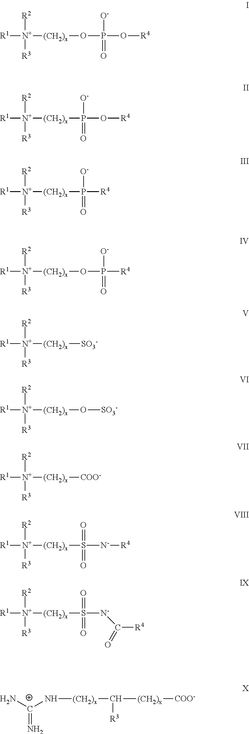

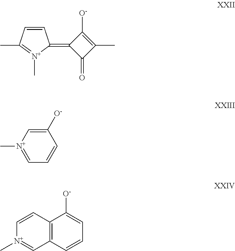


wherein R1 is H, OH or halogen and R2 is halogen, C1-C6 alkyl, C1-C6 alkylene amine, C1-C6 alkylene mono- or dialkyl amine, an optionally substituted thiophenyl group, an alkylene heteroaryl, or a C0-C6 alkylene carboxylic acid group, where the carboxylic acid group is substituted anywhere along the alkylene chain. In one embodiment, said bisphosphonate molecule is selected from the group consisting of etidronate, clodronate, tiladronate, pamidronate, neridronate, olpadronate, alendronate, ibandronate, risedronate and zoledronate. In one embodiment, said bisphosphonate molecule is pamidronate, neridronate or alendronate. In one embodiment, the protocell further comprises at least one cell penetrating peptide, a cell targeting peptide and/or a double stranded linear DNA or a plasmid DNA, wherein one of said DNA cargo components is optionally conjugated further with a nuclear localization sequence. In one embodiment, the bisphosphonate molecule is optionally conjugated to the MSNP. In one embodiment, the bisphosphonate molecule is conjugated solely to the lipid bilayer. In one embodiment, the bisphosphonate molecule is conjugated to the MSNP and to the lipid bilayer. In one embodiment, said cargo is or includes a reporter. In one embodiment, said reporter is an imaging agent. In one embodiment, said imaging agent is a fluorescent peptide. In one embodiment, said imaging agent is a fluorescent dye. In one embodiment, said anticancer agent is an anticancer small molecule. In one embodiment, said anticancer agent is a microtubule-stabilizing agent, microtubule-disruptor agent, an alkylating agent, air antimetabolite, an epidophyliotoxin, an antineoplastic enzyme, a topoisomerase inhibitor, an inhibitor of cell cycle progression or a platinum coordination complex. In one embodiment, said anticancer agent is a FLT-3 inhibitor, a VEGFR inhibitor, an EGFR TK inhibitor, an aurora kinase inhibitor, a PIK-1 modulator, a Bcl-2 inhibitor, an HDAC inhbitor, a c-MET inhibitor, a PARP inhibitor, a Cdk inhibitor, an EGFR TK inhibitor, an IGFR-TK inhibitor, an anti-HGF antibody, a PI3 kinase inhibitors, an AKT inhibitor, a JAK/STAT inhibitor, a checkpoint-1 or 2 inhibitor, a focal adhesion kinase inhibitor, a Map kinase kinase (mek) inhibitor or a VEGF trap antibody. In one embodiment, said anticancer agent is everolimus, trabectedin, abraxane, TLK 286, AV-299, DN-101, pazopanib, GSK690693, TTA 744, ON 0910,Na, AZD 6244 (ARRY-142886; AMN-107, TK.I-258, GSK461364, AZD 1152, enzastaurin, vandetanib, ARQ-197, MK-0457, MLN8054, PHA-739358, R-763, AT-9263, pemetrexed, erlotinib, dasatanib, nilotinib, dasatanib, panitumumab, amrubicin, oregovomab, Lep-etu, nolatrexed, azd2171, batabulin, ofatumumab, zanolimumab, edotecarin, tetrandrine, rubitecan, tesmilifene, oblimersen, ticilimumab, ipilimumab, gossypol, Bio 111, 131-I-TM-601, ALT-110, BIO 140, CC 8490, cilengitide, gimatecan, IL13-PE38QQR, INO 1001, IPdR1 KRX-0402, lucanthone, LY 317615, neuradiab, vitespen, Rta 744, Sdx102, talampanel, atrasentan, Xr 311, romidepsin, ADS-100380, sunitinib, 5-fluorouracil, vorinostat, etoposide, gemcitabine, doxorubicin, liposomal doxorubicin, 5′-deoxy-5-fluorouridine, vincristine, temozolomide, ZK-304709, seliciclib; PD0325901, AZD-6244, capecitabine, L-Glutamic acid, N-[4-[2-(2-amino-4,7-dihydro-4-oxo-I H-pyrrolo[2,3-d]pyrimidin-5-yl)ethyl]benzoyl]-, disodium salt, heptahydrate, camptothecin, PEG-labeled irinotecan, tamoxifen, toremifene citrate, anastrozole, exemestane, letrozole, DES(diethylstilbestrol), estradiol, estrogen, conjugated estrogen, bevacizumab, IMC-1C11, CHIR-258,); 3-(5-(methylsulfonylpiperadinemethyl)-indolylj-quinolone, vatalanib, AG-013736, A VE-0005, the acetate salt of [D-Ser(Bu t) 6,Azgly 10] (pyro-Glu-His-Trp-Ser-Tyr-D-Ser(Bu t)-Leu-Arg-Pro-Azgly-NH2 acetate [C59H84N18Oi4-(C2H4O2)x where x=1 to 2.4], goserelin acetate, leuprolide acetate, triptorelin pamoate, medroxyprogesterone acetate, hydroxyprogesterone caproate, megestrol acetate, raloxifene, bicalutamide, flutamide, nilutamide, megestrol acetate, CP-724714; TAK-165,HKI-272, erlotinib, lapatinib, canertinib, ABX-EGF antibody, erbifux, EKB-569, PKI-166, GW-572016, lonafamib, BMS-214662, tibifamib; amifostine, NVP-LAQ824, suberoyl anilide hydroxamic acid, valproic acid, trichostatin A, FK-228, SU11248, sorafenib, KRN951, aminoglutethimide, amsacrine, anagrelide, L-asparaginase, Bacillus Calmette-Guerin (BCG) vaccine, bleomycin, buserelin, busulfan, carboplatin, carmustine, chlorambucil, cisplatin, cladribine, clodronate, cyproterone, cytarabine, dacarbazine, dactinomycin, daunorubicin, diethylstilbestrol, epirubicin, fludarabine, fludrocortisone, fluoxymesterone, flutamide, gemcitabine, hydroxyurea, idarubicin, ifosfamide, imatinib, leuprolide, levamisole, lomustine, mechlorethamine, melphalan, 6-mercaptopurine, mesna, methotrexate, mitomycin, mitotane, mitoxantrone, nilutamide, octreotide, oxaliplatin, pamidronate, pentostatin, plicamycin, porfimer, procarbazine, raltitrexed, rituximab, streptozocin, teniposide, testosterone, thalidomide, thioguanine, thiotepa, tretinoin, vindesine, 13-cis-retinoic acid, phenylalanine mustard, uracil mustard, estramustine, altretamine, floxuridine, 5-deoxyuridine, cytosine arabinoside, 6-mercaptopurine, deoxycoformycin, calcitriol, varubicin, mithramycin, vinblastine, vinorelbine, topotecan, razoxin, marimastat, COL-3, neovastat, BMS-275291, squalamine, endostatin, SU5416, SU6668, EMD121974, interleukin-12, IM862, angiostatin, vitaxin, droloxifene, idoxifene, spironolactone, finasteride, cimetidine, trastuzumab, denileukin diftitox,gefitinib, bortezomib, paclitaxel, cremophor-free paclitaxel, docetaxel, epithilone B, BMS-247550, BMS-310705, droloxifene, 4-hydroxytamoxifen, pipendoxifene, ERA-923, arzoxifene, fulvestrant, acolbifene, lasoloxifene, idoxilene, TSE-424, HMR-3339, ZK186619, topotecan, PTK787/ZK 222584, VX-745, PD 184352, rapamycin, 40-O-(2-hydroxyethyl)-rapamycin, temsirolimus, AP-23573, RAD00I, ABT-578, BC-210, LY294002, LY292223, LY292696, LY293684, LY293646, wortmannin, ZM336372, L-779,450, PEG-filgrastim, darbepoetin, erythropoietin, granulocyte colony-stimulating factor, zolendronate, prednisone, cetuximab, granulocyte macrophage colony-stimulating factor, histrelin, pegylated interferon alfa-2a, interferon alfa-2a, pegylated interferon alfa-2b, interferon alfa-2b, azacitidine, PEG-L-asparaginase, lenalidomide, gemtuzumab, hydrocortisone, interleukin-11, dexrazoxane, alemtuzumab, all-transretinoic acid, ketoconazole, interleukin-2, megestrol, immune globulin, nitrogen mustard, methylprednisolone, ibritgumomab tiuxetan, androgens, decitabine, hexamethylmelamine, bexarotene, tositumomab, arsenic trioxide, cortisone, etidronate, mitotane, cyclosporine, liposomal daunorubicin, Edwina-asparaginase, strontium 89, casopitant, netupitant, an NK-1 receptor antagonists, palonosetron, aprepitant, diphenhydramine, hydroxyzine, metoclopramide, lorazepam, alprazolam, haloperidol, droperidol, dronabinol, dexamethasone, methylprednisolone, prochlorperazine, granisetron, ondansetron, dolasetron, tropisetron, pegfilgrastim, erythropoietin, epoetin alfa, darbepoetin alfa and mixtures thereof. In one embodiment, said lipid bilayer comprises at least one lipid selected from the group consisting of 1,2-dioleoyl-sn-glycero-3-phosphocholine (DOPC), 1,2-dipalmitoyl-sn-glycero-3-phosphocholine (DPPC), 1,2-distearoyl-sn-glycero-3-phosphocholine (DSPC), I,2-dioleoyl-sn-glycero-3-[phosphor-L-serine] (DOPS), 1,2-dioleoyl-3-trimethylammonium-propane (18:1 DOTAP), 1,2-dioleoyl-sn-glycero-3-phospho-(1′-rac-glycerol) (DOPG), 1,2-dioleoyl-sn-glycero-3-phosphoethanolamine (DOPE), 1,2-dipalmitoyl-sn-glycero-3-phosphoethanolamine (D PPE), I,2-dioleoyl-sn-glycero-3-phosphoethanolamine-N-[methoxy(polyethylene glycol)-2000] (18:1 PEG-2000 PE), 1,2-dipalmitoyl-sn-glycero-3-phosphoethanolamine-N-[methoxy(polyethylene glycol)-2000] (16:0 PEG-2000 PE), 1-Oleoyl-2-[12-[(7-nitro-2-1,3-benzoxadiazol-4-yl)amino)lauroyl]-sn-Glycero-3-Phosphocholine (18: 1-12:0 NBD PC), 1-palmitoyl-2-{12-[(7-nitro-2-1,3-benzoxadiazol-4-yl)amino)lauroyl}-sn-glycero-3-phosphocholine (16:0-12:0 NBD PC), pegylated 1,2-distearoyl-sn-glycero-3-phosphoethanolamine (PEG-DSPE), pegylated 1,2-dioleoyl-sn-glycero-3-phosphoethanolamine (PEG-DOPE), pegylated 1,2-dipalmitoyl-sn-glycero-3-phosphoethanolamine (PEG-DPPE), and pegylated 1,2-dimyristoyl-sn-glycero-3-phosphoethap.olamine (PEG-DMPE), among others, including a pegylated ceramide (e.g. Noctanoyl-sphingosine-1-succinylmethoxy-PEG, N-palmitoyl-sphingosine-1-succinylmethoxy-PEG, cholesterol and mixtures/combinations thereof. In one embodiment, said lipid bilayer comprises a mixture of DSPC, DOPE and Cholesterol; DSPC, DOPE, Cholesterol and DSPE-PEG 2000 or DSPC, DSPE, Cholesterol and DSPE-PEG 2000. In one embodiment, said lipid bilayer is a mixture of DSPC, DOPE and Cholesterol; DSPC, DOPE, Cholesterol and DSPE-PEG 2000; or DSPC, DSPE, Cholesterol and DSPE-PEG 2000.

wherein R1 is H, OH or halogen and R2 is halogen, C1-C6 alkyl, C1-C6 alkylene amine, C1-C6 alkylene mono- or dialkyl amine, an optionally substituted thiophenyl group, an alkylene heteroaryl, or a C0-C6 alkylene carboxylic acid group, where the carboxylic acid group is substituted anywhere along the alkylene chain. In one embodiment, said bisphosphonate molecule is selected from the group consisting of etidronate, clodronate, tiladronate, pamidronate, neridronate, olpadronate, alendronate, ibandronate, risedronate and zoledronate. In one embodiment, said bisphosphonate molecule is pamidronate, neridronate or alendronate. In one embodiment, the MSNP further comprises at least one cell penetrating peptide, a cell targeting peptide and/or a double stranded linear DNA or a plasmid DNA, wherein one of said DNA cargo components is optionally conjugated further with a nuclear localization sequence. In one embodiment, said cargo is or includes a reporter. In one embodiment, said reporter is an imaging agent. In one embodiment, said imaging agent is a fluorescent peptide. In one embodiment, said imaging agent is a fluorescent dye. In one embodiment, said anticancer agent is an anticancer small molecule. In one embodiment, said anticancer agent is a microtubule-stabilizing agent, microtubule-disruptor agent, an alkylating agent, an antimetabolite, an epidophyllotoxin, an antineoplastic enzyme, a topoisomerase inhibitor, an inhibitor of cell cycle progression or a platinum coordination complex. In one embodiment, said anticancer agent is a FLT-3 inhibitor, a VEGFR inhibitor, an EGFR TK inhibitor, an aurora kinase inhibitor, a PIK-1 modulator, a Bcl-2 inhibitor, an HDAC inhbitor, a c-MET inhibitor, a PARP inhibitor, a Cdk inhibitor, an EGFR TK inhibitor, an IGFR-TK inhibitor, an anti-HGF antibody, a PI3 kinase inhibitors, an AKT inhibitor, a JAK/STAT inhibitor, a checkpoint-1 or 2 inhibitor, a local adhesion kinase inhibitor, a Map kinase kinase (mek) inhibitor or a VEGF trap antibody. In one embodiment, said anticancer agent is everolimus, trabectedin, abraxane, TLK 286, AV-299, DN-101, pazopanib, GSK690693, RTA 744, ON 0910.Na, AZD 6244 (ARRY-142886), AMN-107, TK.I-258, GSK461364, AZD 1152, enzastaurin, vandetanib, ARQ-197, MK-0457, MLN8054, PHA-739358, R-763, AT-9263, pemetrexed, erlotinib, dasatanib, nilotinib, dasatanib, panitumumab, amrubicin, oregovomab, Lep-etu, nolatrexed, azd2171, batabulin, ofatumumab, zanolimumab, edotecarin, tetrandrine, rubitecan, tesmililene, oblimersen, ticilimumab, ipilimumab, gossypol, Bio 111, 131-I-TM-601, ALT-110, BIO 140, CC 8490, cilengitide, gimatecan, IL13-PE38QQR, INO 1001, IPdRI KRX-0402, lucanthone, LY 317615, neuradiab, vitespen, Rta 744, Sdx 102, talampanel, atrasentan, Xr 311, romidepsin, ADS-100380, sunitinib, 5-fluorouracil, vorinostat, etoposide, gemcitabine, doxorubicin, liposomal doxorubicin, 5′-deoxy-5-fluorouridine, vincristine, temozolomide, ZK-304709, seliciclib; PD0325901, AZD-6244, capecitabine, L-Glutamic acid, N-[4-[2-(2-amino-4,7-dihydro-4-oxo-I H-pyrrolo[2,3-d]pyrimidin-5-yl)ethyl]benzoyl]-, disodium salt, heptahydrate, camptothecin, PEG-labeled irinotecan, tamoxifen, toremifene citrate, anastrozole, exemestane, letrozole, DES(diethylstilbestrol), estradiol, estrogen, conjugated estrogen, bevacizumab, IMC-1C11, CHIR-258,); 3-(5-(methylsulfonylpiperadinemethyl)-indolylj-quinolone, vatalanib, AG-013736, A VE-0005, the acetate salt of [D-Ser(Bu t) 6,Azgly 10 (pyro-Glu-His-Trp-Ser-Tyr-D-Ser(Bu t)-Leu-Arg-Pro-Azgly-NH2 acetate [C59H84N18Oi4-(C2H4O2)x where x=1 to 2.4], goserelin acetate, leuprolide acetate, triptorelin pamoate, medroxyprogesterone acetate, hydroxyprogesterone caproate, megestrol acetate, raloxifene, bicalutamide, flutamide, nilutamide, megestrol acetate, CP-724714; TAK-165, HKI-272, erlotinib, lapatinib, canertinib, ABX-EGF antibody, erbifux, EKB-569, PKI-166, GW-512016, lonafamib, BMS-214662, tipifamib; amifostine, NVP-LAQ824, suberoyl anilide hydroxamic acid, valproic acid, trichostatin A, FK-228, SU11248, sorafenib, KRN951, aminoglutethimide, amsacrine, anagrelide, L-asparaginase, Bacillus Calmette-Guerin (BCG) vaccine, bleomycin, buserelin, busulfan, carboplatin, carmustine, chlorambucil, cisplatin, cladribine, clodronate, cyproterone, cytarabine, dacarbazine, dactinomycin, daunorubicin, diethylstilbestrol, epirubicin, fludarabine, fludrocortisone, fluoxymesterone, flutamide, gemcitabine, hydroxyurea, idarubicin, ifosfamide, imatinib, leuprolide, levamisole, lomustine, mechlorethamine, melphalan, 6-mercaptopurine, mesna, methotrexate, mitomycin, mitotane, mitoxantrone, nilutamide, octreotide, oxaliplatin, pamidronate, pentostatin, plicamycin, porfimer, procarbazine, raltitrexed, rituximab, streptozocin, teniposide, testosterone, thalidomide, thioguanine, thiotepa, tretinoin, vindesine, 13-cis-retinoic acid, phenylalanine mustard, uracil mustard, estramustine, altretamine, floxuridine, 5-deoxyuridine, cytosine arabinoside, 6-mercaptopurine, deoxycoformycin, calcitriol, valrubicin, mithramycin, vinblastine, vinorelbine, topotecan, razoxin, marimastat, COL-3, neovastat, BMS-275291, squalamine, endostatin, SU5416, SU6668, EMD121974, interleukin-12, IM862, angiostatin, vitaxin, droloxifene, idoxifene, spironolactone, finasteride, cimetidine, trastuzumab, denileukin diftitox,gefitinib, bortezomib, paclitaxel, cremophor-free paclitaxel, docetaxel, epithilone B, BMS-247550, BMS-310705, droloxifene, 4-hydroxytamoxifen, pipendoxifene, ERA-923, arzoxifene, fulvestrant, acolbifene, lasofoxifene, idoxifene, TSE-424, HMR-3339, ZK186619, topotecan, PTK787/ZK 222584, VX-745, PD 184352, rapamycin, 40-O-(2-hydroxyethyl)-rapamycin, temsirolimus, AP-23573, RAD00I, ABT-578, BC-210, LY294002, LY292223, LY292696, LY293684, LY293646, wortmannin, ZM336372, L-779,450, PEGTIgrastim, darbepoetin, erythropoietin, granulocyte colony-stimulating factor, zolendronate, prednisone, cetuximab, granulocyte macrophage colony-stimulating factor, histrelin, pegylated interferon alfa-2a, interferon alfa-2a, pegylated interferon alfa-2b, interferon alfa-2b, azacitidine, PEG-L-asparaginase, lenalidomide, gemtuzumab, hydrocortisone, interleukin-11, dexrazoxane, alemtuzumab, all-transretinoic acid, ketoconazole, interleukin-2, megestrol, immune globulin, nitrogen mustard, methylprednisolone, ibritgumomab tiuxetan, androgens, decitabine, hexamethylmelamine, bexarotene, tositumomab, arsenic trioxide, cortisone, etidronate, mitotane, cyclosporine, liposomal daunorubicin, Edwina-asparaginase, strontium 89, casopitant, netupitant, an NK-1 receptor antagonists, palonosetron, aprepitant, diphenhydramine, hydroxyzine, metoclopramide, lorazepam, alprazolam, haloperidol, droperidol, dronabinol, dexamethasone, methylprednisolone, prochlorperazine, granisetron, ondansetron, dolasetron, tropisetron, pegfilgrastim, erythropoietin, epoetin alfa, darbepoetin alfa and mixtures thereof.
Claims (11)

Priority Applications (1)
| Application Number | Priority Date | Filing Date | Title |
|---|---|---|---|
| US16/068,235 US11672866B2 (en) | 2016-01-08 | 2017-01-06 | Osteotropic nanoparticles for prevention or treatment of bone metastases |
Applications Claiming Priority (5)
| Application Number | Priority Date | Filing Date | Title |
|---|---|---|---|
| US201662276297P | 2016-01-08 | 2016-01-08 | |
| US201662276388P | 2016-01-08 | 2016-01-08 | |
| US201662395196P | 2016-09-15 | 2016-09-15 | |
| US16/068,235 US11672866B2 (en) | 2016-01-08 | 2017-01-06 | Osteotropic nanoparticles for prevention or treatment of bone metastases |
| PCT/US2017/012583 WO2017120504A1 (en) | 2016-01-08 | 2017-01-06 | Osteotropic nanoparticles for prevention or treatment of bone metastases |
Publications (2)
| Publication Number | Publication Date |
|---|---|
| US20190022235A1 US20190022235A1 (en) | 2019-01-24 |
| US11672866B2 true US11672866B2 (en) | 2023-06-13 |
Family
ID=59274430
Family Applications (1)
| Application Number | Title | Priority Date | Filing Date |
|---|---|---|---|
| US16/068,235 Active 2037-06-21 US11672866B2 (en) | 2016-01-08 | 2017-01-06 | Osteotropic nanoparticles for prevention or treatment of bone metastases |
Country Status (2)
| Country | Link |
|---|---|
| US (1) | US11672866B2 (en) |
| WO (1) | WO2017120504A1 (en) |
Cited By (2)
| Publication number | Priority date | Publication date | Assignee | Title |
|---|---|---|---|---|
| US12208164B2 (en) | 2019-02-28 | 2025-01-28 | Unm Rainforest Innovations | Modular metal-organic polyhedra superassembly compositions |
| US12252708B2 (en) | 2018-09-24 | 2025-03-18 | Unm Rainforest Innovations | Living mammalian cells modified with functional modular nanoparticles |
Families Citing this family (14)
| Publication number | Priority date | Publication date | Assignee | Title |
|---|---|---|---|---|
| US9273305B1 (en) | 2012-04-25 | 2016-03-01 | Sandia Corporation | Cell-based composite materials with programmed structures and functions |
| US11672866B2 (en) | 2016-01-08 | 2023-06-13 | Paul N. DURFEE | Osteotropic nanoparticles for prevention or treatment of bone metastases |
| US11344629B2 (en) | 2017-03-01 | 2022-05-31 | Charles Jeffrey Brinker | Active targeting of cells by monosized protocells |
| US11007516B1 (en) | 2017-06-19 | 2021-05-18 | National Technology & Engineering Solutions Of Sandia, Llc | Tunable metal-organic framework compositions and methods thereof |
| US10933027B1 (en) | 2017-09-25 | 2021-03-02 | National Technology & Engineering Solutions Of Sandia, Llc | Expanded pore particles and delivery methods thereof |
| US11045554B1 (en) | 2018-06-22 | 2021-06-29 | National Technology & Engineering Solutions Of Sandia, Llc | Lipid-coated particles for treating viral infections |
| CN109568675B (en) * | 2018-12-13 | 2021-06-04 | 上海纳米技术及应用国家工程研究中心有限公司 | Preparation of polyester/periodic mesoporous bone filling composite material with fluorescence-labeled degradation rate, product and application |
| US20220370372A1 (en) * | 2019-09-27 | 2022-11-24 | Greenmark Biomedical Inc. | Bisphosphonate loaded starch nanoparticle |
| US11433121B1 (en) | 2020-04-03 | 2022-09-06 | National Technology & Engineering Solutions Of Sandia, Llc | Lipid composition for the delivery of therapeutic cargos |
| CN114369647B (en) * | 2021-12-31 | 2024-10-25 | 深圳麦科田生物医疗技术股份有限公司 | Water-in-oil droplet for microdroplet digital PCR and application thereof |
| US12433874B2 (en) * | 2022-04-28 | 2025-10-07 | Prescience Biotechnology Inc. | Liposome comprising rapamycin or a derivative thereof and use thereof in therapy |
| CN115252615B (en) * | 2022-09-14 | 2023-10-24 | 重庆医药高等专科学校 | Pharmaceutical composition containing fangchinoline-7-propionate and PARP-1 inhibitor |
| CN115721629B (en) * | 2022-10-31 | 2024-02-20 | 西安交通大学 | pH-responsive iron-copper magnetic nanodrug-carrying system for combined use of docetaxel and enzalutamide and its preparation and application |
| CN117180227B (en) * | 2023-08-08 | 2024-04-19 | 南京医科大学附属口腔医院 | Curcumin-loaded manganese-doped hollow mesoporous silica nanoparticles and their application in periodontal bone tissue repair |
Citations (103)
| Publication number | Priority date | Publication date | Assignee | Title |
|---|---|---|---|---|
| US4927637A (en) | 1989-01-17 | 1990-05-22 | Liposome Technology, Inc. | Liposome extrusion method |
| US5057296A (en) | 1990-12-10 | 1991-10-15 | Mobil Oil Corp. | Method for synthesizing mesoporous crystalline material |
| US5098684A (en) | 1990-01-25 | 1992-03-24 | Mobil Oil Corp. | Synthetic mesoporous crystaline material |
| US5360834A (en) | 1989-08-01 | 1994-11-01 | Fraunhofer-Gesellschaft Zur Forderung Der Angewandten Forschung E.V. | Process for photoinitiated control of inorganic network formation in the sol-gel process |
| US5689574A (en) | 1995-03-29 | 1997-11-18 | Apple Computer, Inc. | Integral sound module for a modular monitor |
| WO1997047296A2 (en) | 1996-06-12 | 1997-12-18 | Mount Sinai Hospital Corporation | Compositions comprising vanadium compounds for use in the treatment of proliferative disorders and arthropathies |
| US5789230A (en) | 1993-10-14 | 1998-08-04 | Boehringer Ingelheim International Gmbh | Endosomolytically active particles |
| WO2000076556A2 (en) | 1999-06-11 | 2000-12-21 | Neorx Corporation | High dose radionuclide complexes for bone marrow suppression |
| US6194388B1 (en) | 1994-07-15 | 2001-02-27 | The University Of Iowa Research Foundation | Immunomodulatory oligonucleotides |
| US6365266B1 (en) | 1999-12-07 | 2002-04-02 | Air Products And Chemicals, Inc. | Mesoporous films having reduced dielectric constants |
| US6387453B1 (en) | 2000-03-02 | 2002-05-14 | Sandia Corporation | Method for making surfactant-templated thin films |
| WO2002066506A2 (en) | 2001-02-21 | 2002-08-29 | Glaxosmithkline Biologicals S.A. | Use of casb7439 (ash2) in the immunotherapy of breast cancer |
| US20020147105A1 (en) | 1999-03-22 | 2002-10-10 | Shamshoum Edwar S. | Process for producing stereospecific polymers |
| WO2003055469A1 (en) | 2001-12-21 | 2003-07-10 | Celator Technologies Inc. | Improved polymer-lipid delivery vehicles |
| US20040005352A1 (en) | 2002-04-16 | 2004-01-08 | Lopez Gabriel P. | Biologically functionalized porous microspheres |
| US6808867B2 (en) | 1999-04-05 | 2004-10-26 | Science & Technology Corporation @ University Of New Mexico | Photo-definable self-assembled materials |
| WO2004096140A2 (en) | 2003-04-25 | 2004-11-11 | The Penn State Research Foundation | Method and system for systemic delivery of growth arresting, lipid-derived bioactive compounds |
| US20040258671A1 (en) | 2003-06-23 | 2004-12-23 | Watkins Linda May Rothblum | Methods for treating pain |
| WO2005009602A2 (en) | 2003-07-22 | 2005-02-03 | Iowa State University Research Foundation, Inc. | Capped mesoporous silicates |
| US6913832B2 (en) | 2000-04-21 | 2005-07-05 | Science & Technology Corporation At University Of New Mexico | Prototyping of patterned functional nanostructures |
| WO2005084710A2 (en) | 2004-03-02 | 2005-09-15 | Massachusetts Institute Of Technology | Nanocell drug delivery system |
| US20050239687A1 (en) | 2004-04-26 | 2005-10-27 | Centre National De La Recherche Scientifique | Methods and compositions for delivering siRNA into mammalian cells |
| US7101967B2 (en) | 1998-11-13 | 2006-09-05 | Cyclacel Limited | Transport vectors |
| US20070224257A1 (en) | 2006-03-21 | 2007-09-27 | The Secretary Of State For Environment, Foods & Rural Affairs | Brucellosis dna vaccine |
| EP1852393A1 (en) | 2003-06-27 | 2007-11-07 | K.U.Leuven Research & Development | Crystalline mesoporous oxide based materials useful for the fixation and controlled release of drugs |
| WO2007140618A1 (en) | 2006-06-09 | 2007-12-13 | Protox Therapeutics Incorporated | Modified protein toxins targeting cytokine -receptor bearing cells and use thereof for treating disease |
| US20070287104A1 (en) | 1999-04-05 | 2007-12-13 | Dhaval Doshi | Photo-definable self-assembled materials |
| US20080095852A1 (en) | 2005-02-14 | 2008-04-24 | Australian Nuclear Science & Technology Organisation | Layered Nanoparticles |
| US20080160313A1 (en) | 2006-04-26 | 2008-07-03 | Gabriel Lopez | Lipid bilayers on nanotextured solid surfaces |
| US20080213377A1 (en) | 2006-12-08 | 2008-09-04 | Bhatia Sangeeta N | Delivery of Nanoparticles and/or Agents to Cells |
| US20080241917A1 (en) | 2005-09-30 | 2008-10-02 | Hidetaka Akita | Vector For Delivering Target Substance Into Nucleus or Cell |
| US20080241262A1 (en) | 2004-03-29 | 2008-10-02 | The University Of Houston System | Nanoshells and Discrete Polymer-Coated Nanoshells, Methods For Making and Using Same |
| US20080286371A1 (en) | 2005-09-12 | 2008-11-20 | Cristalia Produtos Quimicos Farmaceuticos Ltda | Immunogenical Complex Formed by Vaccinal Antigens Encapsulated by Nanostructured Mesoporous Silica |
| US20090054246A1 (en) | 2006-08-23 | 2009-02-26 | Peabody David S | Virus-like platform for rapid vaccine discovery |
| US7514267B1 (en) | 2005-08-19 | 2009-04-07 | Stcunm | Detection systems utilizing supported lipid bilayers |
| JP2009515520A (en) | 2005-11-10 | 2009-04-16 | レセプター バイオロジックス, インコーポレイテッド | Hepatocyte growth factor intron fusion protein |
| WO2009051837A2 (en) | 2007-10-12 | 2009-04-23 | Massachusetts Institute Of Technology | Vaccine nanotechnology |
| US20090181090A1 (en) | 2005-12-27 | 2009-07-16 | Sebastian Dreis | Protein-Based Carrier System for Overcoming Resistance in Tumour Cells |
| US20090208563A1 (en) | 2005-05-31 | 2009-08-20 | Linda May Rothblum Watkins | Method for delivering genes |
| US20090305409A1 (en) | 2005-03-24 | 2009-12-10 | National University Corporation Hokkaido University | Liposome Capable of Effective Delivery of Given Substance Into Nucleus |
| US20100028341A1 (en) | 2006-12-15 | 2010-02-04 | Ablynx N.V. | Amino acid sequences that modulate the interaction between cells of the immune system |
| US20100055167A1 (en) | 2008-08-29 | 2010-03-04 | Alex Zhang | Stem cell delivery of anti-neoplastic medicine |
| WO2010035304A2 (en) | 2008-09-25 | 2010-04-01 | Saverio Alberti | Oligonucleotidic sequences able to silence the expression of the cyclin d1 -trop2 chimera and uses thereof in medical field |
| WO2010048572A1 (en) | 2008-10-23 | 2010-04-29 | Cornell University | A novel anti-viral method |
| US20100152699A1 (en) | 2008-11-14 | 2010-06-17 | The Board Of Regents Of The University Of Texas System | Nanochanneled Device and Related Methods |
| US20100166665A1 (en) * | 2008-12-29 | 2010-07-01 | General Electric Company | Nanoparticle contrast agents for diagnostic imaging |
| US20100168120A1 (en) | 2006-04-28 | 2010-07-01 | Neuromedix Inc. | Salts of pyridazine compounds |
| WO2010078569A2 (en) | 2009-01-05 | 2010-07-08 | Stc.Unm | Porous nanoparticle supported lipid bilayer nanostructures |
| US20100255103A1 (en) | 2007-12-06 | 2010-10-07 | The Regents Of The University Of California | Mesoporous Silica Nanoparticles for Biomedical Applications |
| US20110059156A9 (en) | 2008-04-25 | 2011-03-10 | Northwestern University | Nanostructures Suitable for Sequestering Cholesterol and Other Molecules |
| US20110097819A1 (en) | 2008-03-16 | 2011-04-28 | Groves John T | Membrane-coated particles |
| US20110135571A1 (en) | 2008-02-22 | 2011-06-09 | Wenbin Lin | Hybrid nanoparticles as anti-cancer therapeutic agents and dual therapeutic/imaging contrast agents |
| WO2011116226A2 (en) | 2010-03-17 | 2011-09-22 | Stc.Unm | Display of antibody fragments on virus-like particles of rna bacteriophages |
| US20110230372A1 (en) | 2008-11-14 | 2011-09-22 | Stc Unm | Gene expression classifiers for relapse free survival and minimal residual disease improve risk classification and outcome prediction in pediatric b-precursor acute lymphoblastic leukemia |
| WO2011116219A1 (en) | 2010-03-17 | 2011-09-22 | Board Of Regent Of The University Of Texas System | Theranostic delivery systems with modified surfaces |
| WO2011150264A2 (en) | 2010-05-26 | 2011-12-01 | Selecta Biosciences, Inc. | Synthetic nanocarrier combination vaccines |
| US20110300186A1 (en) | 2010-04-14 | 2011-12-08 | Battelle Memorial Institute | Functionalized Nano- and Micro-materials for Medical Therapies |
| US20120207795A1 (en) | 2010-07-13 | 2012-08-16 | The Regents Of The University Of California | Cationic polymer coated mesoporous silica nanoparticles and uses thereof |
| US8268962B2 (en) | 2007-07-12 | 2012-09-18 | Prosensa Technologies B.V. | Molecules for targeting compounds to various selected organs or tissues |
| WO2012149376A2 (en) | 2011-04-28 | 2012-11-01 | Stc.Unm | Porous nanoparticle-supported lipid bilayers (protocells) for targeted delivery and methods of using same |
| WO2013012891A1 (en) | 2011-07-19 | 2013-01-24 | Stc. Unm | Intraperitoneally-administered nanocarriers that release their therapeutic load based on the inflammatory environment of cancers |
| US8374816B2 (en) | 2010-06-08 | 2013-02-12 | Honeywell International Inc. | Automatic data collection algorithm for 3D magnetic field calibration with reduced memory requirements |
| US20130095039A1 (en) | 2010-09-30 | 2013-04-18 | The Board Of Trustees Of The University Of Illinois | Nucleic acid-mediated shape control of nanoparticles |
| WO2013056132A2 (en) | 2011-10-14 | 2013-04-18 | Stc.Unm | Porous nanoparticle-supported lipid bilayers (protocells) for targeted delivery including transdermal delivery of cargo and methods thereof |
| US20130108661A1 (en) | 2010-02-05 | 2013-05-02 | Mount Sinai School Of Medicine | Methods and compositions for cancer immunotherapy using flagellin-tumor associated antigen fusion protein expressing tumor cells |
| US20130115169A1 (en) | 2009-12-04 | 2013-05-09 | The Regents Of The University Of California | Red blood cell-mimetic particles and methods for making use thereof |
| US20130122054A1 (en) | 2010-04-21 | 2013-05-16 | National University Corporation Hokkaido University | Lipid membrane structure having intranuclear migrating property |
| WO2013082612A1 (en) | 2011-12-03 | 2013-06-06 | Emd Millipore Corporation | Micro-incubation systems for microfluidic cell culture and methods |
| WO2013103614A1 (en) | 2011-12-30 | 2013-07-11 | Stc.Unm | Crlf-2 binding peptides, protocells and viral-like particles useful in the treatment of cancer, including acute lymphoblastic leukemia (all) |
| US20130185823A1 (en) | 2012-01-16 | 2013-07-18 | Academia Sinica | Mesoporous silica nanoparticle-mediated delivery of dna into arabidopsis root |
| US20130197103A1 (en) | 1999-01-13 | 2013-08-01 | Alchemia Oncology Pty Limited | Composition and method for the enhancement of the efficacy of drugs |
| US20130195963A1 (en) | 2011-12-07 | 2013-08-01 | The Methodist Hospital Research Institute | Mesoporous silicon particles for the presentation of tumor antigens and adjuvant for anti-cancer immunity |
| US20140023700A1 (en) | 2011-02-22 | 2014-01-23 | Leo Pharma A/S | Cyclic peptide and conjugate thereof for binding to keratinocytes |
| US20140141089A1 (en) | 2009-02-11 | 2014-05-22 | Colorado School Of Mines | Nanoparticles, Compositions Thereof, and Methods of Use, and Methods of Making the Same |
| WO2014093635A1 (en) | 2012-12-12 | 2014-06-19 | The Broad Institute, Inc. | Engineering and optimization of improved systems, methods and enzyme compositions for sequence manipulation |
| US20140234210A1 (en) * | 2011-07-08 | 2014-08-21 | The University Of North Carolina At Chapel Hill | Metal bisphosphonate nanoparticles for anti-cancer therapy and imaging and for treating bone disorders |
| WO2014165617A1 (en) | 2013-04-02 | 2014-10-09 | Stc.Unm | Mesoporous alum nanoparticles as universal platform for antigen adsorption, presentation, and delivery |
| WO2014165608A1 (en) | 2013-04-02 | 2014-10-09 | Stc. Unm | Antibiotic protocells and related pharmaceutical formulations and methods of treatment |
| WO2015042268A1 (en) | 2013-09-18 | 2015-03-26 | Stc.Unm | Core and surface modification of mesoporous silica nanoparticles to achieve cell specific targeting in vivo |
| WO2015042279A1 (en) | 2013-09-18 | 2015-03-26 | Stc. Unm | Torroidal mesoporous silica nanoparticles (tmsnps) and related protocells |
| US8992984B1 (en) | 2009-10-21 | 2015-03-31 | Stc.Unm | Protocells and their use for targeted delivery of multicomponent cargos to cancer cells |
| US20150118247A1 (en) | 2013-10-25 | 2015-04-30 | Nodality, Inc. | Methods and compositions for immunomodulation |
| US20150125391A1 (en) * | 2013-11-01 | 2015-05-07 | The Brigham And Women's Hospital, Inc. | Bone and metal targeted polymeric nanoparticles |
| WO2015095340A1 (en) | 2013-12-19 | 2015-06-25 | Novartis Ag | Lipids and lipid compositions for the delivery of active agents |
| WO2015130584A2 (en) | 2014-02-25 | 2015-09-03 | Merck Sharp & Dohme Corp. | Lipid nanoparticle vaccine adjuvants and antigen delivery systems |
| US20160090603A1 (en) | 2014-09-30 | 2016-03-31 | Sandia Corporation | Delivery platforms for the domestication of algae and plants |
| US20160193588A1 (en) | 2013-09-26 | 2016-07-07 | Regents Of The University Of Minnesota | Porous silica having high pore volume and methods of making and using same |
| WO2016145335A1 (en) | 2015-03-11 | 2016-09-15 | Stc.Unm | Generation of mesoporous materials using multiphase surfactant systems |
| WO2016145031A1 (en) | 2015-03-09 | 2016-09-15 | Stc.Unm | Cd 47 containing porous nanoparticle supported lipid bilayers (protocells) |
| WO2017041033A1 (en) | 2015-09-04 | 2017-03-09 | Brinker C Jeffrey | Mesoporous silica nanoparticles and supported lipid bi-layer nanoparticles for biomedical applications |
| WO2017041032A1 (en) | 2015-09-04 | 2017-03-09 | Brinker C Jeffrey | Protocells to treat microbial infection and for synergistic delivery |
| WO2017120504A1 (en) | 2016-01-08 | 2017-07-13 | Durfee Paul N | Osteotropic nanoparticles for prevention or treatment of bone metastases |
| WO2018000043A1 (en) | 2016-06-29 | 2018-01-04 | Commonwealth Scientific And Industrial Research Organisation | Coating material for cells |
| US9989447B1 (en) | 2012-04-25 | 2018-06-05 | National Technology & Engineering Solutions Of Sandia, Llc | Shape-preserving transformations of organic matter and compositions thereof |
| WO2018160865A1 (en) | 2017-03-01 | 2018-09-07 | Charles Jeffrey Brinker | Active targeting of cells by monosized protocells |
| WO2018187287A1 (en) | 2017-04-03 | 2018-10-11 | Charles Jeffrey Brinker | Porous nanoparticle-supported lipid bilayer delivery of transcriptional gene modulators |
| WO2019028387A1 (en) | 2017-08-03 | 2019-02-07 | Rita Elena Serda | Liposomal coated nanoparticles for immunotherapy applications |
| WO2019169152A1 (en) | 2018-02-28 | 2019-09-06 | Noureddine Achraf | Starry mesoporous silica nanoparticles and supported lipid bi-layer nanoparticles |
| US10465189B2 (en) | 2013-12-13 | 2019-11-05 | Nanyang Technological University | Multilayered nanoparticle and methods of manufacturing and using the same |
| WO2020028342A1 (en) | 2018-07-30 | 2020-02-06 | Guo Jimin | Biomimetic rebuilding of multifunctional red blood cells |
| WO2020068798A1 (en) | 2018-09-24 | 2020-04-02 | Guo Jimin | Living mammalian cells modified with functional modular nanoparticles |
| WO2020068806A1 (en) | 2018-09-24 | 2020-04-02 | Guo Jimin | Armored cells |
| WO2020176716A1 (en) | 2019-02-28 | 2020-09-03 | Brinker C Jeffrey | Modular metal–organic polyhedra superassembly compositions |
-
2017
- 2017-01-06 US US16/068,235 patent/US11672866B2/en active Active
- 2017-01-06 WO PCT/US2017/012583 patent/WO2017120504A1/en not_active Ceased
Patent Citations (144)
| Publication number | Priority date | Publication date | Assignee | Title |
|---|---|---|---|---|
| US4927637A (en) | 1989-01-17 | 1990-05-22 | Liposome Technology, Inc. | Liposome extrusion method |
| US5360834A (en) | 1989-08-01 | 1994-11-01 | Fraunhofer-Gesellschaft Zur Forderung Der Angewandten Forschung E.V. | Process for photoinitiated control of inorganic network formation in the sol-gel process |
| US5098684A (en) | 1990-01-25 | 1992-03-24 | Mobil Oil Corp. | Synthetic mesoporous crystaline material |
| US5057296A (en) | 1990-12-10 | 1991-10-15 | Mobil Oil Corp. | Method for synthesizing mesoporous crystalline material |
| US5789230A (en) | 1993-10-14 | 1998-08-04 | Boehringer Ingelheim International Gmbh | Endosomolytically active particles |
| US6194388B1 (en) | 1994-07-15 | 2001-02-27 | The University Of Iowa Research Foundation | Immunomodulatory oligonucleotides |
| US5689574A (en) | 1995-03-29 | 1997-11-18 | Apple Computer, Inc. | Integral sound module for a modular monitor |
| WO1997047296A2 (en) | 1996-06-12 | 1997-12-18 | Mount Sinai Hospital Corporation | Compositions comprising vanadium compounds for use in the treatment of proliferative disorders and arthropathies |
| US7101967B2 (en) | 1998-11-13 | 2006-09-05 | Cyclacel Limited | Transport vectors |
| US20130197103A1 (en) | 1999-01-13 | 2013-08-01 | Alchemia Oncology Pty Limited | Composition and method for the enhancement of the efficacy of drugs |
| US20020147105A1 (en) | 1999-03-22 | 2002-10-10 | Shamshoum Edwar S. | Process for producing stereospecific polymers |
| US6808867B2 (en) | 1999-04-05 | 2004-10-26 | Science & Technology Corporation @ University Of New Mexico | Photo-definable self-assembled materials |
| US7332264B2 (en) | 1999-04-05 | 2008-02-19 | Stc.Unm | Photo-definable self-assembled materials |
| US20070287104A1 (en) | 1999-04-05 | 2007-12-13 | Dhaval Doshi | Photo-definable self-assembled materials |
| WO2000076556A2 (en) | 1999-06-11 | 2000-12-21 | Neorx Corporation | High dose radionuclide complexes for bone marrow suppression |
| US6365266B1 (en) | 1999-12-07 | 2002-04-02 | Air Products And Chemicals, Inc. | Mesoporous films having reduced dielectric constants |
| US6387453B1 (en) | 2000-03-02 | 2002-05-14 | Sandia Corporation | Method for making surfactant-templated thin films |
| US6913832B2 (en) | 2000-04-21 | 2005-07-05 | Science & Technology Corporation At University Of New Mexico | Prototyping of patterned functional nanostructures |
| WO2002066506A2 (en) | 2001-02-21 | 2002-08-29 | Glaxosmithkline Biologicals S.A. | Use of casb7439 (ash2) in the immunotherapy of breast cancer |
| WO2003055469A1 (en) | 2001-12-21 | 2003-07-10 | Celator Technologies Inc. | Improved polymer-lipid delivery vehicles |
| US20040005352A1 (en) | 2002-04-16 | 2004-01-08 | Lopez Gabriel P. | Biologically functionalized porous microspheres |
| WO2004096140A2 (en) | 2003-04-25 | 2004-11-11 | The Penn State Research Foundation | Method and system for systemic delivery of growth arresting, lipid-derived bioactive compounds |
| US20040258671A1 (en) | 2003-06-23 | 2004-12-23 | Watkins Linda May Rothblum | Methods for treating pain |
| EP1852393A1 (en) | 2003-06-27 | 2007-11-07 | K.U.Leuven Research & Development | Crystalline mesoporous oxide based materials useful for the fixation and controlled release of drugs |
| WO2005009602A2 (en) | 2003-07-22 | 2005-02-03 | Iowa State University Research Foundation, Inc. | Capped mesoporous silicates |
| US20060154069A1 (en) | 2003-07-22 | 2006-07-13 | Lin Victor S | Capped mesoporous silicates |
| US7563451B2 (en) | 2003-07-22 | 2009-07-21 | Iowa State University Research Foundation, Inc. | Capped mesoporous silicates |
| WO2005084710A2 (en) | 2004-03-02 | 2005-09-15 | Massachusetts Institute Of Technology | Nanocell drug delivery system |
| US20080241262A1 (en) | 2004-03-29 | 2008-10-02 | The University Of Houston System | Nanoshells and Discrete Polymer-Coated Nanoshells, Methods For Making and Using Same |
| US20050239687A1 (en) | 2004-04-26 | 2005-10-27 | Centre National De La Recherche Scientifique | Methods and compositions for delivering siRNA into mammalian cells |
| US20080095852A1 (en) | 2005-02-14 | 2008-04-24 | Australian Nuclear Science & Technology Organisation | Layered Nanoparticles |
| US20090305409A1 (en) | 2005-03-24 | 2009-12-10 | National University Corporation Hokkaido University | Liposome Capable of Effective Delivery of Given Substance Into Nucleus |
| US20090208563A1 (en) | 2005-05-31 | 2009-08-20 | Linda May Rothblum Watkins | Method for delivering genes |
| US7514267B1 (en) | 2005-08-19 | 2009-04-07 | Stcunm | Detection systems utilizing supported lipid bilayers |
| US20080286371A1 (en) | 2005-09-12 | 2008-11-20 | Cristalia Produtos Quimicos Farmaceuticos Ltda | Immunogenical Complex Formed by Vaccinal Antigens Encapsulated by Nanostructured Mesoporous Silica |
| US20080241917A1 (en) | 2005-09-30 | 2008-10-02 | Hidetaka Akita | Vector For Delivering Target Substance Into Nucleus or Cell |
| JP2009515520A (en) | 2005-11-10 | 2009-04-16 | レセプター バイオロジックス, インコーポレイテッド | Hepatocyte growth factor intron fusion protein |
| US20090181090A1 (en) | 2005-12-27 | 2009-07-16 | Sebastian Dreis | Protein-Based Carrier System for Overcoming Resistance in Tumour Cells |
| US20070224257A1 (en) | 2006-03-21 | 2007-09-27 | The Secretary Of State For Environment, Foods & Rural Affairs | Brucellosis dna vaccine |
| US20080160313A1 (en) | 2006-04-26 | 2008-07-03 | Gabriel Lopez | Lipid bilayers on nanotextured solid surfaces |
| US20100168120A1 (en) | 2006-04-28 | 2010-07-01 | Neuromedix Inc. | Salts of pyridazine compounds |
| WO2007140618A1 (en) | 2006-06-09 | 2007-12-13 | Protox Therapeutics Incorporated | Modified protein toxins targeting cytokine -receptor bearing cells and use thereof for treating disease |
| US20090054246A1 (en) | 2006-08-23 | 2009-02-26 | Peabody David S | Virus-like platform for rapid vaccine discovery |
| US20080213377A1 (en) | 2006-12-08 | 2008-09-04 | Bhatia Sangeeta N | Delivery of Nanoparticles and/or Agents to Cells |
| US20100028341A1 (en) | 2006-12-15 | 2010-02-04 | Ablynx N.V. | Amino acid sequences that modulate the interaction between cells of the immune system |
| US8268962B2 (en) | 2007-07-12 | 2012-09-18 | Prosensa Technologies B.V. | Molecules for targeting compounds to various selected organs or tissues |
| WO2009051837A2 (en) | 2007-10-12 | 2009-04-23 | Massachusetts Institute Of Technology | Vaccine nanotechnology |
| US20100255103A1 (en) | 2007-12-06 | 2010-10-07 | The Regents Of The University Of California | Mesoporous Silica Nanoparticles for Biomedical Applications |
| US20110135571A1 (en) | 2008-02-22 | 2011-06-09 | Wenbin Lin | Hybrid nanoparticles as anti-cancer therapeutic agents and dual therapeutic/imaging contrast agents |
| US20110097819A1 (en) | 2008-03-16 | 2011-04-28 | Groves John T | Membrane-coated particles |
| US20110059156A9 (en) | 2008-04-25 | 2011-03-10 | Northwestern University | Nanostructures Suitable for Sequestering Cholesterol and Other Molecules |
| US20100055167A1 (en) | 2008-08-29 | 2010-03-04 | Alex Zhang | Stem cell delivery of anti-neoplastic medicine |
| WO2010035304A2 (en) | 2008-09-25 | 2010-04-01 | Saverio Alberti | Oligonucleotidic sequences able to silence the expression of the cyclin d1 -trop2 chimera and uses thereof in medical field |
| WO2010048572A1 (en) | 2008-10-23 | 2010-04-29 | Cornell University | A novel anti-viral method |
| US20110230372A1 (en) | 2008-11-14 | 2011-09-22 | Stc Unm | Gene expression classifiers for relapse free survival and minimal residual disease improve risk classification and outcome prediction in pediatric b-precursor acute lymphoblastic leukemia |
| US20100152699A1 (en) | 2008-11-14 | 2010-06-17 | The Board Of Regents Of The University Of Texas System | Nanochanneled Device and Related Methods |
| US20100166665A1 (en) * | 2008-12-29 | 2010-07-01 | General Electric Company | Nanoparticle contrast agents for diagnostic imaging |
| US8734816B2 (en) | 2009-01-05 | 2014-05-27 | Stc.Unm | Porous nanoparticle supported lipid bilayer nanostructures |
| US20110268791A1 (en) | 2009-01-05 | 2011-11-03 | Juewen Liu | Porous nanoparticle supported lipid bilayer nanostructures |
| US20140301951A1 (en) | 2009-01-05 | 2014-10-09 | Juewen Liu | Porous nanoparticle supported lipid nanostructures |
| WO2010078569A2 (en) | 2009-01-05 | 2010-07-08 | Stc.Unm | Porous nanoparticle supported lipid bilayer nanostructures |
| US20140141089A1 (en) | 2009-02-11 | 2014-05-22 | Colorado School Of Mines | Nanoparticles, Compositions Thereof, and Methods of Use, and Methods of Making the Same |
| US20150320681A1 (en) | 2009-10-21 | 2015-11-12 | Stc.Unm | Protocells and their use for targeted delivery of multicomponent cargos to cancer cells |
| US20150164798A1 (en) | 2009-10-21 | 2015-06-18 | Stc.Unm | Protocells and their use for targeted delivery of multicomponent cargos to cancer cells |
| US9480653B2 (en) | 2009-10-21 | 2016-11-01 | Stc.Unm | Protocells and their use for targeted delivery of multicomponent cargos to cancer cells |
| US8992984B1 (en) | 2009-10-21 | 2015-03-31 | Stc.Unm | Protocells and their use for targeted delivery of multicomponent cargos to cancer cells |
| US20130115169A1 (en) | 2009-12-04 | 2013-05-09 | The Regents Of The University Of California | Red blood cell-mimetic particles and methods for making use thereof |
| US20130108661A1 (en) | 2010-02-05 | 2013-05-02 | Mount Sinai School Of Medicine | Methods and compositions for cancer immunotherapy using flagellin-tumor associated antigen fusion protein expressing tumor cells |
| WO2011116219A1 (en) | 2010-03-17 | 2011-09-22 | Board Of Regent Of The University Of Texas System | Theranostic delivery systems with modified surfaces |
| WO2011116226A2 (en) | 2010-03-17 | 2011-09-22 | Stc.Unm | Display of antibody fragments on virus-like particles of rna bacteriophages |
| US20110300186A1 (en) | 2010-04-14 | 2011-12-08 | Battelle Memorial Institute | Functionalized Nano- and Micro-materials for Medical Therapies |
| US20130122054A1 (en) | 2010-04-21 | 2013-05-16 | National University Corporation Hokkaido University | Lipid membrane structure having intranuclear migrating property |
| WO2011150264A2 (en) | 2010-05-26 | 2011-12-01 | Selecta Biosciences, Inc. | Synthetic nanocarrier combination vaccines |
| US8374816B2 (en) | 2010-06-08 | 2013-02-12 | Honeywell International Inc. | Automatic data collection algorithm for 3D magnetic field calibration with reduced memory requirements |
| US20120207795A1 (en) | 2010-07-13 | 2012-08-16 | The Regents Of The University Of California | Cationic polymer coated mesoporous silica nanoparticles and uses thereof |
| US20130095039A1 (en) | 2010-09-30 | 2013-04-18 | The Board Of Trustees Of The University Of Illinois | Nucleic acid-mediated shape control of nanoparticles |
| US20140023700A1 (en) | 2011-02-22 | 2014-01-23 | Leo Pharma A/S | Cyclic peptide and conjugate thereof for binding to keratinocytes |
| WO2012149376A2 (en) | 2011-04-28 | 2012-11-01 | Stc.Unm | Porous nanoparticle-supported lipid bilayers (protocells) for targeted delivery and methods of using same |
| US9579283B2 (en) | 2011-04-28 | 2017-02-28 | Stc.Unm | Porous nanoparticle-supported lipid bilayers (protocells) for targeted delivery and methods of using same |
| US20160106671A1 (en) | 2011-04-28 | 2016-04-21 | Stc.Unm | Porous nanoparticle-supported lipid bilayers (protocells) for targeted delivery and methods of using same |
| US10022327B2 (en) | 2011-04-28 | 2018-07-17 | Stc.Unm | Porous nanoparticle-supported lipid bilayers (protocells) for targeted delivery and methods of using same |
| US20140079774A1 (en) * | 2011-04-28 | 2014-03-20 | Stc.Unm | Porous nanoparticle-supported lipid bilayers (protocells) for targeted delivery and methods of using same |
| US20190091150A1 (en) | 2011-04-28 | 2019-03-28 | Stc.Unm | Porous nanoparticle-supported lipid bilayers (protocells) for targeted delivery and methods of using same |
| US20140234210A1 (en) * | 2011-07-08 | 2014-08-21 | The University Of North Carolina At Chapel Hill | Metal bisphosphonate nanoparticles for anti-cancer therapy and imaging and for treating bone disorders |
| US20140212479A1 (en) | 2011-07-19 | 2014-07-31 | Reema Zeinelden | Intraperitoneally-administered nanocarriers that release their therapeutic load based on the inflammatory environment of cancers |
| WO2013012891A1 (en) | 2011-07-19 | 2013-01-24 | Stc. Unm | Intraperitoneally-administered nanocarriers that release their therapeutic load based on the inflammatory environment of cancers |
| US20170232115A1 (en) | 2011-10-14 | 2017-08-17 | Stc.Unm | Porous nanoparticle-supported lipid bilayers (protocells) for targeted delivery including transdermal delivery of cargo and methods thereof |
| WO2013056132A2 (en) | 2011-10-14 | 2013-04-18 | Stc.Unm | Porous nanoparticle-supported lipid bilayers (protocells) for targeted delivery including transdermal delivery of cargo and methods thereof |
| US20150272885A1 (en) | 2011-10-14 | 2015-10-01 | Stc.Unm | Porous nanoparticle-supported lipid bilayers (protocells) for targeted delivery including transdermal delivery of cargo and methods thereof |
| WO2013082612A1 (en) | 2011-12-03 | 2013-06-06 | Emd Millipore Corporation | Micro-incubation systems for microfluidic cell culture and methods |
| US8926994B2 (en) | 2011-12-07 | 2015-01-06 | The Methodist Hospital Research Institute | Mesoporous silicon particles for the presentation of tumor antigens and adjuvant for anti-cancer immunity |
| US20130195963A1 (en) | 2011-12-07 | 2013-08-01 | The Methodist Hospital Research Institute | Mesoporous silicon particles for the presentation of tumor antigens and adjuvant for anti-cancer immunity |
| WO2013103614A1 (en) | 2011-12-30 | 2013-07-11 | Stc.Unm | Crlf-2 binding peptides, protocells and viral-like particles useful in the treatment of cancer, including acute lymphoblastic leukemia (all) |
| US20150010475A1 (en) | 2011-12-30 | 2015-01-08 | Stc.Unm | Crlf-2 binding peptides, protocells and viral-like particles useful in the treatment of cancer, including acute lymphoblastic leukemia (all) |
| US20130185823A1 (en) | 2012-01-16 | 2013-07-18 | Academia Sinica | Mesoporous silica nanoparticle-mediated delivery of dna into arabidopsis root |
| US9989447B1 (en) | 2012-04-25 | 2018-06-05 | National Technology & Engineering Solutions Of Sandia, Llc | Shape-preserving transformations of organic matter and compositions thereof |
| WO2014093635A1 (en) | 2012-12-12 | 2014-06-19 | The Broad Institute, Inc. | Engineering and optimization of improved systems, methods and enzyme compositions for sequence manipulation |
| WO2014165608A1 (en) | 2013-04-02 | 2014-10-09 | Stc. Unm | Antibiotic protocells and related pharmaceutical formulations and methods of treatment |
| US20160151482A1 (en) | 2013-04-02 | 2016-06-02 | Stc. Unm | Mesoporous alum nanoparticles as a universal platform for antigen adsorption, presentation, and delivery |
| US20170165375A1 (en) | 2013-04-02 | 2017-06-15 | Stc. Unm | Antibiotic protocells and related pharmaceutical formulations and methods of treatment |
| WO2014165617A1 (en) | 2013-04-02 | 2014-10-09 | Stc.Unm | Mesoporous alum nanoparticles as universal platform for antigen adsorption, presentation, and delivery |
| US20160338954A1 (en) | 2013-09-18 | 2016-11-24 | Stc. Unm | Torroidal mesoporous silica nanoparticles (tmsnps) and related protocells |
| WO2015042268A1 (en) | 2013-09-18 | 2015-03-26 | Stc.Unm | Core and surface modification of mesoporous silica nanoparticles to achieve cell specific targeting in vivo |
| US9855217B2 (en) | 2013-09-18 | 2018-01-02 | Stc. Unm | Toroidal mesoporous silica nanoparticles (TMSNPs) and related protocells |
| WO2015042279A1 (en) | 2013-09-18 | 2015-03-26 | Stc. Unm | Torroidal mesoporous silica nanoparticles (tmsnps) and related protocells |
| US20160287717A1 (en) | 2013-09-18 | 2016-10-06 | Stc.Unm | Core and Surface Modification of Mesoporous Silica Nanoparticles to Achieve Cell Specific Targeting In Vivo. |
| US20160193588A1 (en) | 2013-09-26 | 2016-07-07 | Regents Of The University Of Minnesota | Porous silica having high pore volume and methods of making and using same |
| US20150118247A1 (en) | 2013-10-25 | 2015-04-30 | Nodality, Inc. | Methods and compositions for immunomodulation |
| US20150125391A1 (en) * | 2013-11-01 | 2015-05-07 | The Brigham And Women's Hospital, Inc. | Bone and metal targeted polymeric nanoparticles |
| US10465189B2 (en) | 2013-12-13 | 2019-11-05 | Nanyang Technological University | Multilayered nanoparticle and methods of manufacturing and using the same |
| WO2015095340A1 (en) | 2013-12-19 | 2015-06-25 | Novartis Ag | Lipids and lipid compositions for the delivery of active agents |
| US20160361411A1 (en) | 2014-02-25 | 2016-12-15 | Merck Sharp & Dohme Corp | Lipid nanoparticle vaccine adjuvants and antigen delivery systems |
| WO2015130584A2 (en) | 2014-02-25 | 2015-09-03 | Merck Sharp & Dohme Corp. | Lipid nanoparticle vaccine adjuvants and antigen delivery systems |
| US20180049984A1 (en) | 2014-09-30 | 2018-02-22 | Charles Jeffrey Brinker | Carriers for Plasmid and RNP Delivery in the Treatment of Cancer and Other Disease States |
| US20180028686A1 (en) | 2014-09-30 | 2018-02-01 | Charles Jeffrey Brinker | Protocells for Plasmid and RNP Delivery in the Treatment of Cancer and Other Disease States |
| US20160090603A1 (en) | 2014-09-30 | 2016-03-31 | Sandia Corporation | Delivery platforms for the domestication of algae and plants |
| US20180110831A1 (en) | 2015-03-09 | 2018-04-26 | Stc.Unm | Cd 47 containing porous nanoparticle supported lipid bilayers (protocells) field of the invention |
| WO2016145031A1 (en) | 2015-03-09 | 2016-09-15 | Stc.Unm | Cd 47 containing porous nanoparticle supported lipid bilayers (protocells) |
| WO2016145335A1 (en) | 2015-03-11 | 2016-09-15 | Stc.Unm | Generation of mesoporous materials using multiphase surfactant systems |
| US20180105430A1 (en) | 2015-03-11 | 2018-04-19 | Stc.Unm | Generation of mesoporous materials using multiphase surfactant systems |
| US20210030675A1 (en) | 2015-09-04 | 2021-02-04 | C. Jeffrey Brinker | Mesoporous silica nanoparticles and supported lipid bi-layer nanoparticles for biomedical applications |
| WO2017041033A1 (en) | 2015-09-04 | 2017-03-09 | Brinker C Jeffrey | Mesoporous silica nanoparticles and supported lipid bi-layer nanoparticles for biomedical applications |
| WO2017041032A1 (en) | 2015-09-04 | 2017-03-09 | Brinker C Jeffrey | Protocells to treat microbial infection and for synergistic delivery |
| US20180344641A1 (en) | 2015-09-04 | 2018-12-06 | C. Jeffrey Brinker | Mesoporous silica nanoparticles and supported lipid bi-layer nanoparticles for biomedical applications |
| US20190262469A1 (en) | 2015-09-04 | 2019-08-29 | C. Jeffrey Brinker | Protocells to treat microbial infection and for synergistic delivery |
| WO2017120504A1 (en) | 2016-01-08 | 2017-07-13 | Durfee Paul N | Osteotropic nanoparticles for prevention or treatment of bone metastases |
| WO2018000043A1 (en) | 2016-06-29 | 2018-01-04 | Commonwealth Scientific And Industrial Research Organisation | Coating material for cells |
| WO2018160865A1 (en) | 2017-03-01 | 2018-09-07 | Charles Jeffrey Brinker | Active targeting of cells by monosized protocells |
| US20200009264A1 (en) | 2017-03-01 | 2020-01-09 | Charles Jeffrey Brinker | Active targeting of cells by monosized protocells |
| US11344629B2 (en) | 2017-03-01 | 2022-05-31 | Charles Jeffrey Brinker | Active targeting of cells by monosized protocells |
| WO2018187287A1 (en) | 2017-04-03 | 2018-10-11 | Charles Jeffrey Brinker | Porous nanoparticle-supported lipid bilayer delivery of transcriptional gene modulators |
| US20200197536A1 (en) | 2017-04-03 | 2020-06-25 | Charles Jeffrey Brinker | Porous nanoparticle-supported lipid bilayer delivery of transcriptional gene modulators |
| WO2019028387A1 (en) | 2017-08-03 | 2019-02-07 | Rita Elena Serda | Liposomal coated nanoparticles for immunotherapy applications |
| US20200375912A1 (en) | 2017-08-03 | 2020-12-03 | Rita Elena Serda | Liposomal coated nanoparticles for immunotherapy applications |
| WO2019169152A1 (en) | 2018-02-28 | 2019-09-06 | Noureddine Achraf | Starry mesoporous silica nanoparticles and supported lipid bi-layer nanoparticles |
| US20200405650A1 (en) | 2018-02-28 | 2020-12-31 | Achraf Noureddine | Starry mesoporous silica nanoparticles and supported lipid bi-layer nanoparticles |
| US20210315822A1 (en) | 2018-07-30 | 2021-10-14 | Jimin Guo | Biomimetic rebuilding of multifunctional red blood cells |
| WO2020028342A1 (en) | 2018-07-30 | 2020-02-06 | Guo Jimin | Biomimetic rebuilding of multifunctional red blood cells |
| WO2020068806A1 (en) | 2018-09-24 | 2020-04-02 | Guo Jimin | Armored cells |
| WO2020068798A1 (en) | 2018-09-24 | 2020-04-02 | Guo Jimin | Living mammalian cells modified with functional modular nanoparticles |
| US20220033768A1 (en) | 2018-09-24 | 2022-02-03 | Jimin Guo | Living mammalian cells modified with functional modular nanoparticles |
| US20220033767A1 (en) | 2018-09-24 | 2022-02-03 | Jimin Guo | Armored cells |
| WO2020176716A1 (en) | 2019-02-28 | 2020-09-03 | Brinker C Jeffrey | Modular metal–organic polyhedra superassembly compositions |
| US20220151924A1 (en) | 2019-02-28 | 2022-05-19 | C. Jeffrey Brinker | Modular metal–organic polyhedra superassembly compositions |
Non-Patent Citations (521)
Cited By (2)
| Publication number | Priority date | Publication date | Assignee | Title |
|---|---|---|---|---|
| US12252708B2 (en) | 2018-09-24 | 2025-03-18 | Unm Rainforest Innovations | Living mammalian cells modified with functional modular nanoparticles |
| US12208164B2 (en) | 2019-02-28 | 2025-01-28 | Unm Rainforest Innovations | Modular metal-organic polyhedra superassembly compositions |
Also Published As
| Publication number | Publication date |
|---|---|
| WO2017120504A1 (en) | 2017-07-13 |
| US20190022235A1 (en) | 2019-01-24 |
Similar Documents
| Publication | Publication Date | Title |
|---|---|---|
| US11672866B2 (en) | Osteotropic nanoparticles for prevention or treatment of bone metastases | |
| US20210030675A1 (en) | Mesoporous silica nanoparticles and supported lipid bi-layer nanoparticles for biomedical applications | |
| US20180110831A1 (en) | Cd 47 containing porous nanoparticle supported lipid bilayers (protocells) field of the invention | |
| US10022327B2 (en) | Porous nanoparticle-supported lipid bilayers (protocells) for targeted delivery and methods of using same | |
| US20200375912A1 (en) | Liposomal coated nanoparticles for immunotherapy applications | |
| EP3316850B1 (en) | Fusogenic liposome-coated porous silicon nanoparticles | |
| US20160287717A1 (en) | Core and Surface Modification of Mesoporous Silica Nanoparticles to Achieve Cell Specific Targeting In Vivo. | |
| US20170232115A1 (en) | Porous nanoparticle-supported lipid bilayers (protocells) for targeted delivery including transdermal delivery of cargo and methods thereof | |
| US11344629B2 (en) | Active targeting of cells by monosized protocells | |
| US20180105430A1 (en) | Generation of mesoporous materials using multiphase surfactant systems | |
| US20150010475A1 (en) | Crlf-2 binding peptides, protocells and viral-like particles useful in the treatment of cancer, including acute lymphoblastic leukemia (all) | |
| KR20180094112A (en) | Mesoporous silica nanoparticles with a lipid bilayer coating for cargo delivery | |
| JP6387400B2 (en) | Pharmaceutical composition, its manufacture and use | |
| TW202116295A (en) | Drug delivery by pore-modified mesoporous silica nanoparticles | |
| US12208164B2 (en) | Modular metal-organic polyhedra superassembly compositions | |
| US20200405650A1 (en) | Starry mesoporous silica nanoparticles and supported lipid bi-layer nanoparticles | |
| WO2019133884A1 (en) | Scale up synthesis of silicasome nanocarriers | |
| CN115135385B (en) | Formulated and/or co-formulated liposomal compositions containing IDO antagonist prodrugs for treating cancer and methods thereof | |
| WO2019173391A1 (en) | A high capacity platform for immunogenic cancer cell death | |
| Liu et al. | Barriers in bone tumor treatment: the emerging role of drug delivery systems | |
| EP4023211A1 (en) | High-density and short-chain peg modified nano-sized carriers and their uses | |
| EP2410992A1 (en) | Ascorbate-linked nanosystems for brain delivery |
Legal Events
| Date | Code | Title | Description |
|---|---|---|---|
| FEPP | Fee payment procedure |
Free format text: ENTITY STATUS SET TO UNDISCOUNTED (ORIGINAL EVENT CODE: BIG.); ENTITY STATUS OF PATENT OWNER: LARGE ENTITY |
|
| STPP | Information on status: patent application and granting procedure in general |
Free format text: DOCKETED NEW CASE - READY FOR EXAMINATION |
|
| STPP | Information on status: patent application and granting procedure in general |
Free format text: NON FINAL ACTION MAILED |
|
| STPP | Information on status: patent application and granting procedure in general |
Free format text: NON FINAL ACTION MAILED |
|
| AS | Assignment |
Owner name: U.S. DEPARTMENT OF ENERGY, DISTRICT OF COLUMBIA Free format text: CONFIRMATORY LICENSE;ASSIGNOR:NATIONAL TECHNOLOGY & ENGINEERING SOLUTIONS OF SANDIA, LLC;REEL/FRAME:052696/0215 Effective date: 20200309 |
|
| STPP | Information on status: patent application and granting procedure in general |
Free format text: NON FINAL ACTION MAILED |
|
| STPP | Information on status: patent application and granting procedure in general |
Free format text: RESPONSE TO NON-FINAL OFFICE ACTION ENTERED AND FORWARDED TO EXAMINER |
|
| STPP | Information on status: patent application and granting procedure in general |
Free format text: NON FINAL ACTION MAILED |
|
| STPP | Information on status: patent application and granting procedure in general |
Free format text: NOTICE OF ALLOWANCE MAILED -- APPLICATION RECEIVED IN OFFICE OF PUBLICATIONS |
|
| STPP | Information on status: patent application and granting procedure in general |
Free format text: DOCKETED NEW CASE - READY FOR EXAMINATION |
|
| STPP | Information on status: patent application and granting procedure in general |
Free format text: NOTICE OF ALLOWANCE MAILED -- APPLICATION RECEIVED IN OFFICE OF PUBLICATIONS |
|
| STCF | Information on status: patent grant |
Free format text: PATENTED CASE |
|
| CC | Certificate of correction |