US11511000B2 - Imaging methods using 18F-radiolabeled biologics - Google Patents
Imaging methods using 18F-radiolabeled biologics Download PDFInfo
- Publication number
- US11511000B2 US11511000B2 US17/196,274 US202117196274A US11511000B2 US 11511000 B2 US11511000 B2 US 11511000B2 US 202117196274 A US202117196274 A US 202117196274A US 11511000 B2 US11511000 B2 US 11511000B2
- Authority
- US
- United States
- Prior art keywords
- protein
- subject
- radiolabeled
- imaging
- dbco
- Prior art date
- Legal status (The legal status is an assumption and is not a legal conclusion. Google has not performed a legal analysis and makes no representation as to the accuracy of the status listed.)
- Active
Links
- 238000003384 imaging method Methods 0.000 title abstract description 58
- 229960000074 biopharmaceutical Drugs 0.000 title description 4
- 238000000034 method Methods 0.000 claims abstract description 212
- 108090000623 proteins and genes Proteins 0.000 claims description 292
- 102000004169 proteins and genes Human genes 0.000 claims description 282
- 235000018102 proteins Nutrition 0.000 claims description 253
- 239000012216 imaging agent Substances 0.000 claims description 143
- 206010028980 Neoplasm Diseases 0.000 claims description 141
- 239000003795 chemical substances by application Substances 0.000 claims description 61
- 239000000700 radioactive tracer Substances 0.000 claims description 49
- 229920001223 polyethylene glycol Polymers 0.000 claims description 44
- 238000011282 treatment Methods 0.000 claims description 44
- 239000002202 Polyethylene glycol Substances 0.000 claims description 36
- 239000003814 drug Substances 0.000 claims description 29
- 229940124060 PD-1 antagonist Drugs 0.000 claims description 24
- ZUHQCDZJPTXVCU-UHFFFAOYSA-N C1#CCCC2=CC=CC=C2C2=CC=CC=C21 Chemical compound C1#CCCC2=CC=CC=C2C2=CC=CC=C21 ZUHQCDZJPTXVCU-UHFFFAOYSA-N 0.000 claims description 21
- ZPWOOKQUDFIEIX-UHFFFAOYSA-N cyclooctyne Chemical compound C1CCCC#CCC1 ZPWOOKQUDFIEIX-UHFFFAOYSA-N 0.000 claims description 17
- 238000009169 immunotherapy Methods 0.000 claims description 17
- 230000001588 bifunctional effect Effects 0.000 claims description 14
- 230000002285 radioactive effect Effects 0.000 claims description 14
- 125000000151 cysteine group Chemical group N[C@@H](CS)C(=O)* 0.000 claims description 12
- 230000001268 conjugating effect Effects 0.000 claims description 10
- XUJNEKJLAYXESH-UHFFFAOYSA-N cysteine Natural products SCC(N)C(O)=O XUJNEKJLAYXESH-UHFFFAOYSA-N 0.000 claims description 9
- ZJJAWGLRWHRNGC-UHFFFAOYSA-N 1,2-dimethoxy-1-azacyclooct-7-yne Chemical compound COC1CCCCC#CN1OC ZJJAWGLRWHRNGC-UHFFFAOYSA-N 0.000 claims description 8
- 150000001412 amines Chemical class 0.000 claims description 8
- VVFZXPZWVJMYPX-UHFFFAOYSA-N dbco-peg4--maleimide Chemical compound C1C2=CC=CC=C2C#CC2=CC=CC=C2N1C(=O)CCNC(=O)CCOCCOCCOCCOCCNC(=O)CCN1C(=O)C=CC1=O VVFZXPZWVJMYPX-UHFFFAOYSA-N 0.000 claims description 8
- 102000002090 Fibronectin type III Human genes 0.000 claims description 6
- 125000003178 carboxy group Chemical group [H]OC(*)=O 0.000 claims description 6
- 125000003396 thiol group Chemical group [H]S* 0.000 claims description 6
- 108050009401 Fibronectin type III Proteins 0.000 claims description 5
- 125000002915 carbonyl group Chemical group [*:2]C([*:1])=O 0.000 claims description 5
- 229960002621 pembrolizumab Drugs 0.000 claims description 5
- 125000006850 spacer group Chemical group 0.000 claims description 5
- LKSAMQQFMTVPKD-UHFFFAOYSA-N dbco-peg4-acid Chemical compound OC(=O)CCOCCOCCOCCOCCNC(=O)CCC(=O)N1CC2=CC=CC=C2C#CC2=CC=CC=C12 LKSAMQQFMTVPKD-UHFFFAOYSA-N 0.000 claims description 4
- KTIOBJVNCOFWCL-UHFFFAOYSA-N dbco-peg4-amine Chemical compound NCCOCCOCCOCCOCCC(=O)NCCC(=O)N1CC2=CC=CC=C2C#CC2=CC=CC=C12 KTIOBJVNCOFWCL-UHFFFAOYSA-N 0.000 claims description 4
- RRCXYKNJTKJNTD-UHFFFAOYSA-N dbco-peg4-nhs ester Chemical compound C1C2=CC=CC=C2C#CC2=CC=CC=C2N1C(=O)CCC(=O)NCCOCCOCCOCCOCCC(=O)ON1C(=O)CCC1=O RRCXYKNJTKJNTD-UHFFFAOYSA-N 0.000 claims description 4
- QXKHPYSXPCJSPR-UHFFFAOYSA-M dbco-sulfo-nhs ester Chemical compound [Na+].O=C1C(S(=O)(=O)[O-])CC(=O)N1OC(=O)CCCCC(=O)N1C2=CC=CC=C2C#CC2=CC=CC=C2C1 QXKHPYSXPCJSPR-UHFFFAOYSA-M 0.000 claims description 4
- 229960003301 nivolumab Drugs 0.000 claims description 4
- 229940124597 therapeutic agent Drugs 0.000 claims 6
- 229960003852 atezolizumab Drugs 0.000 claims 1
- 229940045207 immuno-oncology agent Drugs 0.000 claims 1
- 239000002584 immunological anticancer agent Substances 0.000 claims 1
- 201000010099 disease Diseases 0.000 abstract description 52
- 208000037265 diseases, disorders, signs and symptoms Diseases 0.000 abstract description 52
- XLYOFNOQVPJJNP-UHFFFAOYSA-N water Substances O XLYOFNOQVPJJNP-UHFFFAOYSA-N 0.000 abstract description 40
- 238000003786 synthesis reaction Methods 0.000 abstract description 31
- 230000015572 biosynthetic process Effects 0.000 abstract description 26
- 238000012544 monitoring process Methods 0.000 abstract description 10
- 230000008569 process Effects 0.000 abstract description 10
- 206010061818 Disease progression Diseases 0.000 abstract description 3
- 230000005750 disease progression Effects 0.000 abstract description 3
- 230000007170 pathology Effects 0.000 abstract description 2
- 239000000523 sample Substances 0.000 description 136
- 230000008685 targeting Effects 0.000 description 117
- 210000004027 cell Anatomy 0.000 description 116
- 210000001519 tissue Anatomy 0.000 description 101
- 150000001875 compounds Chemical class 0.000 description 97
- 108090000765 processed proteins & peptides Proteins 0.000 description 78
- 238000009739 binding Methods 0.000 description 77
- 230000027455 binding Effects 0.000 description 76
- 239000000203 mixture Substances 0.000 description 73
- 238000002600 positron emission tomography Methods 0.000 description 71
- WEVYAHXRMPXWCK-UHFFFAOYSA-N Acetonitrile Chemical compound CC#N WEVYAHXRMPXWCK-UHFFFAOYSA-N 0.000 description 66
- 239000000243 solution Substances 0.000 description 65
- 102000004196 processed proteins & peptides Human genes 0.000 description 57
- 150000003839 salts Chemical class 0.000 description 48
- 229920001184 polypeptide Polymers 0.000 description 44
- 241000282414 Homo sapiens Species 0.000 description 43
- -1 for example Chemical group 0.000 description 41
- JUJWROOIHBZHMG-UHFFFAOYSA-N Pyridine Chemical group C1=CC=NC=C1 JUJWROOIHBZHMG-UHFFFAOYSA-N 0.000 description 39
- 238000002560 therapeutic procedure Methods 0.000 description 38
- 239000000427 antigen Substances 0.000 description 37
- 108091007433 antigens Proteins 0.000 description 37
- 102000036639 antigens Human genes 0.000 description 37
- 238000006243 chemical reaction Methods 0.000 description 33
- 201000011510 cancer Diseases 0.000 description 32
- 239000012634 fragment Substances 0.000 description 32
- IJGRMHOSHXDMSA-UHFFFAOYSA-N nitrogen Substances N#N IJGRMHOSHXDMSA-UHFFFAOYSA-N 0.000 description 32
- 102100040678 Programmed cell death protein 1 Human genes 0.000 description 29
- 101710089372 Programmed cell death protein 1 Proteins 0.000 description 28
- LFQSCWFLJHTTHZ-UHFFFAOYSA-N Ethanol Chemical compound CCO LFQSCWFLJHTTHZ-UHFFFAOYSA-N 0.000 description 27
- 238000001727 in vivo Methods 0.000 description 27
- IAZDPXIOMUYVGZ-UHFFFAOYSA-N Dimethylsulphoxide Chemical compound CS(C)=O IAZDPXIOMUYVGZ-UHFFFAOYSA-N 0.000 description 26
- XEKOWRVHYACXOJ-UHFFFAOYSA-N Ethyl acetate Chemical compound CCOC(C)=O XEKOWRVHYACXOJ-UHFFFAOYSA-N 0.000 description 26
- 239000002904 solvent Substances 0.000 description 26
- 229910052757 nitrogen Inorganic materials 0.000 description 25
- 229940024606 amino acid Drugs 0.000 description 23
- 235000001014 amino acid Nutrition 0.000 description 23
- 150000001413 amino acids Chemical class 0.000 description 22
- 238000009826 distribution Methods 0.000 description 22
- BWHMMNNQKKPAPP-UHFFFAOYSA-L potassium carbonate Chemical compound [K+].[K+].[O-]C([O-])=O BWHMMNNQKKPAPP-UHFFFAOYSA-L 0.000 description 22
- ZMXDDKWLCZADIW-UHFFFAOYSA-N N,N-Dimethylformamide Chemical compound CN(C)C=O ZMXDDKWLCZADIW-UHFFFAOYSA-N 0.000 description 21
- 229940123751 PD-L1 antagonist Drugs 0.000 description 21
- 230000037396 body weight Effects 0.000 description 21
- 238000002372 labelling Methods 0.000 description 21
- 241001465754 Metazoa Species 0.000 description 20
- 239000000047 product Substances 0.000 description 19
- 239000007787 solid Substances 0.000 description 19
- 125000003275 alpha amino acid group Chemical group 0.000 description 18
- 238000004128 high performance liquid chromatography Methods 0.000 description 18
- 238000002347 injection Methods 0.000 description 18
- 239000007924 injection Substances 0.000 description 18
- QJGQUHMNIGDVPM-UHFFFAOYSA-N nitrogen group Chemical group [N] QJGQUHMNIGDVPM-UHFFFAOYSA-N 0.000 description 18
- 102100037362 Fibronectin Human genes 0.000 description 17
- 108010067306 Fibronectins Proteins 0.000 description 17
- 238000000338 in vitro Methods 0.000 description 17
- 241000700605 Viruses Species 0.000 description 16
- 210000004899 c-terminal region Anatomy 0.000 description 16
- 230000000694 effects Effects 0.000 description 16
- 238000012879 PET imaging Methods 0.000 description 15
- 238000007792 addition Methods 0.000 description 15
- 238000003556 assay Methods 0.000 description 15
- 230000002829 reductive effect Effects 0.000 description 15
- 239000002243 precursor Substances 0.000 description 14
- 239000011541 reaction mixture Substances 0.000 description 14
- 239000013598 vector Substances 0.000 description 14
- 241000894006 Bacteria Species 0.000 description 13
- 239000003446 ligand Substances 0.000 description 13
- 230000004044 response Effects 0.000 description 13
- 230000003068 static effect Effects 0.000 description 13
- DTQVDTLACAAQTR-UHFFFAOYSA-N trifluoroacetic acid Substances OC(=O)C(F)(F)F DTQVDTLACAAQTR-UHFFFAOYSA-N 0.000 description 13
- 210000004369 blood Anatomy 0.000 description 12
- 239000008280 blood Substances 0.000 description 12
- LOKCTEFSRHRXRJ-UHFFFAOYSA-I dipotassium trisodium dihydrogen phosphate hydrogen phosphate dichloride Chemical compound P(=O)(O)(O)[O-].[K+].P(=O)(O)([O-])[O-].[Na+].[Na+].[Cl-].[K+].[Cl-].[Na+] LOKCTEFSRHRXRJ-UHFFFAOYSA-I 0.000 description 12
- 238000004519 manufacturing process Methods 0.000 description 12
- 125000004433 nitrogen atom Chemical group N* 0.000 description 12
- 239000002953 phosphate buffered saline Substances 0.000 description 12
- 102000014914 Carrier Proteins Human genes 0.000 description 11
- 108091008324 binding proteins Proteins 0.000 description 11
- 229940079593 drug Drugs 0.000 description 11
- 210000000056 organ Anatomy 0.000 description 11
- 229910000027 potassium carbonate Inorganic materials 0.000 description 11
- 102000008394 Immunoglobulin Fragments Human genes 0.000 description 10
- 108010021625 Immunoglobulin Fragments Proteins 0.000 description 10
- 240000004808 Saccharomyces cerevisiae Species 0.000 description 10
- 230000000259 anti-tumor effect Effects 0.000 description 10
- 239000002738 chelating agent Substances 0.000 description 10
- 239000012153 distilled water Substances 0.000 description 10
- 208000015181 infectious disease Diseases 0.000 description 10
- 239000000463 material Substances 0.000 description 10
- 210000004897 n-terminal region Anatomy 0.000 description 10
- 244000052769 pathogen Species 0.000 description 10
- 239000008194 pharmaceutical composition Substances 0.000 description 10
- 238000002198 surface plasmon resonance spectroscopy Methods 0.000 description 10
- AUFVJZSDSXXFOI-UHFFFAOYSA-N 2.2.2-cryptand Chemical compound C1COCCOCCN2CCOCCOCCN1CCOCCOCC2 AUFVJZSDSXXFOI-UHFFFAOYSA-N 0.000 description 9
- 101150013553 CD40 gene Proteins 0.000 description 9
- 241000233866 Fungi Species 0.000 description 9
- 241000282567 Macaca fascicularis Species 0.000 description 9
- 241000699670 Mus sp. Species 0.000 description 9
- 102100024213 Programmed cell death 1 ligand 2 Human genes 0.000 description 9
- FAPWRFPIFSIZLT-UHFFFAOYSA-M Sodium chloride Chemical compound [Na+].[Cl-] FAPWRFPIFSIZLT-UHFFFAOYSA-M 0.000 description 9
- 102100040245 Tumor necrosis factor receptor superfamily member 5 Human genes 0.000 description 9
- 239000002253 acid Substances 0.000 description 9
- 239000000556 agonist Substances 0.000 description 9
- 238000004458 analytical method Methods 0.000 description 9
- 239000000872 buffer Substances 0.000 description 9
- 238000012650 click reaction Methods 0.000 description 9
- 230000000295 complement effect Effects 0.000 description 9
- 230000008878 coupling Effects 0.000 description 9
- 238000010168 coupling process Methods 0.000 description 9
- 238000005859 coupling reaction Methods 0.000 description 9
- 238000010494 dissociation reaction Methods 0.000 description 9
- 230000005593 dissociations Effects 0.000 description 9
- 238000005516 engineering process Methods 0.000 description 9
- 235000019439 ethyl acetate Nutrition 0.000 description 9
- 230000004927 fusion Effects 0.000 description 9
- 210000003734 kidney Anatomy 0.000 description 9
- 239000003921 oil Substances 0.000 description 9
- 235000019198 oils Nutrition 0.000 description 9
- 230000001717 pathogenic effect Effects 0.000 description 9
- 238000002360 preparation method Methods 0.000 description 9
- 125000001424 substituent group Chemical group 0.000 description 9
- 229940045513 CTLA4 antagonist Drugs 0.000 description 8
- 101000666896 Homo sapiens V-type immunoglobulin domain-containing suppressor of T-cell activation Proteins 0.000 description 8
- 101100407308 Mus musculus Pdcd1lg2 gene Proteins 0.000 description 8
- 108700030875 Programmed Cell Death 1 Ligand 2 Proteins 0.000 description 8
- 108010076504 Protein Sorting Signals Proteins 0.000 description 8
- 102100022153 Tumor necrosis factor receptor superfamily member 4 Human genes 0.000 description 8
- 102100038282 V-type immunoglobulin domain-containing suppressor of T-cell activation Human genes 0.000 description 8
- 239000003153 chemical reaction reagent Substances 0.000 description 8
- 238000010367 cloning Methods 0.000 description 8
- 235000018417 cysteine Nutrition 0.000 description 8
- 229960002433 cysteine Drugs 0.000 description 8
- 238000001514 detection method Methods 0.000 description 8
- 238000002405 diagnostic procedure Methods 0.000 description 8
- 238000001035 drying Methods 0.000 description 8
- 210000002865 immune cell Anatomy 0.000 description 8
- 238000011503 in vivo imaging Methods 0.000 description 8
- 230000004048 modification Effects 0.000 description 8
- 238000012986 modification Methods 0.000 description 8
- VLKZOEOYAKHREP-UHFFFAOYSA-N n-Hexane Chemical class CCCCCC VLKZOEOYAKHREP-UHFFFAOYSA-N 0.000 description 8
- 230000006320 pegylation Effects 0.000 description 8
- AQODXLWEOPYPHB-UHFFFAOYSA-N 3-[2-[2-[2-(2-azidoethoxy)ethoxy]ethoxy]ethoxy]-2-nitropyridine Chemical compound N(=[N+]=[N-])CCOCCOCCOCCOC=1C(=NC=CC=1)[N+](=O)[O-] AQODXLWEOPYPHB-UHFFFAOYSA-N 0.000 description 7
- 208000023275 Autoimmune disease Diseases 0.000 description 7
- 102100025221 CD70 antigen Human genes 0.000 description 7
- 102100034980 ICOS ligand Human genes 0.000 description 7
- 239000003937 drug carrier Substances 0.000 description 7
- 238000003682 fluorination reaction Methods 0.000 description 7
- 210000002216 heart Anatomy 0.000 description 7
- 238000011534 incubation Methods 0.000 description 7
- 230000000977 initiatory effect Effects 0.000 description 7
- 235000011181 potassium carbonates Nutrition 0.000 description 7
- 238000001542 size-exclusion chromatography Methods 0.000 description 7
- 239000000126 substance Substances 0.000 description 7
- 239000000725 suspension Substances 0.000 description 7
- 230000001225 therapeutic effect Effects 0.000 description 7
- 230000014616 translation Effects 0.000 description 7
- 210000004881 tumor cell Anatomy 0.000 description 7
- NLMDJJTUQPXZFG-UHFFFAOYSA-N 1,4,10,13-tetraoxa-7,16-diazacyclooctadecane Chemical compound C1COCCOCCNCCOCCOCCN1 NLMDJJTUQPXZFG-UHFFFAOYSA-N 0.000 description 6
- ZYZZLMAEZYMGEU-UHFFFAOYSA-N 2-[2-[2-(2-azidoethoxy)ethoxy]ethoxy]ethyl 4-methylbenzenesulfonate Chemical compound CC1=CC=C(S(=O)(=O)OCCOCCOCCOCCN=[N+]=[N-])C=C1 ZYZZLMAEZYMGEU-UHFFFAOYSA-N 0.000 description 6
- CJGGGEYIESLVSQ-IUSSAVOWSA-N CCN=[N+]=[N-].C[18F].c1ccncc1 Chemical compound CCN=[N+]=[N-].C[18F].c1ccncc1 CJGGGEYIESLVSQ-IUSSAVOWSA-N 0.000 description 6
- 102100027207 CD27 antigen Human genes 0.000 description 6
- 208000024172 Cardiovascular disease Diseases 0.000 description 6
- 102100039498 Cytotoxic T-lymphocyte protein 4 Human genes 0.000 description 6
- PEDCQBHIVMGVHV-UHFFFAOYSA-N Glycerine Chemical compound OCC(O)CO PEDCQBHIVMGVHV-UHFFFAOYSA-N 0.000 description 6
- 101000914511 Homo sapiens CD27 antigen Proteins 0.000 description 6
- 101000801234 Homo sapiens Tumor necrosis factor receptor superfamily member 18 Proteins 0.000 description 6
- 101000851376 Homo sapiens Tumor necrosis factor receptor superfamily member 8 Proteins 0.000 description 6
- 102000008100 Human Serum Albumin Human genes 0.000 description 6
- 108091006905 Human Serum Albumin Proteins 0.000 description 6
- 102000017578 LAG3 Human genes 0.000 description 6
- 241000829100 Macaca mulatta polyomavirus 1 Species 0.000 description 6
- 102100024216 Programmed cell death 1 ligand 1 Human genes 0.000 description 6
- DNIAPMSPPWPWGF-UHFFFAOYSA-N Propylene glycol Chemical compound CC(O)CO DNIAPMSPPWPWGF-UHFFFAOYSA-N 0.000 description 6
- HEDRZPFGACZZDS-MICDWDOJSA-N Trichloro(2H)methane Chemical compound [2H]C(Cl)(Cl)Cl HEDRZPFGACZZDS-MICDWDOJSA-N 0.000 description 6
- 102100033728 Tumor necrosis factor receptor superfamily member 18 Human genes 0.000 description 6
- 102100022203 Tumor necrosis factor receptor superfamily member 25 Human genes 0.000 description 6
- 102100036857 Tumor necrosis factor receptor superfamily member 8 Human genes 0.000 description 6
- 239000005557 antagonist Substances 0.000 description 6
- 239000000611 antibody drug conjugate Substances 0.000 description 6
- 229940049595 antibody-drug conjugate Drugs 0.000 description 6
- 230000001580 bacterial effect Effects 0.000 description 6
- 230000008901 benefit Effects 0.000 description 6
- 230000005540 biological transmission Effects 0.000 description 6
- 239000000562 conjugate Substances 0.000 description 6
- 230000004069 differentiation Effects 0.000 description 6
- 239000003623 enhancer Substances 0.000 description 6
- KRHYYFGTRYWZRS-BJUDXGSMSA-M fluorine-18(1-) Chemical compound [18F-] KRHYYFGTRYWZRS-BJUDXGSMSA-M 0.000 description 6
- IJJVMEJXYNJXOJ-UHFFFAOYSA-N fluquinconazole Chemical compound C=1C=C(Cl)C=C(Cl)C=1N1C(=O)C2=CC(F)=CC=C2N=C1N1C=NC=N1 IJJVMEJXYNJXOJ-UHFFFAOYSA-N 0.000 description 6
- 238000010438 heat treatment Methods 0.000 description 6
- 238000001990 intravenous administration Methods 0.000 description 6
- 210000004072 lung Anatomy 0.000 description 6
- 238000011275 oncology therapy Methods 0.000 description 6
- 239000002287 radioligand Substances 0.000 description 6
- 102000005962 receptors Human genes 0.000 description 6
- 108020003175 receptors Proteins 0.000 description 6
- 238000004007 reversed phase HPLC Methods 0.000 description 6
- 210000002966 serum Anatomy 0.000 description 6
- 239000011780 sodium chloride Substances 0.000 description 6
- 238000013518 transcription Methods 0.000 description 6
- 230000035897 transcription Effects 0.000 description 6
- XLYOFNOQVPJJNP-NJFSPNSNSA-N ((18)O)water Chemical compound [18OH2] XLYOFNOQVPJJNP-NJFSPNSNSA-N 0.000 description 5
- 238000005160 1H NMR spectroscopy Methods 0.000 description 5
- 102100038078 CD276 antigen Human genes 0.000 description 5
- 108010021064 CTLA-4 Antigen Proteins 0.000 description 5
- 241000282693 Cercopithecidae Species 0.000 description 5
- 101000934356 Homo sapiens CD70 antigen Proteins 0.000 description 5
- 101001019455 Homo sapiens ICOS ligand Proteins 0.000 description 5
- 108060003951 Immunoglobulin Proteins 0.000 description 5
- 108010054477 Immunoglobulin Fab Fragments Proteins 0.000 description 5
- 102000001706 Immunoglobulin Fab Fragments Human genes 0.000 description 5
- 241000124008 Mammalia Species 0.000 description 5
- 108010061593 Member 14 Tumor Necrosis Factor Receptors Proteins 0.000 description 5
- 210000001744 T-lymphocyte Anatomy 0.000 description 5
- 102100024587 Tumor necrosis factor ligand superfamily member 15 Human genes 0.000 description 5
- 102100028785 Tumor necrosis factor receptor superfamily member 14 Human genes 0.000 description 5
- VHHHGBMXDWVKKQ-UHFFFAOYSA-N [CH2-][N+](C)(C)C(C)C Chemical compound [CH2-][N+](C)(C)C(C)C VHHHGBMXDWVKKQ-UHFFFAOYSA-N 0.000 description 5
- 238000010521 absorption reaction Methods 0.000 description 5
- 239000004480 active ingredient Substances 0.000 description 5
- 150000001540 azides Chemical group 0.000 description 5
- 230000004071 biological effect Effects 0.000 description 5
- 239000010949 copper Substances 0.000 description 5
- 238000002059 diagnostic imaging Methods 0.000 description 5
- 239000006185 dispersion Substances 0.000 description 5
- 229940000406 drug candidate Drugs 0.000 description 5
- 238000002474 experimental method Methods 0.000 description 5
- 239000013604 expression vector Substances 0.000 description 5
- 239000012530 fluid Substances 0.000 description 5
- 201000005787 hematologic cancer Diseases 0.000 description 5
- 230000002489 hematologic effect Effects 0.000 description 5
- 208000024200 hematopoietic and lymphoid system neoplasm Diseases 0.000 description 5
- 102000018358 immunoglobulin Human genes 0.000 description 5
- 239000003112 inhibitor Substances 0.000 description 5
- 230000003993 interaction Effects 0.000 description 5
- 239000007788 liquid Substances 0.000 description 5
- 210000004185 liver Anatomy 0.000 description 5
- 208000020816 lung neoplasm Diseases 0.000 description 5
- 208000037841 lung tumor Diseases 0.000 description 5
- 108020004999 messenger RNA Proteins 0.000 description 5
- 229910052751 metal Inorganic materials 0.000 description 5
- 239000002184 metal Substances 0.000 description 5
- 125000002496 methyl group Chemical group [H]C([H])([H])* 0.000 description 5
- 210000003205 muscle Anatomy 0.000 description 5
- 210000000822 natural killer cell Anatomy 0.000 description 5
- 230000004770 neurodegeneration Effects 0.000 description 5
- 208000015122 neurodegenerative disease Diseases 0.000 description 5
- 230000036961 partial effect Effects 0.000 description 5
- 230000018883 protein targeting Effects 0.000 description 5
- 238000000746 purification Methods 0.000 description 5
- 230000009467 reduction Effects 0.000 description 5
- 238000012216 screening Methods 0.000 description 5
- 210000003491 skin Anatomy 0.000 description 5
- 235000000346 sugar Nutrition 0.000 description 5
- 150000003573 thiols Chemical group 0.000 description 5
- 238000012546 transfer Methods 0.000 description 5
- 241000701161 unidentified adenovirus Species 0.000 description 5
- 238000012800 visualization Methods 0.000 description 5
- 125000004169 (C1-C6) alkyl group Chemical group 0.000 description 4
- 0 *C.CCN=[N+]=[N-].c1ccncc1 Chemical compound *C.CCN=[N+]=[N-].c1ccncc1 0.000 description 4
- 102000002627 4-1BB Ligand Human genes 0.000 description 4
- 108010082808 4-1BB Ligand Proteins 0.000 description 4
- 102100029822 B- and T-lymphocyte attenuator Human genes 0.000 description 4
- LPWVZWXMUFRGQS-ZZCPHWBCSA-N CCN=[N+]=[N-].[18F]c1ccccn1 Chemical compound CCN=[N+]=[N-].[18F]c1ccccn1 LPWVZWXMUFRGQS-ZZCPHWBCSA-N 0.000 description 4
- 101710185679 CD276 antigen Proteins 0.000 description 4
- RYGMFSIKBFXOCR-UHFFFAOYSA-N Copper Chemical compound [Cu] RYGMFSIKBFXOCR-UHFFFAOYSA-N 0.000 description 4
- 108020004414 DNA Proteins 0.000 description 4
- IAZDPXIOMUYVGZ-WFGJKAKNSA-N Dimethyl sulfoxide Chemical compound [2H]C([2H])([2H])S(=O)C([2H])([2H])[2H] IAZDPXIOMUYVGZ-WFGJKAKNSA-N 0.000 description 4
- 238000002965 ELISA Methods 0.000 description 4
- 101150029707 ERBB2 gene Proteins 0.000 description 4
- 102100037354 Ectodysplasin-A Human genes 0.000 description 4
- 241000588724 Escherichia coli Species 0.000 description 4
- 102100034458 Hepatitis A virus cellular receptor 2 Human genes 0.000 description 4
- 101000864344 Homo sapiens B- and T-lymphocyte attenuator Proteins 0.000 description 4
- 101000880080 Homo sapiens Ectodysplasin-A Proteins 0.000 description 4
- 101001027128 Homo sapiens Fibronectin Proteins 0.000 description 4
- 101000831007 Homo sapiens T-cell immunoreceptor with Ig and ITIM domains Proteins 0.000 description 4
- 101000610605 Homo sapiens Tumor necrosis factor receptor superfamily member 10A Proteins 0.000 description 4
- 101000610604 Homo sapiens Tumor necrosis factor receptor superfamily member 10B Proteins 0.000 description 4
- 101000679903 Homo sapiens Tumor necrosis factor receptor superfamily member 25 Proteins 0.000 description 4
- 101000679907 Homo sapiens Tumor necrosis factor receptor superfamily member 27 Proteins 0.000 description 4
- 101000679851 Homo sapiens Tumor necrosis factor receptor superfamily member 4 Proteins 0.000 description 4
- 101000920026 Homo sapiens Tumor necrosis factor receptor superfamily member EDAR Proteins 0.000 description 4
- ONIBWKKTOPOVIA-BYPYZUCNSA-N L-Proline Chemical compound OC(=O)[C@@H]1CCCN1 ONIBWKKTOPOVIA-BYPYZUCNSA-N 0.000 description 4
- 101150030213 Lag3 gene Proteins 0.000 description 4
- 102100028198 Macrophage colony-stimulating factor 1 receptor Human genes 0.000 description 4
- CSNNHWWHGAXBCP-UHFFFAOYSA-L Magnesium sulfate Chemical compound [Mg+2].[O-][S+2]([O-])([O-])[O-] CSNNHWWHGAXBCP-UHFFFAOYSA-L 0.000 description 4
- 241000699666 Mus <mouse, genus> Species 0.000 description 4
- 101710094000 Programmed cell death 1 ligand 1 Proteins 0.000 description 4
- ONIBWKKTOPOVIA-UHFFFAOYSA-N Proline Natural products OC(=O)C1CCCN1 ONIBWKKTOPOVIA-UHFFFAOYSA-N 0.000 description 4
- VYPSYNLAJGMNEJ-UHFFFAOYSA-N Silicium dioxide Chemical compound O=[Si]=O VYPSYNLAJGMNEJ-UHFFFAOYSA-N 0.000 description 4
- 102100024834 T-cell immunoreceptor with Ig and ITIM domains Human genes 0.000 description 4
- 108060008682 Tumor Necrosis Factor Proteins 0.000 description 4
- 102000000852 Tumor Necrosis Factor-alpha Human genes 0.000 description 4
- 102100040113 Tumor necrosis factor receptor superfamily member 10A Human genes 0.000 description 4
- 102100040112 Tumor necrosis factor receptor superfamily member 10B Human genes 0.000 description 4
- 102100022202 Tumor necrosis factor receptor superfamily member 27 Human genes 0.000 description 4
- 101710165473 Tumor necrosis factor receptor superfamily member 4 Proteins 0.000 description 4
- 102100030810 Tumor necrosis factor receptor superfamily member EDAR Human genes 0.000 description 4
- 150000007513 acids Chemical class 0.000 description 4
- 150000001345 alkine derivatives Chemical class 0.000 description 4
- 125000000539 amino acid group Chemical group 0.000 description 4
- 239000003242 anti bacterial agent Substances 0.000 description 4
- 239000003963 antioxidant agent Substances 0.000 description 4
- 235000006708 antioxidants Nutrition 0.000 description 4
- 238000000376 autoradiography Methods 0.000 description 4
- 239000011230 binding agent Substances 0.000 description 4
- 239000000090 biomarker Substances 0.000 description 4
- 239000012267 brine Substances 0.000 description 4
- 125000001246 bromo group Chemical group Br* 0.000 description 4
- 230000006721 cell death pathway Effects 0.000 description 4
- 125000001309 chloro group Chemical group Cl* 0.000 description 4
- 238000004587 chromatography analysis Methods 0.000 description 4
- 239000012230 colorless oil Substances 0.000 description 4
- 239000002131 composite material Substances 0.000 description 4
- 230000021615 conjugation Effects 0.000 description 4
- 229910052802 copper Inorganic materials 0.000 description 4
- 238000012937 correction Methods 0.000 description 4
- 239000002739 cryptand Substances 0.000 description 4
- 238000003745 diagnosis Methods 0.000 description 4
- 238000010790 dilution Methods 0.000 description 4
- 239000012895 dilution Substances 0.000 description 4
- 150000002148 esters Chemical class 0.000 description 4
- 125000001495 ethyl group Chemical group [H]C([H])([H])C([H])([H])* 0.000 description 4
- 125000001153 fluoro group Chemical group F* 0.000 description 4
- 239000011521 glass Substances 0.000 description 4
- 238000001802 infusion Methods 0.000 description 4
- 230000002401 inhibitory effect Effects 0.000 description 4
- 230000005764 inhibitory process Effects 0.000 description 4
- NOESYZHRGYRDHS-UHFFFAOYSA-N insulin Chemical compound N1C(=O)C(NC(=O)C(CCC(N)=O)NC(=O)C(CCC(O)=O)NC(=O)C(C(C)C)NC(=O)C(NC(=O)CN)C(C)CC)CSSCC(C(NC(CO)C(=O)NC(CC(C)C)C(=O)NC(CC=2C=CC(O)=CC=2)C(=O)NC(CCC(N)=O)C(=O)NC(CC(C)C)C(=O)NC(CCC(O)=O)C(=O)NC(CC(N)=O)C(=O)NC(CC=2C=CC(O)=CC=2)C(=O)NC(CSSCC(NC(=O)C(C(C)C)NC(=O)C(CC(C)C)NC(=O)C(CC=2C=CC(O)=CC=2)NC(=O)C(CC(C)C)NC(=O)C(C)NC(=O)C(CCC(O)=O)NC(=O)C(C(C)C)NC(=O)C(CC(C)C)NC(=O)C(CC=2NC=NC=2)NC(=O)C(CO)NC(=O)CNC2=O)C(=O)NCC(=O)NC(CCC(O)=O)C(=O)NC(CCCNC(N)=N)C(=O)NCC(=O)NC(CC=3C=CC=CC=3)C(=O)NC(CC=3C=CC=CC=3)C(=O)NC(CC=3C=CC(O)=CC=3)C(=O)NC(C(C)O)C(=O)N3C(CCC3)C(=O)NC(CCCCN)C(=O)NC(C)C(O)=O)C(=O)NC(CC(N)=O)C(O)=O)=O)NC(=O)C(C(C)CC)NC(=O)C(CO)NC(=O)C(C(C)O)NC(=O)C1CSSCC2NC(=O)C(CC(C)C)NC(=O)C(NC(=O)C(CCC(N)=O)NC(=O)C(CC(N)=O)NC(=O)C(NC(=O)C(N)CC=1C=CC=CC=1)C(C)C)CC1=CN=CN1 NOESYZHRGYRDHS-UHFFFAOYSA-N 0.000 description 4
- 238000007918 intramuscular administration Methods 0.000 description 4
- 125000002346 iodo group Chemical group I* 0.000 description 4
- 229960005386 ipilimumab Drugs 0.000 description 4
- 238000002955 isolation Methods 0.000 description 4
- QWTDNUCVQCZILF-UHFFFAOYSA-N isopentane Chemical compound CCC(C)C QWTDNUCVQCZILF-UHFFFAOYSA-N 0.000 description 4
- 239000002502 liposome Substances 0.000 description 4
- 150000007523 nucleic acids Chemical group 0.000 description 4
- 239000002245 particle Substances 0.000 description 4
- 125000001436 propyl group Chemical group [H]C([*])([H])C([H])([H])C([H])([H])[H] 0.000 description 4
- 238000011894 semi-preparative HPLC Methods 0.000 description 4
- 230000009758 senescence Effects 0.000 description 4
- 229910000104 sodium hydride Inorganic materials 0.000 description 4
- HPALAKNZSZLMCH-UHFFFAOYSA-M sodium;chloride;hydrate Chemical compound O.[Na+].[Cl-] HPALAKNZSZLMCH-UHFFFAOYSA-M 0.000 description 4
- 230000009870 specific binding Effects 0.000 description 4
- 210000000952 spleen Anatomy 0.000 description 4
- 238000007920 subcutaneous administration Methods 0.000 description 4
- 150000008163 sugars Chemical class 0.000 description 4
- 238000012360 testing method Methods 0.000 description 4
- 238000013519 translation Methods 0.000 description 4
- 125000002827 triflate group Chemical group FC(S(=O)(=O)O*)(F)F 0.000 description 4
- 230000004614 tumor growth Effects 0.000 description 4
- 210000003462 vein Anatomy 0.000 description 4
- IIZPXYDJLKNOIY-JXPKJXOSSA-N 1-palmitoyl-2-arachidonoyl-sn-glycero-3-phosphocholine Chemical compound CCCCCCCCCCCCCCCC(=O)OC[C@H](COP([O-])(=O)OCC[N+](C)(C)C)OC(=O)CCC\C=C/C\C=C/C\C=C/C\C=C/CCCCC IIZPXYDJLKNOIY-JXPKJXOSSA-N 0.000 description 3
- HUDZNQYZQIWKEM-UHFFFAOYSA-N 3-[2-[2-[2-(2-azidoethoxy)ethoxy]ethoxy]ethoxy]-N,N-dimethylpyridin-2-amine Chemical compound N(=[N+]=[N-])CCOCCOCCOCCOC=1C(=NC=CC=1)N(C)C HUDZNQYZQIWKEM-UHFFFAOYSA-N 0.000 description 3
- 102100022464 5'-nucleotidase Human genes 0.000 description 3
- 102000009027 Albumins Human genes 0.000 description 3
- 108010088751 Albumins Proteins 0.000 description 3
- 206010002091 Anaesthesia Diseases 0.000 description 3
- XQWCRLFXLWYPOZ-ILCBTIAGSA-N CBF.CC.CCC.C[18F].c1ccncc1 Chemical compound CBF.CC.CCC.C[18F].c1ccncc1 XQWCRLFXLWYPOZ-ILCBTIAGSA-N 0.000 description 3
- REEFMEGSBJJXMX-UHFFFAOYSA-N CC.[N-]=[N+]=NCCOCCOCCOCCOc1cccnc1 Chemical compound CC.[N-]=[N+]=NCCOCCOCCOCCOc1cccnc1 REEFMEGSBJJXMX-UHFFFAOYSA-N 0.000 description 3
- SLHAXOSQTIYGFA-HUYCHCPVSA-N CCCOCCOCCOCCOc1cccnc1[18F] Chemical compound CCCOCCOCCOCCOc1cccnc1[18F] SLHAXOSQTIYGFA-HUYCHCPVSA-N 0.000 description 3
- 101710167800 Capsid assembly scaffolding protein Proteins 0.000 description 3
- 108091026890 Coding region Proteins 0.000 description 3
- 108010047041 Complementarity Determining Regions Proteins 0.000 description 3
- 241000701022 Cytomegalovirus Species 0.000 description 3
- YMWUJEATGCHHMB-UHFFFAOYSA-N Dichloromethane Chemical compound ClCCl YMWUJEATGCHHMB-UHFFFAOYSA-N 0.000 description 3
- KCXVZYZYPLLWCC-UHFFFAOYSA-N EDTA Chemical compound OC(=O)CN(CC(O)=O)CCN(CC(O)=O)CC(O)=O KCXVZYZYPLLWCC-UHFFFAOYSA-N 0.000 description 3
- LYCAIKOWRPUZTN-UHFFFAOYSA-N Ethylene glycol Chemical compound OCCO LYCAIKOWRPUZTN-UHFFFAOYSA-N 0.000 description 3
- 241000238631 Hexapoda Species 0.000 description 3
- 101000678236 Homo sapiens 5'-nucleotidase Proteins 0.000 description 3
- 101001138062 Homo sapiens Leukocyte-associated immunoglobulin-like receptor 1 Proteins 0.000 description 3
- 101001117317 Homo sapiens Programmed cell death 1 ligand 1 Proteins 0.000 description 3
- 101000914514 Homo sapiens T-cell-specific surface glycoprotein CD28 Proteins 0.000 description 3
- 101000795167 Homo sapiens Tumor necrosis factor receptor superfamily member 13B Proteins 0.000 description 3
- 101000795169 Homo sapiens Tumor necrosis factor receptor superfamily member 13C Proteins 0.000 description 3
- 241000725303 Human immunodeficiency virus Species 0.000 description 3
- PIWKPBJCKXDKJR-UHFFFAOYSA-N Isoflurane Chemical compound FC(F)OC(Cl)C(F)(F)F PIWKPBJCKXDKJR-UHFFFAOYSA-N 0.000 description 3
- 108010043610 KIR Receptors Proteins 0.000 description 3
- 102000002698 KIR Receptors Human genes 0.000 description 3
- 102100020943 Leukocyte-associated immunoglobulin-like receptor 1 Human genes 0.000 description 3
- 102000004083 Lymphotoxin-alpha Human genes 0.000 description 3
- 108090000542 Lymphotoxin-alpha Proteins 0.000 description 3
- 101710150918 Macrophage colony-stimulating factor 1 receptor Proteins 0.000 description 3
- 108010042215 OX40 Ligand Proteins 0.000 description 3
- 102000004473 OX40 Ligand Human genes 0.000 description 3
- 101710130420 Probable capsid assembly scaffolding protein Proteins 0.000 description 3
- 108020004511 Recombinant DNA Proteins 0.000 description 3
- 108010008281 Recombinant Fusion Proteins Proteins 0.000 description 3
- 102000007056 Recombinant Fusion Proteins Human genes 0.000 description 3
- 101710204410 Scaffold protein Proteins 0.000 description 3
- KEAYESYHFKHZAL-UHFFFAOYSA-N Sodium Chemical compound [Na] KEAYESYHFKHZAL-UHFFFAOYSA-N 0.000 description 3
- PMZURENOXWZQFD-UHFFFAOYSA-L Sodium Sulfate Chemical compound [Na+].[Na+].[O-]S([O-])(=O)=O PMZURENOXWZQFD-UHFFFAOYSA-L 0.000 description 3
- 239000012505 Superdex™ Substances 0.000 description 3
- 102100027213 T-cell-specific surface glycoprotein CD28 Human genes 0.000 description 3
- 102000007000 Tenascin Human genes 0.000 description 3
- 108010008125 Tenascin Proteins 0.000 description 3
- YXFVVABEGXRONW-UHFFFAOYSA-N Toluene Chemical compound CC1=CC=CC=C1 YXFVVABEGXRONW-UHFFFAOYSA-N 0.000 description 3
- 102000004338 Transferrin Human genes 0.000 description 3
- 108090000901 Transferrin Proteins 0.000 description 3
- 108090000138 Tumor necrosis factor ligand superfamily member 15 Proteins 0.000 description 3
- 102100029675 Tumor necrosis factor receptor superfamily member 13B Human genes 0.000 description 3
- 102100029690 Tumor necrosis factor receptor superfamily member 13C Human genes 0.000 description 3
- 108010079206 V-Set Domain-Containing T-Cell Activation Inhibitor 1 Proteins 0.000 description 3
- 102100038929 V-set domain-containing T-cell activation inhibitor 1 Human genes 0.000 description 3
- COJYQLDXKWXEHG-UHFFFAOYSA-N [3-[2-[2-[2-(2-azidoethoxy)ethoxy]ethoxy]ethoxy]pyridin-2-yl]-trimethylazanium Chemical compound N(=[N+]=[N-])CCOCCOCCOCCOC=1C(=NC=CC=1)[N+](C)(C)C COJYQLDXKWXEHG-UHFFFAOYSA-N 0.000 description 3
- HZXOOAKGEHCHTD-UMSOTBISSA-N [N-]=[N+]=NCCOCCOCCOCCOc1cccnc1[18F] Chemical compound [N-]=[N+]=NCCOCCOCCOCCOc1cccnc1[18F] HZXOOAKGEHCHTD-UMSOTBISSA-N 0.000 description 3
- QDUPFUSSRULLPH-JZRMKITLSA-N [N-]=[N+]=NCc1cccnc1[18F] Chemical compound [N-]=[N+]=NCc1cccnc1[18F] QDUPFUSSRULLPH-JZRMKITLSA-N 0.000 description 3
- 230000003213 activating effect Effects 0.000 description 3
- 230000004075 alteration Effects 0.000 description 3
- 230000037005 anaesthesia Effects 0.000 description 3
- 238000010171 animal model Methods 0.000 description 3
- 244000052616 bacterial pathogen Species 0.000 description 3
- 230000002146 bilateral effect Effects 0.000 description 3
- 230000000903 blocking effect Effects 0.000 description 3
- 230000008499 blood brain barrier function Effects 0.000 description 3
- 210000001218 blood-brain barrier Anatomy 0.000 description 3
- 230000008859 change Effects 0.000 description 3
- 238000002512 chemotherapy Methods 0.000 description 3
- KRKNYBCHXYNGOX-UHFFFAOYSA-N citric acid Chemical compound OC(=O)CC(O)(C(O)=O)CC(O)=O KRKNYBCHXYNGOX-UHFFFAOYSA-N 0.000 description 3
- 238000000576 coating method Methods 0.000 description 3
- 238000012875 competitive assay Methods 0.000 description 3
- 230000001143 conditioned effect Effects 0.000 description 3
- 230000000875 corresponding effect Effects 0.000 description 3
- 102000003675 cytokine receptors Human genes 0.000 description 3
- 108010057085 cytokine receptors Proteins 0.000 description 3
- 239000008367 deionised water Substances 0.000 description 3
- 229910021641 deionized water Inorganic materials 0.000 description 3
- 238000013461 design Methods 0.000 description 3
- 239000002612 dispersion medium Substances 0.000 description 3
- 238000010828 elution Methods 0.000 description 3
- 210000002889 endothelial cell Anatomy 0.000 description 3
- 238000001914 filtration Methods 0.000 description 3
- 238000003818 flash chromatography Methods 0.000 description 3
- 230000036541 health Effects 0.000 description 3
- 239000005556 hormone Substances 0.000 description 3
- 229940088597 hormone Drugs 0.000 description 3
- 239000004615 ingredient Substances 0.000 description 3
- 238000007912 intraperitoneal administration Methods 0.000 description 3
- 229960002725 isoflurane Drugs 0.000 description 3
- 239000007951 isotonicity adjuster Substances 0.000 description 3
- 239000000787 lecithin Substances 0.000 description 3
- 235000010445 lecithin Nutrition 0.000 description 3
- 229940067606 lecithin Drugs 0.000 description 3
- 230000003902 lesion Effects 0.000 description 3
- 238000004895 liquid chromatography mass spectrometry Methods 0.000 description 3
- 210000004962 mammalian cell Anatomy 0.000 description 3
- 239000002609 medium Substances 0.000 description 3
- 231100000252 nontoxic Toxicity 0.000 description 3
- 230000003000 nontoxic effect Effects 0.000 description 3
- 108020004707 nucleic acids Proteins 0.000 description 3
- 102000039446 nucleic acids Human genes 0.000 description 3
- 239000002773 nucleotide Substances 0.000 description 3
- 125000003729 nucleotide group Chemical group 0.000 description 3
- 239000012044 organic layer Substances 0.000 description 3
- 239000003960 organic solvent Substances 0.000 description 3
- 229960002087 pertuzumab Drugs 0.000 description 3
- 239000012071 phase Substances 0.000 description 3
- 230000004962 physiological condition Effects 0.000 description 3
- 235000015497 potassium bicarbonate Nutrition 0.000 description 3
- 229910000028 potassium bicarbonate Inorganic materials 0.000 description 3
- 239000011736 potassium bicarbonate Substances 0.000 description 3
- TYJJADVDDVDEDZ-UHFFFAOYSA-M potassium hydrogencarbonate Chemical compound [K+].OC([O-])=O TYJJADVDDVDEDZ-UHFFFAOYSA-M 0.000 description 3
- 230000036515 potency Effects 0.000 description 3
- 239000000843 powder Substances 0.000 description 3
- 150000003141 primary amines Chemical class 0.000 description 3
- 125000006239 protecting group Chemical group 0.000 description 3
- UMJSCPRVCHMLSP-UHFFFAOYSA-N pyridine Natural products COC1=CC=CN=C1 UMJSCPRVCHMLSP-UHFFFAOYSA-N 0.000 description 3
- 238000010188 recombinant method Methods 0.000 description 3
- 230000010076 replication Effects 0.000 description 3
- 239000012312 sodium hydride Substances 0.000 description 3
- 229910052938 sodium sulfate Inorganic materials 0.000 description 3
- 235000011152 sodium sulphate Nutrition 0.000 description 3
- 239000007858 starting material Substances 0.000 description 3
- 239000011550 stock solution Substances 0.000 description 3
- 239000012581 transferrin Substances 0.000 description 3
- 229960000575 trastuzumab Drugs 0.000 description 3
- 229960001612 trastuzumab emtansine Drugs 0.000 description 3
- ITMCEJHCFYSIIV-UHFFFAOYSA-N triflic acid Chemical class OS(=O)(=O)C(F)(F)F ITMCEJHCFYSIIV-UHFFFAOYSA-N 0.000 description 3
- 230000003612 virological effect Effects 0.000 description 3
- YBJHBAHKTGYVGT-ZKWXMUAHSA-N (+)-Biotin Chemical compound N1C(=O)N[C@@H]2[C@H](CCCCC(=O)O)SC[C@@H]21 YBJHBAHKTGYVGT-ZKWXMUAHSA-N 0.000 description 2
- MTCFGRXMJLQNBG-REOHCLBHSA-N (2S)-2-Amino-3-hydroxypropansäure Chemical compound OC[C@H](N)C(O)=O MTCFGRXMJLQNBG-REOHCLBHSA-N 0.000 description 2
- JPSHPWJJSVEEAX-OWPBQMJCSA-N (2s)-2-amino-4-fluoranylpentanedioic acid Chemical compound OC(=O)[C@@H](N)CC([18F])C(O)=O JPSHPWJJSVEEAX-OWPBQMJCSA-N 0.000 description 2
- 238000001644 13C nuclear magnetic resonance spectroscopy Methods 0.000 description 2
- BGFTWECWAICPDG-UHFFFAOYSA-N 2-[bis(4-chlorophenyl)methyl]-4-n-[3-[bis(4-chlorophenyl)methyl]-4-(dimethylamino)phenyl]-1-n,1-n-dimethylbenzene-1,4-diamine Chemical compound C1=C(C(C=2C=CC(Cl)=CC=2)C=2C=CC(Cl)=CC=2)C(N(C)C)=CC=C1NC(C=1)=CC=C(N(C)C)C=1C(C=1C=CC(Cl)=CC=1)C1=CC=C(Cl)C=C1 BGFTWECWAICPDG-UHFFFAOYSA-N 0.000 description 2
- CZIQGBIVPVNKON-UHFFFAOYSA-N 3-[2-[2-[2-(2-azidoethoxy)ethoxy]ethoxy]ethoxy]-2-bromopyridine Chemical compound N(=[N+]=[N-])CCOCCOCCOCCOC=1C(=NC=CC=1)Br CZIQGBIVPVNKON-UHFFFAOYSA-N 0.000 description 2
- HZXOOAKGEHCHTD-UHFFFAOYSA-N 3-[2-[2-[2-(2-azidoethoxy)ethoxy]ethoxy]ethoxy]-2-fluoropyridine Chemical compound FC1=NC=CC=C1OCCOCCOCCOCCN=[N+]=[N-] HZXOOAKGEHCHTD-UHFFFAOYSA-N 0.000 description 2
- OSJPPGNTCRNQQC-UWTATZPHSA-N 3-phospho-D-glyceric acid Chemical compound OC(=O)[C@H](O)COP(O)(O)=O OSJPPGNTCRNQQC-UWTATZPHSA-N 0.000 description 2
- FWMNVWWHGCHHJJ-SKKKGAJSSA-N 4-amino-1-[(2r)-6-amino-2-[[(2r)-2-[[(2r)-2-[[(2r)-2-amino-3-phenylpropanoyl]amino]-3-phenylpropanoyl]amino]-4-methylpentanoyl]amino]hexanoyl]piperidine-4-carboxylic acid Chemical compound C([C@H](C(=O)N[C@H](CC(C)C)C(=O)N[C@H](CCCCN)C(=O)N1CCC(N)(CC1)C(O)=O)NC(=O)[C@H](N)CC=1C=CC=CC=1)C1=CC=CC=C1 FWMNVWWHGCHHJJ-SKKKGAJSSA-N 0.000 description 2
- 102100022089 Acyl-[acyl-carrier-protein] hydrolase Human genes 0.000 description 2
- 102100031934 Adhesion G-protein coupled receptor G1 Human genes 0.000 description 2
- 102000002260 Alkaline Phosphatase Human genes 0.000 description 2
- 108020004774 Alkaline Phosphatase Proteins 0.000 description 2
- CIWBSHSKHKDKBQ-JLAZNSOCSA-N Ascorbic acid Chemical compound OC[C@H](O)[C@H]1OC(=O)C(O)=C1O CIWBSHSKHKDKBQ-JLAZNSOCSA-N 0.000 description 2
- 108010008014 B-Cell Maturation Antigen Proteins 0.000 description 2
- 102000006942 B-Cell Maturation Antigen Human genes 0.000 description 2
- 101000840545 Bacillus thuringiensis L-isoleucine-4-hydroxylase Proteins 0.000 description 2
- 101000964894 Bos taurus 14-3-3 protein zeta/delta Proteins 0.000 description 2
- 239000004322 Butylated hydroxytoluene Substances 0.000 description 2
- NLZUEZXRPGMBCV-UHFFFAOYSA-N Butylhydroxytoluene Chemical compound CC1=CC(C(C)(C)C)=C(O)C(C(C)(C)C)=C1 NLZUEZXRPGMBCV-UHFFFAOYSA-N 0.000 description 2
- 125000001433 C-terminal amino-acid group Chemical group 0.000 description 2
- KMGZIUDSOAJADX-UHFFFAOYSA-N CBF.CC.CCc1cc2ccccc2nc1C Chemical compound CBF.CC.CCc1cc2ccccc2nc1C KMGZIUDSOAJADX-UHFFFAOYSA-N 0.000 description 2
- GETTUIGLVQTQOT-HANGLZGDSA-N CC1CC(=O)N(CCC(=O)NCCOCCOCCOCCOCCC(=O)NCCC(=O)N2Cc3ccccc3-c3c(nnn3CCOCCOCCOCCOc3cccnc3[18F])-c3ccccc32)C1=O Chemical compound CC1CC(=O)N(CCC(=O)NCCOCCOCCOCCOCCC(=O)NCCC(=O)N2Cc3ccccc3-c3c(nnn3CCOCCOCCOCCOc3cccnc3[18F])-c3ccccc32)C1=O GETTUIGLVQTQOT-HANGLZGDSA-N 0.000 description 2
- AOYASSWZUCLTNW-COJKEBBMSA-N CCc1cccnc1[18F] Chemical compound CCc1cccnc1[18F] AOYASSWZUCLTNW-COJKEBBMSA-N 0.000 description 2
- 102100024263 CD160 antigen Human genes 0.000 description 2
- 108010046080 CD27 Ligand Proteins 0.000 description 2
- 108010029697 CD40 Ligand Proteins 0.000 description 2
- 102100032937 CD40 ligand Human genes 0.000 description 2
- 102100036008 CD48 antigen Human genes 0.000 description 2
- IIQRBFKVIWMBAF-AHRWPNKZSA-N C[18F].[N-]=[N+]=NCCOCCOCCOCCOc1cccnc1 Chemical compound C[18F].[N-]=[N+]=NCCOCCOCCOCCOc1cccnc1 IIQRBFKVIWMBAF-AHRWPNKZSA-N 0.000 description 2
- 101100314454 Caenorhabditis elegans tra-1 gene Proteins 0.000 description 2
- OYPRJOBELJOOCE-UHFFFAOYSA-N Calcium Chemical compound [Ca] OYPRJOBELJOOCE-UHFFFAOYSA-N 0.000 description 2
- UVMDHZHVZBIULX-UHFFFAOYSA-N Cc1nc2ccccc2cc1CN=[N+]=[N-] Chemical compound Cc1nc2ccccc2cc1CN=[N+]=[N-] UVMDHZHVZBIULX-UHFFFAOYSA-N 0.000 description 2
- 101710093674 Cyclic nucleotide-gated cation channel beta-1 Proteins 0.000 description 2
- FBPFZTCFMRRESA-FSIIMWSLSA-N D-Glucitol Natural products OC[C@H](O)[C@H](O)[C@@H](O)[C@H](O)CO FBPFZTCFMRRESA-FSIIMWSLSA-N 0.000 description 2
- FBPFZTCFMRRESA-JGWLITMVSA-N D-glucitol Chemical compound OC[C@H](O)[C@@H](O)[C@H](O)[C@H](O)CO FBPFZTCFMRRESA-JGWLITMVSA-N 0.000 description 2
- 241000725619 Dengue virus Species 0.000 description 2
- RTZKZFJDLAIYFH-UHFFFAOYSA-N Diethyl ether Chemical compound CCOCC RTZKZFJDLAIYFH-UHFFFAOYSA-N 0.000 description 2
- ROSDSFDQCJNGOL-UHFFFAOYSA-N Dimethylamine Chemical compound CNC ROSDSFDQCJNGOL-UHFFFAOYSA-N 0.000 description 2
- 239000006144 Dulbecco’s modified Eagle's medium Substances 0.000 description 2
- 102100025137 Early activation antigen CD69 Human genes 0.000 description 2
- 102100029722 Ectonucleoside triphosphate diphosphohydrolase 1 Human genes 0.000 description 2
- 241000196324 Embryophyta Species 0.000 description 2
- 241000991587 Enterovirus C Species 0.000 description 2
- 102000004190 Enzymes Human genes 0.000 description 2
- 108090000790 Enzymes Proteins 0.000 description 2
- 241000206602 Eukaryota Species 0.000 description 2
- 101150089023 FASLG gene Proteins 0.000 description 2
- 241000282326 Felis catus Species 0.000 description 2
- 108010001498 Galectin 1 Proteins 0.000 description 2
- 102100021736 Galectin-1 Human genes 0.000 description 2
- 102100031351 Galectin-9 Human genes 0.000 description 2
- 101710121810 Galectin-9 Proteins 0.000 description 2
- 108010010803 Gelatin Proteins 0.000 description 2
- 108700028146 Genetic Enhancer Elements Proteins 0.000 description 2
- 102100031132 Glucose-6-phosphate isomerase Human genes 0.000 description 2
- 108010070600 Glucose-6-phosphate isomerase Proteins 0.000 description 2
- 102000010956 Glypican Human genes 0.000 description 2
- 108050001154 Glypican Proteins 0.000 description 2
- 108010007712 Hepatitis A Virus Cellular Receptor 1 Proteins 0.000 description 2
- 102100034459 Hepatitis A virus cellular receptor 1 Human genes 0.000 description 2
- 101710083479 Hepatitis A virus cellular receptor 2 homolog Proteins 0.000 description 2
- 241000700721 Hepatitis B virus Species 0.000 description 2
- 241000282412 Homo Species 0.000 description 2
- 101000824278 Homo sapiens Acyl-[acyl-carrier-protein] hydrolase Proteins 0.000 description 2
- 101000775042 Homo sapiens Adhesion G-protein coupled receptor G1 Proteins 0.000 description 2
- 101000761938 Homo sapiens CD160 antigen Proteins 0.000 description 2
- 101000716130 Homo sapiens CD48 antigen Proteins 0.000 description 2
- 101000934374 Homo sapiens Early activation antigen CD69 Proteins 0.000 description 2
- 101001012447 Homo sapiens Ectonucleoside triphosphate diphosphohydrolase 1 Proteins 0.000 description 2
- 101001021491 Homo sapiens HERV-H LTR-associating protein 2 Proteins 0.000 description 2
- 101001068133 Homo sapiens Hepatitis A virus cellular receptor 2 Proteins 0.000 description 2
- 101001037256 Homo sapiens Indoleamine 2,3-dioxygenase 1 Proteins 0.000 description 2
- 101001137987 Homo sapiens Lymphocyte activation gene 3 protein Proteins 0.000 description 2
- 101001023712 Homo sapiens Nectin-3 Proteins 0.000 description 2
- 101000633784 Homo sapiens SLAM family member 7 Proteins 0.000 description 2
- 101100207070 Homo sapiens TNFSF8 gene Proteins 0.000 description 2
- 101000764622 Homo sapiens Transmembrane and immunoglobulin domain-containing protein 2 Proteins 0.000 description 2
- 101000830596 Homo sapiens Tumor necrosis factor ligand superfamily member 15 Proteins 0.000 description 2
- 101000764263 Homo sapiens Tumor necrosis factor ligand superfamily member 4 Proteins 0.000 description 2
- 101000610602 Homo sapiens Tumor necrosis factor receptor superfamily member 10C Proteins 0.000 description 2
- 101000610609 Homo sapiens Tumor necrosis factor receptor superfamily member 10D Proteins 0.000 description 2
- 101000798130 Homo sapiens Tumor necrosis factor receptor superfamily member 11B Proteins 0.000 description 2
- 101000801227 Homo sapiens Tumor necrosis factor receptor superfamily member 19 Proteins 0.000 description 2
- 101000679921 Homo sapiens Tumor necrosis factor receptor superfamily member 21 Proteins 0.000 description 2
- 101000611023 Homo sapiens Tumor necrosis factor receptor superfamily member 6 Proteins 0.000 description 2
- 101000597785 Homo sapiens Tumor necrosis factor receptor superfamily member 6B Proteins 0.000 description 2
- 101000851007 Homo sapiens Vascular endothelial growth factor receptor 2 Proteins 0.000 description 2
- 241000701085 Human alphaherpesvirus 3 Species 0.000 description 2
- 241000701044 Human gammaherpesvirus 4 Species 0.000 description 2
- 229920001612 Hydroxyethyl starch Polymers 0.000 description 2
- 101710093458 ICOS ligand Proteins 0.000 description 2
- 102100026120 IgG receptor FcRn large subunit p51 Human genes 0.000 description 2
- 101710177940 IgG receptor FcRn large subunit p51 Proteins 0.000 description 2
- 229940076838 Immune checkpoint inhibitor Drugs 0.000 description 2
- 108010067060 Immunoglobulin Variable Region Proteins 0.000 description 2
- 102000017727 Immunoglobulin Variable Region Human genes 0.000 description 2
- 102100040061 Indoleamine 2,3-dioxygenase 1 Human genes 0.000 description 2
- 102000004877 Insulin Human genes 0.000 description 2
- 108090001061 Insulin Proteins 0.000 description 2
- 102100025390 Integrin beta-2 Human genes 0.000 description 2
- QNAYBMKLOCPYGJ-REOHCLBHSA-N L-alanine Chemical compound C[C@H](N)C(O)=O QNAYBMKLOCPYGJ-REOHCLBHSA-N 0.000 description 2
- 241000589248 Legionella Species 0.000 description 2
- 208000007764 Legionnaires' Disease Diseases 0.000 description 2
- 206010058467 Lung neoplasm malignant Diseases 0.000 description 2
- FYYHWMGAXLPEAU-UHFFFAOYSA-N Magnesium Chemical compound [Mg] FYYHWMGAXLPEAU-UHFFFAOYSA-N 0.000 description 2
- 241000712079 Measles morbillivirus Species 0.000 description 2
- QXOHLNCNYLGICT-YFKPBYRVSA-N Met-Gly Chemical group CSCC[C@H](N)C(=O)NCC(O)=O QXOHLNCNYLGICT-YFKPBYRVSA-N 0.000 description 2
- 241000711386 Mumps virus Species 0.000 description 2
- 101100046559 Mus musculus Tnfrsf12a gene Proteins 0.000 description 2
- 101100207071 Mus musculus Tnfsf8 gene Proteins 0.000 description 2
- 101000597780 Mus musculus Tumor necrosis factor ligand superfamily member 18 Proteins 0.000 description 2
- 125000000729 N-terminal amino-acid group Chemical group 0.000 description 2
- 102100035487 Nectin-3 Human genes 0.000 description 2
- 108010032605 Nerve Growth Factor Receptors Proteins 0.000 description 2
- ISWSIDIOOBJBQZ-UHFFFAOYSA-N Phenol Chemical compound OC1=CC=CC=C1 ISWSIDIOOBJBQZ-UHFFFAOYSA-N 0.000 description 2
- NBIIXXVUZAFLBC-UHFFFAOYSA-N Phosphoric acid Chemical compound OP(O)(O)=O NBIIXXVUZAFLBC-UHFFFAOYSA-N 0.000 description 2
- ZTHYODDOHIVTJV-UHFFFAOYSA-N Propyl gallate Chemical compound CCCOC(=O)C1=CC(O)=C(O)C(O)=C1 ZTHYODDOHIVTJV-UHFFFAOYSA-N 0.000 description 2
- 102000014128 RANK Ligand Human genes 0.000 description 2
- 108010025832 RANK Ligand Proteins 0.000 description 2
- 108010052562 RELT Proteins 0.000 description 2
- 102000018795 RELT Human genes 0.000 description 2
- 241000711798 Rabies lyssavirus Species 0.000 description 2
- 241000700159 Rattus Species 0.000 description 2
- 108020005091 Replication Origin Proteins 0.000 description 2
- 241000725643 Respiratory syncytial virus Species 0.000 description 2
- 241000283984 Rodentia Species 0.000 description 2
- 241000710799 Rubella virus Species 0.000 description 2
- 102100029198 SLAM family member 7 Human genes 0.000 description 2
- 241000235070 Saccharomyces Species 0.000 description 2
- 101001037255 Saccharomyces cerevisiae (strain ATCC 204508 / S288c) Indoleamine 2,3-dioxygenase Proteins 0.000 description 2
- MTCFGRXMJLQNBG-UHFFFAOYSA-N Serine Natural products OCC(N)C(O)=O MTCFGRXMJLQNBG-UHFFFAOYSA-N 0.000 description 2
- 102000007562 Serum Albumin Human genes 0.000 description 2
- 108010071390 Serum Albumin Proteins 0.000 description 2
- PXIPVTKHYLBLMZ-UHFFFAOYSA-N Sodium azide Chemical compound [Na+].[N-]=[N+]=[N-] PXIPVTKHYLBLMZ-UHFFFAOYSA-N 0.000 description 2
- 102100039367 T-cell immunoglobulin and mucin domain-containing protein 4 Human genes 0.000 description 2
- 101710174757 T-cell immunoglobulin and mucin domain-containing protein 4 Proteins 0.000 description 2
- 229940126547 T-cell immunoglobulin mucin-3 Drugs 0.000 description 2
- 102100027222 T-lymphocyte activation antigen CD80 Human genes 0.000 description 2
- 108010014401 TWEAK Receptor Proteins 0.000 description 2
- 102000016946 TWEAK Receptor Human genes 0.000 description 2
- IQFYYKKMVGJFEH-XLPZGREQSA-N Thymidine Chemical compound O=C1NC(=O)C(C)=CN1[C@@H]1O[C@H](CO)[C@@H](O)C1 IQFYYKKMVGJFEH-XLPZGREQSA-N 0.000 description 2
- 102000004887 Transforming Growth Factor beta Human genes 0.000 description 2
- 108090001012 Transforming Growth Factor beta Proteins 0.000 description 2
- 102100025946 Transforming growth factor beta activator LRRC32 Human genes 0.000 description 2
- 101710169732 Transforming growth factor beta activator LRRC32 Proteins 0.000 description 2
- 102100026224 Transmembrane and immunoglobulin domain-containing protein 2 Human genes 0.000 description 2
- 108060008683 Tumor Necrosis Factor Receptor Proteins 0.000 description 2
- 102100024584 Tumor necrosis factor ligand superfamily member 12 Human genes 0.000 description 2
- 101710097155 Tumor necrosis factor ligand superfamily member 12 Proteins 0.000 description 2
- 102100035283 Tumor necrosis factor ligand superfamily member 18 Human genes 0.000 description 2
- 102100026890 Tumor necrosis factor ligand superfamily member 4 Human genes 0.000 description 2
- 102100031988 Tumor necrosis factor ligand superfamily member 6 Human genes 0.000 description 2
- 102100032100 Tumor necrosis factor ligand superfamily member 8 Human genes 0.000 description 2
- 102100040115 Tumor necrosis factor receptor superfamily member 10C Human genes 0.000 description 2
- 102100040110 Tumor necrosis factor receptor superfamily member 10D Human genes 0.000 description 2
- 102100032236 Tumor necrosis factor receptor superfamily member 11B Human genes 0.000 description 2
- 102100033725 Tumor necrosis factor receptor superfamily member 16 Human genes 0.000 description 2
- 102100033760 Tumor necrosis factor receptor superfamily member 19 Human genes 0.000 description 2
- 101710187743 Tumor necrosis factor receptor superfamily member 1A Proteins 0.000 description 2
- 102100033732 Tumor necrosis factor receptor superfamily member 1A Human genes 0.000 description 2
- 102100033733 Tumor necrosis factor receptor superfamily member 1B Human genes 0.000 description 2
- 101710187830 Tumor necrosis factor receptor superfamily member 1B Proteins 0.000 description 2
- 102100022205 Tumor necrosis factor receptor superfamily member 21 Human genes 0.000 description 2
- 102100035284 Tumor necrosis factor receptor superfamily member 6B Human genes 0.000 description 2
- 102100033177 Vascular endothelial growth factor receptor 2 Human genes 0.000 description 2
- 241000711975 Vesicular stomatitis virus Species 0.000 description 2
- XFLZYQNHQGFTIJ-NEXCISNXSA-N [N-]=[N+]=NCCOCCOCCOCCOc1cccnc1[18F].[N-]=[N+]=NCCOCCOCCOCCOc1cccnc1[N+](=O)[O-] Chemical compound [N-]=[N+]=NCCOCCOCCOCCOc1cccnc1[18F].[N-]=[N+]=NCCOCCOCCOCCOc1cccnc1[N+](=O)[O-] XFLZYQNHQGFTIJ-NEXCISNXSA-N 0.000 description 2
- KRHYYFGTRYWZRS-BJUDXGSMSA-N ac1l2y5h Chemical compound [18FH] KRHYYFGTRYWZRS-BJUDXGSMSA-N 0.000 description 2
- 238000009825 accumulation Methods 0.000 description 2
- 239000012190 activator Substances 0.000 description 2
- OIRDTQYFTABQOQ-KQYNXXCUSA-N adenosine Chemical compound C1=NC=2C(N)=NC=NC=2N1[C@@H]1O[C@H](CO)[C@@H](O)[C@H]1O OIRDTQYFTABQOQ-KQYNXXCUSA-N 0.000 description 2
- 235000004279 alanine Nutrition 0.000 description 2
- 125000001931 aliphatic group Chemical group 0.000 description 2
- 230000000844 anti-bacterial effect Effects 0.000 description 2
- 229940088710 antibiotic agent Drugs 0.000 description 2
- 229940121375 antifungal agent Drugs 0.000 description 2
- 239000003429 antifungal agent Substances 0.000 description 2
- 238000013459 approach Methods 0.000 description 2
- 238000010533 azeotropic distillation Methods 0.000 description 2
- 210000003719 b-lymphocyte Anatomy 0.000 description 2
- SQVRNKJHWKZAKO-UHFFFAOYSA-N beta-N-Acetyl-D-neuraminic acid Natural products CC(=O)NC1C(O)CC(O)(C(O)=O)OC1C(O)C(O)CO SQVRNKJHWKZAKO-UHFFFAOYSA-N 0.000 description 2
- 230000008236 biological pathway Effects 0.000 description 2
- 229960000455 brentuximab vedotin Drugs 0.000 description 2
- 239000007853 buffer solution Substances 0.000 description 2
- 235000010354 butylated hydroxytoluene Nutrition 0.000 description 2
- 229940095259 butylated hydroxytoluene Drugs 0.000 description 2
- 229910052791 calcium Inorganic materials 0.000 description 2
- 239000011575 calcium Substances 0.000 description 2
- 239000012876 carrier material Substances 0.000 description 2
- 230000015556 catabolic process Effects 0.000 description 2
- 238000012512 characterization method Methods 0.000 description 2
- OSASVXMJTNOKOY-UHFFFAOYSA-N chlorobutanol Chemical compound CC(C)(O)C(Cl)(Cl)Cl OSASVXMJTNOKOY-UHFFFAOYSA-N 0.000 description 2
- 238000003776 cleavage reaction Methods 0.000 description 2
- 239000011248 coating agent Substances 0.000 description 2
- 238000002591 computed tomography Methods 0.000 description 2
- 238000007796 conventional method Methods 0.000 description 2
- 108091008034 costimulatory receptors Proteins 0.000 description 2
- 238000012258 culturing Methods 0.000 description 2
- 238000006731 degradation reaction Methods 0.000 description 2
- 238000000502 dialysis Methods 0.000 description 2
- 239000003085 diluting agent Substances 0.000 description 2
- 238000006073 displacement reaction Methods 0.000 description 2
- 239000002552 dosage form Substances 0.000 description 2
- 231100000673 dose–response relationship Toxicity 0.000 description 2
- 238000009509 drug development Methods 0.000 description 2
- 238000007876 drug discovery Methods 0.000 description 2
- 238000004520 electroporation Methods 0.000 description 2
- 238000000605 extraction Methods 0.000 description 2
- 239000012467 final product Substances 0.000 description 2
- 125000000524 functional group Chemical group 0.000 description 2
- 238000001502 gel electrophoresis Methods 0.000 description 2
- 239000008273 gelatin Substances 0.000 description 2
- 229920000159 gelatin Polymers 0.000 description 2
- 235000019322 gelatine Nutrition 0.000 description 2
- 235000011852 gelatine desserts Nutrition 0.000 description 2
- 230000002068 genetic effect Effects 0.000 description 2
- 238000003881 globally optimized alternating phase rectangular pulse Methods 0.000 description 2
- 239000003102 growth factor Substances 0.000 description 2
- 239000001963 growth medium Substances 0.000 description 2
- 238000002868 homogeneous time resolved fluorescence Methods 0.000 description 2
- 229940050526 hydroxyethylstarch Drugs 0.000 description 2
- 230000028993 immune response Effects 0.000 description 2
- 239000012274 immune-checkpoint protein inhibitor Substances 0.000 description 2
- 229940125396 insulin Drugs 0.000 description 2
- 238000004255 ion exchange chromatography Methods 0.000 description 2
- 210000003292 kidney cell Anatomy 0.000 description 2
- 238000012004 kinetic exclusion assay Methods 0.000 description 2
- 230000000670 limiting effect Effects 0.000 description 2
- 125000005647 linker group Chemical group 0.000 description 2
- 229950011263 lirilumab Drugs 0.000 description 2
- 238000011068 loading method Methods 0.000 description 2
- 230000007774 longterm Effects 0.000 description 2
- 201000005296 lung carcinoma Diseases 0.000 description 2
- 210000002540 macrophage Anatomy 0.000 description 2
- 229910052749 magnesium Inorganic materials 0.000 description 2
- 239000011777 magnesium Substances 0.000 description 2
- 229910052943 magnesium sulfate Inorganic materials 0.000 description 2
- 235000019341 magnesium sulphate Nutrition 0.000 description 2
- 238000012423 maintenance Methods 0.000 description 2
- 125000005439 maleimidyl group Chemical group C1(C=CC(N1*)=O)=O 0.000 description 2
- 238000004949 mass spectrometry Methods 0.000 description 2
- 238000005259 measurement Methods 0.000 description 2
- BDAGIHXWWSANSR-UHFFFAOYSA-N methanoic acid Natural products OC=O BDAGIHXWWSANSR-UHFFFAOYSA-N 0.000 description 2
- 230000009871 nonspecific binding Effects 0.000 description 2
- 238000002414 normal-phase solid-phase extraction Methods 0.000 description 2
- 238000009206 nuclear medicine Methods 0.000 description 2
- 235000015097 nutrients Nutrition 0.000 description 2
- 244000045947 parasite Species 0.000 description 2
- 239000008188 pellet Substances 0.000 description 2
- 210000004197 pelvis Anatomy 0.000 description 2
- 230000003285 pharmacodynamic effect Effects 0.000 description 2
- 239000013612 plasmid Substances 0.000 description 2
- 229920003023 plastic Polymers 0.000 description 2
- 239000004033 plastic Substances 0.000 description 2
- 229920005862 polyol Polymers 0.000 description 2
- 150000003077 polyols Chemical class 0.000 description 2
- 230000002035 prolonged effect Effects 0.000 description 2
- 230000001737 promoting effect Effects 0.000 description 2
- 238000003127 radioimmunoassay Methods 0.000 description 2
- 239000012217 radiopharmaceutical Substances 0.000 description 2
- 229940121896 radiopharmaceutical Drugs 0.000 description 2
- 230000002799 radiopharmaceutical effect Effects 0.000 description 2
- 230000003439 radiotherapeutic effect Effects 0.000 description 2
- 238000001959 radiotherapy Methods 0.000 description 2
- 238000011160 research Methods 0.000 description 2
- 238000012552 review Methods 0.000 description 2
- FGDZQCVHDSGLHJ-UHFFFAOYSA-M rubidium chloride Chemical compound [Cl-].[Rb+] FGDZQCVHDSGLHJ-UHFFFAOYSA-M 0.000 description 2
- 108010038196 saccharide-binding proteins Proteins 0.000 description 2
- 238000013391 scatchard analysis Methods 0.000 description 2
- 230000007017 scission Effects 0.000 description 2
- 238000000926 separation method Methods 0.000 description 2
- SQVRNKJHWKZAKO-OQPLDHBCSA-N sialic acid Chemical compound CC(=O)N[C@@H]1[C@@H](O)C[C@@](O)(C(O)=O)OC1[C@H](O)[C@H](O)CO SQVRNKJHWKZAKO-OQPLDHBCSA-N 0.000 description 2
- 239000000377 silicon dioxide Substances 0.000 description 2
- 238000002415 sodium dodecyl sulfate polyacrylamide gel electrophoresis Methods 0.000 description 2
- GEHJYWRUCIMESM-UHFFFAOYSA-L sodium sulfite Chemical compound [Na+].[Na+].[O-]S([O-])=O GEHJYWRUCIMESM-UHFFFAOYSA-L 0.000 description 2
- 239000007790 solid phase Substances 0.000 description 2
- 239000000600 sorbitol Substances 0.000 description 2
- 238000010561 standard procedure Methods 0.000 description 2
- 210000002536 stromal cell Anatomy 0.000 description 2
- 239000006228 supernatant Substances 0.000 description 2
- 239000004094 surface-active agent Substances 0.000 description 2
- 229940034880 tencon Drugs 0.000 description 2
- VDZOOKBUILJEDG-UHFFFAOYSA-M tetrabutylammonium hydroxide Chemical compound [OH-].CCCC[N+](CCCC)(CCCC)CCCC VDZOOKBUILJEDG-UHFFFAOYSA-M 0.000 description 2
- ZRKFYGHZFMAOKI-QMGMOQQFSA-N tgfbeta Chemical compound C([C@H](NC(=O)[C@H](C(C)C)NC(=O)CNC(=O)[C@H](CCC(O)=O)NC(=O)[C@H](CCCNC(N)=N)NC(=O)[C@H](CC(N)=O)NC(=O)[C@H](CC(C)C)NC(=O)[C@H]([C@@H](C)O)NC(=O)[C@H](CCC(O)=O)NC(=O)[C@H]([C@@H](C)O)NC(=O)[C@H](CC(C)C)NC(=O)CNC(=O)[C@H](C)NC(=O)[C@H](CO)NC(=O)[C@H](CCC(N)=O)NC(=O)[C@@H](NC(=O)[C@H](C)NC(=O)[C@H](C)NC(=O)[C@@H](NC(=O)[C@H](CC(C)C)NC(=O)[C@@H](N)CCSC)C(C)C)[C@@H](C)CC)C(=O)N[C@@H]([C@@H](C)O)C(=O)N[C@@H](C(C)C)C(=O)N[C@@H](CC=1C=CC=CC=1)C(=O)N[C@@H](C)C(=O)N1[C@@H](CCC1)C(=O)N[C@@H]([C@@H](C)O)C(=O)N[C@@H](CC(N)=O)C(=O)N[C@@H](CCC(O)=O)C(=O)N[C@@H](C)C(=O)N[C@@H](CC=1C=CC=CC=1)C(=O)N[C@@H](CCCNC(N)=N)C(=O)N[C@@H](C)C(=O)N[C@@H](CC(C)C)C(=O)N1[C@@H](CCC1)C(=O)N1[C@@H](CCC1)C(=O)N[C@@H](CCCNC(N)=N)C(=O)N[C@@H](CCC(O)=O)C(=O)N[C@@H](CCCNC(N)=N)C(=O)N[C@@H](CO)C(=O)N[C@@H](CCCNC(N)=N)C(=O)N[C@@H](CC(C)C)C(=O)N[C@@H](CC(C)C)C(O)=O)C1=CC=C(O)C=C1 ZRKFYGHZFMAOKI-QMGMOQQFSA-N 0.000 description 2
- UMGDCJDMYOKAJW-UHFFFAOYSA-N thiourea Chemical compound NC(N)=S UMGDCJDMYOKAJW-UHFFFAOYSA-N 0.000 description 2
- 229960003989 tocilizumab Drugs 0.000 description 2
- 230000000699 topical effect Effects 0.000 description 2
- 231100000419 toxicity Toxicity 0.000 description 2
- 230000001988 toxicity Effects 0.000 description 2
- 229950007217 tremelimumab Drugs 0.000 description 2
- 102000003298 tumor necrosis factor receptor Human genes 0.000 description 2
- 210000003171 tumor-infiltrating lymphocyte Anatomy 0.000 description 2
- 241001529453 unidentified herpesvirus Species 0.000 description 2
- 241000712461 unidentified influenza virus Species 0.000 description 2
- 238000011144 upstream manufacturing Methods 0.000 description 2
- 229950005972 urelumab Drugs 0.000 description 2
- 210000003932 urinary bladder Anatomy 0.000 description 2
- 239000003643 water by type Substances 0.000 description 2
- 229940055760 yervoy Drugs 0.000 description 2
- QIJRTFXNRTXDIP-UHFFFAOYSA-N (1-carboxy-2-sulfanylethyl)azanium;chloride;hydrate Chemical compound O.Cl.SCC(N)C(O)=O QIJRTFXNRTXDIP-UHFFFAOYSA-N 0.000 description 1
- MZOFCQQQCNRIBI-VMXHOPILSA-N (3s)-4-[[(2s)-1-[[(2s)-1-[[(1s)-1-carboxy-2-hydroxyethyl]amino]-4-methyl-1-oxopentan-2-yl]amino]-5-(diaminomethylideneamino)-1-oxopentan-2-yl]amino]-3-[[2-[[(2s)-2,6-diaminohexanoyl]amino]acetyl]amino]-4-oxobutanoic acid Chemical compound OC[C@@H](C(O)=O)NC(=O)[C@H](CC(C)C)NC(=O)[C@H](CCCN=C(N)N)NC(=O)[C@H](CC(O)=O)NC(=O)CNC(=O)[C@@H](N)CCCCN MZOFCQQQCNRIBI-VMXHOPILSA-N 0.000 description 1
- 125000004178 (C1-C4) alkyl group Chemical group 0.000 description 1
- GVJHHUAWPYXKBD-IEOSBIPESA-N (R)-alpha-Tocopherol Natural products OC1=C(C)C(C)=C2O[C@@](CCC[C@H](C)CCC[C@H](C)CCCC(C)C)(C)CCC2=C1C GVJHHUAWPYXKBD-IEOSBIPESA-N 0.000 description 1
- KIUIVKNVSSLOAG-UHFFFAOYSA-N 1,4,7,10-tetrazacyclotridecan-11-one Chemical compound O=C1CCNCCNCCNCCN1 KIUIVKNVSSLOAG-UHFFFAOYSA-N 0.000 description 1
- GZCWLCBFPRFLKL-UHFFFAOYSA-N 1-prop-2-ynoxypropan-2-ol Chemical compound CC(O)COCC#C GZCWLCBFPRFLKL-UHFFFAOYSA-N 0.000 description 1
- 238000004293 19F NMR spectroscopy Methods 0.000 description 1
- VILCJCGEZXAXTO-UHFFFAOYSA-N 2,2,2-tetramine Chemical compound NCCNCCNCCN VILCJCGEZXAXTO-UHFFFAOYSA-N 0.000 description 1
- 150000003923 2,5-pyrrolediones Chemical class 0.000 description 1
- SLAONPBUWDUSSO-UHFFFAOYSA-N 2-[2-[2-[2-(4-methylphenyl)sulfonyloxyethoxy]ethoxy]ethoxy]ethyl 4-methylbenzenesulfonate Chemical compound C1=CC(C)=CC=C1S(=O)(=O)OCCOCCOCCOCCOS(=O)(=O)C1=CC=C(C)C=C1 SLAONPBUWDUSSO-UHFFFAOYSA-N 0.000 description 1
- GRUVVLWKPGIYEG-UHFFFAOYSA-N 2-[2-[carboxymethyl-[(2-hydroxyphenyl)methyl]amino]ethyl-[(2-hydroxyphenyl)methyl]amino]acetic acid Chemical compound C=1C=CC=C(O)C=1CN(CC(=O)O)CCN(CC(O)=O)CC1=CC=CC=C1O GRUVVLWKPGIYEG-UHFFFAOYSA-N 0.000 description 1
- FDSYTWVNUJTPMA-UHFFFAOYSA-N 2-[3,9-bis(carboxymethyl)-3,6,9,15-tetrazabicyclo[9.3.1]pentadeca-1(15),11,13-trien-6-yl]acetic acid Chemical compound C1N(CC(O)=O)CCN(CC(=O)O)CCN(CC(O)=O)CC2=CC=CC1=N2 FDSYTWVNUJTPMA-UHFFFAOYSA-N 0.000 description 1
- JHALWMSZGCVVEM-UHFFFAOYSA-N 2-[4,7-bis(carboxymethyl)-1,4,7-triazonan-1-yl]acetic acid Chemical compound OC(=O)CN1CCN(CC(O)=O)CCN(CC(O)=O)CC1 JHALWMSZGCVVEM-UHFFFAOYSA-N 0.000 description 1
- JKMHFZQWWAIEOD-UHFFFAOYSA-N 2-[4-(2-hydroxyethyl)piperazin-1-yl]ethanesulfonic acid Chemical compound OCC[NH+]1CCN(CCS([O-])(=O)=O)CC1 JKMHFZQWWAIEOD-UHFFFAOYSA-N 0.000 description 1
- SYFGLWDDLZQFNI-UHFFFAOYSA-N 2-[4-(carboxymethyl)-1,4,8,11-tetrazabicyclo[6.6.2]hexadecan-11-yl]acetic acid Chemical compound C1CN(CC(O)=O)CCCN2CCN(CC(=O)O)CCCN1CC2 SYFGLWDDLZQFNI-UHFFFAOYSA-N 0.000 description 1
- GTACSIONMHMRPD-UHFFFAOYSA-N 2-[4-[2-(benzenesulfonamido)ethylsulfanyl]-2,6-difluorophenoxy]acetamide Chemical compound C1=C(F)C(OCC(=O)N)=C(F)C=C1SCCNS(=O)(=O)C1=CC=CC=C1 GTACSIONMHMRPD-UHFFFAOYSA-N 0.000 description 1
- GOJUJUVQIVIZAV-UHFFFAOYSA-N 2-amino-4,6-dichloropyrimidine-5-carbaldehyde Chemical group NC1=NC(Cl)=C(C=O)C(Cl)=N1 GOJUJUVQIVIZAV-UHFFFAOYSA-N 0.000 description 1
- YKHQFTANTNMYPP-UHFFFAOYSA-N 2-bromopyridin-3-ol Chemical compound OC1=CC=CN=C1Br YKHQFTANTNMYPP-UHFFFAOYSA-N 0.000 description 1
- UEQRKEWMEMJXQO-UHFFFAOYSA-N 2-fluoropyridin-3-ol Chemical compound OC1=CC=CN=C1F UEQRKEWMEMJXQO-UHFFFAOYSA-N 0.000 description 1
- HLTDBMHJSBSAOM-UHFFFAOYSA-N 2-nitropyridine Chemical compound [O-][N+](=O)C1=CC=CC=N1 HLTDBMHJSBSAOM-UHFFFAOYSA-N 0.000 description 1
- UQQQAKFVWNQYTP-UHFFFAOYSA-N 3,6,10,13,16,19-hexazabicyclo[6.6.6]icosane-1,8-diamine Chemical compound C1NCCNCC2(N)CNCCNCC1(N)CNCCNC2 UQQQAKFVWNQYTP-UHFFFAOYSA-N 0.000 description 1
- QBPDSKPWYWIHGA-UHFFFAOYSA-N 3-hydroxy-2-nitropyridine Chemical compound OC1=CC=CN=C1[N+]([O-])=O QBPDSKPWYWIHGA-UHFFFAOYSA-N 0.000 description 1
- OSWFIVFLDKOXQC-UHFFFAOYSA-N 4-(3-methoxyphenyl)aniline Chemical compound COC1=CC=CC(C=2C=CC(N)=CC=2)=C1 OSWFIVFLDKOXQC-UHFFFAOYSA-N 0.000 description 1
- QFVHZQCOUORWEI-UHFFFAOYSA-N 4-[(4-anilino-5-sulfonaphthalen-1-yl)diazenyl]-5-hydroxynaphthalene-2,7-disulfonic acid Chemical compound C=12C(O)=CC(S(O)(=O)=O)=CC2=CC(S(O)(=O)=O)=CC=1N=NC(C1=CC=CC(=C11)S(O)(=O)=O)=CC=C1NC1=CC=CC=C1 QFVHZQCOUORWEI-UHFFFAOYSA-N 0.000 description 1
- FJKROLUGYXJWQN-UHFFFAOYSA-N 4-hydroxybenzoic acid Chemical compound OC(=O)C1=CC=C(O)C=C1 FJKROLUGYXJWQN-UHFFFAOYSA-N 0.000 description 1
- KJSJBKBZMGSIPT-UHFFFAOYSA-N 4-oxo-3-phenylmethoxypyran-2-carboxylic acid Chemical compound O1C=CC(=O)C(OCC=2C=CC=CC=2)=C1C(=O)O KJSJBKBZMGSIPT-UHFFFAOYSA-N 0.000 description 1
- MJZJYWCQPMNPRM-UHFFFAOYSA-N 6,6-dimethyl-1-[3-(2,4,5-trichlorophenoxy)propoxy]-1,6-dihydro-1,3,5-triazine-2,4-diamine Chemical compound CC1(C)N=C(N)N=C(N)N1OCCCOC1=CC(Cl)=C(Cl)C=C1Cl MJZJYWCQPMNPRM-UHFFFAOYSA-N 0.000 description 1
- 241000235389 Absidia Species 0.000 description 1
- 241000224424 Acanthamoeba sp. Species 0.000 description 1
- 102000013563 Acid Phosphatase Human genes 0.000 description 1
- 108010051457 Acid Phosphatase Proteins 0.000 description 1
- 102000007469 Actins Human genes 0.000 description 1
- 108010085238 Actins Proteins 0.000 description 1
- 206010067484 Adverse reaction Diseases 0.000 description 1
- 102100023635 Alpha-fetoprotein Human genes 0.000 description 1
- 208000006400 Arbovirus Encephalitis Diseases 0.000 description 1
- 102000004452 Arginase Human genes 0.000 description 1
- 108700024123 Arginases Proteins 0.000 description 1
- 239000004475 Arginine Substances 0.000 description 1
- 206010003445 Ascites Diseases 0.000 description 1
- 101710130081 Aspergillopepsin-1 Proteins 0.000 description 1
- 241000228212 Aspergillus Species 0.000 description 1
- 229930003347 Atropine Natural products 0.000 description 1
- 241000713842 Avian sarcoma virus Species 0.000 description 1
- 108010074708 B7-H1 Antigen Proteins 0.000 description 1
- 102000008096 B7-H1 Antigen Human genes 0.000 description 1
- 241000223848 Babesia microti Species 0.000 description 1
- 241000193830 Bacillus <bacterium> Species 0.000 description 1
- 241000193738 Bacillus anthracis Species 0.000 description 1
- 208000035143 Bacterial infection Diseases 0.000 description 1
- 241001235572 Balantioides coli Species 0.000 description 1
- DWRXFEITVBNRMK-UHFFFAOYSA-N Beta-D-1-Arabinofuranosylthymine Natural products O=C1NC(=O)C(C)=CN1C1C(O)C(O)C(CO)O1 DWRXFEITVBNRMK-UHFFFAOYSA-N 0.000 description 1
- 241000228405 Blastomyces dermatitidis Species 0.000 description 1
- 241000120506 Bluetongue virus Species 0.000 description 1
- 241000589969 Borreliella burgdorferi Species 0.000 description 1
- 208000003508 Botulism Diseases 0.000 description 1
- 241000701822 Bovine papillomavirus Species 0.000 description 1
- 241000701922 Bovine parvovirus Species 0.000 description 1
- 241000589567 Brucella abortus Species 0.000 description 1
- 239000002126 C01EB10 - Adenosine Substances 0.000 description 1
- BMTHNVMASXVELE-UHFFFAOYSA-N CC(C)[N+](C)(C)C Chemical compound CC(C)[N+](C)(C)C BMTHNVMASXVELE-UHFFFAOYSA-N 0.000 description 1
- FYXGYDHFSZSBOX-UHFFFAOYSA-N CCC.CN(C)c1ncccc1OCCOCCOCCOCCN=[N+]=[N-].CN(C)c1ncccc1OCCOCCOCCOCCN=[N+]=[N-].[N-]=[N+]=NCCOCCOCCOCCOc1cccnc1F Chemical compound CCC.CN(C)c1ncccc1OCCOCCOCCOCCN=[N+]=[N-].CN(C)c1ncccc1OCCOCCOCCOCCN=[N+]=[N-].[N-]=[N+]=NCCOCCOCCOCCOc1cccnc1F FYXGYDHFSZSBOX-UHFFFAOYSA-N 0.000 description 1
- SDEYKFFZWSNVMJ-UHFFFAOYSA-M CCC.CN(C)c1ncccc1OCCOCCOCCOCCN=[N+]=[N-].C[N+](C)(C)c1ncccc1OCCOCCOCCOCCN=[N+]=[N-].Cc1ccc(S(=O)(=O)OCCOCCOCCOCCN=[N+]=[N-])cc1.O=S(=O)([O-])C(F)(F)F.Oc1cccnc1F.[N-]=[N+]=NCCOCCOCCOCCOc1cccnc1F Chemical compound CCC.CN(C)c1ncccc1OCCOCCOCCOCCN=[N+]=[N-].C[N+](C)(C)c1ncccc1OCCOCCOCCOCCN=[N+]=[N-].Cc1ccc(S(=O)(=O)OCCOCCOCCOCCN=[N+]=[N-])cc1.O=S(=O)([O-])C(F)(F)F.Oc1cccnc1F.[N-]=[N+]=NCCOCCOCCOCCOc1cccnc1F SDEYKFFZWSNVMJ-UHFFFAOYSA-M 0.000 description 1
- ZOEKCYGEYLLCBN-ZZCPHWBCSA-N CCC.[18F]c1ccccn1 Chemical compound CCC.[18F]c1ccccn1 ZOEKCYGEYLLCBN-ZZCPHWBCSA-N 0.000 description 1
- 102100038077 CD226 antigen Human genes 0.000 description 1
- 102100035793 CD83 antigen Human genes 0.000 description 1
- YGXUBWLPBRKZNB-UHFFFAOYSA-N CN(C)c1ncccc1OCCOCCOCCOCCN=[N+]=[N-].COS(=O)(=O)C(F)(F)F.C[N+](C)(C)c1ncccc1OCCOCCOCCOCCN=[N+]=[N-].C[N+](C)(C)c1ncccc1OCCOCCOCCOCCN=[N+]=[N-] Chemical compound CN(C)c1ncccc1OCCOCCOCCOCCN=[N+]=[N-].COS(=O)(=O)C(F)(F)F.C[N+](C)(C)c1ncccc1OCCOCCOCCOCCN=[N+]=[N-].C[N+](C)(C)c1ncccc1OCCOCCOCCOCCN=[N+]=[N-] YGXUBWLPBRKZNB-UHFFFAOYSA-N 0.000 description 1
- UXVMQQNJUSDDNG-UHFFFAOYSA-L Calcium chloride Chemical compound [Cl-].[Cl-].[Ca+2] UXVMQQNJUSDDNG-UHFFFAOYSA-L 0.000 description 1
- 241000222120 Candida <Saccharomycetales> Species 0.000 description 1
- 241000222122 Candida albicans Species 0.000 description 1
- 108010078791 Carrier Proteins Proteins 0.000 description 1
- VZKRTNDRBHABFU-UHFFFAOYSA-N Cc1ccc(S(=O)(=O)OCCOCCOCCOCCN=[N+]=[N-])cc1.Cc1ccc(S(=O)(=O)OCCOCCOCCOCCN=[N+]=[N-])cc1.Cc1ccc(S(=O)(=O)OCCOCCOCCOCCOS(=O)(=O)c2ccc(C)cc2)cc1.[N-]=[N+]=N[Na] Chemical compound Cc1ccc(S(=O)(=O)OCCOCCOCCOCCN=[N+]=[N-])cc1.Cc1ccc(S(=O)(=O)OCCOCCOCCOCCN=[N+]=[N-])cc1.Cc1ccc(S(=O)(=O)OCCOCCOCCOCCOS(=O)(=O)c2ccc(C)cc2)cc1.[N-]=[N+]=N[Na] VZKRTNDRBHABFU-UHFFFAOYSA-N 0.000 description 1
- DZSASNUTOUIQBY-UHFFFAOYSA-N Cc1ccc(S(=O)(=O)OCCOCCOCCOCCN=[N+]=[N-])cc1.Cc1ccc(S(=O)(=O)OCCOCCOCCOCCOS(=O)(=O)c2ccc(C)cc2)cc1.Cc1ncccc1O.Cc1ncccc1OCCOCCOCCOCCN=[N+]=[N-].[N-]=[N+]=N[Na] Chemical compound Cc1ccc(S(=O)(=O)OCCOCCOCCOCCN=[N+]=[N-])cc1.Cc1ccc(S(=O)(=O)OCCOCCOCCOCCOS(=O)(=O)c2ccc(C)cc2)cc1.Cc1ncccc1O.Cc1ncccc1OCCOCCOCCOCCN=[N+]=[N-].[N-]=[N+]=N[Na] DZSASNUTOUIQBY-UHFFFAOYSA-N 0.000 description 1
- GPJNJSPLGHCHKQ-UHFFFAOYSA-N Cc1ccc(S(=O)(=O)OCCOCCOCCOCCN=[N+]=[N-])cc1.O=[N+]([O-])c1ncccc1O.[N-]=[N+]=NCCOCCOCCOCCOc1cccnc1[N+](=O)[O-].[N-]=[N+]=NCCOCCOCCOCCOc1cccnc1[N+](=O)[O-] Chemical compound Cc1ccc(S(=O)(=O)OCCOCCOCCOCCN=[N+]=[N-])cc1.O=[N+]([O-])c1ncccc1O.[N-]=[N+]=NCCOCCOCCOCCOc1cccnc1[N+](=O)[O-].[N-]=[N+]=NCCOCCOCCOCCOc1cccnc1[N+](=O)[O-] GPJNJSPLGHCHKQ-UHFFFAOYSA-N 0.000 description 1
- NTIULLKJXCICNL-UHFFFAOYSA-N Cc1ccc(S(=O)(=O)OCCOCCOCCOCCN=[N+]=[N-])cc1.Oc1cccnc1-n1nnc2ccccc21.[N-]=[N+]=NCCOCCOCCOCCOc1cccnc1Br.[N-]=[N+]=NCCOCCOCCOCCOc1cccnc1Br Chemical compound Cc1ccc(S(=O)(=O)OCCOCCOCCOCCN=[N+]=[N-])cc1.Oc1cccnc1-n1nnc2ccccc21.[N-]=[N+]=NCCOCCOCCOCCOc1cccnc1Br.[N-]=[N+]=NCCOCCOCCOCCOc1cccnc1Br NTIULLKJXCICNL-UHFFFAOYSA-N 0.000 description 1
- LTMIBGJFXXHCDG-UHFFFAOYSA-N Cc1ccc(S(=O)(=O)OCCOCCOCCOCCN=[N+]=[N-])cc1.Oc1cccnc1F.[N-]=[N+]=NCCOCCOCCOCCOc1cccnc1F.[N-]=[N+]=NCCOCCOCCOCCOc1cccnc1F Chemical compound Cc1ccc(S(=O)(=O)OCCOCCOCCOCCN=[N+]=[N-])cc1.Oc1cccnc1F.[N-]=[N+]=NCCOCCOCCOCCOc1cccnc1F.[N-]=[N+]=NCCOCCOCCOCCOc1cccnc1F LTMIBGJFXXHCDG-UHFFFAOYSA-N 0.000 description 1
- XOGRLDFLLJWUTF-ZTWXEIBQSA-M Cc1ncccc1OCCOCCOCCOCCN=[N+]=[N-].F.[18F][K].[N-]=[N+]=NCCOCCOCCOCCOc1cccnc1[18F] Chemical compound Cc1ncccc1OCCOCCOCCOCCN=[N+]=[N-].F.[18F][K].[N-]=[N+]=NCCOCCOCCOCCOc1cccnc1[18F] XOGRLDFLLJWUTF-ZTWXEIBQSA-M 0.000 description 1
- LTNURYGRQLGSAD-XSGHOIFASA-N Cc1ncccc1OCCOCCOCCOCCN=[N+]=[N-].[N-]=[N+]=NCCOCCOCCOCCOc1cccnc1[18F] Chemical compound Cc1ncccc1OCCOCCOCCOCCN=[N+]=[N-].[N-]=[N+]=NCCOCCOCCOCCOc1cccnc1[18F] LTNURYGRQLGSAD-XSGHOIFASA-N 0.000 description 1
- 108010067225 Cell Adhesion Molecules Proteins 0.000 description 1
- 102000016289 Cell Adhesion Molecules Human genes 0.000 description 1
- 102000000844 Cell Surface Receptors Human genes 0.000 description 1
- 108010001857 Cell Surface Receptors Proteins 0.000 description 1
- 108050001186 Chaperonin Cpn60 Proteins 0.000 description 1
- 102000052603 Chaperonins Human genes 0.000 description 1
- 102000019034 Chemokines Human genes 0.000 description 1
- 108010012236 Chemokines Proteins 0.000 description 1
- 241000606161 Chlamydia Species 0.000 description 1
- 206010008631 Cholera Diseases 0.000 description 1
- 241000223205 Coccidioides immitis Species 0.000 description 1
- 206010009944 Colon cancer Diseases 0.000 description 1
- 206010052360 Colorectal adenocarcinoma Diseases 0.000 description 1
- 208000035473 Communicable disease Diseases 0.000 description 1
- 108020004635 Complementary DNA Proteins 0.000 description 1
- 229940025111 Convidecia Drugs 0.000 description 1
- 241000711573 Coronaviridae Species 0.000 description 1
- 241000709687 Coxsackievirus Species 0.000 description 1
- 241000699802 Cricetulus griseus Species 0.000 description 1
- 201000007336 Cryptococcosis Diseases 0.000 description 1
- 241000221204 Cryptococcus neoformans Species 0.000 description 1
- 241000295636 Cryptosporidium sp. Species 0.000 description 1
- 102000004127 Cytokines Human genes 0.000 description 1
- 108090000695 Cytokines Proteins 0.000 description 1
- 102100031007 Cytosolic non-specific dipeptidase Human genes 0.000 description 1
- FBPFZTCFMRRESA-KVTDHHQDSA-N D-Mannitol Chemical compound OC[C@@H](O)[C@@H](O)[C@H](O)[C@H](O)CO FBPFZTCFMRRESA-KVTDHHQDSA-N 0.000 description 1
- QNAYBMKLOCPYGJ-UWTATZPHSA-N D-alanine Chemical compound C[C@@H](N)C(O)=O QNAYBMKLOCPYGJ-UWTATZPHSA-N 0.000 description 1
- QNAYBMKLOCPYGJ-UHFFFAOYSA-N D-alpha-Ala Natural products CC([NH3+])C([O-])=O QNAYBMKLOCPYGJ-UHFFFAOYSA-N 0.000 description 1
- 150000008574 D-amino acids Chemical class 0.000 description 1
- 230000006820 DNA synthesis Effects 0.000 description 1
- 229920002307 Dextran Polymers 0.000 description 1
- FEWJPZIEWOKRBE-JCYAYHJZSA-N Dextrotartaric acid Chemical compound OC(=O)[C@H](O)[C@@H](O)C(O)=O FEWJPZIEWOKRBE-JCYAYHJZSA-N 0.000 description 1
- 108090000204 Dipeptidase 1 Proteins 0.000 description 1
- 206010059866 Drug resistance Diseases 0.000 description 1
- 241001466953 Echovirus Species 0.000 description 1
- LVGKNOAMLMIIKO-UHFFFAOYSA-N Elaidinsaeure-aethylester Natural products CCCCCCCCC=CCCCCCCCC(=O)OCC LVGKNOAMLMIIKO-UHFFFAOYSA-N 0.000 description 1
- 241000224432 Entamoeba histolytica Species 0.000 description 1
- 241000709661 Enterovirus Species 0.000 description 1
- 101800003838 Epidermal growth factor Proteins 0.000 description 1
- 102400001368 Epidermal growth factor Human genes 0.000 description 1
- PIICEJLVQHRZGT-UHFFFAOYSA-N Ethylenediamine Chemical compound NCCN PIICEJLVQHRZGT-UHFFFAOYSA-N 0.000 description 1
- NZKKQCQKNWWNBV-OXXLIACLSA-M F.[18F][K].[N-]=[N+]=NCCOCCOCCOCCOc1cccnc1[18F].[N-]=[N+]=NCCOCCOCCOCCOc1cccnc1[N+](=O)[O-] Chemical compound F.[18F][K].[N-]=[N+]=NCCOCCOCCOCCOc1cccnc1[18F].[N-]=[N+]=NCCOCCOCCOCCOc1cccnc1[N+](=O)[O-] NZKKQCQKNWWNBV-OXXLIACLSA-M 0.000 description 1
- 241000714165 Feline leukemia virus Species 0.000 description 1
- 241000710831 Flavivirus Species 0.000 description 1
- 241000700662 Fowlpox virus Species 0.000 description 1
- 206010017533 Fungal infection Diseases 0.000 description 1
- CEAZRRDELHUEMR-URQXQFDESA-N Gentamicin Chemical compound O1[C@H](C(C)NC)CC[C@@H](N)[C@H]1O[C@H]1[C@H](O)[C@@H](O[C@@H]2[C@@H]([C@@H](NC)[C@@](C)(O)CO2)O)[C@H](N)C[C@@H]1N CEAZRRDELHUEMR-URQXQFDESA-N 0.000 description 1
- 229930182566 Gentamicin Natural products 0.000 description 1
- 241000224466 Giardia Species 0.000 description 1
- 108010073178 Glucan 1,4-alpha-Glucosidase Proteins 0.000 description 1
- 102100022624 Glucoamylase Human genes 0.000 description 1
- 102000030595 Glucokinase Human genes 0.000 description 1
- 108010021582 Glucokinase Proteins 0.000 description 1
- WQZGKKKJIJFFOK-GASJEMHNSA-N Glucose Natural products OC[C@H]1OC(O)[C@H](O)[C@@H](O)[C@@H]1O WQZGKKKJIJFFOK-GASJEMHNSA-N 0.000 description 1
- 102000003886 Glycoproteins Human genes 0.000 description 1
- 108090000288 Glycoproteins Proteins 0.000 description 1
- 108050007237 Glypican-3 Proteins 0.000 description 1
- 102100039939 Growth/differentiation factor 8 Human genes 0.000 description 1
- HVLSXIKZNLPZJJ-TXZCQADKSA-N HA peptide Chemical compound C([C@@H](C(=O)N[C@@H](CC(O)=O)C(=O)N[C@@H](C(C)C)C(=O)N1[C@@H](CCC1)C(=O)N[C@@H](CC(O)=O)C(=O)N[C@@H](CC=1C=CC(O)=CC=1)C(=O)N[C@@H](C)C(O)=O)NC(=O)[C@H]1N(CCC1)C(=O)[C@@H](N)CC=1C=CC(O)=CC=1)C1=CC=C(O)C=C1 HVLSXIKZNLPZJJ-TXZCQADKSA-N 0.000 description 1
- 239000007995 HEPES buffer Substances 0.000 description 1
- 208000009889 Herpes Simplex Diseases 0.000 description 1
- 102000005548 Hexokinase Human genes 0.000 description 1
- 108700040460 Hexokinases Proteins 0.000 description 1
- 241000228404 Histoplasma capsulatum Species 0.000 description 1
- 101000884298 Homo sapiens CD226 antigen Proteins 0.000 description 1
- 101000884279 Homo sapiens CD276 antigen Proteins 0.000 description 1
- 101000946856 Homo sapiens CD83 antigen Proteins 0.000 description 1
- 101000889276 Homo sapiens Cytotoxic T-lymphocyte protein 4 Proteins 0.000 description 1
- 101001034652 Homo sapiens Insulin-like growth factor 1 receptor Proteins 0.000 description 1
- 101000935040 Homo sapiens Integrin beta-2 Proteins 0.000 description 1
- 101001057504 Homo sapiens Interferon-stimulated gene 20 kDa protein Proteins 0.000 description 1
- 101001055144 Homo sapiens Interleukin-2 receptor subunit alpha Proteins 0.000 description 1
- 101001043809 Homo sapiens Interleukin-7 receptor subunit alpha Proteins 0.000 description 1
- 101000971538 Homo sapiens Killer cell lectin-like receptor subfamily F member 1 Proteins 0.000 description 1
- 101000916644 Homo sapiens Macrophage colony-stimulating factor 1 receptor Proteins 0.000 description 1
- 101000934338 Homo sapiens Myeloid cell surface antigen CD33 Proteins 0.000 description 1
- 101001109503 Homo sapiens NKG2-C type II integral membrane protein Proteins 0.000 description 1
- 101100407307 Homo sapiens PDCD1LG2 gene Proteins 0.000 description 1
- 101001098868 Homo sapiens Proprotein convertase subtilisin/kexin type 9 Proteins 0.000 description 1
- 101000835984 Homo sapiens SLIT and NTRK-like protein 6 Proteins 0.000 description 1
- 101000707569 Homo sapiens Splicing factor 3A subunit 3 Proteins 0.000 description 1
- 101000914496 Homo sapiens T-cell antigen CD7 Proteins 0.000 description 1
- 101000934346 Homo sapiens T-cell surface antigen CD2 Proteins 0.000 description 1
- 101000946843 Homo sapiens T-cell surface glycoprotein CD8 alpha chain Proteins 0.000 description 1
- 101000914484 Homo sapiens T-lymphocyte activation antigen CD80 Proteins 0.000 description 1
- 101000611183 Homo sapiens Tumor necrosis factor Proteins 0.000 description 1
- 101000685848 Homo sapiens Zinc transporter ZIP6 Proteins 0.000 description 1
- 238000006736 Huisgen cycloaddition reaction Methods 0.000 description 1
- 241000598436 Human T-cell lymphotropic virus Species 0.000 description 1
- 241000701109 Human adenovirus 2 Species 0.000 description 1
- 241000700588 Human alphaherpesvirus 1 Species 0.000 description 1
- RKUNBYITZUJHSG-UHFFFAOYSA-N Hyosciamin-hydrochlorid Natural products CN1C(C2)CCC1CC2OC(=O)C(CO)C1=CC=CC=C1 RKUNBYITZUJHSG-UHFFFAOYSA-N 0.000 description 1
- DGAQECJNVWCQMB-PUAWFVPOSA-M Ilexoside XXIX Chemical compound C[C@@H]1CC[C@@]2(CC[C@@]3(C(=CC[C@H]4[C@]3(CC[C@@H]5[C@@]4(CC[C@@H](C5(C)C)OS(=O)(=O)[O-])C)C)[C@@H]2[C@]1(C)O)C)C(=O)O[C@H]6[C@@H]([C@H]([C@@H]([C@H](O6)CO)O)O)O.[Na+] DGAQECJNVWCQMB-PUAWFVPOSA-M 0.000 description 1
- 102000037982 Immune checkpoint proteins Human genes 0.000 description 1
- 108091008036 Immune checkpoint proteins Proteins 0.000 description 1
- 102000018071 Immunoglobulin Fc Fragments Human genes 0.000 description 1
- 108010091135 Immunoglobulin Fc Fragments Proteins 0.000 description 1
- 206010061218 Inflammation Diseases 0.000 description 1
- 102000037984 Inhibitory immune checkpoint proteins Human genes 0.000 description 1
- 108091008026 Inhibitory immune checkpoint proteins Proteins 0.000 description 1
- 102100039688 Insulin-like growth factor 1 receptor Human genes 0.000 description 1
- 108010064593 Intercellular Adhesion Molecule-1 Proteins 0.000 description 1
- 102100037877 Intercellular adhesion molecule 1 Human genes 0.000 description 1
- 102000003814 Interleukin-10 Human genes 0.000 description 1
- 108090000174 Interleukin-10 Proteins 0.000 description 1
- 102100026878 Interleukin-2 receptor subunit alpha Human genes 0.000 description 1
- 102000013264 Interleukin-23 Human genes 0.000 description 1
- 108010065637 Interleukin-23 Proteins 0.000 description 1
- 102000004889 Interleukin-6 Human genes 0.000 description 1
- 108090001005 Interleukin-6 Proteins 0.000 description 1
- 102100021593 Interleukin-7 receptor subunit alpha Human genes 0.000 description 1
- 102000015696 Interleukins Human genes 0.000 description 1
- 108010063738 Interleukins Proteins 0.000 description 1
- 108020003285 Isocitrate lyase Proteins 0.000 description 1
- 241000701460 JC polyomavirus Species 0.000 description 1
- 102100021458 Killer cell lectin-like receptor subfamily F member 1 Human genes 0.000 description 1
- 241000588748 Klebsiella Species 0.000 description 1
- 241000235649 Kluyveromyces Species 0.000 description 1
- 239000011786 L-ascorbyl-6-palmitate Substances 0.000 description 1
- QAQJMLQRFWZOBN-LAUBAEHRSA-N L-ascorbyl-6-palmitate Chemical compound CCCCCCCCCCCCCCCC(=O)OC[C@H](O)[C@H]1OC(=O)C(O)=C1O QAQJMLQRFWZOBN-LAUBAEHRSA-N 0.000 description 1
- FBOZXECLQNJBKD-ZDUSSCGKSA-N L-methotrexate Chemical compound C=1N=C2N=C(N)N=C(N)C2=NC=1CN(C)C1=CC=C(C(=O)N[C@@H](CCC(O)=O)C(O)=O)C=C1 FBOZXECLQNJBKD-ZDUSSCGKSA-N 0.000 description 1
- AYFVYJQAPQTCCC-GBXIJSLDSA-N L-threonine Chemical compound C[C@@H](O)[C@H](N)C(O)=O AYFVYJQAPQTCCC-GBXIJSLDSA-N 0.000 description 1
- QIVBCDIJIAJPQS-VIFPVBQESA-N L-tryptophane Chemical compound C1=CC=C2C(C[C@H](N)C(O)=O)=CNC2=C1 QIVBCDIJIAJPQS-VIFPVBQESA-N 0.000 description 1
- UCEQXRCJXIVODC-PMACEKPBSA-N LSM-1131 Chemical compound C1CCC2=CC=CC3=C2N1C=C3[C@@H]1C(=O)NC(=O)[C@H]1C1=CNC2=CC=CC=C12 UCEQXRCJXIVODC-PMACEKPBSA-N 0.000 description 1
- GUBGYTABKSRVRQ-QKKXKWKRSA-N Lactose Natural products OC[C@H]1O[C@@H](O[C@H]2[C@H](O)[C@@H](O)C(O)O[C@@H]2CO)[C@H](O)[C@@H](O)[C@H]1O GUBGYTABKSRVRQ-QKKXKWKRSA-N 0.000 description 1
- 241001245510 Lambia <signal fly> Species 0.000 description 1
- 241000222727 Leishmania donovani Species 0.000 description 1
- 206010024238 Leptospirosis Diseases 0.000 description 1
- 208000016604 Lyme disease Diseases 0.000 description 1
- 108010064548 Lymphocyte Function-Associated Antigen-1 Proteins 0.000 description 1
- 241000712899 Lymphocytic choriomeningitis mammarenavirus Species 0.000 description 1
- KDXKERNSBIXSRK-UHFFFAOYSA-N Lysine Natural products NCCCCC(N)C(O)=O KDXKERNSBIXSRK-UHFFFAOYSA-N 0.000 description 1
- 239000004472 Lysine Substances 0.000 description 1
- 102000004137 Lysophosphatidic Acid Receptors Human genes 0.000 description 1
- 108090000642 Lysophosphatidic Acid Receptors Proteins 0.000 description 1
- 102100030301 MHC class I polypeptide-related sequence A Human genes 0.000 description 1
- 108010046938 Macrophage Colony-Stimulating Factor Proteins 0.000 description 1
- 102100028123 Macrophage colony-stimulating factor 1 Human genes 0.000 description 1
- PEEHTFAAVSWFBL-UHFFFAOYSA-N Maleimide Chemical compound O=C1NC(=O)C=C1 PEEHTFAAVSWFBL-UHFFFAOYSA-N 0.000 description 1
- 229930195725 Mannitol Natural products 0.000 description 1
- 108090000015 Mesothelin Proteins 0.000 description 1
- 102000003735 Mesothelin Human genes 0.000 description 1
- 206010027476 Metastases Diseases 0.000 description 1
- 241000713333 Mouse mammary tumor virus Species 0.000 description 1
- 241000235395 Mucor Species 0.000 description 1
- 241000235388 Mucorales Species 0.000 description 1
- 241000714177 Murine leukemia virus Species 0.000 description 1
- 241000711408 Murine respirovirus Species 0.000 description 1
- 241000186362 Mycobacterium leprae Species 0.000 description 1
- 241000187479 Mycobacterium tuberculosis Species 0.000 description 1
- 208000031888 Mycoses Diseases 0.000 description 1
- 102100025243 Myeloid cell surface antigen CD33 Human genes 0.000 description 1
- 108010056852 Myostatin Proteins 0.000 description 1
- QPCDCPDFJACHGM-UHFFFAOYSA-N N,N-bis{2-[bis(carboxymethyl)amino]ethyl}glycine Chemical compound OC(=O)CN(CC(O)=O)CCN(CC(=O)O)CCN(CC(O)=O)CC(O)=O QPCDCPDFJACHGM-UHFFFAOYSA-N 0.000 description 1
- MBBZMMPHUWSWHV-BDVNFPICSA-N N-methylglucamine Chemical compound CNC[C@H](O)[C@@H](O)[C@H](O)[C@H](O)CO MBBZMMPHUWSWHV-BDVNFPICSA-N 0.000 description 1
- 102100022683 NKG2-C type II integral membrane protein Human genes 0.000 description 1
- 238000005481 NMR spectroscopy Methods 0.000 description 1
- ACHQFNGCBWWVRR-UHFFFAOYSA-N NOPO Chemical compound NOPO ACHQFNGCBWWVRR-UHFFFAOYSA-N 0.000 description 1
- 241000224438 Naegleria fowleri Species 0.000 description 1
- 238000011887 Necropsy Methods 0.000 description 1
- 102100035486 Nectin-4 Human genes 0.000 description 1
- 101710043865 Nectin-4 Proteins 0.000 description 1
- 241000588652 Neisseria gonorrhoeae Species 0.000 description 1
- 241000588650 Neisseria meningitidis Species 0.000 description 1
- 229930193140 Neomycin Natural products 0.000 description 1
- 208000012902 Nervous system disease Diseases 0.000 description 1
- 241001181114 Neta Species 0.000 description 1
- 208000025966 Neurological disease Diseases 0.000 description 1
- 241001126259 Nippostrongylus brasiliensis Species 0.000 description 1
- 108091028043 Nucleic acid sequence Proteins 0.000 description 1
- 229910019142 PO4 Inorganic materials 0.000 description 1
- 108010067372 Pancreatic elastase Proteins 0.000 description 1
- 102000016387 Pancreatic elastase Human genes 0.000 description 1
- 241001631646 Papillomaviridae Species 0.000 description 1
- 241000526686 Paracoccidioides brasiliensis Species 0.000 description 1
- 108010087702 Penicillinase Proteins 0.000 description 1
- 108010043958 Peptoids Proteins 0.000 description 1
- 102000001105 Phosphofructokinases Human genes 0.000 description 1
- 108010069341 Phosphofructokinases Proteins 0.000 description 1
- 102000012288 Phosphopyruvate Hydratase Human genes 0.000 description 1
- 108010022181 Phosphopyruvate Hydratase Proteins 0.000 description 1
- 108091000080 Phosphotransferase Proteins 0.000 description 1
- 241001315609 Pittosporum crassifolium Species 0.000 description 1
- 206010035148 Plague Diseases 0.000 description 1
- 241000223810 Plasmodium vivax Species 0.000 description 1
- 241000233872 Pneumocystis carinii Species 0.000 description 1
- 241000276498 Pollachius virens Species 0.000 description 1
- 241001505332 Polyomavirus sp. Species 0.000 description 1
- ZLMJMSJWJFRBEC-UHFFFAOYSA-N Potassium Chemical compound [K] ZLMJMSJWJFRBEC-UHFFFAOYSA-N 0.000 description 1
- 108091000054 Prion Proteins 0.000 description 1
- 102000029797 Prion Human genes 0.000 description 1
- 102100038955 Proprotein convertase subtilisin/kexin type 9 Human genes 0.000 description 1
- 241000588769 Proteus <enterobacteria> Species 0.000 description 1
- 241000125945 Protoparvovirus Species 0.000 description 1
- 241000589516 Pseudomonas Species 0.000 description 1
- 241000589517 Pseudomonas aeruginosa Species 0.000 description 1
- 101100084022 Pseudomonas aeruginosa (strain ATCC 15692 / DSM 22644 / CIP 104116 / JCM 14847 / LMG 12228 / 1C / PRS 101 / PAO1) lapA gene Proteins 0.000 description 1
- 108010011939 Pyruvate Decarboxylase Proteins 0.000 description 1
- 102000013009 Pyruvate Kinase Human genes 0.000 description 1
- 108020005115 Pyruvate Kinase Proteins 0.000 description 1
- 239000012980 RPMI-1640 medium Substances 0.000 description 1
- 102000004879 Racemases and epimerases Human genes 0.000 description 1
- 108090001066 Racemases and epimerases Proteins 0.000 description 1
- 241000702263 Reovirus sp. Species 0.000 description 1
- 241000235527 Rhizopus Species 0.000 description 1
- 241000606651 Rickettsiales Species 0.000 description 1
- 241000702670 Rotavirus Species 0.000 description 1
- 102100025504 SLIT and NTRK-like protein 6 Human genes 0.000 description 1
- 241000607142 Salmonella Species 0.000 description 1
- 241000607720 Serratia Species 0.000 description 1
- BQCADISMDOOEFD-UHFFFAOYSA-N Silver Chemical compound [Ag] BQCADISMDOOEFD-UHFFFAOYSA-N 0.000 description 1
- 241000710960 Sindbis virus Species 0.000 description 1
- 108010003723 Single-Domain Antibodies Proteins 0.000 description 1
- 102100031710 Splicing factor 3A subunit 3 Human genes 0.000 description 1
- 241001149962 Sporothrix Species 0.000 description 1
- 241000295644 Staphylococcaceae Species 0.000 description 1
- 241000193985 Streptococcus agalactiae Species 0.000 description 1
- 241000193996 Streptococcus pyogenes Species 0.000 description 1
- 206010042618 Surgical procedure repeated Diseases 0.000 description 1
- 230000005867 T cell response Effects 0.000 description 1
- 102100027208 T-cell antigen CD7 Human genes 0.000 description 1
- 208000000389 T-cell leukemia Diseases 0.000 description 1
- 208000028530 T-cell lymphoblastic leukemia/lymphoma Diseases 0.000 description 1
- 102100025237 T-cell surface antigen CD2 Human genes 0.000 description 1
- 102100034922 T-cell surface glycoprotein CD8 alpha chain Human genes 0.000 description 1
- 108700002718 TACI receptor-IgG Fc fragment fusion Proteins 0.000 description 1
- PZBFGYYEXUXCOF-UHFFFAOYSA-N TCEP Chemical compound OC(=O)CCP(CCC(O)=O)CCC(O)=O PZBFGYYEXUXCOF-UHFFFAOYSA-N 0.000 description 1
- 229940126624 Tacatuzumab tetraxetan Drugs 0.000 description 1
- FEWJPZIEWOKRBE-UHFFFAOYSA-N Tartaric acid Natural products [H+].[H+].[O-]C(=O)C(O)C(O)C([O-])=O FEWJPZIEWOKRBE-UHFFFAOYSA-N 0.000 description 1
- 206010043376 Tetanus Diseases 0.000 description 1
- 208000003217 Tetany Diseases 0.000 description 1
- WDLRUFUQRNWCPK-UHFFFAOYSA-N Tetraxetan Chemical compound OC(=O)CN1CCN(CC(O)=O)CCN(CC(O)=O)CCN(CC(O)=O)CC1 WDLRUFUQRNWCPK-UHFFFAOYSA-N 0.000 description 1
- AYFVYJQAPQTCCC-UHFFFAOYSA-N Threonine Natural products CC(O)C(N)C(O)=O AYFVYJQAPQTCCC-UHFFFAOYSA-N 0.000 description 1
- 239000004473 Threonine Substances 0.000 description 1
- 241000223997 Toxoplasma gondii Species 0.000 description 1
- 241000589884 Treponema pallidum Species 0.000 description 1
- 102000005924 Triose-Phosphate Isomerase Human genes 0.000 description 1
- 108700015934 Triose-phosphate isomerases Proteins 0.000 description 1
- 241000223105 Trypanosoma brucei Species 0.000 description 1
- 241000223109 Trypanosoma cruzi Species 0.000 description 1
- QIVBCDIJIAJPQS-UHFFFAOYSA-N Tryptophan Natural products C1=CC=C2C(CC(N)C(O)=O)=CNC2=C1 QIVBCDIJIAJPQS-UHFFFAOYSA-N 0.000 description 1
- 102100040247 Tumor necrosis factor Human genes 0.000 description 1
- 208000035896 Twin-reversed arterial perfusion sequence Diseases 0.000 description 1
- 108091023045 Untranslated Region Proteins 0.000 description 1
- XSQUKJJJFZCRTK-UHFFFAOYSA-N Urea Natural products NC(N)=O XSQUKJJJFZCRTK-UHFFFAOYSA-N 0.000 description 1
- 241000700618 Vaccinia virus Species 0.000 description 1
- 108010019530 Vascular Endothelial Growth Factors Proteins 0.000 description 1
- 102000005789 Vascular Endothelial Growth Factors Human genes 0.000 description 1
- 108020005202 Viral DNA Proteins 0.000 description 1
- 208000036142 Viral infection Diseases 0.000 description 1
- IXKSXJFAGXLQOQ-XISFHERQSA-N WHWLQLKPGQPMY Chemical compound C([C@@H](C(=O)N[C@@H](CC=1C2=CC=CC=C2NC=1)C(=O)N[C@@H](CC(C)C)C(=O)N[C@@H](CCC(N)=O)C(=O)N[C@@H](CC(C)C)C(=O)N1CCC[C@H]1C(=O)NCC(=O)N[C@@H](CCC(N)=O)C(=O)N[C@@H](CC(O)=O)C(=O)N1CCC[C@H]1C(=O)N[C@@H](CCSC)C(=O)N[C@@H](CC=1C=CC(O)=CC=1)C(O)=O)NC(=O)[C@@H](N)CC=1C2=CC=CC=C2NC=1)C1=CNC=N1 IXKSXJFAGXLQOQ-XISFHERQSA-N 0.000 description 1
- 208000000260 Warts Diseases 0.000 description 1
- 241000607479 Yersinia pestis Species 0.000 description 1
- 102100023144 Zinc transporter ZIP6 Human genes 0.000 description 1
- CUHYXZJMYPSNMP-KXMUYVCJSA-N [N-]=[N+]=NCc1cc2ccccc2nc1[18F] Chemical compound [N-]=[N+]=NCc1cc2ccccc2nc1[18F] CUHYXZJMYPSNMP-KXMUYVCJSA-N 0.000 description 1
- 238000002835 absorbance Methods 0.000 description 1
- 239000003070 absorption delaying agent Substances 0.000 description 1
- 230000009471 action Effects 0.000 description 1
- 230000004913 activation Effects 0.000 description 1
- 239000013543 active substance Substances 0.000 description 1
- 229960005305 adenosine Drugs 0.000 description 1
- 239000002671 adjuvant Substances 0.000 description 1
- 238000005377 adsorption chromatography Methods 0.000 description 1
- 230000006838 adverse reaction Effects 0.000 description 1
- 238000001042 affinity chromatography Methods 0.000 description 1
- 230000002776 aggregation Effects 0.000 description 1
- 238000004220 aggregation Methods 0.000 description 1
- 229910052784 alkaline earth metal Inorganic materials 0.000 description 1
- 150000001342 alkaline earth metals Chemical class 0.000 description 1
- 125000002355 alkine group Chemical group 0.000 description 1
- 229940087168 alpha tocopherol Drugs 0.000 description 1
- 108010026331 alpha-Fetoproteins Proteins 0.000 description 1
- 229910000147 aluminium phosphate Inorganic materials 0.000 description 1
- 125000003368 amide group Chemical group 0.000 description 1
- 150000001408 amides Chemical class 0.000 description 1
- 230000006229 amino acid addition Effects 0.000 description 1
- 125000003277 amino group Chemical group 0.000 description 1
- 125000002344 aminooxy group Chemical group [H]N([H])O[*] 0.000 description 1
- BFNBIHQBYMNNAN-UHFFFAOYSA-N ammonium sulfate Chemical compound N.N.OS(O)(=O)=O BFNBIHQBYMNNAN-UHFFFAOYSA-N 0.000 description 1
- 229910052921 ammonium sulfate Inorganic materials 0.000 description 1
- 235000011130 ammonium sulphate Nutrition 0.000 description 1
- AVKUERGKIZMTKX-NJBDSQKTSA-N ampicillin Chemical compound C1([C@@H](N)C(=O)N[C@H]2[C@H]3SC([C@@H](N3C2=O)C(O)=O)(C)C)=CC=CC=C1 AVKUERGKIZMTKX-NJBDSQKTSA-N 0.000 description 1
- 229960000723 ampicillin Drugs 0.000 description 1
- 210000003484 anatomy Anatomy 0.000 description 1
- 239000002870 angiogenesis inducing agent Substances 0.000 description 1
- 230000006023 anti-tumor response Effects 0.000 description 1
- 230000003078 antioxidant effect Effects 0.000 description 1
- 230000005975 antitumor immune response Effects 0.000 description 1
- 229950003145 apolizumab Drugs 0.000 description 1
- 239000012736 aqueous medium Substances 0.000 description 1
- 239000007864 aqueous solution Substances 0.000 description 1
- ODKSFYDXXFIFQN-UHFFFAOYSA-N arginine Natural products OC(=O)C(N)CCCNC(N)=N ODKSFYDXXFIFQN-UHFFFAOYSA-N 0.000 description 1
- 159000000032 aromatic acids Chemical class 0.000 description 1
- 235000010323 ascorbic acid Nutrition 0.000 description 1
- 229960005070 ascorbic acid Drugs 0.000 description 1
- 239000011668 ascorbic acid Substances 0.000 description 1
- 235000010385 ascorbyl palmitate Nutrition 0.000 description 1
- 229950002882 aselizumab Drugs 0.000 description 1
- 229950009925 atacicept Drugs 0.000 description 1
- QVGXLLKOCUKJST-UHFFFAOYSA-N atomic oxygen Chemical compound [O] QVGXLLKOCUKJST-UHFFFAOYSA-N 0.000 description 1
- RKUNBYITZUJHSG-SPUOUPEWSA-N atropine Chemical compound O([C@H]1C[C@H]2CC[C@@H](C1)N2C)C(=O)C(CO)C1=CC=CC=C1 RKUNBYITZUJHSG-SPUOUPEWSA-N 0.000 description 1
- 229960000396 atropine Drugs 0.000 description 1
- 230000001363 autoimmune Effects 0.000 description 1
- 238000000211 autoradiogram Methods 0.000 description 1
- 208000022362 bacterial infectious disease Diseases 0.000 description 1
- 208000007456 balantidiasis Diseases 0.000 description 1
- 229950001863 bapineuzumab Drugs 0.000 description 1
- JUHORIMYRDESRB-UHFFFAOYSA-N benzathine Chemical compound C=1C=CC=CC=1CNCCNCC1=CC=CC=C1 JUHORIMYRDESRB-UHFFFAOYSA-N 0.000 description 1
- 108010051210 beta-Fructofuranosidase Proteins 0.000 description 1
- IQFYYKKMVGJFEH-UHFFFAOYSA-N beta-L-thymidine Natural products O=C1NC(=O)C(C)=CN1C1OC(CO)C(O)C1 IQFYYKKMVGJFEH-UHFFFAOYSA-N 0.000 description 1
- 102000006635 beta-lactamase Human genes 0.000 description 1
- 108010010804 beta2 Heterotrimer Lymphotoxin alpha1 Proteins 0.000 description 1
- 230000003851 biochemical process Effects 0.000 description 1
- 230000031018 biological processes and functions Effects 0.000 description 1
- 229960002685 biotin Drugs 0.000 description 1
- 235000020958 biotin Nutrition 0.000 description 1
- 239000011616 biotin Substances 0.000 description 1
- 229960005522 bivatuzumab mertansine Drugs 0.000 description 1
- 210000000988 bone and bone Anatomy 0.000 description 1
- 108010006025 bovine growth hormone Proteins 0.000 description 1
- 229940056450 brucella abortus Drugs 0.000 description 1
- 239000007975 buffered saline Substances 0.000 description 1
- RMRJXGBAOAMLHD-IHFGGWKQSA-N buprenorphine Chemical compound C([C@]12[C@H]3OC=4C(O)=CC=C(C2=4)C[C@@H]2[C@]11CC[C@]3([C@H](C1)[C@](C)(O)C(C)(C)C)OC)CN2CC1CC1 RMRJXGBAOAMLHD-IHFGGWKQSA-N 0.000 description 1
- 229960001736 buprenorphine Drugs 0.000 description 1
- 235000019282 butylated hydroxyanisole Nutrition 0.000 description 1
- 239000006227 byproduct Substances 0.000 description 1
- FJDQFPXHSGXQBY-UHFFFAOYSA-L caesium carbonate Chemical compound [Cs+].[Cs+].[O-]C([O-])=O FJDQFPXHSGXQBY-UHFFFAOYSA-L 0.000 description 1
- 229910000024 caesium carbonate Inorganic materials 0.000 description 1
- 239000001110 calcium chloride Substances 0.000 description 1
- 229910001628 calcium chloride Inorganic materials 0.000 description 1
- 239000001506 calcium phosphate Substances 0.000 description 1
- 229910000389 calcium phosphate Inorganic materials 0.000 description 1
- 235000011010 calcium phosphates Nutrition 0.000 description 1
- 238000004364 calculation method Methods 0.000 description 1
- 238000011088 calibration curve Methods 0.000 description 1
- 238000002619 cancer immunotherapy Methods 0.000 description 1
- 229950007296 cantuzumab mertansine Drugs 0.000 description 1
- 150000001720 carbohydrates Chemical class 0.000 description 1
- 235000014633 carbohydrates Nutrition 0.000 description 1
- 229910052799 carbon Inorganic materials 0.000 description 1
- 125000004432 carbon atom Chemical group C* 0.000 description 1
- 150000001732 carboxylic acid derivatives Chemical group 0.000 description 1
- 150000001735 carboxylic acids Chemical group 0.000 description 1
- 239000000969 carrier Substances 0.000 description 1
- 239000003054 catalyst Substances 0.000 description 1
- 229950006754 cedelizumab Drugs 0.000 description 1
- 238000004113 cell culture Methods 0.000 description 1
- 210000004671 cell-free system Anatomy 0.000 description 1
- 230000001413 cellular effect Effects 0.000 description 1
- 238000005119 centrifugation Methods 0.000 description 1
- 229960003115 certolizumab pegol Drugs 0.000 description 1
- 125000003636 chemical group Chemical group 0.000 description 1
- 229960004926 chlorobutanol Drugs 0.000 description 1
- VDANGULDQQJODZ-UHFFFAOYSA-N chloroprocaine Chemical compound CCN(CC)CCOC(=O)C1=CC=C(N)C=C1Cl VDANGULDQQJODZ-UHFFFAOYSA-N 0.000 description 1
- 229960002023 chloroprocaine Drugs 0.000 description 1
- OEYIOHPDSNJKLS-UHFFFAOYSA-N choline Chemical compound C[N+](C)(C)CCO OEYIOHPDSNJKLS-UHFFFAOYSA-N 0.000 description 1
- 229960001231 choline Drugs 0.000 description 1
- 239000013611 chromosomal DNA Substances 0.000 description 1
- 238000011260 co-administration Methods 0.000 description 1
- 230000000052 comparative effect Effects 0.000 description 1
- 238000012790 confirmation Methods 0.000 description 1
- 239000000356 contaminant Substances 0.000 description 1
- 230000002596 correlated effect Effects 0.000 description 1
- 230000009260 cross reactivity Effects 0.000 description 1
- 239000013058 crude material Substances 0.000 description 1
- 239000013078 crystal Substances 0.000 description 1
- 150000001923 cyclic compounds Chemical class 0.000 description 1
- 125000004122 cyclic group Chemical group 0.000 description 1
- 229960001305 cysteine hydrochloride Drugs 0.000 description 1
- 229950007409 dacetuzumab Drugs 0.000 description 1
- 229960002806 daclizumab Drugs 0.000 description 1
- 230000007812 deficiency Effects 0.000 description 1
- 230000001934 delay Effects 0.000 description 1
- 238000012217 deletion Methods 0.000 description 1
- 230000037430 deletion Effects 0.000 description 1
- 210000004443 dendritic cell Anatomy 0.000 description 1
- 230000001419 dependent effect Effects 0.000 description 1
- 238000010511 deprotection reaction Methods 0.000 description 1
- 108700013553 diamsar chelate Proteins 0.000 description 1
- 150000001991 dicarboxylic acids Chemical class 0.000 description 1
- 235000005911 diet Nutrition 0.000 description 1
- 230000037213 diet Effects 0.000 description 1
- 235000014113 dietary fatty acids Nutrition 0.000 description 1
- ZBCBWPMODOFKDW-UHFFFAOYSA-N diethanolamine Chemical compound OCCNCCO ZBCBWPMODOFKDW-UHFFFAOYSA-N 0.000 description 1
- 229940043237 diethanolamine Drugs 0.000 description 1
- UGMCXQCYOVCMTB-UHFFFAOYSA-K dihydroxy(stearato)aluminium Chemical compound CCCCCCCCCCCCCCCCCC(=O)O[Al](O)O UGMCXQCYOVCMTB-UHFFFAOYSA-K 0.000 description 1
- 238000007865 diluting Methods 0.000 description 1
- 206010013023 diphtheria Diseases 0.000 description 1
- 239000002270 dispersing agent Substances 0.000 description 1
- 238000004980 dosimetry Methods 0.000 description 1
- 238000012377 drug delivery Methods 0.000 description 1
- 229950009791 durvalumab Drugs 0.000 description 1
- 229960002224 eculizumab Drugs 0.000 description 1
- 229960000284 efalizumab Drugs 0.000 description 1
- 229940056913 eftilagimod alfa Drugs 0.000 description 1
- 238000001962 electrophoresis Methods 0.000 description 1
- 230000008030 elimination Effects 0.000 description 1
- 238000003379 elimination reaction Methods 0.000 description 1
- 229960004137 elotuzumab Drugs 0.000 description 1
- 239000003995 emulsifying agent Substances 0.000 description 1
- 230000002357 endometrial effect Effects 0.000 description 1
- 230000002708 enhancing effect Effects 0.000 description 1
- 229940007078 entamoeba histolytica Drugs 0.000 description 1
- 230000002255 enzymatic effect Effects 0.000 description 1
- 229940116977 epidermal growth factor Drugs 0.000 description 1
- 102000052116 epidermal growth factor receptor activity proteins Human genes 0.000 description 1
- 108700015053 epidermal growth factor receptor activity proteins Proteins 0.000 description 1
- 229950009760 epratuzumab Drugs 0.000 description 1
- 229950004292 erlizumab Drugs 0.000 description 1
- BEFDCLMNVWHSGT-UHFFFAOYSA-N ethenylcyclopentane Chemical compound C=CC1CCCC1 BEFDCLMNVWHSGT-UHFFFAOYSA-N 0.000 description 1
- 239000002024 ethyl acetate extract Substances 0.000 description 1
- 238000003810 ethyl acetate extraction Methods 0.000 description 1
- LVGKNOAMLMIIKO-QXMHVHEDSA-N ethyl oleate Chemical compound CCCCCCCC\C=C/CCCCCCCC(=O)OCC LVGKNOAMLMIIKO-QXMHVHEDSA-N 0.000 description 1
- 229940093471 ethyl oleate Drugs 0.000 description 1
- 229940012017 ethylenediamine Drugs 0.000 description 1
- 210000003527 eukaryotic cell Anatomy 0.000 description 1
- 238000011156 evaluation Methods 0.000 description 1
- 238000001704 evaporation Methods 0.000 description 1
- 230000007717 exclusion Effects 0.000 description 1
- 230000029142 excretion Effects 0.000 description 1
- 239000000284 extract Substances 0.000 description 1
- 229930195729 fatty acid Natural products 0.000 description 1
- 239000000194 fatty acid Substances 0.000 description 1
- 150000004665 fatty acids Chemical class 0.000 description 1
- 229950001563 felvizumab Drugs 0.000 description 1
- 238000011049 filling Methods 0.000 description 1
- 238000000684 flow cytometry Methods 0.000 description 1
- 239000011888 foil Substances 0.000 description 1
- 229940014144 folate Drugs 0.000 description 1
- OVBPIULPVIDEAO-LBPRGKRZSA-N folic acid Chemical compound C=1N=C2NC(N)=NC(=O)C2=NC=1CNC1=CC=C(C(=O)N[C@@H](CCC(O)=O)C(O)=O)C=C1 OVBPIULPVIDEAO-LBPRGKRZSA-N 0.000 description 1
- 235000019152 folic acid Nutrition 0.000 description 1
- 239000011724 folic acid Substances 0.000 description 1
- 229950004923 fontolizumab Drugs 0.000 description 1
- 235000019253 formic acid Nutrition 0.000 description 1
- 238000009472 formulation Methods 0.000 description 1
- 238000004108 freeze drying Methods 0.000 description 1
- 230000002538 fungal effect Effects 0.000 description 1
- 244000053095 fungal pathogen Species 0.000 description 1
- 229950001109 galiximab Drugs 0.000 description 1
- 238000005227 gel permeation chromatography Methods 0.000 description 1
- 238000002523 gelfiltration Methods 0.000 description 1
- 229960003297 gemtuzumab ozogamicin Drugs 0.000 description 1
- 108060003196 globin Proteins 0.000 description 1
- 102000018146 globin Human genes 0.000 description 1
- 239000008103 glucose Substances 0.000 description 1
- 102000006602 glyceraldehyde-3-phosphate dehydrogenase Human genes 0.000 description 1
- 108020004445 glyceraldehyde-3-phosphate dehydrogenase Proteins 0.000 description 1
- 125000003827 glycol group Chemical group 0.000 description 1
- 230000002414 glycolytic effect Effects 0.000 description 1
- 230000013595 glycosylation Effects 0.000 description 1
- 238000006206 glycosylation reaction Methods 0.000 description 1
- PCHJSUWPFVWCPO-UHFFFAOYSA-N gold Chemical compound [Au] PCHJSUWPFVWCPO-UHFFFAOYSA-N 0.000 description 1
- 239000010931 gold Substances 0.000 description 1
- 229910052737 gold Inorganic materials 0.000 description 1
- 229940093915 gynecological organic acid Drugs 0.000 description 1
- 150000004820 halides Chemical class 0.000 description 1
- 229910052736 halogen Inorganic materials 0.000 description 1
- 125000005843 halogen group Chemical group 0.000 description 1
- 150000002367 halogens Chemical class 0.000 description 1
- 108010067006 heat stable toxin (E coli) Proteins 0.000 description 1
- 208000006454 hepatitis Diseases 0.000 description 1
- 231100000283 hepatitis Toxicity 0.000 description 1
- HNDVDQJCIGZPNO-UHFFFAOYSA-N histidine Natural products OC(=O)C(N)CC1=CN=CN1 HNDVDQJCIGZPNO-UHFFFAOYSA-N 0.000 description 1
- 102000048776 human CD274 Human genes 0.000 description 1
- 210000005260 human cell Anatomy 0.000 description 1
- 238000009396 hybridization Methods 0.000 description 1
- 230000036571 hydration Effects 0.000 description 1
- 238000006703 hydration reaction Methods 0.000 description 1
- BHEPBYXIRTUNPN-UHFFFAOYSA-N hydridophosphorus(.) (triplet) Chemical compound [PH] BHEPBYXIRTUNPN-UHFFFAOYSA-N 0.000 description 1
- 229910052739 hydrogen Inorganic materials 0.000 description 1
- 150000002433 hydrophilic molecules Chemical class 0.000 description 1
- 230000002209 hydrophobic effect Effects 0.000 description 1
- 125000004356 hydroxy functional group Chemical group O* 0.000 description 1
- 125000002887 hydroxy group Chemical group [H]O* 0.000 description 1
- 238000010191 image analysis Methods 0.000 description 1
- 230000002519 immonomodulatory effect Effects 0.000 description 1
- 230000003053 immunization Effects 0.000 description 1
- 238000002649 immunization Methods 0.000 description 1
- 230000002163 immunogen Effects 0.000 description 1
- 229940072221 immunoglobulins Drugs 0.000 description 1
- 230000002055 immunohistochemical effect Effects 0.000 description 1
- 238000002513 implantation Methods 0.000 description 1
- 238000000099 in vitro assay Methods 0.000 description 1
- 238000005462 in vivo assay Methods 0.000 description 1
- 238000011065 in-situ storage Methods 0.000 description 1
- 238000010348 incorporation Methods 0.000 description 1
- 230000001939 inductive effect Effects 0.000 description 1
- 239000012678 infectious agent Substances 0.000 description 1
- 208000027866 inflammatory disease Diseases 0.000 description 1
- 230000004054 inflammatory process Effects 0.000 description 1
- 108091008042 inhibitory receptors Proteins 0.000 description 1
- 239000007972 injectable composition Substances 0.000 description 1
- 150000002484 inorganic compounds Chemical class 0.000 description 1
- 229910010272 inorganic material Inorganic materials 0.000 description 1
- 229950004101 inotuzumab ozogamicin Drugs 0.000 description 1
- 230000002452 interceptive effect Effects 0.000 description 1
- 102000002467 interleukin receptors Human genes 0.000 description 1
- 108010093036 interleukin receptors Proteins 0.000 description 1
- 229940047122 interleukins Drugs 0.000 description 1
- 238000001361 intraarterial administration Methods 0.000 description 1
- 238000007913 intrathecal administration Methods 0.000 description 1
- 238000010253 intravenous injection Methods 0.000 description 1
- 239000001573 invertase Substances 0.000 description 1
- 235000011073 invertase Nutrition 0.000 description 1
- 150000002540 isothiocyanates Chemical class 0.000 description 1
- 229950000518 labetuzumab Drugs 0.000 description 1
- 239000008101 lactose Substances 0.000 description 1
- 210000001821 langerhans cell Anatomy 0.000 description 1
- 229950002950 lintuzumab Drugs 0.000 description 1
- 238000001638 lipofection Methods 0.000 description 1
- 230000004807 localization Effects 0.000 description 1
- 229950004563 lucatumumab Drugs 0.000 description 1
- 238000003670 luciferase enzyme activity assay Methods 0.000 description 1
- 125000003588 lysine group Chemical group [H]N([H])C([H])([H])C([H])([H])C([H])([H])C([H])([H])C([H])(N([H])[H])C(*)=O 0.000 description 1
- 230000014759 maintenance of location Effects 0.000 description 1
- 230000003211 malignant effect Effects 0.000 description 1
- 239000000594 mannitol Substances 0.000 description 1
- 235000010355 mannitol Nutrition 0.000 description 1
- 150000008146 mannosides Chemical class 0.000 description 1
- 239000003550 marker Substances 0.000 description 1
- 239000011159 matrix material Substances 0.000 description 1
- 229950008001 matuzumab Drugs 0.000 description 1
- KJLLKLRVCJAFRY-UHFFFAOYSA-N mebutizide Chemical compound ClC1=C(S(N)(=O)=O)C=C2S(=O)(=O)NC(C(C)C(C)CC)NC2=C1 KJLLKLRVCJAFRY-UHFFFAOYSA-N 0.000 description 1
- 230000007246 mechanism Effects 0.000 description 1
- 230000010534 mechanism of action Effects 0.000 description 1
- 230000001404 mediated effect Effects 0.000 description 1
- 239000012528 membrane Substances 0.000 description 1
- 229960005108 mepolizumab Drugs 0.000 description 1
- 230000004060 metabolic process Effects 0.000 description 1
- 239000002207 metabolite Substances 0.000 description 1
- 230000009401 metastasis Effects 0.000 description 1
- TWXDDNPPQUTEOV-FVGYRXGTSA-N methamphetamine hydrochloride Chemical compound Cl.CN[C@@H](C)CC1=CC=CC=C1 TWXDDNPPQUTEOV-FVGYRXGTSA-N 0.000 description 1
- 108010005942 methionylglycine Proteins 0.000 description 1
- 229960000485 methotrexate Drugs 0.000 description 1
- 239000010445 mica Substances 0.000 description 1
- 229910052618 mica group Inorganic materials 0.000 description 1
- 239000004530 micro-emulsion Substances 0.000 description 1
- 238000001471 micro-filtration Methods 0.000 description 1
- 244000000010 microbial pathogen Species 0.000 description 1
- 244000005700 microbiome Species 0.000 description 1
- 150000007522 mineralic acids Chemical class 0.000 description 1
- 230000009149 molecular binding Effects 0.000 description 1
- 210000001616 monocyte Anatomy 0.000 description 1
- 229960001521 motavizumab Drugs 0.000 description 1
- 238000010172 mouse model Methods 0.000 description 1
- 229960003816 muromonab-cd3 Drugs 0.000 description 1
- 230000035772 mutation Effects 0.000 description 1
- YOHYSYJDKVYCJI-UHFFFAOYSA-N n-[3-[[6-[3-(trifluoromethyl)anilino]pyrimidin-4-yl]amino]phenyl]cyclopropanecarboxamide Chemical compound FC(F)(F)C1=CC=CC(NC=2N=CN=C(NC=3C=C(NC(=O)C4CC4)C=CC=3)C=2)=C1 YOHYSYJDKVYCJI-UHFFFAOYSA-N 0.000 description 1
- 229960005027 natalizumab Drugs 0.000 description 1
- 229960004927 neomycin Drugs 0.000 description 1
- 229950010203 nimotuzumab Drugs 0.000 description 1
- 229950005751 ocrelizumab Drugs 0.000 description 1
- 235000008390 olive oil Nutrition 0.000 description 1
- 239000004006 olive oil Substances 0.000 description 1
- 229960000470 omalizumab Drugs 0.000 description 1
- 229950000846 onartuzumab Drugs 0.000 description 1
- 230000006548 oncogenic transformation Effects 0.000 description 1
- 238000005457 optimization Methods 0.000 description 1
- 150000007524 organic acids Chemical class 0.000 description 1
- 235000005985 organic acids Nutrition 0.000 description 1
- 150000002895 organic esters Chemical class 0.000 description 1
- 239000012074 organic phase Substances 0.000 description 1
- 210000001672 ovary Anatomy 0.000 description 1
- 229910052760 oxygen Inorganic materials 0.000 description 1
- 239000001301 oxygen Substances 0.000 description 1
- 229960000402 palivizumab Drugs 0.000 description 1
- 238000007911 parenteral administration Methods 0.000 description 1
- 229950011485 pascolizumab Drugs 0.000 description 1
- 229950009506 penicillinase Drugs 0.000 description 1
- 238000010647 peptide synthesis reaction Methods 0.000 description 1
- 229940023041 peptide vaccine Drugs 0.000 description 1
- 229950003203 pexelizumab Drugs 0.000 description 1
- 238000002823 phage display Methods 0.000 description 1
- 239000008177 pharmaceutical agent Substances 0.000 description 1
- 239000000825 pharmaceutical preparation Substances 0.000 description 1
- 229940127557 pharmaceutical product Drugs 0.000 description 1
- 101150009573 phoA gene Proteins 0.000 description 1
- NBIIXXVUZAFLBC-UHFFFAOYSA-K phosphate Chemical compound [O-]P([O-])([O-])=O NBIIXXVUZAFLBC-UHFFFAOYSA-K 0.000 description 1
- 239000010452 phosphate Substances 0.000 description 1
- 102000020233 phosphotransferase Human genes 0.000 description 1
- 230000035479 physiological effects, processes and functions Effects 0.000 description 1
- 229950010773 pidilizumab Drugs 0.000 description 1
- 230000007505 plaque formation Effects 0.000 description 1
- 230000036470 plasma concentration Effects 0.000 description 1
- 210000004910 pleural fluid Anatomy 0.000 description 1
- 230000008488 polyadenylation Effects 0.000 description 1
- 229920000642 polymer Polymers 0.000 description 1
- 238000011176 pooling Methods 0.000 description 1
- 230000004481 post-translational protein modification Effects 0.000 description 1
- 229910052700 potassium Inorganic materials 0.000 description 1
- 239000011591 potassium Substances 0.000 description 1
- 239000003755 preservative agent Substances 0.000 description 1
- 230000002265 prevention Effects 0.000 description 1
- MFDFERRIHVXMIY-UHFFFAOYSA-N procaine Chemical compound CCN(CC)CCOC(=O)C1=CC=C(N)C=C1 MFDFERRIHVXMIY-UHFFFAOYSA-N 0.000 description 1
- 229960004919 procaine Drugs 0.000 description 1
- 238000004393 prognosis Methods 0.000 description 1
- 210000001236 prokaryotic cell Anatomy 0.000 description 1
- 239000000473 propyl gallate Substances 0.000 description 1
- 235000010388 propyl gallate Nutrition 0.000 description 1
- 229940075579 propyl gallate Drugs 0.000 description 1
- 238000000159 protein binding assay Methods 0.000 description 1
- 230000004850 protein–protein interaction Effects 0.000 description 1
- 238000011002 quantification Methods 0.000 description 1
- 230000005258 radioactive decay Effects 0.000 description 1
- 229960003876 ranibizumab Drugs 0.000 description 1
- 238000001953 recrystallisation Methods 0.000 description 1
- 210000003289 regulatory T cell Anatomy 0.000 description 1
- 230000003252 repetitive effect Effects 0.000 description 1
- 230000003362 replicative effect Effects 0.000 description 1
- 229960003254 reslizumab Drugs 0.000 description 1
- 238000004366 reverse phase liquid chromatography Methods 0.000 description 1
- 238000007151 ring opening polymerisation reaction Methods 0.000 description 1
- 229950009092 rovelizumab Drugs 0.000 description 1
- 229940102127 rubidium chloride Drugs 0.000 description 1
- 229950005374 ruplizumab Drugs 0.000 description 1
- 210000003296 saliva Anatomy 0.000 description 1
- 238000005185 salting out Methods 0.000 description 1
- 238000009738 saturating Methods 0.000 description 1
- 150000003335 secondary amines Chemical group 0.000 description 1
- 230000003248 secreting effect Effects 0.000 description 1
- 230000028327 secretion Effects 0.000 description 1
- 210000000582 semen Anatomy 0.000 description 1
- 230000035945 sensitivity Effects 0.000 description 1
- 230000009919 sequestration Effects 0.000 description 1
- 229950008684 sibrotuzumab Drugs 0.000 description 1
- 238000007086 side reaction Methods 0.000 description 1
- FZHAPNGMFPVSLP-UHFFFAOYSA-N silanamine Chemical compound [SiH3]N FZHAPNGMFPVSLP-UHFFFAOYSA-N 0.000 description 1
- 229910052709 silver Inorganic materials 0.000 description 1
- 239000004332 silver Substances 0.000 description 1
- 229950003804 siplizumab Drugs 0.000 description 1
- 201000010153 skin papilloma Diseases 0.000 description 1
- 229910052708 sodium Inorganic materials 0.000 description 1
- 239000011734 sodium Substances 0.000 description 1
- WBHQBSYUUJJSRZ-UHFFFAOYSA-M sodium bisulfate Chemical compound [Na+].OS([O-])(=O)=O WBHQBSYUUJJSRZ-UHFFFAOYSA-M 0.000 description 1
- 229910000342 sodium bisulfate Inorganic materials 0.000 description 1
- 229940100996 sodium bisulfate Drugs 0.000 description 1
- HRZFUMHJMZEROT-UHFFFAOYSA-L sodium disulfite Chemical compound [Na+].[Na+].[O-]S(=O)S([O-])(=O)=O HRZFUMHJMZEROT-UHFFFAOYSA-L 0.000 description 1
- 229940001584 sodium metabisulfite Drugs 0.000 description 1
- 235000010262 sodium metabisulphite Nutrition 0.000 description 1
- 229940001482 sodium sulfite Drugs 0.000 description 1
- 235000010265 sodium sulphite Nutrition 0.000 description 1
- 210000001082 somatic cell Anatomy 0.000 description 1
- 229950006551 sontuzumab Drugs 0.000 description 1
- 235000010199 sorbic acid Nutrition 0.000 description 1
- 239000004334 sorbic acid Substances 0.000 description 1
- 229940075582 sorbic acid Drugs 0.000 description 1
- 238000009987 spinning Methods 0.000 description 1
- 210000004988 splenocyte Anatomy 0.000 description 1
- 230000002269 spontaneous effect Effects 0.000 description 1
- 230000000087 stabilizing effect Effects 0.000 description 1
- 238000010186 staining Methods 0.000 description 1
- 238000012289 standard assay Methods 0.000 description 1
- 230000001954 sterilising effect Effects 0.000 description 1
- 238000004659 sterilization and disinfection Methods 0.000 description 1
- 238000012414 sterilization procedure Methods 0.000 description 1
- 238000003756 stirring Methods 0.000 description 1
- 210000002784 stomach Anatomy 0.000 description 1
- 238000003860 storage Methods 0.000 description 1
- 238000006467 substitution reaction Methods 0.000 description 1
- 150000005846 sugar alcohols Polymers 0.000 description 1
- 239000013589 supplement Substances 0.000 description 1
- 108010033090 surfactant protein A receptor Proteins 0.000 description 1
- 230000002194 synthesizing effect Effects 0.000 description 1
- 229950001072 tadocizumab Drugs 0.000 description 1
- 229950004218 talizumab Drugs 0.000 description 1
- 239000011975 tartaric acid Substances 0.000 description 1
- 235000002906 tartaric acid Nutrition 0.000 description 1
- 229940066453 tecentriq Drugs 0.000 description 1
- 229950001788 tefibazumab Drugs 0.000 description 1
- 229940052907 telazol Drugs 0.000 description 1
- 230000002123 temporal effect Effects 0.000 description 1
- 238000011191 terminal modification Methods 0.000 description 1
- 229940104230 thymidine Drugs 0.000 description 1
- 229950005976 tivantinib Drugs 0.000 description 1
- AOBORMOPSGHCAX-DGHZZKTQSA-N tocofersolan Chemical compound OCCOC(=O)CCC(=O)OC1=C(C)C(C)=C2O[C@](CCC[C@H](C)CCC[C@H](C)CCCC(C)C)(C)CCC2=C1C AOBORMOPSGHCAX-DGHZZKTQSA-N 0.000 description 1
- 229960000984 tocofersolan Drugs 0.000 description 1
- 238000011200 topical administration Methods 0.000 description 1
- 229950001802 toralizumab Drugs 0.000 description 1
- 231100000331 toxic Toxicity 0.000 description 1
- 230000002588 toxic effect Effects 0.000 description 1
- 230000002110 toxicologic effect Effects 0.000 description 1
- 231100000759 toxicological effect Toxicity 0.000 description 1
- 239000003053 toxin Substances 0.000 description 1
- 231100000765 toxin Toxicity 0.000 description 1
- 108700012359 toxins Proteins 0.000 description 1
- 239000011573 trace mineral Substances 0.000 description 1
- 235000013619 trace mineral Nutrition 0.000 description 1
- 230000005030 transcription termination Effects 0.000 description 1
- 238000001890 transfection Methods 0.000 description 1
- 230000014723 transformation of host cell by virus Effects 0.000 description 1
- QORWJWZARLRLPR-UHFFFAOYSA-H tricalcium bis(phosphate) Chemical compound [Ca+2].[Ca+2].[Ca+2].[O-]P([O-])([O-])=O.[O-]P([O-])([O-])=O QORWJWZARLRLPR-UHFFFAOYSA-H 0.000 description 1
- ITMCEJHCFYSIIV-UHFFFAOYSA-M triflate Chemical compound [O-]S(=O)(=O)C(F)(F)F ITMCEJHCFYSIIV-UHFFFAOYSA-M 0.000 description 1
- PPPHYGCRGMTZNA-UHFFFAOYSA-M trifluoromethyl sulfate Chemical compound [O-]S(=O)(=O)OC(F)(F)F PPPHYGCRGMTZNA-UHFFFAOYSA-M 0.000 description 1
- 229950003364 tucotuzumab celmoleukin Drugs 0.000 description 1
- 108700008509 tucotuzumab celmoleukin Proteins 0.000 description 1
- 108010087967 type I signal peptidase Proteins 0.000 description 1
- 238000000108 ultra-filtration Methods 0.000 description 1
- 241000701447 unidentified baculovirus Species 0.000 description 1
- 241001430294 unidentified retrovirus Species 0.000 description 1
- 210000002700 urine Anatomy 0.000 description 1
- VBEQCZHXXJYVRD-GACYYNSASA-N uroanthelone Chemical compound C([C@@H](C(=O)N[C@H](C(=O)N[C@@H](CS)C(=O)N[C@@H](CC(N)=O)C(=O)N[C@@H](CS)C(=O)N[C@H](C(=O)N[C@@H]([C@@H](C)CC)C(=O)NCC(=O)N[C@@H](CC=1C=CC(O)=CC=1)C(=O)N[C@@H](CO)C(=O)NCC(=O)N[C@@H](CC(O)=O)C(=O)N[C@@H](CCCNC(N)=N)C(=O)N[C@@H](CS)C(=O)N[C@@H](CCC(N)=O)C(=O)N[C@@H]([C@@H](C)O)C(=O)N[C@@H](CCCNC(N)=N)C(=O)N[C@@H](CC(O)=O)C(=O)N[C@@H](CC(C)C)C(=O)N[C@@H](CCCNC(N)=N)C(=O)N[C@@H](CC=1C2=CC=CC=C2NC=1)C(=O)N[C@@H](CC=1C2=CC=CC=C2NC=1)C(=O)N[C@@H](CCC(O)=O)C(=O)N[C@@H](CC(C)C)C(=O)N[C@@H](CCCNC(N)=N)C(O)=O)C(C)C)[C@@H](C)O)NC(=O)[C@H](CO)NC(=O)[C@H](CC(O)=O)NC(=O)[C@H](CC(C)C)NC(=O)[C@H](CO)NC(=O)[C@H](CCC(O)=O)NC(=O)[C@@H](NC(=O)[C@H](CC=1NC=NC=1)NC(=O)[C@H](CCSC)NC(=O)[C@H](CS)NC(=O)[C@@H](NC(=O)CNC(=O)CNC(=O)[C@H](CC(N)=O)NC(=O)[C@H](CC(C)C)NC(=O)[C@H](CS)NC(=O)[C@H](CC=1C=CC(O)=CC=1)NC(=O)CNC(=O)[C@H](CC(O)=O)NC(=O)[C@H](CC=1C=CC(O)=CC=1)NC(=O)[C@H](CO)NC(=O)[C@H](CO)NC(=O)[C@H]1N(CCC1)C(=O)[C@H](CS)NC(=O)CNC(=O)[C@H]1N(CCC1)C(=O)[C@H](CC=1C=CC(O)=CC=1)NC(=O)[C@H](CO)NC(=O)[C@@H](N)CC(N)=O)C(C)C)[C@@H](C)CC)C1=CC=C(O)C=C1 VBEQCZHXXJYVRD-GACYYNSASA-N 0.000 description 1
- 229950004362 urtoxazumab Drugs 0.000 description 1
- 229960003824 ustekinumab Drugs 0.000 description 1
- 238000001291 vacuum drying Methods 0.000 description 1
- 238000009777 vacuum freeze-drying Methods 0.000 description 1
- 230000002792 vascular Effects 0.000 description 1
- 235000015112 vegetable and seed oil Nutrition 0.000 description 1
- 239000008158 vegetable oil Substances 0.000 description 1
- 239000003981 vehicle Substances 0.000 description 1
- 210000001835 viscera Anatomy 0.000 description 1
- 229950004393 visilizumab Drugs 0.000 description 1
- 239000011534 wash buffer Substances 0.000 description 1
- 229920003169 water-soluble polymer Polymers 0.000 description 1
- 230000003442 weekly effect Effects 0.000 description 1
- 238000001262 western blot Methods 0.000 description 1
- 239000000080 wetting agent Substances 0.000 description 1
- 239000002076 α-tocopherol Substances 0.000 description 1
- 235000004835 α-tocopherol Nutrition 0.000 description 1
Images


Classifications
-
- A—HUMAN NECESSITIES
- A61—MEDICAL OR VETERINARY SCIENCE; HYGIENE
- A61K—PREPARATIONS FOR MEDICAL, DENTAL OR TOILETRY PURPOSES
- A61K51/00—Preparations containing radioactive substances for use in therapy or testing in vivo
- A61K51/02—Preparations containing radioactive substances for use in therapy or testing in vivo characterised by the carrier, i.e. characterised by the agent or material covalently linked or complexing the radioactive nucleus
- A61K51/04—Organic compounds
- A61K51/08—Peptides, e.g. proteins, carriers being peptides, polyamino acids, proteins
- A61K51/088—Peptides, e.g. proteins, carriers being peptides, polyamino acids, proteins conjugates with carriers being peptides, polyamino acids or proteins
-
- A—HUMAN NECESSITIES
- A61—MEDICAL OR VETERINARY SCIENCE; HYGIENE
- A61K—PREPARATIONS FOR MEDICAL, DENTAL OR TOILETRY PURPOSES
- A61K49/00—Preparations for testing in vivo
- A61K49/0004—Screening or testing of compounds for diagnosis of disorders, assessment of conditions, e.g. renal clearance, gastric emptying, testing for diabetes, allergy, rheuma, pancreas functions
-
- C—CHEMISTRY; METALLURGY
- C07—ORGANIC CHEMISTRY
- C07K—PEPTIDES
- C07K16/00—Immunoglobulins [IGs], e.g. monoclonal or polyclonal antibodies
- C07K16/18—Immunoglobulins [IGs], e.g. monoclonal or polyclonal antibodies against material from animals or humans
- C07K16/28—Immunoglobulins [IGs], e.g. monoclonal or polyclonal antibodies against material from animals or humans against receptors, cell surface antigens or cell surface determinants
- C07K16/2803—Immunoglobulins [IGs], e.g. monoclonal or polyclonal antibodies against material from animals or humans against receptors, cell surface antigens or cell surface determinants against the immunoglobulin superfamily
- C07K16/2818—Immunoglobulins [IGs], e.g. monoclonal or polyclonal antibodies against material from animals or humans against receptors, cell surface antigens or cell surface determinants against the immunoglobulin superfamily against CD28 or CD152
Definitions
- the invention relates to 18 F-prosthetic groups and the synthesis and use of 18 F-labeled compositions for imaging various processes within the body, for detecting the location of molecules associated with disease pathology, and for monitoring disease progression.
- PET Positron emission tomography
- PET relies on the design and synthesis of molecules labeled with a positron-emitting radioisotopes including 18 F, 64 Cu, 11 C, 15 O, 13 N, 66 Ga, 68 Ga, 76 Br, 89 Zr, 94 mTc, 86 Y and 124 I.
- these radiotracers or radioligands emit positrons from the nucleus of the isotope with different energies depending on the isotope used.
- the energy of the ejected positron controls the average distance that it travels before it collides with an electron resulting in the emission of two 511 keV gamma rays in opposite directions.
- the gamma rays produced by this positron annihilation event are detected by the PET imaging scanner to produce planar and tomographic images that reveal distribution of the radiotracer as a function of time. Accordingly, isotopes that are pure positron emitters with low ejection energy isotopes are preferred for PET imaging to minimize the distance traveled by the positron before annihilation and dosimetry problems caused by other emissions such as gamma rays, alpha particles or beta particles.
- the half-life of the isotope used in PET imaging must be long enough to allow synthesis and analysis of the radiotracer molecule, injection into the patient, in vivo localization, clearance from non-target tissues and the production of a clear image.
- 18 F ( ⁇ + 635 keV 97%, t 1/2 110 min) is one of the most widely used PET emitting isotopes because of its low positron emission energy, lack of side emissions and suitable half-life.
- the present invention is based is based at least in part on the discovery of an 18 F-labeled prosthetic group that contains a nitro-pyridine linked to a polyethylene glycol (PEG) moiety and a terminal azide, which prosthetic group is water soluble and less volatile than other agents used for 18F -labeling, and that reactions incorporating this 18 F-labeled prosthetic group into biological molecules (e.g., peptides and proteins) can be monitored by UV.
- PEG polyethylene glycol
- biomolecules e.g., peptides and proteins
- bifunctional conjugating moieties e.g., with ring constrained alkyne groups, such as bifunctional chelators
- the UV absorbance of the resultant product further provides a practical, sensitive and rapid analytical method for determining the radiochemical purity of the product.
- 18 F is an attractive PET radionuclide with a 1.8 hour radioactive half life, which provides a same day imaging tool, and where the targeting agent to which it is linked has a similar half-life, produces excellent images with less radiation exposure to the patient.
- an 18 F-labelled anti-PD-L1 Adnectin specifically and efficiently labelled human PD-L1 positive tumors in mice, human lung tumor tissue, and PD-L1 positive tumors in cynomolgus monkeys.
- a 18 F-labeled prosthetic group with the following structure,
- x is an integer from 1 to 8. In some embodiments, x is an integer from 2 to 6. In some embodiments x is an integer from 3 to 5. In some embodiments, x is 4. In some embodiments, the [O(CH 2 ) 2 ] x moiety is present in the 1-2, 1-3 or 1-4 configuration relative to the nitrogen on the pyridine ring. In related embodiments, 18 F is attached to the pyridine ortho to the N atom. In some embodiments, the pharmaceutically acceptable salt is selected from the group consisting of fluoro, bromo, chloro and iodo salts. In one embodiment, the salt is a trifluoromethanesulfonate salt. In some embodiments, the 18 F-radiolabeled prosthetic group or pharmaceutically acceptable salt is water soluble. In some embodiments, the 18 F-radiolabeled prosthetic group or pharmaceutically acceptable salt is nonvolatile.
- the 18 F-radiolabeled compound has the structure
- x is an integer from 1 to 8. In some embodiments, x is an integer from 2 to 6. In some embodiments x is an integer from 3 to 5. In some embodiments, x is 4. In some embodiments, the [O(CH 2 ) 2 ] x moiety is present in the 1-2, 1-3 or 1-4 configuration relative to the nitrogen on the pyridine ring. In some embodiments, the pharmaceutically acceptable salt is selected from the group consisting of fluoro, bromo, chloro and iodo salts. In one embodiment, the salt is a trifluoromethanesulfonate salt. In some embodiments, the 18 F-radiolabeled prosthetic group or pharmaceutically acceptable salt is nonvolatile.
- the 18 F-radiolabeled compound has the structure
- x is an integer from 1 to 8. In some embodiments, x is an integer from 2 to 6. In some embodiments x is an integer from 3 to 5. In some embodiments, x is 4. In some embodiments, the pharmaceutically acceptable salt is selected from the group consisting of fluoro, bromo, chloro and iodo salts. In one embodiment, the salt is a trifluoromethanesulfonate salt. In some embodiments, the 18 F-radiolabeled prosthetic group or pharmaceutically acceptable salt is nonvolatile.
- the 18 F-radiolabeled compound has the structure
- the pyridine ring of the 18 F-labeled prosthetic group contains one or more additional substituents which do not interfere with fluorination of the molecule.
- the additional substituent is a C 1-6 alkyl, for example, methyl, ethyl or propyl.
- an 18 F-radiolabeled prosthetic group having the structure
- OPEG is [O(CH 2 ) 2 ] x
- x is an integer from 1 to 8, or a pharmaceutically acceptable salt thereof.
- x is an integer from 2 to 6.
- x is an integer from 3 to 5.
- x is 4.
- the 18 F-radiolabeled prosthetic group or pharmaceutically acceptable salt is nonvolatile.
- an 18 F-radiolabeled protein-based probe comprising a 18 F-radiolabeled prosthetic group as described herein, a bifunctional chelator or conjugating (BFC) moiety and a protein, wherein the radiolabeled protein-based probe has the following structure,
- an 18 F-radiolabeled protein-based probe comprising a 18 F-radiolabeled prosthetic group as described herein, a bifunctional chelator or conjugating (BFC) and a protein, wherein the radiolabeled protein-based probe has the following structure,
- the BFC is a cyclooctyne comprising a reactive group that forms a covalent bond with an amine, carboxyl, carbonyl or thiol functional group on the protein.
- the cyclooctyne is selected from the group consisting of dibenzocyclooctyne (DIBO), biarylazacyclooctynone (BARAC), dimethoxyazacyclooctyne (DIMAC) and dibenzocyclooctyne (DBCO).
- the cyclooctyne is DBCO.
- the BFC further comprises a polyethylene glycol (PEG) y spacer arm, wherein y is an integer from 1 to 8. In some embodiments, y is an integer from 2 to 6. In some embodiments, y is 4 or 5.
- PEG polyethylene glycol
- the BFC is DBCO-PEG4-NHS-Ester, DBCO-Sulfo-NHS-Ester, DBCO-PEG4-Acid, DBCO-PEG4-Amine or DBCO-PEG4-Maleimide. In some embodiments, the BFC is DBCO-PEG4-Maleimide.
- an 18 F-radiolabeled protein-based probe with the following structure,
- the maleimide group of the BFC is covalently linked to the thiol group on a cysteine residue of the protein.
- the cysteine residue is at the C-terminus of the protein.
- the protein portion of the 18 F-radiolabeled protein-based probe comprises or consists of a peptide, consisting, e.g., of less than 100, 50, 25, 20, 15 or 10 amino acid residues.
- the protein portion of the 18 F-radiolabeled protein-based probe comprises a ligand.
- the protein portion of the probe comprises an antibody or antibody fragment.
- the protein portion of the probe comprises a fibronectin based scaffold (FBS). Any other type of protein may be used.
- the protein portion of the 18 F-radiolabeled protein-based probe binds to a biological molecule associated with a disease.
- the disease is selected from the group consisting of solid cancers, hematopoietic cancers, hematological cancers, autoimmune disease, neurodegenerative disease, cardiovascular disease and pathogenic infections.
- the probe binds to a tumor-associated antigen.
- the probe binds to a protein present on a pathogenic organism, e.g., a virus, bacterium or fungus.
- the 18 F-radiolabeled protein-based probe provided herein may be in the form of a pharmaceutical composition.
- a method of obtaining an image of an 18 F-radiolabeled protein-based probe as provided herein including the steps of (a) administering the 18 F-radiolabeled protein-based probe to a subject; and (b) imaging in vivo the distribution of the 18 F-radiolabeled protein-based probe by positron emission tomography (PET) scanning.
- PET positron emission tomography
- the imaged distribution of the 18 F-radiolabeled protein-based probe is indicative of the presence or absence of a disease or predictive of whether a subject is likely to respond to a treatment and/or should be receiving a given treatment.
- a method of diagnosing the presence of a disease in a subject including the steps of (a) administering to a subject in need thereof an 18 F-radiolabeled protein-based probe as provided herein which binds to a target molecule associated with the presence of the disease; and (b) obtaining an radio-image of at least a portion of the subject to detect the presence or absence of the 18 F-radiolabeled protein-based probe; wherein the presence and location of the 18 F-radiolabeled protein-based probe above background is indicative of the presence and location of the disease.
- a method of monitoring the progress of a disease in a subject including the steps of (a) administering to a subject in need thereof an 18 F-radiolabeled protein-based probe as provided herein which binds to a target molecule associated with the presence of the disease at a first time point and obtaining an image of at least a portion of the subject to determine the amount of the diseased cells or tissue; and (b) administering to the subject the 18 F-radiolabeled protein-based probe at one or more subsequent time points and obtaining an image of at least a portion of the subject at each time point; wherein the dimension and location of the diseased cells or tissue at each time point is indicative of the progress of the disease.
- a method of quantifying diseased cells or tissues in a subject including the steps of (a) administering to a subject having diseased cells or tissues with an 18 F-radiolabeled protein-based probe as described herein which binds to a target molecule located with the diseased cells or tissues; and (b) detecting radioactive emissions of the 18 F in the diseased cells or tissue, wherein the level and distribution of the radioactive emissions in the diseased cells or tissues is a quantitative measure of the diseased cells or tissues.
- a method of screening for an agent for treating a disease including the steps of (a) contacting a cells expressing a target protein associated with the disease with an 18 F-radiolabeled protein-based probe as provided herein which binds to the target protein in the presence and absence of a candidate agent; and (b) imaging the cells in the presence and absence of the candidate agent using positron emission tomography (PET), wherein a decrease in the amount of radioactive emissions in the presence of the candidate agent is indicative of that the agent binds to the target protein.
- PET positron emission tomography
- the disease is selected from the group consisting of solid cancers, hematopoietic cancers, hematological cancers, autoimmune disease, neurodegenerative disease, cardiovascular disease and pathogenic infection (e.g., viral, bacterial or fungal infections).
- a method of obtaining a quantitative image of tissues or cells expressing a target protein including the steps of contacting the cells or tissue with an 18 F-radiolabeled protein-based probe as provided herein which binds to the target protein, and detecting or quantifying the tissue expressing the target protein using positron emission tomography (PET).
- PET positron emission tomography
- PD-L1 is detected in a subject having cancer, and the presence of a certain amount of PD-L1 is indicative of whether a subject will respond to a therapy with a PD-1 or PD-L1 antagonist.
- Predictive levels of PD-L1 protein are required for certain cancers and therapies, e.g., for PD-1 or PD-L1 antagonist therapies, such as KEYTRUDA or TECENTRIQ.
- a method may comprise treating a subject having cancer by administering to the subject an imaging agent to PD-L1, e.g., as described herein, and determining qualitatively or quantitatively the level of PD-L1, wherein if PD-L1 is present (e.g., in certain amounts), the subject is administered a cancer therapy requiring the presence of a given level of PD-L1, e.g., a PD-1 antagonist.
- a method may comprise treating a subject having cancer by administering to the subject an imaging agent to PD-L1, e.g., as described herein, and determining qualitatively or quantitatively the level of PD-L1, wherein if PD-L1 is present (e.g., in certain amounts), the subject is administered a cancer therapy requiring the presence of a given level of PD-L1, e.g., a PD-1 antagonist.
- the 18 F-radiolabeled protein-based probe comprises a ligand. In some embodiments, the 18 F-radiolabeled protein-based probe comprises an antibody or antibody fragment. In some embodiments, the 18 F-radiolabeled protein-based probe comprises a fibronectin based scaffold (FBS). In some embodiments, the 18 F-radiolabeled protein-based probe binds to a tumor-associated antigen. In still other embodiments, the 18 F-radiolabeled protein-based probe binds to a protein present on a pathogenic organism (e.g., a virus, bacterium or fungus).
- a pathogenic organism e.g., a virus, bacterium or fungus
- kits containing the reaction precursors for producing the 18 F-radiolabeled protein-based probes provided herein e.g., a non-radiolabeled prosthetic group, BFC-linked protein probe and reagents for carrying out a bioorthogonal click reaction
- the reaction precursors for producing the 18 F-radiolabeled protein-based probes provided herein e.g., a non-radiolabeled prosthetic group, BFC-linked protein probe and reagents for carrying out a bioorthogonal click reaction
- instructions for producing the 18 F-radiolabeled protein-based probe e.g., a non-radiolabeled prosthetic group, BFC-linked protein probe and reagents for carrying out a bioorthogonal click reaction
- a method of producing a 18 F-labeled prosthetic group having the following structure,
- x is an integer from 1 to 8, and R is NO 2 , Br, F or
- step (b) providing a mixture of 18 F in 18 O water, 4,7,13,16,21,24-hexaoxa-1,10-diazabicyclo[8.8.8]hexacosane and a weak base; (c) drying the mixture from step (b) to form a solid; and (d) reacting the solution from step (a) with the solid from step (c) to form the 18 F-labeled compound.
- the method produces an 18 F-pyridine prosthetic group with the following structure b
- step (b) providing a mixture of 18 F in 18 O water, 4,7,13,16,21,24-hexaoxa-1,10-diazabicyclo[8.8.8]hexacosane and weak base; (c) drying the mixture from step (b) to form a solid; and (d) reacting the solution from step (a) with the solid from step (c) to form the 18 F-labeled compound.
- the starting compound from step a) described above, X is NO 2 .
- X is attached to the carbon atom ortho to the N atom and the PEG-N 3 side chain.
- the weak base in step b) is K 2 CO 3 , cesium carbonate or tetrabutylammonium hydroxide. In one embodiment, the weak base in step b) is K 2 CO 3 .
- a method for the 18 F labeling of a protein which includes the step of coupling the 18 F-radiolabeled prosthetic group of formula b with the protein to be radiolabeled in a “click reaction” to form the 18 F-radiolabeled protein.
- the bioorthogonal click reaction is metal free (e.g., copper free click reaction).
- the protein comprises a bifunctional conjugating (BFC) moiety (e.g., a bifunctional chelator).
- the protein comprises a cyclooctyne BFC.
- the protein comprises DBCO-PEG4-Maleimide or is covalently linked to it.
- FIG. 1 is a schematic for the chemical synthesis of [ 18 F]E01-4PEG-DBCO-FPPEGA.
- the E01 portion of the molecule has the sequence set forth in SEQ ID NO: 1.
- FIG. 2 is a bar graph depicting the tissue distribution of the 18 F-A02 radiotracer in mice bearing bilateral PD-L1(+) L2987 and PD-L1( ⁇ ) HT-29 xenografts as measured ex vivo by gamma counter.
- FIG. 3 is a composite image of 18 F-E01 distribution in cynomologus monkeys.
- FIGS. 4A and B depict images of in vitro autoradiography of 18 F-A02 in xenograft and human lung tissues.
- FIG. 5 is a schematic for the production of [ 18 F]-radiolabeled-A02-PEG-DBCO-FPPEGA using metal free “click chemistry”.
- FIG. 6 is a schematic of the GE TRACERlab FX2 N Synthesis module for automated synthesis of [ 18 F]-FPPEGA.
- FIG. 7 is a schematic of the Synthera Synthesis module (IBA) for automated synthesis of [ 18 F]-FPPEGA.
- Described herein are 18 F-prosthetic groups and methods for producing the 18 F-prosthetic groups. Also described herein are radiolabeled compositions containing the 18 F-prosthetic groups and the use of these radiolabeled compositions to diagnose, localize, monitor and/or assess diseased cells and/or tissues, and related biological conditions.
- medical imaging refers to the techniques and processes used to create images of the subject's body (or parts thereof) for clinical purposes (medical procedures seeking to reveal, diagnose or monitor disease) or medical science (including the study of normal anatomy and physiology).
- PET positron emission tomography
- tracer positron-emitting radionuclide
- PET imaging tools have a wide variety of uses and aid in drug development both preclinically and clinically. Exemplary applications include direct visualization of in vivo saturation of targets; monitoring uptake in normal tissues to anticipate toxicity or patient to patient variation; quantifying diseased tissue; tumor metastasis; and monitoring drug efficacy over time, or resistance over time.
- bioorthogonal chemistry refers to any chemical reaction that can occur inside of living systems without interfering with native biochemical processes.
- the term includes chemical reactions that are chemical reactions that occur in vitro at physiological pH in, or in the presence of water. To be considered bioorthogonol, the reactions are selective and avoid side-reactions with other functional groups found in the starting compounds.
- the resulting covalent bond between the reaction partners should be strong and chemically inert to biological reactions and should not affect the biological activity of the desired molecule.
- click chemistry refers to a set of reliable and selective bioorthogonal reactions for the rapid synthesis of new compounds and combinatorial libraries. Properties of for click reactions include modularity, wideness in scope, high yielding, stereospecificity and simple product isolation (separation from inert by-products by non-chromatographic methods) to produce compounds that are stable under physiological conditions.
- click chemisty is a generic term for a set of labeling reactions which make use of selective and modular building blocks and enable chemoselective ligations to radiolabel biologically relevant compounds in the absence of catalysts.
- a “click reaction” can be with copper, or it can be a copper-free click reaction.
- prosthetic group or “bifunctional labeling agent” refers to a small organic molecule containing a radionulide (e.g., 18 F) that is capable of being linked to peptides or proteins.
- a radionulide e.g. 18 F
- chelator ligand refers to a bifunctional chelator or bifunctional conjugating (BFC) moiety (used interchangeably herein) that covalently links a radiolabeled prosthetic group to a biologically active targeting molecule (e.g., peptide or protein).
- BFCs utilize functional groups such as carboxylic acids or activated esters for amide couplings, isothiocyanates for thiourea couplings and maleimides for thiol couplings.
- target as a general reference to a “biological target” refers to a cell, tissue (e.g., cancer or tumor), molecule thereof, a pathogenic microorganism (e.g., bacteria, virus, fungus, plant, prion, protozoa or portion thereof) or molecule associated with a biological pathway, or a biological phenomenon, such as tissue inflammation, plaque formation, etc.
- tissue e.g., cancer or tumor
- pathogenic microorganism e.g., bacteria, virus, fungus, plant, prion, protozoa or portion thereof
- molecule associated with a biological pathway e.g., a cell, tissue (e.g., cancer or tumor), molecule thereof, a pathogenic microorganism (e.g., bacteria, virus, fungus, plant, prion, protozoa or portion thereof) or molecule associated with a biological pathway, or a biological phenomenon, such as tissue inflammation, plaque formation, etc.
- a biological phenomenon such as tissue inflammation, plaque
- targeting ligand refers to a molecule, such as peptide, protein, glycoprotein, etc., that binds to another molecule.
- a targeting agent is bound to the 18 F-prosthetic group in order to “target” a molecule associated with a particular cell, tissue, pathogen or biological pathway.
- Polypeptide refers to any sequence of two or more amino acids, regardless of length, post-translation modification, or function. Polypeptides can include natural amino acids and non-natural amino acids such as those described in U.S. Pat. No. 6,559,126, incorporated herein by reference. Polypeptides can also be modified in any of a variety of standard chemical ways (e.g., an amino acid can be modified with a protecting group; the carboxy-terminal amino acid can be made into a terminal amide group; the amino-terminal residue can be modified with groups to, e.g., enhance lipophilicity; or the polypeptide can be chemically glycosylated or otherwise modified to increase stability or in vivo half-life). Polypeptide modifications can include the attachment of another structure such as a cyclic compound or other molecule to the polypeptide and can also include polypeptides that contain one or more amino acids in an altered configuration (i.e., R or S; or, L or D).
- an “isolated” polypeptide is one that has been identified and separated and/or recovered from a component of its natural environment. Contaminant components of its natural environment are materials that would interfere with diagnostic or therapeutic uses for the polypeptide, and may include enzymes, hormones, and other proteinaceous or nonproteinaceous solutes.
- the polypeptide will be purified (1) to greater than 95% by weight of polypeptide as determined by the Lowry method, and most preferably more than 99% by weight, (2) to a degree sufficient to obtain at least residues of N-terminal or internal amino acid sequence by use of a spinning cup sequenator, or (3) to homogeneity by SDS-PAGE under reducing or nonreducing condition using Coomassie blue or, preferably, silver stain.
- Isolated polypeptide includes the polypeptide in situ within recombinant cells since at least one component of the polypeptide's natural environment will not be present. Ordinarily, however, isolated polypeptides will be prepared by at least one purification step.
- telomere binding refers to a peptide or polypeptide that exhibits affinity for a biological target, but does not significantly bind (e.g., less than about 10% binding) to other molecules as measured by a technique available in the art such as, but not limited to, Scatchard analysis and/or competitive binding assays (e.g., competition ELISA, BIACORE assay).
- preferentially binds refers to the situation in which a peptide or protein binds a selected biological target at least about 20% greater than it binds a different biological target as measured by a technique available in the art such as, but not limited to, Scatchard analysis and/or competitive binding assays (e.g., competition ELISA, BIACORE assay).
- K D is intended to refer to the dissociation equilibrium constant of a protein-protein interaction (e.g., 18 F-radiolabeled-protein based probe-target molecule) or the affinity of an 18 F-radiolabeled-protein based probe for a target protein, as measured using a surface plasmon resonance assay or a cell binding assay.
- a “desired K D ,” as used herein, refers to a K D of an 18 F-radiolabeled-protein based probe that is sufficient for the purposes contemplated.
- a desired K D may refer to the K D of an 18 F-radiolabeled-protein based probe required to elicit a functional effect in an in vivo imaging assay or in vitro assay, e.g., a cell-based luciferase assay.
- k ass is intended to refer to the association rate constant for the association of an 18 F-radiolabeled-protein based probe into an 18 F-radiolabeled-protein based probe/target protein complex.
- k diss is intended to refer to the dissociation rate constant for the dissociation of an 18 F-radiolabeled-protein based probe from the 18 F-radiolabeled-protein based probe/target protein complex.
- IC 50 refers to the concentration of an 18 F-radiolabeled-protein based probe that inhibits a response, either in an in vitro or an in vivo assay, to a level that is 50% of the maximal inhibitory response, i.e., halfway between the maximal inhibitory response and the untreated response.
- PK is an acronym for “pharmacokinetic” and encompasses properties of a compound including, by way of example, absorption, distribution, metabolism, and elimination by a subject.
- a “PK modulation protein” or “PK moiety” as used herein refers to any protein, peptide, or moiety that affects the pharmacokinetic properties of a biologically active molecule when fused to or administered together with the biologically active molecule.
- Examples of a PK modulation protein or PK moiety include PEG, human serum albumin (HSA) binders (as disclosed in U.S. Publication Nos. 2005/0287153 and 2007/0003549, PCT Publication Nos. WO 2009/083804 and WO 2009/133208), human serum albumin and variants thereof, transferrin and variants thereof, Fc or Fc fragments and variants thereof, and sugars (e.g., sialic acid).
- HSA human serum albumin
- the “serum half-life” of a protein or compound can generally be defined as the time taken for the serum concentration of the polypeptide to be reduced by 50%, in vivo, for example due to degradation of the sequence or compound and/or clearance or sequestration of the sequence or compound by natural mechanisms.
- the half-life can be determined in any manner known per se, such as by pharmacokinetic analysis.
- Suitable techniques will be clear to the person skilled in the art, and may for example generally involve the steps of suitably administering to a subject a suitable dose of the amino acid sequence or compound described herein; collecting blood samples or other samples from the subject at regular intervals; determining the level or concentration of the amino acid sequence or compound described herein in said blood sample; and calculating, from (a plot of) the data thus obtained, the time until the level or concentration of the amino acid sequence or compound described herein has been reduced by 50% compared to the initial level upon dosing.
- Half-life can be expressed using parameters such as the t 1/2 -alpha, t 1/2 -beta, HL_Lambda_z, and the area under the curve (AUC).
- an “increase in half-life” refers to an increase in any one of these parameters, any two of these parameters, any three of these parameters or all four of these parameters.
- An “increase in half-life” in particular refers to an increase in the t 1/2 -beta, and/or HL_Lambda_z, either with or without an increase in the t 1/2 -alpha and/or the AUC or both.
- linkage refers to the association of two or more molecules.
- the linkage can be covalent or non-covalent.
- the linkage also can be genetic (i.e., recombinantly fused). Such linkages can be achieved using a wide variety of art recognized techniques, such as chemical conjugation and recombinant protein production.
- diagnosis or “detection” can be used interchangeably. Whereas diagnosis usually refers to defining a tissue's specific histological status, detection recognizes and locates a tissue, lesion or organism containing a particular detectable target.
- detectable refers to the ability to detect a signal over the background signal.
- detectable signal as used herein in the context of imaging agents and diagnostics, is a signal derived from non-invasive imaging techniques such as, but not limited to, positron emission tomography (PET).
- PET positron emission tomography
- the detectable signal is detectable and distinguishable from other background signals that may be generated from the subject.
- there is a measurable and statistically significant difference e.g., a statistically significant difference is enough of a difference to distinguish among the detectable signal and the background, such as about 0.1%, 1%, 3%, 5%, 10%, 15%, 20%, 25%, 30%, or 40% or more difference between the detectable signal and the background
- Standards and/or calibration curves can be used to determine the relative intensity of the detectable signal and/or the background.
- a “detectably effective amount” of a composition comprising an imaging agent described herein is defined as an amount sufficient to yield an acceptable image using equipment that is available for clinical use.
- a detectably effective amount of an imaging agent provided herein may be administered in more than one injection.
- the detectably effective amount can vary according to factors such as the degree of susceptibility of the individual, the age, sex, and weight of the individual, idiosyncratic responses of the individual, and the like. Detectably effective amounts of imaging compositions can also vary according to instrument and methodologies used. Optimization of such factors is well within the level of skill in the art.
- administering refers to the physical introduction of a composition comprising an imaging agent to a subject, using any of the various methods and delivery systems known to those skilled in the art.
- Preferred routes of administration for the imaging agents described herein include intravenous, intraperitoneal, intramuscular, subcutaneous, spinal or other parenteral routes of administration, for example by injection or infusion.
- parenteral administration means modes of administration other than enteral and topical administration, usually by injection, and includes, without limitation, intravenous, intraperitoneal, intramuscular, intraarterial, intrathecal, intralymphatic, intralesional, intracapsular, intraorbital, intracardiac, intradermal, transtracheal, subcutaneous, subcuticular, intraarticular, subcapsular, subarachnoid, intraspinal, epidural and intrasternal injection and infusion, as well as in vivo electroporation.
- an imaging agent described herein can be administered via a non-parenteral route, such as a topical, epidermal or mucosal route of administration, for example, intranasally, orally, vaginally, rectally, sublingually or topically.
- Administering can also be performed, for example, once, a plurality of times, and/or over one or more extended periods.
- co-administration are meant to encompass administration of the selected pharmaceutical agents to a single patient, and are intended to include regimens in which the agents are administered by the same or different route of administration or at the same or different time.
- patient and “subject” refer to a human.
- sample can refer to a tissue sample, cell sample, a fluid sample, and the like.
- the sample may be taken from a subject.
- the tissue sample can include hair (including roots), buccal swabs, blood, saliva, semen, muscle, or from any internal organs.
- the fluid may be, but is not limited to, urine, blood, ascites, pleural fluid, spinal fluid, and the like.
- the body tissue can include, but is not limited to, skin, muscle, endometrial, uterine, and cervical tissue.
- isotopically pure means that the element, compound, or composition contains a greater proportion of one isotope in relation to other isotopes. In certain embodiments, the element, compound, or composition is greater than about 40%, 50%, or 60% isotopically pure.
- a labeled molecule is “purified” when the labeled molecule is partially or wholly separated from unlabeled molecules, so that the fraction of labeled molecules is enriched compared to the starting mixture.
- a “purified” labeled molecule may comprise a mixture of labeled and unlabeled molecules in almost any ratio, including but not limited to about 5:95; 10:90; 15:85; 20:80; 25:75; 30:70; 40:60; 50:50; 60:40; 70:30; 75:25; 80:20; 85:15; 90:10; 95:5; 97:3; 98:2; 99:1 or 100:0.
- OTF refers to triflate having the formula CF 3 SO 3 or trifluoromethanesulfate.
- halo or, alternatively, “halogen” or “halide” means fluoro, chloro, bromo or iodo.
- an 18 F-radiolabeled compound containing a prosthetic group for use in a bioorthogonal reaction involving 1,3-dipolar cycloaddition between an azide and a cyclooctyne which proceeds selectively under water tolerant conditions.
- the 18 F-radiolabeled prosthetic groups of the invention are soluble in 100% aqueous, and there is no need for an organic phase to link the prosthetic group to a peptide or protein molecule. This feature is particularly advantageous as many biologics (e.g., peptides or proteins), cannot withstand even small amounts of organic solvents, with degradation and aggregation issues.
- the 18 F fluorination reaction can be monitored with UV.
- the 18 F-radiolabeled prosthetic groups described herein are not volatile.
- 18 F-radiolabeled prosthetic groups provided may be incorporated into biologics using a copper free click chemisty, e.g., as described in the Examples, thus avoiding the stability issues observed in some biologics when copper mediated click chemistry is used.
- a PEGylated 18 F-pyridine covalently bound to an azide with the following structure
- x is an integer from 1 to 8. In some embodiments, x is an integer from 2 to 6. In some embodiments x is an integer from 3 to 5. In some embodiments, x is 4. In related embodiments, 18 F is attached to the pyridine ortho to the N atom.
- the [O(CH 2 ) 2 ] x moiety is present in the 1-3 configuration relative to the nitrogen on the pyridine ring. In some embodiments, the [O(CH 2 ) 2 ] x moiety is present in the 1-2 configuration relative to the nitrogen on the pyridine ring. In other embodiments, the [O(CH 2 ) 2 ] x moiety is present in the 1-4 configuration relative to the nitrogen on the pyridine ring.
- the 18 F-radiolabeled compound has the structure
- x is an integer from 1 to 8. In some embodiments, x is an integer from 2 to 6. In some embodiments x is an integer from 3 to 5. In some embodiments, x is 4. In some embodiments, the [O(CH 2 ) 2 ] x moiety is present in the 1-3 configuration relative to the nitrogen on the pyridine ring. In some embodiments, the [O(CH 2 ) 2 ] x moiety is present in the 1-2 configuration relative to the nitrogen on the pyridine ring. In other embodiments, the [O(CH 2 ) 2 ] x moiety is present in the 1-4 configuration relative to the nitrogen on the pyridine ring.
- the 18 F-radiolabeled compound has the structure
- x is an integer from 1 to 8. In some embodiments, x is an integer from 2 to 6. In some embodiments x is an integer from 3 to 5. In some embodiments, x is 4.
- the 18 F-radiolabeled compound is [ 18 F]-3-(2-(2-(2-(2-(2-azidoethoxy)ethoxy)ethoxy)-2-fluoropyridine ( 18 F-FPPEGA) and has the structure
- the 18 F-radiolabeled prosthetic group may contain additional groups on the pyridine ring which do not interfere with the fluorination reaction.
- additions to the pyridine ring include C 1-6 alkyl groups, for example methyl, ethyl and propyl.
- the 18 F-radiolabeled prosthetic group is a fused ring system with the following structure:
- OPEG is [O(CH 2 ) 2 ] x
- x is an integer from 1 to 8.
- x is an integer from 2 to 6.
- x is an integer from 3 to 5.
- x is 4.
- a method of preparing a PEGylated 18 F-pyridine covalently bound to an azide with the following structure,
- x is an integer from 1 to 8, the method comprising the steps of
- x is an integer from 1 to 8, and R is NO 2 , Br, F or
- the method produces a 18 F-pyridine prosthetic group with the following structure b
- the method further comprises the step of producing a compound with the following structure a
- the method comprises producing the 18 F-pyridine prosthetic group [ 18 F]-3-(2-(2-(2-(2-azidoethoxy)ethoxy)ethoxy)ethoxy)-2-fluoropyridine ( 18 F-FPPEGA), e, from d, according to the following reaction conditions:
- x is an integer from 1 to 8. In some embodiments, x is an integer from 2 to 6. In some embodiments x is an integer from 3 to 5. In some embodiments, x is 4. BFC
- Bifunctional chelators or conjugating (BFC) moieties which can be used in the radiolabeled compositions disclosed herein, are commercially available (e.g., Sigma Aldrich; Click Chemistry Tools), or may be synthesized according to well-known chemical reactions.
- the BFC is selected from cyclooctyne based agents (e.g., DBCO, DIBO), DFO, DOTA and its derivatives (CB-DO2A, 3p-C-DEPA, TCMC, Oxo-DO3A), TE2A, CB-TE2A, CB-TE1A1P, CB-TE2P, MM-TE2A, DM-TE2A, diamsar and derivatives, NODASA, NODAGA, NOTA, NETA, TACN-TM, DTPA, 1B4M-DTPA, CHX-A′′-DTPA, TRAP (PRP9), NOPO, AAZTA and derivatives (DATA), H 2 dedpa, H 4 octapa, H 2 azapa, H 5 decapa, H 6 phospa, HBED, SHBED, BPCA, CP256, PCTA, HEHA, PEPA, EDTA, TETA, and TRITA
- the BFC is a cyclooctyne comprising a reactive group that forms a covalent bond with an amine, carboxyl, carbonyl or thiol functional group on the targeting protein or peptide.
- Reactive groups on the cyclooctyne include esters, acids, hydroxyl groups, aminooxy groups, maliemides, ⁇ -halogenketones and ⁇ -halogenacetamides.
- the BFC is a cyclooctyne, such as dibenzocyclooctyne (DIBO), biarylazacyclooctynone (BARAC), dimethoxyazacyclooctyne (DIMAC) and dibenzocyclooctyne (DBCO).
- the cyclootyne is DBCO.
- the cyclooctyne comprises a hydrophilic polyethylene glycol (PEG) y spacer arm, wherein y is an integer from 1 to 8. In certain embodiments, y is an integer from 2 to 6. In certain embodiments, y is 4 or 5.
- PEG polyethylene glycol
- the BFC is DBCO-PEG4-NHS-Ester or DBCO-Sulfo-NHS-Ester which react specifically and efficiently with a primary amine (e.g., side chain of lysine residues or aminosilane-coated surfaces).
- the BFC is DBCO-PEG4-Acid with terminal carboxylic acid (—COOH) that can be reacted with primary or secondary amine groups in the presence activators (e.g. EDC) forming a stable amide bond.
- the BFC is DBCO-PEG4-Amine which reacts with carboxyl groups in the presence of activators (e.g. EDC, or DCC) or with activated esters (e.g. NHS esters) forming stable amide bonds.
- the BFC is DBCO-PEG4-Maleimide which reacts with sulfhydryl groups on cysteine residues, e.g., cysteine residues near the C-terminus of the polypeptide.
- the protein is first modified to incorporate a cysteine for attaching the prosthetic group.
- a cysteine may be added to the C-terminus of the protein.
- PxCy wherein P is proline, C is cysteine, x is an integer that is at least 0 (e.g., 0, 1 or 2) and y is an integer that is at least 1, is added to the C-terminus of the protein.
- the 18 F-radiolabeled probe or agent has the following structure a,
- the BFC is conjugated to the protein at a cysteine residue.
- the 18 F-radiolabeled targeting agents described herein may be produced using bioorthogonal, metal free click chemistry in medium suitable for direct use in vivo (e.g., saline) according to the procedures described herein.
- the 18 F-prosthetic groups provided herein may be attached to virtually any targeting molecule, so long as it contains a derivatizable group that may be modified without affecting the interaction between the targeting molecule and the in vivo biological target (e.g., cell or tissue).
- a derivatizable group that may be modified without affecting the interaction between the targeting molecule and the in vivo biological target (e.g., cell or tissue).
- the targeting molecule is a peptide (e.g., 2-5, 2-10, 2-20, 2-50 or 2-100 amino acid residues long) or protein, including, but not limited to, antibodies, antibody fragments, fibronectin based molecules and ligands (e.g., hormones, growth factors, cytokines, chemokines, interleukins and angiogenic factors).
- the targeting molecule will comprise one or more binding sites for a target, e.g., associated with a disease or condition, such as a tumor associated or autoimmune antigen, or a protein displayed by a pathogenic organism such as a virus, bacterium, fungus or protozoan.
- the 18 F-labeled peptides or protein may be selected to bind directly to a targeted cell, tissue, pathogenic organism or other target for imaging and/or detection.
- 18 F labeled protein or peptide may be selected to bind directly or indirectly to the in vivo target molecule. For example, a first protein or peptide may administered to the subject, followed by a second 18 F-labeled molecule which binds to the first.
- Exemplary proteins for use in the radiolabeled probes described herein include any known antibody or fragment or derivative thereof or alternative scaffold protein, such as Adnectins, or peptide or protein that specifically binds to a target, and does not significantly cross-react with unrelated targets.
- the targeting molecule e.g., 18 F-[targeting protein]-4PEG-DBCO-FPPEGA has a half-life, e.g., in blood or serum, of 30 minutes to 3 hours, 30 minutes to 120 minutes, 60 minutes to 120 minutes or 80 minutes to 100 minutes. In certain embodiments, the half-life of the targeting molecule is about the same as that of 18 F, about 110 minutes.
- Peptides having as few as two amino acid residues, preferably two to ten residues, may be used and may also be coupled to other moieties.
- the targetable construct may also comprise unnatural amino acids, e.g., D-amino acids, in the backbone structure to increase the stability of the peptide in vivo.
- other backbone structures such as those constructed from non-natural amino acids or peptoids may be used.
- peptides which may be used include ligands, peptide vaccines, and epitopes.
- the peptides used as targetable constructs are conveniently synthesized on an automated peptide synthesizer using a solid-phase support and standard techniques of repetitive orthogonal deprotection and coupling. N-terminal residues may be acetylated to increase serum stability.
- Such protecting groups will be known to the skilled artisan. See Greene and Wuts Protective Groups in Organic Synthesis, 1999 (John Wiley and Sons, N.Y.).
- the targeting molecule used in the radiotracer composition described herein is an antibody.
- antibody as used to herein may include whole antibodies and any antigen binding fragments (i.e., “antigen-binding portions”) or single chains thereof.
- antibody may refer to both naturally occurring and non-naturally occurring antibodies; monoclonal and polyclonal antibodies; chimeric and humanized antibodies; human and nonhuman antibodies; bispecific antibodies; wholly synthetic antibodies; and single chain antibodies, and antigen-binding fragments of any of these, e.g., as further described herein.
- the term “antigen” refers to any natural or synthetic immunogenic substance, such as a protein, peptide, or hapten.
- the targeting molecules described herein may incorporate any antibody or fragment known in the art that has binding specificity for a target antigen, e.g., associated with a disease state or condition.
- Antibodies useful as targeting molecules may be commercially obtained from a wide variety of sources (e.g., ATTC, Manassas, Va.), and/or have published variable region sequences which may be produced according to art recognized recombinant techniques.
- exemplary antibodies for use in the present methods include an anti-CTLA-4 antibody, an anti-PD-1 antibody, an anti-PDL-1 antibody, an anti-OX40 (also known as CD134, TNFRSF4, ACT35 and/or TXGP1L) antibody, or an anti-LAG-3 antibody or an antigen binding portion of any of these antibodies.
- Antibodies used in the compositions and methods described herein can be produced using a variety of known techniques. Immunization protocols and techniques for isolation of immunized splenocytes are well established in the art. The production of monoclonal antibodies using the standard somatic cell hybridization technique described by Kohler and Milstein, Nature 256: 495 (1975), as well as viral or oncogenic transformation of B lymphocytes, phage display technique using libraries of human antibody genes are also routine. In addition, standard methodologies for the production of chimeric and humanized antibodies are readily available (see e.g., U.S. Pat. No. 4,816,567 to Cabilly et al.; U.S. Pat. No. 5,225,539 to Winter, and U.S. Pat. Nos. 5,530,101; 5,585,089; 5,693,762 and 6,180,370 to Queen et al.).
- the targeting molecule used in the radiotracer composition is an antigen binding fragment.
- the biologic providing the molecular target specificity would need to have a plasma half-life compatible with the half-life of 18 F.
- smaller proteins e.g., antigen-binding fragments of antibodies or portions or derivatives of antigen binding fragments, are preferred.
- the term “antigen-binding portion” or “antigen-binding fragment” of an antibody refers to one or more fragments of an antibody that retains the ability to specifically bind to an antigen.
- binding fragments encompassed within the term “antigen-binding portion” of an antibody include (i) a Fab fragment, a monovalent fragment consisting of the V L , V H , CL and CH1 domains; (ii) a F(ab′) 2 fragment, a bivalent fragment comprising two Fab fragments linked by a disulfide bridge at the hinge region; (iii) a Fd fragment consisting of the V H and CH1 domains; (iv) a Fv fragment consisting of the V L and V H domains of a single arm of an antibody, (v) a dAb fragment (Ward et al., (1989) Nature 341:544-546), which consists of a V H domain; (vi) an isolated complementarity determining region (CDR), (vii) a combination of two or more isolated CDRs which may optionally be joined by a synthetic linker, and (viii) non CDR antigen binding portions of antibodies, e.g., Fcabs (see
- V L and V H are coded for by separate genes, they can be joined, using recombinant methods, by a synthetic linker that enables them to be made as a single protein chain in which the V L and V H regions pair to form monovalent molecules known as single chain Fv (scFv); see e.g., Bird et al. (1988) Science 242:423-426; and Huston et al. (1988) Proc. Natl. Acad. Sci. USA 85:5879-5883).
- scFv single chain Fv
- Such single chain antibodies, domain antibodies, single-chain variable fragments, Fcab fragments, Fcabs and Fab fragments are intended to be encompassed within the term “antigen-binding portion” of an antibody.
- antigen-binding portion of an antibody.
- Antigen-binding portions can be produced by recombinant DNA techniques, or by enzymatic or chemical cleavage of intact immunoglobulins.
- the antibody or antigen binding fragment used in the imaging agent is modified to modulate, e.g., decrease the half-life of the antibody or rapid clearance for use in the medical imaging methods described herein.
- Modifications such as I253A (Hornick et al. (2000) J. Nucl. Med. 41:355) and H435A/R I253A or H310A (Kim et al. (2000) Eur. J. Immunol. 29:2819) in Fc of human IgG1 can decrease FcRn binding. See also Kenanova et al. (2005) Cancer Res. 65:622.
- Other means to enhance clearance include formatting the antigen binding domains of the present invention as antibody fragments lacking the ability to bind FcRn, such as Fab fragments. Such modification can reduce the circulating half-life of an antibody from a couple of weeks to a matter of hours. Selective PEGylation of antibody fragments can then be used to fine-tune (increase in increments) the half-life of the antibody fragments if necessary. Chapman et al. (1999) Nat. Biotechnol. 17:780.
- Radiotracer compositions containing an antibody or antigen binding fragment thereof can be assayed for retention of binding specificity in vitro and/or in vivo.
- Methods for analyzing binding affinity, cross-reactivity, and binding kinetics of various antibody compositions include standard assays known in the art, for example, ELISA, Western Blotting, flow cytometry, and BIACORE® surface plasmon resonance (SPR) analysis using a BIACORE® 2000 SPR instrument (Biacore AB, Uppsala, Sweden).
- FBS Fibronectin Based Protein
- the targeting molecule used in the radiotracer compositions described herein is a FBS protein.
- FBS protein molecules have inherently rapid blood clearance rates, which can be advantageous for use with 18 F in imaging technologies by minimizing the amount of time needed for background probe signals from non-relevant tissue. Rapid clearing probers allow high contrast images to be collected the same day the probe is injected, and very importantly, can also serve to reduce overall radiation exposure to the subject.
- a “fibronectin based scaffold” or “FBS” protein or moiety refers to proteins or moieties that are based on a fibronectin type III (“Fn3”) repeat.
- Fn3 is a small (about 10 kDa) domain that has the structure of an immunoglobulin (Ig) fold (i.e., an Ig-like ⁇ -sandwich structure, consisting of seven ⁇ -strands and six loops).
- Ig immunoglobulin
- Fibronectin has 18 Fn3 repeats, and while the sequence homology between the repeats is low, they all share a high similarity in tertiary structure.
- Fn3 domains are also present in many proteins other than fibronectin, such as adhesion molecules, cell surface molecules, e.g., cytokine receptors, and carbohydrate binding domains.
- adhesion molecules e.g., cell surface molecules, e.g., cytokine receptors, and carbohydrate binding domains.
- cell surface molecules e.g., cytokine receptors
- carbohydrate binding domains e.g., cytokine receptors, and carbohydrate binding domains.
- FBS protein or moiety is intended to include scaffolds based on Fn3 domains from these other proteins (i.e., non fibronectin molecules).
- Fn3 domain is small, monomeric, soluble, and stable. It lacks disulfide bonds and, therefore, is stable under reducing conditions.
- Fn3 domains comprise, in order from N-terminus to C-terminus, a beta or beta-like strand, A; a loop, AB; a beta or beta-like strand, B; a loop, BC; a beta or beta-like strand, C; a loop, CD; a beta or beta-like strand, D; a loop, DE; a beta or beta-like strand, E; a loop, EF; a beta or beta-like strand, F; a loop, FG; and a beta or beta-like strand, G.
- the seven antiparallel ⁇ -strands are arranged as two beta sheets that form a stable core, while creating two “faces” composed of the loops that connect the beta or beta-like strands.
- Loops AB, CD, and EF are located at one face (“the south pole”) and loops BC, DE, and FG are located on the opposing face (“the north pole”).
- the loops in Fn3 molecules are structurally similar to complementary determining regions (CDRs) of antibodies, and when altered, may be involved in binding of the Fn3 molecule to a target, e.g., a target protein.
- Other regions of Fn3 molecules such as the beta or beta-like strands and N-terminal or C-terminal regions, when altered, may also be involved in binding to a target.
- Any or all of loops AB, BC, CD, DE, EF and FG may participate in binding to a target.
- Any of the beta or beta-like strands may be involved in binding to a target.
- Fn3 domains may also bind to a target through one or more loops and one or more beta or beta-like strands.
- Binding may also require the N-terminal or C-terminal regions.
- An FBS domain for use in a protein may comprise all loops, all beta or beta-like strands, or only a portion of them, wherein certain loops and/or beta or beta-like strands and/or N- or C-terminal regions are modified (or altered), provided that the FBS domain preferably binds specifically to a target.
- an FBS domain may comprise 1, 2, 3, 4, 5 or 6 loops, 1, 2, 3, 4, 5, 6, 7, or 8 beta strands, and optionally an N-terminal and/or C-terminal region, wherein one or more loops, one or more beta strands, the N-terminal region and/or the C-terminal regions are modified relative to the wild-type FBS domain.
- FBS proteins that are based on human 10 Fn3 domains are adnectins (Adnexus, a wholly owned subsidiary of Bristol-Myers Squibb).
- Adnectins are human 10 Fn3 molecules in which CDR-like loop regions, ⁇ -strands, N-terminal and/or C-terminal regions of a 10 Fn3 domain has been modified to evolve a protein capable of binding to a compound of interest.
- U.S. Pat. No. 7,115,396 describes 10 Fn3 domain proteins wherein alterations to the BC, DE, and FG loops result in high affinity TNF ⁇ binders.
- U.S. Pat. No. 7,858,739 describes Fn3 domain proteins wherein alterations to the BC, DE, and FG loops result in high affinity VEGFR2 binders.
- Imaging agents comprising Adnectins binding specifically to a target and comprising an Fn3 domain in which one or more of the solvent accessible loops and/or beta strands has been randomized or mutated.
- the Fn3 domain is an Fn3 domain derived from the wild-type tenth module of the human fibronectin type III domain ( 10 Fn3):
- SEQ ID NO: 3 VSDVPRDLEVVAA TPTS LLISW DAPAVTVRY YRITY GETGGNSPVQE FTV PGSKST ATISGL KPGVD YTITVYA VTGRGDSPASSKP ISINYRT (94 amino acids; AB, CD, and EF loops are underlined; the core 10 Fn3 domain begins with amino acid 9 (“E”) and ends with amino acid 94 (“T”) and corresponds to an 86 amino acid polypeptide).
- E amino acid 9
- T amino acid 94
- the core wild-type human 10 Fn3 domain is set forth in SEQ ID NO: 4.
- Exemplary imaging agents comprise a 10 Fn3 polypeptide that is at least 40%, 50%, 60%, 65%, 70%, 75%, 80%, 85%, or 90% identical to the human 10 Fn3 domain, shown in SEQ ID NO: 3, or its core sequence, as shown in SEQ ID NO: 4. Much of the variability will generally occur in one or more of the loops or one or more of the beta strands or N- or C-terminal regions.
- Each of the beta or beta-like strands of a 10 Fn3 polypeptide may consist essentially of an amino acid sequence that is at least 80%, 85%, 90%, 95% or 100% identical to the sequence of a corresponding beta or beta-like strand of SEQ ID NO: 3 or 4, provided that such variation does not disrupt the stability of the polypeptide in physiological conditions.
- the invention provides an anti-[human target] Adnectin comprising a tenth fibronectin type III ( 10 Fn3) domain, wherein the 10 Fn3 domain comprises a loop, AB; a loop, BC; a loop, CD; a loop, DE; a loop EF; and a loop FG; and has at least one loop selected from loop BC, DE, and FG with an altered amino acid sequence relative to the sequence of the corresponding loop of the human 10 Fn3 domain.
- 10 Fn3 domain comprises a loop, AB; a loop, BC; a loop, CD; a loop, DE; a loop EF; and a loop FG; and has at least one loop selected from loop BC, DE, and FG with an altered amino acid sequence relative to the sequence of the corresponding loop of the human 10 Fn3 domain.
- Adnectins comprise a 10 Fn3 domain comprising an amino acid sequence at least 80%, 85%, 90%, 95%, 98%, 99% or 100% identical to the non-loop regions of SEQ ID NO: 3 or 4, wherein at least one loop selected from BC, DE, and FG is altered.
- the BC and FG loops are altered, in certain embodiments, the BC and DE loops are altered, in certain embodiments, the DE and FG loops are altered, and in certain embodiments, the BC, DE, and FG loops are altered, i.e., the 10 Fn3 domains comprise non-naturally occurring loops.
- the AB, CD and/or the EF loops are altered.
- altered is meant one or more amino acid sequence alterations relative to a template sequence (corresponding human fibronectin domain) and includes amino acid additions, deletions, substitutions or a combination thereof. Altering an amino acid sequence may be accomplished through intentional, blind, or spontaneous sequence variation, generally of a nucleic acid coding sequence, and may occur by any technique, for example, PCR, error-prone PCR, or chemical DNA synthesis.
- an 18 F labeled Adnectin imaging agent binding specifically to a human target molecule comprises an alternative N-terminal region having from 1-20, 1-15, 1-10, 1-8, 1-5, 1-4, 1-3, 1-2, or 1 amino acids in length can be added to the N-terminal region of SEQ ID NO: 3 or 4.
- Exemplary alternative N-terminal regions include (represented by the single letter amino acid code) M, MG, G, MGVSDVPRDL (SEQ ID NO: 5) and GVSDVPRDL (SEQ ID NO: 6).
- the M When a Met-Gly sequence is added to the N-terminus of a 10 Fn3 domain, the M will usually be cleaved off, leaving a G at the N-terminus.
- the alternative N-terminal region comprises the amino acid sequence MASTSG (SEQ ID NO: 13).
- an 18 F labeled Adnectin imaging agent binding specifically to a human target molecule comprises an alternative C-terminal region having from 1-20, 1-15, 1-10, 1-8, 1-5, 1-4, 1-3, 1-2, or 1 amino acids in length can be added to the C-terminal region of SEQ ID NO: 3 or 4.
- alternative C-terminal region sequences include, for example, polypeptides comprising, consisting essentially of, or consisting of, EIEK (SEQ ID NO: 14), EGSGC (SEQ ID NO: 15), EIEKPCQ (SEQ ID NO: 16), EIEKPSQ (SEQ ID NO: 17), EIEKP (SEQ ID NO: 18), EIEKPS (SEQ ID NO: 19), or EIEKPC (SEQ ID NO: 20).
- the alternative C-terminal region comprises EIDK (SEQ ID NO: 21), and in particular embodiments, the alternative C-terminal region is either EIDKPCQ (SEQ ID NO: 22) or EIDKPSQ (SEQ ID NO: 23).
- the alternative C-terminal moiety comprises the amino acids PI, PC, PID, PIE, PIDK (SEQ ID NO: 24), PIEK (SEQ ID NO: 25), PIDKP (SEQ ID NO: 26), PIEKP (SEQ ID NO: 27), PIDKPS (SEQ ID NO: 28), PIEKPS (SEQ ID NO: 29), PIDKPC (SEQ ID NO: 30), PIEKPC (SEQ ID NO: 31), PIDKPSQ (SEQ ID NO: 32), PIEKPSQ (SEQ ID NO: 33), PIDKPCQ (SEQ ID NO: 34), or PIEKPCQ (SEQ ID NO: 35).
- An exemplary Adnectin having PC at its C-terminus is provided in the Examples.
- an FBS moiety may be from a molecule other than fibronectin.
- Exemplary FBS moieties may be derived from tenascin, a protein that is composed of 15 Fn3 domains with similar sequence similarities to one another as found in fibronectin. These repeats are described, e.g., in Jacobs et al., Protein Engineering, Design & Selection, 25:107 (2012). Based on the homology of the repeats in the fibronectin molecule and those in the tenascin molecule, artificial molecules based on these homologies have been created.
- Fibcon and FibconB Proteins comprising a consensus amino acid sequence based on the homology of the domains in the fibronectin molecule are referred to as Fibcon and FibconB (WO 2010/093627 and Jacobs et al. (2012) supra.) and those based on the homology of the domains in the tenascin molecule are referred to as Tencon (WO 2010/051274, WO 2010/051310 and WO 2011/137319, which are specifically incorporated by reference herein).
- a Fibcon, FibconB or Tencon moiety, or target binding variants thereof, whether by itself or linked to a heterologous moiety may be fused as described herein.
- Fn3 domains from other proteins, e.g., cell surface hormone and cytokine receptors, chaperonins, and carbohydrate-binding domains, may be conjugated as described herein.
- FBS proteins specific for any desired target molecule can be generated and tested using art recognized methods. Methods for testing the binding properties of FBS proteins are also well-known. For example, one way to rapidly make and test Fn3 domains with specific binding properties is the nucleic acid-protein fusion technology of Adnexus, a Bristol-Myers Squibb R&D Company. This disclosure utilizes the in vitro expression and tagging technology, termed ‘PROfusion’ which exploits nucleic acid-protein fusions (RNA- and DNA-protein fusions) to identify novel polypeptides and amino acid motifs that are important for binding to proteins. Nucleic acid-protein fusion technology is a technology that covalently couples a protein to its encoding genetic information.
- the FBS protein binds to PD-L1.
- the FBS protein comprises the amino acid sequence of SEQ ID NO: 1 or SEQ ID NO: 2.
- an imaging agent e.g., comprising an FBS protein
- the clearance rate of the polypeptide in a mammal e.g., mouse, rat, or human
- Moieties that slow clearance of a protein from the blood include polyoxyalkylene moieties (e.g., polyethylene glycol), sugars (e.g., sialic acid), and well-tolerated protein moieties (e.g., Fc and fragments and variants thereof, transferrin, or serum albumin).
- the FBS protein may also be fused to albumin or a fragment (portion) or variant of albumin as described in U.S. Publication No. 2007/0048282, or may be fused to one or more serum albumin binding FBS proteins, as described herein.
- PK moieties that can be used in the invention include those described in Kontermann et al., ( Current Opinion in Biotechnology 2011; 22:868-76), herein incorporated by reference.
- PK moieties include, but are not limited to, human serum albumin fusions, human serum albumin conjugates, human serum albumin binders (e.g., Adnectin PKE, AlbudAb, ABD), XTEN fusions, PAS fusions (i.e., recombinant PEG mimetics based on the three amino acids proline, alanine, and serine), carbohydrate conjugates (e.g., hydroxyethyl starch (HES)), glycosylation, polysialic acid conjugates, and fatty acid conjugates.
- human serum albumin fusions e.g., human serum albumin conjugates, human serum albumin binders (e.g., Adnectin PKE, AlbudAb, ABD), XTEN fusions, PAS
- the invention provides 18 F-labeled FBS proteins fused to a PK moiety that is a polymeric sugar.
- the PK moiety is a polyethylene glycol moiety.
- PEG is a well-known, water soluble polymer that is commercially available or can be prepared by ring-opening polymerization of ethylene glycol according to methods well known in the art (Sandler and Karo, Polymer Synthesis, Academic Press, New York, Vol. 3, pages 138-161).
- PEG is used broadly to encompass any polyethylene glycol molecule, without regard to size or to modification at an end of the PEG, and can be represented by the formula: X—O(CH 2 CH 2 O) n-1 CH 2 CH 2 OH, where n is 2 or more, e.g., 20 to 2300 and X is H or a terminal modification, e.g., a C 1-4 alkyl.
- PEG can contain further chemical groups which are necessary for binding reactions, which result from the chemical synthesis of the molecule; or which act as a spacer for optimal distance of parts of the molecule.
- such a PEG can consist of one or more PEG side-chains which are linked together. PEGs with more than one PEG chain are called multiarmed or branched PEGs. Branched PEGs are described in, for example, European Published Application No. 473084A and U.S. Pat. No. 5,932,462.
- One or more PEG molecules may be attached at different positions on the protein, and such attachment may be achieved by reaction with amines, thiols or other suitable reactive groups.
- the amine moiety may be, for example, a primary amine found at the N-terminus of a polypeptide or an amine group present in an amino acid, such as lysine or arginine.
- the PEG moiety is attached at a position on the polypeptide selected from the group consisting of: a) the N-terminus; b) between the N-terminus and the most N-terminal beta strand or beta-like strand; c) a loop positioned on a face of the polypeptide opposite the target-binding site; d) between the C-terminus and the most C-terminal beta strand or beta-like strand; and e) at the C-terminus.
- PEGylation may be achieved by site-directed PEGylation, wherein a suitable reactive group is introduced into the protein to create a site where PEGylation preferentially occurs.
- the protein is modified to introduce a cysteine residue at a desired position, permitting site-directed PEGylation on the cysteine.
- Mutations may be introduced into a protein coding sequence to generate cysteine residues. This might be achieved, for example, by mutating one or more amino acid residues to cysteine.
- Preferred amino acids for mutating to a cysteine residue include serine, threonine, alanine and other hydrophilic residues.
- the residue to be mutated to cysteine is a surface-exposed residue.
- Algorithms are well-known in the art for predicting surface accessibility of residues based on primary sequence or a protein.
- surface residues may be predicted by comparing the amino acid sequences of binding polypeptides, given that the crystal structure of the framework, based on which binding polypeptides are designed and evolved, has been solved (see Himanen et al., Nature 2001; 414:933-8) and thus the surface-exposed residues identified.
- PEGylation of cysteine residues may be carried out using, for example, PEG-maleimide, PEG-vinylsulfone, PEG-iodoacetamide, or PEG-orthopyridyl disulfide.
- the PEG is typically activated with a suitable activating group appropriate for coupling to a desired site on the polypeptide.
- PEGylation methods are well-known in the art and further described in Zalipsky, S., et al., “Use of Functionalized Poly(Ethylene Glycols) for Modification of Polypeptides” in Polyethylene Glycol Chemistry: Biotechnical and Biomedical Applications, J. M. Harris, Plenus Press, New York (1992), and in Zalipsky (1995) Advanced Drug Reviews 16: 157-182.
- PEG may vary widely in molecular weight and may be branched or linear. Typically, the weight-average molecular weight of PEG is from about 100 Daltons to about 150,000 Daltons. Exemplary weight-average molecular weights for PEG include about 1,000, Daltons, about 2,000 Daltons, about 5,000 Daltons, about 10,000, Daltons, about 20,000 Daltons, about 40,000 Daltons, about 60,000 Daltons and about 80,000 Daltons. In certain embodiments, the molecular weight of PEG is about 5,000 Daltons. Branched versions of PEG having a total molecular weight of any of the foregoing can also be used. In some embodiments, the PEG has two branches. In other embodiments, the PEG has four branches. In one embodiment, the PEG is a bis-PEG (NOF Corporation, DE-200MA).
- selective PEGylation of adnectins can be used to fine-tune (increase in increments) the half-life of the adnectins if necessary.
- Exemplary in vivo target molecules which bind the 18 F-labeled probes described herein are those associated with various diseases or conditions, such as a malignant disease, a cardiovascular disease, an infectious disease, an inflammatory disease, an autoimmune disease, or a neurological disease.
- Target molecules may also be biomarkers for certain therapies, e.g., PD-L1.
- 18 F labeled imaging agents e.g., [ 18 F]-moiety-4PEG-DBCO-FPPEGA
- the moiety binds specifically to a target molecule, such as a target protein on the surface of human cells.
- the moiety is a peptide; an antibody, or antigen binding portion thereof or a variant of an antibody; an alternative scaffold, such as an Fn3 (e.g., a human Fn3) domain, such as an FBS, e.g., a human 10 Fn3 domain.
- the moiety binds to a cell surface molecule, e.g., a cell surface molecule on a tumor cell or a cell in the tumor, e.g., a tumor infiltrating lymphocyte that is located in the tumor.
- the moiety binds to a cell surface molecule on an immune cell, e.g., a T cell (e.g., a Treg cell), a Teff cell, a B cell, a macrophage, a dendritic cell, an NK cell or a Langerhans cell.
- an 18 F labeled imaging agent comprises a moiety that binds specifically to an immuno-oncology target (receptor or ligand), such as a co-stimulatory receptor on an immune cell (e.g., T cell or NK cell) or an inhibitor on an immune cell (e.g., a T cell or NK cell), which targets modulate immune responses.
- an immuno-oncology target receptor or ligand
- a co-stimulatory receptor on an immune cell e.g., T cell or NK cell
- an inhibitor on an immune cell e.g., a T cell or NK cell
- the moiety binds to one of the following molecules or ligand or receptor thereof: an immunoglobulin super family (IgSF) member; a member of the B7 family, which includes B7-1, B7-2, B7-H1 (PD-L1), B7-DC (PD-L2), B7-H2 (ICOS-L), B7-H3, B7-H4, B7-H5 (VISTA), and B7-H6; a member of the TNF receptor superfamily or its ligand, e.g., CD40, CD40L, OX-40, OX-40L, CD70, CD27L, CD30, CD30L, 4-1BBL, CD137, GITR, TRAIL/Apo2-L, TRAILR1/DR4, TRAILR2/DR5, TRAILR3, TRAILR4, OPG, RANK, RANKL, TWEAKR/Fn14, TWEAK, BAFFR, EDAR, XEDAR, TACI,
- the targeting molecule binds to an antigen or receptor of a pathogen, including but not limited to fungi, viruses, parasites and bacteria.
- pathogenic viruses detectable by methods described herein include HIV, hepatitis (A, B, or C), herpes virus (e.g., VZV, HSV-1, HAV-6, HSV-II, and CMV, Epstein Barr virus), adenovirus, influenza virus, flaviviruses, echovirus, rhinovirus, coxsackie virus, coronavirus, respiratory syncytial virus, mumps virus, rotavirus, measles virus, rubella virus, parvovirus, vaccinia virus, HTLV virus, dengue virus, papillomavirus, molluscum virus, poliovirus, rabies virus, JC virus and arboviral encephalitis virus, human immunodeficiency virus (HIV), herpes virus, cytomegalovirus, rabies virus, influenza virus,
- HIV
- bacteria and fungi examples include, Streptococcus agalactiae, Legionella pneumophilia, Streptococcus pyogenes, Escherichia coli, Neisseria gonorrhoeae, Neisseria meningitidis, Pneumococcus, Hemophilis influenzae B, Treponema pallidum , Lyme disease spirochetes, Pseudomonas aeruginosa, Mycobacterium leprae, Brucella abortus, Mycobacterium tuberculosis and Chlostridium tetani.
- pathogenic bacteria causing infections detectable by methods described herein include chlamydia , rickettsial bacteria, mycobacteria, staphylococci, streptococci, pneumonococci, meningococci and gonococci, klebsiella, proteus, serratia, pseudomonas, legionella , diphtheria, salmonella , bacilli, cholera, tetanus, botulism, anthrax, plague, leptospirosis, and Lyme disease bacteria.
- pathogenic fungi causing infections detectable by methods described herein include Candida ( albicans, krusei, glabrata, tropicalis , etc.), Cryptococcus neoformans, Aspergillus ( fumigatus, niger , etc.), Genus Mucorales ( mucor, absidia, rhizopus ), Sporothrix schenkii, Blastomyces dermatitidis, Paracoccidioides brasiliensis, Coccidioides immitis and Histoplasma capsulatum.
- pathogenic parasites causing infections detectable by methods described herein include Entamoeba histolytica, Balantidium coli, Naegleriafowleri, Acanthamoeba sp., Giardia lambia, Cryptosporidium sp., Pneumocystis carinii, Plasmodium vivax, Babesia microti, Trypanosoma brucei, Trypanosoma cruzi, Leishmania donovani, Toxoplasma gondii, Nippostrongylus brasiliensis.
- Binding of the protein targeting molecules described herein to a target molecule may be assessed in terms of equilibrium constants (e.g., dissociation, K D ) and in terms of kinetic constants (e.g., on-rate constant, k on and off-rate constant, k off ).
- a protein targeting molecule will generally bind to a target molecule with a K D of less than 500 nM, 100 nM, 10 nM, 1 nM, 500 pM, 200 pM, or 100 pM, although higher K values may be tolerated where the k off is sufficiently low or the k on , is sufficiently high.
- Exemplary assays for determining the binding affinity of a protein targeting molecule include, but are not limited to, solution phase methods such as the kinetic exclusion assay (KinExA) (Blake et al., JBC 1996; 271:27677-85; Drake et al., Anal Biochem 2004; 328:35-43), surface plasmon resonance (SPR) with the Biacore system (Uppsala. Sweden) (Welford et al., Opt. Quant.
- KinExA kinetic exclusion assay
- SPR surface plasmon resonance
- biomolecular interactions can be monitored in real time with the Biacore system, which uses SPR to detect changes in the resonance angle of light at the surface of a thin gold film on a glass support due to changes in the refractive index of the surface up to 300 nm away.
- Biacore analysis generates association rate constants, dissociation rate constants, equilibrium dissociation constants, and affinity constants. Binding affinity is obtained by assessing the association and dissociation rate constants using a Biacore surface plasmon resonance system (Biacore. Inc.).
- a biosensor chip is activated for covalent coupling of the target. The target is then diluted and injected over the chip to obtain a signal in response units of immobilized material.
- the protein targeting molecules described herein exhibit a K D of binding to its target in the SPR affinity assay of 500 nM or less, 400 nM or less, 300 nM or less, 200 nM or less, 150 nM or less, 100 nM or less, 90 nM or less, 80 nM or less, 70 nM or less, 60 nM or less, 50 nM or less, 40 nM or less, 30 nM or less, 20 nM or less, 15 nM or less, 10 nM or less, 5 nM or less, or 1 nM or less.
- the assays described herein above are exemplary, and that any method known in the art for determining the binding affinity between proteins (e.g., fluorescence based-transfer (FRET), enzyme-linked immunosorbent assay, and competitive binding assays (e.g., radioimmunoassays)) can be used to assess the binding affinities of the protein targeting molecules described herein.
- FRET fluorescence based-transfer
- enzyme-linked immunosorbent assay e.g., enzyme-linked immunosorbent assay
- competitive binding assays e.g., radioimmunoassays
- the targeting proteins described herein e.g., peptides, antigen-binding fragments of antibodies and Adnectins may be produced recombinantly not only directly, but also as a fusion polypeptide with a heterologous polypeptide, which is preferably a signal sequence or other polypeptide having a specific cleavage site at the N-terminus of the mature protein or polypeptide.
- the heterologous signal sequence selected preferably is one that is recognized and processed (i.e., cleaved by a signal peptidase) by the host cell.
- An exemplary N-terminal leader sequence for production of polypeptides in a mammalian system is: METDTLLLWVLLLWVPGSTG (SEQ ID NO: 36), which is removed by the host cell following expression.
- the signal sequence is substituted by a prokaryotic signal sequence selected, for example, from the group of the alkaline phosphatase, penicillinase, 1 pp, or heat-stable enterotoxin II leaders.
- the native signal sequence may be substituted by, e.g., a yeast invertase leader, a factor leader (including Saccharomyces and Kluyveromyces alpha-factor leaders), or acid phosphatase leader, the C. albicans glucoamylase leader, or the signal sequence described in U.S. Pat. No. 5,631,144.
- yeast invertase leader e.g., a yeast invertase leader, a factor leader (including Saccharomyces and Kluyveromyces alpha-factor leaders), or acid phosphatase leader, the C. albicans glucoamylase leader, or the signal sequence described in U.S. Pat. No. 5,631,144.
- mammalian signal sequences as well as viral secretory leaders for example, the herpes simplex gD signal, are available.
- the DNA for such precursor regions may be ligated in reading frame to DNA encoding the protein.
- Both expression and cloning vectors contain a nucleic acid sequence that enables the vector to replicate in one or more selected host cells.
- this sequence is one that enables the vector to replicate independently of the host chromosomal DNA, and includes origins of replication or autonomously replicating sequences.
- origins of replication or autonomously replicating sequences are well known for a variety of bacteria, yeast, and viruses.
- the origin of replication from the plasmid pBR322 is suitable for most Gram-negative bacteria, the 2 micron plasmid origin is suitable for yeast, and various viral origins (SV40, polyoma, adenovirus, VSV or BPV) are useful for cloning vectors in mammalian cells.
- the origin of replication component is not needed for mammalian expression vectors (the SV40 origin may typically be used only because it contains the early promoter).
- Selection genes may contain a selection gene, also termed a selectable marker.
- Typical selection genes encode proteins that (a) confer resistance to antibiotics or other toxins, e.g., ampicillin, neomycin, methotrexate, or tracycline, (b) complement auxotrophic deficiencies, or (c) supply critical nutrients not available from complex media, e.g., the gene encoding D-alanine racemase for Bacilli.
- Expression and cloning vectors usually contain a promoter that is recognized by the host organism and is operably linked to the nucleic acid encoding the protein described herein, e.g., a fibronectin-based scaffold protein.
- Promoters suitable for use with prokaryotic hosts include the phoA promoter, beta-lactamase and lactose promoter systems, alkaline phosphatase, a tryptophan (trp) promoter system, and hybrid promoters such as the tan promoter.
- trp tryptophan
- Promoters for use in bacterial systems also will contain a Shine-Dalgarno (S.D.) sequence operably linked to the DNA encoding the protein described herein.
- Promoter sequences are known for eukaryotes. Virtually all eukaryotic genes have an AT-rich region located approximately 25 to 30 bases upstream from the site where transcription is initiated. Another sequence found 70 to 80 bases upstream from the start of transcription of many genes is a CNCAAT region where N may be any nucleotide. At the 3′ end of most eukaryotic genes is an AATAAA sequence that may be the signal for addition of the poly A tail to the 3′ end of the coding sequence. All of these sequences are suitably inserted into eukaryotic expression vectors.
- suitable promoting sequences for use with yeast hosts include the promoters for 3-phosphoglycerate kinase or other glycolytic enzymes, such as enolase, glyceraldehyde-3-phosphate dehydrogenase, hexokinase, pyruvate decarboxylase, phosphofructokinase, glucose-6-phosphate isomerase, 3-phosphoglycerate mutase, pyruvate kinase, triosephosphate isomerase, phosphoglucose isomerase, and glucokinase.
- 3-phosphoglycerate kinase or other glycolytic enzymes such as enolase, glyceraldehyde-3-phosphate dehydrogenase, hexokinase, pyruvate decarboxylase, phosphofructokinase, glucose-6-phosphate isomerase, 3-phosphoglycerate mutase, pyruvate
- Transcription from vectors in mammalian host cells can be controlled, for example, by promoters obtained from the genomes of viruses such as polyoma virus, fowlpox virus, adenovirus (such as Adenovirus 2), bovine papilloma virus, avian sarcoma virus, cytomegalovirus, a retrovirus, hepatitis-B virus and most preferably Simian Virus 40 (SV40), from heterologous mammalian promoters, e.g., the actin promoter or an immunoglobulin promoter, from heat-shock promoters, provided such promoters are compatible with the host cell systems.
- viruses such as polyoma virus, fowlpox virus, adenovirus (such as Adenovirus 2), bovine papilloma virus, avian sarcoma virus, cytomegalovirus, a retrovirus, hepatitis-B virus and most preferably Simian Virus 40 (SV
- Enhancer sequences are now known from mammalian genes (globin, elastase, albumin, ⁇ -fetoprotein, and insulin). Typically, however, one will use an enhancer from a eukaryotic cell virus. Examples include the SV40 enhancer on the late side of the replication origin (bp 100-270), the cytomegalovirus early promoter enhancer, the polyoma enhancer on the late side of the replication origin, and adenovirus enhancers. See also Yaniv, Nature, 297:17-18 (1982) on enhancing elements for activation of eukaryotic promoters. The enhancer may be spliced into the vector at a position 5′ or 3′ to the peptide-encoding sequence, but is preferably located at a site 5′ from the promoter.
- Expression vectors used in eukaryotic host cells will also contain sequences necessary for the termination of transcription and for stabilizing the mRNA. Such sequences are commonly available from the 5′ and, occasionally 3′, untranslated regions of eukaryotic or viral DNAs or cDNAs. These regions contain nucleotide segments transcribed as polyadenylated fragments in the untranslated portion of mRNA encoding the protein described herein.
- One useful transcription termination component is the bovine growth hormone polyadenylation region. See WO 94/11026 and the expression vector disclosed therein.
- the recombinant DNA can also include any type of protein tag sequence that may be useful for purifying the protein.
- protein tags include, but are not limited to, a histidine tag, a FLAG tag, a myc tag, an HA tag, or a GST tag.
- Appropriate cloning and expression vectors for use with bacterial, fungal, yeast, and mammalian cellular hosts can be found in Cloning Vectors: A Laboratory Manual, (Elsevier, New York (1985)), the relevant disclosure of which is hereby incorporated by reference.
- the expression construct is introduced into the host cell using a method appropriate to the host cell, as will be apparent to one of skill in the art.
- a variety of methods for introducing nucleic acids into host cells are known in the art, including, but not limited to, electroporation; transfection employing calcium chloride, rubidium chloride, calcium phosphate, DEAE-dextran, or other substances; microprojectile bombardment; lipofection; and infection (where the vector is an infectious agent).
- Suitable host cells include prokaryotes, yeast, mammalian cells, or bacterial cells.
- Suitable bacteria include gram negative or gram positive organisms, for example, E. coli or Bacillus spp. Yeast, preferably from the Saccharomyces species, such as S. cerevisiae , may also be used for production of polypeptides.
- Various mammalian or insect cell culture systems can also be employed to express recombinant proteins. Baculovirus systems for production of heterologous proteins in insect cells are reviewed by Luckow et al. (Bio/Technology, 6:47 (1988)).
- suitable mammalian host cell lines include endothelial cells, COS-7 monkey kidney cells, CV-1, L cells, C127, 3T3, Chinese hamster ovary (CHO), human embryonic kidney cells, HeLa, 293, 293T, and BHK cell lines.
- Purified polypeptides are prepared by culturing suitable host/vector systems to express the recombinant proteins. For many applications, the small size of many of the polypeptides described herein would make expression in E. coli as the preferred method for expression. The protein is then purified from culture media or cell extracts.
- cell lines that express a targeting protein e.g., an Adnectin. Creation and isolation of cell lines producing a targeting protein can be accomplished using standard techniques known in the art, such as those described herein.
- Host cells are transformed with the herein-described expression or cloning vectors for protein production and cultured in conventional nutrient media modified as appropriate for inducing promoters, selecting transformants, or amplifying the genes encoding the desired sequences.
- Targeting proteins can also be obtained in aglycosylated form by producing them in, e.g., prokaryotic cells (e.g., E. coli ).
- prokaryotic cells e.g., E. coli
- aglycosylated forms of the Adnectins described herein exhibit the same affinity, potency, and mechanism of action as glycosylated Adnectins when tested in vitro.
- the host cells used to produce the proteins of this invention may be cultured in a variety of media.
- Commercially available media such as Ham's F10 (Sigma), Minimal Essential Medium ((MEM), (Sigma), RPMI-1640 (Sigma), and Dulbecco's Modified Eagle's Medium ((DMEM), Sigma)) are suitable for culturing the host cells.
- MEM Minimal Essential Medium
- RPMI-1640 Sigma
- DMEM Dulbecco's Modified Eagle's Medium
- 4,767,704, 4,657,866, 4,927,762, 4,560,655, 5,122,469, 6,048,728, 5,672,502, or U.S. Pat. No. RE 30,985 may be used as culture media for the host cells. Any of these media may be supplemented as necessary with hormones and/or other growth factors (such as insulin, transferrin, or epidermal growth factor), salts (such as sodium chloride, calcium, magnesium, and phosphate), buffers (such as HEPES), nucleotides (such as adenosine and thymidine), antibiotics (such as Gentamycin drug), trace elements (defined as inorganic compounds usually present at final concentrations in the micromolar range), and glucose or an equivalent energy source. Any other necessary supplements may also be included at appropriate concentrations that would be known to those skilled in the art.
- the culture conditions, such as temperature, pH, and the like, are those previously used with the host cell selected for expression, and will be apparent to the ordinarily skilled artisan.
- Proteins described herein can also be produced using cell-free translation systems.
- the nucleic acids encoding the polypeptide must be modified to allow in vitro transcription to produce mRNA and to allow cell-free translation of the mRNA in the particular cell-free system being utilized (eukaryotic such as a mammalian or yeast cell-free translation system or prokaryotic such as a bacterial cell-free translation system).
- Proteins described herein can also be produced by chemical synthesis (e.g., by the methods described in Solid Phase Peptide Synthesis, 2nd Edition, The Pierce Chemical Co., Rockford, Ill. (1984)). Modifications to the protein can also be produced by chemical synthesis.
- the proteins of the present invention can be purified by isolation/purification methods for proteins generally known in the field of protein chemistry.
- Non-limiting examples include extraction, recrystallization, salting out (e.g., with ammonium sulfate or sodium sulfate), centrifugation, dialysis, ultrafiltration, adsorption chromatography, ion exchange chromatography, hydrophobic chromatography, normal phase chromatography, reversed-phase chromatography, get filtration, gel permeation chromatography, affinity chromatography, electrophoresis, countercurrent distribution or any combinations of these.
- polypeptides may be exchanged into different buffers and/or concentrated by any of a variety of methods known to the art, including, but not limited to, filtration and dialysis.
- the purified polypeptide is preferably at least 85% pure, or preferably at least 95% pure, and most preferably at least 98% pure. Regardless of the exact numerical value of the purity, the polypeptide is sufficiently pure for use as a pharmaceutical product.
- compositions e.g., a pharmaceutical compositions, containing one or a combination of 18 F-labeled targeting agents, described herein, formulated together with a pharmaceutically acceptable carrier.
- Such compositions may include one or a combination of (e.g., two or more different) agents described herein.
- a pharmaceutical composition described herein can comprise a combination of 18 F-labeled targeting agent and a drug.
- “pharmaceutically acceptable carrier” includes any and all solvents, dispersion media, coatings, antibacterial and antifungal agents, isotonic and absorption delaying agents, and the like that are physiologically compatible.
- the carrier is suitable for intravenous, intramuscular, subcutaneous, parenteral, spinal or epidermal administration (e.g., by injection or infusion).
- 18 F-labeled targeting agent may be coated in a material to protect the compound from the action of acids and other natural conditions that may inactivate the compound.
- the pharmaceutical compounds described herein may include one or more pharmaceutically acceptable salts.
- a “pharmaceutically acceptable salt” refers to a salt that retains the desired biological activity of the parent compound and does not impart any undesired toxicological effects (see e.g., Berge, S. M., et al. (1977) J. Pharm. Sci. 66:1-19). Examples of such salts include acid addition salts and base addition salts.
- Acid addition salts include those derived from nontoxic inorganic acids, such as hydrochloric, nitric, phosphoric, sulfuric, hydrobromic, hydroiodic, phosphorous and the like, as well as from nontoxic organic acids such as aliphatic mono- and dicarboxylic acids, phenyl-substituted alkanoic acids, hydroxy alkanoic acids, aromatic acids, aliphatic and aromatic sulfonic acids and the like.
- nontoxic inorganic acids such as hydrochloric, nitric, phosphoric, sulfuric, hydrobromic, hydroiodic, phosphorous and the like
- nontoxic organic acids such as aliphatic mono- and dicarboxylic acids, phenyl-substituted alkanoic acids, hydroxy alkanoic acids, aromatic acids, aliphatic and aromatic sulfonic acids and the like.
- Base addition salts include those derived from alkaline earth metals, such as sodium, potassium, magnesium, calcium and the like, as well as from nontoxic organic amines, such as N,N′-dibenzylethylenediamine, N-methylglucamine, chloroprocaine, choline, diethanolamine, ethylenediamine, procaine and the like.
- a pharmaceutical composition described herein also may include a pharmaceutically acceptable anti-oxidant.
- pharmaceutically acceptable antioxidants include: (1) water soluble antioxidants, such as ascorbic acid, cysteine hydrochloride, sodium bisulfate, sodium metabisulfite, sodium sulfite and the like; (2) oil-soluble antioxidants, such as ascorbyl palmitate, butylated hydroxyanisole (BHA), butylated hydroxytoluene (BHT), lecithin, propyl gallate, alpha-tocopherol, and the like; and (3) metal chelating agents, such as citric acid, ethylenediamine tetraacetic acid (EDTA), sorbitol, tartaric acid, phosphoric acid, and the like.
- water soluble antioxidants such as ascorbic acid, cysteine hydrochloride, sodium bisulfate, sodium metabisulfite, sodium sulfite and the like
- oil-soluble antioxidants such as ascorbyl palmitate, butylated
- aqueous and nonaqueous carriers examples include water, ethanol, polyols (such as glycerol, propylene glycol, polyethylene glycol, and the like), and suitable mixtures thereof, vegetable oils, such as olive oil, and injectable organic esters, such as ethyl oleate.
- polyols such as glycerol, propylene glycol, polyethylene glycol, and the like
- vegetable oils such as olive oil
- injectable organic esters such as ethyl oleate.
- Proper fluidity can be maintained, for example, by the use of coating materials, such as lecithin, by the maintenance of the required particle size in the case of dispersions, and by the use of surfactants.
- compositions may also contain adjuvants such as preservatives, wetting agents, emulsifying agents and dispersing agents. Prevention of presence of microorganisms may be ensured both by sterilization procedures, supra, and by the inclusion of various antibacterial and antifungal agents, for example, paraben, chlorobutanol, phenol sorbic acid, and the like. It may also be desirable to include isotonic agents, such as sugars, sodium chloride, and the like into the compositions. In addition, prolonged absorption of the injectable pharmaceutical form may be brought about by the inclusion of agents which delay absorption such as aluminum monostearate and gelatin.
- Pharmaceutically acceptable carriers include sterile aqueous solutions or dispersions and sterile powders for the extemporaneous preparation of sterile injectable solutions or dispersion.
- sterile aqueous solutions or dispersions and sterile powders for the extemporaneous preparation of sterile injectable solutions or dispersion.
- the use of such media and agents for pharmaceutically active substances is known in the art. Except insofar as any conventional media or agent is incompatible with the active compound, use thereof in the pharmaceutical compositions described herein is contemplated. Supplementary active compounds can also be incorporated into the compositions.
- compositions typically must be sterile and stable under the conditions of manufacture and storage.
- the composition can be formulated as a solution, microemulsion, liposome, or other ordered structure suitable to high drug concentration.
- the carrier can be a solvent or dispersion medium containing, for example, water, ethanol, polyol (for example, glycerol, propylene glycol, and liquid polyethylene glycol, and the like), and suitable mixtures thereof.
- the proper fluidity can be maintained, for example, by the use of a coating such as lecithin, by the maintenance of the required particle size in the case of dispersion and by the use of surfactants.
- isotonic agents for example, sugars, polyalcohols such as mannitol, sorbitol, or sodium chloride in the composition.
- Prolonged absorption of the injectable compositions can be brought about by including in the composition an agent that delays absorption, for example, monostearate salts and gelatin.
- Sterile injectable solutions can be prepared by incorporating the 18 F-labeled targeting agent in the required amount in an appropriate solvent with one or a combination of ingredients enumerated above, as required, followed by sterilization microfiltration.
- dispersions are prepared by incorporating the active compound into a sterile vehicle that contains a basic dispersion medium and the required other ingredients from those enumerated above.
- the preferred methods of preparation are vacuum drying and freeze-drying (lyophilization) that yield a powder of the active ingredient plus any additional desired ingredient from a previously sterile-filtered solution thereof.
- the amount of 18 F-labeled targeting agent which can be combined with a carrier material to produce a single dosage form will vary depending upon the subject being treated, and the particular mode of administration.
- the amount of 18 F-labeled targeting agent which can be combined with a carrier material to produce a single dosage form will generally be that amount of the composition which produces a detectable effect. Generally, out of one hundred percent, this amount will range from about 0.01 percent to about ninety-nine percent of active ingredient, preferably from about 0.1 percent to about 70 percent, most preferably from about 1 percent to about 30 percent of active ingredient in combination with a pharmaceutically acceptable carrier.
- the 18 F-labeled targeting agents described herein are useful in a variety of in vivo imaging applications (e.g., for tissue or whole body imaging).
- the 18 F-labeled targeting agent can be used to image target-positive cells or tissues, e.g., target expressing tumors.
- the labeled 18 F-labeled targeting agent is administered to a subject in an amount sufficient to uptake the 18 F-labeled targeting agent into the tissue of interest.
- the subject is then imaged using an imaging system such as PET for an amount of time appropriate for the 18 F radionuclide.
- the 18 F-labeled targeting agent-bound to cells or tissues expressing the targeting agent are then detected by the imaging system.
- PET imaging with an 18 F labeled imaging agent e.g., [18F]-[targeting molecule]-4PEG-DBCO-FPPEGA
- 18 F labeled imaging agent e.g., [18F]-[targeting molecule]-4PEG-DBCO-FPPEGA
- An imaging agent may be used as a biomarker, and the presence or absence of a positive signal (i.e., presence of the target molecule) in a subject may be indicative that, e.g., the subject would be responsive to a given therapy, e.g., a cancer therapy, or that the subject is responding or not to a therapy.
- the progression or regression of disease can be imaged as a function of time or treatment.
- the size of the tumor can be monitored in a subject undergoing cancer therapy (e.g., chemotherapy, radiotherapy) and the extent of regression of the tumor can be monitored in real-time based on detection of the 18 F labeled imaging, e.g., [ 18 F]-[targeting molecule]-4PEG-DBCO-FPPEGA.
- the distribution of target molecule within one or more tumors or healthy cells may also be visualized, and monitored prior and/or during a treatment and/or a disease.
- imaging of tissues expressing the target is effected before, during, and after administration of the labeled anti-target imaging agent, such as [ 18 F]-[targeting molecule]-4PEG-DBCO-FPPEGA to a subject.
- the labeled anti-target imaging agent such as [ 18 F]-[targeting molecule]-4PEG-DBCO-FPPEGA
- the subject receiving an imaging agent e.g., PD-L1 imaging agent
- PD-L1 imaging agent is a mammal, for example, a human, dog, cat, ape, monkey, rat, or mouse.
- the imaging agents described herein are useful for PET imaging of lungs, heart, kidneys, liver, and skin, and other organs, or tumors associated with these organs which express the target molecule.
- the imaging agents such as [ 18 F]-[targeting molecule]-4PEG-DBCO-FPPEGA imaging agents, provide a contrast of at least 50%, 75%, 2, 3, 4, 5 or more.
- the Examples show that the anti-PD-L1 Adnectins that were used provided a PET contrast of 2 or more, and that the affinity of the Adnectins was less important.
- the imaging agents e.g., [ 18 F]-[targeting molecule]-4PEG-DBCO-FPPEGA imaging agents that bind specifically given targets, are preferably administered intravenously, e.g., as a bolus injection.
- the imaging agents described herein e.g., [ 18 F]-[targeting molecule]-4PEG-DBCO-FPPEGA imaging agents, are used to detect target positive cells in a subject by administering to the subject an imaging agent disclosed herein, and detecting the imaging agent, the detected imaging agent defining the location of the target positive cells in the subject.
- the imaging agent is detected by positron emission tomography.
- the imaging agents described herein e.g., [ 18 F]-[targeting molecule]-4PEG-DBCO-FPPEGA imaging agents, are used to detect target expressing tumors in a subject by administering to the subject the imaging agent disclosed herein, and detecting the imaging agent, the detected imaging agent defining the location of the tumor in the subject.
- the imaging agent is detected by positron emission tomography.
- an image of an imaging agent described herein e.g., [ 18 F]-[targeting molecule]-4PEG-DBCO-FPPEGA imaging agents, is obtained by administering the imaging agent to a subject and imaging in vivo the distribution of the imaging agent by positron emission tomography.
- an anti-target imaging agent e.g., [ 18 F]-[targeting molecule]-4PEG-DBCO-FPPEGA imaging agents
- Also disclosed herein are methods of detecting a target-expressing tumor comprising administering an imaging-effective amount of an anti-target imaging agent, e.g., [ 18 F]-[targeting molecule]-4PEG-DBCO-FPPEGA imaging agent, to a subject having a target-expressing tumor, and detecting the radioactive emissions of said imaging agent in the tumor using positron emission tomography, wherein the radioactive emissions are detected in the tumor.
- an anti-target imaging agent e.g., [ 18 F]-[targeting molecule]-4PEG-DBCO-FPPEGA imaging agent
- an anti-target imaging agent described herein e.g., [ 18 F]-[targeting molecule]-4PEG-DBCO-FPPEGA imaging agents;
- a method may comprise
- an anti-target imaging agent described herein e.g., [ 18 F]-[targeting molecule]-4PEG-DBCO-FPPEGA imaging agents;
- a method comprises
- an anti-target imaging agent e.g., an anti-PD-L1 imaging agent, described herein, e.g., [ 18 F]-[targeting molecule]-4PEG-DBCO-FPPEGA imaging agents;
- an anti-tumor therapy e.g., a PD
- an imaging agent that detects a target protein, e.g., described herein, and obtaining an image (static or dynamic) of at least a portion of the subject to determine the presence of target protein in one or more tissues, e.g., tumors; and if the subject has a level of target protein in one tissue or across several tumors that is equal to or
- a method of treating a subject having cancer comprises (a) administering to a subject in need thereof an imaging agent comprising a PD-L1 imaging agent, e.g., described herein, and obtaining an image (static or dynamic) of at least a portion of the subject to determine the presence of PD-L1 in one or more tumors; and if the subject has a level of PD-L1 in one tumor or across several tumors that is equal to or above that required for treatment with a PD-1 or PD-L1 antagonist (e.g., OPDIVOTM, KEYTRUDATM or TECENTRIQTM), then, (a) administering to the subject an anti-tumor therapy, e.g., an agent that inhibits the interaction between PD-1 and PD-L1 (a PD-1 or PD-L1 antagonist), e.g., OPDIVOTM, KEYTRUDATM or TECENTRIQTM.
- an anti-tumor therapy e.g., an
- an anti-target imaging agent described herein e.g., [ 18 F]-[targeting molecule]-4PEG-DBCO-FPPEGA imaging agent, at a first time point and obtaining an image of at least a portion of the subject to determine the size of the tumor;
- a dosage of protein or peptide that is in the range of from about 1 mg to 200 mg as a single intravenous infusion, although a lower or higher dosage also may be administered as circumstances dictate.
- a dosage that is in the range of from about 0.1 mg to 10 mg per square meter of body surface area of the protein or peptide for the typical adult although a lower or higher dosage also may be administered as circumstances dictate.
- dosages proteins or peptides that may be administered to a human subject for imaging purposes are about 0.1 to 200 mg, about 0.1 to 70 mg, about 0.1 to 20 mg, and about 0.1 to 10 mg, although higher or lower doses may be used.
- Examples of dosages of imaging agents that may be administered to a human subject for imaging purposes are 10 ⁇ g to 1000 ⁇ g, 100 ⁇ g to 1000 ⁇ g, 100 ⁇ g to 500 ⁇ g, 200 ⁇ g to 500 ⁇ g, and 300 ⁇ g to 400 ⁇ g, although higher or lower doses may be used.
- an 18 F labeled anti-target Adnectin e.g., [ 18 F]-[targeting molecule]-4PEG-DBCO-FPPEGA imaging agent may be administered in an amount, e.g., as a bolus injection, to a human ranging from 10 ⁇ g to 1000 ⁇ g, 100 ⁇ g to 1000 ⁇ g, 100 ⁇ g to 500 ⁇ g, 200 ⁇ g to 500 ⁇ g, and 300 ⁇ g to 400 ⁇ g.
- an 18 F labeled anti-target Adnectin e.g., [ 18 F]-[targeting molecule]-4PEG-DBCO-FPPEGA imaging agent, is administered to a human subject in an amount of about 350 ⁇ g, which corresponds to about 4.4 ⁇ g/kg for an 80 kg subject.
- administration occurs in an amount of radiolabeled imaging agent, e.g., [ 18 F]-[targeting molecule]-4PEG-DBCO-FPPEGA imaging agent, of between 0.005 ⁇ g/kg of body weight to 50 ⁇ g/kg of body weight per day, e.g., between 0.02 ⁇ g/kg of body weight to 10 ⁇ g/kg, e.g., per day, between 0.1 ⁇ g/kg of body weight to 10 ⁇ g/kg of body weight, e.g., per day, between 1 ⁇ g/kg of body weight to 10 ⁇ g/kg of body weight, e.g., per day, between 2 ⁇ g/kg of body weight to 6 ⁇ g/kg of body weight, e.g., per day or between 4 ⁇ g/kg of body weight to 5 ⁇ g/kg of body weight, e.g., per day.
- radiolabeled imaging agent e.g., [ 18 F]-[targeting molecule]-4PEG-DB
- the mass associated with a PET tracer is in the form of the natural isotope (e.g., 19 F for a 18 F PET tracer).
- an 18 F imaging agent e.g., [ 18 F]-[targeting molecule]-4PEG-DBCO-FPPEGA imaging agent is administered to a human subject in an amount between 0.1 ⁇ g/kg of body weight to 10 ⁇ g/kg of body weight, e.g., per day, between 1 ⁇ g/kg of body weight to 10 ⁇ g/kg of body weight, e.g., per day, between 2 ⁇ g/kg of body weight to 6 ⁇ g/kg of body weight, e.g., per day or between 4 ⁇ g/kg of body weight to 5 ⁇ g/kg of body weight, e.g., per day.
- Dosage regimens are adjusted to provide the optimum detectable amount for obtaining a clear image of the tissue or cells which uptake the 18 F-labeled targeting agent. It is especially advantageous to formulate parenteral compositions in dosage unit form for ease of administration and uniformity of dosage.
- Dosage unit form as used herein refers to physically discrete units suited as unitary dosages for the subjects to which the 18 F-labeled targeting agent is to be administered. The specification for the dosage unit forms described herein are dictated by and directly dependent on (a) the unique characteristics of the targeting portion of the 18 F-labeled targeting agent; (b) the tissue or cells to be targeted; (c) the limitations inherent in the imaging technology used.
- an imaging agent e.g., [ 18 F]-[targeting molecule]-4PEG-DBCO-FPPEGA imaging agent
- the dosage used will depend upon the disease type, targeting compound used, the age, physical condition, and gender of the subject, the degree of the disease, the site to be examined, and others. In particular, sufficient care has to be taken about exposure doses to a subject. A saturating dose of 18 F radiolabel may be administered to the patient.
- the amount of radioactivity of the imaging agent may range from 3.7 megabecquerels (MBq) to 3.7 gigabecquerels (GBq), from 18 MBq to 740 MBq, from 100 MBq to 500 MBq, from 100 MBq to 400 MBq, from 100 MBq to 333 MBq, from 100 MBq to 250 MBq, from 150 MBq to 250 MBq, from 200 MBq to 250 MBq or from 200 MBq to 225 MBq.
- the dosage may be measured by millicuries, for example.
- the amount of imaging agent e.g., [ 18 F]-[targeting molecule]-4PEG-DBCO-FPPEGA imaging agent, administered for imaging studies is 1 to 10 mCi, 3 to 10 mCi, 3 to 8 mCi, 4 to 7 mCi or 5 to 6 mCi.
- an effective amount will be the amount of compound sufficient to produce emissions in the range of from 1 to 10 mCi, 3 to 10 mCi, 3 to 8 mCi, 4 to 7 mCi or 5 to 6 mCi.
- an imaging agent e.g., [ 18 F]-[targeting molecule]-4PEG-DBCO-FPPEGA imaging agent, is administered to a human subject in an amount of 1 to 10 mCi, 3 to 10 mCi, 3 to 8 mCi, 4 to 7 mCi or 5 to 6 mCi.
- an imaging agent e.g., [ 18 F]-[targeting molecule]-4PEG-DBCO-FPPEGA imaging agent
- the ratio is 2% of the 18 F labeled imaging agent, and 98% of the non-radiolabeled precursor. The ratio may vary, provided that, preferably, the total amount of protein administered to the subject for imaging remains a microdose, i.e., ⁇ 30 nM.
- Actual dosage levels of the active ingredients in the pharmaceutical compositions described herein may be varied so as to obtain an amount of the active ingredient which is effective to achieve the desired uptake of the 18 F-labeled targeting agent in the cells or tissues of a particular patient, composition, and mode of administration, without being toxic to the patient. It will be understood, however, that the total daily usage of the 18 F-labeled targeting agent of the present disclosure will be decided by the attending physician or other attending professional within the scope of sound medical judgment.
- the specific effective dose level for any particular subject will depend upon a variety of factors, including for example, the activity of the specific composition employed; the specific composition employed; the age, body weight, general health, sex, and diet of the host; the time of administration; the route of administration; the rate of excretion of the specific compound employed; the duration of the treatment; other drugs, compounds and/or materials used in combination with the particular compositions employed, the age, sex, weight, condition, general health and prior medical history of the patient being treated, and like factors well known in the medical arts.
- the amount of 18 F-radiolabeled probe administered into a human subject required for imaging will be determined by the prescribing physician with the dosage generally varying according to the quantity of emission from the 18 F-radionuclide.
- the 18 F-labeled targeting agent described herein can be formulated to ensure proper distribution in vivo.
- the blood-brain barrier excludes many highly hydrophilic compounds.
- Agents may cross the BBB by formulating them, for example, in liposomes.
- the liposomes may comprise one or more moieties which are selectively transported into specific cells or organs, thus enhance targeted drug delivery (see, e.g., V. V. Ranade (1989) J. Clin. Pharmacol. 29:685).
- Exemplary targeting moieties include folate or biotin (see, e.g., U.S. Pat. No. 5,416,016 to Low et al.); mannosides (Umezawa et al., (1988) Biochem. Biophys. Res. Commun. 153:1038); antibodies (P. G. Bloeman et al. (1995) FEBS Lett. 357:140; M. Owais et al. (1995) Antimicrob. Agents Chemother. 39:180); surfactant protein A receptor (Briscoe et al. (1995) Am. J. Physiol. 1233:134); p120 (Schreier et al. (1994) J. Biol. Chem. 269:9090); see also K. Keinanen; M. L. Laukkanen (1994) FEBS Lett. 346:123; J. J. Killion; I. J. Fidler (1994).
- a venous catheter e.g., a 20 G two-inch venous catheter, is inserted into the contralateral ulnar vein for radiotracer administration.
- Administration of the PET tracer is often timed to coincide with time of maximum (T max) or minimum (T min) of the imaging agent or precursor concentration in the blood.
- the patient is positioned in the PET camera and a tracer dose of the PET tracer [ 18 F]-[targeting molecule]-4PEG-DBCO-FPPEGA imaging agent ( ⁇ 20 mCi) is administered via i.v. catheter.
- a subject may, prior to administration of the PET tracer, drink a liter of water to promote the renal clearance of unbound tracer from the circulation in order to enhance signal to background ratio and/or empty his bladder.
- Either arterial or venous blood samples may be taken at 15 appropriate time intervals throughout the PET scan in order to, e.g., analyze and quantitate the fraction of unmetabolized PET tracer in plasma. Images may be acquired for up to 120 min. Within ten minutes of the injection of radiotracer and at the end of the imaging session, 1 ml blood samples may be obtained, e.g., for determining the plasma concentration of any labeled or unlabeled imaging agent or precursor or metabolite.
- PET procedures Two types may be used.
- One type involves obtaining single time point estimates of tracer uptake or static imaging that provides a spatial map of regional tracer concentration. With static imaging, only an average value is measured (e.g. Standardized Uptake Value, SUV).
- the second type is referred to as dynamic tracer imaging, which can provide considerably more information about in vivo biology by delineating both the temporal and spatial pattern of tracer uptake. See, e.g., Muzi et al. Magn Reson Imaging. 2012 30(9): 1203-1215.
- Imaging agents such as such as [ 18 F]-[targeting molecule]-4PEG-DBCO-FPPEGA imaging agents, may be used in either static tracer imaging or dynamic tracer imaging.
- the clinician may visually identify tumor lesions on a PET or CT scan and determine a region-of-interest (ROI) around these lesions. Imaging agent-uptake in these ROI's may be corrected for body weight and injected dose and quantified as standardized uptake value (SUVmax and SUVmean).
- ROI region-of-interest
- ROIs may be drawn on the reconstructed image including, but not limited to, the lungs, liver, heart, kidney, skin, or other organs and tissue (e.g., cancer tissue).
- Radiotracer uptakes over time in these regions are used to generate time activity curves (TAC) obtained in the absence of any intervention or in the presence of the unlabeled targeting molecule at the various dosing paradigms examined.
- TAC time activity curves
- Data may be expressed as radioactivity per unit time per unit volume ( ⁇ ci/cc/mCi injected dose).
- PET may be accompanied by a low-dose or diagnostic CT-scan for anatomic reference purposes.
- PET scanning can be used to assess whole body distribution, pharmacokinetics (PK) and pharmacodynamics (PD) and to relate findings to treatment effects. This could help in patient selection and possibly serve as an (early) biomarker for response to PD1/PD-L1 checkpoint inhibitors in the future.
- PK pharmacokinetics
- PD pharmacodynamics
- Exemplary PET procedures with 18 F labeled imaging agents such as [ 18 F]-[targeting molecule]-4PEG-DBCO-FPPEGA imaging agents, are as follows.
- a method comprises (a) administering to a subject, e.g., a human, an imaging agent, e.g., an [ 18 F]-[targeting molecule]-4PEG-DBCO-FPPEGA imaging agent, at a dose of about 3-10 mCi (100-333 MBq); and (b) conducting a PET scan of the subject about 1-120 minutes (such as 30-120, 30-60 or 60-120 minutes) after step (a).
- the PET scan may be a static PET scan or a dynamic PET scan.
- the PET scan is a static PET scan
- the PET scan may occur 30-120, 30-60 or 60-120 minutes after administration of the imaging agent
- the PET scan is a dynamic PET scan
- it may occur 1-120, 30-120, 30-60 or 60-120 minutes after administration of the imaging agent, such as 1, 35, 70 and 105 minutes post injection.
- a dynamic PET scan may take a total duration of 30 to 120 minutes, such as 30 to 60 minutes, e.g., 30 minutes or 60 minutes, with variable frame lengths.
- the scan may be a whole body scan or a partial body scan, e.g., a scan of a single tumor.
- a dynamic PET scan may be a scan of a single tumor and a static PET scan may be a whole body scan.
- the dose administered is about 200-225 MBq (i.e., ⁇ 10%) or about 6 mCi (i.e., ⁇ 10%).
- a subject is a subject with cancer
- the method comprises (a) administering to the subject a PD-L1 imaging agent, e.g., an 18 F labeled imaging agent, e.g., an [ 18 F]-[targeting molecule]-4PEG-DBCO-FPPEGA imaging agent, at a dose of about 3-10 mCi (100-333 MBq); and (b) conducting a PET scan of the subject about 1-120 minutes (such as 30-120, 30-60 or 60-120 minutes) after step (a), wherein steps (a) and (b) are conducted prior to the initiation of a cancer treatment.
- a PD-L1 imaging agent e.g., an 18 F labeled imaging agent, e.g., an [ 18 F]-[targeting molecule]-4PEG-DBCO-FPPEGA imaging agent
- a subject is a subject with cancer
- the method comprises (a) administering to the subject an imaging agent, e.g., an 18 F labeled PD-L1 Adnectin imaging agent, at a dose of about 3-10 mCi (100-333 MBq); and (b) conducting a PET scan of the subject about 1-120 minutes (such as 30-120, 30-60 or 60-120 minutes) after step (a), wherein steps (a) and (b) are conducted at at least 2 time points, e.g., one of which is prior to the initiation of a cancer treatment, and one of which is during the cancer treatment, or wherein both time points are during the cancer treatment.
- an imaging agent e.g., an 18 F labeled PD-L1 Adnectin imaging agent
- the two time points may be separated by, e.g., a time of 1-10 weeks, such as 2-8 weeks, such as 5-7 weeks, such as 6 weeks.
- steps (a) and (b) are conducted at at least 3, 4, 5 or more time points, wherein the successive time points are separated by, e.g., a time of 1-10 weeks, such as 2-8 weeks, such as 5-7 weeks, such as 6 weeks.
- a subject is a subject with cancer and the subject is being treated with an immunotherapy, e.g., a PD-1 antagonist and/or a PD-L1 antagonist
- the method comprises (a) administering to the subject an agent, e.g., an [ 18 F]-[targeting molecule]-4PEG-DBCO-FPPEGA imaging agent, at a dose of about 3-10 mCi (100-333 MBq); and (b) conducting a PET scan of the subject about 1-120 minutes (such as 30-120, 30-60 or 60-120 minutes) after step (a), wherein steps (a) and (b) are conducted at at least 2, 3, 4 or 5 time points, e.g., one of which is prior to the initiation of the immunotherapy treatment, and one of which is during the immunotherapy treatment, or wherein both time points are during the immunotherapy treatment.
- an agent e.g., an [ 18 F]-[targeting molecule]-4PEG-DBCO-FPPEGA imaging agent
- a subject is a subject with cancer and the subject is being treated with an immunotherapy, e.g., a PD-1 antagonist and/or a PD-L1 antagonist
- the method comprises (a) administering to the subject an imaging agent, e.g., an [ 18 F]-[targeting molecule]-4PEG-DBCO-FPPEGA imaging agent, at a dose of about 3-10 mCi (100-333 MBq); and (b) conducting a PET scan of the subject about 1-120 minutes (such as 30-120, 30-60 or 60-120 minutes) after step (a), wherein steps (a) and (b) are conducted at at least 1, 2, 3, 4 or 5 time points, e.g., one of which is prior to the initiation of the immunotherapy treatment, and if there are more than one iteration of steps (a) and (b), one of which is during the immunotherapy treatment, or wherein all time points are during the immunotherapy treatment, and wherein the results of the PET scan are informative for further treatment of the
- the results of the PET scan may indicate that the tumors of the subject are not reduced in size during the treatment, which suggests that the treatment may not be successful and should be changed or stopped.
- a first scan, prior to treatment may indicate that the subject does not express the target molecule in a majority of tumors, and that a treatment with a PD-1 antagonist and/or a PD-L1 antagonist, would not be successful.
- Methods in which more than one iteration of steps (a) and (b) are used may comprise comparing a PET scan conducted at a first time point with a PET scan conducted at a second time point, and/or later time point. Such comparison may inform on a patient's evolution of the disease, a patient's response to a treatment, a patient's potential adverse reaction or other.
- a subject is a subject with cancer and the subject is being treating with an immunotherapy, e.g., a PD-1 antagonist and/or a PD-L1 antagonist
- the method comprises (a) administering to the subject an imaging agent, e.g., an [ 18 F]-[targeting molecule]-4PEG-DBCO-FPPEGA imaging agent, at a dose of about 3-10 mCi (100-333 MBq); and (b) conducting a PET scan of the subject about 1-120 minutes (such as 30-120, 30-60 or 60-120 minutes) after step (a), wherein steps (a) and (b) are conducted at at least 1, 2, 3, 4 or 5 time points, e.g., one of which is prior to the initiation of the immunotherapy treatment, and if there are more than one iteration of steps (a) and (b), one of which is during the immunotherapy treatment, or wherein all time points are during the immunotherapy treatment, wherein the imaging agent is an antibody, an antigen binding fragment of an imaging agent,
- Also provided is a method of predicting whether a subject having a disease that is responsive to a therapy only if the subject expresses a target protein, is likely to respond to the therapy comprising (a) administering to a subject having the disease an imaging agent comprising a molecule binding to the target protein, and obtaining an image (static or dynamic) of at least a portion of the subject to determine the presence of the target molecule in one or more tissues; and, if the target molecule is detected in one or more tumors, then, the subject is likely to respond to the therapy.
- provided are methods of predicting whether a subject having cancer is likely to respond to a therapy with a PD-1 or PD-L1 antagonist comprising (a) administering to a subject in need thereof an imaging agent, comprising an anti-PD-L1 imaging agent, e.g., as described herein, and obtaining an image (static or dynamic) of at least a portion of the subject to determine the presence of PD-L1 in one or more tumors; and, if PD-L1 is detected in one or more tumors, then, the subject is likely to respond to a therapy with a PD-1 or PD-L1 antagonist, and the therapy may be administered to the subject.
- the methods may comprise administering an anti-tumor therapy when at least 1%, 5%, 10%, 20%, 30%, 40%, 50%, 60%, 70%, 80%, 90%, or 95% of the cells in a tumor specimen are PD-L1 positive, or on average across several tumors.
- an anti-tumor therapy e.g., a PD-1 or PD-L1 antagonist, is not administered to the subject, unless the subject is PD-L1 positive in at least 1%, 5%, 10%, 20%, 30%, 40%, 50%, 60%, 70%, 80%, 90%, or 95% of the cells in a tumor specimen, or on average across several tumors.
- an anti-tumor therapy is administered if the level of PD-L1 detected in one or more tumors is at least equal to the level of PD-L1 that is necessary for receiving treatment with an a PD-1 or PD-L1 antagonist therapeutic.
- kits for treating a subject having cancer comprising (a) administering to a subject in need thereof an imaging agent comprising an anti-PD-L1 imaging agent, and obtaining an image (static or dynamic) of at least a portion of the subject to determine the presence of PD-L1 in one or more tumors; and, if PD-L1 is detected in one or more tumors, then (b) administering to the subject an anti-tumor therapy, e.g., an agent that inhibits the interaction between PD-1 and PD-L1 (a PD-1 or PD-L1 antagonist), e.g., OPDIVOTM, KEYTRUDATM or TECENTRIQTM.
- an anti-tumor therapy e.g., an agent that inhibits the interaction between PD-1 and PD-L1 (a PD-1 or PD-L1 antagonist), e.g., OPDIVOTM, KEYTRUDATM or TECENTRIQTM.
- a method of treating a subject having cancer comprising (a) administering to the subject a PD-L1 imaging agent, e.g., an 18 F labeled PD-L1 imaging agent, at a dose of about 3-10 mCi (100-333 MBq); and (b) conducting a PET scan of the subject about 1-120 minutes (such as 30-120, 30-60 or 60-120 minutes) after step (a), and if the subject has a level of PD-L1 in one tumor or across several tumors that is equal to or above that required for treatment with a PD-1 or PD-L1 antagonist (e.g., OPDIVOTM, KEYTRUDATM or TECENTRIQTM), then administering to the subject an anti-tumor therapy, e.g., a PD-1 or PD-L1 antagonist e.g., OPDIVOTM, KEYTRUDATM or TECENTRIQTM.
- an anti-tumor therapy e.g., a
- Her2-binding protein such as an Fcab described in WO2009/132876 or in WO2015/049537
- an 18 F labeled imaging agent for use as a complementary/companion diagnostic test to support use of therapeutics targeting Her2, including the Fcabs described in WO2009/132876 or WO2015/049537, trastuzumab, pertuzumab, or ado-trastuzumab emtansine, in Her2-overexpressing tumors.
- a Her-2 binding protein e.g., a Her-2 binding Fcab (e.g., FS102), or an antigen-binding fragment of trastuzumab or pertuzumab is reacted to form an [ 18 F]-[Her2 binding protein]-4PEG-DBCO-FPPEGA imaging agent, and the imaging agent is administered to a subject prior to or during treatment with an anti-Her2 agent, e.g., FS102, trastuzumab, pertuzumab, or ado-trastuzumab emtansine.
- an anti-Her2 agent e.g., FS102, trastuzumab, pertuzumab, or ado-trastuzumab emtansine.
- a fucosyl-GM1-binding protein such as an antigen binding fragment derived from the antibody BMS-986012, WO2007067992
- an 18 F labeled imaging agent for use as a complementary/companion diagnostic to support the use of a fucosyl-GM1-binding therapeutic, such as BMS-986012, in fucosyl-GM1 expressing or high-expressing tumors.
- a fucosyl-GM1 binding protein e.g., an antigen binding fragment of BMS-986012 or antibody described in WO2007067992
- a fucosyl-GM1 binding protein is reacted to form [ 18 F]-[fucosyl-GM1 binding protein]-4PEG-DBCO-FPPEGA imaging agent, and the imaging agent is administered to a subject prior to or during treatment with an fucosyl-GM1 binding therapeutic, e.g., BMS-986012.
- a fucosyl-GM1-binding protein such as an antigen binding fragment derived from the antibody BMS-986012, WO2007067992
- an 18 F labeled imaging agent for use as a complementary/companion diagnostic to support the use of a fucosyl-GM1-binding ADC, such an ADC derived from BMS-986012, in fucosyl-GM1 expressing or high-expressing tumors.
- a Met-binding protein such as an antibody fragment
- a complementary/companion diagnostic test to support use of a Met-targeted antibody (such as onartuzumab) or a small-molecule Met inhibitor (such as tivantinib) in Met-high expressing tumors.
- CD30-binding protein for use as a complementary/companion diagnostic test to support use of a CD30-targeted ADC (such as brentuximab vedotin, Seattle Genetics) in CD30-expressing tumors.
- a CD30-targeted ADC such as brentuximab vedotin, Seattle Genetics
- a protein that binds to a target on the surface of tumor infiltrating lymphocytes or other immune cells, or a target on the surface of tumor cells that signals to immune cells or otherwise modulates the host anti-tumor response such as PD-1, PD-L1, PD-L2, CTLA-4, B7.1, SLAMF7, CSF1R, TIM3, LAG3, KIR, CD27, or CD137, for use as a complementary/companion diagnostic test to support use of an inhibitor or agonist of the target, or other immune-modulating agent, to promote an anti-tumor immune response.
- a specific target-binding protein for use as a complementary/companion diagnostic test to support use of a specific target-binding ADC in tumors expressing the specific target.
- the target include, but are not limited to, Her2 (ado-trastuzumab emtansine, Genentech), CD30 (brentuximab vedotin, Seattle Genetics), Ly6E (Genentech), CD79b (Genentech), CD33 (Seattle Genetics), CD70 (Seattle Genetics), LIV-1 (Seattle Genetics), SLITRK6 (Seattle Genetics), Nectin-4 (Seattle Genetics), mesothelin (BMS-986148; Terrett et al., U.S. Pat. No. 8,268,970 B2 (2012); antibody 6A4), and Glypican3 (BMS-986183; Terrett et al., U.S. Pat. No. 8,680,247 B2
- ADC antibody-drug conjugate
- an alternative specific target-binding protein such as Adnectins, single-domain antibodies, domain antibodies, single-chain variable fragments, Fcab fragments, or Fab fragments conjugated to a drug are also envisioned. Also provided herein are the following methods:
- a method of treating cancer by inhibiting tumor growth, promoting differentiation or senescence, or activating cell death pathways, with an agonist or antagonist of a target on the surface of tumor cells may comprise administering to a subject having cancer, an imaging agent that detects the target, and if the target is detected, then treating the subject with the agonist or antagonist of the target.
- a method of treating cancer with an agonist or antagonist of a target on the surface of tumor cells or other cells in the tumor microenvironment such as stromal cells or endothelial cells may comprise administering to a subject having cancer, an imaging agent that detects the target, and if the target is detected then treating the subject with the agonist or antagonist of the target, to inhibit tumor growth, promote differentiation or senescence, or activate cell death pathways.
- Labelling a protein that binds to a target on the surface of tumor cells that has limited distribution on normal tissues for use as a diagnostic test or staging test for cancer.
- Non-oncology applications such as identifying the specific immune cell populations in auto-immune diseases to assist in diagnosis or selection of therapy, are also envisioned.
- An imaging agent may specifically bind to any of the following target molecules: (i) an agonist of a co-stimulatory receptor and/or (ii) an antagonist of an inhibitory signal on T cells, both of which result in amplifying antigen-specific T cell responses (immune checkpoint regulators).
- Exemplary targets include: members of the immunoglobulin super family (IgSF); a member of a family of membrane-bound ligands that bind to co-stimulatory or co-inhibitory receptors is the B7 family, which includes B7-1, B7-2, B7-H1 (PD-L1), B7-DC (PD-L2), B7-H2 (ICOS-L), B7-H3, B7-H4, B7-H5 (VISTA), and B7-H6; a member of the TNF family of molecules that bind to cognate TNF receptor family members, which include CD40 and CD40L, OX-40, OX-40L, CD70, CD27L, CD30, CD30L, 4-1BBL, CD137, GITR, TRAIL/Apo2-L, TRAILR1/DR4, TRAILR2/DR5, TRAILR3, TRAILR4, OPG, RANK, RANKL, TWEAKR/Fn14, TWEAK, BAFFR,
- Antigen binding fragments of the following antibodies may be used for preparing imaging agents as described herein: YervoyTM (ipilimumab) or Tremelimumab (to CTLA-4), galiximab (to B7.1), BMS-936558 (to PD-1), CT-011 (to PD-1), MK-3475 (to PD-1), AMP224 (to B7DC), BMS-936559 (to B7-H1), MPDL3280A (to B7-H1), MEDI-570 (to ICOS), AMG557 (to B7H2), MGA271 (to B7H3), IMP321 (to LAG-3), BMS-663513 (to CD137), PF-05082566 (to CD137), CDX-1127 (to CD27), anti-OX40 (Providence Health Services), huMAbOX40L (to OX40L), Atacicept (to TACI), CP-870893 (to CD40), Lucatumumab
- anti-CTLA-4 antibodies disclosed in WO2013/173223; apolizumab, aselizumab, atlizumab, bapineuzumab, bivatuzumab mertansine, cantuzumab mertansine, cedelizumab, certolizumab pegol, cidfusituzumab, cidtuzumab, daclizumab, eculizumab, efalizumab, epratuzumab, erlizumab, felvizumab, fontolizumab, gemtuzumab ozogamicin, inotuzumab ozogamicin, ipilimumab, labetuzumab, lintuzumab, matuzumab, mepolizumab, motavizumab, motovizumab, natalizumab, nimotuzumab, nolovizum
- kits for producing the 18 F-radiolabeled targeting compositions described herein and instructions for use typically include a packaged combination of reagents in predetermined amounts with instructions and a label indicating the intended use of the contents of the kit.
- the term label includes any writing, or recorded material supplied on or with the kit, or which otherwise accompanies the kit.
- the kit contains the reagents necessary for the prosthetic group in condition to be fluorinated on site with 18 F, and then linking the radiolabeled prosthetic group to the BFC-linked targeting molecule (e.g., protein or peptide) prior to administration.
- the BFC-linked targeting molecule e.g., protein or peptide
- a kit comprises one or more reagents necessary for forming an 18 F labeled in vivo imaging agent, such as a [ 18 F]-[targeting molecule]-4PEG-DBCO-FPPEGA, as further described herein.
- a kit may comprise a first vial comprising [targeting molecule]-4PEG-DBCO and a second vial comprising [ 18 F]FPPEGA.
- a kit may comprise a first vial comprising [targeting molecule]-4PEG-DBCO, a second vial comprising un unlabeled precursor of [ 18 F]FPPEGA, such as 4-PEG-tosyl-azide, and optionally a third vial comprising 18 F (e.g., in O 18 water).
- the kits may further comprise vials, solutions and optionally additional reagents necessary for the manufacture of [ 18 F]-[targeting molecule]-4PEG-DBCO-FPPEGA.
- the kit can further contain at least one additional reagent (e.g., pharmaceutically acceptable carrier).
- the kit includes the reaction precursors to be used to generate the labeled probe according to the methods disclosed herein.
- the components of the kit can be tailored to the particular biological condition to be monitored as described herein.
- the kit can further include appropriate buffers and reagents known in the art for administering various combinations of the components listed above to the host cell or host organism.
- the imaging agent and carrier may be provided in solution or in lyophilized form. When the imaging agent and carrier of the kit are in lyophilized form, the kit may optionally contain a sterile and physiologically acceptable reconstitution medium such as water, saline, buffered saline, and the like.
- Suitable containers include, for example, bottles, vials, syringes, and test tubes.
- the containers may be formed from a variety of materials such as glass or plastic. It may further include other materials desirable from a commercial and user standpoint, including other buffers, diluents, filters, needles, syringes, and package inserts with instructions for use.
- Positron emission tomography (PET) tracers such as the present 18 F-radiolabeled protein-based PET probes can be used with currently available PET technology for use in exploratory and diagnostic imaging applications in vitro and in vivo.
- Imaging techniques and equipment for 18 F imaging by PET scanning are well known in the art (see, e.g., U.S. Pat. Nos. 6,358,489; 6,953,567; Page et al., Nuclear Medicine And Biology, 21:911-919, 1994; Choi et al., Cancer Research 55:5323-5329, 1995; Zalutsky et al., J. Nuclear Med., 33:575-582, 1992) and any such known PET imaging technique or apparatus may be utilized.
- In vivo applications of the imaging methods provided herein include disease diagnosis, monitoring of disease progression, prognosis, determining likelihood of a subject to respond to a treatment, determining eligibility to a treatment, monitoring of clinical response to therapy, clinical evaluation and dose selection of therapeutic compounds, preclinical studies of potential drug candidates in animal models, and the study of regional distribution and concentration of target molecules in tissues and organs.
- In vitro applications include screening of drug candidates in cell assays (e.g., competition assays, affinity assays, etc.)
- the 18 F-labeled targeting agents can be used to determine the relationship between level of tissue occupancy by candidate therapeutic compounds and clinical efficacy in patients; to determine dose selection for clinical trials of drug candidates prior to initiation of long term clinical studies; and to compare potencies of different drug candidates.
- the 18 F-radiolabeled targeting compound is used in a method for in in vivo imaging normal or diseased tissues and/or organs (e.g., lungs, heart, kidneys, liver, and skin).
- the 18 F-radiolabeled targeting compound is administered to a subject in an amount effective to result in uptake of the 18 F-radiolabeled targeting compound into the cells or tissue of interest.
- the subject is then introduced to an appropriate imaging system (e.g., PET system) for a sufficient amount of time to allow detection of the 18 F-radiolabeled targeting compound.
- the location of the detected signal from the 18 F-radiolabeled targeting compound can be correlated with the location of the cells or tissue of interest.
- the dimensions of the location can be determined as well.
- In vivo imaging is described herein. See also U.S. Pat. Nos. 6,126,916; 6,077,499; 6,010,680; 5,776,095; 5,776,094; 5,776,093; 5,772,981; 5,753,206; 5,746,996; 5,697,902; 5,328,679; 5,128,119; 5,101,827; and 4,735,210, each incorporated herein by reference.
- a method of obtaining an image of an 18 F-radiolabeled protein-based probe comprising administering the 18 F-radiolabeled protein-based probe to a subject, and imaging in vivo the distribution of the 18 F-radiolabeled protein-based probe by PET.
- the subject is a mammal, for example, a human, dog, cat, ape, monkey, rat, or mouse.
- a method of diagnosing the presence of a disease in a subject comprising administering to a subject in need thereof an 18 F-radiolabeled protein-based probe which binds to a target molecule associated with the presence of the disease, and obtaining a radio-image of at least a portion of the subject to detect the presence or absence of the 18 F-radiolabeled protein-based probe.
- the disease is a solid cancer, hematopoietic cancer, hematological cancer, autoimmune disease, neurodegenerative disease, cardiovascular disease or pathogenic infection.
- PET imaging with an 18 F-radiolabeled targeting compound may be used to qualitatively or quantitatively detect the targeting compound.
- An 18 F-radiolabeled targeting compound imaging agent may be used as a biomarker, and the presence or absence of a positive signal in a subject may be indicative that, e.g., the subject would be responsive to a given therapy, e.g., a cancer therapy, or that the subject is responding or not to a therapy.
- the steps of this method can be repeated at determined intervals so that the location and/or size of the disease can be monitored as a function of time and/or treatment.
- the 18 F-radiolabeled targeting compound can be used in a subject undergoing treatment (e.g., chemotherapy, etc.), to aid in visualizing response to the treatment.
- the 18 F-radiolabeled targeting compound is typically visualized and sized prior to treatment, and periodically (e.g., daily, weekly, monthly, intervals in between these, and the like) during treatment to monitor the progression or regression of the disease in the patient.
- a method of monitoring the progress of a disease in a subject in need thereof comprising administering to the subject an 18 F-radiolabeled protein-based probe which binds to a target molecule associated with the presence of the disease at a first time point and obtaining an image of at least a portion of the subject to determine the amount of diseased cells or tissue, and administering to the subject the 18 F-radiolabeled protein-based probe at one or more subsequent time points and obtaining an image of at least a portion of the subject at each subsequent time point (e.g., same portion as the first time point).
- the size of a tumor can be monitored in a subject undergoing cancer therapy (e.g., chemotherapy, radiotherapy) and the extent of regression of the tumor can be monitored in real-time based on detection of 18 F-radiolabeled tumor targeting.
- cancer therapy e.g., chemotherapy, radiotherapy
- the methods herein are used to evaluate the patient's response to therapy. In some embodiments, the methods are used to select or modify the dosage of therapeutic compounds. In some embodiments, the methods are used to monitor the uptake of the 18 F-radiolabeled targeting compound in normal tissues to analyze toxicity or patient to patient variation. In some embodiments, the methods are used to monitor drug efficacy or to detect drug resistance.
- the radiolabeled compounds are administered to mammals, preferably humans, in a pharmaceutical composition, either alone or in combination with pharmaceutically acceptable carriers or diluents according to standard pharmaceutical practice.
- Such compositions can be administered orally or parenterally, including the intravenous, intramuscular, intraperitoneal, subcutaneous, rectal and topical routes of administration.
- administration is intravenous.
- the radiolabeled compound is administered via intravenous injection within less than one hour of synthesis.
- the biological activity of the 18 F-radiolabeled targeting agent in vivo may be measured in terms of organ-specific uptake by biodistribution studies and dynamic small animal PET imaging studies in an appropriate animal model.
- biodistribution studies a group of animals are injected with the 18 F-radiolabeled targeting agent and the subsets of the animals are sacrificed at one or more time intervals (e.g., 5 min., 10 min., 30 min., 60 min., 2 h). Organs and tissues of interest are rapidly excised and weighed, and radioactivity determined. Accumulated radioactivity in organs and selected tissues is calculated as the percentage of injected dose (% ID).
- the 18 F-radiolabeled targeting agent provided herein is used in vitro as a screening tool to select compounds for use in treating tissues or cells.
- diseased cells are incubated with the 18 F-radiolabeled targeting compound during or after exposure to one or more candidate drugs.
- the ability of the drug candidate to affect the disease can be imaged over time using the 18 F-radiolabeled targeting compound.
- the integrity of biological activity of the 18 F-radiolabeled targeting agent in vitro in terms of specific binding to the selected target molecule and uptake of the radiolabeled composition is assessed in a cell line expressing the target molecule.
- cells are incubated at 4° C. or 37° C. for an appropriate time with the 18 F-radiolabeled targeting composition.
- Nonspecific binding is determined by the addition of an excess of unlabeled targeting agent.
- the extent of specific binding is calculated by subtracting the nonspecific binding from the total binding.
- Uptake is expressed as a percentage of the total added dose of targeting agent to the cells per microgram of protein (% ID/ ⁇ g cell protein).
- the present invention provides a diagnostic or radiopharmaceutical composition for in vivo or in vitro, which includes an 18 F-radiolabeled protein-based probe, and a pharmaceutically acceptable carrier.
- a compound comprising an 18 F-radiolabeled prosthetic group having the structure
- the 18 F-radiolabeled protein-based probe of embodiments 20 or 21, wherein the [O(CH 2 ) 2 ] x moiety is present in the 1-4 configuration relative to the nitrogen on the pyridine ring.
- the 18 F-radiolabeled protein-based probe of embodiment 20, wherein 18 F-radiolabeled prosthetic group has the following structure,
- the 18 F-radiolabeled protein-based probe of any one of embodiments 20-28, wherein the [O(CH 2 ) 2 ] x moiety is present in the 1-3 configuration relative to the nitrogen on the pyridine ring.
- 33. The 18 F-radiolabeled protein-based probe of embodiment 20, wherein the 18 F-radiolabeled prosthetic group has the structure
- An 18 F-radiolabeled protein-based probe comprising 18 F-radiolabeled prosthetic group, a bifunctional conjugating (BFC) moiety and a protein with the following structure
- OPEG is [O(CH 2 ) 2 ] x
- x is an integer from 1 to 8, or a pharmaceutically acceptable salt thereof.
- 35. The 18 F-radiolabeled protein-based probe of embodiment 34, wherein x is an integer from 2 to 6, or a pharmaceutically acceptable salt thereof.
- 36. The 18 F-radiolabeled protein-based probe of embodiment 34, wherein x is an integer from 3 to 5, or a pharmaceutically acceptable salt thereof.
- 37. The 18 F-radiolabeled protein-based probe of embodiment 34, wherein x is 4, or a pharmaceutically acceptable salt thereof. 38.
- the cyclooctyne is selected from the group consisting of dibenzocyclooctyne (DIBO), biarylazacyclooctynone (BARAC), dimethoxyazacyclooctyne (DIMAC) and dibenzocyclooctyne (DBCO). 40.
- PEG polyethylene glycol
- y is 4 or 5. 44.
- the 18 F-radiolabeled protein-based probe of embodiment 39 wherein the BFC is DBCO-PEG4-NHS-Ester, DBCO-Sulfo-NHS-Ester, DBCO-PEG4-Acid, DBCO-PEG4-Amine or DBCO-PEG4-Maleimide.
- the 18 F-radiolabeled protein-based probe of embodiment 44 wherein the BFC is DBCO-PEG4-Maleimide.
- the 18 F-radiolabeled protein-based probe of embodiment 45 having the following structure,
- the 18 F-radiolabeled protein-based probe of embodiment 46 wherein the cysteine residue is at the C-terminus of the protein.
- the disease is selected from the group consisting of solid cancers, hematopoietic cancers, hematological cancers, autoimmune disease, neurodegenerative disease, cardiovascular disease, and pathogenic infection. 50.
- FBS fibronectin based scaffold
- PET positron emission tomography
- a method of diagnosing the presence of a disease in a subject comprising
- a method of monitoring the progress of a disease in a subject comprising
- a method of quantifying diseased cells or tissues in a subject comprising
- 61 The method of any one of embodiments 58 to 60, wherein the disease is selected from the group consisting of solid cancers, hematopoietic cancers, hematological cancers, autoimmune disease, neurodegenerative disease cardiovascular disease, and pathogenic infection.
- 62. A method of obtaining a quantitative image of tissues or cells expressing a target protein, the method comprising contacting the cells or tissue with an 18 F-radiolabeled protein-based probe of any one of embodiments 20 to 55 which binds to the target protein, and detecting or quantifying the tissue expressing the target protein using positron emission tomography (PET).
- PET positron emission tomography
- a method of screening for an agent for treating a disease comprising the steps of
- a pharmaceutical composition comprising the 18 F-radiolabeled protein-based probe of any one of embodiments 20 to 55.
- a kit comprising the reaction precursors for use in producing the 18 F-radiolabeled protein-based probe of any one of embodiments 20 to 55, and instructions for producing the 18 F-radiolabeled protein-based probe.
- x is an integer from 1 to 8, the method comprising the steps of
- x is an integer from 1 to 8, and R is NO 2 , Br, F or
- step d) reacting the solution from step a) with the solid from step c) to form the 18 F-labeled compound.
- BFC bifunctional conjugating
- the BFC coupled to the protein comprises a cyclooctyne.
- the cyclooctyne is DBCO-PEG4-Maleimide.
- the solvent was removed using partial vacuum and then loaded onto a 40 gram silica cartridge and was purified using flash chromatography (IscoCombiFlash—eluted using a linear gradient method starting from 10% ethyl acetate in hexanes going to a 90% ethyl acetate in hexanes over a 45 minute period.
- the pooled fractions were checked by TLC and combined to give 2-(2-(2-(2-azidoethoxy)ethoxy)ethoxy)ethyl 4-methylbenzenesulfonate as a colorless oil. Due to the reactive nature of the 2-(2-(2-(2-azidoethoxy)ethoxy)ethoxy)ethyl 4-methylbenzenesulfonate product this material was used “as is” without any further characterizations.
- 3-(2-(2-(2-(2-azidoethoxy)ethoxy)ethoxy)ethoxy)-2-nitropyridine was eluted between 30-40 minute portion of the chromatogram and the pooled fractions were evaporated under reduced pressure, then under vacuum for 2 hours to give 3-(2-(2-(2-(2-azidoethoxy)ethoxy)ethoxy)-2-nitropyridine (687 mg, 1.973 mmol, 72.0% yield) as a light yellow oil.
- the crude reaction was reconstituted in 50 mL of ethyl acetate, washed with 2 ⁇ 50 mL of a aqueous brine solution and the organic layer was dried over magnesium sulfate, filtered and concentrated in vacuo.
- the crude reaction was purified using normal-phase chromatography to give 3-(2-(2-(2-(2-(2-azidoethoxy)ethoxy)ethoxy)ethoxy)-N,N-dimethylpyridin-2-amine (140 mg, 0.413 mmol, 81% yield) as a colorless oil.
- Methyl trifluoromethanesufonate (0.065 mL, 0.589 mmol) was added to the solution of 3-(2-(2-(2-(2-azidoethoxy)ethoxy)ethoxy)ethoxy)-N,N-dimethylpyridin-2-amine (40 mg, 0.118 mmol) in toluene (1.5 mL) in a sealed container under a steady stream of nitrogen. The reaction mixture was stirred at room temperature over a 14 hour period.
- aqueous [ 18 F]-Fluoride solution (2.0 ml, 33.3 GBq/900 mCi) was purchased from P.E.T. Net® Pharmaceuticals in West Point PA and directly transferred to a Sep-Pak light QMA [The Sep-Pak light QMA cartridge was pre-conditioned sequentially with 5 ml of 0.5 M potassium bicarbonate, 5 ml of deionized water, and 5 ml of MeCN before use.] Upon completion of this transfer, the aqueous [ 18 F] fluoride was released from the QMA Sep-Pak by the sequential addition of potassium carbonate (15 mg/ml; 0.1 ml) followed by a mixture of potassium carbonate (30 mg/ml, 0.1 ml), 4,7,13,16,21,24-hexaoxa-1,10-diazabicyclo[8.8.8]hexacosane (15 mg, 0.04 mmol) and 1.2 ml of MeCN.
- the [ 18 F]-3-(2-(2-(2-(2-(2-azidoethoxy)ethoxy)ethoxy)-2-fluoropyridine may be used to generate 18 F labeled biologic products by taking advantage of the “click” azide-alkyne reaction with the appropriate biologic containing an alkynes.
- [ 18 F]-3-(2-(2-(2-(2-(2-azidoethoxy)ethoxy)ethoxy)-2-fluoropyridine was used to radiolabel a target protein as illustrated in FIGS. 1 and 5 .
- the [ 18 F]-3-(2-(2-(2-(2-azidoethoxy)ethoxy)ethoxy)-2-fluoropyridine ([ 18 F]-FPPEGA) product was isolated at the 24 minute mark of the chromatogram and was collect over a 2 minute period. This product was collected into a 100 ml flask that contained 15 ml of DI water and the entire contents were delivered to a Sep PakVac tC18 6 cc 1 g sep pack. PN WAT036795. The [ 18 F]-FPPEGA was released from the Sep Pak using 2.5 ml of ethanol and this solution was reduced with 98° C. N 2 and vacuum over a 15 minute period until dryness.
- the E01 Adnectin was first modified by adding a proline followed by a cysteine at its C-terminus using routine recombinant techniques.
- a 4-fold molar excess of Maleimide-PEG4-DBCO was dissolved in DMSO and added to the purified modified E01 Adnectin in the presence of 1 mM TCEP. Final DMSO concentrations did not exceed 5% in the conjugation mixtures.
- the conjugation mixture was left at room temperature for one hour before mass spec analysis. After MS confirmation of conjugation, the sample was purified by size-exclusion chromatography using a HiLoad 26/60 Superdex 75 column (GE Healthcare) equilibrated in PBS pH 7.2.
- [ 18 F]-E01-4PEG-DBCO-FPPEGA was analyzed via SEC with co-injection of non-radioactive standard, RP HPLC using a PLRPS column and gel electrophoresis.
- Solvent A 0.1% formic acid in DI water
- the 18 F-radiolabeled E01-4PEG-DBCO can be used in a variety of in vitro and/or in vivo imaging applications, including diagnostic imaging, basic research, and radiotherapeutic applications.
- diagnostic imaging and radiotherapeutic applications include determining the location, the relative activity and/or quantifying of PD-L1 positive tumors, radioimmunoassay of PD-L1 positive tumors, and autoradiography to determine the distribution of PD-L1 positive tumors in a mammal or an organ or tissue sample thereof.
- the 18 F-radiolabeled E01-4PEG-DBCO is useful for positron emission tomographic (PET) imaging of PD-L1 positive tumors in the lung, heart, kidneys, liver and skin and other organs of humans and experimental animals.
- PET positron emission tomographic
- PET imaging using the 18 F-radiolabeled E01-4PEG-DBCO can be used to obtain the following information: relationship between level of tissue occupancy by candidate PD-L1 tumor-treating medicaments and clinical efficacy in patients; dose selection for clinical trials of PD-L1 tumor-treating medicaments prior to initiation of long term clinical studies; comparative potencies of structurally novel PD-L1 tumor-treating medicaments; investigating the influence of PD-L1 tumor-treating medicaments on in vivo transporter affinity and density during the treatment of clinical targets with PD-L1 tumor-treating medicaments; changes in the density and distribution of PD-L1 positive tumors during effective and ineffective treatment.
- inhibition of PD-L1 can be calculated based on the change of BP or V T by equilibrium analysis in the presence of PD-L1 tumor-treating medicament at the various dosing paradigms as compared to the BP or V T in the unmedicated state. Inhibition curves are generated by plotting the above data vs. the dose (concentration) of PD-L1 tumor-treating medicament.
- Inhibition of PD-L1 positive tumors is then calculated based on the maximal reduction of PET radioligands V T or BP that can be achieved by a blocking drug at E max , T max or T min and the change of its non-specific volume of distribution (V ND ) and the BP in the presence of PD-L1 tumor-treating medicaments at the various dosing paradigms as compared to the BP or V T in the unmedicated state.
- the ID50 values are obtained by curve fitting the dose-rate/inhibition curves.
- 900 mCi of fluorine-18 in 18 O water (2 ml) activity was purchased from IBA molecular and delivered into the remote controlled synthesis unit. This sample was transferred directly into a micro vial that contained 4,7,13,16,21,24-hexaoxa-1,10-diazabicyclo[8.8.8]hexacosane (3.2 mg, 8.50 ⁇ mol) and potassium carbonate (1.4 mg, 10.13 ⁇ mol). An additional 1.5 ml of acetonitrile was transferred into this vial and the entire mixture was azeotropically dried. This solution was then evaporated with by placing the vial into a 90° C. oil bath and applying a gentle stream of N 2 and partial vacuum.
- [ 18 F]-3-(2-(2-(2-(2-azidoethoxy)ethoxy)ethoxy)-2-fluoropyridine was isolated at the 24 min mark of the chromatogram and was collected over a 2 minute period. This product was collected into a 100 ml flask that contained 10 ml of DI water and the entire contents were delivered to a Sep-Pak Vac tC18 6 cc 1 g sep pack from Waters. 224 mCi of [ 18 F]-3-(2-(2-(2-(2-azidoethoxy)ethoxy)ethoxy)-2-fluoropyridine was isolated from this reaction.
- E01-4PEG-DBCO solution was prepared in PBS+0.5% BSA at a concentration of 300 nCi/200 ⁇ L. Portions of this solution were supplemented with either cold (unlabeled) E01 Adnectin or cold (unlabeled) Adnectin (control) to a final concentration of 450 nM.
- mice bearing bilateral xenograft tumors were produced by introducing 1 ⁇ 10 6 PD-L1(+) L2987 human lung carcinoma cells and 1.5 ⁇ 10 6 PD-L( ⁇ ) HT-29 human colon carcinoma cells subcutaneously on opposite sides of the mouse. Once tumors reached approximately 300 mm 3 (approximately 2-3 weeks after cell implantation) animals were selected for imaging. For imaging, animals were placed under anesthesia with 2% isoflurane and tail vein catheters were installed. Mice were then placed into a custom animal holder with capacity for 4 animals, where they remained under anesthesia for the duration of the study. The animal holder was transferred to the microPET® F120TM scanner (Siemens Preclinical Solutions, Knoxville, Tenn.). The axial field of view of this instrument is 7.6 cm. With this limitation, animals were positioned such that the scanning region was from immediately in front of the eyes to approximately the base of the tail.
- a 10-minute transmission image was first acquired using a 57 Co point source for the purpose of attenuation correction of the final PET images.
- radiotracer solutions were administered via the previously installed tail vein catheters and a 2 hour emission image was acquired.
- Injected radiotracer solutions consisted of either approximately 200 ⁇ Ci 18 F-radiolabeled A02 or 200 ⁇ Ci 18 F-radiolabeled A02 supplemented with 3 mg/kg final concentration of cold, unlabeled A02 Adnectin (based on individual animal weight). All injections were formulated in 200 ⁇ L saline prior to injection. Exact injected doses were calculated by taking direct measurement of the formulated dose and subtracting the radioactivity remaining in the syringe and the tail vein catheter.
- radiotracer uptake in PD-L1(+) L2987 xenografts was 3.05 ⁇ that seen PD-L1( ⁇ ) HT-29 xenografts in animals receiving only the 64 Cu-A01 radiotracer.
- Adnectin uptake in the PD-L(+) L2987 xenografts was only 1.04 ⁇ that seen in PD-L1( ⁇ ) HT-29 xenografts.
- mice were sacrificed via cervical dislocation immediately following imaging. Necropsy was then performed on the animals, and individual tissues were collected (blood, heart, lung, liver, spleen, kidney, muscle, stomach, bone, L2987 tumor, and HT-29 tumor) into pre-weighed tubes. All tissues were then weighed again to determine the weight of each tissue. The radioactivity in each tissue was then directly measured ex vivo using a Perkin-Elmer Wizard3 gamma counter. For all tissues, measured values in counts per minute (CPM) were normalized to the injected radioactive dose for the individual animals and corrected for radioactive decay. These results were then plotted to show the biodistribution of the radiotracer as shown in FIG. 2 .
- CPM counts per minute
- the 18 F-radiolabeled E01 imaging agents also showed similar results when performed in cynomolgus monkeys.
- the 18 F-E01 anti-PD-L1 produced as described in Example 9, was tested for the ability to produce high-contrast images in cynomolgus monkeys.
- the anti-PD-L1 adnectins described here maintain high affinity for cynomolgus PD-L1 (but have low affinity for rodent PD-L1).
- Cynomolgus male monkeys with a previously installed vascular access port were anesthetized with 0.02 mg/kg atropine, 5 mg/kg Telazol and 0.01 mg/kg buprenorphine I.M. (all drawn into a single syringe).
- An i.v. catheter is then placed in the cephalic vessel for fluid administration during the imaging procedure to maintain hydration. Animals were intubated with an endotracheal tube—usually 3.0 mm and transferred to the imaging bed of a microPET® F220TM PET instrument (Siemens Preclinical Solutions, Knoxville, Tenn.). Anesthesia was maintained with isoflurane and oxygen and I.V.
- a 10 minute transmission image was first acquired using a 57 Co point source for the purpose of attenuation correction of the final PET images.
- approximately 1.5 mCi (approximately 0.015 mg/kg) of the 18 F-E01 adnectin radiotracer was administered via the installed VAP.
- Five minute duration emission scans were then sequentially acquired for each bed position, beginning at position 1 centered approximately at the heart and moving toward the pelvis of the animal. Once images were acquired at each position (1 through 5), the imaging bed was moved back to bed position 1 and the process was repeated. Using this procedure, a total of 5 distinct images were acquired for each bed position over the duration of the imaging study.
- Final composite images were then produced by aligning images from all 5 bed positions obtained from a single pass (i.e. a single composite image was produced from each set of sequential images from bed positions 1 through 5) covering the duration of the imaging study ( FIG. 3 ).
- Final images were visually inspected to note areas of visible radiotracer uptake (i.e. spleen, kidney, bladder) and background tissue (muscle). Background accumulation of 18 F-E01 adnectin was very low, with little signal visible in background tissues such as muscle.
- Human lung tumor tissues were embedded in OCT and chilled in 2-methylbutane for 2-5 minutes until frozen. The samples were stored in ⁇ 80° C. degree freezer until use. Human xenograft tissues were also included in the assay. Mice bearing bilateral xenografts were produced by introducing 4 ⁇ 10 6 PD-L1(+) L2987 cells and 1.5 ⁇ 10 6 PD-L1( ⁇ ) HT-29 t cells subcutaneously into opposite flanks of nu/nu mice. Once resulting xenograft tumors reached appropriate size (approx. 200-300 mm 3 ) mice were anesthetized with 2% isoflurane and sacrificed via cervical dislocation.
- Fresh tumor tissues were excised, immersed into OCT and chilled in 2-methylbutane for 2-5 minutes until frozen. The tissues were then wrapped in foil/ZIPLOC® bag and stored at ⁇ 80° C. until use. For all tissues (human lung tumor and xenografts) sections of 5 ⁇ m thickness (collected as 2 sections/slide) were cut using a cryostat, thaw-mounted on glass microscope slides, and allowed to air dry for approximately 30 minutes.
- Blocking studies with cold (unlabeled) A02 adnectin at 0.025 nM, 0.25 nM, 2.5 nM and 25 nM respectively and 25 nM non-PD-L1 binding adnectin were conducted using the following conditions.
- the individual slides, 1 slide per concentration, were placed in plastic slide cassettes and pre-incubated in Dako serum-free protein block solution for 30 minutes. Slides were then transferred to glass slide incubation chambers for further incubation. Separately, a stock solution of 0.25 nM 18 F-A02 adnectin was produced by diluting 10.6 ⁇ l of the original stock radioligand solution (7064 nM at the time of experiment) with 300 ml of PBS+0.5% BSA.
- the apparent displacement of the 18 F-A02 adnectin radioligand when compared to the total binding sections was determined for 4 different concentrations (0.025 nM, 0.25 nM, 2.5 nM and 25 nM) of unlabeled A02 adnectin in both human lung tumor sections as well as human xenograft sections.
- a dose dependent displacement of 18 F-A02 was seen in all tissue sections with the addition of unlabeled A02 adnectin.
- 25 nM non-PD-L1 binding adnectin showed minimal blockade in all tissues compared to total binding ( FIG. 4A ).
- the aqueous [ 18 F]-Fluoride solution (2.0 ml, 29.6 GBq/800 mCi) was delivered to a Sep-Pak light QMA [The Sep-Pak light QMA cartridge was pre-conditioned sequentially with 5 ml of 0.5 M potassium bicarbonate, 5 ml of deionized water, and 5 ml of acetonitrile before use.] Upon completion of this transfer, the aqueous [ 18 F] fluoride was released from the QMA Sep-Pak by the addition of the elution mixture (from “V1”) into the reactor. The solvent was evaporated under a gentle stream of nitrogen and vacuum.
- the aqueous [ 18 F]-Fluoride solution (2.0 ml, 29.6 GBq/800 mCi) was delivered to a Sep-Pak light QMA [The Sep-Pak light QMA cartridge was pre-conditioned sequentially with 5 ml of 0.5 M potassium bicarbonate, 5 ml of deionized water, and 5 ml of acetonitrile before use.] Upon completion of this transfer, the aqueous [ 18 F] fluoride was released from the QMA Sep-Pak by the addition of the elution mixture (from “V1”) into the reactor. The solvent was evaporated under a gentle stream of nitrogen and vacuum.
Landscapes
- Health & Medical Sciences (AREA)
- Life Sciences & Earth Sciences (AREA)
- Chemical & Material Sciences (AREA)
- Proteomics, Peptides & Aminoacids (AREA)
- General Health & Medical Sciences (AREA)
- Veterinary Medicine (AREA)
- Epidemiology (AREA)
- Animal Behavior & Ethology (AREA)
- Public Health (AREA)
- Immunology (AREA)
- Medicinal Chemistry (AREA)
- Organic Chemistry (AREA)
- Biomedical Technology (AREA)
- Urology & Nephrology (AREA)
- Pharmacology & Pharmacy (AREA)
- Diabetes (AREA)
- Endocrinology (AREA)
- Gastroenterology & Hepatology (AREA)
- Pathology (AREA)
- Rheumatology (AREA)
- Toxicology (AREA)
- Engineering & Computer Science (AREA)
- Optics & Photonics (AREA)
- Physics & Mathematics (AREA)
- Biophysics (AREA)
- Genetics & Genomics (AREA)
- Molecular Biology (AREA)
- Biochemistry (AREA)
- Medicines Containing Antibodies Or Antigens For Use As Internal Diagnostic Agents (AREA)
- Peptides Or Proteins (AREA)
Abstract
Description
or a pharmaceutically acceptable salt thereof, wherein x is an integer from 1 to 8. In some embodiments, x is an integer from 2 to 6. In some embodiments x is an integer from 3 to 5. In some embodiments, x is 4. In some embodiments, the [O(CH2)2]x moiety is present in the 1-2, 1-3 or 1-4 configuration relative to the nitrogen on the pyridine ring. In related embodiments, 18F is attached to the pyridine ortho to the N atom. In some embodiments, the pharmaceutically acceptable salt is selected from the group consisting of fluoro, bromo, chloro and iodo salts. In one embodiment, the salt is a trifluoromethanesulfonate salt. In some embodiments, the 18F-radiolabeled prosthetic group or pharmaceutically acceptable salt is water soluble. In some embodiments, the 18F-radiolabeled prosthetic group or pharmaceutically acceptable salt is nonvolatile.

or a pharmaceutically acceptable salt thereof, wherein x is an integer from 1 to 8. In some embodiments, x is an integer from 2 to 6. In some embodiments x is an integer from 3 to 5. In some embodiments, x is 4. In some embodiments, the [O(CH2)2]x moiety is present in the 1-2, 1-3 or 1-4 configuration relative to the nitrogen on the pyridine ring. In some embodiments, the pharmaceutically acceptable salt is selected from the group consisting of fluoro, bromo, chloro and iodo salts. In one embodiment, the salt is a trifluoromethanesulfonate salt. In some embodiments, the 18F-radiolabeled prosthetic group or pharmaceutically acceptable salt is nonvolatile.

or a pharmaceutically acceptable salt thereof, wherein x is an integer from 1 to 8. In some embodiments, x is an integer from 2 to 6. In some embodiments x is an integer from 3 to 5. In some embodiments, x is 4. In some embodiments, the pharmaceutically acceptable salt is selected from the group consisting of fluoro, bromo, chloro and iodo salts. In one embodiment, the salt is a trifluoromethanesulfonate salt. In some embodiments, the 18F-radiolabeled prosthetic group or pharmaceutically acceptable salt is nonvolatile.

or a pharmaceutically acceptable salt thereof.

wherein “OPEG” is [O(CH2)2]x, and x is an integer from 1 to 8, or a pharmaceutically acceptable salt thereof. In some embodiments, x is an integer from 2 to 6. In some embodiments, x is an integer from 3 to 5. In one embodiment, x is 4. In some embodiments, the 18F-radiolabeled prosthetic group or pharmaceutically acceptable salt is nonvolatile.
or pharmaceutically acceptable salt thereof.

or a pharmaceutically acceptable salt thereof.

wherein the maleimide group of the BFC is covalently linked to the thiol group on a cysteine residue of the protein. In some embodiments, the cysteine residue is at the C-terminus of the protein.
wherein x is an integer from 1 to 8, the method including the steps of (a) providing a solution of a compound a with the following structure:

wherein x is an integer from 1 to 8, and R is NO2, Br, F or

and is ortho to the N atom of the pyridine ring; (b) providing a mixture of 18F in 18O water, 4,7,13,16,21,24-hexaoxa-1,10-diazabicyclo[8.8.8]hexacosane and a weak base; (c) drying the mixture from step (b) to form a solid; and (d) reacting the solution from step (a) with the solid from step (c) to form the 18F-labeled compound.

(where 18F is ortho to the N atom), and includes the steps of (a) providing a solution of the compound of the structure

(where X is ortho to the N atom) where X is NO2, Br or

(b) providing a mixture of 18F in 18O water, 4,7,13,16,21,24-hexaoxa-1,10-diazabicyclo[8.8.8]hexacosane and weak base; (c) drying the mixture from step (b) to form a solid; and (d) reacting the solution from step (a) with the solid from step (c) to form the 18F-labeled compound.
or a pharmaceutically acceptable salt thereof, wherein x is an integer from 1 to 8. In some embodiments, x is an integer from 2 to 6. In some embodiments x is an integer from 3 to 5. In some embodiments, x is 4. In related embodiments, 18F is attached to the pyridine ortho to the N atom. In some embodiments, the [O(CH2)2]x moiety is present in the 1-3 configuration relative to the nitrogen on the pyridine ring. In some embodiments, the [O(CH2)2]x moiety is present in the 1-2 configuration relative to the nitrogen on the pyridine ring. In other embodiments, the [O(CH2)2]x moiety is present in the 1-4 configuration relative to the nitrogen on the pyridine ring.

or a pharmaceutically acceptable salt thereof, wherein x is an integer from 1 to 8. In some embodiments, x is an integer from 2 to 6. In some embodiments x is an integer from 3 to 5. In some embodiments, x is 4. In some embodiments, the [O(CH2)2]x moiety is present in the 1-3 configuration relative to the nitrogen on the pyridine ring. In some embodiments, the [O(CH2)2]x moiety is present in the 1-2 configuration relative to the nitrogen on the pyridine ring. In other embodiments, the [O(CH2)2]x moiety is present in the 1-4 configuration relative to the nitrogen on the pyridine ring.

wherein x is an integer from 1 to 8. In some embodiments, x is an integer from 2 to 6. In some embodiments x is an integer from 3 to 5. In some embodiments, x is 4.


wherein “OPEG” is [O(CH2)2]x, and x is an integer from 1 to 8. In some embodiments, x is an integer from 2 to 6. In some embodiments x is an integer from 3 to 5. In some embodiments, x is 4.
wherein x is an integer from 1 to 8, the method comprising the steps of
-
- (a) providing a solution of a compound a with the following structure:

wherein x is an integer from 1 to 8, and R is NO2, Br, F or

and is ortho to the N atom of the pyridine ring;
-
- (b) providing a mixture of 18F in 18O water, 4,7,13,16,21,24-hexaoxa-1,10-diazabicyclo[8.8.8]hexacosane and a weak base;
- (c) drying the mixture from step (b) to form a solid; and
- (d) reacting the solution from step (a) with the solid from step (c) to form the 18F-labeled compound.

(where 18F is ortho to the N atom), and includes the steps of
-
- (a) providing a solution of the compound of the structure

(where X is ortho to the N atom) where X is NO2, Br or

-
- (b) providing a mixture of 18F in 18O water, 4,7,13,16,21,24-hexaoxa-1,10-diazabicyclo[8.8.8]hexacosane and weak base, such as K2CO3;
- (c) drying the mixture from step (b) to form a solid; and
- (d) reacting the solution from step (a) with the solid from step (c) to form the 18F-labeled compound.
according to the Scheme I shown below:


II. 18F-radiolabeled Biological Probes
wherein x is an integer from 1 to 8. In some embodiments, x is an integer from 2 to 6. In some embodiments x is an integer from 3 to 5. In some embodiments, x is 4.
BFC

wherein, the BFC is conjugated to the protein at a cysteine residue.
| (SEQ ID NO: 3) |
| VSDVPRDLEVVAATPTSLLISWDAPAVTVRYYRITYGETGGNSPVQEFTV |
| PGSKSTATISGLKPGVDYTITVYAVTGRGDSPASSKPISINYRT |
(94 amino acids; AB, CD, and EF loops are underlined; the core 10Fn3 domain begins with amino acid 9 (“E”) and ends with amino acid 94 (“T”) and corresponds to an 86 amino acid polypeptide). The core wild-type human 10Fn3 domain is set forth in SEQ ID NO: 4.
-
- (a) administering to a subject in need thereof an imaging agent binding to the target protein, and obtaining an image (static or dynamic) of at least a portion of the subject to determine the presence of the target protein in one or more tissues; and, if the target protein is detected in one or more tissues, then,
- (b) administering to the subject the therapy.
or a pharmaceutically acceptable salt thereof, wherein x is an integer from 1 to 8 and the 18F-radiolabel is attached ortho to the N atom of the pyridine.
2. The compound of

or a pharmaceutically acceptable salt thereof.
3. The compound of
4. The compound of
5. The compound of
6. The compound of

or a pharmaceutically acceptable salt thereof.
7. The compound of any one of the preceding embodiments, wherein x is an integer from 2 to 6, or a pharmaceutically acceptable salt thereof.
8. The compound of embodiment 4, wherein x is an integer from 3 to 5, or a pharmaceutically acceptable salt thereof.
9. The compound of embodiment 4, wherein x is 4, or a pharmaceutically acceptable salt thereof.
10. The compound of

or a pharmaceutically acceptable salt thereof.
11. The compound of any one of the preceding embodiments, wherein the pyridine ring comprises an additional substituent which does not interfere with the fluorination reaction.
12. The compound of
13. The compound of embodiment 12, wherein the substituent is methyl, ethyl or propyl.
14. A compound comprising an 18F-radiolabeled prosthetic group having the structure

wherein “OPEG” is [O(CH2)2]x, and x is an integer from 1 to 8, or a pharmaceutically acceptable salt thereof.
15. The compound of embodiment 14, wherein x is an integer from 2 to 6, or a pharmaceutically acceptable salt thereof.
16. The compound of embodiment 14, wherein x is an integer from 3 to 5, or a pharmaceutically acceptable salt thereof.
17. The compound of
18. The compound or pharmaceutically acceptable salt thereof of any one of the preceding embodiments which is water soluble.
19. The compound or pharmaceutically acceptable salt thereof of any one of the preceding embodiments which is not volatile.
20. An 18F-radiolabeled protein-based probe comprising 18F-radiolabeled prosthetic group, a bifunctional conjugating (BFC) moiety and a protein with the following structure,
wherein the 18F is ortho to the N atom, x is an integer from 1 to 8, or pharmaceutically acceptable salt thereof.
21. The 18F-radiolabeled protein-based probe of embodiment 20, wherein 18F-radiolabeled prosthetic group has the following structure,

22. The 18F-radiolabeled protein-based probe of embodiments 20 or 21, wherein the [O(CH2)2]x moiety is present in the 1-3 configuration relative to the nitrogen on the pyridine ring.
23. The 18F-radiolabeled protein-based probe of embodiments 20 or 21, wherein the [O(CH2)2]x moiety is present in the 1-2 configuration relative to the nitrogen on the pyridine ring.
24. The 18F-radiolabeled protein-based probe of embodiments 20 or 21, wherein the [O(CH2)2]x moiety is present in the 1-4 configuration relative to the nitrogen on the pyridine ring.
25. The 18F-radiolabeled protein-based probe of embodiment 20, wherein 18F-radiolabeled prosthetic group has the following structure,

26. The 18F-radiolabeled protein-based probe of any one of the embodiments 20 to 25, wherein x is an integer from 2 to 6.
27. The 18F-radiolabeled protein-based probe of embodiment 26, wherein x is an integer from 3 to 5.
28. The 18F-radiolabeled protein-based probe of embodiment 26, wherein x is 4.
29. The 18F-radiolabeled protein-based probe of any one of embodiments 20-28, wherein the [O(CH2)2]x moiety is present in the 1-3 configuration relative to the nitrogen on the pyridine ring.
30. The 18F-radiolabeled protein-based probe of any one of the embodiments 20-29, wherein the pyridine ring comprises an additional substituent which does not interfere with the fluorination reaction.
31. The 18F-radiolabeled protein-based probe of embodiment 30, wherein the substituent on the pyridine ring is a C1-6 alkyl.
32. The 18F-radiolabeled protein-based probe of embodiment 31, wherein the substituent is methyl, ethyl or propyl.
33. The 18F-radiolabeled protein-based probe of embodiment 20, wherein the 18F-radiolabeled prosthetic group has the structure

34. An 18F-radiolabeled protein-based probe comprising 18F-radiolabeled prosthetic group, a bifunctional conjugating (BFC) moiety and a protein with the following structure

wherein “OPEG” is [O(CH2)2]x, and x is an integer from 1 to 8, or a pharmaceutically acceptable salt thereof.
35. The 18F-radiolabeled protein-based probe of embodiment 34, wherein x is an integer from 2 to 6, or a pharmaceutically acceptable salt thereof.
36. The 18F-radiolabeled protein-based probe of embodiment 34, wherein x is an integer from 3 to 5, or a pharmaceutically acceptable salt thereof.
37. The 18F-radiolabeled protein-based probe of embodiment 34, wherein x is 4, or a pharmaceutically acceptable salt thereof.
38. The 18F-radiolabeled protein-based probe of any one of embodiments 20 to 37, wherein the BFC is a cyclooctyne comprising a reactive group that forms a covalent bond with an amine, carboxyl, carbonyl or thiol functional group on the protein.
39. The 18F-radiolabeled protein-based probe of embodiment 38, wherein the cyclooctyne is selected from the group consisting of dibenzocyclooctyne (DIBO), biarylazacyclooctynone (BARAC), dimethoxyazacyclooctyne (DIMAC) and dibenzocyclooctyne (DBCO).
40. The 18F-radiolabeled protein-based probe of embodiment 39, wherein the cyclooctyne is DBCO.
41. The 18F-radiolabeled protein-based probe of any one of embodiments 20 to 40, wherein the BFC further comprises a polyethylene glycol (PEG), spacer arm, wherein y is an integer from 1 to 8.
42. The 18F-radiolabeled protein-based probe of
43. 18F-radiolabeled protein-based probe of
44. The 18F-radiolabeled protein-based probe of embodiment 39, wherein the BFC is DBCO-PEG4-NHS-Ester, DBCO-Sulfo-NHS-Ester, DBCO-PEG4-Acid, DBCO-PEG4-Amine or DBCO-PEG4-Maleimide.
45. The 18F-radiolabeled protein-based probe of embodiment 44, wherein the BFC is DBCO-PEG4-Maleimide.
46. The 18F-radiolabeled protein-based probe of embodiment 45, having the following structure,

wherein the maleimide group of the BFC is covalently linked to the thiol group on a cysteine residue of the protein.
47. The 18F-radiolabeled protein-based probe of embodiment 46, wherein the cysteine residue is at the C-terminus of the protein.
48. The 18F-radiolabeled protein-based probe of any one of embodiments 20 to 47, wherein the protein portion of the probe binds to a biological molecule associated with a disease.
49. The 18F-radiolabeled protein-based probe of embodiment 48, wherein the disease is selected from the group consisting of solid cancers, hematopoietic cancers, hematological cancers, autoimmune disease, neurodegenerative disease, cardiovascular disease, and pathogenic infection.
50. The 18F-radiolabeled protein-based probe of any one of embodiments 20 to 49, wherein the protein portion of the probe comprises a ligand.
51. The 18F-radiolabeled protein-based probe of any one of embodiments 20 to 49, wherein the protein portion of the probe comprises an antibody or antibody fragment.
52. The 18F-radiolabeled protein-based probe of any one of embodiments 20 to 49, wherein the protein portion of the probe comprises a fibronectin based scaffold (FBS).
53. The 18F-radiolabeled protein-based probe of any one of embodiments 20 to 52, wherein the probe binds to a tumor-associated antigen.
54. The 18F-radiolabeled protein-based probe of any one of embodiments 20 to 52, wherein the probe binds to a protein present on a pathogenic organism.
55. The 18F-radiolabeled protein-based probe of embodiment 54, wherein the pathogenic organism is a virus, bacterium or fungus.
56. A method of obtaining an image of an 18F-radiolabeled protein-based probe of any one of embodiments 20 to 55, the method comprising,
62. A method of obtaining a quantitative image of tissues or cells expressing a target protein, the method comprising contacting the cells or tissue with an 18F-radiolabeled protein-based probe of any one of embodiments 20 to 55 which binds to the target protein, and detecting or quantifying the tissue expressing the target protein using positron emission tomography (PET).
63. A method of screening for an agent for treating a disease comprising the steps of
wherein x is an integer from 1 to 8, the method comprising the steps of

wherein x is an integer from 1 to 8, and R is NO2, Br, F or

and is ortho to the N atom of the pyridine ring;

74. The compound of embodiment 72 or 73, wherein the [O(CH2)2]x moiety is present in the 1-3 configuration relative to the nitrogen on the pyridine ring.
75. The compound of embodiment 72 or 73, wherein the [O(CH2)2]x moiety is present in the 1-2 configuration relative to the nitrogen on the pyridine ring.
76. The compound of embodiment 72 or 73, wherein the [O(CH2)2]x moiety is present in the 1-4 configuration relative to the nitrogen on the pyridine ring.
77. The method of embodiment 73, wherein the 18F-radiolabeled compound has the structure

78. The method of any one of embodiments 72 to 77, wherein x is an integer from 2 to 6.
79. The method of embodiment 78, wherein x is an integer from 3 to 5.
80. The method of embodiment 78, wherein x is 4.
81. The method of embodiment 72, wherein the 18F-radiolabeled prosthetic group has the structure

82. A method for preparing an 18F-labeled compound of the structure

which comprises
a) providing a solution of the compound of the structure

where X is NO2, Br or

b) providing a mixture of 18F in 18Owater, 4,7,13,16,21,24-hexaoxa-1,10-diazabicyclo[8.8.8]hexacosane and weak base;
c) drying the mixture from step b) to form a solid; and
d) reacting the solution from step a) with the solid from step c) to form the 18F-labeled compound.
83. The method of embodiment 72, wherein R is NO2 in the starting compound from step a).
84. A method of producing the 18F-radiolabeled protein-based probe of any one of embodiments 20 to 55, the method comprising coupling the 18F-radiolabeled prosthetic group of any one of
85. The method of embodiment 84, wherein the bioorthogonal click reaction is metal free.
86. The method of embodiment 84 or 85, wherein the BFC coupled to the protein comprises a cyclooctyne.
87. The method of embodiment 86, wherein the cyclooctyne is DBCO-PEG4-Maleimide.

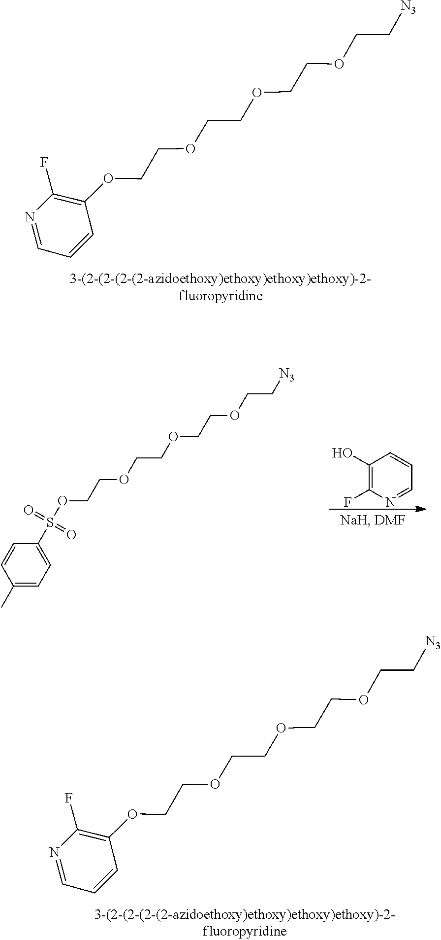


| (E01; SEQ ID NO: 1) |
| GVSDVPRDLEVVAATPTSLLISWRAQLSPSFYYRITYGETGGNSPVQEFT |
| VPNDVMTATISGLKPGVDYTITVYAVTTHGVYFYSPISINYRTPC |
containing the C-terminal amino acids PC was used.
-
- 0 min 90
% A 10% B - 30
min 45% A 55% B - 32 min 25% A 75% B
- 36 min 25% A 75% B
- 50 min 90
% A 10% B
- 0 min 90

| (A02; SEQ ID NO: 2) |
| EVVAATPTSLLISWSYDGPIDRYYRITYGETGGNSPVQEFTVPPDQKTAT |
| ISGLKPGVDYTITVYAVRLEEAHYNREFPISINYRTPC |
was produced as described in Example 9, was tested for the ability to discriminate between PD-L1-positive tumors and PD-L1-negative tumors in mice.

Procedure:
| TABLE 1 | |
| Vial 1 (V1) | 16 mg K.2.2.2, 3 mg Potassium carbonate, dissolved in |
| 0.1 ml of distilled water and 1.4 ml of acetonitrile | |
| Vial 3 (V3) | 2 mg 3-(2-(2-(2-(2-azidoethoxy)ethoxy)ethoxy)ethoxy)- |
| 2-nitropyridine in 0.5 ml DMSO | |
| Vial 4 (V4) | 4 ml of distilled water |
| Vial 14 (V14) | 3 ml of 100% |
| Dilution Flask | |
| 15 ml of distilled water | |
| Cartridge 1 (C1) | |
| HPLC Column | Luna C18(2), 250 × 10 mm, 5 μm, Phenomenex |
| HPLC Solvent | 35% acetonitrile in an aqueous 0.1% trifluoroacetitic |
| acid solution | |
| HPLC flow | 4.6 ml/min |

Procedure:
| TABLE 2 | |
| Vial 1 (V1) | 22 mg K.2.2.2, 4 mg Potassium carbonate, dissolved in |
| 0.3 ml of distilled water and 0.3 ml of acetonitrile | |
| Vial 2 (V2) | 2 mg 3-(2-(2-(2-(2-azidoethoxy)ethoxy)ethoxy)ethoxy)- |
| 2-nitropyridine in 0.5 ml DMSO | |
| Vial 4 (V4) | 3 ml of distilled |
| Dilution Flask | |
| 15 ml of distilled water | |
| Cartridge 1 (C1) | |
| HPLC Column | Luna C18(2), 250 × 10 mm, 5 μm, Phenomenex |
| HPLC Solvent | 35% acetonitrile in an aqueous 0.1% trifluoroacetitic |
| acid solution | |
| HPLC flow | 4.6 ml/min |
| TABLE 3 | ||||
| [19F]-E01 | E01-4PEG-DBCO | |||
| AUC(INF) (μg*h/mL) | 4.72 ± 0.79 | 2.92 ± 0.40 | ||
| CLTs (mL/min/kg) | 4.54 ± 0.81 | 5.78 ± 0.76 | ||
| Vss (L/kg) | 0.29 ± 0.05 | 0.40 ± 0.04 | ||
| T-HALF (h) | 1.69 ± 0.13 | 1.65 ± 0.13 | ||
| MRT (h) | 1.05 ± 0.06 | 1.14 ± 0.05 | ||
Claims (20)
Priority Applications (2)
| Application Number | Priority Date | Filing Date | Title |
|---|---|---|---|
| US17/196,274 US11511000B2 (en) | 2016-06-01 | 2021-03-09 | Imaging methods using 18F-radiolabeled biologics |
| US17/984,825 US11806409B2 (en) | 2016-06-01 | 2022-11-10 | Imaging methods using 18F-radiolabeled biologics |
Applications Claiming Priority (4)
| Application Number | Priority Date | Filing Date | Title |
|---|---|---|---|
| US201662344289P | 2016-06-01 | 2016-06-01 | |
| PCT/US2017/035271 WO2017210335A1 (en) | 2016-06-01 | 2017-05-31 | Imaging methods using 18f-radiolabeled biologics |
| US201816305286A | 2018-11-28 | 2018-11-28 | |
| US17/196,274 US11511000B2 (en) | 2016-06-01 | 2021-03-09 | Imaging methods using 18F-radiolabeled biologics |
Related Parent Applications (2)
| Application Number | Title | Priority Date | Filing Date |
|---|---|---|---|
| US16/305,286 Continuation US10994033B2 (en) | 2016-06-01 | 2017-05-31 | Imaging methods using 18F-radiolabeled biologics |
| PCT/US2017/035271 Continuation WO2017210335A1 (en) | 2016-06-01 | 2017-05-31 | Imaging methods using 18f-radiolabeled biologics |
Related Child Applications (1)
| Application Number | Title | Priority Date | Filing Date |
|---|---|---|---|
| US17/984,825 Continuation US11806409B2 (en) | 2016-06-01 | 2022-11-10 | Imaging methods using 18F-radiolabeled biologics |
Publications (2)
| Publication Number | Publication Date |
|---|---|
| US20210196843A1 US20210196843A1 (en) | 2021-07-01 |
| US11511000B2 true US11511000B2 (en) | 2022-11-29 |
Family
ID=59034937
Family Applications (3)
| Application Number | Title | Priority Date | Filing Date |
|---|---|---|---|
| US16/305,286 Active US10994033B2 (en) | 2016-06-01 | 2017-05-31 | Imaging methods using 18F-radiolabeled biologics |
| US17/196,274 Active US11511000B2 (en) | 2016-06-01 | 2021-03-09 | Imaging methods using 18F-radiolabeled biologics |
| US17/984,825 Active US11806409B2 (en) | 2016-06-01 | 2022-11-10 | Imaging methods using 18F-radiolabeled biologics |
Family Applications Before (1)
| Application Number | Title | Priority Date | Filing Date |
|---|---|---|---|
| US16/305,286 Active US10994033B2 (en) | 2016-06-01 | 2017-05-31 | Imaging methods using 18F-radiolabeled biologics |
Family Applications After (1)
| Application Number | Title | Priority Date | Filing Date |
|---|---|---|---|
| US17/984,825 Active US11806409B2 (en) | 2016-06-01 | 2022-11-10 | Imaging methods using 18F-radiolabeled biologics |
Country Status (2)
| Country | Link |
|---|---|
| US (3) | US10994033B2 (en) |
| WO (1) | WO2017210335A1 (en) |
Cited By (4)
| Publication number | Priority date | Publication date | Assignee | Title |
|---|---|---|---|---|
| US11806409B2 (en) | 2016-06-01 | 2023-11-07 | Bristol-Myers Squibb Company | Imaging methods using 18F-radiolabeled biologics |
| US11857352B2 (en) | 2016-09-06 | 2024-01-02 | The Research Foundation For The State University Of New York | Positron imaging tomography imaging agent composition and method for bacterial infection |
| US12194110B2 (en) | 2014-11-25 | 2025-01-14 | Bristol-Myers Squibb Company | PD-L1 binding polypeptides for imaging |
| US12529165B2 (en) | 2014-03-20 | 2026-01-20 | Bristol-Myers Squibb Company | Stabilized fibronectin based scaffold molecules |
Families Citing this family (14)
| Publication number | Priority date | Publication date | Assignee | Title |
|---|---|---|---|---|
| ES2941895T3 (en) | 2014-11-25 | 2023-05-26 | Bristol Myers Squibb Co | Methods and Compositions for 18F Radiolabeling of the Fibronectin Type (III) Domain |
| WO2017201111A1 (en) | 2016-05-19 | 2017-11-23 | Bristol-Myers Squibb Company | Pet-imaging immunomodulators |
| EP3463486A1 (en) | 2016-06-01 | 2019-04-10 | Bristol-Myers Squibb Company | Pet imaging with pd-l1 binding polypeptides |
| US11168144B2 (en) | 2017-06-01 | 2021-11-09 | Cytomx Therapeutics, Inc. | Activatable anti-PDL1 antibodies, and methods of use thereof |
| WO2019227490A1 (en) * | 2018-06-01 | 2019-12-05 | Tayu Huaxia Biotech Medical Group Co., Ltd. | Compositions and methods for imaging |
| CN117442717A (en) | 2018-06-01 | 2024-01-26 | 大有华夏生物医药集团有限公司 | Compositions for treating diseases or conditions and uses thereof |
| CN113260310A (en) * | 2018-10-15 | 2021-08-13 | 百时美施贵宝公司 | Radioligands for imaging LPA1 receptor |
| CN113795511B (en) | 2019-01-23 | 2024-07-23 | 大有华夏生物医药集团有限公司 | Anti-PD-L1 double antibody and its use |
| US12162953B2 (en) | 2019-12-06 | 2024-12-10 | Yale University | Enhanced targeting platform |
| EP4110407A1 (en) | 2020-02-28 | 2023-01-04 | Bristol-Myers Squibb Company | Radiolabeled fibronectin based scaffolds and antibodies and theranostic uses thereof |
| CN111320675A (en) * | 2020-03-11 | 2020-06-23 | 安徽医科大学第二附属医院 | Radioactive technetium-labeled PD-L1 targeted polypeptide and preparation method and application thereof |
| CA3218697A1 (en) * | 2021-05-03 | 2022-11-10 | Merck Patent Gmbh | Her2 targeting fc antigen binding fragment-drug conjugates |
| CN114133434B (en) * | 2021-12-01 | 2022-11-15 | 北京大学第一医院 | Bicyclic peptide nuclide ligands and probes targeting Nectin-4 |
| WO2025165830A1 (en) * | 2024-01-30 | 2025-08-07 | Wisconsin Alumni Research Foundation | Compositions and methods for targeting cell surface markers induced by radiation therapy |
Citations (57)
| Publication number | Priority date | Publication date | Assignee | Title |
|---|---|---|---|---|
| WO2002096910A1 (en) | 2001-05-31 | 2002-12-05 | Medarex, Inc. | Cytotoxins, prodrugs, linkers and stabilizers useful therefor |
| US20040044076A1 (en) * | 2002-06-07 | 2004-03-04 | Immunomedics, Inc. | Neutrophil imaging methods in cystic fibrosis |
| WO2005056764A2 (en) | 2003-12-05 | 2005-06-23 | Compound Therapeutics, Inc. | Inhibitors of type 2 vascular endothelial growth factor receptors |
| US20060004081A1 (en) | 2004-05-19 | 2006-01-05 | Medarex, Inc. | Cytotoxic compounds and conjugates |
| US20060024317A1 (en) | 2004-05-19 | 2006-02-02 | Medarex, Inc | Chemical linkers and conjugates thereof |
| US20060247295A1 (en) | 2005-04-08 | 2006-11-02 | Medarex, Inc. | Cytotoxic compounds and conjugates with cleavable substrates |
| US20060246059A1 (en) | 1998-12-10 | 2006-11-02 | Compound Therapeutics, Inc. | Pharmaceutically acceptable FN3 polypeptides for human treatments |
| US7180074B1 (en) * | 2001-06-27 | 2007-02-20 | Crosetto Dario B | Method and apparatus for whole-body, three-dimensional, dynamic PET/CT examination |
| WO2007038658A2 (en) | 2005-09-26 | 2007-04-05 | Medarex, Inc. | Antibody-drug conjugates and methods of use |
| WO2007051081A1 (en) | 2005-10-26 | 2007-05-03 | Medarex, Inc. | Methods and compounds for preparing cc-1065 analogs |
| WO2007059404A2 (en) | 2005-11-10 | 2007-05-24 | Medarex, Inc. | Duocarmycin derivatives as novel cytotoxic compounds and conjugates |
| WO2008073458A2 (en) | 2006-12-11 | 2008-06-19 | Bracco Imaging S.P.A. | Fibrin-binding peptides and conjugates thereof |
| WO2008083312A2 (en) | 2006-12-28 | 2008-07-10 | Medarex, Inc. | Chemical linkers and cleavable substrates and conjugates thereof |
| WO2008103693A2 (en) | 2007-02-21 | 2008-08-28 | Medarex, Inc. | Chemical linkers with single amino acids and conjugates thereof |
| US20090028786A1 (en) * | 2007-07-23 | 2009-01-29 | Rcp Diagnostics, Llc | Methods and apparatus for imaging agents for disease detection |
| WO2009080810A1 (en) | 2007-12-21 | 2009-07-02 | Affibody Ab | New polypeptides having affinity for her2 |
| US7556925B2 (en) | 2001-04-04 | 2009-07-07 | University Of Rochester | ανβ3 integrin-binding polypeptide monobodies and their use |
| US20090208410A1 (en) | 2007-09-17 | 2009-08-20 | Dietmar Berndorff | Ed-b fibronectin as stratification marker for anti-tumor drugs |
| WO2009142773A2 (en) | 2008-05-22 | 2009-11-26 | Bristol-Myers Squibb Company | Multivalent fibronectin based scaffold domain proteins |
| US7943743B2 (en) | 2005-07-01 | 2011-05-17 | Medarex, Inc. | Human monoclonal antibodies to programmed death ligand 1 (PD-L1) |
| CN102076713A (en) | 2008-05-02 | 2011-05-25 | 诺瓦提斯公司 | Improved fibronectin-based binding molecules and uses thereof |
| US8008449B2 (en) | 2005-05-09 | 2011-08-30 | Medarex, Inc. | Human monoclonal antibodies to programmed death 1 (PD-1) and methods for treating cancer using anti-PD-1 antibodies alone or in combination with other immunotherapeutics |
| US20120288442A1 (en) * | 2011-05-11 | 2012-11-15 | Atomic Energy Council-Institute Of Nuclear Energy Research | Nuclear Imaging Method Using Molecular Target Detection Agent for Liver Fibrosis |
| US20120309250A1 (en) | 2009-03-24 | 2012-12-06 | North Carolina State University | Composite inorganic/polymer fibers and inorganic fibrils fabricated from sheared solutions |
| US8343501B2 (en) | 2008-11-24 | 2013-01-01 | Bristol-Myers Squibb Company | Bispecific EGFR/IGFIR binding molecules |
| WO2013010573A1 (en) | 2011-07-18 | 2013-01-24 | Universitaet Muenster | Compounds with matrix-metalloproteinase inhibitory activity |
| US8470332B2 (en) | 2006-11-22 | 2013-06-25 | Bristol-Myers Squibb Company | Targeted therapeutics based on engineered proteins for tyrosine kinases receptors, including IGF-IR |
| US8524244B2 (en) | 2008-02-14 | 2013-09-03 | Bristol-Myers Squibb Company | Targeted therapeutics based on engineered proteins that bind EGFR |
| WO2013173223A1 (en) | 2012-05-15 | 2013-11-21 | Bristol-Myers Squibb Company | Cancer immunotherapy by disrupting pd-1/pd-l1 signaling |
| WO2013181634A2 (en) | 2012-05-31 | 2013-12-05 | Sorrento Therapeutics Inc. | Antigen binding proteins that bind pd-l1 |
| WO2014086364A1 (en) | 2012-12-03 | 2014-06-12 | Rigshospitalet | Positron emitting radionuclide labeled peptides for human upar pet imaging |
| US8808665B2 (en) | 2007-01-11 | 2014-08-19 | Immunomedics, Inc. | In vivo copper-free click chemistry for delivery of therapeutic and/or diagnostic agents |
| WO2014126902A1 (en) | 2013-02-15 | 2014-08-21 | Thomas Jefferson University | Kit for tumor imaging |
| US20140271467A1 (en) | 2013-03-12 | 2014-09-18 | The Board Of Trustees Of The Leland Stanford Junior University | Probes and methods of imaging non-hodgkins lymphoma |
| US8853154B2 (en) | 2012-09-13 | 2014-10-07 | Bristol-Myers Squibb Company | Fibronectin based scaffold domain proteins that bind to myostatin |
| US8969289B2 (en) | 2010-05-03 | 2015-03-03 | Bristol-Myers Squibb Company | Serum albumin binding molecules |
| WO2015143199A1 (en) | 2014-03-20 | 2015-09-24 | Bristol-Myers Squibb Company | Serum albumin-binding fibronectin type iii domains |
| US20150361159A1 (en) | 2013-02-01 | 2015-12-17 | Bristol-Myers Squibb Company | Fibronectin based scaffold proteins |
| US20150368319A1 (en) | 2013-02-06 | 2015-12-24 | Bristol-Myers Squibb Company | Fibronectin type iii domain proteins with enhanced solubility |
| WO2016022994A2 (en) | 2014-08-08 | 2016-02-11 | The Board Of Trustees Of The Leland Stanford Junior University | High affinity pd-1 agents and methods of use |
| WO2016077518A1 (en) | 2014-11-14 | 2016-05-19 | Bristol-Myers Squibb Company | Macrocyclic peptides useful as immunomodulators |
| WO2016086036A2 (en) | 2014-11-25 | 2016-06-02 | Bristol-Myers Squibb Company | Methods and compositions for 18f-radiolabeling of biologics |
| WO2016086021A1 (en) | 2014-11-25 | 2016-06-02 | Bristol-Myers Squibb Company | Novel pd-l1 binding polypeptides for imaging |
| US20160287734A1 (en) | 2013-11-13 | 2016-10-06 | Whitehead Institute For Biomedical Research | 18f labeling of proteins using sortases |
| WO2016162368A1 (en) | 2015-04-07 | 2016-10-13 | INSERM (Institut National de la Santé et de la Recherche Médicale) | Non-invasive imaging of tumor pd-l1 expression |
| US9469676B2 (en) | 2011-04-13 | 2016-10-18 | Bristol-Myers Squibb Company | Fc fusion proteins comprising novel linkers or arrangements |
| US9522951B2 (en) | 2011-10-31 | 2016-12-20 | Bristol-Myers Squibb Company | Fibronectin binding domains with reduced immunogenicity |
| US9562089B2 (en) | 2010-05-26 | 2017-02-07 | Bristol-Myers Squibb Company | Fibronectin based scaffold proteins having improved stability |
| WO2017053619A1 (en) | 2015-09-23 | 2017-03-30 | Bristrol-Myers Squibb Company | Glypican-3binding fibronectin based scafflold molecules |
| WO2017053617A1 (en) | 2015-09-23 | 2017-03-30 | Bristol-Myers Squibb Company | Fast-off rate serum albumin binding fibronectin type iii domains |
| US20170114042A1 (en) | 2014-06-09 | 2017-04-27 | Takeda Pharmaceutical Company Limited | Radiolabeled compounds |
| WO2017072273A1 (en) | 2015-10-30 | 2017-05-04 | Affibody Ab | New polypeptide |
| WO2017072280A1 (en) | 2015-10-30 | 2017-05-04 | Affibody Ab | New polypeptide having affinity to pd-l1 |
| US20170183393A1 (en) | 2014-03-20 | 2017-06-29 | Bristol-Myers Squibb Company | Stabilized fibronectin based scaffold molecules |
| WO2017210302A1 (en) | 2016-06-01 | 2017-12-07 | Bristol-Myers Squibb Company | Pet imaging with pd-l1 binding polypeptides |
| WO2017210335A1 (en) | 2016-06-01 | 2017-12-07 | Bristol-Myers Squibb Company | Imaging methods using 18f-radiolabeled biologics |
| US20190015532A1 (en) | 2016-01-15 | 2019-01-17 | Rigshospitalet | Quantitative pet imaging of tissue factor expression using 18f-labled active site inhibited factor vii |
Family Cites Families (81)
| Publication number | Priority date | Publication date | Assignee | Title |
|---|---|---|---|---|
| USRE30985E (en) | 1978-01-01 | 1982-06-29 | Serum-free cell culture media | |
| US4522811A (en) | 1982-07-08 | 1985-06-11 | Syntex (U.S.A.) Inc. | Serial injection of muramyldipeptides and liposomes enhances the anti-infective activity of muramyldipeptides |
| US4560655A (en) | 1982-12-16 | 1985-12-24 | Immunex Corporation | Serum-free cell culture medium and process for making same |
| US4657866A (en) | 1982-12-21 | 1987-04-14 | Sudhir Kumar | Serum-free, synthetic, completely chemically defined tissue culture media |
| US4816567A (en) | 1983-04-08 | 1989-03-28 | Genentech, Inc. | Recombinant immunoglobin preparations |
| US4767704A (en) | 1983-10-07 | 1988-08-30 | Columbia University In The City Of New York | Protein-free culture medium |
| US5374548A (en) | 1986-05-02 | 1994-12-20 | Genentech, Inc. | Methods and compositions for the attachment of proteins to liposomes using a glycophospholipid anchor |
| MX9203291A (en) | 1985-06-26 | 1992-08-01 | Liposome Co Inc | LIPOSOMAS COUPLING METHOD. |
| US5672502A (en) | 1985-06-28 | 1997-09-30 | Celltech Therapeutics Limited | Animal cell culture |
| US4735210A (en) | 1985-07-05 | 1988-04-05 | Immunomedics, Inc. | Lymphographic and organ imaging method and kit |
| US5101827A (en) | 1985-07-05 | 1992-04-07 | Immunomedics, Inc. | Lymphographic and organ imaging method and kit |
| US5776093A (en) | 1985-07-05 | 1998-07-07 | Immunomedics, Inc. | Method for imaging and treating organs and tissues |
| US5225539A (en) | 1986-03-27 | 1993-07-06 | Medical Research Council | Recombinant altered antibodies and methods of making altered antibodies |
| US4927762A (en) | 1986-04-01 | 1990-05-22 | Cell Enterprises, Inc. | Cell culture medium with antioxidant |
| US5328679A (en) | 1988-04-01 | 1994-07-12 | Immunomedics, Inc. | Methods for technetium/rhenium labeling of proteins |
| US5128119A (en) | 1989-06-12 | 1992-07-07 | Immunomedics, Inc. | Methods for technetium/rhenium labeling of f(ab1)2 fragments |
| US6048728A (en) | 1988-09-23 | 2000-04-11 | Chiron Corporation | Cell culture medium for enhanced cell growth, culture longevity, and product expression |
| US5530101A (en) | 1988-12-28 | 1996-06-25 | Protein Design Labs, Inc. | Humanized immunoglobulins |
| US5108921A (en) | 1989-04-03 | 1992-04-28 | Purdue Research Foundation | Method for enhanced transmembrane transport of exogenous molecules |
| FR2646437B1 (en) | 1989-04-28 | 1991-08-30 | Transgene Sa | NOVEL DNA SEQUENCES, THEIR APPLICATION AS A SEQUENCE ENCODING A SIGNAL PEPTIDE FOR THE SECRETION OF MATURE PROTEINS BY RECOMBINANT YEASTS, EXPRESSION CASSETTES, PROCESSED YEASTS AND PROCESS FOR PREPARING THE SAME |
| JP3051145B2 (en) | 1990-08-28 | 2000-06-12 | 住友製薬株式会社 | Novel polyethylene glycol derivative modified peptide |
| US5122469A (en) | 1990-10-03 | 1992-06-16 | Genentech, Inc. | Method for culturing Chinese hamster ovary cells to improve production of recombinant proteins |
| ATE139900T1 (en) | 1992-11-13 | 1996-07-15 | Idec Pharma Corp | THERAPEUTIC USE OF CHIMERIC AND LABELED ANTIBODIES AGAINST HUMAN B LYMPHOCYTE RESTRICTED DIFFERENTIATION ANTIGEN FOR THE TREATMENT OF B CELL LYMPHOMA |
| IL113610A0 (en) | 1994-06-03 | 1995-08-31 | Immunomedics Inc | A method of radiolabeling a protein, radiolabeled proteins prepared thereby and diagnostic kits containing the same |
| US5746996A (en) | 1994-06-03 | 1998-05-05 | Immunomedics, Inc. | Thiolation of peptides for radionuclide-based radiodetection and radiotherapy |
| US5932462A (en) | 1995-01-10 | 1999-08-03 | Shearwater Polymers, Inc. | Multiarmed, monofunctional, polymer for coupling to molecules and surfaces |
| US5753206A (en) | 1995-06-07 | 1998-05-19 | Immunomedics, Inc. | Radiometal-binding analogues of luteinizing hormone releasing hormone |
| ATE365562T1 (en) | 1996-05-03 | 2007-07-15 | Immunomedics Inc | TARGETED COMBINATION IMMUNOTHERAPY FOR CANCER |
| EP0975374B1 (en) | 1996-07-12 | 2007-12-05 | Immunomedics, Inc. | Radiometal-binding peptide analogues |
| US6261804B1 (en) | 1997-01-21 | 2001-07-17 | The General Hospital Corporation | Selection of proteins using RNA-protein fusions |
| RU2233878C2 (en) | 1997-01-21 | 2004-08-10 | Дзе Дженерал Хоспитал Корпорейшн | Method for selection of desirable protein and nucleic acid, agents for its realization |
| WO1998042752A1 (en) | 1997-03-21 | 1998-10-01 | Brigham And Women's Hospital Inc. | Immunotherapeutic ctla-4 binding peptides |
| AU9375698A (en) | 1997-09-03 | 1999-03-22 | Immunomedics Inc. | Fluorination of proteins and peptides for f-18 positron emission tomography |
| US6818418B1 (en) | 1998-12-10 | 2004-11-16 | Compound Therapeutics, Inc. | Protein scaffolds for antibody mimics and other binding proteins |
| CA2356215C (en) | 1998-12-23 | 2015-11-24 | Pfizer Inc. | Human monoclonal antibodies to ctla-4 |
| WO2001014556A1 (en) | 1999-08-23 | 2001-03-01 | Dana-Farber Cancer Institute, Inc. | Novel b7-4 molecules and uses therefor |
| ATE354655T1 (en) | 1999-08-24 | 2007-03-15 | Medarex Inc | HUMAN ANTIBODIES TO CTLA-4 AND THEIR USES |
| CA3016482A1 (en) | 1999-11-30 | 2001-06-07 | Mayo Foundation For Medical Education And Research | B7-h1, a novel immunoregulatory molecule |
| US20050287153A1 (en) | 2002-06-28 | 2005-12-29 | Genentech, Inc. | Serum albumin binding peptides for tumor targeting |
| CA2404528A1 (en) | 2000-03-31 | 2001-10-04 | Institut Pasteur | Peptides blocking vascular endothelial growth factor (vegf)-mediated angiogenesis, polynucleotides encoding said peptides and methods of use thereof |
| EP1301538B1 (en) | 2000-07-11 | 2015-12-09 | Research Corporation Technologies, Inc | Artificial antibody polypeptides |
| IL149820A0 (en) | 2002-05-23 | 2002-11-10 | Curetech Ltd | Humanized immunomodulatory monoclonal antibodies for the treatment of neoplastic disease or immunodeficiency |
| US7696320B2 (en) | 2004-08-24 | 2010-04-13 | Domantis Limited | Ligands that have binding specificity for VEGF and/or EGFR and methods of use therefor |
| AU2004296184B2 (en) * | 2003-12-04 | 2010-12-16 | Vaccinex, Inc. | Methods of killing tumor cells by targeting internal antigens exposed on apoptotic tumor cells |
| CA2554089C (en) | 2004-02-09 | 2013-10-22 | Human Genome Sciences, Inc. | Albumin fusion proteins |
| PL1960434T3 (en) | 2005-12-08 | 2012-12-31 | Squibb & Sons Llc | Human monoclonal antibodies to fucosyl-gm1 and methods for using anti-fucosyl-gm1 |
| HUE028737T2 (en) | 2007-07-17 | 2017-01-30 | Squibb & Sons Llc | Monoclonal antibodies to GLYPICAN-3 |
| US20090028857A1 (en) | 2007-07-23 | 2009-01-29 | Cell Genesys, Inc. | Pd-1 antibodies in combination with a cytokine-secreting cell and methods of use thereof |
| EP2222846B1 (en) | 2007-08-10 | 2017-05-03 | Protelix, Inc. | Universal fibronectin type iii binding-domain libraries |
| PL2195017T3 (en) | 2007-10-01 | 2015-03-31 | Bristol Myers Squibb Co | Human antibodies that bind mesothelin, and uses thereof |
| KR101686247B1 (en) | 2007-10-31 | 2016-12-14 | 메디뮨 엘엘씨 | Protein scaffolds |
| WO2009086116A2 (en) | 2007-12-19 | 2009-07-09 | Centocor, Inc. | Alternative scaffold protein fusions phage display via fusion to plx of m13 phage |
| AU2008345424A1 (en) | 2007-12-27 | 2009-07-09 | Novartis Ag | Improved fibronectin-based binding molecules and their use |
| US8168757B2 (en) | 2008-03-12 | 2012-05-01 | Merck Sharp & Dohme Corp. | PD-1 binding proteins |
| EP2113255A1 (en) | 2008-05-02 | 2009-11-04 | f-star Biotechnologische Forschungs- und Entwicklungsges.m.b.H. | Cytotoxic immunoglobulin |
| WO2010051274A2 (en) | 2008-10-31 | 2010-05-06 | Centocor Ortho Biotech Inc. | Fibronectin type iii domain based scaffold compositions, methods and uses |
| US8415291B2 (en) | 2008-10-31 | 2013-04-09 | Centocor Ortho Biotech Inc. | Anti-TNF alpha fibronectin type III domain based scaffold compositions, methods and uses |
| EP4331604B9 (en) | 2008-12-09 | 2025-07-23 | F. Hoffmann-La Roche AG | Anti-pd-l1 antibodies and their use to enhance t-cell function |
| BRPI1008532B1 (en) | 2009-02-12 | 2021-12-14 | Janssen Biotech, Inc | ISOLATED PROTEIN FRAMEWORK, METHOD FOR BUILDING AN ISOLATED PROTEIN FRAMEWORK LIBRARY, ISOLATED NUCLEIC ACID MOLECULE, ISOLATED NUCLEIC ACID VECTOR, BACTERIAL OR FUNGUS HOST CELL, COMPOSITION, MEDICAL DEVICE AND MANUFACTURING ARTICLE FOR PRODUCT USE IN HUMAN BEINGS |
| US20120270797A1 (en) | 2009-08-13 | 2012-10-25 | Massachusetts Institute Of Technology | Engineered proteins including mutant fibronectin domains |
| WO2011051333A1 (en) | 2009-10-30 | 2011-05-05 | Novartis Ag | Universal fibronectin type iii bottom-side binding domain libraries |
| WO2011051466A1 (en) | 2009-11-02 | 2011-05-05 | Novartis Ag | Anti-idiotypic fibronectin-based binding molecules and uses thereof |
| CA2778714C (en) | 2009-11-24 | 2018-02-27 | Medimmune Limited | Targeted binding agents against b7-h1 |
| HUE044179T2 (en) | 2009-12-10 | 2019-10-28 | Hoffmann La Roche | Antibodies binding preferentially human csf1r extracellular domain 4 and their use |
| WO2011092233A1 (en) | 2010-01-29 | 2011-08-04 | Novartis Ag | Yeast mating to produce high-affinity combinations of fibronectin-based binders |
| WO2011100700A2 (en) | 2010-02-12 | 2011-08-18 | University Of Rochester | Antigenic mimics of discontinuous epitopes of pathogen recognized by broadly neutralizing antibodies |
| US9169323B2 (en) | 2010-03-05 | 2015-10-27 | Hoffmann-La Roche Inc. | Antibodies against human CSF-1R |
| EP2542588A1 (en) | 2010-03-05 | 2013-01-09 | F. Hoffmann-La Roche AG | Antibodies against human csf-1r and uses thereof |
| AU2011240620A1 (en) | 2010-04-13 | 2012-10-18 | Medimmune, Llc | Fibronectin type III domain-based multimeric scaffolds |
| PT3103478T (en) | 2010-04-30 | 2019-07-10 | Janssen Biotech Inc | Stabilized fibronectin domain compositions, methods and uses |
| NZ603193A (en) | 2010-05-04 | 2014-07-25 | Five Prime Therapeutics Inc | Antibodies that bind csf1r |
| US8907053B2 (en) | 2010-06-25 | 2014-12-09 | Aurigene Discovery Technologies Limited | Immunosuppression modulating compounds |
| SG187225A1 (en) | 2010-07-30 | 2013-02-28 | Novartis Ag | Fibronectin cradle molecules and libraries thereof |
| PT2699264T (en) | 2011-04-20 | 2018-05-23 | Medimmune Llc | ANTIBODIES AND OTHER MOLECULES CONNECTING B7-H1 AND PD-1 |
| BR112014012624A2 (en) | 2011-12-15 | 2018-10-09 | F Hoffmann-La Roche Ag | antibodies, pharmaceutical composition, nucleic acid, expression vectors, host cell, method for producing a recombinant antibody and use of the antibody |
| CA2861122A1 (en) | 2012-02-06 | 2013-08-15 | Genentech, Inc. | Compositions and methods for using csf1r inhibitors |
| AR090263A1 (en) | 2012-03-08 | 2014-10-29 | Hoffmann La Roche | COMBINED ANTIBODY THERAPY AGAINST HUMAN CSF-1R AND USES OF THE SAME |
| HK1208233A1 (en) | 2012-05-11 | 2016-02-26 | 戊瑞治疗有限公司 | Methods of treating conditions with antibodies that bind colony stimulating factor 1 receptor (csf1r) |
| HK1212214A1 (en) | 2012-08-31 | 2016-06-10 | 戊瑞治疗有限公司 | Methods of treating conditions with antibodies that bind colony stimulating factor 1 receptor (csf1r) |
| GB201317622D0 (en) | 2013-10-04 | 2013-11-20 | Star Biotechnology Ltd F | Cancer biomarkers and uses thereof |
| TWI758788B (en) | 2020-07-16 | 2022-03-21 | 富世達股份有限公司 | Torque-variable uniaxial shaft structure with multiple friction pairs |
-
2017
- 2017-05-31 WO PCT/US2017/035271 patent/WO2017210335A1/en not_active Ceased
- 2017-05-31 US US16/305,286 patent/US10994033B2/en active Active
-
2021
- 2021-03-09 US US17/196,274 patent/US11511000B2/en active Active
-
2022
- 2022-11-10 US US17/984,825 patent/US11806409B2/en active Active
Patent Citations (106)
| Publication number | Priority date | Publication date | Assignee | Title |
|---|---|---|---|---|
| US20060246059A1 (en) | 1998-12-10 | 2006-11-02 | Compound Therapeutics, Inc. | Pharmaceutically acceptable FN3 polypeptides for human treatments |
| US20170275342A1 (en) | 1998-12-10 | 2017-09-28 | Bristol-Myers Squibb Company | Protein scaffolds for antibody mimics and other binding proteins |
| US9605039B2 (en) | 1998-12-10 | 2017-03-28 | Bristol-Myers Squibb Company | Protein scaffolds for antibody mimics and other binding proteins |
| US7556925B2 (en) | 2001-04-04 | 2009-07-07 | University Of Rochester | ανβ3 integrin-binding polypeptide monobodies and their use |
| US6989452B2 (en) | 2001-05-31 | 2006-01-24 | Medarex, Inc. | Disulfide prodrugs and linkers and stabilizers useful therefor |
| WO2002096910A1 (en) | 2001-05-31 | 2002-12-05 | Medarex, Inc. | Cytotoxins, prodrugs, linkers and stabilizers useful therefor |
| US7087600B2 (en) | 2001-05-31 | 2006-08-08 | Medarex, Inc. | Peptidyl prodrugs and linkers and stabilizers useful therefor |
| US7129261B2 (en) | 2001-05-31 | 2006-10-31 | Medarex, Inc. | Cytotoxic agents |
| US7180074B1 (en) * | 2001-06-27 | 2007-02-20 | Crosetto Dario B | Method and apparatus for whole-body, three-dimensional, dynamic PET/CT examination |
| US20040044076A1 (en) * | 2002-06-07 | 2004-03-04 | Immunomedics, Inc. | Neutrophil imaging methods in cystic fibrosis |
| CN1946417A (en) | 2003-12-05 | 2007-04-11 | 阿德内克休斯治疗公司 | Inhibitors of type 2 vascular endothelial growth factor receptor |
| US8609613B2 (en) | 2003-12-05 | 2013-12-17 | Bristol-Myers Squibb Company | Methods of inhibiting type 2 vascular endothelial growth factor receptors using polypeptides |
| US9328157B2 (en) | 2003-12-05 | 2016-05-03 | Bristol-Myers Squibb Company | Inhibitors of type 2 vascular endothelial growth factor receptors |
| WO2005056764A2 (en) | 2003-12-05 | 2005-06-23 | Compound Therapeutics, Inc. | Inhibitors of type 2 vascular endothelial growth factor receptors |
| US9862758B2 (en) | 2003-12-05 | 2018-01-09 | Bristol-Myers Quibb Company | Inhibitors of type 2 vascular endothelial growth factor receptors |
| US8324362B2 (en) | 2003-12-05 | 2012-12-04 | Bristol-Myers Squibb Company | Nucleic acid encoding 10FN3 polypeptide inhibitors of Type 2 vascular endothelial growth factor receptors |
| US20060024317A1 (en) | 2004-05-19 | 2006-02-02 | Medarex, Inc | Chemical linkers and conjugates thereof |
| US20060004081A1 (en) | 2004-05-19 | 2006-01-05 | Medarex, Inc. | Cytotoxic compounds and conjugates |
| US20060247295A1 (en) | 2005-04-08 | 2006-11-02 | Medarex, Inc. | Cytotoxic compounds and conjugates with cleavable substrates |
| US8008449B2 (en) | 2005-05-09 | 2011-08-30 | Medarex, Inc. | Human monoclonal antibodies to programmed death 1 (PD-1) and methods for treating cancer using anti-PD-1 antibodies alone or in combination with other immunotherapeutics |
| US7943743B2 (en) | 2005-07-01 | 2011-05-17 | Medarex, Inc. | Human monoclonal antibodies to programmed death ligand 1 (PD-L1) |
| WO2007038658A2 (en) | 2005-09-26 | 2007-04-05 | Medarex, Inc. | Antibody-drug conjugates and methods of use |
| WO2007051081A1 (en) | 2005-10-26 | 2007-05-03 | Medarex, Inc. | Methods and compounds for preparing cc-1065 analogs |
| WO2007059404A2 (en) | 2005-11-10 | 2007-05-24 | Medarex, Inc. | Duocarmycin derivatives as novel cytotoxic compounds and conjugates |
| US20150252097A1 (en) | 2006-11-22 | 2015-09-10 | Bristol-Myers Squibb Company | Targeted therapeutics based on engineered proteins for tyrosine kinases receptors, including igf-ir |
| US20170190761A1 (en) | 2006-11-22 | 2017-07-06 | Bristol-Myers Squibb Company | Targeted therapeutics based on engineered proteins for tyrosine kinases receptors, including igf-ir |
| US20130310317A1 (en) | 2006-11-22 | 2013-11-21 | Bristol-Myers Squibb Company | Targeted therapeutics based on engineered proteins for tyrosine kinases receptors, including igf-ir |
| US8470332B2 (en) | 2006-11-22 | 2013-06-25 | Bristol-Myers Squibb Company | Targeted therapeutics based on engineered proteins for tyrosine kinases receptors, including IGF-IR |
| CN101558081A (en) | 2006-12-11 | 2009-10-14 | 伯拉考成像股份公司 | Fibrin-binding peptide conjugates for diagnostic and therapeutic applications |
| WO2008073458A2 (en) | 2006-12-11 | 2008-06-19 | Bracco Imaging S.P.A. | Fibrin-binding peptides and conjugates thereof |
| WO2008083312A2 (en) | 2006-12-28 | 2008-07-10 | Medarex, Inc. | Chemical linkers and cleavable substrates and conjugates thereof |
| US8808665B2 (en) | 2007-01-11 | 2014-08-19 | Immunomedics, Inc. | In vivo copper-free click chemistry for delivery of therapeutic and/or diagnostic agents |
| WO2008103693A2 (en) | 2007-02-21 | 2008-08-28 | Medarex, Inc. | Chemical linkers with single amino acids and conjugates thereof |
| US20090028786A1 (en) * | 2007-07-23 | 2009-01-29 | Rcp Diagnostics, Llc | Methods and apparatus for imaging agents for disease detection |
| US20090208410A1 (en) | 2007-09-17 | 2009-08-20 | Dietmar Berndorff | Ed-b fibronectin as stratification marker for anti-tumor drugs |
| WO2009080810A1 (en) | 2007-12-21 | 2009-07-02 | Affibody Ab | New polypeptides having affinity for her2 |
| CN101952302A (en) | 2007-12-21 | 2011-01-19 | 阿菲博迪公司 | Novel peptides with HER2 affinity |
| US8524244B2 (en) | 2008-02-14 | 2013-09-03 | Bristol-Myers Squibb Company | Targeted therapeutics based on engineered proteins that bind EGFR |
| US9920108B2 (en) | 2008-02-14 | 2018-03-20 | Bristol-Myers Squibb Company | Targeted therapeutics based on engineered proteins that bind EGFR |
| US9234028B2 (en) | 2008-02-14 | 2016-01-12 | Bristol-Myers Squibb Company | Targeted therapeutics based on engineered proteins that bind EGFR |
| CN102076713A (en) | 2008-05-02 | 2011-05-25 | 诺瓦提斯公司 | Improved fibronectin-based binding molecules and uses thereof |
| US8221765B2 (en) | 2008-05-22 | 2012-07-17 | Bristol-Myers Squibb Company | Multivalent fibronectin based scaffold domain proteins |
| CN102099373A (en) | 2008-05-22 | 2011-06-15 | 百时美施贵宝公司 | Multivalent fibronectin based scaffold domain proteins |
| US8728483B2 (en) | 2008-05-22 | 2014-05-20 | Bristol-Myers Squibb Company | Multivalent fibronectin based scaffold domain proteins |
| US9902762B2 (en) | 2008-05-22 | 2018-02-27 | Bristol-Myers Squibb Company | Multivalent fibronectin based scaffold domain proteins |
| WO2009142773A2 (en) | 2008-05-22 | 2009-11-26 | Bristol-Myers Squibb Company | Multivalent fibronectin based scaffold domain proteins |
| US9017655B2 (en) | 2008-11-24 | 2015-04-28 | Bristol-Myers Squibb Company | Bispecific EGFR/IGFIR binding molecules |
| US8343501B2 (en) | 2008-11-24 | 2013-01-01 | Bristol-Myers Squibb Company | Bispecific EGFR/IGFIR binding molecules |
| US20180037631A1 (en) | 2008-11-24 | 2018-02-08 | Bristol-Myers Squibb Company | Bispecific egfr/igfir binding molecules |
| US9771411B2 (en) | 2008-11-24 | 2017-09-26 | Bristol-Myers Squibb Company | Method of treating cancer by administering EGFR and EGFR/IGFIR binding molecules |
| US20120309250A1 (en) | 2009-03-24 | 2012-12-06 | North Carolina State University | Composite inorganic/polymer fibers and inorganic fibrils fabricated from sheared solutions |
| US9540424B2 (en) | 2010-05-03 | 2017-01-10 | Bristol-Myers Squibb Company | Serum albumin binding molecules |
| US20170145464A1 (en) | 2010-05-03 | 2017-05-25 | Bristol-Myers Squibb Company | Serum albumin binding molecules |
| US8969289B2 (en) | 2010-05-03 | 2015-03-03 | Bristol-Myers Squibb Company | Serum albumin binding molecules |
| US9562089B2 (en) | 2010-05-26 | 2017-02-07 | Bristol-Myers Squibb Company | Fibronectin based scaffold proteins having improved stability |
| US20170166627A1 (en) | 2010-05-26 | 2017-06-15 | Bristol-Myers Squibb Company | Fibronectin based scaffold proteins having improved stability |
| US20160376346A1 (en) | 2011-04-13 | 2016-12-29 | Bristol-Myers Squibb Company | Fc FUSION PROTEINS COMPRISING NOVEL LINKERS OR ARRANGEMENTS |
| US9469676B2 (en) | 2011-04-13 | 2016-10-18 | Bristol-Myers Squibb Company | Fc fusion proteins comprising novel linkers or arrangements |
| US20120288442A1 (en) * | 2011-05-11 | 2012-11-15 | Atomic Energy Council-Institute Of Nuclear Energy Research | Nuclear Imaging Method Using Molecular Target Detection Agent for Liver Fibrosis |
| WO2013010573A1 (en) | 2011-07-18 | 2013-01-24 | Universitaet Muenster | Compounds with matrix-metalloproteinase inhibitory activity |
| US9522951B2 (en) | 2011-10-31 | 2016-12-20 | Bristol-Myers Squibb Company | Fibronectin binding domains with reduced immunogenicity |
| US20170137494A1 (en) | 2011-10-31 | 2017-05-18 | Bristol-Myers Squibb Company | Fibronectin binding domains with reduced immunogenicity |
| WO2013173223A1 (en) | 2012-05-15 | 2013-11-21 | Bristol-Myers Squibb Company | Cancer immunotherapy by disrupting pd-1/pd-l1 signaling |
| WO2013181634A2 (en) | 2012-05-31 | 2013-12-05 | Sorrento Therapeutics Inc. | Antigen binding proteins that bind pd-l1 |
| US20130323249A1 (en) | 2012-05-31 | 2013-12-05 | Sorrento Therapeutics Inc. | Antigen Binding Proteins that Bind PD-L1 |
| US20170354718A1 (en) | 2012-09-13 | 2017-12-14 | Bristol-Myers Squibb Company | Fibronectin based scaffold domain proteins that bind to myostatin |
| US9493546B2 (en) | 2012-09-13 | 2016-11-15 | Bristol-Myers Squibb Company | Fibronectin based scaffold domain proteins that bind to myostatin |
| US20170088602A1 (en) | 2012-09-13 | 2017-03-30 | Bristol-Myers Squibb Company | Fibronectin based scaffold domain proteins that bind to myostatin |
| US8993265B2 (en) | 2012-09-13 | 2015-03-31 | Bristol-Myers Squibb Company | Fibronectin based scaffold domain proteins that bind to myostatin |
| US8853154B2 (en) | 2012-09-13 | 2014-10-07 | Bristol-Myers Squibb Company | Fibronectin based scaffold domain proteins that bind to myostatin |
| US9662373B2 (en) | 2012-09-13 | 2017-05-30 | Bristol-Myers Squibb Company | Fibronectin based scaffold domain proteins that bind to myostatin |
| US8933199B2 (en) | 2012-09-13 | 2015-01-13 | Bristol-Myers Squibb Company | Fibronectin based scaffold domain proteins that bind to myostatin |
| WO2014086364A1 (en) | 2012-12-03 | 2014-06-12 | Rigshospitalet | Positron emitting radionuclide labeled peptides for human upar pet imaging |
| US20150361159A1 (en) | 2013-02-01 | 2015-12-17 | Bristol-Myers Squibb Company | Fibronectin based scaffold proteins |
| US20150368319A1 (en) | 2013-02-06 | 2015-12-24 | Bristol-Myers Squibb Company | Fibronectin type iii domain proteins with enhanced solubility |
| WO2014126902A1 (en) | 2013-02-15 | 2014-08-21 | Thomas Jefferson University | Kit for tumor imaging |
| US20140271467A1 (en) | 2013-03-12 | 2014-09-18 | The Board Of Trustees Of The Leland Stanford Junior University | Probes and methods of imaging non-hodgkins lymphoma |
| US20160287734A1 (en) | 2013-11-13 | 2016-10-06 | Whitehead Institute For Biomedical Research | 18f labeling of proteins using sortases |
| US20170183393A1 (en) | 2014-03-20 | 2017-06-29 | Bristol-Myers Squibb Company | Stabilized fibronectin based scaffold molecules |
| US20170174748A1 (en) | 2014-03-20 | 2017-06-22 | Bristol-Myers Squibb Company | Serum albumin-binding fibronectin type iii domains |
| WO2015143199A1 (en) | 2014-03-20 | 2015-09-24 | Bristol-Myers Squibb Company | Serum albumin-binding fibronectin type iii domains |
| US20170114042A1 (en) | 2014-06-09 | 2017-04-27 | Takeda Pharmaceutical Company Limited | Radiolabeled compounds |
| WO2016022994A2 (en) | 2014-08-08 | 2016-02-11 | The Board Of Trustees Of The Leland Stanford Junior University | High affinity pd-1 agents and methods of use |
| WO2016077518A1 (en) | 2014-11-14 | 2016-05-19 | Bristol-Myers Squibb Company | Macrocyclic peptides useful as immunomodulators |
| WO2016086036A2 (en) | 2014-11-25 | 2016-06-02 | Bristol-Myers Squibb Company | Methods and compositions for 18f-radiolabeling of biologics |
| CN107406494A (en) | 2014-11-25 | 2017-11-28 | 百时美施贵宝公司 | Novel PD‑L1-binding peptides for imaging |
| US20220001040A1 (en) | 2014-11-25 | 2022-01-06 | Bristol-Myers Squibb Company | Novel pd-l1 binding polypeptides for imaging |
| US11173219B2 (en) | 2014-11-25 | 2021-11-16 | Bristol-Myers Squibb Company | PD-L1 binding polypeptides for imaging |
| WO2016086021A1 (en) | 2014-11-25 | 2016-06-02 | Bristol-Myers Squibb Company | Novel pd-l1 binding polypeptides for imaging |
| US11229713B2 (en) | 2014-11-25 | 2022-01-25 | Bristol-Myers Squibb Company | Methods and compositions for 18F-radiolabeling of biologics |
| US20170258948A1 (en) | 2014-11-25 | 2017-09-14 | Bristol-Myers Squibb Company | Novel pd-l1 binding polypeptides for imaging |
| US10406251B2 (en) | 2014-11-25 | 2019-09-10 | Bristol-Myers Squibb Company | PD-L1 binding polypeptides for imaging |
| US20180326098A1 (en) | 2014-11-25 | 2018-11-15 | Bristol-Myers Squibb Company | Methods and compositions for 18f-radiolabeling of biologics |
| US20190343972A1 (en) | 2014-11-25 | 2019-11-14 | Bristol-Myers Squibb Company | Novel pd-l1 binding polypeptides for imaging |
| WO2016162368A1 (en) | 2015-04-07 | 2016-10-13 | INSERM (Institut National de la Santé et de la Recherche Médicale) | Non-invasive imaging of tumor pd-l1 expression |
| WO2017053619A1 (en) | 2015-09-23 | 2017-03-30 | Bristrol-Myers Squibb Company | Glypican-3binding fibronectin based scafflold molecules |
| WO2017053617A1 (en) | 2015-09-23 | 2017-03-30 | Bristol-Myers Squibb Company | Fast-off rate serum albumin binding fibronectin type iii domains |
| WO2017072273A1 (en) | 2015-10-30 | 2017-05-04 | Affibody Ab | New polypeptide |
| WO2017072280A1 (en) | 2015-10-30 | 2017-05-04 | Affibody Ab | New polypeptide having affinity to pd-l1 |
| US20190015532A1 (en) | 2016-01-15 | 2019-01-17 | Rigshospitalet | Quantitative pet imaging of tissue factor expression using 18f-labled active site inhibited factor vii |
| US20190184042A1 (en) | 2016-06-01 | 2019-06-20 | Bristol-Myers Squibb Company | Pet imaging with pd-l1 binding polypeptides |
| US20190184043A1 (en) | 2016-06-01 | 2019-06-20 | Bristol-Myers Squibb Company | Imaging methods using 18f-radiolabeled biologics |
| US10994033B2 (en) | 2016-06-01 | 2021-05-04 | Bristol-Myers Squibb Company | Imaging methods using 18F-radiolabeled biologics |
| WO2017210335A1 (en) | 2016-06-01 | 2017-12-07 | Bristol-Myers Squibb Company | Imaging methods using 18f-radiolabeled biologics |
| WO2017210302A1 (en) | 2016-06-01 | 2017-12-07 | Bristol-Myers Squibb Company | Pet imaging with pd-l1 binding polypeptides |
| US11344639B2 (en) | 2016-06-01 | 2022-05-31 | Bristol-Myers Squibb Company | PET imaging with PD-L1 binding polypeptides |
Non-Patent Citations (79)
| Title |
|---|
| "Abstracts DEGRO 2016", STRAHLENTHERAPIE UND ONKOLOGIE, SPRINGER BERLIN HEIDELBERG, BERLIN/HEIDELBERG, vol. 192, no. 1, 11 May 2016 (2016-05-11), Berlin/Heidelberg, pages 1 - 161, XP035803487, ISSN: 0179-7158, DOI: 10.1007/s00066-016-0974-z |
| "Proceedings of the World Molecular Imaging Congress 2015, Honolulu, Hawaii, September 2-5, 2015: Late-Breaking Abstracts", MOLECULAR IMAGING & BIOLOGY, ELSEVIER, BOSTON, vol. 18, no. 1, 24 May 2016 (2016-05-24), Boston , pages 1554 - 1859, XP035974151, ISSN: 1536-1632, DOI: 10.1007/s11307-016-0968-3 |
| A. NATARAJAN, B. J. HACKEL, S. S. GAMBHIR: "A Novel Engineered Anti-CD20 Tracer Enables Early Time PET Imaging in a Humanized Transgenic Mouse Model of B-cell Non-Hodgkins Lymphoma", CLINICAL CANCER RESEARCH, ASSOCIATION FOR CANCER RESEARCH, US, vol. 19, no. 24, 15 December 2013 (2013-12-15), US, pages 6820 - 6829, XP055398629, ISSN: 1078-0432, DOI: 10.1158/1078-0432.CCR-13-0626 |
| Abstracts DEGRO 2016, Strahlentherapie Und Onkologie, vol. 192(1): 1-161 (2016), XP035803487, ISSN: 0179-7158, DOI:10.1007/S00066-016-0974-Z [retrieved on May 11, 2016]. |
| ANA C. VALDIVIA, MIRIAM ESTRADA, TAYEBEH HADIZAD, DUNCAN J. STEWART, ROB S. BEANLANDS, JEAN N. DASILVA: "A fast, simple, and reproducible automated synthesis of [18F]FPyKYNE-c(RGDyK) for αvβ3 receptor positron emission tomography imaging", JOURNAL OF LABELLED COMPOUNDS AND RADIOPHARMACEUTICALS, JOHN WILEY & SONS LTD., GB, vol. 55, no. 2, 1 February 2012 (2012-02-01), GB , pages 57 - 60, XP055250449, ISSN: 0362-4803, DOI: 10.1002/jlcr.1948 |
| ANDERS JOSEFSSON, JESSIE R. NEDROW, SUNJU PARK, SANGEETA RAY BANERJEE, ANDREW RITTENBACH, FABIEN JAMMES, BENJAMIN TSUI, GEORGE SGO: "Imaging, Biodistribution, and Dosimetry of Radionuclide-Labeled PD-L1 Antibody in an Immunocompetent Mouse Model of Breast Cancer", CANCER RESEARCH, AMERICAN ASSOCIATION FOR CANCER RESEARCH, US, vol. 76, no. 2, 15 January 2016 (2016-01-15), US, pages 472 - 479, XP055398622, ISSN: 0008-5472, DOI: 10.1158/0008-5472.CAN-15-2141 |
| Berndt, M. et al., "Labeling of low-density lipoproteins using the 18F-labeled thiol-reactive reagent N-[6-(4-[18F] fluorobenzylidene)aminooxyhexyl]maleimide," Nuclear Medicine and Biology, vol. 34:5-15 (2007). |
| BERTRAND KUHNAST, FRANÇOISE HINNEN, BERTRAND TAVITIAN, FRÉDÉRIC DOLLÉ: "[ 18 F]FPyKYNE, a fluoropyridine-based alkyne reagent designed for the fluorine-18 labelling of macromolecules using click chemistry", JOURNAL OF LABELLED COMPOUNDS AND RADIOPHARMACEUTICALS, JOHN WILEY & SONS LTD., GB, vol. 51, no. 9, 1 August 2008 (2008-08-01), GB , pages 336 - 342, XP055250465, ISSN: 0362-4803, DOI: 10.1002/jlcr.1533 |
| Biochemistry vol. 82(8):710-726 (2010) cited in corresponding Japanese Offce Action, Application No. JP2018-563054, dated Jun. 22, 2021, 4 pages. |
| BRAD D. MAXWELL, KAI CAO, SAMUEL BONACORSI, LAKSHMI SIVARAMAN, BONNIE WANG, MARYELLEN MCNERNEY, LIFEI WANG, LISA J. CHRISTOPHER, N: "The synthesis of a carbon-14 labeled pegylated Adnectin™ for placental transfer studies in guinea pigs", JOURNAL OF LABELLED COMPOUNDS AND RADIOPHARMACEUTICALS, JOHN WILEY & SONS LTD., GB, vol. 56, no. 9-10, 1 July 2013 (2013-07-01), GB , pages 492 - 494, XP055255267, ISSN: 0362-4803, DOI: 10.1002/jlcr.3077 |
| CAM PATTERSON, C. FREDERICK, HONG YUAN, LAURA DYER, PAMELA LOCKYER, DAVID LALUSH, ANKA VELEVA: "Development of a New Positron Emission Tomography Tracer for Targeting Tumor Angiogenesis: Synthesis, Small Animal Imaging, and Radiation Dosimetry", MOLECULES, vol. 18, no. 5, pages 5594 - 5610, XP055255261, DOI: 10.3390/molecules18055594 |
| Campbell-Verduyn, S. et al., "Strain-Promoted Copper-Free "Click" Chemistry for 18F Radiolabeling of Bombesin," Angew. Chemie International Edition, vol. 50 (47):11117-11120 (2011)XP55250606. |
| CHATTERJEE SAMIT; LESNIAK WOJCIECH G.; MILLER MICHELLE S.; LISOK ALA; SIKORSKA EMILIA; WHARRAM BRYAN; KUMAR DHIRAJ; GABRIELSON MAT: "Rapid PD-L1 detection in tumors with PET using a highly specific peptide", BIOCHEMICAL AND BIOPHYSICAL RESEARCH COMMUNICATIONS, ELSEVIER, AMSTERDAM NL, vol. 483, no. 1, 24 December 2016 (2016-12-24), Amsterdam NL , pages 258 - 263, XP029887248, ISSN: 0006-291X, DOI: 10.1016/j.bbrc.2016.12.156 |
| Chatterjee, S. et al., "A humanized antibody for imaging immune checkpoint ligand PD-L1 expression in tumors," Oncotarget, vol. 7(9):10215-10227 (2016), XP055389519, Retrieved from the Internet: URL:http://www.impactjournals.com/oncotarget/index.php?Journal=oncotarget&page=article&op=download&path[]=7143&path[]=20347[retrieved on Jul. 10, 2017]. |
| Chatterjee, S. et al., "Rapid PD-L1 detection in tumors with PET using a highly specific peptide," Biochemical and Biophysical Research Communications, vol. 483(1):258-263, (2016) XP029887248. |
| Chow, P. "Radiosynthesis and Preclinical PET Evaluation of 89Zr-Nivolumab (BMS-936558) in Healthy Non-Human Primates," Proceedings of the World Molecular Imaging Congress 2015, Honolulu, Hawaii, Sep. 2-5, 2015: Late-Breaking Abstracts Molecular Imaging and Biology, vol. 18, No. Suppl.1, p. 1669, (May 2016) XP002773214, ISSN: 1536-1632,Retrieved from the Internet: <URL:https://rd.springer.com/content/pdf/10.1007%2Fs11307-016-0968-3.pdf> Abstract, control ID: 2324486 [retrieved on Aug. 24, 2017]. |
| DAVID DONNELLY, R. ADAM SMITH, DANIEL COHEN, VIRGINIE LAFONT, ERIN COLE, WENDY HAYES, JOONYOUNG KIM, KAI CAO, SAMUEL BONACORSI, D: "68: Discovery of a novel 18F prosthetic group that enables radiolabeling of anti-human PD-L1 Adnectins", THE JOURNAL OF NUCLEAR MEDICINE, SOCIETY OF NUCLEAR MEDICINE, US, vol. 58, no. Suppl. 1, 68, 1 May 2017 (2017-05-01) - 14 June 2017 (2017-06-14), US , pages 68, XP008185772, ISSN: 0161-5505 |
| Donnelly, D. et al., "Discovery of a novel 18F prosthetic group that enables radiolabeling of anti-human PD-L1 Adnectins," The Journal of Nuclear Medicine: Annual Meeting of the Society of Nuclear Medicine and Molecular Imaging, vol. 58, No. Suppl. 1 p. 68 (2017) XP008185772, Society of Nuclear Medicine, US, Denver, Colorado, ISSN: 0161-5505, Retrieved from the Internet: URL:http://jnm.snmjournals.org/content/58/supplement 1/68 [retrieved on Aug. 24, 2017]. |
| Gill, H-S., et al., "Preparation of 18F-labeled peptides using the copper(I)-catalyzed azide-alkyne 1,3-dipolar cycloaddition.," Nature Protocols, vol. 6:1718-1725 (2011). |
| GIORGIO TREGLIA, MARCO SALSANO: "PET imaging using radiolabelled antibodies: future direction in tumor diagnosis and correlate applications", RESEARCH AND REPORTS IN NUCLEAR MEDICINE, pages 9, XP055255276, DOI: 10.2147/RRNM.S35186 |
| H. WANG, L. WANG, K. CAO, S. L. EMANUEL, P. MORIN, Z. LIN, G. SHEN, J. HOSBACH, D. ZHANG, S. BONACORSI, A. D. RODRIGUES, Z. YANG: "Development of a Carbon-14 Labeling Approach to Support Disposition Studies with a Pegylated Biologic", DRUG METABOLISM AND DISPOSITION, vol. 40, no. 9, 1 September 2012 (2012-09-01), pages 1677 - 1685, XP055255270, DOI: 10.1124/dmd.112.044792 |
| Hackel, B. et al., "Use of 64 cu-labeled Fibronectin Domain with egFr-Overexpressing Tumor Xenograft: Molecular Imaging 1", Radiology, vol. 263(1):179-188 (2012) XP055255254. |
| Heskamp S. et al., "SPECT/CT imaging of tumor PD-L1 expression using radiolabeled anti-PD-L1 antibodies," (Abstract 116), Journal of Nuclear Medicine, vol. 56 (Suppl 3) 1 page (May 2015)XP002773040, & Annual Meeting of the Society of Nuclear Medicine-and Molecular Imaging, Jun. 6-10, 2015 Retrieved from the Internet: URL:http://jnm.snmjournals.org/content/56/supplement 3/116.short [retrieved on Aug. 18, 2017]. |
| Heskamp, S. et al., "Noninvasive Imaging of Tumor PD-L1 Expression Using Radiolabele Anti-PD-L1 Antibodies," Cancer Research, vol. 75 (14):2928-2936 (2015) XP055255277. |
| Inkster, J. et al., "2-Fluoropyridine prosthetic compounds for the 18F labeling of bombesin analogues," Bioorganic & Medicinal Chemistry Letters, vol. 23:3920-3926 (2013). |
| Inkster, J. et al., "Radiosynthesis and bioconjugation of [18F]FPy5yne, a prosthetic group for the 18F labeling of bioactive peptides," J. Label Compd. Radiopharm, vol. 51: 444-452. (2008). |
| International Preliminary Report on Patentability, PCT/US2017/035271, dated Dec. 4, 2018, 10 pages. |
| International Search Report and Written Opinion, PCT/US2017/035271, dated Sep. 11, 2017, 16 pages. |
| Josefsson, A. et al., "Imaging, Biodistribution, and Dosimetry of Radionuclide-Labeled PD-L1 Antibody in an Immunocompetent Mouse Model of Breast Cancer," Cancer Research, vol. 76 (2):472-479 (2016) XP055398622, ISSN: 0008-5472, DOI: 10.1158/0008-5472 [retrieved on May 11, 2016]. |
| KALME SACHIN, VINOD H. JADHAV, EUN-MI KIM, HYE LAN KIM, SANG BONG LEE, HWAN-JEONG JEONG, SEOK TAE LIM, MYUNG-HEE SOHN, DONG WOOK K: "F-18 Labeling Protocol of Peptides Based on Chemically Orthogonal Strain-Promoted Cycloaddition under Physiologically Friendly Reaction Conditions", BIOCONJUGATE CHEMISTRY, AMERICAN CHEMICAL SOCIETY, vol. 23, no. 8, 15 August 2012 (2012-08-15), pages 1680 - 1686, XP055043362, ISSN: 10431802, DOI: 10.1021/bc3002425 |
| KATELYN E. MCCABE AND ANNA M. WU: "Positive progress in immunoPET--not just a coincidence", CANCER BIOTHERAPY AND RADIOPHARMACEUTICALS., LIEBERT., US, vol. 25, no. 3, 1 June 2010 (2010-06-01), US , pages 253 - 261, XP009150571, DOI: 10.1089/cbr.2010.0776 |
| Koide S. et al., "Target-binding proteins based on the 10th human fibronectin type III domain(10Fn3)," Methods in Enzymology, Academ. Press, USA, vol. 503: 135-156 (2012). |
| Kuhnast, B. et al., "[ 18 F]FPyKYNE, a fluoropyridine-based alkyne reagent designed for the fluorine-18 labelling of macromolecules using click chemistry," Journal of Labelled Compounds and Radiopharmaceuticals, vol. 51(9):336-342 (2008) XP55250465. |
| Kuhnast, B. et al., "PEG-[18F]FPyZIDE and PEG-[18F]FPyKYNE, Two New Fluoropyridine-Based Reagents For the Fluorine-18 Labeling of Macromolecules Using Click Chemistry," Journal of Labelled Compounds and Radiopharmaceuticals, p. S184, Jun. 17, 2009, XP055250504. |
| LACHLAN S. CAMPBELL-VERDUYN, MIRFEIZI LEILA, SCHOONEN ANNE K., DIERCKX RUDI A., ELSINGA PHILIP H., FERINGA BEN L.: "Strain-Promoted Copper-Free "Click" Chemistry for 18F Radiolabeling of Bombesin", ANGEWANDTE CHEMIE INTERNATIONAL EDITION, VERLAG CHEMIE, vol. 50, no. 47, 18 November 2011 (2011-11-18), pages 11117 - 11120, XP055250606, ISSN: 1433-7851, DOI: 10.1002/anie.201105547 |
| Lesniak, W. et al., "PD-L1 Detection in Tumors Using [ 64 Cu]Atezolizumab with PET," Bioconjugate Chemistry, vol. 27(9):2103-2110 (2016). |
| Maute R. et al., "Engineering high-affinity PD-1 variants for optimized immunotherapy and immuno-PET imaging," PNAS, vol. 112 (47):E6506-E6514 (2015) XP002772779, ISSN: 0027-8424. |
| Maxwell, B. et al.,"The synthesis of a carbon-14 labeled pegylated Adnectin(TM) for placental transfer studies in guinea pigs," Journal of Labellled Compounds and Radiopharmaceuticals, vol. 56(9-10):492-494 (2013) XP055255267. |
| McCabe, K. et al., "Positive progress in immunoPET—not just a coincidence," Cancer Biotherapy and Radiopharmacueticals, vol. 25(3):253-261 (2010) XP009150571. |
| Michel, K. et al., Development and evaluation of endothelin-A receptor (radio)ligands for positron emission tomography, J Med Chem., vol. 54(4):939-48 (2011). |
| Natarajan, A. et al., "A Novel Engineered Anti-CD20 Tracer Enables Early Time PET Imaging in a Humanized Transgenic Mouse Model of B-cell Non-Hodgkins Lymphoma," Clinical Cancer Research, vol. 19 (24):6820-6829 (2013), XP055398629, ISSN: 1078-0432, DOI: 10.1158/1078-0432.CCR-13-0626. |
| NIEMEIJER ANNA-LARISSA N. ET AL: "Whole body PD-1 and PD-L1 PET with 89Zr-nivolumab and 18F- BMS-986192 in pts with NSCLC", JOURNAL OF CLINICAL ONCOLOGY, 1 May 2017 (2017-05-01), XP002773215, Retrieved from the Internet <URL:http://ascopubs.org/author/Poot%2C+Alex+J> |
| Niemeijer, Anna-Larissa N. et al: "Whole body PD-1 and PD-L1 PET with 89Zr-nivolumab and 18F-BMS-986192 in pts with NSCLC," Journal of Clinical Oncology, Abstract Only, May 2017, XP002773215, Retrieved from the Internet: <URL:http://ascopubs.org/author/Poot%2C+Alex+J> [retrieved on Aug. 24, 2017]. |
| PATRICK CHOW: "Radiosynthesis and Preclinical PET Evaluation of 89Zr-Nivolumab (BMS-936558) in Healthy Non-Human Primates", MOLECULAR IMAGING & BIOLOGY, ELSEVIER, BOSTON, vol. 18, no. Suppl. 1, CONTROL ID: 2324486, 1 July 2016 (2016-07-01), Boston , pages 1669, XP002773214, ISSN: 1536-1632, Retrieved from the Internet <URL:https://rd.springer.com/content/pdf/10.1007%2Fs11307-016-0968-3.pdf> |
| Patterson, C. et al., "Development of a New Positron Emission Tomography Tracer for Targeting Tumor Angiogenesis: Synthesis, Small Animal Imaging, and Radiation Dosimetry," Molecules, vol. 18(5):5594-5610 (2013) XP055255261. |
| Proceedings of the World Molecular Imaging Congress 2015, Honolulu, Hawaii, Sep. 2-5, 2015 Late-Breaking Abstracts, Molecular Imaging & Biology, vol. 18(1):S1554-S1859 (2016) XP035974151, ISSN: 1536-1632, DOI:10.1007/S11307-016-0968-3 [retrieved on May 24, 2016]. |
| Rice, E. et al., "Matching chelators to radiometals for radiopharmaceuticals," Chem Soc Rev., vol. 43:260-290 (2014). |
| ROY L. MAUTE, SYDNEY R. GORDON, AARON T. MAYER, MELISSA N. MCCRACKEN, ARUTSELVAN NATARAJAN, NAN GUO RING, RICHARD KIMURA, JONATHAN: "Engineering high-affinity PD-1 variants for optimized immunotherapy and immuno-PET imaging", PROCEEDINGS OF THE NATIONAL ACADEMY OF SCIENCES, NATIONAL ACADEMY OF SCIENCES, vol. 112, no. 47, 1 November 2015 (2015-11-01), pages E6506 - E6514, XP002772779, ISSN: 0027-8424, DOI: 10.1073/pnas.1519623112 |
| S. HESKAMP, W. HOBO, J. D. M. MOLKENBOER-KUENEN, D. OLIVE, W. J. G. OYEN, H. DOLSTRA, O. C. BOERMAN: "Noninvasive Imaging of Tumor PD-L1 Expression Using Radiolabeled Anti-PD-L1 Antibodies", CANCER RESEARCH, AMERICAN ASSOCIATION FOR CANCER RESEARCH, US, vol. 75, no. 14, 15 July 2015 (2015-07-15), US, pages 2928 - 2936, XP055255277, ISSN: 0008-5472, DOI: 10.1158/0008-5472.CAN-14-3477 |
| Sachin, K et al., "F-18 Labeling Protocol of Peptides Based on Chemically Orthogonal Strain-Promoted Cycloaddition under Physiologically Friendly Reaction Conditions," Bioconjugate Chemistry, vol. 23 (8):1680-1686 (2012) XP55043362. |
| Salsano, et al., "PET imaging using radiolabelled antibodies: future direction in tumor diagnosis and correlate applications," Research and Reports in Nuclear Medicine,p. 9(2013) XP055255276. |
| Schrama, D. et al., "Antibody targeted drugs as cancer therapeutics," Nature Rev. Drug Disc., vol. 5: 147-159 (2006). |
| Schrigten, D. et al., "A New Generation of Radiofluorinated Pyrimidine-2,4,6-triones as MMP-Targeted Radiotracers for Positron Emission Tomography," Med Chem., vol. 55(1): 223-232 (2012). |
| U.S. Appl. No. 15/526,584, filed May 12, 2017, David Donnelly. |
| U.S. Appl. No. 15/526,584, Jan. 6, 2021. |
| U.S. Appl. No. 15/526,584, Jun. 3, 2019. |
| U.S. Appl. No. 15/526,584, May 1, 2020. |
| U.S. Appl. No. 15/526,584, May 14, 2021. |
| U.S. Appl. No. 15/526,584, Oct. 7, 2019. |
| U.S. Appl. No. 15/529,260, Apr. 10, 2018. |
| U.S. Appl. No. 15/529,260, Apr. 26, 2019. |
| U.S. Appl. No. 15/529,260, Aug. 15, 2018. |
| U.S. Appl. No. 15/529,260, filed May 24, 2017, Paul E. Morin. |
| U.S. Appl. No. 16/305,284, Dec. 15, 2020. |
| U.S. Appl. No. 16/305,284, filed Nov. 28, 2018, Paul E. Morin. |
| U.S. Appl. No. 16/305,284, Jul. 24, 2020. |
| U.S. Appl. No. 16/305,284, Mar. 23, 2020. |
| U.S. Appl. No. 16/305,286, Dec. 8, 2020. |
| U.S. Appl. No. 16/305,286, Feb. 28, 2020. |
| U.S. Appl. No. 16/305,286, filed Nov. 28, 2018, David Donnelly. |
| U.S. Appl. No. 16/305,286, Jul. 16, 2020. |
| U.S. Appl. No. 16/520,965, filed Jul. 24, 2019, Paul E. Morin. |
| U.S. Appl. No. 16/520,965, Jan. 26, 2021. |
| U.S. Appl. No. 17/483,393, filed Sep. 23, 2021, Paul E. Morin, US 20220001040. |
| U.S. Appl. No. 17/539,442, filed Dec. 1, 2021, David Donnelly. |
| U.S. Appl. No. 17/712,729, filed Apr. 4, 2022, Paul E. Morin. |
| Valdivia, A. et al., "A fast, simple, and reproducible automated synthesis of [18F]FPyKYNE-c RGDyK) for alpha v beta 3 receptor positron emission tomography imaging," Journal of Labelled Compounds and Radiopharmaceuticals, vol. 55(2):57-60 (2011) XP55250449. |
| Wang H. et al., "Development of a Carbon-14 Labeling Approach to Support Disposition Studies with a Pegylated Biologic," Drug Metabolism and Disposition, vol. 40(9):1677-1685 (2012)XP055255270. |
| Yampolsky, L. et al., "The Exchangeability of Amino Acids in Proteins," Genetics, vol. 170: 1459-1472 (2005). |
Cited By (4)
| Publication number | Priority date | Publication date | Assignee | Title |
|---|---|---|---|---|
| US12529165B2 (en) | 2014-03-20 | 2026-01-20 | Bristol-Myers Squibb Company | Stabilized fibronectin based scaffold molecules |
| US12194110B2 (en) | 2014-11-25 | 2025-01-14 | Bristol-Myers Squibb Company | PD-L1 binding polypeptides for imaging |
| US11806409B2 (en) | 2016-06-01 | 2023-11-07 | Bristol-Myers Squibb Company | Imaging methods using 18F-radiolabeled biologics |
| US11857352B2 (en) | 2016-09-06 | 2024-01-02 | The Research Foundation For The State University Of New York | Positron imaging tomography imaging agent composition and method for bacterial infection |
Also Published As
| Publication number | Publication date |
|---|---|
| US11806409B2 (en) | 2023-11-07 |
| US20210196843A1 (en) | 2021-07-01 |
| US20230241258A1 (en) | 2023-08-03 |
| WO2017210335A1 (en) | 2017-12-07 |
| US10994033B2 (en) | 2021-05-04 |
| US20190184043A1 (en) | 2019-06-20 |
Similar Documents
| Publication | Publication Date | Title |
|---|---|---|
| US11806409B2 (en) | Imaging methods using 18F-radiolabeled biologics | |
| US20220184239A1 (en) | Methods and compositions for 18f-radiolabeling of biologics | |
| US12194110B2 (en) | PD-L1 binding polypeptides for imaging | |
| US20220347324A1 (en) | Pet imaging with pd-l1 binding polypeptides | |
| US11623013B2 (en) | Methods and compositions for deuterated biologics | |
| BR112017010414B1 (en) | 18F-RADIOLABELED PROTEIN-BASED PROBES AND PHARMACEUTICAL COMPOSITIONS |
Legal Events
| Date | Code | Title | Description |
|---|---|---|---|
| FEPP | Fee payment procedure |
Free format text: ENTITY STATUS SET TO UNDISCOUNTED (ORIGINAL EVENT CODE: BIG.); ENTITY STATUS OF PATENT OWNER: LARGE ENTITY |
|
| AS | Assignment |
Owner name: BRISTOL-MYERS SQUIBB COMPANY, NEW JERSEY Free format text: ASSIGNMENT OF ASSIGNORS INTEREST;ASSIGNORS:DONNELLY, DAVID;LEUNG, DAVID K.;SIGNING DATES FROM 20200505 TO 20200527;REEL/FRAME:056631/0888 |
|
| STPP | Information on status: patent application and granting procedure in general |
Free format text: APPLICATION DISPATCHED FROM PREEXAM, NOT YET DOCKETED |
|
| STPP | Information on status: patent application and granting procedure in general |
Free format text: DOCKETED NEW CASE - READY FOR EXAMINATION |
|
| STPP | Information on status: patent application and granting procedure in general |
Free format text: NON FINAL ACTION MAILED |
|
| STPP | Information on status: patent application and granting procedure in general |
Free format text: RESPONSE TO NON-FINAL OFFICE ACTION ENTERED AND FORWARDED TO EXAMINER |
|
| STPP | Information on status: patent application and granting procedure in general |
Free format text: NOTICE OF ALLOWANCE MAILED -- APPLICATION RECEIVED IN OFFICE OF PUBLICATIONS |
|
| STPP | Information on status: patent application and granting procedure in general |
Free format text: PUBLICATIONS -- ISSUE FEE PAYMENT VERIFIED |
|
| STCF | Information on status: patent grant |
Free format text: PATENTED CASE |