US10898212B2 - Devices and methods for treating an artery - Google Patents
Devices and methods for treating an artery Download PDFInfo
- Publication number
- US10898212B2 US10898212B2 US15/993,359 US201815993359A US10898212B2 US 10898212 B2 US10898212 B2 US 10898212B2 US 201815993359 A US201815993359 A US 201815993359A US 10898212 B2 US10898212 B2 US 10898212B2
- Authority
- US
- United States
- Prior art keywords
- balloon
- catheter
- angle
- lumen
- expandable
- Prior art date
- Legal status (The legal status is an assumption and is not a legal conclusion. Google has not performed a legal analysis and makes no representation as to the accuracy of the status listed.)
- Expired - Fee Related, expires
Links
- 238000000034 method Methods 0.000 title claims abstract description 104
- 210000001367 artery Anatomy 0.000 title description 35
- 210000001636 ophthalmic artery Anatomy 0.000 claims abstract description 201
- 210000004004 carotid artery internal Anatomy 0.000 claims abstract description 104
- 238000011144 upstream manufacturing Methods 0.000 claims 3
- 210000003484 anatomy Anatomy 0.000 description 37
- 230000017531 blood circulation Effects 0.000 description 35
- 239000000463 material Substances 0.000 description 31
- 230000000414 obstructive effect Effects 0.000 description 23
- 210000005166 vasculature Anatomy 0.000 description 21
- 238000011282 treatment Methods 0.000 description 20
- 230000002792 vascular Effects 0.000 description 19
- 238000002399 angioplasty Methods 0.000 description 18
- 239000012530 fluid Substances 0.000 description 17
- 239000008280 blood Substances 0.000 description 16
- 210000004369 blood Anatomy 0.000 description 16
- 238000013459 approach Methods 0.000 description 14
- 230000001225 therapeutic effect Effects 0.000 description 13
- 210000000275 circle of willis Anatomy 0.000 description 12
- 206010053648 Vascular occlusion Diseases 0.000 description 11
- 230000000916 dilatatory effect Effects 0.000 description 11
- 238000002474 experimental method Methods 0.000 description 11
- 230000001965 increasing effect Effects 0.000 description 11
- 235000015097 nutrients Nutrition 0.000 description 11
- 210000001519 tissue Anatomy 0.000 description 11
- 230000003073 embolic effect Effects 0.000 description 8
- 230000010412 perfusion Effects 0.000 description 8
- 230000002829 reductive effect Effects 0.000 description 8
- 241000219109 Citrullus Species 0.000 description 7
- 235000012828 Citrullus lanatus var citroides Nutrition 0.000 description 7
- 238000004891 communication Methods 0.000 description 7
- 238000005520 cutting process Methods 0.000 description 7
- 201000010099 disease Diseases 0.000 description 7
- 208000037265 diseases, disorders, signs and symptoms Diseases 0.000 description 7
- 208000030533 eye disease Diseases 0.000 description 7
- 230000002441 reversible effect Effects 0.000 description 7
- 208000021331 vascular occlusion disease Diseases 0.000 description 7
- 238000004873 anchoring Methods 0.000 description 6
- 210000004027 cell Anatomy 0.000 description 6
- 238000013519 translation Methods 0.000 description 6
- 206010012689 Diabetic retinopathy Diseases 0.000 description 5
- 208000007536 Thrombosis Diseases 0.000 description 5
- 230000001154 acute effect Effects 0.000 description 5
- QVGXLLKOCUKJST-UHFFFAOYSA-N atomic oxygen Chemical compound [O] QVGXLLKOCUKJST-UHFFFAOYSA-N 0.000 description 5
- 230000008901 benefit Effects 0.000 description 5
- 210000004204 blood vessel Anatomy 0.000 description 5
- 238000000576 coating method Methods 0.000 description 5
- 238000013461 design Methods 0.000 description 5
- 208000002780 macular degeneration Diseases 0.000 description 5
- 229910052760 oxygen Inorganic materials 0.000 description 5
- 239000001301 oxygen Substances 0.000 description 5
- 230000036961 partial effect Effects 0.000 description 5
- 208000003569 Central serous chorioretinopathy Diseases 0.000 description 4
- 239000000853 adhesive Substances 0.000 description 4
- 230000001070 adhesive effect Effects 0.000 description 4
- 206010064930 age-related macular degeneration Diseases 0.000 description 4
- 230000009286 beneficial effect Effects 0.000 description 4
- 210000001124 body fluid Anatomy 0.000 description 4
- 239000010839 body fluid Substances 0.000 description 4
- 230000006378 damage Effects 0.000 description 4
- 230000010339 dilation Effects 0.000 description 4
- 238000013156 embolectomy Methods 0.000 description 4
- 208000004644 retinal vein occlusion Diseases 0.000 description 4
- 239000000126 substance Substances 0.000 description 4
- 238000002560 therapeutic procedure Methods 0.000 description 4
- 208000010412 Glaucoma Diseases 0.000 description 3
- 208000001344 Macular Edema Diseases 0.000 description 3
- 201000007527 Retinal artery occlusion Diseases 0.000 description 3
- 210000004556 brain Anatomy 0.000 description 3
- 238000010276 construction Methods 0.000 description 3
- 239000003527 fibrinolytic agent Substances 0.000 description 3
- HLXZNVUGXRDIFK-UHFFFAOYSA-N nickel titanium Chemical compound [Ti].[Ti].[Ti].[Ti].[Ti].[Ti].[Ti].[Ti].[Ti].[Ti].[Ti].[Ni].[Ni].[Ni].[Ni].[Ni].[Ni].[Ni].[Ni].[Ni].[Ni].[Ni].[Ni].[Ni].[Ni] HLXZNVUGXRDIFK-UHFFFAOYSA-N 0.000 description 3
- 229910001000 nickel titanium Inorganic materials 0.000 description 3
- 238000010899 nucleation Methods 0.000 description 3
- 230000037361 pathway Effects 0.000 description 3
- 229920000642 polymer Polymers 0.000 description 3
- 208000024891 symptom Diseases 0.000 description 3
- 229960000103 thrombolytic agent Drugs 0.000 description 3
- 230000007704 transition Effects 0.000 description 3
- 210000002385 vertebral artery Anatomy 0.000 description 3
- KKJUPNGICOCCDW-UHFFFAOYSA-N 7-N,N-Dimethylamino-1,2,3,4,5-pentathiocyclooctane Chemical compound CN(C)C1CSSSSSC1 KKJUPNGICOCCDW-UHFFFAOYSA-N 0.000 description 2
- 208000002177 Cataract Diseases 0.000 description 2
- 206010058202 Cystoid macular oedema Diseases 0.000 description 2
- WQZGKKKJIJFFOK-GASJEMHNSA-N Glucose Natural products OC[C@H]1OC(O)[C@H](O)[C@@H](O)[C@@H]1O WQZGKKKJIJFFOK-GASJEMHNSA-N 0.000 description 2
- 102000001554 Hemoglobins Human genes 0.000 description 2
- 108010054147 Hemoglobins Proteins 0.000 description 2
- 206010025421 Macule Diseases 0.000 description 2
- 241001465754 Metazoa Species 0.000 description 2
- 206010069385 Ocular ischaemic syndrome Diseases 0.000 description 2
- 208000017442 Retinal disease Diseases 0.000 description 2
- 206010038923 Retinopathy Diseases 0.000 description 2
- 206010043189 Telangiectasia Diseases 0.000 description 2
- 230000002159 abnormal effect Effects 0.000 description 2
- 210000005249 arterial vasculature Anatomy 0.000 description 2
- 210000001841 basilar artery Anatomy 0.000 description 2
- 230000015572 biosynthetic process Effects 0.000 description 2
- 201000005845 branch retinal artery occlusion Diseases 0.000 description 2
- 210000001715 carotid artery Anatomy 0.000 description 2
- 201000005667 central retinal vein occlusion Diseases 0.000 description 2
- 239000011248 coating agent Substances 0.000 description 2
- 230000024203 complement activation Effects 0.000 description 2
- 230000008878 coupling Effects 0.000 description 2
- 238000010168 coupling process Methods 0.000 description 2
- 238000005859 coupling reaction Methods 0.000 description 2
- 201000010206 cystoid macular edema Diseases 0.000 description 2
- 230000003247 decreasing effect Effects 0.000 description 2
- 238000006073 displacement reaction Methods 0.000 description 2
- 239000003814 drug Substances 0.000 description 2
- 229940079593 drug Drugs 0.000 description 2
- 238000002594 fluoroscopy Methods 0.000 description 2
- 230000006870 function Effects 0.000 description 2
- 239000008103 glucose Substances 0.000 description 2
- 230000012010 growth Effects 0.000 description 2
- 230000036541 health Effects 0.000 description 2
- 239000007943 implant Substances 0.000 description 2
- 230000000670 limiting effect Effects 0.000 description 2
- 230000007246 mechanism Effects 0.000 description 2
- 230000002503 metabolic effect Effects 0.000 description 2
- 210000003470 mitochondria Anatomy 0.000 description 2
- 230000004048 modification Effects 0.000 description 2
- 238000012986 modification Methods 0.000 description 2
- 210000000056 organ Anatomy 0.000 description 2
- 230000000149 penetrating effect Effects 0.000 description 2
- 210000001525 retina Anatomy 0.000 description 2
- 230000002207 retinal effect Effects 0.000 description 2
- 238000001356 surgical procedure Methods 0.000 description 2
- 208000009056 telangiectasis Diseases 0.000 description 2
- 230000009424 thromboembolic effect Effects 0.000 description 2
- 239000002699 waste material Substances 0.000 description 2
- 102100028187 ATP-binding cassette sub-family C member 6 Human genes 0.000 description 1
- 206010000060 Abdominal distension Diseases 0.000 description 1
- 208000024827 Alzheimer disease Diseases 0.000 description 1
- 206010002329 Aneurysm Diseases 0.000 description 1
- 208000031104 Arterial Occlusive disease Diseases 0.000 description 1
- 208000037260 Atherosclerotic Plaque Diseases 0.000 description 1
- 208000033379 Chorioretinopathy Diseases 0.000 description 1
- 208000021089 Coats disease Diseases 0.000 description 1
- 206010012289 Dementia Diseases 0.000 description 1
- 206010015901 Exudative retinopathy Diseases 0.000 description 1
- 241000282412 Homo Species 0.000 description 1
- 206010065630 Iris neovascularisation Diseases 0.000 description 1
- 206010065534 Macular ischaemia Diseases 0.000 description 1
- 206010025415 Macular oedema Diseases 0.000 description 1
- 208000009857 Microaneurysm Diseases 0.000 description 1
- 206010030113 Oedema Diseases 0.000 description 1
- 208000018262 Peripheral vascular disease Diseases 0.000 description 1
- 244000208734 Pisonia aculeata Species 0.000 description 1
- 206010036346 Posterior capsule opacification Diseases 0.000 description 1
- 201000004613 Pseudoxanthoma elasticum Diseases 0.000 description 1
- 208000007135 Retinal Neovascularization Diseases 0.000 description 1
- 208000008709 Retinal Telangiectasis Diseases 0.000 description 1
- 206010038848 Retinal detachment Diseases 0.000 description 1
- 206010038899 Retinal telangiectasia Diseases 0.000 description 1
- 201000000582 Retinoblastoma Diseases 0.000 description 1
- 206010038926 Retinopathy hypertensive Diseases 0.000 description 1
- 206010038933 Retinopathy of prematurity Diseases 0.000 description 1
- 206010039729 Scotoma Diseases 0.000 description 1
- FAPWRFPIFSIZLT-UHFFFAOYSA-M Sodium chloride Chemical compound [Na+].[Cl-] FAPWRFPIFSIZLT-UHFFFAOYSA-M 0.000 description 1
- 208000005392 Spasm Diseases 0.000 description 1
- 239000004809 Teflon Substances 0.000 description 1
- 229920006362 Teflon® Polymers 0.000 description 1
- 101150071882 US17 gene Proteins 0.000 description 1
- 230000003872 anastomosis Effects 0.000 description 1
- 208000000252 angiomatosis Diseases 0.000 description 1
- 210000002551 anterior cerebral artery Anatomy 0.000 description 1
- 210000000702 aorta abdominal Anatomy 0.000 description 1
- 201000007917 background diabetic retinopathy Diseases 0.000 description 1
- 238000005452 bending Methods 0.000 description 1
- 230000001364 causal effect Effects 0.000 description 1
- 201000005849 central retinal artery occlusion Diseases 0.000 description 1
- 230000001684 chronic effect Effects 0.000 description 1
- -1 complement Chemical compound 0.000 description 1
- 230000000295 complement effect Effects 0.000 description 1
- 230000004154 complement system Effects 0.000 description 1
- 239000002131 composite material Substances 0.000 description 1
- 238000002788 crimping Methods 0.000 description 1
- 230000004300 dark adaptation Effects 0.000 description 1
- 230000002950 deficient Effects 0.000 description 1
- 206010012601 diabetes mellitus Diseases 0.000 description 1
- 239000000032 diagnostic agent Substances 0.000 description 1
- 229940039227 diagnostic agent Drugs 0.000 description 1
- 238000002651 drug therapy Methods 0.000 description 1
- 230000009977 dual effect Effects 0.000 description 1
- 230000004064 dysfunction Effects 0.000 description 1
- 230000037149 energy metabolism Effects 0.000 description 1
- 230000001815 facial effect Effects 0.000 description 1
- 238000001914 filtration Methods 0.000 description 1
- 238000011010 flushing procedure Methods 0.000 description 1
- 210000004013 groin Anatomy 0.000 description 1
- 208000019622 heart disease Diseases 0.000 description 1
- 201000001948 hypertensive retinopathy Diseases 0.000 description 1
- 238000003384 imaging method Methods 0.000 description 1
- 230000006872 improvement Effects 0.000 description 1
- 238000012613 in situ experiment Methods 0.000 description 1
- 238000011065 in-situ storage Methods 0.000 description 1
- 238000010348 incorporation Methods 0.000 description 1
- 230000001939 inductive effect Effects 0.000 description 1
- 230000002757 inflammatory effect Effects 0.000 description 1
- 208000014674 injury Diseases 0.000 description 1
- 238000003780 insertion Methods 0.000 description 1
- 230000037431 insertion Effects 0.000 description 1
- 238000013152 interventional procedure Methods 0.000 description 1
- 238000007917 intracranial administration Methods 0.000 description 1
- 230000001788 irregular Effects 0.000 description 1
- 230000000302 ischemic effect Effects 0.000 description 1
- 230000003902 lesion Effects 0.000 description 1
- 201000010230 macular retinal edema Diseases 0.000 description 1
- 238000004519 manufacturing process Methods 0.000 description 1
- 239000003550 marker Substances 0.000 description 1
- 238000005259 measurement Methods 0.000 description 1
- 238000010297 mechanical methods and process Methods 0.000 description 1
- 239000000203 mixture Substances 0.000 description 1
- 201000003142 neovascular glaucoma Diseases 0.000 description 1
- 230000016273 neuron death Effects 0.000 description 1
- 239000002547 new drug Substances 0.000 description 1
- 235000021232 nutrient availability Nutrition 0.000 description 1
- 235000006286 nutrient intake Nutrition 0.000 description 1
- 210000003463 organelle Anatomy 0.000 description 1
- 230000010627 oxidative phosphorylation Effects 0.000 description 1
- 239000002245 particle Substances 0.000 description 1
- 230000008506 pathogenesis Effects 0.000 description 1
- 230000001575 pathological effect Effects 0.000 description 1
- 229920000052 poly(p-xylylene) Polymers 0.000 description 1
- 229920001343 polytetrafluoroethylene Polymers 0.000 description 1
- 239000004810 polytetrafluoroethylene Substances 0.000 description 1
- 210000003388 posterior cerebral artery Anatomy 0.000 description 1
- 230000037452 priming Effects 0.000 description 1
- 230000008569 process Effects 0.000 description 1
- 201000007914 proliferative diabetic retinopathy Diseases 0.000 description 1
- 208000023558 pseudoxanthoma elasticum (inherited or acquired) Diseases 0.000 description 1
- 230000005855 radiation Effects 0.000 description 1
- 238000002407 reforming Methods 0.000 description 1
- 238000009877 rendering Methods 0.000 description 1
- 230000004263 retinal angiogenesis Effects 0.000 description 1
- 230000004264 retinal detachment Effects 0.000 description 1
- 210000001210 retinal vessel Anatomy 0.000 description 1
- 201000008979 rubeosis iridis Diseases 0.000 description 1
- 239000012781 shape memory material Substances 0.000 description 1
- 229920002379 silicone rubber Polymers 0.000 description 1
- 239000004945 silicone rubber Substances 0.000 description 1
- 239000011780 sodium chloride Substances 0.000 description 1
- 229910001220 stainless steel Inorganic materials 0.000 description 1
- 239000010935 stainless steel Substances 0.000 description 1
- 238000010561 standard procedure Methods 0.000 description 1
- 230000003068 static effect Effects 0.000 description 1
- 238000013151 thrombectomy Methods 0.000 description 1
- 230000008733 trauma Effects 0.000 description 1
- 208000019553 vascular disease Diseases 0.000 description 1
- 230000004304 visual acuity Effects 0.000 description 1
- 238000005406 washing Methods 0.000 description 1
Images


Classifications
-
- A—HUMAN NECESSITIES
- A61—MEDICAL OR VETERINARY SCIENCE; HYGIENE
- A61B—DIAGNOSIS; SURGERY; IDENTIFICATION
- A61B17/00—Surgical instruments, devices or methods
- A61B17/32—Surgical cutting instruments
- A61B17/3205—Excision instruments
- A61B17/3207—Atherectomy devices working by cutting or abrading; Similar devices specially adapted for non-vascular obstructions
- A61B17/320783—Atherectomy devices working by cutting or abrading; Similar devices specially adapted for non-vascular obstructions through side-hole, e.g. sliding or rotating cutter inside catheter
-
- A—HUMAN NECESSITIES
- A61—MEDICAL OR VETERINARY SCIENCE; HYGIENE
- A61B—DIAGNOSIS; SURGERY; IDENTIFICATION
- A61B17/00—Surgical instruments, devices or methods
- A61B17/22—Implements for squeezing-off ulcers or the like on inner organs of the body; Implements for scraping-out cavities of body organs, e.g. bones; for invasive removal or destruction of calculus using mechanical vibrations; for removing obstructions in blood vessels, not otherwise provided for
-
- A—HUMAN NECESSITIES
- A61—MEDICAL OR VETERINARY SCIENCE; HYGIENE
- A61F—FILTERS IMPLANTABLE INTO BLOOD VESSELS; PROSTHESES; DEVICES PROVIDING PATENCY TO, OR PREVENTING COLLAPSING OF, TUBULAR STRUCTURES OF THE BODY, e.g. STENTS; ORTHOPAEDIC, NURSING OR CONTRACEPTIVE DEVICES; FOMENTATION; TREATMENT OR PROTECTION OF EYES OR EARS; BANDAGES, DRESSINGS OR ABSORBENT PADS; FIRST-AID KITS
- A61F9/00—Methods or devices for treatment of the eyes; Devices for putting in contact-lenses; Devices to correct squinting; Apparatus to guide the blind; Protective devices for the eyes, carried on the body or in the hand
- A61F9/007—Methods or devices for eye surgery
-
- A—HUMAN NECESSITIES
- A61—MEDICAL OR VETERINARY SCIENCE; HYGIENE
- A61B—DIAGNOSIS; SURGERY; IDENTIFICATION
- A61B17/00—Surgical instruments, devices or methods
- A61B17/12—Surgical instruments, devices or methods for ligaturing or otherwise compressing tubular parts of the body, e.g. blood vessels or umbilical cord
- A61B17/12022—Occluding by internal devices, e.g. balloons or releasable wires
- A61B17/12027—Type of occlusion
- A61B17/1204—Type of occlusion temporary occlusion
- A61B17/12045—Type of occlusion temporary occlusion double occlusion, e.g. during anastomosis
-
- A—HUMAN NECESSITIES
- A61—MEDICAL OR VETERINARY SCIENCE; HYGIENE
- A61B—DIAGNOSIS; SURGERY; IDENTIFICATION
- A61B17/00—Surgical instruments, devices or methods
- A61B17/12—Surgical instruments, devices or methods for ligaturing or otherwise compressing tubular parts of the body, e.g. blood vessels or umbilical cord
- A61B17/12022—Occluding by internal devices, e.g. balloons or releasable wires
- A61B17/12099—Occluding by internal devices, e.g. balloons or releasable wires characterised by the location of the occluder
- A61B17/12109—Occluding by internal devices, e.g. balloons or releasable wires characterised by the location of the occluder in a blood vessel
-
- A—HUMAN NECESSITIES
- A61—MEDICAL OR VETERINARY SCIENCE; HYGIENE
- A61B—DIAGNOSIS; SURGERY; IDENTIFICATION
- A61B17/00—Surgical instruments, devices or methods
- A61B17/12—Surgical instruments, devices or methods for ligaturing or otherwise compressing tubular parts of the body, e.g. blood vessels or umbilical cord
- A61B17/12022—Occluding by internal devices, e.g. balloons or releasable wires
- A61B17/12131—Occluding by internal devices, e.g. balloons or releasable wires characterised by the type of occluding device
- A61B17/12136—Balloons
-
- A—HUMAN NECESSITIES
- A61—MEDICAL OR VETERINARY SCIENCE; HYGIENE
- A61B—DIAGNOSIS; SURGERY; IDENTIFICATION
- A61B17/00—Surgical instruments, devices or methods
- A61B17/00234—Surgical instruments, devices or methods for minimally invasive surgery
- A61B2017/00358—Snares for grasping
-
- A—HUMAN NECESSITIES
- A61—MEDICAL OR VETERINARY SCIENCE; HYGIENE
- A61B—DIAGNOSIS; SURGERY; IDENTIFICATION
- A61B17/00—Surgical instruments, devices or methods
- A61B17/12—Surgical instruments, devices or methods for ligaturing or otherwise compressing tubular parts of the body, e.g. blood vessels or umbilical cord
- A61B17/12022—Occluding by internal devices, e.g. balloons or releasable wires
- A61B2017/12127—Double occlusion, e.g. for creating blood-free anastomosis site
-
- A—HUMAN NECESSITIES
- A61—MEDICAL OR VETERINARY SCIENCE; HYGIENE
- A61B—DIAGNOSIS; SURGERY; IDENTIFICATION
- A61B17/00—Surgical instruments, devices or methods
- A61B17/22—Implements for squeezing-off ulcers or the like on inner organs of the body; Implements for scraping-out cavities of body organs, e.g. bones; for invasive removal or destruction of calculus using mechanical vibrations; for removing obstructions in blood vessels, not otherwise provided for
- A61B2017/22001—Angioplasty, e.g. PCTA
-
- A—HUMAN NECESSITIES
- A61—MEDICAL OR VETERINARY SCIENCE; HYGIENE
- A61B—DIAGNOSIS; SURGERY; IDENTIFICATION
- A61B17/00—Surgical instruments, devices or methods
- A61B17/22—Implements for squeezing-off ulcers or the like on inner organs of the body; Implements for scraping-out cavities of body organs, e.g. bones; for invasive removal or destruction of calculus using mechanical vibrations; for removing obstructions in blood vessels, not otherwise provided for
- A61B2017/22038—Implements for squeezing-off ulcers or the like on inner organs of the body; Implements for scraping-out cavities of body organs, e.g. bones; for invasive removal or destruction of calculus using mechanical vibrations; for removing obstructions in blood vessels, not otherwise provided for with a guide wire
- A61B2017/22039—Implements for squeezing-off ulcers or the like on inner organs of the body; Implements for scraping-out cavities of body organs, e.g. bones; for invasive removal or destruction of calculus using mechanical vibrations; for removing obstructions in blood vessels, not otherwise provided for with a guide wire eccentric
-
- A—HUMAN NECESSITIES
- A61—MEDICAL OR VETERINARY SCIENCE; HYGIENE
- A61B—DIAGNOSIS; SURGERY; IDENTIFICATION
- A61B17/00—Surgical instruments, devices or methods
- A61B17/22—Implements for squeezing-off ulcers or the like on inner organs of the body; Implements for scraping-out cavities of body organs, e.g. bones; for invasive removal or destruction of calculus using mechanical vibrations; for removing obstructions in blood vessels, not otherwise provided for
- A61B2017/22038—Implements for squeezing-off ulcers or the like on inner organs of the body; Implements for scraping-out cavities of body organs, e.g. bones; for invasive removal or destruction of calculus using mechanical vibrations; for removing obstructions in blood vessels, not otherwise provided for with a guide wire
- A61B2017/22041—Implements for squeezing-off ulcers or the like on inner organs of the body; Implements for scraping-out cavities of body organs, e.g. bones; for invasive removal or destruction of calculus using mechanical vibrations; for removing obstructions in blood vessels, not otherwise provided for with a guide wire outside the catheter
-
- A—HUMAN NECESSITIES
- A61—MEDICAL OR VETERINARY SCIENCE; HYGIENE
- A61B—DIAGNOSIS; SURGERY; IDENTIFICATION
- A61B17/00—Surgical instruments, devices or methods
- A61B17/22—Implements for squeezing-off ulcers or the like on inner organs of the body; Implements for scraping-out cavities of body organs, e.g. bones; for invasive removal or destruction of calculus using mechanical vibrations; for removing obstructions in blood vessels, not otherwise provided for
- A61B2017/22038—Implements for squeezing-off ulcers or the like on inner organs of the body; Implements for scraping-out cavities of body organs, e.g. bones; for invasive removal or destruction of calculus using mechanical vibrations; for removing obstructions in blood vessels, not otherwise provided for with a guide wire
- A61B2017/22047—Means for immobilising the guide wire in the patient
- A61B2017/22048—Balloons
-
- A—HUMAN NECESSITIES
- A61—MEDICAL OR VETERINARY SCIENCE; HYGIENE
- A61B—DIAGNOSIS; SURGERY; IDENTIFICATION
- A61B17/00—Surgical instruments, devices or methods
- A61B17/22—Implements for squeezing-off ulcers or the like on inner organs of the body; Implements for scraping-out cavities of body organs, e.g. bones; for invasive removal or destruction of calculus using mechanical vibrations; for removing obstructions in blood vessels, not otherwise provided for
- A61B2017/22051—Implements for squeezing-off ulcers or the like on inner organs of the body; Implements for scraping-out cavities of body organs, e.g. bones; for invasive removal or destruction of calculus using mechanical vibrations; for removing obstructions in blood vessels, not otherwise provided for with an inflatable part, e.g. balloon, for positioning, blocking, or immobilisation
- A61B2017/22052—Implements for squeezing-off ulcers or the like on inner organs of the body; Implements for scraping-out cavities of body organs, e.g. bones; for invasive removal or destruction of calculus using mechanical vibrations; for removing obstructions in blood vessels, not otherwise provided for with an inflatable part, e.g. balloon, for positioning, blocking, or immobilisation eccentric
-
- A—HUMAN NECESSITIES
- A61—MEDICAL OR VETERINARY SCIENCE; HYGIENE
- A61B—DIAGNOSIS; SURGERY; IDENTIFICATION
- A61B17/00—Surgical instruments, devices or methods
- A61B17/22—Implements for squeezing-off ulcers or the like on inner organs of the body; Implements for scraping-out cavities of body organs, e.g. bones; for invasive removal or destruction of calculus using mechanical vibrations; for removing obstructions in blood vessels, not otherwise provided for
- A61B2017/22051—Implements for squeezing-off ulcers or the like on inner organs of the body; Implements for scraping-out cavities of body organs, e.g. bones; for invasive removal or destruction of calculus using mechanical vibrations; for removing obstructions in blood vessels, not otherwise provided for with an inflatable part, e.g. balloon, for positioning, blocking, or immobilisation
- A61B2017/22054—Implements for squeezing-off ulcers or the like on inner organs of the body; Implements for scraping-out cavities of body organs, e.g. bones; for invasive removal or destruction of calculus using mechanical vibrations; for removing obstructions in blood vessels, not otherwise provided for with an inflatable part, e.g. balloon, for positioning, blocking, or immobilisation with two balloons
-
- A—HUMAN NECESSITIES
- A61—MEDICAL OR VETERINARY SCIENCE; HYGIENE
- A61B—DIAGNOSIS; SURGERY; IDENTIFICATION
- A61B17/00—Surgical instruments, devices or methods
- A61B17/22—Implements for squeezing-off ulcers or the like on inner organs of the body; Implements for scraping-out cavities of body organs, e.g. bones; for invasive removal or destruction of calculus using mechanical vibrations; for removing obstructions in blood vessels, not otherwise provided for
- A61B2017/22051—Implements for squeezing-off ulcers or the like on inner organs of the body; Implements for scraping-out cavities of body organs, e.g. bones; for invasive removal or destruction of calculus using mechanical vibrations; for removing obstructions in blood vessels, not otherwise provided for with an inflatable part, e.g. balloon, for positioning, blocking, or immobilisation
- A61B2017/22065—Functions of balloons
- A61B2017/22067—Blocking; Occlusion
-
- A—HUMAN NECESSITIES
- A61—MEDICAL OR VETERINARY SCIENCE; HYGIENE
- A61B—DIAGNOSIS; SURGERY; IDENTIFICATION
- A61B17/00—Surgical instruments, devices or methods
- A61B17/22—Implements for squeezing-off ulcers or the like on inner organs of the body; Implements for scraping-out cavities of body organs, e.g. bones; for invasive removal or destruction of calculus using mechanical vibrations; for removing obstructions in blood vessels, not otherwise provided for
- A61B2017/22051—Implements for squeezing-off ulcers or the like on inner organs of the body; Implements for scraping-out cavities of body organs, e.g. bones; for invasive removal or destruction of calculus using mechanical vibrations; for removing obstructions in blood vessels, not otherwise provided for with an inflatable part, e.g. balloon, for positioning, blocking, or immobilisation
- A61B2017/22065—Functions of balloons
- A61B2017/22069—Immobilising; Stabilising
-
- A—HUMAN NECESSITIES
- A61—MEDICAL OR VETERINARY SCIENCE; HYGIENE
- A61B—DIAGNOSIS; SURGERY; IDENTIFICATION
- A61B17/00—Surgical instruments, devices or methods
- A61B17/22—Implements for squeezing-off ulcers or the like on inner organs of the body; Implements for scraping-out cavities of body organs, e.g. bones; for invasive removal or destruction of calculus using mechanical vibrations; for removing obstructions in blood vessels, not otherwise provided for
- A61B2017/22051—Implements for squeezing-off ulcers or the like on inner organs of the body; Implements for scraping-out cavities of body organs, e.g. bones; for invasive removal or destruction of calculus using mechanical vibrations; for removing obstructions in blood vessels, not otherwise provided for with an inflatable part, e.g. balloon, for positioning, blocking, or immobilisation
- A61B2017/22065—Functions of balloons
- A61B2017/22071—Steering
-
- A—HUMAN NECESSITIES
- A61—MEDICAL OR VETERINARY SCIENCE; HYGIENE
- A61B—DIAGNOSIS; SURGERY; IDENTIFICATION
- A61B17/00—Surgical instruments, devices or methods
- A61B17/22—Implements for squeezing-off ulcers or the like on inner organs of the body; Implements for scraping-out cavities of body organs, e.g. bones; for invasive removal or destruction of calculus using mechanical vibrations; for removing obstructions in blood vessels, not otherwise provided for
- A61B2017/22079—Implements for squeezing-off ulcers or the like on inner organs of the body; Implements for scraping-out cavities of body organs, e.g. bones; for invasive removal or destruction of calculus using mechanical vibrations; for removing obstructions in blood vessels, not otherwise provided for with suction of debris
-
- A—HUMAN NECESSITIES
- A61—MEDICAL OR VETERINARY SCIENCE; HYGIENE
- A61B—DIAGNOSIS; SURGERY; IDENTIFICATION
- A61B17/00—Surgical instruments, devices or methods
- A61B17/22—Implements for squeezing-off ulcers or the like on inner organs of the body; Implements for scraping-out cavities of body organs, e.g. bones; for invasive removal or destruction of calculus using mechanical vibrations; for removing obstructions in blood vessels, not otherwise provided for
- A61B2017/22082—Implements for squeezing-off ulcers or the like on inner organs of the body; Implements for scraping-out cavities of body organs, e.g. bones; for invasive removal or destruction of calculus using mechanical vibrations; for removing obstructions in blood vessels, not otherwise provided for after introduction of a substance
- A61B2017/22084—Implements for squeezing-off ulcers or the like on inner organs of the body; Implements for scraping-out cavities of body organs, e.g. bones; for invasive removal or destruction of calculus using mechanical vibrations; for removing obstructions in blood vessels, not otherwise provided for after introduction of a substance stone- or thrombus-dissolving
-
- A—HUMAN NECESSITIES
- A61—MEDICAL OR VETERINARY SCIENCE; HYGIENE
- A61M—DEVICES FOR INTRODUCING MEDIA INTO, OR ONTO, THE BODY; DEVICES FOR TRANSDUCING BODY MEDIA OR FOR TAKING MEDIA FROM THE BODY; DEVICES FOR PRODUCING OR ENDING SLEEP OR STUPOR
- A61M25/00—Catheters; Hollow probes
- A61M25/10—Balloon catheters
- A61M25/1011—Multiple balloon catheters
- A61M2025/1015—Multiple balloon catheters having two or more independently movable balloons where the distance between the balloons can be adjusted, e.g. two balloon catheters concentric to each other forming an adjustable multiple balloon catheter system
Definitions
- the present disclosure relates to medical devices, systems and related methods for removal and/or treatment of one or more of a blockage, lesion, or other tissue in a small diameter artery, such as the ophthalmic artery. Additionally, the present disclosure relates to medical devices, systems, and related methods of improving or restoring blood flow in such an artery, and/or to treating an eye disease or condition.
- AMD age-related macular degeneration
- glaucoma glaucoma
- diabetic retinopathy affect a large percentage of the population.
- most of the diseases of the eye are treated by treating one or more symptoms, but failing to address the underlying cause(s) of the disease or condition.
- These therapies are therefore deficient in one or more aspects, necessitating improved approaches.
- the pathogenesis of some eye diseases is similar if not the same as those seen for cardiac diseases and for abdominal aorta conditions.
- the anatomy of the vasculature behind the eye is typically smaller, includes more branches, and includes more sharp angles in the blood flow pathway.
- the vascular system supplying blood to the eye is closer to the brain and any uncaptured or non-rerouted debris may, upon reaching the brain, cause an immediate stroke.
- catheter delivery systems for positioning and deploying therapeutic devices, such as balloons, stents and embolic devices, in the vasculature of the human body has become a standard procedure for treating endovascular diseases. It has been found that such devices are particularly useful as an alternative in treating areas where traditional operational procedures are impossible or pose a great risk to the patient.
- Some of the advantages of catheter delivery systems are that they provide methods for treating blood vessels by an approach that has been found to reduce the risk of trauma to the surrounding tissue, and they also allow for treatment of blood vessels that in the past would have been considered inoperable.
- Obstructive emboli have also been mechanically removed from various sites in the vasculature for years.
- an embolectomy catheter such as, for example, a “Fogarty catheter,” or variations thereof, has been used to remove clots from arteries found in legs and in arms.
- Fogarty catheter a “Fogarty catheter”
- these well-known devices are described, for example, in U.S. Pat. No. 3,435,826 to Fogarty, and in U.S. Pat. Nos. 4,403,612 and 3,367,101.
- these patents describe a balloon catheter in which a balloon material is longitudinally stretched when deflated.
- emboli retrieval catheters have also been developed, in which various wire corkscrews and baskets must be advanced distally through the embolic material in order to achieve capture and removal.
- removal of emboli using such catheters may give rise to potential problems.
- One such problem may occur when advancing the catheter through the clot dislodges material to a more remote site where removal may become more difficult or impossible.
- proximal and distal refer to a direction or a position along a longitudinal axis of a catheter or medical instrument. Proximal refers to the end of the catheter or medical instrument closer to the operator, while distal refers to the end of the catheter or medical instrument closer to the patient.
- Fr is defined as three times the diameter of a device as measured in mm.
- a 3 mm diameter catheter is 9 French in diameter.
- FIG. 4 illustrates the ICA and the OA.
- the OA branches off the ICA in a portion called the “short limb.”
- An “angle a” is a distinctive turn in the OA near an end of the short limb, and the “long limb” is the portion of the OA before it penetrates into the dural sheath.
- One skilled in the art will readily recognize that while these are typical or common structures, not all subjects/patients/humans have these exact same structures, e.g., there are human population variations.
- the present disclosure is directed to one or more intravascular medical devices and methods intended to sufficiently unblock or at least partially restore blood flow in a blocked or partially blocked artery such that nutrient(s) content is increased distal to the blockage.
- An embodiment of the present disclosure is directed to devices and methods for restoring blood flow through the ostium between an internal carotid artery (ICA) and an ophthalmic artery (OA) of a subject.
- An embodiment of the present disclosure includes using these devices and methods to restore or increase blood flow to the eye or a portion thereof.
- An embodiment of the present disclosure includes restoring or increasing nutrient levels in the eye or a portion thereof. Restoring or increasing blood flow may include using these devices and methods, or equivalent devices and methods, but is not to be limited thereby.
- blockage, occlusion, or obstruction refers to complete or partial blockage resulting in reduced, restricted, or eliminated blood flow and is sometimes caused by plaque or other tissue, tortuous shaped anatomy, vessel failure and/or dysfunction.
- the Circle of Willis refers to interconnected cranial arteries between branches of the internal carotid arteries and the vertebral arteries at the base of the brain.
- Superior refers to a location above a horizontal plane extending through an identified anatomical structure.
- Inferior refers to a location below a horizontal plane extending through an identified anatomical structure. For example, at least a portion of the Circle of Willis is superior to the ophthalmic artery.
- a condition such as a blockage, that leads to lowered nutrient availability and/or consumption contributes to abnormal physiologic function. Also, it is believed that those conditions may reduce metabolic waste removal from cells, organs, and other biological structures.
- Possible such conditions include but are not limited to one or more of the following: reduced or blocked blood flow in one or more arteries or system of arteries; reduced or blocked source of energy or nutrients to a cell, organelle of a cell, mitochondrion, group of cells, or organ; altered aerobic energy metabolism; altered mitochondria oxidative phosphorylation; decreased or blocked supply of glucose; decreased hemoglobin amount or delivery to one or more intra-cranial structures or to one or more eye tissues; reduced blood flow or rate anywhere in the fluid flow path between the ICA and eye tissue; blockage or partial blockage in one or more arteries or system of arteries; any compromise of the complement system, the complement cascade, and/or one of the complement cascade associated molecules; and lowered/blocked nutrient supply and/or metabolic waste removal.
- these conditions may occur in one or more of the following areas or structures: one or more arteries; one or more cranial arteries; and one or more arteries associated with of supplying blood flow to the eye; the ICA; the OA; anywhere in the fluid flow path between the ICA and eye tissue; the junction between the ICA and the OA, which is referred to in this disclosure as the ostium; and secondary areas of the anatomy including the vascular system (commonly referred to as the terminal branches).
- These secondary areas include, but are not limited to the supra orbital artery (SOA), the supra trochlear artery (STA), the dorsal nasal artery (DNA), and the facial arteries (FA); any cranial artery; and in any of the junctions or ostia between any of the vasculature between the ICA and one or more eye tissues.
- SOA supra orbital artery
- STA supra trochlear artery
- DNA dorsal nasal artery
- FA facial arteries
- diseases and conditions that can occur in these blood vessels may include, but are not limited to, any of a variety of eye diseases, including but not limited to AMD (both dry and wet); neuronal cell death; Alzheimer's disease; dementia; glaucoma; diabetic macula edema, macular telangiectasia (e.g., type 1 or 2 macular telangiectasia), atrophic macular degeneration, chorioretinopathy (e.g., central serous chorioretinopathy), retinal inflammatory vasculopathy, pathological retinal angiogenesis, age-related maculopathy, retinoblastoma, pseudoxanthoma elasticum, vitreoretinal disease, choroidal sub-retinal neovascularization, central serous chorioretinopathy, ischemic retinopathy, hypertensive retinopathy or diabetic retinopathy (e.g., nonproliferative retinopathy, atrophic macular degeneration
- Methods and devices are also described in this disclosure for OA interventional procedures, such as stenting, angioplasty, and atherectomy, performed through a transcervical or transfemoral approach into the OA, either using an open surgical technique or using a percutaneous technique, such as a modified Seldinger technique. Some of these methods and devices are particularly useful in procedures which use reverse or retrograde flow protocols (e.g., such as those that impede, block, or otherwise stop antegrade blood flow).
- reverse or retrograde flow protocols e.g., such as those that impede, block, or otherwise stop antegrade blood flow.
- the disclosed methods and devices include arterial access sheaths, closure devices, and/or interventional catheters.
- the methods and devices described herein are useful for procedures utilizing any method of embolic protection, including distal filters, flow occlusion, retrograde flow, or combinations of these methods, or for procedures which do not use any method of embolic protection. Specific methods and devices for embolic protection are also described.
- the present disclosure provides a system useable for performing a therapeutic and/or diagnostic task at a location within the body of a human or animal subject.
- a system may include a catheter that has a proximal portion, a distal portion, a lumen and a distal end opening.
- the catheter may be transitionable from a first configuration in which the distal portion has a first outer diameter that is smaller than an outer diameter of the proximal portion, and a second configuration in which the distal portion is expanded to a second outer diameter that is larger than the first outer diameter and, in some embodiments, no larger than the outer diameter of the proximal portion.
- the described system may further include a working device that can be advanced though the lumen of the catheter and out of the distal opening of the catheter at least when the distal portion of the catheter is in is second configuration.
- the working device may be useable to perform a therapeutic or diagnostic task.
- Examples of the types of working devices that may be used in this system include, but are but are not limited to, (i) devices for removing thrombus or other obstructive matter from body lumens, (ii) flow restoration devices useable to facilitate flow of a fluid though or around an obstruction within a body lumen, and (iii) devices for deploying or delivering implants (e.g., implantable occlusion coils or implantable embolic devices).
- a method for performing a therapeutic or diagnostic task at a location within the body of a human or animal subject may include inserting into the subject's body a catheter that has a proximal portion, a distal portion, a lumen, and a distal end opening.
- the catheter may be transitionable from a first configuration in which the distal portion has a first outer diameter that is smaller than an outer diameter of the proximal portion and a second configuration in which the distal portion is expanded to a second outer diameter that is larger than the first outer diameter and, in some embodiments, no larger than the outer diameter of the proximal portion.
- the method may further include positioning the distal end opening of the catheter in a desired body lumen while the distal portion of the catheter is in its first configuration. Further, the method may include causing the distal portion of the catheter to transition to the second configuration, advancing a working device though the lumen of the catheter and out of the distal opening, and using the working device to perform the therapeutic or diagnostic task.
- working devices include, but are but are not limited to, devices for removing thrombus or other obstructive matter from body lumens, flow restoration devices useable to restore blood flow through an obstructed body lumen, and devices for delivering implants (e.g., implantable occlusion coils or embolic devices).
- a method for removing obstructive matter from a body lumen may include inserting a catheter that has a proximal portion, a distal portion, a lumen and a distal end opening into the body of a subject.
- the catheter may be transitionable from a first configuration in which the distal portion has a first outer diameter that is smaller than an outer diameter of the proximal portion and a second configuration in which the distal portion is expanded to a second outer diameter that is larger than the first outer diameter and, in some embodiments, no larger than the outer diameter of the proximal portion.
- the method may further include positioning the catheter, while in the first configuration, such that its distal end opening of the catheter is within a body lumen of the subject, causing the catheter to transition from the first configuration to the second configuration, moving obstructive matter through the distal end opening and into the lumen of the catheter, and removing the catheter along with the obstructive matter that has been moved into the lumen of the catheter.
- negative pressure may be applied through the lumen of the catheter to aspirate obstructive matter through the distal end opening and into the lumen of the catheter.
- advancing a working device through a lumen of the catheter or moving obstructive matter through the distal opening of the catheter may include advancing an obstructive matter moving device (e.g., an embolectomy device) from the catheter and using the obstructive matter moving device to move obstructive matter through the distal end opening and into the lumen of the catheter.
- an obstructive matter moving device e.g., an embolectomy device
- One non-limiting example of the types of obstructive matter moving devices that may be used is a device having an expandable element that is expanded within the body lumen such that obstructive matter becomes entrained in or engaged by the expandable element in a manner that allows it to thereafter move some or all of the obstructive matter.
- Such an expandable element may then be retracted, along with obstructive matter that has become entrained in or engaged by the expandable member, through the distal end opening and into the lumen of the catheter.
- the method may further include delivering a therapeutic substance.
- a thrombolytic agent or other substance that may dissolve some of a thrombus and/or deter adherence of the thrombus to a wall of the body lumen may be delivered.
- such obstructive matter moving device may be initially used to canalize or compress the obstructive matter in a manner that improves blood flow through or around the obstructive matter for a period of time and, thereafter, is used to move at least some of the obstructive matter through the distal opening and into the lumen of the catheter.
- a method for increasing flow of a body fluid may include inserting a catheter that has a proximal portion, a distal portion, a lumen, and a distal end opening into a body of a subject.
- the catheter may be transitionable from a first configuration in which the distal portion has a first outer diameter that is smaller than an outer diameter of the proximal portion and a second configuration in which the distal portion is expanded to a second outer diameter that is larger than the first outer diameter and, in some embodiments, no larger than the outer diameter of the proximal portion.
- the method may include positioning the catheter, while in the first configuration, such that the distal end opening is within a body lumen. Further, the method may include causing the catheter to transition from the first configuration to the second configuration, and using the catheter to deliver a treatment that restores or improving flow of a body fluid (e.g., blood) through an obstructed body lumen.
- the treatment delivered may comprise the delivery of a therapeutic substance (e.g., a thrombolytic agent) of a type and in an amount that is effective to improve flow of body fluid through the body lumen.
- the treatment delivered may comprise use of a device that canalizes or compresses obstructive matter in a manner that improves flow of body fluid through or around the obstructive matter.
- an expandable guide catheter may be used to perform therapy.
- the expandable guide catheter can include a side port located proximal to an expandable distal region of the catheter.
- the side port of the expandable guide catheter may communicate fluidly between an environment external to the catheter and an internal lumen of the catheter.
- the expandable guide catheter may include a translation dilator that includes at least one window that can be aligned with the side port on the exterior of the catheter to permit fluid communication between the external environment adjacent the catheter and the internal lumen of the catheter.
- the internal lumen may reside radially inside the translation dilator.
- the expandable guide catheter can further include a removable obturator or lead guidewire.
- the expandable guide catheter may serve as a temporary shunt for the vasculature or other body lumen.
- the therapeutic expandable guide catheter may be advanced toward and through an obstruction such as a clot or region of spasm within a vessel.
- the obstruction may be penetrated by the removable obturator or guidewire which may be followed by the radially collapsed distal end of the expandable guide catheter.
- the obturator may be removed once the obstruction is fully penetrated and the distal end of the expandable region is securely within the unobstructed vessel lumen distal (e.g., downstream) of the obstruction.
- the translation dilator may then be advanced distally to expand the distal, expandable region. Additionally, the window in the side wall of the translation dilator may be aligned with the port or window in the proximal portion of the expandable guide catheter.
- the obturator can remain within the translation dilator while it is being advanced distally to expand the distal, radially expandable region. Blood flow through the vessel obstruction can be restored in this way since blood can flow into the window or port within the sidewall of the expandable guide catheter and flow out through the open distal end of the central lumen of the translation dilator. In other embodiments, blood flow can also be restored or improved in the reverse direction.
- the present disclosure also generally relates to constructions for intravascular treatment devices useful for removing vascular occlusion material from a vascular occlusion or other tissue or material from a vascular lumen.
- the present disclosure more specifically relates to expandable intravascular occlusion material removal devices, as well as to methods of using those devices to treat eye diseases and conditions.
- Vascular diseases may take the form of deposits, growths, or other tissue or material in a patient's vasculature which may restrict, in the case of a partial occlusion, or stop, in the case of a total occlusion, antegrade blood flow to a certain portion of the patient's body.
- intravascular treatments may exist that are not only pharmaceutical, but also revascularize blood vessels or lumens by mechanical means.
- intravascular therapies include balloon angioplasty, atherectomy, and vascular dilator(s), which physically revascularize a portion of a patient's vasculature.
- Balloon angioplasty may include intravascular insertion of a balloon catheter into a patient through a relatively small puncture, which may be located proximate the groin, and intravascularly navigated by a treating physician to the occluded vascular site.
- the balloon catheter may include a balloon or dilating member which may be placed adjacent the vascular occlusion and then inflated. Intravascular inflation of the dilating member by sufficient pressures, on the order of 5 to 12 atmospheres or so, may cause the balloon to displace the occluding matter to revascularize the occluded lumen and thereby restore substantially normal blood flow through the revascularized portion of the vasculature. It is to be noted, however, that this procedure does not remove the occluding matter from the patient's vasculature, but rather, displaces it.
- angioplasty While balloon angioplasty is quite successful in substantially revascularizing many vascular lumens by reforming the occluding material, other occlusions may be difficult to treat with angioplasty. Specifically, some intravascular occlusions may be composed of an irregular, loose, or heavily calcified material which may extend relatively far along a vessel or may extend adjacent a side branching vessel, and thus are not prone or susceptible to angioplasty treatment. Even if angioplasty is successful in revascularizing the vessel and substantially restoring normal blood flow therethrough, there is a chance that the occlusion may recur. Recurrence of an occlusion may require repeated or alternative treatments given at the same intravascular site.
- One such alternative mechanical treatment method involves removal, not displacement, as is the case with balloon angioplasty, of the material occluding a vascular lumen.
- Such treatment devices sometimes referred to as atherectomy devices, use a variety of means, such as lasers, rotating cutters (e.g., blades), or ablaters, for example, to remove the occluding material.
- the rotating cutters may be particularly useful in removing certain vascular occlusions. Since vascular occlusions may have different compositions and morphology or shape, a given removal or cutting element may not be suitable for removal of a certain occlusion.
- a given removal element may be suitable for removing only one (or less than all) of the occlusions.
- Suitability of a particular cutting element may be determined by, for example, its size or shape.
- a treating physician may have to use a plurality of different treatment devices to provide the patient with complete treatment. This type of procedure can be quite expensive because multiple pieces of equipment may need to be used (such intravascular devices are not reusable because they are inserted directly into the blood stream), and may be tedious to perform because multiple pieces of equipment must be navigated through an often-tortuous vascular path to the treatment site.
- FIG. 1A illustrates an exemplary catheter of the present disclosure including a balloon configured for placement in the OA;
- FIG. 1B is a cross-sectional view of the catheter in FIG. 1A taken along line B-B of FIG. 1A ;
- FIG. 1C is a side view of the catheter shown in FIG. 1A ;
- FIG. 1D is cross-sectional view of the catheter in FIG. 1A taken along line D-D of FIG. 1C ;
- FIG. 2 illustrates a two-lumen catheter including a balloon attached to a band that stretches when the balloon is inflated and allows the balloon to conform to the anatomy between the ICA and the OA;
- FIG. 3 illustrates a three-lumen device and an elongation member that allows the balloon to conform to the anatomy between the ICA and the OA;
- FIG. 4 illustrates a balloon which is offset or inflatable on one side so as to be asymmetrical
- FIG. 5 illustrates a medical device in which a tip houses both a guidewire and a distal end of a balloon, and where the balloon lumen is flexible at both distal and proximal ends of the balloon;
- FIG. 6 illustrates a two-lumen catheter in which one lumen houses a balloon and a guidewire, and a second lumen is a balloon inflation lumen;
- FIG. 7 illustrates a device according to the present disclosure in use within the ICA and the OA
- FIG. 8 illustrates a two-balloon medical device in which one balloon is adapted to conform to the ostium of the OA;
- FIGS. 9 A-D illustrate a balloon device pre-shaped to conform to the ostium anatomy
- FIGS. 10 A-C illustrate a two-balloon medical device in which one balloon is configured for placement in the OA (e.g., a dilation balloon) and an anchoring or support balloon is configured for placement in the ICA, and a guiding catheter ( FIG. 10C );
- one balloon is configured for placement in the OA (e.g., a dilation balloon) and an anchoring or support balloon is configured for placement in the ICA, and a guiding catheter ( FIG. 10C );
- FIGS. 11 A-D show a close-up view of a balloon configured for use in the OA
- FIGS. 12 A-D show a variation of a balloon in which different attachment points to the guidewire allow the balloon to conform to the anatomy of the junction between the ICA and the OA;
- FIGS. 13 A-C illustrate variations of a balloon and method in which the balloon intentionally slides out of the OA, thereby removing a blockage in the OA;
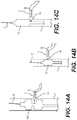
- FIGS. 14 A-C illustrate alternative two-balloon configurations in which FIG. 14A shows a bifurcated first balloon and a second balloon shaped for placement in the OA;
- FIGS. 15A and 15B illustrate alternative designs for a multiple lumen device
- FIGS. 16A and 16B illustrate a site-specific blockage in the OA
- FIGS. 16C and 16D illustrate alternative designs for removing a site-specific blockage in the OA
- FIG. 17 illustrates an embodiment of the present disclosure having a perfusion balloon
- FIG. 18 is an alternative embodiment of a perfusion system in which blood may flow through the catheter
- FIG. 19 illustrates an embodiment of a device in which a portion of the device is configured for the ophthalmic artery and can be mechanically extended or angled to conform to ICA/OA anatomy;
- FIG. 20 illustrates the embodiment of FIG. 19 in a closed position
- FIG. 21 illustrates a three-balloon configuration having an anchor balloon and two ophthalmic artery balloons
- FIG. 22 illustrates a two-lumen configuration in which a balloon lumen is deployed in the ICA and enters the OA from the cranial or superior side;
- FIG. 23 illustrates the delivery of a medical device toward the OA via a portion of the Circle of Willis.
- the present disclosure involves restoring or otherwise improving blood flow in arterial vasculature, including, for example, the OA, by delivering a balloon or expandable element to the OA, wherein said balloon is adapted and configured for placement in the OA, and wherein said balloon is used to restore blood flow in the OA.
- the expandable removal element may be movable between an expanded position and a contracted position and may be utilized in a single or multiple drive shaft configuration. Applicant notes that references throughout the disclosure to the OA are exemplary, and that in some embodiments, the devices, systems, and method described herein may be used to treat other arterial vasculature, such as vessels with small diameters and/or sharp-angled (e.g., tortuous) lumens.
- the present disclosure also includes a medical device suitable for delivery and deployment within the ophthalmic artery.
- the device may be variously configured.
- the device may be a catheter comprising one or more lumens.
- the lumens may be used to house, deliver, and/or retrieve one or more elements suitable for restoring blood flow in the ophthalmic artery, typically by removing a blockage or the like.
- Exemplary elements include, but are not limited to, one or more guidewires, one or more balloons, one or more blockage capture and retrieval elements, one or more inflation elements (e.g., an inflation lumen), one or more filter elements, and/or one or more anchoring elements.
- the system for accessing the ophthalmic artery may comprise a catheter, typically a catheter suitable for use in angioplasty or atherectomy procedures.
- the catheter may also be a guide catheter.
- the medical device may include one or more lumens and elements as described above. In these arrangements, the medical device may not include a guide catheter.
- the present disclosure also describes one or more devices comprising one or more balloons for performing the methods described above.
- the device may comprise a cutter and/or capture element for removing material from the ophthalmic artery.
- Some of these examples include a catheter and/or an aspiration lumen.
- the present disclosure includes devices, methods, and systems for removing, or restoring flow through, blockage or occlusive material in a small diameter artery, such as the ophthalmic artery.
- the method may include the steps of providing a catheter having a proximal end and a distal end; at least one guidewire configured to extend from or pass through a distal tip of the catheter to a desired location; at least one lumen defining a channel within the catheter; and at least one balloon or expandable element configured for passing through a lumen of the catheter and/or comprising a distal portion of a guidewire.
- placing the catheter, guidewires, and balloons in a particular portion of the anatomy typically requires the use of one of a variety of imaging techniques and devices, e.g., fluoroscopy.
- Restoring and/or increasing blood flow is used herein to refer to any device, method, therapy, or combination that changes the blood flow to the eye.
- Examples of such include, but are not limited to increasing the blood flow anywhere in the vasculature leading to the eye or a portion of the eye; removing (e.g., atherectomy) or opening or displacing (e.g., angioplasty) an obstruction in the fluid flow path in the vasculature leading to the eye (e.g., from the ICA through the OA); delivering and deploying a stent in the fluid flow path in the vasculature leading to the eye; using atherectomy or similar devices to physically remove portions of any obstructions in the vasculature leading to the eye or portion of the eye; and localized drug and/or an oxygen device for increasing flow or amount of oxygen in one or more eye tissues.
- a device or method of the present disclosure may be combined with a known or new drug or oxygen device in order to treat one or more eye diseases or conditions.
- the present disclosure may also include restoring and/or increasing the amount of nutrients that are available to one or more parts of the eye or to the eye area, specifically by removing or partially opening a blockage in one or more of the arteries that supplies blood flow to the eye.
- a blockage is removed or opened in the ICA, the OA, the ostium (as used herein, referring to the junction between the ICA and the OA), or combinations thereof.
- nutrients as used herein include, but are not limited to, oxygen, hemoglobin, complement, and glucose.
- the present disclosure may also include methods, devices, and systems for removing or displacing a blockage in the ostium or a proximal segment (e.g., short limb) of the OA near the ICA.
- removing or displacing the blockage comprises opening a channel or access through the ostium sufficient to provide a therapeutically beneficial result to the eye, the rear of the eye, or portions thereof.
- removing a blockage involves atherectomy devices and methods.
- opening or displacing a blockage involves angioplasty devices and methods.
- the present disclosure also includes restoring and/or improving blood flow anywhere in the vascular pathway to or within the eye.
- Therapeutically beneficial result is used herein to refer to any perceived or actual benefit to the patient.
- beneficial results may include but are not limited to: treatment of an eye disease, condition, and/or symptom; restoring or increasing blood flow in any manner that treats an eye disease, condition, and/or symptom; and removing or partially removing a blockage in the blood flow path between the heart and the eye, in the OA or a portion thereof.
- the present disclosure should not be limited solely to changing vascular flow in order to improve or restore the amount of nutrients that are delivered to the eye.
- the vascular flow may be unaffected for the most part, but the amount or concentration of nutrients may be increased, thereby increasing the amount of nutrients that may be delivered to the eye or associated with the eye.
- One skilled in the art may recognize, with the teaching of this disclosure, that there are other biological systems or capabilities that may be used to increase the amount of nutrients that are delivered to the eye.
- reducing or opening a blockage includes, but is not limited to, piercing or penetrating the blockage.
- piercing and penetrating the blockage may refer to obtaining sufficient blood and/or fluid flow through or around a blocked vascular area sufficient to provide a therapeutically beneficial amount of oxygen (or other such nutrient) to the eye or a portion of the eye.
- a method for removing, opening, displacing, or restoring flow through thromboembolic material from a small diameter artery such as the OA.
- Some methods involve using atherectomy devices and related procedures; other methods involve using angioplasty devices and related procedures.
- Still other methods include providing a catheter having a proximal end, a distal end, an expandable distal section having a distal port, an aspiration lumen communicating with the port, and an axially movable support.
- the method may include inserting the distal end of the catheter into an artery of a subject, and distally advancing the support to expand the distal section. Negative pressure is applied to the aspiration port, to draw the thromboembolic material into the distal section.
- an aspiration catheter in accordance with another aspect of the present disclosure, there is provided an aspiration catheter.
- the catheter includes an elongate flexible tubular body, having a proximal end, a distal end, and an aspiration lumen extending therethrough.
- An aspiration lumen in a distal section of the flexible tubular body is movable between a first, reduced inside diameter for transluminal navigation and a second, enlarged inside diameter for aspirating material.
- a catheter of the present disclosure may include one or more elements for physically capturing material and pulling it into the catheter and/or washing it away from the site of the occlusion.
- a microcatheter having an outside diameter of approximately 3-French or smaller, with the incorporation of an outer, diametrically expansile/contractile element near the distal region of the device.
- This expansile/contractile element coupled with the microcatheter system can serve a variety of therapeutic indications within the vasculature supplying blood flow or fluid flow to and from the eye.
- the microcatheter can comprise a distention means for vascular anastomotic regions, flow restoration within an occluded vessel, foreign body retrieval, or an endovascular filter.
- the microcatheter can comprise means to deliver therapeutic devices and diagnostic agents (e.g., TPA) through one or more of the catheter's lumens or side holes, which further adds to this system's utility.
- TPA therapeutic devices and diagnostic agents
- the device's lumen, or lumens, could allow for aspiration and/or drainage.
- reverse flow devices and methods Any of the devices, methods, embodiments, or variations of the present disclosure may be used with reverse flow or retrograde flow devices and methods.
- An exemplary description of reverse flow devices and methods includes, but is not limited to PCT/US17/21673 (filed 9 Mar. 2017) and U.S. Pat. No. 9,259,215, both incorporated by reference herein in their entireties.
- a catheter or medical device of the present disclosure can be a tubular structure with distal and proximal ends and at least one lumen throughout its length or a portion of the catheter.
- the length of the catheter may be determined by its access point into the body, e.g., a transfemoral approach or a cervical approach.
- the length of the catheter for a transfemoral approach can be approximately 150 cm, and can range from about 100 cm to about 200 cm.
- the catheter can have an outer diameter with the diametrically expansile/contractile element contracted of no more than 1 mm (3 F).
- the length of the catheter for a cervical approach can be approximately 20 cm, and can range from about 10 cm to about 30 cm.
- the microcatheter advantageously comprises lateral flexibility, which can be constant or can include a plurality of increasingly flexible regions moving from the proximal to the distal end of the microcatheter.
- the microcatheter includes columnar strength sufficient to facilitate pushability/advanceability through the vasculature.
- the outer diametrically expansile/contractile element hereafter referred to as the expandable element, which can be generally affixed to the catheter shaft near the distal end of the microcatheter shaft, can be fabricated from a variety of metallic or polymeric materials, either porous, non-porous, or a combination of these materials.
- This expandable element can be located proximate the distal end of the micro-catheter. In other embodiments, the expandable element can be located flush against the distal end, or about 1-2 cm from the distal end to improve guidewire-aided navigation through tortuous vasculature.
- the design may be provided with the expandable element having a maximum, expanded outer diameter of about 0.5 mm to about 2.5 mm, or between about 1 mm and about 2 mm.
- the catheter can be used to perform blockage retrieval, removal, displacement, or opening.
- the device may be first prepared by flushing or priming the lumen with saline.
- the catheter is then navigated to the site of the blockage.
- the catheter is advanced along a guidewire so that the expandable element is positioned within or through the blockage.
- the expandable element is then expanded, engaging the blockage.
- the user can administer thrombolytic agents to further entwine or entrap the blockage.
- the blockage may then be removed from the vasculature via the catheter. Additionally, the user may elect to keep the expandable element expanded, and remove the catheter device from the vasculature. Lastly, the blockage removal could be aided by aspiration through the catheter side holes.
- the microcatheter can be used for the purposes of anastomosis distension or dilation, vascular foreign body retrieval, temporary dilatation and flow restoration through atheromatous plaque, and vascular embolic filtering. These goals can be addressed by inserting the proper therapeutic device, such as a dilatation balloon, grasper or basket device, high force mesh dilator, or distal protection filter, respectively, through the working lumen of the microcatheter.
- the proper therapeutic device such as a dilatation balloon, grasper or basket device, high force mesh dilator, or distal protection filter, respectively.
- the catheter and/or guidewire and/or balloon may be delivered or positioned at the proximal end of the OA by accessing the OA from a cranial or superior position.
- the device may be passed through the Circle of Willis or a portion thereof, or one or more cranial arteries, and approach the OA from a cranial or superior direction in the ICA.
- the devices and methods described herein may include one or more elongation members, such as a balloon or inflatable member.
- elongation members may be variously configured and may include multiple variations or alternatives. All of these configurations, variations, and alternatives are adapted and configured for delivery and/or placement in the OA, in the junction (e.g., ostium) between the ICA and the OA, and in the OA before the first bend (e.g., angle a) or turn (e.g., within the short limb of the OA).
- An elongation member (as described further herein) or balloon of the present disclosure may be constructed of conventional materials; may be variously shaped (e.g., tiered, dog-bone shaped, oval, elliptical, or round); may comprise a drug or chemical (e.g., may be drug-eluting) having therapeutic benefit or purpose; and/or may be compliant, semi-compliant, or non-compliant throughout the balloon or a portion thereof.
- a catheter/system 10 comprises a plurality of lumens including a first lumen 17 for housing, delivering, and deploying a balloon 14 , a second lumen 11 for housing, delivering, and deploying a guidewire 12 , and a third lumen 15 for delivering inflation fluid to the balloon 14 (e.g., via a distal end of balloon 14 ).
- the catheter also includes an elongation member 13 having balloon 14 coupled thereto. Elongation member 13 may extend to a proximal end of the catheter for manipulation (e.g., proximal retraction and/or distal advancement) by a medical professional. As shown in FIG.
- the catheter/system 10 is adapted and configured for delivery and placement in the junction J between the ICA I, and the OA O. That is, as shown, at least one or more of guidewire 12 and balloon 14 may be routed through junction (e.g., ostium) J and into a short limb S of OA O. Additionally, at least a portion of guidewire 12 may be advanced past angle A of OA O and into a long limb LL of OA O.
- FIGS. 1C and 1D illustrate additional views of the catheter of FIGS. 1A and 1B , having guidewire 12 removed therefrom.
- FIG. 1C also shows a place of fluid communication 18 between inflation lumen 15 and the distal end of the balloon 14 .
- a side port in balloon 14 at its distal end may communicate with a side port in inflation lumen 15 at a place of fluid communication 18 .
- the inflation lumen 15 or inflated balloon 14 may sandwich or anchor the guidewire lumen 11 against the ostium O; in other embodiments the balloon 14 or inflation lumen 15 may sandwich or anchor the guidewire lumen 11 against the ostium O.
- the configuration of the balloon 14 when inflated, sandwiches the guidewire 12 , balloon 14 , and/or lumen 11 in place, e.g., in the junction J between the ICA I and the OA O.
- inflation of balloon 14 may result in sandwiching, pushing, or otherwise urging guidewire 12 into contact with a wall of the OA O.
- the balloon 14 has sufficient compliance and/or flexibility to conform to the typical anatomy in this location. That is, the balloon 14 has sufficient compliance and/or flexibility so as to fill the short limb S of the OA O, thereby urging the guidewire 12 into contact with the wall of the OA O.
- the anatomy between the ICA I and the OA O includes the junction/ostium J, the short limb S, and angle A.
- the long limb is shown as LL.
- the balloon may be short, and does not watermelon seed out of place (e.g., slide away from its intended position in the junction between the ICA I and the OA O).
- inflation of balloon 14 may include performing an angioplasty on the OA and, since the OA expands with the inflated balloon 14 , the OA is not dissected.
- the elongation member 13 is preferably flexible (e.g., bendable), allowing the balloon 14 to move within the anatomy. Also, when the elongation member 13 is proximally retracted, a proximal end of the balloon 14 may be pulled in the proximal direction back into the catheter while a distal end of the balloon 14 is fixed relative to the guidewire lumen 11 in any appropriate manner (e.g., via adhesive, crimping, etc.), thereby preventing the balloon from buckling within the anatomy. That is, proximal retraction of elongation member 13 by a medical professional may likewise proximally retract a proximal end of balloon 14 relative to the distal end of balloon 14 which is fixed to inflation lumen 15 .
- proximal retraction of elongation member 13 by a medical professional may likewise proximally retract a proximal end of balloon 14 relative to the distal end of balloon 14 which is fixed to inflation lumen 15 .
- balloon 14 is stretched or elongated due to the relative movement between elongation member 13 and inflation lumen 15 .
- the elongation member 13 may be made from an elastic polymer, spring, corrugated tube, nitinol, a telescoping tube or tubes, or a fixed wire. Balloon 14 may be extruded over the elongation member 13 , thereby coupling the balloon 14 to the elongation member 13 .
- elongation member 13 includes a stretchable polymer.
- elongate member 13 may include a rigid cable or wire.
- the elongation member 13 may extend distally of, or may stop near, a proximal waist of balloon 14 .
- the balloon 14 can be inflated from the distal end or from the proximal end of the balloon 14 .
- FIGS. 2 and 3 illustrate embodiments of the present disclosure in which the elongation member 13 is attached to a two- or three-lumen shaft, respectively.
- an attachment e.g., the elongation member 13
- elongation element 13 may be arranged as an elastic band/tube having one end coupled to balloon 14 and an opposite end coupled to the catheter/system 10 (e.g., coupled to the inflation lumen 15 ).
- elongation member 13 may stretch.
- balloon 14 may be inflated through a distal end of balloon 14 , as shown in FIG. 2 . In other arrangements, however, balloon 14 may be inflated through a proximal end of balloon 14 , as shown in FIG. 3 . In such an arrangement, inflation lumen 15 may communicate with elongation member 13 which may be in communication with a proximal end of balloon 14 . In this example, elongation member 13 may be tubular so as to convey inflation fluid therethrough.
- FIG. 4 illustrates a configuration in which a balloon 14 A is offset or asymmetrical.
- balloon 14 A is inflated through the distal end of balloon 14 A via inflation lumen 15 .
- a distal waist 19 of balloon 14 A may be fluidly coupled with inflation lumen 15 , as will be described in further detail below.
- FIG. 5 illustrates a configuration in which the inflation lumen 15 is flexible (e.g., axially moveable relative to) at least one end of the balloon 14 .
- the lumen 15 includes a flexible element 20 (e.g., a spring, coil, etc.) attached to the shaft of the lumen 15 on both the proximal end and the distal end of the balloon 14 .
- a first (e.g., proximal) end of each flexible element 20 may be attached via any appropriate manner (e.g., adhesives, etc.) to inflation lumen 15 while a distal end of each flexible element 20 may be attached via any appropriate manner (e.g., adhesives, etc.) to balloon 14 .
- each flexible element 20 may permit or enable relative movement between inflation lumen 15 and balloon 14 .
- each flexible element 20 may include a spring or coil that may extend and retract longitudinally. This embodiment shows a configuration for sandwiching or anchoring the guidewire 12 against a wall of an artery.
- Arrangements of the present disclosure also may include a tip element 16 ( FIG. 5 ) attached to the distal end of the balloon 14 (or the distal flexible element 20 ), and includes a lumen to further position the guidewire 12 .
- Tip element 16 may be attached via any appropriate manner to one or more of a distal end of balloon 14 , flexible element 20 , and inflation lumen 15 .
- tip element 16 may include a passage or lumen through which guidewire 12 may be passed to direct advancement of balloon 14 relative to guidewire 12 or vice versa, or to maintain radial spacing between guidewire 12 and balloon 14 .
- the guidewire lumen 11 includes a guidewire 12 that extends through a balloon 14 .
- guidewire 12 may act in the same manner as elongation member 13 , described above. That is, in the arrangement of FIGS. 6 and 7 , guidewire 12 may be used to extend or retract balloon 14 relative to inflation lumen 15 . As such, guidewire 12 may be fixed relative to balloon 14 in any appropriate manner. Additionally, inflation lumen 15 is coupled to the distal end of the balloon 14 . In such an arrangement, similar to that described above in FIG.
- a channel, lumen, or communication 18 may be provided between the distal end of balloon 14 and inflation lumen 15 .
- the balloon 14 and/or guidewire 12 allows the balloon 14 to conform to the anatomy, and the balloon 14 may be inflated from the distal end of the balloon 14 .
- balloon 14 naturally wants to straighten.
- the proximal end of balloon 14 is connected to flexible guidewire 12 and/or elongation member 13 , the balloon is permitted to follow the trajectory of the OA (or ostium) rather than straighten out. That is, since the proximal end of balloon 14 is permitted to move relative to a distal end of balloon 14 , balloon 14 may conform to the anatomy of the OA and/or ostium.
- the guidewire 12 may be braided or coiled in a manner suitable for making the sharp turn or acute angle into the OA from the ICA.
- the catheter comprises a plurality of lumens including a first lumen 11 for housing a conventional guidewire 12 and a second lumen 16 for housing a second guidewire 30 and a ball or balloon 14 B, as shown in FIG. 8 .
- the arrangement of FIG. 8 would include an inflation lumen 15 in fluid communication with balloon 14 A.
- Guidewire lumen 11 may extend the length of the catheter and through balloon 14 A, the second lumen 16 may terminate at the first balloon 14 A and extend through an opening in balloon 14 A.
- second lumen 16 may include a bend or turn that alters a direction of second lumen 16 so as to direct second guidewire 30 and balloon 14 B in a direction angled with respect to guidewire lumen 11 .
- balloon 14 A may be deployed in the ICA while second guidewire 30 and balloon 14 B may extend through second lumen 16 to be positioned at the junction of the OA with the ICA.
- the second balloon 14 B is shaped (or pre-shaped) and configured to fit into the OA ostium (e.g., junction J).
- the balloon 14 B may then be inflated to dilate the OA, to provide a stable base, and/or to prevent or reduce particle generation.
- the side branch (e.g., second branch) guidewire 30 and balloon 14 B may directly access the OA (e.g., for accessing and dilating the ostium junction J and/or short limb S).
- This embodiment also may include dual or multiple inflation lumens for the balloon(s).
- the configuration shown in FIG. 8 also may include a perfusion feature or element, thereby allowing blood to flow through, past, or by the portion of the device in the ICA, as will be described in further detail below.
- the perfusion feature may be variously configured, including but not limited to a separate channel or lumen through the device; a through lumen on the side of balloon 14 A; or grooves in the deployed balloon 14 B.
- These embodiments of the present disclosure may further include a guidewire/balloon diverter configured to position the OA guidewire/balloon 14 B in the OA.
- an example of such a structure may be a side channel or pathway at or near the distal end of the balloon/lumen 14 A positioned in the ICA.
- Other examples include, but are not limited to one or more channels in a central position of the balloon/lumen 14 A; or near the proximal end of the balloon/lumen 14 A.
- the catheter, balloon, and or guidewire may be pre-shaped (e.g., including a bent distal end angled to facilitate entry into the OA) to facilitate entry into the OA, as shown in FIG. 9A .
- the balloon 14 may be shaped to match or conform to the ostium O.
- the OA may have a funnel or cone-shaped cross-sectional arrangement. That is, a diameter of the OA may vary along the length of the OA (e.g., may narrow such that a portion of the OA closer to the ostium may have a cross-sectional dimension larger than a portion of the OA farther away from the ostium).
- balloon 14 may be likewise tapered such that the shape of the balloon 14 is complimentary to the shape of the OA and/or ostium.
- the catheter positions the balloon 14 and guidewire 12 into the OA.
- the guidewire 12 is removed or withdrawn, and in FIG. 9D , the balloon is inflated in the ostium.
- the balloon 14 and guiding catheter 10 may be removed or withdrawn.
- the balloon 14 may be designed to slip out of the anatomy or to be held in a static position during dilatation. For example, under pressure, the balloon 14 may displace from the OA short limb to the ICA (e.g., “watermelon seeding”).
- the balloon 14 and/or inflation lumen 15 may be pre-formed and shaped to conform to the anatomy at the junction J of the OA and the ICA, wherein the guiding or delivery catheter 10 straightens the balloon 14 and/or inflation lumen 15 during delivery and, when the catheter is withdrawn or the balloon 14 is pushed out of the catheter, its release thereby allows the balloon 14 and/or inflation lumen 15 to assume its pre-formed (angled) shape. That is, when positioned radially within guiding catheter 10 , inflation lumen 15 and balloon 14 generally may extend along an axis extending through guiding catheter 10 .
- inflation lumen 15 and balloon 14 may deflect away from such an axis so as to form an acute bend arranged to facilitate entry into the OA.
- one or both of inflation lumen 15 and balloon 14 may include a shape memory material (e.g., Nitinol) to facilitate bending or deflecting thereof.
- the catheter may include a dilatation balloon 14 A and an anchor balloon 14 B, positioned on one (e.g., the same) or more (e.g., different) guidewires.
- inflation lumen 15 may include an extruded polymer tube or the like over a guidewire (e.g., a “balloon on a wire”).
- the dilatation balloon 14 A is shaped to conform to the OA (e.g., is sized such that upon expansion, dilatation balloon 14 A contacts a wall of the OA), more specifically, the ostium junction J.
- FIG. 10A-10C An illustration of this embodiment is shown in which inflation lumen 15 may include an extruded polymer tube or the like over a guidewire (e.g., a “balloon on a wire”).
- the dilatation balloon 14 A is shaped to conform to the OA (e.g., is sized such that upon expansion, dilatation balloon 14 A contacts a wall of the OA), more specifically, the ostium
- FIG. 10A shows an embodiment in which a balloon catheter is inside a guiding catheter 10 , thereby anchoring balloon 14 B to guide catheter 10 rather than a wall of the ICA.
- the balloon and/or the guidewire may include a bias in one direction to conform the element(s) to the anatomy of the ICA and OA junction J.
- the balloon and optionally the guidewire 12 are biased toward a deflected configuration.
- FIG. 11A-11C An example is shown in FIG. 11A-11C in which, as opposed to the arrangement of FIGS. 10A-10C where balloon 14 was coupled to inflation lumen, balloon 14 of FIGS. 11A-11C is coupled directly to guidewire 12 .
- FIG. 11A shows an uninflated balloon 14 on a guidewire 12 designed to conform or fit into the ICA/OA ostium.
- the bias in one direction allows the balloon 14 to conform to the angle of the junction J between the ICA and the OA ( FIGS.
- the bias deflects one or more portions of balloon 14 and/or guidewire 12 at or along an angle (e.g., an acute angle) so as to enter the ostium junction J and/or OA O.
- the arrangement may be biased in any appropriate manner.
- guidewire 12 may be pre-shaped to include a curve or bend.
- balloon 14 may be fabricated such that it is shorter on one side than the other, thus resulting in a biased or deflected arrangement upon inflation.
- the bias is intended to prevent or reduce vessel straightening. That is, the short arm of the OA typically connects with the ICA at an angle. In particular, the connection may be curved.
- Biasing the balloon and/or guidewire may enable the balloon 14 and/or guidewire 12 to fit within this angled and curved structure without forcing the short limb of the OA to adjust the angle at which it extends from the ICA.
- the bias may be in the guidewire alone, and may not include the balloon.
- Embodiments having a biased guidewire and/or balloon may further include a delivery or guiding catheter, or may not.
- the balloon is designed to fit or conform to the anatomy of the OA and/or the ostium by varying the point(s) of attachment to the catheter or guidewire.
- This fit or conformance in the ostium is intended to keep the distal portion of the balloon within the short arm of the OA such that when inflated, vessel straightening does not occur beyond the distal portion of the short limb of the OA.
- the balloon 14 is attached to the guidewire at both the distal and proximal ends of the balloon 14 , as shown in FIG. 12A .
- the balloon 14 may be attached at the proximal end, but the distal attachment point may be closer to the center of the balloon 14 so as to cause portions of balloon 14 to overlap and/or invert on itself, thereby providing a discrete distal edge of the balloon 14 which substantially matches the distal anatomy of the short limb of the OA. As such, this distal edge of the balloon 14 will not extend past the distal most portion of the short limb.
- the distal attachment point may be varied along the body of the catheter or guidewire during device fabrication to provide adjustment of overall device flexibility. Two examples are shown in FIGS. 12B and 12C , in which the second attachment point is more proximal along the length of the balloon 14 than the distal end of balloon 14 .
- the distal attachment between the balloon and the guidewire may be spaced proximally from a distal end of the balloon 14 .
- the distal attachment point allows a distal end of the balloon 14 to fold when the balloon is deflated so that the tip of the guidewire is more flexible.
- such attachment points may be designed to impart vessel straightening of the OA.
- FIG. 12 D shows a balloon 14 with an attachment point (e.g., a heat joint, adhesives, etc.) to a guidewire at the proximal end and a second attachment point at any point proximal of the distal end of balloon 14 .
- the balloon 14 may fold of conform to the anatomy of the OA and/or the ostium.
- a distal end of guidewire 12 may be flush with the distal end of the balloon.
- a distal end of the guidewire 12 may extend distally of a distal end of the balloon.
- FIGS. 12A-12D may be an over-the-wire configuration or a fixed wire configuration.
- FIGS. 13 A-C show a guidewire 12 and distal balloon 14 configuration wherein the uninflated OA balloon 14 may be inserted into proximal end of the OA, e.g., at the ostium junction J.
- the compliant or semi-compliant balloon 14 may be placed in the OA, then inflated or partially inflated which forces the balloon out of the OA and into the ICA.
- a system comprises a first guidewire 12 configured for delivery and placement in the ICA adjacent the OA.
- this guidewire may include a first balloon, or in the illustrated embodiment, may include a bifurcated balloon 14 B.
- the bifurcated balloon 14 B may be configured so a first portion is adapted for placement in the ICA distal to the OA and a second portion is adapted for placement at the junction of the OA or just proximal of the junction of the OA.
- Bifurcated balloon 14 B may have a narrowed or thinned portion in communication with the distal portion and the proximal portion which may enable additional flexibility to bifurcated balloon 14 B
- the system further comprises a second guidewire 30 suitable for placement and entry into the OA O.
- the second guidewire 30 comprises a second balloon 14 C configured and adapted for placement in the proximal end of the OA.
- a single (e.g., non-bifurcated) balloon 14 may be used for the ICA balloon.
- balloon 14 may include zones or sections of different compliancy.
- balloon 14 may be compliant near the waist portion (e.g., FIG. 14A ).
- a balloon in the ICA or any structure not needing dilation
- a balloon (or balloon portion) 14 C in the OA may be semi-compliant or non-compliant, and may or may not be inflated to a pre-determined and/or set pressure or size.
- a first balloon 14 is not bifurcated, and is adapted for placement near but not over (e.g., distal of or downstream of) the entrance (e.g., the junction J or ostium) to the OA O, and the second guidewire 12 and second balloon 14 C stem from the system or a second lumen of the system orthogonally or angled with respect to the first guidewire and first balloon.
- the entrance e.g., the junction J or ostium
- the first balloon 14 , 14 B helps stabilize the second balloon 14 C when the second balloon 14 C is inflated, keeping the second balloon 14 C from watermelon seeding out of (or otherwise slipping out of) the OA upon inflation.
- the balloons can be over-the-wire, rapid exchange, or fixed wire configurations.
- the system may include an inflation lumen 15 and a separate guidewire lumen 11 .
- FIGS. 15A and 15B Two examples of such configurations are illustrated in FIGS. 15A and 15B .
- FIG. 15A illustrates an inflation lumen coupled to a proximal end of a balloon
- FIG. 15B illustrates an inflation lumen extending through the balloon and coupled to a distal end thereof. Due to the inclusion of inflation lumen 15 extending through the balloon, the balloon of FIG. 15B may have a larger outer profile/dimension than the balloon of FIG. 15A
- the balloon may be intentionally underinflated.
- a 2 mm diameter balloon e.g., a balloon having a maximum inflated diameter of 2 mm
- the balloon is selected based on the desired dilatation outcome, e.g., larger than the diameter of the target anatomy.
- the balloon when inflated, intentionally slides out of the target anatomy (e.g., the OA).
- a larger than required balloon may not be supported circumferentially in the ICA, and as such, the balloon may be free to watermelon seed out of the OA and into the ICA.
- the balloon 14 may be partially inserted into the target anatomy (e.g., the OA). In some embodiments, only the distal 1 ⁇ 3 of the balloon 14 is inserted into the area of dilatation.
- the balloon 14 is then inflated at the lower end of its inflation capacity (e.g., about 6 to 8 ATMS). As the balloon 14 is inflated, the larger diametric area of the balloon (larger as compared to the ID of the target anatomy) will force the balloon 14 to slide out of the target anatomy and into the adjacent anatomy, e.g., slide from the ostium or OA and into the ICA in a retrograde manner or otherwise in a direction opposite that of antegrade blood flow.
- the intentional use of the watermelon seed affect has a therapeutic effect: it opens or clears the ostium of any blockage or material covering the ostium.
- FIGS. 13 A-C The method used in this Example is illustrated in FIGS. 13 A-C.
- a 2 mm diameter semi-compliant balloon (6 mm length) is inserted into the ostium of the OA, an area of the anatomy that is typically about 1 mm or less in diameter.
- FIG. 13B when the balloon is inflated to less than its nominal inflation pressure, the balloon is forced out of the OA into the ICA in a “watermelon seed” phenomenon. That is, if the balloon is inflated to a pressure less than that of the nominal pressure, the balloon may begin to exit the anatomy at low pressures.
- FIG. 13C the balloon completely exits the OA and enters the ICA, but in accordance with this method, partial dilatation of the ostium occurs, clearing any blockage in the ostium and the OA.
- the catheter body/system it may be desirable to coat the outside surface of the guidewire 12 , the catheter body/system, and/or the balloon 14 , 14 A, 14 B, 14 C, and/or the inside surface of the wall defining a lumen of the catheter.
- these coatings are lubricious to minimize friction.
- a variety of coatings may be used, including but not limited to Parylene, Teflon, silicone rubber, polyimide-polytetrafluoroethylene composite materials, as well as other coatings known in the art.
- the coating may be tacky, sticky, or include bumps or appendages specifically designed to hold or retain one or more elements in place or to otherwise inhibit movement of the one or more elements relative to the coating.
- a system may include one or more lumens and one or more guidewires.
- the guidewire lumen may be sandwiched against the wall of an artery.
- the guidewire may be sandwiched against the wall of an artery.
- devices, systems, and methods may be used to address a site-specific blockage B in the OA, specifically in the ostium and/or the short limb S.
- a site-specific blockage B in the OA specifically in the ostium and/or the short limb S.
- further arrangements according to the present disclosure may be restricted for use in a particular anatomical location.
- FIGS. 16A and 16B A typical location of such blockages is shown in FIGS. 16A and 16B .
- FIG. 16C illustrates an embodiment in which a cutting catheter includes a cutter window and a cutting element adapted for communicating with the debris or blockage.
- Exemplary cutters include but are not limited to a pullback cutter, a push cutter, and a rotational cutter.
- a balloon 14 B is shown in an exemplary position distal to the cutting window and the blockage B, and is configured and adapted for placement in the ostium or short limb S of the OA O.
- FIG. 16D An alternative configuration is shown in FIG. 16D , in which a balloon 14 C is positioned adjacent the cutting element, and when inflated, the balloon anchors the cutter and pushes the cutter into the debris or blockage B.
- the balloon(s) 14 B, 14 C block any debris from moving distally into the OA O, and aspiration may be used to remove the debris and/or occlusive material.
- the side of the OA O in this location of the OA O may be used to place the cutter window within the blockage B or occlusive material.
- the balloon 14 B, 14 C may be used to anchor the cutter, and it can also be used to push the cutter into the occlusive material or blockage B. For example, inflation of the balloon 14 B, 14 C may deflect or otherwise urge the cutter window towards the blockage B.
- FIG. 17 illustrates an embodiment in which the system includes a perfusion feature, thereby allowing blood to flow past the ICA portion of the device (e.g., past balloon 14 A inflated in ICA I).
- blood flow is maintained (as illustrated by arrows 2 ) during the ICA balloon 14 A inflation.
- any structure or configuration that allows blood to pass by the ICA portion of the system may be used, including but not limited to, a blood flow channel or lumen; grooves or channels in the ICA balloon 14 A, one or more bypass channels or structures, and combinations thereof.
- balloon 14 A may be delivered over a first guidewire 12 arranged within inflation lumen 15 B, while balloon 14 B may be delivered over a second guidewire 12 arranged within inflation lumen 15 A.
- Balloon 14 A may have any appropriate shape including a channel to allow blood to perfuse therethrough.
- This embodiment also shows two inflation lumens 15 A and 15 B.
- inflation lumen 15 B anchors or positions inflation lumen 15 A in place, thereby anchoring the OA dilatation balloon 14 B in the OA O.
- Allowing blood to flow past the ICA portion of the device makes this embodiment particularly well suited for use with reverse or retrograde flow systems.
- the perfusion lumen may be arranged to deliver blood in a retrograde or reverse direction (e.g., in the direction opposite the arrows 2 ).
- FIG. 18 shows an alternative configuration that allows blood to flow past a portion of the device in the ICA (e.g., past balloon 14 A).
- FIG. 18 illustrates an embodiment in which a catheter 10 includes at least one lumen 15 C or channel within the catheter for allowing blood to pass through the ICA I (e.g., a perfusion lumen within which a guidewire 12 may extend).
- the illustrated embodiment also shows balloon 14 A anchoring or positioning the lumen 15 C in place. That is, inflation of balloon 14 A in catheter 10 may deflect, urge, or otherwise push lumen 15 C into engagement with a wall of catheter 10 to prevent movement of lumen 15 C relative to catheter 10 , which may prevent the balloon from watermelon seeding out the OA and into the ICA.
- FIGS. 19 and 20 show an embodiment in which an OA portion of the device is configured sized, and/or shaped in a manner complimentary to the specific anatomy in the junction J between the ICA and the OA.
- the OA portion of the device is hinged 18 , articulatable, or movable in relation to an ICA portion of the device.
- the hinge 18 (or an articulation lumen) also anchors or supports the OA portion of the device in place in the OA.
- the illustrated configuration may include an open and/or close mechanism or element for releasing the OA portion of the device ( FIG. 19 ) or closing the OA portion of the device ( FIG. 20 ).
- a wire 19 , loop, spring, or the like may be arranged about the OA portion device.
- the device may include a lumen 15 D though which wire 19 having a wire loop at a distal end thereof extends.
- the wire loop of wire 15 may surround the OA portion of the device and upon extension or loosening of the loop or wire 19 , the OA portion of the device may deflect radially outwards away from the ICA portion of the device, as shown in FIG. 19 .
- the OA portion of the device may deflect radially inwards toward the ICA portion of the device, as shown in FIG. 20 .
- the device is adapted for delivery and removal from the ICA and OA.
- the device may be positioned in the ICA in its closed position ( FIG. 20 ) above, superior, distal, or downstream to the OA.
- the OA portion may be released, thereby facilitating entry into the OA ( FIG. 19 ).
- the device may be withdrawn from its position in the ICA by pulling the OA portion flush against the ICA portion of the device.
- FIG. 21 shows an embodiment of the present disclosure comprising a balloon 14 C configured and adapted for deployment or positioning in the long limb LL of the OA.
- the device may include at least one balloon, and in the illustrated embodiment, three balloons.
- balloon 14 B anchors, stabilizes, or supports balloon 14 C in place in the LL of the OA
- balloon 14 A anchors, stabilizes, or supports balloon 14 B in place in the short limb S or ostium junction J of the OA.
- balloon 14 B is preferably compliant, non-compliant, or semi-compliant; balloon 14 C is preferably compliant.
- FIG. 22 shows an alternative configuration of a catheter having an OA lumen configured for deployment in the OA and adapted for the acute angle between the ICA and the OA.
- the OA lumen and balloon may be deployed in the OA by first wrapping the OA portion around the ICA portion of the catheter and angling the OA portion down into the OA.
- This figure also illustrates another embodiment of the present disclosure, in which a lumen may be large enough to guide both the guidewire and the balloon.
- guide catheter 51 is configured for positioning in the ICA.
- balloon 14 B and guidewire lumen 15 A extend from a portion (e.g., a distal end) of the guide catheter 51 , and loops around (e.g., behind) the guide catheter 51 and enters the OA from the superior or cranial side.
- balloon 14 B and guidewire lumen 15 A are sized to position both a guidewire lumen and a balloon lumen, and configured to enter the OA.
- balloon and guidewire lumens may be positioned in a tube or the like, which can in turn be heat shrunk to the desired size around the lumens.
- FIG. 22 illustrates an arrangement in which balloon 14 B includes a pair of attachment points for coupling balloon 14 B to lumen 15 A. As shown, the distal attachment point of the balloon 14 B is provided proximally of the distal end of the balloon 14 B.
- the various embodiments of the present disclosure provide a number of constructions of expandable vascular material removal and/or opening devices, intravascular material removal and/or opening elements, and the like, which can be utilized to perform a plurality of different intravascular treatments, such as atherectomy, thrombectomy, angioplasty and the like.
- the embodiments of the present disclosure also provide a plurality of methods for using those devices and their associated vascular material removal or opening elements for performing intravascular treatments on a patient. It is to be fully recognized that the different teachings of the embodiments, examples, and arrangements disclosed herein can be employed separately or in any suitable combination to produce desired results.
- the embodiments provide, in the form of expandable intravascular removal or opening elements, ways of changing cutting or removing profiles, configurations or characteristics of a particular intravascular treatment device while only using a single removal or opening element.
- An inner hollow tube or sheath is located between an inner, proximal end of the housing.
- the inner sheath defines a lumen of dimensions sufficient for accepting a medical guidewire, made of stainless steel, nitinol, and the like, which can extend from the guidewire lumen within the inner sheath, and through an aperture in the proximal end of the housing to the exterior of the housing.
- the hollow shaft also defines a guidewire lumen, thereby allowing for passage of the guidewire from the material removal element to the exterior of the housing.
- the removal device 10 is of an over-the-wire construction which can facilitate removing the device from, and replacing the device in the patient because the guidewire can remain within the patient.
- some prior art devices require removal of the guidewire along with the device, thereby necessitating additional intravascular navigation not only of the device, but also of the guidewire to replace the device adjacent the occlusion material to be removed.
- the presence of the guidewire facilitates intravascular navigation of the removal device, because the device can be delivered over the guidewire, which is an improvement over some expandable intravascular devices.
- an expandable intravascular removal or opening element is highly desirable for the reasons discussed earlier, it may be desirable to limit the maximum size of these intravascular elements. It may be desirable not to over-expand the expandable elements. While some means for positively limiting radial expansion of the expandable intravascular element have been detailed hereinabove, it may be desirable to provide additional safety mechanisms.
- the removal or opening device may include a manifold assembly and a catheter assembly.
- the manifold assembly includes a third port located distally of the port.
- the port may be connectable with a suitable source of fluid, not shown, but known in the art, for supplying the catheter assembly with fluid to dilate a dilating member for performing balloon angioplasty.
- the port may be located distally of a proximal end of the catheter assembly.
- a dilating member constructed substantially similarly to an angioplasty balloon, is located on the catheter assembly offset proximally of a distal end of the catheter assembly and the distal end of the drive shaft lumen.
- the inflation lumen extends from the port to a proximal end of the dilating member and conveys fluid from the fluid source, conventionally referred to as an inflation device, to and from the dilating member, thereby causing the dilating member to inflate and deflate.
- a radiopaque marker band is provided on the outer surface of the drive shaft lumen, thereby rendering the intravascular portion of the dilating member radioscopically visible to a treating physician.
- Intravascular inflation of the dilating member provides added stability to the distal portion of the removal device during operation thereof, while also allowing the treating physician to occlude blood flow through the vascular lumen being treated and further allowing the physician to perform balloon angioplasty if desired.
- the removal device it is possible for a treating physician to cut, remove, and/or angioplastically displace vascular occlusion material while only using a single piece of equipment.
- a PTCA balloon was placed into the OA via the ICA in-situ, and inflated in the segment of the OA (e.g., the short limb S) proximal to the first sharp bend (e.g., angle A).
- a PTCA balloon was placed and inflated in the target area ex-vivo, in the OA ostium. This experiment was repeated two times.
- the balloon was successfully placed in the target area and dilated, without causing any apparent damage to the OA or the ostium.
- the balloons used in the first experiment had a maximum inflated diameter of 0.8 mm at 16 ATMs.
- the working length was 3.5 mm.
- the balloon used in the second experiment had a maximum inflated diameter of 2.0 mm at 8 ATMs.
- the working length was 6.0 mm.
- the in-situ experiment (e.g., first experiment) demonstrates that the balloon is capable of navigating the ICA anatomy, can be positioned in the short limb of the OA (before the first sharp bend in the OA), and inflated without causing any apparent damage to any of the vessels.
- the ex vivo experiment (e.g., the second experiment) demonstrates that the balloon can be visualized in the correct location in the proximal end of the OA and can be oversized and inflated without causing damage to the OA or ostium.
- accessing the OA or other anatomical locations may be difficult.
- a medical device e.g., a catheter, guidewire, etc.
- the medical device may be required to traverse tortuous anatomy requiring a number of sharp angled bends or direction changes.
- the medical device may be required to bend at an acute angle.
- An alternative approach may be to access the OA from a cranial, upper, or superior position. That is, approaching the OA from a superior position may require advancing the medical device along a less tortuous path.
- a medical device may more easily be introduced to the OA via the superior approach, which may result in reduced procedure time, reduced procedure cost, and reduced risk of damaging or rupturing an arterial structure or other tissues.
- one or more devices may be delivered to, or introduced within, an identified anatomical location via the Circle of Willis, for example, by first traversing all or part of the Circle of Willis and approaching a target artery from a superior position.
- a medical device such as a catheter or guidewire 500 may be tracked (e.g., guided, steered, and/or passed) through either the left or right ICA, around a portion of the Circle of Willis (e.g., through the anterior communicating artery) into the other of the left or right ICA, and towards the OA.
- the guidewire wire 500 may approach the OA from an upper or superior direction so as to better align with the trajectory of the OA and reduce a tortuosity of the path through with the medical device may be passed.
- guidewire 500 may be advanced through a vertebral artery, enter a posterior communicating artery of the Circle of Willis, through the right ICA, around a portion of the Circle of Willis superior to the ICA (e.g., the anterior communicating artery), into the left ICA, and into a target OA from a superior direction.
- the target OA may include a blockage (e.g., a complete or partial blockage) therein to be treated via any of the methods and devices described in this disclosure.
- guidewire 500 may be tracked from an inferior direction (e.g., guided, steered, and/or passed) through a target ICA (e.g., the left ICA), around a portion of the Circle of Willis (e.g., via communicating arteries), and back into the target ICA (e.g., the left ICA) so as to approach the OA from an upper or superior direction.
- a target ICA e.g., the left ICA
- Circle of Willis e.g., via communicating arteries
- target ICA e.g., the left ICA
- a percutaneous interventional device may be advanced into the Circle of Willis from an inferior position and directed toward or into one or more arteries of the Circle of Willis.
- the one or more arteries may include the anterior cerebral arteries, the internal carotid arteries, branches of the internal carotid arteries, the vertebral arteries, the anterior communicating arteries, the posterior cerebral arteries, the basilar arteries, branches of the basilar arteries, and the posterior communicating arteries.
- the percutaneous interventional device may be directed into the ICA and into a target OA from a superior position.
Landscapes
- Health & Medical Sciences (AREA)
- Surgery (AREA)
- Life Sciences & Earth Sciences (AREA)
- Public Health (AREA)
- Animal Behavior & Ethology (AREA)
- Engineering & Computer Science (AREA)
- Biomedical Technology (AREA)
- Heart & Thoracic Surgery (AREA)
- Veterinary Medicine (AREA)
- Vascular Medicine (AREA)
- Nuclear Medicine, Radiotherapy & Molecular Imaging (AREA)
- General Health & Medical Sciences (AREA)
- Molecular Biology (AREA)
- Medical Informatics (AREA)
- Ophthalmology & Optometry (AREA)
- Orthopedic Medicine & Surgery (AREA)
- Surgical Instruments (AREA)
- Reproductive Health (AREA)
Abstract
Description
Claims (20)
Priority Applications (2)
| Application Number | Priority Date | Filing Date | Title |
|---|---|---|---|
| US15/993,359 US10898212B2 (en) | 2017-05-07 | 2018-05-30 | Devices and methods for treating an artery |
| US16/571,848 US10772645B2 (en) | 2017-05-07 | 2019-09-16 | Devices and methods for treating an artery |
Applications Claiming Priority (5)
| Application Number | Priority Date | Filing Date | Title |
|---|---|---|---|
| US201762502733P | 2017-05-07 | 2017-05-07 | |
| US201762513383P | 2017-05-31 | 2017-05-31 | |
| US201762523724P | 2017-06-22 | 2017-06-22 | |
| PCT/US2018/031229 WO2018208622A1 (en) | 2017-05-07 | 2018-05-04 | Devices and methods for treating an artery |
| US15/993,359 US10898212B2 (en) | 2017-05-07 | 2018-05-30 | Devices and methods for treating an artery |
Related Parent Applications (1)
| Application Number | Title | Priority Date | Filing Date |
|---|---|---|---|
| PCT/US2018/031229 Continuation-In-Part WO2018208622A1 (en) | 2017-05-07 | 2018-05-04 | Devices and methods for treating an artery |
Related Child Applications (1)
| Application Number | Title | Priority Date | Filing Date |
|---|---|---|---|
| US16/571,848 Continuation US10772645B2 (en) | 2017-05-07 | 2019-09-16 | Devices and methods for treating an artery |
Publications (2)
| Publication Number | Publication Date |
|---|---|
| US20180317944A1 US20180317944A1 (en) | 2018-11-08 |
| US10898212B2 true US10898212B2 (en) | 2021-01-26 |
Family
ID=64013869
Family Applications (2)
| Application Number | Title | Priority Date | Filing Date |
|---|---|---|---|
| US15/993,359 Expired - Fee Related US10898212B2 (en) | 2017-05-07 | 2018-05-30 | Devices and methods for treating an artery |
| US16/571,848 Active US10772645B2 (en) | 2017-05-07 | 2019-09-16 | Devices and methods for treating an artery |
Family Applications After (1)
| Application Number | Title | Priority Date | Filing Date |
|---|---|---|---|
| US16/571,848 Active US10772645B2 (en) | 2017-05-07 | 2019-09-16 | Devices and methods for treating an artery |
Country Status (1)
| Country | Link |
|---|---|
| US (2) | US10898212B2 (en) |
Families Citing this family (5)
| Publication number | Priority date | Publication date | Assignee | Title |
|---|---|---|---|---|
| US10799688B2 (en) * | 2018-12-31 | 2020-10-13 | J.D. Franco & Co., Llc | Intravascular devices, systems, and methods to address eye disorders |
| ES2777881A1 (en) * | 2020-02-25 | 2020-08-06 | Weyezer Solutions Sl | VITRECTOMY PROBE (Machine-translation by Google Translate, not legally binding) |
| CN111449722B (en) * | 2020-04-08 | 2021-03-30 | 上海心玮医疗科技股份有限公司 | Intravascular delivery system |
| CN116196532B (en) * | 2023-03-10 | 2025-07-15 | 普利瑞医疗科技(苏州)有限公司 | Bifurcated balloon catheter assembly and preparation method thereof |
| WO2025064605A1 (en) * | 2023-09-20 | 2025-03-27 | J.D. Franco & Co., Llc | Ophthalmic artery angioplasty balloon and guidewire based intra vascular lithotripsy (ivl) for treatment of ophthalmic diseases |
Citations (86)
| Publication number | Priority date | Publication date | Assignee | Title |
|---|---|---|---|---|
| US2690595A (en) | 1951-06-22 | 1954-10-05 | Davol Rubber Co | Manufacture of low-pressure inflation catheters |
| US3367101A (en) | 1959-05-22 | 1968-02-06 | Spunize Co Of America Inc | Crimped roving or sliver |
| US3435826A (en) | 1964-05-27 | 1969-04-01 | Edwards Lab Inc | Embolectomy catheter |
| US4403612A (en) | 1980-10-20 | 1983-09-13 | Fogarty Thomas J | Dilatation method |
| US4795433A (en) * | 1985-05-20 | 1989-01-03 | Survival Technology, Inc. | Automatic injector for emergency treatment |
| US4926858A (en) | 1984-05-30 | 1990-05-22 | Devices For Vascular Intervention, Inc. | Atherectomy device for severe occlusions |
| US5026384A (en) | 1989-11-07 | 1991-06-25 | Interventional Technologies, Inc. | Atherectomy systems and methods |
| US5176693A (en) | 1992-05-11 | 1993-01-05 | Interventional Technologies, Inc. | Balloon expandable atherectomy cutter |
| US5395311A (en) | 1990-05-14 | 1995-03-07 | Andrews; Winston A. | Atherectomy catheter |
| US5415634A (en) | 1990-08-23 | 1995-05-16 | Devices For Vascular Intervention, Inc. | Catheter having helical inflation lumen |
| US5709701A (en) | 1996-05-30 | 1998-01-20 | Parodi; Juan C. | Apparatus for implanting a prothesis within a body passageway |
| US5820595A (en) | 1995-06-07 | 1998-10-13 | Parodi; Juan C. | Adjustable inflatable catheter and method for adjusting the relative position of multiple inflatable portions of a catheter within a body passageway |
| WO1998052639A1 (en) | 1997-05-19 | 1998-11-26 | United States Surgical Corporation | Catheter system |
| WO1998053761A1 (en) | 1997-05-26 | 1998-12-03 | William A. Cook Australia Pty. Ltd. | A prosthesis and a method and means of deploying a prosthesis |
| US5897567A (en) | 1993-04-29 | 1999-04-27 | Scimed Life Systems, Inc. | Expandable intravascular occlusion material removal devices and methods of use |
| WO2000054673A1 (en) | 1999-03-16 | 2000-09-21 | Percusurge, Inc. | Apparatus for containing and removing occlusions in the carotid arteries |
| US6146370A (en) | 1999-04-07 | 2000-11-14 | Coaxia, Inc. | Devices and methods for preventing distal embolization from the internal carotid artery using flow reversal by partial occlusion of the external carotid artery |
| US6206868B1 (en) | 1998-03-13 | 2001-03-27 | Arteria Medical Science, Inc. | Protective device and method against embolization during treatment of carotid artery disease |
| US20010001114A1 (en) | 1996-05-14 | 2001-05-10 | Embol-X, Inc. | Cardioplegia balloon cannula |
| US6302908B1 (en) | 1997-03-24 | 2001-10-16 | Juan Carlos Parodi | Arterial graft device and method of positioning the same |
| US6336933B1 (en) | 1998-03-13 | 2002-01-08 | Juan C. Parodi | Endovascular device for application of prosthesis with sutures |
| US6344054B1 (en) | 1996-09-20 | 2002-02-05 | Juan Carlos Parodi | Endoluminal prosthesis comprising stent and overlying graft cover, and system and method for deployment thereof |
| US6413235B1 (en) | 1998-03-13 | 2002-07-02 | Arteria Medical Science, Inc. | Protective device against embolization in carotid angioplasty |
| US20020087128A1 (en) | 2000-08-31 | 2002-07-04 | Mpnb Ltd. | Microsurgical injection and/or distending instruments and surgical method and apparatus utilizing same |
| US6423032B2 (en) | 1998-03-13 | 2002-07-23 | Arteria Medical Science, Inc. | Apparatus and methods for reducing embolization during treatment of carotid artery disease |
| US20020151922A1 (en) | 1998-05-13 | 2002-10-17 | Michael Hogendijk | Apparatus and methods for removing emboli during a surgical procedure |
| US20030023200A1 (en) | 1999-03-01 | 2003-01-30 | Coaxia, Inc. | Partial aortic occlusion devices and methods for cerebral perfusion augmentation |
| WO2003018085A2 (en) | 2001-08-22 | 2003-03-06 | Arteria Medical Science, Inc. | Apparatus and methods for treating stroke and controlling cerebral flow characteristics |
| US6540712B1 (en) | 1998-03-13 | 2003-04-01 | Arteria Medical Science, Inc. | Methods and low profile apparatus for reducing embolization during treatment of carotid artery disease |
| US20030199802A1 (en) | 1999-01-15 | 2003-10-23 | Denise Barbut | Methods for flow augmentation in patients with occlusive cerebrovascular disease |
| US20030199819A1 (en) | 2002-04-17 | 2003-10-23 | Beck Robert C. | Filter wire system |
| US20030203958A1 (en) | 1997-03-31 | 2003-10-30 | Neorx Corporation | Therapeutic inhibitor of vascular smooth muscle cells |
| US6641573B1 (en) | 1999-03-25 | 2003-11-04 | Arteria Medical Science, Inc. | Device and method of guide wire balloon inflation and deflation to prevent cerebral embolization during carotid stenting |
| US6645222B1 (en) | 1998-05-13 | 2003-11-11 | Arteria Medical Science, Inc. | Puncture resistant branch artery occlusion device and methods of use |
| US6824558B2 (en) | 1998-08-25 | 2004-11-30 | Boston Scientific Corporation | Endoluminal device and method for treating branched lumen having a restricted section |
| US6827726B2 (en) | 2001-01-19 | 2004-12-07 | Boston Scientific Corporation | Introducer for deployment of branched prosthesis |
| US6837881B1 (en) | 2001-02-23 | 2005-01-04 | Coaxia, Inc. | Devices and methods for preventing distal embolization using flow reversal by partial occlusion of the brachiocephalic artery |
| US6902540B2 (en) | 2001-08-22 | 2005-06-07 | Gerald Dorros | Apparatus and methods for treating stroke and controlling cerebral flow characteristics |
| US6908474B2 (en) | 1998-05-13 | 2005-06-21 | Gore Enterprise Holdings, Inc. | Apparatus and methods for reducing embolization during treatment of carotid artery disease |
| US20050149117A1 (en) | 2003-12-24 | 2005-07-07 | Farhad Khosravi | Apparatus and methods for delivering sealing materials during a percutaneous procedure to facilitate hemostasis |
| US6929634B2 (en) | 2001-08-22 | 2005-08-16 | Gore Enterprise Holdings, Inc. | Apparatus and methods for treating stroke and controlling cerebral flow characteristics |
| US6936053B1 (en) | 1998-07-02 | 2005-08-30 | Jeffrey N. Weiss | Ocular implant needle |
| US20060089618A1 (en) | 2004-10-21 | 2006-04-27 | Mcferran Sean | Catheter with a pre-shaped distal tip |
| US20060136022A1 (en) | 2004-11-23 | 2006-06-22 | Wong Edward K Jr | Medical device and method for temperature control and treatment of the eye and surrounding tissues |
| US20060259132A1 (en) | 2005-05-02 | 2006-11-16 | Cook Incorporated | Vascular stent for embolic protection |
| US7214201B2 (en) | 1991-06-18 | 2007-05-08 | Boston Scientific Scimed, Inc. | Intravascular guide wire and method for manufacture thereof |
| US7235095B2 (en) | 2002-02-22 | 2007-06-26 | Scimed Life Systems, Inc. | Method and system for deploying multi-part endoluminal devices |
| WO2007103464A2 (en) | 2006-03-08 | 2007-09-13 | Orqis Medical Corporation | Blood conduit connector |
| US7309334B2 (en) | 2002-07-23 | 2007-12-18 | Von Hoffmann Gerard | Intracranial aspiration catheter |
| US20080027519A1 (en) | 2006-07-28 | 2008-01-31 | Guerrero John M | Method of treatment of ocular compartment syndromes |
| US20080243229A1 (en) | 1996-09-18 | 2008-10-02 | Micro Therapeutics, Inc. | Intracranial Stent and Method of Use |
| US20090018455A1 (en) | 2003-11-21 | 2009-01-15 | Silk Road Medical, Inc. | Method and apparatus for treating a carotid artery |
| US20090024072A1 (en) | 2007-07-18 | 2009-01-22 | Enrique Criado | Methods and systems for establishing retrograde carotid arterial blood flow |
| US20090030323A1 (en) | 2007-04-02 | 2009-01-29 | Doheny Eye Institute | Ultrasound and Microbubbles in Ocular Diagnostics and Therapies |
| US7604612B2 (en) | 2001-05-01 | 2009-10-20 | St. Jude Medical, Cardiology Division, Inc. | Emboli protection devices and related methods of use |
| US20100125244A1 (en) | 2008-11-14 | 2010-05-20 | Medtronic Vascular, Inc. | Balloon catheter for crossing a chronic total occlusion |
| US7867273B2 (en) | 2007-06-27 | 2011-01-11 | Abbott Laboratories | Endoprostheses for peripheral arteries and other body vessels |
| US20110143993A1 (en) | 2009-12-15 | 2011-06-16 | The Brigham And Women's Hospital, Inc. | Endothelial basement membrane targeting peptide ligands |
| US20110152683A1 (en) | 2011-03-01 | 2011-06-23 | Gerrans Lawrence J | Abrading Balloon Catheter for Extravasated Drug Delivery |
| US20120046679A1 (en) | 2010-07-01 | 2012-02-23 | Patel Himanshu N | Atherectomy catheters with longitudinally displaceable drive shafts |
| US20120078287A1 (en) | 2001-05-01 | 2012-03-29 | Barbut Denise R | Devices and methods for preventing distal embolization using flow reversal and perfusion augmentation within the cerebral vasculature |
| US20120101510A1 (en) | 2008-12-23 | 2012-04-26 | Reverse Medical Corporation | Systems And Methods For Removing Obstructive Matter From Body Lumens And Treating Vascular Defects |
| US8353850B2 (en) | 2005-07-07 | 2013-01-15 | St. Jude Medical, Cardiology Division, Inc. | Steerable guide wire with torsionally stable tip |
| US20130197621A1 (en) | 2010-08-12 | 2013-08-01 | Silk Road Medical, Inc. | Systems and methods for treating a carotid artery |
| US8545432B2 (en) | 2009-06-03 | 2013-10-01 | Silk Road Medical, Inc. | System and methods for controlling retrograde carotid arterial blood flow |
| US20130281788A1 (en) | 2011-08-05 | 2013-10-24 | Silk Road Medical, Inc. | Methods and systems for treatment of acute ischemic stroke |
| WO2014022866A1 (en) | 2012-08-03 | 2014-02-06 | J.D. Franco & Company | Devices and methods for treating occlusion of the ophthalmic artery |
| US20140154246A1 (en) | 2004-09-24 | 2014-06-05 | Rfe Pharma Llc | Cai-based systems and methods for the localized treatment of ocular and other diseases |
| US20140243809A1 (en) * | 2013-02-22 | 2014-08-28 | Mark Gelfand | Endovascular catheters for trans-superficial temporal artery transmural carotid body modulation |
| US8834404B2 (en) | 2009-03-30 | 2014-09-16 | Steve Andre Beaudin | Apparatus, system and methods for extracorporeal blood processing for selectively cooling the brain relative to the body during hyperthermic treatment or to induce hypothermia of the brain |
| US8852226B2 (en) | 1997-11-07 | 2014-10-07 | Salviac Limited | Vascular device for use during an interventional procedure |
| US8863631B1 (en) | 2013-07-29 | 2014-10-21 | Insera Therapeutics, Inc. | Methods of manufacturing flow diverting devices |
| US20150025562A1 (en) * | 2013-07-16 | 2015-01-22 | Reverse Medical Corporation | Microcatheter with Modified PTFE Liner |
| US20150313607A1 (en) | 2014-05-04 | 2015-11-05 | Zhadkevich Medical, Inc. | Noninvasive protection from emboli |
| US20150359547A1 (en) | 2014-06-13 | 2015-12-17 | Neuravi Limited | Devices and methods for removal of acute blockages from blood vessels |
| US20150366580A1 (en) | 2014-06-19 | 2015-12-24 | Limflow Gmbh | Devices and methods for treating lower extremity vasculature |
| US9241699B1 (en) | 2014-09-04 | 2016-01-26 | Silk Road Medical, Inc. | Methods and devices for transcarotid access |
| US9259215B2 (en) | 2007-07-18 | 2016-02-16 | Silk Road Medical, Inc. | Systems and methods for treating a carotid artery |
| US9265512B2 (en) | 2013-12-23 | 2016-02-23 | Silk Road Medical, Inc. | Transcarotid neurovascular catheter |
| US20160166754A1 (en) | 2013-07-31 | 2016-06-16 | Hyo Won Choi | Unitary body systems and devices and methods to use the same for retroperfusion |
| WO2016109586A1 (en) | 2014-12-29 | 2016-07-07 | Ocudyne, Llc | Apparatus and method for treating eye diseases |
| US20160317328A1 (en) | 2009-12-21 | 2016-11-03 | Covidien Lp | Procedures for vascular occlusion |
| US20170239453A1 (en) | 2016-02-18 | 2017-08-24 | Terumo Kabushiki Kaisha | Drug treatment method and delivery device |
| WO2017156333A1 (en) | 2016-03-09 | 2017-09-14 | J.D. Franco & Co. | Systems and methods for treating eye diseases using retrograde blood flow |
| WO2018053121A1 (en) | 2016-09-15 | 2018-03-22 | J.D. Franco & Company | Systems and methods for treating an eye using retrograde blood flow |
| WO2018106858A1 (en) | 2016-12-08 | 2018-06-14 | J.D. Franco & Company | Methods and devices for treating an eye using a filter |
-
2018
- 2018-05-30 US US15/993,359 patent/US10898212B2/en not_active Expired - Fee Related
-
2019
- 2019-09-16 US US16/571,848 patent/US10772645B2/en active Active
Patent Citations (98)
| Publication number | Priority date | Publication date | Assignee | Title |
|---|---|---|---|---|
| US2690595A (en) | 1951-06-22 | 1954-10-05 | Davol Rubber Co | Manufacture of low-pressure inflation catheters |
| US3367101A (en) | 1959-05-22 | 1968-02-06 | Spunize Co Of America Inc | Crimped roving or sliver |
| US3435826A (en) | 1964-05-27 | 1969-04-01 | Edwards Lab Inc | Embolectomy catheter |
| US4403612A (en) | 1980-10-20 | 1983-09-13 | Fogarty Thomas J | Dilatation method |
| US4926858A (en) | 1984-05-30 | 1990-05-22 | Devices For Vascular Intervention, Inc. | Atherectomy device for severe occlusions |
| US4795433A (en) * | 1985-05-20 | 1989-01-03 | Survival Technology, Inc. | Automatic injector for emergency treatment |
| US5026384A (en) | 1989-11-07 | 1991-06-25 | Interventional Technologies, Inc. | Atherectomy systems and methods |
| US5395311A (en) | 1990-05-14 | 1995-03-07 | Andrews; Winston A. | Atherectomy catheter |
| US5415634A (en) | 1990-08-23 | 1995-05-16 | Devices For Vascular Intervention, Inc. | Catheter having helical inflation lumen |
| US7214201B2 (en) | 1991-06-18 | 2007-05-08 | Boston Scientific Scimed, Inc. | Intravascular guide wire and method for manufacture thereof |
| US5176693A (en) | 1992-05-11 | 1993-01-05 | Interventional Technologies, Inc. | Balloon expandable atherectomy cutter |
| US5897567A (en) | 1993-04-29 | 1999-04-27 | Scimed Life Systems, Inc. | Expandable intravascular occlusion material removal devices and methods of use |
| US5820595A (en) | 1995-06-07 | 1998-10-13 | Parodi; Juan C. | Adjustable inflatable catheter and method for adjusting the relative position of multiple inflatable portions of a catheter within a body passageway |
| US20010001114A1 (en) | 1996-05-14 | 2001-05-10 | Embol-X, Inc. | Cardioplegia balloon cannula |
| US5709701A (en) | 1996-05-30 | 1998-01-20 | Parodi; Juan C. | Apparatus for implanting a prothesis within a body passageway |
| US7901445B2 (en) | 1996-09-18 | 2011-03-08 | Micro Therapeutics, Inc. | Intracranial stent and method of use |
| US20080243229A1 (en) | 1996-09-18 | 2008-10-02 | Micro Therapeutics, Inc. | Intracranial Stent and Method of Use |
| US6344054B1 (en) | 1996-09-20 | 2002-02-05 | Juan Carlos Parodi | Endoluminal prosthesis comprising stent and overlying graft cover, and system and method for deployment thereof |
| US6855162B2 (en) | 1997-03-24 | 2005-02-15 | Scimed Life Systems, Inc. | Arterial graft device |
| US6302908B1 (en) | 1997-03-24 | 2001-10-16 | Juan Carlos Parodi | Arterial graft device and method of positioning the same |
| US20030203958A1 (en) | 1997-03-31 | 2003-10-30 | Neorx Corporation | Therapeutic inhibitor of vascular smooth muscle cells |
| WO1998052639A1 (en) | 1997-05-19 | 1998-11-26 | United States Surgical Corporation | Catheter system |
| WO1998053761A1 (en) | 1997-05-26 | 1998-12-03 | William A. Cook Australia Pty. Ltd. | A prosthesis and a method and means of deploying a prosthesis |
| US8852226B2 (en) | 1997-11-07 | 2014-10-07 | Salviac Limited | Vascular device for use during an interventional procedure |
| US6336933B1 (en) | 1998-03-13 | 2002-01-08 | Juan C. Parodi | Endovascular device for application of prosthesis with sutures |
| US6905490B2 (en) | 1998-03-13 | 2005-06-14 | Gore Enterprise Holdings, Inc. | Apparatus and methods for reducing embolization during treatment of carotid artery disease |
| US6423032B2 (en) | 1998-03-13 | 2002-07-23 | Arteria Medical Science, Inc. | Apparatus and methods for reducing embolization during treatment of carotid artery disease |
| US6413235B1 (en) | 1998-03-13 | 2002-07-02 | Arteria Medical Science, Inc. | Protective device against embolization in carotid angioplasty |
| US6206868B1 (en) | 1998-03-13 | 2001-03-27 | Arteria Medical Science, Inc. | Protective device and method against embolization during treatment of carotid artery disease |
| US6540712B1 (en) | 1998-03-13 | 2003-04-01 | Arteria Medical Science, Inc. | Methods and low profile apparatus for reducing embolization during treatment of carotid artery disease |
| US20110160762A1 (en) | 1998-05-13 | 2011-06-30 | Michael Hogendijk | Apparatus and methods for reducing embolization during treatment of carotid artery disease |
| US20020151922A1 (en) | 1998-05-13 | 2002-10-17 | Michael Hogendijk | Apparatus and methods for removing emboli during a surgical procedure |
| US6936060B2 (en) | 1998-05-13 | 2005-08-30 | Arteria Medical Sciences, Inc. | Apparatus and methods for removing emboli during a surgical procedure |
| US7927347B2 (en) | 1998-05-13 | 2011-04-19 | Gore Enterprise Holdings, Inc. | Apparatus and methods for reducing embolization during treatment of carotid artery disease |
| US6908474B2 (en) | 1998-05-13 | 2005-06-21 | Gore Enterprise Holdings, Inc. | Apparatus and methods for reducing embolization during treatment of carotid artery disease |
| US6645222B1 (en) | 1998-05-13 | 2003-11-11 | Arteria Medical Science, Inc. | Puncture resistant branch artery occlusion device and methods of use |
| US6936053B1 (en) | 1998-07-02 | 2005-08-30 | Jeffrey N. Weiss | Ocular implant needle |
| US6824558B2 (en) | 1998-08-25 | 2004-11-30 | Boston Scientific Corporation | Endoluminal device and method for treating branched lumen having a restricted section |
| US20030199802A1 (en) | 1999-01-15 | 2003-10-23 | Denise Barbut | Methods for flow augmentation in patients with occlusive cerebrovascular disease |
| US20030023200A1 (en) | 1999-03-01 | 2003-01-30 | Coaxia, Inc. | Partial aortic occlusion devices and methods for cerebral perfusion augmentation |
| WO2000054673A1 (en) | 1999-03-16 | 2000-09-21 | Percusurge, Inc. | Apparatus for containing and removing occlusions in the carotid arteries |
| US20030023227A1 (en) | 1999-03-16 | 2003-01-30 | Gholam-Reza Zadno-Azizi | Method for containing and removing occlusions in the carotid arteries |
| US6641573B1 (en) | 1999-03-25 | 2003-11-04 | Arteria Medical Science, Inc. | Device and method of guide wire balloon inflation and deflation to prevent cerebral embolization during carotid stenting |
| US6146370A (en) | 1999-04-07 | 2000-11-14 | Coaxia, Inc. | Devices and methods for preventing distal embolization from the internal carotid artery using flow reversal by partial occlusion of the external carotid artery |
| US6623471B1 (en) | 1999-04-07 | 2003-09-23 | Coaxia, Inc. | Devices and methods for preventing distal embolization from the internal carotid artery using flow reversal by partial occlusion of the external carotid artery |
| US20020087128A1 (en) | 2000-08-31 | 2002-07-04 | Mpnb Ltd. | Microsurgical injection and/or distending instruments and surgical method and apparatus utilizing same |
| US6827726B2 (en) | 2001-01-19 | 2004-12-07 | Boston Scientific Corporation | Introducer for deployment of branched prosthesis |
| US6837881B1 (en) | 2001-02-23 | 2005-01-04 | Coaxia, Inc. | Devices and methods for preventing distal embolization using flow reversal by partial occlusion of the brachiocephalic artery |
| US20120078287A1 (en) | 2001-05-01 | 2012-03-29 | Barbut Denise R | Devices and methods for preventing distal embolization using flow reversal and perfusion augmentation within the cerebral vasculature |
| US7604612B2 (en) | 2001-05-01 | 2009-10-20 | St. Jude Medical, Cardiology Division, Inc. | Emboli protection devices and related methods of use |
| WO2003018085A2 (en) | 2001-08-22 | 2003-03-06 | Arteria Medical Science, Inc. | Apparatus and methods for treating stroke and controlling cerebral flow characteristics |
| US6929634B2 (en) | 2001-08-22 | 2005-08-16 | Gore Enterprise Holdings, Inc. | Apparatus and methods for treating stroke and controlling cerebral flow characteristics |
| US6902540B2 (en) | 2001-08-22 | 2005-06-07 | Gerald Dorros | Apparatus and methods for treating stroke and controlling cerebral flow characteristics |
| US7235095B2 (en) | 2002-02-22 | 2007-06-26 | Scimed Life Systems, Inc. | Method and system for deploying multi-part endoluminal devices |
| US20030199819A1 (en) | 2002-04-17 | 2003-10-23 | Beck Robert C. | Filter wire system |
| US7309334B2 (en) | 2002-07-23 | 2007-12-18 | Von Hoffmann Gerard | Intracranial aspiration catheter |
| US20090018455A1 (en) | 2003-11-21 | 2009-01-15 | Silk Road Medical, Inc. | Method and apparatus for treating a carotid artery |
| US20050149117A1 (en) | 2003-12-24 | 2005-07-07 | Farhad Khosravi | Apparatus and methods for delivering sealing materials during a percutaneous procedure to facilitate hemostasis |
| US20140154246A1 (en) | 2004-09-24 | 2014-06-05 | Rfe Pharma Llc | Cai-based systems and methods for the localized treatment of ocular and other diseases |
| US20060089618A1 (en) | 2004-10-21 | 2006-04-27 | Mcferran Sean | Catheter with a pre-shaped distal tip |
| US20060136022A1 (en) | 2004-11-23 | 2006-06-22 | Wong Edward K Jr | Medical device and method for temperature control and treatment of the eye and surrounding tissues |
| US20060259132A1 (en) | 2005-05-02 | 2006-11-16 | Cook Incorporated | Vascular stent for embolic protection |
| US8353850B2 (en) | 2005-07-07 | 2013-01-15 | St. Jude Medical, Cardiology Division, Inc. | Steerable guide wire with torsionally stable tip |
| WO2007103464A2 (en) | 2006-03-08 | 2007-09-13 | Orqis Medical Corporation | Blood conduit connector |
| US20080027519A1 (en) | 2006-07-28 | 2008-01-31 | Guerrero John M | Method of treatment of ocular compartment syndromes |
| US20090030323A1 (en) | 2007-04-02 | 2009-01-29 | Doheny Eye Institute | Ultrasound and Microbubbles in Ocular Diagnostics and Therapies |
| US7867273B2 (en) | 2007-06-27 | 2011-01-11 | Abbott Laboratories | Endoprostheses for peripheral arteries and other body vessels |
| US9259215B2 (en) | 2007-07-18 | 2016-02-16 | Silk Road Medical, Inc. | Systems and methods for treating a carotid artery |
| US8157760B2 (en) | 2007-07-18 | 2012-04-17 | Silk Road Medical, Inc. | Methods and systems for establishing retrograde carotid arterial blood flow |
| US20090024072A1 (en) | 2007-07-18 | 2009-01-22 | Enrique Criado | Methods and systems for establishing retrograde carotid arterial blood flow |
| US20100125244A1 (en) | 2008-11-14 | 2010-05-20 | Medtronic Vascular, Inc. | Balloon catheter for crossing a chronic total occlusion |
| US20120101510A1 (en) | 2008-12-23 | 2012-04-26 | Reverse Medical Corporation | Systems And Methods For Removing Obstructive Matter From Body Lumens And Treating Vascular Defects |
| US9078682B2 (en) | 2008-12-23 | 2015-07-14 | Covidien Lp | Systems and methods for removing obstructive matter from body lumens and treating vascular defects |
| US8834404B2 (en) | 2009-03-30 | 2014-09-16 | Steve Andre Beaudin | Apparatus, system and methods for extracorporeal blood processing for selectively cooling the brain relative to the body during hyperthermic treatment or to induce hypothermia of the brain |
| US8545432B2 (en) | 2009-06-03 | 2013-10-01 | Silk Road Medical, Inc. | System and methods for controlling retrograde carotid arterial blood flow |
| US20110143993A1 (en) | 2009-12-15 | 2011-06-16 | The Brigham And Women's Hospital, Inc. | Endothelial basement membrane targeting peptide ligands |
| US20160317328A1 (en) | 2009-12-21 | 2016-11-03 | Covidien Lp | Procedures for vascular occlusion |
| US20120046679A1 (en) | 2010-07-01 | 2012-02-23 | Patel Himanshu N | Atherectomy catheters with longitudinally displaceable drive shafts |
| US20130197621A1 (en) | 2010-08-12 | 2013-08-01 | Silk Road Medical, Inc. | Systems and methods for treating a carotid artery |
| US20110152683A1 (en) | 2011-03-01 | 2011-06-23 | Gerrans Lawrence J | Abrading Balloon Catheter for Extravasated Drug Delivery |
| US20130281788A1 (en) | 2011-08-05 | 2013-10-24 | Silk Road Medical, Inc. | Methods and systems for treatment of acute ischemic stroke |
| US20170326001A1 (en) | 2012-08-03 | 2017-11-16 | J.D. Franco & Company | Devices and methods for treating occlusion of the ophthalmic artery |
| WO2014022866A1 (en) | 2012-08-03 | 2014-02-06 | J.D. Franco & Company | Devices and methods for treating occlusion of the ophthalmic artery |
| US20140243809A1 (en) * | 2013-02-22 | 2014-08-28 | Mark Gelfand | Endovascular catheters for trans-superficial temporal artery transmural carotid body modulation |
| US20150025562A1 (en) * | 2013-07-16 | 2015-01-22 | Reverse Medical Corporation | Microcatheter with Modified PTFE Liner |
| US20150032121A1 (en) | 2013-07-29 | 2015-01-29 | Insera Therapeutics Inc. | Aspiration catheters |
| US8863631B1 (en) | 2013-07-29 | 2014-10-21 | Insera Therapeutics, Inc. | Methods of manufacturing flow diverting devices |
| US20160166754A1 (en) | 2013-07-31 | 2016-06-16 | Hyo Won Choi | Unitary body systems and devices and methods to use the same for retroperfusion |
| US9265512B2 (en) | 2013-12-23 | 2016-02-23 | Silk Road Medical, Inc. | Transcarotid neurovascular catheter |
| US20150313607A1 (en) | 2014-05-04 | 2015-11-05 | Zhadkevich Medical, Inc. | Noninvasive protection from emboli |
| US20150359547A1 (en) | 2014-06-13 | 2015-12-17 | Neuravi Limited | Devices and methods for removal of acute blockages from blood vessels |
| US20150366580A1 (en) | 2014-06-19 | 2015-12-24 | Limflow Gmbh | Devices and methods for treating lower extremity vasculature |
| US9241699B1 (en) | 2014-09-04 | 2016-01-26 | Silk Road Medical, Inc. | Methods and devices for transcarotid access |
| WO2016109586A1 (en) | 2014-12-29 | 2016-07-07 | Ocudyne, Llc | Apparatus and method for treating eye diseases |
| US20170239453A1 (en) | 2016-02-18 | 2017-08-24 | Terumo Kabushiki Kaisha | Drug treatment method and delivery device |
| WO2017156333A1 (en) | 2016-03-09 | 2017-09-14 | J.D. Franco & Co. | Systems and methods for treating eye diseases using retrograde blood flow |
| WO2018053121A1 (en) | 2016-09-15 | 2018-03-22 | J.D. Franco & Company | Systems and methods for treating an eye using retrograde blood flow |
| WO2018106858A1 (en) | 2016-12-08 | 2018-06-14 | J.D. Franco & Company | Methods and devices for treating an eye using a filter |
Non-Patent Citations (23)
| Title |
|---|
| Altinbas, N.K. et al, "Effect of Carotid Artery Stenting on Ophthalmic Artery Flow Patterns," Journal of Ultrasound Medicine, 2014; 33: pp. 629-638. |
| Ambarki, K. et al., "Blood Flow of Ophthalmic Artery in Healthy Individuals Determined by Phase-Contrast Magnetic Resonance Imaging," Investigative Ophthalmology & Visual Science, 2013; 54: pp. 2738-2745. |
| Examination Report No. 2 for AU Application No. 2013296195, dated Jun. 27, 2017 (6 pages). |
| Hayreh, S.S., "The Ophthalmic Artery III. Branches," British Journal of Ophthalmology, 1962, 46, pp. 212-247. |
| Hwang, G. et al., "Reversal of Ischemic Retinopathy Following Balloon Angioplasty of a Stenotic Ophthalmic Artery." Journal of Neuro-Ophthalmology 30.3, 2010, pp. 228-230. |
| International Search Report and Written Opinion for International Application No. PCT/US2017/0051551, dated Dec. 15, 2017 (14 pages). |
| International Search Report and Written Opinion for International Application No. PCT/US2017/0052901, dated Dec. 8, 2017 (9 pages). |
| International Search Report and Written Opinion for International Application No. PCT/US2018/031229, dated Jul. 27, 2018 (19 pages). |
| Kane, A.G. et al., "Reduced Caliber of the Internal Carotid Artery: A Normal Finding with Ipsilateral Absence or Hypoplasia of the A1 Segment," American Journal of Neuroradiology, 1996; 17: pp. 1295-1301. |
| Kawa, M.P. et al., "Complement System in Pathogenesis of AMD: Dual Player in Degeneration and Protection of Retinal Tissue," Hindawi Publishing Corporation, Journal of Immunology Research, vol. 2014, Article ID 483960, 12 pages. |
| Klein, R. et al., "Vasodilators, Blood Pressure-Lowering Medications, and Age-Related Macular Degeneration," American Academy of Ophthalmology, 2014, vol. 121, Issue 8, pp. 1604-1611. |
| Kooragayala, K. et al., "Quanitification of Oxygen Consumption in Retina Ex Vivo Demonstrates Limited Reserve Capacity of Photoreceptor Mitochondria," Investigative Ophthalmology & Visual Science, 2015; 56: pp. 8428-8436. |
| Krejza, J. et al., "Carotid Artery Diameter in Men and Women and the Relation to Body and Neck Size," Stroke, 2006; 3 pages. |
| Lanzino, G. et al., "Treatment of Carotid Artery Stenosis: Medical Therapy, Surgery, or Stenting?," Mayo Clinic Proceedings, Apr. 2009; 84(4), pp. 362-368. |
| Loh, K. et al., "Prevention and management of vision loss relating to facial filler injections." Singapore Medical Journal, 2016; 57(8): 438-443. |
| Michalinos, A. et al., "Anatomy of the Ophthalmic Artery: A Review concerning Its Modern Surgical and Clinical Applications," Hindawi Publishing Corporation, Anatomy Research International, vol. 2015, Article ID 591961, 8 pages. |
| Notice of Allowance for KR 20157005602, dated Sep. 25, 2017 (3 pages). |
| Paques, M. et al., "Superselective ophthalmic artery fibrinolytic therapy for the treatment of central retinal vein occlusion." British Journal of Ophthalmology, 2000, 85: 1387-1391. |
| Tan, P.L. et al., "AMD and the alternative complement pathway: genetics and functional implications," Human Genomics, 2016, 10:23, 13 pages. |
| Xu, H. et al., "Targeting the complement system for the management of retinal inflammatory and degenerative diseases," European Journal of Pharmacology, 2016, 787, pp. 94-104. |
| Yamane, T. et al., "The technique of ophthalmic arterial infusion therapy for patients with intraocular retinoblastoma," International Journal of Clinical Oncology, Apr. 2004; vol. 9, Issue 2, pp. 69-73. |
| Zeumer, H. et al., "Local intra-arterial fibrinolytic therapy in patients with stroke: urokinase versus recombinant tissue plagminogen activator (r-TPA)," Neuroradiology, 1993; 35: pp. 159-162. |
| Zipfel, P.F., et al., "The Role of Complement in AMD," Inflammation and Retinal Disease: Complement Biology and Pathology, Advances in Experimental Medicine and Biology, 2010, 703, pp. 9-24. |
Also Published As
| Publication number | Publication date |
|---|---|
| US20180317944A1 (en) | 2018-11-08 |
| US10772645B2 (en) | 2020-09-15 |
| US20200022713A1 (en) | 2020-01-23 |
Similar Documents
| Publication | Publication Date | Title |
|---|---|---|
| US10772645B2 (en) | Devices and methods for treating an artery | |
| US12343036B2 (en) | Methods and systems for treatment of acute ischemic stroke | |
| US20250204941A1 (en) | Methods and systems for treatment of acute ischemic stroke | |
| JP6438495B2 (en) | Retraction and suction device and related systems and methods for treating embolism | |
| US20250064456A1 (en) | Flow Rate Control Device for Variable Artery Occlusion | |
| US11690985B2 (en) | Systems and methods for single puncture percutaneous reverse blood flow | |
| CN105979996B (en) | Apparatus and method for imaging and treating blood vessels | |
| US10195077B2 (en) | Systems and methods for treating eye diseases using retrograde blood flow | |
| US20150313732A1 (en) | Method and Device for Treating a Target Site in a Vascular Body Channel | |
| US20110040319A1 (en) | Method for treating a target site in a vascular body channel | |
| US20040006306A1 (en) | Apparatus and method for removing occlusive material within blood vessels | |
| US12318327B2 (en) | Systems and methods for treating eye diseases | |
| WO2014188300A1 (en) | Introducer sheath device with expandable end | |
| EP3310424A1 (en) | Endovascular mobile balloon support catheter | |
| WO2018208622A1 (en) | Devices and methods for treating an artery |
Legal Events
| Date | Code | Title | Description |
|---|---|---|---|
| FEPP | Fee payment procedure |
Free format text: ENTITY STATUS SET TO UNDISCOUNTED (ORIGINAL EVENT CODE: BIG.); ENTITY STATUS OF PATENT OWNER: SMALL ENTITY |
|
| FEPP | Fee payment procedure |
Free format text: ENTITY STATUS SET TO SMALL (ORIGINAL EVENT CODE: SMAL); ENTITY STATUS OF PATENT OWNER: SMALL ENTITY |
|
| STPP | Information on status: patent application and granting procedure in general |
Free format text: DOCKETED NEW CASE - READY FOR EXAMINATION |
|
| AS | Assignment |
Owner name: OCUDYNE, INC., TEXAS Free format text: ASSIGNMENT OF ASSIGNORS INTEREST;ASSIGNOR:VIDLUND, ROBERT;REEL/FRAME:048594/0728 Effective date: 20180605 |
|
| AS | Assignment |
Owner name: OCUDYNE, INC., TEXAS Free format text: ASSIGNMENT OF ASSIGNORS INTEREST;ASSIGNOR:CALHOUN, MICHAEL;REEL/FRAME:051342/0672 Effective date: 20180605 |
|
| STPP | Information on status: patent application and granting procedure in general |
Free format text: NON FINAL ACTION MAILED |
|
| AS | Assignment |
Owner name: J.D. FRANCO & CO., LLC, TEXAS Free format text: ASSIGNMENT OF ASSIGNORS INTEREST;ASSIGNOR:OCUDYNE, INC.;REEL/FRAME:051879/0711 Effective date: 20180720 |
|
| STPP | Information on status: patent application and granting procedure in general |
Free format text: NON FINAL ACTION MAILED |
|
| STPP | Information on status: patent application and granting procedure in general |
Free format text: PUBLICATIONS -- ISSUE FEE PAYMENT VERIFIED |
|
| STCF | Information on status: patent grant |
Free format text: PATENTED CASE |
|
| FEPP | Fee payment procedure |
Free format text: MAINTENANCE FEE REMINDER MAILED (ORIGINAL EVENT CODE: REM.); ENTITY STATUS OF PATENT OWNER: SMALL ENTITY |
|
| LAPS | Lapse for failure to pay maintenance fees |
Free format text: PATENT EXPIRED FOR FAILURE TO PAY MAINTENANCE FEES (ORIGINAL EVENT CODE: EXP.); ENTITY STATUS OF PATENT OWNER: SMALL ENTITY |
|
| STCH | Information on status: patent discontinuation |
Free format text: PATENT EXPIRED DUE TO NONPAYMENT OF MAINTENANCE FEES UNDER 37 CFR 1.362 |
|
| FP | Lapsed due to failure to pay maintenance fee |
Effective date: 20250126 |