US10590376B2 - System for conditioning of engineered microtissues - Google Patents
System for conditioning of engineered microtissues Download PDFInfo
- Publication number
- US10590376B2 US10590376B2 US15/528,233 US201515528233A US10590376B2 US 10590376 B2 US10590376 B2 US 10590376B2 US 201515528233 A US201515528233 A US 201515528233A US 10590376 B2 US10590376 B2 US 10590376B2
- Authority
- US
- United States
- Prior art keywords
- microtissue
- flexible
- silicon
- pillars
- magnetic
- Prior art date
- Legal status (The legal status is an assumption and is not a legal conclusion. Google has not performed a legal analysis and makes no representation as to the accuracy of the status listed.)
- Active, expires
Links
- 230000003750 conditioning effect Effects 0.000 title claims abstract description 18
- 238000000034 method Methods 0.000 claims abstract description 31
- 239000000758 substrate Substances 0.000 claims abstract description 13
- 239000004005 microsphere Substances 0.000 claims abstract description 12
- PXHVJJICTQNCMI-UHFFFAOYSA-N Nickel Chemical compound [Ni] PXHVJJICTQNCMI-UHFFFAOYSA-N 0.000 claims description 76
- 229910052759 nickel Inorganic materials 0.000 claims description 38
- 235000012431 wafers Nutrition 0.000 claims description 24
- 239000010931 gold Substances 0.000 claims description 15
- 230000008569 process Effects 0.000 claims description 14
- 229910052581 Si3N4 Inorganic materials 0.000 claims description 13
- PCHJSUWPFVWCPO-UHFFFAOYSA-N gold Chemical compound [Au] PCHJSUWPFVWCPO-UHFFFAOYSA-N 0.000 claims description 13
- 229910052737 gold Inorganic materials 0.000 claims description 13
- 229920000435 poly(dimethylsiloxane) Polymers 0.000 claims description 13
- HQVNEWCFYHHQES-UHFFFAOYSA-N silicon nitride Chemical compound N12[Si]34N5[Si]62N3[Si]51N64 HQVNEWCFYHHQES-UHFFFAOYSA-N 0.000 claims description 13
- -1 poly(dimethylsiloxane) Polymers 0.000 claims description 2
- 239000004205 dimethyl polysiloxane Substances 0.000 claims 1
- 235000013870 dimethyl polysiloxane Nutrition 0.000 claims 1
- CXQXSVUQTKDNFP-UHFFFAOYSA-N octamethyltrisiloxane Chemical compound C[Si](C)(C)O[Si](C)(C)O[Si](C)(C)C CXQXSVUQTKDNFP-UHFFFAOYSA-N 0.000 claims 1
- 238000004987 plasma desorption mass spectroscopy Methods 0.000 claims 1
- 210000001519 tissue Anatomy 0.000 description 37
- 210000004027 cell Anatomy 0.000 description 11
- 238000004519 manufacturing process Methods 0.000 description 10
- 238000003491 array Methods 0.000 description 7
- 238000012360 testing method Methods 0.000 description 7
- 238000005259 measurement Methods 0.000 description 6
- 230000000638 stimulation Effects 0.000 description 6
- 238000013459 approach Methods 0.000 description 5
- 238000012512 characterization method Methods 0.000 description 5
- 238000005530 etching Methods 0.000 description 5
- XUIMIQQOPSSXEZ-UHFFFAOYSA-N Silicon Chemical compound [Si] XUIMIQQOPSSXEZ-UHFFFAOYSA-N 0.000 description 4
- 238000004070 electrodeposition Methods 0.000 description 4
- 230000004044 response Effects 0.000 description 4
- 229910052710 silicon Inorganic materials 0.000 description 4
- 239000010703 silicon Substances 0.000 description 4
- 102000010834 Extracellular Matrix Proteins Human genes 0.000 description 3
- 108010037362 Extracellular Matrix Proteins Proteins 0.000 description 3
- 229920001486 SU-8 photoresist Polymers 0.000 description 3
- 238000000151 deposition Methods 0.000 description 3
- 230000008021 deposition Effects 0.000 description 3
- 238000005516 engineering process Methods 0.000 description 3
- 210000002744 extracellular matrix Anatomy 0.000 description 3
- 210000002950 fibroblast Anatomy 0.000 description 3
- 238000000206 photolithography Methods 0.000 description 3
- DBMJMQXJHONAFJ-UHFFFAOYSA-M Sodium laurylsulphate Chemical compound [Na+].CCCCCCCCCCCCOS([O-])(=O)=O DBMJMQXJHONAFJ-UHFFFAOYSA-M 0.000 description 2
- 238000004458 analytical method Methods 0.000 description 2
- 230000008901 benefit Effects 0.000 description 2
- 230000008859 change Effects 0.000 description 2
- 239000003153 chemical reaction reagent Substances 0.000 description 2
- 125000004122 cyclic group Chemical group 0.000 description 2
- 238000013461 design Methods 0.000 description 2
- 238000011161 development Methods 0.000 description 2
- 238000006073 displacement reaction Methods 0.000 description 2
- 229940079593 drug Drugs 0.000 description 2
- 239000003814 drug Substances 0.000 description 2
- 230000000694 effects Effects 0.000 description 2
- 239000001963 growth medium Substances 0.000 description 2
- 239000011159 matrix material Substances 0.000 description 2
- 229910052751 metal Inorganic materials 0.000 description 2
- 239000002184 metal Substances 0.000 description 2
- 239000000203 mixture Substances 0.000 description 2
- 238000000847 optical profilometry Methods 0.000 description 2
- 230000000737 periodic effect Effects 0.000 description 2
- 229920002120 photoresistant polymer Polymers 0.000 description 2
- BASFCYQUMIYNBI-UHFFFAOYSA-N platinum Chemical compound [Pt] BASFCYQUMIYNBI-UHFFFAOYSA-N 0.000 description 2
- 238000012545 processing Methods 0.000 description 2
- 238000011160 research Methods 0.000 description 2
- UCSJYZPVAKXKNQ-HZYVHMACSA-N streptomycin Chemical compound CN[C@H]1[C@H](O)[C@@H](O)[C@H](CO)O[C@H]1O[C@@H]1[C@](C=O)(O)[C@H](C)O[C@H]1O[C@@H]1[C@@H](NC(N)=N)[C@H](O)[C@@H](NC(N)=N)[C@H](O)[C@H]1O UCSJYZPVAKXKNQ-HZYVHMACSA-N 0.000 description 2
- 238000010146 3D printing Methods 0.000 description 1
- 102000008186 Collagen Human genes 0.000 description 1
- 108010035532 Collagen Proteins 0.000 description 1
- 102000012422 Collagen Type I Human genes 0.000 description 1
- 108010022452 Collagen Type I Proteins 0.000 description 1
- 239000006144 Dulbecco’s modified Eagle's medium Substances 0.000 description 1
- WQZGKKKJIJFFOK-GASJEMHNSA-N Glucose Natural products OC[C@H]1OC(O)[C@H](O)[C@@H](O)[C@@H]1O WQZGKKKJIJFFOK-GASJEMHNSA-N 0.000 description 1
- 229910021586 Nickel(II) chloride Inorganic materials 0.000 description 1
- 229930182555 Penicillin Natural products 0.000 description 1
- JGSARLDLIJGVTE-MBNYWOFBSA-N Penicillin G Chemical compound N([C@H]1[C@H]2SC([C@@H](N2C1=O)C(O)=O)(C)C)C(=O)CC1=CC=CC=C1 JGSARLDLIJGVTE-MBNYWOFBSA-N 0.000 description 1
- 230000009471 action Effects 0.000 description 1
- 230000003213 activating effect Effects 0.000 description 1
- 230000004913 activation Effects 0.000 description 1
- 230000001154 acute effect Effects 0.000 description 1
- 210000005057 airway smooth muscle cell Anatomy 0.000 description 1
- 239000011324 bead Substances 0.000 description 1
- 230000008827 biological function Effects 0.000 description 1
- 230000015572 biosynthetic process Effects 0.000 description 1
- KGBXLFKZBHKPEV-UHFFFAOYSA-N boric acid Chemical compound OB(O)O KGBXLFKZBHKPEV-UHFFFAOYSA-N 0.000 description 1
- 239000004327 boric acid Substances 0.000 description 1
- 239000012888 bovine serum Substances 0.000 description 1
- 238000004364 calculation method Methods 0.000 description 1
- 210000004413 cardiac myocyte Anatomy 0.000 description 1
- 239000011248 coating agent Substances 0.000 description 1
- 238000000576 coating method Methods 0.000 description 1
- 229920001436 collagen Polymers 0.000 description 1
- 239000012141 concentrate Substances 0.000 description 1
- 238000010276 construction Methods 0.000 description 1
- 238000010219 correlation analysis Methods 0.000 description 1
- 238000005520 cutting process Methods 0.000 description 1
- 230000001419 dependent effect Effects 0.000 description 1
- 230000004907 flux Effects 0.000 description 1
- 239000008103 glucose Substances 0.000 description 1
- 230000036541 health Effects 0.000 description 1
- 238000003384 imaging method Methods 0.000 description 1
- 230000001939 inductive effect Effects 0.000 description 1
- 230000000977 initiatory effect Effects 0.000 description 1
- 238000011835 investigation Methods 0.000 description 1
- 230000004904 long-term response Effects 0.000 description 1
- 230000005415 magnetization Effects 0.000 description 1
- 239000000463 material Substances 0.000 description 1
- 238000011326 mechanical measurement Methods 0.000 description 1
- 230000005226 mechanical processes and functions Effects 0.000 description 1
- 238000012986 modification Methods 0.000 description 1
- 230000004048 modification Effects 0.000 description 1
- 238000012544 monitoring process Methods 0.000 description 1
- QMMRZOWCJAIUJA-UHFFFAOYSA-L nickel dichloride Chemical compound Cl[Ni]Cl QMMRZOWCJAIUJA-UHFFFAOYSA-L 0.000 description 1
- KERTUBUCQCSNJU-UHFFFAOYSA-L nickel(2+);disulfamate Chemical compound [Ni+2].NS([O-])(=O)=O.NS([O-])(=O)=O KERTUBUCQCSNJU-UHFFFAOYSA-L 0.000 description 1
- 230000003287 optical effect Effects 0.000 description 1
- 238000000399 optical microscopy Methods 0.000 description 1
- 210000000056 organ Anatomy 0.000 description 1
- 230000008520 organization Effects 0.000 description 1
- 229940049954 penicillin Drugs 0.000 description 1
- 230000000144 pharmacologic effect Effects 0.000 description 1
- 238000011458 pharmacological treatment Methods 0.000 description 1
- 238000002135 phase contrast microscopy Methods 0.000 description 1
- 238000001020 plasma etching Methods 0.000 description 1
- 229910052697 platinum Inorganic materials 0.000 description 1
- 230000008521 reorganization Effects 0.000 description 1
- 230000008439 repair process Effects 0.000 description 1
- 238000000820 replica moulding Methods 0.000 description 1
- 238000000926 separation method Methods 0.000 description 1
- 239000007779 soft material Substances 0.000 description 1
- 238000009987 spinning Methods 0.000 description 1
- 230000003068 static effect Effects 0.000 description 1
- 229960005322 streptomycin Drugs 0.000 description 1
- 239000000725 suspension Substances 0.000 description 1
- 238000002207 thermal evaporation Methods 0.000 description 1
- 230000009772 tissue formation Effects 0.000 description 1
- 238000011282 treatment Methods 0.000 description 1
- XLYOFNOQVPJJNP-UHFFFAOYSA-N water Substances O XLYOFNOQVPJJNP-UHFFFAOYSA-N 0.000 description 1
Images


Classifications
-
- C—CHEMISTRY; METALLURGY
- C12—BIOCHEMISTRY; BEER; SPIRITS; WINE; VINEGAR; MICROBIOLOGY; ENZYMOLOGY; MUTATION OR GENETIC ENGINEERING
- C12M—APPARATUS FOR ENZYMOLOGY OR MICROBIOLOGY; APPARATUS FOR CULTURING MICROORGANISMS FOR PRODUCING BIOMASS, FOR GROWING CELLS OR FOR OBTAINING FERMENTATION OR METABOLIC PRODUCTS, i.e. BIOREACTORS OR FERMENTERS
- C12M35/00—Means for application of stress for stimulating the growth of microorganisms or the generation of fermentation or metabolic products; Means for electroporation or cell fusion
- C12M35/06—Magnetic means
-
- C—CHEMISTRY; METALLURGY
- C12—BIOCHEMISTRY; BEER; SPIRITS; WINE; VINEGAR; MICROBIOLOGY; ENZYMOLOGY; MUTATION OR GENETIC ENGINEERING
- C12M—APPARATUS FOR ENZYMOLOGY OR MICROBIOLOGY; APPARATUS FOR CULTURING MICROORGANISMS FOR PRODUCING BIOMASS, FOR GROWING CELLS OR FOR OBTAINING FERMENTATION OR METABOLIC PRODUCTS, i.e. BIOREACTORS OR FERMENTERS
- C12M21/00—Bioreactors or fermenters specially adapted for specific uses
- C12M21/08—Bioreactors or fermenters specially adapted for specific uses for producing artificial tissue or for ex-vivo cultivation of tissue
-
- C—CHEMISTRY; METALLURGY
- C12—BIOCHEMISTRY; BEER; SPIRITS; WINE; VINEGAR; MICROBIOLOGY; ENZYMOLOGY; MUTATION OR GENETIC ENGINEERING
- C12M—APPARATUS FOR ENZYMOLOGY OR MICROBIOLOGY; APPARATUS FOR CULTURING MICROORGANISMS FOR PRODUCING BIOMASS, FOR GROWING CELLS OR FOR OBTAINING FERMENTATION OR METABOLIC PRODUCTS, i.e. BIOREACTORS OR FERMENTERS
- C12M23/00—Constructional details, e.g. recesses, hinges
- C12M23/02—Form or structure of the vessel
- C12M23/12—Well or multiwell plates
-
- C—CHEMISTRY; METALLURGY
- C12—BIOCHEMISTRY; BEER; SPIRITS; WINE; VINEGAR; MICROBIOLOGY; ENZYMOLOGY; MUTATION OR GENETIC ENGINEERING
- C12M—APPARATUS FOR ENZYMOLOGY OR MICROBIOLOGY; APPARATUS FOR CULTURING MICROORGANISMS FOR PRODUCING BIOMASS, FOR GROWING CELLS OR FOR OBTAINING FERMENTATION OR METABOLIC PRODUCTS, i.e. BIOREACTORS OR FERMENTERS
- C12M23/00—Constructional details, e.g. recesses, hinges
- C12M23/20—Material Coatings
-
- C—CHEMISTRY; METALLURGY
- C12—BIOCHEMISTRY; BEER; SPIRITS; WINE; VINEGAR; MICROBIOLOGY; ENZYMOLOGY; MUTATION OR GENETIC ENGINEERING
- C12M—APPARATUS FOR ENZYMOLOGY OR MICROBIOLOGY; APPARATUS FOR CULTURING MICROORGANISMS FOR PRODUCING BIOMASS, FOR GROWING CELLS OR FOR OBTAINING FERMENTATION OR METABOLIC PRODUCTS, i.e. BIOREACTORS OR FERMENTERS
- C12M23/00—Constructional details, e.g. recesses, hinges
- C12M23/26—Constructional details, e.g. recesses, hinges flexible
-
- C—CHEMISTRY; METALLURGY
- C12—BIOCHEMISTRY; BEER; SPIRITS; WINE; VINEGAR; MICROBIOLOGY; ENZYMOLOGY; MUTATION OR GENETIC ENGINEERING
- C12M—APPARATUS FOR ENZYMOLOGY OR MICROBIOLOGY; APPARATUS FOR CULTURING MICROORGANISMS FOR PRODUCING BIOMASS, FOR GROWING CELLS OR FOR OBTAINING FERMENTATION OR METABOLIC PRODUCTS, i.e. BIOREACTORS OR FERMENTERS
- C12M25/00—Means for supporting, enclosing or fixing the microorganisms, e.g. immunocoatings
-
- C—CHEMISTRY; METALLURGY
- C12—BIOCHEMISTRY; BEER; SPIRITS; WINE; VINEGAR; MICROBIOLOGY; ENZYMOLOGY; MUTATION OR GENETIC ENGINEERING
- C12M—APPARATUS FOR ENZYMOLOGY OR MICROBIOLOGY; APPARATUS FOR CULTURING MICROORGANISMS FOR PRODUCING BIOMASS, FOR GROWING CELLS OR FOR OBTAINING FERMENTATION OR METABOLIC PRODUCTS, i.e. BIOREACTORS OR FERMENTERS
- C12M35/00—Means for application of stress for stimulating the growth of microorganisms or the generation of fermentation or metabolic products; Means for electroporation or cell fusion
- C12M35/04—Mechanical means, e.g. sonic waves, stretching forces, pressure or shear stimuli
-
- C—CHEMISTRY; METALLURGY
- C12—BIOCHEMISTRY; BEER; SPIRITS; WINE; VINEGAR; MICROBIOLOGY; ENZYMOLOGY; MUTATION OR GENETIC ENGINEERING
- C12M—APPARATUS FOR ENZYMOLOGY OR MICROBIOLOGY; APPARATUS FOR CULTURING MICROORGANISMS FOR PRODUCING BIOMASS, FOR GROWING CELLS OR FOR OBTAINING FERMENTATION OR METABOLIC PRODUCTS, i.e. BIOREACTORS OR FERMENTERS
- C12M41/00—Means for regulation, monitoring, measurement or control, e.g. flow regulation
-
- C—CHEMISTRY; METALLURGY
- C12—BIOCHEMISTRY; BEER; SPIRITS; WINE; VINEGAR; MICROBIOLOGY; ENZYMOLOGY; MUTATION OR GENETIC ENGINEERING
- C12N—MICROORGANISMS OR ENZYMES; COMPOSITIONS THEREOF; PROPAGATING, PRESERVING, OR MAINTAINING MICROORGANISMS; MUTATION OR GENETIC ENGINEERING; CULTURE MEDIA
- C12N13/00—Treatment of microorganisms or enzymes with electrical or wave energy, e.g. magnetism, sonic waves
-
- B—PERFORMING OPERATIONS; TRANSPORTING
- B01—PHYSICAL OR CHEMICAL PROCESSES OR APPARATUS IN GENERAL
- B01L—CHEMICAL OR PHYSICAL LABORATORY APPARATUS FOR GENERAL USE
- B01L3/00—Containers or dishes for laboratory use, e.g. laboratory glassware; Droppers
- B01L3/50—Containers for the purpose of retaining a material to be analysed, e.g. test tubes
- B01L3/508—Containers for the purpose of retaining a material to be analysed, e.g. test tubes rigid containers not provided for above
Definitions
- the present disclosure relates generally to mechanical conditioning of engineered tissues and, more particularly, systems and methods for conditioning or controlling processing of microtissues.
- Engineered biological tissues provide the potential for options beyond the traditional treatments, for example, when testing drugs in development, and when performing tissue and organ repair. In addition, these tissues allow for the study of the organization and mechanical and biological function of model multicellular constructs. Static and dynamic mechanical conditioning during the engineering process have been found to enhance tissue structure, mechanical strength, and overall functionality. Mechanical conditioning of these engineered tissues traditionally requires the use of centimeter scale tissue samples and potentially complex bioreactor systems. The large scale of the tissues sets a limit to the imaging which can be performed on the tissue and the ability for pharmacological treatments to diffuse throughout.
- PDMS poly(dimethylsiloxane)
- cells and extracellular matrix self-assemble under the contractile action of the cells into tissue constructs suspended between a pair of flexible vertical cantilevers, whose deflection reports the net contractile force generated by the cells in the tissue.
- These microtissue strain gauges have enabled the study of contractility in a range of model tissues, involving fibroblasts, airway smooth muscle cells, and cardiomyocytes.
- actuation allows for analysis of both acute and long-term response to mechanical conditioning of the specific tissues. It would be desirable to have a system and method that allows for further actuation of tissue constructs during a conditioning and measuring process.
- the present disclosure overcomes the aforementioned drawbacks by providing a system and method for non-invasive actuation of tissues to mechanically condition such tissue.
- a system and method is provided for mechanical actuation of an array of microtissues.
- MMT magnetic microtissue tester
- a system and method for conditioning a tissue are provided.
- the system includes a substrate, a plurality of microwells formed in the substrate, and a microsphere associated with each of the plurality of microwells.
- the system also includes a pair of flexible pillars within each of the plurality of microwells.
- Each flexible pillar includes a first end bonded to a respective microwell and at least one flexible pillar has a second end bonded to the microsphere.
- the flexible pillars are configured to deflect when exposed to a magnetic field to controllably stretch microtissue spanning the flexible pillars.
- a method of conditioning tissue includes introducing sample cells into one or more microwells to extend as microtissue across a plurality of pillars.
- the method also includes magnetizing the nickel microsphere and pillars using an externally-applied magnetic field to displace the pillars and apply a force to the microtissue and monitoring mechanical properties of the microtissue while repeatedly adjusting the magnetizing of the nickel microsphere and pillars to perform a tissue conditioning process.
- FIG. 1A is a schematic illustration of a microwell containing the actuation device and a magnetic microtissue tester (MMT) in accordance with the present disclosure.
- MMT magnetic microtissue tester
- FIG. 1B is schematic illustration of an array of microwells containing magnetic microtissue testers (MMT) to create a magnetic microtissue actuation system to condition and test an array of tissues in accordance with the present disclosure.
- MMT magnetic microtissue testers
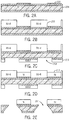
- FIG. 2A is schematic illustration of an actuation device in accordance with the present disclosure in one state of a fabrication process in accordance with the present disclosure.
- FIG. 2B is schematic illustration of an actuation device in accordance with the present disclosure in another state of a fabrication process in accordance with the present disclosure.
- FIG. 2C is schematic illustration of an actuation device in accordance with the present disclosure in yet another state of a fabrication process in accordance with the present disclosure.
- FIG. 2D is schematic illustration of an actuation device in accordance with the present disclosure in still another state of a fabrication process in accordance with the present disclosure.
- FIG. 2E is schematic illustration of an actuation device in accordance with the present disclosure in still another state of a fabrication process in accordance with the present disclosure.
- FIG. 3A is a schematic illustration of the geometric characterization of the actuation device in accordance with the present disclosure.
- FIG. 3B is another illustration of the geometric characterization of the actuation device in accordance with the present disclosure.
- FIG. 4A is a graph illustrating characteristics of an example for magnetic characterization of a system in accordance with the present disclosure.
- FIG. 4B is another graph illustrating characteristics of an example for magnetic characterization of a system in accordance with the present disclosure.
- FIG. 4C is yet another graph illustrating characteristics of an example for magnetic characterization of a system in accordance with the present disclosure.
- FIG. 5A is an image of an MMT without an external magnetic field applied
- FIG. 5B is an image of an MMT with an external magnetic field applied
- FIG. 5C is a graph of the motion of a pillar in response to the external magnetic field in accordance with the present disclosure.
- FIG. 6A is a graph of stiffness changes of microtissue characterized by force and length during dynamic loading.
- FIG. 6B is a graph of stiffness changes of microtissue characterized by elastic modulus during dynamic loading.
- the present disclosure provides a device for actuating and conditioning microtissues.
- the device When paired with a magnetic microtissue tester (MMT), the device acts as a less resource intensive means of tissue engineering and as a method to influence and test tissue mechanical properties.
- MMT magnetic microtissue tester
- FIG. 1( a ) illustrates one microwell 102 which may be used in a broader system that may incorporate more than one microwell 102 , each containing flexible pillars 104 that may be fabricated in a PDMS substrate with, for example, a magnetic Nickel microsphere 106 bonded to one of the pillars 104 .
- a pair of pillars 104 is illustrated, this is a non-limiting example.
- other numbers of pillars 104 beyond pairs, for example, 4 or 8 pillars, or odd numbers of pillars may be included.
- a mixture of cells and extracellular matrix (ECM) is introduced into the wells 102 , and as the cells contract the mixture, they form an aligned microtissue 108 spanning the pillars 104 , which bend due to the collective contractile force of the microtissue 108 , providing a read-out of the force.
- a small ( ⁇ 1 mm) Nickel bar 110 is microfabricated on a silicon wafer, and is placed near the magnetic pillar 104 . When the sphere 106 and bar 110 are magnetized by an externally applied magnetic field, the sphere is attracted to the bar with a magnetic force, which controllably stretches the microtissue 108 .
- a PDMS MMT device can be created in conjunction with an actuation device.
- the PDMS MMT device facilitates the formation of the microtissues, which will be magnetically activated by the actuation device.
- PDMS molds are used for replica molding.
- the PDMS molds include pairs of flexible pillars 104 with, as a non-limiting example, separation of 500 ⁇ m in wells 102 with, as a non-limiting example, dimensions 800 ⁇ m ⁇ 400 ⁇ m ⁇ 170 ⁇ m deep.
- the PDMS may have an elastic modulus of 1.6 MPa.
- Nickel spheres 106 may have ⁇ 100 ⁇ m diameters and be bonded with PDMS to the top of one pillar in each MMT.
- the microtissues 108 are formed by introducing suspensions of NIH 3T3 fibroblasts and 2.5 mg/ml unpolymerized rat tail type-I collagen (BD Biosciences) into the wells 102 .
- the cells are cultured on the MMT devices for two days prior to measurements in high glucose Dulbecco's Modified Eagle's Medium, supplemented with 10% bovine serum, 100 units/ml penicillin, and 100 ⁇ g/ml streptomycin (all from Invitrogen).
- FIG. 1( b ) illustrates the present approach in an array format, with a schematic of a magnetic microtissue actuation system, with multiple nickel bars 114 that align with the individual ⁇ TUGs 116 , microtissues and the inclusion of an array of holes 118 etched through the Si wafer 120 to enable optical access and good exchange of culture media for the microtissues.
- the actuation device 112 is made up of a through-etched silicon nitride-coated wafer 120 with patterned gold fingers. Nickel islands 114 are created on top of the gold circuitry via electrodeposition in order to transduce an externally applied uniform magnetic field into a local inhomogeneous field near each MMT 122 . That is, the nickel bars 114 may be patterned on a silicon wafer 120 . Alternatively, the nickel bars 114 may be mounted directly on the PDMS MMT device, as generally illustrated in FIG. 1( a ) .
- the construction of the actuation device can be broken down into three segments: gold circuitry definition, nickel electro-deposition, and wafer through-etching.
- the component steps in the latter two processes can be interleaved to ensure survivability of the features on the substrate.
- the actuation device is constructed using standard photolithography and electrodeposition techniques, and represents a simple to design, easy to create device for tissue mechanical conditioning and testing.
- the techniques used for fabrication allow for highly uniform actuation device production.
- FIG. 2 A schematic illustration of a fabrication process for the actuation device can be seen in FIG. 2 .
- This process includes the steps of fabricating the gold fingers on the silicon nitride-coated wafers, spinning a photoresist layer over the gold fingers, defining the desired shape of the nickel bars in the resist layer, defining a mask for the wafer through-etching, electrodepositing the nickel bars in the previously defined mask, and etching holes in the wafer.
- Patterned metal (Cr(7 nm)/Au (45 nm)) finger-shaped arrays 202 that are 1,600 ⁇ m wide were fabricated on double-side polished silicon-nitride coated wafers 204 using standard photolithography, thermal evaporation, and lift-off processing techniques which can be seen in FIG. 2( a ) .
- the position and size of the metal fingers were designed to align along the short edge of each individual microwell, and fit to the empty space between two adjacent microwells.
- a 120 ⁇ m thick layer of SU-8 photoresist 206 was then spun between the gold fingers, and patterns corresponding to the shape of the nickel bars were defined in the SU-8 layer on the gold fingers as seen in FIG. 2( b ) .
- the top side of the substrate was reactive-ion etched (RIE) in O 2 for 5 minutes to remove any remaining SU-8 on the exposed gold regions.
- RIE reactive-ion etched
- an array of rectangular holes 208 each of dimension 1,200 ⁇ m by 840 ⁇ m, was patterned into the silicon-nitride coating using S1813 210 .
- the holes 208 were patterned on the side of the wafer opposite the gold fingers via backside alignment, using standard photolithography and reactive ion etching in CF 4 and O 2 , as shown in FIG. 2( c ) .
- the nickel bars 212 were then electrodeposited onto the previously defined patterns to thicknesses of 50-100 ⁇ m, as desired, using the gold finger array as a working electrode.
- the nickel deposition solution consisted of 80.5 g nickel (II) sulfamate, 6.25 g nickel chloride, 10 g boric acid, and 0.05 g sodium dodecyl sulfate (SDS), in 250 mL water.
- a potentiostat (Model 263A, Princeton Applied Research) was used in galvanostatic mode and was set to ⁇ 1 V relative to a platinum reference electrode.
- the holes 214 in the wafer were produced by etching in a 30% KOG solution at 150° C. for approximately 6 hours to obtain holes on the side of the wafers with the nickel bars that match the dimensions of the wells on the MMTs.
- the KOH bath had the added effect of removing any remaining photoresist adhered to the wafer.
- VSM vibrating sample magnetometer
- FIG. 3( a ) illustrates a portion of one such array, showing three nickel bars 302 , their underlying Au strip 304 , and corresponding through-holes 306 .
- the nickel bars are fabricated with pointed ends to concentrate magnetic flux and create larger field gradients in the neighborhood of the magnetic pillars.
- the bars shown have length 1,600 ⁇ m, width 450 ⁇ m, and tip width 90 ⁇ m.
- the nickel bars are laid out on a rectangular grid with center-to-center spacing of 3,200 ⁇ m along the bars' long axis and 1,200 ⁇ m along the short axis.
- the dimensions of the nickel bars may be characterized via optical profilometry.
- FIG. 3( b ) shows the height profile of a nickel bar.
- the PDMS-MMT device together with actuation device provide a system for mechanically activating the microtissue, and calculating mechanical properties such as stress, strain, and stiffness.
- actuation device By mounting the actuation device on the PDMS-MMT device, a system is created that can allow for electromagnetic activation of the actuation device and stimulation of the microtissues that have been formed on the PDMS-MMT device.
- measurements and probing of microtissue occur when the actuation device is mounted and aligned under a microscope on the MMT device, and the arrays are actuated with a microscope-mounted dual-coil programmable electromagnet.
- the electromagnet used is capable of producing magnetic fields of up to 50 mT with a uniformity of 3% over the largest arrays studied.
- Images of individual MMTs and microtissues may be obtained using phase contrast microscopy with a 10 ⁇ objective on a Nikon TE-2000E inverted microscope.
- a quasi-static stretching protocol is used, during which images were recorded with a CoolSnap HQ (Photometrics) camera.
- movies are recorded at 100 frames/sec using a Prosilica GX (Allied Vision Technologies) camera.
- the pillar deflections are determined from the images using Image) (NIH) for the quasi-static measurements and via custom tracking software written in IgorPro (WaveMetrics) for the dynamic actuation studies.
- the stress, strain, and elastic modulus of each microtissue is determined from the quasi-static stretching data. Briefly, the force on each microtissue is found by tracking the deflection of the non-magnetic MMT pillar and calculating a force based on its spring constant. The stress in the central region of each microtissue is then obtained from the measured dimensions of the microtissue. The strain is measured locally in the microtissues' centers from sequential phase contrast images, using a texture correlation analysis algorithm. The elastic modulus is determined from the slope of the resulting stress-strain curves
- FIG. 4( a ) shows the magnetic moment ⁇ Bar vs. applied magnetic field ⁇ 0 H for a representative nickel bar removed from the array and measured using a vibrating sample magnetometer (VSM).
- VSM vibrating sample magnetometer
- the actuation device applies a quasi-static load on the microtissue.
- the magnetic pillars are pulled toward the nickel bars.
- the response of the magnetic pillars to sinusoidal external fields is measured.
- the system allows for dynamic loading of the tissue. Cyclic loading of the actuation device causes motion of the pillars, inducing an active load in the tissues by the pillar and a tension force on the pillars by the microtissue.
- the dynamic loading capacity of the actuation device is evaluated by applying a sinusoidal magnetic external field of amplitude 20 mT at 1 Hz to the actuation devices on MMTs.
- a sinusoidal magnetic external field of amplitude 20 mT at 1 Hz to the actuation devices on MMTs.
- the microtissues are observed for brief intervals ( ⁇ 15 sec) while recording their motion at 100 frames/sec.
- FIG. 6( a ) shows the left pillar displacement (microtissue force) 602 and overall length (difference in pillar positions) 604 vs. time for a microtissue following initiation of actuation. Both the force and length are predominantly sinusoidal with second harmonic content ⁇ 6% of the 2 Hz fundamental, similar to that observed for AC actuation of magnetic pillars without microtissue as shown in FIG. 5 .
- the elastic modulus of a set of microtissues is first measured by quasi-static loading with the actuation device as described in earlier. Cyclic loading at 2 Hz (1 Hz external field) is then applied simultaneously to the tissues for 15 min, and the stiffness is re-measured. While there is some variability in the degree of stiffness change, all microtissues measured exhibit an increase in modulus, with an average increase of 31%, which can be seen in FIG. 6( b ) .
- This stiffness change reflects one of a reorganization of the collagen matrix, as it has been shown that cells play a minor role in fibroblast microtissue stiffness, or an actuation of internal force generation machinery.
- tissue stiffness through AC stimulation has been demonstrated.
- the device and associated apparatus are small enough to fit into a standard incubator. Using the device to mechanically condition tissues with AC stimulation as they are maturing is a simple extension of current protocols. Due to the versatility and efficiency of the present device, a possible application is for pharmacological mechanical testing that requires using expensive drugs and rare cell lines.
Landscapes
- Health & Medical Sciences (AREA)
- Engineering & Computer Science (AREA)
- Life Sciences & Earth Sciences (AREA)
- Chemical & Material Sciences (AREA)
- Zoology (AREA)
- Organic Chemistry (AREA)
- Bioinformatics & Cheminformatics (AREA)
- Wood Science & Technology (AREA)
- Genetics & Genomics (AREA)
- Biotechnology (AREA)
- Biomedical Technology (AREA)
- General Health & Medical Sciences (AREA)
- Biochemistry (AREA)
- General Engineering & Computer Science (AREA)
- Microbiology (AREA)
- Sustainable Development (AREA)
- Clinical Laboratory Science (AREA)
- Cell Biology (AREA)
- Immunology (AREA)
- Molecular Biology (AREA)
- Mechanical Engineering (AREA)
- Analytical Chemistry (AREA)
- Apparatus Associated With Microorganisms And Enzymes (AREA)
Abstract
Description
Claims (17)
Priority Applications (1)
| Application Number | Priority Date | Filing Date | Title |
|---|---|---|---|
| US15/528,233 US10590376B2 (en) | 2014-11-20 | 2015-11-20 | System for conditioning of engineered microtissues |
Applications Claiming Priority (3)
| Application Number | Priority Date | Filing Date | Title |
|---|---|---|---|
| US201462082374P | 2014-11-20 | 2014-11-20 | |
| PCT/US2015/061800 WO2016081816A1 (en) | 2014-11-20 | 2015-11-20 | System and method for conditioning of engineered tissues |
| US15/528,233 US10590376B2 (en) | 2014-11-20 | 2015-11-20 | System for conditioning of engineered microtissues |
Publications (2)
| Publication Number | Publication Date |
|---|---|
| US20170362560A1 US20170362560A1 (en) | 2017-12-21 |
| US10590376B2 true US10590376B2 (en) | 2020-03-17 |
Family
ID=56014593
Family Applications (1)
| Application Number | Title | Priority Date | Filing Date |
|---|---|---|---|
| US15/528,233 Active 2036-07-08 US10590376B2 (en) | 2014-11-20 | 2015-11-20 | System for conditioning of engineered microtissues |
Country Status (2)
| Country | Link |
|---|---|
| US (1) | US10590376B2 (en) |
| WO (1) | WO2016081816A1 (en) |
Families Citing this family (2)
| Publication number | Priority date | Publication date | Assignee | Title |
|---|---|---|---|---|
| KR102660112B1 (en) * | 2020-10-26 | 2024-04-23 | 포항공과대학교 산학협력단 | Strain gauge sensor and mehtod for manufacturing thereof |
| WO2023028224A2 (en) * | 2021-08-25 | 2023-03-02 | Clemson University Research Foundation | In vitro myocardial tissue screening devices, systems, and methods |
Citations (17)
| Publication number | Priority date | Publication date | Assignee | Title |
|---|---|---|---|---|
| US6218178B1 (en) | 1998-05-08 | 2001-04-17 | Flexcell International Corporation | Loading station assembly |
| JP2003180331A (en) | 2001-12-21 | 2003-07-02 | Takagi Ind Co Ltd | Cell / tissue culture equipment |
| US20040101819A1 (en) | 2002-08-08 | 2004-05-27 | Mt Technologies, Inc. | Self-assembled muscle-powered microdevices |
| WO2005007233A2 (en) | 2003-06-20 | 2005-01-27 | Massachusetts Institute Of Technology | Application of electrical stimulation for functional tissue engineering in vitro and in vivo |
| US20060282576A1 (en) * | 2005-05-24 | 2006-12-14 | Omnidirectional Control Technology Inc. | Computer peripheral converter |
| JP2008263986A (en) | 2000-12-22 | 2008-11-06 | Keele Univ | Method of culturing tissue using magnetically generated mechanical stress |
| US7553662B2 (en) | 2000-12-22 | 2009-06-30 | Keele University | Culturing tissue using magnetically generated mechanical stresses |
| US20090209035A1 (en) | 2006-07-10 | 2009-08-20 | Takagi Industrial Co., Ltd. | Cell or tissue cultivation apparatus and method of cultivation |
| JP2009254275A (en) | 2008-04-16 | 2009-11-05 | Yamaguchi Univ | Cell-extending and shortening device using shape memory alloy |
| US20110007955A1 (en) * | 2009-07-08 | 2011-01-13 | Applied Biocode Inc. | Apparatus and Method for Barcoded Magnetic Beads Analysis |
| US20110097723A1 (en) * | 2009-09-19 | 2011-04-28 | Qun Liu | Methods and reagents for analyte detection |
| WO2012118799A2 (en) | 2011-02-28 | 2012-09-07 | President And Fellows Of Harvard College | Cell culture system |
| US8375851B2 (en) | 2008-11-11 | 2013-02-19 | The Regents Of University Of Colorado | Apparatus and methods for loading soft materials |
| WO2013119570A1 (en) | 2012-02-06 | 2013-08-15 | Simmons Chelsey S | Cell culture strain array systems and methods for using the same |
| WO2013152036A1 (en) | 2012-04-02 | 2013-10-10 | The Children's Mercy Hospital | Disposable single use self-contained cyclic pressure and flow bioreactor system |
| US10001474B2 (en) * | 2011-06-07 | 2018-06-19 | Life Technologies Corporation | Fluorogenic semiconductor nanocrystals |
| US10451631B2 (en) * | 2013-04-25 | 2019-10-22 | Vladislav B. Bergo | Microarray compositions and methods of their use |
-
2015
- 2015-11-20 US US15/528,233 patent/US10590376B2/en active Active
- 2015-11-20 WO PCT/US2015/061800 patent/WO2016081816A1/en not_active Ceased
Patent Citations (17)
| Publication number | Priority date | Publication date | Assignee | Title |
|---|---|---|---|---|
| US6218178B1 (en) | 1998-05-08 | 2001-04-17 | Flexcell International Corporation | Loading station assembly |
| JP2008263986A (en) | 2000-12-22 | 2008-11-06 | Keele Univ | Method of culturing tissue using magnetically generated mechanical stress |
| US7553662B2 (en) | 2000-12-22 | 2009-06-30 | Keele University | Culturing tissue using magnetically generated mechanical stresses |
| JP2003180331A (en) | 2001-12-21 | 2003-07-02 | Takagi Ind Co Ltd | Cell / tissue culture equipment |
| US20040101819A1 (en) | 2002-08-08 | 2004-05-27 | Mt Technologies, Inc. | Self-assembled muscle-powered microdevices |
| WO2005007233A2 (en) | 2003-06-20 | 2005-01-27 | Massachusetts Institute Of Technology | Application of electrical stimulation for functional tissue engineering in vitro and in vivo |
| US20060282576A1 (en) * | 2005-05-24 | 2006-12-14 | Omnidirectional Control Technology Inc. | Computer peripheral converter |
| US20090209035A1 (en) | 2006-07-10 | 2009-08-20 | Takagi Industrial Co., Ltd. | Cell or tissue cultivation apparatus and method of cultivation |
| JP2009254275A (en) | 2008-04-16 | 2009-11-05 | Yamaguchi Univ | Cell-extending and shortening device using shape memory alloy |
| US8375851B2 (en) | 2008-11-11 | 2013-02-19 | The Regents Of University Of Colorado | Apparatus and methods for loading soft materials |
| US20110007955A1 (en) * | 2009-07-08 | 2011-01-13 | Applied Biocode Inc. | Apparatus and Method for Barcoded Magnetic Beads Analysis |
| US20110097723A1 (en) * | 2009-09-19 | 2011-04-28 | Qun Liu | Methods and reagents for analyte detection |
| WO2012118799A2 (en) | 2011-02-28 | 2012-09-07 | President And Fellows Of Harvard College | Cell culture system |
| US10001474B2 (en) * | 2011-06-07 | 2018-06-19 | Life Technologies Corporation | Fluorogenic semiconductor nanocrystals |
| WO2013119570A1 (en) | 2012-02-06 | 2013-08-15 | Simmons Chelsey S | Cell culture strain array systems and methods for using the same |
| WO2013152036A1 (en) | 2012-04-02 | 2013-10-10 | The Children's Mercy Hospital | Disposable single use self-contained cyclic pressure and flow bioreactor system |
| US10451631B2 (en) * | 2013-04-25 | 2019-10-22 | Vladislav B. Bergo | Microarray compositions and methods of their use |
Non-Patent Citations (22)
| Title |
|---|
| Altman, G., et al., "Cell Differentiation by Mechanical Stress" FASEB journal, 2001. |
| Andrade, P.Z., et al., Stem cell bioengineering strategies to widen the therapeutic applications of haematopoietic stem/progenitor cells from umbilical cord blood. Journal of tissue engineering and regenerative medicine, 2013. |
| Barron, V., et al., Bioreactors for cardiovascular cell and tissue growth: a review. Annals of biomedical engineering, 2003. 31(9): p. 1017-1030. |
| Beca, B., et al., "A Platform for Combinatorial Mechanobiological Stimulation of Engineered Microtissues" 16th international conference on Miniaturized Systems for Chemistry and Life Sciences, 2012. |
| Boudou, T., et al., A microfabricated platform to measure and manipulate the mechanics of engineered cardiac microtissues. Tissue Eng Part A, 2012. 18(9-10): p. 910-9. |
| Brady, M. "The Design and Development of a High-Throughput Magneto-Mechanostimulation Device for Cartilage Tissue Engineering" Tissue Engineering: Part C, vol. 20, No. 2, 2014. |
| Cummings, C.L., et al., Properties of engineered vascular constructs made from collagen, fibrin, and collagen-fibrin mixtures. Biomaterials, 2004. 25(17): p. 3699-3706. |
| Dobson, J., "Principles and Design of a Novel Magnetic Force Mechanical Conditioning Bioreactor for Tissue Engineering, Stem Cell Conditioning, and Dynamic In Vitro Screening" IEEE Transactions on Nanobioscience, vol. 5, No. 3, Sep. 2006. |
| Hirt, M., et al., "Cardiac Tissue Engineering : State of the Art" Circ Res. 2014;114:354-367. |
| Hoerstrup S., et al., "New pulsatile bioreactor for in vitro formation of tissue engineered heart valves" Tissue Engineering, vol. 6, No. 1, 2000. |
| Huang, A., et al., "Engineering of arteries in vitro", Cell Mol Life Sci. Jun. 2014 ; 71(11): 2103-2118. |
| Legant, W., "Microfabricated tissue gauges to measure and manipulate forces from 3D microtissues" PNAS 2009, vol. 106, No. 25, pp. 10097-10102. |
| Legant, W.R., et al., Microfabricated tissue gauges to measure and manipulate forces from 3D microtissues. Proc Natl Acad Sci U S A, 2009. 106(25): p. 10097-102. |
| Lucas, K., et al. Sculpting of Nanopores in Silicon-Nitride Membranes. in APS Meeting Abstracts. 2007. |
| Mannoor, M.S., et al., 3D printed bionic ears. Nano letters, 2013. 13(6): p. 2634-2639. |
| Sacks, et al., "Bioengineering Challenges for heart Valve Tissue Engineering" Annu. Rev. Biomed. Eng. 2009. 11:289-313. |
| Seliktar, D., et al., Dynamic mechanical conditioning of collagen-gel blood vessel constructs induces remodeling in vitro. Annals of biomedical engineering, 2000.28(4): p. 351-362. |
| Wasserman, J., et al., Fabrication of one-dimensional programmable-height nanostructures via dynamic stencil deposition. Review of Scientific Instruments, 2008. 79(7): p. 073909-073909-4. |
| West, A.R., et al., Development and characterization of a 3D multicell microtissue culture model of airway smooth muscle. American Journal of Physiology-Lung Cellular and Molecular Physiology, 2013. 304(1): p. L4-L16. |
| West, A.R., et al., Development and characterization of a 3D multicell microtissue culture model of airway smooth muscle. American Journal of Physiology—Lung Cellular and Molecular Physiology, 2013. 304(1): p. L4-L16. |
| Xu, F., et al., "A microfabricated magnetic actuation device for mechanical conditioning of arrays of 3D microtissues", Lab Chip (2015) vol. 15, No. 11, pp. 2496-2503. |
| Zhao, R., et al., Decoupling cell and matrix mechanics in engineered microtissues using magnetically actuated microcantilevers. Adv Mater, 2013. 25(12): p. 1699-705. |
Also Published As
| Publication number | Publication date |
|---|---|
| WO2016081816A1 (en) | 2016-05-26 |
| US20170362560A1 (en) | 2017-12-21 |
Similar Documents
| Publication | Publication Date | Title |
|---|---|---|
| Dou et al. | Microengineered platforms for characterizing the contractile function of in vitro cardiac models | |
| Grosberg et al. | Muscle on a chip: in vitro contractility assays for smooth and striated muscle | |
| Xu et al. | A microfabricated magnetic actuation device for mechanical conditioning of arrays of 3D microtissues | |
| Grosberg et al. | Ensembles of engineered cardiac tissues for physiological and pharmacological study: heart on a chip | |
| Addae-Mensah et al. | Measurement techniques for cellular biomechanics in vitro | |
| US10034738B2 (en) | Cardiac tissue constructs and methods of fabrication thereof | |
| Sorce et al. | Mitotic cells contract actomyosin cortex and generate pressure to round against or escape epithelial confinement | |
| US9512396B2 (en) | In vitro microphysiological system for high throughput 3D tissue organization and biological function | |
| US9012172B2 (en) | Devices comprising muscle thin films and uses thereof in high throughput assays for determining contractile function | |
| Bidan et al. | Magneto-active substrates for local mechanical stimulation of living cells | |
| US20130046134A1 (en) | Methods of generating engineered innervated tissue and uses thereof | |
| Lele et al. | Tools to study cell mechanics and mechanotransduction | |
| Liu et al. | Effect of static pre-stretch induced surface anisotropy on orientation of mesenchymal stem cells | |
| US10910573B2 (en) | Cell-based electromechanical biocomputing | |
| US10590376B2 (en) | System for conditioning of engineered microtissues | |
| JP4689609B2 (en) | Method and apparatus for adhesion control of intracellular tissue | |
| JP2007504818A5 (en) | ||
| Zhao et al. | Magnetic approaches to study collective three-dimensional cell mechanics in long-term cultures | |
| Nagarajan et al. | Modulation of the contractility of micropatterned myocardial cells with nanoscale forces using atomic force microscopy | |
| Zhou et al. | Real-time monitoring of contractile properties of H9C2 cardiomyoblasts by using a quartz crystal microbalance | |
| Gao et al. | Three dimensional and homogenous single cell cyclic stretch within a magnetic micropillar array (mMPA) for a cell proliferation study | |
| Kim et al. | Enhancement of cardiac contractility using gold-coated SU-8 cantilevers and their application to drug-induced cardiac toxicity tests | |
| de Vries et al. | Patterned electroplating of micrometer scale magnetic structures on glass substrates | |
| Demri et al. | Magnetic Bioprinting and Actuation of Stretchable Muscle Tissue | |
| Belot | Development of nanoprobe array technology for high resolution electrophysiology of human brain organoids-on-chip |
Legal Events
| Date | Code | Title | Description |
|---|---|---|---|
| FEPP | Fee payment procedure |
Free format text: ENTITY STATUS SET TO SMALL (ORIGINAL EVENT CODE: SMAL); ENTITY STATUS OF PATENT OWNER: SMALL ENTITY |
|
| AS | Assignment |
Owner name: NATIONAL INSTITUTES OF HEALTH (NIH), U.S. DEPT. OF HEALTH AND HUMAN SERVICES (DHHS), U.S. GOVERNMENT, MARYLAND Free format text: CONFIRMATORY LICENSE;ASSIGNOR:JOHNS HOPKINS UNIVERSITY;REEL/FRAME:044968/0950 Effective date: 20171227 Owner name: NATIONAL INSTITUTES OF HEALTH (NIH), U.S. DEPT. OF Free format text: CONFIRMATORY LICENSE;ASSIGNOR:JOHNS HOPKINS UNIVERSITY;REEL/FRAME:044968/0950 Effective date: 20171227 |
|
| STPP | Information on status: patent application and granting procedure in general |
Free format text: DOCKETED NEW CASE - READY FOR EXAMINATION |
|
| STPP | Information on status: patent application and granting procedure in general |
Free format text: NON FINAL ACTION MAILED |
|
| STPP | Information on status: patent application and granting procedure in general |
Free format text: RESPONSE TO NON-FINAL OFFICE ACTION ENTERED AND FORWARDED TO EXAMINER |
|
| AS | Assignment |
Owner name: THE JOHNS HOPKINS UNIVERSITY, MARYLAND Free format text: ASSIGNMENT OF ASSIGNORS INTEREST;ASSIGNORS:LIU, ALAN S.;ZHAO, RUOGANG;XU, FAN;AND OTHERS;SIGNING DATES FROM 20190514 TO 20190703;REEL/FRAME:050058/0749 |
|
| STPP | Information on status: patent application and granting procedure in general |
Free format text: NOTICE OF ALLOWANCE MAILED -- APPLICATION RECEIVED IN OFFICE OF PUBLICATIONS |
|
| STPP | Information on status: patent application and granting procedure in general |
Free format text: PUBLICATIONS -- ISSUE FEE PAYMENT VERIFIED |
|
| STPP | Information on status: patent application and granting procedure in general |
Free format text: PUBLICATIONS -- ISSUE FEE PAYMENT VERIFIED |
|
| STCF | Information on status: patent grant |
Free format text: PATENTED CASE |
|
| MAFP | Maintenance fee payment |
Free format text: PAYMENT OF MAINTENANCE FEE, 4TH YR, SMALL ENTITY (ORIGINAL EVENT CODE: M2551); ENTITY STATUS OF PATENT OWNER: SMALL ENTITY Year of fee payment: 4 |