US10034889B2 - Pharmaceutical composition including migratory factor for guiding pluripotent stem cells to damage - Google Patents
Pharmaceutical composition including migratory factor for guiding pluripotent stem cells to damage Download PDFInfo
- Publication number
- US10034889B2 US10034889B2 US15/238,020 US201615238020A US10034889B2 US 10034889 B2 US10034889 B2 US 10034889B2 US 201615238020 A US201615238020 A US 201615238020A US 10034889 B2 US10034889 B2 US 10034889B2
- Authority
- US
- United States
- Prior art keywords
- cells
- negative
- muse cells
- pluripotent stem
- stem cells
- Prior art date
- Legal status (The legal status is an assumption and is not a legal conclusion. Google has not performed a legal analysis and makes no representation as to the accuracy of the status listed.)
- Active
Links
- AMXVYJYMZLDINS-RSWLNLDNSA-N C/C(=N\O)C1=NC([C@@H](O)[C@H](O)[C@H](O)CO)=CN1 Chemical compound C/C(=N\O)C1=NC([C@@H](O)[C@H](O)[C@H](O)CO)=CN1 AMXVYJYMZLDINS-RSWLNLDNSA-N 0.000 description 1
- CQSIXFHVGKMLGQ-BWZBUEFSSA-N CC(=O)C1=NC=C([C@@H](O)[C@H](O)[C@H](O)CO)N1 Chemical compound CC(=O)C1=NC=C([C@@H](O)[C@H](O)[C@H](O)CO)N1 CQSIXFHVGKMLGQ-BWZBUEFSSA-N 0.000 description 1
- FJCVBKSKQXCVJP-UHFFFAOYSA-N CC1=CC(C(=O)CN2C(=O)C(=O)N(C)C2=O)=C(C)N1CC1=CC=CC=C1 Chemical compound CC1=CC(C(=O)CN2C(=O)C(=O)N(C)C2=O)=C(C)N1CC1=CC=CC=C1 FJCVBKSKQXCVJP-UHFFFAOYSA-N 0.000 description 1
- JRESCQKIRPOOEM-UHFFFAOYSA-N CC1=CC(C(=O)CN2C(=O)CCC2=O)=C(C)N1CC1=CC=CC=C1 Chemical compound CC1=CC(C(=O)CN2C(=O)CCC2=O)=C(C)N1CC1=CC=CC=C1 JRESCQKIRPOOEM-UHFFFAOYSA-N 0.000 description 1
- JGXVEFSBVPQLIL-UHFFFAOYSA-N CC1=CC(C(=O)CN2C(=O)NC3(CCC4=CC=CC=C43)C2=O)=C(C)N1CC1=CC=CC=C1 Chemical compound CC1=CC(C(=O)CN2C(=O)NC3(CCC4=CC=CC=C43)C2=O)=C(C)N1CC1=CC=CC=C1 JGXVEFSBVPQLIL-UHFFFAOYSA-N 0.000 description 1
- RGSGTUIDJXHTTO-UHFFFAOYSA-N CC1=CC(C(=O)CN2C=C(C(F)(F)F)C=CC2=O)=C(C)N1CC1=CC=CC=C1 Chemical compound CC1=CC(C(=O)CN2C=C(C(F)(F)F)C=CC2=O)=C(C)N1CC1=CC=CC=C1 RGSGTUIDJXHTTO-UHFFFAOYSA-N 0.000 description 1
- AHPQZCDAMIJFNI-UHFFFAOYSA-N CC1=CC(C(=O)CSC2=NN=NN2C)=C(C)N1CC1=CC=CC=C1 Chemical compound CC1=CC(C(=O)CSC2=NN=NN2C)=C(C)N1CC1=CC=CC=C1 AHPQZCDAMIJFNI-UHFFFAOYSA-N 0.000 description 1
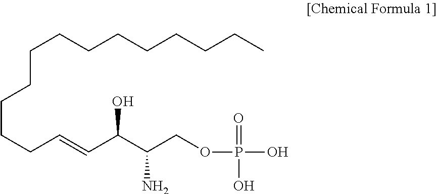
- DUYSYHSSBDVJSM-KRWOKUGFSA-N CCCCCCCCCCCCC/C=C/[C@@H](O)[C@@H](N)COP(=O)(O)O Chemical compound CCCCCCCCCCCCC/C=C/[C@@H](O)[C@@H](N)COP(=O)(O)O DUYSYHSSBDVJSM-KRWOKUGFSA-N 0.000 description 1
- WGDPRIZUIYIENM-IWSPIJDZSA-N OC[C@@H](O)[C@@H](O)[C@H](O)C1=CNC(C2=NOC=C2)=N1 Chemical compound OC[C@@H](O)[C@@H](O)[C@H](O)C1=CNC(C2=NOC=C2)=N1 WGDPRIZUIYIENM-IWSPIJDZSA-N 0.000 description 1
Images


Classifications
-
- A—HUMAN NECESSITIES
- A61—MEDICAL OR VETERINARY SCIENCE; HYGIENE
- A61K—PREPARATIONS FOR MEDICAL, DENTAL OR TOILETRY PURPOSES
- A61K31/00—Medicinal preparations containing organic active ingredients
- A61K31/66—Phosphorus compounds
- A61K31/661—Phosphorus acids or esters thereof not having P—C bonds, e.g. fosfosal, dichlorvos, malathion or mevinphos
-
- A—HUMAN NECESSITIES
- A61—MEDICAL OR VETERINARY SCIENCE; HYGIENE
- A61K—PREPARATIONS FOR MEDICAL, DENTAL OR TOILETRY PURPOSES
- A61K31/00—Medicinal preparations containing organic active ingredients
- A61K31/33—Heterocyclic compounds
- A61K31/395—Heterocyclic compounds having nitrogen as a ring hetero atom, e.g. guanethidine or rifamycins
- A61K31/40—Heterocyclic compounds having nitrogen as a ring hetero atom, e.g. guanethidine or rifamycins having five-membered rings with one nitrogen as the only ring hetero atom, e.g. sulpiride, succinimide, tolmetin, buflomedil
- A61K31/4025—Heterocyclic compounds having nitrogen as a ring hetero atom, e.g. guanethidine or rifamycins having five-membered rings with one nitrogen as the only ring hetero atom, e.g. sulpiride, succinimide, tolmetin, buflomedil not condensed and containing further heterocyclic rings, e.g. cromakalim
-
- A—HUMAN NECESSITIES
- A61—MEDICAL OR VETERINARY SCIENCE; HYGIENE
- A61K—PREPARATIONS FOR MEDICAL, DENTAL OR TOILETRY PURPOSES
- A61K31/00—Medicinal preparations containing organic active ingredients
- A61K31/33—Heterocyclic compounds
- A61K31/395—Heterocyclic compounds having nitrogen as a ring hetero atom, e.g. guanethidine or rifamycins
- A61K31/41—Heterocyclic compounds having nitrogen as a ring hetero atom, e.g. guanethidine or rifamycins having five-membered rings with two or more ring hetero atoms, at least one of which being nitrogen, e.g. tetrazole
- A61K31/4164—1,3-Diazoles
- A61K31/417—Imidazole-alkylamines, e.g. histamine, phentolamine
-
- A—HUMAN NECESSITIES
- A61—MEDICAL OR VETERINARY SCIENCE; HYGIENE
- A61K—PREPARATIONS FOR MEDICAL, DENTAL OR TOILETRY PURPOSES
- A61K31/00—Medicinal preparations containing organic active ingredients
- A61K31/33—Heterocyclic compounds
- A61K31/395—Heterocyclic compounds having nitrogen as a ring hetero atom, e.g. guanethidine or rifamycins
- A61K31/41—Heterocyclic compounds having nitrogen as a ring hetero atom, e.g. guanethidine or rifamycins having five-membered rings with two or more ring hetero atoms, at least one of which being nitrogen, e.g. tetrazole
- A61K31/4164—1,3-Diazoles
- A61K31/4178—1,3-Diazoles not condensed 1,3-diazoles and containing further heterocyclic rings, e.g. pilocarpine, nitrofurantoin
-
- A—HUMAN NECESSITIES
- A61—MEDICAL OR VETERINARY SCIENCE; HYGIENE
- A61K—PREPARATIONS FOR MEDICAL, DENTAL OR TOILETRY PURPOSES
- A61K31/00—Medicinal preparations containing organic active ingredients
- A61K31/33—Heterocyclic compounds
- A61K31/395—Heterocyclic compounds having nitrogen as a ring hetero atom, e.g. guanethidine or rifamycins
- A61K31/41—Heterocyclic compounds having nitrogen as a ring hetero atom, e.g. guanethidine or rifamycins having five-membered rings with two or more ring hetero atoms, at least one of which being nitrogen, e.g. tetrazole
- A61K31/4164—1,3-Diazoles
- A61K31/4184—1,3-Diazoles condensed with carbocyclic rings, e.g. benzimidazoles
-
- A—HUMAN NECESSITIES
- A61—MEDICAL OR VETERINARY SCIENCE; HYGIENE
- A61K—PREPARATIONS FOR MEDICAL, DENTAL OR TOILETRY PURPOSES
- A61K31/00—Medicinal preparations containing organic active ingredients
- A61K31/33—Heterocyclic compounds
- A61K31/395—Heterocyclic compounds having nitrogen as a ring hetero atom, e.g. guanethidine or rifamycins
- A61K31/41—Heterocyclic compounds having nitrogen as a ring hetero atom, e.g. guanethidine or rifamycins having five-membered rings with two or more ring hetero atoms, at least one of which being nitrogen, e.g. tetrazole
- A61K31/42—Oxazoles
- A61K31/422—Oxazoles not condensed and containing further heterocyclic rings
-
- A—HUMAN NECESSITIES
- A61—MEDICAL OR VETERINARY SCIENCE; HYGIENE
- A61K—PREPARATIONS FOR MEDICAL, DENTAL OR TOILETRY PURPOSES
- A61K31/00—Medicinal preparations containing organic active ingredients
- A61K31/33—Heterocyclic compounds
- A61K31/395—Heterocyclic compounds having nitrogen as a ring hetero atom, e.g. guanethidine or rifamycins
- A61K31/435—Heterocyclic compounds having nitrogen as a ring hetero atom, e.g. guanethidine or rifamycins having six-membered rings with one nitrogen as the only ring hetero atom
- A61K31/44—Non condensed pyridines; Hydrogenated derivatives thereof
- A61K31/4427—Non condensed pyridines; Hydrogenated derivatives thereof containing further heterocyclic ring systems
- A61K31/4439—Non condensed pyridines; Hydrogenated derivatives thereof containing further heterocyclic ring systems containing a five-membered ring with nitrogen as a ring hetero atom, e.g. omeprazole
-
- A—HUMAN NECESSITIES
- A61—MEDICAL OR VETERINARY SCIENCE; HYGIENE
- A61K—PREPARATIONS FOR MEDICAL, DENTAL OR TOILETRY PURPOSES
- A61K35/00—Medicinal preparations containing materials or reaction products thereof with undetermined constitution
- A61K35/12—Materials from mammals; Compositions comprising non-specified tissues or cells; Compositions comprising non-embryonic stem cells; Genetically modified cells
- A61K35/48—Reproductive organs
- A61K35/54—Ovaries; Ova; Ovules; Embryos; Foetal cells; Germ cells
- A61K35/545—Embryonic stem cells; Pluripotent stem cells; Induced pluripotent stem cells; Uncharacterised stem cells
-
- A—HUMAN NECESSITIES
- A61—MEDICAL OR VETERINARY SCIENCE; HYGIENE
- A61P—SPECIFIC THERAPEUTIC ACTIVITY OF CHEMICAL COMPOUNDS OR MEDICINAL PREPARATIONS
- A61P17/00—Drugs for dermatological disorders
- A61P17/02—Drugs for dermatological disorders for treating wounds, ulcers, burns, scars, keloids, or the like
-
- A—HUMAN NECESSITIES
- A61—MEDICAL OR VETERINARY SCIENCE; HYGIENE
- A61P—SPECIFIC THERAPEUTIC ACTIVITY OF CHEMICAL COMPOUNDS OR MEDICINAL PREPARATIONS
- A61P29/00—Non-central analgesic, antipyretic or antiinflammatory agents, e.g. antirheumatic agents; Non-steroidal antiinflammatory drugs [NSAID]
-
- A—HUMAN NECESSITIES
- A61—MEDICAL OR VETERINARY SCIENCE; HYGIENE
- A61P—SPECIFIC THERAPEUTIC ACTIVITY OF CHEMICAL COMPOUNDS OR MEDICINAL PREPARATIONS
- A61P35/00—Antineoplastic agents
-
- A—HUMAN NECESSITIES
- A61—MEDICAL OR VETERINARY SCIENCE; HYGIENE
- A61P—SPECIFIC THERAPEUTIC ACTIVITY OF CHEMICAL COMPOUNDS OR MEDICINAL PREPARATIONS
- A61P43/00—Drugs for specific purposes, not provided for in groups A61P1/00-A61P41/00
-
- A—HUMAN NECESSITIES
- A61—MEDICAL OR VETERINARY SCIENCE; HYGIENE
- A61P—SPECIFIC THERAPEUTIC ACTIVITY OF CHEMICAL COMPOUNDS OR MEDICINAL PREPARATIONS
- A61P9/00—Drugs for disorders of the cardiovascular system
- A61P9/10—Drugs for disorders of the cardiovascular system for treating ischaemic or atherosclerotic diseases, e.g. antianginal drugs, coronary vasodilators, drugs for myocardial infarction, retinopathy, cerebrovascula insufficiency, renal arteriosclerosis
Definitions
- the present invention relates to a pharmaceutical composition that contains a chemotactic factor that guides pluripotent stem cells to a site of tissue damage.
- Non-Patent Documents 1 and 2 these consist of cell groups containing various cells, the actual state of their ability to differentiate is not understood, and there have been considerable fluctuations in therapeutic effects.
- Patent Document 1 iPS cells have been reported to be adult-derived pluripotent stem cells, in addition to the establishment of iPS cells requiring an extremely complex procedure involving the introduction of specific genes into mesenchymal cells in the form of a skin fibroblast fraction and the introduction of specific compounds into somatic cells, since iPS cells have a high tumorigenic potential, extremely high hurdles must be overcome for their clinical application.
- pluripotent stem cells have been determined to be present in mesenchymal tissue of the body, accumulate at the site of a tissue damage when body tissue has been damaged and be responsible for tissue regeneration (Patent Document 2, Non-Patent Document 3).
- Muse cells have been determined to be present in mesenchymal tissue of the body, accumulate at the site of a tissue damage when body tissue has been damaged and be responsible for tissue regeneration (Patent Document 2, Non-Patent Document 3).
- the mechanism by which Muse cells are guided to damaged tissue not been elucidated, but the chemotactic factor that guides Muse cells to the damaged site has also not been identified.
- An object of the present invention is to provide a medical application using pluripotent stem cells (Muse cells) in the field of regenerative medicine, and to enhance the chemotactic activity of Muse cells and identify a chemotactic factor for allowing Muse cells to effectively accumulate at a damaged site together with providing a pharmaceutical composition containing that chemotactic factor.
- Muse cells pluripotent stem cells
- the inventors of the present invention succeeded in identifying chemotactic factors that guide Muse cells to a damage site by utilizing a proteomic analysis, and found that one of those chemotactic factors in the form of sphingosine-1-phosphate (S1P) enhances the chemotactic activity of Muse cells and is involved in their accumulation at a damage site, thereby leading to completion of the present invention.
- Enhancement of chemotactic activity includes initiating migration of Muse cells present in mesenchymal tissue towards a damage site.
- a pharmaceutical composition for activating migration of pluripotent stem cells comprising: as an active ingredient thereof a compound that activates sphingosine-1-phosphate receptor 2.
- sphingosine-1-phosphate receptor 2 is selected from the group consisting of 1-(2-(1-benzyl-2,5-dimethyl-1H-pyrrol-3-yl)-2-oxoethyl)-5-(trifluoromethyl) pyridin-2(1H)-one, 1-(2-(1-benzyl-2,5-dimethyl-1H-pyrrol-3-yl)-2-oxoethyl)pyrrolidine-2,5-dione, 1-(2-(1-benzyl-2,5-dimethyl-1H-pyrrol-3-yl)-2-oxoethyl)-3-methylimidazolindine-2,4,5-trione, 1-(1-benzyl-2,5-dimethyl-1H-pyrrol-3-yl)-2-((1-methyl-1H-tetrazol-5-yl)thio)ethanone, and (S)-1-(2-(1-benzyl-2,5-dimethyl-1H-pyrrol-3-y
- sphinigosine-1-phosphate lyase inhibitor is selected from the group consisting of (E)-1-(4-((1R,2S,3R)-1,2,3,4-tetrahydroxybutyl)-1H-imidazol-2-yl)ethanone oxime, (1R,2S,3R)-1-(2-isoxazol-3-yl)-1H-imidazol-4-yl)butane-1,2,3,4-tetraol, and 1-(5-((1R,2S,3R)-1,2,3,4-tetrahydroxybutyl)-1H-imidazol-2-yl)ethanone.
- pluripotent stem cells are CD34-negative, CD117-negative, CD146-negative, CD271-negative, NG2-negative, vWF-negative, Sox10-negative, Snail-negative, Slug-negative, Tyrp1-negative and Dct-negative.
- pluripotent stem cells are pluripotent stem cells having all of the following properties:
- a pharmaceutical composition that comprises a chemotactic factor that enhances the chemotactic activity of Muse cells and guides Muse cells to the site of a damage during tissue regeneration by Muse cells.
- FIG. 1 is a graph indicating the results of measuring expression levels of various types of sphingosine-1-phosphate receptors (S1PR) in Muse cells as relative values based on expression levels in normal human dermal fibroblasts (NHDF) using real-time PCR (quantitative PCR).
- S1PR sphingosine-1-phosphate receptors
- FIG. 2 is a drawing schematically showing a Boyden chamber used to measure migration of Muse cells and cell migration induced by a chemotactic factor.
- the Boyden chamber has an insert having a micropore diameter of 8 ⁇ m inside. A culture broth containing Muse cells or non-Muse cells is added above the insert, a culture broth containing chemotactic factor is added below the insert, and the number of cells that have passed through the insert after 18 hours is counted.
- FIG. 3 is a graph indicating a relative evaluation of the numbers of migrated cells measured using a Boyden chamber.
- different concentrations of chemotactic factor are plotted on the horizontal axis, and relative values are plotted on the vertical axis based on a value of 1 for the number of Muse cells that were induced to migrate by the chemotactic factor (0 nM).
- FIG. 4 is a conceptual drawing of a basic cell kinetics analysis apparatus (EZ-TAXIScan) used to measure migration of Muse cells.
- the apparatus contains two slits, cells and chemotactic factor are respectively added to each slit, and the manner in which the cells orient towards the chemotactic factor on the plate is indicated with arrows.
- FIG. 5 depicts photographs showing cell migration measured using a basic cell kinetics analysis apparatus.
- Muse cells migrate by following the concentration gradient of a chemotactic factor so as to pass under a structure referred to as a terrace having a length of 250 mm and depth of 8 ⁇ m.
- the photograph on the left shows cell kinetics in the case of the addition of sphingosine-1-phosphate (S1P) at a concentration of 2 ⁇ M, while the photograph on the right shows cell kinetics in the case of not adding S1P.
- S1P sphingosine-1-phosphate
- FIG. 6 indicates the results of analyzing migration of Muse cells induced by S1P using a basic cell kinetics analysis apparatus.
- the graph on the left indicates the results of real-time measurement of the manner in which Muse cells migrate linearly towards S1P.
- the graph on the right indicates the results of real-time measurement of the manner in which Muse cells spread out randomly in the absence of S1P.
- FIG. 7 is a drawing for explaining the procedure of a migration test of Muse cells in mice using a chemotactic factor.
- the mice consisted of immunodeficient SCID mice (age 7 weeks) that do not reject human cells.
- a biodegradable hydrogel impregnated with an S1P solution was transplanted onto the backs of the mice, and after administering GFP-positive human Muse cells into a tail vein, tissue was collected from the vicinity of the hydrogel transplant site to verify whether or not Muse cells had accumulated at the site.
- FIG. 8 depicts drawings showing the accumulation of GFP-positive human Muse cells that accumulated in hydrogel in the case of making the concentration of S1P solution 500 nM or 1,000 nM.
- Anti-GFP antibody Alexa 578 was used to stain the GFP.
- FIG. 9 shows the results of evaluating the number of Muse cells that accumulated in hydrogel as the number of cells per square millimeter (1 mm 2 ). Similar to the results shown in FIG. 8 , the Muse cells were accumulated dependent on the concentration of the S1P solution.
- the present invention relates to a composition comprising a chemotactic factor that guides pluripotent stem cells to a damaged site, and to the utilization thereof.
- a composition comprising a chemotactic factor that guides pluripotent stem cells to a damaged site, and to the utilization thereof.
- Muse cells can be obtained from bone marrow aspirates or skin tissue such as dermal connective tissue, and are sporadically present in the connective tissue of various organs. In addition, these cells have both the properties of pluripotent stem cells and mesenchymal stem cells, and are identified as being double-positive for each of the cell surface markers of “stage-specific embryonic antigen-3 (SSEA-3)” and “CD105”.
- SSEA-3 stage-specific embryonic antigen-3
- Muse cells or cell populations containing Muse cells can be isolated from body tissue by using these antigen markers as indicators. Details regarding methods used to isolate and identify Muse cells as well as their characteristics are disclosed in International Publication No. WO 2011/007900. In addition, as has been reported by Wakao, et al. (2011, previously cited), in the case of using a cell culture obtained by culturing mesenchymal cells present in bone marrow, skin and the like and using the cells as the parent population of Muse cells, all cells positive for SSEA-3 are known to be positive for CD105.
- Muse cells in the case of isolating Muse cells from biological mesenchymal tissue or cultured mesenchymal cells, Muse cells can be purified and used simply by using SSEA-3 as an antigen marker.
- pluripotent stem cells that have been isolated from biological mesenchymal tissue or cultured mesenchymal cells by using SSEA-3 as an antigen marker, or a cell population containing Muse cells, may simply be described as “SSEA-3-positive cells”.
- “non-Muse cells” refers to cells that are contained in the biological mesenchymal tissue or cultured mesenchymal cells, and that are not “SSEA-3-positive cells”.
- Muse cells or cell populations containing Muse cells can be isolated from biological tissue (such as mesenchymal tissue) using antibody to the cell surface marker SSEA-3 alone or using both an antibody to SSEA-3 and an antibody to CD105.
- biological tissue refers to the biological tissue of a mammal. In the present invention, although an embryo in a development stage prior to a fertilized egg or blastula stage is not included in biological tissue, an embryo in a development stage in or after the fetus or blastula stage, including the blastula, is included.
- mammals include, but are not limited to, primates such as humans or monkeys, rodents such as mice, rats, rabbits or guinea pigs as well as cats, dogs, sheep, pigs, cows, horses, donkeys, goats and ferrets.
- the Muse cells used in the cell preparation of the present invention are clearly distinguished from embryonic stem (ES) cells and embryonic germ (EG) cells in that they are derived from biological tissue.
- ES embryonic stem
- EG embryonic germ
- meenchymal tissue refers to tissue of bone, synovial membrane, fat, blood, bone marrow, skeletal muscle, dermis, ligaments, tendons, pulp, umbilical and the like, as well as connective tissue present in various organs.
- Muse cells can be obtained from bone marrow and skin.
- Muse cells are preferably used that have been isolated from mesenchymal tissue collected from the living body.
- Muse cells may also be isolated from cultured mesenchymal cells using the aforementioned isolation means.
- Muse cells or cell populations containing Muse cells can be isolated from biological tissue by using their property of being SSEA-3-positive or SSEA and CD105 double-positive
- human adult skin is known to contain various types of stem cells and precursor cells.
- Muse cells are not the same as these cells.
- stem cells and precursor cells include skin-derived precursor (SKP) cells, neural crest stem cells (NCSC), melanoblasts (MB), perivascular cells (PC), endothelial precursor (EP) cells and adipose-derived stem cells (ADSC).
- SFP skin-derived precursor
- NCSC neural crest stem cells
- MB melanoblasts
- PC perivascular cells
- EP endothelial precursor
- ADSC adipose-derived stem cells
- Muse cells can be isolated by using non-expression of at least one of 11 markers, such as 1, 2, 3, 4, 5, 6, 7, 8, 9, 10 or 11 markers, selected from the group consisting of CD34 (marker for EP and ADSC), CD117 (c-kit) (MB marker), CD146 (PC and ADSC marker), CD271 (NGFR) (NCSC marker), NG2 (PC marker), vWF factor (von Willebrand factor) (EP marker), Sox10 (NCSC marker), Snail (SKP marker), Slug (SKP marker), Tyrp1 (MB marker) and Dct (MB marker).
- 11 markers such as 1, 2, 3, 4, 5, 6, 7, 8, 9, 10 or 11 markers, selected from the group consisting of CD34 (marker for EP and ADSC), CD117 (c-kit) (MB marker), CD146 (PC and ADSC marker), CD271 (NGFR) (NCSC marker), NG2 (PC marker), vWF factor (von Willebrand factor) (EP marker), Sox10 (NCSC marker), Snail (SK
- Muse cells can be isolated by using non-expression of CD117 and CD146 as an indicator, can be isolated using non-expression of CD117, CD146, NG2, CD34, vWF and CD271 as an indicator, and can be isolated by using non-expression of the aforementioned 11 markers as an indicator.
- Muse cells having the aforementioned characteristics used in the cell preparation of the present invention may have at least one property selected from the group consisting of:
- Muse cells have previously been known to converge on damaged sites when administered to an adult while non-Muse cells do not, the chemotactic factor that guides the Muse cells to a damaged site is not known. Therefore, in the case of hypothesizing the existence of a chemotactic factor that guides Muse cells to a damaged site, by identifying a protein (particularly a receptor) that is specifically expressed in Muse cells but not expressed in non-Muse cells, there is thought to be a high possibility that the corresponding ligand and the like is a chemotactic factor.
- proteomic analysis is known to be useful as a technique for identifying unknown factors.
- This analysis is an analytical technique for conducting research for the purpose of determining the correlation between a protein extracted from cells or tissue and a gene that encodes it by analyzing the biochemical and physicochemical properties of the aforementioned protein and using genetic information determined from a genome analysis, and further determining the functions of the translation products of all genes while utilizing genome sequence information.
- PMF peptide mass fingerprinting
- a proteomic analysis developed by a group led by Professor Toshiaki Isobe of the Tokyo Metropolitan University can be used for the purpose of identifying receptors for chemotactic factors of Muse cells. More specifically, proteins (mixtures) are extracted from two cell groups to be compared (Muse cell group and non-Muse cell group), and these mixtures are isolated by electrophoresis based on differences in molecular weight. The isolated proteins are distinguished as respective bands in a gel. Next, each band is cut out from the gel and the mass thereof is measured by LC-MS analysis.
- proteins are searched for automatically and selected from a database by using these mass values, thereby making it possible to identify proteins that differ between the two cell groups (see, for example, Taoka, M., et al., “Protein Analysis Model—The Definitive Version!”, Isobe, T. and Takahashi N., ed., pp. 92-100, Yodosha Co., Ltd., 2004).
- a previously publicly disclosed protein database in the form of “Swiss Prot” can be used.
- the inventors of the present invention identified proteins specifically expressed in Muse cells by comparing with non-Muse cells using the aforementioned proteomic analysis (data not shown).
- S1PR2 sphingosine-1-phosphate receptor 2
- Chemotactic factors of Muse cells can be identified by selecting proteins specifically expressed in Muse cells identified in the manner described above that are able to serve as receptors of chemotactic factors, and then examining whether or not ligands for these receptors can be chemotactic factors.
- methods such as the Boyden chamber method or cell kinetics analysis technology can be used in in vitro experimental systems.
- the Boyden chamber method is a method that is effective for quantifying chemotactic activity, and consists of providing an insert as a separate compartment in a Boyden chamber and installing a filter having a large number of uniform size (for example, about 8 ⁇ m) micropores on the bottom of this insert.
- the cells migrate downward by passing through the filter along the concentration gradient of the chemotactic factor generated in the micropores of the filter, and this migration is used to quantify chemotactic activity ( FIG. 2 ) (see, for example, Boyden, S., J. Exp. Med., Vol. 115, p. 453-466 (1962)). In this manner, the degree of chemotaxis can be measured quantitatively by counting the number of cells that pass through the filter.
- the present invention after adding a candidate substance of a chemotactic factor below an insert and adding Muse cells above the insert, the number of Muse cells that migrated through the insert are counted, thereby making it possible to evaluate whether or not the added candidate substance is able to act as a chemotactic factor.
- cell kinetics analysis technology refers to a system enabling measurement of concentration gradient-dependent chemotactic activity of cells in the horizontal direction by forming a constant chemotactic factor concentration gradient on a glass substrate using a silicon wafer chip fabricated using the latest microfabrication technology.
- Directivity (such as the degree to which Muse cells proceed towards the high concentration side of a chemotactic factor in a certain direction) can be quantified by analyzing images obtained using this system on a real-time basis.
- chemotactic factors can be identified by using a system in which mice, for example, are used for the experimental model. More specifically, a gel (such as hydrogel) impregnated with a chemotactic factor is transplanted into the tissue of immunodeficient mice that do not reject human cells, and by subsequently administering human Muse cells labeled with GFP into a tail vein, the chemotactic factor can be identified by histochemically observing whether or not the Muse cells are able to accumulate in the transplanted gel containing the chemotactic factor. As indicated in Example 2 to be subsequently described, GFP-positive Muse cells were determined to accumulate dependent on the concentration of S1P.
- a gel such as hydrogel impregnated with a chemotactic factor
- the present invention provides a pharmaceutical composition that comprises as an active ingredient thereof a chemotactic factor that enhances chemotactic activity of Muse cells and guides Muse cells to a damaged site.
- a “chemotactic factor” used in the pharmaceutical composition of the present invention refers to, for example, a substance that causes cells to migrate towards that chemotactic factor as a result of binding to a receptor expressed on the surface of Muse cells and activating a signal transduction system involved in cell migration by means of that binding.
- the term “damaged site” refers to a specific site in various organs or tissues in the body for which function has been lost due to degeneration or elimination with various types of cells and tissues caused by trauma, inflammation, disease, ischemia, necrosis, tumorigenesis or aging and the like.
- a chemotactic factor may be used as a pharmaceutical composition obtained by incorporating a pharmacologically acceptable carrier and/or diluent, or the chemotactic factor may be used alone.
- the chemotactic factor contained in the pharmaceutical composition of the present invention is a substance that has the ability to guide Muse cells to a damaged site (examples of which include proteins, peptides, lipids and chemical compounds). More preferably, the chemotactic factor is an agonist of sphingosine-1-phosphate (S1P), a sphingosine-1-phosphate derivative or a sphingosine-1-phosphate receptor.
- S1P sphingosine-1-phosphate
- S1P sphingosine-1-phosphate
- S1P is known to be a physiologically active substance that induces migration by being released after being cleaved from the cell membrane by certain types of enzymes and then binding to a G protein-coupled receptor expressed on the cell membrane.
- a G protein-coupled receptor in the form of S1P receptor is known to be a receptor for S1P, and five types have previously been determined to exist, consisting of S1PR1 to S1PR5.
- SP1R2 was determined by the inventors of the present invention to be expressed in Muse cells (data not shown).
- the chemotactic factor used in the pharmaceutical composition of the present invention is not limited to S1P, but can also be an S1P derivative thereof, provided it is a substance that enhances the chemotactic activity of Muse cells.
- S1P derivatives include sphingosyl choline, galactosyl sphingosine (psychosine), glucosyl sphingosine (glucopsychosine), sulfogalactosyl sphingosine (lysosulfatide), N,N-dimethylsphingosine-1-phosphate, N,N,N-trimethylsphingosine-1-phosphate, ceramide-1-phosphate, dihydrosphinogisine-1-phosphate, phytosphingosine-1-phosphate, dehydrophytosphingosine-1-phosphate and salts thereof.
- an agonist for S1PR2 an agonist for S1PR2
- S1P is desphosphorylated in the body by sphingosine-1-phosphate lyase present in endoplasmic reticulum causing it to be broken down into trans-2-hexadecenal and ethanolamine phosphate
- this dephosphorylation reaction is known to normally be in a state of equilibrium with a reaction that adds phosphoric acid to trans-2-hexadecenal. Therefore, a substance that inhibits S1P lyase, which is responsible for dephosphorylation, can also be used in the pharmaceutical composition of the present invention in order to enhance S1P concentration.
- examples of such substances that inhibit S1P lyase include a substance having the following structure:
- the pharmaceutical composition comprising a chemotactic factor of the present invention for treatment for the purpose tissue regeneration
- the administration route there are no particular limitations on the administration route and can be suitably selected corresponding to the purpose of treatment.
- the pharmaceutical composition of the present invention may be any of an injection preparation, oral preparation, suppository or inhalant and the like, in the case of aiming to accumulate Muse cells at a damaged site, it is more preferable to administer the chemotactic factor of pharmaceutical composition of the present invention directly into the damaged site.
- administration of the pharmaceutical composition is not limited to direct injection to the damaged site, but rather the pharmaceutical composition can also be administered intravenously.
- the pharmaceutical composition can also be administered systemically such as by intravenous administration for the purpose of initiating migration of Muse cells present in mesenchymal tissue in the body.
- pharmaceutical compositions suitable for these administration forms can be produced using known preparation methods.
- a local injection preparation can be produced using ordinary methods by adding a pH adjuster, buffer, stabilizer, tonicity agent or local anesthetic and the like to the chemotactic factor.
- pH adjusters and buffers include sodium citrate, sodium acetate and sodium phosphate.
- stabilizers include sodium pyrosulfite, EDTA (sodium edetate), thioglycolic acid and thiolactic acid.
- local anesthetics include procaine hydrochloride and lidocaine hydrochloride.
- tonicity agents include sodium chloride and glucose.
- a sheet in which the pharmaceutical composition or chemotactic factor of the present invention is contained in a carrier such as a biodegradable hydrogel that contains ingredients of the aforementioned injection preparations.
- a carrier such as a biodegradable hydrogel that contains ingredients of the aforementioned injection preparations.
- biodegradable hydrogel examples include, but are not limited to, gels having collagen, fibronectin, gelatin or agarose as a main component thereof.
- the pharmaceutical composition or chemotactic factor of the present invention can be coated onto the inner diameter of a stent used for the purpose of vasodilation when an infarction and the like has occurred.
- a component such as a growth factor or cytokine
- a component that enhances the viability of accumulated Muse cells may also be incorporated for the purpose of supporting tissue regeneration by Muse cells.
- the concentration of chemotactic factor contained in the pharmaceutical composition of the present invention can be suitably adjusted according to the degree of damage at the damaged site and the type of chemotactic factor.
- the effective chemotactic factor concentration for guiding Muse cells is, for example, 1 nM to 100 ⁇ M.
- the injected amount or administered amount of the pharmaceutical composition, administration form, number of administrations and administration interval and the like can be suitably selected in consideration of the chemotactic factor concentration or degree of damage as previously described.
- This method is a method for identifying large numbers of proteins contained in a sample by analyzing a complex peptide mixture obtained by digesting a protein mixture with protease, and is also referred to as the “shotgun method”.
- An automated system for carrying out this method is composed of an integrated LC system combining an ion exchange LC, separating reverse phase LC and desalination system, a hybrid mass spectrometer and a data analysis system.
- the integrated LC system is characterized in particular by the combining of two types of LC having different separation modes (ion exchange mode and reverse phase mode), and since the resolution of the overall system is obtained as the product of the resolution of each separation method, an extremely large number of proteins or peptides can be separated.
- a biological sample is separated at high resolution dependent on mass, and by using MS that provides mass data, proteins or peptides can be identified by subsequently searching a database containing sequence information.
- MS that provides mass data
- proteins or peptides can be identified by subsequently searching a database containing sequence information.
- 2,000 to 3,000 types of peptides derived from roughly 1,000 types of proteins can be identified by acquiring approximately 10,000 to 15,000 MS/MS spectra in a single round of analysis from an extremely complex mixture of peptides obtained by digesting a crude cell or tissue extract with trypsin.
- use of the automated system developed by Isobe et al. as previously described made it possible to identify proteins specifically expressed in Muse cells in comparison with non
- Preparation of human Muse cells was carried out in accordance with the method described in International Publication No. WO 2011/007900. More specifically, adhesive mesenchymal cells were cultured from human bone marrow aspirate followed by allowing the cells to proliferate and introducing lentivirus-GFP into the cells. A cell population containing GFP-labeled Muse cells or Muse cells was separated by FACS as cells double-positive for GFP and SSEA-3. In addition, non-Muse cells constituted a GFP-positive cell group of the aforementioned mesenchymal cells that is negative for SSEA-3, and these cells were used as a control.
- the cells were adjusted to prescribed concentrations using phosphate-buffered physiological saline or culture broth and then used in the Boyden chamber method and cell kinetics analysis technology described below. Furthermore, in the case of using cells obtained by culturing mesenchymal cells such as bone marrow mesenchymal cells as a parent population of Muse cells, all SSEA-3-positive cells have been determined to be CD105-positive cells as reported in Wakao, et al. (2011, ibid).
- S1PR2 sphingosine-1-phosphate receptor 2
- S1PR is known to have five types consisting of S1PR1, S1PR2, S1PR3, S1PR4 and S1PR5 based on differences in expression sites, amino acid sequences and base sequences thereof
- a comparison was made of the presence or absence of their expression and differences in expression levels thereof in Muse cells by real-time PCR (quantitative PCR) ( FIG. 1 ).
- S1PR2 was suggested to be specifically expressed in Muse cells. Therefore, S1P that binds to S1PR2 was confirmed to be one of the chemotactic factors specific to Muse cells using the experimental systems described below.
- the Boyden chamber method was used to quantitatively measure migration of Muse cells induced by chemotactic factor.
- the QCM Chemotaxis Cell Migration Assay Kit (QCM 24-Well Colorimetric Cell Migration Assay), commercially available from Millipore Corp., was used for the Boyden chamber.
- This Boyden chamber contains an insert having a filter on the bottom thereof that has uniform 8 ⁇ m micropores therein. Culture broth containing Muse cells or non-Muse cells is added above the filter on the insert, culture broth containing a chemotactic factor is added below the insert, and the number of cells that have passed through the micropores of the filter is counted after incubating for 18 hours (see FIG. 2 ).
- the use of this method makes it possible to identify chemotactic factors for Muse cells by testing each of the chemotactic factor candidates obtained in Example 1.
- Muse cells or non-Muse cells were plated at a concentration of 1 ⁇ 10 5 cells/well onto the filter, and a culture broth containing a prescribed concentration of S1P (0 nM, 100 nM, 500 nM, 1000 nM or 5000 nM) was added below the insert. After incubating the cells for 18 hours, the number of cells that passed through the micropores of the filter was counted. The results are shown in FIG. 3 . In the graph, the different concentrations of S1P are plotted on the horizontal axis, while the relative values of the number of cells for each concentration based on a value of 1 for the number of Muse cells that migrated at an S1P concentration of 0 ⁇ m are plotted on the vertical axis.
- Cell kinetics analysis technology is a technology for analyzing cell chemotactic activity developed by ECI Inc. (see Nitta, et al., Journal of Immunological Methods, 320, 155-163 (2007)).
- a system is used that enables measurement of chemotactic activity of cells dependent on a concentration gradient in the horizontal direction by forming a constant concentration gradient of a chemotactic factor using a silicon wafer chip fabricated using the latest microfabrication technology ( FIG. 4 ).
- Directivity (such as the degree to which Muse cells proceed towards the high concentration side of a chemotactic factor in a certain direction) can be quantified by analyzing the results obtained from this system in the form of images ( FIG. 5 ).
- Muse cells were added to one of the slits having a diameter of about 1 mm provided in a chamber above the chip at an arbitrary density, while chemotactic factor in the form of S1P was added from the other slit.
- Time-lapse photography was started following the addition of the cells and S1P and observations were made for about 14 hours.
- Cell chemotactic activity was evaluated by measuring the distance each of the cells migrated in the direction of length over a terrace provided on the silicon wafer chip having a structure measuring 1200 ⁇ m wide, 250 ⁇ m long and about 8 ⁇ m deep.
- a system to which S1P was not added was used as a control.
- FIG. 6 shows the results of observing the chemotactic activity of Muse cells (A to N) on a real-time basis in the case of having added S1P at a concentration of 2 ⁇ M.
- the graph on the right side of FIG. 6 shows the results of observing the movement of Muse cells in the case of having not added S1P.
- the Muse cells (A to N) were determined to pass linearly through the terrace while following the concentration gradient of S1P.
- this is thought to have been mainly caused by the presence of columns provided in the terrace having obstructed migration of cells (right side of FIG. 6 ).
- mice for the experimental model were immunodeficient SCID mice that do not reject human cells (males, age 7 weeks) purchased from Japan SLC, Inc. or Charles River Laboratories Japan, Inc. S1P was used for the chemotactic factor.
- GFP-labeled human Muse cells prepared in Example 1 were injected into a tail vein of the mice. Two days later, tissue in the vicinity of the hydrogel was removed from the transplanted site, the GFP label was detected using GFP antibody, and the number of GFP-positive Muse cells was counted with a laser scanning microscope.
- the removed hydrogel was stained according to commonly used histochemical techniques using anti-GFP antibody to GFP (Alexa 568, purchased from Invitrogen). The stained images are shown in FIG. 8 .
- Portions indicated by arrows indicate GFP-positive Muse cells.
- the number of Muse cells can be seen to increase dependent on the concentration of S1P.
- the enlarged upper right images are enlarged images of the framed portions in the center.
- the results of counting the number of cells based on each of the resulting images are shown in FIG. 9 .
- Muse cells were determined to be accumulated dependent on the concentration of S1P.
- the pharmaceutical composition of the present invention is able to provide a novel medical application used for the purpose of efficient tissue regeneration in regenerative medicine using Muse cells by enabling Muse cells to accumulate at a damaged site.
Landscapes
- Health & Medical Sciences (AREA)
- Life Sciences & Earth Sciences (AREA)
- Animal Behavior & Ethology (AREA)
- Medicinal Chemistry (AREA)
- Pharmacology & Pharmacy (AREA)
- Chemical & Material Sciences (AREA)
- General Health & Medical Sciences (AREA)
- Public Health (AREA)
- Veterinary Medicine (AREA)
- Epidemiology (AREA)
- Cell Biology (AREA)
- Developmental Biology & Embryology (AREA)
- Engineering & Computer Science (AREA)
- Nuclear Medicine, Radiotherapy & Molecular Imaging (AREA)
- Organic Chemistry (AREA)
- General Chemical & Material Sciences (AREA)
- Chemical Kinetics & Catalysis (AREA)
- Reproductive Health (AREA)
- Bioinformatics & Cheminformatics (AREA)
- Immunology (AREA)
- Biomedical Technology (AREA)
- Biotechnology (AREA)
- Zoology (AREA)
- Virology (AREA)
- Gynecology & Obstetrics (AREA)
- Dermatology (AREA)
- Urology & Nephrology (AREA)
- Vascular Medicine (AREA)
- Cardiology (AREA)
- Heart & Thoracic Surgery (AREA)
- Pain & Pain Management (AREA)
- Rheumatology (AREA)
- Medicines That Contain Protein Lipid Enzymes And Other Medicines (AREA)
- Micro-Organisms Or Cultivation Processes Thereof (AREA)
- Pharmaceuticals Containing Other Organic And Inorganic Compounds (AREA)
- Hematology (AREA)
- Medicines Containing Material From Animals Or Micro-Organisms (AREA)
- Measuring Or Testing Involving Enzymes Or Micro-Organisms (AREA)
- Materials For Medical Uses (AREA)
Abstract
Description
- [Patent Document 1] Japanese Patent No. 4183742
- [Patent Document 2] International Publication No. WO 2011/007900
- [Non-Patent Document 1] Dezawa, M., et al., J. Clin. Invest., Vol. 113, p. 1701-1710 (2004)
- [Non-Patent Document 2] Dezawa, M., et al., Science, Vol. 309, p. 314-317 (2005)
- [Non-Patent Document 3] Kuroda, Y., et al., Proc. Natl. Acad. Sci. USA, Vol. 107, p. 8639-8643 (2010)
- [Non-Patent Document 4] Wakao, S., et al., Proc. Natl. Acad. Sci. USA, Vol. 108, p. 9875-9880 (2011)
-
- (i) low or absent telomerase activity;
- (ii) ability to differentiate into cells of any of the three germ layers;
- (iii) absence of demonstration of neoplastic proliferation; and,
- (iv) presence of self-renewal ability.
-
- (i) low or absent telomerase activity;
- (ii) ability to differentiate into any of the three germ layers;
- (iii) absence of demonstration of neoplastic proliferation; and,
- (iv) self-renewal ability.
In one aspect of the present invention, the Muse cells used in the cell preparation of the present invention have all of the aforementioned properties. Here, with respect to the aforementioned (i), “low or absent telomerase activity” refers to telomerase activity being low or being unable to be detected in the case of having detected telomerase activity using, for example, the Trapeze XL Telomerase Detection Kit (Millipore Corp.). “Low” telomerase activity refers to having telomerase activity roughly equal to that of human fibroblasts, for example, or having telomerase activity that is ⅕ or less and preferably 1/10 or less in comparison with Hela cells. With respect to the aforementioned (ii), Muse cells have the ability to differentiate into the three germ layers (endoderm, mesoderm and ectoderm) in vitro and in vivo, and by inducing to differentiate by culturing in vitro, for example, can differentiate into hepatocytes, neurocytes, skeletal muscle cells, smooth muscle cells, osteocytes, adipocytes and the like. In addition, Muse cells may also demonstrate the ability to differentiate into the three germ layers in the case of transplanting in vivo into testes, for example. Moreover, Muse cells also have the ability to migrate, graft and differentiate into a damaged organ (such as the heart, skin, spinal cord, liver or muscle) by being transplanted into the body by intravenous injection. With respect to the aforementioned (iii), although Muse cells proliferate at a growth rate of about 1.3 days in a suspension culture, they also have the property of discontinuing proliferation for about 10 days, and in the case of having been transplanted into testes, have the property of not becoming malignant for at least six months. In addition, with respect to the aforementioned (iv), Muse cells have self-renewal (self-replication) ability. Here, “self-renewal” refers to culturing cells contained in an embryoid body-like cell mass obtained by suspension culturing single Muse cells and allowing them to reform an embryoid body-like cell mass. Self-renewal may be carried out for one cycle or repeated for a plurality of cycles.
2. Identification of Protein Specifically Expressed in Muse Cells

representing 1-(2-(1-benzyl-2,5-dimethyl-1H-pyrrol-3-yl)-2-oxoethyl)-5-(trifluoromethyl)pyridin-2(1H)-one (refer to, for example, Park, S. W., et al., J. Am. Soc. Nephrol., Vol. 23, p. 266-280 (2012)), an agonist having the following structure:
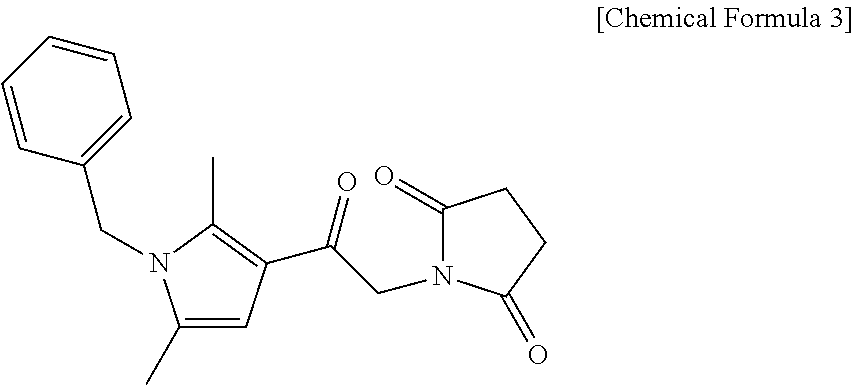
representing 1-(2-(1-benzyl-2,5-dimethyl-1H-pyrrol-3-yl)-2-oxoethyl)pyrrolidine-2,5-dione, an agonist having the following structure:

representing 1-(2-(1-benzyl-2,5-dimethyl-1H-pyrrol-3-yl)-2-oxoethyl)-3-methylimidazolindine-2,4,5-trione, an agonist having the following structure:
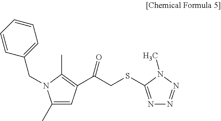
representing 1-(1-benzyl-2,5-dimethyl-1H-pyrrol-3-yl)-2-((1-methyl-1H-tetrazol-5-yl)thio)ethanone, and an agonist having the following structure:
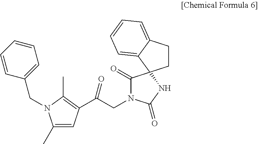
representing (S)-1-(2-(1-benzyl-2,5-dimethyl-1H-pyrrol-3-yl)-2-oxoethyl-2′,3′-dihydrospiro[imidazolidine-4,1′-indene]-2,5-dione.
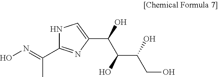
representing (E)-1-(4-((1R,2S,3R)-1,2,3,4-tetrahydroxybutyl)-1H-imidazol-2-yl)ethanone oxime (see, for example, Bagdanoff, J. T., et al., J. Med. Chem., Vol. 53, p. 8650-8662 (2010)), a substance having the following structure:

representing (1R,2S,3R)-1-(2-isoxazol-3-yl)-1H-imidazol-4-yl)butane-1,2,3,4-tetraol (Bagdanoff, et al., ibid), and a substance having the following structure:
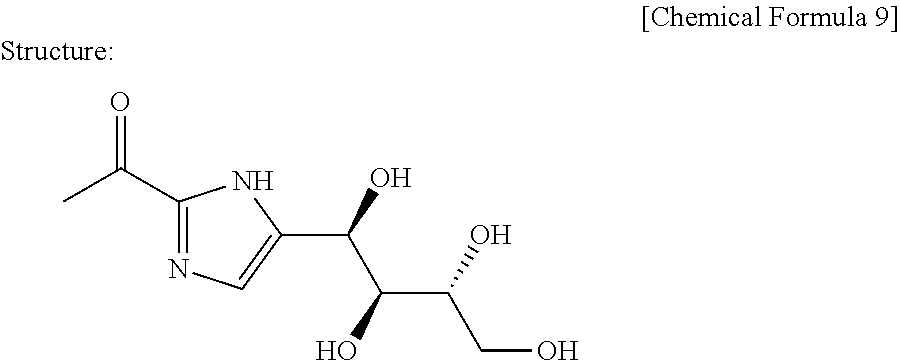
representing 1-(5-((1R,2S,3R)-1,2,3,4-tetrahydroxybutyl)-1H-imidazol-2-yl)ethanone (see, for example, Cayman Chemical Item Number 13222, Cayman Chemical Co., Michigan, USA).
Claims (12)
Priority Applications (2)
| Application Number | Priority Date | Filing Date | Title |
|---|---|---|---|
| US15/238,020 US10034889B2 (en) | 2013-03-01 | 2016-08-16 | Pharmaceutical composition including migratory factor for guiding pluripotent stem cells to damage |
| US15/642,534 US10369162B2 (en) | 2013-03-01 | 2017-07-06 | Pharmaceutical composition including migratory factor for guiding pluripotent stem cells to damage |
Applications Claiming Priority (5)
| Application Number | Priority Date | Filing Date | Title |
|---|---|---|---|
| JP2013-041161 | 2013-03-01 | ||
| JP2013041161 | 2013-03-01 | ||
| PCT/JP2014/055181 WO2014133170A1 (en) | 2013-03-01 | 2014-02-28 | Pharmaceutical composition including migratory factor for guiding pluripotent stem cells to injury |
| US201514771588A | 2015-08-31 | 2015-08-31 | |
| US15/238,020 US10034889B2 (en) | 2013-03-01 | 2016-08-16 | Pharmaceutical composition including migratory factor for guiding pluripotent stem cells to damage |
Related Parent Applications (3)
| Application Number | Title | Priority Date | Filing Date |
|---|---|---|---|
| US14/771,588 Continuation US9446033B2 (en) | 2013-03-01 | 2014-02-28 | Pharmaceutical composition including migratory factor for guiding pluripotent stem cells to injury |
| PCT/JP2014/055181 Continuation WO2014133170A1 (en) | 2013-03-01 | 2014-02-28 | Pharmaceutical composition including migratory factor for guiding pluripotent stem cells to injury |
| US201514711588A Continuation | 2014-05-16 | 2015-05-13 |
Related Child Applications (1)
| Application Number | Title | Priority Date | Filing Date |
|---|---|---|---|
| US15/642,534 Continuation US10369162B2 (en) | 2013-03-01 | 2017-07-06 | Pharmaceutical composition including migratory factor for guiding pluripotent stem cells to damage |
Publications (2)
| Publication Number | Publication Date |
|---|---|
| US20160354393A1 US20160354393A1 (en) | 2016-12-08 |
| US10034889B2 true US10034889B2 (en) | 2018-07-31 |
Family
ID=51428427
Family Applications (3)
| Application Number | Title | Priority Date | Filing Date |
|---|---|---|---|
| US14/771,588 Active US9446033B2 (en) | 2013-03-01 | 2014-02-28 | Pharmaceutical composition including migratory factor for guiding pluripotent stem cells to injury |
| US15/238,020 Active US10034889B2 (en) | 2013-03-01 | 2016-08-16 | Pharmaceutical composition including migratory factor for guiding pluripotent stem cells to damage |
| US15/642,534 Active US10369162B2 (en) | 2013-03-01 | 2017-07-06 | Pharmaceutical composition including migratory factor for guiding pluripotent stem cells to damage |
Family Applications Before (1)
| Application Number | Title | Priority Date | Filing Date |
|---|---|---|---|
| US14/771,588 Active US9446033B2 (en) | 2013-03-01 | 2014-02-28 | Pharmaceutical composition including migratory factor for guiding pluripotent stem cells to injury |
Family Applications After (1)
| Application Number | Title | Priority Date | Filing Date |
|---|---|---|---|
| US15/642,534 Active US10369162B2 (en) | 2013-03-01 | 2017-07-06 | Pharmaceutical composition including migratory factor for guiding pluripotent stem cells to damage |
Country Status (10)
| Country | Link |
|---|---|
| US (3) | US9446033B2 (en) |
| EP (1) | EP2962698B1 (en) |
| JP (2) | JP6511606B2 (en) |
| KR (1) | KR102180319B1 (en) |
| CN (1) | CN105188754B (en) |
| AU (1) | AU2014221659B2 (en) |
| CA (1) | CA2903415C (en) |
| ES (1) | ES2877555T3 (en) |
| SG (2) | SG10201710901RA (en) |
| WO (1) | WO2014133170A1 (en) |
Families Citing this family (12)
| Publication number | Priority date | Publication date | Assignee | Title |
|---|---|---|---|---|
| SG10201710901RA (en) | 2013-03-01 | 2018-02-27 | Clio Inc | Pharmaceutical composition including migratory factor for guiding pluripotent stem cells to damage |
| RU2683309C2 (en) * | 2014-03-14 | 2019-03-28 | Раквалиа Фарма Инк. | Azaspiro derivatives as trpm8 antagonists |
| JP6452107B2 (en) * | 2014-09-05 | 2019-01-16 | 国立大学法人 東京大学 | Pluripotent stem cells for the treatment of diabetic skin ulcers |
| EP3252153A4 (en) * | 2015-01-29 | 2018-07-18 | Kaneka Corporation | Method for producing cell aggregation |
| JP6570053B2 (en) * | 2015-03-25 | 2019-09-04 | 国立大学法人岐阜大学 | Non-injury site preparation containing sphingosine-1-phosphate receptor 2 activating compound |
| TWI711456B (en) * | 2015-05-29 | 2020-12-01 | 日商日本臟器製藥股份有限公司 | Extract from inflamed tissue inoculated with vaccinia virus and its use and method for determining or evaluating the same |
| KR101900818B1 (en) * | 2016-05-17 | 2018-09-20 | 주식회사 피토스 | Composition for promoting growth of stem cells Comprising phytospingosine-1-phosphate or its derivatives and composition for culturing stem cells comprising the same |
| JP7148402B2 (en) | 2016-08-03 | 2022-10-05 | 株式会社生命科学インスティテュート | Method for inducing differentiation of pluripotent stem cells in vitro |
| JP7102666B2 (en) * | 2018-01-25 | 2022-07-20 | 国立大学法人東海国立大学機構 | Use for Muse cell mobilizers and myocardial damage |
| DE102018105524A1 (en) * | 2018-03-09 | 2019-09-12 | Universität Duisburg-Essen | Use of modulators of sphingosine-1-phosphate signal transduction |
| EP3782702A1 (en) * | 2019-08-21 | 2021-02-24 | AC BioScience SA | Compounds and use thereof for the treatment of infectious diseases and cancer |
| EP4125880A1 (en) * | 2020-03-27 | 2023-02-08 | AC BioScience SA | A combination of flavonoids and sphingosine 1 phosphate lyase inhibitors for the treatment of lung inflammation |
Citations (8)
| Publication number | Priority date | Publication date | Assignee | Title |
|---|---|---|---|---|
| WO2008107436A1 (en) | 2007-03-06 | 2008-09-12 | Novartis Ag | Bicyclic organic compounds suitable for the treatment of inflammatory or allergic conditions |
| US20080248032A1 (en) | 2006-11-21 | 2008-10-09 | Children's Hospital & Research Center At Oakland | Compositions and methods for protection against cardiac and/or central nervous system tissue injury by inhibiting sphingosine-1-phosphate lyase |
| CN101361745A (en) | 2008-09-17 | 2009-02-11 | 中国医学科学院阜外心血管病医院 | Application of S1P in the preparation of drugs for inhibiting bone marrow mesenchymal stem cell apoptosis |
| WO2011007900A1 (en) | 2009-07-15 | 2011-01-20 | Dezawa Mari | Pluripotent stem cell that can be isolated from body tissue |
| WO2012129073A2 (en) | 2011-03-18 | 2012-09-27 | University Of Virginia Patent Foundation | Compositions and methods for tissue engineering and cell based therapies |
| US8642660B2 (en) | 2007-12-21 | 2014-02-04 | The University Of Rochester | Method for altering the lifespan of eukaryotic organisms |
| US20160008340A1 (en) | 2013-03-01 | 2016-01-14 | Clio, Inc. | Pharmaceutical composition including migratory factor for guiding pluripotent stem cells to injury |
| US9500642B2 (en) | 2012-04-18 | 2016-11-22 | Hemoshear, Llc | In vitro model for pathological or physiologic conditions |
Family Cites Families (2)
| Publication number | Priority date | Publication date | Assignee | Title |
|---|---|---|---|---|
| EP1970446B1 (en) | 2005-12-13 | 2011-08-03 | Kyoto University | Nuclear reprogramming factor |
| US9399758B2 (en) * | 2009-07-15 | 2016-07-26 | Mari Dezawa | SSEA3(+) pluripotent stem cell that can be isolated from body tissue |
-
2014
- 2014-02-28 SG SG10201710901RA patent/SG10201710901RA/en unknown
- 2014-02-28 KR KR1020157025190A patent/KR102180319B1/en not_active Expired - Fee Related
- 2014-02-28 US US14/771,588 patent/US9446033B2/en active Active
- 2014-02-28 ES ES14756298T patent/ES2877555T3/en active Active
- 2014-02-28 WO PCT/JP2014/055181 patent/WO2014133170A1/en not_active Ceased
- 2014-02-28 SG SG11201506845XA patent/SG11201506845XA/en unknown
- 2014-02-28 JP JP2015503070A patent/JP6511606B2/en active Active
- 2014-02-28 EP EP14756298.7A patent/EP2962698B1/en active Active
- 2014-02-28 CN CN201480011901.9A patent/CN105188754B/en active Active
- 2014-02-28 AU AU2014221659A patent/AU2014221659B2/en not_active Ceased
- 2014-02-28 CA CA2903415A patent/CA2903415C/en active Active
-
2016
- 2016-08-16 US US15/238,020 patent/US10034889B2/en active Active
-
2017
- 2017-07-06 US US15/642,534 patent/US10369162B2/en active Active
-
2018
- 2018-04-23 JP JP2018082545A patent/JP2018111734A/en active Pending
Patent Citations (14)
| Publication number | Priority date | Publication date | Assignee | Title |
|---|---|---|---|---|
| US20080248032A1 (en) | 2006-11-21 | 2008-10-09 | Children's Hospital & Research Center At Oakland | Compositions and methods for protection against cardiac and/or central nervous system tissue injury by inhibiting sphingosine-1-phosphate lyase |
| US8633211B2 (en) | 2007-03-06 | 2014-01-21 | Novartis Ag | Bicyclic organic compounds suitable for the treatment of inflammatory or allergic conditions |
| WO2008107436A1 (en) | 2007-03-06 | 2008-09-12 | Novartis Ag | Bicyclic organic compounds suitable for the treatment of inflammatory or allergic conditions |
| US20100113774A1 (en) | 2007-03-06 | 2010-05-06 | Roger John Taylor | Bicyclic organic compounds suitable for the treatment of inflammatory or allergic conditions |
| JP2010520256A (en) | 2007-03-06 | 2010-06-10 | ノバルティス アーゲー | Bicyclic organic compounds suitable for the treatment of inflammation or allergic symptoms |
| US8642660B2 (en) | 2007-12-21 | 2014-02-04 | The University Of Rochester | Method for altering the lifespan of eukaryotic organisms |
| CN101361745A (en) | 2008-09-17 | 2009-02-11 | 中国医学科学院阜外心血管病医院 | Application of S1P in the preparation of drugs for inhibiting bone marrow mesenchymal stem cell apoptosis |
| US20110070647A1 (en) | 2009-07-15 | 2011-03-24 | Mari Dezawa | Pluripotent stem cell that can be isolated from body tissue |
| WO2011007900A1 (en) | 2009-07-15 | 2011-01-20 | Dezawa Mari | Pluripotent stem cell that can be isolated from body tissue |
| WO2012129073A2 (en) | 2011-03-18 | 2012-09-27 | University Of Virginia Patent Foundation | Compositions and methods for tissue engineering and cell based therapies |
| US9500642B2 (en) | 2012-04-18 | 2016-11-22 | Hemoshear, Llc | In vitro model for pathological or physiologic conditions |
| US20160008340A1 (en) | 2013-03-01 | 2016-01-14 | Clio, Inc. | Pharmaceutical composition including migratory factor for guiding pluripotent stem cells to injury |
| US9446033B2 (en) | 2013-03-01 | 2016-09-20 | Clio, Inc. | Pharmaceutical composition including migratory factor for guiding pluripotent stem cells to injury |
| US20170304326A1 (en) | 2013-03-01 | 2017-10-26 | Clio, Inc. | Pharmaceutical composition including migratory factor for guiding pluripotent stem cells to damage |
Non-Patent Citations (14)
Also Published As
| Publication number | Publication date |
|---|---|
| US20160008340A1 (en) | 2016-01-14 |
| EP2962698A1 (en) | 2016-01-06 |
| WO2014133170A1 (en) | 2014-09-04 |
| KR20150125673A (en) | 2015-11-09 |
| AU2014221659A1 (en) | 2015-10-01 |
| KR102180319B1 (en) | 2020-11-18 |
| US20160354393A1 (en) | 2016-12-08 |
| JP6511606B2 (en) | 2019-05-15 |
| AU2014221659B2 (en) | 2018-12-20 |
| US10369162B2 (en) | 2019-08-06 |
| EP2962698B1 (en) | 2021-05-12 |
| SG10201710901RA (en) | 2018-02-27 |
| CA2903415A1 (en) | 2014-09-04 |
| US9446033B2 (en) | 2016-09-20 |
| ES2877555T3 (en) | 2021-11-17 |
| EP2962698A4 (en) | 2016-11-23 |
| SG11201506845XA (en) | 2015-09-29 |
| JPWO2014133170A1 (en) | 2017-02-09 |
| CN105188754A (en) | 2015-12-23 |
| CN105188754B (en) | 2019-11-26 |
| JP2018111734A (en) | 2018-07-19 |
| US20170304326A1 (en) | 2017-10-26 |
| CA2903415C (en) | 2021-04-20 |
Similar Documents
| Publication | Publication Date | Title |
|---|---|---|
| US10369162B2 (en) | Pharmaceutical composition including migratory factor for guiding pluripotent stem cells to damage | |
| Zhao et al. | Mesenchymal stem cell transplantation improves regional cardiac remodeling following ovine infarction | |
| CN101374538A (en) | Drugs that promote functional regeneration of damaged tissues | |
| Han et al. | Blockade of TGF-β signalling alleviates human adipose stem cell senescence induced by native ECM in obesity visceral white adipose tissue | |
| Guan et al. | CaMKK2 regulates macrophage polarization induced by matrix stiffness: implications for shaping the immune response in stiffened tissues | |
| KR102521638B1 (en) | A marker for diagnosis of nonalcoholic fatty liver disease | |
| US20210038688A1 (en) | Icam-1 marker and application thereof | |
| Nakashima et al. | POT1a deficiency in mesenchymal niches perturbs B-lymphopoiesis | |
| Davila | Investigating extracellular matrix-derived hydrogels from the lungs of patients with idiopathic pulmonary fibrosis as an in vitro disease model | |
| US20190192698A1 (en) | A method for obtaining indicator signals from a cell | |
| KR102747229B1 (en) | Biomarkers in cancer associated fibroblasts for predicting prognosis of head and neck cancer | |
| US20250171784A1 (en) | Application of mechanical-force sensitive macrophage subset in pancreatic cancer diagnosis or prognosis evaluation | |
| Martin García | Cathepsin K expressing cells and self-renewing cartilage in the murine zone of Ranvier and zebrafish | |
| Brown-Clay et al. | Pro-Metastatic Matrix Metalloproteinase Expression is Induced by the Invadopodial and Cytoskeletal Regulators Glycine-and Cysteine-Rich Proteins 1 and 2 | |
| Cassel | Creation and Application of Tools for Probing Cell Functions in Wound Healing and Disease Processes | |
| EP3991797A1 (en) | Cell structure and method for producing same | |
| Hu | Local niche mechanics within primary tumors regulate breast cancer metastatic organotropism through metabolic reprogramming | |
| Bignold | Targeting inflammatory mediator signalling in pericytes to resolve tissue fibrosis | |
| CN117043604A (en) | Diagnostic markers for non-alcoholic fatty liver disease | |
| CN119799893A (en) | Application of NAT10 in the diagnosis and treatment of glioma | |
| WO2015146697A1 (en) | Method for inhibiting cell death of cardiomyocytes | |
| Goldhaber et al. | Mark A. Aminzadeh, Russell G. Rogers, Mario Fournier, Rachel E. Tobin, Xuan Guan, 2 Martin K. Childers, 2 Allen M. Andres, David J. Taylor, Ahmed Ibrahim, Xiangming Ding, 3 Angelo Torrente | |
| WO2019133767A1 (en) | A method of in vitro diagnostic for prediction of drug efficacy | |
| Tuttle | The roles of Rabconnectin-3a and LIM domain only 7a in zebrafish neural crest migration | |
| KR20120010003A (en) | Use of Vern2 as a Marker for Graft-versus-Host Disease |
Legal Events
| Date | Code | Title | Description |
|---|---|---|---|
| AS | Assignment |
Owner name: NATIONAL UNIVERSITY CORPORATION NAGOYA UNIVERSITY, Free format text: ASSIGNMENT OF ASSIGNORS INTEREST;ASSIGNORS:DEZAWA, MARI;FUJIYOSHI, YOSHINORI;YOSHIDA, MASANORI;SIGNING DATES FROM 20150805 TO 20150806;REEL/FRAME:039661/0313 Owner name: CLIO, INC., JAPAN Free format text: ASSIGNMENT OF ASSIGNORS INTEREST;ASSIGNORS:DEZAWA, MARI;FUJIYOSHI, YOSHINORI;YOSHIDA, MASANORI;SIGNING DATES FROM 20150805 TO 20150806;REEL/FRAME:039661/0313 Owner name: TOHOKU UNIVERSITY, JAPAN Free format text: ASSIGNMENT OF ASSIGNORS INTEREST;ASSIGNORS:DEZAWA, MARI;FUJIYOSHI, YOSHINORI;YOSHIDA, MASANORI;SIGNING DATES FROM 20150805 TO 20150806;REEL/FRAME:039661/0313 |
|
| STCF | Information on status: patent grant |
Free format text: PATENTED CASE |
|
| CC | Certificate of correction | ||
| MAFP | Maintenance fee payment |
Free format text: PAYMENT OF MAINTENANCE FEE, 4TH YEAR, LARGE ENTITY (ORIGINAL EVENT CODE: M1551); ENTITY STATUS OF PATENT OWNER: LARGE ENTITY Year of fee payment: 4 |
|
| AS | Assignment |
Owner name: LIFE SCIENCE INSTITUTE, INC., JAPAN Free format text: MERGER AND CHANGE OF NAME;ASSIGNORS:CLIO, INC.;LIFE SCIENCE INSTITUTE, INC.;REEL/FRAME:064649/0794 Effective date: 20170101 Owner name: NATIONAL UNIVERSITY CORPORATION TOKAI NATIONALHIGHER EDUCATION AND RESEARCH SYSTEM, JAPAN Free format text: CHANGE OF NAME;ASSIGNOR:NATIONAL UNIVERSITY CORPORATION NAGOYA UNIVERSITY;REEL/FRAME:064659/0327 Effective date: 20200401 |
|
| AS | Assignment |
Owner name: TOHOKU UNIVERSITY, JAPAN Free format text: ASSIGNMENT OF ASSIGNORS INTEREST;ASSIGNOR:NATIONAL UNIVERSITY CORPORATION TOKAI NATIONAL HIGHER EDUCATION AND RESEARCH SYSTEM;REEL/FRAME:068010/0238 Effective date: 20231020 Owner name: TOHOKU UNIVERSITY, JAPAN Free format text: ASSIGNMENT OF ASSIGNORS INTEREST;ASSIGNOR:LIFE SCIENCE INSTITUTE, INC.;REEL/FRAME:068009/0757 Effective date: 20230928 Owner name: TOHOKU UNIVERSITY, JAPAN Free format text: ASSIGNMENT OF ASSIGNOR'S INTEREST;ASSIGNOR:LIFE SCIENCE INSTITUTE, INC.;REEL/FRAME:068009/0757 Effective date: 20230928 |
|
| FEPP | Fee payment procedure |
Free format text: ENTITY STATUS SET TO SMALL (ORIGINAL EVENT CODE: SMAL); ENTITY STATUS OF PATENT OWNER: SMALL ENTITY |
|
| MAFP | Maintenance fee payment |
Free format text: PAYMENT OF MAINTENANCE FEE, 8TH YR, SMALL ENTITY (ORIGINAL EVENT CODE: M2552); ENTITY STATUS OF PATENT OWNER: SMALL ENTITY Year of fee payment: 8 |