NL9202197A - Method and device for identifying a mycobacterium species responsible for mycobacterial infection. - Google Patents
Method and device for identifying a mycobacterium species responsible for mycobacterial infection. Download PDFInfo
- Publication number
- NL9202197A NL9202197A NL9202197A NL9202197A NL9202197A NL 9202197 A NL9202197 A NL 9202197A NL 9202197 A NL9202197 A NL 9202197A NL 9202197 A NL9202197 A NL 9202197A NL 9202197 A NL9202197 A NL 9202197A
- Authority
- NL
- Netherlands
- Prior art keywords
- antigen
- antibody
- serum
- mycobacterial
- kda
- Prior art date
Links
- 238000000034 method Methods 0.000 title claims abstract description 22
- 241000186359 Mycobacterium Species 0.000 title claims abstract description 16
- 208000027531 mycobacterial infectious disease Diseases 0.000 title claims abstract description 15
- 206010062207 Mycobacterial infection Diseases 0.000 title claims abstract description 10
- 102000036639 antigens Human genes 0.000 claims abstract description 67
- 108091007433 antigens Proteins 0.000 claims abstract description 67
- 239000000427 antigen Substances 0.000 claims abstract description 65
- 210000002966 serum Anatomy 0.000 claims abstract description 30
- 238000002360 preparation method Methods 0.000 claims abstract description 19
- 238000006243 chemical reaction Methods 0.000 claims abstract description 12
- 238000003119 immunoblot Methods 0.000 claims abstract description 12
- 230000027455 binding Effects 0.000 claims abstract description 11
- 241000894007 species Species 0.000 claims abstract description 10
- 238000009007 Diagnostic Kit Methods 0.000 claims abstract description 7
- 241001465754 Metazoa Species 0.000 claims abstract description 4
- 102000004190 Enzymes Human genes 0.000 claims description 17
- 108090000790 Enzymes Proteins 0.000 claims description 17
- 239000012528 membrane Substances 0.000 claims description 10
- 230000004044 response Effects 0.000 claims description 7
- 102000004169 proteins and genes Human genes 0.000 claims description 5
- 108090000623 proteins and genes Proteins 0.000 claims description 5
- 238000007598 dipping method Methods 0.000 claims description 4
- 239000000758 substrate Substances 0.000 claims description 4
- 230000000890 antigenic effect Effects 0.000 claims description 3
- 108040007629 peroxidase activity proteins Proteins 0.000 claims description 3
- 238000010186 staining Methods 0.000 claims description 3
- 239000000020 Nitrocellulose Substances 0.000 claims description 2
- 229920001220 nitrocellulos Polymers 0.000 claims description 2
- 102000003992 Peroxidases Human genes 0.000 claims 1
- 238000001962 electrophoresis Methods 0.000 abstract description 6
- 239000003153 chemical reaction reagent Substances 0.000 abstract 3
- 239000000499 gel Substances 0.000 description 21
- LOKCTEFSRHRXRJ-UHFFFAOYSA-I dipotassium trisodium dihydrogen phosphate hydrogen phosphate dichloride Chemical compound P(=O)(O)(O)[O-].[K+].P(=O)(O)([O-])[O-].[Na+].[Na+].[Cl-].[K+].[Cl-].[Na+] LOKCTEFSRHRXRJ-UHFFFAOYSA-I 0.000 description 16
- 239000002953 phosphate buffered saline Substances 0.000 description 16
- 239000000287 crude extract Substances 0.000 description 11
- 238000011534 incubation Methods 0.000 description 10
- 201000008827 tuberculosis Diseases 0.000 description 10
- 201000010099 disease Diseases 0.000 description 9
- 208000037265 diseases, disorders, signs and symptoms Diseases 0.000 description 9
- 208000011231 Crohn disease Diseases 0.000 description 8
- 230000001580 bacterial effect Effects 0.000 description 7
- 238000001514 detection method Methods 0.000 description 7
- 108091003079 Bovine Serum Albumin Proteins 0.000 description 6
- 229940098773 bovine serum albumin Drugs 0.000 description 6
- 239000007858 starting material Substances 0.000 description 6
- 238000001262 western blot Methods 0.000 description 6
- 241000894006 Bacteria Species 0.000 description 5
- 210000004027 cell Anatomy 0.000 description 5
- 230000028993 immune response Effects 0.000 description 5
- 238000003018 immunoassay Methods 0.000 description 5
- 206010024229 Leprosy Diseases 0.000 description 4
- 238000010790 dilution Methods 0.000 description 4
- 239000012895 dilution Substances 0.000 description 4
- 239000000203 mixture Substances 0.000 description 4
- 239000008188 pellet Substances 0.000 description 4
- 239000000243 solution Substances 0.000 description 4
- QKNYBSVHEMOAJP-UHFFFAOYSA-N 2-amino-2-(hydroxymethyl)propane-1,3-diol;hydron;chloride Chemical compound Cl.OCC(N)(CO)CO QKNYBSVHEMOAJP-UHFFFAOYSA-N 0.000 description 3
- 206010006049 Bovine Tuberculosis Diseases 0.000 description 3
- PEDCQBHIVMGVHV-UHFFFAOYSA-N Glycerine Chemical compound OCC(O)CO PEDCQBHIVMGVHV-UHFFFAOYSA-N 0.000 description 3
- 241000282412 Homo Species 0.000 description 3
- 108060003951 Immunoglobulin Proteins 0.000 description 3
- 238000004458 analytical method Methods 0.000 description 3
- 230000005875 antibody response Effects 0.000 description 3
- 238000003556 assay Methods 0.000 description 3
- 210000002421 cell wall Anatomy 0.000 description 3
- KRKNYBCHXYNGOX-UHFFFAOYSA-N citric acid Chemical compound OC(=O)CC(O)(C(O)=O)CC(O)=O KRKNYBCHXYNGOX-UHFFFAOYSA-N 0.000 description 3
- 210000000987 immune system Anatomy 0.000 description 3
- 102000018358 immunoglobulin Human genes 0.000 description 3
- 150000002632 lipids Chemical class 0.000 description 3
- 229920002401 polyacrylamide Polymers 0.000 description 3
- 235000010482 polyoxyethylene sorbitan monooleate Nutrition 0.000 description 3
- 229920000053 polysorbate 80 Polymers 0.000 description 3
- 235000018102 proteins Nutrition 0.000 description 3
- 239000007790 solid phase Substances 0.000 description 3
- HVCOBJNICQPDBP-UHFFFAOYSA-N 3-[3-[3,5-dihydroxy-6-methyl-4-(3,4,5-trihydroxy-6-methyloxan-2-yl)oxyoxan-2-yl]oxydecanoyloxy]decanoic acid;hydrate Chemical compound O.OC1C(OC(CC(=O)OC(CCCCCCC)CC(O)=O)CCCCCCC)OC(C)C(O)C1OC1C(O)C(O)C(O)C(C)O1 HVCOBJNICQPDBP-UHFFFAOYSA-N 0.000 description 2
- 208000030507 AIDS Diseases 0.000 description 2
- KCXVZYZYPLLWCC-UHFFFAOYSA-N EDTA Chemical compound OC(=O)CN(CC(O)=O)CCN(CC(O)=O)CC(O)=O KCXVZYZYPLLWCC-UHFFFAOYSA-N 0.000 description 2
- LFQSCWFLJHTTHZ-UHFFFAOYSA-N Ethanol Chemical compound CCO LFQSCWFLJHTTHZ-UHFFFAOYSA-N 0.000 description 2
- 229930186217 Glycolipid Natural products 0.000 description 2
- 206010024227 Lepromatous leprosy Diseases 0.000 description 2
- 238000003745 diagnosis Methods 0.000 description 2
- 230000028996 humoral immune response Effects 0.000 description 2
- 230000008348 humoral response Effects 0.000 description 2
- 229940072221 immunoglobulins Drugs 0.000 description 2
- 208000015181 infectious disease Diseases 0.000 description 2
- 238000004519 manufacturing process Methods 0.000 description 2
- 239000003147 molecular marker Substances 0.000 description 2
- 102000013415 peroxidase activity proteins Human genes 0.000 description 2
- 230000000405 serological effect Effects 0.000 description 2
- 238000010561 standard procedure Methods 0.000 description 2
- UCSJYZPVAKXKNQ-HZYVHMACSA-N streptomycin Chemical compound CN[C@H]1[C@H](O)[C@@H](O)[C@H](CO)O[C@H]1O[C@@H]1[C@](C=O)(O)[C@H](C)O[C@H]1O[C@@H]1[C@@H](NC(N)=N)[C@H](O)[C@@H](NC(N)=N)[C@H](O)[C@H]1O UCSJYZPVAKXKNQ-HZYVHMACSA-N 0.000 description 2
- 239000000725 suspension Substances 0.000 description 2
- 238000005406 washing Methods 0.000 description 2
- YRNWIFYIFSBPAU-UHFFFAOYSA-N 4-[4-(dimethylamino)phenyl]-n,n-dimethylaniline Chemical compound C1=CC(N(C)C)=CC=C1C1=CC=C(N(C)C)C=C1 YRNWIFYIFSBPAU-UHFFFAOYSA-N 0.000 description 1
- SATHPVQTSSUFFW-UHFFFAOYSA-N 4-[6-[(3,5-dihydroxy-4-methoxyoxan-2-yl)oxymethyl]-3,5-dihydroxy-4-methoxyoxan-2-yl]oxy-2-(hydroxymethyl)-6-methyloxane-3,5-diol Chemical compound OC1C(OC)C(O)COC1OCC1C(O)C(OC)C(O)C(OC2C(C(CO)OC(C)C2O)O)O1 SATHPVQTSSUFFW-UHFFFAOYSA-N 0.000 description 1
- HKJKONMZMPUGHJ-UHFFFAOYSA-N 4-amino-5-hydroxy-3-[(4-nitrophenyl)diazenyl]-6-phenyldiazenylnaphthalene-2,7-disulfonic acid Chemical compound OS(=O)(=O)C1=CC2=CC(S(O)(=O)=O)=C(N=NC=3C=CC=CC=3)C(O)=C2C(N)=C1N=NC1=CC=C([N+]([O-])=O)C=C1 HKJKONMZMPUGHJ-UHFFFAOYSA-N 0.000 description 1
- 208000031295 Animal disease Diseases 0.000 description 1
- 239000001904 Arabinogalactan Substances 0.000 description 1
- DCXYFEDJOCDNAF-UHFFFAOYSA-N Asparagine Natural products OC(=O)C(N)CC(N)=O DCXYFEDJOCDNAF-UHFFFAOYSA-N 0.000 description 1
- 208000023275 Autoimmune disease Diseases 0.000 description 1
- -1 Bromo, Chloro Indolyl phosphate Chemical compound 0.000 description 1
- 241000283707 Capra Species 0.000 description 1
- 239000004215 Carbon black (E152) Substances 0.000 description 1
- CURLTUGMZLYLDI-UHFFFAOYSA-N Carbon dioxide Chemical compound O=C=O CURLTUGMZLYLDI-UHFFFAOYSA-N 0.000 description 1
- 101710098119 Chaperonin GroEL 2 Proteins 0.000 description 1
- 102000016911 Deoxyribonucleases Human genes 0.000 description 1
- 108010053770 Deoxyribonucleases Proteins 0.000 description 1
- 201000004624 Dermatitis Diseases 0.000 description 1
- DCXYFEDJOCDNAF-REOHCLBHSA-N L-asparagine Chemical compound OC(=O)[C@@H](N)CC(N)=O DCXYFEDJOCDNAF-REOHCLBHSA-N 0.000 description 1
- 241000186367 Mycobacterium avium Species 0.000 description 1
- 241000187478 Mycobacterium chelonae Species 0.000 description 1
- 241000186365 Mycobacterium fortuitum Species 0.000 description 1
- 241000186364 Mycobacterium intracellulare Species 0.000 description 1
- 241000186362 Mycobacterium leprae Species 0.000 description 1
- 241000187492 Mycobacterium marinum Species 0.000 description 1
- 241000187490 Mycobacterium scrofulaceum Species 0.000 description 1
- 241000187917 Mycobacterium ulcerans Species 0.000 description 1
- 229910019142 PO4 Inorganic materials 0.000 description 1
- 229930182555 Penicillin Natural products 0.000 description 1
- JGSARLDLIJGVTE-MBNYWOFBSA-N Penicillin G Chemical compound N([C@H]1[C@H]2SC([C@@H](N2C1=O)C(O)=O)(C)C)C(=O)CC1=CC=CC=C1 JGSARLDLIJGVTE-MBNYWOFBSA-N 0.000 description 1
- 102000004160 Phosphoric Monoester Hydrolases Human genes 0.000 description 1
- 108090000608 Phosphoric Monoester Hydrolases Proteins 0.000 description 1
- 102000006382 Ribonucleases Human genes 0.000 description 1
- 108010083644 Ribonucleases Proteins 0.000 description 1
- 101150027663 Rpain gene Proteins 0.000 description 1
- 240000008042 Zea mays Species 0.000 description 1
- 238000010521 absorption reaction Methods 0.000 description 1
- 239000002253 acid Substances 0.000 description 1
- 150000007513 acids Chemical class 0.000 description 1
- 239000002671 adjuvant Substances 0.000 description 1
- 230000001355 anti-mycobacterial effect Effects 0.000 description 1
- 239000003926 antimycobacterial agent Substances 0.000 description 1
- 235000019312 arabinogalactan Nutrition 0.000 description 1
- 229960001230 asparagine Drugs 0.000 description 1
- 235000009582 asparagine Nutrition 0.000 description 1
- 230000001363 autoimmune Effects 0.000 description 1
- 239000001045 blue dye Substances 0.000 description 1
- UDSAIICHUKSCKT-UHFFFAOYSA-N bromophenol blue Chemical compound C1=C(Br)C(O)=C(Br)C=C1C1(C=2C=C(Br)C(O)=C(Br)C=2)C2=CC=CC=C2S(=O)(=O)O1 UDSAIICHUKSCKT-UHFFFAOYSA-N 0.000 description 1
- 235000011089 carbon dioxide Nutrition 0.000 description 1
- 210000000170 cell membrane Anatomy 0.000 description 1
- 238000005119 centrifugation Methods 0.000 description 1
- 239000011248 coating agent Substances 0.000 description 1
- 238000000576 coating method Methods 0.000 description 1
- 230000009918 complex formation Effects 0.000 description 1
- 150000001875 compounds Chemical class 0.000 description 1
- 238000001816 cooling Methods 0.000 description 1
- 230000037029 cross reaction Effects 0.000 description 1
- 210000000805 cytoplasm Anatomy 0.000 description 1
- 230000001086 cytosolic effect Effects 0.000 description 1
- 206010012601 diabetes mellitus Diseases 0.000 description 1
- 239000012153 distilled water Substances 0.000 description 1
- VHJLVAABSRFDPM-ZXZARUISSA-N dithioerythritol Chemical compound SC[C@H](O)[C@H](O)CS VHJLVAABSRFDPM-ZXZARUISSA-N 0.000 description 1
- 239000012636 effector Substances 0.000 description 1
- 230000000694 effects Effects 0.000 description 1
- 238000010828 elution Methods 0.000 description 1
- 230000007613 environmental effect Effects 0.000 description 1
- 150000002148 esters Chemical class 0.000 description 1
- 239000000284 extract Substances 0.000 description 1
- 238000001914 filtration Methods 0.000 description 1
- 239000001963 growth medium Substances 0.000 description 1
- 229930195733 hydrocarbon Natural products 0.000 description 1
- 150000002430 hydrocarbons Chemical class 0.000 description 1
- 230000002519 immonomodulatory effect Effects 0.000 description 1
- 230000005965 immune activity Effects 0.000 description 1
- 230000001900 immune effect Effects 0.000 description 1
- 230000008073 immune recognition Effects 0.000 description 1
- 230000036039 immunity Effects 0.000 description 1
- 229940127121 immunoconjugate Drugs 0.000 description 1
- 230000002134 immunopathologic effect Effects 0.000 description 1
- 230000001506 immunosuppresive effect Effects 0.000 description 1
- 239000012535 impurity Substances 0.000 description 1
- 239000006166 lysate Substances 0.000 description 1
- 210000002540 macrophage Anatomy 0.000 description 1
- 239000000463 material Substances 0.000 description 1
- 239000011159 matrix material Substances 0.000 description 1
- 230000001404 mediated effect Effects 0.000 description 1
- 239000002609 medium Substances 0.000 description 1
- 239000013642 negative control Substances 0.000 description 1
- JPXMTWWFLBLUCD-UHFFFAOYSA-N nitro blue tetrazolium(2+) Chemical compound COC1=CC(C=2C=C(OC)C(=CC=2)[N+]=2N(N=C(N=2)C=2C=CC=CC=2)C=2C=CC(=CC=2)[N+]([O-])=O)=CC=C1[N+]1=NC(C=2C=CC=CC=2)=NN1C1=CC=C([N+]([O-])=O)C=C1 JPXMTWWFLBLUCD-UHFFFAOYSA-N 0.000 description 1
- 230000003287 optical effect Effects 0.000 description 1
- 244000052769 pathogen Species 0.000 description 1
- 230000001717 pathogenic effect Effects 0.000 description 1
- 229940049954 penicillin Drugs 0.000 description 1
- 239000010452 phosphate Substances 0.000 description 1
- 206010039073 rheumatoid arthritis Diseases 0.000 description 1
- 210000003705 ribosome Anatomy 0.000 description 1
- 201000000306 sarcoidosis Diseases 0.000 description 1
- 239000013049 sediment Substances 0.000 description 1
- 239000007787 solid Substances 0.000 description 1
- 230000009870 specific binding Effects 0.000 description 1
- 239000012192 staining solution Substances 0.000 description 1
- 238000003756 stirring Methods 0.000 description 1
- 239000012089 stop solution Substances 0.000 description 1
- 229960005322 streptomycin Drugs 0.000 description 1
- 239000006228 supernatant Substances 0.000 description 1
- YWYZEGXAUVWDED-UHFFFAOYSA-N triammonium citrate Chemical compound [NH4+].[NH4+].[NH4+].[O-]C(=O)CC(O)(CC([O-])=O)C([O-])=O YWYZEGXAUVWDED-UHFFFAOYSA-N 0.000 description 1
- XLYOFNOQVPJJNP-UHFFFAOYSA-N water Chemical compound O XLYOFNOQVPJJNP-UHFFFAOYSA-N 0.000 description 1
Classifications
-
- G—PHYSICS
- G01—MEASURING; TESTING
- G01N—INVESTIGATING OR ANALYSING MATERIALS BY DETERMINING THEIR CHEMICAL OR PHYSICAL PROPERTIES
- G01N33/00—Investigating or analysing materials by specific methods not covered by groups G01N1/00 - G01N31/00
- G01N33/48—Biological material, e.g. blood, urine; Haemocytometers
- G01N33/50—Chemical analysis of biological material, e.g. blood, urine; Testing involving biospecific ligand binding methods; Immunological testing
- G01N33/53—Immunoassay; Biospecific binding assay; Materials therefor
- G01N33/543—Immunoassay; Biospecific binding assay; Materials therefor with an insoluble carrier for immobilising immunochemicals
- G01N33/54366—Apparatus specially adapted for solid-phase testing
-
- G—PHYSICS
- G01—MEASURING; TESTING
- G01N—INVESTIGATING OR ANALYSING MATERIALS BY DETERMINING THEIR CHEMICAL OR PHYSICAL PROPERTIES
- G01N33/00—Investigating or analysing materials by specific methods not covered by groups G01N1/00 - G01N31/00
- G01N33/48—Biological material, e.g. blood, urine; Haemocytometers
- G01N33/50—Chemical analysis of biological material, e.g. blood, urine; Testing involving biospecific ligand binding methods; Immunological testing
- G01N33/53—Immunoassay; Biospecific binding assay; Materials therefor
- G01N33/569—Immunoassay; Biospecific binding assay; Materials therefor for microorganisms, e.g. protozoa, bacteria, viruses
- G01N33/56911—Bacteria
- G01N33/5695—Mycobacteria
Landscapes
- Health & Medical Sciences (AREA)
- Life Sciences & Earth Sciences (AREA)
- Immunology (AREA)
- Engineering & Computer Science (AREA)
- Molecular Biology (AREA)
- Biomedical Technology (AREA)
- Chemical & Material Sciences (AREA)
- Hematology (AREA)
- Urology & Nephrology (AREA)
- Food Science & Technology (AREA)
- General Physics & Mathematics (AREA)
- Cell Biology (AREA)
- Biotechnology (AREA)
- Medicinal Chemistry (AREA)
- Physics & Mathematics (AREA)
- Analytical Chemistry (AREA)
- Biochemistry (AREA)
- General Health & Medical Sciences (AREA)
- Microbiology (AREA)
- Pathology (AREA)
- Tropical Medicine & Parasitology (AREA)
- Virology (AREA)
- Measuring Or Testing Involving Enzymes Or Micro-Organisms (AREA)
- Peptides Or Proteins (AREA)
- Medicines Containing Antibodies Or Antigens For Use As Internal Diagnostic Agents (AREA)
- Micro-Organisms Or Cultivation Processes Thereof (AREA)
- Preparation Of Compounds By Using Micro-Organisms (AREA)
Abstract
Description
WERKWIJZE EN INRICHTING VOOR HET IDENTIFICEREN VAN EEN VOOR EEN MYCOBACTERIËLE INFECTIE VERANTWOORDELIJK MYCOBACTERIUM SPECIES.METHOD AND APPARATUS FOR IDENTIFYING A MYCOBACTERIAL INFECTION RESPONSIBLE FOR A MYCOBACTERIAL INFECTION.
De uitvinding heeft betrekking op een werkwijze voor het identificeren van een voor een mycobacteriële infectie in mens of dier verantwoordelijk Mycobacterium species. De uitvinding betreft verder diagnostische kits voor gebruik bij de werkwijze.The invention relates to a method for identifying a Mycobacterium species responsible for mycobacterial infection in humans or animals. The invention further relates to diagnostic kits for use in the method.
Het genus Mycobacterium dat ongeveer 50 species bevat is verantwoordelijk voor een aantal humane en dierlijke ziekten, die gezamenlijk bekend staan als de mycobacteri-osen. De bekendsten bij de mens zijn lepra, veroorzaakt door M. leprae, dat wereldwijd meer dan 10 miljoen mensen treft, en tuberculose, gewoonlijk veroorzaakt door M. tuberculosis, waarvan jaarlijks tenminste 10 miljoen nieuwe gevallen voorkomen. De meeste andere mycobacteriën komen normaal gesproken slechts voor als omgevingssaprofyten, maar kunnen ook opportunistische ziekten veroorzaken. Dit gebeurt gewoonlijk, maar niet slechts, bij personen, die problemen hebben met hun immuunsysteem, zoals AIDS-patiënten en personen welke immuno-suppressie ondergaan. Deze opportunistische soorten omvatten het langzaam groeiende species M. avium en de nauw daarmee verwante M. intracellulare en M. scroful-aceum (vaak samen het MAIS-complex genoemd), M. kansasi, M. marinum en M. ulcerans, en de snel groeiende species M. chelonae en M. fortuitum. Hoewel zij vroeger zeldzaam waren neemt de incidentie van opportunistische mycobacteriële ziekten en tuberculose in de westerse wereld parallel toe met de incidentie van AIDS. Daarnaast is er beperkt maar toenemend bewijs dat mycobacteriën of antigenen daarvan een directe of indirecte rol spelen in de etiologie van een veelvoud van andere ziekten, zoals sarcoidosis en de ziekte van Crohn, en verschillende auto-immuunziekten zoals autoimmune dermatitis, reumatoïde arthritis en diabetes. Dit zou te wijten kunnen zijn aan een structurele mimicry tussen epitopen van mycobacteriën en die van de gastheer.The genus Mycobacterium, which contains about 50 species, is responsible for a number of human and animal diseases, collectively known as the mycobacterioses. The best known in humans are leprosy, caused by M. leprae, which affects more than 10 million people worldwide, and tuberculosis, usually caused by M. tuberculosis, of which at least 10 million new cases occur every year. Most other mycobacteria normally only occur as environmental saprophytes, but can also cause opportunistic diseases. This usually happens, but not only, in individuals who have problems with their immune systems, such as AIDS patients and those who are immunosuppressed. These opportunistic species include the slow-growing species M. avium and the closely related M. intracellulare and M. scroful-aceum (often collectively referred to as the MAIS complex), M. kansasi, M. marinum, and M. ulcerans, and the growing species M. chelonae and M. fortuitum. Although rare in the past, the incidence of opportunistic mycobacterial diseases and tuberculosis in the Western world is increasing in parallel with the incidence of AIDS. In addition, there is limited but increasing evidence that mycobacteria or their antigens play a direct or indirect role in the etiology of a multitude of other diseases, such as sarcoidosis and Crohn's disease, and various autoimmune diseases such as autoimmune dermatitis, rheumatoid arthritis and diabetes. This could be due to a structural mimicry between epitopes of mycobacteria and that of the host.
De celwanden van mycobacteriën zijn zeer complex en bevatten vele lipiden, sommige met structuren die uniek zijn voor het genus. Deze structuren omvatten mycoline-zuren en esters, peptido-glycolipide, arabino-galactan en lipo-arabi-no-manan. De lipiderijke mycobacteriële celwanden zijn verantwoordelijk voor de kenmerkende kleuringseigenschappen van de mycobacteriën. Zij stellen mycobacteriën eveneens in staat een aanval door het immuunsysteem van de gastheer af te slaan. Een aantal species is, wanneer zij eenmaal zijn opgenomen in macrofagen, in staat zichzelf te omringen met een dikke laag van uitgescheiden lipiden.The cell walls of mycobacteria are very complex and contain many lipids, some with structures unique to the genus. These structures include mycolic acids and esters, peptido glycolipid, arabino-galactan and lipo-arabi-no-manan. The lipid-rich mycobacterial cell walls are responsible for the characteristic staining properties of the mycobacteria. They also enable mycobacteria to repel an attack by the host's immune system. A number of species, once incorporated into macrophages, are able to surround themselves with a thick layer of secreted lipids.
Veel verschillende componenten van de mycobacteriën gaan een interaktie aan met het immuunsysteem. Deze componenten omvatten eiwit- en koolwaterstofantigenen, welke ofwel aktief uitgescheiden kunnen worden door de mycobacteriën, ofwel deel uitmaken van de celwand of celmembraan. Daarnaast kunnen zij aanwezig zijn in het cytoplasma, bijvoorbeeld in de cytoplasmatische matrix, ribosomen en enzymen. Mycobacteriën bezitten eveneens immuno-modulerende componenten, zoals immuno-onderdrukkende verbindingen en adjuvantia. Dientengevolge kan één enkel mycobacterieel species een grote verscheidenheid aan immuun-responsen in verschillende vormen en met diverse specificiteiten induceren. Het is derhalve moeilijk immuun-responsen tegen spe-cies-specifieke componenten te onderscheiden van kruisreacties. Het is om deze reden derhalve moeilijk gebleken eiwit-antigenen af te leiden die geschikt zijn voor de detectie van species-specifieke humorale responsen als een basis voor een zeer gevoelige en specifieke serodiagnosti-sche test voor tuberculose. Doordat de mycobacteriën veel in de omgeving voorkomen, bevat humaan serum bijna altijd anti-mycobacteriële antilichamen.Many different components of the mycobacteria interact with the immune system. These components include protein and hydrocarbon antigens, which can either be actively secreted by the mycobacteria or form part of the cell wall or cell membrane. In addition, they can be present in the cytoplasm, for example in the cytoplasmic matrix, ribosomes and enzymes. Mycobacteria also have immuno-modulating components, such as immunosuppressive compounds and adjuvants. As a result, a single mycobacterial species can induce a wide variety of immune responses in different shapes and with different specificities. It is therefore difficult to distinguish immune responses against specific components from cross-reactions. It has therefore been difficult for this reason to derive protein antigens suitable for the detection of species-specific humoral responses as a basis for a highly sensitive and specific serodiagnostic test for tuberculosis. Because the mycobacteria are abundant in the environment, human serum almost always contains anti-mycobacterial antibodies.
Met het oog op de problemen met de specificiteit van eiwit-antigenen hebben een aantal onderzoekers, waaronder de onderhavige uitvinders, hun aandacht gericht op species-specifieke glycolipide-antigenen voor de detectie van specifieke humorale immuun-responsen. Hoewel de immuunreactivi-teit tegen mycobacteriën van het cel-gemedieerde type is en de humorale immuun-responsen waarschijnlijk een ondergeschikte rol spelen in het totale effector-mechanisme van mycobacteriële immuniteit en immunopathologie, zouden studies naar de antilichaam-respons tegen immuno-dominante mycobacteriële kruisreactieve antigeen componenten (verder te noemen Im-KRAC) een licht kunnen werpen op het variërend vermogen van de gastheer verschillende mycobacteriële anti-genen te herkennen. Zij zouden derhalve indirecte informatie kunnen verschaffen omtrent de aard van de immuun-herkenning van, en respons tegen een bepaalde mycobacteriële pathogeen.In view of the problems with the specificity of protein antigens, a number of researchers, including the present inventors, have turned their attention to species-specific glycolipid antigens for the detection of specific humoral immune responses. Although the immune activity against mycobacteria is of the cell-mediated type and the humoral immune responses are likely to play a minor role in the overall effector mechanism of mycobacterial immunity and immunopathology, studies of the antibody response against immuno-dominant mycobacterial cross-reactive antigenic components (hereinafter referred to as Im-KRAC) can shed light on the varying ability of the host to recognize different mycobacterial anti-genes. They could therefore provide indirect information about the nature of the immune recognition and response to a particular mycobacterial pathogen.
Gevonden is nu dat de klinische manifestatie van mycobacteriële ziekten gerelateerd blijkt te zijn aan het variërend vermogen van een individuele gastheer een humorale respons te produceren tegen verschillende mycobacteriële immuno-kruisreactieve antigeen componenten (Im-KRAC). Elke mycobacteriële infectie wekt een eigen specifieke antili-chaamrespons op tegen een aantal gespecificeerde antigenen. Analyse van de antilichaam-respons door middel van immu-noblotting heeft aangetoond dat de immunodominante Im-KRAC variëren in overeenstemming met de immunopathologische manifestatie van de mycobacteriële ziekten. Gebleken is dat de sera van individuen, welke geïnfecteerd zijn met verschillende Mycobacterium species, op immunoblots van mycobacteriële antigenen verschillende en onderscheidende bandenpatronen veroorzaken.It has now been found that the clinical manifestation of mycobacterial diseases appears to be related to the varying ability of an individual host to produce a humoral response against various mycobacterial immuno-cross-reactive antigen components (Im-KRAC). Each mycobacterial infection elicits its own specific antibody response against a number of specified antigens. Analysis of the antibody response by immunoblotting has shown that the immunodominant Im-KRAC varies in accordance with the immunopathological manifestation of the mycobacterial diseases. It has been found that the sera of individuals infected with different Mycobacterium species on immunoblots of mycobacterial antigens cause different and distinctive banding patterns.
Deze vinding vormt de basis voor de onderhavige uitvinding, waardoor een werkwijze verschaft wordt voor het identificeren van een voor een mycobacteriële infectie in mens of dier verantwoordelijk Mycobacterium species, omvattende de stappen; a) het kiezen van een geschikt mycobacteriële species en stam; b) het bereiden van een tenminste één mycobacterieel antigeen, respectievelijk antigeenpreparaat; c) het binden van het antigeen, respectievelijk het antigeenpreparaat aan een geschikte drager; d) het laten reageren van het gebonden antigeen met antilichamen uit serum van een met een Mycobacterium species geïnfecteerd individu; e) het zichtbaar maken van antigeen-antilichaam reacties voor een geschikte antilichaam (sub-)klasse; en f) het aan de hand van de zichtbaar gemaakte reacties identificeren van het verantwoordelijke Mycobacterium-species .This finding forms the basis for the present invention, providing a method for identifying a Mycobacterium species responsible for mycobacterial infection in humans or animals, comprising the steps; a) selecting an appropriate mycobacterial species and strain; b) preparing an at least one mycobacterial antigen or antigen preparation, respectively; c) binding the antigen or antigen preparation to a suitable carrier; d) reacting the bound antigen with serum antibodies of an individual infected with a Mycobacterium species; e) visualizing antigen-antibody responses for a suitable antibody (sub) class; and f) identifying the responsible Mycobacterium species from the visualized responses.
Bij voorkeur wordt het antigeenpreparaat voorafgaand aan stap c) electroforetisch gescheiden en is de drager een membraan waaraan het antigeen door middel van electroblot-ting wordt gebonden. Dit proces wordt immunoblotting genoemd.Preferably, the antigen preparation is electrophoretically separated prior to step c) and the support is a membrane to which the antigen is bonded by electroblotting. This process is called immunoblotting.
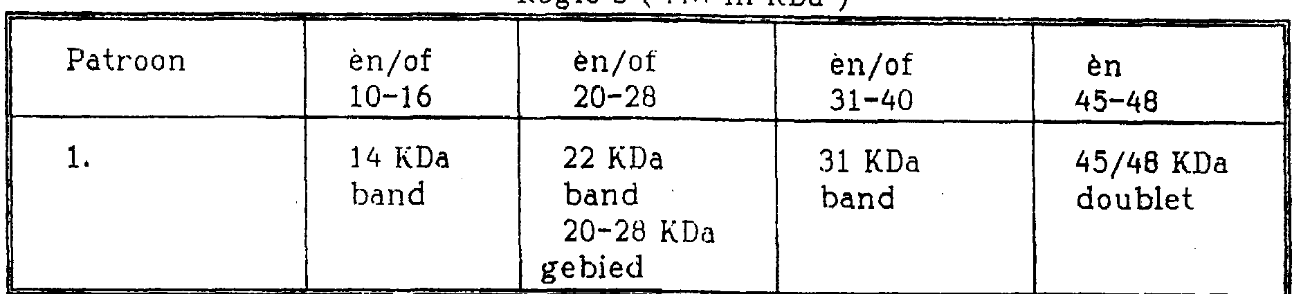
De Im-KRAC omvatten namelijk een aantal antigenen met specifieke molecuulgewichten, welke, zoals nu gevonden is, na immunoblotting een bindingspatroon vertonen, dat correleert met de ziekte, respectievelijk infectie. De specifieke bandenpatronen worden gekenmerkt door de aanwezigheid of afwezigheid van vier individuele componenten, bijvoorbeeld : - een regio die verschillende geprononceerde banden en/of overlopende banden omvat, waarneembaar als een smeer ("gebied"); - scherpe enkele banden, die sterk positief zijn ("band"); - scherpe dubbele banden, die sterk positief zijn ("doublet") ; en - andere positieve banden ("extra banden").Namely, the Im-KRAC comprise a number of specific molecular weight antigens which, as now found, exhibit a binding pattern after immunoblotting that correlates with the disease and infection, respectively. The specific tire patterns are characterized by the presence or absence of four individual components, for example: - a region comprising several pronounced tires and / or overflowing tires, perceivable as a smear ("area"); - sharp single bands, which are strongly positive ("band"); - sharp double bands, which are strongly positive ("doublet"); and - other positive ties ("extra ties").
Zie voor een overzicht van de verschillende antigenen, hun molecuulgewichten en bindingskarakteristieken tabel 1.For an overview of the different antigens, their molecular weights and binding characteristics, see Table 1.
k #1 A Ak # 1 A A
fabel l:fable l:
Overzicht van karakteristieke bindingspatronen van mycobac-teriële Immuno Kruisreactieve Antigeen ComponentenOverview of Characteristic Binding Patterns of Mycobacterial Immuno Cross-Reactive Antigen Components
Tabel l:Table 1:

__ T: Humane Tuberculose L: Lepra C: Crohn’s Disease B: Bovine Tuberculose .1: Johne’s DiseaseT: Human Tuberculosis L: Leprosy C: Crohn's Disease B: Bovine Tuberculosis. 1: John's Disease
De mycobacteriosen worden alle gekenmerkt door een specifiek bandenpatroon, dat gevormd wordt wanneer een blot met daarop een op grootte gescheiden antigeenpreparaat van mycobacteriën geïncubeerd wordt met serum van een geïnfecteerd individu.The mycobacterioses are all characterized by a specific banding pattern, which is formed when a blot containing a size-separated antigen preparation of mycobacteria is incubated with serum from an infected individual.
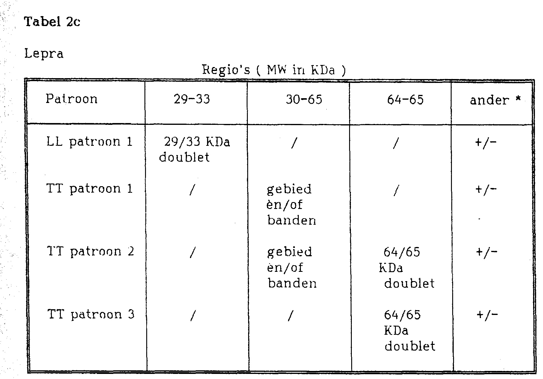
De onderstaande tabellen 2a t/m 2c geven een overzicht van de bandenpatronen van een aantal mycobacteriële ziekten.Tables 2a through 2c below summarize the band patterns of a number of mycobacterial diseases.
Tabel 2a:Table 2a:
Bovine TuberculoseBovine Tuberculosis
Rpain’s (Rpain's (
MW in WTia 'iMW in WTia 'i
Johne’s DiseaseJohne's Disease

Tabel 2b:Table 2b:
Humane Tuberculose.Human Tuberculosis.
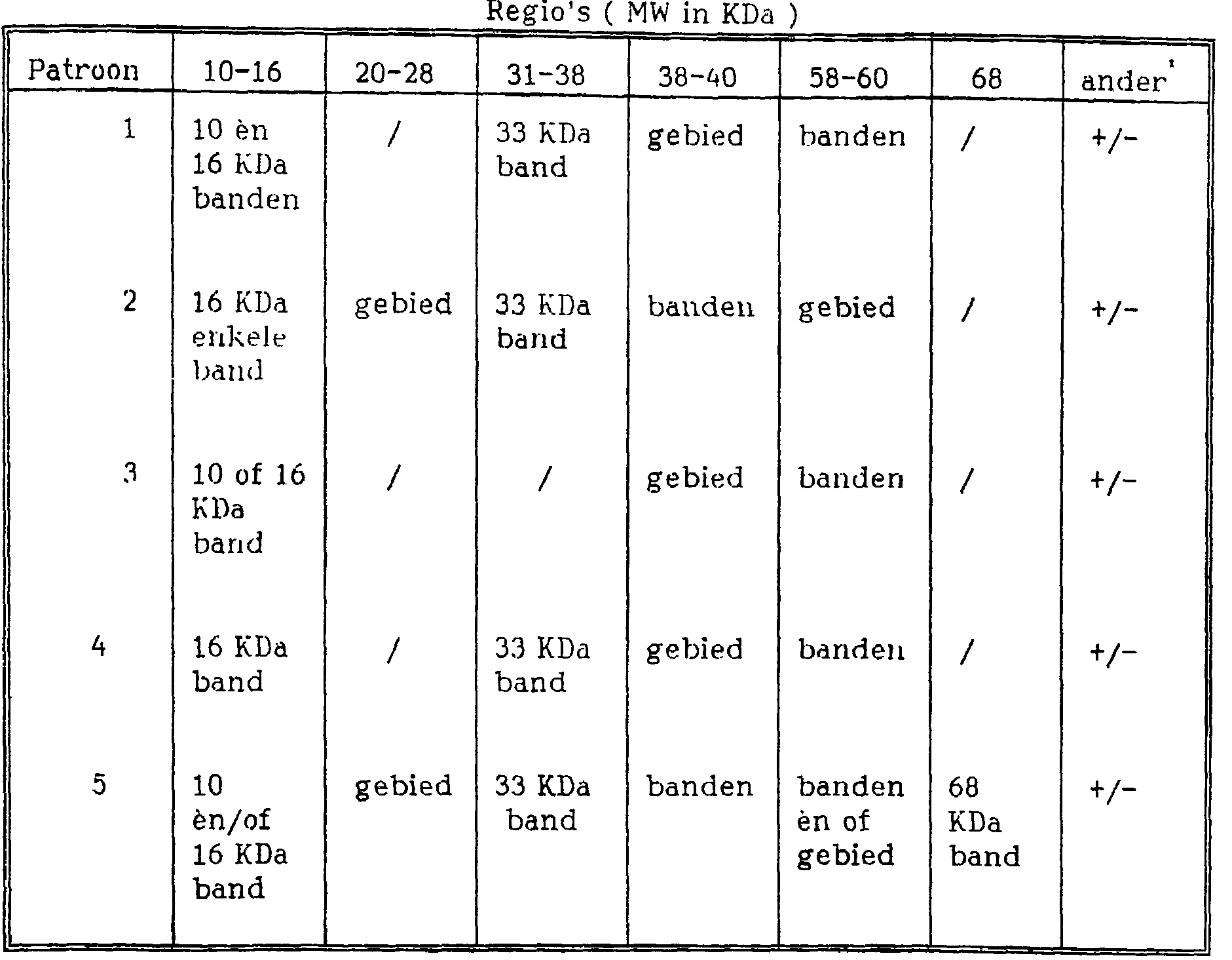
* extra banden èn/of gebieden kunnen aankleuren maar zijn niet diagnostisch voor Humane Tuberculose* extra bands and / or areas may stain but are not diagnostic for Human Tuberculosis
* extra banden èn/of gebieden kunnen aankleuren maar zijn niet diagnostisch voor Human Tuberculose* extra bands and / or areas can stain but are not diagnostic for Human Tuberculosis
* extr a banden èn/of gebieden kunnen aankleuren maar zijn niet diagnos-tixch voor Crohn's Disease.* extr a bands and / or areas can stain but are not a diagnosis for Crohn's Disease.
Wanneer een immunoblot gebruikt wordt kunnen twee vragen beantwoord worden. Ten eerste zal de aanwezigheid van elk positief bandenpatroon de vraag beantwoorden of er een mycobacteriële infectie aanwezig is. Ten tweede geeft de aanwezigheid van specifieke bandenpatronen aan welk myco-bacterieel species de infectie veroorzaakt heeft, en derhalve wat de aard en etiologie van de ziekte zal zijn.When an immunoblot is used, two questions can be answered. First, the presence of any positive banding pattern will answer the question of whether a mycobacterial infection is present. Second, the presence of specific band patterns indicates which mycobacterial species has caused the infection, and therefore what the nature and etiology of the disease will be.
De uitvinding betreft verder een heterogene enzym-immunobepaling.The invention further relates to a heterogeneous enzyme immunoassay.
Uit de patronen in de immunoblotting volgt welke mycobacteriële antigenen, respectievelijk antigeen preparaten, voor de diagnostiek van één bepaalde ziekte in aanmerking komen.It follows from the patterns in the immunoblotting which mycobacterial antigens and antigen preparations are suitable for the diagnosis of one specific disease.
Het antigeen voor een heterogene enzym-immunobepa-i ling wordt bij voorkeur gekozen uit de groep die bestaat uit mycobacteriële immuno-kruisreactieve antigeencomponenten met een molecuulgewicht van 29-33 KDa, 45-48 KDa, 64-65 KDa en een fractie die aangeduid wordt met de term KP-100. Deze ImKRAC kunnen afzonderlijk of in combinatie met elkaar ) worden gebruikt voor de serologische diagnostiek van de correlerende ziekten in een heterogene enzym-immunobepaling (EIA).The antigen for a heterogeneous enzyme immunoassay is preferably selected from the group consisting of mycobacterial immuno-cross-reactive antigen components having a molecular weight of 29-33 KDa, 45-48 KDa, 64-65 KDa and a fraction designated with the term KP-100. These ImKRAC can be used individually or in combination for the serological diagnostics of the correlating diseases in a heterogeneous enzyme immunoassay (EIA).
In deze bepalingsvorm worden met een standaard enzym gelabelde antilichaam-conjugaten gebruikt. Een belangrijk > gegeven is dat de enzymaktiviteit niet verandert tijdens de immunologische reactie.Antibody-labeled antibody conjugates are used in this assay. An important fact is that the enzyme activity does not change during the immunological response.
Voor het testen van de immuunrespons tegen de geselecteerde antigenen, bij patiënten, wordt bijvoorbeeld gebruik gemaakt van microtiterplaten ("Solid Phase"). De D antigenen worden middels standaard gepubliceerde technieken irreversibel geïmmobiliseerd aan het oppervlak van de putjes in een dergelijke microtiterplaat.For example, to test the immune response against the selected antigens, in patients, microtiter plates ("Solid Phase") are used. The D antigens are irreversibly immobilized to the surface of the wells in such a microtiter plate by standard published techniques.
Deze binding vindt plaats met behoud van specifieke antigene determinanten op de gebruikte antigenen. Na incuba-5 tie met serum, in de putjes van de microtiterplaat, kunnen daarin aanwezige antilichamen specifiek een complex vormen met de irreversibel gebonden antigenen.This binding takes place while retaining specific antigenic determinants on the antigens used. After incubation with serum, in the wells of the microtiter plate, antibodies contained therein can specifically complex with the irreversibly bound antigens.
Na verwijdering van ongebonden serumcomponenten worden gebonden antilichamen gedetecteerd met behulp van een met een enzym gelabeld anti-antilichaam antilichaam.After removal of unbound serum components, bound antibodies are detected using an enzyme-labeled anti-antibody antibody.
Binding van het enzym is alleen mogelijk als specifieke antilichamen zich aan de geïmmobiliseerde antigenen hebben gehecht. Substraatomzetting door het gebonden enzym tot een visueel of fotometrisch waarneembaar signaal is daardoor direct gerelateerd aan het aanwezig zijn van specifieke antilichamen in het geteste serum.Binding of the enzyme is only possible if specific antibodies have attached to the immobilized antigens. Substrate conversion by the bound enzyme to a visually or photometrically detectable signal is therefore directly related to the presence of specific antibodies in the tested serum.
De keuze van specificiteit van het enzym-gebonden anti-antilichaam antilichaam bepaalt het type reactie dat plaatsvindt. Zo kan het in sommige gevallen wenselijk zijn alleen de specifiek gebonden immuno-globulinen van het IgG type aan te tonen, terwijl in andere gevallen juist immuno-globulinen van het type IgA en/of IgM aangetoond worden.The choice of specificity of the enzyme-linked anti-antibody antibody determines the type of reaction that takes place. For example, in some cases it may be desirable to demonstrate only the specifically bound immunoglobulins of the IgG type, while in others it is true to demonstrate immunoglobulins of the IgA and / or IgM type.
De combinatie van antigeen en immunoglobuline type is bepalend voor de specificiteit van de test.The combination of antigen and immunoglobulin type determines the specificity of the test.
Genoemde werkwijzen, dat wil zeggen de immunoblot en de EIA kunnen ter confirmatie van elkaar gebruikt worden.Said methods, i.e. the immunoblot and the EIA can be used to confirm each other.
Daarnaast kan voor de serologische diagnostiek gebaseerd op genoemde antigenen gebruik worden gemaakt van een teststaafje als solid phase.In addition, for serological diagnostics based on the antigens mentioned, a test rod can be used as a solid phase.
Een bijzonder voordelige uitvoeringsvorm van de uitvinding betreft een teststaafje de zogeheten "dipstick", die als vaste fase in de heterogene enzym immuunbepaling gebruikt wordt.A particularly advantageous embodiment of the invention concerns a test stick, the so-called "dipstick", which is used as a solid phase in the heterogeneous enzyme immunoassay.
Genoemde mycobacteriële antigenen kunnen irreversibel gebonden worden aan een dergelijke dipstick.Said mycobacterial antigens can be irreversibly bound to such a dipstick.
Het antigeen wordt in reactie gebracht met antilichaam uit te testen serum door het dopen van de dipstick in een te testen serummonster. Het gevormde antigeen-antili-chaam complex kan zichtbaar gemaakt worden door de dipstick vervolgens te dopen in een anti-antilichaam antilichaam-enzymconjugaat oplossing.The antigen is reacted with antibody from serum to be tested by dipping the dipstick in a serum sample to be tested. The antigen-antibody complex formed can be visualized by subsequently dipping the dipstick in an anti-antibody-antibody-enzyme conjugate solution.
Met het gebonden enzym kan vervolgens een substraat omgezet worden tot een visueel of fotometrisch waarneembaar signaal.With the bound enzyme, a substrate can then be converted into a visually or photometrically detectable signal.
De uitvinding verschaft een diagnostisch kit voor: - een immunoblot bepaling; omvattende electrofore-tisch gescheiden ImKRAC antigenen zoals eerder beschreven, geïmmobiliseerd op een vaste drager, alsmede een daarbij behorend geschikt detectie systeem.The invention provides a diagnostic kit for: - an immunoblot assay; comprising electrophoretically separated ImKRAC antigens, as previously described, immobilized on a solid support, as well as an associated suitable detection system.
- een heterogene enzym immunologische bepaling; omvattende een microtiterplaat waarvan de putjes bekleed zijn met eerder genoemde antigenen of antigeen preparaten, alsmede een daarbij behorend geschikt detectiesysteem.- a heterogeneous enzyme immunological determination; comprising a microtiter plate whose wells are coated with the aforementioned antigens or antigen preparations, and an associated suitable detection system.
- een dipstick bepaling; omvattende met antigeen of antigeen preparaten beklede teststaafjes, alsmede een daarbij behorend geschikt detectiesysteem.- a dipstick determination; comprising test bars coated with antigen or antigen preparations, and an associated suitable detection system.
De onderhavige uitvinding zal verder worden verduidelijkt aan de hand van een aantal voorbeelden, welke hierin bij wijze van illustratie gegeven worden en niet de bedoeling hebben de uitvinding te beperken.The present invention will be further elucidated by means of a number of examples, which are illustrative herein and do not intend to limit the invention.
voorbeeld 1 immunoblot 1. Bereiding van ruwe mycobacteriële massa ("startmateri-aal").example 1 immunoblot 1. Preparation of crude mycobacterial mass ("starting material").
De mycobacteriën werden gekweekt in commercieel verkrijgbaar Sauton medium aangevuld met 2g MgS04, 8g citroenzuur, 2g K2HP04, 16g asparagine, 2g (Fe+) ammoniumci-traat, 240 ml glycerol. De bacteriën werden onder standaardcondities opgegroeid. De cellen werden geoogst door filtratie van het kweekmedium met een 12μπι filter. Vervolgens werden de cellen geresuspendeerd in 20 ml PBS (fosfaatgebuf-ferde zoutoplossing) (pH 7,4) de geoogste cellen werden onder een druk van 15 Psi gedurende 20 minuten geautocla-veerd teneinde de bacteriën te inaktiveren en steriliseren. De op deze wijze verkregen bacteriële massa kan bewaard worden bij -80°C.The mycobacteria were grown in commercially available Sauton medium supplemented with 2g MgSO 4, 8g citric acid, 2g K2HPO 4, 16g asparagine, 2g (Fe +) ammonium citrate, 240ml glycerol. The bacteria were grown under standard conditions. The cells were harvested by filtration of the culture medium with a 12 µl filter. The cells were then resuspended in 20 ml of PBS (phosphate buffered saline) (pH 7.4), the harvested cells were autoclaved under a pressure of 15 Psi for 20 minutes to inactivate and sterilize the bacteria. The bacterial mass obtained in this way can be stored at -80 ° C.
Voor het bepalen van de hoeveelheid uitgangsmateriaal werd een 1/100 verdunning in PBS gemaakt van de geoogste geautoclaveerde suspensie. De optische dichtheid daarvan, gemeten bij 420 nm (O.D.420) moet 0,1 zijn. Indien noodzakelijk wordt de geconcentreerde bacteriële massa met PBS (pH 7,4) aangevuld tot de juiste O.D. bereikt wordt. Een O.D.420 van 0,1 geeft de aanwezigheid van 7xl011 bacteriën per 30 ml weer, hetgeen equivalent is aan 12 g natgewicht van de bacteriële massa.To determine the amount of starting material, a 1/100 dilution in PBS was made from the harvested autoclaved suspension. Its optical density measured at 420 nm (O.D.420) should be 0.1. If necessary, the concentrated bacterial mass is supplemented with PBS (pH 7.4) to the correct O.D. is reached. An O.D.420 of 0.1 indicates the presence of 7x10¹¹ bacteria per 30 ml, which is equivalent to 12 g wet weight of the bacterial mass.
Voor het bereiden van een ruw mycobacteriële extract werd 5 g natgewicht van de bacteriële massa driemaal gewassen met PBS (pH 7,4). Vervolgens werd gecentrufigeerd bij 3000g totdat de massa neersloeg. Het pellet werd opgenomen in 50 ml PBS en voorzichtig geroerd teneinde klontering te minimaliseren. Voor het voorkomen van klontering werd eventueel 0,05% Tween 80 toegevoegd. Teneinde bacteriële verontreinigingen te vermijden werd 3 mg penicilline/streptomycine aan deze oplossing toegevoegd. Vervolgens werd de concentratie met PBS op 2 g natgewicht/ml gebracht.To prepare a crude mycobacterial extract, 5 g wet weight of the bacterial mass was washed three times with PBS (pH 7.4). It was then centered at 3000g until the mass precipitated. The pellet was taken up in 50 ml PBS and stirred gently to minimize clumping. To prevent clumping, 0.05% Tween 80 was optionally added. In order to avoid bacterial impurities, 3 mg penicillin / streptomycin was added to this solution. Then the concentration was adjusted to 2 g wet weight / ml with PBS.
De bacteriële massa werd vervolgens opengebroken met behulp van een automatische French-X-pers of RIBI-pers (American Instruments Company, Trevenollab. Inc. Maryland). De buckets werden overnacht voorgekoeld bij -20°C. Voor gebruik werden de buckets in een mengsel van ethanol en droogijs (-20°C) gehouden. Nadat de buckets gevuld waren met 1 g bacteriële massa per bucket van 5 ml en gedurende 20 minuten gekoeld waren bij -80°C, werden de buckets in de FRENCH-X-pers geplaatst en werd 12 ton druk aangebracht door het induwen van de plunjer van de pers. Vervolgens werden de buckets verwijderd en opnieuw gedurende 20 minuten gekoeld bij -80°C. De buckets werden omgekeerd en voor de tweede maal behandeld. De tweede maal werd 10 ton druk gebruikt. Vervolgens werd het koelen en openbreken een aantal malen herhaald, gewoonlijk ongeveer 5 maal. De opengebroken cellen werden geëlueerd met een geschikt volume PBS en vervolgens gecentrufigeerd bij 4°C bij 3000g gedurende 10 minuten i teneinde de ongebroken bacteriën met het sediment te verwijderen. Het verzamelde supernatant werd bij 4°C en 145.000g gedurende 2 uur gecentrufigeerd. Het pellet werd opgenomen in 0,1 M Tris-HCl (pH 7,2), 0,01 M EDTA, dat 20 mM MgS04.7H20 bevatte in een concentratie van ongeveer 1 g per > 10 ml. Per 10 ml volume werden 1 mg RNase en 1 mg DNase toegevoegd. Vervolgens werd onder voorzichtig roeren overnacht bij 4°C geïncubeerd. Daarna werd gedurende 1 uur bij 37°c geïncubeerd en werd het lysaat gecentrufigeerd bij 3000g en 4°C gedurende 10 minuten teneinde de laatste overgebleven niet gebroken bacteriën te verwijderen (dit wordt verder "startmateriaal" genoemd).The bacterial mass was then broken open using an automatic French-X press or RIBI press (American Instruments Company, Trevenollab. Inc. Maryland). The buckets were pre-cooled overnight at -20 ° C. Before use, the buckets were kept in a mixture of ethanol and dry ice (-20 ° C). After the buckets were filled with 1 g of bacterial mass per 5 ml bucket and cooled at -80 ° C for 20 minutes, the buckets were placed in the FRENCH-X press and 12 tons of pressure was applied by pushing the plunger of the press. The buckets were then removed and cooled again at -80 ° C for 20 minutes. The buckets were inverted and treated a second time. The second time, 10 tons of pressure were used. Then the cooling and breaking open was repeated several times, usually about 5 times. The disrupted cells were eluted with an appropriate volume of PBS and then centrifuged at 4 ° C at 3000g for 10 minutes to remove the unbroken bacteria with the sediment. The collected supernatant was centrifuged at 4 ° C and 145,000g for 2 hours. The pellet was taken up in 0.1 M Tris-HCl (pH 7.2), 0.01 M EDTA, containing 20 mM MgSO 4. 7H 2 O at a concentration of about 1 g per> 10 ml. 1 mg RNase and 1 mg DNase were added per 10 ml volume. Subsequently, it was incubated overnight at 4 ° C with gentle stirring. Thereafter, it was incubated for 1 hour at 37 ° C and the lysate was centrifuged at 3000g and 4 ° C for 10 minutes to remove the last remaining unbroken bacteria (this is further referred to as "starting material").
2. Vervaardiging van membraan voor bepalingen2. Manufacture of Membrane for Assays
Er werd een 12% polyacrylamide analysegel van 1,5 mm dikte volgens normale standaardprocedures vervaardigd. Er werd geen kam in de stackinggel gebruikt. Voor elke gel werd 5 mg van het onder l. verkregen startmateriaal (100 mg per ml) gebruikt. 40 microliter van dit materiaal werd verdund met 1200 microliter PBS. Vervolgens werd 300 microliter 5x opbrengmengsel (0,3 g 250mM Tris-HCl, 1,0 g 10% SDS, 1,0 g 10% dithioerytreitol, 5 mg 0,05% broomfenolblauw) toegevoegd .A 12% 1.5 mm thick polyacrylamide analysis gel was prepared according to normal standard procedures. No comb was used in the stacking gel. For each gel, 5 mg of the under 1. obtained starting material (100 mg per ml). 40 microliters of this material was diluted with 1200 microliters of PBS. Then 300 microliters 5x application mixture (0.3 g 250mM Tris-HCl, 1.0 g 10% SDS, 1.0 g 10% dithioerythreitol, 5 mg 0.05% bromphenol blue) was added.
Er werd gedurende 20 minuten bij 65eC geïncubeerd. Vervolgens werd 1500 microliter op de gel opgebracht en electroforese uitgevoerd onder de volgende omstandigheden: 150 Volt voor de run door de stackinggel gedurende 30 minuten en 100 Volt tijdens de runninggel gedurende 6 uur.Incubation was carried out at 65 ° C for 20 minutes. Then 1500 microliters were applied to the gel and electrophoresis performed under the following conditions: 150 Volts for the run through the stacking gel for 30 minutes and 100 Volts for the running gel for 6 hours.
Voor het vormen van een Western blot werden de in de gel aanwezige eiwitten bij 50 Volt gedurende 3 uur overgebracht naar een nitrocellulose membraan. Na afloop van de overdracht werd het membraan gekleurd met 1,5% amidozwart gedurende 2 minuten teneinde het membraan te controleren op onregelmatigheden en luchtbellen. Vervolgens werd het membraan in 0,05% Tween 80 in PBS met 1% BSA (runderserumalbu-mine) ontkleurd. Het membraan werd vervolgens in strips gesneden en was gereed voor gebruik.To form a Western blot, the proteins contained in the gel were transferred to a nitrocellulose membrane at 50 volts for 3 hours. At the end of the transfer, the membrane was stained with 1.5% amido black for 2 minutes to check the membrane for irregularities and air bubbles. The membrane was then decolorized in 0.05% Tween 80 in PBS with 1% BSA (bovine serum albumin). The membrane was then cut into strips and was ready for use.
3. Immuno-detectie3. Immuno detection
De strips werden met 1:500 verdund humaan serum geïncubeerd. Het serum werd verdund in PBS met 3% BSA. De incubatie vond gedurende 1 uur plaats bij kamertemperatuur. Vervolgens werden de strips driemaal gewassen (telkens 3 minuten) in PBS. Hierna werden de strips geïncubeerd met een geit anti-totaal-humaan-immunoglobuline-alkalisch fosfatase conjugaat in een verdunning van 1 op 1000 in PBS met 3 % BSA en 0,05% Tween 80. Hierna werd opnieuw driemaal gewassen in PBS. De kleur werd ontwikkeld met een NBT/BCIP (nitroblue tetrazolium/Bromo, Chloro Indolyl phosphaat) kleuroplossing (1 mg per 10 ml) waaraan 10 microliter HgOg was toegevoegd.The strips were incubated with 1: 500 diluted human serum. The serum was diluted in PBS with 3% BSA. The incubation took place at room temperature for 1 hour. The strips were then washed three times (3 minutes each) in PBS. After this, the strips were incubated with a goat anti-total human immunoglobulin-alkaline phosphatase conjugate at a dilution of 1 in 1000 in PBS with 3% BSA and 0.05% Tween 80. After this, wash again in PBS three times. The color was developed with an NBT / BCIP (nitroblue tetrazolium / Bromo, Chloro Indolyl phosphate) staining solution (1 mg per 10 ml) to which 10 microliters of HgOg was added.
De strips werden gedurende maximaal 2 uur in 1 ml per strip van deze oplossing geïncubeerd. De kleurreactie werd gestopt door het overbrengen van de strips naar 0,1 M Tris-HCl (pH 8,3), 0,01 M EDTA. De verkregen patronen worden geïnterpreteerd door vergelijking met een referentiepatroon.The strips were incubated in 1 ml per strip of this solution for up to 2 hours. The color reaction was stopped by transferring the strips to 0.1 M Tris-HCl (pH 8.3), 0.01 M EDTA. The patterns obtained are interpreted by comparison with a reference pattern.
De resultaten worden weergegeven in de figuren 1 en 2a t/m 2c.The results are shown in Figures 1 and 2a through 2c.
Figuur 1 toont een in de blots A en B voorbeeld van Westernblotting patronen, die ontwikkeld zijn na incubatie met respectievelijk representatieve negatieve en positieve sera (positief voor rundertuberculose).Figure 1 shows an example in blots A and B of Western blotting patterns developed after incubation with representative negative and positive sera (positive for bovine tuberculosis), respectively.
Blots C en D zijn een voorbeeld van Western Blotting patronen, die ontwikkeld zijn na incubatie met respectievelijk een representatief negatief en positief serummonster (positief voor Cattle Jones Disease).Blots C and D are an example of Western Blotting patterns developed after incubation with a representative negative and positive serum sample (positive for Cattle Jones Disease), respectively.
Blots A en B: Laan 1: BCG ruw extract, Laan 2: RIVM 7114 ruw extract, Laan 3: Myc. bovis ruw extract.Blots A and B: Lane 1: BCG crude extract, Lane 2: RIVM 7114 crude extract, Lane 3: Myc. bovis crude extract.
Blots C en D: Laan 1: BCG afgeleid KP-100, Laan 2: RIVM 7114 afgeleid KP-100.Blots C and D: Lane 1: BCG derived KP-100, Lane 2: RIVM 7114 derived KP-100.
De interpretatie van bandenpatroon van links naar rechts is flip volgt»The interpretation of tire pattern from left to right is flip follows »
Hierbij zijn alleen de specifieke kenmerken vermeld.Only the specific characteristics are stated here.
Blot A: alleen de achtergrondbanden kunnen worden waargenomen in blots geïncubeerd met negatief serum.Blot A: Only the background bands can be observed in blots incubated with negative serum.
Blot B: gebied in de 10-16 KDa regio in laan 3, 22 KDA band in laan 2 31 KDa banden in laan 1 en 2, 14 KDa band in laan 2.Blot B: area in the 10-16 KDa region in lane 3, 22 KDA band in lane 2 31 KDa bands in lane 1 and 2, 14 KDa band in lane 2.
Blot C: alleen achtergrondbanden kunnen worden waargenomen in blots geïncubeerd met negatief serum.Blot C: Only background bands can be observed in blots incubated with negative serum.
Blot D: 45-48 KDa doublet in laan 1 en 2, 22 en 25 KDa band in laan 1 en 2, 66 KDa band in laan 1 en 2, 27 KDa band in laan 1.Blot D: 45-48 KDa doublet in lanes 1 and 2, 22 and 25 KDa band in lanes 1 and 2, 66 KDa band in lanes 1 and 2, 27 KDa band in lane 1.
Figuur 2a is een voorbeeld van verschillende Western Blotting patronen ontwikkeld na incubatie met representatie- ve variabele sera van Tuberculose patiënten. Opvallend is de combinatie van verschillende patronen die de aanwezigheid van verschillende dominante banden aantonen, zoals in tabel 1 vermeld. Deze bandpatronen fungeren als "hallmarks" voor TB patiënten zoals serologisch gediagnosticeerd.Figure 2a is an example of various Western blotting patterns developed after incubation with representative variable sera from tuberculosis patients. The combination of different patterns demonstrating the presence of different dominant bands is striking, as shown in Table 1. These band patterns act as "hallmarks" for TB patients as diagnosed serologically.
Voor alle blots geldt (van links naar rechts): Laan 1 = BCG ruw extract, Laan 2 = RIVM 7114 ruw extract.For all blots applies (from left to right): Lane 1 = BCG crude extract, Lane 2 = RIVM 7114 crude extract.
De interpretatie van banden patroon is als volgt. Hierbij worden verschillende blots (afkomstig van verschillend PAGE gels met elkaar vergeleken.The interpretation of tire pattern is as follows. Different blots (from different PAGE gels) are compared.
Blot A: Mycobacterium avium geïnfecteerde patiënt.Blot A: Mycobacterium avium infected patient.
Blot B-F: Tuberculose patiënten.Blot B-F: Tuberculosis patients.
Blot G: niet endemisch negatief serum.Blot G: non-endemic negative serum.
Blot H: endemisch negatief serum (bekend recent contact, blot ontwikkeld 2 weken nadat patiënt uit endemisch gebied terug is gekeerd in Holland).Blot H: endemic negative serum (known recent contact, blot developed 2 weeks after patient returned from endemic area to Holland).
Alleen "Hallmarks" worden genoemd.Only "Hallmarks" are mentioned.
Blot A: Mycobacterium avium geïnfecteerde patiënten, sera, band bij 68 KDa in laan 1 en 2, gebied in 10-16 KDa in laan 1, band in de 58-60 KDa regio in laan 2. Patiënt laat lage IgA titer in P-90 ELISA zien).Blot A: Mycobacterium avium infected patients, sera, band at 68 KDa in lane 1 and 2, region in 10-16 KDa in lane 1, band in the 58-60 KDa region in lane 2. Patient shows low IgA titer in P- 90 see ELISA).
Blot B: 38-40 KDa band in laan 1 en 2, 10-16 KDa band in laan 1 en 2, band in 58-60 KDa regio in laan 2, smeer in 22-28 KD regio in laan 1.Blot B: 38-40 KDa belt in lanes 1 and 2, 10-16 KDa belt in lanes 1 and 2, belt in 58-60 KDa region in lane 2, lubricate in 22-28 KD region in lane 1.
Blot C: 16 KDa band in laan 1 en 2, banden in 58-60* KDa · regio in laan 1 en 2 banden in 38-40 KDa regio in laan 1 en 2, smeer in 22-28 KDa regio in laan 1, 33 KDa band in laan 1 en 2.Blot C: 16 KDa band in lane 1 and 2, bands in 58-60 * KDa region in lane 1 and 2 bands in 38-40 KDa region in lanes 1 and 2, lubricate in 22-28 KDa region in lane 1, 33 KDa band in lanes 1 and 2.
Blot D: 10 KDa band in 10-16 KDa regio in laan 1, 16 KDa band in 10-16 KDa regio in laan 2, 68 KDa band in laan 1 en 2, banden in 58-60* KD regio in laan 1 en 2.Blot D: 10 KDa band in 10-16 KDa region in lane 1, 16 KDa band in 10-16 KDa region in lane 2, 68 KDa band in lane 1 and 2, bands in 58-60 * KD region in lane 1 and 2.
Blot E: smeer in 33-38 KDa regio in laan 1 en 2, 16 KDa banden in laan 1 en 2, banden in 58-60* KDa regio in laan 2. Blot F: banden in 10-16, 22-28, 38-40, 58-60 regio's en 68 KDa band in beide lanen 1 en 2.Blot E: lubricate in 33-38 KDa region in lane 1 and 2, 16 KDa tires in lane 1 and 2, tires in 58-60 * KDa region in lane 2. Blot F: tires in 10-16, 22-28, 38-40, 58-60 regions and 68 KDa band in both lanes 1 and 2.
Blot G: niet-endemisch negatief serum.Blot G: non-endemic negative serum.
Blot H: endemisch negatief serum (bekend contact).Blot H: endemic negative serum (known contact).
Figuur 2b is een voorbeeld van verschillende Western Blotting patronen ontwikkeld na incubatie met representatieve sera van patiënten met Lepromateuze Lepra (LL), Blot A en C en Tuberculoide Lepra (TT), Blot B en D.Figure 2b is an example of various Western blotting patterns developed after incubation with representative sera from patients with Lepromatous Leprosy (LL), Blot A and C and Tuberculoid Leprosy (TT), Blot B and D.
De "hallmark" patronen staan in tabel 1 en zijn voor LL: distinctieve 29/33 KDa doublet en voor TT: distinctieve 64/65 KDa doublet (vaak als enkele band waargenomen) of een zeer geprononceerde smeer in de 30-64 KDa regio.The hallmark patterns are shown in Table 1 for LL: distinctive 29/33 KDa doublet and for TT: distinctive 64/65 KDa doublet (often observed as a single band) or a highly pronounced smear in the 30-64 KDa region.
Opgebracht zijn op blot A en B: Laan 1: BCG ruw extract, Laan 2: RIVM 7114 ruw extract blot C: Laan 1: Moleculaire marker, Laan 2: niet relevant, Laan 3: BCG ruw extract, Laan 4: RIVM 7114 ruw extract blot D: Laan 1: BCG ruw extract, Laan 2: RIVM 7114 ruw extract, Laan 3: Moleculaire marker.Applied to blot A and B: Lane 1: BCG crude extract, Lane 2: RIVM 7114 crude extract blot C: Lane 1: Molecular marker, Lane 2: not relevant, Lane 3: BCG crude extract, Lane 4: RIVM 7114 crude extract blot D: Lane 1: BCG crude extract, Lane 2: RIVM 7114 crude extract, Lane 3: Molecular marker.
De interpretatie van banden patronen, waarbij alleen de "hallmarks" worden genoemd, is als volgt Blot A/C: 29/33 KDa doublet in laan 1 en 2.The interpretation of tire patterns, mentioning only the "hallmarks", is as follows Blot A / C: 29/33 KDa doublet in lanes 1 and 2.
Blot B/D: 64/65 KDa doublet in laan 1 en 2.Blot B / D: 64/65 KDa doublet in lanes 1 and 2.
Opvallende is de zeer intensieve smeer in het 30-64 KDa gebied op blot D.Striking is the very intensive smear in the 30-64 KDa area on blot D.
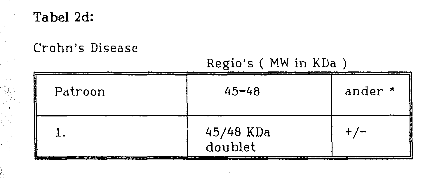
Figuur 2c, tenslotte is een voorbeeld van verschillende Western Blotting patronen ontwikkeld na incubatie met representatieve sera van patiënten met de ziekte van Crohn. De "hallmark" patronen staan in tabel 3 en zijn voor Crohn’s disease een geprononceerde 45/48 KDa doublet.Figure 2c, finally, is an example of different Western blotting patterns developed after incubation with representative sera from Crohn's disease patients. The hallmark patterns are shown in Table 3 and are a pronounced 45/48 KDa doublet for Crohn's disease.
Opgebracht is op blot A: BCG ruw extract, blot B: RIVM 7114 ruw extract, blot C: Mycobacterium avium ruw extract, blot D: moleculaire marker.Blot A: BCG crude extract, blot B: RIVM 7114 crude extract, blot C: Mycobacterium avium crude extract, blot D: molecular marker.
De interpretatie van banden patronen is als volgt.The interpretation of tire patterns is as follows.
Alle lanen laten een distinctieve kleuring zien van de 45/48 KDa doublet positiviteit, welke de ziekte van Crohn. De 45/48 KDa doublet reageert positief in 65% van alle Crohn’s patiënten.All lanes show a distinctive staining of the 45/48 KDa doublet positivity, which is Crohn's disease. The 45/48 KDa doublet responds positively in 65% of all Crohn's patients.
VOORBEELD 2. Enzym Immunobepaling 1. Bereiding van antigenenEXAMPLE 2. Enzyme Immunoassay 1. Preparation of Antigens
Het uitgangsmateriaal (zie VOORBEELD 1 onder 1.) werd bij lOO.OOOg gecentrifugeerd bij 4°C gedurende 2 uur. Het pellet werd driemaal gewassen met PBS. Tussen de wasstappen door werd gecentrifugeerd bij 100.000 g bij 4°C gedurende 2 uur. Het pellet werd verzameld en geresuspen-deerd in 10 ml PBS. Vervolgens werd de suspensie gedurende 2 minuten bij 80 Watt bij 4eC gesonificeerd. Nadat de eiwit-concentratie bepaald was werden hoeveelheden van 100 μΐ met een concentratie van lmg/ml ingevroren en bij -80°C bewaard tot gebruik (dit preparaat wordt aangeduid met de term KP-100).The starting material (see EXAMPLE 1 under 1.) was centrifuged at 100 ° C at 4 ° C for 2 hours. The pellet was washed three times with PBS. In between the washing steps, centrifugation was carried out at 100,000 g at 4 ° C for 2 hours. The pellet was collected and resuspended in 10 ml PBS. Then the suspension was sonicated for 2 minutes at 80 Watts at 4 ° C. After protein concentration was determined, aliquots of 100 µl at a concentration of 1 mg / ml were frozen and stored at -80 ° C until use (this preparation is referred to as KP-100).
Vervolgens werd op een preparatieve 12% polyacrylamide gel van 0,5 cm dikte 30 mg van het startmateriaal in de aanwezigheid van opbrengmengsel, na 20 minuten incuberen bij 65°C, opgebracht. De electroforese werd gedurende 30 minuten bij 150V (stackinggel) en 6 uur bij 100V (runninggel) uitgevoerd. De electroforese werd gestopt nadat de blauwe kleurstof band van de gel was verdwenen. De gel werd vervolgens in horizontale stroken van 2 mm dik gesneden, welke op hun beurt in stukjes van 1 cm lengte werden verdeeld. De gel-stukjes werden elk overnacht bij 4°C in een buis met 5 ml steriel gedestilleerd water geëlueerd. Daarna werd goed gemengd en werden de overgebleven gelstukjes naar de bodem gecentrifugeerd.Then, on a preparative 12% polyacrylamide gel of 0.5 cm thickness, 30 mg of the starting material in the presence of application mixture, after incubation at 65 ° C for 20 minutes, was applied. The electrophoresis was performed at 150V (stacking gel) for 30 minutes and at 100V (running gel) for 6 hours. The electrophoresis was stopped after the blue dye band had disappeared from the gel. The gel was then cut into 2 mm thick horizontal strips, which in turn were divided into 1 cm length pieces. The gel pieces were each eluted overnight at 4 ° C in a tube with 5 ml sterile distilled water. It was then mixed well and the remaining gel pieces were centrifuged to the bottom.
De elutie werd gecontroleerd met behulp van een 12% polyacrylamide analysegel van 1,5 mm dikte. De gel werd gegoten met een kam. In de slots werd na 20 minuten incuberen bij 65°C 40 μΐ van elke buis met gelstukjes in de aanwezigheid van 10 μΐ 5x opbrengmengsel gebracht. De electroforese werd gedurende 30 minuten bij 150V "stackinggel" en 6 uur bij 100V "runninggel" uitgevoerd. De electroforese werd gestopt en de gel klaargemaakt voor het vervaardigen van een Western blot. De blotprocedure wordt beschreven in VOORBEELD 1 onder 2.The elution was checked using a 1.5% thick 12% polyacrylamide analysis gel. The gel was poured with a comb. After 20 minutes incubation at 65 ° C, 40 μΐ of each tube containing gel pieces was placed in the slots in the presence of 10 μΐ 5x application mixture. The electrophoresis was performed at 150V "stacking gel" for 30 minutes and at 100V "running gel" for 6 hours. The electrophoresis was stopped and the gel prepared to make a Western blot. The blotting procedure is described in EXAMPLE 1 under 2.
Teneinde vast te stellen welke fracties de relevante antigenen bevatten werden strips van de blot geïncubeerd met sera van patiënten met lepromateuze lepra, tuberculode lepra en de ziekte van Crohn. Hiermee worden respectievelijk de 29/33 kDa antigenen, het 64/65 kDa antigeen en de 45/48 kDa antigenen aangetoond. De complexvorming werd zichtbaar gemaakt met behulp van anti-humaan IgG peroxidase conjugaat en DAB. De gewenste fracties werden verzameld, samengevoegd en gebruikt voor het coaten van een microtiterplaat (zie onder) .In order to determine which fractions contained the relevant antigens, blot strips were incubated with sera from patients with lepromatous leprosy, tuberculode leprosy and Crohn's disease. This demonstrates the 29/33 kDa antigens, the 64/65 kDa antigen and the 45/48 kDa antigens, respectively. The complex formation was visualized using anti-human IgG peroxidase conjugate and DAB. The desired fractions were collected, pooled and used to coat a microtiter plate (see below).
2. EIA.2. EIA.
Microtiterplaten worden bekleed (via standaard technieken) met hetzij KP-100, 29/33 KDa, 64/65 KDa of 45/48 KDa antigenen.Microtiter plates are coated (via standard techniques) with either KP-100, 29/33 KDa, 64/65 KDa or 45/48 KDa antigens.
Na bekleding worden de platen geblokkeerd om een a-specifieke binding van serumcomponenten tegen te gaan, met een 3% runderserum albumine (BSA) oplossing. Hierna worden platen gedroogd en bewaard bij 4°C.After coating, the plates are blocked to prevent a-specific binding of serum components with a 3% bovine serum albumin (BSA) solution. Plates are then dried and stored at 4 ° C.
2.1. Tuberculose EIA test (met KP-100 beklede microtiterplaten)2.1. Tuberculosis EIA test (KP-100 coated microtiter plates)
Testsera worden in een 1:100 verdunningen in de beklede putjes van een microtiterplaat gepipetteerd. De reactie vindt gedurende 1 uur bij 37°C plaats. A-specifieke serumcomponenten en niet-gebonden serumcomponenten worden met een wasstap weggewassen. Een tweede incubatie met een geschikte verdunning van een anti-humaan IgA peroxidase conjugaat wordt wederom 1 uur bij 37°C uitgevoerd, teveel aan conjugaat wordt aansluitend weggewassen.Test sera are pipetted in 1: 100 dilutions into the coated wells of a microtiter plate. The reaction takes place at 37 ° C for 1 hour. A-specific serum components and unbound serum components are washed away in a washing step. A second incubation with an appropriate dilution of an anti-human IgA peroxidase conjugate is again performed at 37 ° C for 1 hour, excess conjugate is subsequently washed away.
Het aantonen van specifiek aan KP-100 gebonden humane antilichamen van het subtype IgA vindt plaats door toevoeging van TMB (tetramethylbenzidine) aan de putjes.Detection of human antibodies of the IgA subtype specific to KP-100 is accomplished by adding TMB (tetramethylbenzidine) to the wells.
Gebonden enzym geeft aanleiding tot het ontstaan van een blauwe kleur die na toevoeging van een kleurstopoplos-sing omslaat naar geel. Deze gele kleur heeft een absorptie-maximum van 450 nm.Bound enzyme gives rise to a blue color which changes to yellow after the addition of a color stop solution. This yellow color has an absorption maximum of 450 nm.
De intensiteit van de kleur die ontstaat is evenredig met de hoeveelheid gebonden KP-100 specifiek IgA.The intensity of the color that arises is proportional to the amount of bound KP-100 specific IgA.
De resultaten staan weergegeven in de onderstaande tabellen.The results are shown in the tables below.
In de beschreven test zijn patiënte- en controle sera gebruikt uit 2 verschillende populaties.In the described test, patient and control sera from 2 different populations were used.
A =Endemisch gebied (Afrika, Ghana) B = Niet-endemisch gebied (Europa, Nederland)A = Endemic area (Africa, Ghana) B = Non-endemic area (Europe, Netherlands)
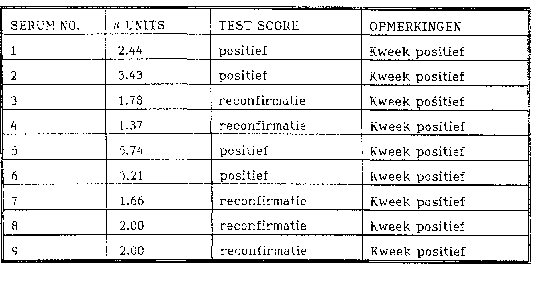
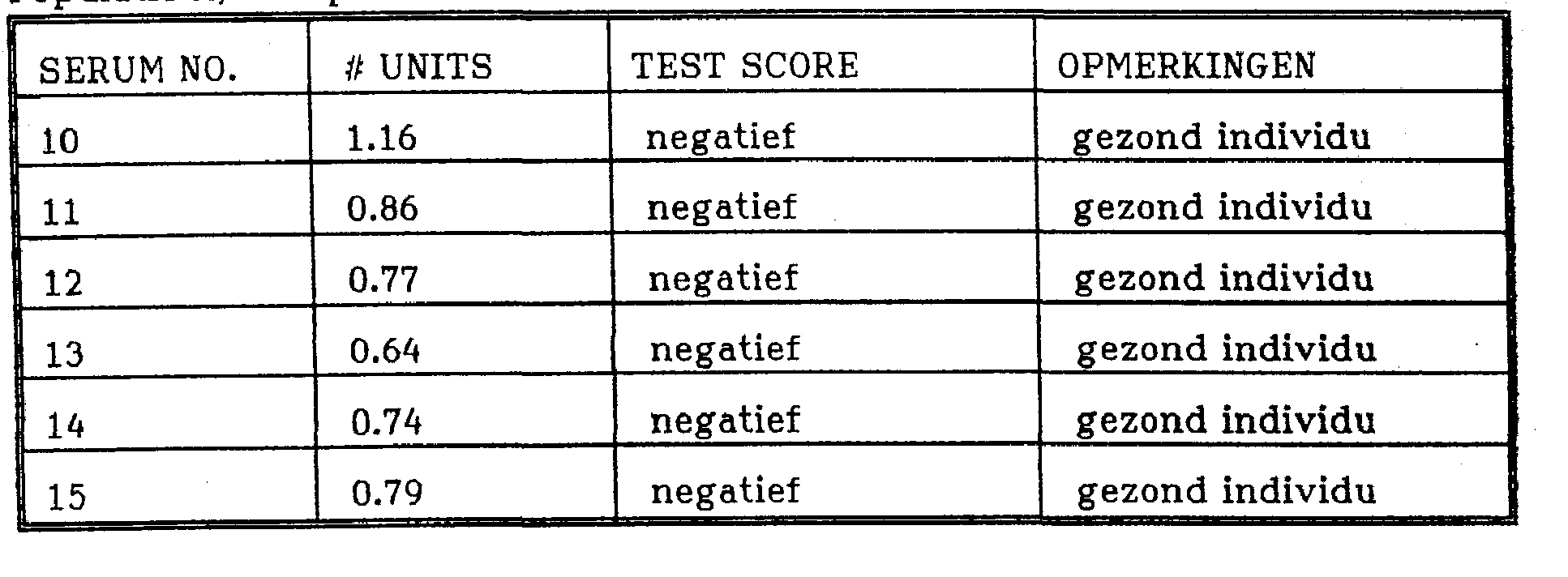
Elke populatie is onderverdeeld in 4 subgroepen, te weten: Groep 1 = kweek geconfirmeerde Tb patiënten Groep 2 = negatieve controlegroep (normale gezonde individuen)Each population is divided into 4 subgroups, namely: Group 1 = culture of confirmed Tb patients Group 2 = negative control group (normal healthy individuals)
Groep 3 = verdacht positieven (Tb contacten)Group 3 = suspicious positives (Tb contacts)
Groep 4 = verdacht negatieven (geen gegevens bekend, maar zeker geen Tb, mogelijk Lepra of ander a-specifie-ke Mycobacteriosis).Group 4 = suspicious negatives (no data known, but certainly no Tb, possibly Leprosy or other non-specific Mycobacteriosis).
De test is uitgevoerd met 2 kits met verschillende lotnummers en produktiedata.The test was performed with 2 kits with different lot numbers and production dates.
Interpretatie van de testresultaten geschiedt aan de hand van een zogenaamde ijklijn die samengesteld is uit controle sera met een vastgestelde arbitraire unit definitie die overeenkomt met een bekende OD waarde (1 unit, 4 units en 8 units).Interpretation of the test results takes place on the basis of a so-called calibration line composed of control sera with an established arbitrary unit definition that corresponds to a known OD value (1 unit, 4 units and 8 units).
Elke keer dat een test wordt uitgevoerd worden de units in de bepaling meegenomen. Gevonden monsterwaarden kunnen dan aan de unitdefinitie worden gerelateerd.The units are included in the determination each time a test is performed. Sample values found can then be related to the unit definition.
Een testserum kan als positief worden aangemerkt wanneer de gevonden uitslag in de test hoger scoort dan 2,1 units.A test serum can be considered positive if the result found in the test scores higher than 2.1 units.
Een testserum kan als negatief worden aangemerkt wanneer de gevonden uitslag lager scoort dan 2,1 units.A test serum can be considered negative if the result found scores below 2.1 units.
Testsera met unitwaarden tussen 2,1 en 1,2 units vallen in de ingestelde zogeheten reconfirmatie zone. Dit wil zeggen dat niet in eerste instantie met deze test is uit te maken of er sprake is van positiviteit of negativiteit voor Tuberculose.Test sera with unit values between 2.1 and 1.2 units fall in the set so-called reconfirmation zone. This means that it is not initially possible to determine with this test whether there is any positivity or negativity for Tuberculosis.
Reconfirmatie van deze sera gebeurt met behulp van beschreven westernblot strips waarmee na serumincubatie op basis van bandenpatronen en specifieke "hallmarks" antwoord is te geven op de vraag of er sprake is van een positief (banden aanwezig) danwel negatief (banden afwezig) testserum.Reconfirmation of these sera is done with the help of described western blot strips with which after serum incubation on the basis of tire patterns and specific hallmarks an answer can be given to the question whether there is a positive (tires present) or negative (tires absent) test serum.
Tabel 3:Table 3:
Populatie A.Endemisch gebied: Groep 1:Population A. Endemic Area: Group 1:
Tabel 4:Table 4:
Pormlatie A. Groeü 2:Pormlation A. Growth 2:
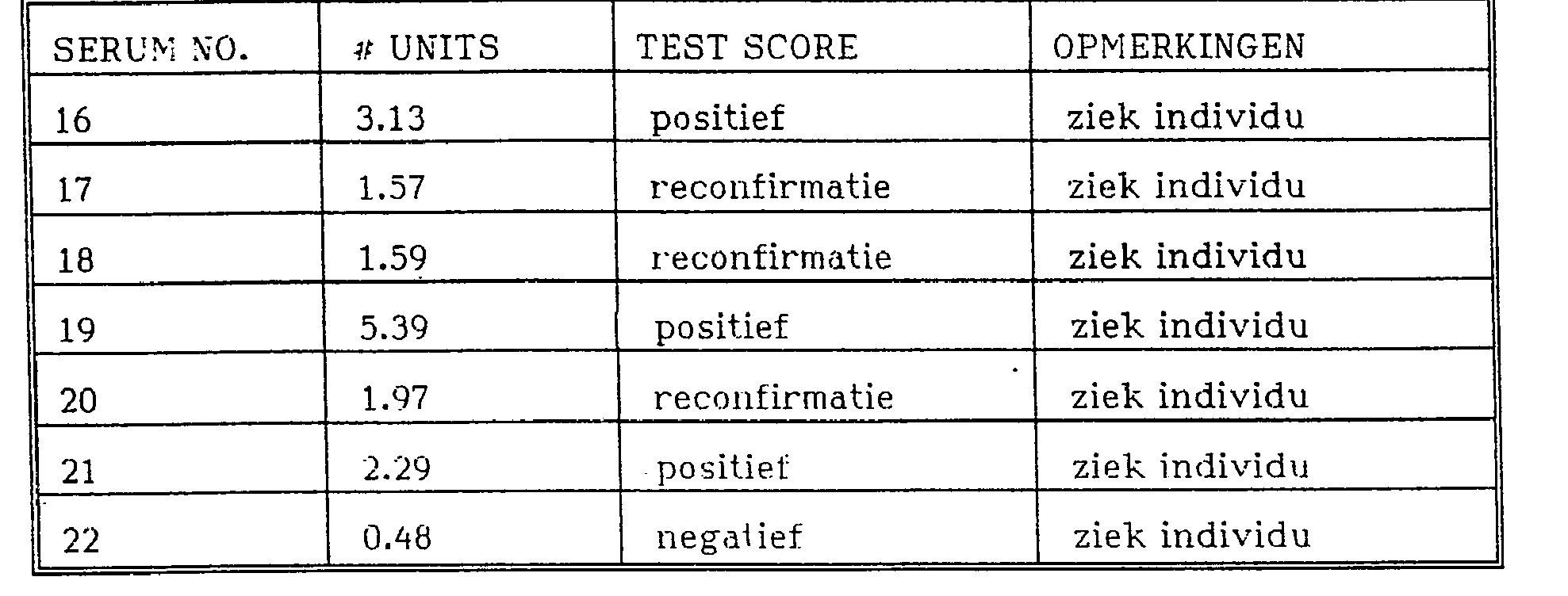
Tabel 5:Table 5:
Populatie A, Groep 3:_Population A, Group 3: _
Tabel 6:Table 6:
Populatie A, Groep 4:Population A, Group 4:

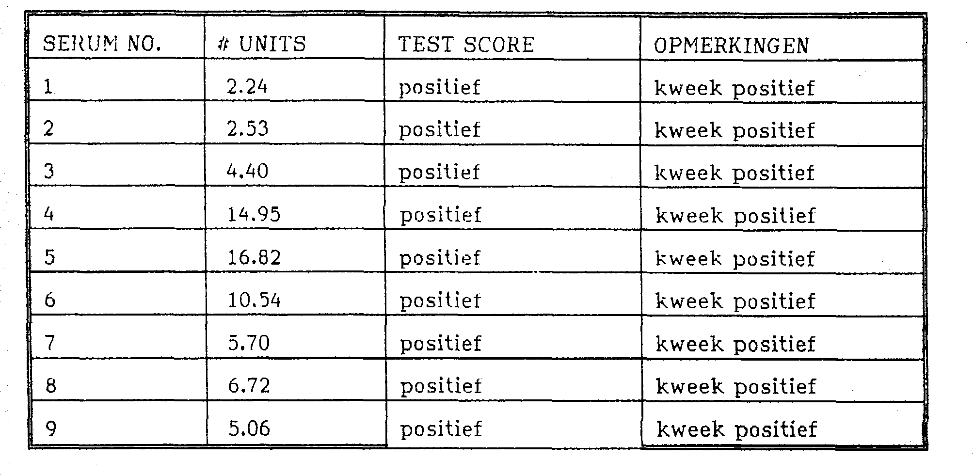
Tabel 7:Table 7:
Populatie B. Niet-Endemisch gebied: Groep 1:Population B. Non-Endemic Area: Group 1:

Tabel 9:Table 9:
Populatie B, Groep 3:_Population B, Group 3: _


Claims (15)
Priority Applications (11)
| Application Number | Priority Date | Filing Date | Title |
|---|---|---|---|
| NL9202197A NL9202197A (en) | 1992-12-17 | 1992-12-17 | Method and device for identifying a mycobacterium species responsible for mycobacterial infection. |
| EP94903148A EP0674766B1 (en) | 1992-12-17 | 1993-12-17 | Method and device for identifying a mycobacterium species responsible for a mycobacterial infection |
| AT94903148T ATE192238T1 (en) | 1992-12-17 | 1993-12-17 | METHOD AND DEVICE FOR DETECTING A MYCOBACTERIUM SPECIES RESPONSIBLE FOR A MYCOBACTERIUM INFECTION |
| ES94903148T ES2148314T3 (en) | 1992-12-17 | 1993-12-17 | PROCEDURE AND DEVICE FOR IDENTIFICATION OF A MICOBACTERIAL SPECIES RESPONSIBLE FOR A MICOBACTERIAL INFECTION. |
| AU57203/94A AU5720394A (en) | 1992-12-17 | 1993-12-17 | Method and device for identifying a mycobacterium species responsible for a mycobacterial infection |
| DE69328493T DE69328493T2 (en) | 1992-12-17 | 1993-12-17 | METHOD AND DEVICE FOR DETECTING A MYCOBACTERIUM SPECIES RESPONSIBLE FOR A MYCOBACTERIUM INFECTION |
| US08/454,122 US5817473A (en) | 1992-12-17 | 1993-12-17 | Method and device for identifying a mycobacterium species responsible for a mycobacterial infection |
| PCT/NL1993/000270 WO1994014069A1 (en) | 1992-12-17 | 1993-12-17 | Method and device for identifying a mycobacterium species responsible for a mycobacterial infection |
| PT94903148T PT674766E (en) | 1992-12-17 | 1993-12-17 | METHOD AND DEVICE FOR IDENTIFYING A SPECIES OF MICOBACTERIA RESPONSIBLE FOR A MYCOBACTERIAL INFECTION |
| DK94903148T DK0674766T3 (en) | 1992-12-17 | 1993-12-17 | Method and apparatus for identifying a mycobacterial species responsible for a mycobacterial infection |
| GR20000401710T GR3034022T3 (en) | 1992-12-17 | 2000-07-26 | Method and device for identifying a mycobacterium species responsible for a mycobacterial infection. |
Applications Claiming Priority (2)
| Application Number | Priority Date | Filing Date | Title |
|---|---|---|---|
| NL9202197 | 1992-12-17 | ||
| NL9202197A NL9202197A (en) | 1992-12-17 | 1992-12-17 | Method and device for identifying a mycobacterium species responsible for mycobacterial infection. |
Publications (1)
| Publication Number | Publication Date |
|---|---|
| NL9202197A true NL9202197A (en) | 1994-07-18 |
Family
ID=19861648
Family Applications (1)
| Application Number | Title | Priority Date | Filing Date |
|---|---|---|---|
| NL9202197A NL9202197A (en) | 1992-12-17 | 1992-12-17 | Method and device for identifying a mycobacterium species responsible for mycobacterial infection. |
Country Status (11)
| Country | Link |
|---|---|
| US (1) | US5817473A (en) |
| EP (1) | EP0674766B1 (en) |
| AT (1) | ATE192238T1 (en) |
| AU (1) | AU5720394A (en) |
| DE (1) | DE69328493T2 (en) |
| DK (1) | DK0674766T3 (en) |
| ES (1) | ES2148314T3 (en) |
| GR (1) | GR3034022T3 (en) |
| NL (1) | NL9202197A (en) |
| PT (1) | PT674766E (en) |
| WO (1) | WO1994014069A1 (en) |
Families Citing this family (25)
| Publication number | Priority date | Publication date | Assignee | Title |
|---|---|---|---|---|
| US5658749A (en) * | 1994-04-05 | 1997-08-19 | Corning Clinical Laboratories, Inc. | Method for processing mycobacteria |
| RU2145977C1 (en) * | 1994-04-05 | 2000-02-27 | Интигрейтид Рисеч Текнолэджи эЛэЛСи | Method of preparing sample for determination of microorganisms (variants) and set for sample treatment for determination of microorganisms |
| US6458366B1 (en) * | 1995-09-01 | 2002-10-01 | Corixa Corporation | Compounds and methods for diagnosis of tuberculosis |
| US6290969B1 (en) * | 1995-09-01 | 2001-09-18 | Corixa Corporation | Compounds and methods for immunotherapy and diagnosis of tuberculosis |
| US6592877B1 (en) * | 1995-09-01 | 2003-07-15 | Corixa Corporation | Compounds and methods for immunotherapy and diagnosis of tuberculosis |
| US6416962B1 (en) * | 1995-11-20 | 2002-07-09 | Kreatech Biotechnology B.V. | Method and device for identifying a mycobacterium species responsible for a mycobacterial infection |
| DE19621488C2 (en) * | 1996-05-29 | 1998-05-14 | Gerlach Gerald F Prof Dr | Diagnostic procedure for the detection of subclinical mammals suffering from paratuberculosis |
| RU2133965C1 (en) * | 1997-07-08 | 1999-07-27 | Московский областной научно-исследовательский клинический институт | Serological method for diagnosing tuberculosis in patients suffering from immunodeficiency |
| EP0921397A1 (en) * | 1997-12-08 | 1999-06-09 | Kreatech Biotechnology B.V. | A method for identifying a mycobacterium species |
| RU2141665C1 (en) * | 1998-04-14 | 1999-11-20 | Научно-исследовательский ветеринарный институт Нечерноземной зоны РФ | Method of detection of tuberculosis mycobacterium |
| GB2342443A (en) * | 1998-10-02 | 2000-04-12 | Abp Diagnostics Limited | Immunoassay device |
| US8143386B2 (en) * | 1999-04-07 | 2012-03-27 | Corixa Corporation | Fusion proteins of mycobacterium tuberculosis antigens and their uses |
| EP1229931A4 (en) * | 1999-10-07 | 2003-05-28 | Corixa Corp | Fusion proteins of mycobacterium tuberculosis |
| AU2001241738A1 (en) * | 2000-02-25 | 2001-09-03 | Corixa Corporation | Compounds and methods for diagnosis and immunotherapy of tuberculosis |
| DK2133100T3 (en) * | 2000-06-20 | 2012-01-23 | Corixa Corp | MTB32A Antigen of Mycobacterium tuberculosis with Inactivated Active Site and Fusion Proteins |
| RU2219547C2 (en) * | 2001-07-23 | 2003-12-20 | Нижегородская государственная медицинская академия | Method for predicting tuberculosis activity in respiratory organs |
| EP1442301A1 (en) * | 2001-11-09 | 2004-08-04 | Kreatech Biotechnology B.V. | Means and methods for the detection of immunoglobulin capable of binding to mycobacterium antigen |
| US6841159B2 (en) * | 2002-01-30 | 2005-01-11 | The United States Of America As Represented By The Secretary Of The Navy | Rapid lateral flow assay for determining exposure to Mycobacterium tuberculosis and other mycobacteria |
| WO2003070187A2 (en) * | 2002-02-15 | 2003-08-28 | Corixa Corporation | Fusion proteins of mycobacterium tuberculosis |
| US7276350B2 (en) * | 2004-04-27 | 2007-10-02 | University Of Tennessee Research Foundation | Method for diagnosing infectious diseases |
| US8956859B1 (en) | 2010-08-13 | 2015-02-17 | Aviex Technologies Llc | Compositions and methods for determining successful immunization by one or more vaccines |
| NL2017204B1 (en) | 2016-06-08 | 2017-12-18 | Kei International Ltd | Solid substrate comprising antigens immobilised thereto, biosensor comprising said solid substrate and method for detecting the presence of mycobacterial material in a sample |
| NL2016913B1 (en) * | 2016-06-08 | 2017-12-18 | Kei International Ltd | Solid substrate comprising antigens immobilised thereto and use thereof in a method for detecting the presence of mycobacterial material in a sample |
| WO2017211314A1 (en) | 2016-06-08 | 2017-12-14 | Kei International Limited | Method for detecting presence of mycobacterial material in sample using immobilised mannosyl phosphoketide antigen |
| NL2021443B1 (en) | 2018-08-08 | 2020-02-20 | Kei International Ltd | Synthetic antigens for tuberculosis detection |
Family Cites Families (1)
| Publication number | Priority date | Publication date | Assignee | Title |
|---|---|---|---|---|
| US4391904A (en) * | 1979-12-26 | 1983-07-05 | Syva Company | Test strip kits in immunoassays and compositions therein |
-
1992
- 1992-12-17 NL NL9202197A patent/NL9202197A/en not_active Application Discontinuation
-
1993
- 1993-12-17 DK DK94903148T patent/DK0674766T3/en active
- 1993-12-17 PT PT94903148T patent/PT674766E/en unknown
- 1993-12-17 WO PCT/NL1993/000270 patent/WO1994014069A1/en not_active Ceased
- 1993-12-17 ES ES94903148T patent/ES2148314T3/en not_active Expired - Lifetime
- 1993-12-17 US US08/454,122 patent/US5817473A/en not_active Expired - Fee Related
- 1993-12-17 AT AT94903148T patent/ATE192238T1/en not_active IP Right Cessation
- 1993-12-17 EP EP94903148A patent/EP0674766B1/en not_active Expired - Lifetime
- 1993-12-17 DE DE69328493T patent/DE69328493T2/en not_active Expired - Fee Related
- 1993-12-17 AU AU57203/94A patent/AU5720394A/en not_active Abandoned
-
2000
- 2000-07-26 GR GR20000401710T patent/GR3034022T3/en not_active IP Right Cessation
Also Published As
| Publication number | Publication date |
|---|---|
| AU5720394A (en) | 1994-07-04 |
| GR3034022T3 (en) | 2000-11-30 |
| ES2148314T3 (en) | 2000-10-16 |
| EP0674766B1 (en) | 2000-04-26 |
| ATE192238T1 (en) | 2000-05-15 |
| DE69328493T2 (en) | 2000-12-21 |
| WO1994014069A1 (en) | 1994-06-23 |
| EP0674766A1 (en) | 1995-10-04 |
| PT674766E (en) | 2000-10-31 |
| DE69328493D1 (en) | 2000-05-31 |
| DK0674766T3 (en) | 2000-09-25 |
| US5817473A (en) | 1998-10-06 |
Similar Documents
| Publication | Publication Date | Title |
|---|---|---|
| NL9202197A (en) | Method and device for identifying a mycobacterium species responsible for mycobacterial infection. | |
| Backhouse et al. | Treponema pallidum western blot: comparison with the FTA-ABS test as a confirmatory test for syphilis | |
| Bevanger et al. | Agglutinins and antibodies to Francisella tularensis outer membrane antigens in the early diagnosis of disease during an outbreak of tularemia | |
| Ryan et al. | Enzyme-linked immunosorbent assay based on soluble promastigote antigen detects immunoglobulin M (IgM) and IgG antibodies in sera from cases of visceral and cutaneous leishmaniasis | |
| NO821569L (en) | DIAGNOSTIZATION METHOD AND TEST SET | |
| DE69121290T2 (en) | Method for the determination of antibodies against Chlamydia Trachomatis and diagnostic preparation for Chlamydia Trachomatis infection | |
| Chenthamarakshan et al. | Detection of immunoglobulins M and G using culture filtrate antigen of Burkholderia pseudomallei | |
| Angeles et al. | Rapid diagnosis of nocardiosis with an enzyme immunoassay | |
| Uhaa et al. | Evaluation of specificity of indirect enzyme-linked immunosorbent assay for diagnosis of human Q fever | |
| Cho et al. | Comparison of the proteosomes and antigenicities of secreted and cellular proteins produced by Mycobacterium paratuberculosis | |
| de Lemos et al. | Characterization of the Western blotting IgG reactivity patterns in the clinical phases of acquired syphilis | |
| Sathish et al. | Identification and characterization of antigenic determinants of Mycobacterium leprae that react with antibodies in sera of leprosy patients | |
| US6416962B1 (en) | Method and device for identifying a mycobacterium species responsible for a mycobacterial infection | |
| Lilenbaum et al. | The use of Ag85 complex as antigen in ELISA for the diagnosis of bovine tuberculosis in dairy cows in Brazil | |
| US6733983B1 (en) | Method for identifying a mycobacterium species | |
| Dettori et al. | Evaluation of Western immunoblotting technique in the serological diagnosis of human syphilitic infections | |
| Magnarelli | Laboratory diagnosis of Lyme disease | |
| Hechemy et al. | Detection of antibodies to Rickettsia conorii with a latex agglutination test in patients with Mediterranean spotted fever | |
| Diagbouga et al. | Immunoblot analysis for serodiagnosis of tuberculosis using a 45/47-kilodalton antigen complex of Mycobacterium tuberculosis | |
| Thoen et al. | An enzyme-labeled antibody test for detecting antibodies in chickens infected with Mycobacterium avium serotype 2 | |
| Rivoire et al. | Chemical definition, cloning, and expression of the major protein of the leprosy bacillus | |
| Khosrobeygi et al. | Isolation and purification of low molecular weight proteins from culture filtrate of Mycobacterium tuberculosis strain C | |
| Lyashchenko et al. | Novel monoclonal antibodies against major antigens of Mycobacterium bovis | |
| Martínez-Moreno et al. | Comparison of the use of secretory and somatic antigens in an ELISA for the serodiagnosis of hypodermosis | |
| Poussin et al. | Capture-ELISA: a new assay for the detection of immunoglobulin M isotype antibodies using Chlamydia trachomatis antigen |
Legal Events
| Date | Code | Title | Description |
|---|---|---|---|
| A1B | A search report has been drawn up | ||
| BC | A request for examination has been filed | ||
| BV | The patent application has lapsed |