KR20240161220A - Retinal pigment epithelium cell compositions - Google Patents
Retinal pigment epithelium cell compositions Download PDFInfo
- Publication number
- KR20240161220A KR20240161220A KR1020247036309A KR20247036309A KR20240161220A KR 20240161220 A KR20240161220 A KR 20240161220A KR 1020247036309 A KR1020247036309 A KR 1020247036309A KR 20247036309 A KR20247036309 A KR 20247036309A KR 20240161220 A KR20240161220 A KR 20240161220A
- Authority
- KR
- South Korea
- Prior art keywords
- cells
- cell
- rpe
- thawing
- cryopreservation
- Prior art date
- Legal status (The legal status is an assumption and is not a legal conclusion. Google has not performed a legal analysis and makes no representation as to the accuracy of the status listed.)
- Pending
Links
- 239000000203 mixture Substances 0.000 title claims abstract description 227
- 210000003583 retinal pigment epithelium Anatomy 0.000 title description 312
- 238000010257 thawing Methods 0.000 abstract description 113
- 238000000034 method Methods 0.000 abstract description 68
- 238000002659 cell therapy Methods 0.000 abstract description 59
- 238000011282 treatment Methods 0.000 abstract description 22
- 238000002347 injection Methods 0.000 abstract description 20
- 239000007924 injection Substances 0.000 abstract description 20
- 239000000790 retinal pigment Substances 0.000 abstract description 20
- 230000002207 retinal effect Effects 0.000 abstract description 14
- 208000015122 neurodegenerative disease Diseases 0.000 abstract description 10
- 230000006378 damage Effects 0.000 abstract description 4
- 101000670189 Homo sapiens Ribulose-phosphate 3-epimerase Proteins 0.000 abstract description 3
- 210000004027 cell Anatomy 0.000 description 720
- IAZDPXIOMUYVGZ-UHFFFAOYSA-N Dimethylsulphoxide Chemical compound CS(C)=O IAZDPXIOMUYVGZ-UHFFFAOYSA-N 0.000 description 172
- HEMHJVSKTPXQMS-UHFFFAOYSA-M Sodium hydroxide Chemical compound [OH-].[Na+] HEMHJVSKTPXQMS-UHFFFAOYSA-M 0.000 description 87
- 238000011084 recovery Methods 0.000 description 85
- 238000005138 cryopreservation Methods 0.000 description 84
- 210000001508 eye Anatomy 0.000 description 82
- 230000035899 viability Effects 0.000 description 80
- DFPAKSUCGFBDDF-UHFFFAOYSA-N Nicotinamide Chemical group NC(=O)C1=CC=CN=C1 DFPAKSUCGFBDDF-UHFFFAOYSA-N 0.000 description 69
- 239000012595 freezing medium Substances 0.000 description 68
- 239000002609 medium Substances 0.000 description 68
- 241001465754 Metazoa Species 0.000 description 62
- 206010061218 Inflammation Diseases 0.000 description 57
- KWYUFKZDYYNOTN-UHFFFAOYSA-M Potassium hydroxide Chemical compound [OH-].[K+] KWYUFKZDYYNOTN-UHFFFAOYSA-M 0.000 description 57
- 230000004054 inflammatory process Effects 0.000 description 57
- 230000001225 therapeutic effect Effects 0.000 description 56
- LWIHDJKSTIGBAC-UHFFFAOYSA-K tripotassium phosphate Chemical compound [K+].[K+].[K+].[O-]P([O-])([O-])=O LWIHDJKSTIGBAC-UHFFFAOYSA-K 0.000 description 56
- OIRDTQYFTABQOQ-KQYNXXCUSA-N adenosine Chemical compound C1=NC=2C(N)=NC=NC=2N1[C@@H]1O[C@H](CO)[C@@H](O)[C@H]1O OIRDTQYFTABQOQ-KQYNXXCUSA-N 0.000 description 54
- JKMHFZQWWAIEOD-UHFFFAOYSA-N 2-[4-(2-hydroxyethyl)piperazin-1-yl]ethanesulfonic acid Chemical compound OCC[NH+]1CCN(CCS([O-])(=O)=O)CC1 JKMHFZQWWAIEOD-UHFFFAOYSA-N 0.000 description 53
- 239000000243 solution Substances 0.000 description 53
- 238000011534 incubation Methods 0.000 description 48
- -1 adenosine) Chemical compound 0.000 description 43
- 230000004069 differentiation Effects 0.000 description 41
- 230000003472 neutralizing effect Effects 0.000 description 41
- TWRXJAOTZQYOKJ-UHFFFAOYSA-L Magnesium chloride Chemical compound [Mg+2].[Cl-].[Cl-] TWRXJAOTZQYOKJ-UHFFFAOYSA-L 0.000 description 38
- WCUXLLCKKVVCTQ-UHFFFAOYSA-M Potassium chloride Chemical compound [Cl-].[K+] WCUXLLCKKVVCTQ-UHFFFAOYSA-M 0.000 description 38
- 235000005152 nicotinamide Nutrition 0.000 description 37
- 210000001671 embryonic stem cell Anatomy 0.000 description 36
- 239000011570 nicotinamide Substances 0.000 description 35
- 229960003966 nicotinamide Drugs 0.000 description 35
- 239000006285 cell suspension Substances 0.000 description 33
- 210000002540 macrophage Anatomy 0.000 description 32
- 102100031248 Patatin-like phospholipase domain-containing protein 2 Human genes 0.000 description 31
- 238000001914 filtration Methods 0.000 description 31
- 108090000102 pigment epithelium-derived factor Proteins 0.000 description 31
- 239000003855 balanced salt solution Substances 0.000 description 29
- 230000003833 cell viability Effects 0.000 description 29
- 108010014606 glutathione-bicarbonate-Ringer solution Proteins 0.000 description 29
- 229910000160 potassium phosphate Inorganic materials 0.000 description 28
- 235000011009 potassium phosphates Nutrition 0.000 description 28
- 239000002126 C01EB10 - Adenosine Substances 0.000 description 27
- 239000007995 HEPES buffer Substances 0.000 description 27
- 229960005305 adenosine Drugs 0.000 description 27
- FZWBNHMXJMCXLU-BLAUPYHCSA-N isomaltotriose Chemical compound O[C@@H]1[C@@H](O)[C@H](O)[C@@H](CO)O[C@@H]1OC[C@@H]1[C@@H](O)[C@H](O)[C@@H](O)[C@@H](OC[C@@H](O)[C@@H](O)[C@H](O)[C@@H](O)C=O)O1 FZWBNHMXJMCXLU-BLAUPYHCSA-N 0.000 description 27
- 206010064930 age-related macular degeneration Diseases 0.000 description 26
- 229940119744 dextran 40 Drugs 0.000 description 26
- 108010073929 Vascular Endothelial Growth Factor A Proteins 0.000 description 25
- 102000005789 Vascular Endothelial Growth Factors Human genes 0.000 description 25
- 108010019530 Vascular Endothelial Growth Factors Proteins 0.000 description 25
- 235000000346 sugar Nutrition 0.000 description 25
- XLYOFNOQVPJJNP-UHFFFAOYSA-N water Substances O XLYOFNOQVPJJNP-UHFFFAOYSA-N 0.000 description 25
- 102000004190 Enzymes Human genes 0.000 description 23
- 108090000790 Enzymes Proteins 0.000 description 23
- 229940088598 enzyme Drugs 0.000 description 23
- 238000009472 formulation Methods 0.000 description 23
- 210000002966 serum Anatomy 0.000 description 23
- 238000003860 storage Methods 0.000 description 22
- WQZGKKKJIJFFOK-GASJEMHNSA-N Glucose Natural products OC[C@H]1OC(O)[C@H](O)[C@@H](O)[C@@H]1O WQZGKKKJIJFFOK-GASJEMHNSA-N 0.000 description 21
- WQZGKKKJIJFFOK-VFUOTHLCSA-N beta-D-glucose Chemical compound OC[C@H]1O[C@@H](O)[C@H](O)[C@@H](O)[C@@H]1O WQZGKKKJIJFFOK-VFUOTHLCSA-N 0.000 description 21
- 238000012258 culturing Methods 0.000 description 21
- RWSXRVCMGQZWBV-WDSKDSINSA-N glutathione Chemical compound OC(=O)[C@@H](N)CCC(=O)N[C@@H](CS)C(=O)NCC(O)=O RWSXRVCMGQZWBV-WDSKDSINSA-N 0.000 description 21
- 239000000126 substance Substances 0.000 description 21
- FBPFZTCFMRRESA-KVTDHHQDSA-N D-Mannitol Chemical compound OC[C@@H](O)[C@@H](O)[C@H](O)[C@H](O)CO FBPFZTCFMRRESA-KVTDHHQDSA-N 0.000 description 20
- 229930195725 Mannitol Natural products 0.000 description 20
- 229930006000 Sucrose Natural products 0.000 description 20
- CZMRCDWAGMRECN-UGDNZRGBSA-N Sucrose Chemical compound O[C@H]1[C@H](O)[C@@H](CO)O[C@@]1(CO)O[C@@H]1[C@H](O)[C@@H](O)[C@H](O)[C@@H](CO)O1 CZMRCDWAGMRECN-UGDNZRGBSA-N 0.000 description 20
- 239000000594 mannitol Substances 0.000 description 20
- 235000010355 mannitol Nutrition 0.000 description 20
- 239000005720 sucrose Substances 0.000 description 20
- UOQHWNPVNXSDDO-UHFFFAOYSA-N 3-bromoimidazo[1,2-a]pyridine-6-carbonitrile Chemical compound C1=CC(C#N)=CN2C(Br)=CN=C21 UOQHWNPVNXSDDO-UHFFFAOYSA-N 0.000 description 19
- 229920001503 Glucan Polymers 0.000 description 19
- RWSXRVCMGQZWBV-PHDIDXHHSA-N L-Glutathione Natural products OC(=O)[C@H](N)CCC(=O)N[C@H](CS)C(=O)NCC(O)=O RWSXRVCMGQZWBV-PHDIDXHHSA-N 0.000 description 19
- 239000000872 buffer Substances 0.000 description 19
- 239000008121 dextrose Substances 0.000 description 19
- 210000002950 fibroblast Anatomy 0.000 description 19
- 229940099563 lactobionic acid Drugs 0.000 description 19
- 208000002780 macular degeneration Diseases 0.000 description 19
- 229910001629 magnesium chloride Inorganic materials 0.000 description 19
- 239000003880 polar aprotic solvent Substances 0.000 description 19
- 239000011736 potassium bicarbonate Substances 0.000 description 19
- 235000015497 potassium bicarbonate Nutrition 0.000 description 19
- 229910000028 potassium bicarbonate Inorganic materials 0.000 description 19
- 239000001103 potassium chloride Substances 0.000 description 19
- 235000011164 potassium chloride Nutrition 0.000 description 19
- TYJJADVDDVDEDZ-UHFFFAOYSA-M potassium hydrogencarbonate Chemical compound [K+].OC([O-])=O TYJJADVDDVDEDZ-UHFFFAOYSA-M 0.000 description 19
- 239000002212 purine nucleoside Substances 0.000 description 19
- 102000008100 Human Serum Albumin Human genes 0.000 description 18
- 108091006905 Human Serum Albumin Proteins 0.000 description 18
- FAPWRFPIFSIZLT-UHFFFAOYSA-M Sodium chloride Chemical compound [Na+].[Cl-] FAPWRFPIFSIZLT-UHFFFAOYSA-M 0.000 description 18
- 108010023082 activin A Proteins 0.000 description 18
- MRWXACSTFXYYMV-FDDDBJFASA-N nebularine Chemical compound O[C@@H]1[C@H](O)[C@@H](CO)O[C@H]1N1C2=NC=NC=C2N=C1 MRWXACSTFXYYMV-FDDDBJFASA-N 0.000 description 18
- 210000000130 stem cell Anatomy 0.000 description 18
- 239000002253 acid Substances 0.000 description 17
- 230000001464 adherent effect Effects 0.000 description 17
- 230000000694 effects Effects 0.000 description 17
- 229910052760 oxygen Inorganic materials 0.000 description 16
- 230000028327 secretion Effects 0.000 description 16
- NCYCYZXNIZJOKI-UHFFFAOYSA-N vitamin A aldehyde Natural products O=CC=C(C)C=CC=C(C)C=CC1=C(C)CCCC1(C)C NCYCYZXNIZJOKI-UHFFFAOYSA-N 0.000 description 16
- 239000006144 Dulbecco’s modified Eagle's medium Substances 0.000 description 15
- 239000000758 substrate Substances 0.000 description 15
- 239000000725 suspension Substances 0.000 description 15
- IJGRMHOSHXDMSA-UHFFFAOYSA-N Atomic nitrogen Chemical compound N#N IJGRMHOSHXDMSA-UHFFFAOYSA-N 0.000 description 14
- 239000003963 antioxidant agent Substances 0.000 description 14
- 235000006708 antioxidants Nutrition 0.000 description 14
- QVGXLLKOCUKJST-UHFFFAOYSA-N atomic oxygen Chemical compound [O] QVGXLLKOCUKJST-UHFFFAOYSA-N 0.000 description 14
- 239000001301 oxygen Substances 0.000 description 14
- 238000004321 preservation Methods 0.000 description 14
- 210000002459 blastocyst Anatomy 0.000 description 13
- 239000012071 phase Substances 0.000 description 13
- 238000002360 preparation method Methods 0.000 description 13
- 235000020945 retinal Nutrition 0.000 description 13
- 239000011604 retinal Substances 0.000 description 13
- 208000008069 Geographic Atrophy Diseases 0.000 description 12
- 150000001447 alkali salts Chemical class 0.000 description 12
- 230000004888 barrier function Effects 0.000 description 12
- 239000002585 base Substances 0.000 description 12
- 239000003795 chemical substances by application Substances 0.000 description 12
- 150000003013 phosphoric acid derivatives Chemical class 0.000 description 12
- 150000005846 sugar alcohols Chemical class 0.000 description 12
- 238000002054 transplantation Methods 0.000 description 12
- 102000004887 Transforming Growth Factor beta Human genes 0.000 description 11
- 108090001012 Transforming Growth Factor beta Proteins 0.000 description 11
- 230000003078 antioxidant effect Effects 0.000 description 11
- 229920000159 gelatin Polymers 0.000 description 11
- 239000008273 gelatin Substances 0.000 description 11
- 239000013028 medium composition Substances 0.000 description 11
- 108091008695 photoreceptors Proteins 0.000 description 11
- 150000008163 sugars Chemical class 0.000 description 11
- UXVMQQNJUSDDNG-UHFFFAOYSA-L Calcium chloride Chemical compound [Cl-].[Cl-].[Ca+2] UXVMQQNJUSDDNG-UHFFFAOYSA-L 0.000 description 10
- 108010050345 Microphthalmia-Associated Transcription Factor Proteins 0.000 description 10
- 102100030157 Microphthalmia-associated transcription factor Human genes 0.000 description 10
- 239000001110 calcium chloride Substances 0.000 description 10
- 229910001628 calcium chloride Inorganic materials 0.000 description 10
- 238000004113 cell culture Methods 0.000 description 10
- 210000001519 tissue Anatomy 0.000 description 10
- 108010010803 Gelatin Proteins 0.000 description 9
- 235000019322 gelatine Nutrition 0.000 description 9
- 235000011852 gelatine desserts Nutrition 0.000 description 9
- 230000036512 infertility Effects 0.000 description 9
- 238000004519 manufacturing process Methods 0.000 description 9
- 210000001525 retina Anatomy 0.000 description 9
- 239000011780 sodium chloride Substances 0.000 description 9
- 239000001488 sodium phosphate Substances 0.000 description 9
- 229910000162 sodium phosphate Inorganic materials 0.000 description 9
- 235000011008 sodium phosphates Nutrition 0.000 description 9
- RYFMWSXOAZQYPI-UHFFFAOYSA-K trisodium phosphate Chemical compound [Na+].[Na+].[Na+].[O-]P([O-])([O-])=O RYFMWSXOAZQYPI-UHFFFAOYSA-K 0.000 description 9
- CIWBSHSKHKDKBQ-JLAZNSOCSA-N Ascorbic acid Chemical compound OC[C@H](O)[C@H]1OC(=O)C(O)=C1O CIWBSHSKHKDKBQ-JLAZNSOCSA-N 0.000 description 8
- 102100024785 Fibroblast growth factor 2 Human genes 0.000 description 8
- 108090000379 Fibroblast growth factor 2 Proteins 0.000 description 8
- IAJILQKETJEXLJ-UHFFFAOYSA-N Galacturonsaeure Natural products O=CC(O)C(O)C(O)C(O)C(O)=O IAJILQKETJEXLJ-UHFFFAOYSA-N 0.000 description 8
- 102100028001 Retinaldehyde-binding protein 1 Human genes 0.000 description 8
- 101710101931 Retinaldehyde-binding protein 1 Proteins 0.000 description 8
- 238000004458 analytical method Methods 0.000 description 8
- 238000006243 chemical reaction Methods 0.000 description 8
- 210000004087 cornea Anatomy 0.000 description 8
- 238000011156 evaluation Methods 0.000 description 8
- 230000006870 function Effects 0.000 description 8
- 238000011194 good manufacturing practice Methods 0.000 description 8
- 230000012010 growth Effects 0.000 description 8
- 239000003102 growth factor Substances 0.000 description 8
- 210000004263 induced pluripotent stem cell Anatomy 0.000 description 8
- 239000010410 layer Substances 0.000 description 8
- 210000000440 neutrophil Anatomy 0.000 description 8
- 230000008569 process Effects 0.000 description 8
- 230000002829 reductive effect Effects 0.000 description 8
- 230000004083 survival effect Effects 0.000 description 8
- 102000004219 Brain-derived neurotrophic factor Human genes 0.000 description 7
- 108090000715 Brain-derived neurotrophic factor Proteins 0.000 description 7
- 108010005939 Ciliary Neurotrophic Factor Proteins 0.000 description 7
- 102100031614 Ciliary neurotrophic factor Human genes 0.000 description 7
- 108010085895 Laminin Proteins 0.000 description 7
- 102000007547 Laminin Human genes 0.000 description 7
- 108010009583 Transforming Growth Factors Proteins 0.000 description 7
- 102000009618 Transforming Growth Factors Human genes 0.000 description 7
- 229940077737 brain-derived neurotrophic factor Drugs 0.000 description 7
- 150000001875 compounds Chemical class 0.000 description 7
- 210000002744 extracellular matrix Anatomy 0.000 description 7
- 238000003306 harvesting Methods 0.000 description 7
- 230000003118 histopathologic effect Effects 0.000 description 7
- 210000004698 lymphocyte Anatomy 0.000 description 7
- 229910052757 nitrogen Inorganic materials 0.000 description 7
- 210000003954 umbilical cord Anatomy 0.000 description 7
- PMATZTZNYRCHOR-CGLBZJNRSA-N Cyclosporin A Chemical compound CC[C@@H]1NC(=O)[C@H]([C@H](O)[C@H](C)C\C=C\C)N(C)C(=O)[C@H](C(C)C)N(C)C(=O)[C@H](CC(C)C)N(C)C(=O)[C@H](CC(C)C)N(C)C(=O)[C@@H](C)NC(=O)[C@H](C)NC(=O)[C@H](CC(C)C)N(C)C(=O)[C@H](C(C)C)NC(=O)[C@H](CC(C)C)N(C)C(=O)CN(C)C1=O PMATZTZNYRCHOR-CGLBZJNRSA-N 0.000 description 6
- 108010036949 Cyclosporine Proteins 0.000 description 6
- 102000010834 Extracellular Matrix Proteins Human genes 0.000 description 6
- 108010037362 Extracellular Matrix Proteins Proteins 0.000 description 6
- 241000699670 Mus sp. Species 0.000 description 6
- 108010025020 Nerve Growth Factor Proteins 0.000 description 6
- 102000007072 Nerve Growth Factors Human genes 0.000 description 6
- QJJXYPPXXYFBGM-LFZNUXCKSA-N Tacrolimus Chemical compound C1C[C@@H](O)[C@H](OC)C[C@@H]1\C=C(/C)[C@@H]1[C@H](C)[C@@H](O)CC(=O)[C@H](CC=C)/C=C(C)/C[C@H](C)C[C@H](OC)[C@H]([C@H](C[C@H]2C)OC)O[C@@]2(O)C(=O)C(=O)N2CCCC[C@H]2C(=O)O1 QJJXYPPXXYFBGM-LFZNUXCKSA-N 0.000 description 6
- 230000015572 biosynthetic process Effects 0.000 description 6
- 229960001265 ciclosporin Drugs 0.000 description 6
- 239000003814 drug Substances 0.000 description 6
- 230000002255 enzymatic effect Effects 0.000 description 6
- 235000021472 generally recognized as safe Nutrition 0.000 description 6
- 238000012744 immunostaining Methods 0.000 description 6
- 239000003018 immunosuppressive agent Substances 0.000 description 6
- 229940124589 immunosuppressive drug Drugs 0.000 description 6
- 239000003112 inhibitor Substances 0.000 description 6
- 210000001778 pluripotent stem cell Anatomy 0.000 description 6
- QJJXYPPXXYFBGM-SHYZHZOCSA-N tacrolimus Natural products CO[C@H]1C[C@H](CC[C@@H]1O)C=C(C)[C@H]2OC(=O)[C@H]3CCCCN3C(=O)C(=O)[C@@]4(O)O[C@@H]([C@H](C[C@H]4C)OC)[C@@H](C[C@H](C)CC(=C[C@@H](CC=C)C(=O)C[C@H](O)[C@H]2C)C)OC QJJXYPPXXYFBGM-SHYZHZOCSA-N 0.000 description 6
- 229960001967 tacrolimus Drugs 0.000 description 6
- 238000002560 therapeutic procedure Methods 0.000 description 6
- 101000620359 Homo sapiens Melanocyte protein PMEL Proteins 0.000 description 5
- 101001122114 Homo sapiens NUT family member 1 Proteins 0.000 description 5
- 206010025412 Macular dystrophy congenital Diseases 0.000 description 5
- 102100022430 Melanocyte protein PMEL Human genes 0.000 description 5
- 208000007014 Retinitis pigmentosa Diseases 0.000 description 5
- 230000007547 defect Effects 0.000 description 5
- 229940079593 drug Drugs 0.000 description 5
- 230000004064 dysfunction Effects 0.000 description 5
- 230000001605 fetal effect Effects 0.000 description 5
- 238000000338 in vitro Methods 0.000 description 5
- 208000015181 infectious disease Diseases 0.000 description 5
- 239000003550 marker Substances 0.000 description 5
- 239000003900 neurotrophic factor Substances 0.000 description 5
- 150000005480 nicotinamides Chemical class 0.000 description 5
- 108090000623 proteins and genes Proteins 0.000 description 5
- 239000002356 single layer Substances 0.000 description 5
- 230000001052 transient effect Effects 0.000 description 5
- 201000007790 vitelliform macular dystrophy Diseases 0.000 description 5
- 108010035532 Collagen Proteins 0.000 description 4
- 102000008186 Collagen Human genes 0.000 description 4
- 102000029816 Collagenase Human genes 0.000 description 4
- 108060005980 Collagenase Proteins 0.000 description 4
- 102000004127 Cytokines Human genes 0.000 description 4
- 108090000695 Cytokines Proteins 0.000 description 4
- RGHNJXZEOKUKBD-SQOUGZDYSA-N D-gluconic acid Chemical compound OC[C@@H](O)[C@@H](O)[C@H](O)[C@@H](O)C(O)=O RGHNJXZEOKUKBD-SQOUGZDYSA-N 0.000 description 4
- KCXVZYZYPLLWCC-UHFFFAOYSA-N EDTA Chemical compound OC(=O)CN(CC(O)=O)CCN(CC(O)=O)CC(O)=O KCXVZYZYPLLWCC-UHFFFAOYSA-N 0.000 description 4
- 108010073385 Fibrin Proteins 0.000 description 4
- 102000009123 Fibrin Human genes 0.000 description 4
- BWGVNKXGVNDBDI-UHFFFAOYSA-N Fibrin monomer Chemical compound CNC(=O)CNC(=O)CN BWGVNKXGVNDBDI-UHFFFAOYSA-N 0.000 description 4
- 102100037362 Fibronectin Human genes 0.000 description 4
- 108010067306 Fibronectins Proteins 0.000 description 4
- 208000005422 Foreign-Body reaction Diseases 0.000 description 4
- 102000006395 Globulins Human genes 0.000 description 4
- 108010044091 Globulins Proteins 0.000 description 4
- 241000699666 Mus <mouse, genus> Species 0.000 description 4
- 102000007354 PAX6 Transcription Factor Human genes 0.000 description 4
- 108010032788 PAX6 Transcription Factor Proteins 0.000 description 4
- 206010057430 Retinal injury Diseases 0.000 description 4
- IAJILQKETJEXLJ-RSJOWCBRSA-N aldehydo-D-galacturonic acid Chemical compound O=C[C@H](O)[C@@H](O)[C@@H](O)[C@H](O)C(O)=O IAJILQKETJEXLJ-RSJOWCBRSA-N 0.000 description 4
- 235000010323 ascorbic acid Nutrition 0.000 description 4
- 239000011668 ascorbic acid Substances 0.000 description 4
- 229960005070 ascorbic acid Drugs 0.000 description 4
- 238000003556 assay Methods 0.000 description 4
- 210000001775 bruch membrane Anatomy 0.000 description 4
- 229920001436 collagen Polymers 0.000 description 4
- 229960002424 collagenase Drugs 0.000 description 4
- 229930182912 cyclosporin Natural products 0.000 description 4
- 210000002257 embryonic structure Anatomy 0.000 description 4
- 210000002919 epithelial cell Anatomy 0.000 description 4
- 230000004438 eyesight Effects 0.000 description 4
- 229950003499 fibrin Drugs 0.000 description 4
- 239000012634 fragment Substances 0.000 description 4
- 238000007710 freezing Methods 0.000 description 4
- 230000008014 freezing Effects 0.000 description 4
- 239000001963 growth medium Substances 0.000 description 4
- 230000001506 immunosuppresive effect Effects 0.000 description 4
- 208000014674 injury Diseases 0.000 description 4
- 230000000670 limiting effect Effects 0.000 description 4
- 239000007788 liquid Substances 0.000 description 4
- 239000011159 matrix material Substances 0.000 description 4
- HPNSFSBZBAHARI-UHFFFAOYSA-N micophenolic acid Natural products OC1=C(CC=C(C)CCC(O)=O)C(OC)=C(C)C2=C1C(=O)OC2 HPNSFSBZBAHARI-UHFFFAOYSA-N 0.000 description 4
- HPNSFSBZBAHARI-RUDMXATFSA-N mycophenolic acid Chemical compound OC1=C(C\C=C(/C)CCC(O)=O)C(OC)=C(C)C2=C1C(=O)OC2 HPNSFSBZBAHARI-RUDMXATFSA-N 0.000 description 4
- 238000006386 neutralization reaction Methods 0.000 description 4
- ZAHRKKWIAAJSAO-UHFFFAOYSA-N rapamycin Natural products COCC(O)C(=C/C(C)C(=O)CC(OC(=O)C1CCCCN1C(=O)C(=O)C2(O)OC(CC(OC)C(=CC=CC=CC(C)CC(C)C(=O)C)C)CCC2C)C(C)CC3CCC(O)C(C3)OC)C ZAHRKKWIAAJSAO-UHFFFAOYSA-N 0.000 description 4
- 108020003175 receptors Proteins 0.000 description 4
- 102000005962 receptors Human genes 0.000 description 4
- QFJCIRLUMZQUOT-HPLJOQBZSA-N sirolimus Chemical compound C1C[C@@H](O)[C@H](OC)C[C@@H]1C[C@@H](C)[C@H]1OC(=O)[C@@H]2CCCCN2C(=O)C(=O)[C@](O)(O2)[C@H](C)CC[C@H]2C[C@H](OC)/C(C)=C/C=C/C=C/[C@@H](C)C[C@@H](C)C(=O)[C@H](OC)[C@H](O)/C(C)=C/[C@@H](C)C(=O)C1 QFJCIRLUMZQUOT-HPLJOQBZSA-N 0.000 description 4
- 229960002930 sirolimus Drugs 0.000 description 4
- 210000001082 somatic cell Anatomy 0.000 description 4
- 210000001578 tight junction Anatomy 0.000 description 4
- 230000032258 transport Effects 0.000 description 4
- 239000012808 vapor phase Substances 0.000 description 4
- GVJHHUAWPYXKBD-IEOSBIPESA-N α-tocopherol Chemical compound OC1=C(C)C(C)=C2O[C@@](CCC[C@H](C)CCC[C@H](C)CCCC(C)C)(C)CCC2=C1C GVJHHUAWPYXKBD-IEOSBIPESA-N 0.000 description 4
- 108010059616 Activins Proteins 0.000 description 3
- 206010003694 Atrophy Diseases 0.000 description 3
- 229930105110 Cyclosporin A Natural products 0.000 description 3
- 102000018233 Fibroblast Growth Factor Human genes 0.000 description 3
- 108050007372 Fibroblast Growth Factor Proteins 0.000 description 3
- 108090000386 Fibroblast Growth Factor 1 Proteins 0.000 description 3
- 102100031706 Fibroblast growth factor 1 Human genes 0.000 description 3
- 201000003533 Leber congenital amaurosis Diseases 0.000 description 3
- 206010029113 Neovascularisation Diseases 0.000 description 3
- 208000034247 Pattern dystrophy Diseases 0.000 description 3
- 241000288906 Primates Species 0.000 description 3
- 101100247004 Rattus norvegicus Qsox1 gene Proteins 0.000 description 3
- 206010038848 Retinal detachment Diseases 0.000 description 3
- 208000017442 Retinal disease Diseases 0.000 description 3
- 208000027073 Stargardt disease Diseases 0.000 description 3
- 239000012190 activator Substances 0.000 description 3
- 239000000488 activin Substances 0.000 description 3
- SHGAZHPCJJPHSC-YCNIQYBTSA-N all-trans-retinoic acid Chemical compound OC(=O)\C=C(/C)\C=C\C=C(/C)\C=C\C1=C(C)CCCC1(C)C SHGAZHPCJJPHSC-YCNIQYBTSA-N 0.000 description 3
- 239000003708 ampul Substances 0.000 description 3
- 239000003242 anti bacterial agent Substances 0.000 description 3
- 230000003110 anti-inflammatory effect Effects 0.000 description 3
- 230000037444 atrophy Effects 0.000 description 3
- 230000008901 benefit Effects 0.000 description 3
- 210000004155 blood-retinal barrier Anatomy 0.000 description 3
- 230000004378 blood-retinal barrier Effects 0.000 description 3
- 230000037396 body weight Effects 0.000 description 3
- 230000010261 cell growth Effects 0.000 description 3
- 230000008021 deposition Effects 0.000 description 3
- 208000037265 diseases, disorders, signs and symptoms Diseases 0.000 description 3
- 210000003038 endothelium Anatomy 0.000 description 3
- 238000005516 engineering process Methods 0.000 description 3
- 210000003754 fetus Anatomy 0.000 description 3
- 229940126864 fibroblast growth factor Drugs 0.000 description 3
- 238000005469 granulation Methods 0.000 description 3
- 230000003179 granulation Effects 0.000 description 3
- 230000028993 immune response Effects 0.000 description 3
- 238000003364 immunohistochemistry Methods 0.000 description 3
- 230000008595 infiltration Effects 0.000 description 3
- 238000001764 infiltration Methods 0.000 description 3
- 230000002401 inhibitory effect Effects 0.000 description 3
- 210000001161 mammalian embryo Anatomy 0.000 description 3
- 210000004379 membrane Anatomy 0.000 description 3
- 239000012528 membrane Substances 0.000 description 3
- 210000002901 mesenchymal stem cell Anatomy 0.000 description 3
- 230000007170 pathology Effects 0.000 description 3
- 230000035935 pregnancy Effects 0.000 description 3
- 230000002035 prolonged effect Effects 0.000 description 3
- 102000004169 proteins and genes Human genes 0.000 description 3
- 238000010791 quenching Methods 0.000 description 3
- 230000000171 quenching effect Effects 0.000 description 3
- 230000005855 radiation Effects 0.000 description 3
- 230000004264 retinal detachment Effects 0.000 description 3
- 229930002330 retinoic acid Natural products 0.000 description 3
- 229960004641 rituximab Drugs 0.000 description 3
- 239000011435 rock Substances 0.000 description 3
- 239000007787 solid Substances 0.000 description 3
- 230000035882 stress Effects 0.000 description 3
- 238000004114 suspension culture Methods 0.000 description 3
- 238000012360 testing method Methods 0.000 description 3
- ZRKFYGHZFMAOKI-QMGMOQQFSA-N tgfbeta Chemical compound C([C@H](NC(=O)[C@H](C(C)C)NC(=O)CNC(=O)[C@H](CCC(O)=O)NC(=O)[C@H](CCCNC(N)=N)NC(=O)[C@H](CC(N)=O)NC(=O)[C@H](CC(C)C)NC(=O)[C@H]([C@@H](C)O)NC(=O)[C@H](CCC(O)=O)NC(=O)[C@H]([C@@H](C)O)NC(=O)[C@H](CC(C)C)NC(=O)CNC(=O)[C@H](C)NC(=O)[C@H](CO)NC(=O)[C@H](CCC(N)=O)NC(=O)[C@@H](NC(=O)[C@H](C)NC(=O)[C@H](C)NC(=O)[C@@H](NC(=O)[C@H](CC(C)C)NC(=O)[C@@H](N)CCSC)C(C)C)[C@@H](C)CC)C(=O)N[C@@H]([C@@H](C)O)C(=O)N[C@@H](C(C)C)C(=O)N[C@@H](CC=1C=CC=CC=1)C(=O)N[C@@H](C)C(=O)N1[C@@H](CCC1)C(=O)N[C@@H]([C@@H](C)O)C(=O)N[C@@H](CC(N)=O)C(=O)N[C@@H](CCC(O)=O)C(=O)N[C@@H](C)C(=O)N[C@@H](CC=1C=CC=CC=1)C(=O)N[C@@H](CCCNC(N)=N)C(=O)N[C@@H](C)C(=O)N[C@@H](CC(C)C)C(=O)N1[C@@H](CCC1)C(=O)N1[C@@H](CCC1)C(=O)N[C@@H](CCCNC(N)=N)C(=O)N[C@@H](CCC(O)=O)C(=O)N[C@@H](CCCNC(N)=N)C(=O)N[C@@H](CO)C(=O)N[C@@H](CCCNC(N)=N)C(=O)N[C@@H](CC(C)C)C(=O)N[C@@H](CC(C)C)C(O)=O)C1=CC=C(O)C=C1 ZRKFYGHZFMAOKI-QMGMOQQFSA-N 0.000 description 3
- 230000008736 traumatic injury Effects 0.000 description 3
- 229960001727 tretinoin Drugs 0.000 description 3
- HDTRYLNUVZCQOY-UHFFFAOYSA-N α-D-glucopyranosyl-α-D-glucopyranoside Natural products OC1C(O)C(O)C(CO)OC1OC1C(O)C(O)C(O)C(CO)O1 HDTRYLNUVZCQOY-UHFFFAOYSA-N 0.000 description 2
- RBNPOMFGQQGHHO-UHFFFAOYSA-N -2,3-Dihydroxypropanoic acid Natural products OCC(O)C(O)=O RBNPOMFGQQGHHO-UHFFFAOYSA-N 0.000 description 2
- NAOLWIGVYRIGTP-UHFFFAOYSA-N 1,3,5-trihydroxyanthracene-9,10-dione Chemical compound C1=CC(O)=C2C(=O)C3=CC(O)=CC(O)=C3C(=O)C2=C1 NAOLWIGVYRIGTP-UHFFFAOYSA-N 0.000 description 2
- RIZUCYSQUWMQLX-UHFFFAOYSA-N 2,3-dimethylbenzoic acid Chemical compound CC1=CC=CC(C(O)=O)=C1C RIZUCYSQUWMQLX-UHFFFAOYSA-N 0.000 description 2
- LMFAFFIRCNUJJO-UHFFFAOYSA-N 5-(4-azidophenyl)-8-iodo-3-methyl-1,2,4,5-tetrahydro-3-benzazepin-7-ol Chemical compound C1N(C)CCC2=CC(I)=C(O)C=C2C1C1=CC=C(N=[N+]=[N-])C=C1 LMFAFFIRCNUJJO-UHFFFAOYSA-N 0.000 description 2
- 102000002260 Alkaline Phosphatase Human genes 0.000 description 2
- 108020004774 Alkaline Phosphatase Proteins 0.000 description 2
- 102000012304 Bestrophin Human genes 0.000 description 2
- 108050002823 Bestrophin Proteins 0.000 description 2
- 108050003623 Bestrophin-1 Proteins 0.000 description 2
- 108010007726 Bone Morphogenetic Proteins Proteins 0.000 description 2
- 102000007350 Bone Morphogenetic Proteins Human genes 0.000 description 2
- 241000283690 Bos taurus Species 0.000 description 2
- 241000282472 Canis lupus familiaris Species 0.000 description 2
- 208000024172 Cardiovascular disease Diseases 0.000 description 2
- 102000014914 Carrier Proteins Human genes 0.000 description 2
- 206010057402 Choroidal coloboma Diseases 0.000 description 2
- 102000012422 Collagen Type I Human genes 0.000 description 2
- 108010022452 Collagen Type I Proteins 0.000 description 2
- 208000032170 Congenital Abnormalities Diseases 0.000 description 2
- FBPFZTCFMRRESA-FSIIMWSLSA-N D-Glucitol Natural products OC[C@H](O)[C@H](O)[C@@H](O)[C@H](O)CO FBPFZTCFMRRESA-FSIIMWSLSA-N 0.000 description 2
- FBPFZTCFMRRESA-JGWLITMVSA-N D-glucitol Chemical compound OC[C@H](O)[C@@H](O)[C@H](O)[C@H](O)CO FBPFZTCFMRRESA-JGWLITMVSA-N 0.000 description 2
- RGHNJXZEOKUKBD-UHFFFAOYSA-N D-gluconic acid Natural products OCC(O)C(O)C(O)C(O)C(O)=O RGHNJXZEOKUKBD-UHFFFAOYSA-N 0.000 description 2
- RBNPOMFGQQGHHO-UWTATZPHSA-N D-glyceric acid Chemical compound OC[C@@H](O)C(O)=O RBNPOMFGQQGHHO-UWTATZPHSA-N 0.000 description 2
- KDXKERNSBIXSRK-RXMQYKEDSA-N D-lysine Chemical compound NCCCC[C@@H](N)C(O)=O KDXKERNSBIXSRK-RXMQYKEDSA-N 0.000 description 2
- FEWJPZIEWOKRBE-JCYAYHJZSA-N Dextrotartaric acid Chemical compound OC(=O)[C@H](O)[C@@H](O)C(O)=O FEWJPZIEWOKRBE-JCYAYHJZSA-N 0.000 description 2
- 239000004386 Erythritol Substances 0.000 description 2
- UNXHWFMMPAWVPI-UHFFFAOYSA-N Erythritol Natural products OCC(O)C(O)CO UNXHWFMMPAWVPI-UHFFFAOYSA-N 0.000 description 2
- HKVAMNSJSFKALM-GKUWKFKPSA-N Everolimus Chemical compound C1C[C@@H](OCCO)[C@H](OC)C[C@@H]1C[C@@H](C)[C@H]1OC(=O)[C@@H]2CCCCN2C(=O)C(=O)[C@](O)(O2)[C@H](C)CC[C@H]2C[C@H](OC)/C(C)=C/C=C/C=C/[C@@H](C)C[C@@H](C)C(=O)[C@H](OC)[C@H](O)/C(C)=C/[C@@H](C)C(=O)C1 HKVAMNSJSFKALM-GKUWKFKPSA-N 0.000 description 2
- 241000282326 Felis catus Species 0.000 description 2
- 241000282412 Homo Species 0.000 description 2
- 101000729271 Homo sapiens Retinoid isomerohydrolase Proteins 0.000 description 2
- 101000803709 Homo sapiens Vitronectin Proteins 0.000 description 2
- 241000598436 Human T-cell lymphotropic virus Species 0.000 description 2
- 102000000521 Immunophilins Human genes 0.000 description 2
- 108010016648 Immunophilins Proteins 0.000 description 2
- 102100026818 Inhibin beta E chain Human genes 0.000 description 2
- 102000014150 Interferons Human genes 0.000 description 2
- 108010050904 Interferons Proteins 0.000 description 2
- KFZMGEQAYNKOFK-UHFFFAOYSA-N Isopropanol Chemical compound CC(C)O KFZMGEQAYNKOFK-UHFFFAOYSA-N 0.000 description 2
- UPYKUZBSLRQECL-UKMVMLAPSA-N Lycopene Natural products CC(=C/C=C/C=C(C)/C=C/C=C(C)/C=C/C1C(=C)CCCC1(C)C)C=CC=C(/C)C=CC2C(=C)CCCC2(C)C UPYKUZBSLRQECL-UKMVMLAPSA-N 0.000 description 2
- 241000124008 Mammalia Species 0.000 description 2
- FQISKWAFAHGMGT-SGJOWKDISA-M Methylprednisolone sodium succinate Chemical compound [Na+].C([C@@]12C)=CC(=O)C=C1[C@@H](C)C[C@@H]1[C@@H]2[C@@H](O)C[C@]2(C)[C@@](O)(C(=O)COC(=O)CCC([O-])=O)CC[C@H]21 FQISKWAFAHGMGT-SGJOWKDISA-M 0.000 description 2
- 241001529936 Murinae Species 0.000 description 2
- 208000022873 Ocular disease Diseases 0.000 description 2
- 206010030113 Oedema Diseases 0.000 description 2
- 241000283973 Oryctolagus cuniculus Species 0.000 description 2
- 102000012338 Poly(ADP-ribose) Polymerases Human genes 0.000 description 2
- 108010061844 Poly(ADP-ribose) Polymerases Proteins 0.000 description 2
- 229920000776 Poly(Adenosine diphosphate-ribose) polymerase Polymers 0.000 description 2
- 101710150336 Protein Rex Proteins 0.000 description 2
- 241000700159 Rattus Species 0.000 description 2
- 201000007737 Retinal degeneration Diseases 0.000 description 2
- 102100031176 Retinoid isomerohydrolase Human genes 0.000 description 2
- VMHLLURERBWHNL-UHFFFAOYSA-M Sodium acetate Chemical compound [Na+].CC([O-])=O VMHLLURERBWHNL-UHFFFAOYSA-M 0.000 description 2
- UIIMBOGNXHQVGW-DEQYMQKBSA-M Sodium bicarbonate-14C Chemical compound [Na+].O[14C]([O-])=O UIIMBOGNXHQVGW-DEQYMQKBSA-M 0.000 description 2
- FEWJPZIEWOKRBE-UHFFFAOYSA-N Tartaric acid Natural products [H+].[H+].[O-]C(=O)C(O)C(O)C([O-])=O FEWJPZIEWOKRBE-UHFFFAOYSA-N 0.000 description 2
- HDTRYLNUVZCQOY-WSWWMNSNSA-N Trehalose Natural products O[C@@H]1[C@@H](O)[C@@H](O)[C@@H](CO)O[C@@H]1O[C@@H]1[C@H](O)[C@@H](O)[C@@H](O)[C@@H](CO)O1 HDTRYLNUVZCQOY-WSWWMNSNSA-N 0.000 description 2
- 102000004142 Trypsin Human genes 0.000 description 2
- 108090000631 Trypsin Proteins 0.000 description 2
- LEHOTFFKMJEONL-UHFFFAOYSA-N Uric Acid Chemical compound N1C(=O)NC(=O)C2=C1NC(=O)N2 LEHOTFFKMJEONL-UHFFFAOYSA-N 0.000 description 2
- TVWHNULVHGKJHS-UHFFFAOYSA-N Uric acid Natural products N1C(=O)NC(=O)C2NC(=O)NC21 TVWHNULVHGKJHS-UHFFFAOYSA-N 0.000 description 2
- TVXBFESIOXBWNM-UHFFFAOYSA-N Xylitol Natural products OCCC(O)C(O)C(O)CCO TVXBFESIOXBWNM-UHFFFAOYSA-N 0.000 description 2
- DFPAKSUCGFBDDF-ZQBYOMGUSA-N [14c]-nicotinamide Chemical compound N[14C](=O)C1=CC=CN=C1 DFPAKSUCGFBDDF-ZQBYOMGUSA-N 0.000 description 2
- 238000009825 accumulation Methods 0.000 description 2
- 238000004115 adherent culture Methods 0.000 description 2
- IAJILQKETJEXLJ-QTBDOELSSA-N aldehydo-D-glucuronic acid Chemical compound O=C[C@H](O)[C@@H](O)[C@H](O)[C@H](O)C(O)=O IAJILQKETJEXLJ-QTBDOELSSA-N 0.000 description 2
- 239000002168 alkylating agent Substances 0.000 description 2
- 229940100198 alkylating agent Drugs 0.000 description 2
- 229940087168 alpha tocopherol Drugs 0.000 description 2
- HDTRYLNUVZCQOY-LIZSDCNHSA-N alpha,alpha-trehalose Chemical compound O[C@@H]1[C@@H](O)[C@H](O)[C@@H](CO)O[C@@H]1O[C@@H]1[C@H](O)[C@@H](O)[C@H](O)[C@@H](CO)O1 HDTRYLNUVZCQOY-LIZSDCNHSA-N 0.000 description 2
- 150000001408 amides Chemical class 0.000 description 2
- 230000002491 angiogenic effect Effects 0.000 description 2
- 239000005557 antagonist Substances 0.000 description 2
- 230000000781 anti-lymphocytic effect Effects 0.000 description 2
- 230000000340 anti-metabolite Effects 0.000 description 2
- 230000001494 anti-thymocyte effect Effects 0.000 description 2
- 229940088710 antibiotic agent Drugs 0.000 description 2
- 239000000427 antigen Substances 0.000 description 2
- 102000036639 antigens Human genes 0.000 description 2
- 108091007433 antigens Proteins 0.000 description 2
- 229940100197 antimetabolite Drugs 0.000 description 2
- 239000002256 antimetabolite Substances 0.000 description 2
- 229960002170 azathioprine Drugs 0.000 description 2
- LMEKQMALGUDUQG-UHFFFAOYSA-N azathioprine Chemical compound CN1C=NC([N+]([O-])=O)=C1SC1=NC=NC2=C1NC=N2 LMEKQMALGUDUQG-UHFFFAOYSA-N 0.000 description 2
- WPYMKLBDIGXBTP-UHFFFAOYSA-N benzoic acid Chemical compound OC(=O)C1=CC=CC=C1 WPYMKLBDIGXBTP-UHFFFAOYSA-N 0.000 description 2
- 108091008324 binding proteins Proteins 0.000 description 2
- 239000003124 biologic agent Substances 0.000 description 2
- 229940112869 bone morphogenetic protein Drugs 0.000 description 2
- 150000001746 carotenes Chemical class 0.000 description 2
- 235000005473 carotenes Nutrition 0.000 description 2
- 239000006143 cell culture medium Substances 0.000 description 2
- 238000012512 characterization method Methods 0.000 description 2
- 238000007385 chemical modification Methods 0.000 description 2
- 210000003161 choroid Anatomy 0.000 description 2
- 210000002987 choroid plexus Anatomy 0.000 description 2
- 235000019504 cigarettes Nutrition 0.000 description 2
- 239000003636 conditioned culture medium Substances 0.000 description 2
- 239000000356 contaminant Substances 0.000 description 2
- 238000011109 contamination Methods 0.000 description 2
- 239000003246 corticosteroid Substances 0.000 description 2
- 239000002577 cryoprotective agent Substances 0.000 description 2
- 239000000824 cytostatic agent Substances 0.000 description 2
- 230000001085 cytostatic effect Effects 0.000 description 2
- 229960002806 daclizumab Drugs 0.000 description 2
- 238000011161 development Methods 0.000 description 2
- 230000018109 developmental process Effects 0.000 description 2
- 201000010099 disease Diseases 0.000 description 2
- 238000010494 dissociation reaction Methods 0.000 description 2
- 230000005593 dissociations Effects 0.000 description 2
- 239000002552 dosage form Substances 0.000 description 2
- 230000002900 effect on cell Effects 0.000 description 2
- 229940009714 erythritol Drugs 0.000 description 2
- 235000019414 erythritol Nutrition 0.000 description 2
- UNXHWFMMPAWVPI-ZXZARUISSA-N erythritol Chemical compound OC[C@H](O)[C@H](O)CO UNXHWFMMPAWVPI-ZXZARUISSA-N 0.000 description 2
- 229960005167 everolimus Drugs 0.000 description 2
- 238000002474 experimental method Methods 0.000 description 2
- 238000007667 floating Methods 0.000 description 2
- 238000001943 fluorescence-activated cell sorting Methods 0.000 description 2
- 230000007045 gastrulation Effects 0.000 description 2
- 239000003862 glucocorticoid Substances 0.000 description 2
- 239000000174 gluconic acid Substances 0.000 description 2
- 235000012208 gluconic acid Nutrition 0.000 description 2
- 239000008103 glucose Substances 0.000 description 2
- 229940097043 glucuronic acid Drugs 0.000 description 2
- ZDXPYRJPNDTMRX-UHFFFAOYSA-N glutamine Natural products OC(=O)C(N)CCC(N)=O ZDXPYRJPNDTMRX-UHFFFAOYSA-N 0.000 description 2
- IAJILQKETJEXLJ-LECHCGJUSA-N iduronic acid Chemical compound O=C[C@@H](O)[C@H](O)[C@@H](O)[C@H](O)C(O)=O IAJILQKETJEXLJ-LECHCGJUSA-N 0.000 description 2
- 238000002513 implantation Methods 0.000 description 2
- 229940047124 interferons Drugs 0.000 description 2
- 230000003834 intracellular effect Effects 0.000 description 2
- 238000002955 isolation Methods 0.000 description 2
- BQINXKOTJQCISL-GRCPKETISA-N keto-neuraminic acid Chemical compound OC(=O)C(=O)C[C@H](O)[C@@H](N)[C@@H](O)[C@H](O)[C@H](O)CO BQINXKOTJQCISL-GRCPKETISA-N 0.000 description 2
- AGBQKNBQESQNJD-UHFFFAOYSA-M lipoate Chemical compound [O-]C(=O)CCCCC1CCSS1 AGBQKNBQESQNJD-UHFFFAOYSA-M 0.000 description 2
- 235000019136 lipoic acid Nutrition 0.000 description 2
- 230000007774 longterm Effects 0.000 description 2
- 229960001855 mannitol Drugs 0.000 description 2
- HEBKCHPVOIAQTA-UHFFFAOYSA-N meso ribitol Natural products OCC(O)C(O)C(O)CO HEBKCHPVOIAQTA-UHFFFAOYSA-N 0.000 description 2
- 229960004584 methylprednisolone Drugs 0.000 description 2
- 229940014456 mycophenolate Drugs 0.000 description 2
- RTGDFNSFWBGLEC-SYZQJQIISA-N mycophenolate mofetil Chemical compound COC1=C(C)C=2COC(=O)C=2C(O)=C1C\C=C(/C)CCC(=O)OCCN1CCOCC1 RTGDFNSFWBGLEC-SYZQJQIISA-N 0.000 description 2
- 229960004866 mycophenolate mofetil Drugs 0.000 description 2
- 229960000951 mycophenolic acid Drugs 0.000 description 2
- 230000001537 neural effect Effects 0.000 description 2
- CERZMXAJYMMUDR-UHFFFAOYSA-N neuraminic acid Natural products NC1C(O)CC(O)(C(O)=O)OC1C(O)C(O)CO CERZMXAJYMMUDR-UHFFFAOYSA-N 0.000 description 2
- 231100000252 nontoxic Toxicity 0.000 description 2
- 230000003000 nontoxic effect Effects 0.000 description 2
- 235000015097 nutrients Nutrition 0.000 description 2
- 229940005483 opioid analgesics Drugs 0.000 description 2
- 238000012014 optical coherence tomography Methods 0.000 description 2
- 230000003076 paracrine Effects 0.000 description 2
- 231100000915 pathological change Toxicity 0.000 description 2
- 230000036285 pathological change Effects 0.000 description 2
- 239000000049 pigment Substances 0.000 description 2
- 230000000270 postfertilization Effects 0.000 description 2
- 238000011533 pre-incubation Methods 0.000 description 2
- 229960005205 prednisolone Drugs 0.000 description 2
- OIGNJSKKLXVSLS-VWUMJDOOSA-N prednisolone Chemical compound O=C1C=C[C@]2(C)[C@H]3[C@@H](O)C[C@](C)([C@@](CC4)(O)C(=O)CO)[C@@H]4[C@@H]3CCC2=C1 OIGNJSKKLXVSLS-VWUMJDOOSA-N 0.000 description 2
- 239000000047 product Substances 0.000 description 2
- NPCOQXAVBJJZBQ-UHFFFAOYSA-N reduced coenzyme Q9 Natural products COC1=C(O)C(C)=C(CC=C(C)CCC=C(C)CCC=C(C)CCC=C(C)CCC=C(C)CCC=C(C)CCC=C(C)CCC=C(C)CCC=C(C)C)C(O)=C1OC NPCOQXAVBJJZBQ-UHFFFAOYSA-N 0.000 description 2
- 230000001172 regenerating effect Effects 0.000 description 2
- 230000004044 response Effects 0.000 description 2
- 230000004258 retinal degeneration Effects 0.000 description 2
- 210000000844 retinal pigment epithelial cell Anatomy 0.000 description 2
- 230000035945 sensitivity Effects 0.000 description 2
- 210000004927 skin cell Anatomy 0.000 description 2
- 230000000391 smoking effect Effects 0.000 description 2
- 239000001632 sodium acetate Substances 0.000 description 2
- 235000017281 sodium acetate Nutrition 0.000 description 2
- 239000000600 sorbitol Substances 0.000 description 2
- 229960002920 sorbitol Drugs 0.000 description 2
- 235000010356 sorbitol Nutrition 0.000 description 2
- UCSJYZPVAKXKNQ-HZYVHMACSA-N streptomycin Chemical compound CN[C@H]1[C@H](O)[C@@H](O)[C@H](CO)O[C@H]1O[C@@H]1[C@](C=O)(O)[C@H](C)O[C@H]1O[C@@H]1[C@@H](NC(N)=N)[C@H](O)[C@@H](NC(N)=N)[C@H](O)[C@H]1O UCSJYZPVAKXKNQ-HZYVHMACSA-N 0.000 description 2
- 238000001356 surgical procedure Methods 0.000 description 2
- 229940037128 systemic glucocorticoids Drugs 0.000 description 2
- 239000011975 tartaric acid Substances 0.000 description 2
- 235000002906 tartaric acid Nutrition 0.000 description 2
- XOAAWQZATWQOTB-UHFFFAOYSA-N taurine Chemical compound NCCS(O)(=O)=O XOAAWQZATWQOTB-UHFFFAOYSA-N 0.000 description 2
- 238000011287 therapeutic dose Methods 0.000 description 2
- 229960002663 thioctic acid Drugs 0.000 description 2
- 229960000984 tocofersolan Drugs 0.000 description 2
- 238000012546 transfer Methods 0.000 description 2
- 239000012588 trypsin Substances 0.000 description 2
- 229940040064 ubiquinol Drugs 0.000 description 2
- QNTNKSLOFHEFPK-UPTCCGCDSA-N ubiquinol-10 Chemical compound COC1=C(O)C(C)=C(C\C=C(/C)CC\C=C(/C)CC\C=C(/C)CC\C=C(/C)CC\C=C(/C)CC\C=C(/C)CC\C=C(/C)CC\C=C(/C)CC\C=C(/C)CCC=C(C)C)C(O)=C1OC QNTNKSLOFHEFPK-UPTCCGCDSA-N 0.000 description 2
- 229940116269 uric acid Drugs 0.000 description 2
- 230000000007 visual effect Effects 0.000 description 2
- 230000004382 visual function Effects 0.000 description 2
- 239000002676 xenobiotic agent Substances 0.000 description 2
- 230000002034 xenobiotic effect Effects 0.000 description 2
- 239000000811 xylitol Substances 0.000 description 2
- 229960002675 xylitol Drugs 0.000 description 2
- 235000010447 xylitol Nutrition 0.000 description 2
- HEBKCHPVOIAQTA-SCDXWVJYSA-N xylitol Chemical compound OC[C@H](O)[C@@H](O)[C@H](O)CO HEBKCHPVOIAQTA-SCDXWVJYSA-N 0.000 description 2
- 210000004340 zona pellucida Anatomy 0.000 description 2
- 239000002076 α-tocopherol Substances 0.000 description 2
- 235000004835 α-tocopherol Nutrition 0.000 description 2
- UBWXUGDQUBIEIZ-UHFFFAOYSA-N (13-methyl-3-oxo-2,6,7,8,9,10,11,12,14,15,16,17-dodecahydro-1h-cyclopenta[a]phenanthren-17-yl) 3-phenylpropanoate Chemical compound CC12CCC(C3CCC(=O)C=C3CC3)C3C1CCC2OC(=O)CCC1=CC=CC=C1 UBWXUGDQUBIEIZ-UHFFFAOYSA-N 0.000 description 1
- NCYCYZXNIZJOKI-IOUUIBBYSA-N 11-cis-retinal Chemical compound O=C/C=C(\C)/C=C\C=C(/C)\C=C\C1=C(C)CCCC1(C)C NCYCYZXNIZJOKI-IOUUIBBYSA-N 0.000 description 1
- NCYCYZXNIZJOKI-HPNHMNAASA-N 11Z-retinal Natural products CC(=C/C=O)C=C/C=C(C)/C=C/C1=C(C)CCCC1(C)C NCYCYZXNIZJOKI-HPNHMNAASA-N 0.000 description 1
- FPIPGXGPPPQFEQ-UHFFFAOYSA-N 13-cis retinol Natural products OCC=C(C)C=CC=C(C)C=CC1=C(C)CCCC1(C)C FPIPGXGPPPQFEQ-UHFFFAOYSA-N 0.000 description 1
- CTLOSZHDGZLOQE-UHFFFAOYSA-N 14-methoxy-9-[(4-methylpiperazin-1-yl)methyl]-9,19-diazapentacyclo[10.7.0.02,6.07,11.013,18]nonadeca-1(12),2(6),7(11),13(18),14,16-hexaene-8,10-dione Chemical compound O=C1C2=C3C=4C(OC)=CC=CC=4NC3=C3CCCC3=C2C(=O)N1CN1CCN(C)CC1 CTLOSZHDGZLOQE-UHFFFAOYSA-N 0.000 description 1
- AJHPGXZOIAYYDW-UHFFFAOYSA-N 3-(2-cyanophenyl)-2-[(2-methylpropan-2-yl)oxycarbonylamino]propanoic acid Chemical compound CC(C)(C)OC(=O)NC(C(O)=O)CC1=CC=CC=C1C#N AJHPGXZOIAYYDW-UHFFFAOYSA-N 0.000 description 1
- GSCPDZHWVNUUFI-UHFFFAOYSA-N 3-aminobenzamide Chemical compound NC(=O)C1=CC=CC(N)=C1 GSCPDZHWVNUUFI-UHFFFAOYSA-N 0.000 description 1
- MDOJTZQKHMAPBK-UHFFFAOYSA-N 4-iodo-3-nitrobenzamide Chemical compound NC(=O)C1=CC=C(I)C([N+]([O-])=O)=C1 MDOJTZQKHMAPBK-UHFFFAOYSA-N 0.000 description 1
- ZLWYEPMDOUQDBW-UHFFFAOYSA-N 6-aminonicotinamide Chemical compound NC(=O)C1=CC=C(N)N=C1 ZLWYEPMDOUQDBW-UHFFFAOYSA-N 0.000 description 1
- GSDSWSVVBLHKDQ-UHFFFAOYSA-N 9-fluoro-3-methyl-10-(4-methylpiperazin-1-yl)-7-oxo-2,3-dihydro-7H-[1,4]oxazino[2,3,4-ij]quinoline-6-carboxylic acid Chemical compound FC1=CC(C(C(C(O)=O)=C2)=O)=C3N2C(C)COC3=C1N1CCN(C)CC1 GSDSWSVVBLHKDQ-UHFFFAOYSA-N 0.000 description 1
- 102100023971 ADP-ribosylation factor-like protein 13B Human genes 0.000 description 1
- 206010000117 Abnormal behaviour Diseases 0.000 description 1
- 102000005606 Activins Human genes 0.000 description 1
- 229920000936 Agarose Polymers 0.000 description 1
- 102000009027 Albumins Human genes 0.000 description 1
- 108010088751 Albumins Proteins 0.000 description 1
- 206010002091 Anaesthesia Diseases 0.000 description 1
- 206010003497 Asphyxia Diseases 0.000 description 1
- 239000005711 Benzoic acid Substances 0.000 description 1
- 101100454434 Biomphalaria glabrata BG04 gene Proteins 0.000 description 1
- 102100024506 Bone morphogenetic protein 2 Human genes 0.000 description 1
- 102100024504 Bone morphogenetic protein 3 Human genes 0.000 description 1
- 102100024505 Bone morphogenetic protein 4 Human genes 0.000 description 1
- 102100022526 Bone morphogenetic protein 5 Human genes 0.000 description 1
- 102100022525 Bone morphogenetic protein 6 Human genes 0.000 description 1
- 102100022544 Bone morphogenetic protein 7 Human genes 0.000 description 1
- AQGNHMOJWBZFQQ-UHFFFAOYSA-N CT 99021 Chemical compound CC1=CNC(C=2C(=NC(NCCNC=3N=CC(=CC=3)C#N)=NC=2)C=2C(=CC(Cl)=CC=2)Cl)=N1 AQGNHMOJWBZFQQ-UHFFFAOYSA-N 0.000 description 1
- OKTJSMMVPCPJKN-UHFFFAOYSA-N Carbon Chemical compound [C] OKTJSMMVPCPJKN-UHFFFAOYSA-N 0.000 description 1
- 108010078791 Carrier Proteins Proteins 0.000 description 1
- 208000005590 Choroidal Neovascularization Diseases 0.000 description 1
- 206010060823 Choroidal neovascularisation Diseases 0.000 description 1
- 206010010071 Coma Diseases 0.000 description 1
- 206010010904 Convulsion Diseases 0.000 description 1
- 206010011022 Corneal infiltrates Diseases 0.000 description 1
- 101000822161 Cucumis melo 1-aminocyclopropane-1-carboxylate oxidase 1 Proteins 0.000 description 1
- 208000032131 Diabetic Neuropathies Diseases 0.000 description 1
- 206010012735 Diarrhoea Diseases 0.000 description 1
- 102100030074 Dickkopf-related protein 1 Human genes 0.000 description 1
- 101710099518 Dickkopf-related protein 1 Proteins 0.000 description 1
- 101100296720 Dictyostelium discoideum Pde4 gene Proteins 0.000 description 1
- 101100295848 Drosophila melanogaster Optix gene Proteins 0.000 description 1
- 241000709661 Enterovirus Species 0.000 description 1
- 229930182566 Gentamicin Natural products 0.000 description 1
- CEAZRRDELHUEMR-URQXQFDESA-N Gentamicin Chemical compound O1[C@H](C(C)NC)CC[C@@H](N)[C@H]1O[C@H]1[C@H](O)[C@@H](O[C@@H]2[C@@H]([C@@H](NC)[C@@](C)(O)CO2)O)[C@H](N)C[C@@H]1N CEAZRRDELHUEMR-URQXQFDESA-N 0.000 description 1
- 108010024636 Glutathione Proteins 0.000 description 1
- 102100035364 Growth/differentiation factor 3 Human genes 0.000 description 1
- 102100030634 Homeobox protein OTX2 Human genes 0.000 description 1
- 102100027345 Homeobox protein SIX3 Human genes 0.000 description 1
- 102100025448 Homeobox protein SIX6 Human genes 0.000 description 1
- 101000757620 Homo sapiens ADP-ribosylation factor-like protein 13B Proteins 0.000 description 1
- 101000762366 Homo sapiens Bone morphogenetic protein 2 Proteins 0.000 description 1
- 101000762375 Homo sapiens Bone morphogenetic protein 3 Proteins 0.000 description 1
- 101000762379 Homo sapiens Bone morphogenetic protein 4 Proteins 0.000 description 1
- 101000899388 Homo sapiens Bone morphogenetic protein 5 Proteins 0.000 description 1
- 101000899390 Homo sapiens Bone morphogenetic protein 6 Proteins 0.000 description 1
- 101000899361 Homo sapiens Bone morphogenetic protein 7 Proteins 0.000 description 1
- 101001023986 Homo sapiens Growth/differentiation factor 3 Proteins 0.000 description 1
- 101000584400 Homo sapiens Homeobox protein OTX2 Proteins 0.000 description 1
- 101000651928 Homo sapiens Homeobox protein SIX3 Proteins 0.000 description 1
- 101000835956 Homo sapiens Homeobox protein SIX6 Proteins 0.000 description 1
- 101001139134 Homo sapiens Krueppel-like factor 4 Proteins 0.000 description 1
- 101001020544 Homo sapiens LIM/homeobox protein Lhx2 Proteins 0.000 description 1
- 101000854931 Homo sapiens Visual system homeobox 2 Proteins 0.000 description 1
- 241000701044 Human gammaherpesvirus 4 Species 0.000 description 1
- 241000702617 Human parvovirus B19 Species 0.000 description 1
- 208000023105 Huntington disease Diseases 0.000 description 1
- 206010021143 Hypoxia Diseases 0.000 description 1
- 102000004889 Interleukin-6 Human genes 0.000 description 1
- 102000010781 Interleukin-6 Receptors Human genes 0.000 description 1
- 108010038501 Interleukin-6 Receptors Proteins 0.000 description 1
- YQEZLKZALYSWHR-UHFFFAOYSA-N Ketamine Chemical compound C=1C=CC=C(Cl)C=1C1(NC)CCCCC1=O YQEZLKZALYSWHR-UHFFFAOYSA-N 0.000 description 1
- 102100020677 Krueppel-like factor 4 Human genes 0.000 description 1
- 102100036132 LIM/homeobox protein Lhx2 Human genes 0.000 description 1
- 206010023644 Lacrimation increased Diseases 0.000 description 1
- JVTAAEKCZFNVCJ-UHFFFAOYSA-M Lactate Chemical compound CC(O)C([O-])=O JVTAAEKCZFNVCJ-UHFFFAOYSA-M 0.000 description 1
- 241000282560 Macaca mulatta Species 0.000 description 1
- WAEMQWOKJMHJLA-UHFFFAOYSA-N Manganese(2+) Chemical compound [Mn+2] WAEMQWOKJMHJLA-UHFFFAOYSA-N 0.000 description 1
- 241000699673 Mesocricetus auratus Species 0.000 description 1
- 101100244964 Mus musculus Prkra gene Proteins 0.000 description 1
- 102100038895 Myc proto-oncogene protein Human genes 0.000 description 1
- 101710135898 Myc proto-oncogene protein Proteins 0.000 description 1
- 241000204031 Mycoplasma Species 0.000 description 1
- BAWFJGJZGIEFAR-NNYOXOHSSA-N NAD zwitterion Chemical compound NC(=O)C1=CC=C[N+]([C@H]2[C@@H]([C@H](O)[C@@H](COP([O-])(=O)OP(O)(=O)OC[C@@H]3[C@H]([C@@H](O)[C@@H](O3)N3C4=NC=NC(N)=C4N=C3)O)O2)O)=C1 BAWFJGJZGIEFAR-NNYOXOHSSA-N 0.000 description 1
- PVNIIMVLHYAWGP-UHFFFAOYSA-N Niacin Chemical compound OC(=O)C1=CC=CN=C1 PVNIIMVLHYAWGP-UHFFFAOYSA-N 0.000 description 1
- 239000012661 PARP inhibitor Substances 0.000 description 1
- 241001494479 Pecora Species 0.000 description 1
- 229930182555 Penicillin Natural products 0.000 description 1
- JGSARLDLIJGVTE-MBNYWOFBSA-N Penicillin G Chemical compound N([C@H]1[C@H]2SC([C@@H](N2C1=O)C(O)=O)(C)C)C(=O)CC1=CC=CC=C1 JGSARLDLIJGVTE-MBNYWOFBSA-N 0.000 description 1
- 206010057249 Phagocytosis Diseases 0.000 description 1
- 206010035039 Piloerection Diseases 0.000 description 1
- 101100082610 Plasmodium falciparum (isolate 3D7) PDEdelta gene Proteins 0.000 description 1
- 229940121906 Poly ADP ribose polymerase inhibitor Drugs 0.000 description 1
- 241001505332 Polyomavirus sp. Species 0.000 description 1
- 102000001708 Protein Isoforms Human genes 0.000 description 1
- 108010029485 Protein Isoforms Proteins 0.000 description 1
- JUJWROOIHBZHMG-UHFFFAOYSA-N Pyridine Chemical group C1=CC=NC=C1 JUJWROOIHBZHMG-UHFFFAOYSA-N 0.000 description 1
- 101150116803 RAX gene Proteins 0.000 description 1
- 229940127361 Receptor Tyrosine Kinase Inhibitors Drugs 0.000 description 1
- 208000002367 Retinal Perforations Diseases 0.000 description 1
- NCYCYZXNIZJOKI-OVSJKPMPSA-N Retinaldehyde Chemical compound O=C\C=C(/C)\C=C\C=C(/C)\C=C\C1=C(C)CCCC1(C)C NCYCYZXNIZJOKI-OVSJKPMPSA-N 0.000 description 1
- 206010039424 Salivary hypersecretion Diseases 0.000 description 1
- 108700010572 Sine oculis homeobox homolog 3 Proteins 0.000 description 1
- 241000282898 Sus scrofa Species 0.000 description 1
- QIOZLISABUUKJY-UHFFFAOYSA-N Thiobenzamide Chemical class NC(=S)C1=CC=CC=C1 QIOZLISABUUKJY-UHFFFAOYSA-N 0.000 description 1
- 102000040945 Transcription factor Human genes 0.000 description 1
- 108091023040 Transcription factor Proteins 0.000 description 1
- 101710150448 Transcriptional regulator Myc Proteins 0.000 description 1
- 206010044565 Tremor Diseases 0.000 description 1
- GLEVLJDDWXEYCO-UHFFFAOYSA-N Trolox Chemical compound O1C(C)(C(O)=O)CCC2=C1C(C)=C(C)C(O)=C2C GLEVLJDDWXEYCO-UHFFFAOYSA-N 0.000 description 1
- 102000003425 Tyrosinase Human genes 0.000 description 1
- 108060008724 Tyrosinase Proteins 0.000 description 1
- 108091008605 VEGF receptors Proteins 0.000 description 1
- 102000009484 Vascular Endothelial Growth Factor Receptors Human genes 0.000 description 1
- 102000013127 Vimentin Human genes 0.000 description 1
- 108010065472 Vimentin Proteins 0.000 description 1
- 102100020676 Visual system homeobox 2 Human genes 0.000 description 1
- FPIPGXGPPPQFEQ-BOOMUCAASA-N Vitamin A Natural products OC/C=C(/C)\C=C\C=C(\C)/C=C/C1=C(C)CCCC1(C)C FPIPGXGPPPQFEQ-BOOMUCAASA-N 0.000 description 1
- 108010031318 Vitronectin Proteins 0.000 description 1
- 102100035140 Vitronectin Human genes 0.000 description 1
- 208000027418 Wounds and injury Diseases 0.000 description 1
- 230000002159 abnormal effect Effects 0.000 description 1
- 230000005856 abnormality Effects 0.000 description 1
- 210000002718 aborted fetus Anatomy 0.000 description 1
- 150000007513 acids Chemical class 0.000 description 1
- 230000004913 activation Effects 0.000 description 1
- 108010023079 activin B Proteins 0.000 description 1
- 230000001154 acute effect Effects 0.000 description 1
- 230000003044 adaptive effect Effects 0.000 description 1
- 239000000853 adhesive Substances 0.000 description 1
- 230000001070 adhesive effect Effects 0.000 description 1
- 210000004504 adult stem cell Anatomy 0.000 description 1
- 238000011166 aliquoting Methods 0.000 description 1
- FPIPGXGPPPQFEQ-OVSJKPMPSA-N all-trans-retinol Chemical compound OC\C=C(/C)\C=C\C=C(/C)\C=C\C1=C(C)CCCC1(C)C FPIPGXGPPPQFEQ-OVSJKPMPSA-N 0.000 description 1
- 238000011316 allogeneic transplantation Methods 0.000 description 1
- 150000001413 amino acids Chemical class 0.000 description 1
- 206010002026 amyotrophic lateral sclerosis Diseases 0.000 description 1
- 230000037005 anaesthesia Effects 0.000 description 1
- 230000033115 angiogenesis Effects 0.000 description 1
- 238000002583 angiography Methods 0.000 description 1
- 238000010171 animal model Methods 0.000 description 1
- 210000002159 anterior chamber Anatomy 0.000 description 1
- 239000000010 aprotic solvent Substances 0.000 description 1
- 206010003246 arthritis Diseases 0.000 description 1
- 230000001363 autoimmune Effects 0.000 description 1
- 230000002567 autonomic effect Effects 0.000 description 1
- 230000001580 bacterial effect Effects 0.000 description 1
- 239000003637 basic solution Substances 0.000 description 1
- 210000000227 basophil cell of anterior lobe of hypophysis Anatomy 0.000 description 1
- 235000010233 benzoic acid Nutrition 0.000 description 1
- 230000003115 biocidal effect Effects 0.000 description 1
- 229920002988 biodegradable polymer Polymers 0.000 description 1
- 239000004621 biodegradable polymer Substances 0.000 description 1
- 230000004071 biological effect Effects 0.000 description 1
- 208000002352 blister Diseases 0.000 description 1
- 210000004204 blood vessel Anatomy 0.000 description 1
- 210000004271 bone marrow stromal cell Anatomy 0.000 description 1
- 210000004556 brain Anatomy 0.000 description 1
- 230000003139 buffering effect Effects 0.000 description 1
- 229910052799 carbon Inorganic materials 0.000 description 1
- 239000000969 carrier Substances 0.000 description 1
- 230000011712 cell development Effects 0.000 description 1
- 230000024245 cell differentiation Effects 0.000 description 1
- 230000003915 cell function Effects 0.000 description 1
- 230000004663 cell proliferation Effects 0.000 description 1
- 230000001413 cellular effect Effects 0.000 description 1
- 230000004637 cellular stress Effects 0.000 description 1
- HWGQMRYQVZSGDQ-HZPDHXFCSA-N chembl3137320 Chemical compound CN1N=CN=C1[C@H]([C@H](N1)C=2C=CC(F)=CC=2)C2=NNC(=O)C3=C2C1=CC(F)=C3 HWGQMRYQVZSGDQ-HZPDHXFCSA-N 0.000 description 1
- 239000003153 chemical reaction reagent Substances 0.000 description 1
- 229960005091 chloramphenicol Drugs 0.000 description 1
- WIIZWVCIJKGZOK-RKDXNWHRSA-N chloramphenicol Chemical compound ClC(Cl)C(=O)N[C@H](CO)[C@H](O)C1=CC=C([N+]([O-])=O)C=C1 WIIZWVCIJKGZOK-RKDXNWHRSA-N 0.000 description 1
- 210000004081 cilia Anatomy 0.000 description 1
- 238000010367 cloning Methods 0.000 description 1
- 230000004456 color vision Effects 0.000 description 1
- 230000036461 convulsion Effects 0.000 description 1
- 239000013078 crystal Substances 0.000 description 1
- 238000012136 culture method Methods 0.000 description 1
- 230000034994 death Effects 0.000 description 1
- 230000003247 decreasing effect Effects 0.000 description 1
- 230000003412 degenerative effect Effects 0.000 description 1
- 238000012217 deletion Methods 0.000 description 1
- 230000037430 deletion Effects 0.000 description 1
- 238000009795 derivation Methods 0.000 description 1
- 206010012601 diabetes mellitus Diseases 0.000 description 1
- 208000035475 disorder Diseases 0.000 description 1
- 108010007093 dispase Proteins 0.000 description 1
- 238000009826 distribution Methods 0.000 description 1
- 210000003981 ectoderm Anatomy 0.000 description 1
- 238000002571 electroretinography Methods 0.000 description 1
- 210000002242 embryoid body Anatomy 0.000 description 1
- 210000002308 embryonic cell Anatomy 0.000 description 1
- 201000002491 encephalomyelitis Diseases 0.000 description 1
- 210000001900 endoderm Anatomy 0.000 description 1
- 230000006862 enzymatic digestion Effects 0.000 description 1
- 238000006911 enzymatic reaction Methods 0.000 description 1
- 206010015037 epilepsy Diseases 0.000 description 1
- 239000003797 essential amino acid Substances 0.000 description 1
- 235000020776 essential amino acid Nutrition 0.000 description 1
- 230000029142 excretion Effects 0.000 description 1
- 238000013401 experimental design Methods 0.000 description 1
- 230000004720 fertilization Effects 0.000 description 1
- 238000011049 filling Methods 0.000 description 1
- 239000013020 final formulation Substances 0.000 description 1
- 238000000684 flow cytometry Methods 0.000 description 1
- GNBHRKFJIUUOQI-UHFFFAOYSA-N fluorescein Chemical compound O1C(=O)C2=CC=CC=C2C21C1=CC=C(O)C=C1OC1=CC(O)=CC=C21 GNBHRKFJIUUOQI-UHFFFAOYSA-N 0.000 description 1
- 235000013305 food Nutrition 0.000 description 1
- 210000003953 foreskin Anatomy 0.000 description 1
- 230000005714 functional activity Effects 0.000 description 1
- 230000002538 fungal effect Effects 0.000 description 1
- 230000004927 fusion Effects 0.000 description 1
- 230000005021 gait Effects 0.000 description 1
- 230000002496 gastric effect Effects 0.000 description 1
- 238000010353 genetic engineering Methods 0.000 description 1
- 210000004392 genitalia Anatomy 0.000 description 1
- 229960002518 gentamicin Drugs 0.000 description 1
- 210000004602 germ cell Anatomy 0.000 description 1
- 210000001654 germ layer Anatomy 0.000 description 1
- 229960003180 glutathione Drugs 0.000 description 1
- 108010008486 gp100 Melanoma Antigen Proteins 0.000 description 1
- 102000007192 gp100 Melanoma Antigen Human genes 0.000 description 1
- 238000012787 harvest procedure Methods 0.000 description 1
- 230000036541 health Effects 0.000 description 1
- 210000003958 hematopoietic stem cell Anatomy 0.000 description 1
- 210000003494 hepatocyte Anatomy 0.000 description 1
- 238000013537 high throughput screening Methods 0.000 description 1
- 210000003630 histaminocyte Anatomy 0.000 description 1
- 229940088597 hormone Drugs 0.000 description 1
- 239000005556 hormone Substances 0.000 description 1
- 102000057037 human NUTM1 Human genes 0.000 description 1
- 210000005260 human cell Anatomy 0.000 description 1
- 108700002956 human laminin 11 Proteins 0.000 description 1
- 125000004435 hydrogen atom Chemical group [H]* 0.000 description 1
- 230000000222 hyperoxic effect Effects 0.000 description 1
- 230000001146 hypoxic effect Effects 0.000 description 1
- 238000003384 imaging method Methods 0.000 description 1
- 230000002519 immonomodulatory effect Effects 0.000 description 1
- 230000001900 immune effect Effects 0.000 description 1
- 230000003832 immune regulation Effects 0.000 description 1
- 210000000987 immune system Anatomy 0.000 description 1
- 238000003018 immunoassay Methods 0.000 description 1
- 238000010874 in vitro model Methods 0.000 description 1
- 238000001727 in vivo Methods 0.000 description 1
- 238000009540 indirect ophthalmoscopy Methods 0.000 description 1
- 230000006698 induction Effects 0.000 description 1
- 210000004969 inflammatory cell Anatomy 0.000 description 1
- 230000010354 integration Effects 0.000 description 1
- 102000006495 integrins Human genes 0.000 description 1
- 108010044426 integrins Proteins 0.000 description 1
- 238000012432 intermediate storage Methods 0.000 description 1
- 238000007913 intrathecal administration Methods 0.000 description 1
- 230000037427 ion transport Effects 0.000 description 1
- 150000002500 ions Chemical class 0.000 description 1
- 230000001788 irregular Effects 0.000 description 1
- 229960003299 ketamine Drugs 0.000 description 1
- 238000011005 laboratory method Methods 0.000 description 1
- 230000004317 lacrimation Effects 0.000 description 1
- 229940099584 lactobionate Drugs 0.000 description 1
- JYTUSYBCFIZPBE-AMTLMPIISA-N lactobionic acid Chemical compound OC(=O)[C@H](O)[C@@H](O)[C@@H]([C@H](O)CO)O[C@@H]1O[C@H](CO)[C@H](O)[C@H](O)[C@H]1O JYTUSYBCFIZPBE-AMTLMPIISA-N 0.000 description 1
- 108010038862 laminin 10 Proteins 0.000 description 1
- 230000003902 lesion Effects 0.000 description 1
- 239000003446 ligand Substances 0.000 description 1
- 230000031700 light absorption Effects 0.000 description 1
- 210000004185 liver Anatomy 0.000 description 1
- 244000144972 livestock Species 0.000 description 1
- 238000011068 loading method Methods 0.000 description 1
- 230000033001 locomotion Effects 0.000 description 1
- 238000012423 maintenance Methods 0.000 description 1
- 241001515942 marmosets Species 0.000 description 1
- 239000000463 material Substances 0.000 description 1
- 230000035800 maturation Effects 0.000 description 1
- 238000005259 measurement Methods 0.000 description 1
- 230000007246 mechanism Effects 0.000 description 1
- HRLIOXLXPOHXTA-UHFFFAOYSA-N medetomidine Chemical compound C=1C=CC(C)=C(C)C=1C(C)C1=CN=C[N]1 HRLIOXLXPOHXTA-UHFFFAOYSA-N 0.000 description 1
- 229960002140 medetomidine Drugs 0.000 description 1
- 210000003716 mesoderm Anatomy 0.000 description 1
- 230000002503 metabolic effect Effects 0.000 description 1
- 239000002207 metabolite Substances 0.000 description 1
- 230000001617 migratory effect Effects 0.000 description 1
- 230000011278 mitosis Effects 0.000 description 1
- 238000004264 monolayer culture Methods 0.000 description 1
- FABPRXSRWADJSP-MEDUHNTESA-N moxifloxacin Chemical compound COC1=C(N2C[C@H]3NCCC[C@H]3C2)C(F)=CC(C(C(C(O)=O)=C2)=O)=C1N2C1CC1 FABPRXSRWADJSP-MEDUHNTESA-N 0.000 description 1
- 210000004400 mucous membrane Anatomy 0.000 description 1
- 210000002894 multi-fate stem cell Anatomy 0.000 description 1
- 201000006417 multiple sclerosis Diseases 0.000 description 1
- 210000000663 muscle cell Anatomy 0.000 description 1
- 229950006238 nadide Drugs 0.000 description 1
- 230000017074 necrotic cell death Effects 0.000 description 1
- 230000000508 neurotrophic effect Effects 0.000 description 1
- 229960003512 nicotinic acid Drugs 0.000 description 1
- PCHKPVIQAHNQLW-CQSZACIVSA-N niraparib Chemical compound N1=C2C(C(=O)N)=CC=CC2=CN1C(C=C1)=CC=C1[C@@H]1CCCNC1 PCHKPVIQAHNQLW-CQSZACIVSA-N 0.000 description 1
- 125000004433 nitrogen atom Chemical group N* 0.000 description 1
- 102000045246 noggin Human genes 0.000 description 1
- 108700007229 noggin Proteins 0.000 description 1
- 150000003833 nucleoside derivatives Chemical class 0.000 description 1
- FDLYAMZZIXQODN-UHFFFAOYSA-N olaparib Chemical compound FC1=CC=C(CC=2C3=CC=CC=C3C(=O)NN=2)C=C1C(=O)N(CC1)CCN1C(=O)C1CC1 FDLYAMZZIXQODN-UHFFFAOYSA-N 0.000 description 1
- 210000000287 oocyte Anatomy 0.000 description 1
- 210000001328 optic nerve Anatomy 0.000 description 1
- 210000004789 organ system Anatomy 0.000 description 1
- 230000003204 osmotic effect Effects 0.000 description 1
- 125000004430 oxygen atom Chemical group O* 0.000 description 1
- 230000008186 parthenogenesis Effects 0.000 description 1
- 239000008188 pellet Substances 0.000 description 1
- 230000000149 penetrating effect Effects 0.000 description 1
- 229940049954 penicillin Drugs 0.000 description 1
- 230000008782 phagocytosis Effects 0.000 description 1
- 210000000608 photoreceptor cell Anatomy 0.000 description 1
- 230000019612 pigmentation Effects 0.000 description 1
- 230000005371 pilomotor reflex Effects 0.000 description 1
- 210000002826 placenta Anatomy 0.000 description 1
- 238000007747 plating Methods 0.000 description 1
- 230000010287 polarization Effects 0.000 description 1
- 239000013641 positive control Substances 0.000 description 1
- 230000036544 posture Effects 0.000 description 1
- 230000003389 potentiating effect Effects 0.000 description 1
- 238000012545 processing Methods 0.000 description 1
- 230000035755 proliferation Effects 0.000 description 1
- 230000001737 promoting effect Effects 0.000 description 1
- 230000001179 pupillary effect Effects 0.000 description 1
- 230000008929 regeneration Effects 0.000 description 1
- 238000011069 regeneration method Methods 0.000 description 1
- 238000009256 replacement therapy Methods 0.000 description 1
- 230000008672 reprogramming Effects 0.000 description 1
- 230000029058 respiratory gaseous exchange Effects 0.000 description 1
- 210000002345 respiratory system Anatomy 0.000 description 1
- 230000004283 retinal dysfunction Effects 0.000 description 1
- 210000001164 retinal progenitor cell Anatomy 0.000 description 1
- 230000001177 retroviral effect Effects 0.000 description 1
- FCCGJTKEKXUBFZ-UHFFFAOYSA-N rucaparib phosphate Chemical compound OP(O)(O)=O.C1=CC(CNC)=CC=C1C(N1)=C2CCNC(=O)C3=C2C1=CC(F)=C3 FCCGJTKEKXUBFZ-UHFFFAOYSA-N 0.000 description 1
- 208000026451 salivation Diseases 0.000 description 1
- 150000003839 salts Chemical class 0.000 description 1
- 230000002000 scavenging effect Effects 0.000 description 1
- 230000003248 secreting effect Effects 0.000 description 1
- 230000004905 short-term response Effects 0.000 description 1
- 231100000161 signs of toxicity Toxicity 0.000 description 1
- 210000003491 skin Anatomy 0.000 description 1
- 230000007958 sleep Effects 0.000 description 1
- 238000010583 slow cooling Methods 0.000 description 1
- 150000003384 small molecules Chemical class 0.000 description 1
- 238000004659 sterilization and disinfection Methods 0.000 description 1
- 210000002784 stomach Anatomy 0.000 description 1
- 229960005322 streptomycin Drugs 0.000 description 1
- 125000001424 substituent group Chemical group 0.000 description 1
- 238000006467 substitution reaction Methods 0.000 description 1
- 239000006228 supernatant Substances 0.000 description 1
- 239000013589 supplement Substances 0.000 description 1
- 229960003080 taurine Drugs 0.000 description 1
- 239000003104 tissue culture media Substances 0.000 description 1
- NLVFBUXFDBBNBW-PBSUHMDJSA-N tobramycin Chemical compound N[C@@H]1C[C@H](O)[C@@H](CN)O[C@@H]1O[C@H]1[C@H](O)[C@@H](O[C@@H]2[C@@H]([C@@H](N)[C@H](O)[C@@H](CO)O2)O)[C@H](N)C[C@@H]1N NLVFBUXFDBBNBW-PBSUHMDJSA-N 0.000 description 1
- 229940035275 tobrex Drugs 0.000 description 1
- 230000000699 topical effect Effects 0.000 description 1
- 231100000331 toxic Toxicity 0.000 description 1
- 230000002588 toxic effect Effects 0.000 description 1
- 238000010361 transduction Methods 0.000 description 1
- 230000026683 transduction Effects 0.000 description 1
- 210000000143 trophectoderm cell Anatomy 0.000 description 1
- 241001529453 unidentified herpesvirus Species 0.000 description 1
- 239000013598 vector Substances 0.000 description 1
- JNAHVYVRKWKWKQ-CYBMUJFWSA-N veliparib Chemical compound N=1C2=CC=CC(C(N)=O)=C2NC=1[C@@]1(C)CCCN1 JNAHVYVRKWKWKQ-CYBMUJFWSA-N 0.000 description 1
- 229940034215 vigamox Drugs 0.000 description 1
- 210000005048 vimentin Anatomy 0.000 description 1
- 230000003612 virological effect Effects 0.000 description 1
- 230000004304 visual acuity Effects 0.000 description 1
- 235000019155 vitamin A Nutrition 0.000 description 1
- 239000011719 vitamin A Substances 0.000 description 1
- 229940045997 vitamin a Drugs 0.000 description 1
- 238000005406 washing Methods 0.000 description 1
- 230000003442 weekly effect Effects 0.000 description 1
- 230000036266 weeks of gestation Effects 0.000 description 1
Images


Classifications
-
- A—HUMAN NECESSITIES
- A01—AGRICULTURE; FORESTRY; ANIMAL HUSBANDRY; HUNTING; TRAPPING; FISHING
- A01N—PRESERVATION OF BODIES OF HUMANS OR ANIMALS OR PLANTS OR PARTS THEREOF; BIOCIDES, e.g. AS DISINFECTANTS, AS PESTICIDES OR AS HERBICIDES; PEST REPELLANTS OR ATTRACTANTS; PLANT GROWTH REGULATORS
- A01N1/00—Preservation of bodies of humans or animals, or parts thereof
- A01N1/10—Preservation of living parts
- A01N1/12—Chemical aspects of preservation
- A01N1/122—Preservation or perfusion media
- A01N1/125—Freeze protecting agents, e.g. cryoprotectants or osmolarity regulators
-
- A—HUMAN NECESSITIES
- A61—MEDICAL OR VETERINARY SCIENCE; HYGIENE
- A61K—PREPARATIONS FOR MEDICAL, DENTAL OR TOILETRY PURPOSES
- A61K35/00—Medicinal preparations containing materials or reaction products thereof with undetermined constitution
- A61K35/12—Materials from mammals; Compositions comprising non-specified tissues or cells; Compositions comprising non-embryonic stem cells; Genetically modified cells
- A61K35/30—Nerves; Brain; Eyes; Corneal cells; Cerebrospinal fluid; Neuronal stem cells; Neuronal precursor cells; Glial cells; Oligodendrocytes; Schwann cells; Astroglia; Astrocytes; Choroid plexus; Spinal cord tissue
-
- A—HUMAN NECESSITIES
- A61—MEDICAL OR VETERINARY SCIENCE; HYGIENE
- A61K—PREPARATIONS FOR MEDICAL, DENTAL OR TOILETRY PURPOSES
- A61K47/00—Medicinal preparations characterised by the non-active ingredients used, e.g. carriers or inert additives; Targeting or modifying agents chemically bound to the active ingredient
- A61K47/02—Inorganic compounds
-
- A—HUMAN NECESSITIES
- A61—MEDICAL OR VETERINARY SCIENCE; HYGIENE
- A61K—PREPARATIONS FOR MEDICAL, DENTAL OR TOILETRY PURPOSES
- A61K47/00—Medicinal preparations characterised by the non-active ingredients used, e.g. carriers or inert additives; Targeting or modifying agents chemically bound to the active ingredient
- A61K47/06—Organic compounds, e.g. natural or synthetic hydrocarbons, polyolefins, mineral oil, petrolatum or ozokerite
- A61K47/16—Organic compounds, e.g. natural or synthetic hydrocarbons, polyolefins, mineral oil, petrolatum or ozokerite containing nitrogen, e.g. nitro-, nitroso-, azo-compounds, nitriles, cyanates
- A61K47/18—Amines; Amides; Ureas; Quaternary ammonium compounds; Amino acids; Oligopeptides having up to five amino acids
- A61K47/183—Amino acids, e.g. glycine, EDTA or aspartame
-
- A—HUMAN NECESSITIES
- A61—MEDICAL OR VETERINARY SCIENCE; HYGIENE
- A61K—PREPARATIONS FOR MEDICAL, DENTAL OR TOILETRY PURPOSES
- A61K47/00—Medicinal preparations characterised by the non-active ingredients used, e.g. carriers or inert additives; Targeting or modifying agents chemically bound to the active ingredient
- A61K47/06—Organic compounds, e.g. natural or synthetic hydrocarbons, polyolefins, mineral oil, petrolatum or ozokerite
- A61K47/20—Organic compounds, e.g. natural or synthetic hydrocarbons, polyolefins, mineral oil, petrolatum or ozokerite containing sulfur, e.g. dimethyl sulfoxide [DMSO], docusate, sodium lauryl sulfate or aminosulfonic acids
-
- A—HUMAN NECESSITIES
- A61—MEDICAL OR VETERINARY SCIENCE; HYGIENE
- A61K—PREPARATIONS FOR MEDICAL, DENTAL OR TOILETRY PURPOSES
- A61K47/00—Medicinal preparations characterised by the non-active ingredients used, e.g. carriers or inert additives; Targeting or modifying agents chemically bound to the active ingredient
- A61K47/06—Organic compounds, e.g. natural or synthetic hydrocarbons, polyolefins, mineral oil, petrolatum or ozokerite
- A61K47/22—Heterocyclic compounds, e.g. ascorbic acid, tocopherol or pyrrolidones
-
- A—HUMAN NECESSITIES
- A61—MEDICAL OR VETERINARY SCIENCE; HYGIENE
- A61K—PREPARATIONS FOR MEDICAL, DENTAL OR TOILETRY PURPOSES
- A61K47/00—Medicinal preparations characterised by the non-active ingredients used, e.g. carriers or inert additives; Targeting or modifying agents chemically bound to the active ingredient
- A61K47/06—Organic compounds, e.g. natural or synthetic hydrocarbons, polyolefins, mineral oil, petrolatum or ozokerite
- A61K47/26—Carbohydrates, e.g. sugar alcohols, amino sugars, nucleic acids, mono-, di- or oligo-saccharides; Derivatives thereof, e.g. polysorbates, sorbitan fatty acid esters or glycyrrhizin
-
- A—HUMAN NECESSITIES
- A61—MEDICAL OR VETERINARY SCIENCE; HYGIENE
- A61K—PREPARATIONS FOR MEDICAL, DENTAL OR TOILETRY PURPOSES
- A61K47/00—Medicinal preparations characterised by the non-active ingredients used, e.g. carriers or inert additives; Targeting or modifying agents chemically bound to the active ingredient
- A61K47/30—Macromolecular organic or inorganic compounds, e.g. inorganic polyphosphates
- A61K47/36—Polysaccharides; Derivatives thereof, e.g. gums, starch, alginate, dextrin, hyaluronic acid, chitosan, inulin, agar or pectin
-
- A—HUMAN NECESSITIES
- A61—MEDICAL OR VETERINARY SCIENCE; HYGIENE
- A61K—PREPARATIONS FOR MEDICAL, DENTAL OR TOILETRY PURPOSES
- A61K9/00—Medicinal preparations characterised by special physical form
- A61K9/0012—Galenical forms characterised by the site of application
- A61K9/0019—Injectable compositions; Intramuscular, intravenous, arterial, subcutaneous administration; Compositions to be administered through the skin in an invasive manner
-
- A—HUMAN NECESSITIES
- A61—MEDICAL OR VETERINARY SCIENCE; HYGIENE
- A61K—PREPARATIONS FOR MEDICAL, DENTAL OR TOILETRY PURPOSES
- A61K9/00—Medicinal preparations characterised by special physical form
- A61K9/0012—Galenical forms characterised by the site of application
- A61K9/0048—Eye, e.g. artificial tears
- A61K9/0051—Ocular inserts, ocular implants
-
- A—HUMAN NECESSITIES
- A61—MEDICAL OR VETERINARY SCIENCE; HYGIENE
- A61P—SPECIFIC THERAPEUTIC ACTIVITY OF CHEMICAL COMPOUNDS OR MEDICINAL PREPARATIONS
- A61P27/00—Drugs for disorders of the senses
- A61P27/02—Ophthalmic agents
Landscapes
- Health & Medical Sciences (AREA)
- Life Sciences & Earth Sciences (AREA)
- Chemical & Material Sciences (AREA)
- General Health & Medical Sciences (AREA)
- Medicinal Chemistry (AREA)
- Veterinary Medicine (AREA)
- Public Health (AREA)
- Animal Behavior & Ethology (AREA)
- Pharmacology & Pharmacy (AREA)
- Engineering & Computer Science (AREA)
- Epidemiology (AREA)
- General Chemical & Material Sciences (AREA)
- Chemical Kinetics & Catalysis (AREA)
- Oil, Petroleum & Natural Gas (AREA)
- Zoology (AREA)
- Ophthalmology & Optometry (AREA)
- Biomedical Technology (AREA)
- Developmental Biology & Embryology (AREA)
- Cell Biology (AREA)
- Environmental Sciences (AREA)
- Wood Science & Technology (AREA)
- Dentistry (AREA)
- Inorganic Chemistry (AREA)
- Neurosurgery (AREA)
- Biotechnology (AREA)
- Molecular Biology (AREA)
- Proteomics, Peptides & Aminoacids (AREA)
- Virology (AREA)
- Biochemistry (AREA)
- Neurology (AREA)
- Immunology (AREA)
- Organic Chemistry (AREA)
- Nuclear Medicine, Radiotherapy & Molecular Imaging (AREA)
- Dermatology (AREA)
- Bioinformatics & Cheminformatics (AREA)
- Medicinal Preparation (AREA)
- Micro-Organisms Or Cultivation Processes Thereof (AREA)
- Medicines Containing Material From Animals Or Micro-Organisms (AREA)
- Agricultural Chemicals And Associated Chemicals (AREA)
- Pharmaceuticals Containing Other Organic And Inorganic Compounds (AREA)
Abstract
망막 변성 질환 및 손상의 치료를 위한 즉시 투여가능한 (RTA) 망막 색소 상피 (RPE) 세포 치료 조성물이 본원에 제시된다. 해동 직후에 대상체에게 투여하기 위한 인간 RPE 세포를 제형화하는 방법, 및 동결보존시키고 동결보존된 조성물을 해동 후에 대상체에게 투여하기 위한 RPE 세포 치료 조성물을 제형화하는 방법이 또한 제시된다. 또 다른 측면에서, RTA 조성물은 해동 및 주사 (TAI) 조성물로서 제형화될 수 있으며, 조성물은 해동 후에 주사에 의해 투여된다.Presented herein are ready-to-administer (RTA) retinal pigment epithelial (RPE) cell therapy compositions for the treatment of retinal degenerative diseases and injuries. Methods of formulating human RPE cells for administration to a subject immediately after thawing, and methods of formulating RPE cell therapy compositions for cryopreserving and administering the cryopreserved composition to a subject after thawing, are also presented. In another aspect, the RTA compositions can be formulated as thawed and injectable (TAI) compositions, wherein the compositions are administered by injection after thawing.

Description
본 출원은 2017년 12월 29일에 출원한 미국 가특허 출원 번호 62/612,210을 우선권으로 주장하며, 그의 내용은 그 전문이 본원에 참조로 포함된다.This application claims the benefit of U.S. Provisional Patent Application No. 62/612,210, filed December 29, 2017, the contents of which are incorporated herein by reference in their entirety.
망막 색소 상피 (RPE)는 광수용체 외측 분절 (POS)과 맥락막 혈관 사이의 브루크 막 상에 위치하는 신경상피-유래된 착색된 세포의 단층이다. RPE 단층은 광수용체의 기능 및 건강에 중요하다. 망막 색소 상피 (RPE) 세포의 기능장애, 손상 및 손실은 특정한 안구 질환 및 장애, 예컨대 연령-관련 황반 변성 (AMD), 유전성 황반 변성, 예컨대 베스트병 (난황상 황반 이영양증의 초기 발병 형태), 및 색소성 망막염 (RP)의 아형의 두드러진 특징이다. 이러한 질환이 발병한 이들 망막으로 RPE (및 광수용체)의 이식은 RPE가 변성된 망막 질환에서 세포 대체 요법으로서 이용될 수 있다.The retinal pigment epithelium (RPE) is a monolayer of neuroepithelial-derived pigmented cells that lies on the Bruch's membrane between the photoreceptor outer segments (POS) and the choroidal blood vessels. The RPE monolayer is important for the function and health of photoreceptors. Dysfunction, damage, and loss of retinal pigment epithelium (RPE) cells are prominent features of certain ocular diseases and disorders, such as age-related macular degeneration (AMD), inherited macular degenerations such as Best's disease (an early-onset form of vitelliform macular dystrophy), and subtypes of retinitis pigmentosa (RP). Transplantation of RPE (and photoreceptors) into the retinas affected by these diseases may be used as a cell replacement therapy in retinal diseases in which the RPE is degenerated.
인간 태아 및 성인 RPE는 동종이계 이식을 위한 공여자 공급원으로서 사용되어 왔다. 그러나, 충분한 조직 공급을 얻는데 있어서의 실질적인 문제 및 유산된 태아로부터의 조직 사용과 관련된 윤리적 우려는 이들 공여자 공급원의 광범위한 이용을 제한한다. 성인 및 태아 RPE 이식편의 공급에서의 제한으로 인해, 대안적인 공여자 공급원의 잠재성을 연구하였다.Human fetal and adult RPE have been used as donor sources for allogeneic transplantation. However, practical problems in obtaining sufficient tissue supplies and ethical concerns regarding the use of tissue from aborted fetuses limit the widespread use of these donor sources. Because of the limitations in the supply of adult and fetal RPE grafts, the potential of alternative donor sources has been explored.
인간 다능성 줄기 세포는 이식을 위한 RPE 세포의 공급원으로서 상당한 이점을 제공한다. 그들의 다능성 발달 잠재성은 진정한 기능적 RPE 세포로의 그들의 분화를 가능하게 하고, 무한 자가-재생에 대한 그들의 잠재성을 고려할 때, 그들은 RPE 세포의 비제한적인 공여자 공급원으로서 기능할 수 있다. 실제로, 인간 배아 줄기 세포 (hESC) 및 인간 유도된 다능성 줄기 세포 (iPSC)가 시험관 내에서 RPE 세포로 분화할 수 있고, 망막 변성을 약화시킬 수 있고, 망막하 이식후 시각 기능을 보존할 수 있음이 입증된 바 있다. 따라서, hESC는 세포 치료제를 위해 RPE 세포의 생산을 위한 비제한적인 공급원일 수 있다.Human pluripotent stem cells offer significant advantages as a source of RPE cells for transplantation. Their pluripotent developmental potential allows their differentiation into truly functional RPE cells, and given their potential for infinite self-renewal, they could serve as an unlimited donor source of RPE cells. Indeed, human embryonic stem cells (hESCs) and human induced pluripotent stem cells (iPSCs) have been demonstrated to be able to differentiate into RPE cells in vitro, attenuate retinal degeneration, and preserve visual function after subretinal transplantation. Therefore, hESCs could be an unlimited source for the production of RPE cells for cell therapy.
그러나, 대부분의 세포 기반 치료는 보통 신체로 직접 투여하기에 적합하지 않은 동결-용액으로 냉동 보존되며, 이는 임상적 사용을 위한 실질적인 문제를 일으킨다. 세포는 그들이 해동된 후 몇 시간 내에 이식되어야 하며, 그렇지 않으면 그들은 생존력 및 품질이 손실되기 시작할 수 있다. 또한, 임상 지역, 병원 또는 다른 치료 시설과 가깝지 않을 수 있는 인증된 시설에서는 투여하기 전에 세포가 제조되어야 한다. 최종적으로, 최종 제형의 제조가 세포 치료제 생산 과정의 일부로 고려되기 때문에, 각각의 대상체의 치료 용량은 적격한 기술자에 의해 방출되어야 한다.However, most cell-based therapies are cryopreserved in cryo-solutions that are usually not suitable for direct administration into the body, which poses a practical problem for clinical use. The cells must be transplanted within hours of being thawed, otherwise they may begin to lose viability and quality. In addition, the cells must be manufactured prior to administration in a certified facility that may not be close to the clinical site, hospital, or other treatment facility. Finally, since the manufacture of the final formulation is considered part of the cell therapy production process, the therapeutic dose for each subject must be released by a qualified technician.
본 개시내용은 재생 의약 및 RPE 세포 치료제의 분야에서 이들 및 다른 단점을 다룬다.The present disclosure addresses these and other shortcomings in the fields of regenerative medicine and RPE cell therapy.
한 측면에서, 망막 변성 질환 및 손상의 치료를 위한 즉시 투여가능한 (RTA) 망막 색소 상피 (RPE) 세포 치료 조성물이 제시된다. 해동 직후에 대상체에게 투여하기 위한 인간 RPE 세포를 제형화하는 방법, 및 동결보존시키고 동결보존된 조성물을 해동 후에 대상체에게 투여하기 위한 RPE 세포 치료 조성물을 제형화하는 방법이 또한 제시된다. 또 다른 측면에서, RTA 조성물은 해동 및 주사 (TAI) 조성물로서 제형화될 수 있으며, 조성물은 해동 후에 주사에 의해 투여된다.In one aspect, a ready-to-administer (RTA) retinal pigment epithelial (RPE) cell therapy composition for the treatment of retinal degenerative diseases and injuries is provided. Methods of formulating human RPE cells for administration to a subject immediately after thawing, and methods of formulating an RPE cell therapy composition for cryopreserving and administering the cryopreserved composition to a subject after thawing are also provided. In another aspect, the RTA composition can be formulated as a thawed and injectable (TAI) composition, wherein the composition is administered by injection after thawing.
다른 측면에서, 대상체에서 망막 변성 질환의 진행을 늦추거나, 연령 관련 황반 변성 (AMD) 및/또는 지도형 위축 (GA)의 진행을 늦추거나, 망막 변성 질환을 예방하거나, AMD를 예방하거나, GA를 예방하거나, 망막 색소 상피 (RPE)를 복원시키거나, RPE를 증가시키거나, RPE를 대체하거나 또는 RPE 결함을 치료하는 것 중 하나 이상을 위한 방법으로서, 대상체에게 RPE 세포 및 즉시 투여가능한 생체적합성 동결보존 배지를 포함하는 조성물을 투여하는 것에 의한 방법이 제시된다.In another aspect, a method is provided for slowing the progression of a retinal degenerative disease in a subject, slowing the progression of age-related macular degeneration (AMD) and/or geographic atrophy (GA), preventing a retinal degenerative disease, preventing AMD, preventing GA, restoring retinal pigment epithelium (RPE), increasing RPE, replacing RPE, or treating an RPE defect, comprising administering to the subject a composition comprising RPE cells and a ready-to-administer biocompatible cryopreservation medium.
본원에 기재된 동결보존 배지는 약 2% DMSO, 5% DMSO, 약 1% 내지 약 15% DMSO, 또는 약 0.5% 내지 약 7% DMSO, 또는 약 1.5% 내지 약 6.5% DMSO, 또는 약 1.5% 내지 약 3% DMSO, 또는 약 4% 내지 약 6% DMSO를 포함할 수 있다.The cryopreservation medium described herein can comprise about 2% DMSO, 5% DMSO, about 1% to about 15% DMSO, or about 0.5% to about 7% DMSO, or about 1.5% to about 6.5% DMSO, or about 1.5% to about 3% DMSO, or about 4% to about 6% DMSO.
일부 측면에서, 동결보존 배지는 퓨린 뉴클레오시드 (예를 들어, 아데노신), 분지형 글루칸 (예를 들어, 덱스트란-40), 쯔비터이온성 유기 화학적 완충제 (예를 들어, HEPES (N-(2-히드록시에틸) 피페라진-N'-(2-에탄술폰산))), 및 세포 내성 극성 비양성자성 용매 (예를 들어, 디메틸 술폭시드 (DMSO))를 포함한다.In some aspects, the cryopreservation medium comprises a purine nucleoside (e.g., adenosine), a branched glucan (e.g., dextran-40), a zwitterionic organic chemical buffer (e.g., HEPES (N-(2-hydroxyethyl) piperazine-N'-(2-ethanesulfonic acid))), and a cell-resistant polar aprotic solvent (e.g., dimethyl sulfoxide (DMSO)).
다른 측면에서, 망막 변성 질환은 RPE 기능장애, 광수용체 기능장애, 리포푸신의 축적, 드루젠의 형성, 또는 염증 중 하나 이상을 포함한다.In other words, retinal degenerative diseases include one or more of RPE dysfunction, photoreceptor dysfunction, accumulation of lipofuscin, formation of drusen, or inflammation.
망막 변성 질환은 색소성 망막염, 레베르 선천성 흑암시, 유전성 또는 후천성 황반 변성, 연령 관련 황반 변성 (AMD), 베스트병, 망막 박리, 뇌회형 위축, 맥락막결손, 패턴 이영양증, RPE 이영양증, 스타르가르트병, 광, 레이저, 감염, 방사선, 신생혈관 또는 외상성 손상 중 어느 하나에 의해 야기된 RPE 및 망막 손상 중 적어도 하나로부터 선택될 수 있다. 또한, AMD는 지도형 위축 (GA)을 포함할 수 있다.The retinal degenerative disease may be selected from at least one of retinitis pigmentosa, Leber's congenital amaurosis, hereditary or acquired macular degeneration, age-related macular degeneration (AMD), Best's disease, retinal detachment, globular atrophy, choroidal coloboma, pattern dystrophy, RPE dystrophy, Stargardt's disease, RPE and retinal damage caused by any one of light, laser, infection, radiation, neovascularization or traumatic injury. In addition, AMD may include geographic atrophy (GA).
또 다른 측면에서, RPE 결함은 고령, 담배 흡연, 건강하지 못한 체중, 항산화제의 낮은 섭취, 또는 심혈관 장애 중 하나 이상으로부터 초래된다. 추가의 또 다른 측면에서, RPE 결함은 선천성 이상으로부터 초래된다.In another aspect, RPE defects result from one or more of the following: old age, cigarette smoking, unhealthy body weight, low intake of antioxidants, or cardiovascular disorders. In yet another aspect, RPE defects result from congenital abnormalities.
일부 측면에서, 시력의 회복을 필요로 하는 대상체에서 시력을 회복시키는 방법으로서, 대상체에게 아데노신, 덱스트란-40, 락토비온산, HEPES (N-(2-히드록시에틸) 피페라진-N'-(2-에탄술폰산)), 수산화나트륨, L-글루타티온, 염화칼륨, 중탄산칼륨, 인산칼륨, 덱스트로스, 수크로스, 만니톨, 염화칼슘, 염화마그네슘, 수산화칼륨, 수산화나트륨, 디메틸 술폭시드 (DMSO), 물, 및 망막 색소 상피 (RPE) 세포를 포함하는 조성물을 투여하는 것을 포함하는 방법이 기재된다.In some aspects, a method of restoring vision in a subject in need thereof is described, comprising administering to the subject a composition comprising adenosine, dextran-40, lactobionic acid, HEPES (N-(2-hydroxyethyl) piperazine-N'-(2-ethanesulfonic acid)), sodium hydroxide, L-glutathione, potassium chloride, potassium bicarbonate, potassium phosphate, dextrose, sucrose, mannitol, calcium chloride, magnesium chloride, potassium hydroxide, sodium hydroxide, dimethyl sulfoxide (DMSO), water, and retinal pigment epithelial (RPE) cells.
다른 측면에서, 시력의 회복을 필요로 하는 대상체에서 시력을 회복시키는 방법으로서, 대상체에게 퓨린 뉴클레오시드 (예를 들어, 아데노신), 분지형 글루칸 (예를 들어, 덱스트란-40), 쯔비터이온성 유기 화학적 완충제 (예를 들어, HEPES (N-(2-히드록시에틸) 피페라진-N'-(2-에탄술폰산))), 및 세포 내성 극성 비양성자성 용매 (예를 들어, 디메틸 술폭시드 (DMSO))를 포함하는 세포 보존 배지; 및 RPE 세포를 포함하는 조성물을 투여하는 것을 포함하는 방법이 기재된다.In another aspect, a method of restoring vision in a subject in need thereof is described, comprising administering to the subject a cell preservation medium comprising a purine nucleoside (e.g., adenosine), a branched glucan (e.g., dextran-40), a zwitterionic organic chemical buffer (e.g., HEPES (N-(2-hydroxyethyl) piperazine-N'-(2-ethanesulfonic acid))), and a cell-resistant polar aprotic solvent (e.g., dimethyl sulfoxide (DMSO)); and a composition comprising RPE cells.
다른 측면에서, 해동 직후에 대상체에게 투여하기 위한 인간 망막 색소 상피 (RPE) 세포를 제형화하는 방법으로서, (a) 퓨린 뉴클레오시드 (예를 들어, 아데노신), 분지형 글루칸 (예를 들어, 덱스트란-40), 쯔비터이온성 유기 화학적 완충제 (예를 들어, HEPES (N-(2-히드록시에틸) 피페라진-N'-(2-에탄술폰산))), 및 세포 내성 극성 비양성자성 용매 (예를 들어, 디메틸 술폭시드 (DMSO))를 포함하는 세포 보존 배지 중에서 RPE 세포를 현탁시켜 세포 현탁액을 형성하고; (b) 동결보존 온도에서 세포 현탁액을 저장하고; (c) 동결보존된 현탁액을 해동시키는 것을 포함하며, 여기서 해동 후에 세포의 적어도 약 60% 내지 약 92%가 생존가능하는 것인 방법이 기재된다.In another aspect, a method of formulating human retinal pigment epithelial (RPE) cells for administration to a subject immediately after thawing is described, comprising: (a) suspending the RPE cells in a cell preservation medium comprising a purine nucleoside (e.g., adenosine), a branched glucan (e.g., dextran-40), a zwitterionic organic chemical buffer (e.g., HEPES (N-(2-hydroxyethyl) piperazine-N'-(2-ethanesulfonic acid))), and a cell-resistant polar aprotic solvent (e.g., dimethyl sulfoxide (DMSO)) to form a cell suspension; (b) storing the cell suspension at a cryopreservation temperature; and (c) thawing the cryopreserved suspension, wherein at least about 60% to about 92% of the cells are viable after thawing.
일부 측면에서, 해동 직후에 대상체에게 투여하기 위한 인간 망막 색소 상피 (RPE) 세포를 제형화하는 방법으로서, (a) 퓨린 뉴클레오시드 (예를 들어, 아데노신), 분지형 글루칸 (예를 들어, 덱스트란-40), 쯔비터이온성 유기 화학적 완충제 (예를 들어, HEPES (N-(2-히드록시에틸) 피페라진-N'-(2-에탄술폰산))), 및 세포 내성 극성 비양성자성 용매 (예를 들어, 디메틸 술폭시드 (DMSO))를 포함하는 세포 보존 배지 중에서 RPE 세포를 현탁시켜 세포 현탁액을 형성하고; (b) 동결보존 온도에서 세포 현탁액을 저장하고; (c) 동결보존된 현탁액을 해동시키는 것을 포함하며, 여기서 해동 후에 세포의 수율이 적어도 약 50% 내지 약 120%인 방법이 기재된다.In some aspects, a method of formulating human retinal pigment epithelial (RPE) cells for administration to a subject immediately after thawing is described, comprising: (a) suspending the RPE cells in a cell preservation medium comprising a purine nucleoside (e.g., adenosine), a branched glucan (e.g., dextran-40), a zwitterionic organic chemical buffer (e.g., HEPES (N-(2-hydroxyethyl) piperazine-N'-(2-ethanesulfonic acid))), and a cell-resistant polar aprotic solvent (e.g., dimethyl sulfoxide (DMSO)) to form a cell suspension; (b) storing the cell suspension at a cryopreservation temperature; and (c) thawing the cryopreserved suspension, wherein the yield of cells after thawing is at least about 50% to about 120%.
일부 측면에서, 해동 후에 세포의 수율이 적어도 약 65% 내지 약 70%였고; 해동 후에 세포의 수율이 적어도 약 64% 내지 약 97%였고; 해동 후에 세포의 수율이 적어도 약 59% 내지 약 82%였다.In some aspects, the yield of cells after thawing was at least about 65% to about 70%; the yield of cells after thawing was at least about 64% to about 97%; and the yield of cells after thawing was at least about 59% to about 82%.
다른 측면에서, 해동 직후에 대상체에게 투여하기 위한 인간 망막 색소 상피 (RPE) 세포를 제형화하는 방법으로서, (a) 퓨린 뉴클레오시드 (예를 들어, 아데노신), 분지형 글루칸 (예를 들어, 덱스트란-40), 쯔비터이온성 유기 화학적 완충제 (예를 들어, HEPES (N-(2-히드록시에틸) 피페라진-N'-(2-에탄술폰산))), 및 세포 내성 극성 비양성자성 용매 (예를 들어, 디메틸 술폭시드 (DMSO))를 포함하는 세포 보존 배지 중에서 RPE 세포를 현탁시켜 세포 현탁액을 형성하고; (b) 동결보존 온도에서 세포 현탁액을 저장하고; (c) 동결보존된 현탁액을 해동시키는 것을 포함하며, 여기서 해동한지 약 24 시간 후에 세포의 활력이 적어도 약 30% 내지 약 112%였던 것인 방법이 기재된다.In another aspect, a method of formulating human retinal pigment epithelial (RPE) cells for administration to a subject immediately after thawing is described, comprising: (a) suspending the RPE cells in a cell preservation medium comprising a purine nucleoside (e.g., adenosine), a branched glucan (e.g., dextran-40), a zwitterionic organic chemical buffer (e.g., HEPES (N-(2-hydroxyethyl) piperazine-N'-(2-ethanesulfonic acid))), and a cell-resistant polar aprotic solvent (e.g., dimethyl sulfoxide (DMSO)) to form a cell suspension; (b) storing the cell suspension at a cryopreservation temperature; and (c) thawing the cryopreserved suspension, wherein the viability of the cells is from at least about 30% to about 112% after about 24 hours of thawing.
일부 측면에서, 해동한지 약 24 시간 후에 세포의 활력이 적어도 약 89% 내지 약 110%였고; 해동한지 약 24 시간 후에 세포의 활력이 적어도 약 76% 내지 약 112%였다.In some aspects, the cell viability was at least about 89% to about 110% after about 24 hours of thawing; and the cell viability was at least about 76% to about 112% after about 24 hours of thawing.
다른 측면에서, 해동 직후에 대상체에게 투여하기 위한 인간 망막 색소 상피 (RPE) 세포를 제형화하는 방법으로서, (a) 퓨린 뉴클레오시드 (예를 들어, 아데노신), 분지형 글루칸 (예를 들어, 덱스트란-40), 쯔비터이온성 유기 화학적 완충제 (예를 들어, HEPES (N-(2-히드록시에틸) 피페라진-N'-(2-에탄술폰산))), 및 세포 내성 극성 비양성자성 용매 (예를 들어, 디메틸 술폭시드 (DMSO))를 포함하는 세포 보존 배지 중에서 RPE 세포를 현탁시켜 세포 현탁액을 형성하고; (b) 동결보존 온도에서 세포 현탁액을 저장하고; (c) 동결보존된 현탁액을 해동시키는 것을 포함하며, 여기서 해동 및 배양한지 약 8-18 일 후에 세포의 확장이 적어도 약 3 내지 약 7배였던 것인 방법이 기재된다.In another aspect, a method of formulating human retinal pigment epithelial (RPE) cells for administration to a subject immediately following thawing is described, comprising: (a) suspending the RPE cells in a cell preservation medium comprising a purine nucleoside (e.g., adenosine), a branched glucan (e.g., dextran-40), a zwitterionic organic chemical buffer (e.g., HEPES (N-(2-hydroxyethyl) piperazine-N'-(2-ethanesulfonic acid))), and a cell-resistant polar aprotic solvent (e.g., dimethyl sulfoxide (DMSO)) to form a cell suspension; (b) storing the cell suspension at a cryopreservation temperature; and (c) thawing the cryopreserved suspension, wherein the cells have expanded at least about 3-fold to about 7-fold after about 8-18 days of thawing and culturing.
다른 측면에서, 해동 및 배양한지 약 14 일 후에 세포의 확장이 적어도 약 4.2 내지 약 5.4배였다. 추가의 다른 측면에서, 해동 및 배양한지 약 14 일 후에 세포의 확장이 적어도 약 4.2 내지 약 4.9배였고; 해동 및 배양한지 약 14 일 후에 세포의 확장이 적어도 약 4.5 내지 약 5.4배였다.In another aspect, the expansion of the cells was at least about 4.2 to about 5.4 fold after about 14 days of thawing and culturing; in yet another aspect, the expansion of the cells was at least about 4.2 to about 4.9 fold after about 14 days of thawing and culturing; and the expansion of the cells was at least about 4.5 to about 5.4 fold after about 14 days of thawing and culturing.
다른 측면에서, 해동 직후에 대상체에게 투여하기 위한 인간 망막 색소 상피 (RPE) 세포를 제형화하는 방법으로서, (a) 퓨린 뉴클레오시드 (예를 들어, 아데노신), 분지형 글루칸 (예를 들어, 덱스트란-40), 쯔비터이온성 유기 화학적 완충제 (예를 들어, HEPES (N-(2-히드록시에틸) 피페라진-N'-(2-에탄술폰산))), 및 세포 내성 극성 비양성자성 용매 (예를 들어, 디메틸 술폭시드 (DMSO))를 포함하는 세포 보존 배지 중에서 RPE 세포를 현탁시켜 세포 현탁액을 형성하고; (b) 동결보존 온도에서 세포 현탁액을 저장하고; (c) 동결보존된 세포 현탁액을 해동시키는 것을 포함하며, 여기서 세포가 해동 후에 약 100 Ω 내지 약 1300 Ω의 장벽 기능 TEER; 약 3.5 내지 약 9.4의 PEDF 상부 대 하부 비; 약 1.2 내지 약 5의 VEGF 하부 대 상부 비; 또는 약 95% 내지 약 100%의 순도 중 하나 이상을 입증한 것인 방법이 기재된다.In another aspect, a method of formulating human retinal pigment epithelial (RPE) cells for administration to a subject immediately following thawing, comprising: (a) suspending the RPE cells in a cell preservation medium comprising a purine nucleoside (e.g., adenosine), a branched glucan (e.g., dextran-40), a zwitterionic organic chemical buffer (e.g., HEPES (N-(2-hydroxyethyl) piperazine-N'-(2-ethanesulfonic acid))), and a cell-resistant polar aprotic solvent (e.g., dimethyl sulfoxide (DMSO)) to form a cell suspension; (b) storing the cell suspension at a cryopreservation temperature; and (c) thawing the cryopreserved cell suspension, wherein the cells after thawing have a barrier function TEER of from about 100 Ω to about 1300 Ω; a PEDF upper to lower ratio of from about 3.5 to about 9.4; A method is described wherein the method demonstrates at least one of: a VEGF lower to upper ratio of about 1.2 to about 5; or a purity of about 95% to about 100%.
일부 측면에서, 세포는 약 107 내지 약 402 Ω; 또는 약 241 내지 약 715 Ω의 장벽 기능을 가졌다. 다른 측면에서, 세포는 약 5.1 내지 약 9.4; 또는 약 3.5 내지 약 9.4의 PEDF 상부 대 하부 비를 가졌다. 다른 측면에서, 세포는 약 1.2 내지 약 1.7; 또는 약 1.2 내지 약 1.9의 VEGF 하부 대 상부 비를 가졌다.In some aspects, the cells had a barrier function of about 107 to about 402 Ω; or about 241 to about 715 Ω. In other aspects, the cells had a PEDF upper to lower ratio of about 5.1 to about 9.4; or about 3.5 to about 9.4. In other aspects, the cells had a VEGF lower to upper ratio of about 1.2 to about 1.7; or about 1.2 to about 1.9.
일부 측면에서, 퓨린 뉴클레오시드, 분지형 글루칸, 완충제, 및 극성 비양성자성 용매 중 1종 이상은 US FDA에 의해 일반적으로 안전한 것으로 인정된다.In some aspects, one or more of the purine nucleosides, branched glucans, buffers, and polar aprotic solvents are generally recognized as safe by the US FDA.
다른 측면에서, 세포 보존은 당 산 (예를 들어, 락토비온산), 1종 이상의 염기 (예를 들어, 수산화나트륨, 수산화칼륨), 항산화제 (예를 들어, L-글루타티온), 1종 이상의 할라이드 염 (예를 들어, 염화칼륨, 염화나트륨, 염화마그네슘), 염기성 염 (예를 들어, 중탄산칼륨), 포스페이트 염 (예를 들어, 인산칼륨, 인산나트륨, 인산칼륨), 1종 이상의 당 (예를 들어, 덱스트로스, 수크로스), 당 알콜 (예를 들어, 만니톨), 및 물 중 1종 이상을 추가로 포함한다.In another aspect, the cell preservation further comprises one or more of a sugar acid (e.g., lactobionic acid), one or more bases (e.g., sodium hydroxide, potassium hydroxide), an antioxidant (e.g., L-glutathione), one or more halide salts (e.g., potassium chloride, sodium chloride, magnesium chloride), a basic salt (e.g., potassium bicarbonate), a phosphate salt (e.g., potassium phosphate, sodium phosphate, potassium phosphate), one or more sugars (e.g., dextrose, sucrose), a sugar alcohol (e.g., mannitol), and water.
추가의 다른 측면에서, 당 산은 락토비온산, 글리세르산, 크실론산, 글루콘산, 아스코르브산, 뉴라민산, 케토데옥시옥툴로손산, 글루쿠론산, 갈락투론산, 갈락투론산, 이두론산, 타르타르산, 뮤신산, 또는 사카르산을 포함한다.In yet another aspect, the sugar acid comprises lactobionic acid, glyceric acid, xylic acid, gluconic acid, ascorbic acid, neuraminic acid, ketodeoxyoctulosonic acid, glucuronic acid, galacturonic acid, galacturonic acid, iduronic acid, tartaric acid, mucinic acid, or saccharic acid.
일부 측면에서, 1종 이상의 염기는 수산화나트륨 또는 수산화칼륨을 포함한다. 일부 측면에서, 항산화제는 L-글루타티온, 아스코르브산, 리포산, 요산, 카로텐, 알파-토코페롤, 또는 유비퀴놀을 포함한다. 일부 측면에서, 1종 이상의 할라이드 염은 염화칼륨, 염화나트륨, 또는 염화마그네슘을 포함한다. 일부 측면에서, 염기성 염은 중탄산칼륨, 중탄산나트륨, 또는 아세트산나트륨을 포함한다. 일부 측면에서, 포스페이트 염은 인산칼륨, 인산나트륨, 또는 인산칼륨을 포함한다. 일부 측면에서, 1종 이상의 당은 덱스트로스, 수크로스를 포함한다. 일부 측면에서, 당 알콜은 만니톨, 소르비톨, 에리트리톨 또는 크실리톨을 포함한다. 일부 측면에서, 당 산, 염기, 할라이드 염, 염기성 염, 항산화제, 포스페이트 염, 당, 당 알콜 중 1종 이상은 US FDA에 의해 일반적으로 안전한 것으로 인정된다.In some aspects, the one or more bases comprise sodium hydroxide or potassium hydroxide. In some aspects, the antioxidant comprises L-glutathione, ascorbic acid, lipoic acid, uric acid, carotene, alpha-tocopherol, or ubiquinol. In some aspects, the one or more halide salts comprise potassium chloride, sodium chloride, or magnesium chloride. In some aspects, the basic salt comprises potassium bicarbonate, sodium bicarbonate, or sodium acetate. In some aspects, the phosphate salt comprises potassium phosphate, sodium phosphate, or potassium phosphate. In some aspects, the one or more sugars comprise dextrose, sucrose. In some aspects, the sugar alcohol comprises mannitol, sorbitol, erythritol, or xylitol. In some aspects, one or more of the sugar acids, bases, halide salts, basic salts, antioxidants, phosphate salts, sugars, and sugar alcohols are generally recognized as safe by the US FDA.
다른 측면에서, RPE 조성물은 망막하로 투여된다. 다른 측면에서, RPE 조성물은 전달 디바이스를 이용하여 투여된다. 다른 측면에서, 전달 디바이스는 니들, 모세관 및 팁을 포함한다. 다른 측면에서, 전달 디바이스는 약 0.63 mm의 외부 직경 및 약 0.53 mm의 내부 직경을 갖는 니들, 약 0.5 mm의 외부 직경 및 약 0.25 mm의 내부 직경을 갖는 모세관, 및 약 0.12 mm의 외부 직경 및 약 0.07 mm의 내부 직경을 갖는 팁을 포함한다.In another aspect, the RPE composition is administered subretinacularly. In another aspect, the RPE composition is administered using a delivery device. In another aspect, the delivery device comprises a needle, a capillary, and a tip. In another aspect, the delivery device comprises a needle having an outer diameter of about 0.63 mm and an inner diameter of about 0.53 mm, a capillary having an outer diameter of about 0.5 mm and an inner diameter of about 0.25 mm, and a tip having an outer diameter of about 0.12 mm and an inner diameter of about 0.07 mm.
특정 측면에서, 전달후 퍼센트 생존력은 약 85% 내지 약 99%이고, 전달후 퍼센트 회복률은 약 65% 내지 약 99%이고, 전달후 장벽 기능 TEER은 약 100 내지 약 600 Ω이고, PEDF 정단/기저 비는 약 2 내지 약 7이고, 전달후 VEGF 기저/정단 비는 약 1.5 내지 약 3이다.In certain aspects, the percent viability after delivery is about 85% to about 99%, the percent recovery after delivery is about 65% to about 99%, the barrier function TEER after delivery is about 100 to about 600 Ω, the PEDF apical/basal ratio is about 2 to about 7, and the VEGF basal/apical ratio after delivery is about 1.5 to about 3.
일부 측면에서, 조성물은 망막하 공간으로 투여된다. 일부 측면에서, 조성물은 주사된다. 일부 측면에서, 조성물은 단일 용량 치료로서 투여된다.In some aspects, the composition is administered into the subretinal space. In some aspects, the composition is injected. In some aspects, the composition is administered as a single dose treatment.
다른 측면에서, 조성물은 투여된 후에 염증을 야기하지 않는다. 추가의 다른 측면에서, 염증은 염증과 연관된 세포의 존재를 특징으로 한다. 다른 측면에서, 세포 조성물은 유리체절제 없이 및 망막을 천공할 필요 없이 투여된다. 일부 측면에서, 세포 조성물은 맥락막상 주사에 의해 투여된다.In another aspect, the composition does not cause inflammation after administration. In a further other aspect, the inflammation is characterized by the presence of cells associated with the inflammation. In another aspect, the cell composition is administered without vitrectomy and without the need for retinal perforation. In some aspects, the cell composition is administered by intrachoroidal injection.
일부 측면에서, 세포는 신경영양 인자: 섬유모세포 성장 인자 (bFGF 및 aFGF), 섬모 신경영양 인자 (CNTF), 색소 상피-유래된 인자 (PEDF), 뇌-유래된 신경영양 인자 (BDNF), 및 혈관 내피 성장 인자 (VEGF) 중 1종 이상을 분비한다. 일부 측면에서, 세포는 1종 이상의 항염증성 시토카인을 분비한다.In some aspects, the cells secrete one or more of the neurotrophic factors: fibroblast growth factor (bFGF and aFGF), ciliary neurotrophic factor (CNTF), pigment epithelium-derived factor (PEDF), brain-derived neurotrophic factor (BDNF), and vascular endothelial growth factor (VEGF). In some aspects, the cells secrete one or more anti-inflammatory cytokines.
다른 측면에서, 해동 직후에 대상체에게 투여하기 위한 인간 망막 색소 상피 (RPE) 세포를 제형화하는 방법으로서, (a) 아데노신, 덱스트란-40, 락토비온산, HEPES (N-(2-히드록시에틸) 피페라진-N'-(2-에탄술폰산)), 수산화나트륨, L-글루타티온, 염화칼륨, 중탄산칼륨, 인산칼륨, 덱스트로스, 수크로스, 만니톨, 염화칼슘, 염화마그네슘, 수산화칼륨, 수산화나트륨, 디메틸 술폭시드 (DMSO), 및 물을 포함하는 배지 조성물 중에서 RPE 세포를 현탁시키고; (b) 동결보존에 적절한 온도에서 세포 현탁액을 저장하고; (c) 동결보존된 현탁액을 해동시키는 것을 포함하며, 여기서 해동 후에 세포의 적어도 약 60% 내지 약 95%가 생존가능한 것인 방법이 기재된다.In another aspect, a method of formulating human retinal pigment epithelial (RPE) cells for administration to a subject immediately after thawing is described, comprising: (a) suspending the RPE cells in a medium composition comprising adenosine, dextran-40, lactobionic acid, HEPES (N-(2-hydroxyethyl) piperazine-N'-(2-ethanesulfonic acid)), sodium hydroxide, L-glutathione, potassium chloride, potassium bicarbonate, potassium phosphate, dextrose, sucrose, mannitol, calcium chloride, magnesium chloride, potassium hydroxide, sodium hydroxide, dimethyl sulfoxide (DMSO), and water; (b) storing the cell suspension at a temperature suitable for cryopreservation; and (c) thawing the cryopreserved suspension, wherein at least about 60% to about 95% of the cells are viable after thawing.
다른 측면에서, 해동 후에 세포의 적어도 약 40% 내지 약 100%가 생존가능하고; 해동 후에 세포의 적어도 약 45% 내지 약 95%가 생존가능하고; 해동 후에 세포의 적어도 약 62% 내지 약 70%가 생존가능하다.In another aspect, at least about 40% to about 100% of the cells are viable after thawing; at least about 45% to about 95% of the cells are viable after thawing; and at least about 62% to about 70% of the cells are viable after thawing.
일부 측면에서, 해동 직후에 대상체에게 투여하기 위한 인간 망막 색소 상피 (RPE) 세포를 제형화하는 방법으로서, (a) 줄기 세포를 RPE 세포를 포함하는 세포의 집단으로 분화시키고; (b) RPE 세포를 효소적으로 수확하고; (c) 효소를 중화제에 의해 중화시키며, 여기서 중화제는 인간 혈청을 포함하지 않는 것이고; (d) 아데노신, 덱스트란-40, 락토비온산, HEPES (N-(2-히드록시에틸) 피페라진-N'-(2-에탄술폰산)), 수산화나트륨, L-글루타티온, 염화칼륨, 중탄산칼륨, 인산칼륨, 덱스트로스, 수크로스, 만니톨, 염화칼슘, 염화마그네슘, 수산화칼륨, 수산화나트륨, 디메틸 술폭시드 (DMSO), 및 물을 포함하는 배지 조성물 중에서 RPE 세포를 현탁시키고; (e) 동결보존에 적절한 온도에서 세포 현탁액을 저장하고; (f) 동결보존된 현탁액을 해동시키는 것을 포함하며, 여기서 해동 후에 세포의 적어도 약 70%가 생존가능한 것인 방법이 기재된다.In some aspects, a method of formulating human retinal pigment epithelial (RPE) cells for administration to a subject immediately following thawing, comprising: (a) differentiating stem cells into a population of cells comprising RPE cells; (b) enzymatically harvesting the RPE cells; (c) neutralizing the enzyme with a neutralizing agent, wherein the neutralizing agent does not comprise human serum; (d) suspending the RPE cells in a medium composition comprising adenosine, dextran-40, lactobionic acid, HEPES (N-(2-hydroxyethyl) piperazine-N'-(2-ethanesulfonic acid)), sodium hydroxide, L-glutathione, potassium chloride, potassium bicarbonate, potassium phosphate, dextrose, sucrose, mannitol, calcium chloride, magnesium chloride, potassium hydroxide, sodium hydroxide, dimethyl sulfoxide (DMSO), and water; (e) storing the cell suspension at a temperature suitable for cryopreservation; (f) thawing a cryopreserved suspension, wherein at least about 70% of the cells are viable after thawing.
일부 측면에서, RPE 세포를 중화제 중에서 약 1 내지 약 8 시간 동안 저장하며, 생존력은 약 10% 초과로 감소되지 않는다. 일부 측면에서, RPE 세포를 동결보존 이전에 배지 조성물 중에서 약 3 시간 동안 현탁시키며, 배지 중에서 1 시간 미만 동안 현탁시킨 RPE 세포와 비교하여 해동후 퍼센트 생존력이 약 10% 초과로 감소되지 않고, 해동후 퍼센트 수율은 20% 초과로 감소되지 않고, 해동후 활력이 10% 초과로 감소되지 않는다.In some aspects, the RPE cells are stored in the medium composition for about 1 to about 8 hours and viability is not reduced by more than about 10%. In some aspects, the RPE cells are suspended in the medium composition prior to cryopreservation for about 3 hours and the percent viability after thawing is not reduced by more than about 10%, the percent yield after thawing is not reduced by more than 20%, and the vitality after thawing is not reduced by more than 10% as compared to RPE cells suspended in the medium for less than 1 hour.
일부 측면에서, RPE 세포를 동결보존 이전에 배지 조성물 중에서 약 3 시간 동안 현탁시키며, 배지 중에서 1 시간 미만 동안 현탁시킨 RPE 세포와 비교하여 해동후 장벽 기능이 감소되지 않고, 해동후 PEDF 상부 대 하부 비가 10% 초과로 감소되지 않고, 해동후 VEGF 하부 대 상부 비가 감소되지 않는다.In some aspects, RPE cells suspended in a medium composition prior to cryopreservation for about 3 hours do not exhibit a decrease in barrier function after thawing, a decrease in the PEDF upper to lower ratio after thawing of more than 10%, and a decrease in the VEGF lower to upper ratio after thawing compared to RPE cells suspended in a medium composition for less than 1 hour.
일부 측면에서, RPE 세포를 동결보존 이전에 배지 조성물 중에서 약 2 내지 3 시간 동안 현탁시키고, 해동후 퍼센트 생존력은 약 50 내지 약 75이고, 해동후 퍼센트 수율은 약 50 내지 약 95이고, 해동후 활력은 약 80 내지 약 120이고, 해동후 장벽 기능은 약 100 내지 약 750 Ω이고, 해동후 PEDF 상부 대 하부 비는 약 3 내지 약 7이고, 해동후 VEGF 하부 대 상부 비는 약 1 내지 3이다.In some aspects, the RPE cells are suspended in the medium composition for about 2 to 3 hours prior to cryopreservation and the percent viability after thawing is about 50 to about 75, the percent yield after thawing is about 50 to about 95, the vitality after thawing is about 80 to about 120, the barrier function after thawing is about 100 to about 750 Ω, the PEDF top to bottom ratio after thawing is about 3 to about 7, and the VEGF bottom to top ratio after thawing is about 1 to 3.
일부 측면에서, 상기 기재된 방법은 단계 (c) 이후에 RPE 세포를 순차적으로 여과하는 것을 추가로 포함하고, 퍼센트 생존력은 적어도 98%이다. 일부 측면에서, 상기 방법은 단계 (c) 이후에 RPE 세포를 순차적으로 여과하고, RPE 세포를 배지 조성물 중에서 약 2-4 시간 동안 인큐베이션하는 것을 추가로 포함하고, 퍼센트 회복률은 약 80% 내지 약 95%이다.In some aspects, the method further comprises sequentially filtering the RPE cells after step (c), wherein the percent viability is at least 98%. In some aspects, the method further comprises sequentially filtering the RPE cells after step (c), and incubating the RPE cells in the medium composition for about 2-4 hours, wherein the percent recovery is from about 80% to about 95%.
일부 측면에서, 상기 기재된 방법은 단계 (c) 이후에 RPE 세포를 순차적으로 여과하고, RPE 세포를 중화 용액 중에서 약 2 내지 약 4 시간 동안 인큐베이션하고, RPE 세포를 배지 조성물 중에서 약 2-4 시간 동안 인큐베이션하는 것을 추가로 포함하고, 퍼센트 생존력은 약 80% 내지 약 99%이고, 퍼센트 회복률은 약 70% 내지 약 95%이다.In some aspects, the method described above further comprises sequentially filtering the RPE cells after step (c), incubating the RPE cells in the neutralizing solution for about 2 to about 4 hours, and incubating the RPE cells in the medium composition for about 2-4 hours, wherein the percent viability is about 80% to about 99% and the percent recovery is about 70% to about 95%.
다른 측면에서, 상기 기재된 방법은 단계 (c) 이후에 RPE 세포를 순차적으로 여과하고, RPE 세포를 중화 용액 중에서 약 2 내지 약 4 시간 동안 인큐베이션하고, RPE 세포를 배지 조성물 중에서 약 2-4 시간 동안 인큐베이션하는 것을 추가로 포함하고, 해동후 퍼센트 생존력은 약 80% 내지 약 99%이고, 해동후 퍼센트 회복률은 약 70% 내지 약 95%이고, PEDF 분비는 약 2,000 ng/ml/일 내지 약 3,000 ng/ml/일이다.In another aspect, the method described above further comprises sequentially filtering the RPE cells after step (c), incubating the RPE cells in the neutralizing solution for about 2 to about 4 hours, and incubating the RPE cells in the medium composition for about 2-4 hours, wherein the percent viability after thawing is about 80% to about 99%, the percent recovery after thawing is about 70% to about 95%, and the PEDF secretion is about 2,000 ng/ml/day to about 3,000 ng/ml/day.
다른 측면에서, 상기 기재된 방법은 RPE 세포를 배지 조성물 중에서 약 2-6 시간 동안 실온에서 인큐베이션하는 것을 추가로 포함하고, 퍼센트 생존력은 약 80% 내지 약 99%이고, 퍼센트 회복률은 약 80% 내지 약 120%이다.In another aspect, the method described above further comprises incubating RPE cells in the medium composition at room temperature for about 2-6 hours, wherein the percent viability is about 80% to about 99% and the percent recovery is about 80% to about 120%.
다른 측면에서, (a) 아데노신, 덱스트란-40, 락토비온산, HEPES (N-(2-히드록시에틸) 피페라진-N'-(2-에탄술폰산)), 수산화나트륨, L-글루타티온, 염화칼륨, 중탄산칼륨, 인산칼륨, 덱스트로스, 수크로스, 만니톨, 염화칼슘, 염화마그네슘, 수산화칼륨, 수산화나트륨, 디메틸 술폭시드 (DMSO), 및 물; 및 (b) RPE 세포를 포함하는 조성물로서, 극저온 온도에서 저장될 수 있고 해동 직후에 대상체에게 즉시 투여가능한 조성물이 기재된다.In another aspect, a composition comprising (a) adenosine, dextran-40, lactobionic acid, HEPES (N-(2-hydroxyethyl) piperazine-N'-(2-ethanesulfonic acid)), sodium hydroxide, L-glutathione, potassium chloride, potassium bicarbonate, potassium phosphate, dextrose, sucrose, mannitol, calcium chloride, magnesium chloride, potassium hydroxide, sodium hydroxide, dimethyl sulfoxide (DMSO), and water; and (b) RPE cells, wherein the composition can be stored at cryogenic temperatures and is immediately administered to a subject upon thawing.
다른 측면에서, (a) 퓨린 뉴클레오시드 (예를 들어, 아데노신), 분지형 글루칸 (예를 들어, 덱스트란-40), 쯔비터이온성 유기 화학적 완충제 (예를 들어, HEPES (N-(2-히드록시에틸) 피페라진-N'-(2-에탄술폰산))), 및 세포 내성 극성 비양성자성 용매 (예를 들어, 디메틸 술폭시드 (DMSO))를 포함하는 세포 보존 배지; 및 (b) RPE 세포를 포함하는 조성물이 기재된다.In another aspect, a composition is described comprising (a) a cell preservation medium comprising a purine nucleoside (e.g., adenosine), a branched glucan (e.g., dextran-40), a zwitterionic organic chemical buffer (e.g., HEPES (N-(2-hydroxyethyl) piperazine-N'-(2-ethanesulfonic acid))), and a cell-resistant polar aprotic solvent (e.g., dimethyl sulfoxide (DMSO)); and (b) RPE cells.
특정 조성물에서, 퓨린 뉴클레오시드, 분지형 글루칸, 완충제, 및 극성 비양성자성 용매 중 1종 이상은 US FDA에 의해 일반적으로 안전한 것으로 인정된다.In certain compositions, one or more of the purine nucleoside, branched glucan, buffer, and polar aprotic solvent are generally recognized as safe by the US FDA.
다른 측면에서, 본원에 기재된 치료용 세포 조성물은 당 산 (예를 들어, 락토비온산), 1종 이상의 염기 (예를 들어, 수산화나트륨, 수산화칼륨), 항산화제 (예를 들어, L-글루타티온), 1종 이상의 할라이드 염 (예를 들어, 염화칼륨, 염화나트륨, 염화마그네슘), 염기성 염 (예를 들어, 중탄산칼륨), 포스페이트 염 (예를 들어, 인산칼륨, 인산나트륨, 인산칼륨), 1종 이상의 당 (예를 들어, 덱스트로스, 수크로스), 당 알콜 (예를 들어, 만니톨), 및 물 중 1종 이상을 추가로 포함한다.In another aspect, the therapeutic cell compositions described herein further comprise one or more of a sugar acid (e.g., lactobionic acid), one or more bases (e.g., sodium hydroxide, potassium hydroxide), an antioxidant (e.g., L-glutathione), one or more halide salts (e.g., potassium chloride, sodium chloride, magnesium chloride), a basic salt (e.g., potassium bicarbonate), a phosphate salt (e.g., potassium phosphate, sodium phosphate, potassium phosphate), one or more sugars (e.g., dextrose, sucrose), a sugar alcohol (e.g., mannitol), and water.
상기 기재된 조성물의 다른 측면에서, 당 산은 락토비온산, 글리세르산, 크실론산, 글루콘산, 아스코르브산, 뉴라민산, 케토데옥시옥툴로손산, 글루쿠론산, 갈락투론산, 갈락투론산, 이두론산, 타르타르산, 뮤신산, 또는 사카르산을 포함한다. 상기 기재된 조성물의 다른 측면에서, 1종 이상의 염기는 수산화나트륨 또는 수산화칼륨을 포함한다. 상기 기재된 조성물의 다른 측면에서, 항산화제는 L-글루타티온, 아스코르브산, 리포산, 요산, 카로텐, 알파-토코페롤, 또는 유비퀴놀을 포함한다. 상기 기재된 조성물의 다른 측면에서, 1종 이상의 할라이드 염은 염화칼륨, 염화나트륨, 또는 염화마그네슘을 포함한다. 상기 기재된 조성물의 다른 측면에서, 염기성 염은 중탄산칼륨, 중탄산나트륨, 또는 아세트산나트륨을 포함한다. 상기 기재된 조성물의 다른 측면에서, 포스페이트 염은 인산칼륨, 인산나트륨, 또는 인산칼륨을 포함한다. 상기 기재된 조성물의 다른 측면에서, 1종 이상의 당은 덱스트로스, 수크로스를 포함한다. 상기 기재된 조성물의 다른 측면에서, 당 알콜은 만니톨, 소르비톨, 에리트리톨 또는 크실리톨을 포함한다. 상기 기재된 조성물의 다른 측면에서, 당 산, 염기, 할라이드 염, 염기성 염, 항산화제, 포스페이트 염, 당, 당 알콜 중 1종 이상은 US FDA에 의해 일반적으로 안전한 것으로 인정된다.In another aspect of the composition described above, the sugar acid comprises lactobionic acid, glyceric acid, xylic acid, gluconic acid, ascorbic acid, neuraminic acid, ketodeoxyoctulosonic acid, glucuronic acid, galacturonic acid, galacturonic acid, iduronic acid, tartaric acid, mucinic acid, or saccharic acid. In another aspect of the composition described above, the one or more bases comprises sodium hydroxide or potassium hydroxide. In another aspect of the composition described above, the antioxidant comprises L-glutathione, ascorbic acid, lipoic acid, uric acid, a carotene, alpha-tocopherol, or ubiquinol. In another aspect of the composition described above, the one or more halide salts comprises potassium chloride, sodium chloride, or magnesium chloride. In another aspect of the composition described above, the basic salt comprises potassium bicarbonate, sodium bicarbonate, or sodium acetate. In another aspect of the composition described above, the phosphate salt comprises potassium phosphate, sodium phosphate, or potassium phosphate. In another aspect of the composition described above, the one or more sugars comprises dextrose, sucrose. In another aspect of the composition described above, the sugar alcohol comprises mannitol, sorbitol, erythritol, or xylitol. In another aspect of the composition described above, one or more of the sugar acid, base, halide salt, basic salt, antioxidant, phosphate salt, sugar, and sugar alcohol are generally recognized as safe by the US FDA.
상기 기재된 조성물의 다른 측면에서, RPE 세포 농도는 약 100,000 내지 약 10,000,000개 세포/mL이다. 상기 기재된 조성물의 다른 측면에서, 상기 조성물 중에서 세포의 개수는 약 100,000 내지 약 500,000개이다.In another aspect of the composition described above, the RPE cell concentration is from about 100,000 to about 10,000,000 cells/mL. In another aspect of the composition described above, the number of cells in the composition is from about 100,000 to about 500,000.
상기 기재된 조성물의 다른 측면에서, 세포 보존 배지는 당 산 (예를 들어, 락토비온산), 1종 이상의 염기 (예를 들어, 수산화나트륨, 수산화칼륨), 항산화제 (예를 들어, L-글루타티온), 1종 이상의 할라이드 염 (예를 들어, 염화칼륨, 염화나트륨, 염화마그네슘), 염기성 염 (예를 들어, 중탄산칼륨), 포스페이트 염 (예를 들어, 인산칼륨, 인산나트륨, 인산칼륨), 1종 이상의 당 (예를 들어, 덱스트로스, 수크로스), 당 알콜 (예를 들어, 만니톨), 및 물 중 1종 이상을 추가로 포함한다.In another aspect of the composition described above, the cell preservation medium further comprises one or more of a sugar acid (e.g., lactobionic acid), one or more bases (e.g., sodium hydroxide, potassium hydroxide), an antioxidant (e.g., L-glutathione), one or more halide salts (e.g., potassium chloride, sodium chloride, magnesium chloride), a basic salt (e.g., potassium bicarbonate), a phosphate salt (e.g., potassium phosphate, sodium phosphate, potassium phosphate), one or more sugars (e.g., dextrose, sucrose), a sugar alcohol (e.g., mannitol), and water.
상기 기재된 조성물의 다른 측면에서, 조성물은 ROCK 억제제 또는 NA 중 1종 이상을 추가로 포함한다.In another aspect of the composition described above, the composition further comprises at least one ROCK inhibitor or NA.
추가의 측면에서, 동결보존 배지는 아데노신, 덱스트란-40, 락토비온산, HEPES (N-(2-히드록시에틸) 피페라진-N'-(2-에탄술폰산)), 수산화나트륨, L-글루타티온, 염화칼륨, 중탄산칼륨, 인산칼륨, 덱스트로스, 수크로스, 만니톨, 염화칼슘, 염화마그네슘, 수산화칼륨, 수산화나트륨, 디메틸 술폭시드 (DMSO), 및 물을 포함한다.In an additional aspect, the cryopreservation medium contains adenosine, dextran-40, lactobionic acid, HEPES (N-(2-hydroxyethyl) piperazine-N'-(2-ethanesulfonic acid)), sodium hydroxide, L-glutathione, potassium chloride, potassium bicarbonate, potassium phosphate, dextrose, sucrose, mannitol, calcium chloride, magnesium chloride, potassium hydroxide, sodium hydroxide, dimethyl sulfoxide (DMSO), and water.
일부 실시양태에서, 동결보존 배지는 약 2% DMSO를 포함한다. 다른 실시양태에서, 동결보존 배지는 약 5% DMSO를 포함한다. 추가의 다른 실시양태에서, 동결보존 배지는 약 1% 내지 약 15% DMSO를 포함한다.In some embodiments, the cryopreservation medium comprises about 2% DMSO. In other embodiments, the cryopreservation medium comprises about 5% DMSO. In still other embodiments, the cryopreservation medium comprises about 1% to about 15% DMSO.
추가의 실시양태에서, 동결보존 배지는 퓨린 뉴클레오시드 (예를 들어, 아데노신), 분지형 글루칸 (예를 들어, 덱스트란-40), 쯔비터이온성 유기 화학적 완충제 (예를 들어, HEPES (N-(2-히드록시에틸) 피페라진-N'-(2-에탄술폰산))), 및 세포 내성 극성 비양성자성 용매 (예를 들어, 디메틸 술폭시드 (DMSO))를 포함한다. 더욱 추가의 실시양태에서, 퓨린 뉴클레오시드, 분지형 글루칸, 완충제, 및 극성 비양성자성 용매 중 1종 이상은 US FDA에 의해 일반적으로 안전한 것으로 인정된다.In a further embodiment, the cryopreservation medium comprises a purine nucleoside (e.g., adenosine), a branched glucan (e.g., dextran-40), a zwitterionic organic chemical buffer (e.g., HEPES (N-(2-hydroxyethyl) piperazine-N'-(2-ethanesulfonic acid))), and a cell-resistant polar aprotic solvent (e.g., dimethyl sulfoxide (DMSO)). In still a further embodiment, one or more of the purine nucleoside, the branched glucan, the buffer, and the polar aprotic solvent are generally recognized as safe by the US FDA.
일부 실시양태에서, 동결보존 배지는 당 산 (예를 들어, 락토비온산), 1종 이상의 염기 (예를 들어, 수산화나트륨, 수산화칼륨), 항산화제 (예를 들어, L-글루타티온), 1종 이상의 할라이드 염 (예를 들어, 염화칼륨, 염화나트륨, 염화마그네슘), 염기성 염 (예를 들어, 중탄산칼륨), 포스페이트 염 (예를 들어, 인산칼륨, 인산나트륨, 인산칼륨), 1종 이상의 당 (예를 들어, 덱스트로스, 수크로스), 당 알콜 (예를 들어, 만니톨), 및 물 중 1종 이상을 추가로 포함한다.In some embodiments, the cryopreservation medium further comprises one or more of a sugar acid (e.g., lactobionic acid), one or more bases (e.g., sodium hydroxide, potassium hydroxide), an antioxidant (e.g., L-glutathione), one or more halide salts (e.g., potassium chloride, sodium chloride, magnesium chloride), a basic salt (e.g., potassium bicarbonate), a phosphate salt (e.g., potassium phosphate, sodium phosphate, potassium phosphate), one or more sugars (e.g., dextrose, sucrose), a sugar alcohol (e.g., mannitol), and water.
다른 실시양태에서, 당 산, 염기, 할라이드 염, 염기성 염, 항산화제, 포스페이트 염, 당, 당 알콜 중 1종 이상은 US FDA에 의해 일반적으로 안전한 것으로 인정된다.In another embodiment, one or more of the sugar acid, base, halide salt, basic salt, antioxidant, phosphate salt, sugar, and sugar alcohol is generally recognized as safe by the US FDA.
특정 실시양태에서, 망막 변성 질환은 RPE 기능장애, 광수용체 기능장애, 리포푸신의 축적, 드루젠의 형성, 또는 염증 중 하나 이상일 수 있다.In certain embodiments, the retinal degenerative disease may be one or more of RPE dysfunction, photoreceptor dysfunction, accumulation of lipofuscin, formation of drusen, or inflammation.
다른 실시양태에서, 망막 변성 질환은 색소성 망막염, 레베르 선천성 흑암시, 유전성 또는 후천성 황반 변성, 연령 관련 황반 변성 (AMD), 베스트병, 망막 박리, 뇌회형 위축, 맥락막결손, 패턴 이영양증, RPE 이영양증, 스타르가르트병, 광, 레이저, 감염, 방사선, 신생혈관 또는 외상성 손상 중 어느 하나에 의해 야기된 RPE 및 망막 손상 중 적어도 하나로부터 선택된다. 추가의 다른 실시양태에서, AMD는 지도형 위축 (GA)이다.In another embodiment, the retinal degenerative disease is selected from at least one of retinitis pigmentosa, Leber's congenital amaurosis, hereditary or acquired macular degeneration, age-related macular degeneration (AMD), Best's disease, retinal detachment, gyral atrophy, choroidal coloboma, pattern dystrophy, RPE dystrophy, Stargardt's disease, RPE and retinal damage caused by any one of light, laser, infection, radiation, neovascularization or traumatic injury. In yet another embodiment, the AMD is geographic atrophy (GA).
특정 실시양태에서, RPE 결함은 고령, 담배 흡연, 건강하지 못한 체중, 항산화제의 낮은 섭취, 또는 심혈관 장애 중 하나 이상으로부터 초래될 수 있다. 다른 실시양태에서, RPE 결함은 선천성 이상으로부터 초래될 수 있다.In certain embodiments, the RPE defect may result from one or more of: advanced age, cigarette smoking, unhealthy body weight, low intake of antioxidants, or cardiovascular disorders. In other embodiments, the RPE defect may result from a congenital abnormality.
다른 실시양태에서, 해동 직후에 대상체에게 투여하기 위한 인간 망막 색소 상피 (RPE) 세포를 제형화하는 방법은 아데노신, 덱스트란-40, 락토비온산, HEPES (N-(2-히드록시에틸) 피페라진-N'-(2-에탄술폰산)), 수산화나트륨, L-글루타티온, 염화칼륨, 중탄산칼륨, 인산칼륨, 덱스트로스, 수크로스, 만니톨, 염화칼슘, 염화마그네슘, 수산화칼륨, 수산화나트륨, 디메틸 술폭시드 (DMSO), 및 물을 포함하는 조성물 중에서 RPE 세포를 현탁시키고, 동결보존에 적절한 온도에서 세포 현탁액을 저장하고, 동결보존된 현탁액을 해동시키는 것을 포함하며, 여기서 해동 후에 세포의 적어도 약 70%가 생존가능하다.In another embodiment, a method of formulating human retinal pigment epithelial (RPE) cells for administration to a subject immediately following thawing comprises suspending RPE cells in a composition comprising adenosine, dextran-40, lactobionic acid, HEPES (N-(2-hydroxyethyl) piperazine-N'-(2-ethanesulfonic acid)), sodium hydroxide, L-glutathione, potassium chloride, potassium bicarbonate, potassium phosphate, dextrose, sucrose, mannitol, calcium chloride, magnesium chloride, potassium hydroxide, sodium hydroxide, dimethyl sulfoxide (DMSO), and water, storing the cell suspension at a temperature suitable for cryopreservation, and thawing the cryopreserved suspension, wherein at least about 70% of the cells are viable after thawing.
다른 실시양태에서, 해동 직후에 대상체에게 투여하기 위한 인간 망막 색소 상피 (RPE) 세포를 제형화하는 방법은 퓨린 뉴클레오시드 (예를 들어, 아데노신), 분지형 글루칸 (예를 들어, 덱스트란-40), 쯔비터이온성 유기 화학적 완충제 (예를 들어, HEPES (N-(2-히드록시에틸) 피페라진-N'-(2-에탄술폰산))), 및 세포 내성 극성 비양성자성 용매 (예를 들어, 디메틸 술폭시드 (DMSO))를 포함하는 배지 중에서 RPE 세포를 현탁시켜 세포 현탁액을 형성하고; 동결보존 온도에서 세포 현탁액을 저장하고; 동결보존된 현탁액을 해동시키는 것을 포함하며, 여기서 세포의 적어도 약 60% 내지 약 75%가 해동 후에 생존가능하다.In another embodiment, a method of formulating human retinal pigment epithelial (RPE) cells for administration to a subject immediately following thawing comprises suspending the RPE cells in a medium comprising a purine nucleoside (e.g., adenosine), a branched glucan (e.g., dextran-40), a zwitterionic organic chemical buffer (e.g., HEPES (N-(2-hydroxyethyl) piperazine-N'-(2-ethanesulfonic acid))), and a cell-resistant polar aprotic solvent (e.g., dimethyl sulfoxide (DMSO)) to form a cell suspension; storing the cell suspension at a cryopreservation temperature; and thawing the cryopreserved suspension, wherein at least about 60% to about 75% of the cells are viable after thawing.
다른 실시양태에서, 해동 직후에 대상체에게 투여하기 위한 인간 망막 색소 상피 (RPE) 세포를 제형화하는 방법은 당 산 (예를 들어, 락토비온산), 1종 이상의 염기 (예를 들어, 수산화나트륨, 수산화칼륨), 항산화제 (예를 들어, L-글루타티온), 1종 이상의 할라이드 염 (예를 들어, 염화칼륨, 염화나트륨, 염화마그네슘), 염기성 염 (예를 들어, 중탄산칼륨), 포스페이트 염 (예를 들어, 인산칼륨, 인산나트륨, 인산칼륨), 1종 이상의 당 (예를 들어, 덱스트로스, 수크로스), 당 알콜 (예를 들어, 만니톨), 및 물 중 1종 이상을 제형에 첨가하는 것을 포함한다.In another embodiment, a method of formulating human retinal pigment epithelial (RPE) cells for administration to a subject immediately after thawing comprises adding to the formulation one or more of a sugar acid (e.g., lactobionic acid), one or more bases (e.g., sodium hydroxide, potassium hydroxide), an antioxidant (e.g., L-glutathione), one or more halide salts (e.g., potassium chloride, sodium chloride, magnesium chloride), a basic salt (e.g., potassium bicarbonate), a phosphate salt (e.g., potassium phosphate, sodium phosphate, potassium phosphate), one or more sugars (e.g., dextrose, sucrose), a sugar alcohol (e.g., mannitol), and water.
일부 실시양태에서, 해동 후에 세포의 적어도 약 40% 내지 약 100%가 생존가능하고; 해동 후에 세포의 적어도 약 45% 내지 약 95%가 생존가능하고; 해동 후에 세포의 적어도 약 62% 내지 약 70%가 생존가능하다.In some embodiments, at least about 40% to about 100% of the cells are viable after thawing; at least about 45% to about 95% of the cells are viable after thawing; and at least about 62% to about 70% of the cells are viable after thawing.
일부 실시양태에서, 해동 직후에 대상체에게 투여하기 위한 인간 망막 색소 상피 (RPE) 세포를 제형화하는 방법은 퓨린 뉴클레오시드 (예를 들어, 아데노신), 분지형 글루칸 (예를 들어, 덱스트란-40), 쯔비터이온성 유기 화학적 완충제 (예를 들어, HEPES (N-(2-히드록시에틸) 피페라진-N'-(2-에탄술폰산))), 및 세포 내성 극성 비양성자성 용매 (예를 들어, 디메틸 술폭시드 (DMSO))를 포함하는 배지 중에서 RPE 세포를 현탁시켜 세포 현탁액을 형성하고; 동결보존 온도에서 세포 현탁액을 저장하고; 동결보존된 현탁액을 해동시키는 것을 포함하며, 여기서 해동 후에 세포의 수율이 적어도 약 59% 내지 약 92%였다.In some embodiments, a method of formulating human retinal pigment epithelial (RPE) cells for administration to a subject immediately following thawing comprises suspending RPE cells in a medium comprising a purine nucleoside (e.g., adenosine), a branched glucan (e.g., dextran-40), a zwitterionic organic chemical buffer (e.g., HEPES (N-(2-hydroxyethyl) piperazine-N'-(2-ethanesulfonic acid))), and a cell-resistant polar aprotic solvent (e.g., dimethyl sulfoxide (DMSO)) to form a cell suspension; storing the cell suspension at a cryopreservation temperature; and thawing the cryopreserved suspension, wherein the yield of cells after thawing is at least about 59% to about 92%.
일부 실시양태에서, 해동 후에 세포의 수율이 적어도 약 65% 내지 약 70%였고; 적어도 해동 후에 세포의 수율이 약 64% 내지 약 92%였고; 해동 후에 세포의 수율이 적어도 약 59% 내지 약 82%였다.In some embodiments, the yield of cells after thawing was at least about 65% to about 70%; the yield of cells after thawing was at least about 64% to about 92%; and the yield of cells after thawing was at least about 59% to about 82%.
일부 실시양태에서, 해동 직후에 대상체에게 투여하기 위한 인간 망막 색소 상피 (RPE) 세포를 제형화하는 방법은 퓨린 뉴클레오시드 (예를 들어, 아데노신), 분지형 글루칸 (예를 들어, 덱스트란-40), 쯔비터이온성 유기 화학적 완충제 (예를 들어, HEPES (N-(2-히드록시에틸) 피페라진-N'-(2-에탄술폰산))), 및 세포 내성 극성 비양성자성 용매 (예를 들어, 디메틸 술폭시드 (DMSO))를 포함하는 배지 중에서 RPE 세포를 현탁시켜 세포 현탁액을 형성하고; 동결보존 온도에서 세포 현탁액을 저장하고; 동결보존된 현탁액을 해동시키는 것을 포함하며, 여기서 해동한지 약 24 시간 후에 세포의 활력이 적어도 약 76% 내지 약 112%였다.In some embodiments, a method of formulating human retinal pigment epithelial (RPE) cells for administration to a subject immediately following thawing comprises suspending RPE cells in a medium comprising a purine nucleoside (e.g., adenosine), a branched glucan (e.g., dextran-40), a zwitterionic organic chemical buffer (e.g., HEPES (N-(2-hydroxyethyl) piperazine-N'-(2-ethanesulfonic acid))), and a cell-resistant polar aprotic solvent (e.g., dimethyl sulfoxide (DMSO)) to form a cell suspension; storing the cell suspension at a cryopreservation temperature; and thawing the cryopreserved suspension, wherein the viability of the cells is at least about 76% to about 112% after about 24 hours of thawing.
일부 실시양태에서, 해동한지 약 24 시간 후에 세포의 활력이 적어도 약 89% 내지 약 110%였고; 해동한지 약 24 시간 후에 세포의 활력이 적어도 약 76% 내지 약 112%였다.In some embodiments, the viability of the cells was at least about 89% to about 110% after about 24 hours of thawing; and the viability of the cells was at least about 76% to about 112% after about 24 hours of thawing.
다른 실시양태에서, 해동 직후에 대상체에게 투여하기 위한 인간 망막 색소 상피 (RPE) 세포를 제형화하는 방법은 퓨린 뉴클레오시드 (예를 들어, 아데노신), 분지형 글루칸 (예를 들어, 덱스트란-40), 쯔비터이온성 유기 화학적 완충제 (예를 들어, HEPES (N-(2-히드록시에틸) 피페라진-N'-(2-에탄술폰산))), 및 세포 내성 극성 비양성자성 용매 (예를 들어, 디메틸 술폭시드 (DMSO))를 포함하는 배지 중에서 RPE 세포를 현탁시켜 세포 현탁액을 형성하고; 동결보존 온도에서 세포 현탁액을 저장하고; 동결보존된 현탁액을 해동시키며, 여기서 해동 및 배양한지 약 14 일 후에 세포의 확장이 적어도 약 4.2 내지 약 5.4배였다.In another embodiment, a method of formulating human retinal pigment epithelial (RPE) cells for administration to a subject immediately following thawing comprises suspending RPE cells in a medium comprising a purine nucleoside (e.g., adenosine), a branched glucan (e.g., dextran-40), a zwitterionic organic chemical buffer (e.g., HEPES (N-(2-hydroxyethyl) piperazine-N'-(2-ethanesulfonic acid))), and a cell-resistant polar aprotic solvent (e.g., dimethyl sulfoxide (DMSO)) to form a cell suspension; storing the cell suspension at a cryopreservation temperature; and thawing the cryopreserved suspension, wherein the cells have expanded at least about 4.2 to about 5.4 fold after about 14 days of thawing and culturing.
일부 실시양태에서, 해동 및 배양한지 약 14 일 후에 세포의 확장이 적어도 약 4.2 내지 약 4.9배였고; 해동 및 배양한지 약 14 일 후에 세포의 확장이 적어도 약 4.5 내지 약 5.4배였다. 일부 실시양태에서, 해동 및 배양한지 약 8-18 일 후에 세포의 확장이 적어도 약 3 내지 약 7배였다. 일부 실시양태에서, 해동 및 배양한지 약 8 일 후에 세포의 확장이 적어도 약 3 내지 약 5배였다.In some embodiments, after about 14 days of thawing and culturing, the cells expanded at least about 4.2-fold to about 4.9-fold; after about 14 days of thawing and culturing, the cells expanded at least about 4.5-fold to about 5.4-fold. In some embodiments, after about 8-18 days of thawing and culturing, the cells expanded at least about 3-fold to about 7-fold. In some embodiments, after about 8 days of thawing and culturing, the cells expanded at least about 3-fold to about 5-fold.
일부 실시양태에서, 해동 직후에 대상체에게 투여하기 위한 인간 망막 색소 상피 (RPE) 세포를 제형화하는 방법은 퓨린 뉴클레오시드 (예를 들어, 아데노신), 분지형 글루칸 (예를 들어, 덱스트란-40), 쯔비터이온성 유기 화학적 완충제 (예를 들어, HEPES (N-(2-히드록시에틸) 피페라진-N'-(2-에탄술폰산))), 및 세포 내성 극성 비양성자성 용매 (예를 들어, 디메틸 술폭시드 (DMSO))를 포함하는 배지 중에서 RPE 세포를 현탁시켜 세포 현탁액을 형성하고; 동결보존 온도에서 세포 현탁액을 저장하고; 동결보존된 현탁액을 해동시키는 것을 포함하며, 여기서 세포가 해동 후에 약 100 내지 약 720의 장벽 기능; 약 3.5 내지 약 9.4의 PEDF 상부 대 하부 비; 약 1.2 내지 약 2.7의 VEGF 하부 대 상부 비; 약 95 내지 약 100%의 순도; 약 150 내지 약 900의 효력 중 하나 이상을 입증하였다.In some embodiments, a method of formulating human retinal pigment epithelial (RPE) cells for administration to a subject immediately following thawing comprises suspending the RPE cells in a medium comprising a purine nucleoside (e.g., adenosine), a branched glucan (e.g., dextran-40), a zwitterionic organic chemical buffer (e.g., HEPES (N-(2-hydroxyethyl) piperazine-N'-(2-ethanesulfonic acid))), and a cell-resistant polar aprotic solvent (e.g., dimethyl sulfoxide (DMSO)) to form a cell suspension; storing the cell suspension at a cryopreservation temperature; and thawing the cryopreserved suspension, wherein the cells, after thawing, have a barrier function of from about 100 to about 720; a PEDF top to bottom ratio of from about 3.5 to about 9.4; a VEGF bottom to top ratio of from about 1.2 to about 2.7; A purity of about 95 to about 100%; a potency of about 150 to about 900.
일부 실시양태에서, 세포는 약 107 내지 약 402 Ω; 또는 약 241 내지 약 715 Ω의 장벽 기능을 가졌다.In some embodiments, the cells have a barrier function of about 107 to about 402 Ω; or about 241 to about 715 Ω.
일부 실시양태에서, 세포는 약 5.1 내지 약 9.4; 또는 약 3.5 내지 약 9.4의 PEDF 상부 대 하부 비를 가졌다.In some embodiments, the cells have a PEDF top to bottom ratio of about 5.1 to about 9.4; or about 3.5 to about 9.4.
일부 실시양태에서, 세포는 약 1.2 내지 약 1.7; 또는 약 1.2 내지 약 1.9의 VEGF 하부 대 상부 비를 가졌다.In some embodiments, the cells had a VEGF down to up ratio of about 1.2 to about 1.7; or about 1.2 to about 1.9.
일부 실시양태에서, 시력의 회복을 필요로 하는 대상체에서 시력을 회복시키는 방법은 대상체에게 아데노신, 덱스트란-40, 락토비온산, HEPES (N-(2-히드록시에틸) 피페라진-N'-(2-에탄술폰산)), 수산화나트륨, L-글루타티온, 염화칼륨, 중탄산칼륨, 인산칼륨, 덱스트로스, 수크로스, 만니톨, 염화칼슘, 염화마그네슘, 수산화칼륨, 수산화나트륨, 디메틸 술폭시드 (DMSO), 물, 및 망막 색소 상피 세포를 포함하는 조성물을 투여하는 것을 포함한다.In some embodiments, a method of restoring vision in a subject in need thereof comprises administering to the subject a composition comprising adenosine, dextran-40, lactobionic acid, HEPES (N-(2-hydroxyethyl) piperazine-N'-(2-ethanesulfonic acid)), sodium hydroxide, L-glutathione, potassium chloride, potassium bicarbonate, potassium phosphate, dextrose, sucrose, mannitol, calcium chloride, magnesium chloride, potassium hydroxide, sodium hydroxide, dimethyl sulfoxide (DMSO), water, and retinal pigment epithelial cells.
일부 실시양태에서, 시력의 회복을 필요로 하는 대상체에서 시력을 회복시키는 방법은 대상체에게 퓨린 뉴클레오시드 (예를 들어, 아데노신), 분지형 글루칸 (예를 들어, 덱스트란-40), 쯔비터이온성 유기 화학적 완충제 (예를 들어, HEPES (N-(2-히드록시에틸) 피페라진-N'-(2-에탄술폰산))), 및 세포 내성 극성 비양성자성 용매 (예를 들어, 디메틸 술폭시드 (DMSO))를 포함하는 세포 보존 배지; 및 RPE 세포를 포함하는 조성물을 투여하는 것을 포함한다. 일부 실시양태에서, 세포 보존 배지는 당 산 (예를 들어, 락토비온산), 1종 이상의 염기 (예를 들어, 수산화나트륨, 수산화칼륨), 항산화제 (예를 들어, L-글루타티온), 1종 이상의 할라이드 염 (예를 들어, 염화칼륨, 염화나트륨, 염화마그네슘), 염기성 염 (예를 들어, 중탄산칼륨), 포스페이트 염 (예를 들어, 인산칼륨, 인산나트륨, 인산칼륨), 1종 이상의 당 (예를 들어, 덱스트로스, 수크로스), 당 알콜 (예를 들어, 만니톨), 및 물 중 1종 이상을 추가로 포함한다.In some embodiments, a method of restoring vision in a subject in need thereof comprises administering to the subject a cell preservation medium comprising a purine nucleoside (e.g., adenosine), a branched glucan (e.g., dextran-40), a zwitterionic organic chemical buffer (e.g., HEPES (N-(2-hydroxyethyl) piperazine-N'-(2-ethanesulfonic acid))), and a cell-resistant polar aprotic solvent (e.g., dimethyl sulfoxide (DMSO)); and a composition comprising RPE cells. In some embodiments, the cell preservation medium further comprises one or more of a sugar acid (e.g., lactobionic acid), one or more bases (e.g., sodium hydroxide, potassium hydroxide), an antioxidant (e.g., L-glutathione), one or more halide salts (e.g., potassium chloride, sodium chloride, magnesium chloride), a basic salt (e.g., potassium bicarbonate), a phosphate salt (e.g., potassium phosphate, sodium phosphate, potassium phosphate), one or more sugars (e.g., dextrose, sucrose), a sugar alcohol (e.g., mannitol), and water.
일부 실시양태에서, 세포 조성물은 망막하 공간으로 투여된다. 다른 실시양태에서, 세포 조성물은 주사된다. 일부 실시양태에서, 세포 조성물은 유리체를 경유하여 망막하 공간으로 투여될 수 있다.In some embodiments, the cell composition is administered into the subretinal space. In other embodiments, the cell composition is injected. In some embodiments, the cell composition can be administered into the subretinal space via the vitreous.
일부 실시양태에서, 세포 조성물은 단일 용량 치료로서 투여된다. 일부 실시양태에서, 단일 용량 치료는 몇몇 주사를 포함하는 단일 투여를 포함한다. 일부 실시양태에서, 주사는 몇몇 망막하 수포의 투여를 포함한다.In some embodiments, the cell composition is administered as a single dose treatment. In some embodiments, the single dose treatment comprises a single administration comprising several injections. In some embodiments, the injections comprise administration of several subretinal blebs.
일부 실시양태에서, 세포 조성물은 유리체절제 없이 및 망막을 천공할 필요 없이 망막하 공간으로 투여된다. 일부 실시양태에서, 세포 조성물은 맥락막상 주사에 의해 투여된다.In some embodiments, the cell composition is administered into the subretinal space without vitrectomy and without the need for perforation of the retina. In some embodiments, the cell composition is administered by suprachoroidal injection.
일부 실시양태에서 RPE 세포는 맥락막 모세혈관층 내피 및 광수용체의 구조적 온정성을 유지하는데 도움이 되는 다양한 신경영양 인자, 예컨대 섬유모세포 성장 인자 (bFGF 및 aFGF), 섬모 신경영양 인자 (CNTF), 색소 상피-유래된 인자 (PEDF), 뇌-유래된 신경영양 인자 (BDNF), 혈관 내피 성장 인자 (VEGF) 등을 분비한다. RPE 세포는 또한 눈의 면역 특권 성질을 확립하는데 중요한 항염증성 시토카인, 예컨대 형질전환 성장 인자 (TGF)-β를 분비한다. 본원에 기재된 RTA 치료용 세포 조성물에서 사용되는 RPE 세포는 신경영양 인자를 분비할 수 있다.In some embodiments, RPE cells secrete various neurotrophic factors, such as fibroblast growth factor (bFGF and aFGF), ciliary neurotrophic factor (CNTF), pigment epithelium-derived factor (PEDF), brain-derived neurotrophic factor (BDNF), vascular endothelial growth factor (VEGF), etc., which help maintain the structural integrity of the choroid plexus endothelium and photoreceptors. RPE cells also secrete anti-inflammatory cytokines, such as transforming growth factor (TGF)-β, which are important in establishing the immune privileged nature of the eye. The RPE cells used in the cell compositions for RTA therapy described herein can secrete neurotrophic factors.
일부 실시양태에서, 세포 조성물은 투여된 후에 염증을 야기하지 않는다. 일부 실시양태에서, 가벼운 염증은 염증과 연관된 세포의 존재를 특징으로 할 수 있다.In some embodiments, the cell composition does not cause inflammation after administration. In some embodiments, mild inflammation may be characterized by the presence of cells associated with inflammation.
일부 실시양태에서, 조성물은 (a) 아데노신, 덱스트란-40, 락토비온산, HEPES (N-(2-히드록시에틸) 피페라진-N'-(2-에탄술폰산)), 수산화나트륨, L-글루타티온, 염화칼륨, 중탄산칼륨, 인산칼륨, 덱스트로스, 수크로스, 만니톨, 염화칼슘, 염화마그네슘, 수산화칼륨, 수산화나트륨, 디메틸 술폭시드 (DMSO), 및 물; 및 (b) RPE 세포를 포함하며, 여기서 조성물은 극저온 온도에서 저장될 수 있고, 여기서 조성물은 해동 직후에 대상체에게 즉시 투여가능하다.In some embodiments, the composition comprises (a) adenosine, dextran-40, lactobionic acid, HEPES (N-(2-hydroxyethyl) piperazine-N'-(2-ethanesulfonic acid)), sodium hydroxide, L-glutathione, potassium chloride, potassium bicarbonate, potassium phosphate, dextrose, sucrose, mannitol, calcium chloride, magnesium chloride, potassium hydroxide, sodium hydroxide, dimethyl sulfoxide (DMSO), and water; and (b) RPE cells, wherein the composition can be stored at cryogenic temperatures, and wherein the composition is immediately administrable to a subject upon thawing.
다른 실시양태에서, 치료용 세포 조성물은 퓨린 뉴클레오시드 (예를 들어, 아데노신), 분지형 글루칸 (예를 들어, 덱스트란-40), 쯔비터이온성 유기 화학적 완충제 (예를 들어, HEPES (N-(2-히드록시에틸) 피페라진-N'-(2-에탄술폰산))), 및 세포 내성 극성 비양성자성 용매 (예를 들어, 디메틸 술폭시드 (DMSO))를 포함하는 세포 보존 배지; 및 RPE 세포를 포함한다.In another embodiment, the therapeutic cell composition comprises a cell preservation medium comprising a purine nucleoside (e.g., adenosine), a branched glucan (e.g., dextran-40), a zwitterionic organic chemical buffer (e.g., HEPES (N-(2-hydroxyethyl) piperazine-N'-(2-ethanesulfonic acid))), and a cell-resistant polar aprotic solvent (e.g., dimethyl sulfoxide (DMSO)); and RPE cells.
일부 실시양태에서, 세포 보존 배지는 당 산 (예를 들어, 락토비온산), 1종 이상의 염기 (예를 들어, 수산화나트륨, 수산화칼륨), 항산화제 (예를 들어, L-글루타티온), 1종 이상의 할라이드 염 (예를 들어, 염화칼륨, 염화나트륨, 염화마그네슘), 염기성 염 (예를 들어, 중탄산칼륨), 포스페이트 염 (예를 들어, 인산칼륨, 인산나트륨, 인산칼륨), 1종 이상의 당 (예를 들어, 덱스트로스, 수크로스), 당 알콜 (예를 들어, 만니톨), 및 물 중 1종 이상을 추가로 포함한다.In some embodiments, the cell preservation medium further comprises one or more of a sugar acid (e.g., lactobionic acid), one or more bases (e.g., sodium hydroxide, potassium hydroxide), an antioxidant (e.g., L-glutathione), one or more halide salts (e.g., potassium chloride, sodium chloride, magnesium chloride), a basic salt (e.g., potassium bicarbonate), a phosphate salt (e.g., potassium phosphate, sodium phosphate, potassium phosphate), one or more sugars (e.g., dextrose, sucrose), a sugar alcohol (e.g., mannitol), and water.
추가의 다른 실시양태에서, 세포 보존 배지는 ROCK 억제제 및 NA 중 1종 이상을 포함할 수 있다.In yet another embodiment, the cell preservation medium can comprise one or more of a ROCK inhibitor and NA.
일부 실시양태에서, RPE 세포 조성물은 아데노신, 덱스트란-40, 락토비온산, HEPES (N-(2-히드록시에틸) 피페라진-N'-(2-에탄술폰산)), 수산화나트륨, L-글루타티온, 염화칼륨, 중탄산칼륨, 인산칼륨, 덱스트로스, 수크로스, 만니톨, 염화칼슘, 염화마그네슘, 수산화칼륨, 수산화나트륨, 디메틸 술폭시드 (DMSO), 및 물; 및 약 2,000,000 내지 약 5,000,000개 세포/mL의 세포 농도의 RPE 세포를 포함한다. 조성물은 극저온 온도에서 저장될 수 있고, 조성물은 해동 직후에 대상체에게 즉시 투여가능하다. 이 RPE 세포 조성물에서, 세포의 개수는 약 200,000 내지 약 500,000개일 수 있다. 또한, 대상체에게 투여되는 부피는 약 50 μl 내지 약 100 μl일 수 있다.In some embodiments, the RPE cell composition comprises adenosine, dextran-40, lactobionic acid, HEPES (N-(2-hydroxyethyl) piperazine-N'-(2-ethanesulfonic acid)), sodium hydroxide, L-glutathione, potassium chloride, potassium bicarbonate, potassium phosphate, dextrose, sucrose, mannitol, calcium chloride, magnesium chloride, potassium hydroxide, sodium hydroxide, dimethyl sulfoxide (DMSO), and water; and RPE cells at a cell concentration of about 2,000,000 to about 5,000,000 cells/mL. The composition can be stored at cryogenic temperatures, and the composition is immediately ready for administration to a subject upon thawing. In the RPE cell composition, the number of cells can be about 200,000 to about 500,000. Additionally, the volume administered to the subject may be about 50 μl to about 100 μl.
본원에 기재된 기술의 추가의 측면은 명세서의 하기 부분에서 확인될 것이며, 여기서 상세한 설명은 그에 대해 제한을 두지 않고 기술의 바람직한 실시양태를 충분히 개시하기 위한 목적이다.Additional aspects of the technology described herein will be identified in the following portions of the specification, the detailed description of which is intended to fully disclose preferred embodiments of the technology without being limiting thereto.
본원에 기재된 기술은 하기 도면을 참조하여 더욱 충분히 이해될 것이며, 이는 단지 설명의 목적을 위한 것이다:
도 1은 해동 후에 망막 색소 상피 (RPE) 세포의 생존력 및 활력을 제시하는 그래프이다. 세포는 해동하기 전에 5% DMSO (CS5)와 함께 동결보존 배지 중에서 동결보존되었다.
도 2는 나이브 동물 (비처리된 동물)의 눈의 조직학적 영상이며, 병리학을 나타내지 않는다 (x4 배율 필드에서 H&E 염색됨).
도 3은 대조군 그룹에서 동물의 오른쪽 눈 (비처리된/대조군 눈)의 조직학적 영상이며, 병리학적 변화를 나타내지 않는다. (x4 배율 필드에서 H&E 염색됨).
도 4a는 BSS 플러스로 치료되고 연구 1 일째에 희생된 동물로부터 취한 왼쪽 눈 (처리된 눈)의 조직학적 영상이며, 공막의 가벼운 침윤과 함께 가벼운 염증을 나타낸다. (x4 배율 필드에서 H&E 염색됨).
도 4b는 BSS 플러스로 치료되고 연구 1 일째에 희생된 동물로부터 취한 왼쪽 눈 (처리된 눈)의 조직학적 영상이며, 가벼운 염증 및 약간의 손실된 대식세포 및 림프구를 나타낸다. (x20 배율 필드에서 H&E 염색됨).
도 5a는 CS5로 처리되고 연구 1 일째에 희생된 동물로부터 취한 왼쪽 눈 (처리된 눈)의 조직학적 영상이며, 공막의 중간 정도의 염증 및 침윤을 나타낸다. (x4 배율 필드에서 H&E 염색됨).
도 5b는 CS5로 처리되고 연구 1 일째에 희생된 동물로부터 취한 왼쪽 눈 (처리된 눈)의 조직학적 영상이며, 일부 대식세포 및 약간의 호중구와 함께 중간 정도의 염증을 나타낸다. (x20 배율 필드에서 H&E 염색됨).
도 6a는 CS2로 처리되고 연구 1 일째에 희생된 동물로부터 취한 왼쪽 눈 (처리된 눈)의 조직학적 영상이며, 각막에서 대식세포 및 호중구와 함께 중간 정도의 염증을 나타낸다. (x4 배율 필드에서 H&E 염색됨).
도 6b는 CS2로 처리되고 연구 1 일째에 희생된 동물로부터 취한 왼쪽 눈 (처리된 눈)의 조직학적 영상이며, 대식세포 및 호중구와 함께 중간 정도의 염증을 나타낸다. (x20 배율 필드에서 H&E 염색됨).
도 7a는 BSS 플러스:CS2로 처리되고 연구 1 일째에 희생된 동물로부터 취한 왼쪽 눈 (처리된 눈)의 조직학적 영상이며, 공막의 중간 정도의 침윤과 함께 강한 염증을 나타낸다. (x4 배율 필드에서 H&E 염색됨).
도 7b는 BSS 플러스:CS2로 처리되고 연구 1 일째에 희생된 동물로부터 취한 왼쪽 눈 (처리된 눈)의 조직학적 영상이며, 공막에서 림프구 옆의 우측 하단 코너에 제시된 피브린과 함께 강한 염증을 나타낸다. (x20 배율 필드에서 H&E 염색됨).
도 8a는 BSS 플러스로 치료되고 연구 3 일째에 희생된 동물로부터 취한 왼쪽 눈 (처리된 눈)의 조직학적 영상이며, 공막의 중간 정도의 침윤과 함께 중간 정도의 염증을 나타낸다. (x4 배율 필드에서 H&E 염색됨).
도 8b는 BSS 플러스로 치료되고 연구 3 일째에 희생된 동물로부터 취한 왼쪽 눈 (처리된 눈)의 조직학적 영상이며, 몇몇 대식세포와 함께 중간 정도의 염증을 나타낸다. (x20 배율 필드에서 H&E 염색됨).
도 9a는 CS5로 처리되고 연구 3 일째에 희생된 동물로부터 취한 왼쪽 눈 (처리된 눈)의 조직학적 영상이며, 국소 과립화 반응과 함께 강한 염증을 나타낸다. (x4 배율 필드에서 H&E 염색됨).
도 9b는 CS5로 처리되고 연구 3 일째에 희생된 동물로부터 취한 왼쪽 눈 (처리된 눈)의 조직학적 영상이며, 몇몇 대식세포 및 섬유모세포와 함께 강한 염증을 나타내고, 이는 초기 단계의 일시적인 이물질 반응을 입증한다. (x20 배율 필드에서 H&E 염색됨).
도 10a는 CS2로 처리되고 연구 3 일째에 희생된 동물로부터 취한 왼쪽 눈 (처리된 눈)의 조직학적 영상이며, 국소 과립화 반응과 함께 강한 염증을 나타낸다. (x4 배율 필드에서 H&E 염색됨).
도 10b는 CS2로 처리되고 연구 3 일째에 희생된 동물로부터 취한 왼쪽 눈 (처리된 눈)의 조직학적 영상이며, 몇몇 대식세포 및 섬유모세포와 함께 강한 염증을 나타내고, 이는 초기 단계의 일시적인 이물질 반응을 입증한다. (x20 배율 필드에서 H&E 염색됨).
도 11a는 BSS 플러스:CS2로 처리되고 연구 3 일째에 희생된 동물로부터 취한 왼쪽 눈 (처리된 눈)의 조직학적 영상이며, 가벼운 부종과 함께 가벼운 염증을 나타낸다. (x4 배율 필드에서 H&E 염색됨).
도 11b는 BSS 플러스:CS2로 처리되고 연구 3 일째에 희생된 동물로부터 취한 왼쪽 눈 (처리된 눈)의 조직학적 영상이며, 가벼운 염증 및 약간의 대식세포를 나타낸다. (x20 배율 필드에서 H&E 염색됨).
도 12a는 BSS 플러스로 치료되고 연구 10 일째에 희생된 동물로부터 취한 왼쪽 눈 (처리된 눈)의 조직학적 영상이며, 약간의 대식세포와 함께 가벼운 염증을 나타낸다. (x4 배율 필드에서 H&E 염색됨).
도 12b는 BSS 플러스로 치료되고 연구 10 일째에 희생된 동물로부터 취한 왼쪽 눈 (처리된 눈)의 조직학적 영상이며, 약간의 대식세포와 함께 가벼운 염증을 나타낸다. (x20 배율 필드에서 H&E 염색됨).
도 13a는 CS5로 처리되고 연구 10 일째에 희생된 동물로부터 취한 왼쪽 눈 (처리된 눈)의 조직학적 영상이며, 약간의 대식세포와 함께 가벼운 염증을 나타낸다. (x4 배율 필드에서 H&E 염색됨).
도 13b는 CS5로 처리되고 연구 10 일째에 희생된 동물로부터 취한 왼쪽 눈 (처리된 눈)의 조직학적 영상이며, 약간의 대식세포와 함께 가벼운 염증을 나타낸다. (x20 배율 필드에서 H&E 염색됨).
도 14a는 CS2로 처리되고 연구 10 일째에 희생된 동물로부터 취한 왼쪽 눈 (처리된 눈)의 조직학적 영상이며, 약간의 대식세포와 함께 가벼운 염증을 나타낸다. (x4 배율 필드에서 H&E 염색됨).
도 14b는 CS2로 처리되고 연구 10 일째에 희생된 동물로부터 취한 왼쪽 눈 (처리된 눈)의 조직학적 영상이며, 약간의 대식세포와 함께 가벼운 염증을 나타낸다. (x20 배율 필드에서 H&E 염색됨).
도 15는 RPE 및 인접한 세포를 제시한다.
도 16은 대조군 그룹, G1과 비교하여 여과후 시간에 걸쳐 4℃에서 그룹 2 (G2) (NUTS(-)+HSA) 및 그룹 3 (G3) (NUTS(-))의 생존력의 그래프이다. 여과된 세포 조성물을 샘플링하였고, 3가지 시점에서: 여과후 0 시간, 2 시간후 및 4 시간후에 계수하였다 (n=3).
도 17a는 동결보존 이전에, 세포 생존력에 대한 여과후 치료용 세포 조성물의 0 시간 인큐베이션, 이어서 동결보존 배지 중에서 치료용 세포 조성물의 0, 2, 3 및 4 시간 인큐베이션의 효과를 제시하는 그래프이다.
도 17b는 동결보존 이전에, 세포 회복률에 대한 여과후 치료용 세포 조성물의 0 시간 인큐베이션, 이어서 동결보존 배지 중에서 치료용 세포 조성물의 0, 2, 3 및 4 시간 인큐베이션의 효과를 제시하는 그래프이다.
도 18a는 동결보존 이전에, 세포 생존력에 대한 여과후 치료용 세포 조성물의 2 시간 인큐베이션, 이어서 동결보존 배지 중에서 치료용 세포 조성물의 0, 2, 3 및 4 시간 인큐베이션의 효과를 제시하는 그래프이다.
도 18b는 동결보존 이전에, 세포 회복률에 대한 여과후 치료용 세포 조성물의 2 시간 인큐베이션, 이어서 동결보존 배지 중에서 치료용 세포 조성물의 0, 2, 3 및 4 시간 인큐베이션의 효과를 제시하는 그래프이다.
도 19a는 동결보존 이전에, 세포 생존력에 대한 여과후 치료용 세포 조성물의 4 시간 인큐베이션, 이어서 동결보존 배지 중에서 치료용 세포 조성물의 0, 2, 3 및 4 시간 인큐베이션의 효과를 제시하는 그래프이다.
도 19b는 동결보존 이전에, 세포 회복률에 대한 여과후 치료용 세포 조성물의 4 시간 인큐베이션, 이어서 동결보존 배지 중에서 치료용 세포 조성물의 0, 2, 3 및 4 시간 인큐베이션의 효과를 제시하는 그래프이다.
도 20a는 해동후 세포 생존력에 대한 동결배지 중에서 0, 2, 3 및 4 시간 동안 세포 조성물의 장기간 동결보존 이전 인큐베이션의 효과를 제시하는 그래프이다.
도 20b는 해동후 세포 회복률에 대한 동결배지 중에서 0, 2, 3 및 4 시간 동안 세포 조성물의 장기간 동결보존 이전 인큐베이션의 효과를 제시하는 그래프이다.
도 21a는 동결보존 이전에 효소 중화 용액 중에서 인큐베이션한 다음, 동결보존 이전에 동결배지 중에서 인큐베이션한 치료용 세포 조성물의 효과를 제시하는 그래프이다. 이어서, 세포를 동결보존하고, 해동시키고, 해동후 생존력에 대해 분석하였다.
도 21b는 동결보존 이전에 효소 중화 용액 중에서 인큐베이션한 다음, 동결보존 이전에 동결배지 중에서 인큐베이션한 치료용 세포 조성물의 효과를 제시하는 그래프이다. 이어서, 세포를 동결보존하고, 해동시키고, 해동후 회복률에 대해 분석하였다.
도 22는 여과하고 약 2-8℃에서 및 RT에서 2 시간 동안 인큐베이션한 후에 치료용 세포의 퍼센트 회복률을 제시하는 그래프이다.
도 23a는 상이한 인큐베이션 조건 하에 동결배지를 포함하는 치료용 세포 조성물의 생존력을 제시하는 그래프이다.
도 23b는 상이한 인큐베이션 조건 하에 동결배지를 포함하는 치료용 세포 조성물의 생존력을 제시하는 그래프이다.
도 24a는 4 시간 기간에 걸쳐 실온에서 유지한 해동된 RTA 세포 조성물의 생존력을 제시하는 그래프이다. 세포를 0, 2, 4 시간 시점에서 시험하였다.
도 24b는 4 시간 기간에 걸쳐 실온에서 유지한 해동된 RTA 세포 조성물의 회복률을 제시하는 그래프이다. 세포를 0, 2, 4 시간 시점에서 시험하였다.
도 25a는 RTA (세포 + CS5)로 처리되고 연구 14 일째에 희생된 동물로부터 취한 처리된 눈의 조직학적 영상이며, 가벼운 염증 및 약간 손실된 대식세포 및 림프구를 나타낸다. (x4 배율 필드에서 H&E 염색됨).
도 25b는 RTA (세포 + CS5)로 처리되고 연구 14 일째에 희생된 동물로부터 취한 처리된 눈의 조직학적 영상이며, 가벼운 염증 및 약간 손실된 대식세포 및 림프구를 나타낸다. (x20 배율 필드에서 H&E 염색됨).The technology described herein will be more fully understood by reference to the following drawings, which are for illustrative purposes only:
Figure 1 is a graph showing the viability and vitality of retinal pigment epithelial (RPE) cells after thawing. Cells were cryopreserved in cryopreservation medium with 5% DMSO (CS5) before thawing.
Figure 2 is a histological image of the eye of a naïve (untreated) animal, showing no pathology (H&E stained in a ×4 magnification field).
Figure 3 is a histological image of the right eye of an animal in the control group (untreated/control eye), showing no pathological changes (H&E stained in a ×4 magnification field).
Figure 4a is a histological image of the left eye (treated eye) taken from an animal treated with BSS plus and sacrificed on day 1 of the study, showing mild inflammation with mild corneal infiltrates (H&E stained in a ×4 magnification field).
Figure 4b is a histological image of the left eye (treated eye) taken from an animal treated with BSS plus and sacrificed on day 1 of the study, showing mild inflammation and some lost macrophages and lymphocytes (H&E stained at ×20 magnification field).
Figure 5a is a histological image of the left eye (treated eye) taken from an animal treated with CS5 and sacrificed on day 1 of the study, showing moderate inflammation and infiltration of the cornea (H&E stained in a ×4 magnification field).
Figure 5b is a histological image of the left eye (treated eye) taken from an animal treated with CS5 and sacrificed on day 1 of the study, showing moderate inflammation with some macrophages and a few neutrophils (H&E stained at ×20 magnification field).
Figure 6a is a histological image of the left eye (treated eye) taken from an animal treated with CS2 and sacrificed on day 1 of the study, showing moderate inflammation with macrophages and neutrophils in the cornea (H&E stained in a ×4 magnification field).
Figure 6b is a histological image of the left eye (treated eye) taken from an animal treated with CS2 and sacrificed on day 1 of the study, showing moderate inflammation with macrophages and neutrophils (H&E stained at ×20 magnification field).
Figure 7a is a histological image of the left eye (treated eye) taken from an animal treated with BSS plus:CS2 and sacrificed on day 1 of the study, showing intense inflammation with moderate infiltrates of the cornea (H&E stained in a ×4 magnification field).
Figure 7b is a histological image of the left eye (treated eye) taken from an animal treated with BSS plus:CS2 and sacrificed on day 1 of the study, showing intense inflammation with fibrin present in the lower right corner next to lymphocytes in the cornea (H&E stained in a ×20 magnification field).
Figure 8a is a histological image of the left eye (treated eye) taken from an animal treated with BSS plus and sacrificed on
Figure 8b is a histological image of the left eye (treated eye) taken from an animal treated with BSS plus and sacrificed on
Figure 9a is a histological image of the left eye (treated eye) taken from an animal treated with CS5 and sacrificed on
Figure 9b is a histological image of the left eye (treated eye) taken from an animal treated with CS5 and sacrificed on
Figure 10a is a histological image of the left eye (treated eye) taken from an animal treated with CS2 and sacrificed on
Figure 10b is a histological image of the left eye (treated eye) taken from an animal treated with CS2 and sacrificed on
Figure 11a is a histological image of the left eye (treated eye) taken from an animal treated with BSS plus:CS2 and sacrificed on
Figure 11b is a histological image of the left eye (treated eye) taken from an animal treated with BSS plus:CS2 and sacrificed on
Figure 12a is a histological image of the left eye (treated eye) taken from an animal treated with BSS plus and sacrificed on day 10 of the study, showing mild inflammation with a few macrophages (H&E stained in a ×4 magnification field).
Figure 12b is a histological image of the left eye (treated eye) taken from an animal treated with BSS plus and sacrificed on day 10 of the study, showing mild inflammation with a few macrophages (H&E stained at ×20 magnification field).
Figure 13a is a histological image of the left eye (treated eye) taken from an animal treated with CS5 and sacrificed on day 10 of the study, showing mild inflammation with a few macrophages (H&E stained in a ×4 magnification field).
Figure 13b is a histological image of the left eye (treated eye) taken from an animal treated with CS5 and sacrificed on day 10 of the study, showing mild inflammation with a few macrophages (H&E stained at x20 magnification field).
Figure 14a is a histological image of the left eye (treated eye) taken from an animal treated with CS2 and sacrificed on day 10 of the study, showing mild inflammation with a few macrophages (H&E stained in a ×4 magnification field).
Figure 14b is a histological image of the left eye (treated eye) taken from an animal treated with CS2 and sacrificed on day 10 of the study, showing mild inflammation with a few macrophages (H&E stained at ×20 magnification field).
Figure 15 presents the RPE and adjacent cells.
Figure 16 is a graph of the viability of group 2 (G2) (NUTS(-)+HSA) and group 3 (G3) (NUTS(-)) at 4°C over time after filtration compared to the control group, G1. The filtered cell composition was sampled and counted at three time points: 0 h, 2 h and 4 h after filtration (n=3).
Figure 17a is a graph presenting the effect of 0 hour incubation of a therapeutic cell composition for filtration on cell viability prior to cryopreservation, followed by 0, 2, 3 and 4 hours of incubation of the therapeutic cell composition in cryopreservation medium.
Figure 17b is a graph presenting the effect of 0 hour incubation of the therapeutic cell composition for filtration on cell recovery prior to cryopreservation, followed by 0, 2, 3 and 4 hours of incubation of the therapeutic cell composition in cryopreservation medium.
Figure 18a is a graph presenting the effect of 2 hours incubation of a therapeutic cell composition for filtration on cell viability prior to cryopreservation, followed by 0, 2, 3 and 4 hours of incubation of the therapeutic cell composition in cryopreservation medium.
Figure 18b is a graph presenting the effect of 2 hours incubation of the therapeutic cell composition for filtration on cell recovery prior to cryopreservation, followed by 0, 2, 3 and 4 hours incubation of the therapeutic cell composition in cryopreservation medium.
Figure 19a is a graph presenting the effect of 4 hours incubation of a therapeutic cell composition for filtration on cell viability prior to cryopreservation, followed by 0, 2, 3 and 4 hours of incubation of the therapeutic cell composition in cryopreservation medium.
Figure 19b is a graph presenting the effect of 4 hours incubation of the therapeutic cell composition for filtration on cell recovery prior to cryopreservation, followed by 0, 2, 3 and 4 hours incubation of the therapeutic cell composition in cryopreservation medium.
Figure 20a is a graph presenting the effect of long-term cryopreservation pre-incubation of cell compositions in cryopreservation medium for 0, 2, 3 and 4 hours on cell viability after thawing.
Figure 20b is a graph presenting the effect of long-term cryopreservation pre-incubation of cell compositions in cryopreservation medium for 0, 2, 3 and 4 hours on cell recovery after thawing.
Figure 21a is a graph showing the effect of a therapeutic cell composition incubated in an enzyme neutralizing solution prior to cryopreservation and then incubated in a freezing medium prior to cryopreservation. The cells were then cryopreserved, thawed, and analyzed for post-thaw viability.
Figure 21b is a graph showing the effect of a therapeutic cell composition that was incubated in an enzyme neutralizing solution prior to cryopreservation and then incubated in a freezing medium prior to cryopreservation. The cells were then cryopreserved, thawed, and analyzed for post-thaw recovery.
Figure 22 is a graph showing the percent recovery of therapeutic cells after filtering and incubation at about 2-8°C and RT for 2 hours.
Figure 23a is a graph showing the viability of therapeutic cell compositions containing cryopreservation media under different incubation conditions.
Figure 23b is a graph showing the viability of therapeutic cell compositions containing cryopreservation media under different incubation conditions.
Figure 24a is a graph presenting the viability of thawed RTA cell compositions maintained at room temperature over a 4 hour period. Cells were tested at 0, 2, and 4 hour time points.
Figure 24b is a graph showing the recovery of thawed RTA cell compositions maintained at room temperature over a 4 hour period. Cells were tested at 0, 2, and 4 hour time points.
Figure 25a is a histological image of a treated eye taken from an animal treated with RTA (cells + CS5) and sacrificed on day 14 of the study, showing mild inflammation and some loss of macrophages and lymphocytes (H&E stained in a ×4 magnification field).
Figure 25b is a histological image of a treated eye taken from an animal treated with RTA (cells + CS5) and sacrificed on day 14 of the study, showing mild inflammation and some loss of macrophages and lymphocytes (H&E stained at x20 magnification field).
본원에 기재된 조성물은 대상체의 눈에 주사 또는 이식하기 전에 세척 또는 재구성과 같은 준비 절차를 필요로 하지 않는 치료 용도에 적합한 즉시 투여가능한 (RTA) 망막 색소 상피 (RPE) 세포 조성물로서 사용될 수 있다. 일부 실시양태에서, 세포 치료 조성물은 무독성 동결-용액으로 보존되고, 임상 지역으로 운송되고, 해동되고, 의료진에 의해 대상체의 눈에 즉시 투여된다. 투여전 준비 절차, 특히 GLP/GMP 조건 하에 수행되어야 하는 준비 절차를 제거함으로써, 안전성 및 품질을 보존하면서 RPE 세포 치료제에 광범위하게 접근할 수 있게 된다.The compositions described herein can be used as ready-to-administer (RTA) retinal pigment epithelial (RPE) cell compositions suitable for therapeutic applications that do not require preparatory procedures such as washing or reconstitution prior to injection or implantation into the eye of a subject. In some embodiments, the cell therapy compositions are preserved in a non-toxic cryo-solution, transported to the clinical site, thawed, and immediately administered into the eye of a subject by a medical professional. By eliminating preparatory procedures prior to administration, particularly those that must be performed under GLP/GMP conditions, RPE cell therapy becomes widely accessible while preserving safety and quality.
"망막 색소 상피 세포", "RPE 세포", "RPE"는 문맥상 상호교환적으로 사용될 수 있으며, 이는 예를 들어 망막의 색소 상피 세포 층을 형성하는 본래의 RPE 세포와 기능적으로, 후성적으로 또는 발현 프로파일에서 유사한 세포 유형을 갖는 세포를 지칭한다 (예를 들어, 눈 안으로 이식, 투여 또는 전달 시에, 이들은 본래의 RPE 세포와 유사한 기능적 활성을 나타냄).“Retinal pigment epithelial cells”, “RPE cells”, “RPE” may be used interchangeably in the context and refer to cells that are functionally, epigenetically or in expression profile similar to native RPE cells, for example, which form the pigment epithelial cell layer of the retina (e.g., when transplanted, administered or delivered into the eye, they exhibit similar functional activity as native RPE cells).
일부 실시양태에 따라, RPE 세포는 성숙한 RPE 세포의 적어도 1, 2, 3, 4 또는 5개의 마커를 발현한다. 일부 실시양태에 따라, RPE 세포는 성숙한 RPE 세포의 적어도 2 내지 적어도 10개 또는 적어도 2 내지 적어도 30개의 마커를 발현한다. 이러한 마커에는 CRALBP, RPE65, PEDF, PMEL17, 베스트로핀 1 및 티로시나제가 포함되나 이로 제한되지 않는다. 임의적으로, RPE 세포는 RPE 전구체의 마커 (예를 들어, MITF)를 또한 발현할 수 있다. 다른 실시양태에서, RPE 세포는 PAX-6을 발현한다. 다른 실시양태에서, RPE 세포는 Rx, OTX2 또는 SIX3을 비롯하여 이로 제한되지 않는 망막 전구 세포의 적어도 1개의 마커를 발현한다. 임의적으로, RPE 세포는 SIX6 및/또는 LHX2를 발현할 수 있다.In some embodiments, the RPE cells express at least 1, 2, 3, 4, or 5 markers of mature RPE cells. In some embodiments, the RPE cells express at least 2 to at least 10, or at least 2 to at least 30 markers of mature RPE cells. Such markers include, but are not limited to, CRALBP, RPE65, PEDF, PMEL17, bestropin 1, and tyrosinase. Optionally, the RPE cells can also express a marker of RPE progenitors (e.g., MITF). In other embodiments, the RPE cells express PAX-6. In other embodiments, the RPE cells express at least one marker of retinal progenitor cells, including but not limited to, Rx, OTX2, or SIX3. Optionally, the RPE cells can express SIX6 and/or LHX2.
본원에서 사용된 바와 같이, 어구 "성숙한 RPE 세포의 마커"는 비 RPE 세포 또는 미성숙 RPE 세포에 비해 성숙한 RPE 세포에서 (예를 들어, 적어도 2배, 적어도 5배, 적어도 10배) 상승되는 항원 (예를 들어, 단백질)을 지칭한다.As used herein, the phrase “marker of mature RPE cells” refers to an antigen (e.g., a protein) that is elevated (e.g., at least 2-fold, at least 5-fold, at least 10-fold) in mature RPE cells relative to non-RPE cells or immature RPE cells.
본원에서 사용된 바와 같이 어구 "RPE 전구 세포의 마커"는 비 RPE 세포에 비해 RPE 전구 세포에서 (예를 들어, 적어도 2배, 적어도 5배, 적어도 10배) 상승되는 항원 (예를 들어, 단백질)을 지칭한다.As used herein, the phrase “marker of RPE progenitor cells” refers to an antigen (e.g., a protein) that is elevated (e.g., at least 2-fold, at least 5-fold, at least 10-fold) in RPE progenitor cells relative to non-RPE cells.
다른 실시양태에 따라, RPE 세포는 망막의 색소 상피 세포 층을 형성하는 본래의 RPE 세포와 유사한 모폴로지를 갖는다. 예를 들어, 세포는 침착될 수 있고, 특징적인 다변형 형상을 갖는다.According to another embodiment, the RPE cells have a morphology similar to native RPE cells that form the pigment epithelial cell layer of the retina. For example, the cells can be deposited and have a characteristic pleomorphic shape.
추가의 다른 실시양태에 따라, RPE 세포는 황반 변성과 같은 질환을 치료할 수 있다.According to further other embodiments, RPE cells can treat diseases such as macular degeneration.
추가의 실시양태에 따라, RPE 세포는 본원에서 상기 열거된 요건 중 적어도 1, 2, 3, 4가지 또는 모두를 충족시킨다.According to further embodiments, the RPE cells satisfy at least one, two, three, four or all of the requirements listed herein.
본원에서 사용된 바와 같이, 어구 "줄기 세포"는 특별한 특수화된 기능을 갖는 다른 세포 유형 (예를 들어 완전히 분화된 세포)으로 분화되도록 유도될 때까지 장기간 배양 기간 동안 미분화된 상태로 유지될 수 있는 세포 (예를 들어, 다능성 또는 다분화능 줄기 세포)를 지칭한다. 바람직하게는, 어구 "줄기 세포"는 배아 줄기 세포 (ESC), 유도된 다능성 줄기 세포 (iPSC), 성인 줄기 세포, 중간엽 줄기 세포 및 조혈성 줄기 세포를 포괄한다.As used herein, the phrase "stem cell" refers to a cell (e.g., a pluripotent or multipotent stem cell) that can be maintained in an undifferentiated state for long periods of time in culture until induced to differentiate into another cell type (e.g., a fully differentiated cell) with a particular specialized function. Preferably, the phrase "stem cell" encompasses embryonic stem cells (ESCs), induced pluripotent stem cells (iPSCs), adult stem cells, mesenchymal stem cells and hematopoietic stem cells.
일부 실시양태에 따라, RPE 세포는 다능성 줄기 세포 (예를 들어, ESC 또는 iPSC)로부터 생성된다.According to some embodiments, RPE cells are generated from pluripotent stem cells (e.g., ESCs or iPSCs).
유도된 다능성 줄기 세포 (iPSC)는, 체세포의 유전자 조작에 의해, 예를 들어 전사 인자, 예컨대 Oct-3/4, Sox2, c-Myc 및 KLF4에 의한 체세포, 예컨대 섬유모세포, 간세포, 위 상피 세포의 레트로바이러스 형질도입에 의해 체세포로부터 생성될 수 있다 [Yamanaka S, Cell Stem Cell. 2007, 1(1):39-49; Aoi T, et al., Generation of Pluripotent Stem Cells from Adult Mouse Liver and Stomach Cells. Science. 2008 Feb 14. (Epub ahead of print); IH Park, Zhao R, West JA, et al. Reprogramming of human somatic cells to pluripotency with defined factors. Nature 2008;451:141-146; K Takahashi, Tanabe K, Ohnuki M, et al. Induction of pluripotent stem cells from adult human fibroblasts by defined factors. Cell 2007;131:861-872]. 수용자 세포가 유사분열에서 정지된 경우, 다른 배아-유사 줄기 세포는 난모세포로의 핵 전달, 배아 줄기 세포와의 융합, 또는 접합자로의 핵 전달에 의해 생성될 수 있다. 또한, iPSC는 예를 들어 소분자 또는 RNA를 사용함으로써 비통합 방법을 이용하여 생성될 수 있다.Induced pluripotent stem cells (iPSCs) can be generated from somatic cells by genetic manipulation of somatic cells, for example, by retroviral transduction of somatic cells, such as fibroblasts, hepatocytes, gastric epithelial cells, with transcription factors such as Oct-3/4, Sox2, c-Myc and KLF4 [Yamanaka S, Cell Stem Cell. 2007, 1(1):39-49; Aoi T, et al., Generation of Pluripotent Stem Cells from Adult Mouse Liver and Stomach Cells. Science. 2008 Feb 14. (Epub ahead of print); IH Park, Zhao R, West JA, et al. Reprogramming of human somatic cells to pluripotency with defined factors. Nature 2008;451:141-146; K Takahashi, Tanabe K, Ohnuki M, et al. Induction of pluripotent stem cells from adult human fibroblasts by defined factors. Cell 2007;131:861-872]. If the recipient cell is arrested in mitosis, other embryonic-like stem cells can be generated by nuclear transfer into an oocyte, fusion with an embryonic stem cell, or nuclear transfer into a zygote. Additionally, iPSCs can be generated using non-integrative methods, for example, by using small molecules or RNA.
어구 "배아 줄기 세포"는 3가지 모든 배아 생식 층 (즉, 내배엽, 외배엽 및 중배엽)의 세포로 분화할 수 있거나 또는 미분화된 상태로 유지될 수 있는 배아 세포를 지칭한다. 어구 "배아 줄기 세포"는 배아의 이식 전에 (즉, 이식전 배반포) 임신 후에 형성된 배아 조직 (예를 들어, 배반포)으로부터 수득되는 세포, 이식후/임신전 단계 배반포로부터 수득되는 확장된 배반포 세포 (EBC) (WO 2006/040763 참조) 및 임신 동안에 임의의 시점의, 바람직하게는 임신 10 주 이전의 태아의 생식기 조직으로부터 수득되는 배아 생식 (EG) 세포를 포함할 수 있다. 본 개시내용의 일부 실시양태의 배아 줄기 세포는 널리 공지된 세포-배양 방법을 이용하여 수득될 수 있다. 예를 들어, 인간 배아 줄기 세포는 인간 배반포로부터 단리될 수 있다.The phrase "embryonic stem cell" refers to an embryonic cell that is capable of differentiating into cells of all three embryonic germ layers (i.e., endoderm, ectoderm, and mesoderm) or that is capable of remaining undifferentiated. The phrase "embryonic stem cell" may include cells obtained from embryonic tissue formed after pregnancy (e.g., blastocysts) prior to implantation of the embryo (i.e., preimplantation blastocysts), expanded blastocyst cells (EBCs) obtained from postimplantation/pregestational stage blastocysts (see WO 2006/040763), and embryonic germ (EG) cells obtained from genital tissue of a fetus at any time during pregnancy, preferably prior to 10 weeks of gestation. Embryonic stem cells of some embodiments of the present disclosure may be obtained using well-known cell-culture methods. For example, human embryonic stem cells may be isolated from human blastocysts.
인간 배반포는 전형적으로 인간 생체내 이식전 배아로부터 또는 시험관내 수정된 (IVF) 배아로부터 수득된다. 대안적으로, 단일 세포 인간 배아는 배반포 단계로 확장될 수 있다. 인간 ES 세포의 단리를 위해, 투명대를 배반포로부터 제거하고, 영양외배엽 세포를 용해시키고, 부드러운 피펫팅에 의해 온전한 내부 세포 매스 (ICM)로부터 제거하는 절차에 의해 ICM을 단리한다. 이어서, ICM을 그의 성장을 가능하게 하는 적절한 배지를 함유하는 조직 배양 플라스크에 플레이팅한다. 9 내지 15 일 후에, ICM 유래된 성장을 기계적 해리에 의해 또는 효소적 분해에 의해 덩어리로 해리시킨 다음, 세포를 신선한 조직 배양 배지 상에 다시 플레이팅한다. 미분화된 모폴로지를 입증하는 콜로니를 마이크로피펫에 의해 개별적으로 선택하고, 덩어리로 기계적으로 해리시키고, 다시 플레이팅한다. 이어서, 생성된 ES 세포를 4-7 일마다 일상적으로 분열시킨다. 인간 ES 세포의 제조 방법에 대한 추가의 상세한 내용에 대해서는 문헌 (Reubinoff et al. Nat Biotechnol 2000, May: 18(5): 559; Thomson et al., [미국 특허 번호 5,843,780; Science 282: 1145, 1998; Curr. Top. Dev. Biol. 38: 133, 1998; Proc. Natl. Acad. Sci. USA 92: 7844, 1995]; Bongso et al., [Hum Reprod 4: 706, 1989]; 및 Gardner et al., [Fertil. Steril. 69: 84, 1998])를 참조한다.Human blastocysts are typically obtained from human in vivo preimplantation embryos or from in vitro fertilized (IVF) embryos. Alternatively, single-cell human embryos can be expanded to the blastocyst stage. For isolation of human ES cells, the zona pellucida is removed from the blastocyst, the trophectoderm cells are lysed, and the ICM is isolated by a procedure in which the intact inner cell mass (ICM) is removed by gentle pipetting. The ICM is then plated in tissue culture flasks containing an appropriate medium to allow its growth. After 9 to 15 days, the ICM-derived growth is dissociated into clumps by mechanical dissociation or enzymatic digestion, and the cells are then replated onto fresh tissue culture medium. Colonies demonstrating undifferentiated morphology are individually selected by micropipette, mechanically dissociated into clumps, and replated. The resulting ES cells are then routinely split every 4 to 7 days. For further details on methods for producing human ES cells, see Reubinoff et al. Nat Biotechnol 2000, May: 18(5): 559; Thomson et al., [U.S. Pat. No. 5,843,780; Science 282: 1145, 1998; Curr. Top. Dev. Biol. 38: 133, 1998; Proc. Natl. Acad. Sci. USA 92: 7844, 1995]; Bongso et al., [Hum Reprod 4: 706, 1989]; and Gardner et al., [Fertil. Steril. 69: 84, 1998].
상업적으로 입수가능한 줄기 세포 또한 본 개시내용의 일부 실시양태에 따라 사용될 수 있음을 이해할 것이며, 인간 ES 세포를 NIH 인간 배아 줄기 세포 등록소, www.grants.nih.govstem_cells/로부터 또는 다른 hESC 등록소로부터 구입할 수 있다. 상업적으로 입수가능한 배아 줄기 세포주의 비제한적인 예는 HAD-C 102, ESI, BGO 1, BG02, BG03, BG04, CY12, CY30, CY92, CYIO, TE03, TE32, CHB-4, CHB-5, CHB-6, CHB-8, CHB-9, CHB-10, CHB-11, CHB-12, HUES 1, HUES 2, HUES 3, HUES 4, HUES 5, HUES 6, HUES 7, HUES 8, HUES 9, HUES 10, HUES 11, HUES 12, HUES 13, HUES 14, HUES 15, HUES 16, HUES 17, HUES 18, HUES 19, HUES 20, HUES 21, HUES 22, HUES 23, HUES 24, HUES 25, HUES 26, HUES 27, HUES 28, CyT49, RUES3, WAO 1, UCSF4, NYUES I, NYUES2, NYUES3, NYUES4, NYUES5, NYUES6, NYUES7, UCLA 1, UCLA 2, UCLA 3, WA077 (H7), WA09 (H9), WA 13 (H13), WA14 (H14), HUES 62, HUES 63, HUES 64, CT 1, CT2, CT3, CT4, MA135, Eneavour-2, WIBR 1, WIBR2, WIBR3, WIBR4, WIBR5, WIBR6, HUES 45, Shef 3, Shef 6, BJNhem19, BJNhem20, SAGO 1, SAOO1이다.It will be appreciated that commercially available stem cells may also be used in accordance with some embodiments of the present disclosure, and human ES cells may be purchased from the NIH Human Embryonic Stem Cell Registry, www.grants.nih.govstem_cells/, or from other hESC registries. Non-limiting examples of commercially available embryonic stem cell lines include HAD-C 102, ESI, BGO 1, BG02, BG03, BG04, CY12, CY30, CY92, CYIO, TE03, TE32, CHB-4, CHB-5, CHB-6, CHB-8, CHB-9, CHB-10, CHB-11, CHB-12, HUES 1, HUES 2, HUES 3, HUES 4, HUES 5, HUES 6, HUES 7, HUES 8, HUES 9, HUES 10, HUES 11, HUES 12, HUES 13, HUES 14, HUES 15, HUES 16, HUES 17, HUES 18, HUES 19, HUES 20, HUES 21, HUES 22, HUES 23, HUES 24, HUES 25, HUES 26, HUES 27, HUES 28, CyT49, RUES3, WAO 1, UCSF4, NYUES I, NYUES2, NYUES3, NYUES4, NYUES5, NYUES6, NYUES7, UCLA 1, UCLA 2, UCLA 3, WA077 (H7), WA09 (H9), WA 13 (H13), WA14 (H14), HUES 62, HUES 63, HUES 64, CT 1, CT2, CT3, CT4, MA135, Eneavour-2, WIBR 1, WIBR2, WIBR3, WIBR4, WIBR5, WIBR6, HUES 45, Shef 3, Shef 6, BJNhem19, BJNhem20, SAGO 1, SAOO1.
일부 실시양태에 따라, 배아 줄기 세포주는 HAD-C102 또는 ESI이다.In some embodiments, the embryonic stem cell line is HAD-C102 or ESI.
또한, ES 세포는 마우스 (Mills and Bradley, 2001), 골든 햄스터 [Doetschman et al., 1988, Dev Biol. 127: 224-7], 래트 [lannaccone et al., 1994, Dev Biol. 163: 288-92], 토끼 [Giles et al. 1993, Mol Reprod Dev. 36: 130-8; Graves & Moreadith, 1993, Mol Reprod Dev. 1993, 30 36: 424-33], 몇몇 사육 동물 종 [Notarianni et al., 1991, J Reprod Fertil Suppl. 43: 255-60; Wheeler 1994, Reprod Fertil Dev. 6: 563-8; Mitalipova et al., 2001, Cloning. 3: 59-67] 및 비-인간 영장류 종 (레수스 원숭이 및 마모셋) [Thomson et al., 1995, Proc Natl Acad Sci U S A. 92: 7844-8; Thomson et al., 1996, Biol Reprod. 55: 254-9]을 비롯한 다른 종으로부터 수득될 수 있다.Additionally, ES cells have been isolated from mouse (Mills and Bradley, 2001), golden hamster [Doetschman et al., 1988, Dev Biol. 127: 224-7], rat [lannaccone et al., 1994, Dev Biol. 163: 288-92], rabbit [Giles et al. 1993, Mol Reprod Dev. 36: 130-8; Graves & Moreadith, 1993, Mol Reprod Dev. 1993, 30 36: 424-33], and several domesticated animal species [Notarianni et al., 1991, J Reprod Fertil Suppl. 43: 255-60; Wheeler 1994, Reprod Fertil Dev. 6: 563-8; Mitalipova et al., 2001, Cloning. 3: 59-67] and non-human primate species (rhesus monkeys and marmosets) [Thomson et al., 1995, Proc Natl Acad Sci U S A. 92: 7844-8; Thomson et al., 1996, Biol Reprod. 55: 254-9].
확장된 배반포 세포 (EBC)는 장배 형성 이전의 단계에서 수정후 적어도 9 일째의 배반포로부터 수득될 수 있다. 배반포를 배양하기 전에, 투명대를 소화시켜서 [예를 들어, 타이로드(Tyrode) 산성 용액 (시그마 알드리치(Sigma Aldrich), 미국 미주리주 세인트 루이스)에 의해] 내부 세포 매스를 노출시킨다. 이어서, 수정후 (즉, 장배 형성 사건 이전에) 시험관 내에서 표준 배아 줄기 세포 배양 방법을 이용하여 배반포를 전체 배아로서 적어도 9 일 동안 (그리고 바람직하게는 14 일을 넘지 않게) 배양한다.Expanded blastocyst cells (EBCs) can be obtained from blastocysts at least 9 days post-fertilization, prior to gastrulation. Prior to culturing the blastocysts, the zona pellucida is digested (e.g., by Tyrode's acid solution (Sigma Aldrich, St. Louis, MO)) to expose the inner cell mass. The blastocysts are then cultured as whole embryos in vitro for at least 9 days (and preferably no longer than 14 days) post-fertilization (i.e., prior to the gastrulation event).
ES 세포의 또 다른 제조 방법은 [Chung et al., Cell Stem Cell, Volume 2, Issue 2, 113-117, 7 February 2008]에 기재된다. 이 방법은 시험관내 수정 과정 동안에 배아로부터 단일 세포를 제거하는 것을 포함한다. 배아는 이 과정에서 파괴되지 않는다.Another method for producing ES cells is described by [Chung et al., Cell Stem Cell,
EG (배아 생식) 세포는 관련 기술분야의 기술자에게 공지된 실험실 기술을 이용하여 (인간 태아의 경우) 임신 약 8-11 주째의 태아로부터 수득된 원시 생식 세포로부터 제조될 수 있다. 생식 융선을 해리시키고, 소형 부분으로 절단한 후, 기계적 해리에 의해 세포로 분해시킨다. 이어서, EG 세포를 적절한 배지를 갖는 조직 배양 플라스크에서 성장시킨다. EG 세포와 일치하는 세포 모폴로지가 관찰될 때까지, 전형적으로 7-30 일 또는 1-4 계대 후에, 배지를 매일 대체하면서 세포를 배양한다. 인간 EG 세포의 제조 방법에 대한 추가의 상세한 내용은 Shamblott et al., [Proc. Natl. Acad. Sci. USA 95: 13726, 1998] 및 미국 특허 번호 6,090,622를 참조하며, 그의 전문이 본원에 참조로 포함된다.EG (Embryonic Germ) Cells can be prepared from primordial germ cells obtained from a fetus at about 8-11 weeks gestation (for a human fetus) using laboratory techniques known to those skilled in the art. The germinal ridges are dissociated, cut into small sections, and dissociated into cells by mechanical dissociation. The EG cells are then grown in tissue culture flasks containing appropriate media. The cells are cultured with daily media replacement until a cell morphology consistent with EG cells is observed, typically after 7-30 days or 1-4 passages. For additional details on methods for preparing human EG cells, see Shamblott et al., [Proc. Natl. Acad. Sci. USA 95: 13726, 1998] and U.S. Pat. No. 6,090,622, which are incorporated herein by reference in their entireties.
ES 세포의 추가의 또 다른 제조 방법은 단위생식에 의한 것이다. 배아는 상기 공정에서 파괴되지 않는다.Another additional method of producing ES cells is by parthenogenesis. The embryos are not destroyed in the process.
ES 배양 방법은 줄기 세포 증식을 위해 필요한 인자를 분비하고, 그와 동시에 그들의 분화를 억제하는 피더 세포 층을 사용하는 것을 포함할 수 있다. 배양은 전형적으로 고체 표면, 예를 들어 젤라틴 또는 비멘틴으로 코팅된 표면 상에서 수행된다. 예시적인 피더 층은 인간 배아 섬유모세포, 성인 난관 상피 세포, 일차 마우스 배아 섬유모세포 (PMEF), 마우스 배아 섬유모세포 (MEF), 뮤린 태아 섬유모세포 (MFF), 인간 배아 섬유모세포 (HEF), 인간 배아 줄기 세포의 분화로부터 수득되는 인간 섬유모세포, 인간 태아 근육 세포 (HFM), 인간 태아 피부 세포 (HFS), 인간 성인 피부 세포, 인간 포피 섬유모세포 (HFF), 인간 탯줄 섬유모세포, 탯줄 또는 태반으로부터 수득되는 인간 세포, 및 인간 골수 기질 세포 (hMSC)를 포함한다. 성장 인자를 배지에 첨가하여 ESC를 미분화된 상태로 유지시킬 수 있다. 이러한 성장 인자에는 bFGF 및/또는 TGF가 포함된다. 또 다른 실시양태에서, 작용제를 배지에 첨가하여 hESC를 나이브의 미분화된 상태로 유지시킬 수 있으며; 예를 들어 [Kalkan et al., 2014, Phil. Trans. R. Soc. B, 369: 20130540]를 참조한다.ES culture methods may include using a feeder cell layer that secretes factors necessary for stem cell proliferation while simultaneously inhibiting their differentiation. Culture is typically performed on a solid surface, such as a surface coated with gelatin or vimentin. Exemplary feeder layers include human embryonic fibroblasts, adult tubal epithelial cells, primary mouse embryonic fibroblasts (PMEF), mouse embryonic fibroblasts (MEF), murine fetal fibroblasts (MFF), human embryonic fibroblasts (HEF), human fibroblasts obtained from differentiation of human embryonic stem cells, human fetal muscle cells (HFM), human fetal skin cells (HFS), human adult skin cells, human foreskin fibroblasts (HFF), human umbilical cord fibroblasts, human cells obtained from the umbilical cord or placenta, and human bone marrow stromal cells (hMSC). Growth factors may be added to the medium to maintain the ESCs in an undifferentiated state. Such growth factors include bFGF and/or TGF. In another embodiment, agents can be added to the medium to maintain hESCs in a naïve, undifferentiated state; see, e.g., [Kalkan et al., 2014, Phil. Trans. R. Soc. B, 369: 20130540].
인간 탯줄 섬유모세포를 인간 혈청 (예를 들어 20%) 및 글루타민으로 보충된 둘베코 변형 이글 배지 (예를 들어 DMEM, SH30081.01, 하이클론(Hyclone)) 중에서 확장시킬 수 있다. 바람직하게는 인간 제대 세포를 방사선 조사한다. 이는 관련 기술분야에 공지된 방법 (예를 들어, 감마 셀(Gamma cell), 220 엑셀(Exel), 엠디에스 노르디온(MDS Nordion) 3,500 - 750Ω rads)을 이용하여 수행될 수 있다. 충분한 세포가 수득되면, 이들을 냉동시킬 수 있다 (예를 들어 동결보존시킬 수 있다). ESC의 확장을 위해, 약 20% 인간 혈청 (및 글루타민)으로 보충된 DMEM (예를 들어, SH30081.01, 하이클론) 중에서 약 25,000-100,000개 세포/cm2의 농도로 부착성 기질, 예컨대 젤라틴 (예를 들어, 재조합 인간 젤라틴 (RhG 100-001, 피브로겐(Fibrogen)) 또는 인간 비트로넥틴(Vitronectin) 또는 라미닌(Laminin) 521 (바이오 라미나(Bio lamina))로 임의적으로 코팅된 고체 표면 (예를 들어 T75 또는 T 175 플라스크) 상에 인간 제대 섬유모세포를 시딩할 수 있다. 1-4 일 후에 hESC를 보충 배지 (예를 들어, 인간 혈청 알부민을 갖는 뉴트리스템(NUTRISTEM)® 또는 NUT(+)) 중에서 피더 세포의 상단에 플레이팅할 수 있다. 추가의 인자를 배지에 첨가하여 ESC, 예컨대 bFGF 및 TGFβ의 분화를 방지할 수 있다. 충분한 양의 hESC가 수득되면, 세포를 기계적으로 파괴할 수 있다 (예를 들어, 멸균성 팁 또는 일회용 멸균성 줄기 세포 도구를 사용함으로써; 14602 스웨메드(Swemed)). 예를 들어, 매주 계대하는 동안 세포를 기계적으로 확장시킬 수 있다. 대안적으로, 세포를 효소 처리 (예를 들어, 콜라게나제 A, 또는 트리플 셀렉트(TrypLE Select))에 의해 제거할 수 있다. hESC의 필요한 농도에 도달하도록 이 과정을 몇회 반복할 수 있다. 일부 실시양태에 따라, 첫번째 차례의 확장 이후에, 트리플 셀렉트를 이용하여 hESC를 제거하고, 두번째 차례의 확장 이후에, 콜라게나제 A를 이용하여 hESC를 제거한다.Human umbilical cord fibroblasts can be expanded in Dulbecco's modified Eagle's medium (e.g. DMEM, SH30081.01, Hyclone) supplemented with human serum (e.g. 20%) and glutamine. Preferably, the human umbilical cord cells are irradiated. This can be done using methods known in the art (e.g. Gamma cell, 220 Exel, MDS Nordion 3,500 - 750Ω rads). Once sufficient cells are obtained, they can be frozen (e.g. cryopreserved). For expansion of ESCs, human umbilical cord fibroblasts can be seeded at a density of about 25,000-100,000 cells/ cm2 in DMEM (e.g., SH30081.01, Hyclone) supplemented with about 20% human serum (and glutamine) onto an adherent substrate, such as gelatin (e.g., recombinant human gelatin (RhG 100-001, Fibrogen) or human Vitronectin or Laminin 521 (Bio lamina), optionally coated with a solid surface (e.g., a T75 or T 175 flask). After 1-4 days, hESCs can be plated on top of feeder cells in supplemented medium (e.g., NUTRISTEM ® with human serum albumin or NUT(+)). Additional factors may be added. can be added to the medium to prevent differentiation of ESCs, such as bFGF and TGFβ. Once a sufficient amount of hESCs is obtained, the cells can be mechanically disrupted (e.g., by using a sterile tip or a disposable sterile stem cell tool; 14602 Swemed). For example, the cells can be mechanically expanded during weekly passages. Alternatively, the cells can be removed by enzymatic treatment (e.g., collagenase A, or TrypLE Select). This process can be repeated several times to reach the desired concentration of hESCs. According to some embodiments, after the first round of expansion, hESCs are removed using Triple Select, and after the second round of expansion, hESCs are removed using collagenase A.
분화 단계 이전에 ESC를 피더 상에서 확장시킬 수 있다. 예시적인 피더 층 기반 배양이 본원에서 상기에 기재된다. 확장은 전형적으로 적어도 2 일, 3 일, 4 일, 5 일, 6 일, 7 일, 8 일, 9 일 또는 10 일 동안 수행된다. 확장은 적어도 1 계대, 적어도 2 계대, 적어도 3 계대, 적어도 4 계대, 적어도 5 계대, 적어도 6 계대, 적어도 7 계대, 적어도 8 계대, 적어도 9 계대 또는 적어도 10 계대 동안 수행될 수 있다. 일부 실시양태에서, 확장은 적어도 2 계대 내지 적어도 20 계대 동안 수행된다. 다른 실시양태에서, 확장은 적어도 2 내지 적어도 40 계대 동안 수행된다. 확장 이후에, 다능성 줄기 세포 (예를 들어, ESC)를 분화제를 사용하여 지시된 분화에 적용할 수 있다.ESCs can be expanded on feeders prior to the differentiation step. Exemplary feeder layer-based cultures are described herein above. Expansion is typically performed for at least 2 days, 3 days, 4 days, 5 days, 6 days, 7 days, 8 days, 9 days, or 10 days. Expansion can be performed for at least 1 passage, at least 2 passages, at least 3 passages, at least 4 passages, at least 5 passages, at least 6 passages, at least 7 passages, at least 8 passages, at least 9 passages, or at least 10 passages. In some embodiments, expansion is performed for at least 2 passages to at least 20 passages. In other embodiments, expansion is performed for at least 2 to at least 40 passages. Following expansion, the pluripotent stem cells (e.g., ESCs) can be subjected to directed differentiation using a differentiation agent.
피더 세포 무함유 시스템 또한 ES 세포 배양에서 이용될 수 있다. 이러한 시스템은 피더 세포 층에 대한 대체물로서 혈청 대체물, 시토카인 및 성장 인자 (예컨대, IL6 및 가용성 IL6 수용체 키메라)가 보충된 매트릭스를 사용한다. 줄기 세포를 배양 배지 - 예를 들어 론자(Lonza) L7 시스템, mTeSR, 스템프로(StemPro), XFKSR, E8, 뉴트리스템®)의 존재 하에 고체 표면, 예컨대 세포외 매트릭스 (예를 들어, 마트리겔(MATRIGELR)™, 라미닌 또는 비트로넥틴) 상에서 성장시킬 수 있다. 피더 세포 및 줄기 세포의 동시 성장을 필요로 하고, 혼합된 세포 집단을 생성할 수 있는 피더-기반 배양과는 달리, 피더-무함유 시스템 상에서 성장한 줄기 세포는 표면으로부터 용이하게 분리된다. 줄기 세포를 성장시키기 위해 사용한 배양 배지는 분화를 효과적으로 억제하고 그들의 성장을 촉진시키는 인자, 예컨대 MEF-조건화된 배지 및 bFGF를 함유한다.Feeder-free systems can also be used in ES cell culture. These systems use a matrix supplemented with serum replacement, cytokines, and growth factors (e.g., IL6 and soluble IL6 receptor chimeras) as a replacement for the feeder cell layer. Stem cells can be grown on a solid surface, such as an extracellular matrix (e.g., MATRIGELR™, laminin, or vitronectin), in the presence of culture medium—e.g., Lonza L7 system, mTeSR, StemPro, XFKSR, E8, Nutristem ® ). Unlike feeder-based cultures, which require simultaneous growth of feeder cells and stem cells and can produce mixed cell populations, stem cells grown on feeder-free systems are readily separated from the surface. Culture media used to grow stem cells contain factors that effectively inhibit differentiation and promote their growth, such as MEF-conditioned media and bFGF.
일부 실시양태에서, 확장 이후에, 다능성 ESC를 (구상체 또는 배양체의 중간 생성 없이) 부착성 표면 상에서 지시된 분화에 적용한다. 예를 들어, 국제 특허 출원 공개 번호 WO 2017/072763을 참조하며, 그의 전문이 본원에 참조로 포함된다.In some embodiments, after expansion, the pluripotent ESCs are subjected to directed differentiation on an adherent surface (without intermediate formation of spheroids or embryoid bodies). See, e.g., International Patent Application Publication No. WO 2017/072763, the entire contents of which are incorporated herein by reference.
따라서, 본 개시내용의 측면에 따라, 부착성 표면 상에서 지시된 분화에 적용된 세포의 적어도 50%, 55%, 60%, 65%, 70%, 75%, 80%, 85%, 90%, 91%, 92%, 93%, 94%, 95%, 96%, 97%, 98%, 99% 또는 100%가 미분화된 ESC이고, 다능성 마커를 발현한다. 예를 들어, 세포의 적어도 50%, 55%, 60%, 65%, 70%, 75%, 80%, 85%, 90%, 91%, 92%, 93%, 94%, 95%, 96%, 97%, 98%, 99% 또는 100%가 Oct4+TRA- I-60+이다. 미분화 ESC는 다른 다능성 마커, 예컨대 NANOG, Rex- 1, 알칼리 포스파타제, Sox2, TDGF- 베타, SSEA-3, SSEA-4, SSEA-5, OCT4, TRA-1-60 및/또는 TRA-1-81을 발현할 수 있다.Thus, according to aspects of the present disclosure, at least 50%, 55%, 60%, 65%, 70%, 75%, 80%, 85%, 90%, 91%, 92%, 93%, 94%, 95%, 96%, 97%, 98%, 99% or 100% of the cells subjected to directed differentiation on an adherent surface are undifferentiated ESCs and express pluripotency markers. For example, at least 50%, 55%, 60%, 65%, 70%, 75%, 80%, 85%, 90%, 91%, 92%, 93%, 94%, 95%, 96%, 97%, 98%, 99% or 100% of the cells are Oct4 + TRA- I-60 + . The undifferentiated ESCs can express other pluripotency markers, such as NANOG, Rex- 1, alkaline phosphatase, Sox2, TDGF- beta, SSEA-3, SSEA-4, SSEA-5, OCT4, TRA-1-60 and/or TRA-1-81.
한 예시적인 분화 프로토콜에서, 미분화 배아 줄기 세포를 제1 분화제를 사용하여 부착성 표면 상에서 RPE 세포 계통으로 분화시킨 다음, 수많은 형질전환 성장 인자-β (TGFβ) 수퍼패밀리, (예를 들어 TGF 1, TGF2, 및 TGF 3 아형, 뿐만 아니라 상동성 리간드, 예컨대 액티빈 (예를 들어, 액티빈 A, 액티빈 B, 및 액티빈 AB), 결절, 항-뮐러 호르몬 (AMH), 일부 골 형태발생 단백질 (BMP), 예를 들어 BMP2, BMP3, BMP4, BMP5, BMP6 및 BMP7, 및 성장 및 분화 인자 (GDF))를 사용하여 RPE 세포로 분화시킨다. 구체적인 실시양태에 따라, 형질전환 성장 인자-β (TGFβ) 수퍼패밀리의 구성원은 액티빈 A - 예를 들어 20-200 ng/mL, 예를 들어 100-180 ng/mL이다.In one exemplary differentiation protocol, undifferentiated embryonic stem cells are differentiated into the RPE cell lineage on an adherent surface using a first differentiating agent, followed by differentiation into RPE cells using a number of members of the transforming growth factor-β (TGFβ) superfamily (e.g., TGF-1, TGF2, and TGF-3 isoforms, as well as homologous ligands such as activins (e.g., activin A, activin B, and activin AB), nodal, anti-Müllerian hormone (AMH), certain bone morphogenetic proteins (BMPs), such as BMP2, BMP3, BMP4, BMP5, BMP6, and BMP7, and growth and differentiation factors (GDFs)). According to a specific embodiment, the member of the transforming growth factor-β (TGFβ) superfamily is activin A - e.g., at 20-200 ng/mL, e.g., at 100-180 ng/mL.
일부 실시양태에 따라, 제1 분화제는 약 1-100 mM, 5-50 mM, 5-20 mM, 및 예를 들어 10 mM의 농도로 사용되는 니코틴아미드 (NA)이다. 다른 실시양태에 따라, 제1 분화제는 3-아미노벤즈민이다.According to some embodiments, the first differentiating agent is nicotinamide (NA), used at a concentration of about 1-100 mM, 5-50 mM, 5-20 mM, and for example 10 mM. According to other embodiments, the first differentiating agent is 3-aminobenzmine.
"니아신아미드"로도 공지된 NA는 베타 세포 기능을 보존하고 개선시키는 것으로 생각되는 비타민 B3 (니아신)의 아미드 유도체 형태이다. NA는 화학식 C6H6N20를 갖는다. NA는 성장 및 음식의 에너지 전환에 필수적이고, 관절염 치료 및 당뇨병 치료 및 예방에 사용되어 왔다.NA, also known as "niacinamide", is an amide derivative form of vitamin B3 (niacin) that is thought to preserve and improve beta cell function. NA has the chemical formula C6H6N20. NA is essential for growth and conversion of food into energy, and has been used to treat arthritis and prevent and treat diabetes.
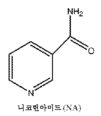
일부 실시양태에 따라, 니코틴아미드는 니코틴아미드 유도체 또는 니코틴아미드 모방체이다. 본원에서 사용된 바와 같이, 용어 "니코틴아미드 (NA)의 유도체"는 천연 NA의 화학적으로 변형된 유도체인 화합물을 지칭한다. 한 실시양태에서, 화학적 변형은 아미드 모이어티의 질소 또는 산소 원자를 통해 염기성 NA 구조체의 피리딘 고리의 치환 (고리의 탄소 또는 질소 구성원을 통해)일 수 있다. 치환될 때, 1개 이상의 수소 원자는 치환체로 대체될 수 있고/거나 N 원자에 부착되어 4가의 양으로 하전된 질소를 형성할 수 있다. 따라서, 본 발명의 니코틴아미드는 치환된 또는 비치환된 니코틴아미드를 포함한다. 또 다른 실시양태에서, 화학적 변형은 예를 들어 NA의 티오벤즈아미드 유사체를 형성하기 위한 단일 기의 결실 또는 대체일 수 있으며, 이들 모두는 유기 화학에 정통한 사람들에 의해 이해되는 것과 같다. 본 발명의 문맥에서 유도체에는 NA의 뉴클레오시드 유도체 (예를 들어, 니코틴아미드 아데닌) 또한 포함된다. NA의 다양한 유도체가 PDE4 효소의 억제 활성과 관련하여 (WO 03/068233; WO 02/060875; GB2327675A), 또는 VEGF-수용체 티로신 키나제 억제제로서 (WO 01/55114) 일부 기재된다. 예를 들어, 4-아릴-니코틴아미드 유도체 (WO 05/014549)의 제조 방법. 다른 예시적인 니코틴아미드 유도체가 WO 01/55114 및 EP2128244에 개시된다.In some embodiments, the nicotinamide is a nicotinamide derivative or a nicotinamide mimetic. As used herein, the term "derivative of nicotinamide (NA)" refers to a compound that is a chemically modified derivative of natural NA. In one embodiment, the chemical modification can be a substitution of the pyridine ring of the basic NA structure (via a carbon or nitrogen member of the ring) via a nitrogen or oxygen atom of the amide moiety. When substituted, one or more hydrogen atoms can be replaced by a substituent and/or can be attached to the N atom to form a tetravalent positively charged nitrogen. Accordingly, the nicotinamides of the present invention include substituted or unsubstituted nicotinamides. In another embodiment, the chemical modification can be a deletion or replacement of a single group, for example, to form a thiobenzamide analog of NA, all of which are as understood by those skilled in organic chemistry. In the context of the present invention, derivatives also include nucleoside derivatives of NA (e.g., nicotinamide adenine). Various derivatives of NA are described, some with regard to inhibitory activity of PDE4 enzyme (WO 03/068233; WO 02/060875; GB2327675A), or as VEGF-receptor tyrosine kinase inhibitors (WO 01/55114). For example, a process for preparing 4-aryl-nicotinamide derivatives (WO 05/014549). Other exemplary nicotinamide derivatives are disclosed in WO 01/55114 and EP2128244.
니코틴아미드 모방체는 변형된 형태의 니코틴아미드, 및 다능성 세포로부터 RPE 세포의 분화 및 성숙에서 니코틴아미드의 효과를 재현하는 니코틴아미드의 화학적 유사체를 포함한다. 예시적인 니코틴아미드 모방체에는 벤조산, 3-아미노벤조산, 및 6-아미노니코틴아미드가 포함된다. 니코틴아미드 모방체로서 작용할 수 있는 또 다른 부류의 화합물은 폴리(ADP-리보스) 폴리머라제 (PARR)의 억제제이다. 예시적인 PARP 억제제에는 3-아미노벤즈아미드, 이니파립 (BSI 201), 올라파립 (AZD-2281), 루카파립 (AG014699, PF- 01367338), 벨리파립 (ABT-888), CEP 9722, MK 4827, 및 BMN- 673이 포함된다.Nicotinamide mimetics include modified forms of nicotinamide and chemical analogs of nicotinamide that recapitulate the effects of nicotinamide in the differentiation and maturation of RPE cells from pluripotent cells. Exemplary nicotinamide mimetics include benzoic acid, 3-aminobenzoic acid, and 6-aminonicotinamide. Another class of compounds that may act as nicotinamide mimetics are inhibitors of poly(ADP-ribose) polymerase (PARR). Exemplary PARP inhibitors include 3-aminobenzamide, iniparib (BSI 201), olaparib (AZD-2281), rucaparib (AG014699, PF-01367338), veliparib (ABT-888), CEP 9722, MK 4827, and BMN-673.
추가로 고려되는 분화제에는 예를 들어 노긴, Wnt의 길항제 (Dkk1 또는 IWR1e), 결절 길항제 (Lefty-A), 레티노산, 타우린, GSK3b 억제제 (CHIR99021) 및 노치 억제제 (DAFT)가 포함된다.Additional differentiating agents being considered include, for example, Noggin, Wnt antagonists (Dkk1 or IWR1e), nodal antagonists (Lefty-A), retinoic acid, taurine, GSK3b inhibitors (CHIR99021), and Notch inhibitors (DAFT).
특정 실시양태에 따라, 분화는 다음과 같이 수행된다: (a) 제1 분화제 (예를 들어 니코틴아미드)를 포함하는 배지 중에서 ESC의 배양; 및 (b) TGFβ 수퍼패밀리의 구성원 (예를 들어 액티빈 A) 및 제1 분화제 (예를 들어 니코틴아미드)를 포함하는 배지 중에서 단계 a)로부터 수득된 세포의 배양. 단계 (a)는 TGFβ 수퍼패밀리의 구성원 (예를 들어 액티빈 A)의 부재 하에 수행될 수 있다.According to a specific embodiment, differentiation is performed as follows: (a) culturing of ESCs in a medium comprising a first differentiating agent (e.g. nicotinamide); and (b) culturing of the cells obtained from step a) in a medium comprising a member of the TGFβ superfamily (e.g. activin A) and a first differentiating agent (e.g. nicotinamide). Step (a) can be performed in the absence of the member of the TGFβ superfamily (e.g. activin A).
일부 실시양태에서, 단계 (a)에서 배지에는 TGFβ 수퍼패밀리의 구성원이 전혀 없다. 다른 실시양태에서, 배지 중에서 TGFβ 수퍼패밀리 구성원의 수준은 20 ng/mL, 10 ng/mL, 1 ng/mL 미만 또는 심지어 0.1 ng/mL 미만이다.In some embodiments, the medium in step (a) is completely free of members of the TGFβ superfamily. In other embodiments, the level of the TGFβ superfamily member in the medium is less than 20 ng/mL, less than 10 ng/mL, less than 1 ng/mL, or even less than 0.1 ng/mL.
상기 기재된 프로토콜은 제1 분화제 (예를 들어 니코틴아미드)는 포함하지만 TGFβ 수퍼패밀리의 구성원 (예를 들어 액티빈 A)은 전혀 없는 배지 중에서 단계 (b)에서 수득된 세포를 배양함으로써 계속될 수 있다. 이 단계는 본원에서 단계 (b*)로 지칭된다.The above described protocol can be continued by culturing the cells obtained in step (b) in medium containing a first differentiating agent (e.g., nicotinamide) but no member of the TGFβ superfamily (e.g., activin A). This step is referred to herein as step (b * ).
상기 기재된 프로토콜은 이제 추가의 실시양태와 함께 더욱 상세하게 기재된다.The protocol described above is now described in more detail with additional embodiments.
단계 (a): 충분한 양의 ESC가 수득되면 분화 과정이 시작된다. 이들을 전형적으로 세포 배양물로부터 (예를 들어, 콜라게나제 A, 디스파제, 트리플 셀렉트, EDTA를 사용함으로써) 제거하고, 니코틴아미드의 존재 하에 (그리고 액티빈 A의 부재 하에) 비-부착성 기질 (예를 들어, 세포 배양 플레이트, 예컨대 히드로셀(Hydrocell) 또는 아가로스-코팅된 배양물 디쉬, 또는 페트리 박테리아용 디쉬) 상에 플레이팅한다. 니코틴아미드의 예시적인 농도는 0.01-100 mM, 0.1-100 mM, 0.1-50 mM, 5-50 mM, 5-20 mM, 및 10 mM이다. 세포를 비-부착성 기질 (예를 들어 세포 배양 플레이트) 상에 플레이팅한 후에, 세포 배양물은 세포 현탁액, 바람직하게는 현탁액 배양물 중의 자유-부유 클러스터, 즉, 인간 배아 줄기 세포 (hESC)로부터 유래된 세포의 응집물로 지칭될 수 있다. 세포 클러스터는 임의의 기질 (예를 들어, 배양 플레이트, 담체)에 부착하지 않는다. 자유 부유 줄기 세포의 공급원은 이전에 WO 06/070370에 기재되었고, 그의 전문이 본원에 참조로 포함된다. 이 단계는 최소 1 일, 더욱 바람직하게는 2 일, 3 일, 1 주 또는 심지어 14 일 동안 수행될 수 있다. 바람직하게는, 세포를 예를 들어 0.01-100 mM, 0.1-100 mM, 0.1-50 mM, 5-50 mM, 5-20 mM, 예를 들어 10 mM의 니코틴아미드와 함께 (및 액티빈 A의 부재 하에) 현탁액 중에서 3 주 초과 동안 배양하지 않는다. 한 실시양태에서, 세포를 예를 들어 0.01-100 mM, 0.1-100 mM, 0.1-50 mM, 5-50 mM, 5-20 mM, 예를 들어 10 mM의 니코틴아미드와 함께 (및 액티빈 A의 부재 하에) 현탁액 중에서 6-8 일 동안 배양한다.Step (a): Once a sufficient number of ESCs are obtained, the differentiation process begins. They are typically removed from the cell culture (e.g., using collagenase A, dispase, triple select, EDTA) and plated on a non-adherent substrate (e.g., a cell culture plate such as Hydrocell or an agarose-coated culture dish, or a petri dish) in the presence of nicotinamide (and in the absence of activin A). Exemplary concentrations of nicotinamide are 0.01-100 mM, 0.1-100 mM, 0.1-50 mM, 5-50 mM, 5-20 mM, and 10 mM. After plating the cells on a non-adherent substrate (e.g. a cell culture plate), the cell culture can be referred to as a cell suspension, preferably a free-floating cluster in a suspension culture, i.e. an aggregate of cells derived from human embryonic stem cells (hESCs). The cell clusters do not attach to any substrate (e.g. a culture plate, a carrier). Sources of free-floating stem cells have been previously described in WO 06/070370, the entire contents of which are incorporated herein by reference. This step can be performed for at least 1 day, more preferably 2 days, 3 days, 1 week or even 14 days. Preferably, the cells are cultured in suspension with (and in the absence of activin A) nicotinamide, for example at 0.01-100 mM, 0.1-100 mM, 0.1-50 mM, 5-50 mM, 5-20 mM, for example at 10 mM, for more than 3 weeks. In one embodiment, the cells are cultured in suspension with nicotinamide (and in the absence of activin A) at concentrations of, for example, 0.01-100 mM, 0.1-100 mM, 0.1-50 mM, 5-50 mM, 5-20 mM, for example, 10 mM, for 6-8 days.
일부 실시양태에 따라, 세포를 비-부착성 기질, 예를 들어 세포 배양 플레이트 상에서 배양할 때, 대기 산소 조건은 20%이다. 그러나, 대기 산소 조건의 조작은 또한 대기 산소 퍼센트가 약 20%, 15%, 10%, 9%, 8%, 7%, 6% 미만 또는 심지어 약 5% 미만 (예를 들어, 1% - 20%, 1% - 10% 또는 0 - 5%)이도록 고려된다. 다른 실시양태에 따라, 세포를 초기에는 정상 대기 산소 조건 하에 비-부착성 기질 상에서 배양한 다음, 정상 미만의 대기 산소 조건으로 저하시킨다. 일부 실시양태에서, 세포를 초기 분화 동안에는 저산소 수준 하에 배양한 다음, 후기 분화 동안에는 고산소 수준 하에 배양한다.In some embodiments, when the cells are cultured on a non-adherent substrate, e.g., a cell culture plate, the atmospheric oxygen conditions are 20%. However, manipulation of the atmospheric oxygen conditions is also contemplated such that the atmospheric oxygen percentage is less than about 20%, 15%, 10%, 9%, 8%, 7%, 6%, or even less than about 5% (e.g., 1% to 20%, 1% to 10%, or 0 to 5%). In other embodiments, the cells are initially cultured on a non-adherent substrate under normal atmospheric oxygen conditions and then reduced to subnormal atmospheric oxygen conditions. In some embodiments, the cells are cultured under hypoxic conditions during initial differentiation and then under hyperoxic conditions during later differentiation.
비-부착성 세포 배양 플레이트의 예에는 눈크(Nunc)에 의해 제작된 것들 (예를 들어, 히드로셀 카탈로그 번호 174912) 등이 포함된다.Examples of non-adherent cell culture plates include those manufactured by Nunc (e.g., Hydrocell catalog number 174912).
클러스터는 적어도 50-500,000, 50-100,000, 50-50,000, 50-10,000, 50-5000, 50-1000개 세포를 포함할 수 있다. 한 실시양태에 따라, 클러스터 중의 세포는 층으로 조직화되지 않고, 불규칙한 형태를 형성한다. 한 실시양태에서, 클러스터에는 다능성 배아 줄기 세포가 전혀 없다. 또 다른 실시양태에서, 클러스터는 소량의 다능성 배아 줄기 세포 (예를 들어, 단백질 수준에서 OCT4 및 TRA-1-60을 공동-발현하는 5 % 이하, 또는 3 % 이하 (예를 들어 0.01-2.7%)의 세포)를 포함한다. 전형적으로, 클러스터는 니코틴아미드의 영향 하에 부분적으로 분화된 세포를 포함한다. 이러한 세포는 주로 신경 및 망막 전구체 마커, 예컨대 PAX6, Rax, Six3 및/또는 CHX10을 발현한다.The cluster can comprise at least 50-500,000, 50-100,000, 50-50,000, 50-10,000, 50-5000, 50-1000 cells. According to one embodiment, the cells in the cluster are not organized into layers and form an irregular shape. In one embodiment, the cluster is completely free of pluripotent embryonic stem cells. In another embodiment, the cluster comprises a small number of pluripotent embryonic stem cells (e.g., no more than 5%, or no more than 3% (e.g., 0.01-2.7%) of cells that co-express OCT4 and TRA-1-60 at the protein level). Typically, the cluster comprises cells that have partially differentiated under the influence of nicotinamide. Such cells predominantly express neural and retinal progenitor markers, such as PAX6, Rax, Six3 and/or CHX10.
관련 기술분야에 공지된 효소적 또는 비효소적 방법 (예를 들어, 기계적)을 이용하여 클러스터를 해리시킬 수 있다. 일부 실시양태에 따라, 클러스터 - 예를 들어 2-100,000개 세포, 2-50,000개 세포, 2-10,000개 세포, 2-5000개 세포, 2-1000개 세포, 2-500개 세포, 2-100개 세포, 2-50개 세포의 응집물 또는 덩어리가 더이상 없도록 세포를 해리시킨다. 특별한 실시양태에 따라, 세포는 단일 세포 현탁액 중에 있다.Clusters can be dissociated using enzymatic or non-enzymatic methods known in the art (e.g., mechanically). According to some embodiments, the cells are dissociated such that clusters—e.g., aggregates or clumps of 2-100,000 cells, 2-50,000 cells, 2-10,000 cells, 2-5000 cells, 2-1000 cells, 2-500 cells, 2-100 cells, 2-50 cells—are no longer present. According to a particular embodiment, the cells are in a single cell suspension.
이어서, 세포 (예를 들어, 해리된 세포)를 부착성 기질 상에 플레이팅하고, 예를 들어 0.01-100 mM, 0.1-100 mM, 0.1- 50 mM, 5-50 mM, 5-20 mM, 예를 들어 10 mM의 니코틴아미드의 존재 하에 (액티빈 A의 부재 하에) 배양할 수 있다. 이 단계는 최소 1 일, 더욱 바람직하게는 2 일, 3 일, 1 주 또는 심지어 14 일 동안 수행될 수 있다. 바람직하게는, 세포를 니코틴아미드의 존재 하에 (및 액티빈의 부재 하에) 3 주 초과 동안 배양하지 않는다. 예시적인 실시양태에서, 이 단계를 6-7 일 동안 수행한다.The cells (e.g., dissociated cells) can then be plated on an adherent substrate and cultured in the presence (in the absence of activin A) of nicotinamide, for example, at 0.01-100 mM, 0.1-100 mM, 0.1- 50 mM, 5-50 mM, 5-20 mM, for example 10 mM. This step can be performed for at least 1 day, more preferably 2 days, 3 days, 1 week or even 14 days. Preferably, the cells are not cultured in the presence of nicotinamide (and in the absence of activin A) for more than 3 weeks. In an exemplary embodiment, this step is performed for 6-7 days.
다른 실시양태에 따라, 세포를 부착성 기질, 예를 들어 라미닌 상에서 배양할 때, 대기 산소 조건은 20%이다. 이들은 백분율이 약 20%, 15%, 10% 미만, 더욱 바람직하게는 약 9% 미만, 약 8% 미만, 약 7% 미만, 약 6 % 미만 및 더욱 바람직하게는 약 5% 미만 (예를 들어, 1% - 20%, 1% - 10% 또는 0 - 5%)이도록 조작될 수 있다.In another embodiment, when culturing cells on an adherent substrate, such as laminin, the atmospheric oxygen conditions are 20%. They can be manipulated to have a percentage of less than about 20%, less than about 15%, less than about 10%, more preferably less than about 9%, less than about 8%, less than about 7%, less than about 6% and more preferably less than about 5% (e.g., 1% to 20%, 1% to 10% or 0 to 5%).
일부 실시양태에 따라, 세포를 초기에는 정상 대기 산소 조건 하에 부착성 기질 상에서 배양하고, 후속적으로 산소를 정상 미만의 대기 산소 조건으로 저하시킨다. 다른 실시양태에 따라, 세포를 초기에는 정상 미만의 대기 산소 조건 하에 부착성 기질 상에서 배양하고, 후속적으로 산소를 정상 대기 산소 조건으로 상승시킨다.According to some embodiments, the cells are initially cultured on the adherent substrate under normal atmospheric oxygen conditions and the oxygen is subsequently reduced to subnormal atmospheric oxygen conditions. According to other embodiments, the cells are initially cultured on the adherent substrate under subnormal atmospheric oxygen conditions and the oxygen is subsequently increased to normal atmospheric oxygen conditions.
부착성 기질 또는 물질들의 혼합물의 예에는 피브로넥틴, 라미닌, 폴리D-리신, 콜라겐 및 젤라틴이 포함될 수 있으나 이로 제한되지 않는다.Examples of adhesive substrates or mixtures of materials may include, but are not limited to, fibronectin, laminin, poly-D-lysine, collagen, and gelatin.
단계 (b): 지시된 분화의 첫번째 단계 이후에 (단계 a; 즉, 니코틴아미드 (예를 들어, 0.01-100 mM, 0.1-100 mM, 0.1-50 mM, 5-50 mM, 5-20 mM, 예를 들어 10 mM)의 존재 하에 배양), 부분-분화된 세포를 부착성 기질 상에서 추가의 분화 단계에 적용하며, 액티빈 A (예를 들어, 0.01-1000 ng/mL, 0.1-200 ng/mL, 1-200 ng/mL 예를 들어 140 ng/mL, 150 ng/mL, 160 ng/mL 또는 180 ng/mL)의 존재 하에 배양한다. 따라서, 액티빈 A를 0.1 pM-10 nM, 10 pM-10 nM, 0.1 nM-10 nM, 1 nM-10 nM, 예를 들어 5.4 nM의 최종 몰농도로 첨가할 수 있다.Step (b): After the first step of directed differentiation (step a; i.e. culturing in the presence of nicotinamide (e.g. 0.01-100 mM, 0.1-100 mM, 0.1-50 mM, 5-50 mM, 5-20 mM, e.g. 10 mM)), partially-differentiated cells are subjected to a further differentiation step on an adherent substrate, culturing in the presence of activin A (e.g. 0.01-1000 ng/mL, 0.1-200 ng/mL, 1-200 ng/mL e.g. 140 ng/mL, 150 ng/mL, 160 ng/mL or 180 ng/mL). Thus, activin A can be added at final molar concentrations of 0.1 pM-10 nM, 10 pM-10 nM, 0.1 nM-10 nM, 1 nM-10 nM, for example 5.4 nM.
니코틴아미드 또한 이 단계에서 첨가할 수 있다 (예를 들어, 0.01-100 mM, 0.1-100 mM, 0.1-50 mM, 5-50 mM, 5-20 mM, 예를 들어 10 mM). 이 단계는 1 일 내지 10 주, 3 일 내지 10 주, 1 주 내지 10 주, 1 주 내지 8 주, 1 주 내지 4 주 동안, 예를 들어 적어도 1 일, 적어도 2 일, 적어도 3 일, 적어도 5 일, 적어도 1 주, 적어도 9 일, 적어도 10 일, 적어도 2 주, 적어도 3 주, 적어도 4 주, 적어도 5 주, 적어도 6 주, 적어도 7 주, 적어도 8 주, 적어도 9 주, 적어도 10 주 동안 수행할 수 있다.Nicotinamide can also be added at this step (e.g., 0.01-100 mM, 0.1-100 mM, 0.1-50 mM, 5-50 mM, 5-20 mM, for example 10 mM). This step can be performed for 1 day to 10 weeks, 3 days to 10 weeks, 1 week to 10 weeks, 1 week to 8 weeks, 1 week to 4 weeks, for example for at least 1 day, at least 2 days, at least 3 days, at least 5 days, at least 1 week, at least 9 days, at least 10 days, at least 2 weeks, at least 3 weeks, at least 4 weeks, at least 5 weeks, at least 6 weeks, at least 7 weeks, at least 8 weeks, at least 9 weeks, at least 10 weeks.
일부 실시양태에 따라, 이 단계를 약 8 일 내지 약 2 주 동안 수행한다. 이 분화 단계를 본원에서 상기 기재된 바와 같이 낮은 또는 정상 대기 산소 조건에서 수행할 수 있다.According to some embodiments, this step is performed for about 8 days to about 2 weeks. This differentiation step can be performed under low or normal atmospheric oxygen conditions as described herein above.
단계 (b*): 지시된 분화의 두번째 단계 이후에 (즉, 부착성 기질 상에서 니코틴아미드 및 액티빈 A의 존재 하에 배양; 단계 (b)), 추가의 분화된 세포를 임의적으로 부착성 기질 상에서 후속 분화 단계에 적용하며, 니코틴아미드 (예를 들어, 0.01-100 mM, 0.1-100 mM, 0.1-50 mM, 5-50 mM, 5-20 mM, 예를 들어 10 mM)의 존재 하에 액티빈 A의 부재 하에 배양한다. 이 단계를 적어도 1 일, 2, 일, 5 일, 적어도 1 주, 적어도 2 주, 적어도 3 주 또는 심지어 4 주 동안 수행할 수 있다. 이 분화 단계는 또한 본원에서 상기 기재된 바와 같이 낮은 또는 정상 대기 산소 조건에서 수행할 수 있다.Step (b * ): After the second step of directed differentiation (i.e. culturing in the presence of nicotinamide and activin A on an adherent substrate; step (b)), additional differentiated cells are optionally subjected to a subsequent differentiation step on an adherent substrate, culturing in the presence of nicotinamide (e.g. 0.01-100 mM, 0.1-100 mM, 0.1-50 mM, 5-50 mM, 5-20 mM, e.g. 10 mM) in the absence of activin A. This step can be performed for at least 1 day, 2, days, 5 days, at least 1 week, at least 2 weeks, at least 3 weeks or even 4 weeks. This differentiation step can also be performed under low or normal atmospheric oxygen conditions as described hereinbefore.
ESC가 분화되는 염기성 배지는 시험관내 세포 성장을 뒷받침하기 위해 관련 기술분야에 공지된 임의의 공지된 세포 배양 배지, 전형적으로 정의된 염기 용액을 포함하는 배지이며, 이는 염, 당, 아미노산, 및 배양물 중에서 세포를 생존가능한 상태로 유지시키는데 필요한 임의의 다른 영양분을 포함한다. 구체적인 실시양태에 따라, 염기성 배지는 조건화된 배지가 아니다. 본 발명에 따라 사용될 수 있는 상업적으로 입수가능한 염기성 배지의 비제한적인 예는 뉴트리스템® (ESC 분화의 경우 bFGF 및 TGF 없음, ESC 확장의 경우 bFGF 및 TGF 있음), 뉴로바잘(NEUROBASAL)™, KO-DMEM, DMEM, DMEM/F12, 셀그로(CELLGRO)™ 줄기 세포 성장 배지, 또는 엑스-비보(X-VIVO)™를 포함한다. 염기성 배지를 세포 배양을 다루는 관련 기술분야에 공지된 다양한 작용제로 보충할 수 있다. 하기는 본 개시내용에 따라 사용되는 배양물에 포함될 수 있는 다양한 보충물에 대한 비제한적인 참조이다: 혈청 또는 혈청 대체물 함유 배지, 예컨대 비제한적으로 녹아웃 혈청 대체물 (KOSR), 뉴트리도마(NUTRIDOMA)-CS, TCH™, N2, N2 유도체, 또는 B27 또는 조합물; 세포외 매트릭스 (ECM) 성분, 예컨대 비제한적으로 피브로넥틴, 라미닌, 콜라겐 및 젤라틴. 이어서, ECM을 사용하여, 성장 인자의 TGFβ 수퍼패밀리의 1종 이상의 구성원; 항박테리아제, 예컨대 비제한적으로 페니실린 및 스트렙토마이신; 및 비필수 아미노산 (NEAA), 배양물 중에서 SC의 생존을 촉진시키는 역할을 하는 것으로 공지된 뉴트로핀, 예컨대 비제한적으로 BDNF, NT3, NT4를 운반할 수 있다.The basic medium in which ESCs are differentiated is any known cell culture medium known in the art to support in vitro cell growth, typically a medium comprising a defined basic solution, which comprises salts, sugars, amino acids, and any other nutrients necessary to maintain the cells in a viable state in culture. In certain embodiments, the basic medium is not a conditioned medium. Non-limiting examples of commercially available basic media that may be used in accordance with the present invention include NUTRISTEM® (without bFGF and TGF for ESC differentiation, with bFGF and TGF for ESC expansion), NEUROBASAL™, KO-DMEM, DMEM, DMEM/F12, CELLGRO™ Stem Cell Growth Medium, or X-VIVO™. The basic medium may be supplemented with a variety of agents known in the art dealing with cell culture. The following are non-limiting references to various supplements that may be included in the cultures used in accordance with the present disclosure: media containing serum or serum replacement, such as but not limited to Knock Out Serum Replacement (KOSR), NUTRIDOMA-CS, TCH™, N2, N2 derivatives, or B27 or combinations; extracellular matrix (ECM) components, such as but not limited to fibronectin, laminin, collagen, and gelatin. The ECM can then be used to transport one or more members of the TGFβ superfamily of growth factors; antibacterial agents, such as but not limited to penicillin and streptomycin; and non-essential amino acids (NEAAs), neurotrophins known to play a role in promoting the survival of SCs in culture, such as but not limited to BDNF, NT3, NT4.
일부 실시양태에 따라, ESC의 분화에 사용되는 배지는 뉴트리스템® 배지 (바이올로지컬 인더스트리즈(Biological Industries), 06-5102-01- IA)이다.In some embodiments, the medium used for differentiation of ESCs is Nutristem ® medium (Biological Industries, 06-5102-01-IA).
일부 실시양태에 따라, ESC의 분화 및 확장은 이종이 없는 조건 하에 수행된다.According to some embodiments, differentiation and expansion of ESCs are performed under xenobiotic-free conditions.
다른 실시양태에 따라, 증식/성장 배지에는 이종 오염물이 전혀 없으며, 즉, 동물 유래된 성분, 예컨대 혈청, 동물 유래된 성장 인자 및 알부민이 없다. 따라서, 이들 실시양태에 따라, 배양을 이종 오염물의 부재 하에 수행한다.According to another embodiment, the proliferation/growth medium is completely free of xenobiotic contaminants, i.e., free of animal-derived components, such as serum, animal-derived growth factors and albumin. Thus, according to these embodiments, the culturing is performed in the absence of xenobiotic contaminants.
이종이 없는 조건 하에 ESC의 다른 배양 방법은 미국 특허 출원 번호 20130196369에 제공되어 있으며, 그의 내용은 전문이 본원에 참조로 포함된다.Other methods for culturing ESCs under xenobiotic-free conditions are provided in U.S. Patent Application No. 20130196369, the contents of which are incorporated herein by reference in their entirety.
RPE 세포를 포함하는 제제는 우수 제조 관리기준 (GMP)에 따라 (예를 들어, 제제는 GMP-적합판정임) 및/또는 현행 우수 조직 관리기준 (GTP)에 따라 (예를 들어, 제제는 GTP-적합판정일 수 있음) 제조될 수 있다.A formulation comprising RPE cells can be manufactured in accordance with Good Manufacturing Practices (GMP) (e.g., the formulation is determined to be GMP-compliant) and/or in accordance with current Good Tissue Practices (GTP) (e.g., the formulation can be determined to be GTP-compliant).
분화 단계 동안에, 배아 줄기 세포는 그들의 분화 상태에 대해 모니터링될 수 있다. 세포 분화는 분화의 지표인 것으로 공지된 세포 또는 조직-특이적 마커의 실험 시에 결정될 수 있다.During the differentiation stage, embryonic stem cells can be monitored for their differentiation status. Cell differentiation can be determined upon testing for cell or tissue-specific markers known to be indicative of differentiation.
조직/세포 특이적 마커는 관련 기술분야에 널리 공지된 면역학적 기술을 이용하여 검출될 수 있다 [Thomson JA et al., (1998). Science 282: 1145-7]. 그 예에는 막-결합된 또는 세포내 마커에 대한 유세포 분석, 세포외 및 세포내 마커에 대한 면역조직화학, 및 분자 마커에 대한 효소적 면역검정이 포함되나 이로 제한되지 않는다.Tissue/cell specific markers can be detected using immunological techniques well known in the art [Thomson JA et al., (1998). Science 282: 1145-7]. Examples include, but are not limited to, flow cytometry for membrane-bound or intracellular markers, immunohistochemistry for extracellular and intracellular markers, and enzymatic immunoassays for molecular markers.
상기 기재된 분화 단계 이후에, 착색된 및 착색되지 않은 세포 둘 다를 포함하는 혼합된 세포 집단을 수득할 수 있다. 이 측면에 따라, 혼합된 세포 집단의 세포를 플레이트로부터 제거한다. 일부 실시양태에서, 이는 효소적으로 수행된다 (예를 들어, 트립신 사용, (트리플 셀렉트); 예를 들어, 국제 특허 출원 공개 번호 WO 2017/021973을 참조하며, 그의 전문이 본원에 참조로 포함됨). 본 발명의 이 측면에 따라, 배양물로부터 제거된 (그리고 후속적으로 확장된) 세포의 적어도 10%, 20%, 30%, 적어도 40%, 적어도 50%, 적어도 60%, 적어도 70%는 착색되지 않은 세포이다. 다른 실시양태에서, 이는 기계적으로 - 예를 들어 세포 스크레이퍼를 사용하여 수행된다. 추가의 다른 실시양태에서, 이는 화학적으로 (예를 들어, EDTA) 수행된다. 효소적 및 화학적 처리의 조합 또한 고려된다. 예를 들어, EDTA 및 효소적 처리가 이용될 수 있다. 추가로, 배양물로부터 제거된 (그리고 후속적으로 확장된) 세포의 적어도 10%, 20% 또는 심지어 30%는 착색된 세포이다.After the differentiation step described above, a mixed cell population comprising both pigmented and unpigmented cells can be obtained. According to this aspect, cells of the mixed cell population are removed from the plate. In some embodiments, this is done enzymatically (e.g., using trypsin, (triple select); see, e.g., International Patent Application Publication No. WO 2017/021973, which is incorporated herein by reference in its entirety). According to this aspect of the invention, at least 10%, 20%, 30%, at least 40%, at least 50%, at least 60%, at least 70% of the cells removed from the culture (and subsequently expanded) are unpigmented cells. In other embodiments, this is done mechanically - e.g., using a cell scraper. In still other embodiments, this is done chemically (e.g., with EDTA). Combinations of enzymatic and chemical treatments are also contemplated. For example, EDTA and enzymatic treatments can be used. Additionally, at least 10%, 20%, or even 30% of the cells removed from the culture (and subsequently expanded) are pigmented cells.
본 개시내용의 이 측면에 따라, 배양물 중의 모든 세포의 적어도 50%, 60%, 70%, 80%, 90%, 95%, 100%가 제거된다 (그리고 후속적으로 확장됨).According to this aspect of the present disclosure, at least 50%, 60%, 70%, 80%, 90%, 95%, 100% of all cells in a culture are removed (and subsequently expanded).
세포의 혼합된 집단의 확장은 세포외 매트릭스, 예를 들어, 젤라틴, 콜라겐 I, 콜라겐 IV, 라미닌 (예를 들어, 라미닌 521), 피브로넥틴 및 폴리-D-리신 상에서 수행될 수 있다. 확장을 위해, 세포를 혈청-무함유 KOM, 혈청 포함 배지 (예를 들어, 20% 인간 혈청을 갖는 DMEM) 또는 뉴트리스템® 배지 (06- 5102-01- IA, 바이올로지컬 인더스트리즈) 상에서 배양할 수 있다. 이들 배양 조건 하에, 적합한 조건 하에 계대한 후에, 정제된 RPE 세포의 집단이 수득되도록 착색된 세포 대 착색되지 않은 세포의 비를 증가시킨다. 이러한 세포는 특징적인 다변형 형상 모폴로지 및 RPE 세포의 색소 침착을 나타낸다.Expansion of mixed populations of cells can be performed on extracellular matrices, such as gelatin, collagen I, collagen IV, laminin (e.g. laminin 521), fibronectin and poly-D-lysine. For expansion, cells can be cultured on serum-free KOM, serum-containing media (e.g. DMEM with 20% human serum) or Nutristem ® media (06-5102-01-IA, Biological Industries). Under these culture conditions, after passage under suitable conditions, the ratio of pigmented to unpigmented cells is increased so that a population of purified RPE cells is obtained. These cells exhibit the characteristic polyhedral shape morphology and pigmentation of RPE cells.
한 실시양태에서, 확장은 니코틴아미드 (예를 들어, 0.01-100 mM, 0.1-100 mM, 0.1-50 mM, 5-50 mM, 5-20 mM, 예를 들어 10 mM)의 존재 하에 및 액티빈 A의 부재 하에 수행된다.In one embodiment, expansion is performed in the presence of nicotinamide (e.g., 0.01-100 mM, 0.1-100 mM, 0.1-50 mM, 5-50 mM, 5-20 mM, e.g., 10 mM) and in the absence of activin A.
세포의 혼합된 집단을 현탁액 중에서 (미세-담체와 함께 또는 그 없이) 또는 단층으로 확장시킬 수 있다. 단층 배양물 중에서 또는 현탁액 배양물 중에서 세포의 혼합된 집단의 확장을 관련 기술분야에 정통한 사람들에게 널리 공지된 방법에 의해 생물반응기 또는 다중/하이퍼 스택에서 대규모 확장으로 변형시킬 수 있다.Mixed populations of cells can be expanded in suspension (with or without micro-carriers) or in monolayers. Expansion of mixed populations of cells in monolayer cultures or in suspension cultures can be transformed into large-scale expansion in bioreactors or multi/hyper stacks by methods well known to those skilled in the art.
일부 실시양태에 따라, 확장기는 적어도 1 주, 적어도 2 주, 적어도 3 주, 적어도 4 주, 적어도 5 주, 적어도 6 주, 적어도 7 주, 적어도 8 주, 적어도 9 주 또는 심지어 10 주 동안 수행된다. 바람직하게는, 확장기는 1 주 - 10 주, 더욱 바람직하게는 2 주 - 10 주, 더욱 바람직하게는, 3 주 - 10 주, 더욱 바람직하게는 4 주 - 10 주, 또는 4 주 - 8 주 동안 수행된다.According to some embodiments, the expansion is performed for at least 1 week, at least 2 weeks, at least 3 weeks, at least 4 weeks, at least 5 weeks, at least 6 weeks, at least 7 weeks, at least 8 weeks, at least 9 weeks or even 10 weeks. Preferably, the expansion is performed for 1 week to 10 weeks, more preferably 2 weeks to 10 weeks, more preferably 3 weeks to 10 weeks, more preferably 4 weeks to 10 weeks, or 4 weeks to 8 weeks.
추가의 다른 실시양태에 따라, 세포의 혼합된 집단을 확장기 동안에 적어도 1회, 확장기 동안에 적어도 2회, 확장기 동안에 적어도 3회, 확장기 동안에 적어도 4회, 확장기 동안에 적어도 5회, 또는 확장기 동안에 적어도 6회 계대한다.According to further other embodiments, the mixed population of cells is passaged at least once during the expansion phase, at least twice during the expansion phase, at least three times during the expansion phase, at least four times during the expansion phase, at least five times during the expansion phase, or at least six times during the expansion phase.
본 발명자들은, 세포를 효소적으로 수집할 때, 8 계대 초과, 9 계대 초과 및 심지어 10 계대 초과 (예를 들어, 11-15 계대) 동안 계속 확장시킬 수 있음을 확인하였다. 총 세포 배가의 수는 30 초과, 예를 들어, 31, 32, 33, 34 또는 그 초과로 증가될 수 있다. (국제 특허 출원 공개 번호 WO 2017/021973을 참조하며, 그의 전문이 본원에 참조로 포함됨).The present inventors have found that when cells are enzymatically harvested, they can be expanded continuously for more than 8 passages, more than 9 passages, and even more than 10 passages (e.g., 11-15 passages). The total number of cell doublings can be increased to more than 30, e.g., 31, 32, 33, 34 or more. (See International Patent Application Publication No. WO 2017/021973, which is incorporated herein by reference in its entirety).
본원에 기재된 방법에 따라 생성된 RPE 세포의 집단은 수많은 상이한 파라미터에 따라 특징분석될 수 있다. 따라서, 예를 들어, 수득된 RPE 세포는 다변형 형상을 갖고, 착색될 수 있다.A population of RPE cells generated according to the methods described herein can be characterized according to a number of different parameters. Thus, for example, the obtained RPE cells can have a polyhedral shape and be pigmented.
본원에 개시된 세포 집단에는 일반적으로 미분화된 인간 배아 줄기 세포가 전혀 없음을 이해할 것이다. 일부 실시양태에 따라, 예를 들어 FACS에 의해 측정 시, 1:250,000개 미만의 세포는 Oct4+TRA-1-60+ 세포이다. 세포는 또한 PCR에 의해 측정 시 GDF3 또는 TDGF의 발현을 (5,000배 초과로) 하향 조절할 수 있었다. 이 측면의 RPE 세포는 배아 줄기 세포 마커를 발현하지 않는다. 상기 1종 이상 배아 줄기 세포 마커는 OCT- 4, NANOG, Rex- 1, 알칼리 포스파타제, Sox2, TDGF- 베타, SSEA-3, SSEA-4, TRA- 1-60, 및/또는 TRA-1-81을 포함할 수 있다.It will be appreciated that the cell populations disclosed herein are generally free of undifferentiated human embryonic stem cells. In some embodiments, less than 1:250,000 cells are Oct4+TRA-1-60+ cells, as measured by, for example, FACS. The cells can also down-regulate expression of GDF3 or TDGF (greater than 5,000-fold), as measured by PCR. The RPE cells of this aspect do not express embryonic stem cell markers. The one or more embryonic stem cell markers can include OCT-4, NANOG, Rex-1, alkaline phosphatase, Sox2, TDGF-beta, SSEA-3, SSEA-4, TRA-1-60, and/or TRA-1-81.
치료적 RPE 세포 제제는 적어도 약 75%, 80%, 85%, 90%, 91 %, 92%, 93%, 94%, 95%, 96%, 97%, 98%, 99% 또는 100% RPE 세포를 포함하도록 비-RPE 세포와 관련하여 실질적으로 정제될 수 있다. 치료적 RPE 세포 제제는 본질적으로 비-RPE 세포를 함유하지 않을 수 있거나 또는 RPE 세포로 이루어질 수 있다. 예를 들어, RPE 세포의 실질적으로 정제된 제제는 약 25%, 20%, 15%, 10%, 9%, 8%, 7%, 6%, 5%, 4%, 3%, 2% 또는 1% 미만의 비-RPE 세포 유형을 포함할 수 있다. 예를 들어, RPE 세포 제제는 약 25%, 20%, 15%, 10%, 9%, 8%, 7%, 6%, 5%, 4%, 3%, 2%, 1%, 0.9%, 0.8%, 0.7%, 0.6%, 0.5%, 0.4%, 0.3%, 0.2%, 0.1%, 0.09%, 0.08%, 0.07%, 0.06%, 0.05%, 0.04%, 0.03%, 0.02%, 0.01%, 0.009%, 0.008%, 0.007%, 0.006%, 0.005%, 0.004%, 0.003%, 0.002%, 0.001%, 0.0009%, 0.0008%, 0.0007%, 0.0006%, 0.0005%, 0.0004%, 0.0003%, 0.0002%, 또는 0.0001% 미만의 비-RPE 세포를 포함할 수 있다.The therapeutic RPE cell preparation can be substantially purified with respect to non-RPE cells to comprise at least about 75%, 80%, 85%, 90%, 91%, 92%, 93%, 94%, 95%, 96%, 97%, 98%, 99% or 100% RPE cells. The therapeutic RPE cell preparation can be essentially free of non-RPE cells or can consist essentially of RPE cells. For example, the substantially purified preparation of RPE cells can comprise less than about 25%, 20%, 15%, 10%, 9%, 8%, 7%, 6%, 5%, 4%, 3%, 2% or 1% of a non-RPE cell type. For example, RPE cell preparations may contain about 25%, 20%, 15%, 10%, 9%, 8%, 7%, 6%, 5%, 4%, 3%, 2%, 1%, 0.9%, 0.8%, 0.7%, 0.6%, 0.5%, 0.4%, 0.3%, 0.2%, 0.1%, 0.09%, 0.08%, 0.07%, 0.06%, 0.05%, 0.04%, 0.03%, 0.02%, 0.01%, 0.009%, 0.008%, 0.007%, 0.006%, 0.005%, 0.004%, 0.003%, may contain less than 0.002%, 0.001%, 0.0009%, 0.0008%, 0.0007%, 0.0006%, 0.0005%, 0.0004%, 0.0003%, 0.0002%, or 0.0001% non-RPE cells.
RPE 세포 제제는 비-RPE 세포와 관련하여 뿐만 아니라 다른 성숙 수준의 RPE 세포와 관련하여 실질적으로 순수할 수 있다. 제제는 비-RPE 세포와 관련하여 실질적으로 정제될 수 있고, 성숙한 RPE 세포에 대해 강화될 수 있다. 예를 들어, 성숙한 RPE 세포에 대해 강화된 RPE 세포 제제에서, RPE 세포의 적어도 약 30%, 40%, 45%, 50%, 55%, 60%, 65%, 70%, 75%, 80%, 85%, 90%, 91%, 92%, 93%, 94%, 95%, 96%, 97%, 98%, 99%, 99% 또는 100%는 성숙한 RPE 세포이다. 제제는 비-RPE 세포와 관련하여 실질적으로 정제될 수 있고, 성숙한 RPE 세포가 아니라 분화된 RPE 세포에 대해 강화될 수 있다. 예를 들어, RPE 세포의 적어도 약 30%, 40%, 45%, 50%, 55%, 60%, 65%, 70%, 75%, 80%, 85%, 90%, 91%, 92%, 93%, 94%, 95%, 96%, 97%, 98%, 99% 또는 100%는 성숙한 RPE 세포가 아니라 분화된 RPE 세포일 수 있다.The RPE cell preparation can be substantially pure with respect to non-RPE cells as well as with respect to RPE cells of different maturity levels. The preparation can be substantially purified with respect to non-RPE cells and enriched for mature RPE cells. For example, in an RPE cell preparation enriched for mature RPE cells, at least about 30%, 40%, 45%, 50%, 55%, 60%, 65%, 70%, 75%, 80%, 85%, 90%, 91%, 92%, 93%, 94%, 95%, 96%, 97%, 98%, 99%, 99%, or 100% of the RPE cells are mature RPE cells. The preparation can be substantially purified with respect to non-RPE cells and enriched for differentiated RPE cells rather than mature RPE cells. For example, at least about 30%, 40%, 45%, 50%, 55%, 60%, 65%, 70%, 75%, 80%, 85%, 90%, 91%, 92%, 93%, 94%, 95%, 96%, 97%, 98%, 99% or 100% of the RPE cells may be differentiated RPE cells rather than mature RPE cells.
본원에 기재된 제제에는 박테리아, 바이러스 또는 진균 오염 또는 감염, 예컨대 비제한적으로 HIV 1, HIV 2, HBV, HCV, HAV, CMV, HTLV 1, HTLV 2, 파보바이러스 B19, 엡스타인-바르(Epstein-Barr) 바이러스, 또는 헤르페스바이러스 1 및 2, SV40, HHV5, 6, 7, 8, CMV, 폴리오마 바이러스, HPV, 엔테로바이러스의 존재가 실질적으로 없을 수 있다. 본원에 기재된 제제에는 마이코플라즈마 오염 또는 감염이 실질적으로 없을 수 있다.The formulations described herein can be substantially free of bacterial, viral or fungal contamination or infection, including but not limited to HIV 1,
본원에 개시된 세포 집단을 특징분석하는 또 다른 방식은 마커 발현에 의한 것이다. 따라서, 예를 들어, 면역염색에 의해 측정 시 세포의 적어도 50%, 60% 70%, 80%, 85%, 90%, 95% 또는 100%는 베스트로핀 1을 발현할 수 있다. 한 실시양태에 따라, 세포의 80-100%는 베스트로핀 1을 발현한다.Another way to characterize the cell population disclosed herein is by marker expression. Thus, for example, at least 50%, 60% 70%, 80%, 85%, 90%, 95% or 100% of the cells can express Bestropin 1, as measured by immunostaining. In one embodiment, 80-100% of the cells express Bestropin 1.
다른 실시양태에 따라, 면역염색에 의해 측정 시 세포의 적어도 80%, 85%, 87%, 89%, 90%, 95%, 97% 또는 100%는 소안구증-연관된 전사 인자 (MITF)를 발현한다. 예를 들어, 세포의 80-100%는 MITF를 발현한다.In other embodiments, at least 80%, 85%, 87%, 89%, 90%, 95%, 97% or 100% of the cells express microphthalmia-associated transcription factor (MITF) as measured by immunohistochemistry. For example, 80-100% of the cells express MITF.
다른 실시양태에 따라, 면역염색에 의해 측정 시 세포의 적어도 50%, 60% 70%, 80%, 85%, 87%, 89%, 90%, 95%, 97% 또는 100%는 소안구증-연관된 전사 인자 (MITF) 및 베스트로핀 1을 둘 다 발현한다. 예를 들어, 세포의 80-100%는 MITF 및 베스트로핀 1을 공동-발현한다.In other embodiments, at least 50%, 60% 70%, 80%, 85%, 87%, 89%, 90%, 95%, 97% or 100% of the cells express both microphthalmia-associated transcription factor (MITF) and bestrophin 1, as measured by immunostaining. For example, 80-100% of the cells co-express MITF and bestrophin 1.
다른 실시양태에 따라, 면역염색에 의해 측정 시 세포의 적어도 80%, 85%, 87%, 89%, 90%, 95%, 97% 또는 100%는 소안구증-연관된 전사 인자 (MITF) 및 Z0-1을 둘 다 발현한다. 예를 들어, 세포의 80-100%는 MITF 및 Z0-1을 공동-발현한다.In other embodiments, at least 80%, 85%, 87%, 89%, 90%, 95%, 97% or 100% of the cells express both microphthalmia-associated transcription factor (MITF) and Z0-1 as measured by immunostaining. For example, 80-100% of the cells co-express MITF and Z0-1.
다른 실시양태에 따라, 면역염색에 의해 측정 시 세포의 적어도 50%, 60% 70%, 80%, 85%, 87%, 89%, 90%, 95%, 97% 또는 100%는 Z0-1 및 베스트로핀 1을 둘 다 발현한다. 예를 들어, 세포의 80-100%는 Z0-1 및 베스트로핀 1을 공동-발현한다.In other embodiments, at least 50%, 60% 70%, 80%, 85%, 87%, 89%, 90%, 95%, 97% or 100% of the cells express both Z0-1 and bestropin 1, as measured by immunostaining. For example, 80-100% of the cells co-express Z0-1 and bestropin 1.
또 다른 실시양태에 따라, 면역염색 또는 FACS에 의해 측정 시 세포의 적어도 50%, 60% 70% 80%, 85%, 87%, 89%, 90%, 95%, 97% 또는 100%는 쌍형성 박스 유전자 6 (PAX-6)을 발현한다.In another embodiment, at least 50%, 60% 70% 80%, 85%, 87%, 89%, 90%, 95%, 97% or 100% of the cells express paired box gene 6 (PAX-6) as measured by immunohistochemistry or FACS.
또 다른 실시양태에 따라, 면역염색에 의해 측정 시 세포의 적어도 50%, 60% 70%, 80%, 85%, 87%, 89%, 90%, 95%, 97% 또는 100%는 세포성 레틴알데히드 결합 단백질 (CRALBP)을 발현한다. 예를 들어, 세포의 85-100%는 CRALBP를 발현한다.In another embodiment, at least 50%, 60% 70%, 80%, 85%, 87%, 89%, 90%, 95%, 97% or 100% of the cells express cellular retinaldehyde binding protein (CRALBP) as measured by immunostaining. For example, 85-100% of the cells express CRALBP.
또 다른 실시양태에 따라, 세포의 적어도 50%, 60% 70%, 80%, 85%, 87%, 89%, 90%, 95%, 97% 또는 100%는 면역염색에 의해 측정 시 세포성 멜라닌세포 계통-특이적 항원 GP100 (PMEL17)을 발현한다. 예를 들어, 세포의 85-100%는 PMEL17을 발현한다.In another embodiment, at least 50%, 60% 70%, 80%, 85%, 87%, 89%, 90%, 95%, 97% or 100% of the cells express cellular melanocyte lineage-specific antigen GP100 (PMEL17) as measured by immunostaining. For example, 85-100% of the cells express PMEL17.
RPE 세포는 전형적으로 최종 분화를 나타내는 마커, 예를 들어 베스트로핀 1, CRALBP 및/또는 RPE65를 공동-발현한다. 또한, 본원에 기재된 RPE 세포는 RPE 일차 섬모에 대한 마커, 예컨대 ARL13B 및 GT335를 발현할 수 있다.RPE cells typically co-express markers indicative of terminal differentiation, such as bestropin 1, CRALBP and/or RPE65. Additionally, the RPE cells described herein may express markers for RPE primary cilia, such as ARL13B and GT335.
확장기 이후에, RPE 세포를 포함하는 세포 집단이 수득되며, 그의 적어도 70%, 71%, 72%, 73%, 74%, 75%, 76%, 77%, 78%, 79%, 80%, 81%, 82%, 83%, 84%, 85%, 86%, 87%, 88%, 89%, 90%, 91%, 92%, 93%, 94%, 95%, 96%, 97%, 98%, 99% 또는 심지어 100%가 CRALBP+ PMEL1 7+이다.After the expansion phase, a cell population comprising RPE cells is obtained, at least 70%, 71%, 72%, 73%, 74%, 75%, 76%, 77%, 78%, 79%, 80%, 81%, 82%, 83%, 84%, 85%, 86%, 87%, 88%, 89%, 90%, 91%, 92%, 93%, 94%, 95%, 96%, 97%, 98%, 99% or even 100% of which are CRALBP+ PMEL1 7+.
특정 실시양태에서, RPE 세포 조성물은 하기 방법에 따라 생성될 수 있다: (1) 인간 혈청 알부민 (HSA)을 갖는 NUT+ 중에서 2 주 동안 중심 웰 (CW) 플레이트에서 hUCF 상에서 hESC를 배양하고, (2) HSA를 갖는 NUT+ 중에서 4 내지 5 주 동안 (또는 원하는 양의 세포일 때까지) CW 플레이트에서 hUCF 상에서 hESC를 확장시키기 위해 기계적으로 계대하고, (3) HSA를 갖는 NUT+ 중에서 추가 1 주 동안 6 cm 플레이트에서 hUCF 상에서 (예를 들어, 콜라게나제를 사용하여) hESC 콜로니를 계속 확장시키고, (4) 니코틴아미드 (NIC)를 갖는 NUT- 중에서 약 1 주 동안 약 5개의 6 cm 플레이트로부터 1개의 히드로셀로 콜로니를 전달함으로써 구상체 (SB)를 제조하고, (5) NIC를 갖는 NUT- 중에서 약 1 주 동안 SB를 6-웰 플레이트의 2-3개 웰로 전달함으로써 Lam511 상에서 SB를 평탄화시키고, (6) NIC 및 액티빈을 갖는 NUT- 중에서 약 1 내지 2 주 동안 Lam511 상에서 부착성 세포를 배양하고, 배지를 NIC를 갖는 NUT-로 교체하고, 1 내지 3 주 동안 배양하고, (7) 효소, 예컨대 트리플 셀렉트를 사용하여 착색된 세포에 대해 강화시키고, (8) 20% 인간 혈청 및 NUT- 중에서 약 2 내지 9 주 동안 (배지 교체) 플라스크에서 젤라틴 상에서 RPE 세포를 확장시키고, (9) RPE 세포를 수확한다.In certain embodiments, the RPE cell composition can be generated by the following methods: (1) culturing hESCs on hUCFs in center well (CW) plates in NUT+ with human serum albumin (HSA) for 2 weeks, (2) mechanically passage hESCs to expand on hUCFs in CW plates in NUT+ with HSA for 4-5 weeks (or until desired amount of cells), (3) continuing to expand hESC colonies on hUCFs (e.g., using collagenase) in 6 cm plates in NUT+ with HSA for an additional week, (4) transferring colonies from about 5 of the 6 cm plates to 1 hydrocell in NUT- with nicotinamide (NIC) for about 1 week to prepare spheroids (SBs), and (5) transferring SBs to 2-3 wells of a 6-well plate in NUT- with NIC for about 1 week. Flatten SB on Lam511, (6) culture adherent cells on Lam511 in NUT- with NIC and activin for about 1 to 2 weeks, replace medium with NUT- with NIC, culture for 1 to 3 weeks, (7) enrich for pigmented cells using enzymes such as triple select, (8) expand RPE cells on gelatin in flasks in 20% human serum and NUT- for about 2 to 9 weeks (with medium replacement), (9) harvest RPE cells.
RPE 세포의 확장된 집단의 수확은 관련 기술분야에 공지된 방법을 이용하여 (예를 들어, 효소, 예컨대 트립신을 사용하여, 또는 화학적으로 EDTA 등을 사용하여) 수행될 수 있다. 일부 실시양태에서, RPE 세포를 적절한 용액, 예컨대 PBS 또는 BSS 플러스를 사용하여 세척할 수 있다. 일부 실시양태에서, RPE 세포를 수확하거나 강화시킨 후에 효소 중화 용액을 사용할 수 있다. 중화 용액은 예를 들어 인간 혈청 또는 인간 혈청 알부민을 갖거나 또는 갖지 않는 배지를 포함할 수 있다. 일부 실시양태에서, HS 또는 HAS를 갖거나 또는 갖지 않는 효소 중화 용액 중에서 장기간 인큐베이션은 세포 생존력 또는 세포 회복률에 대해 효과를 갖지 않는다.Harvesting of expanded populations of RPE cells can be accomplished using methods known in the art (e.g., using enzymes such as trypsin, or chemically using EDTA, etc.). In some embodiments, the RPE cells can be washed using an appropriate solution such as PBS or BSS Plus. In some embodiments, an enzyme neutralizing solution can be used after the RPE cells have been harvested or enriched. The neutralizing solution can include, for example, media with or without human serum or human serum albumin. In some embodiments, prolonged incubation in an enzyme neutralizing solution with or without HS or HAS has no effect on cell viability or cell recovery.
다른 실시양태에서, 동결보존을 위한 RPE 세포를 제형화하고, 해동 직후에 대상체에게 투여하기 전에, RPE 세포를 여과할 수 있다. 일부 실시양태에서, 여과후 세포의 퍼센트 생존력은 적어도 약 75%, 80%, 85%, 90%, 91%, 92%, 93%, 94%, 95%, 96%, 97%, 98%, 99% 또는 100%이다. 일부 실시양태에서, 중화 용액 중에서 약 0 내지 약 8 시간 동안 저장한 여과후 세포의 퍼센트 생존력은 적어도 약 75%, 80%, 85%, 90%, 91%, 92%, 93%, 94%, 95%, 96%, 97%, 98%, 99% 또는 100%이다.In other embodiments, the RPE cells can be formulated for cryopreservation and filtered prior to administration to a subject immediately following thawing. In some embodiments, the percent viability of the cells after filtration is at least about 75%, 80%, 85%, 90%, 91%, 92%, 93%, 94%, 95%, 96%, 97%, 98%, 99%, or 100%. In some embodiments, the percent viability of the cells after filtration is at least about 75%, 80%, 85%, 90%, 91%, 92%, 93%, 94%, 95%, 96%, 97%, 98%, 99%, or 100% after storage in a neutralizing solution for about 0 to about 8 hours.
수확한 후에, RPE 세포의 확장된 집단을 구체적인 치료 용량 (예를 들어, 세포의 개수)으로 제형화할 수 있고, 클리닉으로 운송하기 위해 동결보존시킬 수 있다. 이어서, 해동 직후에 추가로 가공하지 않고 즉시 투여가능한 (RTA) RPE 세포 치료 조성물을 투여할 수 있다. 동결보존에 적합한 배지의 예에는 90% 인간 혈청/10% DMSO, 배지 3 10% (CS10), 배지 2 5% (CS5) 및 배지 1 2% (CS2), 스템 셀 뱅커(Stem Cell Banker), 프라임(PRIME) XV® 프리지스(FREEZIS), 하이포써모솔(HYPOTHERMOSOL)®, CSB, 트레할로스 등이 포함되나 이로 제한되지 않는다.After harvest, the expanded population of RPE cells can be formulated into a specific therapeutic dose (e.g., number of cells) and cryopreserved for shipment to the clinic. The RPE cell therapy composition can then be administered ready-to-use (RTA) without further processing immediately after thawing. Examples of suitable media for cryopreservation include, but are not limited to, 90% Human Serum/10% DMSO,
일부 실시양태에서, 동결보존 배지 중에서 약 0 내지 약 8 시간 동안 저장한 여과후 세포의 퍼센트 생존력은 적어도 약 75%, 80%, 85%, 90%, 91%, 92%, 93%, 94%, 95%, 96%, 97%, 98%, 99% 또는 100%이다. 다른 실시양태에서, 동결보존 배지 중에서 약 0 내지 약 8 시간 동안 저장한 여과후 세포의 퍼센트 회복률은 적어도 약 75%, 80%, 85%, 90%, 91%, 92%, 93%, 94%, 95%, 96%, 97%, 98%, 99% 또는 100%이다.In some embodiments, the percent viability of the filtered cells after storage in the cryopreservation medium for about 0 to about 8 hours is at least about 75%, 80%, 85%, 90%, 91%, 92%, 93%, 94%, 95%, 96%, 97%, 98%, 99% or 100%. In other embodiments, the percent recovery of the filtered cells after storage in the cryopreservation medium for about 0 to about 8 hours is at least about 75%, 80%, 85%, 90%, 91%, 92%, 93%, 94%, 95%, 96%, 97%, 98%, 99% or 100%.
추가의 실시양태에서, 중화 배지 중에서 약 0 내지 약 8 시간 동안 저장한 후에, 동결보존 배지 중에서 약 0 내지 약 8 시간 동안 저장한 여과후 세포의 퍼센트 생존력은 적어도 약 75%, 80%, 85%, 90%, 91%, 92%, 93%, 94%, 95%, 96%, 97%, 98%, 99% 또는 100%이다. 다른 실시양태에서, 중화 배지 중에서 약 0 내지 약 8 시간 동안 저장한 후에, 동결보존 배지 중에서 약 0 내지 약 8 시간 동안 저장한 여과후 세포의 퍼센트 회복률은 적어도 약 70%, 75%, 80%, 85%, 90%, 91%, 92%, 93%, 94%, 95%, 96%, 97%, 98%, 99% 또는 100%이다.In further embodiments, the percent viability of the post-filtered cells after storage in a neutralizing medium for about 0 to about 8 hours followed by storage in a cryopreservation medium for about 0 to about 8 hours is at least about 75%, 80%, 85%, 90%, 91%, 92%, 93%, 94%, 95%, 96%, 97%, 98%, 99% or 100%. In other embodiments, the percent recovery of the post-filtered cells after storage in a neutralizing medium for about 0 to about 8 hours followed by storage in a cryopreservation medium for about 0 to about 8 hours is at least about 70%, 75%, 80%, 85%, 90%, 91%, 92%, 93%, 94%, 95%, 96%, 97%, 98%, 99% or 100%.
추가의 다른 실시양태에서, 중화 배지 중에서 약 0 내지 약 8 시간 동안 저장한 후에, 동결보존 배지 중에서 약 0 내지 약 8 시간 동안 저장하고, 동결보존된 조성물을 해동한 후에, 여과후 세포의 퍼센트 생존력은 적어도 약 75%, 80%, 85%, 90%, 91%, 92%, 93%, 94%, 95%, 96%, 97%, 98%, 99% 또는 100%이다. 추가의 다른 실시양태에서, 중화 배지 중에서 약 0 내지 약 8 시간 동안 저장한 후에, 동결보존 배지 중에서 약 0 내지 약 8 시간 동안 저장하고, 동결보존된 조성물을 해동한 후에, 여과후 세포의 퍼센트 회복률은 적어도 약, 70%, 75%, 80%, 85%, 90%, 91%, 92%, 93%, 94%, 95%, 96%, 97%, 98%, 99% 또는 100%이다.In yet other embodiments, after storage in a neutralizing medium for about 0 to about 8 hours, storage in a cryopreservation medium for about 0 to about 8 hours, and after thawing the cryopreserved composition, the percent viability of cells after filtration is at least about 75%, 80%, 85%, 90%, 91%, 92%, 93%, 94%, 95%, 96%, 97%, 98%, 99% or 100%. In yet further embodiments, after storage in a neutralizing medium for about 0 to about 8 hours, storage in a cryopreservation medium for about 0 to about 8 hours, and after thawing the cryopreserved composition, the percent recovery of cells after filtration is at least about 70%, 75%, 80%, 85%, 90%, 91%, 92%, 93%, 94%, 95%, 96%, 97%, 98%, 99% or 100%.
일부 실시양태에서, 중화 배지 중에서 약 0 내지 약 8 시간 동안 저장한 후에, 동결보존 배지 중에서 약 0 내지 약 8 시간 동안 저장하고, 동결보존된 조성물을 해동한 후에, 여과후 RPE 세포는 약 1,500 ng/mL/일 내지 약 4,500 ng/mL/일, 약 2,000 ng/mL/일 내지 약 3,000 ng/mL/일의 PEDF를 분비할 수 있다. 다른 실시양태에서, 중화 배지 중에서 약 0 내지 약 8 시간 동안 저장한 후에, 동결보존 배지 중에서 약 0 내지 약 8 시간 동안 저장하고, 동결보존된 조성물을 해동한 후에, 여과후 RPE 세포는 14 일 내에 적어도 약 1.2x10⁶ 내지 5 x10⁶, 또는 약 2.5x x106 내지 약 4 x10⁶ 세포로 확장될 수 있다.In some embodiments, after storage in a neutralizing medium for about 0 to about 8 hours, after storage in a cryopreservation medium for about 0 to about 8 hours, and after thawing the cryopreserved composition, the filtered RPE cells can secrete PEDF at about 1,500 ng/mL/day to about 4,500 ng/mL/day, or about 2,000 ng/mL/day to about 3,000 ng/mL/day. In other embodiments, after storage in a neutralizing medium for about 0 to about 8 hours, after storage in a cryopreservation medium for about 0 to about 8 hours, and after thawing the cryopreserved composition, the filtered RPE cells can be expanded to at least about 1.2x10 6 to 5 x10 6 , or about 2.5x x10 6 to about 4 x10 6 cells within 14 days.
일부 실시양태에서, 중화 배지 중에서 약 0 내지 약 8 시간 동안 실온에서 저장한 여과후 RPE 세포의 퍼센트 생존력은 적어도 약, 70%, 75%, 80%, 85%, 90%, 91%, 92%, 93%, 94%, 95%, 96%, 97%, 98%, 99% 또는 100%이다. 일부 실시양태에서, 동결보존 배지 중에서 약 0 내지 약 8 시간 동안 실온에서 저장한 여과후 RPE 세포의 퍼센트 생존력은 적어도 약, 70%, 75%, 80%, 85%, 90%, 91%, 92%, 93%, 94%, 95%, 96%, 97%, 98%, 99% 또는 100%이다. 추가의 실시양태에서, 중화 용액 중에서 실온에서 약 0 내지 약 8 시간 동안 저장한 후에, 동결보존 배지 중에서 약 0 내지 약 8 시간 동안 실온에서 저장한 여과후 세포의 퍼센트 생존력은 적어도 약 75%, 80%, 85%, 90%, 91%, 92%, 93%, 94%, 95%, 96%, 97%, 98%, 99% 또는 100%이다. 더욱 추가의 실시양태에서, 중화 용액 중에서 실온에서 약 0 내지 약 8 시간 동안 저장한 후, 동결보존 배지 중에서 약 0 내지 약 8 시간 동안 실온에서 저장한 여과후 세포의 퍼센트 회복률은 적어도 약 75%, 80%, 85%, 90%, 91%, 92%, 93%, 94%, 95%, 96%, 97%, 98%, 99%, 100%, 105%, 110%, 115%, 120%, 125%, 130%, 140%, 150%이다.In some embodiments, the percent viability of the filtered RPE cells after storage at room temperature for about 0 to about 8 hours in a neutralizing medium is at least about 70%, 75%, 80%, 85%, 90%, 91%, 92%, 93%, 94%, 95%, 96%, 97%, 98%, 99%, or 100%. In some embodiments, the percent viability of the filtered RPE cells after storage at room temperature for about 0 to about 8 hours in a cryopreservation medium is at least about 70%, 75%, 80%, 85%, 90%, 91%, 92%, 93%, 94%, 95%, 96%, 97%, 98%, 99%, or 100%. In a further embodiment, the percent viability of the post-filtered cells after storage in a neutralizing solution at room temperature for about 0 to about 8 hours, followed by storage in a cryopreservation medium at room temperature for about 0 to about 8 hours, is at least about 75%, 80%, 85%, 90%, 91%, 92%, 93%, 94%, 95%, 96%, 97%, 98%, 99%, or 100%. In yet further embodiments, the percent recovery of post-filtered cells after storage in a neutralizing solution at room temperature for about 0 to about 8 hours followed by storage in a cryopreservation medium at room temperature for about 0 to about 8 hours is at least about 75%, 80%, 85%, 90%, 91%, 92%, 93%, 94%, 95%, 96%, 97%, 98%, 99%, 100%, 105%, 110%, 115%, 120%, 125%, 130%, 140%, 150%.
해동후 즉시 투여가능한 (RTA) 적용에 적절한 동결보존 배지 중에서 제형화된 RPE 세포는 아데노신, 덱스트란-40, 락토비온산, HEPES (N-(2-히드록시에틸) 피페라진-N'-(2-에탄술폰산)), 수산화나트륨, L-글루타티온, 염화칼륨, 중탄산칼륨, 인산칼륨, 덱스트로스, 수크로스, 만니톨, 염화칼슘, 염화마그네슘, 수산화칼륨, 수산화나트륨, 디메틸 술폭시드 (DMSO), 및 물 중에서 현탁된 RPE 세포를 포함할 수 있다. 이 동결보존 배지의 예는 상표명 크리오스토르(CRYOSTOR)®로 상업적으로 입수가능하고, 바이오라이프 솔루션즈, 인크.(BioLife Solutions, Inc.)에 의해 제작된다.RPE cells formulated in a cryopreservation medium suitable for immediate-to-thaw (RTA) applications can include RPE cells suspended in adenosine, dextran-40, lactobionic acid, HEPES (N-(2-hydroxyethyl) piperazine-N'-(2-ethanesulfonic acid)), sodium hydroxide, L-glutathione, potassium chloride, potassium bicarbonate, potassium phosphate, dextrose, sucrose, mannitol, calcium chloride, magnesium chloride, potassium hydroxide, sodium hydroxide, dimethyl sulfoxide (DMSO), and water. An example of such a cryopreservation medium is commercially available under the trademark CRYOSTOR ® and is manufactured by BioLife Solutions, Inc.
DMSO는 동결보존 과정 동안에 세포를 죽일 수 있는 얼음 결정의 형성을 방지하기 위해 동해방지제로서 사용될 수 있다. 일부 실시양태에서, 동해방지성 RPE 세포 치료 조성물은 약 0.1% 내지 약 2% DMSO (v/v)를 포함한다. 일부 실시양태에서, RTA RPE 세포 치료 조성물은 약 1% 내지 약 20% DMSO를 포함한다. 일부 실시양태에서, RTA RPE 세포 치료 조성물은 약 2% DMSO를 포함한다. 일부 실시양태에서, RTA RPE 세포 치료 조성물은 약 5% DMSO를 포함한다.DMSO can be used as a cryoprotectant to prevent the formation of ice crystals that can kill cells during the cryopreservation process. In some embodiments, the cryoprotectant RPE cell therapy composition comprises about 0.1% to about 2% DMSO (v/v). In some embodiments, the RTA RPE cell therapy composition comprises about 1% to about 20% DMSO. In some embodiments, the RTA RPE cell therapy composition comprises about 2% DMSO. In some embodiments, the RTA RPE cell therapy composition comprises about 5% DMSO.
일부 실시양태에서, 해동후 즉시 투여가능한 (RTA) 적용에 적절한 동결보존 배지 중에서 제형화된 RPE 세포 치료제는 DMSO를 함유하지 않는 동결보존 배지 중에서 현탁된 RPE 세포를 포함할 수 있다. 예를 들어, RTA RPE 치료용 세포 조성물은 DMSO (디메틸 술폭시드, (CH3)2SO) 또는 임의의 다른 쌍극성 비양성자성 용매 없이 트롤록스, Na+, K+, Ca2+, Mg2+, c1-, H2PO4-, HEPES, 락토비오네이트, 수크로스, 만니톨, 글루코스, 덱스트란-40, 아데노신, 글루타티온 중에서 현탁된 RPE 세포를 포함할 수 있다. 이 동결보존 배지의 예는 상표명 하이포써모솔® 또는 하이포써모솔®-FRS로 상업적으로 입수가능하며, 또한 바이오라이프 솔루션즈, 인크.에 의해 제작된다. 다른 실시양태에서, 해동후 즉시 투여가능한 적용에 적절한 동결보존 배지 중에서 제제화된 RPE 세포 조성물은 트레할로스 중에서 현탁된 RPE 세포를 포함할 수 있다.In some embodiments, the RPE cell therapy formulated in a cryopreservation medium suitable for immediate-to-thaw (RTA) applications can comprise RPE cells suspended in a cryopreservation medium that does not contain DMSO. For example, a cell composition for RTA RPE therapy can comprise RPE cells suspended in Trolox, Na+, K+, Ca2+, Mg2 +, Cl-, H2PO4-, HEPES, Lactobionate, Sucrose, Mannitol, Glucose, Dextran- 40 , Adenosine, Glutathione, without DMSO (dimethyl sulfoxide, (CH 3 ) 2 SO) or any other dipolar aprotic solvent. Examples of such cryopreservation media are commercially available under the trademarks HypoThermoSol ® or HypoThermoSol ® -FRS and are also manufactured by BioLife Solutions, Inc. In another embodiment, an RPE cell composition formulated in a cryopreservation medium suitable for administration immediately after thawing may comprise RPE cells suspended in trehalose.
RTA RPE 세포 치료 조성물은 RPE 이식, 통합, 생존, 효력 등을 뒷받침하는 추가의 인자를 임의적으로 포함할 수 있다. 일부 실시양태에서, RTA RPE 세포 치료 조성물은 본원에 기재된 RPE 세포 제제의 기능의 활성화제를 포함한다. 일부 실시양태에서, RTA RPE 세포 치료 조성물은 니코틴아미드를 포함한다. 일부 실시양태에서, RTA RPE 세포 치료 조성물은 약 0.01-100 mM, 0.1-100 mM, 0.1-50 mM, 5-50 mM, 5-20 mM, 예를 들어 10 mM 농도의 니코틴아미드를 포함한다. 다른 실시양태에서, RTA RPE 세포 치료 조성물은 레티노산을 포함한다. 일부 실시양태에서, RTA RPE 세포 치료 조성물은 약 0.01-100 mM, 0.1-100 mM, 0.1-50 mM, 5-50 mM, 5-20 mM, 예를 들어 10 mM 농도의 레티노산을 포함한다.The RTA RPE cell therapy composition can optionally include additional factors that support RPE transplantation, integration, survival, efficacy, etc. In some embodiments, the RTA RPE cell therapy composition comprises an activator of a function of the RPE cell preparations described herein. In some embodiments, the RTA RPE cell therapy composition comprises nicotinamide. In some embodiments, the RTA RPE cell therapy composition comprises nicotinamide at a concentration of about 0.01-100 mM, 0.1-100 mM, 0.1-50 mM, 5-50 mM, 5-20 mM, for example, 10 mM. In other embodiments, the RTA RPE cell therapy composition comprises retinoic acid. In some embodiments, the RTA RPE cell therapy composition comprises retinoic acid at a concentration of about 0.01-100 mM, 0.1-100 mM, 0.1-50 mM, 5-50 mM, 5-20 mM, for example, 10 mM.
일부 실시양태에서, RTA RPE 세포 치료 조성물은 브루크 막으로 RPE 세포 제제의 부착을 증가시키는 것으로 확인된 다양한 인테그린의 활성화제, 예컨대 본원에 기재된 것들을 포함하도록 제형화될 수 있다. 예를 들어, 일부 실시양태에서, RTA RPE 세포 치료 조성물은 약 5 μM 내지 1,000 μM 농도의 세포외 망가니즈 (Mn2+)를 포함한다. 다른 실시양태에서, RTA RPE 세포 치료 조성물은 배좌-특이적 모노클로날 항체, TS2/16을 포함한다.In some embodiments, the RTA RPE cell therapy composition can be formulated to include activators of various integrins that have been shown to increase the attachment of RPE cell preparations to Bruch's membrane, such as those described herein. For example, in some embodiments, the RTA RPE cell therapy composition comprises extracellular manganese (Mn2+) at a concentration of about 5 μM to 1,000 μM. In other embodiments, the RTA RPE cell therapy composition comprises a conformation-specific monoclonal antibody, TS2/16.
다른 실시양태에서, RTA RPE 세포 치료 조성물은 또한 RPE 세포 면역 조절 활성의 활성화제를 포함하도록 제형화될 수 있다.In other embodiments, the RTA RPE cell therapy composition may also be formulated to include an activator of RPE cell immunomodulatory activity.
일부 실시양태에서, RTA RPE 세포 치료 조성물은 ROCK 억제제를 포함할 수 있다.In some embodiments, the RTA RPE cell therapy composition may comprise a ROCK inhibitor.
일부 실시양태에서, RTA RPE 세포 치료 조성물은 자유 라디칼의 스케빈징, pH 완충, 교질삼투성/삼투성 보조 및 이온 농도 균형의 유지에 의해 냉동 및 해동 과정 동안에 분자 세포 스트레스를 감소시키는 성분을 포함하는 배지 중에서 제형화될 수 있다.In some embodiments, the RTA RPE cell therapy composition can be formulated in a medium comprising components that reduce molecular cellular stress during the freezing and thawing process by scavenging free radicals, buffering pH, aiding in colloidal/osmotic pressure and maintaining ionic concentration balance.
일부 실시양태에서, 해동후 즉시 투여가능한 적용에 적절한 동결보존 배지 중에서 제형화된 RPE 세포 치료제는 1종 이상의 면역억제성 화합물을 포함할 수 있다. 특정 실시양태에서, 해동후 즉시 투여가능한 적용에 적절한 동결보존 배지 중에서 제형화된 RPE 세포 치료제는 1종 이상의 면역억제성 화합물의 느린 방출을 위해 제형화된 1종 이상의 면역억제성 화합물을 포함할 수 있다. 본원에 기재된 제형과 함께 사용하기 위한 면역억제성 화합물은 하기 면역억제성 약물의 부류에 속할 수 있다: 글루코코르티코이드, 세포분열 억제제 (예를 들어 알킬화제 또는 항대사물질), 항체 (폴리클로날 또는 모노클로날), 이뮤노필린에 대해 작용하는 약물 (예를 들어, 시클로스포린, 타크롤리무스 또는 시롤리무스). 추가의 약물에는 인터페론, 오피오이드, TNF 결합 단백질, 미코페놀레이트 및 소형 생물학 작용제가 포함된다. 면역억제성 약물의 예에는 중간엽 줄기 세포, 항-림프구 글로불린 (ALG) 폴리클로날 항체, 항-흉선세포 글로불린 (ATG) 폴리클로날 항체, 아자티오프린, BAS 1LI X IMAB® (항-I L-2Ra 수용체 항체), 시클로스포린 (시클로스포린 A), 다클리주맙(DACLIZUMAB)® (항-I L-2Ra 수용체 항체), 에베롤리무스, 미코페놀산, 리툭시맙(RITUX IMAB)® (항-CD20 항체), 시롤리무스, 타크롤리무스, 타크롤리무스 및 또는 미코페놀레이트 모페틸이 포함된다.In some embodiments, the RPE cell therapy formulated in a cryopreservation medium suitable for immediate post-thaw administration can include one or more immunosuppressive compounds. In certain embodiments, the RPE cell therapy formulated in a cryopreservation medium suitable for immediate post-thaw administration can include one or more immunosuppressive compounds formulated for slow release of the one or more immunosuppressive compounds. Immunosuppressive compounds for use with the formulations described herein can fall within the following classes of immunosuppressive drugs: glucocorticoids, cytostatics (e.g., alkylating agents or antimetabolites), antibodies (polyclonal or monoclonal), drugs that act on immunophilins (e.g., cyclosporin, tacrolimus, or sirolimus). Additional drugs include interferons, opioids, TNF binding proteins, mycophenolate, and small biological agents. Examples of immunosuppressive drugs include mesenchymal stem cells, anti-lymphocyte globulin (ALG) polyclonal antibodies, anti-thymocyte globulin (ATG) polyclonal antibodies, azathioprine, BAS 1LI X IMAB® (anti-I L-2Ra receptor antibody), cyclosporine (cyclosporin A), daclizumab® (anti-I L-2Ra receptor antibody), everolimus, mycophenolic acid, rituximab (RITUX IMAB® (anti-CD20 antibody), sirolimus, tacrolimus, tacrolimus and/or mycophenolate mofetil.
대상체에게 투여될 수 있는 생존가능한 세포의 개수는 전형적으로 용량당 적어도 약 50,000 내지 약 5x10⁶개이다. 일부 실시양태에서, RTA RPE 세포 치료 조성물은 적어도 100,000개의 생존가능한 세포를 포함한다. 일부 실시양태에서, RTA RPE 세포 치료 조성물은 적어도 150,000개의 생존가능한 세포를 포함한다. 일부 실시양태에서, RTA RPE 세포 치료 조성물은 적어도 200,000개의 생존가능한 세포를 포함한다. 일부 실시양태에서, RTA RPE 세포 치료 조성물은 적어도 250,000개의 생존가능한 세포를 포함한다. 일부 실시양태에서, RTA RPE 세포 치료 조성물은 적어도 300,000개의 생존가능한 세포를 포함한다. 일부 실시양태에서, RTA RPE 세포 치료 조성물은 적어도 350,000개의 생존가능한 세포를 포함한다. 일부 실시양태에서, RTA RPE 세포 치료 조성물은 적어도 400,000개의 생존가능한 세포를 포함한다. 일부 실시양태에서, RTA RPE 세포 치료 조성물은 적어도 450,000개의 생존가능한 세포를 포함한다. 일부 실시양태에서, RTA RPE 세포 치료 조성물은 적어도 500,000개의 생존가능한 세포를 포함한다. 일부 실시양태에서, RTA RPE 세포 치료 조성물은 적어도 600,000, 적어도 700,000, 적어도 800,000, 적어도 900,000, 적어도 1,000,000, 적어도, 2,000,000, 적어도 3,000,000, 적어도, 4,000,000, 적어도 5,000,000 적어도 6,000,000, 적어도 7,000,000, 적어도 8,000,000, 적어도 9,000,000, 적어도 10,000,000, 적어도 11,000,000, 또는 적어도 12,000,000개의 생존가능한 세포를 포함한다.The number of viable cells that can be administered to a subject is typically at least about 50,000 to about 5x10 6 per dose. In some embodiments, the RTA RPE cell therapy composition comprises at least 100,000 viable cells. In some embodiments, the RTA RPE cell therapy composition comprises at least 150,000 viable cells. In some embodiments, the RTA RPE cell therapy composition comprises at least 200,000 viable cells. In some embodiments, the RTA RPE cell therapy composition comprises at least 250,000 viable cells. In some embodiments, the RTA RPE cell therapy composition comprises at least 300,000 viable cells. In some embodiments, the RTA RPE cell therapy composition comprises at least 350,000 viable cells. In some embodiments, the RTA RPE cell therapy composition comprises at least 400,000 viable cells. In some embodiments, the RTA RPE cell therapy composition comprises at least 450,000 viable cells. In some embodiments, the RTA RPE cell therapy composition comprises at least 500,000 viable cells. In some embodiments, the RTA RPE cell therapy composition comprises at least 600,000, at least 700,000, at least 800,000, at least 900,000, at least 1,000,000, at least 2,000,000, at least 3,000,000, at least 4,000,000, at least 5,000,000, at least 6,000,000, at least 7,000,000, at least 8,000,000, at least 9,000,000, at least 10,000,000, at least 11,000,000, or at least 12,000,000 viable cells.
일부 실시양태에서, 대상체에게 투여되는 RTA RPE 제형의 부피는 약 50 μL 내지 약 100 μL, 약 25 μL 내지 약 100 μL, 약 100 μL 내지 약 150 μL, 또는 약 10 μL 내지 약 200 μL이다. 특정 실시양태에서, 10 μL 내지 200 μL의 2회 용량의 RTA RPE 제형이 투여될 수 있다. 특정 실시양태에서, RTA RPE 제형의 부피는 대상체 눈의 망막하 공간으로 투여된다. 특정 실시양태에서, 망막하 전달 방법은 유리체 경우 또는 맥락막상일 수 있다. 일부 실시양태에서, RTA RPE 제형의 부피는 대상체의 눈으로 주사될 수 있다.In some embodiments, the volume of the RTA RPE formulation administered to the subject is from about 50 μL to about 100 μL, from about 25 μL to about 100 μL, from about 100 μL to about 150 μL, or from about 10 μL to about 200 μL. In certain embodiments, two doses of the RTA RPE formulation can be administered, each dose being from 10 μL to 200 μL. In certain embodiments, the volume of the RTA RPE formulation is administered into the subretinal space of the eye of the subject. In certain embodiments, the method of subretinal delivery can be intravitreal or suprachoroidal. In some embodiments, the volume of the RTA RPE formulation can be injected into the eye of the subject.
특정 실시양태에서, RTA RPE 치료용 세포 조성물은 약 100,000개 세포/mL 내지 약 1,000,000개 세포/mL의 세포 농도로 제형화될 수 있다. 특정 실시양태에서, RTA RPE 세포 치료제는 약 1,000,000개 세포/mL, 약 2,000,000개 세포/mL, 약 3,000,000개 세포/mL, 약 4,000,000개 세포/mL, 약 5,000,000개 세포/mL, 6,000,000개 세포/mL, 7,000,000개 세포/mL, 8,000,000개 세포/mL, 약 9,000,000개 세포/mL, 약 10,000,000개 세포/mL, 약 11,000,000개 세포/mL, 약 12,000,000개 세포/mL, 13,000,000개 세포/mL, 14,000,000개 세포/mL, 15,000,000개 세포/mL, 16,000,000개 세포/mL, 약 17,000,000개 세포/mL, 약 18,000,000개 세포/mL, 약 19,000,000개 세포/mL, 또는 약 20,000,000개 세포/mL의 세포 농도로 제형화될 수 있다.In certain embodiments, the cell composition for RTA RPE treatment can be formulated at a cell concentration of from about 100,000 cells/mL to about 1,000,000 cells/mL. In certain embodiments, the RTA RPE cell therapy comprises about 1,000,000 cells/mL, about 2,000,000 cells/mL, about 3,000,000 cells/mL, about 4,000,000 cells/mL, about 5,000,000 cells/mL, about 6,000,000 cells/mL, about 7,000,000 cells/mL, about 8,000,000 cells/mL, about 9,000,000 cells/mL, about 10,000,000 cells/mL, about 11,000,000 cells/mL, about 12,000,000 cells/mL, about 13,000,000 cells/mL, about 14,000,000 cells/mL, It can be formulated at a cell concentration of about 15,000,000 cells/mL, about 16,000,000 cells/mL, about 17,000,000 cells/mL, about 18,000,000 cells/mL, about 19,000,000 cells/mL, or about 20,000,000 cells/mL.
일부 실시양태에서, RTA RPE 세포 치료 조성물은 동결보존될 수 있고, 약 -4℃ 내지 약 -200℃의 온도에서 저장될 수 있다. 일부 실시양태에서, RTA RPE 세포 치료 조성물은 동결보존될 수 있고, 약 -20℃ 내지 약 -200℃의 온도에서 저장될 수 있다. 일부 실시양태에서, RTA RPE 세포 치료 조성물은 동결보존될 수 있고, 약 -70℃ 내지 약 -196℃의 온도에서 저장될 수 있다. 일부 실시양태에서, 동결보존에 적절한 온도 또는 동결보존 온도는 약 -4℃ 내지 약 -200℃의 온도, 또는 약 -20℃ 내지 약 -200℃, -70℃ 내지 약 -196℃의 온도를 포함한다.In some embodiments, the RTA RPE cell therapy composition can be cryopreserved and stored at a temperature of about -4°C to about -200°C. In some embodiments, the RTA RPE cell therapy composition can be cryopreserved and stored at a temperature of about -20°C to about -200°C. In some embodiments, the RTA RPE cell therapy composition can be cryopreserved and stored at a temperature of about -70°C to about -196°C. In some embodiments, a temperature suitable for cryopreservation or a cryopreservation temperature comprises a temperature of about -4°C to about -200°C, or a temperature of about -20°C to about -200°C, -70°C to about -196°C.
일부 실시양태에서, RTA RPE 세포 치료 조성물은 약 1, 2, 3, 4, 5, 6, 7, 8, 9, 10, 11, 12, 13, 14, 15, 16, 17, 18, 19, 20, 21, 22, 23, 24, 25, 26, 27, 28, 29, 30 또는 31 일 동안 냉동 저장될 수 있다. 다른 실시양태에서, RPE 세포는 약 1.5 내지 48 개월 동안 냉동 저장될 수 있다. 다른 실시양태에서, RTA RPE 세포 치료 조성물은 퍼센트 생존력 또는 세포 회복률에서의 감소 없이 약 1 내지 약 48 개월 동안 냉동 저장될 수 있다. 일부 실시양태에서, RTA RPE 세포 치료 조성물은 안정성을 유지하면서 2-8℃에서 적어도 약 38 시간 동안 저장될 수 있다.In some embodiments, the RTA RPE cell therapy composition can be stored frozen for about 1, 2, 3, 4, 5, 6, 7, 8, 9, 10, 11, 12, 13, 14, 15, 16, 17, 18, 19, 20, 21, 22, 23, 24, 25, 26, 27, 28, 29, 30 or 31 days. In other embodiments, the RPE cells can be stored frozen for about 1.5 to 48 months. In other embodiments, the RTA RPE cell therapy composition can be stored frozen for about 1 to about 48 months without a decrease in percent viability or cell recovery. In some embodiments, the RTA RPE cell therapy composition can be stored at 2-8° C. for at least about 38 hours while maintaining stability.
일부 실시양태에서, RTA RPE 세포 치료 조성물은 퍼센트 생존력, 퍼센트 세포 회복률, 또는 효력에서의 감소 없이 8,000 마일에 걸쳐 냉동 운송될 수 있다.In some embodiments, the RTA RPE cell therapy composition can be refrigerated and shipped over 8,000 miles without any loss in percent viability, percent cell recovery, or potency.
관련 기술분야에 정통한 사람이라면, RPE 세포의 유도가 큰 이익을 갖는다는 것을 잘 이해할 것이다. 이들은 그들의 생존, 재생 및 기능을 촉진시키기 위해 새로운 약물의 개발을 위한 시험관내 모델로서 사용될 수 있다. RPE 세포는 RPE 세포에 대한 독성 또는 재생 효과를 갖는 화합물에 대한 고처리량 스크리닝을 제공할 수 있다. 이들은 광수용체 세포의 발달, 분화, 유지, 생존 및 기능에 중요한 메카니즘, 새로운 유전자, 가용성 또는 막-결합된 인자를 알아내기 위해 사용될 수 있다.Those skilled in the art will readily appreciate that the derivation of RPE cells has great benefits. They can be used as an in vitro model for the development of novel drugs to promote their survival, regeneration and function. RPE cells can provide high-throughput screening for compounds that have toxic or regenerative effects on RPE cells. They can be used to identify mechanisms, novel genes, soluble or membrane-bound factors that are important for the development, differentiation, maintenance, survival and function of photoreceptor cells.
본원에 기재된 RPE 세포는 또한 망막 변성 및 다른 변성 장애에서 기능부전된 또는 퇴화된 RPE 세포의 이식, 대체 및 보충을 위해 RPE 세포의 비제한적인 공급원으로서 작용할 수 있다. 추가로, 유전자 변형된 RPE 세포는 이식 후에 눈 및 망막에서 유전자를 운반하고 발현하기 위한 벡터로서 작용할 수 있다.The RPE cells described herein can also serve as an unlimited source of RPE cells for transplantation, replacement and replenishment of dysfunctional or degenerated RPE cells in retinal degeneration and other degenerative disorders. Additionally, the genetically modified RPE cells can serve as vectors for carrying and expressing genes in the eye and retina following transplantation.
RPE 세포가 치료제로서 작용할 수 있는 눈의 상태에는 일반적으로 망막 기능장애, 망막 손상 및/또는 망막 색소 상피의 손실과 연관된 망막 질환 또는 장애가 포함되나 이로 제한되지 않는다. 본 발명에 따라 치료될 수 있는 상태의 비제한적인 목록은 색소성 망막염, 레베르 선천성 흑암시, 유전성 또는 후천성 황반 변성, 연령 관련 황반 변성 (AMD), 비-삼출성 (건성) AMD, 지도형 위축 (GA), 베스트병, 망막 박리, 뇌회형 위축, 맥락막결손, 패턴 이영양증 뿐만 아니라 RPE의 다른 이영양증, 스타르가르트병, 광, 레이저, 염증, 감염, 방사선, 신생혈관 또는 외상성 손상 중 어느 하나에 의해 야기된 손상으로 인한 RPE 및 망막 손상을 포함한다.Ocular conditions for which RPE cells may act as a therapeutic include, but are not limited to, retinal diseases or disorders generally associated with retinal dysfunction, retinal damage and/or loss of the retinal pigment epithelium. A non-limiting list of conditions that may be treated according to the present invention includes retinitis pigmentosa, Leber's congenital amaurosis, inherited or acquired macular degeneration, age-related macular degeneration (AMD), non-exudative (dry) AMD, geographic atrophy (GA), Best's disease, retinal detachment, gyral atrophy, choroidal neovascularization, pattern dystrophy as well as other dystrophies of the RPE, Stargardt's disease, RPE and retinal damage resulting from damage caused by any of the following: light, laser, inflammation, infection, radiation, neovascularization or traumatic injury.
본 발명의 이 측면의 세포를 이용하여 치료될 수 있는 예시적인 변성 장애에는 파킨슨, ALS, 다발성 경화증, 헌팅톤병, 자가면역 뇌척수염, 당뇨병성 신경병증, 알츠하이머 및 간질을 비롯하여 이로 제한되지 않는 신경변성 장애가 포함된다.Exemplary neurodegenerative disorders that may be treated using cells of this aspect of the present invention include, but are not limited to, neurodegenerative disorders such as Parkinson's, ALS, multiple sclerosis, Huntington's disease, autoimmune encephalomyelitis, diabetic neuropathy, Alzheimer's, and epilepsy.
치료될 수 있는 대상체에는 영장류 (예컨대, 인간), 개, 고양이, 유제류 (예를 들어, 말, 소, 돼지 (예를 들어, 돼지)), 조류, 및 다른 대상체가 포함된다. 상업적으로 중요한 인간 및 비-인간 동물 (예를 들어, 가축 및 사육 동물)은 특별한 관심의 대상이다. 치료될 수 있는 예시적인 포유동물에는 개; 고양이; 말; 소; 양; 쥐 등 및 영장류, 특히 인간이 포함된다. 비-인간 동물 모델, 특히 포유동물, 예를 들어 영장류, 뮤린, 토끼 등은 실험적 연구를 위해 사용될 수 있다.Subjects that may be treated include primates (e.g., humans), dogs, cats, ungulates (e.g., horses, cattle, porcines (e.g., swine)), birds, and other subjects. Commercially important human and non-human animals (e.g., livestock and farmed animals) are of particular interest. Exemplary mammals that may be treated include dogs; cats; horses; cattle; sheep; rats, etc., and primates, particularly humans. Non-human animal models, particularly mammals such as primates, murines, rabbits, etc., may be used for experimental studies.
본원에 기재된 바와 같이 생성되는 RPE 세포는 대상체의 눈 또는 다른 위치 (예를 들어, 뇌) 내의 다양한 표적 부위에 이식될 수 있다. 한 실시양태에 따라, RPE 세포는 눈의 망막하 공간으로 이식되며, 이는 RPE의 정상 해부학적 위치 (광수용체 외측 분절과 맥락막 사이)이다. 또한, 세포의 이동 능력 및/또는 긍정적인 파라크린 효과에 따라, 유리체 공간, 내부 또는 외부 망막, 망막 주변 및 맥락막내를 비롯하여 이로 제한되지 않는 추가의 안구 구획으로의 이식이 고려될 수 있다.RPE cells generated as described herein can be transplanted into a variety of target sites within the eye of a subject or elsewhere (e.g., the brain). According to one embodiment, the RPE cells are transplanted into the subretinal space of the eye, which is the normal anatomical location of the RPE (between the outer photoreceptor segment and the choroid). Additionally, depending on the migratory ability and/or positive paracrine effects of the cells, transplantation into additional ocular compartments may be considered, including but not limited to the vitreous space, the inner or outer retina, the periretinal region, and the intrachoroid.
이식은 관련 기술분야에 공지된 다양한 기술에 의해 수행될 수 있다. RPE 이식을 수행하는 방법은 예를 들어 미국 특허 번호 5,962,027, 6,045,791, 및 5,941,250 및 [Eye Graefes Arch Clin Exp Opthalmol March 1997; 235(3):149-58; Biochem Biophys Res Commun Feb. 24, 2000; 268(3): 842-6; Opthalmic Surg February 1991; 22(2): 102-8]에 기재되어 있다. 각막 이식을 수행하는 방법은 예를 들어 미국 특허 번호 5,755,785, 및 [Eye 1995; 9 (Pt 6 Su):6-12; Curr Opin Opthalmol August 1992; 3 (4): 473-81; Ophthalmic Surg Lasers April 1998; 29 (4): 305-8; Ophthalmology April 2000; 107 (4): 719-24; 및 Jpn J Ophthalmol November-December 1999; 43(6): 502-8]에 기재되어 있다. 주로 파라크린 효과가 이용되는 경우, 세포는 또한 숙주 면역 시스템에 대한 세포의 노출을 감소시키는 반투과성 용기 내에 캡슐화된 눈에서 전달되고 유지될 수 있다 (Neurotech USA CNTF delivery system; PNAS March 7, 2006 vol. 103(10) 3896-3901).Transplantation can be performed by a variety of techniques known in the art. Methods for performing RPE transplantation are described, for example, in U.S. Pat. Nos. 5,962,027, 6,045,791, and 5,941,250 and in [Eye Graefes Arch Clin Exp Opthalmol March 1997; 235(3):149-58; Biochem Biophys Res Commun Feb. 24, 2000; 268(3): 842-6; Opthalmic Surg February 1991; 22(2): 102-8]. Methods for performing corneal transplantation are described, for example, in U.S. Pat. No. 5,755,785, and [Eye 1995; 9 (
투여 단계는 그를 필요로 하는 눈에 RPE 세포의 안내 투여를 포함할 수 있다. 안내 투여는 망막하 공간으로 RPE 세포의 주사를 포함할 수 있다.The administration step may include intraocular administration of RPE cells to an eye in need thereof. Intraocular administration may include injection of RPE cells into the subretinal space.
한 실시양태에 따라, 파스 평면 유리체절제 수술 후에 작은 망막 개구부를 통해 망막하 공간으로 세포를 전달하거나 또는 직접 주사함으로써 이식을 수행한다.According to one embodiment, transplantation is performed by delivering or directly injecting cells into the subretinal space through a small retinal opening following pars plana vitrectomy surgery.
특정 실시양태에서, 투여는 유리체절제 후에, 작은 망막 절개를 통해 캐뉼라를 통해 황반 영역에서 망막하 공간으로 RTA 치료용 세포 조성물을 전달하는 것을 포함할 수 있다. 세포 용량에 따라 총 부피 50-100 μL의 세포 현탁액을 GA 확장에 대한 잠재적인 위험이 있는 영역에 이식할 수 있다.In certain embodiments, administration may comprise delivering the RTA therapeutic cell composition into the subretinal space in the macular area via a cannula through a small retinal incision following vitrectomy. Depending on the cell dose, a total volume of 50-100 μL of cell suspension may be implanted into an area at potential risk for GA expansion.
일부 실시양태에서, RTA 치료용 세포 조성물을 GA (존재하는 경우)의 영역들 사이의 경계 및 더욱 잘 보존된 중심와 외부의 망막 및 RPE 층을 따라, 유리체절제 후에 작은 망막 절개를 통해 황반 영역에 생성된 망막하 공간에 전달하는 단일 수술 절차를 수행한다. 개검기를 배치한 후에, 표준 3-포트 유리체절제를 수행할 수 있다. 이는 23G 또는 25G 주입 캐뉼라 및 2개의 23G 또는 25/23G 포트 (투관침)의 배치를 포함할 수 있다. 이어서, 23G 또는 25G 장비를 사용하여 코어 유리체절제를 수행한 후, 후방 유리체 면을 박리시킬 수 있다. RTA 치료용 세포 조성물을 후방극 내의 예정된 부위에서 망막하 공간으로 주사할 수 있으며, 바람직하게는 GA (존재하는 경우)의 경계에 근접하여 여전히 비교적 보존된 영역에서 망막을 관통할 수 있다.In some embodiments, a single surgical procedure is performed in which the cell composition for RTA treatment is delivered into the subretinal space created in the macular area through a small retinal incision following vitrectomy, along the border between the areas of GA (if present) and the retina and RPE layers outside the more well-preserved fovea. After placement of the vitrectomy instrument, a standard 3-port vitrectomy can be performed. This can involve placement of a 23G or 25G injection cannula and two 23G or 25/23G ports (trocars). A core vitrectomy can then be performed using the 23G or 25G instrument, followed by detachment of the posterior vitreous plane. The cell composition for RTA treatment can be injected into the subretinal space at a predetermined site within the posterior pole, preferably penetrating the retina in a still relatively preserved area, proximal to the border of the GA (if present).
일부 실시양태에서, 세포 조성물은 맥락막상 주사에 의해 투여된다.In some embodiments, the cell composition is administered by intrathecal injection.
RPE 세포는 다양한 형태로 투여될 수 있다. 예를 들어, RPE 세포는 매트릭스와 함께 단일 세포 현탁액의 형태로 표적 부위로 도입될 수 있거나, 또는 매트릭스 또는 막, 세포외 매트릭스 또는 기질, 예컨대 생분해성 중합체 또는 조합물 상에 부착될 수 있다. RPE 세포는 또한 매트릭스 또는 스캐폴드 상에 인쇄될 수 있다. RPE 세포는 또한 다른 망막 세포, 예컨대 광수용체와 함께 이식될 수 있다 (공동-이식). 치료의 효과는 시각 및 안구 기능 및 구조의 상이한 측정, 예컨대 무엇보다도, 최대 교정 시력 (BCVA), 어두움 및 빛-적응된 상태에서 시야 측정 또는 미세시야 측정에 의해 측정 시 빛에 대한 망막 민감도, 전장, 다초점, 초점 또는 패턴 망막전위도검사 5 ERG), 대비 민감도, 읽기 속도, 색각, 임상 생체현미경 검사, 안저 촬영, 광간섭 단층촬영 (OCT), 안저 자가-형광 (FAF), 적외선 및 다색 영상화, 플루오레세인 또는 ICG 혈관촬영, 적응 광학, 및 시각 기능 및 안구 구조를 평가하기 위해 사용되는 추가의 수단에 의해 평가될 수 있다.RPE cells can be administered in a variety of forms. For example, RPE cells can be introduced into the target site in the form of a single cell suspension together with a matrix, or can be adhered to a matrix or membrane, an extracellular matrix or substrate, such as a biodegradable polymer or combination. RPE cells can also be printed onto a matrix or scaffold. RPE cells can also be transplanted together with other retinal cells, such as photoreceptors (co-transplantation). The effectiveness of the treatment can be assessed by different measures of visual and ocular function and structure, such as, inter alia, best corrected visual acuity (BCVA), retinal sensitivity to light as measured by perimetry or microperimetry in dark and light-adapted conditions, full-field, multifocal, focal or pattern electroretinography (ERG), contrast sensitivity, reading speed, color vision, clinical biomicroscopy, fundus photography, optical coherence tomography (OCT), fundus autofluorescence (FAF), infrared and polychromatic imaging, fluorescein or ICG angiography, adaptive optics, and additional means used to assess visual function and ocular structures.
대상체에게 RPE 세포의 투여 이전에 또는 그와 동시에 코르티코스테로이드, 예컨대 프레드니솔론 또는 메틸프레드니솔론, 프레드포르테를 투여할 수 있다. 또 다른 실시양태에 따라, 대상체에게 RPE 세포의 투여 이전에 또는 그와 동시에 코르티코스테로이드, 예컨대 프레드니솔론 또는 메틸프레드니솔론, 프레드포르테를 투여하지 않는다.A corticosteroid, such as prednisolone or methylprednisolone, Predeforte, may be administered to the subject prior to or concurrently with administration of the RPE cells. According to another embodiment, a corticosteroid, such as prednisolone or methylprednisolone, Predeforte, is not administered to the subject prior to or concurrently with administration of the RPE cells.
면역억제성 약물은 치료 이전에, 그와 동시에 및/또는 그 이후에 대상체에게 투여될 수 있다. 면역억제성 약물은 하기 부류에 속할 수 있다: 글루코코르티코이드, 세포분열 억제제 (예를 들어 알킬화제 또는 항대사물질), 항체 (폴리클로날 또는 모노클로날), 이뮤노필린에 대해 작용하는 약물 (예를 들어 시클로스포린, 타크롤리무스 또는 시롤리무스). 추가의 약물에는 인터페론, 오피오이드, TNF 결합 단백질, 미코페놀레이트 및 소형 생물학 작용제가 포함된다. 면역억제성 약물의 예에는 다음이 포함된다: 중간엽 줄기 세포, 항-림프구 글로불린 (ALG) 폴리클로날 항체, 항-흉선세포 글로불린 (ATG) 폴리클로날 항체, 아자티오프린, BAS 1LI X IMAB® (항-I L-2Ra 수용체 항체), 시클로스포린 (시클로스포린 A), 다클리주맙® (항-I L-2Ra 수용체 항체), 에베롤리무스, 미코페놀산, 리툭시맙® (항-CD20 항체), 시롤리무스, 타크롤리무스, 타크롤리무스 및 또는 미코페놀레이트 모페틸.Immunosuppressive drugs may be administered to the subject prior to, concurrently with, and/or subsequent to treatment. Immunosuppressive drugs may fall into the following classes: glucocorticoids, cytostatics (e.g., alkylating agents or antimetabolites), antibodies (polyclonal or monoclonal), drugs acting on immunophilins (e.g., cyclosporine, tacrolimus, or sirolimus). Additional drugs include interferons, opioids, TNF binding proteins, mycophenolate, and small biological agents. Examples of immunosuppressive drugs include: mesenchymal stem cells, anti-lymphocyte globulin (ALG) polyclonal antibodies, anti-thymocyte globulin (ATG) polyclonal antibodies, azathioprine, BAS 1LI X IMAB® (anti-I L-2Ra receptor antibody), cyclosporine (cyclosporin A), daclizumab® (anti-I L-2Ra receptor antibody), everolimus, mycophenolic acid, rituximab® (anti-CD20 antibody), sirolimus, tacrolimus, tacrolimus and/or mycophenolate mofetil.
대안적으로, RTA RPE 세포 치료 조성물은 면역억제성 약물을 사용하지 않고 투여될 수 있다.Alternatively, the RTA RPE cell therapy composition can be administered without using immunosuppressive drugs.
항생제는 치료 이전에, 그와 동시에 또는 그 이후에 대상체에게 투여될 수 있다. 항생제이 예에는 오플록스, 겐타미신, 클로람페니콜, 토브렉스, 비가목스, 또는 안구 사용을 위해 승인된 임의의 다른 국소 항생제 제제가 포함된다.Antibiotics may be administered to the subject prior to, concurrently with, or subsequent to treatment. Examples of antibiotics include Oflox, Gentamicin, Chloramphenicol, Tobrex, Vigamox, or any other topical antibiotic preparation approved for ocular use.
본 개시내용에 따라 제형화된 RTA RPE 세포 치료제는 대상체의 눈에 주사하기 전에 최종 투여 제형의 준비를 위해 GMP 시설의 이용을 필요로 하지 않는다. 본원에 기재된 RTA RPE 세포 치료제 제형은 임상 지역으로 직접 운송될 수 있는 최종 투여 제형을 포함하는 무독성 동결용액 중에서 동결보존될 수 있다. 필요한 경우, 제형을 해동시키고, 임의의 중간 준비 단계를 수행할 필요 없이 대상체의 눈에 투여할 수 있다.The RTA RPE cell therapy formulations according to the present disclosure do not require the use of a GMP facility for preparation of the final dosage form prior to injection into the eye of a subject. The RTA RPE cell therapy formulations described herein can be cryopreserved in a non-toxic cryopreserved solution containing the final dosage form, which can be shipped directly to a clinical site. If desired, the formulation can be thawed and administered into the eye of a subject without the need for performing any intermediate preparation steps.
RPE 세포는 영양분, 물, 및 이온 수송, 빛 흡수, 탈락된 광수용체 외측 분절 (POS)의 식균작용, 시각 회로에 중요한 모든-트랜스-망막의 11-시스-망막으로의 재이성질체화, 면역 조절, 필수 인자의 분비, 및 혈관-망막 장벽의 형성을 비롯하여 광수용체 생존에 중요한 여러 과정을 수반한다. 도 15에 제시된 바와 같이, RPE 단층은 PR과 맥락막모세혈관층 (CC) 사이에서 분극화된 대사 게이트키퍼로서 작용한다. RPE는 정단에서 기저측부로의 구조적 및 기능적 극성을 갖는다. 정단측 상에서, RPE 세포는 POS와의 직접 접촉을 가능하게 하는 다중 융모를 형성하고, 글루코스 및 비타민 A와 같은 분자를 맥락막모세혈관층에서 PR로 수송한다. 기저측 상에서, RPE 세포는 예컨대 CO2, 락테이트 및 물과 같은 대사물을 맥락막모세혈관층으로 수송하고, 혈관-망막 장벽을 생성하는, 맥락막으로부터 RPE를 분리시키는 아래쪽 기저 브루크 막 (BM)을 생성한다. 측부 벽 상에서, 인접한 RPE 세포는 치밀 이음부를 형성한다.RPE cells are involved in several processes important for photoreceptor survival, including nutrient, water, and ion transport, light absorption, phagocytosis of shed photoreceptor outer segments (POS), reisomerization of all-trans-retinal to 11-cis-retinal, which is important for the visual circuit, immune regulation, secretion of essential factors, and formation of the blood-retinal barrier. As shown in Figure 15 , the RPE monolayer acts as a polarized metabolic gatekeeper between the PR and the choriocapillaris (CC). The RPE has a structural and functional polarity from apical to basolateral. On the apical side, RPE cells form multiple villi that allow direct contact with the POS and transport molecules such as glucose and vitamin A from the choriocapillaris to the PR. On the basolateral side, RPE cells produce the inferior basal Bruch's membrane (BM), which separates the RPE from the choroid, transporting metabolites such as CO2, lactate, and water to the choroidal capillaries and creating the blood-retinal barrier. On the lateral wall, adjacent RPE cells form a dense junction.
장벽 기능을 이용하여, 세포들 사이에 형성된 치밀 이음부를 측정함으로써 RPE 세포 배양물의 효력을 결정할 수 있다. RPE 치밀 이음부는 RPE 단층을 가로지르는 이온 및 물의 세포간 이동을 제한하고, RPE 수송자의 정확한 정단-기저 분포를 유지한다. 본원에 개시된 RPE 세포 조성물은 100Ω 초과의 상피 통과 전기 저항 (TEER)을 발생시키는 능력에 의해 측정되는 장벽 기능을 나타낸다.By utilizing barrier function, the efficacy of RPE cell cultures can be determined by measuring the tight junctions formed between cells. RPE tight junctions limit the transcellular movement of ions and water across the RPE monolayer and maintain the correct apical-basal distribution of RPE transporters. The RPE cell compositions disclosed herein exhibit barrier function as measured by the ability to generate a transepithelial electrical resistance (TEER) greater than 100Ω.
또한, RPE 세포는 맥락막 모세혈관층 내피 및 광수용체의 구조적 온전성을 유지하는데 도움이 되는 다양한 신경영양 인자, 예컨대 섬유모세포 성장 인자 (bFGF 및 aFGF), 섬모 신경영양 인자 (CNTF), 색소 상피-유래된 인자 (PEDF), 뇌-유래된 신경영양 인자 (BDNF), 혈관 내피 성장 인자 (VEGF) 등을 분비한다. RPE 세포는 또한 눈의 면역 특권 성질을 확립하는데 중요한 항염증성 시토카인, 예컨대 형질전환 성장 인자 (TGF)-β를 분비한다. 본원에 기재된 RTA 치료용 세포 조성물에서 사용되는 RPE 세포는 신경영양 인자를 분비할 수 있다. 본원에 개시된 RPE 세포 조성물은 또한 RPE 성장 및 혈관 형성을 각각 증강시키는 분극화된 PEDF 및 VEGF 분비를 입증한다.Additionally, RPE cells secrete various neurotrophic factors, such as fibroblast growth factor (bFGF and aFGF), ciliary neurotrophic factor (CNTF), pigment epithelium-derived factor (PEDF), brain-derived neurotrophic factor (BDNF), vascular endothelial growth factor (VEGF), etc., which help maintain the structural integrity of the choroid plexus endothelium and photoreceptors. RPE cells also secrete anti-inflammatory cytokines, such as transforming growth factor (TGF)-β, which are important in establishing the immune privileged properties of the eye. The RPE cells used in the cell compositions for RTA therapy described herein are capable of secreting neurotrophic factors. The RPE cell compositions disclosed herein also demonstrate polarized PEDF and VEGF secretion, which enhance RPE growth and angiogenesis, respectively.
상이한 세포 배양 배지는 세포의 확장 효율에 대한 효과를 가질 수 있다. 그러나, 본원에 개시된 RPE 세포 조성물은 DMSO를 포함하는 배지 제형 중에서 현탁된 후에 확장하는 능력을 입증한다.Different cell culture media can have an effect on the expansion efficiency of cells. However, the RPE cell compositions disclosed herein demonstrate the ability to expand after being suspended in a media formulation comprising DMSO.
본원에 개시된 RPE 세포 조성물은 죽은 세포를 제거할 필요 없이 제형이 세포 치료제의 즉시 주사를 위해 사용되게 하는 해동후 생존가능한 세포의 백분율을 나타낸다. 밀리리터당 세포에 의해 측정되는 본원에 개시된 RPE 세포 조성물의 퍼센트 수율은 대규모 임상 사용 요건을 충족시키기 위해 최적화된 제형을 특징이다.The RPE cell compositions disclosed herein exhibit a percentage of viable cells after thawing, allowing the formulation to be used for immediate injection of cell therapy without the need for removing dead cells. The percent yield of the RPE cell compositions disclosed herein, measured by cells per milliliter, characterizes a formulation optimized to meet the requirements of large-scale clinical use.
실시예 1Example 1
본원에서는 이종이 없는 GMP 제작 조건 하에 지시된 분화 과정을 통해 인간 배아 줄기 세포 (hESC)로부터 유래된 RPE 세포의 세포 현탁액이 사용된다. 이들 세포를 방사선 조사된 인간 탯줄 섬유모세포 피더 (hUCF) 상에서 확장시켰다. 이어서, 니코틴아미드 및 액티빈 A를 사용하여 확장된 hESC를 망막 색소 상피 (RPE) 세포로 분화시켰다. 이어서, RPE 세포를 확장시키고, 동결보존 배지 중에서 동결보존시켰다.In this study, a cell suspension of RPE cells derived from human embryonic stem cells (hESCs) was used through a directed differentiation process under xenograft-free GMP manufacturing conditions. These cells were expanded on irradiated human umbilical cord fibroblast feeders (hUCFs). The expanded hESCs were then differentiated into retinal pigment epithelial (RPE) cells using nicotinamide and activin A. The RPE cells were then expanded and cryopreserved in cryopreservation medium.
재조합 인간 비트로넥틴 (rhVTN) 상에 또는 재조합 인간 젤라틴 (rh젤라틴) 상에 시딩된 방사선 조사된 이종이 없는 GMP-등급 CRD008 hUCF 상에서 이종이 없는 GMP 등급 HAD-C 102 hESC를 콜로니로서 확장시켰다. hESC 확장을 성장 인자 염기성 FGF 및 TGF 베타 (바이올로지컬 인더스트리즈) 외에도 인간 혈청 알부민을 함유하는 뉴트리스템® 배지의 존재 하에 수행하였다. 이어서, 확장된 hESC를 현탁액 배양물로 옮겨서, 정상 (대기) O2 조건 하에 지시된 방식으로 분화를 개시하였다.Xenograft-free, GMP grade HAD-C 102 hESCs were expanded as colonies on irradiated, xenograft-free, GMP grade CRD008 hUCFs seeded on recombinant human vitronectin (rhVTN) or recombinant human gelatin (rhGelatin). hESC expansion was performed in the presence of Nutristem ® medium containing human serum albumin in addition to the growth factors basic FGF and TGF beta (Biological Industries). Expanded hESCs were then transferred to suspension culture and differentiation was initiated in the indicated manner under normal (atmospheric) O 2 conditions.
구상체 (SB)를 형성한 다음, 신경 운명을 향해 및 후속적으로 RPE 세포 운명을 향해 계속해서 지시된 분화 조건 하에 부착성 세포 배양물로서 플레이팅하였다.Spheroids (SBs) were then formed and plated as adherent cell cultures under directed differentiation conditions toward a neural fate and subsequently toward an RPE cell fate.
분화기의 종료 시에, 세포를 하기 2가지 기술을 이용하여 수확하고 확장시켰다: 1) 착색되지 않은 영역을 수동으로 절제하고, 제거하였고, 나머지 착색된 세포 영역은 효소적으로 수집하였고, 2) 세포 (착색된 및 착색되지 않은)를 효소적으로 수집하였다. 이어서, 세포를 시딩하고, 니코틴아미드의 존재 및 부재 하에 제조자의 지시에 따라 rh젤라틴 피복된 세포 배양 플레이트의 상단에서, 또는 라미닌 521, 피브로넥틴, 콜라겐 I 또는 콜라겐 IV의 상단에서 3 계대 동안 확장시켰다. 세포를 수확하였고, 90% 인간 혈청 및 10% DMSO로 구성된 동결-배지 중에서 및 혈청 무함유의 이종이 없는 GMP 등급 동결-배지 (배지 2 (CS5) 및 배지 1 (CS2), 바이오라이프 솔루션즈) 중에서 계대 2 (P2)에서 동결보존시켰다.At the end of the differentiation phase, cells were harvested and expanded using two techniques: 1) unpigmented areas were manually excised and removed and the remaining pigmented cell areas were harvested enzymatically and 2) cells (pigmented and unpigmented) were harvested enzymatically. Cells were then seeded and expanded for 3 passages on top of rhGelatin-coated cell culture plates, or on top of laminin 521, fibronectin, collagen I or collagen IV according to the manufacturer's instructions in the presence or absence of nicotinamide. Cells were harvested and cryopreserved at passage 2 (P2) in freezing-medium consisting of 90% human serum and 10% DMSO and in serum-free, xenograft-free GMP grade freezing-medium (Media 2 (CS5) and Media 1 (CS2), BioLife Solutions).
실시예 2Example 2
1.5x10⁶ 및 5x10⁶의 세포 밀도에서 5% 디메틸 술폭시드 (DMSO)를 갖는 동결보존 배지 (배지 2, CS5) 중에서 동결보존된 치료적 RPE 세포에 대해 해동후 활력 및 생존력을 평가하였다. 결과를 제어된 냉동 기계 (예를 들어, 이소프로판올 함유 느린 냉각 장치)를 사용하여 10% DMSO를 갖는 90% 인간 혈청 (HS) 중에서 동결보존된 세포의 결과와 비교하였다. 각각의 동결보존 배지에서 냉동된 각각의 조성물의 3개 바이알을 해동시킨 후, 세포 계수기를 사용하여 생존력을 시험하였다. 이어서, 각각의 바이알의 세포를 24 시간 동안 37℃ 및 5% CO2에서 웰당 20% 인간 혈청을 함유하는 2 mL DMEM의 최종 부피에서 0.5 x 10⁶ 생존가능한 세포/웰의 밀도로 12-웰 플레이트에 시딩하였다. 인큐베이션 기간의 종료 시에, 배양물을 PBS로 세척하였다. 트리플 셀렉트 처리후, 세포 계수기를 사용하여 세포를 나열하였다. 이어서, 생존가능한 부착된 세포의 평균 개수를 웰당 시딩된 세포의 총 개수로 나누고, 그 결과에 100을 곱하여 퍼센트 활력을 계산하였다.Therapeutic RPE cells cryopreserved in cryopreservation medium (medium 2, CS5) with 5% dimethyl sulfoxide (DMSO) at cell densities of 1.5 x 10 6 and 5 x 10 6 were assessed for post-thaw vitality and viability. The results were compared to those of cells cryopreserved in 90% human serum (HS) with 10% DMSO using a controlled freezing machine (e.g., a slow cooling device containing isopropanol). Triplicate vials of each composition frozen in each cryopreservation medium were thawed and tested for viability using a cell counter. Cells from each vial were then seeded in 12-well plates at a density of 0.5 x 10 6 viable cells/well in a final volume of 2 mL DMEM containing 20% human serum per well at 37°C and 5% CO 2 for 24 hours. At the end of the incubation period, the cultures were washed with PBS. After triple select treatment, cells were counted using a cell counter. The average number of viable attached cells was then divided by the total number of cells seeded per well, and the result was multiplied by 100 to calculate percent viability.
도 1에 제시된 바와 같이, 결과는 본원에서 사용된 배지 중에서 동결보존된 RPE 세포가 90% 인간 혈청 및 10% DMSO (HS/DMSO)로 구성된 동결보존 배지와 비교할 때 유사한 해동후 생존력 및 보다 양호한 해동후 활력 (해동 24 시간후 보다 양호한 % 세포 부착)을 가졌음을 입증한다.As presented in Figure 1, the results demonstrate that cryopreserved RPE cells in the medium used herein had similar post-thaw viability and better post-thaw vitality (better % cell attachment 24 hours post-thaw) when compared to cryopreservation medium consisting of 90% human serum and 10% DMSO (HS/DMSO).
실시예 3Example 3
실시예 1에 기재된 바와 같이 이종이 없는 GMP-등급 시약, 이종이 없는 GMP-등급 세포 (방사선 조사된 CRD008 상에서 성장된 HAD-C 102-hESC)를 사용하여 치료적 RPE 세포 조성물을 제형화하였다.Therapeutic RPE cell compositions were formulated using xenograft-free GMP-grade reagents and xenograft-free GMP-grade cells (HAD-C 102-hESC grown on irradiated CRD008) as described in Example 1.
RPE 순도의 측정을 위한 CRALBP+PMEL17+ 세포의 평가를 분화기의 종료 시에 수행하였. 표 1a 및 표 1b에 제시된 바와 같이, RPE 세포의 순도는 CS2 (배지 1) 또는 CS5 (배지 2)에 의해 제형화된 모든 RTA RPE 세포 치료 조성물의 경우 적어도 98.76% 또는 그 초과였다.Evaluation of CRALBP + PMEL17 + cells for determination of RPE purity was performed at the end of the differentiation phase. As presented in Tables 1a and 1b, the purity of RPE cells was at least 98.76% or greater for all RTA RPE cell therapy compositions formulated by CS2 (Media 1) or CS5 (Media 2).
RPE 세포 사이에 생성된 치밀 이음부는 혈액-망막 장벽 및 분극화된 PEDF 및 VEGF 분비를 가능하게 한다. PEDF는 정단측으로 분비되고, 여기서 그는 혈관형성 억제성 및 향신경성 성장 인자로서 작용한다. VEGF는 주로 기저측으로 분비되고, 여기서 그는 맥락막 내피 상에서 혈관형성 촉진 성장 인자로서 작용한다. RPE 분극화 (장벽 기능 및 분극화된 PEDF 및 VEGF 분비)를 생성 과정의 종료 시에 세포에서 트랜스웰 시스템에서 측정하였다. 표 1a 및 1b에 제시된 바와 같이, 장벽 기능/상피 통과 전기 저항 (TEER) 뿐만 아니라 PEDF 및 VEGF의 분극화된 분비가 입증되었다.Tight junctions formed between RPE cells allow for the blood-retinal barrier and polarized PEDF and VEGF secretion. PEDF is secreted apically, where it acts as an angiogenic inhibitory and neurotrophic growth factor. VEGF is secreted primarily basolaterally, where it acts as an angiogenic growth factor on the choroidal endothelium. RPE polarization (barrier function and polarized PEDF and VEGF secretion) was measured in cells at the end of the production process in a transwell system. Polarized secretion of PEDF and VEGF as well as barrier function/transepithelial electrical resistance (TEER) were demonstrated, as shown in Tables 1a and 1b.
대조군 샘플 (Ctrl)을 10% DMSO 및 90% 인간 혈청 중에서 동결보존시켰다.Control samples (Ctrl) were cryopreserved in 10% DMSO and 90% human serum.
표 1a. 생산 실행 (PR) 1 및 2에 대해 배지 1 (CS2) 및 배지 2 (CS5) 중에서 동결보존된 치료적 RPE 세포의 특징분석Table 1a. Characterization of cryopreserved therapeutic RPE cells in medium 1 (CS2) and medium 2 (CS5) for production runs (PR) 1 and 2.

표 1b. 실험 (PR) 3 및 4에 대해 배지 1 (CS2) 및 배지 2 (CS5) 중에서 동결보존된 치료적 RPE 세포의 특징분석Table 1b. Characterization of therapeutic RPE cells cryopreserved in media 1 (CS2) and media 2 (CS5) for experiments (PR) 3 and 4.
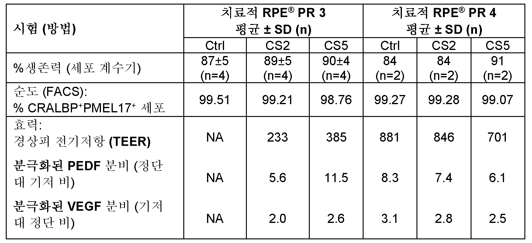
실시예 4Example 4
RTA RPE 세포 치료 조성물 상에서 안정성 검정을 수행하였다. 실시예 1의 방법에 따라 생성된 세포를 동결보존 이전에 2% DMSO를 함유하는 배지 1 (CS2) 또는 5% DMSO (CS5)를 함유하는 배지 2 중에서 3 시간 이하 동안 현탁시켰다. CS2 및 CS5 중에서 3 시간 인큐베이션 후에 동결보존된 치료적 RPE 세포는 동결보존 이전에 1 시간 미만 동안 인큐베이션한 세포와 유사한 해동후 생존력, 활력 및 수율을 나타내었다. 안정성 결과는 표 2에 제시되어 있다.A stability assay was performed on the RTA RPE cell therapeutic composition. Cells generated according to the method of Example 1 were suspended in Medium 1 (CS2) containing 2% DMSO or
표 2. 해동후 치료적 RPE 세포의 안정성 (동결보존 이전에 배지 1 (CS2) 및 배지 2 (CS5) 중에서 인큐베이션)Table 2. Stability of therapeutic RPE cells after thawing (incubated in medium 1 (CS2) and medium 2 (CS5) prior to cryopreservation)

또한, 동결보존 이전에 CS2 또는 CS5 중에서 3 시간 동안 인큐베이션한 세포는, 표 3에 제시된 바와 같이, 100 Ω 초과의 상피 통과 전기 저항 (TEER)을 생성하고, 분극화된 방식으로 VEGF 및 PEDF를 분비하는 능력에 의해 측정되는, 장벽 기능 (RPE 세포 사이의 치밀 이음부)을 생성하는 능력을 입증하였다. (도 15 또한 참조).Additionally, cells incubated for 3 hours in either CS2 or CS5 prior to cryopreservation demonstrated the ability to generate transepithelial electrical resistance (TEER) greater than 100 Ω, as shown in Table 3, and to generate barrier function (tight junctions between RPE cells), as measured by the ability to secrete VEGF and PEDF in a polarized manner (see also Figure 15 ).
표 3. 해동후 배지 1 (CS2) 및 배지 2 (CS5) 중에서 치료적 RPE® 세포의 TEER, 및 PEDF 및 VEGF의 분극화된 분비 (동결보존 이전에 배지 중에서 인큐베이션)Table 3. TEER and polarized secretion of PEDF and VEGF by therapeutic RPE® cells in medium 1 (CS2) and medium 2 (CS5) after thawing (incubated in medium prior to cryopreservation)

상기 기재된 RPE 세포 치료 조성물에 대해 해동후 안정성을 평가하였다. RPE 세포 조성물을 2% DMSO를 함유하는 배지 1 (CS2) 또는 5% DMSO를 함유하는 배지 2 (CS5) 중에서 제형화하고, 동결보존 이전에 3 시간 이하 동안 인큐베이션하였다. 생존력, 살아있는 세포 수율, 및 효력을 동결보존 이후에 대략 2 내지 8℃에서 0 시간, 1 시간, 2 시간, 3 시간, 5 시간, 6 시간 및 24 시간 시점에서 상기 기재된 바와 같이 측정하였다.The RPE cell therapy compositions described above were evaluated for post-thaw stability. The RPE cell compositions were formulated in medium 1 (CS2) containing 2% DMSO or medium 2 (CS5) containing 5% DMSO and incubated for up to 3 hours prior to cryopreservation. Viability, live cell yield, and potency were measured as described above at time points of 0, 1, 2, 3, 5, 6, and 24 hours after cryopreservation at approximately 2 to 8°C.
RPE 세포는 2% DMSO를 함유하는 배지 1 (CS2) 또는 5% DMSO를 함유하는 배지 2 (CS5) 중에서 동결보존 이전에 적어도 약 3 시간 동안 및 동결보존 이후에 적어도 약 1 시간 동안 또는 동결보존 이전에 적어도 약 2 시간 동안 및 동결보존 이후에 적어도 약 5 시간 동안 안정한 것으로 확인되었다.RPE cells were found to be stable for at least about 3 hours prior to cryopreservation and at least about 1 hour after cryopreservation, or for at least about 2 hours prior to cryopreservation and at least about 5 hours after cryopreservation, in either medium 1 (CS2) containing 2% DMSO or medium 2 (CS5) containing 5% DMSO.
실시예 5Example 5
즉시 투여가능한 (RTA) RPE 세포 치료 조성물 제형과 함께 사용하기 위한 3가지 동결보존 용액의 안전성을 Balb/c 마우스에게 망막하 주사한 후에 평가하였다.The safety of three cryopreservation solutions for use with ready-to-administer (RTA) RPE cell therapy composition formulations was evaluated following subretinal injection in Balb/c mice.
총 36 마리의 Balb/c 마우스를 사용하였고, 각 그룹에 9 마리씩 4개의 그룹으로 나누었다 (각각의 종료 시점에 대해 n=3; 투여후 1, 3 및 10 일째). 이들 그룹은 1개의 비히클 (BSS 플러스, 알콘 래보러토리즈(Alcon Laboratories)) 대조군 그룹, 및 시험 항목 (BSS 플러스에 의해 1:1 v/v로 희석된 CS5, CS2 및 CS2)을 제공한 3개의 처리된 그룹을 함유하였다. 모든 동물을 왼쪽 눈으로 망막하 주사를 통한 다양한 처리에 의해 투여하였다. 절차는 주사하기 2 분 전에 IP로 제공되는 75 mg/kg의 케타민/메데토미딘을 사용하여 마취 하에 수행되었다.A total of 36 Balb/c mice were used, divided into four groups of nine mice each (n=3 for each endpoint;
연구하는 동안, 이환율 및 사망률, 체중, 및 전반적인 임상 관찰 뿐만 아니라 외부 및 내부 눈 검사를 수행하였다. 눈 평가는 수의학 안과 의사에 의해 순응 동안에 (투여하기 전에 기준선 측정) 1회 및 그후 각각의 종료일에 수행되었다. 눈 평가는 다음을 포함하였다: 슬릿 램프 생체현미경검사를 이용하는 전방 분절 및 수정체의 검사 및 간접적인 검안경 검사를 이용하는 안저 검사. 동물을 투여후 1, 3 및 10 일째에 희생시켰고, 주사된 눈에 대해 조직병리학 검사를 수행하였다.During the study, external and internal eye examinations were performed, as well as morbidity and mortality, body weight, and general clinical observations. Ocular evaluations were performed by a veterinary ophthalmologist once during acclimatization (baseline measurements prior to dosing) and at each subsequent end date. Ocular evaluations included: examination of the anterior segment and lens using slit-lamp biomicroscopy and fundus examination using indirect ophthalmoscopy. Animals were sacrificed on
수의학 안과 의사 및 광범위한 공인 수의학 병리학자에 의해 수행된 임상적, 안과적 및 조직병리학적 검사는 RTA RPE 제형의 망막하 투여 이후에 10 일의 추적 조사 내에 주요한 처리-관련된 또는 독성학적으로 유의한 효과를 입증하지 않았다. 염증의 조직병리학적 평가는 하기 기준에 따라 호중구, 림프구, 대식세포, 및 비만 세포를 기반으로 하였다: 염증성 세포의 부재에 의해 나타나는 염증 없음, x10 배율 필드당 10개 이하의 세포에 의해 나타나는 가벼운 염증, x10 배율 필드당 약 10 내지 20개 세포에 의해 나타나는 중간 정도의 염증, 및 x10 배율 필드당 20개 초과의 세포에 의해 나타나는 강한 염증.Clinical, ophthalmologic, and histopathologic examinations performed by a veterinary ophthalmologist and a broadly board-certified veterinary pathologist demonstrated no major treatment-related or toxicologically significant effects within 10 days of follow-up following subretinal administration of the RTA RPE formulation. Histopathologic assessment of inflammation was based on the presence of neutrophils, lymphocytes, macrophages, and mast cells according to the following criteria: no inflammation as indicated by the absence of inflammatory cells, mild inflammation as indicated by 10 or fewer cells per x10 magnification field, moderate inflammation as indicated by about 10 to 20 cells per x10 magnification field, and severe inflammation as indicated by more than 20 cells per x10 magnification field.
도 2 및 도 3에 제시된 바와 같이, 대조군 그룹에서 비처리된 동물의 본래의 눈 및 동물의 비처리된 오른쪽 눈의 조직병리학적 평가는 병리학적 변화를 전혀 나타내지 않았다.As shown in Figures 2 and 3, histopathological evaluation of the native eyes and the untreated right eyes of the untreated animals in the control group showed no pathological changes at all.
처리된 눈 (왼쪽 눈)의 조직병리학적 평가는, 종료 1 일째에, BSS 플러스:CS2 (1:1 v/v)로 처리된 그룹의 동물만이 강한 염증 징후를 보였음을 나타내었다. BSS 플러스, CS5 또는 CS2로 처리된 다른 모든 동물은 가벼운 내지 중간 정도의 염증을 나타내었다. BSS 플러스로 치료되고 연구 1 일째에 희생된 동물로부터 취한 처리된 눈의 조직병리학적 슬라이드의 영상은 도 4a 및 도 4b에 제시된다. 이들 슬라이드는 공막의 가벼운 침윤 및 약간의 손실된 대식세포 및 림프구와 함께 가벼운 염증을 나타낸다. (각각 x4 및 x20 배율 필드에서 염색된 H&E). 도 5a 및 도 5b는 CS5로 처리되고 1 일째에 희생된 동물로부터 취한 처리된 눈의 조직병리학적 슬라이드의 영상을 제시한다. 이들 슬라이드는 공막의 침윤, 일부 대식세포 및 약간의 호중구와 함께 중간 정도의 염증을 나타낸다. (각각 x4 및 x20 배율 필드에서 염색된 H&E). CS2로 처리되고 연구 1 일째에 희생된 동물로부터 취한 처리된 눈의 조직병리학적 슬라이드의 영상이 도 6a 및 도 6b에 제시된다. 이들 슬라이드는 각막에서 대식세포 및 호중구와 함께 중간 정도의 염증을 나타낸다. (각각 x4 및 x20 배율 필드에서 염색된 H&E).Histopathological evaluation of the treated eyes (left eyes) showed that on day 1, only the animals in the group treated with BSS plus:CS2 (1:1 v/v) showed strong signs of inflammation. All other animals treated with BSS plus, CS5 or CS2 showed mild to moderate inflammation. Images of histopathological slides of treated eyes taken from animals treated with BSS plus and sacrificed on day 1 of the study are presented in Figures 4A and 4B. These slides show mild inflammation with mild scleral infiltrates and a few missing macrophages and lymphocytes. (H&E stained at x4 and x20 magnification fields, respectively). Figures 5A and 5B show images of histopathological slides of treated eyes taken from animals treated with CS5 and sacrificed on day 1. These slides show moderate inflammation with scleral infiltrates, some macrophages and a few neutrophils. (H&E stained at x4 and x20 magnification fields, respectively). Histopathologic images of histopathologic slides of treated eyes taken from animals treated with CS2 and sacrificed on day 1 of the study are presented in Figures 6A and 6B. These slides demonstrate moderate inflammation with macrophages and neutrophils in the cornea. (H&E stained at x4 and x20 magnification fields, respectively).
CS2 또는 BSS 플러스:CS2로 처리된 대부분의 동물이 종료 1 일째에 전방 챔버에서 최소의 피브린 침착을 나타내었지만, 이들 급성 변화는 단기 반응을 입증한다. 도 7a는 BSS 플러스:CS2로 처리되고 연구 1 일째에 희생된 동물로부터 취한 처리된 눈의 조직병리학적 슬라이드의 영상을 제시하며, 공막의 중간 정도의 침윤과 함께 강한 염증을 나타낸다. (x4 배율 필드에서 H&E 염색됨).Although most animals treated with CS2 or BSS plus:CS2 showed minimal fibrin deposition in the anterior chamber on day 1 of the study, these acute changes demonstrate a short-term response. Figure 7a presents images of histopathologic slides of treated eyes taken from animals treated with BSS plus:CS2 and sacrificed on day 1 of the study, demonstrating intense inflammation with moderate scleral infiltrates (H&E stained at ×4 magnification field).
도 7b는 공막에서 림프구 옆의 우측 하단 코너에 제시된 피브린 침착을 나타낸다. (x20 배율 필드에서 H&E 염색됨).Figure 7b shows fibrin deposition in the lower right corner next to lymphocytes in the cornea (H&E stained at ×20 magnification field).
종료 3 일째에, CS5 또는 CS2로 처리된 모든 동물은 대식세포 및 분열중인 섬유모세포를 특징으로 하는 국소 공막 육아종성 반응 (가벼운 내지 중간 정도의 염증)을 나타내었다. 대식세포는 BSS 플러스로 치료된 동물에서도 관찰되었지만, 이들은 섬유모세포 활성화 없이 상이한 패턴 및 세포 농도를 나타내었고, 주사된 물질과 관련이 없었다. 이들 결과는 일반적으로 이물질에 대한 초기 단계 반응의 전형적인 패턴을 나타낸다. 결과적으로, 종료 10 일째에, BSS 플러스, CS5 또는 CS2로 처리된 모든 동물은 염증없음 또는 가벼운 염증을 나타내었고, 피브린 침착을 나타내지 않았다. 또한, BSS 플러스:CS2로 처리된 한 마리의 동물만이 중간 정도의 염증을 나타낸 반면에, 다른 모든 동물은 가벼운 염증을 나타내었다.At
BSS 플러스로 치료되고 연구 3 일째에 희생된 동물에서 공막의 중간 정도의 침윤 및 몇몇 대식세포와 함께 중간 정도의 염증을 입증하는 조직병리학적 영상이 도 8a 및 도 8b에 제시된다. 도 9a 및 도 9b는 국소 과립화 반응 및 몇몇 대식세포 및 섬유모세포와 함께 강한 염증의 조직병리학적 영상을 제시하며, 이는 CS5로 처리되고 연구 3 일째에 희생된 동물에서 초기 단계의 일시적인 이물질 반응을 나타낸다. 도 10a 및 도 10b는 CS2로 처리되고 연구 3 일째에 희생된 동물로부터의 조직병리학적 영상이고, 몇몇 대식세포 및 섬유모세포와 함께 강한 염증을 나타내며, 이는 또한 초기 단계의 일시적인 이물질 반응을 입증한다. 가벼운 염증 및 가벼운 부종을 갖는, BSS 플러스:CS2로 처리되고 연구 3 일째에 희생된 동물로부터의 조직병리학적 영상은 도 11a 및 도 11b에 제시된다. 도 12a 및 도 12b는 BSS 플러스로 치료되고 연구 10 일째에 희생된 동물로부터의 조직병리학적 영상을 제시하며, 약간의 대식세포와 함께 가벼운 염증을 나타낸다. 도 13a 및 도 13b는 CS5로 처리되고 연구 10 일째에 희생된 동물로부터의 조직병리학적 영상을 제시하며, 약간의 대식세포와 함께 가벼운 염증을 나타낸다. 도 14a 및 도 14b는 CS2로 처리되고 연구 10 일째에 희생된 동물로부터의 조직병리학적 영상을 제시하며, 약간의 대식세포와 함께 가벼운 염증을 나타낸다.Histopathological images demonstrating moderate inflammation with moderate infiltration of the cornea and some macrophages in an animal treated with BSS plus and sacrificed on
조직병리학적 평가 결과는, 10 일 추적 조사 이후에 대조군과 비교하여 BSS 플러스, CS5, CS2 또는 BSS 플러스:CS2의 망막하 투여 이후에 주요한 처리-관련된 및/또는 독성학적으로 유의한 효과를 나타내지 않았음을 입증한다. 처리된 눈의 조직병리학적 평가는 BSS 플러스, CS5 및 CS2로 처리된 그룹에서 종료 3 일째에 이물질에 대한 전형적인 초기 단계 반응을 나타내었다. 그러나, 이 반응은 일시적이었고, 10 일째까지 가라앉아서 주사된 부위에 매우 미미한 대식세포 침윤을 남겼다. 어떠한 동물의 망막에서나 다른 곳에서도 괴사는 존재하지 않았다.Histopathological evaluation results demonstrated that there were no major treatment-related and/or toxicologically significant effects following subretinal administration of BSS plus, CS5, CS2 or BSS plus:CS2 compared to the control group after 10 days of follow-up. Histopathological evaluation of treated eyes showed a typical early-phase reaction to a foreign body as early as
실시예 6Example 6
착색된 세포의 효소적 강화 (단리/수확) 및 NUTS(-)+인간 혈청 알부민 (HSA) 및 NUTS(-) (인간 혈청 (HS) 없음)를 포함하는 효소 중화 용액을 이용하여 RTA RPE 세포 치료 조성물을 제형화하였고, 동결배지를 첨가하기 전에 및 첨가한 후에 안정성에 대해 평가하였다.Enzymatic enrichment (isolation/harvesting) of pigmented cells and RTA RPE cell therapy compositions were formulated using enzyme neutralizing solutions containing NUTS(-)+human serum albumin (HSA) and NUTS(-) (no human serum (HS)) and evaluated for stability before and after addition of freezing medium.
세포를 시딩하고, 계대 4 이하로 T25, T75 및 T175 플라스크에서 확장시켰다. 약 90% 초과의 다변형성에 도달한 후에, 세포를 트리플 셀렉트 (1X) 중에서 약 50 분 이하 동안 37℃/5%CO2에서 인큐베이션하였다. 세포를 풀링하고, 얼음 상에 두었다. 플라스크를 동등한 부피의 PBS (-)로 1회 세척하였고, 세척물을 세포 풀에 첨가하였다. PBS (-)를 개선된 효소-중화를 위해 NUTS (-)로 대체하였고, 세포 스트레스를 감소시켰다.Cells were seeded and expanded in T25, T75 and T175 flasks for
이어서, 세포 풀을 샘플링하였고 (180 μL PBS (-) 중 20 μL), 세포 계수기, 예컨대 NC-200 세포 계수기를 사용하여 계수하였다. 이어서, 세포 풀을 그룹 (G1, G2 및 G3)에 대해 다양한 켄칭 용액으로 분취하였다. 이어서, 각각의 그룹으로부터의 세포를 계수하고, 여과하고, 4℃에서 세포 조성물 안정성 분석을 위해 분취하였다.The cell pool was then sampled (20 μL in 180 μL PBS (-)) and counted using a cell counter, such as the NC-200 Cell Counter. The cell pool was then aliquoted into various quenching solutions for groups (G1, G2, and G3). Cells from each group were then counted, filtered, and aliquoted for cell composition stability analysis at 4°C.
분석한 효소 중화 용액 유형에는 하기가 포함되었다:The types of enzyme neutralizing solutions analyzed included:
ㆍ 그룹 1 (G1) - 20% 인간 혈청 (HS) / DMEM (HS 양성 대조군 그룹)ㆍ Group 1 (G1) - 20% human serum (HS) / DMEM (HS positive control group)
ㆍ 그룹 2 (G2) - 인간 혈청 알부민 (HSA)을 갖는 뉴트리스템 (-)ㆍ Group 2 (G2) - Nutristem (-) with human serum albumin (HSA)
ㆍ 그룹 3 (G3) - 뉴트리스템 (-) (NUTS)ㆍ Group 3 (G3) - Nutristem (-) (NUTS)
각각의 켄칭 그룹, G1, G2 및 G3을 탄뎀 500-200-40-마이크로미터 순차적 여과 시스템에 통과시켰다. 여과된 세포 용액을 4℃에서 유지하였고, 세포 생존력을 여과후 0, 2 및 4 시간 시점에서 시험하였다.Each of the quenching groups, G1, G2, and G3, was passed through a Tandem 500-200-40-micrometer sequential filtration system. The filtered cell solutions were maintained at 4°C, and cell viability was tested at 0, 2, and 4 h post-filtration.
여과후 각각의 시점의 종료 시에, 세포를 계수하고, 약 220g에서 약 5 분 동안 원심분리하였고, 상청액을 폐기하였고, 펠렛을 약 5-10 mL CS5 중에서 재현탁시키고, 샘플링하고, 계수하였다 (180 μL 20% HS/DMEM 중 20 μL). 계수 결과를 기반으로 하여, 세포를 CS5 중에서 희석하여 2x10⁶ 세포/mL의 최종 농도를 수득하였다. 동결보존 이전 안정성 분석을 위해 세포를 4℃에서 상이한 시간 동안 (0, 2, 3 또는 4 시간) 두었고, 그 후에 RPE 세포+동결배지 조성물을 크리오바이알로 분취하였다. 3개의 크리오바이알을 무작위로 샘플링하였고, 세포를 계수하고, 동결보존하였다.At the end of each time point after filtration, cells were counted, centrifuged at about 220 g for about 5 minutes, the supernatant was discarded, and the pellet was resuspended in about 5-10 mL CS5, sampled, and counted (20 μL in 180
바이알을 37℃ 수조 약 2.5 분 동안 해동시켰다. 계수를 위해 세포를 즉시 샘플링하였고 (180 μL 20% HS/DMEM 중 20 μL), 세포 현탁액을 따뜻한 20% HS/DMEM 배양 배지의 적가에 의해 희석하였다. 이어서, 세포를 세척하고, 추가의 분석을 위해 얼음 상에 두었다.The vials were thawed in a 37°C water bath for approximately 2.5 minutes. Cells were immediately sampled for counting (20 μL in 180
회복률 백분율은 2x10⁶ 세포/mL의 최종 목표 농도를 기반으로 하여 계산되었다.Percent recovery was calculated based on a final target concentration of 2x10⁶ cells/mL.
순차적 여과 이후에, 상이한 효소 중화 용액 중의 여과된 세포 현탁액을 4℃에서 유지하였고, 여과후 0, 2 및 4 시간에서의 세포 생존력을 평가하였다.After sequential filtration, the filtered cell suspensions in different enzyme neutralizing solutions were maintained at 4°C, and cell viability was assessed at 0, 2, and 4 h after filtration.
표 4. 여과후 세포 조성물 생존력Table 4. Cell composition viability after filtration

모든 시점에서 모든 그룹에서의 생존력은 표 4에 제시된 바와 같이 98% 또는 99%로 유지되었다. 0 시간 시점의 G1 (대조군 그룹)과 비교하여 상이한 시점에서 (0, 2 및 4 시간) 2가지 중화 용액 제형 그룹 G2 및 G3 사이에 유의한 차이가 발견되지 않았다. 도 16은 대조군 그룹 G1과 비교하여 여과후 시간에 걸쳐 4℃에서 G2 (NUTS(-)+HSA) 및 G3 (NUTS(-)) 그룹의 생존력을 제시한다.At all time points, the viability in all groups was maintained at 98% or 99%, as presented in Table 4. No significant difference was found between the two neutralizing solution formulation groups G2 and G3 at different time points (0, 2 and 4 h) compared to G1 (control group) at time point 0 h. Figure 16 presents the viability of groups G2 (NUTS(-)+HSA) and G3 (NUTS(-)) at 4 °C over time after filtration compared to the control group G1.
세포 회복률 및 생존력은 냉동 과정 이전에 (동결보존 이전) 동결보존 용액 내에서 평가되었다. 세포 치료 조성물 (여과후)을 4℃에서 0, 2 및 4 시간 동안 유지시켰다. 이어서, 각각의 용액을 원심분리하고, 2x10⁶ 세포/mL의 최종 농도로 동결보존 용액, CS5 중에서 재현탁시켰다. 다음으로, 동결보존 용액 (동결보존 + 세포 치료 조성물, 동결보존 이전) 내에서 세포를 4℃에서 0, 2, 3 및 4 시간 동안 유지시킨 후, 1 mL 크리오바이알로 분취하였다. 각각의 그룹으로부터의 3개의 바이알을 샘플링하고 계수하여, 바이알을 동결보존하기 전에 생존력 및 회복률 백분율을 평가하였다.Cell recovery and viability were assessed in the cryopreservation solution prior to the freezing process (prior to cryopreservation). The cell therapy composition (after filtering) was maintained at 4°C for 0, 2 and 4 hours. Each solution was then centrifuged and resuspended in the cryopreservation solution, CS5, to a final concentration of 2x10 6 cells/mL. The cells were then maintained in the cryopreservation solution (cryopreservation + cell therapy composition, prior to cryopreservation) at 4°C for 0, 2, 3 and 4 hours and then aliquoted into 1 mL cryovials. Three vials from each group were sampled and counted to assess the viability and percent recovery prior to cryopreservation of the vials.
0 시간 인큐베이션, 이어서 동결배지 중에서 0, 2, 3 및 4 시간 동안 인큐베이션한, 냉동 과정 이전의 세포 치료 조성물의 세포 생존력 및 회복률을 표 5A에서 함께 요약된다. 표 5B 및 5C는 0, 2, 4 시간 동안 인큐베이션, 이어서 동결보존 용액 (세포 + CM) 중에서 0, 2, 3, 4 시간 동안 인큐베이션한 세포 조성물을 포함한다. 대조군 그룹 (G1)을 동결보존 이전에 세포 조성물의 경우 0 시간 및 세포 + CM의 경우 0 시간 시점에만 평가하였다.Cell viability and recovery of cell therapy compositions prior to cryopreservation, after 0 hour incubation, followed by 0, 2, 3 and 4 hours in cryopreservation medium, are summarized together in Table 5A. Tables 5B and 5C include cell compositions incubated for 0, 2 and 4 hours, followed by 0, 2, 3 and 4 hours in cryopreservation solution (cells + CM). The control group (G1) was evaluated only at the 0 hour time point for cell compositions and 0 hour for cells + CM prior to cryopreservation.
표 5A. 중화 용액 중에서 0 시간 인큐베이션, 이어서 동결배지 (세포 + CM) 중에서 0, 2, 3 및 4 시간 동안 인큐베이션한 세포 치료 조성물의 동결보존 이전 생존력 및 회복률.Table 5A. Pre-cryopreservation viability and recovery of cell therapy compositions incubated for 0 h in neutralizing solution followed by 0, 2, 3 and 4 h in freezing medium (cells + CM).

표 5A, 도 17a 및 도 17b에 제시된 바와 같이, 동결보존 이전에, 0 시간 시점 치료용 세포 조성물 인큐베이션, 이어서 동결배지 중에서 0 시간 인큐베이션에서, G1 대조군 그룹과 비교하여 두 중화 그룹, G2 및 G3 사이에 생존력 및 회복률에서의 유의한 차이가 검출되지 않았다. 더욱이, 동결보존 이전에, 중화 용액 중에서 0 시간 치료용 세포 조성물 인큐베이션, 이어서 동결배지 중에서 2, 3 및 4 시간 인큐베이션은 G1 대조군 그룹과 비교하여 G2 및 G3 그룹 둘 다에서 세포의 생존력 또는 회복률에서의 감소를 나타내지 않았다. 생존력은 시간에 걸쳐 95% 초과로 유지되었고, 3가지 그룹 사이에서 유의한 차이가 관찰되지 않았다. 추가로, 모든 그룹의 회복률은 75%-100%의 범위로 유지되었다.As shown in Table 5A, Figures 17A and 17B, prior to cryopreservation, at 0 hour therapeutic cell composition incubation followed by 0 hour incubation in cryopreservation medium, no significant difference in viability and recovery rate was detected between the two neutralizing groups, G2 and G3, compared to the G1 control group. Moreover, prior to cryopreservation, at 0 hour therapeutic cell composition incubation in neutralizing solution followed by 2, 3 and 4 hours incubation in cryopreservation medium did not show any decrease in cell viability or recovery rate in either G2 or G3 groups compared to the G1 control group. Viability was maintained above 95% over time, and no significant difference was observed between the three groups. Additionally, the recovery rates for all groups were maintained in the range of 75%-100%.
표 5B. 동결보존 이전에, 2 시간 치료용 세포 조성물 인큐베이션, 이어서 동결배지 중에서 0, 2, 3 및 4 시간 인큐베이션한 후에, 동결보존 이전 세포 생존력 및 세포 회복률.Table 5B. Pre-cryopreservation cell viability and cell recovery after 2-hour therapeutic cell composition incubation, followed by 0, 2, 3 and 4 hours incubation in freezing medium prior to cryopreservation.

표 5C. 동결보존 이전에, 4 시간 치료용 세포 조성물 인큐베이션, 이어서 동결배지 중에서 0, 2, 3, 4 시간 인큐베이션한 후에, 동결보존 이전 세포 생존력 및 세포 회복률.Table 5C. Pre-cryopreservation cell viability and cell recovery after 4-hour therapeutic cell composition incubation, followed by 0, 2, 3 and 4 hours incubation in freezing medium.

표 5B 및 5C 및 도 18a, 도 18b, 도 19a 및 도 19b는, 동결보존 이전에, RPE 치료용 세포 조성물을 중화 용액 중에서 2 및 4 시간 동안 인큐베이션하고, 이어서 동결배지 중에서 0-4 시간 동안 인큐베이션하였을 때, 두 그룹 G2 (HSA를 갖는 뉴트리스템(-) 중의 세포 조성물) 및 G3 (뉴트리스템(-) (NUTS) 중의 세포 조성물)에 대해 유사한 세포 생존력 및 회복률 값이 확인됨을 제시한다. 이 검정은, 동결보존 이전에, 중화 용액 또는 동결배지 중에서 장기간 인큐베이션하였을 때 세포 생존력 또는 세포 회복률에 대해 유의한 효과가 없었음을 입증하였다.Tables 5B and 5C and Figures 18A, 18B, 19A and 19B show that when the cell compositions for RPE treatment were incubated in the neutralizing solution for 2 and 4 hours prior to cryopreservation, followed by incubation in the freezing medium for 0-4 hours, similar cell viability and recovery rates were confirmed for the two groups G2 (cell composition in Nutristem(-) with HSA) and G3 (cell composition in Nutristem(-) (NUTS)). This assay demonstrated that there was no significant effect on cell viability or cell recovery when incubated for a longer period of time in the neutralizing solution or the freezing medium prior to cryopreservation.
실시예 7Example 7
세포는 동결보존 동안에 해동후 불량한 생존율로 이어질 수 있는 스트레스를 겪을 수 있다. 추가로, 수확 절차 동안에 세포에 대한 스트레스는 해동후 세포 생존력 및 회복률에 영향을 미칠 수 있다. 따라서, RTA 치료용 세포 조성물의 세포 생존력 및 회복률을 해동후 평가하였다. 동결보존 이전에, 상이한 중화 용액 (상기 기재된 바와 같이 G1, G2 및 G3) 중에서 0 시간 동안 인큐베이션, 이어서 동결배지 중에서 0, 2, 3 및 4 시간 동안 인큐베이션한 치료용 세포 조성물을 동결보존후 생존력 및 회복률에 대해 평가하였다.Cells can be subjected to stress during cryopreservation which can lead to poor post-thaw survival. Additionally, stress to cells during the harvest procedure can affect post-thaw cell viability and recovery. Therefore, cell viability and recovery of RTA therapeutic cell compositions were evaluated post-thaw. Prior to cryopreservation, therapeutic cell compositions were incubated for 0 h in different neutralizing solutions (G1, G2 and G3 as described above) followed by incubation in freezing medium for 0, 2, 3 and 4 h, and then evaluated for viability and recovery after cryopreservation.
표 6은 치료용 세포 조성물을 효소 중화 용액 중에서 0 시간 동안, 이어서 동결배지 중에서 0, 2, 3 및 4 시간 동안 인큐베이션하고 (동결보존 이전에), 동결보존시킨 다음, 해동하였을 때 수득된 생존력 및 안정성 결과를 요약한다.Table 6 summarizes the viability and stability results obtained when therapeutic cell compositions were incubated in an enzyme neutralizing solution for 0 hour, followed by 0, 2, 3 and 4 hours in freezing medium (prior to cryopreservation), cryopreserved and then thawed.
표 6. 0 시간 세포 + 중화 용액 인큐베이션, 이어서 동결배지 중에서 0, 2, 3 및 4 시간 인큐베이션의 해동후 생존력 및 회복률Table 6. Post-thaw viability and recovery after 0-hour cell + neutralizing solution incubation, followed by 0, 2, 3, and 4-hour incubation in frozen medium.

동결보존 이전에 동결배지 중에서 상이한 인큐베이션 시간에 의한 세포 생존력 및 회복률의 분석은, 생존력이 모든 시점에서 모든 그룹에서 약 90% 초과로 유지되었고, 회복률이 약 75%-100%의 범위였음을 나타내었다.Analysis of cell viability and recovery rate by different incubation times in the cryopreservation medium prior to cryopreservation showed that viability was maintained at more than approximately 90% in all groups at all time points, and recovery rate was in the range of approximately 75%-100%.
도 20a에 제시된 바와 같이, 그룹들 사이에서 세포 생존력에서 유의한 차이가 없었으며, 생존력은 전체 동결보존 이전 시간 범위에 걸쳐서 약 95% 초과로 유지되었고, G1 대조군 그룹의 생존력에 필적하였다. 도 20b에 제시된 회복률 결과의 분석은 그룹 G4에 대해 보다 높은 회복률 값을 나타내었고, 이는 동결보존 이전 2 시간째에 피크였고, 이어서 동결보존 이전 3 및 4 시간 후에 약간 감소하였다. 그러나, 회복률은 약 80%-100%의 범위로 유지되었다.As presented in Fig. 20a, there was no significant difference in cell viability between the groups, and viability was maintained at greater than about 95% throughout the entire pre-cryopreservation time range, comparable to the viability of the G1 control group. Analysis of the recovery rate results presented in Fig. 20b showed higher recovery rate values for group G4, which peaked at 2 h pre-cryopreservation, and then slightly decreased after 3 and 4 h pre-cryopreservation. However, the recovery rate was maintained in the range of about 80%-100%.
치료용 세포 조성물을 중화 용액 중에서 0, 2 및 4 시간 동안 인큐베이션, 이어서 동결배지 중에서 0, 2, 3 및 4 시간 동안 인큐베이션하였을 때 수득된 해동후 결과가 표 7에 제시된다. 이들 결과는 동결보존 이전에 CS5에 대한 세포의 장기간 노출에 대한 해동후 생존력 및 회복률의 관계를 입증하였고, 이는 다시 효소 중화 용액 중에서 세포의 장기간 인큐베이션에 의해 영향을 받을 수 있다.Post-thaw results obtained when therapeutic cell compositions were incubated in neutralizing solution for 0, 2 and 4 hours, followed by incubation in freezing medium for 0, 2, 3 and 4 hours are presented in Table 7. These results demonstrate the relationship between post-thaw viability and recovery of cells following prolonged exposure to CS5 prior to cryopreservation, which in turn can be influenced by prolonged incubation of cells in enzyme neutralizing solution.
표 7. 효소 중화 용액 중에서 0, 2, 4 시간 동안 인큐베이션, 이어서 동결배지 중에서 (동결보존 이전) 0, 2, 3 및 4 시간 동안 인큐베이션한 세포의 해동후 생존력 및 회복률Table 7. Post-thaw viability and recovery of cells incubated for 0, 2, and 4 hours in enzyme neutralizing solution, followed by 0, 2, 3, and 4 hours in freezing medium (prior to cryopreservation).

두 그룹 G2 및 G3에 대해 측정된 생존력은 93% 초과로 확고하였고, 모든 시점에 걸쳐 그룹 G2 및 G3 사이에 유의한 차이가 없었다. 두 그룹의 회복률은 유의한 차이를 나타내지 않았다. 그룹 G2에 대한 회복률은 75%±3.3이었고, 그룹 G3에 대한 회복률은 81%±7.1이었다.The measured survival for both groups G2 and G3 was robust at over 93%, and there was no significant difference between groups G2 and G3 at any time point. The recovery rates of the two groups did not show a significant difference. The recovery rate for group G2 was 75%±3.3, and the recovery rate for group G3 was 81%±7.1.
이들 결과는, 중화 용액 (HSA를 갖는 (그룹 G2) 및 HSA를 갖지 않는 (그룹 G3) NUT(-))이 세포 회복률 또는 세포 생존력을 손상시키지 않았음을 입증한다. HSA를 갖는 또는 갖지 않는 (G2 & G3) 두 켄칭 그룹 사이에서 유의한 차이가 관찰되지 않았다. HS를 갖지 않는 효소 중화 용액에 대한 세포 생존력 및 세포 회복률 결과는 동결보존 이전 및 이후 둘 다의 경우 HS를 포함하는 효소 중화 용액 (G1)에 필적하였다.These results demonstrate that the neutralizing solutions (NUT(-) with (group G2) and without (group G3) HSA) did not impair cell recovery or cell viability. No significant difference was observed between the two quenching groups with or without HSA (G2 & G3). The cell viability and cell recovery results for the enzyme neutralizing solution without HS were comparable to the enzyme neutralizing solution with HS (G1) both before and after cryopreservation.
또한, HS를 갖지 않는 효소 중화 용액 및 여과 단계를 포함하는 세포 수확 절차는 세포의 생존력을 손상시키지 않았다 (표 4). 세포 회복률 및 생존력의 동결보존 이전 및 해동 이후 분석은 대조군 그룹 (G1)과 비교하여 그룹 G2 및 G3 사이에서 유의한 차이를 나타내지 않았다 (표 5-7). 최상의 퍼센트 회복률 및 생존력은 세포를 효소 중화 용액 중에서 2 시간 이하 동안 인큐베이션, 이어서 동결배지 중에서 3 시간 이하 동안 인큐베이션하였을 때 관찰되었다. 그러나, 효소 중화 용액 또는 동결배지 중에서 적어도 4 시간의 인큐베이션 시간은 세포 생존력 또는 세포 회복률에서 유의한 감소를 일으키지 않았다.Also, the cell harvesting procedure including the enzyme neutralization solution and filtration step without HS did not impair the viability of the cells (Table 4). Analysis of cell recovery and viability before and after cryopreservation showed no significant difference between groups G2 and G3 compared to the control group (G1) (Tables 5-7). The highest percent recovery and viability were observed when the cells were incubated in the enzyme neutralization solution for 2 hours or less followed by 3 hours or less in the freezing medium. However, an incubation time of at least 4 hours in the enzyme neutralization solution or the freezing medium did not result in a significant decrease in cell viability or cell recovery.
세포를 수확하고, 동결보존시키고, 해동한 후에, PEDF 분비 및 세포 확장 능력 또한 측정하였다. 결과는 표 8에 제시된다.After cells were harvested, cryopreserved, and thawed, PEDF secretion and cell expansion capacity were also measured. The results are presented in Table 8.
표 8. 효소 중화 용액을 갖는 RPE 세포 조성물 (세포) 및 RTA RPE 세포 조성물 (세포+CM) 인큐베이션 시간에 대해 수확 및 동결보존 이후에 세포의 PEDF 분비 및 확장 능력Table 8. PEDF secretion and expansion capacity of cells after harvest and cryopreservation for incubation time of RPE cell composition (cells) and RTA RPE cell composition (cells+CM) with enzyme neutralizing solution.

ND - 데이터 없음ND - No data
표 8에 제시된 바와 같이, 해동 시에, 세포는 PEDF를 분비하고 확장하는 그들의 기능적 능력을 유지하였다. 결과는, 측정된 모든 시점에서 HS를 갖지 않는 2가지 효소 중화 용액 그룹 (G2, G3) 사이에서 또는 이들 그룹과 HS를 갖는 효소 중화 용액 그룹 (G1) 사이에서 유의한 차이가 없음을 나타내었다.As shown in Table 8, upon thawing, the cells maintained their functional ability to secrete PEDF and expand. The results showed that there was no significant difference between the two enzyme neutralizing solution groups without HS (G2, G3) or between these groups and the enzyme neutralizing solution group with HS (G1) at any time point measured.
실시예 8Example 8
표 9에 제시된 바와 같이, 치료용 세포 조성물을 실온에서 (RT) 및 약 2-8℃의 온도에서 인큐베이션한 후에 여과후 안정성 (% 생존력 및 % 회복률)에 대해 평가하였다.As presented in Table 9, the therapeutic cell compositions were evaluated for post-filtration stability (% viability and % recovery) after incubation at room temperature (RT) and at a temperature of about 2-8°C.
표 9. 인큐베이션 시간Table 9. Incubation time

세포를 효소적으로 (예를 들어, 트리플 셀렉트) 수확하였다. 효소를 뉴트리스템 (-) (NUTS)을 사용하여 중화시켰다. 순차적 여과 이후에, 여과된 세포 현탁액 (세포 풀)을 두 그룹으로 나누었고, 제1 그룹 ("세포 2-8℃")은 약 2-8℃에서 유지되었고, 제2 그룹 ("세포 RT")은 RT에서 유지되었다. 두 그룹의 세포 생존력 및 세포 농도를 0 시점에서 및 인큐베이션 2 시간 후에 평가하였다.Cells were harvested enzymatically (e.g., Triple Select). The enzyme was neutralized using Nutristem (-) (NUTS). After sequential filtration, the filtered cell suspension (cell pool) was divided into two groups, the first group (“cells 2-8°C”) was maintained at about 2-8°C, and the second group (“cells RT”) was maintained at RT. Cell viability and cell concentration of both groups were evaluated at
표 10에 제시된 바와 같이, 동결보존 이전에 약 2-8℃ 또는 RT에서 인큐베이션한 세포 조성물의 그룹 사이에서 세포 생존력 및 세포 농도에서 유의한 차이가 없었다. 유사하게, 도 22에 제시된 바와 같이, 그룹들 사이에 퍼센트 회복률에서 유의한 차이가 없었다.As presented in Table 10, there were no significant differences in cell viability and cell concentration between groups of cell compositions incubated at about 2-8°C or RT prior to cryopreservation. Similarly, as presented in Figure 22, there were no significant differences in percent recovery between groups.
표 10. 2 시간 인큐베이션 후에 세포 생존력 및 세포 농도Table 10. Cell viability and cell concentration after 2 hours of incubation
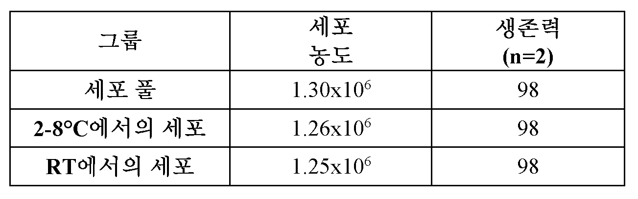
2 시간 인큐베이션 후에, 두 그룹을 원심분리하고, 2x10⁶ 세포/ml의 최종 농도로 동결보존 용액, CS5 중에서 재현탁시켰다. "세포 2-8℃" 그룹을 4가지 하위-그룹 (세포+CM -그룹 1-4)으로 나누었고, 이를 2-8℃에서 인큐베이션하였다. 세포+CM -그룹 1을 0 시간 시점에서 세포 농도 및 세포 생존력에 대해 계수하였고, 동결보존시켰다 (n=29 크리오바이알). 세포+CM -그룹 2는 2-8℃에서 인큐베이션 2 시간 후에 계수하였고, 세포+CM -그룹 3은 4 시간 후에 샘플링하였고, 세포+CM -그룹 4는 인큐베이션 6 시간 후에 세포 농도 및 세포 생존력에 대해 계수하였다. 다양한 인큐베이션 시점에서, 계수를 완료하였을 때, 세포+CM -그룹 2-4를 각각의 크리오바이알 (각각 n= 30, 30 및 29 크리오바이알)에 1 ml를 분취함으로써 동결보존시켰다. "세포+CM RT"를 2, 4 및 6 시간 시점에서 생존력 및 세포 농도에 대해 샘플링하였다. 6 시간 인큐베이션 후에, 이 그룹을 또한 동결보존시켰다 (n=22 크리오바이알).After 2 h of incubation, both groups were centrifuged and resuspended in cryopreservation solution, CS5, to a final concentration of 2x10 6 cells/ml. The "Cells 2-8°C" group was divided into four sub-groups (Cells+CM - groups 1-4) and incubated at 2-8°C. Cells+CM - group 1 was counted for cell concentration and cell viability at time point 0 h and cryopreserved (n=29 cryovials). Cells+CM -
표 11. 동결배지 중에서 치료용 세포 조성물: 다양한 시점에서 세포 농도 및 세포 생존력.Table 11. Therapeutic cell compositions in cryopreservoir: Cell concentration and cell viability at various time points.

표 11 및 도 23a 및 도 23b에 제시된 바와 같이, RT에서 세포 조성물 + 동결배지 그룹과 약 2-8℃에서 세포 조성물 + 동결배지 그룹 사이에 생존력에서 유의한 차이가 없었고, 시간에 걸쳐 모든 그룹에서 세포 생존력의 작은 감소가 있었다. 결과는 세포 회복률이 두 온도 조건, 약 2-8℃ 및 RT에서 적어도 6 시간에 걸쳐 모든 그룹 내에서 안정하게 유지되고, 냉동 과정 이전에 약 2-8℃에서 인큐베이션한 세포의 회복률이 약간 더 높다. RT에서 인큐베이션한 세포는 2 시간째에 회복률의 15% 감소를 나타내었으나, 회복률 및 생존력은 적어도 6 시간 동안 안정하게 유지되었다.As presented in Table 11 and Figures 23a and 23b, there was no significant difference in viability between the cell composition + freezing medium group at RT and the cell composition + freezing medium group at about 2-8°C, and there was a small decrease in cell viability in all groups over time. The results showed that cell recovery remained stable in all groups for at least 6 hours at both temperature conditions, about 2-8°C and RT, and that cells incubated at about 2-8°C prior to the freezing process had a slightly higher recovery rate. Cells incubated at RT showed a 15% decrease in recovery rate at 2 hours, but the recovery rate and viability remained stable for at least 6 hours.
실시예 9Example 9
표 12에 제시된 바와 같이, 2가지 세포 해동 방법 (하나는 약 37℃의 수조를 포함하고, 다른 것은 자동화된 세포 해동 유닛을 포함함)을 세포 생존력, 회복률, 멸균성, 효력 및 동일성을 기반으로 하여 평가하였다.As presented in Table 12, two cell thawing methods (one involving a water bath at approximately 37°C and the other involving an automated cell thawing unit) were evaluated based on cell viability, recovery, sterility, potency, and identity.
표 12. 해동후 분석을 위한 시험 그룹 및 검정 시점.Table 12. Test groups and assay time points for post-thaw analysis.

V - 생존력 %, R - 회복률 %, I - 동일성, P - 효력, S - 멸균성V - Viability %, R - Recovery %, I - Identity, P - Potency, S - Sterility
4 그룹의 RTA 치료용 세포 조성물 각각으로부터 2개의 바이알을 동시에 해동시켰고; 1개의 바이알은 자동화된 세포 해동 유닛 (예를 들어, 시그마-알드리치에 의한 토스타(ThawSTAR) 자동화된 해동 시스템)을 사용하였고, 두번째는 약 37℃의 표준 수조를 사용하였다. 이어서, 해동된 바이알을 얼음 상에 두었다. 각각의 바이알을 부드럽게 피펫팅하였고, 각각의 바이알로부터 20 μl의 2개의 샘플을 취하고, 180 μl의 NUTS (-) 중에서 희석하고, 볼텍싱하고, 계수하였다. 평균 생존력 및 회복률 백분율을 각각의 해동된 바이알에 대해 계산하였다.Two vials from each of the four groups of RTA therapeutic cell compositions were thawed simultaneously; one vial using an automated cell thawing unit (e.g., ThawSTAR automated thawing system by Sigma-Aldrich) and the second using a standard water bath at approximately 37°C. The thawed vials were then placed on ice. Each vial was gently pipetted and two 20 μl samples were taken from each vial, diluted in 180 μl of NUTS (-), vortexed, and counted. The average viability and percent recovery were calculated for each thawed vial.
생존력, 회복률, 멸균성, 효력 및 동일성에 대해 해동된 세포 조성물을 검정함으로써 4 시간 이하 동안 해동후 실온에서 RTA 치료용 세포 조성물의 안정성을 평가하였다. 측정된 TEER, 21 일째에 기저 PEDF/VEGF 비 분비 및 21 일째에 정단 VEGF/PEDF 비 분비에 의해 효력을 측정하였다. 회복률 백분율을 2x10⁶ 또는 5x10⁶ 세포/ml의 최종 목표 농도를 기반으로 하여 계산하였다.The stability of cell compositions for RTA therapy was assessed at room temperature after thawing for up to 4 hours by assaying thawed cell compositions for viability, recovery, sterility, potency, and identity. Potency was measured by measured TEER, basal PEDF/VEGF ratio secretion at day 21, and apical VEGF/PEDF ratio secretion at day 21. Percent recovery was calculated based on a final target concentration of 2x10 6 or 5x10 6 cells/ml.
표 13에 제시된 바와 같이, 자동화된 세포 해동 유닛을 사용하여 해동된 RTA 치료용 세포 조성물의 생존력 및 회복률 평균은 2가지 세포 농도 모두에서 약 37℃의 통상적인 수조를 사용하여 달성된 것에 필적하였다.As shown in Table 13, the average viability and recovery of RTA therapeutic cell compositions thawed using the automated cell thawing unit were comparable to those achieved using a conventional water bath at about 37°C for both cell concentrations.
표 13. 상이한 세포 농도에서 자동화된 세포 해동 유닛을 사용하여 해동된 및 약 37℃의 수조를 사용하여 해동된 RTA 치료용 세포 조성물의 평균 생존력 및 회복률.Table 13. Average viability and recovery of RTA therapeutic cell compositions thawed using an automated cell thawing unit at different cell concentrations and using a water bath at about 37°C.

표 14에 제시된 바와 같이, 0, 2 및 4 시간 시점에서 각각의 그룹에 대한 평균 퍼센트 생존력은 적어도 83%였다. 시험한 모든 그룹의 평균 퍼센트 회복률은 0, 2 및 4 시간 시점에서 적어도 약 78%였다. 2 및 4 시간 실온 인큐베이션 후에 모든 그룹에서 생존력에서 약 4% 내지 10%의 약간의 감소 및 회복률에서 17% 이하의 감소가 있었다. 5x10⁶ 세포/mL 세포 농도의 평균 회복률은 모든 시점에서 2x10⁶ 세포/ml 농도의 것에 비해 더 높았다.As presented in Table 14, the average percent viability for each group at 0, 2, and 4 hours was at least 83%. The average percent recovery for all groups tested was at least about 78% at 0, 2, and 4 hours. After 2 and 4 hours of room temperature incubation, there was a slight decrease in viability of about 4% to 10% and a decrease in recovery of less than 17% in all groups. The average recovery for the cell concentration of 5x10⁶ cells/mL was higher than that for the 2x10⁶ cells/mL concentration at all time points.
표 14. 생존력 및 회복률 평균.Table 14. Average survival and recovery rates.
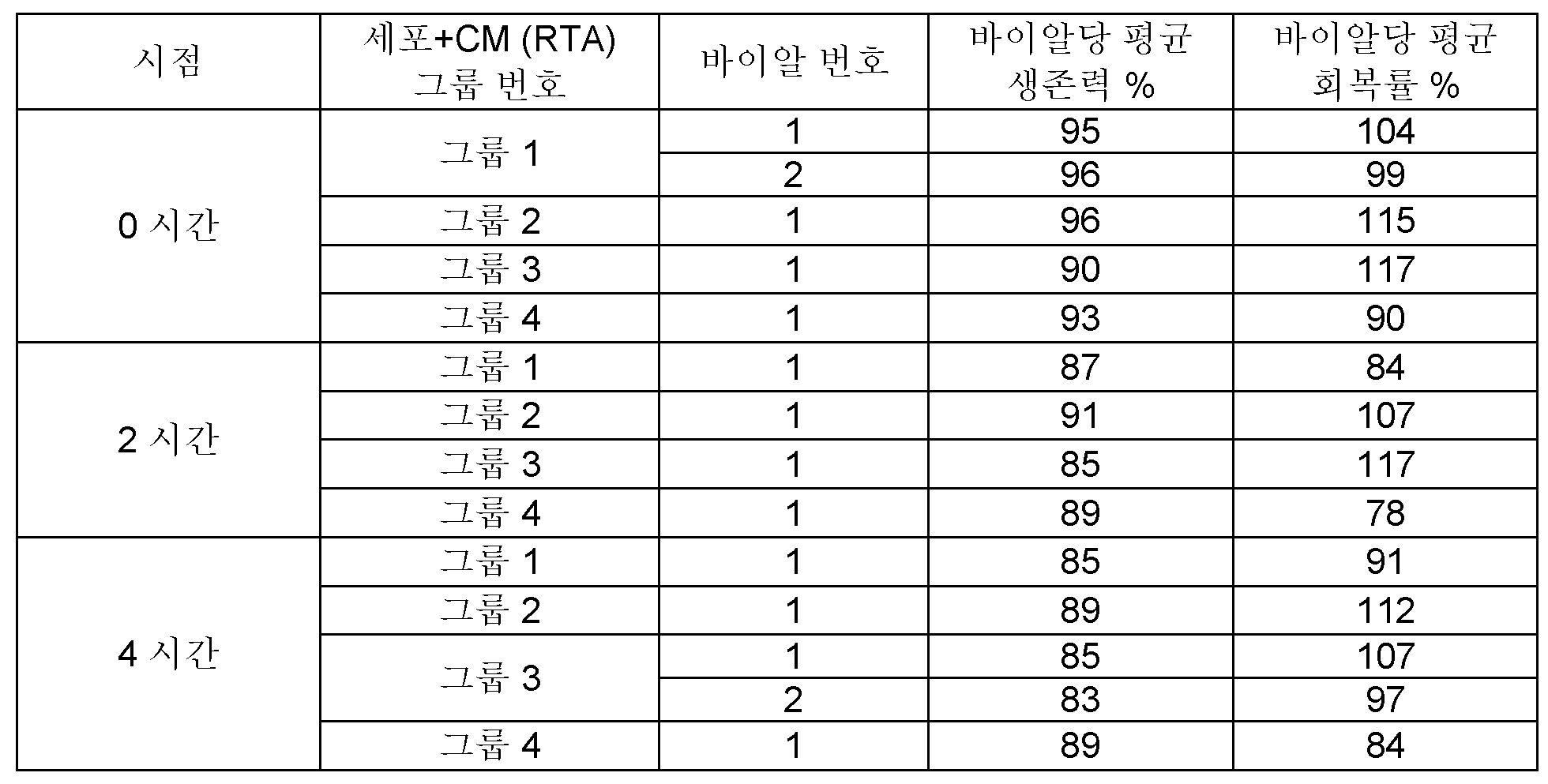
도 24a 및 도 24b는 4-시간 인큐베이션 시간에 걸쳐 실온에서 해동된 RTA 세포 조성물의 생존력 및 회복률을 제시하는 그래프이다.Figures 24a and 24b are graphs showing the viability and recovery rate of RTA cell compositions thawed at room temperature over a 4-hour incubation time.
실시예 10Example 10
RTA 치료용 세포 조성물과 전달 디바이스의 상용성을 평가하였다. 전달 디바이스에는 약 0.63 mm의 외부 직경 및 약 0.53 mm의 내부 직경을 갖는 니들, 약 0.5 mm의 외부 직경 및 약 0.25 mm의 내부 직경을 갖는 모세관, 및 약 0.12 mm의 외부 직경 및 약 0.07 mm의 내부 직경을 갖는 팁을 포함하는, 더치 옵탈믹 리서치 센터 (Dutch Ophthalmic Research Center, D.O.R.C)에 의해 제작된 디바이스가 포함되나 이로 제한되지 않는다. 4개의 배치를 포함하는 전달 디바이스 방출된 RTA 치료용 세포 조성물을 자동화된 해동 시스템을 사용하여 해동후 RT에서 2-시간 인큐베이션 시간 후에 생존력, 회복률 및 효력에 대해 검정하였다. 동결보존 이전에 여과 단계를 적용하지 않은 그룹 4를 제외하고는, 모든 RTA 세포 조성물을 실시예 6에 기재된 바와 같이 제형화하였다. 결과는 표 15에 제시된다.The compatibility of RTA therapeutic cell compositions and delivery devices was evaluated. The delivery devices include, but are not limited to, devices manufactured by Dutch Ophthalmic Research Center (D.O.R.C.) comprising a needle having an outer diameter of about 0.63 mm and an inner diameter of about 0.53 mm, a capillary having an outer diameter of about 0.5 mm and an inner diameter of about 0.25 mm, and a tip having an outer diameter of about 0.12 mm and an inner diameter of about 0.07 mm. The RTA therapeutic cell compositions released from the delivery devices containing four batches were tested for viability, recovery, and potency after thawing at RT for 2 hours using an automated thawing system. All RTA cell compositions were formulated as described in Example 6, except for
표 15. 전달 디바이스로부터 방출 전에 RTA 세포 조성물의 안정성Table 15. Stability of RTA cell compositions prior to release from the delivery device.

시험한 그룹의 CRALBP/PMEL17 (동일성) 값 (표 15)은 모든 시점에서 시험한 모든 배치에 대해 98% 초과였다. 순 TEER 값 (14 일째)은 모든 그룹에서 100 Ω 초과로 잘 유지되었다. 시간이 지남에 따라 주로 그룹 1에서 및 4 시간후 그룹 4에서 점진적인 감소가 명확해졌다. 그룹 1 및 그룹 2로부터의 배치를 실온에서 4 시간 인큐베이션 후에 멸균성에 대해 시험하였고, 두 배치에 대해 성장은 관찰되지 않았다.The CRALBP/PMEL17 (identity) values of the tested groups (Table 15) were greater than 98% for all batches tested at all time points. The net TEER values (day 14) were well maintained above 100 Ω in all groups. A gradual decrease was evident over time, mainly in group 1 and after 4 h in
생존가능한 강력한 RTA 치료용 세포 조성물을 전달하기 위한 전달 디바이스의 상용성은 전달 디바이스 내에서 해동된 RTA 치료용 세포 조성물 (RT에서 2 시간 동안 인큐베이션하였음)을 방출시킴으로써 평가하였다. 제1 및 제2 용량 부피는 융통성을 허용하도록 시험하였다. 세포를 생존력, 회복률 및 효력에 대해 검정하였다. RTA 치료용 세포 조성물을 전달 디바이스에 로딩하기 전에 RT에서 2 시간 동안 유지시킨 후에 생존력 및 회복률 백분율에 대한 결과.The compatibility of the delivery device for delivering viable potent RTA therapeutic cell compositions was evaluated by releasing thawed RTA therapeutic cell compositions (incubated at RT for 2 hours) within the delivery device. First and second dose volumes were tested to allow for flexibility. Cells were assayed for viability, recovery, and potency. Results for percent viability and recovery after maintaining the RTA therapeutic cell composition at RT for 2 hours prior to loading into the delivery device.
표 16. RT에서 2-시간 후에 생존력 및 회복률 (전달 디바이스 이전).Table 16. Viability and recovery rates after 2 hours at RT (before delivery device).
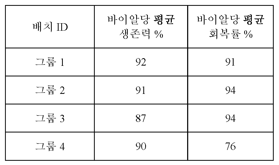
모든 그룹의 전달 디바이스 방출후 생존력 및 회복률 백분율이 표 17에 제시된다. 평균 생존력은 89% 내지 95%였다. 모든 그룹에 걸친 평균 총 회복률은 약 71% 내지 94%였다. 전달 디바이스 이전 회복률 결과와 비교하여 첫번째 100 μl의 회복률에서 8% 이하의 약간의 감소 및 두번째 100 μl 부피에서 16% 이하의 감소가 있었다 (회복률이 안정하게 유지된 그룹 2 제외).The viability and recovery percentages after delivery device release for all groups are presented in Table 17. The average viability ranged from 89% to 95%. The average overall recovery across all groups ranged from about 71% to 94%. There was a slight decrease of less than 8% in the recovery of the first 100 μl and less than 16% in the second 100 μl volume compared to the recovery results prior to delivery device (except for
표 17. 전달 디바이스 방출후 세포 생존력 및 회복률 결과.Table 17. Results of cell viability and recovery rate after release of the delivery device.
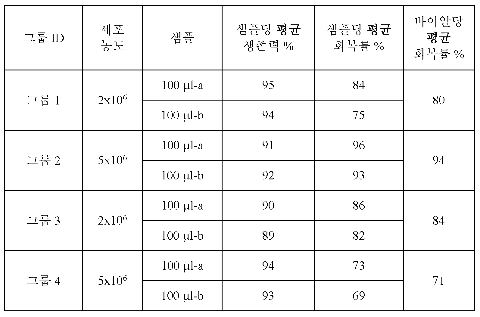
그룹 4는 동결보존 이전에 여과 단계 없이 제형화되었다. 따라서, 감소된 회복률은 전달 디바이스에 잔류하는 세포 및 세포외 매트릭스 응집물과 관련된 것일 수 있다.
전달 디바이스 방출후 순 TEER (14 일 동안) 값이 표 18에 제시된다. 순 TEER 값은 약 154 Ω 내지 약 435 Ω의 범위였다. PEDF 정단/기저 비 (21 일째) 및 VEGF 기저/정단 비 (21 일째)에 대한 결과 또한 표 18에 제시된다.The net TEER values (for 14 days) after release of the delivery device are presented in Table 18. The net TEER values ranged from about 154 Ω to about 435 Ω. Results for PEDF apical/basal ratio (day 21) and VEGF basal/apical ratio (day 21) are also presented in Table 18.
표 18. 전달 디바이스 방출후 효력 결과.Table 18. Results of effectiveness after release of the delivery device.

그룹 1 및 3의 2x10⁶ 세포 용량에 대한 TEER 값이 5x10⁶ 세포 용량의 TEER 값 (그룹 2 및 4)에 비해 약간 더 높았지만, 시험한 모든 전달 디바이스 이후 샘플 그룹은 효력 검정 결과에 따라 생물학적 활성을 나타내었다.Although the TEER values for the 2x10⁶ cell dose in
실시예 11Example 11
동결운송기를 액체 질소로 로딩하고, 수송을 위해 준비하고, 실시예 10에 기재된 동결보존된 RTA 치료용 세포 조성물의 배치 (그룹 1-4)를 로딩하였다. 동결보존 이전에 여과 단계를 적용하지 않은 그룹 4를 제외하고는, 모든 RTA 세포 조성물을 실시예 6에 기재된 바와 같이 제형화하였다. RTA 치료용 세포 조성물을 이스라엘 예루살렘에서 메릴랜드주 프레데릭의 유에스-바이오레포지토리(US-biorepository)로 운송하였고, 액체 질소 냉동기의 증기 상에서 중간 저장하고, 생성물을 이스라엘 예루살렘으로 다시 운송하였다. 운송에는 단일 증기 상 동결운송기에 저장된 4개 배치 (그룹 1-4)의 항공 및 지상 수송이 포함되었고, 월드 쿠리어(World Courier)에 의해 수행되었다. 동결운송기는 대략 (-196℃) 내지 (-150℃)의 필요한 저장 조건을 제공하였고, 내부 데이터 로거(Data Logger)에 의해 모니터링되었다. 그룹 1-4를 외관, 생존력, 회복률, 효력 및 멸균성에 대해 검정하였다. 결과는 표 19에 제시된다.The cryotransporter was loaded with liquid nitrogen, prepared for transport and loaded with batches (Groups 1-4) of the cryopreserved RTA therapeutic cell compositions as described in Example 10. All RTA cell compositions were formulated as described in Example 6, except for
표 19. 동결운송된 RTA 세포 조성물의 외관, 생존력, 회복률, 효력 및 멸균성.Table 19. Appearance, viability, recovery rate, potency and sterility of cryopreserved RTA cell compositions.
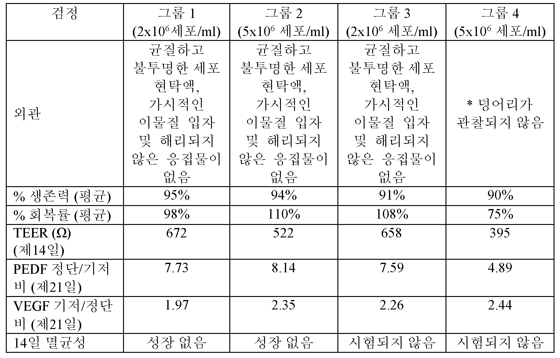
* 덩어리는 충전 및 마무리 절차 이전에 여과하지 않은 제형의 결과였다. * The lumps were the result of unfiltered formulation prior to filling and finishing procedures.
동결운송기의 온도가 -196℃ 미만으로 도달하지 않았지만, 이 온도는 세포 조성물의 온전성 및 품질에 대한 위험을 안고 있지 않으며, 액체 질소 데이터 로거의 보정된 범위 내에 있다.Although the temperature in the cryotransporter did not reach below -196°C, this temperature does not pose a risk to the integrity and quality of the cell composition and is within the calibrated range of the liquid nitrogen data logger.
결과는 CS5 동결배지 중에서 제형화된 동결보존된 RTA 세포 조성물의 안정성, 품질 및 온전성이, 이스라엘 예루살렘에서 메릴랜드주 프레데릭의 유에스-바이오레포지토리로의 제어된 운송, 액체 질소 냉동기의 증기 상에서 중간 저장 및 생성물의 이스라엘 예루살렘으로의 다시 운송 동안에 유지됨을 나타낸다.Results indicate that the stability, quality and integrity of cryopreserved RTA cell compositions formulated in CS5 freezing medium are maintained during controlled transportation from Jerusalem, Israel to the US-Biorepository in Frederick, Maryland, intermediate storage in the vapor phase of a liquid nitrogen freezer and return transportation of the product to Jerusalem, Israel.
실시예 12Example 12
RPE 세포 및 동결보존 용액을 포함하는 RTA 치료용 세포 조성물의 안전성을 NOD/SLID 마우스로의 망막하 주사 이후에 평가하였다. 5-9 주령의 총 40 마리의 암컷 NOD/SLID 마우스를 이용하였고, 각각의 그룹에서 4 또는 12 마리 동물의 4개의 그룹으로 나누었다 (각각의 종료 시점의 경우 n=1 또는 3; 투여후 1, 3, 7 및 14 일째).The safety of a cell composition for RTA therapy comprising RPE cells and cryopreservation solution was evaluated following subretinal injection into NOD/SLID mice. A total of 40 female NOD/SLID mice, 5-9 weeks of age, were used and divided into four groups of 4 or 12 animals in each group (n=1 or 3 for each endpoint; on
4가지 조성물을 평가하였다. 표 20에 제시된 바와 같이, 그룹 1은 BSS 플러스를 투여하였고, 그룹 2는 CS5 동결배지를 투여하였고, 그룹 3은 RTA 치료용 세포 조성물 (RPE 세포 및 CS5를 포함함)을 투여하였고, 그룹 4는 BSS 플러스 중 RPE 세포를 투여하였다.Four compositions were evaluated. As shown in Table 20, Group 1 was administered BSS Plus,
표 20. 실험 설계.Table 20. Experimental design.

동물을 처음 24 시간 동안 주기적으로 관찰하고 (투여후 처음 4 시간 동안은 특별히 주의를 기울임), 그 후에는 종료시까지 매일 관찰하였다. 관찰은 국소 주사 부위, 피부, 털, 눈, 점막, 호흡기, 분비물 및 배설물 발생 (예를 들어, 설사) 및 자율 활성 (예를 들어, 누루, 타액 분비, 입모, 동공 크기, 비정상적인 호흡 패턴)에서의 임의의 변화에 대해 수행하였다. 기이한 거동, 떨림, 경련, 수면 및 혼수상태의 존재로서, 걸음걸이, 자세, 및 취급에 대한 반응에서의 변화 또한 포함된다. 이상, 독성 징후, 빈사 상태 및 예정되지 않은 사망은 관찰되지 않은 것으로 기록되었다. 눈 검사는 순응 동안에 및 각각의 종료일에 수의학 안과 의사에 의해 수행되었다. 모든 그룹으로부터의 모든 오른쪽 눈 (비처리된)에서 가시적인 병변 없음 (NVL)이 관찰되었다.Animals were observed periodically for the first 24 hours (with special attention paid to the first 4 hours after dosing) and then daily until termination. Observations were made for any changes in the local injection site, skin, fur, eyes, mucous membranes, respiratory tract, secretions and excretions (e.g., diarrhea), and autonomic activity (e.g., lacrimation, salivation, piloerection, pupillary size, abnormal breathing patterns). Changes in gait, posture, and response to handling were also noted, as were the presence of bizarre behavior, tremors, convulsions, sleep, and coma. Abnormalities, signs of toxicity, moribundity, and unplanned death were recorded as not observed. Eye examinations were performed by a veterinary ophthalmologist during acclimation and on each termination day. No visible lesions (NVL) were observed in all right eyes (untreated) from all groups.
종료 시에, 동물을 CO2 질식에 의해 희생시키고, 상이한 눈 구조, 주요 조직 및 장기 시스템을 비롯하여 국소 주사 부위 (눈)를 검사하는 총 병리학을 수행하였다. 시신경을 비롯하여 동물을 적출시키고, 데이비슨(Davidson) 용액 중에서 고정시켰다. 모든 눈에 대해 조직병리학 검사를 실시하였다.At the end of the experiment, animals were sacrificed by CO2 asphyxiation and gross pathology was performed, examining the local injection site (eye) as well as the different ocular structures, major tissues and organ systems. Animals were excised, including the optic nerve, and fixed in Davidson's solution. Histopathological examinations were performed on all eyes.
조직병리학 평가는 3 및 14 일째에 그룹 3 및 4로부터의 몇몇 세포-처리된 동물 (각각의 종료일 및 그룹에서 3 마리의 동물 중 1 마리)에서 대식세포 침윤의 존재를 나타내었다. 이들 발견은 아마도 인간 RPE 세포의 주사에 대한 늦은 면역 반응으로 인한 것이다. 그러나, 세포-처리된 그룹에서 대부분의 동물 (20/24)은 이식된 세포에 대해 어떠한 면역 반응도 나타내지 않았다. 세포로 처리하지 않은 그룹 1 및 2에서는, 호중구가 1, 3 및 7 일째에 관찰되었다 (3 마리의 동물에서). 호중구는 면역 반응의 초기 단계에서 일반적이지만, 대식세포의 결여는 늦은 반응이 없음을 나타낸다. 몇몇 세포-처리된 마우스 (4/24)에서 대식세포의 출현을 제외하고, 모든 그룹은 유사한 병리학을 나타내었으며, 이는 상기 발견이 절차-유도된 염증과 관련있을 가능성이 크다는 것을 나타낸다. 도 25a 및 도 25b는 각각 x4 및 x20 배율에서의 대표적인 조직학적 영상이며, RTA 제형 (세포 + CS5)으로 처리된 동물에서 가벼운 염증을 나타내었다. 수집된 데이터를 기반으로 하여, 10 일 추적 조사 이후에 비히클과 비교하여 치료용 세포 조성물의 망막하 투여 후에 주요한 처리-관련된 및/또는 독성학적으로 유의한 효과는 없었다.Histopathologic evaluation revealed the presence of macrophage infiltration in some cell-treated animals from
실시예 13Example 13
상이한 제작 실행 동안에 제형화된 RTA RPE 치료용 세포 조성물의 비교가능성을 평가하기 위해, 몇몇 배치를 제조하였다. 먼저 적어도 1개의 hESC 앰풀을 해동시킴으로써 hESC를 기계적으로 계대하였다. 해동후, 앰풀로부터 회수된 10개의 단편을 피더 세포로부터 유래된 방사선 조사된 인간 제대의 단층을 함유하는 2개의 중심 웰 (CW) 플레이트 상에 시딩하고, 37℃/5% CO2에서 '뉴트리스템 플러스'+HSA 배지 (또는 등가물) 중에서 인큐베이션하였다 (1개의 앰풀 → 10개의 콜로니 단편을 갖는 2개의 CW).To assess the comparability of the formulated RTA RPE therapeutic cell compositions during different production runs, several batches were manufactured. hESCs were first passaged mechanically by thawing at least one hESC ampoule. After thawing, 10 fragments recovered from the ampoule were seeded onto 2 center well (CW) plates containing a monolayer of irradiated human umbilical cord derived from feeder cells and incubated in 'Nutristem Plus' + HSA medium (or equivalent) at 37°C/5% CO2 ( 1 ampoule → 2 CW with 10 colony fragments ).
hESC 콜로니를 확장시키고, 총 45개의 중심 웰 플레이트에 도달할 때까지 약 3 주 더 매주 1회 계대하였다. 이어서, 콜로니를 약 2:1의 비로 피더를 갖는 6 cm 플레이트로 옮기고 (2개의 CW → 6 cm 플레이트), 37℃/5% CO2에서 '뉴트리스템 플러스'+HSA 배지 중에서 약 6 일 동안 배양하였다.hESC colonies were expanded and passaged once a week for approximately 3 more weeks until a total of 45 center-well plates were reached. Colonies were then transferred to 6 cm plates with feeders at a ratio of approximately 2:1 (2 CW → 6 cm plates) and cultured in 'Nutristem Plus' + HSA medium at 37°C/5% CO2 for approximately 6 days.
구상체 (SB)를 형성하고, RPE 지시된 분화 과정을 개시하기 위해, hESC 콜로니를 6 cm 플레이트로부터 수집하고, 약 5:1의 비로 플레이트 (예컨대, 히드로셀™ (눈크)) (비-부착성 표면)로 옮기고 (5 x 6 cm 플레이트 → 플레이트), 37℃/5% CO2 /5%O2에서 니코틴아미드로 보충된 '뉴트리스템 마이너스' 배지 중에서 약 1 주 동안 배양하였다. SB 형성 약 1 주 후에, SB를 수집하였고, 피펫팅에 의해 작은 단편으로 분해하였다. SB 단편을 라미닌 511 (바이오라미나, 스웨덴 스톡홀름) 또는 등가물로 코팅된 6 웰 플레이트 상에 시딩하고, 니코틴아미드 및 액티빈 A의 존재 하에 (분화 단계에 따라 달라지는 니코틴아미드 및 액티빈 A의 조합) 뉴트리스템 마이너스 또는 등가물 중에서 약 37℃/5% CO2 /5% O2에서 약 5 주 동안 및 37℃/5% CO2에서 약 1 주 동안 배양하였다. 분화 단계의 종료 시에, 착색된 세포를 효소적으로 강화시키고, 재조합 인간 젤라틴 코된 T175 (175 cm2) 플라스크로 옮기고 (P0), 뉴트리스템 마이너스 중에서 (처음 2-3 일 동안 20% 인간 혈청/DMEM) 37℃/5% CO2에서 인큐베이션하였다.To form spheroids (SB) and initiate RPE-directed differentiation process, hESC colonies were collected from 6 cm plates, transferred to plates (e.g., Hydrocell™ (Nunc)) (non-adherent surface) at a ratio of approximately 5:1 (5 x 6 cm plate → plate) and cultured in 'Nutristem Minus' medium supplemented with nicotinamide at 37°C/5% CO 2 /5% O 2 for approximately 1 week. After approximately 1 week of SB formation, SB were collected and disaggregated into small fragments by pipetting. SB fragments were seeded onto 6-well plates coated with Laminin 511 (Biolamina, Stockholm, Sweden) or equivalent and cultured in the presence of nicotinamide and activin A (the combination of nicotinamide and activin A depending on the differentiation stage) in Nutristem Minus or equivalent at 37°C/5% CO 2 /5% O 2 for about 5 weeks and 37°C/5% CO 2 for about 1 week. At the end of the differentiation stage, the pigmented cells were enzymatically enriched and transferred to recombinant human gelatin-coated T175 (175 cm 2 ) flasks (P0) and incubated in Nutristem Minus (20% human serum/DMEM for the first 2-3 days) at 37°C/5% CO 2 .
세포를 대략 14 일째에 수확하고, rh-젤라틴 코팅된 T175 플라스크로 계대하고, 뉴트리스템 마이너스 중에서 (처음 2-3 일 동안 20% 인간 혈청/DMEM) 약 37℃/5% CO2에서 인큐베이션하였다 (P1). 세포를 12 일째에 수확하고, rh-젤라틴 코팅된 T175 플라스크로 계대하고, 뉴트리스템 마이너스 중에서 (처음 2-3 일 동안 20% 인간 혈청/DMEM) 37℃/5% CO2에서 인큐베이션하였다 (P2).Cells were harvested on day 14, passaged onto rh-gelatin-coated T175 flasks, and incubated in Nutristem Minus (20% human serum/DMEM for the first 2-3 days) at approximately 37°C/5% CO2 (P1). Cells were harvested on day 12, passaged onto rh-gelatin-coated T175 flasks, and incubated in Nutristem Minus (20% human serum/DMEM for the first 2-3 days) at 37°C/5% CO2 (P2).
P2 계대후 10 일째에, T175 플라스크를 수확하고, 500-200-40 μm 세포 스트레이너 시스템의 순차적 탄뎀을 이용하여 여과한 다음, 풀링하였다. 이 시점에서 세포의 총 개수는 적어도 약 551x106 세포일 수 있다. 세포를 원심분리하고, 동결배지 (예컨대, 크리오스토르 5 (바이오라이프 솔루션즈 인크., 워싱턴주 보첼)) 중에서 재현탁시키고, 계수하여, 약 1x10⁶ 세포/ml, 약 2x10⁶ 세포/ml, 약 3x10⁶ 세포/ml, 약 4x10⁶ 세포/ml, 약 5x10⁶ 세포/ml, 약 6x10⁶ 세포/ml, 약 7x10⁶ 세포/ml, 약 8x10⁶ 세포/ml, 또는 약 9x10⁶ 세포/ml의 최종 농도에 도달하였다. 이 세포 현탁액을 크리오바이알로 분배할 때까지 2-8℃에서 유지시킬 수 있다. 이어서, RTA 치료용 세포 조성물을 제어된-속도 냉동기에서 동결보존시킬 수 있고, 이어서 증기 상 LN2 냉동기로 옮길 수 있다. 이용될 수 있는 냉동 프로파일의 예에는 하기가 포함된다:On day 10 after P2 passage, the T175 flasks are harvested, filtered using a sequential tandem of a 500-200-40 μm cell strainer system, and pooled. The total number of cells at this point can be at least about 551x10 6 cells. The cells are centrifuged, resuspended in freezing medium (e.g., CryoStor 5 (BioLife Solutions, Inc., Botchel, WA)), and counted to reach a final concentration of about 1x10 6 cells/ml, about 2x10 6 cells/ml, about 3x10 6 cells/ml, about 4x10 6 cells/ml, about 5x10 6 cells/ml, about 6x10 6 cells/ml, about 7x10 6 cells/ml, about 8x10 6 cells/ml, or about 9x10 6 cells/ml. This cell suspension can be maintained at 2-8°C until dispensed into cryovials. The RTA therapeutic cell composition can then be cryopreserved in a controlled-rate freezer and then transferred to a vapor phase LN2 freezer. Examples of cryopreservation profiles that can be utilized include:
ㆍ 4℃에서 대기ㆍ Wait at 4℃
ㆍ 4℃에서 1 분 동안 유지ㆍKeep at 4℃ for 1 minute
ㆍ -11℃로 1.00℃ /분 샘플ㆍ -11℃ to 1.00℃/min sample
ㆍ -50℃로 30.00℃ /분 챔버ㆍ -50℃ to 30.00℃ /min chamber
ㆍ -25℃로 15.00℃ /분 챔버ㆍ -25℃ to 15.00℃ /min chamber
ㆍ -50℃로 1.00℃ /분 챔버ㆍ -50℃ to 1.00℃ /min chamber
ㆍ -90℃로 10.00℃ /분 챔버ㆍ -90℃ to 10.00℃ /min chamber
ㆍ 종료ㆍ End
또는or
ㆍ 4℃에서 대기ㆍ Wait at 4℃
ㆍ -4℃로 1.00℃ /분 샘플ㆍ -4℃ to 1.00℃/min sample
ㆍ -40℃로 25.00℃ /분 챔버ㆍ -40℃ to 25.00℃ /min chamber
ㆍ -12℃로 10.00℃ /분 챔버ㆍ -12℃ to 10.00℃ /min chamber
ㆍ -40℃로 1.00℃ /분 챔버ㆍ -40℃ to 1.00℃ /min chamber
ㆍ -90℃로 10.00℃ /분 챔버ㆍ -90℃ to 10.00℃ /min chamber
ㆍ 종료ㆍ End
표 21은 hESC가 확장되고, RPE 세포로 분화되고, RPE 세포로서 확장됨에 따라 이들의 모폴로지 및 순도 결과를 제공한다. 표 21에 제시된 바와 같이, hESC는 상기 기재된 과정을 이용하여 성공적으로 확장되고 분화되었다.Table 21 provides the morphology and purity results of hESCs as they were expanded, differentiated into RPE cells, and expanded as RPE cells. As shown in Table 21, hESCs were successfully expanded and differentiated using the procedure described above.
표 21. RTA 배치 A에 대한 확장 및 분화기 동안에 세포의 분석Table 21. Analysis of cells during expansion and differentiation for RTA batch A.
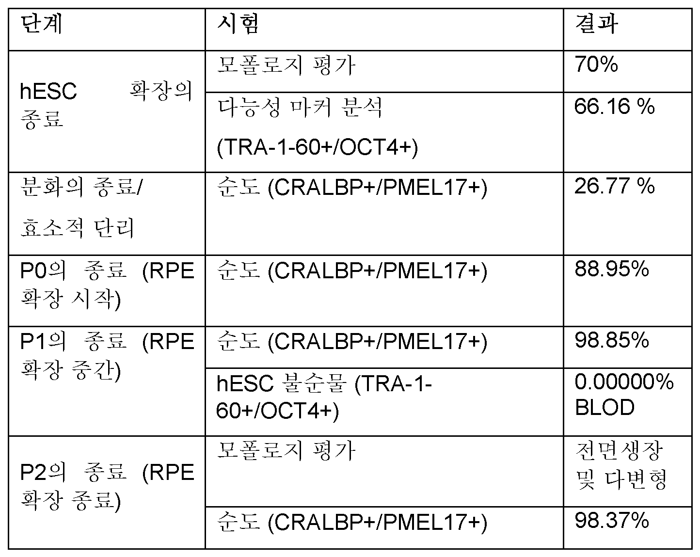
또한, 퍼센트 생존력, 세포 농도, 퍼센트 회복률, 및 순도를 배치 A에 대해 측정하였다. 결과는 표 22에 제시된다.Additionally, percent viability, cell concentration, percent recovery, and purity were measured for batch A. The results are presented in Table 22.
표 22. RTA 배치 A에 대한 퍼센트 생존력, 세포 농도, 퍼센트 회복률, 및 순도Table 22. Percent viability, cell concentration, percent recovery, and purity for RTA batch A.

배치 A에 대해 기재된 것과 같이 추가의 배치를 생성하고 분석하였다. 각각의 배치에 대한 세포 용량이 표 23에 제시된다.Additional batches were generated and analyzed as described for batch A. The cell doses for each batch are presented in Table 23.
표 23. 각각의 배치에 대한 세포 용량Table 23. Cell capacity for each batch
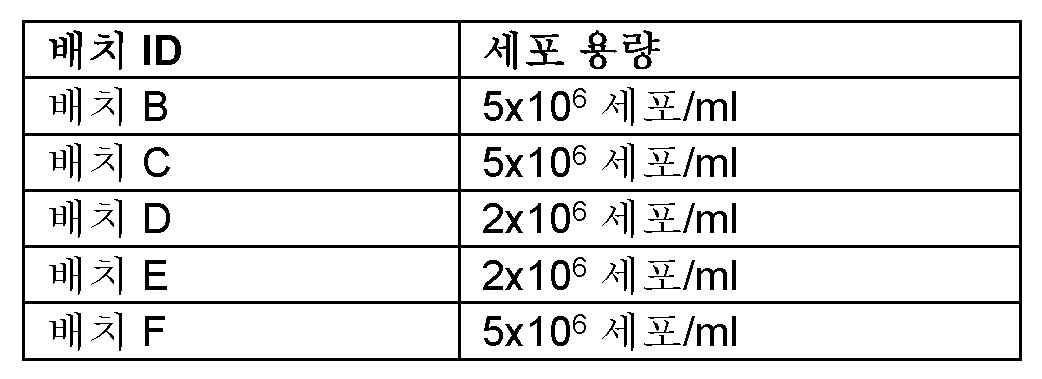
배치 B-F에 대한 결과는 표 24 및 표 25에 제시된다.The results for batches B-F are presented in Tables 24 and 25.
표 24. 배치 B-F에 대한 모폴로지, 멸균성, 및 순도Table 24. Morphology, sterility, and purity for batches B-F

* 시험한 세포 조성물 * Tested cell composition
표 25. 배치 B-F에 대한 퍼센트 생존력, 세포 농도, 퍼센트 회복률, 순도, 멸균성, PEDF 분비 및 VEGF 분비Table 25. Percent viability, cell concentration, percent recovery, purity, sterility, PEDF secretion, and VEGF secretion for batches B-F.
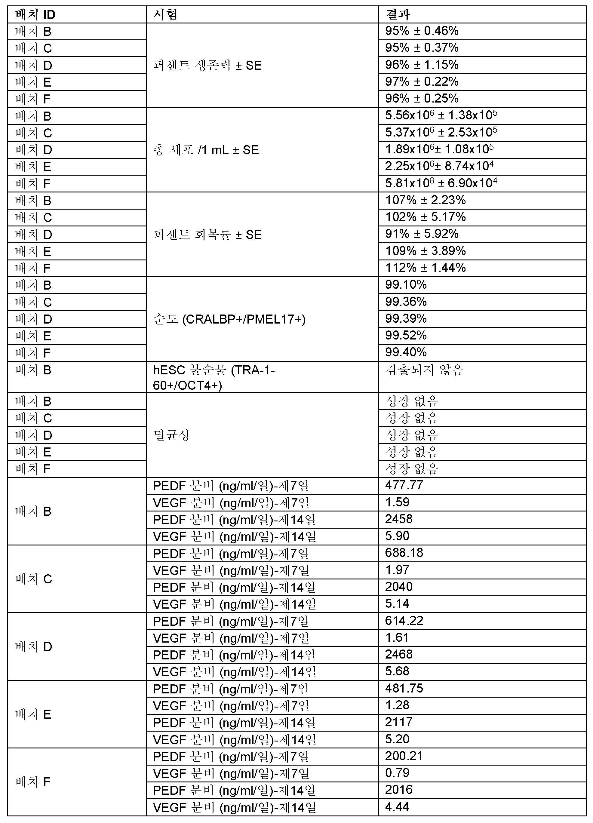
표 24 및 표 25에 제시된 바와 같이, RTA RPE 치료용 세포 조성물의 제형 방법은 세포 모폴로지, 멸균성, 순도, 퍼센트 생존력, 세포 농도, 퍼센트 회복률, PEDF 분비 및 VEGF 분비 (효력)와 관련하여 재현가능하고 확고하다.As presented in Tables 24 and 25, the formulation method of the cell composition for RTA RPE treatment is reproducible and robust with respect to cell morphology, sterility, purity, percent viability, cell concentration, percent recovery, PEDF secretion and VEGF secretion (potency).
본원의 기재가 여러 상세한 내용을 함유하지만, 이들이 본 개시내용의 범위를 제한하지 않으며, 단지 본원의 바람직한 실시양태의 일부를 설명하기 위해 제공되는 것으로 해석되어야 한다. 따라서, 본 개시내용의 범위가 관련 기술분야의 기술자에게 명백해질 수 있는 다른 실시양태를 충분히 포함한다는 것을 이해할 것이다.Although the description herein contains many specific details, they should not be construed as limiting the scope of the present disclosure, but rather as merely providing an illustration of some of the preferred embodiments of the present disclosure. Accordingly, it will be understood that the scope of the present disclosure fully encompasses other embodiments that may become apparent to those skilled in the art.
청구범위에서, 단수로 언급된 요소는 명시적으로 나타내지 않는다면 "하나 및 단지 하나"를 의미하는 것이 아니라, 오히려 "하나 이상"을 의미하는 것으로 의도된다. 관련 기술분야의 통상의 기술자에게 공지된 개시된 실시양태의 요소에 대한 모든 구조적, 화학적 및 기능적 등가물은 본원에 명백히 참조로 포함되고, 본원의 청구범위에 포함되는 것으로 의도된다. 추가로, 본 개시내용에서 어떠한 요소, 성분 또는 방법 단계도, 상기 요소, 성분 또는 방법 단계가 청구범위에서 명시적으로 인용되는지 여부와 무관하게, 대중에게 제공되는 것으로 의도되지 않는다. 본원의 어떠한 청구범위 요소도, 상기 요소가 명시적으로 어구 "을 위한 수단"을 이용하여 인용되지 않는다면, "수단 플러스 기능" 요소로 해석되어서는 안된다. 본원의 어떠한 청구범위 요소도, 상기 요소가 명시적으로 어구 "을 위한 단계"를 이용하여 인용되지 않는다면, "단계 플러스 기능" 요소로 해석되어서는 안된다.In the claims, elements referred to in the singular are not intended to mean "one and only one," unless expressly stated otherwise, but rather "one or more." All structural, chemical, and functional equivalents to elements of the disclosed embodiments known to those of ordinary skill in the art are expressly incorporated herein by reference and are intended to be included in the claims herein. Additionally, no element, component, or method step in this disclosure is intended to be dedicated to the public, regardless of whether or not said element, component, or method step is expressly recited in the claims. No claim element herein should be construed as a "means plus function" element unless said element is expressly recited using the phrase "means for." No claim element herein should be construed as a "step plus function" element unless said element is expressly recited using the phrase "step for."
Claims (1)
Applications Claiming Priority (4)
| Application Number | Priority Date | Filing Date | Title |
|---|---|---|---|
| US201762612210P | 2017-12-29 | 2017-12-29 | |
| US62/612,210 | 2017-12-29 | ||
| PCT/IB2018/001579 WO2019130061A2 (en) | 2017-12-29 | 2018-12-28 | Retinal pigment epithelium cell compositions |
| KR1020207021645A KR102725720B1 (en) | 2017-12-29 | 2018-12-28 | Retinal pigment epithelial cell composition |
Related Parent Applications (1)
| Application Number | Title | Priority Date | Filing Date |
|---|---|---|---|
| KR1020207021645A Division KR102725720B1 (en) | 2017-12-29 | 2018-12-28 | Retinal pigment epithelial cell composition |
Publications (1)
| Publication Number | Publication Date |
|---|---|
| KR20240161220A true KR20240161220A (en) | 2024-11-12 |
Family
ID=67066705
Family Applications (2)
| Application Number | Title | Priority Date | Filing Date |
|---|---|---|---|
| KR1020247036309A Pending KR20240161220A (en) | 2017-12-29 | 2018-12-28 | Retinal pigment epithelium cell compositions |
| KR1020207021645A Active KR102725720B1 (en) | 2017-12-29 | 2018-12-28 | Retinal pigment epithelial cell composition |
Family Applications After (1)
| Application Number | Title | Priority Date | Filing Date |
|---|---|---|---|
| KR1020207021645A Active KR102725720B1 (en) | 2017-12-29 | 2018-12-28 | Retinal pigment epithelial cell composition |
Country Status (11)
| Country | Link |
|---|---|
| US (4) | US20210000102A1 (en) |
| EP (2) | EP3731789A4 (en) |
| JP (2) | JP7498664B2 (en) |
| KR (2) | KR20240161220A (en) |
| CN (1) | CN111741735A (en) |
| AU (2) | AU2018393494A1 (en) |
| BR (1) | BR112020013238A2 (en) |
| CA (1) | CA3086815A1 (en) |
| IL (1) | IL275542A (en) |
| SG (1) | SG11202005795TA (en) |
| WO (1) | WO2019130061A2 (en) |
Families Citing this family (10)
| Publication number | Priority date | Publication date | Assignee | Title |
|---|---|---|---|---|
| EP3674397A1 (en) | 2014-12-30 | 2020-07-01 | Cell Cure Neurosciences Ltd. | Rpe cell populations and methods of generating same |
| KR20240161220A (en) * | 2017-12-29 | 2024-11-12 | 셀 큐어 뉴로사이언시스 리미티드 | Retinal pigment epithelium cell compositions |
| MX2022005134A (en) | 2019-10-30 | 2022-05-30 | Astellas Inst For Regenerative Medicine | METHODS TO PRODUCE RETINAL PIGMENTARY EPITHELIUM CELLS. |
| CA3184790A1 (en) * | 2020-05-25 | 2021-12-02 | Lineage Cell Therapeutics, Inc. | Methods and compositions for treating retinal diseases and conditions |
| AU2021280343A1 (en) * | 2020-05-29 | 2022-12-08 | FUJIFILM Cellular Dynamics, Inc. | Retinal pigmented epithelium and photoreceptor dual cell aggregates and methods of use thereof |
| CN112544612A (en) * | 2020-12-18 | 2021-03-26 | 江苏艾尔康生物医药科技有限公司 | Frozen stock solution and application thereof in RPE cell freezing |
| JP2024522608A (en) | 2021-06-09 | 2024-06-21 | リネージ セル セラピューティクス インコーポレイテッド | Methods and compositions for treating retinal diseases and conditions |
| IL310156A (en) * | 2021-07-28 | 2024-03-01 | Lineage Cell Therapeutics Inc | Expansion of retinal pigment epithelium cells |
| CN114561356B (en) * | 2022-03-09 | 2022-09-13 | 北京呈诺医学科技有限公司 | Method for influencing cytokine secretion and application |
| WO2025122978A1 (en) * | 2023-12-06 | 2025-06-12 | Bluerock Therapeutics Lp | Cell cryopreservation and delivery vehicle and methods of using the same |
Family Cites Families (38)
| Publication number | Priority date | Publication date | Assignee | Title |
|---|---|---|---|---|
| SG49267A1 (en) | 1989-08-14 | 1998-05-18 | Photogenesis Inc | Surgical instrument and cell isolation and transplantation |
| US6045791A (en) | 1992-03-06 | 2000-04-04 | Photogenesis, Inc. | Retinal pigment epithelium transplantation |
| US5405742A (en) * | 1993-07-16 | 1995-04-11 | Cyromedical Sciences, Inc. | Solutions for tissue preservation and bloodless surgery and methods using same |
| US5755785A (en) | 1994-08-12 | 1998-05-26 | The University Of South Florida | Sutureless corneal transplantation method |
| US5843780A (en) | 1995-01-20 | 1998-12-01 | Wisconsin Alumni Research Foundation | Primate embryonic stem cells |
| US5854015A (en) * | 1995-10-31 | 1998-12-29 | Applied Food Biotechnology, Inc. | Method of making pure 3R-3'R stereoisomer of zeaxanthin for human ingestion |
| US5941250A (en) | 1996-11-21 | 1999-08-24 | University Of Louisville Research Foundation Inc. | Retinal tissue implantation method |
| US6090622A (en) | 1997-03-31 | 2000-07-18 | The Johns Hopkins School Of Medicine | Human embryonic pluripotent germ cells |
| GB9715584D0 (en) | 1997-07-23 | 1997-10-01 | Eisai Co Ltd | Compounds |
| EE200300360A (en) | 2001-01-31 | 2003-12-15 | Pfizer Products Inc. | Biaryl derivatives of nicotinamide used as inhibitors of PDE4 isozymes |
| AU2003201745A1 (en) | 2002-02-11 | 2003-09-04 | Pfizer Limited | Nicotinamide derivatives and a tiotropium salt in combination for the treatment of e.g. inflammatory, allergic and respiratory diseases |
| TWI280239B (en) | 2003-07-15 | 2007-05-01 | Hoffmann La Roche | Process for preparation of pyridine derivatives |
| EP1799810A2 (en) | 2004-10-12 | 2007-06-27 | Technion Research & Development Foundation Ltd. | Isolated primate embryonic cells and methods of generating and using same |
| ES2525684T3 (en) | 2004-12-29 | 2014-12-29 | Hadasit Medical Research Services And Development Ltd. | Stem cell culture systems |
| EP2128244A4 (en) | 2007-01-18 | 2012-05-09 | Riken | INDUCTION / DIFFERENTIATION METHOD IN PHOTORECEPTOR CELL |
| CA2762584A1 (en) * | 2009-05-20 | 2010-11-25 | Cardio3 Biosciences S.A. | Pharmaceutical composition for the treatment of heart diseases |
| EP2658556A4 (en) * | 2010-12-30 | 2015-03-04 | Anthrogenesis Corp | METHODS OF CRYOPRESERVATION AND ENCAPSULATION OF CELLS |
| EP2702135B1 (en) * | 2011-04-29 | 2019-04-17 | University of Southern California | Method of cryopreservation of stem cell-derived retinal pigment epithelial cells on polymeric substrate |
| CA2855941A1 (en) * | 2011-11-14 | 2013-05-23 | Advanced Cell Technology, Inc. | Pharmaceutical preparations of human rpe cells and uses thereof |
| US9850463B2 (en) | 2012-02-01 | 2017-12-26 | The Regents Of The University Of California | Methods of culturing retinal pigmented epithelium cells, including xeno-free production, RPE enrichment, and cryopreservation |
| ES2691296T3 (en) * | 2012-04-03 | 2018-11-26 | Reneuron Limited | Stem cell microparticles |
| WO2013181424A1 (en) * | 2012-05-30 | 2013-12-05 | Neurotech Usa, Inc. | Cryopreserved implantable cell culture devices and uses thereof |
| WO2013188217A1 (en) * | 2012-06-11 | 2013-12-19 | Macuclear, Inc. | Therapeutic formulation and methods of treatment |
| DK2951290T3 (en) * | 2013-02-01 | 2018-01-22 | Us Health | PROCEDURE FOR PREPARING RETINAL PIGMENTAL EPIT (RPE) CELLS FROM INDUCED PLURIPOTENT STEM CELLS (IPSCS) |
| US9943545B2 (en) * | 2013-03-15 | 2018-04-17 | Fate Therapeutics, Inc. | Stem cell culture media and methods of enhancing cell survival |
| AU2014364368A1 (en) * | 2013-12-20 | 2016-07-07 | Bio-Ess Laboratories, Llc | Media for cell culture |
| EP3142485B1 (en) * | 2014-05-12 | 2021-04-28 | Roosterbio, Inc. | Ready-to-print cells and integrated devices |
| CA2972700A1 (en) * | 2014-12-30 | 2016-07-07 | Cell Cure Neurosciences Ltd. | Methods of treating retinal diseases |
| EP3674397A1 (en) * | 2014-12-30 | 2020-07-01 | Cell Cure Neurosciences Ltd. | Rpe cell populations and methods of generating same |
| WO2017017686A1 (en) * | 2015-07-29 | 2017-02-02 | Hadasit Medical Research Services And Development Ltd. | Large scale production of retinal pigment epithelial cells |
| US11090337B2 (en) * | 2015-08-05 | 2021-08-17 | Cell Cure Neurosciences Ltd | Preparation of photoreceptors for the treatment of retinal diseases |
| US11066642B2 (en) | 2015-08-05 | 2021-07-20 | Cell Cure Neurosciences Ltd | Preparation of retinal pigment epithelium cells |
| EP3334415A1 (en) * | 2015-08-18 | 2018-06-20 | Astellas Institute for Regenerative Medicine | Clinical formulations |
| CN105284787A (en) * | 2015-10-23 | 2016-02-03 | 深圳爱生再生医学科技有限公司 | Preservation reagent for mesenchymal stem cells and application thereof |
| WO2017072763A1 (en) * | 2015-10-26 | 2017-05-04 | Cell Cure Neurosciences Ltd. | Preparation of retinal pigment epithelium cells |
| US20200085882A1 (en) * | 2017-03-16 | 2020-03-19 | Lineage Cell Therapeutics, Inc. | Methods for measuring therapeutic effects of retinal disease therapies |
| KR20240161220A (en) * | 2017-12-29 | 2024-11-12 | 셀 큐어 뉴로사이언시스 리미티드 | Retinal pigment epithelium cell compositions |
| US20230310709A1 (en) * | 2021-03-11 | 2023-10-05 | Precise-Bio 3D LTD. | Retinal graft and method of preparation |
-
2018
- 2018-12-28 KR KR1020247036309A patent/KR20240161220A/en active Pending
- 2018-12-28 SG SG11202005795TA patent/SG11202005795TA/en unknown
- 2018-12-28 BR BR112020013238-1A patent/BR112020013238A2/en not_active Application Discontinuation
- 2018-12-28 EP EP18893743.7A patent/EP3731789A4/en active Pending
- 2018-12-28 CA CA3086815A patent/CA3086815A1/en active Pending
- 2018-12-28 KR KR1020207021645A patent/KR102725720B1/en active Active
- 2018-12-28 WO PCT/IB2018/001579 patent/WO2019130061A2/en not_active Ceased
- 2018-12-28 EP EP21215492.6A patent/EP4000568A1/en active Pending
- 2018-12-28 CN CN201880088554.8A patent/CN111741735A/en active Pending
- 2018-12-28 AU AU2018393494A patent/AU2018393494A1/en not_active Abandoned
- 2018-12-28 JP JP2020536258A patent/JP7498664B2/en active Active
- 2018-12-28 US US16/958,399 patent/US20210000102A1/en active Pending
-
2020
- 2020-06-21 IL IL275542A patent/IL275542A/en unknown
-
2021
- 2021-12-10 US US17/548,450 patent/US20220095608A1/en not_active Abandoned
-
2022
- 2022-08-23 US US17/893,977 patent/US20220408719A1/en not_active Abandoned
- 2022-12-14 US US18/081,476 patent/US20230119816A1/en not_active Abandoned
-
2024
- 2024-02-09 JP JP2024018609A patent/JP2024056824A/en active Pending
- 2024-11-13 AU AU2024264609A patent/AU2024264609A1/en active Pending
Also Published As
| Publication number | Publication date |
|---|---|
| US20230119816A1 (en) | 2023-04-20 |
| JP2021509110A (en) | 2021-03-18 |
| CA3086815A1 (en) | 2019-07-04 |
| AU2024264609A1 (en) | 2024-12-05 |
| BR112020013238A2 (en) | 2020-12-01 |
| KR102725720B1 (en) | 2024-11-06 |
| EP4000568A1 (en) | 2022-05-25 |
| EP3731789A2 (en) | 2020-11-04 |
| WO2019130061A3 (en) | 2019-11-28 |
| JP7498664B2 (en) | 2024-06-12 |
| US20220095608A1 (en) | 2022-03-31 |
| AU2018393494A1 (en) | 2020-07-09 |
| US20220408719A1 (en) | 2022-12-29 |
| KR20210040275A (en) | 2021-04-13 |
| EP3731789A4 (en) | 2021-09-08 |
| SG11202005795TA (en) | 2020-07-29 |
| CN111741735A (en) | 2020-10-02 |
| IL275542A (en) | 2020-08-31 |
| WO2019130061A2 (en) | 2019-07-04 |
| US20210000102A1 (en) | 2021-01-07 |
| JP2024056824A (en) | 2024-04-23 |
Similar Documents
| Publication | Publication Date | Title |
|---|---|---|
| KR102725720B1 (en) | Retinal pigment epithelial cell composition | |
| US8956866B2 (en) | Retinal pigment epithelial cells differentiated from embryonic stem cells with nicotinamide and activin A | |
| US20220169982A1 (en) | Rpe cell populations and methods of generating same | |
| ES2865024T3 (en) | Large-scale production of retinal pigment epithelium cells | |
| KR20160002779A (en) | Photoreceptors and photoreceptor progenitors produced from pluripotent stem cells | |
| JP2018531048A (en) | Preparation of retinal pigment epithelial cells | |
| JP2018531048A6 (en) | Preparation of retinal pigment epithelial cells | |
| JP2025161869A (en) | Method for measuring the therapeutic effect of a treatment for retinal disease | |
| ES2880346T3 (en) | Evaluation of retinal pigment epithelial cell populations | |
| JP2018521678A (en) | Preparation of retinal pigment epithelial cells | |
| US20230086868A1 (en) | Methods and compositions for treating retinal diseases and conditions | |
| HK40076519A (en) | Retinal pigment epithelium cell compositions | |
| JP2024522608A (en) | Methods and compositions for treating retinal diseases and conditions | |
| HK40103528A (en) | Methods for measuring therapeutic effects of retinal disease therapies | |
| HK40081746A (en) | Methods and compositions for treating retinal diseases and conditions | |
| HK40027162A (en) | Methods for measuring therapeutic effects of retinal disease therapies |
Legal Events
| Date | Code | Title | Description |
|---|---|---|---|
| A107 | Divisional application of patent | ||
| PA0104 | Divisional application for international application |
Comment text: Divisional Application for International Patent Patent event code: PA01041R01D Patent event date: 20241030 Application number text: 1020207021645 Filing date: 20200724 |
|
| PG1501 | Laying open of application | ||
| A201 | Request for examination | ||
| PA0201 | Request for examination |
Patent event code: PA02012R01D Patent event date: 20241129 Comment text: Request for Examination of Application |