KR20230156364A - Methods and related aspects for analyzing molecular reactions - Google Patents
Methods and related aspects for analyzing molecular reactions Download PDFInfo
- Publication number
- KR20230156364A KR20230156364A KR1020237033549A KR20237033549A KR20230156364A KR 20230156364 A KR20230156364 A KR 20230156364A KR 1020237033549 A KR1020237033549 A KR 1020237033549A KR 20237033549 A KR20237033549 A KR 20237033549A KR 20230156364 A KR20230156364 A KR 20230156364A
- Authority
- KR
- South Korea
- Prior art keywords
- maf
- variants
- variant
- sequence reads
- subject
- Prior art date
- Legal status (The legal status is an assumption and is not a legal conclusion. Google has not performed a legal analysis and makes no representation as to the accuracy of the status listed.)
- Pending
Links
- 238000000034 method Methods 0.000 title claims abstract description 318
- 238000006243 chemical reaction Methods 0.000 title description 23
- 230000004044 response Effects 0.000 claims abstract description 184
- 238000011282 treatment Methods 0.000 claims abstract description 81
- 206010028980 Neoplasm Diseases 0.000 claims description 294
- 150000007523 nucleic acids Chemical class 0.000 claims description 230
- 102000039446 nucleic acids Human genes 0.000 claims description 222
- 108020004707 nucleic acids Proteins 0.000 claims description 222
- 230000000392 somatic effect Effects 0.000 claims description 170
- 201000011510 cancer Diseases 0.000 claims description 159
- 210000004602 germ cell Anatomy 0.000 claims description 96
- 238000002560 therapeutic procedure Methods 0.000 claims description 87
- 108700028369 Alleles Proteins 0.000 claims description 85
- 108090000623 proteins and genes Proteins 0.000 claims description 80
- 230000035772 mutation Effects 0.000 claims description 64
- 108020004414 DNA Proteins 0.000 claims description 58
- 239000002773 nucleotide Substances 0.000 claims description 57
- 230000001973 epigenetic effect Effects 0.000 claims description 44
- 230000008859 change Effects 0.000 claims description 42
- 210000004027 cell Anatomy 0.000 claims description 30
- 230000003394 haemopoietic effect Effects 0.000 claims description 24
- 239000012634 fragment Substances 0.000 claims description 22
- 230000004927 fusion Effects 0.000 claims description 20
- 238000009826 distribution Methods 0.000 claims description 16
- 238000009169 immunotherapy Methods 0.000 claims description 14
- 108010033040 Histones Proteins 0.000 claims description 10
- 230000011132 hemopoiesis Effects 0.000 claims description 10
- 238000002203 pretreatment Methods 0.000 claims description 8
- 239000000092 prognostic biomarker Substances 0.000 claims description 8
- 238000012217 deletion Methods 0.000 claims description 5
- 230000037430 deletion Effects 0.000 claims description 5
- 238000003780 insertion Methods 0.000 claims description 5
- 230000037431 insertion Effects 0.000 claims description 5
- 230000011987 methylation Effects 0.000 claims description 5
- 238000007069 methylation reaction Methods 0.000 claims description 5
- 238000012986 modification Methods 0.000 claims description 5
- 208000032818 Microsatellite Instability Diseases 0.000 claims description 4
- 230000021736 acetylation Effects 0.000 claims description 4
- 238000006640 acetylation reaction Methods 0.000 claims description 4
- 230000006329 citrullination Effects 0.000 claims description 4
- 238000007031 hydroxymethylation reaction Methods 0.000 claims description 4
- 230000026731 phosphorylation Effects 0.000 claims description 4
- 238000006366 phosphorylation reaction Methods 0.000 claims description 4
- 230000001902 propagating effect Effects 0.000 claims description 4
- 230000010741 sumoylation Effects 0.000 claims description 4
- 238000010798 ubiquitination Methods 0.000 claims description 4
- 230000034512 ubiquitination Effects 0.000 claims description 4
- 230000004544 DNA amplification Effects 0.000 claims description 3
- 101000962461 Homo sapiens Transcription factor Maf Proteins 0.000 claims 4
- 101000613608 Rattus norvegicus Monocyte to macrophage differentiation factor Proteins 0.000 claims 4
- 238000013519 translation Methods 0.000 claims 1
- 239000000523 sample Substances 0.000 description 236
- 238000012163 sequencing technique Methods 0.000 description 114
- 125000003729 nucleotide group Chemical group 0.000 description 57
- 238000001514 detection method Methods 0.000 description 42
- 230000002068 genetic effect Effects 0.000 description 42
- 238000003199 nucleic acid amplification method Methods 0.000 description 33
- 108700024394 Exon Proteins 0.000 description 32
- 230000003321 amplification Effects 0.000 description 32
- 208000037265 diseases, disorders, signs and symptoms Diseases 0.000 description 22
- 102000040430 polynucleotide Human genes 0.000 description 20
- 108091033319 polynucleotide Proteins 0.000 description 20
- 239000002157 polynucleotide Substances 0.000 description 20
- 102000037982 Immune checkpoint proteins Human genes 0.000 description 19
- 108091008036 Immune checkpoint proteins Proteins 0.000 description 19
- 238000004891 communication Methods 0.000 description 19
- 229940126546 immune checkpoint molecule Drugs 0.000 description 18
- 238000004458 analytical method Methods 0.000 description 17
- 201000010099 disease Diseases 0.000 description 17
- 238000001914 filtration Methods 0.000 description 17
- 239000013610 patient sample Substances 0.000 description 17
- 210000001519 tissue Anatomy 0.000 description 17
- 102000053602 DNA Human genes 0.000 description 16
- 238000013459 approach Methods 0.000 description 16
- 238000004364 calculation method Methods 0.000 description 16
- 210000001124 body fluid Anatomy 0.000 description 15
- 239000000203 mixture Substances 0.000 description 15
- 108091032973 (ribonucleotides)n+m Proteins 0.000 description 13
- 230000001965 increasing effect Effects 0.000 description 13
- 230000035945 sensitivity Effects 0.000 description 13
- 238000010586 diagram Methods 0.000 description 12
- 230000002401 inhibitory effect Effects 0.000 description 12
- 238000007481 next generation sequencing Methods 0.000 description 12
- 238000012545 processing Methods 0.000 description 12
- 239000000439 tumor marker Substances 0.000 description 12
- 238000003556 assay Methods 0.000 description 11
- 239000010839 body fluid Substances 0.000 description 10
- 239000000411 inducer Substances 0.000 description 10
- 230000008569 process Effects 0.000 description 10
- 206010069754 Acquired gene mutation Diseases 0.000 description 9
- 101000914484 Homo sapiens T-lymphocyte activation antigen CD80 Proteins 0.000 description 9
- 210000001744 T-lymphocyte Anatomy 0.000 description 9
- 102100027222 T-lymphocyte activation antigen CD80 Human genes 0.000 description 9
- 230000015572 biosynthetic process Effects 0.000 description 9
- 230000000295 complement effect Effects 0.000 description 9
- 230000006870 function Effects 0.000 description 9
- 210000002381 plasma Anatomy 0.000 description 9
- 230000037439 somatic mutation Effects 0.000 description 9
- 238000012360 testing method Methods 0.000 description 9
- 108091028043 Nucleic acid sequence Proteins 0.000 description 8
- 239000003795 chemical substances by application Substances 0.000 description 8
- OPTASPLRGRRNAP-UHFFFAOYSA-N cytosine Chemical compound NC=1C=CNC(=O)N=1 OPTASPLRGRRNAP-UHFFFAOYSA-N 0.000 description 8
- UYTPUPDQBNUYGX-UHFFFAOYSA-N guanine Chemical compound O=C1NC(N)=NC2=C1N=CN2 UYTPUPDQBNUYGX-UHFFFAOYSA-N 0.000 description 8
- -1 less than about 500 Chemical class 0.000 description 8
- 101710089372 Programmed cell death protein 1 Proteins 0.000 description 7
- 102000044209 Tumor Suppressor Genes Human genes 0.000 description 7
- 108700025716 Tumor Suppressor Genes Proteins 0.000 description 7
- 239000000556 agonist Substances 0.000 description 7
- 239000000427 antigen Substances 0.000 description 7
- 108091007433 antigens Proteins 0.000 description 7
- 102000036639 antigens Human genes 0.000 description 7
- 210000004369 blood Anatomy 0.000 description 7
- 239000008280 blood Substances 0.000 description 7
- 210000000349 chromosome Anatomy 0.000 description 7
- 230000007423 decrease Effects 0.000 description 7
- 239000002955 immunomodulating agent Substances 0.000 description 7
- 238000013507 mapping Methods 0.000 description 7
- 208000002154 non-small cell lung carcinoma Diseases 0.000 description 7
- 238000004088 simulation Methods 0.000 description 7
- 208000029729 tumor suppressor gene on chromosome 11 Diseases 0.000 description 7
- 108091093088 Amplicon Proteins 0.000 description 6
- 206010006187 Breast cancer Diseases 0.000 description 6
- 208000026310 Breast neoplasm Diseases 0.000 description 6
- 206010009944 Colon cancer Diseases 0.000 description 6
- 102100039498 Cytotoxic T-lymphocyte protein 4 Human genes 0.000 description 6
- 101000889276 Homo sapiens Cytotoxic T-lymphocyte protein 4 Proteins 0.000 description 6
- 241001465754 Metazoa Species 0.000 description 6
- ISAKRJDGNUQOIC-UHFFFAOYSA-N Uracil Chemical compound O=C1C=CNC(=O)N1 ISAKRJDGNUQOIC-UHFFFAOYSA-N 0.000 description 6
- JJWKPURADFRFRB-UHFFFAOYSA-N carbonyl sulfide Chemical group O=C=S JJWKPURADFRFRB-UHFFFAOYSA-N 0.000 description 6
- 230000014509 gene expression Effects 0.000 description 6
- 230000000670 limiting effect Effects 0.000 description 6
- 229960003278 osimertinib Drugs 0.000 description 6
- DUYJMQONPNNFPI-UHFFFAOYSA-N osimertinib Chemical compound COC1=CC(N(C)CCN(C)C)=C(NC(=O)C=C)C=C1NC1=NC=CC(C=2C3=CC=CC=C3N(C)C=2)=N1 DUYJMQONPNNFPI-UHFFFAOYSA-N 0.000 description 6
- RWQNBRDOKXIBIV-UHFFFAOYSA-N thymine Chemical compound CC1=CNC(=O)NC1=O RWQNBRDOKXIBIV-UHFFFAOYSA-N 0.000 description 6
- 102100025064 Cellular tumor antigen p53 Human genes 0.000 description 5
- 102000004190 Enzymes Human genes 0.000 description 5
- 108090000790 Enzymes Proteins 0.000 description 5
- 101001137987 Homo sapiens Lymphocyte activation gene 3 protein Proteins 0.000 description 5
- 101000914514 Homo sapiens T-cell-specific surface glycoprotein CD28 Proteins 0.000 description 5
- 102100020862 Lymphocyte activation gene 3 protein Human genes 0.000 description 5
- 230000005867 T cell response Effects 0.000 description 5
- 102100027213 T-cell-specific surface glycoprotein CD28 Human genes 0.000 description 5
- 108700009124 Transcription Initiation Site Proteins 0.000 description 5
- 108010078814 Tumor Suppressor Protein p53 Proteins 0.000 description 5
- 239000005557 antagonist Substances 0.000 description 5
- 210000001175 cerebrospinal fluid Anatomy 0.000 description 5
- 230000003247 decreasing effect Effects 0.000 description 5
- 238000013461 design Methods 0.000 description 5
- 208000035475 disorder Diseases 0.000 description 5
- 239000003814 drug Substances 0.000 description 5
- 230000000694 effects Effects 0.000 description 5
- 239000012530 fluid Substances 0.000 description 5
- 238000009396 hybridization Methods 0.000 description 5
- 239000003446 ligand Substances 0.000 description 5
- 238000002360 preparation method Methods 0.000 description 5
- 238000003786 synthesis reaction Methods 0.000 description 5
- 206010044412 transitional cell carcinoma Diseases 0.000 description 5
- 229930024421 Adenine Natural products 0.000 description 4
- GFFGJBXGBJISGV-UHFFFAOYSA-N Adenine Chemical compound NC1=NC=NC2=C1N=CN2 GFFGJBXGBJISGV-UHFFFAOYSA-N 0.000 description 4
- 108010074708 B7-H1 Antigen Proteins 0.000 description 4
- 208000001333 Colorectal Neoplasms Diseases 0.000 description 4
- 102100031351 Galectin-9 Human genes 0.000 description 4
- 101710121810 Galectin-9 Proteins 0.000 description 4
- 102100034458 Hepatitis A virus cellular receptor 2 Human genes 0.000 description 4
- 102000002698 KIR Receptors Human genes 0.000 description 4
- 108010043610 KIR Receptors Proteins 0.000 description 4
- 206010058467 Lung neoplasm malignant Diseases 0.000 description 4
- 101100407308 Mus musculus Pdcd1lg2 gene Proteins 0.000 description 4
- 108091034117 Oligonucleotide Proteins 0.000 description 4
- 108700030875 Programmed Cell Death 1 Ligand 2 Proteins 0.000 description 4
- 102100024216 Programmed cell death 1 ligand 1 Human genes 0.000 description 4
- 102100024213 Programmed cell death 1 ligand 2 Human genes 0.000 description 4
- 229960000643 adenine Drugs 0.000 description 4
- 229940104302 cytosine Drugs 0.000 description 4
- 238000007405 data analysis Methods 0.000 description 4
- 230000002939 deleterious effect Effects 0.000 description 4
- 238000005516 engineering process Methods 0.000 description 4
- 102000052116 epidermal growth factor receptor activity proteins Human genes 0.000 description 4
- 108700015053 epidermal growth factor receptor activity proteins Proteins 0.000 description 4
- 108020001507 fusion proteins Proteins 0.000 description 4
- 102000037865 fusion proteins Human genes 0.000 description 4
- 210000000987 immune system Anatomy 0.000 description 4
- 230000006872 improvement Effects 0.000 description 4
- 201000005202 lung cancer Diseases 0.000 description 4
- 208000020816 lung neoplasm Diseases 0.000 description 4
- 239000000463 material Substances 0.000 description 4
- 201000001441 melanoma Diseases 0.000 description 4
- 230000004048 modification Effects 0.000 description 4
- YOHYSYJDKVYCJI-UHFFFAOYSA-N n-[3-[[6-[3-(trifluoromethyl)anilino]pyrimidin-4-yl]amino]phenyl]cyclopropanecarboxamide Chemical compound FC(F)(F)C1=CC=CC(NC=2N=CN=C(NC=3C=C(NC(=O)C4CC4)C=CC=3)C=2)=C1 YOHYSYJDKVYCJI-UHFFFAOYSA-N 0.000 description 4
- 238000011275 oncology therapy Methods 0.000 description 4
- 230000004481 post-translational protein modification Effects 0.000 description 4
- 238000012175 pyrosequencing Methods 0.000 description 4
- 230000001105 regulatory effect Effects 0.000 description 4
- 230000003252 repetitive effect Effects 0.000 description 4
- 210000002966 serum Anatomy 0.000 description 4
- 210000002700 urine Anatomy 0.000 description 4
- 238000012070 whole genome sequencing analysis Methods 0.000 description 4
- 101150051188 Adora2a gene Proteins 0.000 description 3
- 108020004635 Complementary DNA Proteins 0.000 description 3
- 101001068133 Homo sapiens Hepatitis A virus cellular receptor 2 Proteins 0.000 description 3
- 206010028851 Necrosis Diseases 0.000 description 3
- 108020004682 Single-Stranded DNA Proteins 0.000 description 3
- 230000006907 apoptotic process Effects 0.000 description 3
- 201000009036 biliary tract cancer Diseases 0.000 description 3
- 208000020790 biliary tract neoplasm Diseases 0.000 description 3
- 239000012472 biological sample Substances 0.000 description 3
- 230000005540 biological transmission Effects 0.000 description 3
- 238000001574 biopsy Methods 0.000 description 3
- 210000000601 blood cell Anatomy 0.000 description 3
- 238000004422 calculation algorithm Methods 0.000 description 3
- 238000005251 capillar electrophoresis Methods 0.000 description 3
- 230000001413 cellular effect Effects 0.000 description 3
- 238000002512 chemotherapy Methods 0.000 description 3
- 230000001684 chronic effect Effects 0.000 description 3
- 238000004590 computer program Methods 0.000 description 3
- 229940079593 drug Drugs 0.000 description 3
- 238000000605 extraction Methods 0.000 description 3
- 230000037433 frameshift Effects 0.000 description 3
- 206010017758 gastric cancer Diseases 0.000 description 3
- 230000008826 genomic mutation Effects 0.000 description 3
- 230000036541 health Effects 0.000 description 3
- 206010073071 hepatocellular carcinoma Diseases 0.000 description 3
- 230000001939 inductive effect Effects 0.000 description 3
- 230000010354 integration Effects 0.000 description 3
- 238000012432 intermediate storage Methods 0.000 description 3
- 238000002955 isolation Methods 0.000 description 3
- 210000004072 lung Anatomy 0.000 description 3
- 230000001404 mediated effect Effects 0.000 description 3
- 230000017074 necrotic cell death Effects 0.000 description 3
- 239000002777 nucleoside Substances 0.000 description 3
- 230000003287 optical effect Effects 0.000 description 3
- 230000037361 pathway Effects 0.000 description 3
- 238000011338 personalized therapy Methods 0.000 description 3
- 239000000902 placebo Substances 0.000 description 3
- 229940068196 placebo Drugs 0.000 description 3
- 239000013074 reference sample Substances 0.000 description 3
- 238000012552 review Methods 0.000 description 3
- 238000013077 scoring method Methods 0.000 description 3
- 238000007841 sequencing by ligation Methods 0.000 description 3
- 230000011664 signaling Effects 0.000 description 3
- 239000000243 solution Substances 0.000 description 3
- 230000008685 targeting Effects 0.000 description 3
- 229940113082 thymine Drugs 0.000 description 3
- 238000012546 transfer Methods 0.000 description 3
- 229940035893 uracil Drugs 0.000 description 3
- YBJHBAHKTGYVGT-ZKWXMUAHSA-N (+)-Biotin Chemical compound N1C(=O)N[C@@H]2[C@H](CCCCC(=O)O)SC[C@@H]21 YBJHBAHKTGYVGT-ZKWXMUAHSA-N 0.000 description 2
- RYVNIFSIEDRLSJ-UHFFFAOYSA-N 5-(hydroxymethyl)cytosine Chemical group NC=1NC(=O)N=CC=1CO RYVNIFSIEDRLSJ-UHFFFAOYSA-N 0.000 description 2
- 240000005020 Acaciella glauca Species 0.000 description 2
- 208000023275 Autoimmune disease Diseases 0.000 description 2
- 206010005003 Bladder cancer Diseases 0.000 description 2
- 241000283690 Bos taurus Species 0.000 description 2
- 208000003174 Brain Neoplasms Diseases 0.000 description 2
- 201000009030 Carcinoma Diseases 0.000 description 2
- 108091061744 Cell-free fetal DNA Proteins 0.000 description 2
- 208000010693 Charcot-Marie-Tooth Disease Diseases 0.000 description 2
- 108091028075 Circular RNA Proteins 0.000 description 2
- 108091035707 Consensus sequence Proteins 0.000 description 2
- AOJJSUZBOXZQNB-TZSSRYMLSA-N Doxorubicin Chemical compound O([C@H]1C[C@@](O)(CC=2C(O)=C3C(=O)C=4C=CC=C(C=4C(=O)C3=C(O)C=21)OC)C(=O)CO)[C@H]1C[C@H](N)[C@H](O)[C@H](C)O1 AOJJSUZBOXZQNB-TZSSRYMLSA-N 0.000 description 2
- 241000196324 Embryophyta Species 0.000 description 2
- 108060002716 Exonuclease Proteins 0.000 description 2
- 201000003741 Gastrointestinal carcinoma Diseases 0.000 description 2
- 229940076838 Immune checkpoint inhibitor Drugs 0.000 description 2
- 206010025323 Lymphomas Diseases 0.000 description 2
- 206010027406 Mesothelioma Diseases 0.000 description 2
- 208000002454 Nasopharyngeal Carcinoma Diseases 0.000 description 2
- 206010061306 Nasopharyngeal cancer Diseases 0.000 description 2
- 108020004485 Nonsense Codon Proteins 0.000 description 2
- 108010047956 Nucleosomes Proteins 0.000 description 2
- 206010033128 Ovarian cancer Diseases 0.000 description 2
- 239000012661 PARP inhibitor Substances 0.000 description 2
- 206010061902 Pancreatic neoplasm Diseases 0.000 description 2
- 108091007412 Piwi-interacting RNA Proteins 0.000 description 2
- 229940121906 Poly ADP ribose polymerase inhibitor Drugs 0.000 description 2
- 206010060862 Prostate cancer Diseases 0.000 description 2
- 208000000236 Prostatic Neoplasms Diseases 0.000 description 2
- 238000003559 RNA-seq method Methods 0.000 description 2
- 208000015634 Rectal Neoplasms Diseases 0.000 description 2
- 208000006265 Renal cell carcinoma Diseases 0.000 description 2
- VYPSYNLAJGMNEJ-UHFFFAOYSA-N Silicium dioxide Chemical compound O=[Si]=O VYPSYNLAJGMNEJ-UHFFFAOYSA-N 0.000 description 2
- 208000000453 Skin Neoplasms Diseases 0.000 description 2
- 108020003224 Small Nucleolar RNA Proteins 0.000 description 2
- 102000042773 Small Nucleolar RNA Human genes 0.000 description 2
- 208000005718 Stomach Neoplasms Diseases 0.000 description 2
- 108091008874 T cell receptors Proteins 0.000 description 2
- 102000016266 T-Cell Antigen Receptors Human genes 0.000 description 2
- NKANXQFJJICGDU-QPLCGJKRSA-N Tamoxifen Chemical compound C=1C=CC=CC=1C(/CC)=C(C=1C=CC(OCCN(C)C)=CC=1)/C1=CC=CC=C1 NKANXQFJJICGDU-QPLCGJKRSA-N 0.000 description 2
- IQFYYKKMVGJFEH-XLPZGREQSA-N Thymidine Chemical compound O=C1NC(=O)C(C)=CN1[C@@H]1O[C@H](CO)[C@@H](O)C1 IQFYYKKMVGJFEH-XLPZGREQSA-N 0.000 description 2
- 108020004566 Transfer RNA Proteins 0.000 description 2
- 208000007097 Urinary Bladder Neoplasms Diseases 0.000 description 2
- 239000000654 additive Substances 0.000 description 2
- OIRDTQYFTABQOQ-KQYNXXCUSA-N adenosine Chemical compound C1=NC=2C(N)=NC=NC=2N1[C@@H]1O[C@H](CO)[C@@H](O)[C@H]1O OIRDTQYFTABQOQ-KQYNXXCUSA-N 0.000 description 2
- 230000004075 alteration Effects 0.000 description 2
- 210000000612 antigen-presenting cell Anatomy 0.000 description 2
- 229950002916 avelumab Drugs 0.000 description 2
- VSRXQHXAPYXROS-UHFFFAOYSA-N azanide;cyclobutane-1,1-dicarboxylic acid;platinum(2+) Chemical compound [NH2-].[NH2-].[Pt+2].OC(=O)C1(C(O)=O)CCC1 VSRXQHXAPYXROS-UHFFFAOYSA-N 0.000 description 2
- 230000006399 behavior Effects 0.000 description 2
- 230000008901 benefit Effects 0.000 description 2
- 238000001369 bisulfite sequencing Methods 0.000 description 2
- 210000000481 breast Anatomy 0.000 description 2
- 238000010804 cDNA synthesis Methods 0.000 description 2
- 229960004562 carboplatin Drugs 0.000 description 2
- 210000003169 central nervous system Anatomy 0.000 description 2
- 239000003153 chemical reaction reagent Substances 0.000 description 2
- 229940044683 chemotherapy drug Drugs 0.000 description 2
- 208000006990 cholangiocarcinoma Diseases 0.000 description 2
- 230000002759 chromosomal effect Effects 0.000 description 2
- 239000002299 complementary DNA Substances 0.000 description 2
- 238000004925 denaturation Methods 0.000 description 2
- 230000036425 denaturation Effects 0.000 description 2
- 229940000406 drug candidate Drugs 0.000 description 2
- 229950009791 durvalumab Drugs 0.000 description 2
- 230000002708 enhancing effect Effects 0.000 description 2
- 230000002255 enzymatic effect Effects 0.000 description 2
- 230000004049 epigenetic modification Effects 0.000 description 2
- 210000003743 erythrocyte Anatomy 0.000 description 2
- 238000011156 evaluation Methods 0.000 description 2
- 102000013165 exonuclease Human genes 0.000 description 2
- 210000003722 extracellular fluid Anatomy 0.000 description 2
- 230000004720 fertilization Effects 0.000 description 2
- 238000007672 fourth generation sequencing Methods 0.000 description 2
- 201000011243 gastrointestinal stromal tumor Diseases 0.000 description 2
- 102000054766 genetic haplotypes Human genes 0.000 description 2
- 231100000844 hepatocellular carcinoma Toxicity 0.000 description 2
- 238000012165 high-throughput sequencing Methods 0.000 description 2
- 210000002865 immune cell Anatomy 0.000 description 2
- 230000028993 immune response Effects 0.000 description 2
- 239000012274 immune-checkpoint protein inhibitor Substances 0.000 description 2
- 238000000126 in silico method Methods 0.000 description 2
- 239000004615 ingredient Substances 0.000 description 2
- 201000002313 intestinal cancer Diseases 0.000 description 2
- 150000002500 ions Chemical class 0.000 description 2
- 208000032839 leukemia Diseases 0.000 description 2
- 210000000265 leukocyte Anatomy 0.000 description 2
- 201000007270 liver cancer Diseases 0.000 description 2
- 208000014018 liver neoplasm Diseases 0.000 description 2
- 230000007774 longterm Effects 0.000 description 2
- 210000004880 lymph fluid Anatomy 0.000 description 2
- 208000015486 malignant pancreatic neoplasm Diseases 0.000 description 2
- 238000004519 manufacturing process Methods 0.000 description 2
- 239000003550 marker Substances 0.000 description 2
- 238000005259 measurement Methods 0.000 description 2
- 239000002679 microRNA Substances 0.000 description 2
- 238000012544 monitoring process Methods 0.000 description 2
- 239000000178 monomer Substances 0.000 description 2
- 201000011216 nasopharynx carcinoma Diseases 0.000 description 2
- 239000002547 new drug Substances 0.000 description 2
- PCHKPVIQAHNQLW-CQSZACIVSA-N niraparib Chemical compound N1=C2C(C(=O)N)=CC=CC2=CN1C(C=C1)=CC=C1[C@@H]1CCCNC1 PCHKPVIQAHNQLW-CQSZACIVSA-N 0.000 description 2
- 229950011068 niraparib Drugs 0.000 description 2
- 229960003301 nivolumab Drugs 0.000 description 2
- 230000037434 nonsense mutation Effects 0.000 description 2
- 125000003835 nucleoside group Chemical group 0.000 description 2
- 210000001623 nucleosome Anatomy 0.000 description 2
- 239000002674 ointment Substances 0.000 description 2
- 201000002528 pancreatic cancer Diseases 0.000 description 2
- 208000008443 pancreatic carcinoma Diseases 0.000 description 2
- 230000036961 partial effect Effects 0.000 description 2
- 229960002621 pembrolizumab Drugs 0.000 description 2
- 230000002093 peripheral effect Effects 0.000 description 2
- 238000011002 quantification Methods 0.000 description 2
- 206010038038 rectal cancer Diseases 0.000 description 2
- 201000001275 rectum cancer Diseases 0.000 description 2
- 230000009467 reduction Effects 0.000 description 2
- 235000003499 redwood Nutrition 0.000 description 2
- 229920002477 rna polymer Polymers 0.000 description 2
- 238000005070 sampling Methods 0.000 description 2
- 239000004065 semiconductor Substances 0.000 description 2
- 201000000849 skin cancer Diseases 0.000 description 2
- 239000004055 small Interfering RNA Substances 0.000 description 2
- 239000007787 solid Substances 0.000 description 2
- 238000000638 solvent extraction Methods 0.000 description 2
- 241000894007 species Species 0.000 description 2
- 201000011549 stomach cancer Diseases 0.000 description 2
- 238000003860 storage Methods 0.000 description 2
- 238000013517 stratification Methods 0.000 description 2
- 229940124597 therapeutic agent Drugs 0.000 description 2
- 230000000699 topical effect Effects 0.000 description 2
- 201000005112 urinary bladder cancer Diseases 0.000 description 2
- 208000023747 urothelial carcinoma Diseases 0.000 description 2
- YXTKHLHCVFUPPT-YYFJYKOTSA-N (2s)-2-[[4-[(2-amino-5-formyl-4-oxo-1,6,7,8-tetrahydropteridin-6-yl)methylamino]benzoyl]amino]pentanedioic acid;(1r,2r)-1,2-dimethanidylcyclohexane;5-fluoro-1h-pyrimidine-2,4-dione;oxalic acid;platinum(2+) Chemical compound [Pt+2].OC(=O)C(O)=O.[CH2-][C@@H]1CCCC[C@H]1[CH2-].FC1=CNC(=O)NC1=O.C1NC=2NC(N)=NC(=O)C=2N(C=O)C1CNC1=CC=C(C(=O)N[C@@H](CCC(O)=O)C(O)=O)C=C1 YXTKHLHCVFUPPT-YYFJYKOTSA-N 0.000 description 1
- MZOFCQQQCNRIBI-VMXHOPILSA-N (3s)-4-[[(2s)-1-[[(2s)-1-[[(1s)-1-carboxy-2-hydroxyethyl]amino]-4-methyl-1-oxopentan-2-yl]amino]-5-(diaminomethylideneamino)-1-oxopentan-2-yl]amino]-3-[[2-[[(2s)-2,6-diaminohexanoyl]amino]acetyl]amino]-4-oxobutanoic acid Chemical compound OC[C@@H](C(O)=O)NC(=O)[C@H](CC(C)C)NC(=O)[C@H](CCCN=C(N)N)NC(=O)[C@H](CC(O)=O)NC(=O)CNC(=O)[C@@H](N)CCCCN MZOFCQQQCNRIBI-VMXHOPILSA-N 0.000 description 1
- YKBGVTZYEHREMT-KVQBGUIXSA-N 2'-deoxyguanosine Chemical compound C1=NC=2C(=O)NC(N)=NC=2N1[C@H]1C[C@H](O)[C@@H](CO)O1 YKBGVTZYEHREMT-KVQBGUIXSA-N 0.000 description 1
- CKTSBUTUHBMZGZ-ULQXZJNLSA-N 4-amino-1-[(2r,4s,5r)-4-hydroxy-5-(hydroxymethyl)oxolan-2-yl]-5-tritiopyrimidin-2-one Chemical compound O=C1N=C(N)C([3H])=CN1[C@@H]1O[C@H](CO)[C@@H](O)C1 CKTSBUTUHBMZGZ-ULQXZJNLSA-N 0.000 description 1
- LRSASMSXMSNRBT-UHFFFAOYSA-N 5-methylcytosine Chemical group CC1=CNC(=O)N=C1N LRSASMSXMSNRBT-UHFFFAOYSA-N 0.000 description 1
- 102100023990 60S ribosomal protein L17 Human genes 0.000 description 1
- 101150113019 74 gene Proteins 0.000 description 1
- 102000000872 ATM Human genes 0.000 description 1
- 208000024893 Acute lymphoblastic leukemia Diseases 0.000 description 1
- 208000031261 Acute myeloid leukaemia Diseases 0.000 description 1
- 208000036764 Adenocarcinoma of the esophagus Diseases 0.000 description 1
- 102000007471 Adenosine A2A receptor Human genes 0.000 description 1
- 108010085277 Adenosine A2A receptor Proteins 0.000 description 1
- 208000003343 Antiphospholipid Syndrome Diseases 0.000 description 1
- 206010003445 Ascites Diseases 0.000 description 1
- 206010003571 Astrocytoma Diseases 0.000 description 1
- 206010003805 Autism Diseases 0.000 description 1
- 208000020706 Autistic disease Diseases 0.000 description 1
- 208000010061 Autosomal Dominant Polycystic Kidney Diseases 0.000 description 1
- 241000271566 Aves Species 0.000 description 1
- 208000003950 B-cell lymphoma Diseases 0.000 description 1
- 102100022548 Beta-hexosaminidase subunit alpha Human genes 0.000 description 1
- 108010006654 Bleomycin Proteins 0.000 description 1
- 206010005949 Bone cancer Diseases 0.000 description 1
- 208000018084 Bone neoplasm Diseases 0.000 description 1
- 108010014064 CCCTC-Binding Factor Proteins 0.000 description 1
- 102100027207 CD27 antigen Human genes 0.000 description 1
- 102100038078 CD276 antigen Human genes 0.000 description 1
- 101710185679 CD276 antigen Proteins 0.000 description 1
- 102100025221 CD70 antigen Human genes 0.000 description 1
- 206010065305 Cancer in remission Diseases 0.000 description 1
- 208000005623 Carcinogenesis Diseases 0.000 description 1
- 208000010667 Carcinoma of liver and intrahepatic biliary tract Diseases 0.000 description 1
- DLGOEMSEDOSKAD-UHFFFAOYSA-N Carmustine Chemical compound ClCCNC(=O)N(N=O)CCCl DLGOEMSEDOSKAD-UHFFFAOYSA-N 0.000 description 1
- 206010008342 Cervix carcinoma Diseases 0.000 description 1
- 206010008723 Chondrodystrophy Diseases 0.000 description 1
- 108020004638 Circular DNA Proteins 0.000 description 1
- 208000030808 Clear cell renal carcinoma Diseases 0.000 description 1
- 108091026890 Coding region Proteins 0.000 description 1
- 108020004705 Codon Proteins 0.000 description 1
- 206010052360 Colorectal adenocarcinoma Diseases 0.000 description 1
- 206010010099 Combined immunodeficiency Diseases 0.000 description 1
- RYGMFSIKBFXOCR-UHFFFAOYSA-N Copper Chemical compound [Cu] RYGMFSIKBFXOCR-UHFFFAOYSA-N 0.000 description 1
- 108091029523 CpG island Proteins 0.000 description 1
- 208000011231 Crohn disease Diseases 0.000 description 1
- CMSMOCZEIVJLDB-UHFFFAOYSA-N Cyclophosphamide Chemical compound ClCCN(CCCl)P1(=O)NCCCO1 CMSMOCZEIVJLDB-UHFFFAOYSA-N 0.000 description 1
- 201000003883 Cystic fibrosis Diseases 0.000 description 1
- 102000004127 Cytokines Human genes 0.000 description 1
- 108090000695 Cytokines Proteins 0.000 description 1
- 239000003298 DNA probe Substances 0.000 description 1
- 102000016928 DNA-directed DNA polymerase Human genes 0.000 description 1
- 108010014303 DNA-directed DNA polymerase Proteins 0.000 description 1
- 201000010374 Down Syndrome Diseases 0.000 description 1
- 201000000913 Duane retraction syndrome Diseases 0.000 description 1
- 208000020129 Duane syndrome Diseases 0.000 description 1
- 206010013801 Duchenne Muscular Dystrophy Diseases 0.000 description 1
- 206010058314 Dysplasia Diseases 0.000 description 1
- 101150029707 ERBB2 gene Proteins 0.000 description 1
- 206010014733 Endometrial cancer Diseases 0.000 description 1
- 206010014759 Endometrial neoplasm Diseases 0.000 description 1
- 241000283086 Equidae Species 0.000 description 1
- 208000000461 Esophageal Neoplasms Diseases 0.000 description 1
- LFQSCWFLJHTTHZ-UHFFFAOYSA-N Ethanol Chemical compound CCO LFQSCWFLJHTTHZ-UHFFFAOYSA-N 0.000 description 1
- 206010016207 Familial Mediterranean fever Diseases 0.000 description 1
- 108010049003 Fibrinogen Proteins 0.000 description 1
- 102000008946 Fibrinogen Human genes 0.000 description 1
- GHASVSINZRGABV-UHFFFAOYSA-N Fluorouracil Chemical compound FC1=CNC(=O)NC1=O GHASVSINZRGABV-UHFFFAOYSA-N 0.000 description 1
- 206010062878 Gastrooesophageal cancer Diseases 0.000 description 1
- 108700039691 Genetic Promoter Regions Proteins 0.000 description 1
- 208000032612 Glial tumor Diseases 0.000 description 1
- 206010018338 Glioma Diseases 0.000 description 1
- 208000002250 Hematologic Neoplasms Diseases 0.000 description 1
- 206010073069 Hepatic cancer Diseases 0.000 description 1
- 101710083479 Hepatitis A virus cellular receptor 2 homolog Proteins 0.000 description 1
- 208000002972 Hepatolenticular Degeneration Diseases 0.000 description 1
- 208000017604 Hodgkin disease Diseases 0.000 description 1
- 208000021519 Hodgkin lymphoma Diseases 0.000 description 1
- 208000010747 Hodgkins lymphoma Diseases 0.000 description 1
- 101000914511 Homo sapiens CD27 antigen Proteins 0.000 description 1
- 101000934356 Homo sapiens CD70 antigen Proteins 0.000 description 1
- 101001019455 Homo sapiens ICOS ligand Proteins 0.000 description 1
- 101000598160 Homo sapiens Nuclear mitotic apparatus protein 1 Proteins 0.000 description 1
- 101001126417 Homo sapiens Platelet-derived growth factor receptor alpha Proteins 0.000 description 1
- 101000579425 Homo sapiens Proto-oncogene tyrosine-protein kinase receptor Ret Proteins 0.000 description 1
- 101000620814 Homo sapiens Ras and EF-hand domain-containing protein Proteins 0.000 description 1
- 101000632056 Homo sapiens Septin-9 Proteins 0.000 description 1
- 101000638251 Homo sapiens Tumor necrosis factor ligand superfamily member 9 Proteins 0.000 description 1
- 101000851370 Homo sapiens Tumor necrosis factor receptor superfamily member 9 Proteins 0.000 description 1
- 208000025500 Hutchinson-Gilford progeria syndrome Diseases 0.000 description 1
- 206010020608 Hypercoagulation Diseases 0.000 description 1
- 208000000563 Hyperlipoproteinemia Type II Diseases 0.000 description 1
- 102100034980 ICOS ligand Human genes 0.000 description 1
- 108090001005 Interleukin-6 Proteins 0.000 description 1
- 208000008839 Kidney Neoplasms Diseases 0.000 description 1
- FBOZXECLQNJBKD-ZDUSSCGKSA-N L-methotrexate Chemical compound C=1N=C2N=C(N)N=C(N)C2=NC=1CN(C)C1=CC=C(C(=O)N[C@@H](CCC(O)=O)C(O)=O)C=C1 FBOZXECLQNJBKD-ZDUSSCGKSA-N 0.000 description 1
- 208000031671 Large B-Cell Diffuse Lymphoma Diseases 0.000 description 1
- 108091026898 Leader sequence (mRNA) Proteins 0.000 description 1
- 108020005198 Long Noncoding RNA Proteins 0.000 description 1
- 102100024640 Low-density lipoprotein receptor Human genes 0.000 description 1
- 241000124008 Mammalia Species 0.000 description 1
- 208000025205 Mantle-Cell Lymphoma Diseases 0.000 description 1
- 108700011259 MicroRNAs Proteins 0.000 description 1
- 108020005196 Mitochondrial DNA Proteins 0.000 description 1
- 208000003445 Mouth Neoplasms Diseases 0.000 description 1
- 208000034578 Multiple myelomas Diseases 0.000 description 1
- 208000002033 Myoclonus Diseases 0.000 description 1
- 206010029260 Neuroblastoma Diseases 0.000 description 1
- 208000015914 Non-Hodgkin lymphomas Diseases 0.000 description 1
- 208000010505 Nose Neoplasms Diseases 0.000 description 1
- 102100036961 Nuclear mitotic apparatus protein 1 Human genes 0.000 description 1
- 102000004473 OX40 Ligand Human genes 0.000 description 1
- 108010042215 OX40 Ligand Proteins 0.000 description 1
- 206010030137 Oesophageal adenocarcinoma Diseases 0.000 description 1
- 206010030155 Oesophageal carcinoma Diseases 0.000 description 1
- 206010061534 Oesophageal squamous cell carcinoma Diseases 0.000 description 1
- 108020005187 Oligonucleotide Probes Proteins 0.000 description 1
- 206010031096 Oropharyngeal cancer Diseases 0.000 description 1
- 206010057444 Oropharyngeal neoplasm Diseases 0.000 description 1
- 206010061535 Ovarian neoplasm Diseases 0.000 description 1
- 238000012408 PCR amplification Methods 0.000 description 1
- 229930012538 Paclitaxel Natural products 0.000 description 1
- 208000027190 Peripheral T-cell lymphomas Diseases 0.000 description 1
- 201000011252 Phenylketonuria Diseases 0.000 description 1
- 206010035226 Plasma cell myeloma Diseases 0.000 description 1
- 102100030485 Platelet-derived growth factor receptor alpha Human genes 0.000 description 1
- 208000002151 Pleural effusion Diseases 0.000 description 1
- 208000020584 Polyploidy Diseases 0.000 description 1
- 241000097929 Porphyria Species 0.000 description 1
- 208000010642 Porphyrias Diseases 0.000 description 1
- 208000032758 Precursor T-lymphoblastic lymphoma/leukaemia Diseases 0.000 description 1
- 241000288906 Primates Species 0.000 description 1
- 206010036790 Productive cough Diseases 0.000 description 1
- 208000007932 Progeria Diseases 0.000 description 1
- 102100028286 Proto-oncogene tyrosine-protein kinase receptor Ret Human genes 0.000 description 1
- 108091008109 Pseudogenes Proteins 0.000 description 1
- 102000057361 Pseudogenes Human genes 0.000 description 1
- 108020004518 RNA Probes Proteins 0.000 description 1
- 239000003391 RNA probe Substances 0.000 description 1
- 102100022869 Ras and EF-hand domain-containing protein Human genes 0.000 description 1
- 206010038389 Renal cancer Diseases 0.000 description 1
- 208000007660 Residual Neoplasm Diseases 0.000 description 1
- 208000007014 Retinitis pigmentosa Diseases 0.000 description 1
- 206010039491 Sarcoma Diseases 0.000 description 1
- 102100028024 Septin-9 Human genes 0.000 description 1
- 108020004459 Small interfering RNA Proteins 0.000 description 1
- 206010054184 Small intestine carcinoma Diseases 0.000 description 1
- 208000032383 Soft tissue cancer Diseases 0.000 description 1
- 208000000102 Squamous Cell Carcinoma of Head and Neck Diseases 0.000 description 1
- 208000034254 Squamous cell carcinoma of the cervix uteri Diseases 0.000 description 1
- 208000036765 Squamous cell carcinoma of the esophagus Diseases 0.000 description 1
- 241000282887 Suidae Species 0.000 description 1
- 230000006044 T cell activation Effects 0.000 description 1
- 208000031672 T-Cell Peripheral Lymphoma Diseases 0.000 description 1
- 208000029052 T-cell acute lymphoblastic leukemia Diseases 0.000 description 1
- 206010042971 T-cell lymphoma Diseases 0.000 description 1
- 208000027585 T-cell non-Hodgkin lymphoma Diseases 0.000 description 1
- 208000022292 Tay-Sachs disease Diseases 0.000 description 1
- 108091046869 Telomeric non-coding RNA Proteins 0.000 description 1
- 208000002903 Thalassemia Diseases 0.000 description 1
- 108091036066 Three prime untranslated region Proteins 0.000 description 1
- 206010043515 Throat cancer Diseases 0.000 description 1
- 208000024770 Thyroid neoplasm Diseases 0.000 description 1
- 108091023040 Transcription factor Proteins 0.000 description 1
- 102000040945 Transcription factor Human genes 0.000 description 1
- 102100027671 Transcriptional repressor CTCF Human genes 0.000 description 1
- 206010068233 Trimethylaminuria Diseases 0.000 description 1
- 108060008682 Tumor Necrosis Factor Proteins 0.000 description 1
- 102100040247 Tumor necrosis factor Human genes 0.000 description 1
- 102100032101 Tumor necrosis factor ligand superfamily member 9 Human genes 0.000 description 1
- 102100022153 Tumor necrosis factor receptor superfamily member 4 Human genes 0.000 description 1
- 101710165473 Tumor necrosis factor receptor superfamily member 4 Proteins 0.000 description 1
- 102100036856 Tumor necrosis factor receptor superfamily member 9 Human genes 0.000 description 1
- 208000026928 Turner syndrome Diseases 0.000 description 1
- 206010045261 Type IIa hyperlipidaemia Diseases 0.000 description 1
- 208000006105 Uterine Cervical Neoplasms Diseases 0.000 description 1
- 208000002495 Uterine Neoplasms Diseases 0.000 description 1
- 201000005969 Uveal melanoma Diseases 0.000 description 1
- 108010079206 V-Set Domain-Containing T-Cell Activation Inhibitor 1 Proteins 0.000 description 1
- 102100038929 V-set domain-containing T-cell activation inhibitor 1 Human genes 0.000 description 1
- 241000251539 Vertebrata <Metazoa> Species 0.000 description 1
- 201000007960 WAGR syndrome Diseases 0.000 description 1
- 208000008383 Wilms tumor Diseases 0.000 description 1
- 208000018839 Wilson disease Diseases 0.000 description 1
- JLCPHMBAVCMARE-UHFFFAOYSA-N [3-[[3-[[3-[[3-[[3-[[3-[[3-[[3-[[3-[[3-[[3-[[5-(2-amino-6-oxo-1H-purin-9-yl)-3-[[3-[[3-[[3-[[3-[[3-[[5-(2-amino-6-oxo-1H-purin-9-yl)-3-[[5-(2-amino-6-oxo-1H-purin-9-yl)-3-hydroxyoxolan-2-yl]methoxy-hydroxyphosphoryl]oxyoxolan-2-yl]methoxy-hydroxyphosphoryl]oxy-5-(5-methyl-2,4-dioxopyrimidin-1-yl)oxolan-2-yl]methoxy-hydroxyphosphoryl]oxy-5-(6-aminopurin-9-yl)oxolan-2-yl]methoxy-hydroxyphosphoryl]oxy-5-(6-aminopurin-9-yl)oxolan-2-yl]methoxy-hydroxyphosphoryl]oxy-5-(6-aminopurin-9-yl)oxolan-2-yl]methoxy-hydroxyphosphoryl]oxy-5-(6-aminopurin-9-yl)oxolan-2-yl]methoxy-hydroxyphosphoryl]oxyoxolan-2-yl]methoxy-hydroxyphosphoryl]oxy-5-(5-methyl-2,4-dioxopyrimidin-1-yl)oxolan-2-yl]methoxy-hydroxyphosphoryl]oxy-5-(4-amino-2-oxopyrimidin-1-yl)oxolan-2-yl]methoxy-hydroxyphosphoryl]oxy-5-(5-methyl-2,4-dioxopyrimidin-1-yl)oxolan-2-yl]methoxy-hydroxyphosphoryl]oxy-5-(5-methyl-2,4-dioxopyrimidin-1-yl)oxolan-2-yl]methoxy-hydroxyphosphoryl]oxy-5-(6-aminopurin-9-yl)oxolan-2-yl]methoxy-hydroxyphosphoryl]oxy-5-(6-aminopurin-9-yl)oxolan-2-yl]methoxy-hydroxyphosphoryl]oxy-5-(4-amino-2-oxopyrimidin-1-yl)oxolan-2-yl]methoxy-hydroxyphosphoryl]oxy-5-(4-amino-2-oxopyrimidin-1-yl)oxolan-2-yl]methoxy-hydroxyphosphoryl]oxy-5-(4-amino-2-oxopyrimidin-1-yl)oxolan-2-yl]methoxy-hydroxyphosphoryl]oxy-5-(6-aminopurin-9-yl)oxolan-2-yl]methoxy-hydroxyphosphoryl]oxy-5-(4-amino-2-oxopyrimidin-1-yl)oxolan-2-yl]methyl [5-(6-aminopurin-9-yl)-2-(hydroxymethyl)oxolan-3-yl] hydrogen phosphate Polymers Cc1cn(C2CC(OP(O)(=O)OCC3OC(CC3OP(O)(=O)OCC3OC(CC3O)n3cnc4c3nc(N)[nH]c4=O)n3cnc4c3nc(N)[nH]c4=O)C(COP(O)(=O)OC3CC(OC3COP(O)(=O)OC3CC(OC3COP(O)(=O)OC3CC(OC3COP(O)(=O)OC3CC(OC3COP(O)(=O)OC3CC(OC3COP(O)(=O)OC3CC(OC3COP(O)(=O)OC3CC(OC3COP(O)(=O)OC3CC(OC3COP(O)(=O)OC3CC(OC3COP(O)(=O)OC3CC(OC3COP(O)(=O)OC3CC(OC3COP(O)(=O)OC3CC(OC3COP(O)(=O)OC3CC(OC3COP(O)(=O)OC3CC(OC3COP(O)(=O)OC3CC(OC3COP(O)(=O)OC3CC(OC3COP(O)(=O)OC3CC(OC3CO)n3cnc4c(N)ncnc34)n3ccc(N)nc3=O)n3cnc4c(N)ncnc34)n3ccc(N)nc3=O)n3ccc(N)nc3=O)n3ccc(N)nc3=O)n3cnc4c(N)ncnc34)n3cnc4c(N)ncnc34)n3cc(C)c(=O)[nH]c3=O)n3cc(C)c(=O)[nH]c3=O)n3ccc(N)nc3=O)n3cc(C)c(=O)[nH]c3=O)n3cnc4c3nc(N)[nH]c4=O)n3cnc4c(N)ncnc34)n3cnc4c(N)ncnc34)n3cnc4c(N)ncnc34)n3cnc4c(N)ncnc34)O2)c(=O)[nH]c1=O JLCPHMBAVCMARE-UHFFFAOYSA-N 0.000 description 1
- 230000002159 abnormal effect Effects 0.000 description 1
- 230000005856 abnormality Effects 0.000 description 1
- 208000008919 achondroplasia Diseases 0.000 description 1
- 208000006336 acinar cell carcinoma Diseases 0.000 description 1
- 230000004913 activation Effects 0.000 description 1
- 229930013930 alkaloid Natural products 0.000 description 1
- 229940100198 alkylating agent Drugs 0.000 description 1
- 239000002168 alkylating agent Substances 0.000 description 1
- 208000006682 alpha 1-Antitrypsin Deficiency Diseases 0.000 description 1
- 239000012491 analyte Substances 0.000 description 1
- 238000000137 annealing Methods 0.000 description 1
- 239000003242 anti bacterial agent Substances 0.000 description 1
- 230000000340 anti-metabolite Effects 0.000 description 1
- 230000000259 anti-tumor effect Effects 0.000 description 1
- 230000005809 anti-tumor immunity Effects 0.000 description 1
- 229940088710 antibiotic agent Drugs 0.000 description 1
- 229940100197 antimetabolite Drugs 0.000 description 1
- 239000002256 antimetabolite Substances 0.000 description 1
- 229940045719 antineoplastic alkylating agent nitrosoureas Drugs 0.000 description 1
- 230000005975 antitumor immune response Effects 0.000 description 1
- 239000007900 aqueous suspension Substances 0.000 description 1
- 238000013528 artificial neural network Methods 0.000 description 1
- 229960003852 atezolizumab Drugs 0.000 description 1
- 208000022185 autosomal dominant polycystic kidney disease Diseases 0.000 description 1
- 229940120638 avastin Drugs 0.000 description 1
- 239000011324 bead Substances 0.000 description 1
- 235000015278 beef Nutrition 0.000 description 1
- 229960002685 biotin Drugs 0.000 description 1
- 235000020958 biotin Nutrition 0.000 description 1
- 239000011616 biotin Substances 0.000 description 1
- 229960001561 bleomycin Drugs 0.000 description 1
- OYVAGSVQBOHSSS-UAPAGMARSA-O bleomycin A2 Chemical compound N([C@H](C(=O)N[C@H](C)[C@@H](O)[C@H](C)C(=O)N[C@@H]([C@H](O)C)C(=O)NCCC=1SC=C(N=1)C=1SC=C(N=1)C(=O)NCCC[S+](C)C)[C@@H](O[C@H]1[C@H]([C@@H](O)[C@H](O)[C@H](CO)O1)O[C@@H]1[C@H]([C@@H](OC(N)=O)[C@H](O)[C@@H](CO)O1)O)C=1N=CNC=1)C(=O)C1=NC([C@H](CC(N)=O)NC[C@H](N)C(N)=O)=NC(N)=C1C OYVAGSVQBOHSSS-UAPAGMARSA-O 0.000 description 1
- 210000001772 blood platelet Anatomy 0.000 description 1
- 210000001185 bone marrow Anatomy 0.000 description 1
- 201000008275 breast carcinoma Diseases 0.000 description 1
- 230000036952 cancer formation Effects 0.000 description 1
- 230000005907 cancer growth Effects 0.000 description 1
- 208000035269 cancer or benign tumor Diseases 0.000 description 1
- 239000002775 capsule Substances 0.000 description 1
- 231100000504 carcinogenesis Toxicity 0.000 description 1
- 229960005243 carmustine Drugs 0.000 description 1
- 230000003197 catalytic effect Effects 0.000 description 1
- 230000030833 cell death Effects 0.000 description 1
- 108091092259 cell-free RNA Proteins 0.000 description 1
- 238000005119 centrifugation Methods 0.000 description 1
- 201000010881 cervical cancer Diseases 0.000 description 1
- 201000006612 cervical squamous cell carcinoma Diseases 0.000 description 1
- HWGQMRYQVZSGDQ-HZPDHXFCSA-N chembl3137320 Chemical compound CN1N=CN=C1[C@H]([C@H](N1)C=2C=CC(F)=CC=2)C2=NNC(=O)C3=C2C1=CC(F)=C3 HWGQMRYQVZSGDQ-HZPDHXFCSA-N 0.000 description 1
- 229960004630 chlorambucil Drugs 0.000 description 1
- JCKYGMPEJWAADB-UHFFFAOYSA-N chlorambucil Chemical compound OC(=O)CCCC1=CC=C(N(CCCl)CCCl)C=C1 JCKYGMPEJWAADB-UHFFFAOYSA-N 0.000 description 1
- 229960004316 cisplatin Drugs 0.000 description 1
- DQLATGHUWYMOKM-UHFFFAOYSA-L cisplatin Chemical compound N[Pt](N)(Cl)Cl DQLATGHUWYMOKM-UHFFFAOYSA-L 0.000 description 1
- 206010073251 clear cell renal cell carcinoma Diseases 0.000 description 1
- 238000003776 cleavage reaction Methods 0.000 description 1
- 210000001072 colon Anatomy 0.000 description 1
- 208000029742 colonic neoplasm Diseases 0.000 description 1
- 201000010989 colorectal carcinoma Diseases 0.000 description 1
- 239000000356 contaminant Substances 0.000 description 1
- 238000007796 conventional method Methods 0.000 description 1
- 229910052802 copper Inorganic materials 0.000 description 1
- 239000010949 copper Substances 0.000 description 1
- 108091008034 costimulatory receptors Proteins 0.000 description 1
- 208000035250 cutaneous malignant susceptibility to 1 melanoma Diseases 0.000 description 1
- 208000030381 cutaneous melanoma Diseases 0.000 description 1
- 229960004397 cyclophosphamide Drugs 0.000 description 1
- 235000013365 dairy product Nutrition 0.000 description 1
- 238000013500 data storage Methods 0.000 description 1
- 238000012350 deep sequencing Methods 0.000 description 1
- 239000005549 deoxyribonucleoside Substances 0.000 description 1
- 238000009795 derivation Methods 0.000 description 1
- 238000011161 development Methods 0.000 description 1
- 230000018109 developmental process Effects 0.000 description 1
- 229960003957 dexamethasone Drugs 0.000 description 1
- UREBDLICKHMUKA-CXSFZGCWSA-N dexamethasone Chemical compound C1CC2=CC(=O)C=C[C@]2(C)[C@]2(F)[C@@H]1[C@@H]1C[C@@H](C)[C@@](C(=O)CO)(O)[C@@]1(C)C[C@@H]2O UREBDLICKHMUKA-CXSFZGCWSA-N 0.000 description 1
- 206010012818 diffuse large B-cell lymphoma Diseases 0.000 description 1
- 230000029087 digestion Effects 0.000 description 1
- 239000006185 dispersion Substances 0.000 description 1
- 230000003828 downregulation Effects 0.000 description 1
- 229960004679 doxorubicin Drugs 0.000 description 1
- 238000001493 electron microscopy Methods 0.000 description 1
- 239000000839 emulsion Substances 0.000 description 1
- 201000003914 endometrial carcinoma Diseases 0.000 description 1
- 201000000330 endometrial stromal sarcoma Diseases 0.000 description 1
- 208000029179 endometrioid stromal sarcoma Diseases 0.000 description 1
- 210000002889 endothelial cell Anatomy 0.000 description 1
- 239000003623 enhancer Substances 0.000 description 1
- 229940082789 erbitux Drugs 0.000 description 1
- 208000028653 esophageal adenocarcinoma Diseases 0.000 description 1
- 201000004101 esophageal cancer Diseases 0.000 description 1
- 208000007276 esophageal squamous cell carcinoma Diseases 0.000 description 1
- 230000007717 exclusion Effects 0.000 description 1
- 210000001808 exosome Anatomy 0.000 description 1
- 210000001723 extracellular space Anatomy 0.000 description 1
- 108010091897 factor V Leiden Proteins 0.000 description 1
- 201000001386 familial hypercholesterolemia Diseases 0.000 description 1
- 230000001605 fetal effect Effects 0.000 description 1
- 229940012952 fibrinogen Drugs 0.000 description 1
- 229960000390 fludarabine Drugs 0.000 description 1
- GIUYCYHIANZCFB-FJFJXFQQSA-N fludarabine phosphate Chemical compound C1=NC=2C(N)=NC(F)=NC=2N1[C@@H]1O[C@H](COP(O)(O)=O)[C@@H](O)[C@@H]1O GIUYCYHIANZCFB-FJFJXFQQSA-N 0.000 description 1
- 229960002949 fluorouracil Drugs 0.000 description 1
- JYEFSHLLTQIXIO-SMNQTINBSA-N folfiri regimen Chemical compound FC1=CNC(=O)NC1=O.C1NC=2NC(N)=NC(=O)C=2N(C=O)C1CNC1=CC=C(C(=O)N[C@@H](CCC(O)=O)C(O)=O)C=C1.C1=C2C(CC)=C3CN(C(C4=C([C@@](C(=O)OC4)(O)CC)C=4)=O)C=4C3=NC2=CC=C1OC(=O)N(CC1)CCC1N1CCCCC1 JYEFSHLLTQIXIO-SMNQTINBSA-N 0.000 description 1
- 238000009472 formulation Methods 0.000 description 1
- 201000008396 gallbladder adenocarcinoma Diseases 0.000 description 1
- 201000010175 gallbladder cancer Diseases 0.000 description 1
- 201000007487 gallbladder carcinoma Diseases 0.000 description 1
- 208000010749 gastric carcinoma Diseases 0.000 description 1
- 201000006974 gastroesophageal cancer Diseases 0.000 description 1
- 239000000499 gel Substances 0.000 description 1
- 238000001502 gel electrophoresis Methods 0.000 description 1
- 238000012268 genome sequencing Methods 0.000 description 1
- 208000005017 glioblastoma Diseases 0.000 description 1
- 239000008187 granular material Substances 0.000 description 1
- 230000012010 growth Effects 0.000 description 1
- 201000010536 head and neck cancer Diseases 0.000 description 1
- 208000014829 head and neck neoplasm Diseases 0.000 description 1
- 201000005787 hematologic cancer Diseases 0.000 description 1
- 230000002489 hematologic effect Effects 0.000 description 1
- 208000024200 hematopoietic and lymphoid system neoplasm Diseases 0.000 description 1
- 210000003958 hematopoietic stem cell Anatomy 0.000 description 1
- 208000006359 hepatoblastoma Diseases 0.000 description 1
- 229940022353 herceptin Drugs 0.000 description 1
- 229940125697 hormonal agent Drugs 0.000 description 1
- 108091008039 hormone receptors Proteins 0.000 description 1
- 125000004435 hydrogen atom Chemical group [H]* 0.000 description 1
- QAOWNCQODCNURD-UHFFFAOYSA-M hydrogensulfate Chemical compound OS([O-])(=O)=O QAOWNCQODCNURD-UHFFFAOYSA-M 0.000 description 1
- 125000002887 hydroxy group Chemical group [H]O* 0.000 description 1
- 239000000367 immunologic factor Substances 0.000 description 1
- 230000028709 inflammatory response Effects 0.000 description 1
- 239000003112 inhibitor Substances 0.000 description 1
- 238000011221 initial treatment Methods 0.000 description 1
- 230000000977 initiatory effect Effects 0.000 description 1
- 238000007918 intramuscular administration Methods 0.000 description 1
- 238000007912 intraperitoneal administration Methods 0.000 description 1
- 238000007913 intrathecal administration Methods 0.000 description 1
- 238000007915 intraurethral administration Methods 0.000 description 1
- 238000001990 intravenous administration Methods 0.000 description 1
- 229960005386 ipilimumab Drugs 0.000 description 1
- 230000001788 irregular Effects 0.000 description 1
- 201000010982 kidney cancer Diseases 0.000 description 1
- 230000002147 killing effect Effects 0.000 description 1
- 238000007834 ligase chain reaction Methods 0.000 description 1
- 208000012987 lip and oral cavity carcinoma Diseases 0.000 description 1
- 201000002250 liver carcinoma Diseases 0.000 description 1
- 230000000527 lymphocytic effect Effects 0.000 description 1
- 238000007403 mPCR Methods 0.000 description 1
- 230000036244 malformation Effects 0.000 description 1
- 238000007726 management method Methods 0.000 description 1
- 230000008774 maternal effect Effects 0.000 description 1
- 208000020968 mature T-cell and NK-cell non-Hodgkin lymphoma Diseases 0.000 description 1
- 230000007246 mechanism Effects 0.000 description 1
- 108020004999 messenger RNA Proteins 0.000 description 1
- 229960000485 methotrexate Drugs 0.000 description 1
- 108091070501 miRNA Proteins 0.000 description 1
- 238000002493 microarray Methods 0.000 description 1
- KKZJGLLVHKMTCM-UHFFFAOYSA-N mitoxantrone Chemical compound O=C1C2=C(O)C=CC(O)=C2C(=O)C2=C1C(NCCNCCO)=CC=C2NCCNCCO KKZJGLLVHKMTCM-UHFFFAOYSA-N 0.000 description 1
- 229960001156 mitoxantrone Drugs 0.000 description 1
- 230000000877 morphologic effect Effects 0.000 description 1
- 210000003097 mucus Anatomy 0.000 description 1
- 230000000869 mutational effect Effects 0.000 description 1
- 208000037830 nasal cancer Diseases 0.000 description 1
- 229930014626 natural product Natural products 0.000 description 1
- 201000008026 nephroblastoma Diseases 0.000 description 1
- 238000007857 nested PCR Methods 0.000 description 1
- 230000006855 networking Effects 0.000 description 1
- 201000002120 neuroendocrine carcinoma Diseases 0.000 description 1
- 108091027963 non-coding RNA Proteins 0.000 description 1
- 102000042567 non-coding RNA Human genes 0.000 description 1
- 210000004882 non-tumor cell Anatomy 0.000 description 1
- 201000011330 nonpapillary renal cell carcinoma Diseases 0.000 description 1
- 150000003833 nucleoside derivatives Chemical class 0.000 description 1
- 201000002575 ocular melanoma Diseases 0.000 description 1
- 229960000572 olaparib Drugs 0.000 description 1
- FAQDUNYVKQKNLD-UHFFFAOYSA-N olaparib Chemical compound FC1=CC=C(CC2=C3[CH]C=CC=C3C(=O)N=N2)C=C1C(=O)N(CC1)CCN1C(=O)C1CC1 FAQDUNYVKQKNLD-UHFFFAOYSA-N 0.000 description 1
- 239000002751 oligonucleotide probe Substances 0.000 description 1
- 239000013307 optical fiber Substances 0.000 description 1
- 201000002740 oral squamous cell carcinoma Diseases 0.000 description 1
- 201000006958 oropharynx cancer Diseases 0.000 description 1
- 201000008968 osteosarcoma Diseases 0.000 description 1
- 229960001592 paclitaxel Drugs 0.000 description 1
- 201000008129 pancreatic ductal adenocarcinoma Diseases 0.000 description 1
- 229960005079 pemetrexed Drugs 0.000 description 1
- QOFFJEBXNKRSPX-ZDUSSCGKSA-N pemetrexed Chemical compound C1=N[C]2NC(N)=NC(=O)C2=C1CCC1=CC=C(C(=O)N[C@@H](CCC(O)=O)C(O)=O)C=C1 QOFFJEBXNKRSPX-ZDUSSCGKSA-N 0.000 description 1
- 239000008194 pharmaceutical composition Substances 0.000 description 1
- 229920000642 polymer Polymers 0.000 description 1
- 208000015768 polyposis Diseases 0.000 description 1
- 238000011176 pooling Methods 0.000 description 1
- 244000144977 poultry Species 0.000 description 1
- 229960004618 prednisone Drugs 0.000 description 1
- XOFYZVNMUHMLCC-ZPOLXVRWSA-N prednisone Chemical compound O=C1C=C[C@]2(C)[C@H]3C(=O)C[C@](C)([C@@](CC4)(O)C(=O)CO)[C@@H]4[C@@H]3CCC2=C1 XOFYZVNMUHMLCC-ZPOLXVRWSA-N 0.000 description 1
- 230000000770 proinflammatory effect Effects 0.000 description 1
- 201000005825 prostate adenocarcinoma Diseases 0.000 description 1
- 238000000746 purification Methods 0.000 description 1
- 238000001959 radiotherapy Methods 0.000 description 1
- 239000011541 reaction mixture Substances 0.000 description 1
- 230000010076 replication Effects 0.000 description 1
- 230000002441 reversible effect Effects 0.000 description 1
- 239000002342 ribonucleoside Substances 0.000 description 1
- 108020004418 ribosomal RNA Proteins 0.000 description 1
- 229960004641 rituximab Drugs 0.000 description 1
- HMABYWSNWIZPAG-UHFFFAOYSA-N rucaparib Chemical compound C1=CC(CNC)=CC=C1C(N1)=C2CCNC(=O)C3=C2C1=CC(F)=C3 HMABYWSNWIZPAG-UHFFFAOYSA-N 0.000 description 1
- 229950004707 rucaparib Drugs 0.000 description 1
- 210000003296 saliva Anatomy 0.000 description 1
- 150000003839 salts Chemical class 0.000 description 1
- 239000012898 sample dilution Substances 0.000 description 1
- 238000007480 sanger sequencing Methods 0.000 description 1
- 230000007017 scission Effects 0.000 description 1
- 230000028327 secretion Effects 0.000 description 1
- 238000010187 selection method Methods 0.000 description 1
- 210000000582 semen Anatomy 0.000 description 1
- 208000007056 sickle cell anemia Diseases 0.000 description 1
- 239000000377 silicon dioxide Substances 0.000 description 1
- 201000003708 skin melanoma Diseases 0.000 description 1
- 239000007790 solid phase Substances 0.000 description 1
- 208000002320 spinal muscular atrophy Diseases 0.000 description 1
- 239000007921 spray Substances 0.000 description 1
- 210000003802 sputum Anatomy 0.000 description 1
- 208000024794 sputum Diseases 0.000 description 1
- 238000013179 statistical model Methods 0.000 description 1
- 201000000498 stomach carcinoma Diseases 0.000 description 1
- 238000007920 subcutaneous administration Methods 0.000 description 1
- 239000000126 substance Substances 0.000 description 1
- 238000012706 support-vector machine Methods 0.000 description 1
- 239000000829 suppository Substances 0.000 description 1
- 238000001356 surgical procedure Methods 0.000 description 1
- 210000004243 sweat Anatomy 0.000 description 1
- 208000011580 syndromic disease Diseases 0.000 description 1
- 210000001179 synovial fluid Anatomy 0.000 description 1
- 238000010189 synthetic method Methods 0.000 description 1
- 239000003826 tablet Substances 0.000 description 1
- 229950004550 talazoparib Drugs 0.000 description 1
- 229960001603 tamoxifen Drugs 0.000 description 1
- RCINICONZNJXQF-MZXODVADSA-N taxol Chemical compound O([C@@H]1[C@@]2(C[C@@H](C(C)=C(C2(C)C)[C@H](C([C@]2(C)[C@@H](O)C[C@H]3OC[C@]3([C@H]21)OC(C)=O)=O)OC(=O)C)OC(=O)[C@H](O)[C@@H](NC(=O)C=1C=CC=CC=1)C=1C=CC=CC=1)O)C(=O)C1=CC=CC=C1 RCINICONZNJXQF-MZXODVADSA-N 0.000 description 1
- 229940066453 tecentriq Drugs 0.000 description 1
- 108091035539 telomere Proteins 0.000 description 1
- 102000055501 telomere Human genes 0.000 description 1
- 210000003411 telomere Anatomy 0.000 description 1
- 230000001225 therapeutic effect Effects 0.000 description 1
- 201000005665 thrombophilia Diseases 0.000 description 1
- 210000003813 thumb Anatomy 0.000 description 1
- 201000002510 thyroid cancer Diseases 0.000 description 1
- 230000000451 tissue damage Effects 0.000 description 1
- 231100000827 tissue damage Toxicity 0.000 description 1
- 230000008467 tissue growth Effects 0.000 description 1
- UCFGDBYHRUNTLO-QHCPKHFHSA-N topotecan Chemical compound C1=C(O)C(CN(C)C)=C2C=C(CN3C4=CC5=C(C3=O)COC(=O)[C@]5(O)CC)C4=NC2=C1 UCFGDBYHRUNTLO-QHCPKHFHSA-N 0.000 description 1
- 229960000303 topotecan Drugs 0.000 description 1
- 238000013518 transcription Methods 0.000 description 1
- 230000035897 transcription Effects 0.000 description 1
- 230000002103 transcriptional effect Effects 0.000 description 1
- 230000005945 translocation Effects 0.000 description 1
- 238000011269 treatment regimen Methods 0.000 description 1
- 210000004881 tumor cell Anatomy 0.000 description 1
- 206010046766 uterine cancer Diseases 0.000 description 1
- 208000037965 uterine sarcoma Diseases 0.000 description 1
- OGWKCGZFUXNPDA-UHFFFAOYSA-N vincristine Natural products C1C(CC)(O)CC(CC2(C(=O)OC)C=3C(=CC4=C(C56C(C(C(OC(C)=O)C7(CC)C=CCN(C67)CC5)(O)C(=O)OC)N4C=O)C=3)OC)CN1CCC1=C2NC2=CC=CC=C12 OGWKCGZFUXNPDA-UHFFFAOYSA-N 0.000 description 1
- OGWKCGZFUXNPDA-XQKSVPLYSA-N vincristine Chemical compound C([N@]1C[C@@H](C[C@]2(C(=O)OC)C=3C(=CC4=C([C@]56[C@H]([C@@]([C@H](OC(C)=O)[C@]7(CC)C=CCN([C@H]67)CC5)(O)C(=O)OC)N4C=O)C=3)OC)C[C@@](C1)(O)CC)CC1=C2NC2=CC=CC=C12 OGWKCGZFUXNPDA-XQKSVPLYSA-N 0.000 description 1
- 229960004528 vincristine Drugs 0.000 description 1
- 230000003612 virological effect Effects 0.000 description 1
- 238000005406 washing Methods 0.000 description 1
- XLYOFNOQVPJJNP-UHFFFAOYSA-N water Substances O XLYOFNOQVPJJNP-UHFFFAOYSA-N 0.000 description 1
- 238000005303 weighing Methods 0.000 description 1
- 238000007482 whole exome sequencing Methods 0.000 description 1
- 229940055760 yervoy Drugs 0.000 description 1
Images


Classifications
-
- G—PHYSICS
- G16—INFORMATION AND COMMUNICATION TECHNOLOGY [ICT] SPECIALLY ADAPTED FOR SPECIFIC APPLICATION FIELDS
- G16B—BIOINFORMATICS, i.e. INFORMATION AND COMMUNICATION TECHNOLOGY [ICT] SPECIALLY ADAPTED FOR GENETIC OR PROTEIN-RELATED DATA PROCESSING IN COMPUTATIONAL MOLECULAR BIOLOGY
- G16B40/00—ICT specially adapted for biostatistics; ICT specially adapted for bioinformatics-related machine learning or data mining, e.g. knowledge discovery or pattern finding
-
- C—CHEMISTRY; METALLURGY
- C12—BIOCHEMISTRY; BEER; SPIRITS; WINE; VINEGAR; MICROBIOLOGY; ENZYMOLOGY; MUTATION OR GENETIC ENGINEERING
- C12Q—MEASURING OR TESTING PROCESSES INVOLVING ENZYMES, NUCLEIC ACIDS OR MICROORGANISMS; COMPOSITIONS OR TEST PAPERS THEREFOR; PROCESSES OF PREPARING SUCH COMPOSITIONS; CONDITION-RESPONSIVE CONTROL IN MICROBIOLOGICAL OR ENZYMOLOGICAL PROCESSES
- C12Q1/00—Measuring or testing processes involving enzymes, nucleic acids or microorganisms; Compositions therefor; Processes of preparing such compositions
- C12Q1/68—Measuring or testing processes involving enzymes, nucleic acids or microorganisms; Compositions therefor; Processes of preparing such compositions involving nucleic acids
- C12Q1/6869—Methods for sequencing
-
- C—CHEMISTRY; METALLURGY
- C12—BIOCHEMISTRY; BEER; SPIRITS; WINE; VINEGAR; MICROBIOLOGY; ENZYMOLOGY; MUTATION OR GENETIC ENGINEERING
- C12Q—MEASURING OR TESTING PROCESSES INVOLVING ENZYMES, NUCLEIC ACIDS OR MICROORGANISMS; COMPOSITIONS OR TEST PAPERS THEREFOR; PROCESSES OF PREPARING SUCH COMPOSITIONS; CONDITION-RESPONSIVE CONTROL IN MICROBIOLOGICAL OR ENZYMOLOGICAL PROCESSES
- C12Q1/00—Measuring or testing processes involving enzymes, nucleic acids or microorganisms; Compositions therefor; Processes of preparing such compositions
- C12Q1/68—Measuring or testing processes involving enzymes, nucleic acids or microorganisms; Compositions therefor; Processes of preparing such compositions involving nucleic acids
- C12Q1/6876—Nucleic acid products used in the analysis of nucleic acids, e.g. primers or probes
- C12Q1/6883—Nucleic acid products used in the analysis of nucleic acids, e.g. primers or probes for diseases caused by alterations of genetic material
- C12Q1/6886—Nucleic acid products used in the analysis of nucleic acids, e.g. primers or probes for diseases caused by alterations of genetic material for cancer
-
- G—PHYSICS
- G16—INFORMATION AND COMMUNICATION TECHNOLOGY [ICT] SPECIALLY ADAPTED FOR SPECIFIC APPLICATION FIELDS
- G16B—BIOINFORMATICS, i.e. INFORMATION AND COMMUNICATION TECHNOLOGY [ICT] SPECIALLY ADAPTED FOR GENETIC OR PROTEIN-RELATED DATA PROCESSING IN COMPUTATIONAL MOLECULAR BIOLOGY
- G16B20/00—ICT specially adapted for functional genomics or proteomics, e.g. genotype-phenotype associations
-
- G—PHYSICS
- G16—INFORMATION AND COMMUNICATION TECHNOLOGY [ICT] SPECIALLY ADAPTED FOR SPECIFIC APPLICATION FIELDS
- G16B—BIOINFORMATICS, i.e. INFORMATION AND COMMUNICATION TECHNOLOGY [ICT] SPECIALLY ADAPTED FOR GENETIC OR PROTEIN-RELATED DATA PROCESSING IN COMPUTATIONAL MOLECULAR BIOLOGY
- G16B30/00—ICT specially adapted for sequence analysis involving nucleotides or amino acids
-
- C—CHEMISTRY; METALLURGY
- C12—BIOCHEMISTRY; BEER; SPIRITS; WINE; VINEGAR; MICROBIOLOGY; ENZYMOLOGY; MUTATION OR GENETIC ENGINEERING
- C12Q—MEASURING OR TESTING PROCESSES INVOLVING ENZYMES, NUCLEIC ACIDS OR MICROORGANISMS; COMPOSITIONS OR TEST PAPERS THEREFOR; PROCESSES OF PREPARING SUCH COMPOSITIONS; CONDITION-RESPONSIVE CONTROL IN MICROBIOLOGICAL OR ENZYMOLOGICAL PROCESSES
- C12Q2600/00—Oligonucleotides characterized by their use
- C12Q2600/106—Pharmacogenomics, i.e. genetic variability in individual responses to drugs and drug metabolism
-
- C—CHEMISTRY; METALLURGY
- C12—BIOCHEMISTRY; BEER; SPIRITS; WINE; VINEGAR; MICROBIOLOGY; ENZYMOLOGY; MUTATION OR GENETIC ENGINEERING
- C12Q—MEASURING OR TESTING PROCESSES INVOLVING ENZYMES, NUCLEIC ACIDS OR MICROORGANISMS; COMPOSITIONS OR TEST PAPERS THEREFOR; PROCESSES OF PREPARING SUCH COMPOSITIONS; CONDITION-RESPONSIVE CONTROL IN MICROBIOLOGICAL OR ENZYMOLOGICAL PROCESSES
- C12Q2600/00—Oligonucleotides characterized by their use
- C12Q2600/156—Polymorphic or mutational markers
Landscapes
- Life Sciences & Earth Sciences (AREA)
- Health & Medical Sciences (AREA)
- Chemical & Material Sciences (AREA)
- Engineering & Computer Science (AREA)
- Physics & Mathematics (AREA)
- Proteomics, Peptides & Aminoacids (AREA)
- Bioinformatics & Cheminformatics (AREA)
- Organic Chemistry (AREA)
- Biotechnology (AREA)
- Biophysics (AREA)
- Analytical Chemistry (AREA)
- General Health & Medical Sciences (AREA)
- Medical Informatics (AREA)
- Genetics & Genomics (AREA)
- Zoology (AREA)
- Wood Science & Technology (AREA)
- Theoretical Computer Science (AREA)
- Spectroscopy & Molecular Physics (AREA)
- Evolutionary Biology (AREA)
- Bioinformatics & Computational Biology (AREA)
- Immunology (AREA)
- Molecular Biology (AREA)
- Pathology (AREA)
- General Engineering & Computer Science (AREA)
- Microbiology (AREA)
- Biochemistry (AREA)
- Oncology (AREA)
- Public Health (AREA)
- Artificial Intelligence (AREA)
- Bioethics (AREA)
- Computer Vision & Pattern Recognition (AREA)
- Data Mining & Analysis (AREA)
- Databases & Information Systems (AREA)
- Epidemiology (AREA)
- Evolutionary Computation (AREA)
- Hospice & Palliative Care (AREA)
- Software Systems (AREA)
- Measuring Or Testing Involving Enzymes Or Micro-Organisms (AREA)
- Addition Polymer Or Copolymer, Post-Treatments, Or Chemical Modifications (AREA)
- Investigating Or Analysing Biological Materials (AREA)
Abstract
분자 반응 점수를 결정하는 방법이 본원에서 제공된다. 분자 반응 점수는 대상체에 대한 치료의 투여를 모니터링하고 가이드하는데 사용될 수 있다.Methods for determining molecular response scores are provided herein. Molecular response scores can be used to monitor and guide the administration of treatment to a subject.
Description
관련 출원에 대한 상호 참조Cross-reference to related applications
본 출원은 2021년 3월 5일에 출원된 미국 가특허 출원 번호 63/157,592, 및 2021년 4월 9일에 출원된 미국 가특허 출원 번호 63/173,193의 우선권을 주장하며, 이들 각각은 모든 목적을 위해 그 전문이 본원에 참조로 포함된다.This application claims priority from U.S. Provisional Patent Application No. 63/157,592, filed March 5, 2021, and U.S. Provisional Patent Application No. 63/173,193, filed April 9, 2021, each of which claims priority for all purposes. For this purpose, the entire contents are incorporated herein by reference.
분자 반응은 상이한 시점에서 대상체로부터 수집된 샘플에서 관찰되는 순환 종양 DNA (ctDNA) 수준의 변화의 계산이다. 특정 경우에, 계산은 샘플 내의 총 무세포 DNA (cfDNA) 내의 체세포 변이체의 분율에 기초한다. 다른 경우에, 계산은 샘플 내의 ctDNA의 농도에 기초한다 (즉, 샘플 내의 cfDNA 농도당 정규화된다). 이들 접근법과 연관된 공통적인 문제는 이러한 상대적으로 간단한 분자 반응의 계산이 부정확하거나 비정밀한 분자 반응 점수를 빈번히 생성한다는 것이다. 따라서, 암을 갖는 대상체에 대한 분자 반응 점수를 정확하게 결정하는 방법에 대한 필요가 남아 있다.Molecular response is a calculation of the change in circulating tumor DNA (ctDNA) levels observed in samples collected from a subject at different time points. In certain cases, calculations are based on the fraction of somatic variants in the total cell-free DNA (cfDNA) in the sample. In other cases, calculations are based on the concentration of ctDNA in the sample (i.e., normalized per cfDNA concentration in the sample). A common problem associated with these approaches is that calculation of these relatively simple molecular responses frequently produces inaccurate or imprecise molecular response scores. Accordingly, there remains a need for a method to accurately determine molecular response scores for subjects with cancer.
간단한 요약brief summary
한 측면에서, 본 개시내용은 적어도 부분적으로 컴퓨터를 사용하여 분자 반응 점수를 결정하는 방법을 제공한다. 방법은 대상체와 연관된 제1 복수의 서열 판독물 및 제2 복수의 서열 판독물을 결정하고, 여기서 제1 복수의 서열 판독물은 요법을 투여하기 전에 결정되고, 제2 복수의 서열 판독물은 요법을 투여한 후에 결정되고, 제1 복수의 서열 판독물 및 제2 복수의 서열 판독물 내의 복수의 변이체를 체세포 또는 배선으로서 분류하고, 체세포로서 분류된 복수의 변이체의 적어도 하나의 변이체에 대해, 제1 돌연변이체 대립유전자 분율 (MAF) 및 제2 MAF에 기초하여, 제1 MAF의 가중 평균 및 제2 MAF의 가중 평균을 결정하고, 대상체에 대해, 제1 MAF의 가중 평균 및 제2 MAF의 가중 평균의 비를 결정하고, 제1 MAF의 가중 평균 및 제2 MAF의 가중 평균의 비에 기초하여, 신뢰 구간을 결정하고, 분자 반응 점수로서, 제1 MAF의 가중 평균 및 제2 MAF의 가중 평균의 비 및 신뢰 구간을 산출하는 것을 포함한다.In one aspect, the present disclosure provides a method of determining a molecular response score using, at least in part, a computer. The method determines a first plurality of sequence reads and a second plurality of sequence reads associated with the subject, wherein the first plurality of sequence reads are determined prior to administering the therapy and the second plurality of sequence reads are associated with the therapy. determined after administration, classifying the plurality of variants in the first plurality of sequence reads and the second plurality of sequence reads as somatic or germline, and for at least one variant of the plurality of variants classified as somatic, 1 Based on the mutant allele fraction (MAF) and the second MAF, determine the weighted average of the first MAF and the weighted average of the second MAF, and for the subject, the weighted average of the first MAF and the weighted average of the second MAF Determine the ratio of means, and based on the ratio of the weighted average of the first MAF and the weighted average of the second MAF, determine a confidence interval and, as the molecular response score, the weighted average of the first MAF and the weighted average of the second MAF It includes calculating the ratio and confidence interval of .
한 측면에서, 본 개시내용은 적어도 부분적으로 컴퓨터를 사용하여 분자 반응 점수를 결정하는 방법을 제공한다. 방법은 대상체와 연관된 제1 복수의 서열 판독물 및 제2 복수의 서열 판독물을 결정하고, 여기서 제1 복수의 서열 판독물은 요법을 투여하기 전에 결정되고, 제2 복수의 서열 판독물은 요법을 투여한 후에 결정되고, 제1 복수의 서열 판독물 및 제2 복수의 서열 판독물 내의 복수의 변이체를 체세포 또는 배선으로서 분류하고, 체세포로서 분류된 복수의 변이체의 적어도 하나의 변이체에 대해, 제1 돌연변이체 대립유전자 분율 (MAF) 및 제2 MAF에 기초하여, MAF 비를 결정하고, 대상체에 대해, MAF 비의 가중 평균을 결정하고, MAF 비의 가중 평균에 기초하여, MAF 비의 가중 평균과 연관된 신뢰 구간을 결정하고, 분자 반응 점수로서, MAF 비의 가중 평균 및 신뢰 구간을 산출하는 것을 포함한다.In one aspect, the present disclosure provides a method of determining a molecular response score using, at least in part, a computer. The method determines a first plurality of sequence reads and a second plurality of sequence reads associated with the subject, wherein the first plurality of sequence reads are determined prior to administering the therapy and the second plurality of sequence reads are associated with the therapy. determined after administration, classifying the plurality of variants in the first plurality of sequence reads and the second plurality of sequence reads as somatic or germline, and for at least one variant of the plurality of variants classified as somatic, Based on the 1 mutant allele fraction (MAF) and the second MAF, determine the MAF ratio, and for the subject, determine a weighted average of the MAF ratios, and based on the weighted average of the MAF ratios, determine the weighted average of the MAF ratios. Determining the confidence interval associated with , and calculating the weighted average and confidence interval of the MAF ratio as the molecular response score.
한 측면에서, 본 개시내용은 적어도 부분적으로 컴퓨터를 사용하여 분자 반응 점수를 결정하는 방법을 제공한다. 방법은 대상체와 연관된 제1 복수의 서열 판독물 및 제2 복수의 서열 판독물을 결정하고, 여기서 제1 복수의 서열 판독물은 요법을 투여하기 전에 결정되고, 제2 복수의 서열 판독물은 요법을 투여한 후에 결정되고, 제1 복수의 서열 판독물 내의 복수의 변이체를 체세포 또는 배선으로서 분류하고, 제2 복수의 서열 판독물 내의 복수의 변이체를 체세포 또는 배선으로서 분류하고, 복수의 변이체의 적어도 하나의 변이체를 재분류하여 제1 복수의 서열 판독물 및 제2 복수의 서열 판독물 사이의 분류 불일치를 해결하고, 체세포로서 분류되거나 재분류된 복수의 변이체의 적어도 하나의 변이체에 대해, 제1 복수의 서열 판독물의 적어도 일부에 기초하여, 제1 돌연변이체 대립유전자 분율을 결정하고, 체세포로서 분류되거나 재분류된 복수의 변이체의 적어도 하나의 변이체에 대해, 제2 복수의 서열 판독물의 적어도 일부에 기초하여, 제2 돌연변이체 대립유전자 분율을 결정하고, 제1 돌연변이체 대립유전자 분율 및 제2 돌연변이체 대립유전자 분율에 기초하여, 분자 반응 점수를 결정하는 것을 포함한다.In one aspect, the present disclosure provides a method of determining a molecular response score using, at least in part, a computer. The method determines a first plurality of sequence reads and a second plurality of sequence reads associated with the subject, wherein the first plurality of sequence reads are determined prior to administering the therapy and the second plurality of sequence reads are associated with the therapy. determined after administration, classifying the plurality of variants in the first plurality of sequence reads as somatic or germline, classifying the plurality of variants in the second plurality of sequence reads as somatic or germline, and classifying at least one of the plurality of variants as somatic or germline. Reclassify one variant to resolve a classification discrepancy between the first plurality of sequence reads and the second plurality of sequence reads, and for at least one variant of the plurality of variants classified or reclassified as somatic, the first plurality of sequence reads Based on at least a portion of the plurality of sequence reads, a first mutant allele fraction is determined, and for at least one variant of the plurality of variants classified or reclassified as somatic, at least a portion of the second plurality of sequence reads. Based on the first mutant allele fraction, determining a second mutant allele fraction, and based on the first mutant allele fraction and the second mutant allele fraction, determining a molecular response score.
한 측면에서, 본 개시내용은 적어도 부분적으로 컴퓨터를 사용하여 분자 반응 점수를 결정하는 방법을 제공한다. 방법은 대상체와 연관된 제1 복수의 서열 판독물 및 제2 복수의 서열 판독물을 결정하고, 여기서 제1 복수의 서열 판독물은 요법을 투여하기 전에 결정되고, 제2 복수의 서열 판독물은 요법을 투여한 후에 결정되고, 제1 복수의 서열 판독물 및 제2 복수의 서열 판독물 내의 복수의 변이체를 체세포 또는 배선으로서 분류하고, 복수의 변이체의 적어도 하나의 변이체를 잠재성 불명 클론성 조혈(Clonal Hematopoiesis of Indeterminate Potential) (CHIP) 변이체로서 결정하고, 복수의 변이체로부터, 적어도 하나의 CHIP 변이체를 제거하고, 체세포로서 분류된 복수의 변이체의 적어도 하나의 변이체에 대해, 제1 복수의 서열 판독물의 적어도 일부에 기초하여, 제1 돌연변이체 대립유전자 분율을 결정하고, 체세포로서 분류된 복수의 변이체의 적어도 하나의 변이체에 대해, 제2 복수의 서열 판독물의 적어도 일부에 기초하여, 제2 돌연변이체 대립유전자 분율을 결정하고, 제1 돌연변이체 대립유전자 분율 및 제2 돌연변이체 대립유전자 분율에 기초하여, 분자 반응 점수를 결정하는 것을 포함한다.In one aspect, the present disclosure provides a method of determining a molecular response score using, at least in part, a computer. The method determines a first plurality of sequence reads and a second plurality of sequence reads associated with the subject, wherein the first plurality of sequence reads are determined prior to administering the therapy and the second plurality of sequence reads are associated with the therapy. is determined after administration, classifies the plurality of variants in the first plurality of sequence reads and the second plurality of sequence reads as somatic or germline, and at least one variant of the plurality of variants is classified into clonal hematopoiesis of unknown potential ( Clonal Hematopoiesis of Indeterminate Potential (CHIP) variant, remove at least one CHIP variant from the plurality of variants, and for at least one variant of the plurality of variants classified as somatic, of the first plurality of sequence reads. Determine a first mutant allele fraction based on at least a portion of the sequence reads, and for at least one variant of the plurality of variants classified as somatic, determine a second mutant allele fraction based on at least a portion of the second plurality of sequence reads. Determining the genetic fraction and, based on the first mutant allele fraction and the second mutant allele fraction, determining a molecular response score.
한 측면에서, 본 개시내용은 적어도 부분적으로 컴퓨터를 사용하여 분자 반응 점수를 결정하는 방법을 제공한다. 방법은 대상체와 연관된 제1 복수의 서열 판독물 및 제2 복수의 서열 판독물을 결정하고, 여기서 제1 복수의 서열 판독물은 요법을 투여하기 전에 결정되고, 제2 복수의 서열 판독물은 요법을 투여한 후에 결정되고, 제1 복수의 서열 판독물 내의 복수의 변이체를 체세포 또는 배선으로서 분류하고, 제2 복수의 서열 판독물 내의 복수의 변이체를 체세포 또는 배선으로서 분류하고, 복수의 변이체의 적어도 하나의 변이체를 재분류하여 제1 복수의 서열 판독물 및 제2 복수의 서열 판독물 사이의 분류 불일치를 해결하고, 복수의 변이체의 적어도 하나의 변이체를 잠재성 불명 클론성 조혈 (CHIP) 변이체로서 결정하고, 복수의 변이체로부터, 적어도 하나의 CHIP 변이체를 제거하고, 체세포로서 분류되거나 재분류된 복수의 변이체의 적어도 하나의 변이체에 대해, 제1 복수의 서열 판독물의 적어도 일부에 기초하여, 제1 돌연변이체 대립유전자 분율을 결정하고, 체세포로서 분류되거나 재분류된 복수의 변이체의 적어도 하나의 변이체에 대해, 제2 복수의 서열 판독물의 적어도 일부에 기초하여, 제2 돌연변이체 대립유전자 분율을 결정하고, 체세포로서 분류되거나 재분류된 복수의 변이체의 적어도 하나의 변이체에 대해, 제1 돌연변이체 대립유전자 분율 및 제2 돌연변이체 대립유전자 분율에 기초하여, MAF 비를 결정하고, 대상체에 대해, MAF 비의 가중 평균을 결정하고, MAF 비의 가중 평균에 기초하여, MAF 비의 가중 평균과 연관된 신뢰 구간을 결정하고, 분자 반응 점수로서, MAF 비의 가중 평균 및 신뢰 구간을 산출하는 것을 포함한다.In one aspect, the present disclosure provides a method of determining a molecular response score using, at least in part, a computer. The method determines a first plurality of sequence reads and a second plurality of sequence reads associated with the subject, wherein the first plurality of sequence reads are determined prior to administering the therapy and the second plurality of sequence reads are associated with the therapy. determined after administration, classifying the plurality of variants in the first plurality of sequence reads as somatic or germline, classifying the plurality of variants in the second plurality of sequence reads as somatic or germline, and classifying at least one of the plurality of variants as somatic or germline. Reclassify one variant to resolve classification discrepancies between the first plurality of sequence reads and the second plurality of sequence reads, and classify at least one variant of the plurality of variants as a clonal hematopoiesis of unknown potential (CHIP) variant. determine, and remove, from the plurality of variants, at least one CHIP variant, and for at least one variant of the plurality of variants classified or reclassified as somatic, based on at least a portion of the first plurality of sequence reads, Determining a mutant allele fraction, and for at least one variant of the plurality of variants classified or reclassified as somatic, determining a second mutant allele fraction based on at least a portion of the sequence reads of the second plurality; , for at least one variant of the plurality of variants classified or reclassified as somatic, determine the MAF ratio based on the first mutant allele fraction and the second mutant allele fraction, and for the subject, determine the MAF ratio determining a weighted average of , and based on the weighted average of the MAF ratio, determining a confidence interval associated with the weighted average of the MAF ratio, and calculating the weighted average and confidence interval of the MAF ratio as the molecular response score.
한 측면에서, 본 개시내용은 적어도 부분적으로 컴퓨터를 사용하여 분자 반응 점수를 결정하는 방법을 제공한다. 방법은 대상체와 연관된 제1 복수의 서열 판독물 및 제2 복수의 서열 판독물을 결정하고, 여기서 제1 복수의 서열 판독물은 요법을 투여하기 전에 결정되고, 제2 복수의 서열 판독물은 요법을 투여한 후에 결정되고, 제1 복수의 서열 판독물 내의 복수의 변이체를 체세포 또는 배선으로서 분류하고, 제2 복수의 서열 판독물 내의 복수의 변이체를 체세포 또는 배선으로서 분류하고, 복수의 변이체의 적어도 하나의 변이체를 재분류하여 제1 복수의 서열 판독물 및 제2 복수의 서열 판독물 사이의 분류 불일치를 해결하고, 복수의 변이체의 적어도 하나의 변이체를 잠재성 불명 클론성 조혈 (CHIP) 변이체로서 결정하고, 복수의 변이체로부터, 적어도 하나의 CHIP 변이체를 제거하고, 체세포로서 분류된 복수의 변이체의 적어도 하나의 변이체에 대해, 제1 복수의 서열 판독물의 적어도 일부에 기초하여, 제1 돌연변이체 대립유전자 분율 (MAF)을 결정하고, 체세포로서 분류된 복수의 변이체의 적어도 하나의 변이체에 대해, 제2 복수의 서열 판독물의 적어도 일부에 기초하여, 제2 MAF를 결정하고, 체세포로서 분류된 복수의 변이체의 적어도 하나의 변이체에 대해, 제1 MAF 및 제2 MAF에 기초하여, 제1 MAF의 가중 평균 및 제2 MAF의 가중 평균을 결정하고, 대상체에 대해, 제1 MAF의 가중 평균 및 제2 MAF의 가중 평균의 비를 결정하고, 제1 MAF의 가중 평균 및 제2 MAF의 가중 평균의 비에 기초하여, 신뢰 구간을 결정하고, 분자 반응 점수로서, 제1 MAF의 가중 평균 및 제2 MAF의 가중 평균의 비 및 신뢰 구간을 산출하는 것을 포함한다.In one aspect, the present disclosure provides a method of determining a molecular response score using, at least in part, a computer. The method determines a first plurality of sequence reads and a second plurality of sequence reads associated with the subject, wherein the first plurality of sequence reads are determined prior to administering the therapy and the second plurality of sequence reads are associated with the therapy. determined after administration, classifying the plurality of variants in the first plurality of sequence reads as somatic or germline, classifying the plurality of variants in the second plurality of sequence reads as somatic or germline, and classifying at least one of the plurality of variants as somatic or germline. Reclassify one variant to resolve classification discrepancies between the first plurality of sequence reads and the second plurality of sequence reads, and classify at least one variant of the plurality of variants as a clonal hematopoiesis of unknown potential (CHIP) variant. determine, and remove, from the plurality of variants, at least one CHIP variant, and for at least one variant of the plurality of variants classified as somatic, based on at least a portion of the sequence reads of the first plurality, a first mutant allele. Determine a genetic fraction (MAF), and for at least one variant of the plurality of variants classified as somatic, determine a second MAF based on at least a portion of the sequence reads of the second plurality, and determine the second MAF for at least one variant of the plurality of variants classified as somatic. For at least one variant of the variant, based on the first MAF and the second MAF, determine a weighted average of the first MAF and a weighted average of the second MAF, and for the subject, determine a weighted average of the first MAF and a weighted average of the second MAF Determine the ratio of the weighted average of the MAF, and determine a confidence interval based on the ratio of the weighted average of the first MAF and the weighted average of the second MAF, as the molecular response score, the weighted average of the first MAF and the weighted average of the second MAF. It includes calculating the ratio and confidence interval of the weighted average of .
한 측면에서, 본 개시내용은 적어도 부분적으로 컴퓨터를 사용하여 분자 반응 점수를 결정하는 방법을 제공한다. 방법은 대상체와 연관된 제1 복수의 서열 판독물 및 제2 복수의 서열 판독물을 결정하고, 여기서 제1 복수의 서열 판독물은 요법을 투여하기 전의 제1 시점에서 결정되고, 제2 복수의 서열 판독물은 요법을 투여한 후의 제2 시점에서 결정되고, 제1 복수의 서열 판독물 및 제2 복수의 서열 판독물 내의 복수의 변이체를 체세포 또는 배선으로서 분류하고, 체세포로서 분류된 복수의 변이체의 적어도 하나의 변이체에 대해, 제1 시점에서의 제1 돌연변이체 대립유전자 분율 (MAF) 및 제2 시점에서의 제2 MAF에 기초하여, 제1 MAF의 제1 중심 경향성 척도 및 제2 MAF의 제2 중심 경향성 척도를 결정하고, 제1 시점에서의 제1 중심 경향성 척도 대 제2 시점에서의 제2 중심 경향성 척도의 비를 결정하고, 분자 반응 점수로서, 제1 시점에서의 제1 중심 경향성 척도 대 제2 시점에서의 제2 중심 경향성 척도의 비를 산출하는 것을 포함한다.In one aspect, the present disclosure provides a method of determining a molecular response score using, at least in part, a computer. The method determines a first plurality of sequence reads and a second plurality of sequence reads associated with the subject, wherein the first plurality of sequence reads are determined at a first time point prior to administering therapy, and the second plurality of sequence reads The reads are determined at a second time point after administering the therapy, classify the plurality of variants in the first plurality of sequence reads and the second plurality of sequence reads as somatic or germline, and classify the plurality of variants classified as somatic. For at least one variant, based on the first mutant allele fraction (MAF) at the first time point and the second MAF at the second time point, a first central tendency measure of the first MAF and a second MAF of the second time point. Determine two measures of central tendency, determine the ratio of the first measure of central tendency at the first time point to the second measure of central tendency at the second time point, and, as the numerator response score, the first measure of central tendency at the first time point. and calculating a ratio of a second measure of central tendency at a second point in time.
한 측면에서, 본 개시내용은 적어도 부분적으로 컴퓨터를 사용하여 암을 갖는 대상체에 대한 분자 반응 점수를 결정하는 방법을 제공한다. 방법은 (a) 컴퓨터에 의해, 제1 및 제2 시점에서 대상체로부터 수득된 샘플 내의 하나 이상의 암 유형과 연관된 표적화된 핵산으로부터 생성된 서열 정보로부터 복수의 변이체에 대한 돌연변이체 대립유전자 빈도 (MAF)를 결정하여 복수의 변이체 내의 각각의 변이체에 대한 제1 및 제2 MAF의 세트를 생성하는 것을 포함한다. 방법은 (b) 컴퓨터에 의해, 복수의 변이체 내의 각각의 변이체에 대한 제1 및 제2 MAF의 비를 계산하여 MAF 비의 세트 및 MAF 비의 세트 내의 각각의 MAF 비에 대한 상응하는 표준 편차를 생성하는 것을 또한 포함한다. 또한, 방법은 (c) 컴퓨터에 의해, MAF 비의 가중 평균 및 신뢰 구간을 계산함으로써, 암을 갖는 대상체에 대한 분자 반응 점수를 결정하는 것을 또한 포함한다.In one aspect, the present disclosure provides a method of determining a molecular response score for a subject with cancer using, at least in part, a computer. The method comprises (a) determining, by computer, a mutant allele frequency (MAF) for a plurality of variants from sequence information generated from targeted nucleic acids associated with one or more cancer types in a sample obtained from a subject at first and second time points; Determining and generating a set of first and second MAFs for each variant in the plurality of variants. The method includes (b) calculating, by a computer, a ratio of the first and second MAF for each variant within a plurality of variants to obtain a set of MAF ratios and a corresponding standard deviation for each MAF ratio within the set of MAF ratios. Also includes creating. Additionally, the method also includes (c) determining a molecular response score for the subject with cancer by computing, by a computer, a weighted average and confidence interval of the MAF ratio.
또 다른 측면에서, 본 개시내용은 대상체에서 암을 치료하는 방법을 제공한다. 방법은 (a) 제1 및 제2 시점에서 대상체로부터 수득된 샘플 내의 하나 이상의 암 유형과 연관된 표적화된 핵산으로부터 생성된 서열 정보로부터 복수의 변이체에 대한 돌연변이체 대립유전자 빈도 (MAF)를 결정하여 복수의 변이체 내의 각각의 변이체에 대한 제1 및 제2 MAF의 세트를 생성하는 것을 포함한다. 방법은 (b) 복수의 변이체 내의 각각의 변이체에 대한 제1 및 제2 MAF의 비를 계산하여 MAF 비의 세트 및 MAF 비의 세트 내의 각각의 MAF 비에 대한 상응하는 표준 편차를 생성하는 것을 또한 포함한다. 방법은 (c) MAF 비의 가중 평균 및 신뢰 구간을 계산하여 대상체에 대한 분자 반응 점수를 결정하는 것을 또한 포함한다. 또한, 방법은 (d) 적어도 분자 반응 점수에 기초하여 하나 이상의 요법을 대상체에게 투여함으로써, 대상체에서 암을 치료하는 것을 또한 포함한다.In another aspect, the disclosure provides a method of treating cancer in a subject. The method comprises (a) determining the mutant allele frequency (MAF) for a plurality of variants from sequence information generated from targeted nucleic acids associated with one or more cancer types in a sample obtained from a subject at a first and a second time point; and generating a set of first and second MAFs for each variant within the variant. The method also includes (b) calculating the ratio of the first and second MAF for each variant within the plurality of variants to generate a set of MAF ratios and a corresponding standard deviation for each MAF ratio within the set of MAF ratios. Includes. The method also includes (c) calculating a weighted average and confidence interval of the MAF ratio to determine a molecular response score for the subject. Additionally, the method also includes treating the cancer in the subject by (d) administering to the subject one or more therapies based at least on the molecular response score.
또 다른 측면에서, 본 개시내용은 대상체에서 암을 치료하는 방법을 제공한다. 방법은 적어도 대상체에 대한 분자 반응 점수에 기초하여 하나 이상의 요법을 대상체에게 투여하는 것을 포함한다. 분자 반응 점수는 (a) 컴퓨터에 의해, 제1 및 제2 시점에서 대상체로부터 수득된 샘플 내의 하나 이상의 암 유형과 연관된 표적화된 핵산으로부터 생성된 서열 정보로부터 복수의 변이체에 대한 돌연변이체 대립유전자 빈도 (MAF)를 결정하여 복수의 변이체 내의 각각의 변이체에 대한 제1 및 제2 MAF의 세트를 생성하고; (b) 컴퓨터에 의해, 복수의 변이체 내의 각각의 변이체에 대한 제1 및 제2 MAF의 비를 계산하여 MAF 비의 세트 및 MAF 비의 세트 내의 각각의 MAF 비에 대한 상응하는 표준 편차를 생성하고; (c) 컴퓨터에 의해, MAF 비의 가중 평균 및 신뢰 구간을 계산하여 대상체에 대한 분자 반응 점수를 결정하는 것에 의해 생성된다.In another aspect, the present disclosure provides a method of treating cancer in a subject. The method includes administering one or more therapies to the subject at least based on a molecular response score for the subject. The molecular response score is determined by (a) computer-generated mutant allele frequencies ( MAF) to generate first and second sets of MAF for each variant in the plurality of variants; (b) calculating by a computer the ratio of the first and second MAF for each variant in the plurality of variants to generate a set of MAF ratios and a corresponding standard deviation for each MAF ratio in the set of MAF ratios; ; (c) Generated by a computer by calculating a weighted mean and confidence interval of the MAF ratio to determine a molecular response score for the subject.
또 다른 측면에서, 본 개시내용은 적어도 부분적으로 컴퓨터를 사용하여 암을 갖는 대상체에서 클론성 조혈 변이체를 확인하는 방법을 제공한다. 방법은 (a) 컴퓨터에 의해, 제1 및 제2 시점에서 대상체로부터 수득된 샘플 내의 하나 이상의 암 유형과 연관된 표적화된 핵산으로부터 생성된 서열 정보로부터 각각의 복수의 변이체에 대한 종양 분율 변화에 대한 종양 부하 변화 (R) P(R)을 결정하여 종양 부하 변화의 세트를 생성하는 것을 포함한다. 방법은 (b) 컴퓨터에 의해, 종양 부하 변화의 세트로부터 하나 이상의 클론성 조혈 변이체에 상응하는 하나 이상의 저항성 시그니쳐를 확인함으로써, 암을 갖는 대상체에서 클론성 조혈 변이체를 확인하는 것을 또한 포함한다.In another aspect, the present disclosure provides a method of identifying clonal hematopoietic variants in a subject with cancer using, at least in part, a computer. The method comprises (a) computer-generated tumor fraction changes for each plurality of variants from sequence information generated from targeted nucleic acids associated with one or more cancer types in a sample obtained from a subject at first and second time points; Load change ( R ) involves determining P( R ) to generate a set of tumor load changes. The method also includes identifying clonal hematopoietic variants in a subject with cancer by (b) computationally identifying one or more resistance signatures corresponding to one or more clonal hematopoietic variants from a set of tumor burden changes.
또 다른 측면에서, 본 개시내용은 적어도 부분적으로 컴퓨터를 사용하여 암을 갖는 대상체에서 클론성 조혈 변이체를 확인하는 방법을 제공한다. 방법은 (a) 컴퓨터에 의해, 제1 및 제2 시점에서 대상체로부터 수득된 샘플 내의 하나 이상의 암 유형과 연관된 표적화된 핵산으로부터 생성된 서열 정보로부터 각각의 복수의 변이체에 대한 종양 분율 변화에 대한 확률 밀도 함수 P(R)을 계산하는 것을 포함한다. 방법은 (b) 컴퓨터에 의해, 변이체의 하나 이상을 P(R)에 의해 하나 이상의 클론으로 그룹화하고, (c) 컴퓨터에 의해, 각각의 클론에 대한 갱신된 P(R)을 생성하는 것을 또한 포함한다. 또한, 방법은 (d) 컴퓨터에 의해, 미리 결정된 임계치 값 이상의 제1 및 제2 시점 사이의 분율 변화를 갖는 하나 이상의 클론을 확인함으로써, 암을 갖는 대상체에서 클론성 조혈 변이체를 확인하는 것을 또한 포함한다. 일부 이러한 실시양태에서, 방법은 주어진 변이체의 쌍이 동일한 분율 변화를 나타낼 가능도를 결정하고, 가장 가능성 있는 변이체의 쌍을 하나의 클론으로 병합하고, 하나의 클론에 대한 P(R)을 갱신하는 것을 포함한다.In another aspect, the present disclosure provides a method of identifying clonal hematopoietic variants in a subject with cancer using, at least in part, a computer. The method comprises (a) the probability of a change in tumor fraction for each plurality of variants from sequence information generated by a computer from targeted nucleic acids associated with one or more cancer types in a sample obtained from a subject at first and second time points; It involves calculating the density function P( R ). The method also includes (b) grouping, by a computer, one or more of the variants into one or more clones by P( R ), and (c) generating, by a computer, an updated P( R ) for each clone. Includes. Additionally, the method also includes identifying a clonal hematopoietic variant in the subject having cancer by (d) identifying, by computer, one or more clones with a fraction change between the first and second time points above a predetermined threshold value. do. In some such embodiments, the method includes determining the likelihood that a given pair of variants will exhibit the same fractional change, merging the most likely pairs of variants into one clone, and updating P( R ) for the one clone. Includes.
또 다른 측면에서, 본 개시내용은 적어도 부분적으로 컴퓨터를 사용하여 암을 갖는 대상체에서 배선 변이체를 확인하는 방법을 제공한다. 방법은 (a) 컴퓨터에 의해, 대상체로부터 수득된 샘플 내의 하나 이상의 암 유형과 연관된 표적화된 핵산으로부터 생성된 서열 정보로부터 주어진 변이체에 대한 돌연변이체 대립유전자 빈도 (MAF)를 결정하는 것을 포함한다. 방법은 (b) 컴퓨터에 의해, 주어진 변이체의 MAF가 샘플의 최대 MAF를 증가시키는 경우 (샘플은 최대 배수체 유전자의 분율 (max frac_diploid)을 포함함) 및/또는 주어진 변이체의 MAF가 대상체로부터 수득된 샘플로부터 결정된 하나 이상의 다른 MAF보다 적어도 약 2배 더 크거나, 3배 더 크거나, 4배 더 크거나, 5배 더 크거나, 6배 더 크거나, 7배 더 크거나, 8배 더 크거나, 9배 더 크거나, 또는 그 초과인 경우, 주어진 변이체가 배선 변이체인 것을 확인함으로써, 암을 갖는 대상체에서 배선 변이체를 확인하는 것을 또한 포함한다.In another aspect, the present disclosure provides a method of identifying germline variants in a subject with cancer using, at least in part, a computer. The method includes (a) determining, by a computer, the mutant allele frequency (MAF) for a given variant from sequence information generated from targeted nucleic acids associated with one or more cancer types in a sample obtained from the subject. The method (b) determines by computer if the MAF of a given variant increases the maximum MAF of the sample (the sample contains the fraction of genes max frac_diploid) and/or the MAF of a given variant was obtained from the subject. At least about 2 times greater, 3 times greater, 4 times greater, 5 times greater, 6 times greater, 7 times greater, or 8 times greater than one or more other MAFs determined from the sample. It also includes identifying a germline variant in a subject with cancer, by confirming that a given variant is a germline variant if it is greater than or equal to 9 times greater.
일부 실시양태에서, 본원에 개시된 방법은 암을 갖는 대상체에 대한 분자 반응 점수를 미리 결정된 컷오프 포인트와 비교하여, 분자 반응 점수가 미리 결정된 컷오프 포인트 미만인 경우 대상체가 암에 대한 하나 이상의 요법에 대한 가능성 있는 반응자인 것 또는 분자 반응 점수가 미리 결정된 컷오프 포인트 이상인 경우 대상체가 암에 대한 하나 이상의 요법에 대한 가능성 있는 비-반응자인 것을 확인하는 것을 포함한다. 일부 실시양태에서, 하나 이상의 요법은 하나 이상의 면역요법을 포함한다. 일부 실시양태에서, 본원에 개시된 방법은 분자 반응 점수를 고려하여 암에 대한 하나 이상의 요법을 대상체에게 투여하는 것을 포함한다. 일부 실시양태에서, 본원에 개시된 방법은 분자 반응 점수를 고려하여 암에 대한 하나 이상의 요법을 대상체에게 투여하는 것을 중단하는 것을 포함한다. 일부 실시양태에서, 본원에 개시된 방법은 하나 이상의 요법을 권장하는 것을 포함한다. 일부 실시양태에서, 본원에 개시된 방법은 하나 이상의 요법을 중단할 것을 권장하는 것을 포함한다. 일부 실시양태에서, 본원에 개시된 방법은 분자 반응 점수를 대상체에 대한 예후적 바이오마커 및/또는 예측적 바이오마커로서 사용하는 것을 포함한다.In some embodiments, the methods disclosed herein compare the molecular response score for a subject with cancer to a predetermined cutoff point, such that if the molecular response score is below the predetermined cutoff point, the subject is eligible for one or more therapies for cancer. It includes identifying a subject as a responder or a probable non-responder to one or more therapies for cancer if the molecular response score is above a predetermined cutoff point. In some embodiments, the one or more therapies include one or more immunotherapy. In some embodiments, the methods disclosed herein include administering to the subject one or more therapies for cancer taking into account the molecular response score. In some embodiments, the methods disclosed herein include discontinuing administration to the subject of one or more therapies for cancer considering the molecular response score. In some embodiments, the methods disclosed herein include recommending one or more therapies. In some embodiments, the methods disclosed herein include recommending discontinuation of one or more therapies. In some embodiments, the methods disclosed herein include using a molecular response score as a prognostic and/or predictive biomarker for the subject.
일부 실시양태에서, 본원에 개시된 방법은 분자 카운트를 사용하여 MAF 비의 세트 내의 각각의 MAF 비에 대한 표준 편차를 계산하는 것을 포함한다. 일부 실시양태에서, 본원에 개시된 방법은 MAF 비의 세트 내의 각각의 MAF 비를 통해 분산을 전파하는 것을 포함한다. 일부 실시양태에서, 본원에 개시된 방법은 복수의 변이체에 대한 돌연변이체 대립유전자 빈도 (MAF)를 결정하는 경우 하나 이상의 배선 및/또는 클론성 조혈 변이체를 배제하는 것을 포함한다. 일부 실시양태에서, 복수의 변이체는 체세포 핵산 변이체를 포함한다. 일부 실시양태에서, 본원에 개시된 방법은 제1 및 제2 시점 양쪽 모두에서 약 0.1%, 0.2%, 0.3%, 0.4%, 0.5%, 0.6%, 0.7%, 0.8%, 또는 0.9% 미만인 MAF를 갖는 하나 이상의 체세포 변이체를 배제하는 것을 포함한다. 일부 실시양태에서, 제1 시점은 치료전 시점을 포함하고, 여기서 제2 시점은 치료중 또는 치료후 시점을 포함한다.In some embodiments, the methods disclosed herein include calculating the standard deviation for each MAF ratio within a set of MAF ratios using molecular counts. In some embodiments, the methods disclosed herein include propagating the variance through each MAF ratio within a set of MAF ratios. In some embodiments, the methods disclosed herein include excluding one or more germline and/or clonal hematopoietic variants when determining the mutant allele frequency (MAF) for a plurality of variants. In some embodiments, the plurality of variants comprises somatic nucleic acid variants. In some embodiments, the methods disclosed herein produce a MAF that is less than about 0.1%, 0.2%, 0.3%, 0.4%, 0.5%, 0.6%, 0.7%, 0.8%, or 0.9% at both the first and second time points. and excluding one or more somatic variants having. In some embodiments, the first time point comprises a pre-treatment time point, and where the second time point comprises a time point during or after treatment.
일부 실시양태에서, 본원에 개시된 방법은 샘플 내의 하나 이상의 조직 또는 세포로부터 수득된 핵산 분자로부터 서열 정보를 생성하는 것을 포함한다. 일부 실시양태에서, 본원에 개시된 방법은 대상체로부터 수득된 샘플 내의 무세포 핵산 (cfNA)으로부터 서열 정보를 생성하는 것을 포함한다. 일부 실시양태에서, cfNA는 순환 종양 DNA (ctDNA)를 포함한다.In some embodiments, the methods disclosed herein include generating sequence information from nucleic acid molecules obtained from one or more tissues or cells in a sample. In some embodiments, the methods disclosed herein include generating sequence information from cell-free nucleic acid (cfNA) in a sample obtained from a subject. In some embodiments, cfNA comprises circulating tumor DNA (ctDNA).
일부 실시양태에서, 비는 복수의 변이체 내의 각각의 변이체에 대한 제2 MAF 대 제1 MAF를 포함한다. 일부 실시양태에서, 본원에 개시된 방법은 하기 식을 사용하여 MAF 비의 가중 평균을 계산하는 것을 포함한다:In some embodiments, the ratio includes the second MAF to the first MAF for each variant in the plurality of variants. In some embodiments, the methods disclosed herein include calculating a weighted average of the MAF ratio using the formula:
합계[중량 * 비]/합계[중량]Total[Weight * Ratio]/Total[Weight]
여기서 중량은 복수의 변이체 내의 주어진 변이체에 대한 1/범위2이고, 여기서 범위는 복수의 변이체 내의 주어진 변이체에 대한 제1 및 제2 MAF의 값 사이의 차이이고, 비는 MAF 비의 세트 내의 주어진 MAF 비이다. 일부 실시양태에서, 본원에 개시된 방법은 하기 식을 사용하여 신뢰 구간을 계산하는 것을 포함한다:where the weight is 1/range 2 for a given variant within a plurality of variants, where the range is the difference between the values of the first and second MAF for a given variant within the plurality of variants, and the ratio is the given MAF within the set of MAF ratios. It's rain. In some embodiments, the methods disclosed herein include calculating a confidence interval using the formula:
MAF 비의 가중 평균 +/- sqrt[비 분산],Weighted average of MAF ratio +/- sqrt[ratio variance],
여기서 비 분산은 1/합계[중량]이다.Here, the specific variance is 1/sum [weight].
일부 실시양태에서, 변이체는 하나 이상의 단일-뉴클레오티드 변이체 (SNV), 삽입/결실 돌연변이 (인델), 유전자 증폭, 및/또는 유전자 융합을 포함한다. 일부 실시양태에서, 본원에 개시된 방법은 하나 이상의 추가의 게놈 데이터 공급원을 사용하여 암을 갖는 대상체에 대한 분자 반응 점수를 결정하는 것을 포함한다. 일부 실시양태에서, 추가의 게놈 데이터 공급원은 커버리지, 오프-타겟 커버리지, 후성유전학적 시그니쳐, 및/또는 미세부수체 불안정성 점수 중 하나 이상을 포함한다. 일부 실시양태에서, 후성유전학적 시그니쳐는 cfNA 단편 길이, 위치, 및/또는 종점 밀도 분포를 포함한다. 일부 실시양태에서, 후성유전학적 시그니쳐는 주어진 표적화된 게놈 영역 내의 하나 이상의 후성유전학적 유전자좌에 의해 나타내어지는 후성유전학적 상태 또는 상황을 포함한다. 일부 실시양태에서, 후성유전학적 상태 또는 상황은 메틸화, 히드록시메틸화, 아세틸화, 유비퀴틸화, 인산화, 수모일화, 리보실화, 시트룰린화, 및/또는 히스톤 번역후 변형 또는 다른 히스톤 변이의 존재 또는 부재를 포함한다.In some embodiments, the variant comprises one or more single-nucleotide variants (SNVs), insertion/deletion mutations (indels), gene amplifications, and/or gene fusions. In some embodiments, the methods disclosed herein include determining a molecular response score for a subject with cancer using one or more additional sources of genomic data. In some embodiments, the additional genomic data source includes one or more of coverage, off-target coverage, epigenetic signature, and/or microsatellite instability score. In some embodiments, the epigenetic signature includes cfNA fragment length, location, and/or endpoint density distribution. In some embodiments, an epigenetic signature comprises an epigenetic state or condition indicated by one or more epigenetic loci within a given targeted genomic region. In some embodiments, the epigenetic state or condition is the presence of methylation, hydroxymethylation, acetylation, ubiquitination, phosphorylation, sumoylation, ribosylation, citrullination, and/or histone post-translational modification or other histone modifications. Includes absence.
본 출원은 암을 갖는 대상체에 대한 분자 반응 점수를 결정하는데 유용한 방법, 컴퓨터 판독가능 매체, 및 시스템을 개시한다. 클론성 조혈 및/또는 배선 변이체를 확인하는 관련 방법이 또한 개시된다. 개시된 방법, 시스템, 및/또는 조성물의 추가의 이점은 부분적으로 이어지는 설명에 제시될 것이고, 부분적으로 설명으로부터 이해될 것이거나, 또는 개시된 방법 및 조성물의 실시에 의해 학습될 수 있다. 개시된 방법 및 조성물의 이점은 첨부된 청구범위에 특히 지적된 요소 및 조합에 의해 실현되고 달성될 것이다. 상기 일반적 설명 및 하기 상세한 설명 양쪽 모두는 단지 예시적이고 설명적이며, 청구된 바와 같은 본 발명을 제한하지 않음을 이해하여야 한다.This application discloses methods, computer-readable media, and systems useful for determining molecular response scores for subjects with cancer. Related methods for identifying clonal hematopoietic and/or germline variants are also disclosed. Additional advantages of the disclosed methods, systems, and/or compositions will be set forth in part in the description that follows, and may be understood in part from the description, or may be learned by practice of the disclosed methods and compositions. The advantages of the disclosed methods and compositions will be realized and achieved by means of the elements and combinations particularly pointed out in the appended claims. It is to be understood that both the foregoing general description and the following detailed description are exemplary and explanatory only and do not limit the invention as claimed.
본 명세서에 포함되고 그의 일부를 구성하는 첨부된 도면은 개시된 방법 및 조성물의 몇몇 실시양태를 예시하며, 설명과 함께, 개시된 방법 및 조성물의 원리를 설명하는 역할을 한다.
도 1은 예시 방법을 제시한다.
도 2는 예시 방법을 제시한다.
도 3은 예시 방법을 제시한다.
도 4는 예시 방법을 제시한다.
도 5는 예시 방법을 제시한다.
도 6A는 예시 방법을 제시한다.
도 6B는 예시 방법을 제시한다.
도 7은 예시 방법을 제시한다.
도 8은 예시 방법을 제시한다.
도 9는 예시 방법을 제시한다.
도 10은 예시 방법을 제시한다.
도 11은 예시 방법을 제시한다.
도 12A는 예시 방법을 제시한다.
도 12B는 예시 방법을 제시한다.
도 13은 예시 방법을 제시한다.
도 14는 예시 방법을 제시한다.
도 15는 예시 방법을 제시한다.
도 16은 예시 방법을 제시한다.
도 17은 예시 방법을 제시한다.
도 18은 예시 방법을 제시한다.
도 19는 예시 방법을 제시한다.
도 20은 예시 방법을 제시한다.
도 21은 예시 시스템을 제시한다.
도 22는 패널 공간 내의 샘플당 검출된 체세포 변이체의 수를 제시한다.
도 23은 MR 결과를 왜곡시킬 수 있는 체세포 분류 불일치의 예를 제시한다.
도 24A 내지 24F는 돌연변이체 분자 카운트 (MMC = VAF * 분자 커버리지)에 의해 결정된 변이체 정밀도의 예를 제시한다. (A) 변이체는 샘플 투입 및 패널 디자인에 따라, 다양한 분자 커버리지를 갖는다. 변이체 검출의 확률 (B) 및 VAF 정밀도 (C)는 VAF 및 분자 커버리지 (색상, (A)에 맵핑) 양쪽 모두에 좌우된다. MMC (D)는, 그것이 변이체 검출의 확률 (E)을 결정하기 때문에, 변이체 정밀도에 대한 보다 좋은 계량이다. VAF 정밀도 (F).
도 25A 내지 25C는 비의 평균, m(rVAF), 또는 최대값의 비, R(maxVAF)를 사용하는 경우 소수의 변이체가 종양 신호를 능가할 수 있음을 제시한다. (A) MR 점수는 증가, 감소 또는 정밀도 한계 내 ("거의 0% 변화")로서 카테고리화된다. (B)는 방법에 의한 환자 분자 반응 점수를 제시한다. (C) R(mVAF) 단지 기준선 평가가능한 변이체 (Y-축) 대 R(mVAF) 모든 평가가능한 변이체의 그래프. 어두운 원은 평가가능하고; 보다 밝은 원 (x-축에 걸쳐 라인으로 보여짐)은 평가가능하지 않다.
도 26A 내지 26C는 분자 반응 점수의 확실성이 변이체의 수 (A), 분자 커버리지 (B), 및 최대 VAF (C)가 증가함에 따라 증가하는 예를 제시한다.
도 27A 및 27B는 변이체 궤적의 이론적 예를 갖는, 임상적 샘플 (A) 및 기술적 반복실험 (널(null) 분포) (B)에 대한 분자 반응 점수의 히스토그램을 제시한다.
도 28은 분자 반응 점수의 예시 결정을 제시한다.The accompanying drawings, which are incorporated in and constitute a part of this specification, illustrate several embodiments of the disclosed methods and compositions and, together with the description, serve to explain the principles of the disclosed methods and compositions.
Figure 1 presents an example method.
Figure 2 presents an example method.
Figure 3 presents an example method.
Figure 4 presents an example method.
Figure 5 presents an example method.
Figure 6A presents an example method.
Figure 6B presents an example method.
Figure 7 presents an example method.
Figure 8 presents an example method.
Figure 9 presents an example method.
Figure 10 presents an example method.
Figure 11 presents an example method.
Figure 12A presents an example method.
Figure 12B presents an example method.
Figure 13 presents an example method.
Figure 14 presents an example method.
Figure 15 presents an example method.
Figure 16 presents an example method.
Figure 17 presents an example method.
Figure 18 presents an example method.
Figure 19 presents an example method.
Figure 20 presents an example method.
Figure 21 presents an example system.
Figure 22 presents the number of somatic variants detected per sample within the panel space.
Figure 23 presents an example of somatic classification inconsistency that can distort MR results.
Figures 24A-24F present examples of variant precision determined by mutant molecule count (MMC = VAF * molecule coverage). (A) Variants have varying molecular coverage, depending on sample input and panel design. The probability of variant detection (B) and VAF precision (C) depend on both VAF and molecular coverage (color, mapped in (A)). MMC (D) is a better measure of variant precision because it determines the probability of variant detection (E). VAF precision (F).
Figures 25A-25C show that a small number of variants can outperform the tumor signal when using the mean of the ratio, m(rVAF), or the maximum ratio, R(maxVAF). (A) MR scores are categorized as increasing, decreasing, or within precision limits (“near 0% change”). (B) presents patient molecular response scores by method. (C) Graph of R(mVAF) only baseline evaluable variants (Y-axis) versus R(mVAF) all evaluable variants. Dark circles are evaluable; Brighter circles (seen as lines across the x-axis) are not evaluable.
Figures 26A-26C present examples where the certainty of molecular response scores increases with increasing number of variants (A), molecular coverage (B), and maximum VAF (C).
Figures 27A and 27B present histograms of molecular response scores for clinical samples (A) and technical replicates (null distribution) (B), with theoretical examples of variant trajectories.
Figure 28 presents example determination of molecular response scores.
개시된 방법 및 조성물은 하기 특정한 실시양태의 상세한 설명 및 본원에 포함된 실시예 및 도면 및 그들의 이전 및 이후 설명을 참고로 보다 용이하게 이해될 수 있다.The disclosed methods and compositions may be more readily understood by reference to the detailed description of specific embodiments below and the examples and figures included herein and their preceding and subsequent descriptions.
달리 특정되지 않는 한, 개시된 방법 및 조성물은 구체적인 합성 방법, 구체적인 분석 기술, 또는 특정한 시약에 제한되지 않으며, 따라서, 다양할 수 있음을 이해하여야 한다.Unless otherwise specified, it should be understood that the disclosed methods and compositions are not limited to specific synthetic methods, specific analytical techniques, or specific reagents and, therefore, may vary.
I. 정의 I. Definition
본원에 사용된 용어는 단지 특정한 실시양태를 설명하기 위한 목적을 위한 것이며, 제한적인 것으로 의도되지 않음을 또한 이해하여야 한다. 또한, 달리 정의되지 않는 한, 본원에 사용된 모든 기술 및 과학 용어는 본 개시내용이 관련되는 관련 기술 분야의 통상의 기술자에 의해 통상적으로 이해되는 것과 동일한 의미를 갖는다. 방법, 컴퓨터 판독가능 매체, 및 시스템을 설명하고 청구하는데 있어서, 하기 용어 및 그의 문법적 파생어는 하기 제시된 정의에 따라 사용될 것이다.It should also be understood that the terminology used herein is for the purpose of describing particular embodiments only and is not intended to be limiting. Additionally, unless otherwise defined, all technical and scientific terms used herein have the same meaning as commonly understood by a person of ordinary skill in the relevant technical field to which this disclosure pertains. In describing and claiming the methods, computer-readable media, and systems, the following terms and their grammatical derivatives will be used according to the definitions given below.
본 명세서 및 첨부된 청구범위에서 사용된 바와 같이, 단수형 형태는 맥락적으로 명백하게 달리 지시되지 않는 한 복수의 지시대상을 포함한다. 따라서, 예를 들어, "방법"에 대한 언급은 본원에 기술되고/거나 본 개시내용의 판독 시 관련 기술 분야의 통상의 기술자에게 명백해질 유형의 하나 이상의 방법 및/또는 단계 등을 포함한다. 본 개시내용에서 논의된 온도, 농도, 시간, 염기 또는 염기 쌍의 수, 커버리지 등 앞에는, 약간의 및 비실질적인 등가물이 본 개시내용의 범위 내에 있도록, "약"이 암시됨이 또한 인정될 것이다. 본 출원에서, 단수의 사용은 구체적으로 달리 언급되지 않는 한, 복수를 포함한다. 또한, "포함하다(comprise)", "포함하다(comprises)", "포함하는(comprising)", "함유하다(contain)", "함유하다(contains)", "함유하는(containing)", "포함하다(include)", "포함하다(includes)", 및 "포함하는(including)"의 사용은 제한적인 것으로 의도되지 않는다.As used in this specification and the appended claims, the singular forms include plural referents unless the context clearly dictates otherwise. Thus, for example, reference to a “method” includes one or more methods and/or steps, etc. of the type described herein and/or which will become apparent to those skilled in the art upon reading this disclosure. It will also be appreciated that "about" is implied before temperature, concentration, time, number of bases or base pairs, coverage, etc., discussed in this disclosure, so that minor and non-practical equivalents are within the scope of this disclosure. In this application, the use of the singular includes the plural, unless specifically stated otherwise. Also, “comprise”, “comprises”, “comprising”, “contain”, “contains”, “containing”, The use of “include,” “includes,” and “including” are not intended to be limiting.
약: 본원에서 사용된 바와 같이, 하나 이상의 관심 값 또는 요소에 적용된 바와 같은 "약" 또는 "대략적으로"는 언급된 기준 값 또는 요소와 유사한 값 또는 요소를 지칭한다. 특정 실시양태에서, 용어 "약" 또는 "대략적으로"는 달리 언급되거나 또는 맥락으로부터 달리 명백하지 않는 한 언급된 기준 값 또는 요소의 어느 한 쪽 방향 (초과 또는 미만)으로의 25%, 20%, 19%, 18%, 17%, 16%, 15%, 14%, 13%, 12%, 11%, 10%, 9%, 8%, 7%, 6%, 5%, 4%, 3%, 2%, 1% 이하에 속하는 범위의 값 또는 요소를 지칭한다 (이같은 숫자가 가능한 값 또는 요소의 100%를 초과할 경우는 제외함).About: As used herein, “about” or “approximately” as applied to one or more values or elements of interest refers to a value or element that is similar to the stated reference value or element. In certain embodiments, the term “about” or “approximately” means 25%, 20%, 25%, 20%, or 20%, in either direction (above or below) of the reference value or factor referenced, unless otherwise stated or otherwise apparent from the context. 19%, 18%, 17%, 16%, 15%, 14%, 13%, 12%, 11%, 10%, 9%, 8%, 7%, 6%, 5%, 4%, 3% , 2%, 1% or less (except when such numbers exceed 100% of the possible values or elements).
어댑터: 본원에서 사용된 바와 같이, "어댑터"는 전형적으로 적어도 부분적으로 이중-가닥이고, 주어진 샘플 핵산 분자의 한쪽 또는 양쪽 단부에 연결되도록 사용되는 짧은 핵산 (예를 들어, 약 500개 미만, 약 100개 미만 또는 약 50개 미만의 뉴클레오티드의 길이)을 지칭한다. 어댑터는 양쪽 단부에 어댑터가 플랭킹된 핵산 분자의 증폭을 허용하는 핵산 프라이머 결합 부위, 및/또는 시퀀싱 용도, 예컨대 다양한 차세대 시퀀싱 (NGS) 용도를 위한 프라이머 결합 부위를 포함하는 시퀀싱 프라이머 결합 부위를 포함할 수 있다. 어댑터는 포획 프로브, 예컨대 유동 셀 지지체 등에 부착된 올리고뉴클레오티드에 대한 결합 부위를 또한 포함할 수 있다. 어댑터는 본원에 기술된 바와 같은 핵산 태그를 또한 포함할 수 있다. 핵산 태그가 주어진 핵산 분자의 앰플리콘 및 시퀀싱 판독물 내에 포함되도록, 핵산 태그는 전형적으로 증폭 프라이머 및 시퀀싱 프라이머 결합 부위에 대해 상대적으로 위치한다. 핵산 분자의 각각의 단부에 동일하거나 상이한 어댑터가 연결될 수 있다. 특정 실시양태에서, 핵산 태그가 상이한 것을 제외하고는 핵산 분자의 각각의 단부에 동일한 어댑터가 연결된다. 일부 실시양태에서, 어댑터는 1개의 단부가 핵산 분자 (또한 평활 단부이거나 하나 이상의 상보적인 뉴클레오티드 꼬리가 있음)에 연결하기 위해 본원에 기술된 바와 같이 평활 단부이거나 또는 꼬리가 있는 Y형 어댑터이다. 또 다른 예시적인 실시양태에서, 어댑터는 분석될 핵산 분자에 연결하기 위한 평활 또는 꼬리 단부를 포함하는 벨 형상의 어댑터이다. 다른 예시적인 어댑터는 T-꼬리 및 C-꼬리 어댑터를 포함한다.Adapter: As used herein, an “adapter” is a short nucleic acid (e.g., less than about 500, about refers to a length of less than 100 or less than about 50 nucleotides). The adapter includes at both ends a nucleic acid primer binding site that allows amplification of a nucleic acid molecule flanked by adapters, and/or a sequencing primer binding site that includes a primer binding site for sequencing applications, such as various next-generation sequencing (NGS) applications. can do. The adapter may also include a binding site for a capture probe, such as an oligonucleotide attached to a flow cell support, etc. Adapters may also include nucleic acid tags as described herein. To ensure that the nucleic acid tag is included within the amplicon and sequencing reads of a given nucleic acid molecule, the nucleic acid tag is typically positioned relative to the amplification primer and sequencing primer binding sites. The same or different adapters may be connected to each end of the nucleic acid molecule. In certain embodiments, identical adapters are linked to each end of the nucleic acid molecule except that the nucleic acid tags are different. In some embodiments, the adapter is a Y-shaped adapter with one end either blunt-ended or tailed as described herein for linking to a nucleic acid molecule (also blunt-ended or with one or more complementary nucleotide tails). In another exemplary embodiment, the adapter is a bell-shaped adapter comprising blunt or tail ends for linking to the nucleic acid molecule to be analyzed. Other exemplary adapters include T-tail and C-tail adapters.
투여하다: 본원에서 사용된 바와 같이, 치료제 (예를 들어, 면역 치료제)를 대상체에게 "투여하다" 또는 "투여함"은 조성물을 대상체에게 제공하거나, 적용하거나 또는 그와 접촉시키는 것을 의미한다. 투여는 예를 들어, 국소, 경구, 피하, 근육내, 복강내, 정맥내, 경막내 및 진피내를 포함하는 임의의 다수의 경로에 의해 달성될 수 있다.Administer: As used herein, to “administer” or “administer” a therapeutic agent (e.g., an immunotherapeutic agent) to a subject means to provide, apply, or contact the composition to the subject. Administration can be accomplished by any of a number of routes, including, for example, topical, oral, subcutaneous, intramuscular, intraperitoneal, intravenous, intrathecal, and intradermal.
대립유전자: 본원에서 사용된 바와 같이, "대립유전자" 또는 "대립유전자 변이체"는 정의된 게놈 위치 또는 유전자좌에서 특이적인 유전자 변이체를 지칭한다. 대립유전자 변이체는, 대립유전자가 이형접합성인지 동형접합성인지 여부에 따라, 50% (0.5) 또는 100%의 빈도에서 통상적으로 제시된다. 예를 들어, 배선 변이체는 유전되고, 0.5 또는 1의 빈도를 통상적으로 갖는다. 그러나, 체세포 변이체는 획득된 변이체이고, < 0.5의 빈도를 통상적으로 갖는다. 유전자의 유전자좌의 주요 및 부수 대립유전자는 유전자좌가 각각 기준 서열의 뉴클레오티드, 및 기준 서열과는 상이한 변이체 뉴클레오티드에 의해 점유된 유전자좌를 보유하는 핵산을 지칭한다. 유전자좌에서의 측정은 대립유전자가 샘플에서 관찰되는 빈도를 측정하는 대립유전자 분율 (AF)의 형태를 취할 수 있다.Allele: As used herein, “allele” or “allelic variant” refers to a specific genetic variant at a defined genomic location or locus. Allelic variants typically occur at frequencies of 50% (0.5) or 100%, depending on whether the allele is heterozygous or homozygous. For example, germline variants are inherited and typically have a frequency of 0.5 or 1. However, somatic variants are acquired variants and typically have a frequency of <0.5. Major and minor alleles of a locus of a gene refer to nucleic acids whose locus is occupied by nucleotides of a reference sequence and variant nucleotides that are different from the reference sequence, respectively. Measurements at a locus can take the form of allele fraction (AF), which measures the frequency with which an allele is observed in a sample.
증폭시키다: 본원에서 사용된 바와 같이, 핵산의 맥락에서의 "증폭시키다" 또는 "증폭"은 전형적으로 소량의 폴리뉴클레오티드 (예를 들어, 단일 폴리뉴클레오티드 분자)에서 시작되는, 폴리뉴클레오티드 또는 폴리뉴클레오티드의 부분의 다중 카피의 생산을 지칭하고, 여기서 증폭 생성물 또는 앰플리콘은 일반적으로 검출가능하다. 폴리뉴클레오티드의 증폭은 다양한 화학적 및 효소적 프로세스를 포함한다.Amplify: As used herein, “amplify” or “amplification” in the context of a nucleic acid refers to a polynucleotide or polynucleotide, typically starting from a small amount of polynucleotide (e.g., a single polynucleotide molecule). Refers to the production of multiple copies of a portion, where the amplification product or amplicon is generally detectable. Amplification of polynucleotides involves a variety of chemical and enzymatic processes.
바코드: 본원에서 사용된 바와 같이, 핵산의 맥락에서의 "바코드"는 분자 식별자로서의 역할을 할 수 있는 서열을 포함하는 핵산 분자를 지칭한다. 예를 들어, 최종 데이터 분석 전에 각각의 판독물이 확인 및 분류될 수 있도록 개별적인 "바코드" 서열이 차세대 시퀀싱 (NGS) 라이브러리 제조 동안 각각의 DNA 단편에 전형적으로 부가된다.Barcode: As used herein, “barcode” in the context of nucleic acids refers to a nucleic acid molecule containing a sequence that can serve as a molecular identifier. For example, individual “barcode” sequences are typically added to each DNA fragment during next-generation sequencing (NGS) library preparation so that each read can be identified and classified prior to final data analysis.
암 유형: 본원에서 사용된 바와 같이, "암", "암 유형" 또는 "종양 유형"은 예를 들어, 조직병리학에 의해 정의되는 암의 유형 또는 하위유형을 지칭한다. 암 유형은 임의의 통상적인 기준에 의해, 예컨대 주어진 조직에서의 발생 (예를 들어, 혈액암, 중추신경계 (CNS), 뇌암, 폐암 (소세포 및 비-소세포), 피부암, 비암, 인후암, 간암, 골암, 림프종, 췌장암, 장암, 직장암, 갑상선암, 방광암, 신장암, 구강암, 위암, 유방암, 전립선암, 난소암, 폐암, 장암, 연질조직암, 신경내분비암, 위식도암, 두경부암, 부인과 암, 결장직장암, 요로상피암, 고형 상태 암, 이질성 암, 동질성 암), 미지의 원발성 기원 등, 및/또는 동일한 세포 계통 (예를 들어, 암종, 육종, 림프종, 담관암종, 백혈병, 중피종, 흑색종, 또는 교모세포종) 및/또는 암 마커, 예컨대 Her2, CA15-3, CA19-9, CA-125, CEA, AFP, PSA, HCG, 호르몬 수용체 및 NMP-22를 나타내는 암에 기초하여 정의될 수 있다. 암은 병기 (예를 들어, 1기, 2기, 3기 또는 4기) 및 원발성인지 또는 속발성인지 여부에 의해서 분류될 수도 있다.Cancer Type: As used herein, “cancer,” “cancer type,” or “tumor type” refers to a type or subtype of cancer, for example, as defined by histopathology. Cancer type can be defined by any conventional criteria, such as occurrence in a given tissue (e.g., blood cancer, central nervous system (CNS), brain cancer, lung cancer (small cell and non-small cell), skin cancer, nasal cancer, throat cancer, liver cancer, Bone cancer, lymphoma, pancreatic cancer, bowel cancer, rectal cancer, thyroid cancer, bladder cancer, kidney cancer, oral cancer, stomach cancer, breast cancer, prostate cancer, ovarian cancer, lung cancer, bowel cancer, soft tissue cancer, neuroendocrine cancer, gastroesophageal cancer, head and neck cancer, gynecological cancer, colorectal cancer, urothelial cancer, solid state cancer, heterogeneous cancer, homogeneous cancer), unknown primary origin, etc., and/or of the same cell lineage (e.g., carcinoma, sarcoma, lymphoma, cholangiocarcinoma, leukemia, mesothelioma, melanoma, or glioblastoma) and/or cancer markers such as Her2, CA15-3, CA19-9, CA-125, CEA, AFP, PSA, HCG, hormone receptors, and NMP-22. Cancer may be classified by stage (e.g.,
무세포 핵산: 본원에서 사용된 바와 같이, "무세포 핵산"은 세포 내에 함유되거나 또는 다른 방식으로 세포에 결합되지 않은 핵산을 지칭하거나, 또는 일부 실시양태에서는 무손상 세포의 제거 후에 샘플 내에 남아 있는 핵산을 지칭한다. 무세포 핵산은, 예를 들어, 출처가 대상체로부터의 체액 (예를 들어, 혈액, 혈장, 혈청, 소변, 뇌척수액 (CSF) 등)인 모든 비-캡슐화 핵산을 포함할 수 있다. 무세포 핵산은 게놈 DNA, 미토콘드리아 DNA, 순환 DNA, siRNA, miRNA, 순환 RNA (cRNA), tRNA, rRNA, 소형 핵소체 RNA (snoRNA), 피위(Piwi)-상호작용 RNA (piRNA), 긴 비-코딩 RNA (긴 ncRNA), 및/또는 이들 중 임의의 것의 단편을 포함하여, DNA (cfDNA), RNA (cfRNA), 및 이의 하이브리드를 포함한다. 무세포 핵산은 이중-가닥, 단일-가닥, 또는 이의 하이브리드일 수 있다. 무세포 핵산은 분비 또는 세포 사망 프로세스, 예를 들어, 세포 괴사, 아팝토시스 등을 통해 체액 내로 방출될 수 있다. 무세포 핵산은 에페로좀 또는 엑소좀에서 발견될 수 있다. 일부 무세포 핵산, 예를 들어, 순환 종양 DNA (ctDNA)는 암 세포로부터 체액 내로 방출된다. 다른 것들은 건강한 세포로부터 방출된다. ctDNA는 캡슐화되지 않은, 종양에서 유래된 단편화 DNA일 수 있다. 무세포 핵산의 또 다른 예는 무세포 태아 DNA (cffDNA)로도 지칭되는, 모체 혈류에서 자유롭게 순환하는 태아 DNA이다. 무세포 핵산은 하나 이상의 후성유전학적 변형이 있을 수 있고, 예를 들어, 무세포 핵산은 아세틸화, 5-메틸화, 유비퀴틸화, 인산화, 수모일화, 리보실화 및/또는 시트룰린화될 수 있다.Cell-free nucleic acid: As used herein, “cell-free nucleic acid” refers to nucleic acid that is not contained within or otherwise bound to a cell, or, in some embodiments, remains in a sample after removal of intact cells. Refers to nucleic acids. Cell-free nucleic acids may include, for example, any non-encapsulated nucleic acid whose source is a bodily fluid (e.g., blood, plasma, serum, urine, cerebrospinal fluid (CSF), etc.) from a subject. Cell-free nucleic acids include genomic DNA, mitochondrial DNA, circular DNA, siRNA, miRNA, circular RNA (cRNA), tRNA, rRNA, small nucleolar RNA (snoRNA), Piwi-interacting RNA (piRNA), and long non-coding RNA. Includes DNA (cfDNA), RNA (cfRNA), and hybrids thereof, including RNA (long ncRNA), and/or fragments of any of these. Cell-free nucleic acids can be double-stranded, single-stranded, or hybrids thereof. Cell-free nucleic acids can be released into body fluids through secretion or cell death processes, such as cell necrosis, apoptosis, etc. Cell-free nucleic acids can be found in epherosomes or exosomes. Some cell-free nucleic acids, such as circulating tumor DNA (ctDNA), are released from cancer cells into body fluids. Others are released from healthy cells. ctDNA may be unencapsulated, fragmented DNA derived from the tumor. Another example of a cell-free nucleic acid is fetal DNA that circulates freely in the maternal bloodstream, also referred to as cell-free fetal DNA (cffDNA). The cell-free nucleic acid may have one or more epigenetic modifications, for example, the cell-free nucleic acid may be acetylated, 5-methylated, ubiquitinated, phosphorylated, sumoylated, ribosylated and/or citrullinated.
분류자: 본원에서 사용된 바와 같이, "분류자"는 입력으로서, 시험 데이터를 수신하고, 출력으로서, 입력 데이터의 분류를 하나 또는 또 다른 부류 (예를 들어, 종양 DNA 또는 비-종양 DNA)에 속하는 것으로서 생성하는 알고리즘 컴퓨터 코드를 일반적으로 지칭한다.Classifier: As used herein, a “classifier” means receiving, as input, test data and, as output, classifying the input data into one or another class (e.g., tumor DNA or non-tumor DNA). It generally refers to the algorithmic computer code that is generated.
클론성: 본원에서 사용된 바와 같이, 핵산의 맥락에서의 "클론성"은 적어도 주어진 관심 유전자좌 (예를 들어, 표적 변이체)에서 서로 실질적으로 또는 완전히 동일한 뉴클레오티드 서열을 포함하는 핵산의 집단을 지칭한다.Clonality: As used herein, “clonality” in the context of nucleic acids refers to a population of nucleic acids comprising nucleotide sequences that are substantially or completely identical to each other at least at a given locus of interest (e.g., a target variant). .
잠재성 불명 클론성 조혈: 본원에서 사용된 바와 같이, "잠재성 불명 클론성 조혈", "클론성 조혈 변이체", 또는 "CHIP"는 하나 이상의 체세포 돌연변이 (예를 들어, 혈액암-연관 돌연변이 및/또는 비-암-연관 돌연변이)를 포함하지만, 다른 방식으로 혈액 악성종양에 대한 진단 기준, 예컨대 형성이상의 확정적 형태학적 증거를 결여하는 조혈 줄기 세포의 확장을 수반하는 개체에서의 조혈을 지칭한다. CHIP는 조혈 줄기 세포가 혈액 세포의 유전적으로 특유의 하위집단의 형성에 기여하는 통상적인 연령-관련 현상이다.Clonal hematopoiesis of unknown potential: As used herein, “clonal hematopoiesis of unknown potential”, “clonal hematopoietic variant”, or “CHIP” refers to one or more somatic mutations (e.g., hematological cancer-associated mutations and /or non-cancer-associating mutations), but otherwise lacks diagnostic criteria for hematological malignancies, such as definitive morphologic evidence of dysplasia. CHIP is a common age-related phenomenon in which hematopoietic stem cells contribute to the formation of genetically distinct subpopulations of blood cells.
신뢰 구간: 본원에서 사용된 바와 같이, "신뢰 구간" 또는 "신뢰의 수준"은 주어진 파라미터의 값이 그 값의 범위 내에 있는 특정된 확률이 있는 그렇게 정의된 값의 범위를 의미한다.Confidence Interval: As used herein, “confidence interval” or “level of confidence” means a so-defined range of values over which there is a specified probability that the value of a given parameter is within that range of values.
카피 수 변이체: 본원에서 사용된 바와 같이, "카피 수 변이체", "CNV", 또는 "카피 수 변이"는 게놈의 섹션이 반복되고, 게놈 내의 반복부의 수가 고려 하의 집단 내의 개체 사이에 다양한 현상을 지칭한다.Copy Number Variant: As used herein, “copy number variant”, “CNV”, or “copy number variation” refers to a phenomenon in which a section of the genome is repeated and the number of repeats within the genome causes variation among individuals within the population under consideration. refers to
커버리지: 본원에서 사용된 바와 같이, "커버리지"는 특정한 염기 위치를 나타내는 핵산 분자의 수를 지칭한다.Coverage: As used herein, “coverage” refers to the number of nucleic acid molecules representing a particular base position.
데옥시리보핵산 또는 리보핵산: 본원에서 사용된 바와 같이, "데옥시리보핵산" 또는 "DNA"는 당 모이어티의 2'-위치에 수소 기를 갖는 천연 또는 변형 뉴클레오티드를 지칭한다. 전형적으로 DNA는 4가지 유형의 뉴클레오티드 염기를 포함하는 뉴클레오티드 쇄를 포함한다; 아데닌 (A), 티민 (T), 시토신 (C), 및 구아닌 (G). 본원에서 사용된 바와 같이, "리보핵산" 또는 "RNA"는 당 모이어티의 2'-위치에 히드록실 기를 갖는 천연 또는 변형 뉴클레오티드를 지칭한다. 전형적으로 RNA는 4가지 유형의 뉴클레오티드 염기를 포함하는 뉴클레오티드 쇄를 포함한다; A, 우라실 (U), G, 및 C. 본원에서 사용된 바와 같이, 용어 "뉴클레오티드"는 천연 뉴클레오티드 또는 변형 뉴클레오티드를 지칭한다. 특정한 뉴클레오티드 쌍은 상보적인 방식으로 서로 특이적으로 결합한다 (상보적 염기 쌍 형성으로 지칭됨). DNA에서는, 아데닌 (A)이 티민 (T)과 쌍을 형성하고, 시토신 (C)이 구아닌 (G)과 쌍을 형성한다. RNA에서, 아데닌 (A)이 우라실 (U)과 쌍을 형성하고, 시토신 (C)이 구아닌 (G)과 쌍을 형성한다. 제1 핵산 가닥이 제1 가닥 내의 것과 상보적인 뉴클레오티드로 구성된 제2 핵산 가닥에 결합하는 경우, 2개의 가닥이 결합하여 이중 가닥을 형성한다. 본원에서 사용된 바와 같이, "핵산 시퀀싱 데이터", "핵산 시퀀싱 정보", "서열 정보", "핵산 서열", "뉴클레오티드 서열", "게놈 서열", "유전자 서열", 또는 "단편 서열", 또는 "핵산 시퀀싱 판독물"은 핵산, 예컨대 DNA 또는 RNA의 분자 (예를 들어, 전체 게놈, 전체 트랜스크립톰, 엑솜, 올리고뉴클레오티드, 폴리뉴클레오티드, 또는 단편) 내의 뉴클레오티드 염기 (예를 들어, 아데닌, 구아닌, 시토신, 및 티민 또는 우라실)의 순서 및 신원을 지시하는 임의의 정보 또는 데이터를 나타낸다. 본 교시내용이 모세관 전기영동, 마이크로어레이, 라이게이션-기반 시스템, 중합효소-기반 시스템, 혼성화-기반 시스템, 직접적 또는 간접적 뉴클레오티드 확인 시스템, 파이로시퀀싱, 이온- 또는 pH-기반 검출 시스템, 및 전자 서명-기반 시스템을 포함하지만 이에 제한되지 않는, 모든 이용가능한 종류의 기법, 플랫폼 또는 기술을 사용하여 수득된 서열 정보를 구상한다는 것을 이해하여야 한다.Deoxyribonucleic acid or ribonucleic acid: As used herein, “deoxyribonucleic acid” or “DNA” refers to a natural or modified nucleotide that has a hydrogen group at the 2′-position of the sugar moiety. Typically, DNA contains nucleotide chains containing four types of nucleotide bases; Adenine (A), thymine (T), cytosine (C), and guanine (G). As used herein, “ribonucleic acid” or “RNA” refers to a natural or modified nucleotide that has a hydroxyl group at the 2′-position of the sugar moiety. Typically RNA contains nucleotide chains containing four types of nucleotide bases; A, uracil (U), G, and C. As used herein, the term “nucleotide” refers to a natural nucleotide or a modified nucleotide. Particular pairs of nucleotides specifically bind to each other in a complementary manner (referred to as complementary base pairing). In DNA, adenine (A) pairs with thymine (T), and cytosine (C) pairs with guanine (G). In RNA, adenine (A) pairs with uracil (U), and cytosine (C) pairs with guanine (G). When a first nucleic acid strand binds to a second nucleic acid strand composed of nucleotides complementary to those in the first strand, the two strands join to form a double strand. As used herein, “nucleic acid sequencing data”, “nucleic acid sequencing information”, “sequence information”, “nucleic acid sequence”, “nucleotide sequence”, “genomic sequence”, “gene sequence”, or “fragment sequence”; Or “nucleic acid sequencing read” refers to a nucleotide base (e.g., adenine, refers to arbitrary information or data that indicates the sequence and identity of guanine, cytosine, and thymine or uracil). The teachings include capillary electrophoresis, microarrays, ligation-based systems, polymerase-based systems, hybridization-based systems, direct or indirect nucleotide identification systems, pyrosequencing, ion- or pH-based detection systems, and electronics. It should be understood that sequence information is envisioned to be obtained using any available type of technique, platform or technology, including but not limited to signature-based systems.
검출하다: 본원에서 사용된 바와 같이, "검출하다", "검출함", 또는 "검출"은 샘플 내의 하나 이상의 표적 핵산 (예를 들어, 표적화된 돌연변이 또는 다른 마커를 갖는 핵산)의 존재(existence) 또는 존재(presence)를 결정하는 활동을 지칭한다.Detect: As used herein, “detect,” “detect,” or “detection” means the presence of one or more target nucleic acids (e.g., nucleic acids with targeted mutations or other markers) in a sample. ) or refers to the activity that determines presence.
풍부화된 샘플: 본원에서 사용된 바와 같이, "풍부화된 샘플"은 특이적인 관심 영역에 대해 풍부화된 샘플을 지칭한다. 샘플은 관심 영역을 증폭시킴으로써 또는 관심 핵산 분자에 혼성화할 수 있는 단일-가닥 DNA/RNA 프로브 또는 이중 가닥 DNA 프로브 (예를 들어, 슈어셀렉트(SureSelect)® 프로브, 애질런트 테크놀로지스(Agilent Technologies))를 사용함으로써 풍부화될 수 있다. 일부 실시양태에서, 풍부화된 샘플은 풍부화된 프로세싱된 샘플의 하위세트 또는 부분을 지칭하고, 여기서 풍부화된 프로세싱된 샘플의 하위세트 또는 부분은 무세포 폴리뉴클레오티드 또는 폴리뉴클레오티드의 샘플로부터의 핵산 분자를 함유한다.Enriched Sample: As used herein, “enriched sample” refers to a sample that is enriched for a specific region of interest. Samples can be prepared by amplifying a region of interest or using single-stranded DNA/RNA probes or double-stranded DNA probes capable of hybridizing to nucleic acid molecules of interest (e.g., SureSelect® probes, Agilent Technologies). It can be enriched by doing so. In some embodiments, an enriched sample refers to a subset or portion of an enriched processed sample, wherein the subset or portion of the enriched processed sample contains cell-free polynucleotides or nucleic acid molecules from a sample of polynucleotides. do.
후성유전학적 정보: 본원에서 사용된 바와 같이, DNA 중합체의 맥락에서의 "후성유전학적 정보"는 그 중합체에서 나타내어지는 하나 이상의 후성유전학적 패턴 또는 시그니쳐를 의미한다.Epigenetic Information: As used herein, “epigenetic information” in the context of a DNA polymer means one or more epigenetic patterns or signatures exhibited by that polymer.
후성유전학적 유전자좌: 본원에서 사용된 바와 같이, "후성유전학적 유전자좌" 또는 "후성유전학적 부위"는 뉴클레오티드 서열의 변화 또는 변경을 수반하지 않는 상이한 상태 또는 상황을 나타내는 염색체 상의 고정된 위치를 의미한다. 의심을 피하기 위해, 주어진 후성유전학적 유전자좌는 유전자 또는 서열 변이 (예를 들어, 돌연변이)를 또한 나타내는 주어진 뉴클레오티드 위치 또는 게놈 영역과 일치할 수 있다. 예를 들어, 주어진 후성유전학적 유전자좌는 아세틸화, 메틸화 (예를 들어, 5-메틸시토신 (5mC)으로 변형 및/또는 5-히드록시메틸시토신 (5hmC)으로 변형 등), 유비퀴틸화, 인산화, 수모일화, 리보실화, 시트룰린화되거나, 히스톤 번역후 변형 또는 다른 히스톤 변이 등을 가질 수 있거나 그렇지 않을 수 있다.Epigenetic locus: As used herein, “epigenetic locus” or “epigenetic site” means a fixed location on a chromosome that represents a different state or situation that does not involve changes or alterations in the nucleotide sequence. . For the avoidance of doubt, a given epigenetic locus may correspond to a given nucleotide position or genomic region that also represents a genetic or sequence variation (e.g., mutation). For example, a given epigenetic locus can be acetylated, methylated (e.g., modified with 5-methylcytosine (5mC) and/or modified with 5-hydroxymethylcytosine (5hmC), etc.), ubiquitinated, and phosphorylated. , may or may not be sumoylated, ribosylated, citrullinated, have histone post-translational modifications or other histone modifications, etc.
후성유전학적 시그니쳐: 본원에서 사용된 바와 같이, "후성유전학적 시그니쳐"는 주어진 DNA 분자에서 하나 이상의 후성유전학적 유전자좌에 의해 나타내어지는 후성유전학적 상태 또는 상황을 의미한다. 예를 들어, 주어진 게놈 영역 또는 유전자좌 (예를 들어, CTCF 결합 영역 등)를 포함하는 DNA 분자 또는 cfDNA 단편은 그러한 DNA 분자의 일부가 메틸화된 특정 수의 후성유전학적 유전자좌를 포함하는 반면, 다른 경우에 동일한 게놈 영역을 포함하는 다른 DNA 분자 또는 cfDNA 단편 내의 상응하는 후성유전학적 유전자좌는 비메틸화되는 후성유전학적 패턴을 또한 나타낼 수 있다.Epigenetic Signature: As used herein, “epigenetic signature” means an epigenetic state or condition exhibited by one or more epigenetic loci in a given DNA molecule. For example, a DNA molecule or cfDNA fragment containing a given genomic region or locus (e.g., CTCF binding region, etc.) will contain a certain number of epigenetic loci at which portions of such DNA molecule are methylated, whereas in other cases The corresponding epigenetic locus within another DNA molecule or cfDNA fragment containing the same genomic region may also exhibit an epigenetic pattern in which it is unmethylated.
배선 돌연변이: 본원에서 사용된 바와 같이, "배선 돌연변이"는 수정 전에 존재하는 배 세포 내의 핵산 내의 돌연변이를 의미한다.Germline mutation: As used herein, “germline mutation” refers to a mutation in a nucleic acid within a germ cell that exists prior to fertilization.
면역요법: 본원에서 사용된 바와 같이, "면역요법"은 암 세포를 죽이거나 또는 적어도 암 세포의 성장을 억제하고, 바람직하게는 암의 추가 성장을 감소시키고/거나, 암의 크기를 감소시키고/거나 암을 제거하도록 면역계를 자극하는 작용을 하는 하나 이상의 작용제로의 치료를 지칭한다. 일부 이같은 작용제는 암 세포 상에 존재하는 표적에 결합하고; 일부는 면역 세포 상에 존재하고 암 세포 상에는 존재하지 않는 표적에 결합하며; 일부는 암 세포 및 면역 세포 양쪽 모두에 존재하는 표적에 결합한다. 이같은 작용제는 체크포인트 억제제 및/또는 항체를 포함하지만, 이에 제한되지 않는다. 체크포인트 억제제는 자가-내성을 유지하고 말초 조직에서의 생리학적 면역 반응의 기간 및 진폭을 조정하여 부차적인 조직 손상을 최소화하는 면역계 경로의 억제제이다 (예를 들어, 문헌 [Pardoll, Nature Reviews Cancer 12, 252-264 (2012)]을 참조한다). 예시적인 작용제는 PD-1, PD-2, PD-L1, PD-L2, CTLA-4, OX40, B7.1, B7He, LAG3, CD137, KIR, CCR5, CD27, CD40 또는 CD47 중 임의의 것에 대한 항체를 포함한다. 다른 예시적인 작용제는 염증유발성 시토카인, 예컨대 IL-1β, IL-6, 및 TNF-α를 포함한다. 다른 예시적인 작용제는 종양에 대해 활성화된 T-세포, 예컨대 T-세포가 인식하는 종양 항원을 표적화하는 키메라 항원을 발현하는 것에 의해 활성화된 T-세포이다.Immunotherapy: As used herein, “immunotherapy” means killing cancer cells or at least inhibiting the growth of cancer cells, preferably reducing further growth of the cancer and/or reducing the size of the cancer. It refers to treatment with one or more agents that act to stimulate the immune system to eliminate cancer. Some such agents bind targets present on cancer cells; Some bind to targets present on immune cells but not on cancer cells; Some bind to targets present on both cancer cells and immune cells. Such agents include, but are not limited to, checkpoint inhibitors and/or antibodies. Checkpoint inhibitors are inhibitors of immune system pathways that maintain self-tolerance and modulate the duration and amplitude of physiological immune responses in peripheral tissues to minimize collateral tissue damage (see, e.g., Pardoll, Nature Reviews Cancer 12 , 252-264 (2012)]. Exemplary agents include: Contains antibodies. Other exemplary agents include proinflammatory cytokines such as IL-1β, IL-6, and TNF-α. Another exemplary agent is a T-cell activated against a tumor, such as a T-cell activated by expressing a chimeric antigen that targets a tumor antigen recognized by the T-cell.
인델: 본원에서 사용된 바와 같이, "인델"은 대상체의 게놈 내의 뉴클레오티드 위치의 삽입 또는 결실을 수반하는 돌연변이를 지칭한다.Indel: As used herein, “indel” refers to a mutation involving an insertion or deletion of a nucleotide position within the genome of a subject.
최대 돌연변이체 대립유전자 빈도: 본원에서 사용된 바와 같이, "최대 돌연변이체 대립유전자 빈도", "최대 변이체 대립유전자 빈도", "최대 MAF", "MAX MAF", "최대 VAF", "max-MAF" 또는 "MAX VAF"는 주어진 샘플에 존재하거나 그에서 관찰된 모든 체세포 변이체의 최대 또는 가장 큰 MAF를 지칭한다.Maximum mutant allele frequency: As used herein, “maximum mutant allele frequency”, “maximum variant allele frequency”, “max MAF”, “MAX MAF”, “max VAF”, “max-MAF " or "MAX VAF" refers to the maximum or largest MAF of all somatic variants present in or observed in a given sample.
돌연변이체 대립유전자 빈도: 본원에서 사용된 바와 같이, "돌연변이체 대립유전자 빈도", "변이체 대립유전자 빈도", "돌연변이체 대립유전자 분율", "변이체 대립유전자 분율", "MAF", 또는 "VAF"는 주어진 핵산의 집단, 예컨대 대상체로부터 수득된 샘플에서 돌연변이체 대립유전자가 발생하는 빈도를 지칭한다. MAF는 분율 또는 백분율로서 일반적으로 표현된다.Mutant Allele Frequency: As used herein, “mutant allele frequency”, “variant allele frequency”, “mutant allele fraction”, “variant allele fraction”, “MAF”, or “VAF”. "refers to the frequency with which a mutant allele occurs in a given population of nucleic acids, such as a sample obtained from a subject. MAF is commonly expressed as a fraction or percentage.
분자 반응: 본원에서 사용된 바와 같이, "분자 반응"은 상이한 시점에서 주어진 대상체로부터 취한 샘플 사이에서 관찰된 하나 이상의 순환 종양 DNA (ctDNA) 변이체 대립유전자 빈도, 수준, 또는 양의 변화를 지칭한다.Molecular Response: As used herein, “molecular response” refers to a change in the frequency, level, or amount of one or more circulating tumor DNA (ctDNA) variant alleles observed between samples taken from a given subject at different time points.
분자 반응자: 본원에서 사용된 바와 같이, "분자 반응자" 또는 "반응자"는 상이한 시점에서 대상체로부터 취한 샘플 사이에서 관찰되는 하나 이상의 순환 종양 DNA (ctDNA) 변이체 대립유전자 빈도, 수준, 또는 양의 감소를 지시하는 분자 반응 점수를 갖는 대상체를 지칭한다.Molecular Responder: As used herein, “molecular responder” or “responder” refers to a decrease in the frequency, level, or quantity of one or more circulating tumor DNA (ctDNA) variant alleles observed between samples taken from a subject at different time points. Refers to a subject with an indicative molecular response score.
분자 비-반응자: 본원에서 사용된 바와 같이, "분자 비-반응자" 또는 "비-반응자"는 상이한 시점에서 대상체로부터 취한 샘플 사이에서 관찰되는 하나 이상의 순환 종양 DNA (ctDNA) 변이체 대립유전자 빈도, 수준, 또는 양의 증가, 또는 변화 없음을 지시하는 분자 반응 점수를 갖는 대상체를 지칭한다. 감소 (또는 증가)의 수준을 특정하는 임계치는 대상체가 분자 반응자인지 분자 비-반응자인지 여부를 결정하는데 이용될 수 있다. 예를 들어, 분자 반응자는 VAF의 특정 백분율 변화 초과의 감소와 연관된 대상체일 수 있고, 비-반응자는 VAF의 특정 백분율 변화 미만만큼의 증가, 또는 변화 없음, 또는 감소와 연관된 대상체일 수 있다.Molecular Non-Responder: As used herein, “molecular non-responder” or “non-responder” refers to the frequency, level, or frequency of one or more circulating tumor DNA (ctDNA) variant alleles observed between samples taken from a subject at different time points. , or a molecular response score indicating an increase in quantity, or no change. Thresholds that specify the level of decrease (or increase) can be used to determine whether a subject is a molecular responder or a molecular non-responder. For example, a molecular responder may be a subject associated with a decrease in VAF by more than a certain percentage change, and a non-responder can be a subject associated with an increase, no change, or a decrease in VAF by less than a certain percentage change.
돌연변이: 본원에서 사용된 바와 같이, "돌연변이", "핵산 변이체", "변이체", 또는 "유전자 이상"은 공지된 기준 서열로부터의 변이를 지칭하고, 예를 들어, 단일 뉴클레오티드 변이체 (SNV), 카피 수 변이체 또는 변이 (CNV)/이상, 삽입 또는 결실 (인델), 말단절단, 유전자 융합, 전환, 전위, 프레임 이동, 중복, 반복 확장, 및 후성유전학적 변이체와 같은 돌연변이를 포함한다. 돌연변이는 배선 또는 체세포 돌연변이일 수 있다. 일부 실시양태에서, 비교 목적을 위한 기준 서열은 시험 샘플을 제공하는 대상체의 종의 야생형 게놈 서열, 전형적으로 인간 게놈이다. 특정 경우에, 돌연변이 또는 변이체는 종양발생을 유발하거나 적어도 그에 기여하는 "종양-관련 유전자 변이체"이다.Mutations : As used herein, “mutation,” “nucleic acid variant,” “variant,” or “genetic abnormality” refers to a variation from a known reference sequence, e.g., a single nucleotide variant (SNV), copy number Includes mutations such as variants or mutations (CNV)/aberrations, insertions or deletions (indels), truncations, gene fusions, conversions, translocations, frame shifts, duplications, repeat expansions, and epigenetic variants. Mutations may be germline or somatic. In some embodiments, the reference sequence for comparison purposes is the wild-type genomic sequence of the species of the subject providing the test sample, typically the human genome. In certain cases, the mutation or variant is a “tumor-related genetic variant” that causes or at least contributes to tumorigenesis.
차세대 시퀀싱: 본원에서 사용된 바와 같이, "차세대 시퀀싱" 또는 "NGS"는, 예를 들어, 한번에 수백개 내지 수천개의 비교적 작은 서열 판독물을 생성하는 능력이 있는, 전통적인 생어- 및 모세관 전기영동-기반 접근법에 비교하여 처리량이 증가된 시퀀싱 기술을 지칭한다. 차세대 시퀀싱 기법의 일부 예는 합성에 의한 시퀀싱, 라이게이션에 의한 시퀀싱, 및 혼성화에 의한 시퀀싱을 포함하지만, 이에 제한되지 않는다.Next-generation sequencing: As used herein, “next generation sequencing” or “NGS” refers to traditional Sanger- and capillary electrophoresis-based approaches that have the ability to generate, for example, hundreds to thousands of relatively small sequence reads at a time. Refers to a sequencing technology with increased throughput compared to other sequencing technologies. Some examples of next-generation sequencing techniques include, but are not limited to, sequencing by synthesis, sequencing by ligation, and sequencing by hybridization.
핵산 태그: 본원에서 사용된 바와 같이, "핵산 태그"는 상이한 유형이거나 또는 상이한 프로세싱이 진행된, 상이한 샘플들로부터의 핵산 (예를 들어, 샘플 색인을 나타냄), 또는 동일한 샘플 내의 상이한 핵산 분자들 (예를 들어, 분자 태그를 나타냄)을 구별하기 위해 핵산 분자를 표지하는데 사용되는 짧은 핵산 (예를 들어, 약 500개, 약 100개, 약 50개 또는 약 10개 미만의 뉴클레오티드의 길이)을 지칭한다. 핵산 태그는 단일 가닥이거나, 이중 가닥이거나, 또는 적어도 부분적으로 이중 가닥일 수 있다. 핵산 태그는 임의적으로 동일한 길이 또는 다양한 길이를 갖는다. 또한 핵산 태그는 하나 이상의 평활-단부를 갖는 이중-가닥 분자를 포함할 수 있고/거나, 5' 또는 3' 단일-가닥 영역 (예를 들어, 오버행)을 포함할 수 있고/거나, 주어진 분자 내의 다른 위치에 하나 이상의 다른 단일-가닥 영역을 포함할 수 있다. 핵산 태그는 다른 핵산 (예를 들어, 증폭 및/또는 시퀀싱될 샘플 핵산)의 한쪽 단부 또는 양쪽 단부에 부착될 수 있다. 핵산 태그를 판독하여, 주어진 핵산의 기원, 형태 또는 프로세싱의 샘플과 같은 정보를 밝힐 수 있다. 핵산 태그는 상이한 핵산 태그 및/또는 샘플 색인을 보유하는 핵산을 포함하는 다중 샘플의 풀링 및/또는 병렬 프로세싱을 가능하게 하는데 사용될 수도 있고, 여기서 핵산 태그를 판독하는 것에 의해 핵산이 후속적으로 디컨볼루션된다. 핵산 태그는 분자 식별자 또는 태그, 샘플 식별자, 색인 태그, 및/또는 바코드로도 지칭될 수 있다. 추가적으로 또는 대안적으로, 핵산 태그는 동일한 샘플 내의 상이한 분자를 구별하는데 사용될 수 있다. 이는 예를 들어, 주어진 샘플 내의 각각의 상이한 핵산 분자를 고유하게 태그부착하는 것, 또는 이같은 분자를 고유하지 않게 태그부착하는 것을 포함한다. 고유하지 않은 태그부착 적용의 경우에, 예를 들어, 이들이 적어도 하나의 핵산 태그와 조합으로 선택된 기준 게놈에 맵핑하는 출발/정지 위치에 기초하여 상이한 분자가 구별될 수 있도록, 제한된 수의 태그를 사용하여 각각의 핵산 분자를 태그부착할 수 있다. 전형적으로, 임의의 2개의 분자가 동일한 출발/정지 위치를 갖고 또한 동일한 핵산 태그를 가질 확률이 낮도록 (예를 들어, 약 10% 미만, 약 5% 미만, 약 1% 미만, 또는 약 0.1% 미만의 확률), 충분한 수의 상이한 핵산 태그가 사용된다. 일부 핵산 태그는 샘플을 표지하는 다중 분자 식별자, 샘플 내의 핵산 분자의 형태, 및 동일한 출발 및 정지 위치를 갖는 형태 내의 핵산 분자를 포함한다. 이같은 핵산 태그는 예시적인 형태 "A1i"를 사용하여 언급될 수 있으며, 여기서 대문자는 샘플 유형을 지시하고, 아라비아 숫자는 샘플 내의 분자의 형태를 지시하고, 소문자 로마자 숫자는 형태 내의 분자를 지시한다.Nucleic acid tags: As used herein, a “nucleic acid tag” refers to nucleic acids from different samples (e.g., indicating a sample index) that are of a different type or have undergone different processing, or to different nucleic acid molecules within the same sample (e.g. refers to a short nucleic acid (e.g., less than about 500, about 100, about 50, or about 10 nucleotides in length) that is used to label a nucleic acid molecule to distinguish it (representing a molecular tag). Nucleic acid tags may be single-stranded, double-stranded, or at least partially double-stranded. Nucleic acid tags can optionally be of equal or varying length. A nucleic acid tag may also comprise a double-stranded molecule with one or more blunt ends, may comprise a 5' or 3' single-stranded region (e.g., an overhang), and/or may be present within a given molecule. It may include one or more other single-stranded regions at different positions. A nucleic acid tag may be attached to one or both ends of another nucleic acid (e.g., a sample nucleic acid to be amplified and/or sequenced). Reading nucleic acid tags can reveal information such as the origin, form, or sample of a given nucleic acid for processing. Nucleic acid tags may be used to enable pooling and/or parallel processing of multiple samples containing nucleic acids bearing different nucleic acid tags and/or sample indices, wherein reading the nucleic acid tags results in the nucleic acids being subsequently deconvoluted. It becomes a solution. Nucleic acid tags may also be referred to as molecular identifiers or tags, sample identifiers, index tags, and/or barcodes. Additionally or alternatively, nucleic acid tags can be used to distinguish different molecules within the same sample. This includes, for example, uniquely tagging each different nucleic acid molecule in a given sample, or non-uniquely tagging such molecules. In the case of non-unique tagging applications, a limited number of tags are used so that different molecules can be distinguished, for example, based on the start/stop positions they map to a selected reference genome in combination with at least one nucleic acid tag. Thus, each nucleic acid molecule can be tagged. Typically, the probability that any two molecules have the same start/stop position and also have the same nucleic acid tag is low (e.g., less than about 10%, less than about 5%, less than about 1%, or about 0.1%). less than a probability), a sufficient number of different nucleic acid tags are used. Some nucleic acid tags include multiple molecular identifiers that label the sample, the conformation of the nucleic acid molecule within the sample, and the nucleic acid molecule within the conformation having the same start and stop positions. Such nucleic acid tags may be referred to using the exemplary format "A1i", where uppercase letters indicate the sample type, Arabic numerals indicate the conformation of the molecule within the sample, and lowercase Roman numerals indicate the molecule within the conformation.
폴리뉴클레오티드: 본원에서 사용된 바와 같이, "폴리뉴클레오티드", "핵산", "핵산 분자", 또는 "올리고뉴클레오티드"는 뉴클레오시드간 연결에 의해 연결된 뉴클레오시드 (데옥시리보뉴클레오시드, 리보뉴클레오시드 또는 그의 유사체를 포함함)의 선형 중합체를 지칭한다. 전형적으로, 폴리뉴클레오티드는 적어도 3개의 뉴클레오시드를 포함한다. 올리고뉴클레오티드는 종종 크기 면에서 수개의 단량체 단위, 예를 들어, 3 내지 4개 내지 수백개의 단량체 단위의 범위이다. 폴리뉴클레오티드가 문자 서열, 예컨대 "ATGCCTG"로 표현되면, 뉴클레오티드가 왼쪽에서 오른쪽으로 5' → 3' 순서이고, DNA의 경우에, 달리 언급되지 않는 한, "A"는 데옥시아데노신을 표시하고, "C"는 데옥시시티딘을 표시하고, "G"는 데옥시구아노신을 표시하고, "T"는 데옥시티미딘을 표시한다는 것이 이해될 것이다. 관련 기술 분야에서 표준인 바와 같이, 문자 A, C, G, 및 T는 염기 자체, 뉴클레오시드, 또는 염기를 포함하는 뉴클레오티드를 지칭하는 데 사용될 수 있다.Polynucleotide : As used herein, “polynucleotide”, “nucleic acid”, “nucleic acid molecule”, or “oligonucleotide” refers to nucleosides (deoxyribonucleosides, ribonucleosides) linked by internucleoside linkages. or analogs thereof). Typically, a polynucleotide contains at least 3 nucleosides. Oligonucleotides often range in size from a few monomer units, for example, from 3 to 4 to several hundred monomer units. When a polynucleotide is expressed as a letter sequence, such as "ATGCCTG", the nucleotides are in the order 5' → 3' from left to right, and in the case of DNA, unless otherwise noted, "A" represents deoxyadenosine; It will be understood that “C” represents deoxycytidine, “G” represents deoxyguanosine, and “T” represents deoxythymidine. As is standard in the art, the letters A, C, G, and T may be used to refer to the base itself, the nucleoside, or the nucleotide comprising the base.
기준 샘플: 본원에서 사용된 바와 같이, "기준 샘플" 또는 "기준 cfNA 샘플"은 분석 절차의 정확성을 평가하고/거나, 시험 샘플을 분류하는 등을 위해 시험 샘플과 함께 또는 그에 비해 분석되는 공지된 조성의 및/또는 특이적 특성 (예를 들어, 공지된 핵산 변이체(들), 공지된 세포 기원, 공지된 종양 분율, 공지된 커버리지 등)을 갖는 또는 갖거나 결여하는 것으로 공지된 샘플을 지칭한다. 기준 샘플 데이터세트는 전형적으로 적어도 약 25개 내지 적어도 약 30,000개 또는 그 초과의 기준 샘플을 포함한다. 일부 실시양태에서, 기준 샘플 데이터세트는 약 50, 75, 100, 150, 200, 300, 400, 500, 600, 700, 800, 900, 1,000, 2,500, 5,000, 7,500, 10,000, 15,000, 20,000, 25,000, 50,000, 100,000, 1,000,000개, 또는 그 초과의 기준 샘플을 포함한다.Reference sample: As used herein, a “reference sample” or “reference cfNA sample” is a known sample that is analyzed in conjunction with or relative to a test sample to evaluate the accuracy of an analytical procedure, classify a test sample, etc. Refers to a sample that has or is known to have or lack composition and/or specific characteristics (e.g., known nucleic acid variant(s), known cellular origin, known tumor fraction, known coverage, etc.) . A reference sample dataset typically includes at least about 25 to at least about 30,000 or more reference samples. In some embodiments, the baseline sample dataset is about 50, 75, 100, 150, 200, 300, 400, 500, 600, 700, 800, 900, 1,000, 2,500, 5,000, 7,500, 10,000, 15,000, 20,000, 25,000 , includes a baseline sample of 50,000, 100,000, 1,000,000, or more.
기준 서열: 본원에서 사용된 바와 같이, "기준 서열" 또는 "기준 게놈"은 실험적으로 결정된 서열과 비교하기 위한 목적에 사용되는 공지된 서열을 지칭한다. 예를 들어, 공지된 서열은 전체 게놈, 염색체, 또는 그의 임의의 분절일 수 있다. 기준 서열은 전형적으로 적어도 약 20개, 적어도 약 50개, 적어도 약 100개, 적어도 약 200개, 적어도 약 250개, 적어도 약 300개, 적어도 약 350개, 적어도 약 400개, 적어도 약 450개, 적어도 약 500개, 적어도 약 1000개, 또는 그 초과의 뉴클레오티드를 포함한다. 기준 서열은 게놈 또는 염색체의 단일한 연속 서열과 정렬될 수 있거나, 또는 게놈 또는 염색체의 상이한 영역들과 정렬되는 비-연속 분절을 포함할 수 있다. 예시적인 기준 서열은, 예를 들어, 인간 게놈, 예컨대 hG19 및 hG38을 포함한다.Reference sequence: As used herein, “reference sequence” or “reference genome” refers to a known sequence used for the purpose of comparison to an experimentally determined sequence. For example, a known sequence can be an entire genome, a chromosome, or any segment thereof. Reference sequences typically include at least about 20, at least about 50, at least about 100, at least about 200, at least about 250, at least about 300, at least about 350, at least about 400, at least about 450, It comprises at least about 500, at least about 1000, or more nucleotides. A reference sequence may be aligned with a single contiguous sequence of the genome or chromosome, or may include non-contiguous segments that are aligned with different regions of the genome or chromosome. Exemplary reference sequences include, for example, the human genome, such as hG19 and hG38.
샘플: 본원에서 사용된 바와 같이, "샘플"은 본원에 개시된 방법 및/또는 시스템에 의해 분석될 수 있는 임의의 생물학적 샘플을 의미한다. 본 개시내용의 특정 측면에서, 샘플은 무세포 (순환, 세포 내에 함유되거나 다른 방식으로 그에 결합되지 않음) 핵산이 공급되는 다른 체액 유형 중에서도, 체액 샘플, 예를 들어, 전혈 또는 그의 분획, 림프액, 소변, 및/또는 뇌척수액이다. 특정 실행에서, 체액 샘플은 세포, 예컨대 적혈구 및 백혈구를 제외한 전혈의 유체 부분인 혈장 샘플이다. 일부 실행에서, 체액 샘플은 혈청 샘플, 즉, 피브리노겐을 결여한 혈장이다. 본 개시내용의 일부 측면에서, 샘플은 "비-체액 샘플" 또는 "비-혈장 샘플", 즉, "체액 샘플" 이외의 생물학적 샘플, 예컨대, 무세포 핵산 이외의 핵산이 공급되는 세포 및/또는 조직 샘플이다.Sample: As used herein, “sample” means any biological sample that can be analyzed by the methods and/or systems disclosed herein. In certain aspects of the disclosure, the sample is a body fluid sample, e.g., whole blood or fractions thereof, lymph fluid, among other body fluid types in which cell-free (circulating, not contained within or otherwise bound to cells) nucleic acids are supplied. Urine, and/or cerebrospinal fluid. In certain implementations, the bodily fluid sample is a plasma sample, which is the fluid portion of whole blood excluding cells, such as red blood cells and white blood cells. In some implementations, the bodily fluid sample is a serum sample, i.e., plasma lacking fibrinogen. In some aspects of the disclosure, a sample is a “non-body fluid sample” or “non-plasma sample,” i.e., a biological sample other than a “body fluid sample,” such as cells and/or supplied with nucleic acids other than cell-free nucleic acids. This is a tissue sample.
감도: 본원에서 사용된 바와 같이, 주어진 검정 또는 방법의 맥락에서의 "감도"는 표적화된 (예를 들어, 종양 세포로부터 기원하는 cfDNA 단편) 및 비-표적화된 (예를 들어, 비-종양 세포로부터 기원하는 cfDNA 단편) 분석물 사이에서 검출하고 구별하는 검정 또는 방법의 능력을 지칭한다.Sensitivity: As used herein, “sensitivity” in the context of a given assay or method refers to targeted (e.g., cfDNA fragments originating from tumor cells) and non-targeted (e.g., non-tumor cells) refers to the ability of an assay or method to detect and distinguish between analytes (cfDNA fragments originating from
시퀀싱: 본원에서 사용된 바와 같이, "시퀀싱"은 생체분자, 예를 들어, 핵산, 예컨대 DNA 또는 RNA의 서열 (예를 들어, 단량체 단위의 신원 및 순서)을 결정하는데 사용되는 다수의 기술 중 임의의 것을 지칭한다. 예시적인 시퀀싱 방법은 표적화된 시퀀싱, 단일 분자 실시간 시퀀싱, 엑손 또는 엑솜 시퀀싱, 인트론 시퀀싱, 전자 현미경-기반 시퀀싱, 패널 시퀀싱, 트랜지스터-매개 시퀀싱, 직접 시퀀싱, 무작위 샷건 시퀀싱, 생어 디데옥시 종결 시퀀싱, 전체-게놈 시퀀싱, 혼성화에 의한 시퀀싱, 파이로시퀀싱, 모세관 전기영동, 겔 전기영동, 듀플렉스 시퀀싱, 사이클 시퀀싱, 단일-염기 확장 시퀀싱, 고체-상 시퀀싱, 고-처리량 시퀀싱, 대규모 병렬 시그니쳐 시퀀싱, 에멀션 PCR, 더 낮은 변성 온도에서의 공동-증폭-PCR (COLD-PCR), 멀티플렉스 PCR, 가역적인 염료 종결인자에 의한 시퀀싱, 쌍을 이룬 단부 시퀀싱, 단기 시퀀싱, 엑소뉴클레아제 시퀀싱, 라이게이션에 의한 시퀀싱, 짧은 판독물 시퀀싱, 단일-분자 시퀀싱, 합성에 의한 시퀀싱, 실시간 시퀀싱, 역-종결인자 시퀀싱, 나노포어 시퀀싱, 454 시퀀싱, 솔렉사 게놈 애널라이저(Solexa Genome Analyzer) 시퀀싱, SOLiD™ 시퀀싱, MS-PET 시퀀싱, 및 이들의 조합을 포함하지만, 이에 제한되지 않는다. 일부 실시양태에서, 시퀀싱은 유전자 분석기, 예를 들어, 많은 것들 중에서도, 일루미나, 인크.(Illumina, Inc.), 퍼시픽 바이오사이언시즈, 인크.(Pacific Biosciences, Inc.), 또는 어플라이드 바이오시스템즈/써모 피셔 사이언티픽(Applied Biosystems/Thermo Fisher Scientific)으로부터 시판되는 유전자 분석기에 의해 수행될 수 있다.Sequencing: As used herein, “sequencing” refers to any of a number of techniques used to determine the sequence (e.g., the identity and order of monomeric units) of biomolecules, e.g., nucleic acids such as DNA or RNA. refers to Exemplary sequencing methods include targeted sequencing, single molecule real-time sequencing, exon or exome sequencing, intron sequencing, electron microscopy-based sequencing, panel sequencing, transistor-mediated sequencing, direct sequencing, random shotgun sequencing, Sanger dideoxy-terminated sequencing, and total sequencing. -Genome sequencing, sequencing by hybridization, pyrosequencing, capillary electrophoresis, gel electrophoresis, duplex sequencing, cycle sequencing, single-base extension sequencing, solid-phase sequencing, high-throughput sequencing, massively parallel signature sequencing, emulsion PCR. , co-amplification-PCR at lower denaturation temperature (COLD-PCR), multiplex PCR, sequencing by reversible dye terminator, paired end sequencing, short-term sequencing, exonuclease sequencing, ligation. Sequencing, short read sequencing, single-molecule sequencing, sequencing by synthesis, real-time sequencing, reverse-terminator sequencing, nanopore sequencing, 454 sequencing, Solexa Genome Analyzer sequencing, SOLiD™ sequencing, MS- including, but not limited to, PET sequencing, and combinations thereof. In some embodiments, sequencing is performed using a genetic analyzer, such as Illumina, Inc., Pacific Biosciences, Inc., or Applied Biosystems/Thermo, among many others. It can be performed by a commercially available genetic analyzer from Fisher Scientific (Applied Biosystems/Thermo Fisher Scientific).
단일 뉴클레오티드 변이체: 본원에서 사용된 바와 같이, "단일 뉴클레오티드 변이체" 또는 "SNV"는 게놈 내의 특이적 위치에서 발생하는 단일 뉴클레오티드 내의 돌연변이 또는 변이를 의미한다.Single Nucleotide Variant: As used herein, “single nucleotide variant” or “SNV” refers to a mutation or variation within a single nucleotide that occurs at a specific location within the genome.
체세포 돌연변이: 본원에서 사용된 바와 같이, "체세포 돌연변이"는 수정 후에 발생하는 게놈 내의 돌연변이를 의미한다. 체세포 돌연변이는 생식 세포를 제외한 신체의 임의의 세포에서 발생할 수 있고, 따라서, 자손에게 계대되지 않는다.Somatic mutations : As used herein, “somatic mutation” refers to a mutation in the genome that occurs after fertilization. Somatic mutations can occur in any cell of the body except germ cells and, therefore, are not passed down to offspring.
특이성: 본원에서 사용된 바와 같이, 진단 분석 또는 검정의 맥락에서의 "특이성"은 분석 또는 검정이 의도된 표적 분석물을 주어진 샘플의 다른 성분의 배제까지 검출하는 정도를 지칭한다.Specificity: As used herein, “specificity” in the context of a diagnostic assay or assay refers to the degree to which an assay or assay detects the intended target analyte to the exclusion of other components of a given sample.
서브-클론성: 본원에서 사용된 바와 같이, 핵산의 맥락에서의 "서브-클론성"은 적어도 주어진 관심 유전자좌 (예를 들어, 표적 변이체)에서 서로 실질적으로 또는 완전히 동일한 뉴클레오티드 서열을 포함하는 핵산의 하위-집단 (즉, 핵산의 집단의 하위세트)을 지칭한다. 예를 들어, 서브-클론성은 암 세포의 하위세트를 지칭할 수 있다.Sub-clonality: As used herein, “sub-clonality” in the context of nucleic acids refers to a group of nucleic acids that contain nucleotide sequences that are substantially or completely identical to each other at least at a given locus of interest (e.g., a target variant). refers to a sub-population (i.e., a subset of a population of nucleic acids). For example, sub-clonality can refer to a subset of cancer cells.
대상체: 본원에서 사용된 바와 같이, "대상체"는 동물, 예컨대 포유동물 종 (예를 들어, 인간) 또는 조류 (예를 들어, 새) 종, 또는 기타 유기체, 예컨대 식물을 지칭한다. 보다 구체적으로, 대상체는 척추동물, 예를 들어, 포유동물, 예컨대 마우스, 영장류, 유인원 또는 인간일 수 있다. 동물은 농장 동물 (예를 들어, 육우, 젖소, 가금류, 말, 돼지 등), 스포츠 동물, 및 반려 동물 (예를 들어, 애완동물 또는 지원 동물)을 포함한다. 대상체는 건강한 개체, 질환 또는 질환 소인이 있거나 질환 또는 질환 소인이 있는 것으로 추정되는 개체, 또는 요법을 필요로 하거나 요법을 필요로 하는 것으로 추정되는 개체일 수 있다. 용어 "개체" 또는 "환자"는 "대상체"와 상호교환가능하도록 의도된다.Object: As used herein, “subject” refers to an animal, such as a mammalian (e.g., human) or avian (e.g., bird) species, or other organism, such as a plant. More specifically, the subject may be a vertebrate, for example a mammal, such as a mouse, primate, ape or human. Animals include farm animals (e.g., beef cattle, dairy cows, poultry, horses, pigs, etc.), sporting animals, and companion animals (e.g., pets or support animals). The subject may be a healthy individual, an individual with a disease or predisposition to a disease or predisposed to a disease or disease, or an individual in need of therapy or presumed to be in need of therapy. The terms “individual” or “patient” are intended to be interchangeable with “subject.”
예를 들어, 대상체는 암에 걸린 것으로 진단되었고/거나, 암 요법을 받을 예정이고/거나, 적어도 하나의 암 요법을 받은 개체일 수 있다. 대상체는 암이 완화 중일 수 있다. 또 다른 예로서, 대상체는 자가면역 질환에 걸린 것으로 진단된 개체일 수 있다. 또 다른 예에서, 대상체는 질환, 예를 들어, 암, 자가면역 질환에 걸린 것으로 진단되었을 수 있거나 이에 걸렸을 것으로 추정될 수 있는, 임신 중이거나 또는 임신 계획이 있는 여성 개체일 수 있다.For example, a subject may be an individual who has been diagnosed with cancer, is scheduled to receive cancer therapy, and/or has received at least one cancer therapy. The subject may have cancer in remission. As another example, the subject may be an individual diagnosed with an autoimmune disease. In another example, the subject may be a female individual who is pregnant or planning to become pregnant who may have been diagnosed with or may be suspected of having a disease, such as cancer, an autoimmune disease.
임계치 값: 본원에서 사용된 바와 같이, "임계치 값"은 실험적으로 결정된 값을 특성화하거나 또는 분류하는데 사용되는 별개로 결정된 값을 지칭한다.Threshold value: As used herein, “threshold value” refers to a separately determined value used to characterize or classify an experimentally determined value.
종양 분율: 본원에서 사용된 바와 같이, "종양 분율"은 주어진 샘플 내의 종양으로부터 유래된 핵산 분자의 분율의 추정값을 지칭한다. 예를 들어, 샘플의 종양 분율은 샘플의 최대 체세포 돌연변이체 대립유전자 빈도 (max MAF) 또는 샘플의 커버리지, 또는 샘플 내의 cfNA 단편의 길이, 후성유전학적 상태, 또는 다른 특성 또는 샘플의 임의의 다른 선택된 특색으로부터 유래되는 척도일 수 있다. 일부 실시양태에서, 샘플의 종양 분율은 샘플의 max MAF와 동일하다.Tumor fraction: As used herein, “tumor fraction” refers to an estimate of the fraction of nucleic acid molecules derived from a tumor in a given sample. For example, the tumor fraction of a sample may be determined by the maximum somatic mutation allele frequency (max MAF) of the sample or the coverage of the sample, or the length of the cfNA fragment within the sample, the epigenetic status, or any other selected characteristic of the sample. It may be a measure derived from a characteristic. In some embodiments, the tumor fraction of a sample is equal to the max MAF of the sample.
값: 본원에서 사용된 바와 같이, "값"은 값이 지칭하는 특색을 특성화하는 임의의 것일 수 있는 데이터세트 내의 엔트리를 일반적으로 지칭한다. 이는 제한 없이, 수, 단어 또는 어구, 기호 (예를 들어, + 또는 -) 또는 정도를 포함한다.Value: As used herein, “value” generally refers to an entry in a dataset, which can be anything that characterizes the characteristic to which the value refers. This includes, without limitation, numbers, words or phrases, symbols (e.g. + or -), or degrees.
본원에 기술된 것들과 유사하거나 등가의 임의의 방법 및 물질이 본 방법 및 조성물의 실시 또는 시험에 사용될 수 있지만, 특히 유용한 방법, 장치, 및 물질은 기술된 바와 같다. 본원에 인용된 간행물 및 이들이 인용된 자료는 구체적으로 본원에 참조로 포함된다. 본원에서 어느 것도 본 발명이 종래 발명에 의해 이같은 개시내용을 앞서는 것으로 자격부여되지 않음을 인정하는 것으로 해석되지 않아야 한다. 임의의 참고문헌이 종래 기술을 구성한다는 인정은 이루어지지 않는다. 참고문헌의 논의는 그들의 저자들이 주장하는 것을 진술하며, 출원인은 인용된 문서의 정확성 및 적절성에 이의를 제기할 권리를 남겨 둔다. 다수의 간행물이 본원에 언급되지만, 이같은 참고문헌은 임의의 이들 문서가 관련 기술 분야의 통상적인 일반적 지식의 일부를 형성한다는 인정을 구성하지 않음이 명백하게 이해될 것이다.Although any methods and materials similar or equivalent to those described herein can be used in the practice or testing of the methods and compositions, particularly useful methods, devices, and materials are those described. Publications cited herein and the material for which they are cited are specifically incorporated herein by reference. Nothing herein should be construed as an admission that the present invention is not entitled to antedate such disclosure by virtue of prior invention. No admission is made that any reference constitutes prior art. Discussions of references state the claims of their authors and reserve the right for applicants to contest the accuracy and adequacy of the documents cited. Although a number of publications are mentioned herein, it will be clearly understood that such reference does not constitute an admission that any of these documents form part of the common general knowledge in the relevant art.
본 명세서의 설명 및 청구범위 전반에 걸쳐, 단어 "포함하다(comprise)" 및 상기 단어의 파생어, 예컨대 "포함하는" 및 "포함하다(comprises)"는 "포함하지만 이에 제한되지 않음"을 의미하고, 예를 들어, 다른 첨가제, 성분, 정수 또는 단계를 배제하는 것으로 의도되지 않는다. 특히, 하나 이상의 단계 또는 작동을 포함하는 것으로 진술된 방법에서, 각각의 단계가 열거된 것을 포함함 (단계가 제한적 용어, 예컨대 "이루어진"을 포함하지 않는 한)이 구체적으로 구상되며, 이는 각각의 단계가 예를 들어, 단계에 열거되지 않은 다른 첨가제, 성분, 정수 또는 단계를 배제하는 것으로 의도되지 않음을 의미한다.Throughout the description and claims herein, the word "comprise" and its derivatives, such as "including" and "comprises," mean "including but not limited to"; It is not intended to exclude, for example, other additives, ingredients, integers or steps. In particular, in methods stated to include one or more steps or operations, it is specifically envisioned that each step includes what is listed (unless the step includes a limiting term such as "consisting of"), which means that each It is meant that a step is not intended to exclude, for example, other additives, ingredients, integers or steps not listed in the step.
본 발명이 다중 순차적 단계를 수반하는 프로세스를 제공하는 경우, 본 발명은 이러한 상이한 단계가 매우 상이한 시간에 상이한 사람에 의해 상이한 장소에서 (예를 들어 상이한 국가에서) 수행될 수 있는 프로세스를 또한 제공할 수 있다.Where the invention provides a process involving multiple sequential steps, the invention may also provide a process in which these different steps can be performed by different people at very different times and in different locations (e.g. in different countries). You can.
II. 분자 반응 점수화 II. Molecular reaction scoring
도 1에 제시된 한 실시양태에서, 분자 반응 (MR) 점수를 결정하는 방법 (100)이 개시된다. 본 개시내용의 방법은 무세포 핵산의 조작, 제조, 확인, 정량화, 및/또는 분석에서 폭넓게 다양한 용도를 가질 수 있다. 분자 반응은 치료전 기준선과 비교하여 치료중 (통상적으로 3 내지 10주)의 순환 종양 DNA (ctDNA) 부하의 변화의 평가이다. 분자 반응은 요법에 대한 환자 반응 및 고형 종양 및 요법 유형에 걸친 장기 결과와 연관된다. 분자 반응은 방사선촬영 및/또는 RECIST 반응보다 더 빨리 임상적 반응을 예측하는데 또한 사용될 수 있다. 다중 방법은 분자 반응을 계산하는데 사용되었으며, 어느 방법이 가장 좋은지에 관한 합의는 없다.In one embodiment shown in Figure 1 , a
분자 반응 (MR) 점수를 사용하여 치료에 대한 반응을 평가하기 위한 방법 및 시스템이 기술된다. 한 실시양태에서, 기준선 (치료전) 유전자 발현 데이터는 치료 전에 복수의 환자에 대해 수득될 수 있고, 치료중 유전자 발현 데이터는 치료 동안 복수의 환자에 대해 수득될 수 있다. 한 실시양태에서, 기준선 유전자 발현 데이터 (예를 들어, 변이체 데이터) 및/또는 치료중 유전자 발현 데이터를 분석하여 분자 반응 (MR) 점수를 결정할 수 있다. MR 점수는 환자가 치료에 대해 반응자 또는 비-반응자임을 지시할 수 있다. 한 실시양태에서, 돌연변이체 대립유전자 분율 (MAF)은 MR 점수의 일부로서 결정될 수 있다. 한 실시양태에서, 각각의 MAF의 분산은 분자 반응 점수의 결정에 포함될 수 있다. 이는 분자 반응 점수가, 분자 반응 점수로부터 정확한 결론을 내리는데 있어서 유의한 개선을 제공하는 정확한 분산을 포함하는 것을 보장한다. 개선은 분자 반응 점수가 비인 경우 훨씬 더 현저한데, 이는 비가 분모 내의 분산에 민감하기 때문이다. 분산은 분자 반응 분산을 수학적으로 유도하는 것을 통해 또는 분자 반응 분산을 결정하기 위한 각각의 변이체의 분산 분포로부터의 시뮬레이션 또는 샘플링을 통해 분자 반응 점수에 포함될 수 있다.Methods and systems for assessing response to treatment using molecular response (MR) scores are described. In one embodiment, baseline (pre-treatment) gene expression data may be obtained for a plurality of patients prior to treatment and on-treatment gene expression data may be obtained for a plurality of patients during treatment. In one embodiment, baseline gene expression data (e.g., variant data) and/or on-treatment gene expression data can be analyzed to determine a molecular response (MR) score. MR scores can indicate whether a patient is a responder or non-responder to treatment. In one embodiment, mutant allele fraction (MAF) can be determined as part of the MR score. In one embodiment, the variance of each MAF may be included in the determination of the molecular response score. This ensures that the molecular response score contains the correct variance, providing a significant improvement in drawing accurate conclusions from the molecular response score. The improvement is even more significant when the numerator response score is a ratio, because the ratio is sensitive to the variance in the denominator. Variance can be included in the molecular response score through mathematical derivation of the molecular response variance or through simulation or sampling from the variance distribution of each variant to determine the molecular response variance.
a. cfDNA 단리 및 추출a. cfDNA isolation and extraction
도 1에 제시된 바와 같이, 제1 시간 T0에서, 기준선 cfDNA는 단계 (101)에서 치료 전에 하나 이상의 대상체로부터 수득된 하나 이상의 기준선 샘플로부터 수득될 수 있고, 제2 시간 T1에서, 치료중 cfDNA는 단계 (102)에서 치료 후에 하나 이상의 대상체로부터 수득된 하나 이상의 치료중 샘플로부터 수득될 수 있다. 치료는 시간 T0에 후속하는 임의의 시간에 발생할/있을 수 있다. 예를 들어, 치료는 시간 T0 후 수 분, 수 시간, 수 일 등에 발생할 수 있다. 추가의 예로서, 치료는 시간 T0 후 30분, 시간 T0 후 1시간 내지 2시간, 시간 T0 후 1일 내지 2일, 시간 T0 후 1주 내지 2주, 시간 T0 후 1개월 내지 2개월, 시간 T0 후 6개월 내지 1년, 시간 T0 후 1년 내지 2년 등에 발생할 수 있다. 시간 T1은 시간 T0 후 임의의 양의 시간, 예를 들어, 1 내지 24시간, 1 내지 180일, 1 내지 12주, 6 내지 12개월 등 사이의 및 이를 포함하는 임의의 시간일 수 있다.As shown in Figure 1 , at a first time, T 0 , baseline cfDNA may be obtained from one or more baseline samples obtained from the one or more subjects prior to treatment in
본원에 기술된 바와 같이, 폴리뉴클레오티드는 임의의 유형의 핵산, 예컨대 DNA 및/또는 RNA를 포함할 수 있다. 예를 들어, 폴리뉴클레오티드가 DNA인 경우, 이는 게놈 DNA, 상보적 DNA (cDNA), 또는 임의의 다른 데옥시리보핵산일 수 있다. 폴리뉴클레오티드는 또한 무세포 핵산, 예컨대 무세포 DNA (cfDNA)일 수 있다. 예를 들어, 폴리뉴클레오티드는 순환 cfDNA일 수 있다. 순환 cfDNA는 아팝토시스 또는 괴사를 통해 신체 세포로부터 쉐딩된 DNA를 포함할 수 있다. 아팝토시스 또는 괴사를 통해 쉐딩된 cfDNA는 정상 (예를 들어 건강한) 신체 세포로부터 기원할 수 있다. 비정상적 조직 성장이 있는 경우, 예컨대 암에 대해, 종양 DNA는 쉐딩될 수 있다. 순환 cfDNA는 순환 종양 DNA (ctDNA)를 포함할 수 있다.As described herein, polynucleotides may comprise any type of nucleic acid, such as DNA and/or RNA. For example, if the polynucleotide is DNA, it may be genomic DNA, complementary DNA (cDNA), or any other deoxyribonucleic acid. A polynucleotide can also be a cell-free nucleic acid, such as cell-free DNA (cfDNA). For example, the polynucleotide may be circular cfDNA. Circulating cfDNA may include DNA shed from body cells through apoptosis or necrosis. cfDNA shed through apoptosis or necrosis may originate from normal (e.g. healthy) body cells. In cases where there is abnormal tissue growth, such as cancer, tumor DNA can be shed. Circulating cfDNA may include circulating tumor DNA (ctDNA).
i. 샘플i. Sample
무세포 폴리뉴클레오티드의 단리 및 추출은 다양한 기법을 사용한 샘플의 수집을 통해 수행될 수 있다. 샘플은 대상체로부터 단리된 임의의 생물학적 샘플일 수 있다. 샘플은 신체 조직, 전혈, 혈소판, 혈청, 혈장, 대변, 적혈구, 백혈구, 내피 세포, 조직 생검 (예를 들어, 공지되어 있거나 추정되는 고형 종양으로부터의 생검), 뇌척수액, 윤활액, 림프액, 복수액, 간질 또는 세포외 체액 (예를 들어, 세포간 공간으로부터의 체액), 치은액, 열구액, 골수, 흉막 삼출액, 뇌척수액, 타액, 점액, 객담, 정액, 땀, 소변을 포함할 수 있다. 샘플은 바람직하게는 체액, 특히 혈액 및 이의 분획, 및 소변이다. 이같은 샘플은 종양으로부터 쉐딩된 핵산을 포함한다. 핵산은 DNA 및 RNA를 포함할 수 있고, 이중 및 단일-가닥 형태일 수 있다. 샘플은 대상체로부터 최초로 단리된 형태일 수 있거나, 또는 성분, 예컨대 세포를 제거하거나 부가하도록, 한 성분을 또 다른 성분에 비해 풍부화하도록, 또는 핵산의 한 형태를 또 다른 형태로, 예컨대 RNA를 DNA로 또는 단일-가닥 핵산을 이중-가닥으로 전환시키도록 추가 프로세싱에 적용되었을 수 있다. 따라서, 예를 들어, 분석용 체액 샘플은 무세포 핵산, 예를 들어, 무세포 DNA (cfDNA)를 함유하는 혈장 또는 혈청이다.Isolation and extraction of cell-free polynucleotides can be accomplished through collection of samples using a variety of techniques. The sample can be any biological sample isolated from the subject. Samples may include body tissue, whole blood, platelets, serum, plasma, stool, red blood cells, white blood cells, endothelial cells, tissue biopsies (e.g., biopsies from known or suspected solid tumors), cerebrospinal fluid, synovial fluid, lymph fluid, ascites fluid, It may include interstitial or extracellular fluids (e.g., fluids from intercellular spaces), gingival fluid, crevicular fluid, bone marrow, pleural effusion, cerebrospinal fluid, saliva, mucus, sputum, semen, sweat, and urine. The samples are preferably body fluids, especially blood and fractions thereof, and urine. Such samples contain nucleic acids shed from tumors. Nucleic acids can include DNA and RNA and can be in double- and single-stranded form. The sample may be in the form in which it was originally isolated from the subject, or may be used to remove or add components, such as cells, to enrich one component relative to another, or to convert one form of nucleic acid to another, such as RNA to DNA. Alternatively, single-stranded nucleic acids may be subjected to further processing to convert them to double-stranded. Thus, for example, a bodily fluid sample for analysis is plasma or serum containing cell-free nucleic acids, such as cell-free DNA (cfDNA).
일부 실시양태에서, 대상체로부터 취한 체액의 샘플 부피는 시퀀싱 영역의 원하는 판독 깊이에 좌우된다. 예시적인 부피는 약 0.4 내지 40 ml, 약 5 내지 20 ml, 약 10 내지 20 ml이다. 예를 들어, 부피는 약 0.5 ml, 약 1 ml, 약 5 ml, 약 10 ml, 약 20 ml, 약 30 ml, 약 40 ml, 또는 이를 초과하는 밀리리터일 수 있다. 샘플링된 혈액의 부피는 전형적으로 약 5 ml 내지 약 20 ml이다.In some embodiments, the sample volume of bodily fluid taken from a subject depends on the desired read depth of the sequencing region. Exemplary volumes are about 0.4 to 40 ml, about 5 to 20 ml, about 10 to 20 ml. For example, the volume can be about 0.5 ml, about 1 ml, about 5 ml, about 10 ml, about 20 ml, about 30 ml, about 40 ml, or more milliliters. The volume of blood sampled is typically about 5 ml to about 20 ml.
샘플은 다양한 양의 핵산을 포함할 수 있다. 전형적으로, 주어진 샘플 내의 핵산의 양은 다중 게놈 등가물과 동일시된다. 예를 들어, 약 30 ng DNA의 샘플은 약 10,000 (104)개의 일배체 인간 게놈 등가물을 함유할 수 있고, cfDNA의 경우에는, 약 2000억 (2×10¹¹)개의 개별적인 폴리뉴클레오티드 분자를 함유할 수 있다. 유사하게, 약 100 ng의 DNA의 샘플은 약 30,000개의 일배체 인간 게놈 등가물을 함유할 수 있고, cfDNA의 경우에는, 약 6000억개의 개별적인 분자를 함유할 수 있다.Samples may contain varying amounts of nucleic acids. Typically, the amount of nucleic acid in a given sample is equated to multiple genome equivalents. For example, a sample of about 30 ng DNA may contain about 10,000 (10 4 ) haplotype human genome equivalents, or in the case of cfDNA, about 200 billion (2×10 11 ) individual polynucleotide molecules. can do. Similarly, a sample of about 100 ng of DNA may contain about 30,000 haplotype human genome equivalents, or, in the case of cfDNA, about 600 billion individual molecules.
일부 실시양태에서, 샘플은 상이한 공급원들, 예를 들어, 세포 및 무세포 공급원 (예를 들어, 혈액 샘플 등)으로부터의 핵산을 포함한다. 전형적으로, 샘플은 돌연변이를 보유하는 핵산을 포함한다. 예를 들어, 샘플은 배선 돌연변이 및/또는 체세포 돌연변이를 보유하는 DNA를 임의적으로 포함한다. 전형적으로, 샘플은 암-연관 돌연변이 (예를 들어, 암-연관 체세포 돌연변이)를 보유하는 DNA를 포함한다. 본 개시내용의 일부 실시양태에서, 대상체 내의 무세포 핵산은 종양으로부터 유래할 수 있다. 예를 들어 대상체로부터 단리된 무세포 DNA는 ctDNA를 포함할 수 있다.In some embodiments, the sample includes nucleic acids from different sources, e.g., cellular and cell-free sources (e.g., blood samples, etc.). Typically, the sample contains nucleic acids carrying mutations. For example, the sample optionally includes DNA carrying germline mutations and/or somatic mutations. Typically, the sample includes DNA carrying a cancer-associated mutation (e.g., a cancer-associated somatic mutation). In some embodiments of the present disclosure, the cell-free nucleic acid in the subject may be derived from a tumor. For example, cell-free DNA isolated from a subject may include ctDNA.
증폭 전의 샘플 내의 무세포 핵산의 예시적인 양은 전형적으로 약 1 펨토그램 (fg) 내지 약 1 마이크로그램 (μg), 예를 들어, 약 1 피코그램 (pg) 내지 약 200 나노그램 (ng), 약 1 ng 내지 약 100 ng, 약 10 ng 내지 약 1000 ng의 범위이다. 일부 실시양태에서, 샘플은 최대 약 600 ng, 최대 약 500 ng, 최대 약 400 ng, 최대 약 300 ng, 최대 약 200 ng, 최대 약 100 ng, 최대 약 50 ng, 또는 최대 약 20 ng의 무세포 핵산 분자를 포함한다. 임의적으로, 양은 적어도 약 1 fg, 적어도 약 10 fg, 적어도 약 100 fg, 적어도 약 1 pg, 적어도 약 10 pg, 적어도 약 100 pg, 적어도 약 1 ng, 적어도 약 10 ng, 적어도 약 100 ng, 적어도 약 150 ng, 또는 적어도 약 200 ng의 무세포 핵산 분자이다. 특정 실시양태에서, 양은 최대 약 1 fg, 약 10 fg, 약 100 fg, 약 1 pg, 약 10 pg, 약 100 pg, 약 1 ng, 약 10 ng, 약 100 ng, 약 150 ng, 또는 약 200 ng의 무세포 핵산 분자이다. 일부 실시양태에서, 방법은 샘플로부터 약 1 fg 내지 약 200 ng 무세포 핵산 분자를 수득하는 것을 포함한다.Exemplary amounts of cell-free nucleic acid in a sample prior to amplification typically range from about 1 femtogram (fg) to about 1 microgram (μg), e.g., from about 1 picogram (pg) to about 200 nanograms (ng), about It ranges from 1 ng to about 100 ng, from about 10 ng to about 1000 ng. In some embodiments, the sample is up to about 600 ng, up to about 500 ng, up to about 400 ng, up to about 300 ng, up to about 200 ng, up to about 100 ng, up to about 50 ng, or up to about 20 ng acellular. Contains nucleic acid molecules. Optionally, the amount is at least about 1 fg, at least about 10 fg, at least about 100 fg, at least about 1 pg, at least about 10 pg, at least about 100 pg, at least about 1 ng, at least about 10 ng, at least about 100 ng, at least About 150 ng, or at least about 200 ng of cell-free nucleic acid molecules. In certain embodiments, the amount is up to about 1 fg, about 10 fg, about 100 fg, about 1 pg, about 10 pg, about 100 pg, about 1 ng, about 10 ng, about 100 ng, about 150 ng, or about 200 ng is a cell-free nucleic acid molecule. In some embodiments, the method includes obtaining from about 1 fg to about 200 ng cell-free nucleic acid molecules from the sample.
무세포 핵산은 전형적으로 약 100개의 뉴클레오티드의 길이 내지 약 500개의 뉴클레오티드의 길이의 크기 분포를 갖고, 약 110개의 뉴클레오티드의 길이 내지 약 230개의 뉴클레오티드의 길이의 분자가 샘플 내의 분자의 약 90%를 나타내며, 약 168개의 뉴클레오티드의 길이가 최빈값이고, 약 240개 내지 약 440개 범위의 뉴클레오티드의 길이에 제2 마이너 피크가 있다. 특정 실시양태에서, 무세포 핵산은 약 160개 내지 약 180개의 뉴클레오티드의 길이, 또는 약 320개 내지 약 360개의 뉴클레오티드의 길이, 또는 약 440개 내지 약 480개의 뉴클레오티드의 길이이다.Cell-free nucleic acids typically have a size distribution ranging from about 100 nucleotides in length to about 500 nucleotides in length, with molecules ranging from about 110 nucleotides in length to about 230 nucleotides in length representing about 90% of the molecules in the sample. , a length of about 168 nucleotides is the mode, and there is a second minor peak at a length ranging from about 240 to about 440 nucleotides. In certain embodiments, the cell-free nucleic acid is about 160 to about 180 nucleotides in length, or about 320 to about 360 nucleotides in length, or about 440 to about 480 nucleotides in length.
일부 실시양태에서, 무세포 핵산은 용액에서 발견된 바와 같은 무세포 핵산이 체액의 무손상 세포 및 다른 비-가용성 성분으로부터 분리되는 분할 단계를 통해 체액으로부터 단리된다. 일부 이러한 실시양태에서, 분할은 원심분리 또는 여과와 같은 기법을 포함한다. 대안적으로, 체액 내의 세포가 용해되고, 무세포 및 세포 핵산이 함께 프로세싱된다. 일반적으로, 완충제 첨가 및 세척 단계 후, 예를 들어 알콜로 무세포 핵산이 침전된다. 특정 실시양태에서, 추가적인 정화 단계, 예컨대 오염물 또는 염을 제거하기 위한 실리카-기반 칼럼이 사용된다. 예를 들어, 비-특이적 벌크 담체 핵산이 예시적인 절차의 특정 측면, 예컨대 수율을 최적화하기 위해 반응 전반에 걸쳐 임의적으로 첨가된다. 이같은 프로세싱 후, 샘플은 이중-가닥 DNA, 단일-가닥 DNA 및/또는 단일-가닥 RNA를 포함하는 다양한 형태의 핵산을 전형적으로 포함한다. 임의적으로, 단일 가닥 DNA 및/또는 단일 가닥 RNA는 후속 프로세싱 및 분석 단계에 포함되도록 이중 가닥 형태로 전환된다. 본원에 개시된 방법을 수행하는데 사용하기 위해 임의적으로 적응되는 후성유전학적 변형의 cfDNA 분할 및 관련 분석에 관한 추가의 상세사항은 예를 들어, 참조로 포함되는 2017년 12월 22일에 출원된 WO 2018/119452에 기술되어 있다.In some embodiments, cell-free nucleic acids are isolated from body fluids through a partitioning step in which cell-free nucleic acids, as found in solution, are separated from intact cells and other non-soluble components of the body fluid. In some such embodiments, partitioning involves techniques such as centrifugation or filtration. Alternatively, cells in the body fluid are lysed and cell-free and cellular nucleic acids are processed together. Typically, after buffer addition and washing steps, cell-free nucleic acids are precipitated, for example with alcohol. In certain embodiments, additional purification steps are used, such as silica-based columns to remove contaminants or salts. For example, non-specific bulk carrier nucleic acids are optionally added throughout the reaction to optimize certain aspects of the exemplary procedure, such as yield. After such processing, the sample typically contains various forms of nucleic acids, including double-stranded DNA, single-stranded DNA, and/or single-stranded RNA. Optionally, single-stranded DNA and/or single-stranded RNA is converted to double-stranded form for inclusion in subsequent processing and analysis steps. Additional details regarding cfDNA cleavage and associated analysis of epigenetic modifications, optionally adapted for use in carrying out the methods disclosed herein, can be found in, e.g., WO 2018, filed December 22, 2017, which is incorporated by reference. Described in /119452.
ii. 핵산 태그ii. Nucleic acid tag
특정 실시양태에서, 분자 식별자 또는 바코드를 제공하는 태그는 다른 방법 중에서도, 화학적 합성, 라이게이션, 또는 중첩 확장 PCR에 의해 어댑터 내로 혼입되거나 다른 방식으로 그에 연결된다. 일부 실시양태에서, 반응에서 고유한 또는 고유하지 않은 식별자, 또는 분자 바코드의 할당은 예를 들어, 각각 참조로 포함되는 미국 특허 출원 20010053519, 20030152490, 20110160078, 및 미국 특허 번호 6,582,908, 7,537,898, 및 9,598,731에 기술된 방법을 따르고 시스템을 이용한다.In certain embodiments, a tag providing a molecular identifier or barcode is incorporated into or otherwise linked to an adapter by chemical synthesis, ligation, or overlap extension PCR, among other methods. In some embodiments, the assignment of a unique or non-unique identifier, or molecular barcode, in a reaction can be performed as described in, for example, U.S. Patent Applications 20010053519, 20030152490, 20110160078, and U.S. Patent Nos. 6,582,908, 7,537,898, and 9,598,731, each of which is incorporated by reference. Follow the methods described and use the system.
태그는 샘플 핵산에 무작위로 또는 비-무작위로 연결된다 (예를 들어, 라이게이션된다). 일부 실시양태에서, 태그는 식별자 (예를 들어, 고유한 및/또는 고유하지 않은 바코드의 조합) 대 마이크로웰의 예상된 비로 도입된다. 예를 들어, 식별자는 게놈 샘플당 약 1, 2, 3, 4, 5, 6, 7, 8, 9, 10, 20, 50, 100, 500, 1000, 5000, 10000, 50,000, 100,000, 500,000, 1,000,000, 10,000,000, 50,000,000 또는 1,000,000,000개 초과의 식별자가 로딩되도록 로딩될 수 있다. 일부 실시양태에서, 식별자는 게놈 샘플당 약 2, 3, 4, 5, 6, 7, 8, 9, 10, 20, 50, 100, 500, 1000, 5000, 10000, 50,000, 100,000, 500,000, 1,000,000, 10,000,000, 50,000,000 또는 1,000,000,000개 미만의 식별자가 로딩되도록 로딩된다. 특정 실시양태에서, 샘플 게놈당 로딩되는 식별자의 평균 수는 게놈 샘플당 약 1, 2, 3, 4, 5, 6, 7, 8, 9, 10, 20, 50, 100, 500, 1000, 5000, 10000, 50,000, 100,000, 500,000, 1,000,000, 10,000,000, 50,000,000 또는 1,000,000,000개 미만 또는 초과의 식별자이다. 식별자는 일반적으로 고유하거나 또는 고유하지 않다.The tag is randomly or non-randomly linked (e.g., ligated) to the sample nucleic acid. In some embodiments, tags are introduced at an expected ratio of identifiers (e.g., a combination of unique and/or non-unique barcodes) to microwells. For example, there are approximately 1, 2, 3, 4, 5, 6, 7, 8, 9, 10, 20, 50, 100, 500, 1000, 5000, 10000, 50,000, 100,000, 500,000, identifiers per genomic sample. 1,000,000, 10,000,000, 50,000,000 or more than 1,000,000,000 identifiers may be loaded. In some embodiments, the identifiers are about 2, 3, 4, 5, 6, 7, 8, 9, 10, 20, 50, 100, 500, 1000, 5000, 10000, 50,000, 100,000, 500,000, 1,000,000 per genomic sample. , loaded so that less than 10,000,000, 50,000,000, or 1,000,000,000 identifiers are loaded. In certain embodiments, the average number of identifiers loaded per sample genome is about 1, 2, 3, 4, 5, 6, 7, 8, 9, 10, 20, 50, 100, 500, 1000, 5000 per genomic sample. , 10000, 50,000, 100,000, 500,000, 1,000,000, 10,000,000, 50,000,000, or less than or more than 1,000,000,000 identifiers. Identifiers are typically either unique or non-unique.
한 예시적인 형식은 표적 핵산 분자의 양쪽 단부에 라이게이션된 약 2개 내지 약 1,000,000개의 상이한 태그, 또는 약 5개 내지 약 150개의 상이한 태그, 또는 약 20개 내지 약 50개의 상이한 태그를 사용한다. 20 내지 50 x 20 내지 50개의 태그에 대해, 총 400 내지 2500개의 태그가 생성된다. 이같은 수의 태그는 동일한 출발 및 정지 지점을 갖는 상이한 분자가 태그의 상이한 조합을 수용할 확률이 높기 위해 (예를 들어, 적어도 94%, 99.5%, 99.99%, 99.999%) 전형적으로 충분하다.One exemplary format uses from about 2 to about 1,000,000 different tags, or from about 5 to about 150 different tags, or from about 20 to about 50 different tags ligated to both ends of the target nucleic acid molecule. For 20 to 50 x 20 to 50 tags, a total of 400 to 2500 tags are generated. This number of tags is typically sufficient such that different molecules with the same start and stop points have a high probability of accepting different combinations of tags (e.g., at least 94%, 99.5%, 99.99%, 99.999%).
일부 실시양태에서, 식별자는 미리 결정된, 무작위, 또는 반-무작위 서열 올리고뉴클레오티드이다. 다른 실시양태에서, 바코드가 복수 내의 서로에 대해 반드시 고유하지는 않도록 복수의 바코드가 사용될 수 있다. 이들 실시양태에서, 바코드 및 서열의 조합이 부착되어 개별적으로 추적될 수 있는 고유한 서열을 생성하도록 바코드는 일반적으로 개별적인 분자에 부착된다 (예를 들어, 라이게이션 또는 PCR 증폭에 의해). 본원에 기술된 바와 같이, 서열 판독물의 시작 (출발) 및 종료 (정지) 부분의 서열 데이터와 조합으로 고유하지 않게 태그부착된 바코드의 검출은 특정한 분자에의 고유한 신원의 할당을 전형적으로 허용한다. 개별적인 서열 판독물의 길이, 또는 염기 쌍의 수는 주어진 분자에 고유한 신원을 할당하기 위해 또한 임의적으로 사용된다. 본원에 기술된 바와 같이, 고유한 신원이 할당된 핵산의 단일 가닥으로부터의 단편은 그에 의해 모 가닥, 및/또는 상보적 가닥으로부터의 단편의 후속적 확인을 허용할 수 있다.In some embodiments, the identifier is a predetermined, random, or semi-random sequence oligonucleotide. In other embodiments, multiple barcodes may be used such that the barcodes are not necessarily unique to each other within the plurality. In these embodiments, barcodes are typically attached to individual molecules (e.g., by ligation or PCR amplification) such that the combination of barcode and sequence is attached to create a unique sequence that can be individually tracked. As described herein, detection of a non-uniquely tagged barcode in combination with sequence data from the start (start) and end (stop) portions of a sequence read typically allows assignment of a unique identity to a specific molecule. . The length, or number of base pairs, of individual sequence reads is also optionally used to assign a unique identity to a given molecule. As described herein, fragments from a single strand of nucleic acid are assigned a unique identity thereby allowing subsequent identification of fragments from the parent strand, and/or the complementary strand.
iii. 핵산 증폭iii. Nucleic acid amplification
어댑터가 플랭킹된 샘플 핵산이 전형적으로 증폭될 DNA 분자에 플랭킹된 어댑터 내의 프라이머 결합 부위에 결합하는 핵산 프라이머를 사용하여 PCR 및 다른 증폭 방법에 의해 증폭된다. 일부 실시양태에서, 증폭 방법은 써모사이클링으로부터 초래되는 확장, 변성 및 어닐링의 사이클을 수반하거나, 또는 예를 들어 전사 매개 증폭에서와 같이, 등온성일 수 있다. 임의적으로 이용되는 다른 예시적인 증폭 방법은 다른 접근법들 중에서도 리가제 연쇄 반응, 가닥 교체 증폭, 핵산 서열-기반 증폭, 및 자가-지속 서열-기반 복제를 포함한다.Sample nucleic acids flanking adapters are typically amplified by PCR and other amplification methods using nucleic acid primers that bind to primer binding sites within the adapters flanking the DNA molecule to be amplified. In some embodiments, the amplification method involves cycles of extension, denaturation and annealing resulting from thermocycling, or can be isothermal, for example in transcription-mediated amplification. Other exemplary amplification methods, optionally employed, include ligase chain reaction, strand replacement amplification, nucleic acid sequence-based amplification, and self-sustained sequence-based replication, among other approaches.
통상적인 핵산 증폭 방법을 사용하여 샘플 색인/태그를 핵산 분자에 도입하기 위해 1회 이상의 라운드의 증폭 사이클이 일반적으로 적용된다. 증폭은 전형적으로 1개 이상의 반응 혼합물에서 수행된다. 일부 실시양태에서, 분자 태그 및 샘플 색인/태그는 서열 포획 단계가 수행되기 전에 및/또는 후에 도입된다. 일부 실시양태에서, 분자 태그만 프로브 포획 전에 도입되고, 샘플 색인/태그는 서열 포획 단계가 수행된 후에 도입된다. 특정 실시양태에서, 분자 태그 및 샘플 색인/태그 양쪽 모두가 프로브-기반 포획 단계를 수행하기 전에 도입된다. 일부 실시양태에서, 샘플 색인/태그는 서열 포획 단계 (즉, 핵산의 풍부화)가 수행된 후에 도입된다. 전형적으로, 서열 포획 프로토콜은 표적화된 핵산 서열, 예를 들어, 게놈 영역의 코딩 서열에 대해 상보적인 단일-가닥 핵산 분자를 도입하는 것을 수반하고, 이같은 영역의 돌연변이가 암 유형과 연관된다. 전형적으로, 증폭 반응은 약 200개의 뉴클레오티드 (nt) 내지 약 700개의 nt, 250개의 nt 내지 약 350개의 nt, 또는 약 320개의 nt 내지 약 550개의 nt 범위의 크기의 분자 태그 및 샘플 색인/태그가 있는 복수의 고유하지 않게 또는 고유하게 태그부착된 핵산 앰플리콘을 생성한다. 일부 실시양태에서, 앰플리콘은 크기가 약 300개의 nt이다. 일부 실시양태에서, 앰플리콘은 크기가 약 500개의 nt이다.One or more rounds of amplification cycles are typically applied to introduce the sample index/tag into the nucleic acid molecule using conventional nucleic acid amplification methods. Amplification is typically performed in one or more reaction mixtures. In some embodiments, molecular tags and sample indexes/tags are introduced before and/or after the sequence capture step is performed. In some embodiments, only the molecular tag is introduced prior to probe capture and the sample index/tag is introduced after the sequence capture step is performed. In certain embodiments, both the molecular tag and the sample index/tag are introduced prior to performing the probe-based capture step. In some embodiments, the sample index/tag is introduced after the sequence capture step (i.e., enrichment of nucleic acids) has been performed. Typically, sequence capture protocols involve introducing single-stranded nucleic acid molecules complementary to the targeted nucleic acid sequence, e.g., the coding sequence of a genomic region where mutations in this region are associated with a type of cancer. Typically, the amplification reaction involves the use of molecular tags and sample indexes/tags ranging in size from about 200 nucleotides (nt) to about 700 nt, from 250 nt to about 350 nt, or from about 320 nt to about 550 nt. Generates a plurality of non-uniquely or uniquely tagged nucleic acid amplicons. In some embodiments, the amplicon is about 300 nt in size. In some embodiments, the amplicon is about 500 nt in size.
iv. 핵산 풍부화iv. Nucleic acid enrichment
일부 실시양태에서, 서열은 핵산을 시퀀싱하기 전에 풍부화된다. 풍부화는 임의적으로 특이적 표적 영역에 대해 또는 비특이적으로 ("표적 서열") 수행된다. 예로서, 풍부화는 서열 특이적이지 않지만 오히려 서열 단편 크기 특이적인 크기 선택 방법에 기초하여 비특이적으로 수행될 수 있다. 일부 실시양태에서, 표적화된 관심 영역이 차등 타일링 및 포획 체계를 사용하여 하나 이상의 미끼 세트 패널에 대해 선택된 핵산 포획 프로브 ("미끼")로 풍부화될 수 있다. 차등 타일링 및 포획 체계는 일반적으로 제약 세트 (예를 들어, 서열분석기 제약, 예컨대 시퀀싱 로드, 각각의 미끼의 유용성 등)에 적용된, 미끼와 연관된 게놈 섹션에 걸쳐 (예를 들어, 상이한 "해상도"에서) 차등적으로 타일링하기 위한 상이한 상대 농도의 미끼 세트를 사용하고, 하류 시퀀싱을 위해 원하는 수준에서 표적화된 핵산을 포획한다. 이러한 표적화된 관심 게놈 섹션은 임의적으로 핵산 구축물의 천연 또는 합성 뉴클레오티드 서열을 포함한다. 일부 실시양태에서, 하나 이상의 관심 섹션에 대한 프로브가 있는 비오틴-표지 비드를 사용하여 표적 서열을 포획한 후, 임의적으로 이러한 섹션을 증폭시켜, 관심 영역에 대해 풍부화할 수 있다.In some embodiments, sequences are enriched prior to sequencing the nucleic acids. Enrichment is optionally performed for a specific target region or non-specifically (“target sequence”). As an example, enrichment may be performed non-specifically based on size selection methods that are not sequence specific but rather sequence fragment size specific. In some embodiments, a targeted region of interest can be enriched with a nucleic acid capture probe (“bait”) selected against a panel of one or more bait sets using a differential tiling and capture scheme. Differential tiling and capture schemes typically span sections of the genome associated with a bait (e.g., at different “resolutions”), applied to a set of constraints (e.g., sequencer constraints such as sequencing load, usefulness of each bait, etc.). ) Use sets of baits of different relative concentrations to differentially tile and capture targeted nucleic acids at the desired level for downstream sequencing. Such targeted genomic sections of interest optionally include natural or synthetic nucleotide sequences of nucleic acid constructs. In some embodiments, biotin-labeled beads with probes for one or more sections of interest are used to capture target sequences, and then these sections can optionally be amplified to enrich for the region of interest.
서열 포획은 전형적으로 표적 핵산 서열에 혼성화하는 올리고뉴클레오티드 프로브를 사용하는 것을 수반한다. 특정 실시양태에서, 프로브 세트 전략은 관심 섹션에 걸쳐 프로브를 타일링하는 것을 수반한다. 이같은 프로브는, 예를 들어, 약 60개 내지 약 120개의 뉴클레오티드의 길이일 수 있다. 세트는 약 2×, 3×, 4×, 5×, 6×, 8×, 9×, 10×, 15×, 20×, 50× 또는 그 초과의 깊이를 가질 수 있다. 일반적으로 서열 포획의 유효성은, 부분적으로, 프로브의 서열에 대해 상보적인 (또는 거의 상보적인) 표적 분자 내의 서열의 길이에 좌우된다.Sequence capture typically involves using oligonucleotide probes that hybridize to a target nucleic acid sequence. In certain embodiments, the probe set strategy involves tiling probes across the section of interest. Such probes may be, for example, from about 60 to about 120 nucleotides in length. The set may have a depth of approximately 2×, 3×, 4×, 5×, 6×, 8×, 9×, 10×, 15×, 20×, 50× or more. In general, the effectiveness of sequence capture depends, in part, on the length of the sequence in the target molecule that is complementary (or nearly complementary) to the sequence of the probe.
b. 핵산 시퀀싱b. nucleic acid sequencing
도 1에 제시된 바와 같이, 단계 (101 및 102)에서의 샘플로부터의 cfDNA의 추출 및 단리 후, cfDNA는 단계 (103 및 104)에서 시퀀싱될 수 있다. 임의적으로 어댑터가 플랭킹된 샘플 핵산은, 사전 증폭의 존재 또는 부재 하에, 일반적으로 시퀀싱에 적용된다. 임의적으로 이용되는 시퀀싱 방법 또는 상업적으로 이용가능한 형식은, 예를 들어, 생어 시퀀싱, 고-처리량 시퀀싱, 비술파이트 시퀀싱, 파이로시퀀싱, 합성에 의한 시퀀싱, 단일-분자 시퀀싱, 나노포어-기반 시퀀싱, 반도체 시퀀싱, 라이게이션에 의한 시퀀싱, 혼성화에 의한 시퀀싱, RNA-Seq (일루미나(Illumina)), 디지털 유전자 발현 (헬리코스(Helicos)), 차세대 시퀀싱 (NGS), 합성에 의한 단일 분자 시퀀싱 (SMSS) (헬리코스), 대규모-병렬 시퀀싱, 클론형 단일 분자 어레이 (솔렉사(Solexa)), 샷건 시퀀싱, 이온 토렌트(Ion Torrent), 옥스포드 나노포어(Oxford Nanopore), 로슈 제니아(Roche Genia), 프라이머 워킹, PacBio, SOLiD, 이온 토렌트, 또는 나노포어 플랫폼을 사용하는 시퀀싱을 포함한다. 시퀀싱 반응은 다양한 샘플 프로세싱 유닛에서 수행될 수 있고, 이는 다중 레인, 다중 채널, 다중 웰, 또는 실질적으로 동시에 다중 샘플 세트를 프로세싱하는 다른 수단을 포함할 수 있다. 샘플 프로세싱 유닛은 다중 실행의 프로세싱을 동시에 가능하게 하도록 다중 샘플 챔버를 또한 포함할 수 있다.As shown in Figure 1 , after extraction and isolation of cfDNA from the samples in
시퀀싱 반응은 암의 또는 다른 질환의 마커를 함유하는 것으로 공지된 하나 이상의 핵산 단편 유형 또는 섹션에 대해 수행될 수 있다. 시퀀싱 반응은 샘플 내에 존재하는 임의의 핵산 단편에 대해 또한 수행될 수 있다. 서열 반응은 게놈의 적어도 약 5%, 10%, 15%, 20%, 25%, 30%, 40%, 50%, 60%, 70%, 80%, 90%, 95%, 99%, 99.9% 또는 100%의 게놈의 서열 커버리지를 제공할 수 있다. 다른 경우에, 게놈의 서열 커버리지는 게놈의 약 5%, 10%, 15%, 20%, 25%, 30%, 40%, 50%, 60%, 70%, 80%, 90%, 95%, 99%, 99.9% 또는 100% 미만일 수 있다.Sequencing reactions can be performed on one or more nucleic acid fragment types or sections known to contain markers of cancer or other diseases. Sequencing reactions can also be performed on any nucleic acid fragments present in the sample. Sequence reactions cover at least about 5%, 10%, 15%, 20%, 25%, 30%, 40%, 50%, 60%, 70%, 80%, 90%, 95%, 99%, 99.9% of the genome. % or 100% sequence coverage of the genome. In other cases, the sequence coverage of the genome is about 5%, 10%, 15%, 20%, 25%, 30%, 40%, 50%, 60%, 70%, 80%, 90%, 95% of the genome. , may be less than 99%, 99.9%, or 100%.
동시 시퀀싱 반응이 멀티플렉스 시퀀싱 기법을 사용하여 수행될 수 있다. 일부 실시양태에서, 무세포 폴리뉴클레오티드가 적어도 약 1000, 2000, 3000, 4000, 5000, 6000, 7000, 8000, 9000, 10000, 50000, 또는 100,000회의 시퀀싱 반응으로 시퀀싱된다. 다른 실시양태에서, 무세포 폴리뉴클레오티드가 약 1000, 2000, 3000, 4000, 5000, 6000, 7000, 8000, 9000, 10000, 50000, 또는 100,000회 미만의 시퀀싱 반응으로 시퀀싱된다. 시퀀싱 반응은 전형적으로 순차적으로 또는 동시에 수행된다. 후속 데이터 분석이 시퀀싱 반응 전부 또는 일부에 대해 일반적으로 수행된다. 일부 실시양태에서, 데이터 분석이 적어도 약 1000, 2000, 3000, 4000, 5000, 6000, 7000, 8000, 9000, 10000, 50000, 또는 100,000회의 시퀀싱 반응에 대해 수행된다. 다른 실시양태에서, 데이터 분석이 약 1000, 2000, 3000, 4000, 5000, 6000, 7000, 8000, 9000, 10000, 50000, 또는 100,000회 미만의 시퀀싱 반응에 대해 수행될 수 있다. 예시적인 판독 깊이는 유전자좌 (염기 위치) 당 약 1000개 내지 약 50000개의 판독물이다.Simultaneous sequencing reactions can be performed using multiplex sequencing techniques. In some embodiments, the cell-free polynucleotide is sequenced in at least about 1000, 2000, 3000, 4000, 5000, 6000, 7000, 8000, 9000, 10000, 50000, or 100,000 sequencing reactions. In other embodiments, the cell-free polynucleotide is sequenced in less than about 1000, 2000, 3000, 4000, 5000, 6000, 7000, 8000, 9000, 10000, 50000, or 100,000 sequencing reactions. Sequencing reactions are typically performed sequentially or simultaneously. Subsequent data analysis is typically performed on all or part of the sequencing reaction. In some embodiments, data analysis is performed on at least about 1000, 2000, 3000, 4000, 5000, 6000, 7000, 8000, 9000, 10000, 50000, or 100,000 sequencing reactions. In other embodiments, data analysis can be performed on less than about 1000, 2000, 3000, 4000, 5000, 6000, 7000, 8000, 9000, 10000, 50000, or 100,000 sequencing reactions. Exemplary read depths are about 1000 to about 50000 reads per locus (base position).
일부 실시양태에서, 한쪽 또는 양쪽 단부에 단일-가닥 오버행이 있는 이중-가닥 핵산 상에 효소에 의해 평활-단부를 형성시키는 것에 의해 시퀀싱용으로 핵산 집단이 제조된다. 이러한 실시양태에서, 집단은 전형적으로 뉴클레오티드 (예를 들어, A, C, G 및 T 또는 U)의 존재 하에 5'-3' DNA 중합효소 활성 및 3'-5' 엑소뉴클레아제 활성을 갖는 효소로 처리된다. 임의적으로 사용되는 예시적인 효소 또는 그의 촉매적 단편은 클레나우(Klenow) 대형 단편 및 T4 중합효소를 포함한다. 5' 오버행에서, 전형적으로 효소는 반대쪽 가닥 상의 오목한 3' 단부를 이것이 5' 단부와 가지런해질 때까지 연장하여, 평활 단부를 생산한다. 3' 오버행에서, 일반적으로 효소는 3' 단부를 반대쪽 가닥의 5' 단부까지, 때로는 이를 넘어서 소화시킨다. 이러한 소화가 반대쪽 가닥의 5' 단부를 넘어서 진행되면, 5' 오버행에 사용된 것과 동일한 중합효소 활성을 갖는 효소에 의해 갭이 채워질 수 있다. 이중-가닥 핵산 상에서의 평활-단부의 형성은, 예를 들어, 어댑터 부착 및 후속 증폭을 용이하게 한다.In some embodiments, populations of nucleic acids are prepared for sequencing by enzymatic blunt-end formation on double-stranded nucleic acids with single-stranded overhangs at one or both ends. In this embodiment, the population typically has 5'-3' DNA polymerase activity and 3'-5' exonuclease activity in the presence of nucleotides (e.g., A, C, G, and T or U). Treated with enzymes. Exemplary enzymes or catalytic fragments thereof, optionally used, include Klenow large fragment and T4 polymerase. In a 5' overhang, typically the enzyme extends the concave 3' end on the opposite strand until it is flush with the 5' end, producing a blunt end. In a 3' overhang, the enzyme typically digests the 3' end up to, and sometimes beyond, the 5' end of the opposite strand. If this digestion proceeds beyond the 5' end of the opposite strand, the gap can be filled by an enzyme with the same polymerase activity as that used for the 5' overhang. Formation of blunt-ends on double-stranded nucleic acids facilitates, for example, adapter attachment and subsequent amplification.
일부 실시양태에서, 핵산 집단은 추가적인 프로세싱, 예컨대 단일-가닥 핵산의 이중-가닥으로의 전환 및/또는 RNA의 DNA로의 전환에 적용된다. 이러한 형태의 핵산은 또한 임의적으로 어댑터에 연결되고, 증폭된다.In some embodiments, the population of nucleic acids is subjected to further processing, such as conversion of single-stranded nucleic acids to double-stranded and/or conversion of RNA to DNA. Nucleic acids of this type can also be optionally linked to adapters and amplified.
사전 증폭의 존재 또는 부재 하에, 상기 기술된 평활-단부를 형성시키는 프로세스에 적용된 핵산, 및 임의적으로 샘플 내의 다른 핵산을 시퀀싱하여, 시퀀싱된 핵산이 생산될 수 있다. 시퀀싱된 핵산은 핵산의 서열 (즉, 서열 정보) 또는 서열이 결정된 핵산을 지칭할 수 있다. 샘플 내의 개별적인 핵산 분자의 증폭 생성물의 컨센서스 서열로부터 직접적으로 또는 간접적으로 샘플 내의 개별적인 핵산 분자의 서열 데이터를 제공하도록 시퀀싱이 수행될 수 있다.Sequenced nucleic acids can be produced by sequencing the nucleic acids subjected to the blunt-end forming process described above, and optionally other nucleic acids in the sample, with or without prior amplification. A sequenced nucleic acid can refer to the sequence of a nucleic acid (i.e., sequence information) or to a nucleic acid whose sequence has been determined. Sequencing can be performed to provide sequence data for individual nucleic acid molecules within a sample either directly or indirectly from a consensus sequence of the amplification products of the individual nucleic acid molecules within the sample.
일부 실시양태에서, 평활-단부 형성 후의 샘플 내의 단일-가닥 오버행이 있는 이중-가닥 핵산이 바코드를 포함하는 어댑터에 양쪽 단부에서 연결되고, 시퀀싱이 핵산 서열, 뿐만 아니라 어댑터에 의해 도입된 인-라인 바코드를 결정한다. 평활-단부 DNA 분자는 임의적으로 적어도 부분적으로 이중-가닥인 어댑터 (예를 들어, Y형 또는 벨-형상 어댑터)의 평활 단부에 라이게이션된다. 대안적으로, 샘플 핵산의 평활 단부 및 어댑터에 상보적인 뉴클레오티드가 꼬리붙어서, 라이게이션을 용이하게 할 수 있다 (예를 들어, 점착성 단부 라이게이션).In some embodiments, double-stranded nucleic acids with single-stranded overhangs in the sample after blunt-end formation are linked at both ends to adapters comprising barcodes, and sequencing is performed in-line with the nucleic acid sequences as well as those introduced by the adapters. Decide on the barcode. The blunt-ended DNA molecule is optionally ligated to the blunt end of an adapter that is at least partially double-stranded (e.g., a Y-shaped or bell-shaped adapter). Alternatively, the blunt ends of the sample nucleic acids and adapters can be tagged with nucleotides complementary to them to facilitate ligation (e.g., sticky end ligation).
동일한 핵산의 임의의 2개의 카피가 양쪽 단부에 연결된 어댑터로부터 동일한 조합의 어댑터 바코드를 수신할 확률이 낮도록 (예를 들어, < 1 또는 0.1 %), 핵산 샘플은 전형적으로 충분한 수의 어댑터와 접촉된다. 이러한 방식으로 어댑터를 사용하는 것은 기준 핵산 상의 동일한 출발 및 정지 지점을 갖고 동일한 조합의 바코드에 연결된 핵산 서열의 패밀리의 확인을 허용한다. 이같은 패밀리는 증폭 전의 샘플 내의 주형/모 핵산의 증폭 생성물의 서열을 나타낸다. 패밀리 구성원의 서열을 컴파일링하여, 평활 단부 형성 및 어댑터 부착에 의해 변형된 바와 같은 원래의 샘플 내의 핵산 분자에 대한 컨센서스 뉴클레오티드(들) 또는 완전한 컨센서스 서열을 유도할 수 있다. 달리 말하면, 샘플 내의 핵산의 특정 위치를 차지하는 뉴클레오티드가 패밀리 구성원 서열에서 이같은 상응하는 위치를 차지하는 뉴클레오티드의 컨센서스인 것으로 결정된다. 패밀리는 이중-가닥 핵산의 한쪽 또는 양쪽 가닥의 서열을 포함할 수 있다. 패밀리의 구성원이 이중-가닥 핵산으로부터의 양쪽 가닥의 서열을 포함하는 경우, 모든 서열을 컴파일링하여 컨센서스 뉴클레오티드(들) 또는 서열을 유도하기 위한 목적으로 한쪽 가닥의 서열이 그의 상보물로 전환된다. 일부 패밀리는 단일 구성원 서열만 포함한다. 이러한 경우, 이러한 서열을 증폭 전의 샘플 내의 핵산의 서열로서 취할 수 있다. 대안적으로, 단일 구성원 서열만 있는 패밀리를 후속 분석에서 제거할 수 있다.A nucleic acid sample is typically contacted with a sufficient number of adapters such that the probability that any two copies of the same nucleic acid will receive the same combination of adapter barcodes from adapters linked to both ends is low (e.g., <1 or 0.1%). do. Using adapters in this way allows the identification of families of nucleic acid sequences that have identical start and stop points on a reference nucleic acid and are linked to the same combination of barcodes. This family represents the sequence of the amplification product of the template/parent nucleic acid in the sample prior to amplification. Sequences of family members can be compiled to derive a consensus nucleotide(s) or complete consensus sequence for the nucleic acid molecule in the original sample as modified by blunt end formation and adapter attachment. In other words, the nucleotide that occupies a particular position in the nucleic acid in the sample is determined to be the consensus of the nucleotide that occupies that corresponding position in the family member sequence. A family may include sequences of one or both strands of double-stranded nucleic acids. If a member of the family contains sequences from both strands from a double-stranded nucleic acid, the sequence of one strand is converted to its complement for the purpose of compiling all sequences to derive a consensus nucleotide(s) or sequence. Some families contain only a single member sequence. In this case, this sequence can be taken as the sequence of the nucleic acid in the sample before amplification. Alternatively, families with only a single member sequence can be removed from subsequent analysis.
시퀀싱된 핵산에서의 뉴클레오티드 변이를 시퀀싱된 핵산과 기준 서열을 비교하는 것에 의해 결정할 수 있다. 기준 서열은 종종 공지된 서열, 예를 들어, 대상체로부터의 공지된 전체 또는 부분적 게놈 서열 (예를 들어, 인간 대상체의 전체 게놈 서열)이다. 기준 서열은, 예를 들어, hG19 또는 hG38일 수 있다. 시퀀싱된 핵산은, 상기 기술된 바와 같이, 샘플 내의 핵산에 대해 직접적으로 결정된 서열, 또는 이같은 핵산의 증폭 생성물의 서열의 컨센서스를 나타낼 수 있다. 비교는 기준 서열 상의 하나 이상의 지정된 위치에서 수행될 수 있다. 각각의 서열이 최대로 정렬되었을 때 기준 서열의 지정된 위치에 상응하는 위치를 포함하여, 시퀀싱된 핵산의 하위세트가 확인될 수 있다. 이같은 하위세트 내에서, 존재하는 경우, 어느 시퀀싱된 핵산이 지정된 위치에서 뉴클레오티드 변이를 포함하는지, 어디에서 그의 종점 (즉, 그의 5' 및 3' 말단 뉴클레오티드)이 기준 서열에 맵핑하는지에 기초하여 주어진 cfDNA 단편의 길이, cfDNA 단편 내의 게놈 영역의 중간점으로부터의 주어진 cfDNA 단편의 중간점의 오프세트, 및 임의적으로 존재하는 경우, 어느 기준 뉴클레오티드 (즉, 기준 서열에서와 동일함)를 포함하는지가 결정될 수 있다. 뉴클레오티드 변이체를 포함하는 하위세트 내의 시퀀싱된 핵산의 수가 선택된 임계치를 초과하면, 변이체 뉴클레오티드가 지정된 위치에서 콜링될 수 있다. 임계치는 단순한 숫자, 예컨대 뉴클레오티드 변이체를 포함하는 하위세트 내의 적어도 1, 2, 3, 4, 5, 6, 7, 9, 또는 10개의 시퀀싱된 핵산일 수 있거나, 또는 비, 예컨대 다른 가능성 중에서도 뉴클레오티드 변이체를 포함하는 하위세트 내의 시퀀싱된 핵산 중 적어도 0.5, 1, 2, 3, 4, 5, 10, 15, 또는 20일 수 있다. 기준 서열 내의 임의의 지정된 관심 위치에 대해 비교가 반복될 수 있다. 때때로, 비교는 기준 서열 상의 적어도 약 20, 100, 200, 또는 300개의 인접 위치, 예를 들어, 약 20 내지 500개, 또는 약 50 내지 300개의 인접 위치를 차지하는 지정된 위치에 대해 수행될 수 있다.Nucleotide variations in a sequenced nucleic acid can be determined by comparing the sequenced nucleic acid to a reference sequence. A reference sequence is often a known sequence, e.g., a known full or partial genome sequence from a subject (e.g., the entire genome sequence of a human subject). The reference sequence may be, for example, hG19 or hG38. A sequenced nucleic acid may represent a consensus of the sequence determined directly for the nucleic acid in the sample, as described above, or the sequence of the amplification product of such nucleic acid. Comparisons may be performed at one or more designated positions on the reference sequence. Subsets of sequenced nucleic acids can be identified, including positions that correspond to designated positions in the reference sequence when each sequence is maximally aligned. Within this subset, which sequenced nucleic acids, if any, contain nucleotide variations at a given position and where their endpoints (i.e., their 5' and 3' terminal nucleotides) map to the reference sequence are given a given sequence. The length of the cfDNA fragment, the offset of the midpoint of a given cfDNA fragment from the midpoint of the genomic region within the cfDNA fragment, and optionally which reference nucleotides, if present, are determined (i.e., identical to those in the reference sequence). You can. If the number of sequenced nucleic acids in the subset containing the nucleotide variant exceeds a selected threshold, the variant nucleotide may be called at the designated position. The threshold may be a simple number, such as at least 1, 2, 3, 4, 5, 6, 7, 9, or 10 sequenced nucleic acids in the subset containing the nucleotide variants, or a ratio, such as a ratio, such as nucleotide variants, among other possibilities. There may be at least 0.5, 1, 2, 3, 4, 5, 10, 15, or 20 of the sequenced nucleic acids in the subset comprising. The comparison can be repeated for any designated position of interest within the reference sequence. Sometimes, comparisons may be performed for a given position occupying at least about 20, 100, 200, or 300 contiguous positions, for example, about 20 to 500, or about 50 to 300 contiguous positions on a reference sequence.
본원에 기술된 형식 및 용도를 포함하여, 핵산 시퀀싱에 관한 추가적인 세부사항이, 예를 들어, 문헌 [Levy et al., Annual Review of Genomics and Human Genetics, 17: 95-115 (2016)], [Liu et al., J. of Biomedicine and Biotechnology, Volume 2012, Article ID 251364:1-11 (2012)], [Voelkerding et al., Clinical Chem., 55: 641-658 (2009)], [MacLean et al., Nature Rev. Microbiol., 7: 287-296 (2009)], [Astier et al., J Am Chem Soc., 128(5):1705-10 (2006)], 미국 특허 번호 6,210,891, 미국 특허 번호 6,258,568, 미국 특허 번호 6,833,246, 미국 특허 번호 7,115,400, 미국 특허 번호 6,969,488, 미국 특허 번호 5,912,148, 미국 특허 번호 6,130,073, 미국 특허 번호 7,169,560, 미국 특허 번호 7,282,337, 미국 특허 번호 7,482,120, 미국 특허 번호 7,501,245, 미국 특허 번호 6,818,395, 미국 특허 번호 6,911,345, 미국 특허 번호 7,501,245, 미국 특허 번호 7,329,492, 미국 특허 번호 7,170,050, 미국 특허 번호 7,302,146, 미국 특허 번호 7,313,308, 및 미국 특허 번호 7,476,503에서 또한 제공되고, 이들은 각각 그 전문이 참조로 포함된다.Additional details regarding nucleic acid sequencing, including formats and uses described herein, can be found, for example, in Levy et al., Annual Review of Genomics and Human Genetics, 17: 95-115 (2016). Liu et al., J. of Biomedicine and Biotechnology,
i. 시퀀싱 패널i. sequencing panel
관심 게놈 영역 및 임의적으로, 돌연변이를 지시하는 종양을 검출할 가능성을 개선시키기 위해, 시퀀싱된 DNA의 섹션은 공지된 게놈 영역을 포함하는 유전자 또는 게놈 섹션의 패널을 포함할 수 있다. 시퀀싱을 위한 제한된 섹션 (예를 들어, 제한된 패널)의 선택은 필요한 총 시퀀싱 (예를 들어, 시퀀싱된 뉴클레오티드의 총량)을 감소시킬 수 있다. 시퀀싱 패널은 복수의 상이한 유전자 또는 영역을 표적화하여, 예를 들어, 단일 암, 암의 세트, 또는 모든 암을 검출할 수 있다. 대안적으로, DNA는 시퀀싱 패널을 사용하지 않고 전체 게놈 시퀀싱 (WGS) 또는 다른 비편향된 시퀀싱 방법에 의해 시퀀싱될 수 있다. 적합한 패널 및 패널에 사용하기 위한 표적의 예는 그 전문이 참조로 포함되는 2019년 1월 31일에 출원된 미국 가특허 출원 62/799,637에 기술된 후성유전학적 표적에서 발견될 수 있다.To improve the likelihood of detecting genomic regions of interest and, optionally, tumors indicating mutations, sections of sequenced DNA may include a panel of genes or genomic sections containing known genomic regions. Selection of limited sections for sequencing (e.g., a limited panel) can reduce the total sequencing required (e.g., the total amount of nucleotides sequenced). Sequencing panels can target multiple different genes or regions to, for example, detect a single cancer, a set of cancers, or all cancers. Alternatively, DNA can be sequenced by whole genome sequencing (WGS) or other unbiased sequencing methods without using a sequencing panel. Examples of suitable panels and targets for use in the panels can be found in the epigenetic targets described in U.S. Provisional Patent Application No. 62/799,637, filed January 31, 2019, which is incorporated by reference in its entirety.
일부 측면에서, 복수의 상이한 유전자 또는 게놈 영역 (예를 들어, 전사 인자 결합 영역, 원위 조절 요소 (DRE), 반복 요소, 인트론-엑손 연접부, 전사 시작 부위 (TSS) 등)을 표적화하는 패널은 암을 갖는 대상체의 결정된 비율이 패널 내의 하나 이상의 상이한 유전자 내의 유전자 변이체 또는 종양 마커를 나타내도록 선택된다. 패널은 시퀀싱하기 위한 영역을 고정된 수의 염기 쌍에 제한하도록 선택될 수 있다. 패널은 원하는 양의 DNA를 시퀀싱하도록 선택될 수 있다. 패널은 원하는 서열 판독 깊이를 달성하도록 추가로 선택될 수 있다. 패널은 시퀀싱된 염기 쌍의 양에 대한 원하는 서열 판독 깊이 또는 서열 판독물 커버리지를 달성하도록 선택될 수 있다. 패널은 샘플 내의 하나 이상의 유전자 변이체를 검출하기 위한 이론적 감도, 이론적 특이성, 및/또는 이론적 정확도를 달성하도록 선택될 수 있다.In some aspects, a panel that targets multiple different genes or genomic regions (e.g., transcription factor binding regions, distal regulatory elements (DREs), repetitive elements, intron-exon junctions, transcription start sites (TSSs), etc.) A determined proportion of subjects with cancer are selected to exhibit tumor markers or genetic variants in one or more different genes in the panel. The panel can be selected to limit the region for sequencing to a fixed number of base pairs. A panel can be selected to sequence the desired amount of DNA. The panel can be further selected to achieve the desired sequence read depth. The panel can be selected to achieve the desired sequence read depth or sequence read coverage for the amount of base pairs sequenced. The panel may be selected to achieve theoretical sensitivity, theoretical specificity, and/or theoretical accuracy for detecting one or more genetic variants in a sample.
영역의 패널을 검출하기 위한 프로브는 관심 게놈 영역 (핫스팟 영역) 뿐만 아니라 뉴클레오솜-인식 프로브 (예를 들어, KRAS 코돈 12 및 13)를 검출하기 위한 것들을 포함할 수 있고, 뉴클레오솜 결합 패턴 및 GC 서열 조성에 의해 영향을 받는 cfDNA 커버리지 및 단편 크기 변이의 분석에 기초하여 포획을 최적화하도록 디자인될 수 있다. 본원에 사용된 영역은 뉴클레오솜 위치 및 GC 모델에 기초하여 최적화된 비-핫스팟 영역을 또한 포함할 수 있다. 패널은 기원의 조직 (예를 들어, 조직에 걸쳐 가장 다양한 전사 프로파일을 갖는 유전자 (반드시 프로모터는 아님)를 나타내는 50 내지 100개의 미끼를 정의하는 공개된 문헌의 사용), 전체 게놈 스캐폴드 (예를 들어, 초-보존적 게놈 함량을 확인하고, 카피 수 염기 라이닝 목적을 위한 소수의 프로브를 갖는 염색체에 걸쳐 성기게 타일링하기 위한), 전사 시작 부위 (TSS)/CpG 섬 (예를 들어, 예를 들어 종양 억제자 유전자 (예를 들어, 결장직장암 내의 SEPT9/VIM)의 프로모터 내의 차등 메틸화된 영역 (예를 들어, 차등적으로 메틸화된 영역 (DMR)을 포획하기 위한)을 확인하기 위한 하위패널을 포함하는 복수의 하위패널을 포함할 수 있다. 일부 실시양태에서, 기원의 조직에 대한 마커는 조직-특이적 후성유전학적 마커이다.Probes for detecting a panel of regions may include those for detecting genomic regions of interest (hotspot regions) as well as nucleosome-recognition probes (e.g., KRAS codons 12 and 13) and nucleosome binding patterns. and analysis of cfDNA coverage and fragment size variation influenced by GC sequence composition. As used herein, regions may also include non-hotspot regions optimized based on nucleosome positions and GC models. The panel is comprised of tissue-of-origin (e.g., use of published literature to define 50 to 100 baits representing genes (but not necessarily promoters) with the most diverse transcriptional profiles across tissues), whole-genome scaffolds (e.g. for sparsely tiling across chromosomes with a small number of probes for copy number base lining purposes, for identifying ultra-conservative genomic content, for transcription start sites (TSS)/CpG islands (e.g. A subpanel to identify differentially methylated regions (e.g., to capture differentially methylated regions (DMRs)) within the promoters of tumor suppressor genes (e.g., SEPT9/VIM in colorectal cancer). In some embodiments, the marker for the tissue of origin is a tissue-specific epigenetic marker.
관심 게놈 위치의 목록의 일부 예는 표 1 및 표 2에서 발견할 수 있다. 일부 실시양태에서, 본 개시내용의 방법에 사용된 게놈 위치는 표 1의 유전자의 적어도 5개, 적어도 10개, 적어도 15개, 적어도 20개, 적어도 25개, 적어도 30개, 적어도 35개, 적어도 40개, 적어도 45개, 적어도 50개, 적어도 55개, 적어도 60개, 적어도 65개, 적어도 70개, 적어도 75개, 적어도 80개, 적어도 85개, 적어도 90개, 적어도 95개, 또는 97개의 적어도 일부를 포함한다. 한 실시양태에서, 본 개시내용의 방법에 사용된 게놈 위치는 표 1의 모든 유전자를 포함한다. 일부 실시양태에서, 본 개시내용의 방법에 사용된 게놈 위치는 표 1의 SNV의 적어도 5개, 적어도 10개, 적어도 15개, 적어도 20개, 적어도 25개, 적어도 30개, 적어도 35개, 적어도 40개, 적어도 45개, 적어도 50개, 적어도 55개, 적어도 60개, 적어도 65개, 또는 70개를 포함한다. 한 실시양태에서, 본 개시내용의 방법에 사용된 게놈 위치는 표 1의 모든 SNV를 포함한다. 일부 실시양태에서, 본 개시내용의 방법에 사용된 게놈 위치는 표 1의 CNV의 적어도 1개, 적어도 2개, 적어도 3개, 적어도 4개, 적어도 5개, 적어도 6개, 적어도 7개, 적어도 8개, 적어도 9개, 적어도 10개, 적어도 11개, 적어도 12개, 적어도 13개, 적어도 14개, 적어도 15개, 적어도 16개, 적어도 17개, 또는 18개를 포함한다. 한 실시양태에서, 본 개시내용의 방법에 사용된 게놈 위치는 표 1의 모든 CNV를 포함한다. 일부 실시양태에서, 본 개시내용의 방법에 사용된 게놈 위치는 표 1의 융합의 적어도 1개, 적어도 2개, 적어도 3개, 적어도 4개, 적어도 5개, 또는 6개를 포함한다. 한 실시양태에서, 본 개시내용의 방법에 사용된 게놈 위치는 표 1의 모든 융합을 포함한다. 일부 실시양태에서, 본 개시내용의 방법에 사용된 게놈 위치는 표 1의 인델의 적어도 1개, 적어도 2개, 또는 3개의 적어도 일부를 포함한다. 한 실시양태에서, 본 개시내용의 방법에 사용된 게놈 위치는 표 1의 모든 인델을 포함한다. 한 실시양태에서, 본 개시내용의 방법에 사용된 게놈 위치는 표 1의 모든 유전자, SNV, CNV, 융합, 및 인델을 포함한다. 일부 실시양태에서, 본 개시내용의 방법에 사용된 게놈 위치는 표 2의 유전자의 적어도 5개, 적어도 10개, 적어도 15개, 적어도 20개, 적어도 25개, 적어도 30개, 적어도 35개, 적어도 40개, 적어도 45개, 적어도 50개, 적어도 55개, 적어도 60개, 적어도 65개, 적어도 70개, 적어도 75개, 적어도 80개, 적어도 85개, 적어도 90개, 적어도 95개, 적어도 100개, 적어도 105개, 적어도 110개, 또는 115개의 적어도 일부를 포함한다. 한 실시양태에서, 본 개시내용의 방법에 사용된 게놈 위치는 표 2의 모든 유전자를 포함한다. 한 실시양태에서, 본 개시내용의 방법에 사용된 게놈 위치는 표 1 및 표 2의 모든 유전자를 포함한다. 일부 실시양태에서, 본 개시내용의 방법에 사용된 게놈 위치는 표 2의 SNV의 적어도 5개, 적어도 10개, 적어도 15개, 적어도 20개, 적어도 25개, 적어도 30개, 적어도 35개, 적어도 40개, 적어도 45개, 적어도 50개, 적어도 55개, 적어도 60개, 적어도 65개, 적어도 70개, 또는 73개를 포함한다. 한 실시양태에서, 본 개시내용의 방법에 사용된 게놈 위치는 표 2의 모든 SNV를 포함한다. 한 실시양태에서, 본 개시내용의 방법에 사용된 게놈 위치는 표 1 및 표 2의 모든 SNV를 포함한다. 일부 실시양태에서, 본 개시내용의 방법에 사용된 게놈 위치는 표 2의 CNV의 적어도 1개, 적어도 2개, 적어도 3개, 적어도 4개, 적어도 5개, 적어도 6개, 적어도 7개, 적어도 8개, 적어도 9개, 적어도 10개, 적어도 11개, 적어도 12개, 적어도 13개, 적어도 14개, 적어도 15개, 적어도 16개, 적어도 17개, 또는 18개를 포함한다. 한 실시양태에서, 본 개시내용의 방법에 사용된 게놈 위치는 표 2의 모든 CNV를 포함한다. 한 실시양태에서, 본 개시내용의 방법에 사용된 게놈 위치는 표 1 및 표 2의 모든 CNV를 포함한다. 일부 실시양태에서, 본 개시내용의 방법에 사용된 게놈 위치는 표 2의 융합의 적어도 1개, 적어도 2개, 적어도 3개, 적어도 4개, 적어도 5개, 또는 6개를 포함한다. 한 실시양태에서, 본 개시내용의 방법에 사용된 게놈 위치는 표 2의 모든 융합을 포함한다. 한 실시양태에서, 본 개시내용의 방법에 사용된 게놈 위치는 표 1 및 표 2의 모든 융합을 포함한다. 일부 실시양태에서, 본 개시내용의 방법에 사용된 게놈 위치는 표 2의 인델의 적어도 1개, 적어도 2개, 적어도 3개, 적어도 4개, 적어도 5개, 적어도 6개, 적어도 7개, 적어도 8개, 적어도 9개, 적어도 10개, 적어도 11개, 적어도 12개, 적어도 13개, 적어도 14개, 적어도 15개, 적어도 16개, 적어도 17개, 또는 18개의 적어도 일부를 포함한다. 한 실시양태에서, 본 개시내용의 방법에 사용된 게놈 위치는 표 2의 모든 인델을 포함한다. 한 실시양태에서, 본 개시내용의 방법에 사용된 게놈 위치는 표 1 및 표 2의 모든 인델을 포함한다. 한 실시양태에서, 본 개시내용의 방법에 사용된 게놈 위치는 표 2의 모든 유전자, SNV, CNV, 융합, 및 인델을 포함한다. 한 실시양태에서, 본 개시내용의 방법에 사용된 게놈 위치는 표 1 및 표 2의 모든 유전자, SNV, CNV, 융합, 및 인델을 포함한다. 각각의 이러한 관심 게놈 위치는 주어진 미끼 세트 패널에 대한 백본 영역 또는 핫-스팟 영역으로서 확인될 수 있다.Some examples of lists of genomic locations of interest can be found in Tables 1 and 2. In some embodiments, the genomic positions used in the methods of the present disclosure are at least 5, at least 10, at least 15, at least 20, at least 25, at least 30, at least 35, at least of the genes in Table 1. 40, at least 45, at least 50, at least 55, at least 60, at least 65, at least 70, at least 75, at least 80, at least 85, at least 90, at least 95, or 97 Includes at least some In one embodiment, the genomic location used in the methods of the present disclosure includes all genes in Table 1. In some embodiments, the genomic positions used in the methods of the present disclosure are at least 5, at least 10, at least 15, at least 20, at least 25, at least 30, at least 35, at least of the SNVs in Table 1. Includes 40, at least 45, at least 50, at least 55, at least 60, at least 65, or 70. In one embodiment, the genomic locations used in the methods of the present disclosure include all SNVs in Table 1. In some embodiments, the genomic locations used in the methods of the present disclosure are at least 1, at least 2, at least 3, at least 4, at least 5, at least 6, at least 7, at least of the CNVs in Table 1 Includes 8, at least 9, at least 10, at least 11, at least 12, at least 13, at least 14, at least 15, at least 16, at least 17, or 18. In one embodiment, the genomic locations used in the methods of the present disclosure include all CNVs in Table 1. In some embodiments, the genomic locations used in the methods of the disclosure comprise at least 1, at least 2, at least 3, at least 4, at least 5, or 6 of the fusions in Table 1. In one embodiment, the genomic locations used in the methods of the present disclosure include all fusions in Table 1. In some embodiments, the genomic location used in the methods of the present disclosure comprises at least a portion of at least 1, at least 2, or 3 indels in Table 1. In one embodiment, the genomic locations used in the methods of the present disclosure include all indels in Table 1. In one embodiment, the genomic locations used in the methods of the present disclosure include all genes, SNVs, CNVs, fusions, and indels in Table 1. In some embodiments, the genomic positions used in the methods of the present disclosure are at least 5, at least 10, at least 15, at least 20, at least 25, at least 30, at least 35, at least of the genes in Table 2. 40, at least 45, at least 50, at least 55, at least 60, at least 65, at least 70, at least 75, at least 80, at least 85, at least 90, at least 95, at least 100 , at least 105, at least 110, or at least part of 115. In one embodiment, the genomic location used in the methods of the present disclosure includes all genes in Table 2. In one embodiment, the genomic location used in the methods of the present disclosure includes all genes in Tables 1 and 2. In some embodiments, the genomic positions used in the methods of the present disclosure are at least 5, at least 10, at least 15, at least 20, at least 25, at least 30, at least 35, at least of the SNVs in Table 2. Includes 40, at least 45, at least 50, at least 55, at least 60, at least 65, at least 70, or 73. In one embodiment, the genomic location used in the methods of the present disclosure includes all SNVs in Table 2. In one embodiment, the genomic location used in the methods of the present disclosure includes all SNVs in Tables 1 and 2. In some embodiments, the genomic locations used in the methods of the disclosure are at least 1, at least 2, at least 3, at least 4, at least 5, at least 6, at least 7, at least of the CNVs in Table 2. Includes 8, at least 9, at least 10, at least 11, at least 12, at least 13, at least 14, at least 15, at least 16, at least 17, or 18. In one embodiment, the genomic locations used in the methods of the present disclosure include all CNVs in Table 2. In one embodiment, the genomic locations used in the methods of the present disclosure include all CNVs in Tables 1 and 2. In some embodiments, the genomic locations used in the methods of the present disclosure comprise at least 1, at least 2, at least 3, at least 4, at least 5, or 6 of the fusions in Table 2. In one embodiment, the genomic locations used in the methods of the present disclosure include all fusions in Table 2. In one embodiment, the genomic locations used in the methods of the present disclosure include all fusions in Tables 1 and 2. In some embodiments, the genomic positions used in the methods of the present disclosure are at least 1, at least 2, at least 3, at least 4, at least 5, at least 6, at least 7, at least of the indels in Table 2. and at least some of 8, at least 9, at least 10, at least 11, at least 12, at least 13, at least 14, at least 15, at least 16, at least 17, or 18. In one embodiment, the genomic locations used in the methods of the present disclosure include all indels in Table 2. In one embodiment, the genomic locations used in the methods of the present disclosure include all indels in Tables 1 and 2. In one embodiment, the genomic locations used in the methods of the present disclosure include all genes, SNVs, CNVs, fusions, and indels in Table 2. In one embodiment, the genomic locations used in the methods of the present disclosure include all genes, SNVs, CNVs, fusions, and indels in Tables 1 and 2. Each of these genomic locations of interest can be identified as a backbone region or hot-spot region for a given bait set panel.
표 1Table 1
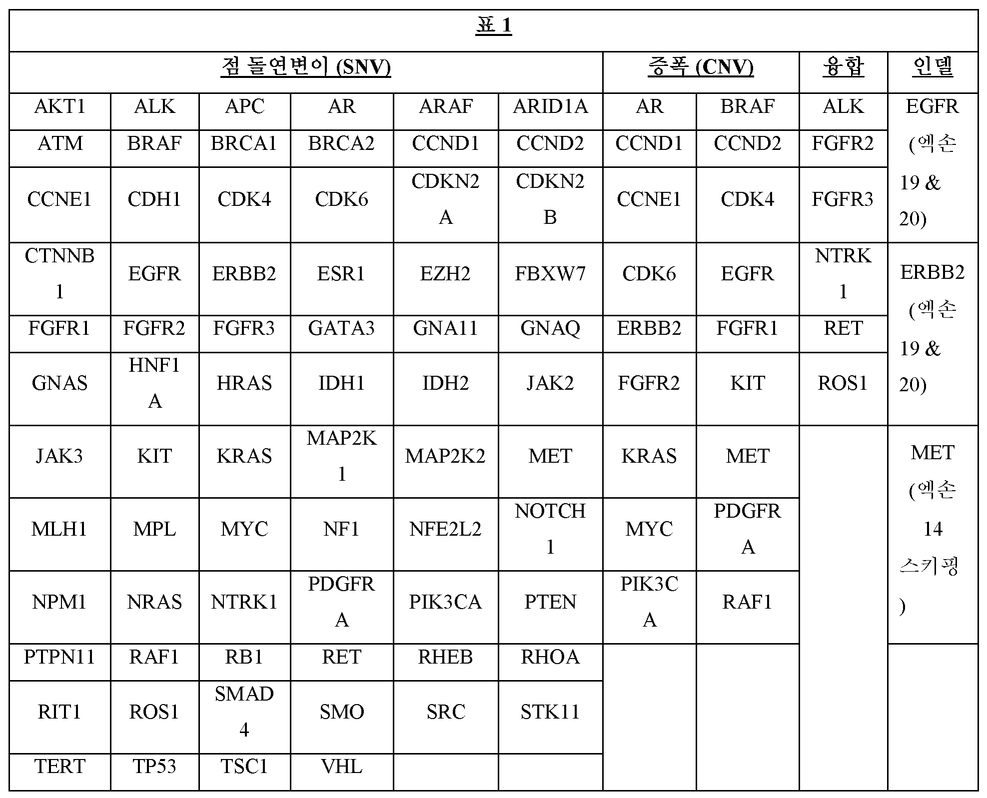
표 2Table 2

일부 실시양태에서, 패널 내의 하나 이상의 영역은 수술 후의 잔류 암을 검출하기 위한 하나 또는 복수의 유전자로부터의 하나 이상의 유전자좌를 포함한다. 이러한 검출은 기존의 암 검출 방법에 대해 가능한 것보다 더 빠를 수 있다. 일부 실시양태에서, 패널 내의 하나 이상의 게놈 위치는 고-위험 환자 집단에서 암을 검출하기 위한 하나 또는 복수의 유전자로부터의 하나 이상의 유전자좌를 포함한다. 예를 들어, 흡연자는 일반적인 집단보다 훨씬 더 높은 폐암의 비율을 갖는다. 더욱이, 흡연자는 암 검출을 보다 곤란하게 만드는 다른 폐 상태, 예컨대 폐 내의 불규칙한 혹의 발달을 발달시킬 수 있다. 일부 실시양태에서, 본원에 기술된 방법은 기존의 암 검출 방법에 대해 가능한 것보다 더 빨리 암 요법에 대한 환자의 반응을 검출한다 (특히 고위험 환자에서).In some embodiments, one or more regions in the panel include one or more loci from one or more genes for detecting residual cancer after surgery. Such detection may be faster than is possible for existing cancer detection methods. In some embodiments, one or more genomic locations in the panel include one or more loci from one or multiple genes for detecting cancer in a high-risk patient population. For example, smokers have much higher rates of lung cancer than the general population. Moreover, smokers may develop other lung conditions that make cancer detection more difficult, such as the development of irregular lumps in the lungs. In some embodiments, the methods described herein detect a patient's response to cancer therapy more quickly than is possible for existing cancer detection methods (particularly in high-risk patients).
게놈 위치는 그 유전자 또는 영역 내에 종양 마커를 갖는 암을 갖는 대상체의 수에 기초하여 시퀀싱 패널에 포함시키기 위해 선택될 수 있다. 게놈 위치는 그 유전자에 존재하는 암 및 종양 마커를 갖는 대상체의 유병률에 기초하여 시퀀싱 패널에 포함시키기 위해 선택될 수 있다. 영역 내의 종양 마커의 존재는 암을 갖는 대상체를 지시할 수 있다.Genomic locations can be selected for inclusion in a sequencing panel based on the number of subjects with cancer that have a tumor marker within that gene or region. Genomic locations can be selected for inclusion in a sequencing panel based on the prevalence of cancer and subjects with tumor markers present in that gene. The presence of tumor markers in an area can indicate a subject has cancer.
일부 경우에, 패널은 하나 이상의 데이터베이스로부터의 정보를 사용하여 선택될 수 있다. 암에 관한 정보는 암 종양 생검 또는 cfDNA 검정으로부터 유래될 수 있다. 데이터베이스는 시퀀싱된 종양 샘플의 집단을 기술하는 정보를 포함할 수 있다. 데이터베이스는 종양 샘플 내의 mRNA 발현에 관한 정보를 포함할 수 있다. 데이터베이스는 종양 샘플 내의 조절 요소 또는 게놈 영역에 관한 정보를 포함할 수 있다. 시퀀싱된 종양 샘플에 관한 정보는 다양한 유전자 변이체의 빈도를 포함하고, 유전자 변이체가 발생하는 유전자 또는 영역을 기술할 수 있다. 유전자 변이체는 종양 마커일 수 있다. 이같은 데이터베이스의 비-제한적인 예는 COSMIC이다. COSMIC는 다양한 암에서 발견되는 체세포 돌연변이의 카탈로그이다. 특정한 암에 대해, COSMIC는 돌연변이의 빈도에 기초하여 유전자를 순위화한다. 유전자는 주어진 유전자 내에 높은 빈도의 돌연변이를 갖는 것에 의해 패널에 포함시키기 위해 선택될 수 있다. 예를 들어, COSMIC는 시퀀싱된 유방암 샘플의 집단의 33%가 TP53에 돌연변이를 갖고, 샘플링된 유방암의 집단의 22%가 KRAS에 돌연변이를 가짐을 지시한다. APC를 포함하는 다른 순위화된 유전자는 시퀀싱된 유방암 샘플의 집단의 약 4%에서만 발견되는 돌연변이를 갖는다. TP53 및 KRAS는 샘플링된 유방암 중에서 상대적으로 높은 빈도를 갖는 것 (예를 들어, 약 4%의 빈도로 발생하는 APC에 비해)에 기초하여 시퀀싱 패널에 포함될 수 있다. COSMIC는 비-제한적인 예로서 제공되지만, 유전자 또는 유전자 영역에 위치한 종양 마커와 암을 연관시키는 임의의 데이터베이스 또는 정보의 세트가 사용될 수 있다. 또 다른 예에서, 1156개의 담도암 샘플의 COSMIC에 의해 제공된 바와 같이, 380개의 샘플 (33%)이 TP53에 돌연변이를 보유하였다. 몇몇 다른 유전자, 예컨대 APC는 모든 샘플의 4 내지 8%에서 돌연변이를 갖는다. 따라서, TP53은 담도암 샘플의 집단 내의 상대적으로 높은 빈도에 기초하여 패널에 포함시키기 위해 선택될 수 있다.In some cases, panels may be selected using information from one or more databases. Information regarding cancer may be derived from a cancer tumor biopsy or cfDNA assay. The database may contain information describing a population of sequenced tumor samples. The database may contain information regarding mRNA expression in tumor samples. The database may contain information regarding regulatory elements or genomic regions within a tumor sample. Information about a sequenced tumor sample may include the frequency of various genetic variants and describe the genes or regions in which the genetic variants occur. Genetic variants may be tumor markers. A non-limiting example of such a database is COSMIC. COSMIC is a catalog of somatic mutations found in various cancers. For a particular cancer, COSMIC ranks genes based on the frequency of mutations. Genes may be selected for inclusion in the panel by having a high frequency of mutations within a given gene. For example, COSMIC indicates that 33% of the population of sequenced breast cancer samples have mutations in TP53 and 22% of the population of breast cancers sampled have mutations in KRAS. Other ranked genes, including APC, have mutations found in only about 4% of the population of sequenced breast cancer samples. TP53 and KRAS may be included in the sequencing panel based on their relatively high frequency among sampled breast cancers (e.g., compared to APC, which occurs at a frequency of about 4%). COSMIC is provided as a non-limiting example, but any database or set of information that associates cancer with a tumor marker located in a gene or gene region may be used. In another example, 380 samples (33%) harbored mutations in TP53, as provided by COSMIC of 1156 biliary tract cancer samples. Several other genes, such as APC, have mutations in 4-8% of all samples. Therefore, TP53 may be selected for inclusion in the panel based on its relatively high frequency within the population of biliary tract cancer samples.
유전자 또는 게놈 섹션은 종양 마커의 빈도가 주어진 배경 집단에서 발견되는 것보다 샘플링된 종양 조직 또는 순환 종양 DNA에서 유의하게 더 큰 패널에 대해 선택될 수 있다. 게놈 위치의 조합은 암을 갖는 적어도 대다수의 대상체가 패널 내의 게놈 위치 또는 유전자의 적어도 하나에 존재하는 종양 마커 또는 게놈 영역을 가질 수 있도록 패널에 포함시키기 위해 선택될 수 있다. 게놈 위치의 조합은 특정한 암 또는 암의 세트에 대해, 대다수의 대상체가 선택된 영역 중 하나 이상에 하나 이상의 종양 마커를 가짐을 지시하는 데이터에 기초하여 선택될 수 있다. 예를 들어, 암 1을 검출하기 위해, 영역 A, B, C, 및/또는 D를 포함하는 패널은 암 1을 갖는 대상체의 90%가 패널의 영역 A, B, C, 및/또는 D에 종양 마커를 가짐을 지시하는 데이터에 기초하여 선택될 수 있다. 교대로, 종양 마커는 2개 이상의 영역 내의 조합된 종양 마커가 암을 갖는 대상체의 집단의 대다수에 존재하도록, 암을 갖는 대상체 내의 2개 이상의 영역에서 독립적으로 발생하는 것으로 제시될 수 있다. 예를 들어, 암 2를 검출하기 위해, 영역 X, Y, 및 Z를 포함하는 패널은 대상체의 90%가 하나 이상의 영역에 종양 마커를 갖고, 이같은 대상체의 30%에서 종양 마커가 영역 X에서만 검출됨을 지시하는 데이터에 기초하여 선택될 수 있는 반면, 종양 마커는 종양 마커가 검출된 대상체의 나머지에 대해 영역 Y 및/또는 Z에서만 검출된다. 하나 이상의 암과 연관되는 것으로 이전에 제시된 하나 이상의 게놈 위치에 존재하는 종양 마커는 종양 마커가 시간의 50% 이상의 그러한 영역 중 하나 이상에서 검출되는 경우 암을 갖는 대상체를 지시하거나 예측할 수 있다. 컴퓨터적 접근법, 예컨대 하나 이상의 영역 내에 종양 마커의 세트에 대한 암 빈도를 고려하여 암을 검출할 조건부 확률을 채용하는 모델은, 어느 영역이 단독으로 또는 조합으로 암을 예측할 수 있는지 예측하는데 사용될 수 있다. 패널 선택을 위한 다른 접근법은 대형 패널 및/또는 전체 게놈 시퀀싱 (WGS, RNA-seq, Chip-seq, 비술페이트 시퀀싱, ATAC-seq, 및 기타)을 갖는 종양의 포괄적 게놈 프로파일링을 채용하는 연구로부터의 정보를 기술하는 데이터베이스의 사용을 수반한다. 문헌으로부터 얻어진 정보는 특정 암에서 통상적으로 영향을 받고 돌연변이된 경로를 또한 기술할 수 있다. 패널 선택은 유전적 정보를 기술하는 온톨로지의 사용에 의해 추가로 알 수 있다.Genes or genomic sections may be selected for a panel where the frequency of tumor markers is significantly greater in sampled tumor tissue or circulating tumor DNA than would be found in a given background population. Combinations of genomic locations can be selected for inclusion in the panel such that at least the majority of subjects with cancer will have a tumor marker or genomic region present at at least one of the genomic locations or genes in the panel. Combinations of genomic locations may be selected based on data indicating that, for a particular cancer or set of cancers, a majority of subjects have one or more tumor markers in one or more of the selected regions. For example, to detect
시퀀싱을 위한 패널에 포함되는 유전자는 완전히 전사된 영역, 프로모터 영역, 인핸서 영역, 조절 요소, 및/또는 하류 서열을 포함할 수 있다. 돌연변이를 지시하는 종양을 검출할 가능성을 추가로 증가시키기 위해, 엑손만이 패널에 포함될 수 있다. 패널은 선택된 유전자의 모든 엑손, 또는 선택된 유전자의 엑손 중 하나 이상만을 포함할 수 있다. 패널은 각각의 복수의 상이한 유전자로부터의 엑손을 포함할 수 있다. 패널은 각각의 복수의 상이한 유전자로부터의 적어도 하나의 엑손을 포함할 수 있다.Genes included in the panel for sequencing may include fully transcribed regions, promoter regions, enhancer regions, regulatory elements, and/or downstream sequences. To further increase the likelihood of detecting tumor indicating mutations, only exons can be included in the panel. The panel may include all exons of the selected gene, or only one or more of the exons of the selected gene. The panel may include exons from each of a plurality of different genes. The panel may include at least one exon from each of a plurality of different genes.
일부 측면에서, 각각의 복수의 상이한 유전자로부터의 엑손의 패널은 암을 갖는 대상체의 결정된 비율이 엑손의 패널 내의 적어도 하나의 엑손에서 유전자 변이체를 나타내도록 선택된다.In some aspects, the panel of exons from each of the plurality of different genes is selected such that a determined proportion of subjects with cancer exhibit a genetic variant in at least one exon within the panel of exons.
유전자의 패널 내의 각각의 상이한 유전자로부터의 적어도 하나의 완전한 엑손이 시퀀싱될 수 있다. 시퀀싱된 패널은 복수의 유전자로부터의 엑손을 포함할 수 있다. 패널은 2 내지 100개의 상이한 유전자, 2 내지 70개의 유전자, 2 내지 50개의 유전자, 2 내지 30개의 유전자, 2 내지 15개의 유전자, 또는 2 내지 10개의 유전자로부터의 엑손을 포함할 수 있다.At least one complete exon from each different gene within the panel of genes can be sequenced. A sequenced panel may include exons from multiple genes. The panel may include exons from 2 to 100 different genes, 2 to 70 genes, 2 to 50 genes, 2 to 30 genes, 2 to 15 genes, or 2 to 10 genes.
선택된 패널은 다양한 수의 엑손을 포함할 수 있다. 패널은 2 내지 3000개의 엑손을 포함할 수 있다. 패널은 2 내지 1000개의 엑손을 포함할 수 있다. 패널은 2 내지 500개의 엑손을 포함할 수 있다. 패널은 2 내지 100개의 엑손을 포함할 수 있다. 패널은 2 내지 50개의 엑손을 포함할 수 있다. 패널은 300개 이하의 엑손을 포함할 수 있다. 패널은 200개 이하의 엑손을 포함할 수 있다. 패널은 100개 이하의 엑손을 포함할 수 있다. 패널은 50개 이하의 엑손을 포함할 수 있다. 패널은 40개 이하의 엑손을 포함할 수 있다. 패널은 30개 이하의 엑손을 포함할 수 있다. 패널은 25개 이하의 엑손을 포함할 수 있다. 패널은 20개 이하의 엑손을 포함할 수 있다. 패널은 15개 이하의 엑손을 포함할 수 있다. 패널은 10개 이하의 엑손을 포함할 수 있다. 패널은 9개 이하의 엑손을 포함할 수 있다. 패널은 8개 이하의 엑손을 포함할 수 있다. 패널은 7개 이하의 엑손을 포함할 수 있다.The selected panel may contain varying numbers of exons. A panel may contain 2 to 3000 exons. A panel may contain from 2 to 1000 exons. A panel may contain from 2 to 500 exons. A panel may contain from 2 to 100 exons. A panel may contain 2 to 50 exons. A panel may contain up to 300 exons. A panel may contain up to 200 exons. A panel may contain up to 100 exons. A panel may contain up to 50 exons. A panel may contain up to 40 exons. A panel may contain up to 30 exons. A panel may contain no more than 25 exons. A panel may contain up to 20 exons. A panel may contain no more than 15 exons. A panel may contain up to 10 exons. A panel may contain no more than 9 exons. A panel may contain no more than 8 exons. A panel may contain no more than 7 exons.
패널은 복수의 상이한 유전자로부터의 하나 이상의 엑손을 포함할 수 있다. 패널은 각각의 비율의 복수의 상이한 유전자로부터의 하나 이상의 엑손을 포함할 수 있다. 패널은 상이한 유전자의 적어도 25%, 50%, 75% 또는 90%의 각각으로부터의 적어도 2개의 엑손을 포함할 수 있다. 패널은 상이한 유전자의 적어도 25%, 50%, 75% 또는 90%의 각각으로부터의 적어도 3개의 엑손을 포함할 수 있다. 패널은 상이한 유전자의 적어도 25%, 50%, 75% 또는 90%의 각각으로부터의 적어도 4개의 엑손을 포함할 수 있다.A panel may include one or more exons from a plurality of different genes. A panel may include one or more exons from a plurality of different genes, each in proportion. The panel may include at least two exons from each of at least 25%, 50%, 75% or 90% of different genes. The panel may include at least three exons from each of at least 25%, 50%, 75% or 90% of different genes. The panel may include at least four exons from each of at least 25%, 50%, 75% or 90% of different genes.
시퀀싱 패널의 크기는 다양할 수 있다. 시퀀싱 패널은 예를 들어, 패널 내의 특정한 영역에 대한 시퀀싱된 뉴클레오티드의 총량 또는 시퀀싱된 고유한 분자의 수를 포함하는 몇몇 인자에 따라 더 크게 또는 더 작게 (뉴클레오티드 크기의 관점에서) 만들어질 수 있다. 시퀀싱 패널은 5 kb 내지 50 kb의 크기일 수 있다. 시퀀싱 패널은 10 kb 내지 30 kb의 크기일 수 있다. 시퀀싱 패널은 12 kb 내지 20 kb의 크기일 수 있다. 시퀀싱 패널은 12 kb 내지 60 kb의 크기일 수 있다. 시퀀싱 패널은 적어도 10kb, 12 kb, 15 kb, 20 kb, 25 kb, 30 kb, 35 kb, 40 kb, 45 kb, 50 kb, 60 kb, 70 kb, 80 kb, 90 kb, 100 kb, 110 kb, 120 kb, 130 kb, 140 kb, 또는 150 kb의 크기일 수 있다. 시퀀싱 패널은 100 kb, 90 kb, 80 kb, 70 kb, 60 kb, 또는 50 kb 미만의 크기일 수 있다.The size of the sequencing panel can vary. Sequencing panels can be made larger or smaller (in terms of nucleotide size) depending on several factors, including, for example, the number of unique molecules sequenced or the total amount of sequenced nucleotides for a particular region within the panel. Sequencing panels can be 5 kb to 50 kb in size. Sequencing panels can be 10 kb to 30 kb in size. Sequencing panels can be 12 kb to 20 kb in size. Sequencing panels can be 12 kb to 60 kb in size. The sequencing panel should be at least 10 kb, 12 kb, 15 kb, 20 kb, 25 kb, 30 kb, 35 kb, 40 kb, 45 kb, 50 kb, 60 kb, 70 kb, 80 kb, 90 kb, 100 kb, 110 kb. , may be of size 120 kb, 130 kb, 140 kb, or 150 kb. Sequencing panels may be less than 100 kb, 90 kb, 80 kb, 70 kb, 60 kb, or 50 kb in size.
시퀀싱을 위해 선택된 패널은 적어도 1, 5, 10, 15, 20, 25, 30, 40, 50, 60, 80, 또는 100개의 게놈 위치 (예를 들어, 각각 관심 게놈 영역을 포함함)를 포함할 수 있다. 일부 경우에, 위치의 크기가 상대적으로 작은 패널 내의 게놈 위치가 선택된다. 일부 경우에, 패널 내의 영역은 약 10 kb 이하, 약 8 kb 이하, 약 6 kb 이하, 약 5 kb 이하, 약 4 kb 이하, 약 3 kb 이하, 약 2.5 kb 이하, 약 2 kb 이하, 약 1.5 kb 이하, 또는 약 1 kb 이하 또는 그 미만의 크기를 갖는다. 일부 경우에, 패널 내의 게놈 위치는 약 0.5 kb 내지 약 10 kb, 약 0.5 kb 내지 약 6 kb, 약 1 kb 내지 약 11 kb, 약 1 kb 내지 약 15 kb, 약 1 kb 내지 약 20 kb, 약 0.1 kb 내지 약 10 kb, 또는 약 0.2 kb 내지 약 1 kb의 크기를 갖는다. 예를 들어, 패널 내의 영역은 약 0.1 kb 내지 약 5 kb의 크기를 가질 수 있다.The panel selected for sequencing will contain at least 1, 5, 10, 15, 20, 25, 30, 40, 50, 60, 80, or 100 genomic positions (e.g., each containing a genomic region of interest). You can. In some cases, genomic positions within the panel are selected where the size of the position is relatively small. In some cases, a region within a panel is less than about 10 kb, less than about 8 kb, less than about 6 kb, less than about 5 kb, less than about 4 kb, less than about 3 kb, less than about 2.5 kb, less than about 2 kb, or less than about 1.5 kb. kb or less, or about 1 kb or less in size. In some cases, the genomic location within the panel is about 0.5 kb to about 10 kb, about 0.5 kb to about 6 kb, about 1 kb to about 11 kb, about 1 kb to about 15 kb, about 1 kb to about 20 kb, about It has a size of 0.1 kb to about 10 kb, or about 0.2 kb to about 1 kb. For example, a region within a panel may have a size of about 0.1 kb to about 5 kb.
본원에서 선택된 패널은 (예를 들어, 샘플로부터 수득된 무세포 핵산 분자 내의) 저-빈도 유전자 변이체를 검출하는데 충분한 딥 시퀀싱을 허용할 수 있다. 샘플 내의 유전자 변이체의 양은 주어진 유전자 변이체에 대한 돌연변이체 대립유전자 빈도의 관점에서 언급될 수 있다. 돌연변이체 대립유전자 빈도는 돌연변이체 대립유전자 (예를 들어, 가장 통상적인 대립유전자가 아님)가 주어진 핵산의 집단, 예컨대 샘플에서 발생하는 빈도를 지칭할 수 있다. 낮은 돌연변이체 대립유전자 빈도의 유전자 변이체는 샘플 내에 상대적으로 낮은 존재의 빈도를 가질 수 있다. 일부 경우에, 패널은 적어도 0.0001%, 0.001%, 0.005%, 0.01%, 0.05%, 0.1%, 또는 0.5%의 돌연변이체 대립유전자 빈도로 유전자 변이체의 검출을 허용한다. 패널은 0.001% 이상의 돌연변이체 대립유전자 빈도로 유전자 변이체의 검출을 허용할 수 있다. 패널은 0.01% 이상의 돌연변이체 대립유전자 빈도로 유전자 변이체의 검출을 허용할 수 있다. 패널은 0.0001%, 0.001%, 0.005%, 0.01%, 0.025%, 0.05%, 0.075%, 0.1%, 0.25%, 0.5%, 0.75%, 또는 1.0%만큼 낮은 빈도로 샘플에 존재하는 유전자 변이체의 검출을 허용할 수 있다. 패널은 적어도 0.0001%, 0.001%, 0.005%, 0.01%, 0.025%, 0.05%, 0.075%, 0.1%, 0.25%, 0.5%, 0.75%, 또는 1.0%의 빈도로 샘플에 존재하는 종양 마커의 검출을 허용할 수 있다. 패널은 1.0%만큼 낮은 샘플 내의 빈도로 종양 마커의 검출을 허용할 수 있다. 패널은 0.75%만큼 낮은 샘플 내의 빈도로 종양 마커의 검출을 허용할 수 있다. 패널은 0.5%만큼 낮은 샘플 내의 빈도로 종양 마커의 검출을 허용할 수 있다. 패널은 0.25%만큼 낮은 샘플 내의 빈도로 종양 마커의 검출을 허용할 수 있다. 패널은 0.1%만큼 낮은 샘플 내의 빈도로 종양 마커의 검출을 허용할 수 있다. 패널은 0.075%만큼 낮은 샘플 내의 빈도로 종양 마커의 검출을 허용할 수 있다. 패널은 0.05%만큼 낮은 샘플 내의 빈도로 종양 마커의 검출을 허용할 수 있다. 패널은 0.025%만큼 낮은 샘플 내의 빈도로 종양 마커의 검출을 허용할 수 있다. 패널은 0.01%만큼 낮은 샘플 내의 빈도로 종양 마커의 검출을 허용할 수 있다. 패널은 0.005%만큼 낮은 샘플 내의 빈도로 종양 마커의 검출을 허용할 수 있다. 패널은 0.001%만큼 낮은 샘플 내의 빈도로 종양 마커의 검출을 허용할 수 있다. 패널은 0.0001%만큼 낮은 샘플 내의 빈도로 종양 마커의 검출을 허용할 수 있다. 패널은 1.0% 내지 0.0001%만큼 낮은 샘플 내의 빈도로 시퀀싱된 cfDNA 내의 종양 마커의 검출을 허용할 수 있다. 패널은 0.01% 내지 0.0001%만큼 낮은 샘플 내의 빈도로 시퀀싱된 cfDNA 내의 종양 마커의 검출을 허용할 수 있다.A panel selected herein can allow for deep sequencing sufficient to detect low-frequency genetic variants (e.g., in cell-free nucleic acid molecules obtained from a sample). The amount of genetic variant in a sample can be stated in terms of mutant allele frequency for a given genetic variant. Mutant allele frequency may refer to the frequency with which a mutant allele (e.g., not the most common allele) occurs in a given population of nucleic acids, such as a sample. Genetic variants of low mutant allele frequency may have a relatively low frequency of presence in a sample. In some cases, the panel allows detection of genetic variants with a mutant allele frequency of at least 0.0001%, 0.001%, 0.005%, 0.01%, 0.05%, 0.1%, or 0.5%. The panel may allow detection of genetic variants with a mutant allele frequency of 0.001% or greater. The panel may allow detection of genetic variants with a mutant allele frequency of 0.01% or greater. The panel detects genetic variants present in a sample at frequencies as low as 0.0001%, 0.001%, 0.005%, 0.01%, 0.025%, 0.05%, 0.075%, 0.1%, 0.25%, 0.5%, 0.75%, or 1.0%. can be allowed. The panel detects a tumor marker present in the sample at a frequency of at least 0.0001%, 0.001%, 0.005%, 0.01%, 0.025%, 0.05%, 0.075%, 0.1%, 0.25%, 0.5%, 0.75%, or 1.0%. can be allowed. The panel can allow detection of tumor markers at frequencies in the sample as low as 1.0%. The panel can allow detection of tumor markers at frequencies in the sample as low as 0.75%. The panel can allow detection of tumor markers at frequencies in the sample as low as 0.5%. The panel can allow detection of tumor markers at frequencies in the sample as low as 0.25%. The panel can allow detection of tumor markers at frequencies within a sample as low as 0.1%. The panel can allow detection of tumor markers at frequencies within a sample as low as 0.075%. The panel can allow detection of tumor markers at frequencies within a sample as low as 0.05%. The panel can allow detection of tumor markers at frequencies within a sample as low as 0.025%. The panel can allow detection of tumor markers at frequencies within a sample as low as 0.01%. The panel can allow detection of tumor markers at frequencies within a sample as low as 0.005%. The panel can allow detection of tumor markers at frequencies within a sample as low as 0.001%. The panel can allow detection of tumor markers at frequencies within a sample as low as 0.0001%. The panel can allow detection of tumor markers in sequenced cfDNA at frequencies within a sample as low as 1.0% to 0.0001%. The panel can allow detection of tumor markers in sequenced cfDNA at frequencies within a sample as low as 0.01% to 0.0001%.
유전자 변이체는 질환 (예를 들어, 암)을 갖는 대상체의 집단의 백분율로 나타내어질 수 있다. 일부 경우에, 암을 갖는 집단의 적어도 1%, 2%, 3%, 5%, 10%, 20%, 30%, 40%, 50%, 60%, 70%, 80%, 90%, 95%, 또는 99%는 패널 내의 영역 중 적어도 하나에서 하나 이상의 유전자 변이체를 나타낸다. 예를 들어, 암을 갖는 집단의 적어도 80%는 패널 내의 게놈 위치 중 적어도 하나에서 하나 이상의 유전자 변이체를 나타낼 수 있다.Genetic variants can be expressed as a percentage of the population of subjects with a disease (eg, cancer). In some cases, at least 1%, 2%, 3%, 5%, 10%, 20%, 30%, 40%, 50%, 60%, 70%, 80%, 90%, 95% of the population have cancer. %, or 99%, represents one or more genetic variants in at least one of the regions in the panel. For example, at least 80% of the population with cancer may exhibit one or more genetic variants at at least one of the genomic locations in the panel.
패널은 하나 이상의 유전자 각각으로부터의 관심 게놈 영역을 포함하는 하나 이상의 위치를 포함할 수 있다. 일부 경우에, 패널은 적어도 1, 2, 3, 4, 5, 6, 7, 8, 9, 10, 15, 20, 25, 30, 40, 50, 또는 80개의 유전자 각각으로부터의 관심 게놈 영역을 포함하는 하나 이상의 위치를 포함할 수 있다. 일부 경우에, 패널은 많아야 1, 2, 3, 4, 5, 6, 7, 8, 9, 10, 15, 20, 25, 30, 40, 50, 또는 80개의 유전자 각각으로부터의 관심 게놈 영역을 포함하는 하나 이상의 위치를 포함할 수 있다. 일부 경우에, 패널은 약 1개 내지 약 80개, 1개 내지 약 50개, 약 3개 내지 약 40개, 5개 내지 약 30개, 10개 내지 약 20개의 상이한 유전자 각각으로부터의 관심 게놈 영역을 포함하는 하나 이상의 위치를 포함할 수 있다.A panel may include one or more positions containing a genomic region of interest from each of one or more genes. In some cases, the panel selects genomic regions of interest from each of at least 1, 2, 3, 4, 5, 6, 7, 8, 9, 10, 15, 20, 25, 30, 40, 50, or 80 genes. It may contain one or more locations. In some cases, the panel may select genomic regions of interest from each of at most 1, 2, 3, 4, 5, 6, 7, 8, 9, 10, 15, 20, 25, 30, 40, 50, or 80 genes. It may contain one or more locations. In some cases, the panel comprises a genomic region of interest from each of about 1 to about 80, 1 to about 50, about 3 to about 40, 5 to about 30, or 10 to about 20 different genes. It may contain one or more locations containing .
패널 내의 게놈 영역을 포함하는 위치는 하나 이상의 후성유전학적으로 변형된 영역이 검출되도록 선택될 수 있다. 하나 이상의 후성유전학적으로 변형된 영역은 아세틸화, 메틸화, 유비퀴틸화, 인산화, 수모일화, 리보실화, 및/또는 시트룰린화될 수 있다. 예를 들어, 패널 내의 영역은 하나 이상의 메틸화된 영역이 검출되도록 선택될 수 있다.Positions comprising genomic regions within the panel may be selected such that one or more epigenetically modified regions are detected. One or more epigenetically modified regions may be acetylated, methylated, ubiquitinated, phosphorylated, sumoylated, ribosylated, and/or citrullinated. For example, regions within a panel can be selected such that one or more methylated regions are detected.
패널 내의 영역은 이들이 하나 이상의 조직에 걸쳐 차등적으로 전사된 서열을 포함하도록 선택될 수 있다. 일부 경우에, 게놈 영역을 포함하는 위치는 다른 조직에 비해 더 높은 수준으로 특정 조직에서 전사된 서열을 포함할 수 있다. 예를 들어, 게놈 영역을 포함하는 위치는 특정 조직에서 전사되지만 다른 조직에서는 전사되지 않는 서열을 포함할 수 있다.Regions within the panel may be selected such that they contain differentially transcribed sequences across one or more tissues. In some cases, a location comprising a genomic region may contain sequences that are transcribed at higher levels in certain tissues compared to other tissues. For example, a location containing a genomic region may contain sequences that are transcribed in certain tissues but not in other tissues.
패널 내의 게놈 위치는 코딩 및/또는 비-코딩 서열을 포함할 수 있다. 예를 들어, 패널 내의 게놈 위치는 엑손, 인트론, 프로모터, 3' 비번역된 영역, 5' 비번역된 영역, 조절 요소, 전사 시작 부위, 및/또는 스플라이스 부위에 하나 이상의 서열을 포함할 수 있다. 일부 경우에, 패널 내의 영역은 위유전자, 반복 서열, 트랜스포존, 바이러스 요소, 및 텔로미어를 포함하는 다른 비-코딩 서열을 포함할 수 있다. 일부 경우에, 패널 내의 게놈 위치는 비-코딩 RNA, 예를 들어, 리보솜 RNA, 운반 RNA, 피위-상호작용 RNA, 및 마이크로RNA에 서열을 포함할 수 있다.Genomic locations within the panel may include coding and/or non-coding sequences. For example, a genomic location within a panel may include one or more sequences in an exon, intron, promoter, 3' untranslated region, 5' untranslated region, regulatory element, transcription start site, and/or splice site. there is. In some cases, regions within the panel may include pseudogenes, repetitive sequences, transposons, viral elements, and other non-coding sequences, including telomeres. In some cases, genomic locations within the panel may include sequences in non-coding RNAs, such as ribosomal RNA, transfer RNA, peptide-interacting RNA, and microRNA.
패널 내의 게놈 위치는 원하는 수준의 감도로 암을 검출하도록 (진단하도록) (예를 들어, 하나 이상의 유전자 변이체의 검출을 통해) 선택될 수 있다. 예를 들어, 패널 내의 영역은 적어도 50%, 55%, 60%, 65%, 70%, 75%, 80%, 85%, 90%, 95%, 96%, 97%, 98%, 99%, 99.5%, 또는 99.9%의 감도로 암을 검출하도록 (예를 들어, 하나 이상의 유전자 변이체의 검출을 통해) 선택될 수 있다. 패널 내의 게놈 위치는 100%의 감도로 암을 검출하도록 선택될 수 있다.Genomic locations within the panel can be selected to detect (diagnose) cancer (e.g., through detection of one or more genetic variants) with a desired level of sensitivity. For example, the area within the panel is at least 50%, 55%, 60%, 65%, 70%, 75%, 80%, 85%, 90%, 95%, 96%, 97%, 98%, 99%. , may be selected to detect cancer (e.g., through detection of one or more genetic variants) with a sensitivity of 99.5%, or 99.9%. Genomic locations within the panel can be selected to detect cancer with 100% sensitivity.
패널 내의 게놈 위치는 원하는 수준의 특이성으로 암을 검출하도록 (진단하도록) (예를 들어, 하나 이상의 유전자 변이체의 검출을 통해) 선택될 수 있다. 예를 들어, 패널 내의 게놈 위치는 적어도 50%, 55%, 60%, 65%, 70%, 75%, 80%, 85%, 90%, 95%, 96%, 97%, 98%, 99%, 99.5%, 또는 99.9%의 특이성으로 암을 검출하도록 (예를 들어, 하나 이상의 유전자 변이체의 검출을 통해) 선택될 수 있다. 패널 내의 게놈 위치는 100%의 특이성으로 하나 이상의 유전자 변이체를 검출하도록 선택될 수 있다.Genomic locations within the panel can be selected to detect (diagnose) cancer (e.g., through detection of one or more genetic variants) with a desired level of specificity. For example, the genomic positions within the panel are at least 50%, 55%, 60%, 65%, 70%, 75%, 80%, 85%, 90%, 95%, 96%, 97%, 98%, 99 %, 99.5%, or 99.9% specificity. Genomic locations within the panel can be selected to detect one or more genetic variants with 100% specificity.
패널 내의 게놈 위치는 원하는 양성 예측적 값으로 암을 검출하도록 (진단하도록) 선택될 수 있다. 양성 예측적 값은 감도 (예를 들어, 검출되는 실제 양성의 확률) 및/또는 특이성 (예를 들어, 실제 음성을 양성으로 오인하지 않을 확률)을 증가시킴으로써 증가될 수 있다. 비-제한적인 예로서, 패널 내의 게놈 위치는 적어도 50%, 55%, 60%, 65%, 70%, 75%, 80%, 85%, 90%, 95%, 96%, 97%, 98%, 99%, 99.5%, 또는 99.9%의 양성 예측적 값으로 하나 이상의 유전자 변이체를 검출하도록 선택될 수 있다. 패널 내의 영역은 100%의 양성 예측적 값으로 하나 이상의 유전자 변이체를 검출하도록 선택될 수 있다.Genomic locations within the panel can be selected to detect (diagnose) cancer with a desired positive predictive value. The positive predictive value can be increased by increasing sensitivity (e.g., the probability of a true positive being detected) and/or specificity (e.g., the probability of not mistaking a true negative for a positive). As a non-limiting example, the genomic positions within the panel are at least 50%, 55%, 60%, 65%, 70%, 75%, 80%, 85%, 90%, 95%, 96%, 97%, 98 %, 99%, 99.5%, or 99.9% positive predictive value may be selected to detect one or more genetic variants. Regions within the panel can be selected to detect one or more genetic variants with a positive predictive value of 100%.
패널 내의 게놈 위치는 원하는 정확도로 암을 검출하도록 (진단하도록) 선택될 수 있다. 본원에서 사용된 바와 같이, 용어 "정확도"는 질환 상태 (예를 들어, 암) 및 건강한 상태 사이를 식별하는 시험의 능력을 지칭할 수 있다. 정확도는 감도 및 특이성, 예측적 값, 가능도 비, ROC 곡선하 면적, 유덴 지수 및/또는 진단 오즈 비와 같은 척도를 사용하여 정량화될 수 있다.Genomic locations within the panel can be selected to detect (diagnose) cancer with desired accuracy. As used herein, the term “accuracy” may refer to the ability of a test to discriminate between a disease state (e.g., cancer) and a healthy state. Accuracy can be quantified using measures such as sensitivity and specificity, predictive value, likelihood ratio, area under the ROC curve, Youden index, and/or diagnostic odds ratio.
정확도는 정확한 결과를 제공하는 시험의 수 및 수행된 시험의 총 수 사이의 비를 지칭하는 백분율로서 제시될 수 있다. 패널 내의 영역은 적어도 50%, 55%, 60%, 65%, 70%, 75%, 80%, 85%, 90%, 95%, 96%, 97%, 98%, 99%, 99.5%, 또는 99.9%의 정확도로 암을 검출하도록 선택될 수 있다. 패널 내의 게놈 위치는 100%의 정확도로 암을 검출하도록 선택될 수 있다.Accuracy can be presented as a percentage, which refers to the ratio between the number of tests that give correct results and the total number of tests performed. The area within the panel is at least 50%, 55%, 60%, 65%, 70%, 75%, 80%, 85%, 90%, 95%, 96%, 97%, 98%, 99%, 99.5%, Alternatively, it may be selected to detect cancer with 99.9% accuracy. Genomic locations within the panel can be selected to detect cancer with 100% accuracy.
패널은 고도로 민감성이고 낮은 빈도 유전자 변이체를 검출하도록 선택될 수 있다. 예를 들어, 패널은 0.01%, 0.05%, 또는 0.001%만큼 낮은 빈도로 샘플에 존재하는 유전자 변이체 또는 종양 마커가 적어도 50%, 55%, 60%, 65%, 70%, 75%, 80%, 85%, 90%, 95%, 96%, 97%, 98%, 99%, 99.5%, 또는 99.9%의 감도로 검출될 수 있도록 선택될 수 있다. 패널 내의 게놈 위치는 샘플에 1% 이하의 빈도로 존재하는 종양 마커를 70% 이상의 감도로 검출하도록 선택될 수 있다. 패널은 0.1%만큼 낮은 샘플 내의 빈도로 종양 마커를 적어도 50%, 55%, 60%, 65%, 70%, 75%, 80%, 85%, 90%, 95%, 96%, 97%, 98%, 99%, 99.5%, 또는 99.9%의 감도로 검출하도록 선택될 수 있다. 패널은 0.01%만큼 낮은 샘플 내의 빈도로 종양 마커를 적어도 50%, 55%, 60%, 65%, 70%, 75%, 80%, 85%, 90%, 95%, 96%, 97%, 98%, 99%, 99.5%, 또는 99.9%의 감도로 검출하도록 선택될 수 있다. 패널은 0.001%만큼 낮은 샘플 내의 빈도로 종양 마커를 적어도 50%, 55%, 60%, 65%, 70%, 75%, 80%, 85%, 90%, 95%, 96%, 97%, 98%, 99%, 99.5%, 또는 99.9%의 감도로 검출하도록 선택될 수 있다.The panel can be selected to be highly sensitive and detect low frequency genetic variants. For example, the panel may determine whether at least 50%, 55%, 60%, 65%, 70%, 75%, or 80% of a genetic variant or tumor marker is present in the sample at a frequency as low as 0.01%, 0.05%, or 0.001%. , can be selected to be detected with a sensitivity of 85%, 90%, 95%, 96%, 97%, 98%, 99%, 99.5%, or 99.9%. Genomic locations within the panel can be selected to detect tumor markers present at a frequency of 1% or less in the sample with a sensitivity of 70% or greater. The panel identified tumor markers with a frequency within the sample as low as 0.1% in at least 50%, 55%, 60%, 65%, 70%, 75%, 80%, 85%, 90%, 95%, 96%, 97%, It can be selected to detect with a sensitivity of 98%, 99%, 99.5%, or 99.9%. The panel identified tumor markers in at least 50%, 55%, 60%, 65%, 70%, 75%, 80%, 85%, 90%, 95%, 96%, 97%, It can be selected to detect with a sensitivity of 98%, 99%, 99.5%, or 99.9%. The panel identified tumor markers in at least 50%, 55%, 60%, 65%, 70%, 75%, 80%, 85%, 90%, 95%, 96%, 97%, It can be selected to detect with a sensitivity of 98%, 99%, 99.5%, or 99.9%.
패널은 고도로 특이적이고 낮은 빈도 유전자 변이체를 검출하도록 선택될 수 있다. 예를 들어, 패널은 0.01%, 0.05%, 또는 0.001%만큼 낮은 빈도로 샘플에 존재하는 유전자 변이체 또는 종양 마커가 적어도 50%, 55%, 60%, 65%, 70%, 75%, 80%, 85%, 90%, 95%, 96%, 97%, 98%, 99%, 99.5%, 또는 99.9%의 특이성으로 검출될 수 있도록 선택될 수 있다. 패널 내의 게놈 위치는 샘플에 1% 이하의 빈도로 존재하는 종양 마커를 70% 이상의 특이성으로 검출하도록 선택될 수 있다. 패널은 0.1%만큼 낮은 샘플 내의 빈도로 종양 마커를 적어도 70%, 75%, 80%, 85%, 90%, 95%, 96%, 97%, 98%, 99%, 99.5%, 또는 99.9%의 특이성으로 검출하도록 선택될 수 있다. 패널은 0.01%만큼 낮은 샘플 내의 빈도로 종양 마커를 적어도 70%, 75%, 80%, 85%, 90%, 95%, 96%, 97%, 98%, 99%, 99.5%, 또는 99.9%의 특이성으로 검출하도록 선택될 수 있다. 패널은 0.001%만큼 낮은 샘플 내의 빈도로 종양 마커를 적어도 70%, 75%, 80%, 85%, 90%, 95%, 96%, 97%, 98%, 99%, 99.5%, 또는 99.9%의 특이성으로 검출하도록 선택될 수 있다.The panel can be selected to be highly specific and detect low frequency genetic variants. For example, the panel may determine whether at least 50%, 55%, 60%, 65%, 70%, 75%, or 80% of a genetic variant or tumor marker is present in the sample at a frequency as low as 0.01%, 0.05%, or 0.001%. , can be selected to be detected with a specificity of 85%, 90%, 95%, 96%, 97%, 98%, 99%, 99.5%, or 99.9%. Genomic locations within the panel can be selected to detect tumor markers present at a frequency of 1% or less in the sample with greater than 70% specificity. The panel detects tumor markers at a frequency in the sample as low as 0.1% in at least 70%, 75%, 80%, 85%, 90%, 95%, 96%, 97%, 98%, 99%, 99.5%, or 99.9%. can be selected for detection with a specificity of The panel detects tumor markers in at least 70%, 75%, 80%, 85%, 90%, 95%, 96%, 97%, 98%, 99%, 99.5%, or 99.9% of the tumor markers at a frequency in the sample as low as 0.01%. can be selected for detection with a specificity of The panel detects tumor markers at a frequency within the sample as low as 0.001% in at least 70%, 75%, 80%, 85%, 90%, 95%, 96%, 97%, 98%, 99%, 99.5%, or 99.9%. can be selected for detection with a specificity of
패널은 고도로 정확하고 낮은 빈도 유전자 변이체를 검출하도록 선택될 수 있다. 패널은 0.01%, 0.05%, 또는 0.001%만큼 낮은 빈도로 샘플에 존재하는 유전자 변이체 또는 종양 마커가 적어도 70%, 75%, 80%, 85%, 90%, 95%, 96%, 97%, 98%, 99%, 99.5%, 또는 99.9%의 정확도로 검출될 수 있도록 선택될 수 있다. 패널 내의 게놈 위치는 샘플에 1% 이하의 빈도로 존재하는 종양 마커를 70% 이상의 정확도로 검출하도록 선택될 수 있다. 패널은 0.1%만큼 낮은 샘플 내의 빈도로 종양 마커를 적어도 70%, 75%, 80%, 85%, 90%, 95%, 96%, 97%, 98%, 99%, 99.5%, 또는 99.9%의 정확도로 검출하도록 선택될 수 있다. 패널은 0.01%만큼 낮은 샘플 내의 빈도로 종양 마커를 적어도 70%, 75%, 80%, 85%, 90%, 95%, 96%, 97%, 98%, 99%, 99.5%, 또는 99.9%의 정확도로 검출하도록 선택될 수 있다. 패널은 0.001%만큼 낮은 샘플 내의 빈도로 종양 마커를 적어도 70%, 75%, 80%, 85%, 90%, 95%, 96%, 97%, 98%, 99%, 99.5%, 또는 99.9%의 정확도로 검출하도록 선택될 수 있다.The panel can be selected to detect low frequency genetic variants with high accuracy. The panel determines that at least 70%, 75%, 80%, 85%, 90%, 95%, 96%, 97%, It can be selected to be detected with an accuracy of 98%, 99%, 99.5%, or 99.9%. Genomic locations within the panel can be selected to detect tumor markers present at a frequency of 1% or less in the sample with greater than 70% accuracy. The panel detects tumor markers at a frequency in the sample as low as 0.1% in at least 70%, 75%, 80%, 85%, 90%, 95%, 96%, 97%, 98%, 99%, 99.5%, or 99.9%. can be selected to detect with an accuracy of . The panel detects tumor markers in at least 70%, 75%, 80%, 85%, 90%, 95%, 96%, 97%, 98%, 99%, 99.5%, or 99.9% of the tumor markers at a frequency in the sample as low as 0.01%. can be selected to detect with an accuracy of . The panel detects tumor markers at a frequency within the sample as low as 0.001% in at least 70%, 75%, 80%, 85%, 90%, 95%, 96%, 97%, 98%, 99%, 99.5%, or 99.9%. can be selected to detect with an accuracy of .
패널은 고도로 예측적이고 낮은 빈도 유전자 변이체를 검출하도록 선택될 수 있다. 패널은 0.01%, 0.05%, 또는 0.001%만큼 낮은 빈도로 샘플에 존재하는 유전자 변이체 또는 종양 마커가 적어도 70%, 75%, 80%, 85%, 90%, 95%, 96%, 97%, 98%, 99%, 99.5%, 또는 99.9%의 양성 예측적 값을 가질 수 있도록 선택될 수 있다.The panel can be selected to detect highly predictive and low frequency genetic variants. The panel determines that at least 70%, 75%, 80%, 85%, 90%, 95%, 96%, 97%, It may be selected to have a positive predictive value of 98%, 99%, 99.5%, or 99.9%.
패널에 사용되는 프로브 또는 미끼의 농도는 샘플 내의 보다 많은 핵산 분자를 포획하도록 증가될 수 있다 (2 내지 6 ng/μL). 패널에 사용되는 프로브 또는 미끼의 농도는 적어도 2 ng/μL, 3 ng/ μL, 4 ng/ μL, 5 ng/μL, 6 ng/μL, 또는 그 초과일 수 있다. 프로브의 농도는 약 2 ng/μL 내지 약 3 ng/μL, 약 2 ng/μL 내지 약 4 ng/μL, 약 2 ng/μL 내지 약 5 ng/μL, 약 2 ng/μL 내지 약 6 ng/μL일 수 있다. 패널에 사용되는 프로브 또는 미끼의 농도는 2 ng/μL 이상 내지 6 ng/μL 이하일 수 있다. 일부 경우에 이는 분석될 생물학적 내의 보다 많은 분자를 허용함으로써, 보다 낮은 빈도 대립유전자가 검출되는 것을 가능하게 할 수 있다.The concentration of probes or baits used in the panel can be increased (2 to 6 ng/μL) to capture more nucleic acid molecules in the sample. The concentration of probe or bait used in the panel may be at least 2 ng/μL, 3 ng/μL, 4 ng/μL, 5 ng/μL, 6 ng/μL, or more. The concentration of the probe is about 2 ng/μL to about 3 ng/μL, about 2 ng/μL to about 4 ng/μL, about 2 ng/μL to about 5 ng/μL, about 2 ng/μL to about 6 ng/μL. It may be μL. The concentration of probes or baits used in the panel may be 2 ng/μL or more and 6 ng/μL or less. In some cases this may allow more molecules within the organism to be analyzed, thereby allowing lower frequency alleles to be detected.
한 실시양태에서, 시퀀싱 후, 서열 판독물은 품질 점수가 할당될 수 있다. 품질 점수는 그러한 서열 판독물이 임계치에 기초하여 후속 분석에 유용할 수 있는지 여부를 지시하는 서열 판독물의 묘사일 수 있다. 일부 경우에, 일부 서열 판독물은 후속 맵핑 단계를 수행하기 위한 충분한 품질 또는 길이의 것이 아니다. 적어도 90%, 95%, 99%, 99.9%, 99.99% 또는 99.999%의 품질 점수를 갖는 서열 판독물은 서열 판독물의 데이터 세트로부터 필터링될 수 있다. 다른 경우에, 적어도 90%, 95%, 99%, 99.9%, 99.99% 또는 99.999%로 점수화된 품질이 할당된 서열 판독물은 데이터 세트로부터 필터링될 수 있다. 특정 품질 점수 임계치를 충족시키는 서열 판독물은 기준 게놈에 맵핑될 수 있다. 맵핑 정렬 후, 서열 판독물은 맵핑 점수가 할당될 수 있다. 맵핑 점수는 각각의 위치가 고유하게 맵핑가능한지 아닌지 여부를 지시하는 기준 서열에 다시 맵핑되는 서열 판독물의 묘사일 수 있다. 적어도 90%, 95%, 99%, 99.9%, 99.99% 또는 99.999%의 맵핑 점수를 갖는 서열 판독물은 데이터 세트로부터 필터링될 수 있다. 다른 경우에, 90%, 95%, 99%, 99.9%, 99.99% 또는 99.999% 미만으로 점수화된 맵핑이 할당된 시퀀싱 판독물은 데이터 세트로부터 필터링될 수 있다.In one embodiment, after sequencing, sequence reads may be assigned a quality score. A quality score can be a description of a sequence read that indicates whether such sequence read may be useful for subsequent analysis based on a threshold. In some cases, some sequence reads are not of sufficient quality or length to perform subsequent mapping steps. Sequence reads with a quality score of at least 90%, 95%, 99%, 99.9%, 99.99% or 99.999% can be filtered out from the data set of sequence reads. In other cases, sequence reads that have been assigned a quality score of at least 90%, 95%, 99%, 99.9%, 99.99% or 99.999% may be filtered from the data set. Sequence reads that meet a certain quality score threshold can be mapped to a reference genome. After mapping alignment, sequence reads can be assigned a mapping score. A mapping score can be a description of sequence reads that map back to a reference sequence, indicating whether each position is uniquely mappable or not. Sequence reads with a mapping score of at least 90%, 95%, 99%, 99.9%, 99.99% or 99.999% can be filtered from the data set. In other cases, sequencing reads assigned mappings that score less than 90%, 95%, 99%, 99.9%, 99.99%, or 99.999% may be filtered from the data set.
c. MAF 결정c. MAF decision
도 1에 제시된 바와 같이, 단계 (103 및/또는 104)에서의 샘플의 cfDNA 시퀀싱 후, 단계 (105 및/또는 106)에서 하나 이상의 돌연변이체 대립유전자 분율 (MAF)이 결정될 수 있다. 일부 또는 모든 MAF 결정은 변이체 분류 (107/108) 전에, 변이체 분류 (107/108) 후에, 변이체 분류 (107/108) 동안, 변이체 필터링 (109) 전에, 변이체 필터링 (109) 후에, 변이체 필터링 (109) 동안, 또는 이들의 조합에 발생할 수 있다. 단계 (103) 전에, cfDNA는 말단 복구되고, 분자 바코드를 포함하는 어댑터로 라이게이션되고, 증폭되고, 풍부화될 수 있다. 증폭은 샘플 색인을 혼입할 수 있다. 한 실시양태에서, MAF 값은 모든 변이체 또는 모든 체세포 변이체에 대해 결정될 수 있다. 한 실시양태에서, MAF 값은 모든 변이체 미만 또는 모든 체세포 변이체 미만에 대해 결정될 수 있다. 변이체 대립유전자 분율 (VAF)은 본원에서 MAF와 상호교환가능하게 사용된다. 돌연변이체 대립유전자 분율 (MAF)은 돌연변이체 분자의 수를 특이적 게놈 위치에서의 분자의 총 수 (예를 들어, 분자 커버리지)로 나눈 것을 나타낸다:As shown in Figure 1 , following cfDNA sequencing of the sample in
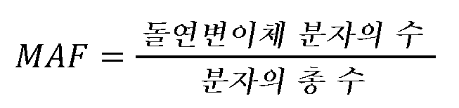
최대 MAF는 주어진 샘플에 존재하거나 그에서 관찰되는 모든 체세포 변이체의 최대 또는 가장 큰 MAF로서 결정될 수 있다. 일부 실시양태에서, 최대 MAF는 주어진 샘플의 종양 분율로 간주될 수 있다.Maximal MAF can be determined as the largest or largest MAF of all somatic variants present or observed in a given sample. In some embodiments, the maximum MAF can be considered the tumor fraction of a given sample.
최대 배수체 유전자의 분율 ("max frac_diploid") (최소 대립유전자 불균형)이 결정될 수 있다. 배수체 유전자의 분율 ("frac_diploid)은 카피 수에 의해 결정된 바와 같은 샘플에 걸친 대립유전자 불균형의 수준의 척도이다. 높은 수준의 대립유전자 불균형을 갖는 샘플은 배선/체세포 오분류될 경향이 있다. 따라서, 낮은 수준의 대립유전자 불균형 (또는 높은 frac_diploid)은 체세포 분류 콜링의 신뢰성의 지시이다.The fraction of genes that are maximally diploid (“max frac_diploid”) (minimum allelic imbalance) can be determined. The fraction of genes that are polyploid ("frac_diploid) is a measure of the level of allelic imbalance across a sample as determined by copy number. Samples with high levels of allelic imbalance are prone to germline/somatic misclassification. Therefore, Low levels of allelic imbalance (or high frac_diploid) are indicative of the reliability of somatic classification calling.
한 실시양태에서, 총 커버리지 프로파일을 사용하여 개별적인 유전자라기 보다는 배수 변화 및 따라서 종양 분율을 포획할 수 있다.In one embodiment, total coverage profiles can be used to capture fold changes and therefore tumor fractions rather than individual genes.
d. 변이체 분류d. Variant classification
단계 (103 및 104)에서의 시퀀싱은 복수의 서열 판독물을 생성한다. 단계 (107 및/또는 108)에서 복수의 서열 판독물을 분석하여 하나 이상의 변이체를 결정하고, 하나 이상의 변이체를 분류할 수 있다. 한 실시양태에서, 일부 또는 모든 변이체 분류는 MAF 결정 (105/106) 전에, MAF 결정 (105/106) 후에, MAF 결정 (105/106) 동안, 또는 이들의 조합에 결정될 수 있다. 변이체는 예를 들어, 단일 뉴클레오티드 변이체 (SNV), 인델, 융합, 및 카피 수 변이를 포함할 수 있다. 변이체 콜링을 위한 임의의 공지된 기법이 사용될 수 있다. 한 실시양태에서, 샘플로부터의 복수의 서열 판독물은 어셈블리되고/거나 맵핑되고, 기준 게놈에 비해 게놈 위치에 대해 정렬될 수 있다. 일부 실시양태에서, 복수의 서열 판독물 (어셈블리된 또는 다른 방식으로)을 이어서 기준 게놈과 비교하여 대상체의 복수의 서열 판독물이 기준 게놈의 그것과 얼마나 다른지 결정할 수 있다. 이같은 프로세스는 복수의 서열 판독물 내의 하나 이상의 변이체의 존재를 결정할 수 있다. 일부 실시양태에서, 복수의 서열 판독물로부터 수득된 핵산 분자의 분자 바코드 및/또는 출발 및 정지 게놈 위치를 사용하여 분자에 속하는 서열 판독물이 기준 게놈과 상이한 돌연변이체 분자를 확인할 수 있다. 이같은 프로세스는 복수의 서열 판독물 내의 하나 이상의 변이체의 존재를 결정할 수 있다.Sequencing in
한 실시양태에서, 통상적인 이형접합성 SNP를 사용하여 국소 배선 대립유전자 카운트 거동을 모델링하고, 이들이 관찰된 배선 돌연변이체 대립유전자 분율로부터 유의하게 벗어나면 변이체를 체세포로 콜링할 수 있다. 베타이항 모델은 그것이 통상적인 SNP에서 돌연변이체 대립유전자 카운트의 평균 및 분산 양쪽 모두를 모델링하기 때문에 사용될 수 있다. 예를 들어, 그 전문이 본원에 참조로 포함되는 PCT/US2018/052087에 기술된 베타이항 모델이 사용될 수 있다. 이는 고정된 MAF 컷오프 또는 포이즌(Poisson) 모델과 같은 보다 간단한 방법에 비해 개선된 것인데, 이는 이들이 분자 카운트 내의 분산을 적절하게 나타내지 않을 수 있기 때문이다.In one embodiment, common heterozygous SNPs can be used to model local germline allele count behavior, and variants can be called somatic if they deviate significantly from the observed germline mutant allele fraction. The beta binomial model can be used because it models both the mean and variance of mutant allele counts in common SNPs. For example, the beta binomial model described in PCT/US2018/052087, which is incorporated herein by reference in its entirety, can be used. This is an improvement over simpler methods such as fixed MAF cutoffs or the Poisson model, as these may not adequately represent the variance within the molecular counts.
e. 변이체 필터링e. Variant filtering
한 실시양태에서, 도 1에 제시된 바와 같이, 하나 이상의 필터링 프로세스를 단계 (109)에서 서열 판독물에 적용하여 서열 판독물을 추가의 분석으로부터 배제할 수 있다. 한 실시양태에서, 일부 또는 모든 필터링은 MAF 결정 (105/106) 전에, MAF 결정 (105/106) 후에, MAF 결정 (105/106) 동안, 변이체 분류 (107/108) 전에, 변이체 분류 (107/108) 후에, 변이체 분류 (107/108) 동안, 또는 이들의 조합에 적용될 수 있다.In one embodiment, one or more filtering processes may be applied to the sequence reads at
일부 실시양태에서, 제1 및/또는 제2 시점에서 약 0.1%, 0.2%, 0.3%, 0.4%, 0.5%, 0.6%, 0.7%, 0.8%, 또는 0.9% 미만인 MAF를 갖는 하나 이상의 체세포 변이체는 추가의 분석으로부터 배제될 수 있다. 일부 실시양태에서, 제1 및/또는 제2 시점에서 5, 10, 15, 20, 25 또는 30 미만의 돌연변이체 분자 카운트를 갖는 하나 이상의 체세포 변이체는 추가의 분석으로부터 배제될 수 있다. 일부 실시양태에서, 제1 및/또는 제2 시점에서 50, 100, 200, 300, 400, 500, 600, 700, 800, 900 또는 1000 미만의 커버리지를 갖는 하나 이상의 체세포 변이체는 추가의 분석으로부터 배제될 수 있다.In some embodiments, one or more somatic variants with a MAF of less than about 0.1%, 0.2%, 0.3%, 0.4%, 0.5%, 0.6%, 0.7%, 0.8%, or 0.9% at the first and/or second time point. can be excluded from further analysis. In some embodiments, one or more somatic variants with a mutant molecule count of less than 5, 10, 15, 20, 25, or 30 at the first and/or second time point may be excluded from further analysis. In some embodiments, one or more somatic variants with coverage of less than 50, 100, 200, 300, 400, 500, 600, 700, 800, 900, or 1000 at the first and/or second time point are excluded from further analysis. It can be.
한 실시양태에서, 카피 수 변이체를 사용하여 서열 판독물을 추가의 분석으로부터 배제할 수 있다. 카피 수 증폭은 관련 기술 분야에 공지된 바와 같이 결정될 수 있다. 단계 (109)에서, 방법 (100)은 불충분한 프로브 커버리지 또는 불충분한 카피 수 (예를 들어 95% 검출의 한계 미만)를 갖는 유전자 내의 카피 수 증폭을 필터링할 수 있다.In one embodiment, copy number variants can be used to exclude sequence reads from further analysis. Copy number amplification can be determined as known in the art. At
예로서, CNV는 서열 판독물을 분석하여 커버리지의 염색체 영역을 생성함으로써 결정될 수 있다. 염색체 영역은 가변적인 길이 윈도우 또는 빈으로 나누어질 수 있다. 판독물 커버리지는 각각의 윈도우/빈 영역에 대해 결정될 수 있다. 한 실시양태에서, 시퀀싱 판독물 커버리지에 관한 정량적 척도는 유전자의 유전자좌 (예를 들어, 기준 게놈으로부터 특정한 위치, 염기, 영역, 유전자 또는 염색체)에 상응하는 DNA 분자로부터 유래된 판독물의 수를 지시하는 척도이다. 판독물을 유전자의 유전자좌에 연관시키기 위해, 판독물은 기준물에 맵핑되거나 정렬될 수 있다. 맵핑 또는 정렬을 수행하는 소프트웨어 (예를 들어, 보타이(Bowtie), BWA, mrsFAST, BLAST, BLAT)는 시퀀싱 판독물을 유전자의 유전자좌와 연관시킬 수 있다. 서열 판독물 커버리지가 결정된 후, 추계적 모델링 알고리즘을 적용하여 각각의 윈도우/빈 영역에 대한 정규화된 핵산 서열 판독물 커버리지를 별개의 카피 수 상태로 전환시킬 수 있다. 일부 경우에, 이 알고리즘은 하기 중 하나 이상을 포함할 수 있다: 히든 마르코브(Hidden Markov) 모델, 동적 프로그래밍, 지지 벡터 기계, 베이시안(Bayesian) 네트워크, 트렐리스 디코딩, 비테르비(Viterbi) 디코딩, 예상 최대화, 칼만(Kalman) 필터링 방법론 및 신경 네트워크. 각각의 윈도우 영역의 별개의 카피 수 상태를 이용하여 염색체 영역 내의 카피 수 변이를 확인할 수 있다. 일부 경우에, 동일한 카피 수를 갖는 모든 인접한 윈도우/빈 영역을 분절로 병합하여 카피 수 변이 상태의 존재 또는 부재를 보고할 수 있다. 일부 경우에, 다양한 윈도우/빈은 이들이 다른 분절과 병합되기 전에 필터링될 수 있다. 카피 수 변이를 사용하여 얼마나 많은 질환 물질 (또는 카피 수 변이를 갖는 핵산)이 무세포 폴리뉴클레오티드 샘플에 존재하는지 지시하는 백분율 점수를 보고할 수 있다.As an example, CNVs can be determined by analyzing sequence reads to generate chromosomal regions of coverage. Chromosome regions can be divided into windows or bins of variable length. Read coverage can be determined for each window/blank area. In one embodiment, a quantitative measure of sequencing read coverage indicates the number of reads derived from a DNA molecule corresponding to a locus of a gene (e.g., a specific position, base, region, gene, or chromosome from a reference genome). It is a measure. To associate reads with a gene's locus, the reads can be mapped or aligned to a reference. Software that performs mapping or alignment (e.g., Bowtie, BWA, mrsFAST, BLAST, BLAT) can associate sequencing reads with the locus of a gene. After sequence read coverage is determined, stochastic modeling algorithms can be applied to convert the normalized nucleic acid sequence read coverage for each window/blank region into distinct copy number states. In some cases, this algorithm may include one or more of the following: Hidden Markov model, dynamic programming, support vector machine, Bayesian network, trellis decoding, Viterbi. ) Decoding, expectation maximization, Kalman filtering methodology and neural networks. Copy number variation within a chromosomal region can be identified using the distinct copy number status of each window region. In some cases, all adjacent windows/empty regions with the same copy number can be merged into a segment to report the presence or absence of a copy number variation state. In some cases, various windows/bins may be filtered before they are merged with other segments. Copy number variations can be used to report a percentage score that indicates how much disease agent (or nucleic acid with a copy number variation) is present in a cell-free polynucleotide sample.
한 실시양태에서, 하나 이상의 유전자 내의 CNV의 존재를 사용하여 변이체를 추가의 분석으로부터 배제할 수 있다. 예로서, T0 또는 T1 샘플 중 어느 하나에서 카피 수를 갖는 LDT-보고가능한 유전자의 임계치 수를 갖는 변이체 >= 유전자-특이적 95% 검출의 한계 (LoD). 임계치는 약 10 내지 약 30일 수 있다. 임계치는 예를 들어, 15, 16, 17, 18, 19, 20, 21, 22, 23, 24, 25 등일 수 있다. 한 실시양태에서, 임계치는 19일 수 있다.In one embodiment, the presence of CNVs in one or more genes can be used to exclude variants from further analysis. As an example, variants with a threshold number of LDT-reportable genes with copy number in either T 0 or T 1 samples >= gene-specific 95% limit of detection (LoD). The threshold may be about 10 to about 30. The threshold may be, for example, 15, 16, 17, 18, 19, 20, 21, 22, 23, 24, 25, etc. In one embodiment, the threshold may be 19.
카피 수 변이는 주어진 변이체에 대한 배수 변화를 지시할 수 있다. 가우스 모델을 사용하여 분자 반응 점수의 추정값으로서 사용될 수 있는 시간 T0 및 시간 T1 사이의 배수 변화의 비를 결정할 수 있다.Copy number variation can dictate the fold change for a given variant. The Gaussian model can be used to determine the ratio of fold changes between time T 0 and time T 1 which can be used as an estimate of the molecular response score.
한 실시양태에서, 대상체가 체세포 변이체를 갖지 않거나, 또는 변이체 필터링 프로세스의 기준을 충족시키는 변이체를 갖지 않는 경우, 대상체는 비-평가가능한 것으로서 분류될 수 있다. 한 실시양태에서, 비-평가가능한 것으로서 분류된 대상체는 분자 반응자로서 추가로 분류될 수 있다. 한 실시양태에서, 시간 T0 및 시간 T1 양쪽 모두에서 낮은 ctDNA를 갖는 대상체는 비-평가가능한 것으로서 분류되고, 분자 반응자로서 추가로 분류될 수 있다. 한 실시양태에서, 시간 T0 및 시간 T1 양쪽 모두에서 낮은 MAF를 갖는 대상체는 비-평가가능한 것으로서 분류되고, 분자 반응자로서 추가로 분류될 수 있다. 한 실시양태에서, 시간 T0 및 시간 T1 양쪽 모두에서 낮은 종양 분율을 갖는 대상체는 비-평가가능한 것으로서 분류되고, 분자 반응자로서 추가로 분류될 수 있다. 낮은 MAF 또는 낮은 종양 분율은 검출의 한계 미만 (예를 들어 95% 검출의 한계 미만), 또는 정량화의 한계 미만의 MAF 또는 종양 분율을 지칭할 수 있다. 무엇이 낮은 것을 구성하는지는 패널 디자인에 좌우될 수 있지만, 예를 들어, MAF f 0.1, 0.2, 또는 0.3%는 낮은 것으로 간주될 수 있다.In one embodiment, if a subject does not have a somatic variant, or does not have a variant that meets the criteria of the variant filtering process, the subject may be classified as non-evaluable. In one embodiment, subjects classified as non-evaluable may be further classified as molecular responders. In one embodiment, subjects with low ctDNA at both time T 0 and time T 1 are classified as non-evaluable and may be further classified as molecular responders. In one embodiment, subjects with low MAF at both time T 0 and time T 1 are classified as non-evaluable and may be further classified as molecular responders. In one embodiment, subjects with low tumor fractions at both time T 0 and time T 1 are classified as non-evaluable and may be further classified as molecular responders. Low MAF or low tumor fraction may refer to MAF or tumor fraction below the limit of detection (e.g., below the 95% limit of detection), or below the limit of quantification. What constitutes low may depend on the panel design, but for example, MAF f 0.1, 0.2, or 0.3% may be considered low.
i. 배선 필터i. wiring filter
도 2에 제시된 한 실시양태에서, 배선 필터 (200)는 서열 판독물에 적용될 수 있다. 도 2에 제시된 일부 (예를 들어, 전부 미만) 또는 모든 단계는 임의의 조합으로 및 임의의 순서로 수행될 수 있다. 대상체의 치료의 과정에 걸쳐 수집된 샘플 (예를 들어, 시간 T0에서 및 시간 T1에서 수집된 샘플)은 상이한 수준의 종양 쉐딩 및 대립유전자 불균형을 가질 수 있으며, 이는 단계 (107/108)에서의 변이체 분류가 동일한 대상체 내의 동일한 변이체에 대해 상이한 체세포 분류를 할당하는 경향이 있을 수 있음을 의미한다. 분자 반응의 목표는 치료의 과정에 걸쳐 체세포 변이체를 추적하는 것이기 때문에, 분류 불일치는 자동적으로 해결되어 변이체를 재분류함으로써 배선 변이체를 고려로부터 적절하게 제거할 수 있다. 예를 들어, 변이체는 시간 T0에서 체세포 및 시간 T1에서 배선으로서 분류될 수 있다. 예를 들어, 변이체는 시간 T0에서 배선 및 시간 T1에서 체세포로서 분류될 수 있다. 예를 들어, 변이체는 시간 T0에서 배선으로서 분류되고, 시간 T1에서 분류되지 않을 수 있다. 예를 들어, 변이체는 시간 T0에서 체세포로서 분류되고, 시간 T1에서 분류되지 않을 수 있다. 배선 필터 (200)는 이같은 불일치를 해결하고 변이체 분류를 재할당하도록 구성된다.In one embodiment shown in Figure 2 , a
도 2에 제시된 바와 같이, 단계 (201)에서, 변이체가 종양 억제 유전자 (TSG)에서 유해한 변이체 (예를 들어, 프레임 이동 또는 넌센스 돌연변이)인지 여부에 관해 서열 판독물 내의 적어도 하나의 변이체에 대해 결정이 이루어질 수 있다. 예를 들어, 변이체를 공지된 TSG의 데이터베이스와 비교할 수 있다. 변이체가 TSG에서 유해한 변이체인 경우, 변이체는 단계 (107/108)에서의 분류 결과와 무관하게 체세포로서 분류될 수 있다 (예를 들어, 분류는 배선에서 체세포로 변경될 것이다).As shown in Figure 2 , at
변이체가 TSG에서 유해한 변이체가 아닌 경우, 배선 필터 (200)는 단계 (202)에서 샘플에 존재하는 변이체의 최대 MAF 및 샘플 내의 적어도 하나의 변이체에 대한 최대 배수체 유전자의 분율을 결정할 수 있다. 단계 (203)에서, (적어도 2개의 시점 중 하나에서의) 변이체에 대한 최대 배수체 유전자의 분율이 변이체가 체세포이고, (적어도 2개의 시점 중 하나에서의) 변이체에 대한 MAF가 최대 MAF를 증가시키지 않음을 지시하는 경우, 변이체는 단계 (107/108)에서의 분류 결과와 무관하게 체세포로서 분류될 수 있다 (예를 들어, 분류는 배선에서 체세포로 변경될 것이다). 단계 (203)에서, (적어도 2개의 시점 중 하나에서의) 변이체에 대한 최대 배수체 유전자의 분율이 변이체가 배선이고, (적어도 2개의 시점 중 하나에서의) 변이체에 대한 MAF가 최대 MAF를 증가시킬 것을 지시하는 경우, 변이체는 단계 (107/108)에서의 분류 결과와 무관하게 배선으로서 분류될 수 있다 (예를 들어, 분류는 체세포에서 배선으로 변경될 것이다).If the variant is not a deleterious variant in the TSG,
단계 (203)에서, 변이체에 대한 최대 배수체 유전자의 분율이 변이체가 체세포이고, 변이체에 대한 MAF가 최대 MAF를 증가시킬 것을 지시하거나 -또는- 변이체에 대한 최대 배수체 유전자의 분율이 변이체가 배선이고, 변이체에 대한 MAF가 최대 MAF를 증가시키지 않을 것을 지시하는 경우, 배선 필터 (200)는 단계 (204)에서 변이체가 (적어도 2개의 시점 중 하나에서) 임계치 백분율 미만에서 또 다른 환자 샘플에서 체세포로서 분류되는지 결정할 수 있다. 임계치 백분율은 적어도 약 1%, 2%, 3%, 4%, 5%, 6%, 7%, 8%, 또는 9%일 수 있다. 변이체가 임계치 백분율 미만에서 또 다른 환자 샘플에서 체세포로서 분류되는 경우, 변이체는 단계 (107/108)에서의 분류 결과와 무관하게 체세포로서 분류될 수 있다 (예를 들어, 분류는 배선에서 체세포로 변경될 것이다).At
단계 (204)에서, 변이체가 <5%에서 또 다른 환자 샘플에서 체세포로서 분류되지 않는 경우, 배선 필터 (200)는 단계 (205)에서 (적어도 2개의 시점 중 하나에서의) 변이체에 대한 MAF가 샘플 내의 또 다른 MAF보다 더 큰지 결정할 수 있다. 예를 들어, 배선 필터 (200)는 변이체에 대한 MAF가 동일한 샘플 내의 하나 이상의 다른 MAF보다 적어도 약 2배 더 크거나, 3배 더 크거나, 4배 더 크거나, 5배 더 크거나, 6배 더 크거나, 7배 더 크거나, 8배 더 크거나, 9배 더 크거나, 또는 적어도 10배 더 큰지 결정할 수 있다. 샘플 내의 하나 이상의 다른 MAF는 예를 들어, 샘플 내의 max MAF에 대한 다음 가장 높은 체세포 MAF일 수 있다. 변이체에 대한 MAF가 샘플 내의 또 다른 MAF보다 더 큰 경우, 변이체는 단계 (107/108)에서의 분류 결과와 무관하게 배선으로서 분류될 수 있다 (예를 들어, 분류는 체세포에서 배선으로 변경될 것이다).At
배선 필터 (200)는 단계 (205)에서 (적어도 2개의 시점 중 하나에서의) 변이체에 대한 MAF가 또 다른 샘플 내의 또 다른 MAF보다 더 큰지 결정할 수 있다. 예를 들어, 배선 필터 (200)는 변이체에 대한 MAF가 또 다른 샘플 내의 하나 이상의 다른 MAF보다 적어도 약 2배 더 크거나, 3배 더 크거나, 4배 더 크거나, 5배 더 크거나, 6배 더 크거나, 7배 더 크거나, 8배 더 크거나, 9배 더 크거나, 또는 적어도 10배 더 큰지 결정할 수 있다. 또 다른 샘플 내의 하나 이상의 다른 MAF는 예를 들어, 다른 샘플의 max MAF일 수 있다. 변이체에 대한 MAF가 또 다른 샘플 내의 또 다른 MAF보다 더 큰 경우, 변이체는 단계 (107/108)에서의 분류 결과와 무관하게 배선으로서 분류될 수 있다 (예를 들어, 분류는 체세포에서 배선으로 변경될 것이다).The
단계 (205)에서, 변이체에 대한 MAF가 샘플 내의 또 다른 MAF보다 더 크지 않고, 또 다른 샘플 내의 또 다른 MAF보다 더 크지 않은 경우, 배선 필터 (200)는 단계 (107/108)에서의 분류 결과와 무관하게 변이체를 배선으로서 분류할 수 있다 (예를 들어, 분류는 체세포에서 배선으로 변경될 것이다).In
배선으로서 분류된 그러한 변이체는 예를 들어, MAF 결정 및/또는 MR 점수화를 포함하는 추가의 분석으로부터 배제될 수 있다. 일부 실시양태에서, 변이체는 그러한 변이체가 적어도 하나의 환자 샘플에서 CHIP로서 분류되는 경우 CHIP 변이체로서 분류된다.Those variants classified as germline can be excluded from further analysis, including, for example, MAF determination and/or MR scoring. In some embodiments, a variant is classified as a CHIP variant if such variant is classified as CHIP in at least one patient sample.
ii. CHIP 필터ii. CHIP filter
cfDNA는 종양, 혈액 세포 등을 포함하는 임의의 세포 유형으로부터의 cfDNA의 집합을 포함할 수 있다. 잠재성 불명 클론성 조혈 돌연변이 (CHIP)는 심지어 cfDNA에 존재할 수 있다. CHIP 필터링을 위한 통상적인 접근법은 대형 공중 또는 내부 코호트 연구에 의해 엄선된 반복성 CHIP 유전자 또는 핫스팟을 레버리징한다. 그러나, 이러한 접근법은 혈장 단독 접근법에서 무작위 CHIP 돌연변이를 확인하는데 있어서의 난제를 다루지 않는다. 잔류 비필터링된 CHIP 변이체는 분율 변화를 1을 향해 편향시키고 (변경되지 않음), 따라서 부정확한 후속 분자 반응 예측을 생성할 것이다. 비공개 CHIP 변이체 (예를 들어, CHIP이지만 공지된 CHIP 변이체의 이전의 데이터베이스에서 한번도 또는 자주 문서화되지 않은 변이체)를 필터링하기 위해, 2개의 시점 사이의 돌연변이 측정을 사용하여 유사한 분율 변화의 변이체를 클러스터링할 수 있다. 환자가 치료를 받는 경우, 진행 또는 반응은 분율적 체세포 돌연변이를 발생시킬 것인 반면, CHIP 변이체는 안정하게 남아 있을 것이다. 돌연변이를 클론으로 클러스터링함으로써, 무작위 CHIP 변이체는 공지된 CHIP 목록의 풍부화를 갖는 클론에서 또는 안정한 분율적 차이를 갖는 클론에서 발견될 수 있다.cfDNA may include a collection of cfDNA from any cell type, including tumors, blood cells, etc. Clonal hematopoietic mutations of unknown occupancy (CHIP) may even be present in cfDNA. A common approach for CHIP filtering leverages carefully selected repetitive CHIP genes or hotspots from large public or internal cohort studies. However, this approach does not address the challenges of identifying random CHIP mutations in plasma-only approaches. Residual unfiltered CHIP variants will bias the fraction change towards 1 (unchanged) and thus produce inaccurate subsequent molecular response predictions. To filter out non-public CHIP variants (e.g., variants that are CHIP but have never or frequently been documented in previous databases of known CHIP variants), mutation measurements between two time points can be used to cluster variants of similar fractional change. You can. If a patient receives treatment, progression or response will result in fractional somatic mutations, while CHIP variants will remain stable. By clustering mutations into clones, random CHIP variants can be found in clones with known enrichment of the CHIP list or in clones with stable fractional differences.
따라서, 2개의 시점 (T0 및 T1) 사이의 관찰을 레버리징하여 상이한 분율 변화를 갖는 클론에서 게놈 돌연변이를 클러스터링하는 CHIP 필터링의 개선이 본원에서 제공된다. CHIP 필터링은 사건을 클론으로 그룹화/클러스터링하여 % 클론 로드 변화를 추정할 수 있다. 클러스터링 절차는 각각의 단일 사건으로 출발하고, 이어서 신규한 클러스터링 발견법을 이용하여 병합될 수 있다. % 클론 로드 변화가 모든 사건을 사용하여 결정되면, 각각의 클론을 변이체의 조성 및 % 클론 로드 변화에 기초하여 점검하여 변이체가 CHIP 클론인지 결정할 수 있다.Therefore, provided herein is an improvement in CHIP filtering that leverages observations between two time points (T 0 and T 1 ) to cluster genomic mutations in clones with different fractional changes. CHIP filtering can group/cluster events into clones to estimate % clone load change. The clustering procedure starts with each single event, which can then be merged using a novel clustering heuristic. Once the % clone load change is determined using all events, each clone can be checked based on the composition of the variant and the % clone load change to determine if the variant is a CHIP clone.
한 실시양태에서, 게놈 돌연변이/변이체는 신규한 병합적 계층적 클러스터링 발견법을 이용하여 클러스터링된다. 발견법은 맞춤 차이점 계량을 통해 돌연변이/변이체 및 클러스터 사이의 통계적 차이점을 정량화한다. 최소 (또는 최대, 계량에 따라) 허용가능한 차이점 임계치가 충족될 때까지 병합을 계속하는 조정가능한 정지 규칙이 이용된다. 한 실시양태에서, 맞춤 차이점 계량은 클러스터링 발견법의 주어진 단계에서 병합될 고려 하인 돌연변이/변이체 및/또는 클러스터의 스케일링된 가능도의 곱 (제곱근으로 적용되지 않음)에 관하여 수치적 통합이 수행되도록 하는 바타차야(Bhattacharyya) 거리의 변형이다. 가능도는 통합의 지지에 비해 수치적으로 1로 통합되도록 스케일링된다. SNV 및 인델에 대해, 가능도는 클러스터링되는 변이체에 대한 MAF 결정을 알리는 관찰된 카운트 데이터의 베타-이항 모델 근사에 관하여 계산된다. 베타-이항 모델의 분산은 조정가능한 파라미터를 통해 설정된다. CNV에 대해, 가능도는 관심 돌연변이의 관찰된 배수 변화 추정값의 가우스 모델 근사에 관하여 계산되며, 가우스 모델의 가변성은 또한 조정가능한 파라미터를 통해 설정된다. 돌연변이의 병합은 일부 경우에, 클러스터링이 구간 접근법을 통해 수행되도록 신규한 방식으로 수행되고, 여기서 돌연변이의 제1 세트는 정지 규칙이 충족될 때까지 클러스터링되고, 이어서 돌연변이의 제2 세트가 도입되고, 추가의 병합적 단계는 가능하게는 동일한 차이점 계량 및 정지 규칙에 따라 수행된다. 일부 상황에서, 돌연변이의 제3 세트는 돌연변이의 제2 세트에 대한 클러스터링 발견법의 적용 후 유사한 방식으로 도입된다.In one embodiment, genomic mutations/variants are clustered using a novel merging hierarchical clustering heuristic. The heuristic quantifies statistical differences between mutations/variants and clusters through tailored difference quantification. An adjustable stopping rule is used that continues merging until a minimum (or maximum, depending on the metric) acceptable difference threshold is met. In one embodiment, the fit difference metric allows numerical integration to be performed in terms of the product of the scaled likelihoods (not applied as square roots) of the mutations/variants and/or clusters considered to be merged at a given step of the clustering heuristic. It is a variation of Bhattacharyya street. The likelihood is numerically scaled to integrate to 1 relative to the support for integration. For SNVs and indels, likelihood is calculated with respect to a beta-binomial model approximation of the observed count data, which informs MAF determination for the variants being clustered. The variance of the beta-binomial model is set through adjustable parameters. For CNVs, the likelihood is calculated with respect to a Gaussian model approximation of the observed fold change estimate of the mutation of interest, and the variability of the Gaussian model is also set via an adjustable parameter. Merging of mutations is, in some cases, performed in a novel way such that clustering is performed via an interval approach, where a first set of mutations are clustered until a stopping rule is met, then a second set of mutations is introduced, Additional merging steps are possibly performed according to the same differential weighing and stopping rules. In some situations, a third set of mutations is introduced in a similar manner after application of the clustering heuristic to the second set of mutations.
도 3에 제시된 한 실시양태에서, CHIP 필터 (300)는 단계 (301)에서 샘플 내의 각각의 돌연변이/변이체에 대한 스케일링된 가능도 함수 Pi(Ri)를 추정할 수 있고, 여기서 i=1,..., Imv는 총 Imv 자격 돌연변이/변이체가 관찰됨을 가정하여, 주어진 샘플에 대한 2개의 시점에 걸쳐 관찰된 각각의 고유한 자격 돌연변이/변이체에 대한 지수이다. 제시의 용이성을 위해, 본 발명자들은 제i 돌연변이/변이체에 대한 시점 1에서의 관찰된 돌연변이/변이체 카운트의 수를 로서 표시하고, 게놈 위치에서의 및 시점 1에서의 카운트의 총 수를 로서 표시한다. 시점 2를 제외하고는 및 를 유사하게 정의한다. 및 를 각각 시점 1 및 2로서 진정한 돌연변이/변이체 대립유전자 분율로서 정의한다. 발견법은 를 추정하고, 이어서 타당하게 동일한 것으로 간주될 수 있는 R i 값을 갖는 돌연변이/변이체를 함께 클러스터링하도록 디자인된다. 발견법의 한 실시양태는 하기와 같다:In one embodiment shown in Figure 3,
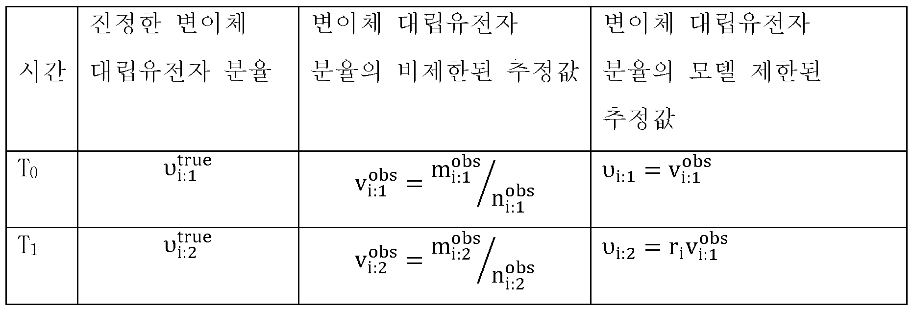
Pi(Ri)는 하기와 같이 결정될 수 있다:P i (R i ) can be determined as follows:
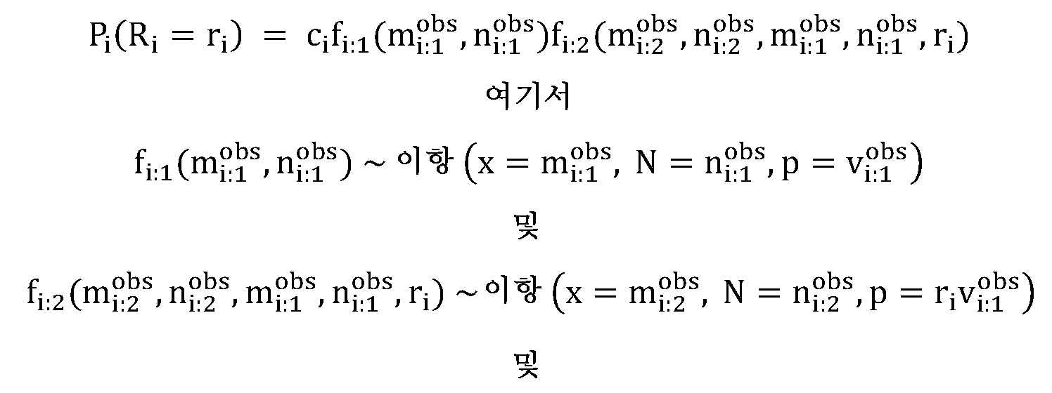
ci는 후보 ri 값의 지지에 걸쳐 Pi(Ri = ri)의 수치적 통합이 1과 같도록 계산된다. 이러한 예시 실시양태는 데이터가 이항 모델에 관하여 과-분산되지 않고, 베타-이항 모델의 보다 일반적인 부류의 특수한 경우에 상응함을 가정한다.c i is computed such that the numerical integration of P i (R i = r i ) over the support of candidate r i values is equal to 1. This example embodiment assumes that the data are not over-dispersed with respect to the binomial model and correspond to a special case of the more general class of beta-binomial models.
대략적 신뢰 구간은 Pi(Ri = ri)에 대한 스케일링된 가능도가 Ri 값에 대한 부적절한 사전 분포를 가정하여 Ri의 대략적 후방 밀도 추정값인 것으로 간주되는 접근법과 같은 가장 높은 밀도 구간을 통해서를 포함하는 다양한 방식으로 Ri에 대해 계산될 수 있다.An approximate confidence interval is the highest density interval such that the scaled likelihood for P i (R i = r i ) is assumed to be an approximate posterior density estimate of R i assuming an inappropriate prior distribution for R i values. R i can be calculated in a variety of ways, including through
단계 (302)에서, 돌연변이/변이체의 세트는 P i (R i )에 따라 쌍별로 병합될 수 있다. 모든 가능한 쌍 {i', i*:i'≠i*; i',i* = 1,2,..., I mv }에 대해, P i' (R i' ), 및 P i* (R i* ) 사이의 차이점 척도 D(i',i*)는 변형된 바타차야 거리를 사용하여 계산된다. D(i',i*)의 보다 큰 값은 돌연변이 쌍 {i',i*}이 동일한 기저 분율 변화 분포로부터의 실현일 가능성이 보다 많음을 지시한다. 따라서, D(·,·)의 가장 큰 값을 갖는 돌연변이/변이체의 쌍은 단일 클론으로 병합될 수 있고, 그 클론에 대한 P i (R i )는 갱신될 수 있다. 쌍별 병합은 정지 기준이 충족되거나 모든 돌연변이/변이체가 단일 클론으로 병합될 때까지 계속될 수 있다. 임계치는 약 0.0005 내지 0.005의 범위의 값이고/거나 이를 포함할 수 있다.At
단계 (303)에서, 클론의 수 및 시점 사이의 연관된 분율 변화는 신뢰 구간과 함께 보고될 수 있다. 미리 결정된 임계치 값 이상의 제1 및 제2 시점 사이의 분율 변화를 갖는 클론이 확인될 수 있다. 다중 클론이 확인되는 경우, 1에 가까운 분율 변화를 갖는 클론 및/또는 특이적인 공지된 CHIP 변이체를 갖는 클론은 잠재적 CHIP 변이체로서 분류될 수 있다. CHIP 변이체는 추가의 분석으로부터 배제될 수 있다. 일부 실시양태에서, 변이체는 그러한 변이체가 적어도 하나의 환자 샘플에서 CHIP로서 분류되는 경우 CHIP 변이체로서 분류될 수 있다.At step 303, the number of clones and the associated fraction change between time points may be reported along with confidence intervals. Clones with a fractional change between the first and second time points above a predetermined threshold value may be identified. If multiple clones are identified, clones with a fraction change close to 1 and/or clones with a specific known CHIP variant can be classified as potential CHIP variants. CHIP variants can be excluded from further analysis. In some embodiments, a variant may be classified as a CHIP variant if such variant is classified as CHIP in at least one patient sample.
도 4는 CHIP 필터 (300)의 예시 적용을 제시한다. 도 4는 병합 절차의 예에 상응한다. 예에서, 3개의 자격화된 돌연변이체가 확인된다. 도 4의 가장 좌측 패널은 R의 지지 (x-축)에 걸쳐 각각의 돌연변이체 (y-축)에 대한 스케일링된 가능도 함수를 나타낸다. 각각의 돌연변이체에 대한 스케일링된 가능도 함수의 제1 가능도 (라인 (403))에 상응하는 돌연변이체가 공지된 CHIP 돌연변이라고 가정한다. 가장 유사성을 갖는 좌측 패널 내의 돌연변이체는 별표로 표시된다. 중간 패널은 좌측 패널 내의 클론에 대한 스케일링된 가능도 함수의 제1 가능도 (라인 (403)) 및 제2 가능도 (라인 (401))의 병합으로부터의 생성된 병합된 가능도를 나타낸다. 좌측 패널로부터의 각각의 클론에 대한 스케일링된 가능도 함수의 제3 가능도 (라인 (402))는 병합에 의해 변경되지 않는 가능도 함수를 갖는다. 우측 패널은 최종 클론성을 나타낸다. 제2 가능도 (라인 (401)) 클론의 조성은 50% CHIP이기 때문에, 제2 가능도 (라인 (401)) 클론은 추정적으로 CHIP로서 확인될 수 있다. 이는 제3 가능도 (라인 (402)) 클론에 의해서만 정의되는 R의 최종 값을 초래할 것이다. Figure 4 presents an example application of
한 실시양태에서, 일부 실시양태에 따른 암을 갖는 대상체에서 클론성 조혈 변이체를 확인하는 예시적인 방법 단계를 개략적으로 도시하는 흐름도가 도 5에 제시된다. 제시된 바와 같이, 방법 (500)은 제1 및 제2 시점에서 대상체로부터 수득된 샘플 내의 하나 이상의 암 유형과 연관된 표적화된 핵산으로부터 생성된 서열 정보로부터 각각의 복수의 변이체에 대한 종양 분율 변화에 대한 종양 부하 변화 (R) P(R)을 결정하여 종양 부하 변화의 세트를 생성하는 것을 포함한다 (단계 (501)). 또한, 방법 (500)은 종양 부하 변화의 세트로부터 하나 이상의 클론성 조혈 변이체에 상응하는 하나 이상의 저항성 시그니쳐를 확인하는 것을 또한 포함한다 (단계 (502)).In one embodiment, a flow diagram schematically depicting exemplary method steps for identifying clonal hematopoietic variants in a subject with cancer according to some embodiments is presented in Figure 5 . As presented,
f. MR 점수f. MR score
도 1로 돌아가서, 방법 (100)은 단계 (110)에서 MR 점수를 결정하는 것으로 진행할 수 있다. 한 실시양태에서, MR 점수는 단계 (109)에서의 변이체 필터링 후에 남아 있는 체세포 변이체와 연관된 MAF 값을 사용하여 결정될 수 있다. 한 실시양태에서, 모든 체세포 변이체의 MAF 값이 사용될 수 있다. 한 실시양태에서, 모든 체세포 변이체 미만의 MAF 값이 사용될 수 있다. 단계 (105/106)에서 기술된 바와 같이, T0 (예를 들어, 치료전) 및 T1 (예를 들어, 치료중)에서 대상체로부터 수득된 샘플 내의 하나 이상의 암 유형과 연관된 표적화된 핵산으로부터 생성된 서열 판독물로부터의 복수의 체세포 변이체에 대해 MAF를 결정하여 복수의 체세포 변이체 내의 체세포 변이체에 대한 제1 및 제2 MAF의 세트를 생성할 수 있다. MR 점수는 분율로서 또는 백분율로서 표현될 수 있다. 도 6A에 제시된 바와 같이, MR 점수는 방법 (600)에 따라 결정될 수 있다. 방법 (600)은 단계 (601)에서 복수의 체세포 변이체 내의 체세포 변이체에 대한 제1 MAF 및 제2 MAF의 비를 결정하여 MAF 비의 세트 및 MAF 비의 세트 내의 MAF 비에 대한 상응하는 표준 편차를 생성하는 것을 포함할 수 있다. 일부 실시양태에서, 표준 편차는 MR 점수를 보고하기 위한 기준으로서 이용될 수 있다. 예를 들어, 적어도 하나의 변이체의 개별적인 표준 편차에 기초하여, MR 점수의 표준 편차를 사용하여 신뢰 구간 및 샘플 평가성을 위한 후속 컷오프를 결정할 수 있다. 일부 실시양태에서, 컷오프는 적어도 0.1, 0.15, 0.2, 0.3, 0.4 또는 0.5일 수 있다. 단계 (602)에서, 대상체에 대해, MAF 비의 가중 평균은 하기 식을 사용하여 결정될 수 있다:Returning to Figure 1 ,

여기서 중량은 복수의 체세포 변이체 내의 주어진 체세포 변이체에 대한 1/범위^2이고, 여기서 범위는 복수의 체세포 변이체 내의 주어진 체세포 변이체에 대한 제1 및 제2 MAF의 값 사이의 차이이고, 비는 MAF 비의 세트 내의 주어진 MAF 비이다. 신뢰 구간은 하기 식을 사용하여 결정될 수 있다:where weight is 1/range^2 for a given somatic variant within a plurality of somatic variants, where range is the difference between the values of the first and second MAF for a given somatic variant within a plurality of somatic variants, and ratio is the MAF ratio. is a given MAF ratio within the set of . The confidence interval can be determined using the formula:
MAF 비의 가중 평균 +/- ,Weighted average of MAF ratio +/- ,
여기서 비 분산은 ![]()
![]()
한 실시양태에서, MR 점수로서 MAF 비의 가중 평균에 추가적으로, 또는 그에 대한 대안으로서, MAF 비에 기초하여 변이체를 클러스터링하고, 클러스터에 대한 합계 MAF 비를 계산하고, 이어서 MR 점수로서 단일 선택된 클러스터 비 또는 클러스터 비의 가중 평균 중 한쪽을 사용하는 방법이 개시된다. 클러스터링은 중첩하는 MAF 비 분포를 갖는 변이체의 쌍을 조합하는 것, 또는 다른 클러스터링 방법에 의해 수행될 수 있다. 단일 선택된 클러스터는 공지된 암 유도자 변이체, 또는 공지된 클론성 조혈 변이체의 부재를 함유하는 것일 수 있다. 클러스터 중량은 클러스터 내의 공지된 암 유도자 변이체의 존재 또는 변이체의 최대 VAF 또는 수에 또한 좌우될 수 있다.In one embodiment, in addition to, or as an alternative to, a weighted average of the MAF ratios as the MR score, variants are clustered based on the MAF ratios, a summed MAF ratio for the cluster is calculated, and then a single selected cluster ratio is calculated as the MR score. Alternatively, a method of using one of the weighted averages of cluster ratios is disclosed. Clustering can be performed by combining pairs of variants with overlapping MAF ratio distributions, or other clustering methods. A single selected cluster may be one containing a known cancer-inducing variant, or the absence of a known clonal hematopoietic variant. Cluster weight may also depend on the presence of known cancer-inducing variants within the cluster or the maximum VAF or number of variants.
도 6B에 제시된 바와 같이, MR 점수는 방법 (610)에 따라 결정될 수 있다. 방법 (610)은 단계 (601)에서 복수의 체세포 변이체 내의 체세포 변이체에 대한 제1 MAF의 가중 평균 및 제2 MAF의 가중 평균 및 가중 MAF 비에 대한 상응하는 표준 편차를 결정하는 것을 포함할 수 있다. 일부 실시양태에서, 표준 편차는 MR 점수를 보고하기 위한 기준으로서 이용될 수 있다. 예를 들어, 적어도 하나의 변이체의 개별적인 표준 편차에 기초하여, MR 점수의 표준 편차를 사용하여 신뢰 구간 및 샘플 평가성을 위한 후속 컷오프를 결정할 수 있다. 일부 실시양태에서, 컷오프는 적어도 0.1, 0.15, 0.2, 0.3, 0.4 또는 0.5일 수 있다. 단계 (612)에서, 대상체에 대해, MAF의 가중 평균의 비가 결정될 수 있다. 비의 분산으로서 신뢰 구간. 예를 들어, 신뢰 구간은 하기 식을 사용하여 결정될 수 있다:As shown in Figure 6B , the MR score may be determined according to method 610. The method 610 may include determining, at step 601, a weighted average of the first MAF and a weighted average of the second MAF and the corresponding standard deviation for the weighted MAF ratio for the somatic variants within the plurality of somatic variants. . In some embodiments, standard deviation can be used as a basis for reporting MR scores. For example, based on the individual standard deviation of at least one variant, the standard deviation of the MR score can be used to determine confidence intervals and subsequent cutoffs for sample evaluability. In some embodiments, the cutoff can be at least 0.1, 0.15, 0.2, 0.3, 0.4, or 0.5. At step 612, for the subject, the ratio of the weighted average of the MAF may be determined. Confidence interval as the variance of the ratio. For example, a confidence interval can be determined using the formula:
R = A/B: var(R) ~= var(B)/A^2 + var(A)*B^2/A^4R = A/B: var(R) ~= var(B)/A^2 + var(A)*B^2/A^4
여기서 A 및 B는 각각 시점 1 및 시점 2에서의 가중 평균 MAF이다.where A and B are the weighted average MAF at
클러스터는 증거의 강도에 기초하여 가중화될 수 있다. 예를 들어, max-VAF는 어느 것이 1차 클론인지 지시할 수 있고, 비-CHIP 변이체의 수는 보다 강한 신호를 갖는 클러스터를 가중화할 수 있고; 유도자 중량은 중량을 증가시키거나 또는 그 특정한 암 유형 또는 분자 하위유형에 대한 유도자를 함유하는 클러스터를 선택할 수 있다. 적용된 가중화는 예를 들어, 특이적 암 유형 또는 분자 하위유형 내의 유도자인 것으로 공지된 변이체에 보다 큰 중량을 적용하고 있을 수 있다. 한 실시양태에서, 중량은 max-VAF (한쪽 샘플), 비-CHIP 변이체의 수, 및/또는 유도자 중량 (종양-유형-특이적; 배치 파일에서 정의됨)에 기초할 수 있다. 또 다른 실시양태에서, 적용된 가중화는 예를 들어, 체세포 변이체를 동등하게 가중화하는 것일 수 있다.Clusters can be weighted based on the strength of evidence. For example, max-VAF can indicate which is the primary clone, and the number of non-CHIP variants can weight clusters with stronger signals; Inducer weight can increase the weight or select clusters containing inducers for that particular cancer type or molecular subtype. The weighting applied may be, for example, applying greater weight to variants known to be inducers within a specific cancer type or molecular subtype. In one embodiment, the weight may be based on max-VAF (one sample), number of non-CHIP variants, and/or inducer weight (tumor-type-specific; defined in the batch file). In another embodiment, the weighting applied may be, for example, weighting somatic variants equally.
한 실시양태에서, 분자 반응자 또는 분자 비-반응자로서의 분류는 변이체 VAF 및 변이체 중량에 좌우될 수 있다. 예를 들어, MR 점수가 평균 VAF의 비인 경우, 보다 높은 VAF (즉, 보다 많은 클론성 변이체)가 우세할 가능성이 있다. MR 점수가 변이체 중량을 사용하는 경우, 보다 높은 중량을 갖는 변이체 (예를 들어, 유도자 변이체)가 우세할 수 있다.In one embodiment, classification as a molecular responder or molecular non-responder may depend on variant VAF and variant weight. For example, if the MR score is a ratio of the average VAF, the higher VAF (i.e., more clonal variants) are likely to dominate. When the MR score uses variant weight, variants with higher weights (e.g., inducer variants) may dominate.
도 6A에 기술된 바와 같은 생성된 MAF 비의 가중 평균 또는 도 6B에 기술된 바와 같은 MAF의 가중 평균의 비는 대상체에 대한 MR 점수이다. 이같은 MR 점수는 MAF의 분산을 분자 반응 계산 내로 혼입한다. 이는 분자 반응 점수가 분자 반응으로부터 정확한 결론을 도출하는데 기여하는 정확한 분산을 포함하는 것을 보장한다. MR 점수는 MAF 내의 정밀도에 기초하여 MAF의 변화를 적절하게 가중화하고, MAF가 검출의 한계 (LOD) 부근에서 변동하고 있는 경우 과신뢰적이고 부정확한 결과를 허용하지 않은 평균 MAF의 "수치적으로 안정한" 비로서 검토될 수 있다. MR 점수를 임계치와 비교하여 대상체가 치료에 반응하고 있는지 또는 치료에 반응하고 있지 않는지 결정할 수 있다. 임계치는 예를 들어, 약 25% 내지 약 75%이고/거나 이를 포함할 수 있다. 일부 실시양태에서, 가중화는 VAF 정밀도 (예를 들어 위치, 핫스팟 영역, 커버리지 깊이 등) 또는 종양에 대한 그 변이체의 중요성의 사전 지식 (예를 들어 공지된 유도자 또는 저항성 돌연변이, 또는 불확실한 (또는 미지의) 유의성의 변이체)에 기초할 수 있다.The weighted average of the generated MAF ratios as described in FIG. 6A or the weighted average of the MAF ratios as described in FIG. 6B is the MR score for the subject. This MR score incorporates the variance of MAF into the molecular response calculation. This ensures that the molecular response score contains the correct variance that contributes to drawing accurate conclusions from the molecular response. The MR score is a "numerical" measure of the average MAF that appropriately weights changes in the MAF based on the precision within the MAF and does not allow for overconfident and inaccurate results if the MAF is fluctuating near the limit of detection (LOD). It can be considered as a “stable” ratio. The MR score can be compared to a threshold to determine whether the subject is responding or not responding to treatment. The threshold may be and/or include, for example, about 25% to about 75%. In some embodiments, weighting may be performed based on VAF precision (e.g. location, hotspot area, depth of coverage, etc.) or prior knowledge of the significance of that variant to the tumor (e.g. known inducer or resistance mutation, or uncertain (or unknown) ) can be based on the variant of significance).
본원에 제시된 MR 점수화 방법이 다루는 문제의 측면을 예시하는 간단한 예를 제공하기 위해, 기준선 (T0)에서 0.3%의 MAF, 및 치료중 (T1)에 MAF 0.1%, 및 3000개의 분자의 그 변이체 위치에서의 커버리지를 갖는 검출되는 하나의 변이체를 갖는 대상체를 고려한다. 기존의 방법을 사용하면, 분자 반응 점수는 일 것이다. 50%의 "분자 반응자" 대 "분자 비-반응자"를 정의하기 위한 컷오프에 대해, 이 대상체는 "분자 반응자"일 것이다. 그러나, 본원에 기술된 방법에 따라 분산을 전파하는 것은 ~30 내지 40%의 예상된 값을 갖는 분자 반응 점수, 그러나 0 내지 120%의 95% 신뢰 구간을 초래한다. 따라서, 이 대상체에 대해, 분자 반응은, MR 점수가 진정하게 50% 컷오프 미만 또는 초과인지 여부가 확신 있게 평가될 수 없기 때문에, 평가가능하지 않은 것으로 간주되어야 한다.To provide a simple example illustrating aspects of the problem addressed by the MR scoring method presented herein, a MAF of 0.3% at baseline (T 0 ), and a MAF of 0.1% on treatment (T 1 ), and a MAF of 3000 molecules. Consider a subject with one variant detected with coverage at the variant location. Using conventional methods, the molecular response score is would. For a cutoff to define “molecular responder” versus “molecular non-responder” of 50%, this subject would be a “molecular responder.” However, propagating variance according to the method described herein results in molecular response scores with expected values of ˜30-40%, but 95% confidence intervals of 0-120%. Therefore, for this subject, the molecular response should be considered not evaluable because it cannot be confidently assessed whether the MR score is truly below or above the 50% cutoff.
본원에 제시된 MR 점수화 방법이 다루는 문제의 측면을 예시하는 간단한 예를 제공하기 위해, 기준선 (T0)에서 a = 0.1% 및 b = 8.0%의 MAF, 및 치료중 (T1)에 a = 0.3% 및 b = 2.0%의 MAF를 갖는 2개의 변이체 (a 및 b)가 검출된 대상체를 고려한다. 비의 평균을 취하는 기존의 방법을 사용하면, 분자 반응 점수는 ![]()
![]()
![]()
![]()
본원에 제시된 MR 점수화 방법이 다루는 문제의 측면을 예시하는 간단한 예를 제공하기 위해, 기준선 (T0)에서 a = 0.3% 및 b = 0.0%의 MAF, 및 치료중 (T1)에 a = 0.0% 및 b = 0.3%의 MAF를 갖는 2개의 변이체 (a 및 b)가 검출된 대상체를 고려한다. 단지 기준선에서 0.3% 초과의 변이체만을 평가하는 기존의 방법을 사용하면, 분자 반응 점수는 ![]()
![]()
![]()
![]()
방법 (100)은 적어도 분자 반응 점수에 기초하여 하나 이상의 요법을 대상체에게 투여하는 것을 포함할 수 있다. 예시적인 요법은 본원에 추가로 개시된다. 일부 실시양태에서, 방법 (100)은 암을 갖는 대상체에 대한 분자 반응 점수를 미리 결정된 컷오프 포인트와 비교하여, 분자 반응 점수가 미리 결정된 컷오프 포인트 미만인 경우 대상체가 암에 대한 하나 이상의 요법 (예를 들어, 면역요법 등)에 대한 가능성 있는 반응자인 것 또는 분자 반응 점수가 미리 결정된 컷오프 포인트 이상인 경우 대상체가 암에 대한 하나 이상의 요법에 대한 가능성 있는 비-반응자인 것을 확인하는 것을 포함한다. 일부 실시양태에서, 방법 (100)은 분자 반응 점수를 고려하여 암에 대한 하나 이상의 요법을 대상체에게 투여하는 것을 포함한다. 일부 실시양태에서, 방법 (100)은 분자 반응 점수를 고려하여 암에 대한 하나 이상의 요법을 대상체에게 투여하는 것을 중단하는 것을 포함한다. 일부 실시양태에서, 방법 (100)은 분자 반응 점수를 대상체에 대한 예후적 바이오마커 및/또는 예측적 바이오마커로서 사용하는 것을 포함한다.
다른 예시적인 실시양태에서, 분산을 적어도 하나의 변이체의 분산 분포로부터의 시뮬레이션 또는 샘플링을 통해 분자 반응 계산 내로 혼입하여 분자 반응 분산을 계산한다. 본원에 추가로 개시된 바와 같이, 일부 적용은 종양 또는 종양의 가능성 대 클론성 조혈에 있어서의 그들의 중요성에 기초하여 변이체를 가중화하는 것을 포함한다. 일부 실시양태는 다중 게놈 데이터 공급원을 통합하여 다른 게놈 데이터 공급원 중에서도 종양 분율 (단지 변이체 (예를 들어, SNV, 인델 및 융합) VAF에 의존하는 대신), 커버리지 (예를 들어, 카피 수), 오프-타겟 커버리지, 및/또는 메틸화를 추정하는 것을 수반한다.In another exemplary embodiment, the molecular response variance is calculated by incorporating the variance into the molecular response calculation through simulation or sampling from the variance distribution of at least one variant. As further disclosed herein, some applications include weighting variants based on a tumor or tumor likelihood versus their importance in clonal hematopoiesis. Some embodiments integrate multiple genomic data sources to determine tumor fraction (e.g., variants (e.g., SNVs, indels, and fusions)), coverage (e.g., copy number), tumor fraction (instead of relying only on variants (e.g., SNVs, indels, and fusions) VAF), -Involves estimating target coverage, and/or methylation.
일부 실시양태에서, 방법은 하나 이상의 추가의 게놈 데이터 공급원을 사용하여 암을 갖는 대상체에 대한 분자 반응 점수를 결정하는 것을 포함한다. 일부 실시양태에서, 추가의 게놈 데이터 공급원은 커버리지, 오프-타겟 커버리지, 후성유전학적 시그니쳐, 종양 돌연변이 부담 및/또는 미세부수체 불안정성 점수 중 하나 이상을 포함한다. 데이터 공급원에 대해, 그 데이터 공급원에 기초하여 종양 분율의 계산이 있을 수 있고, 계산된 종양 분율은 데이터 공급원에 걸쳐 조합될 수 있고 (예를 들어 가중 평균을 사용하여, 그 특정한 샘플에 대한 종양 분율 내의 데이터 공급원의 신뢰성을 혼입함), 이어서 샘플 내의 전체 종양 분율 추정값을 조합하여 전체 분자 반응을 계산할 수 있다. 일부 실시양태에서, 후성유전학적 시그니쳐는 cfNA 단편 길이, 위치, 및/또는 종점 밀도 분포를 포함한다. 일부 실시양태에서, 후성유전학적 시그니쳐는 주어진 표적화된 게놈 영역 내의 하나 이상의 후성유전학적 유전자좌에 의해 나타내어지는 후성유전학적 상태 또는 상황을 포함한다. 일부 실시양태에서, 후성유전학적 상태 또는 상황은 메틸화, 히드록시메틸화, 아세틸화, 유비퀴틸화, 인산화, 수모일화, 리보실화, 시트룰린화, 및/또는 히스톤 번역후 변형 또는 다른 히스톤 변이의 존재 또는 부재를 포함한다.In some embodiments, the method includes determining a molecular response score for a subject with cancer using one or more additional sources of genomic data. In some embodiments, the additional genomic data source includes one or more of coverage, off-target coverage, epigenetic signature, tumor mutational burden, and/or microsatellite instability score. For a data source, there may be a calculation of a tumor fraction based on that data source, and the calculated tumor fractions may be combined across data sources (e.g., using a weighted average) to determine the tumor fraction for that particular sample. incorporating the reliability of the data source within the sample), estimates of the overall tumor fraction within the sample can then be combined to calculate the overall molecular response. In some embodiments, the epigenetic signature includes cfNA fragment length, location, and/or endpoint density distribution. In some embodiments, an epigenetic signature comprises an epigenetic state or condition indicated by one or more epigenetic loci within a given targeted genomic region. In some embodiments, the epigenetic state or condition is the presence of methylation, hydroxymethylation, acetylation, ubiquitination, phosphorylation, sumoylation, ribosylation, citrullination, and/or histone post-translational modification or other histone modifications Includes absence.
본 방법은 도 1 및 제1 시간 T0 및 제2 시간 T1의 맥락에서 기술되지만, 예를 들어 종적 모니터링을 위해, 2개 초과의 시점이 구상됨을 이해하여야 한다. 도 7에 제시된 바와 같이, 제1 시간 T0에서, 기준선 cfDNA는 치료 전에 하나 이상의 대상체로부터 수득된 하나 이상의 기준선 샘플로부터 수득될 수 있고, 제2 시간 T1, 또는 임의의 후속 시간 Tn에서, 치료중 cfDNA는 치료 후에 하나 이상의 대상체로부터 수득된 하나 이상의 치료중 샘플로부터 수득될 수 있다. 시간 T1은 시간 T0 후 임의의 양의 시간, 예를 들어, 1 내지 24시간, 1 내지 180일, 1 내지 12주, 1 내지 25주, 1 내지 30주 등 사이의 및 이를 포함하는 임의의 시간일 수 있다. 더욱이, 방법 (100)은 시간 T0, T1, ..., Tn의 임의의 조합에 적용될 수 있다. 예를 들어, 샘플은 시간 T1에서 및 시간 T2에서 수득될 수 있고, 여기서 양쪽 모두의 시간에서 취한 샘플은 치료중 샘플이다. 또 다른 예에서, 샘플은 시간 T1에서 및 시간 T2에서 수득될 수 있고, 여기서 시간 T1에서 취한 샘플은 치료중 샘플을 나타내고, 시간 T2에서 취한 샘플은 치료가 끝난 샘플을 나타낸다.Although the method is described in Figure 1 and in the context of a first time T 0 and a second time T 1 , it should be understood that more than two time points are envisioned, for example for longitudinal monitoring. As shown in FIG. 7 , at a first time T 0 , baseline cfDNA may be obtained from one or more baseline samples obtained from one or more subjects prior to treatment, and at a second time T 1 , or at any subsequent time T n On-treatment cfDNA can be obtained from one or more on-treatment samples obtained from one or more subjects after treatment. Time T 1 is any amount of time after time T 0 , for example, any amount of time between and including 1 to 24 hours, 1 to 180 days, 1 to 12 weeks, 1 to 25 weeks, 1 to 30 weeks, etc. It may be the time of. Moreover,
한 실시양태에서, 대상체에게 투여되는 요법의 투여량은 분자 반응 점수에 기초하여 조정될 수 있다. 예를 들어, 분자 반응 점수는 대상체가 제1 치료에 반응하고 있지 않음을 지시할 수 있고, 제1 치료의 투여량은 반응에서 증가될 수 있다. 한 실시양태에서, 대안적 요법은 분자 반응 점수에 기초하여 확인될 수 있다. 예를 들어, 분자 반응 점수는 대상체가 제1 치료에 대해 반응하고 있지 않음을 지시할 수 있고, 대상체는 이어서 제1 치료 대신, 또는 그에 추가로 제2 치료에 처해질 수 있다. 한 실시양태에서, 분자 반응 점수는 임상 시험에서 대상체에 대해 결정될 수 있고, 여기서 분자 반응 점수는 위약을 받는 대상체에 대해 및 치료를 받는 대상체에 대해 결정될 수 있다. 대상체의 2개의 카테고리의 분자 반응 점수를 비교하여 치료를 평가할 수 있다.In one embodiment, the dosage of therapy administered to a subject can be adjusted based on the molecular response score. For example, a molecular response score may indicate that a subject is not responding to a first treatment, and the dose of the first treatment may be increased upon response. In one embodiment, alternative therapies may be identified based on molecular response scores. For example, a molecular response score may indicate that a subject is not responding to a first treatment, and the subject may then be given a second treatment instead of, or in addition to, the first treatment. In one embodiment, a molecular response score may be determined for subjects in a clinical trial, where the molecular response score may be determined for subjects receiving a placebo and for subjects receiving treatment. Treatment can be evaluated by comparing the subject's two categories of molecular response scores.
또 다른 예에서, 위약 및 치료는 약물의 상이한 조합을 비교하는 임상 시험의 2개의 부문으로 일반화될 수 있다. 임계치 또는 컷오프는 사용 사례에 특이적일 수 있다: 사용 사례는 청소 (MR=0)를 요구할 수 있거나, 또는 사용 사례는 ctDNA 수준의 감소 또는 증가의 특정 수준을 요구할 수 있다.In another example, placebo and treatment can be generalized as two arms of a clinical trial comparing different combinations of drugs. The threshold or cutoff may be specific to the use case: the use case may require clearing (MR=0), or the use case may require a specific level of reduction or increase in ctDNA levels.
도 8은 환자 계층화를 위한 분자 반응 점수의 예시 실제적 적용을 제시한다. 진행성 암 환자는 치료 전에 시간 T0에서 결정된 기준선 MAF를 가질 수 있다. 4 내지 10주의 치료 후, 진행성 암 환자는 시간 T1에서 결정된 치료중 MAF를 가질 수 있다. 생성된 분자 반응 점수는 환자 내의 ctDNA가 감소하고 있음을 지시할 수 있으며, 이 경우 환자는 1차 시험 약물로 치료되기를 계속해야 한다. 생성된 분자 반응 점수는 환자 내의 ctDNA가 증가하고 있음을 지시할 수 있으며, 이 경우 환자는 환자가 대조군 그룹에 있는 경우 1차 시험 약물로 (또는 위약으로) 치료되기를 계속해야 한다. 다른 방식으로, 환자 내의 ctDNA가 증가하고 있는 경우, 환자는 그들의 치료 요법에 부가되는 하나 이상의 요법, 변경된 요법, 또는 변경된 1차 시험 약물의 용량을 가져야 한다. Figure 8 presents an example practical application of molecular response scores for patient stratification. Patients with advanced cancer may have a baseline MAF determined at time T 0 prior to treatment. After 4 to 10 weeks of treatment, patients with advanced cancer may have an on-treatment MAF determined at time T 1 . The resulting molecular response score may indicate that ctDNA in the patient is decreasing, in which case the patient should continue to be treated with the first-line investigational drug. The resulting molecular response score may indicate that ctDNA in the patient is increasing, in which case the patient should continue to be treated with the first study drug (or placebo) if the patient is in the control group. Alternatively, if ctDNA in a patient is increasing, the patient should have one or more regimens added to their treatment regimen, a modified regimen, or a modified dose of the primary study drug.
도 9는 임상 시험 풍부화를 위한 분자 반응 점수의 예시 실제적 적용을 제시한다. 치료의 표준 (SOC) 치료에 대해 적격인 진행성 암 환자는 SOC 치료 전에 시간 T0에서 결정된 기준선 MAF를 가질 수 있다. 4 내지 10주의 SOC 치료 후, 진행성 암 환자는 시간 T1에서 결정된 치료중 MAF를 가질 수 있다. 생성된 분자 반응 점수는 환자 내의 ctDNA가 감소하고 있음을 지시할 수 있으며, 이 경우 환자는 SOC 치료로 치료되기를 계속해야 한다. 생성된 분자 반응 점수는 환자 내의 ctDNA가 증가하고 있음을 지시할 수 있으며, 이 경우 환자는 임상 시험 약물로의 치료에 적격인 것으로 결정될 수 있다. Figure 9 presents an example practical application of molecular response scores for clinical trial enrichment. Patients with advanced cancer who are eligible for standard of care (SOC) treatment may have a baseline MAF determined at time T 0 prior to SOC treatment. After 4 to 10 weeks of SOC treatment, patients with advanced cancer may have an on-treatment MAF determined at time T 1 . The resulting molecular response score may indicate that ctDNA in the patient is decreasing, in which case the patient should continue to be treated with SOC therapy. The resulting molecular response score may indicate that ctDNA in the patient is increasing, in which case the patient may be determined to be eligible for treatment with the investigational drug.
도 10은 EGFR-양성 비-소세포 폐암 (NSCLC)을 갖는 환자에서 오시메르티닙 +/- 화학요법의 MSKCC 시험을 위한 전향적 환자 계층화 및 증량에 대한 분자 반응 점수의 예시 실제적 적용을 제시한다. EGFR-양성 NSCLC를 갖는 새롭게 진단된 환자는 오시메르티닙 전에 시간 T0에서 결정된 기준선 MAF를 가질 수 있다. 1 사이클의 오시메르티닙 후, 환자는 오시메르티닙의 사이클 2의 제1일에 결정된 치료중 MAF를 가질 수 있다. 단지 EGFR 유도자에 기초하여, 생성된 분자 반응 점수는 EGFR 유도자가 검출되지 않음을 지시할 수 있으며, 이 경우 환자는 단지 오시메르티닙으로 치료되기를 계속해야 한다. 단지 EGFR 유도자에 기초하여, 생성된 분자 반응 점수는 EGFR 유도자가 검출됨을 지시할 수 있으며, 이 경우 환자는 오시메르티닙, 카르보플라틴, 및 페메트렉시드로 치료되기를 계속해야 한다. Figure 10 shows EGFR- positive non-small cell lung cancer. We present an illustrative practical application of the molecular response score for prospective patient stratification and dosing escalation for the MSKCC trial of osimertinib +/- chemotherapy in patients with NSCLC. Newly diagnosed patients with EGFR- positive NSCLC may have a baseline MAF determined at time T 0 before osimertinib. After one cycle of osimertinib, patients may have an on-treatment MAF determined on
이러한 방법의 측면은 도 11에 추가로 예시된다. 제시된 바와 같이, 방법 (1100)은 제1 (예를 들어, 치료전) 및 제2 (예를 들어, 치료중) 시점에서 대상체로부터 수득된 샘플 내의 하나 이상의 암 유형과 연관된 표적화된 핵산으로부터 생성된 서열 정보로부터 복수의 변이체에 대한 돌연변이체 대립유전자 빈도 (MAF)를 결정하여 복수의 변이체 내의 변이체에 대한 제1 및 제2 MAF의 세트를 생성하는 것을 포함한다 (단계 (1101)). 방법 (1100)은 복수의 변이체 내의 변이체에 대한 제1 및 제2 MAF의 비를 계산하여 MAF 비의 세트 및 MAF 비의 세트 내의 MAF 비에 대한 상응하는 표준 편차를 생성하는 것을 또한 포함한다 (단계 (1102)). 또한, 방법 (1100)은 MAF 비의 가중 평균 (단계 (1103)) 및 신뢰 구간을 계산하여 암을 갖는 대상체에 대한 분자 반응 점수를 결정하는 것을 또한 포함한다.Aspects of this method are further illustrated in Figure 11 . As presented,
일부 실시양태에서, 방법 (1100)은 암을 갖는 대상체에 대한 분자 반응 점수를 미리 결정된 컷오프 포인트와 비교하여, 분자 반응 점수가 미리 결정된 컷오프 포인트 미만인 경우 대상체가 암에 대한 하나 이상의 요법 (예를 들어, 면역요법 등)에 대한 가능성 있는 반응자인 것 또는 분자 반응 점수가 미리 결정된 컷오프 포인트 이상인 경우 대상체가 암에 대한 하나 이상의 요법에 대한 가능성 있는 비-반응자인 것을 확인하는 것을 포함한다. 일부 실시양태에서, 방법 (1100)은 분자 반응 점수를 고려하여 암에 대한 하나 이상의 요법을 대상체에게 투여하는 것을 포함한다. 일부 실시양태에서, 방법 (1100)은 분자 반응 점수를 고려하여 암에 대한 하나 이상의 요법을 대상체에게 투여하는 것을 중단하는 것을 포함한다. 일부 실시양태에서, 방법 (1100)은 분자 반응 점수를 대상체에 대한 예후적 바이오마커 및/또는 예측적 바이오마커로서 사용하는 것을 포함한다.In some embodiments,
일부 실시양태에서, 방법 (1100)은 분자 카운트를 사용하여 MAF 비의 세트 내의 MAF 비에 대한 표준 편차를 계산하는 것을 포함한다. 일부 실시양태에서, 방법 (1100)은 MAF 비의 세트 내의 MAF 비를 통해 분산을 전파하는 것을 포함한다. 일부 실시양태에서, 방법 (1100)은 복수의 변이체에 대한 돌연변이체 대립유전자 빈도 (MAF)를 결정하는 경우 하나 이상의 배선 및/또는 클론성 조혈 변이체를 배제하는 것을 포함한다. 배선 및 CHIP 변이체를 배제하는 방법의 예는 본원에 추가로 기술된다. 일부 실시양태에서, 방법 (1100)은 제1 및/또는 제2 시점에서 약 0.1%, 0.2%, 0.3%, 0.4%, 0.5%, 0.6%, 0.7%, 0.8%, 또는 0.9% 미만인 MAF를 갖는 하나 이상의 체세포 변이체를 배제하는 것을 포함한다. 일부 실시양태에서, 방법은 제1 및/또는 제2 시점에서 5, 10, 15, 20, 25 또는 30 돌연변이체 분자 카운트 미만의 하나 이상의 체세포 변이체를 배제하는 것을 포함한다. 일부 실시양태에서, 방법은 제1 및/또는 제2 시점에서 300, 400, 500, 600, 700, 800, 900 또는 1000 미만의 커버리지를 갖는 하나 이상의 체세포 변이체를 배제하는 것을 포함한다. 일부 이러한 실시양태에서, 제1 시점은 치료전 시점을 포함하고, 여기서 제2 시점은 치료중 또는 치료후 시점을 포함한다.In some embodiments,
일부 실시양태에서, 본원에 개시된 방법은 샘플 내의 하나 이상의 조직 또는 세포로부터 수득된 핵산 분자로부터 서열 정보를 생성하는 것을 포함한다. 일부 실시양태에서, 본원에 개시된 방법은 대상체로부터 수득된 샘플 내의 무세포 핵산 (cfNA)으로부터 서열 정보를 생성하는 것을 포함한다. 일부 실시양태에서, cfNA는 순환 종양 DNA (ctDNA)를 포함한다.In some embodiments, the methods disclosed herein include generating sequence information from nucleic acid molecules obtained from one or more tissues or cells in a sample. In some embodiments, the methods disclosed herein include generating sequence information from cell-free nucleic acid (cfNA) in a sample obtained from a subject. In some embodiments, cfNA comprises circulating tumor DNA (ctDNA).
일부 실시양태에서, 비는 복수의 변이체 내의 변이체에 대한 제2 MAF 대 제1 MAF를 포함한다. 일부 실시양태에서, 방법 (1100)은 하기 식을 사용하여 MAF 비의 가중 평균을 계산하는 것을 포함한다:In some embodiments, the ratio comprises the second MAF to the first MAF for a variant within a plurality of variants. In some embodiments,
합계[중량 * 비]/합계[중량]Total[Weight * Ratio]/Total[Weight]
여기서 중량은 복수의 변이체 내의 주어진 변이체에 대한 1/범위2이고, 여기서 범위는 복수의 변이체 내의 주어진 변이체에 대한 제1 및 제2 MAF의 값 사이의 차이이고, 비는 MAF 비의 세트 내의 주어진 MAF 비이다. 일부 실시양태에서, 방법 (1100)은 하기 식을 사용하여 신뢰 구간을 계산하는 것을 포함한다:where the weight is 1/range 2 for a given variant within a plurality of variants, where the range is the difference between the values of the first and second MAF for a given variant within the plurality of variants, and the ratio is the given MAF within the set of MAF ratios. It's rain. In some embodiments,
MAF 비의 가중 평균 +/- sqrt[비 분산]Weighted average of MAF ratio +/- sqrt[ratio variance]
여기서 비 분산은 1/합계[중량]이다.Here, the specific variance is 1/sum [weight].
일부 실시양태에서, 변이체는 하나 이상의 단일-뉴클레오티드 변이체 (SNV), 삽입/결실 돌연변이 (인델), 유전자 증폭, 및/또는 유전자 융합을 포함한다. 일부 실시양태에서, 방법 (1100)은 하나 이상의 추가의 게놈 데이터 공급원을 사용하여 암을 갖는 대상체에 대한 분자 반응 점수를 결정하는 것을 포함한다. 일부 실시양태에서, 추가의 게놈 데이터 공급원은 커버리지, 오프-타겟 커버리지, 후성유전학적 시그니쳐, 및/또는 미세부수체 불안정성 점수 중 하나 이상을 포함한다. 일부 실시양태에서, 후성유전학적 시그니쳐는 cfNA 단편 길이, 위치, 및/또는 종점 밀도 분포를 포함한다. 일부 실시양태에서, 후성유전학적 시그니쳐는 주어진 표적화된 게놈 영역 내의 하나 이상의 후성유전학적 유전자좌에 의해 나타내어지는 후성유전학적 상태 또는 상황을 포함한다. 일부 실시양태에서, 후성유전학적 상태 또는 상황은 메틸화, 히드록시메틸화, 아세틸화, 유비퀴틸화, 인산화, 수모일화, 리보실화, 시트룰린화, 및/또는 히스톤 번역후 변형 또는 다른 히스톤 변이의 존재 또는 부재를 포함한다.In some embodiments, the variant comprises one or more single-nucleotide variants (SNVs), insertion/deletion mutations (indels), gene amplifications, and/or gene fusions. In some embodiments,
추가로 예시하기 위해, 도 12A는 예시 방법 (1200)을 개략적으로 도시하는 흐름도이다. 제시된 바와 같이, 방법 (1200)은 제1 및 제2 시점에서 대상체로부터 수득된 샘플 내의 하나 이상의 암 유형과 연관된 표적화된 핵산으로부터 생성된 서열 정보로부터 복수의 변이체에 대한 돌연변이체 대립유전자 빈도 (MAF)를 결정하여 복수의 변이체 내의 변이체에 대한 제1 및 제2 MAF의 세트를 생성하는 것을 포함한다 (단계 (1201)). 방법 (1200)은 복수의 변이체 내의 변이체에 대한 제1 및 제2 MAF의 비를 계산하여 MAF 비의 세트 및 MAF 비의 세트 내의 MAF 비에 대한 상응하는 표준 편차를 생성하고 (단계 (1202)), MAF 비의 가중 평균 및 신뢰 구간을 계산하여 대상체에 대한 분자 반응 점수를 결정하는 것 (단계 (1203))을 또한 포함한다. 일부 실시양태에서, 표준 편차는 신뢰 구간의 추정값으로서 이용될 수 있다. 일부 실시양태에서, 표준 편차는 분자 반응 점수를 보고하기 위한 기준으로서 이용될 수 있다. 또한, 방법 (1200)은 적어도 분자 반응 점수에 기초하여 하나 이상의 요법을 대상체에게 투여하는 것을 또한 포함한다 (단계 (1204)). 예시적인 요법은 본원에 추가로 개시된다.To further illustrate, Figure 12A is a flow diagram schematically depicting an example method 1200. As shown, method 1200 provides mutant allele frequencies (MAFs) for a plurality of variants from sequence information generated from targeted nucleic acids associated with one or more cancer types in samples obtained from a subject at first and second time points. and determining to generate first and second sets of MAFs for variants within the plurality of variants (step 1201). Method 1200 calculates ratios of the first and second MAFs for variants within a plurality of variants to generate a set of MAF ratios and a corresponding standard deviation for the MAF ratios within the set of MAF ratios (step 1202). , calculating a weighted average and confidence interval of the MAF ratio to determine a molecular response score for the subject (step 1203). In some embodiments, the standard deviation can be used as an estimate of the confidence interval. In some embodiments, standard deviation can be used as a basis for reporting molecular response scores. Method 1200 also includes administering one or more therapies to the subject at least based on the molecular response score (step 1204). Exemplary therapies are further disclosed herein.
도 12B는 예시 방법 (1210)을 개략적으로 도시하는 흐름도이다. 제시된 바와 같이, 방법 (1210)은 제1 및 제2 시점에서 대상체로부터 수득된 샘플 내의 하나 이상의 암 유형과 연관된 표적화된 핵산으로부터 생성된 서열 정보로부터 복수의 변이체에 대한 돌연변이체 대립유전자 빈도 (MAF)를 결정하여 복수의 변이체 내의 변이체에 대한 제1 및 제2 MAF의 세트를 생성하는 것을 포함한다 (단계 (1211)). 방법 (1210)은 단계 (1212)에서 시점 (즉, 제1 시점 및 제2 시점)에 대해 고려된 체세포 변이체의 MAF로부터 수득된 중심 경향성 척도를 결정하는 것을 포함한다. 중심 경향성 척도는 평균, 중위값, 또는 최빈값 중 하나일 수 있지만, 이에 제한되지 않음이 이해된다. 방법 (1210)은 단계 (1213)에서 제1 시점에서의 중심 경향성 척도 대 제2 시점에서의 중심 경향성 척도의 비를 결정하는 것을 포함한다. 방법 (1210)은 고려된 MAF의 표준 편차를 사용하여 중심 경향성 비의 표준 편차를 계산하는 것을 포함할 수 있다. 일부 실시양태에서, 중심 경향성 척도는 평균 또는 중위값일 수 있다. 일부 실시양태에서, 중심 경향성 척도는 평균일 수 있다. 일부 실시양태에서, 중심 경향성 척도는 중위값일 수 있다. 일부 실시양태에서, 방법 (1210)은 단계 (1212)에서 각각의 시점 (즉, 제1 시점 및 제2 시점)에 대해 고려된 체세포 변이체의 MAF의 평균을 결정하고; 단계 (1213)에서 제1 시점에서 수득된 평균 대 제2 시점에서 수득된 평균의 비를 계산하고, 고려된 각각의 MAF의 표준 편차를 사용하여 평균 비의 표준 편차를 계산하는 것을 포함한다. 일부 실시양태에서, 분자 반응 점수는 제1 시점에서 수득된 평균 대 제2 시점에서 수득된 평균의 비로부터 계산될 수 있다. 일부 실시양태에서, 방법 (1210)은 단계 (1212)에서 각각의 시점 (즉, 제1 시점 및 제2 시점)에 대해 고려된 체세포 변이체의 MAF의 중위값을 결정하고; 단계 (1213)에서 제1 시점에서 수득된 중위값 대 제2 시점에서 수득된 중위값의 비를 계산하고, 고려된 각각의 MAF의 표준 편차를 사용하여 중위값 비의 표준 편차를 계산하는 것을 포함한다. 일부 실시양태에서, 분자 반응 점수는 제1 시점에서 수득된 중위값 대 제2 시점에서 수득된 중위값의 비로부터 계산될 수 있다. 일부 실시양태에서, 표준 편차는 신뢰 구간의 추정값으로서 이용될 수 있다. 일부 실시양태에서, 표준 편차는 분자 반응 점수를 보고하기 위한 기준으로서 이용될 수 있다. 또한, 방법 (1210)은 적어도 분자 반응 점수에 기초하여 하나 이상의 요법을 대상체에게 투여하는 것을 또한 포함한다 (단계 (1214)). 예시적인 요법은 본원에 추가로 개시된다. FIG. 12B is a flow diagram schematically illustrating an example method 1210. As presented, method 1210 provides mutant allele frequencies (MAFs) for a plurality of variants from sequence information generated from targeted nucleic acids associated with one or more cancer types in samples obtained from a subject at first and second time points. and determining to generate first and second sets of MAFs for variants within the plurality of variants (step 1211). Method 1210 includes determining a measure of central tendency obtained from the MAF of the considered somatic variant for a time point (i.e., a first time point and a second time point) at step 1212. It is understood that the measure of central tendency may be, but is not limited to, one of the mean, median, or mode. Method 1210 includes determining, at step 1213, a ratio of the measure of central tendency at a first time point to the measure of central tendency at a second time point. Method 1210 may include calculating the standard deviation of the central tendency ratio using the standard deviation of the considered MAF. In some embodiments, the measure of central tendency can be the mean or median. In some embodiments, the measure of central tendency can be the average. In some embodiments, the measure of central tendency can be the median. In some embodiments, method 1210 determines the average of the MAF of the considered somatic variants for each time point (i.e., the first time point and the second time point) at step 1212; Step 1213 includes calculating the ratio of the average obtained at the first time point to the average obtained at the second time point, and calculating the standard deviation of the average ratio using the standard deviation of each MAF considered. In some embodiments, the molecular response score can be calculated from the ratio of the average obtained at a first time point to the average obtained at a second time point. In some embodiments, method 1210 determines the median MAF of the considered somatic variants for each time point (i.e., the first time point and the second time point) at step 1212; Step 1213 includes calculating the ratio of the median obtained at the first time point to the median obtained at the second time point, and calculating the standard deviation of the median ratio using the standard deviation of each MAF considered. In some embodiments, the molecular response score can be calculated from the ratio of the median value obtained at a first time point to the median value obtained at a second time point. In some embodiments, the standard deviation can be used as an estimate of the confidence interval. In some embodiments, standard deviation can be used as a basis for reporting molecular response scores. Additionally, method 1210 also includes administering one or more therapies to the subject at least based on the molecular response score (step 1214). Exemplary therapies are further disclosed herein.
전형적으로, 분자 반응 점수를 결정하는 방법은 CHIP 변이체를 필터링하는 것을 포함한다. 예를 들어, 분자 반응은 종양 분율 변화를 나타내는 게놈 교번 (예를 들어, 2개의 시점 사이의 작은 변이체)의 대립유전자 빈도에 의해 전형적으로 측정된다. cfDNA 신호는 종양, 혈액 세포 등을 포함하는 본질적으로 임의의 세포 유형으로부터의 신호의 합계임을 고려하여, 다수의 연구는 cfDNA 샘플 내의 잠재성 불명 클론성 조혈 (CHIP) 변이체의 존재를 제시하였다. CHIP 필터링을 위한 통상적인 접근법은 다양한 데이터 공급원에 의해 엄선된 반복성 CHIP 유전자 또는 핫스팟을 빈번히 레버리징한다. 그러나, 혈장 단독 접근법으로 무작위 CHIP 돌연변이를 확인하는 것은 아직 난제이다. 잔류 비필터링된 CHIP 변이체는 전형적으로 분율 변화를 1을 향해 편향시키고 (변경되지 않음), 따라서 부정확한 분자 반응 예측 또는 점수를 생성한다. 따라서, 일부 실시양태에서, 본원에 개시된 방법은 별개의 분율 변화를 갖는 클론 내의 게놈 돌연변이를 클러스터링하기 위해 2개의 시점 사이의 관찰을 레버리징하는 모델을 사용한다. 돌연변이를 그룹화하기 위해, 이들 접근법은 2개의 시점으로부터의 변이체에 대한 변이체 대립유전자 카운트 및 총 카운트를 전형적으로 레버리징하고, 종양 분율 변화 R에 대한 확률 밀도 함수를 P(R)로서 수립한다.Typically, methods for determining molecular response scores include filtering out CHIP variants. For example, molecular response is typically measured by the allele frequency of genomic alternations (e.g., minor variants between two time points) that represent changes in tumor fraction. Considering that the cfDNA signal is essentially the sum of signals from any cell type, including tumors, blood cells, etc., a number of studies have suggested the presence of clonal hematopoietic (CHIP) variants of unknown occult potential in cfDNA samples. Conventional approaches for CHIP filtering frequently leverage carefully selected repetitive CHIP genes or hotspots from various data sources. However, identifying random CHIP mutations using a plasma-only approach remains challenging. Residual unfiltered CHIP variants typically bias the fraction change toward 1 (unchanged), thus producing inaccurate molecular response predictions or scores. Accordingly, in some embodiments, the methods disclosed herein use models that leverage observations between two time points to cluster genomic mutations within clones with distinct fractional changes. To group mutations, these approaches typically leverage the variant allele counts and total counts for variants from two time points and establish a probability density function for the tumor fraction change, R , as P( R ).
추가의 예시로서, 도 13은 일부 실시양태에 따른 암을 갖는 대상체에서 클론성 조혈 변이체를 확인하는 예시적인 방법 단계를 개략적으로 도시하는 흐름도이다. 제시된 바와 같이, 방법 (1300)은 제1 및 제2 시점에서 대상체로부터 수득된 샘플 내의 하나 이상의 암 유형과 연관된 표적화된 핵산으로부터 생성된 서열 정보로부터 복수의 변이체의 변이체에 대한 종양 분율 변화에 대한 확률 밀도 함수 P(R)을 계산하는 것을 포함한다 (단계 (1301)). 추가적으로 제시된 바와 같이, 방법 (1300)은 변이체의 하나 이상을 P(R)에 의해 하나 이상의 클론으로 그룹화하고 (단계 (1302)), 클론들의 클론에 대한 갱신된 P(R)을 생성하고 (단계 (1303)), 미리 결정된 임계치 값 이상의 제1 및 제2 시점 사이의 분율 변화를 갖는 하나 이상의 클론을 확인하는 것 (단계 (1304))을 포함한다.As a further example, Figure 13 is a flow diagram schematically depicting exemplary method steps for identifying a clonal hematopoietic variant in a subject with cancer according to some embodiments. As presented,
다른 측면에서, 본 개시내용은 배선 변이체를 확인하고 배제하거나, 또는 다른 방식으로 분자 반응 점수를 결정하는 경우 체세포 분류 불일치를 해결하는 방법을 제공한다. 예를 들어, 하나의 문제는 환자의 치료 과정의 과정에 걸쳐 수집된 샘플이 상이한 수준의 종양 쉐딩 및 대립유전자 불균형을 전형적으로 갖는다는 것이며, 이는 주어진 생물정보학 파이프라인의 체세포 변이체 콜러가 때때로 동일한 환자에서 동일한 변이체에 대한 상이한 체세포 분류에 도달할 것임을 의미한다. 분자 반응 결정의 목표는 치료의 과정에 걸쳐 체세포 변이체를 추적하는 것이기 때문에, 임의의 분류 불일치를 해결하여 배선 변이체를 고려로부터 적절하게 제거해야 한다.In another aspect, the present disclosure provides methods for resolving somatic typing discrepancies when identifying and ruling out germline variants, or otherwise determining molecular response scores. For example, one problem is that samples collected over the course of a patient's course of treatment typically have different levels of tumor shedding and allelic imbalance, which means that the somatic variant caller of a given bioinformatics pipeline can sometimes be different for the same patient. means that different somatic classifications will be reached for the same variant. Because the goal of molecular response determination is to track somatic variants over the course of treatment, any classification discrepancies must be resolved to appropriately remove germline variants from consideration.
예시하기 위해, 도 14는 일부 실시양태에 따른 암을 갖는 대상체에서 변이체를 확인하는 예시적인 방법 단계를 개략적으로 도시하는 흐름도이다. 제시된 바와 같이, 방법 (1400)은 대상체로부터 수득된 샘플 내의 하나 이상의 암 유형과 연관된 표적화된 핵산으로부터 생성된 서열 정보로부터 주어진 변이체에 대한 돌연변이체 대립유전자 빈도 (MAF)를 결정하는 것을 포함한다 (단계 (1401)). 방법 (1400)은 주어진 변이체에 대한 결정된 MAF를 이용하여 주어진 변이체를 배선 또는 체세포 변이체로서 확인할 수 있다. 일부 실시양태에서, 방법 (1400)은 주어진 변이체에 대한 기준선 MAF 및 후속 치료중 MAF를 이용하여 주어진 변이체를 배선 또는 체세포 변이체로서 분류하거나, 또는 주어진 변이체의 이전의 분류를 변경할 수 있다. 방법 (1400)은 주어진 변이체의 MAF가 최대 배수체 유전자의 분율 (max frac_diploid) (즉, 최소 대립유전자 불균형)을 포함하는 (적어도 2개의 시점 중 하나에서의) 샘플의 max MAF를 증가시키는 경우 및/또는 주어진 변이체의 MAF가 대상체로부터 수득된 샘플 또는 또 다른 환자 샘플로부터 결정된 하나 이상의 다른 MAF (예를 들어, 샘플 내의 max MAF)보다 적어도 약 2배 더 크거나, 3배 더 크거나, 4배 더 크거나, 5배 더 크거나, 6배 더 크거나, 7배 더 크거나, 8배 더 크거나, 9배 더 크거나, 또는 적어도 10배 더 큰 경우 주어진 변이체가 배선 변이체인 것을 확인하는 것을 또한 포함할 수 있다. 일부 실시양태에서, 주어진 변이체는 그것이 max frac_diploid를 갖는 적어도 2개의 시점 중 하나에서 샘플의 max MAF를 (예를 들어, 또 다른 MAF에 비해) 상승시키지 않는 경우 체세포로서 분류된다. 일부 실시양태에서, 주어진 변이체는 그것이 max MAF를 상승시키고, max frac_diploid를 갖는 샘플이 배선인 경우 배선으로서 분류된다. 일부 실시양태에서, 방법 (1400)은 주어진 변이체를 그 변이체가 종양 억제자 유전자 (TSG) 내의 유해한 변이체 (예를 들어, 프레임 이동 또는 넌센스 돌연변이)인 것으로 결정되는 경우 체세포로서 분류하는 것을 포함한다. 일부 실시양태에서, 주어진 변이체는 그것이 임의의 주어진 샘플에서 약 1%, 2%, 3%, 4%, 5%, 6%, 7%, 8%, 또는 9% 미만으로 보이는 경우 체세포로서 분류된다. 일부 실시양태에서, 주어진 변이체는 관련된 불일치가 방법 (1400)에 의해 해결되지 않는 경우 배선으로서 분류된다. 이러한 실시양태에서, 변이체는 주어진 분자 반응 점수를 결정하는 경우 추가의 고려로부터 전형적으로 제거된다. 일부 실시양태에서, 변이체는 그러한 변이체가 적어도 하나의 환자 샘플에서 CHIP로서 분류되는 경우 CHIP 변이체로서 분류된다.To illustrate, Figure 14 is a flow diagram schematically depicting example method steps for identifying a variant in a subject with cancer according to some embodiments. As shown,
도 15는 대상체와 연관된 제1 복수의 서열 판독물 및 제2 복수의 서열 판독물을 결정하는 것 (단계 (1501))을 포함하는 방법 (1500)을 개략적으로 도시하는 흐름도이다. 추가적으로 제시된 바와 같이, 방법 (1500)은 제1 복수의 서열 판독물 및 제2 복수의 서열 판독물 내의 복수의 변이체를 체세포 또는 배선으로서 분류하고 (단계 (1502)), MAF 비를 결정하고 (단계 (1503)), MAF 비의 가중 평균을 결정하고 (단계 (1504)), MAF 비의 가중 평균과 연관된 신뢰 구간을 결정하고 (단계 (1505)), MAF 비의 가중 평균 및 신뢰 구간을 산출하는 것 (단계 (1506))을 또한 포함한다. 제1 복수의 서열 판독물은 요법을 투여하기 전에 결정될 수 있고, 제2 복수의 서열 판독물은 요법을 투여한 후에 결정될 수 있음이 이해된다. 단계 (1502)에서 제1 복수의 서열 판독물 및 제2 복수의 서열 판독물 내의 복수의 변이체를 체세포 또는 배선으로서 분류하는 것은 본원에 기술된 바와 같이, 예를 들어 도 2에 관하여 기술된 바와 같이 수행될 수 있다. 한 실시양태에서, 복수의 변이체의 적어도 2개의 변이체는 체세포로서 분류된다. MAF 비의 결정 (단계 (1503))은 체세포로서 분류된 복수의 변이체의 적어도 하나의 변이체에 대해서이고, 제1 MAF 및 제2 MAF에 기초하여 결정될 수 있음이 또한 이해된다. 제1 MAF는 치료 전의 시간에서 제1 복수의 서열 판독물 내의 변이체를 사용하여 결정될 수 있고, 제2 MAF는 치료 후의 시간에서 제2 복수의 서열 판독물 내의 동일한 변이체를 사용하여 결정될 수 있다. 제1 MAF 및 제2 MAF는 제1 복수의 서열 판독물 및 제2 복수의 서열 판독물 양쪽 모두 내의 동일한 변이체에 대해 결정될 수 있다. MAF 비의 가중 평균의 결정 (단계 (1504))은 대상체에 대해서일 수 있음이 추가로 이해된다. 추가적으로, MAF 비의 가중 평균과 연관된 신뢰 구간의 결정 (단계 (1505))은 MAF 비의 가중 평균에 기초할 수 있음이 이해된다. 마지막으로, MAF 비의 가중 평균 및 신뢰 구간은 분자 반응 점수로서 산출될 수 있음이 이해된다. Figure 15 is a flow diagram schematically depicting a
도 16은 대상체와 연관된 제1 복수의 서열 판독물 및 제2 복수의 서열 판독물을 결정하는 것 (단계 (1601))을 포함하는 방법 (1600)을 개략적으로 도시하는 흐름도이다. 추가적으로 제시된 바와 같이, 방법 (1600)은 제1 복수의 서열 판독물 및 제2 복수의 서열 판독물 내의 복수의 변이체를 체세포 또는 배선으로서 분류하고 (단계 (1602)), 제1 MAF의 가중 평균 및 제2 MAF의 가중 평균을 결정하고 (단계 (1603)), 제1 MAF의 가중 평균 및 제2 MAF의 가중 평균의 비를 결정하고 (단계 (1604)), 신뢰 구간을 결정하고 (단계 (1605)), 제1 MAF의 가중 평균 및 제2 MAF의 가중 평균의 비 및 신뢰 구간을 산출하는 것 (단계 (1606))을 포함한다. 제1 복수의 서열 판독물은 요법을 투여하기 전에 결정될 수 있고, 제2 복수의 서열 판독물은 요법을 투여한 후에 결정될 수 있음이 이해된다. 단계 (1602)에서 제1 복수의 서열 판독물 및 제2 복수의 서열 판독물 내의 복수의 변이체를 체세포 또는 배선으로서 분류하는 것은 본원에 기술된 바와 같이, 예를 들어 도 2에 관하여 기술된 바와 같이 수행될 수 있다. 한 실시양태에서, 복수의 변이체의 적어도 2개의 변이체는 체세포로서 분류된다. 제1 MAF는 치료 전의 시간에서 제1 복수의 서열 판독물 내의 변이체를 사용하여 결정될 수 있고, 제2 MAF는 치료 후의 시간에서 제2 복수의 서열 판독물 내의 동일한 변이체를 사용하여 결정될 수 있다. 제1 MAF 및 제2 MAF는 제1 복수의 서열 판독물 및 제2 복수의 서열 판독물 양쪽 모두 내의 동일한 변이체에 대해 결정될 수 있다. 제1 MAF의 가중 평균 및 제2 MAF의 가중 평균의 결정 (단계 (1603))은 체세포로서 분류된 복수의 변이체의 적어도 하나의 변이체에 대해서이고, 제1 MAF 및 제2 MAF에 기초하여 결정될 수 있음이 또한 이해된다. 제1 MAF의 가중 평균 및 제2 MAF의 가중 평균의 비의 결정 (단계 (1604))은 대상체에 대해서일 수 있음이 추가로 이해된다. 추가적으로, 신뢰 구간의 결정 (단계 (1605))은 제1 MAF의 가중 평균 및 제2 MAF의 가중 평균의 비에 기초할 수 있음이 이해된다. 마지막으로, 제1 MAF의 가중 평균 및 제2 MAF의 가중 평균의 비 및 신뢰 구간은 분자 반응 점수로서 산출될 수 있음이 이해된다. Figure 16 is a flow diagram schematically depicting a
도 17은 대상체와 연관된 제1 복수의 서열 판독물 및 제2 복수의 서열 판독물을 결정하는 것 (단계 (1701))을 포함하는 방법 (1700)을 개략적으로 도시하는 흐름도이다. 추가적으로 제시된 바와 같이, 방법 (1700)은 제1 복수의 서열 판독물 내의 복수의 변이체를 체세포 또는 배선으로서 분류하고 (단계 (1702)), 제2 복수의 서열 판독물 내의 복수의 변이체를 체세포 또는 배선으로서 분류하고 (단계 (1703)), 복수의 변이체의 적어도 하나의 변이체를 재분류하여 제1 복수의 서열 판독물 및 제2 복수의 서열 판독물 사이의 분류 불일치를 해결하고 (단계 (1704)), 제1 돌연변이체 대립유전자 분율 (MAF)을 결정하고 (단계 (1705)), 제2 MAF를 결정하고 (단계 (1706)), 분자 반응 점수를 결정하는 것 (1707)을 또한 포함한다. 제1 복수의 서열 판독물은 요법을 투여하기 전에 결정될 수 있고, 제2 복수의 서열 판독물은 요법을 투여한 후에 결정될 수 있음이 이해된다. 단계 (1703)에서 제1 복수의 서열 판독물 및 제2 복수의 서열 판독물 내의 복수의 변이체를 체세포 또는 배선으로서 분류하는 것은 본원에 기술된 바와 같이, 예를 들어 도 2에 관하여 기술된 바와 같이 수행될 수 있다. 제1 MAF는 치료 전의 시간에서 제1 복수의 서열 판독물 내의 변이체를 사용하여 결정될 수 있고, 제2 MAF는 치료 후의 시간에서 제2 복수의 서열 판독물 내의 동일한 변이체를 사용하여 결정될 수 있다. 제1 MAF 및 제2 MAF는 제1 복수의 서열 판독물 및 제2 복수의 서열 판독물 양쪽 모두 내의 동일한 변이체에 대해 결정될 수 있다. 한 실시양태에서, 복수의 변이체의 적어도 2개의 변이체는 체세포로서 분류된다. 제1 MAF의 결정 (단계 (1705))은 체세포로서 분류된 복수의 변이체의 적어도 하나의 변이체에 대해서이고, 제1 복수의 서열 판독물의 적어도 일부에 기초할 수 있음이 또한 이해된다. 제2 MAF의 결정 (단계 (1706))은 체세포로서 분류되거나 재분류된 복수의 변이체의 적어도 하나의 변이체에 대해서이고, 제2 복수의 서열 판독물의 적어도 일부에 기초할 수 있음이 추가로 이해된다. 마지막으로, 분자 반응은 제1 MAF 및 제2 MAF에 기초하여 결정될 수 있음이 이해된다. Figure 17 is a flow diagram schematically depicting a
도 18은 대상체와 연관된 제1 복수의 서열 판독물 및 제2 복수의 서열 판독물을 결정하는 것 (단계 (1801))을 포함하는 방법 (1800)을 개략적으로 도시하는 흐름도이다. 추가적으로 제시된 바와 같이, 방법 (1800)은 제1 복수의 서열 판독물 및 제2 복수의 서열 판독물 내의 복수의 변이체를 체세포 또는 배선으로서 분류하고 (단계 (1802)), 복수의 변이체의 적어도 하나의 변이체를 잠재성 불명 클론성 조혈 (CHIP) 변이체로서 결정하고 (단계 (1803)), 적어도 하나의 CHIP 변이체를 제거하고 (단계 (1804)), 제1 돌연변이체 대립유전자 분율 (MAF)을 결정하고 (단계 (1805)), 제2 MAF를 결정하고 (단계 (1806)), 분자 반응 점수를 결정하는 것 (단계 (1807))을 또한 포함한다. 제1 복수의 서열 판독물은 요법을 투여하기 전에 결정될 수 있고, 제2 복수의 서열 판독물은 요법을 투여한 후에 결정될 수 있음이 이해된다. 단계 (1802)에서 제1 복수의 서열 판독물 및 제2 복수의 서열 판독물 내의 복수의 변이체를 체세포 또는 배선으로서 분류하는 것은 본원에 기술된 바와 같이, 예를 들어 도 2에 관하여 기술된 바와 같이 수행될 수 있다. 한 실시양태에서, 복수의 변이체의 적어도 2개의 변이체는 체세포로서 분류된다. 제1 MAF는 치료 전의 시간에서 제1 복수의 서열 판독물 내의 변이체를 사용하여 결정될 수 있고, 제2 MAF는 치료 후의 시간에서 제2 복수의 서열 판독물 내의 동일한 변이체를 사용하여 결정될 수 있다. 제1 MAF 및 제2 MAF는 제1 복수의 서열 판독물 및 제2 복수의 서열 판독물 양쪽 모두 내의 동일한 변이체에 대해 결정될 수 있다. 적어도 하나의 CHIP 변이체의 제거 (단계 (1804))는 복수의 변이체로부터일 수 있음이 또한 이해된다. 제1 MAF의 결정 (단계 (1805))은 체세포로서 분류된 복수의 변이체의 적어도 하나의 변이체에 대해서이고, 제1 복수의 서열 판독물의 적어도 일부에 기초할 수 있음이 추가로 이해된다. 추가적으로, 제2 MAF의 결정 (단계 (1806))은 체세포로서 분류된 복수의 변이체의 적어도 하나의 변이체에 대해서이고, 제2 복수의 서열 판독물의 적어도 일부에 기초할 수 있음이 이해된다. 마지막으로, 분자 반응 점수의 결정 (단계 (1807))은 제1 MAF 및 제2 MAF에 기초할 수 있음이 이해된다. Figure 18 is a flow diagram schematically depicting a
도 19는 대상체와 연관된 제1 복수의 서열 판독물 및 제2 복수의 서열 판독물을 결정하는 것 (단계 (1901))을 포함하는 방법 (1900)을 개략적으로 도시하는 흐름도이다. 추가적으로 제시된 바와 같이, 방법 (1900)은 제1 복수의 서열 판독물 내의 복수의 변이체를 체세포 또는 배선으로서 분류하고 (단계 (1902)), 제2 복수의 서열 판독물 내의 복수의 변이체를 체세포 또는 배선으로서 분류하고 (단계 (1903)), 복수의 변이체의 적어도 하나의 변이체를 재분류하여 제1 복수의 서열 판독물 및 제2 복수의 서열 판독물 사이의 분류 불일치를 해결하고 (단계 (1904)), 복수의 변이체의 적어도 하나의 변이체를 잠재성 불명 클론성 조혈 (CHIP) 변이체로서 결정하고 (단계 (1905)), 적어도 하나의 CHIP 변이체를 제거하고 (단계 (1906)), 제1 돌연변이체 대립유전자 분율 (MAF)을 결정하고 (단계 (1907)), 제2 MAF를 결정하고 (단계 (1908)), MAF 비를 결정하고 (단계 (1909)), MAF 비의 가중 평균을 결정하고 (단계 (1910)), MAF 비의 가중 평균과 연관된 신뢰 구간을 결정하고 (단계 (1911)), MAF 비의 가중 평균 및 신뢰 구간을 산출하는 것 (단계 (1912))을 또한 포함한다. 제1 복수의 서열 판독물은 요법을 투여하기 전에 결정될 수 있고, 제2 복수의 서열 판독물은 요법을 투여한 후에 결정될 수 있음이 이해된다. 단계 (1902)에서 제1 복수의 서열 판독물 내의 복수의 변이체를 분류하고, 단계 (1903)에서 제2 복수의 서열 판독물을 체세포 또는 배선으로서 분류하는 것은 본원에 기술된 바와 같이, 예를 들어 도 2에 관하여 기술된 바와 같이 수행될 수 있다. 한 실시양태에서, 복수의 변이체의 적어도 2개의 변이체는 체세포로서 분류된다. 적어도 하나의 CHIP 변이체의 제거 (단계 (1906))는 복수의 변이체로부터일 수 있음이 또한 이해된다. 제1 MAF는 치료 전의 시간에서 제1 복수의 서열 판독물 내의 변이체를 사용하여 결정될 수 있고, 제2 MAF는 치료 후의 시간에서 제2 복수의 서열 판독물 내의 동일한 변이체를 사용하여 결정될 수 있다. 제1 MAF 및 제2 MAF는 제1 복수의 서열 판독물 및 제2 복수의 서열 판독물 양쪽 모두 내의 동일한 변이체에 대해 결정될 수 있다. 분류 불일치는 제1 복수의 서열 판독물에서 체세포로서 및 제2 복수의 서열 판독물에서 배선으로서 분류된 변이체일 수 있다. 분류 불일치는 제1 복수의 서열 판독물에서 배선으로서 및 제2 복수의 서열 판독에서 체세포로서 분류된 변이체일 수 있다. 제1 MAF의 결정 (단계 (1907))은 체세포로서 분류되거나 재분류된 복수의 변이체의 적어도 하나의 변이체에 대해서이고, 제1 복수의 서열 판독물의 적어도 일부에 기초할 수 있음이 추가로 이해된다. 추가적으로, 제2 MAF의 결정 (1908)은 체세포로서 분류되거나 재분류된 복수의 변이체의 적어도 하나의 변이체에 대해서이고, 제2 복수의 서열 판독물의 적어도 일부에 기초할 수 있음이 이해된다. MAF 비의 결정 (1909)은 체세포로서 분류되거나 재분류된 복수의 변이체의 적어도 하나의 변이체에 대해서이고, 제1 돌연변이체 대립유전자 분율 및 제2 돌연변이체 대립유전자 분율에 기초할 수 있음이 또한 이해된다. MAF 비의 결정 (단계 (1910))은 대상체에 대해서일 수 있음이 추가로 이해된다. 추가적으로, MAF 비의 가중 평균과 연관된 신뢰 구간의 결정 (단계 (1911))은 MAF 비의 가중 평균에 기초할 수 있음이 이해된다. 마지막으로, MAF 비의 가중 평균 및 신뢰 구간은 분자 반응 점수로서 산출될 수 있음이 이해된다. Figure 19 is a flow diagram schematically depicting a
도 20은 대상체와 연관된 제1 복수의 서열 판독물 및 제2 복수의 서열 판독물을 결정하는 것 (단계 (2001))을 포함하는 방법 (2000)을 개략적으로 도시하는 흐름도이다. 추가적으로 제시된 바와 같이, 방법 (2000)은 제1 복수의 서열 판독물 내의 복수의 변이체를 체세포 또는 배선으로서 분류하고 (단계 (2002)), 제2 복수의 서열 판독물 내의 복수의 변이체를 체세포 또는 배선으로서 분류하고 (단계 (2003)), 복수의 변이체의 적어도 하나의 변이체를 재분류하여 제1 복수의 서열 판독물 및 제2 복수의 서열 판독물 사이의 분류 불일치를 해결하고 (단계 (2004)), 복수의 변이체의 적어도 하나의 변이체를 잠재성 불명 클론성 조혈 (CHIP) 변이체로서 결정하고 (단계 (2005)), 적어도 하나의 CHIP 변이체를 제거하고 (단계 (2006)), 제1 돌연변이체 대립유전자 분율 (MAF)을 결정하고 (단계 (2007)), 제2 MAF를 결정하고 (단계 (2008)), 제1 MAF의 가중 평균 및 제2 MAF의 가중 평균을 결정하고 (단계 (2009)), 제1 MAF의 가중 평균 및 제2 MAF의 가중 평균의 비를 결정하고 (단계 (2010)), 신뢰 구간을 결정하고 (2011), 제1 MAF의 가중 평균 및 제2 MAF의 가중 평균의 비 및 신뢰 구간을 산출하는 것 (단계 (2012))을 또한 포함한다. 제1 복수의 서열 판독물은 요법을 투여하기 전에 결정되고, 제2 복수의 서열 판독물은 요법을 투여한 후에 결정됨이 이해된다. 단계 (2002)에서 제1 복수의 서열 판독물 내의 복수의 변이체를 분류하고, 단계 (2003)에서 제2 복수의 서열 판독물을 체세포 또는 배선으로서 분류하는 것은 본원에 기술된 바와 같이, 예를 들어 도 2에 관하여 기술된 바와 같이 수행될 수 있다. 한 실시양태에서, 복수의 변이체의 적어도 2개의 변이체는 체세포로서 분류된다. 적어도 하나의 CHIP 변이체의 제거 (단계 (2006))는 복수의 변이체로부터일 수 있음이 또한 이해된다. 제1 MAF는 치료 전의 시간에서 제1 복수의 서열 판독물 내의 변이체를 사용하여 결정될 수 있고, 제2 MAF는 치료 후의 시간에서 제2 복수의 서열 판독물 내의 동일한 변이체를 사용하여 결정될 수 있다. 제1 MAF 및 제2 MAF는 제1 복수의 서열 판독물 및 제2 복수의 서열 판독물 양쪽 모두 내의 동일한 변이체에 대해 결정될 수 있다. 제1 MAF의 결정 (단계 (2007))은 체세포로서 분류되거나 재분류된 복수의 변이체의 적어도 하나의 변이체에 대해서이고, 제1 복수의 서열 판독물의 적어도 일부에 기초할 수 있음이 추가로 이해된다. 추가적으로, 제2 MAF의 결정 (단계 (2008))은 체세포로서 분류되거나 재분류된 복수의 변이체의 적어도 하나의 변이체에 대해서이고, 제2 복수의 서열 판독물의 적어도 일부에 기초할 수 있음이 이해된다. 제1 MAF의 가중 평균 및 제2 MAF의 가중 평균의 결정 (단계 (2009))은 체세포로서 분류된 복수의 변이체의 적어도 하나의 변이체에 대해서이고, 제1 MAF 및 제2 MAF에 기초할 수 있음이 또한 이해된다. 제1 MAF의 가중 평균 및 제2 MAF의 가중 평균의 비의 결정 (단계 (2010))은 대상체에 대해서일 수 있음이 추가로 이해된다. 추가적으로, 신뢰 구간의 결정 (단계 (2011))은 제1 MAF의 가중 평균 및 제2 MAF의 가중 평균의 비에 기초할 수 있음이 이해된다. 마지막으로, 제1 MAF의 가중 평균 및 제2 MAF의 가중 평균의 비 및 신뢰 구간은 분자 반응 점수로서 산출될 수 있음이 이해된다. Figure 20 is a flow diagram schematically depicting a
III. 암 및 다른 질환 III. cancer and other diseases
특정 실시양태에서, 본원에 개시된 방법 및 측면은 주어진 질환, 장애 또는 상태를 갖는 환자의 종적 모니터링에 사용된다. 개시된 방법을 사용하여 하나 이상의 치료에 대한 환자의 반응을 시간 경과에 따라 추적할 수 있다. 전형적으로, 고려 하의 질환은 암의 유형이다. 이같은 암의 비-제한적인 예는 담도암, 방광암, 이행세포 암종, 요로상피 암종, 뇌암, 신경교종, 성상세포종, 유방 암종, 화생 암종, 자궁경부암, 자궁경부 편평세포 암종, 직장암, 결장직장 암종, 결장암, 유전성 비폴립증 결장직장암, 결장직장 선암종, 위장 기질 종양 (GIST), 자궁내막 암종, 자궁내막 기질 육종, 식도암, 식도 편평세포 암종, 식도 선암종, 안구 흑색종, 포도막 흑색종, 담낭 암종, 담낭 선암종, 신세포 암종, 투명세포 신세포 암종, 이행세포 암종, 요로상피 암종, 윌름스 종양, 백혈병, 급성 림프구성 백혈병 (ALL), 급성 골수성 백혈병 (AML), 만성 림프구성 (CLL), 만성 골수성 (CML), 만성 골수단핵구성 (CMML), 간암, 간 암종, 간세포암, 간세포성 암종, 담관암종, 간모세포종, 폐암, 비-소세포 폐암 (NSCLC), 중피종, B-세포 림프종, 비-호지킨 림프종, 미만성 거대 B-세포 림프종, 외투세포 림프종, T 세포 림프종, 비-호지킨 림프종, 전구체 T-림프모구성 림프종/백혈병, 말초 T 세포 림프종, 다발성 골수종, 비인두 암종 (NPC), 신경모세포종, 구인두암, 구강 편평세포 암종, 골육종, 난소 암종, 췌장암, 췌장관 선암종, 가유두상 신생물, 샘꽈리세포 암종, 전립선암, 전립선 선암종, 피부암, 흑색종, 악성 흑색종, 피부 흑색종, 소장 암종, 위암, 위 암종, 위장 기질 종양 (GIST), 자궁암, 또는 자궁 육종을 포함한다.In certain embodiments, the methods and aspects disclosed herein are used for longitudinal monitoring of patients with a given disease, disorder, or condition. The disclosed methods can be used to track a patient's response to one or more treatments over time. Typically, the disease under consideration is a type of cancer. Non-limiting examples of such cancers include biliary tract cancer, bladder cancer, transitional cell carcinoma, urothelial carcinoma, brain cancer, glioma, astrocytoma, breast carcinoma, metaplastic carcinoma, cervical cancer, cervical squamous cell carcinoma, rectal cancer, and colorectal carcinoma. , colon cancer, hereditary nasal polyposis colorectal cancer, colorectal adenocarcinoma, gastrointestinal stromal tumor (GIST), endometrial carcinoma, endometrial stromal sarcoma, esophageal cancer, esophageal squamous cell carcinoma, esophageal adenocarcinoma, ocular melanoma, uveal melanoma, gallbladder carcinoma. , gallbladder adenocarcinoma, renal cell carcinoma, clear cell renal cell carcinoma, transitional cell carcinoma, urothelial carcinoma, Wilms tumor, leukemia, acute lymphoblastic leukemia (ALL), acute myeloid leukemia (AML), chronic lymphocytic (CLL), Chronic myeloid (CML), chronic myelomonocytic (CMML), liver cancer, liver carcinoma, hepatocellular carcinoma, hepatocellular carcinoma, cholangiocarcinoma, hepatoblastoma, lung cancer, non-small cell lung cancer (NSCLC), mesothelioma, B-cell lymphoma, non-small cell lung cancer -Hodgkin's lymphoma, diffuse large B-cell lymphoma, mantle cell lymphoma, T-cell lymphoma, non-Hodgkin's lymphoma, precursor T-lymphoblastic lymphoma/leukemia, peripheral T-cell lymphoma, multiple myeloma, nasopharyngeal carcinoma (NPC) , neuroblastoma, oropharyngeal cancer, oral squamous cell carcinoma, osteosarcoma, ovarian carcinoma, pancreatic cancer, pancreatic ductal adenocarcinoma, pseudopapillary neoplasm, acinar cell carcinoma, prostate cancer, prostate adenocarcinoma, skin cancer, melanoma, malignant melanoma, skin melanoma. tumor, small bowel carcinoma, gastric cancer, gastric carcinoma, gastrointestinal stromal tumor (GIST), uterine cancer, or uterine sarcoma.
본원에 개시된 방법 및 시스템을 사용하여 임의적으로 평가되는 다른 유전적-기반 질환, 장애, 또는 상태의 비-제한적인 예는 연골무형성증, 알파-1 항트립신 결핍증, 항인지질 증후군, 자폐증, 상염색체 우성 다낭성 신장 질환, 샤르코-마리-투스병 (CMT), 묘성증, 크론병, 낭성 섬유증, 더컴병, 다운 증후군, 듀안 증후군, 듀시엔느 근이영양증, 인자 V 라이덴 혈전성향증, 가족성 고콜레스테롤혈증, 가족성 지중해열, 취약 X 증후군, 고쉐병, 혈색소침착증, 혈우병, 완전전뇌증, 헌팅톤병, 클라인펠터 증후군, 마르팡 증후군, 근긴장성 이영양증, 신경섬유종증, 누난 증후군, 불완전 골생성증, 파킨슨병, 페닐케톤뇨증, 폴란드 기형, 포르피린증, 조로증, 색소성 망막염, 중증 복합 면역결핍증 (scid), 겸상 적혈구 질환, 척수 근위축증, 테이-삭스병, 지중해빈혈증, 트리메틸아민뇨증, 터너 증후군, 구개심장안면 증후군, WAGR 증후군, 윌슨병 등을 포함한다.Non-limiting examples of other genetic-based diseases, disorders, or conditions optionally assessed using the methods and systems disclosed herein include achondroplasia, alpha-1 antitrypsin deficiency, antiphospholipid syndrome, autism, and autosomal dominant Polycystic kidney disease, Charcot-Marie-Tooth disease (CMT), myoclonus, Crohn's disease, cystic fibrosis, Dercombe's disease, Down syndrome, Duane syndrome, Duchenne muscular dystrophy, factor V Leiden thrombophilia, familial hypercholesterolemia, Familial Mediterranean fever, fragile Phenylketonuria, Polish malformation, porphyria, progeria, retinitis pigmentosa, severe combined immunodeficiency (scid), sickle cell disease, spinal muscular atrophy, Tay-Sachs disease, thalassemia, trimethylaminuria, Turner syndrome, palatocardiofacial syndrome, WAGR Syndrome, Wilson's disease, etc.
IV. 맞춤 요법 및 관련 투여 IV. Personalized therapy and related dosing
일부 실시양태에서, 본원에 개시된 방법은 주어진 질환, 장애 또는 상태를 갖는 환자를 확인하고 환자에게 요법을 투여하는 것에 관한 것이다. 본질적으로 임의의 암 요법 (예를 들어, 외과적 요법, 방사선 요법, 화학요법 등)은 이러한 방법의 일부로서 포함된다. 특정 실시양태에서, 대상체에게 투여되는 요법은 적어도 하나의 화학요법 약물을 포함할 수 있다. 일부 실시양태에서, 화학요법 약물은 알킬화제 (예를 들어, 클로람부실, 시클로포스파미드, 시스플라틴 및 카르보플라틴, 그러나 이에 제한되지 않음), 니트로소우레아 (예를 들어, 카르무스틴 및 로무스틴, 그러나 이에 제한되지 않음), 항-대사물 (예를 들어, 플루오로우라실, 메토트렉세이트 및 플루다라빈, 그러나 이에 제한되지 않음), 식물 알칼로이드 및 천연 생성물 (예를 들어, 빈크리스틴, 파클리탁셀 및 토포테칸, 그러나 이에 제한되지 않음), 항-종양 항생제 (예를 들어, 블레오마이신, 독소루비신 및 미톡산트론, 그러나 이에 제한되지 않음), 호르몬 작용제 (예를 들어, 프레드니손, 덱사메타손, 타목시펜 및 류프롤리드, 그러나 이에 제한되지 않음) 및 생물학적 반응 변형제 (예를 들어, 헤르셉틴 및 아바스틴, 에르비툭스 및 리툭산, 그러나 이에 제한되지 않음)를 포함할 수 있다. 일부 실시양태에서, 대상체에게 투여되는 화학요법은 FOLFOX 또는 FOLFIRI를 포함할 수 있다. 특정 실시양태에서, 적어도 하나의 PARP 억제제를 포함하는 요법은 대상체에게 투여될 수 있다. 특정 실시양태에서, PARP 억제제는 다른 것들 중에서도 올라파립(OLAPARIB), 탈라조파립(TALAZOPARIB), 루카파립(RUCAPARIB), 니라파립(NIRAPARIB) (상표명 제줄라(ZEJULA))을 포함할 수 있다. 전형적으로, 요법은 적어도 하나의 면역요법 (또는 면역치료제)을 포함한다. 면역요법은 주어진 암 유형에 대한 면역 반응을 강화하는 방법을 일반적으로 지칭한다. 특정 실시양태에서, 면역요법은 종양 또는 암에 대한 T 세포 반응을 강화하는 방법을 지칭한다.In some embodiments, the methods disclosed herein relate to identifying a patient with a given disease, disorder, or condition and administering therapy to the patient. Essentially any cancer therapy (eg, surgical therapy, radiation therapy, chemotherapy, etc.) is included as part of these methods. In certain embodiments, the therapy administered to the subject may include at least one chemotherapy drug. In some embodiments, the chemotherapy drug is an alkylating agent (e.g., but not limited to, chlorambucil, cyclophosphamide, cisplatin, and carboplatin), nitrosoureas (e.g., carmustine, and lomu). Steen, but not limited to), anti-metabolites (e.g., fluorouracil, methotrexate, and fludarabine, but not limited to), plant alkaloids, and natural products (e.g., vincristine, paclitaxel, and Topotecan, but not limited to), anti-tumor antibiotics (e.g., bleomycin, doxorubicin, and mitoxantrone, but not limited to), hormonal agents (e.g., prednisone, dexamethasone, tamoxifen, and leuprol) leads, but not limited to) and biological response modifiers (e.g., Herceptin and Avastin, Erbitux and Rituxan, but not limited to). In some embodiments, the chemotherapy administered to the subject may include FOLFOX or FOLFIRI. In certain embodiments, a therapy comprising at least one PARP inhibitor can be administered to a subject. In certain embodiments, PARP inhibitors may include OLAPARIB, TALAZOPARIB, RUCAPARIB, NIRAPARIB (trade name ZEJULA), among others. Typically, the therapy includes at least one immunotherapy (or immunotherapeutic agent). Immunotherapy generally refers to methods of enhancing the immune response against a given type of cancer. In certain embodiments, immunotherapy refers to a method of enhancing T cell responses against a tumor or cancer.
일부 실시양태에서, 면역요법 또는 면역치료제는 면역 체크포인트 분자를 표적화한다. 특정 종양은 면역 체크포인트 경로를 선출하는 것에 의해 면역계를 피할 수 있다. 따라서, 면역 체크포인트를 표적화하는 것이 종양이 면역계를 피하는 능력에 대항하고 특정 암에 대한 항-종양 면역을 활성화하기 위한 효과적인 접근법으로 부상하였다. 문헌 [Pardoll, Nature Reviews Cancer, 2012, 12:252-264].In some embodiments, the immunotherapy or immunotherapeutic agent targets an immune checkpoint molecule. Certain tumors can evade the immune system by engaging immune checkpoint pathways. Therefore, targeting immune checkpoints has emerged as an effective approach to counteract the ability of tumors to evade the immune system and activate anti-tumor immunity against certain cancers. [Pardoll, Nature Reviews Cancer, 2012, 12:252-264].
특정 실시양태에서, 면역 체크포인트 분자는 항원에 대한 T 세포 반응에서 수반되는 신호를 감소시키는 억제 분자이다. 예를 들어, CTLA4는 T 세포 상에서 발현되고, 항원 제시 세포 상의 CD80 (일명 B7.1) 또는 CD86 (일명 B7.2)에 결합하는 것에 의해 T 세포 활성화를 하향조절하는 역할을 한다. PD-1은 T 세포 상에서 발현되는 또 다른 억제성 체크포인트 분자이다. PD-1은 염증성 반응 동안 말초 조직 내의 T 세포의 활성을 제한한다. 또한, PD-1에 대한 리간드 (PD-L1 또는 PD-L2)가 통상적으로 다수의 상이한 종양의 표면 상에서 상향조절되어, 종양 미세환경에서 항-종양 면역 반응의 하향조절을 초래한다. 특정 실시양태에서, 억제성 면역 체크포인트 분자는 CTLA4 또는 PD-1이다. 다른 실시양태에서, 억제성 면역 체크포인트 분자는 PD-1에 대한 리간드, 예컨대 PD-L1 또는 PD-L2이다. 다른 실시양태에서, 억제성 면역 체크포인트 분자는 CTLA4에 대한 리간드, 예컨대 CD80 또는 CD86이다. 다른 실시양태에서, 억제성 면역 체크포인트 분자는 림프구 활성화 유전자 3 (LAG3), 킬러 세포 면역글로불린 유사 수용체 (KIR), T 세포 막 단백질 3 (TIM3), 갈렉틴 9 (GAL9), 또는 아데노신 A2a 수용체 (A2aR)이다.In certain embodiments, the immune checkpoint molecule is an inhibitory molecule that reduces signaling involved in a T cell response to an antigen. For example, CTLA4 is expressed on T cells and acts to downregulate T cell activation by binding to CD80 (aka B7.1) or CD86 (aka B7.2) on antigen presenting cells. PD-1 is another inhibitory checkpoint molecule expressed on T cells. PD-1 limits the activity of T cells in peripheral tissues during inflammatory responses. Additionally, the ligand for PD-1 (PD-L1 or PD-L2) is commonly upregulated on the surface of a number of different tumors, resulting in downregulation of anti-tumor immune responses in the tumor microenvironment. In certain embodiments, the inhibitory immune checkpoint molecule is CTLA4 or PD-1. In other embodiments, the inhibitory immune checkpoint molecule is a ligand for PD-1, such as PD-L1 or PD-L2. In other embodiments, the inhibitory immune checkpoint molecule is a ligand for CTLA4, such as CD80 or CD86. In other embodiments, the inhibitory immune checkpoint molecule is lymphocyte activation gene 3 (LAG3), killer cell immunoglobulin-like receptor (KIR), T cell membrane protein 3 (TIM3), galectin 9 (GAL9), or adenosine A2a receptor. (A2aR).
이러한 면역 체크포인트 분자를 표적화하는 길항제가 특정 암에 대한 항원-특이적 T 세포 반응을 강화하는데 사용될 수 있다. 따라서, 특정 실시양태에서, 면역요법 또는 면역치료제는 억제성 면역 체크포인트 분자의 길항제이다. 특정 실시양태에서, 억제성 면역 체크포인트 분자는 PD-1이다. 특정 실시양태에서, 억제성 면역 체크포인트 분자는 PD-L1이다. 특정 실시양태에서, 억제성 면역 체크포인트 분자의 길항제는 항체 (예를 들어, 모노클로날 항체)이다. 특정 실시양태에서, 항체 또는 모노클로날 항체는 항-CTLA4, 항-PD-1, 항-PD-L1, 또는 항-PD-L2 항체이다. 특정 실시양태에서, 항체는 모노클로날 항-PD-1 항체이다. 일부 실시양태에서, 항체는 모노클로날 항-PD-L1 항체이다. 특정 실시양태에서, 모노클로날 항체는 항-CTLA4 항체 및 항-PD-1 항체, 항-CTLA4 항체 및 항-PD-L1 항체, 또는 항-PD-L1 항체 및 항-PD-1 항체의 조합물이다. 특정 실시양태에서, 항-PD-1 항체는 펨브롤리주맙 (키트루다(Keytruda)®) 또는 니볼루맙 (옵디보(Opdivo)®) 중 하나 이상이다. 특정 실시양태에서, 항-CTLA4 항체는 이필리무맙 (예르보이(Yervoy)®)이다. 특정 실시양태에서, 항-PD-L1 항체는 아테졸리주맙 (테센트리크(Tecentriq)®), 아벨루맙 (바벤시오(Bavencio)®), 또는 두르발루맙 (임핀지(Imfinzi)®) 중 하나 이상이다.Antagonists targeting these immune checkpoint molecules can be used to enhance antigen-specific T cell responses against certain cancers. Accordingly, in certain embodiments, the immunotherapy or immunotherapeutic agent is an antagonist of an inhibitory immune checkpoint molecule. In certain embodiments, the inhibitory immune checkpoint molecule is PD-1. In certain embodiments, the inhibitory immune checkpoint molecule is PD-L1. In certain embodiments, the antagonist of an inhibitory immune checkpoint molecule is an antibody (e.g., a monoclonal antibody). In certain embodiments, the antibody or monoclonal antibody is an anti-CTLA4, anti-PD-1, anti-PD-L1, or anti-PD-L2 antibody. In certain embodiments, the antibody is a monoclonal anti-PD-1 antibody. In some embodiments, the antibody is a monoclonal anti-PD-L1 antibody. In certain embodiments, the monoclonal antibody is a combination of an anti-CTLA4 antibody and an anti-PD-1 antibody, an anti-CTLA4 antibody and an anti-PD-L1 antibody, or an anti-PD-L1 antibody and an anti-PD-1 antibody. It's water. In certain embodiments, the anti-PD-1 antibody is one or more of pembrolizumab (Keytruda®) or nivolumab (Opdivo®). In certain embodiments, the anti-CTLA4 antibody is ipilimumab (Yervoy®). In certain embodiments, the anti-PD-L1 antibody is one of atezolizumab (Tecentriq®), avelumab (Bavencio®), or durvalumab (Imfinzi®). That's it.
특정 실시양태에서, 면역요법 또는 면역치료제는 CD80, CD86, LAG3, KIR, TIM3, GAL9, 또는 A2aR에 대한 길항제 (예를 들어 항체)이다. 다른 실시양태에서, 길항제는 억제성 면역 체크포인트 분자의 가용성 버전, 예컨대 억제성 면역 체크포인트 분자의 세포외 도메인 및 항체의 Fc 도메인을 포함하는 가용성 융합 단백질이다. 특정 실시양태에서, 가용성 융합 단백질은 CTLA4, PD-1, PD-L1, 또는 PD-L2의 세포외 도메인을 포함한다. 일부 실시양태에서, 가용성 융합 단백질은 CD80, CD86, LAG3, KIR, TIM3, GAL9, 또는 A2aR의 세포외 도메인을 포함한다. 한 실시양태에서, 가용성 융합 단백질은 PD-L2 또는 LAG3의 세포외 도메인을 포함한다.In certain embodiments, the immunotherapy or immunotherapeutic agent is an antagonist (e.g., an antibody) against CD80, CD86, LAG3, KIR, TIM3, GAL9, or A2aR. In other embodiments, the antagonist is a soluble version of an inhibitory immune checkpoint molecule, such as a soluble fusion protein comprising the extracellular domain of an inhibitory immune checkpoint molecule and the Fc domain of an antibody. In certain embodiments, the soluble fusion protein comprises the extracellular domain of CTLA4, PD-1, PD-L1, or PD-L2. In some embodiments, the soluble fusion protein comprises the extracellular domain of CD80, CD86, LAG3, KIR, TIM3, GAL9, or A2aR. In one embodiment, the soluble fusion protein comprises the extracellular domain of PD-L2 or LAG3.
특정 실시양태에서, 면역 체크포인트 분자는 항원에 대한 T 세포 반응에서 수반되는 신호를 증폭시키는 공동-자극 분자이다. 예를 들어, CD28은 T 세포 상에서 발현되는 공동-자극 수용체이다. T 세포가 항원에 이의 T 세포 수용체를 통해 결합할 때, CD28이 항원-제시 세포 상의 CD80 (일명 B7.1) 또는 CD86 (일명 B7.2)에 결합하여 T 세포 수용체 신호전달을 증폭시키고 T 세포 활성화를 촉진시킨다. CD28은 CTLA4와 동일한 리간드 (CD80 및 CD86)에 결합하기 때문에, CTLA4는 CD28에 의해 매개되는 공동-자극 신호전달에 대항하거나 또는 이를 조절할 수 있다. 특정 실시양태에서, 면역 체크포인트 분자는 CD28, 유도성 T 세포 공동-자극인자 (ICOS), CD137, OX40, 또는 CD27로부터 선택되는 공동-자극 분자이다. 다른 실시양태에서, 면역 체크포인트 분자는, 예를 들어, CD80, CD86, B7RP1, B7-H3, B7-H4, CD137L, OX40L, 또는 CD70을 포함하는, 공동-자극 분자의 리간드이다.In certain embodiments, an immune checkpoint molecule is a co-stimulatory molecule that amplifies signals involved in a T cell response to an antigen. For example, CD28 is a co-stimulatory receptor expressed on T cells. When a T cell binds to an antigen through its T cell receptor, CD28 binds to CD80 (aka B7.1) or CD86 (aka B7.2) on the antigen-presenting cell, amplifying T cell receptor signaling and Promotes activation. Because CD28 binds to the same ligands as CTLA4 (CD80 and CD86), CTLA4 can counteract or modulate co-stimulatory signaling mediated by CD28. In certain embodiments, the immune checkpoint molecule is a co-stimulatory molecule selected from CD28, inducible T cell co-stimulatory factor (ICOS), CD137, OX40, or CD27. In other embodiments, the immune checkpoint molecule is a ligand for a co-stimulatory molecule, including, for example, CD80, CD86, B7RP1, B7-H3, B7-H4, CD137L, OX40L, or CD70.
이러한 공동-자극 체크포인트 분자를 표적화하는 효능제가 특정 암에 대한 항원-특이적 T 세포 반응을 강화하는데 사용될 수 있다. 따라서, 특정 실시양태에서, 면역요법 또는 면역치료제는 공동-자극 체크포인트 분자의 효능제이다. 특정 실시양태에서, 공동-자극 체크포인트 분자의 효능제는 효능제 항체이고, 바람직하게는 모노클로날 항체이다. 특정 실시양태에서, 효능제 항체 또는 모노클로날 항체는 항-CD28 항체이다. 다른 실시양태에서, 효능제 항체 또는 모노클로날 항체는 항-ICOS, 항-CD137, 항-OX40, 또는 항-CD27 항체이다. 다른 실시양태에서, 효능제 항체 또는 모노클로날 항체는 항-CD80, 항-CD86, 항-B7RP1, 항-B7-H3, 항-B7-H4, 항-CD137L, 항-OX40L, 또는 항-CD70 항체이다.Agonists targeting these co-stimulatory checkpoint molecules can be used to enhance antigen-specific T cell responses against certain cancers. Accordingly, in certain embodiments, the immunotherapy or immunotherapeutic agent is an agonist of a co-stimulatory checkpoint molecule. In certain embodiments, the agonist of the co-stimulatory checkpoint molecule is an agonist antibody, preferably a monoclonal antibody. In certain embodiments, the agonist antibody or monoclonal antibody is an anti-CD28 antibody. In other embodiments, the agonist antibody or monoclonal antibody is an anti-ICOS, anti-CD137, anti-OX40, or anti-CD27 antibody. In other embodiments, the agonist antibody or monoclonal antibody is anti-CD80, anti-CD86, anti-B7RP1, anti-B7-H3, anti-B7-H4, anti-CD137L, anti-OX40L, or anti-CD70 It is an antibody.
암 이외의 특이적 유전적-기반 질환, 장애, 또는 상태를 치료하기 위한 치료적 선택안은 일반적으로 관련 기술 분야의 통상의 기술자에게 널리 공지되어 있으며, 특정한 질환, 장애, 또는 상태를 고려하여 명백할 것이다.Therapeutic options for treating specific genetic-based diseases, disorders, or conditions other than cancer are generally well known to those skilled in the art and may be apparent in light of the particular disease, disorder, or condition. will be.
특정 실시양태에서, 본원에 기술된 맞춤 요법은 전형적으로 비경구로 (예를 들어, 정맥내로 또는 피하로) 투여된다. 면역치료제를 함유하는 제약 조성물은 전형적으로 정맥내로 투여된다. 특정 치료제는 경구로 투여된다. 그러나, 맞춤 요법 (예를 들어, 면역치료제 등)은, 예를 들어, 협측, 설하, 직장, 질, 요도내, 국소, 안내, 비강내 및/또는 심방내를 포함하는, 관련 기술 분야에 공지되어 있는 임의의 방법으로 또한 투여될 수 있고, 이러한 투여는 정제, 캡슐, 과립, 수성 현탁액, 겔, 스프레이, 좌약, 고약, 연고 등을 포함할 수 있다.In certain embodiments, the tailored therapies described herein are typically administered parenterally (e.g., intravenously or subcutaneously). Pharmaceutical compositions containing immunotherapeutic agents are typically administered intravenously. Certain therapeutic agents are administered orally. However, tailored therapies (e.g., immunotherapy agents, etc.) are known in the art, including, for example, buccal, sublingual, rectal, vaginal, intraurethral, topical, intraocular, intranasal, and/or intraatrial. It may also be administered by any means available, and such administration may include tablets, capsules, granules, aqueous suspensions, gels, sprays, suppositories, salves, ointments, etc.
V. 시스템 및 컴퓨터 판독가능 매체 V. Systems and Computer Readable Media
본 개시내용은 다양한 시스템 및 컴퓨터 프로그램 제품 또는 기계 판독가능 매체를 또한 제공한다. 일부 실시양태에서, 예를 들어, 본원에 기술된 방법은 적어도 부분적으로 시스템, 분산 컴퓨팅 하드웨어 및 어플리케이션 (예를 들어, 클라우드 컴퓨팅 서비스), 전자 통신 네트워크, 통신 인터페이스, 컴퓨터 프로그램 제품, 기계 판독가능 매체, 전자 저장 매체, 소프트웨어 (예를 들어, 기계-실행가능 코드 또는 논리 명령) 등을 사용하여 임의적으로 수행되거나 또는 수월해진다. 예시하기 위해, 도 21은 적어도 본 출원에 개시된 방법의 측면들을 실행하는데 사용하기에 적절한 예시적인 시스템의 개략도를 제공한다. 제시된 바와 같이, 시스템 (2100)은 프로세서 (2104) 및 메모리, 저장 장치, 또는 메모리 성분 (1506)을 포함하는 적어도 1개의 컨트롤러 또는 컴퓨터, 예를 들어, 서버 (2102) (예를 들어, 검색 엔진 서버), 및 원격 서버 (2102)에 원격으로 위치하고 전자 통신 네트워크 (2112), 예컨대 인터넷 또는 다른 인터네트워크를 통해 이와 통신하는 하나 이상의 다른 통신 장치 (2114 및 2116) (예를 들어, 클라이언트 측의 컴퓨터 단말기, 전화, 태블릿, 랩탑, 기타 모바일 장치 등)를 포함한다. 통신 장치 (2114 및 2116)는 네트워크 (2112) 상에서 예를 들어 서버 (2102) 컴퓨터와 통신하는 전자 디스플레이 (예를 들어, 인터넷이 가능한 컴퓨터 등)를 전형적으로 포함하고, 여기서 전자 디스플레이는 본원에 기술된 방법을 실행할 때의 결과를 디스플레이하기 위한 사용자 인터페이스 (예를 들어, 그래픽 사용자 인터페이스 (GUI), 웹-기반 사용자 인터페이스 등)를 포함한다. 특정 실시양태에서, 통신 네트워크는, 예를 들어, 하드 드라이브, 썸 드라이브, 또는 기타 데이터 저장 메커니즘을 사용하여, 한 위치에서 또 다른 위치로 데이터를 물리적으로 전송하는 것을 또한 포함한다. 예를 들어, 가이드 검색 어플리케이션 또는 하나 이상의 다른 통신 장치, 예컨대 2114 (개략적으로 데스크탑 또는 개인용 컴퓨터로 제시됨) 및 2116 (개략적으로 태블릿 컴퓨터로 제시됨)에 의해 실행가능한 것을 용이하게 하기 위해, 시스템 (2100)은 서버 (2102)에 의해 판독가능한, 컴퓨터 또는 기계 판독가능 매체, 예를 들어, 다양한 유형의 메모리 중 하나 이상, 예컨대 서버 (2102)의 메모리 (2106)에 저장된 프로그램 제품 (2108)을 또한 포함한다. 일부 실시양태에서, 시스템 (2100)은 적어도 1개의 데이터베이스 서버, 예를 들어, 직접적으로 또는 검색 엔진 서버 (2102)를 통해 검색될 수 있는 데이터 (예를 들어, 분류자 점수, 대조군 샘플 또는 비교물 결과 데이터, 색인된 맞춤 요법 등)가 저장되어 있는 온라인 웹사이트와 연관된 서버 (2110)를 임의적으로 또한 포함한다. 시스템 (2100)은 서버 (2102)와 원격으로 위치하는 하나 이상의 다른 서버를 또한 임의적으로 포함하고, 이들 각각은 원격으로 위치하거나 또는 다른 서버 각각에 대해 로컬에 위치하는 하나 이상의 데이터베이스 서버 (2110)와 임의적으로 연관된다. 다른 서버는 이롭게 지리적으로 원격인 사용자에게 서비스를 제공할 수 있고, 지리적으로 분산된 작업을 강화할 수 있다.The present disclosure also provides various systems and computer program products or machine-readable media. In some embodiments, for example, the methods described herein may be used, at least in part, in systems, distributed computing hardware and applications (e.g., cloud computing services), electronic communications networks, communications interfaces, computer program products, machine-readable media. , may be optionally performed or facilitated using electronic storage media, software (e.g., machine-executable code or logic instructions), and the like. For purposes of illustration, Figure 21 provides a schematic diagram of an example system suitable for use in practicing at least aspects of the methods disclosed in this application. As presented,
관련 기술 분야의 통상의 기술자가 이해하는 바와 같이, 서버 (2102)의 메모리 (2106)는 휘발성 및/또는 비휘발성 메모리를 임의적으로 포함하고, 이는 그 중에서도, 예를 들어, RAM, ROM, 및 자기 또는 광학 디스크를 포함한다. 관련 기술 분야의 통상의 기술자는 단일 서버로 예시되었지만, 서버 (2102)의 예시된 구성은 예로서만 제공되고, 다양한 다른 방법론 또는 아키텍처에 따라 구성된 다른 유형의 서버 또는 컴퓨터 또한 사용될 수 있다는 것을 또한 이해한다. 도 21에서 개략적으로 제시된 서버 (2102)는 서버 또는 서버 클러스터 또는 서버 팜을 나타내고, 임의의 개별적인 물리적 서버에 제한되지 않는다. 서버 사이트는 서버 호스팅 공급자가 관리하는 서버 팜 또는 서버 클러스터로서 전개될 수 있다. 시스템 (2100)에 대한 용법, 요구 및 용량 요건에 기초하여 서버의 수 및 이들의 아키텍처 및 구성이 증가될 수 있다. 관련 기술 분야의 통상의 기술자가 또한 이해할 바와 같이, 이러한 실시양태 내의 다른 사용자 통신 장치 (2114 및 2116)는, 예를 들어, 랩탑, 데스크탑, 태블릿, 개인용 디지털 보조장치 (PDA), 휴대폰, 서버, 또는 다른 유형의 컴퓨터일 수 있다. 관련 기술 분야의 통상의 기술자에게 공지되어 있고 이들이 이해할 바와 같이, 네트워크 (2112)는 인터넷, 인트라넷, 원격통신 네트워크, 엑스트라넷, 또는 통신 네트워크를 통해 하나 이상의 다른 컴퓨터와 통신하는 복수의 컴퓨터/서버의 월드와이드웹, 및/또는 로컬 또는 기타 영역 네트워크의 일부분을 포함할 수 있다.As will be understood by those skilled in the art, memory 2106 of server 2102 optionally includes volatile and/or non-volatile memory, such as RAM, ROM, and magnetic memory, among others. or an optical disk. Those skilled in the art will also understand that, although illustrated as a single server, the illustrated configuration of server 2102 is provided as an example only, and that other types of servers or computers configured according to a variety of other methodologies or architectures may also be used. do. Server 2102, shown schematically in Figure 21 , represents a server or server cluster or server farm and is not limited to any individual physical server. Server sites can be deployed as server farms or server clusters managed by a server hosting provider. The number of servers and their architecture and configuration may be increased based on usage, demand, and capacity requirements for
관련 기술 분야의 통상의 기술자가 추가로 이해할 바와 같이, 예시적인 프로그램 제품 또는 기계 판독가능 매체 (2108)는, 임의적으로, 하드웨어의 기능화를 제어하고 이의 작업을 지시하는 순서화된 작업의 하나 이상의 세트를 제공하는 마이크로코드, 프로그램, 클라우드 컴퓨팅 포맷, 루틴, 및/또는 기호 언어의 형태이다. 예시적인 실시양태에 따른 프로그램 제품 (2108)은 또한 전체적으로 휘발성 메모리 내에 존재할 필요는 없지만, 관련 기술 분야의 통상의 기술자에게 공지되어 있고 이들이 이해하는 바와 같은 다양한 방법론에 따라, 필요하다면, 선택적으로 로딩될 수 있다.As those skilled in the art will further understand, example program products or machine-
관련 기술 분야의 통상의 기술자가 추가로 이해할 바와 같이, 용어 "컴퓨터-판독가능 매체" 또는 "기계-판독가능 매체"는 실행을 위해 프로세서에 명령을 제공하는 것에 참여하는 임의의 매체를 지칭한다. 예시하기 위해, 용어 "컴퓨터-판독가능 매체" 또는 "기계-판독가능 매체"는, 예를 들어, 컴퓨터에 의한 판독을 위한, 본 개시내용의 다양한 실시양태의 기능성 또는 프로세스를 실행하는 프로그램 제품 (2108)을 저장할 수 있는 배포 매체, 클라우드 컴퓨팅 포맷, 중간 저장 매체, 컴퓨터의 실행 메모리, 및 임의의 다른 매체 또는 장치를 포함한다. "컴퓨터-판독가능 매체" 또는 "기계-판독가능 매체"는 비-휘발성 매체, 휘발성 매체, 및 전송 매체를 포함하지만 이에 제한되지 않는 다수의 형태를 취할 수 있다. 비-휘발성 매체는, 예를 들어, 광학 또는 자기 디스크를 포함한다. 휘발성 매체는 동적 메모리, 예컨대 주어진 시스템의 메인 메모리를 포함한다. 전송 매체는 버스를 이루는 와이어를 포함하여, 동축 케이블, 구리 와이어 및 광섬유를 포함한다. 전송 매체는, 그 중에서도, 음파 또는 광파, 예컨대 전파 및 적외선 데이터 통신 동안 생성되는 것들의 형태를 취할 수도 있다. 컴퓨터-판독가능 매체의 예시적인 형태는 플로피 디스크, 플렉서블 디스크, 하드 디스크, 자기 테이프, 플래시 드라이브, 또는 임의의 다른 자기 매체, CD-ROM, 임의의 다른 광학 매체, 펀치 카드, 종이 테이프, 홀 패턴이 있는 임의의 다른 물리적 매체, RAM, PROM, 및 EPROM, FLASH-EPROM, 임의의 다른 메모리 칩 또는 카트리지, 반송파, 또는 컴퓨터가 판독할 수 있는 임의의 다른 매체를 포함한다.As those skilled in the art will further understand, the terms “computer-readable medium” or “machine-readable medium” refers to any medium that participates in providing instructions to a processor for execution. For purposes of illustration, the term “computer-readable medium” or “machine-readable medium” refers to a program product (e.g., a program product (e.g., a program product) that implements the functionality or process of various embodiments of the present disclosure, for reading by a computer. 2108), including distribution media, cloud computing formats, intermediate storage media, executable memory of a computer, and any other media or device capable of storing 2108). “Computer-readable media” or “machine-readable media” can take many forms, including, but not limited to, non-volatile media, volatile media, and transmission media. Non-volatile media include, for example, optical or magnetic disks. Volatile media includes dynamic memory, such as the main memory of a given system. Transmission media include coaxial cables, copper wires, and optical fibers, including the wires that make up buses. Transmission media may take the form of acoustic or light waves, such as those generated during radio and infrared data communications, among others. Exemplary forms of computer-readable media include floppy disks, flexible disks, hard disks, magnetic tape, flash drives, or any other magnetic media, CD-ROMs, any other optical media, punch cards, paper tape, hole patterns, etc. This includes any other physical medium, RAM, PROM, and EPROM, FLASH-EPROM, any other memory chip or cartridge, carrier wave, or any other computer-readable medium.
프로그램 제품 (2108)은 임의적으로 컴퓨터-판독가능 매체로부터 하드 디스크 또는 유사한 중간 저장 매체로 복사된다. 프로그램 제품 (2108), 또는 이의 부분이 실행될 때, 이는 임의적으로 이의 배포 매체, 이의 중간 저장 매체 등으로부터 하나 이상의 컴퓨터의 실행 메모리 내로 로딩되어, 컴퓨터(들)를 다양한 실시양태의 기능성 또는 방법에 따라 작동하도록 구성시킨다. 모든 이같은 작업은 관련 기술 분야, 예를 들어, 컴퓨터 시스템의 통상의 기술자에게 널리 공지되어 있다.
추가로 예시하기 위해, 특정 실시양태에서, 본 출원은 하나 이상의 프로세서, 및 프로세서와 통신하는 하나 이상의 메모리 성분을 포함하는 시스템을 제공한다. 메모리 성분은 실행되는 경우, 프로세서가 서열 정보, 후성유전학적 정보, 분류자 점수, cfDNA 특성 데이터, cfDNA 단편 분포 세트 데이터, 시험 결과, 대조군 또는 비교물 결과, 맞춤 요법 등이 제시되는 것을 유발하는 정보를 제공하고/거나 (예를 들어, 통신 장치 (2114, 2116) 등을 통해), 다른 시스템 성분으로부터 및/또는 시스텀 사용자로부터 정보를 수신하는 (예를 들어, 통신 장치 (2114, 2116) 등을 통해) 것을 유발하는 하나 이상의 명령을 전형적으로 포함한다.To further illustrate, in certain embodiments, the present application provides a system including one or more processors, and one or more memory components in communication with the processors. The memory component is information that, when executed, causes the processor to present sequence information, epigenetic information, classifier scores, cfDNA signature data, cfDNA fragment distribution set data, test results, control or comparator results, personalized therapies, etc. and/or receive information (e.g., through
일부 실시양태에서, 프로그램 제품 (2108)은 전자 프로세서 (2104)에 의해 실행되는 경우 제1 및 제2 시점에서 대상체로부터 수득된 샘플 내의 하나 이상의 암 유형과 연관된 표적화된 핵산으로부터 생성된 서열 정보로부터 복수의 변이체에 대한 돌연변이체 대립유전자 빈도 (MAF)를 결정하여 복수의 변이체 내의 적어도 하나의 변이체에 대한 제1 및 제2 MAF의 세트를 생성하고, 복수의 변이체 내의 적어도 하나의 변이체에 대한 제1 및 제2 MAF의 비를 계산하여 MAF 비의 세트 및 MAF 비의 세트 내의 MAF 비에 대한 상응하는 표준 편차를 생성하고, MAF 비의 가중 평균 및 신뢰 구간을 계산하여 암을 갖는 대상체에 대한 분자 반응 점수를 결정하는 것을 적어도 수행하는 비-일시적 컴퓨터-실행가능 명령을 포함한다. 추가의 컴퓨터 판독가능 매체 실시양태는 본원에 기술된다.In some embodiments,
시스템 (2100)은 본원에 기술된 방법의 다양한 측면을 수행하도록 구성된 추가의 시스템 성분을 또한 전형적으로 포함한다. 일부 이러한 실시양태에서, 이러한 추가의 시스템 성분 중 하나 이상은 원격 서버 (2102)로부터 원격에 위치하고, 전자 통신 네트워크 (2112)를 통해 이와 통신하는 반면, 다른 실시양태에서는, 이러한 추가의 시스템 성분 중 하나 이상이 로컬로 위치하고, 서버 (2102)와 통신하거나 (즉, 전자 통신 네트워크 (2112)의 부재 하에), 또는 직접적으로 예를 들어 데스크탑 컴퓨터 (2114)와 통신한다.
일부 실시양태에서, 예를 들어, 추가의 시스템 성분은 컨트롤러 (2102)에 (직접적으로 또는 간접적으로 (예를 들어, 전자 통신 네트워크 (2112)를 통해)) 작동가능하게 연결된 샘플 제조 성분 (2118)을 포함한다. 샘플 제조 성분 (2118)은 핵산 증폭 성분 (예를 들어, 열 사이클러 등) 및/또는 핵산 서열분석기에 의해 증폭 및/또는 시퀀싱될 샘플 내의 핵산을 제조하도록 (예를 들어, 핵산의 라이브러리를 제조하도록) 구성된다. 특정한 이러한 실시양태에서, 샘플 제조 성분 (2118)은 샘플 내의 다른 성분으로부터 핵산을 단리하고, 본원에 기술된 바와 같이 핵산에 바코드를 포함하는 하나 이상의 어댑터를 부착시키고, 시퀀싱 전에 게놈 또는 트랜스크립톰으로부터 하나 이상의 영역을 선택적으로 풍부화하는 것 등을 위해 구성된다.In some embodiments, for example, additional system components include a
특정 실시양태에서, 시스템 (2100)은 컨트롤러 (2102)에 (직접적으로 또는 간접적으로 (예를 들어, 전자 통신 네트워크 (2112)를 통해)) 작동가능하게 연결된 핵산 증폭 성분 (2120) (예를 들어, 열 사이클러 등)을 또한 포함한다. 핵산 증폭 성분 (2120)은 대상체로부터의 샘플 내의 핵산을 증폭시키도록 구성된다. 예를 들어, 핵산 증폭 성분 (2120)은, 임의적으로, 본원에 기술된 바와 같이 샘플 내의 게놈 또는 트랜스크립톰으로부터의 선택적으로 풍부화된 영역을 증폭시키도록 구성된다.In certain embodiments,
시스템 (2100)은 컨트롤러 (2102)에 (직접적으로 또는 간접적으로 (예를 들어, 전자 통신 네트워크 (2112)를 통해)) 작동가능하게 연결된 적어도 1개의 핵산 서열분석기 (2122)를 또한 전형적으로 포함한다. 핵산 서열분석기 (2122)는 대상체로부터의 샘플 내의 핵산 (예를 들어, 증폭된 핵산)으로부터의 서열 정보를 제공하도록 구성된다. 본질적으로 임의의 유형의 핵산 서열분석기가 이러한 시스템에서 사용하기 위해 개조될 수 있다. 예를 들어, 핵산 서열분석기 (2122)는, 임의적으로, 핵산에서 비술파이트 시퀀싱, 파이로시퀀싱, 단일-분자 시퀀싱, 나노포어 시퀀싱, 반도체 시퀀싱, 라이게이션에 의한 시퀀싱, 혼성화에 의한 시퀀싱, 또는 기타 기법을 수행하여 시퀀싱 판독물을 생성하도록 구성된다. 임의적으로, 핵산 서열분석기 (2122)는 서열 판독물을 서열 판독물 패밀리로 그룹화하도록 구성되고, 각각의 패밀리는 주어진 샘플 내의 핵산으로부터 생성된 서열 판독물을 포함한다. 일부 실시양태에서, 핵산 서열분석기 (2122)는 시퀀싱 라이브러리로부터 유래된 클론성 단일 분자 어레이를 사용하여 시퀀싱 판독물을 생성한다. 특정 실시양태에서, 핵산 서열분석기 (2122)는 시퀀싱 판독물이 생성되도록 시퀀싱 라이브러리를 시퀀싱하기 위한 마이크로웰의 어레이가 있는 적어도 1개의 칩을 포함한다.
완전한 또는 부분적인 시스템 자동화를 용이하게 하기 위해, 시스템 (2100)은 컨트롤러 (2102)에 (직접적으로 또는 간접적으로 (예를 들어, 전자 통신 네트워크 (2112)를 통해)) 작동가능하게 연결된 물질 전달 성분 (2124)을 전형적으로 또한 포함한다. 물질 전달 성분 (2124)은 하나 이상의 물질 (예를 들어, 핵산 샘플, 앰플리콘, 시약 등)을 핵산 서열분석기 (2122), 샘플 제조 성분 (2118), 및 핵산 증폭 성분 (2120)으로 및/또는 이들로부터 전달하도록 구성된다.To facilitate complete or partial system automation,
컴퓨터 시스템 및 네트워크, 데이터베이스, 및 컴퓨터 프로그램 제품에 관한 추가의 세부사항이, 예를 들어, 문헌 [Peterson, Computer Networks: A Systems Approach, Morgan Kaufmann, 5th Ed. (2011)], [Kurose, Computer Networking: A Top-Down Approach, Pearson, 7th Ed. (2016)], [Elmasri, Fundamentals of Database Systems, Addison Wesley, 6th Ed. (2010)], [Coronel, Database Systems: Design, Implementation, & Management, Cengage Learning, 11th Ed. (2014)], [Tucker, Programming Languages, McGraw-Hill Science/Engineering/Math, 2nd Ed. (2006)], 및 [Rhoton, Cloud Computing Architected: Solution Design Handbook, Recursive Press (2011)]에서 또한 제공되고, 이들은 각각 그 전문이 참조로 포함된다.Additional details regarding computer systems and networks, databases, and computer program products can be found, for example, in Peterson, Computer Networks: A Systems Approach, Morgan Kaufmann, 5th Ed. (2011)], [Kurose, Computer Networking: A Top-Down Approach, Pearson, 7th Ed. (2016)], [Elmasri, Fundamentals of Database Systems, Addison Wesley, 6th Ed. (2010)], [Coronel, Database Systems: Design, Implementation, & Management, Cengage Learning, 11th Ed. (2014)], [Tucker, Programming Languages, McGraw-Hill Science/Engineering/Math, 2nd Ed. (2006), and Rhoton, Cloud Computing Architected: Solution Design Handbook, Recursive Press (2011), each of which is incorporated by reference in its entirety.
VI. 실시예 VI. Example
A. 실시예 1: 환자 결과의 예측을 위한 분자 반응 계산의 비교A. Example 1: Comparison of Molecular Response Calculations for Prediction of Patient Outcome
1. 배경1. Background
초기 치료중 샘플 (통상적으로 치료 시작 후 2 내지 9주) 및 치료전 기준선 사이의 순환 종양 (ctDNA) 부하의 변화로서 추정된 분자 반응 (MR)은 많은 소급적 연구에서 고형 종양 및 요법 유형에 걸쳐 환자 반응 및 결과를 예측하는 것으로 제시되었다. 그러나, 분자 반응을 평가하는 가장 좋은 방법에 관한 합의는 없다. 따라서, 본 발명자들은 몇몇 분자 반응 계산을 평가하고, 개별적인 진행성 암 환자에서 결과를 예측하는 최적 방법을 결정하는 것을 목표로 하였다.Molecular response (MR), estimated as the change in circulating tumor (ctDNA) burden between initial on-treatment samples (typically 2 to 9 weeks after treatment initiation) and pretreatment baseline, has been evaluated across solid tumors and therapy types in many retrospective studies. It has been shown to predict patient response and outcome. However, there is no consensus regarding the best method to evaluate molecular responses. Therefore, we aimed to evaluate several molecular response calculations and determine the optimal method to predict outcome in individual patients with advanced cancer.
2. 방법2. Method
>4,000개의 환자 샘플 쌍 (3 내지 10주 떨어짐), >1000개의 환자 샘플 기술적 반복실험, >100개의 고안된 샘플 희석물, 및 인 실리코 시뮬레이션의 합계 결과를 cfDNA NGS 검정 임상 플랫폼 (가던트 헬스, 인크.(Guardant Health, Inc.), 미국 캘리포니아주 레드우드 시티)을 사용하여 분석하였다. 기준선 및 치료중 쌍을 이룬 환자 샘플을 폐, 결장, 및 유방을 포함하는 12개 초과의 종양 유형을 갖는 진행성 암 환자로부터 수집하였다. MR 계산은 체세포 SNV, 인델 및 융합의 변이체 대립유전자 분율 (VAF)을 포함하였다. 최대 VAF의 비 (RmaxVAF), 평균 VAF의 비 (RmVAF), 및 VAF 비의 평균 (mVAF)을 포함하는 방법을 비교하였다. 분석 정확도, 재현가능성 및 검출의 한계 (LoD)를 평가하였다.The combined results of >4,000 patient sample pairs (3-10 weeks apart), >1000 patient sample technical replicates, >100 designed sample dilutions, and in silico simulations were analyzed using the cfDNA NGS assay clinical platform (Guardian Health, Inc. (Guardant Health, Inc.), Redwood City, California, USA) was used for analysis. Baseline and on-treatment paired patient samples were collected from patients with advanced cancer with more than 12 tumor types including lung, colon, and breast. MR calculations included variant allele fractions (VAF) of somatic SNVs, indels, and fusions. Methods including ratio of maximum VAF (RmaxVAF), ratio of mean VAF (RmVAF), and mean of VAF ratio (mVAF) were compared. Assay accuracy, reproducibility and limit of detection (LoD) were evaluated.
3. 결과3. Results
>1500개의 샘플 쌍에 대한 ctDNA 부하의 순 변화를 계산하는 방법의 비교는 높은 상관관계 (0.93 내지 0.98의 범위의 ρ) 및 중위값에 의해 분할된 카테고리적 일치 (93%)를 나타내었다. 따라서 결과 예측에 기초하여 최적 방법을 선택하는 것은 엄두도 못 낼 정도로 큰 환자 코호트를 요구할 것이다. 분석적 평가 및 인 실리코 시뮬레이션은 각각의 방법의 거동을 예측할 수 있다. 실제 치료전 샘플의 종양 분율의 변화의 시뮬레이션은 RmVAF 또는 RmaxVAF가 낮은 VAF 비에 의해 왜곡될 수 있는 mVAFR보다 더 정확함을 밝혀내었다. 거의 25%의 샘플 쌍은 maxVAF가 아닌 종양 유도자 또는 저항성 돌연변이를 가지며, 이는 종양 역학이 maxVAF보다 mVAF에 의해 더 잘 포획됨을 시사한다. 새롭게 검출된 치료중 변이체는 대략 2%의 샘플 쌍에서 MR에 영향을 미치는 ctDNA 수준을 상승시키는 중요한 신호일 수 있다.Comparison of methods for calculating net change in ctDNA load for >1500 sample pairs showed high correlation (ρ ranging from 0.93 to 0.98) and categorical agreement (93%) split by the median. Therefore, selecting the optimal method based on outcome predictions would require prohibitively large patient cohorts. Analytical evaluation and in silico simulations can predict the behavior of each method. Simulation of changes in tumor fraction of actual pre-treatment samples revealed that RmVAF or RmaxVAF was more accurate than mVAFR, which could be distorted by low VAF ratios. Almost 25% of sample pairs have tumor driver or resistance mutations that are not maxVAF, suggesting that tumor dynamics are better captured by mVAF than by maxVAF. Newly detected on-treatment variants may be a significant signal of elevated ctDNA levels affecting MR in approximately 2% of sample pairs.
중요하게는, 추계적 검출 및 낮은 VAF에서의 변이체의 보다 높은 CV 양쪽 모두로 인해, maxVAF가 변이체 LoD에 접근하거나 하회함에 따라 모든 방법에 대한 MR 정확도는 감소한다. 따라서 검정 변이체 LoD는 MR 평가를 받을 수 있는 환자의 분율의 핵심적인 결정자이다. 기술적 반복실험은 종양 분율의 50% 변화가 기술적 변이로부터 유의하게 상이한 변이체 기준을 확인하였으며, 분석적 보고 한계를 정의할 수 있다.Importantly, due to both stochastic detection and higher CV of variants at low VAF, MR accuracy for all methods decreases as maxVAF approaches or falls below the variant LoD. Therefore, assay variant LoD is a key determinant of the fraction of patients eligible for MR evaluation. Technical replicates identified a variant criterion where a 50% change in tumor fraction was significantly different from the technical variant and could define analytical reporting limits.
4. 결론4. Conclusion
큰 환자 샘플의 세트에서의 MR 방법의 비교 및 시뮬레이션은 새롭게 검출된 돌연변이의 포함을 갖는 RmVAF를 지지한다.Comparison and simulations of MR methods in sets of large patient samples support RmVAF with inclusion of newly detected mutations.
B. 실시예 2B. Example 2
1. 도입1. Introduction
분자 반응 (MR)은 치료전 기준선과 비교하여 초기 치료중 (통상적으로 3 내지 10주)의 순환 종양 (ctDNA) 부하의 변화의 평가이다. 많은 소급적 연구에서, 분자 반응은 고형 종양 및 요법 유형에 걸쳐 요법에 대한 환자 반응 및 장기 결과와 연관되었다.Molecular response (MR) is an assessment of the change in circulating tumor (ctDNA) burden during initial treatment (typically 3 to 10 weeks) compared to pre-treatment baseline. In many retrospective studies, molecular response has been associated with patient response to therapy and long-term outcomes across solid tumors and therapy types.
분자 반응은 방사선촬영 및/또는 RECIST 반응보다 더 빨리 임상적 반응을 예측하는 것으로 또한 제시되었다. 분자 반응을 계산하는데 다중 방법이 사용되었으며, 어느 방법이 가장 좋은지에 관한 합의는 없다.Molecular response has also been shown to predict clinical response more quickly than radiographic and/or RECIST response. Multiple methods have been used to calculate molecular reactions, and there is no consensus as to which method is best.
이 실시예에서, 몇몇 분자 반응 계산을 평가하고, 개별적인 진행성 암 환자에서 결과를 예측하는 최적 방법을 결정하였다.In this example, several molecular response calculations were evaluated and the optimal method for predicting outcome in individual patients with advanced cancer was determined.
2. 방법2. Method
3 내지 10주 떨어져 이격된 >1,500개의 환자 혈장 샘플로부터의 쌍을 이룬 샘플을 20,000x 판독 깊이로 시퀀싱된 ~4600개의 분자의 중위값 고유한 커버리지를 갖는 cfDNA NGS 검정 임상 플랫폼 (가던트 헬스, 인크., 미국 캘리포니아주 레드우드 시티)을 사용하여 프로세싱하였다. 체세포 및 배선 SNV, 작은 인델, 및 융합은 분자 반응의 임상적 적용을 모방하는 74- 암 연관 유전자 패널 공간에 대해 하위세트처리하였다. >140개의 환자 샘플 기술적 반복실험을 어느 하나의 패널 상에서 프로세싱하고, 74-유전자 패널 공간에 대해 하위세트처리하였다. 3가지 이전에 공개된 분자 반응 방법을 평가하였다 (표 3을 참조한다).Paired samples from >1,500 patient plasma samples spaced 3-10 weeks apart have a median unique coverage of ~4600 molecules sequenced at 20,000x read depth using a cfDNA NGS assay clinical platform (Gardant Health, Inc. , Redwood City, California, USA) was used for processing. Somatic and germline SNVs, small indels, and fusions were subsetted against a 74-cancer associated gene panel space that mimics the clinical application of molecular responses. >140 patient sample technical replicates were processed on any one panel and subsetted into the 74-gene panel space. Three previously published molecular reaction methods were evaluated (see Table 3).
표 3Table 3
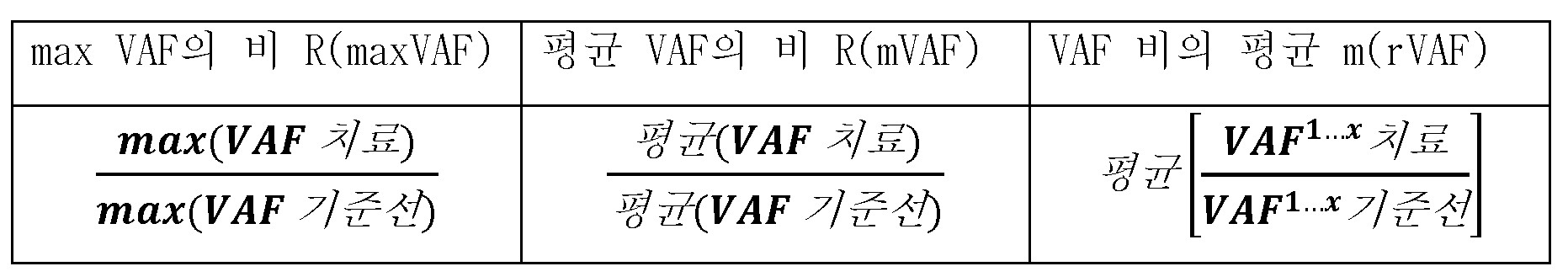
3. 결과3. Results
i. 분자 반응 계산은 SNV, 인델, 및 융합의 ctDNA VAF의 변화를 포획한다i. Molecular response calculations capture changes in ctDNA VAFs of SNVs, indels, and fusions
도 22는 74- 암 연관 유전자 패널 공간 내의 샘플당 검출된 체세포 변이체의 수를 제시한다. 체세포 SNV, 인델, 및 융합물의 수는 이 연구에서 상위 3가지 암 유형에 대한 샘플당 분자 반응 계산에 포함되었다. 중위값 돌연변이 변이체 카운트는 유방, CRC, 및 NSCLC에 대해 각각 4, 5, 및 3이다. Figure 22 presents the number of somatic variants detected per sample within the 74-cancer associated gene panel space. The number of somatic SNVs, indels, and fusions was included in the calculation of molecular responses per sample for the top three cancer types in this study. Median mutation variant counts are 4, 5, and 3 for breast, CRC, and NSCLC, respectively.
ii. 쌍을 이룬 샘플을 갖는 체세포 분류의 해결은 종양 신호를 개선시킨다ii. Solving somatic classification with paired samples improves tumor signal
도 23은 MR 결과를 왜곡시킬 수 있는 체세포 분류 불일치의 예를 제시한다. 희귀한 체세포 상황 분류 불일치 (변이체의 <0.8%)는 높은 종양 분율 및 대립유전자 불균형과 함께 발생할 수 있다. 비해결된, ALK는 보편적으로 감소하는 VAF에 대한 MR 점수를 왜곡시킬 것이다. Figure 23 presents an example of somatic classification inconsistency that can distort MR results. Rare somatic context classification mismatches (<0.8% of variants) can occur with high tumor fractions and allelic imbalance. Unresolved, ALK will distort MR scores with VAF generally decreasing.
표 4는 환자 샘플 사이의 체세포 분류 불일치의 해결의 예가 변이체 정확도를 개선시킴을 제시한다. 환자 샘플 쌍 내의 체세포 분류 불일치는 변이체 특징에 기초한 알고리즘에 의해 해결되었다. 정확도를 주제 전문가에 의한 수동 해결에 대해 평가하였다.Table 4 provides examples of resolution of somatic classification discrepancies between patient samples to improve variant accuracy. Somatic classification discrepancies within patient sample pairs were resolved by an algorithm based on variant features. Accuracy was assessed against manual solving by subject matter experts.
표 4Table 4
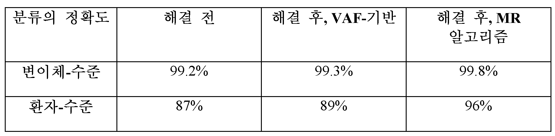
iii. 변이체는 검출 및 VAF 정밀도에 기초한 분자 반응 계산에 포함된다iii. Variants are included in molecular response calculations based on detection and VAF precision
도 24는 변이체 정밀도의 예가 돌연변이체 분자 카운트 (MMC = VAF * 분자 커버리지)에 의해 결정됨을 제시한다. (도 24A) 변이체는 샘플 투입 및 패널 디자인에 따라 다양한 분자 커버리지를 갖는다. 변이체 검출의 확률 (도 24B) 및 VAF 정밀도 (도 24C)는 VAF 및 분자 커버리지 (색상, (도 24A)에 맵핑됨) 양쪽 모두에 좌우된다. MMC (도 24D)는, 그것이 변이체 검출의 확률 (도 24E) 및 VAF 정밀도 (도 24F)를 결정하기 때문에, 변이체 정밀도에 대한 보다 좋은 계량이다. 양쪽 시점에서 낮은 MMC를 갖는 변이체는 신호를 잡음으로부터 보다 잘 분명히 하기 위해 분자 반응으로부터 배제되어야 한다. Figure 24 presents an example of variant precision determined by mutant molecule count (MMC = VAF * molecule coverage). ( Figure 24A ) Variants have varying molecular coverage depending on sample input and panel design. The probability of variant detection ( Figure 24B ) and VAF precision ( Figure 24C ) depend on both VAF and molecular coverage (mapped to color, ( Figure 24A )). MMC ( Figure 24D ) is a better measure of variant precision because it determines the probability of variant detection ( Figure 24E ) and VAF precision ( Figure 24F ). Variants with low MMC at both time points should be excluded from the molecular reaction to better disambiguate the signal from noise.
iv. 분자 반응은 방법 사이에 크게 일치하지만, R(mVAF)는 환자에 걸쳐 보다 확고하다iv. Molecular responses are largely consistent between methods, but R(mVAF) is more robust across patients
도 25는 비의 평균, m(rVAF), 또는 최대값의 비, R(maxVAF)를 사용하는 경우 소수의 변이체가 종양 신호를 능가할 수 있음을 제시한다. (도 25A) MR 점수는 증가, 감소 또는 정밀도 한계 내 ("거의 0% 변화")로서 카테고리화된다. 단지 8%의 환자만이 임의의 방법에서 증가 및 감소 사이에 변화하며, 이는 높은 카테고리적 상관관계를 나타낸다 (X2 p < 0.001). MR 상관관계는 ρ = 0.42 내지 0.86의 범위이다 (p < 0.001). (도 25B) m(rVAF)는 일부 VAF가 낮은 경우 MR을 과대평가하는 경향이 있다 (적색). R(maxVAF)는 대다수로부터 벗어나는 단일 최대 변이체 (자주색)에 의해 왜곡될 수 있다. 20%의 샘플 쌍은 maxVAF가 아닌 종양 유도자 또는 저항성 돌연변이를 가지며, 이는 종양 역학이 mVAF에 의해 더 잘 포획됨을 시사한다. (C) 새로운 치료중 변이체를 배제하는 것은 보다 낮은 MR 평가가능한 비율을 초래할 것이고, 새로 생겨난 변이체의 신호를 배제한다. Figure 25 shows that a small number of variants can outperform the tumor signal when using the mean of the ratio, m(rVAF), or the maximum ratio, R(maxVAF). ( Figure 25A ) MR scores are categorized as increasing, decreasing, or within precision limits (“near 0% change”). Only 8% of patients varied between increase and decrease in any method, indicating a high categorical correlation (X2 p < 0.001). MR correlations range from ρ = 0.42 to 0.86 (p < 0.001). ( Figure 25B ) m(rVAF) tends to overestimate MR when some VAFs are low (red). R(maxVAF) can be skewed by the single largest variant (purple) deviating from the majority. 20% of sample pairs have tumor driver or resistance mutations but not maxVAF, suggesting that tumor dynamics are better captured by mVAF. (C) Excluding new on-treatment variants will result in lower MR evaluable rates and excludes signals of newly emerging variants.
v. ctDNA 수준 변화의 낮은 신호를 갖는 환자는 분자 반응에 대해 평가가능하지 않은 것으로서 확인된다v. Patients with low signal of ctDNA level changes are identified as not evaluable for molecular response.
도 26은 분자 반응 점수의 확실성이 변이체의 수 (도 26A), 분자 커버리지 (도 26B), 및 최대 VAF (도 26C)가 증가함에 따라 증가하는 예를 제시한다. Figure 26 presents an example where the certainty of the molecular response score increases with increasing number of variants ( Figure 26A ), molecular coverage ( Figure 26B ), and maximum VAF ( Figure 26C ).
체세포 변이체가 없거나 (대략 7%의 환자), 또는 포함 기준을 충족시키는 체세포 변이체가 없는 경우 (16%), 샘플 쌍은 분자 반응에 대해 VAF-기반 방법을 사용하여 평가가능하지 않다. 또한, 분자 반응 점수의 확실성은 이론적으로 VAF 정밀도의 통계적 모델을 사용하여 계산된다. 허용가능한 불확실성의 한계를 초과하는 샘플 쌍 (검은색 라인)은 MR에 대해 평가가능하지 않다 (3%). 이는 MR에 대해 평가가능한 대략 74%의 샘플 쌍을 초래한다.If there is no somatic variant (approximately 7% of patients), or no somatic variant meets the inclusion criteria (16%), the sample pair is not evaluable using VAF-based methods for molecular response. Additionally, the certainty of the molecular response score is theoretically calculated using a statistical model of VAF precision. Sample pairs that exceed the limits of acceptable uncertainty (black line) are not evaluable for MR (3%). This results in approximately 74% of sample pairs evaluable for MR.
vi. 임상적 환자 샘플 내의 분자 반응 점수의 범위는 강한 생물학적 신호를 반영한다vi. The range of molecular response scores within clinical patient samples reflects a strong biological signal.
임상적 환자 샘플 쌍에서, 분자 반응 분포는 100% 감소 내지 >100% 증가의 다양한 점수를 나타낸다 (도 27A).In clinical patient sample pairs, the molecular response distribution shows varying scores ranging from 100% decrease to >100% increase ( Figure 27A ).
기술적 반복실험은 0% 변화에서 피크를 갖는 널 분자 반응 분포를 제공한다 (도 27B).Technical replicates provide a null molecular response distribution with a peak at 0% change ( Figure 27B ).
4. 결론4. Conclusion
배선 및 저-정밀도 변이체 필터링, 전체 제형, 및 평가가능한 기준을 포함하는 분자 반응 계산의 각각의 성분은 MR의 정확한 평가에 중요하다. 큰 환자 샘플의 세트에서의 분자 반응 방법의 비교 및 시뮬레이션은 새롭게 검출된 돌연변이의 포함을 갖는 평균 VAF의 비를 지지한다.Each component of molecular response calculations, including germline and low-precision variant filtering, overall formulation, and evaluable criteria, is important for accurate assessment of MR. Comparison of molecular response methods and simulations in sets of large patient samples support the ratio of mean VAF with inclusion of newly detected mutations.
C. 실시예 3C. Example 3
도 28은 MR 계산을 위한 샘플 쌍의 예를 제시한다. 어느 하나의 샘플에서 검출된 모든 SNV, 인델, 융합으로 시작하여, 통상적인 배선 변이체를 제거한다. 다음으로, 변이체 체세포/배선 분류 불일치를 해결하여 단일 분류를 제공한다. (이 실시예에서, 불일치는 없었다). 다음으로, 배선 변이체를 필터링하고, 이어서 CHIP 변이체를 필터링한다 (이 실시예에서, ATM.R3008H는 제거되는 CHIP 변이체이다). 다음으로, MMC- 또는 커버리지- 기반 포함 임계치를 충족시키지 않는 변이체를 제거한다. 이 실시예에서, 이러한 필터링 단계 후, 3개의 체세포 변이체 (PDGFRA, RET 및 TP53)가 남는다. 마지막으로, MR 점수를 이러한 남아 있는 변이체로부터 계산한다. 이 실시예에서, 기준선 평균 VAF는 22.2%이고, 치료중 평균 VAF는 2.7%이며, 이는 88%의 ctDNA 감소인 12%의 MR 점수를 제공한다. Figure 28 presents an example of sample pairs for MR calculation. Starting with all SNVs, indels, and fusions detected in any one sample, common germline variants are removed. Next, variant somatic/germline classification discrepancies are resolved to provide a single classification. (In this example, there was no discrepancy). Next, germline variants are filtered, followed by CHIP variants (in this example, ATM.R3008H is the CHIP variant that is removed). Next, variants that do not meet MMC- or coverage-based inclusion thresholds are removed. In this example, after this filtering step, three somatic variants remain (PDGFRA, RET, and TP53). Finally, MR scores are calculated from these remaining variants. In this example, the baseline mean VAF is 22.2% and the mean on-treatment VAF is 2.7%, giving an MR score of 12%, which is a ctDNA reduction of 88%.
본원에서 인용된 모든 특허, 특허 출원, 웹사이트, 기타 간행물 또는 문서, 수탁 번호 등은 각각의 개별적인 항목이 참조로 포함된 것으로 구체적 및 개별적으로 지시된 것과 동일한 정도로 모든 목적을 위해 그 전문이 참조로 포함된다. 상이한 서열 버전들이 상이한 시간의 수탁 번호와 연관되는 경우, 본 출원의 유효 출원일의 수탁 번호와 연관된 버전을 의미한다. 유효 출원일은 실제 출원일 또는 수탁 번호를 언급하는 우선권 출원의 출원일 (적용가능한 경우) 중 더 앞서는 것을 의미한다. 마찬가지로, 간행물, 웹사이트 등의 상이한 버전들이 상이한 시간에 공개된 경우, 달리 명시되지 않는 한, 본 출원의 유효 출원일에 가장 최근에 공개된 버전을 의미한다.All patents, patent applications, websites, other publications or documents, accession numbers, etc. cited herein are hereby incorporated by reference in their entirety for all purposes to the same extent as if each individual item was specifically and individually indicated to be incorporated by reference. Included. If different sequence versions are associated with accession numbers at different times, it means the version associated with the accession number at the effective filing date of this application. Effective filing date means the earlier of the actual filing date or the filing date of the priority application to which the accession number refers (if applicable). Likewise, if different versions of a publication, website, etc. have been published at different times, the most recent published version as of the effective filing date of this application is meant, unless otherwise specified.
관련 기술 분야의 통상의 기술자는 통상적인 실험 이하를 사용하여, 본원에 기술된 방법 및 조성물의 구체적인 실시양태에 대한 많은 등가물을 인식하거나, 또는 확인할 수 있을 것이다. 이러한 등가물은 하기 청구범위에 의해 포함되는 것으로 의도된다.Those skilled in the art will recognize, or be able to ascertain using no more than routine experimentation, many equivalents to the specific embodiments of the methods and compositions described herein. Such equivalents are intended to be encompassed by the following claims.
Claims (46)
제1 복수의 서열 판독물 및 제2 복수의 서열 판독물 내의 복수의 변이체를 체세포 또는 배선으로서 분류하고;
체세포로서 분류된 복수의 변이체의 적어도 하나의 변이체에 대해, 제1 돌연변이체 대립유전자 분율 (MAF) 및 제2 MAF에 기초하여, 제1 MAF의 가중 평균 및 제2 MAF의 가중 평균을 결정하고;
대상체에 대해, 제1 MAF의 가중 평균 및 제2 MAF의 가중 평균의 비를 결정하고;
제1 MAF의 가중 평균 및 제2 MAF의 가중 평균의 비에 기초하여, 신뢰 구간을 결정하고;
분자 반응 점수로서, 제1 MAF의 가중 평균 및 제2 MAF의 가중 평균의 비 및 신뢰 구간을 산출하는 것
을 포함하는 방법.Determining a first plurality of sequence reads and a second plurality of sequence reads associated with the subject, wherein the first plurality of sequence reads are determined prior to administering the therapy and the second plurality of sequence reads are determined prior to administering the therapy. It is decided after;
classifying the plurality of variants within the first plurality of sequence reads and the second plurality of sequence reads as somatic or germline;
For at least one variant of the plurality of variants classified as somatic, based on the first mutant allele fraction (MAF) and the second MAF, determine a weighted average of the first MAF and a weighted average of the second MAF;
For the subject, determine the ratio of the weighted average of the first MAF and the weighted average of the second MAF;
Based on the ratio of the weighted average of the first MAF and the weighted average of the second MAF, determine a confidence interval;
As a molecular response score, calculating the ratio and confidence interval of the weighted average of the first MAF and the weighted average of the second MAF.
How to include .
제1 복수의 서열 판독물 및 제2 복수의 서열 판독물 내의 복수의 변이체를 체세포 또는 배선으로서 분류하고;
체세포로서 분류된 복수의 변이체의 적어도 하나의 변이체에 대해, 제1 돌연변이체 대립유전자 분율 (MAF) 및 제2 MAF에 기초하여, MAF 비를 결정하고;
대상체에 대해, MAF 비의 가중 평균을 결정하고;
MAF 비의 가중 평균에 기초하여, MAF 비의 가중 평균과 연관된 신뢰 구간을 결정하고;
분자 반응 점수로서, MAF 비의 가중 평균 및 신뢰 구간을 산출하는 것
을 포함하는 방법.Determining a first plurality of sequence reads and a second plurality of sequence reads associated with the subject, wherein the first plurality of sequence reads are determined prior to administering the therapy and the second plurality of sequence reads are determined prior to administering the therapy. It is decided after;
classifying the plurality of variants within the first plurality of sequence reads and the second plurality of sequence reads as somatic or germline;
For at least one variant of the plurality of variants classified as somatic, determine the MAF ratio based on the first mutant allele fraction (MAF) and the second MAF;
For the subject, determine the weighted average of the MAF ratio;
Based on the weighted average of the MAF ratio, determine a confidence interval associated with the weighted average of the MAF ratio;
As a molecular response score, yielding a weighted mean and confidence interval of the MAF ratio
How to include .
제1 복수의 서열 판독물 내의 복수의 변이체를 체세포 또는 배선으로서 분류하고;
제2 복수의 서열 판독물 내의 복수의 변이체를 체세포 또는 배선으로서 분류하고;
복수의 변이체의 적어도 하나의 변이체를 재분류하여 제1 복수의 서열 판독물 및 제2 복수의 서열 판독물 사이의 분류 불일치를 해결하고;
체세포로서 분류되거나 재분류된 복수의 변이체의 적어도 하나의 변이체에 대해, 제1 복수의 서열 판독물의 적어도 일부에 기초하여, 제1 돌연변이체 대립유전자 분율 (MAF)을 결정하고;
체세포로서 분류되거나 재분류된 복수의 변이체의 적어도 하나의 변이체에 대해, 제2 복수의 서열 판독물의 적어도 일부에 기초하여, 제2 MAF를 결정하고;
제1 MAF 및 제2 MAF에 기초하여, 분자 반응 점수를 결정하는 것
을 포함하는 방법.Determining a first plurality of sequence reads and a second plurality of sequence reads associated with the subject, wherein the first plurality of sequence reads are determined prior to administering the therapy and the second plurality of sequence reads are determined prior to administering the therapy. It is decided after;
classifying the plurality of variants in the first plurality of sequence reads as somatic or germline;
classifying the plurality of variants in the second plurality of sequence reads as somatic or germline;
reclassifying at least one variant of the plurality of variants to resolve classification discrepancies between the first plurality of sequence reads and the second plurality of sequence reads;
For at least one variant of the plurality of variants classified or reclassified as somatic, determine a first mutant allele fraction (MAF) based on at least a portion of the sequence reads of the first plurality;
For at least one variant of the plurality of variants classified or reclassified as somatic, determine a second MAF based on at least a portion of the second plurality of sequence reads;
Based on the first MAF and the second MAF, determining the molecular response score
How to include .
제1 복수의 서열 판독물 및 제2 복수의 서열 판독물 내의 복수의 변이체를 체세포 또는 배선으로서 분류하고;
복수의 변이체의 적어도 하나의 변이체를 잠재성 불명 클론성 조혈(Clonal Hematopoiesis of Indeterminate Potential) (CHIP) 변이체로서 결정하고;
복수의 변이체로부터, 적어도 하나의 CHIP 변이체를 제거하고;
체세포로서 분류된 복수의 변이체의 적어도 하나의 변이체에 대해, 제1 복수의 서열 판독물의 적어도 일부에 기초하여, 제1 돌연변이체 대립유전자 분율 (MAF)을 결정하고;
체세포로서 분류된 복수의 변이체의 적어도 하나의 변이체에 대해, 제2 복수의 서열 판독물의 적어도 일부에 기초하여, 제2 MAF를 결정하고;
제1 MAF 및 제2 MAF에 기초하여, 분자 반응 점수를 결정하는 것
을 포함하는 방법.Determining a first plurality of sequence reads and a second plurality of sequence reads associated with the subject, wherein the first plurality of sequence reads are determined prior to administering the therapy and the second plurality of sequence reads are determined prior to administering the therapy. It is decided after;
classifying the plurality of variants within the first plurality of sequence reads and the second plurality of sequence reads as somatic or germline;
determining at least one variant of the plurality of variants as a Clonal Hematopoiesis of Indeterminate Potential (CHIP) variant;
From the plurality of variants, remove at least one CHIP variant;
For at least one variant of the plurality of variants classified as somatic, determine a first mutant allele fraction (MAF) based on at least a portion of the first plurality of sequence reads;
For at least one variant of the plurality of variants classified as somatic, determine a second MAF based on at least a portion of the second plurality of sequence reads;
Based on the first MAF and the second MAF, determining the molecular response score
How to include .
제1 복수의 서열 판독물 내의 복수의 변이체를 체세포 또는 배선으로서 분류하고;
제2 복수의 서열 판독물 내의 복수의 변이체를 체세포 또는 배선으로서 분류하고;
복수의 변이체의 적어도 하나의 변이체를 재분류하여 제1 복수의 서열 판독물 및 제2 복수의 서열 판독물 사이의 분류 불일치를 해결하고;
복수의 변이체의 적어도 하나의 변이체를 잠재성 불명 클론성 조혈 (CHIP) 변이체로서 결정하고;
복수의 변이체로부터, 적어도 하나의 CHIP 변이체를 제거하고;
체세포로서 분류된 복수의 변이체의 적어도 하나의 변이체에 대해, 제1 복수의 서열 판독물의 적어도 일부에 기초하여, 제1 돌연변이체 대립유전자 분율 (MAF)을 결정하고;
체세포로서 분류된 복수의 변이체의 적어도 하나의 변이체에 대해, 제2 복수의 서열 판독물의 적어도 일부에 기초하여, 제2 MAF를 결정하고;
체세포로서 분류된 복수의 변이체의 적어도 하나의 변이체에 대해, 제1 MAF 및 제2 MAF에 기초하여 MAF 비를 결정하고;
대상체에 대해, MAF 비의 가중 평균을 결정하고;
MAF 비의 가중 평균에 기초하여, MAF 비의 가중 평균과 연관된 신뢰 구간을 결정하고;
분자 반응 점수로서, MAF 비의 가중 평균 및 신뢰 구간을 산출하는 것
을 포함하는 방법.Determining a first plurality of sequence reads and a second plurality of sequence reads associated with the subject, wherein the first plurality of sequence reads are determined prior to administering the therapy and the second plurality of sequence reads are determined prior to administering the therapy. It is decided after;
classifying the plurality of variants in the first plurality of sequence reads as somatic or germline;
classifying the plurality of variants in the second plurality of sequence reads as somatic or germline;
reclassifying at least one variant of the plurality of variants to resolve classification discrepancies between the first plurality of sequence reads and the second plurality of sequence reads;
determining at least one variant of the plurality of variants as a clonal hematopoiesis of unknown potential (CHIP) variant;
From the plurality of variants, remove at least one CHIP variant;
For at least one variant of the plurality of variants classified as somatic, determine a first mutant allele fraction (MAF) based on at least a portion of the first plurality of sequence reads;
For at least one variant of the plurality of variants classified as somatic, determine a second MAF based on at least a portion of the second plurality of sequence reads;
For at least one variant of the plurality of variants classified as somatic, determine a MAF ratio based on the first MAF and the second MAF;
For the subject, determine the weighted average of the MAF ratio;
Based on the weighted average of the MAF ratio, determine a confidence interval associated with the weighted average of the MAF ratio;
As a molecular response score, yielding a weighted average and confidence interval of the MAF ratio
How to include .
제1 복수의 서열 판독물 내의 복수의 변이체를 체세포 또는 배선으로서 분류하고;
제2 복수의 서열 판독물 내의 복수의 변이체를 체세포 또는 배선으로서 분류하고;
복수의 변이체의 적어도 하나의 변이체를 재분류하여 제1 복수의 서열 판독물 및 제2 복수의 서열 판독물 사이의 분류 불일치를 해결하고;
복수의 변이체의 적어도 하나의 변이체를 잠재성 불명 클론성 조혈 (CHIP) 변이체로서 결정하고;
복수의 변이체로부터, 적어도 하나의 CHIP 변이체를 제거하고;
체세포로서 분류된 복수의 변이체의 적어도 하나의 변이체에 대해, 제1 복수의 서열 판독물의 적어도 일부에 기초하여, 제1 돌연변이체 대립유전자 분율 (MAF)을 결정하고;
체세포로서 분류된 복수의 변이체의 적어도 하나의 변이체에 대해, 제2 복수의 서열 판독물의 적어도 일부에 기초하여, 제2 MAF를 결정하고;
체세포로서 분류된 복수의 변이체의 적어도 하나의 변이체에 대해, 제1 MAF 및 제2 MAF에 기초하여, 제1 MAF의 가중 평균 및 제2 MAF의 가중 평균을 결정하고;
대상체에 대해, 제1 MAF의 가중 평균 및 제2 MAF의 가중 평균의 비를 결정하고;
제1 MAF의 가중 평균 및 제2 MAF의 가중 평균의 비에 기초하여, 신뢰 구간을 결정하고;
분자 반응 점수로서, 제1 MAF의 가중 평균 및 제2 MAF의 가중 평균의 비 및 신뢰 구간을 산출하는 것
을 포함하는 방법.Determining a first plurality of sequence reads and a second plurality of sequence reads associated with the subject, wherein the first plurality of sequence reads are determined prior to administering the therapy and the second plurality of sequence reads are determined prior to administering the therapy. It is decided after;
classifying the plurality of variants in the first plurality of sequence reads as somatic or germline;
classifying the plurality of variants in the second plurality of sequence reads as somatic or germline;
reclassifying at least one variant of the plurality of variants to resolve classification discrepancies between the first plurality of sequence reads and the second plurality of sequence reads;
determining at least one variant of the plurality of variants as a clonal hematopoiesis of unknown potential (CHIP) variant;
From the plurality of variants, remove at least one CHIP variant;
For at least one variant of the plurality of variants classified as somatic, determine a first mutant allele fraction (MAF) based on at least a portion of the first plurality of sequence reads;
For at least one variant of the plurality of variants classified as somatic, determine a second MAF based on at least a portion of the second plurality of sequence reads;
For at least one variant of the plurality of variants classified as somatic, based on the first MAF and the second MAF, determine a weighted average of the first MAF and a weighted average of the second MAF;
For the subject, determine the ratio of the weighted average of the first MAF and the weighted average of the second MAF;
Based on the ratio of the weighted average of the first MAF and the weighted average of the second MAF, determine a confidence interval;
As a molecular response score, calculating the ratio and confidence interval of the weighted average of the first MAF and the weighted average of the second MAF.
How to include .
제1 복수의 서열 판독물 및 제2 복수의 서열 판독물 내의 복수의 변이체를 체세포 또는 배선으로서 분류하고;
체세포로서 분류된 복수의 변이체의 적어도 하나의 변이체에 대해, 제1 시점에서의 제1 돌연변이체 대립유전자 분율 (MAF) 및 제2 시점에서의 제2 MAF에 기초하여, 제1 MAF의 제1 중심 경향성 척도 및 제2 MAF의 제2 중심 경향성 척도를 결정하고;
제1 시점에서의 제1 중심 경향성 척도 대 제2 시점에서의 제2 중심 경향성 척도의 비를 결정하고;
분자 반응 점수로서, 제1 시점에서의 제1 중심 경향성 척도 대 제2 시점에서의 제2 중심 경향성 척도의 비를 산출하는 것
을 포함하는 방법.Determining a first plurality of sequence reads and a second plurality of sequence reads associated with the subject, wherein the first plurality of sequence reads is determined at a first time point prior to administering the therapy, and the second plurality of sequence reads is determined at a second time point after administering therapy;
classifying the plurality of variants within the first plurality of sequence reads and the second plurality of sequence reads as somatic or germline;
For at least one variant of the plurality of variants classified as somatic, based on the first mutant allele fraction (MAF) at the first time point and the second MAF at the second time point, the first centroid of the first MAF determine a propensity measure and a second central tendency measure of the second MAF;
determine the ratio of the first measure of central tendency at a first time point to the second measure of central tendency at a second time point;
A molecular response score, yielding the ratio of a first measure of central tendency at a first time point to a second measure of central tendency at a second time point.
How to include .
(a) 컴퓨터에 의해, 제1 및 제2 시점에서 대상체로부터 수득된 샘플 내의 하나 이상의 암 유형과 연관된 표적화된 핵산으로부터 생성된 서열 정보로부터 복수의 변이체에 대한 돌연변이체 대립유전자 빈도 (MAF)를 결정하여 복수의 변이체 내의 적어도 하나의 변이체에 대한 제1 및 제2 MAF의 세트를 생성하고;
(b) 컴퓨터에 의해, 복수의 변이체 내의 적어도 하나의 변이체에 대한 제1 및 제2 MAF의 비를 계산하여 MAF 비의 세트 및 MAF 비의 세트 내의 각각의 MAF 비에 대한 상응하는 표준 편차를 생성하고;
(c) 컴퓨터에 의해, MAF 비의 가중 평균 및 신뢰 구간을 계산함으로써, 암을 갖는 대상체에 대한 분자 반응 점수를 결정하는 것
을 포함하는 방법.1. A method of determining a molecular response score for a subject having cancer, at least in part using a computer:
(a) Determine, by computer, the mutant allele frequency (MAF) for a plurality of variants from sequence information generated from targeted nucleic acids associated with one or more cancer types in samples obtained from the subject at first and second time points. thereby generating a set of first and second MAFs for at least one variant in the plurality of variants;
(b) calculate by computer the ratio of the first and second MAF for at least one variant within the plurality of variants to generate a set of MAF ratios and a corresponding standard deviation for each MAF ratio within the set of MAF ratios do;
(c) determining, by computer, a molecular response score for a subject with cancer by calculating the weighted mean and confidence interval of the MAF ratio.
How to include .
(a) 제1 및 제2 시점에서 대상체로부터 수득된 샘플 내의 하나 이상의 암 유형과 연관된 표적화된 핵산으로부터 생성된 서열 정보로부터 복수의 변이체에 대한 돌연변이체 대립유전자 빈도 (MAF)를 결정하여 복수의 변이체 내의 적어도 하나의 변이체에 대한 제1 및 제2 MAF의 세트를 생성하고;
(b) 복수의 변이체 내의 적어도 하나의 변이체에 대한 제1 및 제2 MAF의 비를 계산하여 MAF 비의 세트 및 MAF 비의 세트 내의 각각의 MAF 비에 대한 상응하는 표준 편차를 생성하고;
(c) MAF 비의 가중 평균 및 신뢰 구간을 계산하여 대상체에 대한 분자 반응 점수를 결정하고;
(d) 적어도 분자 반응 점수에 기초하여 하나 이상의 요법을 대상체에게 투여함으로써, 대상체에서 암을 치료하는 것
을 포함하는 방법.As a method of treating cancer in a subject:
(a) Determining the mutant allele frequency (MAF) for a plurality of variants from sequence information generated from targeted nucleic acids associated with one or more cancer types in samples obtained from the subject at first and second time points to identify the plurality of variants. generating a set of first and second MAFs for at least one variant within;
(b) calculating the ratio of the first and second MAF for at least one variant within the plurality of variants to generate a set of MAF ratios and a corresponding standard deviation for each MAF ratio within the set of MAF ratios;
(c) determine the molecular response score for the subject by calculating the weighted mean and confidence interval of the MAF ratio;
(d) treating cancer in the subject by administering to the subject one or more therapies based at least on the molecular response score.
How to include .
(a) 컴퓨터에 의해, 제1 및 제2 시점에서 대상체로부터 수득된 샘플 내의 하나 이상의 암 유형과 연관된 표적화된 핵산으로부터 생성된 서열 정보로부터 복수의 변이체에 대한 돌연변이체 대립유전자 빈도 (MAF)를 결정하여 복수의 변이체 내의 적어도 하나의 변이체에 대한 제1 및 제2 MAF의 세트를 생성하고;
(b) 컴퓨터에 의해, 복수의 변이체 내의 적어도 하나의 변이체에 대한 제1 및 제2 MAF의 비를 계산하여 MAF 비의 세트 및 MAF 비의 세트 내의 각각의 MAF 비에 대한 상응하는 표준 편차를 생성하고;
(c) 컴퓨터에 의해, MAF 비의 가중 평균 및 신뢰 구간을 계산하여 대상체에 대한 분자 반응 점수를 결정하는 것
에 의해 생성되는 것인 방법.A method of treating cancer in a subject, comprising administering to the subject one or more therapies based at least on a molecular response score for the subject, wherein the molecular response score is
(a) Determine, by computer, the mutant allele frequency (MAF) for a plurality of variants from sequence information generated from targeted nucleic acids associated with one or more cancer types in samples obtained from the subject at first and second time points. thereby generating a set of first and second MAFs for at least one variant in the plurality of variants;
(b) calculate by computer the ratio of the first and second MAF for at least one variant within the plurality of variants to generate a set of MAF ratios and a corresponding standard deviation for each MAF ratio within the set of MAF ratios do;
(c) determining, by computer, a molecular response score for the subject by calculating the weighted mean and confidence interval of the MAF ratio.
A method that is created by .
(a) 컴퓨터에 의해, 제1 및 제2 시점에서 대상체로부터 수득된 샘플 내의 하나 이상의 암 유형과 연관된 표적화된 핵산으로부터 생성된 서열 정보로부터 각각의 복수의 변이체에 대한 종양 분율 변화에 대한 종양 부하 변화 (R) P(R)을 결정하여 종양 부하 변화의 세트를 생성하고;
(b) 컴퓨터에 의해, 종양 부하 변화의 세트로부터 하나 이상의 클론성 조혈 변이체에 상응하는 하나 이상의 저항성 시그니쳐를 확인함으로써, 암을 갖는 대상체에서 클론성 조혈 변이체를 확인하는 것
을 포함하는 방법.1. A method for identifying clonal hematopoietic variants in a subject with cancer, at least in part using a computer:
(a) Change in tumor burden relative to change in tumor fraction for each plurality of variants from sequence information computer generated from targeted nucleic acids associated with one or more cancer types in samples obtained from the subject at first and second time points. ( R ) Determine P( R ) to generate a set of tumor burden changes;
(b) identifying clonal hematopoietic variants in a subject with cancer by computationally identifying one or more resistance signatures corresponding to one or more clonal hematopoietic variants from a set of tumor burden changes.
How to include .
(a) 컴퓨터에 의해, 제1 및 제2 시점에서 대상체로부터 수득된 샘플 내의 하나 이상의 암 유형과 연관된 표적화된 핵산으로부터 생성된 서열 정보로부터 각각의 복수의 변이체에 대한 종양 분율 변화에 대한 확률 밀도 함수 P(R)을 계산하고;
(b) 컴퓨터에 의해, 변이체의 하나 이상을 P(R)에 의해 하나 이상의 클론으로 그룹화하고;
(c) 컴퓨터에 의해, 각각의 클론에 대한 갱신된 P(R)을 생성하고;
(d) 컴퓨터에 의해, 미리 결정된 임계치 값 이상의 제1 및 제2 시점 사이의 분율 변화를 갖는 하나 이상의 클론을 확인함으로써, 암을 갖는 대상체에서 클론성 조혈 변이체를 확인하는 것
을 포함하는 방법.1. A method for identifying clonal hematopoietic variants in a subject with cancer, at least in part using a computer:
(a) Probability density function for the change in tumor fraction for each plurality of variants from sequence information computer generated from targeted nucleic acids associated with one or more cancer types in samples obtained from the subject at first and second time points. Calculate P( R );
(b) computerically grouping one or more of the variants into one or more clones by P( R );
(c) generating, by a computer, an updated P( R ) for each clone;
(d) identifying clonal hematopoietic variants in a subject with cancer by computer identifying one or more clones with a fractional change between the first and second time points above a predetermined threshold value.
How to include .
(a) 컴퓨터에 의해, 대상체로부터 수득된 샘플 내의 하나 이상의 암 유형과 연관된 표적화된 핵산으로부터 생성된 서열 정보로부터 주어진 변이체에 대한 돌연변이체 대립유전자 빈도 (MAF)를 결정하고;
(b) 컴퓨터에 의해, 주어진 변이체의 MAF가 샘플의 최대 MAF를 증가시키는 경우 (샘플은 최대 배수체 유전자의 분율 (max frac_diploid)을 포함함) 및/또는 주어진 변이체의 MAF가 대상체로부터 수득된 샘플로부터 결정된 하나 이상의 다른 MAF보다 적어도 약 2배 더 크거나, 3배 더 크거나, 4배 더 크거나, 5배 더 크거나, 6배 더 크거나, 7배 더 크거나, 8배 더 크거나, 9배 더 크거나, 또는 그 초과인 경우, 주어진 변이체가 배선 변이체인 것을 확인함으로써, 암을 갖는 대상체에서 배선 변이체를 확인하는 것
을 포함하는 방법.1. A method for identifying germline variants in a subject having cancer, at least in part using a computer:
(a) determining, by computer, the mutant allele frequency (MAF) for a given variant from sequence information generated from targeted nucleic acids associated with one or more cancer types in a sample obtained from the subject;
(b) by computer, if the MAF of a given variant increases the maximum MAF of a sample (the sample contains the fraction of genes max frac_diploid) and/or the MAF of a given variant is calculated from a sample obtained from the subject. is at least about 2 times larger, 3 times larger, 4 times larger, 5 times larger, 6 times larger, 7 times larger, or 8 times larger than one or more other MAFs determined; Identifying germline variants in subjects with cancer by confirming that a given variant is a germline variant if it is 9 times greater or greater.
How to include .
합계[중량 * 비]/합계[중량]
여기서 중량은 복수의 변이체 내의 주어진 변이체에 대한 1/범위2이고, 여기서 범위는 복수의 변이체 내의 주어진 변이체에 대한 제1 및 제2 MAF의 값 사이의 차이이고, 비는 MAF 비의 세트 내의 주어진 MAF 비인 방법.31. The method of any one of claims 1 to 30, comprising calculating the weighted average of the MAF ratio using the formula:
Total[Weight * Ratio]/Total[Weight]
where the weight is 1/range 2 for a given variant within a plurality of variants, where the range is the difference between the values of the first and second MAF for a given variant within the plurality of variants, and the ratio is the given MAF within the set of MAF ratios. Bein method.
MAF 비의 가중 평균 +/- sqrt[비 분산]
여기서 비 분산은 1/합계[중량]인 방법.32. The method of any one of claims 1 to 31, comprising calculating the confidence interval using the formula:
Weighted average of MAF ratio +/- sqrt[ratio variance]
Here, the ratio is 1/sum [weight].
(시점 2에서의 MAF 비의 가중 평균)/(시점 1에서의 MAF 비의 가중 평균)
여기서 시점에서 비검출된 변이체의 MAF는 0으로 설정되는 것인 방법.33. The method of any one of claims 1 to 32, comprising calculating the ratio of weighted averages of MAF using the formula:
(Weighted average of MAF ratio at Time 2)/(Weighted average of MAF ratio at Time 1)
Here, the MAF of the variant not detected at this point is set to 0.
제1 시점에서 체세포 변이체의 MAF로부터 수득된 제1 중심 경향성 척도 및 제2 시점에서 체세포 변이체의 MAF로부터 수득된 제2 중심 경향성 척도를 결정하고;
제1 시점에서의 중심 경향성 척도 대 제2 시점에서의 중심 경향성 척도의 비를 계산하고;
제1 시점에서의 중심 경향성 척도 대 제2 시점에서의 중심 경향성 척도의 비의 표준 편차를 계산하고;
또 다른 분자 반응 점수로서, 제1 시점에서의 중심 경향성 척도 대 제2 시점에서의 중심 경향성 척도의 비를 산출하는 것
을 추가로 포함하는 방법.According to any one of claims 1 to 40,
Determine a first measure of central tendency obtained from the MAF of the somatic variant at a first time point and a second measure of central tendency obtained from the MAF of the somatic variant at a second time point;
calculate the ratio of the measure of central tendency at a first time point to the measure of central tendency at a second time point;
calculate the standard deviation of the ratio of the measure of central tendency at the first time point to the measure of central tendency at the second time point;
Another molecular response score, yielding the ratio of the measure of central tendency at a first time point to the measure of central tendency at a second time point.
How to additionally include .
각각의 복수의 시점에서 복수의 서열 판독물을 수득하고;
복수의 시점의 임의의 2개의 시점 사이의 MAF 비를 결정하는 것
을 추가로 포함하는 방법.According to any one of claims 1 to 41,
Obtaining multiple sequence reads at each multiple time points;
Determining the MAF ratio between any two time points of multiple time points
How to additionally include .
Applications Claiming Priority (5)
| Application Number | Priority Date | Filing Date | Title |
|---|---|---|---|
| US202163157592P | 2021-03-05 | 2021-03-05 | |
| US63/157,592 | 2021-03-05 | ||
| US202163173193P | 2021-04-09 | 2021-04-09 | |
| US63/173,193 | 2021-04-09 | ||
| PCT/US2022/070984 WO2022187862A1 (en) | 2021-03-05 | 2022-03-04 | Methods and related aspects for analyzing molecular response |
Publications (1)
| Publication Number | Publication Date |
|---|---|
| KR20230156364A true KR20230156364A (en) | 2023-11-14 |
Family
ID=80952359
Family Applications (1)
| Application Number | Title | Priority Date | Filing Date |
|---|---|---|---|
| KR1020237033549A Pending KR20230156364A (en) | 2021-03-05 | 2022-03-04 | Methods and related aspects for analyzing molecular reactions |
Country Status (7)
| Country | Link |
|---|---|
| US (1) | US20220411876A1 (en) |
| EP (1) | EP4302301A1 (en) |
| JP (1) | JP2024513668A (en) |
| KR (1) | KR20230156364A (en) |
| AU (1) | AU2022231055A1 (en) |
| CA (1) | CA3210101A1 (en) |
| WO (1) | WO2022187862A1 (en) |
Families Citing this family (8)
| Publication number | Priority date | Publication date | Assignee | Title |
|---|---|---|---|---|
| CN120188220A (en) * | 2022-11-15 | 2025-06-20 | 夸登特健康公司 | Methods for predicting drug response or time until death or cancer progression in patients with non-small cell lung cancer (NSCLC) from ctDNA using signals from baseline circulating tumor DNA (ctDNA) levels and longitudinal changes in ctDNA levels over time |
| JP2024112204A (en) * | 2023-02-07 | 2024-08-20 | キヤノンメディカルシステムズ株式会社 | Medical information processing device, medical information processing method, and medical information processing program |
| WO2024263804A2 (en) * | 2023-06-20 | 2024-12-26 | Burzynski Stanislaw R | Compositions comprising antineoplastons and methods of treating kidney cancer |
| WO2024263798A1 (en) * | 2023-06-20 | 2024-12-26 | Burzynski Stanislaw R | Compositions comprising antineoplastons and methods of treating prostate cancer |
| WO2024263801A2 (en) * | 2023-06-20 | 2024-12-26 | Burzynski Stanislaw R | Compositions comprising antineoplastons and methods of treating ovarian cancer |
| WO2024263813A2 (en) * | 2023-06-21 | 2024-12-26 | Burzynski Stanislaw R | Compositions comprising antineoplastons and methods of treating colorectal cancer |
| WO2025090646A1 (en) * | 2023-10-27 | 2025-05-01 | Guardant Health, Inc. | Monitoring molecular response by allelic imbalance |
| WO2025106837A1 (en) * | 2023-11-15 | 2025-05-22 | Guardant Health, Inc. | Tumor fraction and outcome association in a real-world non-small cell lung cancer (nsclc) cohort using a methylation-based circulating tumor dna (ctdna) assay |
Family Cites Families (22)
| Publication number | Priority date | Publication date | Assignee | Title |
|---|---|---|---|---|
| US6582908B2 (en) | 1990-12-06 | 2003-06-24 | Affymetrix, Inc. | Oligonucleotides |
| US20030017081A1 (en) | 1994-02-10 | 2003-01-23 | Affymetrix, Inc. | Method and apparatus for imaging a sample on a device |
| JP3102800B2 (en) | 1994-08-19 | 2000-10-23 | パーキン−エルマー コーポレイション | Conjugation methods for amplification and ligation |
| GB9620209D0 (en) | 1996-09-27 | 1996-11-13 | Cemu Bioteknik Ab | Method of sequencing DNA |
| GB9626815D0 (en) | 1996-12-23 | 1997-02-12 | Cemu Bioteknik Ab | Method of sequencing DNA |
| US6969488B2 (en) | 1998-05-22 | 2005-11-29 | Solexa, Inc. | System and apparatus for sequential processing of analytes |
| AR021833A1 (en) | 1998-09-30 | 2002-08-07 | Applied Research Systems | METHODS OF AMPLIFICATION AND SEQUENCING OF NUCLEIC ACID |
| US6818395B1 (en) | 1999-06-28 | 2004-11-16 | California Institute Of Technology | Methods and apparatus for analyzing polynucleotide sequences |
| US7501245B2 (en) | 1999-06-28 | 2009-03-10 | Helicos Biosciences Corp. | Methods and apparatuses for analyzing polynucleotide sequences |
| EP1218543A2 (en) | 1999-09-29 | 2002-07-03 | Solexa Ltd. | Polynucleotide sequencing |
| DE60131194T2 (en) | 2000-07-07 | 2008-08-07 | Visigen Biotechnologies, Inc., Bellaire | SEQUENCE PROVISION IN REAL TIME |
| ATE449186T1 (en) | 2001-11-28 | 2009-12-15 | Applied Biosystems Llc | COMPOSITIONS AND METHODS FOR SELECTIVE NUCLEIC ACID ISOLATION |
| US7169560B2 (en) | 2003-11-12 | 2007-01-30 | Helicos Biosciences Corporation | Short cycle methods for sequencing polynucleotides |
| US7170050B2 (en) | 2004-09-17 | 2007-01-30 | Pacific Biosciences Of California, Inc. | Apparatus and methods for optical analysis of molecules |
| JP2008513782A (en) | 2004-09-17 | 2008-05-01 | パシフィック バイオサイエンシーズ オブ カリフォルニア, インコーポレイテッド | Apparatus and method for molecular analysis |
| US7482120B2 (en) | 2005-01-28 | 2009-01-27 | Helicos Biosciences Corporation | Methods and compositions for improving fidelity in a nucleic acid synthesis reaction |
| US7282337B1 (en) | 2006-04-14 | 2007-10-16 | Helicos Biosciences Corporation | Methods for increasing accuracy of nucleic acid sequencing |
| US8835358B2 (en) | 2009-12-15 | 2014-09-16 | Cellular Research, Inc. | Digital counting of individual molecules by stochastic attachment of diverse labels |
| GB2533006B (en) | 2012-09-04 | 2017-06-07 | Guardant Health Inc | Systems and methods to detect copy number variation |
| CA3046007A1 (en) | 2016-12-22 | 2018-06-28 | Guardant Health, Inc. | Methods and systems for analyzing nucleic acid molecules |
| EP3762512A1 (en) * | 2018-03-06 | 2021-01-13 | Cancer Research Technology Limited | Improvements in variant detection |
| WO2020023420A2 (en) * | 2018-07-23 | 2020-01-30 | Guardant Health, Inc. | Methods and systems for adjusting tumor mutational burden by tumor fraction and coverage |
-
2022
- 2022-03-04 KR KR1020237033549A patent/KR20230156364A/en active Pending
- 2022-03-04 US US17/687,536 patent/US20220411876A1/en active Pending
- 2022-03-04 EP EP22713235.4A patent/EP4302301A1/en active Pending
- 2022-03-04 WO PCT/US2022/070984 patent/WO2022187862A1/en not_active Ceased
- 2022-03-04 CA CA3210101A patent/CA3210101A1/en active Pending
- 2022-03-04 JP JP2023553585A patent/JP2024513668A/en active Pending
- 2022-03-04 AU AU2022231055A patent/AU2022231055A1/en active Pending
Also Published As
| Publication number | Publication date |
|---|---|
| JP2024513668A (en) | 2024-03-27 |
| WO2022187862A1 (en) | 2022-09-09 |
| US20220411876A1 (en) | 2022-12-29 |
| CA3210101A1 (en) | 2022-09-09 |
| EP4302301A1 (en) | 2024-01-10 |
| AU2022231055A1 (en) | 2023-09-14 |
Similar Documents
| Publication | Publication Date | Title |
|---|---|---|
| JP7466519B2 (en) | Methods and systems for adjusting tumor mutation burden by tumor proportion and coverage | |
| JP7699261B2 (en) | Detection of microsatellite instability in cell-free DNA | |
| KR20230156364A (en) | Methods and related aspects for analyzing molecular reactions | |
| KR102722821B1 (en) | Methods and systems for distinguishing somatic and germline variants | |
| JP2024015059A (en) | Methods and systems for determining the cellular origin of cell-free nucleic acids | |
| US20220344004A1 (en) | Detecting the presence of a tumor based on off-target polynucleotide sequencing data | |
| JP2023526252A (en) | Detection of homologous recombination repair defects | |
| US20220028494A1 (en) | Methods and systems for determining the cellular origin of cell-free dna | |
| CN118369726A (en) | Systems and methods for identifying copy number alterations | |
| CN117063239A (en) | Methods and related aspects for analyzing molecular responses |
Legal Events
| Date | Code | Title | Description |
|---|---|---|---|
| PA0105 | International application |
Patent event date: 20230927 Patent event code: PA01051R01D Comment text: International Patent Application |
|
| PG1501 | Laying open of application | ||
| A201 | Request for examination | ||
| PA0201 | Request for examination |
Patent event code: PA02012R01D Patent event date: 20250228 Comment text: Request for Examination of Application |