KR20210065085A - Methods for Characterizing Modifications Elicited by Use of Designer Nucleases - Google Patents
Methods for Characterizing Modifications Elicited by Use of Designer Nucleases Download PDFInfo
- Publication number
- KR20210065085A KR20210065085A KR1020217003131A KR20217003131A KR20210065085A KR 20210065085 A KR20210065085 A KR 20210065085A KR 1020217003131 A KR1020217003131 A KR 1020217003131A KR 20217003131 A KR20217003131 A KR 20217003131A KR 20210065085 A KR20210065085 A KR 20210065085A
- Authority
- KR
- South Korea
- Prior art keywords
- primer
- target
- dna
- seq
- sequence
- Prior art date
- Legal status (The legal status is an assumption and is not a legal conclusion. Google has not performed a legal analysis and makes no representation as to the accuracy of the status listed.)
- Granted
Links
Images


Classifications
-
- C—CHEMISTRY; METALLURGY
- C12—BIOCHEMISTRY; BEER; SPIRITS; WINE; VINEGAR; MICROBIOLOGY; ENZYMOLOGY; MUTATION OR GENETIC ENGINEERING
- C12Q—MEASURING OR TESTING PROCESSES INVOLVING ENZYMES, NUCLEIC ACIDS OR MICROORGANISMS; COMPOSITIONS OR TEST PAPERS THEREFOR; PROCESSES OF PREPARING SUCH COMPOSITIONS; CONDITION-RESPONSIVE CONTROL IN MICROBIOLOGICAL OR ENZYMOLOGICAL PROCESSES
- C12Q1/00—Measuring or testing processes involving enzymes, nucleic acids or microorganisms; Compositions therefor; Processes of preparing such compositions
- C12Q1/68—Measuring or testing processes involving enzymes, nucleic acids or microorganisms; Compositions therefor; Processes of preparing such compositions involving nucleic acids
- C12Q1/6844—Nucleic acid amplification reactions
- C12Q1/6848—Nucleic acid amplification reactions characterised by the means for preventing contamination or increasing the specificity or sensitivity of an amplification reaction
-
- C—CHEMISTRY; METALLURGY
- C12—BIOCHEMISTRY; BEER; SPIRITS; WINE; VINEGAR; MICROBIOLOGY; ENZYMOLOGY; MUTATION OR GENETIC ENGINEERING
- C12Q—MEASURING OR TESTING PROCESSES INVOLVING ENZYMES, NUCLEIC ACIDS OR MICROORGANISMS; COMPOSITIONS OR TEST PAPERS THEREFOR; PROCESSES OF PREPARING SUCH COMPOSITIONS; CONDITION-RESPONSIVE CONTROL IN MICROBIOLOGICAL OR ENZYMOLOGICAL PROCESSES
- C12Q1/00—Measuring or testing processes involving enzymes, nucleic acids or microorganisms; Compositions therefor; Processes of preparing such compositions
- C12Q1/68—Measuring or testing processes involving enzymes, nucleic acids or microorganisms; Compositions therefor; Processes of preparing such compositions involving nucleic acids
- C12Q1/6813—Hybridisation assays
- C12Q1/6827—Hybridisation assays for detection of mutation or polymorphism
-
- C—CHEMISTRY; METALLURGY
- C12—BIOCHEMISTRY; BEER; SPIRITS; WINE; VINEGAR; MICROBIOLOGY; ENZYMOLOGY; MUTATION OR GENETIC ENGINEERING
- C12Q—MEASURING OR TESTING PROCESSES INVOLVING ENZYMES, NUCLEIC ACIDS OR MICROORGANISMS; COMPOSITIONS OR TEST PAPERS THEREFOR; PROCESSES OF PREPARING SUCH COMPOSITIONS; CONDITION-RESPONSIVE CONTROL IN MICROBIOLOGICAL OR ENZYMOLOGICAL PROCESSES
- C12Q1/00—Measuring or testing processes involving enzymes, nucleic acids or microorganisms; Compositions therefor; Processes of preparing such compositions
- C12Q1/68—Measuring or testing processes involving enzymes, nucleic acids or microorganisms; Compositions therefor; Processes of preparing such compositions involving nucleic acids
- C12Q1/6869—Methods for sequencing
-
- C—CHEMISTRY; METALLURGY
- C12—BIOCHEMISTRY; BEER; SPIRITS; WINE; VINEGAR; MICROBIOLOGY; ENZYMOLOGY; MUTATION OR GENETIC ENGINEERING
- C12Q—MEASURING OR TESTING PROCESSES INVOLVING ENZYMES, NUCLEIC ACIDS OR MICROORGANISMS; COMPOSITIONS OR TEST PAPERS THEREFOR; PROCESSES OF PREPARING SUCH COMPOSITIONS; CONDITION-RESPONSIVE CONTROL IN MICROBIOLOGICAL OR ENZYMOLOGICAL PROCESSES
- C12Q2521/00—Reaction characterised by the enzymatic activity
- C12Q2521/30—Phosphoric diester hydrolysing, i.e. nuclease
- C12Q2521/301—Endonuclease
-
- C—CHEMISTRY; METALLURGY
- C12—BIOCHEMISTRY; BEER; SPIRITS; WINE; VINEGAR; MICROBIOLOGY; ENZYMOLOGY; MUTATION OR GENETIC ENGINEERING
- C12Q—MEASURING OR TESTING PROCESSES INVOLVING ENZYMES, NUCLEIC ACIDS OR MICROORGANISMS; COMPOSITIONS OR TEST PAPERS THEREFOR; PROCESSES OF PREPARING SUCH COMPOSITIONS; CONDITION-RESPONSIVE CONTROL IN MICROBIOLOGICAL OR ENZYMOLOGICAL PROCESSES
- C12Q2549/00—Reactions characterised by the features used to influence the efficiency or specificity
- C12Q2549/10—Reactions characterised by the features used to influence the efficiency or specificity the purpose being that of reducing false positive or false negative signals
- C12Q2549/119—Reactions characterised by the features used to influence the efficiency or specificity the purpose being that of reducing false positive or false negative signals using nested primers
Landscapes
- Chemical & Material Sciences (AREA)
- Life Sciences & Earth Sciences (AREA)
- Organic Chemistry (AREA)
- Proteomics, Peptides & Aminoacids (AREA)
- Health & Medical Sciences (AREA)
- Wood Science & Technology (AREA)
- Zoology (AREA)
- Engineering & Computer Science (AREA)
- Biochemistry (AREA)
- General Engineering & Computer Science (AREA)
- Microbiology (AREA)
- Molecular Biology (AREA)
- Physics & Mathematics (AREA)
- Biotechnology (AREA)
- Analytical Chemistry (AREA)
- Biophysics (AREA)
- Bioinformatics & Cheminformatics (AREA)
- Immunology (AREA)
- General Health & Medical Sciences (AREA)
- Genetics & Genomics (AREA)
- Chemical Kinetics & Catalysis (AREA)
- Measuring Or Testing Involving Enzymes Or Micro-Organisms (AREA)
- Pens And Brushes (AREA)
- Electrical Discharge Machining, Electrochemical Machining, And Combined Machining (AREA)
- Enzymes And Modification Thereof (AREA)
Abstract
a) 설계자 뉴클레아제가 세포의 게놈 DNA에 DNA 이중-가닥 절단 (DSB)을 도입할 수 있는 조건하에서 설계자 뉴클레아제에 노출된 세포로부터 게놈 DNA를 추출하는 단계, b) 핵산을 단편화하여 무작위 단편을 수득하는 단계, c) 블런트 말단을 수득하기 위해 말단 복구를 수행하는 단계, d) 소위 "링커 프라이머"에 상보적인 서열을 포함하는 링커와 결찰하는 단계, e) 하나의 프라이머는 온-타겟 부위의 상류에 위치하고 하나의 프라이머는 온-타겟 부위의 하류에 위치하도록 "링커 프라이머" 및 소위 "온-타겟 (ON-target) 프라이머"로 첫 번째 핵산 증폭 반응을 수행하는 단계로서, 적어도 하나의 디코이 프라이머가 반응 혼합물에 존재하는 단계, f) 소위 "네스티드 프라이머 (nested primer)"가 반응 혼합물에 첨가되어 하나의 프라이머는 온-타겟 유전자좌에 상보적이고 하나의 프라이머는 링커 서열에 상보적이도록 두 번째 핵산 증폭 반응을 수행하는 단계, g) 프라이머를 함유하는 적어도 하나의 코드를 반응 혼합물에 첨가하도록 추가의 핵산 증폭 반응을 수행하는 단계, h) 네스티드 및 바코딩된 증폭 산물을 서열분석하는 단계, 및 i) 적어도 하나의 DNA 이중 가닥 절단을 기반으로 게놈 변형을 포함하는 염색체 위치를 식별하기 위해 적절한 생물정보학적 수단을 사용하여 서열화된 산물을 기준 서열에 정렬하는 단계를 포함하는, 설계자 뉴클레아제의 활성에 의해 유발된 세포 또는 조직으로부터 수득된 핵산 게놈의 게놈-전체 변형의 고-처리량 검출 방법이 개시된다.a) extracting genomic DNA from a cell exposed to a designer nuclease under conditions such that the designer nuclease can introduce a DNA double-strand break (DSB) into the genomic DNA of the cell, b) fragmenting the nucleic acid into random fragments c) performing end repair to obtain a blunt end, d) ligating with a linker comprising a sequence complementary to a so-called “linker primer”, e) one primer is on-target site performing a first nucleic acid amplification reaction with a “linker primer” and a so-called “ON-target primer” such that one primer is located upstream of the on-target site and one primer is located downstream of the on-target site, wherein at least one decoy a primer is present in the reaction mixture, f) a second so-called "nested primer" is added to the reaction mixture such that one primer is complementary to the on-target locus and one primer is complementary to the linker sequence performing a nucleic acid amplification reaction, g) performing a further nucleic acid amplification reaction to add at least one code containing primers to the reaction mixture, h) sequencing the nested and barcoded amplification products; and i) aligning the sequenced product to a reference sequence using appropriate bioinformatic means to identify a chromosomal location comprising a genomic modification based on at least one DNA double-strand break. A method for high-throughput detection of a genome-wide modification of a nucleic acid genome obtained from a cell or tissue caused by the activity of
Description
게놈 편집은 소위 "설계자 뉴클레아제"로의 관심있는 모든 종류의 세포 유형의 게놈의 표적화된 변형을 기술한다.Genome editing describes the targeted modification of the genome of all cell types of interest with so-called “designer nucleases”.
몇몇 설계자 뉴클레아제가 알려져 있으며, 이것은 "프로그래밍 가능한 뉴클레아제" 또는 "조작된 뉴클레아제"로도 지정된다. 이의 예는 CRISPR/Cas (clustered regularly interspaced repeat) 원핵 적응 면역계로부터 유래될 수 있는 ZFN (zinc-finger nuclease), TALEN (transcriptional activator-like effector nuclease) 및 RGEN (RNA-guided engineered nuclease)이다. 이러한 수단은 배양된 세포에서 뿐만 아니라 전체 유기체에서도 게놈 편집에 중요하며 이에 널리 사용된다. 설계자 뉴클레아제는 본래 기원을 갖지만 의도한 방식으로 작동하기 위해 인공적으로 변형된다.Several designer nucleases are known, which are also designated "programmable nucleases" or "engineered nucleases". Examples thereof are ZFN (zinc-finger nuclease), TALEN (transcriptional activator-like effector nuclease) and RGEN (RNA-guided engineered nuclease), which can be derived from CRISPR/Cas (clustered regularly interspaced repeat) prokaryotic adaptive immune system. These tools are important and widely used for genome editing in cultured cells as well as in whole organisms. Designer nucleases have an original origin but are artificially modified to function in the intended manner.
게놈 편집은 광범위한 용도를 가지며 원핵 및 진핵 미생물, 작물, 가축, 연구용 모델 유기체, 약물 스크리닝을 위한 세포주, 및 치료 용도를 위한 다양한 세포 유형 또는 기관을 유전자 변형하는데 성공적으로 사용되어 왔다. 이러한 용도의 대부분의 경우, 사용된 설계자 뉴클레아제의 특이성이 편집된 세포 유형의 게놈 보존 (genome integrity)의 유지를 보장하기 위한 주요 매개변수이다.Genome editing has a wide range of uses and has been successfully used to genetically modify various cell types or organs for prokaryotic and eukaryotic microorganisms, crops, livestock, model organisms for research, cell lines for drug screening, and therapeutic applications. For most of these applications, the specificity of the designer nuclease used is a key parameter to ensure maintenance of the genome integrity of the edited cell type.
인간에서의 임상적 적용의 맥락에서, 설계자 뉴클레아제로 편집된 관련 세포 유형은 조혈 줄기 세포, B 및 T 세포, 표피 줄기 세포, 다능성 줄기 세포, 간 세포, 근육 세포, 및 망막 세포를 포함한다. 관련 질환 표적은 유전성 장애, 특히 우성 유전을 갖는 유전성 장애 또는 단단히 조절된 유전자의 돌연변이에 의해 유발된 질환, 전염성 질환, 또는 암을 포함한다(그러나 이에 제한되지 않는다).In the context of clinical application in humans, relevant cell types edited with designer nucleases include hematopoietic stem cells, B and T cells, epidermal stem cells, pluripotent stem cells, liver cells, muscle cells, and retinal cells. . Relevant disease targets include, but are not limited to, hereditary disorders, particularly hereditary disorders with dominant inheritance or diseases caused by mutations in tightly regulated genes, communicable diseases, or cancer.
생체외 이식 가능한 세포 유형에서 게놈 편집을 사용하기 전에 또는 환자에서 직접 생체내 유전자 편집 도구를 적용하기 전에, 설계자 뉴클레아제를 활성 및 특이성에 대해 신중하게 평가해야 한다. 조작된 엔도뉴클레아제의 특이성은 게놈 보존을 유지하고 발암성 돌연변이를 유도할 위험을 줄이기 위해 유전자 편집의 임상 번역의 핵심이다. 소위 오프-타겟 부위에서의 설계자 뉴클레아제 유도된 돌연변이유발 및/또는 그로 인한 염색체 이상의 결과를 종종 유전독성이라고 하며, 이것은 결국 암으로 이어질 수 있다.Before using genome editing in ex vivo transplantable cell types or applying in vivo gene editing tools directly in patients, designer nucleases should be carefully evaluated for activity and specificity. The specificity of engineered endonucleases is key to the clinical translation of gene editing to maintain genome conservation and reduce the risk of inducing oncogenic mutations. The result of designer nuclease induced mutagenesis and/or resulting chromosomal abnormalities at so-called off-target sites is often referred to as genotoxicity, which can eventually lead to cancer.
용어 "온-타겟 부위 (on-target site)"는 "설계자 뉴클레아제"를 사용함으로써 DNA 이중 가닥 절단이 도입되도록 의도된 부위를 지정하기 위해 본 출원에서 사용된다. 이러한 의도된 작동 부위가 통상적으로 "온-타겟 부위"로 지정된다.The term “on-target site” is used in this application to designate a site at which DNA double-strand breaks are intended to be introduced by using a “designer nuclease”. This intended site of action is commonly designated an “on-target site”.
설계자 뉴클레아제는 특정 서열 특이성을 가지며, 이에 따라 이러한 "온-타겟 부위"에서 작동한다. 그러나, 설계자 뉴클레아제는 "온-타겟 부위"에 대해 어느 정도의 서열 상동성을 보여주는 소위 "오프-타겟 부위 (off-target site)"에서도 작동할 수 있다. 본원에 사용된 용어 "오프-타겟 부위"는 설계자 뉴클레아제가 활성을 갖고 통상적으로 설계자 서열의 표적 서열과 동일하지 않은 서열을 갖는 부위를 지칭한다. "오프-타겟 부위"는 설계자 뉴클레아제에 의해 절단되는 "온-타겟 부위" 이외의 서열과 관련된다. 설계자 뉴클레아제가 온-타겟 부위와는 상이한 부위에서도 활성을 갖는다는 사실은 다양한 이유에 의해 유발될 수 있는 현상 때문일 수 있다. 오프-타겟 부위에서 절단되는 설계자 뉴클레아제의 단점은 이것이 피해야 할 게놈의 돌연변이, 결실, 서열 반전 및 기타 장애와 같은 원치 않는 부작용을 초래할 수 있다는 것이다.Designer nucleases have specific sequence specificities and thus act on these “on-target sites”. However, designer nucleases can also operate at so-called “off-target sites” that show some degree of sequence homology to “on-target sites”. As used herein, the term “off-target site” refers to a site in which a designer nuclease is active and has a sequence that is usually not identical to the target sequence of the designer sequence. An “off-target site” refers to a sequence other than an “on-target site” that is cleaved by a designer nuclease. The fact that the designer nuclease is also active at a site different from the on-target site may be due to a phenomenon that can be caused by various reasons. A disadvantage of designer nucleases cleaving at off-target sites is that they can lead to undesirable side effects such as mutations, deletions, sequence inversions and other disturbances in the genome that should be avoided.
일반적으로, 설계자 뉴클레아제 유도된 오프-타겟 활성은 짧은 삽입/결실 (indel) 돌연변이, 대(large) 염색체 결실, 염색체 반전 및 염색체 전좌를 야기할 수 있다. 분자 수준에서, 오프-타겟 활성은 설계자 뉴클레아제의 DNA 결합 모이어티가 실제 표적 부위와 상동성을 공유하는 게놈의 서열에 결합할 때 발생한다. 지난 10년 동안 게놈-편집 도구의 안전성을 증가시키는데 많은 노력이 투자되어, 훨씬 더 높은 특이성을 갖는 더 나은 설계자 뉴클레아제를 야기하였다.In general, designer nuclease induced off-target activity can result in short indel mutations, large chromosomal deletions, chromosomal inversions and chromosomal translocations. At the molecular level, off-target activity occurs when the DNA binding moiety of the designer nuclease binds to a sequence in the genome that shares homology with the actual target site. Much effort has been invested in increasing the safety of genome-editing tools over the past decade, resulting in better designer nucleases with much higher specificity.
그럼에도 불구하고, 설계자 뉴클레아제 특이성의 철저한 전임상 평가는 독일의 Paul Ehrlich 연구소 또는 미국 식품의약국 (FDA)과 같은 규제 기관에 의해 명백히 명시된 요건이다. 고도로 민감하며 숙련가가 오프-타겟 돌연변이유발 뿐만 아니라 염색체 이상 및/또는 다른 예상치 못한 게놈 변형을 고감도로 측정할 수 있도록 하는 응용 진단 방법이 필요하다.Nevertheless, thorough preclinical evaluation of designer nuclease specificity is a requirement explicitly stated by regulatory bodies such as the Paul Ehrlich Institute in Germany or the US Food and Drug Administration (FDA). There is a need for an applied diagnostic method that is highly sensitive and allows the skilled person to measure with high sensitivity not only off-target mutagenesis, but also chromosomal aberrations and/or other unexpected genomic alterations.
CRISPR-Cas 뉴클레아제와 같은 설계자 뉴클레아제의 적용과 관련된 유전독성 위험을 평가하기 위해, 설계자 뉴클레아제의 오프-타겟 활성 또는 설계자 뉴클레아제 유도된 염색체 이상을 결정하기 위한 몇 가지 방법이 개발되었다. 원칙적으로, 이러한 방법은 컴퓨터-기반 예측 알고리즘 (인실리코 방법), 시험관내 시험 방법 및 세포-기반 방법으로 세분화될 수 있다. 이러한 방법들 모두는 차세대 시퀀싱 (NGS)에 의존하며 전형적으로 2단계 공정에 사용된다: '스크리닝 분석'이 먼저 관심 게놈에서 모든 잠재적으로 가능한 오프-타겟 부위를 식별하는데 사용된다. 후속 '확인 분석'이 유전자 편집된 세포의 게놈에서 스크리닝 시험에서 정의된 잠재적인 오프-타겟 부위를 서열분석하는데 사용된다.To assess the genotoxic risk associated with the application of designer nucleases such as CRISPR-Cas nucleases, several methods for determining the off-target activity of designer nucleases or designer nuclease-induced chromosomal aberrations are available. was developed In principle, these methods can be subdivided into computer-based prediction algorithms (in silico methods), in vitro test methods and cell-based methods. All of these methods rely on next-generation sequencing (NGS) and are typically used in a two-step process: a 'screening assay' is first used to identify all potentially possible off-target sites in the genome of interest. Subsequent 'confirmation analysis' is used to sequence potential off-target sites defined in the screening assay in the genome of the genetically edited cells.
인실리코 예측 알고리즘은 표적 서열과의 유사성을 포함한 잘 정의된 매개변수를 기반으로 한다 (Lee et al. (2016), Mol Ther 24, 475-487). 이들은 신속하고 비교적 저렴한 '스크리닝 분석'을 나타내지만, 자주 이러한 알고리즘은 중요한 오프-타겟 부위를 놓친다. 인실리코 분석과 달리, 실험적 방법은 사전 정의된 매개변수와 관계없이 오프-타겟의 식별을 가능하게 하며 결과적으로 덜 편향된다. 그러나, 실험적 방법은 더 힘들고 더 비싸다. 또한, 이들은 기술적 한계가 있으며 이들 중 일부는 민감도가 부족하다.In silico prediction algorithms are based on well-defined parameters including similarity to target sequences (Lee et al. (2016), Mol Ther 24 , 475-487). Although they represent a fast and relatively inexpensive 'screening assay', often these algorithms miss important off-target sites. Unlike in silico analysis, the experimental method allows the identification of off-targets independent of predefined parameters and is consequently less biased. However, the experimental method is more laborious and more expensive. In addition, they have technical limitations and some of them lack sensitivity.
현재, 예를 들면 다음과 같은 몇 가지 실험적 '스크리닝 분석'이 오프-타겟 부위를 결정하는데 사용되며 설계자 뉴클레아제 특이성의 전임상 평가를 위해 고려될 수 있을 만큼 민감할 것으로 예상된다:Currently, several experimental 'screening assays' are used to determine off-target sites, for example, and are expected to be sensitive enough to be considered for preclinical evaluation of designer nuclease specificity:
a) EP 3 219 810 (전체 게놈 시퀀싱)a) EP 3 219 810 (whole genome sequencing)
b) Guide-Seq (Tsai et al. (2015), Nat Biotechnol 33, 187-197), b) Guide-Seq (Tsai et al. (2015), Nat Biotechnol 33 , 187-197),
c) BLISS (Yan et al. (2017), Nat Commun 8, 15058), c) BLISS (Yan et al. (2017), Nat Commun 8 , 15058),
d) Digenome-Seq (Kim et al. (2015), Nat Methods 12, 237-243), 및d) Digenome-Seq (Kim et al. (2015), Nat Methods 12 , 237-243), and
e) Circle-Seq (Tsai et al. (2017), Nat Methods 14, 607-614). e) Circle-Seq (Tsai et al. (2017), Nat Methods 14 , 607-614).
Guide-Seq는 설계자 뉴클레아제 이외에 짧은 이중-가닥 올리고데옥시뉴클레오티드 (dsODN)를 세포에 도입하는 세포-기반 방법이다. 설계자 뉴클레아제가 게놈을 절단하면, 짧은 dsODN이 세포 DNA 복구 기구에 의해 생성된 DNA 이중-가닥 절단물에 통합되며 그후 고-처리량 시퀀싱 (high-throughput sequencing)을 위한 출발점으로서 역할을 할 수 있다. 이 방법은 잘 작동하지만 게놈이 환자의 게놈과는 상당히 상이할 수 있는 특정 인간 세포주에서만 작동한다.Guide-Seq is a cell-based method for introducing short double-stranded oligodeoxynucleotides (dsODNs) into cells in addition to designer nucleases. When the designer nuclease cleaves the genome, short dsODNs are incorporated into the DNA double-strand breaks generated by the cellular DNA repair machinery and can then serve as a starting point for high-throughput sequencing. While this method works well, it only works for certain human cell lines whose genome can be significantly different from the patient's genome.
BLISS는 이용 가능한 DNA 말단에 대한 시험관내 올리고 결찰에 의해 세포에서 실제 DNA 이중 가닥 절단을 검출한다. 결찰된 DNA는 시험관내에서 전사되고 라이브러리는 고-처리량 시퀀싱에 의해 서열분석된다. Digenome-Seq 및 Circle-Seq는 시험관내 방법이며, 이것은 CRISPR-Cas를 사용한 전체 게놈 또는 원형화된 게놈 단편의 절단을 기반으로 한다.BLISS detects actual DNA double-strand breaks in cells by in vitro oligo ligation to available DNA ends. The ligated DNA is transcribed in vitro and the library is sequenced by high-throughput sequencing. Digenome-Seq and Circle-Seq are in vitro methods, which are based on cleavage of whole genomes or circularized genome fragments using CRISPR-Cas.
Digenome-Seq의 경우, 전체-게놈 시퀀싱은 시험관내 절단된 게놈에서 수행되며, 이것은 이후에 컴퓨터로 식별될 수 있는 절단 부위에 동일한 5' 말단을 갖는 서열 리드 (sequence read)를 생성할 것이다. 필요한 커버리지 및 이에 따라 충분한 민감도에 도달하기 위해, Digenome-Seq는 Illumina HiSeq 라인과 같은 고-처리량 시퀀싱 기계에서 수행되어야 한다. 결과적으로, Digenome-Seq의 적용은 다소 비싸다.In the case of Digenome-Seq, whole-genome sequencing is performed on the digested genome in vitro, which will generate sequence reads with the same 5' end to the cleavage site, which can then be identified by a computer. To reach the required coverage and thus sufficient sensitivity, Digenome-Seq must be performed on a high-throughput sequencing machine such as the Illumina HiSeq line. Consequently, the application of Digenome-Seq is rather expensive.
Circle-Seq 시퀀싱에서는 어댑터가 절단된 5' 말단에 결찰되며, 이것은 그후 오프-타겟 부위를 식별하기 위해 NGS에 사용될 수 있다. 그러나, Circle-Seq는 게놈 DNA를 원형화해야 할 필요성으로부터 야기되는 잠재적인 편향을 일으킬 수 있으며 다량의 입력 DNA를 필요로 한다. 따라서, 제한된 양의 샘플, 예를 들어 생검 만이 이용 가능하다면, Circle-Seq가 수행될 수 없다. 모든 경우에, 이러한 실험적으로 결정된 오프-타겟 부위는 임상적으로 관련된 표적 세포에서 뉴클레아제의 실제 특이성 프로파일을 확립하기 위해 다중 표적 앰플리콘 시퀀싱과 같은 NGS-기반 '확인 분석'을 사용하여 환자의 세포에서 검증되어야 한다.In Circle-Seq sequencing, the adapter is ligated to the cleaved 5' end, which can then be used in NGS to identify off-target sites. However, Circle-Seq can introduce potential bias resulting from the need to circularize genomic DNA and requires large amounts of input DNA. Therefore, Circle-Seq cannot be performed if only a limited amount of sample, eg, a biopsy, is available. In all cases, these experimentally determined off-target sites were identified using NGS-based 'confirmation assays' such as multi-target amplicon sequencing to establish the true specificity profile of nucleases in clinically relevant target cells. should be validated in cells.
중요하게는, 상기한 방법은 연구자들이 선택한 설계자 뉴클레아제에 의해 절단되는 오프-타겟 부위를 예측할 수 있게 하지만, 그중 어느 것도 최근에 기술된 바와 같은 프로그래밍 가능한 뉴클레아제에 의해 유도된 총 염색체 이상을 평가할 수 없다 (Kosicki et al. (2018), Nat Biotechnol 36, 765-771). Importantly, the method described above allows researchers to predict off-target sites that are cleaved by designer nucleases of choice, but none of them allow total chromosomal aberrations induced by programmable nucleases as recently described. cannot be evaluated (Kosicki et al. (2018), Nat Biotechnol 36 , 765-771).
추가로 기술된 두 가지 방법, HTGTS (high-throughput genome-wide translocation sequencing) 및 UDiTaS (uni-directional targeted sequencing methodology)는 설계자 뉴클레아제에 의해 유도된 전좌 또는 기타 염색체 이상을 식별할 수 있다. HTGTS (WO 2016/081798) 및 UDiTaS (WO 2018/129368)는 게놈에서 비특이적 DNA 이중-가닥 절단의 검출과 관련된 방법을 개시한다. 이러한 두 가지 방법은 또한 전좌 이벤트의 식별을 가능하게 했지만 기술된 생물정보학 분석 뿐만 아니라 편향된 게놈 단편화 (UDiTaS의 경우 Tn5 태그화 사용, HTGTS의 경우 제한 효소 사용)는 이러한 접근법의 민감도를 상당히 제한한다. HTGTS는 검출 하한 (LLoD)이나 민감도를 명시하지 않는다. UDiTaS의 LLoD는 0.1%로 표시된다.Two additionally described methods, high-throughput genome-wide translocation sequencing (HTGTS) and uni-directional targeted sequencing methodology (UDiTaS), can identify translocations or other chromosomal aberrations induced by designer nucleases. HTGTS (WO 2016/081798) and UDiTaS (WO 2018/129368) disclose methods related to the detection of non-specific DNA double-strand breaks in the genome. Although these two methods also enabled the identification of translocation events, the bioinformatic analysis described as well as biased genome fragmentation (using Tn5 tagging for UDiTaS and restriction enzymes for HTGTS) significantly limits the sensitivity of these approaches. HTGTS does not specify a lower limit of detection (LLoD) or sensitivity. The LLoD of UDiTaS is expressed as 0.1%.
모든 알려진 방법은 설계자 뉴클레아제의 오프-타겟 활성과 관련되지 않은 염색체 재배열을 식별할 수 없다. 특히, 이러한 방법은 설계자 뉴클레아제의 온-타겟 활성에 의해 유발되는 상동성-매개된 염색체 재배열을 식별할 수 없다.All known methods cannot identify chromosomal rearrangements that are not related to the off-target activity of the designer nuclease. In particular, this method cannot identify homology-mediated chromosomal rearrangements caused by on-target activity of designer nucleases.
HTGTS 및 UDiTaS는 알려지지 않은 전좌 이벤트와 관련하여 정량적이지 않다.HTGTS and UDiTaS are not quantitative with respect to unknown translocation events.
HTGTS 및 UDiTaS는 유전자-편집된, 임상적으로 관련된 세포 유형, 즉 조혈 줄기 세포로부터 채취한 게놈 DNA에서 작동하지 않는 것으로 나타났다.HTGTS and UDiTaS have not been shown to work on gene-edited, clinically relevant cell types, ie, genomic DNA taken from hematopoietic stem cells.
본 발명의 목적은 총 염색체 이상을 포함한 게놈 변형을 식별할 수 있고, (i) 매우 민감하고, (ii) 매우 특이적이고, (iii) 정량적이며, (iv) 이전에 기술되지 않은 유형의 염색체 재배열을 검출할 수 있고, (v) 임상적으로 관련된 세포 유형으로부터 단리된 게놈 DNA에서 직접 수행되는 방법을 제공하는 것이다. 이 방법이 본원에서 CAST-Seq (chromosomal aberration analysis by single targeted linker-mediated PCR) (단일 표적 링커-매개된 PCR에 의한 염색체 이상 분석)로 지정된다.It is an object of the present invention to be able to identify genomic alterations, including gross chromosomal aberrations, that are (i) highly sensitive, (ii) highly specific, (iii) quantitative, and (iv) chromosomal alterations of a previously undescribed type It is to provide a method in which the alignment can be detected and (v) performed directly on genomic DNA isolated from clinically relevant cell types. This method is designated herein as CAST-Seq (chromosomal aberration analysis by single targeted linker-mediated PCR).
CAST-Seq는 단일 표적 링커-매개된 PCR (LM-PCR)을 기반으로 하며 디코이 프라이머 (decoy primer)를 사용하여 신호-대-잡음비를 향상시킨다. 이 방법은 오프-타겟 부위를 식별하고 대 결실 (large deletion), 반전 및 전좌를 포함한 설계자 뉴클레아제의 온- 및 오프-타겟 활성 둘 다로부터 유래된 게놈 변형을 탁월한 민감도로 검출할 수 있다. 중요하게는, CAST-Seq의 높은 민감도 때문에, 1 μg 미만의 게놈 DNA를 입력으로 하여 분석을 수행할 수 있다. 따라서 CAST-Seq는 이식 전 생체외 유전자 편집된 세포 또는 유전자 편집된 기관의 생검으로부터 유래된 세포를 포함하는 선택된 임상적으로 관련된 임의의 인간 세포 유형에 직접 적용될 수 있다. 이러한 고유한 설정 및 CAST-Seq가 관심있는 유전자 편집된 세포 유형 또는 조직에서 직접 수행된다는 사실로 인해, CAST-Seq는 '스크리닝 검사'와 '확인 검사'를 간단하게 통합함으로써 NGS-기반 확인 분석을 불필요하게 만들 수 있다. 따라서 CAST-Seq는 "온-타겟 부위" 및 "오프-타겟 부위"에서 염색체 이상을 검출함으로써 공정을 실질적으로 개선할 수 있다.CAST-Seq is based on single target linker-mediated PCR (LM-PCR) and uses decoy primers to improve the signal-to-noise ratio. This method can identify off-target sites and detect genomic modifications resulting from both on- and off-target activities of designer nucleases, including large deletions, inversions and translocations, with exceptional sensitivity. Importantly, because of the high sensitivity of CAST-Seq, the analysis can be performed with less than 1 μg of genomic DNA as input. Thus, CAST-Seq can be directly applied to any selected clinically relevant human cell type, including ex vivo gene edited cells prior to transplantation or cells derived from biopsies of gene edited organs. Due to this unique setup and the fact that CAST-Seq is performed directly on the gene-edited cell type or tissue of interest, CAST-Seq enables NGS-based confirmatory analysis by simply integrating a 'screening test' and a 'confirmation test'. can make it unnecessary. Thus, CAST-Seq can substantially improve the process by detecting chromosomal aberrations at “on-target sites” and “off-target sites”.
본 발명은 인간 세포, 비인간 영장류 세포, 포유동물 세포 유형, 척추동물 세포 유형, 효모, 식물 세포를 포함하지만 이에 제한되지 않는 임의의 진핵 세포 유형에서 설계자 엔도뉴클레아제의 사용에 의해 유발되는 게놈 변형을 특성화하는데 사용되는 신규한 방법을 제공한다. The present invention relates to genomic modifications caused by the use of designer endonucleases in any eukaryotic cell type including, but not limited to, human cells, non-human primate cells, mammalian cell types, vertebrate cell types, yeast, plant cells. A novel method used to characterize
CAST-Seq는 설계자 뉴클레아제의 오프-타겟 활성과 온-타겟 활성 둘 다에 의해 유발되는 염색체 이상을 특성화할 수 있다. 이와 같이, 이것은 또한 온-타겟 부위에서 두 개의 자매 염색체의 융합으로부터 유래되는 희귀한 무동원체/이동원체 전좌, 또는 온-타겟 절단 부위로부터 기원하는 대 염색체 결실을 분류하는 신규한 진단 방법을 제공한다. 중요하게는, CAST-Seq는 또한 게놈의 공통 절단 부위 (CBS) 또는 자연 발생 절단 부위 (NBS)에서 개시되는 설계자 뉴클레아제 유도된 염색체 이상을 검출할 수 있다.CAST-Seq can characterize chromosomal aberrations caused by both off-target and on-target activities of designer nucleases. As such, it also provides a novel diagnostic method for classifying rare atomotic/translocation translocations resulting from the fusion of two sister chromosomes at the on-target site, or macrochromosomal deletions originating from the on-target cleavage site. . Importantly, CAST-Seq can also detect designer nuclease induced chromosomal aberrations that are initiated at the common cleavage site (CBS) or naturally occurring cleavage site (NBS) of the genome.
인간에서 임상 게놈 편집 적용의 맥락에서, CAST-Seq는 예를 들어 높은 활성과 높은 특이성을 겸비한 엔도뉴클레아제를 선택하기 위해 임의의 엔도뉴클레아제 (예를 들어 CRISPR-Cas, TALEN, ZFN, MegaTAL 유형의 설계자 뉴클레아제에 제한되지 않음)의 특이성을 특성화하는데 전임상 단계 동안 효과적으로 구현될 수 있다. 이러한 맥락에서, CAST-Seq는 또한 엔도뉴클레아제의 친화성, 특이성 및/또는 안정성에 영향을 미치는 변형과 같은 프로그래밍 가능한 엔도뉴클레아제에 도입된 변형의 영향을 특성화하는데 사용될 수 있다.In the context of clinical genome editing applications in humans, CAST-Seq can be used with any endonuclease (eg CRISPR-Cas, TALEN, ZFN, It can be effectively implemented during the preclinical phase to characterize the specificity of the MegaTAL type designer nuclease). In this context, CAST-Seq can also be used to characterize the effect of introduced modifications on a programmable endonuclease, such as modifications that affect the affinity, specificity and/or stability of the endonuclease.
게다가, 높은 민감도 때문에 최소량의 게놈 DNA로도 완전한 분석을 수행하기에 충분하다. 따라서, CAST-Seq는 또한 품질 관리 분석의 일부로서 환자에게 적용하기 전에 제조된 유전자 편집 제품을 특성화하는데 사용될 수 있다.Moreover, due to its high sensitivity, even a minimal amount of genomic DNA is sufficient to perform a complete analysis. Thus, CAST-Seq can also be used to characterize the prepared gene editing products prior to application to patients as part of a quality control analysis.
CAST-Seq는 또한 환자 추적 단계 (follow up phase)에서 사용될 수 있다. 예를 들어 CAST-Seq는 유전자 편집된 조혈 줄기 세포의 이식 후 다양한 말초 혈액 세포 유형의 게놈 보존을 평가하는데 사용될 수 있다.CAST-Seq can also be used in the follow up phase of the patient. For example, CAST-Seq can be used to assess the genomic conservation of various peripheral blood cell types after transplantation of genetically edited hematopoietic stem cells.
게다가, CAST-Seq는 반-정량적 방법이기 때문에, 특정 변형의 빈도의 변경이 시간 경과에 따라 뒤따를 수 있어, 예를 들어 초기 전암 세포에서 특정 변형의 클론 확장을 평가할 수 있다. 충분한 데이터가 이용 가능하다면, 이것이 또한 CAST-Seq가 암 발병에 대한 유전독성 돌연변이의 결과 및/또는 위험을 예측하는데 사용될 수 있게 할 것이다.Moreover, since CAST-Seq is a semi-quantitative method, changes in the frequency of specific modifications can be followed over time, for example, to assess clonal expansion of specific modifications in early precancerous cells. If sufficient data are available, this will also allow CAST-Seq to be used to predict the outcome and/or risk of genotoxic mutations for cancer development.
CAST-Seq의 적용은 면역계의 결함, 혈우병 (hemophilia), 혈색소병증 (hemoglobinopathy), 대사 장애, 전염성 질환, 및 암과 싸우기 위한 T 세포 기반 면역요법의 개선과 같은 생체외 게놈 편집이 적용되는 장애를 포함하지만, 이에 제한되지 않는다.The application of CAST-Seq has been shown to detect disorders to which ex vivo genome editing is applied, such as defects in the immune system, hemophilia, hemoglobinopathy, metabolic disorders, infectious diseases, and improvement of T cell-based immunotherapy to combat cancer. including, but not limited to.
CAST-Seq는 또한 생체내에서, 즉, 게놈 편집 도구를 예를 들어 바이러스 전달 또는 나노입자 또는 임의의 다른 수단에 의한 전달을 통해 환자에게 직접 적용함으로써 수행되는 유전자 편집의 결과를 평가하기 위해 구현될 수 있다. 이러한 맥락에서, 표적 기관 (예를 들어 간)에서 채취한 작은 생검으로도 설계자 뉴클레아제 유도된 유전적 또는 염색체 변형의 영향을 평가하기에 충분할 것이다. 이 접근법은 표적 기관에 적용될 수 있을 뿐만 아니라 오프-타겟 기관에서 유전자 편집 접근법에 미치는 영향을 평가하는 데에도 사용될 수 있다. 또한, 유전자 편집된 세포의 운명을 추적하기 위해 종단적 연구가 사용될 수 있다.CAST-Seq may also be implemented to evaluate the results of gene editing performed in vivo, i.e., by directly applying a genome editing tool to a patient via, for example, viral delivery or delivery by nanoparticles or any other means. can In this context, even a small biopsy taken from a target organ (eg liver) would be sufficient to assess the impact of designer nuclease-induced genetic or chromosomal modifications. This approach can not only be applied to target organs, but can also be used to evaluate the impact of gene editing approaches in off-target organs. In addition, longitudinal studies can be used to track the fate of gene-edited cells.
본 발명의 방법은 혈우병 (hemophilia), 대사 장애, 유전적 안구 장애, 유전성 청력 장애, 유전성 근육 장애, 신경근 질환, 및 중추 신경계에 영향을 미치는 장애와 같이 생체내 게놈 편집이 적용되는 장애에서 바람직하게 사용될 수 있다.The method of the present invention is preferably used in disorders to which in vivo genome editing is applied, such as hemophilia, metabolic disorders, genetic ocular disorders, hereditary hearing disorders, hereditary muscular disorders, neuromuscular disorders, and disorders affecting the central nervous system. can be used
본 발명은 암 게놈의 연구를 위한 신규한 진단 도구를 제공한다. 특정 돌연변이 또는 자극이 주어지면, CAST-Seq는 공통 절단 부위 (CBS)를 매핑하고 주어진 암발생 모델의 돌연변이 시그니처를 보여줄 수 있다. 이러한 접근법에서 CAST-Seq를 사용함으로써, 암 결과를 예측하고 진단하는 신규한 표준 접근법을 정의할 수 있을 것이다.The present invention provides a novel diagnostic tool for the study of the cancer genome. Given a specific mutation or stimulus, CAST-Seq can map the common cleavage site (CBS) and reveal the mutation signature of a given carcinogenesis model. By using CAST-Seq in this approach, it will be possible to define a new standard approach for predicting and diagnosing cancer outcome.
본 발명의 방법은 설계자 뉴클레아제의 활성에 의해 유발되는 핵산, 바람직하게는 게놈 산에서의 원치 않는 변형의 검출과 관련된다. 이러한 변형은 바람직하게는 소위 "오프-타겟 부위"에서 발생하지만 소위 "온-타겟 부위"에서도 발생할 수 있다. 이러한 원치 않는 변형을 검출하기 위해, 본 발명에 따르는 방법은 바람직하게는 PCR (중합 효소 연쇄 반응)인 핵산 증폭 단계를 수행한다. 등온 증폭 방법 리가제 연쇄 반응, 고리-매개 등온 증폭, 다중 변위 증폭 또는 핵산 서열 기반 증폭 (NASBA)과 같은 핵산을 증폭시키기 위한 다른 적합한 방법이 또한 사용될 수 있다.The method of the present invention relates to the detection of an unwanted modification in a nucleic acid, preferably a genomic acid, caused by the activity of a designer nuclease. Such modifications preferably occur at so-called “off-target sites” but may also occur at so-called “on-target sites”. In order to detect such unwanted modifications, the method according to the invention preferably carries out a nucleic acid amplification step, which is PCR (polymerase chain reaction). Isothermal Amplification Methods Other suitable methods for amplifying nucleic acids may also be used, such as ligase chain reaction, loop-mediated isothermal amplification, multiple displacement amplification or nucleic acid sequence based amplification (NASBA).
첫 번째 단계에서는, 설계자 뉴클레아제가 적어도 하나의 DNA 이중 가닥 절단을 도입할 수 있는 조건하에서 설계자 뉴클레아제에 노출된 진핵 세포로부터 라이브러리를 제조한다 (단계 a). 적합한 소위 설계자 뉴클레아제는 몇 가지만 말하면 바람직하게는 CRISPR-Cas 뉴클레아제; TALEN; ZFN; MegaTAL이다.In the first step, a library is prepared from eukaryotic cells exposed to the designer nuclease under conditions that allow the designer nuclease to introduce at least one DNA double-strand break (step a). Suitable so-called designer nucleases are preferably CRISPR-Cas nucleases, to name just a few; TALEN; ZFN; It's MegaTAL.
그후 라이브러리의 핵산을 "무작위 단편"으로 전환시킨다 (단계 b). 바람직한 실시양태에서, 수득된 단편은 약 350개 염기쌍의 길이를 갖는다. 이것은 단편의 대부분이 약 200 내지 약 500개 염기쌍에 이르며, 이에 따라 단편의 중간 크기가 대략 약 350개 염기쌍이라는 것을 의미한다. 단편화는 전단력 또는 초음파처리를 적용하는 것과 같은 물리적 조치에 의해 수득될 수 있거나 또는 대안적으로 단편화는 또한 무작위 부위에서 이중-가닥 핵산을 절단하는 적합한 효소로 소화시킴으로써 수득될 수 있다. 이 단계는 정의된 제한 효소 또는 트랜스포손의 작용을 포함하지 않는다.The nucleic acids of the library are then converted into “random fragments” (step b). In a preferred embodiment, the fragment obtained has a length of about 350 base pairs. This means that the majority of fragments range from about 200 to about 500 base pairs, and thus the median size of the fragments is about 350 base pairs. Fragmentation may be obtained by physical measures such as applying shear force or sonication, or alternatively fragmentation may also be obtained by digestion with suitable enzymes that cleave double-stranded nucleic acids at random sites. This step does not involve the action of defined restriction enzymes or transposons.
각 단편에 균일한 말단을 갖기 위해, 복구 (repair)를 바람직하게는 3' 말단에 돌출 A를 갖도록 변형된 말단을 수득하기 위해 수행한다 (단계 c). 그후 돌출 A를 갖는 이러한 "무작위 단편"은 복구된 단편의 A에 상보적인 돌출 3' T를 또한 갖는 적절한 링커와 결합된다. 이것은 "무작위 단편"의 복구된 말단에 대한 링커의 결찰 속도를 향상시킨다.In order to have uniform ends in each fragment, a repair is preferably performed to obtain ends modified to have overhang A at the 3' end (step c). This "random fragment" with overhang A is then joined with an appropriate linker which also has overhang 3' T complementary to A of the repaired fragment. This improves the rate of ligation of the linker to the repaired end of the "random fragment".
바람직한 실시양태에서, 링커는 또한 각각 정방향 프라이머 또는 역방향 프라이머에 상보적인 서열을 포함한다. 이 구조는 링커를 갖는 단편의 용이한 증폭을 가능하게 한다.In a preferred embodiment, the linker also comprises a sequence complementary to the forward primer or the reverse primer, respectively. This structure allows for easy amplification of fragments with linkers.
그후, 첫 번째 핵산 증폭 반응을 온-타겟 서열에 매우 근접한 서열에 또는 바람직하게는 링커에 의해 도입되는 결합 위치에 상보적인 적합한 "온-타겟 프라이머" 및 적합한 "링커 프라이머"로 수행한다. 바람직한 실시양태에서, 온-타겟 프라이머의 결합 부위는 온-타겟 부위의 상류에서 적어도 25개 뉴클레오티드, 바람직하게는 적어도 35개 뉴클레오티드, 보다 바람직하게는 적어도 50개 뉴클레오티드의 거리에 위치한다. 디코이 프라이머는 본 발명에 따르는 방법의 민감도와 특이성을 향상시킨다.A first nucleic acid amplification reaction is then carried out with a suitable "on-target primer" and a suitable "linker primer" complementary to a sequence in close proximity to the on-target sequence or preferably to a binding site introduced by a linker. In a preferred embodiment, the binding site of the on-target primer is located at a distance of at least 25 nucleotides, preferably at least 35 nucleotides, more preferably at least 50 nucleotides upstream of the on-target site. The decoy primer enhances the sensitivity and specificity of the method according to the invention.
정방향 및 역방향 프라이머 이외에, 적어도 하나, 바람직하게는 적어도 두 개의 디코이 프라이머가 추가된다. 디코이 프라이머의 목적은 "무작위 단편"에 온-타겟 서열만을 함유하는, 즉 염색체 이상 이벤트를 포함하지 않는 이러한 단편의 증폭을 억제하거나 적어도 실질적으로 감소시키는 것이다. 온-타겟을 함유하는 단편의 증폭이 감소되는 경우, 이러한 오프-타겟 부위 함유 단편의 수가 온-타겟 서열만을 함유하는 단편에 비해 증가하기 때문에 오프-타겟 부위를 식별할 가능성이 더 높다. "온-타겟 프라이머"는 온-타겟 부위에 특이적으로 결합하는 프라이머이다. 이것은 결합시 높은 특이성을 제공하기 위해 높은 동일성과 충분한 길이를 갖는다.In addition to the forward and reverse primers, at least one, preferably at least two, decoy primers are added. The purpose of the decoy primer is to inhibit or at least substantially reduce the amplification of "random fragments" that contain only on-target sequences, ie, do not contain chromosomal aberrant events. If the amplification of fragments containing on-target is reduced, it is more likely to identify off-target sites because the number of fragments containing these off-target sites is increased compared to fragments containing only on-target sequences. An “on-target primer” is a primer that specifically binds to an on-target site. It has high identity and sufficient length to provide high specificity in binding.
본 발명의 바람직한 실시양태에서, 적어도 2개의 상이한 디코이 프라이머가 사용되며, 이로써 디코이 프라이머 둘 다가 온-타겟 부위의 하류에 매우 근접한 서열에 상보적이다. 원하는 온-타겟 부위를 둘러싼 특정 서열에 따라, 디코이 프라이머에 상보적인 서열을 선택해야 한다. 바람직한 실시양태에서, 디코이 프라이머의 결합 부위는 중첩되지 않는다. 바람직한 실시양태에서, 하나의 프라이머는 DNA 서열의 상단 가닥에 상보적인 반면 다른 프라이머는 DNA 서열의 하단 가닥에 상보적이다. 바람직하게는, 서열은 온-타겟 부위의 하류에서 적어도 10개 뉴클레오티드, 바람직하게는 적어도 15개 뉴클레오티드, 보다 바람직하게는 적어도 30개 뉴클레오티드의 거리에 위치한다. 디코이 프라이머에 결합하기에 적합한 서열의 위치의 최적 조건은 각 온-타겟 부위에 대해 평가되어야 한다. 디코이 프라이머를 사용함으로써 수득 가능한 효과는 온-타겟 부위를 함유하는 증폭된 서열의 발생이 감소되어 오프-타겟 부위를 검출할 확률이 실질적으로 증가한다는 것이다. 온-타겟 부위의 상류 및 하류에 있는 서열이 알려져 있기 때문에, 디코이 프라이머에 적합한 서열을 쉽게 선택할 수 있다. 바람직하게는, 디코이 프라이머는 중합 효소가 디코이 프라이머를 연장할 수 있도록 하기 위해 어느 말단에서도 차단되지 않는다.In a preferred embodiment of the invention, at least two different decoy primers are used, whereby both decoy primers are complementary to sequences in close proximity downstream of the on-target site. Depending on the specific sequence surrounding the desired on-target site, the sequence complementary to the decoy primer should be selected. In a preferred embodiment, the binding sites of the decoy primers do not overlap. In a preferred embodiment, one primer is complementary to the top strand of the DNA sequence while the other primer is complementary to the bottom strand of the DNA sequence. Preferably, the sequence is located at a distance of at least 10 nucleotides, preferably at least 15 nucleotides, more preferably at least 30 nucleotides downstream of the on-target site. Optimal conditions for the location of sequences suitable for binding to decoy primers should be evaluated for each on-target site. An effect obtainable by using a decoy primer is that the occurrence of amplified sequences containing an on-target site is reduced, thereby substantially increasing the probability of detecting an off-target site. Since the sequences upstream and downstream of the on-target site are known, a suitable sequence for the decoy primer can be easily selected. Preferably, the decoy primer is not blocked at either end to allow the polymerase to extend the decoy primer.
그후, 본 발명에 따르는 방법에 의해 수득된 서열을 고-처리량 시퀀싱에 적용하고 수득된 서열의 정보를 당업계의 숙련가에게 잘 알려진 생물정보학적 측정으로 분석한다.Thereafter, the sequence obtained by the method according to the invention is subjected to high-throughput sequencing and the information of the obtained sequence is analyzed by bioinformatic measures well known to those skilled in the art.
오프-타겟 활성 또는 뉴클레아제-유도 염색체 이상을 검출하는 방법이 이전에 기술되었지만, CAST-Seq로 축약된 본 발명에 따르는 방법은 일부 중요한 신규 특성의 포함에 의해 치료학적 게놈 편집에서 임상 위험 평가를 위한 기본적인 신규한 도구이다.Although methods for detecting off-target activity or nuclease-induced chromosomal aberrations have been previously described, the method according to the present invention, abbreviated as CAST-Seq, is a clinical risk assessment in therapeutic genome editing by inclusion of some important novel properties. It is a basic new tool for
본 발명의 방법에 의해 수득 가능한 이점은 특히 다음과 같다:The advantages obtainable by the process of the invention are in particular the following:
(i) 매우 민감하고 매우 특이적이며,(i) very sensitive and very specific;
(ii) 정량적이고,(ii) quantitative;
(iii) 이전에 기술되지 않은 유형의 염색체 이상을 검출할 수 있고, (iii) detect a chromosomal abnormality of a type not previously described;
(iv) 임상적으로 관련된 세포 유형에서 직접 수행할 수 있다.(iv) can be performed directly on clinically relevant cell types.
유리한 특성은 본원에 기술된 실시예에 개시된다:Advantageous properties are disclosed in the examples described herein:
(i) (i) 보다 높은topping 민감도 및 특이성 Sensitivity and specificity
본 발명의 데이터는 CAST-Seq가 150,000 반수체 게놈 (500 ng의 게놈 입력 DNA)에서 10개의 전좌 이벤트 (= 1 히트)를 검출할 수 있으며, 이것은 약 0.007%의 검출 하한 (LLoD)에 해당함을 나타낸다. 보다 높은 특이성을 포함하여 이러한 높은 민감도는 본 발명의 접근법에서 처음으로 기술된 DECOY 프라이머의 사용에 의해 도달할 수 있다.Our data indicate that CAST-Seq can detect 10 translocation events (= 1 hit) in 150,000 haploid genomes (500 ng of genomic input DNA), which corresponds to a lower limit of detection (LLoD) of about 0.007%. . This high sensitivity, including higher specificity, can be reached by the use of the DECOY primers described for the first time in the approach of the present invention.
(ii) 정량적(ii) quantitative
어댑터 결찰 부위와 조합된 염색체 절단점은 다수의 개별 전좌를 결정할 수 있는 고유한 분자 식별자를 생성하여, 이들을 특정 트리거에 의해 유발되는 이벤트로 클러스터링하고 알려진 양의 입력 게놈에 기반으로 매우 희귀한 이벤트의 빈도를 정량할 수 있다. 정량적 ddPCR에 의해 결정되는 CAST-Seq 적중 수와 실제 염색체 재배열 수 사이의 선형 상관관계는 방법의 정량적 특성과 이의 높은 민감도를 확인시켜 준다.Chromosomal breakpoints in combination with adapter ligation sites create unique molecular identifiers that can determine multiple individual translocations, clustering them into specific trigger-triggered events and identifying very rare events based on a known amount of the input genome. frequency can be quantified. The linear correlation between the number of CAST-Seq hits and the actual number of chromosomal rearrangements determined by quantitative ddPCR confirms the quantitative nature of the method and its high sensitivity.
(iii) 이전에 기술되지 않은 염색체 이상(iii) chromosomal abnormalities not previously described
CAST-Seq는 설계자 뉴클레아제의 오프-타겟 활성과 관련되지 않은 염색체 재배열을 처음으로 식별하였다. 특히, 뉴클레아제 유도된 DNA 이중 가닥 절단은 전좌를 유도하는 요인 중 하나에 불과한 것으로 밝혀졌다. CAST-Seq는 온-타겟 유전자와 상당한 상동성을 공유하는 영역이, 이들이 오프-타겟 부위를 함유하지 않더라도, 염색체 재배열되기 쉬울 수 있음을 처음으로 입증한다.CAST-Seq was the first to identify chromosomal rearrangements not associated with off-target activity of designer nucleases. In particular, it has been shown that nuclease-induced DNA double-strand breaks are only one of the factors inducing translocation. CAST-Seq demonstrates for the first time that regions that share significant homology with on-target genes may be susceptible to chromosomal rearrangements, even if they do not contain off-target sites.
(iv) 임상적으로 관련된 세포 유형에서 수행됨(iv) performed in clinically relevant cell types
HTGTS/UDiTaS와 달리, CAST-Seq는 유전자-편집된 조혈 줄기 세포, 즉 임상적으로 관련된 세포 유형으로부터 채취한 게놈 DNA에서 수행될 수 있는 것으로 입증되었다.Unlike HTGTS/UDiTaS, it has been demonstrated that CAST-Seq can be performed on genomic DNA taken from gene-edited hematopoietic stem cells, ie, clinically relevant cell types.
본 발명에 따르는 방법은 도면, 표 및 실험에서 추가로 예시되고 기술된다. 당업계의 숙련가는 개시된 결과가 바람직한 실시양태를 나타내며, 이에 의해 실험 또는 도면의 단일 특징이 본원의 다른 실험에서 개시된 다른 특징과 용이하게 조합될 수 있음을 잘 알고 있다. 통상적으로 한 가지 실시예의 모든 특징을 함께 사용할 필요는 없다.The method according to the invention is further illustrated and described in the drawings, tables and experiments. Those of ordinary skill in the art are well aware that the disclosed results represent preferred embodiments, whereby a single feature in an experiment or figure may be readily combined with other features disclosed in other experiments herein. It is usually not necessary to use all features of one embodiment together.
또 다른 실시양태에서 본 발명은 또한 본 발명의 방법을 수행하기 위한 키트에 관한 것이다. 이러한 키트는 본원에 기술된 특정 방법을 수행하는데 필요한 필수 성분을 포함한다. 특히 키트는 프라이머, 특정 링커 및 디코이 프라이머 및 반응을 수행하는데 필요한 효소를 함유한다. 본원에 개시된 방법에 기술된 모든 성분은 이러한 키트에 단독으로 또는 함께 함유될 수 있다.In another embodiment the invention also relates to a kit for carrying out the method of the invention. Such kits include the essential components necessary to perform the specific methods described herein. In particular, the kit contains primers, specific linkers and decoy primers and the enzymes necessary to carry out the reaction. All components described in the methods disclosed herein may be contained alone or together in such kits.
본 발명의 바람직한 실시양태는 본 출원의 도면 및 실시예에 추가로 기술되고 예시된다.Preferred embodiments of the present invention are further described and illustrated in the drawings and examples of the present application.
도면 및 표에서 뿐만 아니라 실험에서 다음 약어가 사용되었다:

특히, 도면은 다음과 같은 실험 결과를 보여준다:
도 1. CAST- Seq 파이프라인의 개략도.
(a) 라이브러리 제조. 세포에서 설계자 뉴클레아제 (가위로 표시됨)의 동시 온-타겟 (진회색 염색체) 및 오프-타겟 (연회색 염색체) 활성은 두 DNA 이중 가닥 절단 (DSB) 사이에 전좌를 유도할 수 있어, 예를 들어 상호 전좌로 이어질 수 있다. 이에 의해 표적 염색체는 동원체 (c) 부분과 텔로머 (t) 부분으로 분리된다. 대부분의 경우, 전좌는 발생하지 않을 것이다 (오른쪽). 비처리된 세포 및 유전자 편집된 세포로부터 유래된 게놈 DNA는 3'-A 돌출부를 추가할 수 있도록 무작위로 단편화되고 말단-복구된다. 이러한 짧은 돌출부는 짧은 링커 (흑색)의 후속 결찰에 사용된다. 단순화를 위해, 동원체 말단과의 반응만 표시된다. 텔로머 말단 (맨 왼쪽)과의 두 번째 반응은 유사하게 수행된다. 1st PCR은 표적 부위와 링커 서열에 결합하는 프라이머 (열린 화살표)로 수행된다. 온-타겟 절단 부위에 매우 근접하게 결합하도록 설계된 소위 '디코이' 프라이머 (채워진 화살표)가 PCR 반응에 추가된다. 이들은 전좌 이벤트 (왼쪽)에 결합할 수 없지만 다음 PCR 단계에서 추가로 증폭될 수 없는 짧은 앰플리콘을 생성함으로써 변형되지 않은 표적 부위 (오른쪽)의 증폭을 방지한다. 2nd PCR은 NGS를 위한 바코드를 추가하기 위해 3rd PCR 단계에서 사용되는 5'-돌출부를 포함하는 네스티드 프라이머 (nested primer)로 수행된다. ( b) 생물정보학 파이프라인. NGS로부터 유래된 FASTQ 파일은 개략도 개요에 따라 처리된다. 상자는 생물정보학 흐름의 주요 단계를 그룹화한다: 페어링 및 필터링, 트리밍, 정렬, 클러스터 정의, 및 클러스터 분석.
도 2. 범주의 생물정보학적 정의.
(a) 리드 염기 거리. 설계자 뉴클레아제가 유발한 이벤트가 아닌 우연히 리드가 클러스터에 들어갈 가능성을 계산하기 위해, 유전자 편집된 세포로부터의 CAST-Seq 샘플을 동일한 수의 리드를 함유하는 인실리코 생성된 무작위 리드 라이브러리와 비교하였다. 연속 리드의 거리의 분포는 로그 눈금으로 표시된다. 이러한 예에서, 2,500-bp 임계선은 무작위 라이브러리에서 <5%의 영역을 기술하며, 이것은 리드가 우연히 하나의 클러스터에 들어갈 가능성이 5%보다 작다는 것(p<0.05)을 의미한다. 비처리된 세포로부터의 CAST-Seq 분석은 대조군으로 표시된다. ( b) 표적 서열 정렬 점수. 이러한 전좌 부위를 둘러싼 500-bp 게놈 영역을 500-bp의 10,000개 무작위 서열과 비교하였다. 모든 부위를 점수 표를 사용하여 설계자 뉴클레아제 표적 서열에 정렬하였다 (표 12). 서열의 표적 서열 정렬 점수가 무작위 서열에서 5% 최상 점수보다 높으면, 이벤트는 오프-타겟 (OT) 활성 유도된 전좌로 분류되었다. (c) 최대 상동성 영역 스트레치. 비-OT 부위의 경우, 표적 영역과 전좌 영역 사이의 최장 공통 상동 부분열(substring)은 전좌 부위를 둘러싼 5kb 창 내에서 검색되었다. 상동 부분열 길이가 무작위 서열에서 5% 최장 부분열보다 길면, 이벤트는 상동 재조합 (HR)-매개된 전좌로 분류되었다. 다른 모든 것은 공통 절단 부위 (CBS)-유래된 전좌로 분류되었다.
도 3. 디코이 올리고뉴클레오티드 프라이머의 효과
(a) 디코이 시험 시스템의 개략도. 412 bp의 단편을 증폭하는 2개의 유전자좌-특이 프라이머 (열린 화살표)를 사용하여 CCR5 유전자좌에서 디코이 프라이머 (채워진 화살표)의 효능을 시험하였다. 디코이 프라이머의 존재는 412 bp-단편의 증폭을 줄이거나 방지해야 한다. F, 정방향 프라이머; R, 역방향 프라이머. ( b) 차단된 디코어 프라이머의 사용. PCR은 3' 인산화에 의해 차단된 디코이 프라이머 (채워진 막대)와 조합된 CCR5 프라이머로 수행되었다. 다음의 증폭이 표시된다: 대조군: F, CCR5 정방향 프라이머만을 사용한 반응; 1D, 두 개의 디코이 프라이머 중 하나만 사용되었음; H20, 반응에 주형이 없음. 1:1; 1:5 및 1:10은 CCR5 온-타겟 프라이머 대 디코이 프라이머의 비율을 반영한다. ( c) 비-차단된 디코이 프라이머. PCR은 비-차단된 디코이 프라이머와 조합된 CCR5-특이 프라이머로 수행되었다. 다음의 증폭이 표시된다: 대조군 H20, 반응에 주형이 없음. 1:1; 1:5 및 1:10은 CCR5 온-타겟 프라이머 대 디코이 프라이머의 비율을 반영한다. ( d) 단일 비-차단된 디코이 프라이머. PCR은 단지 역방향 디코이 프라이머와 조합된 CCR5 프라이머로 수행되었다. 다음의 증폭이 표시된다: 대조군 F, 역방향 디코이 프라이머와 조합된 CCR5 정방향 프라이머. ( b-d) 앰플리콘의 크기는 왼쪽에 표시되며, 시험된 CCR5 대 디코이 프라이머의 상이한 비율이 하단에 1:1, 1:5, 1:10으로 표시된다. 모든 프라이머 서열은 표 2에 표시되어 있다.
도 3으로부터 알 수 있는 바와 같이, 비-차단된 디코이 프라이머는 412-bp 단편 (c) 및 (d)의 증폭을 효율적으로 줄이거나 방지할 수 있다. 이것은 디코이 프라이머의 사용이 전좌 이벤트를 포함하는 PCR 주형의 증폭에 대한 온-타겟 부위 증폭의 비율을 바꿀 수 있음을 시사한다 (도 1a 참조). 따라서, 첫 번째 증폭 라운드 (도 1a 참조)에서, 비-차단된 디코이 프라이머(들)는 비-전좌 이벤트의 증폭을 실질적으로 방지하거나 감소시키는 반면 차단된 디코이 프라이머의 사용은 그러한 효과가 없다 (b).
도 4. CAST- seq에 의해 매핑된 게놈 변형.
CCR5 유전자좌에서 엑손 3을 표적으로 하는 CRISPR-Cas9 리보핵산단백질 복합체로 편집된 CD34-양성 조혈 줄기 및 전구 세포로부터 단리된 게놈 DNA (표적 부위: 5'-GTGAGTAGAGCGGAGGCAGGAGG (서열 번호 1), PAM 밑줄 표시)를 CAST-Seq에 적용하였다. ( a) 게놈 변형의 매핑. CAST-Seq에 의해 식별된 모든 관련 게놈 변형 부위는 염색체 표의문자 (ideogram)로 표시된다. 매핑된 부위는 3가지 주요 범주로 세분화될 수 있다: 오프-타겟 (OT) 부위에 의해, 상동 매개 재조합 (HR)에 의해, 또는 공통 절단 부위 (CBS)에 의해 매개되는 염색체 이상. ( b) OT 분석. 파이 도표는 매핑된 부위에서 발견된 미스매치 (mismatch) 및 벌지 (bulge)의 분율을 나타낸다. 미스매치/벌지의 수는 0에서 5 이상으로 표시된다.
도 5. 정렬.
CCR5 유전자좌에서 엑손 3을 표적으로 하는 CRISPR-Cas9 리보핵산단백질 복합체로 편집된 CD34-양성 조혈 줄기 및 전구 세포로부터 단리된 게놈 DNA (표적 부위: 5'-GTGAGTAGAGCGGAGGCAGGAGG (서열 번호 1), PAM 밑줄 표시)를 CAST-Seq에 적용하였다. ( a) 정렬. 기준 표적 부위 (맨 위 줄: 5'-GTGAGTAGAGCGGAGGCAGGNRG (서열 번호 2); PAM 밑줄 표시; N, 임의의 뉴클레오티드; R, 퓨린) 및 CAST-Seq에 의해 식별된 상위 25개 오프-타겟 (OT) 부위가 표시된다. 미스매칭된 뉴클레오티드 및 벌지, 즉 기준 표적 부위와 관련하여 오프-타겟 부위 내의 뉴클레오티드 삽입/결실이 강조표시된다. "1"은 1개의 뉴클레오티드 삽입을 나타내고, "-1"은 1개의 뉴클레오티드 결실을 나타낸다. 클러스터 시작 위치는 왼쪽에 표시된다. ( b) 오프-타겟 서열 다양성. 정렬된 오프-타겟 부위의 집합으로부터 서열 로고를 생성하였으며, 이는 컨센서스 서열 및 오프-타겟 서열의 다양성을 나타낸다. ( c) 미스매치 및 벌지에 대한 허용오차. 식별된 오프-타겟 부위를 23개 뉴클레오티드 길이의 표적 서열에 정렬한 다음 gRNA에 의해 인식된 4개 뉴클레오티드 길이의 영역 (1-4, 5-8, 9-12, 13-16, 17-20) 및 Cas9 단백질에 의해 결합된 3개 뉴클레오티드 길이의 스트레치 (PAM, 21-23)로 그룹화하였다. 이들 그룹 각각에서 발견되는 미스매치 및 벌지의 분율이 표시된다.
도 6. CAST- Seq 판독치의 개략적 도식.
CD34-양성 조혈 줄기 및 전구 세포를 엑손 3에서 CCR5 유전자좌를 표적으로 하는 CRISPR-Cas9 리보핵산단백질 복합체로 편집하였다 (표적 부위: 5'-GTGAGTAGAGCGGAGGCAGGAGG (서열 번호 1), PAM 밑줄 표시). 7일 후 게놈 DNA를 추출하여 CAST-Seq에 적용하였다. ( a) CAST-seq 결과의 시각화. IGV를 사용하여 CCR5 표적 유전자좌 근처의 CAST-Seq 결과를 시각화하였다. 모든 매핑된 CAST-Seq 리드는 막대로 표시된다. 연회색 막대는 각각 역방향을 나타내고 진회색 막대는 정방향을 나타낸다. 커버리지, 즉 매핑된 리드의 수는 상단에 표시되고, CCR5 및 CCR2 유전자좌의 위치는 하단에 표시된다. ( b) 염색체 이상의 예. 결과를 해석하는 방법에 대한 두 가지 예가 표시된다: (1) CCR5 유전자좌에서의 동시 온-타겟 활성과 CCR2에 매우 근접한 오프-타겟 부위 활성에 의해 유도된 이동원체 전좌 및 서열 반전; (2) CCR5의 온-타겟 부위와 높은 서열 상동성을 공유하는 CCR2 유전자좌의 부위와 상동 재조합 (HR) 이벤트를 유발하는 CCR5 유전자좌에서의 온-타겟 활성에 의해 유발된 대 결실. ( c) 온-타겟 부위에서 절단 후 CAST-Seq에 의해 식별된 모든 총 염색체 이상의 개략적 개요.
도 7. 보다 제한적인 생물정보학 파이프라인.
(a) 개요. NGS로부터 유래된 FASTQ 파일은 개요에 따라 처리되었다. 상자는 생물정보학 흐름의 주요 단계를 그룹화한다: 페어링 및 필터링, 트리밍, 정렬, 클러스터 정의, 클러스터 분석, 필터링. ( b) 리드 염기 거리. 설계자 뉴클레아제 유발된 이벤트가 아닌 우연히 리드가 클러스터에 들어갈 가능성을 계산하기 위해, 유전자 편집된 세포로부터의 CAST-Seq 샘플을 동일한 수의 리드를 함유하는 인실리코 생성된 무작위 리드 라이브러리와 비교하였다. 연속 리드의 거리의 분포는 로그 눈금으로 표시된다. 이러한 예에서, 2,500-bp 임계선은 무작위 라이브러리에서 <5%의 영역을 기술하며, 이것은 리드가 우연히 하나의 클러스터에 들어갈 가능성이 5%보다 작다는 것(p<0.05)을 의미한다. 비처리된 세포로부터의 CAST-Seq 분석은 대조군으로 표시된다. ( c) 표적 서열 정렬 점수. 이러한 전좌 부위를 둘러싼 500-bp 게놈 영역을 500-bp의 10,000개 무작위 서열과 비교하였다. 모든 부위를 설계자 뉴클레아제 표적 서열에 정렬하였다. 부위의 표적 서열 정렬 점수가 무작위 서열에서 5% 최상 점수보다 높으면, 이벤트는 오프-타겟 (OT) 활성 유도된 전좌로 분류되었다. ( d) 최대 상동성 영역 스트레치. 표적 영역과 전좌 영역 사이의 최장 공통 상동 부분열은 전좌 부위를 둘러싼 5kb 창 내에서 검색되었다. 상동 부분열 길이가 24 bp보다 길면, 이벤트는 상동 재조합 (HR)-매개된 전좌로 분류되었다. 다른 모든 것은 자연 발생 절단 부위 (NBS)-유래된 전좌로 분류되었다.
도 8. 보다 제한적인 생물정보학 알고리즘을 사용한 CCR5 #1 표적화 CRISPR -Cas9 뉴클레아제의 CAST- Seq 분석.
(a) 디코이 전략의 개략도. 먹이 및 미끼 프라이머는 각각 링커 및 온-타겟 부위에 결합하여 염색체 이상을 증폭시킨다. 비-변형된 표적 부위 (왼쪽)에서 전장 앰플리콘의 형성을 방지하기 위해 디코이 프라이머는 미끼 프라이머와 반대로 온-타겟 부위에 매우 근접하게 결합한다. ( b) 정성적 CAST-Seq 분석. 통합 게놈 뷰어 (IGV) 플롯은 33 kb의 창 내의 표적 영역을 둘러싼 CAST-Seq 리드를 예시한다. 매핑된 모든 CAST-Seq 리드는 막대로 표시된다 (상위 7개 라인만 표시됨). 진회색 막대는 음성 가닥에 정렬된 서열을 나타내고 연회색 막대는 양성 가닥에 정렬된 서열을 나타낸다. 커버리지, 즉 매핑된 리드의 수는 중간에 표시되고, 유전자 위치는 하단에 표시된다. 온-타겟 부위 및 CCR2 HR 클러스터의 위치는 점선으로 강조된다. ( c) 표적 부위 정렬. 기준 CCR5 #1 표적 부위는 상단에 표시된다 (N, 임의의 뉴클레오티드; R, 퓨린). 미스매칭된 뉴클레오티드 및 결실/삽입 (-1/1)이 강조 표시된다. 적중 수는 왼쪽에 열거되고, 범주는 오른쪽에 열거된다. ( d) Indel 분석. Cas9 또는 HiFi-Cas9로 유전자 편집한지 4일 후 수확된 게놈 DNA의 확인된 HR 및/또는 OT 부위에서 표적 심층 앰플리콘 시퀀싱을 수행하였다. 통계적으로 유의한 차이는 '*'로 표시된다 (p<0.05; 비처리된 세포 (UT)에서 계산된 표준 편차로 보정된 Z-검정). ( e) 온-타겟 부위에서 발견된 선택된 복잡한 재배열의 그래픽 도식. 예를 들어, 역 CCR2 (연회색) 및 CCR5 (진회색) 유도된 서열의 조합 (상단) 또는 역/중복된 CCR5 서열의 긴 스트레치 (진회색, 하단). ( f) 염색체 재배열의 매핑. CAST-Seq에 의해 식별된 모든 관련 염색체 이상 부위는 염색체 표의문자로 표시된다. 매핑된 부위는 온-타겟 부위 클러스터 (ON)에서 뿐만 아니라 오프-타겟 절단 (OT)에 의해, 상동성-매개 재조합 (HR)에 의해 또는 자연 발생 절단 부위 (NBS)에 의해 매개된 염색체 재배열에서 세분화된다. 황색 막대는 애매한 분류 (HR/OT)를 나타낸다. ( g) 정량화. CAST-Seq 또는 ddPCR에 의해 정량된 염색체 재배열의 수는 산점도(scatter plot)로 표시된다. 선형 회귀선 (점선)과 제곱 상관 계수 (R2)가 표시된다.
도 9. 보다 제한적인 생물정보학 알고리즘을 사용한 CRISPR - Cas9 또는 TALEN 표적화 게놈 부위의 CAST- Seq 분석.
(a-d) 염색체 이상의 매핑. HBB 표적화 TALEN 쌍의 CAST-Seq 분석 (a) 뿐만 아니라 CCR5 #2 (b), FANCF (c) 및 VEGFA (d)를 표적화하는 CRISPR-Cas9를 보고하는 염색체 표의문자. CAST-Seq에 의해 식별된 모든 관련 염색체 이상 부위가 강조 표시된다. ( e-f) GUIDE-Seq와 CIRCLE-Seq의 비교. FANCF (e) 및 VEGFA (f) 표적화 CRISPR-Cas9 뉴클레아제의 CAST-Seq 분석으로부터 수득된 데이터를 공개된 GUIDE-Seq (PMC4320685) 및 CIRCLE-Seq (PMC5924695) 데이터와 비교하고 벤 다이어그램 (Venn diagrams)으로 시각화하였다.
도 10. 염색체 이상의 동역학.
(a-c) 정성적 시각화. 통합 게놈 뷰어 (IGV) 플롯은 33 kb의 창 내의 표적 영역, CCR5 #1 (a), CCR5 #2 (b) 및 HBB (c)를 보여준다. 맨 위 행만 표시된다. 흰색 화살표는 미끼 방향을 나타내고 수직 점선은 온-타겟 부위를 나타낸다. 전기천공 후 일수 (D1, D4, D14)에서의 수확 시간은 왼쪽에 표시된다. ( d-f) 정량적 분석. 플롯은 CCR5 #1 (d) 및 CCR5 #2 (e)를 표적화하는 CRISPR-Cas 또는 HBB (f)를 표적화하는 TALEN의 D1 내지 D14 샘플에 대한 클러스터링된 CAST-Seq 리드 (적중) 수를 보여준다. 클러스터 범주 (HR 및/또는 OT)가 표시된다.
도 11. DNA 복구 동역학 및 염색체 이상의 정량화.
(a) ddPCR 전략. '에지 앰플리콘' (~200 bp)은 절단 부위를 포함하고 표적 부위의 어느 한 부위에 5' 또는 3' 앰플리콘이 측면에 있다. 전좌는 에지 앰플리콘 산물의 양을 감소시킬 것으로 예상되는 반면 대 결실은 또한 측면 앰플리콘의 양을 감소시킬 것이다. 표적 부위에 대해 텔로머 측면 (telo.) 및 반대 염색체 아암 (q arm)에 위치한 앰플리콘 뿐만 아니라 다른 염색체 상의 2개의 대조군 앰플리콘 (cto.)을 사용하여 증폭 가능한 온-타겟 카피의 상대적 변화를 확립하였다. ( b-d) 표적 부위 카피 수의 변이. 플롯은 형질감염 후 상이한 시점 (1일 내지 14일)에서 CCR5 #1 (b) 또는 CCR5 #2 (c)를 표적화하는 CRISPR-Cas 또는 HBB (d)를 표적화하는 TALEN으로 편집된 CD34+ 세포에서 증폭 가능한 표적 부위의 상대적 카피 수 변이 (CNV)를 보여준다. ( e-g) 데이터 요약. ddPCR 결과를 사용하여 D4 시점에 대해 T7E1 분석에 의해 결정된 indel 빈도를 정규화 (Norm.)하였다. '대 결실'은 평균 측면 앰플리콘 수의 상대적 감소를 나타내는 반면 '기타 이상'은 에지 앰플리콘 수와 평균 측면 앰플리콘 수 간의 상대적 차이로 지정된다.The following abbreviations were used in the drawings and tables as well as in the experiments:
In particular, the drawings show the following experimental results:
Figure 1. Schematic diagram of the CAST-Seq pipeline.
(a) Library preparation. Simultaneous on-target (dark gray chromosome) and off-target (light gray chromosome) activity of designer nucleases (indicated by scissors) in cells can induce translocations between two DNA double-strand breaks (DSBs), e.g. can lead to mutual translocation. Thereby, the target chromosome is separated into a centromere (c) portion and a telomeric (t) portion. In most cases, translocation will not occur (right). Genomic DNA derived from untreated and gene edited cells is randomly fragmented and end-repaired to add 3'-A overhangs. These short overhangs are used for subsequent ligation of the short linker (black). For simplicity, only reactions with centromere ends are shown. The second reaction with the telomeric end (far left) is performed similarly. 1 st PCR is performed with primers (open arrows) that bind to the target site and the linker sequence. So-called 'decoy' primers (filled arrows) designed to bind very closely to the on-target cleavage site are added to the PCR reaction. They prevent amplification of the unmodified target site (right) by creating short amplicons that cannot bind to the translocation event (left) but cannot be further amplified in the next PCR step. 2 nd PCR is performed with nested primers containing 5'-overhangs used in the 3 rd PCR step to add barcodes for NGS. ( b) Bioinformatics pipeline. FASTQ files derived from NGS are processed according to the schematic outline. The boxes group the main steps of the bioinformatics flow: pairing and filtering, trimming, sorting, cluster definition, and cluster analysis.
Figure 2. Bioinformatic definition of categories.
(a) Lead base distance. CAST-Seq samples from genetically edited cells were compared to an in silico generated random read library containing the same number of reads to calculate the likelihood that reads entered the cluster by chance rather than a designer nuclease-induced event. The distribution of distances in successive leads is displayed on a logarithmic scale. In this example, the 2,500-bp threshold describes a region of <5% in the random library, meaning that the probability of a read entering one cluster by chance is less than 5% (p<0.05). CAST-Seq analysis from untreated cells is indicated as a control. ( b) Target sequence alignment score. The 500-bp genomic region surrounding this translocation site was compared to 10,000 random sequences of 500-bp. All sites were aligned to the designer nuclease target sequence using a score table (Table 12). If the target sequence alignment score of the sequence was higher than the 5% best score for the random sequence, the event was classified as an off-target (OT) activity induced translocation. (c) Maximum homology region stretch. For non-OT sites, the longest common homology substring between the target region and the translocation region was retrieved within a 5 kb window surrounding the translocation site. If the homologous subsequence length was longer than the 5% longest subsequence in the random sequence, the event was classified as a homologous recombination (HR)-mediated translocation. All others were classified as common cleavage site (CBS)-derived translocations.
Figure 3. Effect of decoy oligonucleotide primers
(a) Schematic diagram of the decoy test system. The efficacy of the decoy primers (filled arrows) at the CCR5 locus was tested using two locus-specific primers (open arrows) amplifying a fragment of 412 bp. The presence of the decoy primer should reduce or prevent amplification of the 412 bp-fragment. F, forward primer; R, reverse primer. ( b) Use of blocked decore primers. PCR was performed with CCR5 primers combined with decoy primers (filled bars) blocked by 3' phosphorylation. The following amplifications are shown: Control: F, reaction with CCR5 forward primer only; 1D, only one of the two decoy primers was used;
As can be seen from FIG. 3 , the non-blocked decoy primer can efficiently reduce or prevent amplification of the 412-bp fragments (c) and (d). This suggests that the use of decoy primers can alter the ratio of on-target site amplification to amplification of PCR templates containing translocation events (see Fig. 1a). Thus, in the first round of amplification (see FIG. 1A ), the non-blocked decoy primer(s) substantially prevents or reduces amplification of non-translocation events whereas the use of the blocked decoy primer has no such effect (b ) .
Figure 4. Genomic modifications mapped by CAST-seq.
Genomic DNA isolated from CD34-positive hematopoietic stem and progenitor cells edited with the CRISPR-Cas9 ribonucleic acid protein
Fig. 5. Alignment.
Genomic DNA isolated from CD34-positive hematopoietic stem and progenitor cells edited with the CRISPR-Cas9 ribonucleic acid protein
Fig. 6. CAST- Seq Schematic diagram of the readings.
CD34-positive hematopoietic stem and progenitor cells were edited with the CRISPR-Cas9 ribonucleic acid protein complex targeting the CCR5 locus in exon 3 (target site: 5'-GTGAGTAGAGCGGAGGCAGG AGG (SEQ ID NO: 1), PAM underlined). After 7 days, genomic DNA was extracted and subjected to CAST-Seq. ( a) Visualization of CAST-seq results. IGV was used to visualize CAST-Seq results near the CCR5 target locus. All mapped CAST-Seq reads are indicated by bars. The light gray bars represent the reverse direction and the dark gray bars represent the forward direction, respectively. Coverage, ie the number of mapped reads, is shown at the top, and the positions of the CCR5 and CCR2 loci are shown at the bottom. ( b) Examples of chromosomal abnormalities. Two examples of how to interpret the results are shown: (1) locomotor translocation and sequence inversion induced by simultaneous on-target activity at the CCR5 locus and off-target site activity very close to CCR2; (2) CCR2 that shares high sequence homology with the on-target region of CCR5 CCR5 triggers a homologous recombination (HR) event with a site at the locus Large deletions caused by on-target activity at the locus. ( c) Schematic overview of all total chromosomal abnormalities identified by CAST-Seq after cleavage at the on-target site.
Figure 7. A more restrictive bioinformatics pipeline.
(a) Overview. FASTQ files derived from NGS were processed according to the scheme. The boxes group the main steps in the bioinformatics flow: pairing and filtering, trimming, sorting, cluster definition, cluster analysis, filtering. ( b) Lead base distance. To calculate the probability that reads entered the cluster by chance rather than a designer nuclease induced event, CAST-Seq samples from gene edited cells were compared to in silico generated random read libraries containing the same number of reads. The distribution of distances in successive leads is displayed on a logarithmic scale. In this example, the 2,500-bp threshold describes a region of <5% in the random library, meaning that the probability of a read entering one cluster by chance is less than 5% (p<0.05). CAST-Seq analysis from untreated cells is indicated as a control. ( c) Target sequence alignment score. The 500-bp genomic region surrounding this translocation site was compared to 10,000 random sequences of 500-bp. All sites were aligned to the designer nuclease target sequence. If the target sequence alignment score of the site was higher than the 5% best score in the random sequence, the event was classified as an off-target (OT) activity induced translocation. ( d) Maximum homology region stretch. The longest common homologous substring between the target region and the translocation region was retrieved within a 5 kb window surrounding the translocation site. If the homologous subsequence length was greater than 24 bp, the event was classified as a homologous recombination (HR)-mediated translocation. All others were classified as naturally occurring cleavage site (NBS)-derived translocations.
Figure 8. CCR5 #1 using a more restrictive bioinformatics algorithm. targeting CAST- Seq analysis of CRISPR-Cas9 nuclease.
(a) Schematic diagram of the decoy strategy. Prey and bait primers amplify chromosomal aberrations by binding to linkers and on-target sites, respectively. To prevent the formation of full-length amplicons at the unmodified target site (left), the decoy primer binds very closely to the on-target site as opposed to the bait primer. ( b) Qualitative CAST-Seq analysis. The Integrated Genome Viewer (IGV) plot illustrates CAST-Seq reads surrounding the target region within a window of 33 kb. All mapped CAST-Seq reads are shown as bars (only the top 7 lines are shown). Dark gray bars represent sequences aligned to the negative strand and light gray bars represent sequences aligned to the positive strand. Coverage, ie, number of mapped reads, is shown in the middle, and gene locations are shown at the bottom. The location of the on-target site and the CCR2 HR cluster is highlighted with dashed lines. ( c) Alignment of target sites. Reference CCR5 #1 target site is indicated at the top (N, any nucleotide; R, purine). Mismatched nucleotides and deletions/insertions (-1/1) are highlighted. Hit counts are listed on the left, and categories are listed on the right. ( d) Indel analysis. Target deep amplicon sequencing was performed at identified HR and/or OT sites of genomic DNA harvested 4 days after gene editing with Cas9 or HiFi-Cas9. Statistically significant differences are indicated by '*'(p<0.05; Z-test corrected for standard deviation calculated in untreated cells (UT)). ( e) Graphical schematic of selected complex rearrangements found at on-target sites. For example, a combination of reversed CCR2 (light gray) and CCR5 (dark gray) derived sequences (top) or long stretches of reversed/overlapping CCR5 sequences (dark gray, bottom). ( f) Mapping of chromosomal rearrangements. All relevant chromosomal aberration sites identified by CAST-Seq are marked with chromosomal ideograms. Mapped sites are chromosomal rearrangements mediated by on-target site clusters (ON) as well as off-target cleavage (OT), by homology-mediated recombination (HR) or by naturally occurring cleavage sites (NBS). is subdivided in Yellow bars indicate ambiguous classification (HR/OT). ( g) Quantification. The number of chromosomal rearrangements quantified by CAST-Seq or ddPCR is displayed as a scatter plot. A linear regression line (dotted line) and a squared correlation coefficient (R 2 ) are shown.
Figure 9. CAST-Seq analysis of CRISPR - Cas9 or TALEN targeting genomic sites using more restrictive bioinformatics algorithms.
(Ad) Mapping of chromosomal abnormalities. CAST-Seq analysis of HBB targeting TALEN pairs (a) as well as chromosomal ideograms reporting CRISPR-Cas9 targeting CCR5 #2 (b), FANCF (c) and VEGFA (d). All relevant chromosomal aberration sites identified by CAST-Seq are highlighted. ( ef) Comparison of GUIDE-Seq and CIRCLE-Seq. Data obtained from CAST-Seq analysis of FANCF (e) and VEGFA (f) targeting CRISPR-Cas9 nucleases were compared with published GUIDE-Seq (PMC4320685) and CIRCLE-Seq (PMC5924695) data and Venn diagrams ) was visualized.
Fig. 10. Kinetics of chromosomal abnormalities.
(ac) Qualitative visualization. The Integrated Genome Viewer (IGV) plot shows the target region, CCR5 #1 (a), CCR5 #2 (b) and HBB (c) within a window of 33 kb. Only the top row is displayed. White arrows indicate bait direction and vertical dashed lines indicate on-target sites. Harvest times at days post-electroporation (D1, D4, D14) are shown on the left. ( df) Quantitative analysis. Plots show the number of clustered CAST-Seq reads (hit) for samples D1-D14 of TALEN targeting HBB (f) or CRISPR-Cas targeting CCR5 #1 (d) and CCR5 #2 (e). Cluster categories (HR and/or OT) are indicated.
Figure 11. Quantification of DNA repair kinetics and chromosomal abnormalities.
(a) ddPCR strategy. 'Edge amplicon' (~200 bp) contains a cleavage site and is flanked by a 5' or 3' amplicon at either site of the target site. Translocations are expected to reduce the amount of edge amplicon products while large deletions will also reduce the amount of flanking amplicons. The relative change of amplifiable on-target copies using two control amplicons (cto.) on different chromosomes as well as amplicons located on the telomeric side (telo.) and opposite chromosomal arm (q arm) relative to the target site. established. ( bd) Variation in target site copy number. Plots show amplification in CD34+ cells edited with CRISPR-Cas targeting CCR5 #1 (b) or CCR5 #2 (c) or TALEN targeting HBB (d) at different time points (days 1-14) after transfection. It shows the relative copy number variation (CNV) of possible target sites. ( eg) data summary. The ddPCR results were used to normalize (Norm.) indel frequencies determined by T7E1 analysis for the D4 time point. 'Large deletion' indicates a relative decrease in the average number of lateral amplicons, while 'other anomalies' are designated as the relative difference between the number of edge amplicons and the average number of lateral amplicons.
표 1. 온-타겟 서열Table 1. On-target sequences
CCR5, VEGFA 및 FANCF에서의 설계자 뉴클레아제 표적 부위가 열거된다.Designer nuclease target sites in CCR5 , VEGFA and FANCF are listed.
표 2. Table 2. 프라이머primer 및 링커 설계 and linker design
CCR5, VEGFA 및 FANCF에 표적화된 CRISPR-Cas9 뉴클레아제로 편집된 세포에서 염색체 이상을 평가하기 위해 CAST-Seq를 수행하는데 사용되는 데옥시올리고뉴클레오티드가 열거되어 있다. 표 2에 예시된 서열은 다른 온-타겟 부위에 대해서도 적절한 프라이머를 설계할 수 있는 방법의 예로서 역할을 할 수 있다. Listed are deoxyoligonucleotides used to perform CAST-Seq to assess chromosomal aberrations in cells edited with CRISPR- Cas9 nucleases targeted to CCR5 , VEGFA and FANCF. The sequences illustrated in Table 2 can serve as examples of how appropriate primers can be designed for other on-target sites.
표 3. 디코이 Table 3. Decoy 프라이머의of primer 효과 effect
CAST-Seq의 신호-대-잡음비에 대한 디코이 프라이머의 영향을 평가하기 위해, 디코이 프라이머의 존재 또는 부재하에서 사이드-바이-사이드 분석 (side-by-side analysis)을 수행하였다. 데이터는 VEGFA 유전자좌 또는 FANCF 유전자좌를 표적으로 하는 CRISPR-Cas9 뉴클레아제로 편집된 CD34+ 조혈 줄기 및 전구 세포로부터 단리된 게놈 DNA에서 수행된 CAST-Seq에 의해 확인된 클러스터의 모든 리드에 기반한다.To evaluate the effect of decoy primers on the signal-to-noise ratio of CAST-Seq, side-by-side analysis was performed in the presence or absence of decoy primers. Data is VEGFA Locus or FANCF Based on all reads of clusters identified by CAST-Seq performed on genomic DNA isolated from CD34+ hematopoietic stem and progenitor cells edited with CRISPR-Cas9 nuclease targeting the locus.
표 4: Table 4: CCR5CCR5 표적화targeting CRISPRCRISPR -- Cas9Cas9 뉴클레아제에 대한 CAST- CAST- for nucleases SeqSeq 분석 analysis
CCR5 유전자좌를 표적화하는 CRISPR-Cas9 뉴클레아제로 편집된 CD34+ 조혈 줄기 및 전구 세포에서 CAST-Seq (완전 분석, 즉 정방향 및 역방향)에 의해 식별된 모든 부위가 열거된다 (표적 부위: 5'-GTGAGTAGAGCGGAGGCAGGAGG (서열 번호 1, PAM 밑줄 표시). 표는 염색체 이상의 염색체 위치, 중복제거된(de-duplicated) 리드 (적중) 수, 리드의 수, 및 전좌 이벤트의 할당된 범주를 보고한다.All sites identified by CAST-Seq (complete analysis, i.e. forward and reverse) in CD34+ hematopoietic stem and progenitor cells edited with CRISPR-Cas9 nuclease targeting the CCR5 locus are listed (target site: 5'-GTGAGTAGAGCGGAGGCAGG AGG) (SEQ ID NO: 1, PAM underlined.) The table reports the chromosomal location of the chromosomal aberration, the number of de-duplicated reads (hit), the number of reads, and the assigned category of translocation events.
표 5. CAST-Table 5. CAST- Seq의Seq's 민감도 responsiveness
드롭렛 디지털 PCR (ddPCR)이 비처리 세포에서 및 CCR5 유전자좌를 표적화하는 CRISPR-Cas9 뉴클레아제로 편집된 조혈 줄기 세포에서 CCR5와 CCR2 유전자좌 사이에서 일어나는 대 결실 이벤트의 수를 정량하는데 사용되었다. 500 ng의 게놈 DNA는 약 152.000 반수체 게놈을 함유한다.Droplet digital PCR (ddPCR) was used to quantify the number of large deletion events between the CCR5 and CCR2 loci in untreated cells and in hematopoietic stem cells edited with CRISPR-Cas9 nuclease targeting the CCR5 locus. 500 ng of genomic DNA contains about 152.000 haploid genomes.
표 6: Table 6: VEGFAVEGFA 표적화targeting CRISPRCRISPR -- Cas9Cas9 뉴클레아제에 대한 CAST- CAST- for nucleases SeqSeq 분석 analysis
VEGFA 유전자좌를 표적화하는 CRISPR-Cas9 뉴클레아제로 편집된 CD34+ 조혈 줄기 및 전구 세포에서 CAST-Seq (정방향 분석에 대해 예시적으로 나타냄)에 의해 식별된 모든 관련 부위가 열거된다 (표적 부위: 5'-GGTGAGTGAGTGTGTGCGTGTGG (서열 번호 3), PAM 밑줄 표시). 표는 염색체 이상의 염색체 위치, 중복제거된 리드 (적중) 수, 리드의 수, 및 전좌 이벤트의 할당된 범주를 보고한다.Listed are all relevant sites identified by CAST-Seq (exemplarily shown for forward analysis) in CD34+ hematopoietic stem and progenitor cells edited with CRISPR-Cas9 nuclease targeting the VEGFA locus (target site: 5'- GGTGAGTGAGTGTGTGCGTG TGG (SEQ ID NO: 3), PAM underlined). The table reports the chromosomal location of the chromosomal aberration, the number of deduplicated reads (hit), the number of reads, and the assigned category of translocation events.
표 7: Table 7: FANCFFANCF 표적화targeting CRISPRCRISPR -- Cas9Cas9 뉴클레아제에 대한 CAST- CAST- for nucleases SeqSeq 분석 analysis
FANCF 유전자좌를 표적화하는 CRISPR-Cas9 뉴클레아제로 핵산전달된 CD34+ 조혈 줄기 및 전구 세포에서 CAST-Seq (정방향 분석에 대해 예시적으로 나타냄)에 의해 식별된 모든 관련 부위가 열거된다 (표적 부위: 5'-GGAATCCCTTCTGCAGCACCTGG (서열 번호 4), PAM 밑줄 표시). 표는 염색체 이상의 염색체 위치, 중복제거된 리드 (적중) 수, 리드의 수, 및 전좌 이벤트의 할당된 범주를 보고한다. Listed are all relevant sites identified by CAST-Seq (exemplarily shown for forward analysis) in CD34+ hematopoietic stem and progenitor cells transduced with CRISPR-Cas9 nuclease targeting the FANCF locus (target site: 5' -GGAATCCCTTCTGCAGCACC TGG (SEQ ID NO: 4), PAM underlined). The table reports the chromosomal location of the chromosomal aberration, the number of deduplicated reads (hit), the number of reads, and the assigned category of translocation events.
표 8. CAST-Table 8. CAST- Seq에Seq 사용된 소프트웨어 software used
CAST-Seq에 사용된 모든 소프트웨어가 열거된다. 표시된 버전은 제공된 주소하에서 우선일에 이용 가능하였다.All software used for CAST-Seq is listed. The version indicated was available on the priority date under the address provided.
표 9. CAST-Table 9. CAST- Seq에Seq 사용된 R 패키지 R package used
CAST-Seq에 사용된 R 패키지가 열거된다. 표시된 버전은 제공된 주소하에서 우선일에 이용 가능하였다.The R packages used for CAST-Seq are listed. The version indicated was available on the priority date under the address provided.
표 10. Table 10. 스코어링scoring 매트릭스 matrix
미스매치 및 벌지 (삽입/결실)에 대한 가중치를 포함하여 표적 부위 서열에 대한 전좌 부위의 정렬에 사용되는 뉴클레오티드 치환의 스코어링 매트릭스. IUPAC 코드가 사용된다. A, 아데닌; C, 시토신; G, 구아닌; T (또는 U), 티민 (또는 우라실); R, A 또는 G; Y, C 또는 T; S, G 또는 C; W, A 또는 T; K, G 또는 T; M, A 또는 C; B, C 또는 G 또는 T; D, A 또는 G 또는 T; H, A 또는 C 또는 T; V, A 또는 C 또는 G; N, 임의의 염기.A scoring matrix of nucleotide substitutions used for alignment of translocation sites to target site sequences, including weighting for mismatches and bulges (insertions/deletions). The IUPAC code is used. A, adenine; C, cytosine; G, guanine; T (or U), thymine (or uracil); R, A or G; Y, C or T; S, G or C; W, A or T; K, G or T; M, A or C; B, C or G or T; D, A or G or T; H, A or C or T; V, A or C or G; N, any base.
표 11. (Table 11. ( TALEN에to TALEN 의해 due to 표적화된targeted ) ) HBBHBB 표적 부위에 대한 to the target site 프라이머primer 설계 design
표적 서열 뿐만 아니라 증폭에 필요한 관련 서열이 표시된다.The target sequence as well as the relevant sequences required for amplification are indicated.
표 12. Table 12.
CCR5CCR5
표적 부위 2에 대한 for
관련 표적 서열과 프라이머의 서열이 표시된다.Relevant target sequences and sequences of primers are indicated.
표 13. Table 13. ddPCR에ddPCR 대한 About 프라이머primer 설계 design
여러 표적 부위에 대해 정방향 및 역방향 프라이머의 서열이 제공된다.Sequences of forward and reverse primers are provided for several target sites.
발명의 바람직한 실시양태Preferred embodiments of the invention
도면 및 표에 나타낸 본 발명의 방법을 사용한 실험의 결과는 다음과 같이 해석될 수 있다:The results of experiments using the method of the present invention shown in the figures and tables can be interpreted as follows:
고-처리량 시퀀싱에 의한 오프-타겟 돌연변이유발, 전좌, 대 결실 또는 대 반전과 같은 희귀한 설계자 뉴클레아제 유도된 돌연변이유발 이벤트의 식별은 다양한 문제를 제기한다. 비용-효과적이기 위해, 방법은 민감도를 손상시키지 않으면서 최소한의 시퀀싱 요건을 기반으로 해야 한다. 임상적 관련성을 갖기 위해, 방법은 상이한 유전적 및 후성적 배경을 갖는 대리 세포주에서 수행되기보다는 환자-유래 세포에 적용 가능해야 한다. 또한, 환자로부터 유래된 귀중한 세포 물질에 대해 수행될 수 있도록 최소한의 게놈 DNA 입력으로 검사를 수행할 수 있어야 한다. 마지막으로, 생물정보학 파이프라인의 PCR 증폭 편향 및 결함과 같은 기술적 및 분석 편향을 최소한으로 유지하여 거짓 양성 또는 거짓 음성 결과를 피해야 한다.Identification of rare designer nuclease induced mutagenesis events such as off-target mutagenesis, translocation, versus deletion or versus inversion by high-throughput sequencing poses a variety of challenges. To be cost-effective, the method should be based on minimal sequencing requirements without compromising sensitivity. To have clinical relevance, the method should be applicable to patient-derived cells rather than performed on surrogate cell lines with different genetic and epigenetic backgrounds. In addition, the test should be able to perform with minimal genomic DNA input so that it can be performed on valuable cellular material derived from the patient. Finally, technical and analytical biases such as PCR amplification bias and defects in the bioinformatics pipeline should be kept to a minimum to avoid false positive or false negative results.
CAST-Seq는 이러한 요건을 충족하고 전례가 없는 민감도로 희귀한 염색체 이상 이벤트를 식별하기 위해 개발되었다. 이를 위해, CAST-Seq는 각각 네스티드 프라이머 뿐만 아니라 디코이 프라이머의 사용을 포함하는 3-단계 PCR 전략을 이용한다. CAST-Seq의 개략적 개요가 도 1에 나타내어져 있다. 설계자 뉴클레아제에 노출된 세포로부터의 게놈 DNA의 단리 후, 게놈 DNA를 초점성(focused) 초음파처리 또는 효소적 소화를 사용하여 단편화하여 350 bp의 평균 크기를 갖는 단편을 생성한다. 말단 복구 및 어느 하나의 말단에의 링커의 결찰 후, 표적 부위 특이적 프라이머 (온-타겟 프라이머, 표 2), 링커에 결합하는 프라이머 (링커 프라이머, 표 2) 및 하나 또는 두 개의 디코이 프라이머 (표 2)를 포함하는 1st PCR 단계를 수행한다. 디코이 프라이머는 표적 부위에 매우 근접하지만 온-타겟 프라이머와 관련하여 반대 부위에 결합하도록 설계된다. 이들을 비-전좌 이벤트로부터 유래된 주형으로부터 전장 증폭 산물의 생성을 방지하기 위해 반응에 추가한다 (도 1a 오른쪽, 도 3). 디코이 프라이머는 전좌 (또는 다른 염색체 이상) 이벤트 (도 1a 왼쪽)로부터 유래된 주형에 결합할 수 없으며, 따라서 이들의 증폭을 방지하지 못한다. 2nd PCR 단계를 위해 3rd PCR를 위한 어댑터를 함유한 두 개의 네스티드 프라이머 (온-타겟 네스티드 프라이머 및 링커 네스티드 프라이머, 표 2)가 사용된다. 디코이 프라이머 유래 산물 (도 1a 오른쪽)은 이 단계에서 증폭되지 않을 것이다. 마지막으로, 3rd PCR을 사용하여 NGS를 위한 바코드 및 Illumina 어댑터를 추가한다.CAST-Seq was developed to meet these requirements and identify rare chromosomal aberrant events with unprecedented sensitivity. To this end, CAST-Seq employs a three-step PCR strategy that involves the use of decoy primers as well as nested primers, respectively. A schematic overview of CAST-Seq is shown in FIG. 1 . Following isolation of genomic DNA from cells exposed to designer nucleases, genomic DNA is fragmented using focused sonication or enzymatic digestion to generate fragments with an average size of 350 bp. After end repair and ligation of the linker to either end, target site-specific primers (on-target primers, Table 2), primers that bind to the linker (linker primers, Table 2) and one or two decoy primers (Table 2) 1 st PCR step including 2) is performed. The decoy primer is designed to bind to the site in close proximity to the target site but opposite to that of the on-target primer. They are added to the reaction to prevent generation of full-length amplification products from templates derived from non-translocation events ( FIG. 1A right, FIG. 3 ). Decoy primers cannot bind to templates derived from translocation (or other chromosomal aberrations) events ( FIG. 1A left ) and thus do not prevent their amplification. For the 2 nd PCR step two nested primers containing adapters for the 3 rd PCR (on-target nested primer and linker nested primer, Table 2) are used. Decoy primer-derived products (Figure 1A right) will not be amplified at this stage. Finally, add barcodes and Illumina adapters for NGS using 3 rd PCR.
염색체 이상 이벤트를 식별하고 어노테이션 (annotation)하기 위한 생물정보학 파이프라인은 도 1b에 개략적으로 도시되며 실시예 2에 상세히 기술된다. CAST-Seq는 전좌 이벤트 뿐만 아니라 대 결실 및 서열 반전을 포함한 다른 염색체 이상을 반-정량적 방식으로 검출하도록 설계되었다. 특정 염색체 영역에 어노테이션된 이벤트는 설계자 뉴클레아제 온-타겟 표적 또는 오프-타겟 활성과 직접 또는 간접적으로 관련된 단일 작동 모드로부터 유래될 가능성이 있다. 이러한 이벤트는 2,500 bp 거리 내의 적어도 두 개의 중복제거된 리드가 발생한다면 클러스터로 정의된다. 리드가 특정 작동 모드에 의해서가 아니라 우연히 하나의 클러스터에 들어갈 가능성을 계산하기 위해, 분석된 CAST-Seq 샘플을 동일한 리드 수를 함유하는 인실리코 생성된 무작위 리드 라이브러리와 비교하였다 (도 2). 연속 리드의 거리의 분포는 비처리된 샘플 및 무작위 대조군 라이브러리와 비교하여 CCR5 표적화 CRISPR-Cas9 뉴클레아제로 편집된 조혈 줄기 세포에서 수행된 CAST-Seq 분석에 대해 예시적으로 나타내어져 있다 (도 2a). 이러한 예에서, 2,500-bp 임계선은 무작위 라이브러리에서 <5%의 영역을 기술하며, 이것은 리드가 우연히 하나의 클러스터에 들어갈 가능성이 5%보다 작다는 것을 의미한다.A bioinformatics pipeline for identifying and annotating chromosomal aberration events is schematically illustrated in FIG. 1B and detailed in Example 2. CAST-Seq was designed to detect translocation events as well as other chromosomal aberrations, including large deletions and sequence inversions, in a semi-quantitative manner. Events annotated on specific chromosomal regions are likely to originate from a single mode of operation that is directly or indirectly related to designer nuclease on-target or off-target activity. Such an event is defined as a cluster if at least two deduplicated reads within a 2,500 bp distance occur. To calculate the likelihood that a read would enter a cluster by chance and not by a particular mode of operation, the analyzed CAST-Seq samples were compared to a library of in silico generated random reads containing the same number of reads ( FIG. 2 ). The distribution of distances of serial reads is exemplarily shown for CAST-Seq analysis performed on hematopoietic stem cells edited with CCR5 targeting CRISPR-Cas9 nuclease compared to untreated samples and a random control library ( FIG. 2A ). . In this example, the 2,500-bp threshold describes a region of <5% in the random library, meaning that the probability of a read entering one cluster by chance is less than 5%.
CAST-Seq 결과를 평가할 때, 본 발명자들은 식별된 모든 염색체 이상 이벤트가 설계자 뉴클레아제 오프-타겟 활성에 직접 연관될 수 있는 것은 아니라는 것을 깨달았다. 대부분의 세포에서 활성인 DNA 복구 경로는 상동-직접 복구 (homology-directed repair)이다. 상동-직접 복구는 손상된 부위를 복구하기 위해 상동 DNA 영역 간의 상동 재조합 (HR)을 기반으로 한다. 종종, 자매 염색분체가 완벽한 복구를 위해 이 과정에서 사용되지만 다른 상동 서열도 동원될 수 있다. 따라서, 온-타겟 절단 이벤트는 게놈에서 상동성 영역과 염색체간 또는 염색체내 (대 결실 및 서열 반전 포함) 전좌를 유발할 수 있다. 마지막으로, 온-타겟 부위에서 설계자 뉴클레아제 유도된 DNA 이중 가닥 절단이 또한 게놈에서 자연적으로 발생하는 DNA 절단과의 재조합을 유발할 수 있다. 오프-타겟 부위가 식별될 수 있는지 여부 및 전좌 부위에서 발견된 상동성 서열의 정도에 기반하여, 식별된 이벤트는 오프-타겟 (OT), 상동-매개 재조합 (HR) 또는 공통 절단 부위 (CBS)-매개된 전좌로 분류되었다. 기본 생물정보학은 실시예 3에 기술되어 있다. 간단히 말해서, 설계자 뉴클레아제 표적 서열을 CAST-Seq 클러스터 영역에 정렬하고 최상의 정렬 점수를 선택한다. 인실리코 무작위 라이브러리를 정렬 점수가 5% 임계값을 통과하는지 시험하기 위해 동일한 방식으로 교차-검사한다 (도 2b). 이 임계값을 통과하는 모든 클러스터는 설계자 뉴클레아제 OT-유발된 전좌로 분류되었다. 이 요건을 충족하지 않는 모든 클러스터의 경우, 전좌 이벤트를 둘러싼 5 kb 창을 표적 부위와 상동성인 서열에 대해 조사하고 무작위 라이브러리와 비교하여 공통 길이 임계값을 정의하였다. 측면 영역의 길이가 무작위 서열에서 5% 최장 부분열보다 길면 전좌 이벤트는 HR로 분류되었다 (도 2c). 다른 모든 클러스터는 CBS-유발된 전좌로 분류되었다.When evaluating CAST-Seq results, we realized that not all chromosomal aberrant events identified could be directly related to designer nuclease off-target activity. The active DNA repair pathway in most cells is homology-directed repair. Homologous-direct repair is based on homologous recombination (HR) between regions of homologous DNA to repair damaged sites. Often, sister chromatids are used in this process for complete repair, but other homologous sequences can also be recruited. Thus, on-target cleavage events can result in homologous regions and interchromosomal or intrachromosomal (including large deletions and sequence inversions) translocations in the genome. Finally, designer nuclease-induced DNA double-strand breaks at on-target sites can also result in recombination with naturally occurring DNA breaks in the genome. Based on whether an off-target site can be identified and the degree of homologous sequence found at the translocation site, the identified event can be an off-target (OT), homolog-mediated recombination (HR) or a consensus cleavage site (CBS). -classified as a mediated translocation. Basic bioinformatics is described in Example 3. Briefly, the designer nuclease target sequence is aligned to the CAST-Seq cluster region and the best alignment score is selected. in silico The random library is cross-checked in the same way to test if the alignment score passes the 5% threshold ( FIG. 2B ). All clusters passing this threshold were classified as designer nuclease OT-induced translocations. For all clusters that do not meet this requirement, a 5 kb window surrounding the translocation event was examined for sequences homologous to the target site and compared to a random library to define a common length threshold. A translocation event was classified as HR if the length of the flanking region was longer than the 5% longest subsequence in the random sequence (Fig. 2c). All other clusters were classified as CBS-induced translocations.
CAST-Seq 디코이 전략은 비-변형된 표적 부위로부터 유래된 배경 리드를 감소시키도록 설계되었으며 이것은 모든 선택된 표적 부위에 대해 쉽게 구현될 수 있다. 이의 효율은 CCR5 표적 부위에 대해 예시적으로 나타내었다 (도 3a). 정방향 (F) 및 역방향 (R)으로 두 개의 디코이 프라이머가 사용되었다. 디코이 프라이머가 3'-인산화에 의해 변형되어 3'-확장을 차단하면, 이들은 412 bp의 전장 앰플리콘의 형성을 감소시킬 수 있었지만 완전히 차단하지는 못하였다 (도 3b). 그러나, 비변형된 디코이 프라이머가 반응에 추가될 때, 전장 앰플리콘의 생성이 효과적으로 방지되었다 (도 3c). 대신에, 264bp 및 140bp의 예상 크기를 갖는 두 가지 산물이 생성되었다. 마지막으로, 단일 디코이 프라이머의 존재조차도 전장 앰플리콘의 생성을 효과적으로 폐지하기에 충분하였으며 (도 3d), 이것은 디코이 전략의 효능을 입증한다. 이러한 전략의 전반적인 효과를 추정하기 위해, 디코이 프라이머의 존재 또는 부재하에 VEGFA 또는 FANCF를 표적으로 하는 CRISPR-Cas 뉴클레아제에 대해 사이드-바이-사이드 CAST-Seq 분석을 수행하였다. 온-타겟 클러스터로부터 유래된 리드를 제외한 모든 전좌된 서열을 고려할 때, 신호-대-잡음비의 약 5배 증가가 관찰되었다 (표 3).The CAST-Seq decoy strategy was designed to reduce background reads derived from non-modified target sites and it can be easily implemented for any selected target site. Its efficiency is CCR5 The target sites are exemplarily shown ( FIG. 3A ). Two decoy primers were used in the forward (F) and reverse (R) directions. When the decoy primers were modified by 3'-phosphorylation to block 3'-extension, they could reduce the formation of 412 bp full-length amplicons, but not completely block them (Fig. 3b). However, when unmodified decoy primers were added to the reaction, the generation of full-length amplicons was effectively prevented (Fig. 3c). Instead, two products with expected sizes of 264 bp and 140 bp were produced. Finally, even the presence of a single decoy primer was sufficient to effectively abrogate the generation of full-length amplicons (Fig. 3d), demonstrating the efficacy of the decoy strategy. To estimate the overall effectiveness of this strategy, side-by-side CAST-Seq analysis was performed for CRISPR-Cas nucleases targeting VEGFA or FANCF in the presence or absence of decoy primers. Considering all translocated sequences except for reads derived from the on-target cluster, an approximately 5-fold increase in signal-to-noise ratio was observed (Table 3).
이러한 신규한 방법의 잠재력을 예시하기 위해, CCR5 표적화 CRISPR-Cas9로 편집된 조혈 줄기 세포로부터 단리된 게놈 DNA에서 CAST-Seq를 수행하였다 (표 4, 도 4). OT, HR 및 CBS로 계층화된 모든 확인된 전좌 이벤트가 염색체 플롯에 나타내어져 있다 (도 4a). 중요하게는, 대부분의 OT 부위는 벌지와 함께 5개 이상의 미스매치를 함유한다 (도 4b).To illustrate the potential of this novel method, CAST-Seq was performed on genomic DNA isolated from hematopoietic stem cells edited with CCR5-targeting CRISPR-Cas9 (Table 4, Figure 4). All identified translocation events stratified by OT, HR and CBS are shown in chromosome plots ( FIG. 4A ). Importantly, most OT sites contain at least 5 mismatches with a bulge ( FIG. 4B ).
상위 30개의 정렬된 클러스터를 자세히 살펴보면 미스매치 및 벌지의 위치 뿐만 아니라 오프-타겟 부위에서 식별된 PAM의 특성화와 같은 추가 분석이 가능해졌다 (도 5a). 중요하게는, 모든 오프-타겟 부위 PAM은 위치 3에 G를 갖는 반면 두 개의 OT 부위는 위치 2에 퓨린 (R : A 또는 G)을 함유하지 않았다. 게다가, 30개 부위 중 2개가 위치 1에 -1 벌지를 허용하였다. 앞서 보고된 바와 같이, 미스매치 및 벌지를 허용하는 허용오차는 표적 부위의 PAM-원위 영역에서 더 두드러진다. 로고 분석 (도 5b)은 오프-타겟 부위의 컨센서스 서열을 보여주며, 이것은 PAM-근위 영역에서 덜 난잡함을 확인시켜 준다. 이러한 관찰은 표적 부위 하위그룹에서 수행된 정량 분석에 의해 더욱 확증되었다 (도 5c). 따라서, CAST-Seq 매개된 표적 서열 정렬은 CRISPR-Cas9 표적 부위에서 가장 보존된 영역이 PAM 및 PAM-근위 영역의 뉴클레오티드라는 이전에 보고된 발견을 되풀이한다. 다른 한편으로, CAST-Seq는 PAM 서열의 미스매치 및 벌지에 대한 허용오차와 관련된 새로운 특징을 확인하였다.A closer look at the top 30 aligned clusters allowed further analysis, such as the characterization of the identified PAMs at off-target sites, as well as the location of mismatches and bulges (Fig. 5a). Importantly, all off-target site PAMs had a G at
상기 언급된 바와 같이, 염색체 이상은 또한 설계자 뉴클레아제 온-타겟 활동에 의해 유발될 수 있다. CCR2 유전자좌는 CCR5 표적 유전자좌에 인접하여 위치하며 CCR5와 높은 서열 상동성을 공유한다. 이 영역을 면밀히 조사한 결과 CCR5 온-타겟 절단 부위를 둘러싼 광범위한 염색체 이상이 드러났다 (도 6a). 주목할 것은 HR에 의해 유도된 대 결실로서 CAST-Seq에 의해 어노테이션된 CCR2 유전자의 핫스팟이다 (도 6b). T7E1 분석에 의한 유전형 분석은 이 부위가 CRISPR-Cas9에 의해 절단되지 않았음을 확인시켜 주며, 이것은 온-타겟 활성이 상동성 서열 간의 재조합을 강화함으로써 염색체 이상을 유발할 수 있음을 확인시켜 주었다.As mentioned above, chromosomal aberrations can also be caused by designer nuclease on-target activity. The CCR2 locus is located adjacent to the CCR5 target locus and shares high sequence homology with CCR5. Close examination of this region revealed extensive chromosomal abnormalities surrounding the CCR5 on-target cleavage site (Fig. 6a). Of note is the hotspot of the CCR2 gene annotated by CAST-Seq as a large deletion induced by HR (Fig. 6b). Genotyping by T7E1 analysis confirmed that this site was not cleaved by CRISPR-Cas9, confirming that on-target activity could induce chromosomal aberrations by enhancing recombination between homologous sequences.
CAST-Seq는 강제 시퀀싱 방향을 가지고 있기 때문에, 전좌 이벤트의 방향을 정의할 수 있다. 따라서 CAST-Seq는 무동원체 및 이동원체 염색체의 형성을 유도하는 염색체간 전좌와 같은 온-타겟 활성에 의해 유발된 추가 염색체 이상을 식별할 수 있게 하였다 (도 6c). 요약하면, CAST-Seq는 다른 방법에 의해 이전에 기술되지 않은 이벤트를 포함하여 다양한 염색체 이상을 식별할 수 있다.Because CAST-Seq has a forced sequencing direction, the direction of translocation events can be defined. Thus, CAST-Seq made it possible to identify additional chromosomal aberrations induced by on-target activities such as interchromosomal translocations leading to the formation of non-mitotic and migratory chromosomes (Fig. In summary, CAST-Seq can identify a variety of chromosomal aberrations, including events not previously described by other methods.
이것은 무동원체/이동원체 염색체의 형성, 대 삽입/결실 및 반전을 야기하는 상동 염색체로의 상호(reciprocal) 전좌와 같은 온-타겟 활성에 의해 유발된 염색체 이상을 포함한다.This includes chromosomal aberrations caused by on-target activities such as the formation of achromatic/transmitotic chromosomes, large indels/deletions and reciprocal translocations to homologous chromosomes that result in inversions.
CAST-Seq의 민감도를 평가하기 위해, 본 발명자들은 CCR5 표적화 CRISPR-Cas9 뉴클레아제에 의해 편집된 게놈 DNA에서 또는 비처리 대조군에서 CCR5와 CCR2 사이의 희귀한 재조합 이벤트를 평가하였다. 500 ng의 게놈 DNA를 사용하여, 재조합 이벤트가 각각 60 또는 63,011개 리드를 가진 샘플 둘 다에서 검출되었다 (표 5). 이들 샘플에서 융합된 CCR2-CCR5 유전자좌의 카피 수를 결정하기 위해, 하나의 프라이머는 CCR5에 배치하고 다른 하나는 CCR2에 배치함으로써 드롭렛 디지털 PCR (ddPCR)로 정량화를 수행하였다. 분석은 비처리된 샘플에서는 9.8 카피를 돌려주고 CRISPR-Cas9 편집된 샘플에서는 1,280 카피를 돌려주었다. 이것은 CAST-Seq가 ~0.006%의 민감도에 해당하는 대략 152,000개 반수체 게놈에서 9.8개의 염색체 이상 이벤트를 검출할 수 있었음을 의미한다. CAST-Seq의 민감도는 분석된 세포의 수에 정비례하기 때문에, 게놈 DNA를 단리하는데 사용되는 세포의 수를 늘림으로써 훨씬 더 높은 민감도 (즉 <1:10,000)를 달성할 수 있으며, 그 결과 CAST-Seq로 더 많은 게놈 DNA를 처리할 수 있다.To evaluate the sensitivity of CAST-Seq, we evaluated rare recombination events between CCR5 and CCR2 in genomic DNA edited by CCR5 targeting CRISPR-Cas9 nuclease or in untreated controls. Using 500 ng of genomic DNA, recombination events were detected in both samples with 60 or 63,011 reads, respectively (Table 5). To determine the copy number of the fused CCR2-CCR5 locus in these samples, quantification was performed by droplet digital PCR (ddPCR) by placing one primer in CCR5 and the other in CCR2. Analysis returned 9.8 copies in the untreated sample and 1,280 copies in the CRISPR-Cas9 edited sample. This means that CAST-Seq was able to detect 9.8 chromosomal aberrant events in approximately 152,000 haploid genomes, corresponding to a sensitivity of ~0.006%. Because the sensitivity of CAST-Seq is directly proportional to the number of cells analyzed, much higher sensitivity (i.e. <1:10,000) can be achieved by increasing the number of cells used to isolate genomic DNA, resulting in CAST-Seq More genomic DNA can be processed with Seq.
CAST-Seq가 다른 표적 유전자좌에 적용될 수 있는지 확인하기 위해, 조혈 줄기 세포를 또한 VEGFA 및 FANCF 유전자좌를 표적으로 하는 CRISPR-Cas9 뉴클레아제로 편집하였다. 이들 실험의 결과는 각각 표 6 및 표 7에 나타내어져 있다.To confirm that CAST-Seq could be applied to other target loci, hematopoietic stem cells were also edited with CRISPR- Cas9 nucleases targeting VEGFA and FANCF loci. The results of these experiments are shown in Tables 6 and 7, respectively.
실시예Example 1: CAST- 1: CAST- seqseq 라이브러리 제조. library manufacturing.
약 350 bp의 평균 크기를 갖는 DNA 단편을 수득하기 위해 비처리된 및 게놈 편집된 조혈 세포의 게놈 DNA를 QIAmp DNA 혈액 미니 키트 (Qiagen)로 추출한 다음 초음파처리 (Covaris)에 의해 또는 효소 반응 (NEBNext® Ultra™ II FS DNA Library Prep Kit, NEB)에 의해 단편화한다 (도 1a 참조). 이어서 단편화된 게놈 DNA를 말단 복구하고 A-꼬리를 붙인다 (NEBNext® Ultra II End Repair/dA-Tailing Module kit, NEB). 그후 플러스 가닥에 3'-T 돌출부를 갖거나 마이너스 가닥에 3'-C7-아미노 그룹과 함께 5'-포스포 그룹을 갖는 두 개의 비대칭 데옥시올리고뉴클레오티드의 어닐링에 의해 생성된 링커 DNA를 전단된 DNA에 결찰하고 이어서 QIAquick PCR 정제 키트 (Qiagen)로 정제한다. 첫 번째 PCR 라운드에서, 500 ng의 DNA를 디코이 올리고 뿐만 아니라 링커-특이적 및 표적 부위-특이적 프라이머와 혼합한다. 다음의 조건을 사용하여 PCR을 수행한다: 15초간 95℃, 20초간 63℃, 20초간 72℃의 20 사이클. 두 번째 PCR 라운드에서, 다음 조건을 사용하여 5'-말단에 Illumina 어댑터 서열을 갖는 링커-특이적 및 표적 부위-특이적 프라이머로 네스티드 PCR (nested PCR)을 수행한다: 15초간 95℃, 20초간 68℃, 20초간 72℃의 20 사이클. 증폭 후, PCR 반응을 QIAquick PCR 정제 키트 (Qiagen)로 정제하고 NanoDrop (ThermoFisher)로 정량한다. 상기 라운드 둘 다에 대해, Hot-start Q5 중합 효소 (Q5® High-Fidelity DNA 중합 효소, NEB)를 50 μl 반응에 사용하였다. 표준 프로토콜에 따라 NEBNext® Multiplex Oligos for Illumina (NEB)를 사용하는 7 사이클 PCR 반응에 의해 Illumina 바코딩된(barcoded) 어댑터를 추가한다. 그후 앰플리콘 (~0.1-1 μg)을 4 nM로 희석된 AMPure XP 자성 비드 (Beckman Coulter)로 정제하고 제조업체 지침에 따라 'ddPCR Library Quantification Kit for Illumina TruSeq' (BIORAD)를 사용하는 ddPCR에 의해 정량한다. 600 μl 중의 8-10pM 변성 DNA의 최종 농도를 MiSeq 시약 키트 v2 카트리지 (Illumina)에 최종적으로 부하하고 서열분석한다.Genomic DNA of untreated and genome edited hematopoietic cells was extracted with QIAmp DNA Blood Mini Kit (Qiagen) to obtain DNA fragments having an average size of about 350 bp, followed by sonication (Covaris) or enzymatic reaction (NEBNext) ® Ultra™ II FS DNA Library Prep Kit, NEB) (see FIG. 1A ). The fragmented genomic DNA is then end repaired and A-tailed (NEBNext® Ultra II End Repair/dA-Tailing Module kit, NEB). The linker DNA produced by annealing of two asymmetric deoxyoligonucleotides having a 3'-T overhang on the plus strand or a 5'-phospho group with a 3'-C7-amino group on the minus strand was then sheared Ligation to DNA followed by purification with QIAquick PCR Purification Kit (Qiagen). In the first round of PCR, 500 ng of DNA is mixed with decoy oligos as well as linker-specific and target site-specific primers. PCR was performed using the following conditions: 20 cycles of 95°C for 15 seconds, 63°C for 20 seconds, 72°C for 20 seconds. In the second round of PCR, nested PCR is performed with linker-specific and target site-specific primers having an Illumina adapter sequence at the 5'-end using the following conditions: 95°C for 15 seconds, 20 20 cycles of 68°C for sec and 72°C for 20 sec. After amplification, the PCR reaction is purified with QIAquick PCR purification kit (Qiagen) and quantified with NanoDrop (ThermoFisher). For both rounds, Hot-start Q5 polymerase (Q5® High-Fidelity DNA Polymerase, NEB) was used in 50 μl reactions. Add Illumina barcoded adapters by 7 cycle PCR reaction using NEBNext® Multiplex Oligos for Illumina (NEB) according to standard protocol. The amplicons (~0.1-1 μg) were then purified with AMPure XP magnetic beads (Beckman Coulter) diluted to 4 nM and quantified by ddPCR using the 'ddPCR Library Quantification Kit for Illumina TruSeq' (BIORAD) according to the manufacturer's instructions. do. A final concentration of 8-10 pM denatured DNA in 600 μl is finally loaded into the MiSeq Reagent Kit v2 cartridge (Illumina) and sequenced.
실시예Example 2: 생물정보학 분석. 2: Bioinformatics analysis.
Illumina 고-처리량 시퀀싱으로부터의 쌍-말단 리드 (paired-end read)를 10 및 250을 각각 최소 및 최대 오버랩으로 하여 FLASh (v1.2.11) (https://ccb.jhu.edu/software/FLASH/)를 사용하여 병합하였다. 그후 BBmap (버전 38.22) (https :// sourceforge .net/projects/ bbmap /)를 다음으로 사용하여, PCR 미스프라이밍 산물을 제거하기 위해 설계자 뉴클레아제 표적 부위를 함유하는 리드의 양성 선택을 먼저 적용하였다. 그후, 링커 서열, Illumina 어댑터 서열, 및 표적 연장 서열을 제거하기 위해 리드를 트리밍하였다 (도 1b). 짧은 표적 서열 반전에 대한 검사는 BBmap을 사용한 필터링 절차의 말기에 수행하였다. 매개변수는 다음과 같이 설정하였다: 오염물을 찾기 위한 kmer 길이는 뉴클레아제 표적 부위, 링커 또는 프라이머 길이에 따라 정의하였다. 기준 kmers에서 최대 2개의 미스매치 및/또는 벌지가 허용되었다. 여기에 언급되지 않은 매개변수는 기본값(default)으로 설정된다. 그후 선택된 리드를 가장 믿을 만한 정렬을 보장하기 위해 매우 민감한 사전설정된 매개변수를 사용하여 Bowtie2 (버전 2.3.4.2) (http://bowtie-bio.sourceforge.net/bowtie2/index.shtml)로 인간 기준 게놈 (GRCh38/hg38)에 정렬하였다. 거짓 양성 전좌 부위를 식별할 가능성을 줄이기 위해 저품질 정렬은 제거하였다 (MAPQ<15). FASTQ 처리에 사용되는 소프트웨어는 표 8에 자세히 기술되어 있다. 다운스트림 분석은 내부 R 스크립트로 수행하였다. 게놈 어노테이션 전용의 R 패키지 목록은 표 9에서 찾아볼 수 있다. 그후 정렬된 리드를 염색체 번호, 시작, 종료 및 가닥 값에 따라 중복제거하였다. 시작 및 종료에 대해 각각 2bp 이내의 리드를 "중복된" 리드로 간주하여 중복제거하였다. 이어서, 연속 리드의 거리 분포를 동일한 수의 무작위 부위를 함유하는 인실리코 생성된 라이브러리와 비교함으로써 전좌 지점을 결정하고 클러스터 분석에 사용하였다. 클러스터 오류율은 무작위 라이브러리의 거리 분포 곡선으로 정량하였다 (도 2). 모든 시험된 샘플에서 유의한 p-값 (p<0.05)을 달성하기 위해 2,500 bp의 임계 거리를 적용하였다. 그후 처리된 샘플로부터 유래된 관련 클러스터를 비처리된 샘플의 클러스터와 비교하여 배경을 제하였다. 리드의 수를 모집단 크기 (즉, 원시 FASTQ 파일의 총 리드 수)와 비교함으로써 유전자-편집된 샘플과 비-처리 대조군 샘플 간의 차이의 유의도를 평가하기 위해 초기하학적 검정 (hypergeometric test)을 수행하였다. 조정된 p-값 (p<0.05)에 따라 유의한 클러스터를 선택하였다. 마지막으로, 단지 하나의 중복제거된 리드를 함유하는 각 클러스터는 거부한다.Paired-end reads from Illumina high-throughput sequencing were subjected to FLASh (v1.2.11) ( https://ccb.jhu.edu/software/FLASH/) with minimum and maximum overlap of 10 and 250, respectively. ) was used to merge. Then BBmap (version 38.22) applied to the (https // sourceforge .net / projects / bbmap /) used in the following, PCR miss positive selection of the lead containing the target site designers nuclease to remove the product first priming did. The reads were then trimmed to remove linker sequences, Illumina adapter sequences, and target extension sequences ( FIG. 1B ). A check for short target sequence inversions was performed at the end of the filtering procedure using BBmap. The parameters were set as follows: The kmer length for finding contaminants was defined according to the nuclease target site, linker or primer length. A maximum of two mismatches and/or bulges were allowed at baseline kmers. Parameters not mentioned here are set to default values. The selected reads were then human-based with Bowtie2 (version 2.3.4.2) (http://bowtie-bio.sourceforge.net/bowtie2/index.shtml ) using highly sensitive preset parameters to ensure the most reliable alignment. Aligned to the genome (GRCh38/hg38). Low-quality alignments were removed (MAPQ<15) to reduce the likelihood of identifying false positive translocation sites. The software used for FASTQ processing is detailed in Table 8. Downstream analysis was performed with an internal R script. A list of R packages dedicated to genome annotation can be found in Table 9. The aligned reads were then deduplicated according to chromosome number, start, end and strand values. Reads within 2 bp each for start and end were considered “duplicate” reads and deduplicated. Translocation points were then determined and used for cluster analysis by comparing the distance distribution of consecutive reads to an in silico generated library containing the same number of random sites. The cluster error rate was quantified by a distance distribution curve of a random library ( FIG. 2 ). A threshold distance of 2,500 bp was applied to achieve a significant p-value (p<0.05) in all tested samples. Related clusters derived from treated samples were then compared to clusters of untreated samples to subtract background. A hypergeometric test was performed to assess the significance of differences between gene-edited and non-treated control samples by comparing the number of reads to the population size (ie, the total number of reads in the raw FASTQ file). . Significant clusters were selected according to the adjusted p-value (p<0.05). Finally, each cluster containing only one deduplicated read is rejected.
실시예Example 3: 3: 전좌translocation 이벤트 분류. Event classification.
전좌 부위는 세 가지 범주로 나뉘었다: 오프-타겟 (OT) 활성 유도 전좌, 상동 재조합 (HR)-매개 전좌, 및 공통 절단 부위 (CBS)-유도 전좌 (도 2). 각 부위를 이러한 범주 중 하나에 할당하기 위해, 전좌 부위를 둘러싼 500-bp 게놈 영역을 500-bp의 무작위 서열 10,000개와 비교하였다. 그후, 실제 또는 무작위 서열로부터 유래된 모든 단일 부위를 매치의 경우 1 내지 미스매치, 갭 오프닝 및 갭 확장의 경우 -1 사이의 점수로 설계자 뉴클레아제 표적 서열에 정렬하였으며 (표 10), 정방향 및 역방향 보체 서열로부터의 최상의 매치를 선택하였다. 그후, 왼쪽 및 오른쪽 측면 영역을 포함하여 각 서열 사이의 최장 공통 부분열을 5kb 창 내에서 정방향 및 역방향 보체 서열에서 검색하였다. 각 이벤트는 최종적으로 다음과 같이 분류되었다: OT, 서열의 표적 서열 정렬 점수가 무작위 서열에서 5% 최고 점수보다 높은 경우. HR, 이벤트가 OT는 아니지만 측면 영역의 길이가 무작위 서열에서 5% 최장 부분열보다 긴 경우. 이러한 기준을 충족하지 못한 다른 모든 이벤트는 CBS로 분류되었다.Translocation sites were divided into three categories: off-target (OT) activity-induced translocation, homologous recombination (HR)-mediated translocation, and common cleavage site (CBS)-induced translocation ( FIG. 2 ). To assign each site to one of these categories, the 500-bp genomic region surrounding the translocation site was compared to 10,000 random sequences of 500-bp. Then, every single site derived from a real or random sequence was aligned to the designer nuclease target sequence with a score between 1 for match and -1 for mismatch, gap opening and gap extension (Table 10), forward and The best match from the reverse complement sequence was selected. The longest common subsequence between each sequence, including the left and right flanking regions, was then searched for forward and reverse complement sequences within a 5 kb window. Each event was finally classified as follows: OT, if the sequence's target sequence alignment score was higher than the 5% highest score in the random sequence. HR, if the event is not OT, but the length of the flanking region is longer than 5% longest subsequence in random sequence. All other events that did not meet these criteria were classified as CBS.
실시예Example 4: 4: 보다 제한적인more limited 생물정보학 분석 (도 7). Bioinformatics analysis ( FIG. 7 ).
정렬: Illumina miSeq 시퀀싱으로부터의 메이트 페어드 리드(mate paired read)를 FLASH 소프트웨어 (Bioinformatics 27 (2011) 2957-2963)를 사용하여 병합하였다. BBmap (https://sourceforge.net/projects/bbmap/)을 다음과 같이 필터링 및 트리밍에 사용하였다: 설계자 뉴클레아제 표적 부위를 함유하는 병합된 리드는 필터-인(filtered-in)된 반면 PCR 미스프라이밍 산물 리드는 필터-아웃(filtered-out)되었다. 링커 서열, Illumina 어댑터 서열, 표적 연장 서열 및 불량 품질 리드를 트리밍하였다. 정렬 정확도를 최대화하기 위해 Bowtie2 (Nat. Methods 9 (2012) 357-359) 및 매우 민감한 사전설정 매개변수를 사용하여 선택된 리드를 인간 게놈 GRCh38 (hg38)에 정렬하였다. 거짓 양성을 발견할 확률을 줄이기 위해, 양호한 매핑 품질 (MAPQ > 15)을 갖는 정렬된 리드를 선택하였다. 정렬된 BAM 파일을 BEDTools을 사용하여 베드 파일 (bed file)로 변환시켰다 (Bioinformatics 26 (2010), 841-842). Alignment: Mate paired reads from Illumina miSeq sequencing were merged using FLASH software (Bioinformatics 27 (2011) 2957-2963). BBmap (https://sourceforge.net/projects/bbmap/) was used for filtering and trimming as follows: merged reads containing designer nuclease target sites were filtered-in while PCR Misprimed product reads were filtered-out. Linker sequences, Illumina adapter sequences, target extension sequences and poor quality reads were trimmed. Selected reads were aligned to the human genome GRCh38 (hg38) using Bowtie2 (Nat. Methods 9 (2012) 357-359) and highly sensitive preset parameters to maximize alignment accuracy. To reduce the probability of finding false positives, aligned reads with good mapping quality (MAPQ > 15) were selected. The aligned BAM files were converted into bed files using BEDTools (Bioinformatics 26 (2010), 841-842).
중복제거/클러스터 정의: 동일한 좌표에 위치한 리드를 PCR-유래된 중복으로 간주하여 중복제거하였다. 전좌 지점 또는 링커 결찰 시퀀싱/정렬 편향에 대처하기 위해, +/-3 bp의 허용 오차를 추가하였다. 따라서, 이 +/-3 bp 창 내의 모든 리드를 중복제거하였으며 총 리드 양을 저장하여 전좌 이벤트를 정량하였다. 인간 게놈의 영역의 무작위 세트를 사용하여 높은 리드 밀도 영역을 결정하여 두 개의 연속 리드 사이의 거리 분포를 추정하였다. 2,500 bp의 임계 거리는 모든 시험된 샘플에서 유의한 p-값 (p<0.05)을 달성하였다. 이어서, 2,500 bp 미만으로 분리된 연속 리드를 클러스터로 병합하였으며, 이것은 모든 추정 전좌 부위를 나타낸다. 샘플에 대해 하나 이상의 중복을 비교할 때, 두 개의 근위 클러스터가 생물정보학 프로세스 동안 병합되었으며 (CCR5 / CCR2 및 HBB / HBD), 경계를 재설정함으로써 개별 클러스터를 수동으로 회복하였다. 마지막으로, 확인된 클러스터의 유의도를 Fisher의 정확 검정을 사용하여 비처리된 대조군 샘플과 비교하여 평가하였다. 유의성 임계값은 조정된 p-값 (Benjamini-Hochberg)에 대해 0.05 아래로 설정되었다. Deduplication/Cluster Definition: Reads located at the same coordinates were deduplicated as PCR-derived duplications. To account for translocation points or linker ligation sequencing/alignment bias, a tolerance of +/-3 bp was added. Therefore, all reads within this +/-3 bp window were deduplicated and the total read amount was stored to quantify translocation events. A random set of regions of the human genome was used to determine high read density regions to estimate the distance distribution between two consecutive reads. A threshold distance of 2,500 bp achieved a significant p-value (p<0.05) in all tested samples. Consecutive reads separated by less than 2,500 bp were then merged into clusters, representing all putative translocation sites. When comparing one or more duplicates for a sample, the two proximal clusters were merged during the bioinformatics process ( CCR5 / CCR2 and HBB / HBD ), and individual clusters were manually recovered by resetting the boundaries. Finally, the significance of the identified clusters was evaluated compared to untreated control samples using Fisher's exact test. The significance threshold was set below 0.05 for the adjusted p-value (Benjamini-Hochberg).
전좌 이벤트 분류: 전좌 부위는 세 그룹으로 분류되었다: 오프-타겟 (OT) 및 상동 재조합 (HR)-매개 전좌 및 자연 발생 절단 부위 (NBS)-유도 전좌. 그룹의 통계적 유의도를 평가하기 위해, 500 bp 길이의 무작위로 선택된 10,000개의 인간 게놈 서열 세트를 선택하였다. OT의 경우, 전좌 부위를 온-타겟 서열에 정렬하였다. +1 및 -1을 각각 매치 및 미스매치에 대한 가중치로 사용하는 뉴클레오티드 치환 매트릭스를 구축하였다. 미스매치와 동일한 페널티 가중치로 갭을 허용하였다. "local-global" 유형의 정렬과 Biostrings R 패키지 (https://rdrr.io/bioc/Biostrings/)로부터의 쌍 정렬 (pairwise alignment)을 사용하였다. 확인된 전좌 부위 및 무작위 서열에 대해 OT 정렬 점수를 계산하였다. HR의 경우, 전좌 부위 주위의 5 kb의 주변 창 및 예상되는 온-타겟 표적 주위의 알려진 5 kb 창을 정의하는, 왼쪽과 오른쪽 측면 영역 사이의 최장 공통 부분열 (LCS)을 선택하였다. 무작위 서열을 사용하여 온-타겟 및 무작위 영역 사이의 LCS 길이를 추정하였다. 마지막으로, 모든 단일 전좌 부위는 다음과 같이 분류되었다: OT 정렬 점수가 무작위 서열에서 상위 5% 점수보다 높으면 OT; LCS가 무작위 서열에서 상위 5% LCS보다 길면 HR; 그렇지 않으면 NBS. Classification of translocation events: Translocation sites were divided into three groups: off-target (OT) and homologous recombination (HR)-mediated translocation and naturally occurring cleavage site (NBS)-induced translocation. To evaluate the statistical significance of the groups, a set of 10,000 randomly selected human genome sequences 500 bp in length were selected. For OT, the translocation site was aligned to the on-target sequence. A nucleotide substitution matrix was constructed using +1 and -1 as weights for matches and mismatches, respectively. A gap was allowed with the same penalty weight as the mismatch. A "local-global" type of alignment and a pairwise alignment from the Biostrings R package (https://rdrr.io/bioc/Biostrings/) were used. OT alignment scores were calculated for identified translocation sites and random sequences. For HR, the longest common subsequence (LCS) between the left and right lateral regions was chosen, defining a 5 kb peripheral window around the translocation site and a known 5 kb window around the expected on-target target. Random sequences were used to estimate the LCS length between on-target and random regions. Finally, all single translocation sites were classified as follows: OT if the OT alignment score was higher than the top 5% score in the random sequence; HR if LCS is longer than the top 5% LCS in random sequence; Otherwise NBS.
어노테이션 . 선택된 전좌 부위를 Bioconductor 어노테이션 패키지 TxDb.Hsapiens.UCSC.hg38.knownGene (http://bioconductor.org/packages/TxDb.Hsapiens.UCSC.hg38.knownGene/)에 보고된 전사 시작 부위 (TSS)까지의 거리를 기준으로 가장 가까운 유전자 또는 유전자 영역 (예를 들어, 프로모터, 엑손, 인트론 등)으로 어노테이션하였다. 전좌 부위 주위의 100 kb의 창 내에 위치한 유전자의 전체 세트가 보고되며, 특히 OncoKB 데이터베이스를 기반으로 암-관련 유전자를 강조한다 (JCO Precis Oncol. 2017, 1-16). Annotations . The selected translocation site is the distance to the transcription start site (TSS) as reported in the Bioconductor annotation package TxDb.Hsapiens.UCSC.hg38.knownGene (http://bioconductor.org/packages/TxDb.Hsapiens.UCSC.hg38.knownGene/) was annotated with the closest gene or gene region (eg, promoter, exon, intron, etc.) based on . A full set of genes located within a 100 kb window around the translocation site is reported, with particular emphasis on cancer-associated genes based on the OncoKB database (JCO Precis Oncol. 2017, 1-16).
실시예 5: 분자 분석. 디지털 드롭렛 PCR (ddPCR)에 의한 분석을 위해, 150-550 ng의 게놈 DNA를 5 U의 HindIII HF 또는 AvrII (NEB)로 37℃에서 30분 동안 소화하여 샘플 점도를 낮추었다. 소화 후, 100 ng (전좌) 또는 20 ng (대 결실)의 소화된 게놈 DNA를 QX200TM EvaGreen ddPCR Supermix TM (Bio-Rad, Cat. # 1864034)를 함유하는 ddPCR 반응 믹스에 첨가하였다. 각 반응물을 100 nM의 프라이머와 복합화하고 QX200 Droplet Generator (Bio-Rad)에 부하(load)하였다. 생성된 액적을 96-웰 PCR 플레이트 (Bio-Rad, Cat. # 12001925)로 옮기고 플레이트를 PX1 PCR 플레이트 밀봉기 (Bio-Rad)로 밀봉하였다. 모든 분석에 대해, 종점 PCR은 다음과 같이 수행하였다: 95℃에서 5분간 뚜껑 예열, 95℃에서 30초, 62℃에서 60초, 72℃에서 2분에 이어 4℃에서 5분, 90℃에서 5분의 50 사이클 (램핑 속도는 2℃/초로 설정). PCR 후, 데이터를 QX200 Droplet 판독기에서 획득하고 결과를 QuantaSoftTM Analysis Pro (Bio-Rad)로 분석하였다. 최소 10,000 액적/20 μl 반응물이 생성되면 결과가 유의한 것으로 간주하였다. 편집된 샘플에서 '대 결실' 및 '기타 이상'의 빈도를 계산하기 위해, 기술 중복의 평균 ddPCR 값을 먼저 비처리된 매칭 대조군 샘플로 정규화하여 분석간 변동을 최소화한 다음 수를 대조군 유전자 (RAD1, STAT3)에 대해 수득된 두 개의 값의 평균으로 나눔으로써 게놈 입력 DNA의 양에 대해 정규화하였다. 5' 및 3' 분석으로부터의 평균 값을 사용하여 대 결실의 분율을 결정하였다. 전좌의 분율은 'Edge' 값에서 대 결실의 분율을 제함으로써 계산하였다. T7E1 분석으로부터의 indel 백분율은 공식에 기반하여 재계산하였다: (100-(대 결실 x 100)-(전위 x 100)) x indel%. Example 5: Molecular Analysis. For analysis by digital droplet PCR (ddPCR), 150-550 ng of genomic DNA was digested with 5 U of HindIII HF or AvrII (NEB) at 37° C. for 30 minutes to lower sample viscosity. After digestion, either 100 ng (translocation) or 20 ng (large deletion) of digested genomic DNA was added to the ddPCR reaction mix containing QX200™ EvaGreen ddPCR Supermix™ (Bio-Rad, Cat. # 1864034). Each reaction was complexed with 100 nM of primer and loaded into a QX200 Droplet Generator (Bio-Rad). The resulting droplet was transferred to a 96-well PCR plate (Bio-Rad, Cat. #12001925) and the plate sealed with a PX1 PCR plate sealer (Bio-Rad). For all assays, endpoint PCR was performed as follows: lid preheat at 95° C. for 5 minutes, 95° C. for 30 seconds, 62° C. for 60 seconds, 72° C. for 2 minutes, followed by 4° C. for 5 minutes, 90° C. 50 cycles of 5 minutes (ramp rate set to 2°C/sec). After PCR, data were acquired on a QX200 Droplet reader and results were analyzed with QuantaSoft™ Analysis Pro (Bio-Rad). Results were considered significant if a minimum of 10,000 drops/20 μl reaction was produced. To calculate the frequency of 'big deletions' and 'other anomalies' in the edited samples, the mean ddPCR values of technical overlaps were first normalized to untreated matching control samples to minimize inter-assay variability and then counted the number of control genes ( RAD1 , normalized to the amount of genomic input DNA by dividing by the average of the two values obtained for STAT3). Mean values from 5' and 3' analyzes were used to determine the fraction of large deletions. The fraction of translocations was calculated by subtracting the fraction of large deletions from the 'Edge' value. The indel percentages from the T7E1 analysis were recalculated based on the formula: (100-(large deletion×100)-(translocation×100))×indel%.
표 1. 온-타겟 서열Table 1. On-target sequences

표 2. 프라이머 및 링커 설계Table 2. Primer and linker designs

표 3. 디코이 프라이머의 효과.Table 3. Effect of decoy primers.

신호-대-잡음비에 대한 디코이 프라이머의 영향을 평가하기 위해, 디코이 프라이머의 존재 또는 부재하에서 사이드-바이-사이드 CAST-Seq 분석을 수행하였다. 배수 변화는 아래 공식을 사용하여 계산하였다:To evaluate the effect of decoy primers on signal-to-noise ratio, side-by-side CAST-Seq analysis was performed in the presence or absence of decoy primers. The fold change was calculated using the formula below:
![]()
![]()
표 4: CCR5 표적화 CRISPR-Cas9 뉴클레아제에 대한 CAST-Seq 분석Table 4: CAST-Seq analysis for CCR5 targeting CRISPR-Cas9 nuclease


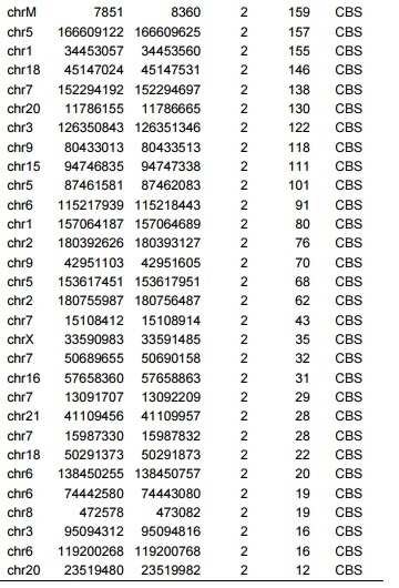
표 5. CAST-Seq의 민감도Table 5. Sensitivity of CAST-Seq

드롭렛 디지털 PCR (ddPCR)을 사용하여 CCR5 및 CCR2 유전자좌 사이의 대 CCR5/CCR2 결실 이벤트의 수를 결정하였다.Droplet digital PCR (ddPCR) was used to determine the number of versus CCR5/CCR2 deletion events between the CCR5 and CCR2 loci.
표 6: VEGFA 표적화 CRISPR-Cas9 뉴클레아제에 대한 CAST-Seq 분석Table 6: CAST-Seq analysis for VEGFA-targeted CRISPR-Cas9 nuclease


표 7: FANCF 표적화 CRISPR-Cas9 뉴클레아제에 대한 CAST-Seq 분석Table 7: CAST-Seq analysis for FANCF-targeted CRISPR-Cas9 nuclease


표 8. CAST-Seq에 사용된 소프트웨어 Table 8. Software used for CAST-Seq

표 9. CAST-Seq에 사용된 R 패키지Table 9. R packages used for CAST-Seq

표 10. 스코어링 매트릭스Table 10. Scoring Matrix

미스매치 및 벌지 (삽입/결실)에 대한 가중치를 포함하여 표적 부위 서열에 대한 전좌 부위의 정렬에 사용되는 뉴클레오티드 치환의 스코어링 매트릭스. A scoring matrix of nucleotide substitutions used for alignment of translocation sites to target site sequences, including weighting for mismatches and bulges (insertions/deletions).
IUPAC 코드가 사용된다. A, 아데닌; C, 시토신; G, 구아닌; T (또는 U), 티민 (또는 우라실); R, A 또는 G; Y, C 또는 T; S, G 또는 C; W, A 또는 T; K, G 또는 T; M, A 또는 C; B, C 또는 G 또는 T; D, A 또는 G 또는 T; H, A 또는 C 또는 T; V, A 또는 C 또는 G; N, 임의의 염기; n/a, 해당 없음.The IUPAC code is used. A, adenine; C, cytosine; G, guanine; T (or U), thymine (or uracil); R, A or G; Y, C or T; S, G or C; W, A or T; K, G or T; M, A or C; B, C or G or T; D, A or G or T; H, A or C or T; V, A or C or G; N, any base; n/a, not applicable.
표 11. (TALEN에 의해 표적화된) HBB 표적 부위에 대한 프라이머 설계Table 11. Primer designs for HBB target sites (targeted by TALEN)

표 12. CCR5 표적 부위 2 (CCR5#2, CRISPR-Cas9에 의해 표적화됨)에 대해 프라이머 설계Table 12. Primer design for CCR5 target site 2 (CCR5 #2 , targeted by CRISPR-Cas9)

표 13. ddPCR에 대한 프라이머 설계Table 13. Primer design for ddPCR

SEQUENCE LISTING <110> Albert-Ludwigs-Universit? Freiburg and Deutsches Krebsforschungszentrum Stiftung des ?fentlichen Rechts <120> Method for characterization of modifications caused by the use of designer nucleases <130> PWO00331ALB <150> EPA 18196438.8 <151> 2018-09-25 <160> 64 <170> PatentIn version 3.5 <210> 1 <211> 23 <212> DNA <213> artificial sequence <220> <223> CCR5 Target <400> 1 gtgagtagag cggaggcagg agg 23 <210> 2 <211> 23 <212> DNA <213> artificial sequence <220> <223> reference target <220> <221> misc_feature <222> (21)..(21) <223> n is a, c, g, t or u <400> 2 gtgagtagag cggaggcagg nrg 23 <210> 3 <211> 23 <212> DNA <213> artificial sequence <220> <223> VEGFA target <400> 3 ggtgagtgag tgtgtgcgtg tgg 23 <210> 4 <211> 23 <212> DNA <213> artificial sequence <220> <223> FANCF target <400> 4 ggaatccctt ctgcagcacc tgg 23 <210> 5 <211> 37 <212> DNA <213> artificial sequence <220> <223> linker positive strand <400> 5 gtaatacgac tcactatagg gctccgctta agggact 37 <210> 6 <211> 16 <212> DNA <213> artificial sequence <220> <223> linker negative strand <400> 6 gtcccttaag cggagc 16 <210> 7 <211> 22 <212> DNA <213> artificial sequence <220> <223> linker <400> 7 gtaatacgac tcactatagg gc 22 <210> 8 <211> 59 <212> DNA <213> artificial sequence <220> <223> linker <400> 8 acactctaca ctctttccct acacgacgct cttccgatct agggctccgc ttaagggac 59 <210> 9 <211> 20 <212> DNA <213> artificial sequence <220> <223> on target <400> 9 aggtagatgt cagtcatgct 20 <210> 10 <211> 21 <212> DNA <213> artificial sequence <220> <223> decoy fwd <400> 10 atcaatgtga agcaaatcgc a 21 <210> 11 <211> 21 <212> DNA <213> artificial sequence <220> <223> decoy rev <400> 11 atcaatgtga agcaaatcgc a 21 <210> 12 <211> 59 <212> DNA <213> artificial sequence <220> <223> on target nested <400> 12 gactggagtt cagacgtgtg ctcttccgat ctgctcttca gccttttgca gtttatcag 59 <210> 13 <211> 21 <212> DNA <213> artificial sequence <220> <223> on target <400> 13 ggattatcaa gtgtcaagtc c 21 <210> 14 <211> 18 <212> DNA <213> artificial sequence <220> <223> decoy fwd <400> 14 ctggtcatcc tcatcctg 18 <210> 15 <211> 22 <212> DNA <213> artificial sequence <220> <223> decoy rev <400> 15 aaaaccaaag atgaacacca gt 22 <210> 16 <211> 51 <212> DNA <213> artificial sequence <220> <223> on target nested <400> 16 gactggagtt cagacgtgtg ctcttccgat ctatacatcg gagccctgcc a 51 <210> 17 <211> 18 <212> DNA <213> artificial sequence <220> <223> VEGFA on target <400> 17 gagagggaca cacagatc 18 <210> 18 <211> 19 <212> DNA <213> artificial sequence <220> <223> decoy fwd <400> 18 cgtcttcgag agtgaggac 19 <210> 19 <211> 17 <212> DNA <213> artificial sequence <220> <223> decoy rev <400> 19 ctgctcgctc cattcac 17 <210> 20 <211> 59 <212> DNA <213> artificial sequence <220> <223> on target nested <400> 20 gactggagtt cagacgtgtg ctcttccgat ctacacagat ctattggaat cctggagtg 59 <210> 21 <211> 18 <212> DNA <213> artificial sequence <220> <223> FANCF on target <400> 21 gttccaatca gtacgcag 18 <210> 22 <211> 17 <212> DNA <213> artificial sequence <220> <223> decoy fwd <400> 22 cttgagaccg ccagaag 17 <210> 23 <211> 18 <212> DNA <213> artificial sequence <220> <223> decoy rev <400> 23 cactacctac gtcagcac 18 <210> 24 <211> 52 <212> DNA <213> artificial sequence <220> <223> on target nested <400> 24 gactggagtt cagacgtgtg ctcttccgat ctgccgtctc caaggtgaaa gc 52 <210> 25 <211> 19 <212> DNA <213> artificial sequence <220> <223> Talen target <400> 25 tgataggcac tgactctct 19 <210> 26 <211> 19 <212> DNA <213> artificial sequence <220> <223> right subunit <400> 26 taagggtggg aaaatagac 19 <210> 27 <211> 22 <212> DNA <213> artificial sequence <220> <223> bait <400> 27 gttggtatca aggttacaag ac 22 <210> 28 <211> 16 <212> DNA <213> artificial sequence <220> <223> decoy <400> 28 ctgctggtgg tctacc 16 <210> 29 <211> 56 <212> DNA <213> artificial sequence <220> <223> bait nested <400> 29 gactggagtt cagacgtgtg ctcttccgat ctgaccaata gaaactgggc atgtgg 56 <210> 30 <211> 23 <212> DNA <213> artificial sequence <220> <223> target <400> 30 caatgtgtca actcttgaca ggg 23 <210> 31 <211> 18 <212> DNA <213> artificial sequence <220> <223> bait <400> 31 aaacacagca tggacgac 18 <210> 32 <211> 21 <212> DNA <213> artificial sequence <220> <223> decoy <400> 32 ccagtgggac tttggaaata c 21 <210> 33 <211> 18 <212> DNA <213> artificial sequence <220> <223> decoy rev <400> 33 gcatagtgag cccagaag 18 <210> 34 <211> 59 <212> DNA <213> artificial sequence <220> <223> bait nested <400> 34 gactggagtt cagacgtgtg ctcttccgat ctaggaggat gatgaagaag attccagag 59 <210> 35 <211> 25 <212> DNA <213> artificial sequence <220> <223> forw <400> 35 ttattataca tcggagccct gccaa 25 <210> 36 <211> 28 <212> DNA <213> artificial sequence <220> <223> rev <400> 36 tgctcttcag ccttttgcag tttatcag 28 <210> 37 <211> 23 <212> DNA <213> artificial sequence <220> <223> ccr5 forw <400> 37 agtttgcatt catggagggc aac 23 <210> 38 <211> 26 <212> DNA <213> artificial sequence <220> <223> ccr5 rev <400> 38 ggcagggctc cgatgtataa taattg 26 <210> 39 <211> 22 <212> DNA <213> artificial sequence <220> <223> ccr5 forw <400> 39 catgctggtc atcctcatcc tg 22 <210> 40 <211> 23 <212> DNA <213> artificial sequence <220> <223> ccr5 rev <400> 40 cccagaaggg gacagtaaga agg 23 <210> 41 <211> 23 <212> DNA <213> artificial sequence <220> <223> edge forw <400> 41 tccttcttac tgtccccttc tgg 23 <210> 42 <211> 21 <212> DNA <213> artificial sequence <220> <223> edge rev <400> 42 agcaaacaca gcatggacga c 21 <210> 43 <211> 22 <212> DNA <213> artificial sequence <220> <223> ccr5 forw <400> 43 catgctggtc atcctcatcc tg 22 <210> 44 <211> 23 <212> DNA <213> artificial sequence <220> <223> ccr5 rev <400> 44 cccagaaggg gacagtaaga agg 23 <210> 45 <211> 22 <212> DNA <213> artificial sequence <220> <223> ccr5 forw <400> 45 atcgataggt acctggctgt cg 22 <210> 46 <211> 25 <212> DNA <213> artificial sequence <220> <223> ccr5 rev <400> 46 gtatggaaaa tgagagctgc aggtg 25 <210> 47 <211> 23 <212> DNA <213> artificial sequence <220> <223> ccr5 telomere forw <400> 47 tgccaaggca tcttacctct tcc 23 <210> 48 <211> 24 <212> DNA <213> artificial sequence <220> <223> ccr5 telomere rev <400> 48 gcatctggtc ttctgctaca ctgg 24 <210> 49 <211> 23 <212> DNA <213> artificial sequence <220> <223> ccr5 mylk forw <400> 49 cagccttgtg attcatgctg tcc 23 <210> 50 <211> 25 <212> DNA <213> artificial sequence <220> <223> ccr5 mylk rev <400> 50 ggactcacct tctactgtca actcc 25 <210> 51 <211> 25 <212> DNA <213> artificial sequence <220> <223> hbb edge forw <400> 51 agaccaatag aaactgggca tgtgg 25 <210> 52 <211> 22 <212> DNA <213> artificial sequence <220> <223> hbb edge rev <400> 52 atcactaaag gcaccgagca ct 22 <210> 53 <211> 22 <212> DNA <213> artificial sequence <220> <223> HBB forw <400> 53 ggctcatggc aagaaagtgc tc 22 <210> 54 <211> 21 <212> DNA <213> artificial sequence <220> <223> hbb rev <400> 54 cagtgcagct cactcagtgt g 21 <210> 55 <211> 22 <212> DNA <213> artificial sequence <220> <223> hbb rev <400> 55 ctgaggagaa gtctgccgtt ac 22 <210> 56 <211> 23 <212> DNA <213> artificial sequence <220> <223> hbb rev <400> 56 ccacatgccc agtttctatt ggt 23 <210> 57 <211> 21 <212> DNA <213> artificial sequence <220> <223> hbb telomere forw <400> 57 gggccaggga agtgtatgat g 21 <210> 58 <211> 21 <212> DNA <213> artificial sequence <220> <223> hbb telomer rev <400> 58 acagacatca gtgccattgc g 21 <210> 59 <211> 20 <212> DNA <213> artificial sequence <220> <223> hbb podl forw <400> 59 gcaggttcag tccctcttgg 20 <210> 60 <211> 21 <212> DNA <213> artificial sequence <220> <223> hbb podl rev <400> 60 tgcttggcct atggacagtt g 21 <210> 61 <211> 20 <212> DNA <213> artificial sequence <220> <223> common target rad forw <400> 61 ccttcagctc tgtggtgacg 20 <210> 62 <211> 21 <212> DNA <213> artificial sequence <220> <223> common target rad rev <400> 62 cccttctcag caaagtccct g 21 <210> 63 <211> 19 <212> DNA <213> artificial sequence <220> <223> common target stat forw <400> 63 actctcacgg acgaggagc 19 <210> 64 <211> 24 <212> DNA <213> artificial sequence <220> <223> common target stat rev <400> 64 cagttttcta gccgatctag gcag 24 SEQUENCE LISTING <110> Albert-Ludwigs-Universit? Freiburg and Deutsches Krebsforschungszentrum Stiftung des ?fentlichen Rechts <120> Method for characterization of modifications caused by the use of designer nucleases <130> PWO00331ALB <150> EPA 18196438.8 <151> 2018-09-25 <160> 64 <170> PatentIn version 3.5 <210> 1 <211> 23 <212> DNA <213> artificial sequence <220> <223> CCR5 Target <400> 1 gtgagtagag cggaggcagg agg 23 <210> 2 <211> 23 <212> DNA <213> artificial sequence <220> <223> reference target <220> <221> misc_feature <222> (21)..(21) <223> n is a, c, g, t or u <400> 2 gtgagtagag cggaggcagg nrg 23 <210> 3 <211> 23 <212> DNA <213> artificial sequence <220> <223> VEGFA target <400> 3 ggtgagtgag tgtgtgcgtg tgg 23 <210> 4 <211> 23 <212> DNA <213> artificial sequence <220> <223> FANCF target <400> 4 ggaatccctt ctgcagcacc tgg 23 <210> 5 <211> 37 <212> DNA <213> artificial sequence <220> <223> linker positive strand <400> 5 gtaatacgac tcactatagg gctccgctta agggact 37 <210> 6 <211> 16 <212> DNA <213> artificial sequence <220> <223> linker negative strand <400> 6 gtcccttaag cggagc 16 <210> 7 <211> 22 <212> DNA <213> artificial sequence <220> <223> linker <400> 7 gtaatacgac tcactatagg gc 22 <210> 8 <211> 59 <212> DNA <213> artificial sequence <220> <223> linker <400> 8 acactctaca ctctttccct acacgacgct cttccgatct agggctccgc ttaagggac 59 <210> 9 <211> 20 <212> DNA <213> artificial sequence <220> <223> on target <400> 9 aggtagatgt cagtcatgct 20 <210> 10 <211> 21 <212> DNA <213> artificial sequence <220> <223> decoy fwd <400> 10 atcaatgtga agcaaatcgc a 21 <210> 11 <211> 21 <212> DNA <213> artificial sequence <220> <223> decoy rev <400> 11 atcaatgtga agcaaatcgc a 21 <210> 12 <211> 59 <212> DNA <213> artificial sequence <220> <223> on target nested <400> 12 gactggagtt cagacgtgtg ctcttccgat ctgctcttca gccttttgca gtttatcag 59 <210> 13 <211> 21 <212> DNA <213> artificial sequence <220> <223> on target <400> 13 ggattatcaa gtgtcaagtc c 21 <210> 14 <211> 18 <212> DNA <213> artificial sequence <220> <223> decoy fwd <400> 14 ctggtcatcc tcatcctg 18 <210> 15 <211> 22 <212> DNA <213> artificial sequence <220> <223> decoy rev <400> 15 aaaaccaaag atgaacacca gt 22 <210> 16 <211> 51 <212> DNA <213> artificial sequence <220> <223> on target nested <400> 16 gactggagtt cagacgtgtg ctcttccgat ctatacatcg gagccctgcc a 51 <210> 17 <211> 18 <212> DNA <213> artificial sequence <220> <223> VEGFA on target <400> 17 gagagggaca cacagatc 18 <210> 18 <211> 19 <212> DNA <213> artificial sequence <220> <223> decoy fwd <400> 18 cgtcttcgag agtgaggac 19 <210> 19 <211> 17 <212> DNA <213> artificial sequence <220> <223> decoy rev <400> 19 ctgctcgctc cattcac 17 <210> 20 <211> 59 <212> DNA <213> artificial sequence <220> <223> on target nested <400> 20 gactggagtt cagacgtgtg ctcttccgat ctacacagat ctattggaat cctggagtg 59 <210> 21 <211> 18 <212> DNA <213> artificial sequence <220> <223> FANCF on target <400> 21 gttccaatca gtacgcag 18 <210> 22 <211> 17 <212> DNA <213> artificial sequence <220> <223> decoy fwd <400> 22 cttgagaccg ccagaag 17 <210> 23 <211> 18 <212> DNA <213> artificial sequence <220> <223> decoy rev <400> 23 cactacctac gtcagcac 18 <210> 24 <211> 52 <212> DNA <213> artificial sequence <220> <223> on target nested <400> 24 gactggagtt cagacgtgtg ctcttccgat ctgccgtctc caaggtgaaa gc 52 <210> 25 <211> 19 <212> DNA <213> artificial sequence <220> <223> Talen target <400> 25 tgataggcac tgactctct 19 <210> 26 <211> 19 <212> DNA <213> artificial sequence <220> <223> right subunit <400> 26 taagggtggg aaaatagac 19 <210> 27 <211> 22 <212> DNA <213> artificial sequence <220> <223> bait <400> 27 gttggtatca aggttacaag ac 22 <210> 28 <211> 16 <212> DNA <213> artificial sequence <220> <223> <400> 28 ctgctggtgg tctacc 16 <210> 29 <211> 56 <212> DNA <213> artificial sequence <220> <223> bait nested <400> 29 gactggagtt cagacgtgtg ctcttccgat ctgaccaata gaaactgggc atgtgg 56 <210> 30 <211> 23 <212> DNA <213> artificial sequence <220> <223> target <400> 30 caatgtgtca actcttgaca ggg 23 <210> 31 <211> 18 <212> DNA <213> artificial sequence <220> <223> bait <400> 31 aaacacagca tggacgac 18 <210> 32 <211> 21 <212> DNA <213> artificial sequence <220> <223> <400> 32 ccagtgggac tttggaaata c 21 <210> 33 <211> 18 <212> DNA <213> artificial sequence <220> <223> decoy rev <400> 33 gcatagtgag cccagaag 18 <210> 34 <211> 59 <212> DNA <213> artificial sequence <220> <223> bait nested <400> 34 gactggagtt cagacgtgtg ctcttccgat ctaggaggat gatgaagaag attccagag 59 <210> 35 <211> 25 <212> DNA <213> artificial sequence <220> <223> <400> 35 ttattataca tcggagccct gccaa 25 <210> 36 <211> 28 <212> DNA <213> artificial sequence <220> <223> rev <400> 36 tgctcttcag ccttttgcag tttatcag 28 <210> 37 <211> 23 <212> DNA <213> artificial sequence <220> <223> ccr5 forw <400> 37 agtttgcatt catggagggc aac 23 <210> 38 <211> 26 <212> DNA <213> artificial sequence <220> <223> ccr5 rev <400> 38 ggcagggctc cgatgtataa taattg 26 <210> 39 <211> 22 <212> DNA <213> artificial sequence <220> <223> ccr5 forw <400> 39 catgctggtc atcctcatcc tg 22 <210> 40 <211> 23 <212> DNA <213> artificial sequence <220> <223> ccr5 rev <400> 40 cccagaaggg gacagtaaga agg 23 <210> 41 <211> 23 <212> DNA <213> artificial sequence <220> <223> edge forward <400> 41 tccttcttac tgtccccttc tgg 23 <210> 42 <211> 21 <212> DNA <213> artificial sequence <220> <223> edge rev <400> 42 agcaaacaca gcatggacga c 21 <210> 43 <211> 22 <212> DNA <213> artificial sequence <220> <223> ccr5 forw <400> 43 catgctggtc atcctcatcc tg 22 <210> 44 <211> 23 <212> DNA <213> artificial sequence <220> <223> ccr5 rev <400> 44 cccagaaggg gacagtaaga agg 23 <210> 45 <211> 22 <212> DNA <213> artificial sequence <220> <223> ccr5 forw <400> 45 atcgataggt acctggctgt cg 22 <210> 46 <211> 25 <212> DNA <213> artificial sequence <220> <223> ccr5 rev <400> 46 gtatggaaaa tgagagctgc aggtg 25 <210> 47 <211> 23 <212> DNA <213> artificial sequence <220> <223> ccr5 telomere forw <400> 47 tgccaaggca tcttacctct tcc 23 <210> 48 <211> 24 <212> DNA <213> artificial sequence <220> <223> ccr5 telomere rev <400> 48 gcatctggtc ttctgctaca ctgg 24 <210> 49 <211> 23 <212> DNA <213> artificial sequence <220> <223> ccr5 mylk forw <400> 49 cagccttgtg attcatgctg tcc 23 <210> 50 <211> 25 <212> DNA <213> artificial sequence <220> <223> ccr5 mylk rev <400> 50 ggactcacct tctactgtca actcc 25 <210> 51 <211> 25 <212> DNA <213> artificial sequence <220> <223> hbb edge forward <400> 51 agaccaatag aaactgggca tgtgg 25 <210> 52 <211> 22 <212> DNA <213> artificial sequence <220> <223> hbb edge rev <400> 52 atcactaaag gcaccgagca ct 22 <210> 53 <211> 22 <212> DNA <213> artificial sequence <220> <223> HBB forward <400> 53 ggctcatggc aagaaagtgc tc 22 <210> 54 <211> 21 <212> DNA <213> artificial sequence <220> <223> hbb rev <400> 54 cagtgcagct cactcagtgt g 21 <210> 55 <211> 22 <212> DNA <213> artificial sequence <220> <223> hbb rev <400> 55 ctgaggagaa gtctgccgtt ac 22 <210> 56 <211> 23 <212> DNA <213> artificial sequence <220> <223> hbb rev <400> 56 ccacatgccc agtttctatt ggt 23 <210> 57 <211> 21 <212> DNA <213> artificial sequence <220> <223> hbb telomere forw <400> 57 gggccaggga agtgtatgat g 21 <210> 58 <211> 21 <212> DNA <213> artificial sequence <220> <223> hbb telomer rev <400> 58 acagacatca gtgccattgc g 21 <210> 59 <211> 20 <212> DNA <213> artificial sequence <220> <223> hbb podl forw <400> 59 gcaggttcag tccctcttgg 20 <210> 60 <211> 21 <212> DNA <213> artificial sequence <220> <223> hbb podl rev <400> 60 tgcttggcct atggacagtt g 21 <210> 61 <211> 20 <212> DNA <213> artificial sequence <220> <223> common target rad forward <400> 61 ccttcagctc tgtggtgacg 20 <210> 62 <211> 21 <212> DNA <213> artificial sequence <220> <223> common target rad rev <400> 62 cccttctcag caaagtccct g 21 <210> 63 <211> 19 <212> DNA <213> artificial sequence <220> <223> common target stat forward <400> 63 actctcacgg acgaggagc 19 <210> 64 <211> 24 <212> DNA <213> artificial sequence <220> <223> common target stat rev <400> 64 cagttttcta gccgatctag gcag 24
Claims (15)
b) 핵산을 단편화하여 무작위 단편을 수득하는 단계,
c) 블런트 말단을 수득하기 위해 말단 복구를 수행하는 단계,
d) 소위 "링커 프라이머"에 상보적인 서열을 포함하는 링커와 결찰하는 단계,
e) 하나의 프라이머는 온-타겟 부위의 상류에 위치하고 하나의 프라이머는 온-타겟 부위의 하류에 위치하도록 "링커 프라이머" 및 소위 "온-타겟 (ON-target) 프라이머"로 첫 번째 핵산 증폭 반응을 수행하는 단계로서, 적어도 하나의 디코이 프라이머가 반응 혼합물에 존재하는 단계,
f) 소위 "네스티드 프라이머 (nested primer)"가 반응 혼합물에 첨가되어 하나의 프라이머는 온-타겟 유전자좌에 상보적이고 하나의 프라이머는 링커 서열에 상보적이도록 두 번째 핵산 증폭 반응을 수행하는 단계,
g) 적어도 하나의 코드 함유 프라이머가 반응 혼합물에 첨가되도록 추가의 핵산 증폭 반응을 수행하는 단계,
h) 네스티드 및 바코딩된 증폭 산물을 서열분석하는 단계, 및
i) 적어도 하나의 DNA 이중 가닥 절단을 기반으로 게놈 변형을 포함하는 염색체 위치를 식별하기 위해 적절한 생물정보학적 수단을 사용하여 서열화된 산물을 기준 서열에 정렬하는 단계를 포함하는, 설계자 뉴클레아제의 활성에 의해 유발된 세포 또는 조직으로부터 수득된 핵산 게놈의 게놈-전체 변형의 고-처리량 검출 방법.a) extracting genomic DNA from the cells exposed to the designer nuclease under conditions such that the designer nuclease can introduce a DNA double-stranded break (DSB) into the genomic DNA of the cell;
b) fragmenting the nucleic acid to obtain random fragments;
c) performing end repair to obtain a blunt end;
d) ligating with a linker comprising a sequence complementary to a so-called “linker primer”;
e) the first nucleic acid amplification reaction with a “linker primer” and a so-called “ON-target primer” such that one primer is located upstream of the on-target site and one primer is located downstream of the on-target site wherein at least one decoy primer is present in the reaction mixture;
f) so-called "nested primers" are added to the reaction mixture to perform a second nucleic acid amplification reaction such that one primer is complementary to the on-target locus and one primer is complementary to the linker sequence;
g) performing a further nucleic acid amplification reaction such that at least one code containing primer is added to the reaction mixture;
h) sequencing the nested and barcoded amplification products, and
i) aligning the sequenced product to a reference sequence using appropriate bioinformatic means to identify a chromosomal location comprising a genomic modification based on at least one DNA double-strand break. A method for high-throughput detection of a genome-wide modification of a nucleic acid genome obtained from a cell or tissue induced by the activity.
Applications Claiming Priority (3)
| Application Number | Priority Date | Filing Date | Title |
|---|---|---|---|
| EP18196438.8 | 2018-09-25 | ||
| EP18196438.8A EP3628748A1 (en) | 2018-09-25 | 2018-09-25 | Method for characterization of modifications caused by the use of designer nucleases |
| PCT/EP2019/075101 WO2020064478A1 (en) | 2018-09-25 | 2019-09-19 | Method for characterization of modifications caused by the use of designer nucleases |
Publications (2)
| Publication Number | Publication Date |
|---|---|
| KR20210065085A true KR20210065085A (en) | 2021-06-03 |
| KR102899840B1 KR102899840B1 (en) | 2025-12-11 |
Family
ID=63794292
Family Applications (1)
| Application Number | Title | Priority Date | Filing Date |
|---|---|---|---|
| KR1020217003131A Active KR102899840B1 (en) | 2018-09-25 | 2019-09-19 | Methods for characterizing modifications induced by the use of designer nucleases |
Country Status (8)
| Country | Link |
|---|---|
| US (1) | US11319580B2 (en) |
| EP (2) | EP3628748A1 (en) |
| JP (2) | JP2022512530A (en) |
| KR (1) | KR102899840B1 (en) |
| DK (1) | DK3856928T3 (en) |
| ES (1) | ES2937986T3 (en) |
| LT (1) | LT3856928T (en) |
| WO (1) | WO2020064478A1 (en) |
Families Citing this family (3)
| Publication number | Priority date | Publication date | Assignee | Title |
|---|---|---|---|---|
| IL298058A (en) * | 2020-05-11 | 2023-01-01 | Univ Bar Ilan | Methods and systems for determining effects of nucleic acid editing |
| CN112708662B (en) * | 2020-12-28 | 2021-08-31 | 阅尔基因技术(苏州)有限公司 | Nucleotide composition for inhibiting non-target area in biological DNA sample and application thereof |
| CN114032292B (en) * | 2021-12-17 | 2024-04-12 | 北京大学 | A method for absolute quantification of DNA double-strand breaks in cells and its application |
Citations (2)
| Publication number | Priority date | Publication date | Assignee | Title |
|---|---|---|---|---|
| WO2016081798A1 (en) * | 2014-11-20 | 2016-05-26 | Children's Medical Center Corporation | Methods relating to the detection of recurrent and non-specific double strand breaks in the genome |
| WO2018129368A2 (en) * | 2017-01-06 | 2018-07-12 | Editas Medicine, Inc. | Methods of assessing nuclease cleavage |
Family Cites Families (2)
| Publication number | Priority date | Publication date | Assignee | Title |
|---|---|---|---|---|
| WO2016076672A1 (en) | 2014-11-14 | 2016-05-19 | 기초과학연구원 | Method for detecting off-target site of genetic scissors in genome |
| EP3541955A1 (en) * | 2016-11-15 | 2019-09-25 | Genomic Vision | Method for the monitoring of modified nucleases induced-gene editing events by molecular combing |
-
2018
- 2018-09-25 EP EP18196438.8A patent/EP3628748A1/en not_active Withdrawn
-
2019
- 2019-09-19 EP EP19769176.9A patent/EP3856928B1/en active Active
- 2019-09-19 US US17/277,236 patent/US11319580B2/en active Active
- 2019-09-19 LT LTEPPCT/EP2019/075101T patent/LT3856928T/en unknown
- 2019-09-19 DK DK19769176.9T patent/DK3856928T3/en active
- 2019-09-19 KR KR1020217003131A patent/KR102899840B1/en active Active
- 2019-09-19 ES ES19769176T patent/ES2937986T3/en active Active
- 2019-09-19 JP JP2021505776A patent/JP2022512530A/en active Pending
- 2019-09-19 WO PCT/EP2019/075101 patent/WO2020064478A1/en not_active Ceased
-
2024
- 2024-05-31 JP JP2024088459A patent/JP2024113001A/en active Pending
Patent Citations (2)
| Publication number | Priority date | Publication date | Assignee | Title |
|---|---|---|---|---|
| WO2016081798A1 (en) * | 2014-11-20 | 2016-05-26 | Children's Medical Center Corporation | Methods relating to the detection of recurrent and non-specific double strand breaks in the genome |
| WO2018129368A2 (en) * | 2017-01-06 | 2018-07-12 | Editas Medicine, Inc. | Methods of assessing nuclease cleavage |
Also Published As
| Publication number | Publication date |
|---|---|
| EP3628748A1 (en) | 2020-04-01 |
| ES2937986T3 (en) | 2023-04-03 |
| KR102899840B1 (en) | 2025-12-11 |
| US20210317514A1 (en) | 2021-10-14 |
| DK3856928T3 (en) | 2023-01-30 |
| EP3856928B1 (en) | 2022-11-09 |
| LT3856928T (en) | 2023-02-27 |
| WO2020064478A1 (en) | 2020-04-02 |
| EP3856928A1 (en) | 2021-08-04 |
| US11319580B2 (en) | 2022-05-03 |
| JP2024113001A (en) | 2024-08-21 |
| JP2022512530A (en) | 2022-02-07 |
Similar Documents
| Publication | Publication Date | Title |
|---|---|---|
| Manghwar et al. | CRISPR/Cas systems in genome editing: methodologies and tools for sgRNA design, off‐target evaluation, and strategies to mitigate off‐target effects | |
| JP7550816B2 (en) | Genome-wide, unbiased identification of DSBs assessed by sequencing (GUIDE-Seq) | |
| Kosicki et al. | Repair of double-strand breaks induced by CRISPR–Cas9 leads to large deletions and complex rearrangements | |
| Wang et al. | Extrachromosomal circular DNA: a new potential role in cancer progression | |
| EP3469079B1 (en) | Enrichment of mutated cell free nucleic acids for cancer detection | |
| López-Girona et al. | CRISPR-Cas9 enrichment and long read sequencing for fine mapping in plants | |
| Barlow et al. | Identification of early replicating fragile sites that contribute to genome instability | |
| St. Charles et al. | High-resolution genome-wide analysis of irradiated (UV and γ-rays) diploid yeast cells reveals a high frequency of genomic loss of heterozygosity (LOH) events | |
| JP2022528139A (en) | Methods and Compositions for Analyzing Nucleic Acids | |
| JP2024113001A (en) | Methods for characterizing modifications using designer nucleases | |
| US20220136041A1 (en) | Off-Target Single Nucleotide Variants Caused by Single-Base Editing and High-Specificity Off-Target-Free Single-Base Gene Editing Tool | |
| JP2018529353A (en) | Comprehensive in vitro reporting of cleavage events by sequencing (CIRCLE-seq) | |
| Cai et al. | Complex genomic patterns of abasic sites in mammalian DNA revealed by a high-resolution SSiNGLe-AP method | |
| Wu et al. | Innovative insights into extrachromosomal circular DNAs in gynecologic tumors and reproduction | |
| Park et al. | Detection and quantification of unintended large on-target gene modifications due to CRISPR/Cas9 editing | |
| US9518293B2 (en) | High throughput genome-wide translocation sequencing | |
| Pfeifer et al. | De novo genome assembly for the coppery titi monkey (Plecturocebus cupreus): an emerging nonhuman primate model for behavioral research | |
| WO2024033378A1 (en) | Method of parallel, rapid and sensitive detection of dna double strand breaks | |
| WO2022243748A2 (en) | Methods of enriching targeted nucleic acid, identifying off-target and evaluating gene editing efficiency | |
| EP4321630A1 (en) | Method of parallel, rapid and sensitive detection of dna double strand breaks | |
| Uppuluri et al. | Linked-Pair Long-Read Sequencing Strategy for Targeted Resequencing and Enrichment | |
| Prykhozhij et al. | Successful optimization of CRISPR/Cas9-mediated defined point mutation knock-in using allele-specific PCR assays in zebrafish | |
| HK40024796A (en) | A method and a kit for constructing sequencing library for simultaneously realizing genome copy number variation detection and gene mutation detection |
Legal Events
| Date | Code | Title | Description |
|---|---|---|---|
| E13-X000 | Pre-grant limitation requested |
St.27 status event code: A-2-3-E10-E13-lim-X000 |
|
| PA0105 | International application |
St.27 status event code: A-0-1-A10-A15-nap-PA0105 |
|
| PG1501 | Laying open of application |
St.27 status event code: A-1-1-Q10-Q12-nap-PG1501 |
|
| P11-X000 | Amendment of application requested |
St.27 status event code: A-2-2-P10-P11-nap-X000 |
|
| P13-X000 | Application amended |
St.27 status event code: A-2-2-P10-P13-nap-X000 |
|
| PA0201 | Request for examination |
St.27 status event code: A-1-2-D10-D11-exm-PA0201 |
|
| E902 | Notification of reason for refusal | ||
| PE0902 | Notice of grounds for rejection |
St.27 status event code: A-1-2-D10-D21-exm-PE0902 |
|
| T11-X000 | Administrative time limit extension requested |
St.27 status event code: U-3-3-T10-T11-oth-X000 |
|
| T11-X000 | Administrative time limit extension requested |
St.27 status event code: U-3-3-T10-T11-oth-X000 |
|
| P11-X000 | Amendment of application requested |
St.27 status event code: A-2-2-P10-P11-nap-X000 |
|
| D22 | Grant of ip right intended |
Free format text: ST27 STATUS EVENT CODE: A-1-2-D10-D22-EXM-PE0701 (AS PROVIDED BY THE NATIONAL OFFICE) |
|
| PE0701 | Decision of registration |
St.27 status event code: A-1-2-D10-D22-exm-PE0701 |
|
| F11 | Ip right granted following substantive examination |
Free format text: ST27 STATUS EVENT CODE: A-2-4-F10-F11-EXM-PR0701 (AS PROVIDED BY THE NATIONAL OFFICE) |
|
| PR0701 | Registration of establishment |
St.27 status event code: A-2-4-F10-F11-exm-PR0701 |
|
| PR1002 | Payment of registration fee |
St.27 status event code: A-2-2-U10-U12-oth-PR1002 Fee payment year number: 1 |
|
| U12 | Designation fee paid |
Free format text: ST27 STATUS EVENT CODE: A-2-2-U10-U12-OTH-PR1002 (AS PROVIDED BY THE NATIONAL OFFICE) Year of fee payment: 1 |
|
| PG1601 | Publication of registration |
St.27 status event code: A-4-4-Q10-Q13-nap-PG1601 |
|
| Q13 | Ip right document published |
Free format text: ST27 STATUS EVENT CODE: A-4-4-Q10-Q13-NAP-PG1601 (AS PROVIDED BY THE NATIONAL OFFICE) |