KR20160005731A - Method for predicting recurrence of stomach cancer - Google Patents
Method for predicting recurrence of stomach cancer Download PDFInfo
- Publication number
- KR20160005731A KR20160005731A KR1020157034350A KR20157034350A KR20160005731A KR 20160005731 A KR20160005731 A KR 20160005731A KR 1020157034350 A KR1020157034350 A KR 1020157034350A KR 20157034350 A KR20157034350 A KR 20157034350A KR 20160005731 A KR20160005731 A KR 20160005731A
- Authority
- KR
- South Korea
- Prior art keywords
- probe
- fragment
- marker
- marker gene
- seq
- Prior art date
- Legal status (The legal status is an assumption and is not a legal conclusion. Google has not performed a legal analysis and makes no representation as to the accuracy of the status listed.)
- Withdrawn
Links
- 208000005718 Stomach Neoplasms Diseases 0.000 title claims abstract description 101
- 206010017758 gastric cancer Diseases 0.000 title claims abstract description 101
- 201000011549 stomach cancer Diseases 0.000 title claims abstract description 101
- 238000000034 method Methods 0.000 title claims description 83
- 239000000523 sample Substances 0.000 claims abstract description 249
- 239000003550 marker Substances 0.000 claims abstract description 200
- 108090000623 proteins and genes Proteins 0.000 claims abstract description 161
- 239000012634 fragment Substances 0.000 claims abstract description 97
- 238000001514 detection method Methods 0.000 claims abstract description 65
- 238000005259 measurement Methods 0.000 claims abstract description 49
- 102100032700 Keratin, type I cytoskeletal 20 Human genes 0.000 claims abstract description 41
- 101000994460 Homo sapiens Keratin, type I cytoskeletal 20 Proteins 0.000 claims abstract description 40
- 102100026745 Fatty acid-binding protein, liver Human genes 0.000 claims abstract description 39
- 101001133081 Homo sapiens Mucin-2 Proteins 0.000 claims abstract description 36
- 102100034263 Mucin-2 Human genes 0.000 claims abstract description 35
- 101000914324 Homo sapiens Carcinoembryonic antigen-related cell adhesion molecule 5 Proteins 0.000 claims abstract 11
- 101000914321 Homo sapiens Carcinoembryonic antigen-related cell adhesion molecule 7 Proteins 0.000 claims abstract 11
- 101000617725 Homo sapiens Pregnancy-specific beta-1-glycoprotein 2 Proteins 0.000 claims abstract 11
- 108010088412 Trefoil Factor-1 Proteins 0.000 claims abstract 11
- 102000008817 Trefoil Factor-1 Human genes 0.000 claims abstract 11
- 101000911317 Homo sapiens Fatty acid-binding protein, liver Proteins 0.000 claims abstract 9
- 102100022019 Pregnancy-specific beta-1-glycoprotein 2 Human genes 0.000 claims abstract 3
- 108020004414 DNA Proteins 0.000 claims description 101
- 238000003752 polymerase chain reaction Methods 0.000 claims description 56
- 201000011510 cancer Diseases 0.000 claims description 45
- 206010028980 Neoplasm Diseases 0.000 claims description 42
- 239000013641 positive control Substances 0.000 claims description 31
- 241000219194 Arabidopsis Species 0.000 claims description 30
- 150000007523 nucleic acids Chemical group 0.000 claims description 28
- 238000009396 hybridization Methods 0.000 claims description 27
- 230000003321 amplification Effects 0.000 claims description 24
- 238000003199 nucleic acid amplification method Methods 0.000 claims description 24
- 108091028043 Nucleic acid sequence Proteins 0.000 claims description 15
- 238000002372 labelling Methods 0.000 claims description 12
- 241000196324 Embryophyta Species 0.000 claims description 10
- 102000053602 DNA Human genes 0.000 claims description 9
- 239000002773 nucleotide Substances 0.000 claims description 9
- 125000003729 nucleotide group Chemical group 0.000 claims description 9
- 239000000203 mixture Substances 0.000 claims description 7
- FWMNVWWHGCHHJJ-SKKKGAJSSA-N 4-amino-1-[(2r)-6-amino-2-[[(2r)-2-[[(2r)-2-[[(2r)-2-amino-3-phenylpropanoyl]amino]-3-phenylpropanoyl]amino]-4-methylpentanoyl]amino]hexanoyl]piperidine-4-carboxylic acid Chemical compound C([C@H](C(=O)N[C@H](CC(C)C)C(=O)N[C@H](CCCCN)C(=O)N1CCC(N)(CC1)C(O)=O)NC(=O)[C@H](N)CC=1C=CC=CC=1)C1=CC=CC=C1 FWMNVWWHGCHHJJ-SKKKGAJSSA-N 0.000 claims description 4
- 244000005700 microbiome Species 0.000 claims description 4
- 230000003100 immobilizing effect Effects 0.000 claims description 2
- 108020004707 nucleic acids Proteins 0.000 claims description 2
- 102000039446 nucleic acids Human genes 0.000 claims description 2
- 108700026220 vif Genes Proteins 0.000 claims 3
- 239000000758 substrate Substances 0.000 description 34
- 101710083182 Fatty acid-binding protein 1 Proteins 0.000 description 30
- 101710188974 Fatty acid-binding protein, liver Proteins 0.000 description 30
- 101710189565 Fatty acid-binding protein, liver-type Proteins 0.000 description 30
- 210000004027 cell Anatomy 0.000 description 28
- 238000000018 DNA microarray Methods 0.000 description 26
- 239000000243 solution Substances 0.000 description 24
- 238000006243 chemical reaction Methods 0.000 description 23
- 239000000047 product Substances 0.000 description 23
- 208000007433 Lymphatic Metastasis Diseases 0.000 description 22
- 230000000750 progressive effect Effects 0.000 description 19
- 101150084967 EPCAM gene Proteins 0.000 description 15
- 102100031940 Epithelial cell adhesion molecule Human genes 0.000 description 15
- 101000756632 Homo sapiens Actin, cytoplasmic 1 Proteins 0.000 description 15
- 101150057140 TACSTD1 gene Proteins 0.000 description 15
- 108091032973 (ribonucleotides)n+m Proteins 0.000 description 14
- 239000000178 monomer Substances 0.000 description 13
- 229920000642 polymer Polymers 0.000 description 13
- 230000000052 comparative effect Effects 0.000 description 12
- 239000002299 complementary DNA Substances 0.000 description 11
- 230000034994 death Effects 0.000 description 11
- 231100000517 death Toxicity 0.000 description 11
- 238000007912 intraperitoneal administration Methods 0.000 description 11
- 201000011591 microinvasive gastric cancer Diseases 0.000 description 11
- 230000035945 sensitivity Effects 0.000 description 10
- -1 CEA Proteins 0.000 description 9
- 239000013642 negative control Substances 0.000 description 8
- 238000004393 prognosis Methods 0.000 description 8
- 238000001356 surgical procedure Methods 0.000 description 8
- 206010027476 Metastases Diseases 0.000 description 7
- 125000003277 amino group Chemical group 0.000 description 7
- 238000011156 evaluation Methods 0.000 description 7
- 239000007788 liquid Substances 0.000 description 7
- 210000001165 lymph node Anatomy 0.000 description 7
- 239000000126 substance Substances 0.000 description 7
- 238000012408 PCR amplification Methods 0.000 description 6
- 206010033661 Pancytopenia Diseases 0.000 description 6
- 210000000683 abdominal cavity Anatomy 0.000 description 6
- 239000000872 buffer Substances 0.000 description 6
- 239000011247 coating layer Substances 0.000 description 6
- 229920001577 copolymer Polymers 0.000 description 6
- 125000004122 cyclic group Chemical group 0.000 description 6
- 208000024389 cytopenia Diseases 0.000 description 6
- 238000002493 microarray Methods 0.000 description 6
- 239000004033 plastic Substances 0.000 description 6
- 229920003023 plastic Polymers 0.000 description 6
- 229920000098 polyolefin Polymers 0.000 description 6
- 238000012360 testing method Methods 0.000 description 6
- 238000005406 washing Methods 0.000 description 6
- ZSZRUEAFVQITHH-UHFFFAOYSA-N 2-(2-methylprop-2-enoyloxy)ethyl 2-(trimethylazaniumyl)ethyl phosphate Chemical compound CC(=C)C(=O)OCCOP([O-])(=O)OCC[N+](C)(C)C ZSZRUEAFVQITHH-UHFFFAOYSA-N 0.000 description 5
- 108010014303 DNA-directed DNA polymerase Proteins 0.000 description 5
- 102000016928 DNA-directed DNA polymerase Human genes 0.000 description 5
- 239000011521 glass Substances 0.000 description 5
- 230000009401 metastasis Effects 0.000 description 5
- 238000010839 reverse transcription Methods 0.000 description 5
- XLYOFNOQVPJJNP-UHFFFAOYSA-N water Substances O XLYOFNOQVPJJNP-UHFFFAOYSA-N 0.000 description 5
- ZHNUHDYFZUAESO-UHFFFAOYSA-N Formamide Chemical compound NC=O ZHNUHDYFZUAESO-UHFFFAOYSA-N 0.000 description 4
- 238000004458 analytical method Methods 0.000 description 4
- 238000003745 diagnosis Methods 0.000 description 4
- 238000000605 extraction Methods 0.000 description 4
- 239000010410 layer Substances 0.000 description 4
- 239000000463 material Substances 0.000 description 4
- 239000002861 polymer material Substances 0.000 description 4
- 229920005989 resin Polymers 0.000 description 4
- 239000011347 resin Substances 0.000 description 4
- 239000006228 supernatant Substances 0.000 description 4
- 108091029845 Aminoallyl nucleotide Proteins 0.000 description 3
- 241000219195 Arabidopsis thaliana Species 0.000 description 3
- SOGAXMICEFXMKE-UHFFFAOYSA-N Butylmethacrylate Chemical compound CCCCOC(=O)C(C)=C SOGAXMICEFXMKE-UHFFFAOYSA-N 0.000 description 3
- AHCYMLUZIRLXAA-SHYZEUOFSA-N Deoxyuridine 5'-triphosphate Chemical compound O1[C@H](COP(O)(=O)OP(O)(=O)OP(O)(O)=O)[C@@H](O)C[C@@H]1N1C(=O)NC(=O)C=C1 AHCYMLUZIRLXAA-SHYZEUOFSA-N 0.000 description 3
- OKKJLVBELUTLKV-UHFFFAOYSA-N Methanol Chemical compound OC OKKJLVBELUTLKV-UHFFFAOYSA-N 0.000 description 3
- 238000000137 annealing Methods 0.000 description 3
- 239000007853 buffer solution Substances 0.000 description 3
- 230000032823 cell division Effects 0.000 description 3
- 238000005119 centrifugation Methods 0.000 description 3
- 239000003153 chemical reaction reagent Substances 0.000 description 3
- 230000000295 complement effect Effects 0.000 description 3
- 238000003505 heat denaturation Methods 0.000 description 3
- 239000008188 pellet Substances 0.000 description 3
- 230000002980 postoperative effect Effects 0.000 description 3
- 238000000746 purification Methods 0.000 description 3
- 229920006395 saturated elastomer Polymers 0.000 description 3
- 229920005992 thermoplastic resin Polymers 0.000 description 3
- 239000000439 tumor marker Substances 0.000 description 3
- VXNZUUAINFGPBY-UHFFFAOYSA-N 1-Butene Chemical compound CCC=C VXNZUUAINFGPBY-UHFFFAOYSA-N 0.000 description 2
- 229920001342 Bakelite® Polymers 0.000 description 2
- HEDRZPFGACZZDS-UHFFFAOYSA-N Chloroform Chemical compound ClC(Cl)Cl HEDRZPFGACZZDS-UHFFFAOYSA-N 0.000 description 2
- 102000004190 Enzymes Human genes 0.000 description 2
- 108090000790 Enzymes Proteins 0.000 description 2
- LFQSCWFLJHTTHZ-UHFFFAOYSA-N Ethanol Chemical compound CCO LFQSCWFLJHTTHZ-UHFFFAOYSA-N 0.000 description 2
- VGGSQFUCUMXWEO-UHFFFAOYSA-N Ethene Chemical compound C=C VGGSQFUCUMXWEO-UHFFFAOYSA-N 0.000 description 2
- 239000005977 Ethylene Substances 0.000 description 2
- LYCAIKOWRPUZTN-UHFFFAOYSA-N Ethylene glycol Chemical compound OCCO LYCAIKOWRPUZTN-UHFFFAOYSA-N 0.000 description 2
- 102100034343 Integrase Human genes 0.000 description 2
- KFZMGEQAYNKOFK-UHFFFAOYSA-N Isopropanol Chemical compound CC(C)O KFZMGEQAYNKOFK-UHFFFAOYSA-N 0.000 description 2
- 206010027459 Metastases to lymph nodes Diseases 0.000 description 2
- 108010092799 RNA-directed DNA polymerase Proteins 0.000 description 2
- 108010005173 SERPIN-B5 Proteins 0.000 description 2
- 102100030333 Serpin B5 Human genes 0.000 description 2
- 208000037065 Subacute sclerosing leukoencephalitis Diseases 0.000 description 2
- 206010042297 Subacute sclerosing panencephalitis Diseases 0.000 description 2
- 238000003556 assay Methods 0.000 description 2
- 210000003050 axon Anatomy 0.000 description 2
- 230000008901 benefit Effects 0.000 description 2
- 150000001732 carboxylic acid derivatives Chemical class 0.000 description 2
- 230000010261 cell growth Effects 0.000 description 2
- 238000004140 cleaning Methods 0.000 description 2
- 150000001875 compounds Chemical class 0.000 description 2
- 238000007796 conventional method Methods 0.000 description 2
- 150000002148 esters Chemical class 0.000 description 2
- 238000010195 expression analysis Methods 0.000 description 2
- 239000000835 fiber Substances 0.000 description 2
- 239000012530 fluid Substances 0.000 description 2
- 239000007850 fluorescent dye Substances 0.000 description 2
- 238000002350 laparotomy Methods 0.000 description 2
- 238000007403 mPCR Methods 0.000 description 2
- 238000004519 manufacturing process Methods 0.000 description 2
- 210000004379 membrane Anatomy 0.000 description 2
- 239000012528 membrane Substances 0.000 description 2
- JFNLZVQOOSMTJK-KNVOCYPGSA-N norbornene Chemical compound C1[C@@H]2CC[C@H]1C=C2 JFNLZVQOOSMTJK-KNVOCYPGSA-N 0.000 description 2
- JRZJOMJEPLMPRA-UHFFFAOYSA-N olefin Natural products CCCCCCCC=C JRZJOMJEPLMPRA-UHFFFAOYSA-N 0.000 description 2
- 230000003287 optical effect Effects 0.000 description 2
- 230000001575 pathological effect Effects 0.000 description 2
- YWAKXRMUMFPDSH-UHFFFAOYSA-N pentene Chemical compound CCCC=C YWAKXRMUMFPDSH-UHFFFAOYSA-N 0.000 description 2
- 210000004303 peritoneum Anatomy 0.000 description 2
- 150000003904 phospholipids Chemical class 0.000 description 2
- 125000004805 propylene group Chemical group [H]C([H])([H])C([H])([*:1])C([H])([H])[*:2] 0.000 description 2
- 230000009257 reactivity Effects 0.000 description 2
- 230000000306 recurrent effect Effects 0.000 description 2
- 238000011160 research Methods 0.000 description 2
- 238000002271 resection Methods 0.000 description 2
- 238000007151 ring opening polymerisation reaction Methods 0.000 description 2
- 210000002784 stomach Anatomy 0.000 description 2
- 230000004083 survival effect Effects 0.000 description 2
- HECLRDQVFMWTQS-RGOKHQFPSA-N 1755-01-7 Chemical compound C1[C@H]2[C@@H]3CC=C[C@@H]3[C@@H]1C=C2 HECLRDQVFMWTQS-RGOKHQFPSA-N 0.000 description 1
- NJNWCIAPVGRBHO-UHFFFAOYSA-N 2-hydroxyethyl-dimethyl-[(oxo-$l^{5}-phosphanylidyne)methyl]azanium Chemical group OCC[N+](C)(C)C#P=O NJNWCIAPVGRBHO-UHFFFAOYSA-N 0.000 description 1
- YHQXBTXEYZIYOV-UHFFFAOYSA-N 3-methylbut-1-ene Chemical compound CC(C)C=C YHQXBTXEYZIYOV-UHFFFAOYSA-N 0.000 description 1
- QRXMUCSWCMTJGU-UHFFFAOYSA-N 5-bromo-4-chloro-3-indolyl phosphate Chemical compound C1=C(Br)C(Cl)=C2C(OP(O)(=O)O)=CNC2=C1 QRXMUCSWCMTJGU-UHFFFAOYSA-N 0.000 description 1
- PCBPVYHMZBWMAZ-UHFFFAOYSA-N 5-methylbicyclo[2.2.1]hept-2-ene Chemical compound C1C2C(C)CC1C=C2 PCBPVYHMZBWMAZ-UHFFFAOYSA-N 0.000 description 1
- 102000007469 Actins Human genes 0.000 description 1
- 108010085238 Actins Proteins 0.000 description 1
- 229930024421 Adenine Natural products 0.000 description 1
- GFFGJBXGBJISGV-UHFFFAOYSA-N Adenine Chemical compound NC1=NC=NC2=C1N=CN2 GFFGJBXGBJISGV-UHFFFAOYSA-N 0.000 description 1
- 102000002260 Alkaline Phosphatase Human genes 0.000 description 1
- 108020004774 Alkaline Phosphatase Proteins 0.000 description 1
- 241000256844 Apis mellifera Species 0.000 description 1
- 108020003215 DNA Probes Proteins 0.000 description 1
- 239000003298 DNA probe Substances 0.000 description 1
- YCKRFDGAMUMZLT-UHFFFAOYSA-N Fluorine atom Chemical compound [F] YCKRFDGAMUMZLT-UHFFFAOYSA-N 0.000 description 1
- 101001120760 Homo sapiens Olfactomedin-4 Proteins 0.000 description 1
- 101000847952 Homo sapiens Trypsin-3 Proteins 0.000 description 1
- 108010066370 Keratin-20 Proteins 0.000 description 1
- CERQOIWHTDAKMF-UHFFFAOYSA-M Methacrylate Chemical compound CC(=C)C([O-])=O CERQOIWHTDAKMF-UHFFFAOYSA-M 0.000 description 1
- 244000061176 Nicotiana tabacum Species 0.000 description 1
- 235000002637 Nicotiana tabacum Nutrition 0.000 description 1
- 102100026071 Olfactomedin-4 Human genes 0.000 description 1
- 240000007594 Oryza sativa Species 0.000 description 1
- 235000007164 Oryza sativa Nutrition 0.000 description 1
- 229910019142 PO4 Inorganic materials 0.000 description 1
- 239000004698 Polyethylene Substances 0.000 description 1
- 239000004743 Polypropylene Substances 0.000 description 1
- 108020004682 Single-Stranded DNA Proteins 0.000 description 1
- 108010088411 Trefoil Factor-2 Proteins 0.000 description 1
- 102000008816 Trefoil Factor-2 Human genes 0.000 description 1
- 102000007641 Trefoil Factors Human genes 0.000 description 1
- 108010007389 Trefoil Factors Proteins 0.000 description 1
- 241000209140 Triticum Species 0.000 description 1
- 235000021307 Triticum Nutrition 0.000 description 1
- 102100034396 Trypsin-3 Human genes 0.000 description 1
- 241000700605 Viruses Species 0.000 description 1
- 230000003187 abdominal effect Effects 0.000 description 1
- 239000006096 absorbing agent Substances 0.000 description 1
- 238000012644 addition polymerization Methods 0.000 description 1
- 229960000643 adenine Drugs 0.000 description 1
- 150000001336 alkenes Chemical class 0.000 description 1
- 239000000427 antigen Substances 0.000 description 1
- 102000036639 antigens Human genes 0.000 description 1
- 108091007433 antigens Proteins 0.000 description 1
- 239000004760 aramid Substances 0.000 description 1
- 229920003235 aromatic polyamide Polymers 0.000 description 1
- 239000004637 bakelite Substances 0.000 description 1
- 239000011324 bead Substances 0.000 description 1
- 238000012661 block copolymerization Methods 0.000 description 1
- 239000000969 carrier Substances 0.000 description 1
- 230000012292 cell migration Effects 0.000 description 1
- 230000008859 change Effects 0.000 description 1
- 238000002512 chemotherapy Methods 0.000 description 1
- 239000011248 coating agent Substances 0.000 description 1
- 238000000576 coating method Methods 0.000 description 1
- 230000009260 cross reactivity Effects 0.000 description 1
- 230000002559 cytogenic effect Effects 0.000 description 1
- NHVNXKFIZYSCEB-XLPZGREQSA-N dTTP Chemical compound O=C1NC(=O)C(C)=CN1[C@@H]1O[C@H](COP(O)(=O)OP(O)(=O)OP(O)(O)=O)[C@@H](O)C1 NHVNXKFIZYSCEB-XLPZGREQSA-N 0.000 description 1
- 230000007423 decrease Effects 0.000 description 1
- 230000002950 deficient Effects 0.000 description 1
- 239000005547 deoxyribonucleotide Substances 0.000 description 1
- 125000002637 deoxyribonucleotide group Chemical group 0.000 description 1
- 238000010586 diagram Methods 0.000 description 1
- 238000012850 discrimination method Methods 0.000 description 1
- 238000001035 drying Methods 0.000 description 1
- 239000000975 dye Substances 0.000 description 1
- 238000004043 dyeing Methods 0.000 description 1
- 230000000694 effects Effects 0.000 description 1
- 238000012143 endoscopic resection Methods 0.000 description 1
- 238000005516 engineering process Methods 0.000 description 1
- 238000012869 ethanol precipitation Methods 0.000 description 1
- 238000001215 fluorescent labelling Methods 0.000 description 1
- 229910052731 fluorine Inorganic materials 0.000 description 1
- 239000011737 fluorine Substances 0.000 description 1
- 208000024200 hematopoietic and lymphoid system neoplasm Diseases 0.000 description 1
- 238000005984 hydrogenation reaction Methods 0.000 description 1
- WGCNASOHLSPBMP-UHFFFAOYSA-N hydroxyacetaldehyde Natural products OCC=O WGCNASOHLSPBMP-UHFFFAOYSA-N 0.000 description 1
- 238000001746 injection moulding Methods 0.000 description 1
- 235000021109 kimchi Nutrition 0.000 description 1
- 210000004185 liver Anatomy 0.000 description 1
- 108020004999 messenger RNA Proteins 0.000 description 1
- 239000002184 metal Substances 0.000 description 1
- 239000011259 mixed solution Substances 0.000 description 1
- 210000004400 mucous membrane Anatomy 0.000 description 1
- 230000003387 muscular Effects 0.000 description 1
- 229930014626 natural product Natural products 0.000 description 1
- 230000007935 neutral effect Effects 0.000 description 1
- 230000002093 peripheral effect Effects 0.000 description 1
- 239000010452 phosphate Substances 0.000 description 1
- 239000002504 physiological saline solution Substances 0.000 description 1
- 229920000573 polyethylene Polymers 0.000 description 1
- 229920005672 polyolefin resin Polymers 0.000 description 1
- 229920001155 polypropylene Polymers 0.000 description 1
- 239000002244 precipitate Substances 0.000 description 1
- 230000002028 premature Effects 0.000 description 1
- 125000002924 primary amino group Chemical group [H]N([H])* 0.000 description 1
- QQONPFPTGQHPMA-UHFFFAOYSA-N propylene Natural products CC=C QQONPFPTGQHPMA-UHFFFAOYSA-N 0.000 description 1
- 239000008213 purified water Substances 0.000 description 1
- 238000001959 radiotherapy Methods 0.000 description 1
- 229920005604 random copolymer Polymers 0.000 description 1
- 239000000985 reactive dye Substances 0.000 description 1
- 238000011084 recovery Methods 0.000 description 1
- 238000003757 reverse transcription PCR Methods 0.000 description 1
- 239000003161 ribonuclease inhibitor Substances 0.000 description 1
- 235000009566 rice Nutrition 0.000 description 1
- 238000007142 ring opening reaction Methods 0.000 description 1
- 238000011309 routine diagnosis Methods 0.000 description 1
- 150000003839 salts Chemical class 0.000 description 1
- 239000007790 solid phase Substances 0.000 description 1
- 150000003431 steroids Chemical class 0.000 description 1
- 238000004381 surface treatment Methods 0.000 description 1
- 239000000725 suspension Substances 0.000 description 1
- 238000010998 test method Methods 0.000 description 1
- XBFJAVXCNXDMBH-UHFFFAOYSA-N tetracyclo[6.2.1.1(3,6).0(2,7)]dodec-4-ene Chemical compound C1C(C23)C=CC1C3C1CC2CC1 XBFJAVXCNXDMBH-UHFFFAOYSA-N 0.000 description 1
- 238000002560 therapeutic procedure Methods 0.000 description 1
- 210000001519 tissue Anatomy 0.000 description 1
- 239000001226 triphosphate Substances 0.000 description 1
- 235000011178 triphosphate Nutrition 0.000 description 1
- UNXRWKVEANCORM-UHFFFAOYSA-N triphosphoric acid Chemical compound OP(O)(=O)OP(O)(=O)OP(O)(O)=O UNXRWKVEANCORM-UHFFFAOYSA-N 0.000 description 1
- 241001515965 unidentified phage Species 0.000 description 1
- 230000000007 visual effect Effects 0.000 description 1
Images


Classifications
-
- C—CHEMISTRY; METALLURGY
- C12—BIOCHEMISTRY; BEER; SPIRITS; WINE; VINEGAR; MICROBIOLOGY; ENZYMOLOGY; MUTATION OR GENETIC ENGINEERING
- C12Q—MEASURING OR TESTING PROCESSES INVOLVING ENZYMES, NUCLEIC ACIDS OR MICROORGANISMS; COMPOSITIONS OR TEST PAPERS THEREFOR; PROCESSES OF PREPARING SUCH COMPOSITIONS; CONDITION-RESPONSIVE CONTROL IN MICROBIOLOGICAL OR ENZYMOLOGICAL PROCESSES
- C12Q1/00—Measuring or testing processes involving enzymes, nucleic acids or microorganisms; Compositions therefor; Processes of preparing such compositions
- C12Q1/68—Measuring or testing processes involving enzymes, nucleic acids or microorganisms; Compositions therefor; Processes of preparing such compositions involving nucleic acids
- C12Q1/6876—Nucleic acid products used in the analysis of nucleic acids, e.g. primers or probes
- C12Q1/6883—Nucleic acid products used in the analysis of nucleic acids, e.g. primers or probes for diseases caused by alterations of genetic material
- C12Q1/6886—Nucleic acid products used in the analysis of nucleic acids, e.g. primers or probes for diseases caused by alterations of genetic material for cancer
-
- C—CHEMISTRY; METALLURGY
- C12—BIOCHEMISTRY; BEER; SPIRITS; WINE; VINEGAR; MICROBIOLOGY; ENZYMOLOGY; MUTATION OR GENETIC ENGINEERING
- C12Q—MEASURING OR TESTING PROCESSES INVOLVING ENZYMES, NUCLEIC ACIDS OR MICROORGANISMS; COMPOSITIONS OR TEST PAPERS THEREFOR; PROCESSES OF PREPARING SUCH COMPOSITIONS; CONDITION-RESPONSIVE CONTROL IN MICROBIOLOGICAL OR ENZYMOLOGICAL PROCESSES
- C12Q1/00—Measuring or testing processes involving enzymes, nucleic acids or microorganisms; Compositions therefor; Processes of preparing such compositions
- C12Q1/68—Measuring or testing processes involving enzymes, nucleic acids or microorganisms; Compositions therefor; Processes of preparing such compositions involving nucleic acids
- C12Q1/6813—Hybridisation assays
- C12Q1/6834—Enzymatic or biochemical coupling of nucleic acids to a solid phase
- C12Q1/6837—Enzymatic or biochemical coupling of nucleic acids to a solid phase using probe arrays or probe chips
-
- C—CHEMISTRY; METALLURGY
- C12—BIOCHEMISTRY; BEER; SPIRITS; WINE; VINEGAR; MICROBIOLOGY; ENZYMOLOGY; MUTATION OR GENETIC ENGINEERING
- C12Q—MEASURING OR TESTING PROCESSES INVOLVING ENZYMES, NUCLEIC ACIDS OR MICROORGANISMS; COMPOSITIONS OR TEST PAPERS THEREFOR; PROCESSES OF PREPARING SUCH COMPOSITIONS; CONDITION-RESPONSIVE CONTROL IN MICROBIOLOGICAL OR ENZYMOLOGICAL PROCESSES
- C12Q2563/00—Nucleic acid detection characterized by the use of physical, structural and functional properties
- C12Q2563/107—Nucleic acid detection characterized by the use of physical, structural and functional properties fluorescence
-
- C—CHEMISTRY; METALLURGY
- C12—BIOCHEMISTRY; BEER; SPIRITS; WINE; VINEGAR; MICROBIOLOGY; ENZYMOLOGY; MUTATION OR GENETIC ENGINEERING
- C12Q—MEASURING OR TESTING PROCESSES INVOLVING ENZYMES, NUCLEIC ACIDS OR MICROORGANISMS; COMPOSITIONS OR TEST PAPERS THEREFOR; PROCESSES OF PREPARING SUCH COMPOSITIONS; CONDITION-RESPONSIVE CONTROL IN MICROBIOLOGICAL OR ENZYMOLOGICAL PROCESSES
- C12Q2600/00—Oligonucleotides characterized by their use
- C12Q2600/158—Expression markers
Landscapes
- Chemical & Material Sciences (AREA)
- Life Sciences & Earth Sciences (AREA)
- Health & Medical Sciences (AREA)
- Organic Chemistry (AREA)
- Proteomics, Peptides & Aminoacids (AREA)
- Engineering & Computer Science (AREA)
- Zoology (AREA)
- Wood Science & Technology (AREA)
- Immunology (AREA)
- Analytical Chemistry (AREA)
- Genetics & Genomics (AREA)
- Pathology (AREA)
- Physics & Mathematics (AREA)
- Biotechnology (AREA)
- Microbiology (AREA)
- Molecular Biology (AREA)
- Biophysics (AREA)
- Biochemistry (AREA)
- Bioinformatics & Cheminformatics (AREA)
- General Engineering & Computer Science (AREA)
- General Health & Medical Sciences (AREA)
- Hospice & Palliative Care (AREA)
- Oncology (AREA)
- Measuring Or Testing Involving Enzymes Or Micro-Organisms (AREA)
Abstract
본 발명은, 음성 컨트롤 프로브가 고정된 위치에서 측정된 표지 단편의 측정량을 기준으로 하여, CEA, TFF1, FABP1, CK20, 및 MUC2로 이루어지는 5종의 마커 유전자 검출용 프로브가 고정된 위치에 있어서의 마커 유전자의 표지 단편의 양에 관한 측정량의 결과가, 제1 임곗값을 넘었을 때, 위암 시료가 양성이라고 판정하는 단계를 포함한다.The present invention is characterized in that five types of marker gene detection probes consisting of CEA, TFF1, FABP1, CK20, and MUC2 are located at fixed positions based on the measurement amount of the marker fragment measured at the fixed position of the voice control probe And determining that the gastric cancer sample is positive when the result of the measurand relating to the amount of the marker fragment of the marker gene of the marker gene exceeds the first threshold value.
Description
본 발명은, 위암의 재발을 예측하는 방법, 및 이것에 이용하는 위암 재발 예측용 키트에 관한 것이다.The present invention relates to a method for predicting recurrence of gastric cancer and a kit for predicting recurrence of gastric cancer used in the method.
최근, DNA칩 기술을 이용한, 세포진을 대신하는 재발 예측법이 개발되고 있다.Recently, a recurrence prediction method has been developed in place of cell growth using DNA chip technology.
특허문헌 1에는, TFF1, TFF2, FABP1, CK20, MUC2, CEA, TACSTD1, MASPIN, PRSS4, GW112 및 ACTB의 합계 11종류의 유전자의 발현 레벨의 측정에 근거하여, 위암의 재발 예측을 행하는 것이 기재되어 있다. 특허문헌 1의 기술에 의하면, 대상의 암환자로부터 채취한 시료를 이용하여 대상의 유전자 일부의 영역을 PCR에 의하여 증폭시켜 얻어진 PCR 증폭물과, 합계 11종류의 프로브가 고상 상에 배치되어 있는 어레이를 하이브리다이제이션시켜, 얻어지는 결과로부터, 균일하고 정확한 위암의 재발 예측을 위한 데이터 취득이 가능하게 된다고 되어 있다.Patent Document 1 discloses that recurrence of stomach cancer is predicted based on measurement of expression levels of eleven kinds of genes of TFF1, TFF2, FABP1, CK20, MUC2, CEA, TACSTD1, MASPIN, PRSS4, GW112 and ACTB have. According to the technique disclosed in Patent Document 1, a PCR amplification product obtained by amplifying a region of a part of a gene of a subject by PCR using a sample collected from a cancer patient and an array in which 11 kinds of probes in total are arranged on a solid phase It is possible to acquire data for predicting the recurrence of the stomach cancer uniformly and accurately from the obtained results.
또, 비특허문헌 1에는, CK20, FABP1, TFF1 및 MASPIN에 대한 마이크로어레이 어세이와 면역 세포 화학적인 결과가 매우 일치하였던 것이 기재되어 있다.In addition, in Non-Patent Document 1, it is described that the microarray assay for CK20, FABP1, TFF1 and MASPIN and the immunocytochemical results were very consistent.
그러나, 상기 문헌의 기술에서는, 컷오프값을 정하기 위하여, 검사 대상 검체와 동시에 조기 위암 검체를 이용한 측정을 행할 필요가 있었다.However, in the technique of the above document, in order to determine the cutoff value, it has been necessary to carry out the measurement using the early gastric cancer specimen together with the specimen to be examined.
예를 들면, 특허문헌 1의 기술에서는, 조기 위암 증례 39예의 최대 휘도값을 판정값으로 하여 이 판정값을 상회하는 휘도값이 얻어진 유전자에 대하여, "의미가 있는 발현이 있었다"고 판단하고 있다.For example, in the technique of Patent Document 1, it is judged that "significant expression was present" in a gene obtained by using the maximum luminance value of 39 cases of early gastric cancer as a judgment value and a luminance value exceeding this judgment value is obtained .
또, 비특허문헌 1의 기술에서는, 조기 위암 샘플 39예의 MAX SD(최대 값 플러스 표준 편차; maximum value plus standard deviation) 또는 AVG 2SD(평균 값 플러스 2배 이상의 양성 마커; average value plus twice or more positive markers)를 컷오프값으로서 설정하고, 결과적으로 후자가 유효한 것을 나타내고 있다.In addition, in the technique of the non-patent document 1, the maximum value plus standard deviation (MAX SD) or AVG 2SD (average value plus twice or more positive markers) markers are set as cutoff values, and as a result, the latter is valid.
따라서, 상기 문헌의 기술에서는, 40예 가까운 조기 위암의 증례를 검사마다 사용하지 않으면 적확한 위암의 재발 예측을 행하는 것이 곤란하여, 실용적인 방법은 아니라는 점이 있었다.Therefore, in the technique of the above document, it is difficult to accurately predict the recurrence of gastric cancer unless 40 cases of early gastric cancer are used every examination, which is not a practical method.
본 발명은 상기 사정을 감안하여 이루어진 것이며, 검사 대상 검체와 동시에 조기 위암 검체를 이용한 측정을 행하지 않아도, 정확하게 위암의 재발을 예측할 수 있는, 간편하고 또한 실용적인 방법을 제공한다.The present invention has been made in view of the above circumstances, and provides a simple and practical method of accurately predicting the recurrence of stomach cancer without performing measurement using a specimen to be examined and an early gastric cancer specimen.
본 발명에 의하면, 위암 시료(복강 내 세정액) 중에 포함되는 위암 세포에 특이적인 마커 유전자의 적어도 일부의 영역을 폴리머라제 연쇄 반응(Polymerase Chain Reaction, PCR)에 의하여 증폭시킴과 함께 표지하여 상기 마커 유전자의 표지 단편을 취득하는 단계와,According to the present invention, at least a part of a marker gene specific to a gastric cancer cell contained in a gastric cancer sample (intraperitoneal rinse solution) is amplified by Polymerase Chain Reaction (PCR) and labeled, Acquiring a tag fragment of the tag,
상기 마커 유전자를 검출하는 적어도 하나의 마커 유전자 검출용 프로브와, 식물 유래의 음성 컨트롤 프로브가 소정의 위치에 각각 고정된 담체에, 상기 마커 유전자의 상기 표지 단편을 접촉시켜, 상기 마커 유전자 검출용 프로브와 상기 마커 유전자의 상기 표지 단편을 하이브리다이즈시키는 단계와,At least one marker gene detection probe for detecting the marker gene and a marker gene probe for detecting a marker gene are brought into contact with a carrier having a plant-derived voice control probe fixed at a predetermined position, And hybridizing the marker fragment of the marker gene,
하이브리다이즈시키는 상기 단계 후에, 상기 마커 유전자 검출용 프로브가 고정된 위치, 및 상기 음성 컨트롤 프로브가 고정된 위치에 있어서의 상기 마커 유전자의 상기 표지 단편의 양에 관한 측정량을 취득하는 단계와,Obtaining a measurand relating to a position where the marker for detecting the marker gene is fixed and an amount of the marker fragment of the marker gene at a position where the voice control probe is immobilized after the step of hybridizing,
상기 음성 컨트롤 프로브가 고정된 위치에 있어서의 상기 표지 단편의 양에 관한 측정량을 기준으로 하여, 상기 마커 유전자 검출용 프로브가 고정된 위치에 있어서의 상기 표지 단편의 양에 관한 측정량이 제1 임곗값을 넘었을 때, 상기 위암 시료가 양성이라고 판정하는 단계A measurement amount relating to an amount of the label fragment at a position where the marker gene detection probe is fixed is set to be a first amount based on a measurement amount relating to an amount of the label fragment at a position where the voice control probe is fixed When the value exceeds the threshold, it is determined that the gastric cancer sample is positive
를 포함하고,Lt; / RTI >
상기 위암 시료가 양성이라고 판정하는 상기 단계에 있어서, CEA, TFF1, FABP1, CK20, 및 MUC2로 이루어지는 5종의 상기 마커 유전자를 이용하여, 상기 위암 시료의 양성을 판정하는 것을 특징으로 하는 위암의 재발을 예측하는 방법이 제공된다.Wherein the positive stain of the gastric cancer sample is judged by using five kinds of marker genes consisting of CEA, TFF1, FABP1, CK20 and MUC2 in the step of judging that the gastric cancer sample is positive. Is provided.
또, 본 발명에 의하면, 위암 세포에 특이적인 마커 유전자를 검출하는 적어도 하나의 마커 유전자 검출용 프로브와, 음성 컨트롤 프로브가 소정의 위치에 각각 고정된 담체를 구비하고,According to the present invention, there is also provided a kit for detecting at least one marker gene which detects a marker gene specific to a gastric cancer cell, and a carrier in which a voice control probe is fixed at a predetermined position,
상기 마커 유전자는, CEA, TFF1, FABP1, CK20, 및 MUC2로 이루어지는 5종이며,The marker genes are five kinds of genes consisting of CEA, TFF1, FABP1, CK20, and MUC2,
위암 시료 중에 포함되는 상기 마커 유전자의 적어도 일부의 영역을 폴리머라제 연쇄 반응(Polymerase Chain Reaction, PCR)에 의하여 증폭시킴과 함께 표지하여, 취득된 상기 마커 유전자의 표지 단편을 상기 마커 유전자 검출용 프로브와 하이브리다이즈시키며, 하이브리다이즈 후에, 상기 마커 유전자 검출용 프로브가 고정된 위치, 및 상기 음성 컨트롤 프로브가 고정된 위치에 있어서의 상기 마커 유전자의 상기 표지 단편의 양에 관한 측정량을 취득하고, 상기 음성 컨트롤 프로브가 고정된 위치에 있어서의 상기 마커 유전자의 상기 표지 단편의 양에 관한 측정량을 기준으로 하여, 상기 마커 유전자 검출용 프로브가 고정된 위치에 있어서의 상기 마커 유전자의 상기 표지 단편의 양에 관한 측정량이 제1 임곗값을 넘었을 때, 상기 위암 시료가 양성이라고 판정함으로써 위암의 재발을 예측하기 위한 위암 재발 예측용 키트가 제공된다.At least a part of the marker gene contained in the gastric cancer sample is amplified by Polymerase Chain Reaction (PCR) and labeled, and the marker fragment of the obtained marker gene is amplified by the probe for detecting a marker gene Obtaining a measured amount of the marker gene detecting probe at a position where the marker gene detecting probe is fixed and an amount of the marker fragment of the marker gene at a position where the voice control probe is immobilized after hybridization, Wherein the marker gene detecting probe is immobilized on the basis of a measurement amount related to the amount of the marker fragment of the marker gene at a position where the voice control probe is immobilized, When the amount of the measurement of the amount exceeds the first threshold value, it is determined that the gastric cancer sample is positive A kit for predicting the recurrence of gastric cancer is provided.
이 발명에 의하면, 식물 유래의 음성 컨트롤 프로브를 담체에 고정하고, 마커 유전자 검출용 프로브와 위암 시료의 PCR 산물을 하이브리다이제이션시켜, 마커 유전자 검출용 프로브가 고정된 위치에서 측정된 표지 단편의 양과, 음성 컨트롤 프로브의 위치에서 측정된 표지 단편의 양의 대비 결과를 이용하여 위암의 재발을 예측한다. 이로써, 식물 유래의 핵산쇄가 음성 컨트롤이 되어, 백그라운드값을 결정할 수 있기 때문에, 검사 대상 검체와 백그라운드값을 결정하기 위한 조기 위암 검체를 동시에 측정하지 않고, 미리 정한 임곗값에 근거하여, 위암의 재발 예측을 정확하게 행할 수 있다. 따라서, 간편하고 또한 실용적인 위암 재발 예측이 실현 가능하게 된다.According to the present invention, a plant-derived voice control probe is immobilized on a carrier, and the PCR product of the marker gene detection probe and the gastric cancer sample is hybridized, and the amount of the marker fragment detected at the fixed position of the marker gene detection probe , Predict the recurrence of gastric cancer using the contrast result of the amount of the marker fragment measured at the position of the voice control probe. Thus, since the plant-derived nucleic acid chain becomes negative control and the background value can be determined, the test sample and the early gastric cancer specimen for determining the background value are not simultaneously measured, and based on the predetermined threshold value, The recurrence prediction can be accurately performed. Therefore, it is possible to realize a simple and practical prediction of recurrence of stomach cancer.
본 발명에 의하면, 검사 대상 검체와 동시에 조기 위암 검체를 이용한 측정을 행하지 않아도, 정확하게 위암의 재발을 예측할 수 있는, 간편하고 또한 실용적인 방법을 제공할 수 있다.According to the present invention, it is possible to provide a simple and practical method of accurately predicting the recurrence of stomach cancer without performing measurement using the test specimen and the early gastric cancer specimen.
상술한 목적, 및 그 외의 목적, 특징 및 이점은, 이하에 설명하는 적합한 실시형태, 및 그에 부수하는 이하의 도면에 의하여 더 명확해진다.
도 1은 실시형태에 관한 위암의 재발을 예측하는 방법을 설명하는 플로 차트이다.
도 2는 실시형태에 관한 위암의 재발을 예측하는 방법을 설명하는 플로 차트이다.
도 3은 실시형태에서 이용하는 DNA 마이크로어레이의 레이아웃의 일례를 나타내는 도이다.The foregoing and other objects, features and advantages of the present invention will become more apparent from the following detailed description of preferred embodiments thereof with reference to the accompanying drawings.
Fig. 1 is a flowchart illustrating a method for predicting recurrence of gastric cancer according to the embodiment. Fig.
2 is a flow chart illustrating a method for predicting recurrence of gastric cancer according to the embodiment.
3 is a diagram showing an example of the layout of a DNA microarray used in the embodiment.
이하, 본 발명의 실시형태에 대하여 설명한다.Hereinafter, an embodiment of the present invention will be described.
도 1은, 본 실시형태의 위암의 재발을 예측하는 방법을 설명하는 플로 차트이다.Fig. 1 is a flowchart illustrating a method for predicting recurrence of gastric cancer in the present embodiment.
도 1에 나타내는 바와 같이, 본 실시형태에 관한 위암의 재발을 예측하는 방법은, 위암 시료(복강 내 세정액) 중에 포함되는 위암 세포에 특이적인 마커 유전자의 적어도 일부의 영역을 폴리머라제 연쇄 반응(Polymerase Chain Reaction, PCR)에 의하여 증폭시킴과 함께 표지하여 마커 유전자의 표지 단편을 취득하는 단계와, 마커 유전자를 검출하는 적어도 하나의 마커 유전자 검출용 프로브와, 식물 유래의 음성 컨트롤 프로브가 소정의 위치에 각각 고정된 담체에, 마커 유전자의 표지 단편을 접촉시켜, 마커 유전자 검출용 프로브와 마커 유전자의 표지 단편을 하이브리다이즈시키는 단계와, 하이브리다이즈시키는 단계 후에, 마커 유전자 검출용 프로브가 고정된 위치, 및 음성 컨트롤 프로브가 고정된 위치에 있어서의 마커 유전자의 표지 단편의 양에 관한 측정량을 취득하는 단계와, 음성 컨트롤 프로브가 고정된 위치에 있어서의 표지 단편의 양에 관한 측정량을 기준으로 하여, 마커 유전자 검출용 프로브가 고정된 위치에 있어서의 표지 단편의 양에 관한 측정량이 제1 임곗값을 넘었을 때, 위암 시료가 양성이라고 판정하는 단계(도면 중, "수술 후 재발의 판정")를 포함하고 있다. 이 방법에 의하면, 위암 시료가 양성이라고 판정하는 단계에 있어서, CEA, TFF1, FABP1, CK20, 및 MUC2로 이루어지는 5종의 마커 유전자를 이용하여, 위암 시료가 양성인지 여부를 판정하고 있다. 이렇게 함으로써, 상기 배경기술 항에서 상술한 기술과 같이 검사 대상 검체와 동시에 조기 위암 검체를 이용한 측정을 행하지 않아도, 높은 정확도로 위암의 재발을 예측할 수 있는, 간편하고 또한 실용적인 방법을 실현할 수 있다.As shown in Fig. 1, the method of predicting the recurrence of gastric cancer according to the present embodiment is characterized in that at least a part of a marker gene specific to a gastric cancer cell contained in a gastric cancer sample (intraperitoneal washing solution) is subjected to a polymerase chain reaction Chain Reaction (PCR) to obtain a marker fragment of the marker gene; a probe for detecting a marker gene; and a plant-derived voice control probe are placed at a predetermined position A method for detecting a marker gene, comprising the steps of: bringing a marker fragment of a marker gene into contact with a fixed carrier, hybridizing a marker gene detection probe and a marker gene fragment; and hybridizing the marker gene detection probe , And the side of the amount of the marker fragment of the marker gene in the fixed position of the voice control probe A step of acquiring a quantitative measurement of the amount of the marker fragment at the position where the marker gene detection probe is fixed based on the measurement amount relating to the amount of the marker fragment at the position where the voice control probe is fixed ("Determination of recurrence after surgery" in the figure) that the gastric cancer sample is positive when the first threshold value is exceeded. According to this method, five types of marker genes consisting of CEA, TFF1, FABP1, CK20, and MUC2 are used to determine whether a gastric cancer sample is positive in the step of determining that the gastric cancer sample is positive. By doing so, it is possible to realize a simple and practical method for predicting the recurrence of stomach cancer with high accuracy without performing measurement using the specimen of the test subject and the early gastric cancer specimen as described above in the background art.
이하, 위암의 재발을 예측하기 위하여 본 실시형태에 관한 방법을 이용하는 것이 바람직한 이유를 설명한다.The reason why it is preferable to use the method according to the present embodiment to predict the recurrence of gastric cancer will be described below.
먼저, 위암이란, 위의 점막에 발생한 악성 종양을 가리킨다. 그리고, 위암은, 진행 상태의 차이에 따라, "조기 위암"과 "진행 위암"의 2종류로 분류할 수 있다. 일반적으로, "조기 위암"이란, 암세포가, 점막층 또는 점막 하층에 존재하고 있는 것을 가리킨다. 이 "조기 위암"은, 침습성이 작은 외과적 절제나 내시경적 절제에 의하여, 그 90% 이상을 근치할 수 있다. 한편, "진행 위암"이란, 암세포가, 고유근층이나, 고유근층보다 깊은 장막하 조직, 장막에 존재하고 있는 것을 가리킨다. 이 진행 위암은, 최종적으로는 위 이외의 림프절, 복막 및 간장 등의 원격 부위에 전이된다.First, stomach cancer refers to a malignant tumor in the mucous membrane of the stomach. The gastric cancer can be classified into two types, "early gastric cancer" and "advanced gastric cancer", depending on the difference in progress status. In general, "early gastric cancer" indicates that cancer cells are present in a mucosal layer or a submucosal layer. This "early gastric cancer" can be found in more than 90% of the cases by small invasive surgical resection or endoscopic resection. On the other hand, "advanced gastric cancer" indicates that cancer cells are present in intrathoracic tissues or membranes deep in deeper layers or deeper than intrinsic muscular layers. This advanced gastric cancer eventually spreads to remote sites other than the stomach, such as lymph nodes, peritoneum, and liver.
여기에서, 위암의 전이에 있어서 가장 빈도가 높게 발생하는 것은 복막 전이이다. 그리고, 복막 전이는, 위암의 전이 증례 전체의 60% 정도를 차지한다. 이로 인하여, 개복 시에 또는 치료 전에 행해지는 심사 복강경 시에, 환자의 복강 내를 세정하여 얻어진 복강 세정액을 채취하여, 현미경을 이용하여 복강 내에 흩어진 암세포의 유무를 조사하는 검사가, 일상 진단으로서 행해지고 있다(세포진). 실제로, 세포진으로 양성이라고 판정되는 증례의 상당수는, 육안적으로 또는 CT 검사로 복막에 전이되어 있는 증례가 아닌, 장막 또는 장막하에 머물고 있지만, 일부의 암세포가 침윤하여, 장막을 찢고 복강 내에 미량으로 존재하고 있는 증례이다. 이로 인하여, 현재, 세포진은, 수술 후의 CT 검사나 혈중 종양 마커의 경과 관찰 등과 마찬가지로, 위암의 재발 예지 진단의 하나로서 중요한 역할을 담당하고 있다.Here, peritoneal metastasis is the most frequently encountered in gastric cancer metastasis. Peritoneal metastasis accounts for about 60% of gastric cancer metastases. Thus, in the laparoscopic examination performed at the time of laparotomy or before the treatment, the abdominal cavity washing liquid obtained by washing the abdominal cavity of the patient is sampled, and a test for examining the presence or absence of cancer cells scattered in the abdominal cavity using a microscope is performed as routine diagnosis There is. In fact, many of the cases diagnosed as positive by cytopenia remain under the curvilinear or subscapular rather than visually or CT-transferred to the peritoneum, but some cancer cells infiltrate, resulting in tearing of the membranes, It is a case that exists. Because of this, cytology is currently playing an important role as a diagnosis of recurrence of stomach cancer, as well as postoperative CT examination and progress of blood tumor markers.
또, 세포진으로 양성이라고 판정된 증례 중, 수술 후의 복막 전이율은, 약 80%로 높고, 5년 생존율은, 20% 이하로 예후 불량이다. 그러나, 세포진으로 음성이라고 판정된 증례인 경우에 있어서도, 수술 후에 전이되는 증례가 많이 있으며, 이러한 증례를, 상술한 세포진으로 전이를 예지할 수 있는 비율은, 30% 정도로 되어 있다. 이로 인하여, 세포진보다 양호한 감도로 복강 내 세정액 중에 암세포가 존재하고 있는지 여부를 정확하게 판단하는 기술이 요구되고 있다.Among cases diagnosed as positive by cytology, the peritoneal metastasis rate after surgery is as high as about 80%, and the 5-year survival rate is not more than 20%. However, even in the case of a case in which the cell is judged to be negative, there are many cases of metastasis after surgery, and the rate at which such a case is predicted to be metastasized to the aforementioned cell is about 30%. Therefore, there is a demand for a technique for accurately determining whether or not cancer cells are present in the intraperitoneal wash solution with better sensitivity than the cell growth.
본 실시형태에 관한 위암의 재발을 예측하는 방법은, 상술한 바와 같이, 복강 내 세정액을 시료로서 이용함과 함께, 특정의 5종의 마커 유전자를 이용하여 당해 시료 중에 위암 세포의 유전자 산물 mRNA가 포함되어 있는지 여부를 판정하는 것이다. 이와 같이 본 실시형태에 관한 방법은, 유전자 레벨에서의 해석을 가능하게 하는 것이기 때문에, 복강 내에 미량의 암세포가 존재하고 있는 경우였다고 해도, 그 존재를 검출할 수 있는 것이다.As a method for predicting the recurrence of gastric cancer according to the present embodiment, as described above, by using the intraperitoneal rinse solution as a sample and using the five specific marker genes, the gene product mRNA of the gastric cancer cells is contained Or not. As described above, since the method according to the present embodiment enables analysis at the gene level, even if a small amount of cancer cells are present in the abdominal cavity, the existence thereof can be detected.
또, 본 실시형태에 관한 방법은, 복강 내 세정액에 포함되는 세포로부터 추출한 RNA를 주형(鑄型)으로 하여, PCR을 행하여 얻어진 형광 표지된 유전자 시료의 형광을 측정함으로써, 위암의 재발을 예측하는 것이다. 즉, 본 실시형태에 관한 방법에서는, 현미경하의 세포 형태 관찰, 육안에 의한 평가가 아닌, 측정한 수치에 의하여 위암의 재발을 예측하고 있다고 할 수 있다. 이로 인하여, 본 실시형태에 관한 방법에 의하면, 간편하고 또한 실용적인 방법으로, 객관적으로 위암의 재발을 예측할 수 있다.The method according to the present embodiment predicts recurrence of gastric cancer by measuring the fluorescence of a fluorescently-labeled gene sample obtained by PCR using RNA extracted from cells contained in the intraperitoneal rinsing liquid as a template will be. That is, in the method according to the present embodiment, the recurrence of stomach cancer is predicted by the measured values, not by cell type observation under the microscope or visual evaluation. Therefore, according to the method of the present embodiment, it is possible to predict the recurrence of gastric cancer objectively in a simple and practical manner.
다음으로, 도 2를 이용하여, 본 실시형태에 관한 위암의 재발을 예측하는 상세한 방법에 대하여 상세하게 설명한다.Next, a detailed method for predicting the recurrence of gastric cancer according to the present embodiment will be described in detail with reference to Fig.
도 2는, 본 실시형태의 위암의 재발을 예측하는 방법을 설명하는 플로 차트이다. 본 실시형태는, 위암 시료 중에 포함되는 위암 세포에 특이적인 마커 유전자의 적어도 일부의 영역을 PCR에 의하여 증폭시킴과 함께 표지하는 단계(S104)와, 마커 유전자의 표지 단편을 취득하는 단계(S105)와, 마커 유전자를 검출하는 적어도 하나의 마커 유전자 검출용 프로브와, 음성 컨트롤 프로브가 소정의 위치에 각각 고정된 담체에, 마커 유전자의 표지 단편의 혼합물을 접촉시키는 단계(S107)와, 마커 유전자 검출용 프로브와 마커 유전자의 표지 단편을 하이브리다이즈시키는 단계(S108)와, S108 후에, 마커 유전자 검출용 프로브가 고정된 위치, 및 음성 컨트롤 프로브가 고정된 위치에 있어서의 표지 단편의 양을 각각 측정하는 단계(S109)와, 마커 유전자 검출용 프로브가 고정된 위치에서 측정된 표지 단편의 양과 음성 컨트롤 프로브가 고정된 위치에서 측정된 표지 단편의 양을 대비하여, 대비 결과가 제1 임곗값을 넘었을 때, 위암 시료가 양성이라고 판정하는 단계(S110: (도면 중, "수술 후 재발의 판정"))를 포함한다.Fig. 2 is a flow chart for explaining a method for predicting the recurrence of stomach cancer according to the present embodiment. The present embodiment includes a step S104 of amplifying and labeling at least a part of a marker gene specific to a gastric cancer cell contained in a gastric cancer sample by PCR and a step S105 of obtaining a marker fragment of a marker gene, (S107) of bringing a mixture of marker fragments of a marker gene into contact with a carrier having a voice control probe fixed at a predetermined position (S107), and a marker gene detection (S108) hybridizing a marker fragment of the marker probe and a marker fragment of the marker gene, and measuring the amount of the marker fragment at the position where the marker gene detecting probe is fixed and the position at which the voice control probe is fixed (S109), and the amount of the marker fragment measured at the position where the marker gene detecting probe is fixed and the amount of the marker fragment detected at the position where the voice control probe is fixed (S110: (determination of recurrence after surgery)), when the contrast result exceeds the first threshold value, in contrast to the measured amount of the marker fragment, and determining that the gastric cancer sample is positive (S110:
본 실시형태에 있어서, 식물 유래의 음성 컨트롤 프로브로서는, 예를 들면 애기장대, 벼, 밀, 벌노랑이, 담배 등의 고등 식물 유래의 핵산쇄를 이용할 수 있는데, 그 중에서도 애기장대 유래의 핵산쇄가 바람직하다. 또한, 이 프로브는, 모든 인간 유래 핵산쇄와 상보성을 갖지 않거나, 또는 매우 낮은 것이다.In the present embodiment, as a plant-derived voice control probe, nucleic acid chains derived from higher plants such as Arabidopsis, rice, wheat, bee yellow and tobacco can be used. Among them, nucleic acid chains derived from Arabidopsis thaliana . In addition, the probe does not have complementarity with all human-derived nucleic acid chains, or is very low.
이하, 음성 컨트롤 프로브로서, 애기장대 유래의 DNA쇄(이하, "애기장대 DNA쇄"라고도 함)를 이용하는 예를 들어 각 단계에 대하여 구체적으로 설명한다.Hereinafter, each step will be described in detail using a DNA chain derived from Arabidopsis thaliana (hereinafter, also referred to as "Arabidopsis DNA chain") as a voice control probe.
먼저, 본 실시형태의 방법을 실행하기 위한 위암 재발 예측용 키트를 준비한다. 이 키트는, 적어도 하나의 마커 유전자 검출용 프로브와, 배열 번호 1로 나타나는 염기 배열로 이루어지는 애기장대 유전자 검출용 프로브(이하, "애기장대 프로브"라고도 함)가 소정의 위치에 각각 고정된 담체를 구비하고 있다.First, a stomach cancer recurrence prediction kit for executing the method of the present embodiment is prepared. This kit comprises at least one marker gene detection probe and a carrier having an Arabidopsis genome detection probe (hereinafter also referred to as a "Arabidaceous probe") comprising a base sequence represented by SEQ ID NO: 1, Respectively.
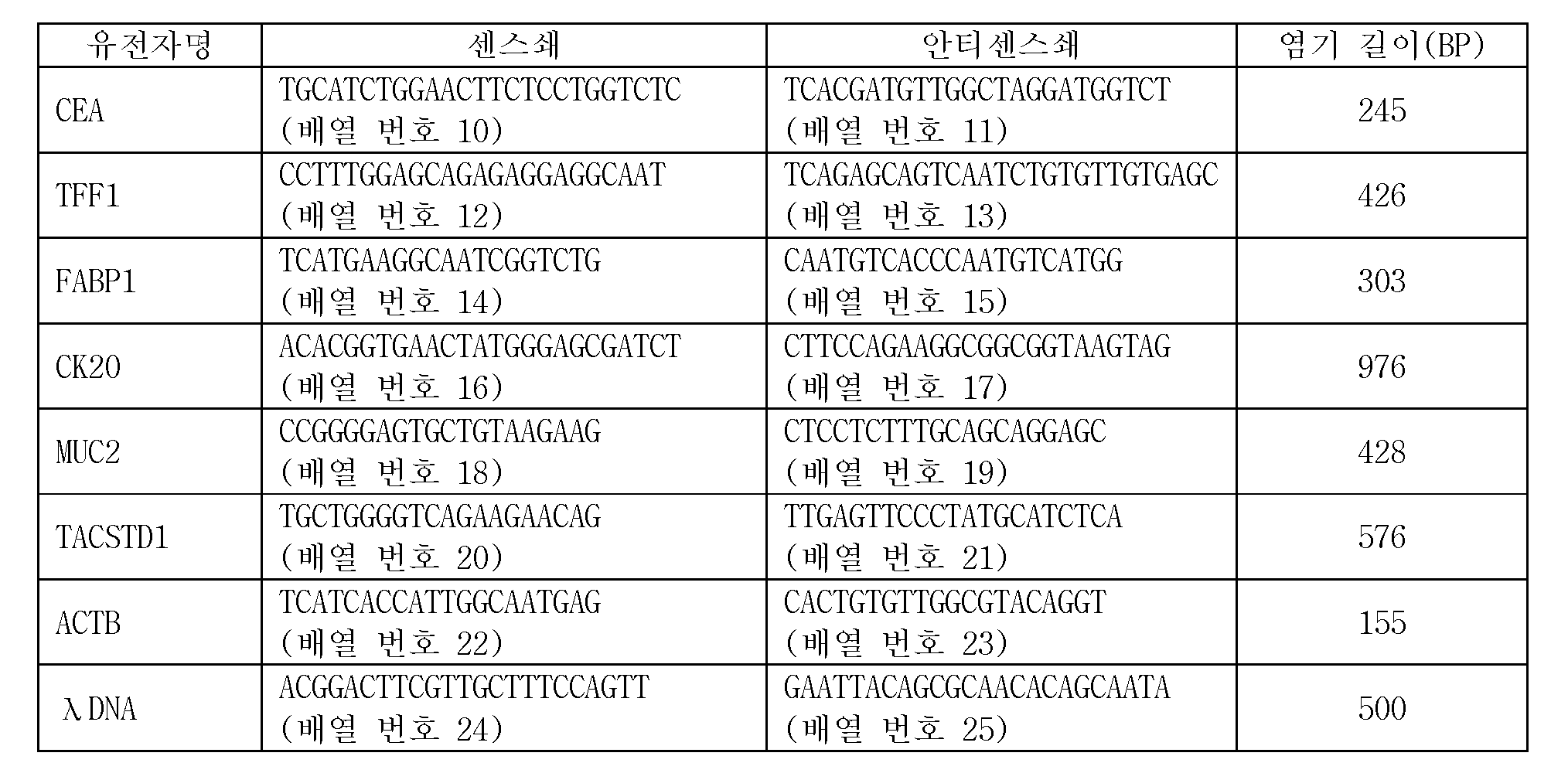
마커 유전자 검출용 프로브는, 바람직하게는, 5종의 종양 마커 유전자에 대하여, 각각 특이적인 배열을 갖는 것이다. 5종의 종양 마커 유전자는, 구체적으로는, CEA(암태아성 항원), TFF1(trefoil factor family 1), FABP1(Fatty acid binding protein 1), CK20(Cytokeratin 20), 및 MUC2(장형(腸型) 뮤신)이다. 이들 5종의 종양 마커 유전자는, 모두 복강 내 세정액 중의 위암 세포의 검출에 유용하고, 활성화 중피를 구별할 수 있는 것이다. 또, 마커 유전자를 검출하기 위한 프로브로서는, 표 1에 나타내는 것을 예시할 수 있다. 구체적으로는, 배열 번호 2로 나타나는 염기 배열로 이루어지는 CEA 검출용 프로브와, 배열 번호 3으로 나타나는 염기 배열로 이루어지는 TFF1 검출용 프로브와, 배열 번호 4로 나타나는 염기 배열로 이루어지는 FABP1 검출용 프로브와, 배열 번호 5로 나타나는 염기 배열로 이루어지는 CK20 검출용 프로브와, 배열 번호 6으로 나타나는 염기 배열로 이루어지는 MUC2 검출용 프로브이다. 본 실시형태에 관한 이들 5종의 마커 유전자는, 종래 사용되고 있는 마커 유전자와 비교하여, 감도(진양성률)가 높다.The probe for marker gene detection preferably has a specific sequence for each of the five tumor marker genes. Specifically, the five tumor marker genes are CEA (cancer antigen), TFF1 (trefoil factor family 1), FABP1 (Fatty acid binding protein 1), CK20 (Cytokeratin 20), and MUC2 Mucin). These five tumor marker genes are all useful for the detection of gastric cancer cells in the intraperitoneal rinse solution and can discriminate the activated mesenchyme. As a probe for detecting a marker gene, those shown in Table 1 can be exemplified. Specifically, a probe for CEA detection comprising a base sequence represented by SEQ ID NO: 2, a probe for TFF1 comprising a base sequence represented by SEQ ID NO: 3, a probe for FABP1 comprising a base sequence represented by SEQ ID NO: 4, A probe for detecting CK20 comprising a base sequence represented by SEQ ID NO: 5 and a probe for MUC2 detection comprising a base sequence represented by SEQ ID NO: 6. These five marker genes according to the present embodiment have a high sensitivity (true positive rate) as compared with the marker genes conventionally used.
예를 들면, 종래 사용되고 있는 마커 유전자로서, TACSTD1이 있다. 이 유전자를 검출하기 위한 프로브로서는, 배열 번호 7로 나타나는 염기 배열로 이루어지는 TACSTD1 검출용 프로브가 있다. 이 TACSTD1은, 본 실시형태에 관한 5종의 마커 유전자보다 감도(진양성률)가 떨어진다. 즉, 마커 유전자로서, TACSTD1을 이용한 경우, 재발 예측의 정확도가 저하한다.For example, TACSTD1 is a conventionally used marker gene. As a probe for detecting this gene, there is a probe for TACSTD1 detection comprising a base sequence represented by SEQ ID NO: 7. This TACSTD1 has a lower sensitivity (true positive rate) than the five marker genes according to the present embodiment. That is, when TACSTD1 is used as a marker gene, the accuracy of recurrence prediction decreases.
이로 인하여, 비록, TACSTD1의 발현량이 높았다고 해도, 본 실시형태에 관한 상기 5종의 마커 유전자의 발현량이 높을 때에 양성이라고 판단하는 것이, 예측 정확도라는 관점에 있어서 우수한 재발 예측을 행할 수 있다.Therefore, even if the expression level of TACSTD1 is high, it is possible to predict the recurrence of the tumor, which is judged to be positive when the expression amounts of the five marker genes according to the present embodiment are high, in terms of prediction accuracy.

담체로서는 기판, 비즈, 섬유 등 다양한 담체를 이용할 수 있다. 기판의 형상은 판 형상으로는 한정되지 않고, 예를 들면 필름 형상이나 시트 형상이어도 된다. 또, 기판은, 하나의 부재로 구성되어 있어도 되고, 복수의 부재로 구성되어 있어도 된다. 담체의 재질로서는, 금속, 유리, 플라스틱, 폴리머, 섬유 등을 이용할 수 있다. 고감도이고, 또한 동시에 다항목의 유전자를 검출하는 방법으로서는, 기판에 검출용 프로브를 고정화한 DNA 마이크로어레이를 들 수 있다. 이 DNA 마이크로어레이에는, 기판 상의 일정한 구획 내에 복수의 스폿을 마련해 두고, 각 스폿에 프로브를 각각 고정화해 두어, 마이크로어레이를 형성해 두는 것이 바람직하다.As the carrier, various carriers such as a substrate, a bead, and a fiber can be used. The shape of the substrate is not limited to a plate shape, and may be, for example, a film shape or a sheet shape. Further, the substrate may be composed of one member or a plurality of members. As the material of the carrier, metal, glass, plastic, polymer, fiber and the like can be used. As a method for detecting a gene having a high sensitivity and also at the same time, there is a DNA microarray in which a detection probe is immobilized on a substrate. In this DNA microarray, it is preferable that a plurality of spots are provided in a predetermined section on the substrate, and each of the probes is fixed to each spot to form a microarray.
또, 담체로서는, 고분자 물질로 이루어지는 코팅층을 표면에 포함하는 기판을 이용해도 된다. 고분자 물질로 이루어지는 코팅층을 표면에 포함하는 기판은, 소정 형상으로 가공된 기판의 표면에 고분자 물질을 포함하는 액체를 도포하고, 건조함으로써 얻을 수 있다. 또, 고분자 물질을 포함하는 액체 중에 기판을 침지하여, 건조해도 된다.As the carrier, a substrate including a coating layer made of a polymer material on its surface may be used. A substrate containing a coating layer made of a polymer material on its surface can be obtained by applying a liquid containing a polymeric substance to the surface of a substrate processed into a predetermined shape and drying the coating. Alternatively, the substrate may be immersed in a liquid containing the polymer substance and dried.
담체로서 "고분자 물질로 이루어지는 코팅층을 표면에 포함하는 기판"을 이용하는 경우, 고분자 물질로서는, 예를 들면 "인지질의 친수부를 구성하는 인산 에스터로부터 유도되는 기를 갖는 제1 단위와 카복실산 유도기를 갖는 제2 단위를 포함하는 고분자 물질"을 이용할 수 있다. "인지질의 친수부를 구성하는 인산 에스터로부터 유도되는 기를 갖는 제1 단위와 카복실산 유도기를 갖는 제2 단위를 포함하는 고분자 물질"은, 예를 들면 일본 공개특허공보 2006-187270호의 단락 0033~0066에 기재된 것을 이용할 수 있는데, 그 중에서도, 하기 일반식 (1)로 나타나는, 2-메타크릴로일옥시에틸포스포릴콜린(MPC)기를 갖는 제1 단량체와, p-나이트로페닐옥시카보닐폴리에틸렌글라이콜메타크릴레이트(NPMA)기를 갖는 제2 단량체와, n-뷰틸메타크릴레이트(BMA)기를 갖는 제3 단량체와의 공중합체인 폴리(MPC-co-BMA-co-NPMA)(PMBN)가 특히 바람직하다.When a substrate containing a coating layer made of a polymer substance on its surface is used as the carrier, examples of the polymer substance include a first unit having a group derived from a phosphate ester constituting the hydrophilic part of the phospholipid and a second unit having a carboxylic acid derivative Polymeric material containing a unit " "A polymer substance comprising a first unit having a group derived from a phosphoric ester and a second unit having a carboxylic acid derivative" constituting the hydrophilic part of the phospholipid is disclosed in, for example, Japanese Patent Laid-Open No. 2006-187270, paragraphs 0033 to 0066 Among them, a first monomer having a 2-methacryloyloxyethylphosphorylcholine (MPC) group represented by the following general formula (1) and a second monomer having a 2-methacryloyloxyethylphosphorylcholine (MPC) group are reacted with p-nitrophenyloxycarbonylpolyethylene glycol (MPC-co-BMA-co-NPMA) (PMBN) as a copolymer of a second monomer having a methacrylate (NPMA) group and a third monomer having a n-butyl methacrylate .

단, 상기 화학식 (1)에 있어서, a, b, 및 c는, 각각 독립적으로, 양의 정수이며, a가 5~200, b가 10~500, c가 1~100인 것이 바람직하다. 또, 상기 일반식 (1)에 있어서, 제1~제3 단량체가 블록 공중합하고 있어도 되고, 이들 단량체가 랜덤으로 공중합하고 있어도 된다.In the above formula (1), a, b, and c are each independently a positive integer, and a is preferably 5 to 200, b is 10 to 500, and c is 1 to 100. In the general formula (1), the first to third monomers may be subjected to block copolymerization, and these monomers may be randomly copolymerized.
또, "고분자 물질로 이루어지는 코팅층을 표면에 포함하는 기판"을 이용하는 경우, 고분자 물질로 이루어지는 코팅층을 형성시키는 기판으로서는, 플라스틱 기판이, 형상이나 사이즈의 변경에 대한 유연성이 확보되고, 유리 기판인 것에 비하여 저가로 제공할 수 있다는 관점에서 바람직하다. 이와 같은 플라스틱 재료로서는, 표면 처리의 용이성 및 양산성의 관점에서, 열가소성 수지를 이용할 수 있다.When a "substrate containing a coating layer made of a polymer material on a surface" is used, as a substrate on which a coating layer made of a polymer material is formed, flexibility with respect to change in shape and size is secured, It is preferable from the viewpoint that it can be provided at a low cost. As such a plastic material, a thermoplastic resin can be used from the viewpoints of ease of surface treatment and mass productivity.
열가소성 수지로서는, 형광 발생량이 적은 것을 이용할 수 있다. 형광 발생량이 적은 수지를 이용함으로써, DNA쇄의 검출 반응에 있어서의 백그라운드를 저하시킬 수 있기 때문에, 검출 감도를 더 향상시킬 수 있다. 형광 발생량이 적은 열가소성 수지로서는, 예를 들면 폴리에틸렌, 폴리프로필렌 등의 직쇄상 폴리올레핀; 환상 폴리올레핀; 함불소 수지; 등을 이용할 수 있다. 상기 수지 중에서도, 포화 환상 폴리올레핀은, 내열성, 내약품성, 저형광성, 투명성 및 성형성이 특히 우수하기 때문에, 광학적인 분석에 적합하고, 기판의 재료로서 바람직하게 이용된다.As the thermoplastic resin, those having a small amount of fluorescence emission can be used. By using a resin having a small amount of fluorescence emission, it is possible to lower the background in the detection reaction of the DNA chain, so that the detection sensitivity can be further improved. Examples of the thermoplastic resin having a small amount of fluorescence emission include linear polyolefins such as polyethylene and polypropylene; Cyclic polyolefin; Fluorine resin; . Among these resins, saturated cyclic polyolefin is suitable for optical analysis because it is excellent in heat resistance, chemical resistance, low fluorescence, transparency and moldability, and is preferably used as a substrate material.
여기에서, 포화 환상 폴리올레핀이란, 환상 올레핀 구조를 갖는 중합체 단독 또는 환상 올레핀과 α-올레핀과의 공중합체를 수소 첨가한 포화 중합체를 가리킨다. 전자의 예로서는, 예를 들면 노보넨, 다이사이클로펜타다이엔, 테트라사이클로도데센으로 대표되는 노보넨계 모노머, 및 이들의 알킬 치환체를 개환 중합하여 얻어지는 중합체를 수소 첨가하여 제조되는 포화 중합체이다. 후자의 공중합체는 에틸렌이나 프로필렌, 아이소프로필렌, 1-뷰텐, 3-메틸-1-뷰텐, 1-펜텐, 3-메틸-1-펜텐, 1-헥센, 1-옥텐 등의 α-올레핀과 환상 올레핀계 모노머의 랜덤 공중합체를 수소 첨가함으로써 제조되는 포화 중합체이다. 공중합체에서는, 에틸렌과의 공중합체가 가장 바람직하다. 이들 수지는 단독으로 이용해도 되고, 2종류 또는 그 이상의 공중합체 혹은 혼합물이어도 된다. 또, 환상 올레핀 구조를 갖는 단량체가 개환 중합하여 얻어지는 포화 환상 폴리올레핀뿐만 아니라, 환상 올레핀 구조를 갖는 단량체의 부가 중합에 의하여 얻어지는 포화 환상 폴리올레핀을 이용할 수도 있다.Here, the saturated cyclic polyolefin refers to a polymer having a cyclic olefin structure alone or a saturated polymer obtained by hydrogenating a copolymer of a cyclic olefin and an? -Olefin. Examples of the former are saturated polymers produced by hydrogenating a norbornene monomer represented by norbornene, dicyclopentadiene, tetracyclododecene, and a polymer obtained by ring-opening polymerization of alkyl substituents thereof. The latter copolymer is obtained by copolymerizing an? -Olefin such as ethylene or propylene, isopropylene, 1-butene, 3-methyl-1-butene, 1-pentene, Is a saturated polymer prepared by hydrogenating a random copolymer of an olefin-based monomer. In the copolymer, a copolymer with ethylene is most preferable. These resins may be used alone, or two or more kinds of copolymers or mixtures thereof may be used. A saturated cyclic polyolefin obtained by ring-opening polymerization of a monomer having a cyclic olefin structure, as well as a saturated cyclic polyolefin obtained by addition polymerization of a monomer having a cyclic olefin structure may be used.
담체에 대한 애기장대 프로브, λDNA 프로브 및 마커 유전자 검출용 프로브(이하, 간단히 "프로브 DNA"라고도 함)의 고정화 방법은, 특별히 한정되지 않지만, 담체로서, PMBN의 코팅층을 표면에 포함하는 기판을 이용하는 경우, 예를 들면 PMBN에 포함되는 p-나이트로페닐옥시카보닐기와 프로브 DNA를 반응시켜 공유결합을 형성시킴으로써, 기판 표면에서 프로브 DNA를 고정화할 수 있다.A method for immobilizing the Arabidopsis thaliana probe, the λDNA probe and the marker gene detection probe (hereinafter, simply referred to as "probe DNA") on the carrier is not particularly limited, but a substrate containing a coating layer of PMBN on its surface is used as the carrier , The probe DNA can be immobilized on the surface of the substrate by, for example, reacting the p-nitrophenyloxycarbonyl group contained in PMBN with the probe DNA to form a covalent bond.
프로브 DNA를 기판 상에 고정화할 때에는, 프로브 DNA를 용해 또는 분산한 액체를 점착하는 방법이 바람직하다. 이 프로브 DNA를 용해 또는 분산한 액체는, 예를 들면 중성으로부터 알칼리성, 예를 들면 pH를 7.6 이상으로 할 수 있다.When the probe DNA is immobilized on a substrate, it is preferable to adhere a liquid obtained by dissolving or dispersing the probe DNA. The liquid in which the probe DNA is dissolved or dispersed can be neutral to alkaline, for example, pH of 7.6 or more.
또, 점착 후, 기판 표면에 고정화되지 않았던 프로브 DNA를 제거하기 위하여, 순수나 완충액으로 세정해도 된다. 세정 후에는 프로브 DNA를 고정화한 이외의 기판 표면의 p-나이트로페닐옥시카보닐기의 불활성화 처리를 알칼리 화합물, 혹은 1급 아미노기를 갖는 화합물로 행해도 된다.After the adhesion, in order to remove the probe DNA that has not been immobilized on the substrate surface, it may be cleaned with pure water or a buffer solution. After the cleaning, the p-nitrophenyloxycarbonyl group on the surface of the substrate other than the surface on which the probe DNA is immobilized may be treated with an alkaline compound or a compound having a primary amino group.
또, 기판에 고정화하는 프로브 DNA에는, p-나이트로페닐옥시카보닐기와의 반응성을 높이기 위하여, 아미노기를 도입해 두는 것이 바람직하다. 아미노기는 p-나이트로페닐옥시카보닐기와의 반응성이 우수하기 때문에, 아미노기가 도입된 프로브 DNA를 이용함으로써, 효율적이고 또한 강고하게 기판의 표면 상에 프로브 DNA를 고정화할 수 있다. 아미노기의 도입 위치는 프로브 DNA의 분자쇄 말단 혹은 측쇄여도 되지만, 분자쇄 말단에 도입되고 있는 것이, 상보적인 주형 DNA 단편과의 어닐링을 보다 더 효율적으로 행할 수 있다는 관점에서는, 바람직하다.In order to increase the reactivity with the p-nitrophenyloxycarbonyl group, it is preferable to introduce an amino group into the probe DNA immobilized on the substrate. Since the amino group is excellent in reactivity with the p-nitrophenyloxycarbonyl group, the use of the probe DNA into which the amino group is introduced can efficiently and firmly immobilize the probe DNA on the surface of the substrate. Although the introduction position of the amino group may be the molecular chain terminal or the side chain of the probe DNA, the introduction of the amino group at the molecular chain terminal is preferable from the viewpoint that the annealing with the complementary template DNA fragment can be performed more efficiently.
이상에 의하여, 기판의 표면 상에 표 1에 나타내는 프로브가 고정화된 DNA 마이크로어레이를 얻을 수 있다.Thus, a DNA microarray in which probes shown in Table 1 are immobilized can be obtained on the surface of the substrate.
DNA 마이크로어레이에 의한 발현 해석을 행하는 경우에는, 보다 정확도가 높은 판정을 행하기 위하여, 내부 컨트롤이나 외부 컨트롤을 이용하는 것이 바람직하다. 본 실시형태에서는, 내부 컨트롤로서, ACTB(베타 액틴)를 이용할 수 있다. 또, 외부 컨트롤로서는, 음성 컨트롤이나 양성 컨트롤을 이용할 수 있고, 상술과 같이 본 실시형태에서는, 음성 컨트롤로서 애기장대 DNA쇄를 이용할 수 있으며, 양성 컨트롤로서 λDNA쇄를 이용할 수 있다. ACTB를 내부 컨트롤로서 이용하는 경우에는, 배열 번호 8로 나타나는 염기 배열로 이루어지는 ACTB 검출용 프로브를 내부 컨트롤 프로브로서 담체에 고정시킬 수 있다. ACTB 검출용 프로브의 고정화도 애기장대 프로브나 마커 유전자 검출용 프로브의 고정화와 동일하게 행할 수 있다.In the case of performing the expression analysis by the DNA microarray, it is preferable to use internal control or external control in order to make a determination with higher accuracy. In the present embodiment, ACTB (beta actin) can be used as the internal control. As the external control, voice control and positive control can be used. As described above, in the present embodiment, Arabidopsis DNA chain can be used as a voice control, and a lambda DNA chain can be used as a positive control. When ACTB is used as an internal control, an ACTB detection probe composed of a base sequence represented by SEQ ID NO: 8 can be fixed to a carrier as an internal control probe. Immobilization of the ACTB detection probe can be performed in the same manner as immobilization of the Arabidopsis probe or the marker gene detection probe.
위암 재발 예측용 키트에는, 마커 유전자의 일부의 영역을 증폭시키기 위한 프라이머 세트를 구비하는 것이 바람직하다. 이렇게 함으로써, 심사 복강경 시의 세정액 혹은 개복 시 복강 내 세정액으로부터 추출한 총 RNA가 적은 경우이더라도, 증폭시켜 유효한 판정이 가능하게 된다. 표 1로 나타내는 프로브에 의하여 검출되는 각 마커 유전자의 프라이머 염기 배열을 표 2에 나타낸다. 구체적으로는, 배열 번호 10, 11로 나타나는 염기 배열로 이루어지는 CEA 증폭용 프라이머 세트, 배열 번호 12, 13으로 나타나는 염기 배열로 이루어지는 TFF1 증폭용 프라이머 세트, 배열 번호 14, 15로 나타나는 염기 배열로 이루어지는 FABP1 증폭용 프라이머 세트, 배열 번호 16, 17로 나타나는 염기 배열로 이루어지는 CK20 증폭용 프라이머 세트, 및 배열 번호 18, 19로 나타나는 염기 배열로 이루어지는 MUC2 증폭용 프라이머 세트를 들 수 있다. 본 실시형태에서는, 이러한 프라이머 세트를 이용하여 위암 시료 중에 포함되는 마커 유전자의 적어도 일부의 영역을 PCR에 의하여 증폭시키는 것이 바람직하다. 또, 내부 컨트롤로서 ACTB를 이용하는 경우에는, 배열 번호 22, 23으로 나타나는 염기 배열로 이루어지는 프라이머 세트에 의하여, ACTB의 일부의 영역을 증폭시켜도 된다. 또한, 표 2에는, 표 1에 나타내는 프라이머에 의하여 증폭되는 PCR 산물의 염기 길이도 함께 나타낸다.It is preferable that the kit for recurrence of gastric cancer has a primer set for amplifying a part of the marker gene. By doing so, even when the total RNA extracted from the lavage fluid of the laparoscopic examination or the lavage fluid of the abdominal cavity at the time of laparotomy is small, it is possible to amplify and make an effective determination. Table 2 shows the primer base sequences of the marker genes detected by the probes shown in Table 1. Specifically, the CEA amplification primer set consisting of the nucleotide sequences represented by SEQ ID NOs: 10 and 11, the TFF1 amplification primer set consisting of the nucleotide sequences represented by SEQ ID NOs: 12 and 13, the FABP1 consisting of the nucleotide sequences represented by SEQ ID NOS: 14 and 15 A primer set for amplification, a primer set for CK20 amplification consisting of nucleotide sequences represented by SEQ ID NOs: 16 and 17, and a primer set for MUC2 amplification consisting of nucleotide sequences shown in SEQ ID NOs: 18 and 19. In the present embodiment, it is preferable that at least a part of the marker gene contained in the gastric cancer sample is amplified by PCR using this primer set. When ACTB is used as the internal control, a part of ACTB may be amplified by a primer set consisting of nucleotide sequences represented by SEQ ID NOS: 22 and 23. Table 2 also shows the base lengths of the PCR products amplified by the primers shown in Table 1 together.
또, 표 2에는 상기에 있어서 종래 사용되고 있는 마커 유전자로서 예시한 TACSTD1의 일부의 영역을 증폭시키기 위한, 배열 번호 20, 21로 나타나는 염기 배열로 이루어지는 TACSTD1 증폭용 프라이머 세트에 대해서도 함께 나타낸다.Table 2 also shows a primer set for TACSTD1 amplification comprising a base sequence represented by SEQ ID NOs: 20 and 21 for amplifying a part of the region of TACSTD1 exemplified as a marker gene conventionally used in the above.
이와 같이 하여 얻어지는 위암 재발 예측용 키트를 준비한 후, 도 2에 나타내는 바와 같이, 연구 사용 목적에 대한 인폼드·컨센트(informed consent)가 얻어진 위암 환자로부터 복강 내 세정액을 회수하고(S101), 이것을 원심하여 부유 성분인 유리 세포를 펠릿 형상으로 침전시킨 후, 그 펠릿으로부터 총 RNA를 추출한다(S102). 총 RNA의 추출 방법은 각종 방법이 고안되어, 많은 제조사에 의하여 키트화되어 있으며, 본 실시형태에 있어서는 어느 키트도 사용 가능하다.After preparing the kit for recurrence prediction of stomach cancer thus obtained, as shown in FIG. 2, the intraperitoneal rinse solution is collected from a gastric cancer patient who has obtained informed consent for research use purpose (S101) To thereby precipitate the glass cells, which are suspended components, in the form of pellets, and then extract the total RNA from the pellets (S102). Various methods for extracting total RNA have been devised and are made by many manufacturers, and in this embodiment, any kit can be used.
다음으로, 위암 시료 중에 포함되는 총 RNA를 증폭시키기 위하여, RT-PCR을 행한다. 구체적으로는, 먼저, 역전사 효소를 이용한 역전사 반응(Reverse Transcription, RT)에 의하여, 추출한 총 RNA를 주형으로 하여, 예를 들면 랜덤 헥사머나 올리고 dT 프라이머 등의 프라이머의 존재하에서 데옥시뉴클레오타이드 3인산(dNTP)을 기질로 하고, 추출한 총 RNA에 상보적인 cDNA를 5'3'방향으로 합성한다(S103).Next, RT-PCR is performed to amplify the total RNA contained in the gastric cancer sample. Specifically, first, by using reverse transcription (RT) using a reverse transcriptase, the extracted total RNA is used as a template, and deoxynucleotide triphosphate (deoxyribonucleotide) is synthesized in the presence of a primer such as a random hexamer or oligo dT primer dNTP) as a substrate, and cDNA complementary to the extracted total RNA is synthesized in the 5'3 'direction (S103).
그 후, 얻어진 cDNA를 이용하여 PCR 증폭 반응을 행한다(S104). 구체적으로는, 합성한 cDNA, 배열 번호 10~25로 나타나는 염기 배열로 이루어지는 프라이머, DNA 폴리머라제 및 dNTP와, DNA 폴리머라제가 작용하는 지적(至適) 염농도 환경을 만들기 위한 버퍼 용액을 혼합하여, 시판 중인 PCR 장치에 세트한다. 사용하는 DNA 폴리머라제는, 내열성을 갖는 것이면 된다.Thereafter, PCR amplification reaction is performed using the obtained cDNA (S104). Specifically, the synthesized cDNA, a primer consisting of a nucleotide sequence represented by SEQ ID NOS: 10 to 25, a DNA polymerase and dNTP, and a buffer solution for creating an optimal salt concentration environment in which the DNA polymerase acts are mixed, Set on a commercially available PCR device. The DNA polymerase to be used may be one having heat resistance.
그 후, (1) cDNA의 열변성(94~96℃), (2) 배열 번호 10~25로 나타나는 염기 배열로 이루어지는 프라이머의 어닐링(55~60℃), (3) DNA 폴리머라제에 의한 상보쇄의 합성(72~74℃)의 3단계의 온도 변화를, 예를 들면 20~30 사이클 반복하여, cDNA를 증폭시킨다.Thereafter, annealing (55 to 60 ° C) of a primer consisting of the nucleotide sequence represented by SEQ ID NO: 10 to 25, (3) complementation with a DNA polymerase (72 to 74 ° C) of the chain is repeated for 20 to 30 cycles, for example, to amplify the cDNA.
여기에서, PCR 증폭 반응은, dNTP와 함께, 라벨화한 뉴클레오타이드 모노머를 이용하여 실행한다. 이렇게 함으로써, 형광 표지된 마커 유전자의 표지 단편을 취득할 수 있다. 라벨 방법은, 형광체, 광흡수체, 방사성 핵종(P32), 효소 표지 등의 방법을 들 수 있다.Here, the PCR amplification reaction is carried out using labeled nucleotide monomers together with dNTP. By doing so, a labeled fragment of the fluorescently-labeled marker gene can be obtained. Examples of the labeling method include a fluorescent substance, a light absorber, a radionuclide (P 32 ), an enzyme label, and the like.
예를 들면, 형광 라벨화 뉴클레오타이드 모노머로서는, dTTP의 염기의 3위치를 형광 라벨한 Cy3-dUTP를 들 수 있다. Cy3-dUTP를 이용한 경우, 주형 DNA 단편의 아데닌 (A)에 대응하는 신장(프라이머)측의 위치에 Cy3-dUTP가 삽입된 PCR 산물을 얻을 수 있다.For example, as fluorescently labeled nucleotide monomers, Cy3-dUTP labeled with fluorescence at 3 positions of the base of dTTP can be mentioned. When Cy3-dUTP is used, a PCR product in which Cy3-dUTP is inserted at a position on the extension (primer) side corresponding to the adenine (A) of the template DNA fragment can be obtained.
또, 방사성 핵종에 의한 라벨화 뉴클레오타이드 모노머로서는, P32-ATP, P32-dATP를 들 수 있다.Examples of the labeled nucleotide monomer by the radionuclide include P 32 -ATP and P 32 -dATP.
또, 아미노알릴 dUTP를 이용하여 PCR 증폭 반응을 행하고, 반응 후에, 아미노알릴 dUTP를 포함하는 PCR 산물에 대하여, Cy3 형광 색소나 아미노기 염색 시약(NHS-시약)을 이용하여 발색시켜도 된다.In addition, PCR amplification reaction may be performed using aminoallyl dUTP, and after the reaction, the PCR product containing aminoallyl dUTP may be color-developed using Cy3 fluorescent dye or amino group dyeing reagent (NHS-reagent).
효소 표지의 방법에 있어서는, 비오틴(biotin)화 또는 다이곡시제닌(DIG: 스테로이드계 천연물)을 결합한 핵산(예를 들면, 비오틴-dUTP, DIG-dUTP)을 사용하여 증폭시킨 후, 얻어지는 PCR 산물에 알칼리포스파타제 처리하여 나이트로블루테트라졸륨(NBT)과 5-브로모-4-클로로-3-인돌일 인산(BCIP)액 중에서 수시간 반응시켜 발색시킬 수도 있다.In the method of labeling the enzymes, biotinization or amplification is performed using a nucleic acid (eg, biotin-dUTP, DIG-dUTP) conjugated with di-glycogenin (DIG: steroid natural product) (NBT) and 5-bromo-4-chloro-3-indolylphosphoric acid (BCIP) in the presence of an alkaline phosphatase for several hours.
또한, S104에서는, 역전사 반응으로 얻어진 cDNA와 함께, 양성 컨트롤 핵산쇄를 버퍼 용액에 혼합하여, 증폭 반응을 행해도 된다. 양성 컨트롤 핵산쇄로서는, 미생물 유래의 핵산쇄를 이용할 수 있고, 구체적으로는 박테리오파지 등의 바이러스 유래의 것을 들 수 있다. 양성 컨트롤 핵산쇄로서 λDNA쇄를 이용하는 경우, 양성 컨트롤 핵산쇄 증폭용 프라이머 세트로서, 표 2로 나타내는 배열 번호 24 및 25로 나타나는 염기 배열로 이루어지는 프라이머를 이용하여 λDNA쇄의 적어도 일부의 영역을 증폭시킬 수 있다. 이렇게 함으로써, 양성 컨트롤 핵산쇄의 표지 단편을 취득할 수 있다. 이 경우, λDNA쇄를 검출하기 위하여, 양성 컨트롤 프로브로서, 표 1로 나타내는 배열 번호 9로 나타나는 염기 배열로 이루어지는 λDNA 검출용 프로브를 미리 담체에 고정해 둔다. 이 λDNA 검출용 프로브는, 배열 번호 24 및 25로 나타나는 염기 배열로 이루어지는 프라이머에 의하여 증폭되는 λDNA쇄의 단편과 상보적으로 결합할 수 있도록 설계되어 있다.In S104, the positive control nucleic acid chain may be mixed with the cDNA obtained by the reverse transcription reaction in a buffer solution, and the amplification reaction may be performed. As the positive control nucleic acid chain, a nucleic acid chain derived from a microorganism can be used, and specific examples thereof include those derived from viruses such as bacteriophage. When a lambda DNA strand is used as a positive control nucleic acid chain, at least a part of the lambda DNA chain is amplified using a primer consisting of the nucleotide sequences represented by SEQ ID NOS: 24 and 25 shown in Table 2 as a primer set for positive control nucleic acid chain amplification . By doing so, a labeled fragment of the positive control nucleic acid chain can be obtained. In this case, in order to detect the lambda DNA chain, a lambda DNA detecting probe comprising a base sequence represented by SEQ ID NO: 9 shown in Table 1 is fixed as a positive control probe to the carrier in advance. This λDNA detection probe is designed to be complementary to a fragment of a λDNA chain amplified by a primer consisting of a base sequence represented by SEQ ID NOS: 24 and 25.
이와 같이 하여, 표지한 PCR 산물(표지 DNA 단편)을 취득한 후(S105), 얻어진 표지 DNA 단편과 DNA 프로브를 접촉시켜 하이브리다이제이션을 행한다. 하이브리다이제이션 버퍼는, 공지의 것을 이용할 수 있는데, 예를 들면 SSC 또는 SSPE와, SDS와, 폼아마이드와의 혼합액을 이용할 수 있다.In this manner, the labeled PCR product (labeled DNA fragment) is obtained (S105), and the obtained labeled DNA fragment is brought into contact with the DNA probe to carry out hybridization. As the hybridization buffer, known buffers can be used. For example, a mixed solution of SSC or SSPE, SDS and formamide can be used.
다음으로, 혼합한 표지 DNA 단편 및 애기장대 DNA쇄를, 하이브리다이제이션 버퍼, 및 프로브 DNA가 고정화된 DNA 마이크로어레이와 함께 하이브리다이제이션 장치에 세트하고(S107), 예를 들면 45℃~60℃에서 3~24시간, 바람직하게는 3~5시간, 하이브리다이제이션한다(S108).Next, the mixed labeled DNA fragment and the Arabidopsis DNA chain are set in a hybridization apparatus (S107) together with a hybridization buffer and a DNA microarray on which the probe DNA is immobilized (S107) For 3 to 24 hours, preferably for 3 to 5 hours (S108).
그 후, 마이크로어레이의 세정 후에, 시판 중인 마이크로어레이 스캐너를 이용하여 표지 단편의 양을 측정한다(S109). S104에 있어서, 미생물 유래의 DNA쇄, 특히, λDNA쇄를 이용하여 마커 유전자와 함께 PCR 증폭시킨 경우에는, S109에 있어서 λDNA쇄의 표지 단편의 측정량을 기준으로 하여 각 마커 유전자의 표지 단편의 양을 측정해도 된다. 구체적으로는, λDNA 검출용 프로브가 고정된 위치에 있어서의 표지 단편의 측정량이 소정의 범위가 되도록 검출 감도를 제어한다. 이렇게 함으로써, 어세이 간의 편차를 보정할 수 있다. 또, 형광 표지된 마커 유전자의 표지 단편에 대해서는, 마커 유전자 검출용 프로브가 고정된 위치, 및 음성 컨트롤 프로브가 고정된 위치의 형광 강도를 각각 측정하는 것이 바람직하다.Thereafter, after cleaning the microarray, the amount of the labeled fragment is measured using a commercially available microarray scanner (S109). When PCR amplification is carried out together with the marker gene using the DNA chain derived from the microorganism, in particular, the lambda DNA chain in S104, the amount of the marker fragment of each marker gene based on the measurement amount of the marker fragment of the lambda DNA chain in S109 May be measured. Specifically, the detection sensitivity is controlled such that the measurement amount of the label fragment at the position where the λDNA detection probe is fixed is within a predetermined range. By doing this, the deviation between the assays can be corrected. In addition, it is preferable to measure the position of the marker gene detecting probe fixed and the fluorescence intensity of the position where the voice control probe is fixed, respectively, with respect to the labeled fragment of the fluorescently labeled marker gene.
또, 얻어진 표지 단편의 양의 실측값에 여러가지 보정을 가하여, 보정된 실측값을 실제 측정값으로 해도 된다. 예를 들면, 프로브 DNA가 고정화되어 있지 않은 담체 표면의 표지 단편의 측정량을 백그라운드로 하여 측정 결과로부터 차감하여, 측정값으로 해도 된다.Further, various corrected values may be applied to actual measured values of the obtained label fragments, and the corrected measured values may be used as actual measured values. For example, the measured value of the labeled fragment on the surface of the carrier on which the probe DNA is not immobilized may be subtracted from the measured result in the background to be a measured value.
그 후, 얻어진 측정값에 근거하여 위암 시료의 수술 후 위암 재발의 예측을 행한다(S110). DNA 마이크로어레이에 의한 발현 해석을 행하는 경우에는, 각 프로브, 판정 대상이 되는 총 RNA마다 알맞는 규격화를 행하고, 각 샘플 간에서 비교 가능하게 되는 조정을 행함으로써, 보다 정확도가 높은 판정을 행할 수 있다.Thereafter, the recurrence of gastric cancer after surgery of the gastric cancer sample is predicted based on the obtained measurement value (S110). In the case of performing the expression analysis by the DNA microarray, standardization suitable for each probe and the total RNA to be determined can be performed, and adjustment can be made to be comparable between the respective samples, whereby more accurate determination can be performed .
구체적으로는, 각 유전자의 발현량(발현 레벨)의 컷오프값을 설정하고, 컷오프값을 상회한 유전자 수를 카운트하여, 정한 수를 상회하는지 하회하는지에 따라 판정을 행하는 방법이 유효하다. 따라서, 본 실시형태에서는, 애기장대 DNA쇄를 음성 컨트롤로 하고, 이를 기준으로 하여, 컷오프값(임곗값)을 도출한다.Specifically, it is effective to set the cutoff value of the expression level (expression level) of each gene, count the number of genes exceeding the cutoff value, and determine whether the number of genes exceeds or falls below a predetermined number. Therefore, in the present embodiment, the Arabidopsis DNA chain is used as a voice control, and a cut-off value (threshold value) is derived based on this.
임곗값을 도출하는 방법은, 의료 기관에 있어서 실제의 검체를 이용한 해석에 의하여, 경험적으로, 마커 유전자마다, 소정의 임곗값(제1 임곗값)을 정한다. 구체적으로는, 임상의 소견이나 그 외의 측정값을 맞추어 제1 임곗값이 정해진다. 그리고, 애기장대 검출용 프로브가 고정된 기판의 위치에 있어서의 표지 단편의 측정량을 기준으로 하여, 각 마커 유전자의 표지 단편의 양의 상댓값을 산출한다. 각 마커 유전자의 표지 단편의 양의 상댓값의 산출법으로서는, 애기장대 검출용 프로브가 고정된 기판의 위치에 있어서의 표지 단편의 측정량을 1로 하여, 각 마커 유전자의 표지 단편의 양의 상댓값을 산출하는 방법이나, 애기장대 검출용 프로브가 고정된 기판의 위치에 있어서의 표지 단편의 양을 0으로 하여, 각 마커 유전자의 표지 단편의 양의 상댓값을 산출하는 방법을 들 수 있다. 또, 상댓값으로서, 자연 대수를 이용할 수도 있다. 그리고, 1개 이상의 마커 유전자의 표지 단편의 양의 상댓값이 제1 임곗값을 넘은 경우에, 결과를 양성, 즉 예후 불량(재발함, 또는 재발의 가능성이 큼)이라고 예측한다. 한편, 모든 마커 유전자의 표지 단편의 양의 상댓값이 임곗값 이하인 경우에는, 결과를 음성, 즉 예후 양호(재발하지 않음, 또는 재발의 가능성이 작음)라고 판정할 수 있다. 또한, 본 실시형태에 관한 판정 수단으로서는, 이에 한정되지 않고, 설정된 임곗값을 넘은 마커 유전자가 2개 이상의 소정 수 존재한 경우에 양성이라고 판정하며, 설정된 임곗값을 넘은 마커 유전자가 이 소정 수에 미치지 않은 경우를 음성이라고 판정해도 되고, 어느 특정 마커 유전자의 표지 단편의 양의 상댓값이 제1 임곗값을 넘은 경우에 양성이라고 판정하며, 이 특정의 마커 유전자의 표지 단편의 양의 상댓값이 임곗값 이하였던 경우에 음성이라고 판정해도 된다.The method for deriving the threshold value is to determine a predetermined threshold value (first threshold value) for each marker gene by empirical analysis using an actual sample in a medical institution. Concretely, the first threshold value is determined in accordance with clinical findings and other measured values. Then, based on the measured amount of the marker fragment at the position of the substrate on which the Arabidopsis marking probe is fixed, the positive value of the marker fragment of each marker gene is calculated. As a method for calculating the positive value of the marker fragment of each marker gene, the measurement amount of the marker fragment at the position of the substrate on which the aramid probe is fixed is taken as 1, And a method of calculating the positive value of the marker fragment of each marker gene by setting the amount of the marker fragment at the position of the substrate on which the Arabidopsis marking probe is fixed to 0. It is also possible to use a natural logarithm as an image value. When the positive value of the marker fragment of one or more marker genes exceeds the first threshold value, the result is predicted as positive, that is, the prognosis is poor (recurrence or a high possibility of recurrence). On the other hand, if the positive value of the marker fragment of all the marker genes is below the threshold value, the result is negative, that is, it can be judged that the prognosis is good (no recurrence or less possibility of recurrence). In addition, the determination means according to the present embodiment is not limited to this, and it is determined that the marker gene is positive when two or more predetermined number of marker genes exceeding the preset threshold value exist, and the marker gene exceeding the set threshold value is determined to be positive And a positive value is determined when the positive value of the positive marker fragment of a specific marker gene exceeds the first threshold value and the negative value of the positive marker fragment of this particular marker gene is It may be judged as negative if it is less than the threshold value.
구체적으로 재발의 예측 방법으로서, 예를 들면 이하의 (i), (ii) 혹은 (iii)의 방법을 이용할 수 있다.Specifically, for example, the following method (i), (ii) or (iii) can be used as a method of predicting the recurrence.
(i)의 방법은, 위암의 재발 예측을, 양성 및 음성의 2단계로 판정하는 것이다. 구체적으로는, (i)의 방법은, 애기장대 DNA의 발현량을 나타내는 표지 단편의 측정량을 기준으로 하여 산출된, CEA, TFF1, FABP1, CK20, 및 MUC2 중 어느 1개 이상의 마커 유전자의 발현량을 나타내는 표지 단편의 양의 상댓값이 제1 임곗값을 넘었을 때, 양성이라고 판단하는 방법이다. CEA, TFF1, FABP1, CK20, 및 MUC2는, TACSTD1과 비교하여 감도(진양성률)가 높다. 따라서, 이렇게 함으로써, 보다 정확한 위암 재발의 예측이 가능하게 된다. 한편, TACSTD1은, CEA, TFF1, FABP1, CK20, 및 MUC2의 마커 유전자에 비하여 감도(진양성률)가 떨어진다. 따라서, TACSTD1의 발현량이 높은 경우이더라도, 1개 이상의 다른 마커 유전자의 발현량이 높을 때에 양성이라고 판단하는 것이, 예측 정확도의 관점에서 바람직하다.The method of (i) is to judge the recurrence of stomach cancer in two stages, positive and negative. Specifically, the method of (i) is characterized in that expression of any one or more marker genes of CEA, TFF1, FABP1, CK20, and MUC2, which is calculated on the basis of a measurement amount of a labeled fragment indicating the expression amount of Arabidopsis DNA When the positive value of the labeled fragment indicating the amount of the labeled fragment exceeds the first threshold value, it is judged to be positive. CEA, TFF1, FABP1, CK20, and MUC2 have higher sensitivity (true positive rate) as compared to TACSTD1. Thus, by doing this, it becomes possible to predict the recurrence of stomach cancer more accurately. On the other hand, TACSTD1 has a lower sensitivity (true positive rate) than the marker genes of CEA, TFF1, FABP1, CK20, and MUC2. Therefore, even when the expression level of TACSTD1 is high, it is preferable from the viewpoint of prediction accuracy that the expression level is high when the expression level of one or more other marker genes is high.
(ii)의 방법은, 위암의 재발 예측을, 양성, 준양성 및 음성의 3단계로 판정하는 것이다. 구체적으로는, (ii)의 방법은, 애기장대 DNA의 발현량을 나타내는 표지 단편의 측정량을 기준으로 하여 산출된, CEA, TFF1, FABP1, CK20, 및 MUC2 중 어느 1개 이상의 마커 유전자의 발현량을 나타내는 표지 단편의 양의 상댓값이 제1 임곗값을 넘었을 때, 위암 시료가 양성이라고 판정하며, 제1 임곗값보다 낮은 제2 임곗값을 넘고, 또한 제1 임곗값보다 낮은 값일 때, 위암 시료가 준양성이라고 판정하는 방법이다. 이러한 (ii)의 방법은, 상기 (i)의 2단계 평가 방법과, 음성, 준양성 및 양성의 3단계 평가를 채용하고 있다는 점에서 상이하다. 이와 같이 3단계 판정법을 채용하고 있기 때문에, 보다 더 판정 정확도를 향상시킴과 함께, 정확한 예후 재발의 판정이 가능하게 된다. 또, (ii)의 방법에 의하면, 준양성이라는 평가 기준을 설정함으로써, 측정 기기나 측정 조건 등의 판정 조건의 차이에 따라 판정 결과가 변동되었던 임곗값 정도의 값을 나타내는 환자에 대하여, 적절한 치료를 실시하는 것이 가능해진다.The method of (ii) is to judge the recurrence prediction of stomach cancer as three stages: positive, positive, and negative. Specifically, the method of (ii) is characterized in that expression of one or more marker genes selected from the group consisting of CEA, TFF1, FABP1, CK20 and MUC2, which is calculated on the basis of a measurement amount of a labeled fragment representing the expression amount of Arabidopsis DNA The gastric cancer sample is judged to be positive when it exceeds the first threshold value, and when the value exceeds the second threshold value lower than the first threshold value and is lower than the first threshold value , And it is judged that the gastric cancer sample is positive. This method (ii) differs from the two-step evaluation method (i) in that it adopts a three-step evaluation of negative, semi-positive and positive. Since the three-step determination method is employed in this manner, it is possible to further improve the judgment accuracy and to determine the accurate prognosis recurrence. According to the method (ii), by setting an evaluation criterion of quasi-positivity, a patient showing a value of the threshold value at which the judgment result has fluctuated in accordance with the difference of the judgment condition such as the measuring instrument or the measurement condition, Can be performed.
(iii)의 방법은, 상기 (ii)의 방법과 마찬가지로 3단계로 판정하는 것이지만, 양성, 준양성 및 음성의 판정 기준이 상기 (ii)의 방법과는 상이한 것이다. 그러므로, 후술에서는, 상기 (ii)의 방법에 따른 판정 결과와 구별하기 위하여, 이하에 설명하는 (iii)의 방법에 따른 판정 결과를, 양성, 약양성 및 음성의 3단계로 나타내기로 한다.(iii) is determined in three steps in the same manner as in the method (ii), but the criteria for the determination of positive, semi-positive and negative are different from the method (ii). Therefore, in the following description, in order to distinguish from the judgment result according to the method (ii), the judgment result according to the method (iii) described below will be expressed in three steps of positive, weak positive and negative.
구체적으로는, (iii)의 방법은, 애기장대 DNA의 발현량을 나타내는 표지 단편의 측정량을 기준으로 하여 산출된, CEA, TFF1, FABP1, CK20, 및 MUC2 중 어느 1개 이상의 마커 유전자의 발현량을 나타내는 표지 단편의 양의 상댓값이 제1 임곗값을 넘은 경우, 또는 CEA, TFF1, FABP1, CK20, 및 MUC2 중 어느 2개 이상의 마커 유전자의 발현량을 나타내는 표지 단편의 양의 상댓값이 제1 임곗값보다 낮은 제2 임곗값을 넘고, 또한 제1 임곗값보다 낮은 값인 경우, 위암 시료가 양성이라고 판정하며, CEA, TFF1, FABP1, CK20, 및 MUC2 중 어느 1개의 마커 유전자의 발현량을 나타내는 표지 단편의 양의 상댓값이 제1 임곗값보다 낮은 제2 임곗값을 넘고, 또한 제1 임곗값보다 낮은 값이며, 그 외의 마커 유전자의 발현량을 나타내는 표지 단편의 양의 상댓값이 제2 임곗값보다 낮은 값인 경우, 위암 시료가 약양성이라고 판정하는 방법이다. 이러한 (iii)의 방법은, 상기 (ii)의 방법과 비교하여, 3단계 평가를 채용하고 있다는 점에서는 공통되지만, 보다 정확하게 위암의 재발 예측을 행한다는 관점에서, 그 판정 기준이 상이하다. 이와 같이, (iii)의 방법에서는, (ii)의 방법과 비교하여 상세하게 설정한 것이기 때문에, 측정 기기나 측정 조건 등의 판정 조건의 차이에 따라 판정 결과가 변동되었던 임곗값 정도의 값을 나타내는 환자에 대하여, 보다 정확하게 재발의 가능성을 판별할 수 있게 된다.Specifically, the method of (iii) is characterized in that the expression of any one or more marker genes of CEA, TFF1, FABP1, CK20, and MUC2, which is calculated on the basis of a measurement amount of a labeled fragment representing the expression amount of Arabidopsis DNA Amount of the marker fragment that indicates the amount of expression of any two or more marker genes of CEA, TFF1, FABP1, CK20, and MUC2 exceeds the first threshold value The gastric cancer sample is determined to be positive and the expression level of any one marker gene of CEA, TFF1, FABP1, CK20, and MUC2 is higher than the first threshold value and lower than the first threshold value Is greater than the second threshold value lower than the first threshold value and lower than the first threshold value and the positive residual value of the marker fragment indicating the amount of expression of the other marker gene is smaller than the second threshold value lower than the first threshold value If the value is lower than the second threshold value, It is judged that the sample is weakly positive. The method (iii) differs from the method (ii) in that it adopts the three-step evaluation, but differs in the criteria from the viewpoint of more accurately predicting the recurrence of stomach cancer. As described above, in the method (iii), since it is set in detail in comparison with the method (ii), the value of the fiducial value at which the determination result is varied according to the difference in the determination conditions such as the measuring apparatus and the measurement conditions The possibility of recurrence can be more accurately determined for the patient.
또, S104에 있어서, 내부 컨트롤(ACTB)이나 양성 컨트롤(λDNA)을 이용하여 PCR 증폭을 실행한 경우에는, ACTB나 λDNA의 측정 결과도 함께 고찰함으로써, S102의 추출, 또는 S108의 하이브리다이제이션이 각각 양호하게 진행되었는지 여부를 판별할 수 있다. 구체적으로는, ACTB의 결과가 음성이고, 또한 λDNA의 결과가 음성인 경우에는, S102의 추출, 및 S108의 하이브리다이제이션 모두가 불량이었던 것을 알 수 있다. 또, ACTB의 결과가 양성인 한편, λDNA의 결과가 음성이었던 경우에는, S102의 추출은 양호하게 진행되었지만, S108의 하이브리다이제이션의 진행이 불량이었던 것을 알 수 있다. ACTB의 결과가 음성이고, 또한 λDNA의 결과가 양성인 경우에는, S104의 PCR은 양호하게 진행되었지만, S102의 추출의 진행이 불량이었던 것을 알 수 있다.When the PCR amplification is carried out using the internal control (ACTB) or the positive control (? DNA) in S104, the extraction results of S102 and the hybridization of S108 It is possible to judge whether or not they have progressed satisfactorily. Specifically, when the result of ACTB is negative and the result of? DNA is negative, it can be seen that both the extraction of S102 and the hybridization of S108 were defective. In addition, when the result of ACTB was positive while the result of? DNA was negative, the extraction of S102 proceeded well, but the progress of hybridization of S108 was poor. When the result of ACTB is negative and the result of? DNA is positive, the PCR of S104 proceeded well, but it is found that the progress of extraction of S102 was poor.
또한, 본 실시형태의 판정 방법과 함께, 통상 외과적 절제 치료 증례, 심사 복강경 증례에 있어서 필수로 되어 있는 세포진의 결과에 더하여, 림프절 전이의 유무, 각종 암 마커 등의 수치를 참조하고, 상황에 따라서는 판정 방법을 구별하여 사용하는 등, 종합적으로 위암 세포의 유무를 판단하면 보다 효과적인 예측이 가능하다.In addition to the determination method of the present embodiment, in addition to the results of the usual surgical resection therapy and the results of cell migration that are indispensable in the examination laparoscopic case, the numerical values such as the presence or absence of lymph node metastasis and various cancer markers are referred to Therefore, it is possible to predict more effectively by judging the presence or absence of the stomach cancer cells comprehensively, such as using discrimination methods separately.
또, 본 실시형태의 판정 방법에 의하여 양성이라고 판정된 경우, 대응하는 위암 환자에 대하여, 방사선 치료, 화학 요법, 또는 이들을 조합한 추가적인 치료를 추가해도 된다. 본 실시형태의 판정 방법에서는, 양호한 정확도로 위암의 예후 재발을 예측할 수 있기 때문에, 재발의 우려가 있는 환자에게만 재발을 방지하기 위한 효과적인 치료를 추가할 수 있고, 또한 재발의 우려가 적은 환자에 대해서는, 과잉 치료를 추가하는 것을 방지할 수 있다.If it is judged to be positive by the determination method of the present embodiment, radiation therapy, chemotherapy, or an additional treatment in combination with these may be added to the corresponding gastric cancer patients. In the judgment method of the present embodiment, it is possible to predict the recurrence of the prognosis of stomach cancer with good accuracy. Therefore, it is possible to add effective treatment for preventing recurrence only to the patient who has a risk of recurrence, , It is possible to prevent the addition of excessive treatment.
다음으로, 본 실시형태의 작용 효과에 대하여 설명한다. 본 실시형태에 의하면, 식물 유래의 음성 컨트롤 핵산쇄의 적어도 일부의 영역에 상보적으로 결합하는 음성 컨트롤 프로브를 담체에 고정하고, 마커 유전자 검출용 프로브와 위암 시료의 PCR 산물을 하이브리다이제이션시킴과 함께, 음성 컨트롤 프로브와 음성 컨트롤 핵산쇄를 하이브리다이제이션시켜, 마커 유전자 검출용 프로브가 고정된 위치에서 측정된 표지 단편의 양과 음성 컨트롤 프로브의 위치에서 측정된 표지 단편의 양의 대비 결과를 이용하여 위암의 재발을 예측한다. 이로써, 식물 유래의 핵산쇄가 음성 컨트롤이 되기 때문에, 검사 대상 검체와 조기 위암 검체를 동시에 측정하지 않고, 미리 정한 임곗값에 근거하여, 위암의 재발의 예측을 정확하게 행할 수 있다. 따라서, 간편하고 또한 실용적인 위암의 예후 예측이 실현 가능하게 된다.Next, the operation and effect of the present embodiment will be described. According to this embodiment, a negative control probe complementarily binding to at least a part of the region of a plant-derived negative control nucleic acid chain is immobilized on a carrier, a probe for detecting a marker gene and a PCR product of a gastric cancer sample are hybridized Together, the negative control probe and the negative control nucleic acid chain were hybridized to each other using the result of the comparison of the amount of the labeled fragment measured at the fixed position of the marker gene detection probe and the amount of the labeled fragment measured at the position of the voice control probe Predict recurrence of gastric cancer. This makes it possible to predict the recurrence of the stomach cancer accurately based on the predetermined threshold value without simultaneously measuring the test sample and the early gastric cancer specimen since the plant-derived nucleic acid chain becomes negative control. Thus, a simple and practical prediction of the prognosis of stomach cancer becomes feasible.
종래, 위암의 재발의 예측에 있어서는, 대상 검체와 동시에, 음성 컨트롤로서 복강 내에 암세포가 존재하지 않는 것이라고 생각되는 Ia기의 조기 암 검체를 이용한 측정을 행하여, 조기 암 검체의 결과로부터 컷오프값을 도출하는 방법이 채용되고 있었다. 그러나, 검사마다 조기 암 검체를 복수, 예를 들면 비특허문헌 1에서는 40예 가까이, 준비하지 않으면 안되어, 실용성이나 윤리면에서 문제가 있었다.Conventionally, in predicting the recurrence of stomach cancer, measurement is performed using an Ia-early cancer sample suspected of not having cancer cells in the abdominal cavity as voice control at the same time as the subject sample, and a cut-off value is derived from the result of the early cancer specimen Method was employed. However, a plurality of early cancer specimens must be prepared for each examination, for example, in the case of non-patent document 1, about 40 cases have to be prepared, resulting in problems in terms of practicality and ethics.
한편, 본 실시형태에서는, 식물 유래의 DNA쇄를 음성 컨트롤로서 이용하여, 양호한 정확도로 위암의 재발을 예측할 수 있다. 따라서, 검사마다 조기 암 검체를 준비할 필요가 없어져, 실용적이다. 특히 애기장대 DNA쇄를 음성 컨트롤로서 이용함으로써, 마커 유전자와의 반응을 무시할 수 있는 레벨로 할 수 있어, 보다 양호한 정확도로 위암의 재발을 예측할 수 있다. 또한, 애기장대 유전자는, 교차 반응성이 낮은 것이 확인되고 있다. 또, 애기장대 DNA쇄는, 입수가 용이한 점에서도 메리트가 있다.On the other hand, in the present embodiment, it is possible to predict the recurrence of stomach cancer with good accuracy by using the plant-derived DNA strand as a negative control. Therefore, there is no need to prepare an early cancer specimen for each examination, which is practical. In particular, by using the Arabidopsis DNA chain as a negative control, the reaction with the marker gene can be made negligible and the recurrence of stomach cancer can be predicted with better accuracy. In addition, it has been confirmed that the Arabidopsis gene has low cross-reactivity. In addition, the Arabidopsis DNA chain is advantageous in that it is easy to obtain.
실시예Example
이하, 구체적인 실시예를 나타내, 본 발명을 더 상세하게 설명한다.Hereinafter, the present invention will be described in more detail by showing specific examples.
<실시예 1>≪ Example 1 >
(1) 플라스틱 기판의 제조(1) Manufacture of plastic substrates
포화 환상 폴리올레핀 수지(5-메틸-2-노보넨의 개환 중합체의 수소 첨가물(MFR(용융 지수; Melt flow rate): 21g/10분, 수소 첨가율: 실질적으로 100%, 열변형 온도 123℃))를 이용하고, 사출 성형에 의하여 슬라이드 유리 형상의 기판을 얻었다. 얻어진 기판을 상기 화학식 (1)로 나타나는 PMBN 폴리머(MPC기:BMA기:NPMA기=23:74:3(몰비))의 0.3중량% 에탄올 용액에 침지함으로써, 기판 표면에 포스포릴콜린기와 활성 에스터기를 갖는 고분자 물질을 도입하여, 플라스틱 기판을 얻었다. 또한, MFR은, JIS K 7210: 1999법에 기재된 조건에 준하여 측정한 것이다.A saturated cyclic polyolefin resin (hydrogenated product of a ring-opening polymer of 5-methyl-2-norbornene (MFR (melt flow rate): 21 g / 10 min, hydrogenation rate: substantially 100% And a slide glass substrate was obtained by injection molding. The resulting substrate was immersed in a 0.3 wt% ethanol solution of a PMBN polymer represented by the above formula (1) (MPC group: BMA group: NPMA group = 23: 74: 3 (molar ratio)) to form a phosphorylcholine group and an active ester Was introduced to obtain a plastic substrate. The MFR is measured according to the conditions described in JIS K 7210: 1999.
(2) 프로브 DNA(2) Probe DNA
5' 말단이 아미노기로 수식된, 위암 재발에 관한 마커 유전자 6종류의 특이 배열을 갖는 올리고 DNA쇄, 및 컨트롤 유전자 3종류를 준비했다. 준비한 프로브는 표 1에 기재한 9종류의 유전자에 대응한 프로브이다. 각 유전자에 대응한 프로브의 배열 번호는, CEA 검출용으로서 배열 번호 2, TFF1 검출용으로서 배열 번호 3, FABP1 검출용으로서 배열 번호 4, CK20 검출용으로서 배열 번호 5, MUC2 검출용으로서 배열 번호 6이다. 또, 내부 컨트롤로서 배열 번호 8(ACTB쇄 검출용), 음성 컨트롤로서 배열 번호 1(애기장대 DNA쇄 검출용), 양성 컨트롤로서 배열 번호 9(λDNA쇄 검출용)이다.Three oligo DNA strands each having a specific sequence of 6 marker genes related to recurrence of gastric cancer, the 5 'end of which was modified with an amino group, and three kinds of control genes were prepared. The prepared probes are probes corresponding to the nine kinds of genes shown in Table 1. Sequence numbers of probes corresponding to the respective genes were SEQ ID NO: 2 for CEA detection, SEQ ID NO: 3 for TFF1 detection, SEQ ID NO: 4 for FABP1 detection, SEQ ID NO: 5 for CK20 detection, to be. Sequence number 8 (for detection of ACTB chain) as internal control, SEQ ID NO: 1 (for detection of DNA chain of Arabidopsis) as voice control, and SEQ ID NO: 9 (for detection of? DNA chain) as positive control.
(3) 기판에 대한 프로브 DNA 고정(3) Probe DNA fixation on the substrate
스포팅 버퍼(제품명: ×2 스포팅액, 스미토모 베이크라이트사제)를 이용하여, 본 실시예의 (2)에서 준비한 프로브 DNA를 용해하여, 10μM의 프로브 DNA 용액을 조제했다. 이 용액을 스포터(히타치 소프트웨어 엔지니어링사제 MARKS-I)를 이용하여, 300μm 직경 스폿핀으로, 본 실시예의 (1)에서 제작한 플라스틱 기판(세로 75mm x 가로 25mm)의 표면 상에 스폿했다. 스폿은, 각 프로브에 대하여, N=5로 스폿했다. 프로브 DNA를 스폿한 기판을, 80℃에서 1시간 가열하여, 프로브 DNA를 고정화시켰다. 이로써 플라스틱 기판의 폴리머 표면에 단일쇄 DNA가 고정된 DNA 마이크로어레이를 얻었다. 스폿의 레이아웃을 도 3에 나타낸다.The probe DNA prepared in (2) of this Example was dissolved using a spotting buffer (product name: 占 2 spotting solution, Sumitomo Bakelite) to prepare a 10 占 프로 probe DNA solution. This solution was spotted on a surface of a plastic substrate (75 mm long x 25 mm wide) produced in (1) of this example with a 300 μm diameter spot pin using a sputter (MARKS-I manufactured by Hitachi Software Engineering Co., Ltd.). The spot was spotted with N = 5 for each probe. The substrate on which the probe DNA was spotted was heated at 80 DEG C for 1 hour to immobilize the probe DNA. As a result, a DNA microarray in which single-stranded DNA was immobilized on the polymer surface of the plastic substrate was obtained. The layout of the spot is shown in Fig.
(4) 총 RNA의 회수(4) Recovery of total RNA
연구 사용 목적에 대한 인폼드·컨센트가 얻어진 위암 수술 중 환자에 대하여, 통상 행해지는 수술 방식에 따라 50~100mL의 생리 식염수를 이용하여 복강 내 세정을 행하고, 세정액을 회수했다. 회수된 복강 내 세정액 중 병리부에 검사 검체 20~60mL로서 제출하는 데 필요한 분량을 빼낸 잉여 검체를 검사 대상 검체로 하여, 1500rpm으로 10분간 원심하고, 상청을 제거함으로써 펠릿 형상이 된 침전 성분을 복강 내 부유 세포로서 회수했다. 회수한 세포를 배양하여, 얻어진 세포를 닛폰진사제 시약 이소젠(Isogen)에 더하여 호모지나이즈하고, 소량의 클로로폼을 넣어, 12k×g로 15분간 원심 분리시켜, 상청을 채취했다. 또한 채취량과 등량의 아이소프로판올을 더하여, 10분 이상 인큐베이트한 후, 12k×g로 10분간 원심하여, 펠릿을 회수하고, 에탄올 침전(70%)에 의하여 총 RNA를 얻었다. 이 총 RNA를 189증례분 준비하여, 진행암 샘플로 했다.Patients undergoing stomach cancer surgery with an informed consent for the purpose of research use were subjected to intraperitoneal rinse using 50 to 100 mL of physiological saline according to the usual surgical procedure, and the rinse solution was collected. Among the recovered intraperitoneal washing solutions, 20-60 mL of the test specimen was taken out from the pathological portion as necessary, and the supernatant was removed from the supernatant by centrifugation at 1500 rpm for 10 minutes and the supernatant was removed to obtain a pellet- And recovered as suspended cells. The recovered cells were cultured, and the obtained cells were added to Isogen, manufactured by Nippon Gene Co., Ltd., homogenized, and a small amount of chloroform was added thereto, followed by centrifugation at 12 kxg for 15 minutes to collect the supernatant. Isopropanol equal to the amount of the sample was added and incubated for 10 minutes or more. The pellet was recovered by centrifugation at 12 kxg for 10 minutes, and total RNA was obtained by ethanol precipitation (70%). This total RNA was prepared for 189 cases, and was used as a progressive cancer sample.
(5) 역전사 반응(5) Reverse transcription reaction
역전사 반응을 행함으로써 상기 추출한 총 RNA로부터 cDNA를 제작했다.CDNA was prepared from the extracted total RNA by performing a reverse transcription reaction.
상기 추출한 총 RNA를 0.1 또는 1μg/μL로 농도 조제하고, 조제한 총 RNA 1 또는 10μL와, 20μM의 랜덤헥사머(제품명: 랜덤 프라이머(Random primer), 다카라바이오사제) 1μL와, 10mM의 dNTP(GE 헬스케어사제) 1μL와, 정제수 9μL, 역전사 효소(인비트로젠(Invitrogen)사제, 슈퍼스크립트(SuperScript)II) 1μL, 및 RNase 억제제(로슈(Roche)사제) 1μL를 첨가하며, 42℃에서 50분 가열 유지하고 또한 72℃에서 15분 가열 유지시켜, 단일쇄 cDNA를 얻었다.The extracted total RNA was adjusted to a concentration of 0.1 or 1 μg / μL, and 1 or 10 μL of the prepared total RNA and 1 μL of 20 μM random hexamer (product name: random primer, Takara Bio) and 10 mM dNTP 1 μL of purified water and 1 μL of a reverse transcriptase enzyme (Invitrogen, SuperScript II) and 1 μL of an RNase inhibitor (Roche) were added thereto, and the mixture was incubated at 42 ° C. for 50 minutes Heated and held at 72 캜 for 15 minutes to obtain single-stranded cDNA.
(6) 멀티플렉스 PCR 반응(6) Multiplex PCR reaction
복강 내 세정액으로부터 얻은 단일쇄 cDNA를 주형으로 검사 대상이 되는 마커 유전자의 cDNA를 증폭시키기 위한 PCR을 행했다. 동일 반응 중에 복수의 프라이머를 이용하는 멀티플렉스 PCR법에 의하여 cDNA의 증폭을 행했다. 동일한 cDNA에 대하여, 프라이머로서 표 2에 나타내는 배열 번호 10~25의 염기 배열로 이루어지는 프라이머 세트를 이용했다.Single-stranded cDNA obtained from the intraperitoneal washing solution was used as a template to perform PCR for amplifying the cDNA of the marker gene to be examined. The amplification of the cDNA was carried out by the multiplex PCR method using a plurality of primers in the same reaction. For the same cDNA, a primer set consisting of the nucleotide sequences of SEQ ID NOS: 10 to 25 shown in Table 2 was used as a primer.
cDNA 용액 1μL, 표 2에 나타내는 배열 번호 10~25의 염기 배열로 이루어지는 프라이머를 DEPC-처리수 중에 각각 농도 10μM로 포함하는 프라이머 용액을 0.5μL, DNA 폴리머라제(인비트로젠사제 아큐프라임(AcuuPrime)) 0.5μL, 및 λDNA쇄(다카라바이오사제)를 PCR 버퍼(인비트로젠사제 아큐프라임의 키트 내에 포함됨) 중에 용해시키고 20μL의 반응액을 조제했다. 또, PCR 산물의 표지를 위하여, 아미노알릴 dUTP(앰비온(Ambion)사제) 0.1μL를 기질로 하여, PCR의 반응 용액에 첨가했다.1 μL of a cDNA solution and 0.5 μL of a primer solution containing a base sequence of SEQ ID NOs: 10 to 25 shown in Table 2 in DEPC-treated water at a concentration of 10 μM each, and 0.5 μL of a DNA polymerase (AcuPrime, manufactured by Invitrogen) ) And a lambda DNA strand (Takara Bio Co., Ltd.) were dissolved in a PCR buffer (contained in a kit of AccuPrime made by Invitrogen), and 20 μL of a reaction solution was prepared. For labeling the PCR product, 0.1 μL of aminoallyl dUTP (Ambion) was added as a substrate to the PCR reaction solution.
조제된 반응액에 대하여, 시판 중인 서멀 사이클러(제품명: GeneAmp(등록상표) PCR 시스템 9700, 어플라이드 바이오시스템(Applied biosystem)사제)를 이용하여, (i) 열변성 95℃ 15초, (ii) 어닐링 60℃ 45초, (iii) DNA쇄의 신장 반응 72℃ 3분의 히트 사이클로, 25사이클 행하여, 아미노알릴 표지 PCR 산물을 얻었다.(I) heat denaturation at 95 DEG C for 15 seconds, (ii) heat denaturation at 95 DEG C for 15 seconds, and (ii) heat denaturation at 95 DEG C for 15 seconds using a commercially available thermal cycler (product name: GeneAmp (registered trademark) PCR system 9700, Applied biosystem) Annealing at 60 캜 for 45 seconds, and (iii) elongation reaction of the DNA strand at 72 캜 for 3 minutes. Thus, an aminoalyl-labeled PCR product was obtained.
정제는, 칩마다 DNA 정제 키트(퀴아젠사제 퀴아 퀵(QIA quick))를 사용하여 상기 PCR 산물의 정제를 행했다.The purification of the above PCR product was carried out using a DNA purification kit (QIA quick, manufactured by QIAGEN) for each chip.
(7) Cy3 표지(7) Cy3 label
얻어진 아미노알릴 표지 PCR 산물에 대하여, Cy3 표지를 행했다. 구체적으로는, 본 실시예의 (6)에서 얻어진 조제가 완료된 아미노알릴 표지 PCR 산물을 형광 라벨화 시약(GE 헬스케어사제 Cy3 모노-반응성 염료 팩(Cy3 Mono-reactive Dye Pack))을 사용하여 Cy3 형광 라벨화하고, 또한 DNA 정제 키트(퀴아젠사제 퀴아 퀵)를 사용하여 정제를 행하여, Cy3 형광 라벨화 PCR 산물을 얻었다.The resulting aminoalyl-labeled PCR product was subjected to Cy3 labeling. Specifically, the prepared aminoleyl-labeled PCR product obtained in (6) of this Example was amplified by Cy3 fluorescence using a fluorescent labeling reagent (Cy3 mono-reactive dye pack manufactured by GE Healthcare) Labeled and purified using a DNA purification kit (Qiagen, QIAGEN) to obtain a Cy3 fluorescently labeled PCR product.
(8) 하이브리다이제이션(8) Hybridization
Cy3 형광 라벨화 PCR 산물 45μL에 대하여, 버퍼 85μL를 첨가하여, 하이브리다이제이션 용액을 얻었다. 얻어진 하이브리다이제이션 용액의 조성은, 이하와 같다.To 45 [mu] L of the Cy3 fluorescently labeled PCR product, 85 [mu] L of buffer was added to obtain a hybridization solution. The composition of the obtained hybridization solution is as follows.
[하이브리다이제이션 용액][Hybridization solution]
·3 × SSPE3 x SSPE
·10% 폼아마이드· 10% formamide
·0.05% SDS0.05% SDS
(3)에서 제작한 DNA 마이크로어레이를 하이브리다이제이션 장치(제노믹 솔루션스(Genomic Solutions)사제 Hyb4)에 세트하고, 상기 조성의 하이브리다이제이션 용액을 이용하여 하이브리다이제이션을 행했다. 구체적으로는, 95℃에서 5분 열변성시킨 하이브리다이제이션 용액을 실시예 1에서 제작한 DNA 마이크로어레이 상에 분주(分注)하고, 55℃에서 4시간 반응을 행했다. 하이브리다이제이션 반응 종료 후, 0.1% SDS를 포함한 2 × SSC 용액, 2 × SSC, 0.1 × SSC의 순서로 마이크로어레이의 표면을 세정하여, 하이브리다이제이션 반응을 종료했다.The DNA microarray prepared in (3) was set in a hybridization device (Hyb4, manufactured by Genomic Solutions), and hybridization was carried out using a hybridization solution of the above composition. Specifically, a hybridization solution which was thermally denatured at 95 ° C for 5 minutes was dispensed (dispensed) onto the DNA microarray prepared in Example 1, and the reaction was carried out at 55 ° C for 4 hours. After completion of the hybridization reaction, the surface of the microarray was washed with 2 x SSC solution containing 0.1% SDS, 2 x SSC, and 0.1 x SSC in this order to terminate the hybridization reaction.
(9) 측정(9) Measurement
본 실시예 (8)에서 행한 하이브리다이제이션 반응 종료 후, 스핀 드라이한 DNA 마이크로어레이에 대하여, DNA 마이크로어레이용 스캐너(액손(Axon)사제 GenePix 4000B)를 이용하여, 프로브가 스폿된 위치마다 형광 강도를 측정했다. 측정할 때의 수광 감도(PMT)는, λDNA가 고정된 스폿에 있어서의 형광 강도가 900에서 1100이 되도록 조정했다. 형광 강도의 산출 시에는, 프로브마다 5개의 측정값이 얻어지는데, 최곳값 및 최젓값을 제외한 중간의 3개의 측정값을 평균화하고, 프로브마다 평균값을 산출하여, 이것을 형광 강도로 했다. 진행암 샘플 전체 189증례에 대하여 형광 강도를 측정했다.After completion of the hybridization reaction in the present Example (8), the spin-dried DNA microarray was subjected to fluorescence intensity measurement at every spot where the probe was spotted using a DNA microarray scanner (GenePix 4000B, manufactured by Axon) . The light receiving sensitivity (PMT) at the time of measurement was adjusted so that the fluorescence intensity in the spot where? DNA was fixed was 900 to 1100. In calculating the fluorescence intensity, five measurement values are obtained for each probe. The three intermediate measurement values excluding the maximum value and the minimum value are averaged, and an average value is calculated for each probe, and this value is used as the fluorescence intensity. Fluorescence intensity was measured for 189 cases of progressive cancer samples.
(10) 상댓값을 이용한 위암의 재발 예측(10) Prediction of recurrence of stomach cancer using statistical value
얻어진 189증례의 형광 강도에 대하여, 애기장대 DNA쇄 검출용 프로브가 스폿된 위치의 형광 강도를 백그라운드값으로 하여, 각 프로브가 고정된 위치에 있어서의 형광 강도로부터 애기장대 DNA쇄 검출용 프로브가 스폿된 위치의 형광 강도를 차감하고, 이것을 자연 대수로 나타낸 결과를 표 3~6에 나타낸다. 또, 각 마커 유전자의 형광 강도의 상댓값(자연 대수)에 각각 임곗값을 설정하고, 그 임곗값을 넘은 경우에는, 양성으로 하고, 하회하는 경우에는, 음성으로 했다. 이 임곗값은, 실시예 1에서 설명한 바와 같이, 일반적인 DNA 마이크로어레이의 컷오프값의 설정이 통상 백그라운드의 3배인 것에 근거하여 CEA의 임곗값을 설정하고, TFF1, FABP1, CK20, 및 MUC2에 대해서는, CEA의 임곗값의 약 절반으로 설정했다. 그리고, 임곗값을 넘은 경우에는, 양성으로 하고, 하회하는 경우에는, 음성으로 했다. 구체적으로는, 진행암 샘플의 마커 유전자의 임곗값은, 하기와 같이 설정했다.With respect to the fluorescence intensity of the obtained 189 cases, the fluorescence intensity at the spot where the Arabidopsis DNA chain detection probe was spotted was set as a background value. From the fluorescence intensity at the position where each probe was fixed, the fluorescence intensity of the Arabidopsis DNA chain detection probe The fluorescence intensity at the position where the fluorescence intensity was obtained was subtracted and the results are shown in Tables 3 to 6 in terms of natural logarithm. In addition, the threshold value (natural logarithm) of the fluorescence intensity of each marker gene was set to a threshold value, and when the threshold value was exceeded, the positive value was determined. As described in Example 1, this threshold value is set based on the fact that the setting of the cutoff value of a general DNA microarray is usually three times the background, and for TFF1, FABP1, CK20, and MUC2, It was set at about half of the threshold value of CEA. When the threshold value was exceeded, it was positive, and when it was lower than the threshold value, it was negative. Specifically, the threshold value of the marker gene of the progressive cancer sample was set as follows.
CEA: 2.11CEA: 2.11
TFF1: 1.85TFF1: 1.85
FABP1: 1.85FABP1: 1.85
CK20: 1.85CK20: 1.85
MUC2: 1.85MUC2: 1.85
진행암 샘플 전체 189증례에 대하여 양성이라고 판단된 마커 유전자의 수를 표 3~6에 함께 나타낸다. 또, 표 3~6에는, 이하에 설명하는 방법으로 얻어진 세포진의 결과, 림프절 전이의 판정 결과, 및 수술 후 경과에 대해서도 함께 나타낸다.Progressive Cancer Samples The number of marker genes determined to be positive for all 189 cases are shown in Tables 3-6. Tables 3 to 6 also show the results of the cell division obtained by the method described below, the judgment result of the lymph node metastasis, and the progress of the operation.
<세포진><Cytopathology>
위암의 수술 중 복강 내 세정 세포진 가이드라인(일본 임상세포학회 잡지 44(1); 93-97.2001)에 근거하여, 진행암 샘플 189증례에 대하여, 세포진에 의하여 판정을 행했다. 각 증례에 대하여 회수한 복강 내 부유 세포를 슬라이드 유리 상에 올리고, 스프레더 유리를 이용하여 도말(塗抹) 표본을 제작했다. 신속하게 도말 표본을 바람 건조시켜, 그 후, 메탄올에 의하여 3분간 고정시켰다. 김자액과 순수를 1:2의 비율로 혼합시키고, 혼합액을 이용하여 표본의 염색을 행했다. 30분간 염색시킨 후, 순수로 세정하고, 건조시켜 봉하여, 표본을 완성시켰다. 광학 현미경에 의한 표본 관찰로 염색된 암세포를 찾음으로써, 세포진의 판정을 행했다. 세포진에서는, 클래스 분류를 행하여, "1"은 양성(재발함)으로 하고, "0"은 음성(재발하지 않음)으로 했다. 결과를 표 3~6에 나타낸다.On the basis of the intraperitoneal washing cytology guidelines (Japan Clinical Cell Society Magazine 44 (1); 93-97, 2001) during gastric cancer surgery, a case of progressive cancer sample 189 was judged by cytopenia. For each case, the collected abdominal suspension cells were placed on a slide glass, and smear specimens were prepared using spreader glass. The smear was quickly air-dried, and then fixed with methanol for 3 minutes. The kimchi liquid and pure water were mixed at a ratio of 1: 2, and the mixture was used to dye the sample. Stained for 30 minutes, washed with pure water, dried and sealed to complete the specimen. The cytopenia was judged by finding cancer cells stained by specimen observation with an optical microscope. In the cell cluster, classification was carried out, and "1" was positive (recurrent) and "0" was negative (no recurrence). The results are shown in Tables 3 to 6.
<림프절 전이><Lymph node metastasis>
진행암 샘플 전체 189증례에 대하여, 위암부와 함께 외과적으로 절제한 주변 림프절의 병리 진단에 의하여, 림프절 전이의 유무의 진단을 행했다. 결과를 표 3~6에 나타낸다. 또한, 표 3 및 이후의 표 중, 림프절 전이의 유무의 진단에 있어서, "0"은, 주변 림프절에 림프절 전이가 확인되지 않는 것이다. "1"은, 주변 림프절에 1개 이상 2개 이하의 림프절 전이가 확인된 것, "2"는, 주변 림프절에 3개 이상 6개 이하의 림프절 전이가 확인된 것, "3"은, 주변 림프절에 7개 이상의 림프절 전이가 확인된 것, "LN+"는, 전이 수는 불명확하지만, 주변 림프절에 림프절 전이가 확인된 것이다. 표 3 및 이후의 표 중, "0"은 음성(-, 재발하지 않음), "1", "2", "3" 및 "LN+"는 양성(+, 재발함)이다.For the entire 189 cases of advanced cancer, diagnosis of lymph node metastasis was made by pathologic diagnosis of surgically resected peripheral lymph nodes with gastric cancer. The results are shown in Tables 3 to 6. In Table 3 and the following tables, "0" in the diagnosis of the presence or absence of lymph node metastasis indicates that no lymph node metastasis is found in the surrounding lymph node. "1" indicates that no more than one lymph node metastasis was found in the surrounding lymph nodes, "2" indicates that no more than three lymph node metastases were found in the surrounding lymph nodes, "3" Seven or more lymph node metastases were found in the lymph nodes, "LN +", but the number of metastases was unclear, but lymph node metastasis was confirmed in the surrounding lymph nodes. In Table 3 and the following tables, "0" is negative (-, no recurrence), "1", "2", "3" and "LN +" are positive (+ recurrence).
또, 진행암 샘플 전체 189증례의 위암 환자의 수술 후 경과에 대해서는 3년 이상, 평균 4.5년에 걸쳐 추적 조사되어, 재발 및 사망 여부의 정보를 얻었다. 표 3~6에서는, 생사의 결과도 함께 나타낸다. "alive"는 재발하지 않고 생존, "alive but recurrent"는 재발하지만 생존, "death"는 재발에 의한 사망을 나타낸다.The progression of gastric cancer in 189 cases of advanced cancer was followed up over a period of 3 years and averaged 4.5 years, and information on recurrence and death was obtained. Tables 3 to 6 also show the results of life and death. "alive" refers to survival without recurrence, "alive but recurrent" refers to recurrence, but "death" refers to death due to recurrence.
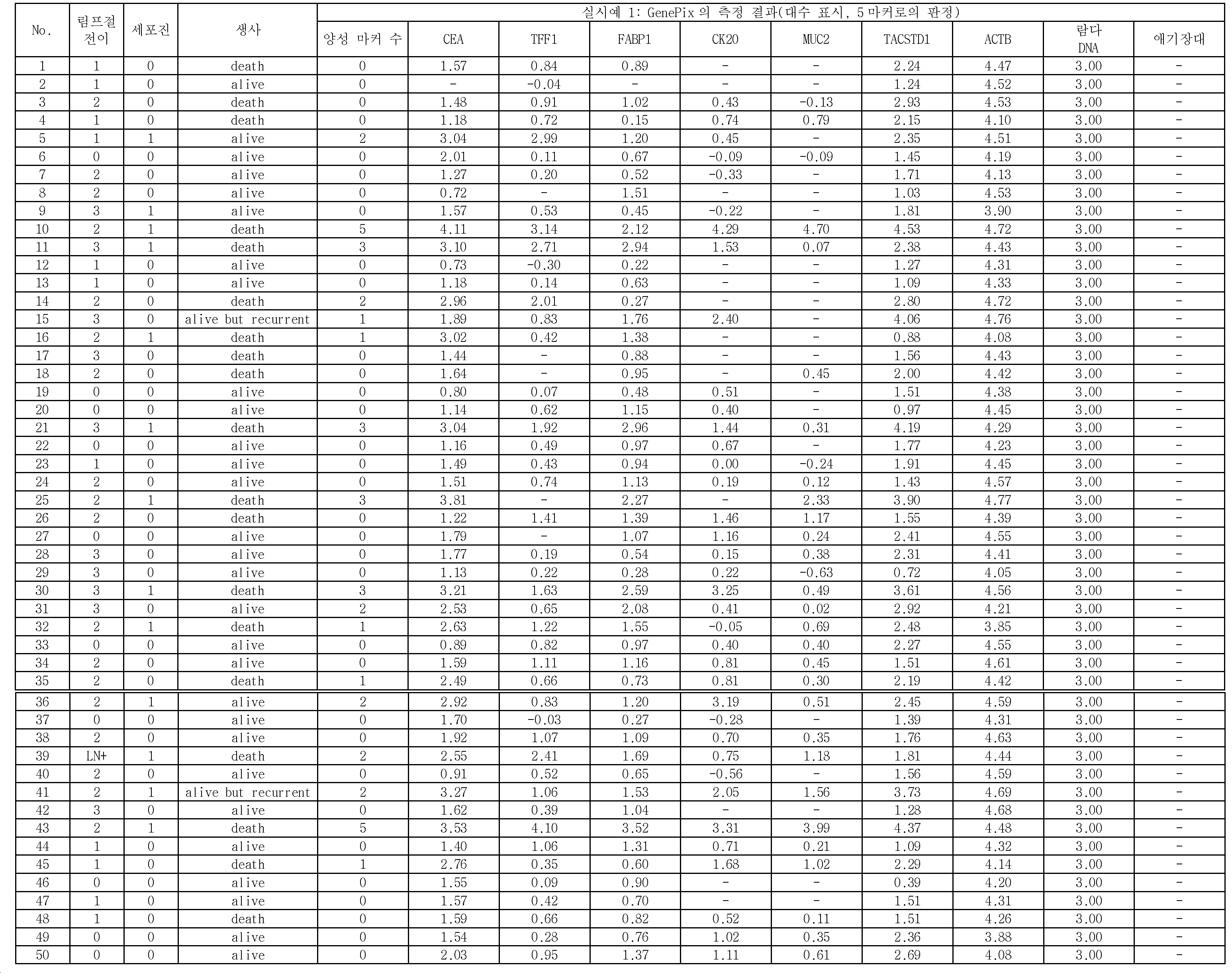
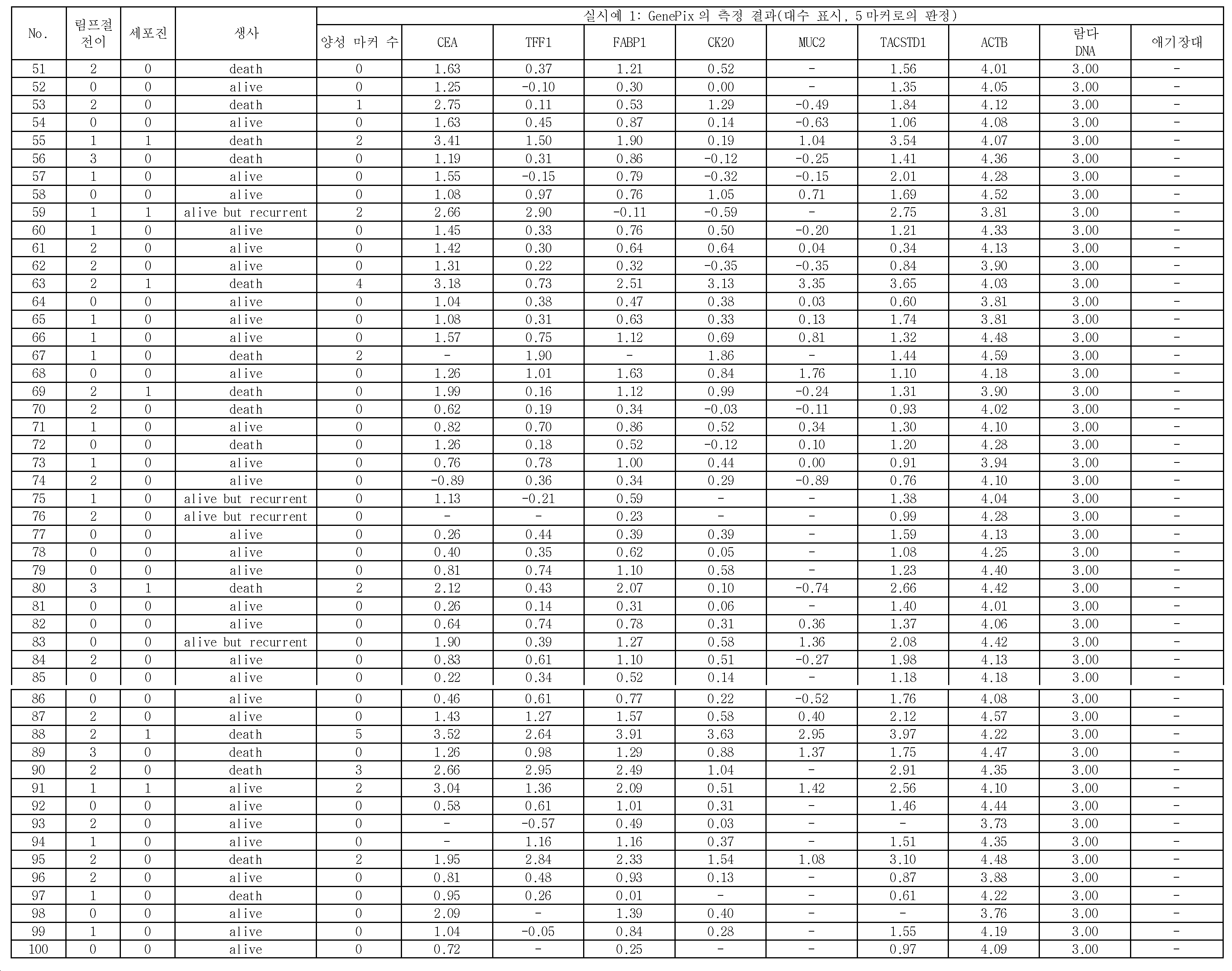

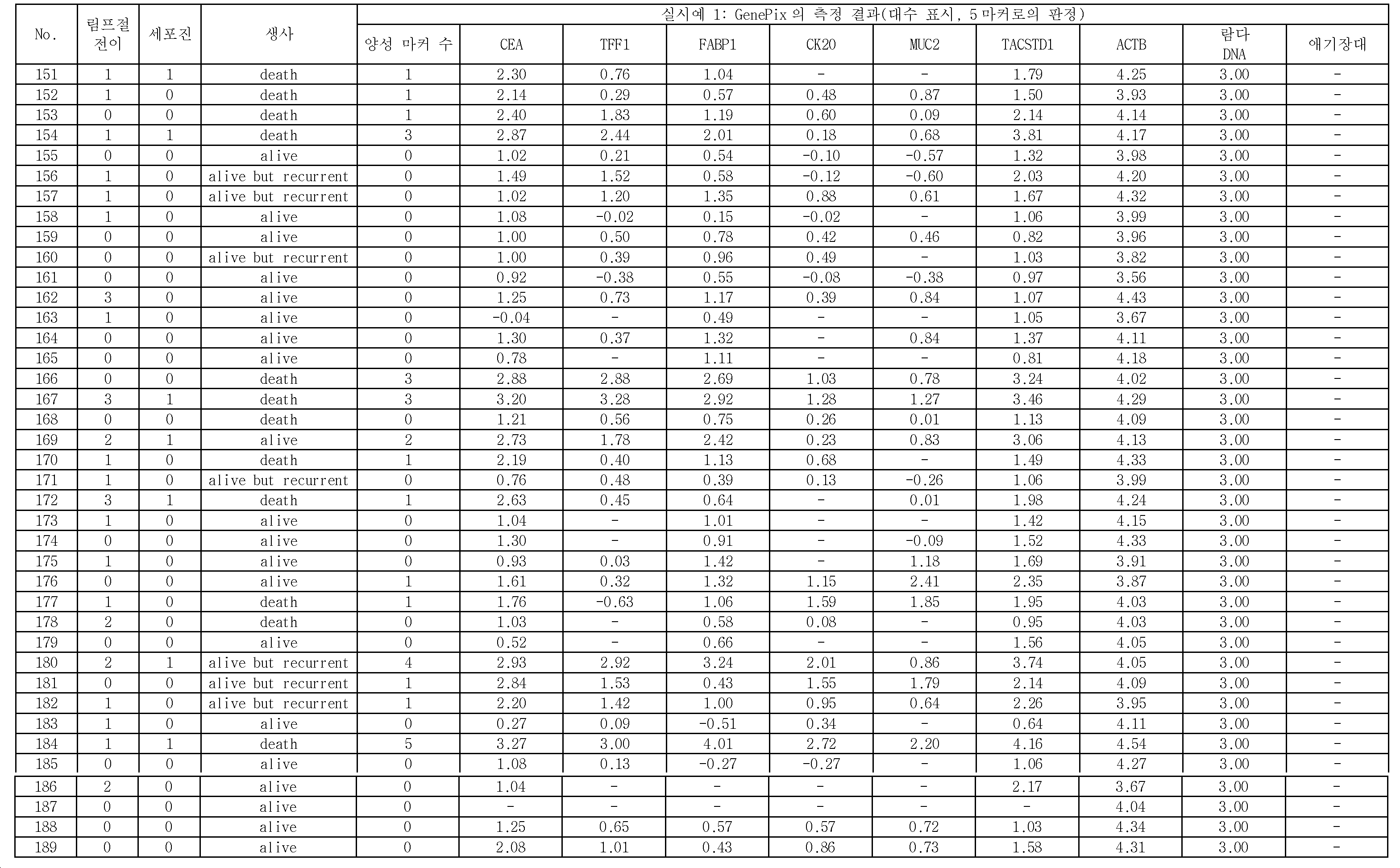
실시예 1의 189증례에 대하여, 후술하는 판정에 의하여 양성 또는 음성을 판정한 결과를 표 7에 나타낸다. 표 7에서는, 재발한 증례와 사망한 증례 수와, 양성 혹은 음성이라고 판정된 진행암 샘플 중의 재발 및 사망 증례 수의 비율(백분율)을 대응지어 나타낸다.Table 7 shows the results of judging positive or negative according to the determination described below with respect to the case 189 of Example 1. In Table 7, the ratio (percentage) of the number of recurrences and the number of deaths in the progressive cancer sample determined to be positive or negative is shown in association with the number of recurrences and the number of deaths.
표 7의 실시예의 란에는, 실시예 1의 189증례 중, 양성 마커 유전자가 1개 이상 검출된 것을 양성(+)으로서 판정하고, 양성 마커 유전자가 전혀 검출되지 않았던 것을 음성(-)으로서 판정한 결과를 나타낸다.In the column of the examples of Table 7, among the 189 cases of Example 1, a case where one or more positive marker genes were detected was judged as positive (+), and a case where no positive marker gene was detected was judged as negative (-) Results are shown.

표 7에서는, 진행암 샘플 189증례 중, 림프절 전이가 양성인 132증례에 있어서, 세포진에 의하여 검출할 수 없었던 98증례 중, 양성이라고 판단된 20증례의 재발률은 90.0%, 사망률은 80.0%이고, 음성이었던 78증례와 비교하여, 모두 높아 예후가 나빴다. 이것은 후술하는 비교예 1의 데이터와 비교하여, 재발률이 23.3포인트, 사망률이 25.5포인트 높았다. 따라서, CEA, TFF1, FABP1, CK20 및 MUC2 중 어느 1개 이상의 마커 유전자가 양성인지 여부를 판정함으로써, 보다 정확도가 높은 예후 판정이 가능하게 되는 것이 명확해졌다.In Table 7, among the cases of progressive cancer samples 189 cases, in 132 cases in which lymph node metastasis was positive, the recurrence rate was 90.0%, the mortality rate was 80.0%, the recurrence rate was 20.0% Compared with 78 cases, the prognosis was worse. Compared with the data of Comparative Example 1 described later, the recurrence rate was 23.3 points and the mortality rate was 25.5 points higher. Thus, it has become clear that more accurate prognostic determination becomes possible by determining whether at least one marker gene among CEA, TFF1, FABP1, CK20, and MUC2 is positive.
<실시예 2>≪ Example 2 >
실시예 1의 (1)~(8)의 조작을 행했다.(1) to (8) of Example 1 were carried out.
(8)의 하이브리다이제이션 반응 종료 후, 실시예 1의 (9)로 나타내는 바와 같이, 프로브가 스폿된 위치마다 형광 강도를 측정했다. 얻어진 189증례의 형광 강도에 대하여, 애기장대 DNA쇄 검출용 프로브가 스폿된 위치의 형광 강도를 백그라운드값으로 하여, 각 프로브가 고정된 위치에 있어서의 형광 강도로부터 애기장대 DNA쇄 검출용 프로브가 스폿된 위치의 형광 강도를 차감하고, 이것을 자연 대수로 나타낸 결과를 표 8~11에 나타낸다. 또, 각 마커 유전자의 형광 강도의 상댓값(자연 대수)에 각각 2개의 임곗값(제1 임곗값과 제2 임곗값)을 설정하고, 높은 쪽의 임곗값(제1 임곗값)을 넘은 경우에는, 양성으로 하고, 2개의 임곗값의 사이의 값을 나타내는 경우에는, 준양성으로 했다. 이 임곗값은, 컷오프값 부근에 값을 갖는 샘플의 측정 오차가 있는 것에 근거하여, 컷오프값의 전후 15%의 범위를 중으로 하고, 그 이상이면 고로 설정하는 것이다. 또, 이 임곗값은, 실시예 1에서 설명한 바와 같이, 일반적인 DNA 마이크로어레이의 컷오프값의 설정이 통상 백그라운드의 3배인 것에 근거하여 CEA의 임곗값을 설정하고, TFF1, FABP1, CK20, 및 MUC2에 대해서는, CEA의 임곗값의 약 절반으로 설정했다. 구체적으로는, 진행암 샘플의 마커 유전자의 임곗값은, 하기와 같이 설정했다.After completion of the hybridization reaction of (8), as shown in (9) of Example 1, the fluorescence intensity was measured at each spot where the probe was spotted. With respect to the fluorescence intensity of the obtained 189 cases, the fluorescence intensity at the spot where the Arabidopsis DNA chain detection probe was spotted was set as a background value. From the fluorescence intensity at the position where each probe was fixed, the fluorescence intensity of the Arabidopsis DNA chain detection probe The fluorescence intensity at the position where the fluorescence intensity was obtained was subtracted, and the results are shown in Tables 8 to 11 in terms of natural logarithm. In addition, when two threshold values (first threshold value and second threshold value) are set for the image intensity value (natural logarithm) of the fluorescent intensity of each marker gene and the threshold value (first threshold value) is exceeded Is positive, and when showing a value between two threshold values, it is considered to be quasi positive. This threshold value is set to 15% before and after the cutoff value based on the presence of a measurement error of the sample having a value near the cutoff value, and to set the range higher than 15%. As described in Example 1, the threshold value is set based on the fact that the cutoff value of a general DNA microarray is usually set to three times the background, and the threshold value of CEA is set to TFF1, FABP1, CK20, and MUC2 , It was set at about half of the threshold value of CEA. Specifically, the threshold value of the marker gene of the progressive cancer sample was set as follows.
(제2 임곗값; 준양성)(Second threshold value, quasi positive)
CEA: 1.79CEA: 1.79
TFF1: 1.57TFF1: 1.57
FABP1: 1.57FABP1: 1.57
CK20: 1.57CK20: 1.57
MUC2: 1.57MUC2: 1.57
(제1 임곗값; 양성)(First threshold value, positive)
CEA: 2.43CEA: 2.43
TFF1: 2.13TFF1: 2.13
FABP1: 2.13FABP1: 2.13
CK20: 2.13CK20: 2.13
MUC2: 2.13MUC2: 2.13
진행암 샘플 전체 189증례에 대하여 준양성 또는 양성이라고 판단된 마커 유전자의 수를 표 8~11에 함께 나타낸다. 또, 표 8~11에는, 상기에서 설명한 방법으로 얻어진 세포진의 결과, 림프절 전이의 판정 결과, 및 수술 후 경과에 대해서도 함께 나타냈다.Progressive Cancer Samples The number of marker genes determined to be positive or positive for all 189 cases are shown in Tables 8-11. Tables 8 to 11 also show the result of the cytology obtained by the above-described method, the judgment result of the lymph node metastasis, and the post-operative course.
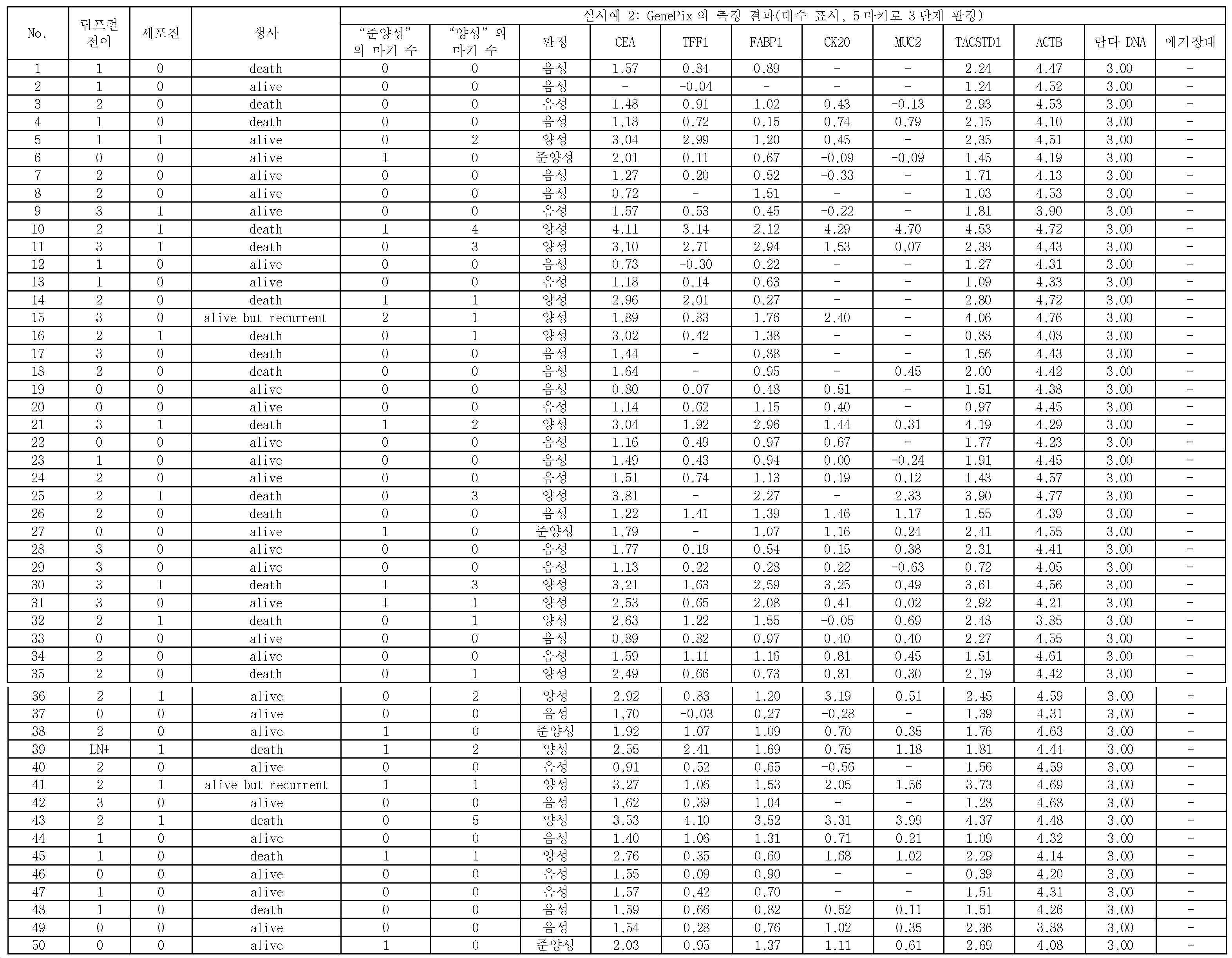
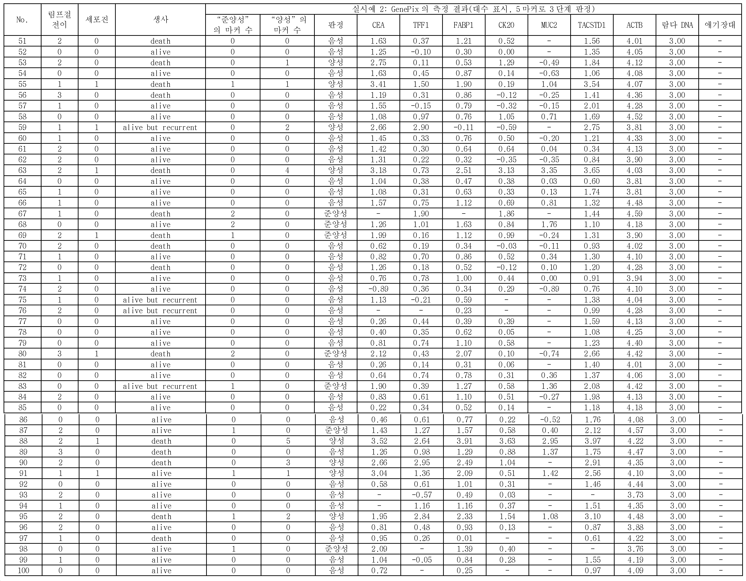
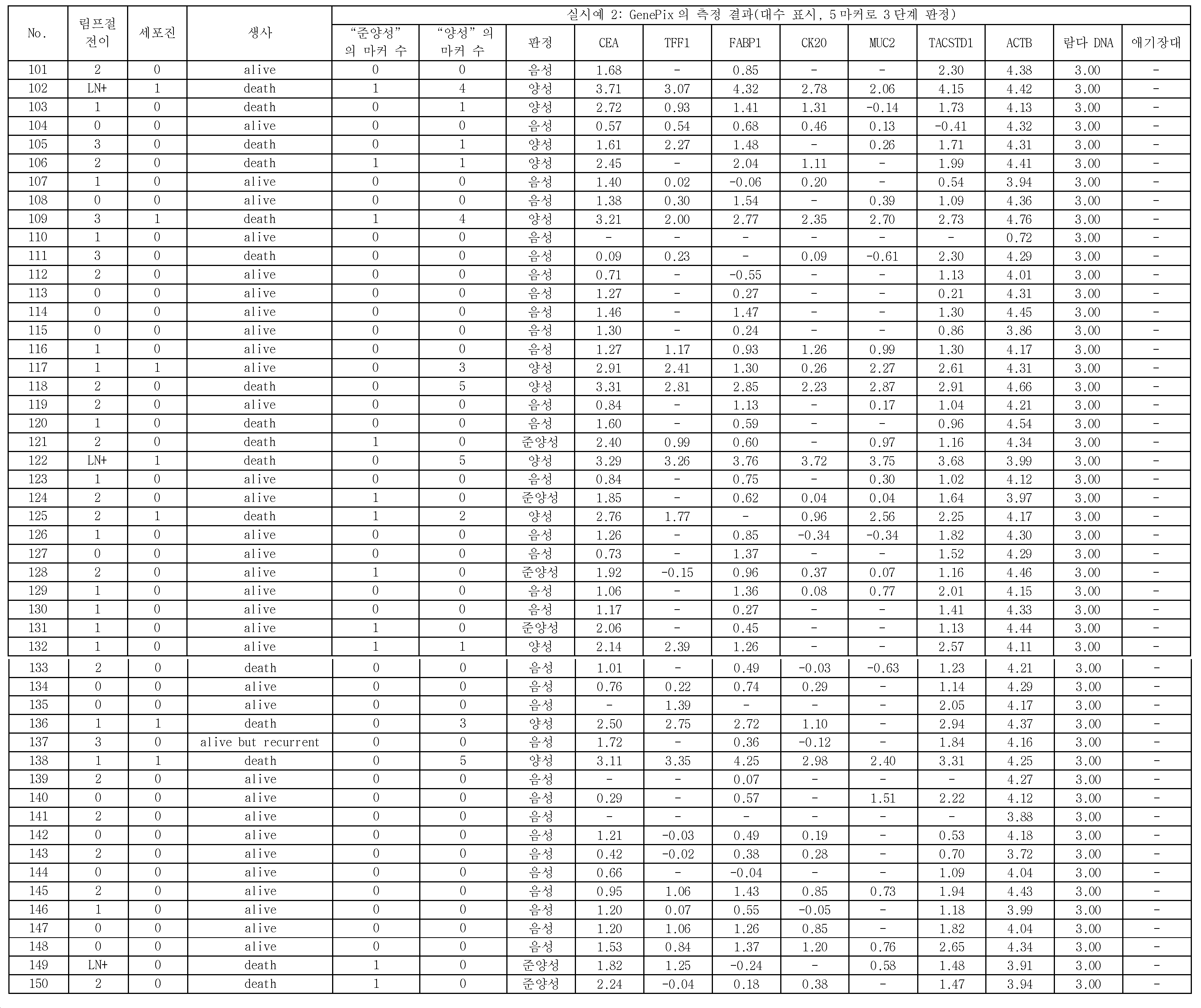

실시예 2의 189증례 중, 상기 판정에 의하여 음성, 준양성 또는 양성을 판정한 결과를 표 12에 나타낸다. 표 12에서는, 재발한 증례와 사망한 증례 수와, 음성, 준양성 또는 양성이라고 판정된 진행암 샘플 중의 재발 및 사망 증례 수의 비율(백분율)을 대응지어 나타낸다.Among the 189 cases of Example 2, the results of judging whether they are negative, quasi-positive or positive by the above determination are shown in Table 12. [ Table 12 shows the percentage (percentage) of recurred cases and the number of deaths and the number of recurrences and death cases in progressive cancer samples determined to be negative, quasi positive or positive.

표 12에서는, 진행암 샘플 189증례 중, 림프절 전이 및 세포진이 모두 음성인 57증례에 있어서, 양성이라고 판단된 3증례의 재발률은 66.7%, 사망률은 33.3%이고, 후술하는 비교예 1의 림프절 전이 및 세포진이 모두 음성인 57증례에 있어서, 양성을 나타낸 데이터와 비교하여, 재발률이 16.7포인트, 사망률이 8.3포인트 높았다. 따라서, 종래의 방법으로 재발의 가능성이 낮다고 판정되었지만 재발하게 된 증례에 대하여, 3단계 평가를 행함으로써, 보다 더 정확도가 높은 예후 판정이 가능하게 되는 것이 명확해졌다.In Table 12, in 57 cases in which both lymph node metastasis and cytology were negative in the progressive cancer sample 189, the recurrence rate of the three cases judged as positive was 66.7%, the mortality rate was 33.3%, and the lymph node metastasis of Comparative Example 1 The recurrence rate was 16.7 points and the mortality rate was 8.3 points higher than that of the data showing positive in 57 cases in which all the cells were negative. Thus, it became clear that a more accurate prognostic determination becomes possible by performing the three-step evaluation for a case in which the possibility of recurrence was determined to be low in the conventional method but the patient recurred.
또, 표 12에서는, 림프절 전이가 양성인 132증례에 있어서, 세포진에 의하여 검출할 수 없었던 98증례 중, 준양성이라고 판정된 13증례의 재발률은 61.5%, 사망률은 53.8%이고, 양성이라고 판정된 13증례의 재발률은 84.6%, 사망률은 76.9%였다. 이것은, 후술하는 비교예 1에 있어서 98증례 중, 양성이라고 판정된 33증례의 재발률, 사망률과 비교하여, 포인트가 각각 향상했다. 따라서, 실시예 2의 방법에 의하면, 보다 더 정확도가 높은 예후 판정이 가능하게 되는 것이 명확해졌다.In Table 12, among the 98 cases in which lymph node metastasis was positive, among the 98 cases that could not be detected by the cytopenia, the recurrence rate of the 13 cases judged to be positive was 61.5% and the mortality rate was 53.8% The recurrence rate was 84.6% and the mortality rate was 76.9%. This was improved in points compared with the recurrence rate and mortality rate of 33 cases, which were judged to be positive among 98 cases in Comparative Example 1 to be described later. Therefore, according to the method of the second embodiment, it becomes clear that a more accurate prognostic determination becomes possible.
<실시예 3>≪ Example 3 >
실시예 1의 (1)~(8)의 조작을 행했다.(1) to (8) of Example 1 were carried out.
(8)의 하이브리다이제이션 반응 종료 후, 실시예 1의 (9)로 나타내는 바와 같이, 프로브가 스폿된 위치마다 형광 강도를 측정했다. 얻어진 189증례의 형광 강도에 대하여, 애기장대 DNA쇄 검출용 프로브가 스폿된 위치의 형광 강도를 백그라운드값으로 하여, 각 프로브가 고정된 위치에 있어서의 형광 강도로부터 애기장대 DNA쇄 검출용 프로브가 스폿된 위치의 형광 강도를 차감하고, 이것을 자연 대수로 나타낸 결과를 표 13~16에 나타낸다. 또, 각 마커 유전자의 형광 강도의 상댓값(자연 대수)에 각각 2개의 임곗값(제1 임곗값과 제2 임곗값)을 설정하고, 적어도 하나 측정 결과의 값이 높은 쪽의 임곗값(제1 임곗값)을 넘은 경우, 또는 높은 쪽의 임곗값(제1 임곗값)을 넘은 것은 없지만, 적어도 2개 이상의 측정 결과의 값이 2개의 임곗값의 사이의 값을 나타내는 경우에는, 양성으로 했다. 또, 측정 결과의 값이 높은 쪽의 임곗값(제1 임곗값)을 넘는 것은 없고, 5개의 마커 중, 1개의 마커에 대하여 측정하여 얻어진 측정 결과의 값이 2개의 임곗값의 사이의 값을 나타냄과 함께, 그 외의 마커에 대하여 측정하여 얻어진 측정 결과의 값은 낮은 쪽의 임곗값을 하회하고 있는 경우, 약양성으로 했다. 이 임곗값은, 컷오프값 부근에 값을 갖는 샘플의 측정 오차가 있는 것에 근거하여, 컷오프값의 전후 15%의 범위를 중으로 하고, 그 이상이면 고로 설정하는 것이다. 또, 이 임곗값은, 실시예 1에서 설명한 바와 같이, 일반적인 DNA 마이크로어레이의 컷오프값의 설정이 통상 백그라운드의 3배인 것에 근거하여 CEA의 임곗값을 설정하고, TFF1, FABP1, CK20, 및 MUC2에 대해서는, CEA의 임곗값의 약 절반으로 설정했다. 구체적으로는, 진행암 샘플의 마커 유전자의 임곗값은, 하기와 같이 설정했다.After completion of the hybridization reaction of (8), as shown in (9) of Example 1, the fluorescence intensity was measured at each spot where the probe was spotted. With respect to the fluorescence intensity of the obtained 189 cases, the fluorescence intensity at the spot where the Arabidopsis DNA chain detection probe was spotted was set as a background value. From the fluorescence intensity at the position where each probe was fixed, the fluorescence intensity of the Arabidopsis DNA chain detection probe The fluorescence intensity at the position where the fluorescence intensity was obtained was subtracted and the results are shown in Tables 13 to 16 in terms of natural logarithm. Two intrinsic values (first intrinsic value and second intrinsic value) are set for each of the above values (natural logarithm) of the fluorescence intensity of each marker gene, and at least one intrinsic value 1 threshold value) or higher than the threshold value (first threshold value), but when the value of at least two measurement results indicates the value between the two threshold values, it is determined to be positive . In addition, there is no case in which the measurement result value is higher than the threshold value (first threshold value), and the measurement result obtained by measuring one marker out of the five markers is a value between the two threshold values And when the value of the measurement result obtained by measuring the other markers is less than the lower threshold value, it is determined to be approximately positive. This threshold value is set to 15% before and after the cutoff value based on the presence of a measurement error of the sample having a value near the cutoff value, and to set the range higher than 15%. As described in Example 1, the threshold value is set based on the fact that the cutoff value of a general DNA microarray is usually set to three times the background, and the threshold value of CEA is set to TFF1, FABP1, CK20, and MUC2 , It was set at about half of the threshold value of CEA. Specifically, the threshold value of the marker gene of the progressive cancer sample was set as follows.
(제2 임곗값)(Second threshold value)
CEA: 1.79CEA: 1.79
TFF1: 1.57TFF1: 1.57
FABP1: 1.57FABP1: 1.57
CK20: 1.57CK20: 1.57
MUC2: 1.57MUC2: 1.57
(제1 임곗값)(First threshold value)
CEA: 2.43CEA: 2.43
TFF1: 2.13TFF1: 2.13
FABP1: 2.13FABP1: 2.13
CK20: 2.13CK20: 2.13
MUC2: 2.13MUC2: 2.13
진행암 샘플 전체 189증례에 대하여 약양성 또는 양성이라고 판단된 마커 유전자의 수를 표 13~16에 함께 나타낸다. 또, 표 13~16에는, 상기에서 설명한 방법으로 얻어진 세포진의 결과, 림프절 전이의 판정 결과, 및 수술 후 경과에 대해서도 함께 나타냈다.Progressive Cancer Samples The number of marker genes determined to be approximately positive or positive for all 189 cases are shown in Tables 13-16. Tables 13 to 16 also show the result of the cytopenia obtained by the above-described method, the judgment result of the lymph node metastasis, and the post-operative course.
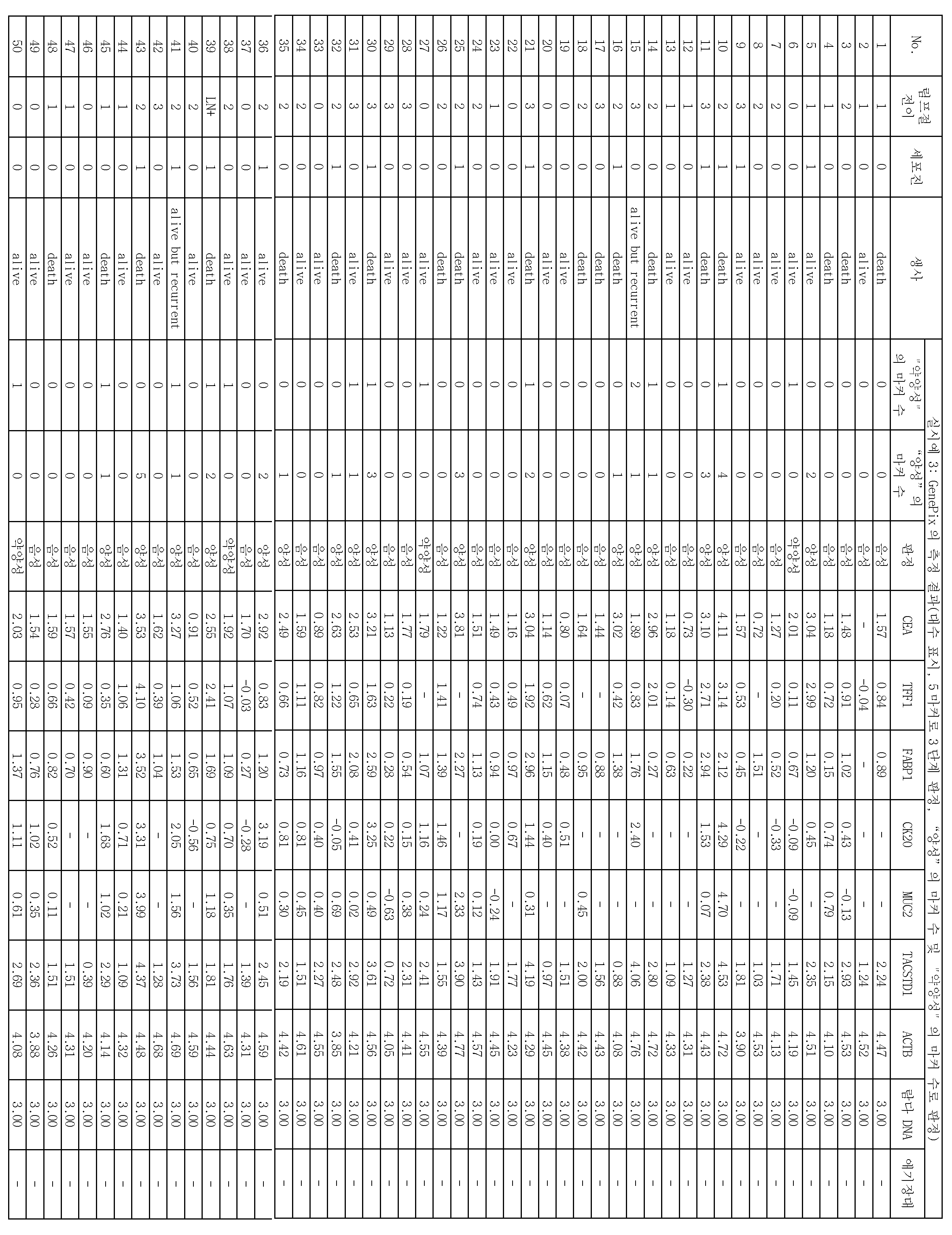
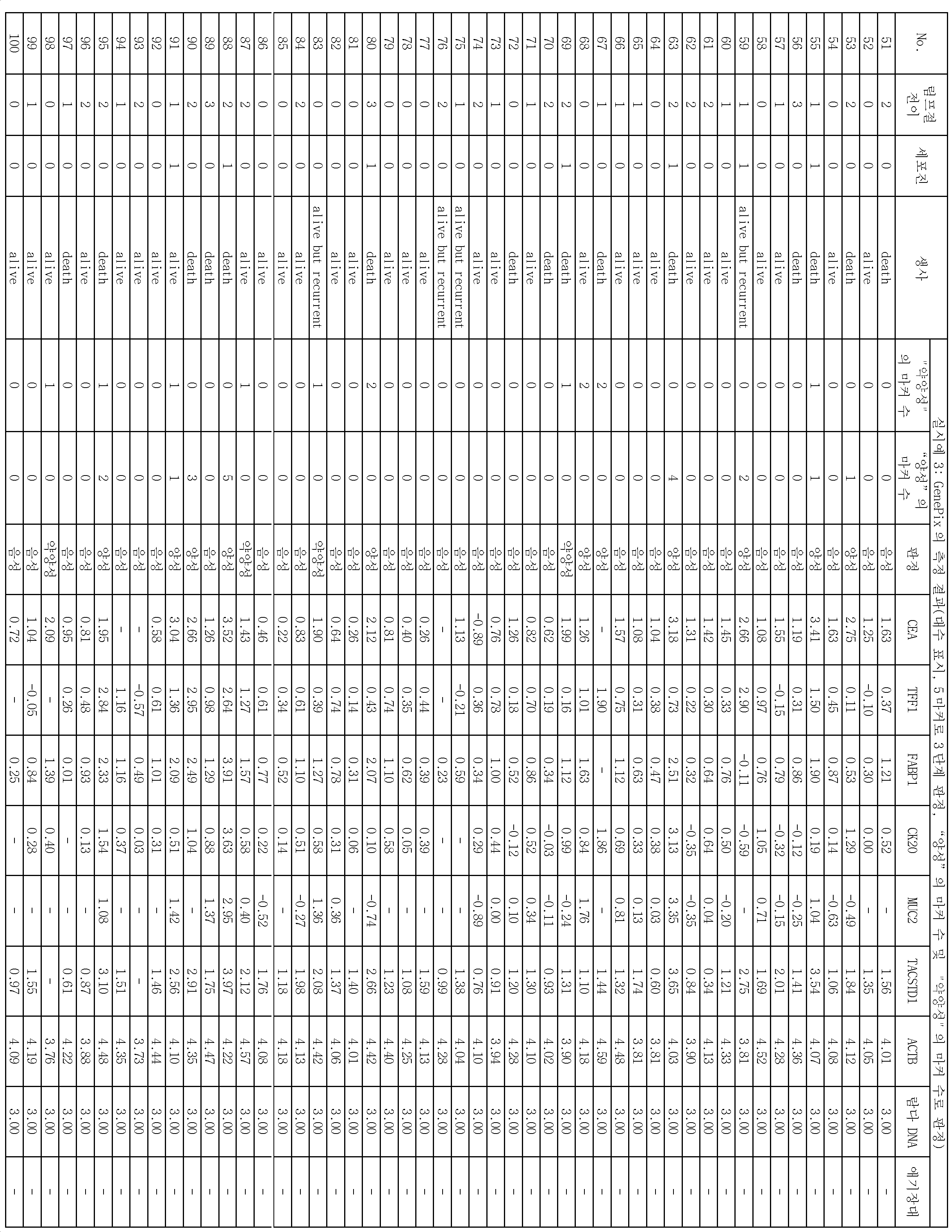
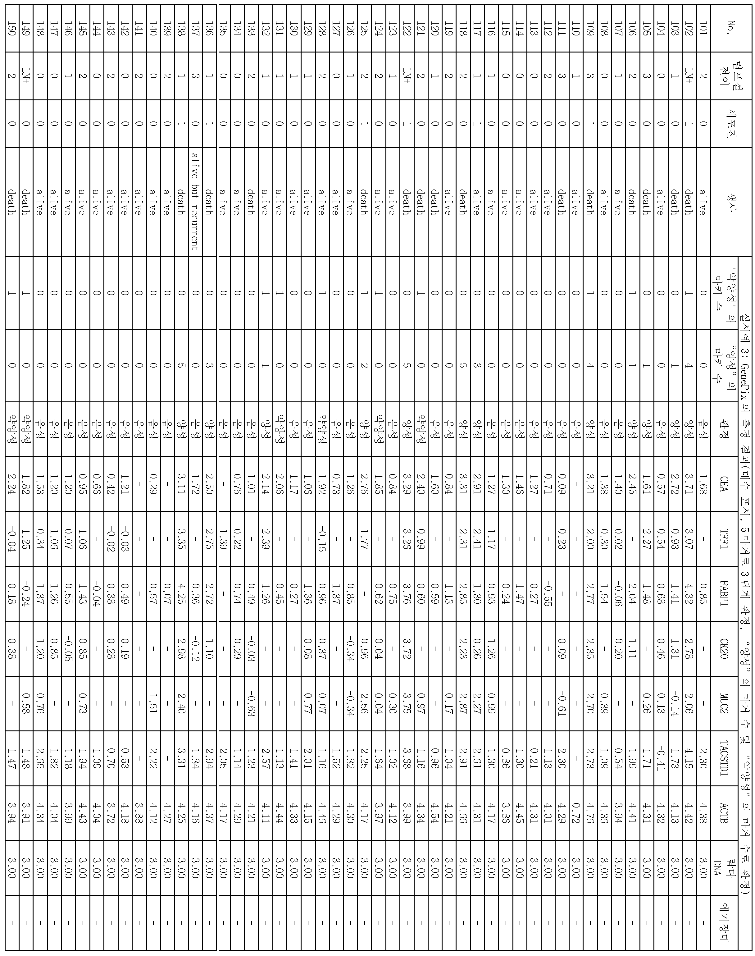

실시예 3의 189증례 중, 상기 판정에 의하여 음성, 약양성 또는 양성을 판정한 결과를 표 17에 나타낸다. 표 17에서는, 재발한 증례와 사망한 증례 수와, 음성, 약양성 또는 양성이라고 판정된 진행암 샘플 중의 재발 및 사망 증례 수의 비율(백분율)을 대응지어 나타낸다.Among the 189 cases of Example 3, Table 17 shows the results of judging the negative, weakly positive or positive by the above-mentioned judgment. In Table 17, the number of relapsed cases, the number of deaths, and the percentage (percentage) of recurrences and the number of deaths in a progressive cancer sample determined to be negative, weakly positive or positive are shown in association with each other.
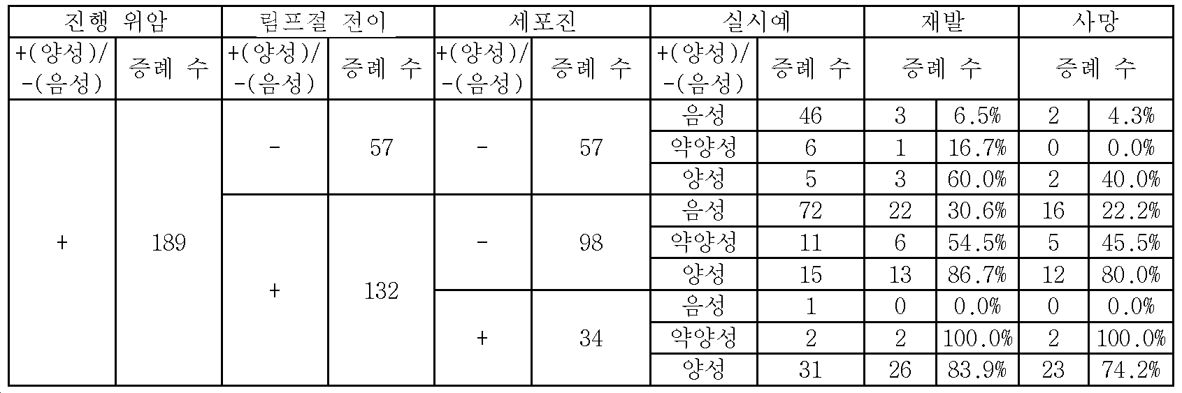
표 17에서는, 진행암 샘플 189증례 중, 림프절 전이 및 세포진이 모두 음성인 57증례에 있어서, 양성이라고 판단된 5증례의 재발률은 60.0%, 사망률은 40.0%이고, 후술하는 비교예 1의 림프절 전이 및 세포진이 모두 음성인 57증례에 있어서, 양성을 나타낸 데이터와 비교하여, 재발률이 10.0포인트, 사망률이 15.0포인트 높았다. 따라서, 종래의 방법으로 재발의 가능성이 낮다고 판정되었지만 재발하게 된 증례에 대하여, 실시예 2의 방법으로부터 판정 기준을 변경한 3단계 평가를 행함으로써, 정확도가 더 높은 예후 판정이 가능하게 되는 것이 명확해졌다.In Table 17, recurrence rate of 60.0% and mortality rate of 5 cases, which were judged positive in 57 cases in which both lymph node metastasis and cytology were negative, were found in the case of advanced cancer sample 189, and the lymph node metastasis of Comparative Example 1 The recurrence rate was 10.0 points and the mortality rate was 15.0 points higher than the data showing positive in 57 cases in which the cell division was negative. Therefore, it is clear that, by performing the three-step evaluation in which the judgment standard is changed from the method of the second embodiment with respect to the case where the likelihood of recurrence is low in the conventional method, It has become.
또, 표 17에서는, 림프절 전이가 양성인 132증례에 있어서, 세포진에 의하여 검출할 수 없었던 98증례 중, 약양성이라고 판정된 11증례의 재발률은 54.5%, 사망률은 45.5%이며, 양성이라고 판정된 15증례의 재발률은 86.7%, 사망률은 80.0%였다. 이것은, 비교예 1에 있어서 98증례 중, 양성이라고 판정된 33증례의 재발률, 사망률과 비교하여, 포인트가 각각 향상했다. 따라서, 실시예 3의 방법에 의하면, 매우 정확도가 높은 예후 판정이 가능하게 되는 것이 명확해졌다.In Table 17, in the cases of 132 cases in which lymph node metastasis was positive, the recurrence rate of the 11 cases, which were judged to be weakly positive, was 54.5% and the mortality rate was 45.5% The recurrence rate was 86.7% and the mortality rate was 80.0%. This indicates that the points were improved in comparison with the recurrence rate and the mortality rate of the 33 cases judged as positive among the 98 cases in the Comparative Example 1. Therefore, according to the method of the third embodiment, it became clear that a highly accurate prognosis determination becomes possible.
<비교예 1>≪ Comparative Example 1 &
(1) DNA 마이크로어레이의 제작(1) Production of a DNA microarray
실시예 1의 (1)~(3)의 조작에 따라, DNA 마이크로어레이를 제작했다. 단, 배열 번호 1로 나타나는 염기 배열로 이루어지는 애기장대 DNA쇄 검출용 프로브는 DNA 마이크로어레이에 고정하지 않았다.A DNA microarray was prepared according to the procedures of (1) to (3) of Example 1. However, the probe for detecting Arabidopsis DNA strand composed of the nucleotide sequence represented by SEQ ID NO: 1 was not fixed to the DNA microarray.
(2) 진행암 샘플과 DNA 마이크로어레이와의 하이브리다이제이션(2) Hybridization with a progressive cancer sample and a DNA microarray
실시예 1의 (4)~(7)의 조작을 행하고, 얻어진 진행암 샘플 189증례의 Cy3 형광 라벨화 PCR 산물을 본 비교예 1의 (1)에서 제작한 DNA 마이크로어레이에 각각 하이브리다이즈했다. 하이브리다이제이션의 조건 및 조작은, 실시예 1의 (8)에 따랐다. 단, Cy3 형광 라벨화 PCR 산물에 애기장대 DNA쇄는 더하지 않았다.(4) to (7) of Example 1 were carried out, and Cy3 fluorescently labeled PCR products of the progressive cancer sample 189 obtained were each hybridized to the DNA microarray prepared in (1) of this Comparative Example 1 . The hybridization conditions and procedures were the same as those in Example 1 (8). However, the Cy3 fluorescently labeled PCR product did not have the Arabidopsis DNA chain added.
(3) 조기 암 샘플과 DNA 마이크로어레이와의 하이브리다이제이션(3) Hybridization of early cancer samples with DNA microarrays
세포진에 의하여 조기 암이라고 판정된 조기 암세포 25증례에 대하여, 실시예 1의 (4)~(7)로 나타내는 방법과 동일한 조작을 행하여 Cy3 형광 라벨화 PCR 산물을 준비하여, 비교예 1의 (1)에서 제작한 DNA 마이크로어레이에 하이브리다이즈했다. 이 경우에도, 하이브리다이제이션의 조건 및 조작은, 실시예 1의 (8)에 따라, Cy3 형광 라벨화 PCR 산물에 애기장대 DNA쇄는 더하지 않았다.The same procedure as in (4) to (7) of Example 1 was carried out on the early cancer cell 25 case judged to be premature cancer by the cytopenia to prepare a Cy3 fluorescently labeled PCR product. ) To hybridize to a DNA microarray made by the manufacturer. Also in this case, according to (8) of Example 1, conditions and operation of hybridization did not add the Arabidopsis DNA strand to the Cy3 fluorescently labeled PCR product.
(4) 측정(4) Measurement
하이브리다이제이션 반응 종료 후, 스핀 드라이한 DNA 마이크로어레이에 대하여, DNA 마이크로어레이용 스캐너(액손사제 GenePix 4000B)를 이용하여, 프로브가 스폿된 위치마다 형광 강도를 측정했다. 측정할 때의 수광 감도(PMT)는, λDNA가 고정된 스폿에 있어서의 형광 강도가 100에서 600이 되도록 조정했다. 형광 강도의 산출 시에는, 프로브마다 5개의 측정값이 얻어지지만, 최곳값 및 최젓값을 제외한 중간의 3개의 측정값을 평균화하고, 프로브마다 평균값을 산출하여, 이것을 형광 강도로 했다. 전체 189증례, 및 조기 암 샘플 25증례에 대하여 형광 강도를 각각 측정했다.After completion of the hybridization reaction, the fluorescence intensity of the spin-dried DNA microarray was measured at each spot where the probe was spotted using a DNA microarray scanner (GenePix 4000B, manufactured by Axon Corporation). The light receiving sensitivity (PMT) at the time of measurement was adjusted so that the fluorescence intensity in a spot to which? DNA was fixed was from 100 to 600. In calculating the fluorescence intensity, five measurement values were obtained for each probe. However, the three middle measurement values excluding the maximum value and the minimum value were averaged, and the average value was calculated for each probe, and the fluorescence intensity was obtained. Fluorescence intensity was measured for all 189 cases, and for 25 cases of early cancer samples.
(5) 위암의 재발 예측(5) recurrence prediction of gastric cancer
조기 암 샘플을 하이브리다이즈한 DNA 마이크로어레이의 측정 결과 25증례를 이용하여, 각 마커에 대하여, 평균값+그 표준 편차×2를 컷오프값으로 했다. 구체적인 컷오프값을 이하에 나타낸다.As a result of the measurement of the DNA microarray hybridized with the early cancer sample, the average value plus the standard deviation x 2 for each marker was used as a cutoff value using 25 cases. Specific cutoff values are shown below.
CEA: 56.9CEA: 56.9
TFF1: 54.0TFF1: 54.0
FABP1: 55.3FABP1: 55.3
CK20: 53.1CK20: 53.1
MUC2: 49.8MUC2: 49.8
TACSTD1: 85.1TACSTD1: 85.1
진행암 샘플 189증례에 대하여, 컷오프값을 넘은 마커 유전자를 양성이라고 판단하고, 컷오프값 이하의 마커 유전자는 음성으로 했다. 형광 강도의 결과를 표 18~21에 나타낸다. 또, 표 18~21에는, 상기에서 설명한 방법으로 얻어진 세포진의 결과, 림프절 전이의 판정 결과, 및 수술 후 경과에 대해서도 함께 나타냈다.For progressive cancer sample 189, the marker gene that exceeded the cutoff value was considered positive and the marker gene below the cutoff value was negative. The results of fluorescence intensity are shown in Tables 18 to 21. Tables 18 to 21 also show the result of the cell division obtained by the above-described method, the judgment result of the lymph node metastasis, and the progress of the operation.
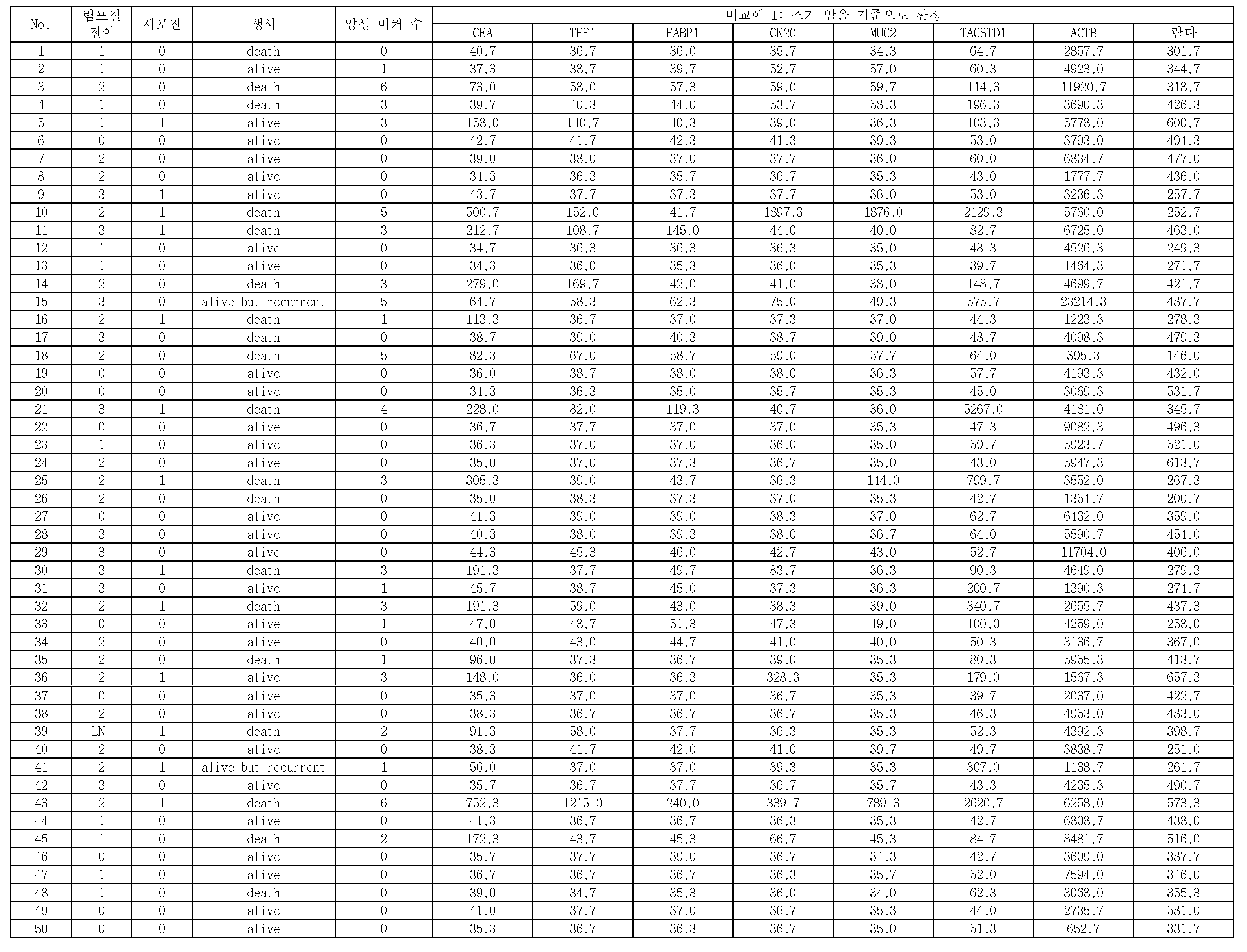

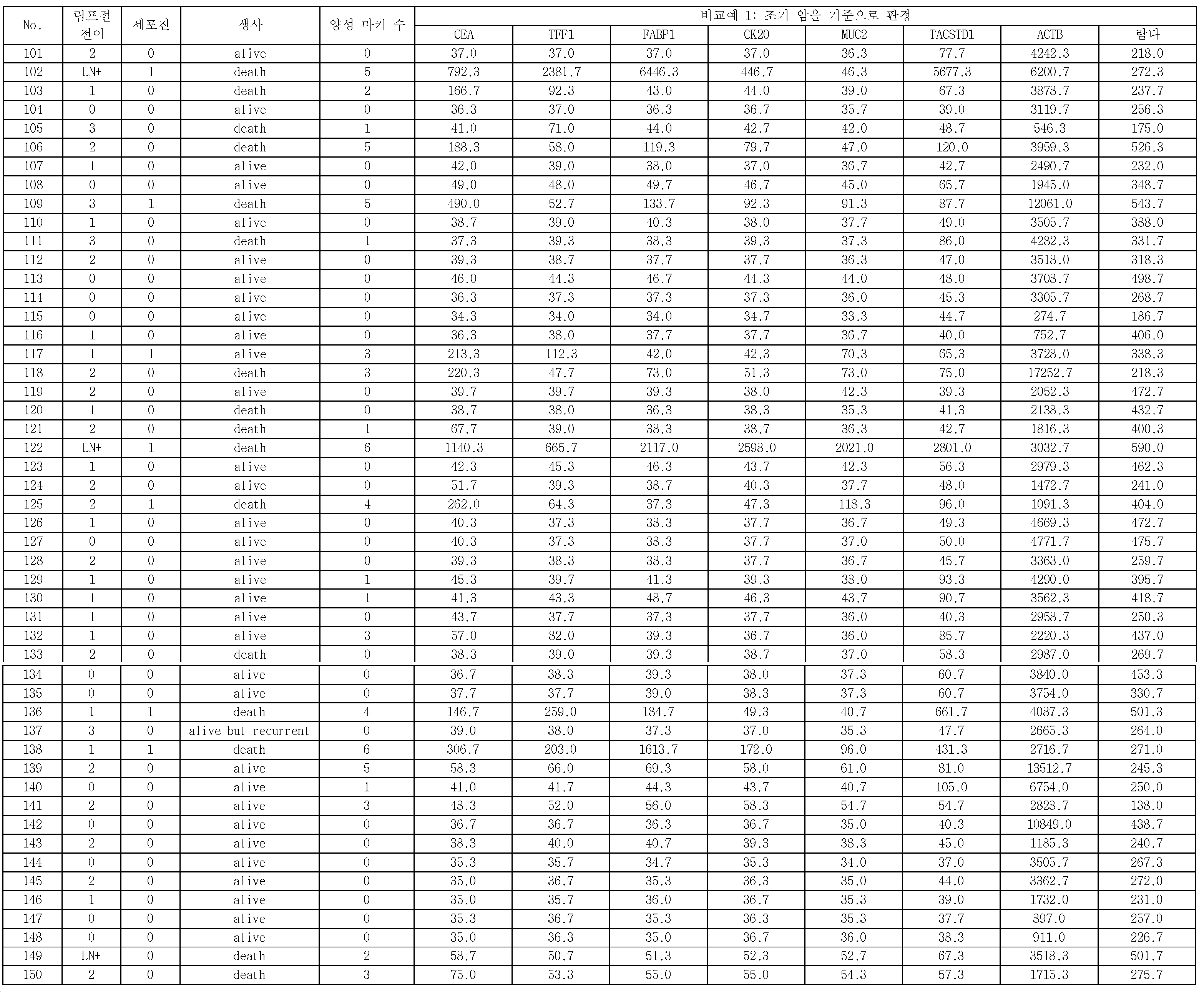
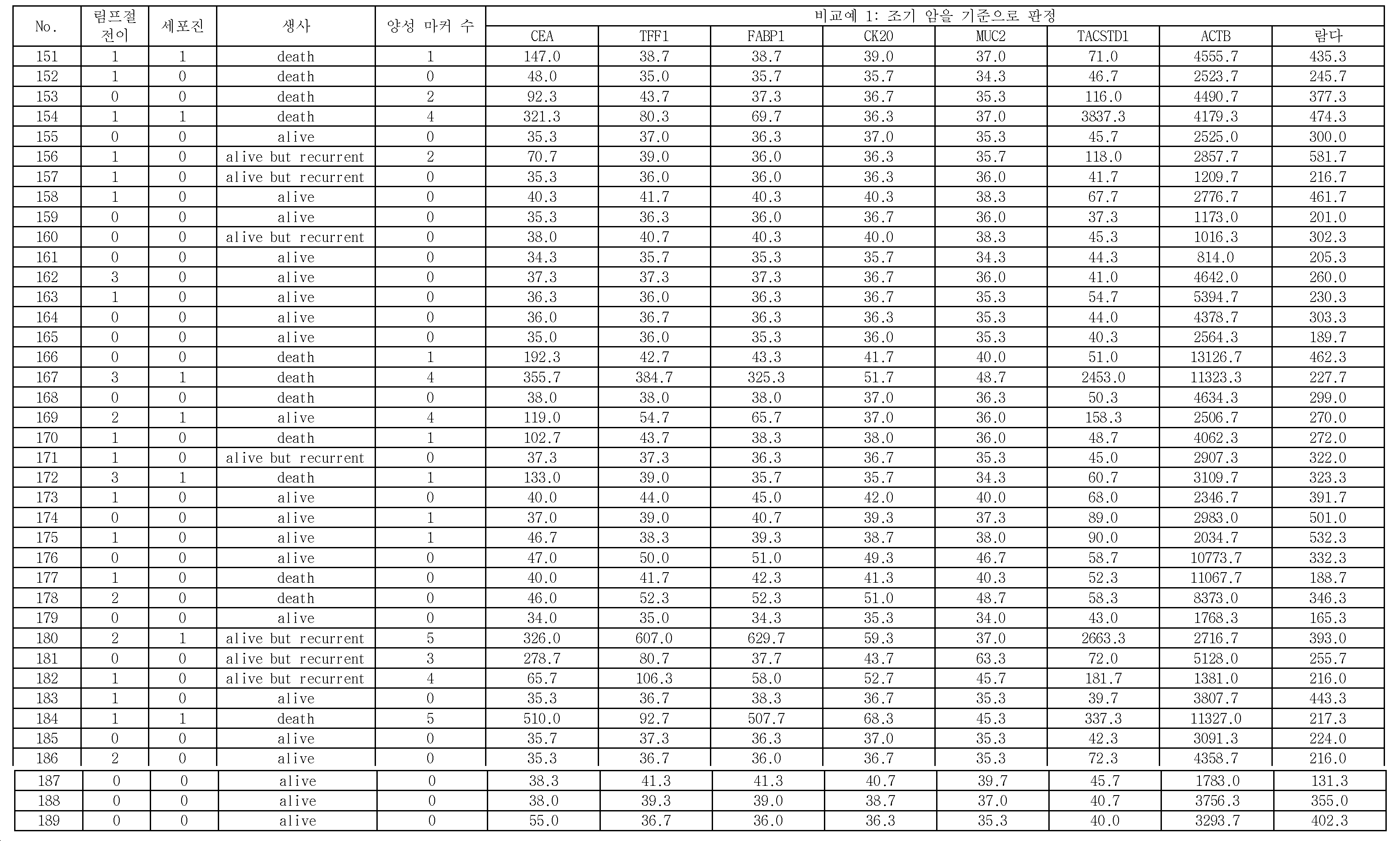
표 22는 비교예 1의 결과이다. 표 22에서는 재발한 증례와 사망한 증례 수, 및 양성 혹은 음성이라고 판정된 진행암 샘플 중의 재발 및 사망 증례 수의 비율(백분율)을 각 비교예의 결과와 대응지어 나타낸다. 표 22에서는, 각 비교예에서 양성 마커 유전자가 1개 이상 검출된 것을 +로 나타내고, 전혀 검출되지 않았던 것을 -로 나타낸다.Table 22 shows the results of Comparative Example 1. Table 22 shows the percentage (percentage) of recurrence cases and the number of death cases in progressive cancer samples determined to be positive or negative, in relation to the results of each comparative example. In Table 22, "+" indicates that one or more positive marker genes were detected in each comparative example, and "-" indicates that no positive marker gene was detected.
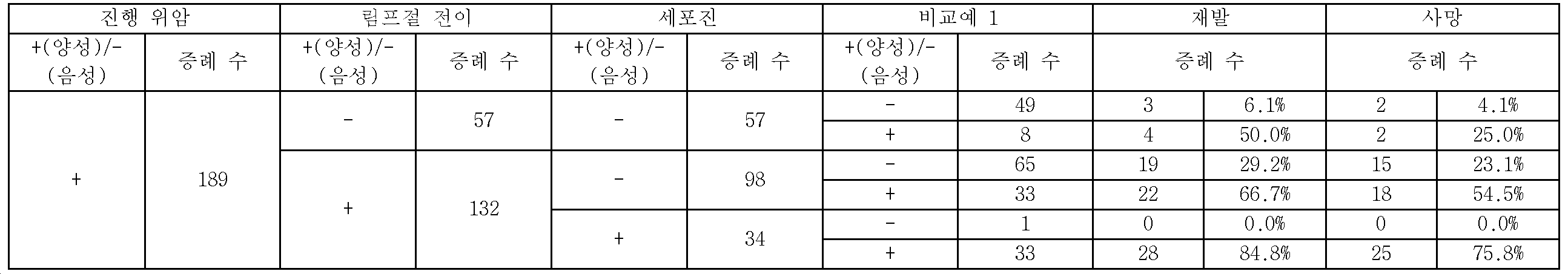
표 22로 나타내는 바와 같이, 진행암 샘플 189증례 중, 림프절 전이가 양성인 132증례에 있어서, 세포진에 의하여 검출할 수 없었던 98증례 중, 비교예 1에 있어서 양성이라고 판단된 33증례의 재발률, 사망률은, 음성이었던 65증례와 비교하여, 모두 높아 예후가 나빴다.As shown in Table 22, among the cases of progressive cancer samples 189, in the cases of 132 cases in which lymph node metastasis was positive, among the 98 cases that could not be detected by the cytogenetic analysis, the recurrence rate and the mortality rate of the 33 cases judged positive in Comparative Example 1 , Compared with 65 cases of negative, all were high and the prognosis was bad.
이상으로부터, 실시예 1~3에 기재된 방법을 이용함으로써, 조기 암환자의 증례를 동시에 이용한 종래의 검사법(비교예 1)보다, 더 양호한 정확도로 위암의 재발 예측을 행할 수 있는 것이 명확해졌다. 림프절 전이 음성의 57증례에 대해서도, 증례 수는 적지만 마이크로어레이로 양성으로 판별된 증례의 재발률, 사망률은, 음성으로 판별된 증례에 비하여 높았다.From the above, it is clear that by using the methods described in Examples 1 to 3, it is possible to predict the recurrence of stomach cancer with better accuracy than the conventional test method using the cases of early cancer patients at the same time (Comparative Example 1). Even in 57 cases of lymph node metastasis, the recurrence rate and mortality rate of cases with few cases but positive by microarrays were higher than cases with negative cases.
이 출원은, 2013년 5월 7일에 출원된 일본 특허출원 2013-097736호를 기초로 하는 우선권을 주장하고, 그 개시의 전체를 여기에 원용한다.This application is based upon and claims the benefit of priority from Japanese Patent Application No. 2013-097736, filed on May 7, 2013, the entire disclosure of which is incorporated herein by reference.
SEQUENCE LISTING <110> NATIONAL CANCER CENTER SUMITOMO BAKELITE CO., LTD <120> Method for predicting recurrence of stomach cancer <130> SB-70865 <160> 25 <170> PatentIn version 3.3 <210> 1 <211> 58 <212> DNA <213> Artificial Sequence <220> <223> probe sequence for arabidopsis <400> 1 agttcaagct tcattctggc gagagagcag tgaaacgtgc aatgttcaac tattcggt 58 <210> 2 <211> 60 <212> DNA <213> Artificial Sequence <220> <223> probe sequence for CEA <400> 2 gatatagcag ccctggtgta gtttcttcat ttcaggaaga ctgacagttg ttttgcttct 60 <210> 3 <211> 54 <212> DNA <213> Artificial Sequence <220> <223> probe sequence for TFF1 <400> 3 ttcgacgaca ccgttcgtgg ggtcccctgg tgcttctatc ctaataccat cgac 54 <210> 4 <211> 60 <212> DNA <213> Artificial Sequence <220> <223> probe sequence for FABP1 <400> 4 cattctgcac gatttccgac acccccttga tatccttccc cttctggatg agctcttccg 60 <210> 5 <211> 60 <212> DNA <213> Artificial Sequence <220> <223> probe sequence for CK20 <400> 5 tctggaggcc caactgatgc agattcggag taacatggaa cgccagaaca acgaatacca 60 <210> 6 <211> 52 <212> DNA <213> Artificial Sequence <220> <223> probe sequence for MUC2 <400> 6 cggaggtttc gtacgccggc tgcaccaaga ccgtcctcat gaatcattgc tc 52 <210> 7 <211> 60 <212> DNA <213> Artificial Sequence <220> <223> probe sequence for TACSTD1 <400> 7 gcagggtcta aaagctggtg ttattgctgt tattgtggtt gtggtgatag cagttgttgc 60 <210> 8 <211> 60 <212> DNA <213> Artificial Sequence <220> <223> probe sequence for ACTB <400> 8 cctgtggcat ccacgaaact accttcaact ccatcatgaa gtgtgacgtg gacatccgca 60 <210> 9 <211> 48 <212> DNA <213> Artificial Sequence <220> <223> probe sequence for LambdaDNA <400> 9 aattcatgga aaaggtctgc gtcaaatccc cagtcgtcat gcattgcc 48 <210> 10 <211> 25 <212> DNA <213> Artificial Sequence <220> <223> Sequence as PCR primer for a part of CEA <400> 10 tgcatctgga acttctcctg gtctc 25 <210> 11 <211> 24 <212> DNA <213> Artificial Sequence <220> <223> Sequence as PCR primer for a part of CEA <400> 11 tcacgatgtt ggctaggatg gtct 24 <210> 12 <211> 24 <212> DNA <213> Artificial Sequence <220> <223> Sequence as PCR primer for a part of TFF1 <400> 12 cctttggagc agagaggagg caat 24 <210> 13 <211> 27 <212> DNA <213> Artificial Sequence <220> <223> Sequence as PCR primer for a part of TFF1 <400> 13 tcagagcagt caatctgtgt tgtgagc 27 <210> 14 <211> 20 <212> DNA <213> Artificial Sequence <220> <223> Sequence as PCR primer for a part of FABP1 <400> 14 tcatgaaggc aatcggtctg 20 <210> 15 <211> 21 <212> DNA <213> Artificial Sequence <220> <223> Sequence as PCR primer for a part of FABP1 <400> 15 caatgtcacc caatgtcatg g 21 <210> 16 <211> 25 <212> DNA <213> Artificial Sequence <220> <223> Sequence as PCR primer for a part of CK20 <400> 16 acacggtgaa ctatgggagc gatct 25 <210> 17 <211> 24 <212> DNA <213> Artificial Sequence <220> <223> Sequence as PCR primer for a part of CK20 <400> 17 cttccagaag gcggcggtaa gtag 24 <210> 18 <211> 20 <212> DNA <213> Artificial Sequence <220> <223> Sequence as PCR primer for a part of MUC2 <400> 18 ccggggagtg ctgtaagaag 20 <210> 19 <211> 20 <212> DNA <213> Artificial Sequence <220> <223> Sequence as PCR primer for a part of MUC2 <400> 19 ctcctctttg cagcaggagc 20 <210> 20 <211> 20 <212> DNA <213> Artificial Sequence <220> <223> Sequence as PCR primer for a part of TACSTD1 <400> 20 tgctggggtc agaagaacag 20 <210> 21 <211> 21 <212> DNA <213> Artificial Sequence <220> <223> Sequence as PCR primer for a part of TACSTD1 <400> 21 ttgagttccc tatgcatctc a 21 <210> 22 <211> 20 <212> DNA <213> Artificial Sequence <220> <223> Sequence as PCR primer for a part of ACTB <400> 22 tcatcaccat tggcaatgag 20 <210> 23 <211> 20 <212> DNA <213> Artificial Sequence <220> <223> Sequence as PCR primer for a part of ACTB <400> 23 cactgtgttg gcgtacaggt 20 <210> 24 <211> 23 <212> DNA <213> Artificial Sequence <220> <223> Sequence as PCR primer for a part of LambdaDNA <400> 24 acggacttcg ttgctttcca gtt 23 <210> 25 <211> 24 <212> DNA <213> Artificial Sequence <220> <223> Sequence as PCR primer for a part of LambdaDNA <400> 25 gaattacagc gcaacacagc aata 24 SEQUENCE LISTING <110> NATIONAL CANCER CENTER SUMITOMO BAKELITE CO., LTD <120> Method for predicting recurrence of stomach cancer <130> SB-70865 <160> 25 <170> PatentIn version 3.3 <210> 1 <211> 58 <212> DNA <213> Artificial Sequence <220> <223> probe sequence for arabidopsis <400> 1 agttcaagct tcattctggc gagagagcag tgaaacgtgc aatgttcaac tattcggt 58 <210> 2 <211> 60 <212> DNA <213> Artificial Sequence <220> <223> probe sequence for CEA <400> 2 gatatagcag ccctggtgta gtttcttcat ttcaggaaga ctgacagttg ttttgcttct 60 <210> 3 <211> 54 <212> DNA <213> Artificial Sequence <220> <223> probe sequence for TFF1 <400> 3 ttcgacgaca ccgttcgtgg ggtcccctgg tgcttctatc ctaataccat cgac 54 <210> 4 <211> 60 <212> DNA <213> Artificial Sequence <220> <223> probe sequence for FABP1 <400> 4 cattctgcac gatttccgac acccccttga tatccttccc cttctggatg agctcttccg 60 <210> 5 <211> 60 <212> DNA <213> Artificial Sequence <220> <223> probe sequence for CK20 <400> 5 tctggaggcc caactgatgc agattcggag taacatggaa cgccagaaca acgaatacca 60 <210> 6 <211> 52 <212> DNA <213> Artificial Sequence <220> <223> probe sequence for MUC2 <400> 6 cggaggtttc gtacgccggc tgcaccaaga ccgtcctcat gaatcattgc tc 52 <210> 7 <211> 60 <212> DNA <213> Artificial Sequence <220> <223> probe sequence for TACSTD1 <400> 7 gcagggtcta aaagctggtg ttattgctgt tattgtggtt gtggtgatag cagttgttgc 60 <210> 8 <211> 60 <212> DNA <213> Artificial Sequence <220> <223> probe sequence for ACTB <400> 8 cctgtggcat ccacgaaact accttcaact ccatcatgaa gtgtgacgtg gacatccgca 60 <210> 9 <211> 48 <212> DNA <213> Artificial Sequence <220> <223> probe sequence for LambdaDNA <400> 9 aattcatgga aaaggtctgc gtcaaatccc cagtcgtcat gcattgcc 48 <210> 10 <211> 25 <212> DNA <213> Artificial Sequence <220> <223> Sequence as PCR primer for a part of CEA <400> 10 tgcatctgga acttctcctg gtctc 25 <210> 11 <211> 24 <212> DNA <213> Artificial Sequence <220> <223> Sequence as PCR primer for a part of CEA <400> 11 tcacgatgtt ggctaggatg gtct 24 <210> 12 <211> 24 <212> DNA <213> Artificial Sequence <220> <223> Sequence as PCR primer for a part of TFF1 <400> 12 cctttggagc agagaggagg caat 24 <210> 13 <211> 27 <212> DNA <213> Artificial Sequence <220> <223> Sequence as PCR primer for a part of TFF1 <400> 13 tcagagcagt caatctgtgt tgtgagc 27 <210> 14 <211> 20 <212> DNA <213> Artificial Sequence <220> <223> Sequence as PCR primer for a part of FABP1 <400> 14 tcatgaaggc aatcggtctg 20 <210> 15 <211> 21 <212> DNA <213> Artificial Sequence <220> <223> Sequence as PCR primer for a part of FABP1 <400> 15 caatgtcacc caatgtcatg g 21 <210> 16 <211> 25 <212> DNA <213> Artificial Sequence <220> <223> Sequence as PCR primer for a part of CK20 <400> 16 acacggtgaa ctatgggagc gatct 25 <210> 17 <211> 24 <212> DNA <213> Artificial Sequence <220> <223> Sequence as PCR primer for a part of CK20 <400> 17 cttccagaag gcggcggtaa gtag 24 <210> 18 <211> 20 <212> DNA <213> Artificial Sequence <220> <223> Sequence as PCR primer for a part of MUC2 <400> 18 ccggggagtg ctgtaagaag 20 <210> 19 <211> 20 <212> DNA <213> Artificial Sequence <220> <223> Sequence as PCR primer for a part of MUC2 <400> 19 ctcctctttg cagcaggagc 20 <210> 20 <211> 20 <212> DNA <213> Artificial Sequence <220> <223> Sequence as PCR primer for a part of TACSTD1 <400> 20 tgctggggtc agaagaacag 20 <210> 21 <211> 21 <212> DNA <213> Artificial Sequence <220> <223> Sequence as PCR primer for a part of TACSTD1 <400> 21 ttgagttccc tatgcatctc a 21 <210> 22 <211> 20 <212> DNA <213> Artificial Sequence <220> <223> Sequence as PCR primer for a part of ACTB <400> 22 tcatcaccat tggcaatgag 20 <210> 23 <211> 20 <212> DNA <213> Artificial Sequence <220> <223> Sequence as PCR primer for a part of ACTB <400> 23 cactgtgttg gcgtacaggt 20 <210> 24 <211> 23 <212> DNA <213> Artificial Sequence <220> <223> Sequence as PCR primer for a part of Lambda DNA <400> 24 acggacttcg ttgctttcca gtt 23 <210> 25 <211> 24 <212> DNA <213> Artificial Sequence <220> <223> Sequence as PCR primer for a part of Lambda DNA <400> 25 gaattacagc gcaacacagc aata 24
Claims (13)
상기 마커 유전자를 검출하는 적어도 하나의 마커 유전자 검출용 프로브와, 식물 유래의 음성 컨트롤 프로브가 소정의 위치에 각각 고정된 담체에, 상기 마커 유전자의 상기 표지 단편을 접촉시켜, 상기 마커 유전자 검출용 프로브와 상기 마커 유전자의 상기 표지 단편을 하이브리다이즈시키는 단계와,
하이브리다이즈시키는 상기 단계 후에, 상기 마커 유전자 검출용 프로브가 고정된 위치, 및 상기 음성 컨트롤 프로브가 고정된 위치에 있어서의 상기 마커 유전자의 상기 표지 단편의 양에 관한 측정량을 취득하는 단계와,
상기 음성 컨트롤 프로브가 고정된 위치에 있어서의 상기 표지 단편의 양에 관한 측정량을 기준으로 하여, 상기 마커 유전자 검출용 프로브가 고정된 위치에 있어서의 상기 표지 단편의 양에 관한 측정량이 제1 임곗값을 넘었을 때, 상기 위암 시료가 양성이라고 판정하는 단계
를 포함하고,
상기 위암 시료가 양성이라고 판정하는 상기 단계에 있어서, CEA, TFF1, FABP1, CK20, 및 MUC2로 이루어지는 5종의 상기 마커 유전자를 이용하여, 상기 위암 시료의 양성을 판정하는 것을 특징으로 하는 위암의 재발을 예측하는 방법.Amplifying and labeling at least a part of a marker gene specific to a gastric cancer cell contained in a gastric cancer sample by a polymerase chain reaction (PCR) to obtain a marker fragment of the marker gene;
At least one marker gene detection probe for detecting the marker gene and a marker gene probe for detecting a marker gene are brought into contact with a carrier having a plant-derived voice control probe fixed at a predetermined position, And hybridizing the marker fragment of the marker gene,
Obtaining a measurand relating to a position where the marker for detecting the marker gene is fixed and an amount of the marker fragment of the marker gene at a position where the voice control probe is immobilized after the step of hybridizing,
A measurement amount relating to an amount of the label fragment at a position where the marker gene detection probe is fixed is set to be a first amount based on a measurement amount relating to an amount of the label fragment at a position where the voice control probe is fixed When the value exceeds the threshold, it is determined that the gastric cancer sample is positive
Lt; / RTI >
Wherein the positive stain of the gastric cancer sample is judged by using five kinds of marker genes consisting of CEA, TFF1, FABP1, CK20 and MUC2 in the step of judging that the gastric cancer sample is positive. / RTI >
상기 음성 컨트롤 프로브가 애기장대 유래의 DNA쇄이고,
상기 음성 컨트롤 프로브가, 배열 번호 1로 나타나는 염기 배열로 이루어지는 애기장대 유전자 검출용 프로브인, 위암의 재발을 예측하는 방법.The method according to claim 1,
Wherein the voice control probe is a DNA chain derived from Arabidopsis,
Wherein the voice control probe is a probe for Arabidopsis gene detection comprising a nucleotide sequence represented by SEQ ID NO: 1.
상기 담체에는,
배열 번호 2로 나타나는 염기 배열로 이루어지는 CEA 검출용 프로브와,
배열 번호 3으로 나타나는 염기 배열로 이루어지는 TFF1 검출용 프로브와,
배열 번호 4로 나타나는 염기 배열로 이루어지는 FABP1 검출용 프로브와,
배열 번호 5로 나타나는 염기 배열로 이루어지는 CK20 검출용 프로브와,
배열 번호 6으로 나타나는 염기 배열로 이루어지는 MUC2 검출용 프로브
가 상기 마커 유전자 검출용 프로브로서 각각 소정의 위치에 고정되어 있고,
상기 마커 유전자의 상기 표지 단편을 취득하는 상기 단계에 있어서,
배열 번호 10 및 11로 나타나는 염기 배열로 이루어지는 CEA 증폭용 프라이머와,
배열 번호 12 및 13으로 나타나는 염기 배열로 이루어지는 TFF1 증폭용 프라이머와,
배열 번호 14 및 15로 나타나는 염기 배열로 이루어지는 FABP1 증폭용 프라이머와,
배열 번호 16 및 17로 나타나는 염기 배열로 이루어지는 CK20 증폭용 프라이머와,
배열 번호 18 및 19로 나타나는 염기 배열로 이루어지는 MUC2 증폭용 프라이머
를 이용하여 상기 위암 시료 중에 포함되는 상기 마커 유전자의 적어도 일부의 영역을 상기 폴리머라제 연쇄 반응에 의하여 증폭시키는, 위암의 재발을 예측하는 방법.The method according to claim 1 or 2,
In the carrier,
A CEA detection probe comprising a base sequence represented by SEQ ID NO: 2,
A TFF1 detecting probe comprising a base sequence represented by SEQ ID NO: 3,
An FABP1 detection probe comprising a base sequence represented by SEQ ID NO: 4,
A probe for detecting CK20 consisting of a base sequence represented by SEQ ID NO: 5,
A probe for detecting MUC2 consisting of a base sequence represented by SEQ ID NO: 6
Are each fixed as a probe for detecting the marker gene at a predetermined position,
In the step of acquiring the marker fragment of the marker gene,
A CEA amplification primer consisting of a nucleotide sequence represented by SEQ ID NOs: 10 and 11,
A TFF1 amplification primer consisting of the nucleotide sequences represented by SEQ ID NOs: 12 and 13,
An FABP1 amplification primer consisting of a base sequence represented by SEQ ID NOS: 14 and 15,
A primer for CK20 amplification consisting of the nucleotide sequences represented by SEQ ID NOS: 16 and 17,
MUC2 amplification primers consisting of the nucleotide sequences represented by SEQ ID NOs: 18 and 19
Wherein at least a part of the marker gene contained in the gastric cancer sample is amplified by the polymerase chain reaction.
상기 위암 시료가 양성이라고 판정하는 상기 단계에 있어서, 상기 CEA 검출용 프로브, 상기 TFF1 검출용 프로브, 상기 FABP1 검출용 프로브, 상기 CK20 검출용 프로브, 및 상기 MUC2 검출용 프로브가 각각 고정화된 위치에서 측정된, 각각의 상기 표지 단편의 측정량의 결과 중, 적어도 하나의 위치에 있어서의 상기 표지 단편의 측정량이, 상기 음성 컨트롤 프로브가 고정된 위치에 있어서의 상기 표지 단편의 측정량을 기준으로 하여, 상기 제1 임곗값을 넘었을 때, 상기 위암 시료가 양성이라고 판정하는 것을 포함하며,
상기 적어도 하나의 위치에 있어서의 상기 표지 단편의 측정량이, 상기 음성 컨트롤 프로브가 고정된 위치에 있어서의 상기 표지 단편의 측정량을 기준으로 하여, 상기 제1 임곗값을 넘었을 때, 상기 위암 시료는 양성이라고 판정하고, 또한 상기 제1 임곗값보다 낮은 제2 임곗값을 넘고, 또한 상기 제1 임곗값보다 낮을 때, 상기 위암 시료는 준양성이라고 판정하는 위암의 재발을 예측하는 방법.The method of claim 3,
Wherein the CEA detection probe, the TFF1 detection probe, the FABP1 detection probe, the CK20 detection probe, and the MUC2 detection probe are measured at positions where the CEA detection probe, the TFF1 detection probe, the CK20 detection probe, Wherein a measurement amount of the labeling fragment in at least one position out of the measurement results of each of the labeling fragments is compared with a measured amount of the labeling fragment at a position where the voice control probe is fixed, And judging that the gastric cancer sample is positive when the first threshold value is exceeded,
Wherein when the measured amount of the labeled fragment at the at least one position exceeds the first threshold value on the basis of the measured amount of the labeled fragment at the position where the voice control probe is fixed, Wherein the gastric cancer sample is judged to be positive when it is determined that the gastric cancer sample is positive and the second threshold value is lower than the first threshold value and is lower than the first threshold value.
상기 위암 시료가 양성이라고 판정하는 상기 단계에 있어서, 상기 CEA 검출용 프로브, 상기 TFF1 검출용 프로브, 상기 FABP1 검출용 프로브, 상기 CK20 검출용 프로브, 및 상기 MUC2 검출용 프로브가 각각 고정화된 위치에서 측정된, 각각의 상기 표지 단편의 측정량의 결과 중, 적어도 하나의 위치에 있어서의 상기 표지 단편의 측정량이, 상기 음성 컨트롤 프로브가 고정된 위치에 있어서의 상기 표지 단편의 측정량을 기준으로 하여, 상기 제1 임곗값을 넘었을 때, 상기 위암 시료가 양성이라고 판정하는 것을 포함하며,
상기 적어도 하나의 위치에 있어서의 상기 표지 단편의 측정량이, 상기 음성 컨트롤 프로브가 고정된 위치에 있어서의 상기 표지 단편의 측정량을 기준으로 하여, 상기 제1 임곗값을 넘은 경우, 또는 적어도 2개의 위치에 있어서의 상기 표지 단편의 측정량이, 상기 음성 컨트롤 프로브가 고정된 위치에 있어서의 상기 표지 단편의 측정량을 기준으로 하여, 상기 제1 임곗값보다 낮은 제2 임곗값을 넘고, 또한 상기 제1 임곗값보다 낮은 경우, 상기 위암 시료는 양성이라고 판정하며,
1개의 위치에 있어서의 상기 표지 단편의 측정량이, 상기 음성 컨트롤 프로브가 고정된 위치에 있어서의 상기 표지 단편의 측정량을 기준으로 하여, 상기 제2 임곗값을 넘고, 또한 그 외의 위치에 있어서의 상기 표지 단편의 측정량이, 상기 제2 임곗값보다 낮은 경우, 상기 위암 시료는 약양성이라고 판정하는 위암의 재발을 예측하는 방법.The method according to claim 3 or 4,
Wherein the CEA detection probe, the TFF1 detection probe, the FABP1 detection probe, the CK20 detection probe, and the MUC2 detection probe are measured at positions where the CEA detection probe, the TFF1 detection probe, the CK20 detection probe, Wherein a measurement amount of the labeling fragment in at least one position out of the measurement results of each of the labeling fragments is compared with a measured amount of the labeling fragment at a position where the voice control probe is fixed, And judging that the gastric cancer sample is positive when the first threshold value is exceeded,
When the measured amount of the tag fragment at the at least one position exceeds the first threshold value based on the measured amount of the tag fragment at the fixed position of the voice control probe, Wherein the measurement amount of the label fragment at the position is greater than a second threshold value lower than the first threshold value based on a measured amount of the label fragment at a position where the voice control probe is fixed, 1 < / RTI > threshold value, the gastric cancer sample is judged to be positive,
The measurement amount of the label fragment at one position is larger than the second threshold value based on the measured amount of the label fragment at the position where the voice control probe is fixed, Wherein the gastric cancer sample is judged to be weakly positive when the measured amount of the labeled fragment is lower than the second threshold value.
상기 마커 유전자의 상기 표지 단편을 취득하는 상기 단계에 있어서, 상기 마커 유전자의 적어도 일부의 영역을 증폭시키면서, 미생물 유래의 양성 컨트롤 핵산쇄의 적어도 일부의 영역을 상기 폴리머라제 연쇄 반응에 의하여 증폭시켜, 상기 마커 유전자의 표지 단편과 함께 상기 양성 컨트롤 핵산쇄의 표지 단편을 취득하고,
하이브리다이즈시키는 상기 단계에 있어서, 상기 양성 컨트롤 핵산쇄의 상기 표지 단편과 상보적으로 결합하는 양성 컨트롤 프로브가 추가로 고정된 상기 담체에, 상기 양성 컨트롤 핵산쇄의 상기 표지 단편을 추가로 포함하는 혼합물을 접촉시켜, 상기 양성 컨트롤 프로브와 상기 양성 컨트롤 핵산쇄의 상기 표지 단편을 하이브리다이즈하며,
상기 표지 단편의 양을 측정하는 상기 단계는, 상기 양성 컨트롤 프로브가 고정된 위치에 있어서의 표지 단편의 측정량을 기준으로 하여, 상기 마커 유전자 검출용 프로브가 고정된 위치, 및 상기 음성 컨트롤 프로브가 고정된 위치의 상기 표지 단편의 양을 각각 측정하는, 위암의 재발을 예측하는 방법.The method according to any one of claims 1 to 5,
Amplifying at least a part of the region of the marker gene and amplifying at least a part of the region of the microorganism-derived positive control nucleic acid chain by the polymerase chain reaction to obtain the marker fragment of the marker gene, A marker fragment of the positive control nucleic acid chain is obtained together with the marker fragment of the marker gene,
The method according to any one of the preceding claims, wherein in the step of hybridizing, the carrier additionally immobilizing a positive control probe that binds complementarily to the labeled fragment of the positive control nucleic acid chain is further added with the label fragment of the positive control nucleic acid chain Contacting the mixture with the positive control probe and hybridizing the labeled fragment of the positive control nucleic acid strand,
Wherein the step of measuring the amount of the marker fragment is performed at a position where the marker for detecting the marker gene is fixed on the basis of the measurement amount of the marker fragment at the position where the positive control probe is fixed, Measuring the amount of said marker fragment at a fixed location, respectively.
상기 양성 컨트롤 핵산쇄가 λDNA쇄이고,
상기 양성 컨트롤 프로브가, 배열 번호 9로 나타나는 염기 배열로 이루어지는 λDNA쇄 검출용 프로브이며,
상기 마커 유전자의 상기 표지 단편을 취득하는 상기 단계에 있어서, 상기 마커 유전자의 적어도 일부의 영역과 함께, 배열 번호 24 및 25로 나타나는 염기 배열로 이루어지는 λDNA 증폭용 프라이머를 이용하여 상기 양성 컨트롤 핵산쇄의 적어도 일부의 영역을 상기 폴리머라제 연쇄 반응에 의하여 증폭시키는, 위암의 재발을 예측하는 방법.The method of claim 6,
Wherein the positive control nucleic acid chain is a? DNA chain,
Wherein the positive control probe is a? DNA strand detection probe comprising a base sequence represented by SEQ ID NO: 9,
In the step of obtaining the marker fragment of the marker gene, the primer of the positive control nucleic acid chain, using the primer for lambda DNA comprising the nucleotide sequences represented by SEQ ID NOs: 24 and 25, together with at least a part of the marker gene Wherein at least a portion of the region is amplified by the polymerase chain reaction.
상기 마커 유전자의 상기 표지 단편을 취득하는 상기 단계에 있어서, 형광 표지된 상기 마커 유전자의 상기 표지 단편을 취득하고,
상기 표지 단편의 양을 측정하는 단계에 있어서, 상기 마커 유전자 검출용 프로브가 고정된 위치, 및 상기 음성 컨트롤 프로브가 고정된 위치의 형광 강도를 각각 측정하는, 위암의 재발을 예측하는 방법.The method according to any one of claims 1 to 7,
Wherein the step of acquiring the marker fragment of the marker gene comprises the steps of obtaining the marker fragment of the fluorescently labeled marker gene,
Wherein the step of measuring the amount of the marker fragment measures the fluorescence intensity of the position where the marker gene detecting probe is fixed and the position where the voice control probe is fixed, respectively.
상기 마커 유전자는, CEA, TFF1, FABP1, CK20, 및 MUC2로 이루어지는 5종이며,
위암 시료 중에 포함되는 상기 마커 유전자의 적어도 일부의 영역을 폴리머라제 연쇄 반응(Polymerase Chain Reaction, PCR)에 의하여 증폭시킴과 함께 표지하여, 취득된 상기 마커 유전자의 표지 단편을 상기 마커 유전자 검출용 프로브와 하이브리다이즈시키며, 하이브리다이즈 후에, 상기 마커 유전자 검출용 프로브가 고정된 위치, 및 상기 음성 컨트롤 프로브가 고정된 위치에 있어서의 상기 마커 유전자의 상기 표지 단편의 양에 관한 측정량을 취득하고, 상기 음성 컨트롤 프로브가 고정된 위치에 있어서의 상기 마커 유전자의 상기 표지 단편의 양에 관한 측정량을 기준으로 하여, 상기 마커 유전자 검출용 프로브가 고정된 위치에 있어서의 상기 마커 유전자의 상기 표지 단편의 양에 관한 측정량이 제1 임곗값을 넘었을 때, 상기 위암 시료가 양성이라고 판정함으로써 위암의 재발을 예측하기 위한 위암 재발 예측용 키트.A probe for detecting at least one marker gene that detects a marker gene specific to a gastric cancer cell; and a carrier having a voice control probe fixed at a predetermined position,
The marker genes are five kinds of genes consisting of CEA, TFF1, FABP1, CK20, and MUC2,
At least a part of the marker gene contained in the gastric cancer sample is amplified by Polymerase Chain Reaction (PCR) and labeled, and the marker fragment of the obtained marker gene is amplified by the probe for detecting a marker gene Obtaining a measured amount of the marker gene detecting probe at a position where the marker gene detecting probe is fixed and an amount of the marker fragment of the marker gene at a position where the voice control probe is immobilized after hybridization, Wherein the marker gene detecting probe is immobilized on the basis of a measurement amount related to the amount of the marker fragment of the marker gene at a position where the voice control probe is immobilized, When the amount of the measurement of the amount exceeds the first threshold value, it is determined that the gastric cancer sample is positive A kit for predicting the recurrence of gastric cancer.
상기 음성 컨트롤 프로브가 애기장대 유래의 DNA쇄이고,
상기 음성 컨트롤 프로브가, 배열 번호 1로 나타나는 염기 배열로 이루어지는 애기장대 유전자 검출용 프로브인, 위암 재발 예측용 키트.The method of claim 9,
Wherein the voice control probe is a DNA chain derived from Arabidopsis,
Wherein the voice control probe is a probe for detecting Arabidopsis gene comprising a nucleotide sequence represented by SEQ ID NO: 1.
상기 담체에는,
배열 번호 2로 나타나는 염기 배열로 이루어지는 CEA 검출용 프로브와,
배열 번호 3으로 나타나는 염기 배열로 이루어지는 TFF1 검출용 프로브와,
배열 번호 4로 나타나는 염기 배열로 이루어지는 FABP1 검출용 프로브와,
배열 번호 5로 나타나는 염기 배열로 이루어지는 CK20 검출용 프로브와,
배열 번호 6으로 나타나는 염기 배열로 이루어지는 MUC2 검출용 프로브
가 상기 마커 유전자 검출용 프로브로서 각각 소정의 위치에 고정되어 있고,
상기 마커 유전자의 일부의 영역을 증폭시키기 위한 프라이머 세트로서,
배열 번호 10 및 11로 나타나는 염기 배열로 이루어지는 CEA 증폭용 프라이머 세트와,
배열 번호 12 및 13으로 나타나는 염기 배열로 이루어지는 TFF1 증폭용 프라이머 세트와,
배열 번호 14 및 15로 나타나는 염기 배열로 이루어지는 FABP1 증폭용 프라이머 세트와,
배열 번호 16 및 17로 나타나는 염기 배열로 이루어지는 CK20 증폭용 프라이머 세트와,
배열 번호 18 및 19로 나타나는 염기 배열로 이루어지는 MUC2 증폭용 프라이머 세트
를 구비하는, 위암 재발 예측용 키트.The method according to claim 9 or 10,
In the carrier,
A CEA detection probe comprising a base sequence represented by SEQ ID NO: 2,
A TFF1 detecting probe comprising a base sequence represented by SEQ ID NO: 3,
An FABP1 detection probe comprising a base sequence represented by SEQ ID NO: 4,
A probe for detecting CK20 consisting of a base sequence represented by SEQ ID NO: 5,
A probe for detecting MUC2 consisting of a base sequence represented by SEQ ID NO: 6
Are each fixed as a probe for detecting the marker gene at a predetermined position,
As a primer set for amplifying a part of the marker gene,
A CEA amplification primer set consisting of the nucleotide sequences represented by SEQ ID NOS: 10 and 11,
A primer set for TFF1 amplification consisting of the nucleotide sequences represented by SEQ ID NOS: 12 and 13,
A primer set for FABP1 amplification consisting of a base sequence represented by SEQ ID NOS: 14 and 15,
A primer set for CK20 amplification consisting of a base sequence represented by SEQ ID NOs: 16 and 17,
A primer set for MUC2 amplification consisting of the nucleotide sequences represented by SEQ ID NOS: 18 and 19
Wherein the cancer recurrence prediction kit comprises:
미생물 유래의 유전자 배열로 이루어지는 양성 컨트롤 핵산쇄와,
상기 양성 컨트롤 핵산쇄의 적어도 일부의 영역을 증폭시키는 양성 컨트롤 핵산쇄 증폭용 프라이머 세트
를 추가로 구비하고,
상기 담체에는, 양성 컨트롤 프로브가 소정의 위치에 고정되어 있으며,
상기 양성 컨트롤 프로브는, 상기 양성 컨트롤 핵산쇄 증폭용 프라이머 세트에 의하여 증폭되는 상기 양성 컨트롤 핵산쇄의 적어도 일부의 영역에 상보적으로 결합하는, 위암 재발 예측용 키트.The method of claim 11,
A positive control nucleic acid chain comprising a gene sequence derived from a microorganism,
A positive control nucleic acid chain amplification primer set for amplifying at least a part of the region of the positive control nucleic acid chain
Further comprising:
In the carrier, a positive control probe is fixed at a predetermined position,
Wherein the positive control probe is complementarily bound to at least a part of the region of the positive control nucleic acid chain amplified by the positive control nucleic acid chain amplification primer set.
상기 양성 컨트롤 핵산쇄가 λDNA쇄이고,
상기 양성 컨트롤 프로브가 배열 번호 9로 나타나는 염기 배열로 이루어지는 λDNA쇄 검출용 프로브이며,
상기 양성 컨트롤 핵산쇄 증폭용 프라이머 세트가, 배열 번호 24 및 25로 나타나는 염기 배열로 이루어지는 λDNA쇄 증폭용 프라이머인, 위암 재발 예측용 키트.The method of claim 12,
Wherein the positive control nucleic acid chain is a? DNA chain,
Wherein the positive control probe is a? DNA strand detection probe comprising a base sequence represented by SEQ ID NO: 9,
Wherein the positive control nucleic acid chain amplification primer set is a? DNA chain amplification primer comprising a base sequence represented by SEQ ID NOs: 24 and 25.
Applications Claiming Priority (3)
| Application Number | Priority Date | Filing Date | Title |
|---|---|---|---|
| JP2013097736 | 2013-05-07 | ||
| JPJP-P-2013-097736 | 2013-05-07 | ||
| PCT/JP2014/062203 WO2014181782A1 (en) | 2013-05-07 | 2014-05-02 | Method for predicting recurrence of stomach cancer |
Publications (1)
| Publication Number | Publication Date |
|---|---|
| KR20160005731A true KR20160005731A (en) | 2016-01-15 |
Family
ID=51867262
Family Applications (1)
| Application Number | Title | Priority Date | Filing Date |
|---|---|---|---|
| KR1020157034350A Withdrawn KR20160005731A (en) | 2013-05-07 | 2014-05-02 | Method for predicting recurrence of stomach cancer |
Country Status (6)
| Country | Link |
|---|---|
| JP (1) | JP2014236726A (en) |
| KR (1) | KR20160005731A (en) |
| CN (1) | CN105247074A (en) |
| RU (1) | RU2015151954A (en) |
| TW (1) | TW201522645A (en) |
| WO (1) | WO2014181782A1 (en) |
Families Citing this family (1)
| Publication number | Priority date | Publication date | Assignee | Title |
|---|---|---|---|---|
| WO2018216618A1 (en) * | 2017-05-25 | 2018-11-29 | 日本電気株式会社 | Information processing device, control method, and program |
Citations (1)
| Publication number | Priority date | Publication date | Assignee | Title |
|---|---|---|---|---|
| JP2006223303A (en) | 2005-01-21 | 2006-08-31 | Canon Inc | Detection method of trace gastric cancer cells |
Family Cites Families (8)
| Publication number | Priority date | Publication date | Assignee | Title |
|---|---|---|---|---|
| JP3694674B2 (en) * | 2001-02-28 | 2005-09-14 | 株式会社日立製作所 | Oligonucleotide array and inspection method |
| JP2004041121A (en) * | 2002-07-12 | 2004-02-12 | Hitachi Ltd | Allergy analysis method and system |
| EP1644858B1 (en) * | 2003-07-10 | 2017-12-06 | Genomic Health, Inc. | Expression profile algorithm and test for cancer prognosis |
| JP2007519399A (en) * | 2003-12-18 | 2007-07-19 | エヴォテック ニューロサイエンシス ゲゼルシャフト ミット ベシュレンクテル ハフツング | Diagnostic and therapeutic uses for neurodegenerative diseases of human HIF3alpha gene and protein |
| US8535914B2 (en) * | 2005-01-21 | 2013-09-17 | Canon Kabushiki Kaisha | Probe, probe set and information acquisition method using the same |
| JP5303132B2 (en) * | 2007-09-20 | 2013-10-02 | シスメックス株式会社 | Method and apparatus for determining the presence or absence of cancer cells |
| CN105821125A (en) * | 2010-03-31 | 2016-08-03 | 斯维丹诊断有限责任公司 | Method for breast cancer recurrence prediction under endocrine treatment |
| JP2013102716A (en) * | 2011-11-11 | 2013-05-30 | National Cancer Center | Method for determining recurrence of gastric cancer |
-
2014
- 2014-05-02 KR KR1020157034350A patent/KR20160005731A/en not_active Withdrawn
- 2014-05-02 CN CN201480025811.5A patent/CN105247074A/en active Pending
- 2014-05-02 JP JP2014095434A patent/JP2014236726A/en active Pending
- 2014-05-02 WO PCT/JP2014/062203 patent/WO2014181782A1/en not_active Ceased
- 2014-05-02 RU RU2015151954A patent/RU2015151954A/en not_active Application Discontinuation
- 2014-05-07 TW TW103116291A patent/TW201522645A/en unknown
Patent Citations (1)
| Publication number | Priority date | Publication date | Assignee | Title |
|---|---|---|---|---|
| JP2006223303A (en) | 2005-01-21 | 2006-08-31 | Canon Inc | Detection method of trace gastric cancer cells |
Non-Patent Citations (1)
| Title |
|---|
| Annals of Surgical Oncology, 14, 1694-1702, 2007 |
Also Published As
| Publication number | Publication date |
|---|---|
| CN105247074A (en) | 2016-01-13 |
| WO2014181782A1 (en) | 2014-11-13 |
| RU2015151954A (en) | 2017-06-13 |
| JP2014236726A (en) | 2014-12-18 |
| TW201522645A (en) | 2015-06-16 |
Similar Documents
| Publication | Publication Date | Title |
|---|---|---|
| JP7372927B6 (en) | Biomolecular probes and detection methods for detecting gene and protein expression | |
| AU2011372509B2 (en) | Methods of detecting gene fusions | |
| JP4435259B2 (en) | Detection method of trace gastric cancer cells | |
| US8088580B2 (en) | RNA detection method | |
| CN105659091A (en) | Assays for Single Molecule Detection and Their Applications | |
| WO2011160585A1 (en) | Micro-rna markers for colorectal cancer | |
| JPWO2018056432A1 (en) | Method for detecting hemolysis of blood sample and chip for hemolysis detection | |
| JP2019213555A (en) | Hybridization buffer composition and hybridization method | |
| KR101825117B1 (en) | Methods and kits for the BRAF mutant detection using PNA mediated Real-time PCR clamping | |
| KR102041001B1 (en) | Method for quantitative analysis of FLT3 gene mutation and kit | |
| KR101272447B1 (en) | Dna chain elongation method, dna chain amplification method and microarray for dna chain elongation | |
| JP2013102716A (en) | Method for determining recurrence of gastric cancer | |
| JPWO2021024331A1 (en) | Analytical method and kit | |
| KR20160005731A (en) | Method for predicting recurrence of stomach cancer | |
| JP4317854B2 (en) | Detection method of trace gastric cancer cells | |
| US20160304976A1 (en) | Method for detecting predisposition for hepatitis b to become chronic | |
| JP4315812B2 (en) | Molecular diagnosis and prognosis of carcinoma | |
| EP3063295A1 (en) | Method for analyzing body fluid samples | |
| KR20210024986A (en) | Urinary exosome-derived miRNA gene biomarkers for diagnosis of antibody-mediated rejection in kidney allografts and use thereof | |
| JPWO2021024332A1 (en) | Analytical method and kit | |
| CN107090512A (en) | The fluorogenic quantitative detection primer and probe of BRAF gene V600E mutation | |
| WO2017041084A2 (en) | Multivalent probes having single nucleotide resolution | |
| WO2024048602A1 (en) | Buffer composition for hybridization, and hybridization method |
Legal Events
| Date | Code | Title | Description |
|---|---|---|---|
| PA0105 | International application |
Patent event date: 20151202 Patent event code: PA01051R01D Comment text: International Patent Application |
|
| PG1501 | Laying open of application | ||
| PC1203 | Withdrawal of no request for examination | ||
| WITN | Application deemed withdrawn, e.g. because no request for examination was filed or no examination fee was paid |