KR20140050608A - Harmful microbial inhibition method and barrier-forming composition therefor - Google Patents
Harmful microbial inhibition method and barrier-forming composition therefor Download PDFInfo
- Publication number
- KR20140050608A KR20140050608A KR1020137030670A KR20137030670A KR20140050608A KR 20140050608 A KR20140050608 A KR 20140050608A KR 1020137030670 A KR1020137030670 A KR 1020137030670A KR 20137030670 A KR20137030670 A KR 20137030670A KR 20140050608 A KR20140050608 A KR 20140050608A
- Authority
- KR
- South Korea
- Prior art keywords
- barrier
- forming composition
- mucosa
- composition
- microorganisms
- Prior art date
- Legal status (The legal status is an assumption and is not a legal conclusion. Google has not performed a legal analysis and makes no representation as to the accuracy of the status listed.)
- Withdrawn
Links
Images


Classifications
-
- A—HUMAN NECESSITIES
- A61—MEDICAL OR VETERINARY SCIENCE; HYGIENE
- A61K—PREPARATIONS FOR MEDICAL, DENTAL OR TOILETRY PURPOSES
- A61K9/00—Medicinal preparations characterised by special physical form
- A61K9/0012—Galenical forms characterised by the site of application
- A61K9/0053—Mouth and digestive tract, i.e. intraoral and peroral administration
- A61K9/006—Oral mucosa, e.g. mucoadhesive forms, sublingual droplets; Buccal patches or films; Buccal sprays
-
- A—HUMAN NECESSITIES
- A61—MEDICAL OR VETERINARY SCIENCE; HYGIENE
- A61K—PREPARATIONS FOR MEDICAL, DENTAL OR TOILETRY PURPOSES
- A61K31/00—Medicinal preparations containing organic active ingredients
- A61K31/70—Carbohydrates; Sugars; Derivatives thereof
- A61K31/715—Polysaccharides, i.e. having more than five saccharide radicals attached to each other by glycosidic linkages; Derivatives thereof, e.g. ethers, esters
- A61K31/736—Glucomannans or galactomannans, e.g. locust bean gum, guar gum
-
- A—HUMAN NECESSITIES
- A61—MEDICAL OR VETERINARY SCIENCE; HYGIENE
- A61K—PREPARATIONS FOR MEDICAL, DENTAL OR TOILETRY PURPOSES
- A61K31/00—Medicinal preparations containing organic active ingredients
- A61K31/045—Hydroxy compounds, e.g. alcohols; Salts thereof, e.g. alcoholates
- A61K31/047—Hydroxy compounds, e.g. alcohols; Salts thereof, e.g. alcoholates having two or more hydroxy groups, e.g. sorbitol
-
- A—HUMAN NECESSITIES
- A61—MEDICAL OR VETERINARY SCIENCE; HYGIENE
- A61K—PREPARATIONS FOR MEDICAL, DENTAL OR TOILETRY PURPOSES
- A61K31/00—Medicinal preparations containing organic active ingredients
- A61K31/13—Amines
- A61K31/155—Amidines (), e.g. guanidine (H2N—C(=NH)—NH2), isourea (N=C(OH)—NH2), isothiourea (—N=C(SH)—NH2)
-
- A—HUMAN NECESSITIES
- A61—MEDICAL OR VETERINARY SCIENCE; HYGIENE
- A61K—PREPARATIONS FOR MEDICAL, DENTAL OR TOILETRY PURPOSES
- A61K31/00—Medicinal preparations containing organic active ingredients
- A61K31/33—Heterocyclic compounds
- A61K31/395—Heterocyclic compounds having nitrogen as a ring hetero atom, e.g. guanethidine or rifamycins
- A61K31/435—Heterocyclic compounds having nitrogen as a ring hetero atom, e.g. guanethidine or rifamycins having six-membered rings with one nitrogen as the only ring hetero atom
- A61K31/44—Non condensed pyridines; Hydrogenated derivatives thereof
- A61K31/4425—Pyridinium derivatives, e.g. pralidoxime, pyridostigmine
-
- A—HUMAN NECESSITIES
- A61—MEDICAL OR VETERINARY SCIENCE; HYGIENE
- A61P—SPECIFIC THERAPEUTIC ACTIVITY OF CHEMICAL COMPOUNDS OR MEDICINAL PREPARATIONS
- A61P1/00—Drugs for disorders of the alimentary tract or the digestive system
- A61P1/02—Stomatological preparations, e.g. drugs for caries, aphtae, periodontitis
-
- A—HUMAN NECESSITIES
- A61—MEDICAL OR VETERINARY SCIENCE; HYGIENE
- A61P—SPECIFIC THERAPEUTIC ACTIVITY OF CHEMICAL COMPOUNDS OR MEDICINAL PREPARATIONS
- A61P11/00—Drugs for disorders of the respiratory system
- A61P11/02—Nasal agents, e.g. decongestants
-
- A—HUMAN NECESSITIES
- A61—MEDICAL OR VETERINARY SCIENCE; HYGIENE
- A61P—SPECIFIC THERAPEUTIC ACTIVITY OF CHEMICAL COMPOUNDS OR MEDICINAL PREPARATIONS
- A61P11/00—Drugs for disorders of the respiratory system
- A61P11/04—Drugs for disorders of the respiratory system for throat disorders
-
- A—HUMAN NECESSITIES
- A61—MEDICAL OR VETERINARY SCIENCE; HYGIENE
- A61P—SPECIFIC THERAPEUTIC ACTIVITY OF CHEMICAL COMPOUNDS OR MEDICINAL PREPARATIONS
- A61P31/00—Antiinfectives, i.e. antibiotics, antiseptics, chemotherapeutics
-
- A—HUMAN NECESSITIES
- A61—MEDICAL OR VETERINARY SCIENCE; HYGIENE
- A61P—SPECIFIC THERAPEUTIC ACTIVITY OF CHEMICAL COMPOUNDS OR MEDICINAL PREPARATIONS
- A61P31/00—Antiinfectives, i.e. antibiotics, antiseptics, chemotherapeutics
- A61P31/02—Local antiseptics
-
- A—HUMAN NECESSITIES
- A61—MEDICAL OR VETERINARY SCIENCE; HYGIENE
- A61P—SPECIFIC THERAPEUTIC ACTIVITY OF CHEMICAL COMPOUNDS OR MEDICINAL PREPARATIONS
- A61P31/00—Antiinfectives, i.e. antibiotics, antiseptics, chemotherapeutics
- A61P31/04—Antibacterial agents
-
- A—HUMAN NECESSITIES
- A61—MEDICAL OR VETERINARY SCIENCE; HYGIENE
- A61P—SPECIFIC THERAPEUTIC ACTIVITY OF CHEMICAL COMPOUNDS OR MEDICINAL PREPARATIONS
- A61P31/00—Antiinfectives, i.e. antibiotics, antiseptics, chemotherapeutics
- A61P31/12—Antivirals
-
- A—HUMAN NECESSITIES
- A61—MEDICAL OR VETERINARY SCIENCE; HYGIENE
- A61P—SPECIFIC THERAPEUTIC ACTIVITY OF CHEMICAL COMPOUNDS OR MEDICINAL PREPARATIONS
- A61P39/00—General protective or antinoxious agents
-
- A—HUMAN NECESSITIES
- A61—MEDICAL OR VETERINARY SCIENCE; HYGIENE
- A61P—SPECIFIC THERAPEUTIC ACTIVITY OF CHEMICAL COMPOUNDS OR MEDICINAL PREPARATIONS
- A61P43/00—Drugs for specific purposes, not provided for in groups A61P1/00-A61P41/00
-
- Y—GENERAL TAGGING OF NEW TECHNOLOGICAL DEVELOPMENTS; GENERAL TAGGING OF CROSS-SECTIONAL TECHNOLOGIES SPANNING OVER SEVERAL SECTIONS OF THE IPC; TECHNICAL SUBJECTS COVERED BY FORMER USPC CROSS-REFERENCE ART COLLECTIONS [XRACs] AND DIGESTS
- Y02—TECHNOLOGIES OR APPLICATIONS FOR MITIGATION OR ADAPTATION AGAINST CLIMATE CHANGE
- Y02A—TECHNOLOGIES FOR ADAPTATION TO CLIMATE CHANGE
- Y02A50/00—TECHNOLOGIES FOR ADAPTATION TO CLIMATE CHANGE in human health protection, e.g. against extreme weather
- Y02A50/30—Against vector-borne diseases, e.g. mosquito-borne, fly-borne, tick-borne or waterborne diseases whose impact is exacerbated by climate change
Landscapes
- Health & Medical Sciences (AREA)
- Life Sciences & Earth Sciences (AREA)
- Medicinal Chemistry (AREA)
- Chemical & Material Sciences (AREA)
- Veterinary Medicine (AREA)
- Pharmacology & Pharmacy (AREA)
- Animal Behavior & Ethology (AREA)
- General Health & Medical Sciences (AREA)
- Public Health (AREA)
- Epidemiology (AREA)
- Chemical Kinetics & Catalysis (AREA)
- General Chemical & Material Sciences (AREA)
- Nuclear Medicine, Radiotherapy & Molecular Imaging (AREA)
- Organic Chemistry (AREA)
- Bioinformatics & Cheminformatics (AREA)
- Oncology (AREA)
- Communicable Diseases (AREA)
- Engineering & Computer Science (AREA)
- Pulmonology (AREA)
- Nutrition Science (AREA)
- Physiology (AREA)
- Otolaryngology (AREA)
- Toxicology (AREA)
- Virology (AREA)
- Molecular Biology (AREA)
- Pharmaceuticals Containing Other Organic And Inorganic Compounds (AREA)
- Medicinal Preparation (AREA)
- Acyclic And Carbocyclic Compounds In Medicinal Compositions (AREA)
- Medicines That Contain Protein Lipid Enzymes And Other Medicines (AREA)
- Polysaccharides And Polysaccharide Derivatives (AREA)
- Cosmetics (AREA)
- Agricultural Chemicals And Associated Chemicals (AREA)
Abstract
한 구현예에서, 장벽-형성 조성물은 탄수화물 검, 보습제 및 항균제를 포함한다. 상기 조성물은 또한 하기 요구조건을 충족한다: 약 0.01% ≤ C ≤ 약 0.4%; 약 4.5% ≤ H ≤ 약 65%; 및 0.050% ≤ A; 또는 약 0% ≤ C < 약 0.4%; 약 55% ≤ H ≤ 약 65%; 및 0.050% < A. C 는 탄수화물 검이고; H 는 보습제이고; A 는 항균제이다. 모든 퍼센트는 총 조성물의 중량% 이다. 한 구현예에서 장벽-형성 조성물은 물 중에서 Rf 값이 0 내지 약 0.25 이다. 미생물을 차단하고, 중화시키거나 사멸시키는 장벽-형성 조성물을 제조하고 투여하는 방법이 또한 제공된다.In one embodiment, the barrier-forming composition comprises carbohydrate gum, humectant and antibacterial agent. The composition also meets the following requirements: about 0.01% <C <about 0.4%; About 4.5% <H <about 65%; And 0.050% ≦ A; Or about 0% ≦ C <about 0.4%; About 55% <H <about 65%; And 0.050% <A. C is a carbohydrate gum; H is a moisturizer; A is an antibacterial agent. All percentages are by weight of the total composition. In one embodiment the barrier-forming composition has an Rf value of 0 to about 0.25 in water. Also provided are methods of making and administering barrier-forming compositions that block, neutralize or kill microorganisms.
Description
관련 출원에 대한 교차 참조Cross-reference to related application
이 출원은 2011 년 4 월 19 일에 출원한 명칭 "장벽 생성물 조성물, 사용 방법 및 제조 방법 (Compositions, Methods of Use, and Methods of Making Barrier Products)" 의 미국 가출원 번호 61/477,147 에 대해 우선권을 주장한다. 상기 가출원은 모든 목적을 위해 본원에 참조로 포함된다.This application claims priority to US Provisional Application No. 61 / 477,147, entitled "Compositions, Methods of Use, and Methods of Making Barrier Products," filed April 19, 2011. do. Said provisional application is incorporated herein by reference for all purposes.
기술분야Technical field
이 개시물은 전염병 예방을 위한 방법 및 장벽-형성 조성물에 관한 것이다. This disclosure relates to methods and barrier-forming compositions for the prevention of infectious diseases.
전염병을 효과적으로 예방하는 장치, 조성물 및 기타 처리에 대한 요구가 다년간 존재해왔다. 이러한 문제점을 해결하기 위한 시도는 마스크 또는 호흡기 착용 및 아프거나 세균을 보유하는 것으로 알려지거나 예상되는 개인 또는 동물의 기피 또는 격리를 포함한다. 이러한 접근 방식은 대중 교통 또는 공공 장소와 같은 오염된 환경과 접촉하는 사람이 마스크를 착용하는 특정 나라에서 흔한 것이다.There has been a need for many years for devices, compositions and other treatments that effectively prevent infectious diseases. Attempts to solve these problems include mask or respiratory wear and avoidance or quarantine of individuals or animals known or expected to be sick or harbor bacteria. This approach is common in certain countries where people wear masks in contact with contaminated environments, such as public transportation or public places.
감염을 예방하고자 하는 기타 시도는 신체 면역계를 신장시키는 것으로 내부적으로 작용하는 것으로 이론화되는 다량의 아연, 비타민 또는 허브를 포함한다.Other attempts to prevent infection include large amounts of zinc, vitamins or herbs that are theorized to act internally to elongate the body's immune system.
미생물이 사람 또는 동물과 일단 접촉하고나면 이를 사멸시키기 위한 수많은 해결책이 존재하지만, 이러한 해결책의 효과는 세균 접촉의 신속한 인지 및 점막에 미생물이 결합 (이로써 신체에 유입되고 개인을 감염시킴) 하기 전 세균-사멸 조성물의 적용에 따라 달라진다. 예를 들어, 항-박테리아 비누로 세척하는 것은 손에 있는 세균을 사멸시키는데 효과적일 수 있으나; 사람이 오염 표면을 자신도 모르게 만지고 그의 손을 세척하기 전에 손을 입이나 코 근처 또는 그 안에 대기가 매우 쉽다.While there are numerous solutions for killing microbes once they have been in contact with humans or animals, the effect of these solutions is the rapid recognition of bacterial contact and the ability of bacteria to bind (and thus enter the body and infect individuals) to mucous membranes. Depends on the application of the killing composition. For example, washing with anti-bacterial soap may be effective to kill bacteria in the hands; It is very easy for a person to unknowingly touch a contaminated surface and to have his or her hand near or in the mouth or nose before washing his hands.
마스크와 같은 물리적 장치는 불편하고, 아연, 비타민 C 및 허브 치료는 그 결과가 증명되지 않았으며, 이미 신체와 접촉한 세균을 사멸시키는 것은, 지속적 보호를 제공하지 않는 간헐적이고 일시적인 선택사항이기 때문에, 감염 예방을 위해서는 종종 효과적이지 못하다. Because physical devices like masks are inconvenient, zinc, vitamin C and herbal treatments have not been proven, and killing bacteria already in contact with the body is an intermittent and temporary option that does not provide lasting protection, It is often not effective to prevent infection.
인간 피부 또는 구강 또는 내공 (internal cavity) 에 국소적으로 차단 장벽을 형성시키기 위한 조성물이 개발된 바 있다. 그러나, 이러한 조성물은 전염병 감염 예방용이 아니다.Compositions have been developed for locally forming barrier barriers in human skin or the mouth or internal cavity. However, such compositions are not intended to prevent infectious disease infections.
발명의 요약Summary of the Invention
한 구현예에서, 장벽-형성 조성물은 탄수화물 검, 보습제 및 항균제를 포함한다. 상기 조성물은 또한 하기의 요구조건을 충족한다: 약 0.01% ≤ C ≤ 약 0.4%; 약 4.5% ≤ H ≤ 약 65%; 및 0.050% < A; 또는 약 0% ≤ C ≤ 약 0.4%; 약 55% ≤ H ≤ 약 65%; 및 0.050% < A. C 는 탄수화물 검이고; H 는 보습제이고; A 는 항균제이다. 모든 퍼센트는 총 조성물의 중량% 이다.In one embodiment, the barrier-forming composition comprises carbohydrate gum, humectant and antibacterial agent. The composition also meets the following requirements: about 0.01% <C <about 0.4%; About 4.5% <H <about 65%; And 0.050% <A; Or about 0% ≦ C ≦ about 0.4%; About 55% <H <about 65%; And 0.050% <A. C is a carbohydrate gum; H is a moisturizer; A is an antibacterial agent. All percentages are by weight of the total composition.
한 구현예에서, 장벽-형성 조성물은 탄수화물 검, 보습제 및 항균제를 포함한다. 상기 조성물은 하기의 요구조건을 충족한다: 약 0.01% ≤ C ≤ 약 0.4%; 및 약 4.5% ≤ H ≤ 약 65%; 또는 약 0% ≤ C ≤ 약 0.4%; 및 약 55% ≤ H ≤ 약 65%. 모든 퍼센트는 총 조성물의 중량% 이다.In one embodiment, the barrier-forming composition comprises carbohydrate gum, humectant and antibacterial agent. The composition meets the following requirements: about 0.01% <C <about 0.4%; And about 4.5% <H <about 65%; Or about 0% ≦ C ≦ about 0.4%; And about 55% <H <about 65%. All percentages are by weight of the total composition.
장벽-형성 조성물은 물 중에서 Rf 값이 0 내지 약 0.25 이다.The barrier-forming composition has an Rf value of 0 to about 0.25 in water.
한 구현예에서, 점막 장벽-형성 조성물 제조 방법은 탄수화물 검; 보습제; 및 항균제를 혼합하고 가열하는 것을 포함한다. 상기 조성물은 하기의 요구조건을 충족한다: 약 0.01% ≤ C ≤ 약 0.4%; 약 4.5% ≤ H ≤ 약 65%; 및 0.050% < A; 또는 약 55% ≤ C ≤ 약 65%; 약 0% ≤ H ≤ 약 0.4%; 및 0.050% < A. 모든 퍼센트는 총 조성물의 중량% 이다.In one embodiment, the method of making a mucosal barrier-forming composition comprises a carbohydrate gum; Moisturizer; And mixing and heating the antimicrobial agent. The composition meets the following requirements: about 0.01% <C <about 0.4%; About 4.5% <H <about 65%; And 0.050% <A; Or about 55% ≦ C ≦ about 65%; About 0% ≦ H ≦ about 0.4%; And 0.050% <A. All percentages are by weight of the total composition.
한 구현예에서, 포유동물이 오염된 환경 또는 품목에 접촉하기 전 또는 접촉하는 동안 상기 포유동물에서 감염성 질환을 일으키는 미생물을 차단하고, 중화시키거나 사멸시키는 방법은 하기를 포함한다: 오염된 환경 또는 품목을 확인하고, 이때 상기 오염된 환경 또는 품목은 유해한 바이러스, 진균 또는 박테리아 미생물로 오염된 것으로 알려지거나 예상되고; 포유동물이 오염된 환경 또는 품목에 접촉하기 전 또는 접촉하는 동안 치료적 유효량의 장벽-형성 조성물을 상기 포유동물의 점막에 투여함. 상기 장벽-형성 조성물은 점막 상에 장벽을 제공하여 미생물이 점막에 접촉하는 것을 저해한다.In one embodiment, a method for blocking, neutralizing or killing a microorganism causing infectious disease in a mammal before or during contact with the contaminated environment or item comprises: a contaminated environment or Identifying the item, wherein the contaminated environment or item is known or expected to be contaminated with harmful virus, fungal or bacterial microorganisms; A therapeutically effective amount of a barrier-forming composition is administered to the mucosa of the mammal prior to or during contact with the mammal in contact with the contaminated environment or item. The barrier-forming composition provides a barrier on the mucosa and inhibits microorganisms from contacting the mucosa.
한 구현예에서, 감염성 질환을 일으키는 미생물을 차단하고, 중화시키거나 사멸시키는 방법은 하기를 포함한다: 오염된 품목 또는 환경을 확인하고, 이때 상기 오염된 품목 또는 환경은 유해한 바이러스, 진균 또는 박테리아 미생물로 오염된 것으로 알려지거나 예상되고; 장치가 오염된 환경에 접촉하기 전 또는 접촉하는 동안 및 장치가 포유동물 점막에 접촉하기 전 또는 접촉하는 동안 상기 장치 상에 장벽을 형성시킴. 상기 장벽-형성 조성물은 장치 상에 미생물을 포획하고 사멸시키는 장벽을 제공하고, 그로써 미생물이 점막에 접촉하거나 감염을 일으키는 것을 예방한다.In one embodiment, a method of blocking, neutralizing or killing a microorganism causing an infectious disease comprises: identifying a contaminated item or environment, wherein the contaminated item or environment is a harmful virus, fungal or bacterial microorganism. Known or expected to be contaminated; Forming a barrier on the device before or while the device contacts the contaminated environment and before or while the device contacts the mammalian mucosa. The barrier-forming composition provides a barrier to trap and kill microorganisms on the device, thereby preventing the microorganisms from contacting the mucous membrane or causing infection.
한 구현예에서, 면역력이 약화된 포유동물에서 감염성 질환을 일으키는 미생물을 차단하고, 사멸시키거나 중화시키는 방법은 하기를 포함한다: 포유동물의 점막에서의 파괴 부위 (disrupted area) 를 확인하고; 치료적 유효량의 장벽-형성 조성물을 적어도 포유동물 점막의 파괴 부위에 투여함. 상기 장벽-형성 조성물은 점막의 파괴 부위 상에 장벽을 제공하여 미생물이 점막의 파괴 부위에 접촉하는 것을 효과적으로 저해한다.In one embodiment, a method of blocking, killing or neutralizing a microorganism causing an infectious disease in a weakened immune system comprises: identifying a disrupted area in the mammalian mucosa; A therapeutically effective amount of the barrier-forming composition is administered to at least the site of destruction of the mammalian mucosa. The barrier-forming composition provides a barrier on the site of destruction of the mucosa and effectively inhibits microorganisms from contacting the site of destruction of the mucosa.
한 구현예에서, 포유동물의 손에서 입 또는 손에서 코 접촉을 통해 상기 포유동물의 구강, 비강 또는 인두강에 유입된 미생물을 차단하고, 중화시키거나 사멸시키는 방법은 하기를 포함한다: 포유동물의 손에 의한 오염된 품목과의 접촉을 확인하고, 이때 상기 오염된 품목 또는 환경은 유해한 바이러스, 진균 또는 박테리아 미생물로 오염된 것으로 알려지거나 예상되고; 포유동물의 손에서 입 또는 손에서 코 접촉 전에 치료적 유효량의 장벽-형성 조성물을 상기 포유동물의 구강, 비강 또는 인두 점막에 투여함. 상기 장벽-형성 조성물은 점막 상에 장벽을 제공하여 미생물이 점막에 접촉하는 것을 저해하고 상기 미생물을 중화시키거나 사멸시킨다.In one embodiment, the method of blocking, neutralizing or killing microorganisms introduced into the mammal's oral, nasal or pharyngeal cavity via nasal contact in the mouth or hand of the mammal includes: Contact with the contaminated item by the hand of the person, wherein the contaminated item or environment is known or expected to be contaminated with harmful virus, fungal or bacterial microorganisms; A therapeutically effective amount of a barrier-forming composition is administered to the oral, nasal or pharyngeal mucosa of the mammal prior to nasal contact in the mouth or hand in the mammal's hand. The barrier-forming composition provides a barrier on the mucosa, inhibiting microorganisms from contacting the mucosa and neutralizing or killing the microbe.
한 구현예에서, 포유동물이 오염된 환경에 접촉하기 전 또는 접촉하는 동안 상기 포유동물의 구강 또는 인두 점막의 알레르겐 또는 공기 운반성 자극물을 차단하고, 중화시키거나 사멸시키는 방법은 하기를 포함한다: 점막에 대한 알레르겐 또는 공기 운반성 자극물로 오염된 것으로 알려지거나 예상되는 오염된 환경 또는 품목을 기준으로 오염된 환경을 확인하고; 포유동물이 오염된 환경에 접촉하기 전 또는 접촉하는 동안 치료적 유효량의 장벽-형성 조성물을 상기 포유동물의 구강 또는 인두 점막에 투여함. 상기 장벽-형성 조성물은 점막 상에 장벽을 제공하여 미생물이 점막에 접촉하는 것을 저해한다.In one embodiment, the method of blocking, neutralizing or killing an allergen or air-carrying stimulant of the mammal's oral or pharyngeal mucosa before or during contact with the contaminated environment comprises: Identifying contaminated environments based on contaminated environments or items known or expected to be contaminated with allergens or air transport irritants on mucous membranes; A therapeutically effective amount of a barrier-forming composition is administered to the oral or pharyngeal mucosa of the mammal before or during contact with the mammal. The barrier-forming composition provides a barrier on the mucosa and inhibits microorganisms from contacting the mucosa.
본원에서 사용하는 바와 같은 단수형 표현은 문맥에서 반대로 명백히 나타내지 않는 한 "하나 이상" 을 의미한다.Singular expression as used herein means "one or more", unless the context clearly indicates otherwise.
용어 "품목" 및 "장치" 는 본원에서 유의어로 사용된다.The terms "item" and "apparatus" are used synonymously herein.
본원에서 사용하는 바와 같은 용어 "치료적" 은 또한 예방적 치료에 적용되는 것을 의미한다.The term "therapeutic" as used herein also means applying prophylactic treatment.
본원에서 사용하는 바와 같은 용어 "또는" 은 문맥에서 반대로 명백히 나타내지 않는 한 독점적인 '또는' 이 아니다.As used herein, the term “or” is not an exclusive “or” unless the context clearly indicates otherwise.
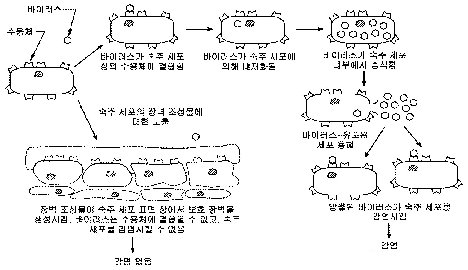
도 1 은 장벽-형성 조성물 구현예에서의 항균 활성의 제시된 메커니즘을 나타낸다.
도 2 는 실시예 2 에서 기재한 바와 같이 점막 표면 상 장벽의 형성을 나타내는 모식도이다.
도 3 은 실시예 11-15 각각에서의 스크래치 공간을 커버하기 위한 6 시간 후 세포 성장 및 이동을 통한 손상된 상피 세포 샘플에 대한 퍼센트로서 복구 과정을 나타내는 그래프이다.
도 4 는 손상된 상피 샘플 상의 미(un)처리 대조예 15 및 처리 실시예 16 및 17 모두에 대한 상피 세포 성장 및 이동을 나타내는 현미경 사진이다.
도 5 는 실시예 11-15 의 장벽-형성 조성물 처리 및 미처리 조작 인간 구강 점막 (EHOM) 의 확대된 횡단면 사진을 나타낸다.
도 6 은 실시예 16-19 및 20-25 의 LDH 검정을 나타내는 그래프이다.
도 7 은 실시예 27-28 에서 기재하는 바와 같이, EHOM 검정의 상부 및 하부 챔버에서의 미생물 성장 평가 방법을 나타내는 모식도이다.
도 8 은 실시예 27-28 에서 기재하는 바와 같이, EHOM 검정의 상부 및 하부 챔버에서의 미생물 성장을 나타내는 한천 배지 플레이트의 사진을 나타낸다.
도 9 는 실시예 31-32 의 장벽-형성 조성물-처리 및 미처리 조작 인간 구강 점막 (EHOM) 의 확대된 횡단면 사진을 나타낸다.
도 10 은 실시예 33-40 에서 기재하는 바와 같이, 미처리된 EHOM 또는 실시예 장벽-형성 조성물로 처리된 EHOM 상의, 이후 C. 알비칸스 (C. albicans) 로 감염시킨, 미생물 성장 사진을 나타낸다.
도 11 은 실시예 33-40 에서 기재하는 바와 같이, 미처리된 EHOM 또는 제형으로 처리된 EHOM 상의, 이후 S. 무탄스 (S. mutans) 로 감염시킨, 미생물 성장 사진을 나타낸다.
도 12 는 실시예 33-40 에서 기재하는 바와 같이, 실시예 장벽-형성 조성물로 처리된 EHOM 의 "통과 (flow-through)" 배지 (하부 챔버로부터 수집) 로부터의 미생물 성장 사진을 나타낸다.
도 13 은 실시예 40-47 에서 기재하는 바와 같이, 식염수 (대조군) 또는 실시예 장벽-형성 조성물로 처리된 EHOM 에 의한, 이후 (A) C. 알비칸스 또는 (B) S. 무탄스로 감염시킨, LDH 방출을 나타내는 그래프이다.
도 14 는 실시예 48-61 및 61-69 에서 기재하는 바와 같이, 박테리아 및 진균에 대한 장벽-형성 조성물의 후-항균 효과를 나타내는 그래프이다.
도 15 는 실시예 71-76 에서 기재하는 바와 같이, 미처리되거나 장벽-형성 조성물로 처리된 S. 산구이스 (S. sanguis), C. 알비칸스 및 S. 무탄스의 주사 전자 현미경 사진을 나타낸다.
도 16 은 실시예 77-79 에서 기재하는 바와 같이, 박테리아 및 진균에 의해 형성된 바이오필름에 대한 실시예 장벽-형성 조성물의 활성을 나타내는 그래프이다.
도 17 은 실시예 80-81 에서 기재하는 바와 같이, 1 분 노출 후 미생물 바이오필름 상의 실시예 장벽-형성 조성물의 활성을 나타내는 그래프이다.
도 18 은 실시예 85-86 에서 기재하는 바와 같이, 인플루엔자 (H1N1)-감염된 MDCK 세포의 세포변성 효과 (CPE) 에 대한 실시예 장벽-형성 조성물의 효과를 나타내는 형광 현미경 사진이다.
도 19 는 실시예 85-86 에서 기재하는 바와 같이, H1N1 바이러스에 대한 실시예 장벽-형성 조성물의 효과를 나타내는 형광 현미경 사진이다.
도 20 은 실시예 87-88 에서 기재하는 바와 같이, 정량 PCR 에 의해 측정된 바와 같은 감염된 장벽-형성 조성물 처리 및 미처리 세포에서의 인플루엔자 바이러스 수준을 나타내는 그래프이다.
도 21 은 실시예 89-91 에서 기재하는 바와 같이, 정량 PCR 을 사용하여 측정한 인플루엔자 바이러스에 대한 항균제 (CPC) 및 보존제의 존재 또는 부재 하에 제조된 실시예 장벽-형성 조성물의 직접적 항바이러스 활성을 나타내는 그래프이다.
도 22 는 6 시간에 걸친 H1N1 바이러스에 대한 실시예 장벽-형성 조성물의 활성을 나타낸다. 패널 (A) 는 미처리 대조군과 비교하여 바이러스 성장에 있어서의 저해율을 나타내는 그래프이다. 패널 (B) 및 (C) 는 (B) 미처리 세포 및 (C) 장벽-형성 조성물 처리 세포의 현미경 사진이다.
도 23 은 실시예 94-96 에서 기재하는 바와 같이, HIV 에 대한 제형의 활성을 나타내는 그래프이다.
도 24 는 실시예 97 에서 기재하는 바와 같이, 엡스타인-바 바이러스 (EBV) 에 대한 실시예 8 의 활성을 나타내는 웨스턴 블롯이다.
도 25 는 실시예 154-159 에서 기재하는 바와 같이, 미처리 (대조군) EHOM 또는 실시예 5-7 에 노출된 EHOM 조직에서의 세포 무결성의 표시자로서 LDH 수준을 나타내는 그래프이다.
도 26 은 실시예 160 에서 기재하는 바와 같이, 손상 직후 (패널 A), 약 6 시간 후 (패널 D) 및 약 24 시간 후 (패널 E), 10 분 동안 실시예 3 (5% 희석물) 으로 처리한 손상 구강 상피 세포 배양물의 대표적 사진을 나타낸다. 패널 B 및 C 는 각각 약 6 시간 후 및 약 24 시간 후 구강 상피 세포의 미처리 대조군 융합 (confluent) 배양물에서의 동일 손상을 나타낸다.
도 27 은 실시예 장벽-형성 조성물의 구강 점막 표면을 코팅하는 능력을 입증하는 사진이다.
도 28 은 실시예 162-163 에서 기재하는 바와 같이, 실시예 장벽-형성 조성물에 1 분 노출된 후 박테리아 성장의 간헐 촬영 (time-lapse) 현미경 관찰을 나타내는 사진이다. 이미지는 노출 후 20 분, 120 분 또는 360 분을 나타낸다.
도 29 는 실시예 164-166 에서 기재하는 바와 같이, 건강한 개인의 구강 미생물 부하 (burden) 에 대한 실시예 장벽-형성 조성물의 단일 용량의 효과를 나타내는 그래프이다. (A) - CFU 로의 미생물 총량 (microbial load), (B) 기준선과 비교하여 미생물 총량에 있어서의 감소 (%).
도 30 은 실시예 167-169 에서 기재하는 바와 같이, 3 명의 건강한 성인에서 5 일 기간에 걸친 구강 미생물 수준에 대한 실시예 장벽-형성 조성물의 효과를 나타내는 그래프이다.
도 31 은 실시예 170-198 에서 기재하는 바와 같이, 31 명의 건강한 대상에서 5 일 사용 후 구강의 미생물 부하에 대한 실시예 장벽-형성 조성물의 효과를 나타내는 그래프이다.
도 32 는 실시예 170-198 에서 기재하는 바와 같이, 3 명의 대표 연구 참가자로부터 수득한 구강 샘플에서의 미생물 총량을 나타내는 그래프이다.
도 33 은 실시예 199-205 에서 기재하는 바와 같이, 실시예 장벽-형성 조성물에 의해 형성된 장벽을 가로지르는 미생물의 침투를 평가하기 위한 체외 필터 삽입-기반 모델을 설명하는 모식도이다.1 shows the presented mechanism of antimicrobial activity in barrier-forming composition embodiments.
FIG. 2 is a schematic diagram showing formation of a barrier on the mucosal surface as described in Example 2. FIG.
3 is a graph showing the repair process as a percentage of damaged epithelial cell sample through cell growth and migration after 6 hours to cover the scratch space in each of Examples 11-15.
FIG. 4 is a micrograph showing epithelial cell growth and migration for untreated Control Example 15 and Treatment Examples 16 and 17 on damaged epithelial samples.
5 shows an enlarged cross-sectional photograph of the barrier-forming composition treated and untreated manipulated human oral mucosa (EHOM) of Examples 11-15.
6 is a graph showing the LDH assay of Examples 16-19 and 20-25.
FIG. 7 is a schematic diagram illustrating a microbial growth evaluation method in the upper and lower chambers of the EHOM assay, as described in Examples 27-28. FIG.
FIG. 8 shows photographs of agar media plates showing microbial growth in the upper and lower chambers of the EHOM assay, as described in Examples 27-28.
9 shows an enlarged cross-sectional photograph of the barrier-forming composition-treated and untreated engineered human oral mucosa (EHOM) of Examples 31-32.
10 shows microbial growth photographs, then infected with C. albicans on untreated EHOM or EHOM treated with the example barrier-forming composition, as described in Examples 33-40.
FIG. 11 shows microbial growth photographs, then infected with S. mutans, on untreated EHOM or EHOM treated with formulation, as described in Examples 33-40.
12 shows microbial growth photographs from “flow-through” medium (collected from the lower chamber) of EHOM treated with the example barrier-forming composition, as described in Examples 33-40.
FIG. 13 is infected with either saline (control) or an example barrier-forming composition, as described in Examples 40-47, followed by infection with (A) C. albicans or (B) S. mutans. , A graph showing LDH emission.
14 is a graph showing the post-antibacterial effect of barrier-forming compositions on bacteria and fungi, as described in Examples 48-61 and 61-69.
FIG. 15 shows scanning electron micrographs of S. sanguis, C. albicans and S. mutans, as treated in untreated or barrier-forming compositions, as described in Examples 71-76.
FIG. 16 is a graph showing the activity of the example barrier-forming composition on biofilms formed by bacteria and fungi, as described in Examples 77-79.
FIG. 17 is a graph showing the activity of the Example barrier-forming composition on microbial biofilms after 1 minute exposure, as described in Examples 80-81.
18 is a fluorescence micrograph showing the effect of the example barrier-forming composition on the cytopathic effect (CPE) of influenza (H1N1) -infected MDCK cells, as described in Examples 85-86.
19 is a fluorescence micrograph showing the effect of the example barrier-forming composition on H1N1 virus, as described in Examples 85-86.
20 is a graph showing influenza virus levels in infected barrier-forming composition treated and untreated cells, as measured by quantitative PCR, as described in Examples 87-88.
FIG. 21 shows direct antiviral activity of example barrier-forming compositions prepared in the presence or absence of antimicrobial (CPC) and preservatives against influenza viruses measured using quantitative PCR, as described in Examples 89-91. It is a graph.
22 shows the activity of the example barrier-forming composition against H1N1 virus over 6 hours. Panel (A) is a graph showing the inhibition rate in virus growth compared to the untreated control. Panels (B) and (C) are micrographs of (B) untreated cells and (C) barrier-forming composition treated cells.
FIG. 23 is a graph showing the activity of the formulations on HIV, as described in Examples 94-96. FIG.
24 is a western blot showing the activity of Example 8 against Epstein-Barr virus (EBV), as described in Example 97.
25 is a graph showing LDH levels as an indicator of cellular integrity in untreated (control) EHOM or EHOM tissue exposed to Examples 5-7, as described in Examples 154-159.
FIG. 26 shows Example 3 (5% dilution) for 10 minutes immediately after injury (Panel A), after about 6 hours (Panel D) and after about 24 hours (Panel E), as described in Example 160. Representative photographs of treated damaged oral epithelial cell cultures are shown. Panels B and C show the same damage in untreated control confluent cultures of oral epithelial cells after about 6 hours and after about 24 hours, respectively.
27 is a photograph demonstrating the ability of the example barrier-forming composition to coat the oral mucosa surface.
FIG. 28 is a photograph showing time-lapse microscopic observation of bacterial growth after 1 minute exposure to an example barrier-forming composition, as described in Examples 162-163. The image represents 20 minutes, 120 minutes or 360 minutes after exposure.
29 is a graph showing the effect of a single dose of the example barrier-forming composition on the oral microbial burden of healthy individuals, as described in Examples 164-166. (A)-microbial load to CFU, (B) reduction in microbial total compared to baseline (%).
30 is a graph showing the effect of an example barrier-forming composition on oral microbial levels over a five day period in three healthy adults, as described in Examples 167-169.
FIG. 31 is a graph showing the effect of an example barrier-forming composition on microbial load in the oral cavity after 5 days of use in 31 healthy subjects, as described in Examples 170-198.
32 is a graph showing the total amount of microorganisms in oral samples obtained from three representative study participants, as described in Examples 170-198.
FIG. 33 is a schematic diagram illustrating an in vitro filter insertion-based model for assessing penetration of microorganisms across a barrier formed by an example barrier-forming composition, as described in Examples 199-205.
상세한 설명details
포유동물의 입, 소화관 및 체강을 나누는 점막은 병원성 미생물이 국부 및 전신 감염 모두를 일으킬 수 있는 경우 상기 미생물의 포유동물 신체에 대한 유입에 대한 첫 번째 장벽을 나타낸다. 상피 점막 내벽 (lining) 은 공생 유기체의 유입을 감소시키는 장벽을 형성한다 (Monica Boirivanta and Warren Strober, "The Mechanism of Action of Probiotics" Current Opinion in Gastroenterology 2007, 23:679-692). The mucosa, which divides the mouth, digestive tract and body cavity of a mammal, represents the first barrier to the influx of microorganisms into the mammalian body if the pathogenic microorganism can cause both local and systemic infections. Lining of the epithelial mucosa forms a barrier that reduces the influx of symbiotic organisms (Monica Boirivanta and Warren Strober, "The Mechanism of Action of Probiotics" Current Opinion in Gastroenterology 2007, 23: 679-692).
이 출원에서, 감염성 질환을 일으키는 미생물이 점막에 접촉하거나 이를 감염시키는 것을 차단하거나 중화시키고, 이로써 미생물이 신체에 전파되고 감염을 일으키는 것을 예방하는 방법 및 조성물이 개시된다. 상기 방법 및 조성물은 감염을 일으키는 것으로 알려져 있는 미생물 (박테리아, 진균 및 바이러스) 을 저해할 수 있는 항균제를 포함한다. 상기 방법은 인간 점막에 대해 장벽을 형성함으로써 이를 보호하며, 미생물 (박테리아, 진균 및 바이러스) 을 사멸시키거나 저해할 수 있는 항균제를 포함한다. 이러한 이중 작용 조성물 및 방법 (장벽 + 항균) 은 인간 또는 기타 포유동물 점막 또는, 예를 들어 구강, 비강, 질강, 인후 및 기타 구멍, 예컨대 비제한적으로 귀의 표면에 적용가능하다. 이는 또한 의료 장치, 예컨대 기도 장치에 적용될 수 있다. 이러한 고유하고 예기치 않은 해결책은, 미생물에 의해 유발된 전염병을 예방하기 위한 오랫동안 필요했으나 해결되지 않은 필요를 다룬다.In this application, methods and compositions are disclosed for preventing or neutralizing microorganisms causing infectious diseases in contact with or infecting the mucosa, thereby preventing the microorganisms from spreading into the body and causing infection. The methods and compositions include antibacterial agents that can inhibit microorganisms (bacteria, fungi and viruses) known to cause infection. The method protects it by forming a barrier against human mucosa and includes antimicrobial agents that can kill or inhibit microorganisms (bacteria, fungi and viruses). Such dual action compositions and methods (barrier plus antibacterial) are applicable to human or other mammalian mucosa or, for example, to the oral cavity, nasal cavity, vaginal cavity, throat and other pores such as but not limited to the surface of the ear. It may also be applied to medical devices, such as airway devices. This unique and unexpected solution addresses the long needed but unresolved need to prevent infectious diseases caused by microorganisms.
점막 조직을 통한 병원성 미생물의 통과를 저해하는 장벽을 형성하며 안전한 (즉, 점막에 대해 손상을 일으키지 않는) 장벽-형성 조성물이 바람직하다. 또 다른 바람직한 특성은 연장된 기간 동안 정적 (static) 또는 살균 (cidal) 활성을 통해 미생물 성장을 저해하는 능력이다. 이론에 구속됨이 없이, 본원에 개시된 장벽-형성 조성물 작용 메커니즘은 세균이 형성 장벽에서 포획되고, 이후 항균 활성 성분에 의해 사멸되는 상승작용적인 이중-작용 메커니즘을 기반으로 한다. 한 구현예에서 장벽-형성 조성물은 친수성이 아니며, 이는 이론에 구속됨이 없이, 지속된 효과를 증강시키는 것으로 가설화된다.Preference is given to barrier-forming compositions that form a barrier that inhibits the passage of pathogenic microorganisms through mucosal tissue and that are safe (ie, do not cause damage to the mucosa). Another desirable property is the ability to inhibit microbial growth through static or cidal activity for extended periods of time. Without being bound by theory, the barrier-forming composition mechanism of action disclosed herein is based on a synergistic dual-action mechanism in which bacteria are captured at the barrier of formation and are subsequently killed by antimicrobial active ingredients. In one embodiment the barrier-forming composition is not hydrophilic, which is hypothesized to enhance sustained effects, without being bound by theory.
하기 실시예에서 나타내는 바와 같이, 장벽-형성 조성물의 특성 및 이의 다양한 전염병 예방 효과를 하기를 기반으로 한 10 개 이상의 상이한 접근 방식을 사용하여 평가하였다: (1) 체외 항균 감수성 시험; (2) 체외 시간 사멸 검정; (3) 체외 바이오필름 모델; (4) 체외 필터 삽입-기반 모델, (5) 체내-유사 조작 인간 구강 점막 (EHOM) 모델; (6) 전자 현미경 평가; (7) 소수성 검정; (8) 이화학적 양립성 검정; (9) 인간 세포주 단일층을 사용하는 세포 배양-기반 모델; 및 (10) 인간 임상 시험. As shown in the Examples below, the properties of the barrier-forming composition and its various infectious disease prevention effects were evaluated using at least 10 different approaches based on: (1) in vitro antimicrobial susceptibility testing; (2) in vitro time killing assay; (3) an in vitro biofilm model; (4) an in vitro filter insertion-based model, (5) an in-body-like manipulated human oral mucosa (EHOM) model; (6) electron microscopy evaluation; (7) hydrophobicity assay; (8) physicochemical compatibility assay; (9) cell culture-based models using human cell line monolayers; And (10) human clinical trials.
본원에서 기재한 방법 및 조성물은 인간, 또는 보다 일반적으로는 포유동물이 파괴된 점막을 갖는 경우 특히 유용할 수 있다. 파괴는 상처 또는 스크래치를 일으킬 수 있다. 구강 및 위장 (GI) 관의 점막은 다양한 미생물의 국부 또는 전신 침입, 및 구강 및 소화관의 내강에 보통 존재하는 미생물 산물 흡수를 방지하는 것을 돕는 중요한 기계적 장벽으로서 역할한다 ("Gastrointestinal mucosal injury in experimental models of shock, trauma, and sepsis," Crit. Care Med. 1991;19:627-41). 점막의 장벽 기능에 있어서의 교란은 전신 감염의 병리생리학에서 중심 역할을 담당한다. 즉, 이러한 점막의 파괴는 감염을 일으킬 것이다.The methods and compositions described herein may be particularly useful when humans, or more generally mammals, have a disrupted mucosa. Destruction can cause scratches or scratches. The mucous membranes of the oral and gastrointestinal (GI) tracts serve as important mechanical barriers that help prevent local or systemic invasion of various microorganisms and the absorption of microbial products normally present in the lumen of the oral and digestive tracts ("Gastrointestinal mucosal injury in experimental models"). of shock, trauma, and sepsis, "Crit. Care Med. 1991; 19: 627-41). Disturbance in the mucosal barrier function plays a central role in the pathophysiology of systemic infection. That is, the destruction of these mucous membranes will cause infection.
방어 제 1 선에서의 붕괴 위험성 제거 또는 감소가 중요하며, 점막 무결성의 유지가 중요하다 (Anders Heimdahl, "Prevention and Man agement of Oral Infections in Cancer Patients" Supportive Care in Cancer, Vol. 7, No. 4, 224-228 (1999)). 따라서, 미손상 점막을 갖는 것은 특히 면역력이 약화된 환자 (예를 들어, 암 환자) 에서의 전신 감염에 대한 중요한 숙주 방어이다 (Shahab A. Khan, John R. Wingard, "Infection and Mucosal Injury," Cancer Treatment Journal of the National Cancer Institute, Monographs No. 29 (2001)). 유해한 미생물을 차단하거나 사멸시키고 파괴 점막의 치유를 방해하지 않는 장벽-형성 조성물은, 파괴 점막을 갖는 경우, 특히 또한 면역결핍성을 갖는 경우 문제점의 감수성에 대한 고유하고 예기치 않은 해결책이다.It is important to eliminate or reduce the risk of collapse at the first line of defense, and to maintain mucosal integrity (Anders Heimdahl, "Prevention and Man agement of Oral Infections in Cancer Patients" Supportive Care in Cancer, Vol. 7, No. 4 , 224-228 (1999). Thus, having an intact mucosa is an important host defense against systemic infections, particularly in patients with weakened immunity (eg cancer patients) (Shahab A. Khan, John R. Wingard, "Infection and Mucosal Injury," Cancer Treatment Journal of the National Cancer Institute, Monographs No. 29 (2001)). Barrier-forming compositions that block or kill harmful microorganisms and do not interfere with the healing of the disrupting mucosa are inherent and unexpected solutions to the susceptibility of the problem when having a disrupting mucosa, in particular also with immunodeficiency.
한 구현예에서, 장벽-형성 조성물은 포유동물에서의 감염성 질환을 예방하거나 저해하는 방법에서 투여될 수 있다. 예방이란, 미생물로부터의 감염이 없게 할 수 있다는 것이 아니라, 장벽-형성 조성물의 적용 후 접촉한 미생물로부터의 감염 위험성이 감소되는 것을 의미한다. 전체 예방 효과를 위해서는, 장벽-형성 조성물이 오염된 환경 또는 품목에 포유동물이 접촉하기 전에 적용되어야 한다. 이것은 오염된 환경 또는 품목과 접촉하는 동안 장벽-형성 조성물을 투여하는 것으로부터 일부 이득이 수득될 수 없다는 것을 의미하는 것은 아니다. 본원에서 사용하는 "포유동물" 은 포유동물로서 통상 정의된 인간 또는 동물을 의미한다.In one embodiment, the barrier-forming composition can be administered in a method of preventing or inhibiting an infectious disease in a mammal. Prophylaxis does not mean that there is no infection from the microorganism, but that the risk of infection from the microorganism in contact after application of the barrier-forming composition is reduced. For the overall prophylactic effect, the barrier-forming composition should be applied before the mammal contacts the contaminated environment or item. This does not mean that some benefit cannot be obtained from administering the barrier-forming composition during contact with the contaminated environment or item. As used herein, "mammal" refers to a human or animal commonly defined as a mammal.
또 다른 구현예에서, 장벽-형성 조성물은 예를 들어 면역력이 약화된 포유동물과 같은 파괴 점막을 갖는 포유동물에서의 감염성 질환을 예방하는 방법에서 투여된다. 포유동물의 점막에서의 파괴 부위가 확인되고 적어도 포유동물 점막의 파괴 부위에 치료적 유효량의 장벽-형성 조성물이 투여된다. 상기 장벽-형성 조성물은 점막 파괴 부위 상에 장벽을 제공하여 미생물이 점막의 파괴 부위에 전파되는 것을 효과적으로 저해한다.In another embodiment, the barrier-forming composition is administered in a method for preventing an infectious disease in a mammal having a disrupting mucosa, such as, for example, a mammal with weakened immunity. Sites of disruption in the mucosa of a mammal are identified and a therapeutically effective amount of barrier-forming composition is administered to at least sites of destruction of a mammalian mucosa. The barrier-forming composition provides a barrier on the mucosal destruction site, effectively inhibiting the spread of microorganisms to the destruction site of the mucosa.
또 다른 구현예에서, 장벽-형성 조성물은 장치가 오염된 환경에 접촉하기 전 및 장치가 포유동물의 점막에 접촉하기 전에 품목 또는 장치 상에 투여된다. 상기 장벽-형성 조성물은 장치 상에 미생물을 포획하고 사멸키는 장벽을 제공하고, 그로써 미생물이 점막을 통과하거나 감염을 일으키는 것을 예방한다.In another embodiment, the barrier-forming composition is administered on an item or device before the device contacts the contaminated environment and before the device contacts the mammalian mucosa. The barrier-forming composition provides a barrier that traps and kills microorganisms on the device, thereby preventing them from passing through the mucous membrane or causing infection.
감염성 질환 예방 방법의 한 구현예에서, 한 단계는 포유동물 또는 품목이 접촉되는 것으로 예상되는 오염된 환경을 확인하는 것을 포함한다. 오염된 환경은 유해한 바이러스, 진균 또는 박테리아 미생물로 오염된 것으로 알려지거나 예상되는 또 다른 포유동물 또는 인간에 근접한, 또는 실내 또는 야외 공간과 같은 환경이다. 주어진 환경이 오염될 수 있는지 여부를 결정하는 것은 연중 시각, 지역 내 창성하고 있는 질환에 대한 공개된 정보, 또는 재채기 등에 의해 아프거나 세균을 퍼뜨리는 것으로 보이는 다른 사람들을 관찰하는 것 등을 기반으로 할 수 있다.In one embodiment of an infectious disease prevention method, one step includes identifying a contaminated environment in which the mammal or item is expected to be contacted. A contaminated environment is an environment, such as an indoor or outdoor space, in proximity to another mammal or human being known or expected to be contaminated with harmful virus, fungal or bacterial microorganisms. Determining whether a given environment can be contaminated can be based on observing other people who are sick or spreading germs, such as by year-round vision, open information about the disease being created in the area, or sneezing. have.
오염된 환경 또는 품목에 접촉될 것인지 여부를 예측하거나 확인하는 것은, 포유동물이 가까운 미래에 상기 환경에 들어가거나 상기 품목에 접촉할 계획이 있거나 예상되는지 여부를 기반으로 한 결정일 수 있다. 이는 오염된 환경 또는 품목에 접촉할 시기를 추정하는 것을 포함할 수 있다. 장벽-형성 조성물은 오염된 환경 또는 품목과의 접촉 추정 시간 전 약 24 시간 이하, 예를 들어, 약 16 시간 이하, 약 12 시간 이하, 약 6 시간 이하, 또는 약 2 시간 이하에 투여될 수 있다. 장벽-형성 조성물은 신속히 제공되며 유해한 미생물이 점막을 감염시키는 것을 예방하거나 저해하도록 예를 들어, 적용 1 분 미만 내, 예컨대 30 초 내에 작동가능해야 한다. 따라서, 이는 오염된 환경 또는 품목과 접촉하는 동안 적용될 수 있으며 효과를 갖는다.Predicting or identifying whether a contaminated environment or item is to be contacted may be a decision based on whether a mammal is planning to or expected to enter the environment or contact the item in the near future. This may include estimating when to contact the contaminated environment or item. The barrier-forming composition may be administered about 24 hours or less, for example about 16 hours or less, about 12 hours or less, about 6 hours or less, or about 2 hours or less before the estimated time of contact with the contaminated environment or item. . The barrier-forming composition is provided quickly and should be operable, for example, within less than one minute of application, such as within 30 seconds, to prevent or inhibit harmful microorganisms from infecting the mucosa. Thus, it can be applied and has an effect during contact with contaminated environments or items.
유해한 미생물은 감염성 질환을 일으키는 것으로 알려져 있는 것들이며, 예를 들어 전염병과 같은 감염성 질환을 일으키는 것들 예컨대 칸디다 종 (예를 들어, C. 알비칸스, C. 글라브라타 (C. glabrata), C. 크루세이 (C. krusei), C. 트로피칼리스 (C. tropicalis)), 스타필로코쿠스 종 (메티실린-저항성 S. 오레우스 (S. aureus), MRSA 포함), 스트렙토코쿠스 종 (예를 들어, S. 산구이스, S. 오랄리스 (S. oralis), S. 미티스 (S. mitis), S. 살리바리우스(S. salivarius), S. 고르도니 (S. gordonii), S. 뉴모니에 (S. pneumoniae)), 아시네토박터 바우마니 (Acinetobacter baumannii), 아그레가티박터 악티노마이세템코미탄스 (Aggregatibacter actinomycetemcomitans), 푸소박테리움 뉴클레아툼 (Fusobacterium nucleatum), 및 기타 미생물 예컨대 상부 호흡기 감염을 일으키는 미생물, 및 감기 및 인플루엔자 바이러스이다. 한 구현예에서, 본원에 기재된 장벽-형성 조성물 및 치료 및 예방 방법은 예를 들어, 성 전염성 질환 [예를 들어, 인간 면역결핍 바이러스 (HIV), 헤르페스 심플렉스 (Herpes simplex) 또는 인간 파필로마 바이러스 (HPV) 에 의한 감염], 감기 (예를 들어, 리노바이러스에 의함) 및 엡스타인-바 바이러스 (EBV) 에 의한 감염의 예방에 유용할 수 있다. Harmful microorganisms are those known to cause infectious diseases, for example those causing infectious diseases such as infectious diseases such as Candida species (eg C. albicans, C. glabrata, C. C. krusei, C. tropicalis, Staphylococcus spp. (Including methicillin-resistant S. aureus, MRSA), Streptococcus spp. (Eg For example, S. sanguise, S. oralis, S. mitis, S. salivarius, S. gordonii, S. nu S. pneumoniae, Acinetobacter baumannii, Aggregatibacter actinomycetemcomitans, Fusobacterium nucleatum, and other microorganisms such as It is a microorganism that causes upper respiratory infections, and cold and influenza viruses. In one embodiment, the barrier-forming compositions and methods of treatment and prophylaxis described herein include, for example, sexually transmitted diseases (eg, human immunodeficiency virus (HIV), herpes simplex or human papilloma virus). (HPV) infection], colds (eg, by rhinoviruses), and Epstein-Barr virus (EBV).
장벽-형성 조성물은 직경이 예를 들어 약 30 nm 이상, 예컨대 약 100 nm (HIV, 구형), 약 100 내지 약 300 nm (인플루엔자, 구형 및 장방형), 약 120 nm 내지 약 260 nm (EBV 구형/디스크형), 및 약 30 nm (리노바이러스, 구형) 인 미생물에 대해 효과를 나타낸다. 따라서, 상기 조성물은 또한 직경이 약 30 nm, 또는 약 30 nm 초과인 기타 미생물에 대해서도 효과적이다. The barrier-forming composition may have a diameter of, for example, about 30 nm or more, such as about 100 nm (HIV, spherical), about 100 to about 300 nm (influenza, spherical and rectangular), about 120 nm to about 260 nm (EBV spherical / Disk), and microorganisms that are about 30 nm (linovirus, spherical). Thus, the compositions are also effective against other microorganisms having a diameter of about 30 nm, or greater than about 30 nm.
미생물은 공기 운반성 미생물일 수 있다. 한 구현예에서 미생물은 전염병을 일으키는 것들이다. 한 구현예에서, 미생물은 알레르기 반응 또는 치과적 문제, 예를 들어 구멍 (충치), 치은염 또는 계절성 알레르기를 일으키는 것들을 포함하지 않는다. 유사하게는, 한 구현예에서, 예방 방법은 치과적 문제 또는 알레르기 반응, 예를 들어 구멍 (충치), 치은염 또는 계절성 알레르기를 단독으로 또는 추가적으로 예방하지 않는다. 그러나 또 다른 구현예에서, 미생물 예컨대 알레르겐으로서 일반적으로 분류될 수 있는 진균, 기타 알레르겐, 및 점막에 대한 공기 운반성 자극물은 상기 장벽 및 상기 방법에 의해 차단된다. 알레르겐 차단 구현예에서, 오염된 환경의 확인은 예를 들어 연중 계절, 또는 꽃가루 또는 기타 알레르겐 또는 자극물 예측을 기반으로 할 수 있다. 이는 또한 예를 들어, 높은 수의 알레르겐 또는 공기 운반성 자극물을 생성하는 것으로 알려지거나 예상되는 장소, 예컨대 숲, 공원 또는 호수를 포함하는 야외 환경에 있는 포유동물에 대한 예상을 기반으로 할 수 있다.The microorganism may be an air carrier microorganism. In one embodiment the microorganisms are those that cause infectious diseases. In one embodiment, the microorganism does not include those causing allergic reactions or dental problems, such as holes (cavities), gingivitis or seasonal allergies. Similarly, in one embodiment, the prophylactic method does not alone or additionally prevent dental problems or allergic reactions, such as holes (cavities), gingivitis or seasonal allergies. However, in another embodiment, air transport stimulants to fungi, other allergens, and mucous membranes, which may be generally classified as microorganisms such as allergens, are blocked by the barrier and the method. In allergen blocking embodiments, the identification of the contaminated environment can be based, for example, throughout the year, or on pollen or other allergen or irritant prediction. It may also be based on expectations for mammals in outdoor environments, including, for example, forests, parks or lakes known or expected to produce a high number of allergens or air transport irritants.
한 구현예에서, 본원에 기재된 장벽-형성 조성물 및 치료 및 예방 방법은 예를 들어, 감염성 미생물로 오염되는 환경에서 흔한 감염 및 병원과 같은 환경에서의 감염을 예방하는데 유용할 수 있다. 상기 언급한 바와 같이, 본원에 개시한 방법 및 조성물은 특히 면역력이 약화된 환자에 대해 적용가능할 수 있다. 또한, 장벽-형성 조성물은 상처를 흔히 감염시키는 미생물에 의한 감염의 예방에 유용할 수 있다.In one embodiment, the barrier-forming compositions and methods of treatment and prevention described herein may be useful for preventing infections in environments such as hospitals and infections that are common in environments contaminated with infectious microorganisms, for example. As mentioned above, the methods and compositions disclosed herein may be particularly applicable to patients with weakened immunity. In addition, barrier-forming compositions may be useful for the prevention of infection by microorganisms that frequently infect wounds.
오염된 환경은 예를 들어, 대중 교통 차량, 대중 운집 장소, 및 병에 걸린 것으로 알려지거나 예상되는 포유동물을 포함하는 공간 또는 차량, 또는 병에 걸린 것으로 알려지거나 예상되는 포유동물에 근접한 곳을 포함할 수 있다. 비행기, 유아원 및 보건소와 같은 오염된 환경으로 흔히 인지되는 환경에 대한 더 많은 정보는 본원에 참조로 포함되는 [Yang, et al., "Concentrations and Size Distributions of Airborne Influenza A Viruses Measured Indoors at a Health Centre, a Day-Care Centre, and on Aeroplanes," J.R. Soc. Interface (Feb. 7, 2011)] 에 개시된다.A contaminated environment includes, for example, a public transport vehicle, a public gathering place, and a space or vehicle containing a known or expected mammal, or a place close to a known or expected mammal. can do. For more information on environments that are commonly perceived as contaminated environments, such as airplanes, pre-K, and public health centers, see Yang, et al., "Concentrations and Size Distributions of Airborne Influenza A Viruses Measured Indoors at a Health Center" , a Day-Care Centre, and on Aeroplanes, "JR Soc. Interface (Feb. 7, 2011).
보다 구체적으로, 한 구현예에서, 대중 교통 차량은 예를 들어 비행기, 버스 또는 택시일 수 있다. 대중 운집 장소는 예를 들어 의사 사무실, 병원, 학교, 유아원, 교회, 호텔 또는 레스토랑일 수 있다. 병에 걸린 것으로 알려지거나 예상되는 포유동물에 근접한 곳은 예를 들어 상기 포유동물의 1 피트 반경 내, 또는 상기 포유동물과 동일한 차량 내일 수 있다. 공공으로 사용하는 비행기는 오염된 환경으로서 많이 확인되는 환경의 흔하고 특히 주목할만한 예로서 언급될 수 있다.More specifically, in one embodiment, the public transport vehicle may be an airplane, bus or taxi, for example. The mass gathering place may be, for example, a doctor's office, a hospital, a school, a nursery school, a church, a hotel or a restaurant. An area close to a mammal known or expected to be ill can be, for example, within a one foot radius of the mammal or in the same vehicle as the mammal. Publicly used airplanes may be mentioned as common and particularly noteworthy examples of environments that are often identified as contaminated environments.
한 구현예에서, 본원에 기재된 장벽-형성 조성물 및 치료 및 예방 방법은 예를 들어 활동 관련 처리, 예컨대 환풍기 사용 (환풍기와 관련된 의료 장비 및 환자 접촉 포함) 에서 오염될 수 있는 품목으로부터의 감염 예방에 유용할 수 있다. 오염된 품목의 또 다른 예로서, 신체, 및 신체와 접촉하는 품목 또는 표면 (예컨대 신발) 에 대한 적용을 통한 진균 감염의 치료 및 예방이 또한 언급될 수 있다. 한 구현예에서, 오염된 품목은 예를 들어 식품, 음료, 조리도구 (utensil), 음료 용기 및 악세사리, 어린이용 품목, 의료 장치 또는 치과 장치일 수 있다.In one embodiment, the barrier-forming compositions and methods of treatment and prevention described herein are used to prevent infection from items that may be contaminated, for example, in activity related treatments, such as the use of a fan, including medical equipment and patient contact associated with the fan. Can be useful. As another example of a contaminated item, mention may also be made of the treatment and prevention of fungal infections through application to the body and to items or surfaces in contact with the body (such as shoes). In one embodiment, the contaminated item can be, for example, food, beverages, utensils, beverage containers and accessories, children's items, medical devices or dental devices.
감염성 질환의 예방 방법의 한 구현예에서, 한 단계는 포유동물이 오염된 환경 또는 품목에 접촉하기 전에 치료적 유효량의 장벽-형성 조성물을 상기 포유동물의 점막에 투여하는 것을 포함한다. 치료적 유효량이란, 점막 상에 장벽 층을 생성시키는 장벽을 형성시키기에 충분한 장벽-형성 조성물로 표적 점막을 코팅하기에 충분한 양을 의미한다. 예를 들어, 구강 청결제 제형에 대해 약 100 μl 내지 약 10 ml, 예컨대 약 1 ml 내지 약 8 ml, 또는 약 2 ml 내지 약 5 ml, 또는 스프레이 제형에 대해 약 0.125 ml 내지 약 2 ml, 예컨대 약 0.5 ml 내지 약 1 ml 이다. 투약량은 또한 cm2 당 부피, 예를 들어 구강 청결제 제형에 대해 약 0.5 내지 약 50 μl/cm2, 예컨대 약 5 내지 약 40 μl/cm2, 또는 약 10 내지 약 25 μl/cm2; 또는 스프레이 제형에 대해, 예를 들어 약 0.625 내지 약 10 μl/cm2, 예컨대 약 2.5 내지 약 5 μl/cm2 로 표현될 수 있다. 기타 전달 매질, 예컨대 분해성 스트립이 농도 및 당업자에게 공지된 기타 인자에 대해 조정되어 이들 범위에서 유래한 투약량을 가질 수 있다. 또한, 장벽-형성 조성물로부터 점막 상 형성된 필름의 평균 두께는 예를 들어 약 0.001 내지 약 0.2 mm, 예컨대 약 0.01 내지 약 0.1 mm, 또는 약 0.08 내지 약 0.15 mm 범위일 수 있다. 예를 들어 주어진 인간 또는 동물에 대해, 치료적 유효량은 치료할 포유동물의 연령 또는 체중 또는 크기를 기반으로 결정될 수 있고, 투약량은 상기 열거한 것들일 수 있다. 비(非)-인간 포유동물에 대해서, 특히, 투약량은 상기 주어진 cm2 당 값 및 치료할 점막 표면 또는 체강의 대략적인 표면적에 따라 조정될 수 있다. In one embodiment of the method of preventing infectious disease, one step comprises administering to the mammalian mucosa a therapeutically effective amount of a barrier-forming composition before the mammal contacts the contaminated environment or item. By therapeutically effective amount is meant an amount sufficient to coat the target mucosa with a barrier-forming composition sufficient to form a barrier that creates a barrier layer on the mucosa. For example, about 100 μl to about 10 ml, such as about 1 ml to about 8 ml, or about 2 ml to about 5 ml, or about 0.125 ml to about 2 ml, such as about, about a mouthwash formulation. 0.5 ml to about 1 ml. The dosage may also be a volume per cm 2 , eg about 0.5 to about 50 μl / cm 2 , such as about 5 to about 40 μl / cm 2 , or about 10 to about 25 μl / cm 2 for the mouthwash formulation; Or for a spray formulation, for example, from about 0.625 to about 10 μl / cm 2 , such as from about 2.5 to about 5 μl / cm 2 . Other delivery media, such as degradable strips, may be adjusted for concentration and other factors known to those skilled in the art to have dosages derived from these ranges. In addition, the average thickness of the film formed on the mucosa from the barrier-forming composition can range from about 0.001 to about 0.2 mm, such as from about 0.01 to about 0.1 mm, or from about 0.08 to about 0.15 mm. For example, for a given human or animal, the therapeutically effective amount can be determined based on the age or weight or size of the mammal to be treated and the dosage can be those listed above. For non-human mammals, in particular, the dosage can be adjusted according to the value per cm 2 given above and the approximate surface area of the mucosal surface or body cavity to be treated.
한 구현예에서, 점막에 치료적 유효량으로 투여된 장벽-형성 조성물은 점막 상에 장벽 층을 제공하여 미생물이 점막에 침투하는 것을 저해한다. 한 구현예에서, 미생물의 저해는 또한 미생물의 유해한 활성을 사멸시키고 불활성화시키는 것을 포함한다. 한 구현예에서, 장벽-형성 조성물은 장벽-형성 조성물에 접촉하는 모든 유해 미생물을 차단하고/하거나 사멸시킨다. 또 다른 구현예에서, 장벽은 충분히 유해한 미생물을 실질적으로 차단하고/하거나 사멸시켜, 이들이 감염성 질환을 일으키는 것을 예방한다. 이러한 경우, 유해 미생물의 점막 침투가 늦춰지고/늦춰지거나 희석된다면, 이는 미생물이 질환 또는 광범위한 감염을 일으키는 것을 예방하는 신체 고유 능력을 증강시킬 것이다. 체외 시험은 장벽-형성 조성물의 전형이 장기간, 예를 들어 약 6 시간 이상, 약 16 시간 이상, 및 약 24 시간 이상 동안 점막 표면에 모든 박테리아가 도달하는 것을 방지한다는 것을 입증한다. 체외 시험은 장벽-형성 조성물의 전형에 노출된 바이러스에서, 성장이 약 2 일 이상 (예컨대 인플루엔자), 약 9 일까지 (예컨대 HIV) 저해될 수 있어, 그 후 바이러스 총 수 (count) 가 연장된 기간, 예컨대 약 2 또는 3 일 동안 여전히 MIC 밑에 있다는 것을 나타낸다.In one embodiment, the barrier-forming composition administered in a therapeutically effective amount to the mucosa provides a barrier layer on the mucosa to inhibit microorganisms from infiltrating the mucosa. In one embodiment, inhibition of the microorganism also includes killing and inactivating the deleterious activity of the microorganism. In one embodiment, the barrier-forming composition blocks and / or kills all harmful microorganisms in contact with the barrier-forming composition. In another embodiment, the barrier substantially blocks and / or kills sufficiently harmful microorganisms, preventing them from causing infectious diseases. In such cases, if the mucosal penetration of harmful microorganisms is slowed and / or diluted, this will enhance the body's inherent ability to prevent the microorganism from causing disease or widespread infection. In vitro tests demonstrate that the typical of the barrier-forming composition prevents all bacteria from reaching the mucosal surface for long periods of time, eg, about 6 hours or more, about 16 hours or more, and about 24 hours or more. In vitro tests show that in viruses exposed to the typical of barrier-forming compositions, growth can be inhibited by at least about 2 days (such as influenza), up to about 9 days (such as HIV), thereby prolonging the virus count. For a period of time, such as about 2 or 3 days, still under the MIC.
체내 시험은 장벽-형성 조성물의 전형이 약 6 시간 이상 동안 구강 내 미생물 총 수를 감소시키기에 치료적으로 효과적이라는 것을 나타낸다.In vivo tests indicate that the typical of the barrier-forming composition is therapeutically effective in reducing the total number of microorganisms in the oral cavity for at least about 6 hours.
한 구현예에서, 예방 또는 치료의 연속 투약 방법에서, 장벽-형성 조성물은 예를 들어 약 1 내지 12 시간마다, 약 2 내지 8 시간마다, 또는 약 4 내지 6 시간마다 연속 용량으로 투여될 수 있다. 상기 예방 방법은 예를 들어 1 일 이상, 예컨대 약 2 일 내지 약 1 주 동안 지속될 수 있다. 이러한 연속 투약 방법은 대상이 오염된 환경 또는 품목과 장시간 접촉하는 경우 바람직할 수 있다. 체내 시험은 연속 투약 방법에 따르는 약 80% 의 인간이 6 일의 치료에 걸쳐 구강 내 약 50% 이상의 미생물 총량 감소를 나타낸다는 것을 보여준다.In one embodiment, in the continuous dosing method of prophylaxis or treatment, the barrier-forming composition can be administered in a continuous dose, for example about every 1 to 12 hours, about every 2 to 8 hours, or about every about 4 to 6 hours. . The prophylactic method can, for example, last for at least 1 day, such as from about 2 days to about 1 week. Such continuous dosing methods may be desirable when the subject is in prolonged contact with the contaminated environment or item. In vivo studies show that about 80% of humans following the continuous dosing method show a reduction in microbial total amount of at least about 50% in the oral cavity over 6 days of treatment.
점막은 예를 들어, 구강, 비강 또는 인두강 예컨대 비인두 (상인두), 구강인두 (중인두), 또는 인후두 (하인두) 내 점막 표면일 수 있다. 점막은 또한 질강, 위, 장, 인후 또는 기타 포유류의 구멍 (비제한적으로 외이도 포함) 내에 있을 수 있다.The mucosa can be, for example, the mucosal surface in the oral cavity, nasal or pharyngeal cavity such as nasopharynx (the upper pharynx), oral pharynx (the middle pharynx), or pharynx (low pharynx). The mucosa may also be in the vaginal cavity, stomach, intestine, throat or other mammalian pores (including but not limited to the ear canal).
한 구현예에서, 상기 조성물을 투여하는 것은 포유동물의 구강 점막에 접촉하도록 구강 청결제 형태로 장벽-형성 조성물을 취하는 것을 포함한다. 구강 내 선택된 시간량 후에, 예를 들어, 약 10 초 이상, 예를 들어 약 15 초 내지 약 5 분, 또는 약 1 분 내지 약 3 분 후, 조성물을 구강과의 접촉부로부터 배출시킨다. 또 다른 구현예에서, 조성물은 포유동물의 목구멍 또는 콧구멍 내로 스프레이하여 투여된다. 다른 투여 방법은 예를 들어, 겔화된 장벽-형성 조성물을 점막 상에 문지르거나 적용하는 것을 포함한다. 장벽-형성 조성물은 예를 들어 액체, 겔, 윤활제, 로션, 크림, 페이스트, 에어로졸화된 입자, 스트립, 스프레이, 린스, 드레싱 (예컨대 상처 드레싱용), 주입제 또는 콘돔, 로젠지 또는 검과 같은 제품 내로의 또는 제품 상으로의 장벽-형성 조성물의 레이어링 (layering) 을 포함하는 많은 상이한 전달 시스템을 통해 포유동물에 투여될 수 있다. 예를 들어, 장벽-형성 조성물은 장벽-형성 조성물을 포함하는 액체 중심부 (liquid center) 를 갖는 로젠지, 또는 장벽-형성 조성물을 포함하는 분해성 스트립의 형태로 투여될 수 있다. In one embodiment, administering the composition comprises taking the barrier-forming composition in the form of an oral cleanser to contact the oral mucosa of the mammal. After a selected amount of time in the oral cavity, for example, about 10 seconds or more, such as about 15 seconds to about 5 minutes, or about 1 minute to about 3 minutes, the composition is discharged from the contact with the oral cavity. In another embodiment, the composition is administered by spraying into the throat or nostril of the mammal. Other methods of administration include, for example, rubbing or applying the gelled barrier-forming composition onto the mucosa. Barrier-forming compositions can be, for example, liquids, gels, lubricants, lotions, creams, pastes, aerosolized particles, strips, sprays, rinses, dressings (such as for wound dressings), infusions or condoms, lozenges or gums. It can be administered to a mammal via many different delivery systems, including the layering of barrier-forming compositions into or onto the product. For example, the barrier-forming composition can be administered in the form of a lozenge with a liquid center comprising the barrier-forming composition, or in the form of a degradable strip comprising the barrier-forming composition.
한 구현예에서, 장벽 조성물은 손에서 입 또는 손에서 코 접촉으로의 유해한 미생물 전달을 방지하는데 사용될 수 있다. 이 구현예에서, 장벽 조성물은 포유동물의 손에서 입 또는 손에서 코 접촉을 통해 포유동물의 구강, 비강 또는 인두강에 유입된 미생물을 차단하고, 중화시키거나 사멸시키기 위해 적용된다. 상기 방법은 포유동물의 손에 의한 오염된 품목과의 접촉을 확인하는 것을 포함하며, 이때 상기 오염된 품목 또는 환경은 유해한 바이러스, 진균 또는 박테리아 미생물로 오염된 것으로 알려지거나 예상된다. 이는 상기 열거한 오염된 품목 또는 환경과의 접촉을 포함할 수 있다.In one embodiment, the barrier composition can be used to prevent harmful microbial transmission from hand to mouth or hand to nose contact. In this embodiment, the barrier composition is applied to block, neutralize or kill microorganisms entering the mammal's oral, nasal or pharyngeal cavity via nasal contact in the mouth or hand in the mammal's hand. The method includes identifying contact with a contaminated item by a mammal's hand, wherein the contaminated item or environment is known or expected to be contaminated with harmful virus, fungal or bacterial microorganisms. This may include contact with the contaminated items or environment listed above.
이러한 접촉이 확인된 후, 오염된 품목과 접촉한 손 또는 양손 모두는 오염된 것으로 간주될 수 있다. 이때, 장벽 조성물은 포유동물의 손에서 입 또는 손에서 코 접촉 전에 상기 포유동물의 구강, 비강 또는 인두 점막에 치료적 유효량으로 투여된다. 장벽-형성 조성물은 점막 상에 장벽을 제공하여 미생물이 점막에 접촉하는 것을 저해하고 상기 미생물을 중화시키거나 사멸시킨다.After such contact has been confirmed, both hands or both hands in contact with the contaminated item may be considered contaminated. The barrier composition is then administered in a therapeutically effective amount to the oral, nasal or pharyngeal mucosa of the mammal prior to nasal contact in the mouth or hand in the mammal's hand. Barrier-forming compositions provide a barrier on the mucosa, inhibiting microorganisms from contacting the mucosa and neutralizing or killing the microbe.
도 1 에서 나타낸 장벽-형성 조성물의 제시된 메커니즘을 설명하는 한 구현예에서, 장벽-형성 조성물은 항바이러스 활성을 제공한다. 바이러스가 세포와 접촉하는 경우, 이는 숙주 세포 상의 수용체에 결합할 것이다. 5 내지 6 시간 정도에 걸쳐, 바이러스는 숙주 세포에 의해 내재화되고, 숙주 세포 내부에서 증식하고, 세포 용해를 유도하여 추가적인 바이러스 입자가 다른 숙주 세포를 감염시키게 한다.In one embodiment describing the presented mechanism of the barrier-forming composition shown in FIG. 1, the barrier-forming composition provides antiviral activity. If the virus contacts a cell, it will bind to a receptor on the host cell. Over about 5 to 6 hours, the virus is internalized by the host cell, propagates inside the host cell, induces cell lysis, causing additional viral particles to infect other host cells.
반대로, 장벽-형성 조성물로 처리된 세포에서, 보호성 장벽이 숙주 세포의 표면 상에 존재한다. 세포 및 세포 상의 임의의 수용체를 덮기에 충분히 두꺼운 장벽은 바이러스 입자가 세포 수용체에 결합하는 것을 방지한다. 따라서, 감염 및 용해가 또한 방지된다. 장벽-형성 조성물은 장기간, 예컨대 약 2 시간 이상, 약 6 시간 이상, 약 16 시간 이상, 약 16 시간 내지 약 24 시간, 또는 약 24 시간 이상의 지속기간 동안 장벽을 보유함으로써, 숙주 세포를 보호하고 감염을 예방한다.In contrast, in cells treated with the barrier-forming composition, a protective barrier is present on the surface of the host cell. A barrier thick enough to cover the cell and any receptor on the cell prevents the viral particles from binding to the cell receptor. Thus, infection and dissolution are also prevented. The barrier-forming composition protects and infects host cells by retaining the barrier for a long period of time, such as at least about 2 hours, at least about 6 hours, at least about 16 hours, at least about 16 hours to about 24 hours, or at least about 24 hours. To prevent.
항균 활성은 또한 장기간, 예컨대 약 2 시간 이상, 약 6 시간 이상, 또는 약 24 이상까지 보유됨으로써, 숙주 세포를 보호하고 감염을 예방한다.Antimicrobial activity is also retained for a long time, such as at least about 2 hours, at least about 6 hours, or at least about 24, thereby protecting host cells and preventing infection.
이론에 구속됨이 없이, 상기 기재하고 도 1 에서 나타낸 동일한 메커니즘을 본원에 기재된 조성물 및 예방 방법의 항박테리아 및 항진균 활성에 적용할 수 있다.Without being bound by theory, the same mechanisms described above and shown in FIG. 1 can be applied to the antibacterial and antifungal activity of the compositions and methods of prevention described herein.
한 구현예에서 장벽-형성 조성물은 탄수화물 검, 보습제 및 항균제의 조합을 포함한다. 한 구현예에서, 상기 조성물은 하기의 요구조건을 충족한다 (이때, C 는 탄수화물 검이고; H 는 보습제이고; A 는 항균제임): In one embodiment the barrier-forming composition comprises a combination of carbohydrate gum, humectant and antibacterial agent. In one embodiment, the composition meets the following requirements, wherein C is a carbohydrate gum; H is a moisturizer; A is an antimicrobial agent:
약 0.01% ≤ C ≤ 약 0.4%; About 0.01% <C <about 0.4%;
약 4.5% ≤ H ≤ 약 65%; 및 About 4.5% <H <about 65%; And
0.050% < A 0.050% <A
또는or
약 0% ≤ C ≤ 약 0.4%; About 0% ≦ C ≦ about 0.4%;
약 55% ≤ H ≤ 약 65%; 및 About 55% <H <about 65%; And
0.050% < A .0.050% <A.
모든 퍼센트는 총 조성물의 중량% 이다.All percentages are by weight of the total composition.
한 구현예에서, 장벽-형성 조성물은 글리세린 또는 하나 이상의 유사한 보습제 물질을 포함한다. 보습제의 농도는 전체 조성물의 약 2 내지 약 70 중량%, 예를 들어 약 4.5 내지 약 65 중량%, 약 7 내지 약 35 중량%, 또는 약 15 내지 약 45 중량% 범위일 수 있다. 글리세린과 유사한 보습제는 일반적으로 폴리올로서 분류될 수 있다. 보습제는 예를 들어 글리세린, 소르비톨, 자일리톨, 프로필렌 글리콜, 폴리에틸렌 글리콜 및 이의 혼합물일 수 있다. 한 구현예에서, 글리세린은 검의 부재 하에 약 55 내지 약 65% 와 같은 고농도로 사용될 수 있다. In one embodiment, the barrier-forming composition comprises glycerin or one or more similar humectant materials. The concentration of the humectant may range from about 2 to about 70 weight percent of the total composition, for example about 4.5 to about 65 weight percent, about 7 to about 35 weight percent, or about 15 to about 45 weight percent. Moisturizers similar to glycerin can generally be classified as polyols. Moisturizing agents can be, for example, glycerin, sorbitol, xylitol, propylene glycol, polyethylene glycol and mixtures thereof. In one embodiment, glycerin can be used in high concentrations such as about 55 to about 65% in the absence of gum.
한 구현예에서, 조성물은 또한 검을 포함한다. 상기 검은 예를 들어 다당류, 잔탄 검, 아라비아 검 또는 구아 검일 수 있다. 이러한 검은 일반적으로 전체 음전하를 갖는 탄수화물 검으로서 분류될 수 있다. 또 다른 구현예에서, 검은 예를 들어 잔탄 검, 구아 검, 아라비아 검, 트래거캔스, 카라야 검, 로커스트 빈 검, 캐롭 검 및 펙틴일 수 있다. 이들 검은 또한 일반적으로 전체 음전하를 갖는 탄수화물 검으로서 분류될 수 있다. 검은 약 0.01 내지 약 0.4%, 예를 들어 약 0.25 내지 약 0.35%, 약 0.05 내지 약 0.25%, 또는 약 0.4% 범위의 총 조성물 중량% 로 존재할 수 있다. In one embodiment, the composition also includes a gum. The gum may be, for example, polysaccharides, xanthan gum, arabian gum or guar gum. Such gums can generally be classified as carbohydrate gums with a total negative charge. In another embodiment, the gum may be for example xanthan gum, guar gum, arabian gum, tragacanth, karaya gum, locust bean gum, carob gum and pectin. These gums can also be classified as carbohydrate gums, which generally have a total negative charge. The gum may be present in a total composition weight percent in the range of about 0.01 to about 0.4%, for example about 0.25 to about 0.35%, about 0.05 to about 0.25%, or about 0.4%.
한 구현예에서, 항균제가 조성물 중에 존재한다. 예를 들어, 조성물은 하나 이상의 항바이러스제 또는 항진균제를 포함할 수 있다. 또한, 이러한 항균제의 효과는 정적 및/또는 살균 활성을 포함한다.In one embodiment, the antimicrobial agent is present in the composition. For example, the composition may comprise one or more antiviral or antifungal agents. In addition, the effects of such antimicrobials include static and / or bactericidal activity.
항균제는 양이온성 항균제 및 이의 약학적으로 허용가능한 염, 예를 들어 단일 4급 (monoquaternary) 암모늄 화합물 (QAC, 세트리미드, 벤즈알코늄 클로라이드, 세트알코늄 클로라이드, 세틸피리디늄 클로라이드, 미리스트알코늄 클로라이드, 폴리사이드), 이중 4급 (biquaternary) 및 비스-비구아니드 (클로르헥시딘, 바르쿠아트 (Barquat), 히비탄 (hibitane)) 및 비구아니드, 중합체성 비구아니드, 폴리헥사메틸렌 비구아니드, 반토실 (Vantocil), 코스모실 (Cosmocil), 디아미딘, 할로겐-방출제 예를 들어 염소- 및 요오드계 화합물, 은 및 은의 항균 화합물, 퍼아세트산 (PAA), 은 술파디아진, 페놀, 비스페놀, 과산화수소, 헥사클로로프렌, 할로페놀, 예를 들어 비제한적으로 클로록시레놀 (4-클로로-3,5-디메틸페놀; p-클로로-m-자일레놀) 을 비제한적으로 포함할 수 있다. The antimicrobial agent is a cationic antimicrobial agent and pharmaceutically acceptable salts thereof, for example a single quaternary ammonium compound (QAC, celimide, benzalkonium chloride, ceconium chloride, cetylpyridinium chloride, myristal). Cornium chloride, polyside), biquaternary and bis-biguanides (chlorhexidine, Barquat, hibitane) and biguanides, polymeric biguanides, polyhexamethylene acetabular Anide, Vantocil, Cosmocil, Diamidine, Halogen-releasing agents such as chlorine- and iodine-based compounds, antibacterial compounds of silver and silver, peracetic acid (PAA), silver sulfadiazine, phenol , Bisphenols, hydrogen peroxide, hexachloroprene, halophenols such as, but not limited to, chloroxyrenol (4-chloro-3,5-dimethylphenol; p-chloro-m-xenol) .
추가로, 항균제는 또한 하기의 것이거나 하기를 포함할 수 있다: 항박테리아제 (살균성이고 정적이며 상이한 부류), 예를 들어 테트라사이클린, 클로람페니콜, 푸시딘산, 플루오로퀴놀론, 마크롤라이드 항박테리아제, 옥사졸리디논, 퀴놀론- 및 나프티리돈-카르복실산, 시트랄, 트리메토프림 및 술파메톡사졸 (단독 또는 조합된), 아미노글리코시드, 폴리믹신, 페니실린 및 그의 유도체. 추가로, 항균제는 또한 예를 들어 하기를 포함할 수 있다: 아졸, 폴리엔, 에키노칸딘 및 피리미딘의 부류의 항진균제. 전술한 임의의 항균제의 조합이 또한 고려된다. 전술한 것 중 많은 것이 양이온성 종류 또는 이의 약학적으로 허용가능한 염이고, 한 구현예에서 양이온성 항균제가 조성물 중에 이용된다.In addition, the antimicrobial agents may also be or include the following: antibacterial agents (sterile, static and different classes) such as tetracycline, chloramphenicol, fusidic acid, fluoroquinolone, macrolide antibacterial agents , Oxazolidinone, quinolone- and naphthyridone-carboxylic acid, citral, trimetaprim and sulfamemethazole (alone or in combination), aminoglycosides, polymyxins, penicillins and derivatives thereof. In addition, the antimicrobial agent may also include, for example: antifungal agents of the class of azoles, polyenes, echinocandines and pyrimidines. Combinations of any of the foregoing antimicrobials are also contemplated. Many of the foregoing are cationic species or pharmaceutically acceptable salts thereof, and in one embodiment cationic antimicrobials are used in the compositions.
항균제는 예를 들어 총 조성물의 약 0.05 내지 0.1 중량%, 예를 들어 약 0.05 내지 약 0.6 중량% 또는 약 0.6 내지 약 0.1 중량% 범위의 양으로 존재할 수 있다. 한 구현예에서, 항균제는 예를 들어, 사용한 항균제가 더 높은 농도에서 용해도 문제를 일으키지 않는 경우, 약 5 중량% 이하, 또는 약 3 중량% 이하, 또는 약 1 중량% 이하이다.The antimicrobial agent can be present, for example, in an amount ranging from about 0.05 to 0.1 weight percent of the total composition, for example from about 0.05 to about 0.6 weight percent or from about 0.6 to about 0.1 weight percent. In one embodiment, the antimicrobial agent is about 5 wt% or less, or about 3 wt% or less, or about 1 wt% or less, for example, when the antimicrobial agent used does not cause solubility problems at higher concentrations.
구현예에서, 조성물은 기타 성분, 예를 들어 코포비돈 및 기타 윤활제, 파라벤 예컨대 메틸 파라벤 또는 프로필파라벤, 착향료, 보존제, 예컨대 나트륨 벤조에이트, 완충제, 예컨대 일나트륨 및 이나트륨 포스페이트, 및 카르복시메틸셀룰로오스를 추가로 포함할 수 있다. 이들 성분은 예를 들어 총 조성물의 약 0.01 내지 약 5 중량%, 예를 들어 약 0.1 내지 약 2 중량% 범위의 양으로 포함될 수 있다. 착향료가 또한 사용될 수 있다. 완충제 (예컨대 일나트륨 또는 이나트륨 포스페이트) 는 처리한 체강의 pH 로 조성물을 맞추는데 사용될 수 있다.In an embodiment, the composition comprises other components such as copovidone and other lubricants, parabens such as methyl paraben or propylparabens, flavoring agents, preservatives such as sodium benzoate, buffers such as monosodium and disodium phosphate, and carboxymethylcellulose It may further comprise. These components can be included, for example, in amounts ranging from about 0.01% to about 5% by weight of the total composition, for example from about 0.1% to about 2% by weight. Flavors may also be used. Buffers (such as monosodium or disodium phosphate) can be used to adjust the composition to the pH of the treated body cavity.
정제수가 조성물의 희석제 성분으로서 사용될 수 있다.Purified water may be used as the diluent component of the composition.
한 구현예에서, 조성물은 또한 프로바이오틱스, 제산제, 비타민, 약물, 약효식품, 은, 천연 또는 합성 소분자, 산화방지제 또는 면역자극제, 및 이의 조합과 같은 추가적인 유리한 활성을 제공하는 추가적인 성분의 포함을 통해 보유 이득을 생성시키는 기능을 할 수 있다. 한 구현예에서, 은이 항균제로서 사용될 수 있다.In one embodiment, the composition is also retained through the inclusion of additional ingredients that provide additional beneficial activity such as probiotics, antacids, vitamins, drugs, pharmaceuticals, silver, natural or synthetic small molecules, antioxidants or immunostimulants, and combinations thereof. It can function to generate gains. In one embodiment, silver may be used as the antimicrobial agent.
세틸 피리디늄 클로라이드를 포함하는 일부 항균제는 추가적인 활성 성분에 의해 그의 항균 특성에 부정적으로 영향을 받는 것으로 알려져 있다. 따라서 한 구현예에서, 조성물은 본질적으로 검, 보습제 및 항균제로 이루어진다. 한 구현예에서, 조성물은 예를 들어 치아 미백제 또는 지각과민 처치제 (desensitizing agent) 를 포함하는 치아 및/또는 검에 대한 작용제를 제외시킨다. 한 구현예에서, 조성물은 또한 셀로올리고당류를 제외시킨다. 한 구현예에서, 조성물은 지속 방출제 (time-release agent), 알레르기 경감 화합물, 아젤라스틴, 규소계 오일, 에센셜 오일, 폴리비닐 피롤리돈 및 칼륨 니트레이트 중 하나 이상을 제외시킨다. 의심의 여지를 없애기 위해서, 상기 것 중 아무것도 모든 구현예가 이들 화합물을 제외시킨다는 것으로 이해되어서는 안 된다.Some antimicrobials, including cetyl pyridinium chloride, are known to be negatively affected by their antimicrobial properties by additional active ingredients. Thus, in one embodiment, the composition consists essentially of gums, moisturizers and antibacterial agents. In one embodiment, the composition excludes agents for teeth and / or gums including, for example, tooth whitening agents or desensitizing agents. In one embodiment, the composition also excludes celloligosaccharides. In one embodiment, the composition excludes one or more of a time-release agent, an allergic alleviating compound, azelastine, silicon based oils, essential oils, polyvinyl pyrrolidone and potassium nitrate. For the avoidance of doubt, none of the above should be understood that all embodiments exclude these compounds.
일반적으로, 미생물로부터 점막 및 항균제에 대한 장벽을 제공하는 이중 작용 메커니즘은 모두 체외로 특징지어지는 장기 지속성 효과를 제공한다. 모의 체내, 및 체내 예를 하기에 제공한다. 체내 예에서 장벽-형성 조성물은 6 시간 이상 동안 항균 효과 (살균 또는 정적) 를 갖는 것으로 나타났으나, 장벽 특성은 실제 인간 시험에서 시험하지 않았고, 모의 체내 시험 (인공 인간 점막 EHOM 에 대한) 은 장벽 그 자체가 6 시간을 지나, 예컨대 약 8 시간 초과, 약 6 내지 약 16 시간, 및 약 24 시간 이상의 유의하게 연장된 지속기간을 갖는다는 것을 나타내었다. 또한, 체외 시험은 항균 효과가 시험한 미생물에 따라 6 시간을 지나, 예컨대 약 8 시간 초과, 약 6 내지 약 16 시간, 및 약 24 시간 이상의 유의하게 연장된 지속기간을 갖는다는 것을 나타낸다. In general, the dual action mechanisms that provide a barrier to microbes and antimicrobials from microorganisms provide long-lasting effects that are characterized in vitro. Simulations in vivo, and in vivo examples are provided below. In vivo examples have shown that the barrier-forming composition has an antimicrobial effect (sterilization or static) for at least 6 hours, but the barrier properties have not been tested in real human tests, and the simulated in vivo test (for artificial human mucosal EHOM) has shown It has been shown that it has a significantly extended duration over 6 hours, such as greater than about 8 hours, about 6 to about 16 hours, and about 24 hours. In vitro tests also indicate that the antimicrobial effect has a significantly extended duration beyond 6 hours, such as greater than about 8 hours, about 6 to about 16 hours, and about 24 hours, depending on the microorganism tested.
후-항균 효과 (PAE) 는 항균제에 대한 제한된 노출 후 지속되는 미생물 성장의 억제로서 정의된다. 더 긴 PAE 를 갖는 것은 항균제에 대해 유리한 것으로 간주되는데, 이는 미생물 성장의 지속된 저해를 가능하게 하며 짧은 PAE 를 갖는 작용제보다 덜 빈번한 투여를 필요로 할 수 있는 긴 PAE 를 갖는 작용제로서 투여 계획에 영향을 줄 수 있기 때문이다. Post-antibacterial effect (PAE) is defined as the inhibition of microbial growth that persists after limited exposure to antimicrobial agents. Having a longer PAE is considered advantageous for the antimicrobial agent, which affects the dosing regimen as an agent with a long PAE that allows for sustained inhibition of microbial growth and may require less frequent administration than agents with shorter PAEs. Because it can give.
본원에 개시된 방법 및 조성물의 구현예에서 점막에 적용하는 경우 상기 조성물의 PAE 는 약 6 시간 이상, 예컨대 약 6 시간 내지 약 16 시간, 또는 약 16 시간 내지 약 24 시간 지속되는 PAE 를 갖는다. When applied to the mucosa in embodiments of the methods and compositions disclosed herein, the PAE of the composition has a PAE that lasts at least about 6 hours, such as from about 6 hours to about 16 hours, or from about 16 hours to about 24 hours.
한 구현예에서, 조성물은 웨이브리지 (Weybridge) 점도가 약 16 내지 약 20 cps, 예를 들어 약 17 내지 약 19 cps 이다. In one embodiment, the composition has a Weybridge viscosity of about 16 to about 20 cps, for example about 17 to about 19 cps.
한 구현예에서, 조성물의 적어도 일부는 섭취되며 치료적 유효 투약량으로의 인간 소비를 위해 안전하다.In one embodiment, at least a portion of the composition is ingested and safe for human consumption at a therapeutically effective dosage.
조성물 및 방법이 효과적이게 하기 위해서 처리된 강 (예를 들어, 구강, 비강, 인두강 또는 기타 강) 내의 모든 점막이 장벽-형성 조성물로 덮일 필요는 없다는 것에 유의해야한다. 이러한 경우, 조성물 및 방법은 여전히 강 내 미생물 총량을 감소시키기에 효과적이다. 이론에 구속됨이 없이, 포획하고 사멸시키는 이중-작용 메커니즘으로 인해, 장벽-형성 조성물은 조성물을 적용한 강의 임의의 덮이지 않은 부위에 도달하도록 달리 상기 조성물을 피하는 미생물을 포획하고 사멸시킬 것이다. 즉, 적용한 조성물은 처리된 강에서의 점막 표면의 실질적 퍼센트, 예를 들어 상기 강의 약 50% 이상, 예컨대 약 75% 이상 또는 약 90% 이상을 덮기에 효과적이어야 한다. It should be noted that not all mucosa in the treated steel (eg, oral, nasal, pharyngeal or other cavity) need to be covered with a barrier-forming composition in order for the compositions and methods to be effective. In such cases, the compositions and methods are still effective to reduce the total amount of microorganisms in the cavity. Without wishing to be bound by theory, due to the dual-acting mechanism of trapping and killing, the barrier-forming composition will capture and kill microorganisms that otherwise avoid the composition to reach any uncovered site of the steel to which the composition is applied. That is, the applied composition should be effective to cover a substantial percentage of the mucosal surface in the treated steel, for example at least about 50%, such as at least about 75% or at least about 90% of the steel.
이론에 구속됨이 없이, 장벽-형성 조성물은 친수성이 아니며, 이는 상기 장벽-형성 조성물이 점막 표면에 부착되고 이를 덮도록 더 큰 친화성을 갖게 한다. 또한 한 구현예에서, 비-친수성 조성물 중에 포매되는 항균제는 점막 환경에서 지속된 항균 활성이 가능하게 할 것이다. 한 구현예에서 장벽-형성 조성물은 양쪽성이거나 양쪽성 성분을 갖는다.Without wishing to be bound by theory, the barrier-forming composition is not hydrophilic, which results in greater affinity for the barrier-forming composition to adhere to and cover the mucosal surface. Also in one embodiment, the antimicrobial agent embedded in the non-hydrophilic composition will enable sustained antimicrobial activity in the mucosal environment. In one embodiment the barrier-forming composition is amphoteric or has amphoteric components.
친수성의 한 측정은 물 중에서의 크로마토그래피에 의해 측정하는 Rf (상대 프론트 (relative front)) 값이다. 한 구현예에서, 조성물은 물 중에서 Rf 값이 0 내지 약 0.25, 예컨대 약 0.0001 내지 약 0.15, 또는 약 0.03 내지 약 0.1 이다. One measure of hydrophilicity is the Rf (relative front) value measured by chromatography in water. In one embodiment, the composition has an Rf value of 0 to about 0.25, such as about 0.0001 to about 0.15, or about 0.03 to about 0.1 in water.
한 구현예에서, 조성물은 pH 가 약 4 내지 약 8, 예컨대 약 5 내지 약 7, 또는 약 6 내지 약 7.5 이다. pH 는 처리할 점막과 양립가능하도록 맞추어질 수 있다.In one embodiment, the composition has a pH of about 4 to about 8, such as about 5 to about 7, or about 6 to about 7.5. The pH can be tailored to be compatible with the mucosa to be treated.
하기 나타낸 실시예로서, 장벽-형성 조성물은 광범위한 대표적 박테리아 및 바이러스의 통로를 차단하는 것으로 나타난다. 바이러스가 최소 감염성 미생물 중의 것이고, 장벽-형성 조성물이 점막 세포로부터 바이러스를 차단하는 기계적 장벽을 형성하기 때문에, 장벽-형성 조성물이 바이러스 뿐 아니라 더 큰 미생물 (광범위한 박테리아 및 진균 포함) 에 대한 효과적 예방 치료일 수 있다는 것이 예상된다.In the examples shown below, barrier-forming compositions appear to block the pathway of a wide variety of representative bacteria and viruses. Since the virus is among the least infectious microorganisms and the barrier-forming composition forms a mechanical barrier that blocks the virus from mucosal cells, the barrier-forming composition effectively prevents the virus as well as larger microorganisms (including a wide range of bacteria and fungi). It is expected that it can be.
포유동물에 대한 조성물의 안전성 및 스프레이 제형의 조작 인간 구강 점막 (EHOM) 모델에 대한 보호성 장벽 형성 능력을 평가하기 위해 여러 실험을 수행하였다. 실험적 증거로, 조성물이 조직에 걸쳐 장벽을 형성하여 미생물이 조직 내로 침투하는 것을 방지한다는 것이 나타났다.Safety of the composition to mammals and manipulation of spray formulations Several experiments were performed to evaluate the ability to form protective barriers against human oral mucosal (EHOM) models. Experimental evidence has shown that the composition forms a barrier across the tissue, preventing microorganisms from penetrating into the tissue.
실시예Example
실시예Example 1 One
인간 잇몸 상피 세포 및 섬유아세포 배양물Human Gum Epithelial Cells and Fibroblast Cultures
정상 인간 잇몸 세포 (상피 세포 및 섬유아세포) 를 ScienCell Research Laboratories (Carlsbad, CA, USA) 로부터 얻었다. 섬유아세포를 소 태아 혈청 (FBS, Gibco, Burlington, ON, Canada) 이 보충된 둘베코 개질 이글 배지 (DME, Invitrogen Life Technologies, Burlington, ON, Canada) 에서 최종 농도 10% 로 배양하였다. 상피 세포를 5 μg/mL 의 인간 트랜스페린, 2 nM 의 3,3',5'-트리요오도-L-티로닌, 0.4 μg/mL 의 히드로코르티손, 10 ng/mL 의 상피 성장 인자, 페니실린 및 스트렙토마이신, 및 10% FBS (최종 농도) 를 갖는 둘베코 개질 이글 (DME) - 햄 F12 (3:1) (DMEH) 에서 배양하였다. Normal human gum cells (epithelial cells and fibroblasts) were obtained from ScienCell Research Laboratories (Carlsbad, CA, USA). Fibroblasts were incubated at a final concentration of 10% in Dulbecco's modified eagle medium (DME, Invitrogen Life Technologies, Burlington, ON, Canada) supplemented with fetal bovine serum (FBS, Gibco, Burlington, ON, Canada). Epithelial cells were treated with 5 μg / mL of human transferrin, 2 nM of 3,3 ', 5'-triiodo-L-tyronine, 0.4 μg / mL of hydrocortisone, 10 ng / mL of epidermal growth factor, penicillin and Incubated in streptomycin, and Dulbecco's Modified Eagle (DME) -Ham F12 (3: 1) (DMEH) with 10% FBS (final concentration).
배지를 상피 세포에 대해서는 1 일 1 회, 섬유아세포에 대해서는 1 주 3 회 교체하였다. 배양물이 90% 융합도에 도달할 때, 세포를 0.05% 트립신-0.1% 에틸렌디아민테트라아세트산 (EDTA) 용액을 사용하여 플라스크에서 떼어내고, 2 회 세척하고, DMEM (섬유아세포에 대해서) 또는 DMEH-보충 배지 (상피 세포에 대해서) 에 재현탁하였다. The medium was changed once a day for epithelial cells and three times a week for fibroblasts. When the culture reaches 90% confluence, the cells are removed from the flask using 0.05% trypsin-0.1% ethylenediaminetetraacetic acid (EDTA) solution, washed twice, DMEM (for fibroblasts) or DMEH Resuspend in supplemental medium (for epithelial cells).
실시예Example 2 2
조작 인간 구강 점막 (EHOM) 조직 Manipulating Human Oral Mucosa (EHOM) Tissue
EHOM 모델을, 정상 인간 구강 점막에서 발견된 것과 유사한 복합적 3 차원 공간 세포 조직을 형성시키는데 사용한 실시예 1 의 잇몸 섬유아세포 및 상피 세포를 사용하여 생성시켰다. 점막고유층 (lamina propria) 을, 제 I 형 콜라겐 (Gibco-Invitrogen, Burlington, ON, Canada) 을 잇몸 섬유아세포와 혼합한 후, 10% FBS-보충 배지 중에서 4 일 동안 배양하여 생성시켰다. 점막고유층을 잇몸 상피 세포로 시딩하여 EHOM 을 수득하였다. 조직 표본을 점막고유층의 총 표면이 상피 세포로 덮일 때까지 액내 조건 하에 성장시켰다. 층상 상피를 생성시키기 위해서, EHOM 을 추가 4 일 동안 공기-액체 계면으로 들어올려 상이한 층 내로의 상피 조직화를 촉진시켰다.An EHOM model was generated using the gum fibroblasts and epithelial cells of Example 1 used to form complex three-dimensional spatial cell tissue similar to that found in normal human oral mucosa. The mucosa unique layer (lamina propria) was produced by mixing collagen type I (Gibco-Invitrogen, Burlington, ON, Canada) with gum fibroblasts and incubating in 10% FBS-supplemented medium for 4 days. Intrinsic mucosa was seeded with gum epithelial cells to obtain EHOM. Tissue specimens were grown under fluid conditions until the total surface of the mucosa was covered with epithelial cells. To generate layered epithelium, EHOM was lifted to the air-liquid interface for an additional 4 days to promote epithelial organization into different layers.
점막고유층은 상피 밑에 존재하는 소성 결합 조직의 얇은 층이며 상피와 함께 점막을 구성한다. 도 2 는 EHOM 점막 조직의 설명을 나타내며, 화살표는 장벽-형성 조성물로 덮인 점막을 나타내는 모식도에서의 이의 위치를 가리킨다. Intrinsic mucosa is a thin layer of plastic connective tissue underneath the epithelium and together with the epithelium constitutes the mucosa. 2 shows a description of EHOM mucosal tissue, with arrows indicating its location in the schematic diagram showing the mucosa covered with the barrier-forming composition.
실시예Example 3-9 3-9
50 mL 원심분리 튜브 내에 하기 열거한 성분들을 추가하고, "자유-흐름 (free-flow)" 점조도 (consistency) 가 되도록 볼텍싱하여, 장벽-형성 조성물의 예를 생성시켰다. 조성물의 성분 및 그의 대략적인 양을 하기 표 II 에 나타낸다 (표 II 에서의 값은 총 조성물의 중량% 임):The components listed below were added into a 50 mL centrifuge tube and vortexed to achieve "free-flow" consistency, creating examples of barrier-forming compositions. The components of the composition and their approximate amounts are shown in Table II below (values in Table II are% by weight of the total composition):
표 table IIII
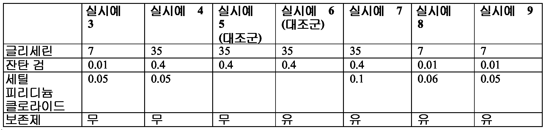
*정제수는 조성물의 나머지 부분을 구성함.Purified water constitutes the remainder of the composition.
**보존제는 메틸파라벤 (0.1%), 프로필파라벤 (0.1%), 나트륨 벤조에이트 (0.5%) 를 포함함. ** Preservatives include methylparaben (0.1%), propylparaben (0.1%), sodium benzoate (0.5%).
하기 결과를 기반으로 하여, 보존제는 장벽 형성 및 항균 활성에 불필요한 것으로 발견되었다.Based on the following results, preservatives were found to be unnecessary for barrier formation and antimicrobial activity.
실시예Example 10 10
단일층 상처 복구 검정Monolayer Wound Repair Black
상처 복구 검정을 실시예 1 의 상피 세포 및 섬유아세포에 대해 수행하였다. 간략하게, 잇몸 상피 세포 (1x10⁴ 세포) 및 섬유아세포 (1x10³ 세포) 를 6-웰 플레이트의 벽에 시딩하고, 5 μg/mL 의 인간 트랜스페린, 2 nM 의 3,3',5'-트리-요오도-L-티로닌, 0.4 μg/mL 의 히드로코르티손, 10 ng/mL 의 상피 성장 인자, 페니실린 및 스트렙토마이신, 및 10% FBS (최종 농도) 를 갖는 둘베코 개질 이글 (DME) - 햄 F12 (3:1) (DMEH) 에서 성장시켰다. 융합시, 200 μL 피펫 팁을 사용하여 각 웰의 융합 단일층에 상처를 만들었다.Wound repair assays were performed on the epithelial cells and fibroblasts of Example 1. Briefly, gum epithelial cells (1 × 10 4 cells) and fibroblasts (1 × 10 3 cells) are seeded on the wall of a 6-well plate and 5 μg / mL of human transferrin, 2 nM of 3,3 ′, 5′-tree Dulbecco's Modified Eagle (DME) with iodo-L-tyronine, 0.4 μg / mL hydrocortisone, 10 ng / mL epidermal growth factor, penicillin and streptomycin, and 10% FBS (final concentration) Grown on F12 (3: 1) (DMEH). Upon fusion, wounds were made to the fusion monolayer of each well using a 200 μL pipette tip.
실시예Example 11-15 11-15
실시예 11 및 12 에서, 실시예 10 으로부터의 상피 세포 배양물을 실시예 3 및 4 의 희석된 장벽-형성 조성물에 약 2 분 동안 노출시켰다. 실시예 13 및 14 에서, 실시예 10 으로부터의 섬유아세포 배양물을 실시예 3 및 4 의 희석된 장벽-형성 조성물에 약 2 분 동안 노출시켰다. 노출 전에, 실시예 3 을 식염수를 사용하여 1% 농도로 희석하고, 실시예 4 를 식염수를 사용하여 5% 농도로 희석하였다. 노출 후, 스프레이를 따뜻한 멸균 배양 배지로 2 회 세척한 후, 세포 배양물을 섬유아세포에 대해서는 DME 로, 상피 세포에 대해서는 DMEH 로 오버레이하고, 6 및 24 시간 동안 5% CO2 습한 분위기에서 37℃ 에서 배양하였다. 실시예 10 으로부터의 미처리 배양물인 대조예 15 를 또한 시험하였다.In Examples 11 and 12, the epithelial cell culture from Example 10 was exposed to the diluted barrier-forming composition of Examples 3 and 4 for about 2 minutes. In Examples 13 and 14, fibroblast cultures from Example 10 were exposed to the diluted barrier-forming compositions of Examples 3 and 4 for about 2 minutes. Prior to exposure, Example 3 was diluted to 1% concentration with saline and Example 4 was diluted to 5% concentration with saline. After exposure, the spray was washed twice with warm sterile culture medium, then the cell cultures were overlaid with DME for fibroblasts and DMEH for epithelial cells and 37 ° C. in 5% CO 2 wet atmosphere for 6 and 24 hours. Incubated at. Control Example 15, an untreated culture from Example 10, was also tested.
상처 복구/세포 이동을 광학 현미경을 사용하여 확인하고, 디지털 사진을 찍었다 (Nikon, Coolpix 950). 상처 봉합 % (세포 성장/이동) 를 하기 식 I 을 사용하여 본 장벽 스프레이에 대한 노출 전 및 노출 후 상대적 상처 부위를 비교함으로써 계산하였다:Wound repair / cell migration was confirmed using an optical microscope and digital pictures were taken (Nikon, Coolpix 950). Wound closure% (cell growth / migration) was calculated by comparing the relative wound area before and after exposure to the present barrier spray using Formula I:
![]()
![]()
도 3 은 6 시간 후-처리 이후, 식 I 로부터 생성된 상처 복구 데이터의 그래프를 나타낸다. 도 4 는 또한 1% 희석된 장벽-형성 조성물 및 대조예 15 로 처리한 상피 세포 배양물의 상처 복구를 나타내는 사진이다. 3 shows a graph of wound repair data generated from Equation I after 6 hours post-treatment. 4 is also a photograph showing wound repair of epithelial cell culture treated with 1% diluted barrier-forming composition and control 15. FIG.
장벽-형성 조성물과 접촉한 후, 상피 세포 (도 4A) 및 섬유아세포 (데이터는 나타내지 않음) 는 실시예 11 및 12 의 장벽-형성 조성물과 접촉 후 6 시간에서 시작하여 계속해서 이동하였다. 비-노출된 배양물 및 장벽-형성 조성물-노출된 배양물 모두에서 상피 세포는 작고 입방형이었다 (도 4A-E). 섬유아세포는 둘 모두의 배양 조건 (비노출 및 세틸피리디늄 클로라이드계 생성물 노출 세포) 에서 작은 핵을 갖는 장방형 세포였다 (데이터는 나타내지 않음). 세포 성장 및 이동 (손상 구역을 덮는) 은 본래 상처와 비교하여 대조예 15 및 처리된 실시예 11 및 12 모두에서 필적할만하였다 (도 4F). 24 시간에 걸친 추적은, 각각의 실시예 11 및 12 가 융합성 단일층을 제공하는 스크래치 가장자리를 덮었다는 것을 나타내었다 (도 4D-E). After contact with the barrier-forming composition, epithelial cells (FIG. 4A) and fibroblasts (data not shown) continued to migrate starting at 6 hours after contact with the barrier-forming composition of Examples 11 and 12. Epithelial cells were small and cubic in both non-exposed cultures and barrier-forming composition-exposed cultures (FIGS. 4A-E). Fibroblasts were rectangular cells with small nuclei in both culture conditions (unexposed and cetylpyridinium chloride-based product exposed cells) (data not shown). Cell growth and migration (covering damage zones) were comparable in both Control Example 15 and treated Examples 11 and 12 compared to the original wound (FIG. 4F). Tracing over 24 hours showed that each of Examples 11 and 12 covered the scratch edge providing a confluent monolayer (FIGS. 4D-E).
상기 실험을 5 회 개별적으로 반복하여 유사한 결과를 얻었다. 처리된 실시예 11 및 12 는 잇몸 상피 세포 성장/이동에도, 세포 분화에도 주요 부작용을 나타내지 않았다 (큰 세포질 및 큰 핵을 나타내는 세포가 없음).The experiment was repeated five times individually to obtain similar results. Treated Examples 11 and 12 showed no major adverse effects on gum epithelial cell growth / migration or cell differentiation (no cells showing large cytoplasm and large nucleus).
실시예Example 16-19 16-19
세포독성 검정Cytotoxicity assay
실시예 2 의 조작 인간 구강 점막 (EHOM) 모델을 사용하여, 실시예 10 및 11 의 조성물이 안전하고 조직 손상 또는 세포 괴사를 촉진하지 않는지 여부를 측정하였다. 실시예 16 및 17 에서, 상피 표면 (10 cm2) 을 5% 희석된 실시예 3 장벽-형성 조성물 및 1% 희석된 실시예 4 조성물 (둘 모두 무-혈청 배양 배지 중에서 희석함) 의 얇은 층 (300 μl) 으로 약 2 분 동안 오버레이하였다. 기간에 있어서의 변화는 결과에 영향을 갖지 않는 것으로 보이며, 단지 절차 수행에 걸리는 시간을 반영한다. 대조예 18 은 장벽-형성 조성물로 처리하지 않은 대조군이었다. EHOM 조직을 이후 따뜻한 멸균 배양 배지로 2 회 헹구고, 5% CO2 습한 분위기에서 37℃ 에서 24 시간 동안 인큐베이션하였다. 상기 인큐베이션 기간 후, 조작 조직이 손상되었는지를 평가하기 위해서, 각각의 EHOM 에 대해 장벽 스프레이 제형과의 접촉으로 인한 구멍의 존재를 육안으로 검사하였다. 이들 EHOM 의 사진을 찍어 이러한 가능성을 확인하였다. 추가적으로, 각각의 EHOM 으로부터 생검물을 수집하고, 에오신 및 헤마톡실린을 사용하여 조직학적 염색을 수행하였다.Manipulation of Example 2 The human oral mucosa (EHOM) model was used to determine whether the compositions of Examples 10 and 11 were safe and did not promote tissue damage or cell necrosis. In Examples 16 and 17, a thin layer of the Example 3 barrier-forming composition and the 1% diluted Example 4 composition (both diluted in serum-free culture medium) diluted 5% of the epithelial surface (10 cm 2 ) (300 μl) was overlaid for about 2 minutes. Changes in duration do not appear to have an effect on the outcome, only reflect the time taken to perform the procedure.
실시예 16 및 17 에서, 미처리 대조예 18 과 유사하게, 1 및 5% 희석된 실시예 3 및 4 에 노출시킨 EHOM 은 구멍과 같은 어떠한 육안으로 보이는 손상도 나타내지 않았다 (도 5 참조). 이러한 손상의 부재를 5% 희석 실시예 3 구강 청결제 (실시예 7A) 및 1% 희석 스프레이 (실시예 7B) 조성물과의 조직 접촉 10 분 후 모니터링하였다. 이를 섬유아세포-조밀 결합 조직의 상부에 상피의 다중층 조직을 나타내는 조직학적 분석에 의해 확인하였다. 실시예 16 또는 17 에서의 EHOM 조직에서는 특정한 손상은 관찰되지 않았다. 실시예 16 및 17 처리-EHOM 구조는 미처리 대조예 18 과 필적할만하였다.In Examples 16 and 17, similar to
실시예 19 에서 동일한 관찰이 기록되었고, 이는 1% 희석물로 처리한 EHOM 을 포함하였다는 것을 제외하고는, 실시예 16 과 동일하였다 (데이터는 나타내지 않음). The same observation was recorded in Example 19, which was the same as Example 16 except that it included EHOM treated with 1% dilution (data not shown).
도 5 는 EHOM 육안 형상 및 구조에 대한 장벽-형성 조성물의 효과를 나타낸다. 패널 A 및 B 는 대조예 18 을 나타낸다. 패널 C 및 D 는 실시예 16 (스프레이 조성물) 을 나타내고, 패널 E 및 F 는 실시예 17 (구강 청결제 조성물) 을 나타낸다.5 shows the effect of barrier-forming compositions on EHOM visual shape and structure. Panels A and B show
실시예Example 20-25 20-25
존재하는 경우, 장벽 스프레이 조성물의 EHOM 손상에 대한 부작용을 또한 세포내 LDH 의 배양 배지로의 누출을 측정함으로써 평가하였다.If present, side effects on EHOM damage of the barrier spray composition were also evaluated by measuring the leakage of intracellular LDH into the culture medium.
실시예 20-23 에서, EHOM 조직을 각각 실시예 3 및 4 의 (A) 1% 또는 (B) 5% 희석물에 10 분 동안 노출시킨 후, 배양 배지에서 24 시간 동안 성장시켰다. 실시예 24 및 25 는 처리하지 않은 대조군이었다. 실시예 20-25 각각의 50 μl 의 상청액을 멸균 96-웰 평평 바닥 플레이트에 옮겼다. 각각의 웰에 50 μl 의 재구성된 기질 믹스를 보충하고, 플레이트를 30 분 동안 실온에서 암 환경 하에 인큐베이션하였다. LDH 수준을 추정하기 위해서, 배양 상청액의 분취액을 수집하고 제조사 프로토콜에 따라 LDH 세포독성 검정을 수행하였다 (Promega, Madison, WI, USA). 상기 검정으로 L-락테이트 및 NAD 의 피루베이트 및 NADH 로의 전환을 방출된 LDH 에 의해 측정한다. 반응을 정지시키기 위해서, 50 μl 의 정지 용액을 각 웰에 추가하였다. 다음으로, 100 μl 의 혼합물을 96-웰 평평 바닥 플레이트에 옮기고, 흡광도를 X-Mark 마이크로플레이트 분광광도계 (Bio-Rad, Mississauga, ON, Canada) 로 490 nm 에서 판독하였다. In Examples 20-23, EHOM tissues were exposed for 10 minutes to (A) 1% or (B) 5% dilution of Examples 3 and 4, respectively, and then grown in culture medium for 24 hours. Examples 24 and 25 were untreated controls. Examples 20-25 50 μl of each supernatant was transferred to a sterile 96-well flat bottom plate. Each well was supplemented with 50 μl of reconstituted substrate mix and plates were incubated for 30 minutes at room temperature under dark environment. To estimate LDH levels, aliquots of the culture supernatants were collected and LDH cytotoxicity assays were performed according to the manufacturer's protocol (Promega, Madison, WI, USA). In this assay the conversion of L-lactate and NAD to pyruvate and NADH is measured by the released LDH. To stop the reaction, 50 μl of stop solution was added to each well. Next, 100 μl of the mixture was transferred to a 96-well flat bottom plate and the absorbance was read at 490 nm with an X-Mark microplate spectrophotometer (Bio-Rad, Mississauga, ON, Canada).
LDH 및 상처 복구 실험에서, 하기의 시험 방법을 사용하였다. 각각의 실험을 4 회 이상 수행하고, 실험 값을 평균 ± SD 로서 표시하였다. 대조값 (장벽 스프레이 부재) 및 시험값 (스프레이 존재) 사이 차이의 통계적 유의성을 1 방향 ANOVA 에 의해 측정하였다. 후험적 비교를 투키 (Tukey) 법을 사용하여 수행하였다. 정규성 및 분산 추정치를 각각 샤피로-윌크 (Shapiro-Wilk) 시험 및 브라운-포사이드 (Brown and Forsythe) 시험을 사용하여 확인하였다. 모든 추정치는 충족되었다. P 값은 ≤ 0.05 에서 유의한 것으로 공표되었다. 데이터를 SAS 통계 패키지 (버전 8.2, SAS Institute Inc., Cary, NC, USA) 를 사용하여 분석하였다.In LDH and wound repair experiments the following test methods were used. Each experiment was performed four or more times and the experimental values were expressed as mean ± SD. The statistical significance of the difference between the control value (barrier spray member) and test value (with spray) was measured by one-way ANOVA. Epigenetic comparisons were performed using the Tukey method. Normality and variance estimates were confirmed using Shapiro-Wilk test and Brown and Forsythe test, respectively. All estimates were met. P values were declared significant at ≦ 0.05. Data was analyzed using a SAS statistical package (version 8.2, SAS Institute Inc., Cary, NC, USA).
도 6 에서 나타낸 결과는, 미처리 조직에 비해, 장벽-형성 조성물 처리 구조에서 LDH 수준에 유의한 차이를 나타내지 않았다. 이러한 관찰을 장벽-형성 조성물 처리 실시예에 노출 후 6 시간 (데이터는 나타내지 않음) 및 24 시간 (도 6) 에서 수행하였다. 도 6(A) 는 대조예 24, 및 1% 희석물 처리 실시예 20 및 21 을 나타내고; 도 6(B) 는 대조예 25, 및 5% 희석물 처리 실시예 22 및 23 을 나타낸다. 이들 데이터로 실시예 3 및 4 의 비독성 효과를 확인하였다. 유사하게, 실시예 3 및 4 와의 2 분 동안의 접촉으로는 대조군에 비해 어떠한 차이도 나타나지 않았다 (데이터는 나타내지 않음). The results shown in FIG. 6 showed no significant differences in LDH levels in the barrier-forming composition treated structure compared to untreated tissue. This observation was performed at 6 hours (data not shown) and 24 hours (FIG. 6) after exposure to the barrier-forming composition treatment examples. 6 (A) shows Control Example 24, and 1% dilution treatment Examples 20 and 21; 6 (B) shows Control Example 25 and Examples 22 and 23 treated with 5% diluent. These data confirmed the nontoxic effects of Examples 3 and 4. Similarly, two minutes of contact with Examples 3 and 4 showed no difference compared to the control (data not shown).
실시예Example 26 26
장벽-형성 조성물이 EHOM 구조를 손상시키는지 여부의 측정Determination of whether the barrier-forming composition damages the EHOM structure
실시예 2 의 EHOM 을 실시예 4 로 약 2 분 동안 처리하고, 배양 배지로 세척한 후 24 시간 동안 배양하였다. 그 다음, 조직을 가능한 육안 조직 손상 (구멍의 존재 유무) 에 대해 검사하였다. 조직 손상을 또한 조직학적 분석에 의해 조사하였다. 이러한 목적으로, 각각의 EHOM 으로부터 생검물을 채취하였다. 이를 4% 파라포름알데히드 용액으로 고정시킨 후 파라핀 중에 포매하였다. 얇은 박편 (4 μm) 을 에오신-헤마톡실린으로 염색하였다. 박편을 50%-글리세롤 봉입 배지 중에 커버슬립으로 봉입하고, 광학 현미경을 통해 관찰하고, 사진 찍었다. 처리된 EHOM 에 대한 손상은 확인되지 않았다.The EHOM of Example 2 was treated with Example 4 for about 2 minutes, washed with culture medium and incubated for 24 hours. The tissues were then examined for possible gross tissue damage (with or without holes). Tissue damage was also investigated by histological analysis. For this purpose, biopsies were taken from each EHOM. It was fixed with 4% paraformaldehyde solution and then embedded in paraffin. Thin flakes (4 μm) were stained with eosin-hematoxylin. The flakes were sealed with coverslips in 50% -glycerol inclusion medium, observed through an optical microscope, and photographed. No damage to the treated EHOM was identified.
실시예Example 27 및 28 27 and 28
장벽-형성 조성물이 점막 조직을 통한 미생물 통과에 대한 EHOM 의 기계적 장벽 기능에 영향을 주는지 여부의 측정Determination of whether the barrier-forming composition affects the mechanical barrier function of the EHOM against microbial passage through mucosal tissue
실시예 27 및 28 에서, 2 가지 접근 방식을 사용하여 대조예가 점막 조직을 통한 미생물 통과를 차단하는 장벽을 형성하며 또한 고유 항균 효과를 갖는지 여부를 측정하였다. 통과점 (pass-through) 챔버에서의 성장 및 EHOM 표면 상의 성장을 한천 배지에서의 성장을 평가함으로써 산정하였다. In Examples 27 and 28, two approaches were used to determine whether the control forms a barrier that blocks microbial passage through mucosal tissue and also has an intrinsic antimicrobial effect. Growth in the pass-through chamber and growth on the EHOM surface were estimated by assessing growth in agar medium.
실시예 27 에서, 실시예 2 의 EHOM 을 실시예 4 의 1% 희석물 및 5% 희석물 (무-혈청 배양 배지 중에 희석함) 과 2 분 동안 접촉시켰다. 이후 조직을 무-혈청 배양 배지로 2 회 세척한 후 1x10⁶ 칸디다 (Candida) 미생물 세포를 300 μl 의 부피로 오버레이하였다. 조직을 공기-액체 배양 플레이트에 넣고 24 시간 동안 5% CO2 습한 분위기에서 37℃ 에서 인큐베이션하였다. 그 다음으로, EHOM 밑의 배양 배지 (복측 챔버) 를 수집하고, 사부로 (Sabouraud) 한천 플레이트에 시딩하여 미생물이 조직을 통해 침투하고 밑의 배양 배지에 도달하였는지 여부를 확인하였다. 배양물을 또한 EHOM 표면으로부터 수득하고, 사부로 한천 플레이트에 시딩하였다. 과정을 도 7 에서 도표로 나타낸다.In Example 27, the EHOM of Example 2 was contacted with the 1% dilution and 5% dilution of Example 4 (diluted in serum-free culture medium) for 2 minutes. The tissue was then washed twice with serum-free culture medium and then overlayed 1 × 10 6 Candida microbial cells in a volume of 300 μl. Tissues were placed in air-liquid culture plates and incubated at 37 ° C. in a 5% CO 2 wet atmosphere for 24 hours. Next, the culture medium underneath the EHOM (ventral chamber) was collected and seeded on a Sabouraud agar plate to confirm whether microorganisms penetrated through the tissue and reached the culture medium below. Cultures were also obtained from the EHOM surface and seeded in agar plates in the dead. The process is shown graphically in FIG. 7.
실시예 28 에서, 실시예 4 조성물의 1% 및 5% 희석물로 2 분 동안 처리한 실시예 2 의 EHOM 을 칸디다 미생물 세포로 24 시간 동안 오버레이하여, 사브로 덱스트로오스 한천 플레이트에 넣고 5 분 동안 제자리에 두었다. 그 다음, EHOM 을 제거하고 플레이트를 24 시간 동안 30℃ 에서 인큐베이션하고, 그 후 미생물 성장을 육안으로 확인하고 사진 찍었다. 각각의 실험을 5 회 독립적으로 반복하여 유사한 결과를 얻었다.In Example 28, the EHOM of Example 2, which was treated with 1% and 5% dilution of the composition of Example 4 for 2 minutes, was overlaid with Candida microbial cells for 24 hours, placed in a Sabro dextrose agar plate for 5 minutes Left in place. The EHOM was then removed and the plates incubated at 30 ° C. for 24 hours, after which the microbial growth was visually confirmed and photographed. Each experiment was repeated five times independently to obtain similar results.
도 8 은 EHOM 표면의 배양물 (패널 C 및 D) 및 하부 (복측) 챔버로부터의 통과 액체의 배양물 (패널 A 및 B) 의 결과를 나타낸다. A 및 C 패널은 실시예 4 의 1% 희석물로 처리된 EHOM 이었고, B 및 D 패널은 실시예 4 의 5% 희석물로 처리된 EHOM 이었다. 상기 데이터는 실시예 4 조성물이 EHOM 조직을 통한 미생물의 통과를 방지하는 장벽을 형성하나 고유 항균 효과는 갖지 않는다는 것을 나타낸다.FIG. 8 shows the results of cultures on the EHOM surface (Panels C and D) and cultures of passage liquid from the lower (ventral) chamber (Panels A and B). Panels A and C were EHOMs treated with the 1% dilution of Example 4 and panels B and D were EHOMs treated with the 5% dilution of Example 4. The data indicate that the Example 4 composition forms a barrier that prevents the passage of microorganisms through EHOM tissue but does not have inherent antimicrobial effect.
실시예Example 29 및 30 29 and 30
실시예 29 및 30 에서, EHOM 을 S. 무탄스로 감염시킨 것을 제외하고는 실시예 27 및 28 을 반복하였다. 유사한 결과를 수득하였고, 이는 장벽-형성 조성물이 S. 무탄스 미생물이 장벽을 통과하는 것을 방지하는 장벽을 형성하였으나 항균 효과는 갖지 않았다는 것을 나타내었다.In Examples 29 and 30, Examples 27 and 28 were repeated except that EHOM was infected with S. mutans. Similar results were obtained, indicating that the barrier-forming composition formed a barrier that prevented S. mutans microorganisms from passing through the barrier but had no antimicrobial effect.
실시예Example 31 및 32 31 and 32
장벽-형성 조성물이 미생물 침입에 대한 EHOM 의 기계적 장벽 기능에 영향을 주는지 여부의 측정Determination of whether the barrier-forming composition affects the mechanical barrier function of the EHOM against microbial invasion
실시예 32 에서, 실시예 2 로부터의 EHOM 조직 한 세트를 실시예 4 의 장벽-형성 조성물로 처리한 후 C. 알비칸스로 오버레이하였다. 대조예 31 에서는 대조군 세트를 C. 알비칸스로 오버레이하기 전에 장벽-형성 조성물로 처리하지 않았다. 각각의 접촉 기간 직후, 각각의 EHOM 으로부터 생검물을 채취하고, 파라포름알데히드 용액으로 고정하고, 파라핀 중에 포매하였다. 얇은 박편 (4 μm) 을 에오신-헤마톡실린으로 염색하였다. 광학 현미경을 사용하여 박편을 관찰하여, 미생물 세포의 조직 내로의 침입/침투를 분석하였다. 현미경 관찰 후, 각각의 조건으로부터 대표적 사진을 찍고 제공하였다. 실험을 3 회 반복하여 유사한 결과를 얻었다. 유사한 결과를 또한 실시예 3 으로 처리하여 수득하였다 (데이터는 나타내지 않음). In Example 32, one set of EHOM tissues from Example 2 were treated with the barrier-forming composition of Example 4 and then overlayed with C. albicans. In Control Example 31, the control set was not treated with the barrier-forming composition before overlaying with C. albicans. Immediately after each contact period, biopsies were taken from each EHOM, fixed with paraformaldehyde solution and embedded in paraffin. Thin flakes (4 μm) were stained with eosin-hematoxylin. The lamella was observed using an optical microscope to analyze the invasion / infiltration of microbial cells into tissue. After microscopic observation, representative pictures were taken and provided from each condition. The experiment was repeated three times to obtain similar results. Similar results were also obtained by treating with Example 3 (data not shown).
도 9 는 EHOM 조직의 미생물 침입에 대한 장벽-형성 조성물의 효과를 나타낸다. 패널 (A) 는 미처리 대조예 31 의 대표적 사진이고, 패널 (B) 는 처리 실시예 32 의 사진이다. 화살표는 미처리 대조예 31 에서의 침입성 진균 균사를 나타낸다. 9 shows the effect of barrier-forming compositions on microbial invasion of EHOM tissues. Panel (A) is a representative photograph of untreated control example 31, and panel (B) is a photograph of treatment example 32. Arrows indicate invasive fungal hyphae in untreated control Example 31.
실시예Example 33-40 33-40
상기 기재한 EHOM 모델을 또한, (a) 구강 박테리아 (S. 무탄스) 및 진균 (칸디다 알비칸스 (Candida albicans)) 이 인간 구강 점막을 침투하고/침입하는 것을 방지하고, (b) 숙주 세포에 손상을 일으키지 않는 (세포독성 검정) 장벽을 형성하는 실시예 5-7 의 능력을 평가하는데 사용하였다. The EHOM model described above also includes (a) preventing oral bacteria (S. mutans) and fungi (Candida albicans) from invading / invading human oral mucosa, and (b) entering host cells. It was used to evaluate the ability of Examples 5-7 to form a barrier that does not cause damage (cytotoxicity assay).
실시예 33-40 을 하기 표 III 에 따라 제형화하였다.Examples 33-40 were formulated according to Table III below.
표 table IIIIII
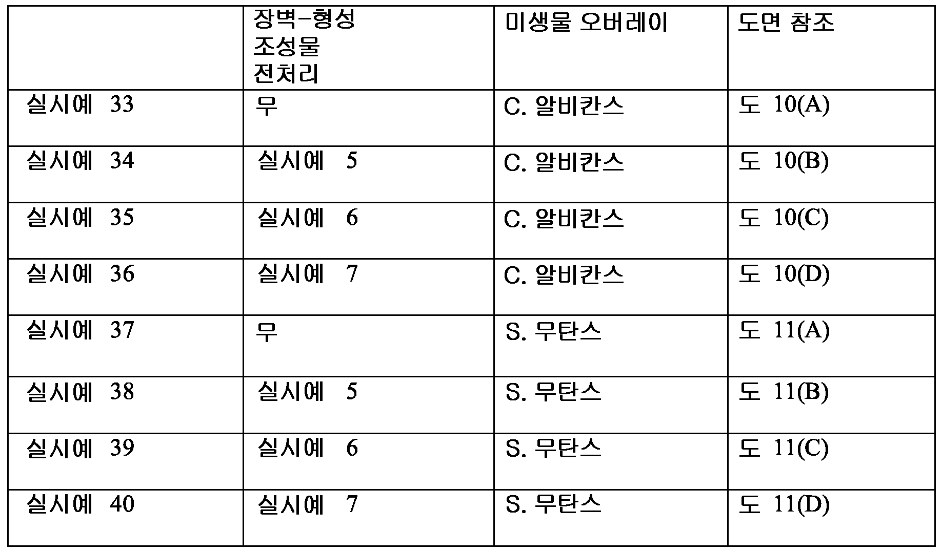
실시예 33-40 에서, 전처리 및 인큐베이션 (실시예 27 및 28 의 절차에 따름) 후: (1) 통과 배지를 하부 챔버로부터 수집하고; (2) 조직을 사부로 덱스트로오스 한천 페트리 디쉬의 표면 상에 넣어 배치시키고, 24 시간 동안 인큐베이션하였다. 수집한 통과 배지를 한천 배지 플레이트에 스프레딩하고, 실시예 27 및 28 에서 또한 기재한 바와 같이 24 시간 동안 인큐베이션하였다. 표 III 은 또한, 각각의 넣은 실시예 배양물 상의 미생물 성장을 나타내는 실시예 각각의 사진을 찍은 도면을 표시한다.In Examples 33-40, after pretreatment and incubation (according to the procedures of Examples 27 and 28): (1) the passing medium is collected from the lower chamber; (2) Tissues were placed on the surface of dextrose agar petri dishes in four portions and incubated for 24 hours. The collected passage medium was spread on agar medium plates and incubated for 24 hours as also described in Examples 27 and 28. Table III also displays a photograph of each of the examples showing microbial growth on each loaded example culture.
도 10 및 11 은 칸디다 및 스트렙토코쿠스 둘 모두가 실시예 5-7 의 조성물로 처리한 EHOM 표면 상에서 성장할 수 있었다는 것을 나타낸다. 반대로, 도 12 에서 나타내는 바와 같이, "통과" 배지가 실시예 36 또는 40 의 EHOM 의 하부 챔버로부터 수집된 경우 (즉, 실시예 7 조성물로 처리한 것들), 미생물 성장이 관찰되지 않았다. 이는 실시예 7 조성물로의 EHOM 처리가 점막 조직 표면에 손상을 일으키지 않았으며 유기체가 처리한 EHOM 에 침투할 수 없었다는 것을 나타낸다. 유사한 결과를 실시예 5 및 6 의 조성물로 처리한 EHOM 으로 수득하였다 (데이터는 나타내지 않음). 이들 데이터는 글리세린 및 잔탄 검의 조합이 점막 조직 상에 보호성 장벽을 형성할 수 있다는 것을 나타낸다.10 and 11 show that both Candida and Streptococcus could grow on the EHOM surface treated with the compositions of Examples 5-7. In contrast, as shown in FIG. 12, when “pass” medium was collected from the lower chamber of the EHOM of Example 36 or 40 (ie those treated with the Example 7 composition), no microbial growth was observed. This indicates that EHOM treatment with the composition of Example 7 did not cause damage to the mucosal tissue surface and the organism was unable to penetrate the treated EHOM. Similar results were obtained with EHOM treated with the compositions of Examples 5 and 6 (data not shown). These data show that the combination of glycerin and xanthan gum can form a protective barrier on mucosal tissue.
실시예Example 41-47 41-47
시험 제형은 비독성이며 세포/조직에 손상을 일으키지 않음Test formulation is nontoxic and does not cause damage to cells / tissues
실시예 41-47 에서, EHOM 모델을 조성물의 독성을 평가하는데 사용하였다. 실시예 41-47 을 하기 표 IV 에서 언급한 바와 같이 제형화하였다.In Examples 41-47, an EHOM model was used to assess the toxicity of the composition. Examples 41-47 were formulated as mentioned in Table IV below.
표 table IVIV

전처리 및 인큐베이션 (실시예 27 및 28 의 절차에 따름) 후, 배양 상청액을 실시예 41-48 EHOM 조직으로부터 수집하고 LDH 활성 측정에 사용하였다.After pretreatment and incubation (following the procedures of Examples 27 and 28), culture supernatants were collected from Examples 41-48 EHOM tissues and used for LDH activity measurements.
도 13 에서 나타낸 바와 같이, 제형이 방부제의 존재 또는 부재 하에 세틸피리디늄 클로라이드를 함유하고 각각 칸디다 알비칸스 또는 S. 무탄스로 감염되었는지 여부에 관계없이, 실시예 41-48 에서는 LDH 수준에 있어서 유의한 증가가 관찰되지 않았다. 이들 데이터로, 실시예 장벽-형성 조성물의 비독성 효과 및 이들 제형이 숙주 점막 조직의 무결성을 유지시켰다는 것을 확인하였다. As shown in FIG. 13, regardless of whether the formulation contained cetylpyridinium chloride in the presence or absence of a preservative and was infected with Candida albicans or S. mutans, respectively, in Examples 41-48, significant LDH levels were observed. No increase was observed. These data confirmed the nontoxic effects of the example barrier-forming compositions and that these formulations maintained the integrity of the host mucosal tissue.
데이터는 평균 ± SD 이고 상기 실시예 25 에서 언급한 바와 같이 계산되었다. 미처리 및 처리 조직 사이에 유의한 차이는 관찰되지 않았다.Data are mean ± SD and calculated as mentioned in Example 25 above. No significant difference was observed between untreated and treated tissues.
종합하여 볼 때, 상기 데이터는 실시예 조성물이, 미생물이 인간 점막 조직을 침투하고 침입하는 것을 방지할 수 있는 효과적이고 안전한 장벽을 제시한다는 것을 나타낸다.Taken together, the data indicate that the example composition presents an effective and safe barrier that can prevent microbes from penetrating and invading human mucosal tissue.
실시예Example 48-61 48-61
장벽-형성 조성물의 전임상 평가로, 상기 조성물이 많은 박테리아 및 효모에 대해 효과적인 것이 나타났다. 실시예 7 장벽-형성 조성물의 항균 활성을, S. 살리바리우스 (S. salivarius), P. 진지발리스 (P. gingivalis), S. 피요제네스 (S. pyogenes), S. 뉴모니아 (S. pneumonia), 푸소박테리움 뉴클레아툼 (Fusobacterium nucleatum), S. 무탄스, S. 오레우스 (S. aureus), Y. 엔테로콜리티카 (Y. enterocolitica), S. 오랄리스, S. 미티스 (S. mitis), C. 알비칸스, C. 크루세이 (C. krusei), C. 트로피칼리스 (C. tropicalis) 및 C. 글라브라타 (C. glabrata) 를 포함하는 환자에서 수득한 수많은 임상 단리물에 대해 평가하였다. 실시예 7 장벽-형성 조성물의 활성을, Clinical and Laboratory Standards Institute (CLSI) 문헌 M07-A8, M11-A7, 및 M27-A3 에서 기재된 참고 방법을 사용하여 이의 최소 저해 농도 (MIC) 를 측정하여 평가하였다. Preclinical evaluation of the barrier-forming composition has shown that the composition is effective against many bacteria and yeasts. Example 7 The antimicrobial activity of the barrier-forming compositions was determined by S. salivarius, P. gingivalis, S. pyogenes, S. pneumoniae pneumonia, Fusobacterium nucleatum, S. mutans, S. aureus, Y. enterocolitica, S. oralis, S. mi Numerous patients obtained from patients including S. mitis, C. albicans, C. krusei, C. tropicalis and C. glabrata. Clinical isolates were evaluated. Example 7 The activity of a barrier-forming composition is assessed by measuring its minimum inhibitory concentration (MIC) using the reference method described in Clinical and Laboratory Standards Institute (CLSI) documents M07-A8, M11-A7, and M27-A3. It was.
여러 유형의 호기성 또는 혐기성 박테리아 (1x10⁴ 세포/ml) 의 표준화된 접종물을, 비교예로서 2% 클로르헥시딘 글루코네이트 (CHX, 20 μg/mL) 또는 실시예 7 (0.1% CPC 함유, 또는 1 μg/ml) 의 연속 희석 용액과 함께 인큐베이션하였다. 세포를 시험 작용제의 존재 또는 부재 (성장 조절) 하에 24 시간 동안 성장시켰다. 각각의 작용제에 대한 MIC 를, 100% 성장 저해를 유도한 (무-약물 대조군에 비해) 농도로서 정의하였다.Standardized inoculations of various types of aerobic or anaerobic bacteria (1 × 10 4 cells / ml) were used as comparative examples with 2% chlorhexidine gluconate (CHX, 20 μg / mL) or Example 7 (containing 0.1% CPC, or 1 μg). / ml) with a serial dilution solution. Cells were grown for 24 hours in the presence or absence of test agents (growth control). MIC for each agent was defined as the concentration that led to 100% growth inhibition (relative to the drug-free control).
유사한 미세희석-기반 CLSI 방법 (M27-A2) 을 알비칸스 및 비-알비칸스 칸디다 종에 대해 실시예 7 의 활성을 평가하는데 사용하였다.A similar microdilution-based CLSI method (M27-A2) was used to assess the activity of Example 7 against albicans and non-albicans candida species.
표 VTable V
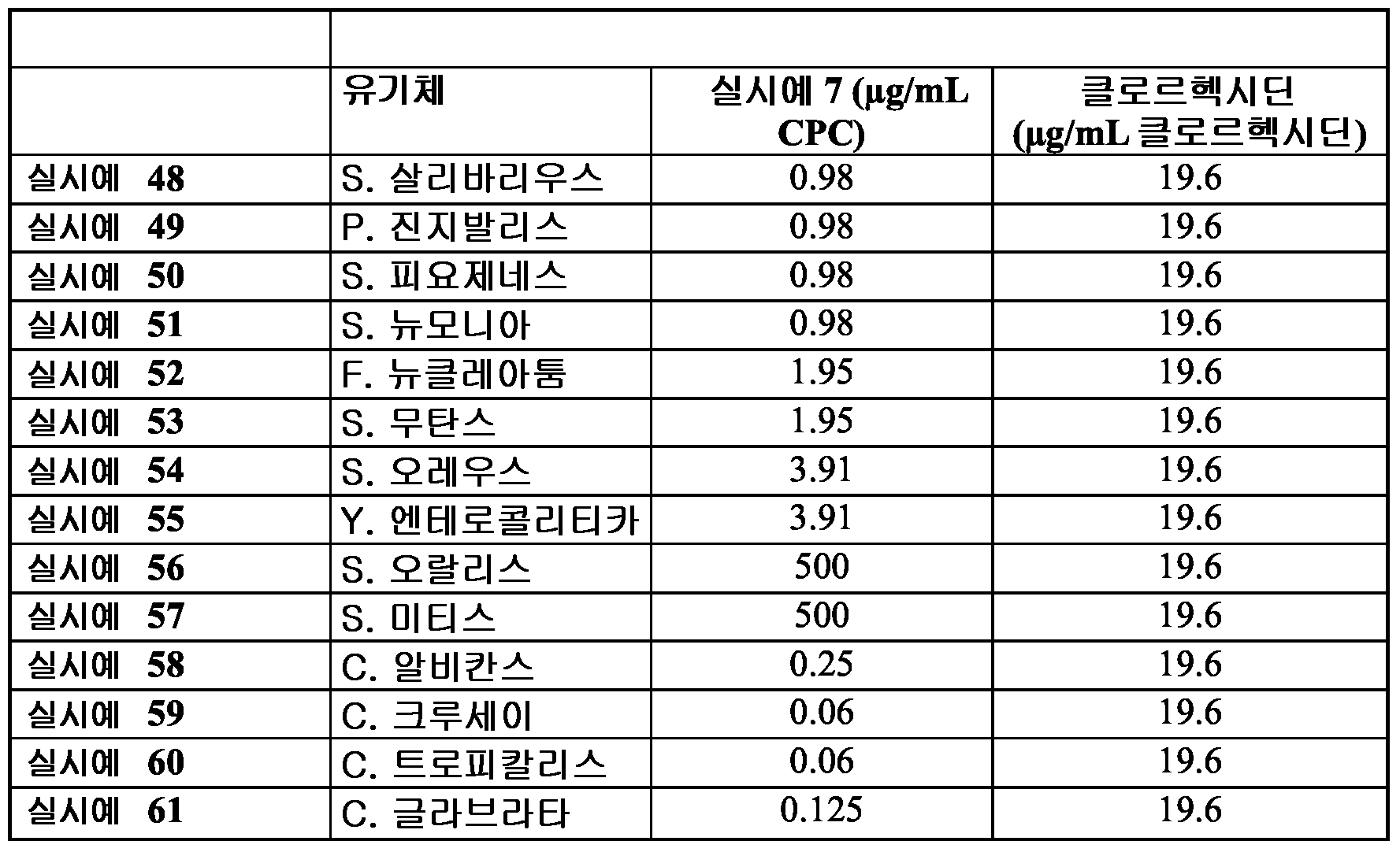
장벽-형성 조성물은 또한 하기의 것에 대해 강력한 항균 활성을 갖는 것으로 발견되었다: MRSA, 아시네토박터 바우마니 (Acinetobacter baumannii), 스트렙토코쿠스 산구이스 (Streptococcus sanguis), S. gordonii (S. 고르도니) 및 아그레가티박터 악티노마이세템코미탄스 (Aggregatibacter actinomycetemcomitans). Barrier-forming compositions have also been found to have potent antimicrobial activity against: MRSA, Acinetobacter baumannii, Streptococcus sanguis, S. gordonii (S. gordonii) And Aggregatibacter actinomycetem comitans.
표 V 에서 볼 수 있듯이, 실시예 7 조성물은 많은 호기성 및 혐기성 박테리아 뿐 아니라 진균에 대해서도 강력한 활성을 나타내었다.As can be seen in Table V, the composition of Example 7 showed potent activity against many aerobic and anaerobic bacteria as well as fungi.
S. 오랄리스 및 S. 미티스에 대한 실시예 7 장벽-형성 조성물의 MIC 는 다른 유기체에 비해 현저히 상승하였다 (500 μg/mL). S. 오랄리스 및 S. 미티스가 구강의 정상적 공생체라는 것은 흥미로운 것이다. 통상적으로 사용한 항균 클로르헥시딘 (2% 용액) 의 활성을 또한 동일한 방법에 의해 측정하였다. 표 V 는 다양한 미생물에 대한 비교예로서 실시예 7 장벽-형성 조성물 및 클로르헥시딘 (2% 용액) 의 MIC 를 나타낸다.Example 7 Barrier-Forming Compositions for S. Oralis and S. Mitis MIC was significantly elevated compared to other organisms (500 μg / mL). It is interesting that S. oralis and S. mitis are normal symbiosis of the oral cavity. The activity of commonly used antimicrobial chlorhexidine (2% solution) was also measured by the same method. Table V shows the MICs of Example 7 barrier-forming composition and chlorhexidine (2% solution) as comparative examples for various microorganisms.
종합하여 볼 때, 이들 결과는 실시예 7 이 구강에서 흔히 단리된 병원성 박테리아 및 진균에 대해 강력한 활성을 갖는다는 것을 입증한다. 이러한 활성은 클로르헥시딘에 대해 관찰된 것보다 더 강력하였다. Taken together, these results demonstrate that Example 7 has potent activity against pathogenic bacteria and fungi that are frequently isolated in the oral cavity. This activity was stronger than that observed for chlorhexidine.
실시예 10 및 11 의 장벽-형성 조성물에 대해 유사한 활성 프로파일이 관찰되었다. Similar activity profiles were observed for the barrier-forming compositions of Examples 10 and 11.
실시예Example 62 62
추가적인 비교로서, 공개된 데이터는 시험한 장벽-형성 조성물이 CPC 단독 (즉, 본원에 개시된 장벽 제형에 따른 조성물 중에 없는) 에 비해 더 양호하거나 적어도 동등한 MIC 를 갖는다는 것을 나타낸다. 본원에 참고로 포함되는 [Frank-Albert Pitten and Axel Kramer, "Efficacy of Cetylpyridinium Chloride Used as Oropharyngeal Antiseptic," Arzneim.-Forsch./Drug Res. 51 (II), pp588-595 (2001)] 를 참조한다. 데이터는 시험한 미생물을 기반으로 하여 가변적이지만, 예를 들어, S. 무탄스에 대한 CPC (단독) 는 MIC 가 5.0 - 6.25 μg/mL 이고, 이는 실시예 53 에서 보고한 1.95 μg/mL 보다 훨씬 덜 유효하다. CPC 가 제형 중에서 기타 부형제 화학물질과 혼합되는 경우 그의 활성을 손실하는 위험성을 갖기 때문에, 이는 예상치 못한 결과였다. Department of Health and Human Services (Food and Drug Administration) (1994) Oral Health Care Drug Products for Over-the-Counter Human Use; Tentative Final Monograph for Oral Antiseptic Drug Products. Proposed Rules (21 CFR Part 356, Docket No. 81N-033A, RIN 0905-AA06). Federal Register 59:6084-124 를 참조한다. As a further comparison, the published data indicate that the barrier-forming composition tested had a better or at least equivalent MIC compared to CPC alone (ie, not in a composition according to the barrier formulation disclosed herein). Frank-Albert Pitten and Axel Kramer, "Efficacy of Cetylpyridinium Chloride Used as Oropharyngeal Antiseptic," Arzneim.-Forsch./Drug Res. 51 (II), pp 588-595 (2001). The data are variable based on the microorganisms tested, but, for example, the CPC (alone) for S. mutans had a MIC of 5.0-6.25 μg / mL, much more than the 1.95 μg / mL reported in Example 53. Less valid. This was unexpected because CPC has the risk of losing its activity when mixed with other excipient chemicals in the formulation. Department of Health and Human Services (Food and Drug Administration) (1994) Oral Health Care Drug Products for Over-the-Counter Human Use; Tentative Final Monograph for Oral Antiseptic Drug Products. Proposed Rules (21 CFR Part 356, Docket No. 81N-033A, RIN 0905-AA06). See Federal Register 59: 6084-124.
실시예Example 63-69 63-69
장벽-형성 조성물의 체외 항균 활성 지속기간: 후-항균 효과 (PAE) 의 측정Duration of In Vitro Antimicrobial Activity of Barrier-forming Compositions: Determination of Post-Antibacterial Effect (PAE)
여러 미생물에 대한 실시예 8 의 PAE 를 실시예 63-68 에서 평가하였다. 대조예 69 를 또한 제공하였다. 여러 미생물을 실시예 8 에 (MIC 와 동일한 농도에서) 1 분 동안 노출시킨 후 3 회 세척하여 잔여 제형을 제거하였다. 처리한 세포를 이후 한천 배지 플레이트에 스프레딩하고, 이를 37℃ 에서 인큐베이션하고, 세포가 재성장하는데 걸린 시간을 측정하였다. PAE 를, 미처리 대조예 69 에 비해 실시예 63-68 에 의해 성장 저해율 (%) 이 유지된 시기 (시간으로 표시) 로서 표시하였다.The PAE of Example 8 for various microorganisms was evaluated in Examples 63-68.
도 14 에서 나타낸 바와 같이, 실시예 8 은 시험한 유기체 (S. 오레우스, S. 뉴모니아, S. 고르도니, S. 산구이스, S. 살리바리우스 및 S. 미티스) 에 따라 4 시간 내지 24 시간 범위의 PAE 를 나타내었다. 실시예 8 의 유사한 활성이 칸디다에 대해 관찰되었다 (데이터는 나타내지 않음). 다른 실시예 장벽-형성 조성물은 미생물에 대해 유사한 PAE 를 나타내었다.As shown in FIG. 14, Example 8 was 4 hours according to the organisms tested (S. oreus, S. pneumoniae, S. gordoni, S. sanguis, S. salivarius and S. mytis). PAEs in the range from 24 hours are shown. Similar activity of Example 8 was observed for Candida (data not shown). Other Example Barrier-forming compositions exhibited similar PAEs for microorganisms.
실시예Example 70 70
하부 CPC 내용물을 갖는 유사 비교예와 비교한 S. 무탄스에 대한 실시예 7 장벽-형성 조성물에 대한 PAE 의 시험으로, 실시예 7 의 PAE 가 24 시간인 한편, 비교예 70 의 PAE 가 6 시간이라는 것이 나타났다. 따라서, 실시예 7 이 비교예 70 보다 더 큰 장기적 항균 활성을 나타내며, 추가량의 CPC 가 항균 활성에 대해 단순한 추가 효과 그 이상을 갖는다는 것이 입증된다.Test of PAE on Example 7 barrier-forming compositions for S. mutans compared to similar comparative examples with bottom CPC content, wherein the PAE of Example 7 was 24 hours while the PAE of Comparative Example 70 was 6 hours Appeared. Thus, it is demonstrated that Example 7 exhibits longer term antimicrobial activity than Comparative Example 70, and that additional amounts of CPC have more than just additional effects on antimicrobial activity.
실시예Example 71-76 71-76
주사 전자 현미경을 또한 사용하여, S. 산구이스 (실시예 71), S. 오랄리스 (실시예 72) 및 C. 알비칸스 (실시예 73) 의 실시예 3 으로의 처리가 세포 무결성의 파괴를 야기하였다는 것을 나타내었다.Using also scanning electron microscopy, treatment of S. sanguise (Example 71), S. oralis (Example 72) and C. albicans (Example 73) with Example 3 prevented disruption of cellular integrity. It was caused.
실시예 71-73 에서, 세포를 실시예 3 의 존재 하에 24 시간 동안 성장시켰다. 그 다음으로, 상기 세포를 세척하여 잔여 제형을 제거하고, 알코올 용액 연속물 (10% 에서 100%, v/v) 을 통과시켜 탈수하고, SEM 분석을 위해 처리하였다. 대조예 74-76 은, 실시예 3 의 존재 하에 성장하지 않았다는 점에 있어서 실시예 71-73 과 상이하였다.In Examples 71-73, the cells were grown for 24 hours in the presence of Example 3. The cells were then washed to remove residual formulation, dehydrated through an alcohol solution series (10% to 100%, v / v) and processed for SEM analysis. Comparative Examples 74-76 differed from Examples 71-73 in that they did not grow in the presence of Example 3.
SEM 사진은 건강한 미손상 세포 (도 15 A, C, E) 를 보여주는 미처리 대조예 74-76 과 달리, 실시예 3 장벽-형성 조성물에 노출된 미생물이 변형되고, 붕괴되고, 세포 무결성의 총 파괴를 나타내어, 세포질 물질 유출의 명백한 증거를 보임을 나타내었다 (도 15, B, D, F).SEM pictures show healthy intact cells (FIGS. 15 A, C, E), unlike untreated controls 74-76, the microorganisms exposed to the Example 3 barrier-forming composition are modified, disintegrated, and total destruction of cellular integrity Showing clear evidence of cellular outflow (FIG. 15, B, D, F).
실시예Example 77-79 77-79
바이오필름이 특정 감염성 질환에 대한 전구체이므로, 실시예 77-79 에서, 실험을 수행하여 장벽-형성 조성물이 박테리아 및 효모에 의한 바이오필름 형성을 방지할 수 있는지 여부를 측정하였다. 바이오필름은 체외 모델을 사용하여 형성되었다. Chandra et al. "In vitro Growth and Analysis of Candida Biofilms" Nature Protocols 3(12): 1909-1924 (2008) 을 참조한다.Since biofilms are precursors to certain infectious diseases, in Examples 77-79, experiments were performed to determine whether barrier-forming compositions could prevent biofilm formation by bacteria and yeast. Biofilms were formed using in vitro models. Chandra et al. See “In vitro Growth and Analysis of Candida Biofilms” Nature Protocols 3 (12): 1909-1924 (2008).
실시예 77-79 에서 표준 바이오필름 모델을 이용하여 실시예 3 장벽-형성 조성물이 박테리아 및 진균 바이오필름에 대해 활성을 나타내는지 여부를 측정하였다. 실시예 77-79 에서, 3 가지 상이한 미생물 (C. 알비칸스, S. 오랄리스 및 S. 살리바리우스) 을 기질 상에 90 분 동안 부착시켜, 바이오필름이 부착 상에 형성되도록 하였다. 그 다음으로, 부착성 박테리아를 함유하는 디스크를 50% 농도의 실시예 3 (적절한 배지로 1:1 희석) 과 함께 15, 30 또는 60 분 동안 인큐베이션하였다. 인큐베이션 후, 바이오필름을 긁어내고, 배양 배지에 스프레딩하고, 인큐베이션하고 콜로니 형성 단위 (CFU) 를 측정하였다. 인산완충식염수 (PBS, 1:1) 로 희석한 배지를 대조군으로서 사용하였다. 하기 표 VI 은 0 (대조군), 15, 30 및 60 분에서의 데이터를 나타낸다.The standard biofilm model in Examples 77-79 was used to determine whether the Example 3 barrier-forming composition was active against bacterial and fungal biofilms. In Examples 77-79, three different microorganisms (C. albicans, S. oralis and S. salivarius) were attached to the substrate for 90 minutes to allow the biofilm to form on the attachment. Discs containing adherent bacteria were then incubated for 15, 30 or 60 minutes with 50% concentration of Example 3 (1: 1 dilution with appropriate medium). After incubation, the biofilm was scraped off, spread on the culture medium, incubated and the colony forming units (CFU) measured. Medium diluted with phosphate buffered saline (PBS, 1: 1) was used as a control. Table VI below shows the data at 0 (control), 15, 30 and 60 minutes.
표 table VIVI

도 16 은 또한 저해율 % 대 성장 조절의 그래프로서 실시예 77-79 에 대한 데이터를 나타낸다. 이들 결과는, 실시예 3 장벽-형성 조성물이 S. 살리바리우스, S. 오랄리스 또는 C. 알비칸스에 의해 형성된 바이오필름에 대해 0.2% 의 MIC 를 갖는 박테리아 및 진균 미생물을 저해하였다는 것을 나타내었다.16 also shows data for Examples 77-79 as a graph of% inhibition versus growth control. These results indicated that Example 3 barrier-forming compositions inhibited bacterial and fungal microorganisms with 0.2% MIC against biofilms formed by S. salivarius, S. oralis or C. albicans. .
실시예Example 80 및 81 80 and 81
실시예 80 에서는 C. 알비칸스 초기상 바이오필름의 실시예 3 에 대한 1 분 노출의 효과를 평가하였고, 1 분 만큼의 짧은 시간 동안의 노출로도 바이오필름 형성을 저해할 수 있었다는 것을 발견하였다 (도 17). 실시예 81 은 미처리 대조군 샘플이었다.Example 80 evaluated the effect of 1 minute exposure of C. albicans prismatic biofilm on Example 3 and found that exposure for as short as 1 minute could inhibit biofilm formation ( 17). Example 81 was an untreated control sample.
실시예Example 82-84 82-84
장벽-형성 조성물의 성숙 바이오필름 처리 능력 Maturation Biofilm Processing Capacity of Barrier-forming Compositions
장벽-형성 조성물이 바이오필름을 처리할 수 있는지 여부를 측정하기 위해서, 전체 형성된 성숙 바이오필름에 대한 이의 활성을 평가하였다. 바이오필름을 성숙상으로 성장시킨 후, 실시예 7 에 2 또는 4 시간 동안 노출시키고, 생성된 CFU 를 측정하였다. 미처리 세포에 비해 미생물 CFU 에 있어서 2-log 이상의 감소를 초래하는 조성물이 미생물 바이오필름에 대해 효과적인 것으로 고려된다.In order to determine whether the barrier-forming composition can treat the biofilm, its activity on the total formed mature biofilm was evaluated. After the biofilm had grown to mature phase, it was exposed to Example 7 for 2 or 4 hours and the resulting CFU was measured. Compositions that result in a 2-log or more reduction in microbial CFU compared to untreated cells are considered effective for microbial biofilms.
표 VII 에서 나타낸 바와 같이, 실시예 7에 대한 노출로 인해, C. 알비칸스 및 S. 오랄리스에 의해 형성된 바이오필름의 완전한 박멸이 이루어지고, 미처리 대조군에 비해 S. 살리바리우스에 의해 형성된 바이오필름에 대한 CFU 에 있어서 3.4-log 감소가 이루어진다 (log CFU = 각각 3.95 대 7.36).As shown in Table VII, exposure to Example 7 resulted in complete eradication of the biofilm formed by C. albicans and S. oralis, and biofilm formed by S. salivarius compared to the untreated control. There is a 3.4-log reduction in CFU for (log CFU = 3.95 vs 7.36, respectively).
표 table VIIVII
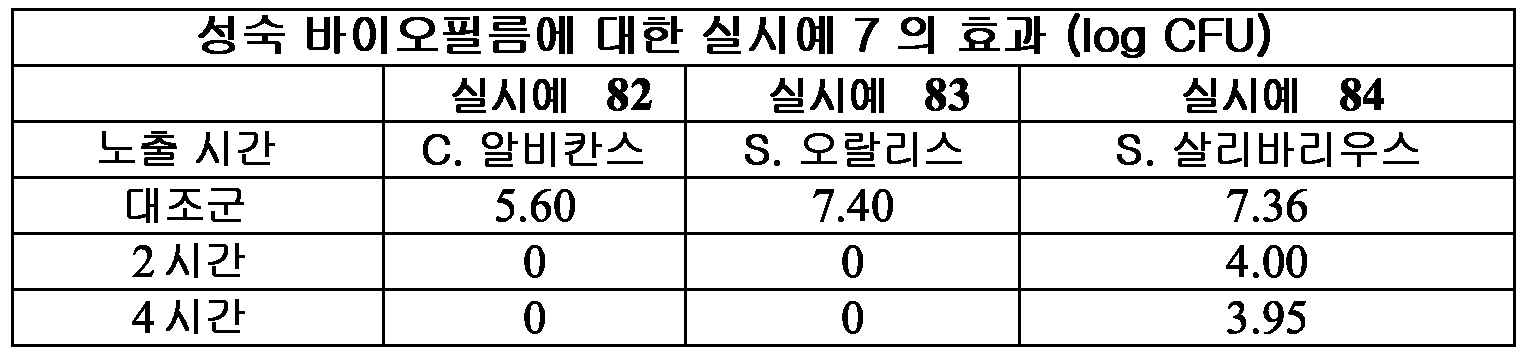
요약하여, 상기 결과는 실시예 15 가 박테리아 및 진균에 의해 형성된 바이오필름에 대한 강력한 활성을 갖는다는 것을 나타낸다. In summary, the results indicate that Example 15 has potent activity against biofilms formed by bacteria and fungi.
실시예Example 85-86 85-86
장벽-형성 조성물은 또한 바이러스에 대해 활성임Barrier-forming compositions are also active against viruses
호흡기 바이러스 (인플루엔자 바이러스 H1N1, 균주 2009/H1N1/infA) 및 인간 면역결핍 바이러스 (HIV) 를 포함하는 바이러스에 대한 장벽-형성 조성물의 활성을 측정하였다.The activity of barrier-forming compositions against viruses including respiratory virus (influenza virus H1N1, strain 2009 / H1N1 / infA) and human immunodeficiency virus (HIV) was measured.
장벽-형성 조성물은 인플루엔자 A 의 감염성을 저해한다.Barrier-forming compositions inhibit the infectivity of influenza A.
인플루엔자 바이러스의 감염성에 대한 장벽-형성 조성물의 효과를 평가하기 위해서, 코카 스파니엘 신장 (Madin Darby canine kidney) (MDCK) 세포를 감염 전 37℃ 에서 ≥90% 융합도로 성장시켰다. MDCK 세포는 인플루엔자 바이러스를 포함하는 검정에 일상적으로 사용된다.To assess the effect of the barrier-forming composition on the infectivity of influenza viruses, Coca Darni canine kidney (MDCK) cells were grown to ≧ 90% confluence at 37 ° C. prior to infection. MDCK cells are routinely used in assays involving influenza viruses.
실시예 85 에서 세포 단일층을 실시예 7 장벽-형성 조성물에 노출시켰다. 대조예 86 에서 세포층을 하기의 상이한 시간 동안 optiMEM (+P/S,+Lglu) 조직 배양 배지에 노출시켰다: (1) T1: 30 분 노출, (2) T2: 1 시간 노출, (3) T3: 2 시간 노출. 다음으로, 제형을 제거하고 세포 단일층을 인플루엔자 바이러스로 감염시켰다 (감염 다중도 (MOI) = 0.1). 미처리되거나 노출 직후 감염된 세포 (T0) 를 기준 대조군으로서 사용하였다. 감염된 세포를 이후 원심분리하고, 500 μL 의 성장 배지에 재현탁하고, 32.5℃ 에서 48 시간 동안 인큐베이션하였다. 면역형광 현미경 (FITC 표지 항-인플루엔자 항체 사용) 을 또한 사용하여 인플루엔자 바이러스가 포유동물 세포를 감염시키는 능력에 대한 실시예 7 장벽-형성 조성물의 효과를 평가하였다.In Example 85 the cell monolayer was exposed to the Example 7 barrier-forming composition. In Control Example 86 the cell layer was exposed to optiMEM (+ P / S, + Lglu) tissue culture medium for the following different times: (1) T1: 30 min exposure, (2) T2: 1 hour exposure, (3) T3 : 2 hours exposure. Next, the formulations were removed and the cell monolayers were infected with influenza virus (multiplicity of infection (MOI) = 0.1). Untreated or infected cells immediately after exposure (T0) were used as reference controls. Infected cells were then centrifuged, resuspended in 500 μL of growth medium and incubated at 32.5 ° C. for 48 hours. Immunofluorescence microscopy (using FITC labeled anti-influenza antibodies) was also used to assess the effect of the Example 7 barrier-forming composition on the ability of the influenza virus to infect mammalian cells.
도 18 은 인플루엔자-감염된 MDCK 세포 (실시예 85) (패널 A 및 C) 및 대조예 86 (패널 B 및 D) 의 세포변성 효과에 대한 실시예 7 의 효과를 나타낸다. 위상차 (A-B) 및 면역형광 현미경 (C-D) 으로부터 이미지를 얻었다. 세포변성 효과 (CPE) 의 확인은 제형-처리 세포에서 관찰되지 않았다. 미처리 세포는 중심점 라운딩 (focal rounding) 및 퇴행성 변화를 포함하는 전형적인 CPE 를 나타내었다.18 shows the effect of Example 7 on the cytopathic effect of influenza-infected MDCK cells (Example 85) (Panels A and C) and Control 86 (Panels B and D). Images were obtained from retardation (A-B) and immunofluorescence microscopy (C-D). Confirmation of cytopathic effect (CPE) was not observed in formulation-treated cells. Untreated cells exhibited typical CPE, including focal rounding and degenerative changes.
데이터는, 세포 단일층의 실시예 7 에 대한 30 분, 1 시간 또는 2 시간 동안의 노출로도 융합성이고 건강하게 남아 있다는 것을 나타내었다 (실시예 85). 반대로, 미처리 세포 및 감염 직전 처리된 세포 (TO) (대조예 86) 는 실질적인 세포변성 효과를 나타내었다. 도 18 패널 C 에서 볼 수 있듯이, 실시예 85 의 장벽-형성 조성물 처리 세포에서는 형광이 관찰되지 않은 한편, 실시예 86 의 미처리 세포는 형광을 나타내었다 (도 18 패널 D). The data showed that exposure to Example 7 of the cell monolayer for Example 7 remained confluent and healthy (Example 85). In contrast, untreated cells and treated cells immediately before infection (TO) (Control 86) showed substantial cytopathic effects. As can be seen in FIG. 18 panel C, no fluorescence was observed in the barrier-forming composition treated cells of Example 85, while the untreated cells of Example 86 showed fluorescence (FIG. 18 panel D).
실시예 85 및 86 에 상응하는 추가의 형광 현미경 이미지를 도 19 에서 나타낸다.Further fluorescence microscopy images corresponding to Examples 85 and 86 are shown in FIG. 19.
실시예Example 87 및 88 87 and 88
정량 PCR 을 사용하는 바이러스 총량 (viral load) 에 대한 장벽-형성 조성물의 활성 측정Activity measurement of barrier-forming composition against viral load using quantitative PCR
도 20 은 정량 PCR 에 의해 측정되는 바와 같은, 감염된 처리 세포 (실시예 87) 및 미처리 세포 (실시예 88) 에서의 인플루엔자 바이러스 수준을 나타낸다. 실시예 87 에서 세포를 실시예 7 로 처리하고, 대조예 88 에서 세포를 미처리한 채로 두었다. 이후 상청액을 수집하고 바이러스 존재에 대해 분석하였다.20 shows influenza virus levels in infected treated cells (Example 87) and untreated cells (Example 88) as measured by quantitative PCR. The cells were treated with Example 7 in Example 87 and the cells were left untreated in Control Example 88. Supernatants were then collected and analyzed for virus presence.
실시예 87 및 88 에서와 동일한 검정으로부터 세포 배양물 상청액을 수집하고 QIAamp Viral RNA Kit (QIAGEN, Valencia, CA) 를 사용하여 핵산을 추출하였다. 무작위 헥사머 프라이머 (Invitrogen Carlsbad, CA) 를 사용하여 각각의 표본에 대한 cDNA 라이브러리를 생성시켰다. 역전사 반응을 제조사 명세에 따라 M-MLV RT (Invitrogen, Carlsbad, CA) 를 사용하여 수행하였다. 정량 분석을 TaqMan Universal PCR Master Mix (Applied Biosystems, Branchburg, NJ), 2 μl 의 cDNA 샘플, 및 인플루엔자 매트릭스 유전자를 표적화하는 프라이머/프로브를 사용하여, StepOne Plus Taqman Real Time PCR (Applied Biosystems, Branchburg, NJ) 상에서 수행하였다. 참조 표준을 종래의 RT-PCR 에 의해 증폭된 인간 RNAse P 및 H1N1 매트릭스 유전자의 cDNA 단편을 사용하여 제조하고, 겔 정제하고 (QIAquick, Qiagen, Valencia, CA), 분광광도계 (Beckman Coulter, Brea, CA) 를 사용하여 정량하였다. Cell culture supernatants were collected from the same assays as in Examples 87 and 88 and nucleic acids were extracted using the QIAamp Viral RNA Kit (QIAGEN, Valencia, CA). Random hexamer primers (Invitrogen Carlsbad, Calif.) Were used to generate cDNA libraries for each sample. Reverse transcription reactions were performed using M-MLV RT (Invitrogen, Carlsbad, Calif.) According to manufacturer specifications. Quantitative analysis using a StepOne Plus Taqman Real Time PCR (Applied Biosystems, Branchburg, NJ) using TaqMan Universal PCR Master Mix (Applied Biosystems, Branchburg, NJ), 2 μl cDNA samples, and primers / probes targeting influenza matrix genes )). Reference standards were prepared using cDNA fragments of human RNAse P and H1N1 matrix genes amplified by conventional RT-PCR, gel purified (QIAquick, Qiagen, Valencia, CA), spectrophotometer (Beckman Coulter, Brea, CA) Quantification using
도 20 및 표 IV 에서 나타낸 바와 같이, 실시예 7 로 30 분 또는 60 분 동안 처리한 실시예 87 세포는 감염 후 48 시간에서 검출가능한 인플루엔자를 갖지 않았다. 또한, 실시예 7 로의 2 시간 처리로는 미처리 대조군 또는 감염 직전 처리한 것들에 비해 바이러스 총량이 6 배 감소하였다 (실시예 88). As shown in FIG. 20 and Table IV, Example 87 cells treated with Example 7 for 30 or 60 minutes did not have detectable influenza 48 hours after infection. In addition, the two-hour treatment with Example 7 resulted in a 6-fold reduction in total virus compared to untreated controls or those treated immediately prior to infection (Example 88).
표 table VIIIVIII

실시예Example 89-91 89-91
장벽-형성 조성물이 인플루엔자에 대해 직접적인 항바이러스 효과를 가짐Barrier-forming compositions have a direct antiviral effect against influenza
장벽-형성 조성물이 인플루엔자 바이러스에 대해 직접적인 항바이러스 활성을 갖는지 여부를 측정하기 위해서, 아프리카 녹색 원숭이 신장 (African Green Monkey Kidney) (CV-1) 세포 (90% 융합도로 24-웰 플레이트에서 성장시킴) 를, 실시예 7 로 전처리한 인플루엔자 바이러스로 감염시켰다. CV-1 세포는 바이러스의 진단 및 연구용으로 일상적으로 사용되는 매우 민감한 기질이다. African Green Monkey Kidney (CV-1) cells (grown in 24-well plates with 90% fusion) to determine whether the barrier-forming composition has direct antiviral activity against influenza virus Was infected with the influenza virus pretreated in Example 7. CV-1 cells are highly sensitive substrates used routinely for the diagnosis and study of viruses.
실시예 89-91 에서, 표준량의 인플루엔자 (0.1 MOI) 를 5 분 동안 실온에서 하기의 것으로 전처리하였다: (1) 실시예 7 (실시예 89 를 형성시키기 위함), (2) 대조예 6, CPC 는 갖지 않으나 보존제는 갖는 화합물 (실시예 90 을 형성시키기 위함), 및 (3) 대조예 5 위약 단독 (CPC 와 보존제를 갖지 않는 화합물) (실시예 91 을 형성시키기 위함). 5 분 인큐베이션 후, 처리 조성물이 희석되도록 optiMEM (+P/S,+Lglu) 으로 추가적인 동일 부피에 의해 바이러스/약물 믹스를 희석하였다.In Examples 89-91, standard amounts of influenza (0.1 MOI) were pretreated for 5 minutes at room temperature as follows: (1) Example 7 (to form Example 89), (2) Control Example 6, CPC Compounds with but not preservatives (to form Example 90), and (3)
실시예 89-91 에서, CV-1 세포를 상기 기재한 바와 같이 제조하였다. 실시예 89-91 처리 및 미처리 바이러스를 이후 상기 기재한 바와 같이 세포에 접종하였다.In Examples 89-91, CV-1 cells were prepared as described above. Examples 89-91 Treated and untreated viruses were then seeded into cells as described above.
인플루엔자 바이러스 총량을 상기 기재한 바와 같이 실시간 PCR 에 의해 측정하였다. 도 21 에서 나타낸 바와 같은 데이터는 항균제 CPC 를 함유하는 실시예 7 장벽-형성 조성물로 전처리된 인플루엔자 바이러스 (실시예 89) 에 대한 바이러스 총량이 장벽-형성 조성물 및/또는 보존제만을 함유하며 CPC 를 갖지 않는 것들 (실시예 90 및 91) 에 비해 유의하게 감소하였다는 것을 나타내었다. 실시예 7 로의 바이러스 전처리는 CPC 를 갖지 않는 제형에 비해 바이러스 카피에 있어서 유의한 감소를 나타내었다.The total influenza virus was measured by real time PCR as described above. The data as shown in FIG. 21 shows that the total amount of virus for influenza virus (Example 89) pretreated with the Example 7 barrier-forming composition containing the antimicrobial CPC contains only the barrier-forming composition and / or preservative and has no CPC. It was shown to be significantly reduced compared to those (Examples 90 and 91). Virus pretreatment with Example 7 showed a significant reduction in virus copy compared to formulations without CPC.
이들 결과는, 실시예 7 장벽-형성 조성물이 실시예 5 및 6 에 고유하지 않은 인플루엔자 바이러스에 대한 직접적인 항바이러스 활성을 갖는다는 것을 입증한다.These results demonstrate that the Example 7 barrier-forming composition has direct antiviral activity against influenza viruses that are not native to Examples 5 and 6.
실시예Example 92 및 93 92 and 93
실시예 92 및 93 에서, 장벽-형성 조성물의 인플루엔자 A (2009/H1N1/infA) 의 감염성을 저해하는 능력을 시험하였다. 아프리카 녹색 원숭이 신장 (CV-1) 세포를 90% 융합도로 24-웰 플레이트에서 성장시켰다. 그 다음으로, 장벽-형성 조성물, 실시예 7 을 실시예 92 에서 세포에 적용하였다 (20% 실시예 7, 80%OptiMeM, 작용 CPC 농도 0.02%). 각각의 시간 지점을 대조예 93 (장벽-형성 조성물 미적용, 100% OptiMeM) 과 매치시켰다. 장벽-형성 조성물을 30 분 동안 표면에 체류시킨 후, 세포 단일층으로부터 제거하였다. 세포를 멸균 optiMEM (+PfS,+Lglu) 으로 2 회 충분히 세척하였다. 인플루엔자를 T0 에서 T+6 시간까지 30 분 간격으로 MOi = 0.1 에서 접종하였다. 감염 후, 세포를 2200 rpm X 30 분에서 원심분리하고 500 μl 의 optiMEM (+P/S, +Lglu, 2 μg/ml 트립신 (sigma-Aldrich, St Louis, MO)) 을 가하였다. 감염된 세포를 32.5℃ 에서 96 시간 동안 5% CO2 에서 성장시켰다. 인플루엔자 바이러스 총량을 실시간 PCR 에 의해 측정하였다.In Examples 92 and 93, the ability to inhibit the infectivity of influenza A (2009 / H1N1 / infA) of the barrier-forming composition was tested. African green monkey kidney (CV-1) cells were grown in 24-well plates with 90% fusion. Next, a barrier-forming composition, Example 7, was applied to the cells in Example 92 (20% Example 7, 80% OptiMeM, working CPC concentration 0.02%). Each time point was matched with Control Example 93 (without barrier-forming composition, 100% OptiMeM). The barrier-forming composition was left on the surface for 30 minutes and then removed from the cell monolayer. The cells were washed sufficiently twice with sterile optiMEM (+ PfS, + Lglu). Influenza was inoculated at MOi = 0.1 at 30 min intervals from T0 to T + 6 hours. After infection, cells were centrifuged at 2200
도 22 에서 나타낸 바와 같이, 글리세린-잔탄 검 제형으로 숙주 단일층을 전처리하여, 미처리 대조군에 비해 바이러스 감염이 84.93% 까지 저해되었다. 장벽-형성 조성물의 제거에도 불구하고 바이러스 감염의 저해가 숙주 세포에서 관찰되었다는 점으로 인해, 상기 장벽-형성 조성물이 숙주 세포 상에 보호성 장벽을 형성하여, 6 시간 이상 바이러스 침입을 방지하였다는 것이 입증된다.As shown in FIG. 22, pretreatment of host monolayers with glycerin-xanthan gum formulation inhibited viral infection by 84.93% compared to untreated controls. Due to the fact that inhibition of viral infection was observed in the host cell despite removal of the barrier-forming composition, it was found that the barrier-forming composition formed a protective barrier on the host cell, preventing virus invasion for more than 6 hours. Proven.
도 1 을 감염 저해를 설명하는 가능한 메커니즘인 것으로 나타낼 수 있다.1 can be shown as a possible mechanism for explaining infection inhibition.
실시예Example 94-96 94-96
장벽-형성 조성물이 HIV 에 대해 활성을 나타냄 Barrier-forming Compositions Are Active Against HIV
실시예 94-96 에서는 장벽-형성 조성물이 HIV 에 대한 활성을 갖는지 여부를 측정하였다. 숙주 MT 포유동물 세포를 RPMI/10%FBS/PS 중에서 15,000 세포/웰의 밀도로 96-웰 둥근 바닥 플레이트에 플레이팅하였다. 다음 날 (제 2 일), 바이러스를 대조예 5 (실시예 94 를 형성시키기 위함), 대조예 6 (실시예 95 를 형성시키기 위함), 또는 실시예 7 (실시예 96 을 형성시키기 위함) 로 5 분 동안 전처리하고 세포에 추가하였다. 제형에 노출시키고 24 시간 후, MT (마카크 (macaque)) 포유동물 세포를 인산완충식염수 (PBS) 로 3 회 세척하고 신선한 배지로 교체하였다. 상청액 (10 μL) 을 제 1, 2, 5, 6, 7 및 9 일에 후-처리하여 수집하고, 역전사효소 (RT) 활성에 의해 바이러스 총량을 측정하였다. 도 23 은 9 일간의 기간에 걸친 실시예 72-74 각각에 대한 mL 당 바이러스 카피의 그래프를 나타낸다.In Examples 94-96 it was determined whether the barrier-forming composition had activity against HIV. Host MT mammalian cells were plated in 96-well round bottom plates at a density of 15,000 cells / well in RPMI / 10% FBS / PS. The next day (day 2), the virus was transferred to Control Example 5 (to form Example 94), Control 6 (to form Example 95), or Example 7 (to form Example 96). Pretreated for 5 minutes and added to the cells. After 24 hours of exposure to the formulation, MT (macaque) mammalian cells were washed three times with phosphate buffered saline (PBS) and replaced with fresh medium. Supernatants (10 μL) were collected by post-treatment on
상기 결과는 실시예 96 에서의 실시예 7 이 모든 시점에서 모니터링한 후-처리에서 항-HIV 활성을 나타내었다는 것을 보여주었다.The results showed that Example 7 in Example 96 showed anti-HIV activity in post-treatment monitored at all time points.
실시예 94 및 95 에서 CPC 및/또는 보존제를 갖지 않는 대조예 5 또는 대조예 6 은 단지 최소의 항-HIV 활성을 나타내었다.In Examples 94 and 95,
요약하여, 본 발견은 CPC 를 함유하는 장벽-형성 조성물 실시예 7 이 HIV 에 대해 장기 지속성 항바이러스 활성을 나타낸다는 것을 입증한다.In summary, the present findings demonstrate that barrier-forming composition Example 7 containing CPC exhibits long-lasting antiviral activity against HIV.
실시예Example 97 97
대표적인 유기체 바이러스 병변은 상이한 점막 조직에서 중요한 감염이다. 실시예 97 에서 실험을 수행하여 장벽-형성 조성물이 통상적인 구강 엡스타인-바 바이러스 (EBV) 에 대해 활성을 나타내는지 여부를 측정하였다. 웨스턴 블롯팅을 사용하여 실시예 8 장벽-형성 조성물이 용해성 바이러스 단백질 EAD 를 분해하는 능력 (바이러스 복제 저해를 나타냄) 을 평가하였다. Representative organism viral lesions are important infections in different mucosal tissues. Experiments in Example 97 were performed to determine whether the barrier-forming composition was active against conventional oral Epstein-Barr virus (EBV). Western blotting was used to assess the ability of the Example 8 barrier-forming composition to degrade soluble viral protein EAD (indicating inhibition of viral replication).
실시예 97 에서, EBV-감염된 위 상피 세포를 실시예 8 의 상이한 희석물 (1:16, 1:32 및 1:64) 에 노출시키고, EAD 단백질의 존재를 특정 항체를 사용하여 검출하였다. 세포 β-액틴의 존재를 상피 세포 무결성의 표시자로서 사용하였다. 도 24 에서 나타낸 바와 같이, 실시예 8 의 1:64 희석물은 세포 액틴에 영향을 주지 않고 EAD 를 분해하였다. 이들 결과는 실시예 8 이 바이러스 복제를 특히 저해하며, 그 자체로서, 효과적인 항바이러스제이며 바이러스 감염 예방에 유용하다는 것을 입증한다.In Example 97, EBV-infected gastric epithelial cells were exposed to different dilutions of Example 8 (1:16, 1:32 and 1:64) and the presence of EAD protein was detected using specific antibodies. The presence of cell β-actin was used as an indicator of epithelial cell integrity. As shown in FIG. 24, the 1:64 dilution of Example 8 degraded EAD without affecting cell actin. These results demonstrate that Example 8 specifically inhibits viral replication and, by itself, is an effective antiviral agent and useful for preventing viral infections.
실시예Example 98-100 98-100
항균 장벽 대 시판 구강 청결제 제품의 지속기간Antimicrobial Barrier Versus Duration of Commercial Mouthwash Products
장벽-형성 조성물이 항균 활성을 유지할 수 있는 지속기간을 측정하기 위해서, 박테리아 및 진균을 웰 내에서 실시예 7 의 장벽-형성 조성물로 처리한 실시예 2 의 EHOM 및 웰 내에서 비교용 시판 제품으로 처리한 실시예 2 의 EHOM 에 2 분 동안 노출시켰다. 박테리아 및 진균 미생물을 대조군 미처리 EHOM (실시예 98) 및 처리 EHOM (실시예 99 및 비교예 100) 의 상부에 오버레이하였다. 그 다음으로, 잔류 (통과) 용액을 하부 웰 (EHOM 모델의 하부 챔버) 로부터 제거하고 한천 배지 플레이트에 스프레딩하였다. 도 7 은 추가적인 명확성을 위한 이러한 시험 방법을 나타낸다. 이들 플레이트를 37℃ 에서 인큐베이션하고, 24 시간 후 성장하는 미생물 세포의 수 (콜로니 형성 단위, CFU) 를 계수하였다.To determine the duration that the barrier-forming composition can maintain antimicrobial activity, the bacteria and fungi were treated in the wells with the barrier-forming composition of Example 7 as a comparative commercial product in the EHOM of Example 2 and wells. The treated EHOM of Example 2 was exposed for 2 minutes. Bacteria and fungal microorganisms were overlaid on top of control untreated EHOM (Example 98) and treated EHOM (Example 99 and Comparative Example 100). The residual (pass) solution was then removed from the lower wells (lower chamber of the EHOM model) and spread on agar medium plates. 7 shows this test method for further clarity. These plates were incubated at 37 ° C. and the number of microbial cells growing after 24 hours (colony forming units, CFU) was counted.
대조예 98 에서 미처리 EHOM 을 시험하였다. 실시예 99 에서 S. 미티스 박테리아를 상기 기재한 바와 같이 장벽-형성 조성물 상에 오버레이하였다. 실시예 100 은 S. 미티스 박테리아에 대한 시판 LISTERINE (에탄올 (26.9%), 멘톨, 티몰, 메틸 살리실레이트 및 유칼립톨 함유) 의 활성을 나타내는 비교예이다. 하기 표 IX 에서 결과를 나타낸다.In Control Example 98 untreated EHOM was tested. In Example 99 S. mytis bacteria were overlaid on the barrier-forming composition as described above. Example 100 is a comparative example showing the activity of commercial LISTERINE (containing ethanol (26.9%), menthol, thymol, methyl salicylate and eucalyptol) against S. mytis bacteria. The results are shown in Table IX below.
표 table IXIX

실시예Example 101-103 101-103
실시예 101-103 에서, 칸디다 알비칸스 진균을 상기 기재한 바와 같이 장벽-형성 조성물에 대해 시험한 것을 제외하고는 실시예 98-100 의 동일한 절차를 수행하였다. 하기 표 X 에서 결과를 나타낸다. 실시예 103 은 시판 LISTERINE 의 활성을 나타내는 비교예이다.In Examples 101-103, the same procedure of Example 98-100 was performed except that Candida albicans fungi were tested against the barrier-forming composition as described above. The results are shown in Table X below. Example 103 is a comparative example showing the activity of commercial LISTERINE.
표 XTable X

상기 데이터는 실시예 7 장벽-형성 조성물이 24 시간 이하 동안 활성을 유지하였다는 것을 추가로 나타내었다. 종합하여 볼 때, 이들 결과는 LISTERINE 와는 달리, 실시예 7 장벽-형성 조성물이 24 시간 이하 동안 EHOM 조직 상에 미손상 장벽을 계속하여 유지하였다는 것을 나타내었다.The data further indicated that the Example 7 barrier-forming composition remained active for up to 24 hours. Taken together, these results showed that, unlike LISTERINE, the Example 7 barrier-forming composition continued to maintain an intact barrier on EHOM tissue for up to 24 hours.
실시예Example 104-153 104-153
미생물 통과를 방지하는데 있어서 효과적인 장벽을 형성할 수 있는 글리세린 및 잔탄 검 농도의 추가 예를 확인하기 위해서, 상이한 농도의 잔탄 검 및 보습제 글리세린을 (5% - 95% 글리세린; 0.005% - 0.5% 잔탄 검) 개별적으로 및 조합으로, 체외 필터 삽입-기반 장벽 모델을 사용하여 시험하였다. 도 33 은 실시예 104-153 에 사용한 일반적 시험 방법을 나타낸다.To identify further examples of glycerin and xanthan gum concentrations that can form an effective barrier in preventing microbial passage, different concentrations of xanthan gum and moisturizing glycerin (5%-95% glycerin; 0.005%-0.5% xanthan gum) Separately and in combination, using an in vitro filter insertion-based barrier model. 33 shows the general test method used in Examples 104-153.
3 μm 및 8 μm 직경 기공 크기의 필터 삽입물을 각각 박테리아 (스트렙토코쿠스 살리바리우스) 및 진균 (칸디다 알비칸스) 의 통과 시험에 사용하였다. 글리세린 또는 잔탄 검 또는 그의 조합 (100 μL 분취액) 을 필터 표면 상에 오버레이하여 장벽을 형성시켰다. 상기 필터는 직경이 24 mm 였다. 따라서, 필름은 필터 상에서 대략 0.01 mm 의 두께를 가지며, 이는 치료적 유효량으로 구강에 적용시 조성물 필름의 두께 범위 내의 값을 모방한다. 다음으로, 박테리아 또는 진균 중 어느 하나의 5 x 10⁴ 세포를 필터 삽입물 내의 형성된 장벽 상부에 적용하였다. 다음으로, 이들 필터 삽입물을 6-웰 플레이트 내에 한천 배지 (박테리아용으로는 뇌 심장 침출액 (Brain Heart Infusion) (BHI) 배지, 진균용으로는 사부로 덱스트로오스 (SD) 배지) 표면 상에 두었다. 상기 플레이트를 필터 삽입물과 함께 24 시간 동안 37℃ 에서 밤새 인큐베이션하였다.Filter inserts of 3 μm and 8 μm diameter pore sizes were used for the passage test of bacteria (Streptococcus salivarius) and fungi (Candida albicans), respectively. Glycerin or xanthan gum or combinations thereof (100 μL aliquots) were overlaid on the filter surface to form a barrier. The filter was 24 mm in diameter. Thus, the film has a thickness of approximately 0.01 mm on the filter, which mimics a value within the thickness range of the composition film when applied to the oral cavity in a therapeutically effective amount. Next, 5 × 10 4 cells of either bacteria or fungi were applied on top of the formed barrier in the filter insert. Next, these filter inserts were placed on the surface of the agar medium (Brain Heart Infusion (BHI) medium for bacteria and Saburro dextrose (SD) medium for fungi) in 6-well plates. The plates were incubated with the filter inserts for 24 hours at 37 ° C. overnight.
플레이트를 한천 배지 뿐 아니라 필터 삽입물에서의 박테리아 또는 진균 성장 CFU (콜로니 형성 단위) 의 존재에 대해 모니터링하였다. 필터 삽입물에서만의 미생물 성장 (한천 배지에서는 아님) 으로, 효과적 장벽이 필터 상에 형성되어 미생물 통과를 방지하였다는 것이 입증되었다. 반대로, 필터 삽입물을 둘러싼 한천 배지에서의 성장은 시험 작용제가 효과적 장벽을 형성하는데 실패하여 유기체가 필터를 통과하게끔 하였다는 것을 제시하였다. Plates were monitored for the presence of bacterial or fungal growth CFU (colony forming units) in agar media as well as filter inserts. Microbial growth (not in agar medium) only in filter inserts has proven that an effective barrier has been formed on the filter to prevent microbial passage. In contrast, growth in the agar medium surrounding the filter insert suggested that the test agent failed to form an effective barrier, causing the organism to pass through the filter.
하기 표 XI 에 나타낸 결과는, 글리세린이 단독으로 시험한 경우 55% 이상의 농도에서 장벽을 형성할 수 있었다는 것을 나타내었다. 반대로, 잔탄 검 단독은 어떠한 시험 농도에서도 (0.005% 내지 0.4% 범위) 장벽을 형성하지 않았다. 그러나 0.01% 잔탄 검과 조합된 경우, 장벽이 글리세린 농도 7%, 45%, 55% 및 65% 에서 형성되었다는 것이 관찰되었다. 또한 0.4% 잔탄 검과 글리세린 농도 7%, 15%, 25%, 35%, 45%, 55% 및 65% 의 조합이 또한 장벽을 형성시켰다. 그러므로, 글리세린 및 잔탄 검의 특정 조합이 체외 필터 삽입-기반 모델에서의 미생물 통과를 방지하는 장벽을 형성시킬 수 있다는 것이 확인되었다.The results shown in Table XI below show that glycerin was able to form barriers at concentrations of at least 55% when tested alone. In contrast, xanthan gum alone did not form a barrier at any test concentration (range from 0.005% to 0.4%). However, when combined with 0.01% xanthan gum, it was observed that barriers were formed at glycerin concentrations of 7%, 45%, 55% and 65%. Also a combination of 0.4% xanthan gum and glycerin concentrations of 7%, 15%, 25%, 35%, 45%, 55% and 65% also formed a barrier. Therefore, it has been found that certain combinations of glycerin and xanthan gum can form barriers that prevent microbial passage in an in vitro filter insertion-based model.
표 table XIXI
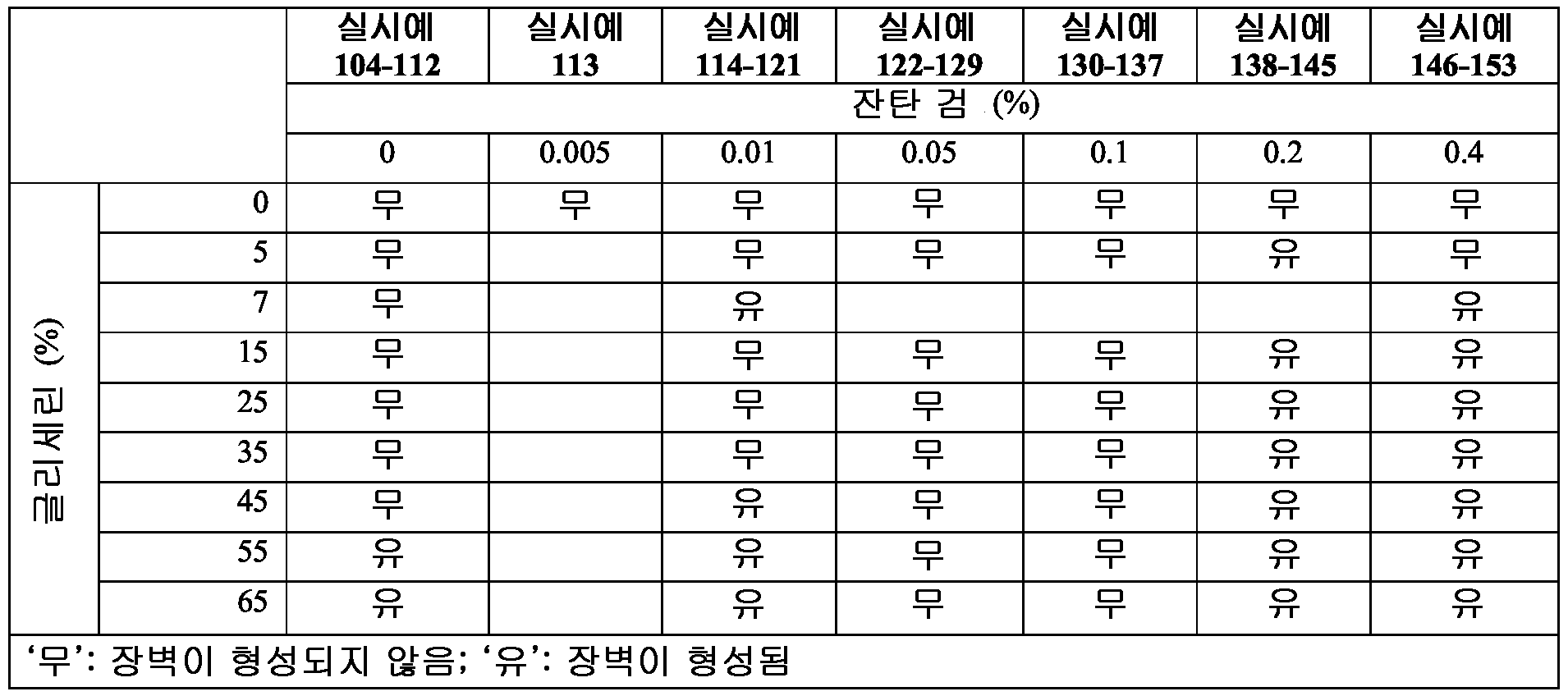
필터 삽입물 상에 형성된 실시예 104-153 의 조성물에 의해 보유된 미생물 세포는 장벽에 의해 포획되고, 생존성이었으며, 따라서 형성된 장벽이 항균제 없이 고유 항균 특성을 갖지 않는다는 것이 입증되었다. 즉, 장벽에 의해 보유된 미생물은 여전히 활성이었으며 감염 위험성을 제기할 수 있었다 (예를 들어, 장벽이 그의 무결성을 잃은 후 또는 마멸에 의해 장벽으로부터 미생물이 벗어나는 경우).The microbial cells retained by the compositions of Examples 104-153 formed on the filter inserts were captured by the barrier and were viable, thus demonstrating that the barrier formed had no inherent antimicrobial properties without the antimicrobial agent. That is, the microorganisms retained by the barrier were still active and could pose a risk of infection (for example, after the barrier lost its integrity or when the microorganism was released from the barrier by abrasion).
실시예Example 154-157 154-157
EHOM 샘플에 대한 상기 실시예에 더해, 추가의 시험을 수행하여 장벽-형성 조성물이 (a) 숙주 조직을 손상시키지 않으며, (b) 인간 점막 조직 내로의 미생물 침입을 방지할 수 있다는 것을 추가로 입증하였다. 이들 기준을 실시예 104-153 의 체외 결과를 기반으로 하여 선택한 2 가지 대표적 조합 (글리세린:잔탄 검; 7%:0.01% 및 35%:0.4%) 에 대해 시험하였는데, 그 이유는 이들이 장벽을 성공적으로 형성시켰기 때문이다. 이들 2 가지 조합을 포함하는 제형을 점막 내벽을 모방하는 실시예 2 의 EHOM 을 사용하여 시험하였다.In addition to the above examples for EHOM samples, further testing is performed to further demonstrate that the barrier-forming composition can (a) not damage host tissue, and (b) prevent microbial invasion into human mucosal tissue. It was. These criteria were tested against two representative combinations (Glycerin: Xanthan Gum; 7%: 0.01% and 35%: 0.4%) selected based on the in vitro results of Examples 104-153, because they successfully tested the barrier. Because it was formed. Formulations comprising these two combinations were tested using the EHOM of Example 2, which mimics the mucosal lining.
EHOM 을 보통의 식염수 [0.9%] 중의 1% (실시예 154 및 155) 또는 5% (실시예 156 및 157) 희석물 중 어느 하나에서의 실시예 3 및 4 의 다양한 제형으로 약 2 분 동안 처리하여 실시예 154-157 을 형성시켰다. 각각의 EHOM 을 시험 제형 중 하나의 300 μL 로 덮고, 멸균 후드 하에 10 분 동안 실온 (25℃) 에서 두었다. 접촉 기간 완료시, 조직을 배양 배지로 2 회 세척하여 제형을 제거하였다.EHOM was treated with various formulations of Examples 3 and 4 in either 1% (Examples 154 and 155) or 5% (Examples 156 and 157) dilution in normal saline [0.9%] for about 2 minutes. Example 154-157 was formed. Each EHOM was covered with 300 μL of one of the test formulations and left at room temperature (25 ° C.) for 10 minutes under sterile hood. At the completion of the contact period, the tissue was washed twice with culture medium to remove the formulation.
실시예Example 158 158
실시예 3 및 4 로 처리한 후, EHOM 을 가능한 육안으로 보이는 조직 손상 (구멍의 존재) 에 대해 검사하였다. 조직 손상을 또한 조직학적 분석에 의해 검사하였다. 이러한 목적으로, 생검물을 각각의 EHOM 으로부터 채취하고 4% 파라포름알데히드 용액으로 고정시킨 후 파라핀 중에 포매하였다. 얇은 박편 (4 μm) 을 에오신-헤마톡실린으로 염색하고 50%-글리세린 봉입 배지 중에서 커버슬립으로 봉입하고, 광학 현미경을 통해 관찰하고, 사진 찍었다. 처리한 EHOM 및 대조군 미처리 EHOM 을 육안으로 보이는 조직 손상 및 구조 변화에 대해 검사하였다. 시험한 5 개 상이한 EHOM 에서, 500 배 확대율에서, 미처리 또는 처리 조직에서는 가시적 손상 (구멍) 이 존재하지 않았다.After treatment with Examples 3 and 4, EHOM was examined for possible visible tissue damage (presence of holes). Tissue damage was also examined by histological analysis. For this purpose, biopsies were taken from each EHOM, fixed with 4% paraformaldehyde solution and embedded in paraffin. Thin flakes (4 μm) were stained with Eosin-hematoxylin and sealed with coverslips in 50% -glycerine inclusion medium, observed through an optical microscope, and photographed. Treated EHOM and control untreated EHOM were examined for visible tissue damage and structural changes. In the five different EHOMs tested, at 500-fold magnification, there was no visible damage (pores) in the untreated or treated tissues.
실시예Example 159 159
숙주 세포에 손상을 일으키는 장벽-형성 조성물의 평가 (세포독성 검정) Evaluation of Barrier-Forming Compositions That Cause Damage to Host Cells (Cytotoxicity Assay)
장벽-형성 조성물의 실시예 3-7 의 5% 희석물로 처리한 실시예 156 및 157 EHOM 을 C. 알비칸스 또는 S. 무탄스 중 어느 하나의 1 x 10⁶ 세포로 100 μl 의 부피로 오버레이하였다. EHOM 을 공기-액체 배양 플레이트 상에 두고 24 시간 동안 5% CO2 습한 분위기에서 37℃ 에서 인큐베이션하였다. 상기 인큐베이션 기간 후, 배양 상청액의 분취액을 수집하고 락테이트 탈수소효소 (LDH) 세포독성 검정 (Promega, Madison, WI, USA) 을 제조사 지시사항에 따라 수행하였다. 50 μl 의 각 상청액을 멸균 96-웰 평평 바닥 플레이트에 옮겼다. 각 웰에 50 μl 의 재구성된 기질 믹스를 보충하고, 상기 플레이트를 30 분 동안 실온에서 암환경 하에 인큐베이션하였다. 반응을 정지시키기 위해서, 50 μl 의 정지 용액을 각각의 웰에 추가하였다. 그 다음으로 100 μl 의 혼합물을 96-웰 평평 바닥 플레이트에 옮기고, 흡광도를 490 nm 에서 X-Mark 마이크로플레이트 분광광도계 (Bio-Rad, Mississauga, ON, Canada) 로 판독하였다. LDH 를 LDH 세포독성 검정을 사용하여 평가하였다. 데이터는 평균 ± SD 이다. 실시예 조성물로 처리한 EHOM 과 미처리 EHOM 사이에 유의한 차이는 없었으며, 미감염된 EHOM 대조군을 기록하였다. 도 25 는 미처리 대조예, 및 실시예 5, 6 및 7 로 처리한 것들에 대한 이들 시험 결과를 도표로 나타낸다. 유사한 결과를 실시예 3 및 4 로 처리한 EHOM 에 대해 얻었다. 시험한 실시예 모두 점막의 무결성을 유지하였다.Example 156 and 157 EHOMs treated with 5% dilution of Example 3-7 of the barrier-forming composition were overlaid with a volume of 100 μl with 1 × 10 6 cells of either C. albicans or S. mutans. It was. EHOM was placed on an air-liquid culture plate and incubated at 37 ° C. in a 5% CO 2 wet atmosphere for 24 hours. After the incubation period, aliquots of the culture supernatants were collected and lactate dehydrogenase (LDH) cytotoxicity assays (Promega, Madison, WI, USA) were performed according to manufacturer's instructions. 50 μl of each supernatant was transferred to a sterile 96-well flat bottom plate. Each well was supplemented with 50 μl of reconstituted substrate mix and the plates were incubated for 30 minutes at room temperature under dark. To stop the reaction, 50 μl of stop solution was added to each well. 100 μl of the mixture was then transferred to a 96-well flat bottom plate and the absorbance was read at 490 nm with an X-Mark microplate spectrophotometer (Bio-Rad, Mississauga, ON, Canada). LDH was assessed using LDH cytotoxicity assay. Data is mean ± SD. There was no significant difference between the EHOM treated with the Example composition and the untreated EHOM, and an uninfected EHOM control was recorded. FIG. 25 graphically shows the results of these tests on untreated controls and those treated with Examples 5, 6, and 7. Similar results were obtained for EHOM treated with Examples 3 and 4. All the examples tested maintained the integrity of the mucosa.
실시예Example 160 160
잇몸 세포 성장/이동에 대한 장벽-형성 조성물 제형의 효과Effect of Barrier-Forming Composition Formulation on Gum Cell Growth / Migration
상처 복구 검정을 수행하였다. 간략하게는, 구강 (잇몸) 상피 세포 (1x10⁴) 및 섬유아세포 (1x10³) 를 6-웰 플레이트의 벽에 시딩하고 적절한 배양 배지에서 성장시켰다. 융합시, 200 μL 피펫 팁을 사용하여 각 웰의 융합 단일층에 상처를 만들었다. 배양물을 이후 실시예 3 및 4 의 1 및 5 중량% 희석물에 약 2 분 동안 노출시켰다. 노출 후, 제형을 따뜻한 멸균 배양 배지로 2 회 세척한 후, 세포 배양물을 섬유아세포에 대해서는 DEM 으로, 상피 세포에 대해서는 DMEH 로 오버레이하고, 6 및 24 시간 동안 5% CO2 습한 분위기에서 37℃ 에서 배양하였다. 상처 복구/세포 이동을 광학 현미경을 사용하여 확인하고, 디지털 사진을 찍었다 (Nikon, Coolpix 950). 도 26 은 상처 직후 (패널 A), 약 6 시간 후 (패널 D), 그리고 약 24 시간 후 (패널 E) 실시예 3 (5% 희석물) 으로 10 분 동안 처리한 손상된 구강 상피 세포 배양물의 대표적 사진을 나타낸다. 패널 B 및 C 는 각각 약 6 시간 및 약 24 시간 후 구강 상피 세포의 미처리 대조군 융합 배양물 상의 동일한 상처를 나타낸다. 유사한 결과가 실시예 4 로 처리한 세포 배양물에 대해 관찰되었다.Wound repair assays were performed. Briefly, oral (gum) epithelial cells (1 × 10 4 ) and fibroblasts (1 × 10 3 ) were seeded on the walls of 6-well plates and grown in appropriate culture medium. Upon fusion, wounds were made to the fusion monolayer of each well using a 200 μL pipette tip. The culture was then exposed to the 1 and 5 wt% dilutions of Examples 3 and 4 for about 2 minutes. After exposure, the formulations were washed twice with warm sterile culture medium, then the cell cultures were overlaid with DEM for fibroblasts and DMEH for epithelial cells and 37 ° C. in 5% CO 2 wet atmosphere for 6 and 24 hours. Incubated at. Wound repair / cell migration was confirmed using an optical microscope and digital pictures were taken (Nikon, Coolpix 950). FIG. 26 is representative of injured oral epithelial cell culture treated for 10 minutes immediately after wound (Panel A), after about 6 hours (Panel D), and after about 24 hours (Panel E) Example 3 (5% dilution) Represents a picture. Panels B and C show the same wounds on untreated control fusion cultures of oral epithelial cells after about 6 hours and about 24 hours, respectively. Similar results were observed for cell cultures treated with Example 4.
상처 봉합 % (세포 성장/이동) 를 상기 언급한 식 I 을 사용하여 제형에 노출되기 전 및 후의 상대적 상처 부위를 비교함으로써 계산하였다.Wound closure% (cell growth / migration) was calculated by comparing the relative wound area before and after exposure to the formulation using Formula I mentioned above.
처리 및 미처리 배양물 모두에서 상피 세포는 작고 입방형이었다. 유사한 결과가 섬유아세포를 사용하는 스크래치 상처 모델에 대해 관찰되었다 (데이터는 나타내지 않음). 종합하여 볼 때, 상기 데이터는 실시예 조성물이 독성이 아니며 세포 성장/이동 및 상처 치유에 부정적으로 영향을 주지 않는다는 것을 나타낸다.Epithelial cells were small and cubic in both treated and untreated cultures. Similar results were observed for the scratch wound model using fibroblasts (data not shown). Taken together, the data indicate that the example composition is not toxic and does not negatively affect cell growth / migration and wound healing.
실시예Example 161 161
글리세린-잔탄 검 제형이 인간 구강 점막 상에 코팅을 형성함Glycerin-Xanthan Gum Formulation Forms Coatings on Human Oral Mucosa
글리세린-잔탄 검 제형이 인간 구강 점막 상에 코팅을 형성할 수 있는지 여부를 측정하기 위해서, 마커 염료로서 젠티안 바이올렛 (GV) 을 사용하여 실시예 7 제형을 스파이킹 (spiking) 하였다. 스파이킹한 생성물 (750 μL) 을 인간 자원자의 구강에 스프레이하였다. 적용 후, 구강을 염색에 대해 검사하고, 디지털 카메라를 사용하여 이미지를 캡쳐하였다. 도 27 에서 나타낸 바와 같이, 상기 제형은 볼 및 혀의 등쪽/배쪽 표면 모두를 염색시켰다.To determine whether the glycerin-xanthan gum formulation could form a coating on the human oral mucosa, the Example 7 formulation was spiked using Gentian Violet (GV) as the marker dye. The spiked product (750 μL) was sprayed into the mouth of a human volunteer. After application, the oral cavity was examined for staining and an image was captured using a digital camera. As shown in FIG. 27, the formulation stained both the dorsal / dorsal surface of the cheek and tongue.
실시예Example 162 및 163 162 and 163
미생물의 장벽-형성 조성물에 대한 노출이 세포 성장을 저해함: 간헐 촬영 현미경Exposure to Microbial Barrier-forming Compositions Inhibits Cell Growth: Intermittent Microscopy
장벽-형성 조성물이 미생물에 대해 활성을 나타내는 지속기간 및 저해 활성을 측정하기 위해서, 미처리 박테리아 및 진균과 비교하여 장벽-형성 조성물에 노출된 세포에 대해 간헐 촬영 분석을 수행하였다.In order to determine the duration and inhibitory activity for which the barrier-forming composition is active against microorganisms, intermittent imaging analyzes were performed on cells exposed to the barrier-forming composition as compared to untreated bacteria and fungi.
실시예 162 에서, S. 무탄스 미생물 세포를 실시예 7 에 1 분 동안 노출시키고, 세척하여 임의의 잔류 작용제를 제거하고, 신선한 성장 배지를 함유하는 페트리 디쉬에서 성장시켰다. 37℃ 에서의 유기체 성장을 6 시간 동안 모니터링하고, 현미경에 연결된 카메라를 사용하여 6 시간 인큐베이션 기간에 걸쳐 20 분 마다 현미경 사진을 찍었다.In Example 162, S. mutans microbial cells were exposed to Example 7 for 1 minute, washed to remove any residual agent, and grown in Petri dishes containing fresh growth medium. Organic growth at 37 ° C. was monitored for 6 hours, and micrographs were taken every 20 minutes over a 6 hour incubation period using a camera connected to the microscope.
대조예 163 에서는 미처리 세포를 사용하여 동일한 절차를 수행하였다.In Comparative Example 163 the same procedure was performed using untreated cells.
도 28 에서 나타낸 바와 같이, 6 시간에 세포가 융합에 도달한 미처리 박테리아와 반대로, 실시예 7 장벽-형성 조성물로 처리한 미생물은 노출 후 동일한 기간 동안 재성장하는데 실패하였다. 유사하게, 칸디다 세포의 실시예 7 장벽-형성 조성물에 대한 노출은 인큐베이션 기간 동안 성장을 완전히 저해하였다 (데이터는 나타내지 않음). As shown in FIG. 28, the microorganisms treated with the Example 7 barrier-forming composition failed to regrow for the same period after exposure as opposed to the untreated bacteria whose cells reached fusion at 6 hours. Similarly, exposure of Candida cells to the Example 7 barrier-forming composition completely inhibited growth during the incubation period (data not shown).
이들 결과로, 장벽-형성 조성물이 장기적인 항균 활성을 갖는다는 것이 추가로 확인되었다. As a result of these, it was further confirmed that the barrier-forming composition has long-term antimicrobial activity.
실시예Example 164-166 164-166
체내 연구: 장벽-형성 조성물 (실시예 7) 은 인간에서의 구강 미생물 총량을 감소시킴: 단기간 및 장기간 활성In vivo studies: barrier-forming compositions (Example 7) reduce the total amount of oral microorganisms in humans: short and long term activity
단기간 활성Short term active
실시예 7 의 활성 지속기간을, 구강의 미생물 부하에 대한 단일 적용의 효과를 평가함으로써 건강한 개인에서 측정하였다. 실시예 164-166 에서, 3 명의 건강한 개인 (18 세 이상, 건강한 구강을 가짐) 을 사전 동의로 등록시키고, 그들의 볼에 실시예 7 의 조성물을 단일 적용하는 것을 요청하였다. 단일 적용을 부피에 있어서 각 0.25 ml 의 3 회 스프레이로서 정의하였다. 다음으로, 기준선 (전처리), 처리 후 1 시간, 2 시간 및 6 시간에서 이들 개인으로부터 면봉 채취 샘플 (swab) 을 수집하였다. 면봉 채취 샘플을 호기성 또는 혐기성 유기체에 특이적인 한천 배지 플레이트에서 배양하고, 24-28 시간 동안 37℃ 에서 인큐베이션하고, CFU 수를 계수하였다. 실시예 7 의 미생물 부하에 대한 효과를 측정하고 (CFU), 저해율을 기준선 (0 분) CFU 에 대한 각각의 후-노출 시간 지점에 대해 계산하였다. The duration of activity of Example 7 was measured in healthy individuals by evaluating the effect of a single application on microbial load in the oral cavity. In Examples 164-166, three healthy individuals (18 years of age or older, with a healthy mouth) were registered with prior consent and requested a single application of the composition of Example 7 to their cheeks. A single application was defined as three sprays of 0.25 ml each in volume. Next, swab samples were collected from these individuals at baseline (pretreatment), 1 hour, 2 hours, and 6 hours after treatment. Swab samples were incubated in agar medium plates specific for aerobic or anaerobic organisms, incubated at 37 ° C. for 24-28 hours, and the CFU numbers counted. The effect on the microbial load of Example 7 was measured (CFU) and the inhibition rate was calculated for each post-exposure time point for baseline (0 min) CFU.
상기 결과는 실시예 7 의 적용이 6 시간까지 동안 미생물 총량을 일관적으로 감소시켰다는 것을 나타내었다 (대표적 시험 개인의 CFU 를 나타내는 도 29A 참조). 장벽-형성 조성물로의 처리로, 구강에서의 미생물 부하가 69% 내지 96% 감소되었다 (미생물 총량에 있어서 대표적 개인의 감소를 나타내는 도 29B 참조).The results showed that the application of Example 7 consistently reduced the total amount of microorganisms for up to 6 hours (see FIG. 29A showing CFU of representative test individuals). Treatment with the barrier-forming composition resulted in a 69% to 96% reduction in microbial load in the oral cavity (see FIG. 29B, which shows a reduction in representative individuals in the total amount of microorganisms).
실시예Example 167-169 167-169
장기간 활성Long-term active
구강 미생물에 대한 5 일 기간에 걸친 장벽-형성 조성물의 활성을 평가하였다. 실시예 167-169 에서, 3 명의 건강한 개인을 등록시키고, 5 일 기간 동안 (전형적인 주 5 일을 나타냄) 1 일 3 회 (대략 9 AM, 정오, 및 3 PM) 실시예 7 을 단일 투약 (상기 정의한 바와 같은 3 회 스프레이) 하는 것을 요청하였다. 면봉 채취 샘플을 기준선 (제 1 일에 적용하기 전) 및 5 일 기간 동안 각 일수에서 그 날의 마지막에 이들 개인으로부터 수집하였다. 수집한 면봉 채취 샘플을 한천 배지 플레이트에서 배양하고, 24-28 시간 동안 37℃ 에서 5% CO2 습도에서 인큐베이션하고, CFU 수를 계수하였다.The activity of the barrier-forming composition over a five day period against oral microorganisms was evaluated. In Examples 167-169, three healthy individuals were enrolled and three times daily (approximately 9 AM, noon, and 3 PM) for a 5 day period (representing 5 days per week)
실시예 7 장벽-형성 조성물의 미생물 부하에 대한 효과를 측정하고 (3 명의 대상에 대한 중간 CFU 로서), 저해율을 기준선 (0 분) CFU 에 대한 각각의 후-노출 시간 지점에 대해 계산하였다. 도 30 은 이들 결과를 CFU 대 시간 (도 30A) 및 미생물 총량 감소 대 시간 (도 30B) 의 그래프로 나타낸다. 실시예 167-169 는 5 일에 걸친 실시예 7 의 적용이 5 일 시험 시간에 걸쳐 미생물 총량을 일관적으로 감소시켰다는 것을 입증한다 (도 30A). 실시예 7 장벽-형성 조성물로의 처리는 연구 참여자의 구강 내 중간 미생물 부하를 65%-88% 감소시켰다 (도 30B). Example 7 The effect on the microbial load of the barrier-forming composition was measured (as intermediate CFU for three subjects) and the inhibition rate was calculated for each post-exposure time point for baseline (0 min) CFU. 30 shows these results graphically of CFU versus time (FIG. 30A) and microbial total reduction versus time (FIG. 30B). Examples 167-169 demonstrate that application of Example 7 over five days consistently reduced the total amount of microorganisms over a five day test time (FIG. 30A). Example 7 Treatment with barrier-forming compositions reduced the median microbial load in the oral cavity of study participants by 65% -88% (FIG. 30B).
실시예Example 170-198 170-198
임상 연구에서, 29 명의 건강한 개인을 사전 동의 후 등록시켰다. 기준 정보를 기록하였다 (연령, 성별, 인종 및 등록일). 입의 구강 검사를 수행하고, 입 내부 (볼) 를 멸균 배양 면봉으로 닦아내었다. 기준선 구강 면봉 채취 샘플을 배양하여 연구 전 박테리아 총량을 측정하였다. 실시예 170-198 에서, 각각의 29 명 참여자에게 실시예 7 의 장벽-형성 조성물을 함유하는 스프레이 병을 제공하고 6 회 입 안에 스프레이한 후 (스프레이한 총 부피는 0.75 ml 임), 30 초 동안 섞이게 하고 삼킬 것을 지시하였다. 참여자의 2 개 군은 실시예 장벽-형성 조성물을 5 일 동안 (전형적인 주 5 일) 2 시간마다, 1 일 3 회 사용하였다. 면봉 채취 샘플을 제 1, 2, 3 및 5 일에 수집하고 호기성 및 혐기성 박테리아에 특이적인 배지에서 배양하였다. 데이터를 미생물 (총, 호기성 및 혐기성) 의 수로서 나타내었다. 도 31 은 처리 직전 및 처리 제 5 일로부터의 총계 미생물 총량 및 총계에서 호기성 및 혐기성 계수로의 내역의 그래프를 나타낸다. 도 32 는 3 명의 대표적 연구 참여자로부터 수득한 구강 샘플에서의 5 일 기간에 걸친 미생물 총량의 그래프를 나타낸다.In clinical studies, 29 healthy individuals were enrolled after informed consent. Baseline information was recorded (age, gender, race and date of registration). An oral examination of the mouth was performed and the inside of the mouth (balls) was wiped off with a sterile culture swab. Baseline oral swab samples were cultured to determine the total amount of bacteria prior to study. In Examples 170-198, each 29 participants were provided with a spray bottle containing the barrier-forming composition of Example 7 and sprayed 6 times in a mouth (total volume of spray was 0.75 ml) for 30 seconds Instructed to mix and swallow. Two groups of participants used the example barrier-forming composition every three hours for two days for five days (typically five days per week). Swab samples were collected on
전반적으로, 단기간 및 장기간의 지속기간 둘 모두에 걸친 구강 미생물 수준의 감소에 의해 측정되는 바와 같이, 체내 시험은 장벽-형성 조성물이 구강 미생물에 대해 항균 활성을 나타낸다는 것을 보여주었다.Overall, in vivo tests showed that the barrier-forming composition exhibited antimicrobial activity against oral microorganisms, as measured by a decrease in oral microbial levels over both short and long durations.
상기 데이터는 5 일 기간에 걸친 장벽-형성 조성물의 처리로 인해 총 미생물, 호기성 및 혐기성 유기체에 대한 구강 미생물 총량이 감소되었다는 것을 나타내었다.The data indicated that the treatment of the barrier-forming composition over a five day period reduced the total amount of oral microorganisms for the total microbial, aerobic and anaerobic organisms.
실시예Example 199-205 199-205
미생물 침투를 방지하기 위해 장벽을 형성하기 위한 추가적인 보습제의 확인Identification of additional moisturizers to form barriers to prevent microbial penetration
실시예 199 에서 체외 필터 삽입-기반 모델 (도 33 참조) 을 사용하여 상이한 농도에서 상이한 보습제를 시험하였다.In Example 199 different humectants were tested at different concentrations using an in vitro filter insertion-based model (see FIG. 33).
6 개의 조성물을 실시예 3-8 에 사용한 혼합 절차를 기반으로 하기 표 XII 에 따라 제조하였다.Six compositions were prepared according to Table XII below, based on the mixing procedure used in Examples 3-8.
표 table XIIXII
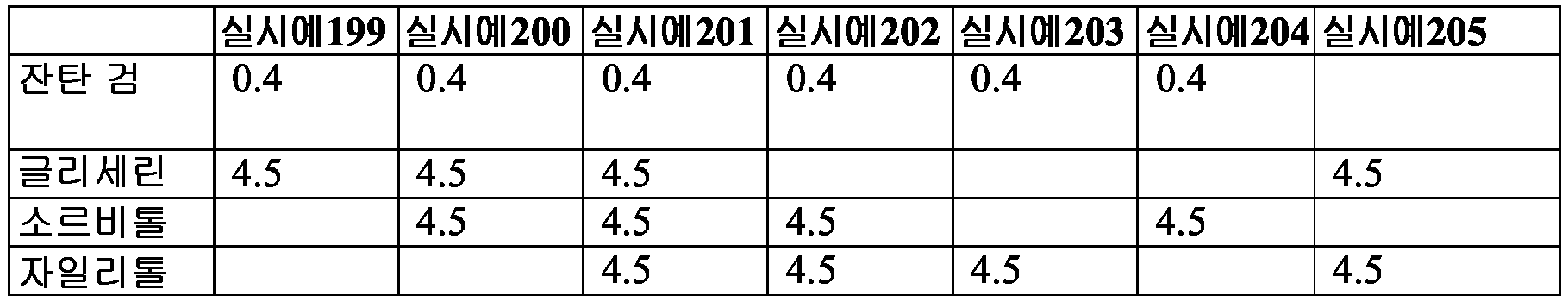
그 다음으로, 100 μL 의 실시예 199-205 를 필터 삽입물 (기공 크기 0.8 μm 직경, 박테리아 및 진균 모두가 통과할 수 있음) 에 두고 층을 형성하게 하였다. 다음으로, 유기체를 시험 용액에 의해 형성된 층 상에 오버레이하였다. 미생물 및 시험 용액의 층을 함유하는 상기 필터 삽입물을 한천 배지 플레이트 표면 상에 두고 24 시간 동안 37℃ 에서 인큐베이션하였다. 인큐베이션 기간 후, 한천 배지 플레이트를 필터 삽입물 상 및 한천 배지에서의 성장에 대해 평가하였다. 필터 삽입물에서는 성장하였으나 한천 배지에서는 성장하지 않은 것은, 시험 용액이 장벽을 형성하여 미생물이 통과하는 것을 방지하였다는 것을 나타내었다. 반대로, 필터 삽입물에서 성장하였을 뿐 아니라 한천 배지에서도 성장한 것은, 이러한 장벽이 형성되지 않았다는 것을 나타내었다.Next, 100 μL of Examples 199-205 were placed in the filter insert (pore size 0.8 μm diameter, through which both bacteria and fungi can pass) to form a layer. Next, the organisms were overlaid on the layer formed by the test solution. The filter insert containing the layers of microorganisms and test solution was placed on the agar medium plate surface and incubated at 37 ° C. for 24 hours. After the incubation period, agar medium plates were evaluated for growth on filter inserts and in agar medium. Growth on the filter insert but not on the agar medium indicated that the test solution formed a barrier to prevent the passage of microorganisms. In contrast, growth not only in the filter insert but also in the agar medium indicated that this barrier was not formed.
상기 결과는, 시험한 보습제를 함유하는 (개별적으로 또는 조합으로) 잔탄 검-기재 용액 각각이 필터 삽입물 상에 미손상 장벽을 형성하여, 밑에 있는 한천 배지로 미생물이 통과하는 것을 방지하였다는 것을 나타내었다.The results indicate that each of the xanthan gum-based solution containing the tested moisturizer (individually or in combination) formed an intact barrier on the filter insert, preventing microorganisms from passing through the underlying agar medium. It was.
실시예Example 206-213 206-213
잔탄 검의 용해도 한계 측정Determination of Solubility Limit of Xanthan Gum
잔탄 검의 용해도를 측정하기 위해서, 물 중에서 상이한 농도로 혼합하고, 응괴의 존재 또는 부재 및 혼합물의 자유 흐름을 모니터링함으로써 용해도를 관찰하였다. 하기 표 XIII 에서 결과 및 농도를 나타낸다.To determine the solubility of xanthan gum, solubility was observed by mixing at different concentrations in water and monitoring the presence or absence of clots and the free flow of the mixture. The results and concentrations are shown in Table XIII below.
표 table XIIIXIII
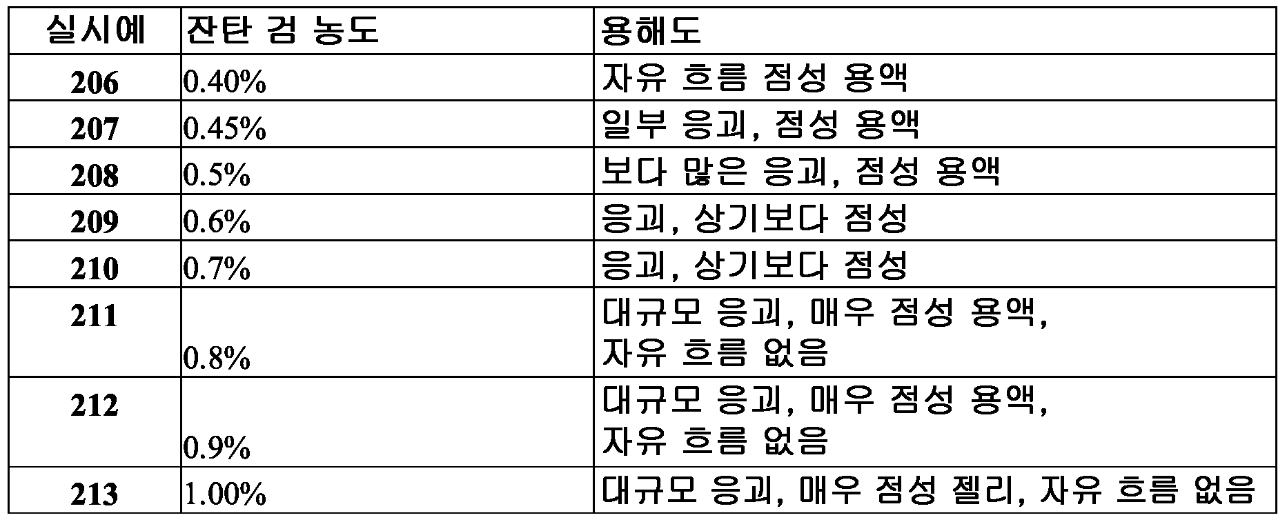
0.4% 에서 혼합한 경우, 잔탄 검이 자유 흐름 용액을 형성하였다는 것을 발견하였다 (표 XIII). 반대로, 0.45% 또는 0.5% 잔탄 검을 함유하는 혼합물은 점성의 유체를 형성하였으나 작은 응괴를 함유하였다. 응괴의 정도는 잔탄 검 농도 증가 (0.6% 및 0.7%) 에 따라 증가하였다. 0.8% 이상의 농도에서, 잔탄 검 혼합물은 대규모 응괴를 가지며, 점조도가 젤리와 같았고 자유 흐름이 없었다.When mixed at 0.4%, it was found that xanthan gum formed a free flowing solution (Table XIII). In contrast, mixtures containing 0.45% or 0.5% xanthan gum formed viscous fluids but contained small clots. The degree of clot increased with increasing xanthan gum concentration (0.6% and 0.7%). At concentrations above 0.8%, the xanthan gum mixture had a massive clot, the consistency was like jelly and there was no free flow.
실시예Example 214 214
장벽-형성 조성물 중의 중성 항균제와 장벽-형성 조성물 중의 양이온성 CPC 의 비교Comparison of Neutral Antibacterials in Barrier-Forming Compositions and Cationic CPC in Barrier-Forming Compositions
실시예 214 에서, CPC 대신 중성제 시트랄 (Citral) 을 사용한 것을 제외하고, 실시예 7 의 제형을 제조하였다. CPC (0.1%) 또는 시트랄 (0.5%) 을 함유하는 제형의 스트렙토코쿠스에 대한 항균 활성을 확인하였다. 실시예 48-61 에서 상기 기재한 검정을 이들 연구를 수행하는데 사용하였다.In Example 214, the formulation of Example 7 was prepared except that the neutral citral was used instead of CPC. Antimicrobial activity against Streptococcus of formulations containing CPC (0.1%) or citral (0.5%) was confirmed. The assays described above in Examples 48-61 were used to perform these studies.
상기 결과는, 시트랄을 함유하는 제형이 항균 활성을 나타냈다는 것을 보여주었다 (MIC = 12.5%). 그러나, 시트랄을 함유하는 제형의 활성은 CPC 를 함유하는 제형의 경우보다 상당히 덜 강력하였다 (MIC = 0.098%). The results showed that formulations containing citral showed antimicrobial activity (MIC = 12.5%). However, the activity of the formulation containing citral was significantly less potent than that of the formulation containing CPC (MIC = 0.098%).
실시예Example 215 215
소수성의 이화학적 시험 및 비교Hydrophobic Physicochemical Test and Comparison
실시예 215 에서 박막 크로마토그래피 분석을 사용하여 실시예 7 의 소수성을 소수성 조성물과 비교하였다. 소수성 조성물은 하기 표 XIV 의 성분으로 이루어졌다.The hydrophobicity of Example 7 was compared with the hydrophobic composition using thin layer chromatography analysis in Example 215. The hydrophobic composition consisted of the components of Table XIV below.
표 table XIVXIV

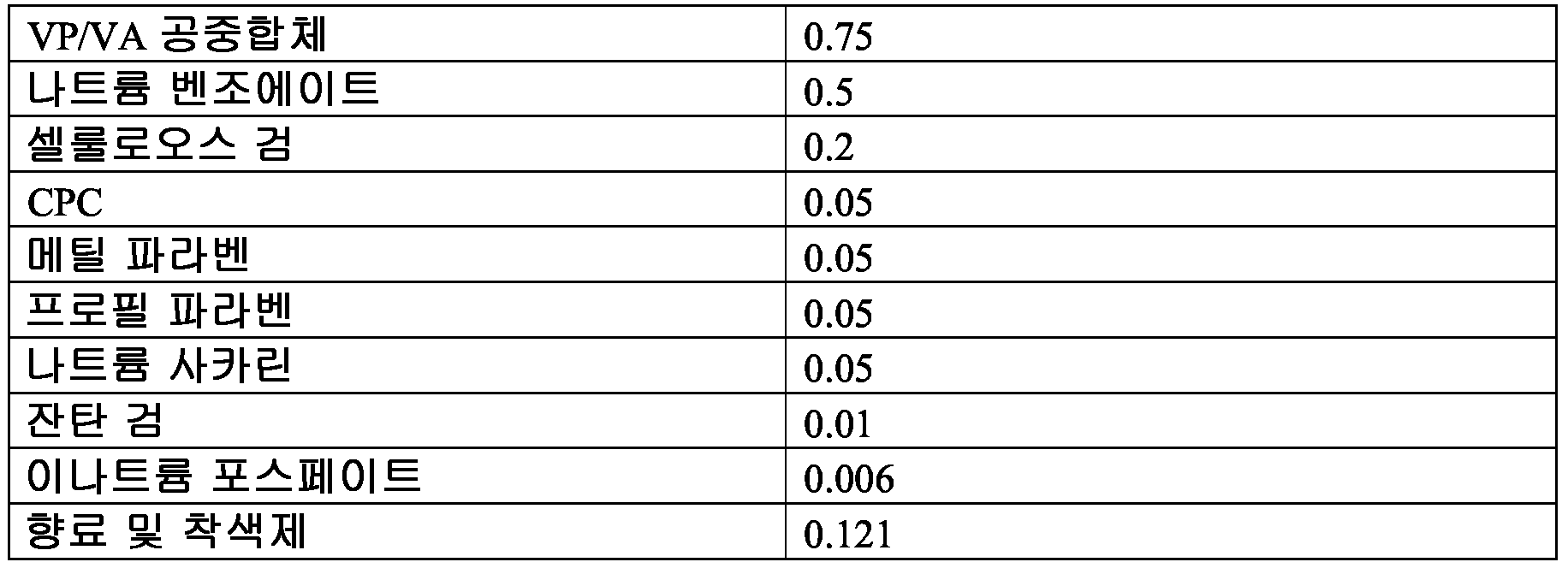
*조성물의 나머지는 정제수임 * The rest of the composition is purified water
10 μL 의 실시예 7 및 소수성 조성물을 사전제조 TLC 플레이트 상에 침적시켰다 (하부 가장자리로부터 2 cm 거리로). 스팟을 5 분 동안 자연 건조시키고, 플레이트를 용매로서 물을 함유하는 TLC 크로마토그래피 단지 (jar) 에 넣었다. 용매 프론트 (front) 가 플레이트의 상부 가장자리에 도달할 때까지 TLC 시스템을 실행하였다. 플레이트를 제거하고 용매 및 샘플 프론트를 표시하였다. 상대 프론트 (Rf) 값을 하기 식 II 를 사용하여 2 개 샘플에 대해 계산하였다:10 μL of Example 7 and the hydrophobic composition were deposited on prefabricated TLC plates (2 cm away from the bottom edge). Spots were allowed to dry for 5 minutes and plates were placed in TLC chromatography jars containing water as solvent. The TLC system was run until the solvent front reached the upper edge of the plate. The plate was removed and the solvent and sample fronts were marked. Relative front (Rf) values were calculated for two samples using the following formula II:
II. Rf = 스팟이 이동한 거리/용매 프론트가 이동한 거리II. Rf = distance traveled by spot / distance traveled by solvent front
상기 결과는 소수성 조성물 및 실시예 7 에 대한 Rf 값이 각각 0.33 및 0 이었음을 나타내었으며, 이는 소수성 조성물이 물에 매우 혼화성이었다는 것을 나타낸다. 반대로, 실시예 7 은 수성 용매 중에서 어떠한 이동성도 나타내지 않았는데, 이는 상기 제형이 소수성이거나, 친수성이 아니라는 것을 입증한다.The results showed that the Rf values for the hydrophobic composition and Example 7 were 0.33 and 0, respectively, indicating that the hydrophobic composition was very miscible with water. In contrast, Example 7 did not show any mobility in aqueous solvents, demonstrating that the formulation is hydrophobic or not hydrophilic.
Claims (53)
탄수화물 검;
보습제; 및
항균제;
이때 상기 조성물이 하기 요구조건을 충족하고:
약 0.01% ≤ C ≤ 약 0.4%;
약 4.5% ≤ H ≤ 약 65%; 및
0.050% < A
또는
약 0% ≤ C ≤ 약 0.4%;
약 55% ≤ H ≤ 약 65%; 및
0.050% < A,
상기 모든 퍼센트가 총 조성물의 중량% 이고;
C 가 탄수화물 검이고; H 가 보습제이고; A 가 항균제인 조성물.As a barrier-forming composition comprising:
Carbohydrate gum;
Humectants; And
Antibacterial agents;
Wherein the composition meets the following requirements:
About 0.01% <C <about 0.4%;
About 4.5% <H <about 65%; And
0.050% <A
or
About 0% ≦ C ≦ about 0.4%;
About 55% <H <about 65%; And
0.050% <A,
All the percentages are weight percent of the total composition;
C is a carbohydrate gum; H is a moisturizer; A is an antimicrobial composition.
탄수화물 검;
보습제; 및
항균제;
이때 상기 조성물이 하기 요구조건을 충족하고:
약 0.01% ≤ C ≤ 약 0.4%; 및
약 4.5% ≤ H ≤ 약 65%;
또는
약 0% ≤ C ≤ 약 0.4%; 및
약 55% ≤ H ≤ 약 65%,
상기 모든 퍼센트가 총 조성물의 중량% 이고;
물 중에서 Rf 값이 0 내지 약 0.25 인 조성물.As a barrier-forming composition comprising:
Carbohydrate gum;
Humectants; And
Antibacterial agents;
Wherein the composition meets the following requirements:
About 0.01% <C <about 0.4%; And
About 4.5% <H <about 65%;
or
About 0% ≦ C ≦ about 0.4%; And
About 55% ≤ H ≤ about 65%,
All the percentages are weight percent of the total composition;
A composition having an Rf value of 0 to about 0.25 in water.
보습제; 및
항균제를 혼합하고 가열하는 것을 포함하는 점막 장벽-형성 조성물의 제조 방법으로서:
이때 상기 조성물이 하기 요구조건을 충족하고:
약 0.01% ≤ C ≤ 약 0.4%;
약 4.5% ≤ H ≤ 약 65%; 및
0.050% < A
또는
약 55% ≤ C ≤ 약 65%;
약 0% ≤ H ≤ 약 0.4%; 및
0.050% < A,
상기 모든 퍼센트가 총 조성물의 중량% 인 제조 방법.Carbohydrate gum;
Humectants; And
A process for preparing a mucosal barrier-forming composition comprising mixing and heating an antimicrobial agent:
Wherein the composition meets the following requirements:
About 0.01% <C <about 0.4%;
About 4.5% <H <about 65%; And
0.050% <A
or
About 55% ≦ C ≦ about 65%;
About 0% ≦ H ≦ about 0.4%; And
0.050% <A,
Wherein all said percentages are weight percent of the total composition.
오염된 환경 또는 품목을 확인하고, 이때 상기 오염된 환경 또는 품목은 유해한 바이러스, 진균 또는 박테리아 미생물로 오염된 것으로 알려지거나 예상되고;
포유동물이 오염된 환경 또는 품목에 접촉하기 전 또는 접촉하는 동안 치료적 유효량의 장벽-형성 조성물을 상기 포유동물의 점막에 투여하고;
상기 장벽-형성 조성물이 점막 상에 장벽을 제공하여 미생물이 점막에 접촉하는 것을 저해함.A method for blocking, neutralizing or killing microorganisms causing infectious diseases in a mammal before or during contact with the contaminated environment or item, comprising:
Identifying a contaminated environment or item, wherein the contaminated environment or item is known or expected to be contaminated with harmful virus, fungal or bacterial microorganisms;
Administering a therapeutically effective amount of the barrier-forming composition to the mucous membrane of the mammal before or during the contact with the contaminated environment or item;
The barrier-forming composition provides a barrier on the mucosa and inhibits microorganisms from contacting the mucosa.
약 0.01% ≤ C ≤ 약 0.4%;
약 7% ≤ H ≤ 약 65%; 및
0.050% < A
또는
약 0% ≤ C ≤ 약 0.4%;
약 55% ≤ H ≤ 약 65%; 및
0.050% < A,
상기 모든 퍼센트가 총 조성물의 중량% 이고;
C 가 탄수화물 검이고; H 가 보습제이고; A 가 항균제인 방법.The method of claim 24, wherein the barrier-forming composition meets the following requirements:
About 0.01% <C <about 0.4%;
About 7% ≦ H ≦ about 65%; And
0.050% <A
or
About 0% ≦ C ≦ about 0.4%;
About 55% <H <about 65%; And
0.050% <A,
All the percentages are weight percent of the total composition;
C is a carbohydrate gum; H is a moisturizer; A is an antimicrobial agent.
오염된 품목 또는 환경을 확인하고, 이때 상기 오염된 품목 또는 환경은 유해한 바이러스, 진균 또는 박테리아 미생물로 오염된 것으로 알려지거나 예상되고;
장치가 오염된 환경에 접촉하기 전 또는 접촉하는 동안 및 장치가 포유동물의 점막에 접촉하기 전 또는 접촉하는 동안 상기 장치 상에 장벽을 형성시키고;
상기 장벽-형성 조성물이 장치 상에 미생물을 포획하고 사멸시키는 장벽을 제공하고, 그로써 미생물이 점막에 접촉하거나 감염을 일으키는 것을 예방함.Methods for blocking, neutralizing or killing microorganisms causing infectious diseases, including:
Identifying a contaminated item or environment, wherein the contaminated item or environment is known or expected to be contaminated with harmful virus, fungal or bacterial microorganisms;
Forming a barrier on the device before or while the device contacts the contaminated environment and before or while the device contacts the mucous membranes of the mammal;
The barrier-forming composition provides a barrier to trap and kill microorganisms on the device, thereby preventing the microorganisms from contacting the mucous membrane or causing infection.
포유동물의 점막에서의 파괴 부위를 확인하고;
치료적 유효량의 장벽-형성 조성물을 적어도 포유동물 점막의 파괴 부위에 투여하고;
상기 장벽-형성 조성물이 점막의 파괴 부위 상에 장벽을 제공하여 미생물이 점막의 파괴 부위에 접촉하는 것을 효과적으로 저해함.A method of blocking, killing or neutralizing a microorganism causing an infectious disease in a weakened immune system, comprising:
Identifying sites of destruction in the mucosa of a mammal;
Administering a therapeutically effective amount of the barrier-forming composition to at least the site of destruction of the mammalian mucosa;
The barrier-forming composition provides a barrier on the site of destruction of the mucosa, effectively inhibiting microorganisms from contacting the site of destruction of the mucosa.
포유동물의 손에 의한 오염된 품목과의 접촉을 확인하고;
이때, 상기 오염된 품목 또는 환경은 유해한 바이러스, 진균 또는 박테리아 미생물로 오염된 것으로 알려지거나 예상되고;
포유동물의 손에서 입 또는 손에서 코 접촉 전에 치료적 유효량의 장벽-형성 조성물을 상기 포유동물의 구강, 비강 또는 인두 점막에 투여하고;
상기 장벽-형성 조성물이 점막 상에 장벽을 제공하여 미생물이 점막에 접촉하는 것을 저해하고 상기 미생물을 중화시키거나 사멸시킴.A method of blocking, neutralizing or killing microorganisms introduced into a mammal's oral, nasal or pharyngeal cavity via nasal contact in the mouth or hand of the mammal's hand, comprising:
Confirm contact with the contaminated item by the mammal's hand;
Wherein the contaminated item or environment is known or expected to be contaminated with harmful virus, fungal or bacterial microorganisms;
Administering a therapeutically effective amount of the barrier-forming composition to the oral, nasal or pharyngeal mucosa of the mammal prior to the mouth or nasal contact in the hand of the mammal;
The barrier-forming composition provides a barrier on the mucosa, inhibiting the contact of the microorganisms with the mucosa and neutralizing or killing the microorganisms.
점막에 대한 알레르겐 또는 공기 운반성 자극물로 오염된 것으로 알려지거나 예상되는 오염된 환경 또는 품목을 기준으로 오염된 환경을 확인하고;
포유동물이 오염된 환경에 접촉하기 전 또는 접촉하는 동안 치료적 유효량의 장벽-형성 조성물을 상기 포유동물의 구강 또는 인두 점막에 투여하고;
상기 장벽-형성 조성물이 점막 상에 장벽을 제공하여 미생물이 점막에 접촉하는 것을 저해함.A method of blocking, neutralizing or killing an allergen or air-carrying stimulant of a mammal's oral or pharyngeal mucosa before or during contact with a mammal in a contaminated environment, comprising:
Identifying contaminated environments based on contaminated environments or items known or expected to be contaminated with allergens or air transport irritants on mucous membranes;
Administering a therapeutically effective amount of the barrier-forming composition to the oral or pharyngeal mucosa of the mammal prior to or during contact with the mammal in a contaminated environment;
The barrier-forming composition provides a barrier on the mucosa and inhibits microorganisms from contacting the mucosa.
Applications Claiming Priority (3)
| Application Number | Priority Date | Filing Date | Title |
|---|---|---|---|
| US201161477147P | 2011-04-19 | 2011-04-19 | |
| US61/477,147 | 2011-04-19 | ||
| PCT/US2012/033921 WO2012145307A1 (en) | 2011-04-19 | 2012-04-17 | Method of inhibiting harmful microorganisms and barrier-forming composition therefor |
Publications (1)
| Publication Number | Publication Date |
|---|---|
| KR20140050608A true KR20140050608A (en) | 2014-04-29 |
Family
ID=47021801
Family Applications (1)
| Application Number | Title | Priority Date | Filing Date |
|---|---|---|---|
| KR1020137030670A Withdrawn KR20140050608A (en) | 2011-04-19 | 2012-04-17 | Harmful microbial inhibition method and barrier-forming composition therefor |
Country Status (12)
| Country | Link |
|---|---|
| US (4) | US8992893B2 (en) |
| EP (1) | EP2699246B1 (en) |
| JP (2) | JP2014518555A (en) |
| KR (1) | KR20140050608A (en) |
| CN (1) | CN103619341A (en) |
| AU (1) | AU2012245665B2 (en) |
| BR (1) | BR112013026965B1 (en) |
| CA (2) | CA3079323A1 (en) |
| IL (1) | IL228956A0 (en) |
| MX (1) | MX2013012151A (en) |
| RU (1) | RU2013151164A (en) |
| WO (1) | WO2012145307A1 (en) |
Families Citing this family (15)
| Publication number | Priority date | Publication date | Assignee | Title |
|---|---|---|---|---|
| KR20140050608A (en) | 2011-04-19 | 2014-04-29 | 암즈 파머수티컬 엘엘씨 | Harmful microbial inhibition method and barrier-forming composition therefor |
| US10426761B2 (en) | 2011-04-19 | 2019-10-01 | Arms Pharmaceutical, Llc | Method for treatment of disease caused or aggravated by microorganisms or relieving symptoms thereof |
| EP2838570A4 (en) * | 2012-04-17 | 2016-03-30 | ARMS Pharmaceutical LLC | Long-lasting surface anti-microbial and method of application |
| WO2014107221A1 (en) * | 2013-01-04 | 2014-07-10 | ARMS Pharmaceutical LLC | Method for treatment of disease caused or aggravated by microorganisms or relieving symptoms thereof |
| WO2014107572A1 (en) * | 2013-01-04 | 2014-07-10 | ARMS Pharmaceutical LLC | Body surface treatment composition and method |
| US10869479B2 (en) * | 2014-04-28 | 2020-12-22 | American Sterilizer Company | Wipe for killing spores |
| CN105497056B (en) * | 2014-09-26 | 2021-02-19 | 上海玄人信息技术有限公司 | Barrier agent for inhibiting fine air particles from invading human body |
| CN105494418B (en) * | 2014-09-26 | 2020-08-07 | 上海玄人信息技术有限公司 | Barrier agent for inhibiting harmful microorganism infection in air fine particles |
| US10240236B2 (en) * | 2015-03-06 | 2019-03-26 | Lam Research Corporation | Clean resistant windows for ultraviolet thermal processing |
| CN106214926A (en) * | 2016-07-29 | 2016-12-14 | 腾科宝迪(厦门)生物科技有限公司 | A kind of nasal spray and preparation method thereof |
| CN106668926A (en) * | 2017-03-29 | 2017-05-17 | 青岛辰达生物科技有限公司 | Preparation method of gel for promoting wound healing |
| US10358717B2 (en) * | 2017-04-21 | 2019-07-23 | Lam Research Corporation | Method for depositing high deposition rate, thick tetraethyl orthosilicate film with low compressive stress, high film stability and low shrinkage |
| WO2019069298A2 (en) | 2018-01-18 | 2019-04-11 | Christian Arnold | Chewing gum composition comprising polyhexanide |
| US11541105B2 (en) | 2018-06-01 | 2023-01-03 | The Research Foundation For The State University Of New York | Compositions and methods for disrupting biofilm formation and maintenance |
| TW202227104A (en) | 2020-09-08 | 2022-07-16 | 瑞士商艾爾塔米拉醫療有限公司 | Compositions for preventing infection |
Family Cites Families (78)
| Publication number | Priority date | Publication date | Assignee | Title |
|---|---|---|---|---|
| US3683297A (en) | 1968-11-22 | 1972-08-08 | Coherent Radiation Lab | Optical cavity for a laser |
| US3993777A (en) | 1975-08-12 | 1976-11-23 | Bio-Lab, Inc. | Aqueous compositions to aid in the prevention of bovine mastitis |
| JPS5562012A (en) | 1978-11-06 | 1980-05-10 | Teijin Ltd | Slow-releasing preparation |
| US4657758A (en) * | 1984-01-27 | 1987-04-14 | Oral Research Laboratories, Inc. | Dental rinse |
| US4666708A (en) * | 1984-01-27 | 1987-05-19 | Oral Research Laboratories, Inc. | Dental rinse |
| US4590057A (en) | 1984-09-17 | 1986-05-20 | Rio Linda Chemical Co., Inc. | Process for the generation of chlorine dioxide |
| US4590067A (en) * | 1984-10-18 | 1986-05-20 | Peritain, Ltd. | Treatment for periodontal disease |
| US5124359A (en) | 1987-12-22 | 1992-06-23 | Cetylite Industries, Inc. | Sterilant composition |
| NO178843C (en) | 1988-07-11 | 1996-06-19 | Sspl Sa Safe Sex Prod Licens | Process for the preparation of a pharmaceutical composition for the prevention of sexually transmitted diseases |
| ES2053963T3 (en) | 1988-11-28 | 1994-08-01 | Unilever Nv | DENTIPHRICS CONTAINING ALKYL ALKYL SILICONE AND SARCOSINATE SURFACTANTS. |
| US5095106A (en) * | 1989-07-25 | 1992-03-10 | Colgate-Palmolive | Olgiosaccharide inhibition of Streptococcus pyogenes adhesion |
| NO901031L (en) | 1990-03-05 | 1991-09-06 | Gunnar Roella | BACTERICID COMPOSITION. |
| US5401723A (en) | 1992-12-02 | 1995-03-28 | Colgate-Palmolive Company | Glycoconjugate inhibition of Streptococcus pyrogenes adhesion |
| US20100055152A1 (en) | 2008-08-26 | 2010-03-04 | Trutek Corporation | Antihistamine and antihistamine-like nasal application, products, and method |
| SI9520017A (en) | 1994-10-21 | 1997-04-30 | Japan Clinic Co Ltd | Microbicidal preparation with low toxicity level, containimng a quaternary ammonia |
| US5733540A (en) * | 1995-03-08 | 1998-03-31 | Lee; Peter Poon-Hang | Protection from viral infection via colonization of mucosal membranes with genetically modified bacteria |
| DE19529862A1 (en) | 1995-08-14 | 1997-02-20 | Rhone Poulenc Rorer Gmbh | Pharmaceutical preparation for oral use |
| US6713049B1 (en) | 1999-11-12 | 2004-03-30 | The Procter & Gamble Company | Oral compositions providing optimal surface conditioning |
| US5776479A (en) | 1996-12-20 | 1998-07-07 | The United States Of America As Represented By The Secretary Of Agriculture | Stable, germicidal film-forming teat-dip solutions |
| US6749869B1 (en) * | 1997-09-26 | 2004-06-15 | Ecolab | Acidic aqueous chlorite teat dip providing shelf life, sanitizing capacity and tissue protection |
| US5938825A (en) | 1998-05-21 | 1999-08-17 | Troy Technology Corporation Inc. | Stabilized antimicrobial compositions containing halopropynyl compounds |
| US6207596B1 (en) | 1998-11-09 | 2001-03-27 | The Procter & Gamble Company | Disposable premoistened wipe containing an antimicrobial protease inhibitor |
| US6309625B1 (en) | 1998-11-12 | 2001-10-30 | Ultradent Products, Inc. | One-part dental compositions and methods for bleaching and desensitizing teeth |
| US7048953B2 (en) | 2000-04-03 | 2006-05-23 | Inhalation, Inc. | Methods and apparatus to prevent, treat and cure infections of the human respiratory system by pathogens causing severe acute respiratory syndrome (SARS) |
| DE60040342D1 (en) | 2000-05-12 | 2008-11-06 | Nippon Steel Corp | DEVICE FOR PFRAIN REFINING AND USE OF THIS DEVICE IN A REFINING PROCESS |
| US6663902B1 (en) | 2000-09-19 | 2003-12-16 | Ecolab Inc. | Method and composition for the generation of chlorine dioxide using Iodo-Compounds, and methods of use |
| US6610276B2 (en) | 2001-01-12 | 2003-08-26 | Steven A. Melman | Multi-functional dental composition |
| US7547433B2 (en) * | 2001-02-15 | 2009-06-16 | Access Pharmaceuticals, Inc. | Liquid formulations for the prevention and treatment of mucosal diseases and disorders |
| WO2002078667A1 (en) | 2001-03-29 | 2002-10-10 | The Dial Corporation | Antibacterial compositions for skin care |
| US6682722B2 (en) * | 2001-09-19 | 2004-01-27 | The Procter & Gamble Company | Oral compositions providing enhanced overall cleaning |
| EP1480517A4 (en) * | 2002-02-07 | 2007-08-22 | Univ Columbia | ZINC SALT COMPOSITIONS AGAINST MUCOSAL IRRITATION FROM SPERMICIDES AND MICROBICIDES |
| ES2734652T3 (en) | 2002-04-04 | 2019-12-11 | Zoetis Belgium S A | Immunostimulatory oligonucleotides containing G and U |
| AR039540A1 (en) | 2002-05-13 | 2005-02-23 | Tibotec Pharm Ltd | MICROBICIDE COMPOUNDS WITH PIRIMIDINE OR TRIAZINE CONTENT |
| EP1562543A1 (en) * | 2002-11-21 | 2005-08-17 | J.P.M.E.D. Ltd. | Compositions for treating infected skin and mucous membrane comprising an anti-microbial agent and an essential oil |
| US6669929B1 (en) | 2002-12-30 | 2003-12-30 | Colgate Palmolive Company | Dentifrice containing functional film flakes |
| US6692726B1 (en) | 2003-02-11 | 2004-02-17 | Colgate Palmolive Company | Enzyme containing oral composition having enhanced stability |
| US7071152B2 (en) | 2003-05-30 | 2006-07-04 | Steris Inc. | Cleaning and decontamination formula for surfaces contaminated with prion-infected material |
| US20050208083A1 (en) * | 2003-06-04 | 2005-09-22 | Nanobio Corporation | Compositions for inactivating pathogenic microorganisms, methods of making the compositons, and methods of use thereof |
| US20050025833A1 (en) | 2003-07-16 | 2005-02-03 | Chaim Aschkenasy | Pharmaceutical composition and method for transdermal drug delivery |
| US20050058673A1 (en) * | 2003-09-09 | 2005-03-17 | 3M Innovative Properties Company | Antimicrobial compositions and methods |
| CA2545117A1 (en) * | 2003-11-07 | 2005-05-26 | Viratox, L.L.C. | Virucidal activities of a combination of cetylpyridinium chloride and citric acid |
| DE602004022200D1 (en) | 2003-12-16 | 2009-09-03 | Colgate Palmolive Co | ORAL AND BODY CARE COMPOSITIONS AND METHODS |
| WO2005067878A1 (en) | 2004-01-13 | 2005-07-28 | Vanson Halosource, Inc. | Polysaccharide alcohol antiseptic gel |
| US9241885B2 (en) | 2004-01-29 | 2016-01-26 | The Procter & Gamble Company | Oral care compositions comprising increased bioavailable levels of quaternary ammonium antimicrobials |
| US20050191270A1 (en) | 2004-02-27 | 2005-09-01 | Hydromer, Inc. | Anti-infectious hydrogel compositions |
| MX2007001384A (en) | 2004-08-02 | 2007-04-19 | Glaxo Group Ltd | Novel composition for xerostomia. |
| TWI272947B (en) * | 2004-09-21 | 2007-02-11 | Far East Bio Tec Co Ltd | Pharmaceutical compositions for suppressing influenza virus infection and replication |
| US10918618B2 (en) * | 2005-03-10 | 2021-02-16 | 3M Innovative Properties Company | Methods of reducing microbial contamination |
| WO2007002411A1 (en) | 2005-06-22 | 2007-01-04 | Myriad Genetics, Inc. | Antiviral compounds |
| US7467162B2 (en) | 2005-06-27 | 2008-12-16 | Microsoft Corporation | Pre-configured settings for portable devices |
| US20070027119A1 (en) | 2005-07-29 | 2007-02-01 | Ahmed Fahim U | Antibacterial composition and method of use |
| CA2632183A1 (en) | 2005-08-25 | 2007-03-01 | Philip R. Houle | Treatment systems for delivery of sensitizer solutions |
| US8349365B2 (en) | 2005-09-27 | 2013-01-08 | Asahi Kasei Chemicals Corporation | Cellooligosaccharide-containing composition |
| US20070166244A1 (en) * | 2006-01-19 | 2007-07-19 | The Procter & Gamble Company | Compositions comprising silicone pressure sensitive adhesives for delivering oral care substances |
| WO2007142967A2 (en) | 2006-05-31 | 2007-12-13 | The Dial Corporation | Alcohol-containing antimicrobial compositions having improved efficacy |
| WO2008026310A1 (en) * | 2006-08-29 | 2008-03-06 | Cellex K.K. | Protective agent for oral mucosa containing trehalose |
| SG176486A1 (en) * | 2006-11-14 | 2011-12-29 | Sunstar Inc | Oral composition containing crystalline cellulose surface-treated with water-soluble substance |
| US20080206155A1 (en) | 2006-11-14 | 2008-08-28 | Foamix Ltd. | Stable non-alcoholic foamable pharmaceutical emulsion compositions with an unctuous emollient and their uses |
| US9271494B2 (en) * | 2007-08-30 | 2016-03-01 | Ecolab USA, Inc. | Shelf stable, reduced corrosion, ready to use peroxycarboxylic acid antimicrobial compositions |
| US20090081294A1 (en) | 2007-09-26 | 2009-03-26 | Gin Jerry B | Sustained release dosage form for lubricating an oral cavity |
| BRPI0819338A2 (en) | 2007-11-21 | 2015-05-19 | Procter & Gamble | Useful cough preparations, methods and kits |
| CN101883559B (en) | 2007-12-06 | 2011-12-28 | 琳得科株式会社 | Edible film |
| JP2009182220A (en) | 2008-01-31 | 2009-08-13 | Opnext Japan Inc | Optical transmission module, wavelength monitor, and wavelength shift detecting method |
| US8568697B2 (en) * | 2008-03-21 | 2013-10-29 | Colgate-Palmolive Company | High fluoride ion recovery compositions |
| KR20120081109A (en) * | 2009-09-30 | 2012-07-18 | 스티펠 리서치 오스트레일리아 피티와이 리미티드 | Cosmetic foam |
| IL212176A0 (en) | 2010-04-06 | 2011-06-30 | Panaxia Ltd | A benzyl alcohol mixture for treating premature ejaculation |
| BR112013016212B1 (en) * | 2010-12-23 | 2019-01-22 | Colgate-Palmolive Company | aqueous oral care composition comprising xanthan gum, cellulose and carbomer gum and use of said composition. |
| US20140051732A1 (en) | 2011-04-19 | 2014-02-20 | Arm Pharmaceutical Llc | Low-tack surface anti-microbial coating composition and method of application |
| KR20140050608A (en) | 2011-04-19 | 2014-04-29 | 암즈 파머수티컬 엘엘씨 | Harmful microbial inhibition method and barrier-forming composition therefor |
| US20130123308A1 (en) | 2011-04-19 | 2013-05-16 | Oasis Consumer Healthcare, Llc | Long-lasting surface anti-microbial and method of application |
| US20130039959A1 (en) | 2011-04-19 | 2013-02-14 | Brian Vincent Sokol | Method of inhibiting hiv and barrier-forming composition therefor |
| US20130123309A1 (en) | 2011-04-19 | 2013-05-16 | Oasis Consumer Healthcare Llc | Method for reducing microbial load for individuals with elevated risk conditions |
| EP2838570A4 (en) | 2012-04-17 | 2016-03-30 | ARMS Pharmaceutical LLC | Long-lasting surface anti-microbial and method of application |
| WO2014074331A1 (en) | 2012-11-12 | 2014-05-15 | ARMS Pharmaceutical LLC | Low-tack surface anti-microbial coating composition and method of application |
| WO2014107221A1 (en) | 2013-01-04 | 2014-07-10 | ARMS Pharmaceutical LLC | Method for treatment of disease caused or aggravated by microorganisms or relieving symptoms thereof |
| US20150306042A1 (en) | 2013-01-04 | 2015-10-29 | ARMS Pharmaceutical LLC | Body surface treatment composition and method |
| WO2014107572A1 (en) | 2013-01-04 | 2014-07-10 | ARMS Pharmaceutical LLC | Body surface treatment composition and method |
| EP2941248A4 (en) | 2013-01-04 | 2016-08-10 | ARMS Pharmaceutical LLC | Body surface treatment composition and method |
-
2012
- 2012-04-17 KR KR1020137030670A patent/KR20140050608A/en not_active Withdrawn
- 2012-04-17 CA CA3079323A patent/CA3079323A1/en not_active Abandoned
- 2012-04-17 BR BR112013026965-0A patent/BR112013026965B1/en active IP Right Grant
- 2012-04-17 US US13/448,957 patent/US8992893B2/en active Active
- 2012-04-17 RU RU2013151164/15A patent/RU2013151164A/en unknown
- 2012-04-17 CN CN201280030293.7A patent/CN103619341A/en active Pending
- 2012-04-17 WO PCT/US2012/033921 patent/WO2012145307A1/en not_active Ceased
- 2012-04-17 AU AU2012245665A patent/AU2012245665B2/en not_active Ceased
- 2012-04-17 JP JP2014506480A patent/JP2014518555A/en active Pending
- 2012-04-17 CA CA2832854A patent/CA2832854C/en active Active
- 2012-04-17 MX MX2013012151A patent/MX2013012151A/en unknown
- 2012-04-17 US US13/448,926 patent/US8535646B2/en active Active
- 2012-04-17 EP EP12774888.7A patent/EP2699246B1/en active Active
-
2013
- 2013-08-30 US US14/014,448 patent/US10398645B2/en active Active
- 2013-10-17 IL IL228956A patent/IL228956A0/en active IP Right Grant
-
2016
- 2016-12-15 JP JP2016243184A patent/JP6831227B2/en active Active
-
2019
- 2019-07-03 US US16/502,993 patent/US20190358149A1/en not_active Abandoned
Also Published As
| Publication number | Publication date |
|---|---|
| AU2012245665A1 (en) | 2013-10-24 |
| RU2013151164A (en) | 2015-05-27 |
| CA3079323A1 (en) | 2012-10-26 |
| US20120270909A1 (en) | 2012-10-25 |
| JP2017081951A (en) | 2017-05-18 |
| EP2699246A4 (en) | 2014-10-08 |
| BR112013026965B1 (en) | 2019-04-02 |
| US20140005236A1 (en) | 2014-01-02 |
| WO2012145307A1 (en) | 2012-10-26 |
| AU2012245665B2 (en) | 2017-06-15 |
| US20130028848A1 (en) | 2013-01-31 |
| IL228956A0 (en) | 2013-12-31 |
| CN103619341A (en) | 2014-03-05 |
| EP2699246B1 (en) | 2023-06-14 |
| US10398645B2 (en) | 2019-09-03 |
| BR112013026965A2 (en) | 2017-01-10 |
| US20190358149A1 (en) | 2019-11-28 |
| MX2013012151A (en) | 2014-06-20 |
| US8535646B2 (en) | 2013-09-17 |
| JP6831227B2 (en) | 2021-02-17 |
| CA2832854A1 (en) | 2012-10-26 |
| JP2014518555A (en) | 2014-07-31 |
| US8992893B2 (en) | 2015-03-31 |
| EP2699246A1 (en) | 2014-02-26 |
| CA2832854C (en) | 2020-04-28 |
| EP2699246C0 (en) | 2023-06-14 |
Similar Documents
| Publication | Publication Date | Title |
|---|---|---|
| JP6831227B2 (en) | Methods of Inhibiting Harmful Microorganisms and Barrier-Forming Compositions for them | |
| US20150031729A1 (en) | Low-tack surface anti-microbial coating composition and method of application | |
| US20130123308A1 (en) | Long-lasting surface anti-microbial and method of application | |
| US20080057136A1 (en) | Disinfecting Composition and Methods of Making and Using Same | |
| US20130123309A1 (en) | Method for reducing microbial load for individuals with elevated risk conditions | |
| JP6901824B2 (en) | How to treat diseases caused or exacerbated by microorganisms, or how to alleviate their symptoms | |
| WO2014107572A1 (en) | Body surface treatment composition and method | |
| US10426761B2 (en) | Method for treatment of disease caused or aggravated by microorganisms or relieving symptoms thereof | |
| US20130039959A1 (en) | Method of inhibiting hiv and barrier-forming composition therefor | |
| WO2013158165A1 (en) | Long-lasting surface anti-microbial and method of application | |
| WO2014074331A1 (en) | Low-tack surface anti-microbial coating composition and method of application | |
| JP2016504383A (en) | Body surface treatment composition and treatment method | |
| US20230050626A1 (en) | Bladder, ureter, kidney, urethra, prostate and catheter anti-microbial, and biofilm prevention, reduction and treatment | |
| WO2021195245A1 (en) | Antimicrobial compositions containing polyphenylene carboxymethylene | |
| Persaud | Povidone-iodine is virucidal against Covid-19 Benefits for the public, infected individuals, surgical patients and healthcare workers | |
| Nakagawa et al. | Evaluation of the effect of catechin inhalation treatment on MRSA elimination |
Legal Events
| Date | Code | Title | Description |
|---|---|---|---|
| PA0105 | International application |
Patent event date: 20131119 Patent event code: PA01051R01D Comment text: International Patent Application |
|
| PG1501 | Laying open of application | ||
| PC1203 | Withdrawal of no request for examination | ||
| WITN | Application deemed withdrawn, e.g. because no request for examination was filed or no examination fee was paid |