KR20130093212A - Stimulus estimation system using brain response pattern scanning - Google Patents
Stimulus estimation system using brain response pattern scanning Download PDFInfo
- Publication number
- KR20130093212A KR20130093212A KR1020120014601A KR20120014601A KR20130093212A KR 20130093212 A KR20130093212 A KR 20130093212A KR 1020120014601 A KR1020120014601 A KR 1020120014601A KR 20120014601 A KR20120014601 A KR 20120014601A KR 20130093212 A KR20130093212 A KR 20130093212A
- Authority
- KR
- South Korea
- Prior art keywords
- brain response
- stimulus
- scan
- data
- brain
- Prior art date
- Legal status (The legal status is an assumption and is not a legal conclusion. Google has not performed a legal analysis and makes no representation as to the accuracy of the status listed.)
- Ceased
Links
- 230000004044 response Effects 0.000 title claims abstract description 388
- 210000004556 brain Anatomy 0.000 title claims abstract description 345
- 238000005259 measurement Methods 0.000 claims abstract description 152
- 238000000034 method Methods 0.000 claims abstract description 41
- 230000000638 stimulation Effects 0.000 claims description 64
- 230000008859 change Effects 0.000 claims description 21
- 239000008280 blood Substances 0.000 claims description 17
- 210000004369 blood Anatomy 0.000 claims description 17
- 230000000007 visual effect Effects 0.000 claims description 17
- QVGXLLKOCUKJST-UHFFFAOYSA-N atomic oxygen Chemical compound [O] QVGXLLKOCUKJST-UHFFFAOYSA-N 0.000 claims description 16
- 238000004891 communication Methods 0.000 claims description 16
- 229910052760 oxygen Inorganic materials 0.000 claims description 16
- 239000001301 oxygen Substances 0.000 claims description 16
- 210000001652 frontal lobe Anatomy 0.000 claims description 15
- 230000002490 cerebral effect Effects 0.000 claims description 14
- WQZGKKKJIJFFOK-GASJEMHNSA-N Glucose Natural products OC[C@H]1OC(O)[C@H](O)[C@@H](O)[C@@H]1O WQZGKKKJIJFFOK-GASJEMHNSA-N 0.000 claims description 11
- 239000008103 glucose Substances 0.000 claims description 11
- 210000003926 auditory cortex Anatomy 0.000 claims description 10
- 230000005540 biological transmission Effects 0.000 claims description 10
- 210000003205 muscle Anatomy 0.000 claims description 10
- 230000002567 autonomic effect Effects 0.000 claims description 5
- 210000000467 autonomic pathway Anatomy 0.000 claims description 5
- 229940088597 hormone Drugs 0.000 claims description 5
- 239000005556 hormone Substances 0.000 claims description 5
- 210000003016 hypothalamus Anatomy 0.000 claims description 5
- 230000036760 body temperature Effects 0.000 claims description 4
- 230000001079 digestive effect Effects 0.000 claims description 4
- 235000011389 fruit/vegetable juice Nutrition 0.000 claims description 4
- 230000033001 locomotion Effects 0.000 claims description 4
- 230000007383 nerve stimulation Effects 0.000 claims description 4
- 230000008447 perception Effects 0.000 claims description 4
- 230000036039 immunity Effects 0.000 claims description 3
- 230000002197 limbic effect Effects 0.000 claims description 3
- 230000000877 morphologic effect Effects 0.000 claims description 3
- 230000033764 rhythmic process Effects 0.000 claims description 3
- 230000001953 sensory effect Effects 0.000 claims description 3
- 230000004886 head movement Effects 0.000 claims description 2
- 230000002360 prefrontal effect Effects 0.000 claims 2
- 230000000694 effects Effects 0.000 description 6
- 230000004913 activation Effects 0.000 description 4
- 238000006243 chemical reaction Methods 0.000 description 4
- 239000011159 matrix material Substances 0.000 description 4
- 238000010276 construction Methods 0.000 description 3
- 238000010586 diagram Methods 0.000 description 3
- 238000007781 pre-processing Methods 0.000 description 3
- 230000002354 daily effect Effects 0.000 description 2
- 230000006870 function Effects 0.000 description 2
- 210000000697 sensory organ Anatomy 0.000 description 2
- INGWEZCOABYORO-UHFFFAOYSA-N 2-(furan-2-yl)-7-methyl-1h-1,8-naphthyridin-4-one Chemical compound N=1C2=NC(C)=CC=C2C(O)=CC=1C1=CC=CO1 INGWEZCOABYORO-UHFFFAOYSA-N 0.000 description 1
- 102000000634 Cytochrome c oxidase subunit IV Human genes 0.000 description 1
- 108050008072 Cytochrome c oxidase subunit IV Proteins 0.000 description 1
- 206010012289 Dementia Diseases 0.000 description 1
- 108010064719 Oxyhemoglobins Proteins 0.000 description 1
- 210000003403 autonomic nervous system Anatomy 0.000 description 1
- 230000003925 brain function Effects 0.000 description 1
- 230000001364 causal effect Effects 0.000 description 1
- 210000003710 cerebral cortex Anatomy 0.000 description 1
- 230000003930 cognitive ability Effects 0.000 description 1
- 108010002255 deoxyhemoglobin Proteins 0.000 description 1
- 238000001514 detection method Methods 0.000 description 1
- 230000003203 everyday effect Effects 0.000 description 1
- 230000036541 health Effects 0.000 description 1
- 238000007689 inspection Methods 0.000 description 1
- 210000003715 limbic system Anatomy 0.000 description 1
- 210000000056 organ Anatomy 0.000 description 1
- 230000008569 process Effects 0.000 description 1
- 230000029058 respiratory gaseous exchange Effects 0.000 description 1
- 238000000638 solvent extraction Methods 0.000 description 1
- 210000003478 temporal lobe Anatomy 0.000 description 1
- 210000000857 visual cortex Anatomy 0.000 description 1
Images


Classifications
-
- A—HUMAN NECESSITIES
- A61—MEDICAL OR VETERINARY SCIENCE; HYGIENE
- A61B—DIAGNOSIS; SURGERY; IDENTIFICATION
- A61B5/00—Measuring for diagnostic purposes; Identification of persons
- A61B5/24—Detecting, measuring or recording bioelectric or biomagnetic signals of the body or parts thereof
- A61B5/316—Modalities, i.e. specific diagnostic methods
- A61B5/369—Electroencephalography [EEG]
- A61B5/377—Electroencephalography [EEG] using evoked responses
-
- A—HUMAN NECESSITIES
- A61—MEDICAL OR VETERINARY SCIENCE; HYGIENE
- A61B—DIAGNOSIS; SURGERY; IDENTIFICATION
- A61B5/00—Measuring for diagnostic purposes; Identification of persons
- A61B5/0059—Measuring for diagnostic purposes; Identification of persons using light, e.g. diagnosis by transillumination, diascopy, fluorescence
- A61B5/0075—Measuring for diagnostic purposes; Identification of persons using light, e.g. diagnosis by transillumination, diascopy, fluorescence by spectroscopy, i.e. measuring spectra, e.g. Raman spectroscopy, infrared absorption spectroscopy
-
- A—HUMAN NECESSITIES
- A61—MEDICAL OR VETERINARY SCIENCE; HYGIENE
- A61B—DIAGNOSIS; SURGERY; IDENTIFICATION
- A61B5/00—Measuring for diagnostic purposes; Identification of persons
- A61B5/145—Measuring characteristics of blood in vivo, e.g. gas concentration or pH-value ; Measuring characteristics of body fluids or tissues, e.g. interstitial fluid or cerebral tissue
- A61B5/14532—Measuring characteristics of blood in vivo, e.g. gas concentration or pH-value ; Measuring characteristics of body fluids or tissues, e.g. interstitial fluid or cerebral tissue for measuring glucose, e.g. by tissue impedance measurement
-
- A—HUMAN NECESSITIES
- A61—MEDICAL OR VETERINARY SCIENCE; HYGIENE
- A61B—DIAGNOSIS; SURGERY; IDENTIFICATION
- A61B5/00—Measuring for diagnostic purposes; Identification of persons
- A61B5/145—Measuring characteristics of blood in vivo, e.g. gas concentration or pH-value ; Measuring characteristics of body fluids or tissues, e.g. interstitial fluid or cerebral tissue
- A61B5/1455—Measuring characteristics of blood in vivo, e.g. gas concentration or pH-value ; Measuring characteristics of body fluids or tissues, e.g. interstitial fluid or cerebral tissue using optical sensors, e.g. spectral photometrical oximeters
- A61B5/14551—Measuring characteristics of blood in vivo, e.g. gas concentration or pH-value ; Measuring characteristics of body fluids or tissues, e.g. interstitial fluid or cerebral tissue using optical sensors, e.g. spectral photometrical oximeters for measuring blood gases
- A61B5/14553—Measuring characteristics of blood in vivo, e.g. gas concentration or pH-value ; Measuring characteristics of body fluids or tissues, e.g. interstitial fluid or cerebral tissue using optical sensors, e.g. spectral photometrical oximeters for measuring blood gases specially adapted for cerebral tissue
-
- A—HUMAN NECESSITIES
- A61—MEDICAL OR VETERINARY SCIENCE; HYGIENE
- A61B—DIAGNOSIS; SURGERY; IDENTIFICATION
- A61B5/00—Measuring for diagnostic purposes; Identification of persons
- A61B5/24—Detecting, measuring or recording bioelectric or biomagnetic signals of the body or parts thereof
- A61B5/316—Modalities, i.e. specific diagnostic methods
- A61B5/369—Electroencephalography [EEG]
- A61B5/377—Electroencephalography [EEG] using evoked responses
- A61B5/378—Visual stimuli
-
- A—HUMAN NECESSITIES
- A61—MEDICAL OR VETERINARY SCIENCE; HYGIENE
- A61B—DIAGNOSIS; SURGERY; IDENTIFICATION
- A61B5/00—Measuring for diagnostic purposes; Identification of persons
- A61B5/24—Detecting, measuring or recording bioelectric or biomagnetic signals of the body or parts thereof
- A61B5/316—Modalities, i.e. specific diagnostic methods
- A61B5/369—Electroencephalography [EEG]
- A61B5/377—Electroencephalography [EEG] using evoked responses
- A61B5/38—Acoustic or auditory stimuli
-
- A—HUMAN NECESSITIES
- A61—MEDICAL OR VETERINARY SCIENCE; HYGIENE
- A61B—DIAGNOSIS; SURGERY; IDENTIFICATION
- A61B5/00—Measuring for diagnostic purposes; Identification of persons
- A61B5/40—Detecting, measuring or recording for evaluating the nervous system
- A61B5/4058—Detecting, measuring or recording for evaluating the nervous system for evaluating the central nervous system
- A61B5/4064—Evaluating the brain
Landscapes
- Health & Medical Sciences (AREA)
- Life Sciences & Earth Sciences (AREA)
- Physics & Mathematics (AREA)
- Molecular Biology (AREA)
- Animal Behavior & Ethology (AREA)
- Veterinary Medicine (AREA)
- Biophysics (AREA)
- Pathology (AREA)
- Engineering & Computer Science (AREA)
- Biomedical Technology (AREA)
- Heart & Thoracic Surgery (AREA)
- Medical Informatics (AREA)
- Public Health (AREA)
- Surgery (AREA)
- General Health & Medical Sciences (AREA)
- Psychology (AREA)
- Neurology (AREA)
- Psychiatry (AREA)
- Optics & Photonics (AREA)
- Spectroscopy & Molecular Physics (AREA)
- Acoustics & Sound (AREA)
- Emergency Medicine (AREA)
- Neurosurgery (AREA)
- Physiology (AREA)
- Measurement Of The Respiration, Hearing Ability, Form, And Blood Characteristics Of Living Organisms (AREA)
Abstract
본 발명은 피험자의 뇌반응을 비침습적 방법으로 스캔하여 뇌반응 스캔 데이터를 생성하여 이와 동일 또는 유사한 기존의 뇌반응 측정 데이터와 비교함으로써 뇌반응 스캔 데이터를 유발한 자극을 추정하는 시스템 및 방법에 관한 것으로, 본 발명에 따른 뇌반응 패턴 스캔을 통한 자극 추정 시스템은, 피험자의 뇌반응을 스캔하여 상기 뇌반응을 유발한 자극을 추정하는 뇌반응 패턴 스캔을 통한 자극 추정 시스템에 있어서, 표준 자극에 따른 뇌반응을 측정한 뇌반응 측정 데이터 및 상기 뇌반응 측정 데이터를 유발한 상기 표준 자극을 식별하는 표준 자극 식별자를 자극-뇌반응 데이터로서 저장하는 자극-뇌반응 저장모듈; 피험자의 두부의 복수의 영역에 특정파장의 측정광을 송출하고 상기 측정광의 반사광를 분석하여 뇌반응 스캔 데이터를 출력하는 뇌반응 스캔모듈; 및 상기 뇌반응 스캔 데이터와 동일 또는 유사한 상기 뇌반응 측정 데이터를 포함하는 자극-뇌반응 데이터의 상기 표준 자극 식별자에 따라 식별된 표준 자극을 추정자극으로 출력하는 자극 추정모듈을 포함하여 구성되는 것을 특징으로 한다. The present invention relates to a system and method for estimating the stimulus that caused the brain response scan data by scanning the brain response of the subject in a non-invasive way to generate brain response scan data and to compare the same or similar existing brain response measurement data. The stimulus estimation system through the brain response pattern scan according to the present invention is a stimulus estimation system through a brain response pattern scan for estimating the stimulus causing the brain response by scanning a brain response of a subject, A stimulus-brain response storage module for storing brain response measurement data measuring a brain response and a standard stimulus identifier identifying the standard stimulus causing the brain response measurement data as stimulus-brain response data; A brain response scan module configured to output measurement light having a specific wavelength to a plurality of areas of the head of the subject, and to analyze the reflected light of the measurement light and output brain response scan data; And a stimulus estimating module configured to output, as an estimated stimulus, a standard stimulus identified according to the standard stimulus identifier of the stimulus-brain response data including the brain response measurement data identical or similar to the brain response scan data. It is done.
Description
본 발명은 뇌반응 패턴 스캔을 통한 자극 추정 시스템 및 추정방법에 관한 것으로, 피험자의 뇌반응을 비침습적 방법으로 스캔하여 뇌반응 스캔 데이터를 생성하여 이와 동일 또는 유사한 기존의 뇌반응 측정 데이터와 비교함으로써 뇌반응 스캔 데이터를 유발한 자극을 추정하는 시스템 및 방법에 관한 것이다.
The present invention relates to a stimulus estimation system and estimation method through the brain response pattern scan, by generating a brain response scan data by scanning the brain response of the subject in a non-invasive method by comparing with the same or similar existing brain response measurement data A system and method for estimating the stimulus that caused brain response scan data.
피험자의 뇌반응을 스캔하여 뇌반응을 유발한 자극을 추정하는 방법이 제시되고 있다. 이러한 자극 추정 시스템은 인간의 뇌작용의 이해를 증진시키는 학술적 효과뿐만 아니라 다양한 실용적, 의학적 활용이 기대된다.A method of estimating the stimulus that caused the brain response by scanning the brain response of the subject has been proposed. Such a stimulus estimation system is expected to have various practical and medical applications as well as academic effects for improving understanding of human brain function.
뇌-기계 인터페이스를 이용하는 인공감각, 인공장기, 인공수족의 제어에 활용될 수 있고, 인지능력이 부족한 유아, 치매환자 등의 의사소통에 활용되거나, 체온, 심박, 호르몬, 혈당 등의 변화를 뇌 반응을 통해 탐지함으로써 헬스캐어 정보로 활용할 수도 있다. It can be used for the control of artificial sense, artificial organ and artificial limb using brain-machine interface, and it is used for communication of infants, dementia patients, etc. who lack cognitive ability, or changes in temperature, heart rate, hormone, blood sugar, etc. It can be used as health care information by detecting through reaction.
그런데, 종래의 뇌반응 패턴 스캔을 통한 자극추정 시스템은 뇌반응 스캔을 위해서 마이크로 니들 어레이 또는 카테터를 이용하기 때문에 뇌반응 스캔을 위해 피험자는 고통스러움을 수반하여야 하고, 일상적인 환경에서 수시로 뇌반응 스캔을 하기가 어렵다는 문제가 있었다.
However, since the conventional stimulus estimation system using the brain response pattern scan uses a microneedle array or a catheter for the brain response scan, the subject has to suffer pain for the brain response scan, and the brain response scan frequently in a daily environment. There was a problem that was difficult to do.
본 발명은 상기의 문제를 해결하기 위한 것으로, 본 발명에 따른 뇌반응 패턴 스캔을 통한 자극 추정 시스템 및 자극 추정방법은, 비침습적 방법으로서 근적외선을 이용하여 뇌반응 스캔 데이터를 생성하기 때문에 일상적인 환경에서도 피험자로부터 용이하게 뇌반응 스캔 데이터를 입수할 수 있는 것을 목적으로 한다.The present invention is to solve the above problems, the stimulation estimation system and stimulus estimation method through the brain response pattern scan according to the present invention, because the non-invasive method to generate brain response scan data using near-infrared rays in everyday environment Also aims to be able to easily obtain brain response scan data from the subject.
본 발명의 실시예에 따른 뇌반응 패턴 스캔을 통한 자극 추정 시스템 및 자극 추정방법은 다양한 주파수 및 세기의 근적외선을 주기적으로 다수의 뇌영역에 송출하여 피험자의 뇌산소포화도 및 뇌혈당을 측정함으로써 정확한 뇌반응 스캔 데이터를 입수하는 것을 다른 목적으로 한다.Stimulation estimation system and stimulation estimation method through the brain response pattern scan according to an embodiment of the present invention by sending a near-infrared rays of various frequencies and intensities to a plurality of brain regions periodically to measure the brain oxygen saturation and cerebral blood glucose of the subject Obtaining reaction scan data is for another purpose.
본 발명의 다른 실시예에 따른 뇌반응 패턴 스캔을 통한 자극 추정 시스템은 복수의 뇌반응 스캔 단말기와 통신망을 통해 연결되는 자극-뇌반응 데이터베이스 서버를 구비함으로써 다수의 뇌반응 스캔 단말기로부터 다양한 피험자의 뇌반응 측정 데이터 및 뇌반응 스캔 데이터를 수집할 수 있을 뿐 아니라 단일의 자극-뇌반응 저장모듈 및 단일의 자극 추적모듈을 자극-뇌반응 데이터베이스 서버에 구축하는 방법으로 다수의 뇌반응 스캔 단말기에제 추정자극을 제공함으로써 시스템 구축비용을 저감하는 것을 다른 목적으로 한다.
Stimulation estimation system through the brain response pattern scan according to another embodiment of the present invention by providing a stimulus-brain response database server connected to a plurality of brain response scan terminal and a communication network brain from a plurality of brain response scan terminals In addition to collecting response measurement data and brain response scan data, a single stimulus-brain response storage module and a single stimulus tracking module are constructed in a stimulus-brain response database server to estimate the number of brain response scan terminals. Another goal is to reduce system construction costs by providing stimuli.
본 발명에 따른 뇌반응 패턴 스캔을 통한 자극 추정 시스템은, 피험자의 뇌반응을 스캔하여 상기 뇌반응을 유발한 자극을 추정하는 뇌반응 패턴 스캔을 통한 자극 추정 시스템에 있어서, 표준 자극에 따른 뇌반응을 측정한 뇌반응 측정 데이터 및 상기 뇌반응 측정 데이터를 유발한 상기 표준 자극을 식별하는 표준 자극 식별자를 자극-뇌반응 데이터로서 저장하는 자극-뇌반응 저장모듈; 피험자의 두부의 복수의 영역에 특정파장의 측정광을 송출하고 상기 측정광의 반사광를 분석하여 뇌반응 스캔 데이터를 출력하는 뇌반응 스캔모듈; 및 상기 뇌반응 스캔 데이터와 동일 또는 유사한 상기 뇌반응 측정 데이터를 포함하는 자극-뇌반응 데이터의 상기 표준 자극 식별자에 따라 식별된 표준 자극을 추정자극으로 출력하는 자극 추정모듈을 포함하여 구성되는 것을 특징으로 한다.Stimulation estimation system through the brain response pattern scan according to the present invention, in the stimulation estimation system through the brain response pattern scan to estimate the stimulus that caused the brain response by scanning the brain response of the subject, the brain response according to the standard stimulation A stimulus-brain response storage module for storing the brain response measurement data and a standard stimulus identifier identifying the standard stimulus that caused the brain response measurement data as stimulus-brain response data; A brain response scan module configured to output measurement light having a specific wavelength to a plurality of areas of the head of the subject, and to analyze the reflected light of the measurement light and output brain response scan data; And a stimulus estimating module configured to output, as an estimated stimulus, a standard stimulus identified according to the standard stimulus identifier of the stimulus-brain response data including the brain response measurement data identical or similar to the brain response scan data. It is done.
본 발명의 실시예에 따른 뇌반응 패턴 스캔을 통한 자극 추정 시스템은, 상기 뇌반응 스캔모듈이 700 nm 내지 1100 nm 파장의 상기 측정광을 송출하는 측정광 송출부; 상기 측정광에 따른 상기 반사광을 수신하는 반사광 수신부; 및 상기 반사광을 분석하여 뇌산소포화도 및 뇌혈당을 측정하여 뇌반응 스캔 데이터를 생성하는 반사광 분석부;를 포함하여 구성되는 것을 특징으로 한다.Stimulation estimation system through the brain response pattern scan according to an embodiment of the present invention, the brain response scan module for transmitting the measurement light of 700 nm to 1100 nm wavelength; A reflected light receiver configured to receive the reflected light according to the measured light; And a reflected light analyzer for analyzing the reflected light to measure cerebral oxygen saturation and cerebral blood glucose to generate brain response scan data.
본 발명의 다른 실시예에 따른 뇌반응 패턴 스캔을 통한 자극 추정 시스템은, 상기 측정광 송출부가 700 내지 800 nm 파장의 제 1 측정광을 송출하는 제 1 측정광 송출수단; 800 내지 900 nm 파장의 제 2 측정광을 송출하는 제 2 측정광 송출수단; 900 내지 1000 nm 파장의 제 3 측정광을 송출하는 제 3 측정광 송출수단; 및 1000 내지 1100 nm 파장의 제 4 측정광을 송출하는 제 4 측정광 송출수단;을 포함하여 구성되는 것을 특징으로 한다.According to another embodiment of the present invention, a stimulation estimation system using a brain response pattern scan may include: first measurement light transmitting means for transmitting the first measurement light having a wavelength of 700 to 800 nm; Second measuring light transmitting means for transmitting a second measuring light having a wavelength of 800 to 900 nm; Third measuring light transmitting means for transmitting third measuring light having a wavelength of 900 to 1000 nm; And fourth measuring light emitting means for transmitting the fourth measuring light having a wavelength of 1000 to 1100 nm.
본 발명의 다른 실시예에 따른 뇌반응 패턴 스캔을 통한 자극 추정 시스템은, 상기 제 1 내지 제 4 측정광 송출수단이, 각각 상기 제 1 내지 제 4 측정광의 세기를 변화시키며 상기 제 1 내지 제 4 측정광을 송출하는 것을 특징으로 한다.According to another embodiment of the present invention, in the stimulus estimation system using the brain response pattern scan, the first to fourth measurement light transmitting means changes the intensity of the first to fourth measurement light, respectively, and the first to fourth It is characterized by sending a measurement light.
본 발명의 다른 실시예에 따른 뇌반응 패턴 스캔을 통한 자극 추정 시스템은, 상기 제 1 내지 제 4 측정광 송출수단은, 동시에 복수의 상기 송출광이 송출되지 않도록 서로 다른 타이밍으로 상기 측정광을 송출하는 것을 특징으로 한다.In a stimulus estimation system using a brain response pattern scan according to another embodiment of the present invention, the first to fourth measurement light transmitting means may transmit the measurement light at different timings so that a plurality of the emission light is not simultaneously transmitted. Characterized in that.
본 발명의 다른 실시예에 따른 뇌반응 패턴 스캔을 통한 자극 추정 시스템은, 상기 뇌반응 스캔모듈이 마이크로 니들 어레이를 더 포함하는 것을 특징으로 한다.In the stimulus estimation system through the brain response pattern scan according to another embodiment of the present invention, the brain response scan module further includes a microneedle array.
본 발명의 다른 실시예에 따른 뇌반응 패턴 스캔을 통한 자극 추정 시스템은, 상기 자극 추정모듈이 상기 피험자로부터 얻어진 자극-뇌반응 데이터 중에서 상기 뇌반응 스캔 데이터와 동일 또는 유사한 상기 뇌반응 측정 데이터를 포함하는 상기 자극-뇌반응 데이터의 상기 표준 자극 식별자에 따라 식별된 상기 표준 자극을 상기 추정자극으로 출력하는 것을 특징으로 한다.Stimulation estimation system through the brain response pattern scan according to another embodiment of the present invention, the stimulation estimation module includes the brain response measurement data identical or similar to the brain response scan data from the stimulus-brain response data obtained from the subject And outputting the standard stimulus identified according to the standard stimulus identifier of the stimulus-brain response data as the estimated stimulus.
본 발명의 다른 실시예에 따른 뇌반응 패턴 스캔을 통한 자극 추정 시스템은, 상기 자극 추정모듈이 상기 피험자로부터 얻어진 자극-뇌반응 데이터 중에서 상기 뇌반응 스캔 데이터와 동일 또는 유사한 상기 뇌반응 측정 데이터를 포함하는 상기 자극-뇌반응 데이터가 없는 경우 피험자가 아닌 제삼자로부터 얻어진 자극-뇌반응 데이터 중에서 상기 뇌반응 스캔 데이터와 동일 또는 유사한 상기 뇌반응 측정 데이터를 포함하는 상기 자극-뇌반응 데이터의 상기 표준 자극 식별자에 따라 식별된 상기 표준 자극을 상기 추정자극으로 출력하는 것을 특징으로 한다.Stimulation estimation system through the brain response pattern scan according to another embodiment of the present invention, the stimulation estimation module includes the brain response measurement data identical or similar to the brain response scan data from the stimulus-brain response data obtained from the subject Said standard stimulus identifier of said stimulus-brain response data comprising said brain response measurement data identical or similar to said brain response scan data among stimulus-brain response data obtained from a third party who is not a subject when said stimulus-brain response data is absent. Outputting the standard stimulus identified according to the estimated stimulus.
본 발명에 따른 뇌반응 패턴 스캔을 통한 자극 추정 시스템은, 뇌반응을 스캔하는 적어도 하나의 뇌반응 스캔 단말기 및 상기 뇌반응 스캔 단말기와 통신망을 통해 접속되는 자극-뇌반응 데이터베이스 서버를 포함하여 구성되는 뇌반응 패턴 스캔을 통한 자극 추정 시스템에 있어서, 상기 뇌반응 스캔 단말기는 피험자의 두부의 복수의 영역에 특정파장의 측정광을 송출하여 반사광를 분석하여 상기 뇌반응 스캔 데이터를 생성하는 뇌반응 스캔모듈; 및 상기 뇌반응 스캔 데이터를 통신망을 통해 전송하는 뇌반응 전송모듈;추정자극을 통신망을 통해 수신하는 추정자극 수신모듈; 및 상기 추정자극을 출력하는 추정자극 출력모듈;을 포함하여 구성되고, 상기 자극-뇌반응 데이터베이스 서버는, 자극에 따른 뇌반응을 측정한 뇌반응 측정 데이터 및 상기 뇌반응 측정 데이터를 유발한 상기 자극을 식별하는 자극 식별자를 자극-뇌반응 데이터로서 저장하는 자극-뇌반응 저장모듈; 상기 뇌반응 스캔 데이터를 수신하는 뇌반응 수신모듈; 상기 뇌반응 스캔 데이터와 동일 또는 유사한 상기 뇌반응 측정 데이터를 포함하는 자극-뇌반응 데이터의 상기 자극 식별자에 따라 식별된 자극을 추정자극으로 출력하는 자극 추정모듈; 및 상기 추정자극을 통신망을 통해 전송하는 추정자극 전송모듈;을 포함하여 구성되는 것을 특징으로 한다.The stimulation estimation system using the brain response pattern scan according to the present invention comprises at least one brain response scanning terminal for scanning brain responses and a stimulus-brain response database server connected to the brain response scanning terminal through a communication network. A stimulus estimation system using a brain response pattern scan, the brain response scanning terminal comprising: a brain response scan module configured to generate measured brain response scan data by transmitting measurement light having a specific wavelength to a plurality of regions of a head of a subject to analyze reflected light; And a brain response transmission module for transmitting the brain response scan data through a communication network; an estimated stimulus reception module for receiving an estimated stimulus through a communication network; And an estimated stimulus output module for outputting the estimated stimulus, wherein the stimulus-brain response database server includes brain response measurement data measuring brain response according to stimulation and the stimulus causing the brain response measurement data. A stimulus-brain response storage module for storing stimulus identifiers identifying the stimulus-brain response data; Brain response receiving module for receiving the brain response scan data; A stimulus estimation module for outputting a stimulus identified according to the stimulus identifier of the stimulus-brain response data including the brain response measurement data identical or similar to the brain response scan data, as an estimated stimulus; And an estimating stimulation transmission module for transmitting the estimated stimulus through a communication network.
본 발명의 다른 실시예에 따른 뇌반응 패턴 스캔을 통한 자극 추정 시스템은, 상기 뇌반응 스캔모듈이, 700 nm 내지 1100 nm 파장의 상기 측정광을 송출하는 측정광 송출부; 상기 측정광에 따른 상기 반사광을 수신하는 반사광 수신부; 및 상기 반사광을 분석하여 뇌산소포화도 및 뇌혈당을 측정하여 뇌반응 스캔 데이터를 생성하는 반사광 분석부;를 포함하여 구성되는 것을 특징으로 한다.Stimulation estimation system through the brain response pattern scan according to another embodiment of the present invention, the brain response scan module, the measurement light transmitting unit for transmitting the measurement light of 700 nm to 1100 nm wavelength; A reflected light receiver configured to receive the reflected light according to the measured light; And a reflected light analyzer for analyzing the reflected light to measure cerebral oxygen saturation and cerebral blood glucose to generate brain response scan data.
본 발명의 다른 실시예에 따른 뇌반응 패턴 스캔을 통한 자극 추정 시스템은, 상기 측정광 송출부가 700 내지 800 nm 파장의 제 1 측정광을 송출하는 제 1 측정광 송출수단; 800 내지 900 nm 파장의 제 2 측정광을 송출하는 제 2 측정광 송출수단; 900 내지 1000 nm 파장의 제 3 측정광을 송출하는 제 3 측정광 송출수단; 및 1000 내지 1100 nm 파장의 제 4 측정광을 송출하는 제 4 측정광 송출수단;을 포함하여 구성되는 것을 특징으로 한다.According to another embodiment of the present invention, a stimulation estimation system using a brain response pattern scan may include: first measurement light transmitting means for transmitting the first measurement light having a wavelength of 700 to 800 nm; Second measuring light transmitting means for transmitting a second measuring light having a wavelength of 800 to 900 nm; Third measuring light transmitting means for transmitting third measuring light having a wavelength of 900 to 1000 nm; And fourth measuring light emitting means for transmitting the fourth measuring light having a wavelength of 1000 to 1100 nm.
본 발명의 다른 실시예에 따른 뇌반응 패턴 스캔을 통한 자극 추정 시스템은, 상기 뇌반응을 유발한 자극은 시각자극이고, 상기 뇌반응 스캔모듈은 상기 두부의 시각영역, 형태지각 영역, 운동영역, 전두엽 영역, 전전두엽 영역 중 적어도 어느 하나를 스캔영역으로 일정시간 간격의 스캔시간 동안 측정하여 상기 뇌반응 스캔 데이터를 출력하고, 상기 자극 추정모듈은, 상기 뇌반응 스캔 데이터와 상기 뇌반응 측정 데이터의 상기 스캔영역의 공간적 유사성을 판단하여 상기 시각자극의 윤곽, 형태, 색상을 추정하고, 상기 뇌반응 스캔 데이터와 상기 뇌반응 측정 데이터의 상기 스캔시간 동안 변화율의 유사성을 판단하여 상기 시각자극의 방향, 각도, 운동을 추정하는 것을 특징으로 한다.In a stimulus estimation system using a brain response pattern scan according to another embodiment of the present invention, the stimulus that causes the brain response is a visual stimulus, and the brain response scan module includes a visual area, a shape perception area, an exercise area, The brain response scan data is measured by measuring at least one of the frontal lobe region and the frontal lobe region as a scan region for a predetermined time interval, and the stimulation estimation module is configured to generate the brain response scan data and the brain response measurement data. The spatial similarity of the scan region is estimated to estimate the contour, shape, and color of the visual stimulus, and the similarity of the rate of change during the scan time between the brain response scan data and the brain response measurement data is determined to determine the direction and angle of the visual stimulus. , Estimating the exercise.
본 발명의 다른 실시예에 따른 뇌반응 패턴 스캔을 통한 자극 추정 시스템은, 상기 뇌반응을 유발한 자극은 청각자극이고, 상기 뇌반응 스캔모듈은 상기 두부의 1차 청각피질 영역, 2차 청각피질 영역, 3차 청각피질 영역, 브로카 영역, 케슈윈트 영역, 베르니케 영역, 운동 영역, 전두엽 영역, 전전두엽 영역 중 적어도 어느 하나를 스캔영역으로 일정시간 간격의 스캔시간 동안 측정하여 상기 뇌반응 스캔 데이터를 출력하고, 상기 자극 추정모듈은, 상기 뇌반응 스캔 데이터와 상기 뇌반응 측정 데이터의 상기 스캔영역의 공간적 유사성을 판단하여 상기 청각자극의 진동수, 진폭을 추정하고, 상기 뇌반응 스캔 데이터와 상기 뇌반응 측정 데이터의 상기 스캔시간 동안 변화율의 유사성을 판단하여 상기 청각자극의 화음, 리듬, 음색을 추정하는 것을 특징으로 한다.In a stimulus estimation system using a brain response pattern scan according to another embodiment of the present invention, the stimulus causing the brain response is an auditory stimulus, and the brain response scan module is the primary auditory cortex area of the head and the secondary auditory cortex. The brain response scan data is measured by measuring at least one of a region, a tertiary auditory cortex region, a broca region, a Keshwin region, a Wernicke region, an exercise region, a frontal lobe region, and a frontal lobe region during a predetermined time interval. The stimulus estimation module estimates the frequency and amplitude of the auditory stimulus by determining the spatial similarity of the scan region between the brain response scan data and the brain response measurement data, and estimates the brain response scan data and the brain response. Estimating the harmony, rhythm, and tone of the auditory stimulus by determining the similarity of the rate of change during the scan time of the measurement data. It shall be.
본 발명의 다른 실시예에 따른 뇌반응 패턴 스캔을 통한 자극 추정 시스템은, 상기 뇌반응을 유발한 자극은 운동지시 또는 운동상상 자극이고, 상기 뇌반응 스캔모듈은 상기 두부의 운동 영역, 감각 영역, 브로카 영역 중 적어도 어느 하나를 스캔영역으로 일정시간 간격의 스캔시간 동안 측정하여 상기 뇌반응 스캔 데이터를 출력하고, 상기 자극 추정모듈은, 상기 뇌반응 스캔 데이터와 상기 뇌반응 측정 데이터의 상기 스캔영역의 공간적 유사성을 판단하여 상기 운동지시 또는 운동상상의 대상이 되는 근육을 추정하고, 상기 뇌반응 스캔 데이터와 상기 뇌반응 측정 데이터의 상기 스캔시간 동안 변화율의 유사성을 판단하여 상기 운동지지 또는 운동상상의 대상이 되는 근육에 가해지는 힘 및 근육의 위치변화를 추정하는 것을 특징으로 한다.According to another embodiment of the present invention, a stimulation estimation system using a brain response pattern scan, wherein the stimulus that caused the brain response is an exercise instruction or an exercise stimulus, and the brain response scan module includes the motor region, the sensory region, The brain response scan data is output by measuring at least one of the broca areas for a scan time interval at a predetermined time interval, and the stimulation estimation module generates the brain response scan data and the brain response measurement data. Estimating the spatial similarity and estimating the muscle that is the target of the exercise instruction or image, and determining the similarity of the rate of change during the scan time of the brain response scan data and the brain response measurement data to determine the target of the exercise support or image It is characterized by estimating the force applied to the muscle and the position change of the muscle.
본 발명의 다른 실시예에 따른 뇌반응 패턴 스캔을 통한 자극 추정 시스템은, 상기 뇌반응을 유발한 자극은 호읍, 맥박, 체온, 호르몬, 면역, 소화액, 혈당, 통증, 산소포화도와 같은 신체상태를 표시하는 자율신경 자극이고, 상기 뇌반응 스캔모듈은 상기 두부의 전두섬엽 영역, 시상하부 영역, 변연계 영역 중 적어도 어느 하나를 스캔영역으로 일정시간 간격의 스캔시간 동안 측정하여 상기 뇌반응 스캔 데이터를 출력하고, 상기 자극 추정모듈은, 상기 뇌반응 스캔 데이터와 상기 뇌반응 측정 데이터의 상기 스캔영역의 공간적 유사성을 판단하여 상기 자율신경 자극의 크기를 추정하고, 상기 뇌반응 스캔 데이터와 상기 뇌반응 측정 데이터의 상기 스캔시간 동안 변화율의 유사성을 판단하여 상기 자율신경 자극의 변화율을 추정하는 것을 특징으로 한다.Stimulation estimation system through the brain response pattern scan according to another embodiment of the present invention, the stimulation that caused the brain response is a body condition such as Hoeup, pulse, body temperature, hormone, immunity, digestive juice, blood sugar, pain, oxygen saturation The brain response scanning module outputs the brain response scan data by measuring at least one of the frontal islet region, the hypothalamus region, and the limbic region of the head as a scan region for a predetermined time interval. The stimulation estimation module estimates the size of the autonomic stimulus by determining the spatial similarity of the scan region between the brain response scan data and the brain response measurement data, and estimates the brain response scan data and the brain response measurement data. Estimating the rate of change of the autonomic nerve stimulus by determining the similarity of the rate of change during the scan time of .
본 발명에 따른 뇌반응 패턴 스캔을 통한 자극 추정방법은, 자극에 따른 뇌반응을 측정한 뇌반응 측정 데이터 및 상기 뇌반응 측정 데이터를 유발한 상기 자극을 식별하는 자극 식별자를 자극-뇌반응 데이터로서 저장하는 제 1 단계; 피험자 두부의 복수의 영역에 특정파장의 측정광을 송출하고 상기 측정광에 따른 반사광를 분석하여 뇌반응 스캔 데이터를 생성하는 제 2 단계; 상기 뇌반응 스캔 데이터와 동일 또는 유사한 상기 뇌반응 측정 데이터를 포함하는 자극-뇌반응 데이터를 검출하는 제 3 단계; 및 검출된 상기 자극-뇌반응 데이터에 포함된 상기 자극 식별자에 따라 식별된 자극을 추정자극으로 출력하는 제 4 단계;를 포함하는 것을 특징으로 한다.Stimulation estimation method through the brain response pattern scan according to the present invention, the brain response measurement data measuring the brain response according to the stimulation and a stimulus identifier for identifying the stimulus that caused the brain response measurement data as stimulation-brain response data A first step of storing; A second step of transmitting measurement light having a specific wavelength to a plurality of regions of the head of the subject and analyzing the reflected light according to the measurement light to generate brain response scan data; Detecting stimulus-brain response data comprising the brain response measurement data identical or similar to the brain response scan data; And outputting a stimulus identified according to the stimulus identifier included in the detected stimulus-brain response data as an estimated stimulus.
본 발명의 실시예에 따른 뇌반응 패턴 스캔을 통한 자극 추정방법은, 제 2 단계가 700 nm 내지 1100 nm 파장의 상기 측정광을 송출하는 제 1 부단계; 상기 측정광에 따른 상기 반사광을 수신하는 제 2 부단계; 및 상기 반사광을 분석하여 뇌산소포화도 및 뇌혈당을 측정하여 뇌반응 스캔 데이터를 생성하는 제 3 부단계;를 포함하는 것을 특징으로 한다. Stimulation estimation method through the brain response pattern scan according to an embodiment of the present invention, the second step is to transmit the measurement light of the
본 발명의 다른 실시예에 따른 뇌반응 패턴 스캔을 통한 자극 추정방법은, 상기 제 1 부단계가 700 내지 800 nm 파장의 제 1 측정광, 800 내지 900 nm 파장의 제 2 측정광, 900 내지 1000 nm 파장의 제 3 측정광, 1000 내지 1100 nm 파장의 제 4 측정광을 교대로 송출하는 것을 특징으로 한다. According to another embodiment of the present invention, a method of estimating stimulation by scanning a brain response pattern may include: first measuring light having a wavelength of 700 to 800 nm, second measuring light having a wavelength of 800 to 900 nm, and 900 to 1000 The third measurement light having a wavelength of nm and the fourth measurement light having a wavelength of 1000 to 1100 nm are alternately transmitted.
마지막으로, 본 발명의 다른 실시예에 따른 뇌반응 패턴 스캔을 통한 자극 추정방법은, 상기 제 1 부단계가 상기 제 1 내지 제 4 측정광의 세기를 변화시키며 상기 측정광을 송출하는 것을 특징으로 한다.
Finally, the method for estimating the stimulation by scanning the brain response pattern according to another embodiment of the present invention is characterized in that the first sub-step changes the intensity of the first to fourth measurement light and emits the measurement light. .
본 발명은 상기의 문제를 해결하기 위한 것으로, 본 발명에 따른 뇌반응을 한 자극 추정 시스템 및 자극 추정방법은, 비침습적 방법으로서 근적외선을 이용하여 뇌반응 스캔 데이터를 생성하기 때문에 일상적인 환경에서도 피험자로부터 용이하게 뇌반응 스캔 데이터를 입수할 수 있는 효과를 제공한다.SUMMARY OF THE INVENTION The present invention has been made to solve the above problems, and since the stimulus estimation system and stimulus estimation method according to the present invention generate the brain response scan data using near-infrared rays as a non-invasive method, even in a daily environment It provides an effect that can easily obtain brain response scan data from.
본 발명의 실시예에 따른 뇌반응 패턴 스캔을 통한 자극 추정 시스템 및 자극 추정방법은 다양한 주파수 및 세기의 근적외선을 주기적으로 다수의 뇌영역에 송출하여 피험자의 뇌산소포화도 및 뇌혈당을 측정함으로써 정확한 뇌반응 스캔 데이터를 입수하는 효과를 제공한다.Stimulation estimation system and stimulation estimation method through the brain response pattern scan according to an embodiment of the present invention by sending a near-infrared rays of various frequencies and intensities to a plurality of brain regions periodically to measure the brain oxygen saturation and cerebral blood glucose of the subject Provides the effect of obtaining response scan data.
본 발명의 다른 실시예에 따른 뇌반응 패턴 스캔을 통한 자극 추정 시스템은 복수의 뇌반응 스캔 단말기와 통신망을 통해 연결되는 자극-뇌반응 데이터베이스 서버를 구비함으로써 다수의 뇌반응 스캔 단말기로부터 다양한 피험자의 뇌반응 측정 데이터 및 뇌반응 스캔 데이터를 수집할 수 있을 뿐 아니라 단일의 자극-뇌반응 저장모듈 및 단일의 자극 추적모듈을 자극-뇌반응 데이터베이스 서버에 구축하는 방법으로 다수의 뇌반응 스캔 단말기에제 추정자극을 제공함으로써 시스템 구축비용을 저감하는 효과를 제공한다.Stimulation estimation system through the brain response pattern scan according to another embodiment of the present invention by providing a stimulus-brain response database server connected to a plurality of brain response scan terminal and a communication network brain from a plurality of brain response scan terminals In addition to collecting response measurement data and brain response scan data, a single stimulus-brain response storage module and a single stimulus tracking module are constructed in a stimulus-brain response database server to estimate the number of brain response scan terminals. Providing the stimulus provides the effect of reducing the system construction cost.
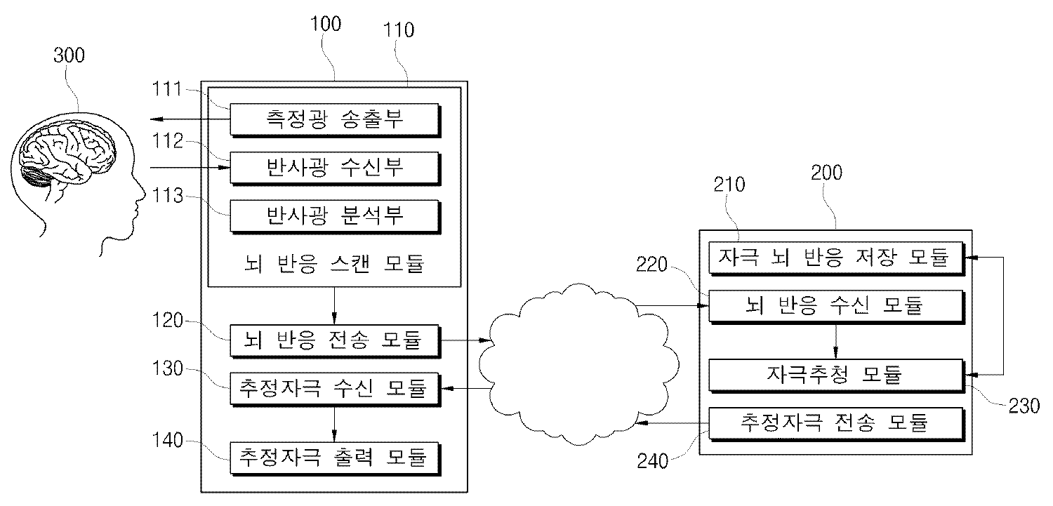
도 1은 본 발명에 따른 뇌반응 패턴 스캔을 통한 자극 추정 시스템을 도시하는 구성도.
도 2는 본 발명에 따른 뇌반응 스캔모듈을 도시하는 상세구성도.
도 3은 측정광 파장에 따른 검출성분을 도시하는 그래프.
도 4는 측정광 송출수단의 측정광 송출 타이밍을 도시하는 타이밍도.
도 5는 반사광 수신수단의 수신상태에 따른 뇌반응 패턴을 도시하는 그래프.
도 6은 본 발명에 따른 뇌반응 패턴 스캔을 통한 자극 추정방법을 도시하는 흐름도.1 is a block diagram showing a stimulation estimation system through a brain response pattern scan according to the present invention.
Figure 2 is a detailed block diagram showing a brain response scan module according to the present invention.
3 is a graph showing detection components according to measured light wavelengths.
4 is a timing diagram showing measurement light delivery timing of the measurement light delivery means;
5 is a graph showing the brain response pattern according to the reception state of the reflected light receiving means.
6 is a flowchart illustrating a stimulus estimation method through brain response pattern scan according to the present invention.
도 1은 본 발명에 따른 뇌반응 패턴 스캔을 통한 자극 추정 시스템을 도시한다. 본 발명에 따른 자극 추정 시스템은 자극-뇌반응 저장모듈(210), 뇌반응 스캔모듈(110), 자극 추정모듈(220)을 포함하여 구성된다.1 illustrates a stimulus estimation system through a brain response pattern scan according to the present invention. The stimulus estimation system according to the present invention includes a stimulus-brain
자극-뇌반응 저장모듈(210)은 자극에 따른 뇌반응을 측정한 뇌반응 측정 데이터(data_measure) 및 뇌반응 측정 데이터(data_measure)를 유발한 표준 자극을 식별하는 표준 자극 식별자(id_stimulus)를 자극-뇌반응 데이터(data_link)로서 저장하는 기능을 수행한다. 표준 자극은 피험자의 감각기관을 통해 뇌로 제공되는 입력에 해당하고, 뇌반응 측정 데이터(data_measure)는 입력된 표준 자극에 의한 뇌의 출력 데이터에 해당한다. The stimulus-brain
표준 자극은 다양한 감각기관으로 수용되는 자극이 될 수 있다. 예컨대 특정한 형태의 무늬가 표준 자극으로서 시각을 통해 입력되면 뇌 영역 중 시각자극과 관계된 대뇌피질 중 시각피질, 형태지각영역, 운동영역, 전두엽, 전전두엽 등의 뇌반응이 활발화 된다. 특정한 형태의 소리가 표준 자극으로서 청각을 통해 입력되면 청각피질, 브로카 영역, 게슈윈트 영역, 베르니케 영역, 운동영역 등의 뇌반응이 활발화 된다. 자율신경계와 관련된 변화 예컨대 호흡, 맥박, 체온, 호르몬, 소화액, 혈당, 산소포화도와 같은 신체상태의 변화 또는 통증이 표준 자극으로서 입력되면 전두섬엽 영역, 시상 영역, 시상하부 영역, 변연계 영역 등의 뇌반응이 활발화 된다. Standard stimuli can be stimuli that are accepted by various sensory organs. For example, when a pattern of a specific type is input through visual as a standard stimulus, brain responses such as visual cortex, morphological perception, motor region, frontal lobe, and frontal lobe of the cerebral cortex related to visual stimulation are activated. When a certain type of sound is input through the hearing as a standard stimulus, brain reactions such as auditory cortex, broca area, Gestwin area, Wernicke area, and motor area are activated. Changes related to the autonomic nervous system such as breathing, pulse, body temperature, hormones, digestive juice, blood sugar, oxygen saturation, or pain when inputted as a standard stimulus, such as the frontal islet, hypothalamus, hypothalamus, and limbic areas The reaction is activated.
자극의 종류에 따라 활성화 되는 뇌영역이 다를 뿐 아니라 세부적인 자극의 내용에 따라 활성화 정도, 활성화 정도의 변화율이 달라지게 된다. 뇌반응 측정 데이터(data_measure)는 뇌반응을 데이터화한 것으로 후술하는 뇌반응 스캔 모듈(110)에 의해 측정된다. 뇌반응 측정 데이터(data_measure)는 뇌를 일정 면적으로 구획화하여 각 구획별 뇌반응의 변화를 일정 시간동안 측정하여 생성할 수 있다.Depending on the type of stimulation, not only the brain area is activated, but also the degree of activation and the rate of change of activation vary according to the details of the detailed stimulus. Brain response measurement data (data_measure) is measured by the brain
자극-뇌반응 데이터(data_link)는 표준 자극과 뇌반응 측정 데이터(data_measure)간의 연관관계를 연계한 데이터이다. 자극-뇌반응 데이터(data_link)를 통해 표준 자극과 그로 인한 뇌반응 측정 데이터(data_measure)의 인과관계를 파악할 수 있다.The stimulus-brain response data (data_link) is data that links the relationship between the standard stimulus and the brain response measurement data (data_measure). The stimulus-brain response data (data_link) provides a causal relationship between the standard stimulus and the resulting brain response measurement data (data_measure).
자극 식별자(id_stimulus)는 해당 뇌반응 측정 데이터(data_measure)를 유발한 자극을 식별하기 위한 것으로, 해당 자극이 시각자극인지 청각자극인지 운동명령인지의 자극종류 및 구체적인 자극의 내용을 파악하는 식별자로서 기능한다.The stimulus identifier (id_stimulus) is used to identify the stimulus that caused the brain response measurement data (data_measure). The stimulus identifier (id_stimulus) functions as an identifier for identifying the stimulus type and the content of the specific stimulus, whether the stimulus is a visual stimulus, an auditory stimulus or an exercise command. do.
뇌반응 스캔모듈(110)은 피험자의 두부(300)의 복수의 영역에 특정파장의 측정광을 송출하고 측정광의 반사광를 분석하여 뇌반응 스캔 데이터(data_scan)를 출력하는 기능을 수행한다. 뇌반응 스캔모듈(110)은 구체적으로는 측정광 송출부(111), 반사광 수신부(112), 반사광 분석부(113)를 포함하여 구성된다.The brain
측정광 송출부(111)는 700 nm 내지 1100 nm 파장의 상기 측정광을 송출한다. 반사광 수신부(112)는 측정광에 따른 반사광을 수신하는 기능을 수행한다. 반사광 분석부(113)는 반사광을 분석하여 피험자 뇌영역의 뇌산소포화도 및 뇌혈당을 측정하여 뇌반응 스캔 데이터(data_scan)를 생성하는 기능을 수행한다.The measurement
700~1100 nm 파장의 근적외선 광은 비침습적 검사방법에 사용될 수 있는 광원이다. 도 3에 도시된 바와 같이 반사광을 분석하여 뇌 내의 수분, 옥시헤모글로빈, 디옥시헤모글로빈, 시토크롬옥시다제 등의 함유비율을 파악할 수 있으며 이를 통해 해당 뇌영역의 뇌산소포화도 및 뇌혈당을 측정할 수 있다.Near-infrared light of 700-1100 nm wavelength is a light source that can be used for non-invasive inspection method. As shown in FIG. 3, the reflected light may be analyzed to determine the content of moisture, oxyhemoglobin, deoxyhemoglobin, cytochrome oxidase, and the like in the brain, thereby measuring cerebral oxygen saturation and cerebral blood glucose in the brain region. .
측정광 송출부(111)는 뇌 영역을 개별적으로 측정할 수 있도록 도 2에 도시된 바와 같이 다수개의 측정광 송출부(111) 및 반사광 수신부(112)가 배치된다. As illustrated in FIG. 2, a plurality of
보다 상세한 분석을 위하여 측정광 송출부(111)는, 700 내지 800 nm 파장의 제 1 측정광을 송출하는 제 1 측정광 송출수단, 800 내지 900 nm 파장의 제 2 측정광을 송출하는 제 2 측정광 송출수단, 900 내지 1000 nm 파장의 제 3 측정광을 송출하는 제 3 측정광 송출수단 및 1000 내지 1100 nm 파장의 제 4 측정광을 송출하는 제 4 측정광 송출수단을 포함하여 구성될 수 있다.For a more detailed analysis, the measurement
또한 제 1 내지 제 4 측정광 송출수단은, 각각 제 1 내지 제 4 측정광의 세기를 변화시켜 송출하는 것이 바람직하다. 이러한 실시예에 측정광의 세기를 변화시킴으로써 측정되는 뇌 영역의 깊이를 달리할 수 있다. 이를 통해 뇌영역의 뇌산소포화도 및 뇌혈당을 입체적으로 분석할 수 있는 효과를 제공한다.Moreover, it is preferable that the 1st-4th measuring light sending means changes and transmits the intensity | strength of 1st-4th measuring light, respectively. In this embodiment, the depth of the brain region to be measured can be varied by changing the intensity of the measurement light. This provides an effect that can be analyzed three-dimensionally the brain oxygen saturation degree and cerebral blood glucose in the brain region.
도 4는 본 발명의 실시예에 따른 제 1 내지 제 4 측정광 송출수단에 의한 측정광 송출 타이밍을 도시한다. 각각의 송출광은 도시된 바와 같이 서로 다른 타이밍으로 송출되며, 송출 타이밍에 따라 측정광의 세기를 변화시킨다. 이러한 실시예에 따르면 한 영역에 다양한 주파수 및 다양한 세기의 근적외선 광을 송출하여 그 반사광을 수신하기 때문에 입체적이고 종합적인 분석이 가능한 효과를 제공한다.4 shows measurement light transmission timing by the first to fourth measurement light transmission means according to the embodiment of the present invention. Each outgoing light is emitted at different timings as shown, and changes the intensity of the measurement light in accordance with the outgoing timing. According to this embodiment, since the near-infrared light of various frequencies and intensities is transmitted to one region and the reflected light is received, the stereoscopic and comprehensive analysis is possible.
보다 정확한 분석을 위해 뇌반응 스캔모듈(110)은 마이크로 니들 어레이와 같은 침습적 측정장비를 더 포함하도록 구현할 수 있다. 이러한 실시예에 따르면 근적외선을 통한 비침습적 측정과 마이크로 니들 어레이를 통한 침습적 측정을 병행함으로써 측정의 정확성을 높일 수 있는 효과를 제공한다.For more accurate analysis, the brain
반사광 분석부(113)는 반사광을 분석하여 도 5에 도시된 바와 같이 반사광 수신부(112)가 반사광을 수신한 해당 뇌영역이 활성화되었는지 여부를 판단하는 방법으로 전체 뇌영역의 활성화 정도를 분석하여 뇌반응 스캔 데이터(data_scan)를 생성한다.The reflected
자극 추정모듈(230)은 뇌반응 스캔 데이터(data_scan)와 동일 또는 유사한 뇌반응 측정 데이터(data_measure)를 포함하는 자극-뇌반응 데이터(data_link)의 자극 식별자(id_stimulus)에 따라 식별된 자극을 추정자극(stimulus_est)으로 출력하는 기능을 수행한다. 뇌반응 스캔 데이터(data_scan)를 통해 뇌의 어떤 부분이 어느 정도 활성화되었는지를 확인할 수 있다. 만일 자극-뇌반응 저장모듈(210)에 뇌반응 스캔 데이터(data_scan)와 동일 또는 유사한 뇌반응 측정 데이터(data_measure)가 존재한다면, 뇌반응 측정 데이터(data_measure)를 유발한 자극과 동일 또는 유사한 자극에 의해 뇌반응 스캔 데이터(data_scan)가 유발된 것으로 볼 수 있다. 따라서 기저장된 자극-뇌반응 데이터(data_link)와 측정된 뇌반응 스캔 데이터(data_scan)를 통해 뇌반응 스캔 데이터(data_scan)가 측정되었을 때 피험자에게 가해진 자극을 추정할 수 있다.The
한편 뇌반응 스캔 데이터(data_scan)는 측정된 그대로 사용될 수도 있지만, 공간적, 시각적 중요도에 따른 가중치를 고려한 전처리 과정을 수행할 수 있다. 측정된 뇌반응 스캔 데이터 행렬을 I 라고 하고 가중치 행렬을 w 라고 하고, 전처리 과정 후 뇌반응 스캔 데이터 행렬을 O 라고 했을 때 아래 수학식 1과 같은 방법으로 전처리 과정 후 뇌반응 스캔 데이터를 구할 수 있다.Meanwhile, the brain response scan data (data_scan) may be used as it is measured, but preprocessing may be performed considering weights according to spatial and visual importance. When the measured brain response scan data matrix is called I, the weight matrix is called w, and the brain response scan data matrix is called O after preprocessing, the brain response scan data can be obtained after the preprocessing process as shown in

이렇게 얻어진 뇌반응 스캔 데이터(data_scan)는 동일한 행렬형태로 기저장된 뇌반응 측정 데이터(data_measure)와 상관도를 계산하는 방법으로 자극을 추정할 수 있다.The brain response scan data (data_scan) thus obtained can estimate the stimulus by calculating correlation with the pre-stored brain response measurement data (data_measure) in the same matrix form.
한편, 자극에 대한 처리방식은 개인차가 존재할 수 있다. 따라서 정확한 자극의 추정을 위해서는 피험자가 미리 여러 가지 자극에 대한 자극-뇌반응 데이터(data_link)를 생성하여 저장하는 것이 바람직하다. 이 경우 자극 추정모듈은(230) 피험자로부터 얻어진 자극-뇌반응 데이터(data_link) 중에서 뇌반응 스캔 데이터(data_scan)와 동일 또는 유사한 뇌반응 측정 데이터(data_measure)를 포함하는 자극-뇌반응 데이터(data_link)의 자극 식별자(id_stimulus)에 따라 식별된 자극을 추정자극(stimulus_est)으로 출력하도록 구현한다.On the other hand, there may be individual differences in the treatment method for the stimulus. Therefore, in order to accurately estimate the stimulus, it is preferable that the subject generates and stores stimulus-brain response data (data_link) for various stimuli in advance. In this case, the
추정자극(stimulus_est)을 식별하는 구체적인 방법은 다음과 같다. 추정자극(stimulus_est)의 종류가 예컨대 시각자극, 청각자극, 운동자극, 자율신경 자극에 따라 활성화되는 뇌영역이 차별화된다. 또한 뇌반응 스캔모듈의 스캔영역 중 활성되는 구체적인 뇌영역의 유사성을 통해 자극의 1차적인 특징을 변별할 수 있으며, 뇌반응 스캔모듈의 스캔시간에 따른 활성화 영역의 변화율을 통해 구체적인 자극을 2차적으로 식별할 수 있다.A specific method of identifying the stimulus est is as follows. Types of stimulus_est are differentiated, for example, in brain regions activated by visual stimulation, auditory stimulation, motor stimulation, and autonomic stimulation. In addition, the primary characteristics of the stimulus can be distinguished through the similarity of the specific brain regions activated among the scan regions of the brain response scan module, and the specific stimulus can be secondary through the rate of change of the activation region according to the scan time of the brain response scan module. Can be identified.
예컨대 뇌반응을 유발한 자극은 시각자극이고, 뇌반응 스캔모듈(110)은 두부의 시각영역, 형태지각 영역, 운동영역, 전두엽 영역, 전전두엽 영역 중 적어도 어느 하나를 스캔영역으로 일정시간 간격의 스캔시간 동안 측정하여 상기 뇌반응 스캔 데이터(data_scan)를 출력하고, 자극 추정모듈(220)은, 뇌반응 스캔 데이터(data_scan)와 뇌반응 측정 데이터(data_measure)의 스캔영역의 공간적 유사성을 판단하여 시각자극의 윤곽, 형태, 색상을 추정하고, 뇌반응 스캔 데이터(data_scan)와 뇌반응 측정 데이터(data_measure)의 스캔시간 동안 변화율의 유사성을 판단하여 시각자극의 방향, 각도, 운동을 추정할 수 있다.For example, the stimulus that caused the brain response is a visual stimulus, and the brain
다음으로, 뇌반응을 유발한 자극은 청각자극인 경우, 뇌반응 스캔모듈(110)은 두부의 1차 청각피질 영역, 2차 청각피질 영역, 3차 청각피질 영역, 브로카 영역, 케슈윈트 영역, 베르니케 영역, 운동 영역, 전두엽 영역, 전전두엽 영역 중 적어도 어느 하나를 스캔영역으로 일정시간 간격의 스캔시간 동안 측정하여 뇌반응 스캔 데이터(data_scan)를 출력하고, 자극 추정모듈(220)은, 뇌반응 스캔 데이터(data_scan)와 뇌반응 측정 데이터(data_measure)의 스캔영역의 공간적 유사성을 판단하여 상기 청각자극의 진동수, 진폭을 추정하고, 뇌반응 스캔 데이터(data_scan)와 상기 뇌반응 측정 데이터(data_measure)의 스캔시간 동안 변화율의 유사성을 판단하여 청각자극의 화음, 리듬, 음색을 추정할 수 있다.Next, when the stimulus that induced the brain response is an auditory stimulus, the brain
다음으로, 뇌반응을 유발한 자극은 특정 근육에 운동을 명령하거나 생각하는 것과 같은 운동지시 또는 운동상상 자극인 경우, 뇌반응 스캔모듈(110)은 두부의 운동 영역, 감각 영역, 브로카 영역 중 적어도 어느 하나를 스캔영역으로 일정시간 간격의 스캔시간 동안 측정하여 뇌반응 스캔 데이터(data_scan)를 출력하고, 자극 추정모듈(220)은, 뇌반응 스캔 데이터(data_scan)와 뇌반응 측정 데이터(data_measure)의 스캔영역의 공간적 유사성을 판단하여 운동지시 또는 운동상상의 대상이 되는 근육을 추정하고, 뇌반응 스캔 데이터(data_scan)와 뇌반응 측정 데이터(data_measure)의 스캔시간 동안 변화율의 유사성을 판단하여 운동지지 또는 운동상상의 대상이 되는 근육에 가해지는 힘 및 근육의 위치변화를 추정할 수 있다.Next, when the stimulus that caused the brain response is an exercise instruction or an imaginary stimulus such as commanding or thinking of exercise to a specific muscle, the brain
마지막으로, 뇌반응을 유발한 자극은 호읍, 맥박, 체온, 호르몬, 면역, 소화액, 혈당, 통증, 산소포화도와 같은 신체상태를 표시하는 자율신경 자극인 경우, 뇌반응 스캔모듈(110)은 두부의 전두섬엽 영역, 시상하부 영역, 변연계 영역 중 적어도 어느 하나를 스캔영역으로 일정시간 간격의 스캔시간 동안 측정하여 뇌반응 스캔 데이터(data_scan)를 출력하고, 자극 추정모듈(220)은, 뇌반응 스캔 데이터(data_scan)와 뇌반응 측정 데이터(data_measure)의 스캔영역의 공간적 유사성을 판단하여 자율신경 자극의 크기를 추정하고, 뇌반응 스캔 데이터(data_scan)와 뇌반응 측정 데이터(data_measure)의 스캔시간 동안 변화율의 유사성을 판단하여 자율신경 자극의 변화율을 추정할 수 있다.Finally, if the stimulus that caused the brain response is autonomic nerve stimulation that displays the physical condition such as Hoeup, pulse, body temperature, hormones, immunity, digestive juice, blood sugar, pain, oxygen saturation, brain
경우에 따라서는 피험자로부터 획득한 자극-뇌반응 데이터(data_link)가 충분하지 못하여 뇌반응 스캔 데이터(data_scan)와 동일 또는 유사한 뇌반응 측정 데이터(data_measure)를 포함하는 자극-뇌반응 데이터(data_link)를 찾지 못하는 경우가 발생할 수 있다. 이 경우 자극 추정모듈(230)은 피험자가 아닌 제삼자로부터 얻어진 자극-뇌반응 데이터(data_link) 중에서 상기 뇌반응 스캔 데이터(data_scan)와 동일 또는 유사한 상기 뇌반응 측정 데이터(data_measure)를 포함하는 상기 자극-뇌반응 데이터(data_link)의 상기 자극 식별자(id_stimulus)에 따라 식별된 자극을 추정자극(stimulus_est)으로 출력하도록 구현한다. 청각, 시각, 미각, 운동지시와 같은 자극의 종류에 따라 활성화되는 뇌영역은 어느 정도 보편성을 가지고 있다. 따라서 많은 사람들로부터 자극-뇌반응 데이터(data_link)를 입수한 경우 피험자로부터 자극-뇌반응 데이터(data_link)를 충분히 입수하지 못한 경우에도 자극의 추정이 가능하다는 효과를 제공한다.In some cases, the stimulus-brain response data (data_link) obtained from the subject is insufficient, and the stimulus-brain response data (data_link) including the same or similar brain response measurement data (data_measure) as the brain response scan data (data_scan) may be obtained. It can happen that it doesn't find it. In this case, the
실시예에 따라서는 뇌반응 스캔 모듈(110) 및 자극 추정모듈(230)이 단일의 시스템에 구현할 수도 있지만, 도 1에 도시된 바와 같이 뇌반응 스캔 단말기(100)와 이와 통신망으로 연결되는 자극-뇌반응 데이터베이스 서버(200)로 분리하여 구현하는 것이 바람직하다. 이러한 실시예에 따르면 통신망을 통해 연결되는 복수의 뇌반응 스캔 단말기(100)를 통해 다수의 피험자로부터 자극-뇌반응 데이터(data_link)를 입수할 수 있을 뿐 아니라 매 뇌반응 스캔 단말기(100) 마다 자극-뇌반응 저장 모듈(210) 및 자극 추정모듈(230)을 구현할 필요가 없기 때문에 시스템 구축비용이 저감되는 효과가 있다.In some embodiments, the brain
상기의 시스템의 자극추정 방법은 전술한 실시예와 동일하나, 뇌반응 스캔 단말기(100)는 뇌반응 스캔 데이터(data_scan)를 통신망을 통해 전송하는 뇌반응 전송모듈(120), 자극-뇌반응 데이터베이스 서버(200)로부터 추정자극(stimulus_est)을 통신망을 통해 수신하는 추정자극(stimulus_est) 수신모듈(130) 및 수신한 추정자극(stimulus_est)을 출력하는 추정자극(stimulus_est) 출력모듈(140)을 더 포함하여 구성된다.The stimulus estimation method of the system is the same as the above-described embodiment, but the brain
자극-뇌반응 데이터베이스 서버(200)는 뇌반응 스캔 단말기(100)로부터 뇌반응 스캔 데이터(data_scan)를 수신하는 뇌반응 수신모듈(220) 및 자극 추정모듈(230)이 추정한 추정자극(stimulus_est)을 통신망을 통해 전송하는 추정자극(stimulus_est) 전송모듈(240)을 더 포함하여 구성된다. The stimulus-brain
도 6은 본 발명에 따른 뇌반응 패턴 스캔을 통한 자극추정 방법을 도시한다.6 illustrates a stimulus estimation method through a brain response pattern scan according to the present invention.
먼저, 자극에 따른 뇌반응을 측정한 뇌반응 측정 데이터(data_measure) 및 상기 뇌반응 측정 데이터(data_measure)를 유발한 상기 자극을 식별하는 자극 식별자(id_stimulus)를 자극-뇌반응 데이터(data_link)로서 저장하는 제 1 단계(S10)를 수행한다.First, the brain response measurement data (data_measure) measuring the brain response according to the stimulus and the stimulus identifier (id_stimulus) identifying the stimulus causing the brain response measurement data (data_measure) are stored as the stimulus-brain response data (data_link). The first step S10 is performed.
다음으로, 피험자 두부의 복수의 영역에 특정파장의 측정광을 송출하고 측정광에 따른 반사광를 분석하여 뇌반응 스캔 데이터(data_scan)를 생성하는 제 2 단계(S20)를 수행한다.Next, a second step (S20) of generating measurement response data (data_scan) by transmitting measurement light having a specific wavelength to a plurality of areas of the head of the subject and analyzing the reflected light according to the measurement light is performed.
이때 제 2 단계(S20)는 구체적으로 700 nm 내지 1100 nm 파장의 측정광을 송출하는 제 1 부단계(S21), 측정광에 따른 반사광을 수신하는 제 2 부단계(S22) 및 반사광을 분석하여 뇌산소포화도 및 뇌혈당을 측정하여 뇌반응 스캔 데이터(data_scan)를 생성하는 제 3 부단계(S23)을 포함하여 구성할 수 있다.In this case, the second step S20 may specifically include a first substep S21 for transmitting the measurement light having a wavelength of 700 nm to 1100 nm, a second substep S22 for receiving the reflected light according to the measurement light, and analyzing the reflected light. It may be configured to include a third sub-step (S23) for generating the brain response scan data (data_scan) by measuring the degree of cerebral oxygen saturation and blood glucose.
제 1 부단계는, 도 3 및 도 4에 도시된 바와 같이 700 내지 800 nm 파장의 제 1 측정광, 800 내지 900 nm 파장의 제 2 측정광, 900 내지 1000 nm 파장의 제 3 측정광, 1000 내지 1100 nm 파장의 제 4 측정광을 교대로 송출하고, 제 1 내지 제 4 측정광의 세기를 변화시키며 측정광을 송출하는 것이 바람직하다.The first sub-step includes, as shown in Figs. 3 and 4, the first measurement light of 700 to 800 nm wavelength, the second measurement light of 800 to 900 nm wavelength, the third measurement light of 900 to 1000 nm wavelength, 1000 It is preferable to alternately transmit the fourth measurement light having a wavelength of 1 to 100 nm and to transmit the measurement light while changing the intensity of the first to fourth measurement light.
다음으로, 뇌반응 스캔 데이터(data_scan)와 동일 또는 유사한 뇌반응 측정 데이터(data_measure)를 포함하는 자극-뇌반응 데이터(data_link)를 검출하는 제 3 단계(S30)를 수행한다.Next, a third step S30 of detecting stimulus-brain response data (data_link) including brain response measurement data (data_measure) identical or similar to the brain response scan data (data_scan) is performed.
마지막으로, 검출된 자극-뇌반응 데이터(data_link)에 포함된 자극 식별자(id_stimulus)에 따라 식별된 자극을 추정자극(stimulus_est)으로 출력하는 제 4 단계(S40)를 수행한다.
Finally, a fourth step S40 of outputting the stimulus identified according to the stimulus identifier id_stimulus included in the detected stimulus-brain response data data_link as the estimated stimulus_est is performed.
100 : 뇌반응 스캔 단말기 110 : 뇌반응 스캔모듈
120 : 뇌반응 전송모듈 130 : 추정자극 수신모듈
140 : 추정자극 출력모듈 200 : 자극-뇌반응 데이터베이스 서버
210 : 자극-뇌반응 저장모듈 220 : 뇌반응 수신모듈
230 : 자극 추정모듈 240 : 추정자극 전송모듈100: brain response scan terminal 110: brain response scan module
120: brain response transmission module 130: estimated stimulation receiving module
140: estimated stimulus output module 200: stimulation-brain response database server
210: stimulation-brain response storage module 220: brain response receiving module
230: stimulation estimation module 240: estimated stimulation transmission module
Claims (19)
표준 자극에 따른 뇌반응을 측정한 뇌반응 측정 데이터 및 상기 뇌반응 측정 데이터를 유발한 상기 표준 자극을 식별하는 표준 자극 식별자를 자극-뇌반응 데이터로서 저장하는 자극-뇌반응 저장모듈;
피험자의 두부의 복수의 영역에 특정파장의 측정광을 송출하고 상기 측정광의 반사광를 분석하여 뇌반응 스캔 데이터를 출력하는 뇌반응 스캔모듈; 및
상기 뇌반응 스캔 데이터와 동일 또는 유사한 상기 뇌반응 측정 데이터를 포함하는 자극-뇌반응 데이터의 상기 표준 자극 식별자에 따라 식별된 표준 자극을 추정자극으로 출력하는 자극 추정모듈을 포함하여 구성되는 것을 특징으로 하는 뇌반응 패턴 스캔을 통한 자극 추정 시스템.
In the stimulus estimation system through the brain response pattern scan for estimating the stimulus that caused the brain response by scanning the brain response of the subject,
A stimulus-brain response storage module for storing brain response measurement data measuring a brain response according to a standard stimulus and a standard stimulus identifier identifying the standard stimulus causing the brain response measurement data as stimulus-brain response data;
A brain response scan module configured to output measurement light having a specific wavelength to a plurality of areas of the head of the subject, and to analyze the reflected light of the measurement light and output brain response scan data; And
And a stimulus estimating module for outputting, as an estimated stimulus, a standard stimulus identified according to the standard stimulus identifier of the stimulus-brain response data including the brain response measurement data identical or similar to the brain response scan data. Stimulation estimation system through the brain response pattern scan.
700 nm 내지 1100 nm 파장의 상기 측정광을 송출하는 측정광 송출부;
상기 측정광에 따른 상기 반사광을 수신하는 반사광 수신부; 및
상기 반사광을 분석하여 뇌산소포화도 및 뇌혈당을 측정하여 뇌반응 스캔 데이터를 생성하는 반사광 분석부;를 포함하여 구성되는 것을 특징으로 하는 뇌반응 패턴 스캔을 통한 자극 추정 시스템.
The method of claim 1, wherein the brain response scan module,
A measurement light emitting unit for transmitting the measurement light having a wavelength of 700 nm to 1100 nm;
A reflected light receiver configured to receive the reflected light according to the measured light; And
Stimulus estimation system through the brain response pattern scan, characterized in that it comprises a; reflected light analysis unit for generating the brain response scan data by measuring the brain oxygen saturation degree and brain blood glucose by analyzing the reflected light.
700 내지 800 nm 파장의 제 1 측정광을 송출하는 제 1 측정광 송출수단;
800 내지 900 nm 파장의 제 2 측정광을 송출하는 제 2 측정광 송출수단;
900 내지 1000 nm 파장의 제 3 측정광을 송출하는 제 3 측정광 송출수단; 및
1000 내지 1100 nm 파장의 제 4 측정광을 송출하는 제 4 측정광 송출수단;을 포함하여 구성되는 것을 특징으로 하는 뇌반응 패턴 스캔을 통한 자극 추정 시스템.
The method of claim 2, wherein the measurement light transmitting unit,
First measuring light transmitting means for transmitting a first measuring light having a wavelength of 700 to 800 nm;
Second measuring light transmitting means for transmitting a second measuring light having a wavelength of 800 to 900 nm;
Third measuring light transmitting means for transmitting third measuring light having a wavelength of 900 to 1000 nm; And
And a fourth measurement light transmitting means for transmitting the fourth measurement light having a wavelength of 1000 to 1100 nm.
각각 상기 제 1 내지 제 4 측정광의 세기를 변화시키며 상기 제 1 내지 제 4 측정광을 송출하는 것을 특징으로 하는 뇌반응 패턴 스캔을 통한 자극 추정 시스템.
The method of claim 3, wherein the first to fourth measurement light transmitting means,
The stimulus estimation system using the brain response pattern scan, characterized in that for transmitting the first to fourth measurement light while varying the intensity of the first to fourth measurement light, respectively.
동시에 복수의 상기 송출광이 송출되지 않도록 서로 다른 타이밍으로 상기 측정광을 송출하는 것을 특징으로 하는 뇌반응 패턴 스캔을 통한 자극 추정 시스템.
The method of claim 4, wherein the first to fourth measurement light transmitting means,
And a stimulus estimation system using a brain response pattern scan, wherein the measurement light is transmitted at different timings so that a plurality of the emitted light is not transmitted.
상기 뇌반응 스캔모듈은, 마이크로 니들 어레이를 더 포함하는 것을 특징으로 하는 뇌반응 패턴 스캔을 통한 자극 추정 시스템.
3. The method of claim 2,
The brain response scan module, the stimulus estimation system through the brain response pattern scan, characterized in that it further comprises a microneedle array.
상기 피험자로부터 얻어진 자극-뇌반응 데이터 중에서 상기 뇌반응 스캔 데이터와 동일 또는 유사한 상기 뇌반응 측정 데이터를 포함하는 상기 자극-뇌반응 데이터의 상기 표준 자극 식별자에 따라 식별된 상기 표준 자극을 상기 추정자극으로 출력하는 것을 특징으로 하는 뇌반응 패턴 스캔을 통한 자극 추정 시스템.
The method of claim 1, wherein the stimulus estimation module,
The standard stimulus identified according to the standard stimulus identifier of the stimulus-brain response data including the brain response measurement data identical or similar to the brain response scan data among the stimulus-brain response data obtained from the subject, to the putative stimulus Stimulation estimation system through the brain response pattern scan, characterized in that outputting.
상기 피험자로부터 얻어진 자극-뇌반응 데이터 중에서 상기 뇌반응 스캔 데이터와 동일 또는 유사한 상기 뇌반응 측정 데이터를 포함하는 상기 자극-뇌반응 데이터가 없는 경우 피험자가 아닌 제삼자로부터 얻어진 자극-뇌반응 데이터 중에서 상기 뇌반응 스캔 데이터와 동일 또는 유사한 상기 뇌반응 측정 데이터를 포함하는 상기 자극-뇌반응 데이터의 상기 표준 자극 식별자에 따라 식별된 상기 표준 자극을 상기 추정자극으로 출력하는 것을 특징으로 하는 뇌반응 패턴 스캔을 통한 자극 추정 시스템.
The method of claim 7, wherein the stimulus estimation module,
If there is no stimulus-brain response data including the brain response measurement data that is the same as or similar to the brain response scan data among the stimulus-brain response data obtained from the subject, the brain among the stimulus-brain response data obtained from a third party who is not a subject. And outputting the standard stimulus identified according to the standard stimulus identifier of the stimulus-brain response data including the brain response measurement data identical or similar to the response scan data, to the estimated stimulus. Stimulus estimation system.
상기 뇌반응을 유발한 자극은 시각자극이고,
상기 뇌반응 스캔모듈은 상기 두부의 시각영역, 형태지각 영역, 운동영역, 전두엽 영역, 전전두엽 영역 중 적어도 어느 하나를 스캔영역으로 일정시간 간격의 스캔시간 동안 측정하여 상기 뇌반응 스캔 데이터를 출력하고,
상기 자극 추정모듈은, 상기 뇌반응 스캔 데이터와 상기 뇌반응 측정 데이터의 상기 스캔영역의 공간적 유사성을 판단하여 상기 시각자극의 윤곽, 형태, 색상을 추정하고, 상기 뇌반응 스캔 데이터와 상기 뇌반응 측정 데이터의 상기 스캔시간 동안 변화율의 유사성을 판단하여 상기 시각자극의 방향, 각도, 운동을 추정하는 것을 특징으로 하는 뇌반응 패턴 스캔을 통한 자극 추정 시스템.
The method of claim 1,
The stimulus that caused the brain response is visual stimulation,
The brain response scan module outputs the brain response scan data by measuring at least one of a visual area, a morphological perception area, an exercise area, a frontal lobe area, and a prefrontal area of the head as scan areas for a predetermined time interval.
The stimulation estimation module estimates the contour, shape, and color of the visual stimulus by determining spatial similarity of the scan region between the brain response scan data and the brain response measurement data, and measures the brain response scan data and the brain response. Stimulus estimation system through the brain response pattern scan, characterized in that for estimating the similarity of the rate of change during the scan time of the data to estimate the direction, angle, and motion of the visual stimulus.
상기 뇌반응을 유발한 자극은 청각자극이고,
상기 뇌반응 스캔모듈은 상기 두부의 1차 청각피질 영역, 2차 청각피질 영역, 3차 청각피질 영역, 브로카 영역, 케슈윈트 영역, 베르니케 영역, 운동 영역, 전두엽 영역, 전전두엽 영역 중 적어도 어느 하나를 스캔영역으로 일정시간 간격의 스캔시간 동안 측정하여 상기 뇌반응 스캔 데이터를 출력하고,
상기 자극 추정모듈은, 상기 뇌반응 스캔 데이터와 상기 뇌반응 측정 데이터의 상기 스캔영역의 공간적 유사성을 판단하여 상기 청각자극의 진동수, 진폭을 추정하고, 상기 뇌반응 스캔 데이터와 상기 뇌반응 측정 데이터의 상기 스캔시간 동안 변화율의 유사성을 판단하여 상기 청각자극의 화음, 리듬, 음색을 추정하는 것을 특징으로 하는 뇌반응 패턴 스캔을 통한 자극 추정 시스템.
The method of claim 1,
The stimulus that caused the brain response is an auditory stimulus,
The brain response scan module includes at least one of a primary auditory cortex, a secondary auditory cortex, a tertiary auditory cortex, a Broca region, a Keshwin region, a Wernicke region, an exercise region, a frontal lobe region, and a prefrontal region. Outputs the brain response scan data by measuring the scan area for a predetermined time interval in the scan area,
The stimulation estimation module estimates the frequency and amplitude of the auditory stimulus by determining spatial similarity of the scan region between the brain response scan data and the brain response measurement data, and estimates the frequency and amplitude of the auditory stimulus. The stimulus estimation system using the brain response pattern scan, characterized in that for estimating the similarity of the rate of change during the scan time to estimate the chord, rhythm, tone of the auditory stimulus.
상기 뇌반응을 유발한 자극은 운동지시 또는 운동상상 자극이고,
상기 뇌반응 스캔모듈은 상기 두부의 운동 영역, 감각 영역, 브로카 영역 중 적어도 어느 하나를 스캔영역으로 일정시간 간격의 스캔시간 동안 측정하여 상기 뇌반응 스캔 데이터를 출력하고,
상기 자극 추정모듈은, 상기 뇌반응 스캔 데이터와 상기 뇌반응 측정 데이터의 상기 스캔영역의 공간적 유사성을 판단하여 상기 운동지시 또는 운동상상의 대상이 되는 근육을 추정하고, 상기 뇌반응 스캔 데이터와 상기 뇌반응 측정 데이터의 상기 스캔시간 동안 변화율의 유사성을 판단하여 상기 운동지지 또는 운동상상의 대상이 되는 근육에 가해지는 힘 및 근육의 위치변화를 추정하는 것을 특징으로 하는 뇌반응 패턴 스캔을 통한 자극 추정 시스템.
The method of claim 1,
The stimulus that caused the brain response is an exercise instruction or an exercise stimulus,
The brain response scan module outputs the brain response scan data by measuring at least one of the head movement region, the sensory region, and the broca region for a scan time at a predetermined time interval,
The stimulation estimation module estimates the muscles that are the targets of the exercise instruction or the exercise image by determining the spatial similarity of the scan region between the brain response scan data and the brain response measurement data, and the brain response scan data and the brain. A stimulus estimation system using a brain response pattern scan, which estimates the similarity of the rate of change during the scan time of the response measurement data, and estimates the change in force and position of the muscle applied to the muscle to be supported by the exercise or exercise image. .
상기 뇌반응을 유발한 자극은 호읍, 맥박, 체온, 호르몬, 면역, 소화액, 혈당, 통증, 산소포화도와 같은 신체상태를 표시하는 자율신경 자극이고,
상기 뇌반응 스캔모듈은 상기 두부의 전두섬엽 영역, 시상하부 영역, 변연계 영역 중 적어도 어느 하나를 스캔영역으로 일정시간 간격의 스캔시간 동안 측정하여 상기 뇌반응 스캔 데이터를 출력하고,
상기 자극 추정모듈은, 상기 뇌반응 스캔 데이터와 상기 뇌반응 측정 데이터의 상기 스캔영역의 공간적 유사성을 판단하여 상기 자율신경 자극의 크기를 추정하고, 상기 뇌반응 스캔 데이터와 상기 뇌반응 측정 데이터의 상기 스캔시간 동안 변화율의 유사성을 판단하여 상기 자율신경 자극의 변화율을 추정하는 것을 특징으로 하는 뇌반응 패턴 스캔을 통한 자극 추정 시스템.
The method of claim 1,
The stimulus that caused the brain response is autonomic nerve stimulation that indicates the physical condition such as Hoeup, pulse, body temperature, hormones, immunity, digestive juice, blood sugar, pain, oxygen saturation,
The brain response scan module outputs the brain response scan data by measuring at least one of the frontal islet region, the hypothalamus region, and the limbic region of the head as a scan region for a predetermined time interval.
The stimulation estimation module estimates the size of the autonomic stimulus by determining spatial similarity of the scan region between the brain response scan data and the brain response measurement data, and estimates the magnitude of the autonomic nerve stimulation. Stimulation estimation system through the brain response pattern scan, characterized in that for estimating the similarity of the rate of change during the scan time to estimate the rate of change of the autonomic nerve stimulation.
상기 뇌반응 스캔 단말기는 피험자의 두부의 복수의 영역에 특정파장의 측정광을 송출하여 반사광를 분석하여 상기 뇌반응 스캔 데이터를 생성하는 뇌반응 스캔모듈; 및 상기 뇌반응 스캔 데이터를 통신망을 통해 전송하는 뇌반응 전송모듈;추정자극을 통신망을 통해 수신하는 추정자극 수신모듈; 및 상기 추정자극을 출력하는 추정자극 출력모듈;을 포함하여 구성되고,
상기 자극-뇌반응 데이터베이스 서버는, 자극에 따른 뇌반응을 측정한 뇌반응 측정 데이터 및 상기 뇌반응 측정 데이터를 유발한 상기 자극을 식별하는 자극 식별자를 자극-뇌반응 데이터로서 저장하는 자극-뇌반응 저장모듈; 상기 뇌반응 스캔 데이터를 수신하는 뇌반응 수신모듈; 상기 뇌반응 스캔 데이터와 동일 또는 유사한 상기 뇌반응 측정 데이터를 포함하는 자극-뇌반응 데이터의 상기 자극 식별자에 따라 식별된 자극을 추정자극으로 출력하는 자극 추정모듈; 및 상기 추정자극을 통신망을 통해 전송하는 추정자극 전송모듈;을 포함하여 구성되는 것을 특징으로 하는 뇌반응 패턴 스캔을 통한 자극 추정 시스템.
In the stimulus estimation system through the brain response pattern scan comprising at least one brain response scanning terminal for scanning the brain response and a stimulus-brain response database server connected to the brain response scanning terminal and a communication network,
The brain response scan terminal may include a brain response scan module configured to transmit measurement light having a specific wavelength to a plurality of regions of a head of a subject to analyze reflected light to generate the brain response scan data; And a brain response transmission module for transmitting the brain response scan data through a communication network; an estimated stimulus reception module for receiving an estimated stimulus through a communication network; And an estimated stimulus output module for outputting the estimated stimulus.
The stimulus-brain response database server stores the stimulus-brain response as the stimulus-brain response data, which stores the brain response measurement data measuring the brain response according to the stimulus and a stimulus identifier identifying the stimulus that caused the brain response measurement data. Storage module; Brain response receiving module for receiving the brain response scan data; A stimulus estimation module for outputting a stimulus identified according to the stimulus identifier of the stimulus-brain response data including the brain response measurement data identical or similar to the brain response scan data, as an estimated stimulus; And an estimation stimulus transmission module configured to transmit the estimated stimulus through a communication network.
700 nm 내지 1100 nm 파장의 상기 측정광을 송출하는 측정광 송출부;
상기 측정광에 따른 상기 반사광을 수신하는 반사광 수신부; 및
상기 반사광을 분석하여 뇌산소포화도 및 뇌혈당을 측정하여 뇌반응 스캔 데이터를 생성하는 반사광 분석부;를 포함하여 구성되는 것을 특징으로 하는 뇌반응 패턴 스캔을 통한 자극 추정 시스템.
The method of claim 13, wherein the brain response scan module,
A measurement light emitting unit for transmitting the measurement light having a wavelength of 700 nm to 1100 nm;
A reflected light receiver configured to receive the reflected light according to the measured light; And
Stimulus estimation system through the brain response pattern scan, characterized in that it comprises a; reflected light analysis unit for generating the brain response scan data by measuring the brain oxygen saturation degree and brain blood glucose by analyzing the reflected light.
700 내지 800 nm 파장의 제 1 측정광을 송출하는 제 1 측정광 송출수단;
800 내지 900 nm 파장의 제 2 측정광을 송출하는 제 2 측정광 송출수단;
900 내지 1000 nm 파장의 제 3 측정광을 송출하는 제 3 측정광 송출수단; 및
1000 내지 1100 nm 파장의 제 4 측정광을 송출하는 제 4 측정광 송출수단;을 포함하여 구성되는 것을 특징으로 하는 뇌반응 패턴 스캔을 통한 자극 추정 시스템.
The method of claim 14, wherein the measurement light transmitting unit,
First measuring light transmitting means for transmitting a first measuring light having a wavelength of 700 to 800 nm;
Second measuring light transmitting means for transmitting a second measuring light having a wavelength of 800 to 900 nm;
Third measuring light transmitting means for transmitting third measuring light having a wavelength of 900 to 1000 nm; And
And a fourth measurement light transmitting means for transmitting the fourth measurement light having a wavelength of 1000 to 1100 nm.
피험자 두부의 복수의 영역에 특정파장의 측정광을 송출하고 상기 측정광에 따른 반사광를 분석하여 뇌반응 스캔 데이터를 생성하는 제 2 단계;
상기 뇌반응 스캔 데이터와 동일 또는 유사한 상기 뇌반응 측정 데이터를 포함하는 자극-뇌반응 데이터를 검출하는 제 3 단계; 및
검출된 상기 자극-뇌반응 데이터에 포함된 상기 자극 식별자에 따라 식별된 자극을 추정자극으로 출력하는 제 4 단계;를 포함하는 것을 특징으로 하는 뇌반응 패턴 스캔을 통한 자극 추정방법.
A first step of storing, as stimulus-brain response data, brain response measurement data measuring brain response according to a stimulus and a stimulus identifier identifying the stimulus that caused the brain response measurement data;
A second step of transmitting measurement light having a specific wavelength to a plurality of regions of the head of the subject and analyzing the reflected light according to the measurement light to generate brain response scan data;
Detecting stimulus-brain response data comprising the brain response measurement data identical or similar to the brain response scan data; And
And a fourth step of outputting a stimulus identified according to the stimulus identifier included in the detected stimulus-brain response data as an estimated stimulus.
700 nm 내지 1100 nm 파장의 상기 측정광을 송출하는 제 1 부단계;
상기 측정광에 따른 상기 반사광을 수신하는 제 2 부단계; 및
상기 반사광을 분석하여 뇌산소포화도 및 뇌혈당을 측정하여 뇌반응 스캔 데이터를 생성하는 제 3 부단계;를 포함하는 것을 특징으로 하는 뇌반응 패턴 스캔을 통한 자극 추정방법.
The method of claim 16, wherein the second step is
A first substep of transmitting the measurement light having a wavelength of 700 nm to 1100 nm;
A second sub-step of receiving the reflected light according to the measurement light; And
And a third sub-step of generating brain response scan data by measuring the degree of cerebral oxygen saturation and the blood glucose by analyzing the reflected light.
700 내지 800 nm 파장의 제 1 측정광, 800 내지 900 nm 파장의 제 2 측정광, 900 내지 1000 nm 파장의 제 3 측정광, 1000 내지 1100 nm 파장의 제 4 측정광을 교대로 송출하는 것을 특징으로 하는 뇌반응 패턴 스캔을 통한 자극 추정방법.
The method of claim 17, wherein the first substep is
Alternately transmitting the first measurement light at a wavelength of 700 to 800 nm, the second measurement light at a wavelength of 800 to 900 nm, the third measurement light at a wavelength of 900 to 1000 nm, and the fourth measurement light at a wavelength of 1000 to 1100 nm. Stimulation estimation method through brain response pattern scan.
상기 제 1 내지 제 4 측정광의 세기를 변화시키며 상기 측정광을 송출하는 것을 특징으로 하는 뇌반응 패턴 스캔을 통한 자극 추정방법.
The method of claim 18, wherein the first substep is
Stimulation estimation method through the brain response pattern scan, characterized in that for transmitting the measurement light while changing the intensity of the first to fourth measurement light.
Priority Applications (1)
| Application Number | Priority Date | Filing Date | Title |
|---|---|---|---|
| KR1020120014601A KR20130093212A (en) | 2012-02-14 | 2012-02-14 | Stimulus estimation system using brain response pattern scanning |
Applications Claiming Priority (1)
| Application Number | Priority Date | Filing Date | Title |
|---|---|---|---|
| KR1020120014601A KR20130093212A (en) | 2012-02-14 | 2012-02-14 | Stimulus estimation system using brain response pattern scanning |
Publications (1)
| Publication Number | Publication Date |
|---|---|
| KR20130093212A true KR20130093212A (en) | 2013-08-22 |
Family
ID=49217552
Family Applications (1)
| Application Number | Title | Priority Date | Filing Date |
|---|---|---|---|
| KR1020120014601A Ceased KR20130093212A (en) | 2012-02-14 | 2012-02-14 | Stimulus estimation system using brain response pattern scanning |
Country Status (1)
| Country | Link |
|---|---|
| KR (1) | KR20130093212A (en) |
Cited By (2)
| Publication number | Priority date | Publication date | Assignee | Title |
|---|---|---|---|---|
| KR20150029350A (en) * | 2013-09-10 | 2015-03-18 | 삼성메디슨 주식회사 | method for diagnosis of an object, system for diagnosis of an object, portable apparatus for diagnosis of an object and bio-patch |
| US11266344B2 (en) | 2016-09-21 | 2022-03-08 | Samsung Electronics Co., Ltd. | Method for measuring skin condition and electronic device therefor |
-
2012
- 2012-02-14 KR KR1020120014601A patent/KR20130093212A/en not_active Ceased
Cited By (2)
| Publication number | Priority date | Publication date | Assignee | Title |
|---|---|---|---|---|
| KR20150029350A (en) * | 2013-09-10 | 2015-03-18 | 삼성메디슨 주식회사 | method for diagnosis of an object, system for diagnosis of an object, portable apparatus for diagnosis of an object and bio-patch |
| US11266344B2 (en) | 2016-09-21 | 2022-03-08 | Samsung Electronics Co., Ltd. | Method for measuring skin condition and electronic device therefor |
Similar Documents
| Publication | Publication Date | Title |
|---|---|---|
| US12484835B2 (en) | Method and system for use in monitoring neural activity in a subject's brain | |
| US8684926B2 (en) | System and method for knowledge verification utilizing biopotentials and physiologic metrics | |
| US7983741B2 (en) | Examination and imaging of brain cognitive functions | |
| US8406838B2 (en) | Apparatus for evaluating biological function, a method for evaluating biological function, a living body probe, a living body probe mounting device, a living body probe support device and a living body probe mounting accessory | |
| EP0926981B1 (en) | Non-invasive imaging of biological tissue | |
| US10779747B2 (en) | System and signatures for the multi-modal physiological stimulation and assessment of brain health | |
| US11612748B2 (en) | Systems, methods and media for detecting and facilitating an effortful mental task by providing real-time deep brain stimulation | |
| US11723566B2 (en) | Deception detection system and method | |
| CN100586373C (en) | In vivo light measuring device | |
| US20040082862A1 (en) | Examination and imaging of brain cognitive functions | |
| US9844345B2 (en) | Combination non-invasive and invasive bioparameter measuring device | |
| US20140228653A1 (en) | Device and method for displaying fetal positions and fetal biological signals using portable technology | |
| KR20200103397A (en) | A System and Method For Taking Care Of Personal Health and Mental Using Virtual Reality Device Mounting Biosignal Sensors | |
| US20170169714A1 (en) | Methods and Systems for Cognitive Training Using High Frequency Heart Rate Variability | |
| JP2006305334A (en) | Answer acquisition device and evaluation analysis device | |
| JP7480710B2 (en) | Information processing device, information processing method, and program | |
| EP4199811B1 (en) | Systems, devices, and methods for developing a fetal oximetry model for use to determine a fetal oximetry value | |
| US20130184539A1 (en) | Sensor Arrangement for Detecting Muscle Activity for the Control of Technical Equipment | |
| Pinti et al. | Non-invasive optical imaging of brain function with fNIRS: Current status and way forward | |
| JPWO2010106826A1 (en) | Optical biological measurement apparatus and analysis method | |
| KR20130093212A (en) | Stimulus estimation system using brain response pattern scanning | |
| EP3048974B1 (en) | A device for use in the evaluation of suicide risk | |
| Martinez-Trujillo et al. | Primacy of vision shapes behavioral strategies and neural substrates of spatial navigation in the hippocampus of the common marmoset | |
| Piza et al. | Primacy of vision shapes behavioral strategies and neural substrates of spatial navigation in the hippocampus of the common marmoset | |
| RU113646U1 (en) | DEVICE FOR DIAGNOSIS OF THE STATE OF THE CARDIOVASCULAR SYSTEM OF A HUMAN |
Legal Events
| Date | Code | Title | Description |
|---|---|---|---|
| A201 | Request for examination | ||
| PA0109 | Patent application |
Patent event code: PA01091R01D Comment text: Patent Application Patent event date: 20120214 |
|
| PA0201 | Request for examination | ||
| E902 | Notification of reason for refusal | ||
| PE0902 | Notice of grounds for rejection |
Comment text: Notification of reason for refusal Patent event date: 20130423 Patent event code: PE09021S01D |
|
| PG1501 | Laying open of application | ||
| E601 | Decision to refuse application | ||
| PE0601 | Decision on rejection of patent |
Patent event date: 20130904 Comment text: Decision to Refuse Application Patent event code: PE06012S01D Patent event date: 20130423 Comment text: Notification of reason for refusal Patent event code: PE06011S01I |