KR20080027134A - Micro flow path, nucleic acid recovery apparatus and nucleic acid recovery method - Google Patents
Micro flow path, nucleic acid recovery apparatus and nucleic acid recovery method Download PDFInfo
- Publication number
- KR20080027134A KR20080027134A KR1020070089057A KR20070089057A KR20080027134A KR 20080027134 A KR20080027134 A KR 20080027134A KR 1020070089057 A KR1020070089057 A KR 1020070089057A KR 20070089057 A KR20070089057 A KR 20070089057A KR 20080027134 A KR20080027134 A KR 20080027134A
- Authority
- KR
- South Korea
- Prior art keywords
- nucleic acid
- flow path
- silica
- microbeads
- acid recovery
- Prior art date
- Legal status (The legal status is an assumption and is not a legal conclusion. Google has not performed a legal analysis and makes no representation as to the accuracy of the status listed.)
- Granted
Links
Images


Classifications
-
- C—CHEMISTRY; METALLURGY
- C12—BIOCHEMISTRY; BEER; SPIRITS; WINE; VINEGAR; MICROBIOLOGY; ENZYMOLOGY; MUTATION OR GENETIC ENGINEERING
- C12N—MICROORGANISMS OR ENZYMES; COMPOSITIONS THEREOF; PROPAGATING, PRESERVING, OR MAINTAINING MICROORGANISMS; MUTATION OR GENETIC ENGINEERING; CULTURE MEDIA
- C12N15/00—Mutation or genetic engineering; DNA or RNA concerning genetic engineering, vectors, e.g. plasmids, or their isolation, preparation or purification; Use of hosts therefor
- C12N15/09—Recombinant DNA-technology
- C12N15/10—Processes for the isolation, preparation or purification of DNA or RNA
- C12N15/1003—Extracting or separating nucleic acids from biological samples, e.g. pure separation or isolation methods; Conditions, buffers or apparatuses therefor
- C12N15/1006—Extracting or separating nucleic acids from biological samples, e.g. pure separation or isolation methods; Conditions, buffers or apparatuses therefor by means of a solid support carrier, e.g. particles, polymers
-
- G—PHYSICS
- G01—MEASURING; TESTING
- G01N—INVESTIGATING OR ANALYSING MATERIALS BY DETERMINING THEIR CHEMICAL OR PHYSICAL PROPERTIES
- G01N33/00—Investigating or analysing materials by specific methods not covered by groups G01N1/00 - G01N31/00
-
- G—PHYSICS
- G01—MEASURING; TESTING
- G01N—INVESTIGATING OR ANALYSING MATERIALS BY DETERMINING THEIR CHEMICAL OR PHYSICAL PROPERTIES
- G01N33/00—Investigating or analysing materials by specific methods not covered by groups G01N1/00 - G01N31/00
- G01N33/48—Biological material, e.g. blood, urine; Haemocytometers
- G01N33/50—Chemical analysis of biological material, e.g. blood, urine; Testing involving biospecific ligand binding methods; Immunological testing
Landscapes
- Health & Medical Sciences (AREA)
- Life Sciences & Earth Sciences (AREA)
- Chemical & Material Sciences (AREA)
- Engineering & Computer Science (AREA)
- Biomedical Technology (AREA)
- Genetics & Genomics (AREA)
- Biotechnology (AREA)
- Organic Chemistry (AREA)
- Zoology (AREA)
- Physics & Mathematics (AREA)
- General Engineering & Computer Science (AREA)
- Molecular Biology (AREA)
- General Health & Medical Sciences (AREA)
- Bioinformatics & Cheminformatics (AREA)
- Biochemistry (AREA)
- Wood Science & Technology (AREA)
- Analytical Chemistry (AREA)
- Immunology (AREA)
- Microbiology (AREA)
- Crystallography & Structural Chemistry (AREA)
- Urology & Nephrology (AREA)
- Food Science & Technology (AREA)
- Medicinal Chemistry (AREA)
- General Physics & Mathematics (AREA)
- Plant Pathology (AREA)
- Pathology (AREA)
- Biophysics (AREA)
- Hematology (AREA)
- Cell Biology (AREA)
- Apparatus Associated With Microorganisms And Enzymes (AREA)
- Measuring Or Testing Involving Enzymes Or Micro-Organisms (AREA)
- Silicon Compounds (AREA)
- Separation Using Semi-Permeable Membranes (AREA)
Abstract
카오트로픽 이온으로 처리된 생물 시료로부터 핵산을 회수하기 위한 미소 유로(微小流路)로서, 세공경(細孔徑; pore size)이 6∼29㎚인 실리카 마이크로 비즈를 포함하는 집적 부분이 유로내에 형성된 미소 유로를 제공한다. A micro flow path for recovering nucleic acids from a biological sample treated with chaotropic ions, wherein an integrated portion containing silica microbeads having a pore size of 6 to 29 nm is formed in the flow path. Provide a micro euro.
Description
본 발명은, 미소 유로(微小流路), 핵산 회수(回收; recover) 장치 및 핵산 회수 방법에 관한 것이다. 보다 상세하게는, 세공경(細孔徑; pore size)이 6∼29㎚인 실리카 마이크로 비즈를 포함하는 집적 부분이 유로내에 형성된 미소 유로(微小流路), 그 미소 유로를 가지는 핵산 회수 장치, 상기 미소 유로를 이용한 핵산 회수 방법 등에 관한 것이다. The present invention relates to a microchannel, a nucleic acid recovering apparatus, and a nucleic acid recovering method. More specifically, a micro-channel in which an integrated portion containing silica microbeads having a pore size of 6 to 29 nm is formed in the channel, a nucleic acid recovery apparatus having the micro-channel, wherein Nucleic acid recovery method using a microchannel.
근래(요즈음)의 게놈 해석의 진전 등에 수반해서, 유전자 검사를 의료 현장 등에 도입하는 시도가 행해지고 있다. 유전자 검사는, 생물 시료로부터 채취 등(等)에 의해 얻어진 핵산에 대해서 행하는 검사·해석을 의미한다. 유전자 검사에 의해, 유전성 질환 및 그 발증(發症; incidence) 리스크, 감염증(병원 미생물), 악성 종양 등을 고정밀도로 검출할 수 있을 가능성이 있다. In recent years, with the advancement of the genome analysis etc., the attempt to introduce a genetic test into a medical field etc. is performed. Genetic test means the test | inspection and analysis which perform with respect to the nucleic acid obtained by sampling etc. from biological samples. By genetic testing, it is possible to detect genetic diseases and their incidence risk, infectious diseases (hospital microorganisms), malignant tumors, etc. with high accuracy.
예를 들면, 인체 등으로부터 혈액이나 조직을 채취하여, 유전자 검사를 행하는 경우, 혈액 등의 생물 시료로부터, 핵산만을 회수·추출할 필요가 있다. For example, when blood or tissue is collected from a human body or the like and a genetic test is performed, only nucleic acids need to be recovered and extracted from biological samples such as blood.
핵산의 회수·추출 방법으로서, 예를 들면 BOOM법이 알려져 있다. BOOM법은 카오트로픽(chaotropic) 시약(試藥)과 실리카 등을 조합한 핵산 추출 기술이며, 카오트로픽 이온의 존재하에서, 핵산이 실리카 표면에 흡착(吸着; adsorb)하는 것을 이용한 것이다. As a method for recovering and extracting nucleic acids, for example, a BOOM method is known. The BOOM method is a nucleic acid extraction technique combining a chaotropic reagent and silica, and is used by adsorbing a nucleic acid to a silica surface in the presence of chaotropic ions.
또한, 선행 문헌으로서 예를 들면, 특허 문헌 1에는, 실리카에 핵산을 흡착(吸着; adsorb)시키는 핵산 정제 방법이, 특허 문헌 2에는, 유리 비즈에 핵산을 흡착시키는 핵산 분리 방법이, 각각 기재되어 있다. Further, as a prior document, for example,
[특허 문헌 1] 일본 특허공개공보 제2005-110503호[Patent Document 1] Japanese Patent Laid-Open No. 2005-110503
[특허 문헌 2] 일본 특허공개공보 제2002-209580호[Patent Document 2] Japanese Patent Laid-Open No. 2002-209580
종래의 핵산 회수·추출 수단은, 실리카 등을, 유리 섬유나 멤브렌(막상물(膜狀物; film-like material)) 등에 고착(固着; adhere)하는 경우가 많았다. 그 때문에, 실리카 입자 사이의 공동(void)이 많아, 핵산 회수 효율이 낮다고 하는 과제가 있었다. 그래서, 본 발명에서는, 핵산 회수 효율을 높게 하는 것을, 주된 목적으로 한다. Conventional nucleic acid recovery and extraction means have often adhered silica or the like to glass fibers, membranes (film-like materials) or the like. Therefore, there existed a subject that there are many voids between silica particles, and the nucleic acid recovery efficiency is low. Thus, in the present invention, the main purpose is to increase the nucleic acid recovery efficiency.
본 발명에서는, 카오트로픽 이온으로 처리된 생물 시료로부터 핵산을 회수하기 위한 미소 유료로서, 세공경이 6∼29㎚인 실리카 마이크로 비즈를 포함하는 집적 부분이 유로내에 형성된 미소 유로를 제공한다. In the present invention, as a micro charge for recovering nucleic acid from a biological sample treated with chaotropic ions, there is provided a micro flow path in which an integrated portion containing silica microbeads having a pore diameter of 6 to 29 nm is formed in the flow path.
핵산을 함유하는 용액(생물 시료 등)을 카오트로픽 이온 용액으로 처리한 후, 그 미소 유로의 상류측으로부터 하류측에 그 핵산 용액을 통과시키고, 실리카 마이크로 비즈에 핵산을 흡착시키는 것에 의해, 핵산을 효율좋게 회수할 수가 있다. After treating a solution containing a nucleic acid (such as a biological sample) with a chaotropic ion solution, the nucleic acid solution is passed through the nucleic acid solution from the upstream side to the downstream side of the microchannel and adsorbed onto the silica microbeads. It can recover efficiently.
이에 부가해서, 예를 들면 평균 입경(粒徑; particle size)이 10㎛ 이하, 입경 5㎛인 경우의 비표면적이 320㎡/g 이상인 실리카 마이크로 비즈를 이용하는 것에 의해, 핵산 회수량 자체를 증대시킬 수 있기 때문에, 핵산 회수 효율을 더욱더 높게 할 수가 있다. In addition, by using silica microbeads having a specific surface area of 320
상술한 수단은, 예를 들면 핵산을 공급하는 유로를 구비한(갖춘) PCR(중합효 소 연쇄 반응) 장치, DNA 칩 등에 핵산을 공급하는 유로를 가지는 핵산 회수 장치 등에도 적용할 수가 있다. The above-described means can be applied to, for example, a PCR (polymerization chain reaction) device having a flow path for supplying nucleic acids, a nucleic acid recovery device having a flow path for supplying nucleic acids to a DNA chip, and the like.
본 발명에 따르면, 핵산 회수 효율을 높게 할 수가 있다. According to the present invention, nucleic acid recovery efficiency can be increased.
<본 발명에 따른 미소 유로에 대해서><About the microchannel according to the present invention>
본 발명에 따른 미소 유로의 예에 대해서, 이하 설명한다. An example of the microchannel according to the present invention will be described below.
미소 유로는, 예를 들면 용융(fused) 실리카, 플라스틱, 금속 등의 마이크로 튜브를 이용해서 형성해도 좋고, 또 실리콘 등의 기판 표면에 에칭 등을 행해서 형성해도 좋다. The microchannel may be formed using, for example, microtubes such as fused silica, plastic, metal, or may be formed by etching or the like on a substrate surface such as silicon.
미소 유로내의 소정 개소에, 백금 등을 증기 증착하고, 유로내에 전기장(電場)을 인가할 수 있는 구성으로 해도 좋다. 예를 들면, 생물 시료로부터 핵산을 회수하는 경우, 시료중에 단백질 등 다른 하전 물질(charge substance)도 함유한다. 그 때문에, 그들 물질이, 실리카 표면에 흡착되는 경우가 있다. 그것에 대해, 예를 들면 카오트로픽 이온 존재하에서, 유로내에 전기장을 인가하는 것에 의해, 핵산이 실리카 표면에 흡착된 채의 상태로, 단백질등을 실리카로부터 유리시킬 수가 있다. 따라서, 핵산 추출의 정밀도(精度; accuracy)를 높게 할 수가 있다. It is good also as a structure which can vapor-deposit platinum etc. in the predetermined location in a micro flow path, and can apply an electric field in a flow path. For example, when recovering nucleic acid from a biological sample, the sample also contains other charge substances such as proteins. Therefore, these substances may be adsorb | sucked on the silica surface. In contrast, for example, by applying an electric field in the flow path in the presence of chaotropic ions, proteins and the like can be released from the silica while the nucleic acid is adsorbed on the silica surface. Therefore, the accuracy of nucleic acid extraction can be made high.
미소 유로의 집적 부분에서의 내경은, 0.32∼1.00㎜가 적합(好適)하다.As for the internal diameter in the integrated part of a micro flow path, 0.32-1.00 mm is suitable.
이 미소 유로는, 적어도, 세공경이 6∼29㎚인 실리카 마이크로 비즈를 포함하는 집적 부분을 유로내에 가진다. This microchannel has at least an integrated portion containing silica microbeads having a pore diameter of 6 to 29 nm in the channel.
실리카 마이크로 비즈는, 다공질의 실리카 입자를 함유하는 것이면 좋다. 또한, 실리카 입자에는, 이산화 규소의 결정(結晶) 이외에도, 그밖의 형태의 규소 산화물, 핵산과 결합가능한 치환기로 변성된 실리카, 알루미나·티탄 등의 다른 조성물을 함유하는 실리카 등을 모두 포함한다. Silica microbeads should just contain porous silica particle. In addition to the crystals of silicon dioxide, the silica particles include all other forms of silicon oxide, silica containing other compositions such as silica modified with a substituent capable of bonding with nucleic acid, alumina titanium, and the like.
실리카 마이크로 비즈의 평균 입경은 10㎛ 이하인 것이 바람직하다. 평균 입경 10㎛ 이하의 마이크로 비즈를 이용하는 것에 의해, 미소 입자내에 충전할 수 있는 마이크로 비즈의 양을 늘릴 수 있으며, 또 표면적도 증대하기 때문에, 핵산의 회수 효율을 높게 할 수 있다. 단, 실리카 마이크로 비즈의 평균 입경이 너무 작은 경우, 용액의 통과 속도가 느려지고, 또 눈막힘(目詰; clogging; 코깅)을 일으키기 쉬워진다. It is preferable that the average particle diameter of a silica microbead is 10 micrometers or less. By using microbeads having an average particle diameter of 10 µm or less, the amount of microbeads that can be filled in the microparticles can be increased, and the surface area also increases, so that the nucleic acid recovery efficiency can be increased. However, when the average particle diameter of silica microbeads is too small, the passage speed | rate of a solution will become slow and clogging will become easy to produce.
실리카 마이크로 비즈의 세공경은, 6∼29㎚의 범위내인 것이 적합하다. 세공경이 이 범위내인 실리카 마이크로 비즈를 이용하는 것에 의해, 핵산 회수 효율을 보다 높게 할 수 있다. The pore diameter of the silica microbeads is preferably within the range of 6 to 29 nm. By using silica microbeads whose pore diameter is in this range, nucleic acid recovery efficiency can be made higher.
여기서, "세공경" 은, 실리카 마이크로 비즈의 입자 표면에 존재하는 미세한 공동(空洞; void)의 직경(diameter)(평균값)이다. 세공경은, 공지의 방법, 예를 들면 가스 흡착법(질소 흡착법 등), X선 회절법, X선 소각(小角) 산란법 등에 의해 측정할 수가 있다. Here, "pore diameter" is the diameter (average value) of the fine void which exists in the particle | grain surface of a silica microbead. A pore diameter can be measured by a well-known method, for example, gas adsorption method (nitrogen adsorption method, etc.), X-ray diffraction method, X-ray incineration scattering method, etc.
또, 실리카 마이크로 비즈는, 비표면적(소정의 크기로 환산한 경우의 표면적)이 큰 쪽이, 보다 적합하다. 비표면적이 크면 핵산 회수량이 증대하기 때문에, 핵산 회수 효율도 더욱더 높게 할 수가 있다. The silica microbeads are more suitable for the larger specific surface area (surface area when converted to a predetermined size). If the specific surface area is large, the amount of nucleic acid recovery increases, so that the nucleic acid recovery efficiency can be further increased.
또한, 이들 특성을 가지는 실리카 마이크로 비즈는, 각종(여러가지) 시판되고 있다. 또, 실리카 마이크로 비즈로서, 예를 들면 실리카계의 중간다공성 재료를 이용해도 좋다. 메소포러스 재료는, 공지의 방법 등에 의해 합성할 수가 있다. 공지의 합성 방법으로서, 예를 들면 계면 활성제 존재하에서 규소의 알콕시화물을 가수분해시켜서 합성하는 방법, 층상(層狀) 규산염(phyllosilicate)의 층 사이에 알킬암모늄을 삽입해서 합성하는 방법 등이 있다. In addition, various (various) commercially available silica microbeads having these characteristics are available. As the silica microbeads, for example, a silica-based mesoporous material may be used. The mesoporous material can be synthesized by a known method or the like. As a well-known synthesis | combining method, the method of synthesize | combining alkoxylide of a silicon by hydrolysis in presence of surfactant, the method of synthesize | combining an alkylammonium between the layers of a layered silicate (phyllosilicate), etc. are mentioned, for example.
<본 발명에 따른 핵산 회수 방법에 대해서><About nucleic acid recovery method according to the present invention>
본 발명에 따른 핵산 회수 방법의 예에 대해서, 이하 설명한다. An example of a nucleic acid recovery method according to the present invention will be described below.
본 발명에 따른 핵산 회수 방법은, 예를 들면 상술한 미소 유로에, 카오트로픽 이온으로 처리한 핵산 용액을 통과시키고, 상기 실리카 마이크로 비즈에 핵산을 흡착시키는 단계를 적어도 포함한다. The nucleic acid recovery method according to the present invention includes, for example, passing a nucleic acid solution treated with chaotropic ions through the microchannel described above and adsorbing the nucleic acid to the silica microbeads.
핵산 용액은, 핵산을 포함하고 있는 것이면 좋다. 혈액 등, 세포를 함유하는 생물 시료의 경우, 예를 들면 세포막을 용해 등(等)시키고, 세포 용해액을 조제하여, 핵산 용액으로서 이용한다. 그 때, 전처리(前處理; pretreatment)로서 필터 등을 이용하여, 불순물(夾雜物; impurities)을 제거해도 좋다. The nucleic acid solution may contain a nucleic acid. In the case of biological samples containing cells such as blood, for example, cell membranes are lysed, and cell lysates are prepared and used as nucleic acid solutions. In that case, you may remove an impurity using a filter etc. as a pretreatment.
카오트로픽 이온을 포함하는 용액중에 핵산이 존재하는 경우, 핵산은 실리카 마이크로 비즈에 흡착된다. 그 때문에, 미리, 카오트로픽 이온으로 처리한 핵산 용액을, 미소 유로에 주입할 필요가 있다. 카오트로픽 물질로서는, 예를 들면 구아니딘염(구아니딘 티오시아네이트, 구아니딘 염산염 등), 요오드화 칼륨, 요오드 화 나트륨, SCN-의 염 등을 들 수가 있다. When nucleic acid is present in a solution containing chaotropic ions, the nucleic acid is adsorbed onto the silica microbeads. Therefore, it is necessary to inject the nucleic acid solution treated with chaotropic ions into the microchannel in advance. Examples of chaotropic substances include guanidine salts (guanidine thiocyanate, guanidine hydrochloride, etc.), potassium iodide, sodium iodide, and salts of SCN − .
핵산 용액이나 그밖의 각종 시약을 미소 유로에 도입하고, 실리카 마이크로 비즈의 집적 부분을 통과시키기 위한 송액(送液) 수단은, 공지의 수단을 채용할 수 있으며, 특별히 한정되지 않는다. 예를 들면, 마이크로 펌프 등을 이용해서, 용액 등을 흡인(suck) 또는 압출(extrude)해도 좋으며, 원심력 등을 이용해도 좋다. Well-known means can be employ | adopted as a liquid feeding means for introduce | transducing a nucleic acid solution and other various reagents into a micro flow path, and let an integrated part of silica microbeads pass, and is not specifically limited. For example, a solution or the like may be sucked or extruded using a micropump, or a centrifugal force may be used.
실리카 마이크로 비즈에 흡착된 핵산을 용출(溶出; eluting) 및 회수하는 수단은, 공지의 수단을 채용할 수 있으며, 특별히 한정되지 않는다. 이 핵산 회수 방법에서는, 카오트로픽 이온에 의한 처리를 행하기 때문에, 카오트로픽 이온을 포함하지 않는 용액(순수한 물(純水; pure water)이나 소정의 버퍼 등)을 흘리는(flowing) 것에 의해, 핵산을 유리(遊離; liberate) 및 용출시킬 수가 있다. 그 때, 예를 들면 전계를 인가해서, 유리 및 용출된 핵산을 강제적으로 정극(正極) 측으로 이동시켜, 핵산을 회수해도 좋다. As a means for eluting and recovering the nucleic acid adsorbed on the silica microbeads, a known means can be employed, and is not particularly limited. In this nucleic acid recovery method, since the treatment with chaotropic ions is performed, the nucleic acid is flowed by flowing a solution (pure water, a predetermined buffer, etc.) containing no chaotropic ions. Can liberate and elute. At that time, for example, an electric field may be applied, and the free and eluted nucleic acids may be forcibly moved to the positive electrode side to recover the nucleic acids.
실시예 1에서는, 미소 유로에 실리카 마이크로 비즈의 집적 부분을 형성하고, 그 유로에 핵산 용액을 통과시켜서, 핵산을 회수하는 경우에 있어서, 미소 유로의 구경(口徑; bore)과 핵산의 회수 효율과의 상관성을 검토했다. 실험 단계의 개요는 다음과 같다. In Example 1, when the integrated portion of the silica microbeads is formed in the microchannel, and the nucleic acid solution is passed through the channel, the nucleic acid is recovered, the bore of the microchannel and the recovery efficiency of the nucleic acid are The correlation was reviewed. The outline of the experimental phase is as follows.
처음에, 실험에 이용하는 유로계(流路系; channel system)(도 1 참조)의 조립을 행했다. 우선, 구경이 다른 5종류의 튜브를 준비했다. 준비한 튜브는, 구경 0.32㎜의 용융 실리카 튜브, 동(同) 0.5㎜의 PEEK(폴리에테르 에테르 케톤, 이하 동일) 튜브, 동 0.75㎜의 PEEK 튜브, 동 1.0㎜의 스텐레스강 튜브, 동 4.0㎜의 스텐레스강 튜브의 5종류이다. 튜브의 길이는, 모두 10㎝로 했다. 다음에, 튜브의 상류측(도 1의 우측)에, 연결 부품을 거쳐서, 실린더(cylinder)를 접속했다. 또, 컬럼의 하류측(도 1의 좌측)에, 연결 부품을 거쳐서, 루어록식 니들(lure lock needle)을 접속하고, 루어록식 니들에, 실린더를 부착(取付; install, attach)했다. 튜브의 루어록식 니들 측에는, 세공경(pore size) 2㎛의 필터를 설치(set up)했다. Initially, a channel system (see Fig. 1) used for the experiment was assembled. First, five types of tubes with different calibers were prepared. The prepared tube was a fused silica tube having a diameter of 0.32 mm, a PEEK (polyether ether ketone) tube of 0.5 mm, a PEEK tube of 0.75 mm, a stainless steel tube of 1.0 mm, and a 4.0 mm of copper. Five types of stainless steel tubes. The length of all the tubes was 10 cm. Next, a cylinder was connected to the upstream side (right side of FIG. 1) of the tube via a connecting component. In addition, a luer lock needle was connected to the downstream side of the column (left side in FIG. 1) via a connecting part, and a cylinder was attached to the luer lock needle. On the luer lock-type needle side of the tube, a filter having a pore size of 2 μm was set up.
계속해서, 튜브내에 실리카 마이크로 비즈의 집적 부분을 형성했다. 우선, 중공(中空; hollow) 실리카 마이크로 비즈(입경 2∼20㎛, Polysciences사제)를 순수한 물 속(中)에 분산시키고, 그 분산액을 상류측의 실린더에 넣었다. 다음에, 하류측의 실린더를 당기면서, 상류측의 실린더를 누르고, 튜브내에 상기 분산액을 주입했다. 주입된 중공 실리카 마이크로 비즈는, 루어록식 니들측의 필터로 막아진다(dam up). 그리고 나서, 하류측의 실린더를 당기면서, 상류측의 실린더를 누르고, 수분을 제거하며, 중공 실리카 마이크로 비즈를, 튜브내의 하류측에 집적시켰다. 집적 부분의 길이는, 주입하는 상기 분산액의 양을 조절하는 것에 의해, 제어했다. Subsequently, an integrated portion of silica micro beads was formed in the tube. First, hollow silica microbeads (
계속해서, 핵산 용액을, 이 유로계에 주입했다. 핵산 용액의 조제는, RNeasy Protect Mini Kit(QIAGEN사제, 이하 「킷」이라고 한다.)의 프로토콜에 따라서 행했다. 우선, 120-mer의 디옥시아데노신으로 이루어지는 합성 한가닥사슬(一本鎖; single strand; 단일) DNA(폴리A)를, RNase-free water(킷중의 시약, 이하 동일)로 용해해서, 5㎍/29μL의 폴리A 용액을 조제했다. 다음에, 그 용액에, Buffer RLT(구아니딘염을 포함하는 카오트로픽 이온 시약, 킷중의 시약, 이하 동일)를 100μL 첨가(부가)해서 혼합했다. 다음에, 그 용액에, 99.5% 에탄올(와코 순약 주식회사(和光純藥株式會社; Wako Pure Chemical Industries, Ltd.)제)를 72μL 첨가해서 혼합했다. 다음에, 상류측 및 하류측의 실린더를 이용하여, 상기와 같은 방법에 의해, 조제한 핵산 용액을, 튜브내에 주입했다. Subsequently, the nucleic acid solution was injected into this flow path system. Preparation of the nucleic acid solution was performed in accordance with the protocol of RNeasy Protect Mini Kit (manufactured by QIAGEN, hereinafter referred to as "kit"). First, synthetic single-stranded DNA (polyA) consisting of 120-mer deoxyadenosine was dissolved in RNase-free water (the reagent in the kit, hereinafter identical), and 5 µg / 29 µL of polyA solution was prepared. Next, 100 µL of Buffer RLT (a chaotropic ion reagent containing a guanidine salt, a reagent in a kit, and the same below) was added (added) to the solution and mixed. Next, 72 µL of 99.5% ethanol (manufactured by Wako Pure Chemical Industries, Ltd.) was added to the solution and mixed. Next, using the upstream and downstream cylinders, the prepared nucleic acid solution was injected into the tube by the same method as described above.
계속해서, 튜브내를 세정한 후, 중공 실리카 마이크로 비즈에 포착된 핵산을 회수했다. 우선, 킷중의 Buffer RPE(킷중의 시약, 이하 동일) 280μL를, 상기와 같은 방법에 의해, 튜브내에 주입하고, 튜브내를 세정했다. 이 단계에 의해, 중공 실리카 마이크로 비즈에 포착된 핵산 이외의 물질을 제거했다. 다음에, RNase-free water 50μL를, 상기와 같은 방법에 의해, 튜브내에 주입하고, 이 유로계를 통과시킨 후, 통과한 용액을 회수했다. 이 단계에 의해, 중공 실리카 마이크로 비즈에 포착된 핵산을, 용액중에 용출했다. Subsequently, after washing the inside of the tube, the nucleic acid captured in the hollow silica microbeads was recovered. First, 280 μL of Buffer RPE (the reagent in the kit, hereinafter identical) in the kit was injected into the tube by the same method as described above, and the inside of the tube was washed. By this step, substances other than the nucleic acid trapped in the hollow silica microbeads were removed. Next, 50 µL of RNase-free water was injected into the tube by the same method as described above, and after passing through this flow path system, the solution passed therethrough was recovered. By this step, the nucleic acid trapped in the hollow silica microbeads was eluted in the solution.
그리고, 분광 광도계(260㎚)를 이용해서, 회수한 핵산 용액의 정량(定量)을 행했다. 결과를 도 2 및 도 3에 도시한다. And the collected nucleic acid solution was quantified using the spectrophotometer (260 nm). The results are shown in FIGS. 2 and 3.
도 2는, 중공 실리카 마이크로 비즈의 양과, 핵산 회수량(정량 결과)과의 상관을 도시하는 그래프, 도 3은, 핵산의 회수 효율을 도시하는 그래프이다. 도 2중, 횡축(흡착제의 양)은, 마이크로 비즈의 양을, 종축(추출량)는, 회수된 핵산량(정량 결과)을, 도면중의 각 플롯은 튜브의 구경마다의 결과를, 각각 나타낸다. 도 3중, 횡축(흡착제의 양)은, 도 2와 마찬가지로, 마이크로 비즈의 양을, 종축(핵 산 회수량/흡착제의 양)은, 마이크로 비즈 1㎎당의 회수 핵산량(핵산의 회수 효율)을, 도면중의 각 플롯은 튜브의 구경마다의 결과를, 각각 나타낸다. 또한, 양 도면은, 회수한 핵산 용액 50μL당의 결과이다. 2 is a graph showing the correlation between the amount of hollow silica microbeads and the nucleic acid recovery amount (quantitative result), and FIG. 3 is a graph showing the recovery efficiency of the nucleic acid. In Fig. 2, the horizontal axis (the amount of adsorbent) indicates the amount of microbeads, the vertical axis (the amount of extraction) indicates the amount of recovered nucleic acid (quantitative result), and each plot in the figure shows the result for each tube diameter. . In FIG. 3, the horizontal axis (amount of adsorbent) is the amount of microbeads, and the vertical axis (amount of nucleic acid recovery amount / adsorbent) is the amount of recovered nucleic acids per 1 mg of microbeads (nucleic acid recovery efficiency) as in FIG. 2. Each plot in the figure shows the result of each tube diameter. In addition, both figures are a result per 50 microliters of collect | recovered nucleic acid solution.
도 2에서는, 이용한 마이크로 비즈의 양과 회수 핵산량이, 거의 비례하고 있었다. 튜브의 구경이 커지면, 충전할 수 있는 마이크로 비즈의 양도 많아지기 때문에, 그것에 비례해서, 회수 핵산량도 증가했다. 한편, 도 3에서는, 튜브의 구경이 작을 수록, 핵산의 회수 효율이 높았다. In FIG. 2, the amount of used microbeads and the amount of recovered nucleic acid were almost in proportion. As the diameter of the tube increases, the amount of microbeads that can be charged also increases, so that the amount of recovered nucleic acid also increases in proportion to it. On the other hand, in FIG. 3, the smaller the tube diameter, the higher the nucleic acid recovery efficiency.
이상의 결과는, 유로의 내경이 작을 수록, 마이크로 비즈당의 핵산 회수량이 크고, 핵산의 회수 효율이 높다는 것을 나타낸다. 본 실험 결과가 나타내는(시사하는), 적합한 유로의 내경은, 0.32∼1.00㎜이다. The above results indicate that the smaller the inner diameter of the flow path, the larger the nucleic acid recovery amount per microbeads and the higher the nucleic acid recovery efficiency. The inner diameter of a suitable flow path which this experimental result shows (suggested) is 0.32-1.00 mm.
또한, 미소 유로의 내경을 작게 하는 것에 의해 핵산 회수량이 증대하는 이유는, 다음과 같다고 추측한다. 미소 유로의 내경을 작게 하면, 유로의 내벽면의 면적이 감소하기 때문에, 내벽면 근방을 통과하는 핵산 분자수는 감소한다. 그 때문에, 핵산은, 실리카 입자에 포착시키기 쉽게 되어, 핵산 회수량이 증대한다. 또, 미소 유로의 내경을 작게 하면, 유량이나 유속이 불균일인 부분이 생기기 어렵기 때문에, 핵산 분자와 실리카 입자와의 흡착이 균일하게 행해진다. 그 때문에, 핵산 회수량이 증대한다. 이에 더하여, 미소 유로의 내경을 작게 하는 것에 의해, 유료의 단면 방향의 구배(句配(구배; gradient)를 감소할 수 있기 때문에, 핵산 분자와 실리카 입자와의 흡착이 균일하게 행해지고, 핵산 회수량이 증대한다. The reason why the nucleic acid recovery amount increases by decreasing the inner diameter of the microchannel is assumed to be as follows. When the inner diameter of the microchannel is reduced, the area of the inner wall surface of the flow path is reduced, so that the number of nucleic acid molecules passing near the inner wall surface is reduced. Therefore, the nucleic acid is easily trapped by the silica particles, and the nucleic acid recovery amount increases. In addition, when the inner diameter of the microchannel is made small, it is difficult to produce a portion having a non-uniform flow rate or flow rate, so that the adsorption of nucleic acid molecules and silica particles is performed uniformly. Therefore, the nucleic acid recovery amount increases. In addition, since the gradient in the cross-sectional direction of the charge can be reduced by reducing the inner diameter of the microchannel, adsorption of nucleic acid molecules and silica particles is performed uniformly, and the amount of nucleic acid recovery. This increases.
실시예 2에서는, 미소 유로에 실리카 마이크로 비즈의 집적 부분을 형성하 고, 그 유로에 핵산 용액을 통과시키며, 핵산을 회수하는 경우에 있어서, 실리카 마이크로 비즈의 입경과 핵산의 회수 효율과의 상관성을 검토했다. In Example 2, when the integrated portion of the silica microbeads is formed in the microchannel, the nucleic acid solution is passed through the channel, and the nucleic acid is recovered, the correlation between the particle diameter of the silica microbeads and the nucleic acid recovery efficiency is obtained. Reviewed.
실험 단계의 개요는, 실시예 1과 마찬가지이다. 본 실험에서는, 입경이 다른 2종류의 중공 실리카 마이크로 비즈를 이용했다. 이용한 중공 실리카 마이크로 비즈는, 평균 입경 10㎛(입경 2∼20㎛)의 것과, 평균 입경 60㎛(입경 15∼135㎛)의 것의 2종류이다. 각 마이크로 비즈를, 튜브내에 86㎍ 충전했다. 튜브는, 구경 0.75㎜, 길이 10㎝의 PEEK 튜브를 이용했다. The outline of the experimental step is the same as in Example 1. In this experiment, two types of hollow silica microbeads having different particle diameters were used. The hollow silica microbeads used are two types, an average particle diameter of 10 micrometers (particle size 2-20 micrometers), and an average particle diameter of 60 micrometers (particle size 15-135 micrometers). Each microbead was filled with 86 µg in a tube. As the tube, a PEEK tube having a diameter of 0.75 mm and a length of 10 cm was used.
그 결과, 평균 입경 10㎛의 마이크로 비즈를 이용한 경우, 핵산을 1㎍정도 회수할 수 있었다. 한편, 평균 입경 60㎛의 마이크로 비즈를 이용한 경우, 핵산을 거의 회수할 수 없었다. 따라서, 본 실험 결과는, 미소 유로에 실리카 마이크로 비즈의 집적 부분을 형성하고, 핵산을 회수하는 경우, 마이크로 비즈의 입경은 작은 쪽이 좋다는 것을 나타낸다. 본 실험 결과가 나타내는, 적합한 마이크로 비즈의 평균 입경은, 10㎛ 이하이다. As a result, when micro beads having an average particle diameter of 10 µm were used, about 1 µg of the nucleic acid was recovered. On the other hand, when microbeads with an average particle diameter of 60 µm were used, almost no nucleic acid could be recovered. Therefore, this experimental result shows that when the integrated part of a silica microbead is formed in a micro flow path, and a nucleic acid is collect | recovered, the particle diameter of a microbead may be smaller. The average particle diameter of the suitable microbeads shown by this experiment result is 10 micrometers or less.
실시예 3에서는, 미소 유로에 실리카 마이크로 비즈를 충전해서 핵산을 회수하는 경우에서의 핵산 회수 효율과 멤브렌(막상물)에 실리카를 고착시키고 핵산을 회수하는 경우에서의 핵산 회수 효율을 비교했다. In Example 3, nucleic acid recovery efficiency when silica microbeads were filled in a microchannel to recover nucleic acids and nucleic acid recovery efficiency when silica was fixed to membranes and membranes were recovered.
미소 유로에 실리카 마이크로 비즈를 충전해서 핵산을 회수하는 경우의 실험 단계는, 실시예 1 등과 마찬가지로 행했다. 우선, 내경이 1㎜의 튜브내에, 중공 실리카 마이크로 비즈(평균 입경 10㎛)의 집적 부분을 형성했다. 다음에, 실린더를 이용해서, 핵산 용액을 튜브내에 주입하고, 실리카 마이크로 비즈에 핵산을 포 착시킨 후, RNase-freewater를 튜브내에 주입하여, 핵산을 용액중에 용출했다. The experimental step in the case of recovering the nucleic acid by filling silica microbeads into the microchannel was performed in the same manner as in Example 1. First, the integrated part of the hollow silica microbeads (
한편, 막상물(멤브렌)에 실리카를 고착시키고 핵산을 회수하는 경우의 실험 단계는, 상기 RNeasy Protect Mini Kit의 프로토콜에 따라서 행했다. 우선, 킷에 부속(attach)되는 원심 컬럼(centrifugal column)(RNeasy Minispin Column, 이하 동일)에 핵산 용액을 넣었다. 다음에, 원심 처리에 의해, 원심 컬럼내의 실리카겔 멤브렌에 핵산 용액을 통과시키고, 그 멤브렌에 핵산을 포착시켰다. 다음에, 불순물을 세정한 후, 상기 RNase-free water를 컬럼내에 주입하여, 핵산을 용액중에 용출했다. In addition, the experimental step at the time of fixing silica to a membranous substance (membran), and recovering a nucleic acid was performed according to the protocol of said RNeasy Protect Mini Kit. First, the nucleic acid solution was placed in a centrifugal column (RNeasy Minispin Column, hereinafter) identical to the kit. Next, the nucleic acid solution was passed through the silica gel membrane in the centrifugal column by centrifugal treatment, and the membrane was trapped with the nucleic acid. Next, after the impurities were washed, the RNase-free water was injected into the column, and the nucleic acid was eluted in the solution.
결과를 표 1에 나타낸다. 또한, 표 1에는, 50μL의 RNase-free water를 이용해서 핵산의 용출을 행한 경우와, 마찬가지로 200μL의 RNase-free water를 이용해서 핵산의 용출을 행한 경우의, 양쪽의 결과를 나타낸다. 표중, 「핵산 회수 효율」은, 핵산 회수량을, 이용한 실리카의 양으로 나눈(제산한) 값이다. The results are shown in Table 1. Table 1 shows the results of both the nucleic acid eluting using 50 μL of RNase-free water and the same result when the nucleic acid was eluted using 200 μL of RNase-free water. In the table, "nucleic acid recovery efficiency" is a value obtained by dividing (divided) the amount of nucleic acid recovery by the amount of silica used.

표 1의 결과가 나타내는 바와 같이, 핵산의 회수량은, 멤브렌에 실리카를 고착시키고 핵산을 회수하는 경우 쪽이 높지만, 핵산의 회수 효율은, 구경 1㎜의 튜브에 중공 실리카 마이크로 비즈를 충전해서 핵산을 회수하는 경우 쪽이 높았다. 따라서, 본 실험 결과는, 미소 유로에 실리카 마이크로 비즈를 충전해서 핵산을 회수하는 것에 의해, 핵산의 회수 효율을 높게 할 수 있고, 또한 이용하는 실리카의 양을 줄일 수 있다는 것을 나타낸다. As the result of Table 1 shows, the recovery amount of nucleic acid is higher when silica is fixed to membrane and the nucleic acid is recovered, but the recovery efficiency of nucleic acid is filled with hollow silica microbeads in a tube of 1 mm diameter and the nucleic acid is recovered. It was higher when recovering. Therefore, the results of this experiment indicate that the nucleic acid recovery efficiency can be increased and the amount of silica used can be reduced by filling the microchannel with silica micro beads to recover the nucleic acid.
실시예 4에서는, 비표면적이 큰 실리카 마이크로 비즈를 이용한 경우에서의 핵산 회수량에 대해서, 검토했다. 실험 단계는 다음과 같다. In Example 4, the amount of nucleic acid recovery in the case of using silica microbeads having a large specific surface area was examined. The experimental steps are as follows.
비표면적이 큰 실리카 마이크로 비즈로서, 「Inertsil SIL-150A」(GL 사이언스사제, 이하, 「Inertsil」이라고 한다.)를 이용했다. Inertsil은, 입경 5㎛인 경우의 비표면적이 320㎡/g이다. As silica microbeads with a large specific surface area, "Inertsil® SIL-150A" (manufactured by GL Science Corporation, hereinafter referred to as "Inertsil") was used. Inertsil has a specific surface area of 320
우선, 구경 0.75㎜의 튜브를 이용해서, 실시예 1 등과 마찬가지로 유로계를 조립하고, 튜브내에, 0.98㎎의 Inertsil를 주입해서, Inertsil의 집적 부분을 형성했다. 다음에, 120-mer의 디옥시아디노신으로 이루어지는 합성 한가닥사슬 DNA(폴리A)를, RNase-free water로 용해한 후, Buffer RLT(구아니딘염을 포함하는 카오트로픽 이온 시약)을 200μL 첨가(부가)하고, 다음에, 70.0% 에탄올(와코 순약 주식회사제)를 첨가하고, 핵산 용액을 조제했다. 다음에, 그 핵산 용액을, 튜브내에 주입한 후, Buffer RPE를 이용하여 세정해서, 불순물을 제거했다. 다음에, RNase-free water 200μL로, 핵산을 용출했다. 그리고, 회수한 핵산 용액을 자외선 흡수 스펙트럼으로 정량했다. 또한, 대조(對照)로서, 실시예 1 등에서 이용한 중공 실리카 마이크로 비즈(비표면적은 0.6㎡/g이라고 추정한다.)에 의해 핵산을 회수한 경우 및, 상기 킷에 부속되는 원심 컬럼을 이용해서 핵산을 회수한 경우에 대해서, 마찬가지로 정량했다. First, using a tube having a diameter of 0.75 mm, a flow path system was assembled in the same manner as in Example 1 and 0.98 mg of Inertsil was injected into the tube to form an integrated portion of Inertsil. Next, after dissolving the synthetic single-stranded DNA (polyA) consisting of 120-mer deoxyadenosine with RNase-free water, 200 μL of Buffer RLT (a chaotropic ion reagent containing guanidine salt) was added (added). Then, 70.0% ethanol (manufactured by Wako Pure Chemical Industries, Ltd.) was added to prepare a nucleic acid solution. Next, the nucleic acid solution was injected into a tube, and then washed with Buffer'RPE to remove impurities. Next, the nucleic acid was eluted with 200 µL of RNase-free water. And the recovered nucleic acid solution was quantified by an ultraviolet absorption spectrum. As a control, the nucleic acid was recovered by hollow silica microbeads (presumed to have a specific surface area of 0.6
결과를, 도 4에 나타낸다. 도 4의 종축(회수량)은, 핵산 회수량을 나타낸다. 도면중, "Spin column"은, 상기 킷에 부속되는 원심 컬럼을 이용해서 핵산을 회수한 경우의 핵산 회수량을, "Inertsil"은, 실리카 마이크로 비즈로서, Inertsil을 이용한 경우의 핵산 회수량을, "HGB"는 중공 실리카 마이크로 비즈를 이용한 경우의 핵산 회수량을, 각각 나타낸다. The results are shown in FIG. 4. 4 represents the amount of nucleic acid recovery. In the figure, "Spin column" indicates nucleic acid recovery amount when nucleic acid is recovered by using a centrifugal column attached to the kit, and "Inertsil" is silica microbeads, and nucleic acid recovery amount when Inertsil is used. "HGB" represents the nucleic acid recovery amount in the case of using hollow silica microbeads, respectively.
도 4에 도시하는 바와 같이, Inertsil을 이용한 경우, 중공 실리카 마이크로 비즈를 이용한 경우의 약 10배, 원심 컬럼을 이용한 경우의 약 6배로 핵산을 회수할 수 있었다. 따라서, 본 실험 결과는, 비표면적이 큰 실리카 마이크로 비즈를 이용하는 것에 의해, 실시예 1 등과 동일한 유로계를 이용하면서, 핵산 회수량을 더욱더 크게 할 수 있다는 것을 나타낸다. As shown in FIG. 4, when Inertsil was used, the nucleic acid was recovered at about 10 times when using hollow silica microbeads and about 6 times when using a centrifugal column. Therefore, the results of this experiment indicate that by using silica microbeads having a large specific surface area, nucleic acid recovery can be further increased while using the same flow path system as in Example 1.
실시예 5에서는, 실리카 마이크로 비즈의 집적 부분이 형성된 미소 유로를 이용해서, 실제로, 생세포(live cell)로부터의 전(total) RNA의 회수를 행했다. 실험 단계의 개요는 다음과 같다. In Example 5, total RNA from a live cell was actually recovered using a microchannel in which an integrated portion of silica microbeads was formed. The outline of the experimental phase is as follows.
처음에, 생세포를 용해함과 동시에, 불순물을 제거했다. 본 실험에서는, 생세포로서, HeLa 세포를 이용했다. 우선, 세포수 105∼106의 HeLa 세포를, PAXgene(QIAGEN사제)를 이용해서 용해하고, 세포 용해액을 얻었다. 다음에, 그 세포 용해액을, 스핀 필터를 가지는 컬럼에 넣고 원심 처리해서, 불순물을 제거했다(전처리 필터링(Prefiltering)). 전처리 필터링은, 1회 또는 2회 행했다. Initially, live cells were lysed and impurities were removed. In this experiment, HeLa cells were used as living cells. First, HeLa cells with
계속해서, 전처리 필터링한 세포 용해액을, 유로계에 주입하고, 핵산을 실리카에 흡착시켰다. 유로계는, 실시예 1 등과 마찬가지의 것을 조립하고, 이용했다. 튜브는, 구경이 0.75㎜인 것을 이용했다. 실리카 마이크로 비즈에는, 실시예 4에서 이용한 Inertsil을 이용했다. 본 실험에서는, Inertsil을 0.5㎎ 충전한 것과 1.0㎎ 충전한 것의 2종류를 준비했다. 전처리 필터링한 세포 용해액에, 50μL 에탄올을 첨가(부가)한 후, 그 용액을, 상기 유로계에 주입했다. 그리고, 세포 용해액에, Inertsil의 집적 부분을 통과시키고, 생세포중에 함유된 핵산을, Inertsil에 흡착시켰다. Subsequently, the pretreated filtered cell lysate was injected into the flow path system and the nucleic acid was adsorbed onto the silica. The flow path system used the same thing as Example 1, etc. assembling. A tube having a diameter of 0.75 mm was used. Inertsil used in Example 4 was used for silica microbeads. In this experiment, two types of 0.5 mg of Inertsil and 1.0 mg of Inertsil were prepared. After adding (adding) 50 µL ethanol to the pretreated filtered cell lysate, the solution was injected into the flow path system. Then, the cell lysate was passed through an integrated portion of Inertsil, and the nucleic acid contained in the living cells was adsorbed onto Inertsil.
계속해서, 실리카에 흡착시킨 핵산을, 실리카를 세정한 후, 용출하여, 정량했다. 우선, 튜브내에, Buffer RW1(상기 킷중의 시약, QIAGEN사제) 700μL 및, Buffer RPE 2mL를 순서대로 통과시키고, 유로계내를 세정했다. 다음에, RNase free water 200μL를 통과시키고, Inertsil에 포착된 핵산을, 용액중에 용출했다. 그리고, 회수한 핵산 용액을 자외선 흡수 스펙트럼으로 정량했다. Subsequently, the nucleic acid adsorbed on the silica was eluted and washed after washing the silica. First, 700 µL of Buffer_RW1 (the reagent in the kit, manufactured by QIAGEN) and 2 mL of Buffer_RPE were sequentially passed through the tube, and the flow path system was washed. Next, 200 µL of RNase-free water was passed through, and the nucleic acid captured by Inertsil was eluted in solution. And the recovered nucleic acid solution was quantified by an ultraviolet absorption spectrum.
또한, 대조로서, 전처리 필터링한 세포 용해액으로부터, 상기 킷에 부속되는 원심 컬럼을 이용해서 핵산을 회수하고, 상기와 마찬가지 방법에 의해, 정량했다. As a control, nucleic acid was recovered from the cell lysate filtered by pretreatment using a centrifugal column attached to the kit, and quantified by the same method as described above.
결과를, 도 5에 도시한다. 도 5의 종축(회수량)은, 핵산 회수량을 나타낸다. 도면중, "Spin Adsorbent"는, 상기 킷에 부속되는 원심 컬럼을 이용해서 핵산을 회수한 경우에서의 핵산 회수량을, "0.75㎜ capillary Adsorbent 0.5㎎"은, 구경 0.75㎎의 튜브에 Inertsil을 0.5㎎ 충전한 경우에서의 핵산 회수량을, "0.75㎜ capillary Adsorbent 1.0㎎"은, 구경 0.75㎎의 튜브에 Inertsil을 1.0㎎ 충전한 경우에서의 핵산 회수량을, 각각 나타낸다. 도면중, "1 Prefiltering"은, 세포 용해액의 전처리 필터링을 1회 행한 경우의 핵산 회수량을, "2 Prefiltering"은, 세포 용해액의 전처리 필터링을 2회행한 경우의 핵산 회수량을, 각각 나타낸다. The results are shown in FIG. 5 represents the amount of nucleic acid recovery. In the figure, "Spin®Adsorbent" represents the amount of nucleic acid recovered when nucleic acid is recovered using a centrifugal column attached to the kit, and "0.75 mm capillary® Adsorbent 0.5 mg" represents 0.5 mm of Inertsil in a tube having a diameter of 0.75 mg. "0.75 mm capillary adsorbent 1.0 mg" represents the amount of nucleic acid recovery when 1.0 mg of Inertsil is charged into a 0.75 mg diameter tube. In the figure, "1 'Prefiltering" denotes the amount of nucleic acid recovered in the case of performing pretreatment filtering of the cell lysate solution once, and "2' Prefiltering" denotes the amount of nucleic acid recovered in the case of performing two times the pretreatment filtering of the cell lysate. Indicates.
도 5의 결과는, 미소 유로에 실리카를 충전해서 핵산을 회수하는 경우에 있어서도, 비표면적이큰 실리카 마이크로 비즈를 이용하는 것에 의해, 멤브렌에 실리카를 고착시키고 핵산을 회수하는 경우와 거의 동등하게까지, 핵산 회수량을 증대할 수 있다는 것을 나타낸다. 즉, 본 실험 결과는, 실제로 생물 시료로부터 핵산을 회수하는 경우에 있어서, 미소 유로에 비표면적이 큰 실리카 마이크로 비즈를 충전하는 것에 의해, 종래의 방법과 거의 동등하게까지, 핵산 회수량을 증대할 수 있고, 또한 핵산 회수 효율을 종래의 방법보다도 높게 할 수 있다는 것을 나타낸다. 5 shows that even in the case of recovering nucleic acid by filling silica into the micro-channel, by using silica microbeads having a large specific surface area, up to almost the same as in the case where the silica is fixed to the membrane and the nucleic acid is recovered, Indicates that the nucleic acid recovery can be increased. In other words, when the nucleic acid is actually recovered from the biological sample, the result of the present experiment is to increase the amount of nucleic acid recovery up to almost the same as the conventional method by filling silica micro beads with a large specific surface area into the microchannel. It also shows that the nucleic acid recovery efficiency can be higher than that of the conventional method.
실시예 6에서는, 세공경이 작은 실리카 마이크로 비즈를 이용해서 미소 유로계를 조립하고, 그 미소 유로계를 이용해서, 생세포로부터의 전RNA의 회수를 행했다. 실험 단계의 개요는 다음과 같다. In Example 6, the microchannels were assembled using silica microbeads with small pore diameters, and all RNAs were recovered from living cells using the microchannels. The outline of the experimental phase is as follows.
처음에, 실시예 1과 거의 마찬가지 유로계의 조립을 행했다. 구경 0.75㎜, 외경 1/16 인치, 길이 10㎝의 PEEK 튜브를 준비하고, 그 튜브의 양단에, 너트와 페럴(ferrule)을 이용해서, 세공경 2㎛의 인터널·유니온(internal union)을 각각 부착했다. 그 때, 튜브의 한쪽측의 선단에, 세공경 2㎛의 필터를 장착(裝着; install)했다. 필터를 장착한 측의 인터널·유니온에는 루어록식 니들을 접속하고, 그의 반대측 인터널·유니온에는 필·포토(fill port)를 접속했다. Initially, the flow path system was assembled in substantially the same manner as in Example 1. A PEEK tube with a diameter of 0.75 mm, an outer diameter of 1/16 inch, and a length of 10 cm was prepared, and an internal union having a pore diameter of 2 μm was formed at both ends of the tube by using nuts and ferrules. Each attached. At that time, a filter having a pore diameter of 2 μm was installed at the tip of one side of the tube. A luer lock needle was connected to the internal union of the filter mounting side, and a fill port was connected to the internal union of the opposite side.
계속해서, 튜브내에 실리카 마이크로 비즈를 충전했다. 흡착제로서, 평균 입경이 10㎛이고, 세공경이 각각 5, 10, 12, 30㎚인 4종류의 실리카 마이크로 비즈를 준비했다(표 2 참조). 조립한 유로계의 루어록식 니들 측에 루어록형 실린더를, 필·포토 측에 레오다인용 실린더(「RHEODYNE」은 회사명이고 또한 등록상표임, 이하 동일)를 각각 부착하고, 레오다인용 실린더에 실리카액을 넣고, 루어록형 실린더의 피스톤을 당기는 것에 의해, 실리카액을 튜브내에 충전했다. 또, 실린더를 이용해서 송기(送氣)하는 것에 의해, 튜브내에 잔존한 수분(액층)을 제거했다. 이상의 단계에 의해, 실리카 마이크로 비즈가 필터부로 막아지고(제지되고), 튜브내에 실리카 마이크로 비즈의 집적 부분이 형성되었다. Subsequently, silica microbeads were filled into the tube. As the adsorbent, four kinds of silica microbeads having an average particle diameter of 10 µm and pore diameters of 5, 10, 12, and 30 nm were prepared (see Table 2). A luer lock cylinder is attached to the assembled luer lock-type needle side, and a cylinder for leodyne ("RHEODYNE" is a company name and registered trademark, the same as below) is attached to the fill-photo side. The silica liquid was charged and the silica liquid was filled in the tube by pulling the piston of the luer lock cylinder. Moreover, the water (liquid layer) which remained in the tube was removed by sending air using a cylinder. By the above steps, the silica microbeads were blocked (blocked) by the filter portion, and an integrated portion of the silica microbeads was formed in the tube.
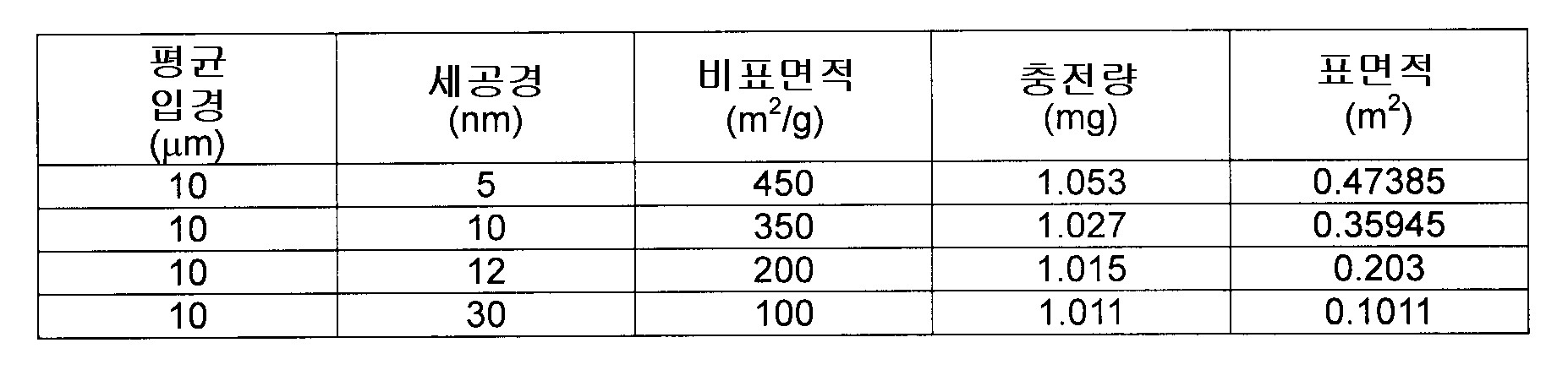
계속해서, 시료의 조제를 행했다. 시료의 조제에는, 상술한 RNeasy Protect Mini Kit(QIAGEN사제)를 이용했다. HeLa 세포를 배양하고, 킷중의 Buffer RLT를 이용해서 세포를 용해한 후, 프로토콜에 따라서, 핵산 용액을 얻었다. 그리고, 스핀 필터를 가지는 컬럼에 그 핵산 용액을 넣고, 원심 처리해서, 불순물을 제거했다(전처리 필터링). 여기에서는, 합계 3회의 전처리 필터링을 행하고, 각각 세공경이 5㎛, 0.45㎛, 0.1㎛인 3종의 원심 컬럼(Millipore사제, "Amicon Ultrafree-MC" 원심식 필터 컬럼)을, 세공경이 큰 것부터 순서대로 이용했다. Subsequently, the sample was prepared. The above-described RNeasy® Protect® Mini ™ Kit (manufactured by QIAGEN) was used for preparation of the sample. HeLa cells were cultured and the cells were lysed using Buffer # RLT in the kit, and then nucleic acid solutions were obtained according to the protocol. The nucleic acid solution was placed in a column having a spin filter and centrifuged to remove impurities (pretreatment filtering). Here, pretreatment filtering was performed three times in total, and three types of centrifugal columns ("Amicon® Ultrafree-MC" centrifugal filter columns manufactured by Millipore) having a pore diameter of 5 µm, 0.45 µm and 0.1 µm, respectively, had a large pore diameter. It was used in order from thing.
계속해서, 핵산 용액을, 이 유로계에 주입했다. 조립한 유로계 가운데, 필터를 장착한 측의 반대측에 있어서, 필·포토를 떼어내고, 대신에 다른 튜브를 부착했다. 그 튜브의 타단에는, 유로계에 액상을 주입할 수 있도록, 너트와 페럴을 이용해서 인터널·유니온을 부착하고, 그곳에 루어록식 니들을 접속하며, 그곳에 루어록형의 실린더를 부착했다. 그리고, 필터측을 감압하고, 그의 반대측의 루어록형 실린더로부터 마이크로 실린더·펌프를 이용해서 시료를 유로계(튜브내)에 주입하고, 실리카에 핵산을 흡착시켰다. Subsequently, the nucleic acid solution was injected into this flow path system. In the assembled flow path system, the peeling port was removed on the opposite side to the side where the filter was mounted, and another tube was attached instead. At the other end of the tube, an internal union was attached using a nut and a ferrule, a luer lock needle was connected thereto, and a luer lock cylinder was attached thereto so that the liquid phase could be injected into the flow path system. Then, the filter side was depressurized, the sample was injected into the flow path system (in a tube) using the micro cylinder pump from the Luerlock-type cylinder on the opposite side, and the nucleic acid was adsorb | sucked to the silica.
계속해서, 유로계내의 실리카 마이크로 비즈에 포착된 핵산(전RNA)을 회수했다. Buffer RW1(킷중의 시약) 700μL, Buffer RPE(킷중의 시약) 5mL를, 유로계내에 주입하고, 유로계내를 세정했다. 이 단계에 의해, 실리카 마이크로 비즈에 포착된 핵산 이외의 물질을 제거했다. 다음에, 100μL의 RNase-free water를, 유로계내에 주입하고, 통과한 용액을 회수했다. 이 단계에 의해, 실리카 마이크로 비즈에 포착된 핵산을, 용액중에 용출했다. Subsequently, the nucleic acid (all RNA) captured in the silica microbeads in the flow path system was recovered. 700 µL of Buffer # RW1 (Reagent in Kit) and 5 mL of Buffer® RPE (Reagent in Kit) were injected into the flow path system to wash the inside of the flow path system. By this step, substances other than the nucleic acid trapped in the silica microbeads were removed. Next, 100 µL of RNase-free water was injected into the flow path system and the solution passed therethrough was recovered. By this step, the nucleic acid trapped in the silica microbeads was eluted in solution.
그리고, 분광 광도계(260㎚)를 이용해서, 회수한 핵산 용액의 정량을 행했다. And the collected nucleic acid solution was quantified using the spectrophotometer (260 nm).
결과를 도 6 및 도 7에 도시한다. 각각, 도 6은 실리카 마이크로 비즈의 세공경마다의 전RNA 회수량을 도시하는 그래프, 도 7은 실리카 마이크로 비즈의 세공경마다의 단위 비표면적당의 전RNA 회수량을 도시하는 그래프이다. 양 도면중의 그래프의 횡축은 실리카 마이크로 비즈의 세공경(단위:㎚)을 나타낸다. 도 6의 그래프의 종축은 전RNA 회수량(단위:㎍)을, 도 7의 그래프의 종축은 전RNA 회수량을 단위 표면적으로 나눈(제산한) 값(단위:㎍/㎡)을, 각각 나타낸다. The results are shown in FIGS. 6 and 7. 6 is a graph showing the total RNA recovery amount per pore diameter of silica microbeads, and FIG. 7 is a graph showing the total RNA recovery amount per unit specific surface area per pore diameter of silica microbeads. The horizontal axis of the graph in both figures shows the pore diameter (unit: nm) of a silica microbead. The vertical axis of the graph of FIG. 6 represents the total RNA recovery amount (unit: µg), and the vertical axis of the graph of FIG. 7 represents the value (unit: µg / m 2) obtained by dividing (dividing) the total RNA recovery by the unit surface area. .
핵산은 실리카 마이크로 비즈의 표면에 카오트로픽 이온의 효과에 의해서 흡착되고, 그의 탈착(脫着; desorption)에 의해서 회수되기 때문에, 그 회수량은 실리카 마이크로 비즈의 비표면적(단위 중량당의 표면적)과 상관된다고 추측할 수 있다. 한편, 표 2에 나타내는 바와 같이, 각 실리카 마이크로 비즈의 충전량은 거의 동등하다. 따라서, 핵산의 회수량은 실리카 마이크로 비즈의 단위 표면적과 상관된다고 추측할 수 있다. Since the nucleic acid is adsorbed on the surface of the silica microbeads by the effect of chaotropic ions and is recovered by desorption thereof, the amount of recovery is correlated with the specific surface area (surface area per unit weight) of the silica microbeads. I can guess. On the other hand, as shown in Table 2, the filling amount of each silica microbead is almost equivalent. Therefore, it can be inferred that the recovery amount of nucleic acid correlates with the unit surface area of the silica microbeads.
그에 대해서, 도 7의 결과에서는, 단위 표면적당에 있어서의 전RNA의 회수량은, 실리카 마이크로 비즈의 세공경이 5㎚ 또는 30㎚인 경우, 회수량과 단위 표면적이 거의 상관한 것에 대해, 실리카 마이크로 비즈의 세공경이 10㎚인 경우, 단위 표면적당의 회수량이 많고, 또 12㎚인 경우, 단위 표면적당의 회수량이 현저하게 높았다. In contrast, in the results of FIG. 7, the amount of recovered RNA per unit surface area is substantially different from the amount recovered and the unit surface area when the pore diameter of silica microbeads is 5 nm or 30 nm. When the pore diameter of the microbeads was 10 nm, the recovery amount per unit surface area was large, and when it was 12 nm, the recovery amount per unit surface area was remarkably high.
이 결과는, 세공경이 10㎚ 또는 12㎚인 경우, 특히 세공경이 12㎚인 경우, 실리카 마이크로 비즈에의 핵산의 흡착량이 현저하게 증가하는 것을 시사한다. 즉, 본 실험 결과는, 세공경이 12㎚ 근방(예를 들면, 6∼29㎚, 보다 적합하게는 11∼29㎚)인 실리카 마이크로 비즈를 이용하는 것에 의해, 핵산의 회수량을 현저하게 증가시킬 수 있다는 것을 시사한다. This result suggests that when the pore diameter is 10 nm or 12 nm, especially when the pore diameter is 12 nm, the amount of adsorption of nucleic acid onto the silica microbeads is significantly increased. That is, the results of this experiment show that the recovery of nucleic acid can be significantly increased by using silica microbeads having a pore diameter of about 12 nm (for example, 6 to 29 nm, more preferably 11 to 29 nm). Suggest that you can.
실시예 7에서는, 세공경 10㎛인 필터를 이용해서 시료의 전처리 필터링을 행하고 나서, 전RNA를 회수했다. 실험 단계의 개요는 다음과 같다.In Example 7, all RNAs were recovered after pretreatment filtering of the sample using a filter having a pore diameter of 10 µm. The outline of the experimental phase is as follows.
처음에, 시료의 조제를 행했다. 시료의 조제에는, 상술한 RNeasy Protect Mini Kit(QIAGEN사제)에 부속되는 시약을 이용했다. HeLa 세포를 배양하고, 킷중의 Buffer RLT를 이용해서 세포를 용해한 후, 프로토콜에 따라서, 세포 용해액을 얻었다. First, a sample was prepared. For the preparation of the sample, a reagent attached to the above-described RNeasy® Protect® Mini ™ Kit (manufactured by QIAGEN) was used. HeLa cells were cultured and the cells were lysed using Buffer # RLT in the kit, and then cell lysates were obtained according to the protocol.
계속해서, 그 세포 용해액의 전처리 필터링을 행했다. 세공경 10㎛의 원심 필터가 장착된(달린) 스핀 컬럼("MicroSpin Empty Columns", GE 헬스케어 바이오사이언스 주식회사제)에 세포 용해액을 넣고, 원심 처리해서 불순물을 제거했다. 또, 각각 세공경이 5㎛인 원심 필터가 장착된 스핀 컬럼("Amicon Ultrafree-MC", Millipore사제)를 이용해서 불순물을 제거했다.Subsequently, pretreatment filtering of the cell lysate was performed. The cell lysate was placed in a spin column ("MicroSpin®Empty®Columns", manufactured by GE Healthcare Bioscience Co., Ltd.) equipped with a centrifugal filter having a pore diameter of 10 µm, and centrifuged to remove impurities. In addition, impurities were removed using a spin column ("Amicon® Ultrafree-MC" manufactured by Millipore) equipped with a centrifugal filter having a pore diameter of 5 µm, respectively.
그리고, 상기 킷을 이용해서 전RNA를 회수하고, 분광 광도계(260㎚)를 이용해서, 회수한 전 RNA의 정량을 행했다. And all RNA was collect | recovered using the said kit, and the collect | recovered all RNA was quantified using the spectrophotometer (260 nm).
결과를 도 8 및 도 9에 나타낸다. 도 8은 세공경 10㎛인 원심 필터가 장착된 스핀 컬럼에 의한 필터링 처리를 행한 경우에서의 전필터(all filters) 처리 종료후의 최종 여과량을 도시하는 그래프, 도 9는 마찬가지로 전RNA 회수량을 도시하는 그래프이다. 양 도면중의 그래프의 횡축은, 세공경 10㎛의 원심 필터의 사용 유무를 나타낸다. 도 8중의 그래프의 종축은 최종 여과량(단위:㎕)을, 도 9중의 그래프의 종축은 전RNA 회수량(단위:㎍)을, 각각 나타낸다. The results are shown in FIGS. 8 and 9. FIG. 8 is a graph showing the final filtration amount after the completion of all filters in the case of performing a filtering process with a spin column equipped with a centrifugal filter having a pore diameter of 10 μm. FIG. 9 similarly shows the total RNA recovery amount. It is a graph to show. The horizontal axis of the graph in both figures shows the use of the centrifugal filter of 10 micrometers of pore diameters. The vertical axis of the graph in FIG. 8 represents the final filtration amount (unit: µl), and the vertical axis of the graph in FIG. 9 represents the total RNA recovery amount (unit: µg).
도 8에 도시하는 바와 같이, 세공경 10㎛의 원심 필터를 이용해서 전처리 필터링을 행한 경우, 전필터 처리 종료후의 최종 여과량이, 세공경 10㎛의 원심 필터를 이용하지 않은 경우와 비교해서, 현저하게 증가했다. 또, 세공경 10㎛의 원심 필터를 이용하지 않은 경우, 세공경 5㎛의 필터에 눈막힘(clogging)이 생기고 있었다. 이들의 결과는, 세포 용해액 중에는, 10㎛ 이상의 불순물이 많이 함유한다는 것을 나타낸다.As shown in FIG. 8, when pretreatment filtering is performed using the centrifugal filter of 10 micrometers of pore diameters, the final filtration amount after completion | finish of prefilter process is remarkable compared with the case where the centrifugal filter of 10 micrometers of pore diameters is not used. Increased. Moreover, when the centrifugal filter of 10 micrometers of pore diameters was not used, clogging occurred in the filter of 5 micrometers of pore diameters. These results show that the cell lysate contains many impurities of 10 µm or more.
또, 도 9에 도시하는 바와 같이, 세공경 10㎛의 원심 필터를 이용해서 전처리 필터링을 행한 경우, 전RNA 회수량이, 세공경 10㎛의 원심 필터를 이용하지 않은 경우와 비교해서, 현저하게 증가했다. 이 결과는, 세공경 10㎛의 원심 필터를 이용해서 전처리 필터링을 행하는 것에 의해, 세공경 5㎛의 필터에의 눈막힘을 억제할 수 있으며, 그것에 의해, 전RNA의 회수 효율을 높게 할 수 있는 것을 나타낸다.In addition, as shown in FIG. 9, when pretreatment filtering is performed using the centrifugal filter of 10 micrometers of pore diameters, all RNA collection amount is remarkably compared with the case where the centrifugal filter of 10 micrometers of pore diameters is not used. Increased. As a result of this, by pretreatment filtering using a centrifugal filter having a pore diameter of 10 µm, clogging to a filter having a pore diameter of 5 µm can be suppressed, whereby the recovery efficiency of all RNAs can be increased. Indicates.
따라서, 본 실험 결과는, 세공경 10㎛ 근방(예를 들면, 세공경 6㎛∼25㎛)의 원심 필터 등을 이용해서 시료의 전처리 필터링을 행하는 것에 의해, 핵산 회수 효율을 증가시킬 수 있다는 것을 시사한다. Therefore, the results of this experiment show that nucleic acid recovery efficiency can be increased by performing pretreatment filtering of a sample using a centrifugal filter having a pore diameter of about 10 μm (for example, a pore diameter of 6 μm to 25 μm). Suggest.
유전자 검사 등에서는, 혈액 등의 생물 시료로부터 핵산만을 추출하고, 추출한 핵산을 DNA 칩 등의 각 반응 영역에 공급하며, 핵산 검출 장치 등에 의해, 분석을 행한다. 본 발명을 이용하는 것에 의해, 예를 들면 생물 시료를 DNA 칩 등에 공급하는 유로에 있어서, 생물 시료로부터 핵산을 추출하고, 그 추출액을 DNA 칩에 공급할 수가 있다. 즉, 예를 들면 본 발명을 핵산 공급 장치에 결합하는(응용하는) 것에 의해, 생물 시료로부터 추출한 핵산을, 보다 간이하게 또한 자동 처리로, DNA 칩의 반응 영역 등에 공급할 수가 있다. In a genetic test or the like, only nucleic acid is extracted from a biological sample such as blood, and the extracted nucleic acid is supplied to each reaction region such as a DNA chip and analyzed using a nucleic acid detection device or the like. By using the present invention, for example, in a flow path for supplying a biological sample to a DNA chip or the like, the nucleic acid can be extracted from the biological sample and the extract can be supplied to the DNA chip. That is, for example, by binding (applying) the present invention to a nucleic acid supply device, the nucleic acid extracted from the biological sample can be supplied to the reaction region of the DNA chip or the like more easily and automatically by processing.
또, 그 핵산 공급 장치 자체를, 핵산 분석 장치에 결합할(응용할) 수도 있다. 이것에 의해, 생물 시료의 공급부터 유전자 해석까지의 일련의 조작을 자동화할 수 있을 가능성이 있으며, 또 장치의 일체화·소형화를 실현할 수 있을 가능성이 있다. The nucleic acid supply device itself can also be coupled (applied) to a nucleic acid analysis device. Thereby, it is possible to automate a series of operations from the supply of biological samples to genetic analysis, and the integration and miniaturization of the device may be realized.
본 발명은, PCR(중합효소 연쇄 반응)를 실시가능한 장치, 예를 들면 PCR 장치, 및 시퀀서(sequencer) 등의 장치에도 결합될 수가 있다. 예를 들면, 생물 시료에 미소 유로를 통과시키고, 추출한 핵산 용액을 반응 영역에 공급하는 것에 의해, PCR 등의 전처리를 간략화할 수가 있다. 또, 이것에 의해, 일련의 조작의 자동화나 장치의 소형화를 실현할 수가 있다. The present invention can also be coupled to a device capable of carrying out a PCR (polymerase chain reaction), such as a PCR device and a sequencer. For example, pretreatment such as PCR can be simplified by passing a microchannel through a biological sample and supplying the extracted nucleic acid solution to the reaction zone. In this way, automation of a series of operations and miniaturization of the apparatus can be realized.
본 발명은 첨부된 청구범위 또는 이와 동등한 범위 내에서 설계 요구 조건 및 그 밖의 요인에 의거하여 각종 변경, 조합, 수정 및 교체가 실시될 수 있다는 것을, 당업자라면 당연히 이해할 수 있을 것이다.It will be apparent to those skilled in the art that the present invention may be practiced with various modifications, combinations, modifications and substitutions within the scope of the appended claims or equivalents thereof, based on design requirements and other factors.
도 1은 실시예 1 등에서, 실험에 이용한 유로계를 도시하는 모식도. BRIEF DESCRIPTION OF THE DRAWINGS The schematic diagram which shows the flow path system used for experiment in Example 1 etc .;
도 2는 실시예 1에서, 중공 실리카 마이크로 비즈의 양과 핵산 회수량(정량 결과)과의 상관을 도시하는 그래프. FIG. 2 is a graph showing the correlation between the amount of hollow silica microbeads and the amount of nucleic acid recovery (quantitative result) in Example 1. FIG.
도 3은 실시예 1에서, 핵산의 회수 효율을 도시하는 그래프. 3 is a graph showing the recovery efficiency of nucleic acids in Example 1. FIG.
도 4는 실시예 4에서, 비표면적이 큰 실리카 마이크로 비즈를 이용한 경우에서의 핵산 회수량을 도시하는 그래프. FIG. 4 is a graph showing the amount of nucleic acid recovery in the case of using silica microbeads having a large specific surface area in Example 4. FIG.
도 5는 실시예 5에서, HeLa 세포로부터 추출한 핵산 회수량을 도시하는 그래프. FIG. 5 is a graph showing the amount of nucleic acid recovery extracted from HeLa cells in Example 5. FIG.
도 6은 실시예 6에서, 실리카 마이크로 비즈의 세공경마다의 전RNA 회수량을 도시하는 그래프. FIG. 6 is a graph showing the total RNA recovery amount per pore diameter of silica micro beads in Example 6. FIG.
도 7은 실시예 6에서, 실리카 마이크로 비즈의 세공경마다의 단위 비표면적당의 전RNA 회수량을 도시하는 그래프. 7 is a graph showing the total RNA recovery amount per unit specific surface area for each pore diameter of silica microbeads in Example 6. FIG.
도 8은 실시예 7에서, 세공경 10㎛의 원심 필터가 장착된 스핀 컬럼에 필터링 처리를 행한 경우에서의 전필터 처리 종료후의 최종 여과량을 도시하는 그래프. 8 is a graph showing the final filtration amount after the completion of the pre-filtering treatment in the case where the filtering treatment was performed on a spin column equipped with a centrifugal filter having a pore diameter of 10 µm in Example 7. FIG.
도 9는 실시예 7에서, 세공경 10㎛의 원심 필터가 장착된 스핀 컬럼에 필터링 처리를 행한 경우에서의 전RNA 회수량을 도시하는 그래프. FIG. 9 is a graph showing the total RNA recovery amount when a filtering process is performed on a spin column equipped with a centrifugal filter having a pore diameter of 10 µm in Example 7. FIG.
Claims (10)
Applications Claiming Priority (2)
| Application Number | Priority Date | Filing Date | Title |
|---|---|---|---|
| JP2006257791A JP4297148B2 (en) | 2006-09-22 | 2006-09-22 | Nucleic acid recovery apparatus and nucleic acid recovery method |
| JPJP-P-2006-00257791 | 2006-09-22 |
Publications (2)
| Publication Number | Publication Date |
|---|---|
| KR20080027134A true KR20080027134A (en) | 2008-03-26 |
| KR101423635B1 KR101423635B1 (en) | 2014-07-25 |
Family
ID=38982955
Family Applications (1)
| Application Number | Title | Priority Date | Filing Date |
|---|---|---|---|
| KR1020070089057A Expired - Fee Related KR101423635B1 (en) | 2006-09-22 | 2007-09-03 | Microchannel, nucleic acid recovery device and nucleic acid recovery method |
Country Status (5)
| Country | Link |
|---|---|
| US (1) | US8691559B2 (en) |
| EP (1) | EP1903110B1 (en) |
| JP (1) | JP4297148B2 (en) |
| KR (1) | KR101423635B1 (en) |
| CN (1) | CN101153264B (en) |
Cited By (1)
| Publication number | Priority date | Publication date | Assignee | Title |
|---|---|---|---|---|
| US8664377B2 (en) | 2009-09-30 | 2014-03-04 | Samsung Electronics Co., Ltd. | Method and apparatus for isolating nucleic acids |
Families Citing this family (6)
| Publication number | Priority date | Publication date | Assignee | Title |
|---|---|---|---|---|
| JP4904973B2 (en) * | 2005-08-08 | 2012-03-28 | ソニー株式会社 | Nucleic acid recovery method, nucleic acid recovery apparatus, and nucleic acid supply apparatus |
| US8735103B2 (en) * | 2006-12-05 | 2014-05-27 | Electronics And Telecommunications Research Institute | Natural convection-driven PCR apparatus and method using disposable polymer chip |
| CN102242053B (en) * | 2011-04-01 | 2014-06-04 | 沈越 | Biochip with polymer three-dimensional nanostructure |
| US9803237B2 (en) | 2012-04-24 | 2017-10-31 | California Institute Of Technology | Slip-induced compartmentalization |
| US11175205B2 (en) * | 2015-11-02 | 2021-11-16 | Biofire Diagnostics, Llc | Sample preparation for difficult sample types |
| WO2019035335A1 (en) * | 2017-08-18 | 2019-02-21 | Agc株式会社 | Nucleic acid collection method, nucleic-acid-binding carrier, and nucleic acid collection kit |
Family Cites Families (16)
| Publication number | Priority date | Publication date | Assignee | Title |
|---|---|---|---|---|
| US5234809A (en) | 1989-03-23 | 1993-08-10 | Akzo N.V. | Process for isolating nucleic acid |
| DE4321904B4 (en) | 1993-07-01 | 2013-05-16 | Qiagen Gmbh | Method for chromatographic purification and separation of nucleic acid mixtures |
| US6426126B1 (en) * | 1997-12-19 | 2002-07-30 | Amt Holdings, Inc. | Preparation of metal coatings |
| US7183002B2 (en) | 2000-03-24 | 2007-02-27 | Qiagen, Gmbh | Porous ferro- or ferrimagnetic glass particles for isolating molecules |
| US20050089850A1 (en) * | 2000-10-02 | 2005-04-28 | Jeffrey Van Ness | Genotyping by liquid chromatographic analysis of short nucleic acid fragments |
| JP2002209580A (en) | 2001-01-15 | 2002-07-30 | Jsr Corp | Method for separating nucleic acid and spherical carrier |
| ATE449186T1 (en) | 2001-11-28 | 2009-12-15 | Applied Biosystems Llc | COMPOSITIONS AND METHODS FOR SELECTIVE NUCLEIC ACID ISOLATION |
| US20040091411A1 (en) * | 2002-11-08 | 2004-05-13 | Bijan Modrek-Najafabadi | High surface area, high porosity silica packing with narrow particle and pore diameter distribution and methods of making same |
| WO2004086055A1 (en) * | 2003-03-24 | 2004-10-07 | Sony Corporation | Microchip, nucleic acid extracting kit, and nucleic acid extracting method |
| JP2005110503A (en) | 2003-10-02 | 2005-04-28 | Arkray Inc | Method for purifying nucleic acid and device therefor |
| JP4441610B2 (en) | 2004-05-31 | 2010-03-31 | 独立行政法人産業技術総合研究所 | Nucleic acid isolation method |
| JP4626350B2 (en) * | 2005-03-22 | 2011-02-09 | ソニー株式会社 | A flow path system having a reaction part suitable for hybridization detection, and a hybridization detection apparatus using the flow path system |
| JP4904973B2 (en) | 2005-08-08 | 2012-03-28 | ソニー株式会社 | Nucleic acid recovery method, nucleic acid recovery apparatus, and nucleic acid supply apparatus |
| US20070084774A1 (en) * | 2005-10-19 | 2007-04-19 | Agilent Technologies, Inc. | Chromatographic stationary phase |
| US20070090034A1 (en) * | 2005-10-20 | 2007-04-26 | Agilent Technologies, Inc. | Substrate for a chromatography column |
| JP4940756B2 (en) * | 2006-05-22 | 2012-05-30 | ソニー株式会社 | Micro channel system |
-
2006
- 2006-09-22 JP JP2006257791A patent/JP4297148B2/en not_active Expired - Fee Related
-
2007
- 2007-08-29 EP EP07016929.7A patent/EP1903110B1/en not_active Ceased
- 2007-09-03 KR KR1020070089057A patent/KR101423635B1/en not_active Expired - Fee Related
- 2007-09-14 CN CN2007101521307A patent/CN101153264B/en not_active Expired - Fee Related
- 2007-09-20 US US11/858,595 patent/US8691559B2/en active Active
Cited By (1)
| Publication number | Priority date | Publication date | Assignee | Title |
|---|---|---|---|---|
| US8664377B2 (en) | 2009-09-30 | 2014-03-04 | Samsung Electronics Co., Ltd. | Method and apparatus for isolating nucleic acids |
Also Published As
| Publication number | Publication date |
|---|---|
| CN101153264A (en) | 2008-04-02 |
| US20080161553A1 (en) | 2008-07-03 |
| KR101423635B1 (en) | 2014-07-25 |
| EP1903110A3 (en) | 2008-04-02 |
| JP2008072987A (en) | 2008-04-03 |
| EP1903110A2 (en) | 2008-03-26 |
| US8691559B2 (en) | 2014-04-08 |
| CN101153264B (en) | 2013-03-27 |
| JP4297148B2 (en) | 2009-07-15 |
| EP1903110B1 (en) | 2014-11-26 |
Similar Documents
| Publication | Publication Date | Title |
|---|---|---|
| JP5977921B2 (en) | Apparatus, system and method for purifying nucleic acids | |
| KR101005924B1 (en) | Nucleic Acid Extraction Device | |
| KR101423635B1 (en) | Microchannel, nucleic acid recovery device and nucleic acid recovery method | |
| CN103282121B (en) | For the microfluidic device that nucleic acid extraction is separated with classification | |
| US20150166592A1 (en) | Selective Nucleic Acid Fragment Recovery | |
| CN108064262B (en) | Device and method for nucleic acid extraction | |
| JP4699868B2 (en) | Nucleic acid purification method and nucleic acid purification instrument | |
| EP1637599A2 (en) | Method of nucleic acid isolation | |
| JP4597870B2 (en) | Mechanism for separation and purification of DNA, etc. | |
| JP4904973B2 (en) | Nucleic acid recovery method, nucleic acid recovery apparatus, and nucleic acid supply apparatus | |
| US20150240291A1 (en) | Lyophilizate of substance-binding solid-phase carrier, vessel for binding substance in substance-containing liquid to substance-binding solid-phase carrier, and method of producing lyophilizate containing substance-binding solid-phase carrier | |
| CN112391378A (en) | Kit and preparation method thereof | |
| WO2024013952A1 (en) | Method for controlling liquid transport in flow path of biomolecule analyzer using computer, and biomolecule purification system | |
| Ceriotti et al. | Combined nucleic acid extraction and enrichment in bead-packed plastic beds | |
| HK1150166B (en) | Apparatus, system, and method for purifying nucleic acids |
Legal Events
| Date | Code | Title | Description |
|---|---|---|---|
| PA0109 | Patent application |
St.27 status event code: A-0-1-A10-A12-nap-PA0109 |
|
| PG1501 | Laying open of application |
St.27 status event code: A-1-1-Q10-Q12-nap-PG1501 |
|
| A201 | Request for examination | ||
| PA0201 | Request for examination |
St.27 status event code: A-1-2-D10-D11-exm-PA0201 |
|
| E902 | Notification of reason for refusal | ||
| PE0902 | Notice of grounds for rejection |
St.27 status event code: A-1-2-D10-D21-exm-PE0902 |
|
| E13-X000 | Pre-grant limitation requested |
St.27 status event code: A-2-3-E10-E13-lim-X000 |
|
| P11-X000 | Amendment of application requested |
St.27 status event code: A-2-2-P10-P11-nap-X000 |
|
| P13-X000 | Application amended |
St.27 status event code: A-2-2-P10-P13-nap-X000 |
|
| E701 | Decision to grant or registration of patent right | ||
| PE0701 | Decision of registration |
St.27 status event code: A-1-2-D10-D22-exm-PE0701 |
|
| GRNT | Written decision to grant | ||
| PR0701 | Registration of establishment |
St.27 status event code: A-2-4-F10-F11-exm-PR0701 |
|
| PR1002 | Payment of registration fee |
St.27 status event code: A-2-2-U10-U11-oth-PR1002 Fee payment year number: 1 |
|
| PG1601 | Publication of registration |
St.27 status event code: A-4-4-Q10-Q13-nap-PG1601 |
|
| PR1001 | Payment of annual fee |
St.27 status event code: A-4-4-U10-U11-oth-PR1001 Fee payment year number: 4 |
|
| PR1001 | Payment of annual fee |
St.27 status event code: A-4-4-U10-U11-oth-PR1001 Fee payment year number: 5 |
|
| PC1903 | Unpaid annual fee |
St.27 status event code: A-4-4-U10-U13-oth-PC1903 Not in force date: 20190722 Payment event data comment text: Termination Category : DEFAULT_OF_REGISTRATION_FEE |
|
| PC1903 | Unpaid annual fee |
St.27 status event code: N-4-6-H10-H13-oth-PC1903 Ip right cessation event data comment text: Termination Category : DEFAULT_OF_REGISTRATION_FEE Not in force date: 20190722 |
|
| PN2301 | Change of applicant |
St.27 status event code: A-5-5-R10-R13-asn-PN2301 St.27 status event code: A-5-5-R10-R11-asn-PN2301 |