KR102738427B1 - Allograft tolerance without systemic immunosuppression - Google Patents
Allograft tolerance without systemic immunosuppression Download PDFInfo
- Publication number
- KR102738427B1 KR102738427B1 KR1020207000550A KR20207000550A KR102738427B1 KR 102738427 B1 KR102738427 B1 KR 102738427B1 KR 1020207000550 A KR1020207000550 A KR 1020207000550A KR 20207000550 A KR20207000550 A KR 20207000550A KR 102738427 B1 KR102738427 B1 KR 102738427B1
- Authority
- KR
- South Korea
- Prior art keywords
- delete delete
- cell
- cells
- promoter
- mfge8
- Prior art date
- Legal status (The legal status is an assumption and is not a legal conclusion. Google has not performed a legal analysis and makes no representation as to the accuracy of the status listed.)
- Active
Links
Images


Classifications
-
- C—CHEMISTRY; METALLURGY
- C12—BIOCHEMISTRY; BEER; SPIRITS; WINE; VINEGAR; MICROBIOLOGY; ENZYMOLOGY; MUTATION OR GENETIC ENGINEERING
- C12N—MICROORGANISMS OR ENZYMES; COMPOSITIONS THEREOF; PROPAGATING, PRESERVING, OR MAINTAINING MICROORGANISMS; MUTATION OR GENETIC ENGINEERING; CULTURE MEDIA
- C12N5/00—Undifferentiated human, animal or plant cells, e.g. cell lines; Tissues; Cultivation or maintenance thereof; Culture media therefor
- C12N5/06—Animal cells or tissues; Human cells or tissues
- C12N5/0602—Vertebrate cells
- C12N5/0603—Embryonic cells ; Embryoid bodies
- C12N5/0606—Pluripotent embryonic cells, e.g. embryonic stem cells [ES]
-
- A—HUMAN NECESSITIES
- A61—MEDICAL OR VETERINARY SCIENCE; HYGIENE
- A61K—PREPARATIONS FOR MEDICAL, DENTAL OR TOILETRY PURPOSES
- A61K35/00—Medicinal preparations containing materials or reaction products thereof with undetermined constitution
- A61K35/12—Materials from mammals; Compositions comprising non-specified tissues or cells; Compositions comprising non-embryonic stem cells; Genetically modified cells
- A61K35/48—Reproductive organs
- A61K35/54—Ovaries; Ova; Ovules; Embryos; Foetal cells; Germ cells
- A61K35/545—Embryonic stem cells; Pluripotent stem cells; Induced pluripotent stem cells; Uncharacterised stem cells
-
- A—HUMAN NECESSITIES
- A61—MEDICAL OR VETERINARY SCIENCE; HYGIENE
- A61K—PREPARATIONS FOR MEDICAL, DENTAL OR TOILETRY PURPOSES
- A61K38/00—Medicinal preparations containing peptides
- A61K38/16—Peptides having more than 20 amino acids; Gastrins; Somatostatins; Melanotropins; Derivatives thereof
- A61K38/17—Peptides having more than 20 amino acids; Gastrins; Somatostatins; Melanotropins; Derivatives thereof from animals; from humans
- A61K38/1703—Peptides having more than 20 amino acids; Gastrins; Somatostatins; Melanotropins; Derivatives thereof from animals; from humans from vertebrates
- A61K38/1709—Peptides having more than 20 amino acids; Gastrins; Somatostatins; Melanotropins; Derivatives thereof from animals; from humans from vertebrates from mammals
-
- A—HUMAN NECESSITIES
- A61—MEDICAL OR VETERINARY SCIENCE; HYGIENE
- A61K—PREPARATIONS FOR MEDICAL, DENTAL OR TOILETRY PURPOSES
- A61K38/00—Medicinal preparations containing peptides
- A61K38/16—Peptides having more than 20 amino acids; Gastrins; Somatostatins; Melanotropins; Derivatives thereof
- A61K38/17—Peptides having more than 20 amino acids; Gastrins; Somatostatins; Melanotropins; Derivatives thereof from animals; from humans
- A61K38/177—Receptors; Cell surface antigens; Cell surface determinants
- A61K38/1774—Immunoglobulin superfamily (e.g. CD2, CD4, CD8, ICAM molecules, B7 molecules, Fc-receptors, MHC-molecules)
-
- A—HUMAN NECESSITIES
- A61—MEDICAL OR VETERINARY SCIENCE; HYGIENE
- A61K—PREPARATIONS FOR MEDICAL, DENTAL OR TOILETRY PURPOSES
- A61K38/00—Medicinal preparations containing peptides
- A61K38/16—Peptides having more than 20 amino acids; Gastrins; Somatostatins; Melanotropins; Derivatives thereof
- A61K38/17—Peptides having more than 20 amino acids; Gastrins; Somatostatins; Melanotropins; Derivatives thereof from animals; from humans
- A61K38/18—Growth factors; Growth regulators
- A61K38/1808—Epidermal growth factor [EGF] urogastrone
-
- A—HUMAN NECESSITIES
- A61—MEDICAL OR VETERINARY SCIENCE; HYGIENE
- A61K—PREPARATIONS FOR MEDICAL, DENTAL OR TOILETRY PURPOSES
- A61K38/00—Medicinal preparations containing peptides
- A61K38/16—Peptides having more than 20 amino acids; Gastrins; Somatostatins; Melanotropins; Derivatives thereof
- A61K38/17—Peptides having more than 20 amino acids; Gastrins; Somatostatins; Melanotropins; Derivatives thereof from animals; from humans
- A61K38/18—Growth factors; Growth regulators
- A61K38/1841—Transforming growth factor [TGF]
-
- A—HUMAN NECESSITIES
- A61—MEDICAL OR VETERINARY SCIENCE; HYGIENE
- A61K—PREPARATIONS FOR MEDICAL, DENTAL OR TOILETRY PURPOSES
- A61K38/00—Medicinal preparations containing peptides
- A61K38/16—Peptides having more than 20 amino acids; Gastrins; Somatostatins; Melanotropins; Derivatives thereof
- A61K38/17—Peptides having more than 20 amino acids; Gastrins; Somatostatins; Melanotropins; Derivatives thereof from animals; from humans
- A61K38/19—Cytokines; Lymphokines; Interferons
- A61K38/195—Chemokines, e.g. RANTES
-
- A—HUMAN NECESSITIES
- A61—MEDICAL OR VETERINARY SCIENCE; HYGIENE
- A61K—PREPARATIONS FOR MEDICAL, DENTAL OR TOILETRY PURPOSES
- A61K38/00—Medicinal preparations containing peptides
- A61K38/16—Peptides having more than 20 amino acids; Gastrins; Somatostatins; Melanotropins; Derivatives thereof
- A61K38/55—Protease inhibitors
-
- A—HUMAN NECESSITIES
- A61—MEDICAL OR VETERINARY SCIENCE; HYGIENE
- A61K—PREPARATIONS FOR MEDICAL, DENTAL OR TOILETRY PURPOSES
- A61K45/00—Medicinal preparations containing active ingredients not provided for in groups A61K31/00 - A61K41/00
- A61K45/06—Mixtures of active ingredients without chemical characterisation, e.g. antiphlogistics and cardiaca
-
- A—HUMAN NECESSITIES
- A61—MEDICAL OR VETERINARY SCIENCE; HYGIENE
- A61K—PREPARATIONS FOR MEDICAL, DENTAL OR TOILETRY PURPOSES
- A61K48/00—Medicinal preparations containing genetic material which is inserted into cells of the living body to treat genetic diseases; Gene therapy
- A61K48/0008—Medicinal preparations containing genetic material which is inserted into cells of the living body to treat genetic diseases; Gene therapy characterised by an aspect of the 'non-active' part of the composition delivered, e.g. wherein such 'non-active' part is not delivered simultaneously with the 'active' part of the composition
-
- A—HUMAN NECESSITIES
- A61—MEDICAL OR VETERINARY SCIENCE; HYGIENE
- A61K—PREPARATIONS FOR MEDICAL, DENTAL OR TOILETRY PURPOSES
- A61K48/00—Medicinal preparations containing genetic material which is inserted into cells of the living body to treat genetic diseases; Gene therapy
- A61K48/005—Medicinal preparations containing genetic material which is inserted into cells of the living body to treat genetic diseases; Gene therapy characterised by an aspect of the 'active' part of the composition delivered, i.e. the nucleic acid delivered
-
- A—HUMAN NECESSITIES
- A61—MEDICAL OR VETERINARY SCIENCE; HYGIENE
- A61K—PREPARATIONS FOR MEDICAL, DENTAL OR TOILETRY PURPOSES
- A61K9/00—Medicinal preparations characterised by special physical form
- A61K9/0012—Galenical forms characterised by the site of application
- A61K9/0019—Injectable compositions; Intramuscular, intravenous, arterial, subcutaneous administration; Compositions to be administered through the skin in an invasive manner
- A61K9/0021—Intradermal administration, e.g. through microneedle arrays, needleless injectors
-
- A—HUMAN NECESSITIES
- A61—MEDICAL OR VETERINARY SCIENCE; HYGIENE
- A61L—METHODS OR APPARATUS FOR STERILISING MATERIALS OR OBJECTS IN GENERAL; DISINFECTION, STERILISATION OR DEODORISATION OF AIR; CHEMICAL ASPECTS OF BANDAGES, DRESSINGS, ABSORBENT PADS OR SURGICAL ARTICLES; MATERIALS FOR BANDAGES, DRESSINGS, ABSORBENT PADS OR SURGICAL ARTICLES
- A61L27/00—Materials for grafts or prostheses or for coating grafts or prostheses
- A61L27/36—Materials for grafts or prostheses or for coating grafts or prostheses containing ingredients of undetermined constitution or reaction products thereof, e.g. transplant tissue, natural bone, extracellular matrix
- A61L27/38—Materials for grafts or prostheses or for coating grafts or prostheses containing ingredients of undetermined constitution or reaction products thereof, e.g. transplant tissue, natural bone, extracellular matrix containing added animal cells
-
- A—HUMAN NECESSITIES
- A61—MEDICAL OR VETERINARY SCIENCE; HYGIENE
- A61P—SPECIFIC THERAPEUTIC ACTIVITY OF CHEMICAL COMPOUNDS OR MEDICINAL PREPARATIONS
- A61P1/00—Drugs for disorders of the alimentary tract or the digestive system
- A61P1/16—Drugs for disorders of the alimentary tract or the digestive system for liver or gallbladder disorders, e.g. hepatoprotective agents, cholagogues, litholytics
-
- A—HUMAN NECESSITIES
- A61—MEDICAL OR VETERINARY SCIENCE; HYGIENE
- A61P—SPECIFIC THERAPEUTIC ACTIVITY OF CHEMICAL COMPOUNDS OR MEDICINAL PREPARATIONS
- A61P19/00—Drugs for skeletal disorders
- A61P19/02—Drugs for skeletal disorders for joint disorders, e.g. arthritis, arthrosis
-
- A—HUMAN NECESSITIES
- A61—MEDICAL OR VETERINARY SCIENCE; HYGIENE
- A61P—SPECIFIC THERAPEUTIC ACTIVITY OF CHEMICAL COMPOUNDS OR MEDICINAL PREPARATIONS
- A61P25/00—Drugs for disorders of the nervous system
- A61P25/14—Drugs for disorders of the nervous system for treating abnormal movements, e.g. chorea, dyskinesia
- A61P25/16—Anti-Parkinson drugs
-
- A—HUMAN NECESSITIES
- A61—MEDICAL OR VETERINARY SCIENCE; HYGIENE
- A61P—SPECIFIC THERAPEUTIC ACTIVITY OF CHEMICAL COMPOUNDS OR MEDICINAL PREPARATIONS
- A61P25/00—Drugs for disorders of the nervous system
- A61P25/28—Drugs for disorders of the nervous system for treating neurodegenerative disorders of the central nervous system, e.g. nootropic agents, cognition enhancers, drugs for treating Alzheimer's disease or other forms of dementia
-
- A—HUMAN NECESSITIES
- A61—MEDICAL OR VETERINARY SCIENCE; HYGIENE
- A61P—SPECIFIC THERAPEUTIC ACTIVITY OF CHEMICAL COMPOUNDS OR MEDICINAL PREPARATIONS
- A61P27/00—Drugs for disorders of the senses
- A61P27/02—Ophthalmic agents
-
- A—HUMAN NECESSITIES
- A61—MEDICAL OR VETERINARY SCIENCE; HYGIENE
- A61P—SPECIFIC THERAPEUTIC ACTIVITY OF CHEMICAL COMPOUNDS OR MEDICINAL PREPARATIONS
- A61P3/00—Drugs for disorders of the metabolism
-
- A—HUMAN NECESSITIES
- A61—MEDICAL OR VETERINARY SCIENCE; HYGIENE
- A61P—SPECIFIC THERAPEUTIC ACTIVITY OF CHEMICAL COMPOUNDS OR MEDICINAL PREPARATIONS
- A61P3/00—Drugs for disorders of the metabolism
- A61P3/08—Drugs for disorders of the metabolism for glucose homeostasis
- A61P3/10—Drugs for disorders of the metabolism for glucose homeostasis for hyperglycaemia, e.g. antidiabetics
-
- A—HUMAN NECESSITIES
- A61—MEDICAL OR VETERINARY SCIENCE; HYGIENE
- A61P—SPECIFIC THERAPEUTIC ACTIVITY OF CHEMICAL COMPOUNDS OR MEDICINAL PREPARATIONS
- A61P37/00—Drugs for immunological or allergic disorders
- A61P37/02—Immunomodulators
- A61P37/06—Immunosuppressants, e.g. drugs for graft rejection
-
- A—HUMAN NECESSITIES
- A61—MEDICAL OR VETERINARY SCIENCE; HYGIENE
- A61P—SPECIFIC THERAPEUTIC ACTIVITY OF CHEMICAL COMPOUNDS OR MEDICINAL PREPARATIONS
- A61P7/00—Drugs for disorders of the blood or the extracellular fluid
- A61P7/04—Antihaemorrhagics; Procoagulants; Haemostatic agents; Antifibrinolytic agents
-
- A—HUMAN NECESSITIES
- A61—MEDICAL OR VETERINARY SCIENCE; HYGIENE
- A61P—SPECIFIC THERAPEUTIC ACTIVITY OF CHEMICAL COMPOUNDS OR MEDICINAL PREPARATIONS
- A61P9/00—Drugs for disorders of the cardiovascular system
- A61P9/10—Drugs for disorders of the cardiovascular system for treating ischaemic or atherosclerotic diseases, e.g. antianginal drugs, coronary vasodilators, drugs for myocardial infarction, retinopathy, cerebrovascula insufficiency, renal arteriosclerosis
-
- C—CHEMISTRY; METALLURGY
- C07—ORGANIC CHEMISTRY
- C07K—PEPTIDES
- C07K14/00—Peptides having more than 20 amino acids; Gastrins; Somatostatins; Melanotropins; Derivatives thereof
- C07K14/435—Peptides having more than 20 amino acids; Gastrins; Somatostatins; Melanotropins; Derivatives thereof from animals; from humans
- C07K14/46—Peptides having more than 20 amino acids; Gastrins; Somatostatins; Melanotropins; Derivatives thereof from animals; from humans from vertebrates
- C07K14/47—Peptides having more than 20 amino acids; Gastrins; Somatostatins; Melanotropins; Derivatives thereof from animals; from humans from vertebrates from mammals
-
- C—CHEMISTRY; METALLURGY
- C07—ORGANIC CHEMISTRY
- C07K—PEPTIDES
- C07K14/00—Peptides having more than 20 amino acids; Gastrins; Somatostatins; Melanotropins; Derivatives thereof
- C07K14/435—Peptides having more than 20 amino acids; Gastrins; Somatostatins; Melanotropins; Derivatives thereof from animals; from humans
- C07K14/475—Growth factors; Growth regulators
- C07K14/485—Epidermal growth factor [EGF], i.e. urogastrone
-
- C—CHEMISTRY; METALLURGY
- C07—ORGANIC CHEMISTRY
- C07K—PEPTIDES
- C07K14/00—Peptides having more than 20 amino acids; Gastrins; Somatostatins; Melanotropins; Derivatives thereof
- C07K14/435—Peptides having more than 20 amino acids; Gastrins; Somatostatins; Melanotropins; Derivatives thereof from animals; from humans
- C07K14/475—Growth factors; Growth regulators
- C07K14/495—Transforming growth factor [TGF]
-
- C—CHEMISTRY; METALLURGY
- C07—ORGANIC CHEMISTRY
- C07K—PEPTIDES
- C07K14/00—Peptides having more than 20 amino acids; Gastrins; Somatostatins; Melanotropins; Derivatives thereof
- C07K14/435—Peptides having more than 20 amino acids; Gastrins; Somatostatins; Melanotropins; Derivatives thereof from animals; from humans
- C07K14/52—Cytokines; Lymphokines; Interferons
- C07K14/521—Chemokines
-
- C—CHEMISTRY; METALLURGY
- C07—ORGANIC CHEMISTRY
- C07K—PEPTIDES
- C07K14/00—Peptides having more than 20 amino acids; Gastrins; Somatostatins; Melanotropins; Derivatives thereof
- C07K14/435—Peptides having more than 20 amino acids; Gastrins; Somatostatins; Melanotropins; Derivatives thereof from animals; from humans
- C07K14/705—Receptors; Cell surface antigens; Cell surface determinants
- C07K14/70503—Immunoglobulin superfamily
- C07K14/70532—B7 molecules, e.g. CD80, CD86
-
- C—CHEMISTRY; METALLURGY
- C07—ORGANIC CHEMISTRY
- C07K—PEPTIDES
- C07K14/00—Peptides having more than 20 amino acids; Gastrins; Somatostatins; Melanotropins; Derivatives thereof
- C07K14/435—Peptides having more than 20 amino acids; Gastrins; Somatostatins; Melanotropins; Derivatives thereof from animals; from humans
- C07K14/705—Receptors; Cell surface antigens; Cell surface determinants
- C07K14/70503—Immunoglobulin superfamily
- C07K14/70539—MHC-molecules, e.g. HLA-molecules
-
- C—CHEMISTRY; METALLURGY
- C07—ORGANIC CHEMISTRY
- C07K—PEPTIDES
- C07K14/00—Peptides having more than 20 amino acids; Gastrins; Somatostatins; Melanotropins; Derivatives thereof
- C07K14/435—Peptides having more than 20 amino acids; Gastrins; Somatostatins; Melanotropins; Derivatives thereof from animals; from humans
- C07K14/705—Receptors; Cell surface antigens; Cell surface determinants
- C07K14/70575—NGF/TNF-superfamily, e.g. CD70, CD95L, CD153, CD154
-
- C—CHEMISTRY; METALLURGY
- C07—ORGANIC CHEMISTRY
- C07K—PEPTIDES
- C07K14/00—Peptides having more than 20 amino acids; Gastrins; Somatostatins; Melanotropins; Derivatives thereof
- C07K14/81—Protease inhibitors
- C07K14/8107—Endopeptidase (E.C. 3.4.21-99) inhibitors
- C07K14/811—Serine protease (E.C. 3.4.21) inhibitors
- C07K14/8121—Serpins
-
- C—CHEMISTRY; METALLURGY
- C12—BIOCHEMISTRY; BEER; SPIRITS; WINE; VINEGAR; MICROBIOLOGY; ENZYMOLOGY; MUTATION OR GENETIC ENGINEERING
- C12N—MICROORGANISMS OR ENZYMES; COMPOSITIONS THEREOF; PROPAGATING, PRESERVING, OR MAINTAINING MICROORGANISMS; MUTATION OR GENETIC ENGINEERING; CULTURE MEDIA
- C12N15/00—Mutation or genetic engineering; DNA or RNA concerning genetic engineering, vectors, e.g. plasmids, or their isolation, preparation or purification; Use of hosts therefor
- C12N15/09—Recombinant DNA-technology
-
- C—CHEMISTRY; METALLURGY
- C12—BIOCHEMISTRY; BEER; SPIRITS; WINE; VINEGAR; MICROBIOLOGY; ENZYMOLOGY; MUTATION OR GENETIC ENGINEERING
- C12N—MICROORGANISMS OR ENZYMES; COMPOSITIONS THEREOF; PROPAGATING, PRESERVING, OR MAINTAINING MICROORGANISMS; MUTATION OR GENETIC ENGINEERING; CULTURE MEDIA
- C12N5/00—Undifferentiated human, animal or plant cells, e.g. cell lines; Tissues; Cultivation or maintenance thereof; Culture media therefor
- C12N5/06—Animal cells or tissues; Human cells or tissues
- C12N5/0602—Vertebrate cells
- C12N5/0634—Cells from the blood or the immune system
- C12N5/0636—T lymphocytes
-
- A—HUMAN NECESSITIES
- A61—MEDICAL OR VETERINARY SCIENCE; HYGIENE
- A61K—PREPARATIONS FOR MEDICAL, DENTAL OR TOILETRY PURPOSES
- A61K39/00—Medicinal preparations containing antigens or antibodies
- A61K2039/505—Medicinal preparations containing antigens or antibodies comprising antibodies
-
- C—CHEMISTRY; METALLURGY
- C12—BIOCHEMISTRY; BEER; SPIRITS; WINE; VINEGAR; MICROBIOLOGY; ENZYMOLOGY; MUTATION OR GENETIC ENGINEERING
- C12N—MICROORGANISMS OR ENZYMES; COMPOSITIONS THEREOF; PROPAGATING, PRESERVING, OR MAINTAINING MICROORGANISMS; MUTATION OR GENETIC ENGINEERING; CULTURE MEDIA
- C12N2510/00—Genetically modified cells
Landscapes
- Health & Medical Sciences (AREA)
- Life Sciences & Earth Sciences (AREA)
- Chemical & Material Sciences (AREA)
- Engineering & Computer Science (AREA)
- Organic Chemistry (AREA)
- General Health & Medical Sciences (AREA)
- Medicinal Chemistry (AREA)
- Bioinformatics & Cheminformatics (AREA)
- Immunology (AREA)
- Zoology (AREA)
- Veterinary Medicine (AREA)
- Animal Behavior & Ethology (AREA)
- Public Health (AREA)
- Pharmacology & Pharmacy (AREA)
- Biomedical Technology (AREA)
- Genetics & Genomics (AREA)
- Cell Biology (AREA)
- Gastroenterology & Hepatology (AREA)
- Biochemistry (AREA)
- Proteomics, Peptides & Aminoacids (AREA)
- Developmental Biology & Embryology (AREA)
- Biotechnology (AREA)
- Molecular Biology (AREA)
- Chemical Kinetics & Catalysis (AREA)
- Biophysics (AREA)
- Epidemiology (AREA)
- General Chemical & Material Sciences (AREA)
- Nuclear Medicine, Radiotherapy & Molecular Imaging (AREA)
- Toxicology (AREA)
- Reproductive Health (AREA)
- Wood Science & Technology (AREA)
- Gynecology & Obstetrics (AREA)
- General Engineering & Computer Science (AREA)
- Diabetes (AREA)
- Neurology (AREA)
- Neurosurgery (AREA)
- Microbiology (AREA)
- Virology (AREA)
- Hematology (AREA)
- Transplantation (AREA)
Abstract
동종이계 숙주에 이식되는 경우, 이식 부위에 국소 면역억제를 제공하기 위한 적어도 하나의 메커니즘을 포함하도록 유전적으로 변형된 세포 및, 이를 제조하고 사용하기 위한 방법이 제공된다. 세포는 각각의 전이유전자가, 세포질에 존재하거나 막 결합되어 있거나 국소 작용하는 유전자 산물을 인코딩하고, 유전자 산물의 기능은 항원 제시 세포 활성화 및 기능을 완화시키는 기능; 이식편 공격 백혈구 활성 또는 세포용해 기능을 완화시키는 기능; 대식세포 세포용해 기능 및 동종 이식편 세포의 포식 작용을 완화시키는 기능; 이식편 공격 백혈구에서 아폽토시스를 유도하는 기능; 국소 염증성 단백질을 완화시키는 기능; 및 백혈구 매개 아폽토시스로부터 보호하는 기능 중 하나 이상인, 전이유전자 세트를 포함한다. 전이유전자는 다음의 유전자 중 2개 이상을 포함한다: PD-L1, HLAG 또는 H2-M3, Cd47, Cd200, FASLG 또는 FasL, Ccl21 또는 Ccl21b, Mfge8, 및 Serpin B9 또는 Spi6.Cells genetically modified to include at least one mechanism for providing local immunosuppression at the site of transplantation when transplanted into an allogeneic host, and methods for making and using the same, are provided. The cells comprise a set of transgenes, each of which encodes a gene product that is cytoplasmic, membrane-bound, or locally acting, and wherein the function of the gene product is at least one of the following: attenuating antigen-presenting cell activation and function; attenuating graft-attacking leukocyte activation or cytolytic function; attenuating macrophage cytolytic function and phagocytosis of allogeneic graft cells; inducing apoptosis in graft-attacking leukocytes; attenuating local inflammatory proteins; and protecting against leukocyte-mediated apoptosis. The transgenes include two or more of the following genes: PD-L1, HLAG or H2-M3, Cd47, Cd200, FASLG or FasL, Ccl21 or Ccl21b, Mfge8, and Serpin B9 or Spi6.
Description
본 개시내용은 일반적으로 이식 분야에 관한 것이다. 본 개시내용은 추가로, 이식된 세포의 환경에서 국소 면역억제를 유발하는 방법에 관한 것이다.The present disclosure relates generally to the field of transplantation. The present disclosure further relates to methods for inducing local immunosuppression in the environment of transplanted cells.
인간 배아 줄기(ES) 세포 및 유도된 만능성 줄기(iPS) 세포의 출현은 재생 및 번역 의학에 패러다임-이동 효과를 가져왔다. 이들 세포는 인체내의 임의의 세포 유형으로 분화할 수 있는 능력을 유지하면서 만능 상태로 무기한 자체 재생할 수 있다. 이러한 특성으로 인해 연구자들은 인간 발달 및 발달 장애의 원인을 더 잘 이해할 수 있었다. 몇몇 예를 들자면, 이는 현대 의학에 척수 손상, 당뇨병, 실명, 다발성 경화증, 및 암을 포함하여 기존의 의약품으로 치료하기 어렵거나 불가능한 질병에 대한 강력한 신규한 도구를 제공하였다. 줄기세포 분화, 및 분화된 세포 산물의 생태를 제어하는 방법에 대한 이해가 증가하는 경우에만, 효능, 및 세포 요법에 적용 가능한 질병의 범위가 증가할 것이다.The advent of human embryonic stem (ES) cells and induced pluripotent stem (iPS) cells has had a paradigm-shifting effect on regenerative and translational medicine. These cells can self-renew indefinitely in a pluripotent state while maintaining the ability to differentiate into any cell type in the human body. This property has enabled researchers to better understand the causes of human development and developmental disorders. To name a few, this has provided modern medicine with powerful new tools for treating diseases that are difficult or impossible to treat with conventional medicine, including spinal cord injury, diabetes, blindness, multiple sclerosis, and cancer. Only as our understanding of how to control stem cell differentiation and the ecology of differentiated cell products increases will the efficacy and range of diseases for which cell therapy is applicable increase.
이러한 적용에는 중요하고 결정적인 과제가 있다. 세포 안전성과 함께, 가장 중요한 사항 중 하나는 상이한 유전적 백그라운드로부터의 세포의 면역 거부이다. 면역 시스템이 공여체와 수용체 사이에 상이한 특정 단백질 단편, 특히 주요 조직적합성 복합체로부터 유래된 것(마우스의 MHC, 인간의 HLA)을 발현하는 '비 자가' 세포를 인식하여 제거하기 위해 메커니즘의 복합 세트로 진화했기 때문에, 면역 거부는 결정적인 장벽으로 남아 있다(문헌[Yang et al., Nat Rev Genet. 18:309-26 (2017)]). 이 반응은 대부분, 기회 감염 및 악성 종양으로부터 보호하기 위한 진화 압력의 부산물로 확인되며, 이는 종종 "외래" 단백질 및 에피토프의 존재에 의해 규정된다. 상황에 따라 이식된 세포 또는 조직의 거부는 분/시간(과급성), 일/월(급성), 개월/년(만성)의 시간 규모 동안 발생할 수 있다(문헌[LaRosa et al., J Immunol. 178:7503-9 (2007)]). 이러한 거부는 선천적(문헌[Murphy et al., Immunol Rev. 241:39-48 (2011)]) 및 적응 면역(문헌[Issa et al., Expert Rev Clin Immunol. 6:155-69 (2010)]) 둘 모두로부터의 세포 유형의 복합 조정 효과로부터 발생한다.These applications present important and critical challenges. Along with cell safety, one of the most important is the immune rejection of cells from different genetic backgrounds. Immune rejection remains a critical barrier, as the immune system has evolved a complex set of mechanisms to recognize and eliminate ‘non-self’ cells expressing specific protein fragments that differ between donor and recipient, particularly those derived from the major histocompatibility complex (MHC in mice, HLA in humans) (Yang et al., Nat Rev Genet. 18:309-26 (2017)). This response is largely identified as a byproduct of evolutionary pressures to protect against opportunistic infections and malignancies, which are often dictated by the presence of ‘foreign’ proteins and epitopes. Depending on the context, rejection of transplanted cells or tissues can occur on timescales of minutes/hours (hyperacute), days/months (acute), and months/years (chronic) (LaRosa et al., J Immunol. 178:7503-9 (2007)). This rejection can be due to both innate (Murphy et al., Immunol Rev. 241:39-48 (2011)) and adaptive immunity (Issa et al., Expert Rev Clin Immunol . 6:155-69 (2010)]) arise from the combined modulating effects of cell types from both.
거부에 대한 가장 중요한 경로 중 하나는 적응 면역 시스템의 프라이밍, 및 CD8+ 세포독성 T-세포의 활성화이다. 이는 항원 제시 세포가 공여체-특이적 펩타이드를 처리한 다음, 이차 림프 기관에서 동일한 펩타이드에 특이적인 수용체 T-세포를 활성화시킨 후에 발생한다(문헌[Lechler et al., J Exp Med. 155:31-41 (1982); Guermonprez et al., Annu Rev Immunol. 20:621-67 (2002); Stockwin et al., Immunol Cell Biol. 78:91-102 (2000)]). 이어서 이들 T-세포는 이식된 세포 또는 조직으로 이동하여 퍼포린 및 그랜자임과 같은 세포용해성 인자를 방출함으로써, 이식된 세포 또는 조직을 사멸시킨다. NK-세포는 또한 외래 물질 또는 MHC 발현의 부재를 기반으로 하여, 공여체 세포에서 아폽토시스를 유도할 수 있고(문헌[Kitchens et al., Transplantation. 81:811-7 (2006); Benichou et al., Curr Opin Organ Transplant. 16:47-53 (2011)]), 대식세포와 같은 다른 세포 유형은 생착 부위에서 전 염증성 사이토카인을 방출함으로써, 거부를 지원할 수 있다(문헌[Mannon, Curr Opin Organ Transplant. 17:20-5 (2012)]). 다른 많은 세포 유형 및 하위 유형이 또한 동종 이식편 거부에 관여한다. 이들은 일반적인 바이러스 및 박테리아 병원균을 제거하기 위해 사용되는 것과 동일한 면역 경로이기 때문에, 이들은 동종 이식편의 거부와 함께 척추동물 종 전반에서 고도로 보존되어 있다.One of the most important pathways for rejection is the priming of the adaptive immune system and the activation of CD8+ cytotoxic T cells. This occurs after antigen presenting cells process donor-specific peptides and then activate receptor T cells specific for the same peptides in secondary lymphoid organs (Lechler et al., J Exp Med. 155:31-41 (1982); Guermonprez et al., Annu Rev Immunol. 20:621-67 (2002); Stockwin et al., Immunol Cell Biol. 78:91-102 (2000)). These T cells then migrate to the transplanted cells or tissues and kill them by releasing cytolytic factors such as perforin and granzymes. NK cells can also induce apoptosis in donor cells based on the absence of foreign material or MHC expression (Kitchens et al., Transplantation. 81:811-7 (2006); Benichou et al., Curr Opin Organ Transplant. 16:47-53 (2011)), and other cell types such as macrophages can support rejection by releasing proinflammatory cytokines at the site of engraftment (Mannon, Curr Opin Organ Transplant. 17:20-5 (2012)). Many other cell types and subtypes are also involved in allograft rejection. They are highly conserved across vertebrate species in their involvement in allograft rejection, as these are the same immune pathways used to eliminate common viral and bacterial pathogens.
동종 이식편의 거부를 방지하기 위한 현재의 해결책은 다음의 2가지 옵션을 포함한다: 매칭된 조직적합성 일배체형이 매칭된 공여체를 찾고(주로 유전자가 연관된 가족의 경우 매칭 가능성이 있음), 더욱 일반적으로, 광범위하게 유도된 면역억제 약물을 사용한다(문헌[Wiseman, Clin J Am Soc Nephrol. 11:332-43 (2016); Malaise et al., Transplant Proc. 37:2840-2 (2005)]). 일반적인 약물은 칼시뉴린 저해제(문헌[Flechner et al., Clin Transplant. 22:1-15 (2008); Casey et al., Curr Opin Nephrol Hypertens. 20:610-5 (2011)]), 항-증식제(문헌[Hardinger et al., World J Transplant. 3:68-77 (2013)]), mTOR 저해제(문헌[Macdonald, Expert Rev Clin Immunol. 3:423-36 (2007); Neuhaus et al., Liver Transpl. 7:473-84 (2001)]), 및 스테로이드(문헌[Steiner et al., Semin Immunopathol. 33:157-67 (2011)]) 패밀리를 포함하고, 이들 모두는 T-세포 증식 또는 기능을 억제한다(특히 앞의 3개). 이들 약물은 평생 동안 매일 복용해야 하며, 한 번의 복용 누락으로도 거부 위험이 증가할 수 있다. 게다가 이들이 항상 효과적인 것은 아니며, 이들이 효과적인 경우, 만성 거부율이 시간 경과에 따라 계속 증가한다(문헌[Demetris et al., Ann Transplant. 2:27-44 (1997); Libby et al., Immunity. 14:387-97 (2001)]). 가장 중요하게는, 이들은 전신적으로 작용하고, 궁극적으로 환자의 면역력을 약화시켜, 암 및 생명을 위협하는 감염률을 증가시킨다(문헌[Gallagher et al., J Am Soc Nephrol. 21:852-8 (2010)]). ES 세포와 관련하여, 이들 약물은 MHC 장벽을 넘은 생존 가능성에서 약간의 개선만을 나타내었다(문헌[Swijnenburg et al., Proc Natl Acad Sci U S A. 105:12991-6 (2008); Toriumi et al., Neurol Res. 31:220-7 (2009)]). ES 세포 동종 이식편 환경(문헌[Pearl et al., Cell Stem Cell. 8:309-17 (2011)]) 뿐만 아니라, 피부 및 심장(문헌[Larsen et al., Nature. 381:434-8 (1996)])에서 더 신규하고 더 표적화된 면역억제 시약이 이용 가능하고 시험되고 있지만, 이들은 여전히 전신적으로 작용하며, 따라서 숙주의 면역력이 약화될 수 있다.Current solutions to prevent allogeneic graft rejection include two options: finding a donor with a matched histocompatibility haplotype (possible matching primarily in genetically related families) and, more commonly, using extensively induced immunosuppressive drugs (Wiseman, Clin J Am Soc Nephrol. 11:332-43 (2016); Malaise et al., Transplant Proc. 37:2840-2 (2005)). Common drugs include the calcineurin inhibitors (Flechner et al., Clin Transplant. 22:1-15 (2008); Casey et al., Curr Opin Nephrol Hypertens. 20:610-5 (2011)), anti-proliferative agents (Hardinger et al., World J Transplant. 3:68-77 (2013)), mTOR inhibitors (Macdonald, Expert Rev Clin Immunol. 3:423-36 (2007); Neuhaus et al., Liver Transpl. 7:473-84 (2001)), and steroids (Steiner et al., Semin Immunopathol. 33:157-67 (2011)) families, all of which inhibit T-cell proliferation or function (especially the first three). These drugs must be taken daily for life, and missing even one dose can increase the risk of rejection. Furthermore, they are not always effective, and when they are, the rate of chronic rejection continues to increase over time (Demetris et al., Ann Transplant. 2:27-44 (1997); Libby et al., Immunity. 14:387-97 (2001)). Most importantly, they act systemically, ultimately weakening the patient's immune system, increasing the risk of cancer and life-threatening infections (Gallagher et al., J Am Soc Nephrol. 21:852-8 (2010)). With respect to ES cells, these drugs have shown only modest improvements in survival across the MHC barrier (Swijnenburg et al., Proc Natl Acad Sci US A. 105:12991-6 (2008); Toriumi et al., Neurol Res. 31:220-7 (2009)). Although novel and more targeted immunosuppressive agents are available and being tested in the ES cell allograft setting (Pearl et al., Cell Stem Cell. 8:309-17 (2011)), as well as in skin and heart (Larsen et al., Nature. 381:434-8 (1996)), they still act systemically and may therefore compromise host immunity.
iPS 세포의 발견에 대한 하나의 제안된 이점은 이들이 각각의 환자로부터 생성될 수 있고, 각각의 환자별로 생성될 수 있다는 것이었다. 이론적으로, 이들 세포는 해당 환자에 의한 면역 거부로부터 보호되어야 한다(문헌[Pearl et al., Sci Transl Med. 4:164ps25 (2012)]). 그러나, iPS 세포 상태의 유도는 이상 및 악성 종양을 생성할 수 있는 후성적 변경 및 시험관내 배양 압력을 수반하므로, 안전성뿐만 아니라 기능을 획득하기 위해, 각 세포주를 활발하게 시험하고/하거나, 유전적으로 변형시켜야 한다(문헌[Hussein et al., Nature. 471:58-62 (2011); Laurent et al., Cell Stem Cell. 8:106-18 (2011); Lister et al., Nature. 471:68-73 (2011)]). 궁극적으로, 각 개별 환자에 대한 iPS 세포주를 생성하고 시험하기 위해 필요한 비용 및 시간은 이러한 접근법을 실용성 및 경제적인 측면에서 실현 불가능하게 만든다. 비용이 획기적으로 감소하더라도, (다수의 질환들 중) 화상, 심장 마비, 뇌졸중 및 척수 손상과 같은 질환에 대한 즉각적인 치료가 필요한 환자에게는 도움이 되지 못한다. 추가로, 최근의 발견을 감안할 때, iPS 세포 유래 세포 유형이 이들이 유래된 동일한 숙주 내에 이식되는 경우에도 면역 거부로부터 실질적으로 보호되는지에 대해서는 여전히 논란의 여지가 있다(문헌[Zhao et al., Nature. 474:212-5 (2011)]).One proposed advantage of the discovery of iPS cells was that they could be generated from and for each patient. In theory, these cells should be protected from immune rejection by the patient (Pearl et al., Sci Transl Med. 4:164ps25 (2012)). However, derivation of the iPS cell state involves epigenetic modifications and in vitro culture pressures that can produce abnormalities and malignancies, so each cell line must be vigorously tested and/or genetically modified to achieve functionality as well as safety (Hussein et al., Nature. 471:58-62 (2011); Laurent et al., Cell Stem Cell. 8:106-18 (2011); Lister et al., Nature. 471:68-73 (2011)). Ultimately, the cost and time required to generate and test iPS cell lines for each individual patient makes this approach impractical and economically unfeasible. Even if costs were dramatically reduced, it would not help patients who need immediate treatment for conditions such as burns, heart attacks, strokes, and spinal cord injuries (among many others). Additionally, given recent findings, there remains controversy as to whether iPS cell-derived cell types are substantially protected from immune rejection when transplanted into the same host from which they were derived (Zhao et al., Nature. 474:212-5 (2011)).
이와 관련하여 하나의 제안된 해결책은 치료 세포 또는 조직의 이식 전, 이식 동안 또는 이식 후에 확장되고/되거나, 전달되는 Treg와 같은 자연 억제 또는 조절 면역 세포 또는 기타의 것을 사용하는 것이었다(문헌[Cobbold et al., Cold Spring Harb Perspect Med. 3(6) (2013); Wood et al., Nature reviews Immunology. 12:417-30 (2012)]). 이들 전략은 억제 면역 경로, 특히 CD4+ 세포 조절 T-세포의 하위세트를 프로그래밍하는 주 조절인자 FoxP3의 발견(문헌[Hori et al., 299:1057-61 (2003); Fontenot et al., Nat Immunol. 4:330-6 (2003)] 및 동종 이식편에 대한 내성의 증대에서 이들이 결정적인 중요성을 갖는다는 증거(문헌[Kendal et al., J Exp Med. 208:2043-53 (2011)])를 기반으로 하여 제안되었다. 이러한 생각은 골수 이식 및 공여체 키메라 생성과 연계한 단일클론 항체의 효과기 T-세포의 고갈에 거의 배타적으로 집중되고 있는 일부 최초 내성 유도 전략과는 상반된다(문헌[Cobbold et al., Nature. 323:164-6 (1986); Qin et al., J Exp Med. 169:779-94 (1989)]). 억제 T-세포 표현형의 중요성은 이후에, 세포를 사멸시키지 않지만, 이들을 동종 이식편에 비반응성이 되도록 하는 방식으로 주요 T-세포 수용체를 차단하고(문헌[Cobbold et al., J Immunol. 172:6003-10 (2004)], 동시에 다른 특이성의 나이브한 T-세포를 억제할 수 있는(문헌[Cobbold et al., Immunol Rev. 129:165-201 (1992); Qin et al., Eur J Immunol. 20:2737-45 (1990)] 전략으로 인식되었다. 현재 Treg로 인식되는 이들 세포는 TGF 베타(문헌[Nakamura et al., The Journal of experimental medicine. 194:629-44 (2001); Nakamura et al., J Immunol. 172:834-42 (2004)]), CTLA4(문헌[Tang et al., J Immunol. 181:1806-13 (2008); Walker et al., Trends Immunol. 36:63-70 (2015)]), IL10(문헌[O'Garra et al., J Clin Invest. 114:1372-8 (2004); Chaudhry et al., Immunity. 34:566-78 (2011)]), 및 IL35(문헌[Collison et al., Nature. 450:566-9 (2007)])와 같은 억제 인자의 발현뿐만 아니라, IL-2의 우선적 소모(문헌[Shevach et al., Immunity. 30:636-45 (2009); Setoguchi et al., J Exp Med. 201:723-35 (2005)]), 항원 제시 세포의 조작 또는 사멸(문헌[Mahnke et al., Cell Immunol. 250:1-13 (2007); Shevach et al., Immunol Rev. 212:60-73 (2006)]), 및 국소 ATP의 고갈(문헌[Regateiro et al., Eur J Immunol. 41:2955-65 (2011); Regateiro et al., Clin Exp Immunol. 171:1-7 (2013)]) 또는 필수 아미노산(문헌[Cobbold et al., Proc Natl Acad Sci U S A. 106:12055-60 (2009)]) 을 포함하여(이에 제한되지 않음) 다수의 메커니즘에 의해 내성을 촉진할 수 있다.One proposed solution in this regard has been the use of natural suppressive or regulatory immune cells, such as Tregs, or others, expanded and/or transferred prior to, during, or after transplantation of therapeutic cells or tissues (Cobbold et al., Cold Spring Harb Perspect Med. 3(6) (2013); Wood et al., Nature reviews Immunology. 12:417-30 (2012)). These strategies were proposed based on the discovery of FoxP3, a master regulator of suppressive immune pathways, particularly a subset of CD4+ regulatory T cells (Hori et al., 299:1057-61 (2003); Fontenot et al., Nat Immunol. 4:330-6 (2003)) and evidence that they are critical for the development of tolerance to allogeneic allografts (Kendal et al., J Exp Med. 208:2043-53 (2011)). This idea stands in contrast to some of the original tolerance-inducing strategies that focused almost exclusively on depletion of effector T cells with monoclonal antibodies in conjunction with bone marrow transplantation and donor chimerization (Cobbold et al., Nature. 323:164-6 (1986); Qin et al., J Exp Med. 169:779-94 (1989)]). The importance of the suppressor T-cell phenotype was later demonstrated by blocking the major T-cell receptor in a way that does not kill the cells, but renders them non-responsive to allografts (Cobbold et al., J Immunol . 172:6003-10 (2004)], while simultaneously suppressing naive T cells of different specificities (Cobbold et al., Immunol Rev. 129:165-201 (1992); Qin et al., Eur J Immunol. 20:2737-45 (1990)). These cells, now recognized as Tregs, are regulated by TGF beta (Nakamura et al., The Journal of experimental medicine. 194:629-44 (2001); Nakamura et al., J Immunol. 172:834-42 (2004)), CTLA4 (Tang et al., J Immunol. 181:1806-13 (2008); Walker et al., Trends Immunol. 36:63-70 (2015)]), IL-10 (reviewed in O'Garra et al., J Clin Invest. 114:1372-8 (2004); Chaudhry et al., Immunity. 34:566-78 (2011)), and IL35 (reviewed in Collison et al., Nature. 450:566-9 (2007)), as well as preferential consumption of IL-2 (reviewed in Shevach et al., Immunity. 30:636-45 (2009); Setoguchi et al., J Exp Med. 201:723-35 (2005)), manipulation or killing of antigen-presenting cells (reviewed in Mahnke et al., Cell Immunol. 250:1-13 (2007); Shevach et al., Immunol Rev. Resistance can be promoted by a number of mechanisms, including but not limited to, depletion of local ATP (Regateiro et al., Eur J Immunol. 41:2955-65 (2011); Regateiro et al., Clin Exp Immunol. 171:1-7 (2013)) or essential amino acids (Cobbold et al., Proc Natl Acad Sci US A. 106:12055-60 (2009)).
Treg의 잠재적 치료 용도에 대한 2가지 접근법은 이식과 결합된 공여체 항원을 사용한 시험관내 확장, 또는 조절 및 효과기 T-세포 사이의 차이를 이용하는 선택적 생체내 확장을 포함한다. 이들 전략은 흥미롭지만, 현재까지, 전적으로 시험관내 또는 생체내 확장 Treg를 사용하는 경우 동종 이식편의 장기적인 수용이 입증되지 않았다. 치료 관련 투여량 및 시점뿐만 아니라, 시험관내 또는 생체내 확장을 위한 최적의 방법을 포함하여, Treg 생태에 많은 복잡한 문제 및 미지의 일면이 남아있다. 또한, 항원-특이적 Treg 억제가 염증 상황에 따라 "무력화"될 수 있고(문헌[Korn et al., Nat Med. 13:423-31 (2007)]), Treg는 NK-세포에 의해 사멸될 수 있음(문헌[Roy et al., J Immunol. 180:1729-36 (2008)])이 밝혀졌다.Two approaches to the potential therapeutic use of Tregs include in vitro expansion using donor antigens combined with transplantation, or selective in vivo expansion that exploits differences between regulatory and effector T cells. While these strategies are intriguing, to date, long-term allogeneic graft acceptance has not been demonstrated using either in vitro or in vivo expanded Tregs alone. Many complex issues and unknown aspects of Treg ecology remain, including the optimal method for in vitro or in vivo expansion, as well as the therapeutically relevant dosing and timing. In addition, it has been shown that antigen-specific Treg suppression can be “disabled” in inflammatory contexts (Korn et al., Nat Med. 13:423-31 (2007)) and that Tregs can be killed by NK cells (Roy et al., J Immunol. 180:1729-36 (2008)).
Treg 외에도, 동종 이식편 내성을 유도하기 위해 다른 억제 세포 유형, 예컨대 수지상 세포(DC)와 같은 항원 제시 세포가 또한 연구되었다(문헌[Walker et al., Trends Immunol. 36:63-70 (2015)]). DC는 선천적 면역과 적응 면역에 공통적으로 관여하며, 이들은 성숙 상태 및 국소 염증 신호와 같은 상황에 따라 효과기 및 억제 면역 반응 둘 모두를 유도할 수 있다. 동종 이식편 거부 동안, DC는 동종 이식편-특이적 T-세포 클론이 활성화된 것으로 인식하는 (특히) CD80, CD86 및 CD40과 같은 공자극 분자와 함께 그들의 표면 상의 MHC(마우스) 또는 HLA(인간) 분자의 결합 홈내에 동종 이식편 항원을 제시한다(문헌[Walker et al., Trends Immunol. 36:63-70 (2015)]). MHC 및 공자극 분자의 발현 수준을 낮게 유지하고, 결과적으로, DC-T 세포 상호작용시 나이브한 T-세포를 아너지(anergy) 또는 심지어 T조절 하위유형으로 유도하는 억제 신호에 노출됨으로써 미성숙 상태로부터 면역허용원성 DC가 유도될 수 있다.In addition to Tregs, other suppressor cell types, such as antigen-presenting cells such as dendritic cells (DCs), have also been studied to induce allograft tolerance (Walker et al., Trends Immunol. 36:63-70 (2015)). DCs are commonly involved in both innate and adaptive immunity, and they can induce both effector and suppressor immune responses depending on contexts such as maturation status and local inflammatory signals. During allograft rejection, DCs present allograft antigens within the binding grooves of MHC (mouse) or HLA (human) molecules on their surface together with costimulatory molecules such as (among others) CD80, CD86 and CD40 that recognize the allograft-specific T-cell clone as activated (Walker et al., Trends Immunol. 36:63-70 (2015)). Tolerogenic DCs can be induced from an immature state by maintaining low levels of expression of MHC and costimulatory molecules, and consequently exposing them to inhibitory signals that induce naïve T cells into anergy or even T regulatory subtype upon DC-T cell interaction.
치료적으로, 이러한 생태의 하나의 적용은 시험관내에서 특정 대상 동종 이식편 항원 및 면역억제 인자에 동시에 노출된 DC를 확장시키는 것이며, TGF-베타, IL10, cAMP, 프로스타글란딘 E2, 히스타민, 뉴로펩타이드, 비타민 D2, B2 작용제, HLA-G, 글루코사민 및 코르티코스테로이드(corticosteroid), 시클로스포린(cyclosporine), 타크로리무스(tacrolimus), 라파마이신(rapamycin), 아스피린(aspirin), 메코페놀레이트 모페틸(mecophenolate mofetil), 상라이프린(sanglifehrin) 및 데옥시스페르과린(deoxyspergualin)과 같은 약물을 포함하여, 이들 중 다수를 시험하였다(문헌[Hackstein et al., Nat Rev Immunol. 4:24-34 (2004)]). 대안적으로, IL-12의 siRNA-유도 사일런싱(silencing)뿐만 아니라, TGF-베타, IL-10, VEGF, FasL, CTLA4-Ig, IDO, NFKb 미끼 수용체, 가용성 TNFR, CCR7과 같은 면역조절 인자를 직접 발현하도록 DC를 유전적으로 변형시켰다(문헌[Morelli et al., Immunol Rev. 196:125-46 (2003)]). 그 후 배양되거나 조작된 이들 DC를 이들이 동종 이식편-특이적 T-세포를 억제하거나, 동종 이식편-집합 T조절 세포의 수를 증가시킨다는 가정하에, 이들이 동종 이식편의 생존을 연장시킬 수 있는지를 시험하기 위해, 동종 이식편과 동시에 수용체에 전달하였다.Therapeutically, one application of this ecology is to expand DCs in vitro by concurrent exposure to specific target allogeneic graft antigens and immunosuppressive factors, many of which have been tested, including TGF-beta, IL-10, cAMP, prostaglandin E2, histamine, neuropeptides, vitamin D2, B2 agonists, HLA-G, glucosamine, and drugs such as corticosteroids, cyclosporine, tacrolimus, rapamycin, aspirin, mecophenolate mofetil, sanglifehrin, and deoxyspergualin (Hackstein et al., Nat Rev Immunol. 4:24-34 (2004)). Alternatively, DCs were genetically modified to directly express immunoregulatory factors such as TGF-beta, IL-10, VEGF, FasL, CTLA4-Ig, IDO, NFKb decoy receptor, soluble TNFR, CCR7, as well as siRNA-induced silencing of IL-12 (Morelli et al., Immunol Rev. 196:125-46 (2003)). These cultured or engineered DCs were then transferred into recipients concurrently with the allograft to test whether they could extend allograft survival, under the hypothesis that they could suppress allograft-specific T cells or increase the number of allograft-populating T regulatory cells.
이러한 종류의 하나의 원형 접근법에서, 골수 유래 DC를 SOCS1(공자극 분자 및 MHCII의 상향조절을 방지함)로 형질도입시켰으며, 마우스 심장 동종 이식편을 연장시켰다(문헌[Fu et al., Cell Mol Immunol. 6:87-95 (2009)]). 또 다른 입증에서, FasL-발현 DC는 또한 마우스 심장 동종 이식편을 연장시킬 수 있었다(문헌[Min et al., J Immunol. 164:161-7 (2000)). 일반적으로 이들 계통에 따라 면역허용원성 DC를 사용하는 단일 및 조합 접근법이 다수 존재하였으며(문헌[Bjorck et al., J Heart Lung Transplant. 24:1118-20 (2005): Sun et al., PLoS One. 7:e52096 (2012); Li et al., J Immunol. 178:5480-7 (2007); Xu et al., Transplant Proc. 38:1561-3 (2006); Lan et al., J Immunol. 177:5868-77 (2006); Lutz et al., Eur J Immunol. 30:1813-22 (2000); Fischer et al., Transpl Immunol. 25:20-6 (2011)]), 그 결과는 DC에 대한 변형 유형, 배양 조건, 시점 및 시험되는 동종 이식편의 유형에 따라 매우 가변적이다(문헌[Zhou et al., J Immunol Res. 2016:5730674 (2016); Xia et al., J Evid Based Med. 7:135-46 (2014)]). 건강한 지원자에 대한 안전성 시험(문헌[Dhodapkar et al., J Exp Med. 193:233-8 (2001); Dhodapkar et al., Blood. 100:174-7 (2002)])뿐만 아니라, 10명의 당뇨병 환자에서의 1상 임상 시험(문헌[Giannoukakis et al., Diabetes Care. 34:2026-32 (2011)])을 포함하여 최근에 인체 시험이 개시되었지만, 거의 모든 연구가 마우스에서 수행되었다.In one prototype approach of this kind, bone marrow-derived DCs were transduced with SOCS1 (which prevents upregulation of costimulatory molecules and MHCII) and extended mouse cardiac allografts (Fu et al., Cell Mol Immunol. 6:87-95 (2009)). In another demonstration, FasL-expressing DCs were also able to extend mouse cardiac allografts (Min et al., J Immunol. 164:161-7 (2000)). In general, there have been a number of single and combined approaches using tolerogenic DCs according to these lineages (reviewed in the literature [Bjorck et al., J Heart Lung Transplant. 24:1118-20 (2005); Sun et al., PLoS One. 7:e52096 (2012); Li et al., J Immunol. 178:5480-7 (2007); Xu et al., Transplant Proc. 38:1561-3 (2006); Lan et al., J Immunol. 177:5868-77 (2006); Lutz et al., Eur J Immunol. 30:1813-22 (2000); Fischer et al., Transpl Immunol. 25:20-6 (2011)]), and the results depend on the type of strain for DC, culture conditions, time point, and type of allogeneic graft tested. The efficacy and safety of chemotherapeutic agents are highly variable (Zhou et al., J Immunol Res. 2016:5730674 (2016); Xia et al., J Evid Based Med. 7:135-46 (2014)). Nearly all studies have been conducted in mice, although human trials have recently been initiated, including a
입양 Treg 및 면역허용원성 DC 요법 둘 모두에 대해 미지의 것이 많이 남아 있으며, 가장 중요한 것 중 하나는 효능 지속기간이다. 생체내 연구에 의해, 이 두 가지 접근법(추가 면역억제 약물이 존재하거나 부재함)을 사용하는 경우, 연장된 동종 이식편 생존이 가능하지만, 이는 장기적이지 않으며, 거의 모든 경우 동종 이식편이 결국 사멸하는 것으로 나타났다. 이는 Treg 및 DC 둘 모두 한정된 시간 범위를 갖는다는 사실에 부합한다. 또한, 특히 DC 중에서 면역허용원성 표현형이 염증 경로를 "전환"하고, 대신 촉진시키는 것이 가능하다(문헌[Delamarre et al., Semin Immunol. 23:2-11 (2011); Schreibelt et al., Cancer Immunol Immunother. 59:1573-82 (2010); Satpathy et al., Nat Immunol. 14:937-48 (2013)]). 이는 DC의 적응력, 및 광범위한 염증 신호를 감지하여 반응하는 능력이 뛰어나기 때문일 수 있다. 또한 이들 세포는 생체내 입양 전달 후 매우 빠르게 사멸할 수 있는 것으로 나타났다. 또한, 기재되어 있는 억제 Treg 및 면역허용원성 DC의 하위세트가 다수 존재하며, 이것이 이상적인 하위유형인지 또는 동종 이식편 이식의 상황에 의해 전적으로 좌우되는지는 여전히 불분명하다.There remains much to be learned about both adoptive Treg and tolerogenic DC therapy, one of the most important being the duration of efficacy. In vivo studies have shown that while prolonged allograft survival is possible using both approaches (with or without additional immunosuppressive drugs), this is not long-term and in almost all cases the allograft eventually dies. This is consistent with the fact that both Tregs and DCs have a limited time window. It is also possible that the tolerogenic phenotype, particularly among DCs, can “switch” and instead promote inflammatory pathways (Delamarre et al., Semin Immunol. 23:2-11 (2011); Schreibelt et al., Cancer Immunol Immunother. 59:1573-82 (2010); Satpathy et al., Nat Immunol. 14:937-48 (2013)). This may be due to the adaptability of DCs and their ability to sense and respond to a wide range of inflammatory signals. Furthermore, these cells have been shown to die very rapidly after in vivo adoptive transfer. In addition, there are numerous subsets of suppressive Tregs and tolerogenic DCs that have been described, and it is still unclear whether these are ideal subtypes or are entirely determined by the context of allogeneic transplantation.
추가로, 이러한 종류의 접근법에는 임상의가 치료법 외에도 복잡한 면역 세포 유형을 조작하고 이에 대한 작업을 수행해야 한다는 점에서 실용성 및 경제적 측면에 있어 거대한 장벽이 존재한다. 이들의 한정된 수명을 감안할 때, 동종 이식편에 대해 장기 내성을 부여하기 위해 이들 세포를 지속적 및/또는 반복적으로 전달해야 하는지는 여전히 불분명하다. 이는 이미, 중요한 면역조절 인자를 갖는 세포를 배양하거나, 확장하거나, 형질도입시키기 위한 고비용의 적시 방법론을 형성하며, 궁극적으로 시간에 매우 민감한 치료에서의 사용을 방해한다.Additionally, these types of approaches present enormous practical and economic barriers, as clinicians must manipulate and work with complex immune cell types in addition to therapies. Given their limited lifespan, it remains unclear whether these cells will need to be delivered continuously and/or repeatedly to confer long-term tolerance to allogeneic transplantation. This already creates expensive, time-consuming methods for culturing, expanding, or transducing cells with important immunomodulatory factors, ultimately hindering their use in very time-sensitive therapies.
내성을 유도하기 위한 또 다른 접근법은, HLA-비매칭 장기의 수용체에 동일한 공여체로부터의 조혈 세포를 사용하여 HCT를 제공하는 조혈 세포 이식(HCT)을 사용하는 것이다(문헌[Gozzo et al., Surg Forum. 21:281-4 (1970); Ildstad et al., Nature. 307:168-70 (1984); Sayegh et al., Ann Intern Med. 114:954-5 (1991); Huang et al., J Clin Invest. 105:173-81 (2000); Kawai et al., N Engl J Med. 358:353-61 (2008); Sachs et al., Semin Immunol. 23:165-73 (2011)]). 이는 수용체에서 새로 발생하는 T 및 B 세포가 수용체 및 공여체 항원 둘 모두에 내성을 갖도록 허용하는 키메라화를 야기한다(문헌[Tomita et al., J Immunol. 153:1087-98 (1994); Tomita et al., Transplantation. 61:469-77 (1996); Tomita et al., Transplantation. 61:477-85 (1996); Khan et al., Transplantation. 62:380-7 (1996); Manilay et al., Transplantation. 66:96-102 (1998)]). 이는 조혈 세포가 흉선에서의 양성 및 음성 선택시에 담당하는 역할로 인한 것이며, 여기서 이들은 동종 이식편에 존재할 수 있는 조혈 세포 함유 항원에 대해 친화도를 갖는 세포를 제거하고, 궁극적으로는 이들의 거부로 이어진다. (문헌[Griesemer et al., Transplantation. 90:465-74 (2010)]). 그러나, 이러한 접근법의 내재적인 유해한 위험은 이식된 조혈 세포가 수용체 조직을 외래 조직으로 인식하고 체계적으로 공격하는 이식편대숙주질병(GVHD)의 가능성이다(문헌[Sun et al., PLoS One. 7:e52096 (2012]). 이러한 접근법이 생겨났을 때부터, 비 골수 어블레이션 전략의 사용을 포함하여, 거부를 완화시키기 위한 수 종의 HCT 변형예가 개발되었다. 이들 전략은 종종 더 낮은 투여량을 포함하는 변경된 화학요법 용법을 사용하므로, HCT가 제공되는 수용체에 조혈 구획의 전체 어블레이션이 제공되지 못한다. 예를 들어, 이러한 전략 중 가장 최근의 전략은 주로 GHVD를 방지하면서, HLA-미스매칭 신장 수용체에서 내성을 유도하기 위해 내성 유도 촉진 세포(FC) 기반 HCT를 사용하였다(문헌[Leventhal et al., Sci Transl Med. 4:124ra28 (2012)]).Another approach to induce tolerance is to use hematopoietic cell transplantation (HCT), which provides HCT using hematopoietic cells from the same donor as the recipient of the HLA-mismatched organ (Gozzo et al., Surg Forum. 21:281-4 (1970); Ildstad et al., Nature. 307:168-70 (1984); Sayegh et al., Ann Intern Med. 114:954-5 (1991); Huang et al., J Clin Invest . 105:173-81 (2000); Kawai et al., N Engl J Med. 358:353-61 (2008); Sachs et al., Semin Immunol. 23:165-73 (2011)]). This results in chimerization, allowing newly arising T and B cells in the recipient to be tolerant to both recipient and donor antigens (Tomita et al., J Immunol. 153:1087-98 (1994); Tomita et al., Transplantation. 61:469-77 (1996); Tomita et al., Transplantation. 61:477-85 (1996); Khan et al., Transplantation. 62:380-7 (1996); Manilay et al., Transplantation. 66:96-102 (1998)). This is due to the role that hematopoietic cells play in positive and negative selection in the thymus, where they eliminate cells with affinity for hematopoietic cell-containing antigens that may be present in the allogeneic graft, ultimately leading to their rejection. (Reference [Griesemer et al., Transplantation. 90:465-74 (2010)]). However, an inherently harmful risk of this approach is the potential for graft-versus-host disease (GVHD), in which the transplanted hematopoietic cells recognize and systematically attack the recipient tissue as foreign (Sun et al., PLoS One. 7:e52096 (2012)). Since the advent of this approach, several HCT modifications have been developed to mitigate rejection, including the use of non-myeloablation strategies. These strategies often utilize altered chemotherapy regimens that include lower doses, thus preventing full ablation of the hematopoietic compartment in the recipient to whom the HCT is given. For example, the most recent of these strategies has used tolerance-inducing promoting cell (FC)-based HCT to induce tolerance in HLA-mismatched renal recipients, while primarily preventing GHVD (Leventhal et al., Sci Transl Med. 4:124ra28 (2012)).
GHVD의 위험 및 이차 HCT 절차의 위험 외에, 이러한 키메라화-유도 접근법에 대한 일반적인 제한은 골수 수집을 위해 HLA-매칭 공여체를 이용할 수 있어야 한다는 점이다. 이것은 설치류 연구에서는 용이하게 달성되지만, 인간에서는 매우 제한적이다. 공여체 장기는 가능한 한 빨리 공여체로부터 채취해야 이상적이며, 가능한 한, 골수를 수집하는 시간이 매우 짧게 유지되어야 한다. 이는 또한 환자 및 작업별로 고도의 접근법을 필요로 하는 조직적으로 제한된 고비용의 절차이다. 또한, 조절 세포 접근법과 마찬가지로, 환자가 급성 손상 또는 질병 치료를 위해 즉시 처치되는 것이 유익한 상황, 또는 즉시 처치될 필요가 있는 상황에 실제로 어떻게 적용할 것인지는 명확하지 않다.In addition to the risk of GHVD and the risks of secondary HCT procedures, a general limitation to this chimerization-inducing approach is the availability of an HLA-matched donor for bone marrow collection. This is readily achievable in rodent studies, but is very limited in humans. Ideally, donor organs should be harvested from the donor as soon as possible, and the time to marrow collection should be kept as short as possible. It is also a costly and organizationally restrictive procedure that requires a highly patient- and task-specific approach. In addition, as with the regulatory cell approach, it is unclear how it would be practically applicable in situations where the patient would benefit from, or needs to be treated immediately for, an acute injury or disease.
생체내에서 동종 거부를 감소시키기 위해 시험된 또 다른 접근법은 동종 거부에서 "비 자기" 인식의 주요 항원원인 조직적합성 분자의 제거이다(문헌[Torikai et al. Blood. 122:1341-9 (2013)]). 이것은 HLA 매칭 공여체 및 수용체에서 이식 후 장기 생존율이 크게 향상되었다는 경험적 데이터와 일치한다(문헌[Opelz et al., Rev Immunogenet. 1:334-42 (1999)]). 이러한 접근법에 일부 긍정적인 결과가 있었지만, MHC 클래스 I의 제거에 의해 세포는 NK 세포에 매우 민감하게 된다(문헌[Pegram et al., Immunol Cell Biol. 89:216-24 (2011); Raulet et al., Nat Rev Immunol. 6:520-31 (2006); Huntington, Immunol Cell Biol. 92:208-9 (2014)]). 이는 또한 CD8+ T-세포 사이에서 MHC-비의존성 사멸 경로를 그대로 유지하고(문헌[Haspot et al., Am J Transplant. 14:49-58 (2014)]), MHC/HLA 유전자 패밀리 외의 항원 차이(소수 항원)를 구별하지 않는다(문헌[Roopenian et al., Immunol Rev. 190:86-94 (2002)]).Another approach that has been tested in vivo to reduce allogeneic rejection is the removal of histocompatibility molecules, a major source of “non-self” recognition in allogeneic rejection (Torikai et al. Blood. 122:1341-9 (2013)). This is consistent with empirical data showing that long-term survival after transplantation from HLA-matched donors and recipients is greatly improved (Opelz et al., Rev Immunogenet. 1:334-42 (1999)). While this approach has had some positive results, removal of MHC class I renders the cells highly susceptible to NK cells (Pegram et al., Immunol Cell Biol. 89:216-24 (2011); Raulet et al., Nat Rev Immunol. 6:520-31 (2006); Huntington, Immunol Cell Biol. 92:208-9 (2014)). It also maintains an MHC-independent killing pathway among CD8+ T cells (Haspot et al., Am J Transplant. 14:49-58 (2014)) and does not discriminate between antigenic differences outside the MHC/HLA gene family (minor antigens) (Roopenian et al., Immunol Rev. 190:86-94 (2002)).
이러한 접근법의 사용은, NK 세포를 저해하는 최소 다형성 HLA인 HLA-E를 인코딩하는 유전자의 도입과 함께, 만능성 줄기세포로부터 통상의 HLA 클래스 I 분자 전체의 결실을 수반하였다(문헌[Gornalusse et al., Nat Biotechnol. (2017)]). 이 접근법은 이들 세포가 부분적으로 면역이 약화된 인간화된 마우스에서 NK 및 CD8 T-세포 공격에 대한 단기 저항성을 나타내지만, 완전한 면역능의 숙주에서 장기간 생존할 수 있는지는 입증되지 않았다. 또 다른 접근법에서, PD-L1 및 CTLA4-Ig를 발현하도록 ES 세포를 조작하여, 동종이계 숙주에서의 생존을 개선시켰지만(문헌[Rong et al., Cell Stem Cell. 14:121-30 (2014)]), CTLA4-Ig는 전신 면역억제를 야기할 수 있다는 점에서 매우 제한적이다. 전신 면역억제에 대한 가능성과 면역억제 약물에 대한 필요성 없이, ES 또는 iPS 세포에 대한 일련의 변형에 의해 동종 거부를 방지할 수 있는지는 아직 입증되지 않았다.The use of this approach involved deletion of the entire common HLA class I molecule from pluripotent stem cells, together with introduction of the gene encoding HLA-E, the minimal polymorphic HLA that inhibits NK cells (Gornalusse et al., Nat Biotechnol. (2017)). This approach resulted in short-term resistance of these cells to NK and CD8 T-cell challenge in partially immunocompromised humanized mice, but long-term survival in fully immunocompetent hosts has not been demonstrated. In another approach, engineering ES cells to express PD-L1 and CTLA4-Ig improved survival in allogeneic hosts (Rong et al., Cell Stem Cell . 14:121-30 (2014)), although CTLA4-Ig is very limited in that it can cause systemic immunosuppression. It remains to be demonstrated whether a series of modifications to ES or iPS cells can prevent allogeneic rejection without the potential for systemic immunosuppression and the need for immunosuppressive drugs.
본 개시내용의 목적은 상기 단점 중 하나 이상을 완화시키고/시키거나, 제거하는 것이다.양태에서, 동종이계 숙주에 이식되는 경우, 이식 부위에 국소 면역억제를 제공하기 위한 적어도 하나의 메커니즘을 포함하도록 유전적으로 변형된 세포가 제공된다. 유전적으로 변형된 세포는 전이유전자 세트를 포함하며, 각각의 전이유전자가, 세포질에 존재하거나 막 결합되어 있거나 국소 작용하는 유전자 산물을 인코딩하고, 유전자 산물은 다음의 기능 중 하나 이상을 갖는다: a) 항원 제시 세포 활성화 및 기능을 완화시키는 기능; b) 이식편 공격 백혈구 활성 또는 세포용해 기능을 완화시키는 기능; c) 대식세포 세포용해 기능 및 동종 이식편 세포의 포식 작용을 완화시키는 기능; d) 이식편 공격 백혈구에서 아폽토시스를 유도하는 기능; e) 국소 염증성 단백질을 완화시키는 기능; 및 f) 백혈구 매개 아폽토시스로부터 보호하는 기능.It is an object of the present disclosure to alleviate and/or eliminate one or more of the above drawbacks. In an embodiment, a genetically modified cell is provided that comprises at least one mechanism for providing local immunosuppression at the site of transplantation when transplanted into an allogeneic host. The genetically modified cell comprises a set of transgenes, each of which encodes a gene product that is cytoplasmic, membrane-bound or locally acting, wherein the gene product has one or more of the following functions: a) a function of attenuating antigen-presenting cell activation and function; b) a function of attenuating graft-attacking leukocyte activation or cytolytic function; c) a function of attenuating macrophage cytolytic function and phagocytosis of allogeneic graft cells; d) a function of inducing apoptosis in graft-attacking leukocytes; e) a function of attenuating local inflammatory proteins; and f) a function of protecting against leukocyte-mediated apoptosis.
세포의 구현예에서, 전이유전자의 세트는 다음의 유전자 중 하나 이상(예를 들어, 1, 2, 3, 4, 5, 6, 7 또는 8개 모두)을 포함한다: PD-L1, HLA-G(또는 HLA-G, H2-M3의 마우스 버전), Cd47, Cd200, FASLG(또는 FASLG, FasL의 마우스 버전), Ccl21(또는 Ccl21, Ccl21b의 마우스 버전), Mfge8, 및 Serpin B9(또는 Serpin B9, Spi6의 마우스 버전).In an embodiment of the cell, the set of transgenes comprises one or more (e.g., all 1, 2, 3, 4, 5, 6, 7 or 8) of the following genes: PD-L1, HLA-G (or a mouse version of HLA-G, H2-M3), Cd47, Cd200, FASLG (or a mouse version of FASLG, FasL), Ccl21 (or a mouse version of Ccl21, Ccl21b), Mfge8, and Serpin B9 (or a mouse version of Serpin B9, Spi6).
세포의 구현예에서, 전이유전자의 세트는 다음의 유전자 중 2개 이상을 포함한다: PD-L1, HLA-G(H2-M3), Cd47, Cd200, FASLG(FasL), Ccl21(Ccl21b), Mfge8, 및 Serpin B9(Spi6).In an embodiment of the cell, the set of transgenes comprises two or more of the following genes: PD-L1, HLA-G (H2-M3), Cd47, Cd200, FASLG (FasL), Ccl21 (Ccl21b), Mfge8, and Serpin B9 (Spi6).
세포의 구현예에서, 전이유전자의 세트는 PD-L1, HLA-G(H2-M3), Cd47, Cd200, FASLG(FasL), Ccl21(Ccl21b), Mfge8, 및 Serpin B9(Spi6), 또는 PD-L1, HLA-G(H2-M3), Cd47, Cd200, FASLG(FasL), Ccl21(Ccl21b), Mfge8, 및 Serpin B9(Spi6)의 작용제로서 작용하는 생물학적 제제를 인코딩하는 유전자를 포함한다.In an embodiment of the invention, the set of transgenes comprises genes encoding a biological agent that acts as an agonist of PD-L1, HLA-G (H2-M3), Cd47, Cd200, FASLG (FasL), Ccl21 (Ccl21b), Mfge8, and Serpin B9 (Spi6), or an agonist of PD-L1, HLA-G (H2-M3), Cd47, Cd200, FASLG (FasL), Ccl21 (Ccl21b), Mfge8, and Serpin B9 (Spi6).
세포의 구현예에서, 세포는 다음의 전이유전자 중 하나 이상(예를 들어, 1, 2, 3, 4, 5, 6, 7, 8, 9, 10 또는 11개 모두)을 추가로 포함한다: TGF-β, Cd73, Cd39, Lag3, Il1r2, Ackr2, Tnfrsf22, Tnfrs23, Tnfrsf10, Dad1, 및 IFNγR1 d39, 또는 TGF-β, Cd73, Cd39, Lag3, Il1r2, Ackr2, Tnfrsf22, Tnfrs23, Tnfrsf10, Dad1, 또는 IFNγR1 d39의 작용제로서 작용하는 생물학적 제제를 인코딩하는 유전자.In an embodiment of the cell, the cell further comprises one or more (e.g., all 1, 2, 3, 4, 5, 6, 7, 8, 9, 10 or 11) of the following transgenes: TGF-β, Cd73, Cd39, Lag3, Il1r2, Ackr2, Tnfrsf22, Tnfrs23, Tnfrsf10, Dad1, and IFNγR1 d39, or a gene encoding a biological agent that acts as an agonist of TGF-β, Cd73, Cd39, Lag3, Il1r2, Ackr2, Tnfrsf22, Tnfrs23, Tnfrsf10, Dad1, or IFNγR1 d39.
세포의 구현예에서, TGF-β 또는 생물학적 제제는 이식 환경(graft environment)에서 국소 작용한다. 세포의 구현예에서, TGF-β 또는 생물학적 제제는 최소 전신 효과로 이식 환경에서 국소 작용한다.In a cellular embodiment, the TGF-β or biologic agent acts locally in the graft environment. In a cellular embodiment, the TGF-β or biologic agent acts locally in the graft environment with minimal systemic effects.
세포의 다양한 구현예에서, 세포는 줄기세포, 게놈 편집이 가능한 세포, 및/또는 치료 세포 유형의 공급원(예를 들어, 치료 세포 유형으로 분화될 수 있는 세포 또는 적절한 표적 조직의 세포)이다. 다양한 구현예에서, 세포는 배아 줄기세포, 만능성 줄기세포, 유도된 만능성 줄기세포, 조혈 줄기세포, 간엽 줄기세포, 내피 줄기세포, 상피 줄기세포, 지방 줄기 또는 전구세포, 생식계 줄기세포, 폐 줄기 또는 전구세포, 유방 줄기세포, 후각 성체 줄기세포, 모낭 줄기세포, 장 줄기 또는 전구세포, 다능성 줄기세포, 양막 줄기세포, 제대혈 줄기세포, 신경 줄기 또는 전구세포, 성체 줄기세포, 체세포 줄기세포, 조직-특이적 줄기세포, 전능성 줄기세포, 섬유아세포, 단핵구 전구세포, B 세포, 외분비 세포, 췌장 전구세포, 내분비 전구세포, 간모세포, 근원세포, 지방전구세포, 간세포, 연골 세포, 평활근 세포, K562 인간 적혈구 백혈병 세포주, 골 세포, 활액 세포, 힘줄 세포, 인대 세포, 반달연골 세포, 지방 세포, 수지상 세포, 자연 살해 세포, 골격근 세포, 심근 세포, 적혈구-거대핵 세포, 호산구, 대식세포, T 세포, 췌도 베타-세포, 뉴런, 심근세포, 혈액 세포, 외분비 전구세포, 유관 세포, 세엽 세포, 알파 세포, 베타 세포, 델타 세포, PP 세포, 담관세포, 백색 또는 갈색 지방 세포, 호르몬 분비 세포, 표피 각질세포, 상피 세포, 신장 세포, 생식 세포, 골격 관절 활막 세포, 골막 세포, 연골막 세포, 연골 세포, 내피 세포, 심막 세포, 수막 세포, 각질세포 전구세포, 각질세포 줄기세포, 주피세포, 신경교 세포, 상의 세포, 양막 또는 태반 막으로부터 단리된 세포, 장막 세포, 체세포, 또는 피부, 심장, 뇌 또는 척수, 간, 폐, 신장, 췌장, 방광, 골수, 비장, 장 또는 위로부터 유래된 세포이다.In various embodiments of the cell, the cell is a stem cell, a cell capable of genome editing, and/or a source of a therapeutic cell type (e.g., a cell capable of differentiating into a therapeutic cell type or a cell of an appropriate target tissue). In various embodiments, the cell is an embryonic stem cell, a pluripotent stem cell, an induced pluripotent stem cell, a hematopoietic stem cell, a mesenchymal stem cell, an endothelial stem cell, an epithelial stem cell, an adipose stem or progenitor cell, a germline stem cell, a lung stem or progenitor cell, a breast stem cell, an olfactory adult stem cell, a hair follicle stem cell, an intestinal stem or progenitor cell, a pluripotent stem cell, an amniotic stem cell, an umbilical cord blood stem cell, a neural stem or progenitor cell, an adult stem cell, a somatic stem cell, a tissue-specific stem cell, a totipotent stem cell, a fibroblast, a monocyte progenitor cell, a B cell, an exocrine cell, a pancreatic progenitor cell, an endocrine progenitor cell, a hepatoblast, a myoblast, a preadipocyte, a hepatocyte, a chondrocyte, a smooth muscle cell, a K562 human erythroid leukemia cell line, a bone cell, a synovial cell, a tendon cell, a ligament cell, a meniscus cell, an adipocyte, a dendritic cell, a natural killer cell, a skeletal muscle cell, a cardiac muscle cell, Erythroid-megakaryocytes, eosinophils, macrophages, T cells, islet beta cells, neurons, cardiomyocytes, blood cells, exocrine precursor cells, ductal cells, acinar cells, alpha cells, beta cells, delta cells, PP cells, cholangiocytes, white or brown adipocytes, hormone-secreting cells, epidermal keratinocytes, epithelial cells, renal cells, germ cells, skeletal joint synovial cells, periosteum cells, perichondrium cells, chondrocytes, endothelial cells, pericardium cells, meningeal cells, keratinocyte precursor cells, keratinocyte stem cells, pericytes, glial cells, ependymocytes, cells isolated from amniotic or placental membranes, serosa cells, somatic cells, or cells derived from skin, heart, brain or spinal cord, liver, lung, kidney, pancreas, bladder, bone marrow, spleen, intestine or stomach.
세포의 구현예에서, 세포는(예를 들어, 변형된 세포의 종양원성을 감소시키거나, 종양원성이 된 변형된 세포의 증식을 감소시키기 위해) 세포 증식을 제어하는 적어도 하나(예를 들어, 1, 2, 3개 이상)의 메커니즘을 포함하도록 추가로 유전적으로 변형된다. 유전적으로 변형된 세포는 하나 이상(예를 들어, 1, 2, 3개 이상)의 세포 분열 좌위/좌위들(CDL)의 유전적 변형을 포함하고, CDL은 그의 전사 산물(들)이 분열 세포(예를 들어, 하나 이상의 면역억제 전이유전자를 함유하는 모든 분열 세포)에 의해 발현되는 하나 이상의 좌위이고, 유전적 변형은 a) CDL을 인코딩하는 DNA 서열에 전사적으로 연결된 음성 선택 마커를 인코딩하는 DNA 서열을 포함하는 어블레이션 연결(ablation link)(ALINK) 시스템; 및 b) CDL에 작동 가능하게 연결된 유도성 활성인자-기반 유전자 발현 시스템을 포함하는 CDL의 외인성 활성인자 조절(EARC) 시스템 중 하나 이상이다.In an embodiment of the cell, the cell is further genetically modified to include at least one (e.g., one, two, three, or more) mechanism that controls cell proliferation (e.g., to reduce tumorigenicity of the modified cell, or to reduce proliferation of a transformed cell that has become tumorigenic). The genetically modified cell comprises a genetic modification of one or more (e.g., one, two, three, or more) cell division loci/locuses (CDLs), wherein the CDLs are one or more loci whose transcription product(s) are expressed by a dividing cell (e.g., all dividing cells containing one or more immunosuppressive transgenes), and wherein the genetic modification is one or more of: a) an ablation link (ALINK) system comprising a DNA sequence encoding a negative selection marker transcriptionally linked to a DNA sequence encoding the CDL; and b) an exogenous activator regulation of CDL (EARC) system comprising an inducible activator-based gene expression system operably linked to the CDL.
세포의 구현예에서, CDL의 유전적 변형은 a) ALINK 시스템을 포함하는 DNA 벡터; b) EARC 시스템을 포함하는 DNA 벡터; 및 c) ALINK 시스템 및 EARC 시스템을 포함하는 DNA 벡터 중 하나 이상에 의해 CDL의 표적화된 대체를 수행하는 단계를 포함하고; ALINK 및/또는 EARC 시스템은 각각 CDL에 작동 가능하게 연결된다.In an embodiment of the invention, the genetic modification of the CDL comprises the step of performing targeted replacement of the CDL by one or more of: a) a DNA vector comprising an ALINK system; b) a DNA vector comprising an EARC system; and c) a DNA vector comprising an ALINK system and an EARC system; wherein the ALINK and/or EARC systems are each operably linked to the CDL.
세포의 다양한 구현예에서, CDL의 ALINK 유전적 변형은 동형접합성, 이형접합성, 반접합성 또는 복합 이형접합성이고/이거나, EARC 유전적 변형은 기능적 CDL 변형이 EARC 변형된 대립유전자를 통해서만 생성될 수 있도록 보장한다.In various embodiments of the cell, the ALINK genetic modification of CDL is homozygous, heterozygous, hemizygous or compound heterozygous, and/or the EARC genetic modification ensures that a functional CDL modification can only be produced via the EARC modified allele.
세포의 다양한 구현예에서, CDL은 표 5에 열거된 하나 이상(예를 들어, 1, 2, 3개 이상)의 좌위들이다. 다양한 구현예에서, CDL은 세포 주기, DNA 복제, RNA 전사, 단백질 번역 및 대사작용 중 하나 이상에 관여하는 기능을 갖는 유전자 산물을 인코딩한다. 다양한 구현예에서, CDL은 Cdk1/CDK1, Top2A/TOP2A, Cenpa/CENPA, Birc5/BIRC5, 및 Eef2/EEF2 중 하나 이상이고, 바람직하게는 CDL은 Cdk1 또는 CDK1이다. 일부 구현예에서, CDL은 Top2A이다. 일부 구현예에서, CDL은 Eef2이다. 다양한 구현예에서, CDL은 Cdk1/CDK1, Top2A/TOP2A, Cenpa/CENPA, Birc5/BIRC5, 및 Eef2/EEF2 중 2개 이상이고, 바람직하게는 CDL은 Cdk1 및 Top2A 또는 Cdk1 및 Eef2이다.In various embodiments of the cell, the CDL is one or more (e.g., one, two, three or more) loci listed in Table 5. In various embodiments, the CDL encodes a gene product having a function involved in one or more of the cell cycle, DNA replication, RNA transcription, protein translation, and metabolism. In various embodiments, the CDL is one or more of Cdk1/CDK1, Top2A/TOP2A, Cenpa/CENPA, Birc5/BIRC5, and Eef2/EEF2, and preferably, the CDL is Cdk1 or CDK1. In some embodiments, the CDL is Top2A. In some embodiments, the CDL is Eef2. In various embodiments, the CDL is two or more of Cdk1/CDK1, Top2A/TOP2A, Cenpa/CENPA, Birc5/BIRC5, and Eef2/EEF2, preferably the CDL is Cdk1 and Top2A or Cdk1 and Eef2.
세포의 다양한 구현예에서, ALINK 시스템은 단순 포진 바이러스 티미딘 키나제(herpes simplex virus-thymidine kinase)/간사이클로비르(ganciclovir) 시스템, 시토신 데아미나제(cytosine deaminase)/5-플루오로시토신(5-fluorocytosine) 시스템, 카르복실 에스테라제(carboxyl esterase)/이리노테칸(irinotecan) 시스템 또는 iCasp9/AP1903 시스템을 포함하고, 바람직하게는 ALINK 시스템은 단순 포진 바이러스 티미딘 키나제/간사이클로비르 시스템이다.In various embodiments of the cell, the ALINK system comprises a herpes simplex virus-thymidine kinase/ganciclovir system, a cytosine deaminase/5-fluorocytosine system, a carboxyl esterase/irinotecan system or an iCasp9/AP1903 system, and preferably, the ALINK system is a herpes simplex virus-thymidine kinase/ganciclovir system.
세포의 다양한 구현예에서, EARC 시스템은 독시사이클린 유도성 "dox 가교(dox-bridge)" 시스템, 큐메이트 스위치 유도성 시스템(cumate switch inducible system), 엑디손 유도성 시스템(ecdysone inducible system), 전파 유도성 시스템(radio wave inducible system), 또는 리간드 가역성 이량체화 시스템이며, 바람직하게는 EARC 시스템은 dox 가교 시스템이다.In various embodiments of the cell, the EARC system is a doxycycline-inducible "dox-bridge" system, a cumate switch inducible system, an ecdysone inducible system, a radio wave inducible system, or a ligand reversible dimerization system, and preferably the EARC system is a dox-bridge system.
양태에서, 동종이계 숙주 내의 이식 부위에 국소 면역억제를 제공하기 위한 방법이 제공된다. 본 방법은 세포를 제공하는 단계; 및 세포에서 전이유전자의 세트를 발현시키는 단계로서, 각각의 전이유전자가, 세포질에 존재하거나 막 결합되어 있거나 국소 작용하는 유전자 산물을 인코딩하고, 유전자 산물은 다음의 기능 중 하나 이상을 갖는, 단계를 포함한다: a) 항원 제시 세포 활성화 및 기능을 완화시키는 기능; b) 이식편 공격 백혈구 활성 또는 세포용해 기능을 완화시키는 기능; c) 대식세포 세포용해 기능 및 동종 이식편 세포의 포식 작용을 완화시키는 기능; d) 이식편 공격 백혈구에서 아폽토시스를 유도하는 기능; e) 국소 염증성 단백질을 완화시키는 기능; 및 f) 백혈구 매개 아폽토시스로부터 보호하는 기능.In one embodiment, a method is provided for providing local immunosuppression at a site of transplantation in an allogeneic host. The method comprises the steps of providing a cell; and expressing in the cell a set of transgenes, each of the transgenes encoding a gene product that is cytoplasmic, membrane-bound, or locally acting, wherein the gene product has one or more of the following functions: a) attenuating antigen-presenting cell activation and function; b) attenuating graft-attacking leukocyte activation or cytolytic function; c) attenuating macrophage cytolytic function and phagocytosis of allogeneic graft cells; d) inducing apoptosis in graft-attacking leukocytes; e) attenuating local inflammatory proteins; and f) protecting against leukocyte-mediated apoptosis.
본 방법의 구현예에서, 전이유전자의 세트는 다음의 유전자 중 하나 이상(예를 들어, 1, 2, 3, 4, 5, 6, 7 또는 8개 모두)을 포함한다: PD-L1, HLA-G(H2-M3), Cd47, Cd200, FASLG(FasL), Ccl21(Ccl21b), Mfge8, 및 Serpin B9(Spi6), 또는 PD-L1, HLA-G(H2-M3), Cd47, Cd200, FASLG(FasL), Ccl21(Ccl21b), Mfge8, 또는 Serpin B9(Spi6)의 작용제로서 작용하는 생물학적 제제를 인코딩하는 유전자.In an embodiment of the present method, the set of transgenes comprises one or more (e.g., all 1, 2, 3, 4, 5, 6, 7 or 8) of the following genes: PD-L1, HLA-G (H2-M3), Cd47, Cd200, FASLG (FasL), Ccl21 (Ccl21b), Mfge8, and Serpin B9 (Spi6), or a gene encoding a biological agent that acts as an agonist of PD-L1, HLA-G (H2-M3), Cd47, Cd200, FASLG (FasL), Ccl21 (Ccl21b), Mfge8, or Serpin B9 (Spi6).
본 방법의 구현예에서, 전이유전자의 세트는 다음의 유전자 중 2개 이상을 포함한다: PD-L1, HLA-G(H2-M3), Cd47, Cd200, FASLG(FasL), Ccl21(Ccl21b), Mfge8, 및 Serpin B9(Spi6).In an embodiment of the present method, the set of transgenes comprises two or more of the following genes: PD-L1, HLA-G (H2-M3), Cd47, Cd200, FASLG (FasL), Ccl21 (Ccl21b), Mfge8, and Serpin B9 (Spi6).
본 방법의 구현예에서, 전이유전자의 세트는 PD-L1, HLA-G(H2-M3), Cd47, Cd200, FASLG(FasL), Ccl21(Ccl21b), Mfge8, 및 Serpin B9(Spi6), 또는 PD-L1, HLA-G(H2-M3), Cd47, Cd200, FASLG(FasL), Ccl21(Ccl21b), Mfge8, 및 Serpin B9(Spi6)의 작용제로서 작용하는 생물학적 제제를 인코딩하는 유전자를 포함한다.In an embodiment of the present method, the set of transgenes comprises genes encoding a biological agent that acts as an agonist of PD-L1, HLA-G (H2-M3), Cd47, Cd200, FASLG (FasL), Ccl21 (Ccl21b), Mfge8, and Serpin B9 (Spi6), or an agonist of PD-L1, HLA-G (H2-M3), Cd47, Cd200, FASLG (FasL), Ccl21 (Ccl21b), Mfge8, and Serpin B9 (Spi6).
본 방법의 구현예에서, 본 방법은 다음의 유전자 중 하나 이상(예를 들어, 1, 2, 3, 4, 5, 6, 7, 8, 9, 10 또는 11개 모두)을 발현시키는 단계를 추가로 포함한다: TGF-β, Cd73, Cd39, Lag3, Il1r2, Ackr2, Tnfrsf22, Tnfrs23, Tnfrsf10, Dad1, 및 IFNγR1 d39, 또는 TGF-β, Cd73, Cd39, Lag3, Il1r2, Ackr2, Tnfrsf22, Tnfrs23, Tnfrsf10, Dad1, 또는 IFNγR1 d39의 작용제로서 작용하는 생물학적 제제를 인코딩하는 유전자. 일 구현예에서, TGF-β 또는 생물학적 제제는 이식 환경에서 국소 작용한다. 세포의 구현예에서, TGF-β 또는 생물학적 제제는 최소 전신 효과로 이식 환경에서 국소 작용한다.In an embodiment of the method, the method further comprises a step of expressing one or more of the following genes (e.g., all 1, 2, 3, 4, 5, 6, 7, 8, 9, 10 or 11): TGF-β, Cd73, Cd39, Lag3, Il1r2, Ackr2, Tnfrsf22, Tnfrs23, Tnfrsf10, Dad1, and IFNγR1 d39, or a gene encoding a biologic agent that acts as an agonist of TGF-β, Cd73, Cd39, Lag3, Il1r2, Ackr2, Tnfrsf22, Tnfrs23, Tnfrsf10, Dad1, or IFNγR1 d39. In one embodiment, the TGF-β or the biologic agent acts locally in a transplant setting. In cellular embodiments, TGF-β or biologic agents act locally in the transplant setting with minimal systemic effects.
본 방법의 다양한 구현예에서, 세포는 줄기세포, 게놈 편집이 가능한 세포, 및/또는 치료 세포 유형의 공급원(예를 들어, 치료 세포 유형으로 분화될 수 있는 세포 또는 적절한 표적 조직의 세포)이다. 다양한 구현예에서, 세포는 배아 줄기세포, 만능성 줄기세포, 유도된 만능성 줄기세포, 조혈 줄기세포, 간엽 줄기세포, 내피 줄기세포, 상피 줄기세포, 지방 줄기 또는 전구세포, 생식계 줄기세포, 폐 줄기 또는 전구세포, 유방 줄기세포, 후각 성체 줄기세포, 모낭 줄기세포, 장 줄기 또는 전구세포, 다능성 줄기세포, 양막 줄기세포, 제대혈 줄기세포, 신경 줄기 또는 전구세포, 성체 줄기세포, 체세포 줄기세포, 조직-특이적 줄기세포, 전능성 줄기세포, 섬유아세포, 단핵구 전구세포, B 세포, 외분비 세포, 췌장 전구세포, 내분비 전구세포, 간모세포, 근원세포, 지방전구세포, 간세포, 연골 세포, 평활근 세포, K562 인간 적혈구 백혈병 세포주, 골 세포, 활액 세포, 힘줄 세포, 인대 세포, 반달연골 세포, 지방 세포, 수지상 세포, 자연 살해 세포, 골격근 세포, 심근 세포, 적혈구-거대핵 세포, 호산구, 대식세포, T 세포, 췌도 베타-세포, 뉴런, 심근세포, 혈액 세포, 외분비 전구세포, 유관 세포, 세엽 세포, 알파 세포, 베타 세포, 델타 세포, PP 세포, 담관세포, 백색 또는 갈색 지방 세포, 호르몬 분비 세포, 표피 각질세포, 상피 세포, 신장 세포, 생식 세포, 골격 관절 활막 세포, 골막 세포, 연골막 세포, 연골 세포, 내피 세포, 심막 세포, 수막 세포, 각질세포 전구세포, 각질세포 줄기세포, 주피세포, 신경교 세포, 상의 세포, 양막 또는 태반 막으로부터 단리된 세포, 장막 세포, 체세포, 또는 피부, 심장, 뇌 또는 척수, 간, 폐, 신장, 췌장, 방광, 골수, 비장, 장 또는 위로부터 유래된 세포이다.In various embodiments of the present methods, the cells are stem cells, cells capable of genome editing, and/or a source of a therapeutic cell type (e.g., cells capable of differentiating into a therapeutic cell type or cells of an appropriate target tissue). In various embodiments, the cell is an embryonic stem cell, a pluripotent stem cell, an induced pluripotent stem cell, a hematopoietic stem cell, a mesenchymal stem cell, an endothelial stem cell, an epithelial stem cell, an adipose stem or progenitor cell, a germline stem cell, a lung stem or progenitor cell, a breast stem cell, an olfactory adult stem cell, a hair follicle stem cell, an intestinal stem or progenitor cell, a pluripotent stem cell, an amniotic stem cell, an umbilical cord blood stem cell, a neural stem or progenitor cell, an adult stem cell, a somatic stem cell, a tissue-specific stem cell, a totipotent stem cell, a fibroblast, a monocyte progenitor cell, a B cell, an exocrine cell, a pancreatic progenitor cell, an endocrine progenitor cell, a hepatoblast, a myoblast, a preadipocyte, a hepatocyte, a chondrocyte, a smooth muscle cell, a K562 human erythroid leukemia cell line, a bone cell, a synovial cell, a tendon cell, a ligament cell, a meniscus cell, an adipocyte, a dendritic cell, a natural killer cell, a skeletal muscle cell, a cardiac muscle cell, Erythroid-megakaryocytes, eosinophils, macrophages, T cells, islet beta cells, neurons, cardiomyocytes, blood cells, exocrine precursor cells, ductal cells, acinar cells, alpha cells, beta cells, delta cells, PP cells, cholangiocytes, white or brown adipocytes, hormone-secreting cells, epidermal keratinocytes, epithelial cells, renal cells, germ cells, skeletal joint synovial cells, periosteum cells, perichondrium cells, chondrocytes, endothelial cells, pericardium cells, meningeal cells, keratinocyte precursor cells, keratinocyte stem cells, pericytes, glial cells, ependymocytes, cells isolated from amniotic or placental membranes, serosa cells, somatic cells, or cells derived from skin, heart, brain or spinal cord, liver, lung, kidney, pancreas, bladder, bone marrow, spleen, intestine or stomach.
본 방법의 다양한 구현예에서, 세포는 이식 부위 또는 그 부근에 제공된다(예를 들어, 주사됨). 본 방법의 다양한 구현예에서, 세포는 이식체내에 제공된다(예를 들어, 주사되거나 이식됨)(예를 들어, 이식 전, 이식 동안 또는 이식 후에 조직 또는 장기 이식체내로 주사되거나 이식됨). 일부 구현예에서, 전이유전자가 발현되는 세포는 이식체의 세포이다(예를 들어, 이식되는 조직 또는 장기의 세포는 PD-L1, HLA-G(H2-M3), Cd47, Cd200, FASLG(FasL), Ccl21(Ccl21b), Mfge8, 및 Serpin B9(Spi6) 중 하나 이상(예를 들어, 1, 2, 3, 4, 5, 6, 7 또는 8개 모두)을 발현하도록 변형됨).In various embodiments of the present methods, the cells are provided (e.g., injected) at or near the site of transplantation. In various embodiments of the present methods, the cells are provided within the transplant (e.g., injected or transplanted) (e.g., injected or transplanted into a tissue or organ transplant prior to, during, or after the transplant). In some embodiments, the cells expressing the transgene are cells of the transplant (e.g., cells of the tissue or organ being transplanted are modified to express one or more (e.g., 1, 2, 3, 4, 5, 6, 7, or all 8) of PD-L1, HLA-G (H2-M3), Cd47, Cd200, FASLG (FasL), Ccl21 (Ccl21b), Mfge8, and Serpin B9 (Spi6).
양태에서, (예를 들어, 이식 부위에서 세포의 종양원성을 감소시키거나, 이식 부위에서 종양원성이 된 세포의 증식을 감소시키기 위해) 동종이계 숙주 내의 이식 부위에서 세포의 증식을 제어하는 방법이 제공된다. 본 방법은 a) 세포에서 하나 이상(예를 들어, 1, 2, 3개 이상)의 세포 분열 좌위/좌위들(CDL)을 유전적으로 변형시키는 단계로서, CDL은 그의 전사 산물(들)이 분열 세포(예를 들어, 하나 이상의 면역억제 전이유전자를 함유하는 모든 분열 세포)에 의해 발현되는 하나 이상의 좌위이고, CDL의 유전적 변형은 i) CDL을 인코딩하는 DNA 서열에 전사적으로 연결된 음성 선택 마커를 인코딩하는 DNA 서열을 포함하는 어블레이션 연결(ALINK) 시스템; 및 ii) CDL에 작동 가능하게 연결된 유도성 활성인자 기반 유전자 발현 시스템을 포함하는 CDL의 유도성 외인성 활성인자 조절(EARC) 시스템 중 하나 이상을 포함하는 단계; b) 이식 부위에 국소 면역억제를 제공하기 위한 적어도 하나의 메커니즘을 포함하도록 세포를 유전적으로 변형시키는 단계; c) 동종이계 숙주 내의 이식 부위에 세포 또는 세포의 집합을 이식하는 단계; 및 d) ALINK 시스템을 포함하는 유전적으로 변형된 세포를 음성 선택 마커의 유도인자의 부재하에 유지시킴으로써, ALINK 시스템을 포함하는 유전적으로 변형된 세포의 증식을 허용하거나, ALINK 시스템을 포함하는 세포를 음성 선택 마커의 유도인자에 노출시킴으로써, ALINK 시스템을 포함하는 유전적으로 변형된 세포의 증식을 어블레이션 및/또는 저해하는 단계; 및/또는 EARC 시스템을 포함하는 유전적으로 변형된 세포를 유도성 활성인자-기반 유전자 발현 시스템의 유도인자에 노출시킴으로써, EARC 시스템을 포함하는 유전적으로 변형된 세포의 증식을 허용하거나, EARC 시스템을 포함하는 세포를 유도성 활성인자-기반 유전자 발현 시스템의 유도인자의 부재하에 유지시킴으로써, EARC 시스템을 포함하는 유전적으로 변형된 세포의 증식을 방지 또는 저해하는 단계를 포함한다.In an embodiment, a method is provided for controlling proliferation of cells at a site of transplantation in an allogeneic host (e.g., to reduce tumorigenicity of cells at the site of transplantation, or to reduce proliferation of cells that have become tumorigenic at the site of transplantation). The method comprises the steps of a) genetically modifying one or more (e.g., one, two, three, or more) cell division loci/locuses (CDLs) in a cell, wherein the CDLs are one or more loci whose transcription product(s) are expressed by dividing cells (e.g., all dividing cells containing one or more immunosuppressive transgenes), wherein the genetic modification of the CDL comprises one or more of: i) an ablation linkage (ALINK) system comprising a DNA sequence encoding a negative selection marker transcriptionally linked to a DNA sequence encoding the CDL; and ii) an inducible exogenous activator regulatory (EARC) system of the CDL comprising an inducible activator-based gene expression system operably linked to the CDL; b) genetically modifying the cell to comprise at least one mechanism for providing local immunosuppression at the site of transplantation; c) transplanting a cell or a collection of cells into a transplant site in an allogeneic host; and d) maintaining the genetically modified cell comprising the ALINK system in the absence of an inducer of the negative selection marker, thereby allowing proliferation of the genetically modified cell comprising the ALINK system, or exposing the cell comprising the ALINK system to an inducer of the negative selection marker, thereby ablating and/or inhibiting proliferation of the genetically modified cell comprising the ALINK system; and/or exposing the genetically modified cell comprising the EARC system to an inducer of an inducible activator-based gene expression system, thereby allowing proliferation of the genetically modified cell comprising the EARC system, or preventing or inhibiting proliferation of the genetically modified cell comprising the EARC system by maintaining the cell comprising the EARC system in the absence of an inducer of an inducible activator-based gene expression system.
본 방법의 구현예에서, CDL의 유전적 변형은 a) ALINK 시스템을 포함하는 DNA 벡터; b) EARC 시스템을 포함하는 DNA 벡터; 및 c) ALINK 시스템 및 EARC 시스템을 포함하는 DNA 벡터 중 하나 이상에 의해 CDL의 표적화된 대체를 수행하는 단계를 포함하고; ALINK 및/또는 EARC 시스템은 각각 CDL에 작동 가능하게 연결된다.In an embodiment of the present method, the genetic modification of the CDL comprises the step of performing a targeted replacement of the CDL by one or more of a) a DNA vector comprising an ALINK system; b) a DNA vector comprising an EARC system; and c) a DNA vector comprising an ALINK system and an EARC system; wherein the ALINK and/or EARC systems are each operably linked to the CDL.
본 방법의 다양한 구현예에서, CDL의 ALINK 유전적 변형은 동형접합성, 이형접합성, 반접합성 또는 복합 이형접합성이고/이거나, EARC 유전적 변형은, 기능적 CDL 변형이 EARC 변형된 대립유전자를 통해서만 생성될 수 있도록 보장한다.In various implementations of the present methods, the ALINK genetic modification of the CDL is homozygous, heterozygous, hemizygous or compound heterozygous, and/or the EARC genetic modification ensures that a functional CDL modification can only be generated via the EARC modified allele.
본 방법의 다양한 구현예에서, CDL은 표 5에 나열된 하나 이상(예를 들어, 1, 2, 3개 이상)의 좌위들이다. 다양한 구현예에서, CDL은 세포 주기, DNA 복제, RNA 전사, 단백질 번역 및 대사작용 중 하나 이상에 관여하는 기능을 갖는 유전자 산물을 인코딩한다. 다양한 구현예에서, CDL은 Cdk1/CDK1, Top2A/TOP2A, Cenpa/CENPA, Birc5/BIRC5, 및 Eef2/EEF2 중 하나 이상이며, 바람직하게는 상기 CDL은 Cdk1 또는 CDK1이다. 일부 구현예에서, CDL은 Top2A이다. 일부 구현예에서, CDL은 Eef2이다. 다양한 구현예에서, CDL은 Cdk1/CDK1, Top2A/TOP2A, Cenpa/CENPA, Birc5/BIRC5, 및 Eef2/EEF2 중 2개 이상이고, 바람직하게는 CDL은 Cdk1 및 Top2A 또는 Cdk1 및 Eef2이다.In various embodiments of the present methods, the CDL is one or more (e.g., 1, 2, 3, or more) loci listed in Table 5. In various embodiments, the CDL encodes a gene product having a function involved in one or more of cell cycle, DNA replication, RNA transcription, protein translation, and metabolism. In various embodiments, the CDL is one or more of Cdk1/CDK1, Top2A/TOP2A, Cenpa/CENPA, Birc5/BIRC5, and Eef2/EEF2, preferably, the CDL is Cdk1 or CDK1. In some embodiments, the CDL is Top2A. In some embodiments, the CDL is Eef2. In various embodiments, the CDL is two or more of Cdk1/CDK1, Top2A/TOP2A, Cenpa/CENPA, Birc5/BIRC5, and Eef2/EEF2, preferably the CDL is Cdk1 and Top2A or Cdk1 and Eef2.
본 방법의 다양한 구현예에서, ALINK 시스템은 단순 포진 바이러스 티미딘 키나제/간사이클로비르 시스템, 시토신 데아미나제/5-플루오로시토신 시스템, 카르복실 에스테라제/이리노테칸 시스템 또는 iCasp9/AP1903 시스템을 포함하며, 바람직하게는 ALINK 시스템은 단순 포진 바이러스 티미딘 키나제/간사이클로비르 시스템이다.In various embodiments of the present methods, the ALINK system comprises a herpes simplex virus thymidine kinase/ganciclovir system, a cytosine deaminase/5-fluorocytosine system, a carboxyl esterase/irinotecan system or an iCasp9/AP1903 system, preferably, the ALINK system is a herpes simplex virus thymidine kinase/ganciclovir system.
본 방법의 다양한 구현예에서, EARC 시스템은 독시사이클린 유도성 "dox 가교" 시스템, 큐메이트 스위치 유도성 시스템, 엑디손 유도성 시스템, 전파 유도성 시스템 또는 리간드 가역성 이량체화 시스템이며, 바람직하게는 EARC 시스템은 dox 가교 시스템이다.In various embodiments of the present methods, the EARC system is a doxycycline-inducible "dox cross-linking" system, a cumate switch-inducible system, an ecdysone-inducible system, a radio-inducible system or a ligand reversible dimerization system, and preferably the EARC system is a dox cross-linking system.
본 방법의 구현예에서, 유전적으로 변형된 세포는 전이유전자의 세트를 포함하며, 각각의 전이유전자가, 세포질에 존재하거나 막 결합되어 있거나 국소 작용하는 유전자 산물을 인코딩하고, 유전자 산물은 다음의 기능 중 하나 이상을 갖는다: a) 항원 제시 세포 활성화 및 기능을 완화시키는 기능; b) 이식편 공격 백혈구 활성 또는 세포용해 기능을 완화시키는 기능; c) 대식세포 세포용해 기능 및 동종 이식편 세포의 포식 작용을 완화시키는 기능; d) 이식편 공격 백혈구에서 아폽토시스를 유도하는 기능; e) 국소 염증성 단백질을 완화시키는 기능; 및 f) 백혈구 매개 아폽토시스로부터 보호하는 기능.In an embodiment of the present method, the genetically modified cell comprises a set of transgenes, each of which encodes a gene product that is cytoplasmic, membrane-bound or locally acting, wherein the gene product has one or more of the following functions: a) a function of attenuating antigen-presenting cell activation and function; b) a function of attenuating graft-attacking leukocyte activation or cytolytic function; c) a function of attenuating macrophage cytolytic function and phagocytosis of allogeneic graft cells; d) a function of inducing apoptosis in graft-attacking leukocytes; e) a function of attenuating local inflammatory proteins; and f) a function of protecting against leukocyte-mediated apoptosis.
본 방법의 구현예에서, 전이유전자의 세트는 다음의 유전자 중 하나 이상(예를 들어, 1, 2, 3, 4, 5, 6, 7 또는 8개 모두)을 포함한다: PD-L1, HLA-G(H2-M3), Cd47, Cd200, FASLG(FasL), Ccl21(Ccl21b), Mfge8, 및 Serpin B9(Spi6), 또는 PD-L1, HLA-G(H2-M3), Cd47, Cd200, FASLG(FasL), Ccl21(Ccl21b), Mfge8, 또는 Serpin B9(Spi6)의 작용제로서 작용하는 생물학적 제제를 인코딩하는 유전자.In an embodiment of the present method, the set of transgenes comprises one or more (e.g., all 1, 2, 3, 4, 5, 6, 7 or 8) of the following genes: PD-L1, HLA-G (H2-M3), Cd47, Cd200, FASLG (FasL), Ccl21 (Ccl21b), Mfge8, and Serpin B9 (Spi6), or a gene encoding a biological agent that acts as an agonist of PD-L1, HLA-G (H2-M3), Cd47, Cd200, FASLG (FasL), Ccl21 (Ccl21b), Mfge8, or Serpin B9 (Spi6).
본 방법의 구현예에서, 전이유전자의 세트는 다음의 유전자 중 2개 이상을 포함한다: PD-L1, HLA-G(H2-M3), Cd47, Cd200, FASLG(FasL), Ccl21(Ccl21b), Mfge8, 및 Serpin B9(Spi6).In an embodiment of the present method, the set of transgenes comprises two or more of the following genes: PD-L1, HLA-G (H2-M3), Cd47, Cd200, FASLG (FasL), Ccl21 (Ccl21b), Mfge8, and Serpin B9 (Spi6).
본 방법의 구현예에서, 전이유전자의 세트는 PD-L1, HLA-G(H2-M3), Cd47, Cd200, FASLG(FasL), Ccl21(Ccl21b), Mfge8, 및 Serpin B9(Spi6), 또는 PD-L1, HLA-G(H2-M3), Cd47, Cd200, FASLG(FasL), Ccl21(Ccl21b), Mfge8, 및 Serpin B9(Spi6)의 작용제로서 작용하는 생물학적 제제를 인코딩하는 유전자를 포함한다.In an embodiment of the present method, the set of transgenes comprises genes encoding a biological agent that acts as an agonist of PD-L1, HLA-G (H2-M3), Cd47, Cd200, FASLG (FasL), Ccl21 (Ccl21b), Mfge8, and Serpin B9 (Spi6), or an agonist of PD-L1, HLA-G (H2-M3), Cd47, Cd200, FASLG (FasL), Ccl21 (Ccl21b), Mfge8, and Serpin B9 (Spi6).
본 방법의 구현예에서, 세포는 다음의 전이유전자 중 하나 이상(예를 들어, 1, 2, 3, 4, 5, 6, 7, 8, 9, 10 또는 11개 모두)을 추가로 포함한다: TGF-β, Cd73, Cd39, Lag3, Il1r2, Ackr2, Tnfrsf22, Tnfrs23, Tnfrsf10, Dad1, 및 IFNγR1 d39, 또는 TGF-β, Cd73, Cd39, Lag3, Il1r2, Ackr2, Tnfrsf22, Tnfrs23, Tnfrsf10, Dad1, 또는 IFNγR1 d39의 작용제로서 작용하는 생물학적 제제를 인코딩하는 유전자.In embodiments of the present methods, the cell further comprises one or more (e.g., all 1, 2, 3, 4, 5, 6, 7, 8, 9, 10 or 11) of the following transgenes: TGF-β, Cd73, Cd39, Lag3, Il1r2, Ackr2, Tnfrsf22, Tnfrs23, Tnfrsf10, Dad1, and IFNγR1 d39, or a gene encoding a biological agent that acts as an agonist of TGF-β, Cd73, Cd39, Lag3, Il1r2, Ackr2, Tnfrsf22, Tnfrs23, Tnfrsf10, Dad1, or IFNγR1 d39.
본 방법의 구현예에서, TGF-β 또는 생물학적 제제는 이식 환경에서 국소 작용한다. 일 구현예에서, TGF-β 또는 생물학적 제제는 최소 전신 효과로 이식 환경에서 국소 작용한다.In an embodiment of the present method, the TGF-β or biologic agent acts locally in the transplant setting. In one embodiment, the TGF-β or biologic agent acts locally in the transplant setting with minimal systemic effects.
본 방법의 다양한 구현예에서, 세포는 줄기세포, 게놈 편집이 가능한 세포, 및/또는 치료 세포 유형의 공급원(예를 들어, 적절한 표적 조직의 세포 또는 치료 세포 유형으로 분화될 수 있는 세포)이다. 다양한 구현예에서, 세포는 배아 줄기세포, 만능성 줄기세포, 유도된 만능성 줄기세포, 조혈 줄기세포, 간엽 줄기세포, 내피 줄기세포, 상피 줄기세포, 지방 줄기 또는 전구세포, 생식계 줄기세포, 폐 줄기 또는 전구세포, 유방 줄기세포, 후각 성체 줄기세포, 모낭 줄기세포, 장 줄기 또는 전구세포, 다능성 줄기세포, 양막 줄기세포, 제대혈 줄기세포, 신경 줄기 또는 전구세포, 성체 줄기세포, 체세포 줄기세포, 조직-특이적 줄기세포, 전능성 줄기세포, 섬유아세포, 단핵구 전구세포, B 세포, 외분비 세포, 췌장 전구세포, 내분비 전구세포, 간모세포, 근원세포, 지방전구세포, 간세포, 연골 세포, 평활근 세포, K562 인간 적혈구 백혈병 세포주, 골 세포, 활액 세포, 힘줄 세포, 인대 세포, 반달연골 세포, 지방 세포, 수지상 세포, 자연 살해 세포, 골격근 세포, 심근 세포, 적혈구-거대핵 세포, 호산구, 대식세포, T 세포, 췌도 베타-세포, 뉴런, 심근세포, 혈액 세포, 외분비 전구세포, 유관 세포, 세엽 세포, 알파 세포, 베타 세포, 델타 세포, PP 세포, 담관세포, 백색 또는 갈색 지방 세포, 호르몬 분비 세포, 표피 각질세포, 상피 세포, 신장 세포, 생식 세포, 골격 관절 활막 세포, 골막 세포, 연골막 세포, 연골 세포, 내피 세포, 심막 세포, 수막 세포, 각질세포 전구세포, 각질세포 줄기세포, 주피세포, 신경교 세포, 상의 세포, 양막 또는 태반 막으로부터 단리된 세포, 장막 세포, 체세포, 또는 피부, 심장, 뇌 또는 척수, 간, 폐, 신장, 췌장, 방광, 골수, 비장, 장 또는 위로부터 유래된 세포이다.In various embodiments of the present methods, the cells are stem cells, cells capable of genome editing, and/or a source of a therapeutic cell type (e.g., cells of an appropriate target tissue or cells capable of differentiating into a therapeutic cell type). In various embodiments, the cell is an embryonic stem cell, a pluripotent stem cell, an induced pluripotent stem cell, a hematopoietic stem cell, a mesenchymal stem cell, an endothelial stem cell, an epithelial stem cell, an adipose stem or progenitor cell, a germline stem cell, a lung stem or progenitor cell, a breast stem cell, an olfactory adult stem cell, a hair follicle stem cell, an intestinal stem or progenitor cell, a pluripotent stem cell, an amniotic stem cell, an umbilical cord blood stem cell, a neural stem or progenitor cell, an adult stem cell, a somatic stem cell, a tissue-specific stem cell, a totipotent stem cell, a fibroblast, a monocyte progenitor cell, a B cell, an exocrine cell, a pancreatic progenitor cell, an endocrine progenitor cell, a hepatoblast, a myoblast, a preadipocyte, a hepatocyte, a chondrocyte, a smooth muscle cell, a K562 human erythroid leukemia cell line, a bone cell, a synovial cell, a tendon cell, a ligament cell, a meniscus cell, an adipocyte, a dendritic cell, a natural killer cell, a skeletal muscle cell, a cardiac muscle cell, Erythroid-megakaryocytes, eosinophils, macrophages, T cells, islet beta cells, neurons, cardiomyocytes, blood cells, exocrine precursor cells, ductal cells, acinar cells, alpha cells, beta cells, delta cells, PP cells, cholangiocytes, white or brown adipocytes, hormone-secreting cells, epidermal keratinocytes, epithelial cells, renal cells, germ cells, skeletal joint synovial cells, periosteum cells, perichondrium cells, chondrocytes, endothelial cells, pericardium cells, meningeal cells, keratinocyte precursor cells, keratinocyte stem cells, pericytes, glial cells, ependymocytes, cells isolated from amniotic or placental membranes, serosa cells, somatic cells, or cells derived from skin, heart, brain or spinal cord, liver, lung, kidney, pancreas, bladder, bone marrow, spleen, intestine or stomach.
본 방법의 다양한 구현예에서, 세포는 이식 부위 또는 그 부근에 제공된다(예를 들어, 주사됨). 본 방법의 다양한 구현예에서, 세포는 이식체내에 제공된다(예를 들어, 주사되거나 이식됨)(예를 들어, 이식 전, 이식 동안 또는 이식 후에 조직 또는 장기 이식체내로 주사되거나 이식됨). 일부 구현예에서, 전이유전자가 발현되는 세포는 이식체의 세포이다(예를 들어, 이식되는 조직 또는 장기의 세포는 PD-L1, HLA-G(H2-M3), Cd47, Cd200, FASLG(FasL), Ccl21(Ccl21b), Mfge8, 및 Serpin B9(Spi6)) 중 하나 이상(예를 들어, 1, 2, 3, 4, 5, 6, 7 또는 8개 모두)을 발현하도록 변형됨).In various embodiments of the present methods, the cells are provided (e.g., injected) at or near the site of transplantation. In various embodiments of the present methods, the cells are provided (e.g., injected or transplanted) within the transplant (e.g., injected or transplanted into a tissue or organ transplant prior to, during, or after the transplant). In some embodiments, the cells expressing the transgene are cells of the transplant (e.g., cells of the tissue or organ being transplanted are modified to express one or more (e.g., 1, 2, 3, 4, 5, 6, 7, or all 8) of PD-L1, HLA-G (H2-M3), Cd47, Cd200, FASLG (FasL), Ccl21 (Ccl21b), Mfge8, and Serpin B9 (Spi6)).
본 방법의 다양한 구현예에서, 동종이계 숙주는 포유류이다. 본 방법의 다양한 구현예에서, 동종이계 숙주는 마우스 또는 인간이다.In various embodiments of the present methods, the allogeneic host is a mammal. In various embodiments of the present methods, the allogeneic host is a mouse or a human.
본 방법의 다양한 구현예에서, 숙주는 세포 요법으로 치료될 수 있는 퇴행성 질병 또는 질환을 가지고 있다. 다양한 구현예에서, 질병 또는 질환은 실명, 관절염(예를 들어, 골관절염 또는 류머티스 관절염), 허혈, 당뇨병(예를 들어, 1형 또는 2형 당뇨병), 다발성 경화증, 척수 손상, 뇌졸중, 암, 폐 질병, 혈액 질병, 신경계 질병, 예컨대 파킨슨병, 알츠하이머병, 헌팅턴병, 및 ALS, 효소 또는 호르몬 결핍, 대사 장애(예를 들어, 리소좀 저장 장애, 갈락토스혈증, 단풍 당밀뇨 질병, 페닐케톤뇨증, 글리코겐 저장 질병, 미토콘드리아 장애, 프리드리히 운동 실조증, 퍼옥시좀 장애, 금속 대사작용 장애, 또는 유기산혈증), 자가면역 질병(예를 들어, 건선, 전신성 홍반성 루푸스, 그레이브병, 염증성 장 질병, 애디슨병, 쇼그렌 증후군, 하시모토 갑상선염, 혈관염, 자가면역 간염, 원형탈모증, 자가면역 췌장염, 크론병, 궤양성 대장염, 피부근염), 연령 관련 황반 변성, 망막 이영양증, 감염성 질병, 혈우병, 퇴행성 질병(예를 들어, 샤르코마리투드병, 만성 폐쇄성 폐 질병, 만성 외상성 뇌병증, 크로이츠펠트-야콥병, 낭포성 섬유증, 시토크롬 C 산화효소 결핍, 엘러스-단로스 증후군, 본태떨림, 진행성 골화성 섬유이형성증, 영아신경축삭이영양증, 원추 각막, 구형 각막, 근이영양증, 신경 세로이드 지질갈색소증(neuronal ceroid lipofuscinosis), 이전 질병, 진행성핵성마비, 샌드호프병, 척수근위축증, 망막염 색소증), 또는 연령 관련 질병(예를 들어, 죽상동맥경화증, 심혈관 질병(예를 들어, 협심증, 심근경색), 백내장, 골다공증, 또는 고혈압)이다.In various embodiments of the present methods, the host has a degenerative disease or condition that can be treated with cell therapy. In various embodiments, the disease or condition is blindness, arthritis (e.g., osteoarthritis or rheumatoid arthritis), ischemia, diabetes (e.g., type 1 or type 2 diabetes), multiple sclerosis, spinal cord injury, stroke, cancer, lung disease, blood disease, neurological disease such as Parkinson's disease, Alzheimer's disease, Huntington's disease, and ALS, enzyme or hormone deficiency, metabolic disorder (e.g., lysosomal storage disorder, galactosemia, maple syrup urine disease, phenylketonuria, glycogen storage disease, mitochondrial disorder, Friedreich's ataxia, peroxisome disorder, metal metabolism disorder, or organic acidemia), autoimmune disease (e.g., psoriasis, systemic lupus erythematosus, Grave's disease, inflammatory bowel disease, Addison's disease, Sjogren's syndrome, Hashimoto's thyroiditis, vasculitis, autoimmune hepatitis, alopecia areata, autoimmune pancreatitis, Crohn's disease, ulcerative colitis, dermatomyositis), age Associated macular degeneration, retinal dystrophies, infectious diseases, hemophilia, degenerative diseases (e.g., Charcot-Marie-Toude disease, chronic obstructive pulmonary disease, chronic traumatic encephalopathy, Creutzfeldt-Jakob disease, cystic fibrosis, cytochrome c oxidase deficiency, Ehlers-Danlos syndrome, essential tremor, fibrodysplasia ossificans progressive, infantile neuroaxonal dystrophy, keratoconus, spherical cornea, muscular dystrophy, neuronal ceroid lipofuscinosis, previous diseases, progressive nuclear palsy, Sandhoff disease, spinal muscular atrophy, retinitis pigmentosa), or age-related diseases (e.g., atherosclerosis, cardiovascular disease (e.g., angina, myocardial infarction), cataracts, osteoporosis, or hypertension).
상기 양태 중 어느 하나의 일부 구현예에서, PD-L1, HLA-G(H2-M3), Cd47, Cd200, FASLG(FasL), Ccl21(Ccl21b), Mfge8, 및 Serpin B9(Spi6) 중 하나 이상(예를 들어, 1, 2, 3, 4, 5, 6, 7 또는 8개 모두)은 활성화된 백혈구에서 상응하는 내인성 유전자의 발현 수준 이상의 수준에서 발현된다(예를 들어, T 세포, 예를 들어, 클로킹 전이유전자(cloaking transgene)의 발현 수준은 활성화된 백혈구내의 내인성 유전자의 발현 수준과 동일하거나, 활성화된 백혈구내의 내인성 유전자의 발현 수준보다 1.5, 2, 3, 4, 5, 6, 7, 8, 9, 10배 이상 더 높음). 일부 구현예에서, PD-L1, HLA-G(H2-M3), Cd47, Cd200, FASLG(FasL), Ccl21(Ccl21b), Mfge8, 및 Serpin B9(Spi6) 중 8개 모두는 활성화된 백혈구에서 상응하는 내인성 유전자의 발현 수준 이상의 수준에서 발현된다.In some embodiments of any of the above aspects, one or more (e.g., all 1, 2, 3, 4, 5, 6, 7, or 8) of PD-L1, HLA-G (H2-M3), Cd47, Cd200, FASLG (FasL), Ccl21 (Ccl21b), Mfge8, and Serpin B9 (Spi6) are expressed at a level greater than or equal to the expression level of the corresponding endogenous gene in the activated leukocyte (e.g., in a T cell, the expression level of the cloaking transgene is equal to or greater than the expression level of the endogenous gene in the activated leukocyte, or is at least 1.5, 2, 3, 4, 5, 6, 7, 8, 9, 10-fold greater than the expression level of the endogenous gene in the activated leukocyte). In some embodiments, all eight of PD-L1, HLA-G (H2-M3), Cd47, Cd200, FASLG (FasL), Ccl21 (Ccl21b), Mfge8, and Serpin B9 (Spi6) are expressed at levels higher than the expression levels of the corresponding endogenous genes in activated leukocytes.
상기 양태 중 어느 하나의 일부 구현예에서, PD-L1, HLA-G(H2-M3), Cd47, Cd200, FASLG(FasL), Ccl21(Ccl21b), Mfge8, 및 Serpin B9(Spi6) 중 하나 이상(예를 들어, 1, 2, 3, 4, 5, 6, 7 또는 8개 모두)은 야생형 줄기세포에서 상응하는 내인성 유전자의 발현 수준보다 더 큰 수준으로 발현된다(예를 들어, 동일한 종으로부터의 야생형 ES 세포, 예를 들어, 클로킹 전이유전자의 발현 수준은 동일한 종으로부터의 변형되지 않은 야생형 ES 세포에서의 내인성 유전자의 발현과 비교하여, 클로킹된 세포에서 적어도 2, 3, 4, 5, 6, 7, 8, 9, 10, 20, 30, 40, 50, 100, 500, 1,000배 이상 더 높음). 일부 구현예에서, PD-L1, HLA-G(H2-M3), Cd47, Cd200, FASLG(FasL), Ccl21(Ccl21b), Mfge8, 및 Serpin B9(Spi6) 중 8개 모두는 야생형 줄기세포(예를 들어, 클로킹된 세포와 동일한 종으로부터의 배아 줄기세포)에서의 내인성 유전자의 발현 수준보다 더 큰(예를 들어, 2, 3, 4, 5, 6, 7, 8, 9, 10, 20, 30, 40, 50, 또는 100배 이상) 수준에서 발현된다. 일부 구현예에서, PD-L1, HLA-G(H2-M3), Cd47, Cd200, FASLG(FasL), Ccl21(Ccl21b), Mfge8, 및 Serpin B9(Spi6) 중 하나 이상(예를 들어, 1, 2, 3, 4, 5, 6, 7 또는 8개 모두)은 ES 세포 게놈내의 모든 유전자의 유전자 발현의 상위 5%의 수준에서 발현된다. 일부 구현예에서, PD-L1, HLA-G(H2-M3), Cd47, Cd200, FASLG(FasL), Ccl21(Ccl21b), Mfge8, 및 Serpin B9(Spi6) 중 하나 이상(예를 들어, 1, 2, 3, 4, 5, 6, 7 또는 8개 모두)은 ES 세포 게놈내의 모든 유전자의 유전자 발현의 상위 1%의 수준에서 발현된다. 일부 구현예에서, PD-L1, HLA-G(H2-M3), Cd47, Cd200, FASLG(FasL), Ccl21(Ccl21b), Mfge8, 및 Serpin B9(Spi6) 모두는 ES 세포 게놈내의 모든 유전자의 유전자 발현의 상위 5%의 수준에서 발현된다.In some embodiments of any of the above aspects, one or more (e.g., all 1, 2, 3, 4, 5, 6, 7, or 8) of PD-L1, HLA-G (H2-M3), Cd47, Cd200, FASLG (FasL), Ccl21 (Ccl21b), Mfge8, and Serpin B9 (Spi6) are expressed at a level greater than the expression level of the corresponding endogenous gene in a wild-type stem cell (e.g., in a wild-type ES cell from the same species, e.g., the expression level of the cloaked transgene is at least 2, 3, 4, 5, 6, 7, 8, 9, 10, 20, 30, 40, 50, 100, 500, 1,000-fold greater in the cloaked cell compared to the expression of the endogenous gene in an unmodified wild-type ES cell from the same species). In some embodiments, all eight of PD-L1, HLA-G (H2-M3), Cd47, Cd200, FASLG (FasL), Ccl21 (Ccl21b), Mfge8, and Serpin B9 (Spi6) are expressed at levels greater than (e.g., greater than 2, 3, 4, 5, 6, 7, 8, 9, 10, 20, 30, 40, 50, or 100-fold) the level of expression of the endogenous genes in a wild-type stem cell (e.g., an embryonic stem cell from the same species as the cloning cell). In some embodiments, one or more (e.g., all 1, 2, 3, 4, 5, 6, 7, or 8) of PD-L1, HLA-G (H2-M3), Cd47, Cd200, FASLG (FasL), Ccl21 (Ccl21b), Mfge8, and Serpin B9 (Spi6) are expressed at a level in the top 5% of gene expression of all genes in the ES cell genome. In some embodiments, one or more (e.g., all 1, 2, 3, 4, 5, 6, 7, or 8) of PD-L1, HLA-G (H2-M3), Cd47, Cd200, FASLG (FasL), Ccl21 (Ccl21b), Mfge8, and Serpin B9 (Spi6) are expressed at a level in the top 1% of gene expression of all genes in the ES cell genome. In some implementations, PD-L1, HLA-G (H2-M3), Cd47, Cd200, FASLG (FasL), Ccl21 (Ccl21b), Mfge8, and Serpin B9 (Spi6) are all expressed at levels in the top 5% of gene expression of all genes in the ES cell genome.
상기 양태 중 어느 하나의 일부 구현예에서, PD-L1 전이유전자는 SEQ ID NO: 11 또는 SEQ ID NO: 12의 아미노산 서열에 적어도 85% 동일성(예를 들어, 85%, 90%, 95%, 96%, 97%, 98%, 99% 이상의 서열 동일성)을 갖는 단백질을 인코딩한다.In some embodiments of any of the above aspects, the PD-L1 transgene encodes a protein having at least 85% identity (e.g., greater than or equal to 85%, 90%, 95%, 96%, 97%, 98%, 99% sequence identity) to the amino acid sequence of SEQ ID NO: 11 or SEQ ID NO: 12.
상기 양태 중 어느 하나의 일부 구현예에서, HLA-G(H2-M3) 전이유전자는 SEQ ID NO: 16 또는 SEQ ID NO: 15의 아미노산 서열에 적어도 85% 동일성(예를 들어, 85%, 90%, 95%, 96%, 97%, 98%, 99% 이상의 서열 동일성)을 갖는 단백질을 인코딩한다.In some embodiments of any of the above aspects, the HLA-G (H2-M3) transgene encodes a protein having at least 85% identity (e.g., 85%, 90%, 95%, 96%, 97%, 98%, 99% or more sequence identity) to the amino acid sequence of SEQ ID NO: 16 or SEQ ID NO: 15.
상기 양태 중 어느 하나의 일부 구현예에서, Cd47 전이유전자는 SEQ ID NO: 3 또는 SEQ ID NO: 4의 아미노산 서열에 적어도 85% 동일성(예를 들어, 85%, 90%, 95%, 96%, 97%, 98%, 99% 이상의 서열 동일성)을 갖는 단백질을 인코딩한다.In some embodiments of any of the above aspects, the Cd47 transgene encodes a protein having at least 85% identity (e.g., 85%, 90%, 95%, 96%, 97%, 98%, 99% or more sequence identity) to the amino acid sequence of SEQ ID NO: 3 or SEQ ID NO: 4.
상기 양태 중 어느 하나의 일부 구현예에서, CD200 전이유전자는 SEQ ID NO: 5 또는 SEQ ID NO: 6의 아미노산 서열에 적어도 85% 동일성(예를 들어, 85%, 90%, 95%, 96%, 97%, 98%, 99% 이상의 서열 동일성)을 갖는 단백질을 인코딩한다.In some embodiments of any of the above aspects, the CD200 transgene encodes a protein having at least 85% identity (e.g., 85%, 90%, 95%, 96%, 97%, 98%, 99% or more sequence identity) to the amino acid sequence of SEQ ID NO: 5 or SEQ ID NO: 6.
상기 양태 중 어느 하나의 일부 구현예에서, FASLG(FasL) 전이유전자는 SEQ ID NO: 10 또는 SEQ ID NO: 9의 아미노산 서열에 적어도 85% 동일성(예를 들어, 85%, 90%, 95%, 96%, 97%, 98%, 99% 이상의 서열 동일성)을 갖는 단백질을 인코딩한다.In some embodiments of any of the above aspects, the FASLG (FasL) transgene encodes a protein having at least 85% identity (e.g., greater than or equal to 85%, 90%, 95%, 96%, 97%, 98%, 99% sequence identity) to the amino acid sequence of SEQ ID NO: 10 or SEQ ID NO: 9.
상기 양태 중 어느 하나의 일부 구현예에서, Ccl21(Ccl21b) 전이유전자는 SEQ ID NO: 2 또는 SEQ ID NO: 1의 아미노산 서열에 적어도 85% 동일성(예를 들어, 85%, 90%, 95%, 96%, 97%, 98%, 99% 이상의 서열 동일성)을 갖는 단백질을 인코딩한다.In some embodiments of any of the above aspects, the Ccl21 (Ccl21b) transgene encodes a protein having at least 85% identity (e.g., 85%, 90%, 95%, 96%, 97%, 98%, 99% or more sequence identity) to the amino acid sequence of SEQ ID NO: 2 or SEQ ID NO: 1.
상기 양태 중 어느 하나의 일부 구현예에서, Mfge8 전이유전자는 SEQ ID NO: 13 또는 SEQ ID NO: 14의 아미노산 서열에 적어도 85% 동일성(예를 들어, 85%, 90%, 95%, 96%, 97%, 98%, 99% 이상의 서열 동일성)을 갖는 단백질을 인코딩한다.In some embodiments of any of the above aspects, the Mfge8 transgene encodes a protein having at least 85% identity (e.g., 85%, 90%, 95%, 96%, 97%, 98%, 99% or more sequence identity) to the amino acid sequence of SEQ ID NO: 13 or SEQ ID NO: 14.
상기 양태 중 어느 하나의 일부 구현예에서, Serpin B9(Spi6) 전이유전자는 SEQ ID NO: 8 또는 SEQ ID NO: 7의 아미노산 서열에 적어도 85% 동일성(예를 들어, 85%, 90%, 95%, 96%, 97%, 98%, 99% 이상의 서열 동일성)을 갖는 단백질을 인코딩한다.In some embodiments of any of the above aspects, the Serpin B9 (Spi6) transgene encodes a protein having at least 85% identity (e.g., greater than or equal to 85%, 90%, 95%, 96%, 97%, 98%, 99% sequence identity) to the amino acid sequence of SEQ ID NO: 8 or SEQ ID NO: 7.
상기 양태 중 어느 하나의 일부 구현예에서, IFNγR1 d39 전이유전자는 SEQ ID NO: 17의 아미노산 서열에 적어도 85% 동일성(예를 들어, 85%, 90%, 95%, 96%, 97%, 98%, 99% 이상의 서열 동일성)을 갖는 단백질을 인코딩한다.In some embodiments of any of the above aspects, the IFNγR1 d39 transgene encodes a protein having at least 85% identity (e.g., greater than or equal to 85%, 90%, 95%, 96%, 97%, 98%, 99% sequence identity) to the amino acid sequence of SEQ ID NO: 17.
상기 양태 중 어느 하나의 일부 구현예에서, 하나 이상의 전이유전자는 항시적 프로모터에 작동 가능하게 연결된다. 일부 구현예에서, 항시적 프로모터는 CAG 프로모터, 사이토메갈로바이러스(CMV) 프로모터, EF1α 프로모터, PGK 프로모터, 아데노바이러스 후기 프로모터, 우두 바이러스 7.5K 프로모터, SV40 프로모터, HSV의 tk 프로모터, 마우스 유방 종양 바이러스(MMTV) 프로모터, HIV의 LTR 프로모터, 몰로니 바이러스(moloney virus)의 프로모터, 엡스테인 바르 바이러스(Epstein barr virus)(EBV) 프로모터, 및 라우스 육종 바이러스(Rous sarcoma virus)(RSV) 프로모터로 이루어진 군으로부터 선택된다.In some embodiments of any of the above aspects, one or more of the transgenes is operably linked to a constitutive promoter. In some embodiments, the constitutive promoter is selected from the group consisting of a CAG promoter, a cytomegalovirus (CMV) promoter, an EF1α promoter, a PGK promoter, an adenovirus late promoter, a vaccinia virus 7.5K promoter, a SV40 promoter, a tk promoter of HSV, a mouse mammary tumor virus (MMTV) promoter, an LTR promoter of HIV, a moloney virus promoter, an Epstein barr virus (EBV) promoter, and a Rous sarcoma virus (RSV) promoter.
상기 양태 중 어느 하나의 일부 구현예에서, 세포는 치료제를 인코딩하는 전이유전자를 추가로 포함한다(예를 들어, 세포는 치료제를 인코딩하는 전이유전자를 포함하도록 추가로 변형됨). 일부 구현예에서, 치료제는 단백질 또는 항체이다. 일부 구현예에서, 항체는 저해 항체 또는 작용제 항체이다. 일부 구현예에서, 치료제는 표 2에 열거된 제제이다. 일부 구현예에서, 치료제는 대상체에서 돌연변이된 유전자의 야생형 버전(예를 들어, 대상체의 질병 또는 질환과 관련된 돌연변이 유전자의 야생형 버전, 예를 들어, 암, 효소 또는 호르몬 결핍, 대사 장애 또는 퇴행성 질병과 관련된 유전적 변형)이다. 일부 구현예에서, 치료제는 테트라사이클린 반응 요소, 광 유도성 시스템, 방사선유전학적 시스템, 큐메이트 스위치 유도성 시스템, 엑디손 유도성 시스템, 불안정화 도메인 시스템, 또는 리간드 가역성 이량체화 시스템으로 이루어진 군으로부터 선택되는 유도성 발현 시스템을 사용하여 발현된다. 일부 구현예에서, 치료제는 CAG 프로모터, 사이토메갈로바이러스(CMV) 프로모터, EF1α 프로모터, PGK 프로모터, 아데노바이러스 후기 프로모터, 우두 바이러스 7.5K 프로모터, SV40 프로모터, HSV의 tk 프로모터, 마우스 유방 종양 바이러스(MMTV) 프로모터, HIV의 LTR 프로모터, 몰로니 바이러스의 프로모터, 엡스테인 바르 바이러스(EBV) 프로모터, 및 라우스 육종 바이러스(RSV) 프로모터로 이루어진 군으로부터 선택되는 항시적 프로모터를 사용하여 발현된다.In some embodiments of any of the above aspects, the cell further comprises a transgene encoding a therapeutic agent (e.g., the cell is further modified to comprise a transgene encoding a therapeutic agent). In some embodiments, the therapeutic agent is a protein or an antibody. In some embodiments, the antibody is an inhibitory antibody or an agonist antibody. In some embodiments, the therapeutic agent is an agent listed in Table 2. In some embodiments, the therapeutic agent is a wild type version of a gene that is mutated in the subject (e.g., a wild type version of a mutant gene associated with a disease or condition of the subject, e.g., a genetic alteration associated with cancer, an enzyme or hormone deficiency, a metabolic disorder, or a degenerative disease). In some embodiments, the therapeutic agent is expressed using an inducible expression system selected from the group consisting of a tetracycline response element, a photoinducible system, a radiogenetic system, a cumate switch inducible system, an ecdysone inducible system, a destabilizing domain system, or a ligand reversible dimerization system. In some embodiments, the therapeutic agent is expressed using a constitutive promoter selected from the group consisting of the CAG promoter, the cytomegalovirus (CMV) promoter, the EF1α promoter, the PGK promoter, the adenovirus late promoter, the vaccinia virus 7.5K promoter, the SV40 promoter, the tk promoter of HSV, the mouse mammary tumor virus (MMTV) promoter, the LTR promoter of HIV, the Moloney virus promoter, the Epstein-Barr virus (EBV) promoter, and the Rous sarcoma virus (RSV) promoter.
또 다른 양태에서, 상기에 기재된 세포 중 어느 하나에 따른 유전적으로 변형된 세포의 집합이 제공된다.In another aspect, a collection of genetically modified cells according to any one of the cells described above is provided.
양태에서, 동종이계 숙주 내의 이식 부위에 국소 면역억제를 제공하기 위한 방법이 제공된다. 본 방법은 상기에 기재된 바와 같은 유전적으로 변형된 세포 또는 상기에 기재된 바와 같은 유전적으로 변형된 세포의 집합을 동종이계 숙주 내의 이식 부위에 이식하는 단계를 포함한다.In one embodiment, a method is provided for providing local immunosuppression to a transplant site in an allogeneic host. The method comprises transplanting a genetically modified cell as described above or a collection of genetically modified cells as described above to a transplant site in an allogeneic host.
또 다른 양태에서, 본 발명은 본 발명의 세포를 함유하는 조성물을 특징으로 한다. 일부 구현예에서, 조성물은 약제학적으로 허용 가능한 부형제를 추가로 포함한다.In another aspect, the invention features a composition comprising a cell of the invention. In some embodiments, the composition further comprises a pharmaceutically acceptable excipient.
또 다른 양태에서, 본 발명의 세포 또는 본 발명의 약제학적 조성물을 포함하는 키트를 특징으로 한다.In another aspect, a kit comprising a cell of the present invention or a pharmaceutical composition of the present invention is described.
또 다른 양태에서, 본 발명의 세포 또는 본 발명의 조성물을 대상체에 투여함으로써, 질병 또는 질환의 치료가 필요한 대상체에서 이를 치료하는 방법을 특징으로 한다. 일부 구현예에서, 질병 또는 질환은 실명, 관절염(예를 들어, 골관절염 또는 류머티스 관절염), 허혈, 당뇨병(예를 들어, 1형 또는 2형 당뇨병), 다발성 경화증, 척수 손상, 뇌졸중, 암, 폐 질병, 혈액 질병, 신경계 질병, 예컨대 파킨슨병, 알츠하이머병, 헌팅턴병, 및 ALS, 효소 또는 호르몬 결핍, 대사 장애(예를 들어, 리소좀 저장 장애, 갈락토스혈증, 단풍 당밀뇨 질병, 페닐케톤뇨증, 글리코겐 저장 질병, 미토콘드리아 장애, 프리드리히 운동 실조증, 퍼옥시좀 장애, 금속 대사작용 장애, 또는 유기산혈증), 자가면역 질병(예를 들어, 건선, 전신성 홍반성 루푸스, 그레이브병, 염증성 장 질병, 애디슨병, 쇼그렌 증후군, 하시모토 갑상선염, 혈관염, 자가면역 간염, 원형탈모증, 자가면역 췌장염, 크론병, 궤양성 대장염, 피부근염), 연령 관련 황반 변성, 망막 이영양증, 감염성 질병, 혈우병, 퇴행성 질병(예를 들어, 샤르코마리투드병, 만성 폐쇄성 폐 질병, 만성 외상성 뇌병증, 크로이츠펠트-야콥병, 낭포성 섬유증, 시토크롬 C 산화효소 결핍, 엘러스-단로스 증후군, 본태떨림, 진행성 골화성 섬유이형성증, 영아신경축삭이영양증, 원추 각막, 구형 각막, 근이영양증, 신경 세로이드 지질갈색소증, 이전 질병, 진행성핵성마비, 샌드호프병, 척수근위축증, 망막염 색소증), 또는 연령 관련 질병(예를 들어, 죽상동맥경화증, 심혈관 질병(예를 들어, 협심증, 심근경색), 백내장, 골다공증, 또는 고혈압), 또는 표 2에 열거된 질병 또는 질환이고/이거나, 세포는 표 2에 열거된 상응하는 치료제, 또는 대상체에서 돌연변이된 유전자의 야생형 버전(예를 들어, 대상체의 질병 또는 질환과 관련된 돌연변이 유전자의 야생형 버전, 예를 들어, 암, 효소 또는 호르몬 결핍, 대사 장애 또는 퇴행성 질병과 관련된 유전적 변형)을 인코딩하는 전이유전자를 추가로 포함한다. 일부 구현예에서, 질병 또는 질환은 연령 관련 황반 변성(예를 들어, 습성 AMD) 또는 망막 이영양증이고, 치료제는 VEGF 저해제(예를 들어, VEGF 수용체의 가용성 형태(예를 들어, 가용성 VEGFR-1 또는 NRP-1), 혈소판 인자-4, 프로락틴, SPARC, VEGF 저해 항체(예를 들어, 베바시주맙(bevacizumab) 또는 라니비주맙), 또는 문헌[Holash et al., Proc Natl Acad Sci U.S.A. 99:11383-11398, 2002]에 기재된 가용성 미끼 수용체, 예를 들어, VEGF-트랩부모, VEGF-트랩△B1, VEGF-트랩△B2, VEGF-트랩R1R2, 예를 들어, 애플리버셉트(aflibercept))이다. 일부 구현예에서, 질병 또는 질환은 골관절염 또는 류머티스 관절염이고, 치료제는 항-염증성 생물학적 제제(예를 들어 TNFα 저해제(예를 들어, 아달리무맙(adalimumab), 에타너셉트(etanercept), 인플릭시맙(infliximab), 골리무맙(golimumab), 또는 세톨리주맙(certolizumab)), 인터루킨-6(IL6) 수용체 저해제(예를 들어, 토실리주맙(tocilizumab)), IL1 수용체 저해제(예를 들어, 아나킨라(anakinra)), 또는 류머티스 관절염(예를 들어, 아바타셉트(abatacept), 리툭시맙(rituximab))의 치료에 사용되는 또 다른 제제)이다. 일부 구현예에서, 질병 또는 질환은 당뇨병(예를 들어, 1형 당뇨병 또는 2형 당뇨병)이고, 치료제는 인슐린이다. 일부 구현예에서, 질병 또는 질환은 혈우병이고, 치료제는 인자 VIII이다. 일부 구현예에서, 질병 또는 질환은 대사 결핍이고, 치료제는 대상체에서 돌연변이된 유전자의 야생형 버전의 핵산 서열을 갖는 전이유전자 또는 대상체에서 결핍된 효소를 인코딩하는 전이유전자이다.In another aspect, a method of treating a disease or condition in a subject in need of treatment is described, by administering to the subject a cell of the present invention or a composition of the present invention. In some embodiments, the disease or condition is blindness, arthritis (e.g., osteoarthritis or rheumatoid arthritis), ischemia, diabetes (e.g., type 1 or type 2 diabetes), multiple sclerosis, spinal cord injury, stroke, cancer, lung disease, blood disease, neurological disease such as Parkinson's disease, Alzheimer's disease, Huntington's disease, and ALS, enzyme or hormone deficiency, metabolic disorder (e.g., lysosomal storage disorder, galactosemia, maple syrup urine disease, phenylketonuria, glycogen storage disease, mitochondrial disorder, Friedreich's ataxia, peroxisome disorder, metal metabolism disorder, or organic acidemia), autoimmune disease (e.g., psoriasis, systemic lupus erythematosus, Grave's disease, inflammatory bowel disease, Addison's disease, Sjogren's syndrome, Hashimoto's thyroiditis, vasculitis, autoimmune hepatitis, alopecia areata, autoimmune pancreatitis, Crohn's disease, ulcerative colitis, dermatomyositis), age Associated macular degeneration, retinal dystrophy, infectious disease, hemophilia, degenerative disease (e.g., Charcot-Marie-Toude disease, chronic obstructive pulmonary disease, chronic traumatic encephalopathy, Creutzfeldt-Jakob disease, cystic fibrosis, cytochrome c oxidase deficiency, Ehlers-Danlos syndrome, essential tremor, fibrodysplasia ossificans progressiveis, infantile neuroaxonal dystrophy, keratoconus, spherical cornea, muscular dystrophy, neuronal ceroid lipofuscinosis, previous disease, progressive nuclear palsy, Sandhoff disease, spinal muscular atrophy, retinitis pigmentosa), or age-related disease (e.g., atherosclerosis, cardiovascular disease (e.g., angina pectoris, myocardial infarction), cataract, osteoporosis, or hypertension), or a disease or condition listed in Table 2, and/or the cell comprises a corresponding therapeutic agent listed in Table 2, or a wild-type version of a gene mutated in the subject (e.g., a gene of the subject Additionally, the invention includes a transgene encoding a wild-type version of a mutant gene associated with a disease or condition, e.g., a genetic alteration associated with cancer, an enzyme or hormone deficiency, a metabolic disorder, or a degenerative disease. In some embodiments, the disease or condition is age-related macular degeneration (e.g., wet AMD) or a retinal dystrophy, and the therapeutic is a VEGF inhibitor (e.g., a soluble form of a VEGF receptor (e.g., soluble VEGFR-1 or NRP-1), platelet factor-4, prolactin, SPARC, a VEGF inhibitory antibody (e.g., bevacizumab or ranibizumab), or a soluble decoy receptor described in the literature (Holash et al., Proc Natl Acad Sci USA 99:11383-11398, 2002), e.g., VEGF-Trap parent , VEGF-Trap ΔB1 , VEGF-Trap ΔB2 , VEGF-Trap R1R2 , e.g., aflibercept). In some embodiments, the disease or condition is osteoarthritis or rheumatoid arthritis and the therapeutic agent is an anti-inflammatory biologic agent (e.g., a TNFα inhibitor (e.g., adalimumab, etanercept, infliximab, golimumab, or certolizumab), an interleukin-6 (IL6) receptor inhibitor (e.g., tocilizumab), an IL1 receptor inhibitor (e.g., anakinra), or another agent used to treat rheumatoid arthritis (e.g., abatacept, rituximab)). In some embodiments, the disease or condition is diabetes (e.g.,
상기 양태 중 어느 하나의 일부 구현예에서, 세포를 대상체에 투여하기 전에 계통 제한 세포 유형으로 분화시킨다. 일부 구현예에서, 질병 또는 질환은 심근경색이고, 세포는 심근 세포로 분화된다. 일부 구현예에서, 질병 또는 질환은 실명이고, 세포는 광수용체 세포로 분화된다. 일부 구현예에서, 질병 또는 질환은 척수 손상, 파킨슨병, 헌팅턴병, 또는 알츠하이머병이고, 세포는 뉴런으로 분화된다. 일부 구현예에서, 질병 또는 질환은 다발성 경화증이고, 세포는 신경교 세포로 분화된다.In some embodiments of any of the above aspects, the cells are differentiated into lineage-restricted cell types prior to administering to the subject. In some embodiments, the disease or condition is myocardial infarction, and the cells differentiate into cardiomyocytes. In some embodiments, the disease or condition is blindness, and the cells differentiate into photoreceptor cells. In some embodiments, the disease or condition is spinal cord injury, Parkinson's disease, Huntington's disease, or Alzheimer's disease, and the cells differentiate into neurons. In some embodiments, the disease or condition is multiple sclerosis, and the cells differentiate into glial cells.
상기 양태 중 어느 하나의 일부 구현예에서, 세포는 세포 또는 치료제가 필요한 조직 또는 신체 부위에 국소 투여된다(예를 들어, 주사되거나 이식됨).In some embodiments of any of the above aspects, the cells are administered locally (e.g., injected or transplanted) to a tissue or body site in need of the cell or therapeutic agent.
상기 양태 중 어느 하나의 일부 구현예에서, 세포는 정맥내, 피하, 근육내, 경피, 피내, 비경구, 동맥내, 혈관내, 또는 관류에 의해 투여된다.In some embodiments of any of the above aspects, the cells are administered intravenously, subcutaneously, intramuscularly, transdermally, intradermally, parenterally, intraarterially, intravascularly, or by perfusion.
상기 양태 중 어느 하나의 일부 구현예에서, 세포는 클로킹된 피하 조직을 생성하기 위해 피하 주사에 의해 투여된다.In some embodiments of any of the above aspects, the cells are administered by subcutaneous injection to produce cloaked subcutaneous tissue.
상기 양태 중 어느 하나의 일부 구현예에서, 세포는 조직으로서 투여된다. 일부 구현예에서, 조직은 겔, 생체적합성 매트릭스 또는 세포 지지체로 투여된다.In some embodiments of any of the above aspects, the cells are administered as a tissue. In some embodiments, the tissue is administered as a gel, a biocompatible matrix or a cell support.
상기 양태 중 어느 하나의 일부 구현예에서, 세포는 25,000개 내지 5,000,000,000개의 세포(예를 들어, 2.5 x 10⁴, 5 x 10⁴, 7.5 x 10⁴, 1 x 10⁵, 2 x 10⁵, 3 x 10⁵, 4 x 10⁵, 6 x 10⁵, 6 x 10⁵, 7 x 10⁵, 8 x 10⁵, 9 x 10⁵, 1 x 10⁶, 2 x 10⁶, 3 x 10⁶, 4 x 10⁶, 5 x 10⁶, 6 x 10⁶, 7 x 10⁶, 8 x 10⁶, 9 x 10⁶, 1 x 10⁷, 2 x 10⁷, 3 x 10⁷, 4 x 10⁷, 5 x 10⁷, 6 x 10⁷, 7 x 10⁷, 8 x 10⁷, 9 x 10⁷, 1 x 10⁸, 2 x 10⁸, 3 x 10⁸, 4 x 10⁸, 5 x 10⁸, 6 x 10⁸, 7 x 10⁸, 8 x 10⁸, 9 x 10⁸, 1 x 10⁹, 2 x 10⁹, 3 x 10⁹, 4 x 10⁹, 또는 5 x 10⁹ 세포)의 양으로 투여된다.In some embodiments of any of the above aspects, the cells are from 25,000 to 5,000,000,000 cells (e.g., 2.5 x 10 4 , 5 x 10 4 , 7.5 x 10 4 , 1 x 10 5 , 2 x 10 5 , 3 x 10 5 , 4 x 10 5 , 6 x 10 5 , 6 x 10 5 , 7 x 10 5 , 8 x 10 5 , 9 x 10 5 , 1 x 10 6 , 2 x 10 6 , 3 x 10 6 , 4 x 10 6 , 5 x 10 6 , 6 x 10 6 , 7 x 10 6 , 8 x 10 6 , 9 10 8 , 3 x 10 8 , 4 x 10 8 , 5 x 10 8 , 6 x 10 8 , 7 x 10 8 , 8 x 10 8 , 9 x 10 8 , 1 x 10 9 , 2 x 10 9 , 3 x 10 9 , 4 x 10 9 , or (5 x 10⁹ cells) It is administered in sheep doses.
상기 양태 중 어느 하나의 일부 구현예에서, 세포는 800,000,000개 내지 100,000,000,000개의 세포(예를 들어, 8 x 10⁸, 9 x 10⁸, 1 x 10⁹, 2 x 10⁹, 또는 3 x 10⁹, 4 x 10⁹, 5 x 10⁹, 6 x 10⁹, 7 x 10⁹, 8 x 10⁹, 9 x 10⁹, 1 x 10¹⁰, 2 x 10¹⁰, 3 x 10¹⁰, 4 x 10¹⁰, 5 x 10¹⁰, 6 x 10¹⁰, 7 x 10¹⁰, 8 x 10¹⁰, 9 x 10¹⁰, 또는 1 x 10¹¹개의 세포)의 양으로 투여된다.In some embodiments of any of the above aspects, the cells comprise between 800,000,000 and 100,000,000,000 cells (e.g., 8 x 10 8 , 9 x 10 8 , 1 x 10 9 , 2 x 10 9 , or 3 x 10 9 , 4 x 10 9 , 5 x 10 9 , 6 x 10 9 , 7 x 10 9 , 8 x 10 9 , 9 x 10 9 , 1 x 10 10 , 2 x 10 10 , 3 x 10 10 , 4 x 10 10 , 5 x 10 10 , 6 x 10 10 , 7 x 10 10 , 8 x 10 10 , 9 x 10 10 , or 1 x 10 11 cells).
상기 방법 중 어느 하나의 일부 구현예에서, 본 방법은 추가적 치료제를 투여하는 단계를 추가로 포함한다. 일부 구현예에서, 추가적 치료제는 세포의 투여 전에 투여된다. 일부 구현예에서, 추가적 치료제는 세포의 투여 후에 투여된다. 일부 구현예에서, 추가적 치료제는 세포의 투여와 동시에 투여된다. 일부 구현예에서, 추가적 치료제는 면역억제제, 질병-변형 항-류머티스 약물(DMARD), 생물학적 반응 조절제(DMARD의 유형), 코르티코스테로이드, 또는 비스테로이드성 항-염증성 약제(NSAID), 프레드니손, 프레드니솔론, 메틸프레드니솔론, 메토트렉세이트, 하이드록시클로로퀸, 술파살라진, 레플루노미드, 사이클로포스파미드, 아자티오프린(azathioprine), 토파시티닙(tofacitinib), 아달리무맙, 아바타셉트, 아나킨라, 키네렛(키네렛), 세톨리주맙, 에타너셉트, 골리무맙, 인플릭시맙, 리툭시맙 또는 토실리주맙, 6-머캅토퓨린(mercaptopurine), 6-티오구아닌(thioguanine), 아바타셉트, 아달리무맙, 알렘투주맙(alemtuzumab), 아미노살리실레이트, 항생제, 항-히스타민, 항-TNFα, 아자티오프린, 벨리무맙(belimumab), 베타 인터페론(interferon), 칼시뉴린 저해제(calcineurin inhibitor), 세톨리주맙, 코르티코스테로이드, 크로몰린, 사이클로스포린(cyclosporin) A, 사이클로스포린, 디메틸 푸마레이트, 에타너셉트, 핑골리모드(fingolimod), 푸마르산 에스테르, 글라티라머 아세테이트, 골리무맙, 하이드록시우레아, IFNγ, IL-11, 레플루노미드, 류코트리엔 수용체 길항제(leukotriene receptor antagonist), 장기 작용 베타2 작용제, 미톡산트론(mitoxantrone), 미코페놀레이트 모페틸(mycophenolate mofetil), 나탈리주맙(natalizumab), 오크렐리주맙(ocrelizumab), 피메크롤리무스(pimecrolimus), 프로바이오틱(probiotic), 레티노이드(retinoid), 살리실산, 단기 작용 베타2 작용제, 술파살라진, 타크롤리무스(tacrolimus), 테리플루노미드(teriflunomide), 테오필린(theophylline), 토실리주맙, 우스테키누맙(ustekinumab), 또는 베돌리주맙(vedolizumab), 베바쿠지맙(bevacuzimab), 라니비주맙, 또는 애플리버셉트, 광역동 요법, 광응고법, 카비도파-레보도파(carbidopa-levodopa), 도파민 작용제, MAO-B 저해제, 카테콜-O-메틸 전이효소 저해제, 항콜린제, 아만타딘(amantadine), 뇌 심부 자극, 항응고제, 혈소판 응집 저해제, 앤지오텐신-전환 효소 저해제(angiotensin-converting enzyme inhibitor), 앤지오텐신 II 수용체 차단제, 앤지오텐신 수용체 네프릴리신 저해제(angiotensin receptor neprilysin inhibitor), 베타 차단제, 조합 알파 및 베타 차단제, 칼슘 채널 차단제, 콜레스테롤 강하 약제, 니코틴산, 콜레스테롤 흡수 저해제, 디지탈리스 표본, 이뇨제, 혈관확장제, 이중 혈소판 응집 저해 요법, 심장 처치, 항바이러스 화합물, 뉴클레오사이드-유사체 역전사효소 저해제(NRTI), 비-뉴클레오사이드 역전사효소 저해제(NNRTI), 프로테아제 저해제, 항균 화합물, 항진균 화합물, 구충 화합물, 인슐린, 술포닐우레아, 비구아니드(biguanide), 메글리티니드(meglitinide), 티아졸리딘디온, DPP-4 저해제, SGLT2 저해제, 알파-글루코시다제 저해제, 담즙산 격리제, 아스피린, 식이 요법, 응고 인자, 데스모프레신(desmopressin), 응고 보존 약제, 섬유소 밀봉제, 물리 치료, 코엔자임, 골수 이식, 장기 이식, 혈액 투석, 혈액 여과, 교환 수혈, 복막 투석, 중쇄 트리아실글리세롤, 미글루스탯, 효소 보충 요법, 체크포인트 저해제, 화학요법 약물, 생물학적 제제 약물, 방사선 요법, 한냉요법, 고체온, 수술 절제 또는 종양 조직, 또는 항암 백신이다.In some embodiments of any of the above methods, the method further comprises administering an additional therapeutic agent. In some embodiments, the additional therapeutic agent is administered prior to administration of the cells. In some embodiments, the additional therapeutic agent is administered after administration of the cells. In some embodiments, the additional therapeutic agent is administered simultaneously with administration of the cells. In some embodiments, the additional therapeutic agent is an immunosuppressant, a disease-modifying anti-rheumatic drug (DMARD), a biological response modifier (a type of DMARD), a corticosteroid, or a nonsteroidal anti-inflammatory drug (NSAID), prednisone, prednisolone, methylprednisolone, methotrexate, hydroxychloroquine, sulfasalazine, leflunomide, cyclophosphamide, azathioprine, tofacitinib, adalimumab, abatacept, anakinra, kineret, certolizumab, etanercept, golimumab, infliximab, rituximab, or tocilizumab, 6-mercaptopurine, 6-thioguanine, abatacept, adalimumab, Alemtuzumab, aminosalicylates, antibiotics, antihistamines, anti-TNFα, azathioprine, belimumab, beta interferon, calcineurin inhibitor, certolizumab, corticosteroids, cromolyn, cyclosporin A, cyclosporin, dimethyl fumarate, etanercept, fingolimod, fumaric acid ester, glatiramer acetate, golimumab, hydroxyurea, IFNγ, IL-11, leflunomide, leukotriene receptor antagonist, long-acting beta2 agonists, mitoxantrone, mycophenolate mofetil, natalizumab, Ocrelizumab, pimecrolimus, probiotic, retinoid, salicylic acid, short-acting beta2 agonists, sulfasalazine, tacrolimus, teriflunomide, theophylline, tocilizumab, ustekinumab, or vedolizumab, bevacuzimab, ranibizumab, or aflibercept, photodynamic therapy, photocoagulation, carbidopa-levodopa, dopamine agonists, MAO-B inhibitors, catechol-O-methyl transferase inhibitors, anticholinergics, amantadine, deep brain stimulation, anticoagulants, platelet aggregation inhibitors, angiotensin-converting enzyme Inhibitors (angiotensin-converting enzyme inhibitors), angiotensin II receptor blockers, angiotensin receptor neprilysin inhibitors, beta-blockers, combination alpha and beta-blockers, calcium channel blockers, cholesterol-lowering agents, nicotinic acid, cholesterol absorption inhibitors, digitalis preparations, diuretics, vasodilators, dual platelet aggregation inhibitors, cardiac procedures, antiviral compounds, nucleoside-analog reverse transcriptase inhibitors (NRTIs), non-nucleoside reverse transcriptase inhibitors (NNRTIs), protease inhibitors, antibacterial compounds, antifungal compounds, anthelmintic compounds, insulin, sulfonylureas, biguanides, meglitinides, thiazolidinediones, DPP-4 inhibitors, SGLT2 inhibitors, alpha-glucosidase inhibitors, bile acid sequestrants, aspirin, diet, Clotting factors, desmopressin, clot-preserving agents, fibrin sealants, physical therapy, coenzymes, bone marrow transplantation, organ transplantation, hemodialysis, hemofiltration, exchange transfusion, peritoneal dialysis, medium-chain triacylglycerols, miglustat, enzyme replacement therapy, checkpoint inhibitors, chemotherapy drugs, biologic drugs, radiation therapy, cryotherapy, hyperthermia, surgical resection or tumor tissue, or anticancer vaccines.
상기 방법 중 어느 하나의 일부 구현예에서, 본 방법은 세포의 증식을 제어하는 단계를 추가로 포함한다. 일부 구현예에서, 세포는 ALINK 시스템을 포함하고, 증식 제어 방법은 i) ALINK 시스템을 포함하는 세포를 음성 선택 마커의 유도인자의 부재하에 유지시킴으로써, ALINK 시스템을 포함하는 세포의 증식을 허용하는 단계; 또는 ii) ALINK 시스템을 포함하는 세포를 음성 선택 마커의 유도인자에 노출시킴으로써, ALINK 시스템을 포함하는 세포의 증식을 어블레이션 또는 저해하는 단계를 포함한다. 일부 구현예에서, 세포는 EARC 시스템을 포함하고, 세포 증식 제어 방법은 i) EARC 시스템을 포함하는 세포를 유도성 활성인자-기반 유전자 발현 시스템의 유도인자에 노출시킴으로써, EARC 시스템을 포함하는 세포의 증식을 허용하는 단계; 또는 ii) EARC 시스템을 포함하는 세포를 유도성 활성인자-기반 유전자 발현 시스템의 유도인자의 부재하에 유지시킴으로써, EARC 시스템을 포함하는 세포의 증식을 방지 또는 저해하는 단계를 포함한다.In some embodiments of any of the above methods, the method further comprises a step of controlling proliferation of the cell. In some embodiments, the cell comprises an ALINK system, and the method of controlling proliferation comprises: i) allowing proliferation of the cell comprising the ALINK system by maintaining the cell comprising the ALINK system in the absence of an inducer of the negative selection marker; or ii) ablating or inhibiting proliferation of the cell comprising the ALINK system by exposing the cell comprising the ALINK system to an inducer of the negative selection marker. In some embodiments, the cell comprises an EARC system, and the method of controlling proliferation of the cell comprises: i) allowing proliferation of the cell comprising the EARC system by exposing the cell comprising the EARC system to an inducer of an inducible activator-based gene expression system; or ii) preventing or inhibiting proliferation of the cell comprising the EARC system by maintaining the cell comprising the EARC system in the absence of an inducer of an inducible activator-based gene expression system.
상기 방법 중 어느 하나의 일부 구현예에서, 세포는 요법의 완료 후에 제거된다. 세포(들)의 제거는 수술(예를 들어, 이식된 조직 또는 장기를 제거하거나, 클로킹된 피하 조직을 제거하기 위함) 또는 ALINK 및/또는 EARC 시스템의 사용에 의해 이루어질 수 있다. 일부 구현예에서, 하나 이상(예를 들어, 1, 2, 3, 4개 이상)의 ALINK 및/또는 EARC 시스템이 클로킹된 세포 모두를 제거하기 위해 사용된다.In some embodiments of any of the above methods, the cells are removed after completion of the therapy. Removal of the cell(s) can be accomplished surgically (e.g., to remove a transplanted tissue or organ, or to remove cloaked subcutaneous tissue) or by use of the ALINK and/or EARC systems. In some embodiments, one or more (e.g., 1, 2, 3, 4, or more) ALINK and/or EARC systems are used to remove all of the cloaked cells.
또 다른 양태에서, 본 발명은 질병 또는 질환의 치료가 필요한 대상체에서 이들을 치료하는 데 사용하기 위한 본 발명의 세포 또는 본 발명의 조성물을 제공한다. 일부 구현예에서, 질병 또는 질환은 실명, 관절염(예를 들어, 골관절염 또는 류머티스 관절염), 허혈, 당뇨병(예를 들어, 1형 또는 2형 당뇨병), 다발성 경화증, 척수 손상, 뇌졸중, 암, 폐 질병, 혈액 질병, 신경계 질병, 예컨대 파킨슨병, 알츠하이머병, 헌팅턴병, 및 ALS, 효소 또는 호르몬 결핍, 대사 장애(예를 들어, 리소좀 저장 장애, 갈락토스혈증, 단풍 당밀뇨 질병, 페닐케톤뇨증, 글리코겐 저장 질병, 미토콘드리아 장애, 프리드리히 운동 실조증, 퍼옥시좀 장애, 금속 대사작용 장애, 또는 유기산혈증), 자가면역 질병(예를 들어, 건선, 전신성 홍반성 루푸스, 그레이브병, 염증성 장 질병, 애디슨병, 쇼그렌 증후군, 하시모토 갑상선염, 혈관염, 자가면역 간염, 원형탈모증, 자가면역 췌장염, 크론병, 궤양성 대장염, 피부근염), 연령 관련 황반 변성, 망막 이영양증, 감염성 질병, 혈우병, 퇴행성 질병(예를 들어, 샤르코마리투드병, 만성 폐쇄성 폐 질병, 만성 외상성 뇌병증, 크로이츠펠트-야콥병, 낭포성 섬유증, 시토크롬 C 산화효소 결핍, 엘러스-단로스 증후군, 본태떨림, 진행성 골화성 섬유이형성증, 영아신경축삭이영양증, 원추 각막, 구형 각막, 근이영양증, 신경 세로이드 지질갈색소증, 이전 질병, 진행성핵성마비, 샌드호프병, 척수근위축증, 망막염 색소증), 또는 연령 관련 질병(예를 들어, 죽상동맥경화증, 심혈관 질병(예를 들어, 협심증, 심근경색), 백내장, 골다공증, 또는 고혈압), 또는 표 2에 열거된 질병 또는 질환이다.In another aspect, the invention provides a cell of the invention or a composition of the invention for use in treating a disease or condition in a subject in need thereof. In some embodiments, the disease or condition is blindness, arthritis (e.g., osteoarthritis or rheumatoid arthritis), ischemia, diabetes (e.g., type 1 or type 2 diabetes), multiple sclerosis, spinal cord injury, stroke, cancer, lung disease, blood disease, neurological disease such as Parkinson's disease, Alzheimer's disease, Huntington's disease, and ALS, enzyme or hormone deficiency, metabolic disorder (e.g., lysosomal storage disorder, galactosemia, maple syrup urine disease, phenylketonuria, glycogen storage disease, mitochondrial disorder, Friedreich's ataxia, peroxisome disorder, metal metabolism disorder, or organic acidemia), autoimmune disease (e.g., psoriasis, systemic lupus erythematosus, Grave's disease, inflammatory bowel disease, Addison's disease, Sjogren's syndrome, Hashimoto's thyroiditis, vasculitis, autoimmune hepatitis, alopecia areata, autoimmune pancreatitis, Crohn's disease, ulcerative colitis, dermatomyositis), age Associated macular degeneration, retinal dystrophy, infectious disease, hemophilia, degenerative disease (e.g., Charcot-Marie-Toude disease, chronic obstructive pulmonary disease, chronic traumatic encephalopathy, Creutzfeldt-Jakob disease, cystic fibrosis, cytochrome c oxidase deficiency, Ehlers-Danlos syndrome, essential tremor, fibrodysplasia ossificans progressive, infantile neuroaxonal dystrophy, keratoconus, spherical cornea, muscular dystrophy, neuronal ceroid lipofuscinosis, previous disease, progressive nuclear palsy, Sandhoff disease, spinal muscular atrophy, retinitis pigmentosa), or age-related disease (e.g., atherosclerosis, cardiovascular disease (e.g., angina pectoris, myocardial infarction), cataract, osteoporosis, or hypertension), or a disease or condition listed in Table 2.
또 다른 양태에서, 본 발명은 동종이계 숙주 내의 이식 부위에 국소 면역억제를 제공하는 데 사용하기 위한 본 발명의 세포 또는 본 발명의 조성물을 제공한다.In another aspect, the invention provides a cell of the invention or a composition of the invention for use in providing local immunosuppression at a site of transplantation in an allogeneic host.
상기 양태 중 어느 하나의 일부 구현예에서, 세포는 전이유전자의 세트 PD-L1, HLA-G(H2-M3), Cd47, Cd200, FASLG(FasL), Ccl21(Ccl21b), Mfge8, 및 Serpin B9(Spi6)(예를 들어, PD-L1 및 HLA-G(H2-M3); PD-L1 및 Cd47; PD-L1 및 Cd200; PD-L1 및 FASLG(FasL); PD-L1 및 Ccl21(Ccl21b); PD-L1 및 Mfge8; PD-L1 및 Serpin B9(Spi6); HLA-G(H2-M3) 및 Cd47; HLA-G(H2-M3) 및 Cd200; HLA-G(H2-M3) 및 FASLG(FasL); HLA-G(H2-M3) 및 Ccl21(Ccl21b); HLA-G(H2-M3) 및 Mfge8; HLA-G(H2-M3) 및 Serpin B9(Spi6); Cd47 및 Cd200; Cd47 및 FASLG(FasL); Cd47 및 Ccl21(Ccl21b); Cd47 및 Mfge8; Cd47 및 Serpin B9(Spi6); Cd200 및 FASLG(FasL); Cd200 및 Ccl21(Ccl21b); Cd200 및 Mfge8; Cd200 및 Serpin B9(Spi6); FASLG(FasL) 및 Ccl21(Ccl21b); FASLG(FasL) 및 Mfge8; FASLG(FasL) 및 Serpin B9(Spi6); Ccl21(Ccl21b) 및 Mfge8; Ccl21(Ccl21b) 및 Serpin B9(Spi6); 또는 Mfge8 및 Serpin B9(Spi6)) 중 2개를 포함한다.In some embodiments of any of the above aspects, the cell comprises a set of transgenes PD-L1, HLA-G (H2-M3), Cd47, Cd200, FASLG (FasL), Ccl21 (Ccl21b), Mfge8, and Serpin B9 (Spi6) (e.g., PD-L1 and HLA-G (H2-M3); PD-L1 and Cd47; PD-L1 and Cd200; PD-L1 and FASLG (FasL); PD-L1 and Ccl21 (Ccl21b); PD-L1 and Mfge8; PD-L1 and Serpin B9 (Spi6); HLA-G (H2-M3) and Cd47; HLA-G (H2-M3) and Cd200; HLA-G (H2-M3) and FASLG (FasL); HLA-G (H2-M3) and Ccl21(Ccl21b); HLA-G(H2-M3) and Mfge8; HLA-G(H2-M3) and Serpin B9(Spi6); Cd47 and Cd200; Cd47 and FASLG(FasL); Cd47 and Ccl21(Ccl21b); Cd47 and Mfge8; Cd47 and Serpin B9(Spi6); Cd200 and FASLG(FasL); Cd200 and Ccl21(Ccl21b); Cd200 and Mfge8; Cd200 and Serpin B9(Spi6); FASLG(FasL) and Ccl21(Ccl21b); FASLG(FasL) and Mfge8; FASLG(FasL) and Serpin B9(Spi6); Ccl21(Ccl21b) and Mfge8; Contains two of Ccl21 (Ccl21b) and Serpin B9 (Spi6); or Mfge8 and Serpin B9 (Spi6).
상기 양태 중 어느 하나의 일부 구현예에서, 세포는 전이유전자의 세트 PD-L1, HLA-G(H2-M3), Cd47, Cd200, FASLG(FasL), Ccl21(Ccl21b), Mfge8, 및 Serpin B9(Spi6)(예를 들어, PD-L1, HLA-G(H2-M3), 및 Cd47; PD-L1, HLA-G(H2-M3), 및 Cd200; PD-L1, HLA-G(H2-M3), 및 FASLG(FasL); PD-L1, HLA-G(H2-M3), 및 Ccl21(Ccl21b); PD-L1, HLA-G(H2-M3), 및 Mfge8; PD-L1, HLA-G(H2-M3), 및 Serpin B9(Spi6); PD-L1, Cd47, 및 Cd200; PD-L1, Cd47, 및 FASLG(FasL); PD-L1, Cd47, 및 Ccl21(Ccl21b); PD-L1, Cd47, 및 Mfge8; PD-L1, Cd47, 및 Serpin B9; PD-L1, Cd200, 및 FASLG(FasL); PD-L1, Cd200, 및 Ccl21(Ccl21b); PD-L1, Cd200, 및 Mfge8; PD-L1, Cd200, 및 Serpin B9(Spi6); PD-L1, FASLG(FasL), 및 Ccl21(Ccl21b); PD-L1, FASLG(FasL), 및 Mfge8; PD-L1, FASLG(FasL), 및 Serpin B9(Spi6); PD-L1, Ccl21(Ccl21b), 및 Mfge8; PD-L1, Ccl21(Ccl21b), 및 Serpin B9(Spi6); PD-L1, Mfge8, 및 Serpin B9(Spi6); HLA-G(H2-M3), Cd47, 및 Cd200; HLA-G(H2-M3), Cd47, 및 FASLG(FasL); HLA-G(H2-M3), Cd47, 및 Ccl21(Ccl21b); HLA-G(H2-M3), Cd47, 및 Mfge8; HLA-G(H2-M3), Cd47, 및 Serpin B9; HLA-G(H2-M3), Cd200, 및 FASLG(FasL); HLA-G(H2-M3), Cd200, 및 Ccl21(Ccl21b); HLA-G(H2-M3), Cd200, 및 Mfge8; HLA-G(H2-M3), Cd200, 및 Serpin B9; HLA-G(H2-M3), FASLG(FasL), 및 Ccl21(Ccl21b); HLA-G(H2-M3), FASLG(FasL), 및 Mfge8; HLA-G(H2-M3), FASLG(FasL), 및 Serpin B9(Spi6); HLA-G(H2-M3), Ccl21(Ccl21b), 및 Mfge8; HLA-G(H2-M3), Ccl21(Ccl21b), 및 Serpin B9(Spi6); HLA-G(H2-M3), Mfge8, 및 Serpin B9(Spi6); Cd47, Cd200, 및 FASLG(FasL); Cd47, Cd200, 및 Ccl21(Ccl21b); Cd47, Cd200, 및 Mfge8; Cd47, Cd200, 및 Serpin B9(Spi6); Cd47, FASLG(FasL), 및 Ccl21(Ccl21b); Cd47, FASLG(FasL), 및 Mfge8; Cd47, FASLG(FasL), 및 Serpin B9(Spi6); Cd47, Ccl21(Ccl21b), 및 Mfge8; Cd47, Ccl21(Ccl21b), 및 Serpin B9(Spi6); Cd47, Mfge8, 및 Serpin B9(Spi6); Cd200, FASLG(FasL), 및 Ccl21(Ccl21b); Cd200, FASLG(FasL), 및 Mfge8; Cd200, FASLG(FasL), 및 Serpin B9(Spi6); Cd200, Ccl21(Ccl21b), 및 Mfge8; Cd200, Ccl21(Ccl21b), 및 Serpin B9(Spi6); Cd200, Mfge8, 및 Serpin B9(Spi6); FASLG(FasL), Ccl21(Ccl21b), 및 Mfge8; FASLG(FasL), Ccl21(Ccl21b), 및 Serpin B9(Spi6); Ccl21(Ccl21b), Mfge8, 및 Serpin B9(Spi6)) 중 3개를 포함한다.In some embodiments of any of the above aspects, the cell comprises a set of transgenes PD-L1, HLA-G (H2-M3), Cd47, Cd200, FASLG (FasL), Ccl21 (Ccl21b), Mfge8, and Serpin B9 (Spi6) (e.g., PD-L1, HLA-G (H2-M3), and Cd47; PD-L1, HLA-G (H2-M3), and Cd200; PD-L1, HLA-G (H2-M3), and FASLG (FasL); PD-L1, HLA-G (H2-M3), and Ccl21 (Ccl21b); PD-L1, HLA-G (H2-M3), and Mfge8; PD-L1, HLA-G (H2-M3), and Serpin B9 (Spi6); PD-L1, Cd47, and Cd200; PD-L1, Cd47, and FASLG (FasL); PD-L1, Cd47, and Ccl21 (Ccl21b); PD-L1, Cd47, and Mfge8; PD-L1, Cd47, and Serpin B9; PD-L1, Cd200, and FASLG (FasL); PD-L1, Cd200, and Ccl21 (Ccl21b); PD-L1, Cd200, and Mfge8; PD-L1, Cd200, and Serpin B9 (Spi6); PD-L1, FASLG (FasL), and Ccl21 (Ccl21b); PD-L1, FASLG (FasL), and Mfge8; PD-L1, FASLG (FasL), and Serpin B9 (Spi6); PD-L1, Ccl21 (Ccl21b), and Mfge8; PD-L1, Ccl21(Ccl21b), and Serpin B9(Spi6); PD-L1, Mfge8, and Serpin B9(Spi6); HLA-G(H2-M3), Cd47, and Cd200; HLA-G(H2-M3), Cd47, and FASLG(FasL); HLA-G(H2-M3), Cd47, and Ccl21(Ccl21b); HLA-G(H2-M3), Cd47, and Mfge8; HLA-G(H2-M3), Cd47, and Serpin B9; HLA-G(H2-M3), Cd200, and FASLG(FasL); HLA-G(H2-M3), Cd200, and Ccl21(Ccl21b); HLA-G(H2-M3), Cd200, and Mfge8; HLA-G (H2-M3), Cd200, and Serpin B9; HLA-G (H2-M3), FASLG (FasL), and Ccl21 (Ccl21b); HLA-G (H2-M3), FASLG (FasL), and Mfge8; HLA-G (H2-M3), FASLG (FasL), and Serpin B9 (Spi6); HLA-G (H2-M3), Ccl21 (Ccl21b), and Mfge8; HLA-G (H2-M3), Ccl21 (Ccl21b), and Serpin B9 (Spi6); HLA-G (H2-M3), Mfge8, and Serpin B9 (Spi6); Cd47, Cd200, and FASLG (FasL); Cd47, Cd200, and Ccl21(Ccl21b); Cd47, Cd200, and Mfge8; Cd47, Cd200, and Serpin B9(Spi6); Cd47, FASLG(FasL), and Ccl21(Ccl21b); Cd47, FASLG(FasL), and Mfge8; Cd47, FASLG(FasL), and Serpin B9(Spi6); Cd47, Ccl21(Ccl21b), and Mfge8; Cd47, Ccl21(Ccl21b), and Serpin B9(Spi6); Cd47, Mfge8, and Serpin B9(Spi6); Cd200, FASLG(FasL), and Ccl21(Ccl21b); Cd200, FASLG(FasL), and Mfge8; Cd200, FASLG (FasL), and Serpin B9 (Spi6); Cd200, Ccl21 (Ccl21b), and Mfge8; Cd200, Ccl21 (Ccl21b), and Serpin B9 (Spi6); Cd200, Mfge8, and Serpin B9 (Spi6); FASLG (FasL), Ccl21 (Ccl21b), and Mfge8; FASLG (FasL), Ccl21 (Ccl21b), and Serpin B9 (Spi6); Ccl21 (Ccl21b), Mfge8, and Serpin B9 (Spi6)).
상기 양태 중 어느 하나의 일부 구현예에서, 세포는 전이유전자의 세트 PD-L1, HLA-G(H2-M3), Cd47, Cd200, FASLG(FasL), Ccl21(Ccl21b), Mfge8, 및 Serpin B9(Spi6)(예를 들어, PD-L1, HLA-G(H2-M3), Cd47, 및 Cd200; PD-L1, HLA-G(H2-M3), Cd47, 및 FASLG(FasL); PD-L1, HLA-G(H2-M3), Cd47, 및 Ccl21(Ccl21b); PD-L1, HLA-G(H2-M3), Cd47, 및 Mfge8; PD-L1, HLA-G(H2-M3), Cd47, 및 Serpin B9(Spi6); PD-L1, HLA-G(H2-M3), Cd200, 및 FASLG(FasL); PD-L1, HLA-G(H2-M3), Cd200, 및 Ccl21(Ccl21b); PD-L1, HLA-G(H2-M3), Cd200, 및 Mfge8; PD-L1, HLA-G(H2-M3), Cd200, 및 Serpin B9(Spi6); PD-L1, HLA-G(H2-M3), FASLG(FasL), 및 Ccl21(Ccl21b); PD-L1, HLA-G(H2-M3), FASLG(FasL), 및 Mfge8; PD-L1, HLA-G(H2-M3), FASLG(FasL), 및 Serpin B9(Spi6); PD-L1, HLA-G(H2-M3), Ccl21(Ccl21b), 및 Mfge8; PD-L1, HLA-G(H2-M3), Ccl21(Ccl21b), 및 Serpin B9(Spi6); PD-L1, HLA-G(H2-M3), Mfge8, 및 Serpin B9(Spi6); PD-L1, Cd47, Cd200, 및 FASLG(FasL); PD-L1, Cd47, Cd200, 및 Ccl21(Ccl21b); PD-L1, Cd47, Cd200, 및 Mfge8; PD-L1, Cd47, Cd200, 및 Serpin B9(Spi6); PD-L1, Cd47, FASLG(FasL), 및 Ccl21(Ccl21b); PD-L1, Cd47, FASLG(FasL), 및 Mfge8; PD-L1, Cd47, FASLG(FasL), 및 Serpin B9(Spi6); PD-L1, Cd47, Ccl21(Ccl21b), 및 Mfge8; PD-L1, Cd47, Ccl21(Ccl21b), 및 Serpin B9(Spi6); PD-L1, Cd47, Mfge8, 및 Serpin B9(Spi6); PD-L1, Cd200, FASLG(FasL), 및 Ccl21(Ccl21b); PD-L1, Cd200, FASLG(FasL), 및 Mfge8; PD-L1, Cd200, FASLG(FasL), 및 Serpin B9(Spi6); PD-L1, Cd200, Ccl21(Ccl21b), 및 Mfge8; PD-L1, Cd200, Ccl21(Ccl21b), 및 Serpin B9(Spi6); PD-L1, Cd200, Mfge8, 및 Serpin B9(Spi6); PD-L1, FASLG(FasL), Ccl21(Ccl21b), 및 Mfge8; PD-L1, FASLG(FasL), Ccl21(Ccl21b), 및 Serpin B9(Spi6); PD-L1, FASLG(FasL), Mfge8, 및 Serpin B9(Spi6); PD-L1, Ccl21(Ccl21b), Mfge8, 및 Serpin B9(Spi6); HLA-G(H2-M3), Cd47, Cd200, 및 FASLG(FasL); HLA-G(H2-M3), Cd47, Cd200, 및 Ccl21(Ccl21b); HLA-G(H2-M3), Cd47, Cd200, 및 Mfge8; HLA-G(H2-M3), Cd47, Cd200, 및 Serpin B9(Spi6); HLA-G(H2-M3), Cd47, FASLG(FasL), 및 Ccl21(Ccl21b); HLA-G(H2-M3), Cd47, FASLG(FasL), 및 Mfge8; HLA-G(H2-M3), Cd47, FASLG(FasL), 및 Serpin B9(Spi6); HLA-G(H2-M3), Cd47, Ccl21(Ccl21b), 및 Mfge8; HLA-G(H2-M3), Cd47, Ccl21(Ccl21b), 및 Serpin B9(Spi6); HLA-G(H2-M3), Cd47, Mfge8, 및 Serpin B9(Spi6); HLA-G(H2-M3), Cd200, FASLG(FasL), 및 Ccl21(Ccl21b); HLA-G(H2-M3), Cd200, FASLG(FasL), 및 Mfge8; HLA-G(H2-M3), Cd200, FASLG(FasL), 및 Serpin B9(Spi6); HLA-G(H2-M3), Cd200, Ccl21(Ccl21b), 및 Mfge8; HLA-G(H2-M3), Cd200, Ccl21(Ccl21b), 및 Serpin B9(Spi6); HLA-G(H2-M3), Cd200, Mfge8, 및 Serpin B9(Spi6); HLA-G(H2-M3), FASLG(FasL), Ccl21(Ccl21b), 및 Mfge8; HLA-G(H2-M3), FASLG(FasL), Ccl21(Ccl21b), 및 Serpin B9(Spi6); HLA-G(H2-M3), FASLG(FasL), Mfge8, 및 Serpin B9(Spi6); HLA-G(H2-M3), Ccl21(Ccl21b), Mfge8, 및 Serpin B9(Spi6); Cd47, Cd200, FASLG(FasL), 및 Ccl21(Ccl21b); Cd47, Cd200, FASLG(FasL), 및 Mfge8; Cd47, Cd200, FASLG(FasL), 및 Serpin B9(Spi6); Cd47, Cd200, Ccl21(Ccl21b), 및 Mfge8; Cd47, Cd200, Ccl21(Ccl21b), 및 Serpin B9(Spi6); Cd47, Cd200, Mfge8, 및 Serpin B9(Spi6); Cd47, FASLG(FasL), Ccl21(Ccl21b), 및 Mfge8; Cd47, FASLG(FasL), Ccl21(Ccl21b), 및 Serpin B9(Spi6); Cd47, Ccl21(Ccl21b), Mfge8, 및 Serpin B9(Spi6); Cd200, FASLG(FasL), Ccl21(Ccl21b), 및 Mfge8; Cd200, FASLG(FasL), Ccl21(Ccl21b), 및 Serpin B9(Spi6); Cd200, Ccl21(Ccl21b), Mfge8, 및 Serpin B9(Spi6); 또는 FASLG(FasL), Ccl21(Ccl21b), Mfge8, 및 Serpin B9(Spi6)) 중 4개를 포함한다.In some embodiments of any of the above aspects, the cell comprises a set of transgenes PD-L1, HLA-G (H2-M3), Cd47, Cd200, FASLG (FasL), Ccl21 (Ccl21b), Mfge8, and Serpin B9 (Spi6) (e.g., PD-L1, HLA-G (H2-M3), Cd47, and Cd200; PD-L1, HLA-G (H2-M3), Cd47, and FASLG (FasL); PD-L1, HLA-G (H2-M3), Cd47, and Ccl21 (Ccl21b); PD-L1, HLA-G (H2-M3), Cd47, and Mfge8; PD-L1, HLA-G (H2-M3), Cd47, and Serpin B9 (Spi6); PD-L1, HLA-G (H2-M3), Cd200, and FASLG (FasL); PD-L1, HLA-G (H2-M3), Cd200, and Ccl21 (Ccl21b); PD-L1, HLA-G(H2-M3), Cd200, and Mfge8; PD-L1, HLA-G (H2-M3), Cd200, and Serpin B9 (Spi6); PD-L1, HLA-G (H2-M3), FASLG (FasL), and Ccl21 (Ccl21b); PD-L1, HLA-G (H2-M3), FASLG (FasL), and Mfge8; PD-L1, HLA-G (H2-M3), FASLG (FasL), and Serpin B9 (Spi6); PD-L1, HLA-G(H2-M3), Ccl21(Ccl21b), and Mfge8; PD-L1, HLA-G(H2-M3), Ccl21(Ccl21b), and Serpin B9(Spi6); PD-L1, HLA-G(H2-M3), Mfge8, and Serpin B9(Spi6); PD-L1, Cd47, Cd200, and FASLG(FasL); PD-L1, Cd47, Cd200, and Ccl21(Ccl21b); PD-L1, Cd47, Cd200, and Mfge8; PD-L1, Cd47, Cd200, and Serpin B9(Spi6); PD-L1, Cd47, FASLG(FasL), and Ccl21(Ccl21b); PD-L1, Cd47, FASLG(FasL), and Mfge8; PD-L1, Cd47, FASLG (FasL), and Serpin B9 (Spi6); PD-L1, Cd47, Ccl21 (Ccl21b), and Mfge8; PD-L1, Cd47, Ccl21 (Ccl21b), and Serpin B9 (Spi6); PD-L1, Cd47, Mfge8, and Serpin B9 (Spi6); PD-L1, Cd200, FASLG (FasL), and Ccl21 (Ccl21b); PD-L1, Cd200, FASLG (FasL), and Mfge8; PD-L1, Cd200, FASLG (FasL), and Serpin B9 (Spi6); PD-L1, Cd200, Ccl21 (Ccl21b), and Mfge8; PD-L1, Cd200, Ccl21 (Ccl21b), and Serpin B9 (Spi6); PD-L1, Cd200, Mfge8, and Serpin B9 (Spi6); PD-L1, FASLG (FasL), Ccl21 (Ccl21b), and Mfge8; PD-L1, FASLG (FasL), Ccl21 (Ccl21b), and Serpin B9 (Spi6); PD-L1, FASLG (FasL), Mfge8, and Serpin B9 (Spi6); PD-L1, Ccl21 (Ccl21b), Mfge8, and Serpin B9 (Spi6); HLA-G (H2-M3), Cd47, Cd200, and FASLG (FasL); HLA-G (H2-M3), Cd47, Cd200, and Ccl21 (Ccl21b); HLA-G (H2-M3), Cd47, Cd200, and Mfge8; HLA-G (H2-M3), Cd47, Cd200, and Serpin B9 (Spi6); HLA-G (H2-M3), Cd47, FASLG (FasL), and Ccl21 (Ccl21b); HLA-G (H2-M3), Cd47, FASLG (FasL), and Mfge8; HLA-G (H2-M3), Cd47, FASLG (FasL), and Serpin B9 (Spi6); HLA-G (H2-M3), Cd47, Ccl21 (Ccl21b), and Mfge8; HLA-G (H2-M3), Cd47, Ccl21 (Ccl21b), and Serpin B9 (Spi6); HLA-G (H2-M3), Cd47, Mfge8, and Serpin B9 (Spi6); HLA-G (H2-M3), Cd200, FASLG (FasL), and Ccl21 (Ccl21b); HLA-G (H2-M3), Cd200, FASLG (FasL), and Mfge8; HLA-G (H2-M3), Cd200, FASLG (FasL), and Serpin B9 (Spi6); HLA-G (H2-M3), Cd200, Ccl21 (Ccl21b), and Mfge8; HLA-G (H2-M3), Cd200, Ccl21 (Ccl21b), and Serpin B9 (Spi6); HLA-G (H2-M3), Cd200, Mfge8, and Serpin B9 (Spi6); HLA-G (H2-M3), FASLG (FasL), Ccl21 (Ccl21b), and Mfge8; HLA-G (H2-M3), FASLG (FasL), Ccl21 (Ccl21b), and Serpin B9 (Spi6); HLA-G (H2-M3), FASLG (FasL), Mfge8, and Serpin B9 (Spi6); HLA-G (H2-M3), Ccl21 (Ccl21b), Mfge8, and Serpin B9 (Spi6); Cd47, Cd200, FASLG (FasL), and Ccl21 (Ccl21b); Cd47, Cd200, FASLG (FasL), and Mfge8; Cd47, Cd200, FASLG (FasL), and Serpin B9 (Spi6); Cd47, Cd200, Ccl21 (Ccl21b), and Mfge8; Cd47, Cd200, Ccl21 (Ccl21b), and Serpin B9 (Spi6); Cd47, Cd200, Mfge8, and Serpin B9 (Spi6); Cd47, FASLG (FasL), Ccl21 (Ccl21b), and Mfge8; Cd47, FASLG (FasL), Ccl21 (Ccl21b), and Serpin B9 (Spi6); Cd47, Ccl21 (Ccl21b), Mfge8, and Serpin B9 (Spi6); Cd200, FASLG (FasL), Ccl21 (Ccl21b), and Mfge8; Cd200, FASLG (FasL), Ccl21 (Ccl21b), and Serpin B9 (Spi6); Cd200, Ccl21 (Ccl21b), Mfge8, and Serpin B9 (Spi6); or FASLG (FasL), Ccl21 (Ccl21b), Mfge8, and Serpin B9 (Spi6)).
상기 양태 중 어느 하나의 일부 구현예에서, 세포는 전이유전자의 세트 PD-L1, HLA-G(H2-M3), Cd47, Cd200, FASLG(FasL), Ccl21(Ccl21b), Mfge8, 및 Serpin B9(Spi6)(예를 들어, PD-L1, HLA-G(H2-M3), Cd47, Cd200, 및 FASLG(FasL); PD-L1, HLA-G(H2-M3), Cd47, Cd200, 및 Ccl21(Ccl21b); PD-L1, HLA-G(H2-M3), Cd47, Cd200, 및 Mfge8; PD-L1, HLA-G(H2-M3), Cd47, Cd200, 및 Serpin B9(Spi6); PD-L1, HLA-G(H2-M3), Cd47, FASLG(FasL), 및 Ccl21(Ccl21b); PD-L1, HLA-G(H2-M3), Cd47, FASLG(FasL), 및 Mfge8; PD-L1, HLA-G(H2-M3), Cd47, FASLG(FasL), 및 Serpin B9(Spi6); PD-L1, HLA-G(H2-M3), Cd47, Ccl21(Ccl21b), 및 Mfge8; PD-L1, HLA-G(H2-M3), Cd47, Ccl21(Ccl21b), 및 Serpin B9(Spi6); PD-L1, HLA-G(H2-M3), Cd47, Mfge8, 및 Serpin B9(Spi6); PD-L1, HLA-G(H2-M3), Cd200, FASLG(FasL), 및 Ccl21(Ccl21b); PD-L1, HLA-G(H2-M3), Cd200, FASLG(FasL), 및 Mfge8, PD-L1, HLA-G(H2-M3), Cd200, FASLG(FasL), 및 Serpin B9(Spi6); PD-L1, HLA-G(H2-M3), Cd200, Ccl21(Ccl21b), 및 Mfge8; PD-L1, HLA-G(H2-M3), Cd200, Ccl21(Ccl21b), 및 Serpin B9(Spi6); PD-L1, HLA-G(H2-M3), Cd200, Mfge8, 및 Serpin B9(Spi6); PD-L1, HLA-G(H2-M3), FASLG(FasL), Ccl21(Ccl21b), 및 Mfge8; PD-L1, HLA-G(H2-M3), FASLG(FasL), Ccl21(Ccl21b), 및 Serpin B9(Spi6); PD-L1, HLA-G(H2-M3), FASLG(FasL), Mfge8, 및 Serpin B9(Spi6); PD-L1, HLA-G(H2-M3), Ccl21(Ccl21b), Mfge8, 및 Serpin B9(Spi6); PD-L1, Cd47, Cd200, FASLG(FasL), 및 Ccl21(Ccl21b); PD-L1, Cd47, Cd200, FASLG(FasL), 및 Mfge8, PD-L1, Cd47, Cd200, FASLG(FasL), 및 Serpin B9(Spi6); PD-L1, Cd47, Cd200, Ccl21(Ccl21b), 및 Mfge8; PD-L1, Cd47, Cd200, Ccl21(Ccl21b), 및 Serpin B9(Spi6); PD-L1, Cd47, Cd200, Mfge8, 및 Serpin B9(Spi6); PD-L1, Cd47, FASLG(FasL), Ccl21(Ccl21b), 및 Mfge8; PD-L1, Cd47, FASLG(FasL), Ccl21(Ccl21b), 및 Serpin B9(Spi6); PD-L1, Cd47, FASLG(FasL), Mfge8, 및 Serpin B9(Spi6); PD-L1, Cd47, Ccl21(Ccl21b), Mfge8, 및 Serpin B9(Spi6); PD-L1, Cd200, FASLG(FasL), Ccl21(Ccl21b), 및 Mfge8; PD-L1, Cd200, FASLG(FasL), Ccl21(Ccl21b), 및 Serpin B9(Spi6); PD-L1, Cd200, FASLG(FasL), Mfge8, 및 Serpin B9(Spi6); PD-L1, Cd200, Ccl21(Ccl21b), Mfge8, 및 Serpin B9(Spi6); PD-L1, FASLG(FasL), Ccl21(Ccl21b), Mfge8, 및 Serpin B9(Spi6); HLA-G(H2-M3), Cd47, Cd200, FASLG(FasL), 및 Ccl21(Ccl21b); HLA-G(H2-M3), Cd47, Cd200, FASLG(FasL), 및 Mfge8; HLA-G(H2-M3), Cd47, Cd200, FASLG(FasL), 및 Serpin B9(Spi6); HLA-G(H2-M3), Cd47, Cd200, Ccl21(Ccl21b), 및 Mfge8; HLA-G(H2-M3), Cd47, Cd200, Ccl21(Ccl21b), 및 Serpin B9(Spi6); HLA-G(H2-M3), Cd47, Cd200, Mfge8, 및 Serpin B9(Spi6); HLA-G(H2-M3), Cd47, FASLG(FasL), Ccl21(Ccl21b), 및 Mfge8; HLA-G(H2-M3), Cd47, FASLG(FasL), Ccl21(Ccl21b), 및 Serpin B9(Spi6); HLA-G(H2-M3), Cd47, FASLG(FasL), Mfge8, 및 Serpin B9(Spi6); HLA-G(H2-M3), Cd47, Ccl21(Ccl21b), Mfge8, 및 Serpin B9(Spi6); HLA-G(H2-M3), Cd200, FASLG(FasL), Ccl21(Ccl21b), 및 Mfge8; HLA-G(H2-M3), Cd200, FASLG(FasL), Ccl21(Ccl21b), 및 Serpin B9(Spi6); HLA-G(H2-M3), Cd200, FASLG(FasL), Mfge8, 및 Serpin B9(Spi6); HLA-G(H2-M3), Cd200, Ccl21(Ccl21b), Mfge8, 및 Serpin B9(Spi6); HLA-G(H2-M3), FASLG(FasL), Ccl21(Ccl21b), Mfge8, 및 Serpin B9(Spi6); Cd47, Cd200, FASLG(FasL), Ccl21(Ccl21b), 및 Mfge8; Cd47, Cd200, FASLG(FasL), Ccl21(Ccl21b), 및 Serpin B9(Spi6); Cd47, Cd200, FASLG(FasL), Mfge8, 및 Serpin B9(Spi6); Cd47, Cd200, Ccl21(Ccl21b), Mfge8, 및 Serpin B9(Spi6); Cd47, FASLG(FasL), Ccl21(Ccl21b), Mfge8, 및 Serpin B9(Spi6); 또는 Cd200, FASLG(FasL), Ccl21(Ccl21b), Mfge8, 및 Serpin B9(Spi6)) 중 5개를 포함한다. 상기 양태 중 어느 하나의 일부 구현예에서, 세포는 전이유전자의 세트 PD-L1, HLA-G(H2-M3), Cd47, Cd200, FASLG(FasL), Ccl21(Ccl21b), Mfge8, 및 Serpin B9(Spi6)(예를 들어, PD-L1, HLA-G(H2-M3), Cd47, Cd200, FASLG(FasL), 및 Ccl21(Ccl21b); PD-L1, HLA-G(H2-M3), Cd47, Cd200, FASLG(FasL), 및 Mfge8; PD-L1, HLA-G(H2-M3), Cd47, Cd200, FASLG(FasL), 및 Serpin B9(Spi6); PD-L1, HLA-G(H2-M3), Cd47, Cd200, Ccl21(Ccl21b), 및 Mfge8; PD-L1, HLA-G(H2-M3), Cd47, Cd200, Ccl21(Ccl21b), 및 Serpin B9(Spi6); PD-L1, HLA-G(H2-M3), Cd47, Cd200, Mfge8, 및 Serpin B9(Spi6); PD-L1, HLA-G(H2-M3), Cd47, FASLG(FasL), Ccl21(Ccl21b), 및 Mfge8; PD-L1, HLA-G(H2-M3), Cd47, FASLG(FasL), Ccl21(Ccl21b), 및 Serpin B9(Spi6); PD-L1, HLA-G(H2-M3), Cd47, FASLG(FasL), Mfge8, 및 Serpin B9(Spi6); PD-L1, HLA-G(H2-M3), Cd200, FASLG(FasL), Ccl21(Ccl21b), 및 Mfge8; PD-L1, HLA-G(H2-M3), Cd200, FASLG(FasL), Ccl21(Ccl21b), 및 Serpin B9(Spi6); PD-L1, HLA-G(H2-M3), Cd200, FASLG(FasL), Mfge8, 및 Serpin B9(Spi6); PD-L1, HLA-G(H2-M3), Cd200, Ccl21(Ccl21b), Mfge8, 및 Serpin B9(Spi6); PD-L1, Cd47, Cd200, FASLG(FasL), Ccl21(Ccl21b), 및 Mfge8; PD-L1, Cd47, Cd200, FASLG(FasL), Ccl21(Ccl21b), 및 Serpin B9(Spi6); PD-L1, Cd47, Cd200, FASLG(FasL), Mfge8, 및 Serpin B9(Spi6); PD-L1, Cd47, Cd200, Ccl21(Ccl21b), Mfge8, 및 Serpin B9(Spi6); PD-L1, Cd47, FASLG(FasL), Ccl21(Ccl21b), Mfge8, 및 Serpin B9(Spi6); PD-L1, Cd200, FASLG(FasL), Ccl21(Ccl21b), Mfge8, 및 Serpin B9(Spi6); HLA-G(H2-M3), Cd47, Cd200, FASLG(FasL), Ccl21(Ccl21b), 및 Mfge8; HLA-G(H2-M3), Cd47, Cd200, FASLG(FasL), Ccl21(Ccl21b), 및 Serpin B9(Spi6); HLA-G(H2-M3), Cd47, Cd200, FASLG(FasL), Mfge8, 및 Serpin B9(Spi6); HLA-G(H2-M3), Cd47, Cd200, Ccl21(Ccl21b), Mfge8, 및 Serpin B9(Spi6); HLA-G(H2-M3), Cd47, FASLG(FasL), Ccl21(Ccl21b), Mfge8, 및 Serpin B9(Spi6); HLA-G(H2-M3), Cd200, FASLG(FasL), Ccl21(Ccl21b), Mfge8, 및 Serpin B9(Spi6); 또는 Cd47, Cd200, FASLG(FasL), Ccl21(Ccl21b), Mfge8, 및 Serpin B9(Spi6)) 중 6개를 포함한다.In some embodiments of any of the above aspects, the cell comprises a set of transgenes PD-L1, HLA-G (H2-M3), Cd47, Cd200, FASLG (FasL), Ccl21 (Ccl21b), Mfge8, and Serpin B9 (Spi6) (e.g., PD-L1, HLA-G (H2-M3), Cd47, Cd200, and FASLG (FasL); PD-L1, HLA-G (H2-M3), Cd47, Cd200, and Ccl21 (Ccl21b); PD-L1, HLA-G (H2-M3), Cd47, Cd200, and Mfge8; PD-L1, HLA-G (H2-M3), Cd47, Cd200, and Serpin B9 (Spi6); PD-L1, HLA-G (H2-M3), Cd47, FASLG (FasL), and Ccl21 (Ccl21b); PD-L1, HLA-G (H2-M3), Cd47, FASLG (FasL), and Mfge8; PD-L1, HLA-G (H2-M3), Cd47, FASLG (FasL), and Serpin B9 (Spi6); PD-L1, HLA-G (H2-M3), Cd47, Ccl21 (Ccl21b), and Mfge8; PD-L1, HLA-G (H2-M3), Cd47, Ccl21 (Ccl21b), and Serpin B9 (Spi6); PD-L1, HLA-G (H2-M3), Cd47, Mfge8, and Serpin B9 (Spi6); PD-L1, HLA-G(H2-M3), Cd200, FASLG (FasL), and Ccl21 (Ccl21b); PD-L1, HLA-G (H2-M3), Cd200, FASLG (FasL), and Mfge8, PD-L1, HLA-G (H2-M3), Cd200, FASLG (FasL), and Serpin B9 (Spi6); PD-L1, HLA-G (H2-M3), Cd200, Ccl21 (Ccl21b), and Mfge8; PD-L1, HLA-G (H2-M3), Cd200, Ccl21 (Ccl21b), and Serpin B9 (Spi6); PD-L1, HLA-G (H2-M3), Cd200, Mfge8, and Serpin B9 (Spi6); PD-L1, HLA-G (H2-M3), FASLG (FasL), Ccl21 (Ccl21b), and Mfge8; PD-L1, HLA-G (H2-M3), FASLG (FasL), Ccl21 (Ccl21b), and Serpin B9 (Spi6); PD-L1, HLA-G (H2-M3), FASLG (FasL), Mfge8, and Serpin B9 (Spi6); PD-L1, HLA-G (H2-M3), Ccl21 (Ccl21b), Mfge8, and Serpin B9 (Spi6); PD-L1, Cd47, Cd200, FASLG (FasL), and Ccl21 (Ccl21b); PD-L1, Cd47, Cd200, FASLG (FasL), and Mfge8; PD-L1, Cd47, Cd200, FASLG (FasL), and Serpin B9 (Spi6); PD-L1, Cd47, Cd200, Ccl21 (Ccl21b), and Mfge8; PD-L1, Cd47, Cd200, Ccl21 (Ccl21b), and Serpin B9 (Spi6); PD-L1, Cd47, Cd200, Mfge8, and Serpin B9 (Spi6); PD-L1, Cd47, FASLG (FasL), Ccl21 (Ccl21b), and Mfge8; PD-L1, Cd47, FASLG (FasL), Ccl21 (Ccl21b), and Serpin B9 (Spi6); PD-L1, Cd47, FASLG (FasL), Mfge8, and Serpin B9 (Spi6); PD-L1, Cd47, Ccl21 (Ccl21b), Mfge8, and Serpin B9 (Spi6); PD-L1, Cd200, FASLG (FasL), Ccl21 (Ccl21b), and Mfge8; PD-L1, Cd200, FASLG (FasL), Ccl21 (Ccl21b), and Serpin B9 (Spi6); PD-L1, Cd200, FASLG (FasL), Mfge8, and Serpin B9 (Spi6); PD-L1, Cd200, Ccl21 (Ccl21b), Mfge8, and Serpin B9 (Spi6); PD-L1, FASLG (FasL), Ccl21 (Ccl21b), Mfge8, and Serpin B9 (Spi6); HLA-G (H2-M3), Cd47, Cd200, FASLG (FasL), and Ccl21 (Ccl21b); HLA-G (H2-M3), Cd47, Cd200, FASLG (FasL), and Mfge8; HLA-G (H2-M3), Cd47, Cd200, FASLG (FasL), and Serpin B9 (Spi6); HLA-G (H2-M3), Cd47, Cd200, Ccl21 (Ccl21b), and Mfge8; HLA-G (H2-M3), Cd47, Cd200, Ccl21 (Ccl21b), and Serpin B9 (Spi6); HLA-G (H2-M3), Cd47, Cd200, Mfge8, and Serpin B9 (Spi6); HLA-G (H2-M3), Cd47, FASLG (FasL), Ccl21 (Ccl21b), and Mfge8; HLA-G (H2-M3), Cd47, FASLG (FasL), Ccl21 (Ccl21b), and Serpin B9 (Spi6); HLA-G (H2-M3), Cd47, FASLG (FasL), Mfge8, and Serpin B9 (Spi6); HLA-G (H2-M3), Cd47, Ccl21 (Ccl21b), Mfge8, and Serpin B9 (Spi6); HLA-G (H2-M3), Cd200, FASLG (FasL), Ccl21 (Ccl21b), and Mfge8; HLA-G (H2-M3), Cd200, FASLG (FasL), Ccl21 (Ccl21b), and Serpin B9 (Spi6); HLA-G (H2-M3), Cd200, FASLG (FasL), Mfge8, and Serpin B9 (Spi6); HLA-G (H2-M3), Cd200, Ccl21 (Ccl21b), Mfge8, and Serpin B9 (Spi6); HLA-G (H2-M3), FASLG (FasL), Ccl21 (Ccl21b), Mfge8, and Serpin B9 (Spi6); Cd47, Cd200, FASLG (FasL), Ccl21 (Ccl21b), and Mfge8; Cd47, Cd200, FASLG (FasL), Ccl21 (Ccl21b), and Serpin B9 (Spi6); Cd47, Cd200, FASLG (FasL), Mfge8, and Serpin B9 (Spi6); Cd47, Cd200, Ccl21 (Ccl21b), Mfge8, and Serpin B9 (Spi6); Cd47, FASLG (FasL), Ccl21 (Ccl21b), Mfge8, and Serpin B9 (Spi6); or Cd200, FASLG (FasL), Ccl21 (Ccl21b), Mfge8, and Serpin B9 (Spi6)). In some embodiments of any of the above aspects, the cell comprises a set of transgenes PD-L1, HLA-G (H2-M3), Cd47, Cd200, FASLG (FasL), Ccl21 (Ccl21b), Mfge8, and Serpin B9 (Spi6) (e.g., PD-L1, HLA-G (H2-M3), Cd47, Cd200, FASLG (FasL), and Ccl21 (Ccl21b); PD-L1, HLA-G (H2-M3), Cd47, Cd200, FASLG (FasL), and Mfge8; PD-L1, HLA-G (H2-M3), Cd47, Cd200, FASLG (FasL), and Serpin B9 (Spi6); PD-L1, HLA-G (H2-M3), Cd47, Cd200, Ccl21 (Ccl21b), and Mfge8; PD-L1, HLA-G (H2-M3), Cd47, Cd200, Ccl21 (Ccl21b), and Serpin B9 (Spi6); PD-L1, HLA-G (H2-M3), Cd47, Cd200, Mfge8, and Serpin B9 (Spi6); PD-L1, HLA-G (H2-M3), Cd47, FASLG (FasL), Ccl21 (Ccl21b), and Mfge8; PD-L1, HLA-G (H2-M3), Cd47, FASLG (FasL), Ccl21 (Ccl21b), and Serpin B9 (Spi6); PD-L1, HLA-G (H2-M3), Cd47, FASLG (FasL), Mfge8, and Serpin B9(Spi6); PD-L1, HLA-G (H2-M3), Cd200, FASLG (FasL), Ccl21 (Ccl21b), and Mfge8; PD-L1, HLA-G (H2-M3), Cd200, FASLG (FasL), Ccl21 (Ccl21b), and Serpin B9 (Spi6); PD-L1, HLA-G (H2-M3), Cd200, FASLG (FasL), Mfge8, and Serpin B9 (Spi6); PD-L1, HLA-G (H2-M3), Cd200, Ccl21 (Ccl21b), Mfge8, and Serpin B9 (Spi6); PD-L1, Cd47, Cd200, FASLG (FasL), Ccl21 (Ccl21b), and Mfge8; PD-L1, Cd47, Cd200, FASLG (FasL), Ccl21 (Ccl21b), and Serpin B9 (Spi6); PD-L1, Cd47, Cd200, FASLG (FasL), Mfge8, and Serpin B9 (Spi6); PD-L1, Cd47, Cd200, Ccl21 (Ccl21b), Mfge8, and Serpin B9 (Spi6); PD-L1, Cd47, FASLG (FasL), Ccl21 (Ccl21b), Mfge8, and Serpin B9 (Spi6); PD-L1, Cd200, FASLG (FasL), Ccl21 (Ccl21b), Mfge8, and Serpin B9 (Spi6); HLA-G (H2-M3), Cd47, Cd200, FASLG (FasL), Ccl21 (Ccl21b), and Mfge8; HLA-G (H2-M3), Cd47, Cd200, FASLG (FasL), Ccl21 (Ccl21b), and Serpin B9 (Spi6); HLA-G (H2-M3), Cd47, Cd200, FASLG (FasL), Mfge8, and Serpin B9 (Spi6); HLA-G (H2-M3), Cd47, Cd200, Ccl21 (Ccl21b), Mfge8, and Serpin B9 (Spi6); HLA-G (H2-M3), Cd47, FASLG (FasL), Ccl21 (Ccl21b), Mfge8, and Serpin B9 (Spi6); HLA-G (H2-M3), Cd200, FASLG (FasL), Ccl21 (Ccl21b), Mfge8, and Serpin B9 (Spi6); or Cd47, Cd200, FASLG (FasL), Ccl21 (Ccl21b), Mfge8, and Serpin B9 (Spi6).
상기 양태 중 어느 하나의 일부 구현예에서, 세포는 전이유전자의 세트 PD-L1, HLA-G(H2-M3), Cd47, Cd200, FASLG(FasL), Ccl21(Ccl21b), Mfge8, 및 Serpin B9(Spi6)(예를 들어, PD-L1, HLA-G(H2-M3), Cd47, Cd200, FASLG(FasL), Ccl21(Ccl21b), 및 Mfge8; PD-L1, HLA-G(H2-M3), Cd47, Cd200, FASLG(FasL), Ccl21(Ccl21b), 및 Serpin B9(Spi6); PD-L1, HLA-G(H2-M3), Cd47, Cd200, FASLG(FasL), Mfge8, 및 Serpin B9(Spi6); PD-L1, HLA-G(H2-M3), Cd47, Cd200, Ccl21(Ccl21b), Mfge8, 및 Serpin B9(Spi6); PD-L1, HLA-G(H2-M3), Cd47, FASLG(FasL), Ccl21(Ccl21b), Mfge8, 및 Serpin B9(Spi6); PD-L1, HLA-G(H2-M3), Cd200, FASLG(FasL), Ccl21(Ccl21b), Mfge8, 및 Serpin B9(Spi6); PD-L1, Cd47, Cd200, FASLG(FasL), Ccl21(Ccl21b), Mfge8, 및 Serpin B9(Spi6); 또는 HLA-G(H2-M3), Cd47, Cd200, FASLG(FasL), Ccl21(Ccl21b), Mfge8, 및 Serpin B9(Spi6)) 중 7개를 포함한다.In some embodiments of any one of the above aspects, the cell comprises a set of transgenes PD-L1, HLA-G (H2-M3), Cd47, Cd200, FASLG (FasL), Ccl21 (Ccl21b), Mfge8, and Serpin B9 (Spi6) (e.g., PD-L1, HLA-G (H2-M3), Cd47, Cd200, FASLG (FasL), Ccl21 (Ccl21b), and Mfge8; PD-L1, HLA-G (H2-M3), Cd47, Cd200, FASLG (FasL), Ccl21 (Ccl21b), and Serpin B9 (Spi6); PD-L1, HLA-G (H2-M3), Cd47, Cd200, FASLG (FasL), Mfge8, and Serpin B9 (Spi6); PD-L1, HLA-G (H2-M3), Cd47, Cd200, Ccl21 (Ccl21b), Mfge8, and Serpin B9 (Spi6); PD-L1, HLA-G (H2-M3), Cd47, FASLG (FasL), Ccl21 (Ccl21b), Mfge8, and Serpin B9 (Spi6); PD-L1, HLA-G (H2-M3), Cd200, FASLG (FasL), Ccl21 (Ccl21b), Mfge8, and Serpin B9 (Spi6); PD-L1, Cd47, Cd200, FASLG (FasL), Ccl21 (Ccl21b), Mfge8, and Serpin B9 (Spi6); or HLA-G (H2-M3), Cd47, Cd200, FASLG (FasL); Contains seven of the following: Ccl21 (Ccl21b), Mfge8, and Serpin B9 (Spi6).
상기 양태 중 어느 하나의 일부 구현예에서, 세포는 전이유전자의 세트 PD-L1, HLA-G(H2-M3), Cd47, Cd200, FASLG(FasL), Ccl21(Ccl21b), Mfge8, 및 Serpin B9(Spi6) 중 8개 모두를 포함한다.In some embodiments of any one of the above aspects, the cell comprises all eight of the set of transgenes PD-L1, HLA-G (H2-M3), Cd47, Cd200, FASLG (FasL), Ccl21 (Ccl21b), Mfge8, and Serpin B9 (Spi6).
상기 양태 중 어느 하나의 일부 구현예에서, 세포는 전이유전자의 세트 HLA-G(H2-M3), Cd47, Cd200, FASLG(FasL), Ccl21(Ccl21b), Mfge8, 및 Serpin B9(Spi6) 중 하나 이상(예를 들어, 1, 2, 3, 4, 5, 6 또는 7개 모두)을 포함한다.In some embodiments of any of the above aspects, the cell comprises one or more (e.g., all 1, 2, 3, 4, 5, 6 or 7) of the set of transgenes HLA-G (H2-M3), Cd47, Cd200, FASLG (FasL), Ccl21 (Ccl21b), Mfge8, and Serpin B9 (Spi6).
상기 양태 중 어느 하나의 일부 구현예에서, 세포는 전이유전자의 세트 PD-L1, HLA-G(H2-M3), Cd47, Cd200, Ccl21(Ccl21b), Mfge8, 및 Serpin B9(Spi6) 중 하나 이상(예를 들어, 1, 2, 3, 4, 5, 6 또는 7개 모두)을 포함한다.In some embodiments of any of the above aspects, the cell comprises one or more (e.g., all 1, 2, 3, 4, 5, 6 or 7) of the set of transgenes PD-L1, HLA-G (H2-M3), Cd47, Cd200, Ccl21 (Ccl21b), Mfge8, and Serpin B9 (Spi6).
상기 양태 중 어느 하나의 일부 구현예에서, 세포는 전이유전자의 세트 HLA-G(H2-M3), Cd47, Cd200, Ccl21(Ccl21b), Mfge8, 및 Serpin B9(Spi6) 중 하나 이상(예를 들어, 1, 2, 3, 4, 5 또는 6개 모두)을 포함한다.In some embodiments of any of the above aspects, the cell comprises one or more (e.g., all 1, 2, 3, 4, 5 or 6) of the set of transgenes HLA-G (H2-M3), Cd47, Cd200, Ccl21 (Ccl21b), Mfge8, and Serpin B9 (Spi6).
상기 양태 중 어느 하나의 일부 구현예에서, 세포는 PD-L1을 발현하도록 변형되지 않는다.In some embodiments of any of the above aspects, the cell is not modified to express PD-L1.
상기 양태 중 어느 하나의 일부 구현예에서, 세포는 FasL을 발현하도록 변형되지 않는다.In some embodiments of any of the above aspects, the cell is not modified to express FasL.
상기 양태 중 어느 하나의 일부 구현예에서, 세포는 TGF-β를 발현하도록 변형되지 않는다. 상기 양태 중 어느 하나의 일부 구현예에서, 세포는 CTLA4 또는 CLTA4-Ig를 발현하도록 변형되지 않는다. 상기 양태 중 어느 하나의 일부 구현예에서, 세포는 IDO를 발현하도록 변형되지 않는다. 상기 양태 중 어느 하나의 일부 구현예에서, 세포는 IL-35를 발현하도록 변형되지 않는다. 상기 양태 중 어느 하나의 일부 구현예에서, 세포는 IL-10을 발현하도록 변형되지 않는다. 상기 양태 중 어느 하나의 일부 구현예에서, 세포는 VEGF를 발현하도록 변형되지 않는다. 상기 양태 중 어느 하나의 일부 구현예에서, 세포는 NFκb 미끼 수용체를 발현하도록 변형되지 않는다. 상기 양태 중 어느 하나의 일부 구현예에서, 세포는 가용성 TNFR을 발현하도록 변형되지 않는다. 상기 양태 중 어느 하나의 일부 구현예에서, 세포는 CCR7을 발현하도록 변형되지 않는다. 상기 양태 중 어느 하나의 일부 구현예에서, 세포는 SOCS1을 발현하도록 변형되지 않는다. 상기 양태 중 어느 하나의 일부 구현예에서, 세포는 HLA-E를 발현하도록 변형되지 않는다. 상기 양태 중 어느 하나의 일부 구현예에서, 세포는 IL-12에 대해 유도된 siRNA를 발현하도록 변형되지 않는다.In some embodiments of any of the above aspects, the cell is not modified to express TGF-β. In some embodiments of any of the above aspects, the cell is not modified to express CTLA4 or CLTA4-Ig. In some embodiments of any of the above aspects, the cell is not modified to express IDO. In some embodiments of any of the above aspects, the cell is not modified to express IL-35. In some embodiments of any of the above aspects, the cell is not modified to express IL-10. In some embodiments of any of the above aspects, the cell is not modified to express VEGF. In some embodiments of any of the above aspects, the cell is not modified to express an NFκb decoy receptor. In some embodiments of any of the above aspects, the cell is not modified to express a soluble TNFR. In some embodiments of any of the above aspects, the cell is not modified to express CCR7. In some embodiments of any of the above aspects, the cell is not modified to express SOCS1. In some embodiments of any of the above aspects, the cell is not modified to express HLA-E. In some embodiments of any of the above aspects, the cell is not modified to express siRNA directed against IL-12.
정의definition
달리 정의되지 않는 한, 본 명세서에 사용되는 모든 기술 및 과학 용어는, 본 개시 내용이 속하는 기술 분야의 당업자에 의해 통상적으로 이해되는 것과 동일한 의미를 갖는다.Unless defined otherwise, all technical and scientific terms used herein have the same meaning as commonly understood by one of ordinary skill in the art to which this disclosure belongs.
본 명세서에 사용되는 바와 같이, 용어 "약(about)"은 기재된 값의 상 또는 하의 10% 이하의 값을 지칭한다. 예를 들어, 용어 "약 5 nM"은 4.5 nM 내지 5.5 nM의 범위를 나타낸다.As used herein, the term "about" refers to a value that is no more than 10% above or below a stated value. For example, the term "about 5 nM" refers to a range of 4.5 nM to 5.5 nM.
본 명세서에 사용되는 바와 같이, 용어 "활성화된 백혈구"는 인식된 공격에 대한 반응에 의해 야기되는 백혈구(예를 들어, 호중구, 호산구 또는 호염기구와 같은 과립구; 단핵구, 또는 B 또는 T 세포와 같은 림프구)의 상태를 지칭한다. 백혈구가 활성화되면, 백혈구는 증식하고, 사이토카인을 분비하고, 분화하고, 항원을 제시하고, 더 편중화되고, 식균 작용이 더 증가하고/하거나, 세포독성이 더 증가할 수 있다. 면역 세포 활성화를 자극하는 인자에는 전 염증성 사이토카인, 병원체 및 비 자기 항원 제시가 포함된다. 활성화된 백혈구는 림프 기관으로부터 단리될 수 있다. T 세포와 같은 백혈구는 또한 항-CD3/CD28 비드 또는 당업자가 사용하는 다른 방법을 사용하여 시험관내에서 활성화될 수 있다(예를 들어, 문헌[Frauwith and Thompson, J.Clin Invest 109:295-299 (2002); 및 Trickett and Kwan, J Immunol Methods 275:251-255 (2003] 참조).As used herein, the term "activated leukocyte" refers to a state of leukocytes (e.g., granulocytes such as neutrophils, eosinophils, or basophils; monocytes, or lymphocytes such as B or T cells) that is induced in response to a recognized attack. When leukocytes are activated, they may proliferate, secrete cytokines, differentiate, present antigens, become more polarized, have increased phagocytosis, and/or have increased cytotoxicity. Factors that stimulate immune cell activation include proinflammatory cytokines, pathogens, and presentation of non-self antigens. Activated leukocytes may be isolated from lymphoid organs. Leukocytes such as T cells can also be activated in vitro using anti-CD3/CD28 beads or other methods employed by those skilled in the art (see, e.g., Frauwith and Thompson, J. Clin Invest 109:295-299 (2002); and Trickett and Kwan, J Immunol Methods 275:251-255 (2003)).
본 명세서에 사용된 "동종이계"는 동일한 종의 상이한 대상체로부터 채취하거나 유래된 세포, 조직, DNA 또는 인자를 의미한다.As used herein, “allogeneic” means cells, tissues, DNA or factors obtained from or derived from a different subject of the same species.
본 명세서에 사용되는 바와 같이, 용어 "줄기세포"는 하나 이상의 특수화된 세포로 분화될 수 있고, 자가 재생 능력을 갖는 세포를 지칭한다. 줄기세포에는 다양한 조직에 존재하는 만능성 줄기세포(PSC), 예컨대 배아 줄기세포(ESC) 및 유도된 만능성 줄기세포(iPSC), 및 다능성 줄기세포, 예컨대 제대혈 줄기세포, 간엽 간질 세포 및 성체 줄기세포가 포함된다. 용어 "줄기세포"는 또한 게놈 편집에 적합한 세포, 치료 세포 유형의 공급원으로서 작용할 수 있는 세포(예를 들어, 적절한 표적 조직의 세포 또는 세포 요법에 사용되는 계통 제한 또는 최종 분화 세포로 분화되도록 유도될 수 있는 세포)를 포함하며, "인공" 세포를 갖는 세포는 줄기세포 특성(예를 들어, 만능성 또는 다능성 또는 자기 재생)을 획득하였다.As used herein, the term "stem cell" refers to a cell that can differentiate into one or more specialized cells and has the ability to self-renew. Stem cells include pluripotent stem cells (PSCs) present in a variety of tissues, such as embryonic stem cells (ESCs) and induced pluripotent stem cells (iPSCs), and multipotent stem cells, such as cord blood stem cells, mesenchymal stromal cells, and adult stem cells. The term "stem cell" also includes cells suitable for genome editing, cells that can serve as a source of therapeutic cell types (e.g., cells that can be induced to differentiate into cells of an appropriate target tissue or into lineage-restricted or terminally differentiated cells used in cell therapy), and cells that have acquired stem cell properties (e.g., pluripotency or multipotency or self-renewal).
본 명세서에 사용되는 바와 같이, 용어 "배아 줄기세포" 및 "ES 세포"는 시험관내 배양에서 적절한 조건하에 유지될 수 있는 배반포의 내부 세포 덩어리로부터 유래된 배아 유래 전능성 또는 만능성 줄기세포를 지칭한다. ES 세포는 척추동물의 3 배엽, 예를 들어 내배엽, 외배엽 또는 중배엽의 임의의 세포로 분화될 수 있다. ES 세포는 또한 적절한 시험관내 배양 조건하에서 무한으로 번식하는 그의 능력을 특징으로 한다. 예를 들어, 문헌[Thomson et al., Science 282:1145 (1998)]을 참조한다.As used herein, the terms "embryonic stem cell" and "ES cell" refer to embryonic totipotent or pluripotent stem cells derived from the inner cell mass of a blastocyst that can be maintained under appropriate conditions in vitro culture. ES cells can differentiate into any cell of the three germ layers of vertebrates, such as the endoderm, ectoderm, or mesoderm. ES cells are also characterized by their ability to proliferate indefinitely under appropriate in vitro culture conditions. See, e.g., Thomson et al., Science 282:1145 (1998).
본 명세서에 사용되는 바와 같이, 용어 "유도된 만능성 줄기세포", "iPS 세포" 및 "iPSC"는 분화된 체세포로부터 직접 유래될 수 있는 만능성 줄기세포를 지칭한다. 인간 iPS 세포는, 예를 들어, Oct3/4, Sox 패밀리 전사 인자(예를 들어, Sox1, Sox2, Sox3, Soxl5), Myc 패밀리 전사 인자(예를 들어, c-Myc, 1-Myc, n-Myc), Kruppel-유사 패밀리(KLF) 전사 인자(예를 들어, KLF1, KLF2, KLF4, KLF5), 및/또는 관련 전사 인자, 예컨대 NANOG, LIN28, 및/또는 Glis1를 포함할 수 있는 비만능성 세포내에 특정 세트의 재프로그래밍 인자를 도입함으로써 생성될 수 있다. 인간 iPS 세포는 또한 예를 들어 miRNA, 전사 인자의 작용을 모방하는 소분자, 또는 계통 확정인자를 사용함으로써 생성될 수 있다. 인간 iPS 세포는 척추 동물의 3 배엽, 예를 들어 내배엽, 외배엽 또는 중배엽의 임의의 세포로 분화하는 능력을 특징으로 한다. 인간 iPS 세포는 또한 적절한 시험관내 배양 조건하에서 무한으로 번식하는 그의 능력을 특징으로 한다. 예를 들어, 문헌[Takahashi and Yamanaka, Cell 126:663 (2006)]을 참조한다.As used herein, the terms "induced pluripotent stem cell," "iPS cell," and "iPSC" refer to pluripotent stem cells that can be derived directly from differentiated somatic cells. Human iPS cells can be generated by introducing a particular set of reprogramming factors into non-potent cells, which can include, for example, Oct3/4, Sox family transcription factors (e.g., Sox1, Sox2, Sox3, Soxl5), Myc family transcription factors (e.g., c-Myc, l-Myc, n-Myc), Kruppel-like family (KLF) transcription factors (e.g., KLF1, KLF2, KLF4, KLF5), and/or related transcription factors, such as NANOG, LIN28, and/or Glis1. Human iPS cells can also be generated using, for example, miRNAs, small molecules that mimic the action of transcription factors, or lineage commitment factors. Human iPS cells are characterized by their ability to differentiate into any cell of the three germ layers of vertebrates, such as endoderm, ectoderm, or mesoderm. Human iPS cells are also characterized by their ability to proliferate indefinitely under appropriate in vitro culture conditions. See, e.g., Takahashi and Yamanaka, Cell 126:663 (2006).
본 명세서에 사용되는 바와 같이, 용어 "항원 제시 세포 활성화 및 기능의 완화"는 항원 제시 세포 활성화를 저해하는 기능 또는 백혈구를 공격하는 이식편의 활성화를 촉진시키는 항원 제시 세포의 능력을 갖는 유전자 산물을 인코딩하는 전이유전자를 지칭한다(문헌[Fiorentino et al., J Immunol. 146:3444-51 (1991); Salio et al., Eur J Immunol. 29:3245-53 (1999)]). 일 구현예에서, 항원 제시 세포의 활성화 및 기능의 완화는 대조군과 비교하여, APC의 활성화 및 기능의 적어도 약 10%, 20%, 30%, 40%, 50%, 60%, 70%, 80%, 90%, 95% 또는 100%의 감소를 지칭한다(예를 들어, 항원 제시 세포의 활성화, 예컨대 감소된 증식, 전-염증성 사이토카인(예를 들어, 인터루킨-1(IL-1, 예를 들어, IL-1β), IL-5, IL-6, IL-8, IL-10, IL-12, IL-13, IL-18, 종양 괴사 인자(TNF, 예를 들어, TNFα), 인터페론 감마(IFNγ), 및 과립구 대식세포 콜로니 자극 인자(GMCSF)의 감소된 분비에 대한 분석을 사용하여 결정되는 바와 같고, 이는 배양 배지 또는 환자 샘플, 예컨대 혈액 샘플의 ELISA 또는 웨스턴 블롯 분석을 사용하여 측정될 수 있음) 또는 감소된 수준의 세포 표면 마커(예를 들어, CD11c, CD11b, HLA 분자(예를 들어, MHC-II), CD40, B7, IL-2, CD80 또는 CD86. 이는 유세포 분석, 면역조직화학, 인시투 혼성화 및 세포 표면 마커의 측정을 가능하게 하는 다른 분석을 사용하여 평가될 수 있음)에 대한 분석을 사용하여 결정되는 바와 같음). 항원 제시 세포는 수지상 세포, B 세포 및 대식세포를 포함한다. 비만 세포 및 호중구는 또한 항원을 제시하도록 유도될 수 있다. 항원 제시 세포 활성화 및 기능의 완화를 결정하는 방법은 당 업계에 공지되어 있다. 항원 제시 세포 활성화 및 기능을 완화시키는 유전자 산물의 예는 Ccl21(Ccl21b) 및 PD-L1을 포함하지만, 이에 제한되지 않는다. 이러한 전이유전자는 본 명세서에서 "클로킹" 또는 "클로킹된" 유전자로 지칭될 수 있다.As used herein, the term "attenuation of antigen-presenting cell activation and function" refers to a transgene encoding a gene product having the function of inhibiting antigen-presenting cell activation or the ability of antigen-presenting cells to promote the activation of graft-attacking leukocytes (Fiorentino et al., J Immunol. 146:3444-51 (1991); Salio et al., Eur J Immunol. 29:3245-53 (1999)). In one embodiment, attenuation of activation and function of antigen presenting cells refers to a decrease in activation and function of APCs by at least about 10%, 20%, 30%, 40%, 50%, 60%, 70%, 80%, 90%, 95% or 100% as compared to a control (e.g., as determined using an assay for activation of antigen presenting cells, such as reduced proliferation, reduced secretion of pro-inflammatory cytokines (e.g., interleukin-1 (IL-1, e.g., IL-1β), IL-5, IL-6, IL-8, IL-10, IL-12, IL-13, IL-18, tumor necrosis factor (TNF, e.g., TNFα), interferon gamma (IFNγ), and granulocyte macrophage colony stimulating factor (GMCSF), which can be measured using an ELISA or Western blot analysis of culture medium or a patient sample, such as a blood sample). (as determined using an assay for reduced levels of cell surface markers (e.g., CD11c, CD11b, HLA molecules (e.g., MHC-II), CD40, B7, IL-2, CD80 or CD86, which can be assessed using flow cytometry, immunohistochemistry, in situ hybridization and other assays that allow for the measurement of cell surface markers). Antigen presenting cells include dendritic cells, B cells and macrophages. Mast cells and neutrophils can also be induced to present antigen. Methods for determining attenuation of antigen presenting cell activation and function are known in the art. Examples of gene products that attenuate antigen presenting cell activation and function include, but are not limited to, Ccl21 (Ccl21b) and PD-L1. Such transgenes may be referred to herein as "cloaked" or "cloaked" genes.
본 명세서에 사용되는 바와 같이, 용어 "이식편 공격 백혈구 활성 또는 세포용해성 기능의 완화"는 동종 이식편 세포 부근에서 이식편 공격 백혈구 활성 또는 세포용해성 기능을 저해하거나 방지하는 기능을 하는 유전자 산물을 인코딩하는 전이유전자를 지칭한다(문헌[MacDonald et al., J Immunol. 126:1671-5 (1981); Bongrand et al., Eur J Immunol. 13:424-9 (1983); MacDonald et al., Eur J Immunol. 9:466-70 (1979)]). 일 구현예에서, 이식편 공격 백혈구 활성 또는 세포용해성 기능의 완화는 대조군과 비교하여, 백혈구 활성 또는 세포용해성 기능의 적어도 약 10%, 20%, 30%, 40%, 50%, 60%, 70%, 80%, 90%, 95% 또는 100%의 감소를 지칭한다(예를 들어, 백혈구 활성화, 예컨대 감소된 증식, 전-염증성 사이토카인(예를 들어, 인터루킨-1(IL-1, 예를 들어, IL-1β), IL-5, IL-6, IL-8, IL-10, IL-12, IL-13, IL-18, 종양 괴사 인자(TNF, 예를 들어, TNFα), 인터페론 감마(IFNγ), 및 과립구 대식세포 콜로니 자극 인자(GMCSF). 이는 배양 배지 또는 환자 샘플, 예컨대 혈액 샘플의 ELISA 또는 웨스턴 블롯 분석을 사용하여 측정될 수 있음)의 감소된 분비 또는 감소된 편중화(예를 들어, 대식세포 또는 단핵구에서 IL-12, TNF, IL-1β, IL-6, IL-23, MARCO, MHC-II, CD86, iNOS, CXCL9, 및 CXCL10의 수준의 감소 또는 T 세포에서 Th1-특이적 마커(예를 들어, T-bet, IL-12R, STAT4), 케모카인 수용체(예를 들어, CCR5, CXCR6, 또는 CXCR3); 또는 Th2-특이적 마커: (예를 들어, CCR3, CXCR4, STAT6, GATA3, 또는 IL-4Rα)의 수준의 감소, 이는 세포 표면 마커 또는 세포내 단백질에 대한 유세포 분석, 면역조직화학, 인시투 혼성화, qPCR, 또는 웨스턴 블롯 분석 및 분비 단백질에 대한 ELISA 또는 웨스턴 블롯 분석을 사용하여 측정될 수 있음)에 대한 분석을 사용하여 결정되는 바와 같거나; (예를 들어, 표적 세포주에 의해 발현된 표면 항원에 항체로 사전에 코팅된 표적 세포주와 백혈구를 인큐베이션하고, 형광 생존 염색에 의해 생존 표적 세포의 수를 측정하거나, 백혈구로부터의 세포용해성 과립(예를 들어, 퍼포린, 그랜자임 또는 면역 세포로부터 방출되는 다른 세포용해성 단백질)의 분비를 측정함으로써) 세포용해성 기능에 대한 분석을 사용하여 결정되는 바와 같음). 이식편 공격 백혈구 활성 또는 세포용해성 기능의 완화를 결정하는 방법은 당 업계에 공지되어 있다. 이식편 공격 백혈구 활성 또는 세포용해성 기능을 완화시키는 유전자 산물의 예는 PD-L1, HLA-G(H2-M3), Cd39, Cd73, 및 Lag3을 포함하지만, 이에 제한되지 않는다. 이러한 전이유전자는 본 명세서에서 "클로킹" 또는 "클로킹된" 유전자로 지칭될 수 있다.As used herein, the term "attenuation of graft-attacking leukocyte activation or cytolytic function" refers to a transgene encoding a gene product that functions to inhibit or prevent graft-attacking leukocyte activation or cytolytic function in the vicinity of allogeneic graft cells (see MacDonald et al., J Immunol. 126:1671-5 (1981); Bongrand et al., Eur J Immunol . 13:424-9 (1983); MacDonald et al., Eur J Immunol. 9:466-70 (1979)]). In one embodiment, amelioration of graft-attacking leukocyte activity or cytolytic function refers to a decrease in leukocyte activity or cytolytic function of at least about 10%, 20%, 30%, 40%, 50%, 60%, 70%, 80%, 90%, 95% or 100% compared to a control (e.g., leukocyte activation, e.g., reduced proliferation, pro-inflammatory cytokines (e.g., interleukin-1 (IL-1, e.g., IL-1β), IL-5, IL-6, IL-8, IL-10, IL-12, IL-13, IL-18, tumor necrosis factor (TNF, e.g., TNFα), interferon gamma (IFNγ), and granulocyte macrophage colony stimulating factor (GMCSF). This can be measured using ELISA or Western blot analysis of culture medium or a patient sample, e.g., a blood sample. (e.g., decreased secretion or decreased localization of IL-12, TNF, IL-1β, IL-6, IL-23, MARCO, MHC-II, CD86, iNOS, CXCL9, and CXCL10 in macrophages or monocytes, or decreased levels of Th1-specific markers (e.g., T-bet, IL-12R, STAT4), chemokine receptors (e.g., CCR5, CXCR6, or CXCR3); or Th2-specific markers: (e.g., CCR3, CXCR4, STAT6, GATA3, or IL-4Rα) in T cells, which can be measured using flow cytometry, immunohistochemistry, in situ hybridization, qPCR, or Western blot analysis for cell surface markers or intracellular proteins and ELISA or Western blot analysis for secreted proteins); or (e.g., surface expressed by the target cell line) As determined using an assay for cytolytic function by incubating target cell lines and leukocytes pre-coated with antibodies to the antigen and measuring the number of viable target cells by fluorescent viability staining or by measuring the secretion of cytolytic granules (e.g., perforin, granzymes, or other cytolytic proteins released by immune cells) from the leukocytes). Methods for determining attenuation of graft-attacking leukocyte activity or cytolytic function are known in the art. Examples of gene products that attenuate graft-attacking leukocyte activity or cytolytic function include, but are not limited to, PD-L1, HLA-G (H2-M3), Cd39, Cd73, and Lag3. Such transgenes may be referred to herein as "cloaked" or "cloaked" genes.
본 명세서에 사용되는 바와 같이, 용어 "동종 이식편 세포의 대식세포 세포용해성 기능 및 포식 작용의 완화"는 동종 이식편 세포의 대식세포 세포용해성 기능 및/또는 포식 작용을 저해하거나 방지하는 기능을 하는 유전자 산물을 인코딩하는 전이유전자를 지칭한다(문헌[Fish et al., Toxicology. 19:127-38.(1981); Sung et al., J Biol Chem. 260:546-54 (1985); Amash et al., J Immunol. 196:3331-40 (2016)]). 일 구현예에서, 동종 이식편 세포의 대식세포 세포용해성 기능 및 포식 작용의 완화는 대조군과 비교하여, 동종 이식편 세포의 대식세포 세포용해성 기능 및/또는 포식 작용의 적어도 약 10%, 20%, 30%, 40%, 50%, 60%, 70%, 80%, 90%, 95% 또는 100%의 감소를 지칭한다((예를 들어, 표적 세포주에 의해 발현된 표면 항원에 항체로 사전에 코팅된 표적 세포주와 대식세포를 인큐베이션하고, 형광 생존 염색에 의해 생존 표적 세포의 수를 측정하거나, 대식세포로부터 방출된 세포용해성 과립(예를 들어, 퍼포린, 그랜자임 또는 면역 세포로부터 방출되는 다른 세포용해성 단백질)의 분비를 측정함으로써) 예를 들어, 대식세포 세포용해성 기능에 대한 분석을 사용하여 결정되는 바와 같거나; (예를 들어, 표적 세포주에 의해 발현된 표면 항원에 항체로 사전에 코팅된 표적 세포주 또는 형광 비드와 대식세포를 배양하고, 면역 세포 내의 형광을 측정하거나, 포획된 비드 또는 세포의 수를 정량화함으로써) 대식세포 포식 작용에 대한 분석을 사용하여 결정되는 바와 같음). 동종 이식편 세포의 대식세포 세포용해성 기능 및 포식 작용의 완화를 결정하는 방법은 당 업계에 공지되어 있다. 대식세포 세포용해성 기능을 완화시키는 유전자 산물의 예는 Cd47, Cd200, Mfge8, 및 Il1r2를 포함하지만 이에 제한되지 않는다. 이러한 전이유전자는 본 명세서에서 "클로킹" 또는 "클로킹된"유전자로 지칭될 수 있다.As used herein, the term "attenuating macrophage cytolytic function and phagocytosis of allogeneic graft cells" refers to a transgene encoding a gene product that functions to inhibit or prevent macrophage cytolytic function and/or phagocytosis of allogeneic graft cells (Fish et al., Toxicology. 19:127-38. (1981); Sung et al., J Biol Chem. 260:546-54 (1985); Amash et al., J Immunol. 196:3331-40 (2016)). In one embodiment, the allogeneic graft cell allogeneic ... Methods for determining the attenuation of macrophage cytolytic function and phagocytosis of allogeneic allograft cells are known in the art (as determined using assays for macrophage phagocytosis by culturing macrophages with cell lines or fluorescent beads and measuring fluorescence within the immune cells or quantifying the number of captured beads or cells). Examples of gene products that attenuate macrophage cytolytic function include, but are not limited to, Cd47, Cd200, Mfge8, and Il1r2. Such transgenes may be referred to herein as "cloaking" or "cloaked" genes.
본 명세서에 사용되는 바와 같이, 용어 "이식편 공격 백혈구에서 아폽토시스의 유도"는 동종 이식편 세포 부근의 이식편 공격 백혈구를 사멸시키는 기능을 하는 유전자 산물을 인코딩하는 전이유전자를 지칭한다(문헌[Huang et al., Proc Natl Acad Sci U S A. 96:14871-6 (1999); Suzuki et al., Proc Natl Acad Sci U S A. 97:1707-12 (2000); Simon et al., Proc Natl Acad Sci U S A. 98:5158-63 (2001)]). 일 구현예에서, 이식편 공격 백혈구에서 아폽토시스의 유도는 대조군과 비교하여, 이식편 공격 백혈구에서의 아폽토시스의 적어도 약 10%, 20%, 30%, 40%, 50%, 60%, 70%, 80%, 90%, 95% 또는 100%의 증가를 지칭한다(예를 들어, TUNEL 염색, 카스파제 염색 또는 아넥신-V 염색 또는 형광 생존 염색의 사용과 같은 아폽토시스에 대한 분석을 사용하여 결정되는 바와 같음). 이식편 공격 백혈구에서 아폽토시스의 유도를 결정하는 방법은 당 업계에 공지되어 있다. 이식편 공격 백혈구에서 아폽토시스를 유도할 수 있는 유전자 산물의 예는 FASLG(FasL) 및 Tnfsf10을 포함하지만 이에 제한되지 않는다. 이러한 전이유전자는 본 명세서에서 "클로킹" 또는 "클로킹된" 유전자로 지칭될 수 있다.As used herein, the term "induction of apoptosis in graft-attacking leukocytes" refers to a transgene encoding a gene product that functions to kill graft-attacking leukocytes in the vicinity of allogeneic graft cells (Huang et al., Proc Natl Acad Sci US A. 96:14871-6 (1999); Suzuki et al., Proc Natl Acad Sci US A. 97:1707-12 (2000); Simon et al., Proc Natl Acad Sci US A. 98:5158-63 (2001)). In one embodiment, induction of apoptosis in graft-attacking leukocytes refers to an increase in apoptosis in graft-attacking leukocytes of at least about a 10%, 20%, 30%, 40%, 50%, 60%, 70%, 80%, 90%, 95%, or 100% as compared to a control (e.g., as determined using an assay for apoptosis, such as using TUNEL staining, caspase staining, or annexin-V staining or fluorescent viability staining). Methods for determining induction of apoptosis in graft-attacking leukocytes are known in the art. Examples of gene products capable of inducing apoptosis in graft-attacking leukocytes include, but are not limited to, FASLG (FasL) and Tnfsf10. Such transgenes may be referred to herein as "cloaked" or "cloaked" genes.
본 명세서에 사용되는 바와 같이, 용어 "국소 염증성 단백질의 완화"는 국소 단백질의 활성을 저해하는 기능을 하는 유전자 산물을 인코딩하는 전이유전자를 지칭하고, 상기 단백질의 기능은 이식편 공격 백혈구 축적 및/또는 그의 세포용해성 기능을 촉진시키는 것이다(문헌[Felix et al., Nat Rev Immunol. 17:112-29 (2017)]). 일 구현예에서, 국소 염증성 단백질의 완화는 대조군과 비교하여, 국소 염증성 단백질의 적어도 약 10%, 20%, 30%, 40%, 50%, 60%, 70%, 80%, 90%, 95% 또는 100%의 감소를 지칭한다(예를 들어, 백혈구 활성화 또는 염증 부위로의 이동을 촉진하는 염증성 단백질(예를 들어, 케모카인, 예컨대 CCL2, CCL3, CCL5, CXCL1, CXCL2, 및 CXCL8, 또는 전-염증성 사이토카인, 예컨대 IL-1β, IL-5, IL-6, IL-8, IL-10, IL-12, IL-13, IL-18, TNFα, IFNγ, 또는 GMCSF. 이는 ELISA, 웨스턴 블롯 분석, 또는 당 업계에 공지된 분비 단백질을 측정하는 다른 기법을 사용하여 측정될 수 있음)에 대한 분석을 사용하여 결정되는 바와 같음). 국소 염증성 단백질의 완화를 결정하는 방법은 당 업계에 공지되어 있다. 국소 염증성 단백질을 완화시키는 유전자 산물의 예는 PD-L1, Il1r2 및 Ackr2를 포함하지만 이에 제한되지 않는다. 이러한 전이유전자는 본 명세서에서 "클로킹" 또는 "클로킹된" 유전자로 지칭될 수 있다.As used herein, the term "alleviation of a local inflammatory protein" refers to a transgene encoding a gene product that functions to inhibit the activity of a local protein, wherein the function of the protein is to promote the accumulation of graft-attacking leukocytes and/or their cytolytic function (Felix et al., Nat Rev Immunol. 17:112-29 (2017)). In one embodiment, alleviation of a local inflammatory protein refers to a decrease in a local inflammatory protein of at least about 10%, 20%, 30%, 40%, 50%, 60%, 70%, 80%, 90%, 95% or 100% as compared to a control (e.g., as determined using an assay for an inflammatory protein that promotes leukocyte activation or migration to a site of inflammation (e.g., chemokines such as CCL2, CCL3, CCL5, CXCL1, CXCL2, and CXCL8, or pro-inflammatory cytokines such as IL-1β, IL-5, IL-6, IL-8, IL-10, IL-12, IL-13, IL-18, TNFα, IFNγ, or GMCSF, which can be measured using ELISA, Western blot analysis, or other techniques for measuring secreted proteins known in the art)). Methods for determining the attenuation of local inflammatory proteins are known in the art. Examples of gene products that attenuate local inflammatory proteins include, but are not limited to, PD-L1, Il1r2, and Ackr2. Such transgenes may be referred to herein as "cloaked" or "cloaked" genes.
본 명세서에 사용되는 바와 같이, 용어 "백혈구-매개 아폽토시스에 대한 보호"는 동종 이식편 세포의 아폽토시스 또는 세포용해를 유도할 수 있는 임의의 세포 구성요소를 저해하는 기능을 하는 유전자 산물을 인코딩하는 전이유전자를 지칭한다(문헌[Abdullah et al., J Immunol. 178:3390-9 (2007)]). 일 구현예에서, 백혈구-매개 아폽토시스에 대한 보호는 대조군과 비교하여, 백혈구-매개 아폽토시스의 적어도 약 10%, 20%, 30%, 40%, 50%, 60%, 70%, 80%, 90%, 95% 또는 100%의 감소를 지칭한다(예를 들어, (예를 들어, 표적 세포주에 의해 발현된 표면 항원에 항체로 사전에 코팅된 표적 세포주와 백혈구를 인큐베이션하고, 형광 생존 염색에 의해 생존 표적 세포의 수를 측정하거나, 백혈구로부터 방출된 세포용해성 과립(예를 들어, 퍼포린, 그랜자임 또는 면역 세포로부터 방출되는 다른 세포용해성 단백질)의 분비를 측정함으로써) 백혈구-매개 아폽토시스에 대한 분석을 사용하여 결정되는 바와 같음). 백혈구-매개 아폽토시스에 대한 보호를 결정하는 방법은 당 업계에 공지되어 있다. 백혈구-매개 아폽토시스에 대해 보호하는 유전자 산물의 예는 Serpin B9(Spi6) 및 Dad1을 포함하지만, 이에 제한되지 않는다. 이러한 전이유전자는 본 명세서에서 "클로킹" 또는 "클로킹된" 유전자를 지칭할 수 있다.As used herein, the term "protection against leukocyte-mediated apoptosis" refers to a transgene encoding a gene product that functions to inhibit any cellular component capable of inducing apoptosis or cytolysis of allogeneic transplant cells (Abdullah et al., J Immunol. 178:3390-9 (2007)). In one embodiment, protection against leukocyte-mediated apoptosis refers to a reduction in leukocyte-mediated apoptosis of at least about 10%, 20%, 30%, 40%, 50%, 60%, 70%, 80%, 90%, 95% or 100% as compared to a control (e.g., as determined using an assay for leukocyte-mediated apoptosis (e.g., by incubating leukocytes with a target cell line pre-coated with an antibody to a surface antigen expressed by the target cell line and measuring the number of viable target cells by fluorescent viability staining or by measuring the secretion of cytolytic granules released from the leukocytes (e.g., perforin, granzymes or other cytolytic proteins released from immune cells)). Methods for determining protection against leukocyte-mediated apoptosis are known in the art. Examples of gene products that protect against leukocyte-mediated apoptosis include, but are not limited to, Serpin B9 (Spi6) and Dad1. Such transgenes may be referred to herein as “cloaked” or “cloaked” genes.
본 명세서에 사용되는 바와 같이, 용어 "생물학적 제제"는 고안된 폴리펩타이드 및 상응하는 인코딩 DNA를 지칭하며, 이는 전이유전자로서 발현될 수 있다. 폴리펩타이드는 내인성 유전자의 기능에 대해 작용화하거나, 이를 저해하거나, 생물학적 과정을 저해하거나 활성화시킬 수 있다. 폴리펩타이드가 작용제 또는 길항제 활성 또는 기능을 갖는지를 결정하는 방법은 일반적으로 당 업계에 공지되어 있다. 일 구현예에서, 작용제 기능은 대조군의 기능과 비교하여, 기능의 적어도 약 10%, 20%, 30%, 40%, 50%, 60%, 70%, 80%, 90, 95% 또는 100%이다. 일 구현예에서, 길항제 기능은 대조군의 기능과 비교하여, 기능의 적어도 약 10%, 20%, 30%, 40%, 50%, 60%, 70%, 80%, 90, 95% 또는 100%이다.As used herein, the term "biological agent" refers to a designed polypeptide and corresponding encoding DNA, which can be expressed as a transgene. The polypeptide can agonize or inhibit the function of an endogenous gene, or can inhibit or activate a biological process. Methods for determining whether a polypeptide has agonist or antagonist activity or function are generally known in the art. In one embodiment, the agonist function is at least about 10%, 20%, 30%, 40%, 50%, 60%, 70%, 80%, 90, 95%, or 100% of the function as compared to a control. In one embodiment, the antagonist function is at least about 10%, 20%, 30%, 40%, 50%, 60%, 70%, 80%, 90, 95%, or 100% of the function as compared to a control.
본 명세서에 사용되는 바와 같이, 용어 "작동 가능하게 연결된"은 제2 분자에 연결된 제1 분자를 지칭하며, 이러한 분자는 제1 분자가 제2 분자의 기능 또는 발현에 영향을 미치도록 배열된다. 2개의 분자는 단일 인접 분자의 일부이거나 그렇지 않을 수 있으며, 인접하거나 인접하지 않을 수 있다. 예를 들어, 프로모터가 세포에서 전사 가능한 대상 폴리뉴클레오타이드 분자의 전사를 조절하는 경우, 프로모터는 전사 가능한 폴리뉴클레오타이드 분자에 작동 가능하게 연결된다. 추가로, 전사 조절 요소의 두 부분은 이들이 결합되면 한 부분의 전사 활성화 기능이 다른 부분의 존재에 의해 부정적인 영향을 받지 않도록, 서로 작동 가능하게 연결된다. 2개의 전사 조절 요소는 링커 핵산(예를 들어, 개재된 비-코딩 핵산)에 의해 서로 작동 가능하게 연결될 수 있거나, 개재된 뉴클레오타이드의 부재하에 서로 작동 가능하게 연결될 수 있다.As used herein, the term "operably linked" refers to a first molecule linked to a second molecule, such that the first molecule is arranged such that the first molecule affects the function or expression of the second molecule. The two molecules may or may not be part of a single contiguous molecule, and may or may not be contiguous. For example, a promoter is operably linked to a transcribable polynucleotide molecule if the promoter regulates the transcription of a transcribable polynucleotide molecule in a cell. Additionally, two portions of a transcriptional regulatory element are operably linked to each other such that when they are joined, the transcriptional activation function of one portion is not adversely affected by the presence of the other portion. The two transcriptional regulatory elements may be operably linked to each other by a linker nucleic acid (e.g., an intervening non-coding nucleic acid), or may be operably linked to each other in the absence of an intervening nucleotide.
본 명세서에 사용되는 바와 같이, 용어 "프로모터"는 RNA 중합효소에 의해 결합된 DNA 상의 인식 부위를 지칭한다. 중합효소는 전이유전자의 전사를 유도한다.As used herein, the term "promoter" refers to a recognition site on DNA bound by RNA polymerase. The polymerase directs transcription of a transgene.
참조 폴리뉴클레오타이드 또는 폴리펩타이드 서열에 대한 "서열 동일성 백분율(%)"은 최대 서열 동일성 퍼센트를 획득하기 위해, 서열을 정렬하고, 필요한 경우, 갭을 도입한 후에, 참조 폴리뉴클레오타이드 또는 폴리펩타이드 서열의 핵산 또는 아미노산과 동일한 후보 서열내의 핵산 또는 아미노산의 백분율로 규정된다. 핵산 또는 아미노산 서열 동일성 퍼센트를 결정하기 위한 정렬은 예를 들어 BLAST, BLAST-2 또는 Megalign 소프트웨어와 같은 공개적으로 이용 가능한 컴퓨터 소프트웨어를 사용하여 당업자의 역량 내의 다양한 방식으로 달성될 수 있다. 당업자는 비교될 서열의 전체 길이에 걸쳐 최대 정렬을 획득하기 위해 필요한 임의의 알고리즘을 포함하여, 서열 정렬을 위한 적절한 파라미터를 결정할 수 있다. 예를 들어, 서열 동일성 퍼센트 값은 서열 비교 컴퓨터 프로그램 BLAST를 사용하여 생성될 수 있다. 예를 들어, 소정의 핵산 또는 아미노산 서열 A 대, A와 또는 A에 대한 소정의 핵산 또는 아미노산 서열 B의 서열 동일성 퍼센트(이는 대안적으로 소정의 핵산 또는 아미노산 B에, B와 또는 B에 대해 일정한 서열 동일성 퍼센트를 갖는 소정의 핵산 또는 아미노산 A로 언급될 수 있음)는 다음과 같이 계산된다:"Percent (%) sequence identity" with respect to a reference polynucleotide or polypeptide sequence is defined as the percentage of nucleic acids or amino acids in the candidate sequence that are identical to the nucleic acids or amino acids of the reference polynucleotide or polypeptide sequence, after aligning the sequences and introducing gaps, if necessary, to obtain the maximum percent sequence identity. Alignment for purposes of determining percent nucleic acid or amino acid sequence identity can be accomplished in a variety of ways that are within the capabilities of those skilled in the art, for example, using publicly available computer software such as BLAST, BLAST-2, or Megalign software. Those skilled in the art can determine appropriate parameters for the sequence alignment, including any algorithms necessary to obtain maximum alignment over the entire length of the sequences being compared. For example, percent sequence identity values can be generated using the sequence comparison computer program BLAST. For example, the percent sequence identity of a given nucleic acid or amino acid sequence A to, with, or with respect to a given nucleic acid or amino acid sequence B (which may alternatively be referred to as a given nucleic acid or amino acid A having a given percent sequence identity to, with, or with respect to a given nucleic acid or amino acid B) is calculated as follows:
100에 (분수 X/Y)를 곱한 값The value of 100 multiplied by (fraction X/Y)
여기서 X는 서열 정렬 프로그램(예를 들어, BLAST)의 A 및 B의 정렬에서 해당 프로그램에 의해 동일한 매치로서 스코어링된 뉴클레오타이드 또는 아미노산의 수이고, Y는 B의 핵산의 총수이다. 핵산 또는 아미노산 서열 A의 길이가 핵산 또는 아미노산 서열 B의 길이와 동일하지 않은 경우, A 대 B의 서열 동일성 퍼센트는 B 대 A의 서열 동일성 퍼센트와 동일하지 않을 것임이 이해될 것이다.Here, X is the number of nucleotides or amino acids scored as identical matches by a sequence alignment program (e.g., BLAST) in the alignment of A and B, and Y is the total number of nucleic acids in B. It will be appreciated that if the length of nucleic acid or amino acid sequence A is not equal to the length of nucleic acid or amino acid sequence B, the percent sequence identity of A to B will not be equal to the percent sequence identity of B to A.
본 명세서에 사용되는 바와 같이, 용어 "약제학적 조성물"은 대상체에 발병된 상태이거나, 발병할 수 있는 특정 질병 또는 질환을 저해하거나, 치료하거나, 제어하기 위해, 포유류, 예를 들어, 인간과 같은 대상체에 투여될 하나 이상의 약제학적으로 허용 가능한 부형제, 희석제, 및/또는 담체와 선택적으로 조합된 치료제를 함유하는 혼합물을 지칭한다.As used herein, the term "pharmaceutical composition" refers to a mixture containing a therapeutic agent, optionally in combination with one or more pharmaceutically acceptable excipients, diluents, and/or carriers, to be administered to a subject, such as a mammal, e.g., a human, to inhibit, treat, or control a particular disease or condition present or likely to occur in the subject.
본 명세서에 사용되는 바와 같이, 용어 "약제학적으로 허용 가능한"은, 합리적인 이익/위험 비율로 상쇄되는 과도한 독성, 자극, 알레르기 반응 및/또는 문제가 되는 다른 합병증 없이, 대상체, 예를 들어 포유류(예를 들어, 인간)의 조직과의 접촉에 적합한 화합물, 물질, 조성물 및/또는 투여 형태를 지칭한다.As used herein, the term "pharmaceutically acceptable" refers to compounds, materials, compositions, and/or dosage forms that are suitable for contact with the tissues of a subject, e.g., a mammal (e.g., a human), without excessive toxicity, irritation, allergic response, and/or other problematic complications that are outweighed by a reasonable benefit/risk ratio.
본 명세서에 사용되는 바와 같이, 용어 "야생형"은 소정의 유기체에서 특정 유전자에 대해 가장 높은 빈도를 갖는 유전자형을 지칭한다.As used herein, the term "wild type" refers to the genotype having the highest frequency for a particular gene in a given organism.
본 명세서에 사용되는 바와 같이, 용어 "세포 분열 좌위", "세포 분열 좌위들", 및 "CDL"은, 그의 전사 산물(들)이 분열 세포에 의해 발현되는 게놈 좌위(또는, 좌위들)를 지칭한다. CDL이 단일 좌위를 포함하는 경우, CDL 발현의 부재 시에 세포(들)가 어블레이션되거나, CDL 발현의 부재 시에 세포(들)의 증식이 차단 또는 훼손될 것이므로, 세포(또는, 그의 유도체)에서 CDL 발현의 부재는 종양 개시 및/또는 형성이 금지된다는 것을 의미한다. CDL이 다수 좌위들을 포함하는 경우, CDL 발현의 부재 시에 세포(들)가 어블레이션되거나, CDL 발현의 부재 시에 세포(들)의 증식이 차단 또는 훼손될 것이므로, 세포(또는, 그의 유도체)에서 상기 좌위들의 전부 또는 그의 하위세트에 의한 발현의 부재는 종양 개시 및/또는 형성이 금지된다는 것을 의미한다. CDL은 비분열성 및/또는 비증식성 세포에서 발현될 수 있거나, 발현되지 않을 수 있다. CDL은 숙주 세포에 대해 내인성이거나, 전이유전자일 수 있다. CDL이 전이유전자인 경우, 숙주 세포와 동일하거나 상이한 종으로부터 유래될 수 있거나, 합성 유래의 것일 수 있다. 일 구현예에서, CDL은 세포 분열 동안 전사되는 단일 좌위이다. 예를 들어, 일 구현예에서, 단일 좌위 CDL은 CDK1이다. 일 구현예에서, CDL은 세포 분열 동안 전사되는 2개 이상의 좌위들을 포함한다. 예를 들어, 일 구현예에서, 다좌위 CDL은 2개의 MYC 유전자(c-Myc 및 N-myc)를 포함한다(문헌[Scognamiglio et al., 2016]). 일 구현예에서, 다좌위 CDL은 중복 기능을 가질 수 있는 AURORA B 및 C 키나제를 포함한다(문헌[Fernandez-Miranda et al., 2011]). 세포 분열 및 세포 증식은 본 명세서에서 상호교환적으로 사용될 수 있는 용어이다.As used herein, the terms "cell division locus", "cell division locus", and "CDL" refer to a genomic locus (or loci) whose transcription product(s) are expressed by a dividing cell. If the CDL comprises a single locus, the absence of CDL expression in the cell (or its derivative) means that tumor initiation and/or formation is inhibited, since the cell(s) will be ablated in the absence of CDL expression, or the proliferation of the cell(s) will be blocked or impaired in the absence of CDL expression. If the CDL comprises multiple loci, the absence of expression by all or a subset of said loci in the cell (or its derivative) means that tumor initiation and/or formation is inhibited, since the cell(s) will be ablated in the absence of CDL expression, or the proliferation of the cell(s) will be blocked or impaired in the absence of CDL expression. A CDL may or may not be expressed in a non-dividing and/or non-proliferating cell. The CDL may be endogenous to the host cell, or may be a transgene. If the CDL is a transgene, it may be from the same or a different species as the host cell, or may be synthetically derived. In one embodiment, the CDL is a single locus that is transcribed during cell division. For example, in one embodiment, the single locus CDL is CDK1. In one embodiment, the CDL comprises two or more loci that are transcribed during cell division. For example, in one embodiment, the multilocus CDL comprises two MYC genes (c-Myc and N-myc) (Scognamiglio et al., 2016). In one embodiment, the multilocus CDL comprises AURORA B and C kinases, which may have overlapping functions (Fernandez-Miranda et al., 2011). Cell division and cell proliferation are terms that may be used interchangeably herein.
본 명세서에 사용되는 바와 같이, 용어 "세포 분열의 정상 속도", "정상 세포 분열 속도", "세포 증식의 정상 속도" 및 "정상 세포 증식 속도"는 비암성의 건강한 세포의 전형적인 세포 분열 및/또는 증식의 속도를 지칭한다. 세포 분열 및/또는 증식의 정상 속도는 세포 유형에 특이적일 수 있다. 예를 들어, 표피, 장, 폐, 혈액, 골수, 흉선, 고환, 자궁 및 유선의 세포 수가 고속 세포 분열 및 고속 사멸에 의해 유지된다는 것이 광범위하게 인정되고 있다. 반대로, 췌장, 신장, 각막, 전립선, 골, 심장 및 뇌의 세포 수는 저속 세포 분열 및 저속 세포 사멸에 의해 유지된다(문헌[Pellettieri and S![]()
![]()
본 명세서에 사용되는 바와 같이, 용어 "증식의 유도성 음성 효과기" 및 "iNEP"는: i) CDL 발현을 유도적으로 중지시키거나 차단함으로써, 세포 분열 및 증식을 금지하거나; ii) CDL 발현 세포의 적어도 일부를 유도적으로 어블레이션하거나(즉, 증식 세포의 적어도 일부를 사멸시킴); 또는 iii) 세포 분열 속도가 종양 형성에 기여할 만큼 충분히 빠르지 않도록, 세포 분열 속도를 세포의 정상 세포 분열 속도에 비해 유도적으로 둔화시킴으로써, 세포 분열 및/또는 증식을 제어하기 위한 CDL 발현의 사용을 용이하게 하는 유전적 변형을 지칭한다.As used herein, the terms "inducible negative effector of proliferation" and "iNEP" refer to a genetic modification that facilitates the use of CDL expression to control cell division and/or proliferation by: i) inducibly stopping or blocking CDL expression, thereby inhibiting cell division and proliferation; ii) inducibly ablating at least a portion of CDL expressing cells (i.e., causing at least a portion of proliferating cells to die); or iii) inducibly slowing the rate of cell division relative to the cell's normal rate of cell division such that the rate of cell division is not sufficiently rapid to contribute to tumorigenesis.
본 명세서에 사용되는 바와 같이, 용어 "어블레이션 연결" 및 "ALINK"은 CDL과 음성 선택 마커를 인코딩하는 서열 사이의 전사 연결을 포함하는 iNEP의 예를 지칭한다. ALINK 변형은 사용자로 하여금, 음성 선택 마커의 유도인자에 ALINK 변형된 세포를 노출시킴으로써, 증식 세포의 적어도 일부를 사멸시킴으로써, ALINK를 포함하는 증식 숙주 세포를 유도적으로 사멸시키거나, 상기 숙주 세포의 증식을 저해할 수 있도록 한다. 예를 들어, CDL에 ALINK를 포함하도록 변형된 세포는, 증식 세포의 적어도 일부를 사멸시킴으로써, 증식 세포를 어블레이션하거나, 세포 증식을 저해하기 위해, 음성 선택 마커의 유도인자(예를 들어, 전구약물)에 의해 치료될 수 있다.As used herein, the terms "ablation linkage" and "ALINK" refer to an example of an iNEP comprising a transcriptional linkage between a sequence encoding a CDL and a negative selection marker. An ALINK modification allows a user to inducibly kill a proliferating host cell comprising the ALINK, or inhibit the proliferation of the host cell, by exposing the ALINK modified cell to an inducer of the negative selection marker, thereby killing at least a portion of the proliferating cells. For example, a cell modified to include ALINK in its CDL can be treated with an inducer of the negative selection marker (e.g., a prodrug) to ablate the proliferating cells, or inhibit the proliferation of the cells, by killing at least a portion of the proliferating cells.
본 명세서에 사용되는 바와 같이, 용어 "CDL의 외인성 활성인자 조절" 및 "EARC"는, 활성인자를 통한 비코딩 또는 코딩 DNA 전사 또는 상응하는 번역의 외인성 변경을 용이하게 하는 메커니즘 또는 시스템을 포함하는 iNEP의 예를 지칭한다. EARC 변형은 사용자로 하여금, EARC 변형된 CDL의 전사 및/또는 번역을 허용하는 유도인자를 EARC 변형된 세포로부터 제거함으로써, EARC를 포함하는 세포의 분열을 유도적으로 중지 또는 저해할 수 있도록 한다. 예를 들어, 유도성 활성인자 기반 유전자 발현 시스템은 CDL에 작동 가능하게 연결될 수 있으며, 약물 유도성 활성인자 및 상응하는 유도인자 약물의 존재가 CDL 전사 및/또는 번역을 위해 요구되도록, CDL의 발현 또는 CDL 번역을 외인적으로 제어하는 데 사용될 수 있다. 유도인자 약물의 부재하에, 세포 분열 및/또는 증식은 중지 또는 저해될 것이다(예를 들어, 정상 세포 분열 속도로 둔화됨). 예를 들어, Cdk1/CDK1의 발현, 그리고 세포 분열 및 증식이 유도인자(예를 들어, 독시사이클린)의 존재하에서만 가능할 수 있도록, CDL Cdk1/CDK1은 dox 가교를 포함하도록 변형될 수 있다.As used herein, the terms "exogenous activator regulation of CDL" and "EARC" refer to examples of iNEPs that include a mechanism or system that facilitates exogenous alteration of noncoding or coding DNA transcription or corresponding translation via an activator. EARC modifications allow a user to inducibly stop or inhibit division of a cell comprising EARC by removing from the EARC modified cell an inducer that permits transcription and/or translation of the EARC modified CDL. For example, an inducible activator-based gene expression system can be operably linked to a CDL and used to exogenously control expression of the CDL or translation of the CDL such that the presence of a drug-inducible activator and a corresponding inducer drug is required for CDL transcription and/or translation. In the absence of the inducer drug, cell division and/or proliferation will be stopped or inhibited (e.g., slowed to the normal cell division rate). For example, the CDL Cdk1/CDK1 can be modified to include a dox crosslink so that expression of Cdk1/CDK1, and thus cell division and proliferation, are only possible in the presence of an inducer (e.g., doxycycline).
본 명세서에 사용되는 바와 같이, 용어 "증식 길항제 시스템"은, 그의 존재가 세포의 증식을 (완전히 또는 부분적으로) 저해하는 천연 또는 조작된 화합물(들)을 지칭한다.As used herein, the term "proliferation antagonist system" refers to a natural or engineered compound(s) whose presence inhibits (completely or partially) the proliferation of a cell.
본 명세서에 사용되는 바와 같이, 용어 "dox 가교"는 내인성 또는 외인성 프로모터에 의해 rtTA(문헌[Gossen et al., 1995])를 발현시키고 TRE의 제어하에 표적 영역이 전사되도록 함으로써, 표적 전사 영역으로부터 프로모터의 활성을 분리하기 위한 메커니즘을 지칭한다. 본 명세서에 사용된 "rtTA"는 테트라사이클린 유도성 시스템의 역 테트라사이클린 트랜스활성인자 요소(문헌[Gossen et al., 1995])를 지칭하고, "TRE"는 최소 프로모터의 상류의 TetO 오퍼레이터 서열로 구성된 프로모터를 지칭한다. 독시사이클린의 존재하에 rtTA가 TRE 프로모터에 결합할 시, TRE 프로모터의 하류의 좌위의 전사가 증가한다. 표적 영역의 전사 발현의 허용 또는 비 허용 상태가 독시사이클린에 의해 제어되는 한, rtTA 서열은 CDL과 동일한 전사 단위 또는 게놈의 다른 위치에 삽입될 수 있다. dox 가교는 EARC의 예이다.As used herein, the term "dox cross-linking" refers to a mechanism for separating the activity of a promoter from a target transcription region by allowing expression of rtTA (Gossen et al., 1995) by an endogenous or exogenous promoter, thereby allowing transcription of the target region under the control of the TRE. As used herein, "rtTA" refers to the reverse tetracycline transactivator element of the tetracycline-inducible system (Gossen et al., 1995), and "TRE" refers to a promoter consisting of a TetO operator sequence upstream of a minimal promoter. When rtTA binds to the TRE promoter in the presence of doxycycline, transcription of a locus downstream of the TRE promoter is increased. The rtTA sequence may be inserted into the same transcription unit as the CDL or at another location in the genome, as long as the permissive or non-permissive state of transcriptional expression of the target region is controlled by doxycycline. The dox bridge is an example of EARC.
본 명세서에 사용되는 바와 같이, 용어 "페일 세이프(fail-safe) 세포"는 하나 이상의 CDL(예를 들어, 적어도 2, 3, 4 또는 5개의 CDL)에 하나 이상의 동형접합성, 이형접합성, 반접합성 또는 복합 이형접합성 ALINKs 또는 EARC를 함유하는 세포를 지칭한다. 페일 세이프 세포는 ALINK 또는 EARC 또는 ALINK 및 EARC 변형 둘 모두(예를 들어, 상이한 CDL 또는 단일 CDL의 ALINK 및 EARC 변형)을 함유할 수 있다.As used herein, the term "fail-safe cell" refers to a cell containing one or more homozygous, heterozygous, hemizygous or compound heterozygous ALINKs or EARCs for one or more CDLs (e.g., at least 2, 3, 4 or 5 CDLs). A fail-safe cell can contain ALINKs or EARCs or both ALINK and EARC variants (e.g., ALINK and EARC variants of different CDLs or a single CDL).
본 명세서에 사용되는 바와 같이, 용어 "페일 세이프"는 비 제어(예를 들어, 종양원성) 증식을 나타내지 않을 수 있는 세포의 특성을 지칭한다. 세포 증식이 음성 조절인자 또는 유도인자의 제어하에 있고, 세포가 유전적 돌연변이로 인한 증식을 제어하는 시스템의 활성을 상실할 가능성이 낮은 경우, 세포는 "페일 세이프"로 간주될 수 있다. 페일 세이프 용적은 ALINK 수 및 ALINK 표적화 CDL 수에 의해 좌우된다(예를 들어, 상이한 2개의 CDL이 동형접합성 변형된 세포는 단일 CDL이 이형접합성 변형된 세포보다 더 높은 페일 세이프 용적을 가짐(예를 들어, 이는 유전적 돌연변이를 통해 증식을 제어하는 모든 시스템을 상실할 가능성이 거의 없음)). 페일 세이프 특성은 표 3에 추가로 기재되어 있다.As used herein, the term "fail safe" refers to a property of a cell that may not exhibit uncontrolled (e.g., oncogenic) proliferation. A cell may be considered "fail safe" if its proliferation is under the control of negative regulators or inducers, and the cell is unlikely to lose activity of the system that controls proliferation due to genetic mutation. The fail safe volume is determined by the number of ALINKs and the number of ALINK-targeting CDLs (e.g., a cell homozygously modified with two different CDLs has a higher fail safe volume than a cell heterozygously modified with a single CDL (e.g., it is unlikely to lose all systems that control proliferation through genetic mutation)). Fail safe properties are further described in Table 3.
본 특허 또는 출원 파일은 적어도 하나의 컬러 도면을 포함한다. 컬러 도면이 첨부된 본 특허 또는 특허 출원 공보의 복사본을 요청하고, 필요한 비용을 지불하면, 사무국에서 이를 제공할 것이다.
본 개시내용의 이러한 특징들 및 다른 특징들은 첨부된 도면을 참조하는 다음의 상세한 설명에서 더욱 명백해질 것이다:
도 1a 내지 도 1d는 면역조직화학을 이용하여, C57BL/6 마우스 배아 줄기세포주 C2에서 클로킹 단백질(Cd200(도 1a), FasL(도 1b), H2-M3(도 1c) 및 Cd47(도 1d))의 발현을 보여주는 대표적인 이미지를 도시한다.
도 2a 내지 도 2e는 혼합된 림프구 반응에서 비장 세포(도 2a), wt B16 흑색종 세포(도 2b), 클로킹된 B16 흑색종 세포(도 2c), wt ES 세포(도 2d), 및 클로킹된 ES 세포(도 2e)를 사용한 T-세포 활성화를 도시하는 유동 세포분석 그래프이다.
도 3a 및 도 3b는 동종이계 모델에서 클로킹된(도 3b) B16F10 암 세포가 그의 WT 대응물(도 3a)과 비교하여, 거부로부터 보호되었음을 보여주는 도식 및 이미지이다. C57BL/6(n=5)의 비클로킹된 세포 및 FVB/N(n=4)의 비클로킹된 세포(도 3a); C57BL/6(n=5)의 클로킹된 세포 및 FVB/N(n=6)의 클로킹된 세포(도 3b)의 대표적인 이미지.
도 4는 클로킹된 배아 줄기세포가 동종동계 B6 마우스 수용체(상부 패널) 및 FVB 동종이계 수용체(하부 패널)에서 종양을 형성하였음을 보여주는 도식이다.
도 5a 내지 도 5c는 클로킹된 C57BL/6 ES 세포의 피하 주사로부터 형성된 기형종을 갖는 동종이계 마우스를 나타낸 일련의 사진이다. 적색 화살표는 기형종을 나타낸다. 도 5a는 C3H 마우스에서의 기형종을 나타내고, 도 5b는 FVB/N 마우스에서의 기형종을 나타내고, 도 5c는 CD1 마우스에서의 기형종을 나타낸다.
도 6은 클로킹된 조직을 갖는 동물의 면역이 약화되지 않았음을 보여주는 도식 및 일련의 이미지이다.
도 7은 클로킹된 조직을 갖는 동물의 면역이 약화되지 않았음을 보여주는 추가 결과를 보여주는 FVB/N 마우스의 일련의 이미지이다.
도 8a 내지 도 8h는 C57BL/6 마우스로부터 유래된 배아 줄기세포를 함유하는 클론 페일세이프에서 전이유전자 발현을 도시한다. 도 8a는 FasL 발현을 나타내고, 도 8b는 Ccl21b 발현을 나타내고, 도 8c는 Cd200 발현을 나타내고, 도 8d는 Cd47 발현을 나타내고, 도 8e는 Mfge8 발현을 나타내고, 도 8f는 Spi6 발현을 나타내고, 도 8g는 H2-M3 발현을 나타내고, 도 8h는 PD-L1 발현을 나타낸다.
도 9는 ES 세포 클론에서 클로킹 전이유전자 발현을 도시하는 일련의 그래프이다. 각각의 클로킹 전이유전자는 상이한 색상으로 도시되어 있다. 원은 log10 척도의 발현 수준을 나타낸다. 굵은 흑색 원은 양성 대조군(쥣과동물 림프 기관으로부터 단리된 활성화된 백혈구)에 대해 정규화된 1x 발현을 나타내며, 바깥쪽의 원은 각각 양성 대조군과 비교하여 10x 및 100x 발현을 나타낸다. 가장 안쪽의 원은 양성 대조군과 비교하여 0.1x 발현이다. 클론 NT2 및 15(적색 사각형 표시)는 가장 높은 클로킹 유전자의 발현을 나타내었다. 이들 클론은 동종이계 숙주에서 생존하였다.
도 10은 ES 세포의 전체 게놈에 대한 전체 게놈 유전자 발현 수준 분포 중 클로킹 전이유전자의 발현을 도시한 그래프이다. NT2 세포주 및 NT2-유래 기형종에서 8개의 클로킹 전이유전자 모두 ES 세포 게놈에서 모든 유전자의 상위 5% 내의 발현 수준을 나타내었고, 클로킹 전이유전자 중 5개가 ES 세포 게놈의 모든 유전자의 상위 1% 내의 발현 수준을 나타내었다. NT2 계통 및 NT2-유래 기형종에서의 전이유전자의 발현 수준은 동종 이식편 내성의 획득에 성공하였다.
도 11a 및 도 11b는 FVB/N 마우스에서 C57BL/6 유래 기형종을 보여주는 사진이다. 트랜스제닉 계통, NT2는 10개의 주사 부위 중 9개에서 기형종을 형성하였다. 주사 3개월 후 이미지를 촬영하였다. 도 11b는 도 11a에서 화살표로 표시된 기형종의 확대 이미지이다.
도 12a 및 도 12b는 간사이클로비르로 처리된 동종동계(도 12a) 및 동종이계(도 12b) 마우스에서 기형종 종양 크기를 보여주는 그래프이다.
도 13a 및 도 13b는 동종동계(도 13a) 및 동종이계(도 13b) 숙주 둘 모두에 주사된 클로킹된 배아 줄기세포가 3개의 세포 계통 모두로 분화될 수 있음을 보여주는 일련의 현미경 사진이다.
도 14a 내지 도 14d는 클로킹된 ES 세포를 마우스에 피하 주사하여 형성된 기형종에서 3 배엽의 형성 모두를 보여주는 현미경 사진이다. 도 14a, 도 14b 및 도 14c는 3 배엽을 도시한다(도 14a에 도시된 ec = 외배엽; 도 14c에 도시된 en = 내배엽; 도 14b에 도시된 me = 중배엽). 도 14d는 적색 화살표로 표시된 혈관을 도시하며, 이는 조직이 적절하게 혈관화되었음을 확인시켜준다.
도 15는 동종-내성에 필수적인 표적 유전자를 발현하는 벡터의 작제를 보여주는 도식이다.
도 16a 내지 도 16h는 ES 세포에서 클로킹 전이유전자에 의해 인코딩된 단백질의 발현을 나타내는 형광 현미경 사진이다. 도 16a는 PD-L1의 발현을 나타내고, 도 16b는 CD200의 발현을 나타내고, 도 16c는 CD47의 발현을 나타내고, 도 16d는 FasL의 발현을 나타내고, 도 16e는 H2-M3의 발현을 나타내고, 도 16f는 Ccl21의 발현을 나타내고, 도 16g는 Mfge8의 발현을 나타내고, 도 16h는 Spi6의 발현을 나타낸다.
도 17a 및 도 17b는 클로킹된 ES 세포가 통상적인 ES 세포 형태를 갖고(도 17a), ES 세포 마커 알칼리 포스파타제를 발현함(도 17b)을 보여주는 현미경 사진이다.
도 18a 및 도 18b는 클로킹된 ES 세포에서 만능성 ES 세포(Oct4(도 18a) 및 SSEA1(도 18b))의 마커의 발현을 보여주는 형광 현미경 사진이다. 도 18a 및 도 18b의 삽입도는 핵 표지된 ES 세포 마커(Oct4 및 SSEA) 및 DAPI의 형광 현미경 사진의 단일 채널 이미지를 보여주며, 이는 ES 세포 마커에 대한 염색이 클로킹된 세포와 공동 위치함을 나타낸다.
도 19는 ES 세포에서 클로킹 전이유전자를 발현시키기 위해 사용된 발현 카세트(하부) 및 클로킹 전이유전자(상부)에 의해 저해된 면역 과정을 도시하는 도식이다.
도 20은 ES 세포에서 MHC 수준에 대한 인터페론 감마(IFNγ)의 효과를 도시하는 일련의 그래프이다. IFNγ는 야생형 IFNγ 수용체 IFNγR1을 과발현시키는 ES 세포 및 야생형 ES 세포에서 MHC 수준을 증가시켰지만, IFNγ 수용체(IFNγR1 d39)의 우성 음성 형태를 과발현시키는 ES 세포에서는 MHC 수준을 증가시키지 않았으며, 이는 IFNγR1 d39가 ES 세포에서 MHC의 IFNγ-매개 상향조절을 완전히 저해함을 시사한다.This patent or application file contains at least one color drawing. Upon request and payment of the required fee, the Office will provide copies of this patent or patent application publication with color drawings.
These and other features of the present disclosure will become more apparent in the following detailed description taken in conjunction with the accompanying drawings, in which:
Figures 1a to 1d illustrate representative images showing the expression of cloaking proteins (Cd200 (Figure 1a), FasL (Figure 1b), H2-M3 (Figure 1c), and Cd47 (Figure 1d)) in the C57BL/6 mouse embryonic stem cell line C2 using immunohistochemistry.
Figures 2a-2e are flow cytometry graphs depicting T-cell activation using spleen cells (Figure 2a), wt B16 melanoma cells (Figure 2b), cloaked B16 melanoma cells (Figure 2c), wt ES cells (Figure 2d), and cloaked ES cells (Figure 2e) in a mixed lymphocyte reaction.
Figures 3a and 3b are schematics and images showing that cloaked (Figure 3b) B16F10 cancer cells in an allogeneic model were protected from rejection compared to their WT counterparts (Figure 3a). Representative images of uncloaked cells from C57BL/6 (n=5) and uncloaked cells from FVB/N (n=4) (Figure 3a); cloaked cells from C57BL/6 (n=5) and cloaked cells from FVB/N (n=6) (Figure 3b).
Figure 4 is a schematic showing that cloaked embryonic stem cells formed tumors in allogeneic B6 mouse recipients (upper panel) and FVB allogeneic recipients (lower panel).
Figures 5A-5C are a series of photographs showing allogeneic mice with teratomas formed from subcutaneous injection of cloaked C57BL/6 ES cells. Red arrows indicate teratomas. Figure 5A shows a teratoma in a C3H mouse, Figure 5B shows a teratoma in a FVB/N mouse, and Figure 5C shows a teratoma in a CD1 mouse.
Figure 6 is a schematic and a series of images showing that the immunity of animals with cloaked tissues is not compromised.
Figure 7 is a series of images of FVB/N mice showing additional results demonstrating that the animals with cloaked tissue were not immunocompromised.
Figures 8A to 8H illustrate transgene expression in clonal failsafes containing embryonic stem cells derived from C57BL/6 mice. Figure 8A shows FasL expression, Figure 8B shows Ccl21b expression, Figure 8C shows Cd200 expression, Figure 8D shows Cd47 expression, Figure 8E shows Mfge8 expression, Figure 8F shows Spi6 expression, Figure 8G shows H2-M3 expression, and Figure 8H shows PD-L1 expression.
Figure 9 is a series of graphs depicting expression of the cloaking transgene in ES cell clones. Each cloaking transgene is depicted in a different color. Circles represent expression levels on a log10 scale. The thick black circles represent 1x expression normalized to the positive control (activated leukocytes isolated from murine lymphoid organs), the outer circles represent 10x and 100x expression compared to the positive control, respectively. The innermost circle is 0.1x expression compared to the positive control. Clones NT2 and 15 (marked with red squares) showed the highest expression of the cloaking genes. These clones survived in the allogeneic host.
Figure 10 is a graph depicting the expression of cloaking transgenes among the distribution of whole genome gene expression levels for the entire genome of ES cells. In the NT2 line and NT2-derived teratomas, all eight cloaking transgenes showed expression levels within the top 5% of all genes in the ES cell genome, and five of the cloaking transgenes showed expression levels within the top 1% of all genes in the ES cell genome. The expression levels of the transgenes in the NT2 line and NT2-derived teratomas successfully achieved allogeneic graft resistance.
Figures 11a and 11b are photographs showing C57BL/6 derived teratomas in FVB/N mice. The transgenic line, NT2, formed teratomas at 9 out of 10 injection sites. Images were taken 3 months after injection. Figure 11b is an enlarged image of the teratomas indicated by arrows in Figure 11a.
Figures 12a and 12b are graphs showing the size of teratoma tumors in syngeneic (Figure 12a) and allogeneic (Figure 12b) mice treated with ganciclovir.
Figures 13a and 13b are a series of photomicrographs showing that cloaked embryonic stem cells injected into both allogeneic (Figure 13a) and allogeneic (Figure 13b) hosts were capable of differentiating into all three cell lineages.
Figures 14A-D are photomicrographs showing the formation of all three germ layers in teratomas formed by subcutaneously injecting cloaked ES cells into mice. Figures 14A, 14B, and 14C illustrate the three germ layers (ec = ectoderm as shown in Figure 14A; en = endoderm as shown in Figure 14C; me = mesoderm as shown in Figure 14B). Figure 14D illustrates blood vessels indicated by red arrows, confirming that the tissue is properly vascularized.
Figure 15 is a schematic showing the construction of a vector expressing a target gene essential for homologous tolerance.
Figures 16A to 16H are fluorescence micrographs showing the expression of proteins encoded by cloaking transgenes in ES cells. Figure 16A shows the expression of PD-L1, Figure 16B shows the expression of CD200, Figure 16C shows the expression of CD47, Figure 16D shows the expression of FasL, Figure 16E shows the expression of H2-M3, Figure 16F shows the expression of Ccl21, Figure 16G shows the expression of Mfge8, and Figure 16H shows the expression of Spi6.
Figures 17a and 17b are micrographs showing that the cloaked ES cells have a typical ES cell morphology (Figure 17a) and express the ES cell marker alkaline phosphatase (Figure 17b).
Figures 18A and 18B are fluorescence micrographs showing expression of markers of pluripotent ES cells (Oct4 (Figure 18A) and SSEA1 (Figure 18B)) in cloaked ES cells. The insets in Figures 18A and 18B show single channel images of fluorescence micrographs of nuclear-labeled ES cell markers (Oct4 and SSEA) and DAPI, indicating that staining for the ES cell markers co-localizes with the cloaked cells.
Figure 19 is a schematic depicting the expression cassette used to express the cloaking transgene in ES cells (bottom) and the immune process inhibited by the cloaking transgene (top).
Figure 20 is a series of graphs depicting the effect of interferon gamma (IFNγ) on MHC levels in ES cells. IFNγ increased MHC levels in ES cells overexpressing the wild-type IFNγ receptor IFNγR1 and in wild-type ES cells, but not in ES cells overexpressing a dominant-negative form of the IFNγ receptor (IFNγR1 d39), suggesting that IFNγR1 d39 completely inhibits IFNγ-mediated upregulation of MHC in ES cells.
세포 및 방법의 설명Description of cells and methods
유전적으로 변형된 세포와 같은 도구, 및 예를 들어 세포가 동종이계 숙주에 이식된 경우에 세포를 사용하여 이식 부위에 국소 면역억제를 제공하는 방법을 특징으로 한다. 유전적으로 변형된 세포는 하나 또는 세트의 전이유전자를 포함하며, 각각의 전이유전자는, 세포질에 존재하거나 막 결합되어 있거나 국소 작용하는 유전자 산물을 인코딩하고, 유전자 산물의 기능은 숙주 면역 시스템의 기능(예를 들어, 이식편 공격 백혈구 및 NK 세포 활성화)을 완화시키거나, 백혈구 공격에 대한 방어 메커니즘으로 작용하는 것이다.Features include tools such as genetically modified cells, and methods for using the cells to provide local immunosuppression at the site of transplantation, for example when the cells are transplanted into an allogeneic host. The genetically modified cells comprise one or a set of transgenes, each of which encodes a gene product that is cytoplasmic, membrane-bound, or locally acting, wherein the function of the gene product is to modulate a function of the host immune system (e.g., to activate graft-attacking leukocytes and NK cells) or to act as a defense mechanism against leukocyte attack.
다양한 세포질, 막-결합 또는 국소 작용 면역 인자가 국소 면역 구획 및 국소 면역 집합을 조절하는 것으로 밝혀졌다. 면역조절에서의 면역 인자의 역할을 포함하여 기재된 매우 많은 면역 인자 중에는 PD-L1(문헌[Brown et al., J Immunol. 170:1257-66 (2003: Curiel et al., Nat Med. 9:562-7 (2003); Dong et al., Nat Med. 8:793-800 (2002)]), CD47(문헌[(Willingham et al., Proc Natl Acad Sci U S A. 109:6662-7 (2012); Liu et al., PLoS One. 10:e0137345 (2015); Demeure et al., J Immunol. 164:2193-9 (2000)), CD200 (Jenmalm et al., J Immunol. 176:191-9 (2006); Cherwinski et al., J Immunol. 174:1348-56 (2005); Kretz-Rommel et al., J Immunol. 178:5595-605 (2007)]), FasL(문헌[O'Connell et al., J Exp Med. 184:1075-82 (1996); Ju et al., Nature. 373:444-8 (1995); Mazar et al., J Biol Chem. 284:22022-8 (2009)]), 및 Spi6(문헌[Medema Proceedings of the National Academy of Sciences of the United States of America. 98:11515-20 (2001); Zhang et al., Immunity. 24:451-61 (2006); Soriano et al., Lung Cancer. 77:38-45 (2012)])과 같은 것들이 있다. 본 발명자들은 동종이계 세포에서 하나 이상의 면역조절 인자의 발현이 세포가 투여되는 숙주에서 국소 면역억제를 제공하고/하거나, 동종 거부를 감소시키기 위해 사용될 수 있음을 발견하였다.A variety of cytoplasmic, membrane-bound, or locally acting immune factors have been shown to modulate local immune compartments and local immune aggregates. Among the many immune factors that have been described, including their role in immunoregulation, are PD-L1 (Brown et al., J Immunol. 170:1257-66 (2003; Curiel et al., Nat Med. 9:562-7 (2003); Dong et al., Nat Med. 8:793-800 (2002)), CD47 (Willingham et al., Proc Natl Acad Sci US A. 109:6662-7 (2012); Liu et al., PLoS One. 10:e0137345 (2015); Demeure et al., J Immunol. 164:2193-9 (2000)), CD200 (Jenmalm et al., J Immunol. 176:191-9 (2006); Cherwinski et al. al., J Immunol. 174:1348-56 (2005); Kretz-Rommel et al., J Immunol. 178:5595-605 (2007)]), FasL (O'Connell et al., J Exp Med. 184:1075-82 (1996); Ju et al., Nature. 373:444-8 (1995); Mazar et al., J Biol Chem. 284:22022-8 (2009)), and Spi6 (Medema Proceedings of the National Academy of Sciences of the United States of America. 98:11515-20 (2001); Zhang et al., Immunity. 24:451-61 (2006); Soriano et al., Lung Cancer. 77:38-45 (2012)]). The inventors have found that expression of one or more immunomodulatory factors in allogeneic cells can be used to provide local immunosuppression in the host to which the cells are administered and/or to reduce allogeneic rejection.
본 발명자들은 세포 또는 세포 집합내에 도입된 특정 면역조절 인자의 사용을 통해 동종이계 세포를 변형시켰다. 변형된 세포는 많은 상이한 국소 면역 경로의 동시 조절을 통해 면역 거부를 방지한다. 이렇게 유전적으로 조작된 세포는 유전적 백그라운드에 상관없이 수용체의 면역 시스템에 의한 거부없이 많은 수용체에 "기 제조된 대로(off the shelf)" 이식될 수 있다. 이러한 면역조절 접근법은 수용체에게 위험할 수 있는 이식 수용체의 전신 면역억제 필요성을 극복시킨다. 따라서, 본 명세서에 기재된 변형된 세포가 제공된 환자에게 면역억제제(들)가 투여될 수 있지만, 요법에 면역억제제(들)의 투여가 포함될 필요는 없다. 또한 이러한 면역조절 접근법은 환자-특이적 iPS 세포를 유도하거나, 조절 세포를 조작하거나, 조혈 세포 이식(HCT)을 통해 키메라를 유도하는 비실용적인 고 비용의 방법론을 극복시킨다.The present inventors have modified allogeneic cells through the use of specific immunomodulatory factors introduced into the cells or cell populations. The modified cells prevent immune rejection through the simultaneous modulation of many different local immune pathways. The genetically engineered cells can be transplanted "off the shelf" into many recipients without rejection by the recipient's immune system, regardless of genetic background. This immunomodulatory approach overcomes the need for systemic immunosuppression of the transplant recipient, which can be hazardous to the recipient. Thus, although immunosuppressive agents can be administered to patients receiving the modified cells described herein, the regimen need not include the administration of immunosuppressive agents. This immunomodulatory approach also overcomes the impractical and expensive methodologies of deriving patient-specific iPS cells, manipulating regulatory cells, or inducing chimeras through hematopoietic cell transplantation (HCT).
세포는 전이유전자의 세트를 발현시키도록 유전적으로 변형될 수 있으며, 전이유전자는, 세포질에 존재하거나 막 결합되어 있거나 국소 작용하는 유전자 산물을 인코딩하고, 유전자 산물의 기능은 면역 기능(예를 들어, 이식편 공격 백혈구 및 NK 세포 활성화)을 완화시키거나 면역 반응(예를 들어, 백혈구 공격)에 대한 방어 메커니즘으로 작용하는 것이다. 전이유전자의 세트는 상기 기재된 면역조절 경로에 관여하는 유전자로부터 선택될 수 있다. 이러한 유전자는 표 1에 제공된 유전자를 포함하지만, 이에 제한되지는 않는다.A cell can be genetically modified to express a set of transgenes, wherein the transgenes encode gene products that are cytoplasmic, membrane-bound or locally acting, and whose function is to modulate immune function (e.g., activation of graft-attacking leukocytes and NK cells) or to act as a defense mechanism against an immune response (e.g., leukocyte attack). The set of transgenes can be selected from genes involved in the immunoregulatory pathways described above. Such genes include, but are not limited to, the genes provided in Table 1.
[표 1][Table 1]
국소 면역억제를 위해 동종이계 세포에 의해 발현될 수 있는 유전자Genes that can be expressed by allogeneic cells for local immunosuppression

C-C 모티프 케모카인 리간드 21(Ccl21)은 활성화된 항원 제시 세포(APC)를 유도하는 작용을 하는 국소 림프절에 의해 발현된다. 이러한 핵심 기능은 이식된 세포에서 이 유전자를 과발현함으로써 APC의 이동을 "역전시킬" 기회를 제공한다. 실제로, 일부 흑색종은 Ccl21을 발현하고, CCR7+ 세포를 집합시켜, 종양 기질의 일부를 "그 자체"로 재구성할 수 있다. 이로 인해 Cd4+ Treg의 집합 및 유지를 유도하는 기질 재구성이 유발된다(문헌[Zindl et al., Science. 328: 697-8 (2010)]). 실제로, 종양에서 Ccl21의 발현은 공동 이식된 Ccl21 결핍 종양 세포를 동질유전자 동종 이식편 환경에서의 거부로부터 보호할 수 있다(문헌[Shields et al., Science. 328: 749-52(2010)]). Ccl21b는 인간 Ccl21의 마우스 오르쏘로그(ortholog)이다.CC motif chemokine ligand 21 (Ccl21) is expressed by regional lymph nodes where it acts to induce activated antigen-presenting cells (APCs). This key function provides an opportunity to "reverse" APC migration by overexpressing this gene in transplanted cells. Indeed, some melanomas express Ccl21 and can recruit CCR7 + cells, thereby remodeling part of the tumor stroma "into itself". This results in stromal remodeling that leads to recruitment and maintenance of Cd4 + Tregs (Zindl et al., Science. 328: 697-8 (2010)). In fact, expression of Ccl21 in tumors can protect cotransplanted Ccl21-deficient tumor cells from rejection in the allogeneic allograft setting (Shields et al., Science. 328: 749-52 (2010)). Ccl21b is the mouse ortholog of human Ccl21.
마우스 및 인간 Ccl21의 아미노산 서열은 다음과 같다:The amino acid sequences of mouse and human Ccl21 are as follows:
마우스 Ccl21Mouse Ccl21
![]()
![]()
인간 Ccl21Human Ccl21

제대혈에서 Cd47의 발현은 저 반응성 T-세포의 발생을 촉진할 수 있다(문헌[Avice et al., J Immunol. 167:2459-68 (2001)]). 적혈구는 또한 "자기" 제시의 부재로 인한 수지상 세포 활성화를 방지하기 위해 Cd47을 상향 조절한다(문헌[van den Berg et al., Immunity. 43:622-4 (2015)]). 보다 최근에, 인간 Cd47의 발현은 돼지-인간 조혈 세포 이식의 마우스 모델에서 생착을 증가시키는 것으로 나타났다(문헌[Tena et al., Am J Transplant. 14:2713-22 (2014)]).Expression of Cd47 in cord blood can promote the development of hyporeactive T cells (Avice et al., J Immunol. 167:2459-68 (2001)). Erythrocytes also upregulate Cd47 to prevent dendritic cell activation in the absence of “self” presentation (van den Berg et al., Immunity. 43:622-4 (2015)). More recently, expression of human Cd47 was shown to increase engraftment in a mouse model of pig-to-human hematopoietic cell transplantation (Tena et al., Am J Transplant. 14:2713-22 (2014)).
마우스 및 인간 Cd47의 아미노산 서열은 다음과 같다:The amino acid sequences of mouse and human Cd47 are as follows:
마우스 Cd47Mouse Cd47

인간 Cd47Human Cd47
Cd200은 또한 주요 면역조절 분자로서; 증가된 발현은 동종 이식 거부, 자가면역 및 알레르기 질병의 중증도를 감소시킬 수 있다(문헌[Gorczynski et al., J Immunol. 172:7744-9 (2004)]). 시험관내에서, Cd200의 APC 발현은 활성화된 Cd8+ T-세포에 의한 인터페론 감마(IFN-γ) 및 세포용해성 과립의 생성을 억제하는 것으로 나타났다(문헌[Misstear et al., J Virol. 86:6246-57 (2012)]). 가장 흥미롭게도, Cd200의 과발현은 Foxp3+ Treg 세포를 촉진시킴으로써 마우스에서 피부 및 심장 동종 이식편의 생존을 증가시킨다(문헌[Gorczynski et al., Transplantation. 98: 1271-8 (2014)]).Cd200 is also a key immunomodulatory molecule; increased expression can reduce the severity of allograft rejection, autoimmune and allergic diseases (Gorczynski et al., J Immunol. 172:7744-9 (2004)). In vitro, APC expression of Cd200 has been shown to inhibit the production of interferon gamma (IFN-γ) and cytolytic granules by activated Cd8+ T cells (Misstear et al., J Virol. 86:6246-57 (2012)). Most interestingly, overexpression of Cd200 increases the survival of skin and cardiac allografts in mice by promoting Foxp3+ Treg cells (Gorczynski et al., Transplantation. 98: 1271-8 (2014)).
마우스 및 인간 Cd200의 아미노산 서열은 다음과 같다:The amino acid sequences of mouse and human Cd200 are as follows:
마우스 Cd200Mouse Cd200
인간 Cd200Human Cd200

Spi6은 활성화된 Cd8+ T-세포에 의해 방출된 세포독성 효과기 분자 그랜자임 B의 내인성 저해제이다(문헌[Sun et al., J Biol Chem. 272:15434-41 (1997)]). 일부 데이터는 간엽 줄기세포(MSC)가 이 분자를 상향 조절함으로써 면역 거부를 방지한다는 것을 보여준다(문헌[El Haddad et al., Blood. 117: 1176-83 (2011)]). 또한 수지상 세포가 접촉 매개 세포독성을 통해 자체적으로 사멸되지 않고, 세포독성 T 세포에 항원을 제시하는 능력이 Spi6에 의해 매개되는 것으로 최근 입증되었다(문헌[Lovo et al., J Immunol. 188: 1057-63 (2012)]). Spi6은 또한 Serpin B9라고도 한다.Spi6 is an endogenous inhibitor of the cytotoxic effector molecule granzyme B released by activated Cd8+ T cells (Sun et al., J Biol Chem. 272:15434-41 (1997)). Some data suggest that mesenchymal stem cells (MSCs) prevent immune rejection by upregulating this molecule (El Haddad et al., Blood . 117: 1176-83 (2011)). In addition, it has been recently demonstrated that the ability of dendritic cells to present antigen to cytotoxic T cells without killing themselves through contact-mediated cytotoxicity is mediated by Spi6 (Lovo et al., J Immunol. 188: 1057-63 (2012)). Spi6 is also called Serpin B9.
마우스 Spi6 및 인간 대응물, Serpin B9의 아미노산 서열은 다음과 같다:The amino acid sequences of mouse Spi6 and its human counterpart, Serpin B9, are as follows:
마우스 Spi6Mouse Spi6

인간 Serpin B9Human Serpin B9

활성화된 세포독성 Cd8+는, FAS 수용체에 결합하고 표적화된 세포에서 카스파제-매개된 아폽토시스를 활성화시키는 FasL의 발현에 의해, 표적 세포를 사멸시킬 수 있다. 그러나, 많은 종양이 그의 표면에 FasL을 상향 조절함으로써 "반격"을 개발하였다(문헌[Chen et al., J Immunol. 171:1183-91 (2003)]). 인간 및 마우스 고형 종양의 혈관 구조에서 FasL의 선택적 발현은 낮은 수준의 Cd8+ T-세포 침윤 및 FoxP3+ Treg 세포의 우세와 관련된다(문헌[Motz et al. Nat Med. 20:607-15 (2014)]). 가장 최근에, B 림프구는 또한 면역 반응의 효과기 단계에서 T 헬퍼 세포를 사멸시키기 위해 FasL의 발현을 사용하는 것으로 나타났다(문헌[Lundy et al., Front Immunol. 6:122 (2015)]). FasL은 인간 FASLG의 마우스 오르쏘로그이다.Activated cytotoxic Cd8+ can kill target cells by expression of FasL, which binds to the FAS receptor and activates caspase-mediated apoptosis in targeted cells. However, many tumors have developed a “counterattack” by upregulating FasL on their surface (Chen et al., J Immunol. 171:1183-91 (2003)). Selective expression of FasL in the vasculature of human and mouse solid tumors is associated with low levels of Cd8+ T-cell infiltration and a predominance of FoxP3+ Treg cells (Motz et al. Nat Med. 20:607-15 (2014)). Most recently, B lymphocytes have also been shown to use expression of FasL to kill T helper cells during the effector phase of the immune response (Lundy et al., Front Immunol. 6:122 (2015)). FasL is the mouse ortholog of human FASLG.
마우스 FasL 및 인간 대응물, FASLG의 아미노산 서열은 다음과 같다:The amino acid sequences of mouse FasL and its human counterpart, FASLG, are as follows:
마우스 FasLMouse FasL

인간 FASLGHuman FASLG

PD-L1은 프로그래밍된 세포 사멸 1(PD-1)에 결합하는 주요 면역조절 분자이다. PD-1은 T-세포에서 발현되고, PD-L1에 결합하여, T-세포 아너지를 발생시킨다(문헌[MacDonald et al., J Immunol. 126:1671-5 (1981)]).PD-L1 is a major immunoregulatory molecule that binds to programmed cell death 1 (PD-1). PD-1 is expressed on T cells and binds to PD-L1, thereby generating T cell anergy (MacDonald et al., J Immunol. 126:1671-5 (1981)).
마우스 및 인간 PD-L1의 아미노산 서열은 다음과 같다:The amino acid sequences of mouse and human PD-L1 are as follows:
마우스 PD-L1Mouse PD-L1

인간 PDL1(CD274)Human PDL1 (CD274)

동종 이식편 이식에 의해 유도된 것과 같은 염증 환경은 많은 다른 선천적 면역 세포 중에서 대식세포 및 염증성 단핵구를 유도한다. 유지방 소구 표피 성장 인자-8(Mfge-8)은 많은 쥣과동물 종양에 의해 발현되고(문헌[Neutzner et al., Cancer Res. 67: 6777-85 (2007)]), 억제성 M2-유사 대식세포로의 유입 단핵구의 편중화에 의해 국소 면역 억제에 기여하는 것으로 나타났다(문헌[Soki et al., J Biol Chem. 289:24560-72 (2014)]).Inflammatory environments, such as those induced by allogeneic allograft transplantation, induce macrophages and inflammatory monocytes, among other innate immune cells. Milk fat globule epidermal growth factor-8 (Mfge-8) is expressed by many murine tumors (Neutzner et al., Cancer Res . 67: 6777-85 (2007)) and has been shown to contribute to local immunosuppression by polarizing incoming monocytes toward suppressive M2-like macrophages (Soki et al., J Biol Chem. 289:24560-72 (2014)).
마우스 및 인간 MFGE-8의 아미노산 서열은 다음과 같다:The amino acid sequences of mouse and human MFGE-8 are as follows:
마우스 MFGE8Mouse MFGE8

인간 MFGE8Human MFGE8

NK 세포의 강력한 사멸 가능성은 또한 이식 거부에서 절대적으로 중요하다. NK 세포는 염증 환경내의 다른 세포뿐만 아니라 MHC 클래스 I 분자가 부재하는 표적 세포를 사멸시킬 수 있다. 인간 HLA-G의 쥣과동물 상동체인 H2-M3은 최근에 NK 세포에 대해 조절 효과를 갖는 것으로 밝혀져, 이들이 "자기 분자"가 부재하는 세포를 무시하는 것으로 인정되었다(문헌[Andrews et al., Nat Immunol. 13:1171-7 (2012)]). 이는 NK 및 T-세포 둘 모두에서 면역억제 수용체인 HLA-G의 결합에 의해 획득되는 것으로 여겨진다(문헌[Carosella et al., Adv Immunol. 127:33-144 (2015)]). H2-M3은 인간 HLA-G의 마우스 오르쏘로그이다.The powerful killing potential of NK cells is also absolutely critical in transplant rejection. NK cells can kill not only other cells in the inflammatory environment, but also target cells that lack MHC class I molecules. The murine homologue of human HLA-G, H2-M3, has recently been shown to have a regulatory effect on NK cells, allowing them to ignore cells lacking “self molecules” (Andrews et al., Nat Immunol. 13:1171-7 (2012)). This is thought to be achieved by engagement of the immunosuppressive receptor HLA-G on both NK and T cells (Carosella et al., Adv Immunol. 127:33-144 (2015)). H2-M3 is the murine orthologue of human HLA-G.
마우스 H2-M3 및 인간 대응물, HLA-G의 아미노산 서열은 다음과 같다:The amino acid sequences of mouse H2-M3 and its human counterpart, HLA-G, are as follows:
마우스 H2-M3Mouse H2-M3

인간 HLA-GHuman HLA-G

PD-L1, H2-M3, Cd47, Cd200, FasL, Ccl21b, Mfge8, 및 Spi6A 중 하나 이상(예를 들어, 1, 2, 3, 4, 5, 6, 7 또는 8개 모두)을 포함하는 전이유전자 세트를 세포에서 발현시킬 수 있다. 세포는, 예를 들어 치료 세포 유형으로 분화되고/되거나, 요법에 사용될 수 있는 세포와 같이, 게놈 편집할 수 있는 세포 또는 줄기세포일 수 있다. 줄기세포는 예를 들어, 배아 줄기(ES) 세포 또는 유도된 만능성 줄기(iPS) 세포일 수 있다. 전이유전자의 세트는 이들 유전자 중 1, 2, 3, 4, 5, 6, 7, 또는 8개 모두를 포함할 수 있거나, 이들 유전자 중 적어도 1, 적어도 2, 적어도 3, 적어도 4, 적어도 5, 적어도 6, 적어도 7개를 포함할 수 있다. 세포는 TGF-β, Cd73, Cd39, Lag3, Il1r2, Ackr2, Tnfrsf22, Tnfrs23, Tnfrsf10, Dad1, 및/또는 IFNγR1 d39 중 하나 이상을 발현하도록 추가로 유전적으로 변형될 수 있다. TGF-β 전이유전자는 당업자에게 공지된 방법을 사용하여, 유전자 산물을 막-결합 형태로 발현하도록(즉, 유전자 산물이 동종 이식편의 세포 표면에 발현되도록) 변형될 수 있다. 예를 들어, TGF-β를 막에 위치시키는 방법은 LRRC32 단백질, 또는 세포막에 TGF-β를 위치시키는 임의의 다른 폴리펩타이드를 인코딩하는 추가의 전이유전자와 TGF-β를 공동 발현시키는 것이다. 이 단백질은 TGF-β를 막에 앵커링(anchoring)시킨다(문헌[Tran DQ et al., Proc Natl Acad Sci U. S. A. 106:13445-50 (2009)]).A set of transgenes can be expressed in a cell comprising one or more (e.g., all 1, 2, 3, 4, 5, 6, 7, or 8) of PD-L1, H2-M3, Cd47, Cd200, FasL, Ccl21b, Mfge8, and Spi6A. The cell can be a cell capable of genome editing, such as a cell that can be differentiated into a therapeutic cell type and/or used in a therapy, or a stem cell. The stem cell can be, for example, an embryonic stem (ES) cell or an induced pluripotent stem (iPS) cell. The set of transgenes can comprise all 1, 2, 3, 4, 5, 6, 7, or 8 of these genes, or can comprise at least 1, at least 2, at least 3, at least 4, at least 5, at least 6, or at least 7 of these genes. The cells can be further genetically modified to express one or more of TGF-β, Cd73, Cd39, Lag3, Il1r2, Ackr2, Tnfrsf22, Tnfrs23, Tnfrsf10, Dad1, and/or IFNγR1 d39. The TGF-β transgene can be modified to express the gene product in a membrane-bound form (i.e., such that the gene product is expressed on the cell surface of the allogeneic graft) using methods known to those of skill in the art. For example, one way to localize TGF-β to the membrane is to co-express TGF-β with an additional transgene encoding the LRRC32 protein, or any other polypeptide that localizes TGF-β to the cell membrane. This protein anchors TGF-β to the membrane (Tran DQ et al., Proc Natl Acad Sci U. S. A. 106:13445-50 (2009)).
IFNγR1 d39의 아미노산 서열은 다음과 같다:The amino acid sequence of IFNγR1 d39 is as follows:

유전자는 인간 유전자 또는 쥣과동물 유전자일 수 있다. 일 구현예에서, 유전자는 세포가 이식될 동종 이식편 수용체의 수용체와 동일한 종의 유전자이다. 일 구현예에서, 유전자는 유전자의 기능이 보존되거나, 설계된 생물학적 제제가 내인성 대응물의 작용제 기능을 갖는 임의의 종의 유전자이다. 세포내에 이들 전이유전자를 도입하고 발현시키는 방법은 본 명세서에 기재되어 있고, 또한 당업자에게 공지되어 있다. 이들 전이유전자를 발현하는 세포는 전신 면역억제의 부재하에 면역억제 약물의 필요없이 동종 거부를 방지하는 그의 능력으로 인해 "클로킹된" 것으로 지칭될 수 있다.The gene may be a human gene or a murine gene. In one embodiment, the gene is a gene of the same species as the recipient of the allogeneic graft into which the cell is to be transplanted. In one embodiment, the gene is a gene of any species in which the function of the gene is preserved or in which the designed biological agent has an agonistic function of its endogenous counterpart. Methods for introducing and expressing these transgenes into cells are described herein and are also known to those skilled in the art. Cells expressing these transgenes may be referred to as "cloaked" due to their ability to prevent allogeneic rejection in the absence of systemic immunosuppression and without the need for immunosuppressive drugs.
본 명세서에서 상기 기재된 클로킹된 세포로부터 유래된 세포 집합이 또한 동종이계 수용체의 이식 부위에 이식되는 경우, 국소 면역억제의 생성에 사용될 수 있는 것으로 고려된다.It is contemplated that cell populations derived from the cloaked cells described herein may also be used to produce local immunosuppression when transplanted into the site of transplantation of an allogeneic recipient.
본 개시내용의 클로킹된 세포를 생성하기 전 또는 후에, 세포를 우선 페일 세이프 세포가 되도록 변형시킬 수 있다. 페일 세이프 세포는 세포 분열 좌위(CDL)를 사용하여 동물 세포에서 세포 증식을 제어한다. 본 명세서에 제공된 바와 같이, CDL은 전사 산물(들)이 세포 분열 동안 발현되는 좌위이다. CDL은 본 명세서에 기재된 바와 같이, 음성 선택 마커 및/또는 유도성 활성인자-기반 유전자 발현 시스템을 포함하도록 유전적으로 변형될 수 있으며, 이는 사용자가 적절한 유도인자를 추가하거나 제거하여, 유전적으로 변형된 세포(들)의 증식을 허용하고, 어블레이션하고/하거나, 저해할 수 있게 한다. 페일 세이프 세포의 제조 및 사용 방법은 예를 들어 그 교시 전문이 본 명세서에 참조로 포함되는 제 WO 2016/141480호에 기재되어 있다. 세포를 먼저 페일 세이프화한 후, 그 후 클로킹시킬 수 있다. 대안적으로, 세포를 먼저 클로킹시킨 후, 그 후 페일 세이프화할 수 있다.Before or after generating the cloaked cells of the present disclosure, the cells can first be modified to become fail-safe cells. Fail-safe cells control cell proliferation in animal cells using cell division loci (CDLs). As provided herein, CDLs are loci at which transcription product(s) are expressed during cell division. The CDLs can be genetically modified to include a negative selection marker and/or an inducible activator-based gene expression system, as described herein, which allows the user to add or remove appropriate inducers to allow, ablate, and/or inhibit proliferation of the genetically modified cell(s). Methods for making and using fail-safe cells are described, for example, in WO 2016/141480, the teachings of which are incorporated herein by reference in their entirety. The cells can be first fail-safed and then cloaked. Alternatively, the cells can be first cloaked and then fail-safed.
세포는 척추동물 세포, 예를 들어, 포유류 세포, 예컨대 인간 세포 또는 마우스 세포일 수 있다. 세포는 또한 척추동물 줄기세포, 예를 들어, 포유류 줄기세포, 예컨대 인간 줄기세포 또는 마우스 줄기세포일 수 있다. 바람직하게는, 세포 또는 줄기세포를 유전적으로 변형시킬 수 있다. 바람직하게는, 세포 또는 줄기세포는 치료 가치가 있는 것으로 사용자에 의해 간주되는데, 이는 세포 또는 줄기세포가 질병, 장애, 결함 또는 손상의 치료가 필요한 대상체에서 이를 치료하기 위해 사용될 수 있음을 의미한다.The cell may be a vertebrate cell, for example a mammalian cell, such as a human cell or a mouse cell. The cell may also be a vertebrate stem cell, for example a mammalian stem cell, such as a human stem cell or a mouse stem cell. Preferably, the cell or stem cell may be genetically modified. Preferably, the cell or stem cell is deemed by the user to be of therapeutic value, meaning that the cell or stem cell can be used to treat a disease, disorder, defect or injury in a subject in need thereof.
일부 구현예에서, 세포는 줄기세포 또는 전구세포(예를 들어, iPSC, 배아 줄기세포, 조혈 줄기세포, 간엽 줄기세포, 내피 줄기세포, 상피 줄기세포, 지방 줄기 또는 전구세포, 생식계 줄기세포, 폐 줄기 또는 전구세포, 유방 줄기세포, 후각 성체 줄기세포, 모낭 줄기세포, 다능성 줄기세포, 양막 줄기세포, 제대혈 줄기세포, 또는 신경 줄기 또는 전구세포)이다. 일부 구현예에서, 줄기세포는 성체 줄기세포(예를 들어, 체세포 줄기세포 또는 조직 특이적 줄기세포)이다. 일부 구현예에서, 줄기 또는 전구세포는 분화할 수 있다(예를 들어, 줄기세포는 전능성, 만능성, 또는 다능성임). 일부 구현예에서, 세포는 배아 또는 신생아 조직으로부터 단리된다. 일부 구현예에서, 세포는 섬유아세포, 단핵구 전구세포, B 세포, 외분비 세포, 췌장 전구세포, 내분비 전구세포, 간모세포, 근원세포, 지방전구세포, 전구세포, 간세포, 연골 세포, 평활근 세포, K562 인간 적혈구 백혈병 세포주, 골 세포, 활액 세포, 힘줄 세포, 인대 세포, 반달연골 세포, 지방 세포, 수지상 세포, 또는 자연 살해 세포이다. 일부 구현예에서, 세포는 근육 세포, 적혈구-거대핵 세포, 호산구, iPS 세포, 대식세포, T 세포, 췌도 베타-세포, 뉴런, 심근세포, 혈액 세포, 내분비 전구세포, 외분비 전구세포, 유관 세포, 세엽 세포, 알파 세포, 베타 세포, 델타 세포, PP 세포, 간세포, 담관세포, 또는 갈색 지방세포로 조작된다(예를 들어, 전환되거나 분화됨). 일부 구현예에서, 세포는 근육 세포(예를 들어, 골격근, 평활근, 또는 심근 세포), 적혈구-거대핵 세포, 호산구, iPS 세포, 대식세포, T 세포, 췌도 베타-세포, 뉴런, 심근세포, 혈액 세포(예를 들어, 적혈구 세포, 백혈구 세포, 또는 혈소판), 내분비 전구세포, 외분비 전구세포, 유관 세포, 세엽 세포, 알파 세포, 베타 세포, 델타 세포, PP 세포, 간세포, 담관세포, 또는 백색 또는 갈색 지방 세포이다. 일부 구현예에서, 세포는 호르몬 분비 세포(예를 들어, 인슐린, 옥시토신, 엔돌핀, 바소프레신, 세로토닌, 소마토스타틴, 가스트린, 세크레틴, 글루카곤, 갑상선 호르몬, 봄베신, 콜레시스토키닌, 테스토스테론, 에스트로겐 또는 프로게스테론, 레닌, 그렐린, 아밀린 또는 췌장 폴리펩타이드 분비 세포), 표피 각질세포, 상피 세포(예를 들어, 외분비 상피 세포, 갑상선 상피 세포, 케라틴화 상피 세포, 쓸개 상피 세포, 또는 각막, 혀, 구강, 식도, 항문관, 원위 요도 또는 질의 표면 상피 세포), 신장 세포, 생식 세포, 골격 관절 활막 세포, 골막 세포, 골 세포(예를 들어, 파골세포 또는 조골세포), 연골막 세포(예를 들어, 연골아세포 또는 연골 세포), 연골 세포(예를 들어, 연골 세포), 섬유아세포, 내피 세포, 심막 세포, 수막 세포, 각질세포 전구세포, 각질세포 줄기세포, 주피세포, 신경교 세포, 상의 세포, 양막 또는 태반 막으로부터 단리된 세포, 또는 장막 세포(예를 들어, 장막 세포 라이닝 체강(serosal cell lining body cavity))이다. 일부 구현예에서, 세포는 체세포이다. 일부 구현예에서, 세포는 피부 또는 다른 장기, 예를 들어, 심장, 뇌 또는 척수, 간, 폐, 신장, 췌장, 방광, 골수, 비장, 장 또는 위로부터 유래한다. 세포는 인간 또는 다른 포유류(예를 들어, 설치류, 비인간 영장류, 소 또는 돼지 세포)으로부터 유래될 수 있다. 클로킹된 세포는 세포-기반 요법에 사용될 수 있으며, 이는 국소 이식 부위에서 동종 거부를 방지하기 위해 적절할 수 있음이 본 명세서에서 고려된다.In some embodiments, the cell is a stem cell or progenitor cell (e.g., an iPSC, an embryonic stem cell, a hematopoietic stem cell, a mesenchymal stem cell, an endothelial stem cell, an epithelial stem cell, an adipose stem or progenitor cell, a germline stem cell, a lung stem or progenitor cell, a mammary stem cell, an olfactory adult stem cell, a hair follicle stem cell, a pluripotent stem cell, an amniotic stem cell, an umbilical cord stem cell, or a neural stem or progenitor cell). In some embodiments, the stem cell is an adult stem cell (e.g., a somatic stem cell or a tissue-specific stem cell). In some embodiments, the stem or progenitor cell is capable of differentiation (e.g., the stem cell is totipotent, pluripotent, or multipotent). In some embodiments, the cell is isolated from embryonic or neonatal tissue. In some embodiments, the cell is a fibroblast, a monocyte precursor cell, a B cell, an exocrine cell, a pancreatic precursor cell, an endocrine precursor cell, a hepatoblast, a myoblast, a preadipocyte, a precursor cell, a hepatocyte, a chondrocyte, a smooth muscle cell, a K562 human erythroid leukemia cell line, a bone cell, a synovial cell, a tendon cell, a ligament cell, a meniscus cell, an adipocyte, a dendritic cell, or a natural killer cell. In some embodiments, the cell is manipulated (e.g., converted or differentiated) into a muscle cell, an erythroid-megakaryocytic cell, an eosinophil, an iPS cell, a macrophage, a T cell, an islet beta-cell, a neuron, a cardiomyocyte, a blood cell, an endocrine precursor cell, an exocrine precursor cell, a ductal cell, an acinar cell, an alpha cell, a beta cell, a delta cell, a PP cell, a hepatocyte, a cholangiocyte, or a brown adipocyte. In some embodiments, the cell is a muscle cell (e.g., a skeletal muscle, smooth muscle, or cardiomyocyte), an erythroid-megakaryocyte, an eosinophil, an iPS cell, a macrophage, a T cell, a pancreatic beta-cell, a neuron, a cardiomyocyte, a blood cell (e.g., a erythroid cell, a leukocyte, or a platelet), an endocrine progenitor cell, an exocrine progenitor cell, a ductal cell, an acinar cell, an alpha cell, a beta cell, a delta cell, a PP cell, a hepatocyte, a cholangiocyte, or a white or brown adipocyte. In some embodiments, the cell is a hormone secreting cell (e.g., insulin, oxytocin, endorphin, vasopressin, serotonin, somatostatin, gastrin, secretin, glucagon, thyroid hormone, bombesin, cholecystokinin, testosterone, estrogen or progesterone, renin, ghrelin, amylin or pancreatic polypeptide secreting cell), an epidermal keratinocyte, an epithelial cell (e.g., an exocrine epithelial cell, a thyroid epithelial cell, a keratinizing epithelial cell, a gallbladder epithelial cell, or a surface epithelial cell of the cornea, tongue, oral cavity, esophagus, anal canal, distal urethra or vagina), a renal cell, a germ cell, a skeletal joint synovial cell, a periosteum cell, an osteocyte (e.g., an osteoclast or an osteoblast), a chondrocyte (e.g., a chondrocyte), a fibroblast, an endothelial cell, a pericardial cell, a meningeal cell, Keratinocyte precursor cells, keratinocyte stem cells, pericytes, glial cells, ependymocytes, cells isolated from the amniotic or placental membrane, or serosa cells (e.g., serosal cells lining body cavity). In some embodiments, the cells are somatic cells. In some embodiments, the cells are derived from skin or another organ, such as the heart, brain or spinal cord, liver, lung, kidney, pancreas, bladder, bone marrow, spleen, intestine, or stomach. The cells can be derived from a human or other mammal (e.g., rodent, non-human primate, bovine, or porcine cells). It is contemplated herein that cloaked cells can be used in cell-based therapies, which may be suitable for preventing allogeneic rejection at the site of local transplantation.
일부 구현예에서, 본 명세서에 기재된 클로킹된 세포는 1주 이상(예를 들어, 1주, 2주, 1개월, 2개월, 3개월, 6개월, 1년, 2년, 3년, 4년, 5년 이상, 예를 들어 세포 및/또는 그 자손의 생존) 동안 숙주 면역 반응을 자극하지 않으면서 숙주에서 생존한다. 세포는 숙주에서 생존하는 한 클로킹 전이유전자의 발현을 유지한다(예를 들어, 클로킹 전이유전자가 더 이상 발현되지 않는 경우, 클로킹된 세포는 숙주의 면역 시스템에 의해 제거될 수 있음). 일부 구현예에서, 클로킹된 세포는 클로킹된 세포가 생체내에서 검출될 수 있도록 하는 단백질(예를 들어, GFP 또는 다른 검출 가능한 마커와 같은 형광 단백질)을 인코딩하는 전이유전자를 추가로 발현시킨다.In some embodiments, the cloaked cell described herein survives in the host for more than 1 week (e.g., 1 week, 2 weeks, 1 month, 2 months, 3 months, 6 months, 1 year, 2 years, 3 years, 4 years, 5 years, or more, e.g., survival of the cell and/or its progeny). The cell maintains expression of the cloaking transgene for as long as it survives in the host (e.g., if the cloaking transgene is no longer expressed, the cloaked cell can be eliminated by the host's immune system). In some embodiments, the cloaked cell additionally expresses a transgene encoding a protein that allows the cloaked cell to be detected in vivo (e.g., a fluorescent protein such as GFP or other detectable marker).
클로킹 세포와 페일 세이프 세포의 조합은 세포-기반 요법에서 사용될 수 있고, 국소 이식 부위에서 동종 거부를 방지하는 것이 바람직할 수 있으며, 또한 이러한 세포가 세포를 숙주 내로 이식하기 전 또는 후에 생성되는지에 관계없이 적절하지 않은 성장 속도를 나타내는 세포를 제거할 수 있는 것으로 고려된다. 조합된 클로킹 및 페일 세이프 기술은 세포가 국소 면역억제를 제공하기 때문에 수용체에서 악성 종양이 발병할 위험을 해소하고, 국소 면역보호를 가능하게 한다.The combination of cloaking cells and fail-safe cells could be used in cell-based therapies, where it would be desirable to prevent allogeneic rejection at the site of local transplantation, and also to eliminate cells that exhibit inappropriate growth rates, whether these cells are generated before or after transplantation of the cells into the host. The combined cloaking and fail-safe technology eliminates the risk of malignancy developing in the recipient, as the cells provide local immunosuppression, and enables local immunoprotection.
클로킹된 세포의 생성 방법Method for creating cloaked cells
본 명세서에 기재된 조성물 및 방법은 클로킹 전이유전자의 발현을 통해 동종이계 세포의 거부를 감소시키기 위해 사용될 수 있다. 포유류 세포로의 단백질의 전달 및 포유류 세포에서 단백질을 인코딩하는 유전자의 안정한 발현을 위한 다양한 방법이 확립되어 있으며, 이는 본 명세서에 기재된 클로킹된 세포를 생성하기 위해 사용될 수 있다.The compositions and methods described herein can be used to reduce rejection of allogeneic cells through expression of cloaking transgenes. Various methods have been established for the delivery of proteins into mammalian cells and for the stable expression of genes encoding proteins in mammalian cells, which can be used to generate the cloaked cells described herein.
클로킹 단백질 또는 치료제를 인코딩하는 폴리뉴클레오타이드Polynucleotide encoding a cloaking protein or therapeutic agent
포유류 세포에서 클로킹 단백질 또는 치료제의 치료적으로 유효한 발현을 획득하기 위해 사용될 수 있는 하나의 플랫폼은 클로킹 단백질 또는 치료제를 인코딩하는 유전자의 안정한 발현을 통해(예를 들어, 포유류 세포의 핵 또는 미토콘드리아 게놈으로의 혼입 또는 포유류 세포의 핵에서 에피솜 카테머(concatemer) 형성에 의해) 이루어진다. 유전자는 상응하는 단백질의 1차 아미노산 서열을 인코딩하는 폴리뉴클레오타이드이다. 외인성 유전자를 포유류 세포에 도입하기 위해, 유전자를 벡터내에 혼입시킬 수 있다. 벡터는 형질전환, 형질감염, 형질도입, 직접 흡수, 발사체 충격, 및 리포솜내의 벡터의 캡슐화를 포함하는 다양한 방법에 의해 세포내로 도입될 수 있다. 세포를 형질감염시키거나, 형질전환시키기에 적합한 방법의 예는 인산칼슘 침전, 전기천공, 미세주입, 감염, 리포펙션(lipofection) 및 직접 흡수를 포함한다. 이러한 방법은 예를 들어 각각의 개시내용이 본 명세서에 참조로 포함되는 문헌[Green, et al., Molecular Cloning: A Laboratory Manual, Fourth Edition(Cold Spring Harbor University Press, New York 2014); 및 Ausubel, et al., Current Protocols in Molecular Biology(John Wiley & Sons, New York 2015)]에 더욱 상세히 기재되어 있다.One platform that can be used to obtain therapeutically effective expression of a cloaking protein or therapeutic agent in mammalian cells is through stable expression of a gene encoding a cloaking protein or therapeutic agent (e.g., by incorporation into the nuclear or mitochondrial genome of a mammalian cell or by formation of an episomal concatemer in the nucleus of a mammalian cell). The gene is a polynucleotide encoding the first amino acid sequence of the corresponding protein. To introduce an exogenous gene into a mammalian cell, the gene can be incorporated into a vector. The vector can be introduced into the cell by a variety of methods, including transformation, transfection, transduction, direct uptake, projectile bombardment, and encapsulation of the vector in a liposome. Examples of suitable methods for transfecting or transforming cells include calcium phosphate precipitation, electroporation, microinjection, infection, lipofection, and direct uptake. Such methods are described, for example, in Green, et al., Molecular Cloning: A Laboratory Manual, 1998, which is incorporated herein by reference. Fourth Edition (Cold Spring Harbor University Press, New York 2014); and Ausubel, et al., Current Protocols in Molecular Biology (John Wiley & Sons, New York 2015).
클로킹 단백질 또는 치료제는 또한 클로킹 단백질 또는 치료제를 인코딩하는 유전자의 일부를 함유하는 벡터를 세포막 인지질에 표적화함으로써 포유류 세포내로 도입될 수 있다. 예를 들어 벡터는, 모든 세포막 인지질에 대해 친화성을 갖는 바이러스성 단백질인 VSV-G 단백질에 벡터 분자를 연결함으로써, 세포막의 세포외 표면상의 인지질에 표적화될 수 있다. 이러한 작제물은 당업자에 널리 공지된 방법을 사용하여 생성될 수 있다.Cloaking proteins or therapeutics can also be introduced into mammalian cells by targeting a vector containing a portion of a gene encoding the cloaking protein or therapeutic to a cell membrane phospholipid. For example, the vector can be targeted to a phospholipid on the extracellular surface of the cell membrane by linking the vector molecule to the VSV-G protein, a viral protein that has affinity for all cell membrane phospholipids. Such constructs can be produced using methods well known to those skilled in the art.
포유류 RNA 중합효소에 의한 클로킹 단백질 또는 치료제를 인코딩하는 폴리뉴클레오타이드의 인식 및 결합은 유전자 발현에 중요하다. 이와 같이, RNA 중합효소를 집합시키고, 전사 개시 부위에서 전사 복합체의 조립을 촉진하는 전사 인자에 대해 높은 친화성을 나타내는 폴리뉴클레오타이드내에 서열 요소를 포함할 수 있다. 이러한 서열 요소는 예를 들어 포유류 프로모터를 포함하며, 그의 서열은 특정 전사 개시 인자 및 궁극적으로 RNA 중합효소에 의해 인식되고 결합될 수 있다.Recognition and binding of polynucleotides encoding cloaking proteins or therapeutic agents by mammalian RNA polymerase is important for gene expression. Thus, sequence elements may be included within the polynucleotide that exhibit high affinity for transcription factors that assemble RNA polymerase and promote assembly of a transcription complex at the transcription initiation site. Such sequence elements include, for example, mammalian promoters, the sequences of which can be recognized and bound by specific transcription initiation factors and ultimately RNA polymerase.
본 명세서에 기재된 조성물 및 방법에 사용하기에 적합한 폴리뉴클레오타이드는 또한 포유류 프로모터의 하류에 클로킹 단백질 또는 치료제를 인코딩하는 폴리뉴클레오타이드를 포함한다. 포유류 세포에서 클로킹 단백질 또는 치료제의 발현에 유용한 프로모터에는 항시적 프로모터가 포함된다. 항시적 프로모터에는 CAG 프로모터, 사이토메갈로바이러스(CMV) 프로모터, EF1α 프로모터, 및 PGK 프로모터가 포함된다. 대안적으로, 바이러스 게놈으로부터 유래된 프로모터가 또한 포유류 세포에서 이들 제제의 안정한 발현을 위해 사용될 수 있다. 이들 제제의 포유류 발현을 촉진시키기 위해 사용될 수 있는 기능성 바이러스 프로모터의 예는 아데노바이러스 후기 프로모터, 우두 바이러스 7.5K 프로모터, SV40 프로모터, HSV의 tk 프로모터, 마우스 유방 종양 바이러스(MMTV) 프로모터, HIV의 LTR 프로모터, 몰로니 바이러스의 프로모터, 엡스테인 바르 바이러스(EBV) 프로모터, 및 라우스 육종 바이러스(RSV) 프로모터를 포함한다.Polynucleotides suitable for use in the compositions and methods described herein also include polynucleotides encoding a cloaking protein or therapeutic agent downstream of a mammalian promoter. Promoters useful for expression of a cloaking protein or therapeutic agent in mammalian cells include constitutive promoters. Constitutive promoters include the CAG promoter, the cytomegalovirus (CMV) promoter, the EF1α promoter, and the PGK promoter. Alternatively, promoters derived from viral genomes can also be used for stable expression of these agents in mammalian cells. Examples of functional viral promoters that can be used to facilitate mammalian expression of these agents include the adenovirus late promoter, the vaccinia virus 7.5K promoter, the SV40 promoter, the tk promoter of HSV, the mouse mammary tumor virus (MMTV) promoter, the LTR promoter of HIV, the Moloney virus promoter, the Epstein-Barr virus (EBV) promoter, and the Rous sarcoma virus (RSV) promoter.
본 명세서의 하기에 기재된 클로킹 단백질 또는 치료제를 인코딩하는 폴리뉴클레오타이드가 포유류 세포의 핵 DNA에 혼입되면, 이 폴리뉴클레오타이드의 전사는 당 업계에 공지된 방법에 의해 유도될 수 있다. 예를 들어, 포유류 세포를 외부 화학적 시약, 예컨대 포유류 프로모터에의 전사 인자 및/또는 RNA 중합효소의 결합을 조절하고, 이에 따라 유전자 발현을 조절하는 제제에 노출시킴으로써 발현을 유도할 수 있다. 화학적 시약은 예를 들어, 프로모터에 결합된 리프레서(repressor) 단백질을 제거함으로써 포유류 프로모터에의 RNA 중합효소 및/또는 전사 인자의 결합을 용이하게 하는 작용을 할 수 있다. 대안적으로, 화학적 시약은 RNA 중합효소 및/또는 전사 인자에 대한 포유류 프로모터의 친화도를 증대시킴으로써, 프로모터의 하류에 위치한 유전자의 전사 속도가 화학적 시약의 존재하에서 증가하도록 하는 작용을 할 수 있다. 상기 메커니즘에 의해 폴리뉴클레오타이드 전사를 강화시키는 화학적 시약의 예는 테트라사이클린 및 독시사이클린을 포함한다. 이들 시약은 상업적으로 구입 가능하고(Life Technologies, Carlsbad, CA), 이를 포유류 세포에 투여하여, 확립된 프로토콜에 따라 유전자 발현을 촉진시킬 수 있다.When a polynucleotide encoding a cloaking protein or therapeutic agent as described herein is incorporated into the nuclear DNA of a mammalian cell, transcription of the polynucleotide can be induced by methods known in the art. For example, expression can be induced by exposing the mammalian cell to an external chemical agent, such as an agent that modulates the binding of a transcription factor and/or RNA polymerase to a mammalian promoter, thereby modulating gene expression. The chemical agent can act to facilitate the binding of RNA polymerase and/or transcription factor to the mammalian promoter, for example, by removing a repressor protein bound to the promoter. Alternatively, the chemical agent can act to increase the affinity of the mammalian promoter for RNA polymerase and/or transcription factor, thereby increasing the transcription rate of a gene located downstream of the promoter in the presence of the chemical agent. Examples of chemical agents that enhance polynucleotide transcription by this mechanism include tetracycline and doxycycline. These reagents are commercially available (Life Technologies, Carlsbad, CA) and can be administered to mammalian cells to stimulate gene expression according to established protocols.
본 명세서에 기재된 조성물 및 방법에 사용하기 위해 핵산 벡터에 포함될 수 있는 다른 DNA 서열 요소는 인핸서 서열을 포함한다. 인핸서는 DNA가 전사 개시 부위에서 전사 인자 및 RNA 중합효소의 결합에 유리한 3차원 배향을 채용하도록, 대상 유전자를 함유하는 폴리뉴클레오타이드에 입체형태적 변화를 유도하는 또 다른 종류의 조절 요소를 나타낸다. 따라서, 본 명세서에 기재된 조성물 및 방법에 사용하기 위한 폴리뉴클레오타이드는 클로킹 단백질 또는 치료제를 인코딩하고, 포유류 인핸서 서열을 추가로 포함하는 폴리뉴클레오타이드를 포함한다. 많은 인핸서 서열이 현재 포유류 유전자로부터 공지되어 있고, 예에는 포유류 글로빈, 엘라스타제(elastase), 알부민, α-페토프로테인(fetoprotein) 및 인슐린을 인코딩하는 유전자의 인핸서가 포함된다. 본 명세서에 기재된 조성물 및 방법에 사용하기 위한 인핸서는 또한 진핵 세포를 감염시킬 수 있는 바이러스의 유전 물질로부터 유래된 인핸서를 포함한다. 예에는 복제 기점의 후반의 SV40 인핸서(bp 100 내지 270), 사이토메갈로바이러스 초기 프로모터 인핸서, 복제 기점의 후반의 폴리오마(polyoma) 인핸서, 및 아데노바이러스 인핸서가 포함된다. 진핵 세포 유전자 전사의 활성화를 유도하는 추가의 인핸서 서열은 문헌[Yaniv, et al., Nature 297:17 (1982)]에 개시되어 있다. 인핸서는 예를 들어 이 유전자의 5' 또는 3' 위치에 클로킹 단백질 또는 치료제를 인코딩하는 폴리뉴클레오타이드를 함유하는 벡터에 스플라이싱될 수 있다. 바람직한 배향에서, 인핸서는 프로모터의 5' 쪽에 위치하고, 결과적으로, 클로킹 단백질 또는 치료제를 인코딩하는 폴리뉴클레오타이드에 대해 5'에 위치한다.Other DNA sequence elements that may be included in the nucleic acid vectors for use in the compositions and methods described herein include enhancer sequences. Enhancers represent another type of regulatory element that induces a conformational change in a polynucleotide containing a gene of interest so that the DNA adopts a three-dimensional orientation that is favorable for binding of transcription factors and RNA polymerase at the transcription initiation site. Accordingly, polynucleotides for use in the compositions and methods described herein include polynucleotides that encode a cloaking protein or therapeutic agent and further comprise a mammalian enhancer sequence. Many enhancer sequences are presently known from mammalian genes, examples of which include enhancers for genes encoding mammalian globin, elastase, albumin, α-fetoprotein, and insulin. Enhancers for use in the compositions and methods described herein also include enhancers derived from the genetic material of viruses capable of infecting eukaryotic cells. Examples include the SV40 enhancer late from the replication origin (bp 100-270), the cytomegalovirus early promoter enhancer, the polyoma enhancer late from the replication origin, and the adenovirus enhancer. Additional enhancer sequences that induce activation of eukaryotic gene transcription are disclosed in the literature [Yaniv, et al., Nature 297:17 (1982)]. The enhancer can be spliced to a vector containing a polynucleotide encoding a cloaking protein or therapeutic agent, for example, at a position 5' or 3' of the gene. In a preferred orientation, the enhancer is located 5' to the promoter, and consequently 5' to the polynucleotide encoding the cloaking protein or therapeutic agent.
본 명세서에 기재된 핵산 벡터는 우드척 전사 후 조절 요소(Woodchuck Posttranscriptional Regulatory Element, WPRE)를 포함할 수 있다. WPRE는 전사체의 핵 유출을 촉진하고/하거나, 초기 전사체의 폴리아데닐화 효율을 증가시킴으로써, 세포에서 mRNA의 총량을 증가시켜, 전사 수준에서 작용한다. 벡터에 WPRE를 첨가하여는 시험관내 및 생체내 둘 모두에서 상이한 수 개의 프로모터로부터의 전이유전자 발현 수준을 실질적으로 개선시킬 수 있다.The nucleic acid vectors described herein can include a Woodchuck Posttranscriptional Regulatory Element (WPRE). WPRE acts at the transcriptional level by increasing the total amount of mRNA in a cell, either by promoting nuclear export of transcripts and/or by increasing the efficiency of polyadenylation of nascent transcripts. Addition of WPRE to the vector can substantially improve the level of transgene expression from a number of different promoters, both in vitro and in vivo.
일부 구현예에서, 본 명세서에 기재된 조성물 및 방법에 사용하기 위한 핵산 벡터는 리포터 서열을 포함하며, 이는 예를 들어 특정 세포 및 조직에서 유전자 발현을 확인하는 데 유용할 수 있다. 전이유전자에 제공될 수 있는 리포터 서열은 β-락타마제, β- 갈락토시다제(LacZ), 알칼리성 포스파타제, 티미딘 키나제, 녹색 형광 단백질(GFP), 클로람페니콜 아세틸 전이효소(CAT), 루시퍼라제 및 당 업계에 널리 공지된 기타의 것들을 인코딩하는 DNA 서열을 포함한다. 그의 발현을 유도하는 조절 요소와 회합되는 경우, 리포터 서열은 효소, 방사선, 비색, 형광 또는 다른 분광 분석, 형광 활성화 세포 분류 분석, 및 효소 결합 면역흡착 분석(ELISA), 방사선면역분석(RIA) 및 면역조직화학을 포함하는 면역학적 분석을 포함하는 통상적인 수단에 의해 검출 가능한 신호를 제공한다. 예를 들어, 마커 서열이 LacZ 유전자인 경우, 신호를 전달하는 벡터의 존재는 β-갈락토시다제 활성에 대한 분석에 의해 검출된다. 전이유전자가 녹색 형광 단백질 또는 루시퍼라제인 경우, 신호를 전달하는 벡터는 발광 측정기에서 색채 또는 광 생성에 의해 시각적으로 측정될 수 있다.In some embodiments, the nucleic acid vectors for use in the compositions and methods described herein comprise a reporter sequence, which may be useful, for example, for identifying gene expression in particular cells and tissues. Reporter sequences that may be provided in the transgene include DNA sequences encoding β-lactamase, β-galactosidase (LacZ), alkaline phosphatase, thymidine kinase, green fluorescent protein (GFP), chloramphenicol acetyltransferase (CAT), luciferase, and others well known in the art. When associated with regulatory elements that direct its expression, the reporter sequence provides a signal detectable by conventional means, including enzymatic, radiometric, colorimetric, fluorescent or other spectroscopic assays, fluorescence-activated cell sorting assays, and immunological assays, including enzyme-linked immunosorbent assays (ELISAs), radioimmunoassays (RIAs), and immunohistochemistry. For example, if the marker sequence is the LacZ gene, the presence of the signal-carrying vector is detected by an assay for β-galactosidase activity. If the transgene is green fluorescent protein or luciferase, the signal-carrying vector can be measured visually by color or light production in a luminometer.
전이유전자를 세포내로 도입하기 위한 기법Techniques for introducing transgenes into cells
형질감염Transfection
본 명세서에 기재된 클로킹 전이유전자 또는 치료 전이유전자와 같은 전이유전자를 표적 세포(예를 들어, 포유류 세포) 내에 도입하기 위해 사용될 수 있는 기법은 당 업계에 널리 공지되어 있다. 예를 들어, 전기천공법은 대상 세포에 정전기적 전위를 적용함으로써 포유류 세포(예를 들어, 인간 표적 세포)를 투과시키기 위해 사용될 수 있다. 이러한 방식으로 외부 전기장에 적용된 포유류 세포, 예컨대 인간 세포는 이후에 외인성 핵산의 흡수에 있어 용이하다. 포유류 세포의 전기천공법은 예를 들어 그 개시내용이 본 명세서에 참조로 포함되는 문헌[Chu et al., Nucleic Acids Research 15:1311 (1987)]에 상세하게 기재되어 있다. 유사한 기법인 Nucleofection™은 진핵 세포 핵으로의 외인성 폴리뉴클레오타이드의 흡수를 자극하기 위해 적용된 전기장을 사용한다. 이 기법을 수행하기에 유용한 Nucleofection™ 및 프로토콜은 예를 들어 각각의 개시내용이 본 명세서에 참조로 포함되는 문헌[Distler et al., Experimental Dermatology 14:315 (2005)] 및 제 US 2010/0317114호에 상세히 기재되어 있다.Techniques that can be used to introduce a transgene, such as a cloaking transgene or a therapeutic transgene described herein, into a target cell (e.g., a mammalian cell) are well known in the art. For example, electroporation can be used to permeabilize a mammalian cell (e.g., a human target cell) by applying an electrostatic potential to the target cell. Mammalian cells, such as human cells, that are subjected to an external electric field in this manner are then susceptible to uptake of the exogenous nucleic acid. Electroporation of mammalian cells is described in detail in, for example, Chu et al., Nucleic Acids Research 15:1311 (1987), the disclosure of which is incorporated herein by reference. A similar technique, Nucleofection™, uses an applied electric field to stimulate uptake of an exogenous polynucleotide into the nucleus of a eukaryotic cell. Nucleofection™ and protocols useful for performing this technique are described in detail, for example, in Distler et al., Experimental Dermatology 14:315 (2005) and US 2010/0317114, the disclosures of each of which are incorporated herein by reference.
표적 세포의 형질감염에 유용한 추가 기법은 압착 천공 방법을 포함한다. 이 기법은 적용된 응력에 반응하여 형성되는 막 기공을 통해 외인성 DNA의 흡수를 자극하기 위해 세포의 급속 기계적 변형을 유도한다. 이 기술은 핵산을 인간 표적 세포와 같은 세포로 전달하기 위해 벡터가 필요하지 않다는 점에서 유리하다. 압착 천공은 예를 들어 그 개시내용이 본 명세서에 참조로 포함되는 문헌[Sharei et al., Journal of Visualized Experiments 81:e50980 (2013)]에 상세하게 기재되어 있다.An additional technique useful for transfecting target cells includes the compression perforation method. This technique induces rapid mechanical deformation of cells to stimulate uptake of exogenous DNA through membrane pores that form in response to applied stress. This technique is advantageous in that it does not require a vector to deliver nucleic acids to cells, such as human target cells. Compression perforation is described in detail, for example, in Sharei et al., Journal of Visualized Experiments 81:e50980 (2013), the disclosure of which is incorporated herein by reference.
리포펙션은 표적 세포의 형질감염에 유용한 또 다른 기법을 나타낸다. 이 방법은 리포솜내에 핵산을 부가하는 단계를 포함하며, 이는 종종 리포솜 외부를 향한 4차 또는 양성자화 아민과 같은 양이온성 작용기를 제공한다. 이는 세포막의 음이온 특성으로 인해 리포솜과 세포 사이의 정전기적 상호작용을 촉진하며, 궁극적으로 예를 들어 리포솜을 세포막과 직접 융합시키거나, 복합체의 세포내 이입에 의해 외인성 핵산의 흡수를 유도한다. 리포펙션은 예를 들어 그 개시내용이 본 명세서에 참조로 포함되는 미국 특허 제 7,442,386호에 상세히 기재되어 있다. 외래 핵산의 흡수를 유발하기 위해 세포막과의 이온 상호작용을 이용하는 유사한 기법은 세포를 양이온성 중합체-핵산 복합체와 접촉시키는 단계를 포함한다. 세포막과의 상호작용에 유리한 양전하를 부가하기 위해 폴리뉴클레오타이드와 회합하는 예시적인 양이온성 분자는 활성화된 덴드리머(dendrimer)(예를 들어, 그 개시내용이 본 명세서에 참조로 포함되는 문헌[Dennig, Topics in Current Chemistry 228:227 (2003)]에 기재됨) 폴리에틸렌이민, 및 디에틸아미노에틸(DEAE)-덱스트란을 포함하며, 형질감염제로서의 그 용도는 예를 들어, 그 개시내용이 본 명세서에 참조로 포함되는 문헌[Gulick et al., Current Protocols in Molecular Biology 40:I:9.2:9.2.1 (1997)]에 상세히 기재되어 있다. 자석 비드는 이 방법이 핵산의 흡수를 유도하기 위해 적용된 자기장을 이용하기 때문에, 경도의 효율적인 방식으로 표적 세포를 형질감염시키기 위해 사용될 수 있는 또 다른 도구이다. 이 기술은 예를 들어 그 개시내용이 본 명세서에 참조로 포함되는 제 US 2010/0227406호에 상세하게 기재되어 있다.Lipofection represents another technique useful for transfecting target cells. This method involves adding a nucleic acid to a liposome, which often provides a cationic functional group, such as a quaternary or protonated amine, facing the exterior of the liposome. This promotes electrostatic interactions between the liposome and the cell due to the anionic nature of the cell membrane, ultimately leading to uptake of the exogenous nucleic acid, for example, by direct fusion of the liposome with the cell membrane, or by endocytosis of the complex. Lipofection is described in detail in, for example, U.S. Pat. No. 7,442,386, the disclosure of which is incorporated herein by reference. A similar technique that utilizes ionic interactions with the cell membrane to induce uptake of foreign nucleic acids involves contacting the cell with a cationic polymer-nucleic acid complex. Exemplary cationic molecules that associate with polynucleotides to impart a positive charge that favors interaction with cell membranes include activated dendrimers (e.g., as described in Dennig, Topics in Current Chemistry 228:227 (2003) which is incorporated herein by reference), polyethyleneimine, and diethylaminoethyl (DEAE)-dextran, the use of which as transfection agents is described in detail in, for example, Gulick et al., Current Protocols in Molecular Biology 40:I:9.2:9.2.1 (1997) which is incorporated herein by reference. Magnetic beads are another tool that can be used to transfect target cells in a gentle and efficient manner since this method utilizes an applied magnetic field to induce uptake of nucleic acids. This technology is described in detail, for example, in US 2010/0227406, the disclosure of which is incorporated herein by reference.
표적 세포에 의한 외인성 핵산의 흡수를 유도하기 위한 또 다른 유용한 도구에는 세포를 부드럽게 투과시키고 폴리뉴클레오타이드가 세포막을 관통할 수 있도록 하기 위해 특정 파장의 전자기선에 세포를 노출시키는 단계를 포함하는 기법인 레이저 감염, 이른바 광 형질감염이 있다. 이 기법의 생체 활성은 전기천공법과 유사하며, 경우에 따라 그 보다 우수한 것으로 나타난다.Another useful tool for inducing uptake of exogenous nucleic acids by target cells is laser transfection, also known as phototransfection, a technique that involves exposing cells to electromagnetic radiation of a specific wavelength to gently permeabilize the cells and allow the polynucleotides to penetrate the cell membrane. The bioactivity of this technique is similar to, and in some cases appears to be superior to, electroporation.
임페일펙션(Impalefection)은 유전 물질을 표적 세포로 전달하기 위해 사용될 수 있는 또 다른 기법이다. 이는 탄소 나노섬유, 탄소 나노튜브 및 나노와이어와 같은 나노 물질의 사용에 의존한다. 바늘형 나노구조를 기판의 표면에 수직으로 합성한다. 세포내 전달을 위한 유전자를 함유하는 DNA를 나노구조 표면에 부착시킨다. 그 후 이들 바늘이 배열된 칩을 세포 또는 조직에 압착시킨다. 나노구조에 의해 임페일(impale)된 세포는 전달된 유전자(들)를 발현할 수 있다. 이 기법의 예는 그 개시내용이 본 명세서에 참조로 포함되는 문헌[Shalek et al., PNAS 107: 1870 (2010)]에 기재되어 있다.Impalefection is another technique that can be used to deliver genetic material into target cells. It relies on the use of nanomaterials such as carbon nanofibers, carbon nanotubes, and nanowires. Needle-shaped nanostructures are synthesized perpendicular to the surface of a substrate. DNA containing the gene for intracellular delivery is attached to the surface of the nanostructures. The chip with these needles arranged in a configuration is then pressed against a cell or tissue. Cells that are impale by the nanostructures can then express the delivered gene(s). An example of this technique is described in Shalek et al., PNAS 107: 1870 (2010), the disclosure of which is incorporated herein by reference.
마그네토펙션(Magnetofection)이 또한 핵산을 표적 세포로 전달하기 위해 사용될 수 있다. 마그네토펙션 원리는 핵산을 양이온성 자기 나노입자와 회합시키는 것이다. 자기 나노입자는 생체내에서 완전히 분해될 수 있는 산화철로 이루어지며, 용도에 따라 상이해지는 특정한 양이온성 등록 분자로 코팅된다. 유전자 벡터(DNA, siRNA, 바이러스 벡터 등)와의 그의 회합은 염-유도 콜로이드 응집 및 정전기적 상호작용에 의해 달성된다. 그런 다음 자기에 의해 생성된 외부 자기장의 영향에 의해 자기 입자가 표적 세포에 집중된다. 이러한 기법은 그 개시내용이 본 명세서에 참조로 포함되는 문헌[Scherer et al., Gene Therapy 9: 102 (2002)]에 상세하게 기재되어 있다.Magnetofection can also be used to deliver nucleic acids to target cells. The principle of magnetofection is to associate nucleic acids with cationic magnetic nanoparticles. The magnetic nanoparticles are made of iron oxide, which is completely biodegradable, and are coated with specific cationic registered molecules, which vary depending on the application. Their association with the genetic vector (DNA, siRNA, viral vector, etc.) is achieved by salt-induced colloidal aggregation and electrostatic interactions. The magnetic particles are then concentrated to the target cells by the influence of an external magnetic field generated by the magnets. This technique is described in detail in Scherer et al., Gene Therapy 9: 102 (2002), the disclosure of which is incorporated herein by reference.
표적 세포에 의한 외인성 핵산의 흡수를 유도하기 위한 또 다른 유용한 도구에는 세포 원형질막의 투과성을 변형시키기 위해 음파(통상적으로 초음파 주파수)를 사용하여 세포를 투과시키고, 폴리뉴클레오타이드가 세포막을 관통할 수 있도록 하는 단계를 포함하는 기법인 음파천공(sonoporation)이 있다. 이 기법은 예를 들어 그 개시내용이 본 명세서에 참조로 포함되는 문헌[Rhodes et al., Methods in Cell Biology 82: 309 (2007)]에 상세하게 기재되어 있다.Another useful tool for inducing uptake of exogenous nucleic acids by target cells is sonoporation, a technique that involves using sound waves (typically ultrasonic frequencies) to permeabilize the cell plasma membrane, thereby altering the permeability of the cell, allowing the polynucleotide to penetrate the cell membrane. This technique is described in detail, for example, in Rhodes et al., Methods in Cell Biology 82: 309 (2007), the disclosure of which is incorporated herein by reference.
미세소포는 본 명세서에 기재된 방법에 따라 표적 세포의 게놈을 변형시키기 위해 사용될 수 있는 또 다른 잠재적 비히클을 나타낸다. 예를 들어, 뉴클레아제와 같은 게놈-변형 단백질과 당단백질 VSV-G의 동시-과발현에 의해 유도된 미세소포는, 예를 들어, 단백질을 세포내로 효과적으로 전달하여, 이후에 내인성 폴리뉴클레오타이드 서열의 부위 특이적 절단을 촉매함으로써, 조절 서열 또는 유전자와 같은 대상 폴리뉴클레오타이드의 공유적 혼입을 위해 세포의 게놈을 제조하는 데 사용될 수 있다. 진핵 세포의 유전적 변형을 위한 Gesicle이라고도 지칭되는 이러한 소포의 사용은 예를 들어 문헌[Quinn et al., Genetic Modification of Target Cells by Direct Delivery of Active Protein [abstract]. In: Methylation changes in early embryonic genes in cancer [abstract], in: Proceedings of the 18th Annual Meeting of the American Society of Gene and Cell Therapy; 2015 May 13, Abstract No. 122]에 상세히 기재되어 있다.Microvesicles represent another potential vehicle that can be used to modify the genome of a target cell according to the methods described herein. For example, microvesicles induced by co-overexpression of a genome-modifying protein, such as a nuclease, and the glycoprotein VSV-G can be used to prepare the genome of a cell for covalent incorporation of a target polynucleotide, such as a regulatory sequence or gene, by, for example, efficiently delivering the protein into the cell, followed by site-specific cleavage of the endogenous polynucleotide sequence. The use of such vesicles, also referred to as Gesicles, for genetic modification of eukaryotic cells is described in detail, for example, in Quinn et al., Genetic Modification of Target Cells by Direct Delivery of Active Protein [abstract]. In: Methylation changes in early embryonic genes in cancer [abstract], in: Proceedings of the 18th Annual Meeting of the American Society of Gene and Cell Therapy; 2015 May 13, Abstract No. 122.
바이러스 감염Virus infection
높은 전사 및 번역 속도의 달성 이외에, 포유류 세포에서 외인성 유전자의 안정한 발현은 유전자를 함유하는 폴리뉴클레오타이드를 포유류 세포의 핵 게놈에 혼입시킴으로써 달성될 수 있다. 포유류 세포의 핵 DNA 로 외인성 단백질을 인코딩하는 폴리뉴클레오타이드의 전달 및 혼입을 위한 다수의 벡터가 개발되었다. 발현 벡터의 예는 예를 들어 본 명세서에 참조로 포함되는 제 WO 1994/011026호에 개시되어 있다. 본 명세서에 기재된 조성물 및 방법에 사용하기 위한 발현 벡터는 클로킹 전이유전자 또는 치료 전이유전자뿐만 아니라, 예를 들어, 이들 제제의 발현, 및/또는 포유류 세포의 게놈으로의 이들 폴리뉴클레오타이드 서열의 혼입에 사용되는 추가 서열 요소를 함유한다. 클로킹 전이유전자 또는 치료 전이유전자의 발현에 사용될 수 있는 특정 벡터는 유전자 전사를 유도하는 프로모터 및 인핸서 영역과 같은 조절 서열을 함유하는 플라스미드를 포함한다. 클로킹 전이유전자 또는 치료 전이유전자의 발현에 유용한 다른 벡터는 이들 유전자의 번역 속도를 증진시키거나, 유전자 전사로부터 야기되는 mRNA의 안정성 또는 핵 유출을 개선시키는 폴리뉴클레오타이드 서열을 함유한다. 이들 서열 요소는 예를 들어, 발현 벡터 상에 보유된 유전자의 효율적인 전사를 유도하기 위해, 5' 및 3' 비번역 영역 및 폴리아데닐화 신호 부위를 포함한다. 본 명세서에 기재된 조성물 및 방법에 사용하기에 적합한 발현 벡터는 또한 이러한 벡터를 함유하는 세포의 선택을 위한 마커를 인코딩하는 폴리뉴클레오타이드를 함유할 수 있다. 적합한 마커의 예는 암피실린, 클로람페니콜, 카나마이신, 또는 뉴세오트리신(nourseothricin)과 같은 항생제에 대한 저항성을 인코딩하는 유전자를 포함한다.In addition to achieving high transcription and translation rates, stable expression of exogenous genes in mammalian cells can be achieved by incorporating polynucleotides containing the genes into the nuclear genome of mammalian cells. A number of vectors have been developed for the delivery and incorporation of polynucleotides encoding exogenous proteins into the nuclear DNA of mammalian cells. Examples of expression vectors are disclosed, for example, in WO 1994/011026, which is incorporated herein by reference. Expression vectors for use in the compositions and methods described herein contain not only the cloaking transgene or the therapeutic transgene, but also additional sequence elements that are used, for example, for the expression of these agents, and/or for the incorporation of these polynucleotide sequences into the genome of mammalian cells. Particular vectors that can be used for the expression of cloaking transgenes or therapeutic transgenes include plasmids that contain regulatory sequences, such as promoter and enhancer regions, that direct gene transcription. Other vectors useful for the expression of cloning transgenes or therapeutic transgenes contain polynucleotide sequences that enhance the rate of translation of these genes, or improve the stability or nuclear export of mRNA resulting from gene transcription. These sequence elements include, for example, 5' and 3' untranslated regions and a polyadenylation signal site, to induce efficient transcription of the gene carried on the expression vector. Expression vectors suitable for use in the compositions and methods described herein can also contain a polynucleotide encoding a marker for selection of cells containing such vectors. Examples of suitable markers include genes encoding resistance to antibiotics such as ampicillin, chloramphenicol, kanamycin, or nourseothricin.
핵산 전달을 위한 바이러스 벡터Viral vectors for nucleic acid delivery
바이러스 게놈은 대상 유전자를 표적 세포(예를 들어, 인간 세포와 같은 포유류 세포)의 게놈으로 효율적으로 전달하기 위해 사용될 수 있는 풍부한 벡터 공급원을 제공한다. 바이러스 게놈은 유전자 전달에 특히 유용한 벡터인데, 이러한 게놈내에 함유된 폴리뉴클레오타이드가 통상적으로 일반 또는 특수 형질도입에 의해 포유류 세포의 핵 게놈내에 혼입되기 때문이다. 이들 과정은 자연적인 바이러스 복제 주기의 일부로서 발생하며, 유전자 혼입을 유도하기 위해 단백질 또는 시약을 첨가할 필요가 없다. 바이러스 벡터의 예는 레트로바이러스(예를 들어, 레트로비리다에(Retroviridae)과 바이러스 벡터), 아데노바이러스(예를 들어, Ad5, Ad26, Ad34, Ad35, 및 Ad48), 파르보바이러스(parvovirus)(예를 들어, 아데노 연관 바이러스), 코로나바이러스(coronavirus), 음의 가닥 RNA 바이러스, 예컨대 오르쏘믹소바이러스(orthomyxovirus)(예를 들어, 인플루엔자 바이러스(influenza virus)), 랍도바이러스(rhabdovirus)(예를 들어, 광견병 및 수포성 구내염 바이러스), 파라믹소바이러스(paramyxovirus)(예를 들어, 홍역 및 센다이(Sendai)), 양의 가닥 RNA 바이러스, 예컨대 피코르나바이러스 및 알파바이러스, 및 아데노바이러스, 포진바이러스(예를 들어, 단순 포진 바이러스 1형 및 2형, 엡스타인-바르 바이러스(Epstein-Barr virus), 사이토메갈로바이러스) 및 폭스바이러스(예를 들어, 우두, 변형 우두 앙카라(MVA), 계두(fowlpox) 및 카나리폭스(canarypox))를 포함하는 이중 가닥 DNA 바이러스를 포함한다. 다른 바이러스는 예를 들어 노르워크 바이러스(Norwalk virus), 토가바이러스(togavirus), 플라비바이러스(flavivirus), 레오바이러스(reovirus), 파포바바이러스(papovavirus), 헤파드나바이러스(hepadnavirus), 인간 유두종 바이러스, 인간 포미 바이러스(foamy virus) 및 간염 바이러스를 포함한다. 레트로바이러스의 예는 조류 백혈병 육종, 조류 C형 바이러스, 포유류 C형, B형 바이러스, D형 바이러스, 온코레트로바이러스, HTLV-BLV 그룹, 렌티바이러스, 알파레트로바이러스, 감마레트로바이러스, 스푸마바이러스(spumavirus)를 포함한다(문헌[Coffin, J. M., Retroviridae: The viruses and their replication, Virology, Third Edition (Lippincott-Raven, Philadelphia, 1996)]. 다른 예는 쥣과동물 백혈병 바이러스, 쥣과동물 육종 바이러스, 마우스 유방 종양 바이러스, 소 백혈병 바이러스, 고양이 백혈병 바이러스, 고양이 육종 바이러스, 조류 백혈병 바이러스, 인간 T-세포 백혈병 바이러스, 개코원숭이 내인성 바이러스, 긴팔 원숭이 백혈병 바이러스, 메이슨 파이저 원숭이 바이러스(Mason Pfizer monkey virus), 유인원 면역결핍 바이러스, 유인원 육종 바이러스, 라우스 육종 바이러스 및 렌티바이러스를 포함한다. 벡터의 다른 예는, 예를 들어 그 개시내용이 본 명세서에 참조로 포함되는 미국 특허 제 5,801,030호에 기재되어 있으며, 이는 유전자 요법에 사용하기 위한 바이러스 벡터에 관한 것이다.Viral genomes provide a rich source of vectors that can be used to efficiently transfer genes of interest into the genome of target cells (e.g., mammalian cells such as human cells). Viral genomes are particularly useful vectors for gene transfer because the polynucleotides contained within these genomes are typically incorporated into the nuclear genome of mammalian cells by general or specific transduction. These processes occur as part of the natural viral replication cycle and do not require the addition of proteins or reagents to induce gene incorporation. Examples of viral vectors include retroviruses (e.g., Retroviridae family viral vectors), adenoviruses (e.g., Ad5, Ad26, Ad34, Ad35, and Ad48), parvoviruses (e.g., adeno-associated virus), coronaviruses, negative-stranded RNA viruses such as orthomyxoviruses (e.g., influenza virus), rhabdoviruses (e.g., rabies and vesicular stomatitis virus), paramyxoviruses (e.g., measles and Sendai), positive-stranded RNA viruses such as picornaviruses and alphaviruses, and adenoviruses, herpesviruses (e.g., herpes
핵산 전달을 위한 AAV 벡터AAV vectors for nucleic acid delivery
일부 구현예에서, 본 명세서에 기재된 클로킹 전이유전자 또는 치료 전이유전자는 세포로의 그의 도입을 용이하게 하기 위해 rAAV 벡터 및/또는 비리온에 혼입된다. 본 명세서에 기재된 조성물 및 방법에 유용한 rAAV 벡터는 (1) 프로모터, (2) 발현될 이종성 서열(예를 들어, 본 명세서에 기재된 클로킹 전이유전자 또는 치료 전이유전자) 및 (3) 이종성 유전자의 혼입 및 발현을 용이하게 하는 바이러스 서열을 포함하는 재조합 핵산 작제물이다. 바이러스 서열은 DNA의 복제 및 비리온으로의 패키징(예를 들어, 기능성 ITR)을 위해 필요한 시스형의 AAV의 서열을 포함할 수 있다. 이러한 rAAV 벡터는 또한 마커 또는 리포터 유전자를 함유할 수 있다. 유용한 rAAV 벡터는 전체적 또는 부분적으로 결실된 하나 이상의 AAV WT 유전자를 갖지만, 플랭킹된 기능적 ITR 서열을 보유한다. AAV ITR은 특정 용도에 적합한 임의의 혈청형일 수 있다. 예를 들어, rAAV 벡터를 사용하는 방법은 각각의 개시내용이 본 명세서에 참조로 포함되는 문헌[Tal et al., J. Biomed. Sci. 7:279 (2000), 및 Monahan and Samulski, Gene Delivery 7:24 (2000)]에 기재되어 있으며, 이는 유전자 전달을 위한 AAV 벡터에 관한 것이다.In some embodiments, the cloaking transgene or therapeutic transgene described herein is incorporated into an rAAV vector and/or virion to facilitate its introduction into a cell. The rAAV vector useful in the compositions and methods described herein is a recombinant nucleic acid construct comprising (1) a promoter, (2) a heterologous sequence to be expressed (e.g., a cloaking transgene or therapeutic transgene described herein), and (3) a viral sequence that facilitates incorporation and expression of the heterologous gene. The viral sequence can include sequences of AAV in cis that are necessary for replication of the DNA and packaging into a virion (e.g., a functional ITR). Such rAAV vectors can also contain a marker or reporter gene. Useful rAAV vectors have one or more AAV WT genes deleted, in whole or in part, but retain flanking functional ITR sequences. The AAV ITRs can be of any serotype suitable for a particular application. For example, methods using rAAV vectors are described in Tal et al., J. Biomed. Sci. 7:279 (2000), and Monahan and Samulski, Gene Delivery, each of which is incorporated herein by reference. 7:24 (2000)], which relates to AAV vectors for gene transfer.
폴리뉴클레오타이드 또는 벡터의 세포로의 도입을 용이하게 하기 위해 본 명세서에 기재된 전이유전자 및 벡터(예를 들어, 클로킹 전이유전자 또는 치료 전이유전자에 작동 가능하게 연결된 프로모터)는 rAAV 비리온에 혼입될 수 있다. AAV의 캡시드 단백질은 비리온의 외부의 비 핵산 부분을 구성하고, AAV 캡 유전자에 의해 인코딩된다. 캡 유전자는 비리온 조립에 필요한 3개의 바이러스인 외피 단백질, VP1, VP2 및 VP3을 인코딩한다. rAAV 비리온의 작제는, 예를 들어 각각의 개시내용이 본 명세서에 참조로 포함되는 제 US 5,173,414호; 제 US 5,139,941호; 제 US 5,863,541호; 제 US 5,869,305호; 제 US 6,057,152호; 및 제 US 6,376,237호뿐만 아니라, 문헌[Rabinowitz et al., J. Virol. 76:791 (2002) 및 Bowles et al., J. Virol. 77:423 (2003)]에 기재되어 있으며, 이는 유전자 전달을 위한 AAV 벡터에 관한 것이다.The transgenes and vectors described herein (e.g., promoters operably linked to a cloning transgene or a therapeutic transgene) can be incorporated into rAAV virions to facilitate introduction of the polynucleotide or vector into cells. The capsid protein of AAV constitutes the external non-nucleic acid portion of the virion and is encoded by the AAV cap gene. The cap gene encodes three viral envelope proteins, VP1, VP2 and VP3, which are necessary for virion assembly. Construction of rAAV virions is described in, for example, US 5,173,414; US 5,139,941; US 5,863,541; US 5,869,305; US 6,057,152; and US 6,376,237, as well as in the literature [Rabinowitz et al., J. Virol. 76:791 (2002) and Bowles et al., J. Virol. 77:423 (2003)], which relate to AAV vectors for gene transfer.
본 명세서에 기재된 조성물 및 방법과 관련하여 유용한 rAAV 비리온은 AAV 1, 2, 3, 4, 5, 6, 7, 8, 9, 10, rh10, rh39, rh43, 및 rh74를 포함하는 다양한 AAV 혈청형으로부터 유래된 것들을 포함한다. 상이한 혈청형의 AAV 벡터 및 AAV 단백질의 작제 및 사용은 예를 들어 각각의 개시내용이 본 명세서에 참조로 포함되는 문헌[Chao et al., Mol. Ther. 2:619 (2000); Davidson et al., Proc. Natl. Acad. Sci. USA 97:3428 (2000); Xiao et al., J. Virol. 72:2224 (1998); Halbert et al., J. Virol. 74:1524 (2000); Halbert et al., J. Virol. 75:6615 (2001); 및 Auricchio et al., Hum. Molec. Genet. 10:3075 (2001)]에 기재되어 있으며, 이는 유전자 전달을 위한 AAV 벡터에 관한 것이다.rAAV virions useful in connection with the compositions and methods described herein include those derived from a variety of AAV serotypes, including
본 명세서에 기재된 조성물 및 방법과 관련하여 또한 위형 rAAV 벡터가 유용하다. 위형 벡터는 소정의 혈청형(예를 들어, AAV1, AAV2, AAV3, AAV4, AAV5, AAV6, AAV7, AAV8 등) 이외의 혈청형으로부터 유래된 캡시드 유전자로 위형화된 소정의 혈청형(예를 들어, AAV9)의 AAV 벡터를 포함한다. 위형 rAAV 비리온의 작제 및 사용을 포함하는 기법은 당 업계에 공지되어 있으며, 예를 들어 문헌[Duan et al., J. Virol. 75:7662 (2001); Halbert et al., J. Virol. 74:1524 (2000); Zolotukhin et al., Methods, 28:158 (2002); 및 Auricchio et al., Hum. Molec. Genet. 10:3075 (2001)]에 기재되어 있다.Also useful in connection with the compositions and methods described herein are pseudotyped rAAV vectors. Pseudotyped vectors include AAV vectors of a given serotype (e.g., AAV9) pseudotyped with a capsid gene derived from a serotype other than the given serotype (e.g., AAV1, AAV2, AAV3, AAV4, AAV5, AAV6, AAV7, AAV8, etc.). Techniques for the construction and use of pseudotyped rAAV virions are known in the art and are described, for example, in Duan et al., J. Virol. 75:7662 (2001); Halbert et al., J. Virol. 74:1524 (2000); Zolotukhin et al., Methods, 28:158 (2002); and Auricchio et al., Hum. Molec. Genet. 10:3075 (2001).
비리온 캡시드내에 돌연변이를 갖는 AAV 비리온은 비 돌연변이 캡시드 비리온보다 특정 세포 유형을 더욱 효과적으로 감염시키기 위해 사용될 수 있다. 예를 들어, 적합한 AAV 돌연변이체는 AAV를 특정 세포 유형으로 표적화하는 것을 용이하게 하기 위해 리간드 삽입 돌연변이를 가질 수 있다. 삽입 돌연변이체, 알라닌 스크리닝 돌연변이체 및 에피토프 태그 돌연변이체를 포함하는 AAV 캡시드 돌연변이체의 작제 및 특성화는 문헌[Wu et al., J. Virol. 74: 8635 (2000)]에 기재되어 있다. 본 명세서에 기재된 방법에 사용될 수 있는 다른 rAAV 비리온은 엑손 셔플링(exon shuffling)뿐만 아니라, 바이러스의 분자 육종에 의해 생성되는 캡시드 혼성체를 포함한다. 예를 들어, 문헌[Soong et al., Nat. Genet., 25:436 (2000) 및 Kolman 및 Stemmer, Nat. Biotechnol. 19:423 (2001)]을 참조한다.AAV virions with mutations within the virion capsid can be used to infect specific cell types more effectively than non-mutant capsid virions. For example, suitable AAV mutants can have ligand insertion mutations to facilitate targeting of AAV to specific cell types. The construction and characterization of AAV capsid mutants, including insertion mutants, alanine screening mutants, and epitope tag mutants, are described in the literature [Wu et al., J. Virol. 74: 8635 (2000)]. Other rAAV virions that can be used in the methods described herein include capsid hybrids produced by molecular breeding of viruses, as well as exon shuffling. See, e.g., Soong et al., Nat. Genet., 25:436 (2000) and Kolman and Stemmer, Nat. Biotechnol. See [19:423 (2001)].
게놈 편집genome editing
상기한 것 외에도, 대상 유전자를 표적 세포, 예컨대 포유류 세포에 혼입시키기 위해 사용될 수 있는 다양한 도구가 개발되었다. 표적 유전자를 인코딩하는 폴리뉴클레오타이드를 표적 세포내에 혼입시키기 위해 사용될 수 있는 이러한 하나의 방법은 트랜스포손의 사용을 포함한다. 트랜스포손은 트랜스포사제(transposase) 효소를 인코딩하고, 5' 및 3' 절제 부위에 플랭킹된 대상 폴리뉴클레오타이드 서열 또는 유전자를 함유하는 폴리뉴클레오타이드이다. 트랜스포손이 세포내로 전달되면, 트랜스포사제 유전자의 발현이 개시되어, 트랜스포손으로부터 대상 유전자를 절단하는 활성 효소가 생성된다. 이 활성은 트랜스포사제에 의한 트랜스포손 절제 부위의 부위-특이적 인식에 의해 매개된다. 일부 예에서, 이러한 절제 부위는 말단 반복 또는 역위 말단 반복일 수 있다. 트랜스포손으로부터 절제되면, 대상 유전자는 세포의 핵 게놈내에 존재하는 유사한 절제 부위의 트랜스포사제-촉매 절단에 의해 포유류 세포의 게놈내로 혼입될 수 있다. 이는 대상 유전자가 상보적 절제 부위에서 절단된 핵 DNA내에 삽입될 수 있게 하고, 대상 유전자를 포유류 세포 게놈의 DNA에 결합시키는 포스포디에스테르 결합의 후속 공유 결찰은 혼입 과정을 완료시킨다. 특정 경우에, 트랜스포손은 표적 유전자를 인코딩하는 유전자가 먼저 RNA 산물로 전사된 후 포유류 세포 게놈에 혼입되기 전에 DNA로 역전사되도록 하는 레트로트랜스포손일 수 있다. 예시적인 트랜스포손 시스템은 돼지 박테리아 트랜스포손(예를 들어, 제 WO 2010/085699호에 상세하게 기재되어 있음) 및 슬리핑 뷰티 트랜스포손(sleeping beauty transposon)(예를 들어, 제 US 2005/0112764호에 상세하게 기재되어 있음)이며, 상기 문헌의 각각의 개시내용은 본 명세서에 참조로 포함되고, 이는 대상 세포로의 유전자 전달에 사용하기 위한 트랜스포손에 관한 것이다.In addition to the above, a variety of tools have been developed that can be used to incorporate a target gene into a target cell, such as a mammalian cell. One such method that can be used to incorporate a polynucleotide encoding a target gene into a target cell involves the use of a transposon. A transposon is a polynucleotide that encodes a transposase enzyme and contains a target polynucleotide sequence or gene flanked by 5' and 3' excision sites. When the transposon is delivered into a cell, expression of the transposase gene is initiated, resulting in the production of an active enzyme that excises the target gene from the transposon. This activity is mediated by site-specific recognition of the transposon excision site by the transposase. In some instances, such an excision site may be a terminal repeat or an inverted terminal repeat. Once excised from the transposon, the target gene can be incorporated into the genome of the mammalian cell by transposase-catalyzed cleavage of a similar excision site present in the nuclear genome of the cell. This allows the target gene to be inserted into the cleaved nuclear DNA at the complementary excision site, and subsequent covalent ligation of the phosphodiester bond linking the target gene to DNA of the mammalian cell genome completes the incorporation process. In certain cases, the transposon may be a retrotransposon, such that the gene encoding the target gene is first transcribed into an RNA product and then reverse transcribed into DNA before incorporation into the mammalian cell genome. Exemplary transposon systems are the porcine bacterial transposon (as described in detail in, e.g., WO 2010/085699) and the sleeping beauty transposon (as described in detail in, e.g., US 2005/0112764), the disclosures of each of which are incorporated herein by reference, which relate to transposons for use in gene transfer into a target cell.
표적 유전자를 표적 세포의 게놈내로 혼입시키기 위한 또 다른 도구는 주기적으로 간격을 띠고 분포하는 짧은 회문구조 반복(clustered regularly interspaced short palindromic repeat)(CRISPR)/Cas 시스템이며, 이 시스템은 원래 바이러스 감염에 대한 박테리아 및 고세균에서의 적응성 방어 메커니즘으로서 발달된 시스템이다. CRISPR/Cas 시스템은 플라스미드 DNA 및 회합된 Cas9 뉴클레아제내의 회문 반복 서열을 포함한다. DNA와 단백질의 이러한 앙상블(ensemble)은 먼저 외래 DNA를 CRISPR 좌위에 혼입시킴으로써 표적 서열의 부위 특이적 DNA 절단을 유도한다. 이들 외래 서열 및 CRISPR 좌위의 반복 스페이서(spacer) 요소를 함유하는 폴리뉴클레오타이드는 결과적으로 숙주 세포에서 전사되어, 가이드 RNA를 생성하고, 이는 후속하여 표적 서열에 어닐링하고, Cas9 뉴클레아제를 이 부위에 위치시킬 수 있다. 이러한 방식으로, 표적 DNA 분자의 근처 내에 cas9를 제공하는 상호작용이 RNA:DNA 혼성화에 의해 결정되기 때문에, 고도의 부위-특이적 cas9-매개 DNA 절단이 외래 폴리뉴클레오타이드에 유발될 수 있다. 결과적으로, 임의의 대상 표적 DNA 분자를 절단하기 위해 CRISPR/Cas 시스템을 설계할 수 있다. 이 기법은 진핵 세포 게놈을 편집하기 위해 이용되어 왔으며(문헌[Hwang et al., Nature Biotechnology 31:227 (2013)]), 표적 유전자를 인코딩하는 유전자의 혼입 전에 DNA를 절단하기 위해, 표적 세포 게놈을 부위 특이적으로 편집하는 효율적인 수단으로 사용될 수 있다. 유전자 발현을 조절하기 위한 CRISPR/Cas의 사용은 예를 들어 그 개시내용이 본 명세서에 참조로 포함되는 미국 특허 제 8,697,359호에 기재되어 있으며, 이는 게놈 편집을 위한 CRISPR/Cas 시스템의 사용에 관한 것이다. 표적 세포에 대상 유전자를 혼입시키기 전에 게놈 DNA를 부위 특이적으로 절단하기 위한 대안적 방법은 아연 핑거 뉴클레아제(ZFN) 및 전사 활성인자-유사 효과기 뉴클레아제(TALEN)의 사용을 포함한다. CRISPR/Cas 시스템과 달리, 이들 효소는 특정 표적 서열에 위치하기 위한 가이드 폴리뉴클레오타이드를 함유하지 않는다. 표적 특이성은 대신에 이들 효소내의 DNA 결합 도메인에 의해 제어된다. 게놈 편집 적용에서 ZFN 및 TALEN의 사용은 예를 들어 각각의 개시내용이 본 명세서에 참조로 포함되는 문헌[Urnov et al., Nature Reviews Genetics 11:636 (2010); 및 Joung et al., Nature Reviews Molecular Cell Biology 14:49 (2013)]에 기재되어 있으며, 이는 게놈 편집을 위한 조성물 및 방법에 관한 것이다.Another tool for incorporating target genes into the genome of a target cell is the clustered regularly interspaced short palindromic repeats (CRISPR)/Cas system, which originally evolved as an adaptive defense mechanism in bacteria and archaea against viral infection. The CRISPR/Cas system comprises palindromic repeat sequences within plasmid DNA and an associated Cas9 nuclease. This ensemble of DNA and proteins first induces site-specific DNA cleavage of the target sequence by incorporating foreign DNA into the CRISPR locus. A polynucleotide containing these foreign sequences and the repeated spacer elements of the CRISPR locus is subsequently transcribed in the host cell to produce guide RNAs, which can subsequently anneal to the target sequence and position the Cas9 nuclease at this site. In this way, highly site-specific cas9-mediated DNA cleavage can be induced in foreign polynucleotides, since the interaction that provides cas9 within the vicinity of the target DNA molecule is determined by RNA:DNA hybridization. Consequently, the CRISPR/Cas system can be designed to cleave any target DNA molecule of interest. This technique has been used to edit eukaryotic genomes (Hwang et al., Nature Biotechnology 31:227 (2013)) and can be used as an efficient means to site-specifically edit the target cell genome to cleave DNA prior to incorporation of the gene encoding the target gene. The use of CRISPR/Cas to modulate gene expression is described, for example, in U.S. Pat. No. 8,697,359, the disclosure of which is incorporated herein by reference, which relates to the use of CRISPR/Cas systems for genome editing. Alternative methods for site-specific cleavage of genomic DNA prior to introducing a gene of interest into a target cell involve the use of zinc finger nucleases (ZFNs) and transcription activator-like effector nucleases (TALENs). Unlike the CRISPR/Cas system, these enzymes do not contain a guide polynucleotide for locating a particular target sequence. Targeting specificity is instead controlled by a DNA binding domain within these enzymes. The use of ZFNs and TALENs in genome editing applications is described, for example, in Urnov et al., Nature Reviews Genetics 11:636 (2010); and Joung et al., Nature Reviews Molecular Cell Biology 14:49 (2013), the disclosures of which are incorporated herein by reference, which relate to compositions and methods for genome editing.
표적 유전자를 인코딩하는 폴리뉴클레오타이드를 표적 세포의 게놈내에 혼입시키기 위해 사용될 수 있는 추가의 게놈 편집 기법은 게놈 DNA를 부위-특이적으로 절단하도록 합리적으로 설계될 수 있는 ARCUS™ 메가뉴클레아제의 사용을 포함한다. 표적 유전자를 인코딩하는 유전자를 포유류 세포의 게놈내에 혼입시키기 위한 이들 효소의 사용은 이러한 효소에 대해 확립된 정의된 구조-활성 관계의 측면에서 유리하다. 단일 사슬 메가뉴클레아제는 적절한 위치에서 DNA를 선택적으로 절단하는 뉴클레아제를 생성하기 위해 특정 아미노산 위치에서 변형될 수 있으며, 이는 표적 유전자를 표적 세포의 핵 DNA에 부위 특이적으로 혼입시킬 수 있다. 이들 단일 사슬 뉴클레아제는 예를 들어 각각의 개시내용이 본 명세서에 참조로 포함되는 미국 특허 제 8,021,867호 및 제 US 8,445,251호에 광범위하게 기재되어 있으며, 이는 게놈 편집을 위한 조성물 및 방법에 관한 것이다.Additional genome editing techniques that can be used to incorporate a polynucleotide encoding a target gene into the genome of a target cell include the use of ARCUS™ meganucleases, which can be rationally designed to site-specifically cleave genomic DNA. The use of these enzymes to incorporate a gene encoding a target gene into the genome of a mammalian cell is advantageous in view of the defined structure-activity relationship established for these enzymes. The single-chain meganucleases can be modified at specific amino acid positions to produce a nuclease that selectively cleaves DNA at appropriate locations, which can site-specifically incorporate the target gene into the nuclear DNA of the target cell. These single-chain nucleases are extensively described, for example, in U.S. Pat. No. 8,021,867 and U.S. Pat. No. 8,445,251, the disclosures of which are each incorporated herein by reference, which relate to compositions and methods for genome editing.
클로킹 전이유전자의 발현Expression of cloaking transgenes
본 명세서에 기재된 클로킹 전이유전자(예를 들어, PD-L1, HLA-G(H2-M3), Cd47, Cd200, FASLG(FasL), Ccl21(Ccl21b), Mfge8, 및 Serpin B9(Spi6) 중 하나 또는 이들의 임의의 조합)는 클로킹 효과를 생성하기에 충분한 양(예를 들어, 대상체, 예를 들어 마우스, 랫트 또는 인간과 같은 포유류 대상체내에 주사되는 경우 거부를 방지하기에 충분한 양)으로 발현된다. 클로킹된 세포의 피하 주사가 대상체의 면역 시스템에 의해 제거되지 않는 기형종을 생성하는 경우, 전이유전자 발현은 클로킹 효과를 생성하는 것으로 고려될 수 있다. 클로킹 전이유전자는 또한 상기 전이유전자에 의해 인코딩된 단백질의 생성을 촉진하기에 충분한 수준으로 발현된다. 단백질 생성은 당업자에게 공지된 통상적인 방법(예를 들어, 면역조직화학, 웨스턴 블롯 분석, 또는 시각화 또는 단백질을 가능하게 하는 다른 방법)을 사용하여 검출될 수 있다. 바람직하게는, 클로킹 전이유전자의 발현은 클로킹 전이유전자(PD-L1, H2-M3, Cd47, Cd200, FasL, Ccl21b, Mfge8, 및 Spi6)에 의해 인코딩되는 8개의 단백질 모두가 클로킹된 세포에서 검출(예를 들어, 클로킹 전이유전자에 의해 인코딩되는 단백질에 대해 유도된 항체를 사용하여 면역조직화학에 의해 검출)될 수 있다.A cloaking transgene described herein (e.g., one or any combination of PD-L1, HLA-G (H2-M3), Cd47, Cd200, FASLG (FasL), Ccl21 (Ccl21b), Mfge8, and Serpin B9 (Spi6)) is expressed in an amount sufficient to produce a cloaking effect (e.g., an amount sufficient to prevent rejection when injected into a subject, e.g., a mammalian subject, such as a mouse, rat or human). Transgene expression may be considered to produce a cloaking effect if subcutaneous injection of the cloaked cells produces a teratoma that is not cleared by the subject's immune system. The cloaking transgene is also expressed at a level sufficient to promote production of a protein encoded by the transgene. Protein production can be detected using conventional methods known to those of skill in the art (e.g., immunohistochemistry, Western blot analysis, or other methods that allow visualization or detection of the protein). Preferably, expression of the cloaking transgene can be detected in the cloaked cells (e.g., by immunohistochemistry using antibodies directed against the proteins encoded by the cloaking transgene) for all eight proteins encoded by the cloaking transgene (PD-L1, H2-M3, Cd47, Cd200, FasL, Ccl21b, Mfge8, and Spi6).
일부 구현예에서, 클로킹 전이유전자는 활성화된 백혈구, 예컨대 T 세포에서의 내인성 유전자 발현 수준과 유사한 수준으로 클로킹된 세포에서 발현된다(예를 들어, 동일한 종으로부터의 활성화된 백혈구, 예컨대 림프 기관으로부터 단리된 활성화된 백혈구, 예를 들어 클로킹된 마우스 세포에서의 발현은 쥣과동물 림프 기관으로부터 단리된 활성화된 백혈구에서의 발현과 유사함). 하나 이상의 클로킹 전이유전자(예를 들어, PD-L1, HLA-G(H2-M3), Cd47, Cd200, FASLG(FasL), Ccl21(Ccl21b), Mfge8, 및 Serpin B9(Spi6) 중 1, 2, 3, 4, 5, 6, 7, 또는 8개)의 발현은 동일한 종으로부터의 활성화된 백혈구(예를 들어, T 세포)에서의 내인성 유전자의 발현과 동일하거나 이를 초과한다(예를 들어, 클로킹 전이유전자의 발현 수준은 활성화된 백혈구내의 내인성 유전자의 발현 수준과 동일하거나, 활성화된 백혈구내의 내인성 유전자의 발현 수준보다 1.5, 2, 3, 4, 5, 6, 7, 8, 9, 10배 이상 더 높음). 일부 구현예에서, 8개의 클로킹 전이유전자 모두는 동일한 종으로부터의 활성화된 백혈구에서의 내인성 유전자의 발현 수준 이상의 수준에서 발현된다. 활성화된 백혈구는 림프 기관, 또는 백혈구, 예컨대 T 세포로부터 단리될 수 있으며, 항-CD3/CD28 비드 또는 당업자에 의해 사용되는 다른 방법을 사용하여 시험관내에서 활성화될 수 있다(예를 들어, 문헌[Frauwith and Thompson, J.Clin Invest 109:295-299 (2002); 및 Trickett and Kwan, J Immunol Methods 275:251-255(2003)] 참조). 클로킹된 세포에서의 전이유전자 발현을 또한 활성화된 T 세포의 프로파일링 연구에서 보고된 유전자 발현 수준과 비교할 수 있다(예를 들어, 문헌[Palacios et al., PLOSone 2:e1222 (2007)] 참조). 일부 구현예에서, 클로킹 전이유전자 발현을 세포의 야생형 버전(예를 들어, 클로킹된 세포와 동일한 종으로부터의 줄기세포, 예를 들어, 배아 줄기세포)에서의 내인성 유전자의 발현과 비교한다. 하나 이상의 클로킹 전이유전자(예를 들어, PD-L1, HLA-G(H2-M3), Cd47, Cd200, FASLG(FasL), Ccl21(Ccl21b), Mfge8, 및 Serpin B9(Spi6) 중 1, 2, 3, 4, 5, 6, 7, 또는 8개)의 발현은 클로킹된 세포와 동일한 세포 유형의 변형되지 않은 야생형 세포(예를 들어, 동일한 종으로부터의 줄기세포, 예컨대 배아 줄기세포)에서의 내인성 유전자의 발현과 비교하여, 클로킹된 세포에서 적어도 2, 3, 4, 5, 6, 7, 8, 9, 10, 20, 30, 40, 50, 100, 500, 1,000배 이상 더 높다. 일부 구현예에서, 8개의 클로킹 전이유전자 모두는 세포의 야생형 버전(예를 들어, 클로킹된 세포와 동일한 종으로부터의 줄기세포, 예를 들어, 배아 줄기세포)에서의 내인성 유전자의 발현 수준보다 더 높은(예를 들어, 2, 3, 4, 5, 6, 7, 8, 9, 10, 20, 30, 40, 50, 또는 100배 이상의) 수준에서 발현된다. 유전자 발현을 단리된 ES 세포와의 직접 비교를 통해 평가할 수 있거나, Project Grandiose 데이터세트(www.stemformatics.org/project_grandiose)의 줄기세포 발현(예를 들어, ES 세포 발현)과 비교할 수 있다. 유전자 발현은 당 업계에 공지된 기법(예를 들어, 정량적 중합효소 연쇄 반응(qPCR))을 사용하여 측정할 수 있다.In some embodiments, the cloaking transgene is expressed in the cloaked cell at a level similar to the level of endogenous gene expression in activated leukocytes, such as T cells (e.g., expression in activated leukocytes from the same species, such as activated leukocytes isolated from a lymphoid organ, e.g., cloaked mouse cells, is similar to expression in activated leukocytes isolated from a murine lymphoid organ). Expression of one or more cloaking transgenes (e.g., 1, 2, 3, 4, 5, 6, 7, or 8 of PD-L1, HLA-G (H2-M3), Cd47, Cd200, FASLG (FasL), Ccl21 (Ccl21b), Mfge8, and Serpin B9 (Spi6)) is equal to or greater than expression of the endogenous genes in an activated leukocyte (e.g., a T cell) from the same species (e.g., the level of expression of the cloaking transgene is equal to or greater than the level of expression of the endogenous genes in the activated leukocyte, or is at least 1.5, 2, 3, 4, 5, 6, 7, 8, 9, 10-fold higher than the level of expression of the endogenous genes in the activated leukocyte). In some embodiments, all eight cloaking transgenes are expressed at levels greater than the level of expression of the endogenous genes in activated leukocytes from the same species. The activated leukocytes can be isolated from lymphoid organs, or from leukocytes, such as T cells, and can be activated in vitro using anti-CD3/CD28 beads or other methods used by those skilled in the art (see, e.g., Frauwith and Thompson, J. Clin Invest 109:295-299 (2002); and Trickett and Kwan, J Immunol Methods 275:251-255 (2003)). Transgene expression in the cloaked cells can also be compared to gene expression levels reported in profiling studies of activated T cells (see, e.g., Palacios et al., PLOSone 2:e1222 (2007)). In some implementations, expression of the cloaked transgene is compared to expression of the endogenous gene in a wild-type version of the cell (e.g., a stem cell from the same species as the cloaked cell, e.g., an embryonic stem cell). Expression of one or more cloaking transgenes (e.g., one, two, three, four, five, six, seven, or eight of PD-L1, HLA-G (H2-M3), Cd47, Cd200, FASLG (FasL), Ccl21 (Ccl21b), Mfge8, and Serpin B9 (Spi6)) is at least 2, 3, 4, 5, 6, 7, 8, 9, 10, 20, 30, 40, 50, 100, 500, 1,000-fold or more higher in the cloaked cell compared to expression of the endogenous genes in an untransformed wild-type cell of the same cell type as the cloaked cell (e.g., a stem cell from the same species, such as an embryonic stem cell). In some embodiments, all eight cloaking transgenes are expressed at levels that are higher (e.g., 2, 3, 4, 5, 6, 7, 8, 9, 10, 20, 30, 40, 50, or 100-fold or greater) than the level of expression of the endogenous genes in a wild-type version of the cell (e.g., a stem cell from the same species as the cloaked cell, e.g., an embryonic stem cell). Gene expression can be assessed by direct comparison to isolated ES cells, or can be compared to stem cell expression (e.g., ES cell expression) in the Project Grandiose dataset (www.stemformatics.org/project_grandiose). Gene expression can be measured using techniques known in the art, e.g., quantitative polymerase chain reaction (qPCR)).
이식 부위에 국소 면역억제를 제공하는 방법Methods for providing local immunosuppression to the transplant site
또한 이식 부위에 국소 면역억제를 제공하는 방법을 특징으로 한다.It also features a method for providing local immunosuppression to the transplant site.
본 방법은 세포를 제공하는 단계; 및 각각의 전이유전자가, 세포질에 존재하거나 막 결합되어 있거나 국소 작용하는 유전자 산물을 인코딩하고, 유전자 산물의 기능은 이식편 공격 백혈구 및 NK 세포 활성화의 기능을 완화시키거나, 공격 백혈구에 대한 방어 메커니즘으로서 작용하는 것인, 전이유전자의 세트를 세포에서 발현시키는 단계를 포함한다.The method comprises the steps of providing a cell; and expressing in the cell a set of transgenes, each of which encodes a gene product that is cytoplasmic, membrane-bound or locally acting, and wherein the function of the gene product is to alleviate the function of graft-attacking leukocytes and NK cell activation, or to act as a defense mechanism against graft-attacking leukocytes.
전이유전자의 세트는 다음의 유전자 중 하나 이상(예를 들어, 2, 3, 4, 5, 6, 7 또는 8개 모두)을 포함한다: PD-L1, HLA-G(H2-M3), Cd47, Cd200, FASLG(FasL), Ccl21(Ccl21b), Mfge8, 및 Serpin B9(Spi6). 일 구현예에서, 전이유전자의 세트는 Pd-L1, H2-M3, Cd47, Cd200, FasL, Ccl21b, Mfge8, 및 Spi6을 포함한다.The set of transgenes comprises one or more (e.g., all 2, 3, 4, 5, 6, 7, or 8) of the following genes: PD-L1, HLA-G (H2-M3), Cd47, Cd200, FASLG (FasL), Ccl21 (Ccl21b), Mfge8, and Serpin B9 (Spi6). In one embodiment, the set of transgenes comprises Pd-L1, H2-M3, Cd47, Cd200, FasL, Ccl21b, Mfge8, and Spi6.
선택적으로, 본 방법은 세포에서 다음의 전이유전자 중 하나 이상을 발현시키는 단계를 추가로 포함한다: TGF-β, Cd73, Cd39, Lag3, Il1r2, Ackr2, Tnfrsf22, Tnfrs23, Tnfrsf10, Dad1, 및 IFNγR1 d39. 일 구현예에서, TGF-β 또는 생물학적 제제는 국소 작용한다.Optionally, the method further comprises a step of expressing in the cell one or more of the following transgenes: TGF-β, Cd73, Cd39, Lag3, Il1r2, Ackr2, Tnfrsf22, Tnfrs23, Tnfrsf10, Dad1, and IFNγR1 d39. In one embodiment, the TGF-β or the biologic agent acts locally.
동물 세포에 다양한 유전적 변형, 예컨대 전이유전자를 도입하기 위한 기법은 본 명세서에 기재되며, 당 업계에 일반적으로 공지되어 있다.Techniques for introducing various genetic modifications, such as transgenes, into animal cells are described herein and are generally known in the art.
본 방법의 구현예에서, 세포는 줄기세포, 게놈 편집이 가능한 세포, 및/또는 치료 세포 유형의 공급원(예를 들어, 세포 요법을 위한 계통 제한 세포로 분화될 수 있는 세포, 또는 적절한 표적 조직의 세포)이다. 일 구현예에서, 세포는 배아 줄기세포, 유도된 만능성 줄기세포, 성체 줄기세포, 조직-특이적 줄기세포, 조혈 줄기세포, 간엽 줄기세포, 내피 줄기세포, 상피 줄기세포, 지방 줄기 또는 전구세포, 생식계 줄기세포, 폐 줄기 또는 전구세포, 유방 줄기세포, 후각 성체 줄기세포, 모낭 줄기세포, 다능성 줄기세포, 양막 줄기세포, 제대혈 줄기세포, 또는 신경 줄기 또는 전구세포이다. 일부 구현예에서, 세포는 표적 조직, 예를 들어, 피부, 심장, 뇌 또는 척수, 간, 폐, 신장, 췌장, 방광, 골수, 비장, 장 또는 위로부터 유래한다. 일부 구현예에서, 세포는 섬유아세포, 상피 세포, 또는 내피 세포이다. 세포는 척추동물 세포, 예를 들어, 포유류 세포, 예컨대 인간 또는 마우스 세포일 수 있다. 일부 구현예에서, PD-L1, HLA-G(H2-M3), Cd47, Cd200, FASLG(FasL), Ccl21(Ccl21b), Mfge8, 및 Serpin B9(Spi6) 중 하나 이상(예를 들어, 2, 3, 4, 5, 6, 7 또는 8개 모두)을 발현하도록 변형된 세포는 이식될 조직 또는 장기 내의 세포이다. 일부 구현예에서, 클로킹된 세포(예를 들어, 클로킹된 줄기세포)를 시험관내에서 당업자에게 공지된 방법을 사용하여 이식을 위한 조직 또는 장기로 분화시킨다.In embodiments of the present methods, the cell is a stem cell, a cell capable of genome editing, and/or a source of a therapeutic cell type (e.g., a cell capable of differentiating into a lineage-restricted cell for cell therapy, or a cell of an appropriate target tissue). In one embodiment, the cell is an embryonic stem cell, an induced pluripotent stem cell, an adult stem cell, a tissue-specific stem cell, a hematopoietic stem cell, a mesenchymal stem cell, an endothelial stem cell, an epithelial stem cell, an adipose stem or progenitor cell, a germline stem cell, a lung stem or progenitor cell, a mammary stem cell, an olfactory adult stem cell, a hair follicle stem cell, a pluripotent stem cell, an amniotic stem cell, an umbilical cord stem cell, or a neural stem or progenitor cell. In some embodiments, the cell is derived from a target tissue, such as skin, heart, brain or spinal cord, liver, lung, kidney, pancreas, bladder, bone marrow, spleen, intestine, or stomach. In some embodiments, the cell is a fibroblast, an epithelial cell, or an endothelial cell. The cell can be a vertebrate cell, for example, a mammalian cell, such as a human or mouse cell. In some embodiments, the cell modified to express one or more (e.g., all 2, 3, 4, 5, 6, 7, or 8) of PD-L1, HLA-G (H2-M3), Cd47, Cd200, FASLG (FasL), Ccl21 (Ccl21b), Mfge8, and Serpin B9 (Spi6) is a cell within the tissue or organ to be transplanted. In some embodiments, the cloaked cell (e.g., cloaked stem cell) is differentiated in vitro into a tissue or organ for transplantation using methods known to those of skill in the art.
일부 구현예에서, 백만개 내지 천억개의 클로킹된 세포(예를 들어, 1 x 10⁶, 2 x 10⁶, 3 x 10⁶, 4 x 10⁶, 5 x 10⁶, 6 x 10⁶, 7 x 10⁶, 8 x 10⁶, 9 x 10⁶, 1 x 10⁷, 2 x 10⁷, 3 x 10⁷, 4 x 10⁷, 5 x 10⁷, 6 x 10⁷, 7 x 10⁷, 8 x 10⁷, 9 x 10⁷, 1 x 10⁸, 2 x 10⁸, 3 x 10⁸, 4 x 10⁸, 5 x 10⁸, 6 x 10⁸, 7 x 10⁸, 8 x 10⁸, 9 x 10⁸, 1 x 10⁹, 2 x 10⁹, 3 x 10⁹, 4 x 10⁹, 5 x 10⁹, 6 x 10⁹, 7 x 10⁹, 8 x 10⁹, 9 x 10⁹, 1 x 10¹⁰, 2 x 10¹⁰, 3 x 10¹⁰, 4 x 10¹⁰, 5 x 10¹⁰, 6 x 10¹⁰, 7 x 10¹⁰, 8 x 10¹⁰, 9 x 10¹⁰, 또는 1 x 10¹¹개의 클로킹된 세포)가 대상체내의 이식 부위 또는 그 부근 또는 이식될 장기 또는 조직내로 투여된다.In some implementations, there are millions to hundreds of billions of cloaked cells (e.g., 1 x 10 6 , 2 x 10 6 , 3 x 10 6 , 4 x 10 6 , 5 x 10 6 , 6 x 10 6 , 7 x 10 6 , 8 x 10 6 , 9 x 10 6 , 1 x 10 7 , 2 x 10 7 , 3 x 10 7 , 4 x 10 7 , 5 x 10 7 , 6 x 10 7 , 7 x 10 7 , 8 x 10 7 , 9 x 10 7 , 1 x 10 8 , 2 x 10 8 , 3 x 10 8 , 4 x 10 8 , 5 x 10 8 , 6 8 , or 1 x 10 11 (cloaked cells of the dog) are administered into the transplant site or vicinity within the subject or into the organ or tissue to be transplanted.
동종이계 숙주의 이식 부위로 유전적으로 변형된 세포를 이식하기 위한 기법은 본 명세서에 기재되며, 당 업계에 일반적으로 공지되어 있다.Techniques for transplanting genetically modified cells into a transplant site in an allogeneic host are described herein and are generally known in the art.
클로킹된 세포에 의한 치료제의 발현Expression of therapeutic agents by cloaked cells
본 명세서에 기재된 클로킹된 세포는 치료제를 발현하도록 추가로 변형될 수 있다. 일부 구현예에서, 치료제는 단백질이다. 치료 단백질은 대상체의 세포에 의해 불충분한 양으로 생성되거나(예를 들어, 낮은 수준으로 생성되거나 생성되지 않거나), 돌연변이된 단백질과 같이, 대상체에 결핍된 단백질의 야생형 형태일 수 있다. 일부 구현예에서, 치료 단백질은 저해 항체(예를 들어, 단백질 기능을 차단하거나, 중화시키는 항체)이다. 클로킹된 세포는 단백질의 과생성 또는 비정상적인 생성(예를 들어, 단백질을 정상적으로 생성하지 않는 세포에 의한 생성, 단백질이 정상적으로 생성되지 않는 시간 또는 위치에서의 단백질의 생성, 또는 과량의 단백질의 생성)과 관련된 질병 또는 질환을 앓고 있거나 발병할 위험이 있는 대상체를 치료하기 위해 저해 항체를 생성하도록 변형될 수 있다. 일부 구현예에서, 치료 항체는 작용제 항체(예를 들어, 활성화 항체)이다. 작용제 항체는 내인성 수용체에 결합하여, 이를 활성화시킴으로써(예를 들어, 수용체 활성화의 하류의 신호전달을 유도하거나 증가시키거나, 내인성 수용체의 입체형태를 개방 또는 활성 상태로 변화시킴으로써) 작용할 수 있다. 클로킹된 세포는 수용체 또는 신호전달 경로의 활성화하에 관련된 질병 또는 질환을 앓고 있거나 발병할 위험이 있는 대상체를 치료하기 위해 작용제 항체를 생성하도록 변형될 수 있다. 클로킹된 세포는 본 명세서에 기재된 방법 또는 당업자에게 공지된 다른 방법을 사용하여 치료 단백질 또는 항체를 생성하도록 변형될 수 있다. 분비된 단백질 또는 항체를 생성하는 클로킹된 세포는 순환 세포로서 전달되거나, 치료 단백질 또는 항체가 필요한 조직, 장기 또는 신체 부위내에 주사되거나, 클로킹된 피하 조직을 생성하기 위해 피하 주사될 수 있다. 막관통 또는 막-결합 단백질을 생성하는 클로킹된 세포는 치료 단백질에 반응하는 내인성 세포의 부위 또는 그 부근에 주사될 수 있다.The cloaked cells described herein can be further modified to express a therapeutic agent. In some embodiments, the therapeutic agent is a protein. The therapeutic protein can be a wild-type form of a protein that is deficient in the subject, such as a mutated protein or is produced in insufficient amounts by the cells of the subject (e.g., produced at low levels or not produced at all), or a protein that is lacking in the subject. In some embodiments, the therapeutic protein is an inhibitory antibody (e.g., an antibody that blocks or neutralizes protein function). The cloaked cell can be modified to produce an inhibitory antibody to treat a subject suffering from or at risk for developing a disease or condition associated with overproduction or abnormal production of the protein (e.g., production by a cell that does not normally produce the protein, production of the protein at a time or location where the protein is not normally produced, or production of excessive amounts of the protein). In some embodiments, the therapeutic antibody is an agonistic antibody (e.g., an activating antibody). Agonist antibodies can act by binding to an endogenous receptor and activating it (e.g., by inducing or increasing signaling downstream of receptor activation, or by changing the conformation of the endogenous receptor to an open or active state). The cloaked cell can be modified to produce the agonist antibody to treat a subject suffering from or at risk for developing a disease or condition associated with activation of the receptor or signaling pathway. The cloaked cell can be modified to produce a therapeutic protein or antibody using the methods described herein or other methods known to those of skill in the art. The cloaked cell that produces the secreted protein or antibody can be delivered as a circulating cell, injected into a tissue, organ, or body site where the therapeutic protein or antibody is needed, or injected subcutaneously to produce cloaked subcutaneous tissue. The cloaked cell that produces a transmembrane or membrane-bound protein can be injected into or near the site of an endogenous cell that responds to the therapeutic protein.
일부 구현예에서, 본 명세서에 기재된 클로킹된 세포는 대상체에서 돌연변이된 유전자의 야생형 카피를 제공한다(예를 들어 클로킹된 세포는, 질병의 유전적 원인을 가지지 않고 PD-L1, HLA-G(H2-M3), Cd47, Cd200, FASLG(FasL), Ccl21(Ccl21b), Mfge8, 및 Serpin B9(Spi6) 중 1, 2, 3, 4, 5, 6, 7 또는 8개 모두를 발현하는 "야생형 세포"임). 이러한 세포는 내인성 유전자의 돌연변이에 의해 유발된 질병 또는 질환을 갖는 대상체(예를 들어, 본 명세서의 하기에 기재된 하나 이상의 돌연변이와 관련된 대사 장애를 갖는 대상체)를 치료하기 위해 사용될 수 있다.In some embodiments, the cloaked cell described herein provides a wild-type copy of a mutated gene in a subject (e.g., the cloaked cell is a “wild-type cell” that does not have a genetic cause of a disease and expresses one, two, three, four, five, six, seven or all eight of PD-L1, HLA-G (H2-M3), Cd47, Cd200, FASLG (FasL), Ccl21 (Ccl21b), Mfge8, and Serpin B9 (Spi6)). Such cells can be used to treat a subject having a disease or condition caused by a mutation in an endogenous gene (e.g., a subject having a metabolic disorder associated with one or more mutations described herein below).
클로킹된 세포와 함께 투여되거나, 이에 의해 생성될 수 있는 예시적인 치료제의 목록 및 이들 치료제를 사용하여 치료될 수 있는 관련 질병 또는 질환이 하기 표 2에 제공된다.A list of exemplary therapeutic agents that may be administered with or produced by cloaked cells and associated diseases or conditions that may be treated using these agents is provided in Table 2 below.
[표 2][Table 2]
질병을 치료하기 위해 클로킹된 세포와 함께 투여되거나 이에 의해 발현될 수 있는 예시적인 치료제Exemplary therapeutic agents that can be administered with or expressed by cloaked cells to treat a disease

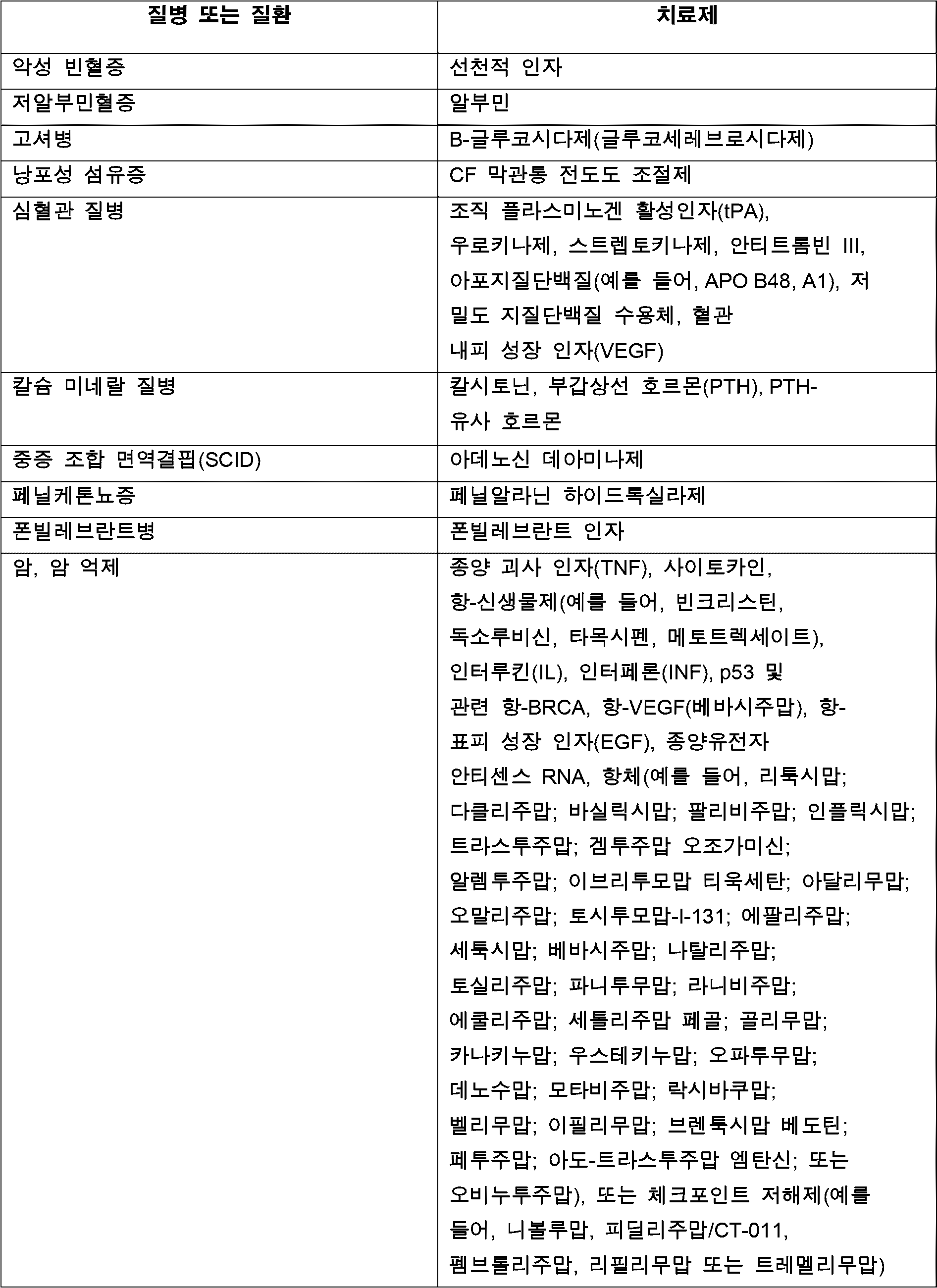


치료제의 발현을 위한 유도성 시스템Inducible system for expression of therapeutic agents
질병 또는 질환을 치료하기 위해 클로킹된 세포에 의해 발현되는 치료제의 연속 투여가 필요한 경우, 치료제는 본 명세서에 기재되거나 당업자에게 공지된 항시적 프로모터(예를 들어, CAG, CMV 또는 또 다른 항시적 프로모터)를 사용하여 발현될 수 있다. 치료제가 간헐적으로 필요한 경우(예를 들어, 질병 또는 질환 중에 발생하는 재발 또는 중증화 기간 동안에는 필요하지만, 대상체가 무증상인 때에는 필요하지 않은 경우), 이는 치료제가 필요할 때만 치료제를 발현하는 능력을 제공하는 유도성 프로모터에 의해 발현될 수 있다. 유도성 프로모터를 사용하여 전달될 수 있는 하나의 예시적인 부류의 치료제는 TNFα 저해제이다. 현재 TNFα 저해제는 류머티스 관절염을 치료하기 위해 사용되지만, TNFα의 항시적 투여가 전신 면역억제를 유발할 수 있으므로, 관절 염증의 중증화 동안에만 간헐적으로 투여된다. 클로킹된 세포가 유도성 프로모터의 제어하에 TNFα 저해제를 발현하도록 변형되는 경우, 클로킹된 세포는 TNFα를 전달하기 위해 간헐적으로 사용될 수 있으므로, 반복 주사의 필요성을 제거한다. 연속적으로 투여되는 경우, 잠재적으로 유해 효과를 발생시킬 수 있는 다른 치료제가 또한 본 명세서에 기재된 유도성 프로모터를 사용하여 간헐적으로 발현될 수 있다. 예시적인 유도성 발현 시스템이 하기에 기재되어 있다.Where continuous administration of a therapeutic agent expressed by cloning cells is required to treat a disease or condition, the therapeutic agent can be expressed using a constitutive promoter (e.g., CAG, CMV or another constitutive promoter) described herein or known to those of skill in the art. Where the therapeutic agent is required intermittently (e.g., during periods of relapse or exacerbation that occur during the disease or condition, but not when the subject is asymptomatic), it can be expressed by an inducible promoter, which provides the ability to express the therapeutic agent only when it is needed. One exemplary class of therapeutic agents that can be delivered using an inducible promoter are TNFα inhibitors. Currently, TNFα inhibitors are used to treat rheumatoid arthritis, but are administered intermittently only during exacerbations of joint inflammation, because constitutive administration of TNFα can lead to systemic immunosuppression. When the cloaked cells are modified to express a TNFα inhibitor under the control of an inducible promoter, the cloaked cells can be used intermittently to deliver TNFα, thus eliminating the need for repeat injections. Other therapeutic agents that may potentially cause detrimental effects when administered sequentially can also be intermittently expressed using the inducible promoters described herein. Exemplary inducible expression systems are described below.
테트라사이클린 반응 요소tetracycline response element
광범위하게 사용되는 유도성 발현 시스템 중 하나는 테트라사이클린-제어된 전사 활성화를 기반으로 한다. 이 시스템에서, 항생제 테트라사이클린 또는 그의 유도체 중 하나(예를 들어, 독시사이클린)는 유전자 발현을 가역적으로 활성화시키거나 저해하기 위해 사용된다. 이 시스템을 사용하기 위해, 테트라사이클린 반응 요소(TRE)는 통상적으로 매우 낮은 기저 수준으로 발현되는 최소 프로모터와 함께, 대상 유전자(예를 들어, 클로킹된 세포에 의해 발현될 치료 전이유전자)의 상류에 위치한다. 클로킹된 세포에 의해 발현시킬 필요가 있는 이른바 rtTA 단백질은 테트라사이클린 또는 독시사이클린의 존재하에서 TRE에 결합하고 전사를 활성화시킨다. 테트라사이클린 또는 독시사이클린이 제거되는 경우, rtTA는 더 이상 TRE에 결합하지 않으며, 대상 유전자가 더 이상 발현되지 않는다. 이 시스템의 발전된 버전인 Tet-On Advanced transactivator(rtTA2s-M2) 및 Tet-On 3G는 인간 코돈에 최적화되어 있고, 저 농도의 독시사이클린에 반응하므로, 이는 인간 요법에 특히 유용할 수 있다.One widely used inducible expression system is based on tetracycline-controlled transcriptional activation. In this system, the antibiotic tetracycline or one of its derivatives (e.g. doxycycline) is used to reversibly activate or inhibit gene expression. To use this system, a tetracycline response element (TRE) is placed upstream of the gene of interest (e.g. a therapeutic transgene to be expressed by the cloaked cell), usually together with a minimal promoter that is expressed at a very low basal level. The so-called rtTA protein, which is to be expressed by the cloaked cell, binds to the TRE in the presence of tetracycline or doxycycline and activates transcription. When tetracycline or doxycycline is removed, the rtTA no longer binds to the TRE and the gene of interest is no longer expressed. Advanced versions of this system, Tet-On Advanced transactivator (rtTA2 s -M2) and Tet-On 3G, are optimized for human codons and respond to low concentrations of doxycycline, which may be particularly useful for human therapy.
광 유도성 시스템Photo-inductive system
유전자 발현의 유도성 활성화를 위한 또 다른 방법은 유전자 발현을 조작하기 위해 감광성 단백질을 사용하는 광유전학의 사용을 포함한다. 클로킹된 세포에서 치료제를 유도적으로 발현시키기 위해 사용될 수 있는 광유전학에서 최근 개발된 것에는 입체형태를 변화시키고, 청색광에 반응하여 이량체화되는 단백질 부류가 포함된다. 특정 프로모터 서열에 결합하고, 청색광에의 노출과 함께 진행되는 경우, 전사를 활성화시키는 것으로 밝혀진 DNA-결합 전사 구성요소에 이들 단백질을 융합시켰다(문헌[Wang et al., Nat Methods, 9: 266-269, 2012]). 유전자 발현을 유도적으로 활성화시키는 이 방법은 클로킹된 피하 조직 부근의 피부에 청색광이 조사됨에 따라, 클로킹된 세포에 의한 치료제의 생성이 유도될 수 있으므로, 피하 투여되는 클로킹된 세포에서 치료제의 생성을 제어하기 위해 사용될 수 있다.Another method for inducible activation of gene expression involves the use of optogenetics, which uses light-sensitive proteins to manipulate gene expression. Recent developments in optogenetics that could be used to inducibly express therapeutics in cloaked cells include a class of proteins that change conformation and dimerize in response to blue light. These proteins have been fused to DNA-binding transcription factors that have been shown to bind to specific promoter sequences and activate transcription when coupled with exposure to blue light (Wang et al., Nat Methods, 9: 266-269, 2012). This method of inducibly activating gene expression could be used to control the production of therapeutics in cloaked cells that are administered subcutaneously, since the production of therapeutics by cloaked cells can be induced when blue light is irradiated onto the skin near the cloaked subcutaneous tissue.
방사선유전학radiogenetics
유전자 발현(예를 들어, 클로킹된 세포에 의한 치료제의 발현)을 유도적으로 활성화시키는 제3의 방법은 전파의 사용을 포함한다. 하나의 버전의 전파 유도성 발현 시스템에서, TRPV1 수용체는 GFP 결합 도메인에 융합되고, GFP에 연결된 페리틴 형태와 공동-발현된다(문헌[Stanley et al., Nat Med 21: 92-98, 2015]). GFP-페리틴은 TRPV1 수용체의 GFP 결합 도메인에 결합한다. 특정 주파수의 전파가 세포에 적용되는 경우, 페리틴은 TRPV1과 상호작용하고, 칼슘의 유입을 허용하여, 전사 인자 NFAT를 활성화시킨다. 치료제는 SRE-CRE-NFATRE와 같은 NFAT-감응성 프로모터 요소에 작동 가능하게 연결되고, TRPV1-GFP 및 GFP-페리틴과 공동-발현되는 경우, 이 시스템을 사용하여 유도적으로 발현될 수 있다. 전파-유도 발현은 전파가 조직을 통과할 수 있기 때문에 신체 외부로부터 더 원위의 세포에서 발현을 유도할 수 있는 이점을 제공한다. 예를 들어, 방사선유전학은 망막에서 유전자 발현을 조절하기 위해 사용될 수 있다. 따라서, 이 방법은 비 침습성 및 비 유해성 전파에 의해 클로킹된 세포에서 치료 전이유전자를 유도적으로 발현시키기 위해 사용될 수 있다.A third method for inducibly activating gene expression (e.g., expression of a therapeutic agent by a cloaked cell) involves the use of radio waves. In one version of the radio-inducible expression system, the TRPV1 receptor is fused to a GFP binding domain and co-expressed with a form of ferritin linked to GFP (Stanley et al., Nat Med 21: 92-98, 2015). GFP-ferritin binds to the GFP binding domain of the TRPV1 receptor. When radio waves of a specific frequency are applied to the cell, ferritin interacts with TRPV1, allowing calcium influx, thereby activating the transcription factor NFAT. Therapeutics can be inducibly expressed using this system if they are operably linked to an NFAT-responsive promoter element, such as SRE-CRE-NFATRE, and are co-expressed with TRPV1-GFP and GFP-ferritin. Radio-induced expression offers the advantage of being able to induce expression in cells more distant from outside the body, since the radio waves can pass through the tissue. For example, radiogenetics can be used to control gene expression in the retina. Thus, this method can be used to inducibly express therapeutic transgenes in cells cloaked by non-invasive and non-toxic radio waves.
불안정화 도메인 시스템destabilizing domain system
유전자 발현은 또한 불안정화 도메인 시스템을 사용하여 조절될 수 있다. 생성된 단백질 산물이 불안정화 도메인에 융합된 대상 단백질을 포함하도록, 대상 단백질(예를 들어, 본 명세서에 기재된 치료제)을 인코딩하는 전이유전자는 또한 불안정화 도메인을 포함할 수 있다. 예시적인 불안정화 도메인은 인간 FK506- 및 라파마이신-결합 단백질(FKBP12)의 돌연변이체를 포함하며, 이는 이들이 융합되는 단백질에 불안정성을 부여한다. FKBP12 돌연변이체는 N-말단 돌연변이체 F15S, V24A, H25R, E60G, 및 L106P, 및 C-말단 돌연변이체 M66T, R71G, D100G, D100N, E102G, 및 K105I를 포함하며, 이는 그 개시내용이 본 명세서에 참조로 포함되는 문헌[Banaszynski et al., Cell 126: 995 (2006)]에 특성화된 바와 같으며, 상기 문헌은 FKBP12 불안정화 도메인에 관한 것이다. 불안정화 도메인은 단백질 분해를 촉진한다. 대상 단백질(예를 들어, 치료제)의 발현이 요구되는 경우, 소분자 합성 리간드가 불안정화 도메인-함유 단백질을 안정화시키기 위해 사용될 수 있다. 소분자 리간드 쉴드(Shield)-1(Shld1)을 사용하여 FKBP12 돌연변이체-함유 단백질을 분해로부터 보호함으로써 안정화시킬 수 있다. 대상 발현 단백질을 조절하기 위해 사용될 수 있는 다른 불안정화 도메인은 E. 콜라이 디하이드로폴레이트 환원효소(ecDHFR)의 돌연변이체 및 인간 에스트로겐 수용체 리간드 결합 도메인(ERLBD)의 돌연변이체를 포함하고, 이는 대상 단백질에 융합되는 경우, 분해를 야기하는 불안정성을 부여하고, 소분자 리간드 트리메토프림(TMP), 또는 CMP8 또는 4-하이드록시타목시펜(4OHT)에 의해 안정화될 수 있고, 이는 문헌[Iwamoto et al., Chem Biol. 17:981 (2010) 및 Miyazaki et al., J Am Chem Soc., 134:3942 (2012)]에 기재된 바와 같으며, 상기 문헌은 불안정화 도메인 시스템에 관한 것이다.Gene expression can also be regulated using a destabilizing domain system. A transgene encoding a subject protein (e.g., a therapeutic agent described herein) can also include a destabilizing domain, such that the resulting protein product comprises the subject protein fused to the destabilizing domain. Exemplary destabilizing domains include mutants of human FK506- and rapamycin-binding protein (FKBP12), which confer instability to the protein to which they are fused. FKBP12 mutants include N-terminal mutants F15S, V24A, H25R, E60G, and L106P, and C-terminal mutants M66T, R71G, D100G, D100N, E102G, and K105I, as characterized in Banaszynski et al., Cell 126: 995 (2006), the disclosure of which is incorporated herein by reference, which relates to the FKBP12 destabilizing domain. The destabilizing domain promotes protein degradation. Small molecule synthetic ligands can be used to stabilize the destabilizing domain-containing protein when expression of a target protein (e.g., a therapeutic agent) is desired. The small molecule ligand Shield-1 (Shld1) can be used to stabilize the FKBP12 mutant-containing protein by protecting it from degradation. Other destabilizing domains that can be used to modulate target expression include mutants of E. coli dihydrofolate reductase (ecDHFR) and mutants of the human estrogen receptor ligand binding domain (ERLBD), which when fused to a target protein confer instability that leads to degradation, and which can be stabilized by the small molecule ligands trimethoprim (TMP), or CMP8 or 4-hydroxytamoxifen (4OHT), as described in the literature (Iwamoto et al., Chem Biol. 17:981 (2010) and Miyazaki et al., J Am Chem Soc., 134:3942 (2012), which are directed to destabilizing domain systems.
큐메이트 스위치 유도성 시스템QMate Switch Inductive System
유전자 발현의 유도성 활성화를 위한 또 다른 방법은 큐메이트 유전자-스위치 시스템의 사용을 포함한다. 이 시스템의 리프레서 입체배좌에서, 조절은 강력한 항시적 프로모터의 하류에 위치한 오퍼레이터 부위(CuO)에 리프레서(CymR)의 결합에 의해 매개된다. 소분자인 큐메이트의 첨가는 저해를 완화시켜 전이유전자의 발현을 가능하게 한다. 대안적으로, 치료제를 인코딩하는 전이유전자에 작동 가능하게 연결된 최소 CMV 프로모터의 상류에 역-큐메이트-트랜스-활성인자(rcTA)가 삽입될 수 있다. 큐메이트 오퍼레이터의 6회 반복(6xCuO)은 치료 전이유전자의 번역 개시(ATG) 직전에 삽입될 수 있다. 큐메이트가 없으면, rcTA는 6xCuO에 결합할 수 없으므로, 치료제를 인코딩하는 전이유전자는 6xCuO가 활성이 아니기 때문에 전사되지 않을 것이다. 큐메이트가 첨가되는 경우, 이는 rcTA와 복합체를 형성할 것이며, 이는 6xCuO에 결합하고, 치료제를 인코딩하는 전이유전자의 전사를 가능하게 한다(문헌[Mullick et al., 2006]).Another method for inducible activation of gene expression involves the use of the cumate gene-switch system. In the repressor conformation of this system, regulation is mediated by binding of the repressor (CymR) to the operator site (CuO) located downstream of a strong constitutive promoter. Addition of the small molecule cumate relieves the inhibition, allowing expression of the transgene. Alternatively, a retro-cumate-trans-activator (rcTA) can be inserted upstream of a minimal CMV promoter operably linked to a transgene encoding a therapeutic. Six repeats of the cumate operator (6xCuO) can be inserted just prior to the translation initiation (ATG) of the therapeutic transgene. In the absence of cumate, rcTA cannot bind 6xCuO, so the transgene encoding the therapeutic will not be transcribed because 6xCuO is not active. When cumate is added, it will form a complex with rcTA, which binds to 6xCuO and enables transcription of the transgene encoding the therapeutic agent (Mullick et al., 2006).
엑디손 유도성 시스템Ecdysone-induced system
유도성 유전자 발현 시스템의 또 다른 예는 레티노이드 X 수용체(RXR), 및 Vp16의 활성화 도메인에 융합된 엑디손 수용체(EcR)(VpEcR)의 N-말단 절두가 클로킹된 세포에 의해 발현되는 유전자의 5' 비번역 영역에 삽입됨으로써, 이들이 내인성 프로모터에 의해 공동 발현되는 엑디손 유도성 시스템이다. 하류의 최소 프로모터를 갖는 엑디손 반응성 요소(EcRE)는 치료제를 인코딩하는 전이유전자의 개시 코돈의 상류에 삽입될 수 있다. 공동 발현된 RXR 및 VpEcR은 서로 이종이량체화될 수 있다. 엑디손 또는 합성 약물 유사체 뮤리스테론 A가 없으면, 이량체화된 RXR/VpEcR은 EcRE에 결합할 수 없으므로, 치료제를 인코딩하는 전이유전자는 전사되지 않는다. 엑디손 또는 뮤리스테론 A의 존재하에, 이량체화된 RXR/VpEcR이 EcRE에 결합함으로써, 치료제를 인코딩하는 전이유전자가 전사될 수 있다(문헌[No et al., 1996]). 엑디손 투여는 포유류에 명백한 영향을 미치지 않기 때문에, 유전자 조절을 위한 그의 용도는 임의의 유전자의 일시적 유도성 발현에 우수해야 한다.Another example of an inducible gene expression system is the ecdysone-inducible system, in which an N-terminal truncation of the retinoid X receptor (RXR) and the ecdysone receptor (EcR) (VpEcR) fused to the activation domain of Vp16 are inserted into the 5'untranslated region of a gene expressed by cloaked cells, such that they are co-expressed under an endogenous promoter. An ecdysone responsive element (EcRE) with a downstream minimal promoter can be inserted upstream of the start codon of a transgene encoding a therapeutic agent. The co-expressed RXR and VpEcR can heterodimerize with each other. In the absence of ecdysone or the synthetic drug analogue muristerone A, the dimerized RXR/VpEcR cannot bind to the EcRE, and therefore the transgene encoding the therapeutic agent is not transcribed. In the presence of ecdysone or muristerone A, dimerized RXR/VpEcR binds to EcRE, thereby transcribing a transgene encoding a therapeutic agent (No et al., 1996). Since ecdysone administration has no apparent effects on mammals, its use for gene regulation should be superior to transient inducible expression of any gene.
리간드 가역성 이량체화 시스템Ligand reversible dimerization system
또 다른 예에서, 치료제를 인코딩하는 전이유전자는 기능적으로 5' 부분 및 3' 부분과 같은 구획/도메인으로 분할되도록 변형될 수 있고, FKBP 펩타이드 서열이 각각의 도메인내에 삽입될 수 있다. IRES(내부 리보솜 진입 부위) 서열은 2개의 도메인 사이에 위치할 수 있고, 이에 의해 2개의 상이한 도메인의 동시 전사를 통해 2개의 개별 단백질이 생성될 수 있다. 이량체화제가 부재하는 경우, 치료제의 2개의 개별 도메인은 기능적으로 불활성일 것이다. 라파마이신 또는 AP20187과 같은 이량체화제의 도입에 따라, FKBP 펩타이드는 이량체화되어 치료제의 5' 및 3' 도메인이 함께 유지되도록 하고, 활성 단백질을 재구성한다(문헌[Rollins et al., 2000]).In another example, the transgene encoding the therapeutic agent can be modified to be functionally split into segments/domains, such as a 5' portion and a 3' portion, and the FKBP peptide sequence can be inserted into each domain. An IRES (internal ribosome entry site) sequence can be positioned between the two domains, thereby allowing for simultaneous transcription of the two different domains to produce two separate proteins. In the absence of a dimerization agent, the two separate domains of the therapeutic agent will be functionally inactive. Upon introduction of a dimerization agent, such as rapamycin or AP20187, the FKBP peptide dimerizes, thereby keeping the 5' and 3' domains of the therapeutic agent together, reconstituting the active protein (Rollins et al., 2000).
치료제의 세포-기반 전달Cell-based delivery of therapeutics
연령 관련 황반 변성 또는 망막 이영양증의 치료Treatment of age-related macular degeneration or retinal dystrophy
일 예에서, 연령 관련 황반 변성(AMD) 또는 망막 이영양증의 치료를 위해, 클로킹된 세포는 VEGF 저해제, 예컨대 VEGF 트랩(예를 들어, 본 명세서에 참조로 포함되는 문헌[Holash et al., Proc Natl Acad Sci U.S.A. 99:11383-11398, 2002]에 기재된 가용성 미끼 수용체, 예컨대 애플리버셉트)을 생성하도록 변형될 수 있다. VEGF 트랩은 비정상적인 혈관의 형성을 촉진할 수 있는 혈관 신생 단백질인 VEGF에 결합하여 이를 저해하는 생물학적 제제이다. VEGF 트랩은 습성 AMD를 치료하기 위해 사용되며, 습성 AMD는 망막 아래 혈관이 비정상적으로 성장하여 망막 박리 및 점진적인 시력 상실을 야기하는 것을 특징으로 할 수 있다. AMD를 치료하기 위해, VEGF 트랩은 일반적으로 안구내로의 주기적 주사에 의해 전달된다. 클로킹된 세포는 항시적 또는 유도성 프로모터에 작동 가능하게 연결된 VEGF 트랩 또는 또 다른 VEGF 저해제를 인코딩하는 전이유전자의 발현에 의해 VEGF 트랩 또는 또 다른 VEGF 저해제를 생성하도록 변형될 수 있다. VEGF 저해제(예를 들어, VEGF 트랩)를 발현하는 클로킹된 세포(예를 들어, 줄기세포)를 안구에 투여하기 전에 당업자에게 공지된 방법을 사용하여 망막 색소 상피(RPE) 세포로 분화시킬 수 있으며, 단리된 RPE 세포를 클로킹 전이유전자 및 VEGF 저해제를 발현하도록 변형시킬 수 있다. VEGF 저해제(예를 들어, VEGF 트랩)를 발현하는 2만 5천개 내지 십만개의 클로킹된 RPE 세포(예를 들어, 25,000, 50,000, 75,000 또는 100,000개의 클로킹된 RPE 세포)를 습성 AMD 또는 망막 이영양증을 치료하기 위해 각 안구의 망막하 공간내에 주사할 수 있다. 본 명세서에 기재된 조성물 및 방법에 사용하기에 적합한 다른 VEGF 저해제는 가용성 형태의 VEGF 수용체(예를 들어, 가용성 VEGFR-1 또는 NRP-1), 혈소판 인자-4, 프로락틴, SPARC, 및 VEGF 저해 항체(예를 들어, 베바시주맙 및 라니비주맙)를 포함한다.In one example, for treating age-related macular degeneration (AMD) or retinal dystrophies, the cloaked cells can be modified to produce a VEGF inhibitor, such as a VEGF trap (e.g., a soluble decoy receptor such as aflibercept, as described in Holash et al., Proc Natl Acad Sci U.S.A. 99:11383-11398, 2002, which is incorporated herein by reference). VEGF traps are biologic agents that bind to and inhibit VEGF, an angiogenic protein that can promote the formation of abnormal blood vessels. VEGF traps are used to treat wet AMD, which can be characterized by abnormal growth of blood vessels beneath the retina, leading to retinal detachment and progressive vision loss. To treat AMD, VEGF traps are typically delivered by periodic injection into the eye. The cloaked cells can be modified to produce VEGF trap or another VEGF inhibitor by expression of a transgene encoding VEGF trap or another VEGF inhibitor operably linked to a constitutive or inducible promoter. The cloaked cells (e.g., stem cells) expressing the VEGF inhibitor (e.g., the VEGF trap) can be differentiated into retinal pigment epithelial (RPE) cells using methods known to those of skill in the art prior to administration to the eye, and the isolated RPE cells can be modified to express the cloaking transgene and the VEGF inhibitor. Between 25,000 and 100,000 cloaked RPE cells (e.g., 25,000, 50,000, 75,000 or 100,000 cloaked RPE cells) expressing a VEGF inhibitor (e.g., a VEGF trap) can be injected into the subretinal space of each eye to treat wet AMD or a retinal dystrophy. Other VEGF inhibitors suitable for use in the compositions and methods described herein include soluble forms of the VEGF receptor (e.g., soluble VEGFR-1 or NRP-1), platelet factor-4, prolactin, SPARC, and VEGF inhibitory antibodies (e.g., bevacizumab and ranibizumab).
파킨슨병의 치료Treatment of Parkinson's disease
또 다른 예에서, 당업자에게 공지된 방법을 사용하여 시험관내에서 분화되어 도파민성 뉴런을 생성할 수 있는 세포(예를 들어, 줄기세포) 또는 도파민성 뉴런과 같은 클로킹된 세포는 도파민성 뉴런의 손실을 특징으로 하는 파킨슨병을 앓고 있는 대상체에 투여될 수 있다. 파킨슨병을 앓고 있는 대상체의 뇌에 25,000개 내지 10만개의 클로킹된 도파민성 뉴런(예를 들어, 25,000, 50,000, 75,000 또는 100,000개의 클로킹된 도파민성 뉴런)을 투여할 수 있다(예를 들어, 흑질내에 정위 주사됨).In another example, cells (e.g., stem cells) or cloaked cells, such as dopaminergic neurons, that can be differentiated in vitro using methods known to those of skill in the art can be administered to a subject suffering from Parkinson's disease, which is characterized by loss of dopaminergic neurons. Between 25,000 and 100,000 cloaked dopaminergic neurons (e.g., 25,000, 50,000, 75,000 or 100,000 cloaked dopaminergic neurons) can be administered to the brain of a subject suffering from Parkinson's disease (e.g., stereotactically injected into the substantia nigra).
심근경색의 치료Treatment of myocardial infarction
본 명세서에 기재된 클로킹된 세포는 또한 심근경색(예를 들어, 일반적으로 심장 마비로 지칭되는 심근경색)을 치료하기 위해 사용될 수 있다. 심근경색은 심장의 일부로의 혈류가 감소하거나 중단되는 경우 발생하여 심근에 손상을 야기한다. 심근경색을 앓고 있는 대상체를 치료하기 위해, 클로킹된 세포(예를 들어, 줄기세포)를 당업자에게 공지된 방법을 사용하여 심근 세포로 분화시킬 수 있거나, 클로킹 전이유전자를 발현하도록 단리된 심근 세포를 변형시킬 수 있다. 심근경색을 앓고 있는 대상체를 치료하기 위해(예를 들어, 사멸 또는 손상 심근 세포를 대체하기 위해), 5억개 내지 50억개의 클로킹된 심근 세포(예를 들어, 5 x 10⁸, 6 x 10⁸, 7 x 10⁸, 8 x 10⁸, 9 x 10⁸, 1 x 10⁹, 2 x 10⁹, 3 x 10⁹, 4 x 10⁹, 또는 5 x 10⁹개의 클로킹된 심근 세포)를 심근 주사에 의해 대상체에 투여할 수 있다.The cloaked cells described herein can also be used to treat myocardial infarction (e.g., myocardial infarction, commonly referred to as a heart attack). Myocardial infarction occurs when blood flow to a part of the heart is reduced or stopped, causing damage to the heart muscle. To treat a subject suffering from a myocardial infarction, the cloaked cells (e.g., stem cells) can be differentiated into cardiomyocytes using methods known to those of skill in the art, or the isolated cardiomyocytes can be modified to express the cloaking transgene. To treat a subject having suffered a myocardial infarction (e.g., to replace dead or damaged cardiomyocytes), between 500 million and 5 billion cloaked cardiomyocytes (e.g., 5 x 10 8 , 6 x 10 8 , 7 x 10 8 , 8 x 10 8 , 9 x 10 8 , 1 x 10 9 , 2 x 10 9 , 3 x 10 9 , 4 x 10 9 , or 5 x 10 9 cloaked cardiomyocytes) can be administered to the subject by intracardiac injection.
골관절염 및 류머티스 관절염의 치료Treatment of osteoarthritis and rheumatoid arthritis
또 다른 예에서, 본 명세서에 기재된 클로킹된 세포는 골관절염 또는 류머티스 관절염을 치료하기 위해 사용될 수 있다. 골관절염 및 류머티스 관절염(RA)은 관절 염증을 특징으로 하며, 일반적으로 항염증 치료제로 치료된다. 골관절염 또는 RA를 앓고 있는 대상체를 치료하기 위해, 클로킹된 세포는, 이미 RA의 치료에 임상적으로 사용되고 있는 항-염증성 생물학적 제제, 예컨대 TNFα의 저해제(예를 들어, TNFα 저해 항체)를 발현하도록 변형될 수 있다. 클로킹된 세포는 항시적 또는 유도성 프로모터에 작동 가능하게 연결된 항 염증성 생물학적 제제를 인코딩하는 전이유전자의 발현에 의해 TNFα 저해제와 같은 항 염증성 생물학적 제제를 생성하도록 변형될 수 있다. 항 염증성 생물학적 제제(예를 들어, TNFα 저해제)를 발현하는 클로킹된 세포(예를 들어, 줄기세포)를 관절에 투여하기 전에 당업자에게 공지된 방법을 사용하여 관절 섬유아세포로 분화시킬 수 있거나, 클로킹 전이유전자 및 항 염증성 생물학적 제제를 발현하도록 단리된 관절 섬유아세포를 변형시킬 수 있다. 골관절염 또는 RA를 치료하기 위해 항-염증성 생물학적 제제를 발현하는 1백만개 내지 1억개의 클로킹된 관절 섬유아세포(예를 들어, 1 x 10⁶, 2 x 10⁶, 3 x 10⁶, 4x 10⁶, 5 x 10⁶, 6 x 10⁶, 7 x 10⁶, 8 x 10⁶, 9 x 10⁶, 1 x 10⁷, 2 x 10⁷, 3 x 10⁷, 4x 10⁷, 5 x 10⁷, 6 x 10⁷, 7 x 10⁷, 8 x 10⁷, 9 x 10⁷, 또는 1 x 10⁸개의 클로킹된 관절 섬유아세포)를 관절염 또는 염증 관절내에 주사할 수 있다(관절 크기에 따름). 골관절염 또는 RA를 치료하기 위해 클로킹된 세포에 의해 발현될 수 있는 항 염증성 생물학적 제제는 TNFα 저해제(아달리무맙, 에타너셉트, 인플릭시맙, 골리무맙, 세톨리주맙), 인터루킨-6(IL6) 수용체 저해제(예를 들어, 토실리주맙), IL1 수용체 저해제(예를 들어, 아나킨라), 또는 RA를 치료하기 위해 사용되는 다른 제제(예를 들어, 아바타셉트, 리툭시맙)를 포함한다.In another example, the cloaked cells described herein can be used to treat osteoarthritis or rheumatoid arthritis. Osteoarthritis and rheumatoid arthritis (RA) are characterized by joint inflammation and are typically treated with anti-inflammatory agents. To treat a subject suffering from osteoarthritis or RA, the cloaked cells can be modified to express an anti-inflammatory biologic that is already clinically used to treat RA, such as an inhibitor of TNFα (e.g., a TNFα inhibitory antibody). The cloaked cells can be modified to produce an anti-inflammatory biologic, such as a TNFα inhibitor, by expression of a transgene encoding the anti-inflammatory biologic that is operably linked to a constitutive or inducible promoter. Cloaked cells (e.g., stem cells) expressing an anti-inflammatory biologic (e.g., a TNFα inhibitor) can be differentiated into arthrofibroblasts using methods known to those of skill in the art prior to administering them to the joint, or isolated arthrofibroblasts can be modified to express the cloaked transgene and the anti-inflammatory biologic. A composition comprising from one million to one hundred million cloaked joint fibroblasts expressing an anti-inflammatory biologic agent for treating osteoarthritis or RA (e.g., 1 x 10 6 , 2 x 10 6 , 3 x 10 6 ,
당뇨병의 치료Treatment of diabetes
클로킹된 세포는 당뇨병(예를 들어, 1형 또는 2형 당뇨병)을 치료하기 위해 사용될 수 있다. 1형 당뇨병은 췌장이 충분한 인슐린을 생성하지 못함으로써 발생한다. 2형 당뇨병은 인슐린 저항성으로 발병하지만, 질병이 진행됨에 따라 인슐린 부족이 발생할 수 있다. 당뇨병을 앓고 있는 대상체를 치료하기 위해, 클로킹된 세포는 인슐린을 발현하도록 변형될 수 있거나, 건강한 대상체로부터의 인슐린-발현 세포(예를 들어, 당뇨병이 없는 대상체의 췌장 베타 세포)는 클로킹 전이유전자 PD-L1, HLA-G(H2-M3), Cd47, Cd200, FASLG(FasL), Ccl21(Ccl21b), Mfge8, 및 Serpin B9(Spi6) 중 하나 이상(예를 들어, 1, 2, 3, 4, 5, 6, 7 또는 8개 모두)을 발현하도록 변형될 수 있으며, 당뇨병을 가진 대상체에 투여될 수 있다. 클로킹된 세포는 항시적 또는 유도성 프로모터에 작동 가능하게 연결된 인슐린을 인코딩하는 전이유전자의 발현에 의해 인슐린을 생성하도록 변형될 수 있다. 인슐린을 발현하는 클로킹된 세포(예를 들어, 줄기세포)를 당업자에 의해 공지된 방법을 사용하여 투여 전에 인슐린 생성 세포(예를 들어, 췌장 베타 세포)로 분화시킬 수 있거나, 분화 과정없이 투여할 수 있거나, 클로킹 전이유전자를 발현하고 선택적으로 인슐린을 인코딩하는 전이유전자를 발현하도록, 단리된 췌장 베타 세포를 변형시킬 수 있다. 당뇨병을 치료하기 위한 인슐린을 생성하는 클로킹된 피하 조직을 생성하기 위해, 인슐린을 발현하는(예를 들어, 내인적으로 인슐린을 발현하거나, 인슐린을 인코딩하는 전이유전자의 발현으로 인해 인슐린을 발현하는) 8억개 내지 30억개의 클로킹된 췌장 베타 세포(예를 들어, 8 x 10⁸, 9 x 10⁸, 1 x 10⁹, 2 x 10⁹, 또는 3 x 10⁹ 개의 클로킹된 췌장 베타 세포)를 대상체에 피하 주사할 수 있다.The cloaked cells can be used to treat diabetes (e.g.,
혈우병의 치료Treatment of hemophilia
또 다른 예에서, 본 명세서에 기재된 클로킹된 세포는 혈우병 치료에 사용될 수 있다. 혈우병 환자는 혈액 응고에 필요한 중요한 혈액 구성요소인 기능적 인자 VIII 단백질을 생성하지 못한다. 이들 환자는 심한 출혈을 겪을 수 있으며, 치료 표준에는 정제된 인자 VIII 단백질의 주당 다회 주사가 수반되었다. 혈우병을 앓고 있는 대상체를 치료하기 위해, 클로킹된 세포는 인자 VIII을 인코딩하는 추가의 전이유전자를 발현하도록 변형될 수 있다. 인자 VIII은 항시적 프로모터, 예컨대 CMV 또는 CAG에 작동 가능하게 연결됨으로써, 클로킹된 세포에서 항시적으로 발현될 것이다. 인자 VIII을 발현하는 클로킹된 세포(예를 들어, 줄기세포)를 당업자에게 공지된 방법을 사용하여 투여 전에 혈액 응고 인자(예를 들어, 간 시누소이드(sinusoidal) 세포 또는 내피 세포)를 생성하는 세포로 분화시킬 수 있거나, 분화 과정 없이 투여할 수 있거나, 건강한 대상체(예를 들어, 혈우병이 없는 대상체)로부터의 단리된 인자 VIII-발현 간 시누소이드 세포 또는 내피 세포를 클로킹 전이유전자 PD-L1, HLA-G(H2-M3), Cd47, Cd200, FASLG(FasL), Ccl21(Ccl21b), Mfge8, 및 Serpin B9(Spi6) 중 하나 이상(예를 들어, 1, 2, 3, 4, 5, 6, 7 또는 8개 모두)을 발현하도록 변형시킬 수 있거나, 혈우병을 가진 대상체에 투여할 수 있다. 인자 VIII이 높은 수준으로 발현되도록 하는 것이 적절한 경우, 하나 이상의 클로킹 전이유전자를 발현하도록 변형된 건강한 대상체로부터의 단리된 인자 VIII-발현 간 시누소이드 세포 또는 내피 세포는 인자 VIII을 인코딩하는 전이유전자를 발현하도록 추가로 변형될 수 있다. 혈우병 치료를 위해, 인자 VIII을 발현하는(예를 들어, 인자 VIII을 내인적으로 발현하거나, 인자 VIII을 인코딩하는 전이유전자의 발현으로 인해 인자 VIII을 발현하는) 8억개 내지 30억개의 클로킹된 세포(예를 들어, 8 x 10⁸, 9 x 10⁸, 1 x 10⁹, 2 x 10⁹, 또는 3 x 10⁹개의 클로킹된 세포)는 인자 VIII을 생성하는 클로킹된 피하 조직을 생성하기 위해 대상체에 피하 주사될 수 있다.In another example, the cloaked cells described herein can be used to treat hemophilia. Patients with hemophilia fail to produce functional factor VIII protein, a critical blood component required for blood clotting. These patients can experience severe bleeding, and the standard of care has involved multiple weekly injections of purified factor VIII protein. To treat a subject with hemophilia, the cloaked cells can be modified to express an additional transgene encoding factor VIII. Factor VIII will be constitutively expressed in the cloaked cells by being operably linked to a constitutive promoter, such as CMV or CAG. Cloaked cells (e.g., stem cells) expressing factor VIII can be differentiated into cells that produce blood clotting factors (e.g., liver sinusoidal cells or endothelial cells) using methods known to those of skill in the art prior to administration, or can be administered without differentiation, or isolated factor VIII-expressing liver sinusoidal cells or endothelial cells from a healthy subject (e.g., a subject without hemophilia) can be modified to express one or more (e.g., all 1, 2, 3, 4, 5, 6, 7, or 8) of the cloaking transgenes PD-L1, HLA-G (H2-M3), Cd47, Cd200, FASLG (FasL), Ccl21 (Ccl21b), Mfge8, and Serpin B9 (Spi6), and can be administered to a subject with hemophilia. In cases where it is appropriate to express high levels of factor VIII, isolated factor VIII-expressing hepatic sinusoid cells or endothelial cells from a healthy subject modified to express one or more cloaking transgenes can be further modified to express a transgene encoding factor VIII. For the treatment of hemophilia, between 800 million and 3 billion cloaked cells (e.g., 8 x 10 8 , 9 x 10 8 , 1 x 10 9 , 2 x 10 9 , or 3 x 10 9 cloaked cells) that express factor VIII (e.g., that endogenously express factor VIII , or that express factor VIII due to expression of a transgene encoding factor VIII) can be injected subcutaneously into the subject to generate cloaked subcutaneous tissue that produces factor VIII.
대사 장애의 치료Treatment of metabolic disorders
본 발명의 클로킹된 세포는 또한 유전적 대사 장애를 치료하기 위해 사용될 수 있다. 대부분의 유전적 대사 장애에서, 단일 효소는 신체에 의해 생성되지 않거나, 결함이 있는 형태로 생성된다. 유전적 대사 장애는 리소좀 저장 장애, 예컨대 헐러 증후군(Hurler syndrome)(알파-L 이두로니다제 결핍), 니만-픽병(Niemann-Pick disease)(SMPD1, NPC1 또는 NPC2의 돌연변이), 테이 삭스병(Tay-Sachs disease)(HEXA의 돌연변이), 고셔병(GBA 유전자의 돌연변이), 파브리병(Fabry disease)(GLA의 돌연변이로 인한 알파 갈락토시다제 결핍) 및 크라브병(Krabbe disease)(GALC의 돌연변이로 인한 갈락토실세라미다제 결핍); 갈락토스혈증(갈락토키나제 또는 갈락토스-1-포스페이트 우리딜 전이효소 결핍); 단풍 당밀뇨 질병(효소 BCKD의 결핍); 페닐케톤뇨증(효소 PAH의 결핍); 글리코겐 저장 질병(GSD), 예컨대 GSD0(글리코겐 합성효소(GYS2)의 결핍), GSD1/폰 기르케병(von Gierke’s disease)(글루코스-6-포스파타제(G6PC) 결핍), GSD 2/폼페병(Pompe’s disease)(산 알파-글루코시다제(GAA)의 결핍), GSD 3/코리병(Cori's disease) 또는 포브스병(Forbes' disease)(글리코겐 탈분지 효소 AGL의 결핍), GSD 4/안데르센병(Andersen disease)(글리코겐 분지 효소 결핍(GBE1)의 결핍), GSD 5/맥아들 질병(근육 글리코겐 포스포릴라제의 결핍)(PYGM)), GSD 6/허스병(Hers' disease)(간 글리코겐 포스포릴라제(PYGL) 또는 근육 포스포글리세레이트 뮤타제(PGAM2)의 결핍), GSD 7/타루이스병(Tarui's disease)(근육 포스포프룩토키나제(PKFM)의 결핍), GSD 9(포스포릴라제 키나제(PHKA2, PHKB, PHKG2 또는 PHKA1)의 결핍), GSD 10(에놀라제 3(ENO3)의 결핍), GSD 11(근육 락테이트 탈수소 효소 (LDHA)의 결핍), 판코니-비켈 증후군(Fanconi-Bickel syndrome)(글루코스 수송체 2(GLUT2)의 결핍), GSD 12(알돌라제 A(ALDOA)의 결핍), GSD 13(β-에놀라제(ENO3)의 결핍), 또는 GSD 15(글리코게닌-1(GYG1)의 결핍); 미토콘드리아 장애, 예컨대 미토콘드리아 근병증(컨스-세이어 증후군(Kearns-Sayre syndrome)(KSS, 미토콘드리아 DNA의 결실에 의해 유발됨), 및 만성 진행성 외부 안근마비(CPEO, 미토콘드리아 DNA의 결실 또는 이중화, 또는 ANT1, POLG, POLG2, 또는 PEO1의 돌연변이에 의해 유발됨), 진성 당뇨병 및 난청(DAD, tRNALeu를 인코딩하는 위치 3243(UUR)에서 미토콘드리아 DNA의 돌연변이에 의해 유발됨), 레버씨 시신경 위축증(Leber's hereditary optic neuropathy)(LHON, MT-ND1, MT-ND4, MT-ND4L, 및 MT-ND6의 돌연변이에 의해 유발됨), 레이 증후군(Leigh syndrome)(SURF1, MT-ATP6, MT-ND2, MT-ND3, MT-ND5, MT-ND6, BCS1L, NDUFA10, SDHA, NDUFS4, NDUFAF2, NDUFA2, NDUFAF6, COX15, NDUFS3, NDUFS8, FOXRED1, NDUFA9, NDUFA12, NDUFS7의 돌연변이와 관련됨), 신경병증, 운동 실조증, 망막염 색소증, 및 안검하수증(NARP, MT-ATP6의 돌연변이에 의해 유발됨), 근신경성 위장 뇌병증(MNGIE, TYMP의 돌연변이에 의해 유발됨), 불균일 적색 근섬유에 의한 근간대 간질(MERRF, MT-TK, MT-TL1, MT-TH, MT-TS1, MT-TS2, 또는 MT-TF의 돌연변이에 의해 유발됨) 또는 미토콘드리아 근병증, 뇌근병증, 젖산증, 뇌졸중 유사 증후군(MELAS, MT-ND1, MT-ND5, MT-TH, MT-TL1, 또는 MT-TV의 돌연변이에 의해 유발됨); 프리드리히 운동 실조증(FXN의 돌연변이); 퍼옥시좀 장애, 예컨대 젤위거 증후군(Zellweger syndrome)(PEX1, PEX2, PEX3, PEX5, PEX6, PEX10, PEX12, PEX13, PEX14, PEX16, PEX19, 또는 PEX26의 돌연변이) 및 부신백질이영양증(ABCD1의 돌연변이); 금속 대사작용 장애, 예컨대 윌슨병(Wilson disease)(윌슨병 단백질 ATP7B의 돌연변이) 및 혈색소증(인간 혈색소증 단백질 HFE의 돌연변이); 유기산혈증, 예컨대 메틸말론 산혈증(MUT, MMAA, MMAB, MMADHC, 또는 MCEE의 돌연변이) 및 프로피온산혈증(PCCA 또는 PCCB의 돌연변이); 요소 회로 장애, 예를 들어 오르니틴 트랜스카바밀라제(OTC) 결핍, 아르기나제(ARG1) 결핍, 아르기니노숙시네이트 리아제(ASL) 결핍, 아르기니노숙시네이트 합성효소 1(ASS1) 결핍, 시트린 결핍, 카바모일 포스페이트 합성효소 1(CPSI) 결핍, N-아세틸글루타메이트 합성효소(NAGS) 결핍, 및 오르니틴 트랜스로카제(ORNT1) 결핍을 포함한다.The cloaked cells of the present invention may also be used to treat genetic metabolic disorders. In most genetic metabolic disorders, a single enzyme is not produced by the body, or is produced in a defective form. Genetic metabolic disorders include lysosomal storage disorders, such as Hurler syndrome (alpha-L iduronidase deficiency), Niemann-Pick disease (mutations in SMPD1, NPC1 or NPC2), Tay-Sachs disease (mutations in HEXA), Gaucher disease (mutations in the GBA gene), Fabry disease (alpha galactosidase deficiency due to mutations in GLA), and Krabbe disease (galactosylceramidase deficiency due to mutations in GALC); galactosemia (galactokinase or galactose-1-phosphate uridyl transferase deficiency); maple syrup urine disease (deficiency in the enzyme BCKD); Phenylketonuria (deficiency of the enzyme PAH); Glycogen storage diseases (GSDs), such as GSD0 (deficiency of glycogen synthase (GYS2)), GSD1/von Gierke’s disease (deficiency of glucose-6-phosphatase (G6PC)),
대사 장애를 앓고 있는 대상체를 치료하기 위해, 대상체에서 돌연변이된 유전자의 야생형 형태 또는 대상체에서 누락되거나 결핍된 효소를 인코딩하는 전이유전자를 발현하도록 클로킹된 세포를 변형시킬 수 있거나(표 2 참조), 대상체에서 결핍된 효소 또는 대상체에서 돌연변이된 유전자의 야생형 형태를 발현하는 건강한 대상체(예를 들어, 대사 장애가 없는 대상체)로부터의 세포를 클로킹 전이유전자 PD-L1, HLA-G(H2-M3), Cd47, Cd200, FASLG(FasL), Ccl21(Ccl21b), Mfge8, 및 Serpin B9(Spi6) 중 하나 이상(예를 들어, 1, 2, 3, 4, 5, 6, 7 또는 8개 모두)을 발현하도록 변형시킬 수 있고, 대사 장애가 있는 대상체에 투여할 수 있다. 대상체에서 돌연변이된 유전자의 야생형 형태 또는 대상체에서 누락되거나 결핍된 효소를 인코딩하는 전이유전자는 CMV 또는 CAG와 같은 항시적 프로모터에 작동 가능하게 연결됨으로써, 클로킹된 세포에서 항시적으로 발현될 수 있거나, 본 명세서에 기재된 유도성 발현 시스템 중 하나를 사용하여 유도적으로 발현될 수 있다. 대상체에서 누락되거나 결핍된 효소 또는 대상체에서 돌연변이된 유전자의 야생형 형태를 발현하도록 변형된 클로킹된 세포(예를 들어, 줄기세포)를 당업자에게 공지된 방법을 사용하여 투여하기 전에 유전자 또는 효소를 정상적으로 발현하는 세포로 분화시킬 수 있거나, 분화 과정 없이 투여할 수 있거나, 대상체에서 돌연변이되거나 결핍된 유전자 또는 효소의 야생형 형태를 발현하는 건강한 대상체로부터 단리된 세포를 클로킹 전이유전자 PD-L1, HLA-G(H2-M3), Cd47, Cd200, FASLG(FasL), Ccl21(Ccl21b), Mfge8, 및 Serpin B9(Spi6) 중 하나 이상(예를 들어, 1, 2, 3, 4, 5, 6, 7 또는 8개 모두)을 발현하도록 변형시킬 수 있고, 대사 장애가 있는 대상체에 투여할 수 있다. 대상체가 치료 전에 특정 돌연변이가 있는 것으로 아직 진단되지 않은 경우, 클로킹된 세포가 상응하는 야생형 유전자를 발현하는지를 확실히 검증하기 위해, 표준 방법을 사용하여 대사 장애와 관련된 돌연변이 유전자를 확인함으로써, 대상체를 평가할 수 있다. 대상체에서 돌연변이된 유전자의 야생형 형태를 발현하는 8억개 내지 30억개의 클로킹된 세포(예를 들어, 8 x 10⁸, 9 x 10⁸, 1 x 10⁹, 2 x 10⁹, 또는 3 x 10⁹개의 클로킹된 세포)를 피하 주사하여, 상응하는 야생형 단백질을 생성하는 클로킹된 피하 조직을 생성할 수 있다.To treat a subject suffering from a metabolic disorder, the cloaked cells can be modified to express a wild-type form of the gene mutated in the subject or a transgene encoding an enzyme that is missing or deficient in the subject (see Table 2), or cells from a healthy subject (e.g., a subject without a metabolic disorder) that express the enzyme that is deficient in the subject or a wild-type form of the gene mutated in the subject can be modified to express one or more (e.g., all 1, 2, 3, 4, 5, 6, 7, or 8) of the cloaked transgenes PD-L1, HLA-G (H2-M3), Cd47, Cd200, FASLG (FasL), Ccl21 (Ccl21b), Mfge8, and Serpin B9 (Spi6) and administered to the subject with the metabolic disorder. A transgene encoding a wild-type form of a mutated gene in the subject or an enzyme that is missing or deficient in the subject can be constitutively expressed in the cloning cell by being operably linked to a constitutive promoter such as CMV or CAG, or can be inducibly expressed using one of the inducible expression systems described herein. Cloaked cells (e.g., stem cells) modified to express a wild-type form of an enzyme that is missing or deficient in the subject or a gene that is mutated in the subject can be differentiated into cells that normally express the gene or enzyme prior to administration using methods known to those of skill in the art, or can be administered without differentiation, or cells isolated from a healthy subject that express a wild-type form of the gene or enzyme that is mutated or deficient in the subject can be modified to express one or more (e.g., all 1, 2, 3, 4, 5, 6, 7, or 8) of the cloaking transgenes PD-L1, HLA-G (H2-M3), Cd47, Cd200, FASLG (FasL), Ccl21 (Ccl21b), Mfge8, and Serpin B9 (Spi6) and administered to a subject with a metabolic disorder. If the subject has not yet been diagnosed with a particular mutation prior to treatment, the subject can be evaluated using standard methods to identify the mutant gene associated with the metabolic disorder to ensure that the cloaked cells express the corresponding wild-type gene. From 800 million to 3 billion cloaked cells expressing the wild-type form of the mutated gene in the subject (e.g., 8 x 10 8 , 9 x 10 8 , 1 x 10 9 , 2 x 10 9 , or 3 x 10 9 cloaked cells) can be injected subcutaneously to generate cloaked subcutaneous tissue that produces the corresponding wild-type protein.
클로킹된 세포의 분열을 제어하는 방법How to control division of cloaked cells
양태에서, 동종이계 숙주 내의 이식 부위에서 세포의 증식을 제어하는 방법이 제공된다(예를 들어, 이식 부위에서 세포의 종양원성을 감소시키거나, 이식 부위에서 종양원성이 되는 세포의 증식을 감소시키기 위함).In one embodiment, a method is provided for controlling proliferation of cells at a transplant site within an allogeneic host (e.g., to reduce tumorigenicity of cells at a transplant site, or to reduce proliferation of cells that become tumorigenic at a transplant site).
본 방법은 동종이계 숙주에 세포 또는 이러한 세포의 집합을 이식하는 경우, 이식 부위에 국소 면역억제를 제공하기 위한 적어도 하나의 메커니즘을 포함하도록 유전적으로 변형된 세포를 제공하는 단계; 세포에서 세포 분열 좌위/좌위들(CDL)을 유전적으로 변형시키는 단계로서, CDL은 그의 전사 산물(들)이 분열 세포(예를 들어, 하나 이상의 면역억제 전이유전자를 함유하는 모든 분열 세포)에 의해 발현되는 하나 이상의 좌위이고, CDL의 유전적 변형은 a) CDL을 인코딩하는 DNA 서열에 전사적으로 연결된 음성 선택 마커를 인코딩하는 DNA 서열을 포함하는 어블레이션 연결(ALINK) 시스템; 및 b) CDL에 작동 가능하게 연결된 유도성 활성인자 기반 유전자 발현 시스템을 포함하는 CDL의 유도성 외인성 활성인자 조절(EARC) 시스템 중 하나 이상을 포함하는 단계; ALINK 시스템을 포함하는 유전적으로 변형된 세포를 음성 선택 마커의 유도인자의 부재하에 유지시킴으로써, ALINK 시스템을 포함하는 유전적으로 변형된 세포의 증식을 허용하거나, ALINK 시스템을 포함하는 세포를 음성 선택 마커의 유도인자에 노출시킴으로써, ALINK 시스템을 포함하는 유전적으로 변형된 세포의 증식을 어블레이션 및/또는 저해하는 단계; 및/또는 EARC 시스템을 포함하는 유전적으로 변형된 세포를 유도성 활성인자-기반 유전자 발현 시스템의 유도인자에 노출시킴으로써, EARC 시스템을 포함하는 유전적으로 변형된 세포의 증식을 허용하거나, EARC 시스템을 포함하는 세포를 유도성 활성인자-기반 유전자 발현 시스템의 유도인자의 부재하에 유지시킴으로써, EARC 시스템을 포함하는 유전적으로 변형된 세포의 증식을 방지 또는 저해하는 단계; 및 동종이계 숙주 내의 이식 부위에 세포 또는 세포의 집합을 이식하는 단계를 포함한다. 하나 이상의 CDL(예를 들어, 2, 3, 4개 이상의 CDL)에서 하나 이상의 ALINK 및/또는 EARC 시스템을 사용하여 세포 분열을 제어하도록 변형된 세포는 "페일 세이프 세포"로 지칭될 수 있다. 세포가 유전적 돌연변이를 통해 세포 분열을 제어하는 모든 시스템(예를 들어, ALINK 또는 EARC)의 활성을 상실하기 전에 단일 페일 세이프 세포로부터 성장할 수 있는 세포의 수(클론 용적)(예를 들어, 수학적 모델링에 기반하여, 제어로부터 "이탈"되고, 비 제어성 세포 증식을 나타내는 세포를 취합한 세포 분열의 수)는 페일 세이프 용적을 결정한다. 페일 세이프 용적은 ALINK 수 및 ALINK 표적 CDL의 수에 의해 좌우될 것이다. 페일 세이프 성질은 표 3에 추가로 기재되어 있다.The method comprises the steps of: providing a cell genetically modified to include at least one mechanism for providing local immunosuppression at the site of transplantation when transplanting the cell or a collection of such cells into an allogeneic host; genetically modifying a cell division locus/locuses (CDL) in the cell, wherein the CDL is one or more loci whose transcription product(s) are expressed by a dividing cell (e.g., all dividing cells containing one or more immunosuppressive transgenes), wherein the genetic modification of the CDL comprises one or more of: a) an ablation linkage (ALINK) system comprising a DNA sequence encoding a negative selection marker transcriptionally linked to a DNA sequence encoding the CDL; and b) an inducible exogenous activator regulation (EARC) system of the CDL comprising an inducible activator-based gene expression system operably linked to the CDL. Maintaining the genetically modified cell comprising the ALINK system in the absence of an inducer of a negative selection marker, thereby allowing proliferation of the genetically modified cell comprising the ALINK system, or exposing the cell comprising the ALINK system to an inducer of a negative selection marker, thereby ablating and/or inhibiting proliferation of the genetically modified cell comprising the ALINK system; and/or exposing the genetically modified cell comprising the EARC system to an inducer of an inducible activator-based gene expression system, thereby allowing proliferation of the genetically modified cell comprising the EARC system, or preventing or inhibiting proliferation of the genetically modified cell comprising the EARC system, by maintaining the cell comprising the EARC system in the absence of an inducer of an inducible activator-based gene expression system; and transplanting the cell or collection of cells into a transplant site in an allogeneic host. Cells that have been modified to control cell division using one or more ALINK and/or EARC systems in one or more CDLs (e.g., two, three, four, or more CDLs) may be referred to as "fail-safe cells." The number of cells (clonal volume) that can grow from a single fail-safe cell before the cell loses activity of any system (e.g., ALINK or EARC) that controls cell division through genetic mutation (e.g., the number of cell divisions that enumerate cells that "grow out of control" and exhibit uncontrolled cell proliferation, based on mathematical modeling) determines the fail-safe volume. The fail-safe volume will be governed by the number of ALINKs and the number of ALINK targeting CDLs. Fail-safe properties are further described in Table 3.
[표 3][Table 3]
페일 세이프 세포 용적과 인체의 관계는 수학적 모델링을 사용함으로써 계산하였다. 본 모델은, CDL 발현이 음성 선택 마커 활성의 상실과 함께 공동 상실되어, 세포 증식이 훼손된 상황은 계수하지 않았다. 따라서, 이 값은 과소 평가되어 있으며, 음성 선택 마커의 경우, 106의 전향 돌연변이율(forward mutation rate)을 가정하여 계산하였다. 인체에서의 추정 세포 수는 3.72x10¹³으로서 산정되었다(문헌[Bianconi et al., 2013]).The relationship between fail-safe cell volume and human body was calculated using mathematical modeling. The model did not account for situations where CDL expression was lost together with the loss of negative selection marker activity, resulting in impaired cell proliferation. Therefore, this value is underestimated, and for negative selection markers, it was calculated assuming a forward mutation rate of 10 6 . The estimated cell number in human body is It was calculated as 3.72x10 13 (literature [Bianconi et al., 2013]).

다양한 구현예에서, CDL은, 본 명세서에서 그 전문이 제시된 것과 같이, 참조로서 포함되는 문헌[Wang et al., 2015]에 제시된 바와 같이, "필수 유전자"로서 확인된 좌위이다. 문헌[Wang et al., 2015]에서의 필수 유전자는, 이러한 유전자의 불활성화에 의해 부여되는 적합도 값(fitness cost)을 반영하는 각 유전자에 대한 점수(즉, CRISPR 점수)를 계산함으로써, 확인하였다. 일 구현예에서, CDL은 약 -1.0 미만의 CRISPR 점수(CS)를 갖는다(표 5, 5열).In various implementations, the CDL is a locus identified as an "essential gene", as set forth in Wang et al., 2015, which is incorporated by reference herein in its entirety. Essential genes in Wang et al., 2015 were identified by computing a score for each gene (i.e., CRISPR score) that reflects the fitness cost conferred by inactivation of such gene. In one implementation, the CDL has a CRISPR score (CS) of less than about -1.0 (Table 5, column 5).
다양한 구현예에서, CDL은, 세포 분열 및/또는 복제에 관련되는 유전자 산물을 인코딩하는 좌위/좌위들이다(표 5, 6열). 예를 들어, 다양한 구현예에서, CDL은: i) 세포 주기; ii) DNA 복제; iii) RNA 전사 및/또는 단백질 번역; 및 iv) 대사작용 중 하나 이상에 관련되는 유전자 산물을 인코딩하는 좌위/좌위들이다(표 5, 7열).In various embodiments, the CDL is a locus/locuses encoding a gene product involved in cell division and/or replication (Table 5, column 6). For example, in various embodiments, the CDL is a locus/locuses encoding a gene product involved in one or more of: i) the cell cycle; ii) DNA replication; iii) RNA transcription and/or protein translation; and iv) metabolism (Table 5, column 7).
일 구현예에서, CDL은, 세포 주기 진행의 조절(예를 들어, G1/S G2/M 및 중기로부터 후기로의 전이의 제어)과 관련된 하나 이상의 사이클린 의존성 키나제, 예를 들어 CDK1, CDK2, CDK3, CDK4, CDK5, CDK6, CDK7, CDK8, CDK9 및/또는 CDK11이다(문헌[Morgan, 2007]). 일 구현예에서, CDL은, 하나 이상의 CDK를 활성화시킴으로써, 세포 주기의 진행을 제어하는 것과 관련된 하나 이상의 사이클린, 예를 들어, 사이클린B, 사이클린E, 사이클린A, 사이클린C, 사이클린D, 사이클린H, 사이클린C, 사이클린T, 사이클린L 및/또는 사이클린F이다(문헌[FUNG and POON, 2005]). 일 구현예에서, CDL은, 세포 주기의 M기에서 중기로부터 후기로의 전이의 진행을 제어하는 후기 촉진 복합체에 포함되는 하나 이상의 좌위들이다(문헌[Peters, 2002]). 일 구현예에서, CDL은, 세포 주기의 M기에서 중기로부터 후기로의 전이의 진행을 제어하는 방추사부착점 구성요소와 관련된 하나 이상의 좌위들이다(문헌[Fukagawa, 2007]). 일 구현예에서, CDL은, 세포 주기에 요구되는 미소관 역학을 제어하는 미소관 구성요소와 관련된 하나 이상의 좌위들이다(문헌[Cassimeris, 1999]).In one embodiment, the CDL is one or more cyclin dependent kinases, e.g., CDK1, CDK2, CDK3, CDK4, CDK5, CDK6, CDK7, CDK8, CDK9 and/or CDK11, which are involved in regulation of cell cycle progression (e.g., control of G1/S G2/M and metaphase to anaphase transitions) (Morgan, 2007). In one embodiment, the CDL is one or more cyclins, e.g., cyclin B, cyclin E, cyclin A, cyclin C, cyclin D, cyclin H, cyclin C, cyclin T, cyclin L and/or cyclin F, which are involved in controlling cell cycle progression by activating one or more CDKs (FUNG and POON, 2005). In one embodiment, the CDL is one or more loci associated with a anaphase-promoting complex that controls progression of the cell cycle from M phase to metaphase to anaphase (Peters, 2002). In one embodiment, the CDL is one or more loci associated with a spindle attachment point component that controls progression of the cell cycle from M phase to metaphase to anaphase (Fukagawa, 2007). In one embodiment, the CDL is one or more loci associated with a microtubule component that controls microtubule dynamics required for the cell cycle (Cassimeris, 1999).
다양한 구현예에서, CDL은 세포유지(housekeeping)와 관련된 좌위/좌위들이다. 본 명세서에 사용되는 바와 같이, 용어 "세포유지 유전자" 또는 "세포유지 좌위"는 기본 세포 기능의 유지에 요구되는 하나 이상의 유전자를 지칭한다. 세포유지 유전자는 정상 조건 및 병리학적 조건하에서 유기체의 모든 세포에서 발현된다.In various embodiments, the CDL is a locus/locuses associated with housekeeping. As used herein, the term "housekeeping gene" or "housekeeping locus" refers to one or more genes required for the maintenance of basic cellular functions. Housekeeping genes are expressed in all cells of an organism under normal and pathological conditions.
다양한 구현예에서, CDL은, 세포 분열 및/또는 증식에 관련되는 유전자 산물을 인코딩하고, 약 -1.0 미만의 CRISPR 점수를 갖는 좌위/좌위들이다. 예를 들어, 일 구현예에서, CDL은, i) 세포 주기; ii) DNA 복제; iii) RNA 전사 및/또는 단백질 번역; 및 iv) 대사작용 중 하나 이상에 관련되는 유전자 산물을 인코딩하고, 약 -1.0 미만의 CRISPR 점수를 갖는 좌위/좌위들이다. 일 구현예에서, CDL은 또한 세포유지 유전자일 수 있다.In various embodiments, the CDL is a locus/locuses encoding a gene product involved in cell division and/or proliferation and having a CRISPR score of less than about -1.0. For example, in one embodiment, the CDL is a locus/locuses encoding a gene product involved in one or more of: i) cell cycle; ii) DNA replication; iii) RNA transcription and/or protein translation; and iv) metabolism and having a CRISPR score of less than about -1.0. In one embodiment, the CDL can also be a cell maintenance gene.
일부 구현예에서, CDL은 Cdk1/CDK1, Top2A/TOP2A, Cenpa/CEPNA, Birc5/BIRC5, 또는 Eef2/EEF2이다. 일부 구현예에서, CDL은 Cdk1/CDK1이다. 일부 구현예에서, CDL은 Top2A/TOP2A이다. 일부 구현예에서, CDL은 Eef2/EEF2이다. 일부 구현예에서, CDL은 Cdk1/CDK1 및 Top2A/TOP2A, 또는 Cdk1/CDK1 및 Eef2/EEF2이다.In some implementations, the CDL is Cdk1/CDK1, Top2A/TOP2A, Cenpa/CEPNA, Birc5/BIRC5, or Eef2/EEF2. In some implementations, the CDL is Cdk1/CDK1. In some implementations, the CDL is Top2A/TOP2A. In some implementations, the CDL is Eef2/EEF2. In some implementations, the CDL is Cdk1/CDK1 and Top2A/TOP2A, or Cdk1/CDK1 and Eef2/EEF2.
음성 선택 마커를 인코딩하는 DNA 서열의 발현과 CDL의 발현을 연결시킴으로써, CDL 및 음성 선택 마커 둘 모두를 발현하는 유사분열 활성 세포의 약물 유도된 어블레이션을 허용하는 "페일 세이프" 세포가 되도록, 세포를 변형시킬 수 있다. 예를 들어, 세포 증식이 세포의 정상 분열 속도에 비해 비제어되고/되거나 가속화되는 경우(예를 들어, 암세포에 의해 나타나는 비제어 세포 분열), 또는 세포에 대한 치료적 필요가 경과한 경우, 증식 세포의 어블레이션이 바람직할 수 있다. 증식 세포의 어블레이션은, 음성 선택 마커를 인코딩하는 DNA 서열의 발현을 CDL의 발현과 연결시킴으로써, 결과적으로 CDL 좌위를 발현하는 ALINK 변형된 증식 세포의 제거 또는 충분한 저해(충분한 저해란, 너무 저속이어서, 종양 형성에 기여할 수 없는 속도로 세포 확장 속도가 저해되는 것임)를 허용하는, 본 명세서에서 "어블레이션 연결"(ALINK)로 지칭되는 세포에 대한 유전적 변형을 통해 달성될 수 있다. 음성 선택 시스템의 전구약물 또는 다른 유도인자의 존재하에, 음성 마커를 발현하는 세포는 선택 마커의 작용 메커니즘에 따라 증식을 중지하거나, 사멸할 것이다. 세포는 동형접합성, 이형접합성, 반접합성 또는 복합 이형접합성 ALINK를 포함하도록 변형될 수 있다. 일 구현예에서, 어블레이션의 정확도를 향상시키기 위해, 음성 선택 마커는 CDL의 모든 기능적 대립유전자에 도입될 수 있다. 바람직한 일 구현예에서, 음성 선택 마커는 CDL의 모든 기능적 대립유전자에 도입될 수 있다. 적절한 경우 페일 세이프 시스템을 사용하여 모든 클로킹된 세포를 제거할 수 있다.By linking the expression of a DNA sequence encoding a negative selection marker to the expression of a CDL, the cell can be modified to become a "fail safe" cell that allows for drug-induced ablation of mitotically active cells that express both a CDL and a negative selection marker. For example, ablation of proliferating cells may be desirable when cell proliferation is uncontrolled and/or accelerated relative to the cell's normal rate of division (e.g., uncontrolled cell division as exhibited by cancer cells), or when a therapeutic need for the cell has passed. Ablation of proliferating cells can be accomplished by a genetic modification to the cell, referred to herein as an "ablation linkage" (ALINK), that links the expression of a DNA sequence encoding a negative selection marker to the expression of a CDL, thereby allowing for elimination or sufficient inhibition (sufficient inhibition being inhibition that the rate of cell expansion is inhibited to a rate that is too slow to contribute to tumorigenesis) of the ALINK-modified proliferating cells expressing the CDL locus. In the presence of a prodrug or other inducer of the negative selection system, cells expressing the negative marker will either stop proliferating or die, depending on the mechanism of action of the selection marker. The cells can be modified to contain homozygous, heterozygous, hemizygous or compound heterozygous ALINK. In one embodiment, to improve the accuracy of ablation, the negative selection marker can be introduced into all functional alleles of the CDL. In a preferred embodiment, the negative selection marker can be introduced into all functional alleles of the CDL. If appropriate, a fail-safe system can be used to remove all cloaked cells.
ALINK는, CDL 및 음성 선택 마커의 공동 발현을 허용하는 CDL의 임의의 위치에 삽입될 수 있다.ALINK can be inserted at any position in the CDL that allows co-expression of the CDL and the voice selection marker.
일부 구현예에서, ALINK 시스템은 단순 포진 바이러스 티미딘 키나제/간사이클로비르 시스템, 시토신 데아미나제/5-플루오로시토신 시스템, 카르복실 에스테라제/이리노테칸 시스템 또는 iCasp9/AP1903 시스템을 포함한다.In some implementations, the ALINK system comprises the herpes simplex virus thymidine kinase/gancyclovir system, the cytosine deaminase/5-fluorocytosine system, the carboxyl esterase/irinotecan system, or the iCasp9/AP1903 system.
음성 선택 마커(예를 들어, HSV-TK)를 인코딩하는 DNA를 숙주 세포의 CDL(예를 들어, CDK1) 내로 삽입함으로써, 음성 선택 마커의 발현은 음성 선택 마커 및 필연적으로 CDL을 발현하는 숙주 세포가 음성 선택 마커(예를 들어, 간사이클로비르(GCV))의 유도인자(예를 들어, 전구약물)의 존재하에 사멸되도록 할 수 있다. 이 실시예에서, ALINK에 의해 변형된 숙주 세포는 티미딘 키나제(TK)를 생성할 것이고, TK 단백질은 GCV를 GCV 일인산염으로 전환시키며, 이어서, 세포 키나제에 의해 GCV 삼인산염으로 전환된다. GCV 삼인산염은 S기 동안 복제 DNA 내로 혼입됨으로써, DNA 연장의 종결 및 세포 아폽토시스를 야기한다(문헌[Halloran and Fenton, 1998]).By inserting DNA encoding a negative selection marker (e.g., HSV-TK) into the CDL of a host cell (e.g., CDK1), expression of the negative selection marker can cause the host cell expressing the negative selection marker and consequently the CDL to be killed in the presence of an inducer (e.g., a prodrug) of the negative selection marker (e.g., ganciclovir (GCV)). In this embodiment, the host cell transformed by ALINK will produce thymidine kinase (TK), and the TK protein converts GCV to GCV monophosphate, which is then converted by cellular kinases to GCV triphosphate. GCV triphosphate is incorporated into replicating DNA during S phase, thereby causing termination of DNA elongation and cell apoptosis (Halloran and Fenton, 1998).
바람직하지 않은 세포 분열 속도를 포함하는 세포를 선택적으로 어블레이션하기 위해, ALINK 유전적 변형에 사용될 수 있는 음성 선택 마커를 인코딩하는 DNA의 일례로서 변형된 HSV-TK 유전자(문헌[Preuß et al., 2010])가 본 명세서에 개시되어 있다.As an example of DNA encoding a negative selectable marker that can be used in ALINK genetic modification to selectively ablate cells comprising an undesirable cell division rate, a modified HSV-TK gene (Preuß et al., 2010) is disclosed herein.
대안적 및/또는 추가적 음성 선택 시스템이 본 명세서에서 제공되는 도구 및/또는 방법에 사용될 수 있음이 본 명세서에서 고려된다. 다양한 음성 선택 마커 시스템이 당 업계에 공지되어 있다(예를 들어, dCK.DM(문헌[Neschadim et al., 2012])).It is contemplated herein that alternative and/or additional voice selection systems may be used with the tools and/or methods provided herein. Various voice selection marker systems are known in the art (e.g., dCK.DM (Neschadim et al., 2012)).
예를 들어, 부작용 없이 또는 최소화하여, 기존의 암 치료법의 치료적 효능을 향상시키는 것을 목표로 하는, 임상적 관련성을 갖는 다양한 음성 선택 시스템이 "유전자 직접 효소/전구약물 치료법"(GEPT) 분야에서 활발히 개발되어 왔다(문헌[Hedley et al., 2007; Nawa et al., 2008]). 종종, GEPT는, 포유류 세포에서 발견되지 않고, 상대적으로 무독성인 전구약물을 독성 제제로 전환시킬 수 있는 효소를 생성하는 암세포의 영역에서 암세포 또는 암세포의 근처에 유전자를 전달하기 위해, 바이러스 벡터의 사용을 포함한다.For example, a variety of clinically relevant negative selection systems have been actively developed in the field of "gene-directed enzyme/prodrug therapy" (GEPT), which aims to enhance the therapeutic efficacy of existing cancer therapies, without or with minimal side effects (Hedley et al., 2007; Nawa et al., 2008). Often, GEPT involves the use of viral vectors to deliver genes to or near cancer cells, in areas of the cancer cell that produce enzymes that are not found in mammalian cells and that can convert relatively non-toxic prodrugs into toxic agents.
HSV-TK/GCV, 시토신 데아미나제/5-플루오로시토신(CD/5-FC), 및 카르복실 에스테라제/이리노테칸(CE/CPT-11)이 GEPT 전임상 시험 및 임상 시험에서 평가되는 음성 선택 마커 시스템의 예이다(문헌[Danks et al., 2007; Shah, 2012]).HSV-TK/GCV, cytosine deaminase/5-fluorocytosine (CD/5-FC), and carboxyl esterase/irinotecan (CE/CPT-11) are examples of negative selection marker systems being evaluated in GEPT preclinical and clinical trials (Danks et al., 2007; Shah, 2012).
단순 포진 바이러스 1형 티미딘 키나제/간사이클로비르(TK/GCV) 시스템의 잠재적인 면역원성을 극복하기 위해, 인간 데옥시시티딘 키나제 효소를 티미딘 활성이 되도록 하고, 비독성 전구약물에 의한 음성 선택(자살) 시스템으로서 작용하도록 조작함으로써, "인간화된" 자살 시스템이 개발되어 왔다: 브로모비닐-데옥시우리딘(BVdU), L-데옥시티미딘(LdT) 또는 L-데옥시우리딘(LdU)(문헌[Neschadim et al., 2012]).To overcome the potential immunogenicity of the herpes
CD/5-FC 음성 선택 마커 시스템은 광범위하게 사용되는 "자살 유전자" 시스템이다. 시토신 데아미나제(CD)는 박테리아 또는 효모(예를 들어, 각각 에세리키아 콜라이(Escherichia coli) 또는 사카로마이세스 세레비시아에(Saccharomyces cerevisiae))로부터 얻을 수 있는 비포유류 효소이다(문헌[Ramnaraine et al., 2003]). CD는 시토신의 우라실로의 전환을 촉매하고, 원핵생물 및 진균류에서 피리미딘 회수 경로의 중요한 구성원이나, 포유류 세포에는 존재하지 않는다. 5-플루오로시토신(5-FC)은 인간에게 저수준의 세포독성을 야기하는 항진균성 전구약물이다(문헌[Denny, 2003]). CD는 5-FC의 인간에서 고수준의 독성을 갖는 유전자 독성 물질 5-FU로의 전환을 촉매한다(문헌[Ireton et al., 2002]).The CD/5-FC negative selectable marker system is a widely used "suicide gene" system. Cytosine deaminase (CD) is a non-mammalian enzyme that can be obtained from bacteria or yeast (e.g., Escherichia coli or Saccharomyces cerevisiae , respectively) (Ramnaraine et al., 2003). CD catalyzes the conversion of cytosine to uracil and is an important component of the pyrimidine salvage pathway in prokaryotes and fungi, but is absent in mammalian cells. 5-Fluorocytosine (5-FC) is an antifungal prodrug that causes low levels of cytotoxicity in humans (Denny, 2003). CD catalyzes the conversion of 5-FC to the highly toxic human genotoxic agent 5-FU (Ireton et al., 2002).
CE/CPT-11 시스템은 포유류 종의 상이한 조직들에서 발견되는 세린 에스테라제인 카르복실 에스테라제 효소를 기반으로 한다(문헌[Humerickhouse et al., 2000]). 항암제 CPT-11은, CE에 의해 활성화됨으로써, 강력한 포유류 토포아이소머라제 I 저해제인 7-에틸-10-하이드록시캄토테신(SN-38)으로 불리는 활성을 생성하는 전구약물이다(문헌[Wierdl et al., 2001]). SN-38은 분열 세포에서 이중 가닥 DNA 파열의 축적을 유도한다(문헌[Kojima et al., 1998]).The CE/CPT-11 system is based on the enzyme carboxyl esterase, a serine esterase found in different tissues of mammalian species (Humerickhouse et al., 2000). The anticancer agent CPT-11 is a prodrug that, upon activation by CE, produces the active agent called 7-ethyl-10-hydroxycamptothecin (SN-38), a potent mammalian topoisomerase I inhibitor (Wierdl et al., 2001). SN-38 induces the accumulation of double-stranded DNA breaks in dividing cells (Kojima et al., 1998).
음성 선택 마커 시스템의 또 다른 예는 iCasp9/AP1903 자살 시스템이며, 이는 인간 FK506 결합 단백질(FKBP)에 융합된 변형된 인간 카스파제 9를 기반으로 하고, 소분자 AP1903을 사용한 화학적 이량체화를 허용하며, 인간에서 안전하게 시험되었다. 이량체화 약물의 투여는 조작된 카스파제 9 구성요소를 발현하는 세포의 아폽토시스를 유도한다. 이 시스템은, 예를 들어, 인간 유전자 산물로 구성되어 있으며, 이 이량체화 약물이, 조작된 카스파제 9 구성요소를 발현하는 세포에만 영향을 미침으로 인한 잠재적 저면역원성을 포함하는 몇 가지 이점을 가진다(문헌[Straathof et al., 2005]). iCasp/AP1903 자살 시스템은 임상 환경에서 시험되고 있다(문헌[Di Stasi et al., 2011]).Another example of a negative selectable marker system is the iCasp9/AP1903 suicide system, which is based on a modified human caspase 9 fused to human FK506 binding protein (FKBP), allows chemical dimerization with the small molecule AP1903, and has been safely tested in humans. Administration of the dimerizing agent induces apoptosis of cells expressing the engineered caspase 9 component. This system has several advantages, including, for example, that it is comprised of a human gene product and that the dimerizing agent only affects cells expressing the engineered caspase 9 component, resulting in potential hypoimmunogenicity (Straathof et al., 2005). The iCasp/AP1903 suicide system is being tested in a clinical setting (Di Stasi et al., 2011).
본 명세서에서, ALINK 시스템의 음성 선택 마커 시스템이 증식 길항제 시스템으로 대체될 수 있음이 고려된다. 본 명세서에 사용되는 바와 같이, 용어 "증식 길항제"는, 그 존재가 세포 분열을 (완전히 또는 부분적으로) 저해하는 천연 또는 조작된 화합물(들)을 지칭한다. 예를 들어, OmomycER는 돌연변이 쥣과동물 에스트로겐 수용체(ER) 도메인을 갖는 MYC 우성 음성 Omomyc의 융합 단백질이다. 타목시펜(tamoxifen)(TAM)으로 유도되는 경우, 융합 단백질 OmomycER는 핵에 국소화되며, 여기서, 우성 음성 Omomyc는 C-Myc, L-Myc 및 N-Myc와 이량체함으로써, Myc DNA 결합 공통 서열과 결합할 수 없는 복합체 내로 이들을 격리시킨다(문헌[Soucek et al., 2002]). Myc 활성의 결여의 결과로서, 세포는 분열할 수 없다(문헌[Oricchio et al., 2014]). 증식 길항제의 또 다른 예는, 등몰농도 경쟁에서 DNA 결합을 저해하는 활성화 단백질-1(AP1)에 대해 우성 음성인 A-Fos(종양 유전자 Fos와 Jun의 이종이량체)이다(문헌[Olive et al., 1997]). A-Fos는 또한 ER 도메인에 융합됨으로써, TAM에 의해 핵 국소화가 유도되도록 할 수 있다. OmomycER/타목시펜 또는 A-FosER/타목시펜은 TK/GCV를 ALINK로 대체할 수 있다.In the present specification, it is contemplated that the negative selection marker system of the ALINK system may be replaced by a proliferation antagonist system. As used herein, the term "proliferation antagonist" refers to a natural or engineered compound(s) whose presence inhibits (completely or partially) cell division. For example, Omomyc ER is a fusion protein of MYC dominant negative Omomyc with a mutant murine estrogen receptor (ER) domain. When induced with tamoxifen (TAM), the fusion protein Omomyc ER localizes to the nucleus, where the dominant negative Omomyc dimerizes with C-Myc, L-Myc, and N-Myc, thereby sequestering them into a complex that cannot bind the Myc DNA-binding consensus sequence (Soucek et al., 2002). As a result of the lack of Myc activity, cells cannot divide (Oricchio et al., 2014). Another example of a proliferation antagonist is A-Fos (a heterodimer of the oncogenes Fos and Jun), a dominant negative for activating protein-1 (AP1) that inhibits DNA binding in an equimolar competition (Olive et al., 1997). A-Fos can also be fused to the ER domain, thereby inducing nuclear localization by TAM. Omomyc ER /tamoxifen or A-Fos ER /tamoxifen can replace TK/GCV with ALINK.
유도성 활성인자-기반 유전자 발현 시스템과 같은 EARC와 CDL을 작동 가능하게 연결시킴으로써, 세포를 또한 "페일 세이프"가 되도록 변형시킬 수 있다. 이들 조건하에, CDL은 단지 유도성 활성인자-기반 유전자 발현 시스템의 유도인자의 존재하에 발현될 것이다(그리고, 세포는 단지 전술한 것의 존재하에 분열할 수 있음). 이들 조건하에, EARC-변형된 세포는 유도성 활성인자-기반 유전자 발현 시스템의 작용 메커니즘 및 CDL 기능에 따라, 유도인자의 부재하에 분열을 중지하거나, 유의하게 지연되거나, 사멸한다. 세포는 동형접합성 또는 복합 이형접합성 EARC를 포함하도록 변형될 수 있거나, EARC-변형된 대립유전자만이 기능적 CDL을 생성할 수 있도록 변경될 수 있다. 일 구현예에서, EARC 변형은 예를 들어, 세포 분열 제어를 위한 메커니즘을 제공하기 위해, CDL의 모든 대립유전자내에 도입될 수 있다.By operatively linking EARC and CDL, such as an inducible activator-based gene expression system, cells can also be modified to be "fail safe." Under these conditions, CDL will only be expressed in the presence of an inducer of the inducible activator-based gene expression system (and the cell can only divide in the presence of the inducer). Under these conditions, EARC-modified cells will either stop dividing, be significantly delayed in dividing, or die in the absence of the inducer, depending on the mechanism of action of the inducible activator-based gene expression system and the function of the CDL. The cell can be modified to contain homozygous or compound heterozygous EARC, or can be altered so that only the EARC-modified allele produces functional CDL. In one embodiment, the EARC modification can be introduced into all alleles of the CDL, for example, to provide a mechanism for cell division control.
EARC는, 유도인자의 존재하에 CDL 및 유도성 시스템의 활성인자 구성요소의 공동 발현을 허용하는 CDL의 임의의 위치에 삽입될 수 있다.EARC can be inserted at any position in the CDL that allows co-expression of CDL and activator components of the inducible system in the presence of an inducer.
일 구현예에서, "활성인자" 기반 유전자 발현 시스템은 "리프레서" 기반 유전자 발현 시스템보다 바람직하다. 예를 들어, 리프레서가 CDL을 억제하는 데 사용되는 경우, 리프레서의 기능 돌연변이의 상실은 CDL 발현을 해제함으로써, 세포 증식을 허용할 수 있다. 활성화에 기반한 세포 분열 억제의 경우, 활성인자 기능(돌연변이)의 상실은 CDL 발현을 폐쇄시킴으로써, 세포 증식을 불허할 수 있다.In one embodiment, an "activator"-based gene expression system is preferred over a "repressor"-based gene expression system. For example, if a repressor is used to repress CDL, loss of function mutation of the repressor can allow cell proliferation by shutting down CDL expression. In the case of activation-based cell division inhibition, loss of activator function (mutation) can shut down CDL expression, thereby disallowing cell proliferation.
일부 구현예에서, EARC 시스템은 dox 가교 시스템, 큐메이트 스위치 유도성 시스템, 엑디손 유도성 시스템, 전파 유도성 시스템, 또는 리간드 가역성 이량체화 시스템이다.In some implementations, the EARC system is a dox cross-linking system, a cumate switch-inducible system, an ecdysone-inducible system, a radio-inducible system, or a ligand reversible dimerization system.
dox 가교를 숙주 세포의 CDL(예를 들어, CDK1) 내로 삽입하여, 유도인자(예를 들어, 독시사이클린 또는 "DOX")의 존재하에, dox 가교가 CDL 발현을 허용함으로써, 세포 분열 및 증식이 허용되게 할 수 있다. dox 가교 EARC에 의해 변형된 숙주 세포는, 분열 세포(예를 들어, CDL)에서 활성인 프로모터의 전사적 제어 하의 역 테트라사이클린 트랜스활성인자(rtTA) 유전자(문헌[Urlinger et al., 2000])를 포함할 수 있다. 이러한 표적 삽입은 CDL 프로모터가 CDL 전사에 더 이상 사용될 수 없도록 한다. CDL 전사를 재획득하기 위해, 테트라사이클린 반응자 요소 프로모터(예를 들어, TRE(문헌[Agha-Mohammadi et al., 2004]))를 CDL 전사체 앞에 삽입하면, rtTA가 발현되고, 독시사이클린이 존재하는 경우에만, CDL 유전자가 발현될 것이다. CDL 발현의 유일한 원천이 dox 가교 대립유전자인 경우, 독시사이클린의 부재 시에, CDL 유전자 발현은 존재하지 않는다. CDL 발현의 결여는, 사멸되거나, 세포 분열이 중지되거나, EARC 변형된 세포가 종양 형성에 기여할 수 없도록 세포 유사분열 속도가 둔화되도록 함으로써, EARC 변형된 세포에서 그의 증식이 훼손되도록 야기한다.The dox cross-linker can be inserted into the CDL of a host cell (e.g., CDK1) such that in the presence of an inducer (e.g., doxycycline or "DOX"), the dox cross-linker allows CDL expression, thereby allowing cell division and proliferation. The host cell transformed by the dox cross-linker EARC can comprise a reverse tetracycline transactivator (rtTA) gene (Urlinger et al., 2000) under the transcriptional control of a promoter that is active in dividing cells (e.g., CDL). This targeted insertion renders the CDL promoter no longer available for CDL transcription. To recapture CDL transcription, a tetracycline response element promoter (e.g., TRE (Agha-Mohammadi et al., 2004)) is inserted in front of the CDL transcript, so that rtTA is expressed and the CDL gene is expressed only in the presence of doxycycline. When the sole source of CDL expression is the dox bridge allele, in the absence of doxycycline, CDL gene expression is absent. The lack of CDL expression results in impaired proliferation of EARC transformed cells, either by causing them to die, stop dividing, or by slowing down their mitotic rate so that EARC transformed cells cannot contribute to tumor formation.
본 명세서에 사용되는 바와 같이, 용어 "dox 가교"는, 내인성 또는 외인성 프로모터에 의해 rtTA(문헌[Gossen et al., 1995])를 발현시키고 TRE의 제어하에 표적 영역이 전사되도록 함으로써, 표적 전사 영역으로부터 프로모터의 활성을 분리시키는 메커니즘을 지칭한다. 본 명세서에 사용되는 바와 같이, "rtTA"는 테트라사이클린 유도성 시스템의 역 테트라사이클린 트랜스활성인자 요소(문헌[Gossen et al., 1995])를 지칭하고, "TRE"는 최소 프로모터의 상류에 TetO 오퍼레이터 서열로 이루어진 프로모터를 지칭한다. 독시사이클린의 존재하에 rtTA가 TRE 프로모터와 결합하는 경우, TRE 프로모터 하류의 좌위들의 전사가 증가한다. rtTA 서열은, 표적 영역의 전사 발현의 허용 또는 불허 상태가 독시사이클에 의해 제어되는 한, CDL과 동일한 전사 단위 또는 게놈의 다른 위치에 삽입될 수 있다. dox 가교는 EARC의 일례이다.As used herein, the term "dox cross-linking" refers to a mechanism that separates the activity of a promoter from a target transcription region by allowing expression of rtTA (Gossen et al., 1995) by an endogenous or exogenous promoter, thereby allowing transcription of the target region under the control of the TRE. As used herein, "rtTA" refers to the reverse tetracycline transactivator element of the tetracycline-inducible system (Gossen et al., 1995) and "TRE" refers to a promoter consisting of a TetO operator sequence upstream of a minimal promoter. When rtTA binds to the TRE promoter in the presence of doxycycline, transcription of loci downstream of the TRE promoter is increased. The rtTA sequence may be inserted into the same transcription unit as the CDL or elsewhere in the genome, as long as the permissive or non-permissive state of transcriptional expression of the target region is controlled by doxycycline. The dox bridge is an example of EARC.
EARC 시스템을 CDL의 5' 조절 영역에 도입하는 것이 또한 본 명세서에서 고려된다.Introducing the EARC system into the 5' regulatory region of CDL is also contemplated herein.
대안적 및/또는 추가적 유도성 활성인자 기반 유전자 발현 시스템이 EARC 변형을 생성하기 위해, 본 명세서에서 제공되는 도구 및/또는 방법에 사용될 수 있음이 본 명세서에서 고려된다. 다양한 유도성 활성인자 기반 유전자 발현 시스템이 당 업계에 공지되어 있다.It is contemplated herein that alternative and/or additional inducible activator-based gene expression systems may be used with the tools and/or methods provided herein to generate EARC variants. A variety of inducible activator-based gene expression systems are known in the art.
예를 들어, 코딩 CDL의 작용 단백질 산물과 융합된 불안정화 단백질 도메인(문헌[Banaszynski et al., 2006])은, 세포 분열 및/또는 증식이 바람직한 경우, CDL 융합 단백질을 안정화시키기 위해, 소분자 합성 리간드와 함께 사용될 수 있다. 안정화제의 부재 시에, 불안정화된 CDL 단백질은 세포에 의해 분해됨으로써, 이어서 증식이 중지될 것이다. 안정화제 화합물이 부가되는 경우, 이는 불안정화된 CDL 단백질과 결합하고, 분해되지 않음으로써, 세포의 증식이 허용될 것이다.For example, a destabilizing protein domain fused to the functional protein product of a coding CDL (Banaszynski et al., 2006) can be used in conjunction with a small molecule synthetic ligand to stabilize the CDL fusion protein when cell division and/or proliferation is desired. In the absence of the stabilizer, the destabilized CDL protein will be degraded by the cell, thereby halting proliferation. When a stabilizer compound is added, it will bind to the destabilized CDL protein and prevent its degradation, thereby allowing cell proliferation.
예를 들어, 전사 활성인자 유사 효과기(TALE) 기법(문헌[Maeder et al., 2013])은 이량체화제 조절된 발현 유도(문헌[Pollock and Clackson, 2002])와 조합될 수 있다. TALE 기법은 CDL의 프로모터를 대체하는 최소 프로모터와 함께 배치된 서열 특이적으로 설계된 DNA 결합 도메인을 생성하는 데 사용될 수 있다. TALE DNA 결합 도메인은 또한 약물 이량체화 도메인으로 확장되었다. 후자는 상응하는 이량체화 도메인 및 전사 활성화 도메인을 갖는 또 다른 조작 단백질과 결합할 수 있다.For example, the transcription activator-like effector (TALE) technique (Maeder et al., 2013) can be combined with dimerization-regulated expression induction (Pollock and Clackson, 2002). The TALE technique can be used to generate sequence-specifically designed DNA binding domains that are placed together with a minimal promoter that replaces the promoter of CDL. The TALE DNA binding domain has also been extended with a drug dimerization domain. The latter can bind to another engineered protein with a corresponding dimerization domain and a transcription activation domain.
예를 들어, 역 큐메이트 트랜스활성인자(rcTA)는 CDL의 5' 비번역 영역에 삽입됨으로써, 이는 내인성 CDL 프로모터에 의해 발현될 수 있다. 큐메이트 오퍼레이터(6xCuO)의 6회 반복체가 CDL의 번역 시작(ATG) 직전에 삽입될 수 있다. 상기 시스템에 큐메이트가 부재하는 경우, rcTA는 6xCuO와 결합할 수 없고, 따라서, 6xCuO가 활성화되지 않으므로, CDL이 전사되지 않을 것이다. 큐메이트가 부가되는 경우, rcTA와 복합체를 형성함으로써, 6xCuO와의 결합을 가능하게 하고, CDL 전사를 가능하게 한다(문헌[Mullick et al., 2006]).For example, the retrograde cumate transactivator (rcTA) can be inserted into the 5'untranslated region of CDL, so that it can be expressed by the endogenous CDL promoter. Six repeats of the cumate operator (6xCuO) can be inserted just before the translation start (ATG) of CDL. In the absence of cumate in the system, rcTA cannot bind to 6xCuO, and therefore, 6xCuO is not activated, and CDL will not be transcribed. When cumate is added, it forms a complex with rcTA, allowing binding to 6xCuO and enabling CDL transcription (Mullick et al., 2006).
예를 들어, Vp16(VpEcR)의 활성화 도메인에 융합된 레티노이드 X 수용체(RXR) 및 엑디손 수용체(EcR)의 N-말단 절두가 CDL의 5' 비번역 영역에 삽입됨으로써, 이들이 내인성 CDL 프로모터에 의해 공동 발현될 수 있다. 하류의 최소 프로모터를 갖는 엑디손 반응성 요소(EcRE)는 또한 개시 코돈 바로 상류의 CDL에 삽입될 수 있다. 공동 발현된 RXR 및 VpEcR은 서로 이종이량체화될 수 있다. 엑디손 또는 합성 약물 유사체 무리스테론 A가 부재하는 경우, 이량체화된 RXR/VpEcR은 EcRE와 결합할 수 없으므로, CDL은 전사되지 않는다. 엑디손 또는 무리스테론 A가 존재하는 경우, 이량체화된 RXR/VpEcR은 EcRE에 결합할 수 있음으로써, CDL이 전사된다(문헌[No et al., 1996]).For example, N-terminal truncations of the retinoid X receptor (RXR) and the ecdysone receptor (EcR) fused to the activation domain of Vp16 (VpEcR) can be inserted into the 5'untranslated region of CDL, allowing them to be coexpressed with the endogenous CDL promoter. An ecdysone responsive element (EcRE) with a downstream minimal promoter can also be inserted into the CDL just upstream of the start codon. The coexpressed RXR and VpEcR can then heterodimerize with each other. In the absence of ecdysone or the synthetic drug analogue muristerone A, the dimerized RXR/VpEcR cannot bind to the EcRE, and therefore the CDL is not transcribed. In the presence of ecdysone or myristorone A, dimerized RXR/VpEcR can bind to EcRE, thereby transcribing CDL (No et al., 1996).
예를 들어, 일과성 수용체 전위 바닐로이드-1(TRPV1)은 페리틴과 함께 CDL의 5' 비번역 영역에 삽입되어, 내인성 CDL 프로모터에 의해 공동 발현될 수 있다. NFAT에 의한 유도성 프로모터(NFATre)는 개시 코돈의 바로 상류의 CDL에 삽입될 수 있다. 정상 환경에서는, NFAT 프로모터는 활성이 아니다. 그러나, TRPV1 및 페리틴이 저주파 전파에 노출되는 경우, 세포로 진입되는 Ca++ 파(wave)가 생성되고, 이어서, 세포질-NFAT(NFATc)가 핵-NFAT(NFATn)로 전환되며, 궁극적으로는 NFATre이 활성화되고, CDL이 전사될 것이다(문헌[Stanley et al., 2015]).For example, transient receptor potential vanilloid-1 (TRPV1) can be inserted into the 5'untranslated region of CDL together with ferritin, and coexpressed by the endogenous CDL promoter. An NFAT-inducible promoter (NFATre) can be inserted into CDL just upstream of the start codon. Under normal circumstances, the NFAT promoter is not active. However, when TRPV1 and ferritin are exposed to low-frequency waves, a Ca ++ wave is generated that enters the cell, which then converts cytosolic NFAT (NFATc) to nuclear NFAT (NFATn), ultimately activating NFATre and transcribing CDL (Stanley et al., 2015).
예를 들어, CDL은 일부분/도메인: 5' CDL 및 3' CDL로 기능적으로 분열될 수 있으며, FKBP 펩티드 서열은 각각의 도메인에 삽입될 수 있다. IRES(내부 리보솜 진입 부위) 서열은 2개의 도메인 사이에 배치됨으로써, CDL 프로모터에 의해 동시에 전사되나, 2개의 별도의 단백질을 생성할 수 있다. 유도인자의 부재 하에, 2개의 별도의 CDL 도메인은 기능적으로 비활성이다. 이량체화제, 예를 들어 라파마이신 또는 AP20187이 도입되는 경우, FKBP 펩티드는 이량체화되고, 5' 및 3' CDL 일부분을 함께 취합하여, 활성 단백질을 재구성한다(문헌[Rollins et al., 2000]).For example, CDL can be functionally split into parts/domains: 5' CDL and 3' CDL, and an FKBP peptide sequence can be inserted into each domain. An IRES (internal ribosome entry site) sequence is placed between the two domains, allowing them to be transcribed simultaneously by the CDL promoter, but producing two separate proteins. In the absence of an inducer, the two separate CDL domains are functionally inactive. When a dimerizing agent, such as rapamycin or AP20187, is introduced, the FKBP peptide dimerizes, bringing the 5' and 3' CDL parts together to reconstitute the active protein (Rollins et al., 2000).
본 방법의 구현예에서, 유전적으로 변형된 세포는 각각의 전이유전자가, 세포질에 존재하거나 막 결합되어 있거나 국소 작용하는 유전자 산물을 인코딩하고, 유전자 산물의 기능은 이식편 공격 백혈구 및 NK 세포 활성화의 기능을 완화시키거나, 공격 백혈구에 대한 방어 메커니즘으로서 작용하는 것인, 전이유전자 세트를 포함한다.In an embodiment of the present method, the genetically modified cell comprises a set of transgenes, each of which encodes a gene product that is cytoplasmic, membrane-bound or locally acting, and wherein the function of the gene product is to alleviate the function of graft-attacking leukocytes and NK cell activation, or to act as a defense mechanism against attacking leukocytes.
동종이계 숙주에서 세포 또는 이러한 세포의 집합을 이식하는 경우, 이식 부위에 국소 면역억제를 제공하기 위한 적어도 하나의 메커니즘을 포함하도록 세포를 유전적으로 변형시키는 방법은 예를 들어 그 교시 전문이 본 명세서에 참조로 포함되는 제 WO 2016/141480호에 기재되어 있다.Methods for genetically modifying cells to include at least one mechanism for providing local immunosuppression at the site of transplantation when transplanting cells or collections of such cells from an allogeneic host are described, for example, in WO 2016/141480, the teachings of which are incorporated herein by reference in their entirety.
전이유전자의 세트는 다음의 유전자 중 하나 이상을 포함한다: PD-L1, HLA-G(H2-M3), Cd47, Cd200, FASLG(FasL), Ccl21(Ccl21b), Mfge8, 및 Serpin B9(Spi6). 일 구현예에서, 전이유전자의 세트는 PD-L1, HLA-G(H2-M3), Cd47, Cd200, FASLG(FasL), Ccl21(Ccl21b), Mfge8, 및 Serpin B9(Spi6)를 포함한다.The set of transgenes comprises one or more of the following genes: PD-L1, HLA-G (H2-M3), Cd47, Cd200, FASLG (FasL), Ccl21 (Ccl21b), Mfge8, and Serpin B9 (Spi6). In one embodiment, the set of transgenes comprises PD-L1, HLA-G (H2-M3), Cd47, Cd200, FASLG (FasL), Ccl21 (Ccl21b), Mfge8, and Serpin B9 (Spi6).
선택적으로, 본 방법은 세포에서 다음의 전이유전자 중 하나 이상을 발현시키는 단계를 추가로 포함한다: TGF-β, Cd73, Cd39, Lag3, Il1r2, Ackr2, Tnfrsf22, Tnfrs23, Tnfrsf10, Dad1, 및 IFNγR1 d39. 일 구현예에서, TGF-β 또는 생물학적 제제는 이식 환경에서 국소 작용한다.Optionally, the method further comprises a step of expressing in the cell one or more of the following transgenes: TGF-β, Cd73, Cd39, Lag3, Il1r2, Ackr2, Tnfrsf22, Tnfrs23, Tnfrsf10, Dad1, and IFNγR1 d39. In one embodiment, the TGF-β or the biologic agent acts locally in the transplantation setting.
동물 세포내에 다양한 유전적 변형, 예컨대 전이유전자를 도입하기 위한 기법은 본 명세서에 기재되며, 당 업계에 일반적으로 공지되어 있다.Techniques for introducing various genetic modifications, such as transgenes, into animal cells are described herein and are generally known in the art.
본 방법의 구현예에서, 세포는 줄기세포, 게놈 편집이 가능한 세포, 또는 치료 세포 유형의 공급원(예를 들어, 세포 요법에 사용될 수 있는 최종 분화 세포 또는 계통 제한 세포로 분화되도록 유도될 수 있는 세포, 또는 적절한 표적 조직의 세포)으로서 기능할 수 있는 세포이다. 일 구현예에서, 세포는 배아 줄기세포, 유도된 만능성 줄기세포, 성체 줄기세포, 조직-특이적 줄기세포, 조혈 줄기세포, 간엽 줄기세포, 내피 줄기세포, 상피 줄기세포, 지방 줄기 또는 전구세포, 생식계 줄기세포, 폐 줄기 또는 전구세포, 유방 줄기세포, 후각 성체 줄기세포, 모낭 줄기세포, 다능성 줄기세포, 양막 줄기세포, 제대혈 줄기세포, 또는 신경 줄기 또는 전구세포이다. 일부 구현예에서, 세포는 표적 조직, 예를 들어, 피부, 심장, 뇌 또는 척수, 간, 폐, 신장, 췌장, 방광, 골수, 비장, 장 또는 위로부터 유래된다. 일부 구현예에서, 세포는 섬유아세포, 상피 세포, 또는 내피 세포이다. 세포는 척추동물 세포, 예를 들어 포유류 세포, 예컨대 인간 또는 마우스 세포일 수 있다.In embodiments of the present methods, the cell is a stem cell, a cell capable of genome editing, or a cell that can function as a source of a therapeutic cell type (e.g., a cell that can be induced to differentiate into a terminally differentiated cell or a lineage-restricted cell that can be used in cell therapy, or a cell of an appropriate target tissue). In one embodiment, the cell is an embryonic stem cell, an induced pluripotent stem cell, an adult stem cell, a tissue-specific stem cell, a hematopoietic stem cell, a mesenchymal stem cell, an endothelial stem cell, an epithelial stem cell, an adipose stem or progenitor cell, a germline stem cell, a lung stem or progenitor cell, a mammary stem cell, an olfactory adult stem cell, a hair follicle stem cell, a pluripotent stem cell, an amniotic stem cell, an umbilical cord stem cell, or a neural stem or progenitor cell. In some embodiments, the cell is derived from a target tissue, such as skin, heart, brain or spinal cord, liver, lung, kidney, pancreas, bladder, bone marrow, spleen, intestine, or stomach. In some embodiments, the cell is a fibroblast, an epithelial cell, or an endothelial cell. The cell may be a vertebrate cell, for example a mammalian cell, such as a human or mouse cell.
동종이계 숙주의 이식 부위내에 유전적으로 변형된 세포를 이식하기 위한 기법은 본 명세서에 기재되며, 당 업계에 일반적으로 공지되어 있다.Techniques for transplanting genetically modified cells into the transplant site of an allogeneic host are described herein and are generally known in the art.
본 개시내용의 방법 중 어느 하나의 다양한 구현예에서, 숙주는 세포 요법에 의해 치료될 수 있는 퇴행성 질병 또는 질환을 가지고 있다. 이러한 질병 또는 질환의 예는 실명, 관절염(예를 들어, 골관절염 또는 류머티스 관절염), 허혈, 당뇨병(예를 들어, 1형 또는 2형 당뇨병), 다발성 경화증, 척수 손상, 뇌졸중, 암, 폐 질병, 혈액 질병, 신경계 질병, 예컨대 파킨슨병, 알츠하이머병, 헌팅턴병, 및 ALS, 효소 또는 호르몬 결핍, 대사 장애(예를 들어, 리소좀 저장 장애, 갈락토스혈증, 단풍 당밀뇨 질병, 페닐케톤뇨증, 글리코겐 저장 질병, 미토콘드리아 장애, 프리드리히 운동 실조증, 퍼옥시좀 장애, 금속 대사작용 장애, 또는 유기산혈증), 자가면역 질병(예를 들어, 건선, 전신성 홍반성 루푸스, 그레이브병, 염증성 장 질병, 애디슨병, 쇼그렌 증후군, 하시모토 갑상선염, 혈관염, 자가면역 간염, 원형탈모증, 자가면역 췌장염, 크론병, 궤양성 대장염, 피부근염), 연령 관련 황반 변성, 망막 이영양증, 감염성 질병, 혈우병, 퇴행성 질병(예를 들어, 샤르코마리투드병, 만성 폐쇄성 폐 질병, 만성 외상성 뇌병증, 크로이츠펠트-야콥병, 낭포성 섬유증, 시토크롬 C 산화효소 결핍, 엘러스-단로스 증후군, 본태떨림, 진행성 골화성 섬유이형성증, 영아신경축삭이영양증, 원추 각막, 구형 각막, 근이영양증, 신경 세로이드 지질갈색소증, 이전 질병, 진행성핵성마비, 샌드호프병, 척수근위축증, 망막염 색소증), 또는 연령 관련 질병(예를 들어, 죽상동맥경화증, 심혈관 질병(예를 들어, 협심증, 심근경색), 백내장, 골다공증, 또는 고혈압)을 포함하지만, 이에 제한되지 않는다.In any of the various embodiments of the methods of the present disclosure, the host has a degenerative disease or condition that can be treated by cell therapy. Examples of such diseases or conditions include blindness, arthritis (e.g., osteoarthritis or rheumatoid arthritis), ischemia, diabetes (e.g., type 1 or type 2 diabetes), multiple sclerosis, spinal cord injury, stroke, cancer, lung disease, blood disease, nervous system disease such as Parkinson's disease, Alzheimer's disease, Huntington's disease, and ALS, enzyme or hormone deficiencies, metabolic disorders (e.g., lysosomal storage disorders, galactosemia, maple urine disease, phenylketonuria, glycogen storage disease, mitochondrial disorders, Friedreich's ataxia, peroxisome disorders, metal metabolism disorders, or organic acidemias), autoimmune diseases (e.g., psoriasis, systemic lupus erythematosus, Graves' disease, inflammatory bowel disease, Addison's disease, Sjogren's syndrome, Hashimoto's thyroiditis, vasculitis, autoimmune hepatitis, alopecia areata, autoimmune pancreatitis, Crohn's disease, ulcerative colitis, dermatomyositis), age-related macular degeneration, retinal dystrophies, infectious diseases, hemophilia, degenerative diseases (e.g., Charcot-Marie-Toude disease, chronic obstructive pulmonary disease, chronic traumatic encephalopathy, Creutzfeldt-Jakob disease, cystic fibrosis, cytochrome c oxidase deficiency, Ehlers-Danlos syndrome, essential tremor, fibrodysplasia ossificans progressiveis, infantile neuroaxonal dystrophy, keratoconus, spherical cornea, muscular dystrophy, neuronal ceroid lipofuscinosis, previous diseases, progressive nuclear palsy, Sandhoff disease, spinal muscular atrophy, retinitis pigmentosa), or age-related diseases (e.g., atherosclerosis, cardiovascular disease (e.g., angina pectoris, myocardial infarction), cataracts, osteoporosis, or hypertension).
약제학적 조성물Pharmaceutical composition
본 명세서에 기재된 클로킹된 세포는 이식을 받거나, 본 명세서에 기재된 질병 또는 질환을 앓고 있는 인간 환자와 같은 환자에 투여하기 위한 비히클내에 혼입될 수 있다. 클로킹된 세포를 함유하는 약제학적 조성물은 당 업계에 공지된 방법을 사용하여 제조될 수 있다. 예를 들어, 이러한 조성물은 예를 들어 생리학적으로 허용 가능한 담체, 부형제 또는 안정제(문헌[Remington: The Science and Practice of Pharmacology 22nd edition, Allen, L. Ed. (2013)]; 본 명세서에 참조로 포함됨)를 사용하여 바람직한 형태, 예를 들어 수용액 형태로 제조될 수 있다.The cloaked cells described herein can be incorporated into a vehicle for administration to a patient, such as a human patient undergoing transplantation or suffering from a disease or disorder described herein. Pharmaceutical compositions containing the cloaked cells can be prepared using methods known in the art. For example, such compositions can be prepared in a desired form, such as an aqueous solution, using, for example, physiologically acceptable carriers, excipients, or stabilizers (see, for example, Remington: The Science and Practice of Pharmacology 22nd edition, Allen, L. Ed. (2013); incorporated herein by reference).
본 명세서에 기재된 클로킹된 세포는 완충 식염수 용액과 같은 임의의 생리학적으로 적합한 담체에 투여될 수 있다. 약제학적으로 허용 가능한 담체 및 희석제는 식염수, 완충액, 용매 및/또는 분산 매질을 포함한다. 이러한 담체 및 희석제의 사용은 당 업계에 널리 공지되어 있다. 다른 예로는 액체 배지, 예를 들어 둘베코스 변형 이글 배지(Dulbeccos modified eagle's medium)(DMEM), 멸균 식염수, 멸균 인산염 완충 식염수, 레이보비츠 배지(Leibovitz's medium)(L15, Invitrogen, Carlsbad, Calif.), 멸균 수 중의 덱스트로스, 및 임의의 다른 생리학적으로 허용 가능한 액체가 포함된다. 분산액은 또한 글리세롤, 액체 폴리에틸렌 글리콜, 및 이들의 혼합물 및 오일로 제조될 수 있다. 담체는 예를 들어 물, 에탄올, 폴리올(예를 들어, 글리세롤, 프로필렌 글리콜 및 액체 폴리에틸렌 글리콜 등), 이들의 적합한 혼합물 및/또는, 식물성 오일을 함유하는 용매 또는 분산 매질일 수 있다. 적절한 유동성은 예를 들어 레시틴과 같은 코팅의 사용, 분산의 경우 필요한 입자 크기의 유지 및 계면활성제의 사용에 의해 유지될 수 있다. 많은 경우에, 등장화제, 예를 들어 당 또는 염화나트륨을 포함하는 것이 바람직할 것이다. 용액은 바람직하게는 주사하기 쉬운 형태로 존재하는 정도까지 멸균되고 유동적이다. 바람직하게는, 용액은 제조 및 저장 조건하에서 안정하고, 예를 들어 파라벤, 클로로부탄올, 페놀, 아스코르브산, 티메로솔 등의 사용을 통해 박테리아 및 진균과 같은 미생물의 오염 작용에 대해 보존된다. 본 발명의 용액은 약제학적으로 허용 가능한 담체 또는 희석제, 및 필요에 따라 상기 열거된 다른 구성성분을 사용한 후, 여과 멸균한 다음, 본 명세서에 기재된 바와 같은 클로킹된 세포를 혼입시킴으로써 제조될 수 있다.The cloaked cells described herein can be administered in any physiologically suitable carrier, such as a buffered saline solution. Pharmaceutically acceptable carriers and diluents include saline, buffers, solvents, and/or dispersion media. The use of such carriers and diluents is well known in the art. Other examples include liquid media, such as Dulbecco's modified eagle's medium (DMEM), sterile saline, sterile phosphate buffered saline, Leibovitz's medium (L15, Invitrogen, Carlsbad, Calif.), dextrose in sterile water, and any other physiologically acceptable liquid. Dispersions can also be prepared with glycerol, liquid polyethylene glycols, and mixtures thereof and oils. The carrier may be a solvent or dispersion medium containing, for example, water, ethanol, a polyol (e.g., glycerol, propylene glycol, and liquid polyethylene glycol), suitable mixtures thereof, and/or vegetable oils. The proper fluidity may be maintained, for example, by the use of a coating such as lecithin, by the maintenance of the required particle size in the case of dispersion, and by the use of surfactants. In many cases, it will be desirable to include isotonic agents, for example, sugars or sodium chloride. The solution is preferably sterile and fluid to the extent that it is present in a form that is easily syringable. Preferably, the solution is stable under the conditions of manufacture and storage, and is preserved against the contaminating action of microorganisms, such as bacteria and fungi, by the use of, for example, parabens, chlorobutanol, phenol, ascorbic acid, thimerosol, and the like. The solution of the present invention may be prepared by using a pharmaceutically acceptable carrier or diluent, and other ingredients enumerated above, followed by filtration sterilization and then incorporating the cloaked cells as described herein.
예를 들어, 본 명세서에 기재된 약제학적 조성물을 함유하는 용액은 필요에 따라 적절히 완충될 수 있고, 먼저 충분한 식염수 또는 글루코스와 등장성이 된 액체 희석제일 수 있다. 이들 특정 수용액은 정맥내, 근육내, 피하 및 복강내 투여에 특히 적합하다. 이와 관련하여, 사용될 수 있는 멸균 수성 매질은 본 개시내용에 비추어 당업자에게 인식될 것이다. 임의의 경우, 투여를 수행하는 자는 개별 대상체에 대한 적절한 투여량을 결정할 것이다. 더욱이, 인간 투여의 경우, 제조물은 생물학 표준 FDA에 의해 요구되는 멸균성, 발열성, 일반적인 안전성 및 순도 표준을 충족시킬 수 있다.For example, solutions containing the pharmaceutical compositions described herein may be suitably buffered as needed and may be liquid diluents that have been first rendered isotonic with sufficient saline or glucose. These particular aqueous solutions are particularly suitable for intravenous, intramuscular, subcutaneous and intraperitoneal administration. In this regard, the sterile aqueous media that may be used will be recognized by those skilled in the art in light of the present disclosure. In any case, the person performing the administration will determine the appropriate dosage for the individual subject. Furthermore, for human administration, the preparations may meet sterility, pyrogenicity, general safety and purity standards required by the biological standards FDA.
반고체 또는 고체 담체 중 클로킹된 세포를 포함하는 약제학적 조성물은 통상적으로 대상체에서 이식 부위, 또는 질병 또는 질환의 발병 부위에의 외과적 이식을 위해 제형화된다. 액체 조성물은 또한 외과적 절차에 의해 투여될 수 있음이 이해될 것이다. 특정 구현예에서, 반고체 또는 고체 약제학적 조성물은 비생분해성 또는 생분해성일 수 있는 반투과성 겔, 매트릭스, 세포 스캐폴드 등을 포함할 수 있다. 예를 들어, 특정 구현예에서, 클로킹된 세포가 주변으로부터 격리되어 있지만, 세포가 생물학적 분자(예를 들어, 표 2에 열거된 치료제)를 분비하여 주변 세포에 전달할 수 있게 하는 것이 바람직하거나 적절할 수 있다.Pharmaceutical compositions comprising cloaked cells in a semi-solid or solid carrier are typically formulated for surgical implantation into a subject at the site of transplantation, or at the site of a disease or disorder. It will be appreciated that liquid compositions may also be administered by surgical procedures. In certain embodiments, the semi-solid or solid pharmaceutical composition may comprise a semipermeable gel, matrix, cell scaffold, or the like, which may be non-biodegradable or biodegradable. For example, in certain embodiments, it may be desirable or appropriate for the cloaked cells to be isolated from their surroundings, but still capable of secreting and delivering biological molecules (e.g., therapeutic agents listed in Table 2) to surrounding cells.
다른 구현예에서, 다양한 상이한 분해성 겔 및 네트워크가 본 발명의 약제학적 조성물에 이용된다. 예를 들어, 분해성 물질은 폴리(락트산), 폴리(락트산-코-글리콜산), 메틸셀룰로스, 히알루론산, 콜라겐 등과 같은 생체적합성 중합체를 포함한다.In other embodiments, a variety of different degradable gels and networks are utilized in the pharmaceutical compositions of the present invention. For example, the degradable materials include biocompatible polymers such as poly(lactic acid), poly(lactic-co-glycolic acid), methylcellulose, hyaluronic acid, collagen, and the like.
다른 구현예에서, 하나 이상의 하이드로겔이 약제학적 조성물에 사용된다. 하나 이상의 하이드로겔은 콜라겐, 아텔로콜라겐, 피브린 작제물, 친수성 비닐 및 아크릴 중합체, 다당류, 예컨대 알긴산칼슘 및 폴리(에틸렌 옥사이드)를 포함할 수 있다. 또한, 하이드로겔은 폴리(2-하이드록시에틸메타크릴레이트), 폴리(아크릴산), 자가-조립 펩타이드(예를 들어, RAD16), 폴리(메타크릴산), 폴리(N-비닐-2-피롤리디논), 폴리(비닐 알코올), 및 이들끼리의 공중합체, 및 이들과 소수성 단량체, 예컨대 메틸 메타크릴레이트, 비닐 아세테이트 등의 공중합체로 형성될 수 있다. 큰 폴리(에틸렌 옥사이드) 블록을 함유하는 친수성 폴리우레탄이 또한 바람직하다. 다른 바람직한 물질은 첨가 또는 축합 중합에 의해 형성될 수 있는 상호 침투성 중합체 네트워크를 포함하는 하이드로겔을 포함하며, 그 구성요소는 열거된 것들과 같은 친수성 및 소수성 단량체를 포함할 수 있다. 계내 형성 분해성 네트워크가 또한 본 발명에 사용하기에 적합하다(예를 들어, 문헌[Anseth, K S et al. J. Controlled Release, 2002; 78:199-209; Wang, D. et al., Biomaterials, 2003; 24:3969-3980]; 미국 특허 공개 제 2002/0022676호 참조). 이들 계내 형성 물질은 주사에 적합한 유체로서 제형화되고; 이어서 온도, pH의 변화, 및 계내 또는 생체내 광 노출과 같은 다양한 수단에 의해 하이드로겔을 형성하도록 유도될 수 있다. 일 구현예에서, 작제물은 겔을 함유하는 섬유 아교를 함유한다. 또 다른 구현예에서, 작제물은 아텔로콜라겐 함유 겔을 함유한다.In another embodiment, one or more hydrogels are used in the pharmaceutical composition. The one or more hydrogels can include collagen, atelocollagen, fibrin constructs, hydrophilic vinyl and acrylic polymers, polysaccharides such as calcium alginate and poly(ethylene oxide). Additionally, the hydrogels can be formed of poly(2-hydroxyethylmethacrylate), poly(acrylic acid), self-assembling peptides (e.g., RAD16), poly(methacrylic acid), poly(N-vinyl-2-pyrrolidinone), poly(vinyl alcohol), and copolymers thereof, and copolymers thereof with hydrophobic monomers such as methyl methacrylate, vinyl acetate, and the like. Hydrophilic polyurethanes containing large poly(ethylene oxide) blocks are also preferred. Other preferred materials include hydrogels comprising an interpenetrating polymer network that can be formed by addition or condensation polymerization, the components of which can include hydrophilic and hydrophobic monomers such as those enumerated. In situ forming degradable networks are also suitable for use in the present invention (see, e.g., Anseth, KS et al. J. Controlled Release, 2002; 78:199-209; Wang, D. et al., Biomaterials, 2003; 24:3969-3980; U.S. Patent Publication No. 2002/0022676). These in situ forming materials are formulated as a fluid suitable for injection; and can then be induced to form a hydrogel by a variety of means, such as changes in temperature, pH, and exposure to light in situ or in vivo. In one embodiment, the construct comprises a fibrous gel that contains a gel. In another embodiment, the construct comprises an atelocollagen-containing gel.
매트릭스를 형성하기 위해 사용되는 중합체는 하이드로겔의 형태일 수 있다. 일반적으로, 하이드로겔은 뚜렷한 3차원 구조를 유지하면서 물에서 그 중량의 20% 초과를 흡수할 수 있는 가교 중합체 물질이다. 이 정의에는 수 팽창성 물질뿐만 아니라, 수성 환경에서 팽창하는 건성 가교 중합체가 포함된다. 중합체가 생물 유래인지, 반합성인지, 또는 전체 합성인지에 관계없이, 다수의 친수성 중합체가 가교되어 하이드로겔을 생성할 수 있다. 하이드로겔은 합성 중합체 물질로 생성될 수 있다. 이러한 합성 중합체는 다양한 특성 및 예측 가능한 단량체 간 균일성의 범위로 조정될 수 있으며, 일반적으로 면역원성 관련성이 없는 신뢰할 수 있는 물질 공급원이 된다. 매트릭스는 미국 특허 제 5,098,359 호에 논의된 것과 같은 자기 조립 펩타이드로부터 형성된 하이드로겔을 포함할 수 있다. 미국 특허 제 5,670,483호 및 제 5,955,343호, 미국 특허 출원 제 2002/0160471호, 및 PCT 출원 제 WO 02/062969호에서 논의된 것과 같은 자체 조립 펩타이드로부터 형성되는 하이드로겔을 포함할 수 있다.The polymer used to form the matrix may be in the form of a hydrogel. Generally, a hydrogel is a cross-linked polymeric material capable of absorbing more than 20% of its weight in water while maintaining a distinct three-dimensional structure. This definition includes water-swellable materials as well as dry cross-linked polymers that swell in an aqueous environment. A number of hydrophilic polymers can be cross-linked to form hydrogels, regardless of whether the polymer is bio-derived, semi-synthetic, or fully synthetic. Hydrogels can be formed from synthetic polymeric materials. Such synthetic polymers can be tuned to a range of properties and predictable monomer-to-monomer homogeneity, and generally provide a reliable source of material without immunogenicity concerns. The matrix may comprise a hydrogel formed from a self-assembling peptide, such as that discussed in U.S. Pat. No. 5,098,359. may include hydrogels formed from self-assembling peptides, such as those discussed in U.S. Patent Nos. 5,670,483 and 5,955,343, U.S. Patent Application No. 2002/0160471, and PCT Application No. WO 02/062969.
약물 전달 용도에 유용한 하이드로겔의 특성은 평형 팽창도, 수착 동역학, 용질 투과성 및 이들의 생체내 성능 특성을 포함한다. 화합물에 대한 투과성은 부분적으로 팽창도 또는 수분 함량 및 생분해 속도에 따른다. 겔의 기계적 강도는 팽창도에 비례하여 감소할 수 있기 때문에, 하이드로겔을 기판에 부착시킴으로써, 복합 시스템이 기계적 강도를 증대시킬 수 있음이 또한 본 발명내에서 적절하게 고려된다. 일부 구현예에서, 하이드로겔은 하이드로겔의 유용한 전달 특성과 함께 기판의 기계적 강도를 획득하기 위해 다공성 기판 내에 함침될 수 있다.Properties of hydrogels useful for drug delivery applications include their equilibrium swelling, sorption kinetics, solute permeability, and their in vivo performance characteristics. Permeability to a compound is in part dependent on swelling or water content and biodegradation rate. Since the mechanical strength of a gel can decrease in proportion to swelling, it is also contemplated within the present invention that the composite system can have enhanced mechanical strength by attaching the hydrogel to a substrate. In some embodiments, the hydrogel can be impregnated into a porous substrate to obtain the mechanical strength of the substrate together with the useful delivery properties of the hydrogel.
다른 구현예에서, 약제학적 조성물은 단일중합체, 공중합체 및 블록 중합체뿐만 아니라 이들의 조합을 포함하는 천연, 변형 천연 또는 합성 생분해성 중합체로 제조된 생체적합성 매트릭스를 포함한다.In another embodiment, the pharmaceutical composition comprises a biocompatible matrix made of natural, modified natural or synthetic biodegradable polymers, including homopolymers, copolymers and block polymers, as well as combinations thereof.
적합한 생분해성 중합체 또는 중합체 부류의 예는 피브린, 콜라겐 유형 I, II, III, IV 및 V, 엘라스틴, 젤라틴, 비트로넥틴, 피브로넥틴, 라미닌, 트롬빈, 폴리(아미노산), 산화 셀룰로스, 트로포엘라스틴, 실크, 리보핵산, 데옥시리보핵산; 단백질, 폴리뉴클레오타이드, 아라비아검, 재구성된 기저막 매트릭스, 전분, 덱스트란, 알기네이트, 히알루론, 키틴, 키토산, 아가로스, 다당류, 히알루론산, 폴리(락트산), 폴리(글리콜산), 폴리에틸렌 글리콜, 탈세포화된 조직, 자체-조립 펩타이드, 폴리펩타이드, 글리코사미노글리칸, 이들의 유도체 및 이들의 혼합물을 포함하지만, 이에 제한되지 않으며, 본 개시내용에서 논의되는 임의의 생분해성 중합체를 포함한다. 적합한 중합체는 또한 L(+) 및 D(-) 중합체, 폴리하이드록시부티레이트, 폴리우레탄, 폴리포스하젠, 폴리(에틸렌 글리콜)-폴리(락티드-코-글리콜라이드) 공중합체, 분해성 폴리시아노아크릴레이트 및 분해성 폴리우레탄으로 형성될 수 있는 폴리(락티드)(PLA)를 포함한다. 글리콜산 및 락트산 둘 모두에 대해, 사이클릭 중간체 이량체가 중합 전에 제조되고 정제될 수 있다. 이들 중간체 이량체는 각각 글리콜라이드 및 락티드로 언급된다.Examples of suitable biodegradable polymers or classes of polymers include, but are not limited to, fibrin, collagen types I, II, III, IV and V, elastin, gelatin, vitronectin, fibronectin, laminin, thrombin, poly(amino acids), oxidized cellulose, tropoelastin, silk, ribonucleic acid, deoxyribonucleic acid; proteins, polynucleotides, gum arabic, reconstituted basement membrane matrix, starch, dextran, alginate, hyaluronic acid, chitin, chitosan, agarose, polysaccharides, hyaluronic acid, poly(lactic acid), poly(glycolic acid), polyethylene glycol, decellularized tissue, self-assembling peptides, polypeptides, glycosaminoglycans, derivatives thereof and mixtures thereof, including any of the biodegradable polymers discussed in the present disclosure. Suitable polymers also include poly(lactide) (PLA), which can be formed into L(+) and D(-) polymers, polyhydroxybutyrates, polyurethanes, polyphosphazenes, poly(ethylene glycol)-poly(lactide-co-glycolide) copolymers, degradable polycyanoacrylates and degradable polyurethanes. For both glycolic acid and lactic acid, cyclic intermediate dimers can be prepared and purified prior to polymerization. These intermediate dimers are referred to as glycolide and lactide, respectively.
다른 유용한 생분해성 중합체 또는 중합체 부류는 제한 없이 지방족 폴리에스테르, 폴리(알킬렌 옥살레이트), 티로신 유래 폴리카보네이트, 폴리이미노카보네이트, 폴리오르쏘에스테르, 폴리옥사에스테르, 폴리아미도에스테르, 아민기를 함유하는 폴리옥사에스테르, 폴리(프로필렌 푸마레이트), 폴리푸마레이트, 폴리디옥사논, 폴리카보네이트, 폴리옥살레이트, 폴리(알파-하이드록시산), 폴리(에스테르), 폴리우레탄, 폴리(에스테르 우레탄), 폴리(에테르 우레탄), 폴리무수물, 폴리아세테이트, 폴리카프로락톤, 폴리(오르쏘에스테르), 폴리 아미노산, 폴리아미드 및 이들의 블렌드 및 공중합체를 포함한다. 추가의 유용한 생분해성 중합체는 제한 없이 L- 및 D-락트산의 입체중합체, 비스(파라-카르복시페녹시)프로판 및 세바스산의 공중합체, 세바스산 공중합체, 카프로락톤의 공중합체, 폴리(락트산)/폴리(글리콜산)/폴리에틸렌 글리콜 공중합체, 폴리우레탄 및 폴리(락트산)의 공중합체, 알파-아미노산의 공중합체, 알파-아미노산 및 카프로산의 공중합체, 알파-벤질 글루타메이트 및 폴리에틸렌 글리콜의 공중합체, 숙시네이트 및 폴리(글리콜)의 공중합체, 폴리포스파젠, 폴리(하이드록시알카노에이트) 및 이들의 혼합물을 포함한다. 이원 및 삼원 시스템이 또한 고려된다.Other useful biodegradable polymers or classes of polymers include, without limitation, aliphatic polyesters, poly(alkylene oxalates), tyrosine derived polycarbonates, polyiminocarbonates, polyorthoesters, polyoxaesters, polyamidoesters, amine group containing polyoxaesters, poly(propylene fumarate), polyfumarates, polydioxanones, polycarbonates, polyoxalates, poly(alpha-hydroxy acids), poly(esters), polyurethanes, poly(ester urethanes), poly(ether urethanes), polyanhydrides, polyacetates, polycaprolactones, poly(orthoesters), polyamino acids, polyamides, and blends and copolymers thereof. Additional useful biodegradable polymers include, without limitation, stereopolymers of L- and D-lactic acid, copolymers of bis(para-carboxyphenoxy)propane and sebacic acid, sebacic acid copolymers, copolymers of caprolactone, poly(lactic acid)/poly(glycolic acid)/polyethylene glycol copolymers, copolymers of polyurethanes and poly(lactic acid), copolymers of alpha-amino acids, copolymers of alpha-amino acids and caproic acid, copolymers of alpha-benzyl glutamate and polyethylene glycol, copolymers of succinates and poly(glycols), polyphosphazenes, poly(hydroxyalkanoates), and mixtures thereof. Binary and ternary systems are also contemplated.
일반적으로, 매트릭스를 형성하기 위해 사용되는 물질은 바람직하게는 다음과 같이 구성된다: (1) 의도된 용도에 적합한 기계적 특성을 갖고; (2) 조직이 성장하고 치유될 때까지 충분히 온전한 상태로 유지되고; (3) 염증 또는 독성 반응을 유발하지 않고; (4) 그 목적을 달성한 후 체내에서 대사되고; (5) 형성될 적절한 최종 산물로 용이하게 가공되고; (6) 허용 가능한 저장 수명을 나타내며; (7) 용이하게 멸균된다.In general, the material used to form the matrix preferably comprises: (1) having mechanical properties suitable for the intended use; (2) remaining sufficiently intact for tissue to grow and heal; (3) not causing inflammatory or toxic responses; (4) being metabolized in the body after achieving its purpose; (5) being readily processed into the appropriate end product to be formed; (6) exhibiting an acceptable shelf life; and (7) being readily sterilized.
또 다른 구현예에서, 클로킹된 세포의 집합은 스캐폴드를 사용하여 투여될 수 있다. 스캐폴드의 조성, 형상 및 다공성은 상기 기재된 임의의 것일 수 있다. 통상적으로, 이들 3차원 생체 물질은 스캐폴드에 부착되거나, 스캐폴드내에 분산되거나, 스캐폴드에 포획된 세포외 매트릭스에 혼입된 살아있는 세포를 함유한다. 신체의 표적 영역내에 이식되면, 이들 이식체는 숙주 조직과 혼입되고, 이식된 세포가 점차 확립된다.In another embodiment, the collection of cloaked cells can be administered using a scaffold. The composition, shape, and porosity of the scaffold can be any of those described above. Typically, these three-dimensional biomaterials contain living cells that are attached to the scaffold, dispersed within the scaffold, or incorporated into an extracellular matrix entrapped in the scaffold. When implanted into a target area of the body, these implants incorporate into the host tissue, and the implanted cells become progressively established.
사용될 수 있는 스캐폴드의 비제한적인 예는 직물 구조, 예컨대 직조물, 편직물, 브레이드(braid), 메쉬(meshe), 부직포 및 뜨개 편직물; 다공성 발포체, 반 다공성 발포체, 천공된 필름 또는 시트, 미립자, 비드 및 구, 및 상기 구조와 조합된 복합 구조를 포함한다. 부직포 매트는, 예를 들어 상표명 VICRYL 봉합사(Ethicon, Inc., Somerville, N.J.)로 판매되는 글리콜산 및 락트산(PGA/PLA)의 합성 흡수성 공중합체로 구성된 섬유를 사용하여 형성될 수 있다. 예를 들어, 미국 특허 제 6,355,699호에 논의된 바와 같이 폴리(엡실론-카프로락톤)/폴리(글리콜산)(PCL/PGA) 공중합체로 구성되고 냉동 건조 또는 동결건조와 같은 공정에 의해 형성된 발포체가 또한 사용될 수 있다.Non-limiting examples of scaffolds that may be used include woven structures, such as wovens, knits, braids, meshes, nonwovens and knitted fabrics; porous foams, semi-porous foams, perforated films or sheets, particulates, beads and spheres, and composite structures in combination with the above structures. Nonwoven mats can be formed using fibers comprised of a synthetic absorbable copolymer of glycolic acid and lactic acid (PGA/PLA), sold, for example, under the trademark VICRYL suture (Ethicon, Inc., Somerville, N.J.). Foams comprised of poly(epsilon-caprolactone)/poly(glycolic acid) (PCL/PGA) copolymers and formed by processes such as freeze-drying or lyophilization, as discussed, for example, in U.S. Pat. No. 6,355,699, may also be used.
또 다른 구현예에서, 프레임워크(framework)는 펠트이며, 이는 생체 흡수성 물질로 제조된 멀티필라멘트사로 구성될 수 있다. 이 실은 크림핑(crimping), 절단, 카딩(carding) 및 니들링(needling)으로 구성된 표준 직물 가공 기법을 사용하여 펠트로 제조될 수 있다. 또 다른 구현예에서, 세포는 복합 구조로서 사용될 수 있는 발포체 스캐폴드 상에 시딩된다.In another embodiment, the framework is a felt, which may be comprised of multifilament yarns made of a bioabsorbable material. The yarns may be manufactured into felt using standard textile processing techniques including crimping, cutting, carding and needling. In another embodiment, the cells are seeded onto a foam scaffold, which may be used as a composite structure.
프레임워크는 조직내 빈 공간을 채우는 것과 같은 유용한 형상으로 성형될 수 있다. 따라서, 프레임워크는 신경 성장을 위한 채널을 제공할뿐만 아니라, 혈관 조직, 근육 조직 등과 같은 지지 및 주변 조직을 위한 스캐폴드를 제공하는 형상일 수 있다. 더욱이, 세포 집합은 사전에 형성된 비 분해성 수술 또는 이식 가능한 장치 상에서 배양될 수 있음이 이해될 것이다.The framework can be shaped into useful shapes, such as to fill voids within the tissue. Thus, the framework can be shaped to provide not only channels for nerve growth, but also scaffolds for supporting and surrounding tissues, such as vascular tissue, muscle tissue, etc. Furthermore, it will be appreciated that cell populations can be cultured on pre-formed, non-degradable surgical or implantable devices.
약제학적 조성물은 약제학적으로 허용 가능한 담체 또는 매질로 제형화된 클로킹된 세포로부터 제조된 제조물을 포함할 수 있다. 약제학적으로 허용 가능한 적합한 담체는 비제한적으로 물, 염 용액(예를 들어, 링거액), 알코올, 오일, 젤라틴, 폴리비닐 피롤리딘, 탄수화물, 예컨대 락토스, 아밀로스 또는 전분, 지방산 에스테르 및 하이드록시메틸셀룰로스를 포함하지만, 본 개시내용내에서 논의되는 임의의 것을 포함한다. 이러한 제조물은 멸균될 수 있고, 필요하다면 보조제, 예컨대 윤활제, 보존제, 안정화제, 습윤제, 유화제, 삼투압에 영향을 미치는 염, 완충제 및 착색제와 혼합될 수 있다. 본 발명에 사용하기에 적합한 약제학적 담체는 당 업계에 공지되어 있고, 예를 들어 문헌[Pharmaceutical Sciences (17th Ed., Mack Pub. Co., Easton, Pa.)] 및 제 WO 96/05309호에 기재되어 있다.The pharmaceutical composition may comprise a preparation prepared from cloaked cells formulated in a pharmaceutically acceptable carrier or medium. Suitable pharmaceutically acceptable carriers include, but are not limited to, water, salt solutions (e.g., Ringer's solution), alcohol, oils, gelatin, polyvinyl pyrrolidine, carbohydrates such as lactose, amylose or starch, fatty acid esters and hydroxymethylcellulose, but include any of those discussed within the present disclosure. Such preparations may be sterilized and, if desired, mixed with adjuvants such as lubricants, preservatives, stabilizers, wetting agents, emulsifiers, salts that affect osmotic pressure, buffers and coloring agents. Pharmaceutical carriers suitable for use in the present invention are known in the art and are described, for example, in Pharmaceutical Sciences ( 17th Ed., Mack Pub. Co., Easton, Pa.) and WO 96/05309.
치료 방법Treatment method
본 명세서에 기재된 클로킹된 세포 및 조성물은 이것이 필요한 대상체(예를 들어, 이식을 받고 있거나, 이식 받은 대상체, 또는 본 명세서에 기재된 질병 또는 질환을 갖는 대상체)에 다양한 경로, 예를 들어, 이식 부위 또는 그 부근의 국소 투여, 질병 또는 질환이 발병한 부위의 국소 투여(예를 들어, RA 치료를 위한 관절 주사, 습성 AMD 치료를 위한 망막하 공간내 주사, 파킨슨병과 같은 신경계 질병의 치료를 위한 중추신경계(CNS)에의 직접 투여(예를 들어, 뇌내, 뇌실내, 척수강내, 수조내 또는 정위 투여), 심근경색 치료를 위한 심근내 직접 주사), 정맥내, 비경구, 피내, 경피, 근육내, 비강내, 피하, 피부하, 기관내, 복강내, 동맥내, 혈관내, 흡입, 관류, 세척, 및 경구 투여로 투여될 수 있다. 임의의 소정의 경우에 투여하기에 가장 적합한 경로는 투여되는 특정 세포 또는 조성물, 환자, 약제학적 제형 방법, 투여 방법(예를 들어, 투여 시간 및 투여 경로), 환자의 연령, 체중, 성별, 치료되는 질병의 중증도, 환자의 식습관 및 환자의 배설 속도에 의해 좌우될 것이다. 조성물은 1회 이상(예를 들어, 1년에 1회, 1년에 2회, 1년에 3회, 격월 또는 1개월마다) 투여될 수 있다. 국소 투여를 위해, 클로킹된 세포는 카테터(catheter), 주사기, 션트(shunt), 스텐트(stent), 마이크로카테터, 펌프, 장치 이식 또는 스캐폴드 이식을 포함하여 적절한 위치에 세포 집합을 배치시키는 임의의 수단에 의해 투여될 수 있다.The cloaked cells and compositions described herein can be administered to a subject in need thereof (e.g., a subject undergoing or having a transplant, or a subject having a disease or condition described herein) by a variety of routes, including, for example, local administration at or near the site of transplantation, local administration at the site of the disease or condition (e.g., by joint injection for treating RA, by intraretinal injection into the subretinal space for treating wet AMD, by direct administration to the central nervous system (CNS) for treating neurological diseases such as Parkinson's disease (e.g., intracerebral, intracerebroventricular, intrathecal, intracisternal, or stereotactic administration), by direct intramyocardial injection for treating myocardial infarction), intravenous, parenteral, intradermal, transdermal, intramuscular, intranasal, subcutaneous, subcutaneous, intratracheal, intraperitoneal, intraarterial, intravascular, by inhalation, perfusion, lavage, and oral administration. The most appropriate route of administration in any given case will depend on the particular cells or composition being administered, the patient, the pharmaceutical formulation method, the method of administration (e.g., time of administration and route of administration), the patient's age, weight, sex, the severity of the disease being treated, the patient's diet, and the patient's rate of excretion. The composition can be administered more than once (e.g., once a year, twice a year, three times a year, every other month, or monthly). For local administration, the cloaked cells can be administered by any means that positions the cell population at an appropriate location, including a catheter, a syringe, a shunt, a stent, a microcatheter, a pump, a device implant, or a scaffold implant.
본 명세서에 기재된 바와 같이, 투여 전에, 세포 집합은 줄기세포가 적절한 세포 유형(예를 들어, 뉴런, 심근 세포, RPE 세포, 인슐린 생성 세포, 혈액 응고 인자 생성 세포, 관절 섬유아세포 또는 본 명세서에 기재된 다른 세포 유형)으로 분화되도록 자극하는 하나 이상의 인자의 존재 또는 조건하에서 인큐베이션될 수 있다. 이러한 인자는 당 업계에 공지되어 있으며, 당업자는 분화를 위한 적합한 조건의 결정이 통상적인 실험으로 달성될 수 있음을 이해할 것이다. 이러한 인자에는 성장 또는 영양 인자, 케모카인, 사이토카인, 세포 산물, 탈메틸화제 및 다른 혈관 자극, 예를 들어 혈관 신생, 혈관 생성, 혈관 형성, 골격근, 혈관 평활근, 주피세포, 뉴런, 또는 혈관 내피 경로 또는 계통에 따라 줄기세포의 분화를 자극하는 것으로 공지되어 있는 다른 자극제를 포함한다. 대안적으로, 환자에 투여되는 조성물은 적절한 세포 유형으로의 세포 분화를 자극하는 하나 이상의 인자를 갖는 클로킹된 세포 집합을 포함하며, 세포 분화는 생체내 조직 부위에서 이루어진다. 일부 구현예에서, 클로킹된 세포는 당업자에게 공지된 방법을 사용하여 시험관내에서 장기 또는 조직으로 분화될 수 있고, 장기 또는 조직 이식이 필요한 대상체에 투여될 수 있다.As described herein, prior to administration, the cell population can be incubated in the presence or conditions of one or more factors that stimulate the stem cells to differentiate into an appropriate cell type (e.g., neurons, cardiomyocytes, RPE cells, insulin-producing cells, blood clotting factor-producing cells, articular fibroblasts, or other cell types described herein). Such factors are known in the art, and those skilled in the art will appreciate that determination of appropriate conditions for differentiation can be accomplished by routine experimentation. Such factors include growth or trophic factors, chemokines, cytokines, cell products, demethylating agents, and other vascular stimuli known to stimulate differentiation of stem cells along an angiogenic, vasculogenic, angiogenic, skeletal muscle, vascular smooth muscle, pericyte, neuronal, or vascular endothelial pathway or lineage. Alternatively, the composition administered to the patient comprises a cloned cell population having one or more factors that stimulate differentiation of the cells into an appropriate cell type, wherein the differentiation of the cells occurs at a tissue site in vivo. In some embodiments, the cloaked cells can be differentiated into organs or tissues in vitro using methods known to those of skill in the art, and administered to a subject in need of organ or tissue transplantation.
일부 구현예에서, 특정 세포 유형의 세포는 환자 또는 공여체로부터(예를 들어, 질병 또는 질환이 없는 HLA-매칭 또는 미스매칭 공여체로부터) 수집되고, 하나 이상(예를 들어, 1, 2, 3, 4, 5, 6, 7 또는 8개)의 클로킹 전이유전자를 발현시키도록 변형된 후, 대상체에 투여된다. 이러한 접근법은 클로킹된 공여체 세포가 유전자의 야생형 버전을 내인적으로 발현할 수 있으므로, 특정 유전자에 돌연변이를 보유하는 대상체 또는 (예를 들어, 대상체에 결핍된 단백질 또는 효소를 내인적으로 발현하는 클로킹된 공여체 세포를 사용하여) 특정 분비 단백질 또는 효소가 결핍된 대상체의 치료에에 유용하다. 이식 수행 전에 장기 또는 조직 이식체의 세포가 하나 이상(예를 들어, 1, 2, 3, 4, 5, 6, 7 또는 8개)의 클로킹 전이유전자를 발현하도록 변형될 수 있으므로, 이러한 접근법은 또한 장기 또는 조직 이식을 받는 대상체의 치료에 사용될 수 있다.In some embodiments, cells of a particular cell type are collected from a patient or donor (e.g., from an HLA-matched or mismatched donor who is free of the disease or disorder), modified to express one or more (e.g., 1, 2, 3, 4, 5, 6, 7 or 8) cloaked transgenes, and then administered to the subject. This approach is useful for treating subjects harboring mutations in particular genes, or subjects deficient in particular secreted proteins or enzymes (e.g., using cloaked donor cells that endogenously express the protein or enzyme that is deficient in the subject), since the cloaked donor cells can endogenously express wild-type versions of the genes. This approach may also be used to treat subjects receiving organ or tissue transplants, since cells of the organ or tissue transplant can be modified to express one or more (e.g., 1, 2, 3, 4, 5, 6, 7, or 8) cloaking transgenes prior to transplantation.
본 명세서에 기재된 바와 같이 치료될 수 있는 대상체는 이식을 받은 대상체 또는 본 명세서에 기재된 질병 또는 질환(예를 들어, 습성 AMD 또는 망막 이영양증, 신경퇴행성 질병, 예컨대 파킨슨병, 심근경색, 골관절염 또는 RA, 당뇨병, 혈우병, 대사 장애, 또는 표 2에 열거된 질병 또는 질환)을 갖는 대상체이다. 본 명세서에 기재된 세포, 조성물 및 방법은, 세포 대체 단백질 또는 세포 요법, 치료 단백질의 생성, 작용제 항체의 생성 또는 저해 항체의 생성을 사용하여 치료될 수 있고, 세포 손실, 단백질의 돌연변이 또는 결핍, 또는 단백질의 이상 생성에 의해 유발되거나 이와 관련된 질병 또는 질환을 치료하기 위해 사용될 수 있다. 본 명세서에 기재된 방법은 본 명세서에 기재된 조성물의 치료 또는 투여 전에 결핍된 단백질 생성과 관련된 유전자의 돌연변이에 대해 대상체를 스크리닝하는 단계를 포함할 수 있다. 당업자에게 공지된 표준 방법(예를 들어, 유전자 검사)을 사용하여 대상체를 유전적 돌연변이에 대해 스크리닝할 수 있다. 본 명세서에 기재된 방법은 또한 대상체에서 질병 또는 질환의 증상을 평가한 후에, 본 명세서에 기재된 클로킹된 세포 또는 조성물로 치료하거나 이를 투여하는 단계를 포함할 수 있다. 이어서, 클로킹된 세포 또는 조성물을 투여한 후 동일한 진단 시험을 사용하여 대상체의 질환이 개선되었는지를 결정하기 위해 대상체를 평가할 수 있다. 본 명세서에 기재된 조성물 및 방법은 환자가 조직 또는 장기 거부의 임의의 징후를 나타내기 전에 조직 또는 장기 이식을 받은 환자에게 예방 치료로서 투여될 수 있다.A subject that can be treated as described herein is a subject who has received a transplant or a subject who has a disease or condition described herein (e.g., wet AMD or retinal dystrophy, a neurodegenerative disease such as Parkinson's disease, myocardial infarction, osteoarthritis or RA, diabetes, hemophilia, a metabolic disorder, or a disease or condition listed in Table 2). The cells, compositions and methods described herein can be treated using cell replacement proteins or cell therapy, production of therapeutic proteins, production of agonistic antibodies, or production of inhibitory antibodies, and can be used to treat diseases or conditions caused by or associated with cell loss, mutation or deficiency of a protein, or abnormal production of a protein. The methods described herein can include a step of screening the subject for a mutation in a gene associated with defective protein production prior to treatment or administration of a composition described herein. The subject can be screened for genetic mutations using standard methods known to those of skill in the art (e.g., genetic testing). The methods described herein may also include a step of treating or administering to a subject a cloaked cell or composition described herein after assessing symptoms of a disease or condition in the subject. The subject may then be evaluated using the same diagnostic test after administering the cloaked cell or composition to determine whether the subject's condition has improved. The compositions and methods described herein may be administered as a prophylactic treatment to a patient undergoing a tissue or organ transplant before the patient exhibits any signs of tissue or organ rejection.
본 명세서에 기재된 클로킹된 세포, 조성물 및 방법은 대상체에서 사멸되었거나 사멸 중인 세포를 대체하기 위해(예를 들어, 신경퇴행성 질병을 앓고 있는 대상체에서 뉴런을 대체하거나, 심근경색증이 있었던 대상체에서 심근 세포를 대체하기 위해) 사용될 수 있다. 본 명세서에 기재된 클로킹된 세포, 조성물 및 방법은 또한 조직 또는 장기 이식 영역에서 면역억제를 제공하거나, 조직 또는 장기 이식의 거부 위험을 감소시키기 위해 사용될 수 있다. 단백질 또는 작용제 항체와 같은 치료제를 발현하는 클로킹된 세포, 이러한 세포를 포함하는 조성물, 또는 이러한 세포를 투여하는 방법은 대상체에서 돌연변이되거나 결핍된 단백질(예를 들어, 유전적 돌연변이로 인해 생성되었지만 올바르게 기능하지 않는 단백질, 예컨대 절두된 단백질, 또는 전하, 극성 또는 결합 특성이 변경된 단백질; 또는 생성되지 않거나 불충분한 양으로 생성되는 단백질, 예를 들어 표 2의 질병 또는 질환과 관련된 결핍 단백질 생성)의 야생형 버전을 대체하거나 공급하기 위해 사용될 수 있다. 치료제, 예컨대 저해 또는 중화 항체를 발현하는 클로킹된 세포, 이러한 세포를 포함하는 조성물 또는 이러한 세포를 투여하는 방법은 대상체에서 과발현된 단백질 또는 비정상적으로 생성된 단백질(예를 들어, 건강한 대상체에서의 단백질의 생성과 상이한 시간에 생성되거나 이와 상이한 위치에서 생성되는 단백질, 예를 들어, 표 2에 열거된 질병 또는 질환과 관련된 비정상적인 단백질 생성)을 차단하거나 중화하기 위해 사용될 수 있다.The cloaked cells, compositions, and methods described herein can be used to replace dead or dying cells in a subject (e.g., to replace neurons in a subject suffering from a neurodegenerative disease, or to replace cardiomyocytes in a subject who has had a myocardial infarction). The cloaked cells, compositions, and methods described herein can also be used to provide immunosuppression in the area of tissue or organ transplantation, or to reduce the risk of rejection of a tissue or organ transplant. Cloaked cells expressing a therapeutic agent, such as a protein or an agonist antibody, a composition comprising such a cell, or a method of administering such a cell, can be used to replace or provide a wild-type version of a mutated or deficient protein in a subject (e.g., a protein that is produced but does not function properly due to a genetic mutation, such as a truncated protein, or a protein with altered charge, polarity, or binding properties; or a protein that is not produced or is produced in insufficient amounts, such as a defective protein produced in a disease or condition associated with Table 2). Cloned cells expressing therapeutic agents, such as inhibitory or neutralizing antibodies, compositions comprising such cells, or methods of administering such cells, can be used to block or neutralize overexpressed proteins or abnormally produced proteins in a subject (e.g., proteins produced at a different time or location than the production of the protein in a healthy subject, e.g., abnormal protein production associated with a disease or condition listed in Table 2).
치료는 다양한 단위 투여량의 클로킹된 세포 또는 클로킹된 세포를 함유하는 조성물의 투여를 포함할 수 있다. 각각의 단위 투여량은 일반적으로 본 명세서에 기재된 클로킹된 세포의 소정량을 함유할 것이다. 투여될 양, 및 특정 투여 경로 및 제형은 임상 분야의 당업자의 기술 범위 내에 있다. 단위 투여량은 단회 주사로 투여될 필요는 없지만, 설정된 시간 동안 연속 주입을 포함할 수 있다. 투여는 카테터, 주사기, 션트, 스텐트, 마이크로카테터, 펌프, 장치 이식 또는 스캐폴드 이식을 사용하여 수행할 수 있다. 투여되는 세포의 수는 세포가 질병 또는 손상과 관련된 조직, 장기 또는 신체 부위에 투여되는지, 또는 클로킹된 피하 조직을 생성하기 위해 피하 투여되는지에 따라 달라질 수 있다. 조직, 장기 또는 신체 부위에 투여하기 위해, 클로킹된 세포는 예를 들어 1 x 10⁴개의 세포내지 1 x 10¹⁰개의 세포(예를 들어, 1 x 10⁴, 2 x 10⁴, 3 x 10⁴, 4 x 10⁴, 5 x 10⁴, 6 x 10⁴, 7 x 10⁴, 8 x 10⁴, 9 x 10⁴, 1 x 10⁵, 2 x 10⁵, 3 x 10⁵, 4 x 10⁵, 5 x 10⁵, 6 x 10⁵, 7 x 10⁵, 8 x 10⁵, 9 x 10⁵, 1 x 10⁶, 2 x 10⁶, 3 x 10⁶, 4 x 10⁶, 5 x 10⁶, 6 x 10⁶, 7 x 10⁶, 8 x 10⁶, 9 x 10⁶, 1 x 10⁷, 2 x 10⁷, 3 x 10⁷, 4 x 10⁷, 5 x 10⁷, 6 x 10⁷, 7 x 10⁷, 8 x 10⁷, 9 x 10⁷, 1 x 10⁸, 2 x 10⁸, 3 x 10⁸, 4 x 10⁸, 5 x 10⁸, 6 x 10⁸, 7 x 10⁸, 8 x 10⁸, 9 x 10⁸, 1 x 10⁹, 2 x 10⁹, 3 x 10⁹, 4 x 10⁹, 5 x 10⁹, 6 x 10⁹, 7 x 10⁹, 8 x 10⁹, 9 x 10⁹, 1 x 10¹⁰개의 세포)의 투여량으로 환자에 투여될 수 있다. 투여되는 세포의 수는 수용체 조직, 장기 또는 신체 부위의 크기에 의해 좌우될 것이다. 예를 들어, 2.5 x 10⁴개 내지 1 x 10⁵개의 세포(예를 들어, 2.5 x 10⁴, 3 x 10⁴, 4 x 10⁴, 5 x 10⁴, 6 x 10⁴, 7 x 10⁴, 8 x 10⁴, 9 x 10⁴, 또는 1 x 10⁵개의 세포)가 안구의 망막하 공간 또는 특정 뇌 영역에 투여(예를 들어, 주사)될 수 있으며; 1 x 10⁶개 내지 1 x 10⁸개의 세포(예를 들어, 1 x 10⁶, 2 x 10⁶, 3 x 10⁶, 4 x 10⁶, 5 x 10⁶, 6 x 10⁶, 7 x 10⁶, 8 x 10⁶, 9 x 10⁶, 1 x 10⁷, 2 x 10⁷, 3 x 10⁷, 4 x 10⁷, 5 x 10⁷, 6 x 10⁷, 7 x 10⁷, 8 x 10⁷, 9 x 10⁷, 또는 1 x 10⁸개의 세포)가 관절에 투여(예를 들어, 주사)될 수 있고, 세포의 양은 관절의 크기에 의해 좌우되며; 5 x 10⁸개 내지 5 x 10⁹개의 세포(예를 들어, 5 x 10⁸, 6 x 10⁸, 7 x 10⁸, 8 x 10⁸, 9 x 10⁸, 1 x 10⁹, 2 x 10⁹, 3 x 10⁹, 4 x 10⁹, 또는 5 x 10⁹개의 세포)가 심근에 투여될 수 있다. 클로킹된 피하 조직을 생성하기 위해, 8 x 10⁸ 개의 세포 내지 3 x 10⁹개의 세포(예를 들어, 8 x 10⁸, 9 x 10⁸, 1 x 10⁹, 2 x 10⁹, 3 x 10⁹ 개의 세포)가 피하 투여(예를 들어, 주사)될 수 있다. 클로킹된 세포는 2회 이상의 투여량(예를 들어, RA를 갖는 환자에서 상이한 크기의 관절에 예를 들어, 2, 3, 4, 5회 이상의 상이한 투여량) 또는 2회 이상 동일한 투여량(예를 들어, 시간, 일, 주, 개월 또는 년의 경과 동안 2, 3, 4, 5, 6회 이상)으로 투여될 수 있다. 일부 구현예에서, 본 명세서에 기재된 클로킹된 세포는 조직(예를 들어, 시험관내에서 클로킹된 세포로부터 성장하고/하거나, 분화된 조직)으로서 투여된다. 일부 구현예에서, 클로킹된 조직은 겔, 생체적합성 매트릭스 또는 스캐폴드와 함께 투여(예를 들어, 이식)된다.Treatment may include administration of various unit doses of cloaked cells or compositions containing cloaked cells. Each unit dose will generally contain a predetermined amount of the cloaked cells described herein. The amount to be administered, and the specific route of administration and formulation, are within the skill of those skilled in the clinical art. The unit dose need not be administered as a single injection, but may include continuous infusion over a set period of time. Administration may be accomplished using a catheter, syringe, shunt, stent, microcatheter, pump, device implant, or scaffold implant. The number of cells administered may vary depending on whether the cells are administered to a tissue, organ, or body site associated with the disease or injury, or are administered subcutaneously to create cloaked subcutaneous tissue. For administration to a tissue, organ or body part, the cloaked cells may be administered in a population of, for example, 1 x 10 4 cells or 1 x 10 10 cells (e.g., 1 x 10 4 , 2 x 10 4 , 3 x 10 4 , 4 x 10 4 , 5 x 10 4 , 6 x 10 4 , 7 x 10 4 , 8 x 10 4 , 9 x 10 4 , 1 x 10 5 , 2 x 10 5 , 3 x 10 5 , 4 x 10 5 , 5 x 10 5 , 6 x 10 5 , 7 x 10 5 , 8 x 10 5 , 9 x 10 5 , 1 x 10 6 , 2 x 10 6 , 3 x 10 6 , 4 x 10 6 , 5 x 10 6 , 6 x 10 6 , 7 x 10 6 , 8 x 10 6 , 9 x 10 6 , 1 x 10 7 , 2 x 10 7 , 3 x 10 7 , 4 x 10 7 , 5 x 10 7 , 6 8 x 10 8 , 9 x The dose may be administered to a patient in amounts ranging from 10 8 , 1 x 10 9 , 2 x 10 9 , 3 x 10 9 , 4 x 10 9 , 5 x 10 9 , 6 x 10 9 , 7 x 10 9 , 8 x 10 9 , 9 x 10 9 , 1 x 10 10 cells. The number of cells administered will depend on the size of the recipient tissue, organ or body site. For example, 2.5 x 10 4 to 1 x 10 5 cells (e.g., 2.5 x 10 4 , 3 x 10 4 , 4 x 10 4 , 5 x 10 4 , 6 x 10 4 , 7 x 10 4 , 8 x 10 4 , 9 x 10 4 , or 1 x 10 5 cells) can be administered (e.g., injected) into the subretinal space of the eye or into a specific brain region; 1 x 10 6 to 1 x 10 8 cells (e.g., 1 x 10 6 , 2 x 10 6 , 3 x 10 6 , 4 x 10 6 , 5 x 10 6 , 6 x 10 6 , 7 x 10 6 , 8 x 10 6 , 9 x 10 6 , 1 x 10 7 , 2 x 10 7 , 3 x 10 7 , 4 x 10 7 , 5 x 10 7 , 6 x 10 7 , 7 x 10 7 , 8 x 10 7 , 9 x 10 7 , or 1 x 10 8 cells) can be administered (e.g., injected) into the joint, with the amount of cells depending on the size of the joint; 5 x 10 8 to 5 x 10 9 cells (e.g., 5 x 10 8 , 6 x 10 8 , 7 x 10 8 , 8 x 10 8 , 9 x 10 8 , 1 x 10 9 , 2 x 10 9 , 3 x 10 9 , 4 x 10 9 , or 5 x 10 9 cells) can be administered into the myocardium. To generate cloaked subcutaneous tissue, 8 x 10 8 to 3 x 10 9 cells (e.g., 8 x 10 8 , 9 x 10 8 , 1 x 10 9 , 2 x 10 9 , 3 x 10 9 cells) can be administered subcutaneously (e.g., by injection). The cloaked cells can be administered in two or more doses (e.g., 2, 3, 4, 5, or more different doses to different sized joints in a patient with RA) or in two or more of the same doses (e.g., 2, 3, 4, 5, 6, or more times over a period of hours, days, weeks, months, or years). In some embodiments, the cloaked cells described herein are administered as a tissue (e.g., tissue grown and/or differentiated from the cloaked cells in vitro). In some embodiments, the cloaked tissue is administered (e.g., transplanted) together with a gel, biocompatible matrix, or scaffold.
본 명세서에 기재된 조성물은 이식 거부를 방지하거나 감소시키거나, 표 2에 열거된 질병 또는 질환의 증상을 개선시키기에 충분한 양으로 투여된다(예를 들어, 골관절염 또는 RA의 증상의 감소(예를 들어, 염증, 관절 통증, 강직 또는 부동성의 감소); 망막 이영양증 또는 습성 AMD의 증상 감소(예를 들어, 시력 개선, 안구의 혈관 확장의 지연 또는 감소), 파킨슨병의 증상 감소(예를 들어, 떨림, 강성, 완서 감소 또는 자세 또는 보행 개선); 당뇨병 증상 감소(예를 들어, 인슐린 수준 개선, 정기적인 인슐린 주사의 필요성 감소); 심근경색 증상 감소(예를 들어, 심장 기능 개선, 경색 크기 감소); 혈우병 증상 감소(예를 들어, 혈중 응고 인자, 예컨대 인자 VIII 수준의 증가, 과도한 출혈 감소, 타박상 감소, 코피 감소, 관절 통증 또는 부종 감소); 또는 대사 장애 증상 감소(예를 들어, 식욕, 성장 또는 중량 획득 증가, 무기력, 중량 손실, 황달, 발작, 복통 또는 구토 감소)). 이식 거부는 당업자에게 공지된 표준 방법을 사용하여 평가될 수 있고, 통상적으로 치료 부재하에 관찰된 이식 거부율과 비교하여, 5% 이상(예를 들어, 5%, 10%, 15%, 20%, 30%, 40%, 50%, 60%, 70%, 80%, 90%, 100% 이상) 감소될 수 있다. 일부 구현예에서, 본 명세서에 기재된 클로킹된 세포 또는 조성물의 투여는 면역억제제(들)가 투여된 대상체에서 관찰된 것과 동일한 이식 거부 결과를 생성한다. 본 명세서에 기재된 질병 및 질환의 증상은 당업자에게 공지된 표준 방법을 사용하여 평가될 수 있고, 본 명세서에 기재된 클로킹된 세포 또는 조성물의 투여 전의 증상과 비교하여, 5% 이상(예를 들어, 5%, 10%, 15%, 20%, 30%, 40%, 50%, 60%, 70%, 80%, 90%, 100% 이상) 감소될 수 있다(예를 들어, 대상체의 질환이 개선될 수 있음). 이러한 효과는 본 명세서에 기재된 조성물의 투여 후, 예를 들어 1주, 2주, 3주, 4주, 5주, 6주, 7주, 8주, 9주, 10주, 15주, 20주, 25주 이상 이내에 발생할 수 있다. 환자는 치료에 사용되는 투여량 및 투여 경로에 따라, 클로킹된 세포 또는 조성물의 투여 후 1개월, 2개월, 3개월, 4개월, 5개월, 6개월 이상 평가될 수 있다. 평가 결과에 따라 환자는 추가 치료를 받을 수 있다.The compositions described herein are administered in an amount sufficient to prevent or reduce transplant rejection, or to improve symptoms of a disease or condition listed in Table 2 (e.g., reducing symptoms of osteoarthritis or RA (e.g., reducing inflammation, joint pain, stiffness, or immobility); reducing symptoms of retinal dystrophy or wet AMD (e.g., improving vision, slowing or reducing vasodilation of the blood vessels in the eye); reducing symptoms of Parkinson's disease (e.g., reducing tremors, rigidity, bradykinesia, or improving posture or gait); reducing symptoms of diabetes (e.g., improving insulin levels, reducing the need for regular insulin injections); reducing symptoms of myocardial infarction (e.g., improving heart function, reducing infarct size); reducing symptoms of hemophilia (e.g., increasing levels of clotting factors, such as factor VIII, reducing excessive bleeding, reducing bruising, reducing nosebleeds, reducing joint pain or swelling); or reducing symptoms of a metabolic disorder (e.g., increasing appetite, growth, or weight gain, reducing lethargy, weight loss, jaundice, seizures, abdominal pain, or vomiting)). Transplant rejection can be assessed using standard methods known to those of skill in the art and can typically be reduced by at least 5% (e.g., at least 5%, 10%, 15%, 20%, 30%, 40%, 50%, 60%, 70%, 80%, 90%, 100%) compared to the transplant rejection rate observed in the absence of treatment. In some embodiments, administration of the cloned cells or compositions described herein produces transplant rejection outcomes that are equivalent to those observed in subjects administered immunosuppressive agent(s). Symptoms of the diseases and conditions described herein can be assessed using standard methods known to those of skill in the art and can be reduced by at least 5% (e.g., at least 5%, 10%, 15%, 20%, 30%, 40%, 50%, 60%, 70%, 80%, 90%, 100%) compared to the symptoms prior to administration of the cloaked cells or compositions described herein (e.g., the condition in the subject can be improved). Such effects can occur within, for example, 1 week, 2 weeks, 3 weeks, 4 weeks, 5 weeks, 6 weeks, 7 weeks, 8 weeks, 9 weeks, 10 weeks, 15 weeks, 20 weeks, 25 weeks, or more after administration of the compositions described herein. Patients may be evaluated at 1 month, 2 months, 3 months, 4 months, 5 months, 6 months or more following administration of the cloaked cells or composition, depending on the dosage and route of administration used for treatment. Depending on the results of the evaluation, patients may receive additional treatment.
조합 요법combination therapy
일부 구현예에서, 본 명세서에 기재된 클로킹된 세포는 하나 이상의 추가적 치료제와 조합하여 투여된다. 추가적 치료제(들)는 클로킹된 세포의 투여 전, 클로킹된 세포의 투여 후, 또는 클로킹된 세포의 투여와 동시에 투여될 수 있다. 클로킹된 세포 및 추가적 치료제는 또한 공동 제형화를 통해 동시에 투여될 수 있다. 클로킹된 세포 및 치료제(들)는 또한 클로킹된 세포 및 치료제(들)의 작용이 중첩되도록 순차적으로 투여될 수 있고, 이들의 조합 효과는 증상, 또는 장애와 관련된 다른 파라미터의 감소가 단독으로 전달되거나 다른 것의 부재하에 전달된 클로킹된 세포 또는 치료제로 관찰되는 것보다 더 큰 것이다. 클로킹된 세포 및 치료제(들)의 효과는 부분적으로 부가적이거나, 전적으로 부가적이거나, 부가적인 경우보다 더 클 수 있다(예를 들어, 상승작용적일 수 있음). 클로킹된 세포 및 치료제(들)의 순차적 또는 실질적 동시 투여는 경구 경로, 정맥내 경로, 근육내 경로, 국소 경로 또는 피하 경로를 포함하지만 이에 제한되지 않는 임의의 적절한 경로에 의해 수행될 수 있다. 클로킹된 세포 및 치료제(들)는 동일한 경로 또는 상이한 경로로 투여될 수 있다. 예를 들어, 클로킹된 세포는 피하 주사에 의해 투여될 수 있고, 추가적 치료제는 경구 투여될 수 있다. 클로킹된 세포는 추가적 치료제 전 또는 후 최대 1시간, 최대 2시간, 최대 3시간, 최대 4시간, 최대 5시간, 최대 6시간, 최대 7시간, 최대, 8시간, 최대 9시간, 최대 10시간, 최대 11시간, 최대 12시간, 최대 13시간, 14시간, 최대시간 16, 최대 17시간, 최대 18시간, 최대 19시간 최대 20시간, 최대 21시간, 최대 22시간, 최대 23시간 최대 24시간 또는 최대 1일 내지 7일, 1일 내지 14일, 1일 내지 21일 또는 1일 내지 30일째에 즉시 투여될 수 있다.In some embodiments, the cloaked cells described herein are administered in combination with one or more additional therapeutic agents. The additional therapeutic agent(s) may be administered prior to administration of the cloaked cells, subsequent to administration of the cloaked cells, or concurrently with administration of the cloaked cells. The cloaked cells and the additional therapeutic agent(s) may also be administered concurrently via co-formulation. The cloaked cells and the therapeutic agent(s) may also be administered sequentially such that the actions of the cloaked cells and the therapeutic agent(s) overlap, and the combined effect is greater than that observed with either cloaked cell or therapeutic agent alone or in the absence of the other in reducing symptoms or other parameters associated with the disorder. The effects of the cloaked cells and the therapeutic agent(s) may be partially additive, entirely additive, or greater than additive (e.g., synergistic). Sequential or substantially simultaneous administration of the cloaked cells and therapeutic agent(s) can be accomplished by any suitable route, including but not limited to oral, intravenous, intramuscular, topical, or subcutaneous. The cloaked cells and therapeutic agent(s) can be administered by the same route or by different routes. For example, the cloaked cells can be administered by subcutaneous injection and the additional therapeutic agent can be administered orally. The cloaked cells can be administered immediately prior to or after the additional therapeutic agent, at most 1 hour, at most 2 hours, at most 3 hours, at most 4 hours, at most 5 hours, at most 6 hours, at most 7 hours, at most, 8 hours, at most 9 hours, at most 10 hours, at most 11 hours, at most 12 hours, at most 13 hours, at most 14 hours, at most 16 hours, at most 17 hours, at most 18 hours, at most 19 hours at most 20 hours, at most 21 hours, at most 22 hours, at most 23 hours at most 24 hours or at most 1 to 7 days, 1 to 14 days, 1 to 21 days or 1 to 30 days.
일 예에서, 추가적 치료제는 장기 또는 조직 이식을 위해 일반적으로 제공되는 면역억제제(들)이다. 면역억제제(들)는 급성 거부를 방지하기 위해 이식 직후에 제공되는 제제(예를 들어, 메틸프레드니솔론, 아트감(atgam), 티모글로불린, OKT3, 바실릭시맙(basiliximab) 또는 다클리주맙(daclizumab)) 또는 유지를 위해 사용되는 면역억제제(들)(예를 들어, 프레드니손, 칼시뉴린 저해제(예를 들어, 사이클로스포린, 타크롤리무스), 미코페놀레이트 모페틸, 아자티오프린 또는 라파마이신)일 수 있다. 장기 이식 후 제공되는 다른 면역억제제에는 코르티코스테로이드(예를 들어, 메틸프레드니솔론, 덱사메타손, 프레드니솔론), 세포독성 면역억제제(예를 들어, 아자티오프린, 클로람부실, 사이클로포스파미드, 머캅토퓨린, 메토트렉세이트), 면역억제제 항체(예를 들어, 항흉선세포 글로불린, 바실릭시맙, 인플릭시맙), 시롤리무스 유도체(sirolimus derivatives)(예를 들어, 에베롤리무스, 시롤리무스) 및 항증식제(예를 들어, 미코페놀레이트 모페틸, 미코페놀레이트 나트륨 및 아자티오프린)를 포함한다. 이 경우, 클로킹된 세포(들)가 이식 부위 또는 그 부근에 투여되거나, 이식될 조직이 하나 이상(예를 들어, 1, 2, 3, 4, 5, 6, 7, 또는 8개)의 클로킹 전이유전자를 발현하도록 변형되고, 면역억제제(들)가 필요한 경우 추가적 면역억제 공급원으로서 투여된다.In one example, the additional treatment is an immunosuppressant(s) commonly given for organ or tissue transplantation. The immunosuppressant(s) may be an agent given immediately following transplantation to prevent acute rejection (e.g., methylprednisolone, atgam, thymoglobulin, OKT3, basiliximab, or daclizumab) or an immunosuppressant(s) used for maintenance (e.g., prednisone, a calcineurin inhibitor (e.g., cyclosporine, tacrolimus), mycophenolate mofetil, azathioprine, or rapamycin). Other immunosuppressive agents given after organ transplantation include corticosteroids (e.g., methylprednisolone, dexamethasone, prednisolone), cytotoxic immunosuppressants (e.g., azathioprine, chlorambucil, cyclophosphamide, mercaptopurine, methotrexate), immunosuppressive antibodies (e.g., antithymocyte globulin, basiliximab, infliximab), sirolimus derivatives (e.g., everolimus, sirolimus), and antiproliferative agents (e.g., mycophenolate mofetil, mycophenolate sodium, and azathioprine). In this case, the cloaked cell(s) are administered at or near the site of transplantation, or the tissue to be transplanted is modified to express one or more (e.g., 1, 2, 3, 4, 5, 6, 7, or 8) cloaking transgenes, and immunosuppressant(s) are administered as an additional source of immunosuppression, if necessary.
염증성 및 자가면역 관련 질병 또는 질환의 치료에서 사용하기 위한 추가적 제제는 질병-변형 항-류머티스 약물(DMARD), 생물학적 반응 조절제(DMARD의 유형), 코르티코스테로이드, 또는 비스테로이드성 항-염증성 약제(NSAID)일 수 있다. 일부 구현예에서, 추가적 제제는 프레드니손, 프레드니솔론, 메틸프레드니솔론, 메토트렉세이트, 하이드록시클로로퀸, 술파살라진, 레플루노미드, 사이클로포스파미드, 아자티오프린, 또는 생물학적 제제, 예컨대 토파시티닙, 아달리무맙, 아바타셉트, 아나킨라, 키네렛, 세톨리주맙, 에타너셉트, 골리무맙, 인플릭시맙, 리툭시맙 또는 토실리주맙이다. 일부 구현예에서, 추가적 제제는 6-머캅토퓨린, 6-티오구아닌, 아바타셉트, 아달리무맙, 알렘투주맙(렘트라다(Lemtrada)), 아미노살리실레이트(5-아미노알리실산, 술파살라진, 메살아민, 발살라지드, 올살라진), 항생제, 항-히스타민, 항-TNFα(인플릭시맙, 아달리무맙, 세톨리주맙 페골, 나탈리주맙), 아자티오프린, 벨리무맙, 베타 인터페론, 칼시뉴린 저해제, 세톨리주맙, 코르티코스테로이드, 크로몰린, 사이클로스포린 A, 사이클로스포린, 디메틸 푸마레이트(텍피데라(tecfidera)), 에타너셉트, 핑골리모드(길렌야(Gilenya)), 푸마르산 에스테르, 글라티라머 아세테이트(코팍손(Copaxone)), 골리무맙, 하이드록시우레아, IFNγ, IL-11, 인플릭시맙, 레플루노미드, 류코트리엔 수용체 길항제, 장기 작용 베타2 작용제, 미톡산트론, 미코페놀레이트 모페틸, 나탈리주맙(티사브리(tysabri)), 오크렐리주맙, 피메크롤리무스, 프로바이오틱(VSL#3), 레티노이드, 리툭시맙, 살리실산, 단기 작용 베타2 작용제, 술파살라진, 타크롤리무스, 테리플루노미드(아우바지오(Aubagio)), 테오필린, 토실리주맙, 우스테키누맙(항-IL-12/IL-23), 또는 베돌리주맙(안티 알파3 베타7 인테그린)이다. 이 경우, 클로킹된 세포(들)는 투여되어, 염증성 또는 자가면역-관련 질병 또는 질환에 의해 손상된 조직 또는 장기를 대체할 수 있다. 또 다른 예에서, 투여되는 클로킹된 세포(들)는 특정 염증성 또는 자가면역-관련 질병 또는 질환의 치료에 관한 생물학적 제제 치료제(예를 들어, 항체)를 발현하도록 변형될 수 있으며, 추가적 제제는 화합물 또는 일반적인 항-염증제(예를 들어, NSAID 또는 코르티코스테로이드)일 수 있다.Additional agents for use in the treatment of inflammatory and autoimmune related diseases or conditions can be disease-modifying antirheumatic drugs (DMARDs), biological response modifiers (a type of DMARD), corticosteroids, or nonsteroidal anti-inflammatory drugs (NSAIDs). In some embodiments, the additional agent is prednisone, prednisolone, methylprednisolone, methotrexate, hydroxychloroquine, sulfasalazine, leflunomide, cyclophosphamide, azathioprine, or a biologic agent such as tofacitinib, adalimumab, abatacept, anakinra, kineret, certolizumab, etanercept, golimumab, infliximab, rituximab, or tocilizumab. In some embodiments, the additional agent is 6-mercaptopurine, 6-thioguanine, abatacept, adalimumab, alemtuzumab (Lemtrada), aminosalicylates (5-aminoalicylic acid, sulfasalazine, mesalamine, balsalazide, olsalazine), antibiotics, antihistamines, anti-TNFα (infliximab, adalimumab, certolizumab pegol, natalizumab), azathioprine, belimumab, beta interferon, calcineurin inhibitor, certolizumab, corticosteroids, cromolyn, cyclosporin A, cyclosporin, dimethyl fumarate (tecfidera), etanercept, fingolimod (Gilenya), fumaric acid esters, glatiramer acetate (Copaxone), golimumab, hydroxyurea, IFNγ, IL-11, infliximab, leflunomide, leukotriene receptor antagonists, long-acting beta2 agonists, mitoxantrone, mycophenolate mofetil, natalizumab (Tysabri), ocrelizumab, pimecrolimus, probiotics (VSL#3), retinoids, rituximab, salicylates, short-acting beta2 agonists, sulfasalazine, tacrolimus, teriflunomide (Aubagio), theophylline, tocilizumab, ustekinumab (anti-IL-12/IL-23), or vedolizumab (anti-alpha3 beta7 integrin). In this case, the cloaked cell(s) may be administered to replace a tissue or organ damaged by an inflammatory or autoimmune-related disease or condition. In another example, the cloaked cell(s) administered may be modified to express a biological agent therapeutic agent (e.g., an antibody) for the treatment of a particular inflammatory or autoimmune-related disease or condition, and the additional agent may be a compound or a general anti-inflammatory agent (e.g., an NSAID or a corticosteroid).
예를 들어, 질병이 류머티스 관절염인 경우, 추가적 제제는 프레드니손, 프레드니솔론 및 메틸프레드니솔론, 메토트렉세이트, 하이드록시클로로퀸, 술파살라진, 레플루노미드, 사이클로포스파미드 및 아자티오프린, 토파시티닙, 아달리무맙, 아바타셉트, 아나킨라, 키네렛, 세톨리주맙, 에타너셉트, 골리무맙, 인플릭시맙, 리툭시맙 또는 토실리주맙 중 하나 이상일 수 있다. 투여되는 클로킹된 세포(들)는 관절의 연골 또는 골 생성 세포일 수 있다. 일부 구현예에서, 클로킹된 세포(들)는 항-TNFα 항체를 생성하도록 변형될 수 있으며, 항-염증제(예를 들어, 코르티코스테로이드)와 조합 투여될 수 있다.For example, if the disease is rheumatoid arthritis, the additional agents can be one or more of prednisone, prednisolone and methylprednisolone, methotrexate, hydroxychloroquine, sulfasalazine, leflunomide, cyclophosphamide and azathioprine, tofacitinib, adalimumab, abatacept, anakinra, kineret, certolizumab, etanercept, golimumab, infliximab, rituximab or tocilizumab. The cloaked cell(s) administered can be cartilage or bone forming cells of the joint. In some embodiments, the cloaked cell(s) can be modified to produce anti-TNFα antibodies and can be administered in combination with an anti-inflammatory agent (e.g., a corticosteroid).
또 다른 예에서, AMD 또는 망막 이영양증의 치료에 사용하기 위한 추가적 치료제는 추가적 생물학적 제제(예를 들어, 베바쿠지맙, 라니비주맙, 또는 애플리버셉트), 광역동 요법, 또는 광응고법일 수 있다. 투여되는 클로킹된 세포(들)는 망막 세포(예를 들어, RPE 세포)일 수 있다. 일부 구현예에서, 클로킹된 세포(들)는 VEGF 저해제를 생성하도록 변형될 수 있으며, 광역동 요법 또는 광응고법과 조합 투여될 수 있다.In another example, the additional therapeutic agent for use in the treatment of AMD or retinal dystrophy can be an additional biologic agent (e.g., bevacuzimab, ranibizumab, or aflibercept), photodynamic therapy, or photocoagulation. The cloaked cell(s) administered can be retinal cells (e.g., RPE cells). In some embodiments, the cloaked cell(s) can be modified to produce a VEGF inhibitor and can be administered in combination with photodynamic therapy or photocoagulation.
파킨슨병의 치료에 사용하기 위한 본 명세서에 기재된 클로킹된 세포는 카비도파-레보도파, 도파민 작용제(예를 들어, 프라미펙솔(pramipexole), 로피니롤(ropinirole), 로티고틴(rotigotine), 또는 아포모르핀(apomorphine)), MAO-B 저해제(예를 들어, 셀레질린(selegiline) 또는 라사질린(rasagiline)), 카테콜-O-메틸 전이효소 저해제(예를 들어, 엔타카폰(entacapone) 또는 톨카폰(tolcapone)), 항콜린제(예를 들어, 벤즈트로핀 또는 트리헥시페니딜), 아만타딘, 또는 뇌 심부 자극과 함께 투여될 수 있다. 투여되는 클로킹된 세포(들)는 도파민성 뉴런일 수 있다.The cloaked cells described herein for use in the treatment of Parkinson's disease can be administered with carbidopa-levodopa, a dopamine agonist (e.g., pramipexole, ropinirole, rotigotine, or apomorphine), a MAO-B inhibitor (e.g., selegiline or rasagiline), a catechol-O-methyl transferase inhibitor (e.g., entacapone or tolcapone), an anticholinergic (e.g., benztropine or trihexyphenidyl), amantadine, or deep brain stimulation. The cloaked cell(s) to be administered can be a dopaminergic neuron.
심근경색을 치료하기 위한 추가적 제제는 항응고제(예를 들어, 리바록사반(rivaroxaban), 다비가트란(dabigatran), 아픽사반(apixaban), 헤파린(heparin), 와파린(warfarin)), 혈소판 응집 저해제(예를 들어, 아스피린, 클로피도그렐(clopidogrel), 디피라미돌(dipyramidole), 프라수그렐(prasugrel), 티카그렐로르(ticagrelor)), 앤지오텐신-전환 효소 저해제(예를 들어, 베나제프릴(benazepril), 캅토프릴(captopril), 에날라프릴(enalapril), 포시노프릴(fosinopril), 리시노프릴(Lisinopril), 모엑시프릴(moexipril), 페린도프릴(perindopril), 퀴나프릴(quinapril), 라미프릴(Ramipril), 트란돌라프릴(trandolapril)), 앤지오텐신 II 수용체 차단제(예를 들어, 칸데사르탄(candesartan), 에프로사르탄(eprosartan), 이르베사르탄(irbesartan), 로사르탄(losartan), 텔미사르탄(telmisartan), 발사르탄(valsartan)), 앤지오텐신 수용체 네프릴리신 저해제(예를 들어, 사쿠비트릴(sacubitril)/발사르탄(valsartan)), 베타 차단제(예를 들어, 아세부텔롤(acebutelol), 아테놀롤(atenolol), 베탁솔롤(betaxolol), 비소프롤롤(bisoprolol), 메토프롤롤(metoprolol), 나돌롤(nadolol), 프로프라놀롤(propranolol), 소탈롤(sotalol)), 조합 알파 및 베타 차단제(예를 들어, 카르베딜롤(carvedilol), 라베탈롤 하이드로클로라이드(labetalol hydrochloride)), 칼슘 채널 차단제(예를 들어, 암로디핀(amlodipine), 딜티아젬(diltiazem), 펠로디핀(felodipine), 니페디핀(nifedipine), 니모디핀(nimodipine), 니솔디핀(nisoldipine), 베라파밀(verapamil)), 콜레스테롤 강하 약제(예를 들어, 스타틴(statin)(예를 들어, 아토르바스타틴(atorvastatin), 로수바스타틴(rosuvastatin)), 니코틴산(예를 들어, 로바스타틴(lovastatin)), 콜레스테롤 흡수 저해제(예를 들어, 아제티밉(ezetimibe)/심바스타틴(simvastatin)), 디지탈리스 표본(예를 들어, 라녹신(lanoxin)), 이뇨제(예를 들어, 아밀로리드(amiloride), 부멘타니드(bumentanide), 클로로티아지드(chlorothiazide), 클로르탈리돈(chlorthalidone), 푸로세미드(furosemide), 하이드로클로로티아지드(hydrochlorothiazide), 인디파미드(indipamide), 스피로놀락톤(spironolactone)), 혈관확장제(예를 들어, 이소소르비드 디니트레이트(isosorbide dinitrate), 네시리티드(nesiritide), 하이드랄라진(hydralazine), 니트레이트, 미녹시딜(minoxidil)), 이중 혈소판 응집 저해 요법(예를 들어, 아스피린 및 P2Y12 저해제), 또는 심장 처치(예를 들어, 혈관 성형술, 인공 심장 판막 수술, 혈관신생술, 우회 수술, 심근 성형술, 심장 이식, 최소 침습성 심장 수술, 고주파 어블레이션, 스텐트 시술 또는 심근 혈관 재관류)를 포함한다. 투여된 클로킹된 세포(들)는 심근 세포일 수 있다.Additional agents to treat myocardial infarction include anticoagulants (e.g., rivaroxaban, dabigatran, apixaban, heparin, warfarin), platelet aggregation inhibitors (e.g., aspirin, clopidogrel, dipyramidole, prasugrel, ticagrelor), angiotensin-converting enzyme inhibitors (e.g., benazepril, captopril, enalapril, fosinopril, lisinopril, moexipril, perindopril, quinapril, ramipril, trandolapril), angiotensin II receptor blockers (e.g. candesartan, eprosartan, irbesartan, losartan, telmisartan, valsartan), angiotensin receptor neprilysin inhibitors (e.g. sacubitril/valsartan), beta-blockers (e.g. acebutelol, atenolol, betaxolol, bisoprolol, metoprolol, nadolol, propranolol, sotalol), combination alpha and beta-blockers (e.g., carvedilol, labetalol hydrochloride), calcium channel blockers (e.g., amlodipine, diltiazem, felodipine, nifedipine, nimodipine, nisoldipine, verapamil), cholesterol-lowering medications (e.g., statins (e.g., atorvastatin, rosuvastatin), nicotinic acid (e.g., lovastatin), cholesterol absorption inhibitors (e.g., azetimibe/simvastatin), digitalis preparations (e.g., lanoxin), diuretics (e.g., amiloride, bumetanide), chlorothiazide, chlorthalidone, furosemide, hydrochlorothiazide, indipamide, spironolactone), vasodilators (e.g., isosorbide dinitrate, nesiritide, hydralazine, nitrates, minoxidil), dual platelet aggregation inhibitors (e.g., aspirin and a P2Y12 inhibitor), or cardiac procedures (e.g., angioplasty, prosthetic heart valve surgery, angioplasty, bypass surgery, cardiomyoplasty, heart transplantation, minimally invasive cardiac surgery, radiofrequency ablation, stent placement, or myocardial revascularization). The administered cloaked cell(s) may be cardiomyocytes.
감염성 질병의 치료에 사용하기 위한 추가적 제제는 항바이러스 화합물(예를 들어, 비다라빈(vidarabine), 어사이클로비르(acyclovir), 간사이클로비르(gancyclovir), 발간사이클로비르(valgancyclovir), 뉴클레오사이드-유사체 역전사효소 저해제(NRTI)(예를 들어, AZT(지도부딘(Zidovudine)), ddI(디다노신(Didanosine)), ddC(잘시타빈(Zalcitabine)), d4T(스타부딘(Stavudine)), 또는 3TC(라미부딘(Lamivudine))), 비-뉴클레오사이드 역전사효소 저해제(NNRTI)(예를 들어, (네비라핀(nevirapine) 또는 델라비르딘(delavirdine)), 프로테아제 저해제(사퀴나비르(saquinavir), 리토나비르(ritonavir), 인디나비르(indinavir), 또는 넬피나비르(nelfinavir)), 리바비린(ribavirin), 또는 인터페론); 항균 화합물; 항진균 화합물; 구충 화합물일 수 있다. 투여되는 클로킹된 세포(들)는 면역 세포(예를 들어, 감염성 질병과의 투병을 지원할 수 있는 세포, 예를 들어, 클로킹된 T 세포 또는 B 세포)일 수 있다.Additional agents for use in the treatment of infectious diseases include antiviral compounds (e.g., vidarabine, acyclovir, gancyclovir, valgancyclovir, nucleoside-analog reverse transcriptase inhibitors (NRTIs) (e.g., AZT (Zidovudine), ddI (Didanosine), ddC (Zalcitabine), d4T (Stavudine), or 3TC (Lamivudine)), non-nucleoside reverse transcriptase inhibitors (NNRTIs) (e.g., (nevirapine or delavirdine)), protease inhibitors (saquinavir, ritonavir, indinavir, or nelfinavir), ribavirin, or interferon); antibacterial compounds; antifungal compounds; antiparasitic compounds. The cloaked cell(s) administered may be immune cells (e.g., cells that can assist in fighting an infectious disease, e.g., cloaked T cells or B cells).
당뇨병의 치료에 사용하기 위한 추가적 제제는 인슐린, 술포닐우레아(예를 들어, 클로르프로파미드, 글리피지드, 글리부리드, 글리메피리드), 비구아니드(예를 들어, 메트포르민), 메글리티니드(예를 들어, 레파글리니드, 네테글리니드), 티아졸리딘디온(예를 들어, 로시글리타존, 피오글리타존), DPP-4 저해제(시타글립틴, 삭사글립틴, 리나글립틴, 알로글립틴), SGLT2 저해제(예를 들어, 카나글리플로진, 다파글리플로진), 알파-글루코시다제 저해제(예를 들어, 아카보스, 미글리톨), 담즙산 격리제(예를 들어, 콜레세벨람), 아스피린, 또는 식이 요법일 수 있다. 투여되는 클로킹된 세포(들)는 인슐린을 인코딩하는 전이유전자를 발현하도록 선택적으로 변형될 수 있는 췌장 베타 세포일 수 있다.Additional agents for use in the treatment of diabetes may be insulin, sulfonylureas (e.g., chlorpropamide, glipizide, glyburide, glimepiride), biguanides (e.g., metformin), meglitinides (e.g., repaglinide, neteglinide), thiazolidinediones (e.g., rosiglitazone, pioglitazone), DPP-4 inhibitors (sitagliptin, saxagliptin, linagliptin, alogliptin), SGLT2 inhibitors (e.g., canagliflozin, dapagliflozin), alpha-glucosidase inhibitors (e.g., acarbose, miglitol), bile acid sequestrants (e.g., colesevelam), aspirin, or diet. The administered cloaked cell(s) may be pancreatic beta cells that can be selectively modified to express a transgene encoding insulin.
혈우병의 치료에 사용하기 위한 추가적 치료제는 응고 인자, 데스모프레신, 응고 보존 약제(예를 들어, 항-피브린용해, 예를 들어, 아프로티닌, 아미노카프로산, 피브리곤겐 또는 트라넥사민산), 섬유소 밀봉제, 또는 물리 치료일 수 있다. 투여되는 클로킹된 세포(들)는 간 시누소이드 세포 또는 내피 세포일 수 있으며, 이는 인자 VIII을 인코딩하는 전이유전자를 발현하도록 선택적으로 변형될 수 있다.Additional therapeutic agents for use in the treatment of hemophilia may be clotting factors, desmopressin, coagulation preserving agents (e.g., antifibrinolytics, e.g., aprotinin, aminocaproic acid, fibrignogen or tranexamic acid), fibrin sealants, or physical therapy. The cloaked cell(s) administered may be liver sinusoid cells or endothelial cells, which may be optionally modified to express a transgene encoding factor VIII.
대사 결핍 또는 장애의 치료를 위해, 추가적 치료제는 코엔자임(예를 들어, 비오틴, 하이드록시코발라민, 리보플라빈, 피리독신, 엽산, 티아민, 유비치논, 테트라하이드로바이오프테린), 골수 이식, 장기 이식(예를 들어, 간, 신장, 또는 심장 이식), 혈액 투석, 혈액 여과, 교환 수혈, 복막 투석, 중쇄 트리아실글리세롤, 미글루스탯, 효소 보충 요법, 또는 식이 제한(예를 들어, 페닐케톤뇨증을 가진 대상체를 위한 저 단백질 또는 페닐알라닌 제한 식이)일 수 있다. 클로킹된 세포(들)는 대사 장애를 갖는 대상체에서 돌연변이된 유전자의 야생형 카피를 보유하는 세포, 또는 대사 장애를 갖는 대상체에서 결핍된 효소를 내인적으로 생성하는 세포(예를 들어, 대사 장애를 가진 대상체에서 돌연변이된 유전자의 야생형 카피를 보유하거나, 대사 장애를 가진 대상체에서 결핍된 효소를 생성하는 간 세포, 신장 세포, 심장 세포 또는 임의의 다른 세포)일 수 있다.For treatment of metabolic deficiencies or disorders, additional therapeutic agents may be coenzymes (e.g., biotin, hydroxycobalamin, riboflavin, pyridoxine, folic acid, thiamine, ubicinone, tetrahydrobiopterin), bone marrow transplantation, organ transplantation (e.g., liver, kidney, or heart transplantation), hemodialysis, hemofiltration, exchange transfusion, peritoneal dialysis, medium-chain triacylglycerols, miglustat, enzyme replacement therapy, or dietary restrictions (e.g., low protein or phenylalanine-restricted diets for subjects with phenylketonuria). The cloaked cell(s) can be a cell that carries a wild-type copy of the gene mutated in the subject having the metabolic disorder, or a cell that endogenously produces an enzyme that is deficient in the subject having the metabolic disorder (e.g., a liver cell, a kidney cell, a heart cell, or any other cell that carries a wild-type copy of the gene mutated in the subject having the metabolic disorder, or produces an enzyme that is deficient in the subject having the metabolic disorder).
암 치료에 사용하기 위한 추가적 제제는 체크포인트 저해제, 화학요법 약물, 생물학적 제제 약물, 비-약물 요법(예를 들어, 방사선 요법, 한냉요법, 고체온, 또는 수술 절제 또는 종양 조직), 또는 항암 백신일 수 있다. 클로킹된 세포(들)는 암과 싸울 수 있는 면역 세포(예를 들어, 대식세포, 자연 살해 세포, 수지상 세포, 또는 T 세포)일 수 있다.Additional agents for use in treating cancer may be checkpoint inhibitors, chemotherapeutic drugs, biologic agents, non-pharmacological therapies (e.g., radiation therapy, cryotherapy, hyperthermia, or surgical resection or tumor tissue), or anticancer vaccines. The cloaked cell(s) may be immune cells capable of fighting cancer (e.g., macrophages, natural killer cells, dendritic cells, or T cells).
체크포인트 저해제는 적어도 4개의 주요 범주로 분류될 수 있다: i) T 세포 또는 자연 살해(NK) 세포상의 억제 경로를 직접 차단하는 항체와 같은 제제(예를 들어, PD-1 표적화 항체, 예컨대 니볼루맙(nivolumab), 피딜리주맙(pidilizumab)/CT-011, 및 펨브롤리주맙(pembrolizumab), TIM-3을 표적화하는 항체 및 LAG-3, 2B4, CD160, A2aR, BTLA, CGEN-15049, 또는 KIR을 표적화하는 항체), ii) T 세포 또는 NK 세포상의 자극 경로를 직접 활성화시키는 항체와 같은 제제(예를 들어, OX40, GITR 또는 4-1BB를 표적화하는 항체), iii) 면역 세포상의 억제 경로를 차단하거나, 항체-의존성 세포 세포독성에 따라, 저해 면역 세포 집합을 고갈시키는 항체와 같은 제제(예를 들어, CTLA-4 표적화 항체, 예컨대 이필리무맙(ipilimumab) 또는 트레멜리무맙(tremelimumab), VISTA를 표적화하는 항체 및 PD-L2를 표적화하는 항체(예를 들어, PDL2/Ig 융합 단백질, 예컨대 AMP 224), Gr1 또는 Ly6G) 및 iv) 암세포상의 억제 경로를 직접 차단하거나, 항체-의존성 세포 세포독성에 따라, 암 세포에 대한 세포독성을 증진시키는 항체 또는 소분자와 같은 제제(예를 들어, 리툭시맙, PD-L1을 표적화하는 항체 또는 소분자(예를 들어, MPDL3280A/RG7446; MEDI4736; MSB0010718C; BMS 936559) 및 targeting B7-H3(예를 들어, MGA271), B7-H4, Gal-9, 또는 MUC1을 표적화하는 항체 또는 소분자 저해제). 일 구현예에서, 체크포인트의 저해제는 HVEM, CD160, CHK 1, CHK2, B-7 패밀리 리간드 또는 이들의 조합의 저해제(예를 들어, 저해 항체 또는 소분자 저해제)이다. 본 명세서에 기재된 이러한 제제는 예를 들어 당 업계에 공지된 통상적인 방법에 의해 설계되고 제조될 수 있다(예를 들어, 문헌[Templeton, Gene and Cell Therapy, 2015; Green and Sambrook, Molecular Cloning, 2012]). 일 구현예에서, 체크포인트의 저해제는 저해 항체(예를 들어, 단일 특이적 항체, 예컨대 단일클론 항체)이다. 항체는 예를 들어 인간화 항체 또는 전장 인간 항체일 수 있다. 다른 구현예에서, 체크포인트의 저해제는 융합 단백질, 예를 들어 Fc-수용체 융합 단백질이다. 일부 구현예에서, 체크포인트 저해제는 체크포인트 단백질과 상호작용하는 항체와 같은 제제이다. 다른 구현예에서, 체크포인트의 저해제는 체크포인트 단백질의 리간드와 상호작용하는 항체와 같은 제제이다.Checkpoint inhibitors can be classified into at least four major categories: i) agents such as antibodies that directly block inhibitory pathways on T cells or natural killer (NK) cells (e.g., antibodies targeting PD-1, such as nivolumab, pidilizumab/CT-011, and pembrolizumab, antibodies targeting TIM-3, and antibodies targeting LAG-3, 2B4, CD160, A2aR, BTLA, CGEN-15049, or KIR), ii) agents such as antibodies that directly activate stimulatory pathways on T cells or NK cells (e.g., antibodies targeting OX40, GITR, or 4-1BB), iii) agents such as antibodies that block inhibitory pathways on immune cells or deplete inhibitory immune cell populations, depending on antibody-dependent cell cytotoxicity (e.g., antibodies targeting CTLA-4, such as ipilimumab or tremelimumab, antibodies targeting VISTA and antibodies targeting PD-L2 (e.g., PDL2/Ig fusion proteins such as AMP 224, Gr1 or Ly6G) and iv) agents such as antibodies or small molecules that directly block inhibitory pathways on cancer cells or enhance cytotoxicity against cancer cells, depending on antibody-dependent cellular cytotoxicity (e.g., rituximab, antibodies or small molecules targeting PD-L1 (e.g., MPDL3280A/RG7446; MEDI4736; MSB0010718C; BMS 936559) and antibodies or small molecule inhibitors targeting B7-H3 (e.g., MGA271), B7-H4, Gal-9, or MUC1). In one embodiment, the checkpoint inhibitor is an inhibitor (e.g., an inhibitory antibody or small molecule inhibitor) of HVEM, CD160,
화학요법제에는 알킬화제, 항대사제, 엽산 유사체, 피리미딘 유사체, 퓨린 유사체 및 관련 저해제, 빈카 알칼로이드(vinca alkaloid), 에피포도피일로톡신(epipodopyyllotoxin), 항생제, L-아스파라기나제, 토포이소머라제 저해제, 인터페론, 백금 배위 착물, 안트라센디온(anthracenedione) 치환 우레아, 메틸 히드라진 유도체, 부신피질 억제제, 아드레노코르티코스테로이드(adrenocorticosteroide), 프로게스틴, 에스트로겐, 항에스트로겐, 안드로겐, 항안드로겐, 및 성선 자극 호르몬 방출 호르몬 유사체를 포함한다. 또한 5-플루오로우라실(5-FU), 레우코보린(leucovorin)(LV), 이레노테칸(irenotecan), 옥살리플라틴(oxaliplatin), 카페시타빈(capecitabine), 파실리탁셀(paclitaxel) 및 독세탁셀(doxetaxel)이 포함된다. 화학요법제의 비제한적인 예는 알킬화제, 예컨대 티오테파(thiotepa) 및 사이클로스포스파미드; 알킬 술포네이트, 예컨대 부술판, 임프로술판 및 피포술판; 아지리딘, 예컨대 벤조도파(benzodopa), 카보쿠온(carboquone), 메투레도파(meturedopa), 및 우레도파(uredopa); 에틸렌이민 및 메틸아멜라민 포함 알트레타민, 트리에틸렌멜라민, 트리에틸렌포스포라미드, 트리에틸렌티오포스포라미드 및 트리메탈로멜라민; 아세토게닌(acetogenin)(특히 불라타신(bullatacin) 및 불라타시논(bullatacinone)); 캄프토테신(camptothecin)(합성 유사체 토포테칸(topotecan) 포함; 브리오스타틴(bryostatin); 칼리스타틴(callystatin); CC-1065(그의 아도젤레신(adozelesin), 카르젤레신(carzelesin) 및 비젤레신(bizelesin) 합성 유사체 포함); 크립토피신(cryptophycin)(특히 크립토피신 1 및 크립토피신 8); 돌라스타틴(dolastatin); 듀오카르마이신(duocarmycin)(합성 유사체 KW-2189 및 CB1-TM1 포함); 엘레우테로빈(eleutherobin); 판크라티스타틴(pancratistatin); 사르코딕틴(sarcodictyin); 스폰지스타틴(spongistatin); 질소 머스타드(nitrogen mustard), 예컨대 클로람부실(chlorambucil), 클로르나파진(chlornaphazine), 클로로포스파미드, 에스트라무스틴(estramustine), 이포스파미드(ifosfamide), 메클로르에타민(mechlorethamine), 메클로르에타민 옥사이드 하이드로클로라이드, 멜팔란(melphalan), 노벰비신(novembichin), 페네스테린(phenesterine), 프레드니무스틴(prednimustine), 트로포스파미드(trofosfamide), 우라실 머스타드(uracil mustard); 니트로스우레아(nitrosurea), 예컨대 카르무스틴(carmustine), 클로로조토신(chlorozotocin), 포테무스틴(fotemustine), 로무스틴(lomustine), 니무스틴(nimustine), 및 라님누스틴(ranimnustine); 항생제, 예컨대 에네디인(enediyne) 항생제(예를 들어, 칼리케아미신(calicheamicin), 특히 칼리케아미신 감말(calicheamicin gammall) 및 칼리케아미신 오메갈(calicheamicin omegall); 다이네미신 A(dynemicin A)를 포함하는 다이네미신; 비스포스포네이트, 예컨대 클로드로네이트(clodronate); 에스퍼라미신(esperamicin)뿐만 아니라, 네오카르지노스타틴 발색단(neocarzinostatin chromophore) 및 관련 색소단백질 에네디인 항생제 발색단), 아클라시노마이신(aclacinomysin), 악티노마이신(actinomycin), 아우트라마이신(authramycin), 아자세린(azaserine), 블레오마이신(bleomycin), 삭티노마이신(cactinomycin), 카라비신(carabicin), 카미노마이신(caminomycin), 카르지노필린(carzinophilin), 크로모마이시니스(chromomycinis), 닥티노마이신(dactinomycin), 다우노루비신(daunorubicin), 데토루비신(detorubicin), 6-디아조-5-옥소-L-노르류신, 독소루비신(모르폴리노-독소루비신, 시아노모르폴리노-독소루비신, 2-피롤리노-독소루비신 및 데옥시독소루비신 포함), 에피루비신(epirubicin), 에소루비신(esorubicin), 이다루비신(idarubicin), 마르셀로마이신(marcellomycin), 미토마이신(mitomycin), 예컨대 미토마이신 C, 미코페놀산(mycophenolic acid), 노갈라마이신(nogalamycin), 올리보마이신(olivomycin), 페플로마이신(peplomycin), 포트피로마이신(potfiromycin), 퓨로마이신(puromycin), 쿠엘라마이신(quelamycin), 로도루비신(rodorubicin), 스트렙토니그린(streptonigrin), 스트렙토조신(streptozocin), 투베르시딘(tubercidin), 우베니멕스(ubenimex), 지노스타틴(zinostatin), 조루비신(zorubicin); 항대사산물, 예컨대 메토트렉세이트 및 5-플루오로우라신(5-FU); 엽산 유사체, 예컨대 데노프테린(denopterin), 메토트렉세이트, 프테로프테린(pteropterin), 트리메트렉세이트(trimetrexate); 퓨린 유사체, 예컨대 플루다라빈(fludarabine), 6-머캅토퓨린, 티아미프린(thiamiprine), 티오구아닌; 피리미딘 유사체, 예컨대 안시타빈(ancitabine), 아자시티딘(azacitidine), 6-아자우리딘(6-azauridine), 카르모푸르(carmofur), 시타라빈(cytarabine), 디데옥시우리딘(dideoxyuridine), 독시플루리딘(doxifluridine), 에노시타빈(enocitabine), 플록수리딘(floxuridine); 안드로겐(androgen), 예컨대 칼루스테론(calusterone), 드로모스타놀론 프로피오네이트(dromostanolone propionate), 에피티오스타놀(epitiostanol), 메피티오스탄(mepitiostane), 테스톨락톤(testolactone); 항-부신호르몬, 예컨대 아미노글루테티미드(aminoglutethimide), 미토탄(mitotane), 트릴로스탄(trilostane); 엽산 보충제, 예컨대 프롤린산; 아세글라톤(aceglatone); 알도포스파미드 글리코시드; 아미노레불린산(aminolevulinic acid); 에닐우라실(eniluracil); 암사크린(amsacrine); 베스트라부실(bestrabucil); 비산트렌(bisantrene); 에다트락세이트(edatraxate); 데포파민(defofamine); 데메콜신(demecolcine); 디아지쿠온(diaziquone); 엘포미틴(elfomithine); 엘립티늄 아세테이트(elliptinium acetate); 에포틸론(epothilone); 에토글루시드(etoglucid); 갈륨 니트레이트; 하이드록시우레아; 레티난(lentinan); 로니다이닌(lonidainine); 마이탄시노이드(maytansinoid), 예컨대 마이탄신(maytansine) 및 안사미토신(ansamitocin); 미토구아존(mitoguazone); 미톡산트론; 모피단몰(mopidanmol); 니트라에린(nitraerine); 펜토스타틴(pentostatin); 페나메트(phenamet); 피라루비신(pirarubicin); 로속산트론(losoxantrone); 포도필린산(podophyllinic acid); 2-에틸히드라지드; 프로카바진; 라족산(razoxane); 리족신(rhizoxin); 시조푸란(sizofuran); 스피로게르마늄(spirogermanium); 테누아존산(tenuazonic acid); 트리아지쿠온(triaziquone); 2,2',2"-트리클로로트리에틸아민; 트리코테센(trichothecene)(특히 T-2 톡신, 베라쿠린 A(verracurin A), 로리딘 A(roridin A) 및 안구이딘(anguidine)); 우레탄(urethan); 빈데신(vindesine); 다카바진(dacarbazine); 마노무스틴(mannomustine); 미토브로니톨(mitobronitol); 미토락톨(mitolactol); 피로브로만(pipobroman); 가시토신(gacytosine); 아라비노시드(arabinoside)("Ara-C"); 사이클로포스파미드; 티오테파(thiotepa); 탁소이드(taxoid), 예를 들어, 파클리탁셀; 클로란부실; 겜시타빈; 6-티오구아닌; 머캅토퓨린; 메토트렉세이트; 백금 배위 착물, 예컨대 시스플라틴, 옥살리플라틴 및 카보플라틴; 빈블라스틴; 백금; 에토포시드(VP-16); 이포스파미드; 미톡산트론; 빈크리스틴(vincristine); 비노렐빈(vinorelbine); 노반트론(novantrone); 테니포시드(teniposide); 에다트렉세이트(edatrexate); 다우노마이신(daunomycin); 아미노프테린(aminopterin); 크셀로다(xeloda); 이반드로네이트(ibandronate); 이리노테칸(예를 들어, CPT-11); 토포이소머라제, 저해제 RFS 2000; 디플루오로메틸오르니틴(DMFO); 레티노이드, 예컨대 레티노산; 카페시타빈; 및 상기의 것 중 어느 하나의 약제학적으로 허용 가능한 염, 산 또는 유도체를 포함한다. 2종 이상의 화학요법제가 본 명세서에 기재된 클로킹된 세포와 조합하여 투여하기 위한 칵테일로 사용될 수 있다. 조합 화학요법의 적합한 투여 요법은 당 업계에 공지되어 있다.Chemotherapeutic agents include alkylating agents, antimetabolites, folic acid analogues, pyrimidine analogues, purine analogues and related inhibitors, vinca alkaloids, epipodopyyllotoxin, antibiotics, L-asparaginase, topoisomerase inhibitors, interferons, platinum coordination complexes, anthracenedione-substituted ureas, methyl hydrazine derivatives, adrenocorticosteroids, progestins, estrogens, antiestrogens, androgens, antiandrogens, and gonadotropin-releasing hormone analogues. Also included are 5-fluorouracil (5-FU), leucovorin (LV), irenotecan, oxaliplatin, capecitabine, paciltaxel, and doxetaxel. Non-limiting examples of chemotherapeutic agents include alkylating agents such as thiotepa and cyclophosphamide; alkyl sulfonates such as busulfan, improsulfan, and piposulfan; aziridines such as benzodopa, carboquone, meturedopa, and uredopa; altretamine including ethyleneimine and methylamelamine, triethylenemelamine, triethylenephosphoramide, triethylenethiophosphoramide, and trimethylomelamine; Acetogenins (especially bullatacin and bullatacinone); camptothecins (including the synthetic analogue topotecan; bryostatin; calystatin; CC-1065 (including its synthetic analogues adozelesin, carzelesin and bizelesin); cryptophycins (especially cryptophycin 1 and cryptophycin 8); dolastatin; duocarmycins (including the synthetic analogues KW-2189 and CB1-TM1); eleutherobin; pancratistatin; sarcodictyin; spongestatin; nitrogen mustards such as chlorambucil, chlornaphazine, Chlorophosphamide, estramustine, ifosfamide, mechlorethamine, mechlorethamine oxide hydrochloride, melphalan, novembichin, phenesterine, prednimustine, trofosfamide, uracil mustard; nitrosureas, such as carmustine, chlorozotocin, fotemustine, lomustine, nimustine, and ranimnustine; antibiotics, such as the enediyne antibiotics (e.g., calicheamicins, particularly calicheamicin gammall) and calicheamicin omegall; dynemicins including dynemicin A; bisphosphonates such as clodronate; In addition to esperamicin, neocarzinostatin chromophore and related chromoprotein enediyne antibiotic chromophores), aclacinomysin, actinomycin, authramycin, azaserine, bleomycin, cactinomycin, carabicin, caminomycin, carzinophilin, chromomycinis, dactinomycin, daunorubicin, detorubicin, 6-diazo-5-oxo-L-norleucine, doxorubicin (morpholino-doxorubicin, cyanomorpholino-doxorubicin, 2-pyrrolino-doxorubicin and deoxydoxorubicin), epirubicin, esorubicin, idarubicin, marcellomycin, mitomycins such as mitomycin C, mycophenolic acid, nogalamycin, olivomycin, peplomycin, potfiromycin, puromycin, quelamycin, rodorubicin, streptonigrin, streptozocin, tubercidin, ubenimex, zinostatin, Zorubicin; antimetabolites such as methotrexate and 5-fluorouracin (5-FU); folic acid analogues such as denopterin, methotrexate, pteropterin, trimetrexate; purine analogues such as fludarabine, 6-mercaptopurine, thiamiprine, thioguanine; pyrimidine analogues such as ancitabine, azacitidine, 6-azauridine, carmofur, cytarabine, dideoxyuridine, doxifluridine, enocitabine, floxuridine; Androgens, such as calusterone, dromostanolone propionate, epitiostanol, mepitiostane, testolactone; antiadrenal hormones, such as aminoglutethimide, mitotane, trilostane; folic acid supplements, such as proline; aceglatone; aldophosphamide glycoside; aminolevulinic acid; eniluracil; amsacrine; bestrabucil; bisantrene; edatraxate; defofamine; demecolcine; diaziquone; elfomithine; elliptinium acetate; epothilone; etoglucid; gallium nitrate; hydroxyurea; lentinan; lonidainine; maytansinoids such as maytansine and ansamitocin; mitoguazone; mitoxantrone; mopidanmol; nitraerine; pentostatin; phenamet; pirarubicin; losoxantrone; podophyllinic acid; 2-ethylhydrazide; procarbazine; razoxane; rhizoxin; sizofuran; spirogermanium; tenuazonic acid; triaziquone; 2,2',2"-trichlorotriethylamine; trichothecenes (especially T-2 toxin, verracurin A, roridin A and anguidine); urethan; vindesine; dacarbazine; mannomustine; mitobronitol; mitolactol; pipobroman; gacytosine; arabinosides ("Ara-C"); cyclophosphamide; thiotepa; taxoids, e.g. paclitaxel; chloranbucil; gemcitabine; 6-thioguanine; mercaptopurine; methotrexate; platinum coordination complexes, e.g. cisplatin, oxaliplatin, and carboplatin; vinblastine; platinum; etoposide (VP-16); ifosfamide; mitoxantrone; vincristine; vinorelbine; novantrone; teniposide; edatrexate; daunomycin; aminopterin; xeloda; ibandronate; irinotecan (e.g., CPT-11); topoisomerase inhibitor RFS 2000; difluoromethylornithine (DMFO); a retinoid, such as retinoic acid; capecitabine; and a pharmaceutically acceptable salt, acid, or derivative of any of the above. Two or more chemotherapeutic agents can be used as a cocktail for administration in combination with the cloning cells described herein. Suitable dosing regimens for combination chemotherapy are known in the art.
생물학적 항암제는 암 치료에 사용되는 사이토카인(예를 들어, 인터페론 또는 인터루킨(예를 들어, IL-2))을 포함한다. 다른 구현예에서, 생물학적 제제는 항혈관신생제, 예컨대 항-VEGF 제제, 예를 들어 베바시주맙이다. 일부 구현예에서, 생물학적 제제는 항암 반응을 자극하기 위해 표적에 대해 효능 작용하거나, 암의 주요 항원에 대해 길항 작용을 하는 면역 글로불린 기반의 생물학적 제제, 예를 들어, 단일 클론 항체(예를 들어, 인간화 항체, 전장 인간 항체, Fc 융합 단백질 또는 그의 기능적 단편)이다. 이러한 제제에는 리툭시맙; 다클리주맙; 바실릭시맙; 팔리비주맙; 인플릭시맙; 트라스투주맙; 젬투주맙 오조가미신; 알렘투주맙; 이브리투모맙 툭세탄; 아달리무맙; 오말리주맙; 토시투모맙-1-131; 에필리주맙; 세툭시맙; 베바시주맙; 나탈리주맙; 토실리주맙; 파니투무맙; 라니비주맙; 에쿨리주맙; 세톨리주맙 페골; 골리무맙; 카나키누맙; 우스테키누맙; 오파투무맙; 데노수맙; 모타비주맙; 락시바쿠맙; 벨리무맙; 이필리무맙; 브렌툭시맙 베도틴; 페르투주맙; 아도-트라스투주맙 엠탄신; 및 오비누투주맙을 포함한다. 또한 항체-약물 접합체가 포함된다.Biological anticancer agents include cytokines (e.g., interferons or interleukins (e.g., IL-2)) used in the treatment of cancer. In other embodiments, the biologic agent is an anti-angiogenic agent, such as an anti-VEGF agent, e.g., bevacizumab. In some embodiments, the biologic agent is an immunoglobulin-based biologic agent, e.g., a monoclonal antibody (e.g., a humanized antibody, a fully human antibody, an Fc fusion protein or a functional fragment thereof), that is agonistic against a target to stimulate an anticancer response or that is antagonistic against a major antigen of the cancer. Such agents include, but are not limited to, rituximab; daclizumab; basiliximab; palivizumab; infliximab; trastuzumab; gemtuzumab ozogamicin; alemtuzumab; ibritumomab tuxetan; adalimumab; omalizumab; tositumomab-1-131; epilizumab; cetuximab; bevacizumab; Natalizumab; tocilizumab; panitumumab; ranibizumab; eculizumab; certolizumab pegol; golimumab; canakinumab; ustekinumab; ofatumumab; denosumab; motavizumab; raxibacumab; belimumab; ipilimumab; brentuximab vedotin; pertuzumab; ado-trastuzumab emtansine; and obinutuzumab. Also included are antibody-drug conjugates.
키트Kit
본 발명은 또한 본 명세서에 기재된 클로킹된 세포(예를 들어, 본 명세서에 기재된 클로킹 전이유전자 세트(예를 들어, PD-L1, H2-M3, Cd47, Cd200, FasL, Ccl21b, Mfge8, 및 Spi6 중 1, 2, 3, 4, 5, 6, 7, 또는 8개)를 발현하고, 선택적으로 다음의 전이유전자 중 하나 이상을 추가로 발현하는 클로킹된 세포: TGF-β, Cd73, Cd39, Lag3, Il1r2, Ackr2, Tnfrsf22, Tnfrs23, Tnfrsf10, Dad1, 및 IFNγR1 d39))를 함유하는 키트를 특징으로 한다. 일부 구현예에서, 클로킹된 세포는 세포 분열을 조절하기 위한 하나 이상의 시스템(예를 들어, ALINK 또는 EARC 시스템) 및/또는 치료제(예를 들어, 단백질 또는 항체를 인코딩하는 전이유전자)를 함유하도록 추가로 변형된다. 클로킹된 세포는 약제학적 조성물로 제공될 수 있다. 키트는 클로킹된 세포 또는 약제학적 조성물의 투여를 위한 주사기, 및 본 명세서에 기재된 질병 또는 질환을 치료하기 위한 클로킹된 세포 또는 약제학적 조성물을 투여하기 위한 설명서를 추가로 포함할 수 있다.The present invention also features a kit comprising a cloaked cell as described herein (e.g., a cloaked cell expressing a set of cloaking transgenes as described herein (e.g., 1, 2, 3, 4, 5, 6, 7, or 8 of PD-L1, H2-M3, Cd47, Cd200, FasL, Ccl21b, Mfge8, and Spi6), and optionally further expressing one or more of the following transgenes: TGF-β, Cd73, Cd39, Lag3, Il1r2, Ackr2, Tnfrsf22, Tnfrs23, Tnfrsf10, Dad1, and IFNγR1 d39). In some embodiments, the cloaked cell is further modified to contain one or more systems for regulating cell division (e.g., an ALINK or EARC system) and/or a therapeutic agent (e.g., a transgene encoding a protein or antibody). The cloaked cell can be provided in a pharmaceutical composition. The kit can further comprise a syringe for administering the cloaked cell or pharmaceutical composition, and instructions for administering the cloaked cell or pharmaceutical composition for treating a disease or condition described herein.
실시예Example
하기 실시예는 본 발명의 일부 구현예를 추가로 설명하기 위해 제공되지만, 본 발명의 범위를 제한하려는 것은 아니며; 당업자는 공지된 다른 절차, 방법론 또는 기법을 대안적으로 사용할 수 있다는 것을 그 예시적인 특성에 의해 이해할 것이다.The following examples are provided to further illustrate some embodiments of the present invention, but are not intended to limit the scope of the present invention; it will be appreciated by those skilled in the art that other known procedures, methodologies, or techniques may be alternatively employed.
실시예 1: 재료 및 방법Example 1: Materials and Methods
동종-내성에 필수적인 표적 유전자를 발현하는 벡터의 작제Construction of vectors expressing target genes essential for homologous tolerance
동종-내성에 관여하는 유전자의 cDNA 서열을 함유하는 플라스미드를 다음과 같이 획득하였다:Plasmids containing cDNA sequences of genes involved in homologous resistance were obtained as follows:
PD-L1: Mount Sinai Hospital, 클론 #V102001PD-L1: Mount Sinai Hospital, Clone #V102001
FasL: Mount Sinai Hospital, #75719FasL: Mount Sinai Hospital, #75719
Cd47: Mount Sinai Hospital, #V75535Cd47: Mount Sinai Hospital, #V75535
Cd200: GE Dharmacon, ID# 17470Cd200: GE Dharmacon, ID# 17470
H2-M3: Mount Sinai Hospital, 클론 #8188H2-M3: Mount Sinai Hospital, Clone #8188
Ccl21: Mount Sinai Hospital, 클론 #V77120Ccl21: Mount Sinai Hospital, Clone #V77120
Mfge8: Mount Sinai Hospital, 클론 #V72614Mfge8: Mount Sinai Hospital, Clone #V72614
Spi6: Mount Sinai Hospital, 클론 #V8907Spi6: Mount Sinai Hospital, Clone #V8907
이들 대상 유전자(GOI) 또는 루시퍼라제 효소를 함유하는 발현 벡터를 Gateway 클로닝 시스템(Thermo Fisher)을 사용하여 생성하였다. Cd47, Ccl21, Mfge8 및 Spi6 cDNA를 cDNA-플랭킹 attB 부위를 함유하는 형태로 획득하였다. H2-M3, Cd200, FasL, 및 PD-L1의 경우, cDNA 서열을 증폭시키고, attB 부위를 첨가하기 위해 프라이머를 설계하였다(도 15의 (a)). PCR 증폭 후, attB 함유 cDNA를 BP(attB와 attP 부위 사이의 재조합) 반응에 의해 pDONR221 벡터(Thermo Fisher, #1256017)내에 재조합하여, 진입(pENTRY) 클론을 생성하였다(도 15의 (b)). BP 반응은 Invitrogen에 의해 제공되는 BP 효소 및 완충제와 함께, 1 mL 튜브에서 attB-플랭킹 전이유전자 cDNA와 pDONR221 플라스미드를 혼합하는 단계를 수반하며, BP 효소는 GOI를 pDONR221 플라스미드의 도킹 부위내에 재조합시킨다. pDONR221 플라스미드내로의 전이유전자의 삽입을 DNA 시퀀싱(응용 유전체학 센터의 TCAG 시퀀싱 시설, 토론토 소재)에 의해 확인하였다. GOI를 함유한 pENTRY 클론을 gateway LR(attL과 attR 부위 사이의 재조합) 반응을 통해 목적 벡터내에 재조합하였다(도 15의 (c)). LR 반응은 Invitrogen에 의해 제공되는 LR 효소 및 완충제와 함께, 1 mL 튜브에서 GOI-함유 pDONR221 플라스미드와 목적 벡터를 혼합하는 단계를 수반하며, LR 효소는 pDONR221 플라스미드로부터의 GOI 카세트를 목적 플라스미드의 도킹 부위내에 재조합시킨다. 모든 전이유전자 작제에 사용된 목적 벡터는 CAGG 프로모터, 이어서 Gateway 진입 부위, 내부 리보솜 진입 부위(IRES), 및 퓨로마이신 저항성 선택 마커 또는 녹색 형광 단백질(GFP) 리포터를 함유한다. 카세트 전체가 트랜스포손성(transposable) PB 부위에 플랭킹되어 있다. LR 재조합 후, GOI를 함유하는 최종 목적 벡터(도 15의 (d))를 제한 효소 분해에 의해 확인하였다.Expression vectors containing these target genes (GOIs) or luciferase enzymes were generated using the Gateway cloning system (Thermo Fisher). Cd47, Ccl21, Mfge8, and Spi6 cDNAs were obtained in the form containing cDNA-flanking attB sites. For H2-M3, Cd200, FasL, and PD-L1, cDNA sequences were amplified, and primers were designed to add attB sites (Fig. 15 (a)). After PCR amplification, attB-containing cDNAs were recombined into the pDONR221 vector (Thermo Fisher, #1256017) by BP (recombination between att B and att P sites) reaction to generate entry (pENTRY) clones (Fig. 15 (b)). The BP reaction involved mixing the attB-flanked transgene cDNA and the pDONR221 plasmid in a 1 mL tube with BP enzyme and buffer provided by Invitrogen, and the BP enzyme recombines the GOI into the docking site of the pDONR221 plasmid. Insertion of the transgene into the pDONR221 plasmid was confirmed by DNA sequencing (TCAG Sequencing Facility, Centre for Applied Genomics, Toronto, Canada). The pENTRY clone containing the GOI was recombined into the destination vector via a gateway LR (recombination between att L and att R sites) reaction (Fig. 15(c)). The LR reaction involves mixing the GOI-containing pDONR221 plasmid and the destination vector in a 1 mL tube with LR enzyme and buffer provided by Invitrogen, and the LR enzyme recombines the GOI cassette from the pDONR221 plasmid into the docking site of the destination plasmid. The destination vectors used for all transgene constructions contain a CAGG promoter followed by a Gateway entry site, an internal ribosome entry site (IRES), and a puromycin resistance selection marker or a green fluorescent protein (GFP) reporter. The entire cassette is flanked by transposable PB sites. After LR recombination, the final destination vector containing the GOI (Fig. 15(d)) was confirmed by restriction enzyme digestion.
ES 세포 배양, 형질감염, 선택 및 클로닝ES cell culture, transfection, selection, and cloning
동계 교배 C57BL/6N 마우스 균주(Gertsenstein 2010)로부터 유래된 마우스 ES 세포를 15% 소 태아 혈청(FBS, ES 세포 배양과의 적합성에 대해 시험됨) 및 표준량의 피루브산나트륨, 비 필수 아미노산(NEAA), 글루타맥스, 페니실린/스트렙토마이신, 베타-머캅토-에탄올 및 백혈병 저해 인자(LIF)가 보충된 DMEM 고 글루코스에서 배양하였다(문헌[Behringer et al 2014]). 미토마이신C-불활성화된 쥣과동물 배아 섬유아세포(MEF)의 배양보조세포 층(feeder layer) 상에서 세포를 배양하였다. 배양물을 37℃ 및 5% CO2에서 표준 세포 배양 인큐베이터에 정치시켰다.Mouse ES cells derived from the inbred C57BL/6N mouse strain (Gertsenstein 2010) were cultured in DMEM high glucose supplemented with 15% fetal bovine serum (FBS, tested for suitability for ES cell culture) and standard amounts of sodium pyruvate, nonessential amino acids (NEAA), Glutamax, penicillin/streptomycin, beta-mercaptoethanol, and leukemia inhibitory factor (LIF) (Behringer et al 2014). Cells were cultured on a feeder layer of mitomycin C-inactivated murine embryonic fibroblasts (MEFs). Cultures were maintained in a standard cell culture incubator at 37°C and 5% CO2.
제조사 프로토콜에 따라 JetPRIME 시약(Polyplus, 카탈로그 #14-07)을 사용하여 형질감염을 수행하고, 다음의 3개의 단계를 수행하였다: 1) 1 단계: PD-L1-IRES-GFP 목적 벡터만을 사용한 형질감염, 2) 퓨로마이신 선택 마커를 보유하는 다른 모든 전이유전자에 의한 형질감염 및 3) e루시퍼라제-IRES-GFP 전이유전자에 의한 형질감염.Transfections were performed using JetPRIME reagent (Polyplus, catalog #14-07) according to the manufacturer's protocol and were performed in three steps: 1) Step 1: transfection with only the PD-L1-IRES-GFP destination vector, 2) transfection with all other transgenes carrying the puromycin selection marker, and 3) transfection with the eLuciferase-IRES-GFP transgene.
단계 1: PD-L1-IRES-GFP로 형질감염시킨 후, 세포를 저밀도로 플레이팅하여, 5일 내지 6일 후에 수회의 증식 후, 세포 응집체(콜론)로서 존재하는 개별 세포 클론을 GFP 발현의 강도를 기반으로 하여 선택한 다음, 클론 세포 배양으로서 확장시켰다. 가장 일관되면서 최고의 GFP 발현을 나타내는 클론을 다음 단계를 위해 선택하였다.Step 1: After transfection with PD-L1-IRES-GFP, cells were plated at low density, and after several expansions of 5–6 days, individual cell clones present as cell aggregates (colons) were selected based on the intensity of GFP expression, and then expanded as clonal cell cultures. The clones with the most consistent and highest GFP expression were selected for the next step.
단계 2: 퓨로마이신 선택 마커를 함유하는 전이유전자에 의한 형질감염 24시간 후, 퓨로마이신을 배양 배지에 첨가하였다. 셋째 날, 세포를 클론 밀도로 플레이팅하고, 개별 콜로니를 발췌하여, 클론으로서 확장시킬 때까지 퓨로마이신 선택을 계속하였다. 다수의 이들 클론을 생체내에서 스크리닝하고, 동종이계 환경에서 기형종을 형성할 수 있는 것을 "NT2"로 지정하였다.Step 2: 24 hours after transfection with the transgene containing the puromycin selection marker, puromycin was added to the culture medium. On the third day, cells were plated at clonal density, and individual colonies were picked and subjected to puromycin selection until they expanded as clones. A number of these clones were screened in vivo, and those capable of forming teratomas in an allogeneic environment were designated "NT2".
단계 3: NT2를 상기 기재된 바와 같이 PB-CAG-e루시퍼라제-IRES-GFP로 형질감염시키고, 클론 밀도로 플레이팅하였다. GFP+ 클론을 발췌하여 확장시켰다. 추가 연구를 위해 높은 수준의 GFP 발현을 나타내는 10개의 클론을 선택하였다.Step 3: NT2 was transfected with PB-CAG-eLuciferase-IRES-GFP as described above and plated at clonal density. GFP+ clones were picked and expanded. Ten clones showing high levels of GFP expression were selected for further study.
전이유전자 발현 수준의 평가Evaluation of transgene expression levels
30 mm 배양 플레이트상에서 성장한 배양물뿐만 아니라, 생체내에서 성장한 종양으로부터 RNA를 단리하였다. 세포를 트립신으로 분리하고, 원심분리하여, 상청액을 제거하였다. 세포 펠릿을 드라이아이스에서 즉시 동결시키고, -80℃에서 저장하였다. 종양 조직을 해부하고, 드라이아이스에서 즉시 동결시키고, -80℃에서 저장하였다. Sigma GeneElute Total RNA Miniprep 키트 #RTN350을 사용하여 표준 프로토콜에 따라 RNA를 단리하였다. Qiagen Quantifast 역전사 효소 키트 #205313을 사용하여 역전사 효소 반응에 의해 cDNA를 획득하였다. 1:50 희석에서 Bioline으로부터의 Sensifast 마스터 믹스, #Bio-98020, 유전자 특이적 프라이머 및 RNA를 사용하여 정량적 PCR을 수행하였다. Eppendorf epMotion 5070 로봇을 사용하여 샘플을 384 웰 플레이트에 플레이팅하고, 표준 프로토콜에 따라 BioRad CFX384 실시간 시스템 C1000 열 사이클러에서 정량적 PCR을 수행하였다. qPCR 데이터를 BioRad CFX Manager 3.1 소프트웨어에 의해 캡처하고, Microsoft Excel로 발현 수준을 계산하였다.RNA was isolated from cultures grown on 30 mm culture plates as well as from tumors grown in vivo. Cells were detached with trypsin, centrifuged, and the supernatant removed. The cell pellet was immediately frozen on dry ice and stored at -80°C. Tumor tissues were dissected, immediately frozen on dry ice, and stored at -80°C. RNA was isolated according to standard protocols using Sigma GeneElute Total RNA Miniprep Kit #RTN350. cDNA was obtained by reverse transcriptase reaction using Qiagen Quantifast Reverse Transcriptase Kit #205313. Quantitative PCR was performed using Sensifast Master Mix from Bioline, #Bio-98020, gene-specific primers, and RNA at a 1:50 dilution. Samples were plated into 384-well plates using an Eppendorf epMotion 5070 robot, and quantitative PCR was performed on a BioRad CFX384 Real-Time System C1000 thermal cycler according to standard protocols. qPCR data were captured by BioRad CFX Manager 3.1 software, and expression levels were calculated using Microsoft Excel.
기형종 분석Teratoma analysis
Matrigel Matrix High Concentration(Corning cat# 354248)을 얼음상에서 냉각 DMEM 배지로 1:1로 희석하였다. 5x10⁷개의 세포를 500 uL의 DMEM 및 동일 용적의 마트리겔에 현탁시켰다. 현탁액의 100 ul 5x10개의 세포를 B6N(동종동계) 또는 FVB/N(동종이계) 마우스의 각 등 측면에 피하 주사하였다. 생성된 기형종은 주사 후 2주 내지 4주째에 형성되었다. 기형종 크기를 캘리퍼를 사용하여 측정하였고, 식 V=(LxWxH)/2로 용적을 계산하였다. 종양을 대략 500 mm2 크기까지 성장시켰으며, 이 크기는 적절한 내성을 보유하였고 이후의 실험에 적합하였다. 모든 전이유전자를 "페일 세이프 시스템"을 함유하는 세포내로 전달시켰다(예를 들어, 그 전문이 본 명세서에 참조로 포함되는 제 WO 2016/141480호에 기재된 바와 같음). 이 유전자 시스템은 간사이클로비르(GCV)의 투여에 의해 세포의 완전한 저해를 가능하게 한다. 앞서 기재된 실험으로부터의 기형종이 500 mm2에 도달하면, 2주 내지 3주 동안 2일 내지 3일마다 50 mg/kg GCV를 마우스에 복강내 주사하였다. 이러한 치료 요법은 종양의 초기 소량 축소를 야기했으며, 그 후 2주 내지 3주 치료 후 400 내지 500 mm2에서 종양 크기가 안정화되었다. 실험의 종료 시점에, 마우스를 희생시키고, 종양을 해부하였다. RNA 추출을 위해 조직의 작은 일부분을 급속 동결시키고, 나머지를 4% 파라포름알데하이드 중에서 고정시켰다.Matrigel Matrix High Concentration (Corning cat# 354248) was diluted 1:1 with cold DMEM medium on ice. 5 x 10 7 cells were suspended in 500 uL DMEM and an equal volume of Matrigel. 100 μl of the suspension (5 x 10 cells) was injected subcutaneously into each dorsal flank of B6N (syngeneic) or FVB/N (allogeneic) mice. Teratomas formed 2 to 4 weeks after injection. Teratoma size was measured using a caliper, and the volume was calculated by the formula V = (L x W x H) / 2. Tumors were allowed to grow to approximately 500 mm 2 , which was adequately tolerated and suitable for further experiments. All transgenes were delivered into cells containing a "fail safe system" (e.g., as described in WO 2016/141480, which is incorporated herein by reference in its entirety). This gene system allows complete inhibition of the cells by administration of ganciclovir (GCV). When the teratomas from the experiments described above reached 500 mm 2 , the mice were injected intraperitoneally with 50 mg/kg GCV every 2 to 3 days for 2 to 3 weeks. This treatment regimen resulted in an initial small shrinkage of the tumors, which then stabilized in size at 400 to 500 mm 2 after 2 to 3 weeks of treatment. At the end of the experiment, the mice were sacrificed and the tumors were dissected. A small portion of the tissue was flash frozen for RNA extraction, and the remainder was fixed in 4% paraformaldehyde.
생물발광 촬영Bioluminescence photography
e루시퍼라제 전이유전자로 형질감염된 세포로부터 유래된 기형종을 발생시킨 마우스에게 촬영 10분 전에 100 uL/25 g 체질량의 30 mg/mL VivoGlo Luciferin(Promega # P104C)을 주사하였다. 이소플루란으로 동물을 마취시키고, Living Image 소프트웨어에 의해 작동되는 IVIS Lumina II 촬영기(Caliper Life Sciences)에 배치하였다. 노출 시간은 신호 강도에 따라 5초 내지 5분으로 설정하였다.Mice harboring teratomas derived from cells transfected with the e-luciferase transgene were injected with 30 mg/mL VivoGlo Luciferin (Promega # P104C) at a volume of 100 uL/25
조직학histology
CMHD 병리학 코어에서 조직학적 분석을 위해 고정된 종양을 파라핀에 포매하고, 절편화하여, 헤마톡실린/에오신으로 염색하였다. 조직 영상을 NDPview2 소프트웨어로 처리하였다.Fixed tumors were embedded in paraffin, sectioned, and stained with hematoxylin/eosin for histologic analysis at the CMHD Pathology Core. Tissue images were processed with NDPview2 software.
실시예 2: 클로킹된 세포의 생성Example 2: Generation of cloaked cells
표 1의 유전자를 인코딩하는 전이유전자를 발현 벡터에 클로닝하고, 중합효소 연쇄 반응(PCR), 제한 효소 분해 및 시퀀싱 둘 모두에 의해 모두 당 업계에 공지된 표준 방법을 사용하여 서열을 확인하였다.Transgenes encoding the genes in Table 1 were cloned into expression vectors and sequenced by both polymerase chain reaction (PCR), restriction enzyme digestion and sequencing, all using standard methods known in the art.
전이유전자 Cd47, Cd200, FasL 및 H2-M3(세트 1)을 함유하는 작제물의 세트를 본 발명자들의 C57BL/6 마우스 ES 계통(C2)으로부터 유래된 마우스 배아 줄기세포로 형질감염시켰다. 전이유전자의 존재를 PCR에 의해 확인하고, 발현된 단백질의 발현을 면역조직화학에 의해 기록하였다(도 1a 내지 도 1d). 전이유전자 Ccl21, Mfge8, TGF-β 및 Spi6(세트 2)을 함유하는 작제물의 제2 세트를 FVB/N(ES 계통 C2)으로부터 유래된 ES 세포내로 형질감염시켰다.A set of constructs containing the transgenes Cd47, Cd200, FasL and H2-M3 (set 1) were transfected into mouse embryonic stem cells derived from our C57BL/6 mouse ES line (C2). The presence of the transgenes was confirmed by PCR and the expression of the expressed proteins was recorded by immunohistochemistry (Figs. 1A-1D). A second set of constructs containing the transgenes Ccl21, Mfge8, TGF-β and Spi6 (set 2) were transfected into ES cells derived from FVB/N (ES line C2).
배지에 10% 소 태아 혈청(FBS)을 함유하는 DMEM을 사용한 것을 제외하고는, 유사한 방법을 사용하여 클로킹된 B16F10 흑색종 세포를 생성하였다.Cloned B16F10 melanoma cells were generated using a similar method, except that DMEM containing 10% fetal bovine serum (FBS) was used in the medium.
실시예 3: T-세포 활성화의 저해를 위한 스크리닝 공정Example 3: Screening process for inhibition of T-cell activation
변형된 시험관내 혼합 림프구 반응(MLR) 분석을 사용하여 T-세포 활성화의 가장 효율적인 저해를 야기하는 전이유전자 조합을 스크리닝하였다. 실시예 1로부터의 세트 1 및 세트 2 클로킹 전이유전자로 형질감염된 세포주를 사용하였다. 공여체 OT-I 비장 세포를 카르복시플루오레세인 숙신이미딜 에스테르 CFSE로 표지하고, 60,000개의 세포를 96-웰 플레이트의 각 웰에 첨가하였다. ES 또는 흑색종 세포를 난소 발현 세포와 10:1로 혼합하였다. 이들 중 10,000개를 비장 세포의 각 웰에 첨가하였다. IL-2를 일반적인 활성인자로서 첨가하고, 3일 후에 유세포 분석법으로 T-세포 증식을 측정하였다(도 2a 내지 도 2e). CD8+ 세포만을 포함하는 세포를 초기에 진입시키고, 모든 조건은 4회 반복으로 설정하였다.A modified in vitro mixed lymphocyte reaction (MLR) assay was used to screen for combinations of transgenes that resulted in the most efficient inhibition of T-cell activation. Cell lines transfected with the
음성 대조군(비장 세포 단독)은 기준선 6.12% 증식률을 야기하였다(도 2a). 야생형 B16 흑색종(+10% 난소 발현) 세포는 증식을 17.1%까지 뚜렷하게 가속화시켰으며(도 2b), 한편 클로킹된 세포는 증식을 9.51%까지 감소시켰다(도 2c). 야생형(도 2d) 대 클로킹된 ES 세포(도 2e)에 대해 유사한 결과를 획득하였다.The negative control (spleen cells alone) resulted in a baseline proliferation rate of 6.12% (Fig. 2a). Wild-type B16 melanoma (+10% ovarian expression) cells showed a marked acceleration of proliferation to 17.1% (Fig. 2b), while the cloaked cells reduced proliferation to 9.51% (Fig. 2c). Similar results were obtained for wild-type (Fig. 2d) versus cloaked ES cells (Fig. 2e).
실시예 4: 동종- 및 동종 이식편 B16F10 흑색종 세포에서 WT 및 클로킹된 암 세포에 대한 연구Example 4: Study of WT and cloaked cancer cells in allogeneic and allograft B16F10 melanoma cells
후보 클로킹 전이유전자의 일부는 면역 인식 전달계의 개시 단계를 저해하거나 조절하게 되기 때문에, 이들 이벤트가 숙주 APC의 성숙, 및 숙주 APC가 국소 림프절로 이동한 후에 여기서 나이브한 T 및 B 세포를 활성화시키는, 숙주 APC의 물리적 이동에 작용함에 따라, MLR 단독에 의해 이들 전이유전자의 효과를 평가할 수 있다.Since some of the candidate cloning transgenes inhibit or modulate the initiation steps of the immune recognition cascade, the effects of these transgenes can be assessed by MLR alone, as these events affect the maturation of host APCs and the physical movement of host APCs to regional lymph nodes where they activate naïve T and B cells.
이를 위해 생체내 동종이계 환경에서 다수의 전이유전자 조합을 스크리닝할 수 있는 대안적인 분석법이 필요하였다. 선택적으로, 다양한 전이유전자 조합을 보유하는 ES 세포의 복강내 및 정맥내 주사를 수행하였다. 그러나 기형종 형성은 최소 개수의 ES 세포(주사 부위에 따라 1x10⁵ 내지 5x10⁶)의 응집에 의해 좌우되므로, 이 선택 사항은 이러한 스크리닝과 상호 적용 가능하지 않다. 그러나, C57BL/6 마우스로부터 유래된 쥣과동물 흑색종 암 세포주 B16F10은 이러한 방식으로 제한되지 않는다. 5x10³개 미만의 정맥 주사는 폐에서 다수의 작은 암 결절을 효율적으로 유도한다. 제한된 수의 세포를 주사함으로써, 폐의 폐포에 포획된 암 세포가 단일 또는 매우 소수의 세포로부터 유래된 결절을 형성할 것으로 예상할 수 있다. 이들 결절을 단리하고, 유전자형을 분석함으로써, 전이유전자를 확인할 수 있다.To this end, an alternative assay that would allow screening of multiple transgene combinations in an in vivo allogeneic setting was needed. Alternatively, intraperitoneal and intravenous injections of ES cells carrying various transgene combinations have been performed. However, since teratoma formation is dependent on the aggregation of a minimal number of ES cells (1x10 5 to 5x10 6 , depending on the injection site), this option is not reciprocally applicable to such screening. However, the murine melanoma cancer cell line B16F10 derived from C57BL/6 mice is not limited in this way. Intravenous injections of less than 5x10 3 efficiently induce numerous small cancer nodules in the lung. By injecting a limited number of cells, it can be expected that cancer cells trapped in the lung alveoli will form nodules derived from single or very few cells. By isolating and genotyping these nodules, the transgenes can be identified.
C57BL/6 마우스의 혈류내로의 B16F10 흑색종 세포 주사(동종동계 이식 제어)는 폐에서 암 결절의 형성을 야기하였다(도 3a, 좌측 패널). 그러나, 주사 후 14일째에 관찰했을 때, 작은 흑색종 결절이 또한 동종이계 대조군 FVB 마우스내에 이식된 야생형 B16F10 흑색종의 음성 대조군의 폐에서 형성되었다. 그러나, 흑색종을 24일 동안 성장하도록 한 경우, 결절은 거의 완전히 퇴행하였다(도 3a, 우측 패널).Injection of B16F10 melanoma cells into the bloodstream of C57BL/6 mice (allogeneic transplantation control) induced the formation of cancer nodules in the lungs (Fig. 3a, left panel). However, when observed 14 days after injection, small melanoma nodules also formed in the lungs of negative controls of wild-type B16F10 melanoma transplanted into allogeneic control FVB mice. However, when the melanomas were allowed to grow for 24 days, the nodules regressed almost completely (Fig. 3a, right panel).
PiggyBac 트랜스포손 시스템을 사용하여 생성된 후보 클로킹 유전자의 무작위 조합을 발현하는 암 세포의 혼합물을 주사함으로써 상기 실험을 반복하였다. 동종이계 환경에서 생성된 폐 결절은 동종 이식편을 인식 및 거부로부터 보호하기 위해 필요한 성공적인 조합(들)을 함유하였다(도 3b, 우측 패널). 동일한 면역 클로킹된 세포는 또한 동종동계 숙주에서 가속적으로 생성되었다(도 3b, 좌측 패널).The above experiment was repeated by injecting mixtures of cancer cells expressing random combinations of candidate cloaking genes generated using the PiggyBac transposon system. Lung nodules generated in the allogeneic setting contained the successful combination(s) required to protect the allograft from recognition and rejection (Fig. 3B, right panel). The same immune-cloaked cells were also generated rapidly in the allogeneic host (Fig. 3B, left panel).
실시예 5: 비-클로킹된 배아 줄기세포는 동종이계 환경에서 기형종을 형성하지 않는다.Example 5: Non-cloaked embryonic stem cells do not form teratomas in an allogeneic environment.
표 4에 제시된 바와 같이, C57BL/6 마우스로부터 유래된 야생형 ESC는 FVB/N 마우스에서 기형종을 형성할 수 없음이 확인되었다. 마찬가지로, 본 발명자들은 FVB/N 백그라운드로부터 유래된 야생형 ESC가 C57BL/6 숙주에서 기형종을 형성할 수 없음을 발견하였다. ES 세포 콜로니를 트립신으로 분해하고, 첨가제의 부재하에 DMEM으로 1회 세척하고, 밀리리터당 약 5천만개의 세포의 농도로 Matrigel HC에 재현탁시켰다. 수용체 마우스를 마취시키고, 각 옆구리 영역에 100 마이크로리터를 피하 주사하였다. 생성 기형종을 12주 동안 추적 조사하고, 촉진(palpation), 및 캘리퍼의 의한 용적 측정에 의해 확인하였다.As shown in Table 4, wild-type ESCs derived from C57BL/6 mice were confirmed to be unable to form teratomas in FVB/N mice. Similarly, the present inventors found that wild-type ESCs derived from the FVB/N background were unable to form teratomas in C57BL/6 hosts. ES cell colonies were digested with trypsin, washed once with DMEM in the absence of additives, and resuspended in Matrigel HC at a concentration of approximately 50 million cells per milliliter. Recipient mice were anesthetized, and 100 microliters were injected subcutaneously into each flank region. Developed teratomas were followed for 12 weeks and confirmed by palpation and volume measurement by caliper.
[표 4][Table 4]
C57BL/6 마우스로부터 유래된 야생형 ESC, 또는 FVB/N 백그라운드로부터 유래된 야생형 ESC를 주사한 FVB/N 마우스 및 C57BL/6 숙주에서 형성된 기형종Teratoma formed in FVB/N mice and C57BL/6 hosts injected with wild-type ESCs derived from C57BL/6 mice or wild-type ESCs derived from the FVB/N background

실시예 6: 클로킹된 ES 세포는 동종동계 숙주 및 동종이계 숙주에서 증식할 수 있다.Example 6: Cloaked ES cells can be propagated in allogeneic and allogeneic hosts.
후보 전이유전자의 클로킹 능력을 확인하기 위해, ESC를 동일한 전이유전자로 형질감염시키고, 또한, 촬영에 의해 검출될 수 있는 루시퍼라제 전이유전자를 첨가하였다. 간단히 말하면, ES 세포를 상기에 기재된 바와 같이 제조하였다. 생존 세포의 존재를 촬영에 의해 반복 측정하였다. 주사 후 17일차에 도 4의 이미지를 촬영하였다.To confirm the cloning ability of the candidate transgene, ESCs were transfected with the same transgene, and also a luciferase transgene that can be detected by imaging was added. Briefly, ES cells were prepared as described above. The presence of viable cells was repeatedly measured by imaging. The images in Figure 4 were taken 17 days after injection.
도 4에서, 상부 패널은 동종동계 숙주에서 면역 클로킹된 세포의 증식을 나타내고, 하부 패널은 동종이계 숙주에서 면역 클로킹된 세포의 증식을 나타낸다.In Figure 4, the upper panel shows proliferation of immune-cloaked cells in an allogeneic host, and the lower panel shows proliferation of immune-cloaked cells in an allogeneic host.
또 다른 실험에서, 미스매칭된 MHC 대립유전자를 갖는 상이한 동종이계 마우스 균주(C3H, FVB/N 및 CD1)에, 8개의 면역조절 전이유전자(클론 NT2)가 고 발현된 C57BL/6 마우스로부터의 클로킹된 ES 세포를 피하 주사하였다. 적색 화살표는 형성된 기형종을 나타낸다(도 5a 내지 도 5c).In another experiment, cloned ES cells from C57BL/6 mice expressing eight immunomodulatory transgenes (clone NT2) were injected subcutaneously into different allogeneic mouse strains (C3H, FVB/N, and CD1) with mismatched MHC alleles. Red arrows indicate formed teratomas (Fig. 5A-C).
실시예 7: 클로킹된 조직을 갖는 마우스는 면역이 약화되지 않는다.Example 7: Mice with cloaked tissue are not immunocompromised.
비 면역 클로킹된(야생형) ESC를 기존의 면역 클로킹된 조직을 보유하는 마우스내에 이식하고, 비 면역 클로킹된 이식편을 효과적으로 거부할 수 있는지를 결정하기 위해 마우스를 평가하였다(도 6). 동일한 마우스를 15일 동안 수회 촬영하였다. 도 6의 좌측 패널에 도시된 바와 같이, 동종동계 마우스 대조군에서, 이식편은 시간 경과에 따라 거부되지 않았다. 동종이계 FVB 마우스의 경우, 도 6의 우측 패널의 좌측 마우스는 기존의 면역 클로킹된 이식편(화살표)을 가졌다. 도 6의 우측 패널의 가운데에 있는 마우스는 이전에 C57BL/6 동종이계 ESC로 이식되었으나, 이식편을 거부하였다(이론에 결부시키고자 하는 것은 아니나, 거부는 C57BL/6 세포에 대해 사전 형성된 항체로 인한 것일 수 있음). 도 6의 우측 패널에서 우측 마우스는 사전에 이식되지 않았다. 3마리의 마우스 모두 비 면역 클로킹된 이식편을 성공적으로 거부하였다. 우측 마우스는 이식 거부가 지연되었으며, 이는 C57BL/6 세포에 대해 사전에 형성된 임의의 항체를 가지지 않았기 때문일 수 있다.Non-immune cloaked (wild type) ESCs were transplanted into mice bearing pre-existing immune cloaked tissue and evaluated to determine whether the mice could effectively reject the non-immune cloaked graft (Fig. 6). The same mice were imaged several times over a 15-day period. As shown in the left panel of Fig. 6 , in the allogeneic control mice, the graft was not rejected over time. In the case of the allogeneic FVB mice, the mouse on the left in the right panel of Fig. 6 had a pre-existing immune cloaked graft (arrow). The mouse in the middle of the right panel of Fig. 6 had previously been transplanted with C57BL/6 allogeneic ESCs but rejected the graft (although without being bound by theory, the rejection may be due to pre-formed antibodies against the C57BL/6 cells). The mouse on the right in the right panel of Fig. 6 had not been previously transplanted. All three mice successfully rejected the non-immune cloaked graft. Right-sided mice showed delayed graft rejection, possibly because they did not have any pre-formed antibodies against C57BL/6 cells.
유사한 실험을 수행하였으며, 클로킹된 기형종을 갖는 FVB/N 마우스내에 주사 후 9일째까지 야생형 배아 줄기세포가 검출되었다(도 7). 그러나, 12일째에, 잔여 세포의 증거는 검출되지 않았다. 대조군 동물은 또한 클로킹된 종양을 보유하는 C57BL/6 마우스였다. 이들 마우스의 신호는 실험의 시간 경과에 따라 증가하였다.A similar experiment was performed, and wild-type embryonic stem cells were detected in FVB/N mice bearing cloaked teratomas up to 9 days after injection (Fig. 7). However, by day 12, no evidence of residual cells was detected. Control animals were C57BL/6 mice also bearing cloaked tumors. The signal in these mice increased over time in the experiment.
실시예 8: 클로킹 및 페일 세이프 배아 줄기세포주Example 8: Cloaking and Fail-Safe Embryonic Stem Cell Lines
페일 세이프 C57BL/6 ES 세포주(예를 들어, 제 WO/2016/141480호에 기재된 바와 같음)를 5개의 후보 클로킹 전이유전자(PD-L1, FasL, Cd47, Cd200 및 H2-M3)로 공동 형질감염시킨 경우, 이들 전이유전자 계통 중 어느 것도 동종 이식편 환경에서 기형종을 생성시키지 않았다. 공동 형질감염된 유전자 세트를 하기 3개의 추가적 후보 클로킹 유전자에 의해 확장시킨 경우, 38개의 클론 계통이 생성되었다: Spi6, Ccl21b 및 Mfge8. 이들 계통 중 하나인 NT2는 동종이계 수용체(FVB)에서 기형종을 생성하였다. NT2 계통을 포함하여 38개의 클론 계통에서의 클로킹 유전자의 발현 수준(도 8a 내지 도 8h의 화살표 참조)을 정량적 PCR을 사용하여 측정하였다(도 8a 내지 도 8h). 38개의 클론 중에서 NT2는 WT ES 세포와 비교하여, Ccl21b(16,000x), FasL(25,000x), Cd200(1700x), Cd47(16x), Mfge8(34x), Spi6(600x) 및 H2-M3(750x)의 가장 높은 과발현을 나타내었다. 또한 PD-L1은 클론 중에서 가장 높은 수준의 발현을 나타내지는 않았지만, ES 세포 대비 350x 발현은 유의한 증가였다. 이들 유전자의 발현을 또한 Project Grandiose 데이터세트(www.stemformatics.org/project_grandiose)에서 확인하였고, Ccl21b, FasL, Cd200, PD-L1 및 Spi6 발현은 검출 임계값 미만이었으므로, 이들의 ES 세포 대비 발현은 매우 높은 것으로 나타났다. 이 데이터에 기반하여, 이들 8개의 고도로 활성화된 유전자는 동종 이식편의 면역 내성을 유도하는 데 주요한 역할을 할 수 있다.When a fail-safe C57BL/6 ES cell line (e.g., as described in WO/2016/141480) was co-transfected with five candidate cloning transgenes (PD-L1, FasL, Cd47, Cd200, and H2-M3), none of these transgene lines produced teratomas in the allogeneic graft setting. When the set of co-transfected genes was expanded by three additional candidate cloning genes, 38 clonal lines were generated: Spi6, Ccl21b, and Mfge8. One of these lines, NT2, produced teratomas in the allogeneic recipient (FVB). Expression levels of the cloning genes in the 38 clonal lines, including the NT2 line (see arrows in Figs. 8A-8H ), were measured using quantitative PCR (Figs. 8A-8H ). Among the 38 clones, NT2 showed the highest overexpression of Ccl21b (16,000x), FasL (25,000x), Cd200 (1700x), Cd47 (16x), Mfge8 (34x), Spi6 (600x), and H2-M3 (750x) compared to WT ES cells. PD-L1 also did not show the highest level of expression among the clones, but its 350x expression compared to ES cells was a significant increase. Expression of these genes was also checked in the Project Grandiose dataset (www.stemformatics.org/project_grandiose), and Ccl21b, FasL, Cd200, PD-L1, and Spi6 expression were below the detection threshold, indicating that their expression is very high compared to ES cells. Based on these data, these eight highly activated genes may play a key role in inducing immune tolerance of allogeneic grafts.
NT2 세포를 C57BL/6 둘 모두에 주사하여, FVB 동종이계 환경(도 11a 및 도 11b) 및 FVB 이소 C57Bl/6 동종동계 환경에서 기형종을 생성시켰다. 동종이계 기형종(n=6)은 12일 내지 38일째까지 꾸준히 성장하였다. 500 mm2의 크기에서, 종양의 증식 구성요소를 제거하기 위해, 간사이클로비르(GCV) 처리를 시작하였다(도 12a 및 도 12b, 상부 패널(도 12a) 동종동계 기형종; 하부 패널(도 12b) 동종이계 기형종)). 20일간의 처리로 동종 이식편 성장이 중단되었다. 이 실험은 다음의 1) 내지 3)을 시사한다: 1) 페일 세이프 및 클로킹(NT2) 세포 유래 기형종은 GCV 처리와 유사하게 반응하고; 이들은 단기 GCV 노출 후 휴면 상태에 진입하고; 2) GCV 후에 기형종은 안정적으로 유지된다. 휴면 조직의 거부 징후는 존재하지 않으며, 3) FVB 동물에서 기형종 성장의 역학은 C57BL/6과 상이하다.NT2 cells were injected into both C57BL/6 and FVB allogeneic environments to generate teratomas in both the FVB allogeneic environment (Figs. 11A and 11B) and the FVB isogenic C57Bl/6 allogeneic environment. Allogeneic teratomas (n=6) grew steadily from day 12 to day 38. At a size of 500 mm 2 , ganciclovir (GCV) treatment was initiated to ablate the proliferative component of the tumors (Figs. 12A and 12B , upper panel (Fig. 12A ) allogeneic teratomas; lower panel (Fig. 12B ) allogeneic teratomas). Allograft growth was halted after 20 days of treatment. These experiments suggest that 1)-3): 1) fail-safe and cloaking (NT2) cell-derived teratomas respond similarly to GCV treatment; they enter a dormant state after short-term GCV exposure; 2) Teratoma remains stable after GCV. There are no signs of rejection of dormant tissue, and 3) the dynamics of teratoma growth in FVB animals are different from those in C57BL/6.
높은 수준으로 발현된 클로킹 전이유전자가 생존하여 동종이계 마우스에서 기형종을 형성한다. 본 발명의 시스템에서, 클로킹 전이유전자는 매우 강력한 합성 프로모터인 CAG하에 발현된다(도 19의 개략도에 도시됨). CAG 프로모터는 사이토메갈로바이러스 초기 인핸서 요소, 토끼 베타-글로빈 유전자의 스플라이서 수용체, 및 닭 베타-액틴 유전자의 프로모터, 제1 엑손 및 제1 인트론의 조합이다. 본 발명자들은 다수의 상이한 ES 세포 클론에서 전이유전자 전사체 수준에 대해 광범위 qPCR 분석을 수행하였으며, 이들 각각은 발현 수준이 상이한 전이유전자를 가진다. 클로킹 전이유전자의 발현이 가장 높은 ES 클론만이 동종이계 숙주에서 생존한다.Highly expressed cloaking transgenes survive and form teratomas in allogeneic mice. In our system, the cloaking transgene is expressed under a very strong synthetic promoter, CAG (schematically illustrated in Figure 19). The CAG promoter is a combination of the cytomegalovirus early enhancer element, the splicer acceptor of the rabbit beta-globin gene, and the promoter, first exon, and first intron of the chicken beta-actin gene. We performed extensive qPCR analysis of transgene transcript levels in a number of different ES cell clones, each of which has different levels of expression of the transgene. Only the ES clones with the highest expression of the cloaking transgene survive in the allogeneic host.
도 9에 도시된 바와 같이, 클로킹 기술과 관련된 면역조절 유전자의 전사체 발현 수준은 ES 세포 클론마다 상이하였다. 원은 log10 척도로 표시된다. 굵은 흑색 선은 1x이고, 그 다음의 바깥쪽 원은 10x이고, 그 다음은 100x이다. 가장 안쪽 원은 0.1x이다. 모든 값을 양성 대조군인 면역조절 전이유전자를 자연적으로 발현하는 쥣과동물 림프 기관으로부터 단리된 활성화된 백혈구로 정규화하였다. 좌측 상부 패널은 참조를 위한 트랜스제닉 변형이 이루어지지 않은 야생형 ES 세포를 보여주며, 이들은 관련된 면역조절 전이유전자를 거의 또는 전혀 발현하지 않는다. 이와 달리, 유전자의 발현이 높은 클론 NT2 및 클론 15(적색 사각형 표시) 둘 모두는 동종이계 숙주에서 생존하였다. 도 9에 도시된 다른 모든 클론은 동종이계 숙주에서 생존하지 못하였다.As shown in Figure 9, transcript expression levels of the immunoregulatory genes associated with the cloaking technique varied among the ES cell clones. Circles are displayed on a log10 scale. The thick black line is 1x, the next outer circle is 10x, and the next is 100x. The innermost circle is 0.1x. All values were normalized to activated leukocytes isolated from murine lymphoid organs that naturally express the immunoregulatory transgenes as a positive control. The upper left panel shows wild-type ES cells that have not undergone any transgenic modification for reference, which express little or no of the relevant immunoregulatory transgenes. In contrast, both clones NT2 and clone 15 (marked with red squares) with high expression of the genes survived in the allogeneic host. All other clones shown in Figure 9 did not survive in the allogeneic host.
고 발현의 클로킹 전이유전자가 또한 도 10에 도시되어 있다. 도 10에 도시된 바와 같이, NT2 세포주 및 NT2-유래 기형종에서 8개의 클로킹 전이유전자 모두의 발현 수준은 ES 세포 게놈에서 모든 유전자 중 상위 5%내였고, 클로킹 전이유전자 중 5개의 발현 수준은 ES 세포 게놈에서 모든 유전자 중 상위 1%내였다. 유전자 중 하나의 발현 수준만이 게놈 내 모든 유전자 중 상위 5%내였으므로, 이들 유전자의 발현은 WT ES 세포에서 훨씬 더 낮다.Highly expressed cloaking transgenes are also depicted in Figure 10. As depicted in Figure 10, the expression levels of all eight cloaking transgenes in the NT2 cell line and NT2-derived teratomas were within the top 5% of all genes in the ES cell genome, and the expression levels of five of the cloaking transgenes were within the top 1% of all genes in the ES cell genome. Since the expression level of only one of the genes was within the top 5% of all genes in the genome, the expression of these genes is much lower in WT ES cells.
실시예 9: 클로킹된 ESC는 동종이계 기형종에서 3 배엽 모두에 관여한다.Example 9: Cloaked ESCs commit to all three germ layers in allogeneic teratoma cells.
다음으로 면역 클로킹이 ESC의 전체 만능성 발생 잠재력이 기형종에서 진행되도록 할 수 있는지를 알아보았다. 동종동계 및 동종이계 숙주 내로 C57BL/6 마우스로부터 유래된 클로킹된 ESC 및 비클로킹된 ECS를 주사하여 생성된 기형종을 병리학(헤마톡실린 및 에오신 염색)에 의해 분석하였다. 도 13a 및 도 13b(상부 패널의 동종동계 숙주 뉴런, 뼈 및 원주 상피(도 13a); 및 하부 패널에서의 동종이계 숙주 뉴런, 뼈, 원주 상피 및 혈관(도 13b))는 두 백그라운드 모두로부터 획득된 대표 이미지를 보여주고, 클로킹 전이유전자의 발현이 이들 ES 세포의 정상적인 발달 가능성을 방해하지 않으며, 종양이 적절히 혈관화되어 있음을 나타낸다. 동종동계 및 동종이계 조직 둘 모두 어떠한 면역 세포 침윤도 나타내지 않았다.Next, we investigated whether immune cloaking could allow the full pluripotent developmental potential of ESCs to progress in teratomas. Teratomas generated by injection of cloaked and uncloaked ESCs derived from C57BL/6 mice into syngeneic and allogeneic hosts were analyzed by pathology (hematoxylin and eosin staining). Figures 13A and 13B (syngeneic host neurons, bone, and columnar epithelium in the upper panel (Figure 13A); and allogeneic host neurons, bone, columnar epithelium, and blood vessels in the lower panel (Figure 13B)) show representative images acquired from both backgrounds, demonstrating that expression of the cloaking transgene did not interfere with the normal developmental potential of these ES cells and that the tumors were properly vascularized. Neither syngeneic nor allogeneic tissues showed any immune cell infiltration.
본 발명자들은 클로킹된 ES 세포가 3 배엽 - 내배엽, 외배엽 및 중배엽 - 모두로부터 세포를 형성할 수 있는지를 시험함으로써 이것이 진정한 만능성인지를 시험하였다(도 14a 내지 도 14d). 106 내지 107개의 클로킹된 ES 세포를 마우스내에 피하 주사하고, 이들이 기형종이라는 조직 덩어리로 증식하고 분화되게함으로써 이를 분석하였다. 그 후, ES 세포 주사 후 3주 내지 4주째에 기형종을 제거하고, 조직 절편을 절단하고, H&E로 염색하였다. 이들 절편을 세포 형태에 대해 현미경하에서 분석하여, 3 배엽이 모두 존재하는지를 결정하였다.We tested whether the cloaked ES cells were truly pluripotent by testing whether they could form cells from all three germ layers—endoderm, ectoderm, and mesoderm (Figs. 14a-14d). We injected 10 6 to 10 7 cloaked ES cells subcutaneously into mice and analyzed them by allowing them to proliferate and differentiate into tissue masses called teratomas. Teratomas were then removed 3 to 4 weeks after ES cell injection, and tissue sections were cut and stained with H&E. These sections were analyzed under a microscope for cell morphology to determine whether all three germ layers were present.
본 발명자들은 ES 세포내에 삽입되고, 높은 수준으로 발현된 8개의 클로킹 전이유전자가 3 배엽 모두를 형성하는 능력을 방해하는지를 알아보았다. 결과는 그렇지 않았다. 도 14a 내지 14c는 3 배엽을 도시한다(도 14a에 도시된 ec = 외배엽; 도 14c에 도시된 en = 내배엽; 도 14b에 도시된 me = 중배엽). 도 14d는 혈관을 도시하며, 이는 이들 조직이 적절히 혈관화되어 있음을 확인시켜준다.We investigated whether the eight cloaking transgenes inserted into ES cells and expressed at high levels interfered with the ability to form all three germ layers. The results were not. Figures 14A-C illustrate the three germ layers (ec = ectoderm, shown in Figure 14A; en = endoderm, shown in Figure 14C; me = mesoderm, shown in Figure 14B). Figure 14D illustrates blood vessels, confirming that these tissues are properly vascularized.
실시예 10: 클로킹 전이유전자를 발현하는 ES 세포는 전이유전자에 의해 인코딩된 단백질을 생성한다.Example 10: ES cells expressing a cloaking transgene produce a protein encoded by the transgene.
본 발명자들은 형광 항체-기반 현미경법을 사용하여 NT2 ES 세포(가장 높은 발현을 나타낸 클론 중 하나)에서 클로킹 전이유전자에 의해 인코딩된 단백질의 존재를 직접 확인하였다(도 16a 내지 도 16h). 이들 데이터는 전이유전자에 의해 인코딩된 단백질이 ES 세포에서 용이하게 검출 가능한 수준으로 발현됨을 확인시켜주며, 이는 높은 수준의 mRNA 발현에 기반하여 예상된다.Using fluorescent antibody-based microscopy, we directly confirmed the presence of the protein encoded by the cloaking transgene in NT2 ES cells (one of the highest expressing clones) (Figs. 16A-H). These data confirm that the protein encoded by the transgene is expressed at readily detectable levels in ES cells, as expected based on the high levels of mRNA expression.
실시예 11: 높은 수준의 클로킹 전이유전자를 발현하는 ES 세포는 통상적인 형태를 가지며, 공통 ES 세포 마커를 발현시킨다.Example 11: ES cells expressing high levels of the cloaking transgene have a typical morphology and express common ES cell markers.
본 발명자들은 클로킹된 ES 세포를 분석하여 이들이 ES 세포의 마커를 발현하고, 정상적인 ES 세포 형태를 유지하는지를 결정하였다. 클로킹된 ES 세포는 건강한 만능성 ES 세포에서 관찰된 통상적인 형태를 가지고(도 17a), 또한 알칼리성 포스파타제에 대해 양성으로 염색되며(도 17b), 이는 건강한 만능성 ES 세포의 특징이다. 추가로, 본 발명의 클로킹된 ES 세포는 형광 항체를 사용한 경우, 둘 모두 정상적인 만능성 ES 세포의 공통 마커인 전사 인자 Oct4(도 18a) 및 SSEA(도 18b)에 대해 양성으로 염색되었다. 이들 데이터는 높은 수준의 8개의 면역조절 클로킹 전이유전자를 발현하는 ES 세포가 그의 형태 및 공통 ES 세포 마커의 발현과 관련하여 정상적인 ES 세포로 나타난다는 것을 보여준다. 삽도는 Oct4 및 SSEA1에 대한 염색(좌측 하부 삽도)이 ES 세포와 공동 위치함(우측 상부 삽도에서 DAPI를 사용하여 시각화됨)을 나타낸다.We analyzed the cloaked ES cells to determine whether they expressed markers of ES cells and maintained normal ES cell morphology. The cloaked ES cells had a typical morphology observed in healthy pluripotent ES cells (Fig. 17a) and also stained positive for alkaline phosphatase (Fig. 17b), which is characteristic of healthy pluripotent ES cells. Additionally, the cloaked ES cells of the present invention stained positive for the transcription factors Oct4 (Fig. 18a) and SSEA (Fig. 18b), both of which are common markers of normal pluripotent ES cells, when fluorescent antibodies were used. These data demonstrate that ES cells expressing high levels of the eight immunoregulatory cloaking transgenes appear normal ES cells with respect to their morphology and expression of common ES cell markers. The insets show that staining for Oct4 and SSEA1 (lower left inset) colocalizes with ES cells (visualized using DAPI in upper right inset).
실시예 12: IFNγR1 d39는 ES 세포에서 MHC의 상향조절을 방지한다Example 12: IFNγR1 d39 prevents MHC upregulation in ES cells
활성화된 T-세포는 IFNγ를 분비하고, 이는 ES 세포로부터 유래된 조직 및 세포를 포함하여 많은 세포 유형에서 발현된 IFNγR1/R2 복합체에 결합한다. IFNγ 수용체에 대한 IFNγ 결합은 세포 표면에 HLA(마우스의 MHC) 및 HLA 관련 분자의 상향조절을 유도하여, 동종 이식편의 동종이계성 및 면역 거부 가능성을 증가시킨다. 공여체와 수용체 간의 HLA 단백질(또한 주 항원으로 지칭됨)의 차이는 모든 동종이계 이식에서 거부의 주요 원인이다.Activated T cells secrete IFNγ, which binds to the IFNγR1/R2 complex expressed on many cell types, including tissues and cells derived from ES cells. IFNγ binding to the IFNγ receptor induces upregulation of HLA (MHC in mouse) and HLA-related molecules on the cell surface, increasing the allogeneic and immune rejection potential of the allogeneic graft. Differences in HLA proteins (also called major antigens) between the donor and recipient are the primary cause of rejection in all allogeneic transplants.
IFNγ 신호전달을 방해하는 것이 HLA 상향 조절을 방지하거나 감소시키는지를 평가하기 위해, 본 발명자들은 야생형 IFNγR1 또는 우성 음성 IFNγR1(IFNγR1 d39, 세포질 꼬리에 39개의 아미노산이 결여됨) 전이유전자를 함유하는 피기백(piggyback)-혼입 가능 벡터로 C57BL/6 ES 세포를 형질감염시켰다. 동일한 피기백-혼입 카세트에 함유된 전이유전자의 상류의 항시적 CAG 프로모터의 제어하에 이들 전이유전자를 발현시켰다.To assess whether interfering with IFNγ signaling would prevent or reduce HLA upregulation, we transfected C57BL/6 ES cells with piggyback-incorporated vectors containing wild-type IFNγR1 or dominant-negative IFNγR1 (IFNγR1 d39, lacking 39 amino acids in the cytoplasmic tail) transgenes. These transgenes were expressed under the control of the constitutive CAG promoter upstream of the transgenes contained in the same piggyback-incorporated cassette.
이어서 야생형 및 형질감염된 ES 세포를 배양하여 성장시키고, 24시간 동안 100 ng/mL의 IFNγ 리간드에 노출시켰다. 야생형 ES 및 IFNγR1-형질감염 세포(각각 도 20의 좌측 및 중간 패널)에서, IFNγ 노출은 H-2kb 및 H-2Db 주요 조직적합성 표면 분자(MHC 클래스 I)의 발현을 증가시켰지만, IFNγR1 d39 세포(도 20의 우측 패널)에서는 발현을 증가시키지 않았다. PBS 단독 노출은 효과가 없었다. 형광 항체 염색에 의해 MHC 클래스 I 수준을 검출하고, 유세포 분석법에 의해 평균 형광 강도(MFI)를 측정함으로써 발현 수준을 정량화하였다. 이들 데이터는 IFNγR1 d39의 과발현이 ES 세포에서 MHC의 IFNγ-매개 상향조절을 완전히 저해함을 보여주며, 이는 ES 세포에서 IFNγR1 d39의 발현이 면역 시스템의 활성화를 방지하고, 면역 거부 가능성을 감소시키기 위해 사용될 수 있음을 시사한다. 따라서 IFNγR1 D39는 면역 활성화 및 이식 거부를 감소시키기 위해 본 명세서에 기재된 클로킹된 세포에 의해 발현될 수 있는 유용한 면역억제 전이유전자이다.Wild-type and transfected ES cells were then cultured and grown and exposed to 100 ng/mL of IFNγ ligand for 24 h. IFNγ exposure increased the expression of H-2k b and H-2D b major histocompatibility surface molecules (MHC class I) in wild-type ES and IFNγR1-transfected cells (left and middle panels of Fig. 20 , respectively), but not in IFNγR1 d39 cells (right panels of Fig. 20 ). Exposure to PBS alone had no effect. MHC class I levels were detected by fluorescent antibody staining, and expression levels were quantified by measuring mean fluorescence intensity (MFI) by flow cytometry. These data demonstrate that overexpression of IFNγR1 d39 completely inhibits IFNγ-mediated upregulation of MHC in ES cells, suggesting that expression of IFNγR1 d39 in ES cells can be used to prevent activation of the immune system and reduce the possibility of immune rejection. Therefore, IFNγR1 D39 is a useful immunosuppressive transgene that can be expressed by the cloning cells described herein to reduce immune activation and transplant rejection.
실시예 13: 습성 AMD를 가진 대상체에 VEGF 저해제를 발현하는 클로킹된 세포의 투여Example 13: Administration of cloaked cells expressing VEGF inhibitors to subjects with wet AMD
본 명세서에 개시된 방법에 따르면, 당업계의 의사는 눈의 혈관 확장을 감소시키거나, 질병의 진행을 방지하거나 감소시키기 위해 환자, 예컨대 습성 AMD를 가진 인간 환자를 치료할 수 있다. 이를 위해, 당업계의 의사는 PD-L1, HLA-G(H2-M3), Cd47, Cd200, FASLG(FasL), Ccl21(Ccl21b), Mfge8, 및 Serpin B9(Spi6) 중 하나 이상(예를 들어, 1, 2, 3, 4, 5, 6, 7 또는 8개 모두)을 항시적 프로모터(예를 들어, CMV 또는 CAG)의 제어 하에 발현하고 VEGF 저해제(예를 들어, VEGF-트랩, 예를 들어, 애플리버셉트)를 항시적 프로모터(예를 들어, CMV 또는 CAG)의 제어하에 발현하는 클로킹된 세포(예를 들어, 클로킹된 RPE 세포 또는 RPE 세포로 분화되는 클로킹된 줄기세포)를 인간 환자에 투여할 수 있다.According to the methods disclosed herein, a physician of ordinary skill in the art can treat a patient, such as a human patient with wet AMD, to reduce vascular dilatation of the eye or to prevent or reduce the progression of the disease. To this end, a physician skilled in the art can administer to a human patient cloaked cells (e.g., cloaked RPE cells or cloaked stem cells that differentiate into RPE cells) that express one or more (e.g., all 1, 2, 3, 4, 5, 6, 7, or all 8) of PD-L1, HLA-G (H2-M3), Cd47, Cd200, FASLG (FasL), Ccl21 (Ccl21b), Mfge8, and Serpin B9 (Spi6) under the control of a constitutive promoter (e.g., CMV or CAG) and express a VEGF inhibitor (e.g., a VEGF-trap, e.g., aflibercept) under the control of a constitutive promoter (e.g., CMV or CAG).
습성 AMD를 치료하기 위해, 예를 들어 눈에의 국소 투여(예를 들어, 망막하 공간으로의 주사)에 의해, 클로킹된 세포를 환자에 투여할 수 있다. 25,000개 내지 10만개의 클로킹된 세포(예를 들어, 25,000, 50,000, 75,000, 또는 100,000개의 클로킹된 세포)를 이환된 눈 각각에 투여할 수 있다.To treat wet AMD, the cloaked cells can be administered to a patient, for example, by topical administration to the eye (e.g., by injection into the subretinal space). Between 25,000 and 100,000 cloaked cells (e.g., 25,000, 50,000, 75,000, or 100,000 cloaked cells) can be administered to each affected eye.
클로킹된 세포를 환자에 투여한 후, 당업계의 개업의는 다양한 방법에 의해 VEGF 저해제의 발현, 및 요법에 의한 환자의 개선을 모니터링할 수 있다. 예를 들어, 의사는 표준 접근법을 사용하여 환자의 시력 및 눈의 혈관 확장을 모니터링할 수 있다. 클로킹된 세포의 투여 전에 측정한 측정치와 비교하여, 환자의 시력이 개선되거나 악화되지 않거나, 눈의 혈관 확장이 감소하거나 악화되지 않는 결과는 환자가 치료에 유리하게 반응하고 있음을 나타낸다. 필요에 따라 후속 투여량을 결정하여 투여할 수 있다.After administering the cloaked cells to a patient, a practitioner of ordinary skill in the art can monitor the expression of the VEGF inhibitor and the patient's improvement with the therapy by a variety of methods. For example, the practitioner can monitor the patient's vision and ocular vasodilation using standard approaches. An improvement or no deterioration in the patient's vision, or a decrease or no deterioration in ocular vasodilation, compared to measurements taken prior to administration of the cloaked cells, indicates that the patient is responding favorably to the therapy. Subsequent doses can be determined and administered as needed.
실시예 14: 파킨슨병(PD)을 가진 대상체에 클로킹된 도파민성 뉴런의 투여Example 14: Administration of cloaked dopaminergic neurons to subjects with Parkinson's disease (PD)
본 명세서에 개시된 방법에 따르면, 당업계의 의사는 PD의 운동 증상(예를 들어, 완서, 진전 또는 강직)을 감소시키거나, 질병의 진행을 방지하거나 감소시키기 위해 환자, 예컨대 PD를 가진 인간 환자를 치료할 수 있다. 이를 위해, 당업계의 의사는 PD-L1, HLA-G(H2-M3), Cd47, Cd200, FASLG(FasL), Ccl21(Ccl21b), Mfge8, 및 Serpin B9(Spi6) 중 하나 이상(예를 들어, 1, 2, 3, 4, 5, 6, 7 또는 8개 모두)을 항시적 프로모터(예를 들어, CMV 또는 CAG)의 제어하에 발현하는 클로킹된 세포(예를 들어, 클로킹 전이유전자 또는 도파민성 뉴런으로 분화되는 클로킹된 줄기세포를 발현하도록 변형된 도파민성 뉴런)를 인간 환자에 투여할 수 있다. PD를 치료하기 위해, 클로킹된 세포를, 예를 들어 중추신경계에 국소 투여(예를 들어, 흑질내로의 정위 주사)에 의해 환자에 투여할 수 있다. 25,000개 내지 10만개의 클로킹된 세포(예를 들어, 25,000, 50,000, 75,000, 또는 100,000개의 클로킹된 세포)를 투여할 수 있다. 선택적으로 도파민 작용제와 같은 PD에 대한 추가적 요법을 환자에 투여할 수 있다.According to the methods disclosed herein, a physician of ordinary skill in the art can treat a patient, e.g., a human patient having PD, to reduce motor symptoms (e.g., bradykinesia, tremors, or rigidity) of PD, or to prevent or reduce progression of the disease. To this end, the physician of ordinary skill in the art can administer to the human patient cloaked cells (e.g., dopaminergic neurons modified to express a cloaking transgene or cloaked stem cells that differentiate into dopaminergic neurons) that express one or more (e.g., all 1, 2, 3, 4, 5, 6, 7, or 8) of PD-L1, HLA-G (H2-M3), Cd47, Cd200, FASLG (FasL), Ccl21 (Ccl21b), Mfge8, and Serpin B9 (Spi6) under the control of a constitutive promoter (e.g., CMV or CAG). To treat PD, the cloaked cells can be administered to the patient, for example, by local administration into the central nervous system (e.g., stereotactic injection into the substantia nigra). Between 25,000 and 100,000 cloaked cells can be administered (e.g., 25,000, 50,000, 75,000, or 100,000 cloaked cells). Optionally, additional therapies for PD, such as dopamine agonists, can be administered to the patient.
클로킹된 세포를 환자에 투여한 후, 당업계의 개업의는 다양한 방법에 의해, 요법에 의한 환자의 개선을 모니터링할 수 있다. 예를 들어, 의사는 표준 신경 테스트를 사용하여 환자의 움직임을 모니터링할 수 있다. 클로킹된 세포를 투여하기 전에 측정한 측정치와 비교하여 환자의 운동 증상이 개선되거나 악화되지 않는 결과는 환자가 치료에 유리하게 반응하고 있음을 나타낸다. 필요에 따라 후속 투여량을 결정하여 투여할 수 있다.After administering the cloaked cells to a patient, a practitioner in the art can monitor the patient's improvement with the therapy by a variety of methods. For example, the practitioner can monitor the patient's movements using standard neurological tests. Results showing improvement or no worsening of the patient's motor symptoms compared to measurements taken prior to administering the cloaked cells indicate that the patient is responding favorably to the therapy. Subsequent doses can be determined and administered as needed.
실시예 15: 심근경색을 앓고 있는 대상체에의 클로킹된 심근 세포의 투여Example 15: Administration of cloaked cardiomyocytes to a subject suffering from myocardial infarction
본 명세서에 개시된 방법에 따르면, 당업계의 의사는 심장 기능을 개선하기 위해(예를 들어, 사멸되거나 손상된 심근 세포를 대체하기 위해) 환자, 예컨대 최근 심근경색을 앓고 있는 인간 환자를 치료할 수 있다. 이를 위해, 당업계의 의사는 PD-L1, HLA-G(H2-M3), Cd47, Cd200, FASLG(FasL), Ccl21(Ccl21b), Mfge8, 및 Serpin B9(Spi6) 중 하나 이상(예를 들어, 1, 2, 3, 4, 5, 6, 7 또는 8개 모두)을 항시적 프로모터(예를 들어, CMV 또는 CAG)의 제어하에 발현하는 클로킹된 세포(예를 들어, 클로킹된 심근 세포 또는 심근 세포로 분화되는 클로킹된 줄기세포)를 인간 환자에 투여할 수 있다. 심근경색 후 회복을 촉진하기 위해, 클로킹된 세포를 예를 들어, 심장으로의 국소 투여(예를 들어, 심근내 주사)에 의해 환자에 투여할 수 있다. 세포는 단일 요법으로서 심근내에 주사될 수 있거나, 우회 수술 또는 또 다른 심장 개방 수술 절차 수행 중에 세포가 전달될 수 있다. 백만개 내지 50억개의 클로킹된 심근 세포(예를 들어, 1 x 10⁶, 2 x 10⁶, 3 x 10⁶, 4 x 10⁶, 5 x 10⁶, 6 x 10⁶, 7 x 10⁶, 8 x 10⁶, 9 x 10⁶, 1 x 10⁷, 2 x 10⁷, 3 x 10⁷, 4 x 10⁷, 5 x 10⁷, 6 x 10⁷, 7 x 10⁷, 8 x 10⁷, 9 x 10⁷, 또는 1 x 10⁸, 2 x 10⁸, 3 x 10⁸, 4 x 10⁸, 5 x 10⁸, 6 x 10⁸, 7 x 10⁸, 8 x 10⁸, 9 x 10⁸, 1 x 10⁹, 2 x 10⁹, 3 x 10⁹, 4 x 10⁹, 또는 5 x 10⁹개의 클로킹된 세포)를 투여할 수 있다.According to the methods disclosed herein, a physician of ordinary skill in the art can treat a patient, such as a human patient who has recently suffered a myocardial infarction, to improve cardiac function (e.g., to replace dead or damaged cardiomyocytes). To this end, the physician of ordinary skill in the art can administer to the human patient a cloaked cell (e.g., a cloaked cardiomyocyte or a cloaked stem cell that differentiates into a cardiomyocyte) that expresses one or more (e.g., all 1, 2, 3, 4, 5, 6, 7, or 8) of PD-L1, HLA-G (H2-M3), Cd47, Cd200, FASLG (FasL), Ccl21 (Ccl21b), Mfge8, and Serpin B9 (Spi6) under the control of a constitutive promoter (e.g., CMV or CAG). To promote recovery after a myocardial infarction, the cloaked cells may be administered to the patient, for example, by local administration to the heart (e.g., intramyocardial injection). The cells may be injected intramyocardially as a single therapy, or the cells may be delivered during bypass surgery or another open-heart surgical procedure. One million to five billion cloaked cardiomyocytes (e.g., 1 x 10 6 , 2 x 10 6 , 3 x 10 6 , 4 x 10 6 , 5 x 10 6 , 6 x 10 6 , 7 x 10 6 , 8 x 10 6 , 9 x 10 6 , 1 x 10 7 , 2 x 10 7 , 3 x 10 7 , 4 x 10 7 , 5 x 10 7 , 6 x 10 7 , 7 x 10 7 , 8 x 10 7 , 9 x 10 7 , or 1 x 10 8 , 2 x 10 8 , 3 x 10 8 , 4 x 10 8 , 5 x 10 8 , 6 x 10 8 , 7 x 10 8 , 8 x 10 8 , 9 x 10 8 , 1 x 10 9 , 2 x 10 9 , 3 x 10 9 , 4 x 10 9 , or 5 x 10 9 cloaked cells) can be administered.
클로킹된 세포를 환자에 투여한 후 당업계의 개업의는 다양한 방법에 의해, 요법에 의한 환자의 개선을 모니터링할 수 있다. 예를 들어, 의사는 표준 접근법(예를 들어, EKG, 심전도, 혈관 조영술, 스트레스 테스트 또는 핵 영상)을 사용하여 환자의 심장 기능을 모니터링할 수 있다. 클로킹된 세포의 투여 전에 측정한 측정치와 비교하여 환자의 심장 기능이 개선되거나 안정화된 결과는 환자가 치료에 유리하게 반응하고 있음을 나타낸다. 필요에 따라 후속 투여량을 결정하여 투여할 수 있다.After administering the cloaked cells to a patient, a practitioner in the art can monitor the patient's improvement with the therapy by a variety of methods. For example, the physician can monitor the patient's cardiac function using standard approaches (e.g., EKG, electrocardiogram, angiography, stress testing, or nuclear imaging). An improvement or stabilization of the patient's cardiac function compared to measurements taken prior to administration of the cloaked cells indicates that the patient is responding favorably to the therapy. Subsequent doses can be determined and administered as needed.
실시예 16: RA를 가진 대상체에 TNFα 저해제를 발현하는 클로킹된 세포의 투여Example 16: Administration of cloaked cells expressing a TNFα inhibitor to a subject with RA
본 명세서에 개시된 방법에 따르면, 당업계의 의사는 관절 강직, 부종 또는 통증을 감소시키기 위해 환자, 예컨대 류머티스 관절염을 가진 인간 환자를 치료할 수 있다. 이를 위해, 당업계의 의사는 PD-L1, HLA-G(H2-M3), Cd47, Cd200, FASLG(FasL), Ccl21(Ccl21b), Mfge8, 및 Serpin B9(Spi6) 중 하나 이상(예를 들어, 1, 2, 3, 4, 5, 6, 7 또는 8개 모두)을 항시적 프로모터(예를 들어, CMV 또는 CAG)의 제어하에 발현하고 TNFα 저해제(예를 들어, TNFα 저해 항체, 예컨대 아달리무맙)를 유도성 프로모터(예를 들어, 테트라사이클린 반응 요소)의 제어하에 발현하는 클로킹된 세포(예를 들어, 클로킹된 관절 섬유아세포 또는 관절 섬유아세포로 분화되는 클로킹된 줄기세포)를 인간 환자에 투여할 수 있다. RA를 치료하기 위해, 예를 들어 관절에의 국소 투여(예를 들어, 손가락 관절과 같은 관절염 관절내 주사)에 의해, 클로킹된 세포를 환자에 투여할 수 있다. 항-염증성 생물학적 제제를 발현하는 백만개 내지 1억개의 클로킹된 관절 섬유아세포(예를 들어, 1 x 10⁶, 2 x 10⁶, 3 x 10⁶, 4 x 10⁶, 5 x 10⁶, 6 x 10⁶, 7 x 10⁶, 8 x 10⁶, 9 x 10⁶, 1 x 10⁷, 2 x 10⁷, 3 x 10⁷, 4 x 10⁷, 5 x 10⁷, 6 x 10⁷, 7 x 10⁷, 8 x 10⁷, 9 x 10⁷, 또는 1 x 10⁸개의 클로킹된 관절 섬유아세포)를 이환된 관절 각각에 투여할 수 있다. 환자가 RA 증상의 발적을 경험한 경우, TNFα 저해제의 발현을 유도하기 위해, 환자를 테트라사이클린 또는 독시사이클린으로 치료할 수 있다. 환자의 발적이 해소되면 테트라사이클린 또는 독시사이클린을 제거할 수 있다.According to the methods disclosed herein, a physician of ordinary skill in the art can treat a patient, such as a human patient having rheumatoid arthritis, to reduce joint stiffness, swelling, or pain. To this end, a physician skilled in the art can administer to a human patient cloaked cells (e.g., cloaked arthrofibroblasts or cloaked stem cells that differentiate into arthrofibroblasts) that express one or more (e.g., all 1, 2, 3, 4, 5, 6, 7, or all 8) of PD-L1, HLA-G (H2-M3), Cd47, Cd200, FASLG (FasL), Ccl21 (Ccl21b), Mfge8, and Serpin B9 (Spi6) under the control of a constitutive promoter (e.g., CMV or CAG) and a TNFα inhibitor (e.g., a TNFα inhibitory antibody such as adalimumab) under the control of an inducible promoter (e.g., a tetracycline response element). To treat RA, the cloaked cells can be administered to the patient, for example by local administration to a joint (e.g., intra-articular injection into an arthritic joint such as a finger joint). One million to one hundred million cloaked joint fibroblasts expressing the anti-inflammatory biologic agent (e.g., 1 x 10 6 , 2 x 10 6 , 3 x 10 6 , 4 x 10 6 , 5 x 10 6 , 6 x 10 6 , 7 x 10 6 , 8 x 10 6 , 9 x 10 6 , 1 x 10 7 , 2 x 10 7 , 3 x 10 7 , 4 x 10 7 , 5 x 10 7 , 6 x 10 7 , 7 x 10 7 , 8 x 10 7 , 9 x 10 7 , or 1 x 10 8 cloaked joint fibroblasts) can be administered to each affected joint. If a patient experiences a flare of RA symptoms, the patient may be treated with tetracycline or doxycycline to induce the expression of TNFα inhibitors. Once the patient's flare resolves, the tetracycline or doxycycline may be discontinued.
클로킹된 세포 및 테트라사이클린 또는 독시사이클린을 환자에 투여한 후, 당업계의 개업의는 다양한 방법에 의해 TNFα 저해제의 발현 및 요법에 의한 환자의 개선을 모니터링할 수 있다. 예를 들어, 의사는 표준 접근법을 사용하여 환자의 관절 통증, 부종 및 강직을 모니터링할 수 있다. 클로킹된 세포를 투여하기 전에 측정한 측정치와 비교하여 환자의 관절 통증, 부종 또는 강직이 감소한 결과는 환자가 치료에 유리하게 반응하고 있음을 나타낸다. 필요에 따라 후속 투여량을 결정하여 투여할 수 있다.After administering the cloaked cells and tetracycline or doxycycline to the patient, a practitioner of ordinary skill in the art can monitor the expression of the TNFα inhibitor and the patient's improvement with the therapy by a variety of methods. For example, the practitioner can monitor the patient's joint pain, swelling, and stiffness using standard approaches. A decrease in the patient's joint pain, swelling, or stiffness compared to measurements taken prior to administering the cloaked cells indicates that the patient is responding favorably to the treatment. Subsequent doses can be determined and administered as needed.
실시예 17: 1형 당뇨병을 가진 대상체에 인슐린을 발현하는 클로킹된 세포의 투여Example 17: Administration of cloaked cells expressing insulin to subjects with
본 명세서에 개시된 방법에 따르면, 당업계의 의사는 인슐린 수준을 증가시키기 위해 환자, 예컨대 1형 당뇨병을 가진 인간 환자를 치료할 수 있다. 이를 위해, 당업계의 의사는 PD-L1, HLA-G(H2-M3), Cd47, Cd200, FASLG(FasL), Ccl21(Ccl21b), Mfge8, 및 Serpin B9(Spi6) 중 하나 이상(예를 들어, 1, 2, 3, 4, 5, 6, 7 또는 8개 모두)을 항시적 프로모터(예를 들어, CMV 또는 CAG)의 제어하에 발현하고 인슐린을 항시적 프로모터(예를 들어, CMV 또는 CAG)의 제어하에 발현하는 클로킹된 세포(예를 들어, 클로킹된 줄기세포, 클로킹된 췌장 베타 세포, 또는 췌장 베타 세포로 분화되는 클로킹된 줄기세포)를 인간 환자에 투여할 수 있다. 1형 당뇨병을 치료하기 위해, 클로킹된 세포를 예를 들어, 피하 주사(예를 들어, 클로킹된 피하 조직을 생성시키기 위함)에 의해 환자에 투여할 수 있다. 인슐린을 발현하는 백만개 내지 30억개의 클로킹된 세포(예를 들어, 1 x 10⁶, 2 x 10⁶, 3 x 10⁶, 4 x 10⁶, 5 x 10⁶, 6 x 10⁶, 7 x 10⁶, 8 x 10⁶, 9 x 10⁶, 1 x 10⁷, 2 x 10⁷, 3 x 10⁷, 4 x 10⁷, 5 x 10⁷, 6 x 10⁷, 7 x 10⁷, 8 x 10⁷, 9 x 10⁷, 또는 1 x 10⁸, 2 x 10⁸, 3 x 10⁸, 4 x 10⁸, 5 x 10⁸, 6 x 10⁸, 7 x 10⁸, 8 x 10⁸, 9 x 10⁸, 1 x 10⁹, 2 x 10⁹, 또는 3 x 10⁹개의 클로킹된 세포)를 피하 투여할 수 있다.According to the methods disclosed herein, a physician of ordinary skill in the art can treat a patient, such as a human patient with
클로킹된 세포를 환자에 투여한 후 당업계의 개업의는 다양한 방법에 의해, 요법에 의한 환자의 개선의 발현을 모니터링할 수 있다. 예를 들어, 의사는 표준 접근법을 사용하여 인슐린 수준 또는 1형 당뇨병의 증상(예를 들어, 의도하지 않은 중량 손실, 피로 또는 시력 흐림)을 모니터링할 수 있다. 클로킹된 세포의 투여 전에 측정한 측정치와 비교하여, 환자의 인슐린 수준이 증가하거나, 1형 당뇨병의 증상이 감소한 결과는 환자가 치료에 유리하게 반응하고 있음을 나타낸다. 필요에 따라 후속 투여량을 결정하여 투여할 수 있다.After administering the cloaked cells to a patient, a practitioner in the art can monitor the patient's improvement from the therapy by a variety of methods. For example, the practitioner can monitor insulin levels or symptoms of
실시예 18: 혈우병을 가진 대상체에 인자 VIII을 발현하는 클로킹된 세포의 투여Example 18: Administration of cloaked cells expressing factor VIII to a subject with hemophilia
본 명세서에 개시된 방법에 따르면, 당업계의 의사는 혈액 응고 인자의 수준을 증가시키거나 과도한 출혈 또는 타박상을 감소시키기 위해 환자, 예컨대 혈우병을 가진 인간 환자를 치료할 수 있다. 이를 위해, 당업계의 의사는 PD-L1, HLA-G(H2-M3), Cd47, Cd200, FASLG(FasL), Ccl21(Ccl21b), Mfge8, 및 Serpin B9(Spi6) 중 하나 이상(예를 들어, 1, 2, 3, 4, 5, 6, 7 또는 8개 모두)을 항시적 프로모터(예를 들어, CMV 또는 CAG)의 제어하에 발현하고 인자 VIII을 항시적 프로모터(예를 들어, CMV 또는 CAG)의 제어하에 발현하는 클로킹된 세포(예를 들어, 클로킹된 줄기세포, 클로킹된 내피 세포, 또는 내피 세포로 분화되는 클로킹된 줄기세포)를 인간 환자에 투여할 수 있다. 혈우병을 치료하기 위해, 클로킹된 세포를, 예를 들어, 피하 주사(예를 들어, 클로킹된 피하 조직을 생성시키기 위함)에 의해 환자에 투여할 수 있다. 인자 VIII을 발현하는 백만개 내지 30억개의 클로킹된 세포(예를 들어, 1 x 10⁶, 2 x 10⁶, 3 x 10⁶, 4 x 10⁶, 5 x 10⁶, 6 x 10⁶, 7 x 10⁶, 8 x 10⁶, 9 x 10⁶, 1 x 10⁷, 2 x 10⁷, 3 x 10⁷, 4 x 10⁷, 5 x 10⁷, 6 x 10⁷, 7 x 10⁷, 8 x 10⁷, 9 x 10⁷, 또는 1 x 10⁸, 2 x 10⁸, 3 x 10⁸, 4 x 10⁸, 5 x 10⁸, 6 x 10⁸, 7 x 10⁸, 8 x 10⁸, 9 x 10⁸, 1 x 10⁹, 2 x 10⁹, 또는 3 x 10⁹개의 클로킹된 세포)를 피하 투여할 수 있다.According to the methods disclosed herein, a physician of ordinary skill in the art can treat a patient, such as a human patient with hemophilia, to increase the levels of blood clotting factors or to decrease excessive bleeding or bruising. To this end, a physician of ordinary skill in the art can administer to the human patient a cloaked cell (e.g., a cloaked stem cell, a cloaked endothelial cell, or a cloaked stem cell that differentiates into an endothelial cell) that expresses one or more (e.g., all 1, 2, 3, 4, 5, 6, 7, or 8) of PD-L1, HLA-G (H2-M3), Cd47, Cd200, FASLG (FasL), Ccl21 (Ccl21b), Mfge8, and Serpin B9 (Spi6) under the control of a constitutive promoter (e.g., CMV or CAG) and expresses factor VIII under the control of a constitutive promoter (e.g., CMV or CAG). To treat hemophilia, cloaked cells can be administered to a patient, for example, by subcutaneous injection (e.g., to create cloaked subcutaneous tissue). One million to three billion cloaked cells expressing factor VIII (e.g., 1 x 10 6 , 2 x 10 6 , 3 x 10 6 , 4 x 10 6 , 5 x 10 6 , 6 x 10 6 , 7 x 10 6 , 8 x 10 6 , 9 x 10 6 , 1 x 10 7 , 2 x 10 7 , 3 x 10 7 , 4 x 10 7 , 5 x 10 7 , 6 x 10 7 , 7 x 10 7 , 8 x 10 7 , 9 x 10 7 , or 1 x 10 8 , 2 x 10 8 , 3 x 10 8 , 4 x 10 8 , 5 x 10 8 , 6 x 10 8 , 7 x 10 8 , 8 x 10 8 , 9 x 10 8 , 1 x 10 9 , 2 x 10 9 , or 3 x 10 9 cloaked cells) can be administered subcutaneously.
클로킹된 세포를 환자에 투여한 후 당업계의 개업의는 다양한 방법에 의해, 요법에 의한 환자의 개선의 발현을 모니터링할 수 있다. 예를 들어, 의사는 표준 접근법을 사용하여 인자 VIII 수준 또는 혈우병 증상(예를 들어, 과도한 출혈 또는 빈번한 타박상)을 모니터링할 수 있다. 클로킹된 세포를 투여하기 전에 측정한 측정치화 비교하여 환자의 인자 VIII 수준이 증가하거나 혈우병의 증상이 감소한 결과는 환자가 치료에 유리하게 반응하고 있음을 나타낸다. 필요에 따라 후속 투여량을 결정하여 투여할 수 있다.After administering the cloaked cells to a patient, a practitioner in the art can monitor the patient's improvement from the therapy by a variety of methods. For example, the practitioner can monitor factor VIII levels or symptoms of hemophilia (e.g., excessive bleeding or frequent bruising) using standard approaches. An increase in the patient's factor VIII levels or a decrease in symptoms of hemophilia compared to measurements taken prior to administering the cloaked cells would indicate that the patient is responding favorably to the therapy. Subsequent doses can be determined and administered as needed.
실시예 19: 고셔병을 가진 대상체에 글루코세레브로시다제를 발현하는 클로킹된 세포의 투여Example 19: Administration of cloaked cells expressing glucocerebrosidase to subjects with Gaucher disease
본 명세서에 개시된 방법에 따르면, 당업계의 의사는 글루코세레브로사이드의 축적을 감소시키거나 고셔병의 증상(예를 들어, 피로, 빈혈, 낮은 혈소판 수, 간 또는 비장 비대)을 감소시키기 위해 환자, 예컨대 고셔병을 가진 인간 환자를 치료할 수 있다. 이를 위해, 당업계의 의사는 PD-L1, HLA-G(H2-M3), Cd47, Cd200, FASLG(FasL), Ccl21(Ccl21b), Mfge8, 및 Serpin B9(Spi6) 중 하나 이상(예를 들어, 1, 2, 3, 4, 5, 6, 7 또는 8개 모두)을 항시적 프로모터(예를 들어, CMV 또는 CAG)의 제어하에 발현하고 글루코세레브로시다제를 항시적 프로모터(예를 들어, CMV 또는 CAG)의 제어하에 발현하는 클로킹된 세포(예를 들어, 클로킹된 줄기세포)를 인간 환자에 투여할 수 있다. 고셔병을 치료하기 위해, 클로킹된 세포를, 예를 들어, 피하 주사(예를 들어, 클로킹된 피하 조직을 생성시키기 위함)에 의해 환자에 투여할 수 있다. 글루코세레브로시다제를 발현하는 백만개 내지 30억개의 클로킹된 세포(예를 들어, 1 x 10⁶, 2 x 10⁶, 3 x 10⁶, 4 x 10⁶, 5 x 10⁶, 6 x 10⁶, 7 x 10⁶, 8 x 10⁶, 9 x 10⁶, 1 x 10⁷, 2 x 10⁷, 3 x 10⁷, 4 x 10⁷, 5 x 10⁷, 6 x 10⁷, 7 x 10⁷, 8 x 10⁷, 9 x 10⁷, 또는 1 x 10⁸, 2 x 10⁸, 3 x 10⁸, 4 x 10⁸, 5 x 10⁸, 6 x 10⁸, 7 x 10⁸, 8 x 10⁸, 9 x 10⁸, 1 x 10⁹, 2 x 10⁹, 또는 3 x 10⁹개의 클로킹된 세포)를 피하 투여할 수 있다.According to the methods disclosed herein, a physician of ordinary skill in the art can treat a patient, such as a human patient with Gaucher disease, to reduce the accumulation of glucocerebroside or to reduce symptoms of Gaucher disease (e.g., fatigue, anemia, low platelet count, enlarged liver or spleen). To this end, a physician skilled in the art can administer to a human patient cloaked cells (e.g., cloaked stem cells) that express one or more (e.g., all 1, 2, 3, 4, 5, 6, 7, or 8) of PD-L1, HLA-G (H2-M3), Cd47, Cd200, FASLG (FasL), Ccl21 (Ccl21b), Mfge8, and Serpin B9 (Spi6) under the control of a constitutive promoter (e.g., CMV or CAG) and express glucocerebrosidase under the control of a constitutive promoter (e.g., CMV or CAG). To treat Gaucher disease, the cloaked cells can be administered to the patient, for example, by subcutaneous injection (e.g., to generate cloaked subcutaneous tissue). One million to three billion cloaked cells expressing glucocerebrosidase (e.g., 1 x 10 6 , 2 x 10 6 , 3 x 10 6 , 4 x 10 6 , 5 x 10 6 , 6 x 10 6 , 7 x 10 6 , 8 x 10 6 , 9 x 10 6 , 1 x 10 7 , 2 x 10 7 , 3 x 10 7 , 4 x 10 7 , 5 x 10 7 , 6 x 10 7 , 7 x 10 7 , 8 x 10 7 , 9 x 10 7 , or 1 x 10 8 , 2 x 10 8 , 3 x 10 8 , 4 x 10 8 , 5 x 10 8 , 6 x 10 8 , 7 x 10 8 , 8 x 10 8 , 9 x 10 8 , 1 x 10 9 , 2 x 10 9 , or 3 x 10 9 cloaked cells) can be administered subcutaneously.
클로킹된 세포를 환자에 투여한 후 당업계의 개업의는 다양한 방법에 의해, 요법에 의한 환자의 개선의 발현을 모니터링할 수 있다. 예를 들어, 의사는 표준 접근법을 사용하여 글루코세레브로사이드의 축적 또는 고셔병의 증상(예를 들어, 피로, 빈혈, 낮은 혈소판 수, 간 또는 비장 비대)을 모니터링할 수 있다. 클로킹 세포를 투여하기 전에 측정한 측정치와 비교하여 환자의 글루코세레브로사이드 축적 또는 고셔병 증상이 감소한 결과는 환자가 치료에 유리하게 반응하고 있음을 나타낸다. 필요에 따라 후속 투여량을 결정하여 투여할 수 있다.After administering the cloaked cells to a patient, a practitioner of ordinary skill in the art can monitor the patient's improvement from the therapy by a variety of methods. For example, the practitioner can monitor the accumulation of glucocerebroside or the symptoms of Gaucher disease (e.g., fatigue, anemia, low platelet count, enlarged liver or spleen) using standard approaches. A decrease in the patient's glucocerebroside accumulation or Gaucher disease symptoms compared to measurements taken prior to administering the cloaked cells indicates that the patient is responding favorably to the therapy. Subsequent doses can be determined and administered as needed.
실시예 20: 간 이식을 받는 대상체에 클로킹된 세포의 투여Example 20: Administration of cloaked cells to a subject undergoing liver transplantation
본 명세서에 개시된 방법에 따르면, 당업계의 의사는 이식 거부의 위험을 감소시키기 위해 환자, 예컨대 간 이식을 받은 인간 환자를 치료할 수 있다. 이를 위해, 당업계의 의사는 PD-L1, HLA-G(H2-M3), Cd47, Cd200, FASLG(FasL), Ccl21(Ccl21b), Mfge8, 및 Serpin B9(Spi6) 중 하나 이상(예를 들어, 1, 2, 3, 4, 5, 6, 7 또는 8개 모두)을 항시적 프로모터(예를 들어, CMV 또는 CAG)의 제어하에 발현하는 클로킹된 세포(예를 들어, 클로킹된 줄기세포, 클로킹된 간 세포, 또는 간 세포로 분화되는 클로킹된 줄기세포)를 인간 환자에 투여할 수 있다. 이식 거부 위험을 감소시키기 위해, 간내에 또는 이식된 간 부위 부근에 주사함으로써, 클로킹된 세포를 환자에 투여할 수 있다. 백만개 내지 천억개의 클로킹된 세포(예를 들어, 1 x 10⁶, 2 x 10⁶, 3 x 10⁶, 4 x 10⁶, 5 x 10⁶, 6 x 10⁶, 7 x 10⁶, 8 x 10⁶, 9 x 10⁶, 1 x 10⁷, 2 x 10⁷, 3 x 10⁷, 4 x 10⁷, 5 x 10⁷, 6 x 10⁷, 7 x 10⁷, 8 x 10⁷, 9 x 10⁷, 1 x 10⁸, 2 x 10⁸, 3 x 10⁸, 4 x 10⁸, 5 x 10⁸, 6 x 10⁸, 7 x 10⁸, 8 x 10⁸, 9 x 10⁸, 1 x 10⁹, 2 x 10⁹, 3 x 10⁹, 4 x 10⁹, 5 x 10⁹, 6 x 10⁹, 7 x 10⁹, 8 x 10⁹, 9 x 10⁹, 1 x 10¹⁰, 2 x 10¹⁰, 3 x 10¹⁰, 4 x 10¹⁰, 5 x 10¹⁰, 6 x 10¹⁰, 7 x 10¹⁰, 8 x 10¹⁰, 9 x 10¹⁰, 또는 1 x 10¹¹개의 클로킹된 세포)가 간 또는 그 부근에 투여될 수 있다.According to the methods disclosed herein, a physician of ordinary skill in the art can treat a patient, such as a human patient who has received a liver transplant, to reduce the risk of transplant rejection. To this end, a physician of ordinary skill in the art can administer to the human patient a cloaked cell (e.g., a cloaked stem cell, a cloaked liver cell, or a cloaked stem cell that differentiates into a liver cell) that expresses one or more (e.g., all 1, 2, 3, 4, 5, 6, 7, or 8) of PD-L1, HLA-G (H2-M3), Cd47, Cd200, FASLG (FasL), Ccl21 (Ccl21b), Mfge8, and Serpin B9 (Spi6) under the control of a constitutive promoter (e.g., CMV or CAG). The cloaked cells can be administered to the patient by injection into the liver or near the site of the transplanted liver to reduce the risk of transplant rejection. Millions to hundreds of billions of cloaked cells (e.g., 1 x 10 6 , 2 x 10 6 , 3 x 10 6 , 4 x 10 6 , 5 x 10 6 , 6 x 10 6 , 7 x 10 6 , 8 x 10 6 , 9 x 10 6 , 1 x 10 7 , 2 x 10 7 , 3 x 10 7 , 4 x 10 7 , 5 x 10 7 , 6 x 10 7 , 7 x 10 7 , 8 x 10 7 , 9 x 10 7 , 1 x 10 8 , 2 x 10 8 , 3 x 10 8 , 4 x 10 8 , 5 x 10 8 , 6 x 10 8 , 7 x 10 8 , 8 x 10 8 , 9 x 10 8 , 1 x 10 9 , 2 x 10 9 , 3 x 10 9 , 4 x 10 9 , 5 x 10 9 , 6 x 10 9 , 7 x 10 9 , 8 x 10 9 , 9 x 10 9 , 1 x 10 10 , 2 x 10 10 , 3 x 10 10 , 4 x 10 10 , 5 x 10 10 , 6 x 10 10 , 7 x 10 10 , 8 x 10 10 , 9 x 10 10 , or 1 x 10 11 cloaked Cells) may be administered to the liver or its vicinity.
클로킹된 세포를 환자에 투여한 후 당업계의 개업의는 다양한 방법에 의해, 요법에 의한 환자의 개선의 발현을 모니터링할 수 있다. 예를 들어, 의사는 표준 접근법을 사용하여 이식 거부를 예측하는 증상에 대해 환자를 모니터링할 수 있다. 면역억제제(들)가 투여된 대상체에서 관찰된 것과 동일한 이식 거부 반응 결과는 환자가 치료에 유리하게 반응하고 있음을 나타낸다. 필요에 따라 후속 투여량을 결정하여 투여할 수 있다.After administering the cloaked cells to a patient, a practitioner in the art can monitor the patient's improvement by a variety of methods. For example, a physician can monitor the patient for symptoms predictive of transplant rejection using standard approaches. The same transplant rejection results observed in subjects receiving immunosuppressive agents(s) indicate that the patient is responding favorably to the treatment. Subsequent doses can be determined and administered as needed.
실시예 21: 1형 당뇨병을 가진 대상체에 인슐린을 발현하는 클로킹 및 페일 세이프 세포의 투여Example 21: Administration of cloaked and fail-safe cells expressing insulin to subjects with
본 명세서에 개시된 방법에 따르면, 당업계의 의사는 인슐린 수준을 증가시키기 위해 환자, 예컨대 1형 당뇨병을 가진 인간 환자를 치료할 수 있다. 이를 위해, 당업계의 의사는 PD-L1, HLA-G(H2-M3), Cd47, Cd200, FASLG(FasL), Ccl21(Ccl21b), Mfge8, 및 Serpin B9(Spi6) 중 하나 이상(예를 들어, 1, 2, 3, 4, 5, 6, 7 또는 8개 모두)을 항시적 프로모터(예를 들어, CMV 또는 CAG)의 제어하에 발현하고 인슐린을 항시적 프로모터(예를 들어, CMV 또는 CAG)의 제어하에 발현하는 클로킹된 세포(예를 들어, 클로킹된 줄기세포, 클로킹된 췌장 베타 세포, 또는 췌장 베타 세포로 분화되는 클로킹된 줄기세포)를 인간 환자에 투여할 수 있다. 클로킹된 세포는 또한 음성 선택 마커를 인코딩하는 DNA 서열의 발현과 CDL의 발현을 연결시킴으로써 그 증식의 제어가 가능하도록 변형될 수 있다. 예를 들어, 클로킹된 세포는 2개의 CDL 좌위(예를 들어, Cdk1 및 Top2A)에 동형접합성 ALINKS(예를 들어, HSV-TK 시스템)를 함유하도록 변형될 수 있다. 1형 당뇨병을 치료하기 위해, 클로킹된 세포를, 예를 들어, 피하 주사(예를 들어, 클로킹된 피하 조직을 생성시키기 위함)에 의해 환자에 투여할 수 있다. 인슐린을 발현하는 백만개 내지 30억개의 클로킹된 세포(예를 들어, 1 x 10⁶, 2 x 10⁶, 3 x 10⁶, 4 x 10⁶, 5 x 10⁶, 6 x 10⁶, 7 x 10⁶, 8 x 10⁶, 9 x 10⁶, 1 x 10⁷, 2 x 10⁷, 3 x 10⁷, 4 x 10⁷, 5 x 10⁷, 6 x 10⁷, 7 x 10⁷, 8 x 10⁷, 9 x 10⁷, 또는 1 x 10⁸, 2 x 10⁸, 3 x 10⁸, 4 x 10⁸, 5 x 10⁸, 6 x 10⁸, 7 x 10⁸, 8 x 10⁸, 9 x 10⁸, 1 x 10⁹, 2 x 10⁹, 또는 3 x 10⁹개의 클로킹된 세포)를 피하 투여할 수 있다.According to the methods disclosed herein, a physician of ordinary skill in the art can treat a patient, such as a human patient with
클로킹된 세포를 환자에 투여한 후 당업계의 개업의는 다양한 방법에 의해, 요법에 의한 환자의 개선의 발현을 모니터링할 수 있다. 예를 들어, 의사는 표준 접근법을 사용하여 인슐린 수준 또는 1형 당뇨병의 증상(예를 들어, 의도하지 않은 중량 손실, 피로 또는 시력 흐림)을 모니터링할 수 있다. 클로킹된 세포의 투여 전에 측정한 측정치와 비교하여, 환자의 인슐린 수준이 증가하거나, 1형 당뇨병의 증상이 감소한 결과는 환자가 치료에 유리하게 반응하고 있음을 나타낸다. 필요에 따라 후속 투여량을 결정하여 투여할 수 있다.After administering the cloaked cells to a patient, a practitioner in the art can monitor the patient's improvement from the therapy by a variety of methods. For example, the practitioner can monitor insulin levels or symptoms of
당업계의 개업의는 또한 클로킹된 피하 조직의 크기를 모니터링할 수 있다. 클로킹된 피하 조직에 종양이 형성된 것으로 나타난 경우, 의사는 증식 클로킹 세포를 어블레이션하기 위해, 간사이클로비르를 대상체에 투여할 수 있다. 비증식 클로킹 세포는 CDL을 발현하지 않을 것이며, 따라서 간사이클로비르 처리에 의해 어블레이션되지 않을 것이다.A practitioner in the art can also monitor the size of the cloaked subcutaneous tissue. If the cloaked subcutaneous tissue appears to have formed a tumor, the physician can administer ganciclovir to the subject to ablate the proliferating cloaked cells. Non-proliferating cloaked cells will not express CDL and will therefore not be ablated by ganciclovir treatment.
실시예 22: 1형 당뇨병을 가진 대상체에 인슐린을 발현하는 클로킹 및 페일 세이프 세포의 투여Example 22: Administration of cloaked and fail-safe cells expressing insulin to subjects with
본 명세서에 개시된 방법에 따르면, 당업계의 의사는 인슐린 수준을 증가시키기 위해 환자, 예컨대 1형 당뇨병을 가진 인간 환자를 치료할 수 있다. 이를 위해, 당업계의 의사는 PD-L1, HLA-G(H2-M3), Cd47, Cd200, FASLG(FasL), Ccl21(Ccl21b), Mfge8, 및 Serpin B9(Spi6) 중 하나 이상(예를 들어, 1, 2, 3, 4, 5, 6, 7 또는 8개 모두)을 항시적 프로모터(예를 들어, CMV 또는 CAG)의 제어하에 발현하고 인슐린을 항시적 프로모터(예를 들어, CMV 또는 CAG)의 제어하에 발현하는 클로킹된 세포(예를 들어, 클로킹된 줄기세포, 클로킹된 췌장 베타 세포, 또는 췌장 베타 세포로 분화되는 클로킹된 줄기세포)를 인간 환자에 투여할 수 있다. 클로킹된 세포는 또한 유도성 활성인자 시스템를 인코딩하는 DNA 서열의 발현과 CDL의 발현을 연결시킴으로써 그 증식의 제어가 가능하도록 변형될 수 있다. 예를 들어, 유도인자(예를 들어, 독시사이클린)의 존재하에 dox 가교가 CDL 발현을 가능하게 함으로써, 세포 분열 및 증식을 허용하도록, 클로킹된 세포내의 두 CDL 모두에 동형접합성 변형을 생성하기 위해, dox 가교를 2개의 CDL(예를 들어, Cdk1 및 Top2A) 내에 삽입할 수 있다. 1형 당뇨병을 치료하기 위해, 클로킹된 세포를, 예를 들어, 피하 주사(예를 들어, 클로킹된 피하 조직을 생성시키기 위함)에 의해 환자에 투여할 수 있다. 인슐린을 발현하는 백만개 내지 30억개의 클로킹된 세포(예를 들어, 1 x 10⁶, 2 x 10⁶, 3 x 10⁶, 4 x 10⁶, 5 x 10⁶, 6 x 10⁶, 7 x 10⁶, 8 x 10⁶, 9 x 10⁶, 1 x 10⁷, 2 x 10⁷, 3 x 10⁷, 4 x 10⁷, 5 x 10⁷, 6 x 10⁷, 7 x 10⁷, 8 x 10⁷, 9 x 10⁷, 또는 1 x 10⁸, 2 x 10⁸, 3 x 10⁸, 4 x 10⁸, 5 x 10⁸, 6 x 10⁸, 7 x 10⁸, 8 x 10⁸, 9 x 10⁸, 1 x 10⁹, 2 x 10⁹, 또는 3 x 10⁹개의 클로킹된 세포)를 피하 투여할 수 있다.According to the methods disclosed herein, a physician of ordinary skill in the art can treat a patient, such as a human patient with
클로킹된 세포를 환자에 투여한 후 당업계의 개업의는 다양한 방법에 의해, 요법에 의한 환자의 개선의 발현을 모니터링할 수 있다. 예를 들어, 의사는 표준 접근법을 사용하여 인슐린 수준 또는 1형 당뇨병의 증상(예를 들어, 의도하지 않은 중량 손실, 피로 또는 시력 흐림)을 모니터링할 수 있다. 클로킹된 세포의 투여 전에 측정한 측정치와 비교하여, 환자의 인슐린 수준이 증가하거나, 1형 당뇨병의 증상이 감소한 결과는 환자가 치료에 유리하게 반응하고 있음을 나타낸다. 필요에 따라 후속 투여량을 결정하여 투여할 수 있다. 의사가 대상체에게 더 높은 수준의 인슐린이 필요하다고 결정한 경우, 의사는 독시사이클린으로 대상체를 처리함으로써 클로킹된 세포가 증식하도록 할 수 있다. 적절한 수준의 인슐린에 도달하면, 독시사이클린으로 처리하는 것을 중단할 수 있으며, 클로킹된 세포의 증식이 중단될 것이다.After administering the cloaked cells to a patient, a practitioner of ordinary skill in the art can monitor the onset of improvement in the patient due to the therapy by a variety of methods. For example, the practitioner can monitor insulin levels or symptoms of
[표 5][Table 5]
예측된 CDL(ID는 EntrezGene 식별 번호를 지칭하고; CS 점수는 문헌[Wang et al., 2015]에 제공된 CRISPR 점수 평균을 지칭하고; 기능은 공지되거나 예측된 좌위의 기능을 지칭하고, 예측은 GO 기간에 기반하여 이루어지고, 이는 Gene Ontology Consortium 웹사이트 http://geneontology.org/에 제시된 바와 같고; 기능 범주는 GO 기간 예측 기능에 기반하여 한 4가지 범주의 세포 기능을 지칭하고; CDL(기준)은 본 발명자들이 유전자가 CDL임을 예측하기 위해 사용하였던 정보를 지칭하고, 예측은 CS 점수를 기반으로 이루어지고, 이용 가능한 유전자 녹아웃(KO) 데이터, 유전자 기능 및 실험 데이터는 제 WO 2016 141480에 제공됨). Predicted CDL (ID refers to EntrezGene identification number; CS score refers to mean CRISPR score provided in the literature [Wang et al., 2015]; Function refers to known or predicted function of the locus, prediction is made based on GO term as presented in the Gene Ontology Consortium website http://geneontology.org/; Function category refers to one of four categories of cellular functions based on GO term predicted functions; CDL(reference) refers to information that the present inventors used to predict that a gene is a CDL, prediction is made based on CS score, available gene knockout (KO) data, gene function and experimental data are provided in WO 2016 141480).


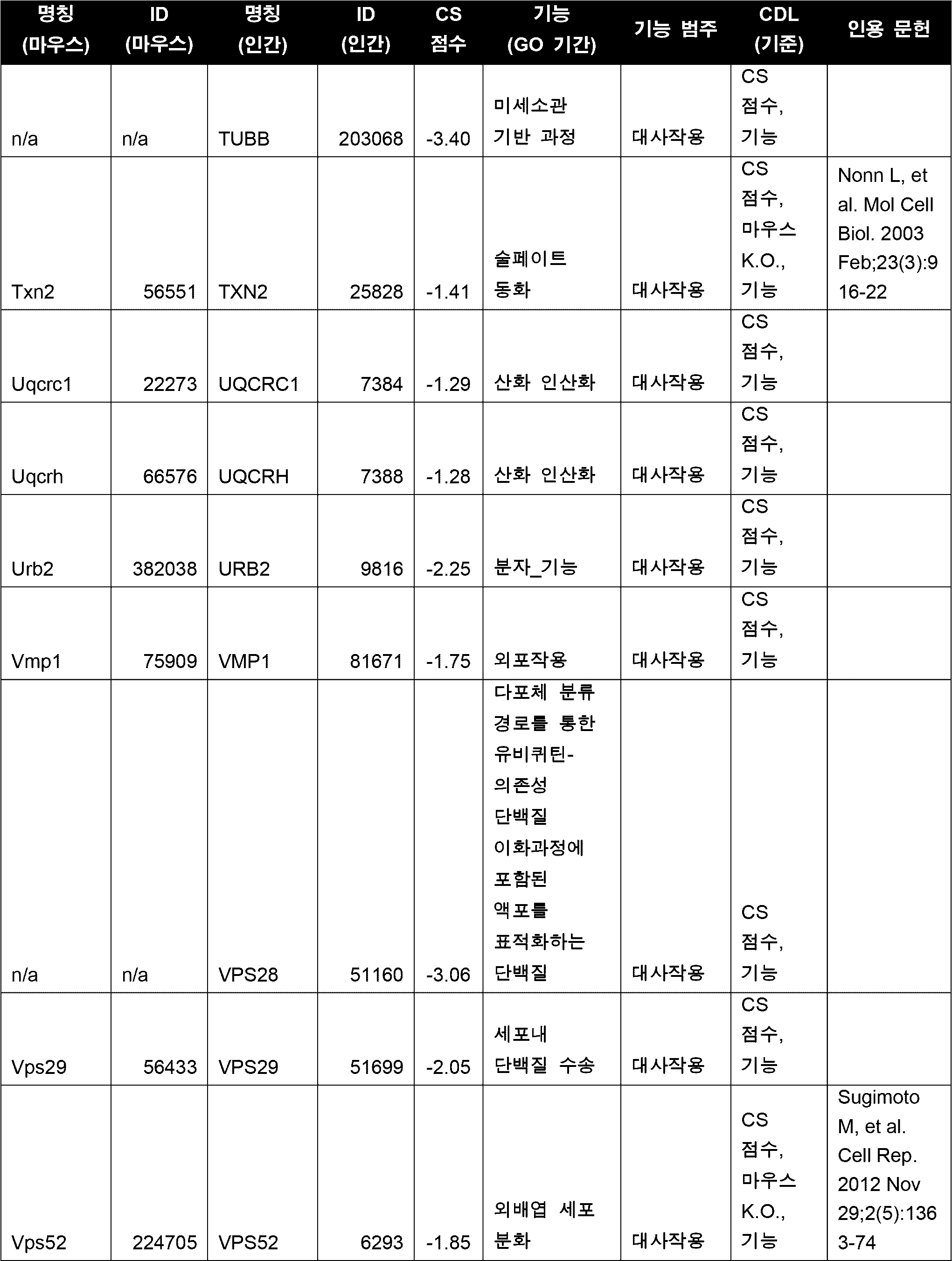


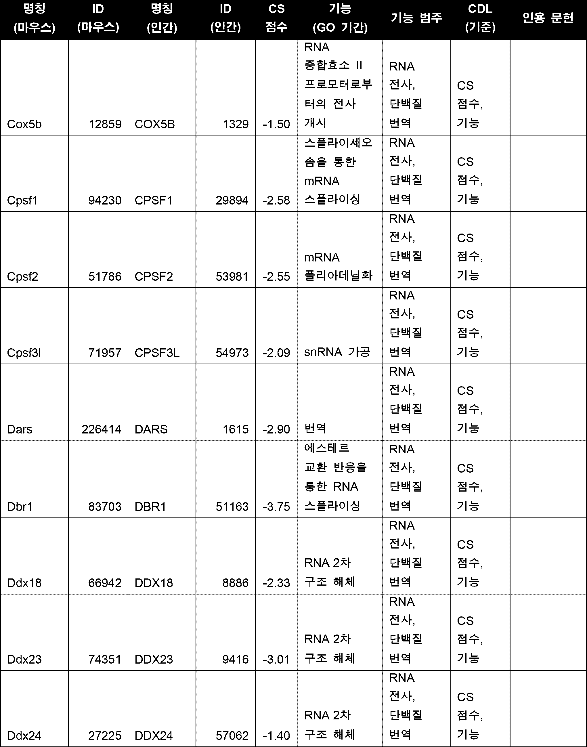


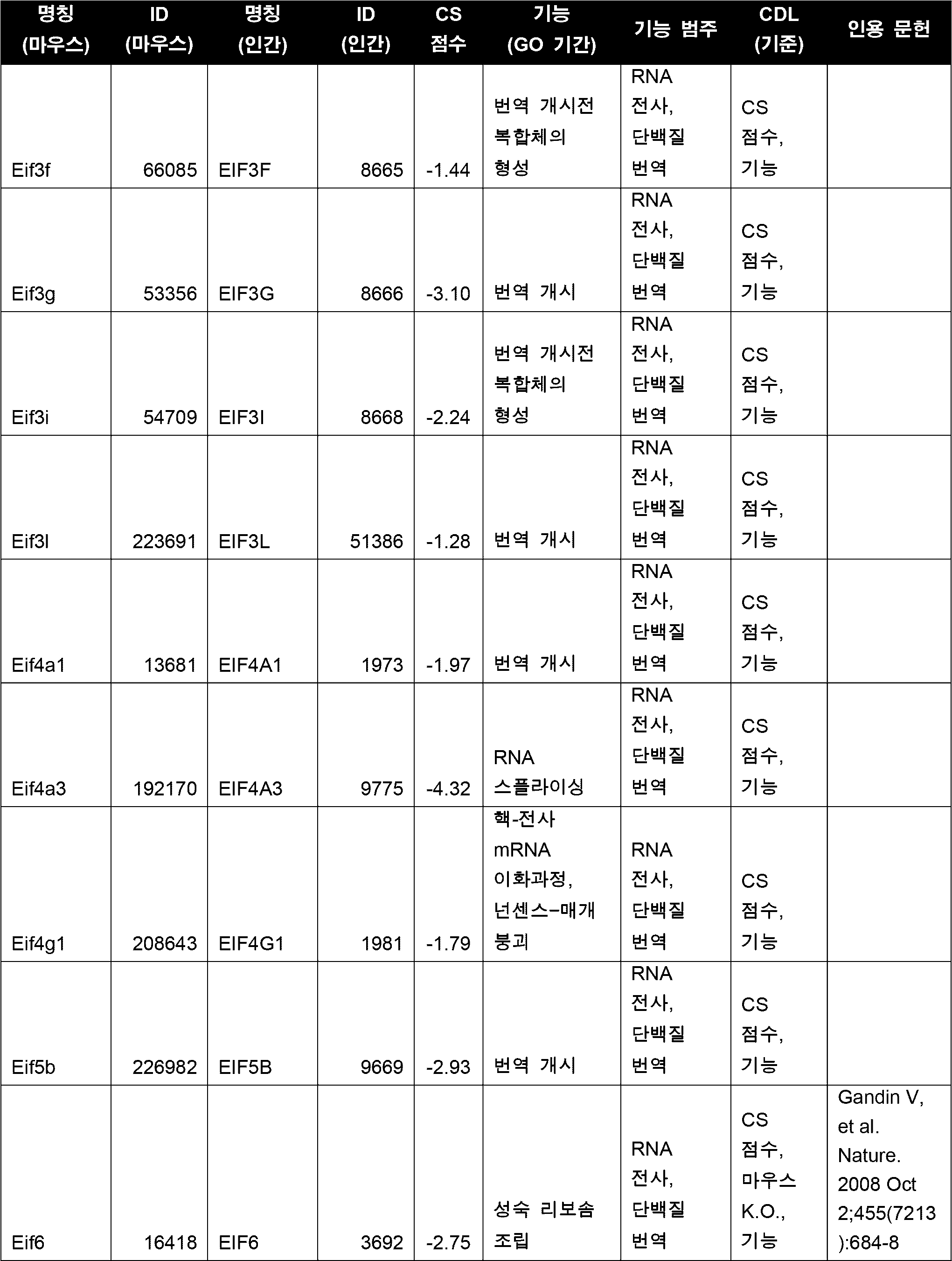

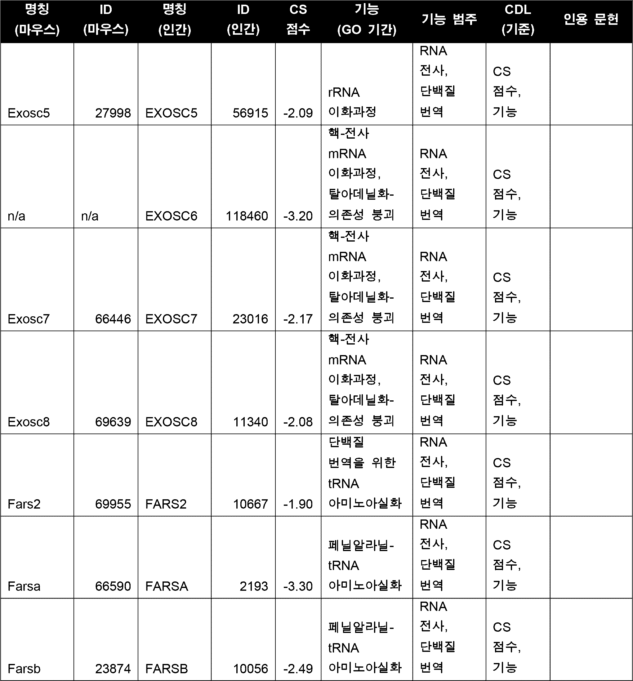


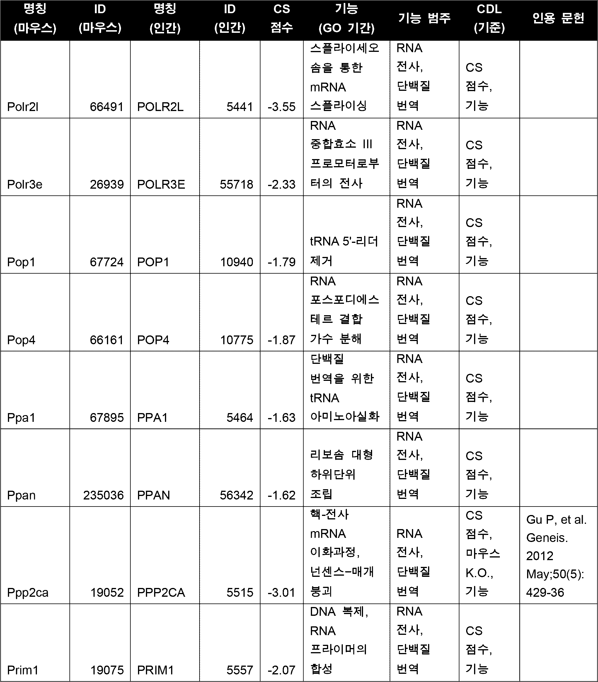


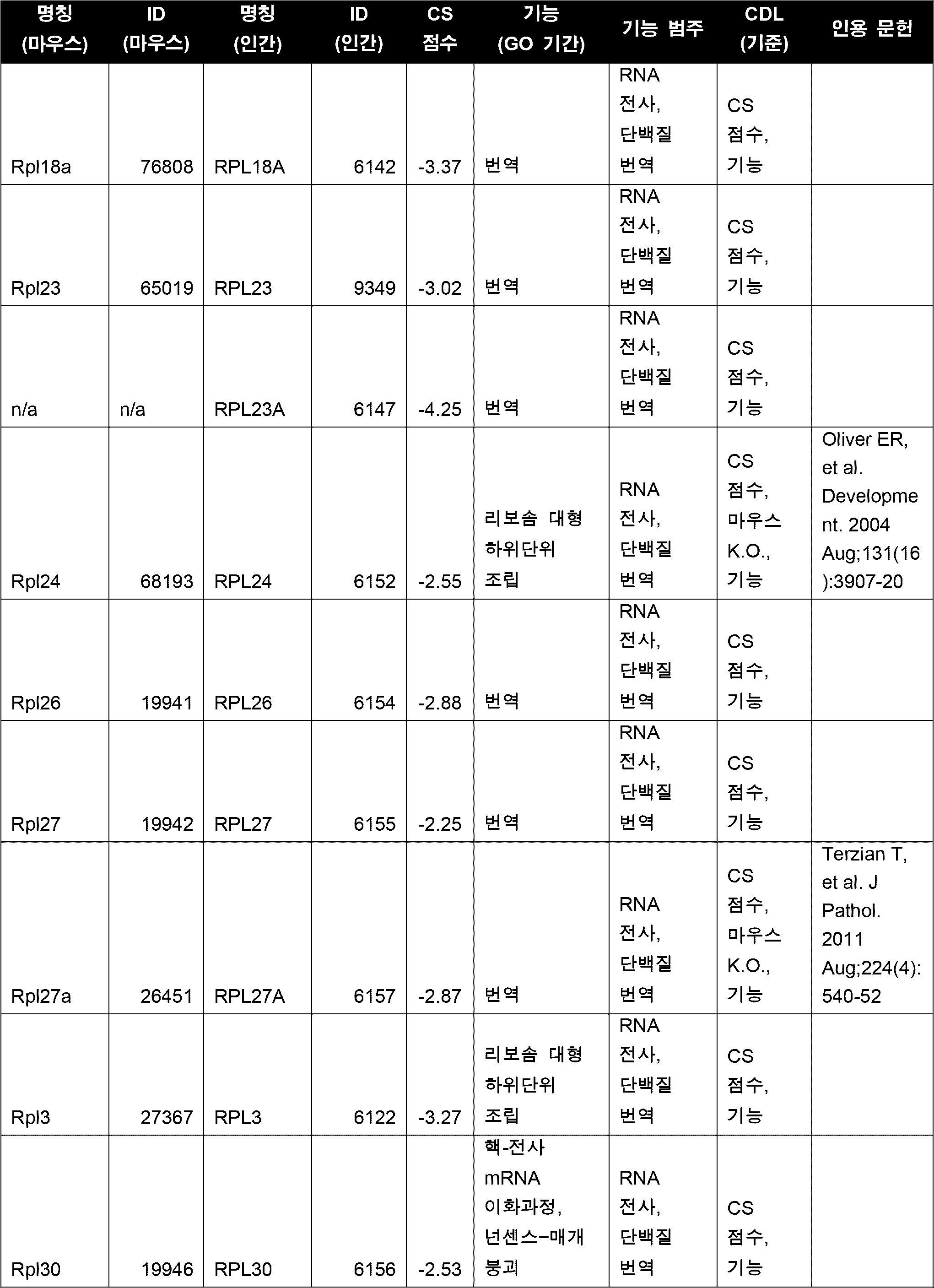


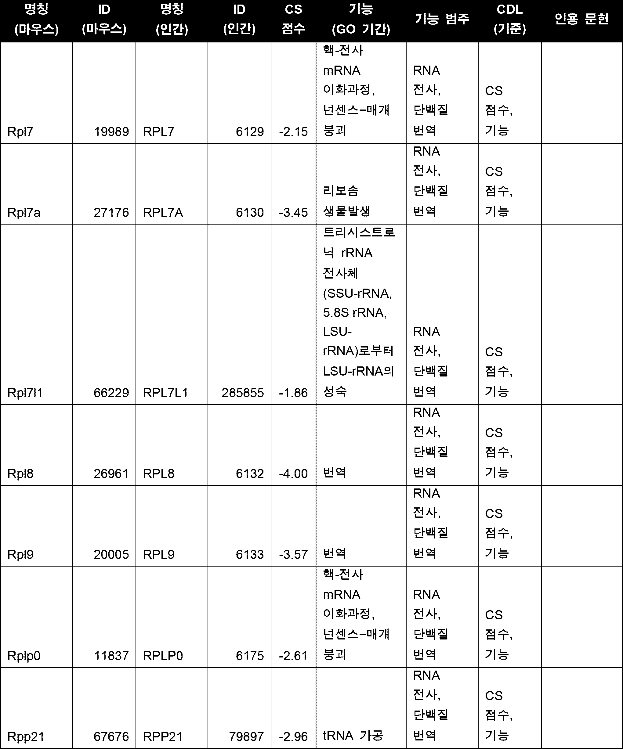
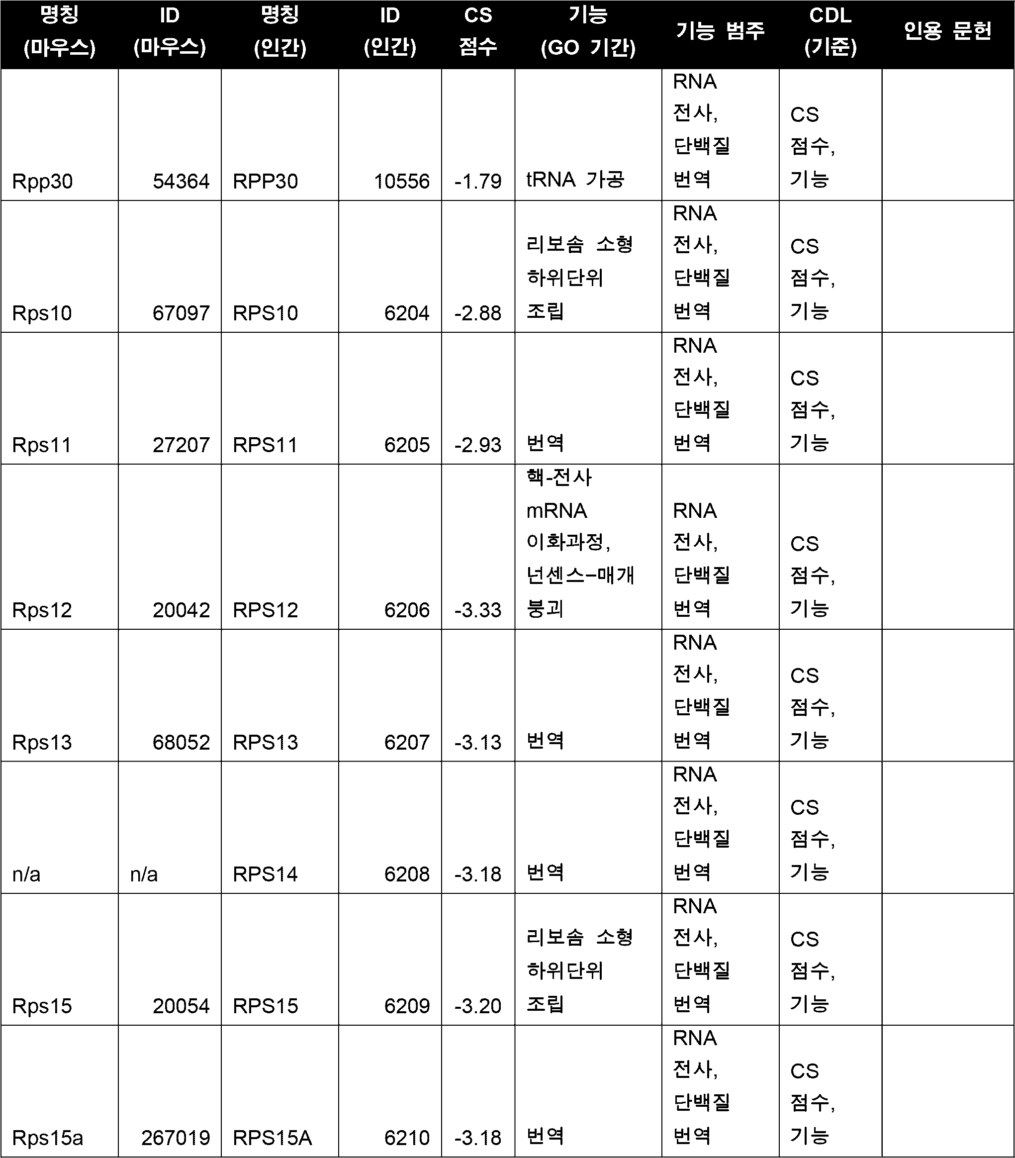


본 개시는 특정의 구체적인 구현예를 참조하여 기재되었으나, 그의 다양한 변형이 당업자에게 명백할 것이다. 본 명세서에서 제공되는 임의의 실시예는 본 개시를 설명하기 위한 목적으로만 포함되며, 어떤 방식으로든 본 개시를 제한하려는 의도가 아니다. 본 명세서에서 제공되는 임의의 도면은 본 개시의 다양한 양태를 설명하기 위한 목적일 뿐이며, 어떤 방식으로든 본 개시의 규모를 도출하거나, 제한하려는 의도가 아니다. 본 명세서에 첨부된 청구범위의 범위는 상기 설명에서 제시된 바람직한 구현예들에 의해 제한되어서는 안되며, 전체적으로 본 명세서와 일치하는 가장 넓은 해석이 주어져야 한다. 본 명세서에 나열된 모든 선행 기술의 개시는 그 전문이 본 명세서에 참조로 포함된다.While this disclosure has been described with reference to specific specific embodiments, various modifications thereof will become apparent to those skilled in the art. Any embodiments provided herein are included solely for the purpose of illustrating the disclosure and are not intended to limit the disclosure in any way. Any drawings provided herein are provided solely for the purpose of illustrating various aspects of the disclosure and are not intended to delineate or limit the scope of the disclosure in any way. The scope of the claims appended to this disclosure should not be limited by the preferred embodiments set forth in the foregoing description, but should be given the broadest interpretation consistent with this disclosure as a whole. All prior art disclosures listed herein are hereby incorporated by reference in their entirety.
SEQUENCE LISTING <110> Sinai Health System <120> ALLOGRAFT TOLERANCE WITHOUT THE NEED FOR SYSTEMIC IMMUNE SUPPRESSION <130> 51276-003WO3 <150> US 62/666,626 <151> 2018-05-03 <150> US 62/518,151 <151> 2017-06-12 <160> 17 <170> PatentIn version 3.5 <210> 1 <211> 133 <212> PRT <213> Mus musculus <400> 1 Met Ala Gln Met Met Thr Leu Ser Leu Leu Ser Leu Val Leu Ala Leu 1 5 10 15 Cys Ile Pro Trp Thr Gln Gly Ser Asp Gly Gly Gly Gln Asp Cys Cys 20 25 30 Leu Lys Tyr Ser Gln Lys Lys Ile Pro Tyr Ser Ile Val Arg Gly Tyr 35 40 45 Arg Lys Gln Glu Pro Ser Leu Gly Cys Pro Ile Pro Ala Ile Leu Phe 50 55 60 Leu Pro Arg Lys His Ser Lys Pro Glu Leu Cys Ala Asn Pro Glu Glu 65 70 75 80 Gly Trp Val Gln Asn Leu Met Arg Arg Leu Asp Gln Pro Pro Ala Pro 85 90 95 Gly Lys Gln Ser Pro Gly Cys Arg Lys Asn Arg Gly Thr Ser Lys Ser 100 105 110 Gly Lys Lys Gly Lys Gly Ser Lys Gly Cys Lys Arg Thr Glu Gln Thr 115 120 125 Gln Pro Ser Arg Gly 130 <210> 2 <211> 134 <212> PRT <213> Homo sapiens <400> 2 Met Ala Gln Ser Leu Ala Leu Ser Leu Leu Ile Leu Val Leu Ala Phe 1 5 10 15 Gly Ile Pro Arg Thr Gln Gly Ser Asp Gly Gly Ala Gln Asp Cys Cys 20 25 30 Leu Lys Tyr Ser Gln Arg Lys Ile Pro Ala Lys Val Val Arg Ser Tyr 35 40 45 Arg Lys Gln Glu Pro Ser Leu Gly Cys Ser Ile Pro Ala Ile Leu Phe 50 55 60 Leu Pro Arg Lys Arg Ser Gln Ala Glu Leu Cys Ala Asp Pro Lys Glu 65 70 75 80 Leu Trp Val Gln Gln Leu Met Gln His Leu Asp Lys Thr Pro Ser Pro 85 90 95 Gln Lys Pro Ala Gln Gly Cys Arg Lys Asp Arg Gly Ala Ser Lys Thr 100 105 110 Gly Lys Lys Gly Lys Gly Ser Lys Gly Cys Lys Arg Thr Glu Arg Ser 115 120 125 Gln Thr Pro Lys Gly Pro 130 <210> 3 <211> 303 <212> PRT <213> Mus musculus <400> 3 Met Trp Pro Leu Ala Ala Ala Leu Leu Leu Gly Ser Cys Cys Cys Gly 1 5 10 15 Ser Ala Gln Leu Leu Phe Ser Asn Val Asn Ser Ile Glu Phe Thr Ser 20 25 30 Cys Asn Glu Thr Val Val Ile Pro Cys Ile Val Arg Asn Val Glu Ala 35 40 45 Gln Ser Thr Glu Glu Met Phe Val Lys Trp Lys Leu Asn Lys Ser Tyr 50 55 60 Ile Phe Ile Tyr Asp Gly Asn Lys Asn Ser Thr Thr Thr Asp Gln Asn 65 70 75 80 Phe Thr Ser Ala Lys Ile Ser Val Ser Asp Leu Ile Asn Gly Ile Ala 85 90 95 Ser Leu Lys Met Asp Lys Arg Asp Ala Met Val Gly Asn Tyr Thr Cys 100 105 110 Glu Val Thr Glu Leu Ser Arg Glu Gly Lys Thr Val Ile Glu Leu Lys 115 120 125 Asn Arg Thr Val Ser Trp Phe Ser Pro Asn Glu Lys Ile Leu Ile Val 130 135 140 Ile Phe Pro Ile Leu Ala Ile Leu Leu Phe Trp Gly Lys Phe Gly Ile 145 150 155 160 Leu Thr Leu Lys Tyr Lys Ser Ser His Thr Asn Lys Arg Ile Ile Leu 165 170 175 Leu Leu Val Ala Gly Leu Val Leu Thr Val Ile Val Val Val Gly Ala 180 185 190 Ile Leu Leu Ile Pro Gly Glu Lys Pro Val Lys Asn Ala Ser Gly Leu 195 200 205 Gly Leu Ile Val Ile Ser Thr Gly Ile Leu Ile Leu Leu Gln Tyr Asn 210 215 220 Val Phe Met Thr Ala Phe Gly Met Thr Ser Phe Thr Ile Ala Ile Leu 225 230 235 240 Ile Thr Gln Val Leu Gly Tyr Val Leu Ala Leu Val Gly Leu Cys Leu 245 250 255 Cys Ile Met Ala Cys Glu Pro Val His Gly Pro Leu Leu Ile Ser Gly 260 265 270 Leu Gly Ile Ile Ala Leu Ala Glu Leu Leu Gly Leu Val Tyr Met Lys 275 280 285 Phe Val Ala Ser Asn Gln Arg Thr Ile Gln Pro Pro Arg Asn Arg 290 295 300 <210> 4 <211> 323 <212> PRT <213> Homo sapiens <400> 4 Met Trp Pro Leu Val Ala Ala Leu Leu Leu Gly Ser Ala Cys Cys Gly 1 5 10 15 Ser Ala Gln Leu Leu Phe Asn Lys Thr Lys Ser Val Glu Phe Thr Phe 20 25 30 Cys Asn Asp Thr Val Val Ile Pro Cys Phe Val Thr Asn Met Glu Ala 35 40 45 Gln Asn Thr Thr Glu Val Tyr Val Lys Trp Lys Phe Lys Gly Arg Asp 50 55 60 Ile Tyr Thr Phe Asp Gly Ala Leu Asn Lys Ser Thr Val Pro Thr Asp 65 70 75 80 Phe Ser Ser Ala Lys Ile Glu Val Ser Gln Leu Leu Lys Gly Asp Ala 85 90 95 Ser Leu Lys Met Asp Lys Ser Asp Ala Val Ser His Thr Gly Asn Tyr 100 105 110 Thr Cys Glu Val Thr Glu Leu Thr Arg Glu Gly Glu Thr Ile Ile Glu 115 120 125 Leu Lys Tyr Arg Val Val Ser Trp Phe Ser Pro Asn Glu Asn Ile Leu 130 135 140 Ile Val Ile Phe Pro Ile Phe Ala Ile Leu Leu Phe Trp Gly Gln Phe 145 150 155 160 Gly Ile Lys Thr Leu Lys Tyr Arg Ser Gly Gly Met Asp Glu Lys Thr 165 170 175 Ile Ala Leu Leu Val Ala Gly Leu Val Ile Thr Val Ile Val Ile Val 180 185 190 Gly Ala Ile Leu Phe Val Pro Gly Glu Tyr Ser Leu Lys Asn Ala Thr 195 200 205 Gly Leu Gly Leu Ile Val Thr Ser Thr Gly Ile Leu Ile Leu Leu His 210 215 220 Tyr Tyr Val Phe Ser Thr Ala Ile Gly Leu Thr Ser Phe Val Ile Ala 225 230 235 240 Ile Leu Val Ile Gln Val Ile Ala Tyr Ile Leu Ala Val Val Gly Leu 245 250 255 Ser Leu Cys Ile Ala Ala Cys Ile Pro Met His Gly Pro Leu Leu Ile 260 265 270 Ser Gly Leu Ser Ile Leu Ala Leu Ala Gln Leu Leu Gly Leu Val Tyr 275 280 285 Met Lys Phe Val Ala Ser Asn Gln Lys Thr Ile Gln Pro Pro Arg Lys 290 295 300 Ala Val Glu Glu Pro Leu Asn Ala Phe Lys Glu Ser Lys Gly Met Met 305 310 315 320 Asn Asp Glu <210> 5 <211> 278 <212> PRT <213> Mus musculus <400> 5 Met Gly Ser Leu Val Phe Arg Arg Pro Phe Cys His Leu Ser Thr Tyr 1 5 10 15 Ser Leu Ile Trp Gly Met Ala Ala Val Ala Leu Ser Thr Ala Gln Val 20 25 30 Glu Val Val Thr Gln Asp Glu Arg Lys Ala Leu His Thr Thr Ala Ser 35 40 45 Leu Arg Cys Ser Leu Lys Thr Ser Gln Glu Pro Leu Ile Val Thr Trp 50 55 60 Gln Lys Lys Lys Ala Val Ser Pro Glu Asn Met Val Thr Tyr Ser Lys 65 70 75 80 Thr His Gly Val Val Ile Gln Pro Ala Tyr Lys Asp Arg Ile Asn Val 85 90 95 Thr Glu Leu Gly Leu Trp Asn Ser Ser Ile Thr Phe Trp Asn Thr Thr 100 105 110 Leu Glu Asp Glu Gly Cys Tyr Met Cys Leu Phe Asn Thr Phe Gly Ser 115 120 125 Gln Lys Val Ser Gly Thr Ala Cys Leu Thr Leu Tyr Val Gln Pro Ile 130 135 140 Val His Leu His Tyr Asn Tyr Phe Glu Asp His Leu Asn Ile Thr Cys 145 150 155 160 Ser Ala Thr Ala Arg Pro Ala Pro Ala Ile Ser Trp Lys Gly Thr Gly 165 170 175 Thr Gly Ile Glu Asn Ser Thr Glu Ser His Phe His Ser Asn Gly Thr 180 185 190 Thr Ser Val Thr Ser Ile Leu Arg Val Lys Asp Pro Lys Thr Gln Val 195 200 205 Gly Lys Glu Val Ile Cys Gln Val Leu Tyr Leu Gly Asn Val Ile Asp 210 215 220 Tyr Lys Gln Ser Leu Asp Lys Gly Phe Trp Phe Ser Val Pro Leu Leu 225 230 235 240 Leu Ser Ile Val Ser Leu Val Ile Leu Leu Val Leu Ile Ser Ile Leu 245 250 255 Leu Tyr Trp Lys Arg His Arg Asn Gln Glu Arg Gly Glu Ser Ser Gln 260 265 270 Gly Met Gln Arg Met Lys 275 <210> 6 <211> 278 <212> PRT <213> Homo sapiens <400> 6 Met Glu Arg Leu Val Ile Arg Met Pro Phe Ser His Leu Ser Thr Tyr 1 5 10 15 Ser Leu Val Trp Val Met Ala Ala Val Val Leu Cys Thr Ala Gln Val 20 25 30 Gln Val Val Thr Gln Asp Glu Arg Glu Gln Leu Tyr Thr Pro Ala Ser 35 40 45 Leu Lys Cys Ser Leu Gln Asn Ala Gln Glu Ala Leu Ile Val Thr Trp 50 55 60 Gln Lys Lys Lys Ala Val Ser Pro Glu Asn Met Val Thr Phe Ser Glu 65 70 75 80 Asn His Gly Val Val Ile Gln Pro Ala Tyr Lys Asp Lys Ile Asn Ile 85 90 95 Thr Gln Leu Gly Leu Gln Asn Ser Thr Ile Thr Phe Trp Asn Ile Thr 100 105 110 Leu Glu Asp Glu Gly Cys Tyr Met Cys Leu Phe Asn Thr Phe Gly Phe 115 120 125 Gly Lys Ile Ser Gly Thr Ala Cys Leu Thr Val Tyr Val Gln Pro Ile 130 135 140 Val Ser Leu His Tyr Lys Phe Ser Glu Asp His Leu Asn Ile Thr Cys 145 150 155 160 Ser Ala Thr Ala Arg Pro Ala Pro Met Val Phe Trp Lys Val Pro Arg 165 170 175 Ser Gly Ile Glu Asn Ser Thr Val Thr Leu Ser His Pro Asn Gly Thr 180 185 190 Thr Ser Val Thr Ser Ile Leu His Ile Lys Asp Pro Lys Asn Gln Val 195 200 205 Gly Lys Glu Val Ile Cys Gln Val Leu His Leu Gly Thr Val Thr Asp 210 215 220 Phe Lys Gln Thr Val Asn Lys Gly Tyr Trp Phe Ser Val Pro Leu Leu 225 230 235 240 Leu Ser Ile Val Ser Leu Val Ile Leu Leu Val Leu Ile Ser Ile Leu 245 250 255 Leu Tyr Trp Lys Arg His Arg Asn Gln Asp Arg Gly Glu Leu Ser Gln 260 265 270 Gly Val Gln Lys Met Thr 275 <210> 7 <211> 374 <212> PRT <213> Mus musculus <400> 7 Met Asn Thr Leu Ser Glu Gly Asn Gly Thr Phe Ala Ile His Leu Leu 1 5 10 15 Lys Met Leu Cys Gln Ser Asn Pro Ser Lys Asn Val Cys Tyr Ser Pro 20 25 30 Ala Ser Ile Ser Ser Ala Leu Ala Met Val Leu Leu Gly Ala Lys Gly 35 40 45 Gln Thr Ala Val Gln Ile Ser Gln Ala Leu Gly Leu Asn Lys Glu Glu 50 55 60 Gly Ile His Gln Gly Phe Gln Leu Leu Leu Arg Lys Leu Asn Lys Pro 65 70 75 80 Asp Arg Lys Tyr Ser Leu Arg Val Ala Asn Arg Leu Phe Ala Asp Lys 85 90 95 Thr Cys Glu Val Leu Gln Thr Phe Lys Glu Ser Ser Leu His Phe Tyr 100 105 110 Asp Ser Glu Met Glu Gln Leu Ser Phe Ala Glu Glu Ala Glu Val Ser 115 120 125 Arg Gln His Ile Asn Thr Trp Val Ser Lys Gln Thr Glu Gly Lys Ile 130 135 140 Pro Glu Leu Leu Ser Gly Gly Ser Val Asp Ser Glu Thr Arg Leu Val 145 150 155 160 Leu Ile Asn Ala Leu Tyr Phe Lys Gly Lys Trp His Gln Pro Phe Asn 165 170 175 Lys Glu Tyr Thr Met Asp Met Pro Phe Lys Ile Asn Lys Asp Glu Lys 180 185 190 Arg Pro Val Gln Met Met Cys Arg Glu Asp Thr Tyr Asn Leu Ala Tyr 195 200 205 Val Lys Glu Val Gln Ala Gln Val Leu Val Met Pro Tyr Glu Gly Met 210 215 220 Glu Leu Ser Leu Val Val Leu Leu Pro Asp Glu Gly Val Asp Leu Ser 225 230 235 240 Lys Val Glu Asn Asn Leu Thr Phe Glu Lys Leu Thr Ala Trp Met Glu 245 250 255 Ala Asp Phe Met Lys Ser Thr Asp Val Glu Val Phe Leu Pro Lys Phe 260 265 270 Lys Leu Gln Glu Asp Tyr Asp Met Glu Ser Leu Phe Gln Arg Leu Gly 275 280 285 Val Val Asp Val Phe Gln Glu Asp Lys Ala Asp Leu Ser Gly Met Ser 290 295 300 Pro Glu Arg Asn Leu Cys Val Ser Lys Phe Val His Gln Ser Val Val 305 310 315 320 Glu Ile Asn Glu Glu Gly Thr Glu Ala Ala Ala Ala Ser Ala Ile Ile 325 330 335 Glu Phe Cys Cys Ala Ser Ser Val Pro Thr Phe Cys Ala Asp His Pro 340 345 350 Phe Leu Phe Phe Ile Arg His Asn Lys Ala Asn Ser Ile Leu Phe Cys 355 360 365 Gly Arg Phe Ser Ser Pro 370 <210> 8 <211> 376 <212> PRT <213> Homo sapiens <400> 8 Met Glu Thr Leu Ser Asn Ala Ser Gly Thr Phe Ala Ile Arg Leu Leu 1 5 10 15 Lys Ile Leu Cys Gln Asp Asn Pro Ser His Asn Val Phe Cys Ser Pro 20 25 30 Val Ser Ile Ser Ser Ala Leu Ala Met Val Leu Leu Gly Ala Lys Gly 35 40 45 Asn Thr Ala Thr Gln Met Ala Gln Ala Leu Ser Leu Asn Thr Glu Glu 50 55 60 Asp Ile His Arg Ala Phe Gln Ser Leu Leu Thr Glu Val Asn Lys Ala 65 70 75 80 Gly Thr Gln Tyr Leu Leu Arg Thr Ala Asn Arg Leu Phe Gly Glu Lys 85 90 95 Thr Cys Gln Phe Leu Ser Thr Phe Lys Glu Ser Cys Leu Gln Phe Tyr 100 105 110 His Ala Glu Leu Lys Glu Leu Ser Phe Ile Arg Ala Ala Glu Glu Ser 115 120 125 Arg Lys His Ile Asn Thr Trp Val Ser Lys Lys Thr Glu Gly Lys Ile 130 135 140 Glu Glu Leu Leu Pro Gly Ser Ser Ile Asp Ala Glu Thr Arg Leu Val 145 150 155 160 Leu Val Asn Ala Ile Tyr Phe Lys Gly Lys Trp Asn Glu Pro Phe Asp 165 170 175 Glu Thr Tyr Thr Arg Glu Met Pro Phe Lys Ile Asn Gln Glu Glu Gln 180 185 190 Arg Pro Val Gln Met Met Tyr Gln Glu Ala Thr Phe Lys Leu Ala His 195 200 205 Val Gly Glu Val Arg Ala Gln Leu Leu Glu Leu Pro Tyr Ala Arg Lys 210 215 220 Glu Leu Ser Leu Leu Val Leu Leu Pro Asp Asp Gly Val Glu Leu Ser 225 230 235 240 Thr Val Glu Lys Ser Leu Thr Phe Glu Lys Leu Thr Ala Trp Thr Lys 245 250 255 Pro Asp Cys Met Lys Ser Thr Glu Val Glu Val Leu Leu Pro Lys Phe 260 265 270 Lys Leu Gln Glu Asp Tyr Asp Met Glu Ser Val Leu Arg His Leu Gly 275 280 285 Ile Val Asp Ala Phe Gln Gln Gly Lys Ala Asp Leu Ser Ala Met Ser 290 295 300 Ala Glu Arg Asp Leu Cys Leu Ser Lys Phe Val His Lys Ser Phe Val 305 310 315 320 Glu Val Asn Glu Glu Gly Thr Glu Ala Ala Ala Ala Ser Ser Cys Phe 325 330 335 Val Val Ala Glu Cys Cys Met Glu Ser Gly Pro Arg Phe Cys Ala Asp 340 345 350 His Pro Phe Leu Phe Phe Ile Arg His Asn Arg Ala Asn Ser Ile Leu 355 360 365 Phe Cys Gly Arg Phe Ser Ser Pro 370 375 <210> 9 <211> 279 <212> PRT <213> Mus musculus <400> 9 Met Gln Gln Pro Met Asn Tyr Pro Cys Pro Gln Ile Phe Trp Val Asp 1 5 10 15 Ser Ser Ala Thr Ser Ser Trp Thr Pro Pro Gly Ser Val Phe Pro Cys 20 25 30 Pro Ser Ser Gly Pro Arg Gly Pro Asp Gln Arg Arg Pro Pro Pro Pro 35 40 45 Pro Pro Pro Val Ser Pro Leu Pro Pro Pro Ser Gln Pro Leu Pro Leu 50 55 60 Pro Pro Leu Thr Pro Leu Lys Lys Lys Asp His Asn Thr Asn Leu Trp 65 70 75 80 Leu Pro Val Val Phe Phe Met Val Leu Val Ala Leu Val Gly Met Gly 85 90 95 Leu Gly Met Tyr Gln Leu Phe His Leu Gln Lys Glu Leu Ala Glu Leu 100 105 110 Arg Glu Phe Thr Asn Gln Ser Leu Lys Val Ser Ser Phe Glu Lys Gln 115 120 125 Ile Ala Asn Pro Ser Thr Pro Ser Glu Lys Lys Glu Leu Arg Ser Val 130 135 140 Ala His Leu Thr Gly Asn Pro His Ser Arg Ser Ile Pro Leu Glu Trp 145 150 155 160 Glu Asp Thr Tyr Gly Thr Ala Leu Ile Ser Gly Val Lys Tyr Lys Lys 165 170 175 Gly Ser Leu Val Ile Asn Glu Ala Gly Leu Tyr Phe Val Tyr Ser Lys 180 185 190 Val Tyr Phe Arg Gly Gln Ser Cys Asn Asn Gln Pro Leu Asn His Lys 195 200 205 Val Tyr Met Arg Asn Ser Lys Tyr Pro Gly Asp Leu Val Leu Met Glu 210 215 220 Glu Lys Arg Leu Asn Tyr Cys Thr Thr Gly Gln Ile Trp Ala His Ser 225 230 235 240 Ser Tyr Leu Gly Ala Val Phe Asn Leu Thr Ser Ala Asp His Leu Tyr 245 250 255 Val Asn Ile Ser Gln Leu Ser Leu Ile Asn Phe Glu Glu Ser Lys Thr 260 265 270 Phe Phe Gly Leu Tyr Lys Leu 275 <210> 10 <211> 281 <212> PRT <213> Homo sapiens <400> 10 Met Gln Gln Pro Phe Asn Tyr Pro Tyr Pro Gln Ile Tyr Trp Val Asp 1 5 10 15 Ser Ser Ala Ser Ser Pro Trp Ala Pro Pro Gly Thr Val Leu Pro Cys 20 25 30 Pro Thr Ser Val Pro Arg Arg Pro Gly Gln Arg Arg Pro Pro Pro Pro 35 40 45 Pro Pro Pro Pro Pro Leu Pro Pro Pro Pro Pro Pro Pro Pro Leu Pro 50 55 60 Pro Leu Pro Leu Pro Pro Leu Lys Lys Arg Gly Asn His Ser Thr Gly 65 70 75 80 Leu Cys Leu Leu Val Met Phe Phe Met Val Leu Val Ala Leu Val Gly 85 90 95 Leu Gly Leu Gly Met Phe Gln Leu Phe His Leu Gln Lys Glu Leu Ala 100 105 110 Glu Leu Arg Glu Ser Thr Ser Gln Met His Thr Ala Ser Ser Leu Glu 115 120 125 Lys Gln Ile Gly His Pro Ser Pro Pro Pro Glu Lys Lys Glu Leu Arg 130 135 140 Lys Val Ala His Leu Thr Gly Lys Ser Asn Ser Arg Ser Met Pro Leu 145 150 155 160 Glu Trp Glu Asp Thr Tyr Gly Ile Val Leu Leu Ser Gly Val Lys Tyr 165 170 175 Lys Lys Gly Gly Leu Val Ile Asn Glu Thr Gly Leu Tyr Phe Val Tyr 180 185 190 Ser Lys Val Tyr Phe Arg Gly Gln Ser Cys Asn Asn Leu Pro Leu Ser 195 200 205 His Lys Val Tyr Met Arg Asn Ser Lys Tyr Pro Gln Asp Leu Val Met 210 215 220 Met Glu Gly Lys Met Met Ser Tyr Cys Thr Thr Gly Gln Met Trp Ala 225 230 235 240 Arg Ser Ser Tyr Leu Gly Ala Val Phe Asn Leu Thr Ser Ala Asp His 245 250 255 Leu Tyr Val Asn Val Ser Glu Leu Ser Leu Val Asn Phe Glu Glu Ser 260 265 270 Gln Thr Phe Phe Gly Leu Tyr Lys Leu 275 280 <210> 11 <211> 290 <212> PRT <213> Mus musculus <400> 11 Met Arg Ile Phe Ala Gly Ile Ile Phe Thr Ala Cys Cys His Leu Leu 1 5 10 15 Arg Ala Phe Thr Ile Thr Ala Pro Lys Asp Leu Tyr Val Val Glu Tyr 20 25 30 Gly Ser Asn Val Thr Met Glu Cys Arg Phe Pro Val Glu Arg Glu Leu 35 40 45 Asp Leu Leu Ala Leu Val Val Tyr Trp Glu Lys Glu Asp Glu Gln Val 50 55 60 Ile Gln Phe Val Ala Gly Glu Glu Asp Leu Lys Pro Gln His Ser Asn 65 70 75 80 Phe Arg Gly Arg Ala Ser Leu Pro Lys Asp Gln Leu Leu Lys Gly Asn 85 90 95 Ala Ala Leu Gln Ile Thr Asp Val Lys Leu Gln Asp Ala Gly Val Tyr 100 105 110 Cys Cys Ile Ile Ser Tyr Gly Gly Ala Asp Tyr Lys Arg Ile Thr Leu 115 120 125 Lys Val Asn Ala Pro Tyr Arg Lys Ile Asn Gln Arg Ile Ser Val Asp 130 135 140 Pro Ala Thr Ser Glu His Glu Leu Ile Cys Gln Ala Glu Gly Tyr Pro 145 150 155 160 Glu Ala Glu Val Ile Trp Thr Asn Ser Asp His Gln Pro Val Ser Gly 165 170 175 Lys Arg Ser Val Thr Thr Ser Arg Thr Glu Gly Met Leu Leu Asn Val 180 185 190 Thr Ser Ser Leu Arg Val Asn Ala Thr Ala Asn Asp Val Phe Tyr Cys 195 200 205 Thr Phe Trp Arg Ser Gln Pro Gly Gln Asn His Thr Ala Glu Leu Ile 210 215 220 Ile Pro Glu Leu Pro Ala Thr His Pro Pro Gln Asn Arg Thr His Trp 225 230 235 240 Val Leu Leu Gly Ser Ile Leu Leu Phe Leu Ile Val Val Ser Thr Val 245 250 255 Leu Leu Phe Leu Arg Lys Gln Val Arg Met Leu Asp Val Glu Lys Cys 260 265 270 Gly Val Glu Asp Thr Ser Ser Lys Asn Arg Asn Asp Thr Gln Phe Glu 275 280 285 Glu Thr 290 <210> 12 <211> 290 <212> PRT <213> Homo sapiens <400> 12 Met Arg Ile Phe Ala Val Phe Ile Phe Met Thr Tyr Trp His Leu Leu 1 5 10 15 Asn Ala Phe Thr Val Thr Val Pro Lys Asp Leu Tyr Val Val Glu Tyr 20 25 30 Gly Ser Asn Met Thr Ile Glu Cys Lys Phe Pro Val Glu Lys Gln Leu 35 40 45 Asp Leu Ala Ala Leu Ile Val Tyr Trp Glu Met Glu Asp Lys Asn Ile 50 55 60 Ile Gln Phe Val His Gly Glu Glu Asp Leu Lys Val Gln His Ser Ser 65 70 75 80 Tyr Arg Gln Arg Ala Arg Leu Leu Lys Asp Gln Leu Ser Leu Gly Asn 85 90 95 Ala Ala Leu Gln Ile Thr Asp Val Lys Leu Gln Asp Ala Gly Val Tyr 100 105 110 Arg Cys Met Ile Ser Tyr Gly Gly Ala Asp Tyr Lys Arg Ile Thr Val 115 120 125 Lys Val Asn Ala Pro Tyr Asn Lys Ile Asn Gln Arg Ile Leu Val Val 130 135 140 Asp Pro Val Thr Ser Glu His Glu Leu Thr Cys Gln Ala Glu Gly Tyr 145 150 155 160 Pro Lys Ala Glu Val Ile Trp Thr Ser Ser Asp His Gln Val Leu Ser 165 170 175 Gly Lys Thr Thr Thr Thr Asn Ser Lys Arg Glu Glu Lys Leu Phe Asn 180 185 190 Val Thr Ser Thr Leu Arg Ile Asn Thr Thr Thr Asn Glu Ile Phe Tyr 195 200 205 Cys Thr Phe Arg Arg Leu Asp Pro Glu Glu Asn His Thr Ala Glu Leu 210 215 220 Val Ile Pro Glu Leu Pro Leu Ala His Pro Pro Asn Glu Arg Thr His 225 230 235 240 Leu Val Ile Leu Gly Ala Ile Leu Leu Cys Leu Gly Val Ala Leu Thr 245 250 255 Phe Ile Phe Arg Leu Arg Lys Gly Arg Met Met Asp Val Lys Lys Cys 260 265 270 Gly Ile Gln Asp Thr Asn Ser Lys Lys Gln Ser Asp Thr His Leu Glu 275 280 285 Glu Thr 290 <210> 13 <211> 463 <212> PRT <213> Mus musculus <400> 13 Met Gln Val Ser Arg Val Leu Ala Ala Leu Cys Gly Met Leu Leu Cys 1 5 10 15 Ala Ser Gly Leu Phe Ala Ala Ser Gly Asp Phe Cys Asp Ser Ser Leu 20 25 30 Cys Leu Asn Gly Gly Thr Cys Leu Thr Gly Gln Asp Asn Asp Ile Tyr 35 40 45 Cys Leu Cys Pro Glu Gly Phe Thr Gly Leu Val Cys Asn Glu Thr Glu 50 55 60 Arg Gly Pro Cys Ser Pro Asn Pro Cys Tyr Asn Asp Ala Lys Cys Leu 65 70 75 80 Val Thr Leu Asp Thr Gln Arg Gly Asp Ile Phe Thr Glu Tyr Ile Cys 85 90 95 Gln Cys Pro Val Gly Tyr Ser Gly Ile His Cys Glu Thr Glu Thr Asn 100 105 110 Tyr Tyr Asn Leu Asp Gly Glu Tyr Met Phe Thr Thr Ala Val Pro Asn 115 120 125 Thr Ala Val Pro Thr Pro Ala Pro Thr Pro Asp Leu Ser Asn Asn Leu 130 135 140 Ala Ser Arg Cys Ser Thr Gln Leu Gly Met Glu Gly Gly Ala Ile Ala 145 150 155 160 Asp Ser Gln Ile Ser Ala Ser Ser Val Tyr Met Gly Phe Met Gly Leu 165 170 175 Gln Arg Trp Gly Pro Glu Leu Ala Arg Leu Tyr Arg Thr Gly Ile Val 180 185 190 Asn Ala Trp Thr Ala Ser Asn Tyr Asp Ser Lys Pro Trp Ile Gln Val 195 200 205 Asn Leu Leu Arg Lys Met Arg Val Ser Gly Val Met Thr Gln Gly Ala 210 215 220 Ser Arg Ala Gly Arg Ala Glu Tyr Leu Lys Thr Phe Lys Val Ala Tyr 225 230 235 240 Ser Leu Asp Gly Arg Lys Phe Glu Phe Ile Gln Asp Glu Ser Gly Gly 245 250 255 Asp Lys Glu Phe Leu Gly Asn Leu Asp Asn Asn Ser Leu Lys Val Asn 260 265 270 Met Phe Asn Pro Thr Leu Glu Ala Gln Tyr Ile Lys Leu Tyr Pro Val 275 280 285 Ser Cys His Arg Gly Cys Thr Leu Arg Phe Glu Leu Leu Gly Cys Glu 290 295 300 Leu His Gly Cys Ser Glu Pro Leu Gly Leu Lys Asn Asn Thr Ile Pro 305 310 315 320 Asp Ser Gln Met Ser Ala Ser Ser Ser Tyr Lys Thr Trp Asn Leu Arg 325 330 335 Ala Phe Gly Trp Tyr Pro His Leu Gly Arg Leu Asp Asn Gln Gly Lys 340 345 350 Ile Asn Ala Trp Thr Ala Gln Ser Asn Ser Ala Lys Glu Trp Leu Gln 355 360 365 Val Asp Leu Gly Thr Gln Arg Gln Val Thr Gly Ile Ile Thr Gln Gly 370 375 380 Ala Arg Asp Phe Gly His Ile Gln Tyr Val Ala Ser Tyr Lys Val Ala 385 390 395 400 His Ser Asp Asp Gly Val Gln Trp Thr Val Tyr Glu Glu Gln Gly Ser 405 410 415 Ser Lys Val Phe Gln Gly Asn Leu Asp Asn Asn Ser His Lys Lys Asn 420 425 430 Ile Phe Glu Lys Pro Phe Met Ala Arg Tyr Val Arg Val Leu Pro Val 435 440 445 Ser Trp His Asn Arg Ile Thr Leu Arg Leu Glu Leu Leu Gly Cys 450 455 460 <210> 14 <211> 387 <212> PRT <213> Homo sapiens <400> 14 Met Pro Arg Pro Arg Leu Leu Ala Ala Leu Cys Gly Ala Leu Leu Cys 1 5 10 15 Ala Pro Ser Leu Leu Val Ala Leu Asp Ile Cys Ser Lys Asn Pro Cys 20 25 30 His Asn Gly Gly Leu Cys Glu Glu Ile Ser Gln Glu Val Arg Gly Asp 35 40 45 Val Phe Pro Ser Tyr Thr Cys Thr Cys Leu Lys Gly Tyr Ala Gly Asn 50 55 60 His Cys Glu Thr Lys Cys Val Glu Pro Leu Gly Met Glu Asn Gly Asn 65 70 75 80 Ile Ala Asn Ser Gln Ile Ala Ala Ser Ser Val Arg Val Thr Phe Leu 85 90 95 Gly Leu Gln His Trp Val Pro Glu Leu Ala Arg Leu Asn Arg Ala Gly 100 105 110 Met Val Asn Ala Trp Thr Pro Ser Ser Asn Asp Asp Asn Pro Trp Ile 115 120 125 Gln Val Asn Leu Leu Arg Arg Met Trp Val Thr Gly Val Val Thr Gln 130 135 140 Gly Ala Ser Arg Leu Ala Ser His Glu Tyr Leu Lys Ala Phe Lys Val 145 150 155 160 Ala Tyr Ser Leu Asn Gly His Glu Phe Asp Phe Ile His Asp Val Asn 165 170 175 Lys Lys His Lys Glu Phe Val Gly Asn Trp Asn Lys Asn Ala Val His 180 185 190 Val Asn Leu Phe Glu Thr Pro Val Glu Ala Gln Tyr Val Arg Leu Tyr 195 200 205 Pro Thr Ser Cys His Thr Ala Cys Thr Leu Arg Phe Glu Leu Leu Gly 210 215 220 Cys Glu Leu Asn Gly Cys Ala Asn Pro Leu Gly Leu Lys Asn Asn Ser 225 230 235 240 Ile Pro Asp Lys Gln Ile Thr Ala Ser Ser Ser Tyr Lys Thr Trp Gly 245 250 255 Leu His Leu Phe Ser Trp Asn Pro Ser Tyr Ala Arg Leu Asp Lys Gln 260 265 270 Gly Asn Phe Asn Ala Trp Val Ala Gly Ser Tyr Gly Asn Asp Gln Trp 275 280 285 Leu Gln Val Asp Leu Gly Ser Ser Lys Glu Val Thr Gly Ile Ile Thr 290 295 300 Gln Gly Ala Arg Asn Phe Gly Ser Val Gln Phe Val Ala Ser Tyr Lys 305 310 315 320 Val Ala Tyr Ser Asn Asp Ser Ala Asn Trp Thr Glu Tyr Gln Asp Pro 325 330 335 Arg Thr Gly Ser Ser Lys Ile Phe Pro Gly Asn Trp Asp Asn His Ser 340 345 350 His Lys Lys Asn Leu Phe Glu Thr Pro Ile Leu Ala Arg Tyr Val Arg 355 360 365 Ile Leu Pro Val Ala Trp His Asn Arg Ile Ala Leu Arg Leu Glu Leu 370 375 380 Leu Gly Cys 385 <210> 15 <211> 249 <212> PRT <213> Mus musculus <400> 15 Ser Ile Glu Glu Ile Pro Arg Met Glu Pro Arg Ala Pro Trp Met Glu 1 5 10 15 Lys Glu Arg Pro Glu Tyr Trp Lys Glu Leu Lys Leu Lys Val Lys Asn 20 25 30 Ile Ala Gln Ser Ala Arg Ala Asn Leu Arg Thr Leu Leu Arg Tyr Tyr 35 40 45 Asn Gln Ser Glu Gly Gly Ser His Ile Leu Gln Trp Met Val Ser Cys 50 55 60 Glu Val Gly Pro Asp Met Arg Leu Leu Gly Ala His Tyr Gln Ala Ala 65 70 75 80 Tyr Asp Gly Ser Asp Tyr Ile Thr Leu Asn Glu Asp Leu Ser Ser Trp 85 90 95 Thr Ala Val Asp Met Val Ser Gln Ile Thr Lys Ser Arg Leu Glu Ser 100 105 110 Ala Gly Thr Ala Glu Tyr Phe Arg Ala Tyr Val Glu Gly Glu Cys Leu 115 120 125 Glu Leu Leu His Arg Phe Leu Arg Asn Gly Lys Glu Ile Leu Gln Arg 130 135 140 Ala Asp Pro Pro Lys Ala His Val Ala His His Pro Arg Pro Lys Gly 145 150 155 160 Asp Val Thr Leu Arg Cys Trp Ala Leu Gly Phe Tyr Pro Ala Asp Ile 165 170 175 Thr Leu Thr Trp Gln Lys Asp Glu Glu Asp Leu Thr Gln Asp Met Glu 180 185 190 Leu Val Glu Thr Arg Pro Ser Gly Asp Gly Thr Phe Gln Lys Trp Ala 195 200 205 Ala Val Val Val Pro Ser Gly Glu Glu Gln Arg Tyr Thr Cys Tyr Val 210 215 220 His His Glu Gly Leu Thr Glu Pro Leu Ala Leu Lys Trp Gly Arg Ser 225 230 235 240 Ser Gln Ser Ser Val Val Ile Met Val 245 <210> 16 <211> 338 <212> PRT <213> Homo sapiens <400> 16 Met Val Val Met Ala Pro Arg Thr Leu Phe Leu Leu Leu Ser Gly Ala 1 5 10 15 Leu Thr Leu Thr Glu Thr Trp Ala Gly Ser His Ser Met Arg Tyr Phe 20 25 30 Ser Ala Ala Val Ser Arg Pro Gly Arg Gly Glu Pro Arg Phe Ile Ala 35 40 45 Met Gly Tyr Val Asp Asp Thr Gln Phe Val Arg Phe Asp Ser Asp Ser 50 55 60 Ala Cys Pro Arg Met Glu Pro Arg Ala Pro Trp Val Glu Gln Glu Gly 65 70 75 80 Pro Glu Tyr Trp Glu Glu Glu Thr Arg Asn Thr Lys Ala His Ala Gln 85 90 95 Thr Asp Arg Met Asn Leu Gln Thr Leu Arg Gly Tyr Tyr Asn Gln Ser 100 105 110 Glu Ala Ser Ser His Thr Leu Gln Trp Met Ile Gly Cys Asp Leu Gly 115 120 125 Ser Asp Gly Arg Leu Leu Arg Gly Tyr Glu Gln Tyr Ala Tyr Asp Gly 130 135 140 Lys Asp Tyr Leu Ala Leu Asn Glu Asp Leu Arg Ser Trp Thr Ala Ala 145 150 155 160 Asp Thr Ala Ala Gln Ile Ser Lys Arg Lys Cys Glu Ala Ala Asn Val 165 170 175 Ala Glu Gln Arg Arg Ala Tyr Leu Glu Gly Thr Cys Val Glu Trp Leu 180 185 190 His Arg Tyr Leu Glu Asn Gly Lys Glu Met Leu Gln Arg Ala Asp Pro 195 200 205 Pro Lys Thr His Val Thr His His Pro Val Phe Asp Tyr Glu Ala Thr 210 215 220 Leu Arg Cys Trp Ala Leu Gly Phe Tyr Pro Ala Glu Ile Ile Leu Thr 225 230 235 240 Trp Gln Arg Asp Gly Glu Asp Gln Thr Gln Asp Val Glu Leu Val Glu 245 250 255 Thr Arg Pro Ala Gly Asp Gly Thr Phe Gln Lys Trp Ala Ala Val Val 260 265 270 Val Pro Ser Gly Glu Glu Gln Arg Tyr Thr Cys His Val Gln His Glu 275 280 285 Gly Leu Pro Glu Pro Leu Met Leu Arg Trp Lys Gln Ser Ser Leu Pro 290 295 300 Thr Ile Pro Ile Met Gly Ile Val Ala Gly Leu Val Val Leu Ala Ala 305 310 315 320 Val Val Thr Gly Ala Ala Val Ala Ala Val Leu Trp Arg Lys Lys Ser 325 330 335 Ser Asp <210> 17 <211> 439 <212> PRT <213> Artificial Sequence <220> <223> Synthetic Construct <400> 17 Met Gly Pro Gln Ala Ala Ala Gly Arg Met Ile Leu Leu Val Val Leu 1 5 10 15 Met Leu Ser Ala Lys Val Gly Ser Gly Ala Leu Thr Ser Thr Glu Asp 20 25 30 Pro Glu Pro Pro Ser Val Pro Val Pro Thr Asn Val Leu Ile Lys Ser 35 40 45 Tyr Asn Leu Asn Pro Val Val Cys Trp Glu Tyr Gln Asn Met Ser Gln 50 55 60 Thr Pro Ile Phe Thr Val Gln Val Lys Val Tyr Ser Gly Ser Trp Thr 65 70 75 80 Asp Ser Cys Thr Asn Ile Ser Asp His Cys Cys Asn Ile Tyr Gly Gln 85 90 95 Ile Met Tyr Pro Asp Val Ser Ala Trp Ala Arg Val Lys Ala Lys Val 100 105 110 Gly Gln Lys Glu Ser Asp Tyr Ala Arg Ser Lys Glu Phe Leu Met Cys 115 120 125 Leu Lys Gly Lys Val Gly Pro Pro Gly Leu Glu Ile Arg Arg Lys Lys 130 135 140 Glu Glu Gln Leu Ser Val Leu Val Phe His Pro Glu Val Val Val Asn 145 150 155 160 Gly Glu Ser Gln Gly Thr Met Phe Gly Asp Gly Ser Thr Cys Tyr Thr 165 170 175 Phe Asp Tyr Thr Val Tyr Val Glu His Asn Arg Ser Gly Glu Ile Leu 180 185 190 His Thr Lys His Thr Val Glu Lys Glu Glu Cys Asn Glu Thr Leu Cys 195 200 205 Glu Leu Asn Ile Ser Val Ser Thr Leu Asp Ser Arg Tyr Cys Ile Ser 210 215 220 Val Asp Gly Ile Ser Ser Phe Trp Gln Val Arg Thr Glu Lys Ser Lys 225 230 235 240 Asp Val Cys Ile Pro Pro Phe His Asp Asp Arg Lys Asp Ser Ile Trp 245 250 255 Ile Leu Val Val Ala Pro Leu Thr Val Phe Thr Val Val Ile Leu Val 260 265 270 Phe Ala Tyr Trp Tyr Thr Lys Lys Asn Ser Phe Lys Arg Lys Ser Ile 275 280 285 Met Leu Pro Lys Ser Leu Leu Ser Val Val Lys Ser Ala Thr Leu Glu 290 295 300 Thr Lys Pro Glu Ser Lys Tyr Ser Leu Val Thr Pro His Gln Pro Ala 305 310 315 320 Val Leu Glu Ser Glu Thr Val Ile Cys Glu Glu Pro Leu Ser Thr Val 325 330 335 Thr Ala Pro Asp Ser Pro Glu Ala Ala Glu Gln Glu Glu Leu Ser Lys 340 345 350 Glu Thr Lys Ala Leu Glu Ala Gly Gly Ser Thr Ser Ala Met Thr Pro 355 360 365 Asp Ser Pro Pro Thr Pro Thr Gln Arg Arg Ser Phe Ser Leu Leu Ser 370 375 380 Ser Asn Gln Ser Gly Pro Cys Ser Leu Thr Ala Tyr His Ser Arg Asn 385 390 395 400 Gly Ser Asp Ser Gly Leu Val Gly Ser Gly Ser Ser Ile Ser Asp Leu 405 410 415 Glu Ser Leu Pro Asn Asn Asn Ser Glu Thr Lys Met Ala Glu His Asp 420 425 430 Pro Pro Pro Val Arg Lys Ala 435 SEQUENCE LISTING <110> Sinai Health System <120> ALLOGRAFT TOLERANCE WITHOUT THE NEED FOR SYSTEMIC IMMUNE SUPPRESSION <130> 51276-003WO3 <150> US 62/666,626 <151> 2018-05-03 <150> US 62/518,151 <151> 2017-06-12 <160> 17 <170> PatentIn version 3.5 <210> 1 <211> 133 <212> PRT <213> Mus musculus <400> 1 Met Ala Gln Met Met Thr Leu Ser Leu Leu Ser Leu Val Leu Ala Leu 1 5 10 15 Cys Ile Pro Trp Thr Gln Gly Ser Asp Gly Gly Gly Gln Asp Cys Cys 20 25 30 Leu Lys Tyr Ser Gln Lys Lys Ile Pro Tyr Ser Ile Val Arg Gly Tyr 35 40 45 Arg Lys Gln Glu Pro Ser Leu Gly Cys Pro Ile Pro Ala Ile Leu Phe 50 55 60 Leu Pro Arg Lys His Ser Lys Pro Glu Leu Cys Ala Asn Pro Glu Glu 65 70 75 80 Gly Trp Val Gln Asn Leu Met Arg Arg Leu Asp Gln Pro Pro Ala Pro 85 90 95 Gly Lys Gln Ser Pro Gly Cys Arg Lys Asn Arg Gly Thr Ser Lys Ser 100 105 110 Gly Lys Lys Gly Lys Gly Ser Lys Gly Cys Lys Arg Thr Glu Gln Thr 115 120 125 Gln Pro Ser Arg Gly 130 <210> 2 <211> 134 <212> PRT <213> Homo sapiens <400> 2 Met Ala Gln Ser Leu Ala Leu Ser Leu Leu Ile Leu Val Leu Ala Phe 1 5 10 15 Gly Ile Pro Arg Thr Gln Gly Ser Asp Gly Gly Ala Gln Asp Cys Cys 20 25 30 Leu Lys Tyr Ser Gln Arg Lys Ile Pro Ala Lys Val Val Arg Ser Tyr 35 40 45 Arg Lys Gln Glu Pro Ser Leu Gly Cys Ser Ile Pro Ala Ile Leu Phe 50 55 60 Leu Pro Arg Lys Arg Ser Gln Ala Glu Leu Cys Ala Asp Pro Lys Glu 65 70 75 80 Leu Trp Val Gln Gln Leu Met Gln His Leu Asp Lys Thr Pro Ser Pro 85 90 95 Gln Lys Pro Ala Gln Gly Cys Arg Lys Asp Arg Gly Ala Ser Lys Thr 100 105 110 Gly Lys Lys Gly Lys Gly Ser Lys Gly Cys Lys Arg Thr Glu Arg Ser 115 120 125 Gln Thr Pro Lys Gly Pro 130 <210> 3 <211> 303 <212> PRT <213> Mus musculus <400> 3 Met Trp Pro Leu Ala Ala Ala Leu Leu Leu Gly Ser Cys Cys Cys Gly 1 5 10 15 Ser Ala Gln Leu Leu Phe Ser Asn Val Asn Ser Ile Glu Phe Thr Ser 20 25 30 Cys Asn Glu Thr Val Val Ile Pro Cys Ile Val Arg Asn Val Glu Ala 35 40 45 Gln Ser Thr Glu Glu Met Phe Val Lys Trp Lys Leu Asn Lys Ser Tyr 50 55 60 Ile Phe Ile Tyr Asp Gly Asn Lys Asn Ser Thr Thr Thr Asp Gln Asn 65 70 75 80 Phe Thr Ser Ala Lys Ile Ser Val Ser Asp Leu Ile Asn Gly Ile Ala 85 90 95 Ser Leu Lys Met Asp Lys Arg Asp Ala Met Val Gly Asn Tyr Thr Cys 100 105 110 Glu Val Thr Glu Leu Ser Arg Glu Gly Lys Thr Val Ile Glu Leu Lys 115 120 125 Asn Arg Thr Val Ser Trp Phe Ser Pro Asn Glu Lys Ile Leu Ile Val 130 135 140 Ile Phe Pro Ile Leu Ala Ile Leu Leu Phe Trp Gly Lys Phe Gly Ile 145 150 155 160 Leu Thr Leu Lys Tyr Lys Ser Ser His Thr Asn Lys Arg Ile Ile Leu 165 170 175 Leu Leu Val Ala Gly Leu Val Leu Thr Val Ile Val Val Val Gly Ala 180 185 190 Ile Leu Leu Ile Pro Gly Glu Lys Pro Val Lys Asn Ala Ser Gly Leu 195 200 205 Gly Leu Ile Val Ile Ser Thr Gly Ile Leu Ile Leu Leu Gln Tyr Asn 210 215 220 Val Phe Met Thr Ala Phe Gly Met Thr Ser Phe Thr Ile Ala Ile Leu 225 230 235 240 Ile Thr Gln Val Leu Gly Tyr Val Leu Ala Leu Val Gly Leu Cys Leu 245 250 255 Cys Ile Met Ala Cys Glu Pro Val His Gly Pro Leu Leu Ile Ser Gly 260 265 270 Leu Gly Ile Ile Ala Leu Ala Glu Leu Leu Gly Leu Val Tyr Met Lys 275 280 285 Phe Val Ala Ser Asn Gln Arg Thr Ile Gln Pro Pro Arg Asn Arg 290 295 300 <210> 4 <211> 323 <212> PRT <213> Homo sapiens <400> 4 Met Trp Pro Leu Val Ala Ala Leu Leu Leu Gly Ser Ala Cys Cys Gly 1 5 10 15 Ser Ala Gln Leu Leu Leu Phe Asn Lys Thr Lys Ser Val Glu Phe Thr Phe 20 25 30 Cys Asn Asp Thr Val Val Ile Pro Cys Phe Val Thr Asn Met Glu Ala 35 40 45 Gln Asn Thr Thr Glu Val Tyr Val Lys Trp Lys Phe Lys Gly Arg Asp 50 55 60 Ile Tyr Thr Phe Asp Gly Ala Leu Asn Lys Ser Thr Val Pro Thr Asp 65 70 75 80 Phe Ser Ser Ala Lys Ile Glu Val Ser Gln Leu Leu Lys Gly Asp Ala 85 90 95 Ser Leu Lys Met Asp Lys Ser Asp Ala Val Ser His Thr Gly Asn Tyr 100 105 110 Thr Cys Glu Val Thr Glu Leu Thr Arg Glu Gly Glu Thr Ile Ile Glu 115 120 125 Leu Lys Tyr Arg Val Val Ser Trp Phe Ser Pro Asn Glu Asn Ile Leu 130 135 140 Ile Val Ile Phe Pro Ile Phe Ala Ile Leu Leu Phe Trp Gly Gln Phe 145 150 155 160 Gly Ile Lys Thr Leu Lys Tyr Arg Ser Gly Gly Met Asp Glu Lys Thr 165 170 175 Ile Ala Leu Leu Val Ala Gly Leu Val Ile Thr Val Ile Val Ile Val 180 185 190 Gly Ala Ile Leu Phe Val Pro Gly Glu Tyr Ser Leu Lys Asn Ala Thr 195 200 205 Gly Leu Gly Leu Ile Val Thr Ser Thr Gly Ile Leu Ile Leu Leu His 210 215 220 Tyr Tyr Val Phe Ser Thr Ala Ile Gly Leu Thr Ser Phe Val Ile Ala 225 230 235 240 Ile Leu Val Ile Gln Val Ile Ala Tyr Ile Leu Ala Val Val Gly Leu 245 250 255 Ser Leu Cys Ile Ala Ala Cys Ile Pro Met His Gly Pro Leu Leu Ile 260 265 270 Ser Gly Leu Ser Ile Leu Ala Leu Ala Gln Leu Leu Gly Leu Val Tyr 275 280 285 Met Lys Phe Val Ala Ser Asn Gln Lys Thr Ile Gln Pro Pro Arg Lys 290 295 300 Ala Val Glu Glu Pro Leu Asn Ala Phe Lys Glu Ser Lys Gly Met Met 305 310 315 320 Asn Asp Glu <210> 5 <211> 278 <212> PRT <213> Mus musculus <400> 5 Met Gly Ser Leu Val Phe Arg Arg Pro Phe Cys His Leu Ser Thr Tyr 1 5 10 15 Ser Leu Ile Trp Gly Met Ala Ala Val Ala Leu Ser Thr Ala Gln Val 20 25 30 Glu Val Val Thr Gln Asp Glu Arg Lys Ala Leu His Thr Thr Ala Ser 35 40 45 Leu Arg Cys Ser Leu Lys Thr Ser Gln Glu Pro Leu Ile Val Thr Trp 50 55 60 Gln Lys Lys Lys Ala Val Ser Pro Glu Asn Met Val Thr Tyr Ser Lys 65 70 75 80 Thr His Gly Val Val Ile Gln Pro Ala Tyr Lys Asp Arg Ile Asn Val 85 90 95 Thr Glu Leu Gly Leu Trp Asn Ser Ser Ile Thr Phe Trp Asn Thr Thr 100 105 110 Leu Glu Asp Glu Gly Cys Tyr Met Cys Leu Phe Asn Thr Phe Gly Ser 115 120 125 Gln Lys Val Ser Gly Thr Ala Cys Leu Thr Leu Tyr Val Gln Pro Ile 130 135 140 Val His Leu His Tyr Asn Tyr Phe Glu Asp His Leu Asn Ile Thr Cys 145 150 155 160 Ser Ala Thr Ala Arg Pro Ala Pro Ala Ile Ser Trp Lys Gly Thr Gly 165 170 175 Thr Gly Ile Glu Asn Ser Thr Glu Ser His Phe His Ser Asn Gly Thr 180 185 190 Thr Ser Val Thr Ser Ile Leu Arg Val Lys Asp Pro Lys Thr Gln Val 195 200 205 Gly Lys Glu Val Ile Cys Gln Val Leu Tyr Leu Gly Asn Val Ile Asp 210 215 220 Tyr Lys Gln Ser Leu Asp Lys Gly Phe Trp Phe Ser Val Pro Leu Leu 225 230 235 240 Leu Ser Ile Val Ser Leu Val Ile Leu Leu Val Leu Ile Ser Ile Leu 245 250 255 Leu Tyr Trp Lys Arg His Arg Asn Gln Glu Arg Gly Glu Ser Ser Gln 260 265 270 Gly Met Gln Arg Met Lys 275 <210> 6 <211> 278 <212> PRT <213> Homo sapiens <400> 6 Met Glu Arg Leu Val Ile Arg Met Pro Phe Ser His Leu Ser Thr Tyr 1 5 10 15 Ser Leu Val Trp Val Met Ala Ala Val Val Leu Cys Thr Ala Gln Val 20 25 30 Gln Val Val Thr Gln Asp Glu Arg Glu Gln Leu Tyr Thr Pro Ala Ser 35 40 45 Leu Lys Cys Ser Leu Gln Asn Ala Gln Glu Ala Leu Ile Val Thr Trp 50 55 60 Gln Lys Lys Lys Ala Val Ser Pro Glu Asn Met Val Thr Phe Ser Glu 65 70 75 80 Asn His Gly Val Val Ile Gln Pro Ala Tyr Lys Asp Lys Ile Asn Ile 85 90 95 Thr Gln Leu Gly Leu Gln Asn Ser Thr Ile Thr Phe Trp Asn Ile Thr 100 105 110 Leu Glu Asp Glu Gly Cys Tyr Met Cys Leu Phe Asn Thr Phe Gly Phe 115 120 125 Gly Lys Ile Ser Gly Thr Ala Cys Leu Thr Val Tyr Val Gln Pro Ile 130 135 140 Val Ser Leu His Tyr Lys Phe Ser Glu Asp His Leu Asn Ile Thr Cys 145 150 155 160 Ser Ala Thr Ala Arg Pro Ala Pro Met Val Phe Trp Lys Val Pro Arg 165 170 175 Ser Gly Ile Glu Asn Ser Thr Val Thr Leu Ser His Pro Asn Gly Thr 180 185 190 Thr Ser Val Thr Ser Ile Leu His Ile Lys Asp Pro Lys Asn Gln Val 195 200 205 Gly Lys Glu Val Ile Cys Gln Val Leu His Leu Gly Thr Val Thr Asp 210 215 220 Phe Lys Gln Thr Val Asn Lys Gly Tyr Trp Phe Ser Val Pro Leu Leu 225 230 235 240 Leu Ser Ile Val Ser Leu Val Ile Leu Leu Val Leu Ile Ser Ile Leu 245 250 255 Leu Tyr Trp Lys Arg His Arg Asn Gln Asp Arg Gly Glu Leu Ser Gln 260 265 270 Gly Val Gln Lys Met Thr 275 <210> 7 <211> 374 <212> PRT <213> Mus musculus <400 > 7 Met Asn Thr Leu Ser Glu Gly Asn Gly Thr Phe Ala Ile His Leu Leu 1 5 10 15 Lys Met Leu Cys Gln Ser Asn Pro Ser Lys Asn Val Cys Tyr Ser Pro 20 25 30 Ala Ser Ile Ser Ser Ala Leu Ala Met Val Leu Leu Gly Ala Lys Gly 35 40 45 Gln Thr Ala Val Gln Ile Ser Gln Ala Leu Gly Leu Asn Lys Glu Glu 50 55 60 Gly Ile His Gln Gly Phe Gln Leu Leu Leu Arg Lys Leu Asn Lys Pro 65 70 75 80 Asp Arg Lys Tyr Ser Leu Arg Val Ala Asn Arg Leu Phe Ala Asp Lys 85 90 95 Thr Cys Glu Val Leu Gln Thr Phe Lys Glu Ser Ser Leu His Phe Tyr 100 105 110 Asp Ser Glu Met Glu Gln Leu Ser Phe Ala Glu Glu Ala Glu Val Ser 115 120 125 Arg Gln His Ile Asn Thr Trp Val Ser Lys Gln Thr Glu Gly Lys Ile 130 135 140 Pro Glu Leu Leu Ser Gly Gly Ser Val Asp Ser Glu Thr Arg Leu Val 145 150 155 160 Leu Ile Asn Ala Leu Tyr Phe Lys Gly Lys Trp His Gln Pro Phe Asn 165 170 175 Lys Glu Tyr Thr Met Asp Met Pro Phe Lys Ile Asn Lys Asp Glu Lys 180 185 190 Arg Pro Val Gln Met Met Cys Arg Glu Asp Thr Tyr Asn Leu Ala Tyr 195 200 205 Val Lys Glu Val Gln Ala Gln Val Leu Val Met Pro Tyr Glu Gly Met 210 215 220 Glu Leu Ser Leu Val Val Leu Leu Pro Asp Glu Gly Val Asp Leu Ser 225 230 235 240 Lys Val Glu Asn Asn Leu Thr Phe Glu Lys Leu Thr Ala Trp Met Glu 245 250 255 Ala Asp Phe Met Lys Ser Thr Asp Val Glu Val Phe Leu Pro Lys Phe 260 265 270 Lys Leu Gln Glu Asp Tyr Asp Met Glu Ser Leu Phe Gln Arg Leu Gly 275 280 285 Val Val Asp Val Phe Gln Glu Asp Lys Ala Asp Leu Ser Gly Met Ser 290 295 300 Pro Glu Arg Asn Leu Cys Val Ser Lys Phe Val His Gln Ser Val Val 305 310 315 320 Glu Ile Asn Glu Glu Gly Thr Glu Ala Ala Ala Ala Ser Ala Ile Ile 325 330 335 Glu Phe Cys Cys Ala Ser Ser Val Pro Thr Phe Cys Ala Asp His Pro 340 345 350 Phe Leu Phe Phe Ile Arg His Asn Lys Ala Asn Ser Ile Leu Phe Cys 355 360 365 Gly Arg Phe Ser Ser Pro 370 <210> 8 <211> 376 <212> PRT <213> Homo sapiens <400> 8 Met Glu Thr Leu Ser Asn Ala Ser Gly Thr Phe Ala Ile Arg Leu Leu 1 5 10 15 Lys Ile Leu Cys Gln Asp Asn Pro Ser His Asn Val Phe Cys Ser Pro 20 25 30 Val Ser Ile Ser Ser Ala Leu Ala Met Val Leu Leu Gly Ala Lys Gly 35 40 45 Asn Thr Ala Thr Gln Met Ala Gln Ala Leu Ser Leu Asn Thr Glu Glu 50 55 60 Asp Ile His Arg Ala Phe Gln Ser Leu Leu Thr Glu Val Asn Lys Ala 65 70 75 80 Gly Thr Gln Tyr Leu Leu Arg Thr Ala Asn Arg Leu Phe Gly Glu Lys 85 90 95 Thr Cys Gln Phe Leu Ser Thr Phe Lys Glu Ser Cys Leu Gln Phe Tyr 100 105 110 His Ala Glu Leu Lys Glu Leu Ser Phe Ile Arg Ala Ala Glu Glu Ser 115 120 125 Arg Lys His Ile Asn Thr Trp Val Ser Lys Lys Thr Glu Gly Lys Ile 130 135 140 Glu Glu Leu Leu Pro Gly Ser Ser Ile Asp Ala Glu Thr Arg Leu Val 145 150 155 160 Leu Val Asn Ala Ile Tyr Phe Lys Gly Lys Trp Asn Glu Pro Phe Asp 165 170 175 Glu Thr Tyr Thr Arg Glu Met Pro Phe Lys Ile Asn Gln Glu Glu Gln 180 185 190 Arg Pro Val Gln Met Met Tyr Gln Glu Ala Thr Phe Lys Leu Ala His 195 200 205 Val Gly Glu Val Arg Ala Gln Leu Leu Glu Leu Pro Tyr Ala Arg Lys 210 215 220 Glu Leu Ser Leu Leu Val Leu Leu Pro Asp Asp Gly Val Glu Leu Ser 225 230 235 240 Thr Val Glu Lys Ser Leu Thr Phe Glu Lys Leu Thr Ala Trp Thr Lys 245 250 255 Pro Asp Cys Met Lys Ser Thr Glu Val Glu Val Leu Leu Pro Lys Phe 260 265 270 Lys Leu Gln Glu Asp Tyr Asp Met Glu Ser Val Leu Arg His Leu Gly 275 280 285 Ile Val Asp Ala Phe Gln Gln Gly Lys Ala Asp Leu Ser Ala Met Ser 290 295 300 Ala Glu Arg Asp Leu Cys Leu Ser Lys Phe Val His Lys Ser Phe Val 305 310 315 320 Glu Val Asn Glu Glu Gly Thr Glu Ala Ala Ala Ala Ser Ser Cys Phe 325 330 335 Val Val Ala Glu Cys Cys Met Glu Ser Gly Pro Arg Phe Cys Ala Asp 340 345 350 His Pro Phe Leu Phe Phe Ile Arg His Asn Arg Ala Asn Ser Ile Leu 355 360 365 Phe Cys Gly Arg Phe Ser Ser Pro 370 375 <210> 9 <211> 279 <212> PRT <213> Mus musculus <400> 9 Met Gln Gln Pro Met Asn Tyr Pro Cys Pro Gln Ile Phe Trp Val Asp 1 5 10 15 Ser Ser Ala Thr Ser Ser Trp Thr Pro Pro Gly Ser Val Phe Pro Cys 20 25 30 Pro Ser Ser Gly Pro Arg Gly Pro Asp Gln Arg Arg Pro Pro Pro Pro 35 40 45 Pro Pro Pro Val Ser Pro Leu Pro Pro Pro Ser Gln Pro Leu Pro Leu 50 55 60 Pro Pro Leu Thr Pro Leu Lys Lys Lys Asp His Asn Thr Asn Leu Trp 65 70 75 80 Leu Pro Val Val Phe Phe Met Val Leu Val Ala Leu Val Gly Met Gly 85 90 95 Leu Gly Met Tyr Gln Leu Phe His Leu Gln Lys Glu Leu Ala Glu Leu 100 105 110 Arg Glu Phe Thr Asn Gln Ser Leu Lys Val Ser Ser Phe Glu Lys Gln 115 120 125 Ile Ala Asn Pro Ser Thr Pro Ser Glu Lys Lys Glu Leu Arg Ser Val 130 135 140 Ala His Leu Thr Gly Asn Pro His Ser Arg Ser Ile Pro Leu Glu Trp 145 150 155 160 Glu Asp Thr Tyr Gly Thr Ala Leu Ile Ser Gly Val Lys Tyr Lys Lys 165 170 175 Gly Ser Leu Val Ile Asn Glu Ala Gly Leu Tyr Phe Val Tyr Ser Lys 180 185 190 Val Tyr Phe Arg Gly Gln Ser Cys Asn Asn Gln Pro Leu Asn His Lys 195 200 205 Val Tyr Met Arg Asn Ser Lys Tyr Pro Gly Asp Leu Val Leu Met Glu 210 215 220 Glu Lys Arg Leu Asn Tyr Cys Thr Thr Gly Gln Ile Trp Ala His Ser 225 230 235 240 Ser Tyr Leu Gly Ala Val Phe Asn Leu Thr Ser Ala Asp His Leu Tyr 245 250 255 Val Asn Ile Ser Gln Leu Ser Leu Ile Asn Phe Glu Glu Ser Lys Thr 260 265 270 Phe Phe Gly Leu Tyr Lys Leu 275 <210> 10 <211> 281 <212> PRT <213> Homo sapiens <400> 10 Mets Gln Gln Pro Phe Asn Tyr Pro Tyr Pro Gln Ile Tyr Trp Val Asp 1 5 10 15 Ser Ser Ala Ser Ser Pro Trp Ala Pro Pro Gly Thr Val Leu Pro Cys 20 25 30 Pro Thr Ser Val Pro Arg Arg Pro Gly Gln Arg Arg Pro Pro Pro Pro 35 40 45 Pro Pro Pro Pro Pro Leu Pro Pro Pro Pro Pro Pro Pro Pro Leu Pro 50 55 60 Pro Leu Pro Leu Pro Pro Leu Lys Lys Arg Gly Asn His Ser Thr Gly 65 70 75 80 Leu Cys Leu Leu Val Met Phe Phe Met Val Leu Val Ala Leu Val Gly 85 90 95 Leu Gly Leu Gly Met Phe Gln Leu Phe His Leu Gln Lys Glu Leu Ala 100 105 110 Glu Leu Arg Glu Ser Thr Ser Gln Met His Thr Ala Ser Ser Leu Glu 115 120 125 Lys Gln Ile Gly His Pro Ser Pro Pro Pro Glu Lys Lys Glu Leu Arg 130 135 140 Lys Val Ala His Leu Thr Gly Lys Ser Asn Ser Arg Ser Met Pro Leu 145 150 155 160 Glu Trp Glu Asp Thr Tyr Gly Ile Val Leu Leu Ser Gly Val Lys Tyr 165 170 175 Lys Lys Gly Gly Leu Val Ile Asn Glu Thr Gly Leu Tyr Phe Val Tyr 180 185 190 Ser Lys Val Tyr Phe Arg Gly Gln Ser Cys Asn Asn Leu Pro Leu Ser 195 200 205 His Lys Val Tyr Met Arg Asn Ser Lys Tyr Pro Gln Asp Leu Val Met 210 215 220 Met Glu Gly Lys Met Met Ser Tyr Cys Thr Thr Gly Gln Met Trp Ala 225 230 235 240 Arg Ser Ser Tyr Leu Gly Ala Val Phe Asn Leu Thr Ser Ala Asp His 245 250 255 Leu Tyr Val Asn Val Ser Glu Leu Ser Leu Val Asn Phe Glu Glu Ser 260 265 270 Gln Thr Phe Phe Gly Leu Tyr Lys Leu 275 280 <210> 11 <211> 290 <212> PRT <213> Mus musculus <400 > 11 Met Arg Ile Phe Ala Gly Ile Ile Phe Thr Ala Cys Cys His Leu Leu 1 5 10 15 Arg Ala Phe Thr Ile Thr Ala Pro Lys Asp Leu Tyr Val Val Glu Tyr 20 25 30 Gly Ser Asn Val Thr Met Glu Cys Arg Phe Pro Val Glu Arg Glu Leu 35 40 45 Asp Leu Leu Ala Leu Val Val Tyr Trp Glu Lys Glu Asp Glu Gln Val 50 55 60 Ile Gln Phe Val Ala Gly Glu Glu Asp Leu Lys Pro Gln His Ser Asn 65 70 75 80 Phe Arg Gly Arg Ala Ser Leu Pro Lys Asp Gln Leu Leu Lys Gly Asn 85 90 95 Ala Ala Leu Gln Ile Thr Asp Val Lys Leu Gln Asp Ala Gly Val Tyr 100 105 110 Cys Cys Ile Ile Ser Tyr Gly Gly Ala Asp Tyr Lys Arg Ile Thr Leu 115 120 125 Lys Val Asn Ala Pro Tyr Arg Lys Ile Asn Gln Arg Ile Ser Val Asp 130 135 140 Pro Ala Thr Ser Glu His Glu Leu Ile Cys Gln Ala Glu Gly Tyr Pro 145 150 155 160 Glu Ala Glu Val Ile Trp Thr Asn Ser Asp His Gln Pro Val Ser Gly 165 170 175 Lys Arg Ser Val Thr Thr Ser Arg Thr Glu Gly Met Leu Leu Asn Val 180 185 190 Thr Ser Ser Leu Arg Val Asn Ala Thr Ala Asn Asp Val Phe Tyr Cys 195 200 205 Thr Phe Trp Arg Ser Gln Pro Gly Gln Asn His Thr Ala Glu Leu Ile 210 215 220 Ile Pro Glu Leu Pro Ala Thr His Pro Pro Gln Asn Arg Thr His Trp 225 230 235 240 Val Leu Leu Gly Ser Ile Leu Leu Phe Leu Ile Val Val Ser Thr Val 245 250 255 Leu Leu Phe Leu Arg Lys Gln Val Arg Met Leu Asp Val Glu Lys Cys 260 265 270 Gly Val Glu Asp Thr Ser Ser Lys Asn Arg Asn Asp Thr Gln Phe Glu 275 280 285 Glu Thr 290 <210> 12 <211> 290 <212> PRT <213> Homo sapiens <400> 12 Met Arg Ile Phe Ala Val Phe Ile Phe Met Thr Tyr Trp His Leu Leu 1 5 10 15 Asn Ala Phe Thr Val Thr Val Pro Lys Asp Leu Tyr Val Val Glu Tyr 20 25 30 Gly Ser Asn Met Thr Ile Glu Cys Lys Phe Pro Val Glu Lys Gln Leu 35 40 45 Asp Leu Ala Ala Leu Ile Val Tyr Trp Glu Met Glu Asp Lys Asn Ile 50 55 60 Ile Gln Phe Val His Gly Glu Glu Asp Leu Lys Val Gln His Ser Ser 65 70 75 80 Tyr Arg Gln Arg Ala Arg Leu Leu Lys Asp Gln Leu Ser Leu Gly Asn 85 90 95 Ala Ala Leu Gln Ile Thr Asp Val Lys Leu Gln Asp Ala Gly Val Tyr 100 105 110 Arg Cys Met Ile Ser Tyr Gly Gly Ala Asp Tyr Lys Arg Ile Thr Val 115 120 125 Lys Val Asn Ala Pro Tyr Asn Lys Ile Asn Gln Arg Ile Leu Val Val 130 135 140 Asp Pro Val Thr Ser Glu His Glu Leu Thr Cys Gln Ala Glu Gly Tyr 145 150 155 160 Pro Lys Ala Glu Val Ile Trp Thr Ser Ser Asp His Gln Val Leu Ser 165 170 175 Gly Lys Thr Thr Thr Thr Asn Ser Lys Arg Glu Glu Lys Leu Phe Asn 180 185 190 Val Thr Ser Thr Leu Arg Ile Asn Thr Thr Thr Asn Glu Ile Phe Tyr 195 200 205 Cys Thr Phe Arg Arg Leu Asp Pro Glu Glu Asn His Thr Ala Glu Leu 210 215 220 Val Ile Pro Glu Leu Pro Leu Ala His Pro Pro Asn Glu Arg Thr His 225 230 235 240 Leu Val Ile Leu Gly Ala Ile Leu Leu Cys Leu Gly Val Ala Leu Thr 245 250 255 Phe Ile Phe Arg Leu Arg Lys Gly Arg Met Met Asp Val Lys Lys Cys 260 265 270 Gly Ile Gln Asp Thr Asn Ser Lys Lys Gln Ser Asp Thr His Leu Glu 275 280 285 Glu Thr 290 <210> 13 <211> 463 <212> PRT <213> Mus musculus <400> 13 Met Gln Val Ser Arg Val Leu Ala Ala Leu Cys Gly Met Leu Leu Cys 1 5 10 15 Ala Ser Gly Leu Phe Ala Ala Ser Gly Asp Phe Cys Asp Ser Ser Leu 20 25 30 Cys Leu Asn Gly Gly Thr Cys Leu Thr Gly Gln Asp Asn Asp Ile Tyr 35 40 45 Cys Leu Cys Pro Glu Gly Phe Thr Gly Leu Val Cys Asn Glu Thr Glu 50 55 60 Arg Gly Pro Cys Ser Pro Asn Pro Cys Tyr Asn Asp Ala Lys Cys Leu 65 70 75 80 Val Thr Leu Asp Thr Gln Arg Gly Asp Ile Phe Thr Glu Tyr Ile Cys 85 90 95 Gln Cys Pro Val Gly Tyr Ser Gly Ile His Cys Glu Thr Glu Thr Asn 100 105 110 Tyr Tyr Asn Leu Asp Gly Glu Tyr Met Phe Thr Thr Ala Val Pro Asn 115 120 125 Thr Ala Val Pro Thr Pro Ala Pro Thr Pro Asp Leu Ser Asn Asn Leu 130 135 140 Ala Ser Arg Cys Ser Thr Gln Leu Gly Met Glu Gly Gly Ala Ile Ala 145 150 155 160 Asp Ser Gln Ile Ser Ala Ser Ser Val Tyr Met Gly Phe Met Gly Leu 165 170 175 Gln Arg Trp Gly Pro Glu Leu Ala Arg Leu Tyr Arg Thr Gly Ile Val 180 185 190 Asn Ala Trp Thr Ala Ser Asn Tyr Asp Ser Lys Pro Trp Ile Gln Val 195 200 205 Asn Leu Leu Arg Lys Met Arg Val Ser Gly Val Met Thr Gln Gly Ala 210 215 220 Ser Arg Ala Gly Arg Ala Glu Tyr Leu Lys Thr Phe Lys Val Ala Tyr 225 230 235 240 Ser Leu Asp Gly Arg Lys Phe Glu Phe Ile Gln Asp Glu Ser Gly Gly 245 250 255 Asp Lys Glu Phe Leu Gly Asn Leu Asp Asn Asn Ser Leu Lys Val Asn 260 265 270 Met Phe Asn Pro Thr Leu Glu Ala Gln Tyr Ile Lys Leu Tyr Pro Val 275 280 285 Ser Cys His Arg Gly Cys Thr Leu Arg Phe Glu Leu Leu Gly Cys Glu 290 295 300 Leu His Gly Cys Ser Glu Pro Leu Gly Leu Lys Asn Asn Thr Ile Pro 305 310 315 320 Asp Ser Gln Met Ser Ala Ser Ser Ser Tyr Lys Thr Trp Asn Leu Arg 325 330 335 Ala Phe Gly Trp Tyr Pro His Leu Gly Arg Leu Asp Asn Gln Gly Lys 340 345 350 Ile Asn Ala Trp Thr Ala Gln Ser Asn Ser Ala Lys Glu Trp Leu Gln 355 360 365 Val Asp Leu Gly Thr Gln Arg Gln Val Thr Gly Ile Ile Thr Gln Gly 370 375 380 Ala Arg Asp Phe Gly His Ile Gln Tyr Val Ala Ser Tyr Lys Val Ala 385 390 395 400 His Ser Asp Asp Gly Val Gln Trp Thr Val Tyr Glu Glu Gln Gly Ser 405 410 415 Ser Lys Val Phe Gln Gly Asn Leu Asp Asn Asn Ser His Lys Lys Asn 420 425 430 Ile Phe Glu Lys Pro Phe Met Ala Arg Tyr Val Arg Val Leu Pro Val 435 440 445 Ser Trp His Asn Arg Ile Thr Leu Arg Leu Glu Leu Leu Gly Cys 450 455 460 <210> 14 <211> 387 <212> PRT <213> Homo sapiens <400> 14 Met Pro Arg Pro Arg Leu Leu Ala Ala Leu Cys Gly Ala Leu Leu Cys 1 5 10 15 Ala Pro Ser Leu Leu Val Ala Leu Asp Ile Cys Ser Lys Asn Pro Cys 20 25 30 His Asn Gly Gly Leu Cys Glu Glu Ile Ser Gln Glu Val Arg Gly Asp 35 40 45 Val Phe Pro Ser Tyr Thr Cys Thr Cys Leu Lys Gly Tyr Ala Gly Asn 50 55 60 His Cys Glu Thr Lys Cys Val Glu Pro Leu Gly Met Glu Asn Gly Asn 65 70 75 80 Ile Ala Asn Ser Gln Ile Ala Ala Ser Ser Val Arg Val Thr Phe Leu 85 90 95 Gly Leu Gln His Trp Val Pro Glu Leu Ala Arg Leu Asn Arg Ala Gly 100 105 110 Met Val Asn Ala Trp Thr Pro Ser Ser Asn Asp Asp Asn Pro Trp Ile 115 120 125 Gln Val Asn Leu Leu Arg Arg Met Trp Val Thr Gly Val Val Thr Gln 130 135 140 Gly Ala Ser Arg Leu Ala Ser His Glu Tyr Leu Lys Ala Phe Lys Val 145 150 155 160 Ala Tyr Ser Leu Asn Gly His Glu Phe Asp Phe Ile His Asp Val Asn 165 170 175 Lys Lys His Lys Glu Phe Val Gly Asn Trp Asn Lys Asn Ala Val His 180 185 190 Val Asn Leu Phe Glu Thr Pro Val Glu Ala Gln Tyr Val Arg Leu Tyr 195 200 205 Pro Thr Ser Cys His Thr Ala Cys Thr Leu Arg Phe Glu Leu Leu Gly 210 215 220 Cys Glu Leu Asn Gly Cys Ala Asn Pro Leu Gly Leu Lys Asn Asn Ser 225 230 235 240 Ile Pro Asp Lys Gln Ile Thr Ala Ser Ser Ser Tyr Lys Thr Trp Gly 245 250 255 Leu His Leu Phe Ser Trp Asn Pro Ser Tyr Ala Arg Leu Asp Lys Gln 260 265 270 Gly Asn Phe Asn Ala Trp Val Ala Gly Ser Tyr Gly Asn Asp Gln Trp 275 280 285 Leu Gln Val Asp Leu Gly Ser Ser Lys Glu Val Thr Gly Ile Ile Thr 290 295 300 Gln Gly Ala Arg Asn Phe Gly Ser Val Gln Phe Val Ala Ser Tyr Lys 305 310 315 320 Val Ala Tyr Ser Asn Asp Ser Ala Asn Trp Thr Glu Tyr Gln Asp Pro 325 330 335 Arg Thr Gly Ser Ser Lys Ile Phe Pro Gly Asn Trp Asp Asn His Ser 340 345 350 His Lys Lys Asn Leu Phe Glu Thr Pro Ile Leu Ala Arg Tyr Val Arg 355 360 365 Ile Leu Pro Val Ala Trp His Asn Arg Ile Ala Leu Arg Leu Glu Leu 370 375 380 Leu Gly Cys 385 <210> 15 <211> 249 <212> PRT <213> Mus musculus <400> 15 Ser Ile Glu Glu Ile Pro Arg Met Glu Pro Arg Ala Pro Trp Met Glu 1 5 10 15 Lys Glu Arg Pro Glu Tyr Trp Lys Glu Leu Lys Leu Lys Val Lys Asn 20 25 30 Ile Ala Gln Ser Ala Arg Ala Asn Leu Arg Thr Leu Leu Arg Tyr Tyr 35 40 45 Asn Gln Ser Glu Gly Gly Ser His Ile Leu Gln Trp Met Val Ser Cys 50 55 60 Glu Val Gly Pro Asp Met Arg Leu Leu Gly Ala His Tyr Gln Ala Ala 65 70 75 80 Tyr Asp Gly Ser Asp Tyr Ile Thr Leu Asn Glu Asp Leu Ser Ser Trp 85 90 95 Thr Ala Val Asp Met Val Ser Gln Ile Thr Lys Ser Arg Leu Glu Ser 100 105 110 Ala Gly Thr Ala Glu Tyr Phe Arg Ala Tyr Val Glu Gly Glu Cys Leu 115 120 125 Glu Leu Leu His Arg Phe Leu Arg Asn Gly Lys Glu Ile Leu Gln Arg 130 135 140 Ala Asp Pro Pro Lys Ala His Val Ala His His Pro Arg Pro Lys Gly 145 150 155 160 Asp Val Thr Leu Arg Cys Trp Ala Leu Gly Phe Tyr Pro Ala Asp Ile 165 170 175 Thr Leu Thr Trp Gln Lys Asp Glu Glu Asp Leu Thr Gln Asp Met Glu 180 185 190 Leu Val Glu Thr Arg Pro Ser Gly Asp Gly Thr Phe Gln Lys Trp Ala 195 200 205 Ala Val Val Val Pro Ser Gly Glu Glu Gln Arg Tyr Thr Cys Tyr Val 210 215 220 His His Glu Gly Leu Thr Glu Pro Leu Ala Leu Lys Trp Gly Arg Ser 225 230 235 240 Ser Gln Ser Ser Val Val Ile Met Val 245 <210> 16 <211> 338 <212> PRT <213> Homo sapiens <400> 16 Met Val Val Met Ala Pro Arg Thr Leu Phe Leu Leu Leu Ser Gly Ala 1 5 10 15 Leu Thr Leu Thr Glu Thr Trp Ala Gly Ser His Ser Met Arg Tyr Phe 20 25 30 Ser Ala Ala Val Ser Arg Pro Gly Arg Gly Glu Pro Arg Phe Ile Ala 35 40 45 Met Gly Tyr Val Asp Asp Thr Gln Phe Val Arg Phe Asp Ser Asp Ser 50 55 60 Ala Cys Pro Arg Met Glu Pro Arg Ala Pro Trp Val Glu Gln Glu Gly 65 70 75 80 Pro Glu Tyr Trp Glu Glu Glu Thr Arg Asn Thr Lys Ala His Ala Gln 85 90 95 Thr Asp Arg Met Asn Leu Gln Thr Leu Arg Gly Tyr Tyr Asn Gln Ser 100 105 110 Glu Ala Ser Ser His Thr Leu Gln Trp Met Ile Gly Cys Asp Leu Gly 115 120 125 Ser Asp Gly Arg Leu Leu Arg Gly Tyr Glu Gln Tyr Ala Tyr Asp Gly 130 135 140 Lys Asp Tyr Leu Ala Leu Asn Glu Asp Leu Arg Ser Trp Thr Ala Ala 145 150 155 160 Asp Thr Ala Ala Gln Ile Ser Lys Arg Lys Cys Glu Ala Ala Asn Val 165 170 175 Ala Glu Gln Arg Arg Ala Tyr Leu Glu Gly Thr Cys Val Glu Trp Leu 180 185 190 His Arg Tyr Leu Glu Asn Gly Lys Glu Met Leu Gln Arg Ala Asp Pro 195 200 205 Pro Lys Thr His Val Thr His His Pro Val Phe Asp Tyr Glu Ala Thr 210 215 220 Leu Arg Cys Trp Ala Leu Gly Phe Tyr Pro Ala Glu Ile Ile Leu Thr 225 230 235 240 Trp Gln Arg Asp Gly Glu Asp Gln Thr Gln Asp Val Glu Leu Val Glu 245 250 255 Thr Arg Pro Ala Gly Asp Gly Thr Phe Gln Lys Trp Ala Ala Val Val 260 265 270 Val Pro Ser Gly Glu Glu Gln Arg Tyr Thr Cys His Val Gln His Glu 275 280 285 Gly Leu Pro Glu Pro Leu Met Leu Arg Trp Lys Gln Ser Ser Leu Pro 290 295 300 Thr Ile Pro Ile Met Gly Ile Val Ala Gly Leu Val Val Leu Ala Ala 305 310 315 320 Val Val Thr Gly Ala Ala Val Ala Ala Val Leu Trp Arg Lys Lys Ser 325 330 335 Ser Asp <210> 17 <211> 439 <212> PRT <213> Artificial Sequence <220> <223> Synthetic Construct <400> 17 Met Gly Pro Gln Ala Ala Ala Gly Arg Met Ile Leu Leu Val Val Leu 1 5 10 15 Met Leu Ser Ala Lys Val Gly Ser Gly Ala Leu Thr Ser Thr Glu Asp 20 25 30 Pro Glu Pro Pro Ser Val Pro Val Pro Thr Asn Val Leu Ile Lys Ser 35 40 45 Tyr Asn Leu Asn Pro Val Val Cys Trp Glu Tyr Gln Asn Met Ser Gln 50 55 60 Thr Pro Ile Phe Thr Val Gln Val Lys Val Tyr Ser Gly Ser Trp Thr 65 70 75 80 Asp Ser Cys Thr Asn Ile Ser Asp His Cys Cys Asn Ile Tyr Gly Gln 85 90 95 Ile Met Tyr Pro Asp Val Ser Ala Trp Ala Arg Val Lys Ala Lys Val 100 105 110 Gly Gln Lys Glu Ser Asp Tyr Ala Arg Ser Lys Glu Phe Leu Met Cys 115 120 125 Leu Lys Gly Lys Val Gly Pro Pro Gly Leu Glu Ile Arg Arg Lys Lys 130 135 140 Glu Glu Gln Leu Ser Val Leu Val Phe His Pro Glu Val Val Val Asn 145 150 155 160 Gly Glu Ser Gln Gly Thr Met Phe Gly Asp Gly Ser Thr Cys Tyr Thr 165 170 175 Phe Asp Tyr Thr Val Tyr Val Glu His Asn Arg Ser Gly Glu Ile Leu 180 185 190 His Thr Lys His Thr Val Glu Lys Glu Glu Cys Asn Glu Thr Leu Cys 195 200 205 Glu Leu Asn Ile Ser Val Ser Thr Leu Asp Ser Arg Tyr Cys Ile Ser 210 215 220 Val Asp Gly Ile Ser Ser Phe Trp Gln Val Arg Thr Glu Lys Ser Lys 225 230 235 240 Asp Val Cys Ile Pro Pro Phe His Asp Asp Arg Lys Asp Ser Ile Trp 245 250 255 Ile Leu Val Val Ala Pro Leu Thr Val Phe Thr Val Val Ile Leu Val 260 265 270 Phe Ala Tyr Trp Tyr Thr Lys Lys Asn Ser Phe Lys Arg Lys Ser Ile 275 280 285 Met Leu Pro Lys Ser Leu Leu Ser Val Val Lys Ser Ala Thr Leu Glu 290 295 300 Thr Lys Pro Glu Ser Lys Tyr Ser Leu Val Thr Pro His Gln Pro Ala 305 310 315 320 Val Leu Glu Ser Glu Thr Val Ile Cys Glu Glu Pro Leu Ser Thr Val 325 330 335 Thr Ala Pro Asp Ser Pro Glu Ala Ala Glu Gln Glu Glu Leu Ser Lys 340 345 350 Glu Thr Lys Ala Leu Glu Ala Gly Gly Ser Thr Ser Ala Met Thr Pro 355 360 365 Asp Ser Pro Pro Thr Pro Thr Gln Arg Arg Ser Phe Ser Leu Leu Ser 370 375 380 Ser Asn Gln Ser Gly Pro Cys Ser Leu Thr Ala Tyr His Ser Arg Asn 385 390 395 400 Gly Ser Asp Ser Gly Leu Val Gly Ser Gly Ser Ser Ile Ser Asp Leu 405 410 415 Glu Ser Leu Pro Asn Asn Asn Ser Glu Thr Lys Met Ala Glu His Asp 420 425 430Pro Pro Pro Val Arg Lys Ala 435
Claims (192)
(ii) PD-L1, H2-M3, Cd47, Cd200, FasL, Ccl21b, Mfge8, 및 Spi6
를 포함하는 전이유전자 세트를 포함하도록 유전적으로 변형된 세포.(i) PD-L1, HLA-G, Cd47, Cd200, FASLG, Ccl21, Mfge8, and Serpin B9; or
(ii) PD-L1, H2-M3, Cd47, Cd200, FasL, Ccl21b, Mfge8, and Spi6
A cell genetically modified to contain a set of transgenes comprising:
(a) 다음의 전이유전자: TGF-β, Cd73, Cd39, Lag3, Il1r2, Ackr2, Tnfrsf22, Tnfrs23, Tnfrsf10, Dad1, 및 IFNγR1 d39 중 하나 이상;
(b) 유전적으로 변형된 세포가 하나 이상의 세포 분열 좌위/좌위들(CDL)의 유전적 변형을 포함하고, CDL은 ACTR8, ALG11, ANAPC11, ANAPC2, ANAPC4, ANAPC5, AURKA, BANF1, BIRC5, BUB3, CASC5, CCNA2, CCNH, CDC123, CDC16, CDC20, CDC23, CDK1, CENPA, CENPM, CHEK1, CHMP2A, CKAP5, CLTC, COPS5, DCTN2, DCTN3, DHFR, DTL, DYNC1H1, ECD, ECT2, EP300, ERCC3, ESPL1, FNTB, GADD45GIP1, GINS1, GNB2L1, GSPT1, HAUS1, HAUS3, HAUS5, HAUS8, HDAC3, KIF11, KIF23, KPNB1, MASTL, MAU2, MCM3, MCM4, MCM7, MNAT1, MYBBP1A, NCAPD2, NCAPH, NDC80, NLE1, NSL1, NUDC, NUF2, NUP133, NUP160, NUP188, NUP214, NUP62, NUP85, ORC3, PAFAH1B1, PCID2, PFAS, PHB2, PKMYT1, PLK1, PMF1, POLE2, PPAT, PSMA6, PSMA7, PSMB1, PSMB4, PSMD12, PSMD13, PSMD14, PSMD7, RACGAP1, RAD21, RAE1, RCC1, RFC3, RPS27A, RRM2, SAE1, SEC13, SMARCB1, SMC2, SMC4, SON, SPC24, SPC25, TERF2, TPX2, TUBG1, TUBGCP2, TUBGCP5, TUBGCP6, TXNL4A, USP39, WDR43, ZNF830, AATF, ALYREF, BRF2, CDC45, CDC6, CDT1, CINP, CIRH1A, DDB1, ERCC2, GABPB1, GTF2B, GTF2H4, GTF3A, GTF3C1, GTF3C2, HINFP, HIST2H2AA3, INTS3, KIN, MCM2, MCM6, MCRS1, MED11, MTPAP, MYC, NDNL2, NOL11, NOL8, PCNA, POLA1, POLD2, POLE, POLR1A, POLR2J2, POLR3A, POLR3C, POLR3H, PRMT1, PRMT5, PUF60, RAD51, RAD51C, RBX1, RFC2, RFC4, RFC5, RPA1, RPS3, RRM1, RUVBL1, RUVBL2, SAP30BP, SMC1A, SMC3, SNAPC4, SNAPC5, SNIP1, SRRT, SSRP1, TAF10, TAF1C, TAF6, TAF6L, TICRR, TOP1, TOP2A, TRRAP, ZBTB11, ACTL6A, ATR, CHD4, CIAO1, DDX21, DNAJA3, DNMT1, GINS2, GTF2H3, HIST2H2BF, MMS22L, MTOR, NARFL, NDUFA13, NOL12, NUP107, ORAOV1, PAM16, POLA2, PPIE, PRPF19, PSMC5, RBBP5, RBBP6, RPTOR, RRN3, SMG1, SUPT6H, TADA2B, TFIP11, TONSL, TPT1, UBA1, VPS25, WBSCR22, WDR5, XAB2, ZMAT2, ZNF335, ACLY, ADSL, AHCY, ARL2, CHKA, COASY, COX4I1, COX7C, CTPS1, DDX10, DDX20, DHDDS, DHX30, DHX8, DHX9, DLST, DPAGT1, GFPT1, GMPS, GPN1, GPN3, GUK1, HSD17B10, LRR1, MTG2, MYH9, NAMPT, NCBP1, NFS1, PPCDC, QRSL1, RPP14, SMARCA4, SNRNP200, SRBD1, SRCAP, UBE2I, UBE2M, VCP, AAMP, ACIN1, ACO2, ADSS, ALG2, AP2S1, ARCN1, ARMC7, ATP2A2, ATP5A1, ATP5D, ATP5O, ATP6V0B, ATP6V0C, ATP6V1A, ATP6V1B2, ATP6V1D, AURKAIP1, C1orf109, COPB1, COPB2, COPE, COPZ1, COQ4, C21orf59, CCDC84, CCT2, CCT3, CCT4, CCT5, CCT7, CCT8, CDIPT, CENPI, CHORDC1, COA5, COG4, COPA, COX15, COX17, CSE1L, CSNK2B, CYCS, DAD1, DAP3, DCTN5, DDOST, DGCR8, DHODH, DNLZ, DNM1L, DNM2, DOHH, DOLK, DONSON, DPH3, DTYMK, EIF2B2, EIF2S2, EMC1, EMC7, ENO1, FAM50A, AM96B, FDPS, GAPDH, GART, GEMIN4, GEMIN5, GGPS1, GMPPB, GNB1L, GOLGA6L1, GOSR2, GPKOW, GPN2, GPS1, GRPEL1, GRWD1, HMGCR, HMGCS1, HSPA5, HSPA9, HSPD1, HSPE1, HYOU1, IPO13, ISCU, ITPK1, KANSL2, KANSL3, KRI1, LAMTOR2, LENG8, LTV1, MAK16, MAT2A, MCM3AP, MDN1, MFAP1, MMGT1, RPL16, MRPL17, MRPL33, MRPL38, MRPL39, MRPL45, MRPL46, MRPL53, MRPS22, MRPS25, MRPS35, MRPS5, MVD, MVK, NAA25, NAPA, NAT10, NDOR1, NDUFAB1, NOL10, NOP9, NRDE2, NSF, NUBP1, NUDCD3, NUP155, NUP93, NUS1, NVL, OGDH, OSBP, PAK1IP1, PFDN2, PGAM1, PKM, PMPCB, PPIL2, PPP4C, PRELID1, PRPF31, PRPF6, PSMA1, PSMA2, PSMA3, PSMB2, PSMB3, PSMB5, PSMB6, PSMB7, PSMC2, PSMC3, PSMC4, PSMD1, PSMD2, PSMD3, PSMD4, PSMD6, PSMG3, PTPMT1, PTPN23, RABGGTA, RABGGTB, RBM19, RFK, RHEB, RIOK1, RPN1, RTFDC1, SACM1L, SAMM50, SCO2, SDHA, SDHB, SEC61A1, SLC20A1, SLC7A6OS, SMN1, SMU1, SNRPD1, SNRPD3, SNRPE, SPATA5, SPATA5L1, TANGO6, TBC1D3B, TBC1D3C, TBCB, TBCC, TBCD, TCP1, TELO2, TEX10, TFRC, TIMM10, TIMM13, TIMM23, TIMM44, TMX2, TNPO3, TRMT112, TRNAU1AP, TTC1, TTC27, TTI1, TTI2, TUBB, TXN2, UQCRC1, UQCRH, URB2, VMP1, VPS28, VPS29, VPS52, WARS2, WDR7, WDR70, WDR74, WDR77, YAE1D1, YRDC, ZNHIT2, AARS, BMS1, BUD31, BYSL, CARS, CDC5L, CDC73, CEBPZ, CLASRP, CLP1, COX5B, CPSF1, CPSF2, CPSF3L, DARS, DBR1, DDX18, DDX23, DDX24, DDX41, DDX46, DDX47, DDX49, DDX54, DDX56, DGCR14, DHX15, DHX16, DHX38, DIEXF, DIMT1, DIS3, DKC1, DNAJC17, EARS2, EBNA1BP2, EEF1A1, EEF1G, EEF2, EFTUD2, EIF1AD, EIF2B1, EIF2B3, EIF2S1, EIF3C, EIF3CL, EIF3D, EIF3F, EIF3G, EIF3I, IF3L, EIF4A1, EIF4A3, EIF4G1, EIF5B, EIF6, ELAC2, ELL, ETF1, EXOSC2, EXOSC4, EXOSC5, EXOSC6, EXOSC7, EXOSC8, FARS2, FARSA, FARSB, FAU, FIP1L1, FTSJ3, GLE1, GNL3L, GTF2E1, GTPBP4, HARS, HARS2, HEATR1, HNRNPC, HNRNPK, HNRNPL, HNRNPU, IARS, IARS2, IMP3, IMP4, INTS1, INTS4, INTS5, INTS8, INTS9, ISG20L2, KARS, KIAA0391, LARS, LARS2, LAS1L, LRPPRC, LSM2, LSM3, LSM7, MAGOH, MARS, MARS2, MED17, MED20, MED22, MED27, MED30, MED8, MEPCE, METTL16, MPHOSPH10, MRPL10, MRPL12, RPL21, MRPL28, MRPL3, MRPL34, MRPL4, MRPL41, MRPL51, MRPS14, MRPS15, MRPS16, MRPS18A, MRPS2, MRPS21, MRPS24, MRPS6, NARS, NARS2, NCBP2, NEDD8, NGDN, NHP2, NIP7, NOC2L, NOC4L, NOL6, NOL9, NOP16, NOP2, NOP58, NSA2, NUDT21, OSGEP, PABPN1, PDCD11, PES1, PHB, PHF5A, PNN, POLR1B, POLR1C, POLR2A, POLR2B, POLR2C, POLR2D, POLR2F, POLR2G, POLR2H, POLR2I, POLR2J, POLR2L, MRPS18A, MRPS2, MRPS21, MRPS24, MRPS6, NARS, NARS2, NCBP2, NEDD8, NGDN, NHP2, NIP7, NOC2L, NOC4L, NOL6, NOL9, NOP16, NOP2, NOP58, NSA2, NUDT21, OSGEP, PABPN1, PDCD11, PES1, PHB, PHF5A, PNN, POLR1B, POLR1C, POLR2A, POLR2B, POLR2C, POLR2D, POLR2F, POLR2G, POLR2H, POLR2I, POLR2J, POLR2L, RPL30, RPL31, RPL32, RPL34, RPL35, RPL35A, RPL36, RPL37, RPL37A, RPL38, RPL4, RPL5, RPL6, RPL7, RPL7A, RPL7L1, RPL8, RPL9, RPLP0, RPP21, RPP30, RPS10, RPS11, RPS12, RPS13, RPS14, RPS15, PS15A, RPS16, RPS17, RPS19, RPS2, RPS21, RPS23, RPS25, RPS3A, RPS4X, RPS5, RPS6, RPS7, RPS8, RPS9, RPSA, RSL24D1, SARS, SARS2, SART1, SART3, SDAD1, SF1, SF3A1, SF3A2, SF3A3, SF3B2, SF3B3, SF3B4, SFPQ, SIN3A, SMG5, SMG6, SNRNP25, SNRNP27, SNRPD2, SNRPF, SRRM1, SRSF1, SRSF2, SRSF3, SRSF7, SSU72, SUGP1, TARS, TARS2, TBL3, THOC2, THOC5, THOC7, TIMELESS, TSEN2, TSR1, TSR2, TUFM, TUT1, TWISTNB, U2AF1, U2AF2, UBA52, UBL5, UPF1, UPF2, UTP15, UTP20, UTP23, UTP3, UTP6, VARS, WARS, WDR12, WDR3, WDR33, WDR36, WDR46, WDR61, WDR75, XPO1, YARS, YARS2, YTHDC1, ZBTB8OS, ZC3H3, 및 그의 마우스 오르쏘로그(ortholog)로 이루어진 군으로부터 선택되는 하나 이상의 내인성 또는 외인성 좌위이고, 그의 전사 산물(들)이 분열 세포에 의해 발현되고, 유전적 변형은
i) CDL을 인코딩하는 DNA 서열에 전사적으로 연결된 음성 선택 마커를 인코딩하는 DNA 서열을 포함하는 어블레이션 연결(ablation link)(ALINK) 시스템; 및
ii) CDL에 작동 가능하게 연결된 유도성 활성인자-기반 유전자 발현 시스템을 포함하는 CDL의 외인성 활성인자 조절(EARC) 시스템
중 하나 이상인, 세포 증식을 제어하기 위한 적어도 하나의 메커니즘; 또는
(c) 치료제를 인코딩하는 전이유전자
를 추가로 포함하는, 세포.In the first paragraph,
(a) one or more of the following transgenes: TGF-β, Cd73, Cd39, Lag3, Il1r2, Ackr2, Tnfrsf22, Tnfrs23, Tnfrsf10, Dad1, and IFNγR1 d39;
(b) the genetically modified cell comprises a genetic alteration of one or more cell division loci/locuses (CDL), wherein the CDL is selected from the group consisting of ACTR8, ALG11, ANAPC11, ANAPC2, ANAPC4, ANAPC5, AURKA, BANF1, BIRC5, BUB3, CASC5, CCNA2, CCNH, CDC123, CDC16, CDC20, CDC23, CDK1, CENPA, CENPM, CHEK1, CHMP2A, CKAP5, CLTC, COPS5, DCTN2, DCTN3, DHFR, DTL, DYNC1H1, ECD, ECT2, EP300, ERCC3, ESPL1, FNTB, GADD45GIP1, GINS1, GNB2L1, GSPT1, HAUS1, HAUS3, HAUS5, HAUS8, HDAC3, KIF11, KIF23, KPNB1, MASTL, MAU2, MCM3, MCM4, MCM7, MNAT1, MYBBP1A, NCAPD2, NCAPH, NDC80, NLE1, NSL1, NUDC, NUF2, NUP133, NUP160, NUP188, NUP214, NUP62, NUP85, ORC3, PAFAH1B1, PCID2, PFAS, PHB2, PKMYT1, PLK1, PMF1, POLE2, PPAT, PSMA6, PSMA7, PSMB1, PSMB4, PSMD12, PSMD13, PSMD14, PSMD7, RACGAP1, RAD21, RAE1, RCC1, RFC3, RPS27A, RRM2, SAE1, SEC13, SMARCB1, SMC2, SMC4, SON, SPC24, SPC25, TERF2, TPX2, TUBG1, TUBGCP2, TUBGCP5, TUBGCP6, TXNL4A, USP39, WDR43, ZNF830, AATF, ALYREF, BRF2, CDC45, CDC6, CDT1, CINP, CIRH1A, DDB1, ERCC2, GABPB1, GTF2B, GTF2H4, GTF3A, GTF3C1, GTF3C2, HINFP, HIST2H2AA3, INTS3, KIN, MCM2, MCM6, MCRS1, MED11, MTPAP, MYC, NDNL2, NOL11, NOL8, PCNA, POLA1, POLD2, POLE, POLR1A, POLR2J2, POLR3A, POLR3C, POLR3H, PRMT1, PRMT5, PUF60, RAD51, RAD51C, RBX1, RFC2, RFC4, RFC5, RPA1, RPS3, RRM1, RUVBL1, RUVBL2, SAP30BP, SMC1A, SMC3, SNAPC4, SNAPC5, SNIP1, SRRT, SSRP1, TAF10, TAF1C, TAF6, TAF6L, TICRR, TOP1, TOP2A, TRRAP, ZBTB11, ACTL6A, ATR, CHD4, CIAO1, DDX21, DNAJA3, DNMT1, GINS2, GTF2H3, HIST2H2BF, MMS22L, MTOR, NARFL, NDUFA13, NOL12, NUP107, ORAOV1, PAM16, POLA2, PPIE, PRPF19, PSMC5, RBBP5, RBBP6, RPTOR, RRN3, SMG1, SUPT6H, TADA2B, TFIP11, TONSL, TPT1, UBA1, VPS25, WBSCR22, WDR5, CTPS1, DDX10, DDX20, DHDDS, DHX30, DHX8, DHX9, DLST, DPAGT1, GFPT1, GMPS, GPN1, GPN3, GUK1, HSD17B10, LRR1, MTG2, MYH9, NAMPT, NCBP1, NFS1, PPCDC, QRSL1, RPP14, SMARCA4, SNRNP200, SRBD1, SRCAP, UBE2I, UBE2M, VCP, AAMP, ACIN1, ACO2, ADSS, ALG2, AP2S1, ARCN1, ARMC7, ATP2A2, ATP5A1, ATP5D, ATP5O, ATP6V0B, ATP6V0C, ATP6V1A, ATP6V1B2, ATP6V1D, AURKAIP1, C1orf109, COPB1, COPB2, COPE, COPZ1, COQ4, C21orf59, CCDC84, CCT2, CCT3, CCT4, CCT5, CCT7, CCT8, CDIPT, CENPI, CHORDC1, COA5, COG4, COPA, COX15, COX17, CSE1L, CSNK2B, CYCS, DAD1, DAP3, DCTN5, DDOST, DGCR8, DHODH, DNLZ, DNM1L, DNM2, DOHH, DOLK, DONSON, DPH3, DTYMK, EIF2B2, EIF2S2, EMC1, EMC7, ENO1, FAM50A, AM96B, FDPS, GAPDH, GART, GEMIN4, GEMIN5, GGPS1, GMPPB, GNB1L, GOLGA6L1, GOSR2, GPKOW, GPN2, GPS1, GRPEL1, GRWD1, HMGCR, HMGCS1, HSPA5, HSPA9, HSPD1, HSPE1, HYOU1, IPO13, ISCU, ITPK1, KANSL2, KANSL3, KRI1, LAMTOR2, LENG8, LTV1, MAK16, MAT2A, MCM3AP, MDN1, MFAP1, MMGT1, RPL16, MRPL17, MRPL33, MRPL38, MRPL39, MRPL45, MRPL46, MRPL53, MRPS22, MRPS25, MRPS35, MRPS5, MVD, MVK, NAA25, NAPA, NAT10, NDOR1, NDUFAB1, NOL10, NOP9, NRDE2, NSF, NUBP1, NUDCD3, NUP155, NUP93, NUS1, NVL, OGDH, OSBP, PAK1IP1, PFDN2, PGAM1, PKM, PMPCB, PPIL2, PPP4C, PRELID1, PRPF31, PRPF6, PSMA1, PSMA2, PSMA3, PSMB2, PSMB3, PSMB5, PSMB6, PSMB7, PSMC2, PSMC3, PSMC4, PSMD1, PSMD2, PSMD3, PSMD4, PSMD6, PSMG3, PTPMT1, PTPN23, RABGGTA, RABGGTB, RBM19, RFK, RHEB, RIOK1, RPN1, RTFDC1, SACM1L, SAMM50, SCO2, SDHA, SDHB, SEC61A1, SLC20A1, SLC7A6OS, SMN1, SMU1, SNRPD1, SNRPD3, SNRPE, SPATA5, SPATA5L1, TANGO6, TBC1D3B, TBC1D3C, TBCB, TBCC, TBCD, TCP1, TELO2, TEX10, TFRC, TIMM10, TIMM13, TIMM23, TIMM44, TMX2, TNPO3, TRMT112, TRNAU1AP, TTC1, TTC27, TTI1, TTI2, TUBB, TXN2, UQCRC1, UQCRH, URB2, VMP1, VPS28, VPS29, VPS52, WARS2, WDR7, WDR70, WDR74, WDR77, YAE1D1, YRDC, ZNHIT2, AARS, BMS1, BUD31, BYSL, CARS, CDC5L, CDC73, CEBPZ, CLASRP, CLP1, COX5B, CPSF1, CPSF2, CPSF3L, DARS, DBR1, DDX18, DDX23, DDX24, DDX41, DDX46, DDX47, DDX49, DDX54, DDX56, DGCR14, DHX15, DHX16, DHX38, DIEXF, DIMT1, DIS3, DKC1, DNAJC17, EARS2, EBNA1BP2, EEF1A1, EEF1G, EEF2, EFTUD2, EIF1AD, EIF2B1, EIF2B3, EIF2S1, EIF3C, EIF3CL, EIF3D, EIF3F, EIF3G, EIF3I, IF3L, EIF4A1, EIF4A3, EIF4G1, EIF5B, EIF6, ELAC2, ELL, ETF1, EXOSC2, EXOSC4, EXOSC5, EXOSC6, EXOSC7, EXOSC8, FARS2, FARSA, FARSB, FAU, FIP1L1, FTSJ3, GLE1, GNL3L, GTF2E1, GTPBP4, HARS, HARS2, HEATR1, HNRNPC, HNRNPK, HNRNPL, HNRNPU, IARS, IARS2, IMP3, IMP4, INTS1, INTS4, INTS5, INTS8, INTS9, ISG20L2, KARS, KIAA0391, LARS, LARS2, LAS1L, LRPPRC, LSM2, LSM3, LSM7, MAGOH, MARS, MARS2, MED17, MED20, MED22, MED27, MED30, MED8, MEPCE, METTL16, MPHOSPH10, MRPL10, MRPL12, RPL21, MRPL28, MRPL3, MRPL34, MRPL4, MRPL41, MRPL51, MRPS14, MRPS15, MRPS16, MRPS18A, MRPS2, MRPS21, MRPS24, MRPS6, NARS, NARS2, NCBP2, NEDD8, NGDN, NHP2, NIP7, NOC2L, NOC4L, NOL6, NOL9, NOP16, NOP2, NOP58, NSA2, NUDT21, OSGEP, PABPN1, PDCD11, PES1, PHB, PHF5A, PNN, POLR1B, POLR1C, POLR2A, POLR2B, POLR2C, POLR2D, POLR2F, POLR2G, POLR2H, POLR2I, POLR2J, POLR2L, MRPS18A, MRPS2, MRPS21, MRPS24, MRPS6, NARS, NARS2, NCBP2, NEDD8, NGDN, NHP2, NIP7, NOC2L, NOC4L, NOL6, NOL9, NOP16, NOP2, NOP58, NSA2, NUDT21, OSGEP, PABPN1, PDCD11, PES1, PHB, PHF5A, PNN, POLR1B, POLR1C, POLR2A, POLR2B, POLR2C, POLR2D, POLR2F, POLR2G, POLR2H, POLR2I, POLR2J, POLR2L, RPL30, RPL31, RPL32, RPL34, RPL35, RPL35A, RPL36, RPL37, RPL37A, RPL38, RPL4, RPL5, RPL6, RPL7, RPL7A, RPL7L1, RPL8, RPL9, RPLP0, RPP21, RPP30, RPS10, RPS11, RPS12, RPS13, RPS14, RPS15, PS15A, RPS16, RPS17, RPS19, RPS2, RPS21, RPS23, RPS25, RPS3A, RPS4X, RPS5, RPS6, RPS7, RPS8, RPS9, RPSA, RSL24D1, SARS, SARS2, SART1, SART3, SDAD1, SF1, SF3A1, SF3A2, SF3A3, SF3B2, SF3B3, SF3B4, SFPQ, SIN3A, SMG5, SMG6, SNRNP25, SNRNP27, SNRPD2, SNRPF, SRRM1, SRSF1, SRSF2, SRSF3, SRSF7, SSU72, SUGP1, TARS, TARS2, TBL3, THOC2, THOC5, THOC7, TIMELESS, TSEN2, TSR1, At least one endogenous or exogenous locus selected from the group consisting of TSR2, TUFM, TUT1, TWISTNB, U2AF1, U2AF2, UBA52, UBL5, UPF1, UPF2, UTP15, UTP20, UTP23, UTP3, UTP6, VARS, WARS, WDR12, WDR3, WDR33, WDR36, WDR46, WDR61, WDR75, XPO1, YARS, YARS2, YTHDC1, ZBTB8OS, ZC3H3, and a mouse ortholog thereof, the transcription product(s) of which are expressed by a dividing cell, and the genetic modification is
i) an ablation link (ALINK) system comprising a DNA sequence encoding a negative selection marker transcriptionally linked to a DNA sequence encoding CDL; and
ii) an exogenous activator-regulated CDL (EARC) system comprising an inducible activator-based gene expression system operably linked to the CDL;
at least one mechanism for controlling cell proliferation, which is one or more of; or
(c) a transgene encoding a therapeutic agent;
A cell additionally comprising:
(a) TGF-β 또는 생물학적 제제는 이식 환경(graft environment)에서 국소 작용하거나;
(b) CDL의 유전적 변형은
i) ALINK 시스템을 포함하는 DNA 벡터;
ii) EARC 시스템을 포함하는 DNA 벡터; 및
iii) ALINK 시스템 및 EARC 시스템을 포함하는 DNA 벡터
중 하나 이상에 의해 CDL의 표적화된 대체를 수행하는 단계를 포함하고,
여기서, ALINK 및/또는 EARC 시스템은 각각 CDL에 작동 가능하게 연결되거나;
(c) ALINK 시스템을 포함하는 CDL의 유전적 변형은 동형접합성, 이형접합성, 반접합성 또는 복합 이형접합성이거나, 또는 EARC 시스템을 포함하는 CDL의 유전적 변형은 유도성 활성인자-기반 유전자 발현 시스템의 유도인자에 의해서만 CDL의 활성화를 유발하거나;
(d) CDL은 ACTR8, ALG11, ANAPC11, ANAPC2, ANAPC4, ANAPC5, AURKA, BANF1, BIRC5, BUB3, CASC5, CCNA2, CCNH, CDC123, CDC16, CDC20, CDC23, CDK1, CENPA, CENPM, CHEK1, CHMP2A, CKAP5, CLTC, COPS5, DCTN2, DCTN3, DHFR, DTL, DYNC1H1, ECD, ECT2, EP300, ERCC3, ESPL1, FNTB, GADD45GIP1, GINS1, GNB2L1, GSPT1, HAUS1, HAUS3, HAUS5, HAUS8, HDAC3, KIF11, KIF23, KPNB1, MASTL, MAU2, MCM3, MCM4, MCM7, MNAT1, MYBBP1A, NCAPD2, NCAPH, NDC80, NLE1, NSL1, NUDC, NUF2, NUP133, NUP160, NUP188, NUP214, NUP62, NUP85, ORC3, PAFAH1B1, PCID2, PFAS, PHB2, PKMYT1, PLK1, PMF1, POLE2, PPAT, PSMA6, PSMA7, PSMB1, PSMB4, PSMD12, PSMD13, PSMD14, PSMD7, RACGAP1, RAD21, RAE1, RCC1, RFC3, RPS27A, RRM2, SAE1, SEC13, SMARCB1, SMC2, SMC4, SON, SPC24, SPC25, TERF2, TPX2, TUBG1, TUBGCP2, TUBGCP5, TUBGCP6, TXNL4A, USP39, WDR43, ZNF830, AATF, ALYREF, BRF2, CDC45, CDC6, CDT1, CINP, CIRH1A, DDB1, ERCC2, GABPB1, GTF2B, GTF2H4, GTF3A, GTF3C1, GTF3C2, HINFP, HIST2H2AA3, INTS3, KIN, MCM2, MCM6, MCRS1, MED11, MTPAP, MYC, NDNL2, NOL11, NOL8, PCNA, POLA1, POLD2, POLE, POLR1A, POLR2J2, POLR3A, POLR3C, POLR3H, PRMT1, PRMT5, PUF60, RAD51, RAD51C, RBX1, RFC2, RFC4, RFC5, RPA1, RPS3, RRM1, RUVBL1, RUVBL2, SAP30BP, SMC1A, SMC3, SNAPC4, SNAPC5, SNIP1, SRRT, SSRP1, TAF10, TAF1C, TAF6, TAF6L, TICRR, TOP1, TOP2A, TRRAP, ZBTB11, ACTL6A, ATR, CHD4, CIAO1, DDX21, DNAJA3, DNMT1, GINS2, GTF2H3, HIST2H2BF, MMS22L, MTOR, NARFL, NDUFA13, NOL12, NUP107, ORAOV1, PAM16, POLA2, PPIE, PRPF19, PSMC5, RBBP5, RBBP6, RPTOR, RRN3, SMG1, SUPT6H, TADA2B, TFIP11, TONSL, TPT1, UBA1, VPS25, WBSCR22, WDR5, XAB2, ZMAT2, ZNF335, ACLY, ADSL, AHCY, ARL2, CHKA, COASY, COX4I1, COX7C, CTPS1, DDX10, DDX20, DHDDS, DHX30, DHX8, DHX9, DLST, DPAGT1, GFPT1, GMPS, GPN1, GPN3, GUK1, HSD17B10, LRR1, MTG2, MYH9, NAMPT, NCBP1, NFS1, PPCDC, QRSL1, RPP14, SMARCA4, SNRNP200, SRBD1, SRCAP, UBE2I, UBE2M, VCP, AAMP, ACIN1, ACO2, ADSS, ALG2, AP2S1, ARCN1, ARMC7, ATP2A2, ATP5A1, ATP5D, ATP5O, ATP6V0B, ATP6V0C, ATP6V1A, ATP6V1B2, ATP6V1D, AURKAIP1, C1orf109, COPB1, COPB2, COPE, COPZ1, COQ4, C21orf59, CCDC84, CCT2, CCT3, CCT4, CCT5, CCT7, CCT8, CDIPT, CENPI, CHORDC1, COA5, COG4, COPA, COX15, COX17, CSE1L, CSNK2B, CYCS, DAD1, DAP3, DCTN5, DDOST, DGCR8, DHODH, DNLZ, DNM1L, DNM2, DOHH, DOLK, DONSON, DPH3, DTYMK, EIF2B2, EIF2S2, EMC1, EMC7, ENO1, FAM50A, AM96B, FDPS, GAPDH, GART, GEMIN4, GEMIN5, GGPS1, GMPPB, GNB1L, GOLGA6L1, GOSR2, GPKOW, GPN2, GPS1, GRPEL1, GRWD1, HMGCR, HMGCS1, HSPA5, HSPA9, HSPD1, HSPE1, HYOU1, IPO13, ISCU, ITPK1, KANSL2, KANSL3, KRI1, LAMTOR2, LENG8, LTV1, MAK16, MAT2A, MCM3AP, MDN1, MFAP1, MMGT1, RPL16, MRPL17, MRPL33, MRPL38, MRPL39, MRPL45, MRPL46, MRPL53, MRPS22, MRPS25, MRPS35, MRPS5, MVD, MVK, NAA25, NAPA, NAT10, NDOR1, NDUFAB1, NOL10, NOP9, NRDE2, NSF, NUBP1, NUDCD3, NUP155, NUP93, NUS1, NVL, OGDH, OSBP, PAK1IP1, PFDN2, PGAM1, PKM, PMPCB, PPIL2, PPP4C, PRELID1, PRPF31, PRPF6, PSMA1, PSMA2, PSMA3, PSMB2, PSMB3, PSMB5, PSMB6, PSMB7, PSMC2, PSMC3, PSMC4, PSMD1, PSMD2, PSMD3, PSMD4, PSMD6, PSMG3, PTPMT1, PTPN23, RABGGTA, RABGGTB, RBM19, RFK, RHEB, RIOK1, RPN1, RTFDC1, SACM1L, SAMM50, SCO2, SDHA, SDHB, SEC61A1, SLC20A1, SLC7A6OS, SMN1, SMU1, SNRPD1, SNRPD3, SNRPE, SPATA5, SPATA5L1, TANGO6, TBC1D3B, TBC1D3C, TBCB, TBCC, TBCD, TCP1, TELO2, TEX10, TFRC, TIMM10, TIMM13, TIMM23, TIMM44, TMX2, TNPO3, TRMT112, TRNAU1AP, TTC1, TTC27, TTI1, TTI2, TUBB, TXN2, UQCRC1, UQCRH, URB2, VMP1, VPS28, VPS29, VPS52, WARS2, WDR7, WDR70, WDR74, WDR77, YAE1D1, YRDC, ZNHIT2, AARS, BMS1, BUD31, BYSL, CARS, CDC5L, CDC73, CEBPZ, CLASRP, CLP1, COX5B, CPSF1, CPSF2, CPSF3L, DARS, DBR1, DDX18, DDX23, DDX24, DDX41, DDX46, DDX47, DDX49, DDX54, DDX56, DGCR14, DHX15, DHX16, DHX38, DIEXF, DIMT1, DIS3, DKC1, DNAJC17, EARS2, EBNA1BP2, EEF1A1, EEF1G, EEF2, EFTUD2, EIF1AD, EIF2B1, EIF2B3, EIF2S1, EIF3C, EIF3CL, EIF3D, EIF3F, EIF3G, EIF3I, IF3L, EIF4A1, EIF4A3, EIF4G1, EIF5B, EIF6, ELAC2, ELL, ETF1, EXOSC2, EXOSC4, EXOSC5, EXOSC6, EXOSC7, EXOSC8, FARS2, FARSA, FARSB, FAU, FIP1L1, FTSJ3, GLE1, GNL3L, GTF2E1, GTPBP4, HARS, HARS2, HEATR1, HNRNPC, HNRNPK, HNRNPL, HNRNPU, IARS, IARS2, IMP3, IMP4, INTS1, INTS4, INTS5, INTS8, INTS9, ISG20L2, KARS, KIAA0391, LARS, LARS2, LAS1L, LRPPRC, LSM2, LSM3, LSM7, MAGOH, MARS, MARS2, MED17, MED20, MED22, MED27, MED30, MED8, MEPCE, METTL16, MPHOSPH10, MRPL10, MRPL12, RPL21, MRPL28, MRPL3, MRPL34, MRPL4, MRPL41, MRPL51, MRPS14, MRPS15, MRPS16, MRPS18A, MRPS2, MRPS21, MRPS24, MRPS6, NARS, NARS2, NCBP2, NEDD8, NGDN, NHP2, NIP7, NOC2L, NOC4L, NOL6, NOL9, NOP16, NOP2, NOP58, NSA2, NUDT21, OSGEP, PABPN1, PDCD11, PES1, PHB, PHF5A, PNN, POLR1B, POLR1C, POLR2A, POLR2B, POLR2C, POLR2D, POLR2F, POLR2G, POLR2H, POLR2I, POLR2J, POLR2L, MRPS18A, MRPS2, MRPS21, MRPS24, MRPS6, NARS, NARS2, NCBP2, NEDD8, NGDN, NHP2, NIP7, NOC2L, NOC4L, NOL6, NOL9, NOP16, NOP2, NOP58, NSA2, NUDT21, OSGEP, PABPN1, PDCD11, PES1, PHB, PHF5A, PNN, POLR1B, POLR1C, POLR2A, POLR2B, POLR2C, POLR2D, POLR2F, POLR2G, POLR2H, POLR2I, POLR2J, POLR2L, RPL30, RPL31, RPL32, RPL34, RPL35, RPL35A, RPL36, RPL37, RPL37A, RPL38, RPL4, RPL5, RPL6, RPL7, RPL7A, RPL7L1, RPL8, RPL9, RPLP0, RPP21, RPP30, RPS10, RPS11, RPS12, RPS13, RPS14, RPS15, PS15A, RPS16, RPS17, RPS19, RPS2, RPS21, RPS23, RPS25, RPS3A, RPS4X, RPS5, RPS6, RPS7, RPS8, RPS9, RPSA, RSL24D1, SARS, SARS2, SART1, SART3, SDAD1, SF1, SF3A1, SF3A2, SF3A3, SF3B2, SF3B3, SF3B4, SFPQ, SIN3A, SMG5, SMG6, SNRNP25, SNRNP27, SNRPD2, SNRPF, SRRM1, SRSF1, SRSF2, SRSF3, SRSF7, SSU72, SUGP1, TARS, TARS2, TBL3, THOC2, THOC5, THOC7, TIMELESS, TSEN2, TSR1, TSR2, TUFM, TUT1, TWISTNB, U2AF1, U2AF2, UBA52, UBL5, UPF1, UPF2, UTP15, UTP20, UTP23, UTP3, UTP6, VARS, WARS, WDR12, WDR3, WDR33, WDR36, WDR46, WDR61, WDR75, XPO1, YARS, YARS2, YTHDC1, ZBTB8OS, ZC3H3, 및 그의 마우스 오르쏘로그로 이루어진 군으로부터 선택되는 하나 이상의 좌위이거나;
(e) CDL은 세포 주기, DNA 복제, RNA 전사, 단백질 번역 및 대사작용 중 하나 이상에 관여하는 기능을 갖거나;
(f) CDL은 CDK1, TOP2A, CENPA, BIRC5, 및 EEF2 중 1개, 2개, 또는 그 이상이거나;
(g) ALINK 시스템은 단순 포진 바이러스 티미딘 키나제(herpes simplex virus-thymidine kinase)/간사이클로비르(ganciclovir) 시스템, 시토신 데아미나제(cytosine deaminase)/5-플루오로시토신(5-fluorocytosine) 시스템, 카르복실 에스테라제(carboxyl esterase)/이리노테칸(irinotecan) 시스템 또는 iCasp9/AP1903 시스템을 포함하거나;
(h) EARC 시스템은 dox 가교 시스템(dox-bridge system), 큐메이트 스위치 유도성 시스템(cumate switch inducible system), 엑디손 유도성 시스템(ecdysone inducible system), 전파 유도성 시스템(radio wave inducible system), 또는 리간드 가역성 이량체화 시스템이거나;
(i) IFNγR1 d39 전이유전자는 SEQ ID NO: 17의 아미노산 서열과 적어도 85% 동일성을 갖는 단백질을 인코딩하거나;
(j) 치료제는 단백질 또는 항체이거나;
(k) 치료제는 인슐린, 인슐로트로핀, 글루카곤, 인간 성장 호르몬, 적혈구생성소(EPO), 헤모글로빈, Ob 유전자 번역 산물(렙틴), 아데노신 데아미나제, 퓨린 뉴클레오사이드 포스포리랄라제, CD-4, 인자 VIII, 인자 IX, α1-안티트립신, LDL 수용체 단백질, 선천적 인자, 알부민, B-글루코시다제(클루코세레브로시다제), CF 막관통 전도도 조절제, 조직 플라스미노겐 활성인자(tPA), 우로키나제, 스트렙토키나제, 안티트롬빈 III, 아포지질단백질, APO B48, A1, 저밀도 지질단백질 수용체, 혈관 내피 성장 인자(VEGF), 칼시토닌, 부갑상선 호르몬(PTH), PTH-유사 호르몬, 페닐알라닌 하이드록실라제, 폰빌레브란트 인자, 종양 괴사 인자(TNF), 사이토카인, 항-신생물제, 빈크리스틴, 독소루비신, 타목시펜, 메토트렉세이트, 인터루킨(IL), 인터페론(INF), p53 및 관련 항-BRCA, 항-VEGF, 베바시주맙, 항-표피 성장 인자(EGF), 종양유전자 안티센스 RNA, 항체, 리툭시맙, 다클리주맙, 바실릭시맙, 팔리비주맙, 인플릭시맙, 트라스투주맙, 겜투주맙 오조가미신, 알렘투주맙, 이브리투모맙 티욱세탄, 아달리무맙, 오말리주맙, 토시투모맙-I-131, 에팔리주맙, 세툭시맙, 나탈리주맙, 토실리주맙, 파니투무맙, 라니비주맙, 에쿨리주맙, 세톨리주맙 페골, 골리무맙, 카나키누맙, 우스테키누맙, 오파루무맙, 데노수맙, 모타비주맙, 락시바쿠맙, 벨리무맙, 이필리무맙, 브렌툭시맙 베도틴, 페투주맙, 아도-트라스투주맙 엠탄신, 오비누루주맙, 체크포인트 저해제, 니볼루맙, 피딜리주맙/CT-011, 펨브롤리주맙, 트레멜리무맙, 엔도텔린, 섬모 신경영양 인자(CNTF), 뇌 유래 신경돌기 인자(BDNF), 신경 성장 인자(NGF), 티로신 하이드록실라제, 골 모르포겐 단백질(BMP), 락타제, 표피 성장 인자, 형질전환 성장 인자, 과립구-콜로니 자극 인자, 섬유아세포 성장 인자, 인슐린-유사 성장 인자, 안티트롬빈, 히루딘, 항이뇨 호르몬(ADH), 선택적 세로토닌 재흡수 저해제, 항-정신병 생물물질, 엔도르핀, 에스트로겐, 안드로겐, 미네랄코티코이드, 글루코코르티코이드, 동화성 스테로이드, 갑상선 호르몬, 티로글로불린, 디스트로핀, 항균성 폴리펩타이드, 지질 결합 단백질(LBP), L-아스파라기나제, 펩신, 트립신, 키모트립신, 콜레시스토키닌, 수크라제, 카르복시펩티다제, 카탈라제, 우리카제, 엘라스타제, 트롬보포이에틴(TPO), 아데노신 데아미다제, 포르포빌리노겐 데아미나제, 유전자 차단 지점에서의 특이적 효소 촉매 형질전환, 글루타릴 CoA 탈수소효소, 시스타티오닌 B-합성효소, 특이적 구리 수송 ATPase, β-글로빈, α-글로빈, 소닉 헤지호그 유전자 산물, VEGF 트랩, VEGF-트랩부모, VEGF-트랩ΔB1, VEGF-트랩ΔB2, VEGF-트랩R1R2, 애플리버셉트, VEGF 수용체의 가용성 형태, 가용성 VEGFR-1, 가용성 NRP-1, 혈소판 인자-4, 프로락틴, SPARC, VEGF 저해 항체, TNFα 저해제, 에타너셉트, 세톨리주맙, 인터루킨-6(IL6) 수용체 저해제, IL1 수용체 저해제, 아나킨라, RA 치료에 사용되는 제제, 아바타셉트, 메살라진, 프레드니손, 아자티오프린, 알도스테론, 코르티솔, 알파-L 이두로니다제, 스핑고미엘린 포스포디에스테라제1(SMPD1), NPC1 단백질, NPC2 단백질, 베타-헥사사미니다제 A, 알파 갈락토시다제, 갈락토실세라미다제, 갈락토키나제, 갈락토스-1-포스페이트 우리딜 전이효소, 분지쇄 알파-케토산 탈수소효소 복합체의 효소, 글리코겐 합성효소(GYS2), 글루코스-6-포스파타제(G6PC), 산 알파-글루코시다제(GAA), 글리코겐 탈분지 효소(AGL), 글리코겐 분지 효소(GBE1), 근육 글리코겐 포스포릴라제(근육포스포릴라제)(PYGM), 간 글리코겐 포스포릴라제(PYGL), 근육 포스포글리세레이트 뮤타제(PGAM2), 근육 포스포프룩토키나제(PKFM), 글리코겐 포스포릴라제 키나제 B, PHKA2, PHKB, PHKG2, PHKA1, 에놀라제 3(ENO3), 근육 락테이트 탈수소효소(LDHA), 글루코스 수송체 2(GLUT2), 알돌라제 A(ALDOA), β-에놀라제(ENO3), 글리코게닌-1(GYG1), NADH 탈수소효소, 티아민-디포스페이트 키나제, 티아민 트리포스페이트, 피루베이트 탈수소효소, ATP 합성효소, 티미딘 포스포릴라제(TYMP), 프라탁신(FXN), PEX1, PEX2, PEX3, PEX5, PEX6, PEX10, PEX12, PEX13, PEX14, PEX16, PEX19, 또는 PEX26에 의해 인코딩되는 단백질, ABCD1에 의해 인코딩되는 단백질, 윌슨병 단백질(ATP7B), 인간 혈색소증 단백질(HFE), 메틸말로닐 CoA 뮤타제, 메틸말로닐 CoA 에피머라제, 아데노실코발라민, 프로피오닐-CoA 카르복실라제, 오르니틴 트랜스카바밀라제, 아르기나제, 아르기노숙시네이트 리아제, 아르기노숙시네이트 합성효소 1, 시트린, 카바모일 포스페이트 합성효소 1, N-아세틸글루타메이트 합성효소, 및 오르니틴 트랜스로카제로 이루어진 군으로부터 선택되는 제제이거나;
(l) 치료제는 테트라사이클린 반응 요소, 광 유도성 시스템, 방사선유전학적 시스템, 큐메이트 스위치 유도성 시스템, 엑디손 유도성 시스템, 불안정화 도메인 시스템, 또는 리간드 가역성 이량체화 시스템으로 이루어진 군으로부터 선택되는 유도성 발현 시스템을 사용하여 발현되거나; 또는
(m) 치료제는 CAG 프로모터, 사이토메갈로바이러스(CMV) 프로모터, EF1α 프로모터, PGK 프로모터, 아데노바이러스 후기 프로모터, 우두 바이러스 7.5K 프로모터, SV40 프로모터, HSV의 tk 프로모터, 마우스 유방 종양 바이러스(MMTV) 프로모터, HIV의 LTR 프로모터, 몰로니 바이러스의 프로모터, 엡스테인 바르 바이러스(EBV) 프로모터, 및 라우스 육종 바이러스(RSV) 프로모터로 이루어진 군으로부터 선택되는 항시적 프로모터를 사용하여 발현되는, 세포.In the second paragraph,
(a) TGF-β or biological agents act locally in the graft environment;
(b) Genetic modification of CDL
i) a DNA vector comprising the ALINK system;
ii) a DNA vector comprising the EARC system; and
iii) DNA vector comprising the ALINK system and the EARC system;
Comprising the step of performing targeted replacement of CDL by one or more of:
wherein the ALINK and/or EARC systems are each operatively connected to the CDL;
(c) the genetic modification of the CDL comprising the ALINK system is homozygous, heterozygous, hemizygous or compound heterozygous, or the genetic modification of the CDL comprising the EARC system causes activation of the CDL only by an inducer of an inducible activator-based gene expression system;
(d) CDL includes ACTR8, ALG11, ANAPC11, ANAPC2, ANAPC4, ANAPC5, AURKA, BANF1, BIRC5, BUB3, CASC5, CCNA2, CCNH, CDC123, CDC16, CDC20, CDC23, CDK1, CENPA, CENPM, CHEK1, CHMP2A, CKAP5, CLTC, COPS5, DCTN2, DCTN3, DHFR, DTL, DYNC1H1, ECD, ECT2, EP300, ERCC3, ESPL1, FNTB, GADD45GIP1, GINS1, GNB2L1, GSPT1, HAUS1, HAUS3, HAUS5, HAUS8, HDAC3, KIF11, KIF23, KPNB1, MASTL, MAU2, MCM3, MCM4, MCM7, MNAT1, MYBBP1A, NCAPD2, NCAPH, NDC80, NLE1, NSL1, NUDC, NUF2, NUP133, NUP160, NUP188, NUP214, NUP62, NUP85, ORC3, PAFAH1B1, PCID2, PFAS, PHB2, PKMYT1, PLK1, PMF1, POLE2, PPAT, PSMA6, PSMA7, PSMB1, PSMB4, PSMD12, PSMD13, PSMD14, PSMD7, RACGAP1, RAD21, RAE1, RCC1, RFC3, RPS27A, RRM2, SAE1, SEC13, SMARCB1, SMC2, SMC4, SON, SPC24, SPC25, TERF2,TPX2, TUBG1, TUBGCP2, TUBGCP5, TUBGCP6, TXNL4A, USP39, WDR43, ZNF830, AATF, ALYREF, BRF2, CDC45, CDC6, CDT1, CINP, CIRH1A, DDB1, ERCC2, GABPB1, GTF2B, GTF2H4, GTF3A, GTF3C1, GTF3C2, HINFP, HIST2H2AA3, INTS3, KIN, MCM2, MCM6, MCRS1, MED11, MTPAP, MYC, NDNL2, NOL11, NOL8, PCNA, POLA1, POLD2, POLE, POLR1A, POLR2J2, POLR3A, POLR3C, POLR3H, PRMT1, PRMT5, PUF60, RAD51, RAD51C, RBX1, RFC2, RFC4, RFC5, RPA1, RPS3, RRM1, RUVBL1, RUVBL2, SAP30BP, SMC1A, SMC3, SNAPC4, SNAPC5, SNIP1, SRRT, SSRP1, TAF10, TAF1C, TAF6, TAF6L, TICRR, TOP1, TOP2A, TRRAP, ZBTB11, ACTL6A, ATR, CHD4, CIAO1, DDX21, DNAJA3, DNMT1, GINS2, GTF2H3, HIST2H2BF, MMS22L, MTOR, NARFL, NDUFA13, NOL12, NUP107, ORAOV1, PAM16, POLA2, PPIE, PRPF19, PSMC5, RBBP5, RBBP6, RPTOR, RRN3, SMG1, SUPT6H, TADA2B, TFIP11, TONSL, TPT1, UBA1, VPS25, WBSCR22, WDR5, DHX8, DHX9, DLST, DPAGT1, GFPT1, GMPS, GPN1, GPN3, GUK1, HSD17B10, LRR1, MTG2, MYH9, NAMPT, NCBP1, NFS1, PPCDC, QRSL1, RPP14, SMARCA4, SNRNP200, SRBD1, SRCAP, UBE2I, UBE2M, VCP, AAMP, ACIN1, ACO2, ADSS, ALG2, AP2S1, ARCN1, ARMC7, ATP2A2, ATP5A1, ATP5D, ATP5O, ATP6V0B, ATP6V0C, ATP6V1A, ATP6V1B2, ATP6V1D, AURKAIP1, C1orf109, COPB1, COPB2, COPE, COPZ1, COQ4, C21orf59, CCDC84, CCT2, CCT3, CCT4, CCT5, CCT7, CCT8, CDIPT, CENPI, CHORDC1, COA5, COG4, COPA, COX15, COX17, CSE1L, CSNK2B, CYCS, DAD1, DAP3, DCTN5, DDOST, DGCR8, DHODH, DNLZ, DNM1L, DNM2, DOHH, DOLK, DONSON, DPH3, DTYMK, EIF2B2, EIF2S2, EMC1, EMC7, ENO1, FAM50A, AM96B, FDPS, GAPDH, GART, GEMIN4, GEMIN5, GGPS1, GMPPB, GNB1L, GOLGA6L1, GOSR2, GPKOW, GPN2, GPS1, GRPEL1, GRWD1, HMGCR, HMGCS1, HSPA5, HSPA9, HSPD1, HSPE1, HYOU1, IPO13, ISCU, ITPK1, KANSL2, KANSL3, KRI1, LAMTOR2, LENG8, LTV1, MAK16, MAT2A, MCM3AP, MDN1, MFAP1, MMGT1, RPL16, MRPL17, MRPL33, MRPL38, MRPL39, MRPL45, MRPL46, MRPL53, MRPS22, MRPS25, MRPS35, MRPS5, MVD, MVK, NAA25, NAPA, NAT10, NDOR1, NDUFAB1, NOL10, NOP9, NRDE2, NSF, NUBP1, NUDCD3, NUP155, NUP93, NUS1, NVL, OGDH, OSBP, PAK1IP1, PFDN2, PGAM1, PKM, PMPCB, PPIL2, PPP4C, PRELID1, PRPF31, PRPF6, PSMA1, PSMA2, PSMA3, PSMB2, PSMB3, PSMB5, PSMB6, PSMB7, PSMC2, PSMC3, PSMC4, PSMD1, PSMD2, PSMD3, PSMD4, PSMD6, PSMG3, PTPMT1, PTPN23, RABGGTA, RABGGTB, RBM19, RFK, RHEB, RIOK1, RPN1, RTFDC1, SACM1L, SAMM50, SCO2, SDHA, SDHB, SEC61A1, SLC20A1, SLC7A6OS, SMN1, SMU1, SNRPD1, SNRPD3, SNRPE, SPATA5, SPATA5L1, TANGO6, TBC1D3B, TBC1D3C, TBCB, TBCC, TBCD, TCP1, TELO2, TEX10, TFRC, TIMM10, TIMM13, TIMM23, TIMM44, TMX2, TNPO3, TRMT112, TRNAU1AP, TTC1, TTC27, TTI1, TTI2, TUBB, TXN2, UQCRC1, UQCRH, URB2, VMP1, VPS28, VPS29, VPS52, WARS2, WDR7, WDR70, WDR74, WDR77, YAE1D1, YRDC, ZNHIT2, AARS, BMS1, BUD31, BYSL, CARS, CDC5L, CDC73, CEBPZ, CLASRP, CLP1, COX5B, CPSF1, CPSF2, CPSF3L, DARS, DBR1, DDX18, DDX23, DDX24, DDX41, DDX46, DDX47, DDX49, DDX54, DDX56, DGCR14, DHX15, DHX16, DHX38, DIEXF, DIMT1, DIS3, DKC1, DNAJC17, EARS2, EBNA1BP2, EEF1A1, EEF1G, EEF2, EFTUD2, EIF1AD, EIF2B1, EIF2B3, EIF2S1, EIF3C, EIF3CL, EIF3D, EIF3F, EIF3G, EIF3I, IF3L, EIF4A1, EIF4A3, EIF4G1, EIF5B, EIF6, ELAC2, ELL, ETF1, EXOSC2, EXOSC4, EXOSC5, EXOSC6, EXOSC7, EXOSC8, FARS2, FARSA, FARSB, FAU, FIP1L1, FTSJ3, GLE1, GNL3L, GTF2E1, GTPBP4, HARS, HARS2, HEATR1, HNRNPC, HNRNPK, HNRNPL, HNRNPU, IARS, IARS2, IMP3, IMP4, INTS1, INTS4, INTS5, INTS8, INTS9, ISG20L2, KARS, KIAA0391, LARS, LARS2, LAS1L, LRPPRC, LSM2, LSM3, LSM7, MAGOH, MARS, MARS2, MED17, MED20, MED22, MED27, MED30, MED8, MEPCE, METTL16, MPHOSPH10, MRPL10, MRPL12, RPL21, MRPL28, MRPL3, MRPL34, MRPL4, MRPL41, MRPL51, MRPS14, MRPS15, MRPS16, MRPS18A, MRPS2, MRPS21, MRPS24, MRPS6, NARS, NARS2, NCBP2, NEDD8, NGDN, NHP2, NIP7, NOC2L, NOC4L, NOL6, NOL9, NOP16, NOP2, NOP58, NSA2, NUDT21, OSGEP, PABPN1, PDCD11, PES1, PHB, PHF5A, PNN, POLR1B, POLR1C, POLR2A, POLR2B, POLR2C, POLR2D, POLR2F, POLR2G, POLR2H, POLR2I, POLR2J, POLR2L, MRPS18A, MRPS2, MRPS21, MRPS24, MRPS6, NARS, NARS2, NCBP2, NEDD8, NGDN, NHP2, NIP7, NOC2L, NOC4L, NOL6, NOL9, NOP16, NOP2, NOP58, NSA2, NUDT21, OSGEP, PABPN1, PDCD11, PES1, PHB, PHF5A, PNN, POLR1B, POLR1C, POLR2A, POLR2B, POLR2C, POLR2D, POLR2F, POLR2G, POLR2H, POLR2I, POLR2J, POLR2L, RPL30, RPL31, RPL32, RPL34, RPL35, RPL35A, RPL36, RPL37, RPL37A, RPL38, RPL4, RPL5, RPL6, RPL7, RPL7A, RPL7L1, RPL8, RPL9, RPLP0, RPP21, RPP30, RPS10, RPS11, RPS12, RPS13, RPS14, RPS15, PS15A, RPS16, RPS17, RPS19, RPS2, RPS21, RPS23, RPS25, RPS3A, RPS4X, RPS5, RPS6, RPS7, RPS8, RPS9, RPSA, RSL24D1, SARS, SARS2, SART1, SART3, SDAD1, SF1, SF3A1, SF3A2, SF3A3, SF3B2, SF3B3, SF3B4, SFPQ, SIN3A, SMG5, SMG6, SNRNP25, SNRNP27, SNRPD2, SNRPF, SRRM1, SRSF1, SRSF2, SRSF3, SRSF7, SSU72, SUGP1, TARS, TARS2, TBL3, THOC2, THOC5, THOC7, TIMELESS, TSEN2, TSR1, TSR2, TUFM, TUT1, TWISTNB, U2AF1, U2AF2, or at least one locus selected from the group consisting of UBA52, UBL5, UPF1, UPF2, UTP15, UTP20, UTP23, UTP3, UTP6, VARS, WARS, WDR12, WDR3, WDR33, WDR36, WDR46, WDR61, WDR75, XPO1, YARS, YARS2, YTHDC1, ZBTB8OS, ZC3H3, and a mouse ortholog thereof;
(e) CDL has a function involved in one or more of the cell cycle, DNA replication, RNA transcription, protein translation, and metabolism;
(f) CDL is one, two, or more of CDK1, TOP2A, CENPA, BIRC5, and EEF2;
(g) the ALINK system comprises a herpes simplex virus-thymidine kinase/ganciclovir system, a cytosine deaminase/5-fluorocytosine system, a carboxyl esterase/irinotecan system, or an iCasp9/AP1903 system;
(h) the EARC system is a dox-bridge system, a cumate switch inducible system, an ecdysone inducible system, a radio wave inducible system, or a ligand reversible dimerization system;
(i) the IFNγR1 d39 transgene encodes a protein having at least 85% identity to the amino acid sequence of SEQ ID NO: 17;
(j) the therapeutic agent is a protein or antibody;
(k) Treatment includes insulin, insulinotropin, glucagon, human growth hormone, erythropoietin (EPO), hemoglobin, Ob gene translation product (leptin), adenosine deaminase, purine nucleoside phosphorylase, CD-4, factor VIII, factor IX, α 1 -antitrypsin, LDL receptor protein, innate factor, albumin, B-glucosidase (glucocerebrosidase), CF transmembrane conductance regulator, tissue plasminogen activator (tPA), urokinase, streptokinase, antithrombin III, apolipoprotein, APO B48, A1, low-density lipoprotein receptor, vascular endothelial growth factor (VEGF), calcitonin, parathyroid hormone (PTH), PTH-like hormone, phenylalanine hydroxylase, von Willebrand factor, tumors. Necrosis factor (TNF), cytokines, anti-neoplastic agents, vincristine, doxorubicin, tamoxifen, methotrexate, interleukins (IL), interferons (INF), p53 and related anti-BRCA, anti-VEGF, bevacizumab, anti-epidermal growth factor (EGF), oncogene antisense RNA, antibodies, rituximab, daclizumab, basiliximab, palivizumab, infliximab, trastuzumab, gemtuzumab ozogamicin, alemtuzumab, ibritumomab tiuxetan, adalimumab, omalizumab, tositumomab-I-131, efalizumab, cetuximab, natalizumab, tocilizumab, panitumumab, ranibizumab, eculizumab, certolizumab pegol, golimumab, canakinumab, Ustekinumab, ofalumumab, denosumab, motavizumab, raxibacumab, belimumab, ipilimumab, brentuximab vedotin, petuzumab, ado-trastuzumab emtansine, obinurugumab, checkpoint inhibitors, nivolumab, pidilizumab/CT-011, pembrolizumab, tremelimumab, endothelin, ciliary neurotrophic factor (CNTF), brain-derived neurite factor (BDNF), nerve growth factor (NGF), tyrosine hydroxylase, bone morphogen protein (BMP), lactase, epidermal growth factor, transforming growth factor, granulocyte-colony stimulating factor, fibroblast growth factor, insulin-like growth factor, antithrombin, hirudin, antidiuretic hormone (ADH), selective serotonin reuptake inhibitors, antipsychotic biologics, endorphins, Estrogen, androgen, mineralocorticoid, glucocorticoid, anabolic steroid, thyroid hormone, thyroglobulin, dystrophin, antimicrobial polypeptide, lipid binding protein (LBP), L-asparaginase, pepsin, trypsin, chymotrypsin, cholecystokinin, sucrase, carboxypeptidase, catalase, uricase, elastase, thrombopoietin (TPO), adenosine deamidase, porphobilinogen deaminase, specific enzyme-catalyzed transformation at gene breakpoint, glutaryl CoA dehydrogenase, cystathionine B-synthase, specific copper transport ATPase, β-globin, α-globin, sonic hedgehog gene product, VEGF trap, VEGF-trap parent , VEGF-trap ΔB1 , VEGF-trap ΔB2 , VEGF-trap R1R2 , aflibercept, soluble form of VEGF receptor, soluble VEGFR-1, soluble NRP-1, platelet factor-4, prolactin, SPARC, VEGF inhibitory antibody, TNFα inhibitor, etanercept, certolizumab, interleukin-6 (IL6) receptor inhibitor, IL1 receptor inhibitor, anakinra, agent used in the treatment of RA, abatacept, mesalazine, prednisone, azathioprine, aldosterone, cortisol, alpha-L iduronidase, sphingomyelin phosphodiesterase 1 (SMPD1), NPC1 protein, NPC2 protein, beta-hexaminidase A, alpha galactosidase, galactosylceramidase, galactokinase, galactose-1-phosphate uridyl transferase, branched-chain alpha-ketoacid Enzymes of the dehydrogenase complex, glycogen synthase (GYS2), glucose-6-phosphatase (G6PC), acid alpha-glucosidase (GAA), glycogen debranching enzyme (AGL), glycogen branching enzyme (GBE1), muscle glycogen phosphorylase (myoprosporylase) (PYGM), liver glycogen phosphorylase (PYGL), muscle phosphoglycerate mutase (PGAM2), muscle phosphofructokinase (PKFM), glycogen phosphorylase kinase B, PHKA2, PHKB, PHKG2, PHKA1, enolase 3 (ENO3), muscle lactate dehydrogenase (LDHA), glucose transporter 2 (GLUT2), aldolase A (ALDOA), β-enolase (ENO3), glycogenin-1 (GYG1), NADH dehydrogenase, thiamine-diphosphate kinase, thiamine triphosphate, pyruvate dehydrogenase, ATP synthase, thymidine phosphorylase (TYMP), frataxin (FXN), protein encoded by PEX1, PEX2, PEX3, PEX5, PEX6, PEX10, PEX12, PEX13, PEX14, PEX16, PEX19, or PEX26, protein encoded by ABCD1, Wilson's disease protein (ATP7B), human hemochromatosis protein (HFE), methylmalonyl CoA mutase, methylmalonyl CoA epimerase, adenosylcobalamin, propionyl-CoA carboxylase, ornithine transcarbamylase, arginase, arginosuccinate lyase, arginosuccinate synthase 1, citrine, carbamoyl phosphate A preparation selected from the group consisting of synthetase 1, N-acetylglutamate synthetase, and ornithine translocase;
(l) the therapeutic agent is expressed using an inducible expression system selected from the group consisting of a tetracycline response element, a photoinducible system, a radiogenetic system, a cumate switch inducible system, an ecdysone inducible system, a destabilizing domain system, or a ligand reversible dimerization system; or
(m) A cell wherein the therapeutic agent is expressed using a constitutive promoter selected from the group consisting of a CAG promoter, a cytomegalovirus (CMV) promoter, an EF1α promoter, a PGK promoter, an adenovirus late promoter, a vaccinia virus 7.5K promoter, a SV40 promoter, a tk promoter of HSV, a mouse mammary tumor virus (MMTV) promoter, an LTR promoter of HIV, a Moloney virus promoter, an Epstein-Barr virus (EBV) promoter, and a Rous sarcoma virus (RSV) promoter.
(a) 세포는 줄기세포, 게놈 편집이 가능한 세포, 또는 치료 세포 유형의 공급원이거나;
(b) (i) PD-L1, HLA-G, Cd47, Cd200, FASLG, Ccl21, Mfge8, 및 Serpin B9; 또는 (ii) PD-L1, H2-M3, Cd47, Cd200, FasL, Ccl21b, Mfge8, 및 Spi6 중 하나 이상은 세포에서 상응하는 내인성 유전자의 발현 수준보다 높거나 또는 활성화된 백혈구에서 상응하는 내인성 유전자의 발현 수준과 동일하거나 이보다 높은 수준에서 발현되거나;
(c) PD-L1 전이유전자는 SEQ ID NO: 11 또는 SEQ ID NO: 12의 아미노산 서열과 적어도 85% 동일성을 갖는 단백질을 인코딩하거나;
(d) HLA-G 또는 H2-M3 전이유전자는 SEQ ID NO: 16 또는 SEQ ID NO: 15의 아미노산 서열과 적어도 85% 동일성을 갖는 단백질을 인코딩하거나;
(e) Cd47 전이유전자는 SEQ ID NO: 3 또는 SEQ ID NO: 4의 아미노산 서열과 적어도 85% 동일성을 갖는 단백질을 인코딩하거나;
(f) CD200 전이유전자는 SEQ ID NO: 5 또는 SEQ ID NO: 6의 아미노산 서열과 적어도 85% 동일성을 갖는 단백질을 인코딩하거나;
(g) FASLG 또는 FasL 전이유전자는 SEQ ID NO: 10 또는 SEQ ID NO: 9의 아미노산 서열과 적어도 85% 동일성을 갖는 단백질을 인코딩하거나;
(h) Ccl21 또는 Ccl21b 전이유전자는 SEQ ID NO: 2 또는 SEQ ID NO: 1의 아미노산 서열과 적어도 85% 동일성을 갖는 단백질을 인코딩하거나;
(i) Mfge8 전이유전자는 SEQ ID NO: 13 또는 SEQ ID NO: 14의 아미노산 서열과 적어도 85% 동일성을 갖는 단백질을 인코딩하거나;
(j) Serpin B9 또는 Spi6 전이유전자는 SEQ ID NO: 8 또는 SEQ ID NO: 7의 아미노산 서열과 적어도 85% 동일성을 갖는 단백질을 인코딩하거나;
(k) 2개 이상의 전이유전자는 항시적 프로모터에 작동 가능하게 연결되거나; 또는
(l) (i) PD-L1, HLA-G, Cd47, Cd200, FASLG, Ccl21, Mfge8, 및 Serpin B9; 또는 (ii) PD-L1, H2-M3, Cd47, Cd200, FasL, Ccl21b, Mfge8, 및 Spi6 중 하나 이상은 세포의 게놈 내의 모든 유전자의 유전자 발현 중 상위 5%의 수준에서 발현되는, 세포.In the first paragraph,
(a) the cells are a source of stem cells, genome-editable cells, or therapeutic cell types;
(b) (i) PD-L1, HLA-G, Cd47, Cd200, FASLG, Ccl21, Mfge8, and Serpin B9; or (ii) one or more of PD-L1, H2-M3, Cd47, Cd200, FasL, Ccl21b, Mfge8, and Spi6 is expressed at a level higher than the level of expression of the corresponding endogenous gene in the cell or at a level equal to or higher than the level of expression of the corresponding endogenous gene in activated leukocytes;
(c) the PD-L1 transgene encodes a protein having at least 85% identity to the amino acid sequence of SEQ ID NO: 11 or SEQ ID NO: 12;
(d) the HLA-G or H2-M3 transgene encodes a protein having at least 85% identity to the amino acid sequence of SEQ ID NO: 16 or SEQ ID NO: 15; or
(e) the Cd47 transgene encodes a protein having at least 85% identity to the amino acid sequence of SEQ ID NO: 3 or SEQ ID NO: 4;
(f) the CD200 transgene encodes a protein having at least 85% identity to the amino acid sequence of SEQ ID NO: 5 or SEQ ID NO: 6;
(g) the FASLG or FasL transgene encodes a protein having at least 85% identity to the amino acid sequence of SEQ ID NO: 10 or SEQ ID NO: 9;
(h) the Ccl21 or Ccl21b transgene encodes a protein having at least 85% identity to the amino acid sequence of SEQ ID NO: 2 or SEQ ID NO: 1;
(i) the Mfge8 transgene encodes a protein having at least 85% identity to the amino acid sequence of SEQ ID NO: 13 or SEQ ID NO: 14;
(j) the Serpin B9 or Spi6 transgene encodes a protein having at least 85% identity to the amino acid sequence of SEQ ID NO: 8 or SEQ ID NO: 7;
(k) two or more transgenes are operably linked to a constitutive promoter; or
(l) a cell wherein one or more of (i) PD-L1, HLA-G, Cd47, Cd200, FASLG, Ccl21, Mfge8, and Serpin B9; or (ii) PD-L1, H2-M3, Cd47, Cd200, FasL, Ccl21b, Mfge8, and Spi6 are expressed at a level in the top 5% of gene expression levels of all genes in the genome of the cell.
(a) 세포는 배아 줄기세포, 만능성 줄기세포, 유도된 만능성 줄기세포(iPSC), 조혈 줄기세포, 간엽 줄기세포, 내피 줄기세포, 상피 줄기세포, 지방 줄기 또는 전구세포, 생식계 줄기세포, 폐 줄기 또는 전구세포, 유방 줄기세포, 후각 성체 줄기세포, 모낭 줄기세포, 장 줄기 또는 전구세포, 다능성 줄기세포, 양막 줄기세포, 제대혈 줄기세포, 신경 줄기 또는 전구세포, 성체 줄기세포, 체세포 줄기세포, 조직-특이적 줄기세포, 전능성 줄기세포, 섬유아세포, 단핵구 전구세포, B 세포, 외분비 세포, 췌장 전구세포, 내분비 전구세포, 간모세포, 근원세포, 지방전구세포, 간세포, 연골 세포, 평활근 세포, K562 인간 적혈구 백혈병 세포주, 골 세포, 활액 세포, 힘줄 세포, 인대 세포, 반달연골 세포, 지방 세포, 수지상 세포, 자연 살해 세포, 골격근 세포, 심근 세포, 적혈구-거대핵 세포, 호산구, 대식세포, T 세포, 췌도 베타-세포, 뉴런, 심근세포, 혈액 세포, 외분비 전구세포, 유관 세포, 세엽 세포, 알파 세포, 베타 세포, 델타 세포, PP 세포, 담관세포, 백색 또는 갈색 지방 세포, 호르몬 분비 세포, 표피 각질세포, 상피 세포, 신장 세포, 생식 세포, 골격 관절 활막 세포, 골막 세포, 연골막 세포, 연골 세포, 내피 세포, 심막 세포, 수막 세포, 각질세포 전구세포, 각질세포 줄기세포, 주피세포, 신경교 세포, 상의 세포, 양막 또는 태반 막으로부터 단리된 세포, 장막 세포, 체세포, 또는 피부, 심장, 뇌 또는 척수, 간, 폐, 신장, 췌장, 방광, 골수, 비장, 장 또는 위로부터 유래된 세포이거나;
(b) (i) PD-L1, HLA-G, Cd47, Cd200, FASLG, Ccl21, Mfge8, 및 Serpin B9; 또는 (ii) PD-L1, H2-M3, Cd47, Cd200, FasL, Ccl21b, Mfge8, 및 Spi6의 8개 모두는 세포에서 상응하는 내인성 유전자의 발현 수준보다 높거나 또는 활성화된 백혈구에서 상응하는 내인성 유전자의 발현 수준과 동일하거나 이보다 높은 수준에서 발현되거나;
(c) 항시적 프로모터는 CAG 프로모터, 사이토메갈로바이러스(CMV) 프로모터, EF1α 프로모터, PGK 프로모터, 아데노바이러스 후기 프로모터, 우두 바이러스 7.5K 프로모터, SV40 프로모터, HSV의 tk 프로모터, 마우스 유방 종양 바이러스(MMTV) 프로모터, HIV의 LTR 프로모터, 몰로니 바이러스의 프로모터, 엡스테인 바르 바이러스(EBV) 프로모터, 및 라우스 육종 바이러스(RSV) 프로모터로 이루어진 군으로부터 선택되거나; 또는
(d) (i) PD-L1, HLA-G, Cd47, Cd200, FASLG, Ccl21, Mfge8, 및 Serpin B9; 또는 (ii) PD-L1, H2-M3, Cd47, Cd200, FasL, Ccl21b, Mfge8, 및 Spi6의 8개 모두는 세포의 게놈 내의 모든 유전자의 유전자 발현 중 상위 5%의 수준에서 발현되는, 세포.In paragraph 4,
(a) the cell is an embryonic stem cell, a pluripotent stem cell, an induced pluripotent stem cell (iPSC), a hematopoietic stem cell, a mesenchymal stem cell, an endothelial stem cell, an epithelial stem cell, an adipose stem or progenitor cell, a germline stem cell, a lung stem or progenitor cell, a breast stem cell, an olfactory adult stem cell, a hair follicle stem cell, an intestinal stem or progenitor cell, a pluripotent stem cell, an amniotic stem cell, an umbilical cord blood stem cell, a neural stem or progenitor cell, an adult stem cell, a somatic stem cell, a tissue-specific stem cell, a totipotent stem cell, a fibroblast, a monocyte progenitor cell, a B cell, an exocrine cell, a pancreatic progenitor cell, an endocrine progenitor cell, a hepatoblast, a myoblast, a pre-adipocyte, a hepatocyte, a chondrocyte, a smooth muscle cell, a K562 human erythroid leukemia cell line, a bone cell, a synovial cell, a tendon cell, a ligament cell, a meniscus cell, an adipocyte, a dendritic cell, a natural killer cell, a skeletal muscle cell, a cardiac muscle cell, Erythroid-megakaryocytic cells, eosinophils, macrophages, T cells, islet beta cells, neurons, cardiomyocytes, blood cells, exocrine precursor cells, ductal cells, acinar cells, alpha cells, beta cells, delta cells, PP cells, cholangiocytes, white or brown adipocytes, hormone-secreting cells, epidermal keratinocytes, epithelial cells, renal cells, germ cells, skeletal joint synovial cells, periosteum cells, perichondrium cells, chondrocytes, endothelial cells, pericardium cells, meningeal cells, keratinocyte precursor cells, keratinocyte stem cells, pericytes, glial cells, ependymocytes, cells isolated from amniotic or placental membranes, serosa cells, somatic cells, or cells derived from skin, heart, brain or spinal cord, liver, lung, kidney, pancreas, bladder, bone marrow, spleen, intestine or stomach;
(b) (i) PD-L1, HLA-G, Cd47, Cd200, FASLG, Ccl21, Mfge8, and Serpin B9; or (ii) all eight of PD-L1, H2-M3, Cd47, Cd200, FasL, Ccl21b, Mfge8, and Spi6 are expressed at a level higher than the expression level of the corresponding endogenous gene in the cell or at a level equal to or higher than the expression level of the corresponding endogenous gene in activated leukocytes;
(c) the constitutive promoter is selected from the group consisting of the CAG promoter, the cytomegalovirus (CMV) promoter, the EF1α promoter, the PGK promoter, the adenovirus late promoter, the vaccinia virus 7.5K promoter, the SV40 promoter, the tk promoter of HSV, the mouse mammary tumor virus (MMTV) promoter, the LTR promoter of HIV, the promoter of Moloney virus, the Epstein-Barr virus (EBV) promoter, and the Rous sarcoma virus (RSV) promoter; or
(d) a cell in which all eight of (i) PD-L1, HLA-G, Cd47, Cd200, FASLG, Ccl21, Mfge8, and Serpin B9; or (ii) PD-L1, H2-M3, Cd47, Cd200, FasL, Ccl21b, Mfge8, and Spi6 are expressed at the top 5% of gene expression levels of all genes in the genome of the cell.
Priority Applications (1)
| Application Number | Priority Date | Filing Date | Title |
|---|---|---|---|
| KR1020247039926A KR20250005439A (en) | 2017-06-12 | 2018-06-12 | Allograft tolerance without the need for systemic immune suppression |
Applications Claiming Priority (5)
| Application Number | Priority Date | Filing Date | Title |
|---|---|---|---|
| US201762518151P | 2017-06-12 | 2017-06-12 | |
| US62/518,151 | 2017-06-12 | ||
| US201862666626P | 2018-05-03 | 2018-05-03 | |
| US62/666,626 | 2018-05-03 | ||
| PCT/CA2018/050706 WO2018227286A1 (en) | 2017-06-12 | 2018-06-12 | Allograft tolerance without the need for systemic immune suppression |
Related Child Applications (1)
| Application Number | Title | Priority Date | Filing Date |
|---|---|---|---|
| KR1020247039926A Division KR20250005439A (en) | 2017-06-12 | 2018-06-12 | Allograft tolerance without the need for systemic immune suppression |
Publications (2)
| Publication Number | Publication Date |
|---|---|
| KR20200016954A KR20200016954A (en) | 2020-02-17 |
| KR102738427B1 true KR102738427B1 (en) | 2024-12-06 |
Family
ID=64658770
Family Applications (2)
| Application Number | Title | Priority Date | Filing Date |
|---|---|---|---|
| KR1020207000550A Active KR102738427B1 (en) | 2017-06-12 | 2018-06-12 | Allograft tolerance without systemic immunosuppression |
| KR1020247039926A Pending KR20250005439A (en) | 2017-06-12 | 2018-06-12 | Allograft tolerance without the need for systemic immune suppression |
Family Applications After (1)
| Application Number | Title | Priority Date | Filing Date |
|---|---|---|---|
| KR1020247039926A Pending KR20250005439A (en) | 2017-06-12 | 2018-06-12 | Allograft tolerance without the need for systemic immune suppression |
Country Status (10)
| Country | Link |
|---|---|
| US (3) | US20210161971A1 (en) |
| EP (1) | EP3638258A4 (en) |
| JP (3) | JP7391016B2 (en) |
| KR (2) | KR102738427B1 (en) |
| CN (2) | CN120041380A (en) |
| AU (3) | AU2018285972B2 (en) |
| CA (1) | CA3064297A1 (en) |
| GB (1) | GB2588249B (en) |
| IL (2) | IL314638A (en) |
| WO (1) | WO2018227286A1 (en) |
Families Citing this family (38)
| Publication number | Priority date | Publication date | Assignee | Title |
|---|---|---|---|---|
| US10704021B2 (en) | 2012-03-15 | 2020-07-07 | Flodesign Sonics, Inc. | Acoustic perfusion devices |
| CN105939767B (en) | 2014-01-08 | 2018-04-06 | 弗洛设计声能学公司 | Sound electrophoretic apparatus with alliteration electrophoresis chamber |
| US11377651B2 (en) | 2016-10-19 | 2022-07-05 | Flodesign Sonics, Inc. | Cell therapy processes utilizing acoustophoresis |
| US11708572B2 (en) | 2015-04-29 | 2023-07-25 | Flodesign Sonics, Inc. | Acoustic cell separation techniques and processes |
| CA2988854A1 (en) | 2015-05-08 | 2016-11-17 | President And Fellows Of Harvard College | Universal donor stem cells and related methods |
| CN114900773A (en) | 2017-12-14 | 2022-08-12 | 弗洛设计声能学公司 | Acoustophoretic system, method for operating acoustophoretic system, and method for controlling acoustic transducer and acoustic system |
| EP3735457A4 (en) | 2018-01-05 | 2021-08-25 | Platelet Biogenesis, Inc. | COMPOSITIONS AND METHODS FOR PRODUCING MEGACARYOCYTES |
| CA3105205A1 (en) | 2018-06-29 | 2020-01-02 | Platelet Biogenesis, Inc. | Compositions for drug delivery and methods of use thereof |
| US12378572B2 (en) | 2018-09-07 | 2025-08-05 | Crispr Therapeutics Ag | Universal donor cells |
| CA3123102A1 (en) * | 2018-12-13 | 2020-06-18 | Sinai Health System | Immunomodulatory cells and uses thereof |
| US20220202868A1 (en) * | 2019-02-27 | 2022-06-30 | Tigenix S.A.U. | Improved stem cell populations for allogeneic therapy |
| CN110179824A (en) * | 2019-04-01 | 2019-08-30 | 普米斯生物技术(珠海)有限公司 | A kind of new method for treating type-2 diabetes mellitus by immunoregulation |
| MX2021013713A (en) | 2019-05-10 | 2021-12-10 | Univ California | MODIFIED PLURIPOTENT CELLS. |
| US11162079B2 (en) | 2019-05-10 | 2021-11-02 | The Regents Of The University Of California | Blood type O Rh-hypo-immunogenic pluripotent cells |
| US20220307013A1 (en) * | 2019-08-30 | 2022-09-29 | The Regents Of The University Of California | Gene fragment overexpression screening methodologies, and uses thereof |
| EP4025224A1 (en) | 2019-09-05 | 2022-07-13 | CRISPR Therapeutics AG | Universal donor cells |
| WO2021146222A1 (en) | 2020-01-13 | 2021-07-22 | Sana Biotechnology, Inc. | Modification of blood type antigens |
| EP4103725A4 (en) * | 2020-02-14 | 2024-03-27 | Asklepios Biopharmaceutical, Inc. | METHODS FOR TREATING GENE THERAPY-ASSOCIATED TOXICITY WITH ANTIBIOTICS |
| WO2021197430A1 (en) * | 2020-04-01 | 2021-10-07 | Nanjing Legend Biotech Co., Ltd. | Compositions and methods for reducing graft rejection in allogeneic cell therapy |
| CA3185385A1 (en) * | 2020-04-30 | 2021-11-04 | Research Institute At Nationwide Children's Hospital | Overcoming immune suppression with tgf-.beta. resistant nk cells |
| MX2023001831A (en) * | 2020-08-13 | 2023-06-29 | Sana Biotechnology Inc | METHODS OF TREATMENT OF SENSITIZED PATIENTS WITH HYPOIMMUNOGENIC CELLS AND ASSOCIATED METHODS AND COMPOSITIONS. |
| CN112089847B (en) * | 2020-08-31 | 2021-05-28 | 高连如 | Stem cell medicine fused with PNV (PNV) targeted ischemic myocardial blood vessel and preparation method thereof |
| WO2022113056A1 (en) * | 2020-11-30 | 2022-06-02 | Crispr Therapeutics Ag | Gene-edited natural killer cells |
| US11473060B2 (en) | 2020-12-30 | 2022-10-18 | Crispr Therapeutics Ag | Compositions and methods for differentiating stem cells into NK cells |
| KR20230146008A (en) | 2020-12-31 | 2023-10-18 | 크리스퍼 테라퓨틱스 아게 | universal donor cells |
| CN112972681B (en) * | 2021-01-27 | 2022-06-07 | 西安交通大学 | Application of MT-ND6 as new target in medicines for diagnosing and treating metabolic syndrome |
| CN115124625B (en) * | 2021-03-24 | 2024-11-26 | 圣安生医股份有限公司 | Immunomodulatory and anti-tumor related nanoantibodies, nucleic acid coding sequences thereof and applications |
| US20230016422A1 (en) * | 2021-06-23 | 2023-01-19 | Crispr Therapeutics Ag | Engineered cells with improved protection from natural killer cell killing |
| WO2023023278A2 (en) * | 2021-08-18 | 2023-02-23 | University Of Utah Research Foundation | Multigene constructs for treatment of age-related macular degeneration and other complement dysregulation-related conditions |
| WO2023098729A1 (en) * | 2021-11-30 | 2023-06-08 | Biocytogen Pharmaceuticals (Beijing) Co., Ltd. | Genetically modified non-human animal with human or chimeric genes |
| KR102803344B1 (en) * | 2022-01-19 | 2025-05-07 | 고려대학교 산학협력단 | Induction of selective apoptosis of pluripotent stem cells by controlling the activity of Milk Fat Globule-EGF Factor 8 |
| WO2023177919A1 (en) * | 2022-03-18 | 2023-09-21 | Memorial Sloan-Kettering Cancer Center | Methods and compositions for improving in vivo survival of midbrain dopamine neurons |
| CN117384851B (en) * | 2022-07-11 | 2025-11-21 | 士泽生物医药(苏州)有限公司 | Universal cell for expressing FASLG and preparation method thereof |
| WO2024091959A1 (en) * | 2022-10-24 | 2024-05-02 | The Regents Of The University Of California | Drug resistant immune cells |
| CN116785312B (en) * | 2023-06-21 | 2023-11-14 | 天津医科大学眼科医院 | Application of miR-15a-5p in the treatment of fundus diseases |
| CN119055769B (en) * | 2023-08-17 | 2025-09-05 | 南方医科大学南方医院 | Application of anti-angiogenic drugs in improving the retention rate of soft tissue grafts |
| WO2025064604A2 (en) * | 2023-09-19 | 2025-03-27 | Musc Foundation For Research Development | Combination engineered cell therapy |
| WO2025106557A1 (en) * | 2023-11-13 | 2025-05-22 | Bluerock Therapeutics Lp | Genetically modified cells for enhanced immune evasion in allogeneic cellular therapies |
Citations (3)
| Publication number | Priority date | Publication date | Assignee | Title |
|---|---|---|---|---|
| WO2015009948A1 (en) | 2013-07-19 | 2015-01-22 | Regents Of The University Of California | Milk fat globule epidermal growth factor 8 regulates fatty acid uptake |
| WO2016089692A1 (en) | 2014-12-05 | 2016-06-09 | Regeneron Pharmaceuticals, Inc. | Non-human animals having a humanized cluster of differentiation 47 gene |
| WO2016141480A1 (en) | 2015-03-09 | 2016-09-15 | Mount Sinai Hospital | Tools and methods for using cell division loci to control proliferation of cells |
Family Cites Families (14)
| Publication number | Priority date | Publication date | Assignee | Title |
|---|---|---|---|---|
| WO2002072798A1 (en) * | 2001-03-13 | 2002-09-19 | Johns Hopkins University School Of Medicine | Fas ligand-expressing hematopoietic cells for transplantation |
| EP1879448A2 (en) * | 2005-02-25 | 2008-01-23 | University of Chicago | Compositions and methods related to serpin spi6 |
| ES2452595T3 (en) * | 2005-10-13 | 2014-04-02 | Anthrogenesis Corporation | Immunomodulation using placental stem cells |
| WO2009139921A2 (en) | 2008-05-16 | 2009-11-19 | Genelux Corporation | Microorganisms for preventing and treating neoplasms accompanying cellular therapy |
| US8592364B2 (en) * | 2010-02-11 | 2013-11-26 | Ecole Polytechnique Federale de Lausanne (“EPFL”) | CCR7 ligand delivery and co-delivery in immunotherapy |
| CN104619722B (en) * | 2012-06-22 | 2022-10-04 | 达特茅斯大学理事会 | Novel VISTA-IG constructs and use of VISTA-IG for treatment of autoimmune, allergic and inflammatory diseases |
| CA2882028C (en) * | 2012-07-31 | 2023-11-07 | Basil M. Hantash | Human leukocyte antigen-g (hla-g) modified cells and methods |
| AU2015277460B2 (en) | 2014-06-18 | 2020-10-08 | Albert Einstein College Of Medicine, Inc. | Syntac polypeptides and uses thereof |
| US11319555B2 (en) * | 2014-11-20 | 2022-05-03 | Duke University | Compositions, systems and methods for cell therapy |
| EP3274715A4 (en) * | 2015-03-27 | 2018-10-10 | University of Southern California | Hla-g as a novel target for car t-cell immunotherapy |
| CA2988854A1 (en) | 2015-05-08 | 2016-11-17 | President And Fellows Of Harvard College | Universal donor stem cells and related methods |
| WO2017066561A2 (en) * | 2015-10-16 | 2017-04-20 | President And Fellows Of Harvard College | Regulatory t cell pd-1 modulation for regulating t cell effector immune responses |
| CA3049766A1 (en) | 2017-01-13 | 2018-07-19 | The Regents Of The University Of California | Immunoengineered pluripotent cells |
| US20230025289A1 (en) | 2019-08-23 | 2023-01-26 | Sana Biotechnology, Inc. | Cd24 expressing cells and uses thereof |
-
2018
- 2018-06-12 KR KR1020207000550A patent/KR102738427B1/en active Active
- 2018-06-12 CN CN202510144417.3A patent/CN120041380A/en active Pending
- 2018-06-12 CN CN201880038734.5A patent/CN110869494B/en active Active
- 2018-06-12 US US16/621,490 patent/US20210161971A1/en not_active Abandoned
- 2018-06-12 JP JP2020518110A patent/JP7391016B2/en active Active
- 2018-06-12 IL IL314638A patent/IL314638A/en unknown
- 2018-06-12 GB GB1917408.5A patent/GB2588249B/en active Active
- 2018-06-12 IL IL270835A patent/IL270835B2/en unknown
- 2018-06-12 AU AU2018285972A patent/AU2018285972B2/en active Active
- 2018-06-12 CA CA3064297A patent/CA3064297A1/en active Pending
- 2018-06-12 KR KR1020247039926A patent/KR20250005439A/en active Pending
- 2018-06-12 EP EP18818206.7A patent/EP3638258A4/en active Pending
- 2018-06-12 WO PCT/CA2018/050706 patent/WO2018227286A1/en not_active Ceased
-
2023
- 2023-02-27 US US18/175,409 patent/US12178835B2/en active Active
- 2023-06-15 AU AU2023203759A patent/AU2023203759B2/en active Active
- 2023-11-21 JP JP2023197046A patent/JP7724834B2/en active Active
-
2024
- 2024-11-15 US US18/949,799 patent/US20250144154A1/en active Pending
-
2025
- 2025-08-05 JP JP2025130460A patent/JP2025163187A/en active Pending
- 2025-08-18 AU AU2025217410A patent/AU2025217410A1/en active Pending
Patent Citations (3)
| Publication number | Priority date | Publication date | Assignee | Title |
|---|---|---|---|---|
| WO2015009948A1 (en) | 2013-07-19 | 2015-01-22 | Regents Of The University Of California | Milk fat globule epidermal growth factor 8 regulates fatty acid uptake |
| WO2016089692A1 (en) | 2014-12-05 | 2016-06-09 | Regeneron Pharmaceuticals, Inc. | Non-human animals having a humanized cluster of differentiation 47 gene |
| WO2016141480A1 (en) | 2015-03-09 | 2016-09-15 | Mount Sinai Hospital | Tools and methods for using cell division loci to control proliferation of cells |
Also Published As
| Publication number | Publication date |
|---|---|
| JP2024026125A (en) | 2024-02-28 |
| EP3638258A4 (en) | 2021-09-08 |
| CN110869494B (en) | 2025-02-18 |
| AU2018285972A1 (en) | 2019-12-12 |
| JP2025163187A (en) | 2025-10-28 |
| KR20250005439A (en) | 2025-01-09 |
| CA3064297A1 (en) | 2018-12-20 |
| CN110869494A (en) | 2020-03-06 |
| JP2020523037A (en) | 2020-08-06 |
| GB2588249B (en) | 2023-01-04 |
| AU2023203759A1 (en) | 2023-07-13 |
| US12178835B2 (en) | 2024-12-31 |
| WO2018227286A1 (en) | 2018-12-20 |
| US20250144154A1 (en) | 2025-05-08 |
| US20230414677A1 (en) | 2023-12-28 |
| US20210161971A1 (en) | 2021-06-03 |
| IL270835A (en) | 2020-01-30 |
| AU2018285972B2 (en) | 2023-07-06 |
| IL270835B2 (en) | 2025-01-01 |
| AU2023203759B2 (en) | 2025-06-05 |
| KR20200016954A (en) | 2020-02-17 |
| IL270835B1 (en) | 2024-09-01 |
| EP3638258A1 (en) | 2020-04-22 |
| CN120041380A (en) | 2025-05-27 |
| AU2025217410A1 (en) | 2025-09-04 |
| JP7724834B2 (en) | 2025-08-18 |
| JP7391016B2 (en) | 2023-12-04 |
| GB201917408D0 (en) | 2020-01-15 |
| IL314638A (en) | 2024-09-01 |
| GB2588249A (en) | 2021-04-21 |
Similar Documents
| Publication | Publication Date | Title |
|---|---|---|
| US12178835B2 (en) | Allograft tolerance without the need for systemic immune suppression | |
| JP6818720B2 (en) | Methods for Inducing Partial Apoptosis Using Caspase Polypeptides | |
| US20220016181A1 (en) | Immunomodulatory cells and uses thereof | |
| US12241053B2 (en) | Modulation of novel immune checkpoint targets | |
| EP3368689B1 (en) | Composition for modulating immune responses by use of immune cell gene signature | |
| AU2014249417B2 (en) | Modified caspase polypeptides and uses thereof | |
| JP2021507703A (en) | VCAR composition and its usage | |
| TW201928052A (en) | Genetically modified immune cells targeting NY-ESO-1 and methods of use thereof | |
| JP2025004127A (en) | Methods for the composition and tunable regulation of CD40L - Patent application | |
| HK40126479A (en) | Allograft tolerance without the need for systemic immune suppression | |
| Hirch | Correction of ATM-deficiency by lentiviral vector gene transfer | |
| Rippaus | Development of a Haematopoietic Stem Cell-based Cell Therapy to Treat Brain Metastases |
Legal Events
| Date | Code | Title | Description |
|---|---|---|---|
| PA0105 | International application |
Patent event date: 20200108 Patent event code: PA01051R01D Comment text: International Patent Application |
|
| PG1501 | Laying open of application | ||
| PA0201 | Request for examination |
Patent event code: PA02012R01D Patent event date: 20210609 Comment text: Request for Examination of Application |
|
| E902 | Notification of reason for refusal | ||
| PE0902 | Notice of grounds for rejection |
Comment text: Notification of reason for refusal Patent event date: 20230830 Patent event code: PE09021S01D |
|
| E601 | Decision to refuse application | ||
| PE0601 | Decision on rejection of patent |
Patent event date: 20240520 Comment text: Decision to Refuse Application Patent event code: PE06012S01D Patent event date: 20230830 Comment text: Notification of reason for refusal Patent event code: PE06011S01I |
|
| AMND | Amendment | ||
| PX0901 | Re-examination |
Patent event code: PX09012R01I Patent event date: 20240821 Comment text: Amendment to Specification, etc. |
|
| PX0701 | Decision of registration after re-examination |
Patent event date: 20240829 Comment text: Decision to Grant Registration Patent event code: PX07013S01D |
|
| X701 | Decision to grant (after re-examination) | ||
| PG1601 | Publication of registration |