KR102372425B1 - Method for diagnosing lung cancer using cfdna - Google Patents
Method for diagnosing lung cancer using cfdna Download PDFInfo
- Publication number
- KR102372425B1 KR102372425B1 KR1020200041223A KR20200041223A KR102372425B1 KR 102372425 B1 KR102372425 B1 KR 102372425B1 KR 1020200041223 A KR1020200041223 A KR 1020200041223A KR 20200041223 A KR20200041223 A KR 20200041223A KR 102372425 B1 KR102372425 B1 KR 102372425B1
- Authority
- KR
- South Korea
- Prior art keywords
- cfdna
- marker
- cancer
- gene
- lung cancer
- Prior art date
- Legal status (The legal status is an assumption and is not a legal conclusion. Google has not performed a legal analysis and makes no representation as to the accuracy of the status listed.)
- Active
Links
Images


Classifications
-
- C—CHEMISTRY; METALLURGY
- C12—BIOCHEMISTRY; BEER; SPIRITS; WINE; VINEGAR; MICROBIOLOGY; ENZYMOLOGY; MUTATION OR GENETIC ENGINEERING
- C12Q—MEASURING OR TESTING PROCESSES INVOLVING ENZYMES, NUCLEIC ACIDS OR MICROORGANISMS; COMPOSITIONS OR TEST PAPERS THEREFOR; PROCESSES OF PREPARING SUCH COMPOSITIONS; CONDITION-RESPONSIVE CONTROL IN MICROBIOLOGICAL OR ENZYMOLOGICAL PROCESSES
- C12Q1/00—Measuring or testing processes involving enzymes, nucleic acids or microorganisms; Compositions therefor; Processes of preparing such compositions
- C12Q1/68—Measuring or testing processes involving enzymes, nucleic acids or microorganisms; Compositions therefor; Processes of preparing such compositions involving nucleic acids
- C12Q1/6876—Nucleic acid products used in the analysis of nucleic acids, e.g. primers or probes
- C12Q1/6883—Nucleic acid products used in the analysis of nucleic acids, e.g. primers or probes for diseases caused by alterations of genetic material
- C12Q1/6886—Nucleic acid products used in the analysis of nucleic acids, e.g. primers or probes for diseases caused by alterations of genetic material for cancer
-
- C—CHEMISTRY; METALLURGY
- C12—BIOCHEMISTRY; BEER; SPIRITS; WINE; VINEGAR; MICROBIOLOGY; ENZYMOLOGY; MUTATION OR GENETIC ENGINEERING
- C12Q—MEASURING OR TESTING PROCESSES INVOLVING ENZYMES, NUCLEIC ACIDS OR MICROORGANISMS; COMPOSITIONS OR TEST PAPERS THEREFOR; PROCESSES OF PREPARING SUCH COMPOSITIONS; CONDITION-RESPONSIVE CONTROL IN MICROBIOLOGICAL OR ENZYMOLOGICAL PROCESSES
- C12Q1/00—Measuring or testing processes involving enzymes, nucleic acids or microorganisms; Compositions therefor; Processes of preparing such compositions
- C12Q1/68—Measuring or testing processes involving enzymes, nucleic acids or microorganisms; Compositions therefor; Processes of preparing such compositions involving nucleic acids
- C12Q1/6806—Preparing nucleic acids for analysis, e.g. for polymerase chain reaction [PCR] assay
-
- C—CHEMISTRY; METALLURGY
- C12—BIOCHEMISTRY; BEER; SPIRITS; WINE; VINEGAR; MICROBIOLOGY; ENZYMOLOGY; MUTATION OR GENETIC ENGINEERING
- C12Q—MEASURING OR TESTING PROCESSES INVOLVING ENZYMES, NUCLEIC ACIDS OR MICROORGANISMS; COMPOSITIONS OR TEST PAPERS THEREFOR; PROCESSES OF PREPARING SUCH COMPOSITIONS; CONDITION-RESPONSIVE CONTROL IN MICROBIOLOGICAL OR ENZYMOLOGICAL PROCESSES
- C12Q1/00—Measuring or testing processes involving enzymes, nucleic acids or microorganisms; Compositions therefor; Processes of preparing such compositions
- C12Q1/68—Measuring or testing processes involving enzymes, nucleic acids or microorganisms; Compositions therefor; Processes of preparing such compositions involving nucleic acids
- C12Q1/6813—Hybridisation assays
- C12Q1/6816—Hybridisation assays characterised by the detection means
-
- C—CHEMISTRY; METALLURGY
- C12—BIOCHEMISTRY; BEER; SPIRITS; WINE; VINEGAR; MICROBIOLOGY; ENZYMOLOGY; MUTATION OR GENETIC ENGINEERING
- C12Q—MEASURING OR TESTING PROCESSES INVOLVING ENZYMES, NUCLEIC ACIDS OR MICROORGANISMS; COMPOSITIONS OR TEST PAPERS THEREFOR; PROCESSES OF PREPARING SUCH COMPOSITIONS; CONDITION-RESPONSIVE CONTROL IN MICROBIOLOGICAL OR ENZYMOLOGICAL PROCESSES
- C12Q2527/00—Reactions demanding special reaction conditions
- C12Q2527/101—Temperature
-
- C—CHEMISTRY; METALLURGY
- C12—BIOCHEMISTRY; BEER; SPIRITS; WINE; VINEGAR; MICROBIOLOGY; ENZYMOLOGY; MUTATION OR GENETIC ENGINEERING
- C12Q—MEASURING OR TESTING PROCESSES INVOLVING ENZYMES, NUCLEIC ACIDS OR MICROORGANISMS; COMPOSITIONS OR TEST PAPERS THEREFOR; PROCESSES OF PREPARING SUCH COMPOSITIONS; CONDITION-RESPONSIVE CONTROL IN MICROBIOLOGICAL OR ENZYMOLOGICAL PROCESSES
- C12Q2563/00—Nucleic acid detection characterized by the use of physical, structural and functional properties
- C12Q2563/125—Nucleic acid detection characterized by the use of physical, structural and functional properties the label being enzymatic, i.e. proteins, and non proteins, such as nucleic acid with enzymatic activity
-
- C—CHEMISTRY; METALLURGY
- C12—BIOCHEMISTRY; BEER; SPIRITS; WINE; VINEGAR; MICROBIOLOGY; ENZYMOLOGY; MUTATION OR GENETIC ENGINEERING
- C12Q—MEASURING OR TESTING PROCESSES INVOLVING ENZYMES, NUCLEIC ACIDS OR MICROORGANISMS; COMPOSITIONS OR TEST PAPERS THEREFOR; PROCESSES OF PREPARING SUCH COMPOSITIONS; CONDITION-RESPONSIVE CONTROL IN MICROBIOLOGICAL OR ENZYMOLOGICAL PROCESSES
- C12Q2563/00—Nucleic acid detection characterized by the use of physical, structural and functional properties
- C12Q2563/131—Nucleic acid detection characterized by the use of physical, structural and functional properties the label being a member of a cognate binding pair, i.e. extends to antibodies, haptens, avidin
-
- C—CHEMISTRY; METALLURGY
- C12—BIOCHEMISTRY; BEER; SPIRITS; WINE; VINEGAR; MICROBIOLOGY; ENZYMOLOGY; MUTATION OR GENETIC ENGINEERING
- C12Q—MEASURING OR TESTING PROCESSES INVOLVING ENZYMES, NUCLEIC ACIDS OR MICROORGANISMS; COMPOSITIONS OR TEST PAPERS THEREFOR; PROCESSES OF PREPARING SUCH COMPOSITIONS; CONDITION-RESPONSIVE CONTROL IN MICROBIOLOGICAL OR ENZYMOLOGICAL PROCESSES
- C12Q2563/00—Nucleic acid detection characterized by the use of physical, structural and functional properties
- C12Q2563/155—Particles of a defined size, e.g. nanoparticles
-
- C—CHEMISTRY; METALLURGY
- C12—BIOCHEMISTRY; BEER; SPIRITS; WINE; VINEGAR; MICROBIOLOGY; ENZYMOLOGY; MUTATION OR GENETIC ENGINEERING
- C12Q—MEASURING OR TESTING PROCESSES INVOLVING ENZYMES, NUCLEIC ACIDS OR MICROORGANISMS; COMPOSITIONS OR TEST PAPERS THEREFOR; PROCESSES OF PREPARING SUCH COMPOSITIONS; CONDITION-RESPONSIVE CONTROL IN MICROBIOLOGICAL OR ENZYMOLOGICAL PROCESSES
- C12Q2600/00—Oligonucleotides characterized by their use
- C12Q2600/118—Prognosis of disease development
-
- C—CHEMISTRY; METALLURGY
- C12—BIOCHEMISTRY; BEER; SPIRITS; WINE; VINEGAR; MICROBIOLOGY; ENZYMOLOGY; MUTATION OR GENETIC ENGINEERING
- C12Q—MEASURING OR TESTING PROCESSES INVOLVING ENZYMES, NUCLEIC ACIDS OR MICROORGANISMS; COMPOSITIONS OR TEST PAPERS THEREFOR; PROCESSES OF PREPARING SUCH COMPOSITIONS; CONDITION-RESPONSIVE CONTROL IN MICROBIOLOGICAL OR ENZYMOLOGICAL PROCESSES
- C12Q2600/00—Oligonucleotides characterized by their use
- C12Q2600/158—Expression markers
Landscapes
- Chemical & Material Sciences (AREA)
- Life Sciences & Earth Sciences (AREA)
- Organic Chemistry (AREA)
- Health & Medical Sciences (AREA)
- Proteomics, Peptides & Aminoacids (AREA)
- Zoology (AREA)
- Engineering & Computer Science (AREA)
- Wood Science & Technology (AREA)
- Analytical Chemistry (AREA)
- Immunology (AREA)
- Genetics & Genomics (AREA)
- General Health & Medical Sciences (AREA)
- General Engineering & Computer Science (AREA)
- Biotechnology (AREA)
- Physics & Mathematics (AREA)
- Biochemistry (AREA)
- Bioinformatics & Cheminformatics (AREA)
- Biophysics (AREA)
- Molecular Biology (AREA)
- Microbiology (AREA)
- Pathology (AREA)
- Hospice & Palliative Care (AREA)
- Oncology (AREA)
- Chemical Kinetics & Catalysis (AREA)
- Measuring Or Testing Involving Enzymes Or Micro-Organisms (AREA)
- Apparatus Associated With Microorganisms And Enzymes (AREA)
- Investigating Or Analysing Biological Materials (AREA)
Abstract
본 발명의 진단 방법은 소변, 뇌척수액, 혈장, 혈액, 흉수, 또는 체액 등의 액체 시료로부터 작은 크기의 cfDNA를 농축하고 분리한 후, PCR 없이 특정 암에서 과발현되는 바이오마커를 초고감도로 검출하는 기술에 관한 것이다. 본 발명의 일 실시예 따른 검출 방법은 PCR 증폭 반응이 필요 없어짐에 따라 암을 진단하기까지 걸리는 시간을 크게 줄일 수 있다. 또한, 현장에서 바로 분석할 수 있으며, 빠른 시간 내에 다수의 유전자를 동시에 검색할 수 있는 현장 진단용 검사(Point-Of-Care Testing, POCT)로 사용될 수 있다.The diagnostic method of the present invention concentrates and separates small-sized cfDNA from a liquid sample such as urine, cerebrospinal fluid, plasma, blood, pleural fluid, or body fluid, and then detects biomarkers overexpressed in specific cancers without PCR with high sensitivity. is about The detection method according to an embodiment of the present invention can greatly reduce the time it takes to diagnose cancer as the PCR amplification reaction is not required. In addition, it can be directly analyzed in the field and can be used as a point-of-care testing (POCT) that can simultaneously search multiple genes within a short time.
Description
본 발명은 이중 나선 구조를 가지는 세포유리 DNA를 이용한 암 진단방법에 관한 것으로, 보다 상세하게는 폐암에서 특이적으로 발현되거나 또는 과발현되는 바이오마커의 유전자를 증폭하는 과정 없이 검출하는 방법 및 이를 이용하기 위한 장치에 관한 것이다.The present invention relates to a cancer diagnosis method using cell-free DNA having a double helix structure, and more particularly, a method for detecting without amplifying a gene of a biomarker specifically expressed or overexpressed in lung cancer, and a method using the same It is about a device for
최근 전 세계적으로 암 질환의 조기 진단 중요성이 크게 부각되고 있다. 따라서 암 조기 진단 방법에 관한 연구가 증가하고 있다. 그러나 현재까지 암 진단 방법은 조직 샘플의 채취 및 내시경 검사와 같은 침습적인 방법으로 이루어지고 있다. 특히, 조직검사는 질병이 의심되는 부위의 일부를 적출해 현미경으로 관찰하는 방식으로 이루어지고 있다. 따라서, 조직 샘플을 채취하기 위해 침이나 펀치, 내시경 또는 복강경을 이용하기 위해서는, 인체를 절개하여야 하므로, 환자가 느끼는 불편함이 적지 않을 뿐 아니라 흉터가 남고 회복하는데 오랜 시간이 걸린다.Recently, the importance of early diagnosis of cancer diseases has been greatly emphasized worldwide. Therefore, research on methods for early diagnosis of cancer is increasing. However, until now, cancer diagnosis methods have been made using invasive methods such as tissue sample collection and endoscopy. In particular, biopsy is performed by extracting a part of a suspected disease area and observing it under a microscope. Therefore, in order to use a needle, a punch, an endoscope, or a laparoscope to collect a tissue sample, an incision in the human body is required, so that not only the discomfort felt by the patient is not small, but also a scar remains and it takes a long time to recover.
침습적인 진단 및 검사 방법의 대안으로 액체 생체 검사를 이용한 분자진단법이 주목받고 있다. 액체 생체 검사는 비침습적인(non-invasive) 방법을 사용하기 때문에, 검사 결과를 신속하게 확인할 수 있다. 그뿐 아니라, 질병의 일부분만 분석할 수 있었던 조직 샘플과 달리, 액체 생체 검사는 질병에 대해 다각도로 분석을 수행할 수 있다. 특히, 액체 생체 검사는 암의 진단에 탁월한 효용성을 발휘할 것으로 전망된다. 특히, 혈액, 소변 등의 체액 검사만으로 신체 부위별 혈액 내에 존재하는 암세포 유래 DNA를 분석하여 암 발생 및 전이 등에 대한 상세한 관찰이 가능할 것으로 예측된다.Molecular diagnostics using liquid biopsy is attracting attention as an alternative to invasive diagnostic and test methods. Since the liquid biopsy uses a non-invasive method, the test result can be quickly confirmed. Furthermore, unlike tissue samples, which can only be analyzed for a portion of the disease, liquid biopsy can perform multi-dimensional analysis of disease. In particular, liquid biopsy is expected to exert excellent utility in the diagnosis of cancer. In particular, it is predicted that detailed observation of cancer occurrence and metastasis will be possible by analyzing cancer cell-derived DNA present in blood for each body part only by testing body fluids such as blood and urine.
분자진단법은 체외진단의 대표적인 기술로서, 혈액, 소변 등의 유전자 정보가 들어 있는 시료로부터, DNA 또는 RNA 변화를 수치 또는 영상을 통해 검출해 진단하는 기법이다. 이는 정확도가 높으며 조직검사를 하지 않아도 된다는 장점이 있기 때문에, 유전체 분석기술의 급속한 발전과 함께 비용 절감의 장점을 바탕으로 암 진단 기술에 적용하려는 시도가 이루어지고 있다.Molecular diagnostics is a representative technology for in vitro diagnostics, and it is a technique for detecting and diagnosing changes in DNA or RNA through numerical values or images from samples containing genetic information such as blood and urine. Since this has the advantage of high accuracy and no need for biopsy, attempts are being made to apply it to cancer diagnosis technology based on the advantage of cost reduction along with the rapid development of genome analysis technology.
한편, 세포유리 DNA(cell-free DNA, 이하 cfDNA라 함)는 혈장에 존재하는 세포에서 유래한 DNA를 의미한다. 상기 cfDNA는 통상 이중나선구조를 가질 뿐 아니라, 코일드코일 구조를 가지는 경우가 많다. 상기 cfDNA는 종양 세포에서 유래된 것일 수 있다. 또한, 암 환자로부터 수득된 혈액, 혈장 또는 소변 등과 같은 체액에서는 종양 세포에서 유래된 cfDNA를 발견할 수 있다.Meanwhile, cell-free DNA (hereinafter referred to as cfDNA) refers to DNA derived from cells present in plasma. The cfDNA usually has a double helix structure as well as a coiled coil structure in many cases. The cfDNA may be derived from tumor cells. In addition, cfDNA derived from tumor cells can be found in body fluids such as blood, plasma or urine obtained from cancer patients.
암 환자에서 발견되는 cfDNA는 세포 괴사, 세포 사멸 또는 비뇨 기관의 정상세포 및/또는 암세포에서 유래하는 경우가 많다. 이러한, cfDNA는 다양한 과정을 통해 소변, 혈액 등으로 방출된다. 따라서, 혈액, 혈장 또는 소변 등의 생물학적 시료 내의 cfDNA를 분리하고 검출하는 기술이 발전함에 따라, 액체 생체 검사가 암 위험군 환자를 모니터링 하는데 보다 효과적이고 신뢰할 수 있는 도구가 될 것으로 예측되고 있다. 특히, 소변, 뇌척수액(cerebrospinal fluid, CSF), 혈장, 흉수, 복수, 혈액 또는 체액은 쉽게 얻을 수 있는 시료이므로, 반복적인 샘플링을 통해 단순하고 비침습적인 방법으로 대량의 검체 수집이 가능하다.cfDNA found in cancer patients is often derived from cell necrosis, apoptosis, or normal and/or cancer cells of the urinary tract. Such cfDNA is released into urine, blood, etc. through various processes. Therefore, as the technology for isolating and detecting cfDNA in biological samples such as blood, plasma or urine develops, it is predicted that liquid biopsy will become a more effective and reliable tool for monitoring cancer-risk patients. In particular, since urine, cerebrospinal fluid (CSF), plasma, pleural fluid, ascites, blood or body fluid are easily obtainable samples, it is possible to collect a large amount of samples in a simple and non-invasive way through repeated sampling.
그러나, 혈액, 소변 등 액체 시료 내 cfDNA를 분석하고 유전자에 존재하는 변이를 발견하여 암을 조기에 진단하는 방법은 현재의 기술 수준상 많은 어려움이 있다. 따라서, 용이하게 cfDNA를 검출하는 방법의 개발뿐 아니라, 검출 민감도의 향상 및 정확한 암 조기진단을 위한 기술이 요구되고 있는 실정이다.However, a method for early diagnosis of cancer by analyzing cfDNA in a liquid sample such as blood or urine and detecting a mutation in a gene has many difficulties due to the current level of technology. Therefore, not only the development of a method for easily detecting cfDNA, but also a technology for improving detection sensitivity and accurate early diagnosis of cancer is required.
한편, 한국등록특허 제10-1751962호에는 cfDNA를 검출하기 위하여 프라이머를 이용하여 연쇄중합반응을 하고, 이때, cfDNA에 상보적으로 결합할 수 있는 프로브를 이용하여 cfDNA를 정량화할 수 있다는 점이 개시되어 있다. 그러나 연쇄중합반응을 하기 위하여는 별도의 중합효소 및 실험기자재가 필요하다는 문제점이 여전히 존재할 뿐 아니라, 현장 진단이 용이하지 않다는 문제점이 있다.On the other hand, Korean Patent No. 10-1751962 discloses that a chain polymerization reaction is performed using a primer to detect cfDNA, and that cfDNA can be quantified using a probe capable of complementary binding to cfDNA. there is. However, there is still a problem that a separate polymerase and experimental equipment are required for the chain polymerization reaction, and there is a problem that on-site diagnosis is not easy.
또한, 한국등록특허 제10-1701618호에서는 cfDNA를 효과적으로 분리하기 위하여 전기장 변화를 통해 표면의 성질이 변할 수 있는 나노구조체를 개시하고 있다. 상기 나노구조체는 전기변화를 통해, cfDNA를 결합시키거나 해리시킬 수 있어 시료로부터 용이하게 cfDNA를 분리할 수 있다. 그러나, 어떠한 cfDNA가 존재하는지를 확인하기 위하여는 여전히 연쇄중합반응을 이용하여야 하는 한계가 있다.In addition, Korean Patent Registration No. 10-1701618 discloses a nanostructure whose surface properties can be changed through an electric field change in order to effectively separate cfDNA. The nanostructure can bind or dissociate cfDNA through electrical change, so that cfDNA can be easily separated from the sample. However, there is still a limitation in that chain polymerization must be used to confirm which cfDNA is present.
연쇄중합반응을 수행하여 cfDNA를 증폭하기 위해서는 여러 종류의 프라이머 세트가 필요할 뿐 아니라, 복잡한 단계를 수행해야 하므로 많은 시간이 소요된다. 따라서, PCR을 수행해야만 하는 한계를 극복하고, cfDNA를 높은 정확도로 분석하기 위한 방법을 개발하기 위한 연구가 지속적으로 이루어지고 있다. 뿐만 아니라, cfDNA가 액체 시료에서 극히 낮은 수준으로 발견되기 때문에, 높은 정확도와 적은 시료 양으로 DNA 수득 및 분석 방법을 개발하기 위한 연구 및 효율적인 분석 방법에 대한 연구가 지속적으로 이루어지고 있다.In order to amplify cfDNA by performing a chain polymerization reaction, several types of primer sets are required, as well as complicated steps are required, which takes a lot of time. Therefore, research to overcome the limitations of performing PCR and to develop a method for analyzing cfDNA with high accuracy is continuously being made. In addition, since cfDNA is found at extremely low levels in liquid samples, research on efficient analysis methods and research to develop methods for obtaining and analyzing DNA with high accuracy and small sample volume are continuously being conducted.
종래에 cfDNA를 검출하기 위하여는 cfDNA와 상보적인 프라이머가 결합될 수 있도록 이중가닥의 cfDNA를 단일가닥의 DNA로 만드는 변성과정이 필수적이다. 따라서, cfDNA를 검출하기 위하여는 열을 가하는 과정이 필수적이고, 중합효소 등과 반응하는 과정이 필수적이다.Conventionally, in order to detect cfDNA, a denaturation process for converting double-stranded cfDNA into single-stranded DNA is essential so that cfDNA and complementary primers can be bound. Therefore, in order to detect cfDNA, a process of applying heat is essential, and a process of reacting with a polymerase is essential.
그러나, 본 발명자는 암 세포에서 DNA 전사과정이 활발하게 일어나 전사과정 중 이중가닥의 DNA가 단일가닥으로 풀린 DNA 구간이 많이 존재하고, 이러한 암세포로부터 유리된 cfDNA는 변성과정 없이도 프로브가 cfDNA와 결합할 수 있다는 점을 발견하여 본 발명을 착안하였다.However, the present inventor found that there are many DNA sections in which double-stranded DNA is unwound into single-stranded DNA during the transcription process due to active DNA transcription in cancer cells. The present invention was conceived by discovering that it is possible to
따라서, 본 발명의 일 측면은 cfDNA에 상보적인 서열을 갖는 프로브를 이용하여 혈장 또는 소변과 같은 액체 시료에 존재하는 암 세포에서 특이적으로 발현되거나 과발현되는 바이오마커를 PCR이나 핵산의 증폭단계없이 검출하는 방법을 제공한다.Accordingly, one aspect of the present invention is to detect a biomarker that is specifically expressed or overexpressed in cancer cells present in a liquid sample such as plasma or urine using a probe having a sequence complementary to cfDNA without PCR or nucleic acid amplification. provides a way to
또 다른 측면에 따르면, 암세포 바이오마커의 돌연변이(예, SNP)를 PCR이나 핵산의 증폭단계 없이 검출하는 방법을 제공한다.According to another aspect, there is provided a method for detecting a mutation (eg, SNP) of a cancer cell biomarker without PCR or nucleic acid amplification.
상기 목적을 달성하기 위하여, 본 발명의 일 측면은, a) 세포유리 DNA(cell-free DNA, 이하 cfDNA)가 포함된 개체로부터 분리된 생물학적 시료 및 양전하를 띠는 물질을 혼합하는 단계; b) cfDNA가 결합된 양전하를 띠는 물질을 분리하는 단계; c) 상기 혼합물에 상기 cfDNA에 상보적인 서열을 갖는 프로브 및 표지자를 순차적으로 또는 동시에 혼합하는 단계; d) cfDNA에 결합하지 않은 프로브 및 표지자를 제거하는 단계; 및 e) 상기 표지자를 검출하는 단계를 포함하는, 상기 시료로부터, 증폭없이, 폐암세포 유래의 유전자를 검출하여 폐암을 진단하는 방법으로서, 상기 cfDNA는 폐암세포에서 유래된 것이며, 상기 cfDNA에 상보적인 서열을 갖는 프로브는 폐암 바이오마커로 알려진 유전자에 상보적으로 결합하는 것인, 폐암을 진단하는 방법을 제공한다.
본 발명의 다른 측면은, a) 세포유리 DNA(cell-free DNA, 이하 cfDNA)가 포함된 개체로부터 분리된 생물학적 시료 및 양전하를 띠는 물질을 혼합하는 단계; b) 상기 혼합물에 상기 cfDNA에 상보적인 서열을 갖는 프로브 및 표지자를 순차적으로 또는 동시에 혼합하는 단계; 및 c) 상기 cfDNA 에 결합된 표지자를 검출하는 단계를 포함하는, 상기 시료로부터, 증폭없이, 폐암 진단용 바이오마커를 검출하는 방법으로서, 상기 cfDNA에 상보적인 서열을 갖는 프로브는 폐암 바이오마커로 알려진 유전자에 상보적으로 결합하는 것인, 폐암 진단용 바이오마커를 검출하는 방법을 제공한다. In order to achieve the above object, one aspect of the present invention comprises the steps of: a) mixing a biological sample isolated from an individual containing cell-free DNA (hereinafter cfDNA) and a material having a positive charge; b) separating a material having a positive charge to which cfDNA is bound; c) sequentially or simultaneously mixing a probe and a marker having a sequence complementary to the cfDNA into the mixture; d) removing probes and markers not bound to cfDNA; And e) detecting the marker, from the sample, without amplification, a method for diagnosing lung cancer by detecting a gene derived from lung cancer cells, wherein the cfDNA is derived from lung cancer cells and is complementary to the cfDNA. A probe having the sequence provides a method for diagnosing lung cancer, wherein the probe binds complementary to a gene known as a lung cancer biomarker.
Another aspect of the present invention comprises the steps of: a) mixing a biological sample isolated from an individual containing cell-free DNA (hereinafter cfDNA) and a material having a positive charge; b) sequentially or simultaneously mixing a probe and a marker having a sequence complementary to the cfDNA into the mixture; and c) detecting a marker bound to the cfDNA, from the sample, without amplification, a method of detecting a biomarker for diagnosing lung cancer, wherein the probe having a sequence complementary to the cfDNA is a gene known as a lung cancer biomarker It provides a method of detecting a biomarker for diagnosis of lung cancer, which is complementary to binding.
본 발명의 암 진단 방법은 소변, 뇌척수액, 혈장, 혈액, 흉수, 또는 체액 등의 액체 시료로부터 작은 크기의 cfDNA를 분리한 후, PCR 없이 폐암에서 특이적으로 또는 과발현되는 바이오마커를 초고감도로 검출하는 기술에 관한 것이다. 본 발명의 일 실시예 따른 검출 방법은 PCR 증폭 반응이 필요 없어짐에 따라 암을 진단하기까지 걸리는 시간을 크게 줄일 수 있다. 또한, 현장에서 바로 분석할 수 있으며, 빠른 시간 내에 다수의 유전자를 동시에 검색할 수 있는 현장 진단용 검사(Point-Of-Care Testing, POCT)로 사용될 수 있다. 특히, 본 발명의 방법에 따르면, 정상인의 혈액에 존재하는 cfDNA와 암환자의 혈액에 존재하는 cfDNA를 구분할 수 있기 때문에 폐암을 효과적으로 검출할 수 있다.The cancer diagnosis method of the present invention isolates small-sized cfDNA from a liquid sample such as urine, cerebrospinal fluid, plasma, blood, pleural fluid, or body fluid, and then detects biomarkers that are specifically or overexpressed in lung cancer without PCR with very high sensitivity It's about technology. The detection method according to an embodiment of the present invention can greatly reduce the time it takes to diagnose cancer as the PCR amplification reaction is not required. In addition, it can be directly analyzed in the field and can be used as a point-of-care testing (POCT) that can simultaneously search multiple genes within a short time. In particular, according to the method of the present invention, since cfDNA present in the blood of a normal person and cfDNA present in the blood of a cancer patient can be distinguished, lung cancer can be effectively detected.
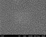
도 1a는 양전하를 띠는 나노와이어(PEI/Ppy NW)의 주사전자현미경(SEM) 이미지를 나타낸 것이다.
도 1b는 HRP/스트렙타비딘이 결합된 나노입자의 주사전자현미경 이미지를 나타낸 것이다.
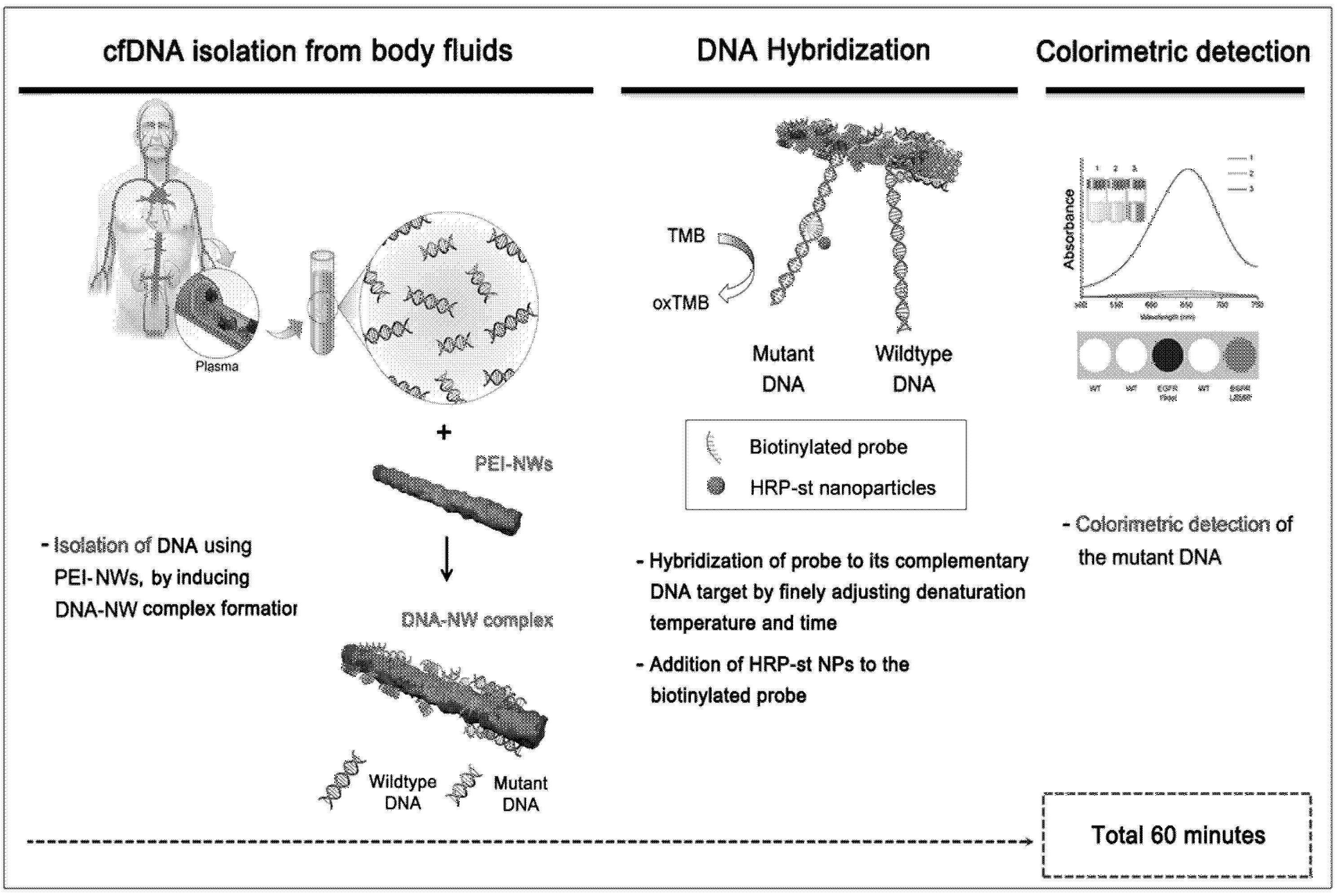
도 2a는 양이온성 고분자인 폴리에틸렌이민(polyethyleneimine, PEI)이 표면에 결합된 나노구조체(PEI/mPpy NW)의 제조 및 이를 이용하여 cfDNA를 검출 및 회수하는 방법에 대한 개념적 모식도를 나타낸 도면이다.
도 2b는 양이온성 고분자인 폴리에틸렌이민(polyethyleneimine, PEI)이 표면에 결합된 자성나노구조체(PEI/mPpy NW)를 이용하여 cfDNA를 검출 및 회수하는 과정을 촬영하여 과정별로 나타낸 도면이다.
도 3은 에펜도르프 튜브를 이용하여 cfDNA를 회수하는 과정을 도식화한 도면이다.
도 4 및 도 5는 PD-L1 양성 암세포주 또는 PD-L1 음성 암세포주의 cfDNA로부터 PD-L1 DNA 발현(PD-L1 DNA expression) 및 PD-L1 mRNA 발현 정도를 측정하여 나타낸 도면이다.
도 6 및 도 7은 EpCAM 양성 암세포주 또는 EpCAM 음성 암세포주의 cfDNA로부터 EpCAM DNA 발현(EpCAM DNA expression) 및 EpCAM mRNA 발현 정도를 측정하여 나타낸 도면이다.
도 8 및 도 9는 FOLR1 양성 암세포주 또는 FOLR1 음성 암세포주의 cfDNA로부터 FOLR1 DNA 발현(FOLR1 DNA expression) 및 FOLR1 mRNA 발현 정도를 측정하여 나타낸 도면이다.
도 10 및 도 11은 EGFR 양성 암세포주 또는 EGFR 음성 암세포주의 cfDNA로부터 EGFR DNA 발현(EGFR DNA expression) 및 EGFR mRNA 발현 정도를 측정하여 나타낸 도면이다.
도 12 및 도 13은 ERBB2 양성 암세포주 또는 ERBB2 음성 암세포주의 cfDNA로부터 ERBB2 DNA 발현(ERBB2 DNA expression) 및 ERBB2 mRNA 발현 정도를 측정하여 나타낸 도면이다.
도 14 및 도 15는 OGT 양성 암세포주 또는 OGT 음성 암세포주의 cfDNA로부터 OGT DNA 발현(OGT DNA expression) 정도를 측정하여 나타낸 도면이다.
도 16 내지 도 18은 CEA 양성 암세포주 또는 CEA 음성 암세포주의 cfDNA로부터 CEA DNA 발현(CEA DNA expression) 정도를 측정하여 나타낸 도면이다.
도 19 및 도 20은 PSA 양성 암세포주 또는 PSA 음성 암세포주의 cfDNA로부터 PSA DNA 발현(PSA DNA expression) 정도를 측정하여 나타낸 도면이다.
도 21 및 도 22는 CA19-9 양성 암세포주 또는 CA19-9 음성 암세포주의 cfDNA로부터 CA19-9 DNA 발현(CA19-9 DNA expression) 정도를 측정하여 나타낸 도면이다.
도 23 및 도 24는 CA125 양성 암세포주 또는 CA125 음성 암세포주의 cfDNA로부터 CA125 DNA 발현(CA125 DNA expression) 정도를 측정하여 나타낸 도면이다.
도 25 및 도 26는 AFP 양성 암세포주 또는 AFP 음성 암세포주의 cfDNA로부터 AFP DNA 발현(AFP DNA expression) 정도를 측정하여 나타낸 도면이다.
도 27 내지 도 29는 전립선암 환자로부터 수득한 혈장을 이용하여 PSA, PSMA, PAP 및 PAC3의 DNA 발현 정도를 측정하여 나타낸 도면이다.
도 30 내지 도 32는 정상인으로부터 수득한 혈장을 이용하여 PSA, PSMA, PAP 및 PAC3의 DNA 발현 정도를 측정하여 나타낸 도면이다.
도 33 및 도 34는 폐암 환자로부터 수득한 혈장을 이용하여 NSE, SCC, CEA, Cyfra21-1 및 TPA의 DNA 발현 정도를 측정하여 나타낸 도면이다.
도 35는 정상인으로부터 수득한 혈장을 이용하여 NSE, SCC, CEA, Cyfra21-1 및 TPA의 DNA 발현 정도를 측정하여 나타낸 도면이다.
도 36 내지 도 38은 갑상선암 환자로부터 수득한 혈장을 이용하여 CEA, NSE, TG 및 CALCA의 DNA 발현 정도를 측정하여 나타낸 도면이다.
도 39 및 도 40은 정상인으로부터 수득한 혈장을 이용하여 CEA, NSE, TG 및 CALCA의 DNA 발현 정도를 측정하여 나타낸 도면이다.
도 41 및 도 42는 방광암 환자로부터 수득한 소변을 이용하여 OGT, FGFR3, TP53, NMP22 및 Cyfra21-1의 DNA 발현 정도를 측정하여 나타낸 도면이다.
도 43 및 도 44는 방광염증 환자로부터 수득한 소변을 이용하여 OGT, FGFR3, TP53, NMP22 및 Cyfra21-1의 DNA 발현 정도를 측정하여 나타낸 도면이다.
도 45 및 도 46은 정상인으로부터 수득한 소변을 이용하여 OGT, FGFR3, TP53, NMP22 및 Cyfra21-1의 DNA 발현 정도를 측정하여 나타낸 도면이다.
도 47 및 도 48은 유방암 환자로부터 수득한 혈장을 이용하여 CA27-29, CA15-3 및 CEA의 DNA 발현 정도를 측정하여 나타낸 도면이다.
도 49 및 도 50은 정상인으로부터 수득한 혈장을 이용하여 CA27-29, CA15-3 및 CEA의 DNA 발현 정도를 측정하여 나타낸 도면이다.
도 51 및 도 52는 대장암 환자로부터 수득한 혈장을 이용하여 CEA 및 CA19-9의 DNA 발현 정도를 측정하여 나타낸 도면이다.
도 53 내지 도 55는 정상인으로부터 수득한 혈장을 이용하여 CEA 및 CA19-9의 DNA 발현 정도를 측정하여 나타낸 도면이다.
도 56은 담도암 환자로부터 수득한 혈장을 이용하여 CA19-9, CA125 및 CEA의 DNA 발현 정도를 측정하여 나타낸 도면이다.
도 57 및 도 58은 정상인으로부터 수득한 혈장을 이용하여 CA19-9, CA125 및 CEA의 DNA 발현 정도를 측정하여 나타낸 도면이다.
도 59는 위암 환자로부터 수득한 혈장을 이용하여 CEA, CA19-9, CGB 및 Cyfra21-1의 DNA 발현 정도를 측정하여 나타낸 도면이다.
도 60 및 도 61은 정상인으로부터 수득한 혈장을 이용하여 CEA, CA19-9, CGB 및 Cyfra21-1의 DNA 발현 정도를 측정하여 나타낸 도면이다.
도 62 내지 도 65는 췌장암 환자로부터 수득한 혈장을 이용하여 CA19-9, CA125 및 CEA의 DNA 발현 정도를 측정하여 나타낸 도면이다.
도 66 내지 도 69는 정상인으로부터 수득한 혈장을 이용하여 CA19-9, CA125 및 CEA의 DNA 발현 정도를 측정하여 나타낸 도면이다.
도 70은 폐암 환자로부터 수득한 혈장을 이용해 CPT1A의 DNA 발현 정도를 측정하여 나타낸 도면이다.
도 71은 정상인으로부터 수득한 혈장을 이용해 CPT1A의 DNA 발현 정도를 측정하여 나타낸 도면이다.
도 72는 방광암 환자로부터 수득한 소변을 이용해 CPT1A의 DNA 발현 정도를 측정하여 나타낸 도면이다.
도 73은 정상인으로부터 수득한 소변을 이용해 CPT1A의 DNA 발현 정도를 측정하여 나타낸 도면이다.
도 74는 IFN-γ를 처리하지 않은 PD-L1 양성 암세포주 또는 PD-L1 음성 암세포주의 cfDNA로부터 PD-L1의 DNA 발현 정도와 본 발명의 일 구체예의 방법으로 PD-L1의 검출 여부를 측정하여 나타낸 도면이다.
도 75는 IFN-γ를 처리하지 않은 PD-L1 양성 암세포주 또는 PD-L1 음성 암세포주의 cfDNA로부터 IFNG(IFN-γ)의 DNA 발현 정도를 측정하여 나타낸 도면이다.
도 76은 IFN-γ를 처리하지 않은 PD-L1 양성 암세포주 또는 PD-L1 음성 암세포주의 cfDNA로부터 IFNR1(IFN-γ receptor)의 DNA 발현 정도를 측정하여 나타낸 도면이다.
도 77 내지 도 79는 IFN-γ를 처리한 PD-L1 양성 암세포주 또는 PD-L1 음성 암세포주의 cfDNA로부터 PD-L1, IFNG 및 IFNR1의 DNA 발현 정도를 측정하여 나타낸 도면이다.
도 80은 IFN-γ의 처리유무에 따라 PD-L1 양성 암세포주 또는 PD-L1 음성 암세포주의 cfDNA로부터 PD-L1의 DNA 발현 정도를 측정하여 나타낸 도면이다.
도 81은 IFN-γ의 처리유무에 따라 PD-L1 양성 암세포주 또는 PD-L1 음성 암세포주의 cfDNA로부터 IFN-γ의 DNA 발현 정도를 측정하여 나타낸 그래프이다.
도 82는 IFN-γ의 처리유무에 따라 PD-L1 양성 암세포주 또는 PD-L1 음성 암세포주의 cfDNA로부터 IFNR1의 DNA 발현 정도를 측정하여 나타낸 도면이다.
도 83은 IFN-γ의 처리유무에 따라 PD-L1 양성 암세포주 또는 PD-L1 음성 암세포주의 cfDNA로부터 PD-L1의 DNA 발현 정도를 측정하여 나타낸 도면이다.
도 84는 본 발명의 검출 단계를 도식화한 것이다.
도 84a는 폴리에틸렌이민(PEI)이 표면에 결합된 나노와이어(PEI/Ppy NW)를 이용하여 환자의 체액으로부터 cfDNA를 수득한 후 프로브 및 HRP/스트렙타비딘-나노입자(HRP/st-tagged NP)와의 반응을 통하여 유전자 변이를 약 60분 이내에 분석하는 방법에 대한 모식도를 나타낸 것이다.
도 84b는 나노와이어, 프로브 및 HRP/스트렙타비딘 나노입자를 이용하여 불안정한 cfDNA를 검출하는 방법을 도식화한 것이다.
도 84c는 자성 나노입자를 포함하지 않은 나노와이어를 사용하여 스핀 컬럼(spin column)을 통해 유전자변이를 검출하는 과정을 나타낸 도면이다. 본 발명의 일 구체예에서 라이시스 버퍼를 처리하는 단계를 추가적으로 포함할 수 있다.
도 84d는 혈액, 뇌척수액 또는 흉수와 같은 시료에서 불안정한 cfDNA를 검출하기 위한 방법을 시계열적 흐름으로 나타난 것이다.
도 84e는 소변과 같은 시료에서 불안정한 cfDNA를 검출하기 위한 방법을 시계열적 흐름으로 나타난 것이다.
도 84f는 혈액에서 취득한 cfDNA의 상태에 따른 변성 조건의 차이를 도식화한 것이다.
도 84g는 소변, 타액 및 객담에서 취득한 cfDNA의 상태에 따른 변성 조건의 차이를 도식화한 것이다.
도 85는 자성 나노입자를 포함하지 않은 나노와이어를 사용한 스핀 컬럼(spin column)을 통해 cfDNA를 분리한 것을 나타낸 도면이다. 위 사진은 원심분리 전 스핀 컬럼의 SEM 이미지이며, 아래 사진은 원심분리 후 cfDNA가 분리된 스핀 컬럼의 SEM 이미지이다.
도 86은 주사바늘을 이용해 폐암환자로부터 수득한 혈액으로부터 AKL Fusion 및 PIK3CA 등의 암 관련 바이오 마커의 DNA 발현 정도를 측정한 도면이다.
도 87은 랜싯을 이용해 폐암환자로부터 수득한 혈액으로부터 AKL Fusion 및 PIK3CA 등의 암 관련 바이오 마커의 DNA 발현 정도를 측정한 도면이다.
도 88은 주사바늘을 이용해 정상인으로부터 수득한 혈액으로부터 AKL Fusion 등의 암 관련 바이오 마커의 DNA 발현 정도를 측정한 도면이다.
도 89는 랜싯을 이용해 정상인으로부터 수득한 혈액으로부터 AKL Fusion 등의 암 관련 바이오 마커의 DNA 발현 정도를 측정한 도면이다.
도 90은 EML4-ALK variant 3a/b positive cell(H2228) 및 EML4-ALK negative cell(A549, H1993, PC9, RT4) 암세포주의 cfDNA로부터 EML4-ALK의 발현 정도를 RT-PCR을 통해 확인한 도면이다.
도 91은 EML4-ALK variant 3a/b positive cell(H2228) 및 EML4-ALK negative cell(A549, H1993, PC9, RT4) 암세포주의 cfDNA로부터 EML4-ALK의 발현 정도를 웨스턴 블랏을 통해 확인한 도면이다.
도 92는 EML4-ALK variant 3a/b positive cell(H2228) 및 EML4-ALK negative cell(A549, H1993, PC9, RT4) 암세포주의 cfDNA로부터 EML4-ALK의 발현 정도를 RT-PCR 및 웨스턴 블랏을 통해 확인한 결과를 나타낸 그래프이다.
도 93은 EML4-ALK variant 3a/b positive cell(H2228) 및 EML4-ALK negative cell(A549, H1993, PC9, RT4) 암세포주의 cfDNA로부터 EML4-ALK fusion var.1 또는 EML4-ALK fusion var.3의 DNA 발현 정도를 측정하여 나타낸 도면이다.
도 94는 EML4-ALK variant 3a/b positive cell(H2228) 및 EML4-ALK negative cell(A549, H1993, PC9, RT4) 암세포주의 cfDNA로부터 EML4-ALK fusion var.1 또는 EML4-ALK fusion var.3의 DNA 발현 정도를 측정하여 나타낸 그래프이다.
도 95는 소세포폐암 환자로부터 수득한 혈액으로부터 EML4-ALK fusion var.3, KRAS, SYP, NCAM1, NKX2-1 등의 암 관련 바이오 마커의 DNA 발현 정도를 측정한 도면이다. 그 결과, EML4-ALK fusion이 암조직과 혈액 내 ctDNA에서 동일하게 발견되고, ctDNA 결과로 EML4-ALK fusion이 var.1이 아닌 var.3임을 알게됨으로써, ALK TKI인 Crizotinib이 잘 반응하지 않는다는 것을 알게되었다.
도 96은 암 환자로부터 수득한 혈액으로부터 EML4-ALK fusion var.1 등의 암 관련 바이오 마커의 DNA 발현 정도를 측정한 도면이다. 그 결과, EML4-ALK fusion이 암 조직과 혈액 내 ctDNA에서 동일하게 발견되고, ctDNA 결과로 EML4-ALK fusion이 var.3가 아닌 var.1임을 알게됨으로써, ALK TKI인 Crizotinib이 잘 반응하여 partial response (PR)의 환자 반응을 얻게 되었다.
도 97은 암 환자로부터 수득한 혈액으로부터 EML4-ALK fusion var.3 등의 암 관련 바이오 마커의 DNA 발현 정도를 측정한 도면이다. 그 결과, EML4-ALK fusion이 암조직과 혈액 내 ctDNA에서 동일하게 발견되고, ctDNA 결과로 EML4-ALK fusion이 var.1이 아닌 var.3임을 알게됨으로써, ALK TKI인 Crizotinib이 잘 반응하지 않을 것이기 때문에, 처음부터 alectinib을 처방하여 환자의 반응을 기다리고 있다.
도 98은 암 환자로부터 수득한 혈액으로부터 EML4-ALK fusion var.3, BRAFV800E, TP53 등의 암 관련 바이오 마커의 DNA 발현 정도를 측정한 도면이다. 그 결과, EML4-ALK fusion이 암조직과 혈액 내 ctDNA에서 동일하게 발견되고, ctDNA 결과로 EML4-ALK fusion이 var.1이 아닌 var.3임을 알게됨으로써, ALK TKI인 Crizotinib이 잘 반응하지 않는다는 것(PD)을 알게 되었다.
도 99는 암 환자로부터 수득한 혈액으로부터 EML4-ALK fusion var.1 등의 암 관련 바이오 마커의 DNA 발현 정도를 측정한 도면이다. 그 결과, EML4-ALK fusion 이 암조직과 혈액 내 ctDNA에서 동일하게 발견되고, ctDNA 결과로 EML4-ALK fusion이 var.3가 아닌 var.1임을 알게됨으로써, ALK TKI인 Crizotinib이 잘 반응하여 partial response(PR)의 환자 반응을 얻게 되었다.
도 100은 암 환자로부터 수득한 혈액으로부터 EML4-ALK fusion var.1 등의 암 관련 바이오 마커의 DNA 발현 정도를 측정한 도면이다. 그 결과, EML4-ALK fusion 이 암조직과 혈액 내 ctDNA에서 동일하게 발견되고, ctDNA 결과로 EML4-ALK fusion이 var.3가 아닌 var.1임을 알게됨으로써, ALK TKI가 잘 반응하여 partial response(PR)의 환자 반응을 얻게 되었다.
도 101은 in vitro 상에서 각 암세포주의 cfDNA로부터 OGT의 단백질 발현 정도를 웨스턴 블랏을 통해 확인한 도면이다.
도 102는 in vitro 상에서 각 암세포주의 cfDNA로부터 OGT의 mRNA 발현 정도를 RT-PCR을 통해 확인한 도면이다.
도 103은 in vitro 상에서 각 암세포주의 cfDNA로부터 OGT의 mRNA 발현 정도를 RT-PCR을 통해 확인한 결과를 나타낸 그래프이다.
도 104는 in vitro 상에서 각 세포주의 cfDNA로부터 OGT의 DNA 발현 정도를 측정한 도면이다.
도 105는 in vitro 상에서 각 세포주의 cfDNA로부터 OGT의 DNA 발현 정도를 측정한 결과를 나타낸 그래프이다.
도 106은 in vitro 상에서 각 세포주로부터 OGT의 발현 정도를 웨스턴블랏, RT-PCR 및 cfDNA 검출을 통해 확인한 결과를 나타낸 도면이다.
도 107은 in vitro 상에서 각 세포주로부터 자성 나노입자를 포함하지 않은 나노와이어에 검출된 OGT의 cfDNA를 촬영한 사진이다.
도 108은 정상인, 방광염 환자 및 방광암 환자의 소변으로부터 수득한 cfDNA를 정량한 그래프이다.
도 109는 정상인, 방광염 환자 및 방광암 환자의 소변으로부터 수득한 cfDNA로부터 OGT의 DNA 발현 정도를 분석한 그래프이다.
도 110은 정상인, 방광염 환자 및 방광암 환자의 소변으로부터 수득한 cfDNA로부터 OGT의 DNA 발현 정도를 분석한 그래프이다.
도 111 내지 도 113은 블라인드 테스트로 여러 암 환자의 소변으로부터 수득한 cfDNA로부터 OGT의 DNA 발현 정도를 분석한 도면이다.
도 114 내지 도 116은 갑상선암 환자의 조직으로부터 수득한 cfDNA로부터 BRAF V600E 및 TERT C250T의 DNA 발현 정도를 분석한 도면이다.
도 117 및 도 121은 소세포폐암 환자로부터 수득한 혈액을 이용해 SYP, CgA, NCAM1, NKX2-1 등의 암 관련 바이오 마커의 DNA 발현 정도를 측정한 도면이다. 특히, 도 120의 환자의 경우, small-cell-lung cancer(SCLC)로 원자력 병원에서 확인했으며, crizotinib을 처방했으나 효과가 없음이 확인되었다.
도 122는 비소세포폐암 환자로부터 수득한 혈액을 이용해 SYP, CgA, NCAM1, NKX2-1의 DNA 발현 정도를 측정한 도면이다.
도 123은 항암 치료 전과 후의 폐암환자로부터 수득한 혈액을 이용해 CEA의 DNA 발현 정도 및 환자의 예후를 나타낸 도면이다.
도 124 내지 도 128은 폐암 환자로부터 수득한 혈액을 이용해 NSE, CEA 등의 암 관련 바이오 마커의 DNA 발현 정도를 측정한 도면이다.
도 129 내지 도 133는 정상인으로부터 수득한 혈액을 이용해 NSE, CEA 등의 암 관련 바이오 마커의 DNA 발현 정도를 측정한 도면이다.
도 134는 전립선암 환자로부터 수득한 혈액을 이용해 PSA, PSMA, PAP, PCA3 등의 암 관련 바이오마커의 DNA 발현정도를 측정한 도면이다.
도 135는 정상인으로부터 수득한 혈액을 이용해 PSA, PSMA, PAP, PCA3 등의 암 관련 바이오마커의 DNA 발현 정도를 측정한 도면이다.
도 136은 전립선암 환자 및 정상인으로부터 수득한 혈액을 이용해 TMPRSS2-ERG fusion의 DNA 발현 정도를 측정한 도면이다.
도 137은 갑상선암 환자로부터 수득한 혈액을 이용해 CEA, NSE, TG(Thyroglobulin) 및 CALCA의 DNA 발현 정도를 측정한 도면이다.
도 138은 정상인으로부터 수득한 혈액을 이용해 CEA, NSE, TG(Thyroglobulin) 및 CALCA의 DNA 발현 정도를 측정한 도면이다.
도 139는 갑상선암 환자 및 정상인으로부터 수득한 혈액을 이용해 BRAF mutation(V600E), TERT Promotor mutation(C228T, C250T)의 DNA 발현 정도를 측정한 도면이다.
도 140은 방광암 환자, 혈뇨환자 및 정상인으로부터 수득한 소변을 이용해 OGT, FGFR3, TP53, NMP22 및 Cyfra21-1의 DNA 발현 정도를 측정한 도면이다.
도 141은 유방암 환자 및 정상인으로부터 수득한 혈액을 이용해 CA27-29 및 CEA의 DNA 발현 정도를 측정한 도면이다.
도 142는 대장암 환자 및 정상인으로부터 수득한 혈액을 이용해 CEA 및 CA19-9의 DNA 발현 정도를 측정한 도면이다.
도 143 및 도 144는 담도암 환자 및 정상인으로부터 수득한 혈액을 이용해 CA 19-9, CEA 및 CA123의 DNA 발현 정도를 측정한 도면이다.
도 145는 위암 환자 및 정상인으로부터 수득한 혈액을 이용해 CEA, CA19-9, CGB 및 Cyfra21-1의 DNA 발현 정도를 측정한 도면이다.
도 146은 난소암 환자 및 정상인으로부터 수득한 혈액을 이용해 CA125 및 CEA의 DNA 발현 정도를 측정한 도면이다.
도 147은 췌장암 환자로부터 수득한 혈액을 이용해 CEA, CA19-9 및 CA125의 DNA 발현 정도를 측정한 도면이다.
도 148은 정상인으로부터 수득한 혈액을 이용해 CEA, CA19-9 및 CA125의 DNA 발현 정도를 측정한 도면이다.
도 149 및 도 150은 정상인으로부터 수득한 혈액을 이용해 암 관련 바이오마커의 DNA 발현 정도를 측정하여 조기 진단을 실시한 결과이다(PC: Positive control, PC는 조기 암진단 실험이 정확하게 진행되었는지 확인하는 도구로서, 조기 암진단 결과와 무관하다).
도 151 및 도 152는 본 발명의 일 실시예에서 사용한 암종별 바이오마커를 정리한 도면이다.
도 153은 HPV 양성 자궁경부암 환자(HPV16(+) 및 HPV18(+)) 및 HPV 음성 건강 대조군(HPV-)의 소변에 존재하는 cfDNA와 HPV 18 또는 HPV 16에 특이적인 프로브의 결합 여부를 흡광도를 통하여 확인한 것이다.
도 154는 자궁경부암 환자의 소변에서 분리한 cfDNA에 HPV 16, EGFR19 deletion, HPV 18, 및 EGFR 21 L858R에 특이적인 프로브를 순차적으로 반응시킨 후, cfDNA와 각 프로브의 결합여부를 확인한 것이다.
도 155는 151명의 폐암 환자들의 혈장으로부터 수득한 cfDNA를 이용하여 폐암 환자의 유전자 변이를 분석한 표이다.
도 156는 EGFR 변이가 없는 환자(Wild type), EGFR exon19 deletion이 있는 환자 및 EGFR exon 21 L858R을 가진 폐암 환자의 혈장으로부터 cfDNA를 수득한 후, EGFR exon19 Del에 특이적인 프로브를 혼합한 후, UV 스펙트럼의 흡광도(ΔOD, 500 nm 내지 650 nm) 값의 분석을 통하여 폐암 환자의 유전자 변이를 확인한 것이다.
도 157은 EGFR exon19 deletion이 있는 폐암 환자의 혈장으로부터 cfDNA를 수득한 후, EGFR exon19 Del에 특이적인 프로브를 혼합한 후, 유전자 변이의 특이도와 민감도를 분석한 것이다.
도 158은 EGFR 변이가 없는 환자(Wild type), EGFR exon19 deletion이 있는 환자 및 EGFR exon 21 L858R을 가진 폐암 환자의 혈장으로부터 cfDNA를 수득한 후, EGFR exon21 L858R에 특이적인 프로브를 첨가한 후, UV 스펙트럼의 흡광도(ΔOD, 500 nm 내지 650 nm) 값의 분석을 통하여 환자의 유전자 변이를 확인한 것이다.
도 159는 EGFR exon 21 L858R이 있는 폐암 환자의 혈장으로부터 cfDNA를 수득한 후, EGFR exon 21 L858R에 특이적인 프로브를 추가한 후, 환자의 유전자 변이의 특이도와 민감도를 분석한 것이다.
도 160은 EGFR exon 19 deletion의 CP 및 DP의 서열을 나타낸 것이다. 본 연구에서는 CP_1 및 DP를 사용하여 폐암 환자의 cfDNA 유전자 변이를 분석하였다. 이때, CP는 변이가 있는 부분을 포함하거나 인접한 서열에 상보적으로 결합하도록 디자인한 프로브이며, DP는 변이 서열에서 이격된 부분에 상보적으로 결합하도록 디자인된 프로브를 의미한다.
도 161은 EGFR exon 20 T790M의 CP 및 DP의 서열을 나타낸 것이다. 본 연구에서는 CP2 및 DP를 사용하여 폐암 환자의 cfDNA 유전자 변이를 분석하였다.
도 162는 EGFR exon 21 L858R의 CP 및 DP의 서열을 나타낸 것이다. 본 연구에서는 CP2 및 DP를 사용하여 폐암 환자의 cfDNA 유전자 변이를 분석하였다.
도 163은 EGFR exon 19 deletion 및 EGFR exon 20 T790M 유전자 변이를 가진 폐암 환자의 혈장으로부터 수득된 cfDNA를 EGFR exon 19 deletion(Del19), EGFR exon 20 T790M 및 EGFR exon 21 L858R에 특이적인 프로브를 이용하여 반응시킨 후 HRP/스트렙타비딘 나노입자(다량의 HRP를 포함)를 첨가하여 cfDNA의 검출여부를 색깔 변화 및 UV 흡광도로 확인한 것이다.
도 164는 도 163과 동일한 EGFR exon 19 deletion 및 EGFR exon 20 T790M 유전자 변이를 가진 폐암 환자의 혈장으로부터 수득된 cfDNA를 EGFR exon 19 deletion(Del19), EGFR exon 20 T790M 및 EGFR exon 21 L858R에 특이적인 프로브를 이용하여 반응시킨 후 HRP/스트렙타비딘 복합체(HRP와 스트렙타비딘이 1:1로 결합된 복합체)를 첨가하여 cfDNA의 검출여부를 색깔 변화 및 UV 흡광도로 확인한 것이다. HRP/스트렙타비딘 나노입자에 비해 HRP/스트렙타비딘 복합체의 경우 노이즈가 생성됨을 확인한 것이다.
도 165는 5명의 EGFR exon19 deletion 및 exon20 T790M 유전자 변이를 가진 폐암 환자의 혈장에서 cfDNA를 추출한 후 EGFR exon 19 Del, EGFR exon 20 T790M, EGFR exon 21 L858R에 특이적인 프로브 및 HRP/스트렙타비딘 나노입자(HRP/st-tagged NP)와 반응시킨 결과와 EGFR exon19 Del, EGFR exon 20 T790M, EGFR exon 21 L858R에 특이적인 프로브와 HRP/스트렙타비딘 복합체(HRP와 스트렙타비딘이 1:1로 결합)를 반응시킨 결과의 분석을 통하여 암조직과 유전형과의 일치성을 확인 및 비교한 것이다.
도 166은 EGFR exon 20 T790M 및 EGFR exon 21 L861Q 유전자 변이를 가진 폐암 환자의 혈장으로부터 수득된 cfDNA의 유전자 변이 검출을 위해 EGFR exon 19 deletion(19 Del), EGFR exon 20 T790M, EGFR exon 21 L858R 및 EGFR exon L861Q에 특이적인 프로브 및 HRP/st-tagged NP을 한꺼번에 혼합한 결과 암조직과 동일하게 EGFR exon 20 T790M 및 EGFR exon 21 L861Q 에서만 유전자변이가 관찰됨을 UV 흡광도로 확인한 것이다.
도 167은 ALK-EML4 fusion 및 ALK point mutation(I1171N/T) 유전자 변이를 가진 폐암 환자의 혈장으로부터 수득된 cfDNA의 유전자 변이 검출을 위해 ALK-EML4 fusion 및 ALK point mutation(T1151, L1152P, L1152R, C1156Y, I1171N/T)에 특이적인 프로브 및 HRP/st-tagged NP을 한꺼번에 혼합한 결과 암조직과 동일하게 ALK-EML4 fusion 및 ALK point mutation(I1171N/T) 유전형이 검출되는 것을 확인하였다.
도 168은 BRAF V600E 유전자 변이를 가진 갑상선암 환자의 혈장으로부터 수득된 cfDNA의 유전자 변이 검출을 위해 BRAF V600E에 특이적인 프로브 및 HRP/st-tagged NP을 한꺼번에 혼합하였다. 그 결과 BRAF V600E 유전자 변이가 환자의 유전형과 동일하게 검출되는 것을 확인하였다.
도 169는 정상인의 혈액에서 채취한 시료를 여러가지 온도 조건으로 변성 시킨 후, 처리조건에 따라 불안정한 cfDNA의 검출 결과를 나타낸 것이다.
도 170은 환자의 혈액에서 채취한 시료를 여러가지 온도 조건으로 변성 시킨 후, 처리조건에 따라 불안정한 cfDNA의 검출 결과를 나타낸 것이다.
도 171은 돌연변이 세포주에서 얻은 fDNA를 여러가지 온도 조건으로 변성 시킨 후, 처리조건에 따라 불안정한 cfDNA의 검출 결과를 나타낸 것이다.
도 172는 돌연변이 세포주에서 얻은 fDNA를 DNase로 37℃, 30분간 처리 후, 처리조건에 따라 불안정한 cfDNA의 검출 결과를 나타낸 것이다.
도 173은 돌연변이 세포주에서 얻은 fDNA를 DNase로 37℃, 60분간 처리 후, 처리조건에 따라 불안정한 cfDNA의 검출 결과를 나타낸 것이다.
도 174는 돌연변이 세포주에서 얻은 fDNA를 DNase로 37℃, 120분간 처리 후, 처리조건에 따라 불안정한 cfDNA의 검출 결과를 나타낸 것이다.
도 175는 DNase의 활성에 따른 불안정한 cfDNA와 안정한 cfDNA의 차이점을 확인하기 위하여, DNase 1 ㎕ 또는 2 ㎕를 24℃, 120분간 처리한 결과를 나타낸 것이다.
도 176은 DNase의 활성에 따른 불안정한 cfDNA와 안정한 cfDNA의 차이점을 확인하기 위하여, DNase 1 ㎕ 또는 2 ㎕를 3℃, 120분간 처리한 결과를 나타낸 것이다.
도 177은 본 발명의 일 실시예로서 폐암환자의 혈장에서 cfDNA를 이용하여 EML4-ALK fusion gene을 검출할 때 cutoff 값의 일 구체예를 나타낸 것이다.1a shows a scanning electron microscope (SEM) image of a positively charged nanowire (PEI/Ppy NW).
Figure 1b shows a scanning electron microscope image of HRP / streptavidin-conjugated nanoparticles.
Figure 2a is a view showing a conceptual schematic diagram for the preparation of a nanostructure (PEI / mPpy NW) bound to the surface of a cationic polymer, polyethyleneimine (PEI), and a method for detecting and recovering cfDNA using the same.
2b is a view showing the process of detecting and recovering cfDNA using a magnetic nanostructure (PEI/mPpy NW) to which a cationic polymer, polyethyleneimine (PEI) is bonded to the surface, by photographing the process.
3 is a diagram schematically illustrating a process of recovering cfDNA using an Eppendorf tube.
4 and 5 are diagrams showing the measurement of PD-L1 DNA expression (PD-L1 DNA expression) and PD-L1 mRNA expression levels from the cfDNA of a PD-L1 positive cancer cell line or a PD-L1 negative cancer cell line.
6 and 7 are diagrams showing EpCAM DNA expression (EpCAM DNA expression) and EpCAM mRNA expression levels measured from cfDNA of EpCAM-positive cancer cell lines or EpCAM-negative cancer cell lines.
8 and 9 are diagrams showing FOLR1 DNA expression (FOLR1 DNA expression) and FOLR1 mRNA expression levels measured from cfDNA of a FOLR1 positive cancer cell line or a FOLR1 negative cancer cell line.
10 and 11 are diagrams showing EGFR DNA expression (EGFR DNA expression) and EGFR mRNA expression levels measured from cfDNA of EGFR-positive cancer cell lines or EGFR-negative cancer cell lines.
12 and 13 are diagrams illustrating ERBB2 DNA expression and ERBB2 mRNA expression levels measured from cfDNA of an ERBB2 positive cancer cell line or an ERBB2 negative cancer cell line.
14 and 15 are diagrams showing the degree of OGT DNA expression (OGT DNA expression) measured from the cfDNA of an OGT-positive cancer cell line or an OGT-negative cancer cell line.
16 to 18 are diagrams showing the degree of CEA DNA expression measured from cfDNA of a CEA-positive cancer cell line or a CEA-negative cancer cell line.
19 and 20 are diagrams showing the degree of PSA DNA expression (PSA DNA expression) measured from the cfDNA of a PSA-positive cancer cell line or a PSA-negative cancer cell line.
21 and 22 are diagrams showing the degree of CA19-9 DNA expression (CA19-9 DNA expression) measured from the cfDNA of a CA19-9 positive cancer cell line or a CA19-9 negative cancer cell line.
23 and 24 are diagrams showing the degree of CA125 DNA expression (CA125 DNA expression) measured from the cfDNA of a CA125-positive cancer cell line or a CA125-negative cancer cell line.
25 and 26 are diagrams showing the degree of AFP DNA expression measured from the cfDNA of an AFP-positive cancer cell line or an AFP-negative cancer cell line.
27 to 29 are diagrams illustrating the measurement of DNA expression levels of PSA, PSMA, PAP and PAC3 using plasma obtained from prostate cancer patients.
30 to 32 are diagrams showing the measurement of DNA expression levels of PSA, PSMA, PAP and PAC3 using plasma obtained from a normal person.
33 and 34 are diagrams illustrating the measurement of DNA expression levels of NSE, SCC, CEA, Cyfra21-1 and TPA using plasma obtained from lung cancer patients.
35 is a view showing the measurement of DNA expression levels of NSE, SCC, CEA, Cyfra21-1 and TPA using plasma obtained from normal people.
36 to 38 are diagrams illustrating the measurement of DNA expression levels of CEA, NSE, TG and CALCA using plasma obtained from a thyroid cancer patient.
39 and 40 are diagrams illustrating the measurement of DNA expression levels of CEA, NSE, TG and CALCA using plasma obtained from normal people.
41 and 42 are diagrams illustrating the measurement of DNA expression levels of OGT, FGFR3, TP53, NMP22 and Cyfra21-1 using urine obtained from bladder cancer patients.
43 and 44 are diagrams showing the measurement of DNA expression levels of OGT, FGFR3, TP53, NMP22 and Cyfra21-1 using urine obtained from a patient with bladder inflammation.
45 and 46 are diagrams showing the measurement of DNA expression levels of OGT, FGFR3, TP53, NMP22 and Cyfra21-1 using urine obtained from a normal person.
47 and 48 are diagrams illustrating the measurement of DNA expression levels of CA27-29, CA15-3 and CEA using plasma obtained from breast cancer patients.
49 and 50 are diagrams showing the measurement of DNA expression levels of CA27-29, CA15-3 and CEA using plasma obtained from a normal person.
51 and 52 are diagrams illustrating the measurement of DNA expression levels of CEA and CA19-9 using plasma obtained from colorectal cancer patients.
53 to 55 are diagrams showing the measurement of DNA expression levels of CEA and CA19-9 using plasma obtained from a normal person.
FIG. 56 is a diagram illustrating the measurement of DNA expression levels of CA19-9, CA125 and CEA using plasma obtained from a patient with biliary tract cancer.
57 and 58 are diagrams showing the measurement of DNA expression levels of CA19-9, CA125 and CEA using plasma obtained from a normal person.
59 is a diagram illustrating the measurement of DNA expression levels of CEA, CA19-9, CGB and Cyfra21-1 using plasma obtained from gastric cancer patients.
60 and 61 are diagrams illustrating the measurement of DNA expression levels of CEA, CA19-9, CGB and Cyfra21-1 using plasma obtained from normal people.
62 to 65 are diagrams illustrating the measurement of DNA expression levels of CA19-9, CA125 and CEA using plasma obtained from pancreatic cancer patients.
66 to 69 are diagrams showing the measurement of DNA expression levels of CA19-9, CA125 and CEA using plasma obtained from a normal person.
70 is a diagram illustrating the measurement of the DNA expression level of CPT1A using plasma obtained from a lung cancer patient.
71 is a diagram showing the measurement of the DNA expression level of CPT1A using plasma obtained from a normal person.
72 is a view showing the measurement of the DNA expression level of CPT1A using urine obtained from a bladder cancer patient.
73 is a view showing the measurement of the DNA expression level of CPT1A using urine obtained from a normal person.
74 shows the DNA expression level of PD-L1 from the cfDNA of a PD-L1 positive cancer cell line or a PD-L1 negative cancer cell line not treated with IFN-γ and whether or not PD-L1 is detected by the method of one embodiment of the present invention. the drawing shown.
FIG. 75 is a diagram illustrating the measurement of the DNA expression level of IFNG (IFN-γ) from the cfDNA of a PD-L1 positive cancer cell line or a PD-L1 negative cancer cell line not treated with IFN-γ.
76 is a view showing the measurement of the DNA expression level of IFNR1 (IFN-γ receptor) from the cfDNA of a PD-L1-positive cancer cell line or a PD-L1-negative cancer cell line not treated with IFN-γ.
77 to 79 are diagrams illustrating the measurement of the DNA expression levels of PD-L1, IFNG and IFNR1 from the cfDNA of a PD-L1 positive cancer cell line or a PD-L1 negative cancer cell line treated with IFN-γ.
FIG. 80 is a view showing the measurement of the DNA expression level of PD-L1 from the cfDNA of a PD-L1-positive cancer cell line or a PD-L1-negative cancer cell line according to the presence or absence of IFN-γ treatment.
81 is a graph showing the measurement of the DNA expression level of IFN-γ from the cfDNA of a PD-L1 positive cancer cell line or a PD-L1 negative cancer cell line according to the presence or absence of IFN-γ treatment.
82 is a view showing the measurement of the DNA expression level of IFNR1 from the cfDNA of a PD-L1-positive cancer cell line or a PD-L1-negative cancer cell line according to the presence or absence of IFN-γ treatment.
83 is a view showing the measurement of the DNA expression level of PD-L1 from the cfDNA of a PD-L1 positive cancer cell line or a PD-L1 negative cancer cell line according to the presence or absence of IFN-γ treatment.
84 is a schematic diagram of the detection step of the present invention.
84a shows a probe and HRP/streptavidin-nanoparticles (HRP/st-tagged NPs) after obtaining cfDNA from a patient's body fluid using a nanowire (PEI/Ppy NW) to which polyethyleneimine (PEI) is bonded to the surface. ) shows a schematic diagram of a method for analyzing genetic mutations within about 60 minutes through the reaction with
84B is a schematic diagram of a method for detecting unstable cfDNA using nanowires, probes, and HRP/streptavidin nanoparticles.
84c is a diagram illustrating a process of detecting a genetic mutation through a spin column using a nanowire that does not include magnetic nanoparticles. In one embodiment of the present invention may further include the step of processing the lysis buffer.
84d is a time series flow diagram illustrating a method for detecting unstable cfDNA in a sample such as blood, cerebrospinal fluid, or pleural fluid.
84e is a time-series flow showing a method for detecting unstable cfDNA in a sample such as urine.
84f is a schematic diagram of the difference in denaturation conditions according to the state of cfDNA obtained from blood.
84g is a schematic diagram of the difference in denaturation conditions according to the state of cfDNA obtained from urine, saliva and sputum.
85 is a view showing separation of cfDNA through a spin column using a nanowire that does not contain magnetic nanoparticles. The upper photo is an SEM image of the spin column before centrifugation, and the lower photo is an SEM image of the spin column from which cfDNA is separated after centrifugation.
86 is a diagram illustrating the measurement of DNA expression levels of cancer-related biomarkers such as AKL Fusion and PIK3CA from blood obtained from lung cancer patients using an injection needle.
87 is a diagram illustrating the measurement of DNA expression levels of cancer-related biomarkers such as AKL Fusion and PIK3CA from blood obtained from lung cancer patients using a lancet.
88 is a view measuring the DNA expression level of cancer-related biomarkers, such as AKL Fusion, from blood obtained from a normal person using an injection needle.
89 is a view measuring the DNA expression level of cancer-related biomarkers, such as AKL Fusion, from blood obtained from a normal person using a lancet.
FIG. 90 is a view confirming the expression level of EML4-ALK from cfDNA of EML4-ALK variant 3a/b positive cell (H2228) and EML4-ALK negative cell (A549, H1993, PC9, RT4) cancer cell lines through RT-PCR.
91 is a view confirming the expression level of EML4-ALK from cfDNA of EML4-ALK variant 3a/b positive cell (H2228) and EML4-ALK negative cell (A549, H1993, PC9, RT4) cancer cell lines through Western blot.
92 shows the expression level of EML4-ALK from cfDNA of EML4-ALK variant 3a/b positive cell (H2228) and EML4-ALK negative cell (A549, H1993, PC9, RT4) cancer cell lines through RT-PCR and Western blot. This is a graph showing the results.
93 shows EML4-ALK fusion var.1 or EML4-ALK fusion var.3 from cfDNA of EML4-ALK variant 3a/b positive cell (H2228) and EML4-ALK negative cell (A549, H1993, PC9, RT4) cancer cell lines. It is a diagram showing the degree of DNA expression measured.
94 shows EML4-ALK fusion var.1 or EML4-ALK fusion var.3 from cfDNA of EML4-ALK variant 3a/b positive cell (H2228) and EML4-ALK negative cell (A549, H1993, PC9, RT4) cancer cell lines. It is a graph showing the degree of DNA expression measured.
FIG. 95 is a diagram illustrating the measurement of DNA expression levels of cancer-related biomarkers such as EML4-ALK fusion var.3, KRAS, SYP, NCAM1, and NKX2-1 from blood obtained from a patient with small cell lung cancer. As a result, EML4-ALK fusion was found identically in ctDNA in cancer tissues and blood, and as the ctDNA result showed that EML4-ALK fusion was var.3 rather than var.1, it was found that the ALK TKI, Crizotinib, did not react well. found out
96 is a diagram illustrating the measurement of DNA expression levels of cancer-related biomarkers such as EML4-ALK fusion var.1 from blood obtained from cancer patients. As a result, EML4-ALK fusion was found identically in ctDNA in cancer tissue and blood, and as the ctDNA result showed that EML4-ALK fusion was var.1 instead of var.3, ALK TKI, Crizotinib, reacted well to a partial response. A patient response of (PR) was obtained.
97 is a diagram illustrating the measurement of DNA expression levels of cancer-related biomarkers such as EML4-ALK fusion var.3 from blood obtained from cancer patients. As a result, EML4-ALK fusion was found identically in cancer tissue and blood ctDNA, and as the ctDNA result showed that EML4-ALK fusion was var.3 instead of var.1, Crizotinib, an ALK TKI, would not react well. Therefore, alectinib is prescribed from the beginning and the patient's response is awaited.
98 is a diagram illustrating the measurement of DNA expression levels of cancer-related biomarkers such as EML4-ALK fusion var.3, BRAFV800E, and TP53 from blood obtained from cancer patients. As a result, EML4-ALK fusion was found identically in ctDNA in cancer tissues and blood, and as it was found that EML4-ALK fusion was var.3 instead of var.1 as a result of ctDNA, the ALK TKI, Crizotinib, did not respond well. (PD) became known.
99 is a diagram illustrating the measurement of DNA expression levels of cancer-related biomarkers such as EML4-ALK fusion var.1 from blood obtained from cancer patients. As a result, EML4-ALK fusion was found identically in ctDNA in cancer tissues and blood, and as the ctDNA result showed that EML4-ALK fusion was var.1 instead of var.3, ALK TKI, Crizotinib, reacted well to a partial response. A patient response of (PR) was obtained.
100 is a diagram illustrating the measurement of DNA expression levels of cancer-related biomarkers such as EML4-ALK fusion var.1 from blood obtained from cancer patients. As a result, EML4-ALK fusion was found identically in ctDNA in cancer tissues and blood, and as the ctDNA result showed that EML4-ALK fusion was var.1 instead of var.3, ALK TKI responded well to a partial response (PR ) of the patient was obtained.
101 is a view confirming the expression level of OGT protein from cfDNA of each cancer cell line in vitro through Western blot.
102 is a view confirming the mRNA expression level of OGT from cfDNA of each cancer cell line in vitro through RT-PCR.
103 is a graph showing the results of confirming the mRNA expression level of OGT from cfDNA of each cancer cell line in vitro through RT-PCR.
104 is a diagram illustrating the measurement of the DNA expression level of OGT from cfDNA of each cell line in vitro .
105 is a graph showing the results of measuring the DNA expression level of OGT from cfDNA of each cell line in vitro .
106 is a view showing the results of confirming the expression level of OGT from each cell line in vitro through Western blot, RT-PCR, and cfDNA detection.
107 is a photograph of cfDNA of OGT detected in nanowires not including magnetic nanoparticles from each cell line in vitro .
108 is a graph quantifying cfDNA obtained from urine of normal persons, cystitis patients, and bladder cancer patients.
109 is a graph analyzing the DNA expression level of OGT from cfDNA obtained from urine of normal people, cystitis patients and bladder cancer patients.
110 is a graph analyzing the DNA expression level of OGT from cfDNA obtained from urine of normal people, cystitis patients and bladder cancer patients.
111 to 113 are diagrams analyzing the DNA expression level of OGT from cfDNA obtained from urine of various cancer patients through a blind test.
114 to 116 are diagrams analyzing the DNA expression levels of BRAF V600E and TERT C250T from cfDNA obtained from tissues of thyroid cancer patients.
117 and 121 are diagrams measuring DNA expression levels of cancer-related biomarkers such as SYP, CgA, NCAM1, and NKX2-1 using blood obtained from a patient with small cell lung cancer. In particular, in the case of the patient of FIG. 120, small-cell-lung cancer (SCLC) was confirmed at the nuclear hospital, and crizotinib was prescribed, but it was confirmed that there was no effect.
122 is a diagram illustrating the measurement of DNA expression levels of SYP, CgA, NCAM1, and NKX2-1 using blood obtained from a patient with non-small cell lung cancer.
123 is a diagram showing the DNA expression level of CEA and the prognosis of the patient using blood obtained from lung cancer patients before and after anticancer treatment.
124 to 128 are diagrams measuring DNA expression levels of cancer-related biomarkers such as NSE and CEA using blood obtained from lung cancer patients.
129 to 133 are diagrams measuring DNA expression levels of cancer-related biomarkers such as NSE and CEA using blood obtained from normal people.
134 is a diagram illustrating the measurement of DNA expression levels of cancer-related biomarkers such as PSA, PSMA, PAP, and PCA3 using blood obtained from prostate cancer patients.
135 is a diagram illustrating the measurement of DNA expression levels of cancer-related biomarkers such as PSA, PSMA, PAP, and PCA3 using blood obtained from a normal person.
136 is a diagram measuring the DNA expression level of TMPRSS2-ERG fusion using blood obtained from prostate cancer patients and normal people.
137 is a diagram illustrating DNA expression levels of CEA, NSE, TG (Thyroglobulin) and CALCA using blood obtained from a thyroid cancer patient.
138 is a diagram illustrating the measurement of DNA expression levels of CEA, NSE, TG (Thyroglobulin) and CALCA using blood obtained from a normal person.
139 is a diagram illustrating the measurement of DNA expression levels of BRAF mutations (V600E) and TERT promoter mutations (C228T, C250T) using blood obtained from thyroid cancer patients and normal people.
140 is a diagram illustrating the measurement of DNA expression levels of OGT, FGFR3, TP53, NMP22 and Cyfra21-1 using urine obtained from bladder cancer patients, hematuria patients, and normal persons.
141 is a diagram illustrating DNA expression levels of CA27-29 and CEA using blood obtained from breast cancer patients and normal persons.
142 is a view measuring the DNA expression levels of CEA and CA19-9 using blood obtained from colorectal cancer patients and normal people.
143 and 144 are diagrams measuring the DNA expression levels of CA 19-9, CEA and CA123 using blood obtained from biliary tract cancer patients and normal persons.
145 is a diagram illustrating DNA expression levels of CEA, CA19-9, CGB and Cyfra21-1 using blood obtained from gastric cancer patients and normal persons.
146 is a diagram illustrating the measurement of DNA expression levels of CA125 and CEA using blood obtained from ovarian cancer patients and normal persons.
147 is a diagram illustrating DNA expression levels of CEA, CA19-9 and CA125 using blood obtained from a pancreatic cancer patient.
148 is a diagram illustrating the measurement of DNA expression levels of CEA, CA19-9 and CA125 using blood obtained from a normal person.
149 and 150 are results of early diagnosis by measuring the DNA expression level of cancer-related biomarkers using blood obtained from a normal person (PC: Positive control, PC is a tool for confirming whether an early cancer diagnosis experiment has been accurately performed. , independent of the results of early cancer diagnosis).
151 and 152 are views summarizing biomarkers for each cancer type used in an embodiment of the present invention.
153 shows the absorbance of cfDNA present in the urine of HPV-positive cervical cancer patients (HPV16(+) and HPV18(+)) and HPV-negative healthy controls (HPV-) and a probe specific for
154 shows whether probes specific for
155 is a table analyzing gene mutations of lung cancer patients using cfDNA obtained from plasma of 151 lung cancer patients.
Figure 156 is after obtaining cfDNA from plasma of a patient without EGFR mutation (Wild type), a patient with EGFR exon19 deletion and a lung cancer patient with
157 shows the analysis of specificity and sensitivity of gene mutations after obtaining cfDNA from the plasma of a lung cancer patient with EGFR exon19 deletion, mixing a probe specific for EGFR exon19 Del.
Figure 158 is after obtaining cfDNA from plasma of a patient without EGFR mutation (Wild type), a patient with EGFR exon19 deletion and a lung cancer patient with
159 is an analysis of the specificity and sensitivity of the patient's gene mutation after obtaining cfDNA from the plasma of a lung cancer patient with
160 shows the sequences of CP and DP of
161 shows the sequences of CP and DP of
162 shows the sequences of CP and DP of
163 shows cfDNA obtained from the plasma of a lung cancer patient with
164 shows cfDNA obtained from the plasma of a lung cancer patient having the
165 shows
166 shows
167 shows ALK-EML4 fusion and ALK point mutations (T1151, L1152P, L1152R, C1156Y) for the detection of genetic mutations in cfDNA obtained from the plasma of lung cancer patients with ALK-EML4 fusion and ALK point mutation (I1171N/T) gene mutations. , I1171N/T) and HRP/st-tagged NPs were mixed at once, confirming that ALK-EML4 fusion and ALK point mutation (I1171N/T) genotypes were detected in the same way as in cancer tissues.
168 shows a BRAF V600E-specific probe and HRP/st-tagged NPs were mixed at once to detect a genetic mutation in cfDNA obtained from the plasma of a thyroid cancer patient with a BRAF V600E gene mutation. As a result, it was confirmed that the BRAF V600E gene mutation was detected identically to the patient's genotype.
169 shows the detection results of unstable cfDNA according to the treatment conditions after denaturing samples collected from normal human blood under various temperature conditions.
170 shows the results of detecting unstable cfDNA according to treatment conditions after denaturing a sample collected from a patient's blood under various temperature conditions.
171 shows the detection results of unstable cfDNA according to treatment conditions after denaturing fDNA obtained from a mutant cell line under various temperature conditions.
172 shows the results of detecting unstable cfDNA according to treatment conditions after treating fDNA obtained from a mutant cell line with DNase at 37° C. for 30 minutes.
173 shows the detection results of unstable cfDNA according to treatment conditions after treating fDNA obtained from a mutant cell line with DNase at 37° C. for 60 minutes.
174 shows the results of detecting unstable cfDNA according to treatment conditions after treating fDNA obtained from a mutant cell line with DNase at 37° C. for 120 minutes.
175 shows the results of treatment with 1 μl or 2 μl of DNase at 24° C. for 120 minutes to determine the difference between unstable cfDNA and stable cfDNA according to the activity of DNase.
176 shows the results of treatment with 1 μl or 2 μl of DNase at 3° C. for 120 minutes to determine the difference between unstable cfDNA and stable cfDNA according to the activity of DNase.
177 shows an example of a cutoff value when detecting an EML4-ALK fusion gene using cfDNA in the plasma of a lung cancer patient as an embodiment of the present invention.
<용어의 정의><Definition of Terms>
본 명세서에서 사용된 용어, "세포유리(cell-free) DNA"는 cfDNA로도 불린다. 또한, cfDNA는 종양 세포에서 기인하여 암 환자로부터 유래된 소변, 뇌척수액, 혈장, 혈액, 또는 체액 등의 생물학적 시료에서 발견될 수 있는 암세포 유래 DNA인 순환 종양 DNA(circulating tumor DNA, ctDNA)일 수도 있다. 또한, 소변, 뇌척수액, 흉수, 복수, 혈장, 혈액, 타액, 객담, 또는 체액 등의 생물학적 시료 내에 cfDNA가 존재할 수 있다. 이때, cfDNA는 약 80 bp 내지 약 10 kbp, 약 100 bp 내지 약 1 kbp, 약 120 bp 내지 약 500 bp의 크기를 가질 수 있다. 또한, cfDNA는 약 150 bp 내지 약 200 bp의 크기를 가질 수 있으며, 통상, 약 165 bp 내지 약 170 bp의 크기를 가질 수 있다. 뿐만 아니라, 상기 cfDNA는 약 80 bp 또는 그 이하의 작은 크기의 cfDNA를 포함할 수 있다.As used herein, the term "cell-free DNA" is also referred to as cfDNA. In addition, cfDNA may be circulating tumor DNA (ctDNA), which is cancer cell-derived DNA that can be found in biological samples such as urine, cerebrospinal fluid, plasma, blood, or body fluids derived from cancer patients due to tumor cells. . In addition, cfDNA may be present in biological samples such as urine, cerebrospinal fluid, pleural fluid, ascites, plasma, blood, saliva, sputum, or body fluids. In this case, the cfDNA may have a size of about 80 bp to about 10 kbp, about 100 bp to about 1 kbp, and about 120 bp to about 500 bp. In addition, cfDNA may have a size of about 150 bp to about 200 bp, and typically, a size of about 165 bp to about 170 bp. In addition, the cfDNA may include a small cfDNA of about 80 bp or less.
본 명세서에서 사용된 용어, "불안정한 cfDNA"는 "안정한 cfDNA"에 비해 열역학적으로 불안정한 cfDNA를 의미한다. 즉, 안정한 cfDNA가 변성이 되는 조건보다 덜 가혹한 조건에서 불안정한 cfDNA는 변성이 될 수 있다. 상기 불안정한 cfDNA가 생성되는 이유는 불안정한 cfDNA는 불안정한 이중 나선 구조를 가지기 때문이다. 구체적으로, 암세포에서 과발현되는 유전자에서 유래되는 cfDNA는 불안정한 cfDNA의 일 구체예 일 수 있다.As used herein, the term "unstable cfDNA" refers to cfDNA that is thermodynamically unstable compared to "stable cfDNA". That is, unstable cfDNA can be denatured under conditions less severe than those under which stable cfDNA is denatured. The reason that the unstable cfDNA is generated is that the unstable cfDNA has an unstable double helix structure. Specifically, cfDNA derived from a gene overexpressed in cancer cells may be an example of unstable cfDNA.
본 명세서에서 사용된 용어, "불안정한 이중 나선 구조를 가지는 cfDNA"는 안정한 이중 나선 구조를 가지는 cfDNA에 비해 낮은 Tm 값을 갖거나, 안정한 이중 나선 구조를 가지는 cfDNA가 변성되지 않는 조건에서 변성이 되는 것을 특징으로 한다. 상기 Tm은 녹는 온도(melting temperature)를 뜻하며, 이중 가닥 DNA의 50%가 단일 가닥 DNA로 바뀌는 온도를 의미한다. Tm 값은 DNA의 길이에 비례하고, 뉴크레오티드 서열에 따라 상이할 수 있다. 다만, 게놈 DNA는 많은 수의 뉴클레오티드가 수소 결합을 하고 있으므로, 약 92℃ 내지 약 95℃로 5분 이상 가열하거나, 약 98℃에서 2분 이상 가열해야 한다. 또한, 약 90℃보다 낮은 온도에서는 게놈 DNA는 변성(denaturation)이 용이하게 일어나지 않는다. 이때, 안정한 이중 나선 구조를 가지는 cfDNA는 평균 약 170 bp의 뉴클레오티드를 갖는다고 가정하면, 게놈 DNA와 유사한 Tm 값을 가질 수 있다.As used herein, the term "cfDNA having an unstable double helix structure" means that it has a lower Tm value compared to cfDNA having a stable double helix structure, or that cfDNA having a stable double helix structure is denatured under conditions in which it is not denatured. characterized. The Tm refers to a melting temperature, and refers to a temperature at which 50% of double-stranded DNA is converted into single-stranded DNA. The Tm value is proportional to the length of the DNA and may vary depending on the nucleotide sequence. However, since a large number of nucleotides are hydrogen-bonded in genomic DNA, it should be heated at about 92°C to about 95°C for 5 minutes or more or at about 98°C for 2 minutes or more. In addition, at a temperature lower than about 90° C., denaturation of genomic DNA does not easily occur. In this case, assuming that cfDNA having a stable double helix structure has an average nucleotide of about 170 bp, it may have a Tm value similar to that of genomic DNA.
그러나, 상기 "불안정한 이중 나선 구조를 가지는 cfDNA"는 안정한 이중 나선 구조를 가지는 cfDNA 보다 낮은 Tm 값을 가진다. 따라서, 안정한 이중 나선 구조를 가지는 cfDNA를 i) 상온에서 약 1분 내지 약 120분 방치하는 조건; ii) 약 90℃ 내지 약 95℃에서 약 1초 내지 약 3분간 가열하는 조건; iii) 약 75℃ 내지 약 90℃에서 약 1초 내지 약 5분간 가열하는 조건; iv) 약 60℃ 내지 약 75℃에서 약 30초 내지 약 60분간 가열하는 조건; v) 약 25℃ 내지 약 40℃에서 약 10분 내지 약 120분 가열하는 조건; vi) 프로테아제로 약 10초 내지 약 30분 처리하는 조건; vii) DNase로 약 10초 내지 약 30분 처리하는 조건; 및 viii) 화학물질(예, 수산화나트륨, DMSO, 계면활성제 등)을 처리하는 조건으로 구성된 군에서 선택되는 어느 하나의 조건으로 변성을 시킨 후에 cfDNA의 일부 서열에 상보적인 서열을 갖는 약 15머 내지 약 30머 프로브와 결합 반응을 수행하였을 때, 안정한 이중 나선 구조를 가지는 cfDNA는 상기 프로브와 결합을 하지 않는다. 이때, "상온"은 실온을 의미하며, 약 18℃ 내지 약 25℃일 수 있다. 또한, 상기 조건 외에도 약 40℃ 내지 약 65℃에서 약 5분 내지 약 80분간 가열하는 조건을 더 포함할 수 있다.However, the "cfDNA having an unstable double helix structure" has a lower Tm value than cfDNA having a stable double helix structure. Accordingly, cfDNA having a stable double helix structure is subjected to conditions such as i) allowing it to stand at room temperature for about 1 minute to about 120 minutes; ii) heating at about 90° C. to about 95° C. for about 1 second to about 3 minutes; iii) heating at about 75° C. to about 90° C. for about 1 second to about 5 minutes; iv) heating at about 60° C. to about 75° C. for about 30 seconds to about 60 minutes; v) heating at about 25° C. to about 40° C. for about 10 minutes to about 120 minutes; vi) treatment with protease for about 10 seconds to about 30 minutes; vii) treatment with DNase for about 10 seconds to about 30 minutes; and viii) about 15 mers having a sequence complementary to some sequence of cfDNA after denaturation under any one condition selected from the group consisting of conditions for treating chemicals (eg, sodium hydroxide, DMSO, surfactant, etc.) When a binding reaction is performed with an about 30-mer probe, cfDNA having a stable double helix structure does not bind to the probe. In this case, “room temperature” means room temperature, and may be about 18°C to about 25°C. In addition, in addition to the above conditions, it may further include a condition of heating at about 40 ℃ to about 65 ℃ for about 5 minutes to about 80 minutes.
그러나, 불안정한 이중 나선 구조를 가지는 cfDNA는 상술한 i) 내지 viii) 중 어느 하나의 조건으로 처리한 후, 약 15머 내지 약 30머 프로브와 결합 반응을 수행하였을 때, 상기 프로브와 결합을 하는 것을 확인하였다. 이때, 상기 프로브는 약 15머 내지 약 30머 또는 약 20머 내지 약 25머일 수 있으며, 약 21머, 약 22머, 약 23머, 약 24머의 프로브일 수 있다.However, when cfDNA having an unstable double helix structure is subjected to a binding reaction with an about 15-mer to about 30-mer probe after treatment with any one of the conditions i) to viii) described above, binding to the probe is prevented. Confirmed. In this case, the probe may be about 15 mers to about 30 mers, or about 20 mers to about 25 mers, and may be about 21 mers, about 22 mers, about 23 mers, or about 24 mers.
이때, 상기 불안정한 이중 나선 구조를 가지는 cfDNA는 순환 종양 DNA(circulating tumor DNA, 이하 ctDNA)일 수 있다. In this case, the cfDNA having the unstable double helix structure may be circulating tumor DNA (hereinafter, ctDNA).
본 명세서에서 사용된 용어, "프로브(probe)"는 타겟 cfDNA를 검출하기 위한 DNA 또는 RNA를 의미한다. 프로브는 불안정한 cfDNA에 상보적인 결합을 할 수 있도록 디자인된 서열을 가질 수 있다. 본 명세서에서 사용된 용어, "cfDNA에 상보적인 서열을 갖는 프로브"는 혈장와 같은 fluid sample에 존재하는 검출하기를 원하는 목적 이중 나선 구조의 cfDNA에 상보적으로 결합할 수 있는 핵산 서열을 갖는 프로브를 의미한다. As used herein, the term “probe” refers to DNA or RNA for detecting a target cfDNA. The probe may have a sequence designed to enable complementary binding to unstable cfDNA. As used herein, the term "probe having a sequence complementary to cfDNA" refers to a probe having a nucleic acid sequence capable of complementary binding to cfDNA of a target double-helix structure desired to be detected in a fluid sample such as plasma. do.
이때, 프로브는 두가지 방식으로 제작될 수 있다. 하나는 유전자에 손상이 일어난 부분에 결합할 수 있도록 디자인된 제1 프로브(이하 CP라 함)이며, 다른 하나는 손상된 부분의 주변에 결합할 수 있도록 디자인된 제2 프로브(이하 DP라 함) 일 수 있다. DP는 표적이 되는 DNA 서열 또는 손상이 일어난 영역으로부터 약 10 bp 내지 약 100 bp, 약 20 bp 내지 약 50 bp 떨어진 위치의 서열에 상보적으로 결합하도록 디자인 될 수 있다.In this case, the probe may be manufactured in two ways. One is a first probe (hereinafter referred to as CP) designed to bind to the damaged portion of the gene, and the other is a second probe (hereinafter referred to as DP) designed to bind to the periphery of the damaged portion. can The DP can be designed to complementarily bind to a target DNA sequence or a sequence at a location about 10 bp to about 100 bp, about 20 bp to about 50 bp away from the damaged region.
여기에서, 상보적으로(complementary) 결합한다고 하는 것은, 적절한 hybridization conditions에서 probe가 target cfDNA에 결합하여 duplex를 형성할 수 있는 것을 의미하고, cfDNA의 타겟 서열에 적어도 약 75%, 적어도 약 80%, 적어도 약 85%, 적어도 약 90%, 적어도 약 95%, 또는 약 100% 상보적인 서열을 갖는다.Here, complementary binding means that the probe can form a duplex by binding to the target cfDNA under appropriate hybridization conditions, and at least about 75%, at least about 80%, at least about 85%, at least about 90%, at least about 95%, or about 100% complementary sequence.
혼성화 조건은, 예를 들어 프로브의 길이, 프로브의 상보성, 혼성화 완충액 중의 염 농도(즉, 이온 강도)와 같이 당업자에 의해 실험적으로 결정될 수 있다. 일반적으로, 엄격한 혼성화 조건은 폴리뉴클레오티드가 그의 상보적인 서열에 우선적으로 결합할 수 있으며, 또한, 표적상의 임의의 다른 영역에 비해 높은 친화 도로 결합할 수 있는 조건이다. 20 개 염기를 갖는 폴리 뉴클레오티드 서열의 상보체에 대한 하이브리드화를 위한 예시적인 엄격한 조건은 약 50% G+C 함량, 50 mM 염(Na+) 및 어닐링 온도 60℃일 수 있다. 더 긴 서열의 경우, 더 높은 온도에서 혼성화가 수행될 수 있다. 일반적으로, 엄격한 조건은 어닐링이 폴리 뉴클레오티드의 녹는점(melting temperature)에서 약 5℃ 미만에서 수행되는 조건이다. "녹는점"은 소정의 이온 강도, pH 및 폴리뉴클레오티드 농도에서 표적 폴리뉴클레오티드에 상보적인 폴리뉴클레오티드의 50%가 상보적인 결합하는 하는 온도이다.Hybridization conditions can be determined empirically by one of ordinary skill in the art, such as, for example, the length of the probe, the complementarity of the probe, the salt concentration in the hybridization buffer (ie, the ionic strength). In general, stringent hybridization conditions are those under which a polynucleotide can preferentially bind to its complementary sequence and also bind with high affinity compared to any other region on the target. Exemplary stringent conditions for hybridization of a 20 base polynucleotide sequence to the complement may be about 50% G+C content, 50 mM salt (Na+) and an annealing temperature of 60°C. For longer sequences, hybridization can be performed at higher temperatures. In general, stringent conditions are those in which annealing is performed at less than about 5° C. from the melting temperature of the polynucleotide. "Melting point" is the temperature at which 50% of a polynucleotide complementary to a target polynucleotide binds complementary at a given ionic strength, pH and polynucleotide concentration.
본 명세서에서는 제1 프로브 및 제2 프로브를 동시에 사용하거나, 제1 프로브 또는 제2 프로브를 각각 사용하여도 손상된 cfDNA를 효과적으로 검출할 수 있음을 확인하였다. 또한, 상기 프로브는 표지자와 결합하기 위하여 바이오틴과 같은 물질이 결합된 형태일 수 있다. 또는 상기 프로브는 표지자가 직접 결합되거나, 링커를 통해 결합된 것일 수 있다. 이때, 표지자는 나노입자, 형광염료, 형광단백질 또는 효소 일 수 있다. 또한, 상기 프로브는 표지자와 동시에 첨가될 수 있으며, 순차적으로 첨가될 수 있다.In the present specification, it was confirmed that damaged cfDNA can be effectively detected even when the first probe and the second probe are used simultaneously, or the first probe or the second probe is used respectively. In addition, the probe may be in a form to which a substance such as biotin is bound in order to bind to a marker. Alternatively, the probe may be directly bound to a marker or bound through a linker. In this case, the marker may be nanoparticles, a fluorescent dye, a fluorescent protein, or an enzyme. In addition, the probe may be added simultaneously with the marker, or may be added sequentially.
본 발명의 일 구체예로 타겟 cfDNA에 상보적인 결합을 할 수 있는 프로브는 하기 암세포에 특이적인 서열을 포함하는 영역에 상보적으로 결합할 수 있다. 예를 들어, 난소암 또는 유방암에 특이적인 서열의 경우 BRCA1 exon 7, BRCA1 exon 10, BRCA1 exon 11, BRCA1 exon 15에 존재하는 SNP일 수 있다. 또한, 위암에 특이적으로 서열의 경우에는 TP53, 대장암은 MSH2에 존재하는 SNP일 수 있다. 폐암에 특이적으로 서열의 경우 EGFR에 존재하는 SNP일 수 있다. 또한, 간암에 특이적으로 서열의 경우에는 FGFR3에 존재하는 SNP에서 선택될 수 있다.In one embodiment of the present invention, a probe capable of complementary binding to a target cfDNA may complementarily bind to a region including a sequence specific to the following cancer cell. For example, in the case of a sequence specific for ovarian cancer or breast cancer, it may be a SNP present in
암세포로부터 유래되며 암세포에 특이적인 biomarker gene은 당업자들에게 알려져 있다. 예를 들면, 다음과 같은 문헌을 참조할 수 있다: Circulating Cell-Free DNA in Plasma/Serum of Lung Cancer Patients as a Potential Screening and Prognostic Tool, Pathak et al, Clinical Chemistry October 2006 vol. 52 no. 10 1833-1842; Cell-free Tumor DNA in Blood Plasma As a Marker for Circulating Tumor Cells in Prostate Cancer, Schwarzenbach et al, Clin Cancer Res Feb. 1, 2009 15; 1032; Cell-free DNA: measurement in various carcinomas and establishment of normal reference range, Wua et al, Clinica Chimica Acta, Volume 321, Issues 1-2, July 2002, Pages 77-87; Detection of Circulating Tumour DNA in the Blood (Plasma/Serum) of Cancer Patients, Anker et al, Cancer and Metastasis Reviews 1999, Volume 18, Issue 1, pp 65-73; Cell-free nucleic acids as biomarkers in cancer patients, Schwarzenbach et al, Nature Reviews Cancer 11, 426-437 (June 2011); Circulating Tumor-Specific DNA: A Marker for Monitoring Efficacy of Adjuvant Therapy in Cancer Patients, Fiegl et al, Cancer Res Feb. 15, 2005 65; 1141.Biomarker genes derived from cancer cells and specific for cancer cells are known to those skilled in the art. For example, reference may be made to the following literature: Circulating Cell-Free DNA in Plasma/Serum of Lung Cancer Patients as a Potential Screening and Prognostic Tool, Pathak et al, Clinical Chemistry October 2006 vol. 52 no. 10 1833-1842; Cell-free Tumor DNA in Blood Plasma As a Marker for Circulating Tumor Cells in Prostate Cancer, Schwarzenbach et al, Clin Cancer Res Feb. 1, 2009 15; 1032; Cell-free DNA: measurement in various carcinomas and establishment of normal reference range, Wua et al, Clinica Chimica Acta, Volume 321, Issues 1-2, July 2002, Pages 77-87; Detection of Circulating Tumor DNA in the Blood (Plasma/Serum) of Cancer Patients, Anker et al, Cancer and Metastasis Reviews 1999,
본 발명의 일 구체예로 타겟 cfDNA에 상보적인 결합을 할 수 있는 프로브는 하기 암세포에서 과발현되는 영역에 상보적으로 결합할 수 있다. 이와 같이, 암세포에서 과발현되는 영역은 암세포의 바이오마커일 수 있다. 이러한 암세포의 바이오마커는 도 151 및 도 152에 나타난 유전자일 수 있으나, 이에 한정되는 것은 아니다. 또한, 본 명세서 전반에 걸쳐, 특정 종양/암 세포에 대한 다양한 바이오마커 유전자 및 바이오마커 유전자에 상보적으로 결합하는 예시적 프로브가 실시예에 의해 설명된다. 또한, 상기 프로브는 바이오틴 또는 아비딘 계열의 단백질을 더 포함할 수 있다. 구체적으로, 상기 표지자는 아비딘(avidin), 스트렙타비딘(streptavidin) 또는 이의 조합으로 이루어진 군에서 선택되는 어느 하나를 더 포함할 수 있다. 바람직하게는 프로브는 바이오틴이 결합된 형태일 수 있다.In one embodiment of the present invention, a probe capable of complementary binding to a target cfDNA may complementarily bind to a region overexpressed in the following cancer cells. As such, the region overexpressed in cancer cells may be a biomarker of cancer cells. The cancer cell biomarker may be the gene shown in FIGS. 151 and 152 , but is not limited thereto. Also, throughout this specification, exemplary probes that complementarily bind to various biomarker genes and biomarker genes for specific tumor/cancer cells are described by way of Examples. In addition, the probe may further include biotin or an avidin-based protein. Specifically, the marker may further include any one selected from the group consisting of avidin, streptavidin, or a combination thereof. Preferably, the probe may be in a form to which biotin is bound.
본 명세서에서 사용된 용어, "분리된 생물학적 시료"는 인체로부터 분리된 소변, 타액, 뇌척수액, 흉수, 복수, 혈장, 혈액, 객담 또는 체액 시료를 의미한다. 상기 분리된 생물학적 시료는 인체에서 분리된 액상 시료일 수 있다. 이때, 혈장은 혈액에서 수득될 수 있다.As used herein, the term "isolated biological sample" refers to a urine, saliva, cerebrospinal fluid, pleural fluid, ascites, plasma, blood, sputum or body fluid sample isolated from a human body. The separated biological sample may be a liquid sample isolated from the human body. In this case, plasma may be obtained from blood.
본 명세서에서 사용된 용어, "양전하를 띠는 물질"은 양전하를 띠는 소재로서, 나노입자, 나노와이어, 망구조 또는 필터의 형태로 이용할 수 있으나, 그 형상에 제한되는 것은 아니다. 상기 "양전하를 띠는 물질"의 일 구체예로는 표면이 양전하를 띠는 나노와이어 또는 멤브레인일 수 있다. 상기 나노와이어 또는 멤브레인은 전도성 고분자를 이용하여 제조할 수 있다. 상기 전도성 고분자는 폴리아세틸렌(poly(acetylene)), 폴리피롤(poly(pyrrole)), 폴리티오펜(poly(thiophene)), 폴리파라페닐렌(poly(para-phenylene)), 폴리(3,4-에틸렌디옥시테오펜(poly(3,4-ethylenedioxythiophene)), 폴리페닐렌설파이드(poly(phenylene sulfide)), 폴리(파라-페닐렌비닐렌)(poly(para-phenylene vinylene)) 및 폴리아닐린(polyaniline)으로 이루어진 군에서 선택되는 어느 하나인 것일 수 있다. 제작 방법에 따라 길이 및 직경이 적절히 조절될 수 있으나, 나노와이어의 경우, 직경은 약 50 nm 내지 약 500 nm, 약 100 nm 내지 약 500 nm, 약 100 nm 내지 약 400 nm, 약 150 nm 내지 약 350 nm, 약 200 nm 내지 약 400 nm, 또는 약 100 nm 내지 약 300 nm의 범위에서 선택될 수 있고, 길이는 수 ㎛에서 약 100 ㎛, 약 10 ㎛ 내지 약 100 ㎛, 약 15 ㎛ 내지 약 50 ㎛, 약 15 ㎛ 내지 약 40 ㎛, 또는 약 15 ㎛ 내지 약 30 ㎛의 범위에서 선택될 수 있다. As used herein, the term "material having a positive charge" is a material having a positive charge, and may be used in the form of nanoparticles, nanowires, a network structure, or a filter, but is not limited to the shape thereof. An example of the "material having a positive charge" may be a nanowire or a membrane having a positively charged surface. The nanowire or membrane may be prepared using a conductive polymer. The conductive polymer is polyacetylene (poly(acetylene)), polypyrrole (poly(pyrrole)), polythiophene (poly(thiophene)), polyparaphenylene (poly(para-phenylene)), poly(3,4- Poly(3,4-ethylenedioxythiophene), poly(phenylene sulfide), poly(para-phenylene vinylene) and polyaniline ) may be any one selected from the group consisting of. Depending on the manufacturing method, the length and diameter may be appropriately adjusted, but in the case of nanowires, the diameter is about 50 nm to about 500 nm, about 100 nm to about 500 nm , from about 100 nm to about 400 nm, from about 150 nm to about 350 nm, from about 200 nm to about 400 nm, or from about 100 nm to about 300 nm, the length of which is several μm to about 100 μm, from about 10 μm to about 100 μm, from about 15 μm to about 50 μm, from about 15 μm to about 40 μm, or from about 15 μm to about 30 μm.
일 실시예로는 직경은 약 200 nm을 가지며, 길이는 약 18 ㎛를 가지는 나노와이어일 수 있다. 또한, 상기 나노와이어는 바이오틴을 결합한 형태로 제작할 수 있다.In an embodiment, the nanowire may have a diameter of about 200 nm and a length of about 18 μm. In addition, the nanowire may be manufactured in a form in which biotin is combined.
상기 나노와이어 또는 멤브레인의 표면은 양이온 고분자에 의해 개질될 수 있다. 상기 양이온성 고분자는 그 종류에 제한이 없다. 양이온성 고분자의 일 구체예로는 폴리에틸렌이민(polyethyleneimine, PEI) 또는 폴리라이신(polylysine, PLL)일 수 있다. 또한, 양이온성 가지형 고분자 폴리에틸렌이민(cationic branched polymer polyethyleneimene)일 수 있다. 이러한 양이온성 고분자로 개질된 나노와이어 또는 멤브레인은 표면이 양전하를 띨 수 있다. cfDNA를 포획하기 위하여는 나노와이어 또는 멤브레인의 표면 전하(surface charge)가 약 20 mV 내지 약 80 mV일 수 있으며, 약 30 mV 내지 약 60 mV 일 수 있으며, 약 35 mV 내지 약 50 mV 일 수 있다. 또한, 상기 표면 전하는 약 36 mV, 약 37 mV, 약 38 mV, 약 39 mV, 약 40 mV, 약 41 mV, 약 42 mV, 약 43 mV, 또는 약 44 mV 일 수 있다.The surface of the nanowire or membrane may be modified with a cationic polymer. The cationic polymer is not limited in its type. An example of the cationic polymer may be polyethyleneimine (PEI) or polylysine (PLL). In addition, it may be a cationic branched polymer polyethyleneimene. Nanowires or membranes modified with these cationic polymers may have a positively charged surface. In order to capture cfDNA, the surface charge of the nanowire or membrane may be from about 20 mV to about 80 mV, from about 30 mV to about 60 mV, and from about 35 mV to about 50 mV. . In addition, the surface charge may be about 36 mV, about 37 mV, about 38 mV, about 39 mV, about 40 mV, about 41 mV, about 42 mV, about 43 mV, or about 44 mV.
일 구체예로 양전하를 띠는 나노와이어는 낮은 농도에서도 효율적으로 cfDNA를 성공적으로 포집할 수 있다. 특히, DNA 등의 표적 분자와의 결합을 위한 넓은 표면적, DNA와의 상호 작용 촉진을 위한 향상된 이동성과 같은 나노와이어의 특징으로 인해, 효과적으로 cfDNA를 포집할 수 있다.In one embodiment, the positively charged nanowire can successfully trap cfDNA efficiently even at a low concentration. In particular, due to the characteristics of nanowires such as a large surface area for binding to target molecules such as DNA and improved mobility for promoting interaction with DNA, it is possible to effectively trap cfDNA.
본 명세서 사용된 용어, "표지자"는 암세포에서 유래한 이중 나선 구조를 가진 cfDNA를 효과적으로 검출 및/또는 정량하기 위한 물질로서, 구체적으로, 양자점, 특정 기질을 분해하여 발색 반응을 나타내게 하는 물질, 특정 파장을 조사했을 때 발광을 나타내게 하는 물질 등일 수 있다. 구체적으로, 상기 표지자는 GFP(Green Fluorescent Protein). YFP(Yellow Fluorescent Protein), RFP(Red Fluorescent Protein) 또는 CFP(Cyan Fluorescent Protein)와 같은 형광 단백질 일 수 있다. 또는 상기 표지자는 알칼라인포스파타제(alkaline phosphatase, AP), HRP(Horseradish peroxidase), 또는 베타-갈락토시다제(β-galactosidase, BGAL)와 같은 발색효소 또는 생물발광효소일 수 있다.As used herein, the term "marker" is a substance for effectively detecting and/or quantifying cfDNA having a double helix structure derived from cancer cells, specifically, a substance that decomposes quantum dots, a specific substrate to exhibit a color reaction, specific It may be a material that emits light when irradiated with a wavelength. Specifically, the marker is GFP (Green Fluorescent Protein). It may be a fluorescent protein such as Yellow Fluorescent Protein (YFP), Red Fluorescent Protein (RFP), or Cyan Fluorescent Protein (CFP). Alternatively, the marker may be a chromogenic or bioluminescent enzyme such as alkaline phosphatase (AP), Horseradish peroxidase (HRP), or beta-galactosidase (β-galactosidase, BGAL).
상기 발색효소는 기질과 반응함으로써 발색반응 또는 발광반응을 매개한다. 이러한 기질로는 HRP의 경우 ABTS, OPD, AmplexRed, DAB, AEC, TMB, Homovanillic acid 및 Luminol 등이 사용될 수 있고, AP의 경우 BCIP(5-Bromo-4-Chloro-3-Indolyl Phosphate)/NBT(nitriblue tetrazolium), pNPP(p-Nitrophenyl Phosphate), Fast Red TR/Naphthol AS-MX 및 CDP-Star(Disodium 2-chloro-5-(4-methoxyspiro[1,2-dioxetane-3,2'-(5-chlorotricyclo[3.3.1.13.7]decan])-4-yl]-1-phenyl phosphate) 등이 사용될 수 있으며, BGAL의 경우 X-gal(5-bromo-4-chloro-3-indolyl-β-d-galactopyranoside) 또는 ONPG(ortho-Nitrophenyl-β-galactoside)를 이루어진 군에서 선택되는 어느 하나의 기질을 함께 사용할 수 있다. 상기 생물발광효소는 개똥벌레(Photinus pyralis), 바다팬지(Renilla sp.), 요각류인 Metridiia longa, Vibrio 속 세균, 또는 상편모조류(dinoflagellate) 유래의 루시퍼라제일 수 있다. The chromogenic enzyme mediates a chromogenic reaction or a luminescent reaction by reacting with a substrate. As such a substrate, ABTS, OPD, AmplexRed, DAB, AEC, TMB, Homovanillic acid, Luminol, etc. may be used for HRP, and BCIP (5-Bromo-4-Chloro-3-Indolyl Phosphate)/NBT ( nitriblue tetrazolium), pNPP (p-Nitrophenyl Phosphate), Fast Red TR/Naphthol AS-MX and CDP-Star (Disodium 2-chloro-5-(4-methoxyspiro[1,2-dioxetane-3,2'-(5) -chlorotricyclo[3.3.1.1 3.7 ]decan])-4-yl]-1-phenyl phosphate) can be used, and in the case of BGAL, X-gal (5-bromo-4-chloro-3-indolyl-β-d -galactopyranoside) or ONPG (ortho-Nitrophenyl-β-galactoside) can be used together with any one substrate selected from the group consisting of.The bioluminescent enzyme is firefly ( Photinus pyralis ), sea pansy ( Renilla sp.), It may be a luciferase derived from a copepod Metridiia longa , a bacterium of the genus Vibrio , or a dinoflagellate.
상기 표지자는 프로브에 결합할 수 있는 물질을 추가적으로 포함할 수 있다. 구체적으로, 프로브에 바이오틴이 결합되어 있는 경우, 상기 표지자는 아비딘 계열의 단백질을 더 포함할 수 있다. 구체적으로, 상기 표지자는 아비딘(avidin), 스트렙타비딘(streptavidin) 또는 이의 조합으로 이루어진 군에서 선택되는 어느 하나를 더 포함할 수 있다. The marker may additionally include a substance capable of binding to the probe. Specifically, when biotin is bound to the probe, the marker may further include an avidin-based protein. Specifically, the marker may further include any one selected from the group consisting of avidin, streptavidin, or a combination thereof.
또한, 표지자는 바이오틴을 포함할 수 있다. 이러한 경우 프로브는 아비딘 계열의 단백질을 더 포함할 수 있다. 구체적으로, 상기 표지자는 아비딘(avidin), 스트렙타비딘(streptavidin) 또는 이의 조합으로 이루어진 군에서 선택되는 어느 하나를 더 포함할 수 있다. In addition, the marker may include biotin. In this case, the probe may further include an avidin-type protein. Specifically, the marker may further include any one selected from the group consisting of avidin, streptavidin, or a combination thereof.
이러한 표지자의 일 구체예로는 전도성 고분자 및 히알루론산으로 구성된 나노입자에 스트렙타비딘 및 HRP를 결합시킨 나노입자의 형태로 이용될 수 있다. 이때, 상기 전도성 고분자는 상술한 바와 같으며, 바람직하게는 폴리피롤일 수 있다. 또 다른 구체예로는 전도성 고분자 및 히알루론산으로 구성된 나노입자에 스트렙타비딘 및 형광단백질이 결합된 나노입자의 형태로 이용될 수 있다. 상기 HRP 나노입자의 크기는 약 20 nm 내지 약 150 nm일 수 있으며, 약 30 nm 내지 약 120 nm, 약 40 nm 내지 약 100 nm일 수 있다. 또한, 상기 HRP 나노입자는 약 50 nm, 약 60 nm, 약 65 nm, 약 70 nm, 약 75 nm, 또는 약 80 nm 일 수 있다.As an example of such a marker, it may be used in the form of nanoparticles in which streptavidin and HRP are bound to nanoparticles composed of a conductive polymer and hyaluronic acid. In this case, the conductive polymer is the same as described above, and may preferably be polypyrrole. In another embodiment, it may be used in the form of nanoparticles in which streptavidin and fluorescent protein are bound to nanoparticles composed of a conductive polymer and hyaluronic acid. The size of the HRP nanoparticles may be from about 20 nm to about 150 nm, from about 30 nm to about 120 nm, from about 40 nm to about 100 nm. In addition, the HRP nanoparticles may be about 50 nm, about 60 nm, about 65 nm, about 70 nm, about 75 nm, or about 80 nm.
또한, 표지자의 발색 반응을 일으키기 위해서 표지자에 적합한 기질을 함께 사용할 수 있다. 이때, 기질은 표지자와 동시에 첨가할 수도 있으나, 표지자를 첨가하기 전후로 첨가할 수 있다. 표지자 및 기질의 사용은 공지된 방법으로 사용할 수 있다. 일 구체예로, HRP를 표지자로 사용시에 ABTS(2,2'-Azinobis [3-ethylbenzothiazoline-6-sulfonic acid]-diammonium salt), OPD (o-Phenylenediamine dihydrochloride), AmplexRed, DAB(3,3'-diaminobenzidine tetrahydrochloride), AEC(3-Amino-9-ethylcarbazole), TMB(3,3',5,5'-Tetramethylbenzidine), Homovanillic acid 또는 Luminol을 기질로 사용할 수 있다. 또한, 일 구체예로, 형광단백질을 사용할 경우에는 기질이 아닌 특정 파장의 빛을 조사한 후 방출되는 파장의 빛 존재 여부로 표지자를 검출할 수 있다.In addition, in order to cause a color reaction of the marker, a substrate suitable for the marker may be used together. In this case, the substrate may be added simultaneously with the marker, but may be added before and after adding the marker. The use of markers and substrates can be used by known methods. In one embodiment, when using HRP as a marker, ABTS (2,2'-Azinobis [3-ethylbenzothiazoline-6-sulfonic acid]-diammonium salt), OPD (o-Phenylenediamine dihydrochloride), AmplexRed, DAB (3,3' -diaminobenzidine tetrahydrochloride), AEC (3-Amino-9-ethylcarbazole), TMB (3,3',5,5'-Tetramethylbenzidine), Homovanillic acid or Luminol can be used as substrates. In addition, in one embodiment, when a fluorescent protein is used, the marker can be detected based on the presence of light of a wavelength emitted after irradiating light of a specific wavelength rather than a substrate.
일 구체예에 따르면, 본 진단방법은 고도의 정밀도와 정확도로 target cfDNA를 검출할 수 있고, 예를 들면 생물학적 시료에 target cfDNA가 극미량으로 포함되어 있는 경우에도 효과적으로 검출이 가능하다. 따라서, 초기 단계의 암 세포 검출에 유용하게 사용될 수 있다.According to one embodiment, the present diagnostic method can detect target cfDNA with high precision and accuracy, and can effectively detect target cfDNA even when, for example, a trace amount of target cfDNA is contained in a biological sample. Therefore, it can be usefully used for early stage cancer cell detection.
생체 시료내에 존재하는, 특정 비정상 세포/조직, 예를 들면 특정 암의 바이오마커를 코딩하는 cfDNA의 존재 유무를 검출함으로써, 해당 암의 diagnosis, prognosis, 또는 metastatis 상태를 알 수 있으며, 기존 치료방법에 대한 resistance/tolerance를 예측할 수도 있다.By detecting the presence or absence of a specific abnormal cell/tissue in a biological sample, for example, cfDNA encoding a biomarker of a specific cancer, the diagnosis, prognosis, or metastatis status of the corresponding cancer can be known, and the existing treatment methods resistance/tolerance can also be predicted.
<전립선암><Prostate Cancer>
전립선암 진단 방법How to diagnose prostate cancer
본 발명의 일 측면은, a) 세포유리 DNA(cell-free DNA, 이하 cfDNA)가 포함된 개체로부터 분리된 생물학적 시료 및 양전하를 띠는 물질을 혼합하는 단계; b) cfDNA가 결합된 양전하를 띠는 물질을 분리하는 단계; c) 상기 혼합물에 상기 cfDNA에 상보적인 서열을 갖는 프로브 및 표지자를 순차적으로 또는 동시에 혼합하는 단계; d) cfDNA에 결합하지 않은 프로브 및 표지자를 제거하는 단계; 및 e) 상기 표지자를 검출하는 단계를 포함하는, 상기 시료로부터, 증폭없이, 전립선암세포 유래의 유전자를 검출하여 전립선암을 진단하는 방법으로서, 상기 cfDNA에 상보적인 서열을 갖는 프로브는 전립선암 바이오마커로 알려진 유전자에 상보적으로 결합하는 것인, 전립선암을 진단하는 방법을 제공한다. 이때, 전립선암 바이오마커로 알려진 유전자는 전립선암에서 과발현되는 단백질을 코딩하는 유전자 일 수 있다. One aspect of the present invention comprises the steps of: a) mixing a biological sample isolated from an individual containing cell-free DNA (hereinafter cfDNA) and a material having a positive charge; b) separating a material having a positive charge to which cfDNA is bound; c) sequentially or simultaneously mixing a probe and a marker having a sequence complementary to the cfDNA into the mixture; d) removing probes and markers not bound to cfDNA; and e) detecting the marker, from the sample, without amplification, by detecting a gene derived from prostate cancer cells to diagnose prostate cancer, wherein the probe having a sequence complementary to the cfDNA is a prostate cancer biomarker It provides a method for diagnosing prostate cancer, which is complementary to a gene known as In this case, the gene known as a prostate cancer biomarker may be a gene encoding a protein overexpressed in prostate cancer.
구체적으로, 상기 cfDNA에 상보적인 서열을 갖는 프로브는 KLK3, FOLH1, PCA3, PDE4D7, SFMBT2, EFEMP1, RETN, ACADL, AGR2, COL1A1, FAM13C, GPX8, GRHL2, HNF1A, HOXB13, KLK2, MYBPC1, NR0B1, PITX2, SFRP4, SLCO1B3, TMEFF2, TMPRSS2-ERG 및 이들의 조합으로 이루어진 군으로부터 선택되는 적어도 어느 하나의 유전자에서 상보적으로 결합하는 것일 수 있다.Specifically, the probes having a sequence complementary to the cfDNA are KLK3 , FOLH1 , PCA3 , PDE4D7 , SFMBT2 , EFEMP1 , RETN , ACADL , AGR2 , COL1A1 , FAM13C , GPX8 , GRHL2 ,
또한, 상기 분리된 생물학적 시료를 사용하는 목적은 시료 내에 존재하는 cfDNA를 검출하기 위함이다. 따라서, 시료내의 cfDNA를 다양한 방법으로 분리 및/또는 농축시켜 사용할 수 있다. 일 구체예로 핵산에 강한 친화성이 있는 니트로셀룰로오스 막(nitrocellulose membrane)을 이용할 수 있다. 또한, 일 구체예로 음전하를 띄고 있는 cfDNA를 포집하기 위하여 양전하를 띄고 있는 물질을 이용할 수 있다. 상기 양전하를 띠고 있는 물질은 나노입자, 나노와이어, 망구조 또는 양전하를 띤 필터일 수 있으나, 그 형상에 제한되는 것은 아니다. 상기 "양전하를 띄고 있는 물질"의 일 구체예로는 양전하를 띈 나노구조체 또는 양전하를 띤 멤브레인일 수 있다.In addition, the purpose of using the isolated biological sample is to detect cfDNA present in the sample. Therefore, the cfDNA in the sample can be isolated and/or concentrated by various methods and used. In one embodiment, a nitrocellulose membrane having a strong affinity for nucleic acids may be used. In addition, in one embodiment, a material having a positive charge may be used to collect cfDNA having a negative charge. The positively charged material may be a nanoparticle, a nanowire, a network structure, or a positively charged filter, but is not limited to a shape thereof. An example of the "material having a positive charge" may be a positively charged nanostructure or a positively charged membrane.
나노구조체의 일 실시예는 양이온 고분자를 포함하는 것일 수 있다. 상기 양이온성 고분자는 그 종류에 있어서 제한이 없다. 양이온성 고분자의 일 구체예로는 폴리에틸렌이민(polyethyleneimine, PEI)일 수 있으며, 양이온성 가지형 고분자 폴리에틸렌이민(cationic branched polymer polyethyleneimene)일 수 있다.An embodiment of the nanostructure may include a cationic polymer. The cationic polymer is not limited in its kind. One specific example of the cationic polymer may be polyethyleneimine (PEI), and may be cationic branched polymer polyethyleneimene.
또한, 아비딘 계열 단백질이 표지된 나노와이어를 바이오틴이 결합된 PEI 용액과 혼합하여, 바이오틴-아비딘 계열 단백질의 상호 작용을 통해 나노와이어에 양이온성 분지형 폴리에틸렌이민(cationic branched PEI)을 추가로 결합시킬 수 있다. 그 결과, 양이온성 고분자인 폴리에틸렌이민이 표면에 결합된 나노구조체(PEI/mPpy NW)안에 나노입자가 높은 밀도로, 불규칙하게 분포되어 매립될 수 있다.In addition, by mixing the avidin-based protein-labeled nanowire with the biotin-bound PEI solution, the biotin-avidin-based protein interaction to further bind cationic branched polyethyleneimine to the nanowire. can As a result, the nanoparticles can be irregularly distributed and embedded with high density in the nanostructure (PEI/mPpy NW) in which the cationic polymer, polyethyleneimine, is bonded to the surface.
이러한 나노와이어는 높은 효율로 낮은 농도에서도 게놈 DNA 및 cfDNA 성공적으로 포집할 수 있다. 특히, DNA 등의 표적 분자와의 결합을 위한 넓은 표면적, DNA와의 상호 작용 촉진을 위한 향상된 이동성과 같은 나노와이어의 특징으로 인해, 효율적이고 효과적으로 표적 cfDNA를 포집할 수 있다.These nanowires can successfully capture genomic DNA and cfDNA even at low concentrations with high efficiency. In particular, due to the characteristics of nanowires such as a large surface area for binding to target molecules such as DNA and improved mobility for promoting interaction with DNA, it is possible to efficiently and effectively trap target cfDNA.
이때, 상기 표적 cfDNA는 검출하려는 목적 cfDNA를 의미한다. 본 명세서에서 cfDNA는 이중가닥을 가진다. 이때, 상기 cfDNA의 일부가 이중가닥이 풀려있을 수 있다. 또한, 상기 cfDNA는 전립선암 세포의 유전자로부터 유래된 것일 수 있다. 구체적으로, cfDNA는 전립선암세포에서 과발현되는 핵산 서열을 포함할 수 있다. 상기 암세포에서 과발현되는 핵산 서열은 정상세포에는 적정 발현수준을 보이나, 특정 암세포에서 과발현되는 핵산 서열을 의미한다. In this case, the target cfDNA refers to the target cfDNA to be detected. In the present specification, cfDNA has a double strand. In this case, a portion of the cfDNA may have a double-stranded release. In addition, the cfDNA may be derived from a gene of a prostate cancer cell. Specifically, cfDNA may include a nucleic acid sequence overexpressed in prostate cancer cells. The nucleic acid sequence overexpressed in cancer cells refers to a nucleic acid sequence that shows an appropriate expression level in normal cells, but is overexpressed in specific cancer cells.
구체적으로, 상기 핵산 서열의 암세포에서 과발현되는 정도 또는 기준(cutoff)은 표지자를 이용한 흡광도(optical density)를 측정하였을 때, OD값이 0.007 이상인 경우일 수 있다. 보다 구체적으로는, 상기 핵산 서열의 암세포에서 과발현되는 정도 또는 기준은 표지자를 이용한 흡광도를 측정한 후, 수신자 조작 특성 곡선을 그려 민감도와 특이도의 최대값에서 정해진 OD값이 기준이 될 수 있다. 이때, 상기 흡광도 측정을 위해 조사하는 파장은 표지자에 따라 적절히 정해질 수 있다. 상기 cfDNA는 이중가닥이 풀린 DNA(unwinding of DNA)일 수 있다. 또한, 목적에 따라 cfDNA는 적절히 정해질 수 있다.Specifically, the degree of overexpression or cutoff of the nucleic acid sequence in cancer cells may be a case in which the OD value is 0.007 or more when the absorbance (optical density) using the marker is measured. More specifically, the degree or standard of overexpression in cancer cells of the nucleic acid sequence may be determined by measuring the absorbance using a marker and then drawing a receiver manipulation characteristic curve to determine the OD value determined from the maximum values of sensitivity and specificity. In this case, the wavelength irradiated for the absorbance measurement may be appropriately determined according to the indicator. The cfDNA may be unwinding of DNA. In addition, depending on the purpose, cfDNA may be appropriately determined.
또한, 상기 전립선암세포에서 유래된 cfDNA는 i) 정상 세포에서 유래된 이중 나선 구조를 가지는 cfDNA에 비해 낮은 Tm 값을 갖거나, ii) 정상 세포에서 유래된 이중 나선 구조를 가지는 cfDNA가 변성되지 않는 조건에서 변성이 되는 것을 특징으로 하는 것일 수 있다.In addition, the cfDNA derived from prostate cancer cells i) has a lower Tm value compared to cfDNA having a double helix structure derived from normal cells, or ii) cfDNA having a double helix structure derived from normal cells is not denatured. It may be characterized by being denatured in.
또한, 상기 cfDNA는 하기의 조건 중 어느 하나의 조건에서 cfDNA 상보적으로 결합 가능한 약 15머 내지 약 30머 프로브와 결합할 수 있다: i) 상온에서 약 1분 내지 약 120분 방치하는 조건; ii) 약 90℃ 내지 약 95℃에서 약 1초 내지 약 3분간 가열하는 조건; iii) 약 75℃ 내지 약 90℃에서 약 1초 내지 약 5분간 가열하는 조건; iv) 약 60℃ 내지 약 75℃에서 약 30초 내지 약 30분간 가열하는 조건; v) 약 25℃ 내지 약 40℃에서 약 10분 내지 약 120분간 가열하는 조건; vi) 프로테아제로 약 1분 내지 약 30분 처리하는 조건; vii) DNase로 약 1분 내지 약 30분 처리하는 조건; 및 viii) 화학물질(예, 수산화나트륨, DMSO, 계면활성제 등)을 처리하는 조건.In addition, the cfDNA can bind to about 15-mer to about 30-mer probe capable of complementary binding to cfDNA under any one of the following conditions: i) a condition in which it is left at room temperature for about 1 minute to about 120 minutes; ii) heating at about 90° C. to about 95° C. for about 1 second to about 3 minutes; iii) heating at about 75° C. to about 90° C. for about 1 second to about 5 minutes; iv) heating at about 60° C. to about 75° C. for about 30 seconds to about 30 minutes; v) heating at about 25° C. to about 40° C. for about 10 minutes to about 120 minutes; vi) treatment with protease for about 1 minute to about 30 minutes; vii) treatment with DNase for about 1 minute to about 30 minutes; and viii) conditions for treating chemicals (eg, sodium hydroxide, DMSO, surfactants, etc.).
상기 전립선암세포에서 과발현 되는 유전자는 KLK3(NCBI Gene ID: 354), FOLH1(NCBI Gene ID: 2346), ACPP(NCBI Gene ID: 55), PCA3(NCBI Gene ID: 50652), PDE4D7(NCBI Gene ID: 5144), SFMBT2(NCBI Gene ID: 57713), EFEMP1(NCBI Gene ID: 2202), RETN(NCBI Gene ID: 56729), ACADL(NCBI Gene ID: 33), AGR2(NCBI Gene ID: 10551), COL1A1(NCBI Gene ID: 1277), FAM13C(NCBI Gene ID: 220965), GPX8(NCBI Gene ID: 493869), GRHL2(NCBI Gene ID: 79977), HNF1A(NCBI Gene ID: 6927), HOXB13(NCBI Gene ID: 10481), KLK2(NCBI Gene ID: 3817), MYBPC1(NCBI Gene ID: 4604), NR0B1(NCBI Gene ID: 190), PITX2(NCBI Gene ID: 5308), SFRP4(NCBI Gene ID: 6424), SLCO1B3(NCBI Gene ID: 28234), TMEFF2(NCBI Gene ID: 23671), CPT1A(NCBI Gene ID: 1374), IFNG(NCBI Gene ID: 3458), CD274(NCBI Gene ID: 29126), FOLR1(NCBI Gene ID: 2348), EPCAM(NCBI Gene ID: 4072), OGT(NCBI Gene ID: 8473), TMPRSS2-ERG 및 이들의 조합으로 이루어진 군으로부터 선택되는 어느 하나일 수 있다.The genes overexpressed in the prostate cancer cells are KLK3 (NCBI Gene ID: 354), FOLH1 (NCBI Gene ID: 2346), ACPP (NCBI Gene ID: 55), PCA3 (NCBI Gene ID: 50652), PDE4D7 (NCBI Gene ID: 5144), SFMBT2 (NCBI Gene ID: 57713), EFEMP1 (NCBI Gene ID: 2202), RETN (NCBI Gene ID: 56729), ACADL (NCBI Gene ID: 33), AGR2 (NCBI Gene ID: 10551), COL1A1 ( NCBI Gene ID: 1277), FAM13C (NCBI Gene ID: 220965), GPX8 (NCBI Gene ID: 493869), GRHL2 (NCBI Gene ID: 79977), HNF1A (NCBI Gene ID: 6927), HOXB13 (NCBI Gene ID: 10481) ), KLK2 (NCBI Gene ID: 3817), MYBPC1 (NCBI Gene ID: 4604), NR0B1 (NCBI Gene ID: 190), PITX2 (NCBI Gene ID: 5308), SFRP4 (NCBI Gene ID: 6424), SLCO1B3 (NCBI) Gene ID: 28234), TMEFF2 (NCBI Gene ID: 23671), CPT1A (NCBI Gene ID: 1374), IFNG (NCBI Gene ID: 3458), CD274 (NCBI Gene ID: 29126), FOLR1 (NCBI Gene ID: 2348) , EPCAM (NCBI Gene ID: 4072), OGT (NCBI Gene ID: 8473), TMPRSS2-ERG , and combinations thereof may be selected from the group consisting of.
일 구체예로서, 전립선암에 특이적으로 존재하는 유전자는 KLK3, FOLH1, PCA3, PDE4D7, SFMBT2, EFEMP1, RETN, ACADL, AGR2, COL1A1, FAM13C, GPX8, GRHL2, HNF1A, HOXB13, KLK2, MYBPC1, NR0B1, PITX2, SFRP4, SLCO1B3, TMEFF2, TMPRSS2-ERG 유전자 일수 있으며, 추가적으로, ACPP, CPT1A, IFNG, CD279, CD274, ERBB2, EGFR, FOLR1 및 EPCAM로 구성되는 군으로부터 선택되는 하나 이상의 추가 마커 유전자를 추가적으로 검출하여 전립선암을 진단할 수 있다. 상기 추가 마커 유전자는 전립선암 특이적 마커 유전자는 아니나 상기 전립선암에 특이적으로 존재하는 유전자와 조합하여 사용할 경우 전립선암 진단방법의 민감도 및 특이도를 대폭 향상시킬 수 있다.In one embodiment, the genes specifically present in prostate cancer include KLK3, FOLH1, PCA3, PDE4D7, SFMBT2, EFEMP1, RETN, ACADL, AGR2, COL1A1, FAM13C, GPX8, GRHL2, HNF1A, HOXB13, KLK2, MYBPC1, NR0 , PITX2, SFRP4, SLCO1B3, TMEFF2, TMPRSS2-ERG gene, Additionally, one or more additional marker genes selected from the group consisting of ACPP , CPT1A, IFNG, CD279, CD274, ERBB2, EGFR, FOLR1 and EPCAM may be additionally detected to diagnose prostate cancer. Although the additional marker gene is not a prostate cancer-specific marker gene, when used in combination with a gene specifically present in the prostate cancer, the sensitivity and specificity of the prostate cancer diagnosis method can be significantly improved.
본 명세서에서 사용된 용어, "KLK3"이란, kallikrein-3, gamma-seminoprotein 또는 전립선 특이 항원(prostate specific antigen, PSA)을 코딩하고 있는 유전자를 의미한다. 상기 PSA는 전립선의 상피세포에서 합성되는 단백분해 효소로 전립선 이외의 조직에서는 거의 발현되지 않아 전립선암의 선별에 이용되는 유용한 종양표지자이다. 또한, PSA는 전립선암의 선별 검사뿐만 아니라 수술 후 재발 판정에도 유용하게 이용되고 있다. As used herein, the term " KLK3 " refers to a gene encoding kallikrein-3, gamma-seminoprotein, or prostate specific antigen (PSA). The PSA is a proteolytic enzyme synthesized in epithelial cells of the prostate and is hardly expressed in tissues other than the prostate, so it is a useful tumor marker used for the screening of prostate cancer. In addition, PSA is useful not only for screening for prostate cancer, but also for determining recurrence after surgery.
본 명세서에서 사용된 용어, "FOLH1"이란, 전립선 특이 세포막 항원(prostate-specific membrane antigen, PSMA)을 코딩하고 있는 유전자를 의미한다. 상기 PSMA는 전립선에서 높게 발현되며, 전립선암세포에서 PSMA는 정상 전립선 세포보다 약 8배 내지 약 12배 정도 발현이 증가되는 것으로 알려져 있다. 이러한 PSMA는 전립선암의 진단을 위한 종양 표지자로 이용되고 있다. As used herein, the term " FOLH1 " refers to a gene encoding prostate-specific membrane antigen (PSMA). The PSMA is highly expressed in the prostate, and it is known that the expression of PSMA in prostate cancer cells is increased by about 8 to about 12 times that of normal prostate cells. Such PSMA is being used as a tumor marker for the diagnosis of prostate cancer.
본 명세서에서 사용된 용어, "ACPP"란, 전립선 산성 인산 가수분해 효소(Prostatic acid phosphatase, PAP)를 코딩하고 있는 유전자를 의미한다. 상기 PAP는 전립선에서 생산되는 효소이다. 상기 PAP는 전립선암 또는 전립선 질환을 앓고 있는 남성에서 그 발현이 증가되는 양상을 보여, 전립선암 또는 전립선 질환의 지표로 이용되고 있다. As used herein, the term " ACPP " means, It refers to a gene encoding prostatic acid phosphatase (PAP). The PAP is an enzyme produced in the prostate. The PAP shows an increased expression in men suffering from prostate cancer or prostate disease, and is used as an indicator of prostate cancer or prostate disease.
본 명세서에서 사용된 용어, "PCA3"이란, 사람의 전립선 조직에서 non-coding RNA 형태로 발현되는 유전자를 의미한다. 상기 PCA3은 사람의 전립선 조직에서만 발현되며, 전립선암세포에서 매우 과발현된다. 이러한, PCA3은 전립선암의 종양 표지자로서 이용되고 있다. As used herein, the term “ PCA3 ” refers to a gene expressed in the form of non-coding RNA in human prostate tissue. The PCA3 is expressed only in human prostate tissue, and is highly overexpressed in prostate cancer cells. Such PCA3 is being used as a tumor marker for prostate cancer.
본 명세서에서 사용된 용어, "프로브(probe)"는 cfDNA를 검출하기 위한 DNA 또는 RNA를 의미한다. 프로브는 cfDNA에 상보적인 결합을 할 수 있도록 특정 서열을 가질 수 있다. 상기 cfDNA에 상보적인 서열을 갖는 프로브는 혈장에 존재하는 검출하기를 원하는 목적 이중가닥의 cfDNA에 상보적인 결합할 수 있는 핵산 서열을 갖는 프로브를 의미한다. 이때, 상기 프로브는 바이오틴(biotin)이 결합된 것일 수 있다. 상기 프로브는 바이오틴 결합 단백질과 결합된 표지자와 결합될 수 있다. As used herein, the term “probe” refers to DNA or RNA for detecting cfDNA. The probe may have a specific sequence to enable complementary binding to cfDNA. The probe having a sequence complementary to the cfDNA refers to a probe having a nucleic acid sequence capable of binding complementary to the desired double-stranded cfDNA present in plasma. In this case, the probe may be biotin-bound. The probe may be bound to a biotin-binding protein-bound marker.
본 명세서에서 사용된 용어, "표지자"는, cfDNA와 결합된 프로브를 검출하기 위해 이용되는 물질을 의미한다. 상기 표지자는 나노입자, 형광염료, 형광단백질 또는 효소일 수 있다. 구체적으로, 상기 표지자는 양자점, HRP 및 형광단백질로 구성된 군에서 선택되는 어느 하나일 수 있다. 일 구체예로는, 상기 표지자는 GFP(green fluorescent protein), BFP(blue fluorescent protein), BFP(cyan fluorescent protein), YFP(yellow fluorescent protein) 또는 HRP(horse radish peroxidase)일 수 있으나, 이에 한정되는 것은 아니다.As used herein, the term “marker” refers to a substance used to detect a probe bound to cfDNA. The marker may be a nanoparticle, a fluorescent dye, a fluorescent protein, or an enzyme. Specifically, the marker may be any one selected from the group consisting of quantum dots, HRP and fluorescent protein. In one embodiment, the marker may be green fluorescent protein (GFP), blue fluorescent protein (BFP), cyan fluorescent protein (BFP), yellow fluorescent protein (YFP), or horse radish peroxidase (HRP), but is not limited thereto. it is not
상기 표지자는 바이오틴 결합 단백질이 결합된 것일 수 있다. 상기 바이오틴 결합 단백질은 아비딘(avidin) 계열 단백질인 아비딘(avidin), 스트렙타비딘, 트랩타비딘(traptavidin) 또는 뉴트라비딘(neutravidin)일 수 있으나, 바이오틴과 특이적으로 결합할 수 있는 단백질이면 제한없이 사용할 수 있다. 일 구체예로서, 상기 표지자는 스트렙타비딘이 결합된 것일 수 있다. The marker may be a biotin-binding protein bound. The biotin-binding protein may be avidin, streptavidin, traptavidin, or neutravidin, which are avidin-based proteins, but any protein capable of specifically binding to biotin is not limited. can be used In one embodiment, the marker may be streptavidin bound.
본 명세서에서 사용되는 용어, "아비딘"은 조류, 파충류 및 양서류의 난관에서 생산되는 동형사량체 단백질로서, 난백에 분포되어 있으며, 바오틴과 고친화성을 가지고 결합하며, 천연에서의 그 기능은 아지 규명되지 않고 있으나, 세균의 증식에 필수적인 바이오틴에 결합함으로써 세균의 증식을 억제하는 용도로 추정되고 있다.As used herein, the term "avidin" is a homotetrameric protein produced in the fallopian tubes of birds, reptiles and amphibians, is distributed in the egg white, and binds with baotin with high affinity, and its function in nature is not Although it has not been identified, it is presumed to be used for inhibiting bacterial growth by binding to biotin, which is essential for bacterial growth.
본 명세서에서 사용된 용어, "스트렙타비딘"은 스트렙토마이세스 아비디니(Streptomyces avidinii)로부터 분리되는 분자량 약 60 kDa의 4량체 바이오틴 결합 단백질이다. 상기 아비딘과는 매우 낮은 상동성을 가지나 그 구조는 매우 유사하다. 아비딘과 마찬가지로 항균활성을 가지며 바이오틴에 대해 매우 높은 결합력을 갖는다. 한편 아비딘과는 달리 탄수화물을 포함하지 않으며 산성 등전점(pI=5)을 나타내고 아비딘에 비해 현저히 낮은 용해도를 갖는다. 상업적으로 사용가능한 스트렙타비딘 예컨대, Thermo Scientific Pierce Streptavidin은 약 53 kDa의 분자량을 갖는 재조합 형태의 스트렙타비딘으로 중성에 가까운 등전점(pI=6.8 내지 7.5)를 갖는다. 스트렙타비딘의 당화결여 및 낮은 pI가 아비딘과 비교하여 낮은 수준의 비특이적 결합(특히, 렉틴결합)을 갖도록 한다. 이러한 스트렙타비딘의 특성이 많은 검출시스템에 이상적인 시약으로 선택될 수 있도록 한다.As used herein, the term "streptavidin" is a tetrameric biotin-binding protein with a molecular weight of about 60 kDa isolated from Streptomyces avidinii. Although it has very low homology with the avidin, its structure is very similar. Like avidin, it has antibacterial activity and has a very high binding affinity to biotin. On the other hand, unlike avidin, it does not contain carbohydrates, exhibits an acidic isoelectric point (pI=5), and has significantly lower solubility than avidin. Commercially available streptavidin, such as Thermo Scientific Pierce Streptavidin, is a recombinant form of streptavidin having a molecular weight of about 53 kDa and has a near-neutral isoelectric point (pI=6.8 to 7.5). The lack of glycosylation and low pI of streptavidin makes it possible to have a low level of nonspecific binding (particularly, lectin binding) compared to avidin. These properties of streptavidin make it an ideal reagent for many detection systems.
본 명세서에서 사용된 용어, "트렙타비딘(traptavidin)"은 스트렙타비딘의 변이체(variant or mutein)를 지칭하는 용어로, 약 10배 가량 느린 바이오틴에 대한 해리속도를 나타내며, 기계적 강도가 증가되고, 열적 안정성이 향상된 단백질이다. 트렙타비딘 역시 바이오틴에 특이적으로 결합한다.As used herein, the term "treptavidin" is a term referring to a variant or mutein of streptavidin, and exhibits a dissociation rate for biotin that is about 10 times slower, and mechanical strength is increased and , a protein with improved thermal stability. Treptavidin also specifically binds to biotin.
본 발명의 용어 "뉴트라비딘(neutravidin)"은 "탈당화아비딘"이라고도 하며 천연형의 아비딘과 스트렙타비딘의 주된 단점을 피하고자 제조된 것으로 이름에 나타난 바와 같이 아비딘을 탈당화하여 생성되는 것으로 아비딘에 비해 감소된 분자량(약 60 kDa)을 가지면서도 높은 바이오틴 결합력을 유지하는 단백질이다. 상기 아비딘의 탈당화는 검출되지 않는 수준으로 렉틴의 결합을 감소시키며 등전점을 낮추어(pI= 약 6.3) 아비딘에 대한 비특이적인 결합의 주된 원인을 효과적으로 제거한다. 라이신 잔기는 사용가능한 채로 유지되므로 스트렙타비딘과 같이 용이하게 유도체화하거나 복합체화할 수 있다. 또한 높은 바이오틴 결합력 및 낮은 비특이적 결합을 나타나므로 이상적인 바이오틴 결합 단백질로 다양하게 이용될 수 있다.The term "neutravidin" of the present invention is also referred to as "deglycosylated avidin" and is manufactured to avoid the main disadvantages of native avidin and streptavidin. As the name indicates, avidin is produced by deglycosylating avidin It is a protein that has a reduced molecular weight (about 60 kDa) and maintains a high biotin binding force compared to that of the protein. The deglycosylation of avidin reduces the binding of lectin to an undetectable level and lowers the isoelectric point (pI = about 6.3), effectively eliminating the main cause of non-specific binding to avidin. Lysine residues remain available and can therefore be readily derivatized or complexed with streptavidin. In addition, since it exhibits high biotin binding capacity and low non-specific binding, it can be variously used as an ideal biotin binding protein.
본 명세서에서 사용된 용어, "표지자를 검출하는 단계"란, 프로브와 바이오틴-에비딘 반응을 통해 결합된 표지자를 검출하는 단계를 의미한다. 상기 표지자의 검출은 색깔 변화, UV 흡광도 변화, 생물발광 여부, 형광반응 변화, 또는 전기화학적 변화에 의해 측정될 수 있다. 구체적으로, 상기 표지자를 검출하는 방법은 사용한 표지자에 따라 다르게 수행될 수 있는데, 예를 들어, HRP를 표지자로 사용한 경우에는 과산화수소와 기질과의 반응을 통한 발색반응을 관찰하여 표지자를 검출할 수 있다. 또한, 표지자가 GFP와 같은 형광단백질일 경우에는 특정 파장의 빛을 조사한 후 나오는 검출되는 빛을 관찰하여 표지자의 존재 여부를 검출할 수 있다. 아울러, 표지자가 루시퍼레이즈일 경우 루시페린과 같은 기질을 첨가한 후 나타나는 생물발광을 생물발광계로 측정함으로써 표지자의 존재를 검출할 수 있다.As used herein, the term "detecting a marker" refers to a step of detecting a marker bound through a biotin-evidin reaction with a probe. The detection of the marker may be measured by color change, UV absorbance change, bioluminescence or not, fluorescence reaction change, or electrochemical change. Specifically, the method for detecting the marker may be performed differently depending on the marker used. For example, when HRP is used as a marker, the marker can be detected by observing a color reaction through the reaction between hydrogen peroxide and a substrate. . In addition, when the marker is a fluorescent protein such as GFP, the presence of the marker can be detected by observing the light emitted after irradiating light of a specific wavelength. In addition, when the marker is luciferase, the presence of the marker can be detected by measuring the bioluminescence that appears after adding a substrate such as luciferin with a bioluminescence meter.
또한, 본 발명의 진단 방법은 cfDNA를 변성단계를 더 포함할 수 있다. 이때, 상기 변성단계는 정상적인 이중가닥 cfDNA는 변성시키지 않으나, 전립선암에서 유래한 cfDNA만을 선별적으로 변성시킬 수 있도록 수행될 수 있다. 이를 위해, 상기 변성 단계의 조건은 약 50℃ 내지 약 100℃에서 약 0.1초 내지 약 5분간 수행될 수 있다. 변성 온도의 일 구체예는 약 95℃일 수 있으며, 변성시간은 통상 약 0.1초 내지 약 8분 정도 수행될 수 있다. 또한, 약 1초, 약 5초, 약 10초, 약 30초, 약 60초 또는 약 90초간 변성될 수 있다. 일 구체예에서 상기 cfDNA를 변성시키는 단계는 상기 c) 단계 전에 수행될 수 있다.In addition, the diagnostic method of the present invention may further include a step of denaturing cfDNA. In this case, the denaturation step may be performed so that only cfDNA derived from prostate cancer can be selectively denatured, although normal double-stranded cfDNA is not denatured. To this end, the conditions of the denaturation step may be performed at about 50° C. to about 100° C. for about 0.1 seconds to about 5 minutes. One specific example of the denaturation temperature may be about 95° C., and the denaturation time may be usually performed for about 0.1 seconds to about 8 minutes. It may also be denatured for about 1 second, about 5 seconds, about 10 seconds, about 30 seconds, about 60 seconds, or about 90 seconds. In one embodiment, the step of denaturing the cfDNA may be performed before step c).
구체적으로, 상기 c) 단계 이전에 시료 또는 양전하를 띠는 물질에 결합된 cfDNA를 i) 상온에서 약 1분 내지 약 10분 방치하는 조건; ii) 약 90℃ 내지 약 95℃에서 1초 내지 1분간 가열하는 조건; iii) 약 75℃ 내지 약 90℃에서 약 10초 내지 약 3분간 가열하는 조건; iv) 약 60℃ 내지 약 75℃에서 약 1분 내지 약 30분간 가열하는 조건; v) 약 25℃ 내지 약 40℃에서 약 5분 내지 약 60분간 가열하는 조건; vi) 프로테아제로 약 1분 내지 약 10분 처리하는 조건; 및 vii) DNaseI로 약 1분 내지 약 10분 처리하는 조건으로 이루어진 군에서 선택되는 어느 하나의 조건에서 변성시키는 단계를 더 포함할 수 있다. 이러한 변성단계는 정상세포에서 유래한 이중가닥 cfDNA는 변성시키지 않으나, 암세포에서 유래한 cfDNA만을 선별적으로 변성시킴으로서, 프로부와의 결합을 더욱 용이하게 할 수 있다. 상기 i) 내지 vii)의 변성 조건은 시료를 수득한 후 수행할 수 있다. 또한, 상기 i) 내지 vii)의 변성 조건은 양전하를 띠는 물질에 결합된 cfDNA를 수득한 후 수행할 수 있다. 또한, 상기 i) 내지 vii)의 변성 조건의 온도, 프로테아제 및 DNase 처리 시간은 안정한 cfDNA를 변성시키지 않는 한 적절히 조정될 수 있다.Specifically, before step c), the cfDNA bound to the sample or material having a positive charge is i) left at room temperature for about 1 minute to about 10 minutes; ii) heating at about 90° C. to about 95° C. for 1 second to 1 minute; iii) heating at about 75° C. to about 90° C. for about 10 seconds to about 3 minutes; iv) heating at about 60° C. to about 75° C. for about 1 minute to about 30 minutes; v) heating at about 25° C. to about 40° C. for about 5 minutes to about 60 minutes; vi) treatment with protease for about 1 minute to about 10 minutes; and vii) denaturing under any one condition selected from the group consisting of conditions of treatment with DNaseI for about 1 minute to about 10 minutes. This denaturation step does not denaturate double-stranded cfDNA derived from normal cells, but selectively denatures only cfDNA derived from cancer cells, thereby making it easier to bind with Probu. The denaturation conditions of i) to vii) may be performed after obtaining the sample. In addition, the denaturation conditions of i) to vii) may be performed after obtaining cfDNA bound to a material having a positive charge. In addition, the temperature, protease, and DNase treatment time of the denaturing conditions of i) to vii) can be appropriately adjusted as long as stable cfDNA is not denatured.
진립선암 진단 키트True Adenocarcinoma Diagnostic Kit
본 발명의 다른 측면은, 전립선암에 특이적으로 발현하는 유전자에 상보적으로 결합하는 바이오틴이 결합된 프로브; 양전하를 띠는 물질; 아비딘 계열의 단백질이 결합된 표지자 및 설명서를 포함하는 전립선암 진단 키트를 제공하는 것이다.Another aspect of the present invention provides a biotin-coupled probe that complementarily binds to a gene specifically expressed in prostate cancer; positively charged substances; An object of the present invention is to provide a prostate cancer diagnostic kit comprising a marker and instructions bound to an avidin-family protein.
이때, 상기 전립선암에 특이적으로 발현하는 유전자는 KLK3, FOLH1, PCA3, PDE4D7, SFMBT2, EFEMP1, RETN, ACADL, AGR2, COL1A1, FAM13C, GPX8, GRHL2, HNF1A, HOXB13, KLK2, MYBPC1, NR0B1, PITX2, SFRP4, SLCO1B3, TMEFF2, TMPRSS2-ERG 및 이들의 조합으로 이루어진 군으로부터 선택되는 어느 하나 이상일 수 있다. In this case, the genes specifically expressed for prostate cancer are KLK3 , FOLH1 , PCA3 , PDE4D7 , SFMBT2 , EFEMP1 , RETN , ACADL , AGR2 , COL1A1 , FAM13C , GPX8 , GRHL2 , HNF1A , KLKTX2 , MYBPC1 , KLKTX2 , KLKTX2 , , SFRP4 , SLCO1B3 , TMEFF2 , TMPRSS2-ERG and may be any one or more selected from the group consisting of combinations thereof.
또한, 상기 설명서는 상기 키트 구성이 하기와 같은 프로토콜에 의해 전립선암을 진단할 수 있는 것으로 기재된 것일 수 있다: a) 개체로부터 분리된 생물학적 시료로부터 키트 내에 포함된 양전하를 띠는 물질을 이용하여 cfDNA를 분리하시오; b) 상기 분리된 cfDNA에 키트내에 포함된 바이오틴이 결합된 프로브 및 키트내에 포함된 표지자를 순차적 또는 동시에 혼합하시오; c) cfDNA에 결합하지 않은 프로브 및 표지자를 제거하시오; 및 d) 상기 표지자의 신호를 검출하시오.In addition, the instructions may be such that the composition of the kit is capable of diagnosing prostate cancer by the following protocol: a) cfDNA using a positively charged material contained in the kit from a biological sample isolated from a subject isolate; b) sequentially or simultaneously mixing the biotin-coupled probe included in the kit with the isolated cfDNA and the marker included in the kit; c) remove probes and markers that do not bind to cfDNA; and d) detect the signal of the marker.
또한, ACPP, CPT1A, IFNG, CD274, FOLR1, EPCAM, OGT 및 이의 조합으로 구성된 군으로부터 선택되는 적어도 어느 하나의 유전자에 상보적으로 결합하는 바이오틴이 결합된 프로브를 추가적으로 포함할 수 있다.In addition, ACPP , CPT1A , IFNG , CD274, FOLR1, EPCAM, OGT, and a biotin-coupled probe that complementarily binds to at least one gene selected from the group consisting of combinations thereof may be additionally included.
또한, 상기 프로브, 양전하를 띠는 물질 및 표지자는 상술한 바와 같다. In addition, the probe, the positively charged material, and the marker are the same as described above.
진립선암 진단 장치diagnosing device for true adenocarcinoma
본 발명의 다른 측면은, a) cfDNA가 포함된 개체로부터 분리된 생물학적 시료 및 양전하를 띠는 물질을 혼합시키는 혼합부; b) cfDNA가 결합된 양전하를 띠는 물질을 제외한 시료를 제거하기 위한 수득부; c) 상기 cfDNA가 결합된 양전하를 띠는 물질에, 상기 전립선암에 특이적으로 발현하는 유전자에 상보적으로 결합할 수 있는 바이오틴이 결합된 프로브; 및 스트렙타비딘 및 표지자를 포함하는 나노입자를 순차적으로 또는 동시에 첨가시키는 반응부; d) 표지자를 검출하는 검출부; 및 e) 표지자의 검출 여부에 따라 상기 시료에 프로브에 상보적인 서열을 가지며, 전립선암에서 유래된 cfDNA가 존재한다고 결정하는 정보 처리부를 포함하는, 상기 시료로부터, 증폭없이, 전립선암세포 유래의 유전자를 검출하여 전립선암을 진단하는 장치를 제공하는 것이다.Another aspect of the present invention includes: a) a mixing unit for mixing a biological sample isolated from an individual containing cfDNA and a material having a positive charge; b) an obtaining unit for removing the sample except for the positively charged material to which cfDNA is bound; c) a biotin-coupled probe capable of complementary binding to a gene specifically expressed in the prostate cancer to the positively charged material to which the cfDNA is bound; and a reaction unit for sequentially or simultaneously adding nanoparticles containing streptavidin and a marker; d) a detection unit for detecting a marker; And e) has a sequence complementary to the probe in the sample according to whether the marker is detected, and includes an information processing unit that determines that cfDNA derived from prostate cancer exists, from the sample, without amplification, a gene derived from prostate cancer cells To provide a device for detecting and diagnosing prostate cancer.
이때, 상기 전립선암에 특이적으로 발현하는 유전자는 KLK3, FOLH1, PCA3, PDE4D7, SFMBT2, EFEMP1, RETN, ACADL, AGR2, COL1A1, FAM13C, GPX8, GRHL2, HNF1A, HOXB13, KLK2, MYBPC1, NR0B1, PITX2, SFRP4, SLCO1B3, TMEFF2, TMPRSS2-ERG 및 이들의 조합으로 이루어진 군으로부터 선택되는 어느 하나 이상일 수 있다. In this case, the genes specifically expressed for prostate cancer are KLK3 , FOLH1 , PCA3 , PDE4D7 , SFMBT2 , EFEMP1 , RETN , ACADL , AGR2 , COL1A1 , FAM13C , GPX8 , GRHL2 , HNF1A , KLKTX2 , MYBPC1 , KLKTX2 , KLKTX2 , , SFRP4 , SLCO1B3 , TMEFF2 , TMPRSS2-ERG and may be any one or more selected from the group consisting of combinations thereof.
또한, ACPP, CPT1A, IFNG, CD274, FOLR1, EPCAM, OGT 및 이의 조합으로 구성된 군으로부터 선택되는 적어도 어느 하나의 유전자에 상보적으로 결합하는 바이오틴이 결합된 프로브를 추가적으로 포함할 수 있다.In addition, ACPP , CPT1A , IFNG , CD274, FOLR1, EPCAM, OGT, and a biotin-coupled probe that complementarily binds to at least one gene selected from the group consisting of combinations thereof may be additionally included.
<폐암><Lung Cancer>
폐암 진단 방법How to diagnose lung cancer
본 발명의 일 측면은, a) 세포유리 DNA(cell-free DNA, 이하 cfDNA)가 포함된 개체로부터 분리된 생물학적 시료 및 양전하를 띠는 물질을 혼합하는 단계; b) cfDNA가 결합된 양전하를 띠는 물질을 분리하는 단계; c) 상기 혼합물에 상기 cfDNA에 상보적인 서열을 갖는 프로브 및 표지자를 순차적으로 또는 동시에 혼합하는 단계; d) cfDNA에 결합하지 않은 프로브 및 표지자를 제거하는 단계; 및 e) 상기 표지자를 검출하는 단계를 포함하는, 상기 시료로부터, 증폭없이, 폐암세포 유래의 유전자를 검출하여 폐암을 진단하는 방법으로서, 상기 cfDNA에 상보적인 서열을 갖는 프로브는 폐암 바이오마커로 알려진 유전자에 상보적으로 결합하는 것인, 폐암을 진단하는 방법을 제공한다. 이때, 폐암 바이오마커로 알려진 유전자는 폐암에서 과발현되는 단백질을 코딩하는 유전자 일 수 있다. One aspect of the present invention comprises the steps of: a) mixing a biological sample isolated from an individual containing cell-free DNA (hereinafter cfDNA) and a material having a positive charge; b) separating a material having a positive charge to which cfDNA is bound; c) sequentially or simultaneously mixing a probe and a marker having a sequence complementary to the cfDNA into the mixture; d) removing probes and markers not bound to cfDNA; And e) detecting the marker, from the sample, without amplification, a method for diagnosing lung cancer by detecting a gene derived from lung cancer cells, wherein the probe having a sequence complementary to the cfDNA is known as a lung cancer biomarker. It provides a method for diagnosing lung cancer, which is complementary to a gene. In this case, the gene known as a lung cancer biomarker may be a gene encoding a protein overexpressed in lung cancer.
구체적으로, 상기 cfDNA에 상보적인 서열을 갖는 프로브는 ENO2, SART3, KRT19, PLAT, EGFR, ALK, ROS1, RET, ERBB2, PI3K, S100P, MMP11, CDCA7, S100A2, ETV4, TOP2A, UBE2C 및 이들의 조합으로 이루어진 군으로부터 선택되는 적어도 어느 하나의 유전자에서 상보적으로 결합하는 것일 수 있다.Specifically, the probe having a sequence complementary to the cfDNA is ENO2 , SART3 , KRT19 , PLAT , EGFR, ALK, ROS1, RET, ERBB2, PI3K, S100P, MMP11, CDCA7, S100A2, ETV4, TOP2A, UBE2C, and combinations thereof. It may be complementary binding to at least one gene selected from the group consisting of.
또한, 상기 분리된 생물학적 시료를 사용하는 목적은 시료 내에 존재하는 cfDNA를 검출하기 위함이다. 따라서, 시료내의 cfDNA를 다양한 방법으로 분리 및/또는 농축시켜 사용할 수 있다. 일 구체예로 핵산에 강한 친화성이 있는 니트로셀룰로오스 막(nitrocellulose membrane)을 이용할 수 있다. 또한, 일 구체예로 음전하를 띄고 있는 cfDNA를 포집하기 위하여 양전하를 띄고 있는 물질을 이용할 수 있다. 상기 양전하를 띠고 있는 물질은 나노입자, 나노와이어, 망구조 또는 양전하를 띤 필터일 수 있으나, 그 형상에 제한되는 것은 아니다. 상기 "양전하를 띄고 있는 물질"의 일 구체예로는 양전하를 띈 나노구조체 또는 양전하를 띤 멤브레인일 수 있다.In addition, the purpose of using the isolated biological sample is to detect cfDNA present in the sample. Therefore, the cfDNA in the sample can be isolated and/or concentrated by various methods and used. In one embodiment, a nitrocellulose membrane having a strong affinity for nucleic acids may be used. In addition, in one embodiment, a material having a positive charge may be used to collect cfDNA having a negative charge. The positively charged material may be a nanoparticle, a nanowire, a network structure, or a positively charged filter, but is not limited to a shape thereof. An example of the "material having a positive charge" may be a positively charged nanostructure or a positively charged membrane.
나노구조체의 일 실시예는 양이온 고분자를 포함하는 것일 수 있다. 상기 양이온성 고분자는 그 종류에 있어서 제한이 없다. 양이온성 고분자의 일 구체예로는 폴리에틸렌이민(polyethyleneimine, PEI)일 수 있으며, 양이온성 가지형 고분자 폴리에틸렌이민(cationic branched polymer polyethyleneimene)일 수 있다.An embodiment of the nanostructure may include a cationic polymer. The cationic polymer is not limited in its kind. One specific example of the cationic polymer may be polyethyleneimine (PEI), and may be cationic branched polymer polyethyleneimene.
또한, 스트렙타비딘이 표지된 나노와이어를 바이오틴이 결합된 PEI 용액과 혼합하여, 바이오틴-스트렙타비딘의 상호 작용을 통해 나노와이어에 양이온성 분지형 폴리에틸렌이민(cationic branched PEI)를 추가로 결합시킬 수 있다. 그 결과, 양이온성 고분자인 폴리에틸렌이민이 표면에 결합된 나노구조체(PEI/mPpy NW)안에 나노입자가 높은 밀도로, 불규칙하게 분포되어 매립될 수 있다.In addition, by mixing the streptavidin-labeled nanowire with the biotin-bound PEI solution, the biotin-streptavidin interaction to further bind cationic branched polyethyleneimine to the nanowire. can As a result, the nanoparticles can be irregularly distributed and embedded with high density in the nanostructure (PEI/mPpy NW) in which the cationic polymer, polyethyleneimine, is bonded to the surface.
이러한 나노와이어는 높은 효율로 낮은 농도에서도 게놈 DNA 및 cfDNA 성공적으로 포집할 수 있다. 특히, DNA 등의 표적 분자와의 결합을 위한 넓은 표면적, DNA와의 상호 작용 촉진을 위한 향상된 이동성과 같은 나노와이어의 특징으로 인해, 효율적이고 효과적으로 표적 cfDNA를 포집할 수 있다.These nanowires can successfully capture genomic DNA and cfDNA even at low concentrations with high efficiency. In particular, due to the characteristics of nanowires such as a large surface area for binding to target molecules such as DNA and improved mobility for promoting interaction with DNA, it is possible to efficiently and effectively trap target cfDNA.
이때, 상기 표적 cfDNA는 검출하려는 목적 cfDNA를 의미한다. 본 명세서에서 cfDNA는 이중가닥을 가진다. 이때, 상기 cfDNA의 일부가 이중가닥이 풀려있을 수 있다. 또한, 상기 cfDNA는 폐암 세포의 유전자로부터 유래된 것일 수 있다. 구체적으로, cfDNA는 폐암세포에서 과발현되는 핵산 서열을 포함할 수 있다. 상기 암세포에서 과발현되는 핵산 서열은 정상세포에는 적정 발현수준을 보이나, 특정 암세포에서 과발현되는 핵산 서열을 의미한다. 구체적으로, 상기 핵산 서열의 암세포에서 과발현되는 정도 또는 기준(cutoff)은 표지자를 이용한 흡광도(optical density)를 측정하였을 때, OD값이 약 0.010 이상인 경우일 수 있다. 보다 구체적으로는, 상기 핵산 서열의 암세포에서 과발현되는 정도 또는 기준은 표지자를 이용한 흡광도를 측정하였을 때, OD값이 약 0.012 또는 약 약 0.015 이상인 경우일 수 있다. 이때, 상기 흡광도 측정을 위해 조사하는 파장은 표지자에 따라 적절히 정해질 수 있다. 상기 cfDNA는 이중가닥이 풀린 DNA(unwinding of DNA)일 수 있다. 또한, 목적에 따라 cfDNA는 적절히 정해질 수 있다.In this case, the target cfDNA refers to the target cfDNA to be detected. In the present specification, cfDNA has a double strand. In this case, a portion of the cfDNA may have a double-stranded release. In addition, the cfDNA may be derived from a gene of lung cancer cells. Specifically, cfDNA may include a nucleic acid sequence overexpressed in lung cancer cells. The nucleic acid sequence overexpressed in cancer cells refers to a nucleic acid sequence that shows an appropriate expression level in normal cells, but is overexpressed in specific cancer cells. Specifically, the degree of overexpression or cutoff of the nucleic acid sequence in cancer cells may be when the OD value is about 0.010 or more when optical density is measured using a marker. More specifically, the degree or standard of overexpression of the nucleic acid sequence in cancer cells may be when the OD value is about 0.012 or about 0.015 or more when absorbance is measured using a marker. In this case, the wavelength irradiated for the absorbance measurement may be appropriately determined according to the indicator. The cfDNA may be unwinding of DNA. In addition, depending on the purpose, cfDNA may be appropriately determined.
또한, 상기 폐암세포에서 유래된 cfDNA는 i) 정상 세포에서 유래된 이중 나선 구조를 가지는 cfDNA에 비해 낮은 Tm 값을 갖거나, ii) 정상 세포에서 유래된 이중 나선 구조를 가지는 cfDNA가 변성되지 않는 조건에서 변성이 되는 것을 특징으로 하는 것일 수 있다.In addition, the cfDNA derived from lung cancer cells i) has a lower Tm value compared to cfDNA having a double helix structure derived from normal cells, or ii) cfDNA having a double helix structure derived from normal cells is not denatured. It may be characterized by being denatured in.
또한, 상기 cfDNA는 하기의 조건 중 어느 하나의 조건에서 cfDNA 상보적으로 결합 가능한 약 15머 내지 약 30머 프로브와 결합할 수 있다: i) 상온에서 약 1분 내지 약 120분 방치하는 조건; ii) 약 90℃ 내지 약 95℃에서 약 1초 내지 약 3분간 가열하는 조건; iii) 약 75℃ 내지 약 90℃에서 약 1초 내지 약 5분간 가열하는 조건; iv) 약 60℃ 내지 약 75℃에서 약 30초 내지 약 30분간 가열하는 조건; v) 약 25℃ 내지 약 40℃에서 10분 내지 120분간 가열하는 조건; vi) 프로테아제로 약 1분 내지 약 30분 처리하는 조건; 및 vii) DNase로 약 1분 내지 약 30분 처리하는 조건.In addition, the cfDNA can bind to about 15-mer to about 30-mer probe capable of complementary binding to cfDNA under any one of the following conditions: i) a condition in which it is left at room temperature for about 1 minute to about 120 minutes; ii) heating at about 90° C. to about 95° C. for about 1 second to about 3 minutes; iii) heating at about 75° C. to about 90° C. for about 1 second to about 5 minutes; iv) heating at about 60° C. to about 75° C. for about 30 seconds to about 30 minutes; v) heating at about 25° C. to about 40° C. for 10 minutes to 120 minutes; vi) treatment with protease for about 1 minute to about 30 minutes; and vii) treatment with DNase for about 1 minute to about 30 minutes.
상기 폐암세포에서 과발현 되는 유전자는 ENO2(NCBI Gene ID: 2026), SART3(NCBI Gene ID: 9733), ACPP(NCBI Gene ID: 55), KRT19(NCBI Gene ID: 3880), PLAT(NCBI Gene ID: 5327), EGFR(NCBI Gene ID: 1956), KRAS(NCBI Gene ID: 3845), ALK(NCBI Gene ID: 238), ROS1(NCBI Gene ID: 6098), RET(NCBI Gene ID: 5979), ERBB2(NCBI Gene ID: 2064), PI3K(NCBI Gene ID: 5291), S100P(NCBI Gene ID: 6286), MMP11(NCBI Gene ID: 4320), CDCA7(NCBI Gene ID: 83879), S100A2(NCBI Gene ID: 6273), ETV4(NCBI Gene ID: 2118), TOP2A(NCBI Gene ID: 7153), UBE2C(NCBI Gene ID: 11065), CPT1A(NCBI Gene ID: 1374), IFNG(NCBI Gene ID: 3458), CD274(NCBI Gene ID: 29126), FOLR1(NCBI Gene ID: 2348), EPCAM(NCBI Gene ID: 4072), OGT(NCBI Gene ID: 8473) 및 이들의 조합으로 이루어진 군으로부터 선택되는 어느 하나일 수 있다.The genes overexpressed in the lung cancer cells are ENO2 (NCBI Gene ID: 2026), SART3 (NCBI Gene ID: 9733), ACPP (NCBI Gene ID: 55), KRT19 (NCBI Gene ID: 3880), PLAT (NCBI Gene ID: 5327), EGFR (NCBI Gene ID: 1956), KRAS (NCBI Gene ID: 3845), ALK (NCBI Gene ID: 238), ROS1 (NCBI Gene ID: 6098), RET (NCBI Gene ID: 5979), ERBB2 ( NCBI Gene ID: 2064), PI3K (NCBI Gene ID: 5291), S100P (NCBI Gene ID: 6286), MMP11 (NCBI Gene ID: 4320), CDCA7 (NCBI Gene ID: 83879), S100A2 (NCBI Gene ID: 6273) ), ETV4 (NCBI Gene ID: 2118), TOP2A (NCBI Gene ID: 7153), UBE2C (NCBI Gene ID: 11065), CPT1A (NCBI Gene ID: 1374), IFNG (NCBI Gene ID: 3458), CD274 (NCBI) Gene ID: 29126), FOLR1 (NCBI Gene ID: 2348), EPCAM (NCBI Gene ID: 4072), OGT (NCBI Gene ID: 8473), and combinations thereof.
일 구체예로서, 폐암에 특이적으로 존재하는 유전자는 SART3, PLAT, ALK, ROS1, PI3K, S100P, CDCA7, S100A2, ETV4 유전자 일수 있으며, 추가적으로, ENO2, ACPP, KRT19, EGFR, KRAS, RET, ERBB2, MMP11, TOP2A, UBE2C, CPT1A, IFNG, CD279, CD274, ERBB2, EGFR, FOLR1 또는 EPCAM 유전자를 검출하여 폐암을 진단할 수 있다.In one embodiment, the genes specifically present in lung cancer may be SART3, PLAT, ALK, ROS1, PI3K, S100P, CDCA7, S100A2, ETV4 genes, Additionally, it is possible to diagnose lung cancer by detecting the ENO2 , ACPP, KRT19, EGFR, KRAS, RET, ERBB2, MMP11, TOP2A, UBE2C, CPT1A, IFNG, CD279, CD274, ERBB2, EGFR, FOLR1 or EPCAM genes.
본 명세서에서 사용된 용어 "ENO2"란, Gamma-enolase 또는 enolase 2 또는 NSE(neuron specific enolase)으로 알려진 효소를 코딩하는 유전자를 의미한다. 상기 NSE는 소세포폐암(작은세포허파암, small cell lung cancer), 신경모세포종(neuroblastoma), 갑상선수양암(medullary thyroid cancer)의 종양표지자로 이용되고 있다.As used herein, the term “ ENO2 ” refers to a gene encoding an enzyme known as Gamma-enolase or
본 명세서에서 사용된 용어 "SART3"이란, 편평세포암 항원(Squamous Cell Carcinoma Antigen, SCCA)을 코딩하고 있는 유전자를 의미한다. 상기 SCCA는 자궁경부의 편평상피암뿐만 아니라 외음암, 질암, 식도암, 설암, 인두암 등 많은 편평상피암의 환자혈중이 양성을 나타내어 종양표지자로서 이용되고 있다. As used herein, the term “ SART3 ” refers to a gene encoding a squamous cell carcinoma antigen (SCCA). The SCCA is used as a tumor marker because the blood cells of many patients with squamous cell carcinoma of the cervix, as well as vulvar cancer, vaginal cancer, esophageal cancer, tongue cancer, and pharyngeal cancer, show benign blood.
본 명세서에서 사용된 용어 "KRT19"란, Cyfra 21-1, CK-19(cytokeratin-19) 또는 K19(keratin-19) 로 알려진 단백질을 코딩하고 있는 유전자를 의미한다. 상기 Cyfra 21-1는 폐암 및 두경부암과 같은 상피세포 기원의 암과 관련이 있음이 알려져있다. 또한, Cyfra 21-1은 폐렴 또는 폐질환을 앓는 환자에서 정상인보다 혈중 수치가 증가됨이 보고된 바 있으며, 종양지표자로서 이용되고 있다. As used herein, the term “ KRT19 ” refers to a gene encoding a protein known as Cyfra 21-1, CK-19 (cytokeratin-19) or K19 (keratin-19). Cyfra 21-1 is known to be associated with cancers of epithelial origin, such as lung cancer and head and neck cancer. In addition, Cyfra 21-1 has been reported to increase blood levels in patients with pneumonia or lung disease compared to normal people, and is used as a tumor marker.
본 명세서에서 사용된 용어 "PLAT"란, 혈병 붕괴에 관여하는 단백질인 TPA(Tissue plasminogen activator)를 코딩하고 있는 유전자를 의미한다.As used herein, the term “ PLAT ” refers to a gene encoding tissue plasminogen activator (TPA), which is a protein involved in blood clot disruption.
본 명세서에서 사용된 용어, "프로브(probe)"는 cfDNA를 검출하기 위한 DNA 또는 RNA를 의미한다. 프로브는 cfDNA에 상보적인 결합을 할 수 있도록 특정 서열을 가질 수 있다. 상기 cfDNA에 상보적인 서열을 갖는 프로브는 혈장에 존재하는 검출하기를 원하는 목적 이중가닥의 cfDNA에 상보적인 결합할 수 있는 핵산 서열을 갖는 프로브를 의미한다. 이때, 상기 프로브는 바이오틴(biotin)이 결합된 것일 수 있다. 상기 프로브는 바이오틴 결합 단백질과 결합된 표지자와 결합될 수 있다. As used herein, the term “probe” refers to DNA or RNA for detecting cfDNA. The probe may have a specific sequence to enable complementary binding to cfDNA. The probe having a sequence complementary to the cfDNA refers to a probe having a nucleic acid sequence capable of binding complementary to the desired double-stranded cfDNA present in plasma. In this case, the probe may be biotin-bound. The probe may be bound to a biotin-binding protein-bound marker.
본 명세서에서 사용된 용어, "표지자"는, cfDNA와 결합된 프로브를 검출하기 위해 이용되는 물질을 의미한다. 상기 표지자는 나노입자, 형광염료, 형광단백질 또는 효소일 수 있다. 구체적으로, 상기 표지자는 양자점, HRP 및 형광단백질로 구성된 군에서 선택되는 어느 하나일 수 있다. 일 구체예로는, 상기 표지자는 GFP(green fluorescent protein), BFP(blue fluorescent protein), CFP(cyan fluorescent protein), YFP(yellow fluorescent protein) 또는 HRP(horse radish peroxidase)일 수 있으나, 이에 한정되는 것은 아니다.As used herein, the term “marker” refers to a substance used to detect a probe bound to cfDNA. The marker may be a nanoparticle, a fluorescent dye, a fluorescent protein, or an enzyme. Specifically, the marker may be any one selected from the group consisting of quantum dots, HRP and fluorescent protein. In one embodiment, the marker may be green fluorescent protein (GFP), blue fluorescent protein (BFP), cyan fluorescent protein (CFP), yellow fluorescent protein (YFP), or horse radish peroxidase (HRP), but is not limited thereto. it is not
상기 표지자는 바이오틴 결합 단백질이 결합된 것일 수 있다. 상기 바이오틴 결합 단백질은 아비딘(avidin) 계열 단백질인 스트렙타비딘, 트랩타비딘(traptavidin) 또는 뉴트라비딘(neutravidin)일 수 있으나, 바이오틴과 특이적으로 결합할 수 있는 단백질이면 제한없이 사용할 수 있다. 일 구체예로서, 상기 표지자는 스트렙타비딘이 결합된 것일 수 있다. The marker may be a biotin-binding protein bound. The biotin-binding protein may be streptavidin, traptavidin, or neutravidin, which are avidin-based proteins, but any protein capable of specifically binding to biotin may be used without limitation. In one embodiment, the marker may be streptavidin bound.
본 명세서에서 사용된 용어, "스트렙타비딘"은 스트렙토마이세스 아비디니(Streptomyces avidinii)로부터 분리되는 분자량 60 kDa의 4량체 바이오틴 결합 단백질이다. 상기 아비딘과는 매우 낮은 상동성을 가지나 그 구조는 매우 유사하다. 아비딘과 마찬가지로 항균활성을 가지며 바이오틴에 대해 매우 높은 결합력을 갖는다. 한편 아비딘과는 달리 탄수화물을 포함하지 않으며 산성 등전점(pI= 약 5)을 나타내고 아비딘에 비해 현저히 낮은 용해도를 갖는다. 상업적으로 사용가능한 스트렙타비딘 예컨대, Thermo Scientific Pierce Streptavidin은 약 53 kDa의 분자량을 갖는 재조합 형태의 스트렙타비딘으로 중성에 가까운 등전점(pI= 약 6.8 내지 약 7.5)를 갖는다. 스트렙타비딘의 당화결여 및 낮은 pI가 아비딘과 비교하여 낮은 수준의 비특이적 결합(특히, 렉틴결합)을 갖도록 한다. 이러한 스트렙타비딘의 특성이 많은 검출시스템에 이상적인 시약으로 선택될 수 있도록 한다.As used herein, the term "streptavidin" is a tetrameric biotin-binding protein having a molecular weight of 60 kDa isolated from Streptomyces avidinii. Although it has very low homology with the avidin, its structure is very similar. Like avidin, it has antibacterial activity and has a very high binding affinity to biotin. On the other hand, unlike avidin, it does not contain carbohydrates, exhibits an acidic isoelectric point (pI = about 5), and has significantly lower solubility than avidin. Commercially available streptavidin, such as Thermo Scientific Pierce Streptavidin, is a recombinant form of streptavidin having a molecular weight of about 53 kDa and has a near-neutral isoelectric point (pI=about 6.8 to about 7.5). The lack of glycosylation and low pI of streptavidin makes it possible to have a low level of nonspecific binding (particularly, lectin binding) compared to avidin. These properties of streptavidin make it an ideal reagent for many detection systems.
본 명세서에서 사용된 용어, "트렙타비딘(traptavidin)"은 스트렙타비딘의 변이체(variant or mutein)를 지칭하는 용어로, 약 10배 가량 느린 바이오틴에 대한 해리속도를 나타내며, 기계적 강도가 증가되고, 열적 안정성이 향상된 단백질이다. 트렙타비딘 역시 바이오틴에 특이적으로 결합한다.As used herein, the term "treptavidin" is a term referring to a variant or mutein of streptavidin, and exhibits a dissociation rate for biotin that is about 10 times slower, and mechanical strength is increased and , a protein with improved thermal stability. Treptavidin also specifically binds to biotin.
본 발명의 용어 "뉴트라비딘(neutravidin)"은 "탈당화아비딘"이라고도 하며 천연형의 아비딘과 스트렙타비딘의 주된 단점을 피하고자 제조된 것으로 이름에 나타난 바와 같이 아비딘을 탈당화하여 생성되는 것으로 아비딘에 비해 감소된 분자량(60 kDa)을 가지면서도 높은 바이오틴 결합력을 유지하는 단백질이다. 상기 아비딘의 탈당화는 검출되지 않는 수준으로 렉틴의 결합을 감소시키며 등전점을 낮추어(pI= 약 6.3) 아비딘에 대한 비특이적인 결합의 주된 원인을 효과적으로 제거한다. 라이신 잔기는 사용가능한 채로 유지되므로 스트렙타비딘과 같이 용이하게 유도체화하거나 복합체화할 수 있다. 또한 높은 바이오틴 결합력 및 낮은 비특이적 결합을 나타나므로 이상적인 바이오틴 결합 단백질로 다양하게 이용될 수 있다.The term "neutravidin" of the present invention is also referred to as "deglycosylated avidin" and is manufactured to avoid the main disadvantages of native avidin and streptavidin. As the name indicates, avidin is produced by deglycosylating avidin It is a protein that has a reduced molecular weight (60 kDa) and maintains high biotin binding force compared to that of the protein. The deglycosylation of avidin reduces the binding of lectin to an undetectable level and lowers the isoelectric point (pI = about 6.3), effectively eliminating the main cause of non-specific binding to avidin. Lysine residues remain available and can therefore be readily derivatized or complexed with streptavidin. In addition, since it exhibits high biotin binding capacity and low non-specific binding, it can be variously used as an ideal biotin binding protein.
본 명세서에서 사용된 용어, "표지자를 검출하는 단계"란, 프로브와 바이오틴-에비딘 반응을 통해 결합된 표지자를 검출하는 단계를 의미한다. 상기 표지자의 검출은 색깔 변화, UV 흡광도 변화, 생물발광 여부, 형광반응 변화, 또는 전기화학적 변화에 의해 측정될 수 있다. 구체적으로, 상기 표지자를 검출하는 방법은 사용한 표지자에 따라 다르게 수행될 수 있는데, 예를 들어, HRP를 표지자로 사용한 경우에는 과산화수소와 기질과의 반응을 통한 발색반응을 관찰하여 표지자를 검출할 수 있다. 또한, 표지자가 GFP와 같은 형광단백질일 경우에는 특정 파장의 빛을 조사한 후 나오는 검출되는 빛을 관찰하여 표지자의 존재 여부를 검출할 수 있다. 아울러, 표지자가 루시퍼레이즈일 경우 루시페린과 같은 기질을 첨가한 후 나타나는 생물발광을 생물발광계로 측정함으로써 표지자의 존재를 검출할 수 있다.As used herein, the term "detecting a marker" refers to a step of detecting a marker bound through a biotin-evidin reaction with a probe. The detection of the marker may be measured by color change, UV absorbance change, bioluminescence or not, fluorescence reaction change, or electrochemical change. Specifically, the method for detecting the marker may be performed differently depending on the marker used. For example, when HRP is used as a marker, the marker can be detected by observing a color reaction through the reaction between hydrogen peroxide and a substrate. . In addition, when the marker is a fluorescent protein such as GFP, the presence of the marker can be detected by observing the light emitted after irradiating light of a specific wavelength. In addition, when the marker is luciferase, the presence of the marker can be detected by measuring the bioluminescence that appears after adding a substrate such as luciferin with a bioluminescence meter.
또한, 본 발명의 진단 방법은 cfDNA를 변성단계를 더 포함할 수 있다. 이때, 상기 변성단계는 정상적인 이중가닥 cfDNA는 변성시키지 않으나, 폐암에서 유래한 cfDNA만을 선별적으로 변성시킬 수 있도록 수행될 수 있다. 이를 위해, 상기 변성 단계의 조건은 약 50℃ 내지 약 100℃에서 약 0.1초 내지 약 5분간 수행될 수 있다. 변성 온도의 일 구체예는 약 95℃일 수 있으며, 변성시간은 통상 약 0.1초 내지 약 8분 정도 수행될 수 있다. 또한, 약 1초, 약 5초, 약 10초, 약 30초, 약 60초 또는 약 90초간 변성될 수 있다. 일 구체예에서 상기 cfDNA를 변성시키는 단계는 상기 c) 단계 전에 수행될 수 있다.In addition, the diagnostic method of the present invention may further include a step of denaturing cfDNA. In this case, the denaturation step may be performed so that only cfDNA derived from lung cancer can be selectively denatured without denaturing normal double-stranded cfDNA. To this end, the conditions of the denaturation step may be performed at about 50° C. to about 100° C. for about 0.1 seconds to about 5 minutes. One specific example of the denaturation temperature may be about 95° C., and the denaturation time may be usually performed for about 0.1 seconds to about 8 minutes. It may also be denatured for about 1 second, about 5 seconds, about 10 seconds, about 30 seconds, about 60 seconds, or about 90 seconds. In one embodiment, the step of denaturing the cfDNA may be performed before step c).
구체적으로, 상기 c) 단계 이전에 시료 또는 양전하를 띠는 물질에 결합된 cfDNA를 i) 상온에서 약 1분 내지 약 10분 방치하는 조건; ii) 약 90℃ 내지 약 95℃에서 약 1초 내지 약 1분간 가열하는 조건; iii) 약 75℃ 내지 약 90℃에서 약 10초 내지 약 3분간 가열하는 조건; iv) 약 60℃ 내지 약 75℃에서 약 1분 내지 약 30분간 가열하는 조건; v) 약 25℃ 내지 약 40℃에서 약 5분 내지 약 60분간 가열하는 조건; vi) 프로테아제로 약 1분 내지 약 10분 처리하는 조건; 및 vii) DNaseI로 약 1분 내지 약 10분 처리하는 조건으로 이루어진 군에서 선택되는 어느 하나의 조건에서 변성시키는 단계를 더 포함할 수 있다. 이러한 변성단계는 정상세포에서 유래한 이중가닥 cfDNA는 변성시키지 않으나, 암세포에서 유래한 cfDNA만을 선별적으로 변성시킴으로서, 프로부와의 결합을 더욱 용이하게 할 수 있다. 상기 i) 내지 vii)의 변성 조건은 시료를 수득한 후 수행할 수 있다. 또한, 상기 i) 내지 vii)의 변성 조건은 양전하를 띠는 물질에 결합된 cfDNA를 수득한 후 수행할 수 있다. 또한, 상기 i) 내지 vii)의 변성 조건의 온도, 프로테아제 및 DNase 처리 시간은 안정한 cfDNA를 변성시키지 않는 한 적절히 조정될 수 있다.Specifically, before step c), the cfDNA bound to the sample or material having a positive charge is i) left at room temperature for about 1 minute to about 10 minutes; ii) heating at about 90° C. to about 95° C. for about 1 second to about 1 minute; iii) heating at about 75° C. to about 90° C. for about 10 seconds to about 3 minutes; iv) heating at about 60° C. to about 75° C. for about 1 minute to about 30 minutes; v) heating at about 25° C. to about 40° C. for about 5 minutes to about 60 minutes; vi) treatment with protease for about 1 minute to about 10 minutes; and vii) denaturing under any one condition selected from the group consisting of conditions of treatment with DNaseI for about 1 minute to about 10 minutes. This denaturation step does not denaturate double-stranded cfDNA derived from normal cells, but selectively denatures only cfDNA derived from cancer cells, thereby making it easier to bind with Probu. The denaturation conditions of i) to vii) may be performed after obtaining the sample. In addition, the denaturation conditions of i) to vii) may be performed after obtaining cfDNA bound to a material having a positive charge. In addition, the temperature, protease, and DNase treatment time of the denaturation conditions of i) to vii) above may be appropriately adjusted as long as stable cfDNA is not denatured.
폐암 진단 키트Lung Cancer Diagnostic Kit
본 발명의 다른 측면은, 폐암에 특이적으로 발현하는 유전자에 상보적으로 결합하는 바이오틴이 결합된 프로브; 양전하를 띠는 물질; 아비딘 계열의 단백질이 결합된 표지자 및 설명서를 포함하는 폐암 진단 키트를 제공하는 것이다.Another aspect of the present invention provides a biotin-coupled probe that complementarily binds to a gene specifically expressed in lung cancer; positively charged substances; An object of the present invention is to provide a lung cancer diagnostic kit comprising a marker and instructions bound to an avidin-family protein.
이때, 상기 폐암에 특이적으로 발현하는 유전자는 SART3, PLAT, ALK, ROS1, PI3K, S100P, CDCA7, S100A2, ETV4 및 이들의 조합으로 이루어진 군으로부터 선택되는 어느 하나 이상일 수 있다.In this case, the gene specifically expressed in the lung cancer may be any one or more selected from the group consisting of SART3, PLAT, ALK, ROS1, PI3K, S100P, CDCA7, S100A2, ETV4, and combinations thereof.
또한, 상기 설명서는 상기 키트 구성이 하기와 같은 프로토콜에 의해 폐암을 진단할 수 있는 것으로 기재된 것일 수 있다: a) 개체로부터 분리된 생물학적 시료로부터 키트 내에 포함된 양전하를 띠는 물질을 이용하여 cfDNA를 분리하시오; b) 상기 분리된 cfDNA에 키트내에 포함된 바이오틴이 결합된 프로브 및 키트내에 포함된 표지자를 순차적 또는 동시에 혼합하시오; c) cfDNA에 결합하지 않은 프로브 및 표지자를 제거하시오; 및 d) 상기 표지자의 신호를 검출하시오.In addition, the instructions may describe that the kit composition can diagnose lung cancer by the following protocol: a) cfDNA is prepared from a biological sample isolated from a subject using a positively charged material contained in the kit separate; b) sequentially or simultaneously mixing the biotin-coupled probe included in the kit with the isolated cfDNA and the marker included in the kit; c) remove probes and markers that do not bind to cfDNA; and d) detect the signal of the marker.
또한, ENO2, ACPP, KRT19, EGFR, KRAS, RET, ERBB2, MMP11, TOP2A, UBE2C, CPT1A, IFNG, CD279, CD274, ERBB2, EGFR, FOLR1 또는 EPCAM 및 이의 조합으로 구성된 군으로부터 선택되는 적어도 어느 하나의 유전자에 상보적으로 결합하는 바이오틴이 결합된 프로브를 추가적으로 포함할 수 있다.In addition, at least one selected from the group consisting of ENO2 , ACPP, KRT19, EGFR, KRAS, RET, ERBB2, MMP11, TOP2A, UBE2C, CPT1A, IFNG, CD279, CD274, ERBB2, EGFR, FOLR1 or EPCAM , and combinations thereof A biotin-coupled probe that complementarily binds to a gene may be additionally included.
또한, 상기 프로브, 양전하를 띠는 물질 및 표지자는 상술한 바와 같다. In addition, the probe, the positively charged material, and the marker are the same as described above.
폐암 진단 장치lung cancer diagnostic device
본 발명의 다른 측면은, a) cfDNA가 포함된 개체로부터 분리된 생물학적 시료 및 양전하를 띠는 물질을 혼합시키는 혼합부; b) cfDNA가 결합된 양전하를 띠는 물질을 제외한 시료를 제거하기 위한 수득부; c) 상기 cfDNA가 결합된 양전하를 띠는 물질에, 상기 폐암에 특이적으로 발현하는 유전자에 상보적으로 결합할 수 있는 바이오틴이 결합된 프로브; 및 스트렙타비딘 및 표지자를 포함하는 나노입자를 순차적으로 또는 동시에 첨가시키는 반응부; d) 표지자를 검출하는 검출부; 및 e) 표지자의 검출 여부에 따라 상기 시료에 프로브에 상보적인 서열을 가지며, 폐암에서 유래된 cfDNA가 존재한다고 결정하는 정보 처리부를 포함하는, 상기 시료로부터, 증폭없이, 폐암세포 유래의 유전자를 검출하여 폐암을 진단하는 장치를 제공하는 것이다.Another aspect of the present invention includes: a) a mixing unit for mixing a biological sample isolated from an individual containing cfDNA and a material having a positive charge; b) an obtaining unit for removing the sample except for the positively charged material to which cfDNA is bound; c) a biotin-coupled probe capable of complementary binding to a gene specifically expressed in the lung cancer to the positively charged material to which the cfDNA is bound; and a reaction unit for sequentially or simultaneously adding nanoparticles containing streptavidin and a marker; d) a detection unit for detecting a marker; and e) an information processing unit having a sequence complementary to a probe in the sample according to whether a marker is detected and determining that cfDNA derived from lung cancer is present in the sample, without amplification, detecting a gene derived from lung cancer cells To provide a device for diagnosing lung cancer.
이때, 상기 폐암에 특이적으로 발현하는 유전자는 SART3, PLAT, ALK, ROS1, PI3K, S100P, CDCA7, S100A2, ETV4 및 이들의 조합으로 이루어진 군으로부터 선택되는 어느 하나 이상일 수 있다. In this case, the gene specifically expressed in the lung cancer may be any one or more selected from the group consisting of SART3, PLAT, ALK, ROS1, PI3K, S100P, CDCA7, S100A2, ETV4, and combinations thereof.
또한, ENO2, ACPP, KRT19, EGFR, KRAS, RET, ERBB2, MMP11, TOP2A, UBE2C, CPT1A, IFNG, CD279, CD274, ERBB2, EGFR, FOLR1 또는 EPCAM 및 이의 조합으로 구성된 군으로부터 선택되는 적어도 어느 하나의 유전자에 상보적으로 결합하는 바이오틴이 결합된 프로브를 추가적으로 포함할 수 있다.In addition, at least one selected from the group consisting of ENO2 , ACPP, KRT19, EGFR, KRAS, RET, ERBB2, MMP11, TOP2A, UBE2C, CPT1A, IFNG, CD279, CD274, ERBB2, EGFR, FOLR1 or EPCAM , and combinations thereof A biotin-coupled probe that complementarily binds to a gene may be additionally included.
<갑상선암><thyroid cancer>
갑상선암 진단 방법How to diagnose thyroid cancer
본 발명의 일 측면은, a) 세포유리 DNA(cell-free DNA, 이하 cfDNA)가 포함된 개체로부터 분리된 생물학적 시료 및 양전하를 띠는 물질을 혼합하는 단계; b) cfDNA가 결합된 양전하를 띠는 물질을 분리하는 단계; c) 상기 혼합물에 상기 cfDNA에 상보적인 서열을 갖는 프로브 및 표지자를 순차적으로 또는 동시에 혼합하는 단계; d) cfDNA에 결합하지 않은 프로브 및 표지자를 제거하는 단계; 및 e) 상기 표지자를 검출하는 단계를 포함하는, 상기 시료로부터, 증폭없이, 갑상선암세포 유래의 유전자를 검출하여 갑상선암을 진단하는 방법으로서, 상기 cfDNA에 상보적인 서열을 갖는 프로브는 갑상선암 바이오마커로 알려진 유전자에 상보적으로 결합하는 것인, 갑상선암을 진단하는 방법을 제공한다. 이때, 갑상선암 바이오마커로 알려진 유전자는 갑상선암에서 과발현되는 단백질을 코딩하는 유전자 일 수 있다. One aspect of the present invention comprises the steps of: a) mixing a biological sample isolated from an individual containing cell-free DNA (hereinafter cfDNA) and a material having a positive charge; b) separating a material having a positive charge to which cfDNA is bound; c) sequentially or simultaneously mixing a probe and a marker having a sequence complementary to the cfDNA into the mixture; d) removing probes and markers not bound to cfDNA; and e) detecting the marker, from the sample, without amplification, by detecting a gene derived from thyroid cancer cells to diagnose thyroid cancer, wherein the probe having a sequence complementary to the cfDNA is known as a thyroid cancer biomarker. It provides a method for diagnosing thyroid cancer, which is complementary to a gene. In this case, the gene known as a thyroid cancer biomarker may be a gene encoding a protein overexpressed in thyroid cancer.
구체적으로, 상기 cfDNA에 상보적인 서열을 갖는 프로브는 TG, CALCA, APOC1, HIG2 및 이들의 조합으로 이루어진 군으로부터 선택되는 적어도 어느 하나의 유전자에서 상보적으로 결합하는 것일 수 있다.Specifically, the probe having a sequence complementary to the cfDNA may be complementary to at least one gene selected from the group consisting of TG , CALCA , APOC1, HIG2, and combinations thereof.
또한, 상기 분리된 생물학적 시료를 사용하는 목적은 시료 내에 존재하는 cfDNA를 검출하기 위함이다. 따라서, 시료내의 cfDNA를 다양한 방법으로 분리 및/또는 농축시켜 사용할 수 있다. 일 구체예로 핵산에 강한 친화성이 있는 니트로셀룰로오스 막(nitrocellulose membrane)을 이용할 수 있다. 또한, 일 구체예로 음전하를 띄고 있는 cfDNA를 포집하기 위하여 양전하를 띄고 있는 물질을 이용할 수 있다. 상기 양전하를 띠고 있는 물질은 나노입자, 나노와이어, 망구조 또는 양전하를 띤 필터일 수 있으나, 그 형상에 제한되는 것은 아니다. 상기 "양전하를 띄고 있는 물질"의 일 구체예로는 양전하를 띈 나노구조체 또는 양전하를 띤 멤브레인일 수 있다.In addition, the purpose of using the isolated biological sample is to detect cfDNA present in the sample. Therefore, the cfDNA in the sample can be isolated and/or concentrated by various methods and used. In one embodiment, a nitrocellulose membrane having a strong affinity for nucleic acids may be used. In addition, in one embodiment, a material having a positive charge may be used to collect cfDNA having a negative charge. The positively charged material may be a nanoparticle, a nanowire, a network structure, or a positively charged filter, but is not limited to a shape thereof. An example of the "material having a positive charge" may be a positively charged nanostructure or a positively charged membrane.
나노구조체의 일 실시예는 양이온 고분자를 포함하는 것일 수 있다. 상기 양이온성 고분자는 그 종류에 있어서 제한이 없다. 양이온성 고분자의 일 구체예로는 폴리에틸렌이민(polyethyleneimine, PEI)일 수 있으며, 양이온성 가지형 고분자 폴리에틸렌이민(cationic branched polymer polyethyleneimene)일 수 있다.An embodiment of the nanostructure may include a cationic polymer. The cationic polymer is not limited in its kind. One specific example of the cationic polymer may be polyethyleneimine (PEI), and may be cationic branched polymer polyethyleneimene.
또한, 스트렙타비딘이 표지된 나노와이어를 바이오틴이 결합된 PEI 용액과 혼합하여, 바이오틴-스트렙타비딘의 상호 작용을 통해 나노와이어에 양이온성 분지형 폴리에틸렌이민(cationic branched PEI)를 추가로 결합시킬 수 있다. 그 결과, 양이온성 고분자인 폴리에틸렌이민이 표면에 결합된 나노구조체(PEI/mPpy NW)안에 나노입자가 높은 밀도로, 불규칙하게 분포되어 매립될 수 있다.In addition, by mixing the streptavidin-labeled nanowire with the biotin-bound PEI solution, the biotin-streptavidin interaction to further bind cationic branched polyethyleneimine to the nanowire. can As a result, the nanoparticles can be irregularly distributed and embedded with high density in the nanostructure (PEI/mPpy NW) in which the cationic polymer, polyethyleneimine, is bonded to the surface.
이러한 나노와이어는 높은 효율로 낮은 농도에서도 게놈 DNA 및 cfDNA 성공적으로 포집할 수 있다. 특히, DNA 등의 표적 분자와의 결합을 위한 넓은 표면적, DNA와의 상호 작용 촉진을 위한 향상된 이동성과 같은 나노와이어의 특징으로 인해, 효율적이고 효과적으로 표적 cfDNA를 포집할 수 있다.These nanowires can successfully capture genomic DNA and cfDNA even at low concentrations with high efficiency. In particular, due to the characteristics of nanowires such as a large surface area for binding to target molecules such as DNA and improved mobility for promoting interaction with DNA, it is possible to efficiently and effectively trap target cfDNA.
이때, 상기 표적 cfDNA는 검출하려는 목적 cfDNA를 의미한다. 본 명세서에서 cfDNA는 이중가닥을 가진다. 이때, 상기 cfDNA의 일부가 이중가닥이 풀려있을 수 있다. 또한, 상기 cfDNA는 갑상선암 세포의 유전자로부터 유래된 것일 수 있다. 구체적으로, cfDNA는 갑상선암세포에서 과발현되는 핵산 서열을 포함할 수 있다. 상기 암세포에서 과발현되는 핵산 서열은 정상세포에는 적정 발현수준을 보이나, 특정 암세포에서 과발현되는 핵산 서열을 의미한다. In this case, the target cfDNA refers to the target cfDNA to be detected. In the present specification, cfDNA has a double strand. In this case, a portion of the cfDNA may have a double-stranded release. In addition, the cfDNA may be derived from a gene of a thyroid cancer cell. Specifically, cfDNA may include a nucleic acid sequence overexpressed in thyroid cancer cells. The nucleic acid sequence overexpressed in cancer cells refers to a nucleic acid sequence that shows an appropriate expression level in normal cells, but is overexpressed in specific cancer cells.
구체적으로, 상기 핵산 서열의 암세포에서 과발현되는 정도 또는 기준(cutoff)은 표지자를 이용한 흡광도(optical density)를 측정하였을 때, OD값이 0.010 이상인 경우일 수 있다. 보다 구체적으로는, 상기 핵산 서열의 암세포에서 과발현되는 정도 또는 기준은 표지자를 이용한 흡광도를 측정하였을 때, OD값이 0.012 또는 0.015 이상인 경우일 수 있다. 이때, 상기 흡광도 측정을 위해 조사하는 파장은 표지자에 따라 적절히 정해질 수 있다. 상기 cfDNA는 이중가닥이 풀린 DNA(unwinding of DNA)일 수 있다. 또한, 목적에 따라 cfDNA는 적절히 정해질 수 있다.Specifically, the degree of overexpression or cutoff of the nucleic acid sequence in cancer cells may be a case in which the OD value is 0.010 or more when the absorbance (optical density) using the marker is measured. More specifically, the degree or standard of overexpression of the nucleic acid sequence in cancer cells may be a case in which the OD value is 0.012 or 0.015 or more when absorbance is measured using a marker. In this case, the wavelength irradiated for the absorbance measurement may be appropriately determined according to the indicator. The cfDNA may be unwinding of DNA. In addition, depending on the purpose, cfDNA may be appropriately determined.
또한, 상기 갑상선암세포에서 유래된 cfDNA는 i) 정상 세포에서 유래된 이중 나선 구조를 가지는 cfDNA에 비해 낮은 Tm 값을 갖거나, ii) 정상 세포에서 유래된 이중 나선 구조를 가지는 cfDNA가 변성되지 않는 조건에서 변성이 되는 것을 특징으로 하는 것일 수 있다.In addition, the cfDNA derived from thyroid cancer cells i) has a lower Tm value than cfDNA having a double helix structure derived from normal cells, or ii) cfDNA having a double helix structure derived from normal cells is not denatured It may be characterized by being denatured in.
또한, 상기 cfDNA는 하기의 조건 중 어느 하나의 조건에서 cfDNA 상보적으로 결합 가능한 약 15머 내지 약 30머 프로브와 결합할 수 있다: i) 상온에서 약 1분 내지 약 120분 방치하는 조건; ii) 약 90℃ 내지 약 95℃에서 약 1초 내지 약 3분간 가열하는 조건; iii) 약 75℃ 내지 약 90℃에서 약 1초 내지 약 5분간 가열하는 조건; iv) 약 60℃ 내지 약 75℃에서 약 30초 내지 약 30분간 가열하는 조건; v) 약 25℃ 내지 약 40℃에서 약 10분 내지 약 120분간 가열하는 조건; vi) 프로테아제로 약 1분 내지 약 30분 처리하는 조건; vii) DNase로 약 1분 내지 약 30분 처리하는 조건; 및 viii) 화학물질(예, 수산화나트륨, DMSO, 계면활성제 등)을 처리하는 조건.In addition, the cfDNA can bind to about 15-mer to about 30-mer probe capable of complementary binding to cfDNA under any one of the following conditions: i) a condition of standing at room temperature for about 1 minute to about 120 minutes; ii) heating at about 90° C. to about 95° C. for about 1 second to about 3 minutes; iii) heating at about 75° C. to about 90° C. for about 1 second to about 5 minutes; iv) heating at about 60° C. to about 75° C. for about 30 seconds to about 30 minutes; v) heating at about 25° C. to about 40° C. for about 10 minutes to about 120 minutes; vi) treatment with protease for about 1 minute to about 30 minutes; vii) treatment with DNase for about 1 minute to about 30 minutes; and viii) conditions for treating chemicals (eg, sodium hydroxide, DMSO, surfactants, etc.).
상기 갑상선암세포에서 과발현 되는 유전자는 ACPP(NCBI Gene ID: 55), ENO2(NCBI Gene ID: 2026), TG(NCBI Gene ID: 7038), CALCA(NCBI Gene ID: 796), APOC1(NCBI Gene ID: 341), HIG2(NCBI Gene ID: 29923), TYRO3(NCBI Gene ID: 7301), CPT1A(NCBI Gene ID: 1374), IFNG(NCBI Gene ID: 3458), CD274(NCBI Gene ID: 29126), FOLR1(NCBI Gene ID: 2348), EPCAM(NCBI Gene ID: 4072) 및 이들의 조합으로 이루어진 군으로부터 선택되는 어느 하나일 수 있다.The genes overexpressed in the thyroid cancer cells are ACPP (NCBI Gene ID: 55) , ENO2 (NCBI Gene ID: 2026) , TG (NCBI Gene ID: 7038), CALCA (NCBI Gene ID: 796), APOC1 (NCBI Gene ID: 341) , HIG2 (NCBI Gene ID: 29923) , TYRO3 (NCBI Gene ID: 7301) , CPT1A (NCBI Gene ID: 1374), IFNG (NCBI Gene ID: 3458), CD274 (NCBI Gene ID: 29126) , FOLR1 ( NCBI Gene ID: 2348) , EPCAM (NCBI Gene ID: 4072), and combinations thereof may be selected from the group consisting of.
일 구체예로서, 갑상선암에 특이적으로 존재하는 유전자는 TG, CALCA, APOC1, HIG2 유전자 일수 있으며, 추가적으로, ENO2, ACPP, TYRO3, CPT1A, IFNG, CD279, CD274, ERBB2, EGFR, FOLR1 또는 EPCAM 유전자를 검출하여 갑상선암을 진단할 수 있다.In one embodiment, the gene specifically present in thyroid cancer may be a TG , CALCA , APOC1, HIG2 gene, Additionally, it is possible to diagnose thyroid cancer by detecting ENO2, ACPP , TYRO3, CPT1A, IFNG, CD279, CD274, ERBB2, EGFR, FOLR1 or EPCAM genes.
본 명세서에 사용된 용어 "TG"란, 티로글로불린(thyroglobulin, Tg)을 코딩하고 있는 유전자를 의미한다. 상기 티로글로불린은 인체 내 갑상선에서만 생산되며, 갑상선암이 발병 또는 전이되는 경우 혈중 티로글로불린 수치가 증가하게 된다. 혈중 티로글로불린 수치는 갑상선암 표지자로서 이용되고 있다. As used herein, the term “ TG ” refers to a gene encoding thyroglobulin (Tg). The thyroglobulin is produced only in the thyroid gland in the human body, and when thyroid cancer develops or metastasizes, the level of thyroglobulin in the blood increases. Blood thyroglobulin levels have been used as markers for thyroid cancer.
본 명세서에서 사용된 용어 "CALCA"란, 칼시토닌 유전자 관련 펩타이드(calcitonin gene-related peptide)를 코딩하고 있는 유전자를 의미한다.As used herein, the term " CALCA " refers to a gene encoding a calcitonin gene-related peptide.
본 명세서에서 사용된 용어, "프로브(probe)"는 cfDNA를 검출하기 위한 DNA 또는 RNA를 의미한다. 프로브는 cfDNA에 상보적인 결합을 할 수 있도록 특정 서열을 가질 수 있다. 상기 cfDNA에 상보적인 서열을 갖는 프로브는 혈장에 존재하는 검출하기를 원하는 목적 이중가닥의 cfDNA에 상보적인 결합할 수 있는 핵산 서열을 갖는 프로브를 의미한다. 이때, 상기 프로브는 바이오틴(biotin)이 결합된 것일 수 있다. 상기 프로브는 바이오틴 결합 단백질과 결합된 표지자와 결합될 수 있다.As used herein, the term “probe” refers to DNA or RNA for detecting cfDNA. The probe may have a specific sequence to enable complementary binding to cfDNA. The probe having a sequence complementary to the cfDNA refers to a probe having a nucleic acid sequence capable of binding complementary to the desired double-stranded cfDNA present in plasma. In this case, the probe may be biotin-bound. The probe may be bound to a biotin-binding protein-bound marker.
본 명세서에서 사용된 용어, "표지자"는, cfDNA와 결합된 프로브를 검출하기 위해 이용되는 물질을 의미한다. 상기 표지자는 나노입자, 형광염료, 형광단백질 또는 효소일 수 있다. 구체적으로, 상기 표지자는 양자점, HRP 및 형광단백질로 구성된 군에서 선택되는 어느 하나일 수 있다. 일 구체예로는, 상기 표지자는 GFP(green fluorescent protein), BFP(blue fluorescent protein), CFP(cyan fluorescent protein), YFP(yellow fluorescent protein) 또는 HRP(horse radish peroxidase)일 수 있으나, 이에 한정되는 것은 아니다.As used herein, the term “marker” refers to a substance used to detect a probe bound to cfDNA. The marker may be a nanoparticle, a fluorescent dye, a fluorescent protein, or an enzyme. Specifically, the marker may be any one selected from the group consisting of quantum dots, HRP and fluorescent protein. In one embodiment, the marker may be green fluorescent protein (GFP), blue fluorescent protein (BFP), cyan fluorescent protein (CFP), yellow fluorescent protein (YFP), or horse radish peroxidase (HRP), but is not limited thereto. it is not
상기 표지자는 바이오틴 결합 단백질이 결합된 것일 수 있다. 상기 바이오틴 결합 단백질은 아비딘(avidin) 계열 단백질인 스트렙타비딘, 트랩타비딘(traptavidin) 또는 뉴트라비딘(neutravidin)일 수 있으나, 바이오틴과 특이적으로 결합할 수 있는 단백질이면 제한없이 사용할 수 있다. 일 구체예로서, 상기 표지자는 스트렙타비딘이 결합된 것일 수 있다. The marker may be a biotin-binding protein bound. The biotin-binding protein may be streptavidin, traptavidin, or neutravidin, which are avidin-based proteins, but any protein capable of specifically binding to biotin may be used without limitation. In one embodiment, the marker may be streptavidin bound.
본 명세서에서 사용된 용어, "스트렙타비딘"은 스트렙토마이세스 아비디니(Streptomyces avidinii)로부터 분리되는 분자량 60 kDa의 4량체 바이오틴 결합 단백질이다. 상기 아비딘과는 매우 낮은 상동성을 가지나 그 구조는 매우 유사하다. 아비딘과 마찬가지로 항균활성을 가지며 바이오틴에 대해 매우 높은 결합력을 갖는다. 한편 아비딘과는 달리 탄수화물을 포함하지 않으며 산성 등전점(pI=5)을 나타내고 아비딘에 비해 현저히 낮은 용해도를 갖는다. 상업적으로 사용가능한 스트렙타비딘 예컨대, Thermo Scientific Pierce Streptavidin은 53 kDa의 분자량을 갖는 재조합 형태의 스트렙타비딘으로 중성에 가까운 등전점(pI=6.8 내지 7.5)를 갖는다. 스트렙타비딘의 당화결여 및 낮은 pI가 아비딘과 비교하여 낮은 수준의 비특이적 결합(특히, 렉틴결합)을 갖도록 한다. 이러한 스트렙타비딘의 특성이 많은 검출시스템에 이상적인 시약으로 선택될 수 있도록 한다.As used herein, the term "streptavidin" is a tetrameric biotin-binding protein having a molecular weight of 60 kDa isolated from Streptomyces avidinii. Although it has very low homology to the avidin, its structure is very similar. Like avidin, it has antibacterial activity and has a very high binding affinity to biotin. On the other hand, unlike avidin, it does not contain carbohydrates, exhibits an acidic isoelectric point (pI=5), and has significantly lower solubility than avidin. Commercially available streptavidin, such as Thermo Scientific Pierce Streptavidin, is a recombinant form of streptavidin having a molecular weight of 53 kDa and has an isoelectric point close to neutral (pI=6.8 to 7.5). The lack of glycosylation and low pI of streptavidin makes it possible to have a low level of nonspecific binding (particularly, lectin binding) compared to avidin. These properties of streptavidin make it an ideal reagent for many detection systems.
본 명세서에서 사용된 용어, "트렙타비딘(traptavidin)"은 스트렙타비딘의 변이체(variant or mutein)를 지칭하는 용어로, 약 10배 가량 느린 바이오틴에 대한 해리속도를 나타내며, 기계적 강도가 증가되고, 열적 안정성이 향상된 단백질이다. 트렙타비딘 역시 바이오틴에 특이적으로 결합한다.As used herein, the term "treptavidin" is a term referring to a variant or mutein of streptavidin, and exhibits a dissociation rate for biotin that is about 10 times slower, and mechanical strength is increased and , a protein with improved thermal stability. Treptavidin also specifically binds to biotin.
본 발명의 용어 "뉴트라비딘(neutravidin)"은 "탈당화아비딘"이라고도 하며 천연형의 아비딘과 스트렙타비딘의 주된 단점을 피하고자 제조된 것으로 이름에 나타난 바와 같이 아비딘을 탈당화하여 생성되는 것으로 아비딘에 비해 감소된 분자량(60 kDa)을 가지면서도 높은 바이오틴 결합력을 유지하는 단백질이다. 상기 아비딘의 탈당화는 검출되지 않는 수준으로 렉틴의 결합을 감소시키며 등전점을 낮추어(pI=6.3) 아비딘에 대한 비특이적인 결합의 주된 원인을 효과적으로 제거한다. 라이신 잔기는 사용가능한 채로 유지되므로 스트렙타비딘과 같이 용이하게 유도체화하거나 복합체화할 수 있다. 또한 높은 바이오틴 결합력 및 낮은 비특이적 결합을 나타나므로 이상적인 바이오틴 결합 단백질로 다양하게 이용될 수 있다.The term "neutravidin" of the present invention is also referred to as "deglycosylated avidin" and is manufactured to avoid the main disadvantages of native avidin and streptavidin. As the name indicates, avidin is produced by deglycosylating avidin It is a protein that has a reduced molecular weight (60 kDa) and maintains high biotin binding force compared to that of the protein. The deglycosylation of avidin reduces the binding of lectin to an undetectable level and lowers the isoelectric point (pI=6.3), effectively eliminating the main cause of non-specific binding to avidin. Lysine residues remain available and can therefore be readily derivatized or complexed with streptavidin. In addition, since it exhibits high biotin binding capacity and low non-specific binding, it can be variously used as an ideal biotin binding protein.
본 명세서에서 사용된 용어, "표지자를 검출하는 단계"란, 프로브와 바이오틴-에비딘 반응을 통해 결합된 표지자를 검출하는 단계를 의미한다. 상기 표지자의 검출은 색깔 변화, UV 흡광도 변화, 생물발광 여부, 형광반응 변화, 또는 전기화학적 변화에 의해 측정될 수 있다. 구체적으로, 상기 표지자를 검출하는 방법은 사용한 표지자에 따라 다르게 수행될 수 있는데, 예를 들어, HRP를 표지자로 사용한 경우에는 과산화수소와 기질과의 반응을 통한 발색반응을 관찰하여 표지자를 검출할 수 있다. 또한, 표지자가 GFP와 같은 형광단백질일 경우에는 특정 파장의 빛을 조사한 후 나오는 검출되는 빛을 관찰하여 표지자의 존재 여부를 검출할 수 있다. 아울러, 표지자가 루시퍼레이즈일 경우 루시페린과 같은 기질을 첨가한 후 나타나는 생물발광을 생물발광계로 측정함으로써 표지자의 존재를 검출할 수 있다.As used herein, the term "detecting a marker" refers to a step of detecting a marker bound through a biotin-evidin reaction with a probe. The detection of the marker may be measured by color change, UV absorbance change, bioluminescence or not, fluorescence reaction change, or electrochemical change. Specifically, the method for detecting the marker may be performed differently depending on the marker used. For example, when HRP is used as a marker, the marker can be detected by observing a color reaction through the reaction between hydrogen peroxide and a substrate. . In addition, when the marker is a fluorescent protein such as GFP, the presence of the marker can be detected by observing the light emitted after irradiating light of a specific wavelength. In addition, when the marker is luciferase, the presence of the marker can be detected by measuring the bioluminescence that appears after adding a substrate such as luciferin with a bioluminescence meter.
또한, 본 발명의 진단 방법은 cfDNA를 변성단계를 더 포함할 수 있다. 이때, 상기 변성단계는 정상적인 이중가닥 cfDNA는 변성시키지 않으나, 이를 위해, 상기 변성 단계의 조건은 약 50℃ 내지 약 100℃에서 약 0.1초 내지 약 5분간 수행될 수 있다. 변성 온도의 일 구체예는 약 95℃일 수 있으며, 변성시간은 통상 약 0.1초 내지 약 8분 정도 수행될 수 있다. 또한, 약 1초, 약 5초, 약 10초, 약 30초, 약 60초 또는 약 90초간 변성될 수 있다. 일 구체예에서 상기 cfDNA를 변성시키는 단계는 상기 c) 단계 전에 수행될 수 있다.In addition, the diagnostic method of the present invention may further include a step of denaturing cfDNA. At this time, the denaturation step does not denaturate normal double-stranded cfDNA, but for this purpose, the conditions of the denaturation step may be performed at about 50° C. to about 100° C. for about 0.1 seconds to about 5 minutes. One specific example of the denaturation temperature may be about 95° C., and the denaturation time may be usually performed for about 0.1 seconds to about 8 minutes. It may also be denatured for about 1 second, about 5 seconds, about 10 seconds, about 30 seconds, about 60 seconds, or about 90 seconds. In one embodiment, the step of denaturing the cfDNA may be performed before step c).
구체적으로, 상기 c) 단계 이전에 시료 또는 양전하를 띠는 물질에 결합된 cfDNA를 i) 상온에서 약 1분 내지 약 10분 방치하는 조건; ii) 약 90℃ 내지 약 95℃에서 1초 내지 1분간 가열하는 조건; iii) 약 75℃ 내지 약 90℃에서 약 10초 내지 약 3분간 가열하는 조건; iv) 약 60℃ 내지 약 75℃에서 약 1분 내지 약 30분간 가열하는 조건; v) 약 25℃ 내지 약 40℃에서 약 5분 내지 약 60분간 가열하는 조건; vi) 프로테아제로 약 1분 내지 약 10분 처리하는 조건; 및 vii) DNaseI로 약 1분 내지 약 10분 처리하는 조건으로 이루어진 군에서 선택되는 어느 하나의 조건에서 변성시키는 단계를 더 포함할 수 있다. 이러한 변성단계는 정상세포에서 유래한 이중가닥 cfDNA는 변성시키지 않으나, 암세포에서 유래한 cfDNA만을 선별적으로 변성시킴으로서, 프로부와의 결합을 더욱 용이하게 할 수 있다. 상기 i) 내지 vii)의 변성 조건은 시료를 수득한 후 수행할 수 있다. 또한, 상기 i) 내지 vii)의 변성 조건은 양전하를 띠는 물질에 결합된 cfDNA를 수득한 후 수행할 수 있다. 또한, 상기 i) 내지 vii)의 변성 조건의 온도, 프로테아제 및 DNase 처리 시간은 안정한 cfDNA를 변성시키지 않는 한 적절히 조정될 수 있다.Specifically, before step c), the cfDNA bound to the sample or material having a positive charge is i) left at room temperature for about 1 minute to about 10 minutes; ii) heating at about 90° C. to about 95° C. for 1 second to 1 minute; iii) heating at about 75° C. to about 90° C. for about 10 seconds to about 3 minutes; iv) heating at about 60° C. to about 75° C. for about 1 minute to about 30 minutes; v) heating at about 25° C. to about 40° C. for about 5 minutes to about 60 minutes; vi) treatment with protease for about 1 minute to about 10 minutes; and vii) denaturing under any one condition selected from the group consisting of conditions of treatment with DNaseI for about 1 minute to about 10 minutes. This denaturation step does not denaturate double-stranded cfDNA derived from normal cells, but selectively denatures only cfDNA derived from cancer cells, thereby making it easier to bind with Probu. The denaturation conditions of i) to vii) may be performed after obtaining the sample. In addition, the denaturation conditions of i) to vii) may be performed after obtaining cfDNA bound to a material having a positive charge. In addition, the temperature, protease, and DNase treatment time of the denaturation conditions of i) to vii) above may be appropriately adjusted as long as stable cfDNA is not denatured.
갑상선암 진단 키트Thyroid Cancer Diagnostic Kit
본 발명의 다른 측면은, 갑상선암에 특이적으로 발현하는 유전자에 상보적으로 결합하는 바이오틴이 결합된 프로브; 양전하를 띠는 물질; 아비딘 계열의 단백질이 결합된 표지자 및 설명서를 포함하는 갑상선암 진단 키트를 제공하는 것이다.Another aspect of the present invention provides a biotin-coupled probe that complementarily binds to a gene specifically expressed in thyroid cancer; positively charged substances; An object of the present invention is to provide a thyroid cancer diagnostic kit comprising a marker and instructions bound to an avidin-family protein.
이때, 상기 갑상선암에 특이적으로 발현하는 유전자는 TG, CALCA, APOC1, HIG2 및 이들의 조합으로 이루어진 군으로부터 선택되는 어느 하나 이상일 수 있다.In this case, the gene specifically expressed in the thyroid cancer may be any one or more selected from the group consisting of TG , CALCA , APOC1 , HIG2 and combinations thereof.
또한, 상기 설명서는 상기 키트 구성이 하기와 같은 프로토콜에 의해 갑상선암을 진단할 수 있는 것으로 기재된 것일 수 있다: a) 개체로부터 분리된 생물학적 시료로부터 키트 내에 포함된 양전하를 띠는 물질을 이용하여 cfDNA를 분리하시오; b) 상기 분리된 cfDNA에 키트내에 포함된 바이오틴이 결합된 프로브 및 키트내에 포함된 표지자를 순차적 또는 동시에 혼합하시오; c) cfDNA에 결합하지 않은 프로브 및 표지자를 제거하시오; 및 d) 상기 표지자의 신호를 검출하시오.In addition, the above instructions may describe that the kit component can diagnose thyroid cancer by the following protocol: a) cfDNA is prepared from a biological sample isolated from a subject using a positively charged material contained in the kit separate; b) sequentially or simultaneously mixing the biotin-coupled probe included in the kit with the isolated cfDNA and the marker included in the kit; c) remove probes and markers that do not bind to cfDNA; and d) detect the signal of the marker.
또한, ENO2, ACPP, TYRO3, CPT1A, IFNG, CD279, CD274, ERBB2, EGFR, FOLR1, EPCAM 및 이의 조합으로 구성된 군으로부터 선택되는 적어도 어느 하나의 유전자에 상보적으로 결합하는 바이오틴이 결합된 프로브를 추가적으로 포함할 수 있다.In addition, a biotin-coupled probe that complementarily binds to at least one gene selected from the group consisting of ENO2, ACPP , TYRO3, CPT1A, IFNG, CD279, CD274, ERBB2, EGFR, FOLR1, EPCAM, and combinations thereof is additionally added. may include
또한, 상기 프로브, 양전하를 띠는 물질 및 표지자는 상술한 바와 같다. In addition, the probe, the positively charged material, and the marker are the same as described above.
갑상선암 진단 장치Thyroid Cancer Diagnostic Device
본 발명의 다른 측면은, a) cfDNA가 포함된 개체로부터 분리된 생물학적 시료 및 양전하를 띠는 물질을 혼합시키는 혼합부; b) cfDNA가 결합된 양전하를 띠는 물질을 제외한 시료를 제거하기 위한 수득부; c) 상기 cfDNA가 결합된 양전하를 띠는 물질에, 상기 갑상선암에 특이적으로 발현하는 유전자에 상보적으로 결합할 수 있는 바이오틴이 결합된 프로브; 및 스트렙타비딘 및 표지자를 포함하는 나노입자를 순차적으로 또는 동시에 첨가시키는 반응부; d) 표지자를 검출하는 검출부; 및 e) 표지자의 검출 여부에 따라 상기 시료에 프로브에 상보적인 서열을 가지며, 갑상선암에서 유래된 cfDNA가 존재한다고 결정하는 정보 처리부를 포함하는, 상기 시료로부터, 증폭없이, 갑상선암세포 유래의 유전자를 검출하여 갑상선암을 진단하는 장치를 제공하는 것이다.Another aspect of the present invention includes: a) a mixing unit for mixing a biological sample isolated from an individual containing cfDNA and a material having a positive charge; b) an obtaining unit for removing the sample except for the positively charged material to which cfDNA is bound; c) a probe in which biotin capable of complementary binding to a gene specifically expressed in the thyroid cancer is bound to the positively charged material to which the cfDNA is bound; and a reaction unit for sequentially or simultaneously adding nanoparticles containing streptavidin and a marker; d) a detection unit for detecting a marker; and e) an information processing unit having a sequence complementary to the probe in the sample according to whether the marker is detected and determining that cfDNA derived from thyroid cancer exists, from the sample, without amplification, detecting a gene derived from thyroid cancer cells To provide a device for diagnosing thyroid cancer.
이때, 상기 갑상선암에 특이적으로 발현하는 유전자는 TG, CALCA, APOC1, HIG2 및 이들의 조합으로 이루어진 군으로부터 선택되는 어느 하나 이상일 수 있다. In this case, the gene specifically expressed in the thyroid cancer may be any one or more selected from the group consisting of TG , CALCA , APOC1 , HIG2 and combinations thereof.
또한, ENO2, ACPP, TYRO3, CPT1A, IFNG, CD279, CD274, ERBB2, EGFR, FOLR1, EPCAM 및 이의 조합으로 구성된 군으로부터 선택되는 적어도 어느 하나의 유전자에 상보적으로 결합하는 바이오틴이 결합된 프로브를 추가적으로 포함할 수 있다.In addition, a biotin-coupled probe that complementarily binds to at least one gene selected from the group consisting of ENO2, ACPP , TYRO3, CPT1A, IFNG, CD279, CD274, ERBB2, EGFR, FOLR1, EPCAM, and combinations thereof is additionally added. may include
<방광암><bladder cancer>
방광암 진단 방법How to diagnose bladder cancer
본 발명의 일 측면은, a) 세포유리 DNA(cell-free DNA, 이하 cfDNA)가 포함된 개체로부터 분리된 생물학적 시료 및 양전하를 띠는 물질을 혼합하는 단계; b) cfDNA가 결합된 양전하를 띠는 물질을 분리하는 단계; c) 상기 혼합물에 상기 cfDNA에 상보적인 서열을 갖는 프로브 및 표지자를 순차적으로 또는 동시에 혼합하는 단계; d) cfDNA에 결합하지 않은 프로브 및 표지자를 제거하는 단계; 및 e) 상기 표지자를 검출하는 단계를 포함하는, 상기 시료로부터, 증폭없이, 방광암세포 유래의 유전자를 검출하여 방광암을 진단하는 방법으로서, 상기 cfDNA에 상보적인 서열을 갖는 프로브는 방광암 바이오마커로 알려진 유전자에 상보적으로 결합하는 것인, 방광암을 진단하는 방법을 제공한다. 이때, 방광암 바이오마커로 알려진 유전자는 방광암에서 과발현되는 단백질을 코딩하는 유전자 일 수 있다. One aspect of the present invention comprises the steps of: a) mixing a biological sample isolated from an individual containing cell-free DNA (hereinafter cfDNA) and a material having a positive charge; b) separating a material having a positive charge to which cfDNA is bound; c) sequentially or simultaneously mixing a probe and a marker having a sequence complementary to the cfDNA into the mixture; d) removing probes and markers not bound to cfDNA; And e) detecting the marker, from the sample, without amplification, a method for diagnosing bladder cancer by detecting a bladder cancer cell-derived gene, wherein the probe having a sequence complementary to the cfDNA is known as a bladder cancer biomarker. It provides a method for diagnosing bladder cancer, which is complementary to a gene. In this case, the gene known as a bladder cancer biomarker may be a gene encoding a protein overexpressed in bladder cancer.
구체적으로, 상기 cfDNA에 상보적인 서열을 갖는 프로브는 OGT, FGFR3, TP53, NUMA1, COCH, CELSR3, HMOX1, KIF1A, MGC17624, MTAP, PFKFB4, S100A8, RSPH9, FOXM1, FANCB, FANCC, FANCD2, RUSC1-AS1, CACNA1B, IMP-1, PDE3A, POU3F4, SOX3, DMC1, PLXDC2, ZNF312, SYCP2L, HOXA9, ISL1, ALDH1A3 및 이들의 조합으로 이루어진 군으로부터 선택되는 적어도 어느 하나의 유전자에서 상보적으로 결합하는 것일 수 있다.Specifically, the probe having a sequence complementary to the cfDNA is OGT, FGFR3, TP53, NUMA1, COCH, CELSR3, HMOX1, KIF1A, MGC17624, MTAP, PFKFB4, S100A8, RSPH9, FOXM1, FANCB, FANCC, FANCD2, RUSC1- , CACNA1B, IMP-1, PDE3A, POU3F4, SOX3, DMC1, PLXDC2, ZNF312, SYCP2L, HOXA9, ISL1, ALDH1A3 and combinations thereof may be complementary to at least one gene selected from the group consisting of .
또한, 상기 분리된 생물학적 시료를 사용하는 목적은 시료 내에 존재하는 cfDNA를 검출하기 위함이다. 따라서, 시료내의 cfDNA를 다양한 방법으로 분리 및/또는 농축시켜 사용할 수 있다. 일 구체예로 핵산에 강한 친화성이 있는 니트로셀룰로오스 막(nitrocellulose membrane)을 이용할 수 있다. 또한, 일 구체예로 음전하를 띄고 있는 cfDNA를 포집하기 위하여 양전하를 띄고 있는 물질을 이용할 수 있다. 상기 양전하를 띠고 있는 물질은 나노입자, 나노와이어, 망구조 또는 양전하를 띤 필터일 수 있으나, 그 형상에 제한되는 것은 아니다. 상기 "양전하를 띄고 있는 물질"의 일 구체예로는 양전하를 띈 나노구조체 또는 양전하를 띤 멤브레인일 수 있다.In addition, the purpose of using the isolated biological sample is to detect cfDNA present in the sample. Therefore, the cfDNA in the sample can be isolated and/or concentrated by various methods and used. In one embodiment, a nitrocellulose membrane having a strong affinity for nucleic acids may be used. In addition, in one embodiment, a material having a positive charge may be used to collect cfDNA having a negative charge. The positively charged material may be a nanoparticle, a nanowire, a network structure, or a positively charged filter, but is not limited to a shape thereof. An example of the "material having a positive charge" may be a positively charged nanostructure or a positively charged membrane.
나노구조체의 일 실시예는 양이온 고분자를 포함하는 것일 수 있다. 상기 양이온성 고분자는 그 종류에 있어서 제한이 없다. 양이온성 고분자의 일 구체예로는 폴리에틸렌이민(polyethyleneimine, PEI)일 수 있으며, 양이온성 가지형 고분자 폴리에틸렌이민(cationic branched polymer polyethyleneimene)일 수 있다.An embodiment of the nanostructure may include a cationic polymer. The cationic polymer is not limited in its kind. One specific example of the cationic polymer may be polyethyleneimine (PEI), and may be cationic branched polymer polyethyleneimene.
또한, 스트렙타비딘이 표지된 나노와이어를 바이오틴이 결합된 PEI 용액과 혼합하여, 바이오틴-스트렙타비딘의 상호 작용을 통해 나노와이어에 양이온성 분지형 폴리에틸렌이민(cationic branched PEI)를 추가로 결합시킬 수 있다. 그 결과, 양이온성 고분자인 폴리에틸렌이민이 표면에 결합된 나노구조체(PEI/mPpy NW)안에 나노입자가 높은 밀도로, 불규칙하게 분포되어 매립될 수 있다.In addition, by mixing the streptavidin-labeled nanowire with the biotin-bound PEI solution, the biotin-streptavidin interaction to further bind cationic branched polyethyleneimine to the nanowire. can As a result, the nanoparticles can be irregularly distributed and embedded with high density in the nanostructure (PEI/mPpy NW) in which the cationic polymer, polyethyleneimine, is bonded to the surface.
이러한 나노와이어는 높은 효율로 낮은 농도에서도 게놈 DNA 및 cfDNA 성공적으로 포집할 수 있다. 특히, DNA 등의 표적 분자와의 결합을 위한 넓은 표면적, DNA와의 상호 작용 촉진을 위한 향상된 이동성과 같은 나노와이어의 특징으로 인해, 효율적이고 효과적으로 표적 cfDNA를 포집할 수 있다.These nanowires can successfully capture genomic DNA and cfDNA even at low concentrations with high efficiency. In particular, due to the characteristics of nanowires such as a large surface area for binding to target molecules such as DNA and improved mobility for promoting interaction with DNA, it is possible to efficiently and effectively trap target cfDNA.
이때, 상기 표적 cfDNA는 검출하려는 목적 cfDNA를 의미한다. 본 명세서에서 cfDNA는 이중가닥을 가진다. 이때, 상기 cfDNA의 일부가 이중가닥이 풀려있을 수 있다. 또한, 상기 cfDNA는 방광암 세포의 유전자로부터 유래된 것일 수 있다. 구체적으로, cfDNA는 방광암세포에서 과발현되는 핵산 서열을 포함할 수 있다. 상기 암세포에서 과발현되는 핵산 서열은 정상세포에는 적정 발현수준을 보이나, 특정 암세포에서 과발현되는 핵산 서열을 의미한다. In this case, the target cfDNA refers to the target cfDNA to be detected. In the present specification, cfDNA has a double strand. In this case, a portion of the cfDNA may have a double-stranded release. In addition, the cfDNA may be derived from a gene of bladder cancer cells. Specifically, cfDNA may include a nucleic acid sequence overexpressed in bladder cancer cells. The nucleic acid sequence overexpressed in cancer cells refers to a nucleic acid sequence that shows an appropriate expression level in normal cells, but is overexpressed in specific cancer cells.
구체적으로, 상기 핵산 서열의 암세포에서 과발현되는 정도 또는 기준(cutoff)은 표지자를 이용한 흡광도(optical density)를 측정하였을 때, OD값이 0.010 이상인 경우일 수 있다. 보다 구체적으로는, 상기 핵산 서열의 암세포에서 과발현되는 정도 또는 기준은 표지자를 이용한 흡광도를 측정하였을 때, OD값이 0.012 또는 0.015 이상인 경우일 수 있다. 이때, 상기 흡광도 측정을 위해 조사하는 파장은 표지자에 따라 적절히 정해질 수 있다. 상기 cfDNA는 이중가닥이 풀린 DNA(unwinding of DNA)일 수 있다. 또한, 목적에 따라 cfDNA는 적절히 정해질 수 있다.Specifically, the degree of overexpression or cutoff of the nucleic acid sequence in cancer cells may be a case in which the OD value is 0.010 or more when the absorbance (optical density) using the marker is measured. More specifically, the degree or standard of overexpression of the nucleic acid sequence in cancer cells may be a case in which the OD value is 0.012 or 0.015 or more when absorbance is measured using a marker. In this case, the wavelength to be irradiated to measure the absorbance may be appropriately determined according to the indicator. The cfDNA may be unwinding of DNA. In addition, depending on the purpose, cfDNA may be appropriately determined.
또한, 상기 방광암세포에서 유래된 cfDNA는 i) 정상 세포에서 유래된 이중 나선 구조를 가지는 cfDNA에 비해 낮은 Tm 값을 갖거나, ii) 정상 세포에서 유래된 이중 나선 구조를 가지는 cfDNA가 변성되지 않는 조건에서 변성이 되는 것을 특징으로 하는 것일 수 있다.In addition, the cfDNA derived from bladder cancer cells i) has a lower Tm value compared to cfDNA having a double helix structure derived from normal cells, or ii) cfDNA having a double helix structure derived from normal cells is not denatured. It may be characterized by being denatured in.
또한, 상기 cfDNA는 하기의 조건 중 어느 하나의 조건에서 cfDNA 상보적으로 결합 가능한 약 15머 내지 약 30머 프로브와 결합할 수 있다: i) 상온에서 약 1분 내지 약 120분 방치하는 조건; ii) 약 90℃ 내지 약 95℃에서 약 1초 내지 약 3분간 가열하는 조건; iii) 약 75℃ 내지 약 90℃에서 약 1초 내지 약 5분간 가열하는 조건; iv) 약 60℃ 내지 약 75℃에서 약 30초 내지 약 30분간 가열하는 조건; v) 약 25℃ 내지 약 40℃에서 약 10분 내지 약 120분간 가열하는 조건; vi) 프로테아제로 약 1분 내지 약 30분 처리하는 조건; vii) DNase로 약 1분 내지 약 30분 처리하는 조건; 및 viii) 화학물질(예, 수산화나트륨, DMSO, 계면활성제 등)을 처리하는 조건.In addition, the cfDNA can bind to about 15-mer to about 30-mer probe capable of complementary binding to cfDNA under any one of the following conditions: i) a condition in which it is left at room temperature for about 1 minute to about 120 minutes; ii) heating at about 90° C. to about 95° C. for about 1 second to about 3 minutes; iii) heating at about 75° C. to about 90° C. for about 1 second to about 5 minutes; iv) heating at about 60° C. to about 75° C. for about 30 seconds to about 30 minutes; v) heating at about 25° C. to about 40° C. for about 10 minutes to about 120 minutes; vi) treatment with protease for about 1 minute to about 30 minutes; vii) treatment with DNase for about 1 minute to about 30 minutes; and viii) conditions for treating chemicals (eg, sodium hydroxide, DMSO, surfactants, etc.).
상기 방광암세포에서 과발현 되는 유전자는 OGT(NCBI Gene ID: 8473), FGFR3(NCBI Gene ID: 2261), TP53(NCBI Gene ID: 7157), NUMA1(NCBI Gene ID: 4926), KRT19(NCBI Gene ID: 3880), COCH(NCBI Gene ID: 1690), CELSR3(NCBI Gene ID: 1951), HMOX1(NCBI Gene ID: 3162), KIF1A(NCBI Gene ID: 547), MGC17624(NCBI Gene ID: 404550), MTAP(NCBI Gene ID: 4507), PFKFB4(NCBI Gene ID: 5210), S100A8(NCBI Gene ID: 6279), RSPH9(NCBI Gene ID: 221421), CCNB1(NCBI Gene ID: 891), FOXM1(NCBI Gene ID: 2305), FANCB(NCBI Gene ID: 2187), FANCC(NCBI Gene ID: 2176), FANCD2(NCBI Gene ID: 2177), RUSC1-AS1(NCBI Gene ID: 284618), CACNA1B(NCBI Gene ID: 774), IMP-1(NCBI Gene ID: 10642), PDE3A(NCBI Gene ID: 5139), POU3F4(NCBI Gene ID: 5456), SOX3(NCBI Gene ID: 6658), DMC1(NCBI Gene ID: 11144), PLXDC2(NCBI Gene ID: 84898), ZNF312(NCBI Gene ID: 55079), SYCP2L(NCBI Gene ID: 221711), HOXA9(NCBI Gene ID: 3205), ISL1(NCBI Gene ID: 3670), ALDH1A3(NCBI Gene ID: 220), CPT1A(NCBI Gene ID: 1374), IFNG(NCBI Gene ID: 3458), CD274(NCBI Gene ID: 29126), FOLR1(NCBI Gene ID: 2348), EPCAM(NCBI Gene ID: 4072) 및 이들의 조합으로 이루어진 군으로부터 선택되는 어느 하나일 수 있다.The genes overexpressed in the bladder cancer cells are OGT (NCBI Gene ID: 8473) , FGFR3 (NCBI Gene ID: 2261) , TP53 (NCBI Gene ID: 7157) , NUMA1 (NCBI Gene ID: 4926) , KRT19 (NCBI Gene ID: 3880) , COCH (NCBI Gene ID: 1690) , CELSR3 (NCBI Gene ID: 1951) , HMOX1 (NCBI Gene ID: 3162) , KIF1A (NCBI Gene ID: 547) , MGC17624 (NCBI Gene ID: 404550) , MTAP ( NCBI Gene ID: 4507) , PFKFB4 (NCBI Gene ID: 5210) , S100A8 (NCBI Gene ID: 6279) , RSPH9 (NCBI Gene ID: 221421) , CCNB1 (NCBI Gene ID: 891) , FOXM1 (NCBI Gene ID: 2305) ) , FANCB (NCBI Gene ID: 2187) , FANCC (NCBI Gene ID: 2176) , FANCD2 (NCBI Gene ID: 2177) , RUSC1-AS1 (NCBI Gene ID: 284618) , CACNA1B (NCBI Gene ID: 774) , IMP -1 (NCBI Gene ID: 10642) , PDE3A (NCBI Gene ID: 5139) , POU3F4 (NCBI Gene ID: 5456) , SOX3 (NCBI Gene ID: 6658) , DMC1 (NCBI Gene ID: 11144) , PLXDC2 (NCBI Gene ID: 84898) , ZNF312 (NCBI Gene ID: 55079) , SYCP2L (NCBI Gene ID: 221711) , HOXA9 (NCBI Gene ID: 3205) , ISL1 (NCBI Gene ID: 3670) , ALDH1A3 (NCBI Gene ID: 220) , CPT1A (NCBI Gene ID: 1374), IF NG (NCBI Gene ID: 3458), CD274 (NCBI Gene ID: 29126) , FOLR1 (NCBI Gene ID: 2348) , EPCAM (NCBI Gene ID: 4072) And it may be any one selected from the group consisting of combinations thereof.
일 구체예로서, 방광암에 특이적으로 존재하는 유전자는 OGT, FGFR3, TP53, NUMA1, COCH, CELSR3, HMOX1, KIF1A, MGC17624, MTAP, PFKFB4, S100A8, RSPH9, FOXM1, FANCB, FANCC, FANCD2, RUSC1-AS1, CACNA1B, IMP-1, PDE3A, POU3F4, SOX3, DMC1, PLXDC2, ZNF312, SYCP2L, HOXA9, ISL1, ALDH1A3 유전자 일수 있으며, 추가적으로, KRT19, CCNB1, CPT1A, IFNG, CD279, CD274, ERBB2, EGFR, FOLR1 또는 EPCAM 유전자를 검출하여 방광암을 진단할 수 있다.In one embodiment, genes specifically present in bladder cancer are OGT, FGFR3, TP53, NUMA1, COCH, CELSR3, HMOX1, KIF1A, MGC17624, MTAP, PFKFB4, S100A8, RSPH9, FOXM1, FANCB, FANCC, FANCD2, RUSC1- AS1, CACNA1B, IMP-1, PDE3A, POU3F4, SOX3, DMC1, PLXDC2, ZNF312, SYCP2L, HOXA9, ISL1, ALDH1A3 genes; Additionally, bladder cancer can be diagnosed by detecting KRT19, CCNB1, CPT1A, IFNG, CD279, CD274, ERBB2, EGFR, FOLR1 or EPCAM genes.
본 명세서에서 사용된 용어 "OGT"란, O-GlcNAc transferase를 코딩하고 있는 유전자를 의미한다. As used herein, the term “ OGT ” refers to a gene encoding O-GlcNAc transferase.
본 명세서에서 사용된 용어 "FGFR1"이란, 섬유아세포 증식인자 수용체 1(fibroblast growth factor receptor 1)을 코딩하고 있는 유전자를 의미한다. As used herein, the term “ FGFR1 ” refers to a gene encoding fibroblast
본 명세서에서 사용된 용어 "NUMA1"이란, NMP22(nuclear matrix protein-22)를 코딩하고 있는 유전자를 의미한다. 상기 NMP22는 방광암을 포함한 일부 유형의 암을 가진 환자의 소변에서 정상 수치보다 높게 발견되어 방광암 표지자로 널리 사용되고 있다. As used herein, the term “ NUMA1 ” refers to a gene encoding NMP22 (nuclear matrix protein-22). The NMP22 is found to be higher than normal in the urine of patients with some types of cancer, including bladder cancer, and is widely used as a bladder cancer marker.
본 명세서에서 사용된 용어, "프로브(probe)"는 cfDNA를 검출하기 위한 DNA 또는 RNA를 의미한다. 프로브는 cfDNA에 상보적인 결합을 할 수 있도록 특정 서열을 가질 수 있다. 상기 cfDNA에 상보적인 서열을 갖는 프로브는 혈장에 존재하는 검출하기를 원하는 목적 이중가닥의 cfDNA에 상보적인 결합할 수 있는 핵산 서열을 갖는 프로브를 의미한다. 이때, 상기 프로브는 바이오틴(biotin)이 결합된 것일 수 있다. 상기 프로브는 바이오틴 결합 단백질과 결합된 표지자와 결합될 수 있다. As used herein, the term "probe" refers to DNA or RNA for detecting cfDNA. The probe may have a specific sequence to enable complementary binding to cfDNA. The probe having a sequence complementary to the cfDNA refers to a probe having a nucleic acid sequence capable of binding complementary to the desired double-stranded cfDNA present in plasma. In this case, the probe may be biotin-bound. The probe may be bound to a biotin-binding protein-bound marker.
본 명세서에서 사용된 용어, "표지자"는, cfDNA와 결합된 프로브를 검출하기 위해 이용되는 물질을 의미한다. 상기 표지자는 나노입자, 형광염료, 형광단백질 또는 효소일 수 있다. 구체적으로, 상기 표지자는 양자점, HRP 및 형광단백질로 구성된 군에서 선택되는 어느 하나일 수 있다. 일 구체예로는, 상기 표지자는 GFP(green fluorescent protein), BFP(blue fluorescent protein), CFP(cyan fluorescent protein), YFP(yellow fluorescent protein) 또는 HRP(horse radish peroxidase)일 수 있으나, 이에 한정되는 것은 아니다.As used herein, the term “marker” refers to a substance used to detect a probe bound to cfDNA. The marker may be a nanoparticle, a fluorescent dye, a fluorescent protein, or an enzyme. Specifically, the marker may be any one selected from the group consisting of quantum dots, HRP and fluorescent protein. In one embodiment, the marker may be green fluorescent protein (GFP), blue fluorescent protein (BFP), cyan fluorescent protein (CFP), yellow fluorescent protein (YFP), or horse radish peroxidase (HRP), but is not limited thereto. it is not
상기 표지자는 바이오틴 결합 단백질이 결합된 것일 수 있다. 상기 바이오틴 결합 단백질은 아비딘(avidin) 계열 단백질인 스트렙타비딘, 트랩타비딘(traptavidin) 또는 뉴트라비딘(neutravidin)일 수 있으나, 바이오틴과 특이적으로 결합할 수 있는 단백질이면 제한없이 사용할 수 있다. 일 구체예로서, 상기 표지자는 스트렙타비딘이 결합된 것일 수 있다. The marker may be a biotin-binding protein bound. The biotin-binding protein may be streptavidin, traptavidin, or neutravidin, which are avidin-based proteins, but any protein capable of specifically binding to biotin may be used without limitation. In one embodiment, the marker may be streptavidin bound.
본 명세서에서 사용된 용어, "스트렙타비딘"은 스트렙토마이세스 아비디니(Streptomyces avidinii)로부터 분리되는 분자량 60 kDa의 4량체 바이오틴 결합 단백질이다. 상기 아비딘과는 매우 낮은 상동성을 가지나 그 구조는 매우 유사하다. 아비딘과 마찬가지로 항균활성을 가지며 바이오틴에 대해 매우 높은 결합력을 갖는다. 한편 아비딘과는 달리 탄수화물을 포함하지 않으며 산성 등전점(pI=5)을 나타내고 아비딘에 비해 현저히 낮은 용해도를 갖는다. 상업적으로 사용가능한 스트렙타비딘 예컨대, Thermo Scientific Pierce Streptavidin은 53 kDa의 분자량을 갖는 재조합 형태의 스트렙타비딘으로 중성에 가까운 등전점(pI=6.8 내지 7.5)를 갖는다. 스트렙타비딘의 당화결여 및 낮은 pI가 아비딘과 비교하여 낮은 수준의 비특이적 결합(특히, 렉틴결합)을 갖도록 한다. 이러한 스트렙타비딘의 특성이 많은 검출시스템에 이상적인 시약으로 선택될 수 있도록 한다.As used herein, the term "streptavidin" is a tetrameric biotin-binding protein having a molecular weight of 60 kDa isolated from Streptomyces avidinii. Although it has very low homology with the avidin, its structure is very similar. Like avidin, it has antibacterial activity and has a very high binding affinity to biotin. On the other hand, unlike avidin, it does not contain carbohydrates, exhibits an acidic isoelectric point (pI=5), and has significantly lower solubility than avidin. Commercially available streptavidin, such as Thermo Scientific Pierce Streptavidin, is a recombinant form of streptavidin having a molecular weight of 53 kDa and has an isoelectric point close to neutral (pI=6.8 to 7.5). The lack of glycosylation and low pI of streptavidin makes it possible to have a low level of nonspecific binding (particularly, lectin binding) compared to avidin. These properties of streptavidin make it an ideal reagent for many detection systems.
본 명세서에서 사용된 용어, "트렙타비딘(traptavidin)"은 스트렙타비딘의 변이체(variant or mutein)를 지칭하는 용어로, 약 10배 가량 느린 바이오틴에 대한 해리속도를 나타내며, 기계적 강도가 증가되고, 열적 안정성이 향상된 단백질이다. 트렙타비딘 역시 바이오틴에 특이적으로 결합한다.As used herein, the term "treptavidin" is a term referring to a variant or mutein of streptavidin, and exhibits a dissociation rate for biotin that is about 10 times slower, and mechanical strength is increased and , a protein with improved thermal stability. Treptavidin also specifically binds to biotin.
본 발명의 용어 "뉴트라비딘(neutravidin)"은 "탈당화아비딘"이라고도 하며 천연형의 아비딘과 스트렙타비딘의 주된 단점을 피하고자 제조된 것으로 이름에 나타난 바와 같이 아비딘을 탈당화하여 생성되는 것으로 아비딘에 비해 감소된 분자량(60 kDa)을 가지면서도 높은 바이오틴 결합력을 유지하는 단백질이다. 상기 아비딘의 탈당화는 검출되지 않는 수준으로 렉틴의 결합을 감소시키며 등전점을 낮추어(pI=6.3) 아비딘에 대한 비특이적인 결합의 주된 원인을 효과적으로 제거한다. 라이신 잔기는 사용가능한 채로 유지되므로 스트렙타비딘과 같이 용이하게 유도체화하거나 복합체화할 수 있다. 또한 높은 바이오틴 결합력 및 낮은 비특이적 결합을 나타나므로 이상적인 바이오틴 결합 단백질로 다양하게 이용될 수 있다.The term "neutravidin" of the present invention is also referred to as "deglycosylated avidin" and is manufactured to avoid the main disadvantages of native avidin and streptavidin. As the name indicates, avidin is produced by deglycosylating avidin It is a protein that has a reduced molecular weight (60 kDa) and maintains high biotin binding force compared to that of the protein. The deglycosylation of avidin reduces the binding of lectin to an undetectable level and lowers the isoelectric point (pI=6.3), effectively eliminating the main cause of non-specific binding to avidin. Lysine residues remain available and can therefore be readily derivatized or complexed with streptavidin. In addition, since it exhibits high biotin binding capacity and low non-specific binding, it can be variously used as an ideal biotin binding protein.
본 명세서에서 사용된 용어, "표지자를 검출하는 단계"란, 프로브와 바이오틴-에비딘 반응을 통해 결합된 표지자를 검출하는 단계를 의미한다. 상기 표지자의 검출은 색깔 변화, UV 흡광도 변화, 생물발광 여부, 형광반응 변화, 또는 전기화학적 변화에 의해 측정될 수 있다. 구체적으로, 상기 표지자를 검출하는 방법은 사용한 표지자에 따라 다르게 수행될 수 있는데, 예를 들어, HRP를 표지자로 사용한 경우에는 과산화수소와 기질과의 반응을 통한 발색반응을 관찰하여 표지자를 검출할 수 있다. 또한, 표지자가 GFP와 같은 형광단백질일 경우에는 특정 파장의 빛을 조사한 후 나오는 검출되는 빛을 관찰하여 표지자의 존재 여부를 검출할 수 있다. 아울러, 표지자가 루시퍼레이즈일 경우 루시페린과 같은 기질을 첨가한 후 나타나는 생물발광을 생물발광계로 측정함으로써 표지자의 존재를 검출할 수 있다.As used herein, the term "detecting a marker" refers to a step of detecting a marker bound through a biotin-evidin reaction with a probe. The detection of the marker may be measured by color change, UV absorbance change, bioluminescence or not, fluorescence reaction change, or electrochemical change. Specifically, the method for detecting the marker may be performed differently depending on the marker used. For example, when HRP is used as a marker, the marker can be detected by observing a color reaction through the reaction between hydrogen peroxide and a substrate. . In addition, when the marker is a fluorescent protein such as GFP, the presence of the marker can be detected by observing the light emitted after irradiating light of a specific wavelength. In addition, when the marker is luciferase, the presence of the marker can be detected by measuring the bioluminescence that appears after adding a substrate such as luciferin with a bioluminescence meter.
또한, 본 발명의 진단 방법은 cfDNA를 변성단계를 더 포함할 수 있다. 이때, 상기 변성단계는 정상적인 이중가닥 cfDNA는 변성시키지 않으나, 방광암에서 유래한 cfDNA만을 선별적으로 변성시킬 수 있도록 수행될 수 있다. 이를 위해, 상기 변성 단계의 조건은 약 50℃ 내지 약 100℃에서 약 0.1초 내지 약 5분간 수행될 수 있다. 변성 온도의 일 구체예는 약 95℃일 수 있으며, 변성시간은 통상 약 0.1초 내지 약 8분 정도 수행될 수 있다. 또한, 약 1초, 약 5초, 약 10초, 약 30초, 약 60초 또는 약 90초간 변성될 수 있다. 일 구체예에서 상기 cfDNA를 변성시키는 단계는 상기 c) 단계 전에 수행될 수 있다.In addition, the diagnostic method of the present invention may further include a step of denaturing cfDNA. In this case, the denaturation step may be performed so that only cfDNA derived from bladder cancer can be selectively denatured without denaturing normal double-stranded cfDNA. To this end, the conditions of the denaturation step may be performed at about 50° C. to about 100° C. for about 0.1 seconds to about 5 minutes. One specific example of the denaturation temperature may be about 95° C., and the denaturation time may be usually performed for about 0.1 seconds to about 8 minutes. It may also be denatured for about 1 second, about 5 seconds, about 10 seconds, about 30 seconds, about 60 seconds, or about 90 seconds. In one embodiment, the step of denaturing the cfDNA may be performed before step c).
구체적으로, 상기 c) 단계 이전에 시료 또는 양전하를 띠는 물질에 결합된 cfDNA를 i) 상온에서 약 1분 내지 약 10분 방치하는 조건; ii) 약 90℃ 내지 약 95℃에서 1초 내지 1분간 가열하는 조건; iii) 약 75℃ 내지 약 90℃에서 약 10초 내지 약 3분간 가열하는 조건; iv) 약 60℃ 내지 약 75℃에서 약 1분 내지 약 30분간 가열하는 조건; v) 약 25℃ 내지 약 40℃에서 약 5분 내지 약 60분간 가열하는 조건; vi) 프로테아제로 약 1분 내지 약 10분 처리하는 조건; 및 vii) DNaseI로 약 1분 내지 약 10분 처리하는 조건으로 이루어진 군에서 선택되는 어느 하나의 조건에서 변성시키는 단계를 더 포함할 수 있다. 이러한 변성단계는 정상세포에서 유래한 이중가닥 cfDNA는 변성시키지 않으나, 암세포에서 유래한 cfDNA만을 선별적으로 변성시킴으로서, 프로부와의 결합을 더욱 용이하게 할 수 있다. 상기 i) 내지 vii)의 변성 조건은 시료를 수득한 후 수행할 수 있다. 또한, 상기 i) 내지 vii)의 변성 조건은 양전하를 띠는 물질에 결합된 cfDNA를 수득한 후 수행할 수 있다. 또한, 상기 i) 내지 vii)의 변성 조건의 온도, 프로테아제 및 DNase 처리 시간은 안정한 cfDNA를 변성시키지 않는 한 적절히 조정될 수 있다.Specifically, before step c), the cfDNA bound to the sample or material having a positive charge is i) left at room temperature for about 1 minute to about 10 minutes; ii) heating at about 90° C. to about 95° C. for 1 second to 1 minute; iii) heating at about 75° C. to about 90° C. for about 10 seconds to about 3 minutes; iv) heating at about 60° C. to about 75° C. for about 1 minute to about 30 minutes; v) heating at about 25° C. to about 40° C. for about 5 minutes to about 60 minutes; vi) treatment with protease for about 1 minute to about 10 minutes; and vii) denaturing under any one condition selected from the group consisting of conditions of treatment with DNaseI for about 1 minute to about 10 minutes. This denaturation step does not denaturate double-stranded cfDNA derived from normal cells, but selectively denatures only cfDNA derived from cancer cells, thereby making it easier to bind with Probu. The denaturation conditions of i) to vii) may be performed after obtaining the sample. In addition, the denaturation conditions of i) to vii) may be performed after obtaining cfDNA bound to a material having a positive charge. In addition, the temperature, protease, and DNase treatment time of the denaturing conditions of i) to vii) can be appropriately adjusted as long as stable cfDNA is not denatured.
방광암 진단 키트Bladder Cancer Diagnostic Kit
본 발명의 다른 측면은, 방광암에 특이적으로 발현하는 유전자에 상보적으로 결합하는 바이오틴이 결합된 프로브; 양전하를 띠는 물질; 아비딘 계열의 단백질이 결합된 표지자 및 설명서를 포함하는 방광암 진단 키트를 제공하는 것이다.Another aspect of the present invention provides a biotin-coupled probe that complementarily binds to a gene specifically expressed in bladder cancer; positively charged substances; An object of the present invention is to provide a bladder cancer diagnostic kit comprising a marker and instructions bound to an avidin-family protein.
이때, 상기 방광암에 특이적으로 발현하는 유전자는 OGT, FGFR3, TP53, NUMA1, COCH, CELSR3, HMOX1, KIF1A, MGC17624, MTAP, PFKFB4, S100A8, RSPH9, FOXM1, FANCB, FANCC, FANCD2, RUSC1-AS1, CACNA1B, IMP-1, PDE3A, POU3F4, SOX3, DMC1, PLXDC2, ZNF312, SYCP2L, HOXA9, ISL1, ALDH1A3 및 이들의 조합으로 이루어진 군으로부터 선택되는 어느 하나 이상일 수 있다.In this case, the genes specifically expressed in bladder cancer are OGT, FGFR3, TP53, NUMA1, COCH, CELSR3, HMOX1, KIF1A, MGC17624, MTAP, PFKFB4, S100A8, RSPH9, FOXM1, FANCB, FANCC, FANCD2, RUSC1-AS1, It may be any one or more selected from the group consisting of CACNA1B, IMP-1, PDE3A, POU3F4, SOX3, DMC1, PLXDC2, ZNF312, SYCP2L, HOXA9, ISL1, ALDH1A3, and combinations thereof.
또한, 상기 설명서는 상기 키트 구성이 하기와 같은 프로토콜에 의해 방광암을 진단할 수 있는 것으로 기재된 것일 수 있다: a) 개체로부터 분리된 생물학적 시료로부터 키트 내에 포함된 양전하를 띠는 물질을 이용하여 cfDNA를 분리하시오; b) 상기 분리된 cfDNA에 키트내에 포함된 바이오틴이 결합된 프로브 및 키트내에 포함된 표지자를 순차적 또는 동시에 혼합하시오; c) cfDNA에 결합하지 않은 프로브 및 표지자를 제거하시오; 및 d) 상기 표지자의 신호를 검출하시오.In addition, the instructions may describe that the kit configuration can diagnose bladder cancer by the following protocol: a) cfDNA is prepared from a biological sample isolated from an individual using a positively charged material contained in the kit separate; b) sequentially or simultaneously mixing the biotin-coupled probe included in the kit with the isolated cfDNA and the marker included in the kit; c) remove probes and markers that do not bind to cfDNA; and d) detect the signal of the marker.
또한, KRT19, CCNB1, CPT1A, IFNG, CD279, CD274, ERBB2, EGFR, FOLR1, EPCAM 및 이의 조합으로 구성된 군으로부터 선택되는 적어도 어느 하나의 유전자에 상보적으로 결합하는 바이오틴이 결합된 프로브를 추가적으로 포함할 수 있다.In addition, a biotin-coupled probe that complementarily binds to at least one gene selected from the group consisting of KRT19, CCNB1, CPT1A, IFNG, CD279, CD274, ERBB2, EGFR, FOLR1, EPCAM, and combinations thereof may additionally be included. can
또한, 상기 프로브, 양전하를 띠는 물질 및 표지자는 상술한 바와 같다. In addition, the probe, the positively charged material, and the marker are the same as described above.
방광암 진단 장치Bladder Cancer Diagnostic Device
본 발명의 다른 측면은, a) cfDNA가 포함된 개체로부터 분리된 생물학적 시료 및 양전하를 띠는 물질을 혼합시키는 혼합부; b) cfDNA가 결합된 양전하를 띠는 물질을 제외한 시료를 제거하기 위한 수득부; c) 상기 cfDNA가 결합된 양전하를 띠는 물질에, 상기 방광암에 특이적으로 발현하는 유전자에 상보적으로 결합할 수 있는 바이오틴이 결합된 프로브; 및 스트렙타비딘 및 표지자를 포함하는 나노입자를 순차적으로 또는 동시에 첨가시키는 반응부; d) 표지자를 검출하는 검출부; 및 e) 표지자의 검출 여부에 따라 상기 시료에 프로브에 상보적인 서열을 가지며, 방광암에서 유래된 cfDNA가 존재한다고 결정하는 정보 처리부를 포함하는, 상기 시료로부터, 증폭없이, 방광암세포 유래의 유전자를 검출하여 방광암을 진단하는 장치를 제공하는 것이다.Another aspect of the present invention includes: a) a mixing unit for mixing a biological sample isolated from an individual containing cfDNA and a material having a positive charge; b) an obtaining unit for removing the sample except for the positively charged material to which cfDNA is bound; c) a probe in which biotin capable of complementary binding to a gene specifically expressed in bladder cancer is bound to the positively charged material to which the cfDNA is bound; and a reaction unit for sequentially or simultaneously adding nanoparticles containing streptavidin and a marker; d) a detection unit for detecting a marker; and e) an information processing unit having a sequence complementary to the probe in the sample according to whether a marker is detected and determining that cfDNA derived from bladder cancer is present in the sample, without amplification, detecting a gene derived from bladder cancer cells To provide a device for diagnosing bladder cancer.
이때, 상기 방광암에 특이적으로 발현하는 유전자는 OGT, FGFR3, TP53, NUMA1, COCH, CELSR3, HMOX1, KIF1A, MGC17624, MTAP, PFKFB4, S100A8, RSPH9, FOXM1, FANCB, FANCC, FANCD2, RUSC1-AS1, CACNA1B, IMP-1, PDE3A, POU3F4, SOX3, DMC1, PLXDC2, ZNF312, SYCP2L, HOXA9, ISL1, ALDH1A3 및 이들의 조합으로 이루어진 군으로부터 선택되는 어느 하나 이상일 수 있다. In this case, the genes specifically expressed in bladder cancer are OGT, FGFR3, TP53, NUMA1, COCH, CELSR3, HMOX1, KIF1A, MGC17624, MTAP, PFKFB4, S100A8, RSPH9, FOXM1, FANCB, FANCC, FANCD2, RUSC1-AS1, It may be any one or more selected from the group consisting of CACNA1B, IMP-1, PDE3A, POU3F4, SOX3, DMC1, PLXDC2, ZNF312, SYCP2L, HOXA9, ISL1, ALDH1A3, and combinations thereof.
또한, KRT19, CCNB1, CPT1A, IFNG, CD279, CD274, ERBB2, EGFR, FOLR1, EPCAM 및 이의 조합으로 구성된 군으로부터 선택되는 적어도 어느 하나의 유전자에 상보적으로 결합하는 바이오틴이 결합된 프로브를 추가적으로 포함할 수 있다.In addition, a biotin-coupled probe that complementarily binds to at least one gene selected from the group consisting of KRT19, CCNB1, CPT1A, IFNG, CD279, CD274, ERBB2, EGFR, FOLR1, EPCAM, and combinations thereof may additionally be included. can
<유방암><Breast Cancer>
유방암 진단 방법How to diagnose breast cancer
본 발명의 일 측면은, a) 세포유리 DNA(cell-free DNA, 이하 cfDNA)가 포함된 개체로부터 분리된 생물학적 시료 및 양전하를 띠는 물질을 혼합하는 단계; b) cfDNA가 결합된 양전하를 띠는 물질을 분리하는 단계; c) 상기 혼합물에 상기 cfDNA에 상보적인 서열을 갖는 프로브 및 표지자를 순차적으로 또는 동시에 혼합하는 단계; d) cfDNA에 결합하지 않은 프로브 및 표지자를 제거하는 단계; 및 e) 상기 표지자를 검출하는 단계를 포함하는, 상기 시료로부터, 증폭없이, 유방암세포 유래의 유전자를 검출하여 유방암을 진단하는 방법으로서, 상기 cfDNA에 상보적인 서열을 갖는 프로브는 유방암 바이오마커로 알려진 유전자에 상보적으로 결합하는 것인, 유방암을 진단하는 방법을 제공한다. 이때, 유방암 바이오마커로 알려진 유전자는 유방암에서 과발현되는 단백질을 코딩하는 유전자 일 수 있다. One aspect of the present invention comprises the steps of: a) mixing a biological sample isolated from an individual containing cell-free DNA (hereinafter cfDNA) and a material having a positive charge; b) separating a material having a positive charge to which cfDNA is bound; c) sequentially or simultaneously mixing a probe and a marker having a sequence complementary to the cfDNA into the mixture; d) removing probes and markers not bound to cfDNA; And e) a method of diagnosing breast cancer by detecting a gene derived from breast cancer cells without amplification from the sample, comprising the step of detecting the marker, wherein the probe having a sequence complementary to the cfDNA is known as a breast cancer biomarker. It provides a method for diagnosing breast cancer, which is complementary to a gene. In this case, the gene known as a breast cancer biomarker may be a gene encoding a protein overexpressed in breast cancer.
구체적으로, 상기 cfDNA에 상보적인 서열을 갖는 프로브는 MEST, NR1D1, BIRC5, RACGAP1, DHCR7, STC2, AZGP1, RBBP8, IL6ST, MGP, TRBC1, MMP11, COL10A1, C10orf64, COL11A1, POTEG, FSIP1, HER2 및 이들의 조합으로 이루어진 군으로부터 선택되는 적어도 어느 하나의 유전자에서 상보적으로 결합하는 것일 수 있다.Specifically, the probes having a sequence complementary to the cfDNA are MEST, NR1D1, BIRC5, RACGAP1, DHCR7, STC2, AZGP1, RBBP8, IL6ST, MGP, TRBC1, MMP11, COL10A1, C10orf64, COL11A1, POTEGHER2, and these It may be complementary binding in at least one gene selected from the group consisting of a combination of.
또한, 상기 분리된 생물학적 시료를 사용하는 목적은 시료 내에 존재하는 cfDNA를 검출하기 위함이다. 따라서, 시료내의 cfDNA를 다양한 방법으로 분리 및/또는 농축시켜 사용할 수 있다. 일 구체예로 핵산에 강한 친화성이 있는 니트로셀룰로오스 막(nitrocellulose membrane)을 이용할 수 있다. 또한, 일 구체예로 음전하를 띄고 있는 cfDNA를 포집하기 위하여 양전하를 띄고 있는 물질을 이용할 수 있다. 상기 양전하를 띠고 있는 물질은 나노입자, 나노와이어, 망구조 또는 양전하를 띤 필터일 수 있으나, 그 형상에 제한되는 것은 아니다. 상기 "양전하를 띄고 있는 물질"의 일 구체예로는 양전하를 띈 나노구조체 또는 양전하를 띤 멤브레인일 수 있다.In addition, the purpose of using the isolated biological sample is to detect cfDNA present in the sample. Therefore, the cfDNA in the sample can be isolated and/or concentrated by various methods and used. In one embodiment, a nitrocellulose membrane having a strong affinity for nucleic acids may be used. In addition, in one embodiment, a material having a positive charge may be used to collect cfDNA having a negative charge. The positively charged material may be a nanoparticle, a nanowire, a network structure, or a positively charged filter, but is not limited to a shape thereof. An example of the "material having a positive charge" may be a positively charged nanostructure or a positively charged membrane.
나노구조체의 일 실시예는 양이온 고분자를 포함하는 것일 수 있다. 상기 양이온성 고분자는 그 종류에 있어서 제한이 없다. 양이온성 고분자의 일 구체예로는 폴리에틸렌이민(polyethyleneimine, PEI)일 수 있으며, 양이온성 가지형 고분자 폴리에틸렌이민(cationic branched polymer polyethyleneimene)일 수 있다.An embodiment of the nanostructure may include a cationic polymer. The cationic polymer is not limited in its kind. One specific example of the cationic polymer may be polyethyleneimine (PEI), and may be cationic branched polymer polyethyleneimene.
또한, 스트렙타비딘이 표지된 나노와이어를 바이오틴이 결합된 PEI 용액과 혼합하여, 바이오틴-스트렙타비딘의 상호 작용을 통해 나노와이어에 양이온성 분지형 폴리에틸렌이민(cationic branched PEI)를 추가로 결합시킬 수 있다. 그 결과, 양이온성 고분자인 폴리에틸렌이민이 표면에 결합된 나노구조체(PEI/mPpy NW)안에 나노입자가 높은 밀도로, 불규칙하게 분포되어 매립될 수 있다.In addition, by mixing the streptavidin-labeled nanowire with the biotin-bound PEI solution, the biotin-streptavidin interaction to further bind cationic branched polyethyleneimine to the nanowire. can As a result, the nanoparticles can be irregularly distributed and embedded with high density in the nanostructure (PEI/mPpy NW) in which the cationic polymer, polyethyleneimine, is bonded to the surface.
이러한 나노와이어는 높은 효율로 낮은 농도에서도 게놈 DNA 및 cfDNA 성공적으로 포집할 수 있다. 특히, DNA 등의 표적 분자와의 결합을 위한 넓은 표면적, DNA와의 상호 작용 촉진을 위한 향상된 이동성과 같은 나노와이어의 특징으로 인해, 효율적이고 효과적으로 표적 cfDNA를 포집할 수 있다.These nanowires can successfully capture genomic DNA and cfDNA even at low concentrations with high efficiency. In particular, due to the characteristics of nanowires such as a large surface area for binding to target molecules such as DNA and improved mobility for promoting interaction with DNA, it is possible to efficiently and effectively trap target cfDNA.
이때, 상기 표적 cfDNA는 검출하려는 목적 cfDNA를 의미한다. 본 명세서에서 cfDNA는 이중가닥을 가진다. 이때, 상기 cfDNA의 일부가 이중가닥이 풀려있을 수 있다. 또한, 상기 cfDNA는 유방암 세포의 유전자로부터 유래된 것일 수 있다. 구체적으로, cfDNA는 유방암세포에서 과발현되는 핵산 서열을 포함할 수 있다. 상기 암세포에서 과발현되는 핵산 서열은 정상세포에는 적정 발현수준을 보이나, 특정 암세포에서 과발현되는 핵산 서열을 의미한다. In this case, the target cfDNA refers to the target cfDNA to be detected. In the present specification, cfDNA has a double strand. In this case, a portion of the cfDNA may have a double-stranded release. In addition, the cfDNA may be derived from a gene of a breast cancer cell. Specifically, cfDNA may include a nucleic acid sequence overexpressed in breast cancer cells. The nucleic acid sequence overexpressed in cancer cells refers to a nucleic acid sequence that shows an appropriate expression level in normal cells, but is overexpressed in specific cancer cells.
구체적으로, 상기 핵산 서열의 암세포에서 과발현되는 정도 또는 기준(cutoff)은 표지자를 이용한 흡광도(optical density)를 측정하였을 때, OD값이 0.010 이상인 경우일 수 있다. 보다 구체적으로는, 상기 핵산 서열의 암세포에서 과발현되는 정도 또는 기준은 표지자를 이용한 흡광도를 측정하였을 때, OD값이 0.012 또는 0.015 이상인 경우일 수 있다. 이때, 상기 흡광도 측정을 위해 조사하는 파장은 표지자에 따라 적절히 정해질 수 있다. 상기 cfDNA는 이중가닥이 풀린 DNA(unwinding of DNA)일 수 있다. 또한, 목적에 따라 cfDNA는 적절히 정해질 수 있다.Specifically, the degree of overexpression or cutoff of the nucleic acid sequence in cancer cells may be a case in which the OD value is 0.010 or more when the absorbance (optical density) using the marker is measured. More specifically, the degree or standard of overexpression of the nucleic acid sequence in cancer cells may be a case in which the OD value is 0.012 or 0.015 or more when absorbance is measured using a marker. In this case, the wavelength to be irradiated to measure the absorbance may be appropriately determined according to the indicator. The cfDNA may be unwinding of DNA. In addition, depending on the purpose, cfDNA may be appropriately determined.
또한, 상기 유방암세포에서 유래된 cfDNA는 i) 정상 세포에서 유래된 이중 나선 구조를 가지는 cfDNA에 비해 낮은 Tm 값을 갖거나, ii) 정상 세포에서 유래된 이중 나선 구조를 가지는 cfDNA가 변성되지 않는 조건에서 변성이 되는 것을 특징으로 하는 것일 수 있다.In addition, the cfDNA derived from the breast cancer cells i) has a lower Tm value compared to cfDNA having a double helix structure derived from normal cells, or ii) cfDNA having a double helix structure derived from normal cells is not denatured. It may be characterized by being denatured in.
또한, 상기 cfDNA는 하기의 조건 중 어느 하나의 조건에서 cfDNA 상보적으로 결합 가능한 약 15머 내지 약 30머 프로브와 결합할 수 있다: i) 상온에서 약 1분 내지 약 120분 방치하는 조건; ii) 약 90℃ 내지 약 95℃에서 약 1초 내지 약 3분간 가열하는 조건; iii) 약 75℃ 내지 약 90℃에서 약 1초 내지 약 5분간 가열하는 조건; iv) 약 60℃ 내지 약 75℃에서 약 30초 내지 약 30분간 가열하는 조건; v) 약 25℃ 내지 약 40℃에서 약 10분 내지 약 120분간 가열하는 조건; vi) 프로테아제로 약 1분 내지 약 30분 처리하는 조건; vii) DNase로 약 1분 내지 약 30분 처리하는 조건; 및 viii) 화학물질(예, 수산화나트륨, DMSO, 계면활성제 등)을 처리하는 조건.In addition, the cfDNA can bind to about 15-mer to about 30-mer probe capable of complementary binding to cfDNA under any one of the following conditions: i) a condition in which it is left at room temperature for about 1 minute to about 120 minutes; ii) heating at about 90° C. to about 95° C. for about 1 second to about 3 minutes; iii) heating at about 75° C. to about 90° C. for about 1 second to about 5 minutes; iv) heating at about 60° C. to about 75° C. for about 30 seconds to about 30 minutes; v) heating at about 25° C. to about 40° C. for about 10 minutes to about 120 minutes; vi) treatment with protease for about 1 minute to about 30 minutes; vii) treatment with DNase for about 1 minute to about 30 minutes; and viii) conditions for treating chemicals (eg, sodium hydroxide, DMSO, surfactants, etc.).
상기 유방암세포에서 과발현 되는 유전자는 MUC1(NCBI Gene ID: 4582), ACPP(NCBI Gene ID: 55), MEST(NCBI Gene ID: 4232), TYRO3(NCBI Gene ID: 7301), NR1D1(NCBI Gene ID: 9572), UBE2C(NCBI Gene ID: 11065), BIRC5(NCBI Gene ID: 332), RACGAP1(NCBI Gene ID: 29127), DHCR7(NCBI Gene ID: 1717), STC2(NCBI Gene ID: 8614), AZGP1(NCBI Gene ID: 563), RBBP8(NCBI Gene ID: 5932), IL6ST(NCBI Gene ID: 3572), MGP(NCBI Gene ID: 4256), TRBC1(NCBI Gene ID: 28639), MMP11(NCBI Gene ID: 4320), COL10A1(NCBI Gene ID: 1300), C10orf64(NCBI Gene ID: 57705), COL11A1(NCBI Gene ID: 1301), POTEG(NCBI Gene ID: 404785), FSIP1(NCBI Gene ID: 161835), HER2(NCBI Gene ID: 2064), CPT1A(NCBI Gene ID: 1374), IFNG(NCBI Gene ID: 3458), CD274(NCBI Gene ID: 29126), FOLR1(NCBI Gene ID: 2348), EPCAM(NCBI Gene ID: 4072) 및 이들의 조합으로 이루어진 군으로부터 선택되는 어느 하나일 수 있다.The genes overexpressed in the breast cancer cells are MUC1 (NCBI Gene ID: 4582) , ACPP (NCBI Gene ID: 55) , MEST (NCBI Gene ID: 4232) , TYRO3 (NCBI Gene ID: 7301) , NR1D1 (NCBI Gene ID: 9572) , UBE2C (NCBI Gene ID: 11065) , BIRC5 (NCBI Gene ID: 332) , RACGAP1 (NCBI Gene ID: 29127) , DHCR7 (NCBI Gene ID: 1717) , STC2 (NCBI Gene ID: 8614) , AZGP1 ( NCBI Gene ID: 563) , RBBP8 (NCBI Gene ID: 5932) , IL6ST (NCBI Gene ID: 3572) , MGP (NCBI Gene ID: 4256) , TRBC1 (NCBI Gene ID: 28639) , MMP11 (NCBI Gene ID: 4320) ) , COL10A1 (NCBI Gene ID: 1300) , C10orf64 (NCBI Gene ID: 57705) , COL11A1 (NCBI Gene ID: 1301) , POTEG (NCBI Gene ID: 404785) , FSIP1 (NCBI Gene ID: 161835) , HER2 (NCBI) Gene ID: 2064) , CPT1A (NCBI Gene ID: 1374), IFNG (NCBI Gene ID: 3458), CD274 (NCBI Gene ID: 29126) , FOLR1 (NCBI Gene ID: 2348) , EPCAM (NCBI Gene ID: 4072) And it may be any one selected from the group consisting of combinations thereof.
일 구체예로서, 유방암에 특이적으로 존재하는 유전자는 MEST, NR1D1, BIRC5, RACGAP1, DHCR7, STC2, AZGP1, RBBP8, IL6ST, MGP, TRBC1, MMP11, COL10A1, C10orf64, COL11A1, POTEG, FSIP1, HER2 유전자 일수 있으며, 추가적으로, MUC1, ACPP, TYRO3, UBE2C, CPT1A, IFNG, CD279, CD274, ERBB2, EGFR, FOLR1 또는 EPCAM 유전자를 검출하여 유방암을 진단할 수 있다.In one embodiment, genes specifically present in breast cancer include MEST, NR1D1, BIRC5, RACGAP1, DHCR7, STC2, AZGP1, RBBP8, IL6ST, MGP, TRBC1, MMP11, COL10A1, C10orf64, COL11A1, POTEG, FSIP1, HER2 genes. can be, Additionally, it is possible to diagnose breast cancer by detecting the MUC1, ACPP, TYRO3, UBE2C, CPT1A, IFNG, CD279, CD274, ERBB2, EGFR, FOLR1 or EPCAM gene.
본 명세서에서 사용된 용어 "MUC1"이란, CA 15-3(Carcinoma Antigen 15-3) 및 CA 27-29 를 포함하는 단백질을 코딩하고 있는 유방암 관련 유전자를 의미한다. 상기 CA15-3은 유방암에서 조기 재발 가능성이 증가하는 것으로 나타났으며, 유방암 표지자로서 이용되고 있다. As used herein, the term “ MUC1 ” refers to a breast cancer-related gene encoding a protein including CA 15-3 (Carcinoma Antigen 15-3) and CA 27-29. The CA15-3 has been shown to increase the likelihood of early recurrence in breast cancer, and is being used as a breast cancer marker.
본 명세서에서 사용된 용어, "프로브(probe)"는 cfDNA를 검출하기 위한 DNA 또는 RNA를 의미한다. 프로브는 cfDNA에 상보적인 결합을 할 수 있도록 특정 서열을 가질 수 있다. 상기 cfDNA에 상보적인 서열을 갖는 프로브는 혈장에 존재하는 검출하기를 원하는 목적 이중가닥의 cfDNA에 상보적인 결합할 수 있는 핵산 서열을 갖는 프로브를 의미한다. 이때, 상기 프로브는 바이오틴(biotin)이 결합된 것일 수 있다. 상기 프로브는 바이오틴 결합 단백질과 결합된 표지자와 결합될 수 있다. As used herein, the term "probe" refers to DNA or RNA for detecting cfDNA. The probe may have a specific sequence to enable complementary binding to cfDNA. The probe having a sequence complementary to the cfDNA refers to a probe having a nucleic acid sequence capable of binding complementary to the desired double-stranded cfDNA present in plasma. In this case, the probe may be biotin-bound. The probe may be bound to a biotin-binding protein-bound marker.
본 명세서에서 사용된 용어, "표지자"는, cfDNA와 결합된 프로브를 검출하기 위해 이용되는 물질을 의미한다. 상기 표지자는 나노입자, 형광염료, 형광단백질 또는 효소일 수 있다. 구체적으로, 상기 표지자는 양자점, HRP 및 형광단백질로 구성된 군에서 선택되는 어느 하나일 수 있다. 일 구체예로는, 상기 표지자는 GFP(green fluorescent protein), BFP(blue fluorescent protein), CFP(cyan fluorescent protein), YFP(yellow fluorescent protein) 또는 HRP(horse radish peroxidase)일 수 있으나, 이에 한정되는 것은 아니다.As used herein, the term “marker” refers to a substance used to detect a probe bound to cfDNA. The marker may be a nanoparticle, a fluorescent dye, a fluorescent protein, or an enzyme. Specifically, the marker may be any one selected from the group consisting of quantum dots, HRP and fluorescent protein. In one embodiment, the marker may be green fluorescent protein (GFP), blue fluorescent protein (BFP), cyan fluorescent protein (CFP), yellow fluorescent protein (YFP), or horse radish peroxidase (HRP), but is not limited thereto. it is not
상기 표지자는 바이오틴 결합 단백질이 결합된 것일 수 있다. 상기 바이오틴 결합 단백질은 아비딘(avidin) 계열 단백질인 스트렙타비딘, 트랩타비딘(traptavidin) 또는 뉴트라비딘(neutravidin)일 수 있으나, 바이오틴과 특이적으로 결합할 수 있는 단백질이면 제한없이 사용할 수 있다. 일 구체예로서, 상기 표지자는 스트렙타비딘이 결합된 것일 수 있다. The marker may be a biotin-binding protein bound. The biotin-binding protein may be streptavidin, traptavidin, or neutravidin, which are avidin-based proteins, but any protein capable of specifically binding to biotin may be used without limitation. In one embodiment, the marker may be streptavidin bound.
본 명세서에서 사용된 용어, "스트렙타비딘"은 스트렙토마이세스 아비디니(Streptomyces avidinii)로부터 분리되는 분자량 60 kDa의 4량체 바이오틴 결합 단백질이다. 상기 아비딘과는 매우 낮은 상동성을 가지나 그 구조는 매우 유사하다. 아비딘과 마찬가지로 항균활성을 가지며 바이오틴에 대해 매우 높은 결합력을 갖는다. 한편 아비딘과는 달리 탄수화물을 포함하지 않으며 산성 등전점(pI=5)을 나타내고 아비딘에 비해 현저히 낮은 용해도를 갖는다. 상업적으로 사용가능한 스트렙타비딘 예컨대, Thermo Scientific Pierce Streptavidin은 53 kDa의 분자량을 갖는 재조합 형태의 스트렙타비딘으로 중성에 가까운 등전점(pI=6.8 내지 7.5)를 갖는다. 스트렙타비딘의 당화결여 및 낮은 pI가 아비딘과 비교하여 낮은 수준의 비특이적 결합(특히, 렉틴결합)을 갖도록 한다. 이러한 스트렙타비딘의 특성이 많은 검출시스템에 이상적인 시약으로 선택될 수 있도록 한다.As used herein, the term "streptavidin" is a tetrameric biotin-binding protein having a molecular weight of 60 kDa isolated from Streptomyces avidinii. Although it has very low homology with the avidin, its structure is very similar. Like avidin, it has antibacterial activity and has a very high binding affinity to biotin. On the other hand, unlike avidin, it does not contain carbohydrates, exhibits an acidic isoelectric point (pI=5), and has significantly lower solubility than avidin. Commercially available streptavidin, such as Thermo Scientific Pierce Streptavidin, is a recombinant form of streptavidin having a molecular weight of 53 kDa and has an isoelectric point close to neutral (pI=6.8 to 7.5). The lack of glycosylation and low pI of streptavidin makes it possible to have a low level of nonspecific binding (particularly, lectin binding) compared to avidin. These properties of streptavidin make it an ideal reagent for many detection systems.
본 명세서에서 사용된 용어, "트렙타비딘(traptavidin)"은 스트렙타비딘의 변이체(variant or mutein)를 지칭하는 용어로, 약 10배 가량 느린 바이오틴에 대한 해리속도를 나타내며, 기계적 강도가 증가되고, 열적 안정성이 향상된 단백질이다. 트렙타비딘 역시 바이오틴에 특이적으로 결합한다.As used herein, the term "treptavidin" is a term referring to a variant or mutein of streptavidin, and exhibits a dissociation rate for biotin that is about 10 times slower, and mechanical strength is increased and , a protein with improved thermal stability. Treptavidin also specifically binds to biotin.
본 발명의 용어 "뉴트라비딘(neutravidin)"은 "탈당화아비딘"이라고도 하며 천연형의 아비딘과 스트렙타비딘의 주된 단점을 피하고자 제조된 것으로 이름에 나타난 바와 같이 아비딘을 탈당화하여 생성되는 것으로 아비딘에 비해 감소된 분자량(60 kDa)을 가지면서도 높은 바이오틴 결합력을 유지하는 단백질이다. 상기 아비딘의 탈당화는 검출되지 않는 수준으로 렉틴의 결합을 감소시키며 등전점을 낮추어(pI=6.3) 아비딘에 대한 비특이적인 결합의 주된 원인을 효과적으로 제거한다. 라이신 잔기는 사용가능한 채로 유지되므로 스트렙타비딘과 같이 용이하게 유도체화하거나 복합체화할 수 있다. 또한 높은 바이오틴 결합력 및 낮은 비특이적 결합을 나타나므로 이상적인 바이오틴 결합 단백질로 다양하게 이용될 수 있다.The term "neutravidin" of the present invention is also referred to as "deglycosylated avidin" and is manufactured to avoid the main disadvantages of native avidin and streptavidin. As the name indicates, avidin is produced by deglycosylating avidin It is a protein that has a reduced molecular weight (60 kDa) and maintains high biotin binding force compared to that of the protein. The deglycosylation of avidin reduces the binding of lectin to an undetectable level and lowers the isoelectric point (pI=6.3), effectively eliminating the main cause of non-specific binding to avidin. Lysine residues remain available and can therefore be readily derivatized or complexed with streptavidin. In addition, since it exhibits high biotin binding capacity and low non-specific binding, it can be variously used as an ideal biotin binding protein.
본 명세서에서 사용된 용어, "표지자를 검출하는 단계"란, 프로브와 바이오틴-에비딘 반응을 통해 결합된 표지자를 검출하는 단계를 의미한다. 상기 표지자의 검출은 색깔 변화, UV 흡광도 변화, 생물발광 여부, 형광반응 변화, 또는 전기화학적 변화에 의해 측정될 수 있다. 구체적으로, 상기 표지자를 검출하는 방법은 사용한 표지자에 따라 다르게 수행될 수 있는데, 예를 들어, HRP를 표지자로 사용한 경우에는 과산화수소와 기질과의 반응을 통한 발색반응을 관찰하여 표지자를 검출할 수 있다. 또한, 표지자가 GFP와 같은 형광단백질일 경우에는 특정 파장의 빛을 조사한 후 나오는 검출되는 빛을 관찰하여 표지자의 존재 여부를 검출할 수 있다. 아울러, 표지자가 루시퍼레이즈일 경우 루시페린과 같은 기질을 첨가한 후 나타나는 생물발광을 생물발광계로 측정함으로써 표지자의 존재를 검출할 수 있다.As used herein, the term "detecting a marker" refers to a step of detecting a marker bound through a biotin-evidin reaction with a probe. The detection of the marker may be measured by color change, UV absorbance change, bioluminescence or not, fluorescence reaction change, or electrochemical change. Specifically, the method for detecting the marker may be performed differently depending on the marker used. For example, when HRP is used as a marker, the marker can be detected by observing a color reaction through the reaction between hydrogen peroxide and a substrate. . In addition, when the marker is a fluorescent protein such as GFP, the presence of the marker can be detected by observing the light emitted after irradiating light of a specific wavelength. In addition, when the marker is luciferase, the presence of the marker can be detected by measuring the bioluminescence that appears after adding a substrate such as luciferin with a bioluminescence meter.
또한, 본 발명의 진단 방법은 cfDNA를 변성단계를 더 포함할 수 있다. 이때, 상기 변성단계는 정상적인 이중가닥 cfDNA는 변성시키지 않으나, 유방암에서 유래한 cfDNA만을 선별적으로 변성시킬 수 있도록 수행될 수 있다. 이를 위해, 상기 변성 단계의 조건은 약 50℃ 내지 약 100℃에서 약 0.1초 내지 약 5분간 수행될 수 있다. 변성 온도의 일 구체예는 약 95℃일 수 있으며, 변성시간은 통상 약 0.1초 내지 약 8분 정도 수행될 수 있다. 또한, 약 1초, 약 5초, 약 10초, 약 30초, 약 60초 또는 약 90초간 변성될 수 있다. 일 구체예에서 상기 cfDNA를 변성시키는 단계는 상기 c) 단계 전에 수행될 수 있다.In addition, the diagnostic method of the present invention may further include a step of denaturing cfDNA. In this case, the denaturation step may be performed so that only cfDNA derived from breast cancer can be selectively denatured, although normal double-stranded cfDNA is not denatured. To this end, the conditions of the denaturation step may be performed at about 50° C. to about 100° C. for about 0.1 seconds to about 5 minutes. One specific example of the denaturation temperature may be about 95° C., and the denaturation time may be usually performed for about 0.1 seconds to about 8 minutes. It may also be denatured for about 1 second, about 5 seconds, about 10 seconds, about 30 seconds, about 60 seconds, or about 90 seconds. In one embodiment, the step of denaturing the cfDNA may be performed before step c).
구체적으로, 상기 c) 단계 이전에 시료 또는 양전하를 띠는 물질에 결합된 cfDNA를 i) 상온에서 약 1분 내지 약 10분 방치하는 조건; ii) 약 90℃ 내지 약 95℃에서 1초 내지 1분간 가열하는 조건; iii) 약 75℃ 내지 약 90℃에서 약 10초 내지 약 3분간 가열하는 조건; iv) 약 60℃ 내지 약 75℃에서 약 1분 내지 약 30분간 가열하는 조건; v) 약 25℃ 내지 약 40℃에서 약 5분 내지 약 60분간 가열하는 조건; vi) 프로테아제로 약 1분 내지 약 10분 처리하는 조건; 및 vii) DNaseI로 약 1분 내지 약 10분 처리하는 조건으로 이루어진 군에서 선택되는 어느 하나의 조건에서 변성시키는 단계를 더 포함할 수 있다. 이러한 변성단계는 정상세포에서 유래한 이중가닥 cfDNA는 변성시키지 않으나, 암세포에서 유래한 cfDNA만을 선별적으로 변성시킴으로서, 프로부와의 결합을 더욱 용이하게 할 수 있다. 상기 i) 내지 vii)의 변성 조건은 시료를 수득한 후 수행할 수 있다. 또한, 상기 i) 내지 vii)의 변성 조건은 양전하를 띠는 물질에 결합된 cfDNA를 수득한 후 수행할 수 있다. 또한, 상기 i) 내지 vii)의 변성 조건의 온도, 프로테아제 및 DNase 처리 시간은 안정한 cfDNA를 변성시키지 않는 한 적절히 조정될 수 있다.Specifically, before step c), the cfDNA bound to the sample or material having a positive charge is i) left at room temperature for about 1 minute to about 10 minutes; ii) heating at about 90° C. to about 95° C. for 1 second to 1 minute; iii) heating at about 75° C. to about 90° C. for about 10 seconds to about 3 minutes; iv) heating at about 60° C. to about 75° C. for about 1 minute to about 30 minutes; v) heating at about 25° C. to about 40° C. for about 5 minutes to about 60 minutes; vi) treatment with protease for about 1 minute to about 10 minutes; and vii) denaturing under any one condition selected from the group consisting of conditions of treatment with DNaseI for about 1 minute to about 10 minutes. This denaturation step does not denaturate double-stranded cfDNA derived from normal cells, but selectively denatures only cfDNA derived from cancer cells, thereby making it easier to bind with Probu. The denaturation conditions of i) to vii) may be performed after obtaining the sample. In addition, the denaturation conditions of i) to vii) may be performed after obtaining cfDNA bound to a material having a positive charge. In addition, the temperature, protease, and DNase treatment time of the denaturing conditions of i) to vii) can be appropriately adjusted as long as stable cfDNA is not denatured.
유방암 진단 키트Breast Cancer Diagnostic Kit
본 발명의 다른 측면은, 유방암에 특이적으로 발현하는 유전자에 상보적으로 결합하는 바이오틴이 결합된 프로브; 양전하를 띠는 물질; 아비딘 계열의 단백질이 결합된 표지자 및 설명서를 포함하는 유방암 진단 키트를 제공하는 것이다.Another aspect of the present invention provides a biotin-coupled probe that complementarily binds to a gene specifically expressed in breast cancer; positively charged substances; An object of the present invention is to provide a breast cancer diagnosis kit comprising a marker and instructions bound to an avidin-family protein.
이때, 상기 유방암에 특이적으로 발현하는 유전자는 MEST, NR1D1, BIRC5, RACGAP1, DHCR7, STC2, AZGP1, RBBP8, IL6ST, MGP, TRBC1, MMP11, COL10A1, C10orf64, COL11A1, POTEG, FSIP1, HER2 및 이들의 조합으로 이루어진 군으로부터 선택되는 어느 하나 이상일 수 있다.In this case, the genes specifically expressed in the breast cancer are MEST, NR1D1, BIRC5, RACGAP1, DHCR7, STC2, AZGP1, RBBP8, IL6ST, MGP, TRBC1, MMP11, COL10A1, C10orf64, COL11A1, POTEG, FSIP1, HER2 and their It may be any one or more selected from the group consisting of combinations.
또한, 상기 설명서는 상기 키트 구성이 하기와 같은 프로토콜에 의해 유방암을 진단할 수 있는 것으로 기재된 것일 수 있다: a) 개체로부터 분리된 생물학적 시료로부터 키트 내에 포함된 양전하를 띠는 물질을 이용하여 cfDNA를 분리하시오; b) 상기 분리된 cfDNA에 키트내에 포함된 바이오틴이 결합된 프로브 및 키트내에 포함된 표지자를 순차적 또는 동시에 혼합하시오; c) cfDNA에 결합하지 않은 프로브 및 표지자를 제거하시오; 및 d) 상기 표지자의 신호를 검출하시오.In addition, the instructions may describe that the kit configuration can diagnose breast cancer by the following protocol: a) cfDNA is prepared from a biological sample isolated from an individual using a positively charged material contained in the kit separate; b) sequentially or simultaneously mixing the biotin-coupled probe included in the kit with the isolated cfDNA and the marker included in the kit; c) remove probes and markers that do not bind to cfDNA; and d) detect the signal of the marker.
또한, MUC1, ACPP, TYRO3, UBE2C, CPT1A, IFNG, CD279, CD274, ERBB2, EGFR, FOLR1, EPCAM 및 이의 조합으로 구성된 군으로부터 선택되는 적어도 어느 하나의 유전자에 상보적으로 결합하는 바이오틴이 결합된 프로브를 추가적으로 포함할 수 있다.In addition, a biotin-coupled probe that complementarily binds to at least one gene selected from the group consisting of MUC1, ACPP, TYRO3, UBE2C, CPT1A, IFNG, CD279, CD274, ERBB2, EGFR, FOLR1, EPCAM, and combinations thereof may additionally include.
또한, 상기 프로브, 양전하를 띠는 물질 및 표지자는 상술한 바와 같다. In addition, the probe, the positively charged material, and the marker are the same as described above.
유방암 진단 장치breast cancer diagnosis device
본 발명의 다른 측면은, a) cfDNA가 포함된 개체로부터 분리된 생물학적 시료 및 양전하를 띠는 물질을 혼합시키는 혼합부; b) cfDNA가 결합된 양전하를 띠는 물질을 제외한 시료를 제거하기 위한 수득부; c) 상기 cfDNA가 결합된 양전하를 띠는 물질에, 상기 유방암에 특이적으로 발현하는 유전자에 상보적으로 결합할 수 있는 바이오틴이 결합된 프로브; 및 스트렙타비딘 및 표지자를 포함하는 나노입자를 순차적으로 또는 동시에 첨가시키는 반응부; d) 표지자를 검출하는 검출부; 및 e) 표지자의 검출 여부에 따라 상기 시료에 프로브에 상보적인 서열을 가지며, 유방암에서 유래된 cfDNA가 존재한다고 결정하는 정보 처리부를 포함하는, 상기 시료로부터, 증폭없이, 유방암세포 유래의 유전자를 검출하여 유방암을 진단하는 장치를 제공하는 것이다.Another aspect of the present invention includes: a) a mixing unit for mixing a biological sample isolated from an individual containing cfDNA and a material having a positive charge; b) an obtaining unit for removing the sample except for the positively charged material to which cfDNA is bound; c) a biotin-coupled probe capable of complementary binding to a gene specifically expressed in the breast cancer to the positively charged material to which the cfDNA is bound; and a reaction unit for sequentially or simultaneously adding nanoparticles containing streptavidin and a marker; d) a detection unit for detecting a marker; and e) an information processing unit that has a sequence complementary to a probe in the sample according to detection of a marker and determines that cfDNA derived from breast cancer exists, from the sample, without amplification, detecting a gene derived from breast cancer cells To provide a device for diagnosing breast cancer.
이때, 상기 유방암에 상보적으로 발현하는 유전자는 MEST, NR1D1, BIRC5, RACGAP1, DHCR7, STC2, AZGP1, RBBP8, IL6ST, MGP, TRBC1, MMP11, COL10A1, C10orf64, COL11A1, POTEG, FSIP1, HER2 및 이들의 조합으로 이루어진 군으로부터 선택되는 어느 하나 이상일 수 있다. In this case, the genes complementary to the breast cancer are MEST, NR1D1, BIRC5, RACGAP1, DHCR7, STC2, AZGP1, RBBP8, IL6ST, MGP, TRBC1, MMP11, COL10A1, C10orf64, COL11A1, POTEG, FSIP1, HER2 and their It may be any one or more selected from the group consisting of combinations.
또한, MUC1, ACPP, TYRO3, UBE2C, CPT1A, IFNG, CD279, CD274, ERBB2, EGFR, FOLR1, EPCAM 및 이의 조합으로 구성된 군으로부터 선택되는 적어도 어느 하나의 유전자에 상보적으로 결합하는 바이오틴이 결합된 프로브를 추가적으로 포함할 수 있다.In addition, a biotin-coupled probe that complementarily binds to at least one gene selected from the group consisting of MUC1, ACPP, TYRO3, UBE2C, CPT1A, IFNG, CD279, CD274, ERBB2, EGFR, FOLR1, EPCAM, and combinations thereof may additionally include.
<대장암><colon cancer>
대장암 진단 방법How to diagnose colon cancer
본 발명의 일 측면은, a) 세포유리 DNA(cell-free DNA, 이하 cfDNA)가 포함된 개체로부터 분리된 생물학적 시료 및 양전하를 띠는 물질을 혼합하는 단계; b) cfDNA가 결합된 양전하를 띠는 물질을 분리하는 단계; c) 상기 혼합물에 상기 cfDNA에 상보적인 서열을 갖는 프로브 및 표지자를 순차적으로 또는 동시에 혼합하는 단계; d) cfDNA에 결합하지 않은 프로브 및 표지자를 제거하는 단계; 및 e) 상기 표지자를 검출하는 단계를 포함하는, 상기 시료로부터, 증폭없이, 대장암세포 유래의 유전자를 검출하여 대장암을 진단하는 방법으로서, 상기 cfDNA에 상보적인 서열을 갖는 프로브는 대장암 바이오마커로 알려진 유전자에 상보적으로 결합하는 것인, 대장암을 진단하는 방법을 제공한다. 이때, 대장암 바이오마커로 알려진 유전자는 대장암에서 과발현되는 단백질을 코딩하는 유전자 일 수 있다. One aspect of the present invention comprises the steps of: a) mixing a biological sample isolated from an individual containing cell-free DNA (hereinafter cfDNA) and a material having a positive charge; b) separating a material having a positive charge to which cfDNA is bound; c) sequentially or simultaneously mixing a probe and a marker having a sequence complementary to the cfDNA into the mixture; d) removing probes and markers not bound to cfDNA; And e) a method of diagnosing colorectal cancer by detecting a colorectal cancer cell-derived gene from the sample without amplification, comprising the step of detecting the marker, wherein the probe having a sequence complementary to the cfDNA is a colorectal cancer biomarker It provides a method for diagnosing colon cancer, which is complementary to a gene known as In this case, the gene known as a colorectal cancer biomarker may be a gene encoding a protein overexpressed in colorectal cancer.
구체적으로, 상기 cfDNA에 상보적인 서열을 갖는 프로브는 NCKAP1, AUNIP, NOTUM, KRT5, TUBB, COL6A1, JUP, CDX2, MELTF, EFEMP2, DEFA5, CHEK1, MAD2L1, ENC1, CSE1L, RAD51AP1, ERICH3, SLC7A11, KRT23, PLAU, CDCA1, KLK6, DPEP1, CDH3, ANLN, CXCL1, CTHRC1, LCN2, HS6ST2, EGFL6, CXCL3, CA9, PROX1, SPP1, CST1, CXCL2, TSTA3, RRM2, MMP3, MMP7, MMP10, CXCL5, SERPINB5, TEAD4, BUB1, CDC2, CLDN2, HSPH1, LY6G6D, PRC1, PUS1, SQLE, TTK, ECT2, RNF183, FBXO39, TEX38, TTLL2, PRR7, CANP, KIAA010 및 이들의 조합으로 이루어진 군으로부터 선택되는 적어도 어느 하나의 유전자에서 상보적으로 결합하는 것일 수 있다.Specifically, the probe having a sequence complementary to the cfDNA is NCKAP1, AUNIP, NOTUM, KRT5, TUBB, COL6A1, JUP, CDX2, MELTF, EFEMP2, DEFA5 , CHEK1, MAD2L1, ENC1, CSE1L, RAD51AP1, ERICH3, SRTLC7A11 , PLAU, CDCA1, KLK6, DPEP1, CDH3, ANLN, CXCL1, CTHRC1, LCN2, HS6ST2, EGFL6, CXCL3, CA9, PROX1, SPP1, CST1, CXCL2, TSTA3, RRM2, MMP3, MMP7, MMP10, CPIXCL5, SERNB In at least one gene selected from the group consisting of , BUB1, CDC2, CLDN2, HSPH1, LY6G6D, PRC1, PUS1, SQLE, TTK, ECT2, RNF183, FBXO39, TEX38, TTLL2, PRR7, CANP, KIAA010, and combinations thereof It may be complementary binding.
또한, 상기 분리된 생물학적 시료를 사용하는 목적은 시료 내에 존재하는 cfDNA를 검출하기 위함이다. 따라서, 시료내의 cfDNA를 다양한 방법으로 분리 및/또는 농축시켜 사용할 수 있다. 일 구체예로 핵산에 강한 친화성이 있는 니트로셀룰로오스 막(nitrocellulose membrane)을 이용할 수 있다. 또한, 일 구체예로 음전하를 띄고 있는 cfDNA를 포집하기 위하여 양전하를 띄고 있는 물질을 이용할 수 있다. 상기 양전하를 띠고 있는 물질은 나노입자, 나노와이어, 망구조 또는 양전하를 띤 필터일 수 있으나, 그 형상에 제한되는 것은 아니다. 상기 "양전하를 띄고 있는 물질"의 일 구체예로는 양전하를 띈 나노구조체 또는 양전하를 띤 멤브레인일 수 있다.In addition, the purpose of using the isolated biological sample is to detect cfDNA present in the sample. Therefore, the cfDNA in the sample can be isolated and/or concentrated by various methods and used. In one embodiment, a nitrocellulose membrane having a strong affinity for nucleic acids may be used. In addition, in one embodiment, a material having a positive charge may be used to collect cfDNA having a negative charge. The positively charged material may be a nanoparticle, a nanowire, a network structure, or a positively charged filter, but is not limited to a shape thereof. An example of the "material having a positive charge" may be a positively charged nanostructure or a positively charged membrane.
나노구조체의 일 실시예는 양이온 고분자를 포함하는 것일 수 있다. 상기 양이온성 고분자는 그 종류에 있어서 제한이 없다. 양이온성 고분자의 일 구체예로는 폴리에틸렌이민(polyethyleneimine, PEI)일 수 있으며, 양이온성 가지형 고분자 폴리에틸렌이민(cationic branched polymer polyethyleneimene)일 수 있다.An embodiment of the nanostructure may include a cationic polymer. The cationic polymer is not limited in its kind. One specific example of the cationic polymer may be polyethyleneimine (PEI), and may be cationic branched polymer polyethyleneimene.
또한, 스트렙타비딘이 표지된 나노와이어를 바이오틴이 결합된 PEI 용액과 혼합하여, 바이오틴-스트렙타비딘의 상호 작용을 통해 나노와이어에 양이온성 분지형 폴리에틸렌이민(cationic branched PEI)를 추가로 결합시킬 수 있다. 그 결과, 양이온성 고분자인 폴리에틸렌이민이 표면에 결합된 나노구조체(PEI/mPpy NW)안에 나노입자가 높은 밀도로, 불규칙하게 분포되어 매립될 수 있다.In addition, by mixing the streptavidin-labeled nanowire with the biotin-bound PEI solution, the biotin-streptavidin interaction to further bind cationic branched polyethyleneimine to the nanowire. can As a result, the nanoparticles can be irregularly distributed and embedded with high density in the nanostructure (PEI/mPpy NW) in which the cationic polymer, polyethyleneimine, is bonded to the surface.
이러한 나노와이어는 높은 효율로 낮은 농도에서도 게놈 DNA 및 cfDNA 성공적으로 포집할 수 있다. 특히, DNA 등의 표적 분자와의 결합을 위한 넓은 표면적, DNA와의 상호 작용 촉진을 위한 향상된 이동성과 같은 나노와이어의 특징으로 인해, 효율적이고 효과적으로 표적 cfDNA를 포집할 수 있다.These nanowires can successfully capture genomic DNA and cfDNA even at low concentrations with high efficiency. In particular, due to the characteristics of nanowires such as a large surface area for binding to target molecules such as DNA and improved mobility for promoting interaction with DNA, it is possible to efficiently and effectively trap target cfDNA.
이때, 상기 표적 cfDNA는 검출하려는 목적 cfDNA를 의미한다. 본 명세서에서 cfDNA는 이중가닥을 가진다. 이때, 상기 cfDNA의 일부가 이중가닥이 풀려있을 수 있다. 또한, 상기 cfDNA는 대장암 세포의 유전자로부터 유래된 것일 수 있다. 구체적으로, cfDNA는 대장암세포에서 과발현되는 핵산 서열을 포함할 수 있다. 상기 암세포에서 과발현되는 핵산 서열은 정상세포에는 적정 발현수준을 보이나, 특정 암세포에서 과발현되는 핵산 서열을 의미한다. In this case, the target cfDNA refers to the target cfDNA to be detected. In the present specification, cfDNA has a double strand. In this case, a portion of the cfDNA may have a double-stranded release. In addition, the cfDNA may be derived from a gene of colorectal cancer cells. Specifically, cfDNA may include a nucleic acid sequence overexpressed in colorectal cancer cells. The nucleic acid sequence overexpressed in cancer cells refers to a nucleic acid sequence that shows an appropriate expression level in normal cells, but is overexpressed in specific cancer cells.
구체적으로, 상기 핵산 서열의 암세포에서 과발현되는 정도 또는 기준(cutoff)은 표지자를 이용한 흡광도(optical density)를 측정하였을 때, OD값이 0.010 이상인 경우일 수 있다. 보다 구체적으로는, 상기 핵산 서열의 암세포에서 과발현되는 정도 또는 기준은 표지자를 이용한 흡광도를 측정하였을 때, OD값이 0.012 또는 0.015 이상인 경우일 수 있다. 이때, 상기 흡광도 측정을 위해 조사하는 파장은 표지자에 따라 적절히 정해질 수 있다. 상기 cfDNA는 이중가닥이 풀린 DNA(unwinding of DNA)일 수 있다. 또한, 목적에 따라 cfDNA는 적절히 정해질 수 있다.Specifically, the degree of overexpression or cutoff of the nucleic acid sequence in cancer cells may be a case in which the OD value is 0.010 or more when the absorbance (optical density) using the marker is measured. More specifically, the degree or standard of overexpression of the nucleic acid sequence in cancer cells may be a case in which the OD value is 0.012 or 0.015 or more when absorbance is measured using a marker. In this case, the wavelength to be irradiated to measure the absorbance may be appropriately determined according to the indicator. The cfDNA may be unwinding of DNA. In addition, depending on the purpose, cfDNA may be appropriately determined.
또한, 상기 대장암세포에서 유래된 cfDNA는 i) 정상 세포에서 유래된 이중 나선 구조를 가지는 cfDNA에 비해 낮은 Tm 값을 갖거나, ii) 정상 세포에서 유래된 이중 나선 구조를 가지는 cfDNA가 변성되지 않는 조건에서 변성이 되는 것을 특징으로 하는 것일 수 있다.In addition, the colorectal cancer cell-derived cfDNA i) has a lower Tm value compared to cfDNA having a double helix structure derived from normal cells, or ii) cfDNA having a double helix structure derived from normal cells is not denatured. It may be characterized by being denatured in.
또한, 상기 cfDNA는 하기의 조건 중 어느 하나의 조건에서 cfDNA 상보적으로 결합 가능한 약 15머 내지 약 30머 프로브와 결합할 수 있다: i) 상온에서 약 1분 내지 약 120분 방치하는 조건; ii) 약 90℃ 내지 약 95℃에서 약 1초 내지 약 3분간 가열하는 조건; iii) 약 75℃ 내지 약 90℃에서 약 1초 내지 약 5분간 가열하는 조건; iv) 약 60℃ 내지 약 75℃에서 약 30초 내지 약 30분간 가열하는 조건; v) 약 25℃ 내지 약 40℃에서 약 10분 내지 약 120분간 가열하는 조건; vi) 프로테아제로 약 1분 내지 약 30분 처리하는 조건; vii) DNase로 약 1분 내지 약 30분 처리하는 조건; 및 viii) 화학물질(예, 수산화나트륨, DMSO, 계면활성제 등)을 처리하는 조건.In addition, the cfDNA can bind to about 15-mer to about 30-mer probe capable of complementary binding to cfDNA under any one of the following conditions: i) a condition in which it is left at room temperature for about 1 minute to about 120 minutes; ii) heating at about 90° C. to about 95° C. for about 1 second to about 3 minutes; iii) heating at about 75° C. to about 90° C. for about 1 second to about 5 minutes; iv) heating at about 60° C. to about 75° C. for about 30 seconds to about 30 minutes; v) heating at about 25° C. to about 40° C. for about 10 minutes to about 120 minutes; vi) treatment with protease for about 1 minute to about 30 minutes; vii) treatment with DNase for about 1 minute to about 30 minutes; and viii) conditions for treating chemicals (eg, sodium hydroxide, DMSO, surfactants, etc.).
상기 대장암세포에서 과발현 되는 유전자는 ACPP(NCBI Gene ID: 55), FLU3(NCBI Gene ID: 837968), TYRO3(NCBI Gene ID: 7301), NCKAP1(NCBI Gene ID: 10787), AUNIP(NCBI Gene ID: 79000), NOTUM(NCBI Gene ID: 147111), KRT5(NCBI Gene ID: 3852), TUBB(NCBI Gene ID: 203068), COL6A1(NCBI Gene ID: 1291), JUP(NCBI Gene ID: 3728), COTL1(NCBI Gene ID: 23406), CK7(NCBI Gene ID: 3855), CK20(NCBI Gene ID: 54474), CDX2(NCBI Gene ID: 1045), MUC2(NCBI Gene ID: 4583), MELTF(NCBI Gene ID: 4241), SDC2(NCBI Gene ID: 6383), EFEMP2(NCBI Gene ID: 30008), DEFA5(NCBI Gene ID: 1670), ASB9(NCBI Gene ID: 140462), CHEK1(NCBI Gene ID: 1111), MAD2L1(NCBI Gene ID: 4085), ENC1(NCBI Gene ID: 8507), CSE1L(NCBI Gene ID: 1434), RAD51AP1(NCBI Gene ID: 10635), ERICH3(NCBI Gene ID: 127524), SLC7A11(NCBI Gene ID: 23657), KRT23(NCBI Gene ID: 25984), PLAU(NCBI Gene ID: 5328), CCNB1(NCBI Gene ID: 891), MELK(NCBI Gene ID: 9833), CDCA1(NCBI Gene ID: 83540), KLK6(NCBI Gene ID: 5653), CKS2(NCBI Gene ID: 1164), IFITM1(NCBI Gene ID: 8519), DPEP1(NCBI Gene ID: 1800), CDH3(NCBI Gene ID: 1001), ANLN(NCBI Gene ID: 54443), CXCL1(NCBI Gene ID: 2919), CTHRC1(NCBI Gene ID: 115908), CEACAM6(NCBI Gene ID: 4680), LCN2(NCBI Gene ID: 3934), HS6ST2(NCBI Gene ID: 90161), EGFL6(NCBI Gene ID: 25975), CXCL3(NCBI Gene ID: 2921), CA9(NCBI Gene ID: 768), ATAD2(NCBI Gene ID: 29028), PROX1(NCBI Gene ID: 5629), SPP1(NCBI Gene ID: 6696), CST1(NCBI Gene ID: 1469), CXCL2(NCBI Gene ID: 2920), TSTA3(NCBI Gene ID: 7264), RRM2(NCBI Gene ID: 6241), MMP3(NCBI Gene ID: 4314), MMP7(NCBI Gene ID: 4316), MMP10(NCBI Gene ID: 4319), CXCL5(NCBI Gene ID: 6374), SERPINB5(NCBI Gene ID: 5268), TEAD4(NCBI Gene ID: 7004), BUB1(NCBI Gene ID: 699), CDC2(NCBI Gene ID: 983), CLDN2(NCBI Gene ID: 9075), HSPH1(NCBI Gene ID: 10808), LY6G6D(NCBI Gene ID: 58530), PRC1(NCBI Gene ID: 9055), PUS1(NCBI Gene ID: 80324), SQLE(NCBI Gene ID: 6713), TOP2A(NCBI Gene ID: 7153), TTK(NCBI Gene ID: 7272), DSCC1(NCBI Gene ID: 79075), ECT2(NCBI Gene ID: 1894), RNF183(NCBI Gene ID: 138065), FBXO39(NCBI Gene ID: 162517), TEX38(NCBI Gene ID: 374973), TTLL2(NCBI Gene ID: 83887), PRR7(NCBI Gene ID: 80758), CANP(NCBI Gene ID: 823), KIAA0101(NCBI Gene ID: 9768), CPT1A(NCBI Gene ID: 1374), IFNG(NCBI Gene ID: 3458), CD274(NCBI Gene ID: 29126), FOLR1(NCBI Gene ID: 2348), EPCAM(NCBI Gene ID: 4072), KRAS(NCBI Gene ID: 3845) 및 이들의 조합으로 이루어진 군으로부터 선택되는 어느 하나일 수 있다.The genes overexpressed in the colorectal cancer cells include ACPP (NCBI Gene ID: 55) , FLU3 (NCBI Gene ID: 837968) , TYRO3 (NCBI Gene ID: 7301) , NCKAP1 (NCBI Gene ID: 10787) , AUNIP (NCBI Gene ID: 79000) , NOTUM (NCBI Gene ID: 147111) , KRT5 (NCBI Gene ID: 3852) , TUBB (NCBI Gene ID: 203068) , COL6A1 (NCBI Gene ID: 1291) , JUP (NCBI Gene ID: 3728) , COTL1 ( NCBI Gene ID: 23406) , CK7 (NCBI Gene ID: 3855) , CK20 (NCBI Gene ID: 54474) , CDX2 (NCBI Gene ID: 1045) , MUC2 (NCBI Gene ID: 4583) , MELTF (NCBI Gene ID: 4241) ), SDC2 (NCBI Gene ID: 6383), EFEMP2 (NCBI Gene ID: 30008) , DEFA5 (NCBI Gene ID: 1670), ASB9 (NCBI Gene ID: 140462) , CHEK1 (NCBI Gene ID: 1111) , MAD2L1 (NCBI) Gene ID: 4085) , ENC1 (NCBI Gene ID: 8507) , CSE1L (NCBI Gene ID: 1434) , RAD51AP1 (NCBI Gene ID: 10635) , ERICH3 (NCBI Gene ID: 127524) , SLC7A11 (NCBI Gene ID: 23657) , KRT23 (NCBI Gene ID: 25984) , PLAU (NCBI Gene ID: 5328) , CCNB1 (NCBI Gene ID: 891) , MELK (NCBI Gene ID: 9833) , CDCA1 (NCBI Gene ID: 83540) , KLK6 (NCBI Gene) ID: 5653) , CKS2 (NCBI Gene ID: 1164) , IFITM1 (NCBI Gene ID: 8519) , DPEP1 (NCBI Gene ID: 1800) , CDH3 (NCBI Gene ID: 1001) , ANLN (NCBI Gene ID: 54443) , CXCL1 (NCBI Gene ID: 2919) , CTHRC1 (NCBI Gene ID: 115908) , CEACAM6 (NCBI Gene ID: 4680) , LCN2 (NCBI Gene ID: 3934) , HS6ST2 (NCBI Gene ID: 90161) , EGFL6 (NCBI Gene ID: 25975) , CXCL3 (NCBI Gene ID: 25975) : 2921) , CA9 (NCBI Gene ID: 768) , ATAD2 (NCBI Gene ID: 29028) , PROX1 (NCBI Gene ID: 5629) , SPP1 (NCBI Gene ID: 6696) , CST1 (NCBI Gene ID: 1469) , CXCL2 (NCBI Gene ID: 2920) , TSTA3 (NCBI Gene ID: 7264) , RRM2 (NCBI Gene ID: 6241) , MMP3 (NCBI Gene ID: 4314) , MMP7 (NCBI Gene ID: 4316) , MMP10 (NCBI Gene ID: 4319) , CXCL5 (NCBI Gene ID: 6374) , SERPINB5 (NCBI Gene ID: 5268) , TEAD4 (NCBI Gene ID: 7004) , BUB1 (NCBI Gene ID: 699) , CDC2 (NCBI Gene ID: 983) , CLDN2 ( NCBI Gene ID: 9075) , HSPH1 (NCBI Gene ID: 10808) , LY6G6D (NCBI Gene ID: 58530) , PRC1 (NCBI Gene ID: 9055) , PUS1 (NCBI Gene ID: 80324) , SQLE (NCBI Gene ID: 6713) ) , TOP2A (NCBI Gene ID: 7153) , T TK (NCBI Gene ID: 7272) , DSCC1 (NCBI Gene ID: 79075) , ECT2 (NCBI Gene ID: 1894) , RNF183 (NCBI Gene ID: 138065) , FBXO39 (NCBI Gene ID: 162517) , TEX38 ( NCBI Gene ID: 162517) : 374973) , TTLL2 (NCBI Gene ID: 83887) , PRR7 (NCBI Gene ID: 80758) , CANP (NCBI Gene ID: 823) , KIAA0101 (NCBI Gene ID: 9768) , CPT1A (NCBI Gene ID: 1374), IFNG (NCBI Gene ID: 3458), CD274 (NCBI Gene ID: 29126) , FOLR1 (NCBI Gene ID: 2348) , EPCAM (NCBI Gene ID: 4072), KRAS (NCBI Gene ID: 3845) And it may be any one selected from the group consisting of combinations thereof.
일 구체예로서, 대장암에 특이적으로 존재하는 유전자는 NCKAP1, AUNIP, NOTUM, KRT5, TUBB, COL6A1, JUP, CDX2, MELTF, EFEMP2, DEFA5, CHEK1, MAD2L1, ENC1, CSE1L, RAD51AP1, ERICH3, SLC7A11, KRT23, PLAU, CDCA1, KLK6, DPEP1, CDH3, ANLN, CXCL1, CTHRC1, LCN2, HS6ST2, EGFL6, CXCL3, CA9, PROX1, SPP1, CST1, CXCL2, TSTA3, RRM2, MMP3, MMP7, MMP10, CXCL5, SERPINB5, TEAD4, BUB1, CDC2, CLDN2, HSPH1, LY6G6D, PRC1, PUS1, SQLE, TTK, ECT2, RNF183, FBXO39, TEX38, TTLL2, PRR7, CANP, KIAA010 유전자 일수 있으며, 추가적으로, ACPP, FLU3, TYRO3, COTL1, CK7, CK20, MUC2, SDC2, ASB9, CCNB1, MELK, CKS2, IFITM1, CEACAM6, ATAD2, TOP2A, CPT1A, DSCC1, IFNG, CD279, CD274, ERBB2, EGFR, FOLR1 또는 EPCAM 유전자를 검출하여 대장암을 진단할 수 있다.In one embodiment, the genes specifically present in colorectal cancer are NCKAP1, AUNIP, NOTUM, KRT5, TUBB, COL6A1, JUP, CDX2, MELTF, EFEMP2, DEFA5 , CHEK1, MAD2L1, ENC1, CSE1L, RAD51AP1, ERICH3, SLC7A11 , KRT23, PLAU, CDCA1, KLK6, DPEP1, CDH3, ANLN, CXCL1, CTHRC1, LCN2, HS6ST2, EGFL6, CXCL3, CA9, PROX1, SPP1, CST1, CXCL2, TSTA3, RRM2, MMP3, CXCL5, MMP3, MMP7, MMP5 , TEAD4, BUB1, CDC2, CLDN2, HSPH1, LY6G6D, PRC1, PUS1, SQLE, TTK, ECT2, RNF183, FBXO39, TEX38, TTLL2, PRR7, CANP, KIAA010 gene; Additionally, ACPP, FLU3, TYRO3, COTL1, CK7, CK20, MUC2, SDC2 , ASB9, CCNB1, MELK, CKS2, IFITM1, CEACAM6, ATAD2, TOP2A, CPT1A, DSCC1, IFNG, CD279, CD274, ERBB2, EGFR, or EGFR Colorectal cancer can be diagnosed by detecting the EPCAM gene.
본 명세서에서 사용된 용어 "FLU3"이란, CA19-9(Carcinoma Antigen 19-9)를 포함하는 단백질을 코딩하고 있는 유전자를 의미한다. As used herein, the term “ FLU3 ” refers to a gene encoding a protein including CA19-9 (Carcinoma Antigen 19-9).
본 명세서에서 사용된 용어, "프로브(probe)"는 cfDNA를 검출하기 위한 DNA 또는 RNA를 의미한다. 프로브는 cfDNA에 상보적인 결합을 할 수 있도록 특정 서열을 가질 수 있다. 상기 cfDNA에 상보적인 서열을 갖는 프로브는 혈장에 존재하는 검출하기를 원하는 목적 이중가닥의 cfDNA에 상보적인 결합할 수 있는 핵산 서열을 갖는 프로브를 의미한다. 이때, 상기 프로브는 바이오틴(biotin)이 결합된 것일 수 있다. 상기 프로브는 바이오틴 결합 단백질과 결합된 표지자와 결합될 수 있다. As used herein, the term “probe” refers to DNA or RNA for detecting cfDNA. The probe may have a specific sequence to enable complementary binding to cfDNA. The probe having a sequence complementary to the cfDNA refers to a probe having a nucleic acid sequence capable of binding complementary to the desired double-stranded cfDNA present in plasma. In this case, the probe may be biotin-bound. The probe may be bound to a biotin-binding protein-bound marker.
본 명세서에서 사용된 용어, "표지자"는, cfDNA와 결합된 프로브를 검출하기 위해 이용되는 물질을 의미한다. 상기 표지자는 나노입자, 형광염료, 형광단백질 또는 효소일 수 있다. 구체적으로, 상기 표지자는 양자점, HRP 및 형광단백질로 구성된 군에서 선택되는 어느 하나일 수 있다. 일 구체예로는, 상기 표지자는 GFP(green fluorescent protein), BFP(blue fluorescent protein), CFP(cyan fluorescent protein), YFP(yellow fluorescent protein) 또는 HRP(horse radish peroxidase)일 수 있으나, 이에 한정되는 것은 아니다.As used herein, the term “marker” refers to a substance used to detect a probe bound to cfDNA. The marker may be a nanoparticle, a fluorescent dye, a fluorescent protein, or an enzyme. Specifically, the marker may be any one selected from the group consisting of quantum dots, HRP and fluorescent protein. In one embodiment, the marker may be green fluorescent protein (GFP), blue fluorescent protein (BFP), cyan fluorescent protein (CFP), yellow fluorescent protein (YFP), or horse radish peroxidase (HRP), but is not limited thereto. it is not
상기 표지자는 바이오틴 결합 단백질이 결합된 것일 수 있다. 상기 바이오틴 결합 단백질은 아비딘(avidin) 계열 단백질인 스트렙타비딘, 트랩타비딘(traptavidin) 또는 뉴트라비딘(neutravidin)일 수 있으나, 바이오틴과 특이적으로 결합할 수 있는 단백질이면 제한없이 사용할 수 있다. 일 구체예로서, 상기 표지자는 스트렙타비딘이 결합된 것일 수 있다. The marker may be a biotin-binding protein bound. The biotin-binding protein may be streptavidin, traptavidin, or neutravidin, which are avidin-based proteins, but any protein capable of specifically binding to biotin may be used without limitation. In one embodiment, the marker may be streptavidin bound.
본 명세서에서 사용된 용어, "스트렙타비딘"은 스트렙토마이세스 아비디니(Streptomyces avidinii)로부터 분리되는 분자량 60 kDa의 4량체 바이오틴 결합 단백질이다. 상기 아비딘과는 매우 낮은 상동성을 가지나 그 구조는 매우 유사하다. 아비딘과 마찬가지로 항균활성을 가지며 바이오틴에 대해 매우 높은 결합력을 갖는다. 한편 아비딘과는 달리 탄수화물을 포함하지 않으며 산성 등전점(pI=5)을 나타내고 아비딘에 비해 현저히 낮은 용해도를 갖는다. 상업적으로 사용가능한 스트렙타비딘 예컨대, Thermo Scientific Pierce Streptavidin은 53 kDa의 분자량을 갖는 재조합 형태의 스트렙타비딘으로 중성에 가까운 등전점(pI=6.8 내지 7.5)를 갖는다. 스트렙타비딘의 당화결여 및 낮은 pI가 아비딘과 비교하여 낮은 수준의 비특이적 결합(특히, 렉틴결합)을 갖도록 한다. 이러한 스트렙타비딘의 특성이 많은 검출시스템에 이상적인 시약으로 선택될 수 있도록 한다.As used herein, the term "streptavidin" is a tetrameric biotin-binding protein having a molecular weight of 60 kDa isolated from Streptomyces avidinii. Although it has very low homology with the avidin, its structure is very similar. Like avidin, it has antibacterial activity and has a very high binding affinity to biotin. On the other hand, unlike avidin, it does not contain carbohydrates, exhibits an acidic isoelectric point (pI=5), and has significantly lower solubility than avidin. Commercially available streptavidin, such as Thermo Scientific Pierce Streptavidin, is a recombinant form of streptavidin having a molecular weight of 53 kDa and has an isoelectric point close to neutral (pI=6.8 to 7.5). The lack of glycosylation and low pI of streptavidin makes it possible to have a low level of nonspecific binding (particularly, lectin binding) compared to avidin. These properties of streptavidin make it an ideal reagent for many detection systems.
본 명세서에서 사용된 용어, "트렙타비딘(traptavidin)"은 스트렙타비딘의 변이체(variant or mutein)를 지칭하는 용어로, 약 10배 가량 느린 바이오틴에 대한 해리속도를 나타내며, 기계적 강도가 증가되고, 열적 안정성이 향상된 단백질이다. 트렙타비딘 역시 바이오틴에 특이적으로 결합한다.As used herein, the term "treptavidin" is a term referring to a variant or mutein of streptavidin, and exhibits a dissociation rate for biotin that is about 10 times slower, and mechanical strength is increased and , a protein with improved thermal stability. Treptavidin also specifically binds to biotin.
본 발명의 용어 "뉴트라비딘(neutravidin)"은 "탈당화아비딘"이라고도 하며 천연형의 아비딘과 스트렙타비딘의 주된 단점을 피하고자 제조된 것으로 이름에 나타난 바와 같이 아비딘을 탈당화하여 생성되는 것으로 아비딘에 비해 감소된 분자량(60 kDa)을 가지면서도 높은 바이오틴 결합력을 유지하는 단백질이다. 상기 아비딘의 탈당화는 검출되지 않는 수준으로 렉틴의 결합을 감소시키며 등전점을 낮추어(pI=6.3) 아비딘에 대한 비특이적인 결합의 주된 원인을 효과적으로 제거한다. 라이신 잔기는 사용가능한 채로 유지되므로 스트렙타비딘과 같이 용이하게 유도체화하거나 복합체화할 수 있다. 또한 높은 바이오틴 결합력 및 낮은 비특이적 결합을 나타나므로 이상적인 바이오틴 결합 단백질로 다양하게 이용될 수 있다.The term "neutravidin" of the present invention is also referred to as "deglycosylated avidin" and is manufactured to avoid the main disadvantages of native avidin and streptavidin. As the name indicates, avidin is produced by deglycosylating avidin It is a protein that has a reduced molecular weight (60 kDa) and maintains high biotin binding force compared to that of the protein. The deglycosylation of avidin reduces the binding of lectin to an undetectable level and lowers the isoelectric point (pI=6.3), effectively eliminating the main cause of non-specific binding to avidin. Lysine residues remain available and can therefore be readily derivatized or complexed with streptavidin. In addition, since it exhibits high biotin binding capacity and low non-specific binding, it can be variously used as an ideal biotin binding protein.
본 명세서에서 사용된 용어, "표지자를 검출하는 단계"란, 프로브와 바이오틴-에비딘 반응을 통해 결합된 표지자를 검출하는 단계를 의미한다. 상기 표지자의 검출은 색깔 변화, UV 흡광도 변화, 생물발광 여부, 형광반응 변화, 또는 전기화학적 변화에 의해 측정될 수 있다. 구체적으로, 상기 표지자를 검출하는 방법은 사용한 표지자에 따라 다르게 수행될 수 있는데, 예를 들어, HRP를 표지자로 사용한 경우에는 과산화수소와 기질과의 반응을 통한 발색반응을 관찰하여 표지자를 검출할 수 있다. 또한, 표지자가 GFP와 같은 형광단백질일 경우에는 특정 파장의 빛을 조사한 후 나오는 검출되는 빛을 관찰하여 표지자의 존재 여부를 검출할 수 있다. 아울러, 표지자가 루시퍼레이즈일 경우 루시페린과 같은 기질을 첨가한 후 나타나는 생물발광을 생물발광계로 측정함으로써 표지자의 존재를 검출할 수 있다.As used herein, the term "detecting a marker" refers to a step of detecting a marker bound through a biotin-evidin reaction with a probe. The detection of the marker may be measured by color change, UV absorbance change, bioluminescence or not, fluorescence reaction change, or electrochemical change. Specifically, the method for detecting the marker may be performed differently depending on the marker used. For example, when HRP is used as a marker, the marker can be detected by observing a color reaction through the reaction between hydrogen peroxide and a substrate. . In addition, when the marker is a fluorescent protein such as GFP, the presence of the marker can be detected by observing the light emitted after irradiating light of a specific wavelength. In addition, when the marker is luciferase, the presence of the marker can be detected by measuring the bioluminescence that appears after adding a substrate such as luciferin with a bioluminescence meter.
또한, 본 발명의 진단 방법은 cfDNA를 변성단계를 더 포함할 수 있다. 이때, 상기 변성단계는 정상적인 이중가닥 cfDNA는 변성시키지 않으나, 대장암에서 유래한 cfDNA만을 선별적으로 변성시킬 수 있도록 수행될 수 있다. 이를 위해, 상기 변성 단계의 조건은 약 50℃ 내지 약 100℃에서 약 0.1초 내지 약 5분간 수행될 수 있다. 변성 온도의 일 구체예는 약 95℃일 수 있으며, 변성시간은 통상 약 0.1초 내지 약 8분 정도 수행될 수 있다. 또한, 약 1초, 약 5초, 약 10초, 약 30초, 약 60초 또는 약 90초간 변성될 수 있다. 일 구체예에서 상기 cfDNA를 변성시키는 단계는 상기 c) 단계 전에 수행될 수 있다.In addition, the diagnostic method of the present invention may further include a step of denaturing cfDNA. In this case, the denaturation step may be performed so that only cfDNA derived from colon cancer can be selectively denatured without denaturing normal double-stranded cfDNA. To this end, the conditions of the denaturation step may be performed at about 50° C. to about 100° C. for about 0.1 seconds to about 5 minutes. One specific example of the denaturation temperature may be about 95° C., and the denaturation time may be usually performed for about 0.1 seconds to about 8 minutes. It may also be denatured for about 1 second, about 5 seconds, about 10 seconds, about 30 seconds, about 60 seconds, or about 90 seconds. In one embodiment, the step of denaturing the cfDNA may be performed before step c).
구체적으로, 상기 c) 단계 이전에 시료 또는 양전하를 띠는 물질에 결합된 cfDNA를 i) 상온에서 약 1분 내지 약 10분 방치하는 조건; ii) 약 90℃ 내지 약 95℃에서 1초 내지 1분간 가열하는 조건; iii) 약 75℃ 내지 약 90℃에서 약 10초 내지 약 3분간 가열하는 조건; iv) 약 60℃ 내지 약 75℃에서 약 1분 내지 약 30분간 가열하는 조건; v) 약 25℃ 내지 약 40℃에서 약 5분 내지 약 60분간 가열하는 조건; vi) 프로테아제로 약 1분 내지 약 10분 처리하는 조건; 및 vii) DNaseI로 약 1분 내지 약 10분 처리하는 조건으로 이루어진 군에서 선택되는 어느 하나의 조건에서 변성시키는 단계를 더 포함할 수 있다. 이러한 변성단계는 정상세포에서 유래한 이중가닥 cfDNA는 변성시키지 않으나, 암세포에서 유래한 cfDNA만을 선별적으로 변성시킴으로서, 프로부와의 결합을 더욱 용이하게 할 수 있다. 상기 i) 내지 vii)의 변성 조건은 시료를 수득한 후 수행할 수 있다. 또한, 상기 i) 내지 vii)의 변성 조건은 양전하를 띠는 물질에 결합된 cfDNA를 수득한 후 수행할 수 있다. 또한, 상기 i) 내지 vii)의 변성 조건의 온도, 프로테아제 및 DNase 처리 시간은 안정한 cfDNA를 변성시키지 않는 한 적절히 조정될 수 있다.Specifically, before step c), the cfDNA bound to the sample or material having a positive charge is i) left at room temperature for about 1 minute to about 10 minutes; ii) heating at about 90° C. to about 95° C. for 1 second to 1 minute; iii) heating at about 75° C. to about 90° C. for about 10 seconds to about 3 minutes; iv) heating at about 60° C. to about 75° C. for about 1 minute to about 30 minutes; v) heating at about 25° C. to about 40° C. for about 5 minutes to about 60 minutes; vi) treatment with protease for about 1 minute to about 10 minutes; and vii) denaturing under any one condition selected from the group consisting of conditions of treatment with DNaseI for about 1 minute to about 10 minutes. This denaturation step does not denaturate double-stranded cfDNA derived from normal cells, but selectively denatures only cfDNA derived from cancer cells, thereby making it easier to bind with Probu. The denaturation conditions of i) to vii) may be performed after obtaining the sample. In addition, the denaturation conditions of i) to vii) may be performed after obtaining cfDNA bound to a material having a positive charge. In addition, the temperature, protease, and DNase treatment time of the denaturing conditions of i) to vii) can be appropriately adjusted as long as stable cfDNA is not denatured.
대장암 진단 키트Colorectal Cancer Diagnostic Kit
본 발명의 다른 측면은, 대장암에 특이적으로 발현하는 유전자에 상보적으로 결합하는 바이오틴이 결합된 프로브; 양전하를 띠는 물질; 아비딘 계열의 단백질이 결합된 표지자 및 설명서를 포함하는 대장암 진단 키트를 제공하는 것이다.Another aspect of the present invention provides a biotin-coupled probe that complementarily binds to a gene specifically expressed in colorectal cancer; positively charged substances; An object of the present invention is to provide a colorectal cancer diagnostic kit comprising a marker and instructions bound to an avidin-based protein.
이때, 상기 대장암에 특이적으로 발현하는 유전자는 NCKAP1, AUNIP, NOTUM, KRT5, TUBB, COL6A1, JUP, CDX2, MELTF, EFEMP2, DEFA5, CHEK1, MAD2L1, ENC1, CSE1L, RAD51AP1, ERICH3, SLC7A11, KRT23, PLAU, CDCA1, KLK6, DPEP1, CDH3, ANLN, CXCL1, CTHRC1, LCN2, HS6ST2, EGFL6, CXCL3, CA9, PROX1, SPP1, CST1, CXCL2, TSTA3, RRM2, MMP3, MMP7, MMP10, CXCL5, SERPINB5, TEAD4, BUB1, CDC2, CLDN2, HSPH1, LY6G6D, PRC1, PUS1, SQLE, TTK, ECT2, RNF183, FBXO39, TEX38, TTLL2, PRR7, CANP, KIAA0101 및 이들의 조합으로 이루어진 군으로부터 선택되는 어느 하나 이상일 수 있다.In this case, the genes specifically expressed in colorectal cancer are NCKAP1, AUNIP, NOTUM, KRT5, TUBB, COL6A1, JUP, CDX2, MELTF, EFEMP2, DEFA5 , CHEK1, MAD2L1, ENC1, CSE1L, RAD51AP1, ERICH3, SLC7A11, KRT23 , PLAU, CDCA1, KLK6, DPEP1, CDH3, ANLN, CXCL1, CTHRC1, LCN2, HS6ST2, EGFL6, CXCL3, CA9, PROX1, SPP1, CST1, CXCL2, TSTA3, RRM2, MMP3, MMP7, MMP10, CPIXCL5, SERNB , BUB1, CDC2, CLDN2, HSPH1, LY6G6D, PRC1, PUS1, SQLE, TTK, ECT2, RNF183, FBXO39, TEX38, TTLL2, PRR7, CANP, may be any one or more selected from the group consisting of KIAA0101 and combinations thereof.
또한, 상기 설명서는 상기 키트 구성이 하기와 같은 프로토콜에 의해 대장암을 진단할 수 있는 것으로 기재된 것일 수 있다: a) 개체로부터 분리된 생물학적 시료로부터 키트 내에 포함된 양전하를 띠는 물질을 이용하여 cfDNA를 분리하시오; b) 상기 분리된 cfDNA에 키트내에 포함된 바이오틴이 결합된 프로브 및 키트내에 포함된 표지자를 순차적 또는 동시에 혼합하시오; c) cfDNA에 결합하지 않은 프로브 및 표지자를 제거하시오; 및 d) 상기 표지자의 신호를 검출하시오.In addition, the above instructions may describe that the composition of the kit can diagnose colorectal cancer by the following protocol: a) cfDNA using a positively charged material contained in the kit from a biological sample isolated from an individual isolate; b) sequentially or simultaneously mixing the biotin-coupled probe included in the kit with the isolated cfDNA and the marker included in the kit; c) remove probes and markers that do not bind to cfDNA; and d) detect the signal of the marker.
또한, ACPP, FLU3, TYRO3, COTL1, CK7, CK20, MUC2, SDC2, ASB9, CCNB1, MELK, CKS2, IFITM1, CEACAM6, ATAD2, TOP2A, CPT1A, DSCC1, IFNG, CD279, CD274, ERBB2, EGFR, FOLR1, EPCAM 및 이의 조합으로 구성된 군으로부터 선택되는 적어도 어느 하나의 유전자에 상보적으로 결합하는 바이오틴이 결합된 프로브를 추가적으로 포함할 수 있다.Also ACPP, FLU3, TYRO3, COTL1, CK7, CK20, MUC2, SDC2 , ASB9, CCNB1, MELK, CKS2, IFITM1, CEACAM6, ATAD2, TOP2A, CPT1A, DSCC1, IFNG, CD279, CD274, ERBB2, EGFR, ERBB2, EGFR, A biotin-coupled probe that complementarily binds to at least one gene selected from the group consisting of EPCAM and combinations thereof may be additionally included.
또한, 상기 프로브, 양전하를 띠는 물질 및 표지자는 상술한 바와 같다. In addition, the probe, the positively charged material, and the marker are the same as described above.
대장암 진단 장치colorectal cancer diagnostic device
본 발명의 다른 측면은, a) cfDNA가 포함된 개체로부터 분리된 생물학적 시료 및 양전하를 띠는 물질을 혼합시키는 혼합부; b) cfDNA가 결합된 양전하를 띠는 물질을 제외한 시료를 제거하기 위한 수득부; c) 상기 cfDNA가 결합된 양전하를 띠는 물질에, 상기 대장암에 특이적으로 발현하는 유전자에 상보적으로 결합할 수 있는 바이오틴이 결합된 프로브; 및 스트렙타비딘 및 표지자를 포함하는 나노입자를 순차적으로 또는 동시에 첨가시키는 반응부; d) 표지자를 검출하는 검출부; 및 e) 표지자의 검출 여부에 따라 상기 시료에 프로브에 상보적인 서열을 가지며, 대장암에서 유래된 cfDNA가 존재한다고 결정하는 정보 처리부를 포함하는, 상기 시료로부터, 증폭없이, 대장암세포 유래의 유전자를 검출하여 대장암을 진단하는 장치를 제공하는 것이다.Another aspect of the present invention includes: a) a mixing unit for mixing a biological sample isolated from an individual containing cfDNA and a material having a positive charge; b) an obtaining unit for removing the sample except for the positively charged material to which cfDNA is bound; c) a probe in which biotin capable of complementary binding to a gene specifically expressed in colorectal cancer is bound to the positively charged material to which the cfDNA is bound; and a reaction unit for sequentially or simultaneously adding nanoparticles containing streptavidin and a marker; d) a detection unit for detecting a marker; And e) has a sequence complementary to the probe in the sample according to whether the marker is detected, and includes an information processing unit that determines that cfDNA derived from colon cancer exists, from the sample, without amplification, a gene derived from colon cancer cells To provide a device for detecting and diagnosing colon cancer.
이때, 상기 대장암에 특이적으로 발현하는 유전자는 NCKAP1, AUNIP, NOTUM, KRT5, TUBB, COL6A1, JUP, CDX2, MELTF, EFEMP2, DEFA5, CHEK1, MAD2L1, ENC1, CSE1L, RAD51AP1, ERICH3, SLC7A11, KRT23, PLAU, CDCA1, KLK6, DPEP1, CDH3, ANLN, CXCL1, CTHRC1, LCN2, HS6ST2, EGFL6, CXCL3, CA9, PROX1, SPP1, CST1, CXCL2, TSTA3, RRM2, MMP3, MMP7, MMP10, CXCL5, SERPINB5, TEAD4, BUB1, CDC2, CLDN2, HSPH1, LY6G6D, PRC1, PUS1, SQLE, TTK, ECT2, RNF183, FBXO39, TEX38, TTLL2, PRR7, CANP, KIAA0101 및 이들의 조합으로 이루어진 군으로부터 선택되는 어느 하나 이상일 수 있다. In this case, the genes specifically expressed in colorectal cancer are NCKAP1, AUNIP, NOTUM, KRT5, TUBB, COL6A1, JUP, CDX2, MELTF, EFEMP2, DEFA5 , CHEK1, MAD2L1, ENC1, CSE1L, RAD51AP1, ERICH3, SLC7A11, KRT23 , PLAU, CDCA1, KLK6, DPEP1, CDH3, ANLN, CXCL1, CTHRC1, LCN2, HS6ST2, EGFL6, CXCL3, CA9, PROX1, SPP1, CST1, CXCL2, TSTA3, RRM2, MMP3, MMP7, MMP10, CPIXCL5, SERNB , BUB1, CDC2, CLDN2, HSPH1, LY6G6D, PRC1, PUS1, SQLE, TTK, ECT2, RNF183, FBXO39, TEX38, TTLL2, PRR7, CANP, may be any one or more selected from the group consisting of KIAA0101 and combinations thereof.
또한, ACPP, FLU3, TYRO3, COTL1, CK7, CK20, MUC2, SDC2, ASB9, CCNB1, MELK, CKS2, IFITM1, CEACAM6, ATAD2, TOP2A, CPT1A, DSCC1, IFNG, CD279, CD274, ERBB2, EGFR, FOLR1, EPCAM 및 이의 조합으로 구성된 군으로부터 선택되는 적어도 어느 하나의 유전자에 상보적으로 결합하는 바이오틴이 결합된 프로브를 추가적으로 포함할 수 있다.Also ACPP, FLU3, TYRO3, COTL1, CK7, CK20, MUC2, SDC2 , ASB9, CCNB1, MELK, CKS2, IFITM1, CEACAM6, ATAD2, TOP2A, CPT1A, DSCC1, IFNG, CD279, CD274, ERBB2, EGFR, ERBB2, EGFR, A biotin-coupled probe that complementarily binds to at least one gene selected from the group consisting of EPCAM and combinations thereof may be additionally included.
<담도암><Bile duct cancer>
담도암 진단 방법How to diagnose biliary tract cancer
본 발명의 일 측면은, a) 세포유리 DNA(cell-free DNA, 이하 cfDNA)가 포함된 개체로부터 분리된 생물학적 시료 및 양전하를 띠는 물질을 혼합하는 단계; b) cfDNA가 결합된 양전하를 띠는 물질을 분리하는 단계; c) 상기 혼합물에 상기 cfDNA에 상보적인 서열을 갖는 프로브 및 표지자를 순차적으로 또는 동시에 혼합하는 단계; d) cfDNA에 결합하지 않은 프로브 및 표지자를 제거하는 단계; 및 e) 상기 표지자를 검출하는 단계를 포함하는, 상기 시료로부터, 증폭없이, 담도암세포 유래의 유전자를 검출하여 담도암을 진단하는 방법으로서, 상기 cfDNA에 상보적인 서열을 갖는 프로브는 담도암 바이오마커로 알려진 유전자에 상보적으로 결합하는 것인, 담도암을 진단하는 방법을 제공한다. 이때, 담도암 바이오마커로 알려진 유전자는 담도암에서 과발현되는 단백질을 코딩하는 유전자 일 수 있다. One aspect of the present invention comprises the steps of: a) mixing a biological sample isolated from an individual containing cell-free DNA (hereinafter cfDNA) and a material having a positive charge; b) separating a material having a positive charge to which cfDNA is bound; c) sequentially or simultaneously mixing a probe and a marker having a sequence complementary to the cfDNA into the mixture; d) removing probes and markers not bound to cfDNA; And e) detecting the marker, from the sample, without amplification, a method for diagnosing biliary cancer by detecting a biliary cancer cell-derived gene, wherein the probe having a sequence complementary to the cfDNA is a biliary cancer biomarker It provides a method for diagnosing biliary tract cancer, which is complementary to a gene known as In this case, the gene known as a biliary tract cancer biomarker may be a gene encoding a protein overexpressed in biliary tract cancer.
구체적으로, 상기 cfDNA에 상보적인 서열을 갖는 프로브는 MUC16, ASH1L, DOCK70 및 이들의 조합으로 이루어진 군으로부터 선택되는 적어도 어느 하나의 유전자에서 상보적으로 결합하는 것일 수 있다.Specifically, the probe having a sequence complementary to the cfDNA may be complementary to at least one gene selected from the group consisting of MUC16, ASH1L, DOCK70, and combinations thereof.
또한, 상기 분리된 생물학적 시료를 사용하는 목적은 시료 내에 존재하는 cfDNA를 검출하기 위함이다. 따라서, 시료내의 cfDNA를 다양한 방법으로 분리 및/또는 농축시켜 사용할 수 있다. 일 구체예로 핵산에 강한 친화성이 있는 니트로셀룰로오스 막(nitrocellulose membrane)을 이용할 수 있다. 또한, 일 구체예로 음전하를 띄고 있는 cfDNA를 포집하기 위하여 양전하를 띄고 있는 물질을 이용할 수 있다. 상기 양전하를 띠고 있는 물질은 나노입자, 나노와이어, 망구조 또는 양전하를 띤 필터일 수 있으나, 그 형상에 제한되는 것은 아니다. 상기 "양전하를 띄고 있는 물질"의 일 구체예로는 양전하를 띈 나노구조체 또는 양전하를 띤 멤브레인일 수 있다.In addition, the purpose of using the isolated biological sample is to detect cfDNA present in the sample. Therefore, the cfDNA in the sample can be isolated and/or concentrated by various methods and used. In one embodiment, a nitrocellulose membrane having a strong affinity for nucleic acids may be used. In addition, in one embodiment, a material having a positive charge may be used to collect cfDNA having a negative charge. The positively charged material may be a nanoparticle, a nanowire, a network structure, or a positively charged filter, but is not limited to a shape thereof. An example of the "material having a positive charge" may be a positively charged nanostructure or a positively charged membrane.
나노구조체의 일 실시예는 양이온 고분자를 포함하는 것일 수 있다. 상기 양이온성 고분자는 그 종류에 있어서 제한이 없다. 양이온성 고분자의 일 구체예로는 폴리에틸렌이민(polyethyleneimine, PEI)일 수 있으며, 양이온성 가지형 고분자 폴리에틸렌이민(cationic branched polymer polyethyleneimene)일 수 있다.An embodiment of the nanostructure may include a cationic polymer. The cationic polymer is not limited in its kind. One specific example of the cationic polymer may be polyethyleneimine (PEI), and may be cationic branched polymer polyethyleneimene.
또한, 스트렙타비딘이 표지된 나노와이어를 바이오틴이 결합된 PEI 용액과 혼합하여, 바이오틴-스트렙타비딘의 상호 작용을 통해 나노와이어에 양이온성 분지형 폴리에틸렌이민(cationic branched PEI)를 추가로 결합시킬 수 있다. 그 결과, 양이온성 고분자인 폴리에틸렌이민이 표면에 결합된 나노구조체(PEI/mPpy NW)안에 나노입자가 높은 밀도로, 불규칙하게 분포되어 매립될 수 있다.In addition, by mixing the streptavidin-labeled nanowire with the biotin-bound PEI solution, the biotin-streptavidin interaction to further bind cationic branched polyethyleneimine to the nanowire. can As a result, the nanoparticles can be irregularly distributed and embedded with high density in the nanostructure (PEI/mPpy NW) in which the cationic polymer, polyethyleneimine, is bonded to the surface.
이러한 나노와이어는 높은 효율로 낮은 농도에서도 게놈 DNA 및 cfDNA 성공적으로 포집할 수 있다. 특히, DNA 등의 표적 분자와의 결합을 위한 넓은 표면적, DNA와의 상호 작용 촉진을 위한 향상된 이동성과 같은 나노와이어의 특징으로 인해, 효율적이고 효과적으로 표적 cfDNA를 포집할 수 있다.These nanowires can successfully capture genomic DNA and cfDNA even at low concentrations with high efficiency. In particular, due to the characteristics of nanowires such as a large surface area for binding to target molecules such as DNA and improved mobility for promoting interaction with DNA, it is possible to efficiently and effectively trap target cfDNA.
이때, 상기 표적 cfDNA는 검출하려는 목적 cfDNA를 의미한다. 본 명세서에서 cfDNA는 이중가닥을 가진다. 이때, 상기 cfDNA의 일부가 이중가닥이 풀려있을 수 있다. 또한, 상기 cfDNA는 담도암 세포의 유전자로부터 유래된 것일 수 있다. 구체적으로, cfDNA는 담도암세포에서 과발현되는 핵산 서열을 포함할 수 있다. 상기 암세포에서 과발현되는 핵산 서열은 정상세포에는 적정 발현수준을 보이나, 특정 암세포에서 과발현되는 핵산 서열을 의미한다. In this case, the target cfDNA refers to the target cfDNA to be detected. In the present specification, cfDNA has a double strand. In this case, a portion of the cfDNA may have a double-stranded release. In addition, the cfDNA may be derived from a gene of a biliary tract cancer cell. Specifically, cfDNA may include a nucleic acid sequence overexpressed in biliary tract cancer cells. The nucleic acid sequence overexpressed in cancer cells refers to a nucleic acid sequence that shows an appropriate expression level in normal cells, but is overexpressed in specific cancer cells.
구체적으로, 상기 핵산 서열의 암세포에서 과발현되는 정도 또는 기준(cutoff)은 표지자를 이용한 흡광도(optical density)를 측정하였을 때, OD값이 0.010 이상인 경우일 수 있다. 보다 구체적으로는, 상기 핵산 서열의 암세포에서 과발현되는 정도 또는 기준은 표지자를 이용한 흡광도를 측정하였을 때, OD값이 0.012 또는 0.015 이상인 경우일 수 있다. 이때, 상기 흡광도 측정을 위해 조사하는 파장은 표지자에 따라 적절히 정해질 수 있다. 상기 cfDNA는 이중가닥이 풀린 DNA(unwinding of DNA)일 수 있다. 또한, 목적에 따라 cfDNA는 적절히 정해질 수 있다.Specifically, the degree of overexpression or cutoff of the nucleic acid sequence in cancer cells may be a case in which the OD value is 0.010 or more when the absorbance (optical density) using the marker is measured. More specifically, the degree or standard of overexpression of the nucleic acid sequence in cancer cells may be a case in which the OD value is 0.012 or 0.015 or more when absorbance is measured using a marker. In this case, the wavelength to be irradiated to measure the absorbance may be appropriately determined according to the indicator. The cfDNA may be unwinding of DNA. In addition, depending on the purpose, cfDNA may be appropriately determined.
또한, 상기 담도암세포에서 유래된 cfDNA는 i) 정상 세포에서 유래된 이중 나선 구조를 가지는 cfDNA에 비해 낮은 Tm 값을 갖거나, ii) 정상 세포에서 유래된 이중 나선 구조를 가지는 cfDNA가 변성되지 않는 조건에서 변성이 되는 것을 특징으로 하는 것일 수 있다.In addition, the cfDNA derived from the biliary tract cancer cells i) has a lower Tm value compared to cfDNA having a double helix structure derived from normal cells, or ii) cfDNA having a double helix structure derived from normal cells is not denatured. It may be characterized by being denatured in.
또한, 상기 cfDNA는 하기의 조건 중 어느 하나의 조건에서 cfDNA 상보적으로 결합 가능한 약 15머 내지 약 30머 프로브와 결합할 수 있다: i) 상온에서 약 1분 내지 약 120분 방치하는 조건; ii) 약 90℃ 내지 약 95℃에서 약 1초 내지 약 3분간 가열하는 조건; iii) 약 75℃ 내지 약 90℃에서 약 1초 내지 약 5분간 가열하는 조건; iv) 약 60℃ 내지 약 75℃에서 약 30초 내지 약 30분간 가열하는 조건; v) 약 25℃ 내지 약 40℃에서 약 10분 내지 약 120분간 가열하는 조건; vi) 프로테아제로 약 1분 내지 약 30분 처리하는 조건; vii) DNase로 약 1분 내지 약 30분 처리하는 조건; 및 viii) 화학물질(예, 수산화나트륨, DMSO, 계면활성제 등)을 처리하는 조건.In addition, the cfDNA can bind to about 15-mer to about 30-mer probe capable of complementary binding to cfDNA under any one of the following conditions: i) a condition in which it is left at room temperature for about 1 minute to about 120 minutes; ii) heating at about 90° C. to about 95° C. for about 1 second to about 3 minutes; iii) heating at about 75° C. to about 90° C. for about 1 second to about 5 minutes; iv) heating at about 60° C. to about 75° C. for about 30 seconds to about 30 minutes; v) heating at about 25° C. to about 40° C. for about 10 minutes to about 120 minutes; vi) treatment with protease for about 1 minute to about 30 minutes; vii) treatment with DNase for about 1 minute to about 30 minutes; and viii) conditions for treating chemicals (eg, sodium hydroxide, DMSO, surfactants, etc.).
상기 담도암세포에서 과발현 되는 유전자는 ACPP(NCBI Gene ID: 55), FLU3(NCBI Gene ID: 837968), MUC16(NCBI Gene ID: 94025), ASH1L(NCBI Gene ID: 55870), DOCK7(NCBI Gene ID: 85440), CPT1A(NCBI Gene ID: 1374), IFNG(NCBI Gene ID: 3458), CD274(NCBI Gene ID: 29126), FOLR1(NCBI Gene ID: 2348), EPCAM(NCBI Gene ID: 4072), CA125(NCBI Gene ID: 94025), CEACAM5(NCBI Gene ID: 1048) 및 이들의 조합으로 이루어진 군으로부터 선택되는 어느 하나일 수 있다.Genes overexpressed in the bile duct cancer cells include ACPP (NCBI Gene ID: 55) , FLU3 (NCBI Gene ID: 837968) , MUC16 (NCBI Gene ID: 94025) , ASH1L (NCBI Gene ID: 55870) , DOCK7 (NCBI Gene ID: 85440) , CPT1A (NCBI Gene ID: 1374), IFNG (NCBI Gene ID: 3458), CD274 (NCBI Gene ID: 29126) , FOLR1 (NCBI Gene ID: 2348) , EPCAM (NCBI Gene ID: 4072), CA125 ( NCBI Gene ID: 94025) , CEACAM5 (NCBI Gene ID: 1048) And it may be any one selected from the group consisting of combinations thereof.
일 구체예로서, 담도암에 특이적으로 존재하는 유전자는 MUC16, ASH1L, DOCK70 유전자 일수 있으며, 추가적으로, ACPP, FLU3, CPT1A, DSCC1, IFNG, CD279, CD274, ERBB2, EGFR, FOLR1 또는 EPCAM 유전자를 검출하여 담도암을 진단할 수 있다.In one embodiment, the genes specifically present in biliary tract cancer may be MUC16, ASH1L, DOCK70 genes, Additionally, it is possible to diagnose biliary tract cancer by detecting ACPP , FLU3, CPT1A, DSCC1, IFNG, CD279, CD274, ERBB2, EGFR, FOLR1 or EPCAM genes.
본 명세서에서 사용된 용어 "MUC16"이란, CA-125(Carcinoma Antigen 125)를 코딩하고 있는 유전자를 의미한다. 상기 CA-125는 특정 유형의 암을 가진 일부 환자의 혈액에서 양성인 종양 표지자 또는 생체 표지자로 이용되고 있다. As used herein, the term “ MUC16 ” refers to a gene encoding CA-125 (Carcinoma Antigen 125). The CA-125 is being used as a positive tumor marker or biomarker in the blood of some patients with certain types of cancer.
본 명세서에서 사용된 용어, "프로브(probe)"는 cfDNA를 검출하기 위한 DNA 또는 RNA를 의미한다. 프로브는 cfDNA에 상보적인 결합을 할 수 있도록 특정 서열을 가질 수 있다. 상기 cfDNA에 상보적인 서열을 갖는 프로브는 혈장에 존재하는 검출하기를 원하는 목적 이중가닥의 cfDNA에 상보적인 결합할 수 있는 핵산 서열을 갖는 프로브를 의미한다. 이때, 상기 프로브는 바이오틴(biotin)이 결합된 것일 수 있다. 상기 프로브는 바이오틴 결합 단백질과 결합된 표지자와 결합될 수 있다. As used herein, the term “probe” refers to DNA or RNA for detecting cfDNA. The probe may have a specific sequence to enable complementary binding to cfDNA. The probe having a sequence complementary to the cfDNA refers to a probe having a nucleic acid sequence capable of binding complementary to the desired double-stranded cfDNA present in plasma. In this case, the probe may be biotin-bound. The probe may be bound to a biotin-binding protein-bound marker.
본 명세서에서 사용된 용어, "표지자"는, cfDNA와 결합된 프로브를 검출하기 위해 이용되는 물질을 의미한다. 상기 표지자는 나노입자, 형광염료, 형광단백질 또는 효소일 수 있다. 구체적으로, 상기 표지자는 양자점, HRP 및 형광단백질로 구성된 군에서 선택되는 어느 하나일 수 있다. 일 구체예로는, 상기 표지자는 GFP(green fluorescent protein), BFP(blue fluorescent protein), CFP(cyan fluorescent protein), YFP(yellow fluorescent protein) 또는 HRP(horse radish peroxidase)일 수 있으나, 이에 한정되는 것은 아니다.As used herein, the term “marker” refers to a substance used to detect a probe bound to cfDNA. The marker may be a nanoparticle, a fluorescent dye, a fluorescent protein, or an enzyme. Specifically, the marker may be any one selected from the group consisting of quantum dots, HRP and fluorescent protein. In one embodiment, the marker may be green fluorescent protein (GFP), blue fluorescent protein (BFP), cyan fluorescent protein (CFP), yellow fluorescent protein (YFP), or horse radish peroxidase (HRP), but is not limited thereto. it is not
상기 표지자는 바이오틴 결합 단백질이 결합된 것일 수 있다. 상기 바이오틴 결합 단백질은 아비딘(avidin) 계열 단백질인 스트렙타비딘, 트랩타비딘(traptavidin) 또는 뉴트라비딘(neutravidin)일 수 있으나, 바이오틴과 특이적으로 결합할 수 있는 단백질이면 제한없이 사용할 수 있다. 일 구체예로서, 상기 표지자는 스트렙타비딘이 결합된 것일 수 있다. The marker may be a biotin-binding protein bound. The biotin-binding protein may be streptavidin, traptavidin, or neutravidin, which are avidin-based proteins, but any protein capable of specifically binding to biotin may be used without limitation. In one embodiment, the marker may be streptavidin bound.
본 명세서에서 사용된 용어, "스트렙타비딘"은 스트렙토마이세스 아비디니(Streptomyces avidinii)로부터 분리되는 분자량 60 kDa의 4량체 바이오틴 결합 단백질이다. 상기 아비딘과는 매우 낮은 상동성을 가지나 그 구조는 매우 유사하다. 아비딘과 마찬가지로 항균활성을 가지며 바이오틴에 대해 매우 높은 결합력을 갖는다. 한편 아비딘과는 달리 탄수화물을 포함하지 않으며 산성 등전점(pI=5)을 나타내고 아비딘에 비해 현저히 낮은 용해도를 갖는다. 상업적으로 사용가능한 스트렙타비딘 예컨대, Thermo Scientific Pierce Streptavidin은 53 kDa의 분자량을 갖는 재조합 형태의 스트렙타비딘으로 중성에 가까운 등전점(pI=6.8 내지 7.5)를 갖는다. 스트렙타비딘의 당화결여 및 낮은 pI가 아비딘과 비교하여 낮은 수준의 비특이적 결합(특히, 렉틴결합)을 갖도록 한다. 이러한 스트렙타비딘의 특성이 많은 검출시스템에 이상적인 시약으로 선택될 수 있도록 한다.As used herein, the term "streptavidin" is a tetrameric biotin-binding protein having a molecular weight of 60 kDa isolated from Streptomyces avidinii. Although it has very low homology with the avidin, its structure is very similar. Like avidin, it has antibacterial activity and has a very high binding affinity to biotin. On the other hand, unlike avidin, it does not contain carbohydrates, exhibits an acidic isoelectric point (pI=5), and has significantly lower solubility than avidin. Commercially available streptavidin, such as Thermo Scientific Pierce Streptavidin, is a recombinant form of streptavidin having a molecular weight of 53 kDa and has an isoelectric point close to neutral (pI=6.8 to 7.5). The lack of glycosylation and low pI of streptavidin makes it possible to have a low level of nonspecific binding (particularly, lectin binding) compared to avidin. These properties of streptavidin make it an ideal reagent for many detection systems.
본 명세서에서 사용된 용어, "트렙타비딘(traptavidin)"은 스트렙타비딘의 변이체(variant or mutein)를 지칭하는 용어로, 약 10배 가량 느린 바이오틴에 대한 해리속도를 나타내며, 기계적 강도가 증가되고, 열적 안정성이 향상된 단백질이다. 트렙타비딘 역시 바이오틴에 특이적으로 결합한다.As used herein, the term "treptavidin" is a term referring to a variant or mutein of streptavidin, and exhibits a dissociation rate for biotin that is about 10 times slower, and mechanical strength is increased and , a protein with improved thermal stability. Treptavidin also specifically binds to biotin.
본 발명의 용어 "뉴트라비딘(neutravidin)"은 "탈당화아비딘"이라고도 하며 천연형의 아비딘과 스트렙타비딘의 주된 단점을 피하고자 제조된 것으로 이름에 나타난 바와 같이 아비딘을 탈당화하여 생성되는 것으로 아비딘에 비해 감소된 분자량(60 kDa)을 가지면서도 높은 바이오틴 결합력을 유지하는 단백질이다. 상기 아비딘의 탈당화는 검출되지 않는 수준으로 렉틴의 결합을 감소시키며 등전점을 낮추어(pI=6.3) 아비딘에 대한 비특이적인 결합의 주된 원인을 효과적으로 제거한다. 라이신 잔기는 사용가능한 채로 유지되므로 스트렙타비딘과 같이 용이하게 유도체화하거나 복합체화할 수 있다. 또한 높은 바이오틴 결합력 및 낮은 비특이적 결합을 나타나므로 이상적인 바이오틴 결합 단백질로 다양하게 이용될 수 있다.The term "neutravidin" of the present invention is also referred to as "deglycosylated avidin" and is manufactured to avoid the main disadvantages of native avidin and streptavidin. As the name indicates, avidin is produced by deglycosylating avidin It is a protein that has a reduced molecular weight (60 kDa) and maintains high biotin binding force compared to that of the protein. The deglycosylation of avidin reduces the binding of lectin to an undetectable level and lowers the isoelectric point (pI=6.3), effectively eliminating the main cause of non-specific binding to avidin. Lysine residues remain available and can therefore be readily derivatized or complexed with streptavidin. In addition, since it exhibits high biotin binding capacity and low non-specific binding, it can be variously used as an ideal biotin binding protein.
본 명세서에서 사용된 용어, "표지자를 검출하는 단계"란, 프로브와 바이오틴-에비딘 반응을 통해 결합된 표지자를 검출하는 단계를 의미한다. 상기 표지자의 검출은 색깔 변화, UV 흡광도 변화, 생물발광 여부, 형광반응 변화, 또는 전기화학적 변화에 의해 측정될 수 있다. 구체적으로, 상기 표지자를 검출하는 방법은 사용한 표지자에 따라 다르게 수행될 수 있는데, 예를 들어, HRP를 표지자로 사용한 경우에는 과산화수소와 기질과의 반응을 통한 발색반응을 관찰하여 표지자를 검출할 수 있다. 또한, 표지자가 GFP와 같은 형광단백질일 경우에는 특정 파장의 빛을 조사한 후 나오는 검출되는 빛을 관찰하여 표지자의 존재 여부를 검출할 수 있다. 아울러, 표지자가 루시퍼레이즈일 경우 루시페린과 같은 기질을 첨가한 후 나타나는 생물발광을 생물발광계로 측정함으로써 표지자의 존재를 검출할 수 있다.As used herein, the term "detecting a marker" refers to a step of detecting a marker bound through a biotin-evidin reaction with a probe. The detection of the marker may be measured by color change, UV absorbance change, bioluminescence or not, fluorescence reaction change, or electrochemical change. Specifically, the method for detecting the marker may be performed differently depending on the marker used. For example, when HRP is used as a marker, the marker can be detected by observing a color reaction through the reaction between hydrogen peroxide and a substrate. . In addition, when the marker is a fluorescent protein such as GFP, the presence of the marker can be detected by observing the light emitted after irradiating light of a specific wavelength. In addition, when the marker is luciferase, the presence of the marker can be detected by measuring the bioluminescence that appears after adding a substrate such as luciferin with a bioluminescence meter.
또한, 본 발명의 진단 방법은 cfDNA를 변성단계를 더 포함할 수 있다. 이때, 상기 변성단계는 정상적인 이중가닥 cfDNA는 변성시키지 않으나, 담도암에서 유래한 cfDNA만을 선별적으로 변성시킬 수 있도록 수행될 수 있다. 이를 위해, 상기 변성 단계의 조건은 약 50℃ 내지 약 100℃에서 약 0.1초 내지 약 5분간 수행될 수 있다. 변성 온도의 일 구체예는 약 95℃일 수 있으며, 변성시간은 통상 약 0.1초 내지 약 8분 정도 수행될 수 있다. 또한, 약 1초, 약 5초, 약 10초, 약 30초, 약 60초 또는 약 90초간 변성될 수 있다. 일 구체예에서 상기 cfDNA를 변성시키는 단계는 상기 c) 단계 전에 수행될 수 있다.In addition, the diagnostic method of the present invention may further include a step of denaturing cfDNA. In this case, the denaturation step may be performed so that only cfDNA derived from biliary tract cancer can be selectively denatured without denaturing normal double-stranded cfDNA. To this end, the conditions of the denaturation step may be performed at about 50° C. to about 100° C. for about 0.1 seconds to about 5 minutes. One specific example of the denaturation temperature may be about 95° C., and the denaturation time may be usually performed for about 0.1 seconds to about 8 minutes. It may also be denatured for about 1 second, about 5 seconds, about 10 seconds, about 30 seconds, about 60 seconds, or about 90 seconds. In one embodiment, the step of denaturing the cfDNA may be performed before step c).
구체적으로, 상기 c) 단계 이전에 시료 또는 양전하를 띠는 물질에 결합된 cfDNA를 i) 상온에서 약 1분 내지 약 10분 방치하는 조건; ii) 약 90℃ 내지 약 95℃에서 1초 내지 1분간 가열하는 조건; iii) 약 75℃ 내지 약 90℃에서 약 10초 내지 약 3분간 가열하는 조건; iv) 약 60℃ 내지 약 75℃에서 약 1분 내지 약 30분간 가열하는 조건; v) 약 25℃ 내지 약 40℃에서 약 5분 내지 약 60분간 가열하는 조건; vi) 프로테아제로 약 1분 내지 약 10분 처리하는 조건; 및 vii) DNaseI로 약 1분 내지 약 10분 처리하는 조건으로 이루어진 군에서 선택되는 어느 하나의 조건에서 변성시키는 단계를 더 포함할 수 있다. 이러한 변성단계는 정상세포에서 유래한 이중가닥 cfDNA는 변성시키지 않으나, 암세포에서 유래한 cfDNA만을 선별적으로 변성시킴으로서, 프로부와의 결합을 더욱 용이하게 할 수 있다. 상기 i) 내지 vii)의 변성 조건은 시료를 수득한 후 수행할 수 있다. 또한, 상기 i) 내지 vii)의 변성 조건은 양전하를 띠는 물질에 결합된 cfDNA를 수득한 후 수행할 수 있다. 또한, 상기 i) 내지 vii)의 변성 조건의 온도, 프로테아제 및 DNase 처리 시간은 안정한 cfDNA를 변성시키지 않는 한 적절히 조정될 수 있다.Specifically, before step c), the cfDNA bound to the sample or material having a positive charge is i) left at room temperature for about 1 minute to about 10 minutes; ii) heating at about 90° C. to about 95° C. for 1 second to 1 minute; iii) heating at about 75° C. to about 90° C. for about 10 seconds to about 3 minutes; iv) heating at about 60° C. to about 75° C. for about 1 minute to about 30 minutes; v) heating at about 25° C. to about 40° C. for about 5 minutes to about 60 minutes; vi) treatment with protease for about 1 minute to about 10 minutes; and vii) denaturing under any one condition selected from the group consisting of conditions of treatment with DNaseI for about 1 minute to about 10 minutes. This denaturation step does not denaturate double-stranded cfDNA derived from normal cells, but selectively denatures only cfDNA derived from cancer cells, thereby making it easier to bind with Probu. The denaturation conditions of i) to vii) may be performed after obtaining the sample. In addition, the denaturation conditions of i) to vii) may be performed after obtaining cfDNA bound to a material having a positive charge. In addition, the temperature, protease, and DNase treatment time of the denaturing conditions of i) to vii) can be appropriately adjusted as long as stable cfDNA is not denatured.
담도암 진단 키트Biliary Tract Cancer Diagnostic Kit
본 발명의 다른 측면은, 담도암에 특이적으로 발현하는 유전자에 상보적으로 결합하는 바이오틴이 결합된 프로브; 양전하를 띠는 물질; 아비딘 계열의 단백질이 결합된 표지자 및 설명서를 포함하는 담도암 진단 키트를 제공하는 것이다.Another aspect of the present invention provides a biotin-coupled probe that complementarily binds to a gene specifically expressed in biliary tract cancer; positively charged substances; An object of the present invention is to provide a biliary tract cancer diagnosis kit comprising a marker and instructions bound to an avidin-family protein.
이때, 상기 담도암에 특이적으로 발현하는 유전자는 MUC16, ASH1L, DOCK7 및 이들의 조합으로 이루어진 군으로부터 선택되는 어느 하나 이상일 수 있다.In this case, the gene specifically expressed in the biliary tract cancer may be any one or more selected from the group consisting of MUC16, ASH1L, DOCK7, and combinations thereof.
또한, 상기 설명서는 상기 키트 구성이 하기와 같은 프로토콜에 의해 담도암을 진단할 수 있는 것으로 기재된 것일 수 있다: a) 개체로부터 분리된 생물학적 시료로부터 키트 내에 포함된 양전하를 띠는 물질을 이용하여 cfDNA를 분리하시오; b) 상기 분리된 cfDNA에 키트내에 포함된 바이오틴이 결합된 프로브 및 키트내에 포함된 표지자를 순차적 또는 동시에 혼합하시오; c) cfDNA에 결합하지 않은 프로브 및 표지자를 제거하시오; 및 d) 상기 표지자의 신호를 검출하시오.In addition, the instructions may describe that the kit component can diagnose biliary tract cancer by the following protocol: a) cfDNA using a positively charged material included in the kit from a biological sample isolated from an individual isolate; b) sequentially or simultaneously mixing the biotin-coupled probe included in the kit with the isolated cfDNA and the marker included in the kit; c) remove probes and markers that do not bind to cfDNA; and d) detect the signal of the marker.
또한, ACPP, FLU3, CPT1A, DSCC1, IFNG, CD279, CD274, ERBB2, EGFR, FOLR1, EPCAM 및 이의 조합으로 구성된 군으로부터 선택되는 적어도 어느 하나의 유전자에 상보적으로 결합하는 바이오틴이 결합된 프로브를 추가적으로 포함할 수 있다.In addition, a biotin-coupled probe that complementarily binds to at least one gene selected from the group consisting of ACPP, FLU3, CPT1A, DSCC1, IFNG, CD279, CD274, ERBB2, EGFR, FOLR1 , EPCAM , and combinations thereof is additionally added. may include
또한, 상기 프로브, 양전하를 띠는 물질 및 표지자는 상술한 바와 같다. In addition, the probe, the positively charged material, and the marker are the same as described above.
담도암 진단 장치Biliary Cancer Diagnosis Device
본 발명의 다른 측면은, a) cfDNA가 포함된 개체로부터 분리된 생물학적 시료 및 양전하를 띠는 물질을 혼합시키는 혼합부; b) cfDNA가 결합된 양전하를 띠는 물질을 제외한 시료를 제거하기 위한 수득부; c) 상기 cfDNA가 결합된 양전하를 띠는 물질에, 상기 담도암에 특이적으로 발현하는 유전자에 상보적으로 결합할 수 있는 바이오틴이 결합된 프로브; 및 스트렙타비딘 및 표지자를 포함하는 나노입자를 순차적으로 또는 동시에 첨가시키는 반응부; d) 표지자를 검출하는 검출부; 및 e) 표지자의 검출 여부에 따라 상기 시료에 프로브에 상보적인 서열을 가지며, 담도암에서 유래된 cfDNA가 존재한다고 결정하는 정보 처리부를 포함하는, 상기 시료로부터, 증폭없이, 담도암세포 유래의 유전자를 검출하여 담도암을 진단하는 장치를 제공하는 것이다.Another aspect of the present invention includes: a) a mixing unit for mixing a biological sample isolated from an individual containing cfDNA and a material having a positive charge; b) an obtaining unit for removing the sample except for the positively charged material to which cfDNA is bound; c) a probe in which biotin capable of complementary binding to a gene specifically expressed in the biliary tract cancer is bound to the positively charged material to which the cfDNA is bound; and a reaction unit for sequentially or simultaneously adding nanoparticles containing streptavidin and a marker; d) a detection unit for detecting a marker; and e) an information processing unit that has a sequence complementary to the probe in the sample according to whether the marker is detected and determines that cfDNA derived from biliary tract cancer exists, from the sample, without amplification, a gene derived from biliary tract cancer cells To provide an apparatus for detecting and diagnosing biliary tract cancer.
이때, 상기 담도암에 특이적으로 발현하는 유전자는 ACPP, FLU3, CPT1A, DSCC1, IFNG, CD279, CD274, ERBB2, EGFR, FOLR1, EPCAM 및 이들의 조합으로 이루어진 군으로부터 선택되는 어느 하나 이상일 수 있다. In this case, the gene specifically expressed in the biliary tract cancer may be any one or more selected from the group consisting of ACPP, FLU3 , CPT1A, DSCC1, IFNG, CD279, CD274, ERBB2, EGFR, FOLR1, EPCAM, and combinations thereof.
또한, ACPP, FLU3, CPT1A, DSCC1, IFNG, CD279, CD274, ERBB2, EGFR, FOLR1, EPCAM 및 이의 조합으로 구성된 군으로부터 선택되는 적어도 어느 하나의 유전자에 특이적으로 결합하는 바이오틴이 결합된 프로브를 추가적으로 포함할 수 있다.In addition, a biotin-coupled probe that specifically binds to at least one gene selected from the group consisting of ACPP, FLU3, CPT1A, DSCC1, IFNG, CD279, CD274, ERBB2, EGFR, FOLR1, EPCAM, and combinations thereof is additionally added. may include
<위암><Stomach Cancer>
위암 진단 방법How to diagnose stomach cancer
본 발명의 일 측면은, a) 세포유리 DNA(cell-free DNA, 이하 cfDNA)가 포함된 개체로부터 분리된 생물학적 시료 및 양전하를 띠는 물질을 혼합하는 단계; b) cfDNA가 결합된 양전하를 띠는 물질을 분리하는 단계; c) 상기 혼합물에 상기 cfDNA에 상보적인 서열을 갖는 프로브 및 표지자를 순차적으로 또는 동시에 혼합하는 단계; d) cfDNA에 결합하지 않은 프로브 및 표지자를 제거하는 단계; 및 e) 상기 표지자를 검출하는 단계를 포함하는, 상기 시료로부터, 증폭없이, 위암세포 유래의 유전자를 검출하여 위암을 진단하는 방법으로서, 상기 cfDNA에 상보적인 서열을 갖는 프로브는 위암 바이오마커로 알려진 유전자에 상보적으로 결합하는 것인, 위암을 진단하는 방법을 제공한다. 이때, 위암 바이오마커로 알려진 유전자는 위암에서 과발현되는 단백질을 코딩하는 유전자 일 수 있다. One aspect of the present invention comprises the steps of: a) mixing a biological sample isolated from an individual containing cell-free DNA (hereinafter cfDNA) and a material having a positive charge; b) separating a material having a positive charge to which cfDNA is bound; c) sequentially or simultaneously mixing a probe and a marker having a sequence complementary to the cfDNA into the mixture; d) removing probes and markers not bound to cfDNA; And e) detecting the marker, from the sample, without amplification, a method of diagnosing gastric cancer by detecting a gene derived from gastric cancer cells, wherein the probe having a sequence complementary to the cfDNA is known as a gastric cancer biomarker. It provides a method for diagnosing gastric cancer, which is complementary to a gene. In this case, the gene known as a gastric cancer biomarker may be a gene encoding a protein overexpressed in gastric cancer.
구체적으로, 상기 cfDNA에 상보적인 서열을 갖는 프로브는 CGB, PARP1, FOXO3A, MED30, CCNE1, MYC, TFF1, FABP1, LAMP5, MATN3, CLIP4, NOX4, ADRA2C, CSK, FZD9, GALR1, GRM6, INSR, LPHN1, LYN, MRGPRX3, ADCY3, HDAC2, CFL1, NRP2, ANXA10, TFF2, CDCA5, NUSAP1 및 이들의 조합으로 이루어진 군으로부터 선택되는 적어도 어느 하나의 유전자에서 상보적으로 결합하는 것일 수 있다.Specifically, the probe having a sequence complementary to the cfDNA is CGB, PARP1, FOXO3A, MED30, CCNE1, MYC, TFF1, FABP1, LAMP5, MATN3, CLIP4, NOX4, ADRA2C, CSK, FZD9, GALR1, GRM6, INSR, LPHN1 , LYN, MRGPRX3, ADCY3, HDAC2, CFL1, NRP2, ANXA10, TFF2, CDCA5, NUSAP1 and combinations thereof may be complementary binding to at least one gene selected from the group consisting of.
또한, 상기 분리된 생물학적 시료를 사용하는 목적은 시료 내에 존재하는 cfDNA를 검출하기 위함이다. 따라서, 시료내의 cfDNA를 다양한 방법으로 분리 및/또는 농축시켜 사용할 수 있다. 일 구체예로 핵산에 강한 친화성이 있는 니트로셀룰로오스 막(nitrocellulose membrane)을 이용할 수 있다. 또한, 일 구체예로 음전하를 띄고 있는 cfDNA를 포집하기 위하여 양전하를 띄고 있는 물질을 이용할 수 있다. 상기 양전하를 띠고 있는 물질은 나노입자, 나노와이어, 망구조 또는 양전하를 띤 필터일 수 있으나, 그 형상에 제한되는 것은 아니다. 상기 "양전하를 띄고 있는 물질"의 일 구체예로는 양전하를 띈 나노구조체 또는 양전하를 띤 멤브레인일 수 있다.In addition, the purpose of using the isolated biological sample is to detect cfDNA present in the sample. Therefore, the cfDNA in the sample can be isolated and/or concentrated by various methods and used. In one embodiment, a nitrocellulose membrane having a strong affinity for nucleic acids may be used. In addition, in one embodiment, a material having a positive charge may be used to collect cfDNA having a negative charge. The positively charged material may be a nanoparticle, a nanowire, a network structure, or a positively charged filter, but is not limited to a shape thereof. An example of the "material having a positive charge" may be a positively charged nanostructure or a positively charged membrane.
나노구조체의 일 실시예는 양이온 고분자를 포함하는 것일 수 있다. 상기 양이온성 고분자는 그 종류에 있어서 제한이 없다. 양이온성 고분자의 일 구체예로는 폴리에틸렌이민(polyethyleneimine, PEI)일 수 있으며, 양이온성 가지형 고분자 폴리에틸렌이민(cationic branched polymer polyethyleneimene)일 수 있다.An embodiment of the nanostructure may include a cationic polymer. The cationic polymer is not limited in its kind. One specific example of the cationic polymer may be polyethyleneimine (PEI), and may be cationic branched polymer polyethyleneimene.
또한, 스트렙타비딘이 표지된 나노와이어를 바이오틴이 결합된 PEI 용액과 혼합하여, 바이오틴-스트렙타비딘의 상호 작용을 통해 나노와이어에 양이온성 분지형 폴리에틸렌이민(cationic branched PEI)를 추가로 결합시킬 수 있다. 그 결과, 양이온성 고분자인 폴리에틸렌이민이 표면에 결합된 나노구조체(PEI/mPpy NW)안에 나노입자가 높은 밀도로, 불규칙하게 분포되어 매립될 수 있다.In addition, by mixing the streptavidin-labeled nanowire with the biotin-bound PEI solution, the biotin-streptavidin interaction to further bind cationic branched polyethyleneimine to the nanowire. can As a result, the nanoparticles can be irregularly distributed and embedded with high density in the nanostructure (PEI/mPpy NW) in which the cationic polymer, polyethyleneimine, is bonded to the surface.
이러한 나노와이어는 높은 효율로 낮은 농도에서도 게놈 DNA 및 cfDNA 성공적으로 포집할 수 있다. 특히, DNA 등의 표적 분자와의 결합을 위한 넓은 표면적, DNA와의 상호 작용 촉진을 위한 향상된 이동성과 같은 나노와이어의 특징으로 인해, 효율적이고 효과적으로 표적 cfDNA를 포집할 수 있다.These nanowires can successfully capture genomic DNA and cfDNA even at low concentrations with high efficiency. In particular, due to the characteristics of nanowires such as a large surface area for binding to target molecules such as DNA and improved mobility for promoting interaction with DNA, it is possible to efficiently and effectively trap target cfDNA.
이때, 상기 표적 cfDNA는 검출하려는 목적 cfDNA를 의미한다. 본 명세서에서 cfDNA는 이중가닥을 가진다. 이때, 상기 cfDNA의 일부가 이중가닥이 풀려있을 수 있다. 또한, 상기 cfDNA는 위암 세포의 유전자로부터 유래된 것일 수 있다. 구체적으로, cfDNA는 위암세포에서 과발현되는 핵산 서열을 포함할 수 있다. 상기 암세포에서 과발현되는 핵산 서열은 정상세포에는 적정 발현수준을 보이나, 특정 암세포에서 과발현되는 핵산 서열을 의미한다. In this case, the target cfDNA refers to the target cfDNA to be detected. In the present specification, cfDNA has a double strand. In this case, a portion of the cfDNA may have a double-stranded release. In addition, the cfDNA may be derived from a gene of gastric cancer cells. Specifically, cfDNA may include a nucleic acid sequence overexpressed in gastric cancer cells. The nucleic acid sequence overexpressed in cancer cells refers to a nucleic acid sequence that shows an appropriate expression level in normal cells, but is overexpressed in specific cancer cells.
구체적으로, 상기 핵산 서열의 암세포에서 과발현되는 정도 또는 기준(cutoff)은 표지자를 이용한 흡광도(optical density)를 측정하였을 때, OD값이 0.010 이상인 경우일 수 있다. 보다 구체적으로는, 상기 핵산 서열의 암세포에서 과발현되는 정도 또는 기준은 표지자를 이용한 흡광도를 측정하였을 때, OD값이 0.012 또는 0.015 이상인 경우일 수 있다. 이때, 상기 흡광도 측정을 위해 조사하는 파장은 표지자에 따라 적절히 정해질 수 있다. 상기 cfDNA는 이중가닥이 풀린 DNA(unwinding of DNA)일 수 있다. 또한, 목적에 따라 cfDNA는 적절히 정해질 수 있다.Specifically, the degree of overexpression or cutoff of the nucleic acid sequence in cancer cells may be a case in which the OD value is 0.010 or more when the absorbance (optical density) using the marker is measured. More specifically, the degree or standard of overexpression of the nucleic acid sequence in cancer cells may be a case in which the OD value is 0.012 or 0.015 or more when absorbance is measured using a marker. In this case, the wavelength to be irradiated to measure the absorbance may be appropriately determined according to the indicator. The cfDNA may be unwinding of DNA. In addition, depending on the purpose, cfDNA may be appropriately determined.
또한, 상기 위암세포에서 유래된 cfDNA는 i) 정상 세포에서 유래된 이중 나선 구조를 가지는 cfDNA에 비해 낮은 Tm 값을 갖거나, ii) 정상 세포에서 유래된 이중 나선 구조를 가지는 cfDNA가 변성되지 않는 조건에서 변성이 되는 것을 특징으로 하는 것일 수 있다.In addition, the cfDNA derived from gastric cancer cells i) has a lower Tm value compared to cfDNA having a double helix structure derived from normal cells, or ii) cfDNA having a double helix structure derived from normal cells is not denatured. It may be characterized by being denatured in.
또한, 상기 cfDNA는 하기의 조건 중 어느 하나의 조건에서 cfDNA 상보적으로 결합 가능한 약 15머 내지 약 30머 프로브와 결합할 수 있다: i) 상온에서 약 1분 내지 약 120분 방치하는 조건; ii) 약 90℃ 내지 약 95℃에서 약 1초 내지 약 3분간 가열하는 조건; iii) 약 75℃ 내지 약 90℃에서 약 1초 내지 약 5분간 가열하는 조건; iv) 약 60℃ 내지 약 75℃에서 약 30초 내지 약 30분간 가열하는 조건; v) 약 25℃ 내지 약 40℃에서 약 10분 내지 약 120분간 가열하는 조건; vi) 프로테아제로 약 1분 내지 약 30분 처리하는 조건; vii) DNase로 약 1분 내지 약 30분 처리하는 조건; 및 viii) 화학물질(예, 수산화나트륨, DMSO, 계면활성제 등)을 처리하는 조건.In addition, the cfDNA can bind to about 15-mer to about 30-mer probe capable of complementary binding to cfDNA under any one of the following conditions: i) a condition in which it is left at room temperature for about 1 minute to about 120 minutes; ii) heating at about 90° C. to about 95° C. for about 1 second to about 3 minutes; iii) heating at about 75° C. to about 90° C. for about 1 second to about 5 minutes; iv) heating at about 60° C. to about 75° C. for about 30 seconds to about 30 minutes; v) heating at about 25° C. to about 40° C. for about 10 minutes to about 120 minutes; vi) treatment with protease for about 1 minute to about 30 minutes; vii) treatment with DNase for about 1 minute to about 30 minutes; and viii) conditions for treating chemicals (eg, sodium hydroxide, DMSO, surfactants, etc.).
상기 위암세포에서 과발현 되는 유전자는 ACPP(NCBI Gene ID: 55), FLU3(Gene ID: 837968), CGB(NCBI Gene ID: 1082), KRT19(NCBI Gene ID: 3880), PARP1(NCBI Gene ID: 142), FOXO3A(NCBI Gene ID: 2309), MED30(NCBI Gene ID: 90390), ERBB2(NCBI Gene ID: 2064), CCNE1(NCBI Gene ID: 898), MYC(NCBI Gene ID: 4609), EGFR(NCBI Gene ID: 1956), KRAS(NCBI Gene ID: 3845), TFF1(NCBI Gene ID: 7031), FABP1(NCBI Gene ID: 2168), CK20(NCBI Gene ID: 54474), MUC2(NCBI Gene ID: 4583), SDC2(NCBI Gene ID: 6383), LAMP5(NCBI Gene ID: 24141), MATN3(NCBI Gene ID: 4148), CLIP4(NCBI Gene ID: 79745), NOX4(NCBI Gene ID: 50507), ADRA2C(NCBI Gene ID: 152), CSK(NCBI Gene ID: 1445), FZD9(NCBI Gene ID: 8326), GALR1(NCBI Gene ID: 2587), GRM6(NCBI Gene ID: 2916), INSR(NCBI Gene ID: 3643), LPHN1(NCBI Gene ID: 22859), LYN(NCBI Gene ID: 4067), MRGPRX3(NCBI Gene ID: 117195), ADCY3(NCBI Gene ID: 109), HDAC2(NCBI Gene ID: 3066), CFL1(NCBI Gene ID: 1072), COTL1(NCBI Gene ID: 23406), NRP2(NCBI Gene ID: 8828), ANXA10(NCBI Gene ID: 11199), TFF2(NCBI Gene ID: 7032), CDCA5(NCBI Gene ID: 113130), ATAD2(NCBI Gene ID: 29028), ASB9(NCBI Gene ID: 140462), MMP1(NCBI Gene ID: 4312), CEACAM6(NCBI Gene ID: 4680), DSCC1(NCBI Gene ID: 79075), CKS2(NCBI Gene ID: 1164), CST1(NCBI Gene ID: 1469), IFITM1(NCBI Gene ID: 8519), NUSAP1(NCBI Gene ID: 51203), MELK(NCBI Gene ID: 9833), LGALS3BP(NCBI Gene ID: 3959), CPT1A(NCBI Gene ID: 1374), IFNG(NCBI Gene ID: 3458), CD274(NCBI Gene ID: 29126), FOLR1(NCBI Gene ID: 2348), EPCAM(NCBI Gene ID: 4072), CEACAM5(NCBI Gene ID: 1048) 및 이들의 조합으로 이루어진 군으로부터 선택되는 어느 하나일 수 있다.Genes overexpressed in gastric cancer cells include ACPP (NCBI Gene ID: 55) , FLU3 (Gene ID: 837968) , CGB (NCBI Gene ID: 1082) , KRT19 (NCBI Gene ID: 3880) , PARP1 (NCBI Gene ID: 142). ) , FOXO3A (NCBI Gene ID: 2309) , MED30 (NCBI Gene ID: 90390) , ERBB2 (NCBI Gene ID: 2064) , CCNE1 (NCBI Gene ID: 898) , MYC (NCBI Gene ID: 4609) , EGFR (NCBI) Gene ID: 1956) , KRAS (NCBI Gene ID: 3845) , TFF1 (NCBI Gene ID: 7031) , FABP1 (NCBI Gene ID: 2168) , CK20 (NCBI Gene ID: 54474) , MUC2 (NCBI Gene ID: 4583) , SDC2 (NCBI Gene ID: 6383) , LAMP5 (NCBI Gene ID: 24141) , MATN3 (NCBI Gene ID: 4148) , CLIP4 (NCBI Gene ID: 79745) , NOX4 (NCBI Gene ID: 50507) , ADRA2C (NCBI Gene) ID: 152) , CSK (NCBI Gene ID: 1445) , FZD9 (NCBI Gene ID: 8326) , GALR1 (NCBI Gene ID: 2587) , GRM6 (NCBI Gene ID: 2916) , INSR (NCBI Gene ID: 3643) , LPHN1 (NCBI Gene ID: 22859) , LYN (NCBI Gene ID: 4067) , MRGPRX3 (NCBI Gene ID: 117195) , ADCY3 (NCBI Gene ID: 109) , HDAC2 (NCBI Gene ID: 3066) , CFL1 (NCBI Gene ID: 3066) : 1072) , COTL1 (NCBI Gene ID: 23406) , NRP2 (NCB) I Gene ID: 8828) , ANXA10 (NCBI Gene ID: 11199) , TFF2 (NCBI Gene ID: 7032) , CDCA5 (NCBI Gene ID: 113130) , ATAD2 (NCBI Gene ID: 29028) , ASB9 (NCBI Gene ID: 140462) ) , MMP1 (NCBI Gene ID: 4312) , CEACAM6 (NCBI Gene ID: 4680) , DSCC1 (NCBI Gene ID: 79075) , CKS2 (NCBI Gene ID: 1164) , CST1 (NCBI Gene ID: 1469) , IFITM1 (NCBI) Gene ID: 8519) , NUSAP1 (NCBI Gene ID: 51203) , MELK (NCBI Gene ID: 9833) , LGALS3BP (NCBI Gene ID: 3959) , CPT1A (NCBI Gene ID: 1374), IFNG (NCBI Gene ID: 3458) , CD274 (NCBI Gene ID: 29126) , FOLR1 (NCBI Gene ID: 2348) , EPCAM (NCBI Gene ID: 4072), CEACAM5 (NCBI Gene ID: 1048) And it may be any one selected from the group consisting of combinations thereof.
일 구체예로서, 위암에 특이적으로 존재하는 유전자는CGB, PARP1, FOXO3A, MED30, CCNE1, MYC, TFF1, FABP1, LAMP5, MATN3, CLIP4, NOX4, ADRA2C, CSK, FZD9, GALR1, GRM6, INSR, LPHN1, LYN, MRGPRX3, ADCY3, HDAC2, CFL1, NRP2, ANXA10, TFF2, CDCA5, NUSAP1 유전자 일수 있으며, 추가적으로, ACPP, FLU3, KRT19, ERBB2, EGFR, KRAS, DSCC1, CK20, MUC2, SDC2, COTL1, ATAD2, ASB9, MMP1, CEACAM6, DSCC1, CKS2, CST1, IFITM1, MELK, LGALS3BP, CPT1A, IFNG, CD279, CD274, ERBB2, EGFR, FOLR1 또는 EPCAM 유전자를 검출하여 위암을 진단할 수 있다.In one embodiment, genes specifically present in gastric cancer are CGB, PARP1, FOXO3A, MED30, CCNE1, MYC, TFF1, FABP1, LAMP5, MATN3, CLIP4, NOX4, ADRA2C, CSK, FZD9, GALR1, GRM6, INSR, LPHN1, LYN, MRGPRX3, ADCY3, HDAC2, CFL1, NRP2, ANXA10, TFF2, CDCA5, NUSAP1 gene; Additionally, ACPP, FLU3, KRT19, ERBB2, EGFR, KRAS, DSCC1, CK20, MUC2, SDC2, COTL1, ATAD2, ASB9, MMP1, CEACAM6, DSCC1, CKS2, CST1, IFITM1, MELK, LGALS3BP, CPT1A, IFNG, CD279, IFNG Gastric cancer can be diagnosed by detecting the CD274, ERBB2, EGFR, FOLR1 or EPCAM gene.
본 명세서에서 사용된 용어 "CGB"란, hCG(Human chorionic gonadotropin) 호르몬을 코딩하는 유전자를 의미한다. 일부 암은 hCG 호르몬을 생성하기도 한다. 따라서 환자가 임신하지 않을 때 측정한 hCG 호르몬 수치가 높으면 암 진단을 받을 수 있다. As used herein, the term “ CGB ” refers to a gene encoding a human chorionic gonadotropin (hCG) hormone. Some cancers also produce the hCG hormone. Therefore, a high level of hCG hormone measured when a patient is not pregnant can lead to a diagnosis of cancer.
본 명세서에서 사용된 용어, "프로브(probe)"는 cfDNA를 검출하기 위한 DNA 또는 RNA를 의미한다. 프로브는 cfDNA에 상보적인 결합을 할 수 있도록 특정 서열을 가질 수 있다. 상기 cfDNA에 상보적인 서열을 갖는 프로브는 혈장에 존재하는 검출하기를 원하는 목적 이중가닥의 cfDNA에 상보적인 결합할 수 있는 핵산 서열을 갖는 프로브를 의미한다. 이때, 상기 프로브는 바이오틴(biotin)이 결합된 것일 수 있다. 상기 프로브는 바이오틴 결합 단백질과 결합된 표지자와 결합될 수 있다. As used herein, the term “probe” refers to DNA or RNA for detecting cfDNA. The probe may have a specific sequence to enable complementary binding to cfDNA. The probe having a sequence complementary to the cfDNA refers to a probe having a nucleic acid sequence capable of binding complementary to the desired double-stranded cfDNA present in plasma. In this case, the probe may be biotin-bound. The probe may be bound to a biotin-binding protein-bound marker.
본 명세서에서 사용된 용어, "표지자"는, cfDNA와 결합된 프로브를 검출하기 위해 이용되는 물질을 의미한다. 상기 표지자는 나노입자, 형광염료, 형광단백질 또는 효소일 수 있다. 구체적으로, 상기 표지자는 양자점, HRP 및 형광단백질로 구성된 군에서 선택되는 어느 하나일 수 있다. 일 구체예로는, 상기 표지자는 GFP(green fluorescent protein), BFP(blue fluorescent protein), CFP(cyan fluorescent protein), YFP(yellow fluorescent protein) 또는 HRP(horse radish peroxidase)일 수 있으나, 이에 한정되는 것은 아니다.As used herein, the term “marker” refers to a substance used to detect a probe bound to cfDNA. The marker may be a nanoparticle, a fluorescent dye, a fluorescent protein, or an enzyme. Specifically, the marker may be any one selected from the group consisting of quantum dots, HRP and fluorescent protein. In one embodiment, the marker may be green fluorescent protein (GFP), blue fluorescent protein (BFP), cyan fluorescent protein (CFP), yellow fluorescent protein (YFP), or horse radish peroxidase (HRP), but is not limited thereto. it is not
상기 표지자는 바이오틴 결합 단백질이 결합된 것일 수 있다. 상기 바이오틴 결합 단백질은 아비딘(avidin) 계열 단백질인 스트렙타비딘, 트랩타비딘(traptavidin) 또는 뉴트라비딘(neutravidin)일 수 있으나, 바이오틴과 특이적으로 결합할 수 있는 단백질이면 제한없이 사용할 수 있다. 일 구체예로서, 상기 표지자는 스트렙타비딘이 결합된 것일 수 있다. The marker may be a biotin-binding protein bound. The biotin-binding protein may be streptavidin, traptavidin, or neutravidin, which are avidin-based proteins, but any protein capable of specifically binding to biotin may be used without limitation. In one embodiment, the marker may be streptavidin bound.
본 명세서에서 사용된 용어, "스트렙타비딘"은 스트렙토마이세스 아비디니(Streptomyces avidinii)로부터 분리되는 분자량 60 kDa의 4량체 바이오틴 결합 단백질이다. 상기 아비딘과는 매우 낮은 상동성을 가지나 그 구조는 매우 유사하다. 아비딘과 마찬가지로 항균활성을 가지며 바이오틴에 대해 매우 높은 결합력을 갖는다. 한편 아비딘과는 달리 탄수화물을 포함하지 않으며 산성 등전점(pI=5)을 나타내고 아비딘에 비해 현저히 낮은 용해도를 갖는다. 상업적으로 사용가능한 스트렙타비딘 예컨대, Thermo Scientific Pierce Streptavidin은 53 kDa의 분자량을 갖는 재조합 형태의 스트렙타비딘으로 중성에 가까운 등전점(pI=6.8 내지 7.5)를 갖는다. 스트렙타비딘의 당화결여 및 낮은 pI가 아비딘과 비교하여 낮은 수준의 비특이적 결합(특히, 렉틴결합)을 갖도록 한다. 이러한 스트렙타비딘의 특성이 많은 검출시스템에 이상적인 시약으로 선택될 수 있도록 한다.As used herein, the term "streptavidin" is a tetrameric biotin-binding protein having a molecular weight of 60 kDa isolated from Streptomyces avidinii. Although it has very low homology with the avidin, its structure is very similar. Like avidin, it has antibacterial activity and has a very high binding affinity to biotin. On the other hand, unlike avidin, it does not contain carbohydrates, exhibits an acidic isoelectric point (pI=5), and has significantly lower solubility than avidin. Commercially available streptavidin, such as Thermo Scientific Pierce Streptavidin, is a recombinant form of streptavidin having a molecular weight of 53 kDa and has an isoelectric point close to neutral (pI=6.8 to 7.5). The lack of glycosylation and low pI of streptavidin makes it possible to have a low level of nonspecific binding (particularly, lectin binding) compared to avidin. These properties of streptavidin make it an ideal reagent for many detection systems.
본 명세서에서 사용된 용어, "트렙타비딘(traptavidin)"은 스트렙타비딘의 변이체(variant or mutein)를 지칭하는 용어로, 약 10배 가량 느린 바이오틴에 대한 해리속도를 나타내며, 기계적 강도가 증가되고, 열적 안정성이 향상된 단백질이다. 트렙타비딘 역시 바이오틴에 특이적으로 결합한다.As used herein, the term "treptavidin (traptavidin)" is a term referring to a variant or mutein of streptavidin, and exhibits a dissociation rate for biotin that is about 10 times slower, and mechanical strength is increased and , a protein with improved thermal stability. Treptavidin also specifically binds to biotin.
본 발명의 용어 "뉴트라비딘(neutravidin)"은 "탈당화아비딘"이라고도 하며 천연형의 아비딘과 스트렙타비딘의 주된 단점을 피하고자 제조된 것으로 이름에 나타난 바와 같이 아비딘을 탈당화하여 생성되는 것으로 아비딘에 비해 감소된 분자량(60 kDa)을 가지면서도 높은 바이오틴 결합력을 유지하는 단백질이다. 상기 아비딘의 탈당화는 검출되지 않는 수준으로 렉틴의 결합을 감소시키며 등전점을 낮추어(pI=6.3) 아비딘에 대한 비특이적인 결합의 주된 원인을 효과적으로 제거한다. 라이신 잔기는 사용가능한 채로 유지되므로 스트렙타비딘과 같이 용이하게 유도체화하거나 복합체화할 수 있다. 또한 높은 바이오틴 결합력 및 낮은 비특이적 결합을 나타나므로 이상적인 바이오틴 결합 단백질로 다양하게 이용될 수 있다.The term "neutravidin" of the present invention is also referred to as "deglycosylated avidin" and is manufactured to avoid the main disadvantages of native avidin and streptavidin. As the name indicates, avidin is produced by deglycosylating avidin It is a protein that has a reduced molecular weight (60 kDa) and maintains high biotin binding force compared to that of the protein. The deglycosylation of avidin reduces the binding of lectin to an undetectable level and lowers the isoelectric point (pI=6.3), effectively eliminating the main cause of non-specific binding to avidin. Lysine residues remain available and can therefore be readily derivatized or complexed with streptavidin. In addition, since it exhibits high biotin binding capacity and low non-specific binding, it can be variously used as an ideal biotin binding protein.
본 명세서에서 사용된 용어, "표지자를 검출하는 단계"란, 프로브와 바이오틴-에비딘 반응을 통해 결합된 표지자를 검출하는 단계를 의미한다. 상기 표지자의 검출은 색깔 변화, UV 흡광도 변화, 생물발광 여부, 형광반응 변화, 또는 전기화학적 변화에 의해 측정될 수 있다. 구체적으로, 상기 표지자를 검출하는 방법은 사용한 표지자에 따라 다르게 수행될 수 있는데, 예를 들어, HRP를 표지자로 사용한 경우에는 과산화수소와 기질과의 반응을 통한 발색반응을 관찰하여 표지자를 검출할 수 있다. 또한, 표지자가 GFP와 같은 형광단백질일 경우에는 특정 파장의 빛을 조사한 후 나오는 검출되는 빛을 관찰하여 표지자의 존재 여부를 검출할 수 있다. 아울러, 표지자가 루시퍼레이즈일 경우 루시페린과 같은 기질을 첨가한 후 나타나는 생물발광을 생물발광계로 측정함으로써 표지자의 존재를 검출할 수 있다.As used herein, the term "detecting a marker" refers to a step of detecting a marker bound through a biotin-evidin reaction with a probe. The detection of the marker may be measured by color change, UV absorbance change, bioluminescence or not, fluorescence reaction change, or electrochemical change. Specifically, the method for detecting the marker can be performed differently depending on the marker used. For example, when HRP is used as a marker, the marker can be detected by observing a color reaction through the reaction between hydrogen peroxide and a substrate. . In addition, when the marker is a fluorescent protein such as GFP, the presence of the marker can be detected by observing the light emitted after irradiating light of a specific wavelength. In addition, when the marker is luciferase, the presence of the marker can be detected by measuring the bioluminescence that appears after adding a substrate such as luciferin with a bioluminescence meter.
또한, 본 발명의 진단 방법은 cfDNA를 변성단계를 더 포함할 수 있다. 이때, 상기 변성단계는 정상적인 이중가닥 cfDNA는 변성시키지 않으나, 위암에서 유래한 cfDNA만을 선별적으로 변성시킬 수 있도록 수행될 수 있다. 이를 위해, 상기 변성 단계의 조건은 약 50℃ 내지 약 100℃에서 약 0.1초 내지 약 5분간 수행될 수 있다. 변성 온도의 일 구체예는 약 95℃일 수 있으며, 변성시간은 통상 약 0.1초 내지 약 8분 정도 수행될 수 있다. 또한, 약 1초, 약 5초, 약 10초, 약 30초, 약 60초 또는 약 90초간 변성될 수 있다. 일 구체예에서 상기 cfDNA를 변성시키는 단계는 상기 c) 단계 전에 수행될 수 있다.In addition, the diagnostic method of the present invention may further include a step of denaturing cfDNA. In this case, the denaturation step may be performed so that only cfDNA derived from gastric cancer can be selectively denatured without denaturing normal double-stranded cfDNA. To this end, the conditions of the denaturation step may be performed at about 50° C. to about 100° C. for about 0.1 seconds to about 5 minutes. One specific example of the denaturation temperature may be about 95° C., and the denaturation time may be usually performed for about 0.1 seconds to about 8 minutes. It may also be denatured for about 1 second, about 5 seconds, about 10 seconds, about 30 seconds, about 60 seconds, or about 90 seconds. In one embodiment, the step of denaturing the cfDNA may be performed before step c).
구체적으로, 상기 c) 단계 이전에 시료 또는 양전하를 띠는 물질에 결합된 cfDNA를 i) 상온에서 약 1분 내지 약 10분 방치하는 조건; ii) 약 90℃ 내지 약 95℃에서 1초 내지 1분간 가열하는 조건; iii) 약 75℃ 내지 약 90℃에서 약 10초 내지 약 3분간 가열하는 조건; iv) 약 60℃ 내지 약 75℃에서 약 1분 내지 약 30분간 가열하는 조건; v) 약 25℃ 내지 약 40℃에서 약 5분 내지 약 60분간 가열하는 조건; vi) 프로테아제로 약 1분 내지 약 10분 처리하는 조건; 및 vii) DNaseI로 약 1분 내지 약 10분 처리하는 조건으로 이루어진 군에서 선택되는 어느 하나의 조건에서 변성시키는 단계를 더 포함할 수 있다. 이러한 변성단계는 정상세포에서 유래한 이중가닥 cfDNA는 변성시키지 않으나, 암세포에서 유래한 cfDNA만을 선별적으로 변성시킴으로서, 프로부와의 결합을 더욱 용이하게 할 수 있다. 상기 i) 내지 vii)의 변성 조건은 시료를 수득한 후 수행할 수 있다. 또한, 상기 i) 내지 vii)의 변성 조건은 양전하를 띠는 물질에 결합된 cfDNA를 수득한 후 수행할 수 있다. 또한, 상기 i) 내지 vii)의 변성 조건의 온도, 프로테아제 및 DNase 처리 시간은 안정한 cfDNA를 변성시키지 않는 한 적절히 조정될 수 있다.Specifically, before step c), the cfDNA bound to the sample or material having a positive charge is i) left at room temperature for about 1 minute to about 10 minutes; ii) heating at about 90° C. to about 95° C. for 1 second to 1 minute; iii) heating at about 75° C. to about 90° C. for about 10 seconds to about 3 minutes; iv) heating at about 60° C. to about 75° C. for about 1 minute to about 30 minutes; v) heating at about 25° C. to about 40° C. for about 5 minutes to about 60 minutes; vi) treatment with protease for about 1 minute to about 10 minutes; and vii) denaturing under any one condition selected from the group consisting of conditions of treatment with DNaseI for about 1 minute to about 10 minutes. This denaturation step does not denaturate double-stranded cfDNA derived from normal cells, but selectively denatures only cfDNA derived from cancer cells, thereby making it easier to bind with Probu. The denaturation conditions of i) to vii) may be performed after obtaining the sample. In addition, the denaturation conditions of i) to vii) may be performed after obtaining cfDNA bound to a material having a positive charge. In addition, the temperature, protease, and DNase treatment time of the denaturing conditions of i) to vii) can be appropriately adjusted as long as stable cfDNA is not denatured.
위암 진단 키트Gastric Cancer Diagnostic Kit
본 발명의 다른 측면은, 위암에 특이적으로 발현하는 유전자에 상보적으로 결합하는 바이오틴이 결합된 프로브; 양전하를 띠는 물질; 아비딘 계열의 단백질이 결합된 표지자 및 설명서를 포함하는 위암 진단 키트를 제공하는 것이다.Another aspect of the present invention provides a biotin-coupled probe that complementarily binds to a gene specifically expressed in gastric cancer; positively charged substances; An object of the present invention is to provide a gastric cancer diagnostic kit comprising a marker and instructions bound to an avidin-family protein.
이때, 상기 위암에 특이적으로 발현하는 유전자는 CGB, PARP1, FOXO3A, MED30, CCNE1, MYC, TFF1, FABP1, LAMP5, MATN3, CLIP4, NOX4, ADRA2C, CSK, FZD9, GALR1, GRM6, INSR, LPHN1, LYN, MRGPRX3, ADCY3, HDAC2, CFL1, NRP2, ANXA10, TFF2, CDCA5, NUSAP1 및 이들의 조합으로 이루어진 군으로부터 선택되는 어느 하나 이상일 수 있다.In this case, the genes specifically expressed for gastric cancer are CGB, PARP1, FOXO3A, MED30, CCNE1, MYC, TFF1, FABP1, LAMP5, MATN3, CLIP4, NOX4, ADRA2C, CSK, FZD9, GALR1, GRM6, INSR, LPHN1, It may be any one or more selected from the group consisting of LYN, MRGPRX3, ADCY3, HDAC2, CFL1, NRP2, ANXA10, TFF2, CDCA5, NUSAP1, and combinations thereof.
또한, 상기 설명서는 상기 키트 구성이 하기와 같은 프로토콜에 의해 위암을 진단할 수 있는 것으로 기재된 것일 수 있다: a) 개체로부터 분리된 생물학적 시료로부터 키트 내에 포함된 양전하를 띠는 물질을 이용하여 cfDNA를 분리하시오; b) 상기 분리된 cfDNA에 키트내에 포함된 바이오틴이 결합된 프로브 및 키트내에 포함된 표지자를 순차적 또는 동시에 혼합하시오; c) cfDNA에 결합하지 않은 프로브 및 표지자를 제거하시오; 및 d) 상기 표지자의 신호를 검출하시오.In addition, the instructions may describe that the kit composition can diagnose gastric cancer by the following protocol: a) cfDNA is prepared from a biological sample isolated from an individual using a positively charged material contained in the kit separate; b) sequentially or simultaneously mixing the biotin-coupled probe included in the kit with the isolated cfDNA and the marker included in the kit; c) remove probes and markers that do not bind to cfDNA; and d) detect the signal of the marker.
또한, ACPP, FLU3, KRT19, ERBB2, EGFR, KRAS, DSCC1, CK20, MUC2, SDC2, COTL1, ATAD2, ASB9, MMP1, CEACAM6, DSCC1, CKS2, CST1, IFITM1, MELK, LGALS3BP, CPT1A, IFNG, CD279, CD274, ERBB2, EGFR, FOLR1, EPCAM 및 이의 조합으로 구성된 군으로부터 선택되는 적어도 어느 하나의 유전자에 상보적으로 결합하는 바이오틴이 결합된 프로브를 추가적으로 포함할 수 있다.Also ACPP, FLU3, KRT19, ERBB2, EGFR, KRAS, DSCC1, CK20, MUC2, SDC2, COTL1, ATAD2, ASB9, MMP1, CEACAM6, DSCC1, CKS2, CST1, IFITM1, MELK, LGALS3BP, CPT1A, IFNG, CD279, IFNG A biotin-coupled probe that complementarily binds to at least one gene selected from the group consisting of CD274, ERBB2, EGFR, FOLR1, EPCAM, and combinations thereof may be additionally included.
또한, 상기 프로브, 양전하를 띠는 물질 및 표지자는 상술한 바와 같다. In addition, the probe, the positively charged material, and the marker are the same as described above.
위암 진단 장치stomach cancer diagnosis device
본 발명의 다른 측면은, a) cfDNA가 포함된 개체로부터 분리된 생물학적 시료 및 양전하를 띠는 물질을 혼합시키는 혼합부; b) cfDNA가 결합된 양전하를 띠는 물질을 제외한 시료를 제거하기 위한 수득부; c) 상기 cfDNA가 결합된 양전하를 띠는 물질에, 상기 위암에 특이적으로 발현하는 유전자에 상보적으로 결합할 수 있는 바이오틴이 결합된 프로브; 및 스트렙타비딘 및 표지자를 포함하는 나노입자를 순차적으로 또는 동시에 첨가시키는 반응부; d) 표지자를 검출하는 검출부; 및 e) 표지자의 검출 여부에 따라 상기 시료에 프로브에 상보적인 서열을 가지며, 위암에서 유래된 cfDNA가 존재한다고 결정하는 정보 처리부를 포함하는, 상기 시료로부터, 증폭없이, 위암세포 유래의 유전자를 검출하여 위암을 진단하는 장치를 제공하는 것이다.Another aspect of the present invention includes: a) a mixing unit for mixing a biological sample isolated from an individual containing cfDNA and a material having a positive charge; b) an obtaining unit for removing the sample except for the positively charged material to which cfDNA is bound; c) a biotin-coupled probe capable of complementary binding to a gene specifically expressed in gastric cancer to the positively charged material to which the cfDNA is bound; and a reaction unit for sequentially or simultaneously adding nanoparticles containing streptavidin and a marker; d) a detection unit for detecting a marker; and e) an information processing unit that has a sequence complementary to a probe in the sample according to whether a marker is detected and determines that cfDNA derived from gastric cancer is present in the sample, without amplification, detecting a gene derived from gastric cancer cells To provide a device for diagnosing gastric cancer.
이때, 상기 위암에 특이적으로 발현하는 유전자는 CGB, PARP1, FOXO3A, MED30, CCNE1, MYC, TFF1, FABP1, LAMP5, MATN3, CLIP4, NOX4, ADRA2C, CSK, FZD9, GALR1, GRM6, INSR, LPHN1, LYN, MRGPRX3, ADCY3, HDAC2, CFL1, NRP2, ANXA10, TFF2, CDCA5, NUSAP1 및 이들의 조합으로 이루어진 군으로부터 선택되는 어느 하나 이상일 수 있다. In this case, the genes specifically expressed for gastric cancer are CGB, PARP1, FOXO3A, MED30, CCNE1, MYC, TFF1, FABP1, LAMP5, MATN3, CLIP4, NOX4, ADRA2C, CSK, FZD9, GALR1, GRM6, INSR, LPHN1, It may be any one or more selected from the group consisting of LYN, MRGPRX3, ADCY3, HDAC2, CFL1, NRP2, ANXA10, TFF2, CDCA5, NUSAP1, and combinations thereof.
또한, ACPP, FLU3, KRT19, ERBB2, EGFR, KRAS, DSCC1, CK20, MUC2, SDC2, COTL1, ATAD2, ASB9, MMP1, CEACAM6, DSCC1, CKS2, CST1, IFITM1, MELK, LGALS3BP, CPT1A, IFNG, CD279, CD274, ERBB2, EGFR, FOLR1, EPCAM 및 이의 조합으로 구성된 군으로부터 선택되는 적어도 어느 하나의 유전자에 상보적으로 결합하는 바이오틴이 결합된 프로브를 추가적으로 포함할 수 있다.Also ACPP, FLU3, KRT19, ERBB2, EGFR, KRAS, DSCC1, CK20, MUC2, SDC2, COTL1, ATAD2, ASB9, MMP1, CEACAM6, DSCC1, CKS2, CST1, IFITM1, MELK, LGALS3BP, CPT1A, IFNG, CD279, IFNG A biotin-coupled probe that complementarily binds to at least one gene selected from the group consisting of CD274, ERBB2, EGFR, FOLR1, EPCAM, and combinations thereof may be additionally included.
<췌장암><Pancreatic Cancer>
췌장암 진단 방법How to diagnose pancreatic cancer
본 발명의 일 측면은, a) 세포유리 DNA(cell-free DNA, 이하 cfDNA)가 포함된 개체로부터 분리된 생물학적 시료 및 양전하를 띠는 물질을 혼합하는 단계; b) cfDNA가 결합된 양전하를 띠는 물질을 분리하는 단계; c) 상기 혼합물에 상기 cfDNA에 상보적인 서열을 갖는 프로브 및 표지자를 순차적으로 또는 동시에 혼합하는 단계; d) cfDNA에 결합하지 않은 프로브 및 표지자를 제거하는 단계; 및 e) 상기 표지자를 검출하는 단계를 포함하는, 상기 시료로부터, 증폭없이, 췌장암세포 유래의 유전자를 검출하여 췌장암을 진단하는 방법으로서, 상기 cfDNA에 상보적인 서열을 갖는 프로브는 췌장암 바이오마커로 알려진 유전자에 상보적으로 결합하는 것인, 췌장암을 진단하는 방법을 제공한다. 이때, 췌장암 바이오마커로 알려진 유전자는 췌장암에서 과발현되는 단백질을 코딩하는 유전자 일 수 있다. One aspect of the present invention comprises the steps of: a) mixing a biological sample isolated from an individual containing cell-free DNA (hereinafter cfDNA) and a material having a positive charge; b) separating a material having a positive charge to which cfDNA is bound; c) sequentially or simultaneously mixing a probe and a marker having a sequence complementary to the cfDNA into the mixture; d) removing probes and markers not bound to cfDNA; and e) detecting the marker, from the sample, without amplification, by detecting a gene derived from pancreatic cancer cells to diagnose pancreatic cancer, wherein the probe having a sequence complementary to the cfDNA is known as a pancreatic cancer biomarker. It provides a method for diagnosing pancreatic cancer, which is complementary to a gene. In this case, the gene known as a pancreatic cancer biomarker may be a gene encoding a protein overexpressed in pancreatic cancer.
구체적으로, 상기 cfDNA에 상보적인 서열을 갖는 프로브는 SMAD4, APC, GNAS 및 이들의 조합으로 이루어진 군으로부터 선택되는 적어도 어느 하나의 유전자에서 상보적으로 결합하는 것일 수 있다.Specifically, the probe having a sequence complementary to the cfDNA may be complementary to at least one gene selected from the group consisting of SMAD4, APC, GNAS, and combinations thereof.
또한, 상기 분리된 생물학적 시료를 사용하는 목적은 시료 내에 존재하는 cfDNA를 검출하기 위함이다. 따라서, 시료내의 cfDNA를 다양한 방법으로 분리 및/또는 농축시켜 사용할 수 있다. 일 구체예로 핵산에 강한 친화성이 있는 니트로셀룰로오스 막(nitrocellulose membrane)을 이용할 수 있다. 또한, 일 구체예로 음전하를 띄고 있는 cfDNA를 포집하기 위하여 양전하를 띄고 있는 물질을 이용할 수 있다. 상기 양전하를 띠고 있는 물질은 나노입자, 나노와이어, 망구조 또는 양전하를 띤 필터일 수 있으나, 그 형상에 제한되는 것은 아니다. 상기 "양전하를 띄고 있는 물질"의 일 구체예로는 양전하를 띈 나노구조체 또는 양전하를 띤 멤브레인일 수 있다.In addition, the purpose of using the isolated biological sample is to detect cfDNA present in the sample. Therefore, the cfDNA in the sample can be isolated and/or concentrated by various methods and used. In one embodiment, a nitrocellulose membrane having a strong affinity for nucleic acids may be used. In addition, in one embodiment, a material having a positive charge may be used to collect cfDNA having a negative charge. The positively charged material may be a nanoparticle, a nanowire, a network structure, or a positively charged filter, but is not limited to a shape thereof. An example of the "material having a positive charge" may be a positively charged nanostructure or a positively charged membrane.
나노구조체의 일 실시예는 양이온 고분자를 포함하는 것일 수 있다. 상기 양이온성 고분자는 그 종류에 있어서 제한이 없다. 양이온성 고분자의 일 구체예로는 폴리에틸렌이민(polyethyleneimine, PEI)일 수 있으며, 양이온성 가지형 고분자 폴리에틸렌이민(cationic branched polymer polyethyleneimene)일 수 있다.An embodiment of the nanostructure may include a cationic polymer. The cationic polymer is not limited in its kind. One specific example of the cationic polymer may be polyethyleneimine (PEI), and may be cationic branched polymer polyethyleneimene.
또한, 스트렙타비딘이 표지된 나노와이어를 바이오틴이 결합된 PEI 용액과 혼합하여, 바이오틴-스트렙타비딘의 상호 작용을 통해 나노와이어에 양이온성 분지형 폴리에틸렌이민(cationic branched PEI)를 추가로 결합시킬 수 있다. 그 결과, 양이온성 고분자인 폴리에틸렌이민이 표면에 결합된 나노구조체(PEI/mPpy NW)안에 나노입자가 높은 밀도로, 불규칙하게 분포되어 매립될 수 있다.In addition, by mixing the streptavidin-labeled nanowire with the biotin-bound PEI solution, the biotin-streptavidin interaction to further bind cationic branched polyethyleneimine to the nanowire. can As a result, the nanoparticles can be irregularly distributed and embedded with high density in the nanostructure (PEI/mPpy NW) in which the cationic polymer, polyethyleneimine, is bonded to the surface.
이러한 나노와이어는 높은 효율로 낮은 농도에서도 게놈 DNA 및 cfDNA 성공적으로 포집할 수 있다. 특히, DNA 등의 표적 분자와의 결합을 위한 넓은 표면적, DNA와의 상호 작용 촉진을 위한 향상된 이동성과 같은 나노와이어의 특징으로 인해, 효율적이고 효과적으로 표적 cfDNA를 포집할 수 있다.These nanowires can successfully capture genomic DNA and cfDNA even at low concentrations with high efficiency. In particular, due to the characteristics of nanowires such as a large surface area for binding to target molecules such as DNA and improved mobility for promoting interaction with DNA, it is possible to efficiently and effectively trap target cfDNA.
이때, 상기 표적 cfDNA는 검출하려는 목적 cfDNA를 의미한다. 본 명세서에서 cfDNA는 이중가닥을 가진다. 이때, 상기 cfDNA의 일부가 이중가닥이 풀려있을 수 있다. 또한, 상기 cfDNA는 췌장암 세포의 유전자로부터 유래된 것일 수 있다. 구체적으로, cfDNA는 췌장암세포에서 과발현되는 핵산 서열을 포함할 수 있다. 상기 암세포에서 과발현되는 핵산 서열은 정상세포에는 적정 발현수준을 보이나, 특정 암세포에서 과발현되는 핵산 서열을 의미한다. In this case, the target cfDNA refers to the target cfDNA to be detected. In the present specification, cfDNA has a double strand. In this case, a portion of the cfDNA may have a double-stranded release. In addition, the cfDNA may be derived from a gene of a pancreatic cancer cell. Specifically, cfDNA may include a nucleic acid sequence overexpressed in pancreatic cancer cells. The nucleic acid sequence overexpressed in cancer cells refers to a nucleic acid sequence that shows an appropriate expression level in normal cells, but is overexpressed in specific cancer cells.
구체적으로, 상기 핵산 서열의 암세포에서 과발현되는 정도 또는 기준(cutoff)은 표지자를 이용한 흡광도(optical density)를 측정하였을 때, OD값이 0.010 이상인 경우일 수 있다. 보다 구체적으로는, 상기 핵산 서열의 암세포에서 과발현되는 정도 또는 기준은 표지자를 이용한 흡광도를 측정하였을 때, OD값이 0.012 또는 0.015 이상인 경우일 수 있다. 이때, 상기 흡광도 측정을 위해 조사하는 파장은 표지자에 따라 적절히 정해질 수 있다. 상기 cfDNA는 이중가닥이 풀린 DNA(unwinding of DNA)일 수 있다. 또한, 목적에 따라 cfDNA는 적절히 정해질 수 있다.Specifically, the degree of overexpression or cutoff of the nucleic acid sequence in cancer cells may be a case in which the OD value is 0.010 or more when the absorbance (optical density) using the marker is measured. More specifically, the degree or standard of overexpression of the nucleic acid sequence in cancer cells may be a case in which the OD value is 0.012 or 0.015 or more when absorbance is measured using a marker. In this case, the wavelength irradiated for the absorbance measurement may be appropriately determined according to the indicator. The cfDNA may be unwinding of DNA. In addition, depending on the purpose, cfDNA may be appropriately determined.
또한, 상기 췌장암세포에서 유래된 cfDNA는 i) 정상 세포에서 유래된 이중 나선 구조를 가지는 cfDNA에 비해 낮은 Tm 값을 갖거나, ii) 정상 세포에서 유래된 이중 나선 구조를 가지는 cfDNA가 변성되지 않는 조건에서 변성이 되는 것을 특징으로 하는 것일 수 있다.In addition, the cfDNA derived from pancreatic cancer cells i) has a lower Tm value than cfDNA having a double helix structure derived from normal cells, or ii) cfDNA having a double helix structure derived from normal cells is not denatured It may be characterized by being denatured in.
또한, 상기 cfDNA는 하기의 조건 중 어느 하나의 조건에서 cfDNA 상보적으로 결합 가능한 약 15머 내지 약 30머 프로브와 결합할 수 있다: i) 상온에서 약 1분 내지 약 120분 방치하는 조건; ii) 약 90℃ 내지 약 95℃에서 약 1초 내지 약 3분간 가열하는 조건; iii) 약 75℃ 내지 약 90℃에서 약 1초 내지 약 5분간 가열하는 조건; iv) 약 60℃ 내지 약 75℃에서 약 30초 내지 약 30분간 가열하는 조건; v) 약 25℃ 내지 약 40℃에서 약 10분 내지 약 120분간 가열하는 조건; vi) 프로테아제로 약 1분 내지 약 30분 처리하는 조건; vii) DNase로 약 1분 내지 약 30분 처리하는 조건; 및 viii) 화학물질(예, 수산화나트륨, DMSO, 계면활성제 등)을 처리하는 조건.In addition, the cfDNA can bind to about 15-mer to about 30-mer probe capable of complementary binding to cfDNA under any one of the following conditions: i) a condition in which it is left at room temperature for about 1 minute to about 120 minutes; ii) heating at about 90° C. to about 95° C. for about 1 second to about 3 minutes; iii) heating at about 75° C. to about 90° C. for about 1 second to about 5 minutes; iv) heating at about 60° C. to about 75° C. for about 30 seconds to about 30 minutes; v) heating at about 25° C. to about 40° C. for about 10 minutes to about 120 minutes; vi) treatment with protease for about 1 minute to about 30 minutes; vii) treatment with DNase for about 1 minute to about 30 minutes; and viii) conditions for treating chemicals (eg, sodium hydroxide, DMSO, surfactants, etc.).
상기 췌장암세포에서 과발현 되는 유전자는 KRAS(NCBI Gene ID: 3845), SMADA4(NCBI Gene ID: 4089), APC(NCBI Gene ID: 324), GNAS(NCBI Gene ID: 2788), MUC1(NCBI Gene ID: 4582), CEACAM5(NCBI Gene ID: 1048), CEACAM1(NCBI Gene ID: 634), MUC16(NCBI Gene ID: 94025) 및 이들의 조합으로 이루어진 군으로부터 선택되는 어느 하나일 수 있다.The genes overexpressed in the pancreatic cancer cells are KRAS (NCBI Gene ID: 3845), SMADA4 (NCBI Gene ID: 4089), APC (NCBI Gene ID: 324) , GNAS (NCBI Gene ID: 2788) , MUC1 (NCBI Gene ID: 4582) , CEACAM5 (NCBI Gene ID: 1048), CEACAM1 (NCBI Gene ID: 634), MUC16 (NCBI Gene ID: 94025) And it may be any one selected from the group consisting of combinations thereof.
일 구체예로서, 췌장암에 특이적으로 존재하는 유전자는 SMAD4, APC, GNAS 유전자 일수 있으며, 추가적으로, KRAS, MUC1, MSLN, CEACAM1, CEACAM5 또는 MUC16 유전자를 검출하여 췌장암을 진단할 수 있다.In one embodiment, the genes specifically present in pancreatic cancer may be SMAD4, APC, GNAS genes, Additionally, pancreatic cancer can be diagnosed by detecting KRAS, MUC1, MSLN, CEACAM1, CEACAM5 or MUC16 genes.
본 명세서에서 사용된 용어, "프로브(probe)"는 cfDNA를 검출하기 위한 DNA 또는 RNA를 의미한다. 프로브는 cfDNA에 상보적인 결합을 할 수 있도록 특정 서열을 가질 수 있다. 상기 cfDNA에 상보적인 서열을 갖는 프로브는 혈장에 존재하는 검출하기를 원하는 목적 이중가닥의 cfDNA에 상보적인 결합할 수 있는 핵산 서열을 갖는 프로브를 의미한다. 이때, 상기 프로브는 바이오틴(biotin)이 결합된 것일 수 있다. 상기 프로브는 바이오틴 결합 단백질과 결합된 표지자와 결합될 수 있다. As used herein, the term “probe” refers to DNA or RNA for detecting cfDNA. The probe may have a specific sequence to enable complementary binding to cfDNA. The probe having a sequence complementary to the cfDNA refers to a probe having a nucleic acid sequence capable of binding complementary to the desired double-stranded cfDNA present in plasma. In this case, the probe may be biotin-bound. The probe may be bound to a biotin-binding protein-bound marker.
본 명세서에서 사용된 용어, "표지자"는, cfDNA와 결합된 프로브를 검출하기 위해 이용되는 물질을 의미한다. 상기 표지자는 나노입자, 형광염료, 형광단백질 또는 효소일 수 있다. 구체적으로, 상기 표지자는 양자점, HRP 및 형광단백질로 구성된 군에서 선택되는 어느 하나일 수 있다. 일 구체예로는, 상기 표지자는 GFP(green fluorescent protein), BFP(blue fluorescent protein), CFP(cyan fluorescent protein), YFP(yellow fluorescent protein) 또는 HRP(horse radish peroxidase)일 수 있으나, 이에 한정되는 것은 아니다.As used herein, the term “marker” refers to a substance used to detect a probe bound to cfDNA. The marker may be a nanoparticle, a fluorescent dye, a fluorescent protein, or an enzyme. Specifically, the marker may be any one selected from the group consisting of quantum dots, HRP and fluorescent protein. In one embodiment, the marker may be green fluorescent protein (GFP), blue fluorescent protein (BFP), cyan fluorescent protein (CFP), yellow fluorescent protein (YFP), or horse radish peroxidase (HRP), but is not limited thereto. it is not
상기 표지자는 바이오틴 결합 단백질이 결합된 것일 수 있다. 상기 바이오틴 결합 단백질은 아비딘(avidin) 계열 단백질인 스트렙타비딘, 트랩타비딘(traptavidin) 또는 뉴트라비딘(neutravidin)일 수 있으나, 바이오틴과 특이적으로 결합할 수 있는 단백질이면 제한없이 사용할 수 있다. 일 구체예로서, 상기 표지자는 스트렙타비딘이 결합된 것일 수 있다. The marker may be a biotin-binding protein bound. The biotin-binding protein may be streptavidin, traptavidin, or neutravidin, which are avidin-based proteins, but any protein capable of specifically binding to biotin may be used without limitation. In one embodiment, the marker may be streptavidin bound.
본 명세서에서 사용된 용어, "스트렙타비딘"은 스트렙토마이세스 아비디니(Streptomyces avidinii)로부터 분리되는 분자량 60 kDa의 4량체 바이오틴 결합 단백질이다. 상기 아비딘과는 매우 낮은 상동성을 가지나 그 구조는 매우 유사하다. 아비딘과 마찬가지로 항균활성을 가지며 바이오틴에 대해 매우 높은 결합력을 갖는다. 한편 아비딘과는 달리 탄수화물을 포함하지 않으며 산성 등전점(pI=5)을 나타내고 아비딘에 비해 현저히 낮은 용해도를 갖는다. 상업적으로 사용가능한 스트렙타비딘 예컨대, Thermo Scientific Pierce Streptavidin은 53 kDa의 분자량을 갖는 재조합 형태의 스트렙타비딘으로 중성에 가까운 등전점(pI=6.8 내지 7.5)를 갖는다. 스트렙타비딘의 당화결여 및 낮은 pI가 아비딘과 비교하여 낮은 수준의 비특이적 결합(특히, 렉틴결합)을 갖도록 한다. 이러한 스트렙타비딘의 특성이 많은 검출시스템에 이상적인 시약으로 선택될 수 있도록 한다.As used herein, the term "streptavidin" is a tetrameric biotin-binding protein having a molecular weight of 60 kDa isolated from Streptomyces avidinii. Although it has very low homology with the avidin, its structure is very similar. Like avidin, it has antibacterial activity and has a very high binding affinity to biotin. On the other hand, unlike avidin, it does not contain carbohydrates, exhibits an acidic isoelectric point (pI=5), and has significantly lower solubility than avidin. Commercially available streptavidin, such as Thermo Scientific Pierce Streptavidin, is a recombinant form of streptavidin having a molecular weight of 53 kDa and has an isoelectric point close to neutral (pI=6.8 to 7.5). The lack of glycosylation and low pI of streptavidin makes it possible to have a low level of nonspecific binding (particularly, lectin binding) compared to avidin. These properties of streptavidin make it an ideal reagent for many detection systems.
본 명세서에서 사용된 용어, "트렙타비딘(traptavidin)"은 스트렙타비딘의 변이체(variant or mutein)를 지칭하는 용어로, 약 10배 가량 느린 바이오틴에 대한 해리속도를 나타내며, 기계적 강도가 증가되고, 열적 안정성이 향상된 단백질이다. 트렙타비딘 역시 바이오틴에 특이적으로 결합한다.As used herein, the term "treptavidin" is a term referring to a variant or mutein of streptavidin, and exhibits a dissociation rate for biotin that is about 10 times slower, and mechanical strength is increased and , a protein with improved thermal stability. Treptavidin also specifically binds to biotin.
본 발명의 용어 "뉴트라비딘(neutravidin)"은 "탈당화아비딘"이라고도 하며 천연형의 아비딘과 스트렙타비딘의 주된 단점을 피하고자 제조된 것으로 이름에 나타난 바와 같이 아비딘을 탈당화하여 생성되는 것으로 아비딘에 비해 감소된 분자량(60 kDa)을 가지면서도 높은 바이오틴 결합력을 유지하는 단백질이다. 상기 아비딘의 탈당화는 검출되지 않는 수준으로 렉틴의 결합을 감소시키며 등전점을 낮추어(pI=6.3) 아비딘에 대한 비특이적인 결합의 주된 원인을 효과적으로 제거한다. 라이신 잔기는 사용가능한 채로 유지되므로 스트렙타비딘과 같이 용이하게 유도체화하거나 복합체화할 수 있다. 또한 높은 바이오틴 결합력 및 낮은 비특이적 결합을 나타나므로 이상적인 바이오틴 결합 단백질로 다양하게 이용될 수 있다.The term "neutravidin" of the present invention is also referred to as "deglycosylated avidin" and is manufactured to avoid the main disadvantages of native avidin and streptavidin. As the name indicates, avidin is produced by deglycosylating avidin It is a protein that has a reduced molecular weight (60 kDa) and maintains high biotin binding force compared to that of the protein. The deglycosylation of avidin reduces the binding of lectin to an undetectable level and lowers the isoelectric point (pI=6.3), effectively eliminating the main cause of non-specific binding to avidin. Lysine residues remain available and can therefore be readily derivatized or complexed with streptavidin. In addition, since it exhibits high biotin binding capacity and low non-specific binding, it can be variously used as an ideal biotin binding protein.
본 명세서에서 사용된 용어, "표지자를 검출하는 단계"란, 프로브와 바이오틴-에비딘 반응을 통해 결합된 표지자를 검출하는 단계를 의미한다. 상기 표지자의 검출은 색깔 변화, UV 흡광도 변화, 생물발광 여부, 형광반응 변화, 또는 전기화학적 변화에 의해 측정될 수 있다. 구체적으로, 상기 표지자를 검출하는 방법은 사용한 표지자에 따라 다르게 수행될 수 있는데, 예를 들어, HRP를 표지자로 사용한 경우에는 과산화수소와 기질과의 반응을 통한 발색반응을 관찰하여 표지자를 검출할 수 있다. 또한, 표지자가 GFP와 같은 형광단백질일 경우에는 특정 파장의 빛을 조사한 후 나오는 검출되는 빛을 관찰하여 표지자의 존재 여부를 검출할 수 있다. 아울러, 표지자가 루시퍼레이즈일 경우 루시페린과 같은 기질을 첨가한 후 나타나는 생물발광을 생물발광계로 측정함으로써 표지자의 존재를 검출할 수 있다.As used herein, the term "detecting a marker" refers to a step of detecting a marker bound through a biotin-evidin reaction with a probe. The detection of the marker may be measured by color change, UV absorbance change, bioluminescence or not, fluorescence reaction change, or electrochemical change. Specifically, the method for detecting the marker may be performed differently depending on the marker used. For example, when HRP is used as a marker, the marker can be detected by observing a color reaction through the reaction between hydrogen peroxide and a substrate. . In addition, when the marker is a fluorescent protein such as GFP, the presence of the marker can be detected by observing the light emitted after irradiating light of a specific wavelength. In addition, when the marker is luciferase, the presence of the marker can be detected by measuring the bioluminescence that appears after adding a substrate such as luciferin with a bioluminescence meter.
또한, 본 발명의 진단 방법은 cfDNA를 변성단계를 더 포함할 수 있다. 이때, 상기 변성단계는 정상적인 이중가닥 cfDNA는 변성시키지 않으나, 췌장암에서 유래한 cfDNA만을 선별적으로 변성시킬 수 있도록 수행될 수 있다. 이를 위해, 상기 변성 단계의 조건은 약 50℃ 내지 약 100℃에서 약 0.1초 내지 약 5분간 수행될 수 있다. 변성 온도의 일 구체예는 약 95℃일 수 있으며, 변성시간은 통상 약 0.1초 내지 약 8분 정도 수행될 수 있다. 또한, 약 1초, 약 5초, 약 10초, 약 30초, 약 60초 또는 약 90초간 변성될 수 있다. 일 구체예에서 상기 cfDNA를 변성시키는 단계는 상기 c) 단계 전에 수행될 수 있다.In addition, the diagnostic method of the present invention may further include a step of denaturing cfDNA. In this case, the denaturation step may be performed so that only cfDNA derived from pancreatic cancer can be selectively denatured without denaturing normal double-stranded cfDNA. To this end, the conditions of the denaturation step may be performed at about 50° C. to about 100° C. for about 0.1 seconds to about 5 minutes. One specific example of the denaturation temperature may be about 95° C., and the denaturation time may be usually performed for about 0.1 seconds to about 8 minutes. It may also be denatured for about 1 second, about 5 seconds, about 10 seconds, about 30 seconds, about 60 seconds, or about 90 seconds. In one embodiment, the step of denaturing the cfDNA may be performed before step c).
구체적으로, 상기 c) 단계 이전에 시료 또는 양전하를 띠는 물질에 결합된 cfDNA를 i) 상온에서 약 1분 내지 약 10분 방치하는 조건; ii) 약 90℃ 내지 약 95℃에서 1초 내지 1분간 가열하는 조건; iii) 약 75℃ 내지 약 90℃에서 약 10초 내지 약 3분간 가열하는 조건; iv) 약 60℃ 내지 약 75℃에서 약 1분 내지 약 30분간 가열하는 조건; v) 약 25℃ 내지 약 40℃에서 약 5분 내지 약 60분간 가열하는 조건; vi) 프로테아제로 약 1분 내지 약 10분 처리하는 조건; 및 vii) DNaseI로 약 1분 내지 약 10분 처리하는 조건으로 이루어진 군에서 선택되는 어느 하나의 조건에서 변성시키는 단계를 더 포함할 수 있다. 이러한 변성단계는 정상세포에서 유래한 이중가닥 cfDNA는 변성시키지 않으나, 암세포에서 유래한 cfDNA만을 선별적으로 변성시킴으로서, 프로부와의 결합을 더욱 용이하게 할 수 있다. 상기 i) 내지 vii)의 변성 조건은 시료를 수득한 후 수행할 수 있다. 또한, 상기 i) 내지 vii)의 변성 조건은 양전하를 띠는 물질에 결합된 cfDNA를 수득한 후 수행할 수 있다. 또한, 상기 i) 내지 vii)의 변성 조건의 온도, 프로테아제 및 DNase 처리 시간은 안정한 cfDNA를 변성시키지 않는 한 적절히 조정될 수 있다.Specifically, before step c), the cfDNA bound to the sample or material having a positive charge is i) left at room temperature for about 1 minute to about 10 minutes; ii) heating at about 90° C. to about 95° C. for 1 second to 1 minute; iii) heating at about 75° C. to about 90° C. for about 10 seconds to about 3 minutes; iv) heating at about 60° C. to about 75° C. for about 1 minute to about 30 minutes; v) heating at about 25° C. to about 40° C. for about 5 minutes to about 60 minutes; vi) treatment with protease for about 1 minute to about 10 minutes; and vii) denaturing under any one condition selected from the group consisting of conditions of treatment with DNaseI for about 1 minute to about 10 minutes. This denaturation step does not denaturate double-stranded cfDNA derived from normal cells, but selectively denatures only cfDNA derived from cancer cells, thereby making it easier to bind with Probu. The denaturation conditions of i) to vii) may be performed after obtaining the sample. In addition, the denaturation conditions of i) to vii) may be performed after obtaining cfDNA bound to a material having a positive charge. In addition, the temperature, protease, and DNase treatment time of the denaturing conditions of i) to vii) can be appropriately adjusted as long as stable cfDNA is not denatured.
췌장암 진단 키트Pancreatic Cancer Diagnostic Kit
본 발명의 다른 측면은, 췌장암에 특이적으로 발현하는 유전자에 상보적으로 결합하는 바이오틴이 결합된 프로브; 양전하를 띠는 물질; 아비딘 계열의 단백질이 결합된 표지자 및 설명서를 포함하는 췌장암 진단 키트를 제공하는 것이다.Another aspect of the present invention provides a biotin-coupled probe that complementarily binds to a gene specifically expressed in pancreatic cancer; positively charged substances; An object of the present invention is to provide a pancreatic cancer diagnostic kit comprising a marker and instructions bound to an avidin-family protein.
이때, 상기 췌장암에 특이적으로 발현하는 유전자는 SMAD4, APC, GNAS 및 이들의 조합으로 이루어진 군으로부터 선택되는 어느 하나 이상일 수 있다.In this case, the gene specifically expressed in the pancreatic cancer may be any one or more selected from the group consisting of SMAD4, APC, GNAS, and combinations thereof.
또한, 상기 설명서는 상기 키트 구성이 하기와 같은 프로토콜에 의해 췌장암을 진단할 수 있는 것으로 기재된 것일 수 있다: a) 개체로부터 분리된 생물학적 시료로부터 키트 내에 포함된 양전하를 띠는 물질을 이용하여 cfDNA를 분리하시오; b) 상기 분리된 cfDNA에 키트내에 포함된 바이오틴이 결합된 프로브 및 키트내에 포함된 표지자를 순차적 또는 동시에 혼합하시오; c) cfDNA에 결합하지 않은 프로브 및 표지자를 제거하시오; 및 d) 상기 표지자의 신호를 검출하시오.In addition, the above instructions may be such that the composition of the kit is capable of diagnosing pancreatic cancer by the following protocol: a) cfDNA is prepared from a biological sample isolated from an individual using a positively charged material contained in the kit separate; b) sequentially or simultaneously mixing the biotin-coupled probe included in the kit with the isolated cfDNA and the marker included in the kit; c) remove probes and markers that do not bind to cfDNA; and d) detect the signal of the marker.
또한, KRAS, MUC1, MSLN, CEACAM1, CEACAM5 또는 MUC16 및 이의 조합으로 구성된 군으로부터 선택되는 적어도 어느 하나의 유전자에 상보적으로 결합하는 바이오틴이 결합된 프로브를 추가적으로 포함할 수 있다.In addition, a biotin-coupled probe that complementarily binds to at least one gene selected from the group consisting of KRAS, MUC1, MSLN, CEACAM1, CEACAM5 or MUC16 and combinations thereof may be additionally included.
또한, 상기 프로브, 양전하를 띠는 물질 및 표지자는 상술한 바와 같다. In addition, the probe, the positively charged material, and the marker are the same as described above.
췌장암 진단 장치Pancreatic Cancer Diagnostic Device
본 발명의 다른 측면은, a) cfDNA가 포함된 개체로부터 분리된 생물학적 시료 및 양전하를 띠는 물질을 혼합시키는 혼합부; b) cfDNA가 결합된 양전하를 띠는 물질을 제외한 시료를 제거하기 위한 수득부; c) 상기 cfDNA가 결합된 양전하를 띠는 물질에, 상기 췌장암에 특이적으로 발현하는 유전자에 상보적으로 결합할 수 있는 바이오틴이 결합된 프로브; 및 스트렙타비딘 및 표지자를 포함하는 나노입자를 순차적으로 또는 동시에 첨가시키는 반응부; d) 표지자를 검출하는 검출부; 및 e) 표지자의 검출 여부에 따라 상기 시료에 프로브에 상보적인 서열을 가지며, 췌장암에서 유래된 cfDNA가 존재한다고 결정하는 정보 처리부를 포함하는, 상기 시료로부터, 증폭없이, 췌장암세포 유래의 유전자를 검출하여 췌장암을 진단하는 장치를 제공하는 것이다.Another aspect of the present invention includes: a) a mixing unit for mixing a biological sample isolated from an individual containing cfDNA and a material having a positive charge; b) an obtaining unit for removing the sample except for the positively charged material to which cfDNA is bound; c) a biotin-coupled probe capable of complementary binding to a gene specifically expressed in the pancreatic cancer to the positively charged material to which the cfDNA is bound; and a reaction unit for sequentially or simultaneously adding nanoparticles containing streptavidin and a marker; d) a detection unit for detecting a marker; and e) an information processing unit having a sequence complementary to a probe in the sample according to whether a marker is detected and determining that cfDNA derived from pancreatic cancer exists, from the sample, without amplification, detecting a gene derived from pancreatic cancer cells To provide a device for diagnosing pancreatic cancer.
이때, 상기 췌장암에 특이적으로 발현하는 유전자는 SMAD4, APC, GNAS 및 이들의 조합으로 이루어진 군으로부터 선택되는 어느 하나 이상일 수 있다. In this case, the gene specifically expressed in the pancreatic cancer may be any one or more selected from the group consisting of SMAD4, APC, GNAS, and combinations thereof.
또한, KRAS, MUC1, MSLN, CEACAM1, CEACAM5 또는 MUC16 및 이의 조합으로 구성된 군으로부터 선택되는 적어도 어느 하나의 유전자에 상보적으로 결합하는 바이오틴이 결합된 프로브를 추가적으로 포함할 수 있다.In addition, a biotin-coupled probe that complementarily binds to at least one gene selected from the group consisting of KRAS, MUC1, MSLN, CEACAM1, CEACAM5 or MUC16 and combinations thereof may be additionally included.
<조기진단 및 예후예측><Early diagnosis and prognosis>
암 진단 방법How to diagnose cancer
본 발명의 일 측면은, a) 세포유리 DNA(cell-free DNA, 이하 cfDNA)가 포함된 개체로부터 분리된 생물학적 시료 및 양전하를 띠는 물질을 혼합하는 단계; b) cfDNA가 결합된 양전하를 띠는 물질을 분리하는 단계; c) 상기 혼합물에 상기 cfDNA에 상보적인 서열을 갖는 프로브 및 표지자를 순차적으로 또는 동시에 혼합하는 단계; d) cfDNA에 결합하지 않은 프로브 및 표지자를 제거하는 단계; 및 e) 상기 표지자를 검출하는 단계를 포함하는, 상기 시료로부터, 증폭없이, 암세포 유래의 유전자를 검출하여 암을 조기진단 또는 예후예측하는 방법으로서, 상기 cfDNA에 상보적인 서열을 갖는 프로브는 암 바이오마커로 알려진 유전자에 상보적으로 결합하는 것인, 암을 조기진단 또는 예후를 예측하는 방법을 제공한다. One aspect of the present invention comprises the steps of: a) mixing a biological sample isolated from an individual containing cell-free DNA (hereinafter cfDNA) and a material having a positive charge; b) separating a material having a positive charge to which cfDNA is bound; c) sequentially or simultaneously mixing a probe and a marker having a sequence complementary to the cfDNA into the mixture; d) removing probes and markers not bound to cfDNA; and e) detecting the marker, from the sample, without amplification, by detecting a gene derived from cancer cells to diagnose or predict cancer early, wherein the probe having a sequence complementary to the cfDNA is cancer biologic Provided is a method for diagnosing cancer early or predicting a prognosis, which is complementary binding to a gene known as a marker.
구체적으로, 상기 cfDNA에 상보적인 서열을 갖는 프로브는 CPT1A, IFNG, IFNGR1, CD279, CD274 및 이들의 조합으로 이루어진 군으로부터 선택되는 적어도 어느 하나의 유전자에서 상보적으로 결합하는 것일 수 있다. 이때, 바람직하게는 상기 군에서 선택되는 2개 이상의 유전자를 조합시킬 수 있다.Specifically, the probe having a sequence complementary to the cfDNA may be complementary to at least one gene selected from the group consisting of CPT1A, IFNG, IFNGR1, CD279, CD274, and combinations thereof. In this case, preferably, two or more genes selected from the above group may be combined.
상기 암은 폐암, 대장암, 전립선암, 갑상선암, 유방암, 뇌암, 두경부암, 식도암, 피부암, 흉선압, 위암, 결장암, 간압, 난소암, 자궁암, 방광암, 직장암, 담낭암, 담도암, 췌장암, 림프종, 급성 백혈병, 다발성 골수종 및 이의 조합으로 이루어진 군으로부터 선택되는 어느 하나일 수 있다The cancer is lung cancer, colorectal cancer, prostate cancer, thyroid cancer, breast cancer, brain cancer, head and neck cancer, esophageal cancer, skin cancer, thymic pressure, stomach cancer, colon cancer, liver pressure, ovarian cancer, uterine cancer, bladder cancer, rectal cancer, gallbladder cancer, biliary tract cancer, pancreatic cancer, lymphoma , may be any one selected from the group consisting of acute leukemia, multiple myeloma, and combinations thereof
또한, 상기 분리된 생물학적 시료를 사용하는 목적은 시료 내에 존재하는 cfDNA를 검출하기 위함이다. 따라서, 시료내의 cfDNA를 다양한 방법으로 분리 및/또는 농축시켜 사용할 수 있다. 일 구체예로 핵산에 강한 친화성이 있는 니트로셀룰로오스 막(nitrocellulose membrane)을 이용할 수 있다. 또한, 일 구체예로 음전하를 띄고 있는 cfDNA를 포집하기 위하여 양전하를 띄고 있는 물질을 이용할 수 있다. 상기 양전하를 띠고 있는 물질은 나노입자, 나노와이어, 망구조 또는 양전하를 띤 필터일 수 있으나, 그 형상에 제한되는 것은 아니다. 상기 "양전하를 띄고 있는 물질"의 일 구체예로는 양전하를 띈 나노구조체 또는 양전하를 띤 멤브레인일 수 있다.In addition, the purpose of using the isolated biological sample is to detect cfDNA present in the sample. Therefore, the cfDNA in the sample can be isolated and/or concentrated by various methods and used. In one embodiment, a nitrocellulose membrane having a strong affinity for nucleic acids may be used. In addition, in one embodiment, a material having a positive charge may be used to collect cfDNA having a negative charge. The positively charged material may be a nanoparticle, a nanowire, a network structure, or a positively charged filter, but is not limited to a shape thereof. An example of the "material having a positive charge" may be a positively charged nanostructure or a positively charged membrane.
나노구조체의 일 실시예는 양이온 고분자를 포함하는 것일 수 있다. 상기 양이온성 고분자는 그 종류에 있어서 제한이 없다. 양이온성 고분자의 일 구체예로는 폴리에틸렌이민(polyethyleneimine, PEI)일 수 있으며, 양이온성 가지형 고분자 폴리에틸렌이민(cationic branched polymer polyethyleneimene)일 수 있다.An embodiment of the nanostructure may include a cationic polymer. The cationic polymer is not limited in its kind. One specific example of the cationic polymer may be polyethyleneimine (PEI), and may be cationic branched polymer polyethyleneimene.
또한, 스트렙타비딘이 표지된 나노와이어를 바이오틴이 결합된 PEI 용액과 혼합하여, 바이오틴-스트렙타비딘의 상호 작용을 통해 나노와이어에 양이온성 분지형 폴리에틸렌이민(cationic branched PEI)를 추가로 결합시킬 수 있다. 그 결과, 양이온성 고분자인 폴리에틸렌이민이 표면에 결합된 나노구조체(PEI/mPpy NW)안에 나노입자가 높은 밀도로, 불규칙하게 분포되어 매립될 수 있다.In addition, by mixing the streptavidin-labeled nanowire with the biotin-bound PEI solution, the biotin-streptavidin interaction to further bind cationic branched polyethyleneimine to the nanowire. can As a result, the nanoparticles can be irregularly distributed and embedded with high density in the nanostructure (PEI/mPpy NW) in which the cationic polymer, polyethyleneimine, is bonded to the surface.
이러한 나노와이어는 높은 효율로 낮은 농도에서도 게놈 DNA 및 cfDNA 성공적으로 포집할 수 있다. 특히, DNA 등의 표적 분자와의 결합을 위한 넓은 표면적, DNA와의 상호 작용 촉진을 위한 향상된 이동성과 같은 나노와이어의 특징으로 인해, 효율적이고 효과적으로 표적 cfDNA를 포집할 수 있다.These nanowires can successfully capture genomic DNA and cfDNA even at low concentrations with high efficiency. In particular, due to the characteristics of nanowires such as a large surface area for binding to target molecules such as DNA and improved mobility for promoting interaction with DNA, it is possible to efficiently and effectively trap target cfDNA.
이때, 상기 표적 cfDNA는 검출하려는 목적 cfDNA를 의미한다. 본 명세서에서 cfDNA는 이중가닥을 가진다. 이때, 상기 cfDNA의 일부가 이중가닥이 풀려있을 수 있다. 또한, 상기 cfDNA는 암 세포의 유전자로부터 유래된 것일 수 있다. 구체적으로, cfDNA는 암세포에서 과발현되는 핵산 서열을 포함할 수 있다. 상기 암세포에서 과발현되는 핵산 서열은 정상세포에는 적정 발현수준을 보이나, 특정 암세포에서 과발현되는 핵산 서열을 의미한다. In this case, the target cfDNA refers to the target cfDNA to be detected. In the present specification, cfDNA has a double strand. In this case, a portion of the cfDNA may have a double-stranded release. In addition, the cfDNA may be derived from a gene of a cancer cell. Specifically, cfDNA may include a nucleic acid sequence overexpressed in cancer cells. The nucleic acid sequence overexpressed in cancer cells refers to a nucleic acid sequence that shows an appropriate expression level in normal cells, but is overexpressed in specific cancer cells.
구체적으로, 상기 핵산 서열의 암세포에서 과발현되는 정도 또는 기준(cutoff)은 표지자를 이용한 흡광도(optical density)를 측정하였을 때, OD값이 0.010 이상인 경우일 수 있다. 보다 구체적으로는, 상기 핵산 서열의 암세포에서 과발현되는 정도 또는 기준은 표지자를 이용한 흡광도를 측정하였을 때, OD값이 0.012 또는 0.015 이상인 경우일 수 있다. 이때, 상기 흡광도 측정을 위해 조사하는 파장은 표지자에 따라 적절히 정해질 수 있다. 상기 cfDNA는 이중가닥이 풀린 DNA(unwinding of DNA)일 수 있다. 또한, 목적에 따라 cfDNA는 적절히 정해질 수 있다.Specifically, the degree of overexpression or cutoff of the nucleic acid sequence in cancer cells may be a case in which the OD value is 0.010 or more when the absorbance (optical density) using the marker is measured. More specifically, the degree or standard of overexpression of the nucleic acid sequence in cancer cells may be a case in which the OD value is 0.012 or 0.015 or more when absorbance is measured using a marker. In this case, the wavelength irradiated for the absorbance measurement may be appropriately determined according to the indicator. The cfDNA may be unwinding of DNA. In addition, depending on the purpose, cfDNA may be appropriately determined.
또한, 상기 암세포에서 유래된 cfDNA는 i) 정상 세포에서 유래된 이중 나선 구조를 가지는 cfDNA에 비해 낮은 Tm 값을 갖거나, ii) 정상 세포에서 유래된 이중 나선 구조를 가지는 cfDNA가 변성되지 않는 조건에서 변성이 되는 것을 특징으로 하는 것일 수 있다.In addition, the cancer cell-derived cfDNA i) has a lower Tm value than that of cfDNA having a double helix structure derived from normal cells, or ii) under the condition that cfDNA having a double helix structure derived from normal cells is not denatured. It may be characterized by being denatured.
또한, 상기 cfDNA는 하기의 조건 중 어느 하나의 조건에서 cfDNA 상보적으로 결합 가능한 약 15머 내지 약 30머 프로브와 결합할 수 있다: i) 상온에서 약 1분 내지 약 120분 방치하는 조건; ii) 약 90℃ 내지 약 95℃에서 약 1초 내지 약 3분간 가열하는 조건; iii) 약 75℃ 내지 약 90℃에서 약 1초 내지 약 5분간 가열하는 조건; iv) 약 60℃ 내지 약 75℃에서 약 30초 내지 약 30분간 가열하는 조건; v) 약 25℃ 내지 약 40℃에서 약 10분 내지 약 120분간 가열하는 조건; vi) 프로테아제로 약 1분 내지 약 30분 처리하는 조건; vii) DNase로 약 1분 내지 약 30분 처리하는 조건; 및 viii) 화학물질(예, 수산화나트륨, DMSO, 계면활성제 등)을 처리하는 조건.In addition, the cfDNA can bind to about 15-mer to about 30-mer probe capable of complementary binding to cfDNA under any one of the following conditions: i) a condition in which it is left at room temperature for about 1 minute to about 120 minutes; ii) heating at about 90° C. to about 95° C. for about 1 second to about 3 minutes; iii) heating at about 75° C. to about 90° C. for about 1 second to about 5 minutes; iv) heating at about 60° C. to about 75° C. for about 30 seconds to about 30 minutes; v) heating at about 25° C. to about 40° C. for about 10 minutes to about 120 minutes; vi) treatment with protease for about 1 minute to about 30 minutes; vii) treatment with DNase for about 1 minute to about 30 minutes; and viii) conditions for treating chemicals (eg, sodium hydroxide, DMSO, surfactants, etc.).
상기 암세포에서 과발현 되는 유전자는 FNG(NCBI Gene ID: 3458), IFNGR1(NCBI Gene ID: 3459), CD279(NCBI Gene ID: 5133) 및 CD274(NCBI Gene ID: 29126) 및 이들의 조합으로 이루어진 군으로부터 선택되는 어느 하나일 수 있다. 일 구체예로서, 상기 암세포에서 과발현되는 유전자의 조합이 2개인 경우 IFNG/ IFNGR1, IFNG/CD274 또는 IFNG/CD279일 수 있으며, 상기 암세포에서 과발현 되는 유전자의 조합이 3개인 경우, IFNG/IFNGR1/CD274, IFNG/CD274/CD279 또는 IFNGR1/CD274/CD279일 수 있으며, 상기 암세포에서 과발현되는 유전자의 조합이 4개인 경우는 INFG/IFNGR1/CD274/CD279일 수 있다. 상기 암세포에서 과발현되는 유전자를 2개 이상 분석할 경우, 분석 결과의 신뢰도가 향상될 수 있다.Genes overexpressed in the cancer cells are FNG (NCBI Gene ID: 3458) , IFNGR1 (NCBI Gene ID: 3459) , CD279 (NCBI Gene ID: 5133) and CD274 (NCBI Gene ID: 29126) And it may be any one selected from the group consisting of combinations thereof. In one embodiment, when the combination of two genes overexpressed in the cancer cell is IFNG/IFNGR1, IFNG/CD274 or IFNG/CD279, when the combination of genes overexpressed in the cancer cell is three, IFNG/IFNGR1/CD274 , IFNG/CD274/CD279 or IFNGR1/CD274/CD279, and in the case of four combinations of genes overexpressed in the cancer cell, it may be INFG/IFNGR1/CD274/CD279. When two or more genes overexpressed in the cancer cells are analyzed, the reliability of the analysis results may be improved.
본 명세서에서 사용된 용어 "IFNG"란, 인터페론 감마(interferon gamma)를 코딩하고 있는 유전자를 의미한다. As used herein, the term “IFNG” refers to a gene encoding interferon gamma.
본 명세서에서 사용된 용어 "IFNGR1"란, 인터페론 감마 리셉터 1(Interferon gamma receptor 1)을 코딩하고 있는 유전자를 의미한다. As used herein, the term " IFNGR1 " refers to a gene encoding interferon gamma receptor 1 (Interferon gamma receptor 1).
본 명세서에서 사용된 용어 "CD274"이란, PD-L1(Programmed death-ligand 1)을 코딩하고 있는 유전자를 의미한다. As used herein, the term “ CD274 ” refers to a gene encoding programmed death-ligand 1 (PD-L1).
본 명세서에서 사용된 용어, "프로브(probe)"는 cfDNA를 검출하기 위한 DNA 또는 RNA를 의미한다. 프로브는 cfDNA에 상보적인 결합을 할 수 있도록 특정 서열을 가질 수 있다. 상기 cfDNA에 상보적인 서열을 갖는 프로브는 혈장에 존재하는 검출하기를 원하는 목적 이중가닥의 cfDNA에 상보적인 결합할 수 있는 핵산 서열을 갖는 프로브를 의미한다. 이때, 상기 프로브는 바이오틴(biotin)이 결합된 것일 수 있다. 상기 프로브는 바이오틴 결합 단백질과 결합된 표지자와 결합될 수 있다. As used herein, the term "probe" refers to DNA or RNA for detecting cfDNA. The probe may have a specific sequence to enable complementary binding to cfDNA. The probe having a sequence complementary to the cfDNA refers to a probe having a nucleic acid sequence capable of binding complementary to the desired double-stranded cfDNA present in plasma. In this case, the probe may be biotin-bound. The probe may be bound to a biotin-binding protein-bound marker.
본 명세서에서 사용된 용어, "표지자"는, cfDNA와 결합된 프로브를 검출하기 위해 이용되는 물질을 의미한다. 상기 표지자는 나노입자, 형광염료, 형광단백질 또는 효소일 수 있다. 구체적으로, 상기 표지자는 양자점, HRP 및 형광단백질로 구성된 군에서 선택되는 어느 하나일 수 있다. 일 구체예로는, 상기 표지자는 GFP(green fluorescent protein), BFP(blue fluorescent protein), CFP(cyan fluorescent protein), YFP(yellow fluorescent protein) 또는 HRP(horse radish peroxidase)일 수 있으나, 이에 한정되는 것은 아니다.As used herein, the term “marker” refers to a substance used to detect a probe bound to cfDNA. The marker may be a nanoparticle, a fluorescent dye, a fluorescent protein, or an enzyme. Specifically, the marker may be any one selected from the group consisting of quantum dots, HRP and fluorescent protein. In one embodiment, the marker may be green fluorescent protein (GFP), blue fluorescent protein (BFP), cyan fluorescent protein (CFP), yellow fluorescent protein (YFP), or horse radish peroxidase (HRP), but is not limited thereto. it is not
상기 표지자는 바이오틴 결합 단백질이 결합된 것일 수 있다. 상기 바이오틴 결합 단백질은 아비딘(avidin) 계열 단백질인 스트렙타비딘, 트랩타비딘(traptavidin) 또는 뉴트라비딘(neutravidin)일 수 있으나, 바이오틴과 특이적으로 결합할 수 있는 단백질이면 제한없이 사용할 수 있다. 일 구체예로서, 상기 표지자는 스트렙타비딘이 결합된 것일 수 있다. The marker may be a biotin-binding protein bound. The biotin-binding protein may be streptavidin, traptavidin, or neutravidin, which are avidin-based proteins, but any protein capable of specifically binding to biotin may be used without limitation. In one embodiment, the marker may be streptavidin bound.
본 명세서에서 사용된 용어, "스트렙타비딘"은 스트렙토마이세스 아비디니(Streptomyces avidinii)로부터 분리되는 분자량 60 kDa의 4량체 바이오틴 결합 단백질이다. 상기 아비딘과는 매우 낮은 상동성을 가지나 그 구조는 매우 유사하다. 아비딘과 마찬가지로 항균활성을 가지며 바이오틴에 대해 매우 높은 결합력을 갖는다. 한편 아비딘과는 달리 탄수화물을 포함하지 않으며 산성 등전점(pI=5)을 나타내고 아비딘에 비해 현저히 낮은 용해도를 갖는다. 상업적으로 사용가능한 스트렙타비딘 예컨대, Thermo Scientific Pierce Streptavidin은 53 kDa의 분자량을 갖는 재조합 형태의 스트렙타비딘으로 중성에 가까운 등전점(pI=6.8 내지 7.5)를 갖는다. 스트렙타비딘의 당화결여 및 낮은 pI가 아비딘과 비교하여 낮은 수준의 비특이적 결합(특히, 렉틴결합)을 갖도록 한다. 이러한 스트렙타비딘의 특성이 많은 검출시스템에 이상적인 시약으로 선택될 수 있도록 한다.As used herein, the term "streptavidin" is a tetrameric biotin-binding protein having a molecular weight of 60 kDa isolated from Streptomyces avidinii. Although it has very low homology with the avidin, its structure is very similar. Like avidin, it has antibacterial activity and has a very high binding affinity to biotin. On the other hand, unlike avidin, it does not contain carbohydrates, exhibits an acidic isoelectric point (pI=5), and has significantly lower solubility than avidin. Commercially available streptavidin, such as Thermo Scientific Pierce Streptavidin, is a recombinant form of streptavidin having a molecular weight of 53 kDa and has an isoelectric point close to neutral (pI=6.8 to 7.5). The lack of glycosylation and low pI of streptavidin makes it possible to have a low level of nonspecific binding (particularly, lectin binding) compared to avidin. These properties of streptavidin make it an ideal reagent for many detection systems.
본 명세서에서 사용된 용어, "트렙타비딘(traptavidin)"은 스트렙타비딘의 변이체(variant or mutein)를 지칭하는 용어로, 약 10배 가량 느린 바이오틴에 대한 해리속도를 나타내며, 기계적 강도가 증가되고, 열적 안정성이 향상된 단백질이다. 트렙타비딘 역시 바이오틴에 특이적으로 결합한다.As used herein, the term "treptavidin" is a term referring to a variant or mutein of streptavidin, and exhibits a dissociation rate for biotin that is about 10 times slower, and mechanical strength is increased and , a protein with improved thermal stability. Treptavidin also specifically binds to biotin.
본 발명의 용어 "뉴트라비딘(neutravidin)"은 "탈당화아비딘"이라고도 하며 천연형의 아비딘과 스트렙타비딘의 주된 단점을 피하고자 제조된 것으로 이름에 나타난 바와 같이 아비딘을 탈당화하여 생성되는 것으로 아비딘에 비해 감소된 분자량(60 kDa)을 가지면서도 높은 바이오틴 결합력을 유지하는 단백질이다. 상기 아비딘의 탈당화는 검출되지 않는 수준으로 렉틴의 결합을 감소시키며 등전점을 낮추어(pI=6.3) 아비딘에 대한 비특이적인 결합의 주된 원인을 효과적으로 제거한다. 라이신 잔기는 사용가능한 채로 유지되므로 스트렙타비딘과 같이 용이하게 유도체화하거나 복합체화할 수 있다. 또한 높은 바이오틴 결합력 및 낮은 비특이적 결합을 나타나므로 이상적인 바이오틴 결합 단백질로 다양하게 이용될 수 있다.The term "neutravidin" of the present invention is also referred to as "deglycosylated avidin" and is manufactured to avoid the main disadvantages of native avidin and streptavidin. As the name indicates, avidin is produced by deglycosylating avidin It is a protein that has a reduced molecular weight (60 kDa) and maintains high biotin binding force compared to that of the protein. The deglycosylation of avidin reduces the binding of lectin to an undetectable level and lowers the isoelectric point (pI=6.3), effectively eliminating the main cause of non-specific binding to avidin. Lysine residues remain available and can therefore be readily derivatized or complexed with streptavidin. In addition, since it exhibits high biotin binding capacity and low non-specific binding, it can be variously used as an ideal biotin binding protein.
본 명세서에서 사용된 용어, "표지자를 검출하는 단계"란, 프로브와 바이오틴-에비딘 반응을 통해 결합된 표지자를 검출하는 단계를 의미한다. 상기 표지자의 검출은 색깔 변화, UV 흡광도 변화, 생물발광 여부, 형광반응 변화, 또는 전기화학적 변화에 의해 측정될 수 있다. 구체적으로, 상기 표지자를 검출하는 방법은 사용한 표지자에 따라 다르게 수행될 수 있는데, 예를 들어, HRP를 표지자로 사용한 경우에는 과산화수소와 기질과의 반응을 통한 발색반응을 관찰하여 표지자를 검출할 수 있다. 또한, 표지자가 GFP와 같은 형광단백질일 경우에는 특정 파장의 빛을 조사한 후 나오는 검출되는 빛을 관찰하여 표지자의 존재 여부를 검출할 수 있다. 아울러, 표지자가 루시퍼레이즈일 경우 루시페린과 같은 기질을 첨가한 후 나타나는 생물발광을 생물발광계로 측정함으로써 표지자의 존재를 검출할 수 있다.As used herein, the term "detecting a marker" refers to a step of detecting a marker bound through a biotin-evidin reaction with a probe. The detection of the marker may be measured by color change, UV absorbance change, bioluminescence or not, fluorescence reaction change, or electrochemical change. Specifically, the method for detecting the marker may be performed differently depending on the marker used. For example, when HRP is used as a marker, the marker can be detected by observing a color reaction through the reaction between hydrogen peroxide and a substrate. . In addition, when the marker is a fluorescent protein such as GFP, the presence of the marker can be detected by observing the light emitted after irradiating light of a specific wavelength. In addition, when the marker is luciferase, the presence of the marker can be detected by measuring the bioluminescence that appears after adding a substrate such as luciferin with a bioluminescence meter.
또한, 본 발명의 진단 방법은 cfDNA를 변성단계를 더 포함할 수 있다. 이때, 상기 변성단계는 정상적인 이중가닥 cfDNA는 변성시키지 않으나, 암에서 유래한 cfDNA만을 선별적으로 변성시킬 수 있도록 수행될 수 있다. 이를 위해, 상기 변성 단계의 조건은 약 50℃ 내지 약 100℃에서 약 0.1초 내지 약 5분간 수행될 수 있다. 변성 온도의 일 구체예는 약 95℃일 수 있으며, 변성시간은 통상 약 0.1초 내지 약 8분 정도 수행될 수 있다. 또한, 약 1초, 약 5초, 약 10초, 약 30초, 약 60초 또는 약 90초간 변성될 수 있다. 일 구체예에서 상기 cfDNA를 변성시키는 단계는 상기 c) 단계 전에 수행될 수 있다.In addition, the diagnostic method of the present invention may further include a step of denaturing cfDNA. In this case, the denaturation step may be performed so that only cfDNA derived from cancer can be selectively denatured, although normal double-stranded cfDNA is not denatured. To this end, the conditions of the denaturation step may be performed at about 50° C. to about 100° C. for about 0.1 seconds to about 5 minutes. One specific example of the denaturation temperature may be about 95° C., and the denaturation time may be usually performed for about 0.1 seconds to about 8 minutes. It may also be denatured for about 1 second, about 5 seconds, about 10 seconds, about 30 seconds, about 60 seconds, or about 90 seconds. In one embodiment, the step of denaturing the cfDNA may be performed before step c).
구체적으로, 상기 c) 단계 이전에 시료 또는 양전하를 띠는 물질에 결합된 cfDNA를 i) 상온에서 약 1분 내지 약 10분 방치하는 조건; ii) 약 90℃ 내지 약 95℃에서 1초 내지 1분간 가열하는 조건; iii) 약 75℃ 내지 약 90℃에서 약 10초 내지 약 3분간 가열하는 조건; iv) 약 60℃ 내지 약 75℃에서 약 1분 내지 약 30분간 가열하는 조건; v) 약 25℃ 내지 약 40℃에서 약 5분 내지 약 60분간 가열하는 조건; vi) 프로테아제로 약 1분 내지 약 10분 처리하는 조건; 및 vii) DNaseI로 약 1분 내지 약 10분 처리하는 조건으로 이루어진 군에서 선택되는 어느 하나의 조건에서 변성시키는 단계를 더 포함할 수 있다. 이러한 변성단계는 정상세포에서 유래한 이중가닥 cfDNA는 변성시키지 않으나, 암세포에서 유래한 cfDNA만을 선별적으로 변성시킴으로서, 프로부와의 결합을 더욱 용이하게 할 수 있다. 상기 i) 내지 vii)의 변성 조건은 시료를 수득한 후 수행할 수 있다. 또한, 상기 i) 내지 vii)의 변성 조건은 양전하를 띠는 물질에 결합된 cfDNA를 수득한 후 수행할 수 있다. 또한, 상기 i) 내지 vii)의 변성 조건의 온도, 프로테아제 및 DNase 처리 시간은 안정한 cfDNA를 변성시키지 않는 한 적절히 조정될 수 있다.Specifically, before step c), the cfDNA bound to the sample or material having a positive charge is i) left at room temperature for about 1 minute to about 10 minutes; ii) heating at about 90° C. to about 95° C. for 1 second to 1 minute; iii) heating at about 75° C. to about 90° C. for about 10 seconds to about 3 minutes; iv) heating at about 60° C. to about 75° C. for about 1 minute to about 30 minutes; v) heating at about 25° C. to about 40° C. for about 5 minutes to about 60 minutes; vi) treatment with protease for about 1 minute to about 10 minutes; and vii) denaturing under any one condition selected from the group consisting of conditions of treatment with DNaseI for about 1 minute to about 10 minutes. This denaturation step does not denaturate double-stranded cfDNA derived from normal cells, but selectively denatures only cfDNA derived from cancer cells, thereby making it easier to bind with Probu. The denaturation conditions of i) to vii) may be performed after obtaining the sample. In addition, the denaturation conditions of i) to vii) may be performed after obtaining cfDNA bound to a material having a positive charge. In addition, the temperature, protease, and DNase treatment time of the denaturing conditions of i) to vii) can be appropriately adjusted as long as stable cfDNA is not denatured.
암 진단 키트cancer diagnostic kit
본 발명의 다른 측면은, 암에 특이적으로 발현하는 유전자에 상보적으로 결합하는 바이오틴이 결합된 프로브; 양전하를 띠는 물질; 아비딘 계열의 단백질이 결합된 표지자 및 설명서를 포함하는 암 조기진단 또는 예후예측을 위한 키트를 제공하는 것이다.Another aspect of the present invention provides a biotin-coupled probe that complementarily binds to a gene specifically expressed in cancer; positively charged substances; An object of the present invention is to provide a kit for early diagnosis or prognosis of cancer including a marker and instructions bound to an avidin-family protein.
이때, 상기 암에 특이적으로 발현하는 유전자는 CPT1A, IFNG, IFNGR1, CD279 및 CD274 및 이들의 조합으로 이루어진 군으로부터 선택되는 어느 하나 이상일 수 있다.In this case, the gene specifically expressed for the cancer may be any one or more selected from the group consisting of CPT1A, IFNG, IFNGR1, CD279 and CD274 , and combinations thereof.
또한, 상기 설명서는 상기 키트 구성이 하기와 같은 프로토콜에 의해 암을 조기진단 또는 암의 예후를 예측할 수 있는 것으로 기재된 것일 수 있다: a) 개체로부터 분리된 생물학적 시료로부터 키트 내에 포함된 양전하를 띠는 물질을 이용하여 cfDNA를 분리하시오; b) 상기 분리된 cfDNA에 키트내에 포함된 바이오틴이 결합된 프로브 및 키트내에 포함된 표지자를 순차적 또는 동시에 혼합하시오; c) cfDNA에 결합하지 않은 프로브 및 표지자를 제거하시오; 및 d) 상기 표지자의 신호를 검출하시오.In addition, the above instructions may be such that the composition of the kit is capable of early diagnosis of cancer or predicting the prognosis of cancer by the following protocol: a) a positively charged material contained in the kit from a biological sample isolated from an individual; Isolate the cfDNA using the material; b) sequentially or simultaneously mixing the biotin-coupled probe included in the kit with the isolated cfDNA and the marker included in the kit; c) remove probes and markers that do not bind to cfDNA; and d) detect the signal of the marker.
또한, 상기 프로브, 양전하를 띠는 물질 및 표지자는 상술한 바와 같다. In addition, the probe, the positively charged material, and the marker are the same as described above.
암 진단 장치cancer diagnosis device
본 발명의 다른 측면은, a) cfDNA가 포함된 개체로부터 분리된 생물학적 시료 및 양전하를 띠는 물질을 혼합시키는 혼합부; b) cfDNA가 결합된 양전하를 띠는 물질을 제외한 시료를 제거하기 위한 수득부; c) 상기 cfDNA가 결합된 양전하를 띠는 물질에, 상기 암에 특이적으로 발현하는 유전자에 상보적으로 결합할 수 있는 바이오틴이 결합된 프로브; 및 스트렙타비딘 및 표지자를 포함하는 나노입자를 순차적으로 또는 동시에 첨가시키는 반응부; d) 표지자를 검출하는 검출부; 및 e) 표지자의 검출 여부에 따라 상기 시료에 프로브에 상보적인 서열을 가지며, 암에서 유래된 cfDNA가 존재한다고 결정하는 정보 처리부를 포함하는, 상기 시료로부터, 증폭없이, 암세포 유래의 유전자를 검출하여 암을 조기진단 또는 암의 예후를 예측하는 장치를 제공하는 것이다.Another aspect of the present invention includes: a) a mixing unit for mixing a biological sample isolated from an individual containing cfDNA and a material having a positive charge; b) an obtaining unit for removing the sample except for the positively charged material to which cfDNA is bound; c) a probe in which biotin capable of complementary binding to a gene specifically expressed in the cancer is bound to the positively charged material to which the cfDNA is bound; and a reaction unit for sequentially or simultaneously adding nanoparticles containing streptavidin and a marker; d) a detection unit for detecting a marker; and e) an information processing unit having a sequence complementary to a probe in the sample according to whether a marker is detected, and determining that cancer-derived cfDNA exists. An object of the present invention is to provide an apparatus for early diagnosis of cancer or predicting the prognosis of cancer.
이때, 상기 암에 특이적으로 발현하는 유전자는 CPT1A, IFNG, IFNGR1, CD279, CD274 및 이들의 조합으로 이루어진 군으로부터 선택되는 어느 하나 이상일 수 있다. In this case, the gene specifically expressed for the cancer may be any one or more selected from the group consisting of CPT1A, IFNG, IFNGR1, CD279, CD274, and combinations thereof.
<암><Cancer>
암, 암의 전이 및 암의 약물에 대한 저항성 확인 방법Cancer, cancer metastasis, and cancer drug resistance confirmation method
본 발명의 일 측면은, a) 세포유리 DNA(cell-free DNA, 이하 cfDNA)가 포함된 개체로부터 분리된 생물학적 시료 및 양전하를 띠는 물질을 혼합하는 단계; b) cfDNA가 결합된 양전하를 띠는 물질을 분리하는 단계; c) 상기 혼합물에 상기 cfDNA에 상보적인 서열을 갖는 프로브 및 표지자를 순차적으로 또는 동시에 혼합하는 단계; d) cfDNA에 결합하지 않은 프로브 및 표지자를 제거하는 단계; 및 e) 상기 표지자를 검출하는 단계를 포함하는, 상기 시료로부터, 증폭없이, 암세포 유래의 유전자를 검출하여 암의 존재 여부, 암의 전이상태 여부 및/또는 저항성 여부를 판단하는 방법으로서, 상기 cfDNA는 암세포에서 유래된 것이며, 상기 cfDNA에 상보적인 서열을 갖는 프로브는 암의 존재 여부, 암의 전이 indicator 혹은 resistance 바이오마커로 알려진 유전자에 상보적으로 결합하는 것인, 암의 전이상태 혹은 저항성 여부를 결정하는 방법을 제공한다. 이때, 암, 암의 전이여부 혹은 resistance 바이오마커로 알려진 유전자는 암에서 single nucleotide polymorphism(snp)를 포함하는 유전자 일 수 있다(실시예 6 및 실시예 7 참조).One aspect of the present invention comprises the steps of: a) mixing a biological sample isolated from an individual containing cell-free DNA (hereinafter cfDNA) and a material having a positive charge; b) separating a material having a positive charge to which cfDNA is bound; c) sequentially or simultaneously mixing a probe and a marker having a sequence complementary to the cfDNA into the mixture; d) removing probes and markers not bound to cfDNA; and e) detecting the marker, from the sample, without amplification, by detecting a gene derived from cancer cells to determine the presence of cancer, metastasis of cancer and/or resistance, the cfDNA is derived from cancer cells, and the probe having a sequence complementary to the cfDNA is complementary to a gene known as a cancer metastasis indicator or resistance biomarker, whether cancer metastasis or resistance. provide a way to decide. In this case, the gene known as cancer, cancer metastasis or resistance biomarker may be a gene containing a single nucleotide polymorphism (snp) in cancer (see Examples 6 and 7).
구체적으로, 상기 cfDNA에 상보적인 서열을 갖는 프로브는 CPT1A, IFNG, IFNGR1, CD279, CD274G 및 이들의 조합으로 이루어진 군으로부터 선택되는 적어도 어느 하나의 유전자에서 상보적으로 결합하는 것일 수 있다.Specifically, the probe having a sequence complementary to the cfDNA may be complementary to at least one gene selected from the group consisting of CPT1A, IFNG, IFNGR1, CD279, CD274G, and combinations thereof.
상기 cfDNA에 상보적인 서열을 갖는 프로브는 암세포에서 과발현 되는 유전자, 암에서 특이적으로 존재하는 유전자, 전이와 관련된 유전자 또는 약물 내성과 관련된 유전자에 상보적으로 결합하는 것일 수 있다.The probe having a sequence complementary to the cfDNA may be complementary to a gene overexpressed in cancer cells, a gene specifically present in cancer, a gene related to metastasis, or a gene related to drug resistance.
일 구체예로서, 상기 암세포에서 과발현 되는 유전자는 CPT1A, IFNG, IFNGR1, CD279, CD274, NSE, SCC, CEA, cyfra21-1, TPA, NMP22, OGT, Thyroglobulin(TG), Calcitonin(CALCA), BRAF V600E, TERT C228T/C250T, AFP, β-HCG(CGB). CA19-9, PSA,PSMA, PAP, PCA3, TMPRSS2-ERG, CA125, HIF-1a, VEGF, CA15-3, HER2, SCC(SART3), TOP2A, MCM2, p16INK4a(CDKN2A), Ki-67(MKI167), HE4(WEDC2) 및 이들의 조합으로 이루어진 군으로부터 선택되는 어느 하나일 수 있다.In one embodiment, the gene overexpressed in the cancer cell is CPT1A, IFNG, IFNGR1, CD279, CD274, NSE, SCC, CEA, cyfra21-1, TPA, NMP22, OGT, Thyroglobulin (TG), Calcitonin (CALCA), BRAF V600E , TERT C228T/C250T, AFP, β-HCG (CGB). CA19-9, PSA, PSMA, PAP, PCA3, TMPRSS2-ERG, CA125, HIF-1a, VEGF, CA15-3, HER2, SCC (SART3), TOP2A, MCM2, p16INK4a (CDKN2A), Ki-67 (MKI167) , HE4 (WEDC2), and may be any one selected from the group consisting of combinations thereof.
일 구체예로서, 상기 암에서 특이적으로 존재하는 유전자는 CPT1A, IFNG, IFNGR1, CD279, CD274 유전자 일수 있으며, 추가적으로, NSE, SCC, CEA, cyfra21-1, TPA, NMP22, OGT, Thyroglobulin(TG), Calcitonin(CALCA), BRAF V600E, TERT C228T/C250T, AFP, β-HCG(CGB). CA19-9, PSA, PSMA, PAP, PCA3, TMPRSS2-ERG, CA125, HIF-1a, VEGF, CA15-3, HER2, SCC(SART3), TOP2A, MCM2, p16INK4a(CDKN2A), Ki-67(MKI167), 또는 HE4(WEDC2) 유전자를 검출하여 암 종류를 진단할 수 있다.In one embodiment, the gene specifically present in the cancer may be a CPT1A, IFNG, IFNGR1, CD279, CD274 gene, Additionally, NSE, SCC, CEA, cyfra21-1, TPA, NMP22, OGT, Thyroglobulin (TG), Calcitonin (CALCA), BRAF V600E, TERT C228T/C250T, AFP, β-HCG (CGB). CA19-9, PSA, PSMA, PAP, PCA3, TMPRSS2-ERG, CA125, HIF-1a, VEGF, CA15-3, HER2, SCC (SART3), TOP2A, MCM2, p16INK4a (CDKN2A), Ki-67 (MKI167) , or by detecting the HE4 (WEDC2) gene, the cancer type can be diagnosed.
일 구체예로서, 상기 암의 전이와 관련된 유전자는 "proliferation" 및 "invasion" 관련 유전자들 일 수 있으며, 예를 들어, Ki67, STK15, Survivin, Cyclin B1, MYBL2, Stromelysin3, Cathepsin L2 일 수 있다.In one embodiment, the gene related to cancer metastasis may be "proliferation" and "invasion" related genes, for example, Ki67, STK15, Survivin, Cyclin B1, MYBL2, Stromelysin3, Cathepsin L2.
일 구체예로서, 상기 약물 내성에 관련된 유전자는 약물 및 암종에 따라 정해질 수 있다. 일 구체예로 EGFR-TKI에 획득 내성에 관련된 유전자는 EGFR T790M, PI3K, BRAF, MAPK1, HER2, KRAS, NRAS, RB deletion, p53 deletion, PTEN 및 NFkB로 구성된 군에서 선택되는 어느 하나 일 수 있다.In one embodiment, the gene related to drug resistance may be determined according to the drug and the carcinoma. In one embodiment, the gene related to acquired resistance to EGFR-TKI may be any one selected from the group consisting of EGFR T790M, PI3K, BRAF, MAPK1, HER2, KRAS, NRAS, RB deletion, p53 deletion, PTEN and NFkB.
또한, 상기 분리된 생물학적 시료를 사용하는 목적은 시료 내에 존재하는 cfDNA를 검출하기 위함이다. 따라서, 시료내의 cfDNA를 다양한 방법으로 분리 및/또는 농축시켜 사용할 수 있다. 일 구체예로 핵산에 강한 친화성이 있는 니트로셀룰로오스 막(nitrocellulose membrane)을 이용할 수 있다. 또한, 일 구체예로 음전하를 띄고 있는 cfDNA를 포집하기 위하여 양전하를 띄고 있는 물질을 이용할 수 있다. 상기 양전하를 띠고 있는 물질은 나노입자, 나노와이어, 망구조 또는 양전하를 띤 필터일 수 있으나, 그 형상에 제한되는 것은 아니다. 상기 "양전하를 띄고 있는 물질"의 일 구체예로는 양전하를 띈 나노구조체 또는 양전하를 띤 멤브레인일 수 있다.In addition, the purpose of using the isolated biological sample is to detect cfDNA present in the sample. Therefore, the cfDNA in the sample can be isolated and/or concentrated by various methods and used. In one embodiment, a nitrocellulose membrane having a strong affinity for nucleic acids may be used. In addition, in one embodiment, a material having a positive charge may be used to collect cfDNA having a negative charge. The positively charged material may be a nanoparticle, a nanowire, a network structure, or a positively charged filter, but is not limited to a shape thereof. An example of the "material having a positive charge" may be a positively charged nanostructure or a positively charged membrane.
나노구조체의 일 실시예는 양이온 고분자를 포함하는 것일 수 있다. 상기 양이온성 고분자는 그 종류에 있어서 제한이 없다. 양이온성 고분자의 일 구체예로는 폴리에틸렌이민(polyethyleneimine, PEI)일 수 있으며, 양이온성 가지형 고분자 폴리에틸렌이민(cationic branched polymer polyethyleneimene)일 수 있다.An embodiment of the nanostructure may include a cationic polymer. The cationic polymer is not limited in its kind. One specific example of the cationic polymer may be polyethyleneimine (PEI), and may be cationic branched polymer polyethyleneimene.
또한, 스트렙타비딘이 표지된 나노와이어를 바이오틴이 결합된 PEI 용액과 혼합하여, 바이오틴-스트렙타비딘의 상호 작용을 통해 나노와이어에 양이온성 분지형 폴리에틸렌이민(cationic branched PEI)를 추가로 결합시킬 수 있다. 그 결과, 양이온성 고분자인 폴리에틸렌이민이 표면에 결합된 나노구조체(PEI/mPpy NW)안에 나노입자가 높은 밀도로, 불규칙하게 분포되어 매립될 수 있다.In addition, by mixing the streptavidin-labeled nanowire with the biotin-bound PEI solution, the biotin-streptavidin interaction to further bind cationic branched polyethyleneimine to the nanowire. can As a result, the nanoparticles can be irregularly distributed and embedded with high density in the nanostructure (PEI/mPpy NW) in which the cationic polymer, polyethyleneimine, is bonded to the surface.
이러한 나노와이어는 높은 효율로 낮은 농도에서도 게놈 DNA 및 cfDNA 성공적으로 포집할 수 있다. 특히, DNA 등의 표적 분자와의 결합을 위한 넓은 표면적, DNA와의 상호 작용 촉진을 위한 향상된 이동성과 같은 나노와이어의 특징으로 인해, 효율적이고 효과적으로 표적 cfDNA를 포집할 수 있다.These nanowires can successfully capture genomic DNA and cfDNA even at low concentrations with high efficiency. In particular, due to the characteristics of nanowires such as a large surface area for binding to target molecules such as DNA and improved mobility for promoting interaction with DNA, it is possible to efficiently and effectively trap target cfDNA.
이때, 상기 표적 cfDNA는 검출하려는 목적 cfDNA를 의미한다. 본 명세서에서 cfDNA는 이중가닥을 가진다. 이때, 상기 cfDNA의 일부가 이중가닥이 풀려있을 수 있다. 또한, 상기 cfDNA는 암 세포의 유전자로부터 유래된 것일 수 있다. 구체적으로, cfDNA는 암세포에서 과발현되는 핵산 서열을 포함할 수 있다. 상기 암세포에서 과발현되는 핵산 서열은 정상세포에는 적정 발현수준을 보이나, 특정 암세포에서 과발현되는 핵산 서열을 의미한다. In this case, the target cfDNA refers to the target cfDNA to be detected. In the present specification, cfDNA has a double strand. In this case, a portion of the cfDNA may have a double-stranded release. In addition, the cfDNA may be derived from a gene of a cancer cell. Specifically, cfDNA may include a nucleic acid sequence overexpressed in cancer cells. The nucleic acid sequence overexpressed in cancer cells refers to a nucleic acid sequence that shows an appropriate expression level in normal cells, but is overexpressed in specific cancer cells.
구체적으로, 상기 핵산 서열의 암세포에서 과발현되는 정도 또는 기준(cutoff)은 표지자를 이용한 흡광도(optical density)를 측정하였을 때, OD값이 0.010 이상인 경우일 수 있다. 보다 구체적으로는, 상기 핵산 서열의 암세포에서 과발현되는 정도 또는 기준은 표지자를 이용한 흡광도를 측정하였을 때, OD값이 0.012 또는 0.015 이상인 경우일 수 있다. 이때, 상기 흡광도 측정을 위해 조사하는 파장은 표지자에 따라 적절히 정해질 수 있다. 상기 cfDNA는 이중가닥이 풀린 DNA(unwinding of DNA)일 수 있다. 또한, 목적에 따라 cfDNA는 적절히 정해질 수 있다.Specifically, the degree of overexpression or cutoff of the nucleic acid sequence in cancer cells may be a case in which the OD value is 0.010 or more when the absorbance (optical density) using the marker is measured. More specifically, the degree or standard of overexpression of the nucleic acid sequence in cancer cells may be a case in which the OD value is 0.012 or 0.015 or more when absorbance is measured using a marker. In this case, the wavelength irradiated for the absorbance measurement may be appropriately determined according to the indicator. The cfDNA may be unwinding of DNA. In addition, depending on the purpose, cfDNA may be appropriately determined.
또한, 상기 암세포에서 유래된 cfDNA는 i) 정상 세포에서 유래된 이중 나선 구조를 가지는 cfDNA에 비해 낮은 Tm 값을 갖거나, ii) 정상 세포에서 유래된 이중 나선 구조를 가지는 cfDNA가 변성되지 않는 조건에서 변성이 되는 것을 특징으로 하는 것일 수 있다.In addition, the cancer cell-derived cfDNA i) has a lower Tm value than that of cfDNA having a double helix structure derived from normal cells, or ii) under the condition that cfDNA having a double helix structure derived from normal cells is not denatured. It may be characterized by being denatured.
또한, 상기 cfDNA는 하기의 조건 중 어느 하나의 조건에서 cfDNA 상보적으로 결합 가능한 약 15머 내지 약 30머 프로브와 결합할 수 있다: i) 상온에서 약 1분 내지 약 120분 방치하는 조건; ii) 약 90℃ 내지 약 95℃에서 약 1초 내지 약 3분간 가열하는 조건; iii) 약 75℃ 내지 약 90℃에서 약 1초 내지 약 5분간 가열하는 조건; iv) 약 60℃ 내지 약 75℃에서 약 30초 내지 약 30분간 가열하는 조건; v) 약 25℃ 내지 약 40℃에서 약 10분 내지 약 120분간 가열하는 조건; vi) 프로테아제로 약 1분 내지 약 30분 처리하는 조건; vii) DNase로 약 1분 내지 약 30분 처리하는 조건; 및 viii) 화학물질(예, 수산화나트륨, DMSO, 계면활성제 등)을 처리하는 조건.In addition, the cfDNA can bind to about 15-mer to about 30-mer probe capable of complementary binding to cfDNA under any one of the following conditions: i) a condition in which it is left at room temperature for about 1 minute to about 120 minutes; ii) heating at about 90° C. to about 95° C. for about 1 second to about 3 minutes; iii) heating at about 75° C. to about 90° C. for about 1 second to about 5 minutes; iv) heating at about 60° C. to about 75° C. for about 30 seconds to about 30 minutes; v) heating at about 25° C. to about 40° C. for about 10 minutes to about 120 minutes; vi) treatment with protease for about 1 minute to about 30 minutes; vii) treatment with DNase for about 1 minute to about 30 minutes; and viii) conditions for treating chemicals (eg, sodium hydroxide, DMSO, surfactants, etc.).
본 명세서에서 사용된 용어, "프로브(probe)"는 cfDNA를 검출하기 위한 DNA 또는 RNA를 의미한다. 프로브는 cfDNA에 상보적인 결합을 할 수 있도록 특정 서열을 가질 수 있다. 상기 cfDNA에 상보적인 서열을 갖는 프로브는 혈장에 존재하는 검출하기를 원하는 목적 이중가닥의 cfDNA에 상보적인 결합할 수 있는 핵산 서열을 갖는 프로브를 의미한다. 이때, 상기 프로브는 바이오틴(biotin)이 결합된 것일 수 있다. 상기 프로브는 바이오틴 결합 단백질과 결합된 표지자와 결합될 수 있다. As used herein, the term "probe" refers to DNA or RNA for detecting cfDNA. The probe may have a specific sequence to enable complementary binding to cfDNA. The probe having a sequence complementary to the cfDNA refers to a probe having a nucleic acid sequence capable of binding complementary to the desired double-stranded cfDNA present in plasma. In this case, the probe may be biotin-bound. The probe may be bound to a biotin-binding protein-bound marker.
본 명세서에서 사용된 용어, "표지자"는, cfDNA와 결합된 프로브를 검출하기 위해 이용되는 물질을 의미한다. 상기 표지자는 나노입자, 형광염료, 형광단백질 또는 효소일 수 있다. 구체적으로, 상기 표지자는 양자점, HRP 및 형광단백질로 구성된 군에서 선택되는 어느 하나일 수 있다. 일 구체예로는, 상기 표지자는 GFP(green fluorescent protein), BFP(blue fluorescent protein), CFP(cyan fluorescent protein), YFP(yellow fluorescent protein) 또는 HRP(horse radish peroxidase)일 수 있으나, 이에 한정되는 것은 아니다.As used herein, the term “marker” refers to a substance used to detect a probe bound to cfDNA. The marker may be a nanoparticle, a fluorescent dye, a fluorescent protein, or an enzyme. Specifically, the marker may be any one selected from the group consisting of quantum dots, HRP and fluorescent protein. In one embodiment, the marker may be green fluorescent protein (GFP), blue fluorescent protein (BFP), cyan fluorescent protein (CFP), yellow fluorescent protein (YFP), or horse radish peroxidase (HRP), but is not limited thereto. it is not
상기 표지자는 바이오틴 결합 단백질이 결합된 것일 수 있다. 상기 바이오틴 결합 단백질은 아비딘(avidin) 계열 단백질인 스트렙타비딘, 트랩타비딘(traptavidin) 또는 뉴트라비딘(neutravidin)일 수 있으나, 바이오틴과 특이적으로 결합할 수 있는 단백질이면 제한없이 사용할 수 있다. 일 구체예로서, 상기 표지자는 스트렙타비딘이 결합된 것일 수 있다. The marker may be a biotin-binding protein bound. The biotin-binding protein may be streptavidin, traptavidin, or neutravidin, which are avidin-based proteins, but any protein capable of specifically binding to biotin may be used without limitation. In one embodiment, the marker may be streptavidin bound.
본 명세서에서 사용된 용어, "스트렙타비딘"은 스트렙토마이세스 아비디니(Streptomyces avidinii)로부터 분리되는 분자량 60 kDa의 4량체 바이오틴 결합 단백질이다. 상기 아비딘과는 매우 낮은 상동성을 가지나 그 구조는 매우 유사하다. 아비딘과 마찬가지로 항균활성을 가지며 바이오틴에 대해 매우 높은 결합력을 갖는다. 한편 아비딘과는 달리 탄수화물을 포함하지 않으며 산성 등전점(pI=5)을 나타내고 아비딘에 비해 현저히 낮은 용해도를 갖는다. 상업적으로 사용가능한 스트렙타비딘 예컨대, Thermo Scientific Pierce Streptavidin은 53 kDa의 분자량을 갖는 재조합 형태의 스트렙타비딘으로 중성에 가까운 등전점(pI=6.8 내지 7.5)를 갖는다. 스트렙타비딘의 당화결여 및 낮은 pI가 아비딘과 비교하여 낮은 수준의 비특이적 결합(특히, 렉틴결합)을 갖도록 한다. 이러한 스트렙타비딘의 특성이 많은 검출시스템에 이상적인 시약으로 선택될 수 있도록 한다.As used herein, the term "streptavidin" is a tetrameric biotin-binding protein having a molecular weight of 60 kDa isolated from Streptomyces avidinii. Although it has very low homology with the avidin, its structure is very similar. Like avidin, it has antibacterial activity and has a very high binding affinity to biotin. On the other hand, unlike avidin, it does not contain carbohydrates, exhibits an acidic isoelectric point (pI=5), and has significantly lower solubility than avidin. Commercially available streptavidin, such as Thermo Scientific Pierce Streptavidin, is a recombinant form of streptavidin having a molecular weight of 53 kDa and has an isoelectric point close to neutral (pI=6.8 to 7.5). The lack of glycosylation and low pI of streptavidin makes it possible to have a low level of nonspecific binding (particularly, lectin binding) compared to avidin. These properties of streptavidin make it an ideal reagent for many detection systems.
본 명세서에서 사용된 용어, "트렙타비딘(traptavidin)"은 스트렙타비딘의 변이체(variant or mutein)를 지칭하는 용어로, 약 10배 가량 느린 바이오틴에 대한 해리속도를 나타내며, 기계적 강도가 증가되고, 열적 안정성이 향상된 단백질이다. 트렙타비딘 역시 바이오틴에 특이적으로 결합한다.As used herein, the term "treptavidin" is a term referring to a variant or mutein of streptavidin, and exhibits a dissociation rate for biotin that is about 10 times slower, and mechanical strength is increased and , a protein with improved thermal stability. Treptavidin also specifically binds to biotin.
본 발명의 용어 "뉴트라비딘(neutravidin)"은 "탈당화아비딘"이라고도 하며 천연형의 아비딘과 스트렙타비딘의 주된 단점을 피하고자 제조된 것으로 이름에 나타난 바와 같이 아비딘을 탈당화하여 생성되는 것으로 아비딘에 비해 감소된 분자량(60 kDa)을 가지면서도 높은 바이오틴 결합력을 유지하는 단백질이다. 상기 아비딘의 탈당화는 검출되지 않는 수준으로 렉틴의 결합을 감소시키며 등전점을 낮추어(pI=6.3) 아비딘에 대한 비특이적인 결합의 주된 원인을 효과적으로 제거한다. 라이신 잔기는 사용가능한 채로 유지되므로 스트렙타비딘과 같이 용이하게 유도체화하거나 복합체화할 수 있다. 또한 높은 바이오틴 결합력 및 낮은 비특이적 결합을 나타나므로 이상적인 바이오틴 결합 단백질로 다양하게 이용될 수 있다.The term "neutravidin" of the present invention is also referred to as "deglycosylated avidin" and is manufactured to avoid the main disadvantages of native avidin and streptavidin. As the name indicates, avidin is produced by deglycosylating avidin It is a protein that has a reduced molecular weight (60 kDa) and maintains high biotin binding force compared to that of the protein. The deglycosylation of avidin reduces the binding of lectin to an undetectable level and lowers the isoelectric point (pI=6.3), effectively eliminating the main cause of non-specific binding to avidin. Lysine residues remain available and can therefore be readily derivatized or complexed with streptavidin. In addition, since it exhibits high biotin binding capacity and low non-specific binding, it can be variously used as an ideal biotin binding protein.
본 명세서에서 사용된 용어, "표지자를 검출하는 단계"란, 프로브와 바이오틴-에비딘 반응을 통해 결합된 표지자를 검출하는 단계를 의미한다. 상기 표지자의 검출은 색깔 변화, UV 흡광도 변화, 생물발광 여부, 형광반응 변화, 또는 전기화학적 변화에 의해 측정될 수 있다. 구체적으로, 상기 표지자를 검출하는 방법은 사용한 표지자에 따라 다르게 수행될 수 있는데, 예를 들어, HRP를 표지자로 사용한 경우에는 과산화수소와 기질과의 반응을 통한 발색반응을 관찰하여 표지자를 검출할 수 있다. 또한, 표지자가 GFP와 같은 형광단백질일 경우에는 특정 파장의 빛을 조사한 후 나오는 검출되는 빛을 관찰하여 표지자의 존재 여부를 검출할 수 있다. 아울러, 표지자가 루시퍼레이즈일 경우 루시페린과 같은 기질을 첨가한 후 나타나는 생물발광을 생물발광계로 측정함으로써 표지자의 존재를 검출할 수 있다.As used herein, the term "detecting a marker" refers to a step of detecting a marker bound through a biotin-evidin reaction with a probe. The detection of the marker may be measured by color change, UV absorbance change, bioluminescence or not, fluorescence reaction change, or electrochemical change. Specifically, the method for detecting the marker may be performed differently depending on the marker used. For example, when HRP is used as a marker, the marker can be detected by observing a color reaction through the reaction between hydrogen peroxide and a substrate. . In addition, when the marker is a fluorescent protein such as GFP, the presence of the marker can be detected by observing the light emitted after irradiating light of a specific wavelength. In addition, when the marker is luciferase, the presence of the marker can be detected by measuring the bioluminescence that appears after adding a substrate such as luciferin with a bioluminescence meter.
또한, 본 발명의 진단 방법은 cfDNA를 변성단계를 더 포함할 수 있다. 이때, 상기 변성단계는 정상적인 이중가닥 cfDNA는 변성시키지 않으나, 암에서 유래한 cfDNA만을 선별적으로 변성시킬 수 있도록 수행될 수 있다. 이를 위해, 상기 변성 단계의 조건은 약 50℃ 내지 약 100℃에서 약 0.1초 내지 약 5분간 수행될 수 있다. 변성 온도의 일 구체예는 약 95℃일 수 있으며, 변성시간은 통상 약 0.1초 내지 약 8분 정도 수행될 수 있다. 또한, 약 1초, 약 5초, 약 10초, 약 30초, 약 60초 또는 약 90초간 변성될 수 있다. 일 구체예에서 상기 cfDNA를 변성시키는 단계는 상기 c) 단계 전에 수행될 수 있다.In addition, the diagnostic method of the present invention may further include a step of denaturing cfDNA. In this case, the denaturation step may be performed so that only cfDNA derived from cancer can be selectively denatured, although normal double-stranded cfDNA is not denatured. To this end, the conditions of the denaturation step may be performed at about 50° C. to about 100° C. for about 0.1 seconds to about 5 minutes. One specific example of the denaturation temperature may be about 95° C., and the denaturation time may be usually performed for about 0.1 seconds to about 8 minutes. It may also be denatured for about 1 second, about 5 seconds, about 10 seconds, about 30 seconds, about 60 seconds, or about 90 seconds. In one embodiment, the step of denaturing the cfDNA may be performed before step c).
구체적으로, 상기 c) 단계 이전에 시료 또는 양전하를 띠는 물질에 결합된 cfDNA를 i) 상온에서 약 1분 내지 약 10분 방치하는 조건; ii) 약 90℃ 내지 약 95℃에서 1초 내지 1분간 가열하는 조건; iii) 약 75℃ 내지 약 90℃에서 약 10초 내지 약 3분간 가열하는 조건; iv) 약 60℃ 내지 약 75℃에서 약 1분 내지 약 30분간 가열하는 조건; v) 약 25℃ 내지 약 40℃에서 약 5분 내지 약 60분간 가열하는 조건; vi) 프로테아제로 약 1분 내지 약 10분 처리하는 조건; 및 vii) DNaseI로 약 1분 내지 약 10분 처리하는 조건으로 이루어진 군에서 선택되는 어느 하나의 조건에서 변성시키는 단계를 더 포함할 수 있다. 이러한 변성단계는 정상세포에서 유래한 이중가닥 cfDNA는 변성시키지 않으나, 암세포에서 유래한 cfDNA만을 선별적으로 변성시킴으로서, 프로부와의 결합을 더욱 용이하게 할 수 있다. 상기 i) 내지 vii)의 변성 조건은 시료를 수득한 후 수행할 수 있다. 또한, 상기 i) 내지 vii)의 변성 조건은 양전하를 띠는 물질에 결합된 cfDNA를 수득한 후 수행할 수 있다. 또한, 상기 i) 내지 vii)의 변성 조건의 온도, 프로테아제 및 DNase 처리 시간은 안정한 cfDNA를 변성시키지 않는 한 적절히 조정될 수 있다.Specifically, before step c), the cfDNA bound to the sample or material having a positive charge is i) left at room temperature for about 1 minute to about 10 minutes; ii) heating at about 90° C. to about 95° C. for 1 second to 1 minute; iii) heating at about 75° C. to about 90° C. for about 10 seconds to about 3 minutes; iv) heating at about 60° C. to about 75° C. for about 1 minute to about 30 minutes; v) heating at about 25° C. to about 40° C. for about 5 minutes to about 60 minutes; vi) treatment with protease for about 1 minute to about 10 minutes; and vii) denaturing under any one condition selected from the group consisting of conditions of treatment with DNaseI for about 1 minute to about 10 minutes. This denaturation step does not denaturate double-stranded cfDNA derived from normal cells, but selectively denatures only cfDNA derived from cancer cells, thereby making it easier to bind with Probu. The denaturation conditions of i) to vii) may be performed after obtaining the sample. In addition, the denaturation conditions of i) to vii) may be performed after obtaining cfDNA bound to a material having a positive charge. In addition, the temperature, protease, and DNase treatment time of the denaturing conditions of i) to vii) can be appropriately adjusted as long as stable cfDNA is not denatured.
암 진단 키트cancer diagnostic kit
본 발명의 다른 측면은, 암에 특이적으로 발현하는 유전자에 상보적으로 결합하는 바이오틴이 결합된 프로브; 양전하를 띠는 물질; 아비딘 계열의 단백질이 결합된 표지자 및 설명서를 포함하는 암 진단 키트를 제공하는 것이다.Another aspect of the present invention provides a biotin-coupled probe that complementarily binds to a gene specifically expressed in cancer; positively charged substances; An object of the present invention is to provide a cancer diagnosis kit comprising a marker and instructions bound to an avidin-family protein.
이때, 상기 암에 특이적으로 발현하는 유전자는 CPT1A, IFNG, IFNGR1, CD279, CD274G 및 이들의 조합으로 이루어진 군으로부터 선택되는 어느 하나 이상일 수 있다. In this case, the gene specifically expressed for the cancer may be any one or more selected from the group consisting of CPT1A, IFNG, IFNGR1, CD279, CD274G, and combinations thereof.
또한, 상기 설명서는 상기 키트 구성이 하기와 같은 프로토콜에 의해 암을 진단할 수 있는 것으로 기재된 것일 수 있다: a) 개체로부터 분리된 생물학적 시료로부터 키트 내에 포함된 양전하를 띠는 물질을 이용하여 cfDNA를 분리하시오; b) 상기 분리된 cfDNA에 키트내에 포함된 바이오틴이 결합된 프로브 및 키트내에 포함된 표지자를 순차적 또는 동시에 혼합하시오; c) cfDNA에 결합하지 않은 프로브 및 표지자를 제거하시오; 및 d) 상기 표지자의 신호를 검출하시오.In addition, the instructions may describe that the kit composition can diagnose cancer by the following protocol: a) cfDNA is prepared from a biological sample isolated from an individual using a positively charged material contained in the kit separate; b) sequentially or simultaneously mixing the biotin-coupled probe included in the kit with the isolated cfDNA and the marker included in the kit; c) remove probes and markers that do not bind to cfDNA; and d) detect the signal of the marker.
또한, NSE, SCC, CEA, cyfra21-1, TPA, NMP22, OGT, Thyroglobulin(TG), Calcitonin(CALCA), BRAF V600E, TERT C228T/C250T, AFP, β-HCG(CGB). CA19-9, PSA, PSMA, PAP, PCA3, TMPRSS2-ERG, CA125, HIF-1a, VEGF, CA15-3, HER2, SCC(SART3), TOP2A, MCM2, p16INK4a(CDKN2A), Ki-67(MKI167), HE4(WEDC2) 및 이의 조합으로 구성된 군으로부터 선택되는 적어도 어느 하나의 유전자에 상보적으로 결합하는 바이오틴이 결합된 프로브를 추가적으로 포함할 수 있다.In addition, NSE, SCC, CEA, cyfra21-1, TPA, NMP22, OGT, Thyroglobulin (TG), Calcitonin (CALCA), BRAF V600E, TERT C228T/C250T, AFP, β-HCG (CGB). CA19-9, PSA, PSMA, PAP, PCA3, TMPRSS2-ERG, CA125, HIF-1a, VEGF, CA15-3, HER2, SCC (SART3), TOP2A, MCM2, p16INK4a (CDKN2A), Ki-67 (MKI167) , HE4 (WEDC2) And it may further include a biotin-coupled probe that complementarily binds to at least one gene selected from the group consisting of combinations thereof.
또한, 상기 프로브, 양전하를 띠는 물질 및 표지자는 상술한 바와 같다. In addition, the probe, the positively charged material, and the marker are the same as described above.
암 진단 장치cancer diagnosis device
본 발명의 다른 측면은, a) cfDNA가 포함된 개체로부터 분리된 생물학적 시료 및 양전하를 띠는 물질을 혼합시키는 혼합부; b) cfDNA가 결합된 양전하를 띠는 물질을 제외한 시료를 제거하기 위한 수득부; c) 상기 cfDNA가 결합된 양전하를 띠는 물질에, 상기 암에 특이적으로 발현하는 유전자에 상보적으로 결합할 수 있는 바이오틴이 결합된 프로브; 및 스트렙타비딘 및 표지자를 포함하는 나노입자를 순차적으로 또는 동시에 첨가시키는 반응부; d) 표지자를 검출하는 검출부; 및 e) 표지자의 검출 여부에 따라 상기 시료에 프로브에 상보적인 서열을 가지며, 암에서 유래된 cfDNA가 존재한다고 결정하는 정보 처리부를 포함하는, 상기 시료로부터, 증폭없이, 암세포 유래의 유전자를 검출하여 암을 진단하는 장치를 제공하는 것이다.Another aspect of the present invention includes: a) a mixing unit for mixing a biological sample isolated from an individual containing cfDNA and a material having a positive charge; b) an obtaining unit for removing the sample except for the positively charged material to which cfDNA is bound; c) a probe in which biotin capable of complementary binding to a gene specifically expressed in the cancer is bound to the positively charged material to which the cfDNA is bound; and a reaction unit for sequentially or simultaneously adding nanoparticles containing streptavidin and a marker; d) a detection unit for detecting a marker; and e) an information processing unit having a sequence complementary to a probe in the sample according to whether a marker is detected, and determining that cancer-derived cfDNA exists. To provide a device for diagnosing cancer.
이때, 상기 암에 특이적으로 발현하는 유전자는 CPT1A, IFNG, IFNGR1, CD279 및 CD274 및 이들의 조합으로 이루어진 군으로부터 선택되는 어느 하나 이상일 수 있다. In this case, the gene specifically expressed for the cancer may be any one or more selected from the group consisting of CPT1A, IFNG, IFNGR1, CD279 and CD274 , and combinations thereof.
또한, NSE, SCC, CEA, cyfra21-1, TPA, NMP22, OGT, Thyroglobulin(TG), Calcitonin(CALCA), BRAF V600E, TERT C228T/C250T, AFP, β-HCG(CGB). CA19-9, PSA, PSMA, PAP, PCA3, TMPRSS2-ERG, CA125, HIF-1a, VEGF, CA15-3, HER2, SCC(SART3), TOP2A, MCM2, p16INK4a(CDKN2A), Ki-67(MKI167), HE4(WEDC2) 및 이의 조합으로 구성된 군으로부터 선택되는 적어도 어느 하나의 유전자에 상보적으로 결합하는 바이오틴이 결합된 프로브를 추가적으로 포함할 수 있다.In addition, NSE, SCC, CEA, cyfra21-1, TPA, NMP22, OGT, Thyroglobulin (TG), Calcitonin (CALCA), BRAF V600E, TERT C228T/C250T, AFP, β-HCG (CGB). CA19-9, PSA, PSMA, PAP, PCA3, TMPRSS2-ERG, CA125, HIF-1a, VEGF, CA15-3, HER2, SCC (SART3), TOP2A, MCM2, p16INK4a (CDKN2A), Ki-67 (MKI167) , HE4 (WEDC2) And it may further include a biotin-coupled probe that complementarily binds to at least one gene selected from the group consisting of combinations thereof.
이하, 본 발명을 하기 실시예에 의하여 더욱 상세하게 설명한다. 단, 하기 실시예는 본 발명을 예시하기 위한 것일 뿐, 본 발명의 범위가 이들만으로 한정되는 것은 아니다.Hereinafter, the present invention will be described in more detail by way of Examples. However, the following examples are only for illustrating the present invention, and the scope of the present invention is not limited thereto.
실험 방법 1. 종양 특이적 유전자에서 유래한 cfDNA 검출 방법
1 단계 : 샘플 준비 및 나노와이어 추가Step 1: Sample Preparation and Add Nanowires
환자의 혈장, 소변, 침 또는 가래 등을 받은 즉시 3000ⅹg에서 4℃, 10분간 원심분리하였다. 환자의 혈장, 소변, 침 또는 가래 등을 DPBS에 일정한 비율로 희석하였다. 혈장의 경우, 혈장 1 ㎕ 내지 30 ㎕를 150 ㎕ DW에 섞어서 스핀 컬럼(Spin column)(Type G 또는 Type Q)에 넣고 PEI/Ppy 나노와이어(150 μl)를 첨가하여 thermomixer를 이용하여 실온에서 1,200 rpm의 속도로 20분간 혼합하였다.Upon receipt of the patient's plasma, urine, saliva or sputum, centrifugation was performed at 3000×g at 4°C for 10 minutes. The patient's plasma, urine, saliva or sputum was diluted in DPBS at a constant ratio. For plasma,
2 단계 : Vacuum/Washing/온도변성Step 2: Vacuum/Washing/Temperature denaturation
스핀 컬럼을 진공(vacuum) 흡입 장치에 장착한 후 550 mBar에서 석션을 수행하였다. 400 μl의 1x DPBS를 넣고 다시 석션을 하였다. 같은 과정을 1회 더 반복하였다. 2단계 스텝을 통하여 획득된 나노와이어-DNA 복합체만 스핀 컬럼에 걸렸다. 온도변성이 필요한 경우 95℃로 예열되어 있는 히트블럭(heating block)에 석션이 완료된 스핀 컬럼을 넣고, 95℃에서 1분간 인큐베이션한 후 바로 꺼냈다. 온도 변성 단계가 필요 없는 조건의 샘플들은 이 과정을 거치지 않았다.After the spin column was mounted on a vacuum suction device, suction was performed at 550 mBar. 400 μl of 1x DPBS was added and suction was performed again. The same process was repeated once more. Only the nanowire-DNA complex obtained through the two-step step was applied to the spin column. When temperature denaturation is required, a spin column with suction is put into a heating block preheated to 95°C, incubated at 95°C for 1 minute, and then taken out immediately. Samples that did not require a temperature denaturation step were not subjected to this process.
자성 나노입자를 포함하지 않은 나노와이어를 사용한 스핀 컬럼(spin column)을 통해 cfDNA를 분리한 것을 보여주었다(도 85). 위 사진은 원심분리 전 스핀 컬럼의 SEM 이미지이며, 아래 사진은 원심분리 후 cfDNA가 분리된 스핀 컬럼의 SEM 이미지이다. It was shown that cfDNA was separated through a spin column using a nanowire not containing magnetic nanoparticles (FIG. 85). The upper photo is an SEM image of the spin column before centrifugation, and the lower photo is an SEM image of the spin column from which cfDNA is separated after centrifugation.
3 단계 : 프로브 및 HRP/STR NPs 추가Step 3: Add Probes and HRP/STR NPs
실험에 맞는 프로브(200 μl) 및 HRP/STR NPs solution(200 ㎕)를 스핀 컬럼에 각각 넣어주었다. 써모믹서(Thermomixer)를 이용하여 실온에서 850 rpm 내지 1,000 rpm의 속도로 30분간 혼합하였다. 스핀 컬럼을 진공 장치에 장착한 후 석션을 수행하였다. 400 ㎕의 1x DPBS를 넣고 다시 석션을 수행 하였다. 같은 과정을 1회 더 반복하였다.Probes suitable for the experiment (200 μl) and HRP/STR NPs solution (200 μl) were added to the spin column, respectively. Using a thermomixer (Thermomixer) was mixed at room temperature at a speed of 850 rpm to 1,000 rpm for 30 minutes. After the spin column was mounted in a vacuum device, suction was performed. 400 μl of 1x DPBS was added and suction was performed again. The same process was repeated once more.
4 단계 : 유전자변이 검출을 위한 TMB 반응Step 4: TMB reaction to detect genetic mutations
수거 튜브(collection tube)를 새 것으로 교체한 후, 스핀 컬럼에 200 ㎕ sodium acetate buffer(0.2 M, pH 7.0), 50 ㎕ H2O2(0.1 M)을 실린지 펌프(syringe pump)를 이용하여 차례대로 스핀 컬럼에 첨가한 후 3분간 인큐베이션을 수행하였다. 인큐베이션이 끝나면, 스핀 컬럼을 3,500 rpm 내지 5,000 rpm 속도로 30초동안 원심분리하였다. 수거 튜브(collection tube)에 모인 용액을 200 ㎕씩 96 well로 옮긴 후, UV/VIS 스펙트로포토미터(spectrophotometer)를 이용하여 500 nm 내지 850 nm 파장대의 흡광도를 측정하였다.After replacing the collection tube with a new one, 200 μl sodium acetate buffer (0.2 M, pH 7.0) and 50 μl H2O2 (0.1 M) were sequentially added to the spin column using a syringe pump. After addition to the column, incubation was performed for 3 minutes. At the end of the incubation, the spin column was centrifuged at a speed of 3,500 rpm to 5,000 rpm for 30 seconds. After 200 μl of the solution collected in a collection tube was transferred to 96 wells, the absorbance in a wavelength range of 500 nm to 850 nm was measured using a UV/VIS spectrophotometer.
검출 방법 개요Detection method overview
본 발명의 검출 단계를 도 84a 내지 도 85g에 도식화하였다. 도 84a는 폴리에틸렌이민(PEI)이 표면에 결합된 polypyrrole 나노와이어(PEI/Ppy NW)를 이용하여 환자의 체액으로부터 cfDNA를 수득한 후, 타겟 cfDNA에 상보적으로 결합하는 프로브 및 HRP/스트렙타비딘-나노입자(HRP/st-tagged NP)와의 반응을 통하여 암세포 유래의 유전자를 60분 이내에 분석하는 방법에 대한 모식도이다. 나노와이어, 프로브 및 HRP/스트렙타비딘 나노입자를 이용하여 암세포 유래의 cfDNA를 검출하는 방법을 도식화한 것이다(도 84b). 나노와이어를 사용하여 스핀 컬럼(spin column)을 통해 암세포 유래의 유전자를 검출하는 과정을 나타낸 도면이다(도 84c). 또한, 본 발명의 일 구체예에서 라이시스 버퍼를 처리하는 단계를 추가적으로 포함할 수 있다. 혈액, 뇌척수액 또는 흉수와 같은 시료에서 암세포 유래의 cfDNA를 검출하기 위한 방법을 시계열적 흐름으로 나타난 것이다(도 84d). 소변과 같은 시료에서 암세포 유래의 cfDNA를 검출하기 위한 방법을 시계열적 흐름으로 나타난 것이다(도 84e). 혈액에서 취득한 cfDNA의 상태에 따른 변성 조건의 차이를 도식화한 것이다(도 84f). 소변, 타액 및 객담에서 취득한 cfDNA의 상태에 따른 변성 조건의 차이를 도식화한 것이다(도 84g).The detection steps of the present invention are diagrammed in FIGS. 84a to 85g. 84a shows a probe and HRP/streptavidin complementary to target cfDNA after obtaining cfDNA from a patient's body fluid using polypyrrole nanowires (PEI/Ppy NW) to which polyethyleneimine (PEI) is bonded to the surface. - It is a schematic diagram of a method of analyzing a gene derived from cancer cells within 60 minutes through a reaction with nanoparticles (HRP/st-tagged NP). A method for detecting cfDNA derived from cancer cells using nanowires, probes, and HRP/streptavidin nanoparticles is schematically shown (FIG. 84b). It is a diagram showing a process of detecting a gene derived from cancer cells through a spin column using a nanowire (FIG. 84c). In addition, in one embodiment of the present invention may further include the step of processing the lysis buffer. A method for detecting cfDNA derived from cancer cells in a sample such as blood, cerebrospinal fluid, or pleural fluid is shown as a time series flow (FIG. 84d). A method for detecting cfDNA derived from cancer cells in a sample such as urine is shown as a time series flow (FIG. 84e). It is a schematic diagram of the difference in denaturation conditions according to the state of cfDNA obtained from blood (FIG. 84f). It is a schematic diagram of the difference in denaturation conditions according to the state of cfDNA obtained from urine, saliva and sputum (FIG. 84g).
제조예 1. 양이온성 고분자로 표면처리된 나노와이어의 제조Preparation Example 1. Preparation of nanowires surface-treated with cationic polymer
도 1a에 나타낸 바와 같이, 양이온성 고분자인 폴리에틸렌이민(polyethyleneimine, PEI)이 표면에 접합된 나노와이어를 제조하였다. 양극산화 알루미늄(anodic aluminium oxide, AAO)의 한 면을 Q150T 모듈러 코팅 시스템(Quorum Technologies, UK)을 사용하여 5Х10-3 mbar 및 50 mA에서 600초 동안 금(Au) 층(약 150 ㎚ 두께)으로 코팅하였다. 모든 전기화학적 실험은 금(Au) 코팅된 AAO 주형에서 백금 와이어 상대 전극과 Ag/AgCl(3.0 M NaCl type) 비교 전극을 구비한 potentiostat/galvanostat(BioLogic SP-150)를 사용하여 측정하였다.As shown in Figure 1a, a cationic polymer, polyethyleneimine (polyethyleneimine, PEI) to prepare a nanowire bonded to the surface. One side of anodic aluminum oxide (AAO) was coated with a gold (Au) layer (approximately 150 nm thick) at 5Х10 -3 mbar and 50 mA for 600 s using the Q150T modular coating system (Quorum Technologies, UK). coated. All electrochemical experiments were performed using a potentiostat/galvanostat (BioLogic SP-150) equipped with a platinum wire counter electrode and an Ag/AgCl (3.0 M NaCl type) comparative electrode in a gold (Au)-coated AAO mold.
양이온성 고분자로 표면 처리된 나노와이어(PEI/Ppy NW)의 제조를 위해 AAO 주형의 기공에 0.01 M 폴리(4-스티렌설폰산)(poly(4-styrene sulfonic acid)) 및 1 ㎎/㎖ 의 바이오틴(biotin)을 함유하는 0.01 M 피롤용액과 함께 1.0 V(vs. Ag/AgCl)에서 7분 동안 크로노암페로메트리(chronoamperometry)를 적용하여 전기화학적 증착을 수행하였다.For the preparation of nanowires (PEI/Ppy NW) surface-treated with a cationic polymer, 0.01 M poly(4-styrene sulfonic acid) and 1 mg/ml were placed in the pores of the AAO template. Electrochemical deposition was performed by applying chronoamperometry with a 0.01 M pyrrole solution containing biotin at 1.0 V (vs. Ag/AgCl) for 7 minutes.
생성된 AAO 주형을 증류수로 여러 번 세척하고, 2 M의 수산화나트륨(NaOH) 용액에 3시간 동안 담근 후, 초음파 처리를 위해(Bioruptor UCD-200, Diagenode)에 넣어 바이오틴 분자가 도핑된 프리-스탠딩 폴리피롤 나노와이어(free-standing Ppy NWs)를 얻었다. 그 후, 생성된 나노와이어에 30 mM N-(3-dimethylaminopropyl)-N-ethylcarbodiimide hydrochloride(EDC) 및 6 mM N-hydroxysuccinimide(NHS)를 첨가하여 카복실산(-COOH, carboxylic acid)기를 활성화하였다. 이후, PEI 용액을 첨가한 후 상온에서 1시간 동안 반응시키고, 물로 세척하여, 폴리에틸렌이민이 표면에 접합된 나노구조체(PEI/Ppy NW)를 수득하였다. 수득된 나노구조체(PEI/Ppy NW)는 탈이온수에 분산시키고 사용할 때까지 실온에서 보관하였다.The resulting AAO template was washed several times with distilled water, immersed in 2 M sodium hydroxide (NaOH) solution for 3 hours, and then placed in (Bioruptor UCD-200, Diagenode) for sonication (Bioruptor UCD-200, Diagenode) doped free-standing with biotin molecules. Polypyrrole nanowires (free-standing Ppy NWs) were obtained. Then, 30 mM N-(3-dimethylaminopropyl)-N-ethylcarbodiimide hydrochloride (EDC) and 6 mM N-hydroxysuccinimide (NHS) were added to the resulting nanowire to activate a carboxylic acid (-COOH, carboxylic acid) group. Then, after adding the PEI solution, the reaction was carried out at room temperature for 1 hour and washed with water to obtain a nanostructure (PEI/Ppy NW) bonded to the surface of polyethyleneimine. The obtained nanostructures (PEI/Ppy NW) were dispersed in deionized water and stored at room temperature until use.
이러한 제조방법으로 인해, AAO 주형이 선택적으로 용해된 후, 각각의 폴리피롤(Ppy) 나노와이어가 AAO 주형으로부터 방출되고, 나노와이어에 바이오틴-스트렙타비딘의 상호 작용을 통해 양이온성 분지형 폴리에틸렌이민(cationic branched PEI, 25 kDa)을 추가로 접합시켰다.Due to this preparation method, after the AAO template is selectively dissolved, each polypyrrole (Ppy) nanowire is released from the AAO template, and the cationic branched polyethyleneimine ( cationic branched PEI, 25 kDa) was further conjugated.
제조예 2. 양이온성 고분자로 표면처리된 나노와이어의 제조Preparation Example 2. Preparation of nanowires surface-treated with cationic polymer
상기 제조예 1과 실질적으로 동일한 방법에 따라, 폴리에틸렌이민 대신에 폴리리신을 표면에 접합시킨 나노구조체 (PL/Ppy NW)를 얻었다.According to substantially the same method as in Preparation Example 1, a nanostructure (PL/Ppy NW) in which polylysine was bonded to the surface instead of polyethyleneimine was obtained.
제조예 3. HRP 및 스트렙타비딘이 표지된 폴리피롤 나노입자 제조Preparation Example 3. Preparation of polypyrrole nanoparticles labeled with HRP and streptavidin
HRP 및 스트렙타비딘이 결합된 나노입자의 제조를 위해, 폴리비닐피롤(polyvinylpyrrolidone, PVP) 0.5 g을 12.5 ㎖의 3차 증류수에 넣고 30분간 교반한 후, 65 ㎕의 피롤(pyrrole)을 넣고 10분간 더 교반하였다. 그 후, 0.75 g/㎖ 농도의 FeCl3 용액을 0.5 ㎖ 첨가하여 3시간 반응시켰다. 그 후, 히알루론산 수용액(400 ㎎/20 ㎖) 20 ㎖을 첨가하여 3시간 동안 교반시킴으로써 폴리피롤-히알루론산 나노입자(Ppy-HA-NPs)를 제조하였다.For the preparation of HRP and streptavidin-conjugated nanoparticles, 0.5 g of polyvinylpyrrolidone (PVP) was added to 12.5 ml of tertiary distilled water and stirred for 30 minutes, 65 μl of pyrrole was added, and 10 Stir for an additional minute. Then, 0.5 ml of FeCl 3 solution having a concentration of 0.75 g/ml was added and reacted for 3 hours. Then, 20 ㎖ of hyaluronic acid aqueous solution (400 mg/20 ㎖) was added and stirred for 3 hours to prepare polypyrrole-hyaluronic acid nanoparticles (Ppy-HA-NPs).
MWCO: 50,000 pore-size의 멤브레인을 이용하여 3차 증류수에 2일간 투석한다. 큰 사이즈의 particles aggregates를 1,200 rpm으로 3분간 원심분리하여 제거한 후 동결건조시켰다. 상기에서 제조된 200 ㎍의 Ppy-HA-NPs를 3차 증류수 1 ㎖에 넣은 후, 100 mM EDC/50 mM NHS 용액을 첨가하여 45분간 반응시켜 히알루론산의 카르복시 그룹을 활성화시켰다. 15,000 rpm으로 10분간 원심분리하여 상층액을 제거하면서 세척을 2번씩 진행하였다. Ppy-HA-NPs에 1 ㎎의 HRP 및 1 ㎎의 스트렙타비딘을 첨가하여 4℃ 온도에서 혼합하였다. 이후, 15,000 rpm으로 10분간 원심분리하여 상층액을 제거한 후 3차 증류수에 보관하였다. HRP 및 스트렙타비딘이 표면에 접합된 나노입자(HRP/st-tagged NP)의 형태는 주사전자현미경을 이용하여 관찰하였다(도 1b).MWCO: Dialysis is carried out in tertiary distilled water for 2 days using a 50,000 pore-size membrane. Large particle aggregates were removed by centrifugation at 1,200 rpm for 3 minutes and then freeze-dried. After adding 200 μg of the Ppy-HA-NPs prepared above to 1 ml of tertiary distilled water, 100 mM EDC/50 mM NHS solution was added and reacted for 45 minutes to activate the carboxyl group of hyaluronic acid. Washing was performed twice while removing the supernatant by centrifugation at 15,000 rpm for 10 minutes. To Ppy-HA-NPs, 1 mg of HRP and 1 mg of streptavidin were added and mixed at a temperature of 4°C. Thereafter, the supernatant was removed by centrifugation at 15,000 rpm for 10 minutes and stored in tertiary distilled water. The form of nanoparticles (HRP/st-tagged NP) having HRP and streptavidin conjugated to the surface was observed using a scanning electron microscope (FIG. 1b).
제조예 4. 프로브 제작Preparation Example 4. Probe Preparation
불안정한 이중 나선 구조를 가지는 cfDNA를 검출하기 위하여 프로브를 제작하였다. 프로브 제작은 검출을 원하는 암종 cfDNA에 따라 다르게 제작되었다. 이때, 상기 프로브는 바이오틴을 결합시켰다. 상기 프로브의 구체적인 핵산서열 정보는 진단 대상 암종에 따라 표 1, 5, 9 및 11 내지 28에 나타난 바와 같다.A probe was prepared to detect cfDNA having an unstable double helix structure. Probes were prepared differently depending on the cancer type cfDNA to be detected. At this time, the probe bound biotin. Specific nucleic acid sequence information of the probe is as shown in Tables 1, 5, 9, and 11 to 28 according to the cancer type to be diagnosed.
I. 암세포주 유래의 유전자 검출 정확도 확인 I. Confirmation of Accuracy of Gene Detection from Cancer Cell Lines
실시예 1. 암세포주에서 발현 유전자 검출Example 1. Detection of expressed genes in cancer cell lines
실시예 1.1. PD-L1 검출 정확도 확인Example 1.1. Check PD-L1 detection accuracy
ATCC 및 한국세포주은행으로부터 입수한 PD-L1 양성 암세포주로 알려진 MDA-MB-231, HCC827, H1975, PC9 및 H460 암세포주와 PD-L1 음성 암세포주로 알려진 A549, MDA-MB-468, HeLa 및 MCF7 암세포주를 이용하여 PD-L1 검출 정확도 확인하였다. 이때, PD-L1 양성 암세포주 및 PD-L1 음성 암세포주는 CCLE(Cancer Cell Line Encyclopedia)에서 제공하는 각 암세포주의 PD-L1의 mRNA 레벨(mRNA level)을 기준으로 분류하였다.MDA-MB-231, HCC827, H1975, PC9 and H460 cancer cell lines, known as PD-L1-positive cancer cell lines, and A549, MDA-MB-468, HeLa and MCF7 cancer cell lines known as PD-L1-negative cancer cell lines obtained from ATCC and Korea Cell Line Bank. The PD-L1 detection accuracy was confirmed using the strain. At this time, PD-L1 positive cancer cell lines and PD-L1 negative cancer cell lines were classified based on the PD-L1 mRNA level of each cancer cell line provided by CCLE (Cancer Cell Line Encyclopedia).
구체적으로, 각각의 PD-L1 양성 암세포주 및 PD-L1 음성 암세포주에서 유전체(genomic) DNA를 추출한 후, 음파처리(sonication)하여 fDNA(fragmented DNA) 형태로 만들었다. 50 ng/㎕ fDNA를 PBS에 넣은 후, 제조예 1에서 제조한 나노와이어를 추가하여 20분 동안 반응시켜 분리(isolation)하였다. 그 후, 95℃ 온도에서 1분 동안 온도변성과정을 거친 후에, 바이오틴이 표지된 PD-L1 프로브(biotinylated PD-L1 probe)를 추가하여 20분 더 반응시켰다. 이때, 사용된 프로브는 하기 표 1에 나타내었다. Specifically, genomic DNA was extracted from each of the PD-L1-positive cancer cell lines and PD-L1-negative cancer cell lines, and then sonicated to form fragmented DNA (fDNA). After 50 ng/μl fDNA was put in PBS, the nanowires prepared in Preparation Example 1 were added and reacted for 20 minutes to separate (isolation). Thereafter, after temperature denaturation at 95° C. for 1 minute, a biotinylated PD-L1 probe was added, followed by further reaction for 20 minutes. At this time, the probes used are shown in Table 1 below.
비색검출(Colorimetric detection)을 위해 TMB(3,3',5,5'-Tetramethylbenzidine) 및 H2O2를 아세트산 나트륨(Sodium acetate) 버퍼에 추가한 용액을 처리하여 검출 결과를 확인하였다. 각 암세포주의 DNA 기반 PD-L1 발현(ΔOD)을 분석한 결과, 두번의 반복실험에서 MDA-MB-231, HCC827, H1975, PC9 및 H460 암세포주의 fDNA에서만 명확한 PD-L1 DNA 발현(ΔOD; cutoff OD > 0.012)이 나타났다. 반면, A549, MDA-MB-461, HeLa, MCF7 암세포주에서는 2번의 반복실험에서 모두 PD-L1 DNA 발현이 나타나지 않았다(도 4 및 도 5). 또한, 상기 각 암세포주들의 PD-L1 DNA 발현 결과와 CCLE(cancer cell line encyclopedia)로부터 얻은 각 암세포주의 PD-L1(CD274) mRNA 발현 결과를 비교하였다. 각 암세포주의 PD-L1(CD274) mRNA 값은 하기 표 2에 나타내었다. For colorimetric detection, TMB (3,3',5,5'-Tetramethylbenzidine) and H 2 O 2 were treated with a solution in which sodium acetate buffer was added to confirm the detection results. As a result of analyzing the DNA-based PD-L1 expression (ΔOD) of each cancer cell line, in two replicates, PD-L1 DNA expression (ΔOD; cutoff OD) was evident only in the fDNA of the MDA-MB-231, HCC827, H1975, PC9 and H460 cancer cell lines. > 0.012). On the other hand, in the A549, MDA-MB-461, HeLa, and MCF7 cancer cell lines, PD-L1 DNA expression was not observed in all of the two replicates ( FIGS. 4 and 5 ). In addition, the PD-L1 DNA expression results of each cancer cell line and the PD-L1 (CD274) mRNA expression results of each cancer cell line obtained from CCLE (cancer cell line encyclopedia) were compared. The PD-L1 (CD274) mRNA values of each cancer cell line are shown in Table 2 below.
그 결과, MDA-MB-231, HCC827, H1975, PC9 및 H460 암세포주에서 높은 mRNA 발현을 보였다. 반면, A549, MDA-MB-461, HeLa 및 MCF7 암세포주에서는 mRNA 발현을 거의 보이지 않았다.As a result, MDA-MB-231, HCC827, H1975, PC9 and H460 cancer cell lines showed high mRNA expression. On the other hand, almost no mRNA expression was seen in A549, MDA-MB-461, HeLa and MCF7 cancer cell lines.
실시예 1.2. EpCAM 검출 정확도 확인Example 1.2. Check EpCAM detection accuracy
ATCC 및 한국세포주은행으로부터 입수한 EpCAM 양성 암세포주로 알려진 MDA-MB468, HCC827, MCF7, H1975 및 MDA-MB-231 암세포주와 EpCAM 음성 암세포주로 알려진 A549, H460 및 Hela 암세포주를 이용하여 EpCAM 검출 정확도 확인하였다. 이때, EpCAM 양성 암세포주 및 EpCAM 음성 암세포주는 CCLE(Cancer Cell Line Encyclopedia)에서 제공하는 각 암세포주의 EpCAM의 mRNA 레벨을 기준으로 분류하였다.EpCAM detection accuracy was confirmed using MDA-MB468, HCC827, MCF7, H1975 and MDA-MB-231 cancer cell lines, known as EpCAM-positive cancer cell lines, and A549, H460 and Hela cancer cell lines, known as EpCAM-negative cancer cell lines, obtained from ATCC and Korea Cell Line Bank. did At this time, EpCAM-positive cancer cell lines and EpCAM-negative cancer cell lines were classified based on the EpCAM mRNA level of each cancer cell line provided by CCLE (Cancer Cell Line Encyclopedia).
구체적으로, EpCAM 양성 암세포주 및 EpCAM 음성 암세포주에서 유전체 DNA를 추출한 후, 음파처리하여 fDNA 형태로 만들었다. 50 ng/㎕ fDNA를 PBS에 넣은 후, 제조예 1에서 제조한 나노와이어를 추가하여 20분 동안 반응시켜 분리하였다. 그 후, 95℃ 온도에서 1분 동안 온도변성과정을 거친 후에, 바이오틴이 표지된 EpCAM 프로브(biotinylated EpCAM probe)를 추가하여 20분 더 반응시켰다. 이때, 사용된 프로브는 하기 표 3에 나타내었다. Specifically, genomic DNA was extracted from EpCAM-positive cancer cell lines and EpCAM-negative cancer cell lines, and then sonicated to form fDNA. After 50 ng/μl fDNA was put in PBS, the nanowires prepared in Preparation Example 1 were added and reacted for 20 minutes to separate. Thereafter, after temperature denaturation at 95° C. for 1 minute, a biotinylated EpCAM probe was added to react for another 20 minutes. In this case, the probes used are shown in Table 3 below.
비색 검출을 위해 TMB 및 H2O2를 아세트산 나트륨 버퍼에 추가한 용액을 처리하여 검출 결과를 확인하였다. 각 암세포주의 DNA 기반 EpCAM 발현(ΔOD)을 분석한 결과, 2번의 반복실험에서 PC9, MDA-MB468, HCC827, MCF7, H1975 및 MDA-MB-231 암세포주의 fDNA에서만 명확한 EpCAM DNA 발현(ΔOD; cutoff OD > 0.012)이 나타났다. 반면, H460 및 Hela 암세포주에서는 2번의 반복실험에서 EpCAM DNA 발현이 나타나지 않았다(도 6 및 도 7). 또한, 상기 각 암세포주들의 EpCAM DNA 발현 결과와 CCLE(cancer cell line encyclopedia)로부터 얻은 각 암세포주의 EpCAM mRNA 발현 결과를 비교하였다. 각 암세포주의 EpCAM mRNA 값은 하기 표 4에 나타내었다.For colorimetric detection, TMB and H 2 O 2 were treated with a solution in which sodium acetate buffer was added to confirm the detection result. As a result of analyzing the DNA-based EpCAM expression (ΔOD) of each cancer cell line, clear EpCAM DNA expression (ΔOD; cutoff OD) only in the fDNA of PC9, MDA-MB468, HCC827, MCF7, H1975 and MDA-MB-231 cancer cell lines in two replicates > 0.012). On the other hand, in the H460 and Hela cancer cell lines, EpCAM DNA expression did not appear in two replicates ( FIGS. 6 and 7 ). In addition, the EpCAM DNA expression results of each cancer cell line and the EpCAM mRNA expression results of each cancer cell line obtained from CCLE (cancer cell line encyclopedia) were compared. EpCAM mRNA values of each cancer cell line are shown in Table 4 below.
그 결과, PC9, MDA-MB468, HCC827, MCF7, H1975 및 MDA-MB-231 암세포주에서 높은 mRNA 발현을 보였다. 반면, H460 및 Hela 암세포주에서는 mRNA 발현을 거의 보이지 않았다. As a result, high mRNA expression was shown in PC9, MDA-MB468, HCC827, MCF7, H1975 and MDA-MB-231 cancer cell lines. On the other hand, almost no mRNA expression was seen in H460 and Hela cancer cell lines.
실시예 1.3. FOLR1 검출 정확도 확인Example 1.3. Check FOLR1 detection accuracy
ATCC 및 한국세포주은행으로부터 입수한 FOLR1 양성 암세포주로 알려진 HeLa, MDA-MB-468, HCC827, MCF7 및 MDA-MB-231 암세포주와 FOLR1 음성 암세포주로 알려진 H460, PC9, H1975 및 A549 암세포주를 이용하여 FOLR1 검출 정확도 확인하였다. 이때, FOLR1 양성 암세포주 및 FOLR1 음성 암세포주는 CCLE(Cancer Cell Line Encyclopedia)에서 제공하는 각 암세포주의 FOLR1의 mRNA 레벨을 기준으로 분류하였다.HeLa, MDA-MB-468, HCC827, MCF7 and MDA-MB-231 cancer cell lines known as FOLR1-positive cancer cell lines obtained from ATCC and Korea Cell Line Bank and H460, PC9, H1975 and A549 cancer cell lines known as FOLR1-negative cancer cell lines were used. FOLR1 detection accuracy was confirmed. At this time, FOLR1-positive cancer cell lines and FOLR1-negative cancer cell lines were classified based on the mRNA level of FOLR1 in each cancer cell line provided by CCLE (Cancer Cell Line Encyclopedia).
구체적으로, FOLR1 양성 암세포주 및 FOLR1 음성 암세포주에서 유전체 DNA를 추출한 후, 음파처리하여 fDNA 형태로 만들었다. 50 ng/㎕ fDNA를 PBS에 넣은 후, 제조예 1에서 제조한 나노와이어를 추가하여 20분 동안 반응시켜 분리하였다. 그 후, 95℃ 온도에서 1분 동안 온도변성과정을 거친 후에, 바이오틴이 표지된 FOLR1 프로브(biotinylated FOLR1 probe)를 추가하여 20분 더 반응시켰다. 이때, 사용된 프로브는 하기 표 5에 나타내었다. Specifically, genomic DNA was extracted from FOLR1-positive cancer cell lines and FOLR1-negative cancer cell lines, and then sonicated to form fDNA. After 50 ng/μl fDNA was put in PBS, the nanowires prepared in Preparation Example 1 were added and reacted for 20 minutes to separate. Thereafter, after temperature denaturation at 95° C. for 1 minute, a biotinylated FOLR1 probe was added, followed by further reaction for 20 minutes. In this case, the probes used are shown in Table 5 below.
비색 검출을 위해 TMB 및 H2O2를 아세트산 나트륨(Sodium acetate) 버퍼에 추가한 용액을 처리하여 검출 결과를 확인하였다. 각 암세포주의 DNA 기반 FOLR1 발현(ΔOD)을 분석한 결과, 2번의 반복실험에서 HeLa, MDA-MB468, HCC827, MCF7 및 MDA-MB-231 암세포주의 fDNA에서만 명확한 FOLR1 DNA 발현(ΔOD; cutoff OD > 0.012)이 나타났다. 반면, H460, PC9, H1975 및 A549 암세포에서는 2번의 반복실험에서 FOLR1 DNA 발현이 나타나지 않았다(도 8 및 도 9). 또한, 상기 각 암세포주들의 FOLR1 DNA 발현 결과와 CCLE(cancer cell line encyclopedia)로부터 얻은 각 암세포주의 FOLR1 mRNA 결과를 비교하였다. 각 암세포주의 FOLR1 mRNA 값은 하기 표 6에 나타내었다. For colorimetric detection, TMB and H 2 O 2 were treated with a solution in which sodium acetate buffer was added to confirm the detection result. As a result of analyzing the DNA-based FOLR1 expression (ΔOD) of each cancer cell line, clear FOLR1 DNA expression (ΔOD; cutoff OD > 0.012) only in the fDNA of HeLa, MDA-MB468, HCC827, MCF7 and MDA-MB-231 cancer cell lines in two replicates. ) appeared. On the other hand, FOLR1 DNA expression was not observed in H460, PC9, H1975 and A549 cancer cells in two replicates ( FIGS. 8 and 9 ). In addition, the FOLR1 DNA expression results of each of the cancer cell lines were compared with the FOLR1 mRNA results of each cancer cell line obtained from the cancer cell line encyclopedia (CCLE). The FOLR1 mRNA values of each cancer cell line are shown in Table 6 below.
그 결과, HeLa, MDA-MB-468, HCC827, MCF7 및 MDA-MB-231 암세포주에서 높은 mRNA 발현을 보였다. 반면, H460, PC9, H1975 및 A549 암세포주에서는 mRNA 발현을 거의 보이지 않았다.As a result, high mRNA expression was shown in HeLa, MDA-MB-468, HCC827, MCF7 and MDA-MB-231 cancer cell lines. On the other hand, almost no mRNA expression was seen in H460, PC9, H1975 and A549 cancer cell lines.
실시예 1.4. EGFR 검출 정확도 확인Example 1.4. Check EGFR detection accuracy
ATCC 및 한국세포주은행으로부터 입수한 EGFR 양성 암세포주로 알려진 HeLa, PC9, A549, H1975, H460, MDA-MB-468, MCF7 및 MDA-MB-231 암세포주와 EGFR 음성 암세포주로 알려진 MCF7 암세포주를 이용하여 EGFR 검출 정확도 확인하였다. 이때, EGFR 양성 암세포주 및 EGFR 음성 암세포주는 CCLE(Cancer Cell Line Encyclopedia)에서 제공하는 각 암세포주의 EGFR의 mRNA 레벨을 기준으로 분류하였다.HeLa, PC9, A549, H1975, H460, MDA-MB-468, MCF7 and MDA-MB-231 cancer cell lines, known as EGFR-positive cancer cell lines, and MCF7 cancer cell lines, known as EGFR-negative cancer cell lines, obtained from ATCC and the Korea Cell Line Bank were used. EGFR detection accuracy was confirmed. At this time, EGFR-positive cancer cell lines and EGFR-negative cancer cell lines were classified based on the EGFR mRNA level of each cancer cell line provided by CCLE (Cancer Cell Line Encyclopedia).
구체적으로, EGFR 양성 암세포주 및 EGFR 음성 암세포주에서 유전체 DNA를 추출한 후, 음파처리하여 fDNA 형태로 만들었다. 50 ng/㎕ fDNA를 PBS에 넣은 후, 제조예 1에서 제조한 나노와이어를 추가하여 20분 동안 반응시켜 분리하였다. 그 후, 95℃ 온도에서 1분 동안 온도변성과정을 거친 후에, 바이오틴이 표지된 EGFR 프로브(biotinylated EGFR probe)를 추가하여 20분 더 반응시켰다. 이때, 사용된 프로브는 하기 표 7에 나타내었다. Specifically, genomic DNA was extracted from EGFR-positive cancer cell lines and EGFR-negative cancer cell lines, and then sonicated to form fDNA. After 50 ng/μl fDNA was put in PBS, the nanowires prepared in Preparation Example 1 were added and reacted for 20 minutes to separate. Thereafter, after temperature denaturation at 95° C. for 1 minute, a biotinylated EGFR probe was added and reacted for another 20 minutes. At this time, the probes used are shown in Table 7 below.
비색 검출을 위해 TMB 및 H2O2를 아세트산 나트륨 버퍼에 추가한 용액을 처리하여 검출 결과를 확인하였다. 각 암세포주의 DNA 기반 EGFR 발현(ΔOD)을 분석한 결과, 2번의 반복실험에서 HCC827, PC9, MDA-MB468, MCF7 및 MDA-MB-231 암세포주의 fDNA에서만 명확한 EGFR DNA 발현(ΔOD; cutoff OD > 0.012)이 나타났다. 반면, MCF7 암세포주에서는 2번의 반복실험에서 EGFR DNA 발현이 나타나지 않았다(도 10 및 도 11). 또한, 상기 각 암세포주들의 EGFR DNA 발현 결과와 CCLE로부터 각 암세포주의 EGFR mRNA 발현 결과를 비교하였다. 각 암세포주의 EGFR mRNA 값은 하기 표 8에 나타내었다. For colorimetric detection, TMB and H 2 O 2 were treated with a solution in which sodium acetate buffer was added to confirm the detection result. As a result of analyzing the DNA-based EGFR expression (ΔOD) of each cancer cell line, clear EGFR DNA expression (ΔOD; cutoff OD > 0.012) only in the fDNA of HCC827, PC9, MDA-MB468, MCF7 and MDA-MB-231 cancer cell lines in two replicates ) appeared. On the other hand, in the MCF7 cancer cell line, EGFR DNA expression did not appear in two replicates ( FIGS. 10 and 11 ). In addition, the EGFR DNA expression results of each cancer cell line and the EGFR mRNA expression results of each cancer cell line from CCLE were compared. The EGFR mRNA values of each cancer cell line are shown in Table 8 below.
그 결과, HeLa, H460, PC9, H1975, MDA-MB-468, MCF7 및 MDA-MB-231 암세포주에서 높은 mRNA 발현을 보였다. 반면, MCF7 암세포주에서는 mRNA 발현을 거의 보이지 않았다.As a result, high mRNA expression was shown in HeLa, H460, PC9, H1975, MDA-MB-468, MCF7 and MDA-MB-231 cancer cell lines. On the other hand, almost no mRNA expression was seen in the MCF7 cancer cell line.
실시예 1.5. ERBB2(HER2) 검출 정확도 확인Example 1.5. Confirmation of ERBB2 (HER2) detection accuracy
ATCC 및 한국세포주은행으로부터 입수한 ERBB2 양성 암세포주로 알려진 MCF7, PC9, A549, H1975, H460, MDA-MB468, MCF7 및 MDA-MB-231 암세포주와 ERBB2 음성 암세포주로 알려진 H460 암세포주를 이용하여 ERBB2 검출 정확도 확인하였다. 이때, ERBB2 양성 암세포주 및 ERBB2 음성 암세포주는 CCLE(Cancer Cell Line Encyclopedia)에서 제공하는 각 암세포주의 ERBB2의 mRNA 레벨을 기준으로 분류하였다.ERBB2 detection using MCF7, PC9, A549, H1975, H460, MDA-MB468, MCF7 and MDA-MB-231 cancer cell lines, known as ERBB2-positive cancer cell lines, and H460 cancer cell lines, known as ERBB2-negative cancer cell lines, obtained from ATCC and the Korea Cell Line Bank. Accuracy was confirmed. At this time, ERBB2-positive cancer cell lines and ERBB2-negative cancer cell lines were classified based on the mRNA level of ERBB2 in each cancer cell line provided by CCLE (Cancer Cell Line Encyclopedia).
구체적으로, ERBB2 양성 암세포주 및 ERBB2 음성 암세포주에서 유전체 DNA를 추출한 후, 음파처리하여 fDNA 형태로 만들었다. 50 ng/㎕ fDNA를 PBS에 넣은 후, 제조예 1에서 제조한 나노와이어를 추가하여 20분 동안 반응시켜 분리하였다. 그 후, 95℃ 온도에서 1분 동안 온도변성과정을 거친 후에, 바이오틴이 표지된 ERBB2 프로브(biotinylated ERBB2 probe)를 추가하여 20분 더 반응시켰다. 이때, 사용된 프로브는 하기 표 9에 나타내었다. Specifically, genomic DNA was extracted from ERBB2-positive cancer cell lines and ERBB2-negative cancer cell lines, and then sonicated to form fDNA. After 50 ng/μl fDNA was put in PBS, the nanowires prepared in Preparation Example 1 were added and reacted for 20 minutes to separate. Thereafter, after temperature denaturation at 95° C. for 1 minute, a biotinylated ERBB2 probe was added, followed by further reaction for 20 minutes. At this time, the probes used are shown in Table 9 below.
비색 검출을 위해 TMB 및 H2O2를 아세트산 나트륨 버퍼에 추가한 용액을 처리하여 검출 결과를 확인하였다. 각 암세포주의 DNA 기반 ERBB2 발현(ΔOD)을 분석한 결과, 2번의 반복실험에서 MCF7, PC9, A549, H1975, MDA-MB468, MCF7 및 MDA-MB-231 암세포주의 fDNA에서만 명확한 ERBB2 DNA 발현(ΔOD; cutoff OD > 0.010)이 나타났다. 반면, H460 암세포주에서는 2번의 반복실험에서 ERBB2 DNA 발현이 나타나지 않았다(도 12 및 도 13). 또한, 상기 각 암세포주들의 ERBB2 DNA 발현 결과와 CCLE로부터 각 암세포주의 ERBB2 mRNA 발현 결과를 비교하였다. 각 암세포주의 EGFR mRNA 값은 하기 표 10에 나타내었다. For colorimetric detection, a solution in which TMB and H2O2 were added to sodium acetate buffer was treated to confirm the detection result. As a result of analyzing the DNA-based ERBB2 expression (ΔOD) of each cancer cell line, clear ERBB2 DNA expression (ΔOD; cutoff OD > 0.010). On the other hand, in the H460 cancer cell line, ERBB2 DNA expression did not appear in two replicates ( FIGS. 12 and 13 ). In addition, the ERBB2 DNA expression results of each cancer cell line and the ERBB2 mRNA expression results of each cancer cell line from CCLE were compared. The EGFR mRNA values of each cancer cell line are shown in Table 10 below.
그 결과, 실험에서 MCF7, PC9, A549, H1975, MDA-MB468, MCF7, H460 및 MDA-MB-231 암세포주에서 높은 mRNA 발현을 보였다.As a result, MCF7, PC9, A549, H1975, MDA-MB468, MCF7, H460 and MDA-MB-231 cancer cell lines showed high mRNA expression in the experiment.
실시예 1.6. OGT(O-linked ß-N-acetylglucosamine transferase) 검출 정확도 확인Example 1.6. Confirmation of O-linked ß-N-acetylglucosamine transferase (OGT) detection accuracy
ATCC 및 한국세포주은행으로부터 입수한 OGT 양성 암세포주로 알려진 UMUC3, KU19-19, 253J, J82, T24, MBT2 암세포주와 OGT 음성 암세포주로 알려진 RT4, MDCK, HBL EpC, Jurkat 암세포주를 이용하여 OGT 검출 정확도 확인하였다. 이때, OGT 양성 암세포주 및 OGT 음성 암세포주는 CCLE(Cancer Cell Line Encyclopedia)에서 제공하는 각 암세포주의 OGT의 mRNA 레벨을 기준으로 분류하였다.OGT detection accuracy using UMUC3, KU19-19, 253J, J82, T24, MBT2 cancer cell lines known as OGT positive cancer cell lines obtained from ATCC and Korea Cell Line Bank and RT4, MDCK, HBL EpC and Jurkat cancer cell lines known as OGT negative cancer cell lines Confirmed. At this time, OGT-positive cancer cell lines and OGT-negative cancer cell lines were classified based on the mRNA level of OGT of each cancer cell line provided by CCLE (Cancer Cell Line Encyclopedia).
구체적으로, OGT 양성 암세포주 및 OGT 음성 암세포주에서 유전체(genomic) DNA를 추출한 후, 음파처리하여 fDNA 형태로 만들었다. 50 ng/㎕ fDNA를 PBS에 넣은 후, 제조예 1에서 제조한 나노와이어를 추가하여 20분 동안 반응시켜 분리하였다. 그 후, 95℃ 온도에서 1분 동안 온도변성과정을 거친 후에, 바이오틴이 표지된 OGT 프로브(biotinylated OGT probe)를 추가하여 20분 더 반응시켰다. 이때, 사용된 프로브는 하기 표 11에 나타내었다.Specifically, genomic DNA was extracted from OGT-positive cancer cell lines and OGT-negative cancer cell lines, and then sonicated to form fDNA. After 50 ng/μl fDNA was put in PBS, the nanowires prepared in Preparation Example 1 were added and reacted for 20 minutes to separate. Thereafter, after temperature denaturation at 95° C. for 1 minute, a biotinylated OGT probe was added and reacted for another 20 minutes. At this time, the probes used are shown in Table 11 below.
비색 검출을 위해 TMB 및 H2O2를 아세트산 나트륨 버퍼에 추가한 용액을 처리하여 검출 결과를 확인하였다. OGT 양성 암세포주 및 OGT 음성 암세포주에서 추출한 fDNA를 제조예 1에서 제조한 나노와이어로 검출한 후, DNA 기반 OGT 발현(ΔOD)을 분석한 결과, 2번의 반복실험에서 UMUC3, KU19-19, 253J, J82, T24, MBT2 암세포주의 fDNA에서만 명확한 OGT DNA 발현(ΔOD; cutoff OD > 0.010)이 나타났다. 반면, 양성 방광암 세포주인 RT4 및 정상 방광암 세포주인 MDCK 및 HBL_EpC, 또는 Jurkat T-lymphocyte 세포들에서는 2번의 실험 모두에서 OGT DNA 발현이 나타나지 않았다(도 14 및 도 15).For colorimetric detection, TMB and H 2 O 2 were treated with a solution in which sodium acetate buffer was added to confirm the detection result. After detecting fDNA extracted from OGT-positive cancer cell lines and OGT-negative cancer cell lines with the nanowires prepared in Preparation Example 1, DNA-based OGT expression (ΔOD) was analyzed. As a result, in two replicates, UMUC3, KU19-19, 253J , Only the fDNA of J82, T24, and MBT2 cancer cell lines showed clear OGT DNA expression (ΔOD; cutoff OD > 0.010). On the other hand, OGT DNA expression was not observed in both experiments in RT4, a benign bladder cancer cell line, MDCK and HBL_EpC, a normal bladder cancer cell line, or Jurkat T-lymphocyte cells ( FIGS. 14 and 15 ).
II. 암세포주 유래의 종양 표지자 검출II. Detection of tumor markers derived from cancer cell lines
실시예 2. 암세포주 유래 바이오마커 검출Example 2. Detection of biomarkers derived from cancer cell lines
실시예 2.1. CEA 검출Example 2.1. CEA detection
ATCC 및 한국세포주은행으로부터 입수한 CEA 양성 암세포주로 알려진 LoVo, MKN45 및 SW1116 암세포주와 CEA 음성 암세포주로 알려진 HCT8, HCT15, HeLa 및 MDA-MB-231 암세포주를 이용하여 CEA를 검출하였다. 이때, CEA 양성 암세포주 및 CEA 음성 암세포주는 CCLE(Cancer Cell Line Encyclopedia)에서 제공하는 각 암세포주의 CEA의 mRNA 레벨을 기준으로 분류하였다.CEA was detected using LoVo, MKN45 and SW1116 cancer cell lines known as CEA-positive cancer cell lines and HCT8, HCT15, HeLa and MDA-MB-231 cancer cell lines known as CEA-negative cancer cell lines obtained from ATCC and the Korea Cell Line Bank. At this time, CEA-positive cancer cell lines and CEA-negative cancer cell lines were classified based on the CEA mRNA level of each cancer cell line provided by CCLE (Cancer Cell Line Encyclopedia).
구체적으로, CEA 양성 암세포주 및 CEA 음성 암세포주에서 유전체 DNA를 추출한 후, 음파처리하여 fDNA 형태로 만들었다. 50 ng/㎕ fDNA를 PBS에 넣은 후, 나노와이어를 추가하여 20분 동안 반응시켜 분리하였다. 그 후, 95℃ 온도에서 1분 동안 온도변성과정을 거친 후에, 바이오틴이 표지된 CEA 프로브(biotinylated CEA probe)를 추가하여 20분 더 반응시켰다. 이때, 사용된 프로브는 하기 표 12에 나타내었다. Specifically, genomic DNA was extracted from CEA-positive cancer cell lines and CEA-negative cancer cell lines, and then sonicated to form fDNA. After 50 ng/μl fDNA was placed in PBS, nanowires were added and reacted for 20 minutes to separate. Thereafter, after temperature denaturation at 95° C. for 1 minute, a biotinylated CEA probe was added and reacted for another 20 minutes. At this time, the probes used are shown in Table 12 below.
비색 검출을 위해 TMB 및 H2O2를 아세트산 나트륨 버퍼에 추가한 용액을 처리하여 검출 결과를 확인하였다. 각 암세포주의 DNA 기반 CEA 발현(ΔOD)을 분석한 결과, 3번의 반복실험에서 LoVo, MKN45 및 SW1116 암세포주의 fDNA에서만 명확한 CEA DNA 발현(ΔOD; cutoff OD > 0.010)이 나타났다. 반면, HCT8, HCT15, HeLa 및 MDA-MB-231 암세포주에서는 3번의 실험 모두에서 CEA DNA 발현이 나타나지 않았다(도 16 내지 도 18).For colorimetric detection, TMB and H 2 O 2 were treated with a solution in which sodium acetate buffer was added to confirm the detection result. As a result of analyzing the DNA-based CEA expression (ΔOD) of each cancer cell line, clear CEA DNA expression (ΔOD; cutoff OD > 0.010) was found only in the fDNA of LoVo, MKN45 and SW1116 cancer cell lines in three replicates. On the other hand, in the HCT8, HCT15, HeLa and MDA-MB-231 cancer cell lines, CEA DNA expression was not observed in all three experiments ( FIGS. 16 to 18 ).
실시예 2.2. PSA 검출Example 2.2. PSA detection
ATCC 및 한국세포주은행으로부터 입수한 PSA 양성 암세포주로 알려진 LNE 및 LNCaP 암세포주와 PSA 음성 암세포주로 알려진 PC3, DU145 및 MCF7 암세포주를 이용하여 PSA를 검출하였다. 이때, PSA 양성 암세포주 및 PSA 음성 암세포주는 CCLE(Cancer Cell Line Encyclopedia)에서 제공하는 각 암세포주의 PSA의 mRNA 레벨을 기준으로 분류하였다.PSA was detected using LNE and LNCaP cancer cell lines known as PSA-positive cancer cell lines and PC3, DU145 and MCF7 cancer cell lines known as PSA-negative cancer cell lines obtained from ATCC and Korea Cell Line Bank. At this time, PSA-positive cancer cell lines and PSA-negative cancer cell lines were classified based on the mRNA level of PSA in each cancer cell line provided by CCLE (Cancer Cell Line Encyclopedia).
구체적으로, PSA 양성 암세포주 및 PSA 음성 암세포주에서 유전체 DNA를 추출한 후, 음파처리하여 fDNA 형태로 만들었다. 50 ng/㎕ fDNA를 PBS에 넣은 후, 제조예 1에서 제조한 나노와이어를 추가하여 20분 동안 반응시켜 분리하였다. 그 후, 95℃ 온도에서 1분 동안 온도변성과정을 거친 후에, 바이오틴이 표지된 PSA 프로브(biotinylated PSA probe)를 추가하여 20분 더 반응시켰다. 이때, 사용된 프로브는 하기 표 13에 나타내었다. Specifically, genomic DNA was extracted from PSA-positive cancer cell lines and PSA-negative cancer cell lines, and then sonicated to form fDNA. After 50 ng/μl fDNA was put in PBS, the nanowires prepared in Preparation Example 1 were added and reacted for 20 minutes to separate. Thereafter, after temperature denaturation at 95° C. for 1 minute, a biotinylated PSA probe was added and reacted for another 20 minutes. At this time, the probes used are shown in Table 13 below.
비색 검출을 위해 TMB 및 H2O2를 아세트산 나트륨 버퍼에 추가한 용액을 처리하여 검출 결과를 확인하였다. 각 암세포주의 DNA 기반 PSA 발현(ΔOD)을 분석한 결과, 2번의 반복실험에서 LNE 및 LNCaP 암세포주의 fDNA에서만 명확한 PSA DNA 발현(ΔOD; cutoff OD > 0.010)이 나타났다. 반면, PC3, DU145 및 MCF7 암세포주에서는 2번의 실험 모두에서 PSA DNA 발현이 나타나지 않았다(도 19 및 도 20).For colorimetric detection, TMB and H 2 O 2 were treated with a solution in which sodium acetate buffer was added to confirm the detection result. As a result of analyzing the DNA-based PSA expression (ΔOD) of each cancer cell line, clear PSA DNA expression (ΔOD; cutoff OD > 0.010) was found only in the fDNA of LNE and LNCaP cancer cell lines in two replicates. On the other hand, PC3, DU145 and MCF7 cancer cell lines did not show PSA DNA expression in both experiments ( FIGS. 19 and 20 ).
실시예 2.3. CA19-9 검출Example 2.3. CA19-9 detection
ATCC 및 한국세포주은행으로부터 입수한 CA19-9 양성 암세포주로 알려진 Capan1, Capn2 및 AsPC1 암세포주와 CA19-9 음성 암세포주로 알려진 MIA-PaCa2 및 Panc1 암세포주를 이용하여 CA19-9를 검출하였다. 이때, CA19-9 양성 암세포주 및 CA19-9 음성 암세포주는 CCLE(Cancer Cell Line Encyclopedia)에서 제공하는 각 암세포주의 CA19-9 의 mRNA 레벨을 기준으로 분류하였다.CA19-9 was detected using Capan1, Capn2 and AsPC1 cancer cell lines known as CA19-9 positive cancer cell lines and MIA-PaCa2 and Panc1 cancer cell lines known as CA19-9 negative cancer cell lines obtained from ATCC and Korea Cell Line Bank. At this time, CA19-9 positive cancer cell lines and CA19-9 negative cancer cell lines were classified based on the CA19-9 mRNA level of each cancer cell line provided by CCLE (Cancer Cell Line Encyclopedia).
구체적으로, CA19-9 양성 암세포주 및 CA19-9 음성 암세포주에서 유전체 DNA를 추출한 후, 음파처리하여 fDNA 형태로 만들었다. 50 ng/㎕ fDNA를 PBS에 넣은 후, 나노와이어를 추가하여 20분 동안 반응시켜 분리하였다. 그 후, 95℃ 온도에서 1분 동안 온도변성과정을 거친 후에, 바이오틴이 표지된 CA19-9 프로브(biotinylated CA19-9 probe)를 추가하여 20분 더 반응시켰다. 이때, 사용된 프로브는 하기 표 14에 나타내었다. Specifically, genomic DNA was extracted from CA19-9-positive cancer cell lines and CA19-9-negative cancer cell lines, and then sonicated to form fDNA. After 50 ng/μl fDNA was placed in PBS, nanowires were added and reacted for 20 minutes to separate. Thereafter, after temperature denaturation at 95° C. for 1 minute, a biotinylated CA19-9 probe was added and the reaction was continued for 20 minutes. At this time, the probes used are shown in Table 14 below.
비색 검출을 위해 TMB 및 H2O2를 아세트산 나트륨 버퍼에 추가한 용액을 처리하여 검출 결과를 확인하였다. 각 암세포주의 DNA 기반 CA19-9 발현(ΔOD)을 분석한 결과, 2번의 반복실험에서 Capan1, Capn2 및 AsPC1 암세포주의 fDNA에서만 명확한 CA19-9 DNA 발현(ΔOD; cutoff OD > 0.010)이 나타났다. 반면, MIA-PaCa2 및 Panc1 암세포주에서는 2번의 실험 모두에서 CA19-9 DNA 발현이 나타나지 않았다(도 21 및 도 22).For colorimetric detection, TMB and H 2 O 2 were treated with a solution in which sodium acetate buffer was added to confirm the detection result. As a result of analyzing the DNA-based CA19-9 expression (ΔOD) of each cancer cell line, clear CA19-9 DNA expression (ΔOD; cutoff OD > 0.010) was found only in the fDNA of Capan1, Capn2 and AsPC1 cancer cell lines in two replicates. On the other hand, CA19-9 DNA expression was not shown in both experiments in MIA-PaCa2 and Panc1 cancer cell lines ( FIGS. 21 and 22 ).
실시예 2.4. CA125 검출Example 2.4. CA125 detection
ATCC 및 한국세포주은행으로부터 입수한 CA125 양성 암세포주로 알려진 A549 암세포주와 CA125 음성 암세포주로 알려진 A431 암세포주를 이용하여 CA125를 검출하였다. 이때, CA125 양성 암세포주 및 CA125 음성 암세포주는 CCLE(Cancer Cell Line Encyclopedia)에서 제공하는 각 암세포주의 CA125의 mRNA 레벨을 기준으로 분류하였다.CA125 was detected using the A549 cancer cell line known as the CA125 positive cancer cell line and the A431 cancer cell line known as the CA125 negative cancer cell line obtained from ATCC and the Korea Cell Line Bank. At this time, CA125-positive cancer cell lines and CA125-negative cancer cell lines were classified based on the CA125 mRNA level of each cancer cell line provided by CCLE (Cancer Cell Line Encyclopedia).
구체적으로, CA125 양성 암세포주 및 CA125 음성 암세포주에서 유전체 DNA를 추출한 후, 음파처리하여 fDNA 형태로 만들었다. 50 ng/㎕ fDNA를 PBS에 넣은 후, 나노와이어를 추가하여 20분 동안 반응시켜 분리하였다. 그 후, 95℃ 온도에서 1분 동안 온도변성과정을 거친 후에, 바이오틴이 표지된 CA125 프로브(biotinylated CA125 probe)를 추가하여 20분 더 반응시켰다. 이때, 사용된 프로브는 하기 표 15에 나타내었다. Specifically, genomic DNA was extracted from CA125-positive cancer cell lines and CA125-negative cancer cell lines, and then sonicated to form fDNA. After 50 ng/μl fDNA was placed in PBS, nanowires were added and reacted for 20 minutes to separate. Thereafter, after temperature denaturation at 95° C. for 1 minute, a biotinylated CA125 probe was added and reacted for another 20 minutes. In this case, the probes used are shown in Table 15 below.
비색 검출을 위해 TMB 및 H2O2를 아세트산 나트륨 버퍼에 추가한 용액을 처리하여 검출 결과를 확인하였다. 각 암세포주의 DNA 기반 CA125 발현(ΔOD)을 분석한 결과, 2번의 반복실험에서 A549 암세포주의 fDNA에서만 명확한 CA125 DNA 발현(ΔOD; cutoff OD > 0.010)이 나타났다. 반면, A431 암세포주에서는 2번의 실험 모두에서 CA125 DNA 발현이 나타나지 않았다(도 23 및 도 24).For colorimetric detection, TMB and H 2 O 2 were treated with a solution in which sodium acetate buffer was added to confirm the detection result. As a result of analyzing the DNA-based CA125 expression (ΔOD) of each cancer cell line, clear CA125 DNA expression (ΔOD; cutoff OD > 0.010) was found only in the fDNA of the A549 cancer cell line in two replicates. On the other hand, in the A431 cancer cell line, CA125 DNA expression did not appear in both experiments ( FIGS. 23 and 24 ).
실시예 2.5. AFP 검출 Example 2.5. AFP detection
ATCC 및 한국세포주은행으로부터 입수한 AFP 양성 암세포주로 알려진 Huh7, HepG2, Hep3B 및 PLC 암세포주와 AFP 음성 암세포주로 알려진 SNU475, SNU387, SNU423, SNU449, SK Hep1 및 HeLa 암세포주를 이용하여 AFP를 검출하였다. 이때, AFP 양성 암세포주 및 AFP 음성 암세포주는 CCLE(Cancer Cell Line Encyclopedia)에서 제공하는 각 암세포주의 AFP의 mRNA 레벨을 기준으로 분류하였다.AFP was detected using Huh7, HepG2, Hep3B and PLC cancer cell lines known as AFP-positive cancer cell lines and SNU475, SNU387, SNU423, SNU449, SK Hep1 and HeLa cancer cell lines known as AFP-negative cancer cell lines obtained from ATCC and the Korea Cell Line Bank. At this time, AFP-positive cancer cell lines and AFP-negative cancer cell lines were classified based on the mRNA level of AFP in each cancer cell line provided by CCLE (Cancer Cell Line Encyclopedia).
구체적으로, AFP 양성 암세포주 및 AFP 음성 암세포주에서 유전체 DNA를 추출한 후, 음파처리하여 fDNA 형태로 만들었다. 50 ng/㎕ fDNA를 PBS에 넣은 후, 나노와이어를 추가하여 20분 동안 반응시켜 분리하였다. 그 후, 95℃ 온도에서 1분 동안 온도변성과정을 거친 후에, 바이오틴이 표지된 AFP 프로브(biotinylated AFP probe)를 추가하여 20분 더 반응시켰다. 이때, 사용된 프로브는 하기 표 16에 나타내었다. Specifically, genomic DNA was extracted from AFP-positive cancer cell lines and AFP-negative cancer cell lines, and then sonicated to form fDNA. After 50 ng/μl fDNA was placed in PBS, nanowires were added and reacted for 20 minutes to separate. Thereafter, after temperature denaturation at 95° C. for 1 minute, a biotinylated AFP probe was added and reacted for another 20 minutes. At this time, the probes used are shown in Table 16 below.
비색 검출을 위해 TMB 및 H2O2를 아세트산 나트륨 버퍼에 추가한 용액을 처리하여 검출 결과를 확인하였다. 각 암세포주의 DNA 기반 AFP 발현(ΔOD)을 분석한 결과, 2번의 반복실험에서 Huh7, HepG2, Hep3B 및 PLC 암세포주의 fDNA에서만 명확한 AFP DNA 발현(ΔOD; cutoff OD > 0.010)이 나타났다. 반면, SNU475, SNU387, SNU423, SNU449, SK Hep1 및 HeLa 암세포주에서는 2번의 실험 모두에서 AFP DNA 발현이 나타나지 않았다(도 25 및 도 26).For colorimetric detection, TMB and H 2 O 2 were treated with a solution in which sodium acetate buffer was added to confirm the detection result. As a result of analyzing the DNA-based AFP expression (ΔOD) of each cancer cell line, clear AFP DNA expression (ΔOD; cutoff OD > 0.010) was found only in the fDNA of Huh7, HepG2, Hep3B and PLC cancer cell lines in two replicates. On the other hand, AFP DNA expression was not shown in both experiments in SNU475, SNU387, SNU423, SNU449, SK Hep1 and HeLa cancer cell lines ( FIGS. 25 and 26 ).
III. 암환자의 혈장 또는 소변을 이용한 바이오마커 검출 확인 III. Confirmation of detection of biomarkers using plasma or urine of cancer patients
실시예 3. 암환자 유래 바이오마커 검출Example 3. Detection of biomarkers derived from cancer patients
실시예 3.1. 전립선암 환자의 혈장을 이용한 종양 표지자 검출 확인Example 3.1. Confirmation of tumor marker detection using plasma from prostate cancer patients
정상인 또는 전립선암 환자로부터 수득한 혈장으로부터 PSA, PSMA, PAP, PCA3 전립선암 종양표지자를 검출하였다. 전립선암 표지자로써 PSA, PSMA, PAP, PAC3는 전립선암 환자에게서 높이 발현되며, 현재 암조직 및 혈액 검사를 통하여 전립선암 항원(PSA, PSMA, PAP, PAC3)의 수치를 확인함으로 전립선암 감별에 사용되고 있다.PSA, PSMA, PAP, and PCA3 prostate cancer tumor markers were detected from plasma obtained from normal persons or patients with prostate cancer. As prostate cancer markers, PSA, PSMA, PAP, and PAC3 are highly expressed in patients with prostate cancer, and are currently used to differentiate prostate cancer by checking the level of prostate cancer antigens (PSA, PSMA, PAP, PAC3) through cancer tissue and blood tests. there is.
구체적으로, 정상인 또는 전립선암 환자로부터 혈장을 수득한 후, 나노와이어를 추가하여 20분 동안 반응시켜 ctDNA(Circulating tumor DNA)를 분리하였다. 이때, ctDNA는 나노와이어에 붙어 복합체를 이룬다. ctDNA와 나노와이어 복합체를 i) 온도변성 없이 27℃ 온도에서 사용하는 경우와 ii) 95℃ 온도에서 1분 동안 온도변성과정을 거친 후에 사용하는 경우로 나누어 실험을 진행하였다. 그 후, 바이오틴이 표지된 각각의 프로브(biotinylated probe) 및 HRP 및 스트렙타비딘 표지된 폴리피롤 나노입자(HRP/st-tagged NPs)를 추가하여 20분 더 반응시켰다. 이때, 사용된 프로브는 하기 표 17에 나타내었다. Specifically, after obtaining plasma from a normal person or a patient with prostate cancer, nanowires were added and reacted for 20 minutes to isolate ctDNA (circulating tumor DNA). At this time, the ctDNA is attached to the nanowire to form a complex. Experiments were carried out by dividing the ctDNA and nanowire complex into i) a case in which it was used at 27°C without temperature denaturation and ii) a case in which it was used after undergoing a temperature denaturation process at 95°C for 1 minute. Then, each biotin-labeled probe (biotinylated probe) and HRP- and streptavidin-labeled polypyrrole nanoparticles (HRP/st-tagged NPs) were added and reacted for another 20 minutes. At this time, the probes used are shown in Table 17 below.
비색 검출을 위해 TMB 및 H2O2를 아세트산 나트륨 버퍼에 추가한 용액을 처리하여 PSA, PSMA, PAP, PAC3의 발현을 확인하였다. PSA, PSMA, PAP, PAC3 ctDNA 발현(ΔOD)을 분석한 결과, 전립선암 환자에게서 온도변성이 없을 경우 또는 온도변성과정을 거친 경우에 명확한 PSA, PSMA, PAP, PAC3 ctDNA 발현(ΔOD; cutoff OD > 0.015)이 나타났다. 반면, 정상인에게서는 온도변성이 없을 경우와 온도변성과정을 거친 경우 모두 PSA, PSMA, PAP, PAC3 ctDNA 발현(ΔOD; cutoff OD > 0.015)이 나타나지 않았다(도 27 내지 도 32).For colorimetric detection, TMB and H 2 O 2 were treated with a solution in which sodium acetate buffer was added to confirm the expression of PSA, PSMA, PAP, and PAC3. As a result of analyzing PSA, PSMA, PAP, and PAC3 ctDNA expression (ΔOD), clear PSA, PSMA, PAP, PAC3 ctDNA expression (ΔOD; cutoff OD > 0.015) appeared. On the other hand, PSA, PSMA, PAP, and PAC3 ctDNA expression (ΔOD; cutoff OD > 0.015) was not observed in normal subjects in the absence of temperature denaturation and in the case of temperature denaturation ( FIGS. 27 to 32 ).
실시예 3.2. 폐암 환자의 혈장을 이용한 종양 표지자 검출 확인Example 3.2. Confirmation of detection of tumor markers using plasma from lung cancer patients
정상인 또는 폐암 환자로부터 수득한 혈장으로부터 NSE, SCC, CEA, Cyfra21-1, TPA 폐암 종양표지자를 검출하였다. 폐암 표지자로써 NSE, SCC, CEA, Cyfra21-1, TPA는 폐암 환자에게서 높이 발현되며, 현재 암조직 및 혈액 검사를 통하여 폐암 항원(NSE, SCC, CEA, Cyfra21-1, TPA)의 수치를 확인함으로써 폐암 감별에 사용되고 있다.NSE, SCC, CEA, Cyfra21-1, TPA lung cancer tumor markers were detected from plasma obtained from normal persons or lung cancer patients. As lung cancer markers, NSE, SCC, CEA, Cyfra21-1, and TPA are highly expressed in lung cancer patients. It is used to detect lung cancer.
구체적으로, 정상인 또는 폐암 환자로부터 혈장을 수득한 후, 나노와이어를 추가하여 20분 동안 반응시켜 ctDNA(Circulating tumor DNA)를 분리하였다. 이때, ctDNA는 나노와이어에 붙어 복합체를 이룬다. ctDNA와 나노와이어 복합체를 i) 온도변성 없이 27℃ 온도에서 사용하는 경우와 ii) 95℃ 온도에서 1분 동안 온도변성과정을 거친 후에 사용하는 경우로 나누어 실험을 진행하였다. 그 후, 바이오틴이 표지된 각각의 프로브(biotinylated probe) 및 HRP 및 스트렙타비딘 표지된 폴리피롤 나노입자(HRP/st-tagged NPs)를 추가하여 20분 더 반응시켰다. 이때, 사용된 프로브는 하기 표 18에 나타내었다. Specifically, after obtaining plasma from a normal person or a lung cancer patient, nanowires were added and reacted for 20 minutes to isolate ctDNA (circulating tumor DNA). At this time, the ctDNA is attached to the nanowire to form a complex. Experiments were carried out by dividing the ctDNA and nanowire complex into i) a case in which it was used at 27°C without temperature denaturation and ii) a case in which it was used after undergoing a temperature denaturation process at 95°C for 1 minute. Then, each biotin-labeled probe (biotinylated probe) and HRP- and streptavidin-labeled polypyrrole nanoparticles (HRP/st-tagged NPs) were added and reacted for another 20 minutes. At this time, the probes used are shown in Table 18 below.
비색 검출을 위해 TMB 및 H2O2를 아세트산 나트륨 버퍼에 추가한 용액을 처리하여 NSE, SCC, CEA, Cyfra21-1, TPA의 발현을 확인하였다. ctDNA 기반 NSE, SCC, CEA, Cyfra21-1, TPA ctDNA 발현(ΔOD)을 분석한 결과, 2명의 폐암 환자에게서 온도변성이 없을 경우 또는 온도변성과정을 거친 경우에 명확한 NSE, SCC, CEA, Cyfra21-1, TPA ctDNA 발현(ΔOD; cutoff OD > 0.010)이 나타났다. 반면, 정상인에게서는 온도변성이 없을 경우와 온도변성과정을 거친 경우에 NSE, SCC, CEA, Cyfra21-1, TPA ctDNA 발현(ΔOD; cutoff OD > 0.010)이 나타나지 않았다(도 33 내지 도 35).For colorimetric detection, the expression of NSE, SCC, CEA, Cyfra21-1, and TPA was confirmed by treating a solution in which TMB and H 2 O 2 were added to sodium acetate buffer. As a result of analyzing ctDNA-based NSE, SCC, CEA, Cyfra21-1, TPA ctDNA expression (ΔOD), clear NSE, SCC, CEA, Cyfra21- 1, TPA ctDNA expression (ΔOD; cutoff OD > 0.010) was shown. On the other hand, NSE, SCC, CEA, Cyfra21-1, TPA ctDNA expression (ΔOD; cutoff OD > 0.010) was not observed in normal subjects when there was no temperature denaturation or when the temperature denaturation process was performed ( FIGS. 33 to 35 ).
실시예 3.3. 갑상선암 환자의 혈장을 이용한 종양 표지자 검출 확인Example 3.3. Confirmation of tumor marker detection using plasma from thyroid cancer patients
정상인 또는 갑상선암 환자로부터 수득한 혈장으로부터 CEA, NSE, TG, CALCA 갑상선암 종양표지자를 검출하였다. 갑상선암 표지자로써 CEA, NSE, TG, CALCA는 갑상선암 환자에게서 높이 발현되며, 현재 암조직 및 혈액 검사를 통하여 갑상선암 항원(CEA, NSE, TG, CALCA)의 수치를 확인함으로 갑상선암 감별에 사용되고 있다.CEA, NSE, TG, and CALCA thyroid cancer tumor markers were detected from plasma obtained from normal persons or patients with thyroid cancer. As thyroid cancer markers, CEA, NSE, TG, and CALCA are highly expressed in thyroid cancer patients, and are currently used to differentiate thyroid cancer by checking the levels of thyroid cancer antigens (CEA, NSE, TG, CALCA) through cancer tissue and blood tests.
구체적으로, 정상인 또는 갑상선암 환자로부터 혈장을 수득한 후, 나노와이어를 추가하여 20분 동안 반응시켜 ctDNA(Circulating tumor DNA)를 분리하였다. 이때, ctDNA는 나노와이어에 붙어 복합체를 이룬다. ctDNA와 나노와이어 복합체를 i) 온도변성 없이 27℃ 온도에서 사용하는 경우와 ii) 95℃ 온도에서 1분 동안 온도변성과정을 거친 후에 사용하는 경우로 나누어 실험을 진행하였다. 그 후, 바이오틴이 표지된 각각의 프로브(Biotinylated probe) 및 HRP 및 스트렙타비딘 표지된 폴리피롤 나노입자(HRP/st-tagged NPs)를 추가하여 20분 더 반응시켰다. 이때, 사용된 프로브는 하기 표 19에 나타내었다. Specifically, after obtaining plasma from a normal person or a thyroid cancer patient, nanowires were added and reacted for 20 minutes to isolate ctDNA (circulating tumor DNA). At this time, the ctDNA is attached to the nanowire to form a complex. Experiments were carried out by dividing the ctDNA and nanowire complex into i) a case in which it was used at 27°C without temperature denaturation and ii) a case in which it was used after undergoing a temperature denaturation process at 95°C for 1 minute. Then, each biotin-labeled probe (Biotinylated probe) and HRP and streptavidin-labeled polypyrrole nanoparticles (HRP/st-tagged NPs) were added and reacted for another 20 minutes. At this time, the probes used are shown in Table 19 below.
비색 검출을 위해 TMB 및 H2O2를 아세트산 나트륨 버퍼에 추가한 용액을 처리하여 CEA, NSE, TG, CALCA의 발현을 확인하였다. ctDNA 기반 CEA, NSE, TG, CALCA ctDNA 발현(ΔOD)을 분석한 결과, 갑상선암 환자에게서 온도변성이 없을 경우 또는 온도변성과정을 거친 경우에 명확한 CEA, NSE, TG, CALCA ctDNA 발현(ΔOD; cutoff OD > 0.010)이 나타났다. 반면, 정상인에게서는 온도변성이 없을 경우와 온도변성과정을 거친 경우에도 CEA, NSE, TG, CALCA ctDNA 발현(ΔOD; cutoff OD > 0.010)이 나타나지 않았다(도 36 내지 도 40).For colorimetric detection, the expression of CEA, NSE, TG, and CALCA was confirmed by treating a solution in which TMB and H 2 O 2 were added to sodium acetate buffer. As a result of analyzing the ctDNA-based CEA, NSE, TG, CALCA ctDNA expression (ΔOD), clear CEA, NSE, TG, CALCA ctDNA expression (ΔOD; cutoff OD) in the absence of temperature denaturation or temperature denaturation in thyroid cancer patients. > 0.010). On the other hand, CEA, NSE, TG, and CALCA ctDNA expression (ΔOD; cutoff OD > 0.010) did not appear in normal subjects even when there was no temperature denaturation and even when the temperature denaturation process was performed ( FIGS. 36 to 40 ).
실시예 3.4. 방광암 환자의 소변을 이용한 종양 표지자 검출 확인Example 3.4. Confirmation of detection of tumor markers using urine from bladder cancer patients
정상인 또는 방광암 환자로부터 수득한 소변으로부터 OGT, FGFR3, TP53, NMP22, Cyfra21-1 방광암 종양표지자를 검출하였다. 방광암 표지자로써 OGT, FGFR3, TP53, NMP22, Cyfra21-1는 방광암 환자의 소변에서 높은 수치를 보임에 따라, 방광암 감별에 사용될 수 있다.OGT, FGFR3, TP53, NMP22, and Cyfra21-1 bladder cancer tumor markers were detected in urine obtained from normal persons or bladder cancer patients. As bladder cancer markers, OGT, FGFR3, TP53, NMP22, and Cyfra21-1 show high levels in the urine of bladder cancer patients, so they can be used to differentiate bladder cancer.
구체적으로, 정상인 또는 방광암 환자로부터 소변을 수득한 후, 나노와이어를 추가하여 20분 동안 반응시켜 ctDNA(Circulating tumor DNA)를 분리하였다. 이때, ctDNA는 나노와이어에 붙어 복합체를 이룬다. ctDNA와 나노와이어 복합체를 i) 온도변성 없이 27℃ 온도에서 사용하는 경우와 ii) 95℃ 온도에서 1분 동안 온도변성과정을 거친 후에 사용하는 경우로 나누어 실험을 진행하였다. 그 후, 바이오틴이 표지된 각각의 프로브(biotinylated probe) 및 HRP 및 스트렙타비딘 표지된 폴리피롤 나노입자(HRP/st-tagged NPs)를 추가하여 20분 더 반응시켰다. 이때, 사용된 프로브는 하기 표 20에 나타내었다. Specifically, after obtaining urine from a normal person or a patient with bladder cancer, nanowires were added and reacted for 20 minutes to isolate ctDNA (circulating tumor DNA). At this time, the ctDNA is attached to the nanowire to form a complex. Experiments were carried out by dividing the ctDNA and nanowire complex into i) a case in which it was used at 27°C without temperature denaturation and ii) a case in which it was used after undergoing a temperature denaturation process at 95°C for 1 minute. Then, each biotin-labeled probe (biotinylated probe) and HRP- and streptavidin-labeled polypyrrole nanoparticles (HRP/st-tagged NPs) were added and reacted for another 20 minutes. At this time, the probes used are shown in Table 20 below.
비색 검출을 위해 TMB 및 H2O2를 아세트산 나트륨 버퍼에 추가한 용액을 처리하여 OGT, FGFR3, TP53, NMP22, Cyfra21-1의 발현을 확인하였다. ctDNA 기반 OGT, FGFR3, TP53, NMP22, Cyfra21-1 ctDNA 발현(ΔOD)을 분석한 결과, 방광암 환자에게서 온도변성이 없을 경우 또는 온도변성과정을 거친 경우에 명확한 OGT, FGFR3, TP53, NMP22, Cyfra21-1 ctDNA 발현(ΔOD; cutoff OD > 0.010)이 나타났다. 반면, 정상인 및 방광염증 환자들에게서는 온도변성이 없을 경우 또는 온도변성과정을 거친 경우에도 OGT, FGFR3, TP53, NMP22, Cyfra21-1 ctDNA 발현(ΔOD; cutoff OD > 0.010)이 나타나지 않았다(도 41 내지 도 46).For colorimetric detection, the expression of OGT, FGFR3, TP53, NMP22, and Cyfra21-1 was confirmed by treating a solution in which TMB and H 2 O 2 were added to sodium acetate buffer. As a result of analyzing ctDNA-based OGT, FGFR3, TP53, NMP22, and Cyfra21-1 ctDNA expression (ΔOD), clear OGT, FGFR3, TP53, NMP22, Cyfra21- 1 ctDNA expression (ΔOD; cutoff OD > 0.010) was shown. On the other hand, OGT, FGFR3, TP53, NMP22, Cyfra21-1 ctDNA expression (ΔOD; cutoff OD > 0.010) did not appear in normal subjects and patients with bladder inflammation even when there was no temperature denaturation or when temperature denaturation was performed ( FIGS. 41 to 41 ) Figure 46).
실시예 3.5. 유방암 환자의 혈장을 이용한 종양 표지자 검출 확인Example 3.5. Confirmation of tumor marker detection using plasma from breast cancer patients
정상인 또는 유방암 환자로부터 수득한 혈장으로부터 CA27-29, CA15-3, CEA 유방암 종양표지자를 검출하였다. 유방암 표지자로써 CA27-29, CA15-3, CEA 는 유방암 환자에게서 높이 발현되며, 현재 암조직 및 혈액 검사를 통하여 유방암 항원(CA27-29, CA15-3, CEA)의 수치를 확인함으로 유방암 감별에 사용되고 있다.CA27-29, CA15-3, and CEA breast cancer tumor markers were detected from plasma obtained from normal persons or breast cancer patients. As breast cancer markers, CA27-29, CA15-3, and CEA are highly expressed in breast cancer patients, and are currently used for breast cancer differentiation by checking the level of breast cancer antigens (CA27-29, CA15-3, CEA) through cancer tissue and blood tests. there is.
구체적으로, 정상인 또는 유방암 환자로부터 혈장을 수득한 후, 나노와이어를 추가하여 20분 동안 반응시켜 ctDNA(Circulating tumor DNA)를 분리하였다. 이때, ctDNA는 나노와이어에 붙어 복합체를 이룬다. ctDNA와 나노와이어 복합체를 i) 온도변성 없이 27℃ 온도에서 사용하는 경우와 ii) 95℃ 온도에서 1분 동안 온도변성과정을 거친 후에 사용하는 경우로 나누어 실험을 진행하였다. 그 후, 바이오틴이 표지된 각각의 프로브(biotinylated probe) 및 HRP 및 스트렙타비딘 표지된 폴리피롤 나노입자(HRP/st-tagged NPs)를 추가하여 20분 더 반응시켰다. 이때, 사용된 프로브는 하기 표 21에 나타내었다. Specifically, after obtaining plasma from a normal person or a breast cancer patient, nanowires were added and reacted for 20 minutes to isolate ctDNA (circulating tumor DNA). At this time, the ctDNA is attached to the nanowire to form a complex. Experiments were carried out by dividing the ctDNA and nanowire complex into i) a case in which it was used at 27°C without temperature denaturation and ii) a case in which it was used after undergoing a temperature denaturation process at 95°C for 1 minute. Then, each biotin-labeled probe (biotinylated probe) and HRP- and streptavidin-labeled polypyrrole nanoparticles (HRP/st-tagged NPs) were added and reacted for another 20 minutes. At this time, the probes used are shown in Table 21 below.
비색 검출을 위해 TMB 및 H2O2를 아세트산 나트륨 버퍼에 추가한 용액을 처리하여 CA27-29, CA15-3, CEA의 발현을 확인하였다. ctDNA 기반 CA27-29, CA15-3, CEA ctDNA 발현(ΔOD)을 분석한 결과, 유방암 환자에게서 온도변성이 없을 경우 또는 온도변성과정을 거친 경우에 명확한 CA27-29, CA15-3, CEA ctDNA 발현(ΔOD; cutoff OD > 0.010)이 나타났다. 반면, 정상인에게서는 온도변성이 없을 경우 또는 온도변성과정을 거친 경우에도 CA27-29, CA15-3, CEA ctDNA 발현(ΔOD; cutoff OD > 0.010)이 나타나지 않았다(도 47 내지 도 50).For colorimetric detection, the expression of CA27-29, CA15-3, and CEA was confirmed by treating a solution in which TMB and H 2 O 2 were added to sodium acetate buffer. As a result of analyzing the ctDNA-based CA27-29, CA15-3, and CEA ctDNA expression (ΔOD), clear CA27-29, CA15-3, CEA ctDNA expression ( ΔOD; cutoff OD > 0.010). On the other hand, CA27-29, CA15-3, and CEA ctDNA expression (ΔOD; cutoff OD > 0.010) did not appear in normal subjects even when there was no temperature denaturation or when the temperature denaturation process was performed ( FIGS. 47 to 50 ).
실시예 3.6. 대장암 환자의 혈장을 이용한 종양 표지자 검출 확인Example 3.6. Confirmation of tumor marker detection using plasma from colorectal cancer patients
정상인 또는 대장암 환자로부터 수득한 혈장으로부터 CEA, CA19-9 대장암 종양표지자를 검출하였다. 대장암 표지자로써 CEA, CA19-9는 대장암 환자에게서 높이 발현되며, 현재 암조직 및 혈액 검사를 통하여 대장암 항원(CEA, CA19-9)의 수치를 확인함으로 대장암 감별에 사용되고 있다.CEA and CA19-9 colorectal cancer tumor markers were detected from plasma obtained from normal persons or colorectal cancer patients. As colorectal cancer markers, CEA and CA19-9 are highly expressed in colorectal cancer patients, and are currently used to differentiate colorectal cancer by checking the level of colorectal cancer antigens (CEA, CA19-9) through cancer tissue and blood tests.
구체적으로, 정상인 또는 대장암 환자로부터 혈장을 수득한 후, 나노와이어를 추가하여 20분 동안 반응시켜 ctDNA(Circulating tumor DNA)를 분리하였다. 이때, ctDNA는 나노와이어에 붙어 복합체를 이룬다. ctDNA와 나노와이어 복합체를 i) 온도변성 없이 27℃ 온도에서 사용하는 경우와 ii) 95℃ 온도에서 1분 동안 온도변성과정을 거친 후에 사용하는 경우로 나누어 실험을 진행하였다. 그 후, 바이오틴이 표지된 각각의 프로브(biotinylated probe) 및 HRP 및 스트렙타비딘 표지된 폴리피롤 나노입자(HRP/st-tagged NPs)를 추가하여 20분 더 반응시켰다. 이때, 사용된 프로브는 하기 표 22에 나타내었다. Specifically, after obtaining plasma from a normal person or colorectal cancer patient, nanowires were added and reacted for 20 minutes to isolate ctDNA (circulating tumor DNA). At this time, the ctDNA is attached to the nanowire to form a complex. Experiments were carried out by dividing the ctDNA and nanowire complex into i) a case in which it was used at 27°C without temperature denaturation and ii) a case in which it was used after undergoing a temperature denaturation process at 95°C for 1 minute. Then, each biotin-labeled probe (biotinylated probe) and HRP- and streptavidin-labeled polypyrrole nanoparticles (HRP/st-tagged NPs) were added and reacted for another 20 minutes. At this time, the probes used are shown in Table 22 below.
비색 검출을 위해 TMB 및 H2O2를 아세트산 나트륨 버퍼에 추가한 용액을 처리하여 CEA, CA19-9의 발현을 확인하였다. ctDNA 기반 CEA, CA19-9 ctDNA 발현(ΔOD)을 분석한 결과, 대장암 환자에게서 온도변성이 없을 경우 또는 온도변성과정을 거친 경우에 명확한 CEA, CA19-9 ctDNA 발현(ΔOD; cutoff OD > 0.010)이 나타났다. 반면, 정상인에게서는 온도변성이 없을 경우와 온도변성과정을 거친 경우에도 CEA, CA19-9 ctDNA 발현(ΔOD; cutoff OD > 0.010)이 나타나지 않았다(도 51 내지 도 55).For colorimetric detection, TMB and H 2 O 2 were treated with a solution in which sodium acetate buffer was added to confirm the expression of CEA and CA19-9. As a result of analyzing the ctDNA-based CEA, CA19-9 ctDNA expression (ΔOD), clear CEA and CA19-9 ctDNA expression (ΔOD; cutoff OD > 0.010) in the absence of temperature denaturation or temperature denaturation in colorectal cancer patients This appeared. On the other hand, CEA and CA19-9 ctDNA expression (ΔOD; cutoff OD > 0.010) did not appear in normal subjects when there was no temperature denaturation and even when the temperature denaturation process was performed ( FIGS. 51 to 55 ).
실시예 3.7. 담도암 환자의 혈장을 이용한 종양 표지자 검출 확인Example 3.7. Confirmation of tumor marker detection using plasma from patients with biliary tract cancer
정상인 또는 담도암 환자로부터 수득한 혈장으로부터 CEA, CA19-9, CA125 담도암 종양표지자를 검출하였다. 담도암 표지자로써 CA19-9, CA125, CEA 는 담도암 환자에게서 높이 발현되며, 혈액 검사를 통하여 담도암 항원(CA19-9, CA125, CEA)의 수치를 확인함으로 담도암 감별에 사용될 수 있다.CEA, CA19-9, CA125 biliary tract cancer tumor markers were detected from plasma obtained from normal persons or patients with biliary tract cancer. CA19-9, CA125, and CEA as biliary cancer markers are highly expressed in biliary cancer patients, and can be used to differentiate biliary tract cancer by checking the level of biliary cancer antigens (CA19-9, CA125, CEA) through blood tests.
구체적으로, 정상인 또는 담도암 환자로부터 혈장을 수득한 후, 나노와이어를 추가하여 20분 동안 반응시켜 ctDNA(Circulating tumor DNA)를 분리하였다. 이때, ctDNA는 나노와이어에 붙어 복합체를 이룬다. ctDNA와 나노와이어 복합체를 i) 온도변성 없이 27℃ 온도에서 사용하는 경우와 ii) 95℃ 온도에서 1분 동안 온도변성과정을 거친 후에 사용하는 경우로 나누어 실험을 진행하였다. 그 후, 바이오틴이 표지된 각각의 프로브(biotinylated probe) 및 HRP 및 스트렙타비딘 표지된 폴리피롤 나노입자(HRP/st-tagged NPs)를 추가하여 20분 더 반응시켰다. 이때, 사용된 프로브는 하기 표 23에 나타내었다. Specifically, after obtaining plasma from a normal person or a patient with biliary tract cancer, nanowires were added and reacted for 20 minutes to isolate ctDNA (circulating tumor DNA). At this time, the ctDNA is attached to the nanowire to form a complex. Experiments were carried out by dividing the ctDNA and nanowire complex into i) a case in which it was used at a temperature of 27°C without temperature denaturation and ii) a case in which it was used after undergoing a temperature denaturation process at a temperature of 95°C for 1 minute. Then, each biotin-labeled probe (biotinylated probe) and HRP- and streptavidin-labeled polypyrrole nanoparticles (HRP/st-tagged NPs) were added and reacted for another 20 minutes. At this time, the probes used are shown in Table 23 below.
비색 검출을 위해 TMB 및 H2O2를 아세트산 나트륨 버퍼에 추가한 용액을 처리하여 CEA, CA19-9, CA125의 발현을 확인하였다. ctDNA 기반 CEA, CA19-9, CA125 ctDNA 발현(ΔOD)을 분석한 결과, 담도암 환자에게서 온도변성이 없을 경우 또는 온도변성과정을 거친 경우에 명확한 CEA, CA19-9, CA125 ctDNA 발현(ΔOD; cutoff OD > 0.010)이 나타났다. 반면, 정상인에게서는 온도변성이 없을 경우와 온도변성과정을 거친 경우에도 CEA, CA19-9, CA125 ctDNA 발현(ΔOD; cutoff OD > 0.010)이 나타나지 않았다(도 56 내지 도 58).For colorimetric detection, TMB and H 2 O 2 were treated with a solution in which sodium acetate buffer was added to confirm the expression of CEA, CA19-9, and CA125. As a result of analyzing ctDNA-based CEA, CA19-9, CA125 ctDNA expression (ΔOD), clear CEA, CA19-9, CA125 ctDNA expression (ΔOD; cutoff) in the absence of temperature denaturation or temperature denaturation in biliary tract cancer patients. OD > 0.010). On the other hand, CEA, CA19-9, CA125 ctDNA expression (ΔOD; cutoff OD > 0.010) did not appear in normal subjects even when there was no temperature denaturation or when the temperature denaturation process was performed ( FIGS. 56 to 58 ).
실시예 3.8. 위암 환자의 혈장을 이용한 종양 표지자 검출 확인Example 3.8. Confirmation of tumor marker detection using plasma from gastric cancer patients
정상인 또는 위암 환자로부터 수득한 혈장으로부터 CEA, CA19-9, CGB, Cyfra21-1 위암 종양표지자를 검출하였다. 위암 표지자로써 CEA, CA19-9, CGB, Cyfra21-1는 위암 환자에게서 높이 발현되며, 혈액 검사를 통하여 위암 항원(CEA, CA19-9, CGB, Cyfra21-1)의 수치를 확인함으로 위암 감별에 사용될 수 있다.CEA, CA19-9, CGB, and Cyfra21-1 gastric cancer tumor markers were detected from plasma obtained from normal persons or gastric cancer patients. As gastric cancer markers, CEA, CA19-9, CGB, and Cyfra21-1 are highly expressed in gastric cancer patients, and can be used for gastric cancer differentiation by checking the level of gastric cancer antigens (CEA, CA19-9, CGB, Cyfra21-1) through blood tests. can
구체적으로, 정상인 또는 위암 환자로부터 혈장을 수득한 후, 나노와이어를 추가하여 20분 동안 반응시켜 ctDNA(Circulating tumor DNA)를 분리하였다. 이때, ctDNA는 나노와이어에 붙어 복합체를 이룬다. ctDNA와 나노와이어 복합체를 i) 온도변성 없이 27℃ 온도에서 사용하는 경우와 ii) 95℃ 온도에서 1분 동안 온도변성과정을 거친 후에 사용하는 경우로 나누어 실험을 진행하였다. 그 후, 바이오틴이 표지된 각각의 프로브(biotinylated probe) 및 HRP 및 스트렙타비딘 표지된 폴리피롤 나노입자(HRP/st-tagged NPs)를 추가하여 20분 더 반응시켰다. 이때, 사용된 프로브는 하기 표 24에 나타내었다. Specifically, after obtaining plasma from a normal person or a patient with gastric cancer, nanowires were added and reacted for 20 minutes to isolate ctDNA (circulating tumor DNA). At this time, the ctDNA is attached to the nanowire to form a complex. Experiments were carried out by dividing the ctDNA and nanowire complex into i) a case in which it was used at a temperature of 27°C without temperature denaturation and ii) a case in which it was used after undergoing a temperature denaturation process at a temperature of 95°C for 1 minute. Then, each biotin-labeled probe (biotinylated probe) and HRP- and streptavidin-labeled polypyrrole nanoparticles (HRP/st-tagged NPs) were added and reacted for another 20 minutes. In this case, the probes used are shown in Table 24 below.
비색 검출을 위해 TMB 및 H2O2를 아세트산 나트륨 버퍼에 추가한 용액을 처리하여 CEA, CA19-9, CGB, Cyfra21-1의 발현을 확인하였다. ctDNA 기반 CEA, CA19-9, CGB, Cyfra21-1 ctDNA 발현(ΔOD)을 분석한 결과, 위암 환자에게서 온도변성이 없을 경우 또는 온도변성과정을 거친 경우에 명확한 CEA, CA19-9, CGB, Cyfra21-1 ctDNA 발현(ΔOD; cutoff OD > 0.010)이 나타났다. 반면, 정상인에게서는 온도변성이 없을 경우와 온도변성과정을 거친 경우에도 CEA, CA19-9, CGB, Cyfra21-1 ctDNA 발현(ΔOD; cutoff OD > 0.010)이 나타나지 않았다(도 59 내지 도 61).For colorimetric detection, the expression of CEA, CA19-9, CGB, and Cyfra21-1 was confirmed by treating a solution in which TMB and H 2 O 2 were added to sodium acetate buffer. As a result of analyzing ctDNA-based CEA, CA19-9, CGB, Cyfra21-1 ctDNA expression (ΔOD), clear CEA, CA19-9, CGB, Cyfra21- 1 ctDNA expression (ΔOD; cutoff OD > 0.010) was shown. On the other hand, CEA, CA19-9, CGB, Cyfra21-1 ctDNA expression (ΔOD; cutoff OD > 0.010) did not appear in normal subjects even when there was no temperature denaturation and even when the temperature denaturation process was performed ( FIGS. 59 to 61 ).
실시예 3.9. 췌장암 환자의 혈장을 이용한 종양 표지자 검출 확인Example 3.9. Confirmation of tumor marker detection using plasma from pancreatic cancer patients
정상인 또는 췌장암 환자로부터 수득한 혈장으로부터 CA19-9, CA125, CEA 췌장암 종양표지자를 검출하였다. CA19-9, CA125, and CEA pancreatic cancer tumor markers were detected from plasma obtained from normal persons or patients with pancreatic cancer.
구체적으로, 정상인 또는 췌장암 환자로부터 혈장을 수득한 후, 나노와이어를 추가하여 20분 동안 반응시켜 ctDNA(Circulating tumor DNA)를 분리하였다. 이때, ctDNA는 나노와이어에 붙어 복합체를 이룬다. ctDNA와 나노와이어 복합체를 i) 온도변성 없이 27℃ 온도에서 사용하는 경우와 ii) 95℃ 온도에서 1분 동안 온도변성과정을 거친 후에 사용하는 경우로 나누어 실험을 진행하였다. 그 후, 바이오틴이 표지된 각각의 프로브(biotinylated probe) 및 HRP 및 스트렙타비딘 표지된 폴리피롤 나노입자(HRP/st-tagged NPs)를 추가하여 20분 더 반응시켰다. 이때, 사용된 프로브는 하기 표 25에 나타내었다. Specifically, after obtaining plasma from a normal person or a patient with pancreatic cancer, nanowires were added and reacted for 20 minutes to isolate ctDNA (circulating tumor DNA). At this time, the ctDNA is attached to the nanowire to form a complex. Experiments were carried out by dividing the ctDNA and nanowire complex into i) a case in which it was used at a temperature of 27°C without temperature denaturation and ii) a case in which it was used after undergoing a temperature denaturation process at a temperature of 95°C for 1 minute. Then, each biotin-labeled probe (biotinylated probe) and HRP- and streptavidin-labeled polypyrrole nanoparticles (HRP/st-tagged NPs) were added and reacted for another 20 minutes. At this time, the probes used are shown in Table 25 below.
비색 검출을 위해 TMB 및 H2O2를 아세트산 나트륨 버퍼에 추가한 용액을 처리하여 CEA, CA19-9, CA125의 발현을 확인하였다. ctDNA 기반 CEA, CA19-9, CA125 ctDNA 발현(ΔOD)을 분석한 결과, 췌장암 환자에게서 온도변성이 없을 경우에 명확한 CEA, CA19-9, CA125 ctDNA 발현(ΔOD; cutoff OD > 0.010)이 나타났다. 반면, 정상인에게서는 온도변성이 없을 경우에 CEA, CA19-9, CA125 ctDNA 발현(ΔOD; cutoff OD > 0.010)이 나타나지 않았다(도 62 내지 도 69).For colorimetric detection, TMB and H 2 O 2 were treated with a solution in which sodium acetate buffer was added to confirm the expression of CEA, CA19-9, and CA125 . As a result of analyzing ctDNA-based CEA, CA19-9, CA125 ctDNA expression (ΔOD), clear CEA, CA19-9, and CA125 ctDNA expression (ΔOD; cutoff OD > 0.010) was found in pancreatic cancer patients in the absence of temperature denaturation. On the other hand, CEA, CA19-9, CA125 ctDNA expression (ΔOD; cutoff OD > 0.010) was not observed in normal subjects in the absence of temperature denaturation ( FIGS. 62 to 69 ).
IV. 암 조기진단 및 예후예측을 위한 종양 표지자 검출 평가 IV. Tumor marker detection evaluation for early cancer diagnosis and prognosis
실시예 4. 암환자 유래 바이오마커 검출Example 4. Detection of biomarkers derived from cancer patients
실시예 4.1. 암환자의 혈장 또는 소변을 이용한 CPT1A 검출Example 4.1. CPT1A detection using plasma or urine of cancer patients
정상인 또는 폐암 환자로부터 수득한 혈장 및 소변으로부터 CPT1A(Carnitine palmitoyltransferase 1A)을 검출하였다. 또한, 방광암 환자로부터 수득한 소변으로부터 CPTA1을 검출하였다. 암조직에서 CPT1A의 발현은 정상조직보다 현저하게 증가하는 것으로 알려져 있어 암의 진단표적으로 활용가능하다.Carnitine palmitoyltransferase 1A (CPT1A) was detected from plasma and urine obtained from normal persons or lung cancer patients. In addition, CPTA1 was detected in urine obtained from bladder cancer patients. It is known that the expression of CPT1A in cancer tissues is significantly higher than in normal tissues, so it can be used as a diagnostic target for cancer.
구체적으로, 상기 수득한 혈장 또는 소변을 수득한 후, 나노와이어를 추가하여 20분 동안 반응시켜 ctDNA(Circulating tumor DNA)를 분리하였다. 이때, ctDNA는 나노와이어에 붙어 복합체를 이룬다. ctDNA와 나노와이어 복합체를 i) 온도변성 없이 27℃ 온도에서 사용하는 경우와 ii) 95℃ 온도에서 1분 동안 온도변성과정을 거친 후에 사용하는 경우로 나누어 실험을 진행하였다. 그 후, 바이오틴이 표지된 각각의 프로브(biotinylated probe) 및 HRP 및 스트렙타비딘 표지된 폴리피롤 나노입자(HRP/st-tagged NPs)를 추가하여 20분 더 반응시켰다. 이때, 사용된 프로브는 하기 표 26에 나타내었다. Specifically, after obtaining the obtained plasma or urine, nanowires were added and reacted for 20 minutes to isolate ctDNA (circulating tumor DNA). At this time, the ctDNA is attached to the nanowire to form a complex. Experiments were carried out by dividing the ctDNA and nanowire complex into i) a case in which it was used at a temperature of 27°C without temperature denaturation and ii) a case in which it was used after undergoing a temperature denaturation process at a temperature of 95°C for 1 minute. Then, each biotin-labeled probe (biotinylated probe) and HRP- and streptavidin-labeled polypyrrole nanoparticles (HRP/st-tagged NPs) were added and reacted for another 20 minutes. In this case, the probes used are shown in Table 26 below.
비색 검출을 위해 TMB 및 H2O2를 아세트산 나트륨 버퍼에 추가한 용액을 처리하여 CPT1A의 발현을 확인하였다. ctDNA 기반 CPT1A ctDNA 발현(ΔOD)을 분석한 결과, 폐암 또는 방광암 환자에게서 온도변성이 없을 경우 또는 온도변성과정을 거친 경우에 명확한 CPT1A ctDNA 발현(ΔOD; cutoff OD > 0.010)이 나타났다. 반면, 정상인에게서는 온도변성이 없을 경우와 온도변성과정을 거친 경우에도 CPT1A ctDNA 발현(ΔOD; cutoff OD > 0.010)이 나타나지 않았다(도 70 내지 73).For colorimetric detection, TMB and H 2 O 2 were treated with a solution in which sodium acetate buffer was added to confirm the expression of CPT1A. As a result of analyzing the ctDNA-based CPT1A ctDNA expression (ΔOD), clear CPT1A ctDNA expression (ΔOD; cutoff OD > 0.010) was found in the absence of temperature denaturation or temperature denaturation in lung cancer or bladder cancer patients. On the other hand, CPT1A ctDNA expression (ΔOD; cutoff OD > 0.010) was not observed in normal subjects when there was no temperature denaturation and even when the temperature denaturation process was performed ( FIGS. 70 to 73 ).
실시예 4.2. 암세포주를 이용한 IFN-γ, IFN-γ receptor 및 PD-L1 발현 확인Example 4.2. IFN-γ, IFN-γ receptor and PD-L1 expression confirmation using cancer cell lines
ATCC 및 한국세포주은행으로부터 입수한 PD-L1 양성 암세포주로 알려진 MDA-MB-231, HCC827, H1975, PC9 및 H460 암세포주와 PD-L1 음성 암세포주로 알려진 A549, MDA-MB-461, HeLa 및 MCF7 암세포주를 이용하여 IFN-γ, IFN-γ receptor 및 PD-L1 검출 정확도를 평가하였다. 이때, PD-L1 양성 암세포주 및 PD-L1 음성 암세포주는 CCLE(Cancer Cell Line Encyclopedia)에서 제공하는 각 암세포주의 PD-L1의 mRNA 레벨을 기준으로 분류하였다. 100 ㎜ 디쉬에 1x10⁶개의 같은 세포를 배양한 후에 한 디쉬는 10 nmol INF-γ를 추가하고, 다른 디쉬는 IFN-γ를 처리하지 않은채 하루동안 배양시킨 후, PD-L1 양성 암세포주 및 PD-L1 음성 암세포주에서 유전체(genomic) DNA를 추출하였다.MDA-MB-231, HCC827, H1975, PC9 and H460 cancer cell lines, known as PD-L1-positive cancer cell lines, and A549, MDA-MB-461, HeLa and MCF7 cancer cell lines known as PD-L1-negative cancer cell lines obtained from ATCC and Korea Cell Line Bank. IFN-γ, IFN-γ receptor and PD-L1 detection accuracy were evaluated using the strain. At this time, PD-L1-positive cancer cell lines and PD-L1-negative cancer cell lines were classified based on the PD-L1 mRNA level of each cancer cell line provided by CCLE (Cancer Cell Line Encyclopedia). After culturing 1x10 6 identical cells in a 100 mm dish, 10 nmol INF-γ was added to one dish, and the other dish was cultured for one day without treatment with IFN-γ, followed by PD-L1-positive cancer cell lines and PD Genomic DNA was extracted from -L1 negative cancer cell lines.
그 후, 음파처리하여 200 bp이하로 fDNA 형태로 만들었다. 50 ng/㎕ fDNA를 PBS에 넣은 후, 나노와이어를 추가하여 20분 동안 반응시켜 분리하였다. 그 후, 95℃ 온도에서 1분 동안 온도변성과정을 거친 후에, 바이오틴이 표지된 프로브(Biotinylated probe) 및 스트렙타비딘 표지된 폴리피롤 나노입자(HRP/st-tagged NPs)를 추가하여 20분 더 반응시켰다. 이때, 사용된 프로브는 하기 표 27에 나타내었다. Then, it was sonicated to make fDNA form less than 200 bp. After 50 ng/μl fDNA was placed in PBS, nanowires were added and reacted for 20 minutes to separate. Thereafter, after temperature denaturation at 95° C. for 1 minute, biotinylated probe and streptavidin-labeled polypyrrole nanoparticles (HRP/st-tagged NPs) were added to react for an additional 20 minutes. made it At this time, the probes used are shown in Table 27 below.
비색 검출을 위해 TMB 및 H2O2를 아세트산 나트륨 버퍼에 추가한 용액을 처리하여 IFN-γ, IFN-γ receptor 및 PD-L1 검출 결과를 확인하였다. 먼저, IFN-γ를 추가하지 않은 암세포주들의 DNA 기반 PD-L1 발현(ΔOD)을 분석한 결과, 3번의 실험 모두에게서 MDA-MB-231, HCC827, H1975, PC9, H460 암세포주의 경우에만 fDNA에서 명확한 PD-L1 DNA 발현(cutoff: OD > 0.6)이 나타났다. 반면, A549, MDA-MB-461, HeLa, MCF7 암세포주들에게서는 3번의 실험 모두에게서 PD_L1 DNA 발현이 나타나지 않았다(도 74 내지 도 76).그러나, IFN-γ를 추가한 경우에 암세포주들의 DNA 기반 PD-L1 발현(ΔOD)을 분석한 결과, 3번의 실험 모두에게서 MDA-MB-231, HCC827, H1975, PC9, H460 PD-L1 양성 암세포주뿐만 아니라, PD-L1 음성 암세포주, 즉 A549, MDA-MB-461, HeLa, MCF7들에게서도 fDNA 기반 명확한 PD-L1 DNA 발현(cutoff: OD > 0.6)이 나타났다(도 77). 또한, MDA-MB-231, HCC827, H1975, PC9, H460의 PD-L1 양성 암세포주 및 PD-L1 음성 암세포주, 즉 A549, MDA-MB-461, HeLa, MCF7들의 fDNA를 통해 IFN-γ및 IFN-γ receptor를 확인한 결과, 3번의 실험 모두에게서 IFN-γ를 추가 여부에 관계없이 IFN-γ는 확인되지 않았으며, IFN-γ receptor는 모든 세포주에서 확인되었다(도 78 및 도 79).나아가, IFN-γ를 추가하지 않은 경우 및 IFN-γ를 추가한 경우에, MDA-MB-231, HCC827, H1975, PC9, H460의 PD-L1 양성 암세포주 및 A549, MDA-MB-461, HeLa, MCF7의 PD-L1 음성 암세포주들의 fDNA를 통해 PD-L1, IFN-γ및 IFN-γ receptor를 3번의 반복실험을 통하여 분석한 그래프에서 IFN-γ를 추가한 경우 PD-L1 음성 암세포주들에게서 높은 PD-L1 발현을 확인한 반면, IFN-γ 추가 여부에 관계없이 IFN-γ는 확인되지 않았으며, IFN-γ receptor는 모든 세포주에서 확인되었다(도 80 내지 도 83).For colorimetric detection, TMB and H 2 O 2 were treated with a solution in which sodium acetate buffer was added, and the detection results of IFN-γ, IFN-γ receptor and PD-L1 were confirmed. First, as a result of analyzing the DNA-based PD-L1 expression (ΔOD) of cancer cell lines to which IFN-γ was not added, in all three experiments, only the MDA-MB-231, HCC827, H1975, PC9, and H460 cancer cell lines showed fDNA Clear PD-L1 DNA expression (cutoff: OD > 0.6) was shown. On the other hand, A549, MDA-MB-461, HeLa, and MCF7 cancer cell lines did not show PD_L1 DNA expression in all three experiments ( FIGS. 74 to 76 ). However, when IFN-γ was added, the DNA of cancer cell lines As a result of analyzing the underlying PD-L1 expression (ΔOD), in all three experiments, MDA-MB-231, HCC827, H1975, PC9, H460 PD-L1 positive cancer cell lines as well as PD-L1 negative cancer cell lines, that is, A549, MDA-MB-461, HeLa, and MCF7s also showed fDNA-based clear PD-L1 DNA expression (cutoff: OD > 0.6) (FIG. 77). In addition, IFN-γ and IFN-γ and As a result of confirming the IFN-γ receptor, IFN-γ was not confirmed in all three experiments regardless of whether IFN-γ was added, and the IFN-γ receptor was confirmed in all cell lines ( FIGS. 78 and 79 ). , when IFN-γ was not added and when IFN-γ was added, PD-L1-positive cancer cell lines of MDA-MB-231, HCC827, H1975, PC9, H460 and A549, MDA-MB-461, HeLa, In the graph analyzed through three replicates of PD-L1, IFN-γ and IFN-γ receptors through fDNA of PD-L1-negative cancer cell lines of MCF7, when IFN-γ was added, PD-L1 negative cancer cell lines While high PD-L1 expression was confirmed, IFN-γ was not confirmed regardless of whether or not IFN-γ was added, and IFN-γ receptor was confirmed in all cell lines ( FIGS. 80 to 83 ).
PD-L1 양성 및 음성 암세포주에 IFN-γ를 추가하기 전 및 추가한 후에 암세포주들의 DNA 기반 PD-L1 발현(ΔOD)을 분석한 그래프를 나타내었다(도 80 내지 도 83).Graphs analyzing DNA-based PD-L1 expression (ΔOD) of cancer cell lines before and after adding IFN-γ to PD-L1 positive and negative cancer cell lines are shown ( FIGS. 80 to 83 ).
V. 수득 방법에 따른 암 관련 cfDNA의 검출 가능성 확인 V. Confirmation of detectability of cancer-related cfDNA according to the method of obtaining
실시예 5. 샘플 채취에 방법 따른 바이오마커 검출 확인Example 5. Confirmation of detection of biomarkers according to sample collection method
상술한 방법과 동일한 방법으로 폐암 환자 및 정상인에서 cfDNA를 검출하였다. 다만, 채취 방법은 주사바늘을 이용하여 정맥혈에서 채취하고, 랜싯(Lancet)을 이용하여 모세혈에서 채취하였다. 사용한 나노와이어 및 프로브는 제조예 1과 제조예 3에 따라 제조한 것을 사용하였다. 주사바늘을 이용해 폐암환자로부터 수득한 혈액으로부터 AKL Fusion 및 PIK3CA 등의 암 관련 바이오 마커의 DNA 발현 정도를 나타내었다(도 86). 또한, 랜싯을 이용해 폐암환자로부터 수득한 혈액으로부터 AKL Fusion 및 PIK3CA 등의 암 관련 바이오 마커의 DNA 발현 정도를 나타내었다(도 87). 주사바늘을 이용해 정상인으로부터 수득한 혈액으로부터 AKL Fusion 및 PIK3CA 등의 암 관련 바이오 마커의 DNA 발현 정도를 나타내었다(도 88). 또한, 랜싯을 이용해 정상인으로부터 수득한 혈액으로부터 AKL Fusion 및 PIK3CA 등의 암 관련 바이오 마커의 DNA 발현 정도를 측정하였다(도 89). 그 결과, 정맥혈과 모세혈에서 동일하게 암 유래의 바이오마커를 검출할 수 있음을 확인하였다.cfDNA was detected in lung cancer patients and normal persons by the same method as described above. However, as for the collection method, venous blood was collected using an injection needle, and capillary blood was collected using a lancet. As the nanowires and probes used, those prepared according to Preparation Examples 1 and 3 were used. DNA expression levels of cancer-related biomarkers such as AKL Fusion and PIK3CA were shown from blood obtained from lung cancer patients using an injection needle (FIG. 86). In addition, DNA expression levels of cancer-related biomarkers such as AKL Fusion and PIK3CA were shown from blood obtained from lung cancer patients using a lancet (FIG. 87). DNA expression levels of cancer-related biomarkers such as AKL Fusion and PIK3CA were shown from blood obtained from normal people using an injection needle (FIG. 88). In addition, the DNA expression level of cancer-related biomarkers, such as AKL Fusion and PIK3CA, was measured from blood obtained from a normal person using a lancet (FIG. 89). As a result, it was confirmed that cancer-derived biomarkers could be detected equally in venous blood and capillary blood.
VI. 폐암 관련 cfDNA의 검출 가능성 확인 VI. Confirmation of detectability of lung cancer-associated cfDNA
실시예 6. 폐암 세포주에서 돌연변이 바이오마커 검출 확인Example 6. Confirmation of detection of mutant biomarkers in lung cancer cell lines
암 특이적 유전자에서 발생하는 돌연변이(snp)의 여부는 특정 치료방법에 대한 resistance 가 존재하는지 혹은 resistance를 갖게 되는 지의 여부를 확인하고, 그에 따라 적절한 치료방법을 찾는데 유용한 정보를 제공한다.Whether there is a mutation (snp) occurring in a cancer-specific gene provides useful information to determine whether resistance to a specific treatment method exists or whether to have resistance, and to find an appropriate treatment method accordingly.
폐암 세포주의 돌연변이도 검출가능한지 확인하기 위하여 EML4-ALK 유전자를 분석하였다. 먼저, 발현량을 확인하기 위하여 RT-PCR로 EML4-ALK 유전자의 발현량을 확인하였다. EML4-ALK variant 3a/b positive cell(H2228) 및 EML4-ALK negative cell(A549, H1993, PC9, RT4) 암세포주의 cfDNA로부터 EML4-ALK의 발현 정도를 RT-PCR을 통해 확인하였다(도 90). 또한, EML4-ALK variant 3a/b positive cell(H2228) 및 EML4-ALK negative cell(A549, H1993, PC9, RT4) 암세포주의 cfDNA로부터 EML4-ALK의 발현 정도를 웨스턴 블랏을 통해 확인하였다(도 91). 또한, EML4-ALK variant 3a/b positive cell(H2228) 및 EML4-ALK negative cell(A549, H1993, PC9, RT4) 암세포주의 cfDNA로부터 EML4-ALK의 발현 정도를 RT-PCR 및 웨스턴 블랏을 통해 확인한 결과를 나타내었다(도 92).The EML4-ALK gene was analyzed to determine whether mutations in lung cancer cell lines were also detectable. First, to confirm the expression level, the expression level of the EML4-ALK gene was confirmed by RT-PCR. The expression level of EML4-ALK from the cfDNA of EML4-ALK variant 3a/b positive cell (H2228) and EML4-ALK negative cell (A549, H1993, PC9, RT4) cancer cell lines was confirmed through RT-PCR (FIG. 90). In addition, the expression level of EML4-ALK from the cfDNA of EML4-ALK variant 3a/b positive cell (H2228) and EML4-ALK negative cell (A549, H1993, PC9, RT4) cancer cell lines was confirmed by Western blot (FIG. 91). . In addition, the expression level of EML4-ALK from cfDNA of EML4-ALK variant 3a/b positive cell (H2228) and EML4-ALK negative cell (A549, H1993, PC9, RT4) cancer cell lines was confirmed through RT-PCR and Western blot. was shown (FIG. 92).
그 후, 본 명세서에서 기술한 fDNA를 이용한 방법으로 EML4-ALK를 검출하였다. 검출 방법은 상술한 방법과 동일한 방법으로 수행하였다. 그 결과, EML4-ALK도 fDNA 검출방법을 이용하여 검출 가능함을 확인하였다. EML4-ALK variant 3a/b positive cell(H2228) 및 EML4-ALK negative cell(A549, H1993, PC9, RT4) 암세포주의 cfDNA로부터 EML4-ALK fusion var.1 또는 EML4-ALK fusion var.3의 DNA 발현 정도를 측정하여 나타내었다(도 93). 또한, EML4-ALK variant 3a/b positive cell(H2228) 및 EML4-ALK negative cell(A549, H1993, PC9, RT4) 암세포주의 cfDNA로부터 EML4-ALK fusion var.1 또는 EML4-ALK fusion var.3의 DNA 발현 정도를 측정하여 나타내었다(도 94).Then, EML4-ALK was detected by the method using the fDNA described herein. The detection method was performed in the same way as the above-described method. As a result, it was confirmed that EML4-ALK can also be detected using the fDNA detection method. DNA expression level of EML4-ALK fusion var.1 or EML4-ALK fusion var.3 from cfDNA of EML4-ALK variant 3a/b positive cell (H2228) and EML4-ALK negative cell (A549, H1993, PC9, RT4) cancer cell lines was measured and shown (FIG. 93). In addition, DNA of EML4-ALK fusion var.1 or EML4-ALK fusion var.3 from cfDNA of EML4-ALK variant 3a/b positive cell (H2228) and EML4-ALK negative cell (A549, H1993, PC9, RT4) cancer cell lines The expression level was measured and shown (FIG. 94).
실시예 7. 폐암 환자에서 돌연변이 바이오마커 검출 확인 : 약물 저항성Example 7. Confirmation of detection of mutation biomarkers in lung cancer patients: drug resistance
상술한 방법과 동일하게 폐암 환자에서 다양한 돌연변이를 검출하였다. 이때, 폐암 환자의 cfDNA를 검출하기 위한 프로브는 상술한 폐암 바이오마커 유전자에 상보적으로 결합하는 핵산 서열을 이용하였다. 환자 정보 및 분석 유전자는 도면에 기술한 바와 같다. In the same manner as described above, various mutations were detected in lung cancer patients. In this case, as a probe for detecting cfDNA of a lung cancer patient, a nucleic acid sequence complementary to the above-described lung cancer biomarker gene was used. Patient information and analysis genes are as described in the drawings.
구체적으로, 소세포폐암 환자로부터 수득한 혈액으로부터 EML4-ALK fusion var.3, KRAS, SYP, NCAM1, NKX2-1 등의 암 관련 바이오 마커의 DNA 발현 정도를 측정하였다(도 95). 그 결과, EML4-ALK fusion이 암조직과 혈액 내 ctDNA에서 동일하게 발견되고, ctDNA 결과로 EML4-ALK fusion이 var.1이 아닌 var.3임을 알게됨으로써, ALK TKI인 Crizotinib이 잘 반응하지 않는다는 것을 알게되었다. Specifically, DNA expression levels of cancer-related biomarkers such as EML4-ALK fusion var.3, KRAS, SYP, NCAM1, and NKX2-1 were measured from blood obtained from a patient with small cell lung cancer (FIG. 95). As a result, EML4-ALK fusion was found identically in ctDNA in cancer tissues and blood, and as the ctDNA result showed that EML4-ALK fusion was var.3 rather than var.1, it was found that the ALK TKI, Crizotinib, did not react well. found out
또한, 암 환자로부터 수득한 혈액으로부터 EML4-ALK fusion var.1 등의 암 관련 바이오 마커의 DNA 발현 정도를 측정하였다(도 96). 그 결과, EML4-ALK fusion이 암 조직과 혈액 내 ctDNA에서 동일하게 발견되고, ctDNA 결과로 EML4-ALK fusion이 var.3가 아닌 var.1임을 알게됨으로써, ALK TKI인 Crizotinib이 잘 반응하여 partial response (PR)의 환자 반응을 얻게 되었다.In addition, the DNA expression level of cancer-related biomarkers such as EML4-ALK fusion var.1 was measured from blood obtained from cancer patients (FIG. 96). As a result, EML4-ALK fusion was found identically in ctDNA in cancer tissue and blood, and as the ctDNA result showed that EML4-ALK fusion was var.1 instead of var.3, ALK TKI, Crizotinib, reacted well to a partial response. A patient response of (PR) was obtained.
또한, 암 환자로부터 수득한 혈액으로부터 EML4-ALK fusion var.3 등의 암 관련 바이오 마커의 DNA 발현 정도를 측정하였다(도 97). 그 결과, EML4-ALK fusion이 암조직과 혈액 내 ctDNA에서 동일하게 발견되고, ctDNA 결과로 EML4-ALK fusion이 var.1이 아닌 var.3임을 알게됨으로써, ALK TKI인 Crizotinib이 잘 반응하지 않을 것이기 때문에, 처음부터 alectinib을 처방하여 환자의 반응을 기다리고 있다.In addition, the DNA expression level of cancer-related biomarkers such as EML4-ALK fusion var.3 was measured from blood obtained from cancer patients (FIG. 97). As a result, EML4-ALK fusion was found identically in cancer tissue and blood ctDNA, and as the ctDNA result showed that EML4-ALK fusion was var.3 instead of var.1, Crizotinib, an ALK TKI, would not react well. Therefore, alectinib is prescribed from the beginning and the patient's response is awaited.
또한, 암 환자로부터 수득한 혈액으로부터 EML4-ALK fusion var.3, BRAFV800E, TP53 등의 암 관련 바이오 마커의 DNA 발현 정도를 측정하였다(도 98). 그 결과, EML4-ALK fusion이 암조직과 혈액 내 ctDNA에서 동일하게 발견되고, ctDNA 결과로 EML4-ALK fusion이 var.1이 아닌 var.3임을 알게됨으로써, ALK TKI인 Crizotinib이 잘 반응하지 않는다는 것(PD)을 알게 되었다.In addition, DNA expression levels of cancer-related biomarkers such as EML4-ALK fusion var.3, BRAFV800E, and TP53 were measured from blood obtained from cancer patients (FIG. 98). As a result, EML4-ALK fusion was found identically in ctDNA in cancer tissues and blood, and as it was found that EML4-ALK fusion was var.3 instead of var.1 as a result of ctDNA, the ALK TKI, Crizotinib, did not respond well. (PD) became known.
또한, 암 환자로부터 수득한 혈액으로부터 EML4-ALK fusion var.1 등의 암 관련 바이오 마커의 DNA 발현 정도를 측정하였다(도 99). 그 결과, EML4-ALK fusion 이 암조직과 혈액 내 ctDNA에서 동일하게 발견되고, ctDNA 결과로 EML4-ALK fusion이 var.3가 아닌 var.1임을 알게됨으로써, ALK TKI인 Crizotinib이 잘 반응하여 partial response (PR)의 환자 반응을 얻게 되었다.In addition, the DNA expression level of cancer-related biomarkers such as EML4-ALK fusion var.1 was measured from blood obtained from cancer patients (FIG. 99). As a result, EML4-ALK fusion was found identically in ctDNA in cancer tissues and blood, and as the ctDNA result showed that EML4-ALK fusion was var.1 instead of var.3, ALK TKI, Crizotinib, reacted well to a partial response. A patient response of (PR) was obtained.
또한, 암 환자로부터 수득한 혈액으로부터 EML4-ALK fusion var.1 등의 암 관련 바이오 마커의 DNA 발현 정도를 측정하였다(도 100). 그 결과, EML4-ALK fusion 이 암조직과 혈액 내 ctDNA에서 동일하게 발견되고, ctDNA 결과로 EML4-ALK fusion이 var.3가 아닌 var.1임을 알게됨으로써, ALK TKI가 잘 반응하여 partial response(PR)의 환자 반응을 얻게 되었다.In addition, the DNA expression level of cancer-related biomarkers such as EML4-ALK fusion var.1 was measured from blood obtained from cancer patients ( FIG. 100 ). As a result, EML4-ALK fusion was found identically in ctDNA in cancer tissues and blood, and as the ctDNA result showed that EML4-ALK fusion was var.1 instead of var.3, ALK TKI responded well to a partial response (PR ) of the patient was obtained.
VII. OGT 검출을 통한 방광암 진단 가능성 확인 VII. Confirmation of bladder cancer diagnosis possibility through OGT detection
실시예 8. 암세포주 유래의 OGT에서 발현량 및 fDNA 검출 가능성 확인Example 8. Confirmation of expression level and fDNA detection potential in OGT derived from cancer cell lines
암 세포주에서 OGT의 발현량 및 환자 혈장에서 검출 가능성을 확인하기 위하여 상술한 방법과 동일한 방법으로 in vitro 실험 및 환자 혈액 샘플을 이용하여 실험하였다. 도 101에는 in vitro 상에서 각 암세포주의 cfDNA로부터 OGT의 단백질 발현 정도를 웨스턴 블랏을 통해 확인한 결과를 나타내었다. 또한, 도 102에는 in vitro 상에서 각 암세포주의 cfDNA로부터 OGT의 mRNA 발현 정도를 RT-PCR을 통해 확인한 결과를 나타내었다. 도 103에는 in vitro 상에서 각 암세포주의 cfDNA로부터 OGT의 mRNA 발현 정도를 RT-PCR을 통해 확인한 결과를 나타내었다. 도 104에는 in vitro 상에서 각 세포주의 cfDNA로부터 OGT의 DNA 발현 정도를 측정한 결과를 나타내었다. 도 105에는 in vitro 상에서 각 세포주의 cfDNA로부터 OGT의 DNA 발현 정도를 측정한 결과를 나타내었다. 도 106에는 in vitro 상에서 각 세포주로부터 OGT의 발현 정도를 웨스턴블랏, RT-PCR 및 cfDNA 검출을 통해 확인한 결과를 나타내었다. 도 107에는 in vitro 상에서 각 세포주로부터 자성 나노입자를 포함하지 않은 나노와이어에 검출된 OGT의 cfDNA를 촬영한 사진을 나타내었다.In order to confirm the expression level of OGT in cancer cell lines and the possibility of detection in patient plasma, in vitro experiments and patient blood samples were used in the same manner as described above. 101 shows the results of confirming the expression level of OGT protein from the cfDNA of each cancer cell line in vitro through Western blot. In addition, FIG. 102 shows the results of confirming the mRNA expression level of OGT from cfDNA of each cancer cell line in vitro through RT-PCR. 103 shows the results of confirming the mRNA expression level of OGT from cfDNA of each cancer cell line in vitro through RT-PCR. 104 shows the results of measuring the DNA expression level of OGT from cfDNA of each cell line in vitro. 105 shows the results of measuring the DNA expression level of OGT from cfDNA of each cell line in vitro. 106 shows the results of confirming the expression level of OGT from each cell line in vitro through Western blot, RT-PCR and cfDNA detection. 107 shows a photograph of cfDNA of OGT detected in nanowires not including magnetic nanoparticles from each cell line in vitro.
실시예 9. OGT 유래 cfDNA 검출 확인Example 9. OGT-derived cfDNA detection confirmation
상술한 바와 동일한 본 발명의 검출 방법을 이용하여 방광암 환자의 진단 가능성을 확인하였다. 그 결과는 도 108 내지 도 113에 나타내었다. 도 108에는 정상인, 방광염 환자 및 방광암 환자의 소변으로부터 수득한 cfDNA를 정량한 그래프를 나타내었다. 도 109에는 정상인, 방광염 환자 및 방광암 환자의 소변으로부터 수득한 cfDNA로부터 OGT의 DNA 발현 정도를 분석한 결과를 나타내었다. 도 110에는 정상인, 방광염 환자 및 방광암 환자의 소변으로부터 수득한 cfDNA로부터 OGT의 DNA 발현 정도를 분석한 결과를 나타내었다. 도 111 내지 도 113에는 블라인드 테스트로 여러 암 환자의 소변으로부터 수득한 cfDNA로부터 OGT의 DNA 발현 정도를 분석한 결과를 나타내었다. 그 결과, 상술한 방법을 이용하여 OGT의 검출하여, 방광암을 진단할 수 있음을 확인하였다.The diagnosis possibility of bladder cancer patients was confirmed using the same detection method of the present invention as described above. The results are shown in FIGS. 108 to 113 . 108 shows a graph for quantifying cfDNA obtained from urine of normal people, cystitis patients, and bladder cancer patients. 109 shows the results of analyzing the DNA expression level of OGT from cfDNA obtained from urine of normal people, cystitis patients, and bladder cancer patients. 110 shows the results of analyzing the DNA expression level of OGT from cfDNA obtained from urine of normal people, cystitis patients, and bladder cancer patients. 111 to 113 show the results of analyzing the DNA expression level of OGT from cfDNA obtained from urine of various cancer patients in a blind test. As a result, it was confirmed that bladder cancer can be diagnosed by detecting OGT using the above-described method.
VIII. 각종 암 진단 가능성 확인 VIII. Check the possibility of diagnosis of various cancers
실시예 10. 갑상선암 진단 가능성 확인Example 10. Confirmation of thyroid cancer diagnosis possibility
상술한 바와 동일한 방법을 이용하여, 환자의 cfDNA를 검출하여 갑상선암의 진단이 가능한지 확인하였다. 도 114 내지 도 116에는 갑상선암 환자의 조직으로부터 수득한 cfDNA로부터 BRAF V600E 및 TERT C250T의 DNA 발현 정도를 분석한 결과를 나타내었다. 분석 결과, 본 발명의 분석법을 통해 갑상선암을 정확하게 진단할 수 있음을 확인하였다.Using the same method as described above, it was confirmed whether the diagnosis of thyroid cancer was possible by detecting the patient's cfDNA. 114 to 116 show the results of analyzing the DNA expression levels of BRAF V600E and TERT C250T from cfDNA obtained from tissues of thyroid cancer patients. As a result of the analysis, it was confirmed that thyroid cancer can be accurately diagnosed through the analysis method of the present invention.
실시예 11. 소세포폐암(SCLC) 진단 가능성 확인Example 11. Confirmation of Small Cell Lung Cancer (SCLC) Diagnostic Possibility
상술한 바와 동일한 방법을 이용하여, 소세포폐암 환자로부터 수득한 혈액을 이용해 SYP, CgA, NCAM1, NKX2-1 등의 암 관련 바이오 마커를 분석하였다(도 117 내지 도 121). 환자 정보 및 샘플 정보는 도면에 나타낸 바와 같다. 분석 결과, 본 발명의 분석법을 통해 소세포폐암을 정확하게 진단할 수 있음을 확인하였다.Using the same method as described above, cancer-related biomarkers such as SYP, CgA, NCAM1, and NKX2-1 were analyzed using blood obtained from a patient with small cell lung cancer ( FIGS. 117 to 121 ). Patient information and sample information are as shown in the drawings. As a result of the analysis, it was confirmed that small cell lung cancer can be accurately diagnosed through the analysis method of the present invention.
실시예 12. 비소세포폐암(NSCLC) 진단 가능성 확인Example 12. Non-Small Cell Lung Cancer (NSCLC) Diagnosis Potential Confirmation
상술한 바와 동일한 방법을 이용하여, 비소세포폐암 환자로부터 수득한 혈액을 이용해, SYP, CgA, NCAM1, NKX2-1의 바이오마커를 분석하였다(도 122). 환자 정보 및 샘플 정보는 도면에 나타낸 바와 같다. 분석 결과, 본 발명의 분석법을 통해 비소세포폐암을 정확하게 진단할 수 있음을 확인하였다.Biomarkers of SYP, CgA, NCAM1, and NKX2-1 were analyzed using blood obtained from a non-small cell lung cancer patient using the same method as described above (FIG. 122). Patient information and sample information are as shown in the drawings. As a result of the analysis, it was confirmed that non-small cell lung cancer can be accurately diagnosed through the analysis method of the present invention.
실시예 13. 항암 치료 전후에 따른 바이오마커 검출 비교Example 13. Comparison of biomarker detection before and after anticancer treatment
상술한 바와 동일한 방법을 이용하여, 항암 치료 전과 후의 폐암환자로부터 수득한 혈액을 이용해 CEA의 DNA 발현 정도 및 환자의 예후를 나타낸 결과이다(도 123). 분석 결과, 본 발명의 분석법을 통해 암 치료 후 예후를 정확하게 확인할 수 있음을 확인하였다.Using the same method as described above, using blood obtained from lung cancer patients before and after chemotherapy, the DNA expression level of CEA and the prognosis of the patient are shown (FIG. 123). As a result of the analysis, it was confirmed that the prognosis after cancer treatment can be accurately confirmed through the analysis method of the present invention.
실시예 14. 폐암 진단 가능성 확인Example 14. Confirmation of lung cancer diagnosis possibility
상술한 바와 동일한 방법을 이용하여, 폐암 환자로부터 수득한 혈액을 이용해 NSE, CEA 등의 암 관련 바이오 마커를 분석한 결과를 나타낸 것이다(도 124 내지 도 128). 또한, 정상인으로부터 수득한 혈액을 이용해 NSE, CEA 등의 암 관련 바이오 마커를 분석한 결과를 나타낸 것이다(도 129 내지 도 133). 분석 결과, 본 발명의 분석법을 통해 폐암을 정확하게 진단할 수 있음을 확인하였다.The results of analyzing cancer-related biomarkers such as NSE and CEA using blood obtained from lung cancer patients using the same method as described above are shown ( FIGS. 124 to 128 ). In addition, the results of analysis of cancer-related biomarkers such as NSE and CEA using blood obtained from normal people are shown ( FIGS. 129 to 133 ). As a result of the analysis, it was confirmed that lung cancer can be accurately diagnosed through the analysis method of the present invention.
실시예 15. 전립선암 진단 가능성 확인Example 15. Prostate cancer diagnosis possibility
상술한 바와 동일한 방법을 이용하여, 전립선암 환자로부터 수득한 혈액을 이용해 PSA, PSMA, PAP, PCA3 등의 암 관련 바이오마커를 분석하였다(도 134). 또한, 정상인으로부터 수득한 혈액을 이용해 PSA, PSMA, PAP, PCA3 등의 암 관련 바이오마커를 분석하였다(도 135). 또한, 전립선암 환자 및 정상인으로부터 수득한 혈액을 이용해 TMPRSS2-ERG fusion의 바이오마커를 분석하였다(도 136). 분석 결과, 본 발명의 분석법을 통해 전립선암을 정확하게 진단할 수 있음을 확인하였다.Using the same method as described above, cancer-related biomarkers such as PSA, PSMA, PAP, and PCA3 were analyzed using blood obtained from a prostate cancer patient ( FIG. 134 ). In addition, cancer-related biomarkers such as PSA, PSMA, PAP, and PCA3 were analyzed using blood obtained from normal people ( FIG. 135 ). In addition, biomarkers of TMPRSS2-ERG fusion were analyzed using blood obtained from prostate cancer patients and normal people (FIG. 136). As a result of the analysis, it was confirmed that prostate cancer can be accurately diagnosed through the analysis method of the present invention.
실시예 16. 갑상선암 진단 가능성 확인Example 16. Confirmation of thyroid cancer diagnosis possibility
상술한 바와 동일한 방법을 이용하여, 갑상선암 환자로부터 수득한 혈액을 이용해 CEA, NSE, TG(Thyroglobulin) 및 CALCA의 바이오마커를 분석한 결과이다(도 137은). 또한, 정상인으로부터 수득한 혈액을 이용해 CEA, NSE, TG(Thyroglobulin) 및 CALCA의 분석하였다(도 138). 또한, 갑상선암 환자 및 정상인으로부터 수득한 혈액을 이용해 BRAF mutation(V600E), TERT Promotor mutation(C228T, C250T)의 DNA 발현 정도를 측정하였다(도 139). 분석 결과, 본 발명의 분석법을 통해 갑상선암을 정확하게 진단할 수 있음을 확인하였다.It is a result of analyzing biomarkers of CEA, NSE, thyroglobulin (TG) and CALCA using blood obtained from a thyroid cancer patient using the same method as described above (FIG. 137). In addition, CEA, NSE, TG (Thyroglobulin) and CALCA were analyzed using blood obtained from a normal person (FIG. 138). In addition, DNA expression levels of BRAF mutations (V600E) and TERT promoter mutations (C228T, C250T) were measured using blood obtained from thyroid cancer patients and normal people ( FIG. 139 ). As a result of the analysis, it was confirmed that thyroid cancer can be accurately diagnosed through the analysis method of the present invention.
실시예 17. 방광암 진단 가능성 확인Example 17. Confirmation of bladder cancer diagnosis possibility
상술한 바와 동일한 방법을 이용하여, 방광암 환자, 혈뇨환자 및 정상인으로부터 수득한 소변을 이용해 OGT, FGFR3, TP53, NMP22 및 Cyfra21-1의 DNA 발현 정도를 측정하였다(도 140). 분석 결과, 본 발명의 분석법을 통해 방광암을 정확하게 진단할 수 있음을 확인하였다.Using the same method as described above, the DNA expression levels of OGT, FGFR3, TP53, NMP22 and Cyfra21-1 were measured using urine obtained from bladder cancer patients, hematuria patients and normal persons (FIG. 140). As a result of the analysis, it was confirmed that bladder cancer can be accurately diagnosed through the analysis method of the present invention.
실시예 18. 유방암 진단 가능성 확인Example 18. Confirmation of breast cancer diagnosis possibility
상술한 바와 동일한 방법을 이용하여, 유방암 환자 및 정상인으로부터 수득한 혈액을 이용해 CA27-29 및 CEA의 DNA 발현 정도를 측정하였다(도 141). 분석 결과, 본 발명의 분석법을 통해 유방암을 정확하게 진단할 수 있음을 확인하였다.Using the same method as described above, the DNA expression levels of CA27-29 and CEA were measured using blood obtained from breast cancer patients and normal persons (FIG. 141). As a result of the analysis, it was confirmed that breast cancer can be accurately diagnosed through the analysis method of the present invention.
실시예 19. 대장암 진단 가능성 확인Example 19. Confirmation of diagnosing possibility of colorectal cancer
상술한 바와 동일한 방법을 이용하여, 대장암 환자 및 정상인으로부터 수득한 혈액을 이용해 CEA 및 CA19-9의 DNA 발현 정도를 측정하였다(도 142). 분석 결과, 본 발명의 분석법을 통해 대장암을 정확하게 진단할 수 있음을 확인하였다.Using the same method as described above, the DNA expression levels of CEA and CA19-9 were measured using blood obtained from colorectal cancer patients and normal people (FIG. 142). As a result of the analysis, it was confirmed that colorectal cancer can be accurately diagnosed through the analysis method of the present invention.
실시예 20. 담도암 진단 가능성 확인Example 20. Confirmation of biliary tract cancer diagnosis possibility
상술한 바와 동일한 방법을 이용하여, 담도암 환자 및 정상인으로부터 수득한 혈액을 이용해 CA 19-9, CEA 및 CA123의 DNA 발현 정도를 측정하였다(도 143 및 도 144). 분석 결과, 본 발명의 분석법을 통해 담도암을 정확하게 진단할 수 있음을 확인하였다.Using the same method as described above, the DNA expression levels of CA 19-9, CEA and CA123 were measured using blood obtained from biliary tract cancer patients and normal persons ( FIGS. 143 and 144 ). As a result of the analysis, it was confirmed that biliary tract cancer can be accurately diagnosed through the analysis method of the present invention.
실시예 21. 위암 진단 가능성 확인Example 21. Confirmation of gastric cancer diagnosis possibility
상술한 바와 동일한 방법을 이용하여, 위암 환자 및 정상인으로부터 수득한 혈액을 이용해 CEA, CA19-9, CGB 및 Cyfra21-1의 DNA 발현 정도를 측정하였다(도 145). 분석 결과, 본 발명의 분석법을 통해 위암을 정확하게 진단할 수 있음을 확인하였다.Using the same method as described above, the DNA expression levels of CEA, CA19-9, CGB and Cyfra21-1 were measured using blood obtained from gastric cancer patients and normal persons (FIG. 145). As a result of the analysis, it was confirmed that gastric cancer can be accurately diagnosed through the analysis method of the present invention.
실시예 22. 난소암 진단 가능성 확인Example 22. Confirmation of ovarian cancer diagnostic potential
상술한 바와 동일한 방법을 이용하여, 난소암 환자 및 정상인으로부터 수득한 혈액을 이용해 CA125 및 CEA의 DNA 발현 정도를 측정하였다(도 146). 분석 결과, 본 발명의 분석법을 통해 난소암을 정확하게 진단할 수 있음을 확인하였다.Using the same method as described above, the DNA expression levels of CA125 and CEA were measured using blood obtained from ovarian cancer patients and normal persons (FIG. 146). As a result of the analysis, it was confirmed that ovarian cancer can be accurately diagnosed through the analysis method of the present invention.
실시예 23. 췌장암 진단 가능성 확인Example 23. Confirmation of pancreatic cancer diagnosis possibility
상술한 바와 동일한 방법을 이용하여, 췌장암 환자로부터 수득한 혈액을 이용해 CEA, CA19-9 및 CA125의 DNA 발현 정도를 측정하였다(도 147). 정상인으로부터 수득한 혈액을 이용해 CEA, CA19-9 및 CA125의 DNA 발현 정도를 측정하였다(도 148). 본 발명의 분석법을 통해 췌장암을 정확하게 진단할 수 있음을 확인하였다.Using the same method as described above, DNA expression levels of CEA, CA19-9 and CA125 were measured using blood obtained from a pancreatic cancer patient (FIG. 147). DNA expression levels of CEA, CA19-9 and CA125 were measured using blood obtained from normal persons (FIG. 148). It was confirmed that pancreatic cancer can be accurately diagnosed through the analysis method of the present invention.
실시예 24. 정상인 샘플에서 다양한 암 바이오마커 검출을 통한 cfDNA를 이용한 검출법의 정확도 확인Example 24. Confirmation of accuracy of detection method using cfDNA through detection of various cancer biomarkers in normal sample
상술한 바와 동일한 방법을 이용하여, 정상인으로부터 수득한 혈액을 이용해 암 관련 바이오마커의 DNA 발현 정도를 측정하는 것을 통해, 정상인에서는 암 관련 바이오마커가 검출되지 않는 것을 확인하였다(도 149 및 도 150)(PC: Positive control).Using the same method as described above, by measuring the DNA expression level of cancer-related biomarkers using blood obtained from normal persons, it was confirmed that cancer-related biomarkers were not detected in normal persons ( FIGS. 149 and 150 ). (PC: Positive control).
실시예 25. 자궁경부암 진단 가능성 확인Example 25. Confirmation of cervical cancer diagnosis possibility
상술한 바와 동일한 방법을 이용하여, HPV 양성 자궁경부암 환자(HPV16(+) 및 HPV18(+)) 및 HPV 음성 건강 대조군(HPV-)의 소변에 존재하는 cfDNA와 HPV 18 또는 HPV 16에 특이적인 프로브의 결합 여부를 흡광도를 통하여 확인하였다(도 153). 또한, 자궁경부암 환자의 소변에서 분리한 cfDNA에 HPV 16, EGFR19 deletion, HPV 18, 및 EGFR 21 L858R에 특이적인 프로브를 순차적으로 반응시킨 후, cfDNA와 각 프로브의 결합여부를 확인하였다(도 154). 본 발명의 분석법을 통해 자궁경부암을 정확하게 진단할 수 있음을 확인하였다.Using the same method as described above, cfDNA present in the urine of HPV-positive cervical cancer patients (HPV16(+) and HPV18(+)) and HPV-negative healthy controls (HPV-) and a probe specific for
실시예 26. 폐암 진단 가능성 확인Example 26. Confirmation of lung cancer diagnosis possibility
상술한 바와 동일한 방법을 이용하여, 폐암 환자의 혈액을 이용하여 폐암 바이오마커를 검출하였다. 도 155는 151명의 폐암 환자들의 혈장으로부터 수득한 cfDNA를 이용하여 폐암 환자의 유전자 변이를 분석한 표이다. 또한, EGFR 변이가 없는 환자(Wild type), EGFR exon19 deletion이 있는 환자 및 EGFR exon 21 L858R을 가진 폐암 환자의 혈장으로부터 cfDNA를 수득한 후, EGFR exon19 Del에 특이적인 프로브를 혼합한 후, UV 스펙트럼의 흡광도(ΔOD, 500 nm 내지 650 nm) 값의 분석을 통하여 폐암 환자의 유전자 변이를 확인하였다(도 156).Using the same method as described above, lung cancer biomarkers were detected using the blood of lung cancer patients. 155 is a table analyzing gene mutations of lung cancer patients using cfDNA obtained from plasma of 151 lung cancer patients. In addition, after obtaining cfDNA from the plasma of a patient without EGFR mutation (wild type), a patient with EGFR exon19 deletion, and a lung cancer patient with
또한, EGFR exon19 deletion이 있는 폐암 환자의 혈장으로부터 cfDNA를 수득한 후, EGFR exon19 Del에 특이적인 프로브를 혼합한 후, 유전자 변이의 특이도와 민감도를 분석하였다(도 157). EGFR 변이가 없는 환자(Wild type), EGFR exon19 deletion이 있는 환자 및 EGFR exon 21 L858R을 가진 폐암 환자의 혈장으로부터 cfDNA를 수득한 후, EGFR exon21 L858R에 특이적인 프로브를 첨가한 후, UV 스펙트럼의 흡광도(ΔOD, 500 nm 내지 650 nm) 값의 분석을 통하여 환자의 유전자 변이를 확인하였다(도 158). 또한, EGFR exon 21 L858R이 있는 폐암 환자의 혈장으로부터 cfDNA를 수득한 후, EGFR exon 21 L858R에 특이적인 프로브를 추가한 후, 환자의 유전자 변이의 특이도와 민감도를 분석하였다(도 159).In addition, after obtaining cfDNA from the plasma of a lung cancer patient with EGFR exon19 deletion, a probe specific for EGFR exon19 Del was mixed, and then the specificity and sensitivity of the gene mutation were analyzed (FIG. 157). After obtaining cfDNA from the plasma of a patient without EGFR mutation (wild type), a patient with EGFR exon19 deletion, and a lung cancer patient with
또한, EGFR exon 19 deletion의 CP 및 DP의 서열을 나타낸 것이다(도 160). 본 연구에서는 CP_1 및 DP를 사용하여 폐암 환자의 cfDNA 유전자 변이를 분석하였다. 이때, CP는 변이가 있는 부분을 포함하거나 인접한 서열에 상보적으로 결합하도록 디자인한 프로브이며, DP는 변이 서열에서 이격된 부분에 상보적으로 결합하도록 디자인된 프로브를 의미한다.In addition, the CP and DP sequences of the
또한, EGFR exon 20 T790M의 CP 및 DP의 서열을 나타낸 것이다. 본 연구에서는 CP2 및 DP를 사용하여 폐암 환자의 cfDNA 유전자 변이를 분석하였다(도 161). In addition, the CP and DP sequences of
또한, EGFR exon 21 L858R의 CP 및 DP의 서열을 나타낸 것이다. 본 연구에서는 CP2 및 DP를 사용하여 폐암 환자의 cfDNA 유전자 변이를 분석하였다(도 162).In addition, the CP and DP sequences of
또한, EGFR exon 19 deletion 및 EGFR exon 20 T790M 유전자 변이를 가진 폐암 환자의 혈장으로부터 수득된 cfDNA를 EGFR exon 19 deletion(Del19), EGFR exon 20 T790M 및 EGFR exon 21 L858R에 특이적인 프로브를 이용하여 반응시킨 후 HRP/스트렙타비딘 나노입자(다량의 HRP를 포함)를 첨가하여 cfDNA의 검출여부를 색깔 변화 및 UV 흡광도로 확인하였다(도 163).In addition, cfDNA obtained from the plasma of a lung cancer patient with
또한, 도 164는 도 47과 동일한 EGFR exon 19 deletion 및 EGFR exon 20 T790M 유전자 변이를 가진 폐암 환자의 혈장으로부터 수득된 cfDNA를 EGFR exon 19 deletion(Del19), EGFR exon 20 T790M 및 EGFR exon 21 L858R에 특이적인 프로브를 이용하여 반응시킨 후 HRP/스트렙타비딘 복합체(HRP와 스트렙타비딘이 1:1로 결합된 복합체)를 첨가하여 cfDNA의 검출여부를 색깔 변화 및 UV 흡광도로 확인한 것이다. HRP/스트렙타비딘 나노입자에 비해 HRP/스트렙타비딘 복합체의 경우 노이즈가 생성됨을 확인한 것이다.In addition, FIG. 164 shows cfDNA obtained from the plasma of a lung cancer patient having the
또한, 도 165는 5명의 EGFR exon19 deletion 및 exon20 T790M 유전자 변이를 가진 폐암 환자의 혈장에서 cfDNA를 추출한 후 EGFR exon 19 Del, EGFR exon 20 T790M, EGFR exon 21 L858R에 특이적인 프로브 및 HRP/스트렙타비딘 나노입자(HRP/st-tagged NP)와 반응시킨 결과와 EGFR exon19 Del, EGFR exon 20 T790M, EGFR exon 21 L858R에 특이적인 프로브와 HRP/스트렙타비딘 복합체(HRP와 스트렙타비딘이 1:1로 결합)를 반응시킨 결과의 분석을 통하여 암조직과 유전형과의 일치성을 확인 및 비교한 것이다.In addition, Figure 165 shows
또한, 도 166은 EGFR exon 20 T790M 및 EGFR exon 21 L861Q 유전자 변이를 가진 폐암 환자의 혈장으로부터 수득된 cfDNA의 유전자 변이 검출을 위해 EGFR exon 19 deletion(19 Del), EGFR exon 20 T790M, EGFR exon 21 L858R 및 EGFR exon L861Q에 특이적인 프로브 및 HRP/st-tagged NP을 한꺼번에 혼합한 결과 암조직과 동일하게 EGFR exon 20 T790M 및 EGFR exon 21 L861Q 에서만 유전자변이가 관찰됨을 UV 흡광도로 확인한 것이다.166 shows
또한, 도 167은 ALK-EML4 fusion 및 ALK point mutation(I1171N/T) 유전자 변이를 가진 폐암 환자의 혈장으로부터 수득된 cfDNA의 유전자 변이 검출을 위해 ALK-EML4 fusion 및 ALK point mutation(T1151, L1152P, L1152R, C1156Y, I1171N/T)에 특이적인 프로브 및 HRP/st-tagged NP을 한꺼번에 혼합한 결과 암조직과 동일하게 ALK-EML4 fusion 및 ALK point mutation(I1171N/T) 유전형이 검출되는 것을 확인하였다. In addition, FIG. 167 shows ALK-EML4 fusion and ALK point mutation (T1151, L1152P, L1152R) for detecting genetic mutations in cfDNA obtained from the plasma of lung cancer patients with ALK-EML4 fusion and ALK point mutation (I1171N/T) gene mutations. , C1156Y, I1171N/T) and HRP/st-tagged NPs were mixed at once, confirming that ALK-EML4 fusion and ALK point mutation (I1171N/T) genotypes were detected as in cancer tissues.
또한, 도 168은 BRAF V600E 유전자 변이를 가진 갑상선암 환자의 혈장으로부터 수득된 cfDNA의 유전자 변이 검출을 위해 BRAF V600E에 특이적인 프로브 및 HRP/st-tagged NP을 한꺼번에 혼합하였다. 그 결과 BRAF V600E 유전자 변이가 환자의 유전형과 동일하게 검출되는 것을 확인하였다.In addition, FIG. 168 shows a BRAF V600E-specific probe and HRP/st-tagged NPs were mixed at once to detect a genetic mutation in cfDNA obtained from the plasma of a thyroid cancer patient with a BRAF V600E gene mutation. As a result, it was confirmed that the BRAF V600E gene mutation was detected the same as the patient's genotype.
IX. 시료의 변성 조건에 따른 암 유래 cfDNA 검출IX. Detection of cancer-derived cfDNA according to sample denaturation conditions
실시예 27. 온도 조건에 따른 암 환자에서 수득한 시료 변성 후 cfDNA 검출Example 27. Detection of cfDNA after denaturation of samples obtained from cancer patients according to temperature conditions
시료 변성 조건에 따라 암 유래 cfDNA와 정상인 유래 cfDNA가 구분될 수 있는지를 확인하기 위하여 실험을 수행하였다. 구체적으로, EGFR 19 deletion을 검출할 수 있는 프로브를 이용하여, 정상인과 폐암환자(0208-343, 20190311_LC#1, Tissue 결과 : E19del)에서 채취한 혈장에 다양한 변성 조건을 준 후, 불안정한 cfDNA와 안정한 cfDNA가 구분 가능한지 확인하였다. An experiment was performed to determine whether cancer-derived cfDNA and normal human-derived cfDNA could be distinguished according to sample denaturation conditions. Specifically, using a probe that can detect
프로브는 EGFR 엑손 19, deletion을 검출할 수 있는 프로브인 ggaattaaga gaagcaacat ctcc(서열번호 105)를 이용하였다. 이때, 프로브는 바이오틴이 결합된 것을 이용하였다. PEI/Ppy 나노와이어를 이용하였고, HRP/스트렙타비딘이 응집된 나노입자를 표지자로 이용하였다. As a probe, ggaattaaga gaagcaacat ctcc (SEQ ID NO: 105), a probe capable of detecting
구체적으로, 나노와이어로 cfDNA를 분리하기 전에 샘플을 다음과 같이 다양한 조건으로 처리하였다. 시료를 30℃에서 15분 및 0분간 가열하였다. 또한, 60℃, 5분 및 0분간 가열하였다. 또한, 95℃, 1분 및 0분간 가열하였다. 다른 단계는 상술한 방법과 같이 수행하였다.Specifically, before isolating cfDNA with nanowires, samples were treated with various conditions as follows. Samples were heated at 30° C. for 15 minutes and 0 minutes. Furthermore, it heated at 60 degreeC for 5 minutes and 0 minutes. Furthermore, it heated at 95 degreeC for 1 minute and 0 minutes. Other steps were performed as described above.
그 결과 EGFR 19 deletion 돌연변이가 없는 정상인에서는 어떠한 변성 조건에서 불안정한 cfDNA가 검출되지 않았다(도 169). 그러나, E19del 환자에서는 여러가지 변성 조건에서 모두 불안정한 cfDNA가 검출되었음을 확인하였다(도 170). 이러한 결과를 통해 불안정한 cfDNA의 존재 유무를 통해, 폐암환자가 E19del 변이가 있다는 정보를 제공할 수 있다.As a result, in normal individuals without
실시예 28. 온도 조건에 따른 암세포주 유래의 cfDNA 검출Example 28. Detection of cfDNA derived from cancer cell lines according to temperature conditions
인체에서 수득한 시료뿐 아니라, 세포주에 존재하는 변이 위치를 확인하기 위한 실험을 수행하였다. 구체적으로, HCC2279(Exon19Del), HCC827(Exon19Del), H1975(T790M, L858R) 및 A549(EGFR wildtype)에서 cfDNA와 유사한 크기를 가지는 fDNA를 수득하였다. In addition to samples obtained from the human body, experiments were performed to confirm the location of mutations present in cell lines. Specifically, fDNA having a size similar to that of cfDNA was obtained from HCC2279 (Exon19Del), HCC827 (Exon19Del), H1975 (T790M, L858R) and A549 (EGFR wildtype).
그 결과, 95℃, 1분 및 0분간 가열하는 변성조건에서, 불안정한 cfDNA만이 상보적으로 프로브와 결합하고, 표지자와 결합하여 검출되는 것을 확인하였다((도 171). 이러한 결과를 통해 세포주에서 수득한 시료에서도 불안정한 cfDNA의 존재 유무를 확인할 수 있음을 검증하였다.As a result, it was confirmed that only unstable cfDNA was complementarily bound to the probe and detected by binding to the marker under denaturation conditions heated to 95° C. for 1 minute and 0 minutes ((Fig. 171). Through these results, the cell line obtained It was verified that the presence or absence of unstable cfDNA could be confirmed even in one sample.
실시예 29. DNase 처리에 따른 암유래 cfDNA 검출Example 29. Detection of cancer-derived cfDNA by DNase treatment
암세포 유래의 불안정한 cfDNA와 안정한 cfDNA가 온도 조건뿐 아니라, DNA 분해 효소에 의해서도 차이가 나는지를 확인하기 위하여, 불안정한 cfDNA와 안정한 cfDNA에 DNase 처리 후 프로브와의 반응성을 확인하였다. 이때, 시료는 고온을 이용한 변성을 수행하지 않았다.In order to check whether the difference between unstable cfDNA and stable cfDNA derived from cancer cells is not only caused by temperature conditions but also by DNA degrading enzymes, the reactivity with probes after DNase treatment on unstable cfDNA and stable cfDNA was confirmed. At this time, the sample was not subjected to denaturation using high temperature.
구체적으로, HCC2279(Exon19Del), HCC827(Exon19Del), H1975(T790M, L858R) 및 A549(EGFR wildtype)에서 PEI/Ppy 나노와이어를 이용하여 수득한 fDNA를 PBS에 현탁시킨 후, DNase 1 ㎕를 처리하였다. 37℃, 60분을 처리한 결과, 불안정한 cfDNA와 안정한 cfDNA는 프로브와 반응성에서 차이가 남을 확인하였다(도 172). 또한, DNase 처리 시간을 37℃, 60분으로 처리하였음에도 동일한 효과가 나타남을 확인하였다(도 173). 이러한 결과를 바탕으로, 안정한 cfDNA는 DNase 효소에 의해서 분해가 쉽게 일어나지 않는다는 점을 확인할 수 있었다.Specifically, fDNA obtained using PEI/Ppy nanowires from HCC2279 (Exon19Del), HCC827 (Exon19Del), H1975 (T790M, L858R) and A549 (EGFR wildtype) was suspended in PBS and then treated with 1 μl of DNase. . As a result of treatment at 37° C. for 60 minutes, it was confirmed that the difference between the unstable cfDNA and the stable cfDNA in reactivity with the probe remained (FIG. 172). In addition, it was confirmed that the same effect was exhibited even when the DNase treatment time was 37° C. and 60 minutes ( FIG. 173 ). Based on these results, it was confirmed that the stable cfDNA is not easily degraded by the DNase enzyme.
그러나, 37℃, 120분간 DNase 1 ㎕ 또는 2 ㎕를 처리한 결과, 안정한 cfDNA도 처리 시간이 길어지거나, DNase 양이 증가할 경우, 프로브와 반응을 한다는 점을 확인하였다(도 174). 이러한 실험 결과를 통해, 불안정한 cfDNA와 안정한 cfDNA의 안정성의 차이를 확인할 수 있었다.However, as a result of treatment with 1 μl or 2 μl of DNase at 37° C. for 120 minutes, it was confirmed that stable cfDNA also reacts with the probe when the treatment time is prolonged or the amount of DNase is increased (FIG. 174). Through these experimental results, it was possible to confirm the difference in stability between unstable cfDNA and stable cfDNA.
또한, DNase의 활성에 따른 불안정한 cfDNA와 안정한 cfDNA의 차이점을 확인하기 위하여, 24℃, 120분간, DNase 1 ㎕ 또는 2 ㎕를 처리한 결과를 도 60에 나타내었다. 그 결과 24℃에서는 효소의 활성이 저하되었음에도, 프로브 반응성 측면에서 불안정한 cfDNA와 안정한 cfDNA이 차이가 남을 확인하였다(도 175).In addition, in order to confirm the difference between unstable cfDNA and stable cfDNA according to the activity of DNase, the results of treatment with 1 μl or 2 μl of DNase at 24° C. for 120 minutes are shown in FIG. 60 . As a result, although the enzyme activity was reduced at 24 °C, it was confirmed that there was a difference between the unstable cfDNA and the stable cfDNA in terms of probe reactivity (FIG. 175).
또한, DNase의 활성에 따른 불안정한 cfDNA와 안정한 cfDNA의 차이점을 확인하기 위하여, 3℃, 120분간, DNase 1 ㎕ 또는 2 ㎕를 처리한 결과를 도 58에 나타내었다. 그 결과 3℃에서는 효소의 활성이 저하되었음에도, 프로브 반응성 측면에서 불안정한 cfDNA와 안정한 cfDNA이 차이가 남을 확인하였다(도 176).In addition, in order to confirm the difference between unstable cfDNA and stable cfDNA according to the activity of DNase, the results of treatment with 1 μl or 2 μl of DNase at 3° C. for 120 minutes are shown in FIG. 58 . As a result, it was confirmed that, although the enzyme activity was reduced at 3°C, there was a difference between unstable cfDNA and stable cfDNA in terms of probe reactivity (FIG. 176).
<110> Genopsy Co., Ltd.
<120> METHOD FOR DIAGNOSING LUNG CANCER USING CFDNA
<130> SPD20-008
<160> 166
<170> KoPatentIn 3.0
<210> 1
<211> 21
<212> DNA
<213> Artificial Sequence
<220>
<223> nucleotide seqeunce for biotinylated PD-L1 probe 1(Exon5)
<400> 1
tgggagccat cttattatgc c 21
<210> 2
<211> 20
<212> DNA
<213> Artificial Sequence
<220>
<223> nucleotide seqeunce for biotinylated PD-L1 probe 2(Exon5)
<400> 2
cggaagatga atgtcagtgc 20
<210> 3
<211> 21
<212> DNA
<213> Artificial Sequence
<220>
<223> nucleotide seqeunce for biotinylated PD-L1 probe 3(Exon2)
<400> 3
cctactggca tttgctgaac g 21
<210> 4
<211> 21
<212> DNA
<213> Artificial Sequence
<220>
<223> nucleotide seqeunce for biotinylated PD-L1 probe 4(Exon3)
<400> 4
accatagctg atcatgcagc g 21
<210> 5
<211> 21
<212> DNA
<213> Artificial Sequence
<220>
<223> nucleotide seqeunce for biotinylated EpCAM probe 1(Exon1)
<400> 5
acagagcgct agtccttcgg c 21
<210> 6
<211> 21
<212> DNA
<213> Artificial Sequence
<220>
<223> nucleotide seqeunce for biotinylated EpCAM probe 2(Exon4)
<400> 6
aaccttatga tagtaaaagt t 21
<210> 7
<211> 21
<212> DNA
<213> Artificial Sequence
<220>
<223> nucleotide seqeunce for biotinylated EpCAM probe 3(Exon7)
<400> 7
ggtctaaaag ctggtgttat t 21
<210> 8
<211> 21
<212> DNA
<213> Artificial Sequence
<220>
<223> nucleotide seqeunce for biotinylated EpCAM probe 4(Exon9)
<400> 8
gaaactggct ttaccaatct t 21
<210> 9
<211> 21
<212> DNA
<213> Artificial Sequence
<220>
<223> nucleotide seqeunce for biotinylated FOLR1 probe 1(Exon4)
<400> 9
tgttctacca acaccagcca g 21
<210> 10
<211> 21
<212> DNA
<213> Artificial Sequence
<220>
<223> nucleotide seqeunce for biotinylated FOLR1 probe 2(Exon2)
<400> 10
actgaaccca aaggatcacc t 21
<210> 11
<211> 21
<212> DNA
<213> Artificial Sequence
<220>
<223> nucleotide seqeunce for biotinylated FOLR1 probe 3(Exon1)
<400> 11
cctaggccac taaaccacag c 21
<210> 12
<211> 21
<212> DNA
<213> Artificial Sequence
<220>
<223> nucleotide seqeunce for biotinylated FOLR1 probe 4(Exon5)
<400> 12
ggctgggccc tgggcagcct g 21
<210> 13
<211> 21
<212> DNA
<213> Artificial Sequence
<220>
<223> nucleotide seqeunce for biotinylated EGFR probe 1(Exon1)
<400> 13
aacgccacaa ccaccgcgca c 21
<210> 14
<211> 21
<212> DNA
<213> Artificial Sequence
<220>
<223> nucleotide seqeunce for biotinylated EGFR probe 2(Exon21)
<400> 14
ggaccgtcgc ttggtgcacc g 21
<210> 15
<211> 21
<212> DNA
<213> Artificial Sequence
<220>
<223> nucleotide seqeunce for biotinylated EGFR probe 3(Exon19)
<400> 15
tctggaccca gaaggtgaga a 21
<210> 16
<211> 21
<212> DNA
<213> Artificial Sequence
<220>
<223> nucleotide seqeunce for biotinylated EGFR probe 4(Exon20)
<400> 16
tgctgggcat ctgcctcacc t 21
<210> 17
<211> 21
<212> DNA
<213> Artificial Sequence
<220>
<223> nucleotide seqeunce for biotinylated ERBB2 probe 1(Exon1)
<400> 17
tcgcagcacc ccgcgccccg c 21
<210> 18
<211> 21
<212> DNA
<213> Artificial Sequence
<220>
<223> nucleotide seqeunce for biotinylated ERBB2 probe 2(Exon5)
<400> 18
cctgttctcc gatgtgtaag g 21
<210> 19
<211> 21
<212> DNA
<213> Artificial Sequence
<220>
<223> nucleotide seqeunce for biotinylated ERBB2 probe 3(Exon10)
<400> 19
ccgctccagc cagagcagct c 21
<210> 20
<211> 21
<212> DNA
<213> Artificial Sequence
<220>
<223> nucleotide seqeunce for biotinylated ERBB2 probe 4(Exon17)
<400> 20
aagatccgga agtacacgat g 21
<210> 21
<211> 18
<212> DNA
<213> Artificial Sequence
<220>
<223> nucleotide seqeunce for Biotinylated OGT (O-GlcNAc transferase)
probe 1
<400> 21
atggcgtctt ccgtgggc 18
<210> 22
<211> 18
<212> DNA
<213> Artificial Sequence
<220>
<223> nucleotide seqeunce for Biotinylated OGT (O-GlcNAc transferase)
probe 2
<400> 22
tgccctgtgg ccatggac 18
<210> 23
<211> 21
<212> DNA
<213> Artificial Sequence
<220>
<223> nucleotide seqeunce for biotinylated CEA (Prostatic Acid
Phosphatase; ACPP gene) probe 1
<400> 23
aatctgaacc tctcctgcca c 21
<210> 24
<211> 21
<212> DNA
<213> Artificial Sequence
<220>
<223> nucleotide seqeunce for biotinylated CEA (Prostatic Acid
Phosphatase; ACPP gene) probe 2
<400> 24
acccttcatc accagcaaca a 21
<210> 25
<211> 21
<212> DNA
<213> Artificial Sequence
<220>
<223> nucleotide seqeunce for biotinylated CEA (Prostatic Acid
Phosphatase; ACPP gene) probe 3
<400> 25
tattcttggc tgattgatgg g 21
<210> 26
<211> 21
<212> DNA
<213> Artificial Sequence
<220>
<223> nucleotide seqeunce for biotinylated CEA (Prostatic Acid
Phosphatase; ACPP gene) probe 4
<400> 26
cagcaacaac tccaaacccg t 21
<210> 27
<211> 21
<212> DNA
<213> Artificial Sequence
<220>
<223> nucleotide seqeunce for biotinylated PSA (KLK3) probe 1
<400> 27
cagtctgcgg cggtgttctg g 21
<210> 28
<211> 21
<212> DNA
<213> Artificial Sequence
<220>
<223> nucleotide seqeunce for biotinylated PSA (KLK3) probe 2
<400> 28
gcaagttcac cctcagaagg t 21
<210> 29
<211> 21
<212> DNA
<213> Artificial Sequence
<220>
<223> nucleotide seqeunce for biotinylated PSA (KLK3) probe 3
<400> 29
gaggtccagg gttgctagga a 21
<210> 30
<211> 18
<212> DNA
<213> Artificial Sequence
<220>
<223> nucleotide seqeunce for biotinylated CA19-9 probe 1
<400> 30
gatcatcacg gcacggtc 18
<210> 31
<211> 21
<212> DNA
<213> Artificial Sequence
<220>
<223> nucleotide seqeunce for biotinylated CA19-9 probe 2
<400> 31
gtgcagagag atcatcacgg c 21
<210> 32
<211> 21
<212> DNA
<213> Artificial Sequence
<220>
<223> nucleotide seqeunce for biotinylated CA19-9 probe 3
<400> 32
tctctcttac ctgggacctc a 21
<210> 33
<211> 21
<212> DNA
<213> Artificial Sequence
<220>
<223> nucleotide seqeunce for biotinylated C125 (MUC16 gene) probe 1
<400> 33
ctggaactga ttctatcaac c 21
<210> 34
<211> 21
<212> DNA
<213> Artificial Sequence
<220>
<223> nucleotide seqeunce for biotinylated C125 (MUC16 gene) probe 2
<400> 34
tgtggcagaa accagctatc c 21
<210> 35
<211> 21
<212> DNA
<213> Artificial Sequence
<220>
<223> nucleotide seqeunce for biotinylated C125 (MUC16 gene) probe 3
<400> 35
gaggtcctga ggatgtgtca t 21
<210> 36
<211> 21
<212> DNA
<213> Artificial Sequence
<220>
<223> nucleotide seqeunce for biotinylated AFP probe 1
<400> 36
tgagtcagaa gtttaccaaa g 21
<210> 37
<211> 21
<212> DNA
<213> Artificial Sequence
<220>
<223> nucleotide seqeunce for biotinylated AFP probe 2
<400> 37
tgcaaactga ccacgctgga a 21
<210> 38
<211> 21
<212> DNA
<213> Artificial Sequence
<220>
<223> nucleotide seqeunce for biotinylated AFP probe 3
<400> 38
cttgaatgcc aagataaagg a 21
<210> 39
<211> 21
<212> DNA
<213> Artificial Sequence
<220>
<223> nucleotide seqeunce for biotinylated PSMA (FOLH1) probe 1
<400> 39
catagtgctc ccttttgatt g 21
<210> 40
<211> 21
<212> DNA
<213> Artificial Sequence
<220>
<223> nucleotide seqeunce for biotinylated PSMA (FOLH1) probe 2
<400> 40
agtggaaaga atttggcctg g 21
<210> 41
<211> 21
<212> DNA
<213> Artificial Sequence
<220>
<223> nucleotide seqeunce for biotinylated PSMA (FOLH1) probe 3
<400> 41
acctagaaga acaattttgt t 21
<210> 42
<211> 21
<212> DNA
<213> Artificial Sequence
<220>
<223> nucleotide seqeunce for biotinylated PAP (Prostatic Acid
Phosphatase; ACPP gene) probe 1
<400> 42
gcagcattat gaacttggag a 21
<210> 43
<211> 21
<212> DNA
<213> Artificial Sequence
<220>
<223> nucleotide seqeunce for biotinylated PAP (Prostatic Acid
Phosphatase; ACPP gene) probe 2
<400> 43
tcggaatgag acgcagcacg a 21
<210> 44
<211> 21
<212> DNA
<213> Artificial Sequence
<220>
<223> nucleotide seqeunce for biotinylated PCA3 (Prostate cancer
antigen 3; PCA3 gene) probe 1
<400> 44
cttagtgttt caatgaacac c 21
<210> 45
<211> 21
<212> DNA
<213> Artificial Sequence
<220>
<223> nucleotide seqeunce for biotinylated PCA3 (Prostate cancer
antigen 3; PCA3 gene) probe 2
<400> 45
gcttagcctt gtactgaggc t 21
<210> 46
<211> 21
<212> DNA
<213> Artificial Sequence
<220>
<223> nucleotide seqeunce for biotinylated NSE (Neuron-Specific
Enolase; ENO2 gene) probe 1
<400> 46
ccatagagaa gatctgggcc c 21
<210> 47
<211> 21
<212> DNA
<213> Artificial Sequence
<220>
<223> nucleotide seqeunce for biotinylated NSE (Neuron-Specific
Enolase; ENO2 gene) probe 2
<400> 47
gatcctccca gtgggagctg a 21
<210> 48
<211> 21
<212> DNA
<213> Artificial Sequence
<220>
<223> nucleotide seqeunce for biotinylated NSE (Neuron-Specific
Enolase; ENO2 gene) probe 3
<400> 48
ggtcatggtg agtcatcgct c 21
<210> 49
<211> 21
<212> DNA
<213> Artificial Sequence
<220>
<223> nucleotide seqeunce for biotinylated SCC (Squamous Cell Carcinoma
Antigen; SART3 gene) probe 1
<400> 49
gctggagtgg ctgcatgacg a 21
<210> 50
<211> 21
<212> DNA
<213> Artificial Sequence
<220>
<223> nucleotide seqeunce for biotinylated SCC (Squamous Cell Carcinoma
Antigen; SART3 gene) probe 2
<400> 50
gcgccctggt cgagaactgc c 21
<210> 51
<211> 21
<212> DNA
<213> Artificial Sequence
<220>
<223> nucleotide seqeunce for biotinylated SCC (Squamous Cell Carcinoma
Antigen; SART3 gene) probe 3
<400> 51
gtggctagag tattacaacc t 21
<210> 52
<211> 21
<212> DNA
<213> Artificial Sequence
<220>
<223> nucleotide seqeunce for biotinylated SCC (Squamous Cell Carcinoma
Antigen; SART3 gene) probe 4
<400> 52
tcgagaacag catccctgca g 21
<210> 53
<211> 20
<212> DNA
<213> Artificial Sequence
<220>
<223> nucleotide seqeunce for biotinylated Cyfra21-1 (cytokeratin
19-fragments; KRT19 gene) probe 1
<400> 53
ctggcctcct acctggacaa 20
<210> 54
<211> 21
<212> DNA
<213> Artificial Sequence
<220>
<223> nucleotide seqeunce for biotinylated Cyfra21-1 (cytokeratin
19-fragments; KRT19 gene) probe 2
<400> 54
gacctggaga tgcagatcga a 21
<210> 55
<211> 21
<212> DNA
<213> Artificial Sequence
<220>
<223> nucleotide seqeunce for biotinylated Cyfra21-1 (cytokeratin
19-fragments; KRT19 gene) probe 3
<400> 55
gtcgctggcc acacggagca g 21
<210> 56
<211> 21
<212> DNA
<213> Artificial Sequence
<220>
<223> nucleotide seqeunce for biotinylated TPA (Tissue plasminogen
activator; PLAT gene) probe 1
<400> 56
gctgtgaagc aatcatggat g 21
<210> 57
<211> 21
<212> DNA
<213> Artificial Sequence
<220>
<223> nucleotide seqeunce for biotinylated TPA (Tissue plasminogen
activator; PLAT gene) probe 2
<400> 57
gcacctgcca gcaggccctg t 21
<210> 58
<211> 21
<212> DNA
<213> Artificial Sequence
<220>
<223> nucleotide seqeunce for biotinylated TPA (Tissue plasminogen
activator; PLAT gene) probe 3
<400> 58
actggacgga gtgtgagctc t 21
<210> 59
<211> 21
<212> DNA
<213> Artificial Sequence
<220>
<223> nucleotide seqeunce for biotinylated Thyroglobulin (TG gene)
probe 1
<400> 59
caacaccaca gacatgatga t 21
<210> 60
<211> 21
<212> DNA
<213> Artificial Sequence
<220>
<223> nucleotide seqeunce for biotinylated Thyroglobulin (TG gene)
probe 2
<400> 60
tgcggcctgg ctcgagcagc a 21
<210> 61
<211> 21
<212> DNA
<213> Artificial Sequence
<220>
<223> nucleotide seqeunce for biotinylated Thyroglobulin (TG gene)
probe 3
<400> 61
aatcaatcac tatccagcca g 21
<210> 62
<211> 21
<212> DNA
<213> Artificial Sequence
<220>
<223> nucleotide seqeunce for biotinylated Thyroglobulin (TG gene)
probe 4
<400> 62
actctctgga gcactccacg g 21
<210> 63
<211> 21
<212> DNA
<213> Artificial Sequence
<220>
<223> nucleotide seqeunce for biotinylated Calcitonin (Calcitonin
Gene-Related Peptide; CALCA gene) probe 1
<400> 63
gaggtgtcat gggcttccaa a 21
<210> 64
<211> 21
<212> DNA
<213> Artificial Sequence
<220>
<223> nucleotide seqeunce for biotinylated Calcitonin (Calcitonin
Gene-Related Peptide; CALCA gene) probe 2
<400> 64
gtaatctgag tacttgcatg c 21
<210> 65
<211> 22
<212> DNA
<213> Artificial Sequence
<220>
<223> nucleotide seqeunce for biotinylated FGFR3 probe 1
<400> 65
agtggcggtg gtggtgaggg ag 22
<210> 66
<211> 20
<212> DNA
<213> Artificial Sequence
<220>
<223> nucleotide seqeunce for biotinylated FGFR3 probe 2
<400> 66
cccacagctt ctgcccccga 20
<210> 67
<211> 17
<212> DNA
<213> Artificial Sequence
<220>
<223> nucleotide seqeunce for biotinylated FGFR3 probe 3
<400> 67
gggcatccat gggagcc 17
<210> 68
<211> 16
<212> DNA
<213> Artificial Sequence
<220>
<223> nucleotide seqeunce for biotinylated FGFR3 probe 4
<400> 68
cagcgtgggc cgaggt 16
<210> 69
<211> 20
<212> DNA
<213> Artificial Sequence
<220>
<223> nucleotide seqeunce for biotinylated FGFR3 probe 5
<400> 69
ccctgagatg ctgggagcag 20
<210> 70
<211> 19
<212> DNA
<213> Artificial Sequence
<220>
<223> nucleotide seqeunce for biotinylated FGFR3 probe 6
<400> 70
gtgtgggaag gcggtgttg 19
<210> 71
<211> 19
<212> DNA
<213> Artificial Sequence
<220>
<223> nucleotide seqeunce for biotinylated TP53 probe 1
<400> 71
gccctgactt tcaactctg 19
<210> 72
<211> 18
<212> DNA
<213> Artificial Sequence
<220>
<223> nucleotide seqeunce for biotinylated TP53 probe 2
<400> 72
acgacagggc tggttgcc 18
<210> 73
<211> 19
<212> DNA
<213> Artificial Sequence
<220>
<223> nucleotide seqeunce for biotinylated TP53 probe 3
<400> 73
tccccaggcc tctgattcc 19
<210> 74
<211> 18
<212> DNA
<213> Artificial Sequence
<220>
<223> nucleotide seqeunce for biotinylated TP53 probe 4
<400> 74
cctccaggtg agcagtag 18
<210> 75
<211> 19
<212> DNA
<213> Artificial Sequence
<220>
<223> nucleotide seqeunce for biotinylated TP53 probe 5
<400> 75
cttgggcctg tgttatctc 19
<210> 76
<211> 18
<212> DNA
<213> Artificial Sequence
<220>
<223> nucleotide seqeunce for biotinylated TP53 probe 6
<400> 76
cccacctctt accgattt 18
<210> 77
<211> 20
<212> DNA
<213> Artificial Sequence
<220>
<223> nucleotide seqeunce for biotinylated TP53 probe 7
<400> 77
acaagggtgg ttgggagtag 20
<210> 78
<211> 19
<212> DNA
<213> Artificial Sequence
<220>
<223> nucleotide seqeunce for biotinylated TP53 probe 8
<400> 78
ggacaagaag cggtggagg 19
<210> 79
<211> 21
<212> DNA
<213> Artificial Sequence
<220>
<223> nucleotide seqeunce for biotinylated NMP22 (nuclear matrix
protein-22; NUMA1 gene) probe 1
<400> 79
atggcactga agagggacag c 21
<210> 80
<211> 21
<212> DNA
<213> Artificial Sequence
<220>
<223> nucleotide seqeunce for biotinylated NMP22 (nuclear matrix
protein-22; NUMA1 gene) probe 2
<400> 80
gccaaggaag agctggagca g 21
<210> 81
<211> 21
<212> DNA
<213> Artificial Sequence
<220>
<223> nucleotide seqeunce for biotinylated NMP22 (nuclear matrix
protein-22; NUMA1 gene) probe 3
<400> 81
gagcagctag aggtatttca g 21
<210> 82
<211> 21
<212> DNA
<213> Artificial Sequence
<220>
<223> nucleotide seqeunce for biotinylated NMP22 (nuclear matrix
protein-22; NUMA1 gene) probe 4
<400> 82
aggtagaatc cctggagagt c 21
<210> 83
<211> 21
<212> DNA
<213> Artificial Sequence
<220>
<223> nucleotide seqeunce for biotinylated CA27.29/CA15-3 (Carcinoma
Antigen 15-3; MUC1 gene) probe 1
<400> 83
gcgcctgcct gaatctgttc t 21
<210> 84
<211> 21
<212> DNA
<213> Artificial Sequence
<220>
<223> nucleotide seqeunce for biotinylated CA27.29/CA15-3 (Carcinoma
Antigen 15-3; MUC1 gene) probe 2
<400> 84
ttctgggcct ctccaatatt a 21
<210> 85
<211> 21
<212> DNA
<213> Artificial Sequence
<220>
<223> nucleotide seqeunce for biotinylated CA27.29/CA15-3 (Carcinoma
Antigen 15-3; MUC1 gene) probe 3
<400> 85
ggatacctac catcctatga g 21
<210> 86
<211> 21
<212> DNA
<213> Artificial Sequence
<220>
<223> nucleotide seqeunce for biotinylated biotinylated Beta-HCG (human
chorionic gonadotropin; CGB gene) probe 1
<400> 86
tgctgagcat gggcgggaca t 21
<210> 87
<211> 21
<212> DNA
<213> Artificial Sequence
<220>
<223> nucleotide seqeunce for biotinylated biotinylated Beta-HCG (human
chorionic gonadotropin; CGB gene) probe 2
<400> 87
tggctctcag ctgtcaatgt g 21
<210> 88
<211> 21
<212> DNA
<213> Artificial Sequence
<220>
<223> nucleotide seqeunce for biotinylated CPT1A probe 1
<400> 88
gagccatgaa gctcttagac a 21
<210> 89
<211> 21
<212> DNA
<213> Artificial Sequence
<220>
<223> nucleotide seqeunce for biotinylated CPT1A probe 2
<400> 89
cttccagaca tcgctgcctc g 21
<210> 90
<211> 21
<212> DNA
<213> Artificial Sequence
<220>
<223> nucleotide seqeunce for biotinylated CPT1A probe 3
<400> 90
tggacaatac ctcggagcct c 21
<210> 91
<211> 21
<212> DNA
<213> Artificial Sequence
<220>
<223> nucleotide seqeunce for biotinylated CPT1A probe 4
<400> 91
catgacccgg ctcttccgag a 21
<210> 92
<211> 21
<212> DNA
<213> Artificial Sequence
<220>
<223> nucleotide seqeunce for biotinylated IFN-gamma probe 1
<400> 92
atcgtatatt gggagtacca g 21
<210> 93
<211> 21
<212> DNA
<213> Artificial Sequence
<220>
<223> nucleotide seqeunce for biotinylated IFN-gamma probe 2
<400> 93
taaatggaga cgagcaggaa g 21
<210> 94
<211> 21
<212> DNA
<213> Artificial Sequence
<220>
<223> nucleotide seqeunce for biotinylated IFN-gamma probe 3
<400> 94
tagtgcttag cctggtattc a 21
<210> 95
<211> 21
<212> DNA
<213> Artificial Sequence
<220>
<223> nucleotide seqeunce for biotinylated PD1 probe 1
<400> 95
gtaccgcatg agccccagca a 21
<210> 96
<211> 21
<212> DNA
<213> Artificial Sequence
<220>
<223> nucleotide seqeunce for biotinylated PD1 probe 2
<400> 96
gttccaaacc ctggtggttg g 21
<210> 97
<211> 20
<212> DNA
<213> Artificial Sequence
<220>
<223> nucleotide seqeunce for biotinylated PD1 probe 3
<400> 97
gagcagacgg agtatgccac 20
<210> 98
<211> 21
<212> DNA
<213> Artificial Sequence
<220>
<223> nucleotide seqeunce for biotinylated IFN-gamma receptor probe 1
<400> 98
atcgtatatt gggagtacca g 21
<210> 99
<211> 21
<212> DNA
<213> Artificial Sequence
<220>
<223> nucleotide seqeunce for biotinylated IFN-gamma receptor probe 2
<400> 99
taaatggaga cgagcaggaa g 21
<210> 100
<211> 21
<212> DNA
<213> Artificial Sequence
<220>
<223> nucleotide seqeunce for biotinylated IFN-gamma receptor probe 3
<400> 100
tagtgcttag cctggtattc a 21
<210> 101
<211> 24
<212> DNA
<213> Artificial Sequence
<220>
<223> HPV 16-CP
<400> 101
gaggaggagg atgaaataga tggt 24
<210> 102
<211> 21
<212> DNA
<213> Artificial Sequence
<220>
<223> HPV 16-DP
<400> 102
ttggaagacc tgttaatggg c 21
<210> 103
<211> 22
<212> DNA
<213> Artificial Sequence
<220>
<223> HPV 18-CP
<400> 103
cacattgtgg cacaatcttt ta 22
<210> 104
<211> 20
<212> DNA
<213> Artificial Sequence
<220>
<223> HPV 18-DP
<400> 104
gccatatcgc tttcatctgt 20
<210> 105
<211> 24
<212> DNA
<213> Artificial Sequence
<220>
<223> EGFR 19-CP
<400> 105
ggaattaaga gaagcaacat ctcc 24
<210> 106
<211> 19
<212> DNA
<213> Artificial Sequence
<220>
<223> EGFR 19-DP
<400> 106
aacctcaggc ccacctttt 19
<210> 107
<211> 21
<212> DNA
<213> Artificial Sequence
<220>
<223> EGFR 21-CP
<400> 107
ccaggaacgt actggtgaaa a 21
<210> 108
<211> 22
<212> DNA
<213> Artificial Sequence
<220>
<223> EGFR 21-DP
<400> 108
ggaagagaaa gaataccatg ca 22
<210> 109
<211> 24
<212> DNA
<213> Artificial Sequence
<220>
<223> EGFR Exon 19-probe1 CP1
<400> 109
ggaattaaga gaagcaacat ctcc 24
<210> 110
<211> 18
<212> DNA
<213> Artificial Sequence
<220>
<223> EGFR Exon 19-probe1 DP
<400> 110
aacctcaggc ccaccttt 18
<210> 111
<211> 21
<212> DNA
<213> Artificial Sequence
<220>
<223> EGFR Exon 19-probe2 CP2
<400> 111
aaaattcccg tcgctatcaa g 21
<210> 112
<211> 19
<212> DNA
<213> Artificial Sequence
<220>
<223> EGFR Exon 19-probe2 DP
<400> 112
aacctcaggc ccacctttt 19
<210> 113
<211> 24
<212> DNA
<213> Artificial Sequence
<220>
<223> EGFR Exon 19-probe3 CP3
<400> 113
ggactctgga tcccagaagg tgag 24
<210> 114
<211> 19
<212> DNA
<213> Artificial Sequence
<220>
<223> EGFR Exon 19-probe3 DP
<400> 114
aacctcaggc ccacctttt 19
<210> 115
<211> 24
<212> DNA
<213> Artificial Sequence
<220>
<223> EGFR Exon 20-probe1 CP1
<400> 115
ccatgagtac gtattttgaa actc 24
<210> 116
<211> 24
<212> DNA
<213> Artificial Sequence
<220>
<223> EGFR Exon 20-probe1 DP
<400> 116
gcaagagttt gccatgggga tatg 24
<210> 117
<211> 27
<212> DNA
<213> Artificial Sequence
<220>
<223> EGFR Exon 20-probe2 CP2
<400> 117
ccaccgtgca gctcatcacg cagctca 27
<210> 118
<211> 24
<212> DNA
<213> Artificial Sequence
<220>
<223> EGFR Exon 20-probe2 DP
<400> 118
gcaagagttt gccatgggga tatg 24
<210> 119
<211> 23
<212> DNA
<213> Artificial Sequence
<220>
<223> EGFR Exon 20-probe3 CP3
<400> 119
gaagcctacg tgatggccag cgt 23
<210> 120
<211> 24
<212> DNA
<213> Artificial Sequence
<220>
<223> EGFR Exon 20-probe3 DP
<400> 120
gcaagagttt gccatgggga tatg 24
<210> 121
<211> 21
<212> DNA
<213> Artificial Sequence
<220>
<223> EGFR Exon 21-probe1 CP1
<400> 121
ccaggaacgt actggtgaaa a 21
<210> 122
<211> 22
<212> DNA
<213> Artificial Sequence
<220>
<223> EGFR Exon 21-probe1 DP
<400> 122
ggaagagaaa gaataccatg ca 22
<210> 123
<211> 22
<212> DNA
<213> Artificial Sequence
<220>
<223> EGFR Exon 21-probe2 CP2
<400> 123
aagatcacag attttgggcg gg 22
<210> 124
<211> 22
<212> DNA
<213> Artificial Sequence
<220>
<223> EGFR Exon 21-probe2 DP
<400> 124
ggaagagaaa gaataccatg ca 22
<210> 125
<211> 24
<212> DNA
<213> Artificial Sequence
<220>
<223> EGFR Exon 21-probe3 CP3
<400> 125
ggcatgaact acttggagga ccgt 24
<210> 126
<211> 22
<212> DNA
<213> Artificial Sequence
<220>
<223> EGFR Exon 21-probe3 DP
<400> 126
ggaagagaaa gaataccatg ca 22
<210> 127
<211> 21
<212> DNA
<213> Artificial Sequence
<220>
<223> KRAS exon2-probe CP1
<400> 127
aaatgactga atataaactt g 21
<210> 128
<211> 21
<212> DNA
<213> Artificial Sequence
<220>
<223> KRAS exon2-probe DP
<400> 128
gagtgccttg acgatacagc t 21
<210> 129
<211> 20
<212> DNA
<213> Artificial Sequence
<220>
<223> ALK-EML4 variant 1-probe CP2
<400> 129
tagagcccac acctgggaaa 20
<210> 130
<211> 20
<212> DNA
<213> Artificial Sequence
<220>
<223> ALK-EML4 variant 1-probe DP
<400> 130
cggagcttgc tcagcttgta 20
<210> 131
<211> 26
<212> DNA
<213> Artificial Sequence
<220>
<223> ALK-EML4 variant 3-probe CP3
<400> 131
gcataaagat gtcatcatca accaag 26
<210> 132
<211> 20
<212> DNA
<213> Artificial Sequence
<220>
<223> ALK-EML4 variant 3-probe DP
<400> 132
cggagcttgc tcagcttgta 20
<210> 133
<211> 32
<212> DNA
<213> Artificial Sequence
<220>
<223> BRCA1 Exon 7 of normal cell
<400> 133
caaagtatgg gctacagaaa ccgtgccaaa ag 32
<210> 134
<211> 32
<212> DNA
<213> Artificial Sequence
<220>
<223> BRCA1 Exon 7 of cancer cell
<400> 134
caaagtatgg gcttcagaaa ccgtgccaaa ag 32
<210> 135
<211> 32
<212> DNA
<213> Artificial Sequence
<220>
<223> BRCA1 Exon 10 of normal cell
<400> 135
tgggaaaacc tatcggaaga aggcaagcct cc 32
<210> 136
<211> 32
<212> DNA
<213> Artificial Sequence
<220>
<223> BRCA1 Exon 10 of cancerl cell
<400> 136
tgggaaaacc tatcggtaga aggcaagcct cc 32
<210> 137
<211> 30
<212> DNA
<213> Artificial Sequence
<220>
<223> BRCA1 Exon 11 of normal cell
<400> 137
ggggccaaga aattagagtc ctcagaagag 30
<210> 138
<211> 31
<212> DNA
<213> Artificial Sequence
<220>
<223> BRCA1 Exon 11 of cancer cell
<400> 138
ggggccaaga aaattagagt cctcagaaga g 31
<210> 139
<211> 33
<212> DNA
<213> Artificial Sequence
<220>
<223> BRCA1 Exon 15 of normal cell
<400> 139
atatacagga tatgcgaatt aagaagaaac aaa 33
<210> 140
<211> 33
<212> DNA
<213> Artificial Sequence
<220>
<223> BRCA1 Exon 15 of cancer cell
<400> 140
atatacagga tatgtgaatt aagaagaaac aaa 33
<210> 141
<211> 32
<212> DNA
<213> Artificial Sequence
<220>
<223> TP53 of normal cell
<400> 141
taggaggccg agctctgttg cttcgaactc ca 32
<210> 142
<211> 31
<212> DNA
<213> Artificial Sequence
<220>
<223> TP53 of cancer cell
<400> 142
taggaggccg agctctttgc ttcgaactcc a 31
<210> 143
<211> 31
<212> DNA
<213> Artificial Sequence
<220>
<223> MSH2 of normal cell
<400> 143
tgaggaggtt tcgacatggc ggtgcagccg a 31
<210> 144
<211> 31
<212> DNA
<213> Artificial Sequence
<220>
<223> MSH2 of cancer cell
<400> 144
tgaggaggtt tcgacctggc ggtgcagccg a 31
<210> 145
<211> 32
<212> DNA
<213> Artificial Sequence
<220>
<223> EGFR of normal cell
<400> 145
aaaaagatca aagtgctggg ctccggtgcg tt 32
<210> 146
<211> 32
<212> DNA
<213> Artificial Sequence
<220>
<223> EGFR of cancer cell
<400> 146
aaaaagatca aagtgctgag ctccggtgcg tt 32
<210> 147
<211> 33
<212> DNA
<213> Artificial Sequence
<220>
<223> FGFR3 of normal cell
<400> 147
atcctctctc tgaaatcact gagcaggaga aag 33
<210> 148
<211> 33
<212> PRT
<213> Artificial Sequence
<220>
<223> EGFR Exon 19 Deletion amino acid sequence
<400> 148
Gly Leu Trp Ile Pro Glu Gly Glu Lys Val Lys Ile Pro Val Ala Ile
1 5 10 15
Lys Glu Leu Arg Glu Ala Thr Ser Pro Lys Ala Asn Lys Glu Ile Leu
20 25 30
Asp
<210> 149
<211> 120
<212> DNA
<213> Artificial Sequence
<220>
<223> Exon 19 deletion
<400> 149
gcagcatgtg gcaccatctc acaattgcca gttaacgtct tccttctctc tctgtcatag 60
gtgagtttct gctttgctgt gtgggggtcc atggctctga acctcaggcc caccttttct 120
120
<210> 150
<211> 24
<212> DNA
<213> Artificial Sequence
<220>
<223> CP_!
<400> 150
ggaattaaga gaagcaacat ctcc 24
<210> 151
<211> 19
<212> DNA
<213> Artificial Sequence
<220>
<223> DP_1
<400> 151
aaaaggtggg cctgaggtt 19
<210> 152
<211> 21
<212> DNA
<213> Artificial Sequence
<220>
<223> CP_2
<400> 152
aaaattcccg tcgctatcaa g 21
<210> 153
<211> 24
<212> DNA
<213> Artificial Sequence
<220>
<223> CP_3
<400> 153
ggactctgga tcccagaagg tgag 24
<210> 154
<211> 238
<212> DNA
<213> Artificial Sequence
<220>
<223> Exon 19 deletion
<400> 154
agggactctg gatcccagaa ggtgagaaag ttaaaattcc cgtcgctatc aaggaattaa 60
gagaagcaac atctccgaaa gccaacaagg aaatcctcga tgtgagtttc tgctttgctg 120
tgtgggggtc catggctctg aacctcaggc ccaccttttc tcatgtctgg cagctgctct 180
gctctagacc ctgctcatct ccacatccta aatgttcact ttctatgtct ttcccttt 238
<210> 155
<211> 658
<212> DNA
<213> Artificial Sequence
<220>
<223> EGFR Exon 20 T790M
<400> 155
ccagatgcac ccaggagggg ccctctccca ctgcatctgt cacttcacag ccctgcgtaa 60
acgtccctgt gctaggtctt ttgcaggcac agcttttcct ccatgagtac gtattttgaa 120
actcaagatc gcattcatgc gtcttcacct ggaaggggtc catgtgcccc tccttctggc 180
caccatgcga agccacactg acgtgcctct ccctccctcc aggaagccta cgtgatggcc 240
agcgtggaca acccccacgt gtgccgcctg ctgggcatct gcctcacctc caccgtgcag 300
ctcatcacgc agctcatgcc cttcggctgc ctcctggact atgtccggga acacaaagac 360
aatattggct cccagtacct gctcaactgg tgtgtgcaga tcgcaaaggt aatcagggaa 420
gggagatacg gggaggggag ataaggagcc aggatcctca catgcggtct gcgctcctgg 480
gatagcaaga gtttgccatg gggatatgtg tgtgcgtgca tgcagcacac acacattcct 540
ttattttgga ttcaatcaag ttgatcttct tgtgcacaaa tcagtgcctg tcccatctgc 600
atgtggaaac tctcatcaat cagctacctt tgaagaattt tctctttatt gagtgctc 658
<210> 156
<211> 24
<212> DNA
<213> Artificial Sequence
<220>
<223> CP1
<400> 156
ccatgagtac gtattttgaa actc 24
<210> 157
<211> 24
<212> DNA
<213> Artificial Sequence
<220>
<223> DP1
<400> 157
catatcccca tggcaaactc ttgc 24
<210> 158
<211> 22
<212> DNA
<213> Artificial Sequence
<220>
<223> CP2
<400> 158
ccaccgtgca gctcatcatg ca 22
<210> 159
<211> 23
<212> DNA
<213> Artificial Sequence
<220>
<223> CP3
<400> 159
gaagcctacg tgatggccag cgt 23
<210> 160
<211> 36
<212> DNA
<213> Artificial Sequence
<220>
<223> EGFR Exon 21 L858R
<400> 160
gtcaagatca cagattttgg gctggccaaa ctgctg 36
<210> 161
<211> 156
<212> DNA
<213> Artificial Sequence
<220>
<223> EGFR Exon 21 L858R
<400> 161
ggcatgaact acttggagga ccgtcgcttg gtgcaccgcg acctggcagc caggaacgta 60
ctggtgaaaa caccgcagca tgtcaagatc acagattttg ggctggccaa actgctgggt 120
gcggaagaga aagaatacca tgcagaagga ggcaaa 156
<210> 162
<211> 21
<212> DNA
<213> Artificial Sequence
<220>
<223> CP1
<400> 162
ccaggagcgt actggtgaaa a 21
<210> 163
<211> 22
<212> DNA
<213> Artificial Sequence
<220>
<223> DP
<400> 163
ggaagagaaa gaataccatg ca 22
<210> 164
<211> 22
<212> DNA
<213> Artificial Sequence
<220>
<223> CP2
<400> 164
aagatcacag attttgggcg gg 22
<210> 165
<211> 24
<212> DNA
<213> Artificial Sequence
<220>
<223> CP3
<400> 165
ggcatgaact acttggagga ccgt 24
<210> 166
<211> 33
<212> DNA
<213> Artificial Sequence
<220>
<223> FGFR3 of cancer cell
<400> 166
atcctctctc tgaaatcact gcgcaggaga aag 33
<110> Genopsy Co., Ltd. <120> METHOD FOR DIAGNOSING LUNG CANCER USING CFDNA <130> SPD20-008 <160> 166 <170> KoPatentIn 3.0 <210> 1 <211> 21 <212> DNA <213> Artificial Sequence <220> <223> nucleotide sequence for biotinylated PD-L1 probe 1(Exon5) <400> 1 tgggagccat cttattatgc c 21 <210> 2 <211> 20 <212> DNA <213> Artificial Sequence <220> <223> nucleotide sequence for biotinylated PD-L1 probe 2 (Exon5) <400> 2 cggaagatga atgtcagtgc 20 <210> 3 <211> 21 <212> DNA <213> Artificial Sequence <220> <223> nucleotide sequence for biotinylated PD-L1 probe 3(Exon2) <400> 3 cctactggca tttgctgaac g 21 <210> 4 <211> 21 <212> DNA <213> Artificial Sequence <220> <223> nucleotide sequence for biotinylated PD-L1 probe 4(Exon3) <400> 4 accatagctg atcatgcagc g 21 <210> 5 <211> 21 <212> DNA <213> Artificial Sequence <220> <223> nucleotide sequence for biotinylated EpCAM probe 1(Exon1) <400> 5 acagagcgct agtccttcgg c 21 <210> 6 <211> 21 <212> DNA < 213> Artificial Sequence <220> <223> nucleotide sequence for biotinylated EpCAM probe 2(Exon4) <400> 6 aaccttatga tagtaaaagt t 21 <210> 7 <211> 21 <212> DNA <213> Artificial Sequence <220> <223> nucleotide sequence for biotinylated EpCAM probe 3(Exon7) <400> 7 ggtctaaaag ctggtgttat t 21 <210> 8 <211> 21 <212> DNA <213> Artificial Sequence <220> <223> nucleotide sequence for biotinylated EpCAM probe 4(Exon9) <400> 8 gaaactggct ttaccaatct t 21 <210> 9 <211 > 21 <212> DNA <213> Artificial Sequence <220> <223> nucleotide sequence for biotinylated FOLR1 probe 1(Exon4) <400> 9 tgttctacca acaccagcca g 21 <210> 10 <211> 21 <212> DNA <213> Artificial Sequence <220> <223> nucleotide sequence for biotinylated FOLR1 probe 2(Exon2) <400> 10 actgaaccca aaggatcacc t 21 <210> 11 <211> 21 <212> DNA <213> Artificial Sequence <220> <223> nucleotide seqeunce for biotinylated FOLR1 probe 3(Exon1) <400> 11 cctaggccac taaaccacag c 21 <210> 12 <211> 21 <212> DNA <213> Artificial Sequence <220> <223> nucleotide seqeunce for biotinylated FOLR1 probe 4(Exon5) <400> 12 ggctgggccc tgggcagcct g 21 < 210> 13 <211> 21 <212> DNA <213> Artificial Sequence <220> <223> nucleotide sequence for biotinylated EGFR probe 1(Exon1) <400> 13 aacgccacaa ccaccgcgca c 21 <210> 14 <211> 21 <212 > DNA <213> Artificial Sequence <220> <223> nucleotide sequence for biotinylated EGFR probe 2(Exon21) <400> 14 ggaccgtcgc ttggtgcacc g 21 <210> 15 <211> 21 <212> DNA <213> Artificial Sequence <220 > <223> nucleotide sequence for biotinylated EGFR probe 3(Exon19) <400> 15 tctggaccca gaaggtgaga a 21 <210> 16 <211> 21 <212> DNA <213> Artificial Sequence <220> <223> nucleotide seqeunce for biotinylated EGFR probe 4(Exon20) <400> 16 tgctgggcat ctgcctcacc t 21 <210> 17 <211> 21 <212> DNA <213> Artificial Sequence <220> <223> nucleotide sequence for biotinylated ERBB2 probe 1(Exon1) <400> 17 tcgcagcacc ccgcgccccg c 21 <210> 18 <211> 21 <212> DNA <213> Artificial Sequence <220> <223> nucleotide sequence for biotinylated ERBB2 probe 2(Exon5) <400> 18 cctgttctcc gatgtgtaag g 21 <210> 19 < 211> 21 <212> DNA <213> Artifici al Sequence <220> <223> nucleotide sequence for biotinylated ERBB2 probe 3(Exon10) <400> 19 ccgctccagc cagagcagct c 21 <210> 20 <211> 21 <212> DNA <213> Artificial Sequence <220> <223> nucleotide seqeunce for biotinylated ERBB2 probe 4(Exon17) <400> 20 aagatccgga agtacacgat g 21 <210> 21 <211> 18 <212> DNA <213> Artificial Sequence <220> <223> nucleotide seqeunce for Biotinylated OGT (O-GlcNAc transferase) ) probe 1 <400> 21 atggcgtctt ccgtgggc 18 <210> 22 <211> 18 <212> DNA <213> Artificial Sequence <220> <223> nucleotide sequence for Biotinylated OGT (O-GlcNAc transferase) probe 2 <400> 22 tgccctgtgg ccatggac 18 <210> 23 <211> 21 <212> DNA <213> Artificial Sequence <220> <223> nucleotide sequence for biotinylated CEA (Prostatic Acid Phosphatase; ACPP gene) probe 1 <400> 23 aatctgaacc tctcctgcca c 21 <210> 24 <211> 21 <212> DNA <213> Artificial Sequence <220> <223> nucleotide sequence for biotinylated CEA (Prostatic Acid Phosphatase; ACPP gene) probe 2 <400> 24 acccttcatc accagcaaca a 21 <210> 25 <211> 21 <212> DNA <213> Artificial Sequence <220> <223> nucleotide sequence for biotinylated CEA (Prostatic Acid Phosphatase; ACPP gene) probe 3 <400> 25 tattcttggc tgattgatgg g 21 <210> 26 <211> 21 <212> DNA <213> Artificial Sequence <220> <223> nucleotide sequence for biotinylated CEA (Prostatic Acid Phosphatase; ACPP gene) probe 4 <400> 26 cagcaacaac tccaaacccg t 21 <210> 27 <211> 21 <212> DNA <213> Artificial Sequence <220> <223> nucleotide sequence for biotinylated PSA (KLK3) probe 1 <400> 27 cagtctgcgg cggtgttctg g 21 <210> 28 <211> 21 <212> DNA <213> Artificial Sequence <220> <223> nucleotide sequence for biotinylated PSA (KLK3) probe 2 <400> 28 gcaagttcac cctcagaagg t 21 <210> 29 <211> 21 <212> DNA <213> Artificial Sequence <2 20> <223> nucleotide sequence for biotinylated PSA (KLK3) probe 3 <400> 29 gaggtccagg gttgctagga a 21 <210> 30 <211> 18 <212> DNA <213> Artificial Sequence <220> <223> nucleotide seqeunce for biotinylated CA19-9 probe 1 <400> 30 gatcatcacg gcacggtc 18 <210> 31 <211> 21 <212> DNA <213> Artificial Sequence <220> <223> nucleotide sequence for biotinylated CA19-9 probe 2 <400> 31 gtgcagagag atcatcacgg c 21 <210> 32 <211> 21 <212> DNA <213> Artificial Sequence <220> <223> nucleotide sequence for biotinylated CA19-9 probe 3 <400> 32 tctctcttac ctgggacctc a 21 <210> 33 <211> 21 <212> DNA <213> Artificial Sequence <220> <223> nucleotide sequence for biotinylated C125 (MUC16 gene) probe 1 <400> 33 ctggaactga ttctatcaac c 21 <210> 34 <211> 21 <212> DNA <213> Artificial Sequence <220> <223> nucleotide sequence for biotinylated C125 (MUC16 gene) probe 2 <400> 34 tgtggcagaa accagctatc c 21 <210> 35 <211> 21 <212> DNA <213> Artificial Sequence <220> <223> nucleotide sequence for biotinylated C125 (M UC16 gene) probe 3 <400> 35 gaggtcctga ggatgtgtca t 21 <210> 36 <211> 21 <212> DNA <213> Artificial Sequence <220> <223> nucleotide sequence for biotinylated AFP probe 1 <400> 36 tgagtcagaa gtttaccaaa g 21 <210> 37 <211> 21 <212> DNA <213> Artificial Sequence <220> <223> nucleotide sequence for biotinylated AFP probe 2 <400> 37 tgcaaactga ccacgctgga a 21 <210> 38 <211> 21 <212> DNA <213> Artificial Sequence <220> <223> nucleotide sequence for biotinylated AFP probe 3 <400> 38 cttgaatgcc aagataaagg a 21 <210> 39 <211> 21 <212> DNA <213> Artificial Sequence <220> <223> nucleotide seqeunce for biotinylated PSMA (FOLH1) probe 1 <400> 39 catagtgctc ccttttgatt g 21 <210> 40 <211> 21 <212> DNA <213> Artificial Sequence <220> <223> nucleotide seqeunce for biotinylated PSMA (FOLH1) probe 2 <400> 40 agtggaaaga atttggcctg g 21 <210> 41 <211> 21 <212> DNA <213> Artificial Sequence <220> <223> nucleotide sequence for biotinylated PSMA (FOLH1) probe 3 <400> 41 acctagaaga acaattttgt t 21 <210> 42 <21 1> 21 <212> DNA <213> Artificial Sequence <220> <223> nucleotide sequence for biotinylated PAP (Prostatic Acid Phosphatase; ACPP gene) probe 1 <400> 42 gcagcattat gaacttggag a 21 <210> 43 <211> 21 <212> DNA <213> Artificial Sequence <220> <223> nucleotide sequence for biotinylated PAP (Prostatic Acid Phosphatase; ACPP gene) probe 2 <400> 43 tcggaatgag acgcagcacg a 21 <210> 44 <211> 21 <212> DNA <213> Artificial Sequence <220> <223> nucleotide sequence for biotinylated PCA3 (Prostate cancer antigen 3; PCA3 gene) probe 1 <400 > 44 cttagtgttt caatgaacac c 21 <210> 45 <211> 21 <212> DNA <213> Artificial Sequence <220> <223> nucleotide sequence for biotinylated PCA3 (Prostate cancer antigen 3; PCA3 gene) probe 2 <400> 45 gcttagcctt gtactgaggc t 21 <210> 46 <211> 21 <212> DNA <213> Artificial Sequence <220> <223> nucleotide sequence for biotinylated NSE (Neuron-Specific Enolase; ENO2 gene) probe 1 <400> 46 ccatagagaa gatctgggcc c 21 <210> 47 <211> 21 <212> DNA <213> Artificial Sequence <220> <223> nucleotide sequence for biotinylated NSE (Neuron-Specific Enolase; ENO2 gene) probe 2 <400> 47 gatcctccca gtgggagctg a 21 <210> 48 <211> 21 <212> DNA <213> Artificial Sequence <220> <223> nucleotide sequence for biotinylated NSE (Neuron-Specific Enolase; ENO2 gene) probe 3 <400> 48 ggtcatggtg agtcatcgct c 21 <210> 49 <211> 21 <212> DNA <213> Artificial Sequence <220> <223> nucleotide sequence for biotinylated SCC (Squamous Cell Carcinoma Antigen; SART3 gene) probe 1 <400> 49 gctggagtgg ctgcatgacg a 21 <210> 50 <211> 21 <212> DNA <213> Artificial Sequence <220> <223> nucleotide sequence for biotinylated SCC (Squamous Cell Carcinoma Antigen; SART3 gene) probe 2 < 400> 50 gcgccctggt cgagaactgc c 21 <210> 51 <211> 21 <212> DNA <213> Artificial Sequence <220> <223> nucleotide sequence for biotinylated SCC (Squamous Cell Carcinoma Antigen; SART3 gene) probe 3 <400> 51 gtggctagag tattacaacc t 21 <210> 52 <211> 21 <212> DNA <213> Artificial Sequence <220> <223> nucleotide sequence for biotinylated SCC (Squamous Cell Carcinoma Antigen; SART3 gene) probe 4 <400> 52 tcgagaacag catccctgca g 21 <210> 53 <211> 20 <212> DNA <213> Artificial Sequence <220> <223> nucleotide sequence for biotinylated Cyfra21-1 (cytokeratin 19-fragments; KRT19 gene) probe 1 <400> 53 ctggcctcct acctggacaa 20 <210> 54 <211> 21 <212> DNA <213> Artificial Sequence <220> <223> nucleotide sequence for biotinylated Cyfra21-1 (cytokeratin 19-fragments; KRT19 gene) probe 2 <400> 54 gacctggaga tgcagatcga a 21 <210> 55 <211> 21 <212> DNA <213> Artificial Sequence <220> <223> nucleotide sequence for biotinylated Cyfra21-1 (cytokeratin 19-fragments; KRT19 gene) probe 3 <400> 55 gtcgctggcc acacggagca g 21 <210> 56 <211> 21 <212> DNA <213> Artificial Sequence <220> <223> nucleotide sequence for biotinylated TPA (Tissue plasminogen activator; PLAT gene) probe 1 <400> 56 gctgtgaagc aatcatggat g 21 <210> 57 <211> 21 <212> DNA <213> Artificial Sequence <220> <223> nucleotide sequence for biotinylated TPA (Tissue plasminogen activator; PLAT gene) probe 2 <400> 57 gcacctgcca gcaggccctg t 21 <210> 58 <211> 21 <212> DNA <213> Artificial Sequence <220> <223> nucleotide sequence for biotinylated TPA (Tissue plasminogen activator; PLAT gene) probe 3 <400> 58 actggacgga gtgtgagctc t 21 <210> 59 <211> 21 <212> DNA <213> Artificial Sequence <220> <223> nucleotide sequence for biotinylated Thyroglobulin (TG gene) probe 1 <400> 59 caacaccaca gacatgatga t 2 1 <210> 60 <211> 21 <212> DNA <213> Artificial Sequence <220> <223> nucleotide sequence for biotinylated Thyroglobulin (TG gene) probe 2 <400> 60 tgcggcctgg ctcgagcagc a 21 <210> 61 <211> 21 <212> DNA <213> Artificial Sequence <220> <223> nucleotide sequence for biotinylated Thyroglobulin (TG gene) probe 3 <400> 61 aatcaatcac tatccagcca g 21 <210> 62 <211> 21 <212> DNA <213> Artificial Sequence <220> <223> nucleotide sequence for biotinylated Thyroglobulin (TG gene) probe 4 <400> 62 actctctgga
Claims (31)
b) 상기 혼합물에 상기 cfDNA에 상보적인 서열을 갖는 프로브 및 표지자를 혼합하는 단계; 및
c) 상기 cfDNA 에 결합된 표지자를 검출하는 단계를 포함하는,
상기 시료로부터, 증폭없이, 폐암세포 유래의 유전자를 검출하여 폐암 진단을 위한 정보를 제공하는 방법으로서,
상기 cfDNA는 안정한 이중 나선 구조를 가지는 cfDNA이며,
상기 cfDNA에 상보적인 서열을 갖는 프로브는 폐암 바이오마커로 알려진 유전자에 상보적으로 결합하는 것으로 여기서 폐암 바이오마커는 정상세포에서는 적정 발현수준을 보이나 폐암세포에서는 과발현되는 유전자를 의미하며,
상기 양전하를 띠는 물질은 양전하를 띠는 나노입자, 나노와이어, 망구조, 필터 또는 멤브레인인,
폐암 진단을 위한 정보를 제공하는 방법.a) mixing a biological sample isolated from an individual containing cell-free DNA (hereinafter cfDNA) and a material having a positive charge;
b) mixing a probe and a marker having a sequence complementary to the cfDNA into the mixture; and
c) comprising the step of detecting a marker bound to the cfDNA,
A method of providing information for lung cancer diagnosis by detecting a gene derived from lung cancer cells from the sample without amplification,
The cfDNA is cfDNA having a stable double helix structure,
The probe having a sequence complementary to the cfDNA binds complementary to a gene known as a lung cancer biomarker, wherein the lung cancer biomarker refers to a gene that shows an appropriate expression level in normal cells but is overexpressed in lung cancer cells,
The positively charged material is a positively charged nanoparticle, nanowire, network structure, filter or membrane,
A method of providing information for lung cancer diagnosis.
상기 b) 단계의 혼합은 순차적으로 또는 동시에 이루어지는 것인, 폐암 진단을 위한 정보를 제공하는 방법.According to claim 1,
A method for providing information for lung cancer diagnosis, wherein the mixing of step b) is made sequentially or simultaneously.
상기 cfDNA에 상보적인 서열을 갖는 프로브는 SART3, PLAT, ALK, ROS1, PI3K, S100P, CDCA7, S100A2, ETV4, ENO2, ACPP, KRT19, EGFR, KRAS, RET, ERBB2, MMP11, TOP2A, UBE2C, CPT1A, IFNG, CD279, CD274, ERBB2, EGFR, FOLR1, EPCAM, CEACAM5, TPA, OGT, 및 이들의 조합으로 이루어진 군으로부터 선택되는 적어도 어느 하나의 유전자에서 상보적으로 결합하는 것인, 폐암 진단을 위한 정보를 제공하는 방법.According to claim 1,
The probes having a sequence complementary to the cfDNA are SART3, PLAT, ALK, ROS1, PI3K, S100P, CDCA7, S100A2, ETV4 , ENO2 , ACPP, KRT19, EGFR, KRAS, RET, ERBB2, MMP11, TOP2A, UBE2C, CPT1A, IFNG, CD279, CD274, ERBB2, EGFR, FOLR1, EPCAM, CEACAM5, TPA, OGT, and at least one gene selected from the group consisting of combinations thereof to complementarily bind to, information for lung cancer diagnosis How to provide.
상기 시료는 소변, 뇌척수액, 혈장, 혈액, 흉수, 복수, 타액, 객담 및 체액으로 이루어진 군에서 선택되는 어느 하나인 것인, 폐암 진단을 위한 정보를 제공하는 방법. According to claim 1,
The sample is any one selected from the group consisting of urine, cerebrospinal fluid, plasma, blood, pleural fluid, ascites, saliva, sputum and body fluid, a method of providing information for diagnosing lung cancer.
상기 양전하를 띠는 물질은 양전하를 띠는 나노와이어인 것인, 폐암 진단을 위한 정보를 제공하는 방법. According to claim 1,
The material having a positive charge is a nanowire having a positive charge, a method for providing information for lung cancer diagnosis.
상기 나노와이어는 전도성 고분자를 포함하는 것인, 폐암 진단을 위한 정보를 제공하는 방법. 9. The method of claim 8,
The nanowire is a method for providing information for lung cancer diagnosis, including a conductive polymer.
상기 나노와이어는 추가적으로 바이오틴을 더 포함하는 것인, 폐암 진단을 위한 정보를 제공하는 방법. 10. The method of claim 9,
The nanowire additionally further comprises biotin, a method of providing information for lung cancer diagnosis.
상기 전도성 고분자는 폴리아세틸렌(poly(acetylene)), 폴리피롤(poly(pyrrole)), 폴리티오펜(poly(thiophene)), 폴리파라페닐렌(poly(para-phenylene)), 폴리(3,4-에틸렌디옥시테오펜(poly(3,4-ethylenedioxythiophene)), 폴리페닐렌설파이드(poly(phenylene sulfide)), 폴리(파라-페닐렌비닐렌)(poly(para-phenylene vinylene)) 및 폴리아닐린(polyaniline)으로 이루어진 군에서 선택되는 어느 하나인 것인, 폐암 진단을 위한 정보를 제공하는 방법. 10. The method of claim 9,
The conductive polymer is polyacetylene (poly(acetylene)), polypyrrole (poly(pyrrole)), polythiophene (poly(thiophene)), polyparaphenylene (poly(para-phenylene)), poly(3,4- Poly(3,4-ethylenedioxythiophene), poly(phenylene sulfide), poly(para-phenylene vinylene) and polyaniline ) is any one selected from the group consisting of, a method of providing information for lung cancer diagnosis.
상기 프로브는 15머 내지 30머의 뉴클레오티드로 구성된 것인, 폐암 진단을 위한 정보를 제공하는 방법.According to claim 1,
The method of providing information for lung cancer diagnosis, wherein the probe is composed of 15 to 30-mer nucleotides.
상기 프로브에 적어도 하나의 바이오틴(biotin)이 결합된 것인, 폐암 진단을 위한 정보를 제공하는 방법.According to claim 1,
A method of providing information for lung cancer diagnosis, wherein at least one biotin is bound to the probe.
상기 표지자는 양자점, HRP(horse-radish peroxidase), 형광단백질, 형광체, 알카라인포스파타제 및 루시퍼라제로 구성된 군에서 선택되는 어느 하나인 것인, 폐암 진단을 위한 정보를 제공하는 방법.According to claim 1,
The method of providing information for lung cancer diagnosis, wherein the marker is any one selected from the group consisting of quantum dots, horse-radish peroxidase (HRP), fluorescent protein, phosphor, alkaline phosphatase and luciferase.
상기 표지자에 아비딘(avidin) 계열 단백질이 결합된 것인, 폐암 진단을 위한 정보를 제공하는 방법.15. The method of claim 14,
A method of providing information for diagnosing lung cancer, in which an avidin family protein is bound to the marker.
상기 아비딘 계열 단백질은 아비딘(avidin), 스트렙타비딘(streptavidin) 및 이의 조합으로 이루어진 군으로부터 선택되는 어느 하나인, 폐암 진단을 위한 정보를 제공하는 방법.16. The method of claim 15,
The avidin family protein is any one selected from the group consisting of avidin (avidin), streptavidin (streptavidin), and a combination thereof, a method for providing information for lung cancer diagnosis.
상기 표지자는 전도성 고분자; 히알루론산; 아비딘(avidin) 또는 스트렙타비딘(streptavidin); 및 HRP(horse-radish peroxidase) 또는 형광단백질;을 포함하는 나노입자인 것인, 폐암 진단을 위한 정보를 제공하는 방법.According to claim 1,
The marker is a conductive polymer; hyaluronic acid; avidin or streptavidin; and HRP (horse-radish peroxidase) or a fluorescent protein; a method for providing information for lung cancer diagnosis, which is a nanoparticle comprising.
상기 c) 단계에서 상기 표지자는 색깔 변화, UV 흡광도 변화, 형광반응 변화, 또는 전기화학적 변화에 의해 검출되는 것인, 폐암 진단을 위한 정보를 제공하는 방법.According to claim 1,
In step c), the marker is detected by color change, UV absorbance change, fluorescence change, or electrochemical change, a method for providing information for diagnosing lung cancer.
양전하를 띠는 물질;
아비딘 계열의 단백질이 결합된 표지자; 및
설명서를 포함하는 폐암 진단 키트로서,
여기서, 양전하를 띠는 물질은 양전하를 띠는 나노입자, 나노와이어, 망구조, 필터 또는 멤브레인이고,
상기 설명서는 상기 키트 구성이 하기와 같은 프로토콜에 의해 폐암을 유전자 증폭없이 진단할 수 있는 것으로 기재된 것인, 진단 키트:
a) 개체로부터 분리된 생물학적 시료로부터 키트 내에 포함된 양전하를 띠는 물질을 이용하여 cfDNA를 분리하시오; b) 상기 분리된 cfDNA에 키트 내에 포함된 바이오틴이 결합된 프로브 및 키트 내에 포함된 표지자를 순차적 또는 동시에 혼합하시오; 및 c) 상기 표지자의 신호를 검출하시오. 단, 상기 cfDNA는 안정한 이중 나선 구조를 가지는 cfDNA임. Biotin-coupled probes that show an appropriate expression level in normal cells but complementarily bind to genes that are overexpressed in lung cancer;
positively charged substances;
a marker to which an avidin-family protein is bound; and
A lung cancer diagnostic kit comprising instructions,
Here, the positively charged material is a positively charged nanoparticle, nanowire, network structure, filter or membrane,
The manual is a diagnostic kit, wherein the kit configuration is described as being capable of diagnosing lung cancer without gene amplification by the following protocol:
a) isolate cfDNA from the biological sample isolated from the subject using the positively charged material contained in the kit; b) sequentially or simultaneously mixing the biotin-coupled probe included in the kit with the isolated cfDNA and the marker included in the kit; and c) detect the signal of the marker. However, the cfDNA is cfDNA having a stable double helix structure.
상기 프로브는 SART3, PLAT, ALK, ROS1, PI3K, S100P, CDCA7, S100A2, ETV4, ENO2, ACPP, KRT19, EGFR, KRAS, RET, ERBB2, MMP11, TOP2A, UBE2C, CPT1A, IFNG, CD279, CD274, ERBB2, EGFR, FOLR1, EPCAM, CEACAM5, TPA, OGT, 및 이들의 조합으로 이루어진 군으로부터 선택되는 적어도 어느 하나의 유전자에서 상보적으로 결합하는 것인,
진단 키트.21. The method of claim 20,
The probes are SART3, PLAT, ALK, ROS1, PI3K, S100P, CDCA7, S100A2, ETV4 , ENO2 , ACPP, KRT19, EGFR, KRAS, RET, ERBB2, MMP11, TOP2A, UBE2C, CPT1A, IFNG, CD279, CD274 , EGFR, FOLR1, EPCAM, CEACAM5, TPA, OGT, and at least any one gene selected from the group consisting of combinations thereof will complementarily bind,
Diagnosis kit.
상기 양전하를 띠는 물질은 양전하를 띠는 나노와이어인 것인, 진단 키트.21. The method of claim 20,
The diagnostic kit, wherein the positively charged material is a positively charged nanowire.
상기 나노와이어는 전도성 고분자를 포함하는 것인, 진단 키트.24. The method of claim 23,
The nanowire is a diagnostic kit comprising a conductive polymer.
상기 나노와이어는 추가적으로 바이오틴을 더 포함하는 것인, 진단 키트.25. The method of claim 24,
The nanowire further comprises biotin in addition, the diagnostic kit.
상기 전도성 고분자는 폴리아세틸렌(poly(acetylene)), 폴리피롤(poly(pyrrole)), 폴리티오펜(poly(thiophene)), 폴리파라페닐렌(poly(para-phenylene)), 폴리(3,4-에틸렌디옥시테오펜(poly(3,4-ethylenedioxythiophene)), 폴리페닐렌설파이드(poly(phenylene sulfide)), 폴리(파라-페닐렌비닐렌)(poly(para-phenylene vinylene)) 및 폴리아닐린(polyaniline)으로 이루어진 군에서 선택되는 어느 하나인 것인, 진단 키트.25. The method of claim 24,
The conductive polymer is polyacetylene (poly(acetylene)), polypyrrole (poly(pyrrole)), polythiophene (poly(thiophene)), polyparaphenylene (poly(para-phenylene)), poly(3,4- Poly(3,4-ethylenedioxythiophene), poly(phenylene sulfide), poly(para-phenylene vinylene) and polyaniline ), which is any one selected from the group consisting of, a diagnostic kit.
상기 표지자는 전도성 고분자; 히알루론산; 아비딘(avidin) 또는 스트렙타비딘(streptavidin); 및 HRP(horse-radish peroxidase) 또는 형광단백질;을 포함하는 나노입자인 것인, 진단 키트.21. The method of claim 20,
The marker is a conductive polymer; hyaluronic acid; avidin or streptavidin; And HRP (horse-radish peroxidase) or fluorescent protein; will be nanoparticles comprising a diagnostic kit.
b) 상기 cfDNA가 결합된 양전하를 띠는 물질에, 정상세포에서는 적정 발현수준을 보이나 폐암에서는 과발현되는 유전자에 상보적으로 결합할 수 있는 바이오틴이 결합된 프로브; 및 스트렙타비딘 및 표지자를 포함하는 나노입자를 순차적으로 또는 동시에 첨가시키는 반응부;
c) 표지자를 검출하는 검출부; 및
d) 표지자의 검출 여부에 따라 상기 시료에 프로브에 상보적인 서열을 가지며, 폐암에서 유래된 cfDNA가 존재한다고 결정하는 정보 처리부를 포함하고,
여기서, 양전하를 띠는 물질은 양전하를 띠는 나노입자, 나노와이어, 망구조, 필터 또는 멤브레인이고,
상기 시료로부터, 증폭없이, 폐암세포 유래의 유전자를 검출하여 폐암을 진단하는 장치.a) a mixing unit for mixing a biological sample isolated from an individual containing cfDNA having a stable double helix structure and a material having a positive charge;
b) a biotin-coupled probe capable of complementary binding to a positively charged material to which the cfDNA is bound to a gene exhibiting an appropriate expression level in normal cells but overexpressed in lung cancer; and a reaction unit for sequentially or simultaneously adding nanoparticles containing streptavidin and a marker;
c) a detection unit for detecting a marker; and
d) an information processing unit having a sequence complementary to the probe in the sample according to whether the marker is detected and determining that cfDNA derived from lung cancer exists;
Here, the positively charged material is a positively charged nanoparticle, nanowire, network structure, filter or membrane,
A device for diagnosing lung cancer by detecting a gene derived from lung cancer cells from the sample, without amplification.
상기 프로브는 SART3, PLAT, ALK, ROS1, PI3K, S100P, CDCA7, S100A2, ETV4, ENO2, ACPP, KRT19, EGFR, KRAS, RET, ERBB2, MMP11, TOP2A, UBE2C, CPT1A, IFNG, CD279, CD274, ERBB2, EGFR, FOLR1, EPCAM, CEACAM5, TPA, OGT, 및 이들의 조합으로 이루어진 군으로부터 선택되는 적어도 어느 하나의 유전자에서 상보적으로 결합하는 것인, 폐암을 진단하는 장치.29. The method of claim 28,
The probes are SART3, PLAT, ALK, ROS1, PI3K, S100P, CDCA7, S100A2, ETV4 , ENO2 , ACPP, KRT19, EGFR, KRAS, RET, ERBB2, MMP11, TOP2A, UBE2C, CPT1A, IFNG, CD279, CD274 , EGFR, FOLR1, EPCAM, CEACAM5, TPA, OGT, and at least one gene selected from the group consisting of combinations thereof to complementarily bind to, the device for diagnosing lung cancer.
b) 상기 혼합물에 상기 cfDNA에 상보적인 서열을 갖는 프로브 및 표지자를 순차적으로 또는 동시에 혼합하는 단계; 및
c) 상기 cfDNA 에 결합된 표지자를 검출하는 단계를 포함하는
상기 시료로부터, 증폭없이, 폐암 진단용 바이오마커를 검출하는 방법으로서,
상기 cfDNA는 안정한 이중 나선 구조를 가지는 cfDNA이며,
상기 cfDNA에 상보적인 서열을 갖는 프로브는 폐암 바이오마커로 알려진 유전자에 상보적으로 결합하는 것으로 여기서 폐암 바이오마커는 정상세포에서는 적정 발현수준을 보이나 폐암세포에서는 과발현되는 유전자를 의미하며,
상기 양전하를 띠는 물질은 양전하를 띠는 나노입자, 나노와이어, 망구조, 필터 또는 멤브레인인,
폐암 진단용 바이오마커를 검출하는 방법. a) mixing a biological sample isolated from an individual containing cell-free DNA (hereinafter cfDNA) and a material having a positive charge;
b) sequentially or simultaneously mixing a probe and a marker having a sequence complementary to the cfDNA into the mixture; and
c) detecting the marker bound to the cfDNA
As a method of detecting a biomarker for lung cancer diagnosis from the sample, without amplification,
The cfDNA is cfDNA having a stable double helix structure,
The probe having a sequence complementary to the cfDNA binds complementary to a gene known as a lung cancer biomarker, wherein the lung cancer biomarker refers to a gene that shows an appropriate expression level in normal cells but is overexpressed in lung cancer cells,
The positively charged material is a positively charged nanoparticle, nanowire, network structure, filter or membrane,
A method for detecting a biomarker for diagnosing lung cancer.
Applications Claiming Priority (4)
| Application Number | Priority Date | Filing Date | Title |
|---|---|---|---|
| KR20190039974 | 2019-04-05 | ||
| KR1020190039974 | 2019-04-05 | ||
| KR20190157368 | 2019-11-29 | ||
| KR1020190157368 | 2019-11-29 |
Publications (2)
| Publication Number | Publication Date |
|---|---|
| KR20200117909A KR20200117909A (en) | 2020-10-14 |
| KR102372425B1 true KR102372425B1 (en) | 2022-03-10 |
Family
ID=72666861
Family Applications (10)
| Application Number | Title | Priority Date | Filing Date |
|---|---|---|---|
| KR1020200041223A Active KR102372425B1 (en) | 2019-04-05 | 2020-04-03 | Method for diagnosing lung cancer using cfdna |
| KR1020200041220A Active KR102745852B1 (en) | 2019-04-05 | 2020-04-03 | Method for diagnosing prostate cancer using cfdna |
| KR1020200041230A Active KR102526913B1 (en) | 2019-04-05 | 2020-04-03 | Method for diagnosing colorectal cancer using cfdna |
| KR1020200041233A Active KR102745900B1 (en) | 2019-04-05 | 2020-04-03 | Method for diagnosing bile duct cancer using cfdna |
| KR1020200041243A Active KR102745906B1 (en) | 2019-04-05 | 2020-04-03 | Method for diagnosing pancreatic cancer using cfdna |
| KR1020200041226A Active KR102745889B1 (en) | 2019-04-05 | 2020-04-03 | Method for diagnosing thyoid cancer using cfdna |
| KR1020200041227A Active KR102745894B1 (en) | 2019-04-05 | 2020-04-03 | Method for diagnosing bladder cancer using cfdna |
| KR1020200041239A Active KR102526916B1 (en) | 2019-04-05 | 2020-04-03 | Method for diagnosing stomach cancer using cfdna |
| KR1020200041228A Active KR102526906B1 (en) | 2019-04-05 | 2020-04-03 | Method for diagnosing breast cancer using cfdna |
| KR1020200041245A Active KR102745908B1 (en) | 2019-04-05 | 2020-04-03 | Method for diagnosing cancer using cfdna |
Family Applications After (9)
| Application Number | Title | Priority Date | Filing Date |
|---|---|---|---|
| KR1020200041220A Active KR102745852B1 (en) | 2019-04-05 | 2020-04-03 | Method for diagnosing prostate cancer using cfdna |
| KR1020200041230A Active KR102526913B1 (en) | 2019-04-05 | 2020-04-03 | Method for diagnosing colorectal cancer using cfdna |
| KR1020200041233A Active KR102745900B1 (en) | 2019-04-05 | 2020-04-03 | Method for diagnosing bile duct cancer using cfdna |
| KR1020200041243A Active KR102745906B1 (en) | 2019-04-05 | 2020-04-03 | Method for diagnosing pancreatic cancer using cfdna |
| KR1020200041226A Active KR102745889B1 (en) | 2019-04-05 | 2020-04-03 | Method for diagnosing thyoid cancer using cfdna |
| KR1020200041227A Active KR102745894B1 (en) | 2019-04-05 | 2020-04-03 | Method for diagnosing bladder cancer using cfdna |
| KR1020200041239A Active KR102526916B1 (en) | 2019-04-05 | 2020-04-03 | Method for diagnosing stomach cancer using cfdna |
| KR1020200041228A Active KR102526906B1 (en) | 2019-04-05 | 2020-04-03 | Method for diagnosing breast cancer using cfdna |
| KR1020200041245A Active KR102745908B1 (en) | 2019-04-05 | 2020-04-03 | Method for diagnosing cancer using cfdna |
Country Status (7)
| Country | Link |
|---|---|
| US (1) | US20220170108A1 (en) |
| EP (1) | EP3950962A4 (en) |
| JP (1) | JP7782835B2 (en) |
| KR (10) | KR102372425B1 (en) |
| CN (1) | CN113677808A (en) |
| TW (1) | TW202104598A (en) |
| WO (1) | WO2020204674A2 (en) |
Cited By (1)
| Publication number | Priority date | Publication date | Assignee | Title |
|---|---|---|---|---|
| KR20250030880A (en) | 2023-08-25 | 2025-03-05 | 경상국립대학교병원 | Method for providing information for predicting lung cancer treatment prognosis and composition for predicting lung cancer prognosis |
Families Citing this family (9)
| Publication number | Priority date | Publication date | Assignee | Title |
|---|---|---|---|---|
| DK4055187T3 (en) | 2019-11-06 | 2025-10-27 | Univ Leland Stanford Junior | METHODS FOR ANALYSIS OF NUCLEIC ACID MOLECULES |
| WO2022203093A1 (en) * | 2021-03-22 | 2022-09-29 | 이원다이애그노믹스(주) | Method for diagnosing or predicting cancer occurrence |
| US11783912B2 (en) | 2021-05-05 | 2023-10-10 | The Board Of Trustees Of The Leland Stanford Junior University | Methods and systems for analyzing nucleic acid molecules |
| CN113718031B (en) * | 2021-08-17 | 2022-05-20 | 浙江大学医学院附属妇产科医院 | Establishment of ovarian cancer early diagnosis composition |
| CN116219012A (en) * | 2022-12-15 | 2023-06-06 | 华中科技大学同济医学院附属同济医院 | A system and method for predicting the effect of neoadjuvant chemotherapy or high-risk classification of recurrence in cervical cancer based on the distribution characteristics of plasma cfDNA fragments |
| KR20250074714A (en) * | 2023-11-15 | 2025-05-28 | 주식회사 진캐스트 | Massive multiplex detection method for lung cancer mutant genes using RNA and massive multiplex detection kit for lung cancer mutant genes using the same |
| WO2025127193A1 (en) * | 2023-12-13 | 2025-06-19 | 연세대학교 산학협력단 | Method for treating cancer exhibiting high expression of sfrp4 |
| WO2025213150A1 (en) * | 2024-04-06 | 2025-10-09 | Precede Biosciences, Inc. | Methods, kits and systems for measuring psa and psma expression and methods for treating cancer based on same |
| CN118497344A (en) * | 2024-05-11 | 2024-08-16 | 青岛可立生物医药有限公司 | Biomarker for colorectal cancer diagnosis and typing and screening method |
Family Cites Families (16)
| Publication number | Priority date | Publication date | Assignee | Title |
|---|---|---|---|---|
| JP2008509672A (en) * | 2004-08-11 | 2008-04-03 | ザ クリーブランド クリニック ファウンデーション | Method for detecting thyroid cancer |
| US9315802B2 (en) | 2009-12-30 | 2016-04-19 | Quest Diagnostics Investments Incorporated | RNA isolation from soluble urine fractions |
| CN107002132A (en) * | 2014-11-14 | 2017-08-01 | 液体基因有限公司 | Circulate the purposes that acellular RNA is used to diagnosing and/or monitoring cancer |
| US20160265064A1 (en) * | 2015-03-11 | 2016-09-15 | The Governing Council Of The University Of Toronto | Electrochemical clamp assay |
| DK3268493T3 (en) * | 2015-03-12 | 2022-03-07 | Janssen Pharmaceutica Nv | WHOLE BLOOD-BASED MRNA MARKERS FOR PREDICTING PROSTATE CANCER AND METHODS FOR DETECTING IT |
| KR101751962B1 (en) | 2015-04-30 | 2017-06-30 | 주식회사 넥스바이오 | Use of Cell-Free DNA for Diagnosing Gastric Cancer |
| KR101701618B1 (en) * | 2015-06-23 | 2017-02-13 | 국립암센터 | Nanostructure for Detecting cell-free DNA using Conductive Polymer and the Use thereof |
| US11149299B2 (en) * | 2015-06-25 | 2021-10-19 | Ramesh Vallabhaneni | Method and system for multiplex profiling of chromosomes in biological samples using target-specific DNA probes |
| KR20170124683A (en) * | 2016-05-02 | 2017-11-13 | 연세대학교 산학협력단 | Fusion genes and proteins as a novel biomarker for diagnosis of biliary trat cancer |
| KR101925125B1 (en) | 2016-06-16 | 2018-12-04 | 울산대학교 산학협력단 | Biomarker composition for diagnosing colon cancer or prognosing metastasis of colon cancer comprising NCKAP1 |
| KR101818352B1 (en) * | 2016-06-30 | 2018-01-15 | 충북대학교 산학협력단 | Biomarkers for diagnosis and prognosis of bladder cancer and uses thereof |
| KR101980819B1 (en) * | 2016-12-23 | 2019-05-21 | 주식회사 제놉시 | Magnetic nanostructure for detecting and isolating circulating cell-free DNA comprising conductive polymers containing magnetic nanoparticles and cationic polymers |
| EP3358020B1 (en) * | 2017-01-05 | 2021-05-19 | Biodesix, Inc. | Diagnostic test system for specific, sensitive and reproducible detection of circulating nucleic acids in whole blood |
| CA3062622A1 (en) | 2017-05-10 | 2018-11-15 | Nantomics, Llc | Circulating rna for detection, prediction, and monitoring of cancer |
| CN107164543A (en) | 2017-06-16 | 2017-09-15 | 上海长海医院 | KRAS as biomarker in cancer of pancreas purposes |
| AU2019279371B2 (en) * | 2018-06-01 | 2023-03-30 | Genopsy Co., Ltd. | Method for detection of unstable cell-free DNA and device using same |
-
2020
- 2020-04-03 KR KR1020200041223A patent/KR102372425B1/en active Active
- 2020-04-03 US US17/594,063 patent/US20220170108A1/en active Pending
- 2020-04-03 KR KR1020200041220A patent/KR102745852B1/en active Active
- 2020-04-03 JP JP2021560412A patent/JP7782835B2/en active Active
- 2020-04-03 WO PCT/KR2020/004602 patent/WO2020204674A2/en not_active Ceased
- 2020-04-03 KR KR1020200041230A patent/KR102526913B1/en active Active
- 2020-04-03 EP EP20782477.2A patent/EP3950962A4/en active Pending
- 2020-04-03 KR KR1020200041233A patent/KR102745900B1/en active Active
- 2020-04-03 KR KR1020200041243A patent/KR102745906B1/en active Active
- 2020-04-03 KR KR1020200041226A patent/KR102745889B1/en active Active
- 2020-04-03 KR KR1020200041227A patent/KR102745894B1/en active Active
- 2020-04-03 KR KR1020200041239A patent/KR102526916B1/en active Active
- 2020-04-03 KR KR1020200041228A patent/KR102526906B1/en active Active
- 2020-04-03 KR KR1020200041245A patent/KR102745908B1/en active Active
- 2020-04-03 CN CN202080027434.4A patent/CN113677808A/en active Pending
- 2020-04-06 TW TW109111543A patent/TW202104598A/en unknown
Non-Patent Citations (2)
| Title |
|---|
| Theranostics, 8(2): 399-409 (2018.01.01.).* |
| Theranostics, 8(2): 505-517 (2018.01.01.)* |
Cited By (1)
| Publication number | Priority date | Publication date | Assignee | Title |
|---|---|---|---|---|
| KR20250030880A (en) | 2023-08-25 | 2025-03-05 | 경상국립대학교병원 | Method for providing information for predicting lung cancer treatment prognosis and composition for predicting lung cancer prognosis |
Also Published As
| Publication number | Publication date |
|---|---|
| KR102745906B1 (en) | 2024-12-23 |
| KR102745889B1 (en) | 2024-12-23 |
| KR102526916B1 (en) | 2023-05-02 |
| KR102745900B1 (en) | 2024-12-23 |
| KR102745852B1 (en) | 2024-12-23 |
| KR102526913B1 (en) | 2023-05-02 |
| KR20200117911A (en) | 2020-10-14 |
| KR20200117910A (en) | 2020-10-14 |
| WO2020204674A3 (en) | 2020-12-17 |
| JP2022528278A (en) | 2022-06-09 |
| KR102745908B1 (en) | 2024-12-23 |
| KR20200117915A (en) | 2020-10-14 |
| KR20200117913A (en) | 2020-10-14 |
| KR20200117917A (en) | 2020-10-14 |
| KR20200117916A (en) | 2020-10-14 |
| KR20200117909A (en) | 2020-10-14 |
| KR20200117907A (en) | 2020-10-14 |
| WO2020204674A2 (en) | 2020-10-08 |
| TW202104598A (en) | 2021-02-01 |
| US20220170108A1 (en) | 2022-06-02 |
| KR20200117914A (en) | 2020-10-14 |
| KR102526906B1 (en) | 2023-05-02 |
| KR20200117912A (en) | 2020-10-14 |
| KR102745894B1 (en) | 2024-12-23 |
| JP7782835B2 (en) | 2025-12-09 |
| EP3950962A4 (en) | 2023-01-11 |
| CN113677808A (en) | 2021-11-19 |
| EP3950962A2 (en) | 2022-02-09 |
Similar Documents
| Publication | Publication Date | Title |
|---|---|---|
| KR102372425B1 (en) | Method for diagnosing lung cancer using cfdna | |
| JP2003532389A5 (en) | ||
| KR101701618B1 (en) | Nanostructure for Detecting cell-free DNA using Conductive Polymer and the Use thereof | |
| CA3147181A1 (en) | Urine markers for detection of bladder cancer | |
| Dolezal et al. | Elevated levels of glycosylphosphatidylinositol (GPI) anchored proteins in plasma from human cancers detected by C. septicum alpha toxin | |
| WO2014062845A1 (en) | Compositions and methods for detecting sessile serrated adenomas/polyps | |
| US11384126B2 (en) | Compositions and methods for inhibiting DHHC-type palmitoyltransferases for cancer treatment | |
| KR102381444B1 (en) | Method for detecting unstable cell free dna and detecting apparatus thereof | |
| JPWO2020204674A5 (en) | ||
| US20050003405A1 (en) | Treatment and diagnostics of cancer | |
| JP2026021394A (en) | Cancer diagnosis method using CFDNA | |
| Guliy et al. | Heat Shock Proteins as Biosensor Instruments for Cancer Detection | |
| KR102886019B1 (en) | Tumor phenotype and Biomarker for predicting the prognosis of advanced ovarian cancer | |
| Liu et al. | Fluorescence method-based nonlinear hybridization chain reaction for highly sensitive and specific detection of HER2 | |
| Koushanpour et al. | Molecular biointerfacing of electrochemical aptamer-based sensors for breast cancer biomarkers | |
| WO2009077156A1 (en) | Method for colorectal tumors diagnosis |
Legal Events
| Date | Code | Title | Description |
|---|---|---|---|
| PA0109 | Patent application |
Patent event code: PA01091R01D Comment text: Patent Application Patent event date: 20200403 |
|
| PG1501 | Laying open of application | ||
| PA0201 | Request for examination |
Patent event code: PA02012R01D Patent event date: 20210513 Comment text: Request for Examination of Application Patent event code: PA02011R01I Patent event date: 20200403 Comment text: Patent Application |
|
| PA0302 | Request for accelerated examination |
Patent event date: 20210513 Patent event code: PA03022R01D Comment text: Request for Accelerated Examination Patent event date: 20200403 Patent event code: PA03021R01I Comment text: Patent Application |
|
| E902 | Notification of reason for refusal | ||
| PE0902 | Notice of grounds for rejection |
Comment text: Notification of reason for refusal Patent event date: 20210924 Patent event code: PE09021S01D |
|
| E701 | Decision to grant or registration of patent right | ||
| PE0701 | Decision of registration |
Patent event code: PE07011S01D Comment text: Decision to Grant Registration Patent event date: 20220218 |
|
| PR0701 | Registration of establishment |
Comment text: Registration of Establishment Patent event date: 20220303 Patent event code: PR07011E01D |
|
| PR1002 | Payment of registration fee |
Payment date: 20220304 End annual number: 3 Start annual number: 1 |
|
| PG1601 | Publication of registration |