KR102183544B1 - Disposable-epidural surgery device intergrated with endoscope - Google Patents
Disposable-epidural surgery device intergrated with endoscope Download PDFInfo
- Publication number
- KR102183544B1 KR102183544B1 KR1020180131043A KR20180131043A KR102183544B1 KR 102183544 B1 KR102183544 B1 KR 102183544B1 KR 1020180131043 A KR1020180131043 A KR 1020180131043A KR 20180131043 A KR20180131043 A KR 20180131043A KR 102183544 B1 KR102183544 B1 KR 102183544B1
- Authority
- KR
- South Korea
- Prior art keywords
- handle
- trocar
- surgical
- endoscope
- drug
- Prior art date
- Legal status (The legal status is an assumption and is not a legal conclusion. Google has not performed a legal analysis and makes no representation as to the accuracy of the status listed.)
- Expired - Fee Related
Links
Images


Classifications
-
- A—HUMAN NECESSITIES
- A61—MEDICAL OR VETERINARY SCIENCE; HYGIENE
- A61B—DIAGNOSIS; SURGERY; IDENTIFICATION
- A61B17/00—Surgical instruments, devices or methods
- A61B17/34—Trocars; Puncturing needles
- A61B17/3401—Puncturing needles for the peridural or subarachnoid space or the plexus, e.g. for anaesthesia
-
- A—HUMAN NECESSITIES
- A61—MEDICAL OR VETERINARY SCIENCE; HYGIENE
- A61B—DIAGNOSIS; SURGERY; IDENTIFICATION
- A61B17/00—Surgical instruments, devices or methods
- A61B17/00234—Surgical instruments, devices or methods for minimally invasive surgery
-
- A—HUMAN NECESSITIES
- A61—MEDICAL OR VETERINARY SCIENCE; HYGIENE
- A61B—DIAGNOSIS; SURGERY; IDENTIFICATION
- A61B17/00—Surgical instruments, devices or methods
- A61B17/34—Trocars; Puncturing needles
- A61B17/3478—Endoscopic needles, e.g. for infusion
-
- A—HUMAN NECESSITIES
- A61—MEDICAL OR VETERINARY SCIENCE; HYGIENE
- A61B—DIAGNOSIS; SURGERY; IDENTIFICATION
- A61B90/00—Instruments, implements or accessories specially adapted for surgery or diagnosis and not covered by any of the groups A61B1/00 - A61B50/00, e.g. for luxation treatment or for protecting wound edges
- A61B90/36—Image-producing devices or illumination devices not otherwise provided for
- A61B90/361—Image-producing devices, e.g. surgical cameras
-
- A—HUMAN NECESSITIES
- A61—MEDICAL OR VETERINARY SCIENCE; HYGIENE
- A61B—DIAGNOSIS; SURGERY; IDENTIFICATION
- A61B17/00—Surgical instruments, devices or methods
- A61B2017/0023—Surgical instruments, devices or methods disposable
-
- A—HUMAN NECESSITIES
- A61—MEDICAL OR VETERINARY SCIENCE; HYGIENE
- A61B—DIAGNOSIS; SURGERY; IDENTIFICATION
- A61B17/00—Surgical instruments, devices or methods
- A61B17/34—Trocars; Puncturing needles
- A61B17/3417—Details of tips or shafts, e.g. grooves, expandable, bendable; Multiple coaxial sliding cannulas, e.g. for dilating
- A61B17/3421—Cannulas
- A61B2017/3445—Cannulas used as instrument channel for multiple instruments
-
- A—HUMAN NECESSITIES
- A61—MEDICAL OR VETERINARY SCIENCE; HYGIENE
- A61B—DIAGNOSIS; SURGERY; IDENTIFICATION
- A61B90/00—Instruments, implements or accessories specially adapted for surgery or diagnosis and not covered by any of the groups A61B1/00 - A61B50/00, e.g. for luxation treatment or for protecting wound edges
- A61B90/36—Image-producing devices or illumination devices not otherwise provided for
- A61B90/361—Image-producing devices, e.g. surgical cameras
- A61B2090/3614—Image-producing devices, e.g. surgical cameras using optical fibre
Landscapes
- Health & Medical Sciences (AREA)
- Surgery (AREA)
- Life Sciences & Earth Sciences (AREA)
- Medical Informatics (AREA)
- Animal Behavior & Ethology (AREA)
- Engineering & Computer Science (AREA)
- Biomedical Technology (AREA)
- Heart & Thoracic Surgery (AREA)
- Veterinary Medicine (AREA)
- Molecular Biology (AREA)
- Nuclear Medicine, Radiotherapy & Molecular Imaging (AREA)
- General Health & Medical Sciences (AREA)
- Public Health (AREA)
- Pathology (AREA)
- Anesthesiology (AREA)
- Oral & Maxillofacial Surgery (AREA)
- Endoscopes (AREA)
- Surgical Instruments (AREA)
Abstract
본 발명은 내시경을 통한 영상을 확인하면서 경막외 수술을 보다 용이하고 효율적으로 수행하고, 정확한 시술이 가능하여 수술 성공율을 현저히 향상시키며, 1회용임에도 경막외 수술에 최적화된 구조를 가져 경제성을 도모할 수 있는 내시경 일체형의 일회용 경막외 수술 장치에 관한 것이다. 본 발명에 따르면, 수술기구의 가이드, 영상취득 및 약물의 주입과 배출을 위한 복수의 통로가 형성되는 투관침; 상기 투관침의 일단부에 결합되고, 상기 투관침의 각 관로와 연결되어 수술도구의 가이드와 식염수 공급 및 배출을 위한 약물 주입구와 약물 배출구가 형성된 손잡이형 바디; 상기 투관침의 타단부에 카메라가 구비되고, 상기 카메라와 연결되어 상기 투관침을 통해 상기 손잡이형 바디의 외부로 인출되는 영상케이블을 포함하는 영상취득수단; 일단부는 상기 투관침의 타단에서 노출되고, 타단부는 상기 손잡이형 바디로 연장되는 광전달 수단; 및 상기 손잡이형 바디의 손잡이 측에 구성되어, 일단부에서 상기 광전달 수단의 타단부가 결합되고 타단부에는 광을 공급하는 라이트소스장치와 탈착가능하게 결합되는 라이트소스 연결구 조립체;를 포함하는 것을 특징으로 하는 내시경 일체형의 일회용 경막외 수술 장치가 제공된다.The present invention makes it easier and more efficient to perform epidural surgery while checking the image through the endoscope, and improves the success rate of surgery by enabling accurate treatment, and promotes economic efficiency by having a structure optimized for epidural surgery even though it is a disposable. It relates to a disposable epidural surgical device of an integrated endoscope. According to the present invention, a trocar is formed with a plurality of passages for guiding surgical instruments, image acquisition, and injection and discharge of drugs; A handle-type body coupled to one end of the trocar, connected to each conduit of the trocar, and having a guide of the surgical tool and a drug inlet and a drug outlet for supplying and discharging saline; An image acquisition means including an image cable provided with a camera at the other end of the trocar, and connected to the camera and drawn out of the handle-type body through the trocar; A light transmitting means having one end exposed at the other end of the trocar, and the other end extending to the handle-shaped body; And a light source connector assembly configured on the handle side of the handle body, coupled to the other end of the light transmitting means at one end and detachably coupled to the light source device for supplying light to the other end. An endoscope-integrated disposable epidural surgical device is provided.
Description
본 발명은 내시경 일체형의 일회용 경막외 수술 장치에 관한 것으로, 더욱 상세하게는 내시경을 통한 영상을 확인하면서 경막외 수술을 보다 용이하고 효율적으로 수행하고, 정확한 시술이 가능하여 수술 성공율을 현저히 향상시키며, 1회용임에도 경막외 수술에 최적화된 구조를 가져 경제성을 도모할 수 있는 내시경 일체형의 일회용 경막외 수술 장치에 관한 것이다.The present invention relates to an endoscope-integrated disposable epidural surgical device, and more specifically, to perform an epidural operation more easily and efficiently while checking an image through an endoscope, and to allow an accurate procedure to significantly improve the success rate of surgery, It relates to an endoscope-integrated disposable epidural surgical device that can achieve economical efficiency by having a structure optimized for epidural surgery even though it is disposable.
일반적으로 경막외 수술의 대표적인 예로는 디스크 수술이나 스텐트 이식수술 등이 있다. 대표적으로 디스크라 함은 척추뼈와 척추뼈 사이에서 완충역할을 하는 수분이 많이 포함된 조직으로서 추간판이라고도 한다.In general, representative examples of epidural surgery include disk surgery or stent transplantation. Representatively, the disk is a tissue containing a lot of moisture that acts as a buffer between the vertebrae and the vertebrae, and is also called the intervertebral disc.
도 1은 추간판과 그 주변 인체 조직을 모식적으로 나타낸 도면이다. 만일, 추간판(10)을 뒤에서 지탱하고 있는 척추 후방인대(20)가 파열되거나, 추간판(10)이 비정상적으로 척추뼈(30) 사이에서 삐져나오는 경우, 주변에 위치하는 신경다발(50)이나 신경(40)을 압박하여 심한 통증을 일으키게 되는데, 이를 병명으로서 디스크라 통칭하여 부르며, 의학적으로는 추간판탈출증(herniation of intervertebral discs)이라 지칭된다.1 is a diagram schematically showing an intervertebral disc and surrounding human tissues. If the
추간판탈출증이 발병하는 경우에는 주변 신경을 치료하여 통증을 완화하거나, 더욱 심한 경우에는 추간판의 탈출된 부위를 제거하는 수술을 행하게 된다.In the case of an onset of an intervertebral disc herniation, the surrounding nerves are treated to relieve pain, or in more severe cases, an operation to remove the prolapsed part of the disc is performed.
그러나 추간판 제거 수술은 절개수술에 따른 부담감이 존재할 뿐 아니라, 수일간의 회복 시간이 소요되며 추간판 제거에 따른 후유증의 우려가 제기될 수 있다. 그러므로 위험 요인을 최소화하면서도 보다 효과적으로 추간판탈출증의 증상을 완화할 수 있는 등 경막외 수술을 진행하는데 있어서 최적의 수술용 도구가 필요로 된다.However, disc removal surgery not only has the burden of incisional surgery, but also takes several days of recovery time, and may raise concerns about sequelae caused by the disc removal. Therefore, an optimal surgical tool is needed to perform epidural surgery, such as minimizing risk factors and able to more effectively alleviate the symptoms of disc herniation.
따라서, 상기한 종래의 문제점을 해결하기 위한 본 발명은, 내시경을 통한 영상을 확인하면서 경막외 수술을 보다 용이하고 효율적으로 수행하고, 정확한 시술이 가능하여 수술 성공율을 현저히 향상시키며, 1회용임에도 경막외 수술에 최적화된 구조를 가져 경제성을 도모할 수 있는 내시경 일체형의 일회용 경막외 수술 장치를 제공하는데 그 목적이 있다.Accordingly, the present invention for solving the above-described conventional problem is to perform an epidural operation more easily and efficiently while checking an image through an endoscope, and to allow an accurate procedure to significantly improve the success rate of surgery. An object thereof is to provide an endoscope-integrated disposable epidural surgical device that can achieve economic efficiency by having a structure optimized for external surgery.
본 발명의 해결과제는 이상에서 언급한 것들에 한정되지 않으며, 언급되지 아니한 다른 해결과제들은 아래의 기재로부터 당업자에게 명확하게 이해될 수 있을 것이다.The problem of the present invention is not limited to those mentioned above, and other problems that are not mentioned will be clearly understood by those skilled in the art from the following description.
상기 본 발명의 목적들 및 다른 특징들을 달성하기 위한 본 발명의 일 관점에 따르면, 수술기구의 가이드, 영상취득 및 약물의 주입과 배출을 위한 복수의 통로가 형성되는 투관침; 상기 투관침의 일단부에 결합되고, 상기 투관침의 각 관로와 연결되어 수술도구의 가이드와 식염수 공급 및 배출을 위한 약물 주입구와 약물 배출구가 형성된 손잡이형 바디; 상기 투관침의 타단부에 카메라가 구비되고, 상기 카메라와 연결되어 상기 투관침을 통해 상기 손잡이형 바디의 외부로 인출되는 영상케이블을 포함하는 영상취득수단; 일단부는 상기 투관침의 타단에서 노출되고, 타단부는 상기 손잡이형 바디로 연장되는 광전달 수단; 및 상기 손잡이형 바디의 손잡이 측에 구성되어, 일단부에서 상기 광전달 수단의 타단부가 결합되고 타단부에는 광을 공급하는 라이트소스장치와 탈착가능하게 결합되는 라이트소스 연결구 조립체;를 포함하는 것을 특징으로 하는 내시경 일체형의 일회용 경막외 수술 장치가 제공된다.According to an aspect of the present invention for achieving the objects and other features of the present invention, a trocar is provided with a guide of a surgical instrument, a plurality of passages for acquisition of images, and injection and discharge of drugs; A handle-type body coupled to one end of the trocar, connected to each conduit of the trocar, and having a guide of the surgical tool and a drug inlet and a drug outlet for supplying and discharging saline; An image acquisition means including an image cable provided with a camera at the other end of the trocar, and connected to the camera and drawn out of the handle-type body through the trocar; A light transmitting means having one end exposed at the other end of the trocar, and the other end extending to the handle-shaped body; And a light source connector assembly configured on the handle side of the handle body, coupled to the other end of the light transmitting means at one end and detachably coupled to the light source device for supplying light to the other end. An endoscope-integrated disposable epidural surgical device is provided.
본 발명에 있어서, 상기 투관침은 수술기구가 가이드되도록 형성된 수술기구 가이드통로에 구비되는 수술도구 가이드관, 및 약물이 각각 주입되고 배출되도록 형성된 약물투입통로와 약물배출통로에 각각 구비되는 약물 주입관과 약물 배출관을 포함하고, 상기 손잡이형 바디의 일측에 형성되는 상기 약물 주입구는 상기 약물 주입관과 연결되며, 상기 손잡이형 바디의 타측에 형성되는 상기 약물 배출구는 상기 약물 배출관과 연결되어 구성될 수 있다.In the present invention, the trocar is a surgical instrument guide tube provided in the surgical instrument guide passage formed to guide the surgical instrument, and a drug injection pipe provided in each of the drug injection passage and the drug discharge passage formed to inject and discharge drugs, respectively. Including a drug discharge pipe, the drug injection port formed on one side of the handle type body is connected to the drug injection pipe, and the drug discharge port formed on the other side of the handle type body may be configured to be connected to the drug discharge pipe. .
본 발명에 있어서, 상기 손잡이형 바디는 손잡이를 갖는 권총 형태로 형성되고, 수술도구를 가이드하기 위하여 상기 수술도구 삽입구멍에 착탈가능하게 결합되는 커넥터를 더 포함할 수 있다.In the present invention, the handle-type body is formed in the shape of a pistol having a handle, and may further include a connector detachably coupled to the surgical tool insertion hole to guide the surgical tool.
본 발명에 있어서, 상기 광전달 수단은 일단부가 각각 상기 투관침의 자유단 둘레를 따라 배치되고, 타단부는 상기 손잡이형 바디의 손잡이 측으로 연장되는 복수의 광섬유로 이루어지며, 상기 라이트소스 연결구 조립체는, 일단부가 상기 손잡이의 하단부에 결합되며 상기 손잡이 측으로 연장되는 광섬유의 다발이 내부로 삽입되어 고정되는 장착구멍을 갖고 형성되는 결속구와, 상기 결속구에 끼움 결합되는 고정구, 및 상기 고정구에 탈착가능하게 결합되고, 단부에 외부 라이트소스장치와 탈착가능하게 결합되는 결합부가 형성되는 장착구를 포함할 수 있다.In the present invention, the light transmission means has one end disposed along the free end of the trocar, and the other end is made of a plurality of optical fibers extending toward the handle of the handle body, and the light source connector assembly, One end is coupled to the lower end of the handle and a binding tool formed with a mounting hole through which a bundle of optical fibers extending toward the handle is inserted and fixed, a fastener fitted to the binding tool, and detachably coupled to the fastener And, it may include a mounting hole in which the coupling portion is formed detachably coupled to the external light source device at the end.
본 발명에 있어서, 상기 광전달 수단은 일단부가 각각 상기 투관침의 자유단 둘레를 따라 배치되고, 타단부는 상기 손잡이형 바디의 손잡이 측으로 연장되는 복수의 광섬유로 이루어지며, 상기 라이트소스 연결구 조립체는, 일단부가 상기 손잡이의 하단부에 결합되며 상기 손잡이 측으로 연장되는 광섬유의 다발이 내부로 삽입되어 고정되는 장착구멍을 갖고 형성되는 결속구, 및 상기 결속구에 탈착 가능하게 결합되고, 단부에 외부 라이트소스장치와 탈착가능하게 결합되는 결합부가 형성되는 장착구를 포함할 수 있다.In the present invention, the light transmission means has one end disposed along the free end of the trocar, and the other end is made of a plurality of optical fibers extending toward the handle of the handle body, and the light source connector assembly, One end is coupled to the lower end of the handle, and a binding tool formed with a mounting hole in which a bundle of optical fibers extending toward the handle is inserted and fixed, and an external light source device detachably coupled to the binding tool at the end It may include a mounting hole and the coupling portion is formed detachably coupled.
본 발명에 있어서, 상기 결속구의 장착구멍은 비반사물질로 처리되어 이루어질 수 있다.In the present invention, the mounting hole of the binding tool may be formed by treatment with a non-reflective material.
본 발명에 있어서, 상기 결속구는 비반사 재질로 이루어지는 것이 바람직하다.In the present invention, the binding tool is preferably made of a non-reflective material.
본 발명에 있어서, 상기 결속구의 장착 구멍은 일측에서 타측으로 확장되게 테이퍼(taper)져서 형성되며, 상기 장착 구멍의 일측은 상기 광섬유의 다발을 고정하는 구멍 크기로 이루어지는 것이 바람직하다.In the present invention, the mounting hole of the binding tool is formed by being tapered to expand from one side to the other side, and one side of the mounting hole is preferably made of a hole size for fixing the bundle of optical fibers.
본 발명에 따른 내시경 일체형의 일회용 경막외 수술 장치에 의하면, 내시경을 통한 영상을 확인하면서 경막외 수술을 보다 용이하고 효율적으로 수행할 수 있는 효과가 있다.According to the disposable epidural surgical apparatus of an endoscope-integrated type according to the present invention, there is an effect that the epidural operation can be performed more easily and efficiently while checking the image through the endoscope.
또한, 본 발명은 정확한 시술이 가능하여 수술 성공율을 현저히 향상시킬 수 있고, 1회용임에도 경막외 수술에 최적화된 구조를 가져 경제성을 도모할 수 있는 효과가 있다.In addition, the present invention can significantly improve the success rate of surgery because accurate procedures are possible, and even though it is a disposable, there is an effect of promoting economy by having a structure optimized for epidural surgery.
본 발명의 효과는 이상에서 언급된 것들에 한정되지 않으며, 언급되지 아니한 다른 해결과제들은 아래의 기재로부터 당업자에게 명확하게 이해되어 질 수 있을 것이다.The effects of the present invention are not limited to those mentioned above, and other problems that are not mentioned will be clearly understood by those skilled in the art from the following description.
도 1은 추간판과 그 주변 인체 조직을 모식적으로 나타낸 도면이다.
도 2는 본 발명에 따른 내시경 일체형의 일회용 경막외 수술 장치를 나타내는 사시도이다.
도 3은 본 발명에 따른 내시경 일체형의 일회용 경막외 수술 장치를 나타내는 평면도이다.
도 4는 본 발명에 따른 내시경 일체형의 일회용 경막외 수술 장치를 나타내는 후면도이다.
도 5는 본 발명에 따른 내시경 일체형의 일회용 경막외 수술 장치에서 일부 구성부의 내부 구성을 나타내는 구성도이다.
도 6은 본 발명에 따른 내시경 일체형의 일회용 경막외 수술 장치를 구성하는 투관침의 단부의 구성을 나타내는 정면도이다.
도 7은 본 발명에 따른 내시경 일체형의 일회용 경막외 수술 장치에 포함되는 광섬유의 구비 상태를 나타내기 위한 내부 구성도이다.
도 8은 본 발명에 따른 내시경 일체형의 일회용 경막외 수술 장치를 구성하는 라이트소스 연결구 조립체를 나타내는 단면도이다.
도 9는 본 발명에 따른 내시경 일체형의 일회용 경막외 수술 장치를 구성하는 라이트소스 연결구 조립체를 분해하여 나타내는 단면도이다.1 is a diagram schematically showing an intervertebral disc and surrounding human tissues.
2 is a perspective view showing a disposable epidural surgical device of an endoscope-integrated type according to the present invention.
3 is a plan view showing a disposable epidural surgical device integrated with an endoscope according to the present invention.
4 is a rear view showing a disposable epidural surgical device of an endoscope-integrated type according to the present invention.
5 is a block diagram showing the internal configuration of some components in the disposable epidural surgical device of an endoscope-integrated type according to the present invention.
Figure 6 is a front view showing the configuration of the end of the trocar constituting the endoscope-integrated disposable epidural surgical device according to the present invention.
7 is an internal configuration diagram for showing a state of having an optical fiber included in the disposable epidural surgical device of an endoscope-integrated type according to the present invention.
8 is a cross-sectional view showing a light source connector assembly constituting a disposable epidural surgical device of an endoscope-integrated type according to the present invention.
9 is a cross-sectional view showing the disassembled light source connector assembly constituting the disposable epidural surgical device of an endoscope-integrated type according to the present invention.
본 발명의 추가적인 목적들, 특징들 및 장점들은 다음의 상세한 설명 및 첨부도면으로부터 보다 명료하게 이해될 수 있다. Additional objects, features, and advantages of the present invention may be more clearly understood from the following detailed description and accompanying drawings.
본 발명의 상세한 설명에 앞서, 본 발명은 다양한 변경을 도모할 수 있고, 여러 가지 실시 예를 가질 수 있는바, 아래에서 설명되고 도면에 도시된 예시들은 본 발명을 특정한 실시 형태에 대해 한정하려는 것이 아니며, 본 발명의 사상 및 기술 범위에 포함되는 모든 변경, 균등물 내지 대체물을 포함하는 것으로 이해되어야 한다.Prior to the detailed description of the present invention, the present invention is capable of various modifications and various embodiments, and the examples described below and shown in the drawings are intended to limit the present invention to specific embodiments. It should be understood as including all changes, equivalents, and substitutes included in the spirit and scope of the present invention.
어떤 구성요소가 다른 구성요소에 "연결되어" 있다거나 "접속되어" 있다고 언급된 때에는, 그 다른 구성요소에 직접적으로 연결되어 있거나 또는 접속되어 있을 수도 있지만, 중간에 다른 구성요소가 존재할 수도 있다고 이해되어야 할 것이다. 반면에, 어떤 구성요소가 다른 구성요소에 "직접 연결되어" 있다거나 "직접 접속되어" 있다고 언급된 때에는, 중간에 다른 구성요소가 존재하지 않는 것으로 이해되어야 할 것이다.When a component is referred to as being "connected" or "connected" to another component, it is understood that it may be directly connected or connected to the other component, but other components may exist in the middle. Should be. On the other hand, when a component is referred to as being "directly connected" or "directly connected" to another component, it should be understood that there is no other component in the middle.
본 명세서에서 사용한 용어는 단지 특정한 실시 예를 설명하기 위해 사용된 것으로, 본 발명을 한정하려는 의도는 아니다. 단수의 표현은 문맥상 명백하게 다르게 뜻하지 않는 한, 복수의 표현을 포함한다. 본 명세서에서, "포함하다" 또는 "가지다" 등의 용어는 명세서상에 기재된 특징, 숫자, 단계, 동작, 구성요소, 부품 또는 이들을 조합한 것이 존재함을 지정하려는 것이지, 하나 또는 그 이상의 다른 특징들이나 숫자, 단계, 동작, 구성요소, 부품 또는 이들을 조합한 것들의 존재 또는 부가 가능성을 미리 배제하지 않는 것으로 이해되어야 한다.The terms used in the present specification are only used to describe specific embodiments, and are not intended to limit the present invention. Singular expressions include plural expressions unless the context clearly indicates otherwise. In this specification, terms such as "comprise" or "have" are intended to designate the presence of features, numbers, steps, actions, components, parts, or a combination thereof described in the specification, but one or more other features. It is to be understood that the presence or addition of elements or numbers, steps, actions, components, parts, or combinations thereof, does not preclude in advance.
또한, 명세서에 기재된 "...부", "...유닛", "...모듈" 등의 용어는 적어도 하나의 기능이나 동작을 처리하는 단위를 의미하며, 이는 하드웨어나 소프트웨어 또는 하드웨어 및 소프트웨어의 결합으로 구현될 수 있다.In addition, terms such as "... unit", "... unit", and "... module" described in the specification mean a unit that processes at least one function or operation, which is hardware, software, or hardware and It can be implemented as a combination of software.
또한, 첨부 도면을 참조하여 설명함에 있어, 도면 부호에 관계없이 동일한 구성 요소는 동일한 참조부호를 부여하고 이에 대한 중복되는 설명은 생략하기로 한다. 본 발명을 설명함에 있어서 관련된 공지 기술에 대한 구체적인 설명이 본 발명의 요지를 불필요하게 흐릴 수 있다고 판단되는 경우 그 상세한 설명을 생략한다.In addition, in the description with reference to the accompanying drawings, the same reference numerals are assigned to the same components regardless of the reference numerals, and redundant descriptions thereof will be omitted. In describing the present invention, when it is determined that a detailed description of related known technologies may unnecessarily obscure the subject matter of the present invention, a detailed description thereof will be omitted.
이하, 본 발명의 바람직한 실시 예에 따른 내시경 일체형의 일회용 경막외 수술 장치를 도 2 내지 도 9를 참조하여 상세히 설명한다.Hereinafter, an endoscope-integrated disposable epidural surgical device according to a preferred embodiment of the present invention will be described in detail with reference to FIGS. 2 to 9.
도 2는 본 발명에 따른 내시경 일체형의 일회용 경막외 수술 장치를 나타내는 사시도이고, 도 3은 본 발명에 따른 내시경 일체형의 일회용 경막외 수술 장치를 나타내는 평면도이고, 도 4는 본 발명에 따른 내시경 일체형의 일회용 경막외 수술 장치를 나타내는 후면도이며, 도 5는 본 발명에 따른 내시경 일체형의 일회용 경막외 수술 장치에서 일부 구성부의 내부 구성을 나타내는 구성도이다. 도 6은 본 발명에 따른 내시경 일체형의 일회용 경막외 수술 장치를 구성하는 투관침의 단부의 구성을 나타내는 정면도이고, 도 7은 본 발명에 따른 내시경 일체형의 일회용 경막외 수술 장치에 포함되는 광섬유의 구비 상태를 나타내기 위한 내부 구성도이고, 도 8은 본 발명에 따른 내시경 일체형의 일회용 경막외 수술 장치를 구성하는 라이트소스 연결구 조립체를 나타내는 단면도이며, 도 9는 본 발명에 따른 내시경 일체형의 일회용 경막외 수술 장치를 구성하는 라이트소스 연결구 조립체를 분해하여 나타내는 단면도이다.2 is a perspective view showing a disposable epidural surgical device of an endoscope-integrated type according to the present invention, Figure 3 is a plan view showing a disposable epidural surgical device of the endoscope-integrated type according to the present invention, Figure 4 is an endoscope-integrated type according to the present invention It is a rear view showing a disposable epidural surgical device, Figure 5 is a configuration diagram showing the internal configuration of some components in the disposable epidural surgical device of the endoscope in accordance with the present invention. Figure 6 is a front view showing the configuration of the end of the trocar constituting the disposable epidural surgical device of an endoscope-integrated type according to the present invention, Figure 7 is a state provided with an optical fiber included in the disposable epidural surgical device of the endoscope according to the present invention Fig. 8 is a cross-sectional view showing a light source connector assembly constituting a disposable epidural surgical device of an endoscope-integrated type according to the present invention, and Fig. 9 is a disposable epidural surgery of an endoscope-integrated type according to the present invention. It is a cross-sectional view showing the disassembled light source connector assembly constituting the device.
본 발명에 따른 내시경 일체형의 일회용 경막외 수술 장치는, 도 2 내지 도 9에 나타낸 바와 같이, 수술기구의 가이드, 영상취득 및 약물이나 식염수(이하, "식염수"로 통칭함)의 주입과 배출을 위한 복수의 통로가 형성되는 투관침(100); 상기 투관침(100)의 일단부에 결합되고, 상기 투관침(100)의 각 관로와 연결되어 수술도구의 가이드와 식염수 공급과 배출을 위한 연결구(주입구 및 배출구)가 형성된 손잡이형 바디(200); 상기 투관침(100)의 타단부에 카메라(310)가 구비되고 상기 카메라(310)와 연결되어 상기 손잡이형 바디(200)의 외부로 인출되는 영상케이블(320)을 포함하는 영상취득수단; 일단부는 상기 투관침(100)의 타단에서 노출되고, 타단부는 상기 손잡이형 바디(200)의 손잡이(201) 측으로 연장되는 광전달 수단; 및 상기 손잡이형 바디(200)의 손잡이(201) 측에 구성되어, 일단부에서 상기 광전달 수단의 타단부가 결합되고 타단부에는 외부에서 라이트소스를 제공하는 라이트소스장치와 탈착가능하게 결합되는 라이트소스 연결구 조립체(400);를 포함한다.Disposable epidural surgical device of an endoscope integrated type according to the present invention, as shown in Figs. 2 to 9, the guide of the surgical instrument, image acquisition, and injection and discharge of drugs or saline solution (hereinafter, collectively referred to as "saline solution") Trocar 100 is formed with a plurality of passages for; A handle-
상기 투관침(100)에는 수술기구가 가이드되기 위하여 길이방향으로 형성되는 수술기구 가이드통로에 구비되는 수술도구 가이드관(110)이 구비되고, 상기 수술도구 가이드관(110)은 그 수술도구 가이드관(110)이 형성되는 동일 선상에서 상기 손잡이형 바디(200)에 형성되는 수술도구 삽입구멍 또는 삽입관과 연통되어 구비될 수있다.The
또한, 상기 투관침(100)에는 약물이 주입되고 배출되기 위하여 길이방향으로 물투입통로와 물배출통로가 형성되고, 상기 물투입통로와 물배출통로 각각에는 약물 주입과 배출을 위한 약물 주입관(120)과 약물 배출관(130)이 구비된다. 상기 약물 주입관(120)과 약물 배출관(130) 각각의 일단부는 상기 손잡이형 바디(200)로 연장되어 그 손잡이형 바디(200)의 양측에 각각 형성된 약물 주입구(210)와 약물 배출구(220)와 연통되어 연결된다.In addition, in the
또한, 상기 투관침(100)은 인체에 무해한 재질로 이루어지는 것이 바람직하다. 상기 투관침(100)은 시술 시 워킹튜브를 통해 병변 부위나 수술 부위로 삽입되게 된다.In addition, the
다음으로, 상기 손잡이형 바디(200)는 손잡이(201)를 갖는 권총 형태로 형성되고, 상기 수술도구 가이드관(110)이 연통되는 수술도구 삽입구멍 또는 삽입관이 형성되고, 상기 수술도구 삽입구멍 또는 삽입관에는 삽입되는 해당 수술도구에 맞는 커넥터(500)가 탈착 가능하게 결합된다.Next, the
또한, 상기 손잡이형 바디(200)는 상부 양측에 각각 상기 약물 주입관(120)과 약물 배출관(130)이 연결되는 약물 주입구(210)와 약물 배출구(220)가 돌출 형성된다. 상기 약물 주입구(210)에는 약물 주입 주사기 등이 연결되어 약물을 주입하게 되고, 상기 약물 배출구(220)에는 주입된 약물을 석션하기 위한 석션 장치가 연결된다.In addition, the handle-
상기 손잡이형 바디(200)는 양측으로 분할되게 구성하여 조립되게 구성될 수 있다.The handle-
다음으로, 상기 영상취득수단은 글래스 커버를 갖는 카메라(310)를 통해 수술 부위 또는 병변 부위의 영상을 촬영하고, 촬영된 영상은 영상케이블(320)을 통해 외부의 관찰조작 컨트롤 박스유닛으로 전송한다.Next, the image acquisition means captures an image of a surgical site or a lesion site through a
예를 들면, 상기 관찰조작 컨트롤 박스유닛은 병변 부위나 수술 부위를 관찰 진단하기 위한 관찰조작 컨트롤 모듈이 구비되고, 그 영상신호를 디스플레이 장치에 제공하여 디스플레이되도록 이루어지며, 영상취득수단을 조작, 예를 들면 사진 촬영을 위해 조작하기 위한 조작 스위치가 구성될 수 있다.For example, the observation operation control box unit is provided with an observation operation control module for observing and diagnosing a lesion site or an operation site, and providing the image signal to a display device for display, and manipulating the image acquisition means. For example, an operation switch for manipulating for taking a picture may be configured.
계속해서, 상기 광전달 수단은 일단부는 각각 상기 투관침(100)의 타단에서 둘레를 따라 배치되고, 타단부는 손잡이형 바디(200)의 손잡이(201) 측으로 연장되는 복수의 광섬유(150)로 이루어진다. 상기 손잡이(201) 측으로 연장되는 광섬유 다발(뭉치)는 후술하는 라이트소스 연결구 조립체(400)에 집합되어 고정된다.Subsequently, the light transmission means is formed of a plurality of
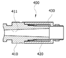
상기 라이트소스 연결구 조립체(400)는, 일단부는 상기 손잡이 바디(200)의 손잡이(201) 하단부에 결합되어 상기 손잡이(201) 측으로 연장되는 광섬유 다발(뭉치)가 내부로 삽입되어 고정되는 장착구멍(411)을 갖고 형성되는 결속구(410)와, 상기 결속구(410)의 외측으로 끼움 결합되는 고정구(420), 및 상기 고정구(420)의 외부로 결합되고, 일단부에 외부의 라이트소스장치와 탈착가능하게 결합되는 결합부(431)가 형성되는 장착구(430)를 포함한다.In the light
상기 결속구(410)의 장착구멍(411)은 일측에서 타측으로 갈수록 확장되게 테이퍼(taper)져서 형성되는 것이 바람직하다. 다시 말해서, 상기 장착구멍(411)은 상기 광섬유(150)가 인입되는 측이 좁게 형성되고 인출되는 측이 넓게 형성되게 된다.It is preferable that the mounting
여기에서, 상기 장착구멍(411)은 좁은 측의 직경은 광섬유 다발의 직경과 같거나 그보다 조금 크게 형성되며 그 좁은 측에서 광섬유를 모아서 고정시킴과 동시에, 외부로부터 들어오는 빛을 효율적으로 전달되도록 할 수 있다.Here, the mounting
또한, 상기 결속구(410)의 장착구멍(411) 내면은 비반사물질로 처리되어 이루어지는 것이 바람직하다.In addition, it is preferable that the inner surface of the mounting
다시 말해서, 외부 라이트소스장치로부터 들어오는 빛이 상기 결속구(410)의 장착구멍(411)에서 반사되지 않고, 상기 결속구(410)의 장착구멍(411)에 뭉치(다발) 형태로 결합되는 광섬유로만 전달될 수 있도록 상기 결속구(410)는 그 장착구멍(411)의 내면에 비반사물질이 도포되어 이루어지거나, 상기 결속구(410) 자체가 비반사재료로 구성될 수 있다.In other words, the light coming from the external light source device is not reflected from the mounting
또한, 상기 고정구(420)는 생략되고, 상기 장착구(430)가 바로 결속구(410)에 착탈가능하게 결합될 수 있다. 또한, 상기 장착구(430)는 생략되고 상기 고정구(420)에 외부의 라이트소스장치가 탈착가능하게 결합될 수 있다. 이와 같이 고정구(420)와 장착구(430)는 외부의 라이트소스장치에 따라 함께 또는 어느 하나가 적용될 수 있는 어댑터로서의 기능을 할 수 있다.In addition, the
상기 각 구성요소 간이 탈착가능하게 하는 구성은 공지의 구성, 예를 들면 후크형태의 록킹-언록킹 방식, 나사결합 방식 등 다양한 결합방식으로 구성될 수 있다.The configuration for allowing the respective components to be detachable may be configured in various coupling methods such as a known configuration, for example, a hook-type locking-unlocking method, a screwing method, and the like.
이상에서 설명한 바와 같은 본 발명에 따른 내시경 일체형의 일회용 경막외 수술 장치에 의하면, 내시경을 통한 영상을 확인하면서 경막외 수술을 보다 용이하고 효율적으로 수행할 수 있으며, 정확한 시술이 가능하여 수술 성공율을 현저히 향상시킬 수 있고, 1회용임에도 경막외 수술에 최적화된 구조를 가져 경제성을 도모할 수 있는 이점이 있다.According to the disposable epidural surgical device of an endoscope integrated type according to the present invention as described above, epidural surgery can be performed more easily and efficiently while checking the image through the endoscope, and accurate procedure is possible, thereby remarkably increasing the success rate of surgery. It can be improved, and even though it is a disposable, it has the advantage of promoting economical efficiency by having a structure optimized for epidural surgery.
본 명세서에서 설명되는 실시 예와 첨부된 도면은 본 발명에 포함되는 기술적 사상의 일부를 예시적으로 설명하는 것에 불과하다. 따라서, 본 명세서에 개시된 실시 예는 본 발명의 기술적 사상을 한정하기 위한 것이 아니라 설명하기 위한 것이므로, 이러한 실시 예에 의하여 본 발명의 기술 사상의 범위가 한정되는 것은 아님은 자명하다. 본 발명의 명세서 및 도면에 포함된 기술적 사상의 범위 내에서 당업자가 용이하게 유추할 수 있는 변형 예와 구체적인 실시 예는 모두 본 발명의 권리범위에 포함되는 것으로 해석되어야 할 것이다.The embodiments described in the present specification and the accompanying drawings are merely illustrative of some of the technical ideas included in the present invention. Accordingly, it is obvious that the embodiments disclosed in the present specification are not intended to limit the technical idea of the present disclosure, but to explain the technical idea, and thus the scope of the technical idea of the present disclosure is not limited by these embodiments. Modification examples and specific embodiments that can be easily inferred by those skilled in the art within the scope of the technical idea included in the specification and drawings of the present invention should be interpreted as being included in the scope of the present invention.
100: 투관침
110: 수술도구 가이드관
120: 약물 주입관
130: 약물 배출관
150: 광섬유
200: 손잡이형 바디
201: 손잡이
210: 약물 주입구
220: 약물 배출구
310: 카메라
320: 영상케이블
400: 라이트소스 연결구 조립체
410: 결속구
411: 장착구멍
420: 고정구
430: 장착구
500: 커넥터100: trocar
110: surgical tool guide tube
120: drug infusion tube
130: drug discharge pipe
150: optical fiber
200: handle type body
201: handle
210: drug inlet
220: drug outlet
310: camera
320: video cable
400: light source connector assembly
410: binding sphere
411: mounting hole
420: fixture
430: mounting hole
500: connector
Claims (8)
상기 투관침의 일단부에 결합되고, 상기 투관침의 각 관로와 연결되어 수술도구의 가이드와 식염수 공급 및 배출을 위한 약물 주입구와 약물 배출구가 형성된 손잡이형 바디;
상기 투관침의 타단부에 카메라가 구비되고, 상기 카메라와 연결되어 상기 투관침을 통해 상기 손잡이형 바디의 외부로 인출되는 영상케이블을 포함하는 영상취득수단;
일단부는 상기 투관침의 타단에서 노출되고, 타단부는 상기 손잡이형 바디로 연장되는 광전달 수단; 및
상기 손잡이형 바디의 손잡이 측에 구성되어, 일단부에서 상기 광전달 수단의 타단부가 결합되고 타단부에는 광을 공급하는 라이트소스장치와 탈착가능하게 결합되는 라이트소스 연결구 조립체;를 포함하고,
상기 손잡이형 바디는 손잡이를 갖는 권총 형태로 형성되고,
상기 광전달 수단은 일단부가 각각 상기 투관침의 자유단 둘레를 따라 배치되고, 타단부는 상기 손잡이형 바디의 손잡이 측으로 연장되는 복수의 광섬유로 이루어지며,
상기 라이트소스 연결구 조립체는,
일단부가 상기 손잡이의 하단부에 결합되며 상기 손잡이 측으로 연장되는 광섬유의 다발이 내부로 삽입되어 고정되는 장착구멍을 갖고 형성되는 결속구와, 상기 결속구에 끼움 결합되는 고정구, 및 상기 고정구에 탈착가능하게 결합되고, 단부에 외부 라이트소스장치와 탈착가능하게 결합되는 결합부가 형성되는 장착구를 포함하여 구성되거나,
일단부가 상기 손잡이의 하단부에 결합되며 상기 손잡이 측으로 연장되는 광섬유의 다발이 내부로 삽입되어 고정되는 장착구멍을 갖고 형성되는 결속구, 및 상기 결속구에 탈착 가능하게 결합되고, 단부에 외부 라이트소스장치와 탈착가능하게 결합되는 결합부가 형성되는 장착구를 포함하는 것을 특징으로 하는
내시경 일체형의 일회용 경막외 수술 장치.
A trocar having a plurality of passages for guiding the surgical instruments, acquiring images, and injecting and discharging drugs;
A handle-type body coupled to one end of the trocar, connected to each conduit of the trocar, and having a guide of the surgical tool and a drug inlet and a drug outlet for supplying and discharging saline;
An image acquisition means including an image cable provided with a camera at the other end of the trocar, and connected to the camera and drawn out of the handle-type body through the trocar;
A light transmitting means having one end exposed at the other end of the trocar, and the other end extending to the handle-shaped body; And
A light source connector assembly configured on the handle side of the handle body, coupled to the other end of the light transmission means at one end and detachably coupled to the light source device for supplying light to the other end, and
The handle-type body is formed in the form of a pistol having a handle,
The light transmitting means is formed of a plurality of optical fibers having one end disposed along the free end of the trocar, and the other end extending toward the handle of the handle body,
The light source connector assembly,
One end is coupled to the lower end of the handle and a binding tool formed with a mounting hole through which a bundle of optical fibers extending toward the handle is inserted and fixed, a fastener fitted to the binding tool, and detachably coupled to the fastener It is configured to include a mounting hole in which a coupling portion detachably coupled to the external light source device is formed at the end, or
One end is coupled to the lower end of the handle, and a binding tool formed with a mounting hole in which a bundle of optical fibers extending toward the handle is inserted and fixed, and an external light source device detachably coupled to the binding tool at the end Characterized in that it comprises a mounting hole formed with a coupling portion detachably coupled to the
Disposable epidural surgical device with integrated endoscope.
상기 투관침은 수술기구가 가이드되도록 형성된 수술기구 가이드통로에 구비되는 수술도구 가이드관, 및 약물이 각각 주입되고 배출되도록 형성된 약물투입통로와 약물배출통로에 각각 구비되는 약물 주입관과 약물 배출관을 포함하고,
상기 손잡이형 바디의 일측에 형성되는 상기 약물 주입구는 상기 약물 주입관과 연결되며,
상기 손잡이형 바디의 타측에 형성되는 상기 약물 배출구는 상기 약물 배출관과 연결되어 구성되는 것을 특징으로 하는
내시경 일체형의 일회용 경막외 수술 장치.
The method of claim 1,
The trocar includes a surgical tool guide tube provided in the surgical instrument guide passage formed to guide the surgical instrument, and a drug injection tube and a drug discharge tube respectively provided in the drug injection passage and the drug discharge passage respectively formed to inject and discharge drugs, ,
The drug injection port formed on one side of the handle-type body is connected to the drug injection tube,
The drug outlet formed on the other side of the handle-type body is characterized in that configured to be connected to the drug discharge pipe
Disposable epidural surgical device with integrated endoscope.
수술도구를 가이드하기 위하여 수술도구 삽입구멍에 착탈가능하게 결합되는 커넥터를 더 포함하는 것을 특징으로 하는
내시경 일체형의 일회용 경막외 수술 장치.
The method of claim 2,
It characterized in that it further comprises a connector detachably coupled to the surgical tool insertion hole to guide the surgical tool
Disposable epidural surgical device with integrated endoscope.
상기 결속구의 장착구멍은 비반사물질로 처리되어 이루어지는 것을 특징으로 하는
내시경 일체형의 일회용 경막외 수술 장치.
The method of claim 1,
Characterized in that the mounting hole of the binding tool is treated with a non-reflective material
Disposable epidural surgical device with integrated endoscope.
상기 결속구는 비반사 재질로 이루어지는 것을 특징으로 하는
내시경 일체형의 일회용 경막외 수술 장치.
The method of claim 1,
The binding tool is characterized in that made of a non-reflective material
Disposable epidural surgical device with integrated endoscope.
상기 결속구의 장착 구멍은 일측에서 타측으로 확장되게 테이퍼(taper)져서 형성되며,
상기 장착 구멍의 일측은 상기 광섬유의 다발을 고정하는 구멍 크기로 이루어지는 것을 특징으로 하는
내시경 일체형의 일회용 경막외 수술 장치.The method of claim 1,
The mounting hole of the binding tool is formed to be tapered to extend from one side to the other,
One side of the mounting hole is characterized in that made of a hole size for fixing the bundle of optical fibers
Disposable epidural surgical device with integrated endoscope.
Priority Applications (1)
| Application Number | Priority Date | Filing Date | Title |
|---|---|---|---|
| KR1020180131043A KR102183544B1 (en) | 2018-10-30 | 2018-10-30 | Disposable-epidural surgery device intergrated with endoscope |
Applications Claiming Priority (1)
| Application Number | Priority Date | Filing Date | Title |
|---|---|---|---|
| KR1020180131043A KR102183544B1 (en) | 2018-10-30 | 2018-10-30 | Disposable-epidural surgery device intergrated with endoscope |
Publications (2)
| Publication Number | Publication Date |
|---|---|
| KR20200048692A KR20200048692A (en) | 2020-05-08 |
| KR102183544B1 true KR102183544B1 (en) | 2020-11-27 |
Family
ID=70678210
Family Applications (1)
| Application Number | Title | Priority Date | Filing Date |
|---|---|---|---|
| KR1020180131043A Expired - Fee Related KR102183544B1 (en) | 2018-10-30 | 2018-10-30 | Disposable-epidural surgery device intergrated with endoscope |
Country Status (1)
| Country | Link |
|---|---|
| KR (1) | KR102183544B1 (en) |
Cited By (6)
| Publication number | Priority date | Publication date | Assignee | Title |
|---|---|---|---|---|
| US11576563B2 (en) | 2016-11-28 | 2023-02-14 | Adaptivendo Llc | Endoscope with separable, disposable shaft |
| USD1018844S1 (en) | 2020-01-09 | 2024-03-19 | Adaptivendo Llc | Endoscope handle |
| USD1031035S1 (en) | 2021-04-29 | 2024-06-11 | Adaptivendo Llc | Endoscope handle |
| USD1051380S1 (en) | 2020-11-17 | 2024-11-12 | Adaptivendo Llc | Endoscope handle |
| USD1066659S1 (en) | 2021-09-24 | 2025-03-11 | Adaptivendo Llc | Endoscope handle |
| USD1070082S1 (en) | 2021-04-29 | 2025-04-08 | Adaptivendo Llc | Endoscope handle |
Citations (2)
| Publication number | Priority date | Publication date | Assignee | Title |
|---|---|---|---|---|
| KR101787691B1 (en) * | 2017-05-30 | 2017-10-18 | 문병진 | Disposalbe endoscope |
| KR101827675B1 (en) * | 2017-01-25 | 2018-02-08 | 김현주 | Epidural catheter surgery tool kit |
Family Cites Families (3)
| Publication number | Priority date | Publication date | Assignee | Title |
|---|---|---|---|---|
| KR101116873B1 (en) | 2010-08-13 | 2012-03-07 | (주)세원메디텍 | Catheter for injecting medicinal fluid in body |
| KR20130140025A (en) | 2010-11-29 | 2013-12-23 | 가부시키가이샤 교토 이료 세케이 | Medical catheter device |
| KR20170000269A (en) * | 2015-06-23 | 2017-01-02 | 미라클스코프(주) | Mesogun having endoscope |
-
2018
- 2018-10-30 KR KR1020180131043A patent/KR102183544B1/en not_active Expired - Fee Related
Patent Citations (2)
| Publication number | Priority date | Publication date | Assignee | Title |
|---|---|---|---|---|
| KR101827675B1 (en) * | 2017-01-25 | 2018-02-08 | 김현주 | Epidural catheter surgery tool kit |
| KR101787691B1 (en) * | 2017-05-30 | 2017-10-18 | 문병진 | Disposalbe endoscope |
Cited By (6)
| Publication number | Priority date | Publication date | Assignee | Title |
|---|---|---|---|---|
| US11576563B2 (en) | 2016-11-28 | 2023-02-14 | Adaptivendo Llc | Endoscope with separable, disposable shaft |
| USD1018844S1 (en) | 2020-01-09 | 2024-03-19 | Adaptivendo Llc | Endoscope handle |
| USD1051380S1 (en) | 2020-11-17 | 2024-11-12 | Adaptivendo Llc | Endoscope handle |
| USD1031035S1 (en) | 2021-04-29 | 2024-06-11 | Adaptivendo Llc | Endoscope handle |
| USD1070082S1 (en) | 2021-04-29 | 2025-04-08 | Adaptivendo Llc | Endoscope handle |
| USD1066659S1 (en) | 2021-09-24 | 2025-03-11 | Adaptivendo Llc | Endoscope handle |
Also Published As
| Publication number | Publication date |
|---|---|
| KR20200048692A (en) | 2020-05-08 |
Similar Documents
| Publication | Publication Date | Title |
|---|---|---|
| KR102183544B1 (en) | Disposable-epidural surgery device intergrated with endoscope | |
| US5735792A (en) | Surgical instrument including viewing optics and an atraumatic probe | |
| US5377668A (en) | Apparatus and method for endoscopic diagnostics and therapy | |
| US11337598B2 (en) | Laser video endoscope | |
| AU2014233486B2 (en) | Viewing trocar with integrated prism for use with angled endoscope | |
| US4736733A (en) | Endoscope with removable eyepiece | |
| CA2327268C (en) | Endoscopic instrumentation with working channel | |
| US6419654B1 (en) | Diagnostic needle arthroscopy and lavage system | |
| US5857961A (en) | Surgical instrument for use with a viewing system | |
| US6432047B1 (en) | Endoscopic surgical procedures and endoscopic apparatus comprising segmented fiber optic cables | |
| WO2004026125A1 (en) | Endoscope | |
| WO2004075715B1 (en) | Diagnostic needle arthroscopy and lavage system | |
| US20170181604A1 (en) | Multi-stage instrument connector | |
| CN102946786B (en) | Laser video endoscope | |
| US20130204083A1 (en) | Sheathless arthroscope and system | |
| KR101881226B1 (en) | Catheter Assembly for Endoscope | |
| CN109691970A (en) | A kind of novel percutaneous choledochoscope system through gall-bladder, Via bile duct | |
| CN217285716U (en) | Endoscope | |
| CN222708333U (en) | A kit for clinical operation of minimally invasive percutaneous nephrolithotomy, a sterile kit for minimally invasive percutaneous nephrolithotomy, and a disposable kit for salivary gland endoscopy | |
| KR20200084324A (en) | Imaging device | |
| US20230380677A1 (en) | Kits for minimally invasive percutaneous nephrolithotomy (pcnl) and sialendoscopy | |
| JP5384894B2 (en) | Adapter and endoscope system using the adapter | |
| US20230049673A1 (en) | Endoscope with reusable optical and electrical distal assembly | |
| CN117860186A (en) | Disposable spinal endoscope | |
| CN118266852A (en) | Disposable electronic spinal endoscope system |
Legal Events
| Date | Code | Title | Description |
|---|---|---|---|
| PA0109 | Patent application |
St.27 status event code: A-0-1-A10-A12-nap-PA0109 |
|
| PA0201 | Request for examination |
St.27 status event code: A-1-2-D10-D11-exm-PA0201 |
|
| D13-X000 | Search requested |
St.27 status event code: A-1-2-D10-D13-srh-X000 |
|
| D14-X000 | Search report completed |
St.27 status event code: A-1-2-D10-D14-srh-X000 |
|
| PG1501 | Laying open of application |
St.27 status event code: A-1-1-Q10-Q12-nap-PG1501 |
|
| E902 | Notification of reason for refusal | ||
| PE0902 | Notice of grounds for rejection |
St.27 status event code: A-1-2-D10-D21-exm-PE0902 |
|
| E13-X000 | Pre-grant limitation requested |
St.27 status event code: A-2-3-E10-E13-lim-X000 |
|
| P11-X000 | Amendment of application requested |
St.27 status event code: A-2-2-P10-P11-nap-X000 |
|
| P13-X000 | Application amended |
St.27 status event code: A-2-2-P10-P13-nap-X000 |
|
| E701 | Decision to grant or registration of patent right | ||
| PE0701 | Decision of registration |
St.27 status event code: A-1-2-D10-D22-exm-PE0701 |
|
| PR0701 | Registration of establishment |
St.27 status event code: A-2-4-F10-F11-exm-PR0701 |
|
| PR1002 | Payment of registration fee |
St.27 status event code: A-2-2-U10-U11-oth-PR1002 Fee payment year number: 1 |
|
| PG1601 | Publication of registration |
St.27 status event code: A-4-4-Q10-Q13-nap-PG1601 |
|
| PC1903 | Unpaid annual fee |
St.27 status event code: A-4-4-U10-U13-oth-PC1903 Not in force date: 20231121 Payment event data comment text: Termination Category : DEFAULT_OF_REGISTRATION_FEE |
|
| PC1903 | Unpaid annual fee |
St.27 status event code: N-4-6-H10-H13-oth-PC1903 Ip right cessation event data comment text: Termination Category : DEFAULT_OF_REGISTRATION_FEE Not in force date: 20231121 |