KR101665124B1 - Ultrasonic imaging apparatus and for the same - Google Patents
Ultrasonic imaging apparatus and for the same Download PDFInfo
- Publication number
- KR101665124B1 KR101665124B1 KR1020140110583A KR20140110583A KR101665124B1 KR 101665124 B1 KR101665124 B1 KR 101665124B1 KR 1020140110583 A KR1020140110583 A KR 1020140110583A KR 20140110583 A KR20140110583 A KR 20140110583A KR 101665124 B1 KR101665124 B1 KR 101665124B1
- Authority
- KR
- South Korea
- Prior art keywords
- region
- interest
- display
- target
- displaying
- Prior art date
- Legal status (The legal status is an assumption and is not a legal conclusion. Google has not performed a legal analysis and makes no representation as to the accuracy of the status listed.)
- Active
Links
Images


Classifications
-
- A—HUMAN NECESSITIES
- A61—MEDICAL OR VETERINARY SCIENCE; HYGIENE
- A61B—DIAGNOSIS; SURGERY; IDENTIFICATION
- A61B8/00—Diagnosis using ultrasonic, sonic or infrasonic waves
- A61B8/13—Tomography
-
- A—HUMAN NECESSITIES
- A61—MEDICAL OR VETERINARY SCIENCE; HYGIENE
- A61B—DIAGNOSIS; SURGERY; IDENTIFICATION
- A61B8/00—Diagnosis using ultrasonic, sonic or infrasonic waves
- A61B8/46—Ultrasonic, sonic or infrasonic diagnostic devices with special arrangements for interfacing with the operator or the patient
- A61B8/461—Displaying means of special interest
- A61B8/463—Displaying means of special interest characterised by displaying multiple images or images and diagnostic data on one display
-
- A—HUMAN NECESSITIES
- A61—MEDICAL OR VETERINARY SCIENCE; HYGIENE
- A61B—DIAGNOSIS; SURGERY; IDENTIFICATION
- A61B8/00—Diagnosis using ultrasonic, sonic or infrasonic waves
- A61B8/08—Clinical applications
-
- A—HUMAN NECESSITIES
- A61—MEDICAL OR VETERINARY SCIENCE; HYGIENE
- A61B—DIAGNOSIS; SURGERY; IDENTIFICATION
- A61B8/00—Diagnosis using ultrasonic, sonic or infrasonic waves
- A61B8/08—Clinical applications
- A61B8/0833—Clinical applications involving detecting or locating foreign bodies or organic structures
- A61B8/085—Clinical applications involving detecting or locating foreign bodies or organic structures for locating body or organic structures, e.g. tumours, calculi, blood vessels, nodules
-
- A—HUMAN NECESSITIES
- A61—MEDICAL OR VETERINARY SCIENCE; HYGIENE
- A61B—DIAGNOSIS; SURGERY; IDENTIFICATION
- A61B8/00—Diagnosis using ultrasonic, sonic or infrasonic waves
- A61B8/46—Ultrasonic, sonic or infrasonic diagnostic devices with special arrangements for interfacing with the operator or the patient
- A61B8/461—Displaying means of special interest
- A61B8/466—Displaying means of special interest adapted to display 3D data
-
- A—HUMAN NECESSITIES
- A61—MEDICAL OR VETERINARY SCIENCE; HYGIENE
- A61B—DIAGNOSIS; SURGERY; IDENTIFICATION
- A61B8/00—Diagnosis using ultrasonic, sonic or infrasonic waves
- A61B8/46—Ultrasonic, sonic or infrasonic diagnostic devices with special arrangements for interfacing with the operator or the patient
- A61B8/467—Ultrasonic, sonic or infrasonic diagnostic devices with special arrangements for interfacing with the operator or the patient characterised by special input means
- A61B8/469—Ultrasonic, sonic or infrasonic diagnostic devices with special arrangements for interfacing with the operator or the patient characterised by special input means for selection of a region of interest
-
- A—HUMAN NECESSITIES
- A61—MEDICAL OR VETERINARY SCIENCE; HYGIENE
- A61B—DIAGNOSIS; SURGERY; IDENTIFICATION
- A61B8/00—Diagnosis using ultrasonic, sonic or infrasonic waves
- A61B8/48—Diagnostic techniques
- A61B8/483—Diagnostic techniques involving the acquisition of a 3D volume of data
-
- A—HUMAN NECESSITIES
- A61—MEDICAL OR VETERINARY SCIENCE; HYGIENE
- A61B—DIAGNOSIS; SURGERY; IDENTIFICATION
- A61B8/00—Diagnosis using ultrasonic, sonic or infrasonic waves
- A61B8/52—Devices using data or image processing specially adapted for diagnosis using ultrasonic, sonic or infrasonic waves
- A61B8/5215—Devices using data or image processing specially adapted for diagnosis using ultrasonic, sonic or infrasonic waves involving processing of medical diagnostic data
- A61B8/5223—Devices using data or image processing specially adapted for diagnosis using ultrasonic, sonic or infrasonic waves involving processing of medical diagnostic data for extracting a diagnostic or physiological parameter from medical diagnostic data
-
- A—HUMAN NECESSITIES
- A61—MEDICAL OR VETERINARY SCIENCE; HYGIENE
- A61B—DIAGNOSIS; SURGERY; IDENTIFICATION
- A61B8/00—Diagnosis using ultrasonic, sonic or infrasonic waves
- A61B8/52—Devices using data or image processing specially adapted for diagnosis using ultrasonic, sonic or infrasonic waves
- A61B8/5215—Devices using data or image processing specially adapted for diagnosis using ultrasonic, sonic or infrasonic waves involving processing of medical diagnostic data
- A61B8/5238—Devices using data or image processing specially adapted for diagnosis using ultrasonic, sonic or infrasonic waves involving processing of medical diagnostic data for combining image data of patient, e.g. merging several images from different acquisition modes into one image
- A61B8/5246—Devices using data or image processing specially adapted for diagnosis using ultrasonic, sonic or infrasonic waves involving processing of medical diagnostic data for combining image data of patient, e.g. merging several images from different acquisition modes into one image combining images from the same or different imaging techniques, e.g. color Doppler and B-mode
- A61B8/5253—Devices using data or image processing specially adapted for diagnosis using ultrasonic, sonic or infrasonic waves involving processing of medical diagnostic data for combining image data of patient, e.g. merging several images from different acquisition modes into one image combining images from the same or different imaging techniques, e.g. color Doppler and B-mode combining overlapping images, e.g. spatial compounding
-
- A—HUMAN NECESSITIES
- A61—MEDICAL OR VETERINARY SCIENCE; HYGIENE
- A61B—DIAGNOSIS; SURGERY; IDENTIFICATION
- A61B8/00—Diagnosis using ultrasonic, sonic or infrasonic waves
- A61B8/54—Control of the diagnostic device
-
- G—PHYSICS
- G01—MEASURING; TESTING
- G01S—RADIO DIRECTION-FINDING; RADIO NAVIGATION; DETERMINING DISTANCE OR VELOCITY BY USE OF RADIO WAVES; LOCATING OR PRESENCE-DETECTING BY USE OF THE REFLECTION OR RERADIATION OF RADIO WAVES; ANALOGOUS ARRANGEMENTS USING OTHER WAVES
- G01S15/00—Systems using the reflection or reradiation of acoustic waves, e.g. sonar systems
- G01S15/88—Sonar systems specially adapted for specific applications
- G01S15/89—Sonar systems specially adapted for specific applications for mapping or imaging
- G01S15/8906—Short-range imaging systems; Acoustic microscope systems using pulse-echo techniques
- G01S15/8993—Three dimensional imaging systems
-
- G—PHYSICS
- G01—MEASURING; TESTING
- G01S—RADIO DIRECTION-FINDING; RADIO NAVIGATION; DETERMINING DISTANCE OR VELOCITY BY USE OF RADIO WAVES; LOCATING OR PRESENCE-DETECTING BY USE OF THE REFLECTION OR RERADIATION OF RADIO WAVES; ANALOGOUS ARRANGEMENTS USING OTHER WAVES
- G01S7/00—Details of systems according to groups G01S13/00, G01S15/00, G01S17/00
- G01S7/52—Details of systems according to groups G01S13/00, G01S15/00, G01S17/00 of systems according to group G01S15/00
- G01S7/52017—Details of systems according to groups G01S13/00, G01S15/00, G01S17/00 of systems according to group G01S15/00 particularly adapted to short-range imaging
- G01S7/52053—Display arrangements
- G01S7/52057—Cathode ray tube displays
- G01S7/5206—Two-dimensional coordinated display of distance and direction; B-scan display
- G01S7/52063—Sector scan display
-
- G—PHYSICS
- G01—MEASURING; TESTING
- G01S—RADIO DIRECTION-FINDING; RADIO NAVIGATION; DETERMINING DISTANCE OR VELOCITY BY USE OF RADIO WAVES; LOCATING OR PRESENCE-DETECTING BY USE OF THE REFLECTION OR RERADIATION OF RADIO WAVES; ANALOGOUS ARRANGEMENTS USING OTHER WAVES
- G01S7/00—Details of systems according to groups G01S13/00, G01S15/00, G01S17/00
- G01S7/52—Details of systems according to groups G01S13/00, G01S15/00, G01S17/00 of systems according to group G01S15/00
- G01S7/52017—Details of systems according to groups G01S13/00, G01S15/00, G01S17/00 of systems according to group G01S15/00 particularly adapted to short-range imaging
- G01S7/52053—Display arrangements
- G01S7/52057—Cathode ray tube displays
- G01S7/52073—Production of cursor lines, markers or indicia by electronic means
-
- G—PHYSICS
- G01—MEASURING; TESTING
- G01S—RADIO DIRECTION-FINDING; RADIO NAVIGATION; DETERMINING DISTANCE OR VELOCITY BY USE OF RADIO WAVES; LOCATING OR PRESENCE-DETECTING BY USE OF THE REFLECTION OR RERADIATION OF RADIO WAVES; ANALOGOUS ARRANGEMENTS USING OTHER WAVES
- G01S7/00—Details of systems according to groups G01S13/00, G01S15/00, G01S17/00
- G01S7/52—Details of systems according to groups G01S13/00, G01S15/00, G01S17/00 of systems according to group G01S15/00
- G01S7/52017—Details of systems according to groups G01S13/00, G01S15/00, G01S17/00 of systems according to group G01S15/00 particularly adapted to short-range imaging
- G01S7/52053—Display arrangements
- G01S7/52057—Cathode ray tube displays
- G01S7/52074—Composite displays, e.g. split-screen displays; Combination of multiple images or of images and alphanumeric tabular information
-
- G—PHYSICS
- G01—MEASURING; TESTING
- G01S—RADIO DIRECTION-FINDING; RADIO NAVIGATION; DETERMINING DISTANCE OR VELOCITY BY USE OF RADIO WAVES; LOCATING OR PRESENCE-DETECTING BY USE OF THE REFLECTION OR RERADIATION OF RADIO WAVES; ANALOGOUS ARRANGEMENTS USING OTHER WAVES
- G01S7/00—Details of systems according to groups G01S13/00, G01S15/00, G01S17/00
- G01S7/52—Details of systems according to groups G01S13/00, G01S15/00, G01S17/00 of systems according to group G01S15/00
- G01S7/52017—Details of systems according to groups G01S13/00, G01S15/00, G01S17/00 of systems according to group G01S15/00 particularly adapted to short-range imaging
- G01S7/52079—Constructional features
- G01S7/52084—Constructional features related to particular user interfaces
-
- A—HUMAN NECESSITIES
- A61—MEDICAL OR VETERINARY SCIENCE; HYGIENE
- A61B—DIAGNOSIS; SURGERY; IDENTIFICATION
- A61B8/00—Diagnosis using ultrasonic, sonic or infrasonic waves
- A61B8/44—Constructional features of the ultrasonic, sonic or infrasonic diagnostic device
- A61B8/4405—Device being mounted on a trolley
-
- A—HUMAN NECESSITIES
- A61—MEDICAL OR VETERINARY SCIENCE; HYGIENE
- A61B—DIAGNOSIS; SURGERY; IDENTIFICATION
- A61B8/00—Diagnosis using ultrasonic, sonic or infrasonic waves
- A61B8/44—Constructional features of the ultrasonic, sonic or infrasonic diagnostic device
- A61B8/4427—Device being portable or laptop-like
-
- A—HUMAN NECESSITIES
- A61—MEDICAL OR VETERINARY SCIENCE; HYGIENE
- A61B—DIAGNOSIS; SURGERY; IDENTIFICATION
- A61B8/00—Diagnosis using ultrasonic, sonic or infrasonic waves
- A61B8/44—Constructional features of the ultrasonic, sonic or infrasonic diagnostic device
- A61B8/4444—Constructional features of the ultrasonic, sonic or infrasonic diagnostic device related to the probe
- A61B8/4472—Wireless probes
-
- A—HUMAN NECESSITIES
- A61—MEDICAL OR VETERINARY SCIENCE; HYGIENE
- A61B—DIAGNOSIS; SURGERY; IDENTIFICATION
- A61B8/00—Diagnosis using ultrasonic, sonic or infrasonic waves
- A61B8/46—Ultrasonic, sonic or infrasonic diagnostic devices with special arrangements for interfacing with the operator or the patient
- A61B8/467—Ultrasonic, sonic or infrasonic diagnostic devices with special arrangements for interfacing with the operator or the patient characterised by special input means
- A61B8/468—Ultrasonic, sonic or infrasonic diagnostic devices with special arrangements for interfacing with the operator or the patient characterised by special input means allowing annotation or message recording
-
- A—HUMAN NECESSITIES
- A61—MEDICAL OR VETERINARY SCIENCE; HYGIENE
- A61B—DIAGNOSIS; SURGERY; IDENTIFICATION
- A61B8/00—Diagnosis using ultrasonic, sonic or infrasonic waves
- A61B8/52—Devices using data or image processing specially adapted for diagnosis using ultrasonic, sonic or infrasonic waves
- A61B8/5215—Devices using data or image processing specially adapted for diagnosis using ultrasonic, sonic or infrasonic waves involving processing of medical diagnostic data
- A61B8/523—Devices using data or image processing specially adapted for diagnosis using ultrasonic, sonic or infrasonic waves involving processing of medical diagnostic data for generating planar views from image data in a user selectable plane not corresponding to the acquisition plane
-
- A—HUMAN NECESSITIES
- A61—MEDICAL OR VETERINARY SCIENCE; HYGIENE
- A61B—DIAGNOSIS; SURGERY; IDENTIFICATION
- A61B8/00—Diagnosis using ultrasonic, sonic or infrasonic waves
- A61B8/56—Details of data transmission or power supply
Landscapes
- Health & Medical Sciences (AREA)
- Life Sciences & Earth Sciences (AREA)
- Engineering & Computer Science (AREA)
- Physics & Mathematics (AREA)
- Surgery (AREA)
- Public Health (AREA)
- Biophysics (AREA)
- Nuclear Medicine, Radiotherapy & Molecular Imaging (AREA)
- Pathology (AREA)
- Radiology & Medical Imaging (AREA)
- Biomedical Technology (AREA)
- Heart & Thoracic Surgery (AREA)
- Medical Informatics (AREA)
- Molecular Biology (AREA)
- Veterinary Medicine (AREA)
- Animal Behavior & Ethology (AREA)
- General Health & Medical Sciences (AREA)
- Radar, Positioning & Navigation (AREA)
- Remote Sensing (AREA)
- Computer Networks & Wireless Communication (AREA)
- General Physics & Mathematics (AREA)
- Acoustics & Sound (AREA)
- Computer Vision & Pattern Recognition (AREA)
- Human Computer Interaction (AREA)
- Vascular Medicine (AREA)
- Physiology (AREA)
- Computer Graphics (AREA)
- General Engineering & Computer Science (AREA)
- Ultra Sonic Daignosis Equipment (AREA)
Abstract
대상체의 복수의 단면을 미리 정해진 프레임 레이트(Frame Rate)에 따라 연속적으로 표시하는 초음파 영상장치 및 그 제어방법을 제공한다.
초음파 영상장치의 일 실시예에 따르면, 대상체의 볼륨 데이터를 기초로 대상체 내부의 타겟(Target)을 추출하는 영상처리부; 추출된 타겟을 기초로, 대상체 내부의 관심영역을 결정하는 제어부; 및 관심영역을 포함하는 대상체의 복수의 단면영상을 연속적으로 표시하는 디스플레이; 를 포함할 수 있다.An ultrasound imaging apparatus and a control method thereof for continuously displaying a plurality of sections of a target object in accordance with a predetermined frame rate are provided.
According to an embodiment of the ultrasound imaging apparatus, an image processing unit extracts a target within a target object based on volume data of the target object; A control unit for determining a region of interest inside the object based on the extracted target; And a display for continuously displaying a plurality of sectional images of the object including the region of interest; . ≪ / RTI >
Description
본 발명은 초음파를 이용하여 대상체 영상을 생성하기 위한 초음파 영상장치 및 그 제어방법에 관한 것이다.BACKGROUND OF THE INVENTION 1. Field of the Invention The present invention relates to an ultrasound imaging apparatus and a control method thereof for generating an object image using ultrasound.
초음파 진단장치는 대상체의 체표로부터 체내의 타겟 부위를 향하여 초음파 신호를 조사하고, 반사된 초음파 신호(초음파 에코신호)의 정보를 이용하여 연부조직의 단층이나 혈류에 관한 이미지를 무침습으로 얻는 장치이다.The ultrasound diagnostic apparatus irradiates an ultrasound signal from a body surface of a target object toward a target portion in the body and obtains an image related to a tomography or blood flow of the soft tissue using information of the reflected ultrasound signal (ultrasound echo signal) .
초음파 진단장치는 X선 진단장치, X선 CT스캐너(Computerized Tomography Scanner), MRI(Magnetic Resonance Image), 핵의학 진단장치 등의 다른 영상진단장치와 비교할 때, 소형이고 저렴하며, 실시간으로 표시 가능하고, 방사선 등의 피폭이 없어 안전성이 높은 장점이 있으므로, 심장, 복부, 비뇨기 및 산부인과 진단을 위해 널리 이용되고 있다.The ultrasonic diagnostic apparatuses are small, inexpensive, real-time displayable, and inexpensive, as compared with other imaging apparatuses such as X-ray diagnostic apparatuses, X-ray CT scanners, MRI (Magnetic Resonance Images) , Radiation and radiation exposure because it has a high safety advantage, is widely used for diagnosis of heart, abdomen, urinary and obstetrics.
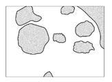
특히, 산부인과에서는 불임의 원인 중 하나인 다낭성난소증후군을 진단하기 위해, 초음파 진단장치를 이용하여 자궁 내 난포의 개수를 확인할 수 있다. In particular, in the obstetrics and gynecology department, the number of intrauterine follicles can be confirmed by using an ultrasonic diagnostic apparatus to diagnose polycystic ovary syndrome which is one of causes of infertility.
초음파 영상장치 및 그 제어방법의 일 측면에 의하면, 대상체의 복수의 단면을 미리 정해진 프레임 레이트(Frame Rate)에 따라 연속적으로 표시하는 초음파 영상장치 및 그 제어방법을 제공한다.According to an aspect of an ultrasound imaging apparatus and a control method therefor, an ultrasound imaging apparatus and a control method thereof for continuously displaying a plurality of sections of a target object in accordance with a predetermined frame rate are provided.
초음파 영상장치의 일 실시예에 따르면, 대상체의 볼륨 데이터를 기초로 대상체 내부의 타겟(Target)을 추출하는 영상처리부; 추출된 타겟을 기초로, 대상체 내부의 관심영역을 결정하는 제어부; 및 관심영역을 포함하는 대상체의 복수의 단면영상을 연속적으로 표시하는 디스플레이; 를 포함할 수 있다.According to an embodiment of the ultrasound imaging apparatus, an image processing unit extracts a target within a target object based on volume data of the target object; A control unit for determining a region of interest inside the object based on the extracted target; And a display for continuously displaying a plurality of sectional images of the object including the region of interest; . ≪ / RTI >
디스플레이는, 대상체의 복수의 단면영상에서 추출된 타겟을 강조하여 표시할 수 있다.The display can emphasize and display the target extracted from a plurality of sectional images of the object.
디스플레이는, 관심영역을 복수의 단면영상에 구간으로 표시할 수 있다.The display can display the region of interest as a section on a plurality of sectional images.
디스플레이는, 볼륨 데이터에 의해 결정되는 전체구간 내에 속하는 대상체의 복수의 단면영상을 연속적으로 표시하고, 표시되는 단면영상에 관심영역이 포함되면, 전체구간에 대한 복수의 단면영상을 연속적으로 표시하는 것을 중단할 수 있다.The display successively displays a plurality of sectional images of the object belonging to the entire section determined by the volume data and successively displays a plurality of sectional images for the entire section when the sectional image to be displayed includes the region of interest You can stop.
디스플레이는, 전체구간에 대한 대상체의 복수의 단면영상을 연속적으로 표시하는 것을 중단한 후, 관심영역에 의해 결정되는 관심구간에 대한 대상체의 복수의 단면 영상을 연속적으로 표시할 수 있다.The display can continuously display a plurality of sectional images of the object for the region of interest determined by the region of interest after discontinuously displaying the plurality of sectional images of the object for the entire region continuously.
제어부는, 추출된 타겟의 크기를 기초로 관심영역을 결정할 수 있다.The control unit can determine the region of interest based on the size of the extracted target.
제어부는, 타겟으로 추출되지 않은 대상체 영역 중 미리 정해진 조건을 만족하는 영역을 관심영역으로 결정할 수 있다.The control unit can determine, as the region of interest, an area that satisfies a predetermined condition among the object areas not extracted as the target.
디스플레이는, 관심영역을 포함하는 대상체의 복수의 단면영상을 동시에 표시하는 것을 포함할 수 있다.The display may include simultaneously displaying a plurality of cross-sectional images of the object including the region of interest.
관심영역을 타겟으로 결정하는 명령, 및 추출된 타겟에 대한 취소 명령 중 적어도 하나를 입력받는 입력부; 를 더 포함할 수 있다.An input unit for receiving at least one of a command for determining an area of interest as a target and a cancel command for an extracted target; As shown in FIG.
디스플레이는, 관심영역을 포함하는 대상체의 복수의 제 1 단면영상을 연속적으로 표시하되, 제 1 단면은, 미리 정해진 제 1 방향에 수직일 수 있다.The display successively displays a plurality of first sectional images of the object including the region of interest, wherein the first cross section may be perpendicular to the predetermined first direction.
디스플레이는, 표시되는 제 1 단면영상의 위치를 대상체의 제 2 단면영상에 표시하되, 제 2 단면은, 제 1 방향에 수직인 제 2 방향에 수직일 수 있다.The display may display the position of the displayed first cross-sectional image on the second cross-sectional image of the object, and the second cross-section may be perpendicular to the second direction perpendicular to the first direction.
디스플레이는, 표시되는 제 1 단면영상의 위치를 대상체의 제 3 단면영상에 표시하되, 제 3 단면은, 제 1 방향 및 제 2 방향에 수직인 제 3 방향에 수직일 수 있다.The display may display the position of the displayed first cross-sectional image on the third cross-sectional image of the object, and the third cross-section may be perpendicular to the first direction and a third direction perpendicular to the second direction.
디스플레이는, 표시되는 단면영상의 위치를 대상체의 3D 영상에 표시할 수 있다.The display can display the position of the displayed sectional image on the 3D image of the object.
초음파 영상장치 제어방법의 일 실시예에 따르면, 대상체의 볼륨 데이터를 기초로, 대상체 내부의 타겟을 추출하고, 추출된 타겟을 기초로, 대상체 내부의 관심영역을 결정하고, 관심영역을 포함하는 대상체의 복수의 단면영상을 연속적으로 표시하는 것을 포함할 수 있다.According to one embodiment of the ultrasonic imaging apparatus control method, a target in a target object is extracted based on volume data of the target object, a region of interest within the target object is determined based on the extracted target, And displaying the plurality of cross-sectional images of the plurality of cross-sectional images.
관심영역을 포함하는 대상체의 복수의 단면영상을 연속적으로 표시하는 것은, 단면영상에서 추출된 타겟을 강조하여 표시할 수 있다.Continuously displaying a plurality of sectional images of the object including the region of interest can emphasize the target extracted from the sectional image.
관심영역을 포함하는 대상체의 복수의 단면영상을 연속적으로 표시하는 것은, 관심영역을 단면영상에 구간으로 표시할 수 있다.Continuously displaying a plurality of sectional images of the object including the region of interest may display the region of interest as a section on the sectional image.
볼륨 데이터에 의해 결정되는 전체구간 내에 속하는 대상체의 복수의 단면영상을 연속적으로 표시하고, 표시되는 단면영상에 관심영역이 포함되면, 전체구간에 대한 복수의 단면영상을 연속적으로 표시하는 것을 중단하는 것을 더 포함할 수 있다.Continuously displaying a plurality of sectional images of the object belonging to the entire section determined by the volume data and continuously displaying a plurality of sectional images of the entire section when the sectional image to be displayed includes the region of interest .
관심영역을 포함하는 대상체의 복수의 단면영상을 연속적으로 표시하는 것은, 전체구간에 대한 대상체의 복수의 단면영상을 연속적으로 표시하는 것을 중단한 후, 관심영역에 의해 결정되는 관심구간에 대한 대상체의 복수의 단면 영상을 연속적으로 표시할 수 있다.Continuously displaying a plurality of sectional images of a target object including a region of interest may be performed by sequentially stopping display of a plurality of sectional images of the target object for the entire region and then displaying the target region for the region of interest determined by the region of interest A plurality of sectional images can be continuously displayed.
대상체 내부의 관심영역을 결정하는 것은, 추출된 타겟의 크기를 기초로 관심영역을 결정할 수 있다.Determining the region of interest within the object may determine the region of interest based on the size of the extracted target.
대상체 내부의 관심영역을 결정하는 것은, 타겟으로 추출되지 않은 대상체 영역 중 미리 정해진 조건을 만족하는 영역을 관심영역으로 결정할 수 있다.Determining the region of interest inside the object may determine an area that satisfies a predetermined condition among the object areas not extracted as the target as the region of interest.
사용자의 입력에 따라, 관심영역을 포함하는 대상체의 복수의 단면영상을 동시에 표시하는 것을 더 포함할 수 있다.And displaying the plurality of cross-sectional images of the object including the region of interest simultaneously according to a user's input.
사용자의 입력에 따라, 관심영역을 타겟으로 결정하거나, 추출된 타겟에 대한 취소 결정을 하는 것을 더 포함할 수 있다.Determining a region of interest as a target, or making a cancellation decision on the extracted target, in accordance with a user's input.
관심영역을 포함하는 대상체의 복수의 단면영상을 연속적으로 표시하는 것은, 관심영역을 포함하는 대상체의 복수의 제 1 단면영상을 연속적으로 표시하되, 제 1 단면은, 미리 정해진 제 1 방향에 수직일 수 있다.Continuously displaying a plurality of sectional images of a target object including a region of interest includes successively displaying a plurality of first sectional images of a target object including a region of interest, wherein the first section is perpendicular to the predetermined first direction .
관심영역을 포함하는 대상체의 복수의 단면영상을 연속적으로 표시하는 것은, 표시되는 제 1 단면영상의 위치를 대상체의 제 2 단면영상에 표시하는 것을 포함하되, 제 2 단면은, 제 1 방향에 수직인 제 2 방향에 수직일 수 있다.Successively displaying a plurality of sectional images of a target object including a region of interest includes displaying a position of the displayed first sectional image on a second sectional image of the target object, Lt; RTI ID = 0.0 > direction. ≪ / RTI >
관심영역을 포함하는 대상체의 복수의 단면영상을 연속적으로 표시하는 것은, 표시되는 제 1 단면영상의 위치를 대상체의 제 3 단면영상에 표시하는 것을 포함하되, 제 3 단면은, 제 1 방향 및 제 2 방향에 수직인 제 3 방향에 수직일 수 있다.Continuously displaying a plurality of sectional images of the object including the region of interest includes displaying the position of the displayed first sectional image on the third sectional image of the object, And may be perpendicular to the third direction perpendicular to the two directions.
관심영역을 포함하는 대상체의 복수의 단면영상을 연속적으로 표시하는 것은, 표시되는 단면영상의 위치를 대상체의 3D 영상에 표시하는 것을 포함할 수 있다.Continuously displaying a plurality of sectional images of the object including the region of interest may include displaying the position of the sectional image to be displayed on the 3D image of the object.
초음파 영상장치 및 그 제어방법의 일 측면에 의하면, 사용자의 조작 없이도 자동으로 대상체의 서로 다른 위치의 단면을 표시할 수 있다.According to an aspect of the ultrasonic imaging apparatus and the control method thereof, a cross section at different positions of a target object can be automatically displayed without a user's operation.
초음파 영상장치 및 그 제어방법의 다른 측면에 의하면, 현재 표시되는 단면의 위치를 다른 방향의 단면 영상에 표시하여, 사용자가 타겟의 위치, 형태 및 크기를 용이하게 파악하도록 도울 수 있다.According to another aspect of the ultrasound imaging apparatus and the control method thereof, the position of the currently displayed cross-section can be displayed on the cross-sectional image of the other direction, so that the user can easily grasp the position, shape and size of the target.
도 1은 초음파 영상장치의 일 실시예를 도시한 사시도이다.
도 2는 초음파 영상장치의 일 실시예에 따른 제어 블록도이다.
도 3a 내지 3c는 초음파 영상장치의 일 실시예에 따른 전체구간 내에 속하는 복수의 단면영상을 연속적으로 표시하는 방법의 일 실시예를 설명하기 위한 도면이다.
도 4a 내지 4c는 초음파 영상장치의 일 실시예에 따른 전체구간 내에 속하는 복수의 단면을 연속적으로 표시하는 방법의 다른 실시예를 설명하기 위한 도면이다.
도 5a 내지 5c는 초음파 영상장치의 일 실시예에 따른 전체구간 내에 속하는 복수의 단면을 연속적으로 표시하는 방법의 다른 실시예를 설명하기 위한 도면이다.
도 6a 및 6b는 초음파 영상장치의 일 실시예에 따른 복수의 단면을 연속적으로 표시하는 것을 중단하는 방법의 일 실시예를 설명하기 위한 도면이다.
도 7a 및 7b는 초음파 영상장치의 일 실시예에 따른 관심영역을 포함하는 단면을 표시하는 방법의 일 실시예를 설명하기 위한 도면이다.
도 8은 초음파 영상장치의 일 실시예에 따른 관심영역을 포함하는 단면을 표시하는 방법의 다른 실시예를 설명하기 위한 도면이다.
도 9a 내지 9f는 초음파 영상장치의 일 실시예에 따른 타겟 추가 방법의 일 실시예를 설명하기 위한 도면이다.
도 10는 초음파 영상장치 제어방법의 일 실시예에 따른 흐름도이다. 1 is a perspective view showing an embodiment of an ultrasound imaging apparatus.
2 is a control block diagram according to an embodiment of the ultrasound imaging apparatus.
3A to 3C are views for explaining an embodiment of a method for continuously displaying a plurality of sectional images belonging to an entire section according to an embodiment of the ultrasound imaging apparatus.
4A to 4C are views for explaining another embodiment of a method for continuously displaying a plurality of sections belonging to an entire section according to an embodiment of the ultrasound imaging apparatus.
5A to 5C are views for explaining another embodiment of a method for continuously displaying a plurality of sections belonging to an entire section according to an embodiment of the ultrasound imaging apparatus.
6A and 6B are views for explaining an embodiment of a method for stopping continuous display of a plurality of sections according to an embodiment of the ultrasound imaging apparatus.
FIGS. 7A and 7B are views for explaining an embodiment of a method of displaying a section including a region of interest according to an embodiment of the ultrasound imaging apparatus.
8 is a view for explaining another embodiment of a method of displaying a cross section including a region of interest according to an embodiment of the ultrasound imaging apparatus.
9A to 9F are views for explaining an embodiment of a method for adding a target according to an embodiment of the ultrasound imaging apparatus.
10 is a flowchart according to an embodiment of a method for controlling an ultrasound imaging apparatus.
이하, 첨부된 도면을 참조하여 초음파 영상장치 및 그 제어방법의 실시예를 구체적으로 설명하도록 한다.Hereinafter, embodiments of the ultrasound imaging apparatus and the control method thereof will be described in detail with reference to the accompanying drawings.
이하에서 사용되는 '초음파 영상'이란 초음파를 이용하여 획득된 대상체에 대한 영상을 의미한다. 또한 '대상체'는 사람, 동물, 금속, 비금속, 또는 그 일부를 의미할 수 있다. 예를 들어, 대상체가 간, 심장, 자궁, 뇌, 유방, 복부 등의 장기, 또는 혈관을 포함할 수 있다. 또한, 대상체는 팬텀(Phantom)을 포함할 수도 있으며, 팬텀은 생물의 밀도와 실효 원자 번호에 아주 근사한 부피를 갖는 물질을 의미할 수 있다. Hereinafter, the term 'ultrasound image' refers to an image of a target object obtained using ultrasound. An "object" may also mean a person, an animal, a metal, a non-metal, or a portion thereof. For example, the subject may include a liver, a heart, an uterus, a brain, a breast, an organ such as the abdomen, or a blood vessel. In addition, the object may comprise a phantom, and the phantom may refer to a material having a volume very close to the density and effective atomic number of the organism.
또한, 이하에서 사용되는 '타겟(Target)'은 대상체의 일부 중 사용자가 초음파 영상을 통해 확인하고자 하는 대상일 수 있다.In addition, 'target' used below may be an object that a user desires to check through ultrasound images.
또한, 이하에서 사용되는 '사용자'는 의료 전문가로서 의사, 간호사, 임상병리사, 의료 영상 전문가 등이 될 수 있으며, 의료 장치를 수리하는 기술자가 될 수 있으나, 이에 한정되지는 않는다.In addition, 'user' used below may be a medical professional such as a doctor, a nurse, a clinical pathologist, a medical imaging expert, and the like, but may be a technician repairing a medical device, but the present invention is not limited thereto.
도 1은 초음파 영상장치의 일 실시예를 도시한 사시도이다. 도 1에 도시된 바와 같이 초음파 영상장치는 본체(100), 초음파 프로브(110), 입력부(150), 디스플레이(160)를 포함할 수 있다. 1 is a perspective view showing an embodiment of an ultrasound imaging apparatus. 1, the ultrasound imaging apparatus may include a
본체(100)의 일측에는 하나 이상의 암 커넥터(female connector; 145)가 구비될 수 있다. 암 커넥터(145)에는 케이블(130)과 연결된 수 커넥터(male connector; 140)가 물리적으로 결합될 수 있다. At least one
한편, 본체(100)의 하부에는 초음파 영상장치의 이동성을 위한 복수개의 캐스터(미도시)가 구비될 수 있다. 복수개의 캐스터는 초음파 영상장치를 특정 장소에 고정시키거나, 특정 방향으로 이동시킬 수 있다. 이와 같은 초음파 영상장치를 카트형 초음파 영상장치라고 한다.Meanwhile, a plurality of casters (not shown) for the mobility of the ultrasound imaging apparatus may be provided under the
또는, 도 1 과 달리, 초음파 영상장치는 원거리 이동 시에 휴대할 수 있는 휴대형 초음파 영상장치일 수도 있다. 이 때, 휴대형 초음파 영상장치는 캐스터가 구비되지 않을 수 있다. 휴대형 초음파 영상장치의 예로는 팩스 뷰어(PACS Viewer), 스마트 폰(Smart Phone), 랩탑 컴퓨터, PDA, 태블릿 PC 등이 있을 수 있으나, 이에 제한되지 않는다.Alternatively, unlike FIG. 1, the ultrasound imaging apparatus may be a portable ultrasound imaging apparatus that can be carried at the time of a long distance movement. At this time, the portable ultrasound imaging apparatus may not include a caster. Examples of portable ultrasound imaging devices include, but are not limited to, a PACS viewer, a smart phone, a laptop computer, a PDA, a tablet PC, and the like.
초음파 프로브(110)는 대상체의 체표에 접촉하는 부분으로, 초음파를 송수신할 수 있다.구체적으로, 초음파 프로브(110)는 본체(100)로부터 제공받은 송신 신호에 따라, 초음파를 대상체의 내부로 송신하고, 대상체 내부의 특정 부위로부터 반사된 에코 초음파를 수신하여 본체(100)로 전달하는 역할을 한다.The ultrasonic probe 110 transmits and receives ultrasound waves to and from the target object according to a transmission signal provided from the
이러한 초음파 프로브(110)에는 케이블(130)의 일단이 연결되며, 케이블(130)의 타단에는 수 커넥터(140)가 연결될 수 있다. 케이블(130)의 타단에 연결된 수 커넥터(140)는 본체(100)의 암 커넥터(145)와 물리적으로 결합할 수 있다. One end of the
또는, 도 1 과 달리, 초음파 프로브는 본체와 무선으로 연결될 수 있다. 이 경우, 초음파 프로브는 대상체로부터 수신한 에코 초음파를 본체로 무선 전송할 수 있다. 뿐만 아니라, 하나의 본체에 복수 개의 초음파 프로브가 연결될 수도 있다.Alternatively, unlike FIG. 1, the ultrasonic probe can be connected to the body wirelessly. In this case, the ultrasonic probe can wirelessly transmit the echo ultrasonic wave received from the object to the body. In addition, a plurality of ultrasonic probes may be connected to one body.
한편, 본체의 내부에는 초음파 프로브가 수신한 에코 초음파를 초음파 영상으로 변환하는 영상 처리부가 마련될 수 있다. 영상 처리부는 마이크로 프로세서(Microprocessor)와 같은 하드웨어의 형태로 구현될 수 있고, 이와는 달리 하드웨어 상에서 수행될 수 있는 소프트웨어의 형태로 구현될 수도 있다.Meanwhile, an image processing unit for converting an echo ultrasonic wave received by an ultrasonic probe into an ultrasound image may be provided inside the body. The image processing unit may be implemented in the form of hardware such as a microprocessor, or may be implemented in the form of software that can be executed on hardware.
영상 처리부는 에코 초음파에 대한 주사 변환(Scan conversion) 과정을 통해 초음파 영상을 생성할 수 있다. 여기서 초음파 영상은 A 모드(amplitude mode), B 모드(brightness mode) 및 M 모드(motion mode)에서 대상체를 스캔하여 획득된 그레이 스케일(gray scale)의 영상뿐만 아니라, 도플러 효과(doppler effect)를 이용하여 움직이는 대상체를 표현하는 도플러 영상을 포함할 수도 있다. 도플러 영상은, 혈액의 흐름을 나타내는 혈류 도플러 영상 (또는, 컬러 도플러 영상으로도 불림), 조직의 움직임을 나타내는 티슈 도플러 영상, 및 대상체의 이동 속도를 파형으로 표시하는 스펙트럴 도플러 영상을 포함할 수 있다. The image processing unit may generate an ultrasound image through a scan conversion process for the echo ultrasonic wave. Here, the ultrasound image uses not only an image of a gray scale obtained by scanning an object in an A mode (amplitude mode), a brightness mode (B mode) and an M mode (motion mode) but also a Doppler effect And may include a Doppler image expressing a moving object. The Doppler image may include a blood flow Doppler image (also referred to as a color Doppler image) representing blood flow, a tissue Doppler image representing tissue motion, and a spectral Doppler image representing a moving velocity of the object as a waveform have.
영상 처리부는 B 모드 영상을 생성하기 위해, 초음파 프로브가 수신한 에코 초음파로부터 B 모드 성분을 추출할 수 있다. 영상 처리부는 B 모드 성분에 기초하여 에코 초음파의 강도가 휘도록 표현되는 초음파 영상을 생성할 수 있다.The image processing unit may extract the B mode component from the echo ultrasonic wave received by the ultrasonic probe to generate the B mode image. The image processing unit can generate an ultrasound image expressed in such a manner that the intensity of the echo ultrasonic waves is warped based on the B mode component.
마찬가지로, 영상 처리부는 에코 초음파로부터 도플러 성분을 추출하고, 추출된 도플러 성분에 기초하여 대상체의 움직임을 컬러 또는 파형으로 표현하는 도플러 영상을 생성할 수 있다.Similarly, the image processing unit may extract a Doppler component from the echo ultrasonic wave, and generate a Doppler image that expresses the motion of the object in color or waveform based on the extracted Doppler component.
뿐만 아니라, 영상 처리부는 에코 초음파를 통해 획득한 볼륨 데이터를 볼륨 렌더링하여 3차원 초음파 영상을 생성할 수도 있고, 압력에 따른 대상체의 변형 정도를 영상화한 탄성 영상을 생성할 수도 있다. 아울러, 영상 처리부는 초음파 영상 상에 여러 가지 부가 정보를 텍스트, 그래픽으로 표현할 수도 있다.In addition, the image processing unit may volume-render the volume data acquired through the echo ultrasonic wave to generate a three-dimensional ultrasound image, or may generate an elastic image that images the degree of deformation of the object according to the pressure. In addition, the image processing unit may display various additional information on the ultrasound image in text or graphics.
한편, 생성된 초음파 영상은 본체 내부 또는 외부의 메모리에 저장될 수 있다. 이와는 달리, 초음파 영상은 웹 상에서 저장기능을 수행하는 웹 스토리지(Web Storage) 또는 클라우드 서버에 저장될 수도 있다.Meanwhile, the generated ultrasound image can be stored in a memory inside or outside the main body. Alternatively, the ultrasound image may be stored in a web storage or a cloud server that performs a storage function on the web.
입력부(150)는 초음파 영상장치의 동작과 관련된 명령을 입력받을 수 있는 부분이다. 예를 들면, A 모드, B 모드, M 모드, 또는 도플러 영상 등의 모드 선택 명령을 입력받을 수 있다. 나아가, 초음파 진단 시작 명령을 입력받을 수도 있다.The
입력부(150)를 통해 입력된 명령은 유선 통신 또는 무선 통신을 통해 본체(100)로 전송될 수 있다.The command input through the
입력부(150)는 예를 들어, 키보드, 풋 스위치(foot switch) 및 풋 페달(foot pedal) 중 적어도 하나를 포함할 수 있다. 키보드는 하드웨어적으로 구현되어, 본체(100)의 상부에 위치할 수 있다. 이러한 키보드는 스위치, 키, 조이스틱 및 트랙볼 중 적어도 하나를 포함할 수 있다. 다른 예로 키보드는 그래픽 유저 인터페이스와 같이 소프트웨어적으로 구현될 수도 있다. 이 경우, 키보드는 서브 디스프레이(161)나 메인 디스플레이(162)를 통해 디스플레이될 수 있다. 풋 스위치나 풋 페달은 본체(100)의 하부에 마련될 수 있으며, 사용자는 풋 페달을 이용하여 초음파 영상장치의 동작을 제어할 수 있다.The
디스플레이(160)는 메인 디스플레이(161)와 서브 디스플레이(162)를 포함할 수 있다.The
서브 디스플레이(162)는 본체(100)에 마련될 수 있다. 도 1은 서브 디스플레이(162)가 입력부(150)의 상부에 마련된 경우를 보여주고 있다. 서브 디스플레이(162)는 초음파 영상장치의 동작과 관련된 어플리케이션을 디스플레이할 수 있다. 예를 들면, 서브 디스플레이(162)는 초음파 진단에 필요한 메뉴나 안내 사항 등을 디스플레이할 수 있다. 이러한 서브 디스플레이(162)는 예를 들어, 브라운관(Cathod Ray Tube: CRT), 액정표시장치(Liquid Crystal Display: LCD) 등으로 구현될 수 있다.The
메인 디스플레이(161)는 본체(100)에 마련될 수 있다. 도 1은 메인 디스플레이(161)가 서브 디스플레이(162)의 상부에 마련된 경우를 보여주고 있다. 메인 디스플레이(161)는 초음파 진단 과정에서 얻어진 초음파 영상을 입력부에 인가된 입력에 따라 디스플레이할 수 있다. 이러한 메인 디스플레이(161)는 서브 디스플레이(162)와 마찬가지로 브라운관 또는 액정표시장치로 구현될 수 있다. 도 1은 메인 디스플레이(161)가 본체(100)에 결합되어 있는 경우를 도시하고 있지만, 메인 디스플레이(161)는 본체(100)와 분리 가능하도록 구현될 수도 있다. The
도 1은 초음파 장치에 메인 디스플레이(161)와 서브 디스플레이(162)가 모두 구비된 경우를 보여주고 있으나, 경우에 따라 서브 디스플레이(162)는 생략될 수도 있다. 이 경우, 서브 디스플레이(162)를 통해 디스플레이되는 어플리케이션이나 메뉴 등은 메인 디스플레이(161)를 통해 디스플레이될 수 있다.1 shows a case where both the
한편, 초음파 영상장치는 통신부를 더 포함할 수 있다. 통신부는, 유선 또는 무선으로 네트워크와 연결되어 외부 디바이스나 서버와 통신한다. 통신부는 의료 영상 정보 시스템(PACS; Picture Archiving and Communication System)을 통해 연결된 병원 서버나 병원 내의 다른 의료 장치와 데이터를 주고 받을 수 있다. 또한, 통신부는 의료용 디지털 영상 및 통신(DICOM; Digital Imaging and Communications in Medicine) 표준에 따라 데이터 통신할 수 있다.On the other hand, the ultrasound imaging apparatus may further include a communication unit. The communication unit is connected to the network by wire or wireless, and communicates with an external device or a server. The communication department can exchange data with other medical devices in the hospital server or the hospital connected through the PACS (Picture Archiving and Communication System). Further, the communication unit can perform data communication according to a DICOM (Digital Imaging and Communications in Medicine) standard.
통신부는 네트워크를 통해 대상체의 초음파 영상, 에코 초음파, 도플러 데이터 등 대상체의 진단과 관련된 데이터를 송수신할 수 있으며, CT, MRI, X-ray 등 다른 의료 장치에서 촬영한 의료 영상 또한 송수신할 수 있다. 나아가, 통신부는 서버로부터 환자의 진단 이력이나 치료 일정 등에 관한 정보를 수신하여 대상체의 진단에 활용할 수도 있다. 나아가, 통신부는 병원 내의 서버나 의료 장치뿐만 아니라, 의사나 환자의 휴대용 단말과 데이터 통신을 수행할 수도 있다.The communication unit can transmit and receive data related to diagnosis of a target object such as an ultrasound image, an echo ultrasonic wave, and Doppler data of a target object through a network, and can transmit and receive a medical image captured by other medical devices such as CT, MRI and X-ray. Further, the communication unit may receive information on a patient's diagnosis history or treatment schedule from the server, and may utilize the information for diagnosis of the subject. Furthermore, the communication unit may perform data communication with a server or a medical device in a hospital, as well as with a doctor or a patient's portable terminal.
통신부는 유선 또는 무선으로 네트워크와 연결되어 서버, 의료 장치, 또는 휴대용 단말과 데이터를 주고 받을 수 있다. 통신부는 외부 디바이스와 통신을 가능하게 하는 하나 이상의 구성 요소를 포함로 수 있으며, 예를 들어 근거리 통신 모듈, 유선 통신 모듈, 및 이동 통신 모듈을 포함할 수 있다.The communication unit can be connected to the network by wire or wireless, and can exchange data with a server, a medical device, or a portable terminal. The communication unit may include one or more components that enable communication with an external device, and may include, for example, a local communication module, a wired communication module, and a mobile communication module.
근거리 통신 모듈은 소정 거리 이내의 근거리 통신을 위한 모듈을 의미한다. 본 발명의 일 실시 예에 따른 근거리 통신 기술에는 무선 랜(Wireless LAN), 와이파이(Wi-Fi), 블루투스, 지그비(Zigbee), WFD(Wi-Fi Direct), UWB(Ultra wideband), 적외선 통신(IrDA; Infrared Data Association), BLE (Bluetooth Low Energy), NFC(Near Field Communication) 등이 있을 수 있으나, 이에 한정되는 것은 아니다. The short-range communication module means a module for short-range communication within a predetermined distance. The local communication technology according to an exemplary embodiment of the present invention includes a wireless LAN, a Wi-Fi, a Bluetooth, a Zigbee, a Wi-Fi Direct, a UWB, Infrared Data Association (IrDA), Bluetooth Low Energy (BLE), Near Field Communication (NFC), and the like.
유선 통신 모듈은 전기적 신호 또는 광 신호를 이용한 통신을 위한 모듈을 의미하며, 일 실시 예에 의한 유선 통신 기술에는 페어 케이블(Pair Cable), 동축 케이블, 광섬유 케이블, 이더넷(Ethernet) 케이블 등이 포함될 수 있다. The wired communication module refers to a module for communication using an electric signal or an optical signal. The wired communication technology according to an exemplary embodiment may include a pair cable, a coaxial cable, an optical fiber cable, an Ethernet cable, have.
이동 통신 모듈은, 이동 통신망 상에서 기지국, 외부의 단말, 서버 중 적어도 하나와 무선 신호를 송수신한다. 여기에서, 무선 신호는, 음성 호 신호, 화상 통화 호 신호 또는 문자/멀티미디어 메시지 송수신에 따른 다양한 형태의 데이터를 포함할 수 있다. The mobile communication module transmits and receives radio signals to and from at least one of a base station, an external terminal, and a server on a mobile communication network. Here, the wireless signal may include various types of data depending on a voice call signal, a video call signal, or a text / multimedia message transmission / reception.
도 2는 초음파 영상장치의 일 실시예에 따른 제어 블록도이다.2 is a control block diagram according to an embodiment of the ultrasound imaging apparatus.
초음파 영상장치의 일 실시예에 따르면, 초음파를 송수신하여 대상체의 볼륨 데이터를 획득하는 초음파 프로브; 대상체의 볼륨 데이터를 기초로 대상체의 초음파 영상을 생성하는 영상처리부(300); 초음파 영상을 표시하는 디스플레이(160); 사용자의 제어명령을 인력받는 입력부(150); 및 초음파 영상장치 전반을 제어하는 제어부(400); 를 포함할 수 있다.According to an embodiment of the ultrasound imaging apparatus, an ultrasound probe for transmitting and receiving ultrasound to acquire volume data of a target object; An
초음파 프로브는 대상체로 초음파를 조사하고, 대상체로부터 반사되는 에코 초음파를 수집할 수 있다. 초음파는 매질에 따라 반사되는 정도가 다르므로, 초음파 프로브는 에코 초음파를 수집함으로써 대상체 내부의 정보를 획득할 수 있다.The ultrasonic probe can irradiate the ultrasonic wave to the object and collect the echosound reflected from the object. Since the degree of reflection varies depending on the medium, the ultrasonic probe can acquire information inside the object by collecting echoes.
초음파 프로브는 대상체의 볼륨 데이터를 획득하는 기술적 사상 안에서 다양하게 구현될 수 있다. 예를 들어 초음파 프로브의 엘리먼트가 1차원 배열을 가지는 경우, 초음파 프로브는 프리핸드(Freehand) 방식에 따라 볼륨 데이터를 획득할 수 있다. 또는, 사용자의 조작 없이, 초음파 프로브는 기계적(Mechanicla) 방식에 따라 볼륨 데이터를 획득할 수 있다. 이와는 달리, 초음파 프로브의 엘리먼트가 2차원 배열을 가지는 경우, 초음파 프로브는 엘리먼트를 제어함으로써 볼륨 데이터를 획득할 수 있다.The ultrasonic probe can be variously implemented in the technical idea of acquiring the volume data of the object. For example, when the elements of the ultrasonic probe have a one-dimensional array, the ultrasonic probe can acquire volume data according to a freehand method. Alternatively, without manipulation of the user, the ultrasonic probe can acquire volume data according to a mechanical method. Alternatively, if the elements of the ultrasonic probe have a two-dimensional array, the ultrasonic probe can acquire volume data by controlling the elements.
영상처리부(300)는 대상체의 볼륨 데이터를 이용하여 대상체의 초음파 영상을 생성할 수 있다. 이 때, 영상처리부(300)는 대상체의 단면에 대한 2D 초음파 영상을 생성할 수 있을 뿐만 아니라, 3D 초음파 영상을 생성하는 것도 가능하다.The
3D 초음파 영상을 생성하기 위해, 영상처리부(300)는 볼륨 데이터를 이용하여 볼륨 렌더링을 수행할 수 있다. 영상처리부(300)는 기존에 공지된 볼륨 렌더링 방식 중 하나를 사용하여 볼륨 데이터를 볼륨 렌더링할 수 있다. 구체적으로, 볼륨 렌더링은 표면 렌더링(Surface Rendering)과 직접 볼륨 렌더링(Direct Volume Rendering)으로 분류될 수 있다.In order to generate a 3D ultrasound image, the
표면 렌더링은 볼륨 데이터로부터 일정한 스칼라 값과 공간적인 변화량을 기반으로 표면 정보를 추출하여 이를 다각형이나 곡면 패치(Patch) 등의 기하학적 요소로 변환하여 기존의 렌더링 기법을 적용하는 방법을 말한다. 표면 렌더링의 예로는 Marching Cubes 알고리즘, Dividing Cubes 알고리즘을 들 수 있다.Surface rendering refers to a method of extracting surface information based on a constant scalar value and a spatial change amount from volume data, converting it into a geometric element such as a polygon or a surface patch (patch), and applying the existing rendering method. Examples of surface rendering include the Marching Cubes algorithm and the Dividing Cubes algorithm.
직접 볼륨 렌더링은 볼륨 데이터를 기하학적 요소로 바꾸는 중간 단계 없이 볼륨 데이터를 직접 렌더링하는 방법을 말한다. 직접 볼륨 렌더링은 물체의 내부 정보를 그대로 가시화할 수 있고, 반투명한 대상체를 표현하는데 유용하다. 직접 볼륨 렌더링은 볼륨 데이터에 접근하는 방식에 따라, 객체 순서 방식(Object-Order Method)과 영상 순서 방식(Image-Order Method)으로 분류될 수 있다.Direct volume rendering is a method of rendering volume data directly without intermediate steps that convert volume data into geometric elements. Direct volume rendering can visualize the internal information of an object as it is, and is useful for expressing a translucent object. Direct volume rendering can be categorized into an object-order method and an image-order method, depending on how the volume data is accessed.
영상 순서 방식은 영상의 픽셀 값을 차례로 결정해 나가는 방식이다. 영상 순서 방식의 예로는 볼륨 광선 투사법(volume ray casting)을 들 수 있다. 객체 순서 방식은 볼륨 데이터를 직접 영상에 투영하는 방식이다. 객체 순서 방식의 예로는 스플래팅 방법(splatting)이 있다. The image ordering method is a method of sequentially determining pixel values of an image. An example of a video ordering scheme is volume ray casting. The object ordering method is a method of projecting the volume data directly onto the image. An example of object ordering is splatting.
또한, 영상처리부(300)는 볼륨 데이터를 기초로 타겟(Target)을 추출할 수 있다. 예를 들어, 대상체가 사람의 자궁이고 타겟이 자궁 내부의 난포인 경우, 영상처리부(300)는 볼륨 데이터를 이용하여 난포를 추출할 수 있다.Also, the
영상처리부(300)는 볼륨 데이터를 기초로 대상체 내부의 타겟을 추출하는 기술적 사상 안에서 다양하게 구현될 수 있다. 예를 들어, 영상처리부(300)는 미리 정해진 범위 내의 밝기 값을 가지는 볼륨 데이터 영역을 타겟으로 추출할 수 있다. 또한 미리 정해진 밝기 값을 가지는 볼륨 데이터 영역의 크기가 미리 정해진 범위 내에 속하는지 판단하여 타겟을 추출할 수도 있다.The
디스플레이(160)는 영상 처리부에서 생성한 초음파 영상을 표시할 수 있다. 구체적으로 디스플레이(160)는 영상 처리부에서 생성한 대상체 단면 영상 또는 대상체의 3D 영상을 각각 표시할 수도 있고, 또는 함께 표시할 수도 있다.The
이 때, 디스플레이(160)는 표시중인 단면 영상에서 추출된 타겟을 강조하여 표시할 수 있다. 예를 들어, 디스플레이(160)는 타겟이 표시되는 영역의 색 또는 음영을 다르게 표시하거나, 타겟이 표시되는 영역의 경계 선의 색 또는 음영을 다르게 표시할 수 있다. 또는, 디스플레이(160)는 타겟이 표시되는 영역의 위치를 사용자에게 알리는 마커를 표시할 수도 있다.At this time, the
디스플레이(160)가 초음파 영상을 표시하는 구체적인 방법은 후술할 제어부(400)와 함께 설명하도록 한다.A specific method for displaying the ultrasound image on the
제어부(400)는 볼륨 데이터에 의해 결정되는 전체구간 내에 속하는 대상체의 복수의 단면영상을 연속적으로 표시하도록 디스플레이(160)를 제어할 수 있다. 제어부(400)의 제어에 따라, 디스플레이(160)는 전체구간 내에 속하는 대상체의 복수의 단면영상을 미리 정해진 프레임 레이트(Frame Rate)에 따라 연속적으로 표시할 수 있다.The
여기서 전체구간이란, 획득한 볼륨데이터에 의해 단면 영상으로 표시될 수 있는 대상체 구간을 의미할 수 있다. 대상체의 복수의 단면 간의 거리 및 미리 정해진 프레임 레이트는 사용자의 입력 또는 초음파 영상장치 내부 연산에 의해 결정될 수 있다.Here, the whole section may refer to an object section that can be displayed as a sectional image by the acquired volume data. The distance between the plurality of sections of the object and the predetermined frame rate may be determined by a user's input or by an internal calculation of the ultrasound imaging apparatus.
이를 통해, 전체구간에 속하는 대상체 내부의 정보를 사용자에게 제공할 수 있고, 또한 대상체 내부를 스캔하여 후술할 관심영역을 결정할 수도 있다.Accordingly, information within the object belonging to the entire section can be provided to the user, and the area of interest to be described later can be determined by scanning the inside of the object.
이하에서는, 전체구간에 속하는 대상체의 복수의 단면영상을 표시하는 방법을 설명한다.Hereinafter, a method of displaying a plurality of sectional images of a target object belonging to the entire section will be described.
도 3a 내지 3c는 초음파 영상장치의 일 실시예에 따른 전체구간 내에 속하는 복수의 단면영상을 연속적으로 표시하는 방법의 일 실시예를 설명하기 위한 도면이다. 3A to 3C are views for explaining an embodiment of a method for continuously displaying a plurality of sectional images belonging to an entire section according to an embodiment of the ultrasound imaging apparatus.
이하에서는 설명의 편의상, 초음파가 대상체로 조사되는 방향을 Z축방향, Z축방향에 수직임과 동시에 상호 수직관계인 방향을 X축방향 및 Y축방향으로 한다. 특히, 1D 어레이 프로브는 엘리먼트가 배열되는 방향을 X축방향으로 하고, 2D 어레이 프로브는 엘리먼트가 배열되는 일 방향을 X축방향, 엘리먼트가 배열되는 다른 방향을 Y축방향으로 한다. Hereinafter, for convenience of explanation, the direction in which the ultrasonic waves are irradiated to the object is referred to as a Z-axis direction and a direction perpendicular to the Z-axis direction and mutually vertical directions are referred to as an X-axis direction and a Y-axis direction. In particular, the 1D array probe has a direction in which the elements are arranged in the X-axis direction, one direction in which the elements are arranged in the 2D array probe is the X-axis direction, and the other direction in which the elements are arranged is the Y-axis direction.
또한, Z축방향을 제 1 방향, X축방향을 제 2 방향, Z축방향을 제 3 방향으로 가정한다.It is also assumed that the Z-axis direction is the first direction, the X-axis direction is the second direction, and the Z-axis direction is the third direction.
제어부(400)는 전체구간 내에서 미리 정해진 제 1 방향에 수직인 대상체의 복수의 단면을 연속적으로 표시하도록 디스플레이(160)를 제어할 수 있다. 구체적으로, 제어부(400)는 제 1 방향에 수직인 대상체의 복수의 단면(제 1 단면)을 미리 정해진 프레임 레이트에 따라 연속적으로 표시하도록 디스플레이(160)를 제어할 수 있다.The
도 3a내지 3c를 참조하면, 디스플레이(160)는 제 1 단면을 연속적으로 표시할 수 있다. 이 때, 타겟이 표시되는 영역의 경계가 각각의 제 1 단면마다 상이함을 확인할 수 있다. 이를 통해, 사용자가 타겟의 형상 및 크기를 용이하게 인식할 수 있다.3A to 3C, the
한편, 제어부(400)는 연속적으로 표시되는 복수의 제 1 단면 각각의 위치를 제 2 방향에 수직인 대상체의 단면(제 2 단면) 상에 연속적으로 표시하도록 디스플레이(160)를 제어할 수 있다. On the other hand, the
도 3a 내지 3c를 참조하면, 디스플레이(160)는 제 1 단면의 우측에 YZ평면과 평행한 제 2 단면을 표시할 수 있다. 구체적으로, 디스플레이(160)는 제 2 단면에서 현재 표시 중인 제 1 단면의 위치를 안내하는 마커를 표시할 수 있다. Referring to Figs. 3A to 3C, the
시간에 따라 도 3a에서 3c로 변화하면, 미리 정해진 프레임 레이트에 따라 제 1 단면이 변화하면서 연속적으로 표시됨과 동시에, 제 2 단면 상에서 마커의 위치도 달라짐을 확인할 수 있다. 3A, it can be seen that the first end face changes continuously according to a predetermined frame rate and the position of the marker changes on the second end face.
이처럼, 디스플레이(160)는 연속적으로 표시되는 제 1 단면과 함께 제 2 단면상에서 제 1 단면의 위치를 제공함으로써, 사용자가 타겟의 위치 및 형상을 용이하게 인식할 수 있도록 도울 수 있다.As such, the
뿐만 아니라, 제어부(400)는 연속적으로 표시되는 복수의 제 1 단면 각각의 위치를 제 3 방향에 수직인 대상체의 단면(제 3 단면) 상에 연속적으로 표시하도록 디스플레이(160)를 제어할 수 있다. In addition, the
도 3a 내지 3c를 참조하면, 디스플레이(160)는 제 1 단면의 아래측에 ZX평면과 평행한 제 3 단면을 표시할 수 있다. 구체적으로, 디스플레이(160)는 제 3 단면에서 현재 표시 중인 제 1 단면의 위치를 안내하는 마커를 표시할 수 있다. 3A-3C, the
시간에 따라 도 3a에서 3c로 변화하면, 미리 정해진 프레임 레이트에 따라 제 1 단면이 변화하면서 연속적으로 표시됨과 동시에, 제 3 단면 상에서 마커의 위치도 달라짐을 확인할 수 있다.3A, it can be seen that the first end face changes continuously according to a predetermined frame rate, and the position of the marker changes on the third end face.
도 4a 내지 4c는 초음파 영상장치의 일 실시예에 따른 전체구간 내에 속하는 복수의 단면을 연속적으로 표시하는 방법의 다른 실시예를 설명하기 위한 도면이다.4A to 4C are views for explaining another embodiment of a method for continuously displaying a plurality of sections belonging to an entire section according to an embodiment of the ultrasound imaging apparatus.
도 3a 내지 3c와는 달리, 제어부(400)는 전체구간 내에서 제 2 방향에 수직인 대상체의 복수의 단면을 연속적으로 표시하도록 디스플레이(160)를 제어할 수 있다. 구체적으로, 제어부(400)는 X축에 수직인 YZ평면과 평행한 대상체의 복수의 단면(제 2 단면)을 미리 정해진 프레임 레이트에 따라 연속적으로 표시하도록 디스플레이(160)를 제어할 수 있다.3A to 3C, the
이와 함께, 제어부(400)는 제 1 단면 또는 제 3 단면 상에서 현재 표시 중인 제 2 단면의 위치를 안내하는 마커를 표시하도록 디스플레이(160)를 제어할 수 있다.The
도 4a 내지 4c를 참조하면, 디스플레이(160)는 미리 정해진 프레임 레이트에 따라 제 2 단면을 연속적으로 표시할 수 있다. 이와 동시에, 디스플레이(160)는 연속적으로 표시되는 제 2 단면의 위치를 제 1 단면 및 제 3 단면 상에서 표시할 수 있다. Referring to Figs. 4A to 4C, the
이처럼 디스플레이(160)는 다양한 방향의 타겟 단면을 연속적으로 사용자에게 제공할 뿐만 아니라, 그 위치를 다른 단면 상에 표시함으로써, 사용자가 타겟의 형상 및 위치를 인식할 수 있도록 돕는다.As such, the
도 5a 내지 5c는 초음파 영상장치의 일 실시예에 따른 전체구간 내에 속하는 복수의 단면을 연속적으로 표시하는 방법의 다른 실시예를 설명하기 위한 도면이다.5A to 5C are views for explaining another embodiment of a method for continuously displaying a plurality of sections belonging to an entire section according to an embodiment of the ultrasound imaging apparatus.
제어부(400)는 전체구간 내에서 대상체의 복수의 단면 각각의 위치를 대상체의 3D 영상 상에 연속적으로 표시하도록 제어부(400)를 제어할 수 있다.The
예를 들어, 제어부(400)는 복수의 제 1 단면을 미리 정해진 프레임 레이트에 따라 연속적으로 표시함과 동시에, 대상체의 3D 영상 상에 현재 표시되는 제 1 단면의 위치를 연속적으로 표시하도록 디스플레이(160)를 제어할 수 있다. 구체적으로 제어부(400)는 디스플레이(160)에 표시되는 제 1 단면의 위치를 대상체 3D 영상 상에 안내하는 마커를 표시할 수 있다. For example, the
그 결과, 도 5a 내지 5c를 참조하면, 디스플레이(160)는 미리 정해진 프레임 레이트에 따라 제 1 단면을 연속적으로 표시할 수 있다. 이와 동시에, 디스플레이(160)는 연속적으로 표시되는 제 1 단면의 위치를 대상체 3D 영상 상에 표시할 수 있다.As a result, referring to Figs. 5A to 5C, the
도 5a 내지 5c에서는 제 1 단면이 연속적으로 표시되는 경우를 예시하였으나, 제 2 단면 또는 제 3 단면과 함께 대상체 3D 영상이 표시될 수 있다. 또는, 제 1 단면, 제 2 단면, 및 제 3 단면 중 적어도 2개 이상이 대상체 3D 영상과 함께 표시될 수도 있다.In FIGS. 5A to 5C, the case where the first end face is continuously displayed is exemplified, but the
이처럼 디스플레이(160)는 표시되는 타겟 단면의 위치를 대상체의 3D 영상 상에 표시함으로써, 사용자가 타겟의 형상 및 위치를 인식할 수 있도록 돕는다.Thus, the
또한, 디스플레이(160)가 전체구간 내에 속하는 대상체의 복수의 단면영상을 연속적으로 표시하던 중, 표시되는 단면영상에 관심영역이 포함될 수 있다. 이 때, 제어부(400)는 전체구간에 대한 복수의 단면영상을 연속적으로 표시하는 것을 중단하도록 디스플레이(160)를 제어할 수 있다. Also, while the
제어부(400)는 상술한 영상처리부(300)에서 추출한 타겟을 기초로 관심영역을 결정할 수 있다. 여기서, 관심영역이란 추출된 타겟 또는 그 이외의 영역에서 사용자의 추가적인 확인이 요구되는 영역일 수 있다. The
예를 들어, 제어부(400)는 추출된 타겟 중 미리 정해진 조건을 만족하는 타겟을 관심영역으로 결정할 수 있다. 타겟이 난포인 경우 사용자가 추출된 난포 중 가장 큰 난포를 확인할 수 있도록, 제어부(400)는 크기가 가장 큰 타겟을 관심영역으로 결정할 수 있다.For example, the
또 다른 예로, 제어부(400)는 타겟으로 추출되지 않은 대상체 영역 중 미리 정해진 조건을 만족하는 영역을 관심영역으로 결정할 수 있다. 타겟이 난포일 경우, 볼륨 데이터를 통해 추출되지 않는 난포가 발생할 수 있다. 단면영상에서 이러한 조건을 만족하는 영역을 관심영역으로 결정함으로써, 사용자에게 해당영역이 난포인지 여부를 확인할 기회를 제공할 수 있다.As another example, the
관심영역으로 결정되기 위한 조건은, 사용자의 입력 또는 장치 내부의 연산에 의해 사전에 결정될 수 있다.The condition to be determined as the region of interest can be determined in advance by the user's input or an operation inside the apparatus.
도 6a 및 6b는 초음파 영상장치의 일 실시예에 따른 복수의 단면을 연속적으로 표시하는 것을 중단하는 방법의 일 실시예를 설명하기 위한 도면이다. 도 6a 및 6b에서는 관심영역이 타겟으로 추출되지 않은 영역 중 미리 정해진 조건을 만족하는 영역인 경우를 전제로 한다.6A and 6B are views for explaining an embodiment of a method for stopping continuous display of a plurality of sections according to an embodiment of the ultrasound imaging apparatus. 6A and 6B, it is assumed that the region of interest is an area that is not extracted as a target and satisfies a predetermined condition.
상술한 도 3a 내지 3c와 같이, 디스플레이(160)는 전체 구간에 대하여 미리 정해진 프레임 레이트에 따라 제 1 단면을 연속적으로 표시할 수 있다. 이 때, 표시되는 제 1 단면에 관심영역이 표시되는 경우, 디스플레이(160)는 연속적으로 복수의 제 1 단면을 표시하는 것을 중단할 수 있다. 그 결과, 디스플레이(160)는 관심영역을 포함하는 제 1 단면을 계속하여 표시할 수 있다.As shown in FIGS. 3A to 3C, the
예를 들어, 도 6a을 참조하면, 디스플레이(160)는 연속적으로 복수의 제 1 단면을 표시할 수 있다. 이 때, 도 6b와 같이 디스플레이(160)에 표시되는 제 1 단면이 관심영역을 포함하는 경우, 디스플레이(160)는 연속적으로 복수의 제 1 단면을 표시하는 것을 중단할 수 있다. 아울러 디스플레이(160)는 관심영역을 포함하는 제 1 단면을 계속하여 표시할 수 있다.For example, referring to FIG. 6A, the
도 6b에서는 관심영역이 타겟으로 추출되지 않은 영역 중 미리 정해진 조건을 만족하는 영역인 경우를 전제로 한다.In FIG. 6B, it is assumed that the region of interest is an area that is not extracted as a target and satisfies a predetermined condition.
디스플레이(160)가 관심영역을 포함하는 단면을 계속하여 표시함으로써, 사용자는 영상처리부(300)에 의해 추출되지 않은 타겟이 존재하는지를 판단할 수 있다.The
또한, 디스플레이(160)가 전체구간에 대한 대상체의 복수의 단면영상을 연속적으로 표시하는 것을 중단한 후, 제어부(400)는 관심영역에 의해 결정되는 관심구간에 대한 대상체의 복수의 단면영상을 연속적으로 표시하도록 디스플레이(160)를 제어할 수 있다. In addition, after the
이 때, 대상체의 복수의 단면 간의 거리 및 프레임 레이트는 사용자의 입력 또는 초음파 영상장치 내부 연산에 의해 결정될 수 있다.At this time, the distance between the plurality of sections of the object and the frame rate may be determined by a user's input or an internal operation of the ultrasound imaging apparatus.
도 7a 및 7b는 초음파 영상장치의 일 실시예에 따른 관심영역을 포함하는 단면을 표시하는 방법의 일 실시예를 설명하기 위한 도면이다.FIGS. 7A and 7B are views for explaining an embodiment of a method of displaying a section including a region of interest according to an embodiment of the ultrasound imaging apparatus.
상술한 바와 같이, 제어부(400)는 디스플레이(160)에 표시되는 단면이 관심영역을 포함하면, 복수의 단면을 연속적으로 표시하는 것을 중단하도록 디스플레이(160)를 제어할 수 있다. 그 결과 디스플레이(160)는 확인필요영역을 포함하는 단면을 계속하여 표시할 수 있다.As described above, the
도 7a는 디스플레이(160)가 관심영역을 포함하는 단면을 계속하여 표시하는 경우를 예시하고 있다. 관심영역은 사용자의 확인이 요구되는 영역인 바, 디스플레이(160)는 관심영역에 대한 더 많은 정보를 사용자에게 제공할 필요가 있다.FIG. 7A illustrates a case where the
따라서, 제어부(400)는 관심영역에 의해 결정되는 관심구간(S)에 대한 복수의 단면을 표시하도록 디스플레이(160)를 제어할 수 있다. 여기서, 관심구간(S)이란 관심영역의 단면을 제공하기 위해 미리 결정되는 구간을 의미할 수 있다. 제어부(400)는 관심영역을 결정한 후, 관심영역의 경계 사이를 관심 구간으로 설정할 수 있다.Accordingly, the
도 7b와 같이, 제어부(400)의 제어에 따라 디스플레이(160)는 관심구간(S) 내의 대상체 단면영상을 연속적으로 표시할 수 있다. As shown in FIG. 7B, the
이를 통해, 디스플레이(160)는 사용자에게 관심영역의 형상 및 크기에 대한 정보를 제공할 수 있다. 관심영역이 타겟으로 추출되지 않은 영역에서 결정된 경우, 디스플레이(160)가 관심영역을 포함하는 복수의 단면을 표시함으로써, 사용자는 관심영역이 타겟을 의미하는지 판단할 수 있다.In this way, the
도 8은 초음파 영상장치의 일 실시예에 따른 관심영역을 포함하는 단면을 표시하는 방법의 다른 실시예를 설명하기 위한 도면이다.8 is a view for explaining another embodiment of a method of displaying a cross section including a region of interest according to an embodiment of the ultrasound imaging apparatus.
도 7a 및 7b는 관심영역을 포함하는 복수의 단면을 연속적으로 표시하는 경우를 예시하였다. 그러나, 이와는 달리, 디스플레이(160)가 관심영역을 포함하는 복수의 단면을 하나의 화면에 표시하는 것도 가능하다.FIGS. 7A and 7B illustrate the case where a plurality of sections including the region of interest are continuously displayed. Alternatively, however, it is also possible for the
도 8과 같이 관심영역을 포함하는 복수의 단면을 동시에 사용자에게 제공할 수 있다. 사용자는 다양한 위치의 단면을 동시에 제공받을 수 있어, 서로 다른 위치에서의 관심영상의 특징을 비교할 수 있다.It is possible to simultaneously provide a plurality of sections including the region of interest to the user as shown in FIG. The user can be provided with cross sections at various positions at the same time, so that the features of the interest image at different positions can be compared.
상술한 바와 같이, 사용자에게 관심영역을 포함하는 복수의 대상체 단면을 제공한 후, 입력부(150)는 관심영역을 타겟으로 결정하는 명령 및 추출된 타겟에 대한 취소 명령 중 적어도 하나를 입력받을 수 있다.As described above, after providing a plurality of object sections including the region of interest to the user, the
도 9a 내지 9f는 초음파 영상장치의 일 실시예에 따른 타겟 추가 방법의 일 실시예를 설명하기 위한 도면이다. 도 9a 내지 9f에서 관심영역은 타겟으로 추출되지 않은 영역 중 미리 정해진 조건을 만족하는 영역일 수 있다.9A to 9F are views for explaining an embodiment of a method for adding a target according to an embodiment of the ultrasound imaging apparatus. 9A to 9F, the region of interest may be an area that does not extract the target and satisfies a predetermined condition.
도 9a와 같이, 디스플레이(160)는 관심영역을 포함하는 단면을 계속하여 표시할 수 있다. 이를 통해, 사용자는 관심영역이 타겟인지를 판단할 수 있다.As shown in FIG. 9A, the
사용자가 관심영역이 타겟이라고 판단한 경우, 사용자는 타겟 의심영역을 타겟으로 결정하는 명령을 입력부(150)를 통해 입력할 수 있다. 예를 들어, 도 9b와 같이, 입력부(150)는 타겟 의심영역을 선택하는 명력을 입력받을 수 있다.If the user determines that the region of interest is the target, the user can input a command through the
사용자의 입력에 따라, 영상처리부(300)는 타겟 의심영역을 포함하는 단면을 필터링(Filtering)하고 레이블링(Labeling)하여 타겟으로 결정된 타겟 의심영역을 추출할 수 있다. 도 9c는 타겟 의심영역을 포함하는 단면을 필터링한 화면을 예시하고 있고, 도 9d는 필터링한 단면에 대하여 레이블링을 수행하는 화면을 예시하고 있다. 또한 도 9e는 레이블링 된 영역 중 종래에 타겟으로 추출된 영역이 삭제된 화면을 예시하고 있다.According to the input of the user, the
그 결과, 디스플레이(160)는 타겟으로 결정된 타겟 의심영역을 강조하여 단면 상에 함께 표시할 수 있다. 예를 들어, 도 9f와 같이, 디스플레이(160)는 타겟으로 결정된 타겟 의심영역의 경계 표시를 할 수 있다.As a result, the
이처럼, 영상처리부(300)가 자동으로 추출한 타겟 이외에 사용자의 입력에 따라 관심영역을 타겟으로 추가할 수 있어, 초음파 영상장치는 보다 정확한 진단환경을 제공할 수 있다.In this way, in addition to the target automatically extracted by the
이와는 달리, 영상처리부(300)는 추출한 타겟 중 사용자의 입력에 따라 적어도 하나의 타겟을 삭제하는 것도 가능하다. 사용자가 추출된 타겟 중 적어도 하나에 대하여 취소 명령을 입력하면, 그 결과로서 디스플레이(160)는 취소 명령의 대상이 되는 타겟의 강조 표시를 제거할 수 있다.Alternatively, the
지금까지는, 도 3을 참조하여, 초음파 프로브를 통해 실시간으로 대상체의 볼륨 데이터를 획득하는 경우를 전제로 설명하였다. 그러나 이는 초음파 영상장치의 일 실시예에 불과하므로, 영상처리부에 대상체의 볼륨 데이터가 제공되는 기술적 사상안에서 다양하게 구현될 수 있다. 예를 들어, 초음파 영상장치의 내부에 미리 저장된 대상체의 볼륨 데이터가 영상처리부에 제공될 수 있고, 이와는 달리 초음파 영상장치 외부의 장치와의 통신을 통해 미리 저장된 대상체의 볼륨 데이터가 영상처리부에 제공되는 것도 가능할 수 있다.Up to now, a case has been described on the assumption that volume data of an object is acquired in real time through an ultrasonic probe with reference to FIG. However, since this is only an embodiment of the ultrasound imaging apparatus, it can be variously implemented in the technical idea in which the volume data of the object is provided to the image processing unit. For example, volume data of a target object stored in advance in the ultrasound imaging apparatus may be provided to the image processing unit, and volume data of a target object stored in advance through communication with an apparatus outside the ultrasound imaging apparatus may be provided to the image processing unit May be possible.
도 10는 초음파 영상장치 제어방법의 일 실시예에 따른 흐름도이다. 10 is a flowchart according to an embodiment of a method for controlling an ultrasound imaging apparatus.
먼저, 초음파를 송수신하여 대상체의 볼륨 데이터를 획득할 수 있다.(500) 이를 위해 볼륨 데이터를 획득할 수 있는 초음파 프로브가 이용될 수 있다.First, volume data of an object can be acquired by transmitting and receiving an ultrasonic wave. (500) An ultrasonic probe capable of acquiring volume data can be used for this purpose.
이렇게 획득한 볼륨 데이터를 기초로 대상체 내부의 타겟을 추출할 수 있다.(510) 여기서, 타겟은 대상체의 일부 중 사용자가 초음파 영상을 통해 확인하고자 하는 대상일 수 있다. 예를 들어, 대상체가 사람의 자궁인 경우, 타겟은 자궁 내의 난포일 수 있다.The target inside the object can be extracted based on the acquired volume data. (510) Here, the target may be an object that a user desires to check through the ultrasound image. For example, if the subject is a human womb, the target may be a follicle in the uterus.
볼륨 데이터를 기초로 대상체 내부의 타겟을 추출하는 방법은 공지된 기술 중 적어도 하나를 적용할 수 있다. 예를 들어, 미리 정해진 범위 내의 밝기 값을 가지는 볼륨 데이터 영역을 타겟으로 추출할 수 있다. 이와는 달리, 미리 정해진 밝기 값을 가지는 볼륨 데이터 영역의 크기가 미리 정해진 범위 내에 속하는지 판단하여 타겟을 추출할 수도 있다.As a method of extracting the target inside the object based on the volume data, at least one of known techniques can be applied. For example, a volume data area having a brightness value within a predetermined range can be extracted as a target. Alternatively, the target may be extracted by determining whether the size of the volume data area having a predetermined brightness value falls within a predetermined range.
다음으로, 추출된 타겟을 기초로 대상체 내부의 관심영역을 결정할 수 있다.(520) 여기서, 관심영역이란 추출된 타겟 또는 그 이외의 영역에서 사용자의 추가적인 확인이 요구되는 영역일 수 있다.Next, the region of interest inside the object may be determined based on the extracted target (520). Here, the region of interest may be a region requiring additional confirmation by the user in the extracted target or other regions.
관심영역의 일 실시예로, 추출된 타겟 중 미리 정해진 조건을 만족하는 타겟을 포함할 수 있다. 또한 관심영역의 다른 실시예로, 타겟으로 추출되지 않은 영역 중 미리 정해진 조건을 만족하는 영역을 포함할 수 있다.In one embodiment of the region of interest, the extracted target may include a target that satisfies a predetermined condition. Also, as another embodiment of the area of interest, it may include an area that satisfies a predetermined condition among the areas not extracted as the target.
마지막으로, 결정된 관심영역을 포함하는 대상체의 복수의 단면영상을 연속적으로 표시할 수 있다.(530) 이 때, 복수의 단면영상은 미리 정해진 방향에 수직인 대상체 단면에 대한 영상일 수 있다. Finally, a plurality of sectional images of the object including the determined region of interest can be continuously displayed. (530) At this time, the plurality of sectional images may be images of the object section perpendicular to the predetermined direction.
이와 같이, 자동으로 관심영역을 추출하고, 이에 대한 복수의 단면영상을 연속적으로 표시함으로서, 초음파 영상장치는 사용자가 관심영역에 대하여 용이하게 진단을 수행할 수 있도록 도울 수 있다.By thus automatically extracting the region of interest and continuously displaying a plurality of sectional images thereof, the ultrasound imaging apparatus can help the user to easily diagnose the region of interest.
150: 입력부
160: 디스플레이
200: 초음파 프로브
300: 영상처리부
400: 제어부150:
160: Display
200: Ultrasonic probe
300:
400:
Claims (26)
상기 추출된 타겟을 기초로, 상기 대상체 내부의 관심영역을 결정하는 제어부; 및
미리 정해진 상기 대상체의 복수의 단면 간의 거리 및 미리 정해진 프레임 레이트에 따라, 상기 관심영역을 포함하는 상기 대상체의 복수의 단면영상을 연속적으로 표시하는 디스플레이; 를 포함하고,
상기 디스플레이는,
상기 볼륨 데이터에 의해 결정되는 전체구간 내에 속하는 상기 대상체의 복수의 단면영상을 연속적으로 표시하고,
상기 표시되는 단면영상에 상기 관심영역이 포함되면, 상기 전체구간에 대한 상기 복수의 단면영상을 연속적으로 표시하는 것을 중단하는 초음파 영상장치.An image processing unit for extracting a target within the object based on volume data of the object;
A control unit for determining a region of interest within the object based on the extracted target; And
A display for continuously displaying a plurality of sectional images of the object including the region of interest in accordance with a predetermined distance between a plurality of sections of the object and a predetermined frame rate; Lt; / RTI >
Wherein the display comprises:
Continuously displaying a plurality of sectional images of the object belonging to the entire section determined by the volume data,
And stops displaying the plurality of sectional images for the entire section continuously if the sectional image to be displayed includes the region of interest.
상기 디스플레이는,
상기 대상체의 복수의 단면영상에서 상기 추출된 타겟을 강조하여 표시하는 초음파 영상장치.The method according to claim 1,
Wherein the display comprises:
Wherein the extracted target is highlighted and displayed on a plurality of sectional images of the object.
상기 디스플레이는,
상기 관심영역을 상기 복수의 단면영상에 구간으로 표시하는 초음파 영상장치.The method according to claim 1,
Wherein the display comprises:
And displays the region of interest as a section on the plurality of sectional images.
상기 디스플레이는,
상기 전체구간에 대한 상기 대상체의 복수의 단면영상을 연속적으로 표시하는 것을 중단한 후, 상기 관심영역에 의해 결정되는 관심구간에 대한 상기 대상체의 복수의 단면 영상을 연속적으로 표시하는 초음파 영상장치.The method according to claim 1,
Wherein the display comprises:
And continuously displays a plurality of sectional images of the object with respect to a region of interest determined by the region of interest after stopping continuous display of a plurality of sectional images of the object with respect to the entire region.
상기 제어부는,
상기 추출된 타겟의 크기를 기초로 상기 관심영역을 결정하는 초음파 영상장치.The method according to claim 1,
Wherein,
And determines the region of interest based on the size of the extracted target.
상기 제어부는,
상기 타겟으로 추출되지 않은 대상체 영역 중 미리 정해진 조건을 만족하는 영역을 상기 관심영역으로 결정하는 초음파 영상장치.The method according to claim 1,
Wherein,
And determines an area that satisfies a predetermined condition among the object areas not extracted as the target as the area of interest.
상기 디스플레이는,
상기 관심영역을 포함하는 상기 대상체의 복수의 단면영상을 동시에 표시하는 것을 포함하는 초음파 영상장치.The method according to claim 1,
Wherein the display comprises:
And simultaneously displaying a plurality of sectional images of the object including the region of interest.
상기 관심영역을 상기 타겟으로 결정하는 명령, 및 상기 추출된 타겟에 대한 취소 명령 중 적어도 하나를 입력받는 입력부; 를 더 포함하는 초음파 영상장치.The method according to claim 1,
An input unit receiving at least one of a command for determining the region of interest as the target and a cancel command for the extracted target; Further comprising an ultrasound imaging device.
상기 디스플레이는,
상기 관심영역을 포함하는 상기 대상체의 복수의 제 1 단면영상을 연속적으로 표시하되,
상기 제 1 단면은, 미리 정해진 제 1 방향에 수직인 초음파 영상장치.The method according to claim 1,
Wherein the display comprises:
Continuously displaying a plurality of first sectional images of the object including the region of interest,
Wherein the first end face is perpendicular to the predetermined first direction.
상기 디스플레이는,
상기 표시되는 제 1 단면영상의 위치를 상기 대상체의 제 2 단면영상에 표시하되,
상기 제 2 단면은, 상기 제 1 방향에 수직인 상기 제 2 방향에 수직인 초음파 영상장치.11. The method of claim 10,
Wherein the display comprises:
Displaying a position of the displayed first sectional image on a second sectional image of the object,
And the second end face is perpendicular to the second direction perpendicular to the first direction.
상기 디스플레이는,
상기 표시되는 제 1 단면영상의 위치를 상기 대상체의 제 3 단면영상에 표시하되,
상기 제 3 단면은, 제 3 방향에 수직이고,
상기 제 3 방향은, 상기 제 1 방향 및 상기 제 2 방향에 수직인 초음파 영상장치.12. The method of claim 11,
Wherein the display comprises:
Displaying a position of the displayed first sectional image on a third sectional image of the object,
The third section is perpendicular to the third direction,
And the third direction is perpendicular to the first direction and the second direction.
상기 디스플레이는,
상기 표시되는 단면영상의 위치를 상기 대상체의 3D 영상에 표시하는 초음파 영상장치.The method according to claim 1,
Wherein the display comprises:
And displaying the position of the displayed sectional image on the 3D image of the object.
상기 추출된 타겟을 기초로, 상기 대상체 내부의 관심영역을 결정하고,
상기 관심영역을 포함하는 상기 대상체의 복수의 단면영상을 연속적으로 표시하는 것을 포함하고,
미리 정해진 상기 대상체의 복수의 단면 간의 거리 및 미리 정해진 프레임 레이트에 따라, 상기 볼륨 데이터에 의해 결정되는 전체구간 내에 속하는 상기 대상체의 복수의 단면영상을 연속적으로 표시하고,
상기 표시되는 단면영상에 상기 관심영역이 포함되면, 상기 전체구간에 대한 상기 복수의 단면영상을 연속적으로 표시하는 것을 중단하는 것을 더 포함하는 초음파 영상장치의 제어방법.Extracting a target inside the object based on volume data of the object,
Determining a region of interest within the object based on the extracted target,
Continuously displaying a plurality of sectional images of the object including the region of interest,
Continuously displaying a plurality of sectional images of the object belonging to the entire section determined by the volume data according to a predetermined distance between a plurality of sections of the object and a predetermined frame rate,
And stopping the display of the plurality of sectional images for the entire section continuously when the displayed sectional image includes the region of interest.
상기 관심영역을 포함하는 상기 대상체의 복수의 단면영상을 연속적으로 표시하는 것은,
상기 단면영상에서 상기 추출된 타겟을 강조하여 표시하는 초음파 영상장치의 제어방법.15. The method of claim 14,
Continuously displaying a plurality of sectional images of the object including the region of interest,
Wherein the extracted target is highlighted and displayed on the cross-sectional image.
상기 관심영역을 포함하는 상기 대상체의 복수의 단면영상을 연속적으로 표시하는 것은,
상기 관심영역을 상기 단면영상에 구간으로 표시하는 초음파 영상장치의 제어방법.15. The method of claim 14,
Continuously displaying a plurality of sectional images of the object including the region of interest,
And displaying the region of interest on the sectional image as a section.
상기 관심영역을 포함하는 상기 대상체의 복수의 단면영상을 연속적으로 표시하는 것은,
상기 전체구간에 대한 상기 대상체의 복수의 단면영상을 연속적으로 표시하는 것을 중단한 후, 상기 관심영역에 의해 결정되는 관심구간에 대한 상기 대상체의 복수의 단면 영상을 연속적으로 표시하는 초음파 영상장치의 제어방법.15. The method of claim 14,
Continuously displaying a plurality of sectional images of the object including the region of interest,
A control unit for continuously displaying a plurality of cross-section images of the object with respect to the region of interest determined by the region of interest after the continuous display of the plurality of cross-sectional images of the object over the entire region is stopped; Way.
상기 대상체 내부의 관심영역을 결정하는 것은,
상기 추출된 타겟의 크기를 기초로 상기 관심영역을 결정하는 초음파 영상장치의 제어방법.15. The method of claim 14,
Determining a region of interest within the object,
And determining the region of interest based on the size of the extracted target.
상기 대상체 내부의 관심영역을 결정하는 것은,
상기 타겟으로 추출되지 않은 대상체 영역 중 미리 정해진 조건을 만족하는 영역을 상기 관심영역으로 결정하는 초음파 영상장치의 제어방법.15. The method of claim 14,
Determining a region of interest within the object,
And determining an area that satisfies a predetermined condition among the object areas not extracted as the target as the area of interest.
사용자의 입력에 따라, 상기 관심영역을 포함하는 상기 대상체의 복수의 단면영상을 동시에 표시하는 것을 더 포함하는 초음파 영상장치의 제어방법.15. The method of claim 14,
And displaying the plurality of sectional images of the object including the region of interest simultaneously according to a user's input.
사용자의 입력에 따라, 상기 관심영역을 상기 타겟으로 결정하거나, 상기 추출된 타겟에 대한 취소 결정을 하는 것을 더 포함하는 초음파 영상장치의 제어방법.15. The method of claim 14,
Further comprising determining the region of interest as the target or making a cancellation decision on the extracted target according to a user's input.
상기 관심영역을 포함하는 상기 대상체의 복수의 단면영상을 연속적으로 표시하는 것은,
상기 관심영역을 포함하는 상기 대상체의 복수의 제 1 단면영상을 연속적으로 표시하되,
상기 제 1 단면은, 미리 정해진 제 1 방향에 수직인 초음파 영상장치의 제어방법.15. The method of claim 14,
Continuously displaying a plurality of sectional images of the object including the region of interest,
Continuously displaying a plurality of first sectional images of the object including the region of interest,
Wherein the first end face is perpendicular to a first predetermined direction.
상기 관심영역을 포함하는 상기 대상체의 복수의 단면영상을 연속적으로 표시하는 것은,
상기 표시되는 제 1 단면영상의 위치를 상기 대상체의 제 2 단면영상에 표시하는 것을 포함하되,
상기 제 2 단면은, 상기 제 1 방향에 수직인 상기 제 2 방향에 수직인 초음파 영상장치의 제어방법.24. The method of claim 23,
Continuously displaying a plurality of sectional images of the object including the region of interest,
And displaying a position of the displayed first cross-sectional image on a second cross-sectional image of the object,
Wherein the second end face is perpendicular to the second direction perpendicular to the first direction.
상기 관심영역을 포함하는 상기 대상체의 복수의 단면영상을 연속적으로 표시하는 것은,
상기 표시되는 제 1 단면영상의 위치를 상기 대상체의 제 3 단면영상에 표시하는 것을 포함하되,
상기 제 3 단면은, 제 3 방향에 수직이고,
상기 제 3 방향은 상기 제 1 방향 및 상기 제 2 방향에 수직인 초음파 영상장치의 제어방법.25. The method of claim 24,
Continuously displaying a plurality of sectional images of the object including the region of interest,
And displaying the position of the displayed first cross-sectional image on a third cross-sectional image of the object,
The third section is perpendicular to the third direction,
Wherein the third direction is perpendicular to the first direction and the second direction.
상기 관심영역을 포함하는 상기 대상체의 복수의 단면영상을 연속적으로 표시하는 것은,
상기 표시되는 단면영상의 위치를 상기 대상체의 3D 영상에 표시하는 것을 포함하는 초음파 영상장치의 제어방법.15. The method of claim 14,
Continuously displaying a plurality of sectional images of the object including the region of interest,
And displaying the position of the displayed sectional image on the 3D image of the object.
Priority Applications (3)
| Application Number | Priority Date | Filing Date | Title |
|---|---|---|---|
| KR1020140110583A KR101665124B1 (en) | 2014-08-25 | 2014-08-25 | Ultrasonic imaging apparatus and for the same |
| EP15174049.5A EP2989984B1 (en) | 2014-08-25 | 2015-06-26 | Ultrasonic imaging apparatus and control method thereof |
| US14/792,410 US11083434B2 (en) | 2014-08-25 | 2015-07-06 | Ultrasonic imaging apparatus and control method thereof |
Applications Claiming Priority (1)
| Application Number | Priority Date | Filing Date | Title |
|---|---|---|---|
| KR1020140110583A KR101665124B1 (en) | 2014-08-25 | 2014-08-25 | Ultrasonic imaging apparatus and for the same |
Publications (2)
| Publication Number | Publication Date |
|---|---|
| KR20160024135A KR20160024135A (en) | 2016-03-04 |
| KR101665124B1 true KR101665124B1 (en) | 2016-10-12 |
Family
ID=53498827
Family Applications (1)
| Application Number | Title | Priority Date | Filing Date |
|---|---|---|---|
| KR1020140110583A Active KR101665124B1 (en) | 2014-08-25 | 2014-08-25 | Ultrasonic imaging apparatus and for the same |
Country Status (3)
| Country | Link |
|---|---|
| US (1) | US11083434B2 (en) |
| EP (1) | EP2989984B1 (en) |
| KR (1) | KR101665124B1 (en) |
Families Citing this family (5)
| Publication number | Priority date | Publication date | Assignee | Title |
|---|---|---|---|---|
| CN109690625A (en) * | 2016-05-03 | 2019-04-26 | 莱尼电缆有限公司 | Enhance the vision system using color segments checked for operator |
| KR102761076B1 (en) * | 2018-12-27 | 2025-02-04 | 삼성메디슨 주식회사 | Ultrasound diagnosis apparatus and operating method for the same |
| JP7183451B2 (en) * | 2019-05-17 | 2022-12-05 | コーニンクレッカ フィリップス エヌ ヴェ | Systems, devices, and methods for assisting in neck ultrasound |
| KR20210093049A (en) * | 2020-01-17 | 2021-07-27 | 삼성메디슨 주식회사 | Ultrasonic diagnostic apparatus and operating method for the same |
| US11810294B2 (en) * | 2021-03-26 | 2023-11-07 | GE Precision Healthcare LLC | Ultrasound imaging system and method for detecting acoustic shadowing |
Citations (2)
| Publication number | Priority date | Publication date | Assignee | Title |
|---|---|---|---|---|
| US20120288172A1 (en) * | 2011-05-10 | 2012-11-15 | General Electric Company | Method and system for ultrasound imaging with cross-plane images |
| JP2013123582A (en) | 2011-12-15 | 2013-06-24 | Hitachi Aloka Medical Ltd | Ultrasonic diagnostic apparatus and image display method |
Family Cites Families (16)
| Publication number | Priority date | Publication date | Assignee | Title |
|---|---|---|---|---|
| US6708055B2 (en) * | 1998-08-25 | 2004-03-16 | University Of Florida | Method for automated analysis of apical four-chamber images of the heart |
| KR100880125B1 (en) * | 2005-10-17 | 2009-01-23 | 주식회사 메디슨 | Image Processing System and Method for Forming 3D Image Using Multiple Section Images |
| JP4626493B2 (en) * | 2005-11-14 | 2011-02-09 | ソニー株式会社 | Image processing apparatus, image processing method, program for image processing method, and recording medium recording program for image processing method |
| US20090267940A1 (en) * | 2006-07-25 | 2009-10-29 | Koninklijke Philips Electronics N.V. | Method and apparatus for curved multi-slice display |
| EP1923839B1 (en) * | 2006-11-14 | 2016-07-27 | Hitachi Aloka Medical, Ltd. | Ultrasound diagnostic apparatus and volume data processing method |
| JP5283877B2 (en) * | 2007-09-21 | 2013-09-04 | 株式会社東芝 | Ultrasonic diagnostic equipment |
| WO2009044316A1 (en) * | 2007-10-03 | 2009-04-09 | Koninklijke Philips Electronics N.V. | System and method for real-time multi-slice acquisition and display of medical ultrasound images |
| EP2238913A1 (en) * | 2009-04-01 | 2010-10-13 | Medison Co., Ltd. | 3-dimensional ultrasound image provision using volume slices in an ultrasound system |
| KR101120684B1 (en) * | 2009-09-08 | 2012-03-29 | 삼성메디슨 주식회사 | Ultrasound system and method for providing 3-dimensional ultrasound image based on roi of ellipsoid |
| KR101100457B1 (en) | 2009-10-13 | 2011-12-29 | 삼성메디슨 주식회사 | Image Brightness Based Region Extraction Method and Ultrasonic System for the Same |
| CN102573649B (en) * | 2009-12-18 | 2015-07-22 | 柯尼卡美能达株式会社 | Ultrasonic diagnostic device, image display method and measurement method of detection target part using same |
| WO2011117788A1 (en) * | 2010-03-23 | 2011-09-29 | Koninklijke Philips Electronics N.V. | Volumetric ultrasound image data reformatted as an image plane sequence |
| CN103327905B (en) * | 2010-11-19 | 2015-12-16 | 皇家飞利浦电子股份有限公司 | The three-D ultrasonic of operating theater instruments guides |
| US8754888B2 (en) * | 2011-05-16 | 2014-06-17 | General Electric Company | Systems and methods for segmenting three dimensional image volumes |
| WO2013055611A1 (en) * | 2011-10-10 | 2013-04-18 | Tractus Corporation | Method, apparatus and system for complete examination of tissue with hand-held imaging devices |
| US9305347B2 (en) * | 2013-02-13 | 2016-04-05 | Dental Imaging Technologies Corporation | Automatic volumetric image inspection |
-
2014
- 2014-08-25 KR KR1020140110583A patent/KR101665124B1/en active Active
-
2015
- 2015-06-26 EP EP15174049.5A patent/EP2989984B1/en active Active
- 2015-07-06 US US14/792,410 patent/US11083434B2/en active Active
Patent Citations (2)
| Publication number | Priority date | Publication date | Assignee | Title |
|---|---|---|---|---|
| US20120288172A1 (en) * | 2011-05-10 | 2012-11-15 | General Electric Company | Method and system for ultrasound imaging with cross-plane images |
| JP2013123582A (en) | 2011-12-15 | 2013-06-24 | Hitachi Aloka Medical Ltd | Ultrasonic diagnostic apparatus and image display method |
Also Published As
| Publication number | Publication date |
|---|---|
| US11083434B2 (en) | 2021-08-10 |
| EP2989984A1 (en) | 2016-03-02 |
| KR20160024135A (en) | 2016-03-04 |
| EP2989984B1 (en) | 2024-11-27 |
| US20160051230A1 (en) | 2016-02-25 |
Similar Documents
| Publication | Publication Date | Title |
|---|---|---|
| KR102618500B1 (en) | Ultrasound diagnosis apparatus and mehtod thereof | |
| KR102267060B1 (en) | Ultrasound imaging apparatus and control method for the same | |
| KR101630763B1 (en) | Ultrasound image display appratus and method for displaying ultrasound image | |
| KR102297346B1 (en) | Medical image apparatus and displaying medical image thereof | |
| US20170164928A1 (en) | Ultrasound apparatus and method of displaying ultrasound images | |
| US10299763B2 (en) | Ultrasound imaging apparatus and method of controlling the same | |
| KR102310976B1 (en) | Untrasound dianognosis apparatus, method and computer-readable storage medium | |
| KR101665124B1 (en) | Ultrasonic imaging apparatus and for the same | |
| KR20170006946A (en) | Untrasound dianognosis apparatus and operating method thereof | |
| US20160089117A1 (en) | Ultrasound imaging apparatus and method using synthetic aperture focusing | |
| KR20160049385A (en) | Method and ultrasound apparatus for inputting informaion | |
| KR102256703B1 (en) | Ultrasonic diagnostic apparatus and operating method for the same | |
| KR20160014933A (en) | Ultrasonic apparatus and control method for the same | |
| KR102569444B1 (en) | Method and apparatus for acquiring image using ultrasound | |
| EP3040031B1 (en) | Ultrasound diagnosis apparatus and method of operating the same | |
| EP3025650B1 (en) | Volume rendering apparatus and volume rendering method | |
| KR101643166B1 (en) | Ultrasound apparatus and control method for the same | |
| KR102270718B1 (en) | Untrasound dianognosis apparatus, operating method thereof and computer-readable storage medium | |
| KR102364490B1 (en) | Untrasound dianognosis apparatus, method and computer-readable storage medium | |
| US20240206857A1 (en) | Ultrasound diagnostic apparatus and method for controlling the same | |
| KR102270721B1 (en) | Method and apparatus for generating ultrasonic image |
Legal Events
| Date | Code | Title | Description |
|---|---|---|---|
| A201 | Request for examination | ||
| PA0109 | Patent application |
Patent event code: PA01091R01D Comment text: Patent Application Patent event date: 20140825 |
|
| PA0201 | Request for examination | ||
| E902 | Notification of reason for refusal | ||
| PE0902 | Notice of grounds for rejection |
Comment text: Notification of reason for refusal Patent event date: 20151029 Patent event code: PE09021S01D |
|
| AMND | Amendment | ||
| PG1501 | Laying open of application | ||
| E601 | Decision to refuse application | ||
| PE0601 | Decision on rejection of patent |
Patent event date: 20160428 Comment text: Decision to Refuse Application Patent event code: PE06012S01D Patent event date: 20151029 Comment text: Notification of reason for refusal Patent event code: PE06011S01I |
|
| AMND | Amendment | ||
| PX0901 | Re-examination |
Patent event code: PX09011S01I Patent event date: 20160428 Comment text: Decision to Refuse Application Patent event code: PX09012R01I Patent event date: 20151229 Comment text: Amendment to Specification, etc. |
|
| PX0701 | Decision of registration after re-examination |
Patent event date: 20160706 Comment text: Decision to Grant Registration Patent event code: PX07013S01D Patent event date: 20160530 Comment text: Amendment to Specification, etc. Patent event code: PX07012R01I Patent event date: 20160428 Comment text: Decision to Refuse Application Patent event code: PX07011S01I Patent event date: 20151229 Comment text: Amendment to Specification, etc. Patent event code: PX07012R01I |
|
| X701 | Decision to grant (after re-examination) | ||
| GRNT | Written decision to grant | ||
| PR0701 | Registration of establishment |
Comment text: Registration of Establishment Patent event date: 20161005 Patent event code: PR07011E01D |
|
| PR1002 | Payment of registration fee |
Payment date: 20161006 End annual number: 3 Start annual number: 1 |
|
| PG1601 | Publication of registration | ||
| FPAY | Annual fee payment |
Payment date: 20191001 Year of fee payment: 4 |
|
| PR1001 | Payment of annual fee |
Payment date: 20191001 Start annual number: 4 End annual number: 4 |
|
| PR1001 | Payment of annual fee |
Payment date: 20201005 Start annual number: 5 End annual number: 5 |
|
| PR1001 | Payment of annual fee |
Payment date: 20210927 Start annual number: 6 End annual number: 6 |
|
| PR1001 | Payment of annual fee |
Payment date: 20220923 Start annual number: 7 End annual number: 7 |
|
| PR1001 | Payment of annual fee |