KR101595138B1 - Rgd(bacterio)chlorophyll conjugates for photodynamic therapy and imaging of necrotic tumors - Google Patents
Rgd(bacterio)chlorophyll conjugates for photodynamic therapy and imaging of necrotic tumors Download PDFInfo
- Publication number
- KR101595138B1 KR101595138B1 KR1020107021381A KR20107021381A KR101595138B1 KR 101595138 B1 KR101595138 B1 KR 101595138B1 KR 1020107021381 A KR1020107021381 A KR 1020107021381A KR 20107021381 A KR20107021381 A KR 20107021381A KR 101595138 B1 KR101595138 B1 KR 101595138B1
- Authority
- KR
- South Korea
- Prior art keywords
- tumor
- group
- compound
- rgd
- necrotic
- Prior art date
- Legal status (The legal status is an assumption and is not a legal conclusion. Google has not performed a legal analysis and makes no representation as to the accuracy of the status listed.)
- Expired - Fee Related
Links
Images


Classifications
-
- A—HUMAN NECESSITIES
- A61—MEDICAL OR VETERINARY SCIENCE; HYGIENE
- A61K—PREPARATIONS FOR MEDICAL, DENTAL OR TOILETRY PURPOSES
- A61K51/00—Preparations containing radioactive substances for use in therapy or testing in vivo
- A61K51/02—Preparations containing radioactive substances for use in therapy or testing in vivo characterised by the carrier, i.e. characterised by the agent or material covalently linked or complexing the radioactive nucleus
- A61K51/04—Organic compounds
- A61K51/0474—Organic compounds complexes or complex-forming compounds, i.e. wherein a radioactive metal (e.g. 111In3+) is complexed or chelated by, e.g. a N2S2, N3S, NS3, N4 chelating group
- A61K51/0485—Porphyrins, texaphyrins wherein the nitrogen atoms forming the central ring system complex the radioactive metal
-
- A—HUMAN NECESSITIES
- A61—MEDICAL OR VETERINARY SCIENCE; HYGIENE
- A61K—PREPARATIONS FOR MEDICAL, DENTAL OR TOILETRY PURPOSES
- A61K41/00—Medicinal preparations obtained by treating materials with wave energy or particle radiation ; Therapies using these preparations
- A61K41/0057—Photodynamic therapy with a photosensitizer, i.e. agent able to produce reactive oxygen species upon exposure to light or radiation, e.g. UV or visible light; photocleavage of nucleic acids with an agent
- A61K41/0071—PDT with porphyrins having exactly 20 ring atoms, i.e. based on the non-expanded tetrapyrrolic ring system, e.g. bacteriochlorin, chlorin-e6, or phthalocyanines
-
- A—HUMAN NECESSITIES
- A61—MEDICAL OR VETERINARY SCIENCE; HYGIENE
- A61K—PREPARATIONS FOR MEDICAL, DENTAL OR TOILETRY PURPOSES
- A61K49/00—Preparations for testing in vivo
- A61K49/001—Preparation for luminescence or biological staining
- A61K49/0013—Luminescence
- A61K49/0017—Fluorescence in vivo
- A61K49/0019—Fluorescence in vivo characterised by the fluorescent group, e.g. oligomeric, polymeric or dendritic molecules
- A61K49/0021—Fluorescence in vivo characterised by the fluorescent group, e.g. oligomeric, polymeric or dendritic molecules the fluorescent group being a small organic molecule
- A61K49/0036—Porphyrins
-
- A—HUMAN NECESSITIES
- A61—MEDICAL OR VETERINARY SCIENCE; HYGIENE
- A61K—PREPARATIONS FOR MEDICAL, DENTAL OR TOILETRY PURPOSES
- A61K49/00—Preparations for testing in vivo
- A61K49/001—Preparation for luminescence or biological staining
- A61K49/0013—Luminescence
- A61K49/0017—Fluorescence in vivo
- A61K49/005—Fluorescence in vivo characterised by the carrier molecule carrying the fluorescent agent
- A61K49/0056—Peptides, proteins, polyamino acids
-
- A—HUMAN NECESSITIES
- A61—MEDICAL OR VETERINARY SCIENCE; HYGIENE
- A61K—PREPARATIONS FOR MEDICAL, DENTAL OR TOILETRY PURPOSES
- A61K49/00—Preparations for testing in vivo
- A61K49/06—Nuclear magnetic resonance [NMR] contrast preparations; Magnetic resonance imaging [MRI] contrast preparations
- A61K49/08—Nuclear magnetic resonance [NMR] contrast preparations; Magnetic resonance imaging [MRI] contrast preparations characterised by the carrier
- A61K49/085—Nuclear magnetic resonance [NMR] contrast preparations; Magnetic resonance imaging [MRI] contrast preparations characterised by the carrier conjugated systems
-
- A—HUMAN NECESSITIES
- A61—MEDICAL OR VETERINARY SCIENCE; HYGIENE
- A61K—PREPARATIONS FOR MEDICAL, DENTAL OR TOILETRY PURPOSES
- A61K49/00—Preparations for testing in vivo
- A61K49/06—Nuclear magnetic resonance [NMR] contrast preparations; Magnetic resonance imaging [MRI] contrast preparations
- A61K49/08—Nuclear magnetic resonance [NMR] contrast preparations; Magnetic resonance imaging [MRI] contrast preparations characterised by the carrier
- A61K49/10—Organic compounds
- A61K49/14—Peptides, e.g. proteins
-
- A—HUMAN NECESSITIES
- A61—MEDICAL OR VETERINARY SCIENCE; HYGIENE
- A61K—PREPARATIONS FOR MEDICAL, DENTAL OR TOILETRY PURPOSES
- A61K51/00—Preparations containing radioactive substances for use in therapy or testing in vivo
- A61K51/02—Preparations containing radioactive substances for use in therapy or testing in vivo characterised by the carrier, i.e. characterised by the agent or material covalently linked or complexing the radioactive nucleus
- A61K51/04—Organic compounds
- A61K51/08—Peptides, e.g. proteins, carriers being peptides, polyamino acids, proteins
- A61K51/082—Peptides, e.g. proteins, carriers being peptides, polyamino acids, proteins the peptide being a RGD-containing peptide
-
- A—HUMAN NECESSITIES
- A61—MEDICAL OR VETERINARY SCIENCE; HYGIENE
- A61K—PREPARATIONS FOR MEDICAL, DENTAL OR TOILETRY PURPOSES
- A61K51/00—Preparations containing radioactive substances for use in therapy or testing in vivo
- A61K51/02—Preparations containing radioactive substances for use in therapy or testing in vivo characterised by the carrier, i.e. characterised by the agent or material covalently linked or complexing the radioactive nucleus
- A61K51/04—Organic compounds
- A61K51/08—Peptides, e.g. proteins, carriers being peptides, polyamino acids, proteins
- A61K51/088—Peptides, e.g. proteins, carriers being peptides, polyamino acids, proteins conjugates with carriers being peptides, polyamino acids or proteins
-
- A—HUMAN NECESSITIES
- A61—MEDICAL OR VETERINARY SCIENCE; HYGIENE
- A61P—SPECIFIC THERAPEUTIC ACTIVITY OF CHEMICAL COMPOUNDS OR MEDICINAL PREPARATIONS
- A61P35/00—Antineoplastic agents
-
- A—HUMAN NECESSITIES
- A61—MEDICAL OR VETERINARY SCIENCE; HYGIENE
- A61P—SPECIFIC THERAPEUTIC ACTIVITY OF CHEMICAL COMPOUNDS OR MEDICINAL PREPARATIONS
- A61P35/00—Antineoplastic agents
- A61P35/04—Antineoplastic agents specific for metastasis
-
- A—HUMAN NECESSITIES
- A61—MEDICAL OR VETERINARY SCIENCE; HYGIENE
- A61P—SPECIFIC THERAPEUTIC ACTIVITY OF CHEMICAL COMPOUNDS OR MEDICINAL PREPARATIONS
- A61P43/00—Drugs for specific purposes, not provided for in groups A61P1/00-A61P41/00
-
- A—HUMAN NECESSITIES
- A61—MEDICAL OR VETERINARY SCIENCE; HYGIENE
- A61K—PREPARATIONS FOR MEDICAL, DENTAL OR TOILETRY PURPOSES
- A61K41/00—Medicinal preparations obtained by treating materials with wave energy or particle radiation ; Therapies using these preparations
- A61K41/0057—Photodynamic therapy with a photosensitizer, i.e. agent able to produce reactive oxygen species upon exposure to light or radiation, e.g. UV or visible light; photocleavage of nucleic acids with an agent
-
- A—HUMAN NECESSITIES
- A61—MEDICAL OR VETERINARY SCIENCE; HYGIENE
- A61K—PREPARATIONS FOR MEDICAL, DENTAL OR TOILETRY PURPOSES
- A61K51/00—Preparations containing radioactive substances for use in therapy or testing in vivo
- A61K51/02—Preparations containing radioactive substances for use in therapy or testing in vivo characterised by the carrier, i.e. characterised by the agent or material covalently linked or complexing the radioactive nucleus
- A61K51/04—Organic compounds
- A61K51/041—Heterocyclic compounds
- A61K51/044—Heterocyclic compounds having nitrogen as a ring hetero atom, e.g. guanethidine, rifamycins
- A61K51/0446—Heterocyclic compounds having nitrogen as a ring hetero atom, e.g. guanethidine, rifamycins having five-membered rings with one nitrogen as the only ring hetero atom, e.g. sulpiride, succinimide, tolmetin, buflomedil
- A61K51/0451—Heterocyclic compounds having nitrogen as a ring hetero atom, e.g. guanethidine, rifamycins having five-membered rings with one nitrogen as the only ring hetero atom, e.g. sulpiride, succinimide, tolmetin, buflomedil having four such rings, e.g. phorphine derivatives, bilirubin, biliverdine
-
- C—CHEMISTRY; METALLURGY
- C07—ORGANIC CHEMISTRY
- C07D—HETEROCYCLIC COMPOUNDS
- C07D257/00—Heterocyclic compounds containing rings having four nitrogen atoms as the only ring hetero atoms
- C07D257/02—Heterocyclic compounds containing rings having four nitrogen atoms as the only ring hetero atoms not condensed with other rings
- C07D257/04—Five-membered rings
Landscapes
- Health & Medical Sciences (AREA)
- Life Sciences & Earth Sciences (AREA)
- General Health & Medical Sciences (AREA)
- Veterinary Medicine (AREA)
- Public Health (AREA)
- Animal Behavior & Ethology (AREA)
- Chemical & Material Sciences (AREA)
- Epidemiology (AREA)
- Medicinal Chemistry (AREA)
- Proteomics, Peptides & Aminoacids (AREA)
- Pharmacology & Pharmacy (AREA)
- Nuclear Medicine, Radiotherapy & Molecular Imaging (AREA)
- Physics & Mathematics (AREA)
- Optics & Photonics (AREA)
- Engineering & Computer Science (AREA)
- Biomedical Technology (AREA)
- Radiology & Medical Imaging (AREA)
- Chemical Kinetics & Catalysis (AREA)
- General Chemical & Material Sciences (AREA)
- Organic Chemistry (AREA)
- Biochemistry (AREA)
- Molecular Biology (AREA)
- Bioinformatics & Cheminformatics (AREA)
- Oncology (AREA)
- Medicines Containing Antibodies Or Antigens For Use As Internal Diagnostic Agents (AREA)
- Medicines That Contain Protein Lipid Enzymes And Other Medicines (AREA)
- Pharmaceuticals Containing Other Organic And Inorganic Compounds (AREA)
- Nitrogen Condensed Heterocyclic Rings (AREA)
- Investigating, Analyzing Materials By Fluorescence Or Luminescence (AREA)
- Peptides Or Proteins (AREA)
Abstract
종양의 비괴사 부위와 비교하여 괴사 부위로 많이 이동하여 보다 오래 축적되는 RGD-클로로필 및 RGD-박테리오클로로필 컨쥬게이트는 최소의 침습적인 종양을 표적으로 하는 영상, 종양을 표적으로 하는 광역학 요법 및/또는 괴사 종양의 온라인 모니터링을 제공한다.RGD-chlorophyll and RGD-bacteriophile chlorophyll conjugates, which migrate more to the necrotic area and accumulate longer compared to non-necrotic areas of the tumor, can be used for images that target minimal invasive tumors, photodynamic therapies targeting tumors and / Or on-line monitoring of necrotizing tumors.
Description
본 발명은 종양학 분야에 속하며 종양-표적 광역학 영상을 이용한 종양 괴사부위의 검출에 관한 것이고, 또한 광민감제, 특히 RGD 모티브 또는 RGD 펩타이드모방체를 포함하는 펩타이드와 클로로필 및 박테리오클로로필 유도체의 컨쥬케이트를 사용하여 이러한 암종을, 종양을 표적으로 하는 광역학 요법으로 치료하는 것에 관한 것이다.The present invention relates to the detection of tumor necrosis sites using tumor-target photodynamic imaging and belongs to the field of oncology, and also relates to peptides containing a photosensitizer, in particular an RGD motif or an RGD peptide mimetic, and a conjugate of a chlorophyll and a bacteriol chlorophyll derivative , ≪ / RTI > and treating such carcinoma with photodynamic therapy targeting the tumor.
정의 및 약어Definitions and Acronyms
Bchl a: 박테리오클로로필 a: 펜타시클릭 7,8,17,18-테트라하이드로포르피린으로 5번째 아이소시클릭 고리, 중앙 Mg 원자, 파이틸(phytyl) 또는 제라닐제라닐기 ( 위치173), COOCH3 기 (위치132), H 원자 (위치132), 메틸기 (위치 2, 7, 12, 18), 아실기 (위치 3), 및 에틸기 (위치 8)를 가짐, 여기서, 화합물 1 ; Bphe: 박테리오페오파이틴 a (중앙 Mg가 두 개의 H 원자로 대체된 Bchl); Bpheid: 박테리오페오포르바이드 a (중앙 금속 원자가 없는 Bphe로부터 유래된 C-172-자유 카르복실산); Chl: 클로로필; 로도박테리오클로린(Rhodobacteriochlorin): 테트라시클릭 7,8,17,18-테트라하이드로포르피린으로, -CH2CH2COOH 기 (위치 17), -COOH (위치 13), 메킬기 (위치 2, 7, 12, 8) 및 에틸기 (위치 3 및 8)을 가짐; Pd-Bpheid: Pd-박테리오페오포르바이드 a; EC(Endothelial Cell): 내피세포; ECM(Extracellular Matrix): 세포외 매트릭스; NIR (Near Infrared): 근적외선; PDT: 광역학요법 (photodynamic therapy); RGD-4C: 시클릭 노나펩타이드 CDCRGDCFC-NH2; ROS: 반응성 산소 종류(species). Bchl a: bacteriophage chlorophyll-a: penta cyclic 7,8,17,18- tetrahydro a fifth child SOCIETE cyclic ring, a central Mg atom, a pie butyl (phytyl) or geranyl group geranic porphyrin (17 positions 3), COOCH 03 (
본 명세서 전반에 걸쳐 IUPAC에 방식에 따라 박테리오클로로필 유도체에 번호를 매겼다. 이러한 명명 방식에 의하면 천연 박테리오클로로필은 132 과 172의 위치에 두 개의 카르복실산 에스테르를 포함하나, 이들은 133 과 173위치에서 에스테르화된다.Bacteriophilic chlorophyll derivatives were numbered according to the IUPAC scheme throughout this specification. According to this naming scheme, natural bacteriophile chlorophylls contain two carboxylic acid esters at
원발종양 및 전이성 종양의 괴사 및 저산소상태는 암환자에서 종양의 공격성 및 나쁜 예후와 많은 연관이 있다. 특정 크기에 달한 고형종양은 산소공급이 모자랄 정도까지 자라 저산소상태에 이르러 결국은 괴사가 일어난다. 영양분을 공급받을 수 있는 혈관으로부터 70μm 보다 멀리 위치한 종양 부위에서, 사이질 산소압은 감소하고 150-180 μm 위치를 벋어나게 되면 세포는 거의 무산소상태에 빠진다 (Vaupel et al. 2001). 괴사는 관협착 및 혈관신생의 속도보다 빠르게 일어나는 종양세포의 성장에 의해 초래되는 것으로 여겨진다 (Leek et al. 1999).Necrosis and hypoxic status of primary and metastatic tumors are highly correlated with tumor aggressiveness and poor prognosis in cancer patients. Solid tumors reaching a certain size grow to such an extent that oxygen supply is insufficient, resulting in hypoxia, which eventually results in necrosis. At tumor sites located farther than 70 μm from the nourished blood vessels, the pressure of cy- togenic oxygen decreases, and when the 150-180 μm position is reached, the cells become almost anaerobic (Vaupel et al., 2001). Necrosis is thought to be caused by tumor cell growth that occurs more rapidly than vessel stenosis and angiogenesis (Leek et al. 1999).
고형암의 괴사부위는 형태적 변형을 겪는다. 초기에는 원래의 구조가 기본적으로 보존되고 괴사되는 세포는 전체적 모양은 유지하나 고호산구성으로 변한다. 얼마가 지나면, 이러한 양상은 세포 구조가 와해되는 액화괴사 현상으로 대체된다 (Leek et al. 1999).Necrotic areas of solid tumors undergo morphological transformation. Initially, the original structure is basically preserved and the necrotic cells retain their overall shape but turn into a high-eosin configuration. Over time, this aspect is replaced by a liquefied necrosis phenomenon where cell structures break down (Leek et al. 1999).
괴사 및 저산소증은 나쁜 예후를 나타내는 인자로서 잘 확립되어 있다. 상부 요로의 이행성 세포 암종, 악성중피암종 및 콩팥세포암종(RCC)에서 괴사는 암의 결과에 대한 독립적 예측인자 및 예후 목적으로 사용될 수 있는 매우 강력한 도구임이 제시되었다 (Edwards et al. 2003; Sengupta et al. 2005; Lee et al. 2007).Necrosis and hypoxia are well established as factors for poor prognosis. Necrosis in transitional cell carcinoma, malignant carcinoma and renal cell carcinoma (RCC) of the upper urinary tract has been suggested to be a very powerful tool that can be used as an independent predictor of cancer outcome and for prognostic purposes (Edwards et al., 2003; Sengupta et al. 2005; Lee et al. 2007).
침습성 유방암에서, 괴사는 높은 혈관밀도 및 혈관신생, 높은 수준의 국소 대식세포 침윤 및 감소된 환자 생존율과 관련이 있다 (Kato et al. 1997; Lee et al. 1997; Leek et al. 1999; Tomes et al. 2003). 침습성 유방암의 공통적 특징인, 중앙 괴사는 나쁜 결과 및 종양의 전이와 관련되어 있었다. 대식세포는 저산소 상태 또는 죽어가는 암세포에 의해 방출되는 화학주성인자에 의해 괴사 종양부위로 몰리는 것으로 알려져 있다 (Leek et al. 1999). 비침습성 유방관상피내암(Ductal Carcinoma In Situ, DCIS)과 비교하여 침습성 유방관상피내암의 관내강에서 큰 괴사부위가 관찰되었다 (Cutuli et al. 2002). DCIS 병변 중앙에서 직경 360μm까지 괴사 및 저산소 상태는 종양세포의 특성 및 양태에 있어 현저한 생물학적 차이를 보여주었다. 따라서 관에서 괴사의 여부는 DCIS 분류에 사용될 수 있는 실현가능한 항목인 것으로 밝혀졌다 (Bussolati et al. 2000).In invasive breast cancer, necrosis is associated with high vascular density and angiogenesis, high levels of local macrophage infiltration, and reduced patient survival (Kato et al., 1997; Lee et al., 1997; al., 2003). Central necrosis, a common feature of invasive breast cancer, was associated with poor outcome and tumor metastasis. Macrophages are known to be mobilized to necrotic tumors by hypoxic conditions or chemotactic factors released by dying cancer cells (Leek et al. 1999). Compared with non-invasive breast carcinoma in situ (DCIS), large necrotic areas were observed in the tubal lumen of invasive breast ductal carcinoma (Cutuli et al., 2002). Necrosis and hypoxia up to 360 μm in diameter from the center of the DCIS lesion showed significant biological differences in the characteristics and mode of tumor cells. Thus, necrosis in the duct was found to be a feasible item that could be used for the DCIS classification (Bussolati et al. 2000).
이러한 유형의 종양 대부분에서 괴사는 저산소 상태와 관련이 있는 것으로 나타났다 (Tomes et al. 2003). 저산소 및 무산소 상태는 암세포에 산화성 스트레스를 가져온다. 저산소 상태가 지속되면 돌연변이 발생속도가 증가하여 암의 진행을 촉진하며, 혈관신생 및 전이가능성을 증가시키고 성장 촉진성 신호전달 경로를 활성화하는 것으로 밝혀졌다 (Brown et al., 2001).Necrosis has been shown to be associated with hypoxic conditions in most of these types of tumors (Tomes et al., 2003). Hypoxia and anoxic conditions lead to oxidative stress in cancer cells. Continuous hypoxia has been shown to increase the rate of mutation to promote cancer progression, increase angiogenesis and metastatic potential, and activate growth-promoting signaling pathways (Brown et al., 2001).
괴사와 저산소증과의 관계는 잘 확립되어 있으나, 괴사에 이르지 않은 저산소 상태도 있을 수 있고, 또한 필연적으로 급성 또는 심한 저산소증을 나타내지 않은 괴사도 있을 수 있다 (Dewhirst 1998). 저산소증에 대한 수 개의 마커가 있으며, 그 중 예로 저산소상태에서 유도되는 인자 1 (hypoxia induced factor 1, HIF1), 글루코스 운반체 1 과 카보닉 안하이드라제 IX를 들 수 있다. 세 개의 모든 마커의 검출만이 괴사로 확실하게 분류할 수 있게 하며 (Tomes et al. 2003), 이는 유전자 발현을 기반으로 특정 부위를 괴사로 규명하는 작업을 매우 복잡하게 만든다.The relationship between necrosis and hypoxia is well established, but there may be hypoxic conditions that do not lead to necrosis, and there may also be necrosis that necessarily does not present acute or severe hypoxia (Dewhirst 1998). There are several markers for hypoxia, such as hypoxia induced factor 1 (HIF1),
괴사 및 전산소상태는 암치료에 있어 큰 문제를 일으키는 것으로 알려져 있다. 저산소상태의 암종 부위는 방사선 요법에 상대적으로 저항성을 보이는데 이는 방사선 공격이 잘 듣지 않으며, 종양부위에 궁극적으로 존재할 수 있는 줄기세포가 방사선 치료에 잘 반응하지 않기 때문으로 이로 인해 종앵 재성장이 초래된다 (Brown et al., 1998; Dean et al., 2005). 대부분의 화학요법 제제는 성장하는 세포와 상호작용을 통해 궁극적으로 세포에 사멸을 가져오기 때문에, 저산소상태로 인해 세포가 세포주기를 돌지 않고 멈추게 되면 통상적인 화학요법에 저항성을 초래하여 분열하지 않는 세포나 또는 느리게 분열하는 세포에는 영향을 주지 못한다 (Tannock, 1978). 나아가, 저산소 상태는 세포 환경을 산성으로 만드는데, 이는 약물의 성질을 바꾸어 놓을 수 있으며, 이는 약물이 더 활성적이게 만들 수 있다 (Tannock et al., 1989).Necrosis and preoxygenation are known to cause major problems in cancer treatment. Hypoxic carcinomas are relatively resistant to radiation therapy because they do not respond well to radiation and stem cells that may ultimately reside in the tumor site are not responsive to radiation therapy, resulting in regrowth of the choroid plexus Brown et al., 1998; Dean et al., 2005). Because most chemotherapeutic agents ultimately kill cells through interaction with growing cells, when the cell stops cycling due to its hypoxic condition, it causes resistance to conventional chemotherapy, resulting in cells that do not divide It does not affect me or slowly dividing cells (Tannock, 1978). Furthermore, the hypoxic condition makes the cell environment acidic, which can alter the nature of the drug, which can make the drug more active (Tannock et al., 1989).
보다 골치아픈 고형암 화학요법의 문제 중의 하나는 치료제를 암종 내부로 전달하는 것, 특히 저산소 상태 및 괴사 부위로 전달하는 것을 포함한다. 종양은 일반적으로 불규칙적이고 누출성인 미세혈관을 포함하며, 이들은 비균질적인 혈류와 혈관사이의 거리가 큰 특징을 갖는다. 이러한 특징은, 적절한 림프 배출의 결여 및 높은 간극 압력에 추가하여, 확산을 종양에서 영양분과 약물의 혈관외 전달의 가장 중요한 기전이 되게 한다. 그러나 불규칙적인 혈관형성으로 인해, 많은 종양세포가 정상적인 조직의 세포와 비교하여 모세혈관으로부터 더 멀리 떨어져 있어, 암세포에 항암제의 농도가 충분하지 않게 된다. 더욱이 림프 배출의 결여로 인한 간극 유압의 증가는 대류 흡수를 감소시켜 암세포로의 약물의 분배, 특히 고분자물질의 분배를 방해하게 된다 (Minchinton et al., 2006).One of the more troublesome problems of solid cancer chemotherapy involves delivery of the therapeutic agent into the carcinoma, particularly to the hypoxic and necrotic areas. Tumors usually involve irregular, leaky microvessels, and these have features of large heterogeneous blood flow and large distance between blood vessels. This feature, in addition to the lack of adequate lymphatic drainage and high clearance pressure, makes diffusion the most important mechanism of nutrients and extravascular delivery of drugs in tumors. However, due to irregular angiogenesis, many tumor cells are farther apart from the capillaries as compared to normal tissue cells, resulting in insufficient concentration of anticancer drugs in cancer cells. Furthermore, increased clearance fluid due to lack of lymphatic exhaust reduces convective absorption and interferes with the distribution of drugs to cancer cells, particularly the distribution of macromolecules (Minchinton et al., 2006).
따라서, 생체내 암종내에서 저산소증 및 괴사 부위의 검출능력이 상당히 요구된다. 저산소상태 종양부위에 대한 지식은 적절한 치료법 선택에 도움을 줄 수 있을 것이며, 이는 치료 전 또는 치료 중에 종양의 산소공급을 향상시키거나 저산소상태를 이용하는 전략을 통해 가능할 것이다. (Weinmann et al., 2004). 이러한 방법을 사용하여, 2-사이클로프로필-인돌로퀴논, AQ4N, 티라파자민 (Tirapazamine (TPZ)) 및 PR-104 등과 같은 저산소상태에서 활성화되는 세포독성 물질을 사용하면 치료효과의 개선에 도움을 줄 수 있을 것이다 (Brown et al., 2004; Lee et al., 2007; Patterson et al. 2007).Therefore, the ability to detect hypoxia and necrotic areas in vivo is highly required. Knowledge of the hypoxic-state tumor site may be helpful in selecting the appropriate therapy, which may be possible through a strategy to improve the oxygen supply of the tumor or to use the hypoxic condition before or during treatment. (Weinmann et al., 2004). Using this method, the use of cytotoxic substances activated in hypoxic conditions such as 2-cyclopropyl-indoloquinone, AQ4N, Tirapazamine (TPZ) and PR-104 helps to improve the therapeutic effect (Brown et al., 2004; Lee et al., 2007; Patterson et al., 2007).
조직병리학 및 면역조직화학이 괴사 및 저산소상태의 규명을 위해 일반적으로 사용되는 방법이다; 그러나 이러한 방법은 침습적이며 제자리 (in situ)에서 검출이 가능하지 않다. 제자리 방법은 자기공명영상 (MRI) (Kamel et al. 2003; Metz et al. 2003), 혈액 산소농도 의존적-MRI (Kennan et al. 1997), 양전자방출단층촬영술 (PET) (Lehtio et al. 2004) 및 확산 강조 (diffusion-weighted) MRI (Lang et al. 1998)를 포함한다.Histopathology and immunohistochemistry are commonly used for the identification of necrotic and hypoxic conditions; However, this method is invasive and is not detectable in situ. MRI (Kennan et al., 1997), positron emission tomography (PET) (Lehtio et al., 2004), magnetic resonance imaging (MRI) (Kamel et al. ) And diffusion-weighted MRI (Lang et al. 1998).
MRI용 괴사-축척되는 (Necrosis-avid) 조영제 (NACA)는 포르피린 기재 및 비-포르피린 기재 제제로 분류될 수 있다. 가장 많이 알려진 포르피린 기재의 NACA 중 하나는 가도프린-2로 이는 괴사에 특이적으로 축적되며 대부분 괴사 부위의 경계부에 축적된다. 축적의 기전은 혈청알부민 (SA) 전달에 기본을 두고 있는 것으로 제시되었으나, 최근의 연구는 이러한 방법에 의문점을 제기하였다 (Hofmann et al. 1999; Ni et al. 2005)Necrosis-avid contrast agents (NACA) for MRI can be classified as porphyrin-based and non-porphyrin-based preparations. One of the most well-known porphyrin-based NACAs is gadofrin-2, which accumulates specifically in necrosis and accumulates at the border of most necrotic areas. The mechanism of accumulation has been suggested to be based on the delivery of serum albumin (SA), but recent research has raised questions about this method (Hofmann et al., 1999; Ni et al., 2005)
대부분의 악성 세포는 혈관의 부재 하에서는 임상적으로 검출가능한 암괴로 자랄 수 없다. 이것이 바로 특정 크기 (대략 2-3 mm3)에 도달한 암은 이들의 성장을 지원하기 위해서 혈관신생 쪽으로 전환해야 하는 이유이다. 혈관신생으로의 전환은 혈관신생 인자 및 혈관신생 억제자의 발현에 있어 불균형을 가져올 수 있다. 혈관신생 인자의 과발현 및 혈관신생 억제자의 저발현 모두가 요구되며 신생 혈관의 성장을 유도하기에 충분해야 하며, 이러한 두 종류의 과정은 일반적으로 동시에 일어나 종양 혈관신생을 일으킨다 (Cao 2005).Most malignant cells can not grow into clinically detectable cancer cells in the absence of blood vessels. This is why cancers that reach a certain size (about 2-3 mm 3 ) have to turn to angiogenesis to support their growth. Conversion to angiogenesis may lead to an imbalance in the expression of angiogenic factors and angiogenesis inhibitors. Both overexpression of angiogenic factors and underexpression of angiogenesis inhibitors are required and should be sufficient to induce the growth of neovascularization, and these two types of processes generally coexist, resulting in tumor angiogenesis (Cao 2005).
종양의 혈관을 특징짓는 생화학적 특징은 특정 인테그린과 같은 혈관신생 관련 분자를 포함할 수 있다. 세포부착 수용체의 인테그린 패밀리는 구분되는 24개의 αβ 헤테로 이량체를 포함하며, 이는 세포외 매트릭스 또는 세포표면의 글리코단백질 리간드를 인식한다. α5β1, α8β1, αIIbβ3, αVβ3, αVβ5, αVβ6 및 αVβ8을 포함하는 인테그린 패밀리의 많은 일원은 리간드 내의 Arg-Gly-Asp (RGD) 모티브를 인식한다. 이러한 리간드는 피브로넥틴, 피브리노겐, 비트로넥틴, 폰 빌레브란드 인자 및 기타 많은 다른 큰 당단백질을 포함한다 (Takagi 2004). 따라서 RGD 모티브를 포함하는 분자는 원발암 병변, 괴사영역의 선택적 흡수 및 후속적인 영상 및 검출과 표적화된 치료에 있어 새로운 기회를 제공한다. 이러한 분야의 연구에 대한 관심이 고조되고 있다. 영상에 RGD 표지된 성분을 사용한 연구결과가 많이 있다 (Temming et al. 2005). 상기 논문에 보고된 주요 결점은 4-5mm 아래의 종양 부위에서 리포팅 요소의 농도가 충분하지 않은 것이다. 이는 왜 RGD 표적화된 영상이 주로 보다 감도가 높은 방법인 PET-scan에만 사용되는지를 설명한다.Biochemical features that characterize tumor vessels may include angiogenesis-related molecules such as certain integrins. The integrin family of cell adhesion receptors contains 24 distinct [alpha] [beta] heterodimers, which recognize the extracellular matrix or the glycoprotein ligands on the cell surface. Many members of the integrin family including? 5? 1,? 8? 1,? IIb? 3,? V? 3,? V? 5,? V? 6 and? V? 8 recognize the Arg-Gly-Asp (RGD) motif in the ligand. Such ligands include fibronectin, fibrinogen, bitronectin, von Willebrand factor and many other large glycoproteins (Takagi 2004). Thus, molecules containing RGD motifs offer new opportunities for primary carcinoma lesions, selective uptake of necrotic areas and subsequent imaging and detection and targeted therapies. There is a growing interest in research in these areas. There are many studies using RGD-labeled components in imaging (Temming et al. 2005). The major drawback reported in this article is the lack of concentration of the reporting element at tumor sites below 4-5 mm. This explains why RGD-targeted images are mainly used for PET-scan, a more sensitive method.
종양의 성장, 전이 형성, 종양-숙주 상호작용 및 혈관신생에 대한 이해를 위해서는 개별적 수준에서 종양 세포를 쉽게 추적할 수 있는 종양 모델이 필요하다. 종양의 가장 의미있는 생물학적 파라미터의 직접적 측정에 사용되는 종전의 방법은 단지 침습적인 종말점법에 의해서만 할 수 있다 (Lyons 2005). 대부분의 이러한 방법은 조직병리학적 관찰 또는 면역조직화학방법과 같은 것을 포함하나, 이들은 느리고, 침습적이고 언제나 감도가 높은 방법은 아니다 (Yang et al. 2000). 그러므로, 종양 조직을 직접적으로 볼 수 있고, 비침습적이며, 세포 및 분자 수준 모두에서 종양과 관련있는 파라미터를 측정할 수 있는 새로운 방법의 개발이 필요하다.To understand tumor growth, metastasis formation, tumor-host interactions, and angiogenesis, a tumor model that can easily track tumor cells at individual levels is needed. The previous methods used to directly measure the most significant biological parameters of a tumor can only be done by invasive endpoint methods (Lyons 2005). Most of these methods include such things as histopathological observation or immunohistochemical methods, but they are not slow, invasive, and not always sensitive (Yang et al. 2000). Therefore, there is a need to develop new methods to measure tumor-related parameters directly at the tumor tissue, non-invasive, and at both cellular and molecular levels.
최근에 들어 수 개의 비침습적 방법이 개발되었다: MRI 및 분광법, PET, 단일광자방출전산화단층촬영 및 전산화단층촬영(Lyons 2005).Recently, several noninvasive methods have been developed: MRI and spectroscopy, PET, single photon emission computed tomography and computed tomography (Lyons 2005).
유전자 전달을 기본으로 하는 수개의 영상 방법이 있다. 이들 방법은 월등한 종양 특이성으로 넓은 범위의 생물학적 파라미터의 비침습적 측정, 살아있는 동물모델에서 전신 영상 및 전이 검출을 가능하게 한다. 이러한 두 가지 방법은 생물발광 (bioluminescence) 영상 및 형광 영상이다.There are several imaging methods based on gene transfer. These methods allow for non-invasive measurement of a wide range of biological parameters with superior tumor specificity, and systemic imaging and metastasis detection in live animal models. These two methods are bioluminescence imaging and fluorescence imaging.
광학적 생물발광은 세가지 성분을 기본으로 한다: 효소인 루시퍼라제, 기질인 루시페린 및 아데노신트리포스파타제 (ATP). 이러한 방법에서, 광방출을 위해서 광에 의한 여기를 필요로 하지 않는다는 것이다. 그러나 이러한 성분 중 하나가 결여되면 검출이 가능하지 않다. 이 방법은 양호한 신호/노이즈 값으로 인해 고처리량으로 세포의 생존 및 세포 기능의 모니터링을 가능하게 한다 (Lyons 2005). 루시퍼라제/루시페린의 주요 단점은 낮은 해부학적 정보 및 영상 해상도로 이로 인해 마취된 동물에서 영상의 형성에 충분한 광자를 수집하기 위해서 상당히 많은 시간이 필요하다는 것이다. 더욱이 조직의 깊이가 증가하면 기질을 외부에서 전달해야 하기 때문에 생체내 광방출을 약화시킨다 (Yang et al., 2000; Lyons, 2005). 추가하여, 생체밖 (ex-vivo) 실험은 효소의 활성을 위해 ATP를 필요로 하기 때문에 어렵다. 중요하게, 이 방법은 정량적 수치를 감소시키는 주관적 파라미터화를 포함한다.Optical bioluminescence is based on three components: the enzyme luciferase, the substrates luciferin and adenosine triphosphatase (ATP). In this way, it does not require excitation by light for light emission. However, if one of these components is missing, detection is not possible. This method allows monitoring of cell viability and cell function at high throughput due to good signal / noise values (Lyons 2005). The main disadvantage of luciferase / luciferin is that it requires considerable time to acquire enough photons to form an image in anesthetized animals with low anatomical information and imaging resolution. Furthermore, increasing tissue depth weakens in vivo light emission because the substrate must be transmitted externally (Yang et al., 2000; Lyons, 2005). In addition, ex-vivo experiments are difficult because they require ATP for enzyme activity. Significantly, this method involves subjective parameterization to reduce quantitative values.
광학적 형광영상에 의해 종양의 진행을 모니터링 할 수 있는 다른 방법은 녹색형광단백질 (GFP) 및 적형광단백질(RFP) 등과 같은 안정된 형광 단백질을 종양 세포에 전달이입하는 것이다. 이 방법은 방출이 검출되기 전에 외부 여기가 필요하다. 이 방법의 주요 단점은 다음과 같다: (1) 여기 및 방출 광은 조직 깊이의 증가에 따라 감소하기 쉽고 (2) 비표지된 세포의 자가형광은 노이즈를 증가시킨다 (Lyons, 2005). 주요 장점은 다음을 포함한다: 다수의 리포터 파장은 다중영상을 가능하게 하고; 신선한 조직의 분석과 같은 분석적 방법에 사용되는 일련의 생체밖 접근법과 높은 적합성을 보이고; 영상을 위해 전처리 과정이 필요없어 살아있는 조직의 가시화에 유일하게 적합하고; 이 방법은 외부에서 진행되며 비침습적이고; 이 방법은 전신 영상으로 볼 수 있는, 조직이 이식된 동물의 내부에서 자라는 종양 및 전이의 실시간 형광 광학 영상은 물론, 원발암부위 및 전이부위에서 추출된 단일세포에 대한 영상을 또한 가능하게 한다 (Yang et al., 2000; Lyons, 2005). 전신 영상은 종양의 진행상태를 이해하기 위해 가장 요구되는 수단이다. 따라서 종양세포를 GFP 또는 RFP로 유전적으로 표지하여,원발암 및 전이성 종양의 전신 영상이 달성될 수 있다 (Yang et al. 2000).Another way to monitor tumor progression by optical fluorescence imaging is to transfer stable fluorescent proteins such as green fluorescent protein (GFP) and red fluorescent protein (RFP) to tumor cells. This method requires an external excitation before emission is detected. The main disadvantages of this method are: (1) excitation and emission light are likely to decrease with increasing tissue depth; (2) autofluorescence of unlabeled cells increases noise (Lyons, 2005). Key advantages include: multiple reporter wavelengths enable multiple imaging; Demonstrates a high degree of suitability with a series of in vitro approaches used in analytical methods such as analysis of fresh tissue; It is uniquely suited for the visualization of living tissue without the need for preprocessing for imaging; This method is externally conducted and noninvasive; This method also enables real-time fluorescence-optical imaging of tumors and metastases that grow within the tissue-implanted animal, which can be viewed as whole-body imaging, as well as imaging of single cells extracted from the primary cancerous and metastatic sites Yang et al., 2000; Lyons, 2005). Whole body imaging is the most necessary means to understand the progression of the tumor. Thus, tumor cells can be genetically labeled with GFP or RFP, resulting in systemic imaging of primary tumor and metastatic tumor (Yang et al. 2000).
형광 태깅은 생체내, 신선한 조직, 및 생체외 검출에 적합하다. 형광단백질을 발현하는 종양 세포를 사용하면 살아있는 동물의 영상화 및 상이한 시점에서 종양 진행의 후속적 관찰이 가능하다. RFP는 GFP보다 방출 파장이 길어서 감도가 높아서 현미경으로 종양성장을 구분할 수 있는 해상도를 높인다 (GFP 여기파장: 489nm, 방출파장: 508nm, RFP 여기파장: 558nm, 방출파장: 583nm).Fluorescent tagging is suitable for in vivo, fresh tissue, and in vitro detection. The use of tumor cells expressing fluorescent proteins allows imaging of live animals and subsequent observation of tumor progression at different time points. RFP has a higher emission wavelength than GFP, which increases the sensitivity to distinguish tumor growth by microscopy (GFP excitation wavelength: 489 nm, emission wavelength: 508 nm, RFP excitation wavelength: 558 nm, emission wavelength: 583 nm).
DCIS는 악성으로 보이고 유관 내강에 축적되며 주변의 유방 스트로마로의 침습 및 상피기저막 너머로의 침습에 대한 증거가 없는, 세포의 클론성 증식을 포함하다. 초기에 치료하지 않으면 비침습성 DCIS 병변이 침습성의 목숨을 위협하는 질환으로 변환될 상당한 가능성이 있다. 널리 사용되는 유방촬영술 후에, 초기 단계 DCIS로 판정되는 환자의 수가 현저하게 증가하였으며, 따라서 추천되는 치료방법도 유방절제 (거의 100% 가까운 치료율)에서 유방보전(BC)수술 (BCS), 예를 들면 선택적으로 RT 및 아주번트 내분비 요법을 병용하는 종괴절제술 또는 최소 침습적인 유방수술(Kepple et al., 2004)로 변하였다. 그러나 BCS 후에 동일한 유방 또는 다른 유방에서의 재발율은, RT를 함께 사용한 경우에도, 유방절제술 경우보다 현저하게 높으며, 특히 ≤40세 환자에서 높은 것으로 나타났다 (회귀율 25-35%; Bijker N et al., 2006; Cutuli et al., 2002). 나아가, 다촛점 병변은 부분적 절제에 어려움을 제공하며, 완벽한 종양 치유에 중요한 것으로 밝혀진 지속적으로 연루된 종양 경계부 제거의 경우에도 사실이다 (Cellini et al., 2005). 부가하여, 신체적 및 정신적 고통과 RT후에 종괴절제술이 가져올 미용적 결과 또한 중요하게 다루어져야 한다. 이러한 단점은 오늘날 DCIS의 치료와 관리의 유방암 치료법으로서 적절성에 대해 논란을 불러오고 치료와 예후에 대한 새롭고/거나 보충적인 치료방법에 대한 개발을 부추겼다.DCIS includes clonal proliferation of cells that appear to be malignant, accumulate in the duct lumen, and have no evidence of invasion into the surrounding breast stroma and invasion beyond the epithelial basement membrane. Unless initially treated, there is considerable potential for non-invasive DCIS lesions to be converted into invasive, life-threatening diseases. After widely used mammography, the number of patients diagnosed with early stage DCIS has increased significantly, and therefore the recommended treatment regimen is breast conserving (BC) surgery (BCS) at mastectomy (nearly 100% cure rate) (Kepple et al., 2004) with either RT or ultrasound endocrine therapy and either minimally invasive breast surgery. However, the recurrence rate in the same breast or other breast after BCS was significantly higher than in the mastectomy case, especially in patients with ≤40 years of age (RT 25-35%; Bijker N et al. , 2006; Cutuli et al., 2002). Furthermore, multifocal lesions present difficulties in partial resection and are also true in the case of persistently involved tumor clearing, which has been found to be important for complete tumor healing (Cellini et al., 2005). In addition, physical and mental distress and cosmetic consequences of mass resection after RT should also be considered important. These disadvantages are controversial as to the appropriateness of today's treatment and management of DCIS as breast cancer therapies and have encouraged the development of new and / or complementary therapies for treatment and prognosis.
DCIS는 다양한 임상적 모습, 조직, 세포적 특징, 및 생물학적 가능성을 갖는 생물학적으로 이질적 형태의 악성 종양이다. 이는 면포성 (침습성) 및 비면포성 (비침습성) 암종으로 구분되고, 여기서 면포성은 높은 등급을 가지며, 현저한 비정형성 세포 및 관의 내부에 큰 괴사 부위를 포함하는 것을 특징으로 하는 보다 침습성이 강한 하위타입을 갖는다. 저 내지 중간 급의 DCIS를 갖는 환자의 약 2/3는 상이한 초점 간의 간극이 1cm에 달할 수 있는 다촛점의, 동측질환을 갖는 것으로 생각된다 (Cutuli et al., 2002). 높은 등급의 병변은 5mm 보다 작은 간극으로 연속적인 경향이 있다 (Cellini et al., 2005).DCIS is a biologically heterogeneous malignant tumor with a variety of clinical features, histological, cellular features, and biological potential. It is divided into cotton (invasive) and non-cotton (non-invasive) carcinomas, in which the filamentous nature is of a higher grade and is characterized by the presence of more invasive subtypes Type. Approximately two-thirds of patients with low to intermediate grade DCIS are thought to have a multifocal, ipsilateral disease (Cutuli et al., 2002) with a gap of 1 cm between different foci. High-grade lesions tend to be continuous with a gap of less than 5 mm (Cellini et al., 2005).
비침습성 DCIS의 침습성 유방암으로의 자연적 발생은 15 내지 20 년이 걸릴 수 있으며 진단된 환자의 14 내지 60 퍼센트를 포함한다 (Burstein et al., 2004). 사실 DCIS는 침습성 유방암을 규정하는 많은 분자적 수준에서의 이벤트가 이미 존재하는, 유방암 발생의 한 스테이지를 나타내는 것으로 보인다 (Cutuli et al., 2002; Holland et al., 1990). 특히, ∼30%의 낮은 등급의 병변은 치료하지 않은 상태로 내버려두면 침습성 종양으로 될 것이다 (Sanders et al., 2005). 직경이 2.5cm 보다 큰 병변에는 0.1mm을 초과하지 않을 수 있는 잠재적인 미세침습성 종양이 종종 함께 있다. 종양 경계부(margin)의 관여는 중요한 예후적 마커를 제공한다. 높은 등급 및/또는 면포성 괴사 부위, 포지티브 경계부 또는 절개 부위 에 가까운 곳(1mm 미만)이 재발 위험성이 높다.The incidence of invasive breast cancer in non-invasive DCIS can take 15 to 20 years and includes 14 to 60 percent of the diagnosed patients (Burstein et al., 2004). In fact, DCIS appears to represent a stage in the development of breast cancer, where events at many molecular levels defining invasive breast cancer already exist (Cutuli et al., 2002; Holland et al., 1990). In particular, low-grade lesions of ~ 30% will become invasive tumors if left untreated (Sanders et al., 2005). Lesions larger than 2.5 cm in diameter often have potential microinvasive tumors that may not exceed 0.1 mm. Involvement of the tumor margin provides an important prognostic marker. A high grade and / or flat necrotic area, a positive border, or a location near the incision (less than 1 mm) is highly likely to recur.
다른 많은 암과 마찬가지로, 유방암에서 새로운 혈관의 형성 (혈관신생)은 지엽적 종양 진행과 다른 부위로의 전이에 있어 중요한 역할을 한다 (Boehm-Viswanathan, 2000; Kieran et al., 2003). 주변의 정상 조직보다 현저하게 높은 미세혈관 밀도 (MVD)가 DCIS 조직에서 관찰되었다 (Guidi et al., 1994; Guidi et al., 1997; Guinebretiere et al., 1994). 가장 높은 혈관 밀도를 갖는 섬유낭 병변은 유방암의 위험이 보다 높다 (Guidi et al., 1994; Guidi et al., 1997; Guinebretiere et al., 1994). 공격적인 DCIS 병변의 조직병리학적 관찰은 증가된 MVD 및 혈관내피성장인자 (VEGF) 발현과 관련이 있었다 (Guidi et al., 1997; Schneider et al., 2005). 임상 병리학적 상관관계 또한 유방암의 발달에 있어 혈관신생의 중대한 역할을 확인하여 DCIS 치료 및 예후에 대한 매력적인 표적이 되었다 (Folkman, 1997; Krippl et al., 2003; Relf et al., 1997). 이미 있는 혈관의 사용 (Vessel cooption), 분할에 의한 혈관 성장(growth by intussusceptions) (Patan et al., 1996), 혈관 모방체 형성 (내피세포가 결여된 혈관, vascular mimicry) 및 혈관생성 (vasculogenesis)은 종양의 전통적인 혈관신생에 대한 의존성을 감소시킬 수 있는 자연적으로 발생하는 과정이다. 이 중 특히 중요한 것은, 명백하게 암세포는 내피세포에 부착할 수 없기 때문에 염증성 유방암은 거의 전적으로 혈관생성에 의존한다는 발견이다.Like many other cancers, the formation of new blood vessels (angiogenesis) in breast cancer plays an important role in the progression of localized tumors and the transfer to other sites (Boehm-Viswanathan, 2000; Kieran et al., 2003). Significantly higher microvessel density (MVD) was observed in the DCIS tissue than in the surrounding normal tissue (Guidi et al., 1994; Guidi et al., 1997; Guinebretiere et al., 1994). The highest vascular density of fibrocystic lesions is associated with a higher risk of breast cancer (Guidi et al., 1994; Guidi et al., 1997; Guinebretiere et al., 1994). Histopathological observations of aggressive DCIS lesions were associated with increased MVD and vascular endothelial growth factor (VEGF) expression (Guidi et al., 1997; Schneider et al., 2005). Clinical pathologic correlations have also shown a significant role for angiogenesis in the development of breast cancer, making it an attractive target for DCIS therapy and prognosis (Folkman, 1997; Krippl et al., 2003; Relf et al., 1997). Vessel cooption, growth by intussusceptions (Patan et al., 1996), vascular mimicry (vascular mimicry), and vasculogenesis, Is a naturally occurring process that can reduce the dependence of tumors on traditional angiogenesis. Of particular importance is the discovery that inflammatory breast cancer is almost entirely dependent on angiogenesis, apparently because cancer cells can not attach to endothelial cells.
DCIS의 매우 밀도가 높은 혈관배드에의 결정적인 의존성은 항혈관신생 (신생 혈관의 형성을 방해) 및 항혈관생성(이미 존재하는 혈관의 막힘 또는 파괴) 요법 (Shimizu et al., 2005; Thorpe, 2004)을 국소적 BC 요법을 위한 매력적인 선택이 되게하였다 (Schneider et al., 2005; Folkman, 1996). 실제, 항혈관신생 약물 예컨대 베바씨주매브(bevacizumab, 항-VEGF-A 수용체 항체) 및 SU011248 (VEGF 수용체 타이로신의 억제제)는 임상 2상에 있다. 흥미롭게도, 타목시펜도 또한 항혈관신생 활성을 갖는 것으로 밝혀졌다. 아직은 하지만 항혈관신생 접근에 있어 결함을 보여주는 증거가 증가하고 있다. 이러한 것은 만성적 치료의 필요성, "저항성 이론에 대한 저항" 의 부분적 실패 (Schneider et al., 2005; Streubel et al., 2004) 및 약동학적 저항을 포함한다. 이러한 어려움에 뒤이어, 항혈관생성 접근법이 현재보다 희망이 있어 보이며 암 전체를 박멸하여 만성적인 치료가 필요없게 만들 것으로 기대된다 (Folkman, 2004). 최근의 부상하는, 혈관 표적 치료의 희망적인 길은 광역학요법 (VTP)이다.The crucial dependence of DCIS on very dense vascular beds has been shown to be mediated by antiangiogenic (inhibiting the formation of neovasculature) and antiangiogenic (already existing vascular occlusion or destruction) therapy (Shimizu et al., 2005; Thorpe, 2004 ) Was an attractive option for local BC therapy (Schneider et al., 2005; Folkman, 1996). Indeed, anti-angiogenic agents such as bevacizumab (an anti-VEGF-A receptor antibody) and SU011248 (an inhibitor of VEGF receptor tyrosine) are in Phase II clinical trials. Interestingly, tamoxifen has also been found to have antiangiogenic activity. There is still evidence of defects in the antiangiogenic approach. These include the need for chronic therapy, the partial failure of "resistance to resistance" ( Schneider et al., 2005; Streubel et al., 2004) and pharmacokinetic resistance. Following these difficulties, the anti-angiogenic approach seems more hopeful than now, and is expected to eradicate the cancer and eliminate the need for chronic treatment (Folkman, 2004). A promising way to treat vasculature, which is emerging recently, is the Wide Angle Method (VTP).
마찬가지로 적절한 융통성을 갖고 상자성 금속을 표적하는, 양전자 발생 화합물체 (e.g. 64Cu), 또는 DCIS의 혈관 밀집 배드에 대한 형광 프로브는 하기에 언급한 바대로 관련성 있는 병변, 경계부 규정 및 예후에 대한 새로운 길을 제시한다. 유방암 병변의 형광 검출은 10mm 깊이까지의 병변에 유용한 것으로 나타났다 (Britton, 2006). 조영제로서 Gd를 이용한 다이나믹 MRI는 암종 혈관구조의 증가된 누출성에 기본을 둔 것으로서 현재 유방에서 암종의 위치규명에 사용된다(Rankin, 2000). 그러나 현재 MRI의 용도는 잠시만 머무를뿐 암 조직에 의해 선택적으로 흡수되지 않는, 시중 조영제의 짧은 통합시간 (integration time)에 의해 제한된다.Likewise, positron emitting compounds (eg 64 Cu), or fluorescent probes for vascular congestion beds of DCIS, which target the paramagnetic metal with the appropriate flexibility, can be used for new pathways for relevant lesions, border regulation and prognosis . Fluorescence detection of breast cancer lesions was found to be useful for lesions up to 10 mm deep (Britton, 2006). Dynamic MRI using Gd as a contrast agent is based on increased leakage of carcinoid structures and is currently used to localize carcinoma in the breast (Rankin, 2000). However, the current use of MRI is limited by the short integration time of the contrast agent, which is only briefly absorbed and not selectively absorbed by cancerous tissue.
광역학요법 (PDT)은 선택된 치료 부위에서 세포독성이 있는 반응성 산소 종(ROS)를 방출한다. 이들의 짧은 수명으로 인하여, ROS 독성은 조광된 부위로 한정된다. PDT는 전형적으로 5 단계로 구성된다: 1. 감광제의 정맥(IV)내 투여; 2. 목적하는 양의 감광제가 표적부위에 도착하는데 필요한 시간; 3. 고강도 레이져 (연속적 조사의 경우 1W 까지)를, 세포독성 ROS를 국소적으로 생성할 수 있도록 깊은 조직 조사용의 가는 광섬유(직경 0.4mm 이하)를 이용하여 표적조직을 경피 또는 조직근접적으로 조사하는 단계; 4. 종양 괴사 및 종양 박멸 발생; 5. 조직 재건 및 치유.Wide area scintigraphy (PDT) releases reactive oxygen species (ROS) that are cytotoxic at selected treatment sites. Due to their short lifespan, ROS toxicity is limited to the dimmed area. PDT typically consists of five steps: 1. Intravenous (IV) administration of photosensitizers; 2. the time required for the desired amount of photosensitizer to reach the target site; 3. Use high-intensity lasers (up to 1 W for continuous irradiation) using a thin optical fiber (diameter less than 0.4 mm) to produce cytotoxic ROS locally so that the target tissue is transdermally or tissue- Surveying; 4. Tumor necrosis and tumor eradication; 5. Reconstruction and healing.
혈관 표적 PDT (VTP)는 치료대상 조직의 혈관내에 ROS 생성을 목적으로 하는 것으로 민감제 투여 직후에 조직의 광조사 또는 혈액순환을 벋어나 유출되지 않는 민감제의 사용에 의해 달성될 수 있다. 본 연구실에서 "Bchl 유도체" 또는 "BchlD" 로 명명된 수 개의 박테리오클로로필 민감제를 본 연구실에서 개발하였다. 합성된 화합물 (Rosenbach-Belkin et al., 1996; US 5,650,292)은 NIR (750-765 nm)에서 매우 강한 흡수를 보여 빛이 대상 조직으로 깊이 침투할 수 있게 하며, 이는 고 플루언스율 (20mW-1W)에서 실린더형 파이버 주위로 직경 4cm 까지 치료할 수 있게 한다. 조사시에, 순환하는 BchlD에 의해 국소적으로 높은 농도의 ROS (OH· 및 O2 - 라디칼)가 암 및 그 주변에 생성되어, 혈액 응고 및 종양 혈관 천공이 초래되고 이어서 광조사 후 수분내에 암의 혈관구조가 완전히 정지된다. 일부 Bchl 유도체를 이용하여, 내피세포를 직접 독화하였다 (Gross et al., 2003; Mazor et al.,2005). 현재 탐구중인 이유로 인해, 종양 혈관 반응은 주변의 정상 조직의 혈관과 비교하여 현저하게 빠르고, 심하였다. 치료 효능은 높은 치료율 (60-90% 동물 생존률)로 이어졌다 (Mazor et al.,2005). 중요하게는 정맥주사된 민감제는 치료받은 동물에서 빠르게 제거되어 (T1/2은 대략 수 분 내지 수 시간이다) (Mazor et al.,2005) 피부독성이 계속되지 않아 필요한 경우 반복적 치료가 가능하다. 방사선 요법에 실패한 전립선 암 환자에 대한 임상2상에서(Weersink et al., 2005), BchlD을 기본으로 하는 VTP로 인해 치료받은 환자의 50-60%에서 암 병변이 성공적으로 박멸되었고 조직도 재건되었다. 동물 모델 및 사람에서 두번째 치료 (임상 2/3 상)에서도 세션당 유사 또는 더 높은 치료율을 가져온 것으로 나타났으며 (약물 및 광 광조사 량에 의존적), 이는 2-3 세션 후에 ∼90%까지 전체적 기대 성공률을 증가시켰다. 중요하게, 세션당 현저하게 높은 치료율은 높은 농도의 민감제를 적용한 동물 연구에서 관찰되었다.Vascular target PDT (VTP) is intended to produce ROS in the blood vessels of the treated tissue, which can be achieved by the use of a photosensitizer immediately after the administration of the sensitizer, without the light irradiation or blood circulation of the tissue but without leakage. We have developed several bacteriophilic chlorophyll sensitizers named "Bchl derivatives" or "BchlD" in our laboratory. The synthesized compounds (Rosenbach-Belkin et al., 1996; US 5,650,292) show very strong absorption at NIR (750-765 nm), allowing light to penetrate deeply into the target tissue, 1W) to a diameter of about 4 cm around the cylindrical fiber. At the time of irradiation, locally high concentrations of ROS (OH < - > and O < 2 > - radicals) are produced in and around the cancer by circulating BchlD, resulting in blood clotting and tumor vascular puncture, The blood vessel structure of the blood vessel completely stops. Some Bchl derivatives were used to directly direct endothelial cells ( Gross et al., 2003; Mazor et al., 2005). Because of the reason being investigated, the tumor vascular response was significantly faster and more severe than the surrounding normal tissue vessels. Therapeutic efficacy led to a high cure rate (60-90% animal survival) (Mazor et al., 2005). Significantly, intravenous sensitizers are rapidly removed from treated animals (T1 / 2 is approximately a few minutes to several hours) (Mazor et al., 2005) . In Phase 2 ( Weersink et al., 2005) of prostate cancer patients failing radiotherapy, cancer lesions were successfully eradicated and reconstituted in 50-60% of patients treated with BchlD-based VTP. In animal models and humans, the second treatment (
종양의 광역학요법 (PDT)은 감광제 투여 및 국소적 빛의 조사의 조합을 포함하며, 이들 모두는 그 자체로 무해하나, 분자 산소의 존재하에서는 세포를 제거할 수 있는 세포독성을 갖는 반응성 산소 종 (ROS)를 생산할 수 있다. 두부분으로 이루어진 치료법으로, PDT는 우수한 특이성이 있어, 통상적으로 사용되는 화학요법 또는 방사선 요법과 비교하여 보다 선택적이지만 덜 파괴적일 잠재력을 가지고 있다 (Dougherty et al. 1998).Tumor metastasis (PDT) involves the combination of photosensitizer administration and local light irradiation, all of which are innocuous by themselves, but in the presence of molecular oxygen, reactive oxygen species with cytotoxic potential to remove cells ROS). With two-part therapies, PDT has excellent specificity and has the potential of being more selective but less destructive compared to commonly used chemotherapy or radiotherapy (Dougherty et al. 1998).
신규한 박테리오클로로필 (Bchl) 유도체를 민감제로 PDT에서 사용한 결과가 본 발명자에 의해 최근에 논문 (Zilberstein et al., 2001; Schreiber et al., 2002; Gross et al., 1997; Zilberstein et al., 1997; Rosenbach-Belkin et al., 1996; Gross et al., 2003a; Koudinova et al., 2003; Preise et al., 2003; Gross et al., 2003b) 및 특허공보 (US 5,726,169 US 5,650,292, US 5,955,585, US 6,147,195, US 6,740,637, US 6,333,319, US 6,569,846, US 7,045,117, DE 41 21 876, EP 1 246 826, WO 2004/045492, WO 2005/120573)에 발표되었다. Bchl 유도체의 스펙트럼, 광물리학, 및 광화학 특성으로 인해 이는 현재 PDT에 사용되는 기타 다른 민감제에 대하여 명백한 장점을 갖는 적정 광포집 분자이다. 이러한 Bchl 유도체는 대부분 극성이며 매우 단기만 혈류에 존재하며, 다른 조직으로 유출되지 않는다 (Brandis, 2003; Mazor et al. 2005). 그러므로 이러한 화합물은 짧은 (5-10분) 임시적인 혈관내 광접촉 및 PDT 생성 세포독성 ROS에 대한 종양 혈관의 높은 민감성에 의존하는 혈관 표적 PDT (VTP)에 대한 좋은 후보물질이다.The results of using the novel bacteriophilic chlorophyll (Bchl) derivatives as sensitizers in PDT have recently been reported by the present inventor (Zilberstein et al., 2001; Schreiber et al., 2002; Gross et al., 1997; Zilberstein et al. 2003, Gross et al., 2003b) and patent publications (US 5,726,169, US 5,650,292, US 5,955,585, and US Pat. , US 6,147,195, US 6,740,637, US 6,333,319, US 6,569,846, US 7,045,117, DE 41 21 876,
본 연구진에 의한 최근 연구에 의하면 일차 광감작화는 혈관내에 작용하여 광조사기간안에 신속한 허혈 폐색 및 정체를 발생시키는 것으로 밝혀졌다. 이러한 과정은 또한 주로 종양 혈관구조에 한정된 광화학적으로 유도된 지질 과산화 (LPO) 및 조기 내피세포 사멸을 촉진한다(Koudinova et al. 2003). 저산소증 발생과 더불어 자유 라디칼 반응의 광의존적 진행으로 인해, LPO 및 세포사멸은 혈관부위를 넘어, PDT 후 약 24시간에 완전한 종양 괴사가 달성될 때까지 전체 종양 간극까지 커버한다. 따라서, PDT의 일차 작용은 혈액 공급을 막고, 이차적 방식으로, 종양의 박멸로 종결되는 일련의 분자적 및 병리생리학적 사건을 개시하는 저산소상태를 유도하는 것이다. 중요하게, 이러한 방법은 정상 및 종양 혈관의 생성된 ROS에 대한 차별적 반응에 의존한다.Recent studies by our team have shown that primary photosensitization acts in the blood vessels, resulting in rapid ischemic occlusion and stagnation within the light exposure period. This process also promotes photochemically induced lipid peroxidation (LPO) and premature endothelial cell death, primarily confined to the tumor vasculature (Koudinova et al., 2003). Due to the light-dependent progression of free radical reactions along with hypoxic development, LPO and apoptosis cover the entire tumor gap beyond the vascular site until complete tumor necrosis is achieved approximately 24 hours after PDT. Thus, the primary action of PDT is to induce hypoxic conditions that prevent blood supply and, in a secondary manner, initiate a series of molecular and pathophysiological events that terminate in the eradication of the tumor. Importantly, this method relies on discriminatory responses to normal and tumor vascular ROS produced.
참조에 의해 그 전문이 본원에 포함되는 본 출원인에 의한 국제출원 WO 2008/023378는 신규한, 포르피린, 클로로필 및 박테리오클로로필 (Bchl) 유도체와 RGD 모티브 또는 RGD 펩타이드모방체와의 컨쥬게이트 및 이를 시력감퇴와 같은 노화와 연관된 상이한 혈관성 질환 및 종양의 생체내 광역학 치료방법 및 진단 방법에서 사용되는 용도를 개시한다. 특히 Bchl 유도체인 c(RGDfK)-Pd-MLT (Compound 24 )는 원발종양의 이종이식에서 4-8μM까지 축적되고 종양 부위에서 길게 머물러 신호축적 및 양호한 신호 대 노이즈 비를 가져올 수 있는 것으로 나타났다.International application WO 2008/023378 by the present applicant, the disclosure of which is hereby incorporated by reference, discloses the conjugation of a novel porphyrin, chlorophyll and bacteriophilic chlorophyll (Bchl) derivatives with RGD motifs or RGD peptide mimetics, , And uses for use in in vivo photodynamic therapy and diagnostic methods of tumors. In particular, the Bchl derivative, c (RGDfK) -Pd-MLT (Compound 24 ), accumulates at 4-8 μM in xenotransplantation of primary tumors and remains long at the tumor site, leading to signal accumulation and good signal-to-noise ratio.
형광 태깅은 생체내, 신선한 조직 및 생체외 검출에 적합하다. c(RGDfK)-Pd-MLT는 근적외선 (NIR)에서 검출할 수 있는 내인성 형광성이 있다. c(RGDfK)-2H-MLT는 세 자릿수 더 높은 발광 능을 가져 표적화된 영상에 보다 적합한 후보물질 일 수 있다. 본 연구에서는 이러한 분자가 인간 유방 선암종 모델에서 종양 경계부 (margin) 및 종양괴사를 정확하게 검출할 수 있는 가능성에 대한 길을 연다. 현재까지 종양 경계부 및 괴사 검출은 종양 치료에서 두 가지의 가장 도전적인 이슈이다. 더욱이, 양자는 치료 후 암 재발에 대한 충실한 예측인자이다. 따라서 미래, 임상에 적용된 경우, 앞서 언급한 RGD 유도체는 수술대에서 종양 및 괴사 검출에 적합할 것으로 기대된다.Fluorescent tagging is suitable for in vivo, fresh tissue and in vitro detection. c (RGDfK) -Pd-MLT has endogenous fluorescence that can be detected in near-infrared (NIR). The c (RGDfK) -2H-MLT has three orders of magnitude higher power and can be a more suitable candidate for targeted imaging. In this study, these molecules pave the way for the possibility of accurately detecting tumor margins and tumor necrosis in human breast adenocarcinoma models. To date, tumor boundary and necrosis detection have been two of the most challenging issues in tumor therapy. Moreover, both are faithful predictors of cancer recurrence after treatment. Therefore, the RGD derivatives mentioned above are expected to be suitable for the detection of tumor and necrosis at the operating table when applied to future and clinical applications.
발명의 요약SUMMARY OF THE INVENTION
본원에서는 앞서 언급한 WO 2008/023378에 기재된 RGD-(박테리오)클로로필 컨쥬게이트가 종양 괴사 부위로 가서 종양의 비괴사 부위 보다 괴사 부위에 더 오래 축적되다는 것을 발견하였다.It has now been found that the RGD- (bacterium) chlorophyll conjugate described in WO 2008/023378 mentioned above is directed to the tumor necrosis site and accumulates longer in the necrotic area than the non-necrotic site of the tumor.
본원은 RGD-박테리오클로로필 및 RGD-클로로필 컨쥬게이트를 최소 침습적 종양-표적 영상, 종양-표적화 광역학요법, 및/또는 괴사 종양의 온라인 예측에 사용하는 용도 및 그 방법에 관한 것이다.The present invention relates to the use and methods of using RGD-bacteriophile chlorophyll and RGD-chlorophyll conjugate for minimally invasive tumor-targeting imaging, tumor-targeting metastatic method, and / or on-line prediction of necrotizing tumors.
도 1은 적형광단백질 (RFP)를 갖는 종양 세포주 전달이입에 사용된 플라스미드를 나타낸다. 1A: pDsRed2-N1 플라스미드 (Clontech, Palo Alto, CA), 1B: pDsRed-Monomer-Hyg-C1 (Clontech, Palo Alto, CA), 1C: 변형된 pDsRed-Monomer-Hyg-C1.
도 2A 및 2B는 도 1의 플라스미드로 전달이입된 형광 클론을 보여주며, 형광 현미경(Nikon, magnification X10, 3초 노출)으로 검출하였다. 2A: MDA-MB-231 RFP 클론 1 (G418 저항성). 2B: MDA-MB-231 RFP 클론 3 (하이그로마이신 저항성).
도 3은 잘라낸 MDA-MB-231-RFP 종양의 단면 영상 및 조직학적 분석 (H&E 염색)을 보여준다. 3A: 큰 종양 (∼1 cm3), 3B: 작은 종양 (∼0.5 cm3) (ND -괴사 부위, VD -살아있는 부위).
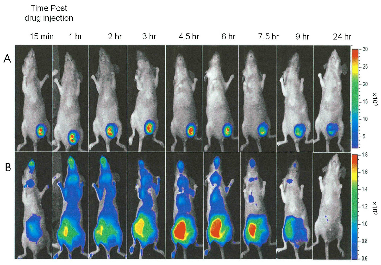
도 4는 MDA-MB-231-RFP 동소이식 종양 (크기 ∼1 cm3) 에서 화합물 13의 축적을 보여준다. 마우스에 화합물 13을 주사하였고, 주사 후 15분부터 24시간 까지 영상을 촬영하였다. 4A (윗 패널): 적색 형광 영상. 4B (아래 패널): NIR 형광 영상.
도 5는 MDA-MB-231-RFP 동소이식 종양 (크기 ∼1 cm3) 에서 화합물 13의 축적을 보여준다. 마우스에 화합물 13을 주사하였고, 주사 후 1일부터 7일까지 영상을 촬영하였다. 5A (윗 패널): 적색 형광 영상. 5B (아래 패널): NIR 형광 영상.
도 6은 MDA-MB-231-RFP 동소이식 종양 (크기 ∼0.5 cm3)에서 화합물 13의 축적을 보여준다. 마우스에 화합물 13을 주사하였고, 주사 후 20분부터 24 시간까지 영상을 촬영하였다. 6A (윗 패널): 적색 형광 영상. 6B (아래 패널): NIR 형광 영상.
도 7은 MDA-MB-231-RFP 동소이식 종양 (크기 ∼0.5 cm3)에서 화합물 13의 축적을 보여준다. 마우스에 화합물 13을 주사하였고, 주사 후 1일부터 3일까지 영상을 촬영하였다. 7A (윗 패널): 적색 형광 영상. 7B (아래 패널): NIR 형광 영상.
도 8은 종양 vs 옆측에서 화합물 13 형광 신호 측정을 보여주는 그래프이다. 형광 신호 (photon/sec/cm2)는 9마리 동물의 종양 및 옆측에서 측정하였다. 결과는 화합물 13 주사 후 15분부터 216 시간까지 취합하였다. 각 시점에서 모든 동물에서 얻은 평균 결과 및 종양과 옆측간 형광의 비를 나타낸다.
도 9는 MDA-MB-231-RFP 동소 종양 (크기 ∼1 cm3)에서 화합물 24의 축적을 보여준다. 마우스에 화합물 24을 주사하고 주사 후 1시간부터 24시간까지 영상을 촬영하였다. 9A (윗 패널): 적색 형광 영상. 9B (아래 패널): NIR 형광 영상.
도 10은 MDA-MB-231-RFP 동소 종양 (크기 ∼1 cm3)에서 화합물 24의 축적을 보여준다. 마우스에 화합물 24을 주사하고 주사 후 1일부터 7일까지 영상을 촬영하였다. 10A (윗 패널): 적색 형광 영상. 10B (아래 패널): NIR 형광 영상.
도 11은 MDA-MB-231-RFP 동소 종양 (크기 ∼0.5 cm3)에서 화합물 24의 축적을 보여준다. 마우스에 화합물 24을 주사하고 주사 후 20분부터 24시간까지 영상을 촬영하였다. 11A (윗 패널): 적색 형광 영상. 11B (아래 패널): NIR 형광 영상.
도 12는 MDA-MB-231-RFP 동소 종양 (크기 ∼0.5 cm3)에서 화합물 24의 축적을 보여준다. 마우스에 화합물 24를 주사하고 주사 후 1일부터 2일까지 영상을 촬영하였다. 12A (윗 패널): 적색 형광 영상. 12B (아래 패널): NIR 형광 영상.
도 13은 MLS-mBanana 동소 종양 (크기 ∼1 cm3)에서 화합물 13의 축적을 보여준다. 마우스를 화합물 13으로 주사하고 주사 후 1시간부터 24시간까지 영상을 촬영하였다. 13A (윗 패널): 적색 형광 영상. 13B (아래 패널): NIR 형광 영상.
도 14는 MLS-mBanana 동소 종양 (크기 ∼1 cm3)에서 화합물 13의 축적을 보여준다. 마우스를 화합물 13으로 주사하고 주사 후 1일부터 4일까지 영상을 촬영하였다. 14A (윗 패널): 적색 형광 영상. 14B (아래 패널): NIR 형광 영상.
도 15는 MLS-mBanana 동소 종양 (크기 ∼0.5 cm3)에서 화합물 13의 축적을 보여준다. 마우스를 화합물 13으로 주사하고 주사 후 10분부터 24시간까지 영상을 촬영하였다. 15A (윗 패널): 적색 형광 영상. 15B (bottom panel): (아래 패널): NIR 형광 영상.
도 16은 MLS-mBanana 동소 종양 (크기 ∼0.5 cm3)에서 화합물 13의 축적을 보여준다. 마우스를 화합물 13으로 주사하고 주사 후 1일부터 4일까지 영상을 촬영하였다. 16A (윗 패널): 적색 형광 영상. 16B (아래 패널): NIR 형광 영상.
도 17은 인간 난소 MLS-mBanana 원발 괴사 및 비괴사 종양에서 화합물 13의 축적을 비교를 보여준다. 화합물 13 주사 2일 후에 영상을 촬영하였다. 화합물 13의 생체내 전신 NIR 형광 영상을 촬영하였다. 17A: 비괴사 종양 (∼0.5 cm3). 17B: 괴사 종양 (∼1 cm3).
도 18은 MDA-MB-231-RFP 동소 종양 (크기 ∼1 cm3)에서 화합물 25의 축적을 보여준다. 마우스를 화합물 25로 주사하고 주사 후 5분부터 24시간까지 영상을 촬영하였다. 18A (윗 패널): 적색 형광 영상. 18B (아래 패널): NIR 형광 영상.
도 19는 MDA-MB-231-RFP 동소 종양 (크기 ∼1 cm3)에서 화합물 25의 축적을 보여준다. 마우스를 화합물 25로 주사하고 주사 후 1일부터 3일까지 영상을 촬영하였다. 19A (윗 패널): 적색 형광 영상. 19B (아래 패널): NIR 형광 영상.
도 20은 MDA-MB-231-RFP 동소 종양 (크기 ∼0.5 cm3)에서 화합물 25의 축적을 보여준다. 마우스를 화합물 25로 주사하고 주사 후 10분부터 24시간까지 영상을 촬영하였다. 20A (윗 패널): 적색 형광 영상. 20B (아래 패널): NIR 형광 영상.
도 21은 MDA-MB-231-RFP 동소 종양 (크기 ∼0.5 cm3)에서 화합물 25의 축적을 보여준다. 마우스를 화합물 25로 주사하고 주사 후 1일부터 3일까지 영상을 촬영하였다. 21A (윗 패널): 적색 형광 영상. 21B (아래 패널): NIR 형광 영상.
도 22는 동소 인간 유방 원발 종양 (크기 ∼0.5 cm3) MDA-MB-231-RFP에서 자유 c(RGDfK) 투여 후 1시간에 투여된 화합물 13의 축적 경쟁 분석을 보여준다. 영상은 화합물 13 투여 후 24시간에 촬영되었다. 22A, 22B - 적색 형광 영상; 22C, 22D -NIR 형광 영상, 22A, 22C: 자유 c(RGDfK) 투여 후 1시간에 화합물 13 투여 (경쟁), 22B, 22D: 대조군, 화합물 13만 투여.
도 23은 약물 주사 후 10분에 측정된, MDA-MB-231-RFP 동소 종양의 살아있는 부위 vs 괴사 부위에서 화합물 13의 축적을 보여준다. 23A, 23B 및 23C (윗 패널)은 각각 완전한 동물의 생체내 전신 사진, 적색 형광 영상 및 NIR 형광 영상; 23D, 23E 및 23F (아래 패널)은 각각 잘라낸 종양 (종양은 반으로 자름)의 사진, 적색 형광 영상 및 NIR 형광 영상 (ND -괴사 부위, VD -살아있는 부위).
도 24는 약물 주사 후 1시간에 측정된, MDA-MB-231-RFP 동소 종양의 살아있는 부위 vs 괴사 부위에서 화합물 13의 축적을 보여준다. 24A, 24B 및 24C (윗 패널) 및 24D, 24E 및 24F (아래 패널)은 각각 도 23A, 23B와 23C 및 23D, 23E와 23F 에서 설명한 바와 같다.
도 25는 약물 주사 후 4시간에 측정된, MDA-MB-231-RFP 동소 종양의 살아있는 부위 vs 괴사 부위에서 화합물 13의 축적을 보여준다. 25A, 25B 및 25C (윗 패널) 및 25D, 25E 및 25F (아래 패널)은 각각 도 23A, 23B와 23C 및 23D, 23E와 23F 에서 설명한 바와 같다.
도 26은 약물 주사 후 24시간에 측정된, MDA-MB-231-RFP 동소 종양의 살아있는 부위 vs 괴사 부위에서 화합물 13의 축적을 보여준다. 26A, 26B 및 26C (윗 패널) 및 26D, 26E 및 26F (아래 패널)은 각각 도 23A, 23B와 23C 및 23D, 23E와 23F 에서 설명한 바와 같다.
도 27은 약물 주사 후 3일에 측정된, MDA-MB-231-RFP 동소 종양의 살아있는 부위 vs 괴사 부위에서 화합물 13의 축적을 보여준다. 27A, 27B 및 27C (윗 패널) 및 27D, 27E 및 27F (아래 패널)은 각각 도 23A, 23B와 23C 및 23D, 23E와 23F 에서 설명한 바와 같다.
도 28은 약물 주사 후 5일에 측정된, MDA-MB-231-RFP 동소 종양의 살아있는 부위 vs 괴사 부위에서 화합물 13의 축적을 보여준다. 28A, 28B 및 28C (윗 패널) 및 28D, 28E 및 28F (아래 패널)은 각각 도 23A, 23B와 23C 및 23D, 23E와 23F 에서 설명한 바와 같다.
도 29는 약물 주사 후 7일에 측정된, MDA-MB-231-RFP 동소 종양의 살아있는 부위 vs 괴사 부위에서 화합물 13의 축적을 보여준다. 29A, 29B 및 29C (윗 패널) 및 29D, 29E 및 29F (아래 패널)은 각각 도 23A, 23B와 23C 및 23D, 23E와 23F 에서 설명한 바와 같다.
도 30은 약물 주사 후 9일에 측정된, MDA-MB-231-RFP 동소 종양의 살아있는 부위 vs 괴사 부위에서 화합물 24의 축적을 보여준다. 30A, 30B 및 30C (윗 패널) 및 30D, 30E 및 30F (아래 패널)은 각각 도 23A, 23B와 23C 및 23D, 23E와 23F 에서 설명한 바와 같다.
도 31은 주사 후 7일에 측정된, MLS-mBanana 종양의 중앙 괴사부위에서 화합물 13의 축적을 보여준다. 31A, 31B 및 31C (윗 패널) 및 31D, 31E 및 31F (아래 패널)은 각각 도 23A, 23B와 23C 및 23D, 23E와 23F 에서 설명한 바와 같다.
도 32는 주사 후 7일에 측정된, MLS-mBanana 종양의 비 중앙괴사 부위에서 화합물 13의 축적을 보여준다. 32A, 32B 및 32C (윗 패널) 및 32D, 32E 및 32F (아래 패널)은 각각 도 23A, 23B와 23C 및 23D, 23E와 23F 에서 설명한 바와 같다.
도 33은 잘라낸 MDA-MB-231-RFP 종양 단면의 영상 및 조직학적 분석 ((H&E 염색)을 보여준다. 33A: 종양 단면 표면의 사진 (육안 관찰 모습) 33B: 단면 표면의 조직학적 관찰 (현미경 관찰 모습). 33C: 33B의 박스친 부위를 중간배율로 관찰한 사진. 33D: 괴사와 살아있는 조직의 경계 영역을 고배율로 관찰한 사진.
도 34는 인간 MDA-MB-231-RFP의 PDT에 대한 대표적 국소적 반응의 예를 보여준다. MDA-MB-231-RFP를 등에 이종이식(∼0.5 cm3)한 마우스에 화합물 13을 7.5 mg/kg의 양으로 정맥주사하였고 8시간 후에 피부를 통하여 조사하였다. 34A: 0일째 (치료 전)와 치료 후 1, 4, 7, 12 및 90 일째 촬영, 34B: 생체내 전신 적색 형광 영상.Figure 1 shows the plasmid used for tumor cell line transfer transfection with the red fluorescent protein (RFP). 1A: pDsRed2-N1 plasmid (Clontech, Palo Alto, CA), 1B: pDsRed-Monomer-Hyg-Cl (Clontech, Palo Alto, Calif.
Figures 2A and 2B show fluorescence clones transferred into the plasmid of Figure 1 and detected by fluorescence microscopy (Nikon, magnification X10, 3 second exposure). 2A: MDA-MB-231 RFP clone 1 (G418 resistance). 2B: MDA-MB-231 RFP clone 3 (hygromycin resistance).
Figure 3 shows cross-sectional images and histological analysis (H & E staining) of cut MDA-MB-231-RFP tumors. 3A: Large tumor (~ 1 cm 3 ), 3B: Small tumor (~ 0.5 cm 3 ) (ND - necrotic area, VD - live area).
Figure 4 shows the accumulation of
Figure 5 shows the accumulation of
Figure 6 shows accumulation of
Figure 7 shows the accumulation of
Figure 8 is a graph showing the measurement of
Figure 9 shows the accumulation of
Figure 10 shows the accumulation of
Figure 11 shows the accumulation of
Figure 12 shows the accumulation of
Figure 13 shows the accumulation of
Figure 14 shows the accumulation of
Figure 15 shows the accumulation of
Figure 16 shows the accumulation of
Figure 17 shows a comparison of the accumulation of
Figure 18 shows accumulation of
Figure 19 shows the accumulation of
Figure 20 shows the accumulation of
Figure 21 shows the accumulation of
22 shows the accumulation competition analysis of
Figure 23 shows the accumulation of
Figure 24 shows the accumulation of
Figure 25 shows the accumulation of
Figure 26 shows the accumulation of
Figure 27 shows the accumulation of
Figure 28 shows the accumulation of
Figure 29 shows the accumulation of
Figure 30 shows the accumulation of
Figure 31 shows the accumulation of
Figure 32 shows the accumulation of
Figure 33 shows an image and histological analysis (H & E staining) of cut section of MDA-MB-231-RFP tumor 33A: photograph of the cross-sectional surface of the tumor (visual observation) 33B: histological observation of the cross- 33C: A photograph of the boxed area of 33B at an intermediate magnification. 33D: A photograph of the border area of necrosis and living tissue at a high magnification.
Figure 34 shows an example of a representative local response to PDT of human MDA-MB-231-RFP.
본 발명의 주 목적은 민감제를 괴사성 종양의 괴사부위에 대해 특이적으로 표적하는 감광제의 컨쥬게이트(conjugates)를 제공하는 것이다. 통상의 종양을 표적으로 하는 화학요법과 비교하여 종양-표적 광역학 치료요법(tumor-targeted photodynamic therapy, PDT)은 몇 가지 장점이 있다. 첫째는 표적화된 종래의 약물이 축적되는 동안에도, 전구약물이 아닌 경우를 제외하고는 활성이 있지만, 표적화된 감광제의 경우는 국소 광조사 전까지는 활성을 나타내지 않는 특징이 있다. 둘째의 장점은 표적화된 종래의 약물은 귀소신호를 제시하는 원치않는 표적에도 결합하여 반응하나, 상기 표적화된 감광제는 오로지 광조사 부위에서만 활성을 나타낼 것이다.The main object of the present invention is to provide conjugates of photosensitizers that specifically target sensitizing agents to necrotic areas of necrotizing tumors. Tumor-targeted photodynamic therapy (PDT) has several advantages over conventional chemotherapy targeting tumors. The first is that while the targeted conventional drug is being accumulated, it is active except for the case where it is not a prodrug, but in the case of the targeted photosensitizer, it does not show activity until the local irradiation. A second advantage is that the targeted conventional drug will also react and bind to an unwanted target presenting a signal of interest, but the targeted photosensitizer will only be active at the irradiated site.
따라서, 광의의 관점에서 본 발명은 RGD 함유 펩타이드 또는 RGD 펩타이드모방체와 클로로필 또는 박테리오클로로필 감광제의 컨쥬게이트의 괴사 종양에 대한 최소 침습적 종양-표적 영상, 종양-표적 광역학 요법 (PDT) 및/또는 온라인 예후에 대한 용도에 관한 것이다.Thus, in light of the present invention, the present invention provides a method of minimally invasive tumor-target imaging, tumor-target photodynamic therapy (PDT) and / or tumor necrosis of necrotic tumors of RGD containing peptide or RGD peptide mimetics and a conjugate of chlorophyll or bacteriophyllophore And the use thereof for online prognosis.
상기의 용어 "RGD 함유 펩타이드" 또는 "RGD 펩타이드" 는 RGD 모티브로 불리는 Arg-Gly-Asp(RGD) 서열을 포함하는 펩타이드로서, 본 명세서에서 서로 혼용되어 사용된다. 상기의 용어 "RGD 펩타이드 모방체" 는 화합물 특히 비펩타이드성 화합물로서, 펩타이드를 모방하고 RGD 모티브를 가진 화합물을 칭한다.The above-mentioned term "RGD-containing peptide" or "RGD peptide" is a peptide containing an Arg-Gly-Asp (RGD) sequence called RGD motif. The term "RGD peptide mimetic" is a compound, particularly a non-peptide compound, which refers to a compound that mimics a peptide and has an RGD motif.
RGD 펩타이드는 세포의 인테그린(integrin) 수용체와 반응하여, 세포 내 신호전달과정을 개시할 잠재력을 가지고 있으며, 많은 질병에 영향을 주는 것으로 알려져 있다. 이러한 이유로 인테그린 내 RGD 결합 부위는 관심을 끄는 약리학적 표적으로 간주되어 왔다.RGD peptides have the potential to interact with integrin receptors in cells, initiate intracellular signaling processes, and are known to affect many diseases. For this reason, the RGD binding site in the integrins has been regarded as a pharmacological target of interest.
상기 RGD함유 펩타이드는 4-100 개, 바람직하게는 5-50, 5-30, 5-20개, 더욱 바람직하게는, 5-10개의 아미노산 잔기이다. 바람직한 구현예에서, 상기 RGD 펩타이드는 4개, 5개, 6개, 7개, 9개 또는 25개의 아미노산, 보다 바람직하게는 5개의 아미노산으로 구성된다.The RGD-containing peptide is 4-100, preferably 5-50, 5-30, 5-20, more preferably 5-10 amino acid residues. In a preferred embodiment, the RGD peptide is composed of 4, 5, 6, 7, 9 or 25 amino acids, more preferably 5 amino acids.
본원에서의 용어 "아미노산" 은 20종의 천연의 아미노산 및 비천연의 아미노산을 포함한다.The term "amino acid" as used herein includes 20 naturally occurring amino acids and non-naturally occurring amino acids.
본원에 적당한 상기 천연의 아미노산의 예로는 Ala, Arg, Asp, Asn, Cys, His, Gln, Glu, Gly, Ile, Leu, Lys, Met, Phe, Pro, Ser, Thr, Trp, Tyr, 및 Val를 예로 들 수 있으며, 이에 제한된 것은 아니다.Examples of such natural amino acids suitable for the present invention include Ala, Arg, Asp, Asn, Cys, His, Gln, Glu, Ile, Leu, Lys, Met, Phe, Pro, Ser, Thr, Trp, For example, but are not limited thereto.
비천연의 아미노산의 예는 4-아미노부틸산(Abu, 4-aminobutyric acid), 2-아미노아디프산(2-aminoadipic acid), 디아미노프로피온산(Dap, diaminopropionic acid), 하이드록시라이신(hydroxylysine), 호모세린(homoserine), 호모발린(homovaline), 호모류신(homoleucine), 노르류신(Nle, norleucine), 노르발린(Nva, norvaline), 오르니틴(Orn, ornithine), TIC, 나프틸알라닌(Nal, naphthylalanine), 페닐알라닌(Phe)의 환상 메틸화 유도체, 페닐알라닌(Phe)의 할로겐화 유도체 또는 O-메틸-타이로신(o-methyl-Tyr)을 들 수 있으며, 이에 제한된 것은 아니다.Examples of unnatural amino acids include, but are not limited to, 4-aminobutyric acid, 2-aminoadipic acid, Dap, diaminopropionic acid, hydroxylysine, , Homoserine, homovaline, homoleucine, Nle, norleucine, Nva, norvaline, Orn, ornithine, TIC, naphthylalanine (Nal , naphthylalanine, cyclic methylated derivatives of phenylalanine (Phe), halogenated derivatives of phenylalanine (Phe), or o-methyl-tyrosine.
본 명세서에서의 상기 용어 "아미노산" 은 in vivo에서 번역 후 일어나는 변형 등과 같은 변형된 아미노산을 또한 포함하며, 예를 들면, 하이드록시프롤린(hydroxyproline), 포스포세린(phosphoserine) 및 포스포트레오닌(phosphothreonine); D-변형(D-modifications); 아미노 말단기 또는 라이신(Lys)의 자유 아미노기의 아실화 또는 알킬화, 바람직하게는 메틸화; 카르복시 말단기 또는 Asp 또는 Glu의 자유 카르복시기의 에스테르화 또는 아미드화; 및 Ser 또는 Tyr의 하이드록실기의 에테르화 또는 에스테르화에 의한 변형을 포함한다.The term "amino acid" as used herein also includes modified amino acids such as those occurring post-translationally in vivo , such as hydroxyproline, phosphoserine and phosphothreonine, ; D-modifications; Acylation or alkylation, preferably methylation, of the free amino group of the amino end group or lysine (Lys); Esterification or amidation of free carboxyl groups of carboxy end groups or Asp or GIu; And etherification or esterification of the hydroxyl group of Ser or Tyr.
상기 "아미노산" 은 D형 및 L형의 아미노산을 포함한다. 따라서, 본 발명의 컨쥬게이트에 채용되는 펩타이드는 모두-D (글라이신은 제외), 모두-L, 또는 L,D-아미노산일 수 있다. 아미노산의 D 변형(D-modifications) 및 펩타이드 결합의 N-알킬화는 개체의 효소에 의하여 펩타이드 절단 방지에 가장 효과적이다. 본 발명에서는 D-아미노산은, 예를 들어 본 발명의 SEQ ID NO: 1로 표시되는 cycloRGDfK에서 D-페닐알라닌을 'f' 로 기재한 것과 같이, 소문자로 표시한다.The "amino acid" includes D-type and L-type amino acids. Thus, all of the peptides employed in the conjugates of the present invention may be -D (except for glycine), all-L, or L, D-amino acids. D-modifications of amino acids and N-alkylation of peptide bonds are most effective in preventing peptide cleavage by individual enzymes. In the present invention, the D-amino acid is represented by a lowercase letter, for example, in the cycloRGDfK represented by SEQ ID NO: 1 of the present invention, in which D-phenylalanine is described as 'f'.
본 발명은 또한 시클릭 펩타이드를 포함한다. 펩타이드는 디설파이드, 설파이드, 또는 특히 N 말단 및 C 말단, 또는 아미노산 측쇄(side chain)의 카르복실기와 아미노 작용기 간의 락탐(lactam) 형성 등과 같은 다양한 방법에 의하여 고리화(Cyclization)될 수 있다. 고리화는 당업계에 알려진 방법, 예를 들면 아미드 결합 형성으로 제조할 수 있으며, 예를 들어, Glu, Asp, Lys, Orn, 디아미노부틸산(diamino butyric acid, Dab), 디아미노프로피온산(Dap)을 펩타이드 사슬의 다양한 위치에 삽입(-CO-NH 또는 -NH-CO 결합)함으로써, 아미드 결합에 의해 제조할 수 있다. 골격 사이의 고리화는 또한 H-N((CH2)n-COOH)-C(R)H-COOH 또는 H-N((CH2)n-NH2)C(R)H-COOH의 화학식으로 표시되는 변형된 아미노산을 삽입함으로써, 제조할 수 있다. 이 때, 상기 화학식의 n은 1-4 이며, 이와 더불어 R은 임의의, 아미노산의 천연의 또는 비천연 측쇄이다.The present invention also includes cyclic peptides. The peptide can be cyclized by a variety of methods such as disulfide, sulfide, or lactam formation, especially between the N-terminal and C-terminal, or between the carboxyl and amino functional groups of the amino acid side chain. The cyclization can be carried out by methods known in the art, for example, by amide bond formation, for example, Glu, Asp, Lys, Orn, diamino butyric acid (Dab), diaminopropionic acid ) At various positions in the peptide chain (-CO-NH or -NH-CO bond). The cyclization between the skeletons can also be carried out in a variant represented by the formula HN ((CH 2 ) n -COOH) -C (R) H-COOH or HN ((CH 2 ) n -NH 2 ) C By inserting the resulting amino acid. Wherein n in the above formula is 1-4, and wherein R is any natural or non-natural side chain of any amino acid.
고리화는 또한 2개의 Cys 잔기를 도입하여 S-S 결합을 통해 수득할 수 있다. 부가적인 측쇄간의 고리화는 또한 화학식 -(CH2)n -S-CH2-CO-(n=1 또는 2)의 상호작용하는 결합을 통하여 제조할 수 있으며, 위 식에서 n= 1 또는 2이며, 이는 예를 들어, Cys 또는 호모시스테인(homoCys)을 삽입하여, 이의 자유 SH기와 브롬아세틸화된 라이신(Lys), Orn, Dab 또는 Dap과의 반응에 의해 수득하는 것이 가능하다.The cyclization can also be obtained via SS bonding by introducing two Cys residues. Additional cyclisation between the side chains can also be prepared via an interactive linkage of the formula - (CH 2 ) n -S-CH 2 -CO- (n = 1 or 2) wherein n = 1 or 2 , Which can be obtained, for example, by inserting Cys or homocysteine (homoCys) and reacting it with its free SH group and bromoacetylated lysine (Lys), Orn, Dab or Dap.
본 발명의 일부 양태에서, 상기 RGD 펩타이드는 US6,576,239, EP 0927045 및 WO 2008/023378에 기재된 것일 수 있으며, 참조에 의해 그 전문이 기술된 것과 같이 본 명세서에 포함된다.In some embodiments of the invention, the RGD peptide may be as described in US 6,576,239, EP 0927045 and WO 2008/023378, which is hereby incorporated by reference as if fully set forth.
본 발명의 바람직한 일 양태에 따르면, 본 발명에서 따른 상기 펩타이드는 SEQ ID NO:1로 표시되는 시클릭 펜타펩타이드(pentapeptide)인 RGDfK이며, 이 때 상기 "f" 는 D-Phe 잔기를 나타낸다.According to a preferred embodiment of the present invention, the peptide according to the present invention is RGDfK which is a cyclic pentapeptide represented by SEQ ID NO: 1, wherein "f" represents a D-Phe residue.
본 발명의 바람직한 다른 양태에 따르면, 상기 펩타이드는 SEQ ID NO:2로 표시되는 시클릭 펜타펩타이드인 RADfK로서, RGD 모티브가 인테그린 수용체에 결합하는 중요한 특성을 용이하게 하기 위해 사용한다. 본 발명에서 유용한 추가의 시클릭 펩타이드는 본 명세서에서 'RGD-4C' 로 표시되며, SEQ ID NO:9의 서열을 갖는 노나펩타이드(nonapeptide)인 CDCRGDCGC로서, 상기 펩타이드는 4개의 시스테인 잔기를 포함하고 있고, 이에 의하여 2개의 디설파이드 결합을 형성한다.According to another preferred embodiment of the present invention, the peptide is R A DfK, a cyclic pentapeptide represented by SEQ ID NO: 2, which is used to facilitate the important property that the RGD motif binds to the integrin receptor. A further cyclic peptide useful in the present invention is CDCRGDCGC, designated herein as 'RGD-4C', which is a nonapeptide having the sequence of SEQ ID NO: 9, wherein the peptide comprises four cysteine residues Thereby forming two disulfide bonds.
RGD 모티브의 아스파르트산은 화학적으로 분해되기가 매우 쉽고, 이는 생물학적 활성이 소실되는 원인이 되기 때문에, 디설파이드 결합에 의한 고리화로 이를 예방할 수 있다. 이러한 안정성 향상과 더불어 시클릭 펩타이드는 선형 펩타이드보다 세포의 비트로넥틴에 대한 결합을 저해하는 활성이 월등히 우수하다. 또한 합성된 펩타이드에서 RGD 서열을 둘러싼(flanking) 잔기의 수와 특성은 그 서열이 개별 인테그린 수용체에 의해 어떻게 인지되는가에 대해 상당한 영향을 미친다. 방향족 잔기는 특히 인테그린의 결합부위에서의 우호적 접촉에 있어 상당히 중요한 역할을 하는 것으로 파악된다. αvβ3 를 타겟팅하는 시클릭 RGD 펩타이드는 인테그린과 상관없는 액상내포작용 (fluid-phase endocytosis) 을 통하여 세포 내로 유입되는데, 따라서 세포 표면의 기능성 인테그린 수용체의 수에는 변화가 없다. 이와 더불어, 시클릭 RGD 펩타이드는 리소좀에 존재하거나, 또는 리보좀에 유입된 지 15분 후 포화상태 과정에서 분해되는데, 그 중 일부만이 리소좀에서 세포질로 유입될 수 있다.The aspartic acid of the RGD motif is very easy to decompose chemically, which causes the disappearance of the biological activity, so it can be prevented by cyclization by disulfide bonds. In addition to this stability improvement, cyclic peptides are far superior to linear peptides in their ability to inhibit binding of cells to bitronectin. The number and nature of flanking residues in RGD sequences in synthetic peptides also have a significant impact on how the sequence is recognized by individual integrin receptors. Aromatic residues are found to play a particularly important role in friendly contact, particularly at the binding sites of integrins. The cyclic RGD peptide targeting α v β 3 is introduced into cells via fluid-phase endocytosis independent of the integrins, and thus the number of functional integrin receptors on the cell surface does not change. In addition, cyclic RGD peptides are present in the lysosomes or are degraded in the
본 발명의 다른 바람직한 양태에 따르면, RGD 펩타이드는 (i) 테트라펩타이드 cycloRGDK(SEQ ID NO: 3), (ii) 펜타펩타이드 cycloRGDf-n(Me)K (SEQ ID NO: 4), 이 때 상기 f는 D-Phe 이며, f와 K의 펩타이드 결합은 메틸화 되어 있음; 및 (iii) y는 D-Tyr인 펜타펩타이드 cycloRGDyK(SEQ ID NO: 5) 의 시클릭 펩타이드로부터 선택된다.According to another preferred embodiment of the present invention, the RGD peptide comprises (i) the tetrapeptide cycloRGDK (SEQ ID NO: 3), (ii) the pentapeptide cycloRGDf-n (Me) K (SEQ ID NO: Is D-Phe, the peptide bond of f and K is methylated; And (iii) y is selected from cyclic peptides of the pentapeptide cycloRGDyK (SEQ ID NO: 5) which is D-Tyr.
본 발명의 다른 양태에 따르면, RGD함유 펩타이드는 선형이며, 헥사펩타이드 GRGDSP(SEQ ID NO: 6), 헵타펩타이드 GRGDSPK(SEQ ID NO: 7), 및 25 mer인 GRGDSP)4K(SEQ ID NO: 8)로부터 선택될 수 있으며, 본 발명의 더욱 바람직한 일 양태에 따르면, 상기 선형 펩타이드는 GRGDSP이다.In accordance with another aspect of the invention, RGD-containing peptide is linear, and the hexapeptide GRGDSP (SEQ ID NO: 6) , hepta peptide GRGDSPK (SEQ ID NO: 7) , and a 25 mer of GRGDSP) 4 K (SEQ ID NO : 8). According to a further preferred embodiment of the present invention, the linear peptide is GRGDSP.
본 발명의 일 양태에 따르면, 상기 RGD 펩타이드는 감광제인 클로로필 또는 박테리오클로로필 거대고리에 직접 연결되며, 이는 그 주변의 작용기, 예를 들면 COOH 가 RGD 펩타이드의 자유 아미노기 또는 아미노 말단과 반응하여 아미드 CO-NH2 형성을 통해 가능하다.According to one aspect of the present invention, the RGD peptide is directly linked to a chlorophyll or a bacteriophile chlorophylic macrocyclic ring, which reacts with a free amino group or amino terminal of the RGD peptide to form an amide CO- NH 2 formation.
본 발명의 다른 양태에 따르면, 상기 RGD 펩타이드는 감광제 거대고리에 스페이서 암(spacer arm)/연결기(bridging group)을 통해 연결되며, 이는 스페이서 암/연결기 예컨대 이로 제한되는 것은 아니나, C1-C25 하이드로카르빌렌, 바람직하게는 C1-C10 알킬렌 또는 페닐렌이, OH, COOH, SO3H, COSH 또는 NH2와 같은 말단 작용기로 치환되고, 이어서 에테르, 에스테르, 아미드, 티오아미드 또는 설폰아미드기 형성을 통해 가능하다.According to another embodiment of the present invention, the RGD peptide is connected to a photosensitizer macrocycle via a spacer arm / bridging group, which may include, but is not limited to, C 1 -C 25 Hydrocarbylene, preferably C 1 -C 10 alkylene or phenylene, is replaced by a terminal functional group such as OH, COOH, SO 3 H, COSH or NH 2 , followed by an ether, ester, amide, thioamide or sulfone Amide group formation.
본 발명의 일부 양태에서, 상기 감광제는 RGD 펩타이드 모방체에 컨쥬게이트된다.In some embodiments of the invention, the photosensitizer is conjugated to an RGD peptide mimic.
본 발명의 바람직한 일 양태에 따르면, 상기 RGD 펩타이드 모방체는 비펩타이드성 화합물로서, 11 원자, 그 중 최소 5개는 탄소원자인 사슬로 간격지워진(spaced) 구아니딘 및 카르복실 말단기를 포함하며, 상기 사슬은 하나 이상의 산소(O), 황(S) 또는 질소(N) 분자를 포함하며, 또한 선택적으로 옥소(oxo), 티오옥소(thioxo), 할로겐(halogen), 아미노(amino), C1-C6 알킬, 하이드록실, 또는 카복시로 치환될 수 있거나, 상기 사슬의 하나 이상의 원자는 3-6 원의 카르보시클릭, 또는 헤테로시클릭 고리를 형성할 수 있다. 상기의 화합물은 동일 출원인에 의한 WO 93/09795 및 WO 2008/023378에 기재되어 있으며, 참조에 의해 그 전문이, 온전히 기술된 것과 마찬가지로 본 명세서에 포함된다.According to a preferred embodiment of the present invention, the RGD peptide mimetic is a non-peptide compound, which comprises 11 atoms, at least five of which are spaced apart from a carbon atom, and guanidine and carboxyl end groups, chain comprises at least one oxygen (O), sulfur (S) or nitrogen (N) molecules, also optionally oxo (oxo), thio-oxo (thioxo), halogen (halogen), amino (amino), C 1 - C 6 alkyl, hydroxyl, or carboxy, or at least one atom of the chain may form a 3-6 member carbocyclic, or heterocyclic ring. Such compounds are described in WO 93/09795 and WO 2008/023378 by the same applicants and are hereby incorporated by reference as if fully set forth.
본 발명의 바람직한 양태에 따르면, 상기 RGD 펩타이드모방체는 사슬에 N 원자를 포함하며, oxo 기로 치환된다. 본 발명의 보다 바람직한 양태에 따르면, 상기 RGD 펩타이드모방체는 다음의 화학식을 가진다:According to a preferred embodiment of the present invention, the RGD peptide mimetic includes an N atom in the chain and is substituted with an oxo group. According to a more preferred embodiment of the present invention, the RGD peptide mimetic has the following formula:
H2N-C(=NH)NH-(CH2)5-CO-NH-CH(CH2)-(CH2)2-COOH 또는 -NH-RGD-CO-NH-(CH2)2-피페라지노-(CH2)2-NH-. H 2 NC (= NH) NH- (CH 2) 5 -CO-NH-CH (CH 2) - (CH 2) 2 -COOH , or -NH-RGD-CO-NH- ( CH 2) 2 - Piperacillin Gino - (CH 2) 2 -NH-.
본 발명에서 사용되는 감광제는 클로로필 또는 박테리오클로로필 유도체로서, 클로로필 또는 박테리오클로로필의 천연의 또는 합성된 비천연 유도체 일 수 있으며, 이는 거대고리 내 및/또는 주변에 변형이 있는 화합물, 및/또는 중심부의 Mg 원자가 없거나, 진단 및/또는 PDT 를 목적으로 다른 적합한 금속분자로 치환된 화합물을 포함한다.The photosensitizer used in the present invention may be a chlorophyll or a bacteriol chlorophyll derivative, a natural or synthetic non-natural derivative of chlorophyll or bacteriophyllophyll, which may be a compound having a modification in and / or around the macrocyclic ring, and / Mg atoms free, or substituted with other suitable metal molecules for diagnostic and / or PDT purposes.
본 발명의 바람직한 양태에 따르면, 본 발명은 감광제가 다음의 화학식 I, II, 또는 III 으로 표시되는 클로로필 또는 박테리오클로로필의 컨쥬게이트(conjugate) 및 그 약학적으로 허용가능한 염 및 그 광학 이성질체 관한 것이다:According to a preferred embodiment of the present invention, the present invention relates to a conjugate of chlorophyll or bacteriocin chlorophyl, and pharmaceutically acceptable salts and optical isomers thereof, wherein the photosensitizer is represented by the following formula (I), (II)
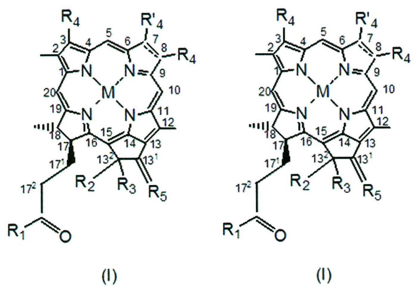

상기 화학식에서,In the above formulas,
M 은 2H, 또는 Mg, Pd, Pt, Co, Ni, Sn, Cu, Zn, Mn, In, Eu, Fe, Au, Al, Gd, Dy, Er, Yb, Lu, Ga, Y, Rh, Ru, Si, Ge, Cr, Mo, P, Re, Tl 그리고 Tc, 및 아이소토프 및 그 방사선 아이소토프로 구성되는 군으로부터 선택되는 원자이며;M is 2H or an element selected from the group consisting of Mg, Pd, Pt, Co, Ni, Sn, Cu, Zn, Mn, In, Eu, Fe, Au, Al, Gd, Dy, Er, Yb, Lu, Ga, , Si, Ge, Cr, Mo, P, Re, Tl and Tc, and an isotope and its radiation isotope;
X 는 O 또는 N-R7;X is O or NR < 7 & gt ;;
R1, R' 2 및 R6 각각은 독립적으로 Y-R8, -NR9R' 9 또는 -N+R9R' 9R" 9 A-; 또는 화학식 II내의 R1 및 R6 는 이들이 연결된 탄소분자와 RGD 펩타이드 또는 펩타이드 모방체를 포함하는 고리를 형성하고;Each of R 1 , R ' 2 and R 6 is independently YR 8 , -NR 9 R' 9 or -N + R 9 R ' 9 R " 9 A - or R 1 and R 6 in formula Forming a ring comprising a molecule and an RGD peptide or peptide mimetic;
Y 는 O 또는 S;Y is O or S;
R2 는 H, OH 또는 COOR9;R 2 is H, OH or COOR 9 ;
R3 또는 H, OH, C1-C12 알킬 또는 C1-C12 알콕시;R 3 or H, OH, C 1 -C 12 alkyl or C 1 -C 12 alkoxy;
R4 은 -CH=CR9R'9, -CH=CR9Hal, -CH=CH-CH2-NR9R' 9, -CH=CH-CH2- N+R9R' 9R" 9 A-, -CHO, -CH=NR9, -CH=N+R9R' 9 A-, -CH2-OR9, -CH2-SR9, -CH2-Hal, -CH2-R9, -CH2-NR9R'9, -CH2-N+R9R' 9R" 9 A-, -CH2-CH2R9, -CH2-CH2Hal, -CH2-CH2OR9, -CH2-CH2SR9, -CH2-CH2-NR9R' 9, -CH2-CH2-N+R9R' 9R" 9 A-, -COCH3, C(CH3)=CR9R'9, -C(CH3)=CR9Hal, -C(CH3)=NR9, -CH(CH3)=N+R9R' 9 A-, -CH(CH3)-Hal, -CH(CH3)-OR9, -CH(CH3)-SR9, -CH(CH3)-NR9R' 9, -CH(CH3)-N+R9R' 9 R' 9 A-, 또는 -C≡CR9;R 4 is -CH = CR 9 R '9, -CH = CR 9 Hal, -CH = CH-CH 2 -NR 9 R' 9, -CH = CH-CH 2 - N + R 9 R '9 R " 9 A -, -CHO, -CH = NR 9, -CH = N + R 9 R '9 A -, -CH 2 -OR 9, -CH 2 -SR 9, -CH 2 -Hal, -CH 2 - R 9, -CH 2 -NR 9 R '9, -CH 2 -N + R 9 R' 9 R "9 A -, -CH 2 -CH 2 R 9, -CH 2 -CH 2 Hal, -CH 2 -CH 2 OR 9, -CH 2 -CH 2 SR 9, -CH 2 -CH 2 -NR 9 R '9, -CH 2 -CH 2 -N + R 9 R' 9 R "9 A -, -COCH 3, C (CH 3) = CR 9 R '9, -C (CH 3) = CR 9 Hal, -C (CH 3) = NR 9, -CH (CH 3) = N + R 9 R' 9 A -, -CH (CH 3) -Hal , -CH (CH 3) -OR 9, -CH (CH 3) -SR 9, -CH (CH 3) -NR 9 R '9, -CH (CH 3) -N + R 9 R '9 R ' 9 A -, or -C≡CR 9;
R' 4 는 메틸 또는 포르밀;R ' 4 is methyl or formyl;
R5 는 =O, =S, =N-R9, =N+R9R' 9 A-, =CR9R'9, 또는 =CR9-Hal;R 5 is = O, = S, = NR 9 , = N + R 9 R 9 A - , = CR 9 R 9 , or = CR 9 -Hal;
R7, R8, R9, R' 9 및 R" 9 는 각각 독립적으로:R 7 , R 8 , R 9 , R ' 9 and R " 9 are each independently:
(a) H;(a) H;
(b) C1-C25 하이드로카르빌;(b) C 1 -C 25 hydrocarbyl;
(c) C1-C25 하이드로카르빌, 바람직하게는 C1-C25 알킬, 알케닐 또는 알키닐, 보다 바람직하게는 할로겐, 니트로, 옥소, OR, SR, 에폭시, 에피티오, -CONRR' , -COR, COOR, -OSO3R, -SO3R, -SO2R, -NHSO2R, - SO2NRR' -NRR' , =N-OR, =N-NRR' , -C(=NR)-NRR' , -NR-NRR' , -(R)N-C(=NR)-NRR' , O←NR-, >C=NR, -(CH2)n-NR-COR' , -(CH2)n-CO-NRR' , -O-(CH2)n-OR, -O-(CH2)n-O-(CH2)n-R, -PRR' , -OPO3RR' , -PO2HR 및 -PO3RR' 로 구성된 군으로부터 선택된 하나 이상의 작용기로 치환된 C1-C10 또는 C1-C6 알킬이며, 이 때 상기 R 및 R' 은 각각 독립적으로 H, 하이드로카르빌 또는 헤테로시클릴이고, R' ' 은 하이드로카르빌 또는 헤테로시클릴;(c) C 1 -C 25 hydrocarbyl, preferably C 1 -C 25 alkyl, alkenyl or alkynyl, more preferably halogen, nitro, oxo, OR, SR, epoxy, epithio, , -COR, COOR, -OSO 3 R , -SO 3 R, -SO 2 R, -NHSO 2 R, -
(d) 양하전된 기, 음하전된 기, 생리적 조건에서 양하전된 기로 전환되는 염기성 기, 및 생리적 조건에서 음하전된 기로 전환되는 산성 기로 구성되는 군으로부터 선택되는 하나 이상의 작용기로 치환되는, C1-C25 하이드로카르빌, 바람직하게는 C1-C25 알킬, 보다 바람직하게는 C1-C10 또는 C1-C6 알킬;(d) a C 1 -
(e) 하나 이상 헤테로원자 및/또는 하나 이상 카르보시클릭 또는 헤테로시클릭 모이어티를 포함하는, C1-C25 하이드로카르빌, 바람직하게는 C1-C25 알킬, 보다 바람직하게는 C1-C10 또는 C1-C6 알킬;(e) one or more containing a hetero atom and / or one or more carbocyclic or heterocyclic moiety, C 1 -C 25 hydrocarbyl, preferably C 1 -C 25 alkyl, more preferably C 1 -C 10 or C 1 -C 6 alkyl;
(f) 하나 이상 헤테로원자 및/또는 하나 이상 카르보시클릭 또는 헤테로시클릭 모이어티를 포함하고, 상기 (c) 및 (d) 에서 정의된 바와 같은 하나 이상 작용기로 치환되는, C1-C25 하이드로카르빌, 바람직하게는 C1-C25 알킬, 보다 바람직하게는 C1-C10 또는 C1-C6 알킬;(f) C 1 -C 25 , optionally substituted with one or more heteroatoms and / or one or more carbocyclic or heterocyclic moieties, as defined in (c) and (d) Hydrocarbyl, preferably C 1 -C 25 alkyl, more preferably C 1 -C 10 or C 1 -C 6 alkyl;
(g) 아미노산 잔기, 펩타이드, 바람직하게는 RGD 펩타이드, 단백질, 모노사카리드, 올리고사카리드, 폴리사카리드 또는 폴리덴테이트 리간드(polydentate ligand) 및 메탈을 갖는 그 킬레이팅 복합체로 치환된 C1-C25 하이드로카르빌, 바람직하게는 C1-C25 알킬, 보다 바람직하게는 C1-C10 또는 C1-C6 알킬; 또는(g) an amino acid residue, a C 1 -C 6 -alkyl substituted with a chelating complex having a peptide, preferably an RGD peptide, a protein, a monosaccharide, an oligosaccharide, a polysaccharide or a polydentate ligand, C 25 hydrocarbyl, preferably C 1 -C 25 alkyl, more preferably C 1 -C 10 or C 1 -C 6 alkyl; or
(h) 아미노산 잔기, 펩타이드, 바람직하게는 RGD 펩타이드 또는 RGD 펩타이드모방체, 단백질, 모노사카리드, 올리고사카리드, 폴리사카리드; 또는 폴리덴테이트 리간드 및 메탈을 갖는 그 킬레이팅 복합체;(h) amino acid residues, peptides, preferably RGD peptides or RGD peptide mimetics, proteins, monosaccharides, oligosaccharides, polysaccharides; Or a chelating complex thereof having a polydentate ligand and a metal;
R7 는 추가로 -NRR' 일 수 있으며, 상기 R 및 R' 은 각각 H 또는 C1-C25 하이드로카르빌, 바람직하게는 C1-C25 알킬, 보다 바람직하게는 C1-C10 또는 C1-C6 알킬로, 음전하된 기, 바람직하게는 SO3 -로 선택적으로 치환되고;R 7 can additionally be -NRR ', wherein R and R' are each H or C 1 -C 25 hydrocarbyl, preferably C 1 -C 25 alkyl, more preferably C 1 -C 10 or C 1 -C 6 alkyl, optionally substituted with a negatively charged group, preferably SO 3 - ;
R8 은 추가로 R1, R' 2 및 R6 이 각각 독립적으로 Y-R8일 경우, H+ 또는 양이온 R+ 10 일 수 있으며,R 8 is a more R 1, R '2 and R 6 to be, be H + or a cation R + 10 is independently YR 8, respectively,
R+ 10 은 금속, 암모늄 기 또는 유기 양이온;R + 10 is a metal, an ammonium group or an organic cation;
A-은 생리적으로 허용가능한 음이온;A - is a physiologically acceptable anion;
m 은 0 또는 1 이고;m is 0 or 1;
상기 7-8 위치의 점선은 선택적 이중결합을 나타내고,The dotted line at the 7-8 position represents a selective double bond,
상기 화학식I, II 또는 III으로 표시되는 클로로필 또는 박테리오클로로필 유도체는 최소 하나의 RGD 함유 펩타이드 잔기를 포함한다.The chlorophyll or bacteriological chlorophyl derivative represented by the above formula (I), (II) or (III) contains at least one RGD-containing peptide residue.
본 발명의 일 양태에 따르면, 7-8번 위치의 상기 점선은 이중결합을 나타내며, 상기 감광제는 화학식 I, II 또는 III으로 표시되는 클로로필이다. 화학식 I의 화합물에서 M은 Mg이며, 173 번 위치의 R1은 피틸록시(phytyloxy), 132 번 위치의 R2 는COOCH3, 132 위치의 R3 는 H 원자, R5 는 O, 3번 위치의 R4 는 비닐(vinyl), 7-8번 위치의 상기 점선은 이중결합, 및 7번 위치의 R' 4 가 메틸이고 8번 위치의 R4 가 에틸이거나, 7번 위치의 R' 4 가 포르밀이고 8번 위치의 R4 가 에틸인 것은 각각 클로로필 a 및 b이며, 이들의 유도체는 상이한 금속 원자를 가질 것이고/거나 상이한 치환체 R1, R2, R3, R4, R' 4 및/또는 R5 를 가질 것이다.According to one aspect of the present invention, the dotted line at positions 7-8 represents a double bond, and the photosensitizer is chlorophyll represented by the formula (I), (II) or (III). In the compound of formula (I), M is Mg, and R 1 at the 3 -position is phytyloxy, R 2 at the 2 -position is COOCH 3 , R 3 at the 2- position is H atom, R 5 is O, R 4 in the 3-position is vinyl, the dotted line in the 7-8 position is the double bond, and R 4 in the 7-position is methyl and R 4 in 8-position is ethyl, or R '4 is formyl, and is not the R 4 is ethyl of one 8-position, each of chlorophyll a and b, their derivatives will have different metal atom and / or different substituents R 1, R 2, R 3 ,
본 발명의 다른 양태에 따르면, 7-8번 위치는 수소화된 특징이 있으며, 상기 감광제는 화학식 I, II 또는 III으로 표시되는 박테리오클로로필이다. 화학식 I의 화합물에서 M은 Mg이며, 173 번 위치의 R1은 피틸록시(phytyloxy) 또는 제라닐제라닐록시(geranylgeranyloxy), 132 번 위치의 R2 는COOCH3, 132 위치의 R3 는 H 분자, R5 는 O, 3번 위치의 R4 는 아세틸, 8번 위치의 R4 는 에틸이며, 7-8번 위치의 상기 점선은 결여된 상기 화합물은 박테리오클로로필 a이고, 이들의 유도체는 상이한 금속 원자를 가질 것이고/거나 상이한 치환체 R1, R2, R3, R4 및/또는 R5 를 가질 것이다.According to another aspect of the present invention, the position 7-8 is hydrogenated and the photosensitizer is a bacterium chlorophyll represented by the formula I, II or III. And M is Mg in the compound of formula I, 17 R 1 in position 3 is pitil hydroxy (phytyloxy) or geranyl geranyl hydroxy (geranylgeranyloxy), 13 2 at position R 2 is COOCH 3, 13 at the 2-position R 3 Is H, R 5 is O, R 4 at the 3-position is acetyl, R 4 is ethyl at the 8-position, the above-mentioned compound lacking the dotted line at 7-8 position is bacteriorochlorophyll a, Will have different metal atoms and / or will have different substituents R 1 , R 2 , R 3 , R 4 and / or R 5 .
본 발명에서 사용하는 용어 "하이드로카르빌" 은 임의의, 1-25 탄소원자, 바람직하게는 1 내지 20, 보다 바람직하게는 1 내지 6, 가장 바람직하게는 2 내지 3 탄소원자의, 하이드로카르빌 라디칼, 방향족 화합물을 포함하는, 선형 또는 분지형, 포화 또는 불포화, 비고리형 또는 고리형 화합물을 의미한다. 상기 하이드로카르빌은 알킬 라디칼, 바람직하게는, 1-4개 탄소 원자를 갖는 라디칼, 예를 들면 메틸(methyl), 에틸(ethyl), 프로필(propyl), 부틸(butyl), 또는 알케킬(alkenyl), 알키닐(alkynyl), 시클로알킬(cycloalkyl), 벤질(benzyl)과 같은 아랄킬(aralkyl) 또는 페닐(phynyl)과 같은 아릴(aryl) 일 수 있으며, 또는 화학식 I, II, 또는 III의 17번 위치는 천연의 Chl 및 Bch로부터 유래된 예를 들면 제라닐제라닐(2-6-디메틸-2,6-옥타디에닐) 또는 파이틸(phytyl)(2,6,10,14-테트라메틸-헥사덱-14-엔-16-일)과 같은 라디칼이다.The term "hydrocarbyl" as used herein refers to any hydrocarbyl radical of 1-25 carbon atoms, preferably 1 to 20, more preferably 1 to 6, and most preferably 2 to 3 carbon atoms. , Linear or branched, saturated or unsaturated, acyclic or cyclic compounds, including aromatic compounds. The hydrocarbyl is preferably an alkyl radical, preferably a radical having 1-4 carbon atoms, such as methyl, ethyl, propyl, butyl, or alkenyl Aryl, such as aralkyl or phynyl, such as alkynyl, cycloalkyl, benzyl, or aryl, such as phynyl, of formula I, II or III, Position may be derived from natural Chl and Bch, for example geranyl geranyl (2-6-dimethyl-2,6-octadienyl) or phytyl (2,6,10,14-tetramethyl -Hexadec-14-en-16-yl).
본 명세서에의 용어 "카르보시클릭 모이어티(carbocyclic moiety)" 는 모노시클릭 또는 폴리시클릭 화합물로서, 고리는 단지 탄소원자만 포함한다. 상기 카보시클릭 모이어티는 포화, 즉 시클로알킬(cycloalkyl), 불포화, 즉 시클로알케닐(cycloalkenyl), 또는 방향족, 즉 아릴(aryl) 일 수 있다.As used herein, the term " carbocyclic moiety "is a monocyclic or polycyclic compound wherein the ring contains only carbon atoms. The carbocyclic moiety may be saturated, i. E. Cycloalkyl, unsaturated, i. E. Cycloalkenyl, or aromatic, i.
본 명세서에서의 용어 "알콕시(alkoxy)" 는 (C1-C25)alkyl-O-이며, 이 때, 상기 C1-C25 알킬은 상기에 정의된 바와 같다. 알콕시(alkoxy)는 예를 들어, 메톡시(methoxy), 에톡시(ethoxy), n-프로폭시(n-propoxy), 이소프로폭시(isopropoxy), 부톡시(butoxy), 이소부톡시(isobutoxy), tert-부톡시(tert-butoxy), 펜톡시(pentoxy), 헥속시(hexoxy), -OC15H31, -OC16H33, -OC17H35, -OC18H37, 등을 포함한다. 본 명세서에서의 용어 "아릴옥시(aryloxy)" 는 (C6-C18)아릴-O-이며, 이 때, 상기 C6-C18 아릴은 상기에 정의된 바와 같고, 대표적인 예로 페녹시(phenoxy) 및 나프톡시(naphthoxy )를 들 수 있다.The term "alkoxy" as used herein is (C 1 -C 25 ) alkyl-O-, wherein the C 1 -C 25 alkyl is as defined above. Alkoxy is, for example, methoxy, ethoxy, n-propoxy, isopropoxy, butoxy, isobutoxy, tert-butoxy, pentoxy, hexoxy, -OC 15 H 31 , -OC 16 H 33 , -OC 17 H 35 , -OC 18 H 37 , etc. . The term "aryloxy" as used herein is (C 6 -C 18 ) aryl-O-, wherein the C 6 -C 18 aryl is as defined above and representative examples are phenoxy ) And naphthoxy.
본 명세서에서의 용어 "헤테로아릴(heteroaryl)" 또는 "헤테로시클릭 모이어티(heterocyclic moiety)" "헤테로방향족(heteroaromatic)" 또는 "헤테로시클릴(heterocyclyl)" 은 모노시클릭 또는 폴리시클릭의 헤테로방향족 고리로부터 유래된 라디칼로서, O, S 및 N으로 구성된 군으로부터 선택된 1 내지 3개의 헤테로원자를 포함한다. 특히 그 예로는 피롤릴(pyrrolyl), 퓨릴(furyl), 티에닐(thienyl), 파라졸릴(pyrazolyl), 이미다졸릴(imidazolyl), 옥사졸릴(oxazolyl), 티아졸릴(thiazolyl), 피리딜(pyridyl), 퀴놀리닐(quinolinyl), 피리미디닐(pyrimidinyl), 1,3,4-트리아지닐(1,3,4-triazinyl), 1,2,3-트리아지닐(1,2,3-triazinyl), 1,3,5-트리아지닐(1,3,5-triazinyl), 벤조푸릴(benzofuryl), 이소벤조푸릴(isobenzofuryl), 인돌릴(indolyl), 이미다조[1,2-a]피리딜(imidazo[1,2-a]pyridyl), 벤즈이미다졸릴(benzimidazolyl), 벤즈치아졸릴(benzthiazolyl) 및 벤족사졸릴(benzoxazolyl)을 들 수 있다.The term "heteroaryl" or "heterocyclic moiety", "heteroaromatic" or "heterocyclyl" as used herein refers to a monocyclic or polycyclic heteroaromatic A radical derived from the ring and containing from 1 to 3 heteroatoms selected from the group consisting of O, S and N; In particular, examples thereof include pyrrolyl, furyl, thienyl, pyrazolyl, imidazolyl, oxazolyl, thiazolyl, pyridyl, ), Quinolinyl, pyrimidinyl, 1,3,4-triazinyl, 1,2,3-triazinyl, ), 1,3,5-triazinyl, benzofuryl, isobenzofuryl, indolyl, imidazo [1,2-a] pyridyl (imidazo [1,2-a] pyridyl), benzimidazolyl, benzthiazolyl and benzoxazolyl.
임이의 "카르보시클릭(carbocyclic)" , "아릴(aryl)" "또는 "헤테로아릴(heteroaryl)" 은 할로겐, C6-C14 아릴, C1-C25 알킬, 니트로, OR, SR, -COR, -COOR, -SO3R, -SO2R, -NHSO2R, -NRR' , -(CH2)n-NR-COR' , 또는 -(CH2)n-CO-NRR' 등과 같은 하나 이상의 라디칼로 치환될 수 있다. 폴리시클릭 헤테로방향족 고리가 치환되면, 상기 치환체는 임의의 카르보시클릭 및/또는 헤테로시클릭 고리 일 수 있는 것으로 이해된다.Quot; carbocyclic ","aryl",or" heteroaryl "refers to a group selected from the group consisting of halogen, C 6 -C 14 aryl, C 1 -C 25 alkyl, nitro, such as (CH 2) n -CO-NRR '- COR, -COOR, -SO 3 R, -SO 2 R, -NHSO 2 R, -NRR', - (CH 2) n -NR-COR ', or It is understood that when the polycyclic heteroaromatic ring is substituted, the substituent may be any carbocyclic and / or heterocyclic ring.
본 명세서에서의 용어 "할로겐(halogen)" 은 불소(fluoro), 염소(chloro), 브롬(bromo) 또는 요오드(iodo)이다.As used herein, the term "halogen" is fluoro, chloro, bromo or iodo.
본 발명의 일 양태에 따르면, 본원의 컨쥬게이트의 감광제는 최소 하나의 음하전된 기 및/또는 최소 하나의, 생리적 pH에서 음하전된 기로 전환되는 산성 기를 포함하는 화학식 I, II 또는 III으로 표시되는 클로로필 또는 박테리오클로로필이다.According to one aspect of the present invention, the photosensitizing agent of the conjugate of the present invention comprises at least one negatively charged group and / or at least one chlorophyll represented by formula I, II or III comprising an acid group which is converted to a negatively charged group at physiological pH Or bacteriocin chlorophyll.
본 명세서에서의 용어 "음하전(된) 기(a negatively charged group)" 는 산에서 유래된 음이온으로서, 카르복실레이트(carboxylate, COO-), 티오카르복실레이트(thiocarboxylate, COS-), 설포네이트(sulfonate, SO3 -), 및 포스포네이트(phosphonate, PO3 2-)을 포함하며, 또한 상기 "생리적 조건에서 음하전된 기로 전환되는 산성 기" 는 카르복실기(carboxylic, -COOH), 티오카르복실기(thio-carboxylic, -COSH), 설폰기(sulfonic, -SO3H) 및 포스폰기(phosphonic, -PO3H2)을 포함한다. 음하전된 기를 갖거나, 생리적 조건에서 음하전된 기로 전환되는 기를 갖는 BChl 유도체는 본 발명과 동일한 출원인에 의하여 출원된 WO 2004/045492에 기재되어 있으며, 참조에 의해 그 전문이, 온전히 기술된 것과 마찬가지로 본 명세서에 포함된다.Term in this specification, "(a) negatively charged group (a negatively charged group)" is an anion derived from the acid, a carboxylate (carboxylate, COO -), thio-carboxylate (thiocarboxylate, COS -), sulfonate ( sulfonate, SO 3 - ) and phosphonate (PO 3 2- ), and the above-mentioned "acidic group which is converted into a negatively charged group at physiological conditions" refers to a carboxylic group (-COOH), a thio carboxyl group -carboxylic, -COSH), sulfonic (-SO 3 H), and phosphonic (-PO 3 H 2 ). BChl derivatives having a negatively charged group or a group that is converted to a negatively charged group under physiological conditions are described in WO 2004/045492, filed by the same applicant as the present invention, Are included in the specification.
본 발명에서 보다 바람직한 일 양태에 따르면, 본원의 컨쥬게이트의 감광제는 화학식 II로 표시되는 클로로필 또는 박테리오클로로필이며, 이 때, R6 은 -NR9R' 9 이고, 여기서 R9 는 H이고, R' 9 는 SO3H로 치환된 C1-C10 알킬 또는 그 알카리염이다. 가장 바람직하게는, 상기 컨쥬게이트는 화학식 II로 표시되는 박테리오클로로필 유도체를 포함하며, 이 때 R6 는 -NH-(CH2)2-SO3K 또는 -NH-(CH2)3-SO3K 이다.According to one more preferred embodiment of the present invention, the photosensitizer of the conjugate of the present invention is chlorophyll or bacteriophile chlorophyll represented by the formula II wherein R 6 is -NR 9 R ' 9 , wherein R 9 is H and R ' 9 is C 1 -C 10 alkyl substituted with SO 3 H or an alkali salt thereof. Most preferably, the conjugate comprises a bacteriological chlorophyl derivative represented by the formula II, wherein R 6 is -NH- (CH 2 ) 2 -SO 3 K or -NH- (CH 2 ) 3 -SO 3 K.
본 발명의 다른 양태에 따르면, 본원 컨쥬게이트의 감광제는 최소 하나의 양하전된 기를 포함하거나 및/또는 생리적 pH에서 양하전된 기로 전환되는 최소 하나의 염기성 기를 포함하는 화학식 I, II 또는 III으로 표시되는 클로로필 또는 박테리오클로로필이다.According to another aspect of the present invention, the photosensitizing agent of the present conjugate is selected from the group consisting of chlorophyll represented by formula (I), (II) or (III), wherein at least one basic group Or bacteriocin chlorophyll.
본 명세서에서의 "양하전된 기" 는 N 함유 기로부터 유래된 양이온 또는 N을 포함하지 않은 오늄(onium)기 로부터 유래된 양이온을 나타낸다. 종양 내피는 음이온 부위의 수가 증가된 특징이 있기 때문에, 양하전된 기 또는 생리적 조건에서 양하전된 기로 전환되는 염기성 기를 가짐으로써, 본 발명의 컨쥬게이트의 표적화 효율을 향상시킬 수 있다.As used herein, a " positively charged group "refers to a cation derived from an N-containing group or a cation derived from an N-free onium group. Since the tumor endothelium is characterized by an increased number of anion sites, it is possible to improve the targeting efficiency of the conjugate of the present invention by having a basic group which is converted to a positively charged group under a biased or physiological condition.
본 명세서의 "N 함유 기로부터 유래된 양이온" 은, 예를 들어 암모늄-N+(RR' R" ), 하이드라지늄-(R)N-N+(RR' R" ), 암모니움옥시O←N+(RR' )-, 이미늄>C=N+(RR' ), 아미디늄-C(=NR)-N+RR' R" 또는 구아니디움-(R)N-C(=NR)-N+RR' R" 이며, 이에 한정된 것은 아니며, 이 때, R, R' 및 R" 은 각각 독립적으로 H, 하이드로카르빌, 바람직하게는 본원에 정의된 C1-C6 알킬, 페닐 또는 벤질, 또는 헤테로시클릴이며, 또는 암모늄 기의 R, R' 및 R" 중에 하나는 OH일 수 있거나, 또는 암모늄 기의 R, R' 및 R" 중에 둘 또는 하이드라지늄(hydrazinium), 암모늄옥시(ammoniumoxy), 이미늄(iminium), 아미디늄(amidinium) 또는 구아니디늄(guanidinium) 기의 R 및 R' 은 이들이 부착되어 있는 N원자와 함께 3-7 원 포화고리를 형성하며, O, S 또는 N로 구성되는 군으로부터 선택되는 하나 이상의 헤테로원자를 선택적으로 포함하며 상기 추가의 N 원자에서 선택적으로 추가로 치환되거나, 상기 양이온은 헤테로방향족 고리에 하나 이상의 N 원자를 포함하는 화합물로부터 유래된다.As used herein, the term "cation derived from an N-containing group" includes, for example, ammonium-N + (RR'R "), hydrazinium- (R) NN + (RR'R"), + (RR ') -, already iodonium> C = N + (RR' ), amidinyl titanium -C (= NR) -N + RR 'R " or guanidyl Stadium - (R) NC (= NR ) -N + RR 'R ", and this is not limited, at this time, R, R' and R" are each independently H, hydrocarbyl, preferably defined herein, C 1 -C 6 alkyl, phenyl or benzyl, Or heterocyclyl, or one of R, R 'and R "of the ammonium group may be OH, or two of R, R' and R" of the ammonium group or a group selected from the group consisting of hydrazinium, ammoniumoxy R, and R 'of an iminium, amidinium or guanidinium group form a 3-7 membered saturated ring with the N atom to which they are attached and are O, S or N < / RTI > optionally substituted with one or more < RTI ID = 0.0 > And also optionally substituted with optionally added at the N atom of the further, the cation is derived from a compound containing one or more N atoms in a heteroaromatic ring.
본 발명의 보다 바람직한 일 양태는 본 발명의 컨쥬게이트는 화학식 -N+(RR' R" )의 암모늄 기를 포함한다. 여기서 각 R, R' 및 R" 는 각각 독립적으로 H 또는 본원에서 정의한 바와 같이, 선택적으로 치환된 하이드로카르빌 또는 헤테로시클릴이거나, 또는 이들 중 하나는 OH일 수 있다. 상기 -N+(RR' R" ) 암모늄 기가 2차 암모늄일 수 있으며, 라디칼 R, R' 및 R" 의 임의의 둘은 H이며; 3차 암모늄일 경우에는 R, R' 및 R" 중 하나만 H이며; 또는 4차 암모늄일 경우에는 각 R, R' 및 R" 는 본원에 정의된 바와 같이, 선택적으로 치환된 하이드로카르빌 또는 헤테로시클릴이다. R, R' 및 R" 중 하나가 OH일 경우에는 기는 하이드록시암모늄이다. 바람직하게는 암모늄 기는 R, R' 및 R" 가 각각 메틸, 에틸, 프로필, 부틸, 헥실과 같은 C1-C6 알킬인 4차 암모늄기 이다. 상기 암모늄 기는 분자 상의 말단기일 수 있으며, 또는 분자의 알킬 사슬 내에 존재할 수 있다.In a more preferred embodiment of the present invention the conjugate of the invention comprises an ammonium group of the formula -N + (RR 'R ") wherein each R, R' and R" is independently H or as defined herein , Optionally substituted hydrocarbyl or heterocyclyl, or one of them may be OH. Wherein the -N + (RR 'R ") ammonium group may be a secondary ammonium and any two of the radicals R, R' and R" are H; R ' and R "in the case of quaternary ammonium are each H; or, in the case of quaternary ammonium, each R, R ' and R" is an optionally substituted hydrocarbyl or hetero Lt; / RTI > R, R 'and R ", if one of the OH, the group is a hydroxyl ammonium. Preferably ammonium groups R, R' and R" are each C 1 -C 6, such as methyl, ethyl, propyl, butyl, hexyl Alkyl < / RTI > The ammonium group may be a molecular end group or may be present in the alkyl chain of the molecule.
하이드라지늄-(R)N-N+(R' R" ), 아미디늄-C(=NR)-N+R' R" 및 구아니디늄-(R)N-C(=NR)-N+R' R" 기에서, R, R' 및 R" 는 각각 독립적으로, H, 하이드로카르빌, 헤테로시클릴, 또는 본 명세서에서 정의한 바와 같이, R' 및 R" 는 자신들이 부착되어 있는 N 원자와 함께 3-7 원 포화 고리를 형성할 수 있다. 이러한 기의 예로는, R은 H이고, R' 및 R" 는 각각 메틸, 에틸, 프로필, 부틸, 헥실 등과 같은 C1-C6 알킬인 것을 포함한다.Hyde large titanium - (R) NN + (R 'R "), an amidinyl titanium -C (= NR) -N + R 'R" and guanidinium - (R) NC (= NR ) -N + R ' R "and R" are each independently selected from the group consisting of H, hydrocarbyl, heterocyclyl, or R 'and R ", as defined herein, together with the N atom to which they are attached. Examples of such groups include those wherein R is H and R 'and R "are each C 1 -C 6 alkyl such as methyl, ethyl, propyl, butyl, hexyl, and the like. do.
암모늄옥시 O←N+(RR' )- 및 이미늄 >C=N+(RR' )기 에서, R 및 R' 은 각각 독립적으로, H, 하이드로카르빌, 바람직하게는 C1-C6 알킬, 또는 헤테로시클릴 일 수 있으며, 또는 본 명세서에서 정의한 바와 같이, R 및 R' 은 자신들이 부착되어 있는 N 원자와 함께 3-7 원 포화고리를 형성한다.Ammonium oxy-O ← N + (RR ') - and already iodonium> C = N + (RR' in) group, R and R 'are each independently, H, hydrocarbyl, preferably C 1 -C 6 alkyl , Or heterocyclyl, or as defined herein, R and R 'together with the N atom to which they are attached form a 3-7 membered saturated ring.
본 발명의 다른 바람직한 양태에 따르면, 상기 클로로필 또는 박테리오클로로필 유도체는 -N+(RR' R" )의 화학식을 가지는 고리형 암모늄기를 포함한다. 여기서 R, R' 및 R" 중 둘은 본 명세서의 이하에서 정의한 바와 같이, N 원자가 서로 결합하여 3-7 포화고리를 형성한다.According to another preferred embodiment of the present invention, the chlorophyll or bacteriophile chlorophyll derivative comprises a cyclic ammonium group having the formula -N + (RR 'R ") wherein two of R, R' and R" As defined below, the N atoms combine with each other to form a 3-7 saturated ring.
본 명세서에서 정의하는, R, R' 및 R" 중 둘과 그들이 부착되어 있는 N 원자에 의해 형성된 "3-7 원 포화 고리" 는 단지 N만을 포함하는 고리일 수 있으며, 그 예로는 아지리딘(aziridine), 피롤리딘(pyrrolidine), 피페리딘(piperidine), 피페라진(piperazine) 또는 아제파인(azepine)을 들 수 있으며, 또는 추가로 모르폴린(morpholine) 또는 티오모르폴린(thiomorpholine) 등과 같은 O 및 S로부터 선택된 헤테로원자를 포함할 수 있다. 추가로 피페라진의 N 원자는, 할로, OH 또는 아미노기로 치환될 수 있는 C1-C6 알킬과 같은 알킬로 선택적으로 치환될 수 있다. 상기 포화고리로부터 유래된 오늄(onium)은 아지리디늄(aziridinium), 피롤리디늄(pyrrolidinium), 피페리디늄(piperidinium), 피페라지늄(piperazinium), 모르폴린(morpholine), 티오모르폴린 (thiomorpholinium) 및 아제피늄(azepinium)을 포함한다.As defined herein, a "3-7 membered saturated ring" formed by two of R, R 'and R "and the N atom to which they are attached may be a ring containing only N, such as aziridine ( aziridine, pyrrolidine, piperidine, piperazine or azepine, or additionally, such as morpholine or thiomorpholine, and the like. O, and S. In addition, the N atom of piperazine may be optionally substituted with halo, OH, or alkyl such as C 1 -C 6 alkyl which may be substituted with an amino group. Onium derived from the saturated ring may be selected from the group consisting of aziridinium, pyrrolidinium, piperidinium, piperazinium, morpholine, thiomorpholinium, And azepinium.
본 명세서에서 정의하는 "N 원자를 포함하는 헤테로방향족 라디칼로부터 유래된 양이온" 은, 선택적으로 O, S 또는 추가로 N 원자를 포함하는 모노시클릭 또는 폴리시클릭 화합물일 수 있는 N-헤테로방향족 화합물로부터 유래된 양이온이다. 양이온이 유래된 고리는 최소 하나의 N 원자를 포함해야하며, 방향족이어야 하지만, 다른 고리가, 만약 있다면, 이는 부분적으로 포화상태일 수 있다. N-헤테로 방향족 양이온은 피라졸륨(pyrazolium), 이미다졸륨(imidazolium), 옥사졸륨(oxazolium), 티아졸륨(thiazolium), 피리디늄(pyridinium), 피리미디늄(pyrimidinium), 퀴놀리늄(quinolinium), 이소귀놀리늄(isoquinolinium), 1,2,4-트리아지늄(1,2,4-triazinium), 1,3,5-트리아지늄(1,3,5-triazinium) 및 퓨리늄(purinium)을 포함한다.As used herein, "a cation derived from a heteroaromatic radical containing an N atom" refers to a cation derived from a N-heteroaromatic compound, which may be a monocyclic or polycyclic compound optionally containing O, S or further N atoms Lt; / RTI > The ring from which the cation is derived must contain at least one N atom and must be aromatic, but other rings, if any, may be partially saturated. The N-heteroaromatic cations include, but are not limited to, pyrazolium, imidazolium, oxazolium, thiazolium, pyridinium, pyrimidinium, quinolinium, Isoquinolinium, 1,2,4-triazinium, 1,3,5-triazinium, and pryrinium purinium).
상기 양하전된 기는 질소를 포함하지 않는 오늄(onium) 기를 또한 포함할 수 있다. 그 예로는 포스포늄 [-P+(RR' R" )], 아르소늄 [-As+(RR' R" )], 옥소늄 [-O+(RR' )], 설포늄 [-S+(RR' )], 셀레노늄 [-Se+(RR' )], 텔루로늄 [-Te+(RR' )], 스티보늄 [-Sb+(RR' R" )], 또는 비스무토늄 [-Bi+(RR' R" )]기 을 포함하며, 이에 한정된 것은 아니다. 여기서 R, R' 및 R" 는 독립적으로, 수소, 하이드로카르빌 또는 헤테로시클릴, 바람직하게는 메틸, 에틸, 프로필, 부틸, 펜틸 또는 헥실, 또는 아릴, 바람직하게는 페닐과 같은 C1-C6 알킬이다.The positively charged group may also include an onium group that does not contain nitrogen. Examples include phosphonium [-P + (RR 'R " )], are sonyum [-As + (RR'R" )], oxonium [-O + (RR ')] , sulfonium [-S + ( RR ')], celecoxib nonyum [-Se + (RR')] , Tell titanium ruro [-Te + (RR ')] , styryl bonyum [-Sb + (RR' R " )], or iodonium bis Muto [- Bi + (RR 'R ")] groups. Wherein R, R 'and R "are, each independently, such as hydrogen, hydrocarbyl or heterocyclyl, preferably selected from methyl, ethyl, propyl, butyl, pentyl or hexyl, or aryl, preferably phenyl C 1 -C Lt; / RTI >
화학식 -P+(RR' R" )를 갖는 포스포늄(phosphonium)기는 예로 R, R' 및 R" 각각이 메틸, 에틸, 프로필, 부틸 또는 페닐이거나, R이 메틸, 에틸, 프로필, 부틸 또는 헥실이며, R' 및 R" 가 모두 페닐인 것을 포함한다. 화학식 -As+(RR' R" )를 갖는 아르소늄(arsonium)기는 예로 R, R' 및 R" 가 각각 메틸, 에틸, 프로필, 부틸 또는 페닐인 것을 포함한다. 화학식 -S+(RR' )를 갖는 설포늄(sulfonium)기는 R 및 R' 가 각각 메틸, 에틸, 프로필, 부틸, 페닐, 벤질, 페네틸(phenethyl), 또는 치환된 하이드로카르빌 기인 것을 포함한다A phosphonium group having the formula -P + (RR'R ") may be, for example, a group in which R, R 'and R" are each methyl, ethyl, propyl, butyl or phenyl, or R is methyl, ethyl, Arsonium groups having the formula -As + (RR 'R ") include, for example, R, R' and R" are each methyl, ethyl, propyl, butyl The sulfonium group having the formula -S + (RR ') is such that R and R' are each methyl, ethyl, propyl, butyl, phenyl, benzyl, phenethyl, Hydrocarbyl group
본 명세서에서 정의하는 "생리적 조건에서 양하전된 기로 전환되는 염기성 기" 는 최소한 이론적으로 본 명세서에서 정의하는 생리적 조건에서 양하전된 기를 생성할 임의의 염기성 기이다. 여기서 생리적 조건은 본 명세서에서 사용된 바와 같이, 혈청만을 의미하는 것은 아니며, 체내의 다른 조직 및 세포 소기관을 의미하는 것이다.As used herein, a "basic group that is converted to a physiologically acceptable group at physiological conditions" is any basic group that will at least theoretically produce a positively charged group at the physiological conditions defined herein. As used herein, physiological conditions are not only serum, but also other tissues and organelles in the body.
상기 N-함유 염기성 기는 예로, 암모늄 기를 생성하는 임의의 아미노기, 이미늄 기를 생성하는 임의의 이민 기, 하이드라지늄 기를 생성하는 임의의 하이드라진 기, 암모늄옥시 기를 생성하는 임의의 아미녹시기, 아미디늄 기를 생성하는 임의의 아미딘기, 구아니디늄를 생성하는 임의의 구아니딘 기가 포함되며, 모두 본 명세서에서 기재하고 있으며, 이에 한정되는 것은 아니다. 다른 예로는 포스피노 및 멀캅토 기를 포함한다.The N-containing basic group may include, for example, any amino group that generates an ammonium group, any imine group that generates an iminium group, any hydrazine group that generates a hydrazinium group, any aminoxy group that produces an ammoniumoxy group, And any guanidine group that produces guanidinium, all of which are described herein, but are not limited thereto. Other examples include phosphino and mercapto groups.
따라서, 본 발명의 컨쥬게이트는 -NRR' , -C(=NR)-NR' R" , -NR-NR' R" , -(R)N-C(=NR)-NR' R" , O←NR-, 또는 >C=NR을 예로 들 수 있는, 생리적 조건에서 양하전된 기로 전환되는 최소 하나의 염기성기를 포함할 수 있으며, 여기서 R, R' 및 R" 각각은 독립적으로 H, 하이드로카르빌, 바람직하게는 C1-C25 알킬, 보다 바람직하게는 C1-C10 또는 C1-C6 알킬, 또는 헤테로시클릴일 수 있으며, 또는 R, R' 및 R" 의 둘은 N원자와 함께 3-7원 포화고리를 형성하고, 선택적으로 O, S 또는 N 원자를 포함하고 선택적으로 상기 추가의 N 원자에서 추가로 치환되고, 또는 상기 염기성 기는 N-함유 헤테로방향족 라디칼이다.Thus, the conjugates of the present invention include but are not limited to -NRR ', -C (= NR) -NR'R ", -NR-NR'R", - -, or > C = NR, wherein each of R, R 'and R "is independently H, hydrocarbyl, optionally substituted with at least one basic group C 1 -C 25 alkyl, more preferably C 1 -C 10 or C 1 -C 6 alkyl, or heterocyclyl, or two of R, R 'and R "together with the N atom may form a 3- 7-membered saturated ring optionally containing an O, S or N atom and optionally further substituted at said further N atom, or said basic group is an N-containing heteroaromatic radical.
상기 3-7 원 포화고리는 할로, 하이드록실 또는 아미노로 선택적으로 치환된 C1-C6 알킬에 의하여, 상기 추가 N 원자에서 선택적으로 치환된 아지리딘(aziridine), 피롤리딘(pyrrolidine), 피페리딘(piperidine), 모르폴린(morpholine), 티오모르폴린(thiomorpholine), 아제핀(azepine) 또는 피페라진(piperazine)일 수 있으며, 상기 N-함유 헤테로방향족 라티칼은 피라졸릴(pyrazolyl), 이미다졸릴(imidazolyl), 옥사졸릴(oxazolyl), 티아졸릴(thiazolyl), 피리딜(pyridyl), 퀴놀리닐(quinolinyl), 이소퀴놀리닐(isoquinolinyl), 피리미딜(pyrimidyl), 1,2,4-트라아지닐(1,2,4-triazinyl), 1,3,5-트리아지닐(,3,5-triazinyl) 또는 퓨리닐인(purinyl)일 수 있다.Wherein said 3-7 membered saturated ring is optionally substituted with C 1 -C 6 alkyl optionally substituted with halo, hydroxyl or amino, aziridine optionally substituted at said additional N atom, pyrrolidine, Piperidine, morpholine, thiomorpholine, azepine, or piperazine, wherein the N-containing heteroaromatic latyl is pyrazolyl, pyrazolyl, Imidazolyl, oxazolyl, thiazolyl, pyridyl, quinolinyl, isoquinolinyl, pyrimidyl, 1, 2, 4-triazinyl, 1,3,5-triazinyl, or purinyl.
양하전된 기 또는 생리학적 조건에서 양하전된 기로 전환되는 기를 가지는 BChl 유도체는 동일 출원인의 WO 2005/120573에 기재되어 있으며, 참조에 의해 그 전문이, 온전히 기술된 것과 마찬가지로 본 명세서에 포함된다.BChl derivatives having a group that is converted to a positively charged group or a group that is converted to a positively charged group under physiological conditions are described in WO 2005/120573 of the same applicant and are hereby incorporated by reference as if fully set forth.
본 발명의 일 양태에 따르면, 상기 감광제는 화학식 II 의 클로로필 또는 박테리오클로로필로서, R6 는 염기성 기인 -NR9R' 9 이고, 여기서 R9 는 H 이고, R' 9 는 염기성 기인 -NRR' 또는 -NH-(CH2) 2-6-NRR' 로 치환된 C1-C6 알킬이고, 여기서 R 및 R' 각각은 독립적으로 H, NH2 로 선택적으로 치환된 C1-C6 알킬 또는 R 및 R' 은 N 원자와 함께 5-6 원 포화고리를 형성하고, 선택적으로 O 또는 N 원자를 포함하고 선택적으로 상기 추가의 N 원자에서 -(CH2)2-6-NH2 로 추가로 치환된 것을 특징으로 한다.According to one aspect of the present invention, the photosensitizer is chlorophyll or bacteriophile chlorophyll of formula (II) wherein R 6 is -NR 9 R ' 9 , wherein R 9 is H and R' 9 is a basic group -NRR ' -NH- (CH 2) 2-6 -NRR 'is a C 1 -C 6 alkyl substituted with, where R and R' each is independently H, optionally substituted by NH 2 C 1 -C 6 alkyl, or R And R 'together with the N atom form a 5-6 membered saturated ring, optionally containing an O or N atom and optionally further substituted with - (CH 2 ) 2-6 -NH 2 at said additional N atom .
본 발명의 일 양태에 따르면, 상기 감광제는 화학식 II의 박테리오클로로필로서, R6는 -NH-(CH2)3-NH-(CH2)3-NH2, -NH-(CH2) 2-1-모르폴리노(morpholino), 또는 -NH-(CH2) 3-피레라지노(piperazion)-(CH2)3-NH2 인 것을 특징으로 한다.In accordance with one aspect of the present invention, the photosensitive agent is a bacteriophage chlorophyll of formula II, R 6 is -NH- (CH 2) 3 -NH- ( CH 2) 3 -
본 발명의 또 다른 양태에 따르면, R1 및 R6 는 함께 RGD 펩타이드 또는 RGD 펩타이드모방체를 포함하는 사이클릭 고리를 형성하는 것을 특징으로 한다.According to another embodiment of the present invention, R 1 and R 6 together form a cyclic ring comprising an RGD peptide or an RGD peptide mimetic.
본 발명의 다른 양태에 따르면, 상기 감광제는 화학식 III의 클로로필 또는 박테리오클로로필로서, X는 -NR7, R7 은 -NRR' , R은 H이고 R' 는 SO3- 로 치환된 C2-6-알킬 또는 이의 알칼리 염이거나, 바람직하게는 상기 감광제는 박테리오클로로필이며, X는 -NR7 이고 R7 은 -NH-(CH2)3-SO3K이다.According to another aspect of the invention, the photosensitizer is a chlorophyll or chlorophyll bacteriophage of formula III, X is -NR 7, R 7 is -NRR ', R is H, and R' is SO 3 - 2-6-substituted by C -Alkyl or an alkali salt thereof, preferably said photoresist is bacteriophilic chlorophyll, X is -NR 7 and R 7 is -NH- (CH 2 ) 3 -SO 3 K.
본 발명의 다른 양태에 따르면, R7, R8, R9 또는 R' 9 는 각각 하나 이상의 -OH기로 치환된 C1-C6 알킬이며, 그 예로 상기 감광제는 화학식 II를 가지는 클로로필 또는 박테리오클로로필이며, R6 는 -NR9R' 9, R9 는 H이며, R' 9 는 HOCH2-CH(OH)-CH2-이다.According to another aspect of the present invention, R 7 , R 8 , R 9 or R ' 9 are each C 1 -C 6 alkyl substituted with one or more -OH groups, for example the said sensitizer is chlorophyll or bterotiochlorophyl R 6 is -NR 9 R ' 9 , R 9 is H, and R' 9 is HOCH 2 -CH (OH) -CH 2 -.
본 발명의 다른 양태에 따르면, 상기 감광제는 화학식 II를 가지는 클로로필 또는 박테리오클로로필이며, R6 는 -NR9R' 9, R9 는 H이고, R' 9는 폴리덴테이트 리간드(polydentate ligand)로 치환된 C1-C6 알킬 또는 이의 금속과의 킬레이팅 복합체이다. 상기 폴리덴테이드 리간드는 예로, EDTA(etlylenediamine tetraacetic acid), DTPA(diethylene triamine pentaacetic acid), 또는 거대고리 리간드인 DOTA를 포함하며, 이에 한정된 것은 아니다. 보다 바람직한 양태에 따르면, 상기 폴리덴테이트 리간드는 DTPA이며, R6 는 -NH-(CH2)3-NH-DTPA 그리고 금속은 Gd이다.According to another embodiment of the present invention, the photosensitizer is chlorophyll or bacterioc chlorophyl having the formula II, R 6 is -NR 9 R ' 9 , R 9 is H and R' 9 is a polydentate ligand Substituted C 1 -C 6 alkyl or a chelating complex thereof with a metal thereof. Such polydentate ligands include, but are not limited to, ethylenediamine tetraacetic acid (EDTA), diethylene triamine pentaacetic acid (DTPA), or DOTA, a macrocyclic ligand. According to a more preferred embodiment, the polydentate ligand is DTPA, R 6 is -NH- (CH 2 ) 3 -NH-DTPA and the metal is Gd.
양이온 R8 +은 K+, Na+, Li+, NH4 +, Ca2+, 보다 바람직하게는 K+를 포함하는 알칼리 또는 알칼리 토금속 유래의 1가 또는 2가의 양이온일 수 있으며; 또는 R8 +는 아민 또는 N-함유 기로부터 유래된 유기성 양이온이다.The cation R 8 + may be a monovalent or divalent cation derived from an alkali or alkaline earth metal comprising K + , Na + , Li + , NH 4 + , Ca 2+ , more preferably K + ; Or R < 8 + & gt ; is an organic cation derived from an amine or an N-containing group.
본 명세서에서 기재하는, R7, R8, R9 및R' 9에 대해 정의된 C1-C25 하이드로카르빌은 할로겐, 니트로, 옥소, OR, SR, 에폭시, 에피티오, 아지리딘, CONRR' , -COR, COOR, -OSO3R, -SO3R, -SO2R, -NHSO2R, - SO2NRR' -NRR' , =N-OR, =N-NRR' , -C(=NR)-NRR' ,-NR-NRR' , -(R)N-C(=NR)-NRR' , O←NR-, >C=NR, -(CH2)n-NR-COR' , -(CH2)n-CO-NRR' , -O-(CH2)n-OR, -O-(CH2)n-O-(CH2)n-R, -PRR' , -OPO3RR' , -PO2HR, --PO3RR' 로부터 선택된 하나 이상의 작용기; COO- COS-, -OSO3 -, -SO3 - , -OPO3R-, -PO2H- , -PO3 2- 및 -PO3R- 와 같은 하나 이상의 음하전된 기; 및/또는 -P+(RR' R" ), -As+(RR' R" ), -O+(RR' ), -S+(RR' ), -Se+(RR' ), -Te+(RR' ), -Sb+(RR' R" ), -Bi+(RR' R" ), O←N+(RR' )-, >C=N+(RR' ), -N+(RR' R" ), -(R)N-N+(RR' R" ), -(R)N-C(=HN)-N+RR' R" , -C(=NH)-N+(RR' R" )를 포함하는 하나 이상의 양하전된 기로 선택적으로 치환될 수 있으며, 또는 피라졸륨(pyrazolium), 이미다졸륨(imidazolium), 옥사졸륨(oxazolium), 티아졸륨(thiazolium), 피리디늄(pyridinium), 퀴놀리늄(quinolinium), 피리미디늄(pyrimidinium), 1,2,4-트리아지늄(1,2,4-triazinium), 1,3,5-트리아지늄(1,3,5-triazinium) 및 퓨리늄(purinium)과 같은 N-헤테로방향족 고리로 치환될 수 있으며, 여기서 n 은 1 부터 6까지의 정수이며, R, R' 및 R" 각각은 독립적으로 H, 하이드로카르빌 또는 헤테로시클릴이며, 또는 R, R' 및 R" 중 둘은 이들이 부착되어 있는 N원자와 함께 3-7 원 포화고리를 형성하고 선택적으로는 O, S 또는 N 로부터 선택되는 하나 이상의 헤테로 원자를 포함하고, 선택적으로 상기 추가의 N 원자에서 추가로 치환된다. R7, R8, R9 및 R' 9 에 대해 정의된 C1-C25 하이드로카르빌은 글리코실과 같은 모노-, 올리고-, 또는 폴리사카라이드 잔기로 치환될 수 있으며, 또는 아미노산, 펩타이드 또는 단백질, 바람직하게는 RGD-펩타이드로 치환될 수 있다. 이와 더불어, R8, R9 및 R' 9은 각각 독립적으로 글리코실과 같은 모노-, 올리고-, 또는 폴리사카라이드 잔기일 수 있으며, 또는 아미노산, 펩타이드 또는 단백질, 또는 DTPA, DOTA, EDTA 등과 같은 폴리덴테이드 리간드 및 이의 금속 킬레이팅 복합체 일 수 있다.C 1 -C 25 hydrocarbyl as defined herein for R 7 , R 8 , R 9 and R ' 9 are independently selected from the group consisting of halogen, nitro, oxo, OR, SR, epoxy, epithio, aziridine, ', -COR, COOR, -OSO 3 R, -SO 3 R, -SO 2 R, -NHSO 2 R, - SO 2 NRR' -NRR ', = N-OR, = N-NRR', -C ( = NR) -NRR ', -NR- NRR', - (R) NC (= NR) -NRR ', O ← NR-,> C = NR, - (CH 2) n -NR-COR', - ( CH 2) n -CO-NRR ' , -O- (CH 2) n -OR, -O- (CH 2) n -O- (CH 2) n -R, -PRR', -OPO 3 RR ', At least one functional group selected from -PO 2 HR, -PO 3 RR '; COO - COS -, -OSO 3 - , -SO 3 -, -OPO 3 R -, -PO 2 H -, -PO 3 2- , and -PO 3 R - at least one negatively charged group such as a; And / or -P + (RR 'R ") , -As + (RR'R"), -O + (RR '), -S + (RR'), -Se + (RR '), -Te + (RR '), -Sb + ( RR' R "), -Bi + (RR 'R"), O ← N + (RR') -,> C = N + (RR '), -N + (RR 'R "), - (R ) NN + (RR'R"), - (R) NC (= HN) -N + RR 'R ", -C (= NH) -N + (RR'R") , Or may be optionally substituted with one or more positively charged groups including pyrazolium, imidazolium, oxazolium, thiazolium, pyridinium, quinolinium quinolinium, pyrimidinium, 1,2,4-triazinium, 1,3,5-triazinium and furyl, Heteroaromatic ring such as purinium, wherein n is an integer from 1 to 6, and each of R, R 'and R "is independently H, hydrocarbyl or heterocyclyl, Or two of R, R 'and R "together with the N atom to which they are attached form a 3-7 membered saturated ring and optionally O, S Includes one or more heteroatoms selected from N, optionally substituted with a further in the N atom of the further. C 1 -C 25 hydrocarbyl defined for R 7 , R 8 , R 9 and R ' 9 may be substituted with mono-, oligo-, or polysaccharide residues such as glycosyl, or amino acids, peptides or Protein, preferably an RGD-peptide. R 8 , R 9 and R 9 may each independently be a mono-, oligo- or polysaccharide moiety, such as glycosyl, or may be an amino acid, peptide or protein, or a poly (amino acid) such as DTPA, DOTA, EDTA etc. Dendritic ligands and metal chelating complexes thereof.
OR 및 SR 기에 대해서는, R이 H이면, 상기 작용기는 각각 하이드록시(hydroxy) 및 멀캅토(mercapto)를 나타내며, R이 H이외의 것이라면, 상기 작용기는 각각 에테르(ethers) 및 설파이드(sulfide)를 나타낸다. -PRR' 에서는, R 및 R' 이 H이면, 포스피노(phosphino)기를 의미한다. -COR에 대해서는 R이 H이면, 알데하이드의 포르밀기인 -CHO를 나타내나, R이 H가 아닐 경우에는, 상기 작용기는 알킬카르보닐, 아릴카르보닐 기와 같은 케톤 잔기를 나타낸다. COOR 기의 경우, R이 H가 아니면, 알콕시카르보닐 및 아릴옥시카르보닐 기와 같은 카르복실산 에스테르기를 나타낸다. 이와 유사하게, 에스테르는, R 및 R' 이 H이외의 것일 경우, -OSO3R, -SO3R, -SO2R, -OPO3RR', -PO2HR 및 -PO3RR 에 나타난다.For OR and SR groups, if R is H, then the functional groups respectively represent hydroxy and mercapto, and if R is other than H, the functional groups may be ethers and sulfides, respectively, . -PRR ', when R and R' are H, it means a phosphino group. For -COR, when R is H, -CHO represents a formyl group of an aldehyde. When R is not H, the functional group represents a ketone moiety such as an alkylcarbonyl or arylcarbonyl group. In the case of a COOR group, R represents a carboxylic acid ester group such as alkoxycarbonyl and aryloxycarbonyl groups, unless R is H. Similarly, esters are present in -OSO 3 R, -SO 3 R, -SO 2 R, -OPO 3 RR ', -PO 2 HR and -PO 3 RR when R and R' are other than H .
본 발명의 바람직한 일 양태에 따르면, 상기 감광제는 금속결합이 이루어지지 않은 상태로, 즉 M은 2H 인 특징이 있다. 본 발명의 다른 바람직한 양태에 따르면, 상기 감광제는 본 발명에서 정의하는 바와 같이 금속결합이 된 상태이며, 보다 바람직하게는 M은 Pd, Cu 또는 Mn이며, 가장 바람직하게는 Pd 또는 Cu이다.According to a preferred embodiment of the present invention, the photosensitive agent is characterized in that no metal bond is formed, that is, M is 2H. According to another preferred embodiment of the present invention, the photosensitive agent is in a state of being metal-bonded as defined in the present invention, more preferably M is Pd, Cu or Mn, and most preferably is Pd or Cu.
본 발명의 일부 바람직한 양태에 따르면, 상기 감광제는 화학식 I, II, 또는 III, 보다 바람직하게는 화학식 II로 표시되는 Bchl로서, M은 2H, Cu, Mn 또는 Pd인 특징이 있다. 본 발명의 다른 양태에 따르면, 상기 감광제는 화학식 I, II 또는 III, 보다 바람직하게는 화학식II로 표시되는 Chl 로서, M은 2H, Cu 또는 Mn인 특징이 있다.According to some preferred embodiments of the present invention, the photosensitive agent is a Bchl represented by the formula (I), (II) or (III), more preferably (II), and M is 2H, Cu, Mn or Pd. According to another embodiment of the present invention, the photosensitizer is represented by the formula (I), (II) or (III), more preferably the formula (II), and M is 2H, Cu or Mn.
본 발명의 일부 바람직한 양태에 따르면, 상기 컨쥬게이트는 화학식 II로 표시되는 Bchl의 감광제를 포함하고, 여기서 M은 Pd, Mn, Cu 또는 2H; m은 O; R1 은 NH-P, 이 때 P는 -NH 또는 스페이서로 직접 연결된RGD-함유 펩타이드 또는 RGD 펩타이드 모방체의 잔기임; R' 2는 메톡시; 3번 위치 및 8번 위치의 R4는 각각 아세틸 및 에틸; R' 4 는 메틸; R6 는 -NH-(CH2)n-SO3 -Me+, 이 때, n은 2 또는 3이며, Me+는 Na+ 또는 K+이다.According to some preferred embodiments of the present invention, the conjugate comprises a Bchl photosensitizer represented by formula II, wherein M is Pd, Mn, Cu or 2H; m is O; R < 1 > is NH-P, where P is -NH or a residue of an RGD-containing peptide or RGD peptide mimic directly linked to a spacer; R ' 2 is methoxy; R < 4 > in
본 발명의 가장 바람직한 양태에 따르면, 상기 컨쥬게이트는 화학식 II로 표시되는 Bchl을 포함하며, 여기서 M은 2H; R1 은 NH-P이며, 이 때 P는 SEQ ID NO: 1로 표시되는 RGD 함유 펩타이드 c(RGDfK)의 잔기임; R' 2는 메톡시; 3번 위치 및 8번 위치의 R4는 각각 아세틸 및 에틸; R' 4 는 메틸; R6 는 -NH-(CH2)n-SO3K이며, 본 명세서에서는 이를 화합물 13 또는 c(RGDfK)-2H-MLT 로 기재하고 있다.According to a most preferred embodiment of the present invention, the conjugate comprises Bchl represented by the formula II wherein M is 2H; R 1 is NH-P, wherein P is a residue of the RGD-containing peptide c (RGDfK) represented by SEQ ID NO: 1; R ' 2 is methoxy; R < 4 > in
본 발명의 가장 바람직한 다른 양태에 따르면, M은 Pd이며, R1, R' 2, R4, R' 4, R6는 상기에서 정의한 바와 동일하고, P는 SEQ ID NO: 1로 표시되는 c(RGDfK)이며, 본 명세서에서는 이를 화합물 24 또는 c(RGDfK)-Pd-MLT로 기재하고 있다.According to another most preferred embodiment of the present invention, M is Pd, R 1 , R ' 2 , R 4 , R' 4 , R 6 are the same as defined above, and P is c (RGDfK), which is referred to herein as
본 발명의 보다 바람직한 양태에 따르면, M은 Mn이며, R1, R' 2, R4, R' 4, R6는 상기에서 정의한 바와 동일하고, P는 c(RGDfK)이며, 본 명세서에서는 이를 화합물 14 또는 c(RGDfK)-Mn-MLT로 기재하고 있으며, 또는 M이 Cu인 컨쥬케이트는 화합물 15 또는 c(RGDfK)-Cu-MLT로 기재하고 있다.According to a more preferred embodiment of the present invention, M is Mn and R 1 , R ' 2 , R 4 , R' 4 and R 6 are the same as defined above and P is c (RGDfK) Compound 14 or c (RGDfK) -Mn-MLT, or a conjugate wherein M is Cu is described as
본 발명의 또 다른 보다 바람직한 양태에 따르면, 화학식 II로 표시되는 Bchl의 M은 2H이고, R' 2, R4, R' 4 및 R6 는 상기에서 정의한 바와 동일하고, 또한 R1 은 NH-P인데, 상기 P는 SEQ ID NO: 2로 표시되는 c(RADfK)인 특징을 갖는 상기 컨쥬게이트는 본 명세서에서는 화합물 45 또는 c(RADfK)-2H-MLT로 기재하거나, 또는 P는 SEQ ID NO: 5의 c(RGDyK) 이다.According to a further preferred embodiment of the present invention than the other, and M in the Bchl of the formula II is 2H, R '2, R 4 , R' 4 and R 6 are the same as defined above, and further R 1 is NH- P, wherein P is a c (RADfK), represented by SEQ ID NO: 2, is referred to herein as
본 발명의 보다 바람직한 다른 양태에 따르면, M은 2H이며, R1, R' 2, R4, R' 4 및 R6 는 상기에서 정의한 바와 동일하고, P는 SEQ ID NO: 6의 GRGDSP, 또는 SEQ ID NO: 7의 GRGDSPK, 또는 SEQ ID NO: 8의 (GRGDSP)4 로부터 선택된 선형 펩타이드인 특징이 있으며, 상기 컨쥬게이트는 본 명세서에서는 화합물 26 또는 선형 GRGDSP-2H-MLT로 기재하고 있다.According to another more preferred embodiment of the present invention, M is 2H, R 1 , R ' 2 , R 4 , R' 4 and R 6 are as defined above, P is GRGDSP of SEQ ID NO: 6, or Characterized by being a linear peptide selected from GRGDSPK of SEQ ID NO: 7 or (GRGDSP) 4 of SEQ ID NO: 8, wherein the conjugate is described herein as compound 26 or linear GRGDSP-2H-MLT.
본 발명의 더욱 바람직한 양태에 따르면, 화학식 II의 Bchl에서, M은 Pd, m은 O이며, R1 은 NH-P로서, 이 때 P는 c(RGDfK)인 특징이 있고, R' 2는 메톡시, 3번 위치 및 8번 위치의 R4는 각각 아세틸 및 에틸; R' 4는 메틸; R6 는 -NH-(CH2)n-SO3K인 특징이 있으며, 또는 P가 시클로펩타이드인 SEQ ID NO: 4 의 RGDf-n(Me)K 이며, R6 가 -NH-(CH2)2-SO3 K 이다.According to a more preferred embodiment of the invention, in the Bchl of the formula II, M is Pd, m is O, R 1 is a NH-P, this point P has the feature c (RGDfK), R '2 are methoxy And R < 4 > at
본 발명의 보다 더 바람직한 양태에 따르면, 화학식 II의 Bchl 중앙의 금속 원자가 Pd이고, m, R' 2, R4, R' 4 는 상기에서 정의한 바와 동일하며, R1 은 HH-P 및 R6 은 -NH-(CH2)2-SO3 K인 컨쥬게이트는 SEQ ID NO: 7의 GRGDSPK 또는 SEQ ID NO: 8의 (GRGDSP)4 선형 펩타이드 이다.According to an even more preferred embodiment of the invention, the metal atom is Pd of the
본 발명의 보다 바람직한 양태에 따르면, 상기 컨쥬게이트는 화학식 II로 표시되는 Bchl 을 포함하며, 이 때 M은 Pd; m, R' 2, R4, R' 4 는 상기에서 정의한 바와 동일하며, R1 은 NH-CH [(-(CH2)2-CO-NH-P]2, P는 SEQ ID NO: 5의 RGD-함유 펩타이드c(RGDyK) 그리고 R6 is -NH-(CH2)2-SO3K인 특징이 있으며, 본 명세서에서는 화합물 36 또는 c(RGDyK)2-2H-MLT로 기재하고 있다.According to a more preferred embodiment of the present invention, the conjugate comprises Bchl represented by the formula (II), wherein M is Pd; m, R '2, R 4 , R' 4 are the same as defined above, R 1 is NH-CH [(- (CH 2) 2 -CO-NH-P] 2, P is SEQ ID NO: 5 (RGDyK) 2 - 2H-MLT in which the RGD-containing peptide c (RGDyK) and R 6 is -NH- (CH 2 ) 2 -SO 3 K are described.
본 발명의 다른 두 가지 바람직한 양태에 따르면, 화학식 II의 Bchl에서, M은 Pd 또는 2H; m은 O; R1 은 NH-P, 이 때 P는 SEQ ID NO: 1의 c(RGDfK)이며, R' 2 는 메톡시; 3번 위치 및 8번 위치의 R4는 각각 아세틸 및 에틸; R' 4 는 메틸; R6는 -NH-CH2-CH(OH)-CH2OH의 특징을 가진다.According to two other preferred aspects of the present invention, in Bchl of formula II, M is Pd or 2H; m is O; R 1 is NH-P, where P is c (RGDfK) of SEQ ID NO: 1, R ' 2 is methoxy; R < 4 > in
본 발명의 또 다른 바람직한 양태에 따르면, 상컨쥬게이트는 상기 언급한 RGD 펩타이드를 포함하며, 바람직하게는 화학식II의 Bchl에 컨쥬게이트 된 c(RGDfK)를 포함하며, 여기서 M은 2H; m, R' 2, R4, R' 4 는 상기에서 정의한 바와 동일하며, R6는 NH-(CH2)3-NH-(CH2)3-NH2, -NH-(CH2)2-모프폴리노 또는 -NH-(CH2)3-피페라지노-(CH2)3-NH2인 특징이 있다.According to another preferred embodiment of the present invention, the phase conjugate comprises the above-mentioned RGD peptide, preferably c (RGDfK) conjugated to a Bchl of formula II, wherein M is 2H; m, R '2, R 4 , R' 4 are the same as defined above, R 6 is NH- (CH 2) 3 -NH- ( CH 2) 3 -
본 발명의 보다 바람직한 양태는 화학식 II의 Bchl을 포함하는 컨쥬게이트에 관한 것이며, 이 때, M은 2H; m은 O; R1 는 NH-c(RGDfK), R' 2는 메톡시; 3번 위치 및 8번 위치의 R4는 각각 아세틸 및 에틸; R' 4 는 메틸; R6는 -NH-(CH2)3-NH-CO-DTPA 또는 이의 Gd와의 킬레이트 복합체인 특징이 있다.A more preferred embodiment of the present invention relates to a conjugate comprising a Bchl of formula II, wherein M is 2H; m is O; R 1 is NH-c (RGDfK), R ' 2 is methoxy; R < 4 > in
본 발명은 또한 바람직하게 화학식 II의 감광제인 Bchl을 포함하는 컨쥬게이트에 관한 것이다. 이 때, M은 Pd 또는 2H; m은 O; R1 는 NH-P이며, 상기 P는 -NH 또는 스페이서에 연결된 RGD-함유 RGD 펩타이드 모방체의 잔기임; R' 2는 메톡시; 3번 위치 및 8번 위치의 R4는 각각 아세틸 및 에틸; R' 4 는 메틸; R6는 -NH-CH2-CH(OH)-CH2-OH 또는 -NH-(CH2)2-SO3K 이다.The present invention also relates to a conjugate comprising Bchl, preferably a photosensitizer of formula II. Where M is Pd or 2H; m is O; R 1 is NH-P, and P is a residue of an RGD-containing RGD peptide mimic connected to -
본 발명의 다른 양태에 따르면, 상기 컨쥬게이트는 화학식 III으로 표시되는 Bchl 을 포함하며, 이 때 M은 Pd; R1 는 NH-P이며, 상기 P는 NH- 또는 스페이서에 직접 연결된 RGD-함유 펩타이드 또는 RGD 펩타이드 모방체의 잔기임; 3번 위치 및 8번 위치의 R4는 각각 아세틸 및 에틸; R' 4 는 메틸; X는 N-R7이며, 상기R7는 -NH-(CH2)3- SO3 -Me+, 여기서 Me+ 는 Na+ 또는 K+ 이다.According to another aspect of the present invention, the conjugate comprises Bchl represented by formula (III), wherein M is Pd; R 1 is NH-P and P is NH- or a residue of an RGD-containing peptide or RGD peptide mimetic directly linked to the spacer; R < 4 > in
본 발명의 다른 양태에 따르면, 컨쥬게이트는 화학식 I로 표시되는 Bchl 을 포함하며, 이 때 M은 Mn; R1 는 NH-P이며, 상기 P는 NH- 또는 스페이서로 직접 연결된RGD-함유 펩타이드 또는 RGD 펩타이드 모방체의 잔기; R2 는 OH; R3 는 COOCH3; 3번 위치 및 8번 위치의 R4는 각각 아세틸 및 에틸; R' 4 는 메틸; R5 는 O인 특징이 있다. 본 발명의 보다 바람직한 양태에 따르면, M은 2H 또는 Mn이며, P는 RGD-함유 펩타이드인 SEQ ID NO: 1의 c(RGDfK) 또는 SEQ ID NO: 3의 c(RGDK)이다.According to another aspect of the present invention, the conjugate comprises Bchl represented by formula I, wherein M is selected from the group consisting of Mn; R 1 is NH-P and P is NH- or the residue of an RGD-containing peptide or RGD peptide mimetic directly linked to a spacer; R 2 is OH; R 3 is COOCH 3 ; R < 4 > in
본 발명의 다른 양태에 따르면, 컨쥬게이트는 화학식 II로 표시되는 Chl 을 포함하며, 이 때 M은 Mn, Cu 또는 2H; R1 은 NH-P, 상기 P는 는 NH- 또는 스페이서에 직접 연결된 RGD-함유 펩타이드 또는 RGD 펩타이드 모방체의 잔기; 3번 위치 및 8번 위치의 R4는 각각 비닐 및 에틸; R' 4 는 메틸; R5 는 -NH-(CH2)2-SO3 - Me+이며, 여기서 Me+은 Na+ 또는 K+ 이다.According to another aspect of the present invention, the conjugate comprises Chl represented by the formula II wherein M is Mn, Cu or 2H; R 1 is NH-P, P is NH-, or the residue of an RGD-containing peptide or RGD peptide mimic directly linked to a spacer; R < 4 > in the 3-position and 8-position are vinyl and ethyl; R ' 4 is methyl; R 5 is -NH- (CH 2 ) 2 -SO 3 - Me + , where Me + is Na + or K + .
본 발명의 보다 바람직한 양태에 따르면, 상기 화학식 II의 Chl에서, M은 2H, Cu 또는 Mn, R4, R' 4 및 R6 는 상기에서 정의한 바와 동일하며, 상기 감광제는 SEQ ID NO: 1의 펜타시클릭 RGD-함유 펩타이드인 c(RGDfK)에 컨쥬게이트되어 있다.According to a more preferred embodiment of the present invention, in the Chl of the formula (II), M is 2H, Cu or Mn, R 4 , R ' 4 and R 6 are the same as defined above, Is conjugated to the pentacyclic RGD-containing peptide c (RGDfK).
본 발명의 다른 양태에 따르면, R1 및 R6이 함께 -NH-RGD-CO-NH-(CH2)2-NH- 또는 -NH-RGD-CO-NH-(CH2)2-piperazino-(CH2)2-NH-을 포함하는 고리를 형성한다. 일 양태에서, 상기 컨쥬게이트는 화학식 II의 Bchl 을 포함하고, 이 때 m은 O; R' 2 는 메톡시; 3번 위치 및 8번 위치의 R4는 각각 아세틸 및 에틸; R' 4 는 메틸이고; R1 및 R6 는 함께 -NH-RGD-CO-NH-(CH2)2-NH- 를 포함하는 고리를 형성하고, M은 Pd 또는 2H이거나 또는 R1 및 R6 는 함께 -NH-RGD-CO-NH-(CH2)2-피페라지노-(CH2)2-NH- 를 포함하는 고리를 형성하고 M은 Pd이다.According to another embodiment of the present invention, R 1 and R 6 together are -NH-RGD-CO-NH- (CH 2 ) 2 -NH- or -NH-RGD-CO-NH- (CH 2 ) 2 -piperazino- (CH 2 ) 2 -NH-. In one embodiment, the conjugate comprises a Bchl of formula II, wherein m is O; R ' 2 is methoxy; R < 4 > in
본 발명을 사용하기 위해서 컨쥬게이트는 약리학적으로 허용되는 담체를 포함하는 약학조성물로서 제형화된다.To use the present invention, the conjugate is formulated as a pharmaceutical composition comprising a pharmacologically acceptable carrier.
본 발명의 일 양태에 따르면, 상기 약학 조성물은 PDT 요법, 보다 세부적으로는 종양-표적 PDT 용법에 사용하기 위한 것이다. 본 발명의 다른 양태에서 상기 약학 조성물은 진단 목적의 용도, 종양 괴사 부위를 가시화하기 위한 용도로 사용하기 위한 것이다.According to one aspect of the present invention, the pharmaceutical composition is for use in PDT therapy, more particularly in tumor-targeted PDT usage. In another aspect of the invention, the pharmaceutical composition is for use in diagnostic purposes, for use in visualizing tumor necrosis sites.
본 발명은 특정 기술에 적합한 중앙부 금속 원자를 적절히 선택하여, 다수의 진단기술에 적용될 수 있다.The present invention can be applied to a large number of diagnostic techniques by appropriately selecting the central metal atom suitable for a particular technique.
역학적 형광 이메징에 의한 종양 괴사 부위 진단을 위해서는 광감제의 M은 2H 또는 Pd 및 Zn으로부터 선택된 금속이 적합하다.For diagnosis of tumor necrosis by mechanical fluorescence imaging, M is 2H or a metal selected from Pd and Zn is suitable.
방사선 진단 기술에 의한 종양 괴사 부위 진단을 위해서는 광감제의 M 은 64Cu, 67Cu, 99mTc, 67Ga, 201Tl, 195Pt, 60Co, 111In 및 51Cr 로부터 선택된 방사성 동위원소가 적합하다. 본 발명의 일 양태에서, 상기 방사성 진단기술이 양전자 방출 단층 촬영술(PET, positron emission tomography)일 경우, M 은 64Cu 또는 67Cu 가 적합하다. 본 발명의 다른 양태에서는 상기 방사성 진단기술이 단일 광량자 방출 컴퓨터 단층 촬영술(SPET, single photon emission tomography)일 경우, M 은 99mTc, 67Ga, 195Pt, 111In, 51Cr 및 60Co 로부터 선택된 방사성 동위원소가 적합하다.For the diagnosis of tumor necrosis by radiological diagnosis, a radioisotope selected from 64 Cu, 67 Cu, 99m Tc, 67 Ga, 201 Tl, 195 Pt, 60 Co, 111 In and 51 Cr is suitable . In one aspect of the present invention, when the radiological diagnostic technique is positron emission tomography (PET), M is 64 Cu or 67 Cu. In another aspect of the present invention when the radioactive diagnostic techniques il single photon emission computer tomography (SPET, single photon emission tomography) , M is selected from 99m Tc, 67 Ga, 195 Pt , 111 In, 51 Cr , and 60 Co radioactive Isotopes are suitable.
분자자기공명영상(MRI, molecular magnetic resonance imaging)에 의한 종양 괴사 부위 진단을 위해서는 M 은 Mn3+, Cu2+, Fe3+, Eu3+, Gd3+ 및 Dy3+ 으로부터 선택된 상자성 금속을 채용하거나, 상기 감광제는 폴리덴데이트 리간드의 금속 킬레이트 복합체로 치환되고, 상기 금속은 앞서 정의한 바와 같다.For the diagnosis of tumor necrosis by Magnetic Resonance Imaging (MRI), M is a paramagnetic metal selected from Mn 3+ , Cu 2+ , Fe 3+ , Eu 3+ , Gd 3+ and Dy 3+ Or the photosensitizer is substituted with a metal chelate complex of a polydentate ligand, and the metal is as defined above.
본 발명의 일 양태에 따르면, 본 발명은 하기를 포함하는 역학적 형광 영상에 의한 종양 괴사부위의 영상 방법에 관한 것이다.According to one aspect of the present invention, the present invention relates to a method of imaging a tumor necrotic region by epidemiological fluorescence imaging, comprising:
(a) 괴사부위가 있는 종양이 의심되는 대상체에게 M 이 2H 또는 Pd 및 Zn 로부터 선택되는 금속을 포함하는 컨쥬게이트를 투여하고;(a) administering to a subject suspected of having a necrotic area a conjugate wherein M is 2H or a metal selected from Pd and Zn;
(b) 상기 대상체에 광조사하고, 컨쥬게이트 투여 후 적어도 24-48 시간 동안 1-8 시간 간격으로 의심되는 부위의 형광을 측정하는데, 이 때 24-48 시간 후 또는 그 이 후에 형광을 방출하는 부위는 종양 괴사 부위가 존재함을 나타낸다.(b) irradiating the subject with fluorescence of the suspected site at intervals of 1-8 hours for at least 24-48 hours after administration of the conjugate, wherein the fluorescence is released after 24-48 hours or after The site represents the presence of a tumor necrosis site.
본 발명의 바람직한 양태에 따르면, 상기 방법에 사용되는 컨쥬게이트는 화합물 13 또는 화합물 24 이며, 종양은 유방 또는 난소암이고, 괴사 부위는 약물(컨쥬게이트)을 투여한 후 3 내지 8 일, 바람직하게는 5-8 일 뒤에 가시화된다.According to a preferred embodiment of the present invention, the conjugate used in the method is
본 발명의 다른 양태에 따르면, 본 발명은 하기를 포함하는 방사성진단 기술에 의한 종양 괴사 부위를 진단하는 방법을 제공한다.According to another aspect of the present invention, there is provided a method for diagnosing tumor necrosis by a radioactive diagnostic technique comprising:
(a) 종양을 가진 것으로 의심되는 대상체에 본원에 따른 컨쥬게이트를 투여하고, 이때 M 이 64Cu, 67Cu, 99mTc, 67Ga, 201Tl, 195Pt, 60Co, 111In 또는 51Cr 으로부터 선택된 방사선 동위원소이고,(a) administering the conjugate to a target object according to the present suspected of having a tumor, and wherein M is 64 Cu, 67 Cu, 99m Tc , 67 Ga, 201 Tl, 195 Pt, 60 Co, from the 111 In or 51 Cr Selected radiation isotope,
(b) 상기 대상체에 대해 상기 컨쥬게이트 투여 후 적어도 24-48 시간에 1-8 시간의 간격으로 영상 스캐너에서 스캐닝하고, 의심되는 부위의 방사선 수준을 측정하는데, 이 때 24-48 시간 또는 그 후에 방사선을 방출하는 부위는 상기 종양 괴사 부위가 존재함을 나타낸다.(b) scanning the subject in an imaging scanner at intervals of 1-8 hours at least 24-48 hours after administration of the conjugate, and measuring the level of radiation in the suspected site, wherein 24-48 hours or after The radiation-emitting site indicates that the tumor necrosis site is present.
본 발명은 하기를 포함하는 종양 괴사 부위 진단을 위한 분자자기공명영상(MRI) 방법에 관한 것이다:The present invention relates to a molecular magnetic resonance imaging (MRI) method for the diagnosis of tumor necrotic areas comprising:
(a) 종양으로 의심되는 대상체에게 본원에 따른 컨쥬게이트를 투여하고, 이 때 M이 Mn3+, Cu2+, Fe3+, Eu3+, Gd3+ 또는 Dy3으로 선택된 상자성 금속이고;(a) administering a conjugate according to the invention to a subject suspected of being a tumor, wherein M is a paramagnetic metal selected from Mn 3+ , Cu 2+ , Fe 3+ , Eu 3+ , Gd 3+ or Dy 3 ;
(b) 상기 투여 (0 시간) 전에 상기 대상체에 MRI를 수행하여 상기 대상체의 몸 내부의 관심 있는 표적 부위에 대한 적어도 하나의 MR 영상을 수득하고, 상기 투여 후, 적어도 24-48 시간, 바람직하게는 96시간에 둘 이상의 시점에서 하나 이상의 MR 영상을 수득하는 단계; 및(b) performing an MRI on the subject prior to the administration (0 hour) to obtain at least one MR image for a target site of interest within the body of the subject, and for at least 24-48 hours, Obtaining one or more MR images at two or more time points at 96 hours; And
(c) 상기 데이터를 처리, 분석하여 상기 종양 괴사 부위의 존재 또는 부재 여부를 진단하는 단계.(c) processing and analyzing the data to diagnose the presence or absence of the tumor necrosis region.
본 발명은 또한 본 발명에 따른 컨쥬게이트를 수술 전에 대상체에 투여하고, 컨쥬게이트 투여 후 처음 2 - 24시간에 대상체에게 광조사를 하여 종양, 바람직하게는 유방암을 영상으로 스케닝하여, 수술 준비과정에서 종양의 경계부를 맵핑하는, 종양 경계부를 맵핑하는 방법을 추가로 제공한다.The present invention also relates to a method for preparing a conjugate, comprising administering a conjugate according to the present invention to a subject prior to surgery, irradiating the subject for the first 2 to 24 hours after administration of the conjugate to scan the tumor, preferably breast cancer, Further, a method of mapping a tumor boundary, which maps a boundary of a tumor, is provided.
본 발명은 최소한의 침습적인, 국소 유방암, 특히 유방관상피내암 (DCIS)의 치료, 검출 및 예후 전략 방법을 제공한다. 상기 방법은 (i) 본 발명에 따른 컨쥬게이트, 바람직하게는 특이적으로 종양 괴사 부위로 가서 축적되는 RGD-Bchl 유도체를 대상체에게 투여하고, (ii) ) 종양검출과 고정확도의 종양 경계부 확인 및 예후를 위해 본 발명에 따른 방법을 사용하여 RGD-BChl 유도체로 치료된 대상체에 대하여 MRI, 형광법, 및 PET 스캔 방법으로 종양 표적화된 영상촬영을 하고, (iii) 상기 국소 괴사 부위에 대한 종양을 표적으로 하는 광역학 요법(PDT)은 유방 보전 및 재건을 가능하게 하는 것을 포함한다.The present invention provides a method for the treatment, detection and prognosis of minimal invasive, localized breast cancers, especially breast cancers (DCIS). The method comprises the steps of (i) administering to a subject an RGD-Bchl derivative that is conjugated according to the present invention, preferably specifically to the tumor necrosis site and accumulating, and (ii) detecting tumor and identifying highly precise tumor boundaries, Subjecting the subject treated with the RGD-BChl derivative to a tumor-targeted imaging using MRI, fluorescence and PET scanning methods using the method according to the invention for the prognosis, (iii) (PDT), which involves making breast conservation and reconstruction possible.
본 발명의 RGD-펩타이드 감광제 컨쥬게이트는 특히 괴사 종양에 대한 종양-표적 PDT 및 종양 치료에 유용하다.The RGD-peptide photosensitizer conjugates of the present invention are particularly useful for tumor-targeted PDT and tumor therapy for necrotic tumors.
따라서, 본 발명의 일 양태에 따르면, 본 발명의 컨쥬게이트는 전암성 상태 및 흑색종, 전립선암, 뇌암, 대장암, 난소암, 유방암, 직장암, 두경부암, 유방암에서 유래되는 흉벽암, 피부암, 폐암, 식도암 및 방광암(종양)을 포함하는 다수의 종양에 대한 PDT 치료를 위한 종양학 분야에서 유용하다.Thus, according to one aspect of the present invention, the conjugate of the present invention can be used in combination with a prophylactic or therapeutic agent for the prophylactic or therapeutic treatment of cancers, such as melanoma, prostate cancer, brain cancer, colon cancer, ovarian cancer, breast cancer, rectal cancer, head and neck cancer, Is useful in the field of oncology for the treatment of PDT for a number of tumors, including lung, esophageal, and bladder cancers (tumors).
본 발명의 바람직한 양태에 따르면, 상기 방법은 국소적 유방암 특히 유방관상피내암(ductal carcinoma in situ, DCIS)을 치료하는데 유용하다.According to a preferred embodiment of the present invention, the method is useful for treating localized breast cancer, particularly ductal carcinoma in situ (DCIS).
본 발명은 괴사성 종양에 대한 종양 광역학적 치료방법을 제공한다. 상기 방법은 (a) 본 발명에 따른 RGD-펩타이드 광감제 컨쥬게이트를 필요로하는 대상체에 투여하고, (b) 본원에 개시된 임의의 방법으로, 컨쥬게이트 투여 후 최소 24시간 후, 바람직하게는 2, 3, 4, 5, 6, 7 또는 8일 후에, 괴사 부위의 존재를 확인한 후에 국소 종양 및 괴사 부위를 제거하는 것을 포함한다.The present invention provides a method of tumor photodynamic therapy for necrotizing tumors. The method comprises the steps of: (a) administering to a subject in need of an RGD-peptide light reducing conjugate according to the invention; (b) in any method described herein, at least 24 hours after administration of the conjugate, , 3, 4, 5, 6, 7 or 8 days after removal of localized tumor and necrotic areas after confirming the presence of necrotic areas.
본 발명은 일 관점에서, 형광 Chl/Bchl-RGD 컨쥬게이트가 종양 괴사 부위에 선택적으로 흡수되고, 여기에 지속적으로 축적된다는 점을 근간으로 하는 종양 괴사 검출을 위한 신규 방법에 관한 것이다. 상기 Chl/Bchl 민감제는 내피세포 또는 종양 세포 특이적인 수용체로 갈 수 있는 리간드에 컨쥬게이트되어 있다. 그러면 일단 상기 Chl/Bchl가 충분히 고농도로 축적되고, 주변 조직에서는 제거되면, 종양 및 근접 부위에 광조사를 하면 광역학적 ROS 생성이 개시된다.The present invention, in one aspect, relates to a novel method for the detection of tumor necrosis based on the fact that the fluorescent Chl / Bchl-RGD conjugate is selectively absorbed into tumor necrosis sites and accumulated thereon. The Chl / Bchl sensitizer is conjugated to a ligand capable of going to an endothelial cell or tumor cell specific receptor. Once the Chl / Bchl accumulates at a sufficiently high concentration and is removed from surrounding tissues, photodynamic irradiation of the tumor and adjacent region initiates photodynamic ROS production.
화합물 24의 Bchl은 근적외선(NIR) 영역에서 검출되는 형광을 발광하는 특징이 있다. 상기 화합물을 사용하여 수행한 최근 실험에 의하면, 원발 종양의 이종이식에서 4-8 μM까지 축적되는 것으로 확인되었다. 화합물 24는 종양 부위에서 오랜 시간 머무르기 때문에, 형광 시그널 축적이 가능하여 양호한 신호 대 노이즈 비값을 얻을 수 있다. 본 화합물의 이러한 특징은 아마도 Bchl과 혈청 알부민의 상호작용에 의한 것으로, 이는 직접적 영상 또는 궁극적으로 직접적 치료 요법에 적용하기에 적합할 수 있다. 다른 Bchl 유도체인 화합물 13은 형광강도가 1000배 높은 특징이 있으며, 따라서 표적 영상에 더욱 적합할 수 있다. 상기 분자는 인간 유방암 모델에서 종양 경계부 및 괴사 부위를 정확하게 검출할 수 있는 가능성을 열어 주고있다. 종양 경계부 및 괴사부위에 대한 검출기술은 최근까지도 종양 치료에 있어서 가장 해결해야 할 난제로 남아 있다. 게다가 이 두 가지는 종양 치료 후 재발 여부를 확인할 수 있는 중요한 인자이다. 따라서, 향후 임상학적으로 본 발명이 적용된다면 본 발명에 따른 RGD 유도체가 수술 현장에서 종양 및 괴사검출에 적합한 기술이 될 것이다.The Bchl of
본 발명은 RGD 펩타이드가 컨쥬게이트된 Chl/Bchl 유도체가 생장력이 있는 종양 조직에 잠시 유입된 후 지속적으로 종양 괴사 부위에 축적될 수 있는 새로운 기술을 제시하였다. 상기 축적된 화합물은 생장 부위(투여 후 단 시간동안) 또는 괴사 종양 부위(장시간)에 대한 생체내 영상에 사용될 수 있음은 물론, PDT, 화학요법 또는 Bcl 중심부 금속을 교체하거나, 또는 저분자 치료제를 추가 컨쥬게이트한 물질에 의한 동위원소 방사선요법에 의한 종양 치료에도 사용할 수 있다. 본 발명에 따른 새로운 접근방법은 화합물 13 및 24를 예시적으로 사용하여 그 실현성을 입증하였다. c(RGDfK)는 신생혈관생성과정에서 상승 조절되는 αVβ3 및 αVβ5 인테그린에 매우 특이적인 리간드의 역할을 한다는 것이 규명되었다. 화합물 25 모이터티는 근적외선 영역에서 강한 자체발광이 가능한 컨쥬게이트로서 조직에 유입된 후 형광 영상에 적합하다.The present invention provides a novel technique in which the RGD peptide-conjugated Chl / Bchl derivative can be continuously infused into the tumor tissue that has growth potential and then accumulated continuously in the tumor necrosis area. The accumulated compounds can be used for in vivo imaging of the growth site (for a short time after administration) or a necrotizing tumor site (for a long time), as well as for PDT, chemotherapy or replacement of the Bcl central metal, It can also be used to treat tumors by isotope radiation therapy with conjugated substances. The novel approach according to the invention demonstrates its feasibility using the
실험 결과에 따르면, 컨쥬게이션 되지 않은 화합물 25는 단기간 또는 장기간의 축적이 관찰되지 않기 때문에, RGD 모이어티가 화합물 13과 화합물 24가 소형(비괴사성) 또는 대형(괴사성) 종양에 특이적으로 축적되는데 필수적인 것으로 파악되었다. 또한, 화합물 25을 마우스에 처리할 경우, 컨쥬게이션된 것 보다 상당히 빠르게 분해되는 것으로 나타났다. 화합물 13과 함께 또는 화합물 13 투여 직후에 투여된 과량의 자유 c(RGDfK)가 그 후 종양조직에 의한 흡수를 방해하였다는 경쟁 실험 결과로부터 RGD와 관련된 기전이 입증되었다.Experimental results show that the RGD moieties are able to inhibit the accumulation of
화합물 13은 괴사성과 비괴사성 종양에 대하여 축적되는 정도에 상당한 차이가 있는 것으로 관찰되었다. 그러므로, 괴사가 진전되지 않은 소형의 MDA-MB-231-RFP 종양(∼0.5 cm3)에는 화합물 13이 투여 후 1-6.5 시간 이내에 종양에 신속하게 축적되고, 이후 빠르게(< 24 h) 제거되는 특징을 나타내었으며, 그 이후에는 대부분의 약물이 약물분해를 담당하는 기관(주로 신장과 간)에 집중된 반면 매우 적은 양의 화합물만이 종양에서 검출할 수 있었다. 괴사가 진행된 대형 MDA-MB-231-RFP 종양(> 1cm3)에 대해서는 느린 속도로 축적이 이루어지다가, 화합물 투여 후 48 시간부터 다른 기관과 비교하여 종양에 최대 농도로 축적되는 것으로 나타났다. 종양 질량에 대한 평균 화합물 농도는 약물 투여 후 24시간부터 약간 감소하였다. 이러한 결과는 괴사 종양 부위에 대해서는 일반적으로 일어나는 현상이지만, 종양의 종류에 따라서 차이가 있다. 또한, 화합물 24를 사용했을 경우, 간에서의 분해속도가 느리게 일어난다는 점을 제외하고, 괴사성 및 비괴사성 종양에 대한 축적 패턴에 유사한 차이가 관찰되었으며, 이러한 특징 때문에, 화합물 13이 임상적으로 더 유용한 것으로 확인되었다.
아마도, 유방암 또는 난소암 모델에서 괴사 및 비괴사 부위 화합물 13의 축적되는 속도의 차이는 두 종양의 미세환경의 특성 차이와 관련이 있다. 종양의 크기와 미세혈관의 밀도간의 역상관관계가 유방암에서 발견되었다: 종양 크기가 증가하면서 평방센티미터 당 미세혈관의 밀도가 상당히 떨어지는 특징을 나타낸다. 따라서, 인테그린의 농도는 비례적으로 감소하고, 그로 인하여 인테그린에 의존적인 화합물의 축적 속도가 비례적으로 감소할 수밖에 없다.Perhaps, differences in the accumulation rates of necrotic and non-necrotic site compounds 13 in breast cancer or ovarian cancer models are related to differences in the microenvironmental characteristics of the two tumors. An inverse correlation between tumor size and microvessel density was found in breast cancer: the tumor size is characterized by a significant decrease in the density of microvessels per square centimeter. Therefore, the concentration of the integrin decreases proportionally, and therefore the accumulation rate of the compound dependent on the integrin is inevitably reduced proportionally.
본 발명에서 제시하는 가장 놀라운 결과는 화합물 13을 투여하고 서로 다른 시점에 얻어진 잘라낸, 괴사성 MDA-MB-231-RFP 종양의 형광 영상에서 나타났듯이 약물에 의한 형광이 종양 생장 부위에서 괴사 종양 부위로 명백하게 옮겨간다는 것이다. 화합물 축적이 RGD 모이어티에 의존한다는 사실에 따라, 다단계로 약물이 축적되도록 하는 것이 가능할 수 있다: 첫 단계는 화합물 25(2H-MLT)의 혈청 알부민으로부터의 해리와 αVβ3 인테그린에 의한 미세혈관계, 생장력 있는 종양세포 및 가능하다면 대식세포 또는 중성 호성구의 유입을 포함한다. 이 과정 다음에는 수동 수송(Passive transfer) 또는 통과세포외배출(transcytosis)에 의한 이동에 의하여 화합물 13이 괴사 부위로 이동하여 여기에서 유출되지 않고 지속적으로 머무르는 것으로 여겨지고 있다. 비괴사성 종양에서는 화합물이 전체 종양을 구성하는 생장 부위를 통해 빠르게 축적되지만, 괴사 부위가 없기 때문에, 상기 화합물은 빠르게 제거된다. 화합물 24를 투여했을 경우, 절개된 종양의 형광분석 결과, 화합물 13에 대한 결과와 유사한 결과를 얻었다.The most surprising result presented in the present invention is that the drug-induced fluorescence of the necrotic MDA-MB-231-RFP tumor, which was obtained at different time points after administration of
괴사성 및 비괴사성 종양에서의 축적 속도에 대해서는, 예를 들어 인간 난소암, MLS-mBanana에서와 같이, 다른 종양 모델에서도 유사한 차이를 보이는 것으로 나타났다. 그러나 축적 패턴과 속도와 관련해서는 종양이 서로 다르다는 것을 반영하듯 일부 차이가 관찰된다. 화합물 13은 비괴사성 MLS-mBanana 종양에 대해서는 빠르게 축적되고, 이 후 느리게 제거된다. 괴사성 종양에서 상기 화합물은 투여 후 최초 1시간 안에 생장경계부위는 최고 농도에 이르다가, 괴사 영역으로 점차 이동하면서 투여 후 24시간에는 괴사 영역에서 최고 농도를 나타내는 것으로 나타났다. MDA-MB-231-RFP 에서보다 MLS-mBanana에서 고 농도로 빠르게 축적되는 것은 아마도 MLS-mBanana 세포에 인테그린 수용체의 농도가 더 높기 때문일 것이다.Accumulation rates in necrotic and non-necrotic tumors have been shown to show similar differences in other tumor models, such as in human ovarian cancer, MLS-mBanana, for example. However, there are some differences as to the accumulation patterns and rates, reflecting that the tumors are different from each other.
화합물 13을 처리한 동물 유래의 소형 및 대형 종양의 조직학 결과는 제시된 축적 패턴을 증명하고, 관여하는 기전에 대한 단서를 제공한다. 화합물 13 투여 후 초기 몇 시간 동안에는 종양 생장 부위에 축적되나, 투여 후 24시간 그리고 그 이후에는 괴사 부위에 집중된다.Histological findings of small and large tumors derived from animals treated with
종양에서는 림파선 배출이 어렵고, 혈관으로의 유입속도가 느린 것과 관련하여 약물 및 조영제의 축적에 대한 다수의 연구결과가 보고되었다. 향상된 투과/보유효과(EPR, enhanced permeability and retention)로 불리는 이러한 현상은 비특이적 방법으로 종양 조직을 표적화하는 수단으로 여겨져 왔다. EPR에 의하여 화합물 13 또는 화합물 24가 괴사 부위와 비괴사 부위에 축적하는 것이 가능하다. 종양의 혈관 구조를 통해 종양 간극으로 투입되는 혈청 알부민-약물 복합체가 이러한 축적의 이유라고 제안되었다(Tanaka, Shiramoto et al. 2004). 다수의 연구결과에 의하여 혈청 알부민과 컨쥬게이션을 통하여 종양으로, 괴사 부위로 전달될 수 있음이 확인되었다. 더욱이 음전하를 갖는 수용성 Bchl 유도체가 본 발명자에 의하여 혈장 알부민에 대해 친화력이 높다는 사실이 확인되었다. 따라서, 상기 화합물 13 또는 화합물 24는 투여 후 아마도 Bchl 모이어티를 통하여 혈장 알부민과 결합하여, 혈액 내를 순환하다가, 상기에 설명한 EPR 효과에 의하여 혈장 알부민과 함께 종양 조직으로 침윤되는 것으로 판단된다. 이 경우, 화합물 25는 화합물 자체만으로도 괴사 부위에 축적되나, 연구에 사용된 종양에서는 단시간 동안만 관찰되기 때문에, EPR 효과에 의하여 축적이 일어나는 것은 아닌 듯하다.Many studies have been reported on the accumulation of drugs and contrast agents in relation to the slow rate of entry into the blood vessels and the difficulty of emptying the limbus. This phenomenon, referred to as enhanced permeability and retention (EPR), has been viewed as a means of targeting tumor tissue in a nonspecific manner. It is possible for EPR to accumulate
대신에, Bchl-RGD 유도체와 αVβ3 또는 αVβ5과 접촉할 경우, 이는 혈청 알부민(SA) 전달체로부터 분리되어, SA 보다는 인테그린에 더 높은 친화력으로 인테그린에 RGD 부분을 통해 결합을 하여야 한다. 이러한 일차 부착 후에, 화합물 13 또는 화합물 24는 내포작용(endocytosis)를 통하여 상피세포 내로, 또는 통과세포외배출(transcytosis)를 통하여 상피세포를 통과하여 확산될 수 있다. 약물이 직접 ECM(extracellular matix)로 이동 할 수도 있다. 이러한 경우에는 농도차에 의하여 화합물의 농도가 가장 낮은 괴사 부위로 화합물 13 또는 화합물 24가 이동하여 침윤될 수 있다.Instead, when contacted with a Bchl-RGD derivative and? V? 3 or? V? 5, it is separated from the serum albumin (SA) delivery and bound to the integrin via the RGD moiety with a higher affinity to the integrin than via SA. After such primary attachment,
c(RGDfK)-2H/Pd-MLT과 인테그린이 상호작용한다는 가설은 종양의 괴사 부위에 축적되는 것이 중성구/대식세포에의 의존성을 나타내는 것일 수도 있다. 활성화된 중성구(neutorphils)과 대식세포(macrophage)가 인테그린을 발현한다는 사실은 보편적으로 알려진 내용이다. 조직학적 분석결과, 중성구가 괴사 부위에서 발견되었다(대부분 경계면). 연구결과에 의하여, 침습적 유방암에서 대식세포의 침윤 빈도가 높은 것으로 알려져 있다(Leek, Landers et al. 1999). NACAs(Necrosis Avid Contrast Agents)의 경우, 투여 후 괴사 부위에서 완전히 제거되는데 수 일이 걸리는 것으로 밝혀졌는데 이는 자연적 치유과정에 상응하는 것으로 이 과정에서는 괴사조직은 다수의 중성구, 단핵구(monocyte) 및/또는 대식세포를 포함하는 염증성 세포에 의해 침윤이 증가되고 식작용이 일어나, 육아조직(granulation tissue)으로 대체된다. 그러므로, 괴사 부위에 남아 있는 NACAs는 괴사 산물이 식작용에 의해 제거되는 과정에서 함께 제거되는 것으로 판단된다. 따라서, 2차 대식세포가 NACA-괴사 결합 이 후, 유입되기 때문에, 이로 인하여 국지적으로 세포가 집중되는 것으로 판단될 수 있다. 그러므로, 괴사 조직에 이끌리게하고, 이에 축적되도록 하는 것은 괴사 부위에서 다수 발견되는 중성구 및/또는 대식세포를 유인하는 것으로 가능할 것으로 판단된다.The hypothesis that integrin interacts with c (RGDfK) -2H / Pd-MLT may be due to accumulation in necrotic areas of the tumor indicating neutrophil / macrophage dependence. It is universally known that activated neutrophils and macrophages express integrins. Histologic analysis showed that neutrophils were found in necrotic areas (mostly at the interface). Studies have shown that infiltration of macrophages is more frequent in invasive breast cancer (Leek, Landers et al., 1999). In the case of NACAs (Necrosis Avid Contrast Agents), it has been shown that it takes several days to completely remove from the necrotic area after administration, which corresponds to a natural healing process in which necrotic tissue is composed of numerous neutrophils, monocytes and / Inflammatory cells, including macrophages, increase invasion, cause phagocytosis, and are replaced by granulation tissue. Therefore, the NACAs remaining in the necrotic area are considered to be removed together with the necrotic products in the process of being removed by the phagocytosis. Therefore, since secondary macrophages are introduced after NACA-necrotic binding, it can be concluded that the cells are localized locally. Therefore, it would be possible to attract and accumulate necrotic tissue by attracting a large number of neutrophils and / or macrophages found in necrotic areas.
괴사 부위에 대해 형광 Chls/Bchls를 사용하여 표적화하고, 장시간 형광이 유지도록함으로써, 종양 예후를 예측하고 및 치료방법에 도움을 주며 종양의 조기발견을 가능케 할 것이다. 이와 더불어, 저산소상태로 유발된 약물 전달 방법의 새로운 길을 열것이다.Targeting necrotic areas using fluorescent Chls / Bchls and maintaining long-term fluorescence will help predict tumor prognosis and treatment methods and enable early detection of tumors. In addition, it will open a new pathway for drug delivery induced by hypoxia.
본 발명은 이하에서 실시예를 통하여 설명될 것이나, 이는 단지 예시적인 것으로 이에 한정된 것은 아니다.The present invention will be described below by way of examples, but the present invention is not limited thereto.
실시예Example
실험재료 및 방법Materials and Methods
(i) 화합물 -Bchl 유도체, RGD-펩타이드 및 이의 컨쥬게이트는 동 출원인의에 의해 출원된 WO 2008/023378 에 기재된 내용으로 제조하였다. 상기 컨쥬게이트 및 화합물은 WO 2008/023378 에서 기재하는 것과 동일한 아라비아 숫자로 표시하였다 (화합물 45 는 제외).(i) Compound- Bchl derivatives, RGD-peptides and conjugates thereof were prepared as described in WO 2008/023378, filed by the same applicant. The conjugates and compounds are represented by the same Arabic numerals as described in WO 2008/023378 (except for compound 45).
화합물 13 [c(RGDfK)-2H-MLT]: 31-옥소-15-메톡시카르보닐메틸-로도박테리오클로린 131-(2-설포에틸) 아미드-173-c(RGDfK)아미드 포타슘 염 (31-oxo-15-methoxycarbonylmethyl-Rhodobacteriochlorin 131-(2-sulfoethyl) amide-173-c(RGDfK)amide potassium salt).Compound 13 [c (RGDfK) -2H-MLT]: 31 1 -oxo-15-methoxycarbonylmethyl-rhodobacteriolin 13 1 - (2-sulfoethyl) amide-17 3 -c (RGDfK) amide potassium salt (3 1 -oxo-15-methoxycarbonylmethyl-Rhodobacteriochloride 13 1- (2-sulfoethyl) amide-17 3- c (RGDfK) amide potassium salt).
화합물 14 [c(RGDfK)-Mn-MLT]: 망간 (III) 31-옥소-15-메톡시카르보닐메틸-로도박테리오클로린 131-(2-설포에틸) 아미드-173-(시클로RGDfK) 아미드 포타슘 염(31-oxo-15-methoxycarbonylmethyl-Rhodobacteriochlorin 131-(2-sulfoethyl) amide-173-(cycloRGDfK)amide potassium salt).Compound 14 [c (RGDfK) -Mn- MLT]: Manganese (III) 3-1-oxo-15-methoxycarbonyl-methyl-bacteriophage also chlorin 13 1 - (2-sulfoethyl) amide -17 3 - (cycloalkyl RGDfK ) Amide potassium salt (3 1 -oxo-15-methoxycarbonylmethyl-Rhodobacteriochlorin 13 1- (2-sulfoethyl) amide-17 3- (cycloRGDfK) amide potassium salt).
화합물 15 [c(RGDfK)-Cu-MLT]: 구리 (II) 31-옥소-15-메톡시카르보닐메틸-로도박테리오클로린 131-(2-설포에틸) 아미드-173-(시클로RGDfK) 아미드 포타슘 염(31-oxo-15-methoxycarbonylmethyl-Rhodobacteriochlorin 131-(2-sulfoethyl) amide-173-(cycloRGDfK)amide potassium salt).Compound 15 [c (RGDfK) -Cu-MLT]: Copper (II) 3 1 -oxo-15-methoxycarbonylmethyl-rhodobacteriolin 13 1 - (2-sulfoethyl) amide-17 3 - (cyclo RGDfK ) Amide potassium salt (3 1 -oxo-15-methoxycarbonylmethyl-Rhodobacteriochlorin 13 1- (2-sulfoethyl) amide-17 3- (cycloRGDfK) amide potassium salt).
화합물 24 [c(RGDfK)-Pd-MLT]: 팔리디움 31-옥소-15-메톡시카르보닐메틸-로도박테리오클로린 131-(2-설포에틸) 아미드-173-c(RGDfK)아미드 포타슘 염 (31-oxo-15-methoxycarbonyl-methyl-Rhodobacteriochlorin 131-(2-sulfoethyl)amide-173-c(RGDfK)amide potassium salt).Compound 24 [c (RGDfK) -Pd-MLT]: Palladium 3 1 -oxo-15-methoxycarbonylmethyl-rhodobacteriolin 13 1 - (2-sulfoethyl) amide-17 3 -c (RGDfK) amide potassium salt (3 1 -oxo-15-methoxycarbonyl -methyl-
화합물 25 [2H-MLT]: 31-옥소-15-메톡시카르보닐메틸-로도-박테리오클로린131-(2-설포에틸) 아미드 포타슘 염(31-oxo-15-methoxycarbonylmethyl-Rhodo-bacteriochlorin 131-(2-sulfoethyl) amide potassium salt).Compound 25 [2H-MLT]: 3 1 - oxo-15-methoxycarbonyl-methyl-also-bacteriophage chlorin 13 1 - (2-sulfoethyl) amide potassium salt (3 1 -oxo-15-methoxycarbonylmethyl -Rhodo-bacteriochlorin 13 1 - (2-sulfoethyl) amide potassium salt).
화합물 26 [선형 GRGDSP-2H-MLT]: 31-옥소-15-메톡시카르보닐메틸-로도박테리오클로린 131-(2-설포에틸) 아미드-173-(GRGDSP)아미드 포타슘 염(31-oxo-15-methoxycarbonylmethyl-Rhodobacteriochlorin 131-(2-sulfoethyl) amide-173-(GRGDSP)amide potassium salt).Compound 26 [linear GRGDSP-2H-MLT]: 3 1 - oxo-15-methoxycarbonyl-methyl-bacteriophage also chlorin 13 1 - (2-sulfoethyl) amide -17 3 - (GRGDSP) amide potassium salt (31 -oxo-15-methoxycarbonylmethyl-Rhodobacteriochloride 13 1- (2-sulfoethyl) amide-17 3- (GRGDSP) amide potassium salt).
화합물 36 [c(RGDyK) 2 -2H-MLT]: 31-옥소-15-메톡시카르보닐메틸-로도박테리오클로린 131-(2-설포에틸) 아미드-173-비스(시클로 RGDfK)아미드 포타슘 염(31-oxo-15-methoxycarbonylmethyl-Rhodobacteriochlorin 131-(2-sulfoethyl) amide-173-bis(cycloRGDfK)amide potassium salt).Compound 36 [c (RGDyK) 2 -2H-MLT]: 31 1 -oxo-15 -methoxycarbonylmethyl-rhodobacteriolin 13 1 - (2-sulfoethyl) amide-17 3 -bis (cyclo RGDfK) amide Potassium salt (3 1 -oxo-15-methoxycarbonylmethyl-Rhodobacteriochlorin 13 1 - (2-sulfoethyl) amide-17 3 -bis (cycloRGDfK) amide potassium salt).
화합물 45 [c(RADfK)-2H-MLT]: 31-옥소-15-메톡시카르보닐메틸-로도박테리오클로린 131-(2-설포에틸) 아미드-173-비스(시클로 RGDfK)아미드 포타슘 염(31-oxo-15-methoxycarbonylmethyl-Rhodobacteriochlorin 131-(2-sulfoethyl)amide-173-(cycloRADfK)amide potassium salt).Compound 45 [c (RADfK) -2H-MLT]: 3 1 -oxo-15 -methoxycarbonylmethyl- rhodobacteriolin 13 1 - (2-sulfoethyl) amide- 17 3 -bis (cyclo RGDfK) (3 1 -oxo-15-methoxycarbonylmethyl-Rhodobacteriochlorin 13 1- (2-sulfoethyl) amide-17 3- (cycloRADfK) amide potassium salt).
(ii) 세포주 - 인간유래 유방암 세포주인 MDA-MB-231는 미국세포주은행(ATCC, American Type Culture Collection, Manassas, VA)에서 구입하였다. 퓨로마이신(puromycin) 저항성을 가지며, 전달이입된 인간 난소암세포주인 MLS-pRSETB-mBanana는 Michal Neeman 교수(Weizmann Institute of Science), 이스라엘)로부터 제공받았다. (ii) Cell line - Human breast cancer cell line MDA-MB-231 was purchased from American Cell Line Bank (ATCC, American Type Culture Collection, Manassas, Va.). MLS-pRSETB-mBanana, a puromycin-resistant, transduced human ovarian cancer cell host, was provided by Prof. Michal Neeman (Weizmann Institute of Science), Israel.
(iii) MDA-MB-231에 적색형광단백질 (RFP)의 전달이입(transfection) - 두 가지 플라스미드를 사용하였다: 니오마이신에 대한 저항성 유전자를 포함하는 pDsRed2-N1 (Clontech, 미국) (도 1A), 및 하이그로마이신(hygromycin) 저항성 유전자를 가지며, 도 1B에 기재된 플라스미드의 DsRed-Monomer 유전자가 pDsRed2 (도 1C의 pDsRed2-N1 플라스미드 유래)로 대체된 변형된 pDsRed-Monomer-Hyg-C1(Clontech, 미국). 전달이입은 리포펙타민 2000(Lipofectamine™ 2000, 인비트로젠)을 사용하여 제조사가 권장하는 방법에 따라, 실시하였다. (iii) Transfer of red fluorescent protein (RFP) to MDA-MB-231 - Two plasmids were used: pDsRed2-N1 (Clontech, USA) containing the resistance gene for niomycin (Figure 1A) And a modified pDsRed-Monomer-Hyg-Cl (Clontech, USA) with the hygromycin resistance gene and the DsRed-Monomer gene of the plasmid described in Figure 1B replaced by pDsRed2 (from the pDsRed2- N1 plasmid of Figure 1C) United States of America). (Lipofectamine 2000, Invitrogen) according to the manufacturer's recommended method.
(iv) 조직배양 - MDA-MB-231-RFP 세포는 1 mmol/L 소디움 피루베이트, 10% 우태아혈청(FCS), 250 μg/ml 하이그로마이신, 0.06 mg/ml 페니실린 및 0.1 mg/ml 스트렙토마이신이 첨가된 RPMI 1640 배지를 사용하였다. 상기 세포는 37˚C의 습식환경(5% CO2, 95% air)에서 단층으로 배양하였다. MLS-mBanana 세포는1 mmol/L 소디움 피루베이트, 10% 우태아혈청(FCS), 10 μg/ml 퓨로마이신, 0.06 mg/ml 페니실린 및 0.1 mg/ml 스트렙토마이신이 첨가된 MEM-α 배지를 사용하여 배양하였다. 상기 세포는 37˚C의 습식환경(5% CO2, 95% air)에서 단층으로 배양하였다. (iv) Tissue Culture - MDA-MB-231-RFP cells were cultured in RPMI 1640 medium containing 1 mmol / L sodium pyruvate, 10% fetal bovine serum (FCS), 250 μg / ml hygromycin, 0.06 mg / ml penicillin and 0.1 mg / RPMI 1640 medium supplemented with streptomycin was used. The cells were cultured as monolayers at 37 ° C in a wet environment (5% CO 2 , 95% air). MLS-mBanana cells were used with MEM-α medium supplemented with 1 mmol / L sodium pyruvate, 10% fetal bovine serum (FCS), 10 μg / ml puromycin, 0.06 mg / ml penicillin and 0.1 mg / ml streptomycin And cultured. The cells were cultured as monolayers at 37 ° C in a wet environment (5% CO 2 , 95% air).
(v) 동물 - 암컷 CD1 누드마우스 (7-8 주령, ∼25 g)은 와이쯔만 연구소(이스라엘)의 동물실험윤리위원회에서 고시한 규정(1996)에 따라, 동물사육시설에서 사육되었으며 물과 사료는 자유롭게 접근하였다. (v) Animal -female CD1 nude mice (7-8 weeks old, ~ 25 g) were raised in animal breeding facilities (1996), notified by the Animal Experimental Ethics Committee of the Weitzmann Institute (Israel) Feeds were freely accessible.
(vi) 마우스 종양모델 - 형광을 내는 인간 유방암세포주인 MDA-MB-231-RFP및 형광을 내는 인간 난소암세포주인 MLS-mBanana(5×10⁶ in 100 μl saline)를 세포배양용기가 꽉 차지 않았을 때 (subconfluency), 트립신 처리하여 모으고, 암컷 마우스의 등 또는 유방지방패드(mammary fat pad)에 주입하였다. 목적하는 두 가지 크기로 종양이 자라도록 하였다 - 괴사성 종양 (∼1 cm3, 3-4 주 이내) 및 비괴사성 종양 (∼0.5 cm3 , 1-2 주 이내). (vi) Mouse tumor model - The human breast cancer cell line MDA-MB-231-RFP, which fluoresces, and MLS-mBanana (5 x 10 6 in 100 μl saline), a fluorescent human ovarian cancer cell host, Subconfluency, trypsinized, and injected into the back or female mammary fat pad of a female mouse. Necrotizing tumors (~ 1 cm 3 , within 3-4 weeks) and necrotizing tumors (~ 0.5 cm 3 , within 1-2 weeks) were made to grow in two desired sizes.
(vii) 전신형광영상 - 마우스에 케타민:자일라진의 85:15 혼합물을 30 μl복강투여하여 마취시켰다. 유방지방패드에 약물이 축적되는 것을 모니터링 하기 위해, 꼬리 정맥에 화합물 13, 화합물 25 또는 화합물 24을 15 mg drug/kg의 양으로 정맥주사하였다. 적색형광단백질(RFP), pRSETB-mBanana 및 감광제에 의한 형광을 생체광학영상시스템(in-vivo optical imaging system)인 IVIS®100 (Xenogen Corp, 미국)을 사용하여 확인하였다. 종양 영상을 얻기 위하여, 500-550 nm 파장 대의 여기필터(excitation filter) 및 575-650 nm 파장대의 방출필터(emission filter)를 주 필터세트로 사용하였으며; 조직의 자체형광을 제거하는 배경필터세트로는 460-490 nm 파장대의 여기필터 및 575-650 nm 파장대의 방출필터를 사용하였다. 약물의 영상을 얻기 위한 주 필터세트는 665-695 nm 파장대의 여기필터 및 810-875 nm 파장대의 방출필터를 사용하였다. 영상은 동일한 노출시간 동안 촬영하였으며, 정량적 비교 분석을 위하여 동일한 선형 컬러 스케일로 나타내었다. (vii) Whole body fluorescence imaging -Anesthetized by intraperitoneal administration of 30 μl of a 85:15 mixture of ketamine: xylazine to the mice. In order to monitor the accumulation of drug in the breast pad,
(viii) 괴사성 종양에 대한 형광신호 측정 - 생체내 전신 영상으로부터 관심 영역 (ROI)- 종양 경계부를 표시하고, 원으로 표시된 경계부내의 형광 시그널을 photon/sec로 표시하였다. 동일한 ROI를 사용하여 옆측의 형광 시그널을 photon/sec로 측정하였다. 이와 더불어, 배경값에 대해서는 세 마리의, 화합물을 처리하지 않은 마우스의 종양 및 옆측의 ROI를 측정하고, 이의 평균을 구하여 도출하였다. 각 ROI에 대해 측정된 형광값은 면적으로 나누어, 형광 시그널 강도를 노말라이즈 (normalized)하고 photon/sec/cm2로 표시하였다. 형광 시그널은 화합물 13을 투여한 후 15분 내지 216 시간 동안의 측정치를 수집하였다. 각 측정 시점 마다 배경값을 제거하고, 종양 경계부와 곁측의 형광비를 도출하여, 평균값을 계산하였다. (viii) Measurement of fluorescence signals for necrotic tumors - ROI-tumor boundaries from in vivo system images and fluorescence signals within boundaries indicated by circles in photon / sec. Using the same ROI, the lateral fluorescence signal was measured with photon / sec. In addition, three ROIs were measured for the background values, and the ROI of the tumor and the lateral side of the mice were calculated. Fluorescence values measured for each ROI are divided by area and the fluorescence signal intensity is normalized and expressed in photon / sec / cm 2 . Fluorescence signals were collected for 15 minutes to 216 hours after
(ix) 화합물 13 및 자유 c(RGDfK) 간의 결합 경쟁 분석 - 마우스에 과량(8.5 μmol)의 자유 c(RGDfK) 펩타이드를 투여하고, 한 시간 후에 화합물 13을 투여하였다(140 nmol). 대조군으로는 화합물 13만을 투여한 군(140 nmol)을 사용하였다. 형광영상은 화합물 13을 투여하고 24시간 후에 촬영하였으며, 형광영상을 얻기 위한 주 필터세트는 (vii)에서 기재된 구성을 사용하였다. (ix) Binding Competition Analysis between
(x) 잘라낸 종양의 형광영상 - 마우스에 화합물 13을 15 mg/kg의 양으로 꼬리에 정맥주사하였다. 마우스를 희생하고, 이로부터 종양을 잘라내어여, 반으로 자르고, 서로 다른 시간간격- MDA-MB-231-RFP 에 대해서는 10 분, 1시간, 4시간, 24시간, 3일, 5일, 및 7일, 그리고 MLS-mBanana에 대해서는 7일을 두고, 제노젠 IVIS® 시스템 및 (vii)에서 기재된 구성의 주 필터세트를 사용하여 영상을 촬영하였다. (x) Fluorescence Imaging of Cut-off Tumors-
(xi) 조직분석 - 종양절개실험 후, 종양을 3.7% 포름알데하이드에 고정하고, 파라핀 블록에 포매하였다. 종양 절편은 표준 조건하에 헤마톡실린-에오신(H&E) 염색을 수행하였다. (xi) Tissue Analysis -Tumor After incision, the tumor was fixed in 3.7% formaldehyde and embedded in paraffin block. Tumor sections were subjected to hematoxylin-eosin (H & E) staining under standard conditions.
(xii) PDT 프로토콜 - 마취된 마우스에 화합물 13을 7.5 또는 15 mg/kg를 정맥주사로 투여하였다. 종양에 10분 또는 30분간 광조사를 하였으며, 약물 투여와 광조사의 간격은 약물투여 후 8 시간 또는 24시간이었다. 피부투과를 위한 광조사는 755 nm 파장대의 다이오드 레이져를 100 mW/cm2 로 조사하였다(CeramOptec, 독일). 암(dark) 대조군은 쥐에 약물을 정맥 투여하고, 24시간 동안 암흑상태에 방치한 것을 사용하였으며, 광(light) 대조군은 쥐에 약물을 주사하지 않고, 100 mW/cm2 에서 10분간 광조사한 것을 사용하였다. 광역학적 치료개시 후 처음 이틀 동안은 필요할 경우, 쥐에 진통제를 투여하였다 (2.5 mg/kg/day 플루넥신). (xii) PDT protocol - Anesthetized mice were dosed intravenously with 7.5 or 15 mg / kg of
실시예 1. 종양세포에 형광 단백질의 전달이입Example 1 Transfer of Fluorescent Protein into Tumor Cells
전달이입은 안정적으로 RFP를 발현하는 세포주를 제조하기 위하여 수행하였다. 상기 세포주는 in vivo 및 in vitro(조직/세포)에서 형광현미경 또는 다른 형광영상 측정 수단에 의해 검출될 수 있다. 상기 목적을 위하여, 종양의 중앙 부위에 괴사가 자발적으로 일어나는 것으로 알려져 있는 인간 유래 유방암세포주인 MDA-MB-231에 대해, 두 가지의 플라스미드 (도 1A, 1C) 를 사용하여 안정적인 세포주를 제조하였으며, 형광 현미경을 사용하여 이를 확인하였다(도 2A-2B). 변형된 pDsRed-Monomer-Hyg-C1 플라스미드 (도 1C)를 사용하여 제조된 클론에서 강한 형광이 발광되었으며, 특히 변형된 pDsRed-Monomer-Hyg-C1 plasmid가 전달이입된 Clone 3 (도 2B)을 이후 실험에 사용하였다.The transduction was carried out to stably produce RFP-expressing cell lines. The cell line can be detected in vivo and in vitro (tissue / cell) by fluorescence microscopy or other means of fluorescence imaging. For this purpose, a stable cell line was prepared using MDA-MB-231, a human breast cancer cell line known to spontaneously cause necrosis in the central region of the tumor, using two plasmids (FIGS. 1A and 1C) This was confirmed using a fluorescence microscope (Figs. 2A-2B). Strong fluorescence was emitted from the clone prepared using the modified pDsRed-Monomer-Hyg-C1 plasmid (Fig. 1C). Specifically, Clone 3 (Fig. 2B) into which the modified pDsRed-Monomer-Hyg- Was used for the experiment.
전달이입된 세포는 RFP를 지속적으로 발현하여, 시간이 경과함에도 불구하고 in vitro 및 in vivo에서 형광이 감소되지 않는 특징이 있으며, 전달이입되지 않은 세포에서는 자발적인 적색형광이 관찰되지 않았다.The transfected cells persisted in expression of RFP, and the fluorescence did not decrease in vitro and in vivo despite the passage of time, and spontaneous red fluorescence was not observed in the cells not transfused.
실시예 2.괴사성 종양모델 -조직병리학적 분석Example 2. Necrotizing tumor model - Histopathological analysis
MDA-MB-231-RFP 세포주가 괴사모델을 제조하는데 있어서 적당한 세포라는 것을 확인하기 위하여, MDA-MB-231-RFP 종양을 두 가지 크기로 발생하도록 하고, 상기 '실험재료 및 방법' 부분 (xi)에 기재된 바와 같이, 조직학적 및 조직병리학적 분석을 수행하였다. 그 결과, 도 3A 및 3B에 나타난 바와 같이,. ∼1 cm3 의 거대 종양에서는 두드러진 괴사부위를 관찰할 수 있었으며 (도 3A) ∼0.5 cm3 의 소형 종양에서는 괴사부위가 나타나지 않았음을 확인하였다(도 3B).MDA-MB-231-RFP according to cell lines producing the necrosis model to make sure that the appropriate cells, and to produce the MDA-MB-231-RFP tumors in two sizes, the "Materials and Methods" section (xi ), it was performed histological and histopathological analysis as described. As a result, as shown in Figs. 3A and 3B,. A prominent necrotic area was observed in a ~ 1 cm 3 giant tumor (Fig. 3A) and no necrosis was observed in a small tumor of 0.5 cm 3 (Fig. 3B).
실시예 3. 원발 괴사성 MDA-MB-231-RFP 이종이식 종양에 유입된 화합물 13에 대한 생채내(Example 3. Primary necrosis of MDA-MB-231-RFP xenografts. In-vivoIn-vivo ) 형광 영상) Fluorescence Imaging
생체내 상태에서 괴사성 MDA-MB-231-RFP 유래 종양 (≥ 1 cm3)에 화합물 13이 축적되는 패턴을 실험하였다. 도 4A-4B 및 5A-5B에 나타난 바와 같이, 암컷 CD-1 누드 마우스의 유방패드에 형성된 인간 유방 유래 MDA-MB-231-RFP 동소 원발종양에서 화합물 13이 축적됨을 제노젠 IVIS® 시스템을 이용하여 확인할 수 있었다. 전신 동물 영상은 상기 '실험재료 및 방법' 부분 (vii)에 기재된 필터세트를 사용하여 동시에 촬영하였다. 역학적 형광영상은 화합물 13 을 투여한 후 9시간 동안 매 1 내지 1.5 시간 간격으로 그리고 24시간째에 촬영하였고(도 4A-4B), 그 후에는 매 24시간 간격으로 7일간 촬영을 진행하였다(도 5A-5B). 화합물 13을 투여한 직후에는 고농도의 약물이 순환되기 때문에, 동물의 전체에 대해 근적외선 형광을 검출할 수 있었다. 간으로의 축적에 동반하여 혈액으로부터의 신속한 제거, 및 어느 정도는 암에서의 제거가 투여 후 9시간 이내에 관찰되었다(도 4B). 그 이후에 화합물 13은 간에서 분해과정이 일어나면서도 종양에 계속 축적되어, 투여 후 ≥3일 이후부터 관찰기간의 마지막인 7일까지 종양 선택적인 영상을 얻을 수 있었으며(도 5B), 이후에는 제거가 매우 느렸다. 종양 크기 및 위치는 신체 전신 적색 영상에 의해 나타난 바와 같이, 실험 내내 변화가 없었다 (도 4A 및 5A). 종양 크기가 ≥1 cm3 인 9 마리의 동물에 대해서도 유사한 결과를 얻었다.A pattern of accumulation of
실시예 4. 원발 비괴사성 MDA-MB-231-RFP 이종이식 종양에 축적되는 화합물 13에 대한 생체내 형광 영상Example 4. In vivo fluorescence imaging of
비괴사성이며, 종양 크기가 ≤ 0.5 cm3이하에 대해 동일한 조건으로 다음의 실험을 수행하였다. 도 6A-6B 및 7A-7B에 나타난 바와 같이, MDA-MB-231-RFP가 이식된 암컷 CD-1 누드 마우스에서 화합물 13과 RFP에 의한 형광 시그널이 검출되었다. 약물의 축적 패턴은 제노젠 IVIS® 시스템을 이용하여 상기에서 기재한 바와 같이, 실시예 3에서와 유사한 시간간격을 두고 단 약물이 3일 후 완전히 분해되었기 때문에, 3일까지만 촬영하였다. 비괴사성 종양에 대한 축적 패턴은 괴사성 종양에서 관찰된 결과에 대해 상당한 차이가 있었다. 화합물 13의 근적외선 형광값은 약물 투여 후 ∼2시간에 종양에서 최대치를 나타내었으며, 그 직후 간에서 검출되었다(투여 후 3.5 시간) (도 6B). 괴사성 종양에서 화합물 13의 형광 결과와 달리, 비괴사성 종양에서는 투여 후 2일에 형광이 실질적으로 관찰되지 않았으며(도 7A). 16 마리의 동물에서 측정된 평균 형광값에 의하면, 종양 형광값은 약물 투여 후 1 내지 6.5시간에 최대치를 나타내었다.The following experiments were performed under the same conditions for non-fibrous, tumor size ≤ 0.5 cm 3 . As shown in Figures 6A-6B and 7A-7B, fluorescence signals by
실시예 5. MDA-MB-231-RFP 괴사 종양에서 화합물 13의 흡수 및 제거 역학Example 5. Absorption and elimination epidemiology of
괴사 종양에서 화합물 13의 축적 패턴을 반-정량적으로 평가하기 위해서, 9마리 마우스의 괴사 종양에서 화합물 13에 대한 평균 형광 신호를 계산하여 216 시간에 걸쳐 수 개의 시점에서 그래프를 그렸다 (도 8). 주사 후 12시간 후부터 계속 종양에서 나오는 형광신호는 옆측의 동등한 대조군 영역에서 방출되는 것과 비교하여 뚜렷하게 강해졌다. 상기 종양 및 옆측의 대조군 간의 형광의 비는 시간에 따라 증가하고 192시간부터 ∼8에서 정점이 지속 (plateau)되었다.To semi-quantitatively evaluate the accumulation pattern of
실시예 6. 동소 MDA-MB-231-RFP 원발 괴사 이종이식 종양에서 화합물 24의 흡수Example 6. Absorption of
괴사성 원발암인 MDA-MB-231-RFP 종양에서 금속함유 컨쥬게이트 화합물 24(c(RGDfK)-Pd-MLT)의 생체내 축적 패턴을 실시예 3에 기술된 바와 동일한 세트의 실험을 사용하여 시험하였다.The in vivo accumulation pattern of the metal-containing conjugate compound 24 (c (RGDfK) -Pd-MLT) in necrotic primary carcinoma MDA-MB-231-RFP tumors was tested using the same set of experiments as described in Example 3 .
결과는 도 9(A, B) 및 도 10 (A,B)에 있으며, 이는 화합물 24 주사 직 후에 형광을 관찰 할 수 있으며, 이는 혈액의 약물이 고농도임을 나타낸다. 간에서의 축적에 동반하여 혈액으로부터 신속한 제거 및 종양에서의 어느 정도 제거가 주사 후 처음 8시간에 관찰되었다 (도 9B). 그 후의 날에서도 화합물 24는 종양에 계속 축적되어 있었으나 간으로부터 완전한 제거는 관찰되지 않았다 (도 10B). 화합물 24는 매우 늦은 제거율을 갖으며 주사 후 ∼3 일부터 종양에 대한 선택적인 영상 분자로서 작용할 수 있는 것으로 보인다. 그러나, 화합물 13보다는 특이성이 다소 떨어진다. 종양크기 및 위치는 적색의 생체내 전신 영상에서 보는 바와 같이 실험전체에 걸쳐 변하지 않았다 (도 9A, 10A). 이러한 결과는 종양 크기가 ≥ 1 cm3 인 3마리의 동물에서 관찰되었다.The results are shown in Figures 9 (A, B) and 10 (A, B), which shows fluorescence immediately after the injection of
실시예 7. 동소 MDA-MB-231-RFP 원발 비괴사성 이종이식 종양에서 화합물 24의 흡수Example 7. Absorption of
화합물 24와 RFP에서 나오는 형광 신호를 실시예 4에서 기술한 바와 같이 CD-1 암컷 누드 마우스에 이식된 MDA-MB-231-RFP 비괴사성 종양 (∼0.5 cm3)에서 실시예 6과 유사한 시간 간격 (단 2일째까지 약물이 완전히 제거되기 때문에 2일로 제한)으로 추가로 관찰하였다. 결과는 도 11A-11B 및 12A-12B에 있다. 화합물 13의 경우, 비괴사성 종양에서 축적 패턴은 괴사성 종양에서 관찰된 것과 현저한 차이가 있었다. 화합물 24 NIR 형광은 종양에서는 주사 후 ∼2.5 시간에 간에서는 그 직후(주사 후 ∼5.5 시간, 도 11B)에 정점에 달하였다. 괴사성 종양의 화합물 24의 해석된 형광과 대조적으로, 비괴사성 종양에서는 주사 후 24시간에 실질적으로 형광을 관찰할 수 없었다 (도 12B). 정점의 종양 형광(5마리의 동물에서 측정된 평균 형광값)은 약물 주사 후 1-4.5 시간에 관찰되었다.Fluorescence signals from
실시예 8. 마우스 유방패드에 이식된 MLS-mBanana 원발괴사 종양에서 화합물 13의 흡수Example 8. Absorption of
상기 실험에서 수득한 결과에 대한 일반화를 확립하기 위해, 괴사 부위에서 화합물 13의 축적을 상이한 종양 타입인 MLS-mBanana 인간 난소암 세포주에서 유래된 종양에서 추가로 관찰하였다. 결과는 도 13A-13B 및 14A-14B에 있으며, 이는 화합물 13에서 나오는 형광 신호가, 실험재료 및 방법 부분(vii)에 기술된 Xenogen IVIS® 시스템을 사용하여, CD-1 암컷 누드 마우스의 유방 패드에 이식된 인간 난소 유래 MLS-mBanana 원발암의 피하 (subcutaneous (s.c.))에 축적되고 있는 것을 보여준다. 축적 패턴은 실시예 3에서 상술한 바와 유사한 시간 간격으로 모니터링하였으나, 다만 4일째에 약물이 완전히 제거되기 때문에 4일로 제한하였다. 주사 직후, 화합물 13의 NIR 형광은 간 및 종양에서 주로 관찰되었다. 혈액에서의 신속한 제거는 간에의 축적과 동반되었으며, 주사 후 처음 8시간에 일어난다 (도 13B). 2일 후에, 화합물 13은 간에서는 완전히 제거되었으나 종양에서는 계속적으로 축적되고 있었으며, 이는 주변의 간섭없이 종양의 선택적 영상촬영이 가능하게 한다(도 14B). MLS-mBanana 괴사 종양으로부터 약물의 완전한 제거는 MDA-MB-231-RFP 괴사 종양에서의 제거와 비교하여 현저하게 빨랐고, 3일째에 거의 완결되었다. 종양크기 및 위치는 적색의 생체내 전신 영상에서 보는 바와 같이 실험전체에 걸쳐 변하지 않았다 (도 13A 및 14A). 이러한 결과는 종양 크기가 ≥ 1 cm3 인 3마리의 동물에서 관찰되었다.To establish generalization of the results obtained in this experiment, accumulation of
실시예 9. 마우스 유방 패드에 이식된 MLS-mBanana 원발 비괴사성 종양에서 화합물 13의 흡수Example 9. Absorption of
지속적 약물 축적을 나타내지 않는 비괴사성 MDA-MB-231-RFP 종양의 결과에 비추어, MLS-mBanana 비괴사성 종양에서의 지속적인 축적을 관찰하였다. 결과는 도 15A-15B 와 16A-16B에 있으며, 이는 MLS-mBanana 비괴사성 종양에서 화합물 13으로 얻은 형광신호 결과를 나타낸다. 실험재료 및 방법 부분 (vi)에 기술된대로 실시예 4에 기술된 간격과 유사한 시간 간격으로, 단 4일째에 약물이 거의 제거되기 때문에 4일로 제한하여 축적 패턴을 모니터링하였다. 비괴사성 종양에서 축적 패턴은 괴사성 종양의 것과 어느 정도 유사하였다. 화합물 13 NIR 형광은 주사 후 ∼1시간에 종양에서 정점에 달하였다 (도 15B). 간에서의 축적은 첫 1시간에 관찰되었다.In view of the results of non-germ-free MDA-MB-231-RFP tumors showing no persistent drug accumulation, persistent accumulation in MLS-mBanana necrotic tumors was observed. The results are shown in Figures 15A-15B and 16A-16B, which show fluorescence signal results with
동일한 선형 형광 스케일에서 괴사 및 비괴사성 종양을 비교했을 때, 괴사성 종양에서 화합물 13의 농도가 비괴사성 종양과 비교하여 훨씬 더 높았다 (도 17A-17B). 정점 종양 형광 (4마리 동물에서 측정된 평균값)은 주사 후 1-3.5 시간에서 관찰되었다.When comparing necrosis and non-necrosis tumors on the same linear fluorescence scale, the concentration of
실시예 10. c(RGDfK) 모이어티에 대한 화합물 13의 의존성Example 10. Dependence of
화합물 13의 종양으로의 흡수 및 이로인한 NIR 형광 신호의 축적이 RGD 모이어티에 의해 유도되는지 여부를 확인하기 위하여 두 번의 실험을 수행하였다. 첫번째 실험에서 화합물 13 주사 후 상이한 시점에서 형광의 축적을, 자유 2H-MLT (화합물 25)을 MDA-MB-231-RFP 비괴사성 종양(≤ 0.5 cm3) 및 괴사성 종양(∼1 cm3) 이 유방 패드에 이식된 CD-1 누드 마우스에 주사한 것과 비교 관찰하였다. 두 번째 실험은 화합물 13 및 자유 c(RGDfK) 간의 경쟁 분석이었다.Two experiments were carried out to ascertain whether absorption of the
10.1 MDA-MB-231-RFP 원발 괴사성 및 비괴사성 종양에서 화합물 25의 흡수10.1 MDA-MB-231-RFP Absorption of
동적 형광 영상을 화합물 25 주사 후 8.5-9 h(시간) 동안 매 1-1.5 h 마다 그리고 24 시간에 (도 18A-18B 및 20A-20B) 그리고 그 후 3 일간 매 24 시간 마다(도 19A-19B 및 21A-21B) 촬영하였다. 화합물 25 NIR 형광 신호는 관찰가능한 농도가 있다면, 주사 후 5-10 분에 종양에서 최대 농도에 달하였다. 형광 신호 값은 화합물 13(보다 긴 시간 간격)으로 얻은 최대값과 비교하여 ∼2 자리수 더 적었다. 주사 후 20 분만에 형광 신호는 벌써 중요하지 않은 값으로 떨어져 버렸다. 반면 형광신호는 대부분 간과 심장에 축적되었다. 동일한 양상을 괴사성 종양을 갖는 3 마리의 동물에서도 관찰하였으며 (도 18B), 2 마리의 비괴사성 종양을 갖는 동물에서도 관찰하였다 (도 20B).18A-18B and 20A-20B) and every 24 hours thereafter for 24 hours (Figs. 19A-19B) for a period of 8.5-9 h And 21A-21B).
10.2 화합물13 과 자유 c(RGDfK)의 MDA-MB-231-RFP 원발 비괴사 종양에 대한 결합 경쟁 분석10.2 Combination competition analysis of
c(RGDfK) 리간드를 사용하여 화합물 13의 축적을 막는 것을 시도한 실험은 특이적 결합을 증명하기 위하여 수행하였다. 화합물 13은 실시예 3에 기술된 바와 같이 앞서 c(RGDfK)의 주사 또는 주사 없이 투여되었다. 결과는 도 22A-22D에 있다. 화합물 13을 자유 c(RGDfK) 투여 후에 투여한 경우, 24시간 후에 종양에서 축적은 검출되지 않았으나 (도 22C), 반면 화합물 13을 단독으로 투여한 경우에는, 대조군에서, 종양에의 축적이 검출되었다(도 22D). 이는 화합물 13이 c(RGDfK) 모이어티를 통하여 종양에 특이적으로 결합하는 것을 보여준다.Experiments attempting to prevent the accumulation of
실시예 11. MDA-MB-231-RFP 괴사 종양 부위에 화합물 13의 특이적 축적Example 11. Specific accumulation of
유방 패드에 이식된 괴사와 비괴사 유방암 이종이식편에서 현저하게 다른 동적 형광 패턴은 종양 조직학과 화합물 13 형광 신호 축적간의 상관관계를 연구하는 동기를 제공하였다. 이에, 종양을 화합물 13의 주사 후 나타낸 시간에 도려내어 반으로 잘랐다. 실험재료 및 방법 부분(vii)에 기술된 바대로 Xenogen IVIS® System을 사용하여 수 개의 시점 (주사 후 10 분, 1, 4 및 24 시간, 및 3, 5 및 7 일)에서 영상을 촬영하였다. 결과는 도 23-29에 있으며, 이는 상이한 시점에서의 화합물 13의 축적을 보여준다. 각 시점에서 3마리의 동물을 사용하였다. 주사 후 10분부터 4시간까지. 약물 형광은 오로지 살아있는 부위에서만 관찰되었다 (도 23-25). 4시간째, 괴사 부위로의 확산이 있었다. 주사 후 3일째부터 축적 패턴은 반대로 되었다: 형광은 전적으로 괴사성 지역으로 이동하였고, 단지 일부 잔류성 형광의 주변 살아있는 세포로의 확산이 뚜렷하게 관찰되었다 (도 27F). 이러한 결과는 5일 및 7일째에도 또한 관찰 되었다 (도 28-29). 중요한 것은, 주사 후 24시간째에 벌써 종양 특이적 형광이 괴사 부위의 높은 약물 형광으로 관찰되었으며, 단 이 때는 3일 간격과 비교하여 종양 경계가 다수 뚜렸하게 잡히지는 않았다 (도 26F). 이러한 세트의 실험은 시간의 경과에 따라 화합물 13이 살아있는 영역을 거쳐 괴사영역으로 이동하고 거기에서 오랜 기간동안 특이적으로 축적되어 있다는 것을 알려주었다.Significantly different dynamic fluorescence patterns in necrosis and non-necrotic breast cancer xenografts implanted in breast pads provided the motivation to study the correlation between tumor histology and accumulation of
실시예 12. MDA-MB-231-RFP 괴사 종양 부위에서 화합물 24의 특이적 축적Example 12. Specific accumulation of
화합물 13으로 관찰된 축적 패턴에 비추어, 다른 Bchl 유도체도 시험하였다. 화합물 24에 대한 축적 패턴을 모니터링하여 유사한 결과를 수득하였다. 약물 주사 후 9일째에 잘라낸 종양에 대한 결과는 도 30A-30F에 있다. 영상은 상술한 바와 같이 Xenogen IVIS® System으로 촬영하였다. 도 30F에서 보는 바와 같이, 주사 후 9일 째에 약물은 명백하게 종양의 괴사 부위에서 관찰되었다. 이러한 결과는 실험에 사용한 모든 동물(N=3)에서 관찰되었다. 주사 후 3일 및 5일째에 잘라낸 종양 (각 시점별 N=3)에 대하여도 유사한 결과를 수득하였다.In view of the accumulation pattern observed with
실시예 13. MLS-mBanana 괴사 종양 부위에서 화합물 13의 특이적 축적Example 13. Specific accumulation of
MDA-MB-231-RFP 종양의 괴사 부위에서 화합물 13과 24의 축적은 이러한 현상이 일반적인 것인지 그리고 치료 및 영상에 있어서의 잠재적 이용가능성에 대해 조사하는 동기를 제공하였다. 이에, 상이한 유형의 종양 - MLS-mBanana 인간 난소암-을 사용하였다. 이 경우 결과는 약간 상이하였다. 모든 경우에 이러한 유형의 종양에서 괴사 패턴은 한곳이 집중되지 않았는데, 즉 괴사가 넓게 퍼져 있었고 종양의 가운데 또는 종양의 한 곳에 위치하고 있지 않았다. 이러한 경우에, 축적은 지속적이지 않았다. 괴사가 한곳에 모여있는 몇 안되는 경우에, 축적 패턴은 MDA-MB-231-RFP 종양의 그것과 유사하였다. 도 31-32는 주사 후 7일째 관찰된 결과로, 보는 바와 같이, 약물은 괴사가 집중되어 일어난 경우에는 종양의 괴사 부위에 축적되는 것을 뚜렸하게 알 수 있었으며 (도 31F), 괴사가 집중되지 않은 경우에는 축적은 없었다 (도 32F).The accumulation of
실시예 14. 잘라낸 종양의 괴사부위 영상 및 조직학적 섹션Example 14. Necrotic area imaging and histological sections of cut tumor
MDA-MB-231-RFP 괴사 종양을 도려내어 조직학적 염색을 수행하였다(hematoxylin and eosin (H&E)). 도 33A-33D는 약물 주사 후 4시간째 잘라낸 종양의 살아있는 부위와 괴사 부위의 조직섹션을 보여준다. 이러한 실험은 앞서 수득한 화합물 13 조직 분포 영상과 RFP를 보충해준다. 도 33A에서 보는 바와 같이, 조직은 불투명한 괴사 조직으로 구성되어 있다. 도 33B에서는 육안관찰과 현미경 관찰간의 상관성을 파악할 수 있다. 중앙의 괴사 조직은 호산구성 내지 과호산구성이고 넓게 퍼진 핵용해, 다소 덜한 핵농축 및 핵파괴가 있었다. 괴사 조직으로의 미약한 다초점 호중성 침윤이 있었으며, 이는 대부분 괴사 부위의 경계부에 있었다. 살아있는 부위는 종양의 경계에 한정되어 있었다. 이는 신생 세포가 체계화 없이 증식하여 생긴 조밀한 세포시트로 구성되었다 (도 33D). 신생 세포는 높은 핵:세포질 비와 불규칙적 낭성 핵을 갖는, 둥근 형태 내지 비정형 모양이었다. 주사 4시간 후에 화합물 13의 형광은 살아있는 부위 및 괴사 부위 모두에서 조직학 실험 결과에 잘 상응하였다 (도 25F). 약물이 주사되지 않은 대조군 종양을 포함하여 관찰된 모든 괴사종양에 대하여 유사한 상관관계를 수득하였다.MDA-MB-231-RFP necrotic tumors were excised and histologically stained (hematoxylin and eosin (H & E)). Figures 33A-33D show the tissue sections of the live and necrotic areas of tumors cut 4 hours after drug injection. These experiments supplement the previously obtained
실시예 15. 화합물 13을 사용한 생체내 PDT 연구Example 15. In vivo PDT
일반적으로, 생체내 PDT 연구에서 마우스 종양 모델은 형광 MDA-MB-231-RFP 유방암 세포 또는 MLS-mBanana 난소암 세포를 앞서 기술한 대로 등 또는 암컷 마우스의 유방 패드에 주입한 후 괴사 (≥1 cm3) 또는 비괴사 (∼0.5 cm3) 크기가 되도록 키운다. 마취한 마우스에 7.5-15 mg/kg 사이에서 상이한 농도의 약물의 정맥 주사한다. 종양은 10분 또는 30분 동안 광조사 (illuminated)되고, 약물 광 간격은 약물 주사 후 4-24 시간 사이이다.Generally, in vivo PDT studies, a mouse tumor model was prepared by injecting fluorescent MDA-MB-231-RFP breast cancer cells or MLS-mBanana ovarian cancer cells into a breast pad of a dorsal or female mouse as described previously and then necrosing 3 ) or non-necrotic ( ~ 0.5 cm 3 ). Anesthetized mice are intravenously injected with different concentrations of drug between 7.5-15 mg / kg. Tumors are illuminated for 10 or 30 minutes, and the drug light interval is between 4-24 hours after drug injection.
누드 CD-1 마우스에서 MDA-MB-231-RFP 종양에 대한 화합물 13의 효과를 시험하는 수 개의 프로토콜을 적용하여 적정한 치료 조건을 수득하였으며, 결과는 표 1에 요약하였다.Several protocols were tested to test the effect of
표 1에서 보는 바와 같이, 7.5 mg drug/kg(체중)과 10분 조사가 괴사 및 비괴사 종양 모두에서 가장 좋은 광역학적 치료 결과를 가져온다. 도 34A-34B의 결과는 비괴사성 MDA-MB-231-RFP 종양에대한 완전한 치유를 보여준다. 치료 후 1일째에 부종이 검출되었고, 이어 미약하지만 보다 넓은 괴사가 그 후 4일에 관찰 되었다. 7일째까지, 종양이 약해지는 것이 관찰되었다. 상처는 치유되었고, 동물은 PDT 후 90일만에 완쾌되었다. 이러한 결과는 앞서 RFP 형광분석에서 상술한 바와 같이 Xenogen IVIS® System을 사용하여 관찰되었다. 90일 후에 어떤 종양 형광신호도 없었다.As shown in Table 1, 7.5 mg drug / kg (body weight) and 10 min irradiation results in the best photodynamic therapy in both necrotic and non-necrotic tumors. The results in Figures 34A-34B show complete healing for non-mycotic MDA-MB-231-RFP tumors. Edema was detected on the first day after treatment, followed by mild but wider necrosis on

실시예 16. 누드 마우스/래트에서 국소 유방관상피내암(DCIS)의 확립Example 16. Establishment of Localized Breast Tumor Inner Cancer (DCIS) in Nude Mouse / Rat
마우스 및 래트에서 DCIS 모델을 확립하기 위해 수 개의 세포주가 사용된다. 먼저 F-344 래트와 유전적 동계인 MADB106 세포는 래트의 유방 패드에 이미 성곡적으로 이식되었다 (동소이식). 이러한 모델이 상이한 RGD-Bchl 유도체와 PDT의 효능 및 후속적인 항-종양 및 장기적 면역 생성 스크리닝을 위해 지금까지 사용되어왔다. 래트 유방 암종에 대한 것과 동일한 프로코콜을 사용하여 두 종류의 인간 세포주를 이식하고 추가적으로 하나의 마우스 세포주를 동소 이식하여 폐 및 림프절에 전이를 수득하였다. 첫 번째 두 경우인 hT47D 와 HCC1395는 관상 암세포주 (HCC1395는 1기의 원발 관상 암이고, DCIS에 가능한 근접한 것이다)로 ATCC 규율 및 문헌에 따라 배양되며 (Gazdar et al.,1998), 누드 동물의 유방 패드에 동종이식 주입된다. 세번째 (4T1)는 꼬리 정맥에 세포 주입 후 수 주 후에 폐 전이가 생성되는 마우스 유방 세포주이다. 이러한 다양한 세포주를 유방암 모델로 사용하여 원발병변, 국소적 재발부위 및 유방암의 원격부위로의 전이에 대한 (Bchl 유도체/Bchl-RGD)-PDT의 효과에 대한 연구가 가능하다. 4T1 세포는 다른 두 종류의 세포주와 마찬가지로 루시퍼라제로 전달이입된다. 4T를 pDsRed1-C1로 전달이입하는 것은 현재 진행 중이다. 이러한 형광분석은 (1) Bchl-RGD를 기본으로하는 형광 또는 MRI를 사용한 검출 정확도 평가와 (2) 온전한 동물에서 매우 높은 감도로 Bchl-PDT 치료하에서 종양의 성장 및 감퇴에 대한 온라인 모니터링이 가능하다.Several cell lines are used to establish the DCIS model in mice and rats. First, F-344 rats and hereditary copper MADB106 cells were already implanted habitually into rat breast pads (isotransplantation). This model has been used to date for the efficacy of PDT with different RGD-Bchl derivatives and for subsequent anti-tumor and long-term immunogenicity screening. Two types of human cell lines were transplanted using the same protocol as for rat breast carcinoma, and one mouse cell line was further transplanted to the lung and lymph nodes. The first two cases, hT47D and HCC1395, were cultured according to the ATCC rules and literature (Gazdar et al., 1998), with a coronary cancer cell line (HCC1395 is one primary tubular carcinoma and as close as possible to DCIS) Allografts are injected into the breast pad. The third (4T1) is a mouse breast cell line that produces lung metastasis a few weeks after cell injection into the tail vein. Using these diverse cell lines as a breast cancer model, it is possible to study the effects of (Bchl derivative / Bchl-RGD) -PDT on primary lesions, local recurrence sites and metastasis to distant sites of breast cancer. 4T1 cells are transfected into luciferase as well as the other two cell lines. Transfer of 4T to pDsRed1-C1 is currently underway. This fluorescence assay allows for the detection accuracy of (1) detection accuracy using fluorescence or MRI based on Bchl-RGD and (2) on-line monitoring of tumor growth and decay under Bchl-PDT treatment with very high sensitivity in intact animals .
실시예 17. 괴사부위 축적 개념을 일반적 개념으로 확립Example 17. Establishment of necrosis accumulation concept as general concept
발생시 중앙 괴사 부위를 형성하는 종양 유형이 있다. 이러한 두 가지 유형의 종양을 하기의 세포주로부터 발생한 종양으로부터 실험용으로 선택한다: 인간 DCIS MCF7 유방암, 인간 DCIS MCF10DCIS (Tait et al., 2007), 인간 아교모세포종 U87, 인간 염증성 유방암 (IBC) WIBC-9 (Shirakawa et al. 2001) 및 RCC.There is a tumor type that forms a central necrotic area when it develops. These two types of tumors are selected for experimental use from tumors arising from the following cell lines: human DCIS MCF7 breast cancer, human DCIS MCF10DCIS (Tait et al., 2007), human glioblastoma U87, human inflammatory breast cancer (IBC) WIBC-9 (Shirakawa et al. 2001) and RCC.
선별된 세포 주는 변형된 pDsRed-Monomer-Hyg-C1 플라스미드 및 Lipofectamine™ 2000 전달이입 시약 (Invitrogen™)을 사용하여 상기 실험재료 및 방법 섹션 (iii)에 기술된 방법으로 전달이입된다. 세포 (각 세포 유형에 따라 1-5×10⁶ 세포)는 CD-1 누드 마우스에 동소 이식되거나 가능한 경우, 피하 주사 되어 목적하는 한계 크기 (약 1 cm3)로 키워 괴사가 발생되게 한다. 괴사 부위는 조직학적으로 관찰한다. 상기 세포주로부터 만든 두 개의 적정한 종양 모델을 괴사 부위, 성장률 및 영상 파라미터에 기본을 둔 추가의 연구를 위해 선별한다.The selected cell lines are transferred using the modified pDsRed-Monomer-Hyg-C1 plasmid and the Lipofectamine (TM) 2000 Transfer Injection Reagent (Invitrogen) in the manner described in the Experimental Materials and Methods section (iii) above. Cells (1-5 × 10 6 cells, depending on each cell type) are homogenized in CD-1 nude mice or, if possible, subcutaneously injected and raised to the desired limiting size (approximately 1 cm 3 ), resulting in necrosis. The necrotic area is histologically observed. Two appropriate tumor models made from the cell lines are selected for further study based on necrotic area, growth rate and imaging parameters.
전신 형광 영상의 경우, 마우스를 상술한 바와 같이 마취하고 화합물 13을 꼬리 정맥에 정맥 주사(15 mg/kg) 한다. 약물 및 종양의 형광을 상술한 바와 같이 IVIS®100 Imaging system (Xenogen)으로 모니터링 한다.For whole-body fluorescence imaging, mice are anesthetized as described above and intravenously injected with compound 13 (15 mg / kg) into the tail vein. Fluorescence of drugs and tumors is monitored with the
잘라낸 종양 형광 영상의 경우, 마우스의 꼬리 정맥에 화합물 13을 15 mg/kg의 양으로 주사한다. 마우스를 상이한 시점에서 희생시켜, 종양을 도려내고 반으로 자른다. 괴사 부위의 평가를 위해 잘라낸 종양의 영상 및 조직염색은 실험재료 및 방법 섹션 (x) 및 (xi)에 따라 수행된다. 종양의 집중된 괴사 부위에 화합물 13이 세포주의 기원에 상관없이 축적될 것으로 기대된다.For cut-off tumor fluorescence imaging,
실시예 18. 상이한 RGD 모이어티에 컨쥬게이트된 화합물 25의 축적 패턴Example 18. Accumulation pattern of
RGD 표적화의 관련성을 추가로 확립하기 위해, 상이한 RGD 펩타이드 및 음성 대조군을 사용하여 MDA-MB-231-RFP 종양에서 축적 패턴을 관찰한다.To further establish the relevance of RGD targeting, accumulation patterns are observed in MDA-MB-231-RFP tumors using different RGD peptides and negative controls.
MDA-MB-231-RFP 세포는 CD-1 누드 암컷 마우스에 동소이식 (5×10⁶ cells)되어 괴사부위의 크기가 1 cm3 이 되도록 자라게 한다. 마우스를 마취하고 꼬리 정맥에 15 mg/kg 의 화합물 26(선형 GRGDSP-2H-MLT), 화합물 45(c(RADfK)-2H-MLT) 및 화합물 36(c(RGDyK) 2 -2H-MLT) 을 정맥 주사한다. 온전한 동물에서 약물의 생체내 형광 영상과 잘라낸 종양의 형광 영상 및 조직분석 각각은 실험재료 및 방법 섹션 (vii), (x) 및 (xi)에 기술된 바대로 수행한다.MDA-MB-231-RFP cells were transplanted into CD-1 nude female mice (5 × 10 6 cells) and allowed to grow to a size of necrotic area of 1 cm 3 . Anesthetized mouse and the compound of 15 mg / kg into the tail vein 26 (linear GRGDSP-2H-MLT), compound 45 (c (R A DfK) -2H-MLT) and compound 36 (c (RGDyK) 2 -2H -MLT ). In vivo fluorescence imaging of drugs and fluorescent imaging and tissue analysis of excised tumors in intact animals are performed as described in experimental materials and methods sections (vii), (x) and (xi), respectively.
실시예 19. 괴사부위에 기타 Bchl-RGD 유도체의 축적Example 19. Accumulation of other Bchl-RGD derivatives in necrotic areas
치료 및/또는 영상 잠재력이 있는, 괴사부위의 축적 패턴이 모든 Bchl-RGD 유도체에 적용될 수 있는 것인지를 조사하기 위하여 추가의 Bchl-RGD 유도체를 사용하여 실험. 특히, 화합물 15 (c(RGDfK)-Cu-MLT) 및 화합물 14 (c(RGDfK)-Mn-MLT) 유도체를 가지고 실험한다.Experiments using additional Bchl-RGD derivatives to investigate whether the accumulation pattern of necrotic areas, with therapeutic and / or imaging potential, could be applied to all Bchl-RGD derivatives. In particular, the compounds 15 (c (RGDfK) -Cu-MLT) and 14 (c (RGDfK) -Mn-MLT) derivatives are tested.
MDA-MB-231-RFP 세포를 CD-1 누드 암컷 마우스에 동소 이식(5×10⁶ cells) 한 후 종양 크기가 1 cm3 로 자라게 한다.MDA-MB-231-RFP cells were homogenously transplanted into CD-1 nude female mice (5 × 10 6 cells) and allowed to grow to a tumor size of 1 cm 3 .
Bchl 형광이 금속의 결합 (e.g. Cu 및 Mn)으로 인해 형광약화(quenching)되는 경우에, Brandis et al. (Brandis et al., 2005)에 기술된 바와 같이 종양 및 비종양 조직의 화합물 농도는 Inductively-Coupled Plasma Mass Spectrometry(ICP-MS) 방법을 ELAN-6000 장비를 (Perkin Elmer, CT)사용하여 결정한다.When Bchl fluorescence is fluorescence quenching due to binding of metals (e.g., Cu and Mn), Brandis et al. Compound concentrations of tumors and non-tumor tissues were determined using an inductively coupled plasma mass spectrometry (ICP-MS) method using an ELAN-6000 instrument (Perkin Elmer, CT) as described in (Brandis et al., 2005) .
실시예 20. 생분포 분석Example 20. Bias distribution analysis
본 실험은 신체의 다양한 기관에 약물 분포의 과정 및 축적을 정량하는 것을 목적으로 하며 약물이 실제로 궁극적으로는 종양에 축적되는 것을 보여주는 비편파적 방법이다.This experiment is intended to quantify the process and accumulation of drug distribution in various organs of the body and is a non-biased method that shows that drugs are ultimately ultimately accumulated in tumors.
MDA-MB-231-RFP 종양을 갖는 마취된 마우스에 화합물 13 (15 mg/kg)을 정맥 주사한다. 마우스를 주사 후 수 개의 시점에서 희생시킨다. 조직 (혈액, 신장, 간, 피부, 지방, 근육, 이자, 장, 뇌, 심장, 폐 및 종양)을 미리 중량된 바이알에 넣고 얼린다. 조직 시료를 균질화하고 약물은 메탄올(조직 100mg 당 ∼1ml 메탄올)에서 추출한다. 이어서 시료에 대하여 형광 분석을 수행하여 약물 농도를 측정한다.Anesthetized mice with MDA-MB-231-RFP tumors are intravenously injected with Compound 13 (15 mg / kg). The mice are sacrificed at several time points after the injection. Place the tissue (blood, kidneys, liver, skin, fat, muscle, ear, intestine, brain, heart, lung and tumor) in a pre-weighed vial and freeze. Tissue samples are homogenized and the drug is extracted from methanol (~ 1 ml methanol per 100 mg of tissue). The sample is then subjected to fluorescence analysis to determine the drug concentration.
다양한 조직에 축적된 화합물 13의 정량은 형광 강도 측정으로 수행되고 이를 기반으로 에탄올 중의 다양한 농도의 화합물 13의 형광광도를 측정하여 수득한 칼리브레이션 곡선을 참조로 사용하여 약물의 농도를 측정한다. 실시예 3에서 수득한 다양한 시점에서의 생체내 NIR 형광 영상을 생분포 분석으로 수득한 추출물에서의 형광강도와 비교한다. 결과는 생체내 측정치 검증에 사용된다.The quantification of the
생체내 및 생체외 형광 측정 간의 선형적 상관관계가 나타날 것으로 기대된다. 또한 정량 분석은 종양 및 정상 조직에서의 상이한 축적 및 제거에 관해 보다 정확하게 보여줄 것이다. 이러한 측정은 임상의 상황에서 매우 유용하다.A linear correlation between in vivo and in vitro fluorescence measurements is expected. Quantitative analysis will also show more precisely about the different accumulation and elimination in tumors and normal tissues. These measurements are very useful in clinical situations.
실시예 21. 영상 및 치료를 위한 금속화된 RGD-박테리오클로로필의 방사선 아이소토프 유도체EXAMPLE 21 Metallized RGD for Imaging and Therapy-Radiation Isotope Derivatives of Bacteriophile Chlorophyll
또다른 치료 옵션은 약물에 있는 중앙의 금속을 방사선 금속으로 대체하는 것이다. M이 상대적으로 긴 수명(치료용)을 갖는 RGD-M-Bchl 유도체와 같은 유도체의 축적은 종양에 대한 방사선 치료요법에 사용될 수 있다. 이러한 약물을 또한 신체에는 낮은 농도로 머무르지만 괴사 부위에서는 축적이 점진적으로 증가하는 그러한 간격으로 투여될 수 있다.Another treatment option is to replace the central metal in the drug with a radiation metal. The accumulation of derivatives such as RGD-M-Bchl derivatives with a relatively long lifetime (for therapeutic use) of M can be used in radiation therapy for tumors. These drugs may also be administered at such intervals that the accumulation at the necrotic area is progressively increased while staying low in the body.
약물에 Cu를 통합시키는 것은 본 발명자의 실험실에서 개발된 방법으로 수행되며 이는 실온에서 10-20 분 내에 2H-Bchl-RGD의 정량적 금속화를 가능하게 한다. 수득된 화합물은 매우 안정하며 생리적 조건에서 금속이 이탈되지 않는다.The incorporation of Cu into the drug is carried out in a manner developed by the inventor's laboratory, which allows quantitative metallization of 2H-Bchl-RGD within 10-20 minutes at room temperature. The obtained compound is very stable and the metal is not released at physiological conditions.
종양 조직에서 방사선 화합물이 축적되는 것을 증명할 것으로 기대되며 1회 또는 2회의 치료 후에 종양의 퇴행을 관찰할 것으로 기대된다.It is expected to demonstrate accumulation of radiation compounds in tumor tissue and is expected to observe tumor degeneration after one or two treatments.
참고문헌references
Alfsen, A., H. Yu, et al. (2005). "HIV-1-infected blood mononuclear cells form an integrin- and agrin-dependent viral synapse to induce efficient HIV-1 transcytosis across epithelial cell monolayer." Mol Biol Cell 16(9): 4267-79.Alfsen, A., H. Yu, et al. (2005). "HIV-1-infected blood mononuclear cells form an integrin- and agrin-dependent viral synapse to induce efficient HIV-1 transcytosis across epithelial cell monolayer." Mol Biol Cell 16 (9): 4267-79.
Becker, A., B. Riefke, et al. (2000). "Macromolecular contrast agents for optical imaging of tumors: comparison of indotricarbocyanine-labeled human serum albumin and transferrin." Photochem Photobiol 72(2): 234-41.Becker, A., B. Riefke, et al. (2000). "Macromolecular contrast agents for optical imaging of tumors: comparison of indotricarbocyanine-labeled human serum albumin and transferrin." Photochem Photobiol 72 (2): 234-41.
Berton, G. and C. A. Lowell (1999). "Integrin signalling in neutrophils and macrophages." Cell Signal 11(9): 621-35.Berton, G. andce Lowell (1999). "Integrin signaling in neutrophils and macrophages." Cell Signal 11 (9): 621-35.
Bijker N et al. (2006) "Breast-conserving treatment with or without radiotherapy in ductal carcinoma-in-situ: ten-year results of European Organisation for Research and Treatment of Cancer randomized phase III trial 10853"--a study by the EORTC Breast Cancer Cooperative Group and EORTC Radiotherapy Group. J Clin Oncol. 24(21):3381-7Bijker N et al. (2006) "Breast-conserving treatment with or without radiotherapy in ductal carcinoma-in-situ: Ten-year results of European Organization for Research and Treatment of Cancer 10853" - a study by the EORTC Breast Cancer Cooperative Group and EORTC Radiotherapy Group. J Clin Oncol. 24 (21): 3381-7
Boehm-Viswanathan T. (2000) "Is angiogenesis inhibition the Holy Grail of cancer therapy®" Curr Opin Oncol. 12(1):89-94Boehm-Viswanathan T. (2000) "Is angiogenesis inhibition of the Holy Grail of cancer therapy®" Curr Opin Oncol. 12 (1): 89-94
Brandis A., Mazor O., Gross S., Koudinova N., Hami R., Kalin-Kammhuber N., Rosenbach-Belkin V., Greenwald M., Bondon A., Simonneaux G., Scheer H., Salomon Y. and Scherz A. (2003) "Novel palladium-bacteriochlorophyll derivatives for antivascular Photodynamic therapy: synthesis, phototoxicity, pharmacokinetics and efficacy" , J. Med. Chem. submitted.Brandis A., Mazor O., Gross S., Koudinova N., Hami R., Kalin-Kammhuber N., Rosenbach-Belkin V., Greenwald M., Bondon A., Simonneaux G., Scheer H., Salomon Y and Scherz A. (2003) "Novel palladium-bacteriochlorophyll derivatives for antivascular photodynamic therapy: synthesis, phototoxicity, pharmacokinetics and efficacy", J. Med. Chem. submitted.
Brandis, A., O. Mazor, et al. (2005). "Novel water-soluble bacteriochlorophyll derivatives for vascular-targeted photodynamic therapy: synthesis, solubility, phototoxicity and the effect of serum proteins." Photochem Photobiol 81(4): 983-93.Brandis, A., O. Mazor, et al. (2005). "Novel water-soluble bacteriochlorophyll derivatives for vascular-targeted photodynamic therapy: synthesis, solubility, phototoxicity and the effect of serum proteins." Photochem Photobiol 81 (4): 983-93.
Britton MM. (2006) "Nuclear magnetic resonance studies of convection in the 1,4-cyclohexanedione-bromate-acid reaction. J Phys Chem A." 110(15):5075-80.Britton MM. (2006) "Nuclear magnetic resonance studies of convection in the 1,4-cyclohexanedione-bromate-acid reaction. J Phys Chem A." 110 (15): 5075-80.
Brown, J. M. and A. J. Giaccia (1998). "The unique physiology of solid tumors: opportunities (and problems) for cancer therapy." Cancer Res 58(7): 1408-16.Brown, JM and AJ Giaccia (1998). "The unique physiology of solid tumors: opportunities (and problems) for cancer therapy." Cancer Res 58 (7): 1408-16.
Brown, J. M. and W. R. Wilson (2004). "Exploiting tumour hypoxia in cancer treatment." Nat Rev Cancer 4(6): 437-47.Brown, JM and WR Wilson (2004). "Exploiting tumor hypoxia in cancer treatment." Nat Rev Cancer 4 (6): 437-47.
Brown, N. S. and R. Bicknell (2001). "Hypoxia and oxidative stress in breast cancer. Oxidative stress: its effects on the growth, metastatic potential and response to therapy of breast cancer." Breast Cancer Res 3(5): 323-7.Brown, NS and R. Bicknell (2001). "Hypoxia and oxidative stress in breast cancer." Oxidative stress: its effects on the growth, metastatic potential and response to breast cancer. " Breast Cancer Res 3 (5): 323-7.
Burstein HJ, Polyak K, Wong JS, Lester SC, Kaelin CM. (2004) "Ductal carcinoma in situ of the breast." N Engl J Med. 350(14):1430-41. ReviewBurstein HJ, Polyak K, Wong JS, Lester SC, Kaelin CM. (2004) "Ductal carcinoma in situ of the breast." N Engl J Med. 350 (14): 1430-41. Review
Bussolati, G., M. Bongiovanni, et al. (2000). "Assessment of necrosis and hypoxia in ductal carcinoma in situ of the breast: basis for a new classification." Virchows Arch 437(4): 360-4.Bussolati, G., M. Bongiovanni, et al. (2000). "Assessment of necrosis and hypoxia in ductal carcinoma in situ of the breast: basis for a new classification." Virchows Arch 437 (4): 360-4.
Cao, Y. (2005). "Tumor angiogenesis and therapy." Biomed Pharmacother 59 Suppl 2: S340-3.Cao, Y. (2005). "Tumor angiogenesis and therapy." Biomed Pharmacother 59 Suppl 2: S340-3.
Cellini C, Huston TL, Martins D, Christos P, Carson J, Kemper S, Simmons RM. (2005) "Multiple re-excisions versus mastectomy in patients with persistent residual disease following breast conservation surgery." Am J Surg. 189(6):662-6.Cellini C, Huston TL, Martins D, Christos P, Carson J, Kemper S, Simmons RM. (2005) "Multiple re-excision versus mastectomy in patients with persistent residual disease following breast conservation surgery." Am J Surg. 189 (6): 662-6.
Cutuli, B., C. Cohen-Solal-le Nir, et al. (2002). "Breast-conserving therapy for ductal carcinoma in situ of the breast: the French Cancer Centers' experience." Int J Radiat Oncol Biol Phys 53(4): 868-79.Cutuli, B., C. Cohen-Solal-le Nir, et al. (2002). "Breast-conserving therapy for ductal carcinoma in situ of the breast: the French Cancer Centers'experience." Int J Radiat Oncol Biol Phys. 53 (4): 868-79.
Dean, M., T. Fojo, et al. (2005). "Tumour stem cells and drug resistance." Nat Rev Cancer 5(4): 275-84.Dean, M., T. Fojo, et al. (2005). "Tumor stem cells and drug resistance." Nat Rev Cancer 5 (4): 275-84.
Dewhirst, M. W. (1998). "Concepts of oxygen transport at the microcirculatory level." Semin Radiat Oncol 8(3): 143-50.Dewhirst, M. W. (1998). "Concepts of oxygen transport at the microcirculatory level." Semin Radiat Oncol 8 (3): 143-50.
Dougherty, T. J., C. J. Gomer, et al. (1998). "Photodynamic therapy." J Natl Cancer Inst 90(12): 889-905.Dougherty, T. J., C. J. Gomer, et al. (1998). "Photodynamic therapy." J Natl Cancer Inst 90 (12): 889-905.
Edwards, J. G., D. E. Swinson, et al. (2003). "Tumor necrosis correlates with angiogenesis and is a predictor of poor prognosis in malignant mesothelioma." Chest 124(5): 1916-23.Edwards, J. G., D. E. Swinson, et al. (2003). "Tumor necrosis correlates with angiogenesis and is a predictor of poor prognosis in malignant mesothelioma." Chest 124 (5): 1916-23.
Folkman J. (1996) "Endogenous inhibitors of angiogenesis." Harvey Lect. 92:65-82.Folkman J. (1996) "Endogenous inhibitors of angiogenesis." Harvey Lect. 92: 65-82.
Folkman J. (1997) "Angiogenesis and angiogenesis inhibition: an overview." EXS. 79:1-8. Review.Folkman J. (1997) "Angiogenesis and angiogenesis inhibition: an overview." EXS. 79: 1-8. Review.
Folkman J. (2004) "A novel anti-vascular therapy for cancer." Cancer Biol Ther. 3(3):338-9.Folkman J. (2004) "A novel anti-vascular therapy for cancer." Cancer Biol Ther. 3 (3): 338-9.
Gazdar AF, Kurvari V, Virmani A, Gollahon L, Sakaguchi M, Westerfield M, Kodagoda D, Stasny V, Cunningham HT, Wistuba II, Tomlinson G, Tonk V, Ashfaq R, Leitch AM, Minna JD, Shay JW. (1998) "Characterization of paired tumor and non-tumor cell lines established from patients with breast cancer." Int J Cancer. 78(6):766-74.Ghazdar AF, Kurvari V, Virmania, Gollahon L, Sakaguchi M, Westerfield M, Kodagoda D, Stasny V, Cunningham HT, Wistuba II, Tomlinson G, Tonk V, Ashfaq R, Leitch AM, Minna JD, Shay JW. (1998) "Characterization of paired tumor and non-tumor cell lines established from patients with breast cancer." Int J Cancer. 78 (6): 766-74.
Gross S., Brandis A., et al. (1997). "Protein-A-mediated targeting of bacteriochlorophyll-IgG to Staphylococcus aureus: a model for enhanced site-specific photocytotoxicity." Photochem Photobiol 66(6): 872-8.Gross S., Brandis A., et al. (1997). "Protein-A-mediated targeting of bacteriochlorophyll-IgG to Staphylococcus aureus: a model for enhanced site-specific photocytotoxicity." Photochem Photobiol 66 (6): 872-8.
Gross S., Gilead A., Brandis A., Schreiber S., Machluf Y., Neeman M., Scherz A. and Salomon Y. (2003a) "Selective vascular and tumor responses of photodynamic therapy (PDT) with Pd bacteriopheophorbide (TOOKAD®): online and offline analyses" , Proceedings of the 94th annual meeting of the American association for cancer research (AACR), Toronto, April 5-9, 44: 27.Gross S., Gilead A., Brandis A., Schreiber S., Machluf Y., Neeman M., Scherz A. and Salomon Y. (2003a) "Selective vascular and tumor responses to PDT with Pd bacteriopheophorbide TOOKAD ®): online and offline analyses ", Proceedings of the 94th annual meeting of the American association for cancer research (AACR), Toronto, April 5-9, 44: 27.
Gross S, Gilead A, Scherz A, Neeman M, Salomon Y. (2003b) Monitoring photodynamic therapy of solid tumors online by BOLD-contrast MRI. Nat Med. 9(10):1327-31Gross S, Gilead A, Scherze A, Neeman M, Salomon Y. (2003b) Monitoring photodynamic therapy of solid tumors by BOLD-contrast MRI. Nat Med. 9 (10): 1327-31
Guidi AJ, Fischer L, Harris JR, Schnitt SJ. (1994 ) "Microvessel density and distribution in ductal carcinoma in situ of the breast." J Natl Cancer Inst. 86(8):614-9.Guidi AJ, Fischer L, Harris JR, Schnitt SJ. (1994) "Microvessel density and distribution in ductal carcinoma in situ of the breast." J Natl Cancer Inst. 86 (8): 614-9.
Guidi AJ, Schnitt SJ, Fischer L, Tognazzi K, Harris JR, Dvorak HF, Brown LF. (1997) "Vascular permeability factor (vascular endothelial growth factor) expression and angiogenesis in patients with ductal carcinoma in situ of the breast." Cancer. 80(10):1945-53.Guidi AJ, Schnitt SJ, Fischer L, Tognazzi K, Harris JR, Dvorak HF, Brown LF. (1997) "Vascular permeability factor (vascular endothelial growth factor) expression and angiogenesis in patients with ductal carcinoma in situ of the breast." Cancer. 80 (10): 1945-53.
Guinebretiere JM, Le Monique G, Gavoille A, Bahi J, Contesso G (1994) "Angiogenesis and risk of breast cancer in women with fibrocystic disease." J Natl Cancer Inst. 86(8):635-6.Guinebretiere JM, Le Monique G, Gavoille A, Bahi J, Contesso G. (1994) "Angiogenesis and risk of breast cancer in women with fibrocystic disease." J Natl Cancer Inst. 86 (8): 635-6.
He, X., P. E. Brenchley, et al. (2004). "Hypoxia increases heparanase-dependent tumor cell invasion, which can be inhibited by antiheparanase antibodies." Cancer Res 64(11): 3928-33.He, X., P. E. Brenchley, et al. (2004). "Hypoxia increases heparanase-dependent tumor cell invasion, which can be inhibited by antiheparanase antibodies." Cancer Res 64 (11): 3928-33.
Hendey, B., M. Lawson, et al. (1996). "Intracellular calcium and calcineurin regulate neutrophil motility on vitronectin through a receptor identified by antibodies to integrins alphav and beta3." Blood 87(5): 2038-48.Hendey, B., M. Lawson, et al. (1996). "Intracellular calcium and calcineurin regulate neutrophil motility on vitronectin through a receptor identified by antibodies to integrins alphav and beta3." Blood 87 (5): 2038-48.
Hofmann, B., A. Bogdanov, Jr., et al. (1999). "Mechanism of gadophrin-2 accumulation in tumor necrosis." J Magn Reson Imaging 9(2): 336-41.Hofmann, B., A. Bogdanov, Jr., et al. (1999). "Mechanism of gadophrin-2 accumulation in tumor necrosis." J Magn Reson Imaging 9 (2): 336-41.
Holland R, Hendriks JH, Vebeek AL, Mravunac M, Schuurmans Stekhoven JH(1990) "Extent, distribution, and mammographic/histological correlations of breast ductal carcinoma in situ." Lancet. 335(8688):519-22.Holland R, Hendriks JH, Vebeek AL, Mravunac M, and Schuurmans Stekhoven JH 1990. "Extent, distribution, and mammographic / histological correlations of breast ductal carcinoma in situ." Lancet. 335 (8688): 519-22.
Iyer, A. K., G. Khaled, et al. (2006). "Exploiting the enhanced permeability and retention effect for tumor targeting." Drug Discov Today 11(17-18): 812-8.Iyer, A. K., G. Khaled, et al. (2006). "Exploiting the enhanced permeability and retention effect for tumor targeting." Drug Discov Today 11 (17-18): 812-8.
Kamel, I. R., D. A. Bluemke, et al. (2003). "Role of diffusion-weighted imaging in estimating tumor necrosis after chemoembolization of hepatocellular carcinoma." AJR Am J Roentgenol 181(3): 708-10.Kamel, I. R., D. A. Bluemke, et al. (2003). "Role of diffusion-weighted imaging in evaluating tumor necrosis after chemoembolization of hepatocellular carcinoma." AJR Am J Roentgenol 181 (3): 708-10.
Kato, T., T. Kimura, et al. (1997). "Clinicopathologic study of angiogenesis in Japanese patients with breast cancer." World J Surg 21(1): 49-56.Kato, T., T. Kimura, et al. (1997). "Clinicopathologic study of angiogenesis in Japanese patients with breast cancer." World J Surg 21 (1): 49-56.
Kennan, R. P., B. E. Scanley, et al. (1997). "Physiologic basis for BOLD MR signal changes due to hypoxia/hyperoxia: separation of blood volume and magnetic susceptibility effects." Magn Reson Med 37(6): 953-6.Kennan, R. P., B. E. Scanley, et al. (1997). "Physiologic basis for BOLD MR signal changes due to hypoxia / hyperoxia: separation of blood volume and magnetic susceptibility effects." Magn Reson Med 37 (6): 953-6.
Kepple J, Van Zee KJ, Dowlatshahi K, Henry-Tillman RS, Israel PZ, Klimberg VS. (2004) "Minimally invasive breast surgery." J Am Coll Surg. 199(6):961-75. ReviewKepple J, Van Zee KJ, Dowlatshahi K, Henry-Tillman RS, Israel PZ, Klimberg VS. (2004) "Minimally invasive breast surgery." J Am Coll Surg. 199 (6): 961-75. Review
Kieran MW, Folkman J, Heymach J. (2003) "Angiogenesis inhibitors and hypoxia." Nat Med. 9(9):1104; author reply 1104-5Kieran MW, Folkman J, Heymach J. (2003) "Angiogenesis inhibitors and hypoxia." Nat Med. 9 (9): 1104; author reply 1104-5
Koudinova, N. V., J. H. Pinthus, et al. (2003). "Photodynamic therapy with Pd-Bacteriopheophorbide (TOOKAD): successful in vivo treatment of human prostatic small cell carcinoma xenografts." Int J Cancer 104(6): 782-9.Koudinova, N. V., J. H. Pinthus, et al. (2003). "Photodynamic therapy with Pd-Bacteriopheophorbide (TOOKAD): successful in vivo treatment of human prostatic small cell carcinoma xenografts." Int J Cancer 104 (6): 782-9.
Krippl P, Langsenlehner U, Renner W, Yazdani-Biuki B, Wolf G, Wascher TC, Paulweber B, Haas J, Samonigg H. (2003) "A common 936 C/T gene polymorphism of vascular endothelial growth factor is associated with decreased breast cancer risk." Int J Cancer. 106(4):468-71.A common 936 C / T gene polymorphism associated with vascular endothelial growth factor associated with decreased Krippl P, Langsenlechneri, Renner W, Yazdani-Biuki B, Wolf G, Wascher TC, Paulweber B, Haas J, Samonigg H. breast cancer risk. " Int J Cancer. 106 (4): 468-71.
Lang, P., M. F. Wendland, et al. (1998). "Osteogenic sarcoma: noninvasive in vivo assessment of tumor necrosis with diffusion-weighted MR imaging." Radiology 206(1): 227-35.Lang, P., M. F. Wendland, et al. (1998). "Osteogenic sarcoma: noninvasive in vivo assessment of tumor necrosis with diffusion-weighted MR imaging." Radiology 206 (1): 227-35.
Lee, A. H., L. C. Happerfield, et al. (1997). "Angiogenesis and inflammation in invasive carcinoma of the breast." J Clin Pathol 50(8): 669-73.Lee, A. H., L. C. Happerfield, et al. (1997). "Angiogenesis and inflammation in invasive carcinoma of the breast." J Clin Pathol 50 (8): 669-73.
Lee, K., R. A. Roth, et al. (2007). "Hypoxia, drug therapy and toxicity." Pharmacol Ther 113(2): 229-46.Lee, K., R. A. Roth, et al. (2007). "Hypoxia, drug therapy and toxicity." Pharmacol Ther 113 (2): 229-46.
Lee, S. E., S. K. Hong, et al. (2007). "Prognostic significance of tumor necrosis in primary transitional cell carcinoma of upper urinary tract." Jpn J Clin Oncol 37(1): 49-55.Lee, S. E., S. K. Hong, et al. (2007). "Prognostic significance of tumor necrosis in primary transitional cell carcinoma of upper urinary tract." Jpn J Clin Oncol 37 (1): 49-55.
Leek, R. D., R. J. Landers, et al. (1999). "Necrosis correlates with high vascular density and focal macrophage infiltration in invasive carcinoma of the breast." Br J Cancer 79(5-6): 991-5.Leek, R. D., R. J. Landers, et al. (1999). "Necrosis correlates with high vascular density and focal macrophage infiltration in invasive carcinoma of the breast." Br J Cancer 79 (5-6): 991-5.
Lehtio, K., O. Eskola, et al. (2004). "Imaging perfusion and hypoxia with PET to predict radiotherapy response in head-and-neck cancer." Int J Radiat Oncol Biol Phys 59(4): 971-82.Lehtio, K., O. Eskola, et al. (2004). "Imaging perfusion and hypoxia with PET to predict radiotherapy response in head-and-neck cancer." Int J Radiat Oncol Biol Phys 59 (4): 971-82.
Lyons, S. K. (2005). "Advances in imaging mouse tumour models in vivo." J Pathol 205(2): 194-205.Lyons, SK (2005). "Advances in imaging mouse tumor models in vivo." J Pathol 205 (2): 194-205.
Maeda, H., J. Wu, et al. (2000). "Tumor vascular permeability and the EPR effect in macromolecular therapeutics: a review." J Control Release 65(1-2): 271-84.Maeda, H., J. Wu, et al. (2000). "Tumor vascular permeability and the EPR effect in macromolecular therapeutics: a review." J Control Release 65 (1-2): 271-84.
Maeshima, Y., P. C. Colorado, et al. (2000). "Two RGD-independent alpha vbeta 3 integrin binding sites on tumstatin regulate distinct anti-tumor properties." J Biol Chem 275(31): 23745-50.Maeshima, Y., P. C., Colorado, et al. (2000). "Two RGD-
Marugan, J. J., C. Manthey, et al. (2005). "Design, synthesis, and biological evaluation of novel potent and selective alphavbeta3/alphavbeta5 integrin dual inhibitors with improved bioavailability. Selection of the molecular core." J Med Chem 48(4): 926-34.Marugan, J. J., C. Manthey, et al. (2005). "Design, synthesis, and biological evaluation of novel potent and selective alphavbeta3 / alphavbeta5 integrin dual inhibitors with improved bioavailability." J Med Chem 48 (4): 926-34.
Mazor, O., A. Brandis, et al. (2005). "WST11, a novel water-soluble bacteriochlorophyll derivative; cellular uptake, pharmacokinetics, biodistribution and vascular-targeted photodynamic activity using melanoma tumors as a model." Photochem Photobiol 81(2): 342-51.Mazor, O., A. Brandis, et al. (2005). "WST11, a novel water-soluble bacteriochlorophyll derivative, cellular uptake, pharmacokinetics, biodistribution and vascular-targeted photodynamic activity using melanoma tumors as a model. Photochem Photobiol 81 (2): 342-51.
Metz, S., H. E. Daldrup-Unk, et al. (2003). "Detection and quantification of breast tumor necrosis with MR imaging: value of the necrosis-avid contrast agent Gadophrin-3." Acad Radiol 10(5): 484-90.Metz, S., H. E. Daldrup-Unk, et al. (2003). "Detection and quantification of breast tumor necrosis with MR imaging: value of the necrosis-avid contrast agent Gadophrin-3." Acad Radiol 10 (5): 484-90.
Minchinton, A. I. and I. F. Tannock (2006). "Drug penetration in solid tumours." Nat Rev Cancer 6(8): 583-92.Minchinton, A. I. and I. F. Tannock (2006). "Drug penetration in solid tumors." Nat Rev Cancer 6 (8): 583-92.
Ni, Y., G. Bormans, et al. (2005). "Necrosis avid contrast agents: functional similarity versus structural diversity." Invest Radiol 40(8): 526-35.Ni, Y., G. Bormans, et al. (2005). "Necrosis avid contrast agents: functional similarity versus structural diversity." Invest Radiol 40 (8): 526-35.
Nyberg, P., M. Ylipalosaari, et al. (2006). "Trypsins and their role in carcinoma growth." Exp Cell Res 312(8): 1219-28.Nyberg, P., M. Ylipalosaari, et al. (2006). "Trypsins and their role in carcinoma growth." Exp Cell R es 312 (8) : 1219-28.
Patan S, Munn LL, Jain RK. (1996) "Intussusceptive microvascular growth in a human colon adenocarcinoma xenograft: a novel mechanism of tumor angiogenesis." Microvasc Res. 51(2):260-72.Patan S, Munn LL, Jain RK. (1996) "Intussusceptive microvascular growth in a human colon adenocarcinoma xenograft: a novel mechanism of tumor angiogenesis." Microvasc Res. 51 (2): 260-72.
Patterson, A. V., D. M. Ferry, et al. (2007). "Mechanism of action and preclinical antitumor activity of the novel hypoxia-activated DNA cross-linking agent PR-104." Clin Cancer Res 13(13): 3922-32.Patterson, A. V., D. M. Ferry, et al. (2007). "Mechanism of action and preclinical antitumor activity of the novel hypoxia-activated DNA cross-linking agent PR-104." Clin Cancer Res 13 (13): 3922-32.
Plaks V, Kalchenko V, Dekel N, Neeman M. (2006) "MRI analysis of angiogenesis during mouse embryo implantation." Magn Reson Med. 55(5):1013-22.Plaks V, Kalchenko V, Dekel N, Neeman M. (2006) "MRI analysis of angiogenesis during mouse embryo implantation." Magn Reson Med. 55 (5): 1013-22.
Preise D., Mazor O., Koudinova N., Liscovitch M., Scherz A. and Salomon Y. (2003) "Bypass Of Tumor Drug Resistance By Antivascular Therapy" , Neopliasia 5(6):475-80Preise D., Mazor O., Koudinova N., Liscovitch M., Scherz A. and Salomon Y. (2003) "Bypass Of Tumor Drug Resistance By Antivascular Therapy", Neopliasia 5 (6): 475-80
Rankin SC. (2000) "MRI of the breast." Br J Radiol. 73(872):806-18. Review.Rankin SC. (2000) "MRI of the breast." Br J Radiol. 73 (872): 806-18. Review.
Relf M, LeJeune S, Scott PA, Fox S, Smith K, Leek R, Moghaddam A, Whitehouse R, Bicknell R, Harris AL. (1997) "Expression of the angiogenic factors vascular endothelial cell growth factor, acidic and basic fibroblast growth factor, tumor growth factor beta-1, platelet-derived endothelial cell growth factor, placenta growth factor, and pleiotrophin in human primary breast cancer and its relation to angiogenesis." Cancer Res. 57(5):963-9.Relf M, LeJeune S, Scott PA, Fox S, Smith K, Leek R, Moghaddam A, Whitehouse R, Bicknell R, Harris AL. (1997) Expression of the angiogenic factors vascular endothelial cell growth factor, acidic and basic fibroblast growth factor, tumor growth factor beta-1, platelet-derived endothelial cell growth factor, placenta growth factor and pleiotrophin in human primary breast cancer and its relation to angiogenesis. " Cancer Res. 57 (5): 963-9.
Rosenbach-Belkin V, Chen L, Fiedor L, Tregub I, Paviotsky F, Brumfeld V, Salomon Y, Scherz A. (1996) "Serine conjugates of chlorophyll and bacteriochlorophyll: photocytotoxicity in vitro and tissue distribution in mice bearing melanoma tumors." Photochem Photobiol. 64(1):174-81.Serine conjugates of chlorophyll and bacteriochlorophyll: photocytotoxicity in vitro and tissue distribution in mice bearing melanoma tumors. Rosenbach-Belkin V, Chen L, Fiedor L, Tregub I, Paviotsky F, Brumfeld V, Salomon Y, Scherz A. Photochem Photobiol. 64 (1): 174-81.
Rubinstein, E., A. Brandis, et al. "Vascular-Targeted Photodynamic Therapy (VTP), Imaging (VTI) and localization of primary lesions and metastases by novel Bacteriochlorophyll (BCL)-RGD conjugates." In preparation. Rubinstein, E., A. Brandis, et al. "Vascular-Targeted Photodynamic Therapy (VTP), Imaging (VTI) and Localization of Primary Lesions and Metastases by Bacteriochlorophyll (BCL) -RGD conjugates." In preparation.
Sanders ME, Schuyler PA, Dupont WD, Page DL. (2005) "The natural history of low-grade ductal carcinoma in situ of the breast in women treated by biopsy only revealed over 30 years of long-term follow-up." Cancer. 103(12):2481-4Sanders ME, Schuyler PA, Dupont WD, Page DL. (2005) "The natural history of low-grade ductal carcinoma in situ of breast cancer treated by biopsy only revealed 30 years of long-term follow-up." Cancer. 103 (12): 2481-4
Schaffner, P. and M. M. Dard (2003). "Structure and function of RGD peptides involved in bone biology." Cell Mol Life Sci 60(1): 119-32.Schaffner, P. and M. M. Dard (2003). "Structure and function of RGD peptides involved in bone biology." Cell Mol Life Sci 60 (1): 119-32.
Schneider BP, Miller KD. (2005) "Angiogenesis of breast cancer." J Clin Oncol. 23(8):1782-90. ReviewSchneider BP, Miller KD. (2005) "Angiogenesis of breast cancer." J Clin Oncol. 23 (8): 1782-90. Review
Schreiber S., Gross S., Brandis A., Harmelin A., Rosenbach-Belkin V., Scherz A. and Salomon Y. (2002) "Local photodynamic therapy (PDT) of rat C6 glioma xenografts with Pd-bacteriopheophorbide leads to decreased metastases and increase of animal cure compared with surgery" , Int. J. Cancer, 99: 279-285.(2002) "Local photodynamic therapy (PDT) of rat C6 glioma xenografts with Pd-bacteriopheophorbide leads to decreased metastases and increase in animal cure compared with surgery ", Int. J. Cancer, 99: 279-285.
Sengupta, S., C. M. Lohse, et al. (2005). "Histologic coagulative tumor necrosis as a prognostic indicator of renal cell carcinoma aggressiveness." Cancer 104(3): 511-20.Sengupta, S., CM Lohse, et al. (2005). "Histologic coagulative tumor necrosis as a prognostic indicator of renal cell carcinoma aggressiveness." Cancer 104 (3): 511-20.
Shimizu K, Asai T, Oku N. (2005) "Antineovascular therapy, a novel antiangiogenic approach." Expert Opin Ther Targets. 9(1):63-76.Shimizu K, Asai T, Oku N. (2005) "Antineovascular therapy, a novel antiangiogenic approach." Expert Opin Ther Targets. 9 (1): 63-76.
Shirakawa, K., H. Tsuda, et al. (2001). "Absence of endothelial cells, central necrosis, and fibrosis are associated with aggressive inflammatory breast cancer." Cancer Res 61(2): 445-51.Shirakawa, K., H. Tsuda, et al. (2001). "Absence of endothelial cells, central necrosis, and fibrosis are associated with aggressive inflammatory breast cancer." Cancer Res 61 (2): 445-51.
Streubel B, Chott A, Huber D, Exner M, Jager U, Wagner O, Schwarzinger I. (v) "Lymphoma-specific genetic aberrations in microvascular endothelial cells in B-cell lymphomas." N Engl J Med. 351(3):250-9.Streubel B, Chotte, Huber D, Exner M, Jageri, Wagner O, Schwarzinger I. (v) "Lymphoma-specific genetic aberrations in microvascular endothelial cells in B-cell lymphomas." N Engl J Med. 351 (3): 250-9.
Tait, L. R., R. J. Pauley, et al. (2007). "Dynamic stromal-epithelial interactions during progression of MCF10DCIS.com xenografts." Int J Cancer 120(10): 2127-34.Tait, L. R., R. J. Pauley, et al. (2007). "Dynamic stromal-epithelial interactions during progression of MCF10DCIS.com xenografts." Int J Cancer 120 (10): 2127-34.
Takagi, J. (2004). "Structural basis for ligand recognition by RGD(Arg-Gly-Asp)-dependent integrins." Biochem Soc Trans 32(Pt3): 403-6.Takagi, J. (2004). "Structural basis for ligand recognition by RGD (Arg-Gly-Asp) -dependent integrins." Biochem Soc Trans 32 (Pt 3): 403-6.
Tanaka, T., S. Shiramoto, et al. (2004). "Tumor targeting based on the effect of enhanced permeability and retention (EPR) and the mechanism of receptor-mediated endocytosis (RME)." Int J Pharm 277(1-2): 39-61.Tanaka, T., S. Shiramoto, et al. (2004). "Tumor targeting based on the effect of enhanced permeability and retention (EPR) and the mechanism of receptor-mediated endocytosis (RME)." Int J Pharm 277 (1-2): 39-61.
Tannock, I. (1978). "Cell kinetics and chemotherapy: a critical review." Cancer Treat Rep 62(8): 1117-33.Tannock, I. (1978). "Cell kinetics and chemotherapy: a critical review." Cancer Treat Rep 62 (8): 1117-33.
Tannock, I. F. and D. Rotin (1989). "Acid pH in tumors and its potential for therapeutic exploitation." Cancer Res 49(16): 4373-84.Tannock, I. F. and D. Rotin (1989). "Acid pH in tumors and its potential for therapeutic exploitation." Cancer Res 49 (16): 4373-84.
Temming, K., R. M. Schiffelers, et al. (2005). "RGD-based strategies for selective delivery of therapeutics and imaging agents to the tumour vasculature." Drug Resist Updat 8(6): 381-402.Temming, K., R. M. Schiffelers, et al. (2005). "RGD-based strategies for selective delivery of therapeutics and imaging agents to the tumor vasculature." Drug Resist Updat 8 (6): 381-402.
Thorpe PE (2004) "Vascular targeting agents as cancer therapeutics." Clin Cancer Res. 10(2):415-27. Review.Thorpe PE (2004) "Vascular targeting agents as cancer therapeutics." Clin Cancer Res. 10 (2): 415-27. Review.
Tomes, L., E. Emberley, et al. (2003). "Necrosis and hypoxia in invasive breast carcinoma." Breast Cancer Res Treat 81(1): 61-9.Tomes, L., E. Emberley, et al. (2003). "Necrosis and hypoxia in invasive breast carcinoma." Breast Cancer Res Treat 81 (1): 61-9.
Vaupel, P., D. K. Kelleher, et al. (2001). "Oxygen status of malignant tumors: pathogenesis of hypoxia and significance for tumor therapy." Semin Oncol 28(2 Suppl 8): 29-35.Vaupel, P., D. K. Kelleher, et al. (2001). "Oxygen status of malignant tumors: pathogenesis of hypoxia and significance for tumor therapy." Semin Oncol 28 (2 Suppl 8): 29-35.
Wapnir, I. L., N. Barnard, et al. (2001). "The inverse relationship between microvessel counts and tumor volume in breast cancer." Breast J 7(3): 184-8.Wapnir, I. L., N. Barnard, et al. (2001). "The inverse relationship between microvessel counts and tumor volume in breast cancer." Breast J 7 (3): 184-8.
Weersink RA, Bogaards A, Gertner M, Davidson SR, Zhang K, Netchev G, Trachtenberg J, Wilson BC. (2005) "Techniques for delivery and monitoring of TOOKAD (WST09)-mediated photodynamic therapy of the prostate: clinical experience and practicalities." J Photochem Photobiol B. 79(3):211-22.Weersink RA, Bogaards A, Gertner M, Davidson SR, Zhang K, Netchev G, Trachtenberg J, Wilson BC. (2005) "Techniques for delivery and monitoring of TOOKAD (WST09) -mediated photodynamic therapy of the prostate: clinical experience and practicalities." J Photochem Photobiol B. 79 (3): 211-22.
Weinmann, M., C. Belka, et al. (2004). "Tumour hypoxia: impact on biology, prognosis and treatment of solid malignant tumours." Onkologie 27(1): 83-90.Weinmann, M., C. Belka, et al. (2004). "Tumor hypoxia: impact on biology, prognosis and treatment of solid malignant tumors." Oncologie 27 (1): 83-90.
Yang, M., E. Baranov, et al. (2000). "Whole-body optical imaging of green fluorescent protein-expressing tumors and metastases." Proc Natl Acad Sci U S A 97(3): 1206-11.Yang, M., E. Baranov, et al. (2000). "Whole-body optical imaging of green fluorescent protein-expressing tumors and metastases." Proc Natl Acad Sci U SE 97 (3): 1206-11.
Zilberstein J., Bromberg A., Frantz A., Rosenbach-Belkin V., Kritzmann A., Pfefermann R., Salomon Y. and Scherz A. (1997) "Light-dependent oxygen consumption in bacteriochlorophyll-serine-treated melanoma tumors: on-line determination using a tissue-inserted oxygen microsensor" , Photochem Photobiol., 65(6):1012-1019.Serinon Y. and Scherz A. (1997) "Light-dependent oxygen consumption in bacteriochlorophyll-serine-treated melanoma tumors < RTI ID = : on-line determination using a tissue-inserted oxygen microsensor ", Photochem Photobiol., 65 (6): 1012-1019.
Zilberstein J., Schreiber S., Bloemers M. C. W. M., Bendel P., Neeman M., Schechtman E., Kohen F., Scherz A. and Salomon Y. (2001) "Antivascular treatment of solid melanoma tumors with bacteriochlorophyll-serine-based photodynamic therapy" , Photochem. Photobiol., 73: 257-266.Zilberstein J., Schreiber S., Bloemers MCWM, Bendel P., Neeman M., Schechtman E., Kohen F., Scherz A. and Salomon Y. (2001) "Antivascular treatment of solid melanoma tumors with bacteriochlorophyll- photodynamic therapy ", Photochem. Photobiol., 73: 257-266.
<110> Yeda Research and Development Co.Ltd
<120> RGD-(BACTERIO)CHLOROPHYLL CONJUGATES FOR PHOTODYNAMIC THERAPY AND IMAGING OF NECROTIC TUMORS
<130> STEBA-011 KR
<140> PCT/IL2009/000228
<141> 2009-03-01
<150> 61/064,298
<151> 2008-02-27
<160> 9
<170> PatentIn version 3.5
<210> 1 (SEQ ID NO:1)
<211> 5
<212> PRT
<213> Artificial Sequence
<220>
<223> synthetic
<220>
<221> Cyclic
<222> (1)..(5)
<220>
<221> D-AMINO ACID
<222> (4)..(4)
<400> 1
Arg Gly Asp Phe Lys
1 5
<210> 2 (SEQ ID NO:2)
<211> 4
<212> PRT
<213> Artificial Sequence
<220>
<223> Synthetic
<220>
<221> Cyclic
<222> (1)..(4)
<220>
<221> D-amino acid
<222> (3)..(3)
<400> 2
Arg Ala Phe Lys
1
<210> 3 (SEQ ID NO:3)
<211> 4
<212> PRT
<213> Artificial Sequence
<220>
<223> Synthetic
<220>
<221> Cyclic
<222> (1)..(4)
<400> 3
Arg Gly Asp Lys
1
<210> 4 (SEQ ID NO:4)
<211> 5
<212> PRT
<213> Artificial Sequence
<220>
<223> Synthetic
<220>
<221> CYCLIC
<222> (1)..(5)
<220>
<221> METHYLAMIDE
<222> (4)..(4)
<220>
<221> D-Amino acid
<222> (4)..(4)
<400> 4
Arg Gly Asp Phe Lys
1 5
<210> 5 (SEQ ID NO:5)
<211> 5
<212> PRT
<213> Artificial Sequence
<220>
<223> Synthetic
<220>
<221> CYCLIC
<222> (1)..(5)
<220>
<221> D-AMINO ACID
<222> (4)..(4)
<400> 5
Arg Gly Asp Tyr Lys
1 5
<210> 6 (SEQ ID NO:6)
<211> 6
<212> PRT
<213> Artificial Sequence
<220>
<223> Synthetic
<220>
<221> Linear
<222> (1)..(6)
<400> 6
Gly Arg Gly Asp Ser Pro
1 5
<210> 7 (SEQ ID NO:7)
<211> 7
<212> PRT
<213> Artificial Sequence
<220>
<223> Synthetic
<220>
<221> Linear
<222> (1)..(7)
<400> 7
Gly Arg Gly Asp Ser Pro Lys
1 5
<210> 8 (SEQ ID NO:8)
<211> 25
<212> PRT
<213> Artificial Sequence
<220>
<223> Synthetic
<220>
<221> Linear
<222> (1)..(25)
<400> 8
Gly Arg Gly Asp Ser Pro Gly Arg Gly Asp Ser Pro Gly Arg Gly Asp
1 5 10 15
Ser Pro Gly Arg Gly Asp Ser Pro Lys
20 25
<210> 9 (SEQ ID NO:9)
<211> 9
<212> PRT
<213> Artificial Sequence
<220>
<223> Synthetic
<220>
<221> CYCLIC
<222> (1)..(9)
<223> disulfide bond Cys 1 - Cys9
<220>
<221> CYCLIC
<222> (3)..(7)
<223> disulfide bond Cys 3 - Cys7
<400> 9
Cys Asp Cys Arg Gly Asp Cys Phe Cys
1 5<110> Yeda Research and Development Co.Ltd
<120> RGD- (BACTERIO) CHLOROPHYLL CONJUGATES FOR PHOTODYNAMIC THERAPY AND IMAGING OF NECROTIC TUMORS
<130> STEBA-011 KR
<140> PCT / IL2009 / 000228
<141> 2009-03-01
<150> 61 / 064,298
<151> 2008-02-27
<160> 9
<170> PatentIn version 3.5
≪ 210 > 1 (SEQ ID NO: 1)
<211> 5
<212> PRT
<213> Artificial Sequence
<220>
<223> synthetic
<220>
<221> Cyclic
<222> (1)
<220>
<221> D-AMINO ACID
<222> (4) (4)
<400> 1
Arg Gly Asp Phe Lys
1 5
≪ 210 > 2 (SEQ ID NO: 2)
<211> 4
<212> PRT
<213> Artificial Sequence
<220>
<223> Synthetic
<220>
<221> Cyclic
<222> (1) (4)
<220>
<221> D-amino acid
≪ 222 > (3)
<400> 2
Arg Ala Phe Lys
One
≪ 210 > 3 (SEQ ID NO: 3)
<211> 4
<212> PRT
<213> Artificial Sequence
<220>
<223> Synthetic
<220>
<221> Cyclic
<222> (1) (4)
<400> 3
Arg Gly Asp Lys
One
≪ 210 > 4 (SEQ ID NO: 4)
<211> 5
<212> PRT
<213> Artificial Sequence
<220>
<223> Synthetic
<220>
<221> CYCLIC
<222> (1)
<220>
<221> METHYLAMIDE
<222> (4) (4)
<220>
<221> D-Amino acid
<222> (4) (4)
<400> 4
Arg Gly Asp Phe Lys
1 5
<210> 5 (SEQ ID NO: 5)
<211> 5
<212> PRT
<213> Artificial Sequence
<220>
<223> Synthetic
<220>
<221> CYCLIC
<222> (1)
<220>
<221> D-AMINO ACID
<222> (4) (4)
<400> 5
Arg Gly Asp Tyr Lys
1 5
≪ 210 > 6 (SEQ ID NO: 6)
<211> 6
<212> PRT
<213> Artificial Sequence
<220>
<223> Synthetic
<220>
<221> Linear
<222> (1) (6)
<400> 6
Gly Arg Gly Asp Ser Pro
1 5
≪ 210 > 7 (SEQ ID NO: 7)
<211> 7
<212> PRT
<213> Artificial Sequence
<220>
<223> Synthetic
<220>
<221> Linear
<222> (1) (7)
<400> 7
Gly Arg Gly Asp Ser Pro Lys
1 5
≪ 210 > 8 (SEQ ID NO: 8)
<211> 25
<212> PRT
<213> Artificial Sequence
<220>
<223> Synthetic
<220>
<221> Linear
<222> (1)
<400> 8
Gly Arg Gly Asp Ser Pro Gly Arg Gly Asp Ser Pro Gly Arg Gly Asp
1 5 10 15
Ser Pro Gly Arg Gly Asp Ser Pro Lys
20 25
≪ 210 > 9 (SEQ ID NO: 9)
<211> 9
<212> PRT
<213> Artificial Sequence
<220>
<223> Synthetic
<220>
<221> CYCLIC
<222> (1) (9)
<223> disulfide bond Cys 1 -
<220>
<221> CYCLIC
<222> (3)
<223> disulfide bond Cys 3 -
<400> 9
Cys Asp Cys Arg Gly Asp Cys Phe Cys
1 5
Claims (38)
상기 클로로필 또는 박테리오클로로필 감광제는 하기 화학식 II로 표시되며,
상기 영상화는,
(i) 상기 조성물을 괴사 부위를 갖는 종양이 의심되는 대상체에게 투여하는 단계;
(ii) 상기 투여 후 적어도 24-48 시간에 상기 대상체의 영상을 획득하는 단계;
(iii) 상기 종양 괴사 부위의 부재 또는 존재를 진단하는 단계를 포함하며,

위 식에서,
M은 2H 또는 Mg, Pd, Pt, Co, Ni, Sn, Cu, Zn, Mn, I n , E u , Fe, Au, Al, Gd, Dy, Er, Yb, Lu, Ga, Y, Rh, Ru, Si, Ge, Cr, Mo, P, Re, Tc 및 Tl 및 아이소토프 및 그 방사선-아이소토프로부터 선택되는 원자이고,
R1, R' 2 및 R6 각각은 독립적으로 Y-R8, -NR9R' 9 또는 -N+R9R' 9R" 9 A-; 또는 R1 및 R6 는 함께 RGD 펩타이드 또는 펩타이드모방체 잔기를 포함하는 고리를 형성하고,
Y 는 O 또는 S 이고;
R4 은 -CH=CR9R'9, -CH=CR9Hal, -CH=CH-CH2-NR9R' 9, -CH=CH-CH2- N+R9R' 9R" 9 A-, -CHO, -CH=NR9, -CH=N+R9R' 9 A-, -CH2-OR9, -CH2-SR9, -CH2-Hal, -CH2-R9, -CH2-NR9R'9, -CH2-N+R9R' 9R" 9 A-, -CH2-CH2R9, -CH2-CH2Hal, -CH2-CH2OR9, -CH2-CH2SR9, -CH2-CH2-NR9R' 9, -CH2-CH2-N+R9R' 9R" 9 A-, -COCH3, C(CH3)=CR9R'9, -C(CH3)=CR9Hal, -C(CH3)=NR9, -CH(CH3)=N+R9R' 9 A-, -CH(CH3)-Hal, -CH(CH3)-OR9, -CH(CH3)-SR9, -CH(CH3)-NR9R' 9, -CH(CH3)-N+R9R' 9 R' 9 A-, 또는 -C≡CR9;
R' 4 는 메틸 또는 포르밀 이고;
R8, R9, R' 9 및 R" 9 는 각각 독립적으로:
(a) H;
(b) C1-C25 하이드로카르빌;
(c) 할로젠, 니트로, 옥소, OR, SR, 에폭시, 에피티오, -CONRR' , -COR, COOR, -OSO3R, -SO3R, -SO2R, -NHSO2R, - SO2NRR' -NRR' , =N-OR, =N-NRR' , -C(=NR)-NRR' , -NR-NRR' , -(R)N-C(=NR)-NRR' , O←NR-, >C=NR, -(CH2)n-NR-COR' , -(CH2)n-CO-NRR' , -O-(CH2)n-OR, -O-(CH2)n-O-(CH2)n-R, -PRR' , -OPO3RR' , -PO2HR 및 -PO3RR' 로부터 선택되는 하나 이상 작용기로 치환된 C1-C25 하이드로카르빌이고, 여기서 n은 1 내지 6의 정수이고, R 및 R' 는 각각 H, 하이드로카르빌 또는 헤테로시클릴이고, R' 은 추가로 RGD 펩타이드 또는 RGD 펩타이드모방체의 잔기일 수 있고, 또는 R 및 R' 은 함께 이들이 부착되어 있는 N 원자와 함께 5-7 원의 포화 고리를 형성하고, 상기 고리는 O, S 및 N에서 선택되는 추가의 헤테로원자를 선택적으로 포함하며, 여기서 상기 추가의 N 원자는 치환될 수 있고, R" 은 H, 양이온, 하이드로카르빌 또는 헤테로시클릴;
(d) 양하전된 기, 음하전된 기, 생리적 조건에서 양하전된 기로 전환되는 염기성 기 및 생리적 조건에서 음하전된 기로 전환되는 산성 기로부터 선택되는 하나 이상의 작용기로 치환된 C1-C25 하이드로카르빌;
(e) 하나 이상 헤테로원자, 하나 이상 카르보시클릭 또는 헤테로시클릭 모이어티, 또는 하나 이상의 헤테로원자 및 하나 이상의 카르보시클릭 또는 헤테로시클릭 모이어티를 포함하는 C1-C25 하이드로카르빌;
(f) 하나 이상 헤테로원자, 하나 이상 카르보시클릭 또는 헤테로시클릭 모이어티, 또는 하나 이상의 헤테로원자 및 하나 이상의 카르보시클릭 또는 헤테로시클릭 모이어티를 포함하며, 상기 (c) 및 (d) 에서 정의된 하나 이상 작용기로 치환되는 C1-C25 하이드로카르빌;
(g) 아미노산 잔기, RGD 펩타이드, 단백질, 모노사카리드, 올리고사카리드, 폴리사카리드 또는 폴리덴테이트 리간드 및 메탈을 갖는 그 킬레이팅 복합체로 치환된 C1-C25 하이드로카르빌; 또는
(h) 아미노산 잔기, RGD 펩타이드 또는 RGD 펩타이드모방체, 단백질, 모노사카리드, 올리고사카리드, 폴리사카리드; 또는 폴리덴테이트 리간드 및 메탈을 갖는 그 킬레이팅 복합체이고;
R8 은, R1, R' 2 및 R6 각각이 독립적으로 Y-R8 인 때, 추가로 H+ 또는 양이온 R+ 10 일 수 있으며;
R+ 10 은 금속, 암모늄 기 또는 유기 양이온이고;
A- 는 생리학적으로 허용가능한 음이온이고;
m 은 0 또는 1 이고;
위치 7-8의 점선은 선택적 이중결합을 나타내고,
상기 화학식 II의 클로로필 또는 박테리오클로로로필 유도체는 적어도 하나의 RGD-함유 펩타이드 잔기 또는 RGD-펩타이드모방체 잔기를 포함하는 것인, 최소 침습적 종양-표적 영상용 조성물.A minimally invasive tumor-targeting imaging composition for the diagnosis and visualization of tumor necrosis sites, comprising a conjugate of RGD-containing peptide or RGD peptide mimetics and a chlorophyll or bacteriophyllophore,
Wherein the chlorophyll or bacteriophyllophyll sensitizer is represented by the following general formula (II)
The imaging,
(i) administering the composition to a subject suspected of having a tumor having a necrotic area;
(ii) obtaining an image of the subject at least 24-48 hours after the administration;
(iii) diagnosing the absence or presence of the tumor necrosis region,
In the above equation,
M is at least one of 2H, Mg, Pd, Pt, Co, Ni, Sn, Cu, Zn, Mn, In, Eu, Fe, Au, Al, Gd, Dy, Er, Yb, Lu, Ga, Is an atom selected from Ru, Si, Ge, Cr, Mo, P, Re, Tc and Tl and isotope and its radiation-
Each of R 1 , R ' 2 and R 6 is independently YR 8 , -NR 9 R' 9 or -N + R 9 R ' 9 R " 9 A - ; or R 1 and R 6 together form an RGD peptide or peptide mimetic Forming a ring containing a sib residue,
Y is O or S;
R 4 is -CH = CR 9 R '9, -CH = CR 9 Hal, -CH = CH-CH 2 -NR 9 R' 9, -CH = CH-CH 2 - N + R 9 R '9 R " 9 A -, -CHO, -CH = NR 9, -CH = N + R 9 R '9 A -, -CH 2 -OR 9, -CH 2 -SR 9, -CH 2 -Hal, -CH 2 - R 9, -CH 2 -NR 9 R '9, -CH 2 -N + R 9 R' 9 R "9 A -, -CH 2 -CH 2 R 9, -CH 2 -CH 2 Hal, -CH 2 -CH 2 OR 9, -CH 2 -CH 2 SR 9, -CH 2 -CH 2 -NR 9 R '9, -CH 2 -CH 2 -N + R 9 R' 9 R "9 A -, -COCH 3, C (CH 3) = CR 9 R '9, -C (CH 3) = CR 9 Hal, -C (CH 3) = NR 9, -CH (CH 3) = N + R 9 R' 9 A -, -CH (CH 3) -Hal , -CH (CH 3) -OR 9, -CH (CH 3) -SR 9, -CH (CH 3) -NR 9 R '9, -CH (CH 3) -N + R 9 R '9 R ' 9 A -, or -C≡CR 9;
R ' 4 is methyl or formyl;
R 8 , R 9 , R ' 9, and R " 9 are each independently:
(a) H;
(b) C 1 -C 25 hydrocarbyl;
(c) halogen, nitro, oxo, OR, SR, epoxy, epithio, -CONRR ', -COR, COOR, -OSO 3 R, -SO 3 R, -SO 2 R, -NHSO 2 R, 2 NRR '-NRR', = N -OR, = N-NRR ', -C (= NR) -NRR', -NR-NRR ', - (R) NC (= NR) -NRR', O ← NR -,> C = NR, - (CH 2) n -NR-COR ', - (CH 2) n -CO-NRR', -O- (CH 2) n -OR, -O- (CH 2) n Is a C 1 -C 25 hydrocarbyl substituted by one or more functional groups selected from -O- (CH 2 ) n -R, -PRR ', -OPO 3 RR', -PO 2 HR and -PO 3 RR ' Wherein n is an integer from 1 to 6 and R and R 'are each H, hydrocarbyl or heterocyclyl, and R' may further be a residue of an RGD peptide or an RGD peptide mimetic, or R and R ' Taken together with the N atom to which they are attached form a 5-7 membered saturated ring optionally containing an additional heteroatom selected from O, S and N, wherein said further N atom is substituted R "is H, a cation, a hydrocarbyl Or a heterocyclyl;
(d) C 1 -C 25 hydrocarbyl substituted by at least one functional group selected from a positively charged group, a negatively charged group, an acidic group which is converted to a positively charged group at physiological conditions and a negatively charged group at physiological conditions;
(e) C 1 -C 25 hydrocarbyl comprising at least one heteroatom, at least one carbocyclic or heterocyclic moiety, or at least one heteroatom and at least one carbocyclic or heterocyclic moiety;
(f) at least one heteroatom, at least one carbocyclic or heterocyclic moiety, or at least one heteroatom and at least one carbocyclic or heterocyclic moiety, wherein in (c) and (d) C 1 -C 25 hydrocarbyl substituted with one or more functional groups as defined;
(g) a C 1 -C 25 hydrocarbyl substituted with an amino acid residue, an RGD peptide, a protein, a monosaccharide, oligosaccharide, polysaccharide or polydentate ligand and a chelating complex thereof having a metal; or
(h) amino acid residues, RGD peptide or RGD peptide mimetics, proteins, monosaccharides, oligosaccharides, polysaccharides; Or a chelating complex thereof having a polydentate ligand and a metal;
R 8 may further be H + or a cation R + 10 when each of R 1 , R ' 2, and R 6 is independently YR 8 ;
R + 10 is a metal, an ammonium group or an organic cation;
A - is a physiologically acceptable anion;
m is 0 or 1;
The dotted line at position 7-8 represents a selective double bond,
Wherein the chlorophyll or bacteriolochlorophyll derivative of formula (II) comprises at least one RGD-containing peptide residue or RGD-peptide mimetic residue.
(i) 상기 위치 7-8 의 점선은 이중결합이고, 상기 감광제는 화학식 II의 클로로필이거나, 또는
(ii) 상기 위치 7-8 의 점선은 존재하지 않으며, 상기 감광제는 화학식 II 의 박테리오클로로필인, 최소 침습적 종양-표적 영상용 조성물.The method according to claim 1,
(i) the dotted line at position 7-8 is a double bond and the photosensitizer is chlorophyll of formula (II), or
(ii) the dashed line at position 7-8 is absent, and wherein said photosensitizer is bacteriocin chlorophyll of formula (II).
상기 클로로필 또는 박테리오클로로필은 하기로부터 선택되는 적어도 하나의 기를 포함하는 것인, 최소 침습적 종양-표적 영상용 조성물:
(a) COO-, COS-, SO3 -, 또는 PO3 2- 로부터 선택되는 음하전된 기;
(b) 생리적 pH 에서 음하전된 기로 전환되는 COOH, COSH, SO3H, 또는 PO3H2 로부터 선택되는 산성기, 또는 그 염;
(c) -O+(RR' ), -S+(RR' ), -Se+(RR' ), -Te+(RR' ), -P+(RR' R" ), -As+(RR' R" ), -Sb+(RR' R" ), 및 -Bi+(RR' R" )로부터 선택되는 오늄 기;
(d) -N+(RR' R" ), -(R)N-N+(RR' R" ), O←N+(RR'R" )-, >C=N+(RR' ), -C(=NR)-N+RR' R" 또는 -(R)N-C(=NR)-N+RR' R" 기로부터 선택되는 N- 함유 기 유래이며, 말단기 도는 알킬 체인내에 위치한 기인 양이온;
(e) 하나 이상의 N 원자 및 선택적으로 O 또는 S 원자를 포함하는 헤테로방향족 화합물로부터 유래된, 피라졸륨, 이미다졸륨, 옥사졸륨, 티아졸륨, 피리디늄, 퀴놀리늄, 이소퀴놀리늄, 피리미디늄, 1,2,4-트리아지늄, 1,3,5-트리아지늄 또는 퓨리늄과 같은 양이온; 또는
(f) 생리적 조건에서 양하전된 기로 전환되며, 말단기 또는 알킬체인 내에 위치한 기이며, -NRR' , -C(=NR)-NR' R" , -NR-NR' R" , -(R)N-C(=NR)-NR' R" , O←NRR'-, 또는 >C=NR로부터 선택되는 염기성 기, 또는 상기 염기성 기는 피라졸릴, 이미다졸릴, 옥사졸릴, 티아졸릴, 피리딜, 퀴놀리닐, 이소퀴놀리닐, 피리미딜, 1,2,4-트라아지닐, 1,3,5-트리아지닐 또는 퓨리닐이고,
여기서 R, R' 및 R" 각각은 독립적으로 H, 선택적으로 치환된 하이드로카르빌 또는 헤테로시클릴, 또는 R, R' 및 R" 의 둘은 이들이 부착되는 N 원자와 함께 3-7 원 포화고리를 형성하고, 선택적으로 O, S 또는 N 원자에서 선택되는 하나 이상의 헤테로원자를 포함하고, 상기 추가의 N 원자에서 선택적으로 추가로 치환되고, 상기 3-7 원 고리는 아지리딘, 피롤리딘, 피페리딘, 모르폴린, 티오모르폴린, 아제핀 또는 피페라진으로 부터 선택되고, 상기 추가의 N 원자에서 할로, 하이드록실 또는 아미노로 선택적으로 치환된 C1-C6 알킬로 선택적으로 치환됨. 3. The method of claim 2,
Wherein said chlorophyll or bacteriophyllophil comprises at least one group selected from the group consisting of:
(a) COO -, COS - , SO 3 - a group which is negatively charged, or selected from a PO 3 2-;
(b) an acid group selected from COOH, COSH, SO 3 H, or PO 3 H 2 , or a salt thereof, which is converted to a negatively charged group at physiological pH;
(c) -O + (RR ' ), -S + (RR'), -Se + (RR '), -Te + (RR'), -P + (RR 'R "), -As + (RR 'R''), -Sb + (RR' R "), and -Bi + (RR 'R");
(d) -N + (RR ' R "), - (R) NN + (RR'R"), O ← N + (RR'R ") -,> C = N + (RR '), -C (= NR) -N + RR ' R " or - (R) NC (= NR ) -N + RR'R" is a N- containing group selected from the group derived from, the end groups in turn resulting in the alkyl chain a cation;
(e) at least one heteroatom selected from the group consisting of pyrazolium, imidazolium, oxazolium, thiazolium, pyridinium, quinolinium, isoquinolinium, pyridinium, Cations such as mdinium, 1,2,4-triazinium, 1,3,5-triazinium or furinium; or
(f) is converted to a positively charged group at physiological conditions and is a group located in the end group or the alkyl chain and is selected from the group consisting of -NRR ', -C (= NR) -NR'R ", -NR- A basic group selected from NC (= NR) -NR 'R'',O'NRR'-,or> C = NR or a basic group selected from pyrazolyl, imidazolyl, oxazolyl, thiazolyl, pyridyl, quinolyl Naphthyl, isoquinolinyl, pyrimidyl, 1,2,4-triazinyl, 1,3,5-triazinyl or furyl,
Wherein each of R, R 'and R "is independently H, optionally substituted hydrocarbyl or heterocyclyl, or two of R, R' and R" are taken together with the N atom to which they are attached to form a 3-7 membered saturated ring Optionally containing one or more heteroatoms selected from O, S or N atoms, optionally further substituted at said additional N atom, said 3-7 membered ring being optionally substituted by one or more substituents selected from the group consisting of aziridine, pyrrolidine, Piperidine, morpholine, thiomorpholine, azepine, or piperazine, optionally substituted with C1-C6 alkyl optionally substituted with halo, hydroxyl or amino at said additional N atom.
상기 화학식 II의 클로로필 또는 박테리오클로로필은, R6 은 -N R9R' 9 이고, R9 는 H 이고, R' 9 는 (i) SO3H 로 치환된 C1-C10 알킬 또는 그 알카리염; (ii) 염기성기, -NPR' 또는 -NH-(CH2)2-6-NPR'로 치환된 C1-C6 알킬로, 여기서 R 및 R' 각각은 독립적으로 H, NH2 로 선택적으로 치환된 C1-C6 알킬 또는 R 및 R' 은 N 원자와 함께 5-6 원 포화고리를 형성하고, 선택적으로 O 또는 N 원자를 포함하고, 상기 추가의 N 원자에서 -(CH2)2-6-NH2 로 선택적으로 추가로 치환되는 것인, 최소 침습적 종양-표적 영상용 조성물.The method of claim 3,
Wherein R 6 is -NR 9 R ' 9 , R 9 is H and R' 9 is (i) C 1 -C 10 alkyl substituted with SO 3 H or an alkali metal salt thereof ; (ii) a basic group, -NPR to "or -NH- (CH 2) 2-6 -NPR 'a C 1 -C 6 alkyl substituted by wherein each of R and R' are independently a H, NH 2 optionally Substituted C 1 -C 6 alkyl or R and R 'together with the N atom form a 5-6 membered saturated ring, optionally containing O or N atoms, and at the additional N atom - (CH 2 ) 2 -6 -NH 2 would be substituted with optionally addition, minimally invasive tumor-targeted imaging composition.
상기 감광제는 화학식 II의 박테리오클로로필이며, R6 은 (i) -NH-(CH2)2-SO3K 또는 -NH-(CH2)3-SO3K; 또는 (ii) -NH-(CH2)3-NH-(CH2)3-NH2, -NH-(CH2) 2-1-모르폴리노, 또는 -NH-(CH2) 3-피레라지노-(CH2)3-NH2 인 최소 침습적 종양-표적 영상용 조성물.5. The method of claim 4,
The photosensitive agent is a bacteriophage chlorophyll of formula II, R 6 is (i) -NH- (CH 2) 2 -SO 3 K or -NH- (CH 2) 3 -SO 3 K; Or (ii) -NH- (CH 2) 3 -NH- (CH 2) 3 -NH 2, -NH- (CH 2) 2 -1- morpholino, or -NH- (CH 2) 3 - Pierre A composition for minimally invasive tumor-target imaging wherein the amino acid is - (CH 2 ) 3 -NH 2 .
상기 감광제는 화학식 II 의 클로로필 또는 박테리오클로로필이고, R1 및 R6 는 함께 RGD 펩타이드 또는 RGD 펩타이드모방체를 포함하는 시클릭 고리를 형성하는, 최소 침습적 종양-표적 영상용 조성물.The method according to claim 1,
Wherein the photosensitizer is chlorophyll or bacteriophile chlorophyll of formula II and R 1 and R 6 together form a cyclic ring comprising an RGD peptide or an RGD peptide mimetic.
상기 RGD-함유 펩타이드는 시클릭 펩타이드로, 상기 시클릭 펩타이드는 f 는 D-Phe를 나타내는 c(RGDfK) (SEQ ID NO:1), c(RGDK) (SEQ ID NO:3), c(RGDf-n(Me)K) (SEQ ID NO: 4), y 는 D-Tyr를 나타내는 c(RGDyK)(SEQ ID NO: 5), 또는 CDCRGDCGC (SEQ ID NO: 9)로부터 선택되고,
상기 RGD-함유 펩타이드는 선형 펩타이드로, 상기 선형 펩타이드는 헥사펩타이드 GRGDSP (SEQ ID NO: 6), 헵타펩타이드 GRGDSPK (SEQ ID NO: 7), 또는 25-mer (GRGDSP)4K (SEQ ID NO: 8)로부터 선택되는, 최소 침습적 종양-표적 영상용 조성물.The method according to claim 1,
(RGDK) (SEQ ID NO: 1), c (RGDK) (SEQ ID NO: 3), c (RGDf y) is selected from c (RGDyK) (SEQ ID NO: 5), or CDCRGDCGC (SEQ ID NO: 9), which represents D-Tyr,
The RGD- containing peptide is a linear peptide, it said linear peptide hexapeptide GRGDSP (SEQ ID NO: 6) , hepta peptide GRGDSPK (SEQ ID NO: 7) , or 25-mer (GRGDSP) 4 K (SEQ ID NO: 8). ≪ / RTI >
상기 컨쥬게이트는 c(RGDfK)-2H-MLT 로 명명된 화합물 13, 또는 c(RGDfK)-Pd-MLT 로 명명된 화합물 24 로부터 선택되는, 최소 침습적 종양-표적 영상용 조성물.The method according to claim 1,
Wherein the conjugate is selected from Compound 13 designated c (RGDfK) -2H-MLT, or Compound 24 named c (RGDfK) -Pd-MLT.
상기 종양은 흑생종, 전립선암, 뇌암, 대장암, 난소암, 유방암, 직장암, 두경부암, 유방암에서 유래되는 흉벽암, 피부암, 폐암, 식도암 또는 방광암에서 선택되는, 괴사 부위를 갖는 전이성 종양 또는 원발 종양인, 최소 침습적 종양-표적 영상용 조성물. 9. The method according to any one of claims 1 to 8,
Wherein the tumor is a metastatic tumor or primary tumor having a necrotic region selected from the group consisting of a melanoma, prostate cancer, brain cancer, colon cancer, ovarian cancer, breast cancer, rectal cancer, head and neck cancer, chest wall cancer derived from breast cancer, skin cancer, lung cancer, Lt; RTI ID = 0.0 > minimally invasive < / RTI >
상기 종양은 유방관상피내암 (ductal carcinoma in situ localized breast cancer)인, 최소 침습적 종양-표적 영상용 조성물. 10. The method of claim 9,
Wherein the tumor is a ductal carcinoma in situ localized breast cancer.
상기 종양 괴사 부위의 영상화는 하기의 단계를 포함하는 역동적 형광 영상화인, 최소 침습적 종양-표적 영상용 조성물:
(a) 괴사 부위를 갖는 종양이 의심되는 대상체에게 제 1 항 정의된 컨쥬게이트를 투여하고, 여기서 M 은 2H 또는 Pd 및 Zn 로부터 선택되는 금속;
(b) 상기 대상체에게 조사하여, 컨쥬게이트 투여 후 적어도 24-48 시간 동안 1-8 시간 간격으로 의심되는 부위의 형광을 측정하고, 여기서 24-48 시간 후에 또는 그 이 후에 형광을 방출하는 부위는 상기 종양 괴사 부위의 존재를 나타냄.The method according to claim 1,
Wherein the imaging of the tumor necrotic region is a dynamic fluorescence imaging comprising the steps of:
(a) administering a conjugate as defined in claim 1 to a subject suspected of having a tumor having a necrotic region, wherein M is 2H or a metal selected from Pd and Zn;
(b) irradiating the subject and measuring the fluorescence of the suspected site at 1-8 hour intervals for at least 24-48 hours after administration of the conjugate, wherein the site that emits fluorescence after 24-48 hours or after Indicating the presence of the tumor necrosis region.
상기 컨쥬게이트는 하기 식으로 표시되는 화합물 13의 31-옥소-15-메톡시카르보닐메틸-로도박테리오클로린 131-(2-설포에틸) 아미드-173-c(RGDfK)아미드 포타슘염,

또는 하기 식으로 표시되는 화합물 24의 팔리디움 31-옥소-15-메톡시카르보닐메틸-로도박테리오클로린 131-(2-설포에틸) 아미드-173-c(RGDfK)아미드 포타슘 염이고,

상기 종양은 유방암 또는 난소암이고, 상기 괴사 부위는 컨쥬게이트 주사 후 3 내지 8 일째에 가시화되는, 최소 침습적 종양-표적 영상용 조성물. 12. The method of claim 11,
Oxo-15-methoxycarbonyl-methyl-bacteriophage also chlorin 13 1 - (2-sulfoethyl) amide -17 3 -c (RGDfK) amide potassium salt, 31 of the compound 13 conjugate is represented by the following formula
Farley Stadium 31 of compound 24 represented by the following formula or a-oxo-15-methoxycarbonyl-methyl-a (2-sulfoethyl) amide -17 3 -c (RGDfK) amide potassium salt, - also bacteriophage chlorin 13 1
Wherein the tumor is breast or ovarian cancer and the necrotic area is visible 3 to 8 days after conjugate injection.
상기 종양 괴사 부위의 진단을 위한 영상화는 하기를 포함하는 방사선진단 기술인 것인, 최소 침습적 종양-표적 영상용 조성물:
(a) 제 1 항에 따른 컨쥬게이트를 괴사 부위를 갖는 종양이 의심되는 대상체에게 투여하고, 여기서 M 은 64Cu, 67Cu, 99mTc, 67Ga, 201Tl, 195Pt, 60Co, 111In 또는 51Cr 로 구성되는 군으로부터 선택되는 방사선아이소토프이고,
(b) 상기 컨쥬게이트 투여 후 적어도 24-48 시간에 1-8 시간의 간격으로 영상 스캐너에서 상기 대상체를 스캐닝하고, 의심되는 부위의 방사선 수준을 측정하고, 여기서 24-48 시간 또는 그 후에 방사선을 방출하는 부위는 상기 종양 괴사 부위의 존재를 나타냄.The method according to claim 1,
Wherein the imaging for the diagnosis of the tumor necrotic region is a radiological diagnostic technique comprising: a composition for minimally invasive tumor-target imaging;
(a) administering to a subject a tumor having necrosis a conjugate according to claim 1, wherein the suspected, and where M is 64 Cu, 67 Cu, 99m Tc , 67 Ga, 201 Tl, 195 Pt, 60 Co, 111 In Or < RTI ID = 0.0 > 51 Cr < / RTI >
(b) scanning the subject in an imaging scanner at intervals of 1-8 hours at least 24-48 hours after administration of the conjugate, measuring the level of radiation in the suspected site, and then radiating radiation 24-48 hours thereafter The exiting region indicates the presence of the tumor necrosis region.
상기 방사선진단 기술은 양전자방출단층촬영(PET)이고, M은 64Cu 또는 67Cu 이거나, 또는
상기 방사선진단 기술은 단일광자방출단층촬영(SPET)이고, M은 99mTc, 67Ga, 195Pt, 111In, 51Cr 및 60Co로 구성되는 군으로부터 선택되는 것인, 최소 침습적 종양-표적 영상용 조성물.14. The method of claim 13,
Wherein the radiation diagnostic technique is positron emission tomography (PET), M is 64 Cu or 67 Cu, or
Wherein the radiological diagnostic technique is a single photon emission tomography (SPET), wherein M is selected from the group consisting of 99m Tc, 67 Ga, 195 Pt, 111 In, 51 Cr and 60 Co. / RTI >
상기 종양 괴사 부위의 진단을 위한 영상화는 하기의 단계를 포함하는 분자자기공명영상(MRI)인, 최소 침습적 종양-표적 영상용 조성물:
(a) 괴사 부위를 갖는 종양이 의심되는 대상체에게 제 1 항에 따른 컨쥬게이트를 투여하고, 여기서 M은 Mn3+, Cu2+, Fe3+, Eu3+, Gd3+ 또는 Dy3+ 로부터 선택되는 상자성 금속이고,
(b) 상기 투여 (0 시간) 전에 상기 대상체에 MRI 를 수행하여 상기 대상체의 몸 내부의 관심있는 표적 부위에 대한 적어도 하나의 MR 영상을 수득하고, 상기 투여 후, 적어도 24-48 시간에 둘 이상의 시점에서 하나 이상의 MR 영상을 수득하는 단계; 그리고
(c) 상기 데이터를 처리, 분석하여 상기 종양 괴사 부위의 존재 또는 부재 여부를 진단하는 단계.The method according to claim 1,
A composition for minimally invasive tumor-target imaging, wherein the imaging for diagnosis of the tumor necrotic area is a MRI (Magnetic Resonance Imaging) comprising the steps of:
(a) administering a conjugate according to claim 1 to a subject suspected of having a necrotic tumor, wherein M is selected from the group consisting of Mn 3+ , Cu 2+ , Fe 3+ , Eu 3+ , Gd 3+ or Dy 3+ And is a paramagnetic metal selected from the group consisting of
(b) performing an MRI on the subject prior to the administration (0 hour) to obtain at least one MR image of a target site of interest within the body of the subject, wherein at least 24-48 hours after the administration of the subject, Obtaining at least one MR image at a time point; And
(c) processing and analyzing the data to diagnose the presence or absence of the tumor necrosis region.
상기 조성물은, 하기의 단계를 포함하는, 수술 전에, 살아있는 종양 세포의 경계부 (margin) 맵핑용인, 최소 침습적 종양-표적 영상용 조성물:
상기 맵핑을 필요로하는 대상체에게 제 1 항에 따른 화학식 II의 컨쥬게이트를 투여하는 단계, 및 상기 종양 경계부를 상기 컨쥬게이트 투여 후 처음 2-24시간에 영상화하는 단계.The method according to claim 1,
Said composition being for minimally invasive tumor-targeting imaging, prior to surgery, for the mapping of margins of live tumor cells, comprising the steps of:
Administering a conjugate of formula II according to claim 1 to a subject in need of said mapping, and imaging said tumor boundary for the first 2-24 hours after administration of said conjugate.

상기 화학식 II에서 M, R1, R'2, R4, R'4, R6 및 m은 제 1 항에 정의한 바와 같은, 원발 또는 전이성 괴사 종양의 최소 침습적 치료용 약학 조성물. A RGD-containing peptide or RGD peptide mimetics and a conjugate of a chlorophyll or a bacteriocylophyll sensitizer and a pharmaceutically acceptable carrier, wherein the photosensitizer is chlorophyll or bacterioc chlorophyl,
A pharmaceutical composition for minimally invasive treatment of primary or metastatic necrotic tumors, wherein M, R 1 , R ' 2 , R 4 , R' 4 , R 6 and m in the above formula (II) are as defined in claim 1.
상기 종양은 유방관상피내암인, 원발 또는 전이성 괴사 종양의 최소 침습적 치료용 약학 조성물. 18. The method of claim 17,
Wherein said tumor is a mammary tumor of the breast. ≪ RTI ID = 0.0 > 21. < / RTI >
(i) 상기 컨쥬게이트는 상기 종양 괴사 부위로 특이적으로 귀소(home)하여 축적되고,
(ii) 상기 종양 괴사 부위는 검출되고 예후되고, 종양 경계부는 MRI,형광 또는 PET SCAN 법을 이용하여 고정밀도로 결정되며, 그리고
(iii) 상기 결정된 괴사 부위는 유방 보존 및 재구축이 가능하도록, 종양 표적 광역학요법 (PDT)으로 치료되는 것인, 원발 또는 전이성 괴사 종양의 최소 침습적 치료용 약학 조성물. 19. The method of claim 18,
(i) the conjugate specifically accumulates at the tumor necrosis site,
(ii) the tumor necrosis site is detected and prognostic, and the tumor boundary is determined with high accuracy using MRI, fluorescence or PET SCAN method, and
(iii) the minimally invasive treatment of primary or metastatic necrotic tumors, wherein the determined necrotic area is treated with a tumor-targeted metastasis method (PDT) to enable breast conservation and reconstruction.
Applications Claiming Priority (2)
| Application Number | Priority Date | Filing Date | Title |
|---|---|---|---|
| US6429808P | 2008-02-27 | 2008-02-27 | |
| US61/064,298 | 2008-02-27 |
Publications (2)
| Publication Number | Publication Date |
|---|---|
| KR20100127798A KR20100127798A (en) | 2010-12-06 |
| KR101595138B1 true KR101595138B1 (en) | 2016-02-18 |
Family
ID=40677556
Family Applications (1)
| Application Number | Title | Priority Date | Filing Date |
|---|---|---|---|
| KR1020107021381A Expired - Fee Related KR101595138B1 (en) | 2008-02-27 | 2009-03-01 | Rgd(bacterio)chlorophyll conjugates for photodynamic therapy and imaging of necrotic tumors |
Country Status (15)
| Country | Link |
|---|---|
| US (1) | US8815213B2 (en) |
| EP (2) | EP2712627A3 (en) |
| JP (1) | JP5620279B2 (en) |
| KR (1) | KR101595138B1 (en) |
| CN (1) | CN102036684B (en) |
| AU (1) | AU2009219682B2 (en) |
| BR (1) | BRPI0907791A2 (en) |
| CA (1) | CA2717060C (en) |
| DK (1) | DK2257309T3 (en) |
| ES (1) | ES2764781T3 (en) |
| MX (1) | MX2010009530A (en) |
| PT (1) | PT2257309T (en) |
| RU (1) | RU2518296C2 (en) |
| WO (1) | WO2009107139A1 (en) |
| ZA (1) | ZA201006789B (en) |
Families Citing this family (37)
| Publication number | Priority date | Publication date | Assignee | Title |
|---|---|---|---|---|
| WO2007075565A2 (en) | 2005-12-16 | 2007-07-05 | Shachaf Catherine M | Diagnostic system for the detection and diagnosis of skin cancer |
| US9957293B2 (en) * | 2006-08-23 | 2018-05-01 | Yeda Research And Development Company Ltd. | Conjugates of RGD peptides and porphyrin or (bacterio)chlorophyll photosynthesizers and their uses |
| WO2011020107A2 (en) * | 2009-08-14 | 2011-02-17 | Georgetown University | Compositions and methods for detection and treatment of breast cancer |
| PL219569B1 (en) | 2010-02-19 | 2015-05-29 | Peptaderm Spółka Z Ograniczoną Odpowiedzialnością | Cyclic tetrapeptides and their application |
| US8524239B2 (en) | 2010-07-09 | 2013-09-03 | The United States of America as represented by the Secrectary, Department of Health and Human Services | Photosensitizing antibody-fluorophore conjugates |
| KR101253709B1 (en) * | 2011-06-02 | 2013-04-12 | 경북대학교 산학협력단 | Secretory fusion protein for targeting and tracing |
| WO2013035750A1 (en) * | 2011-09-05 | 2013-03-14 | Maeda Hiroshi | Polymer-type fluorescent molecule probe |
| US9798856B2 (en) * | 2012-03-21 | 2017-10-24 | Koninklijke Philips N.V. | Clinical workstation integrating medical imaging and biopsy data and methods using same |
| CN103705532B (en) * | 2012-10-08 | 2016-09-14 | 浙江海正药业股份有限公司 | The purposes of targeting thymidine kinase photosensitizer and pharmaceutical composition thereof and treatment cancer |
| WO2015187651A1 (en) * | 2014-06-02 | 2015-12-10 | Li-Cor, Inc. | Therapeutic and diagnostic probes |
| JP6796058B2 (en) | 2014-08-08 | 2020-12-02 | ザ ユナイテッド ステイツ オブ アメリカ, アズ リプレゼンテッド バイ ザ セクレタリー, デパートメント オブ ヘルス アンド ヒューマン サービシーズ | Light-controlled removal of targets in vitro and in vivo |
| HRP20210809T8 (en) | 2014-10-29 | 2021-09-17 | Bicyclerd Limited | Bicyclic peptide ligands specific for mt1-mmp |
| CA2969251C (en) * | 2014-12-08 | 2023-08-15 | Arash Zarrine-Afsar | System and method for enhanced mass spectrometry imaging |
| HUE051161T2 (en) * | 2014-12-19 | 2021-03-01 | Bracco Imaging Spa | Intra-operative imaging |
| RU2720976C2 (en) * | 2015-06-03 | 2020-05-15 | Сюржимаб С.А.С. | Fluorescent conjugates and use thereof for tumor diagnostics |
| SG10202011033QA (en) | 2015-08-18 | 2020-12-30 | Rakuten Medical Inc | Phthalocyanine dye conjugates and their storage |
| WO2017049403A1 (en) | 2015-09-22 | 2017-03-30 | University Health Network | System and method for optimized mass spectrometry analysis |
| JP7387440B2 (en) * | 2016-12-23 | 2023-11-28 | バイスクルテクス・リミテッド | Peptide ligand for binding to MT1-MMP |
| EP3559018A1 (en) * | 2016-12-23 | 2019-10-30 | Bicyclerd Limited | Peptide derivatives having novel linkage structures |
| US10933134B2 (en) | 2017-03-16 | 2021-03-02 | Memorial Sloan Kettering Cancer Center | Combination therapies for treatment of cancer |
| WO2019025811A1 (en) | 2017-08-04 | 2019-02-07 | Bicycletx Limited | Bicyclic peptide ligands specific for cd137 |
| WO2019032683A2 (en) * | 2017-08-08 | 2019-02-14 | Board Of Trustees Of Michigan State University | Tunable luminescent organic salts for enhanced imaging and photodynamic therapy |
| JP6920931B2 (en) * | 2017-09-01 | 2021-08-18 | 富士フイルム株式会社 | Medical image processing equipment, endoscopy equipment, diagnostic support equipment, and medical business support equipment |
| MX2020008791A (en) | 2018-02-23 | 2021-01-08 | Bicycletx Ltd | Multimeric bicyclic peptide ligands. |
| US11180531B2 (en) | 2018-06-22 | 2021-11-23 | Bicycletx Limited | Bicyclic peptide ligands specific for Nectin-4 |
| RU2700407C1 (en) * | 2018-07-23 | 2019-09-16 | Михаил Тимофеевич Александров | Method of treating tumor and inflammatory diseases using photodynamic therapy |
| CN111171030B (en) * | 2018-11-12 | 2022-11-22 | 浙江海正药业股份有限公司 | Bacteriochlorophyll derivatives and process for their preparation |
| GB201820288D0 (en) | 2018-12-13 | 2019-01-30 | Bicycle Tx Ltd | Bicycle peptide ligaands specific for MT1-MMP |
| GB201820295D0 (en) | 2018-12-13 | 2019-01-30 | Bicyclerd Ltd | Bicyclic peptide ligands specific for MT1-MMP |
| GB201820325D0 (en) | 2018-12-13 | 2019-01-30 | Bicyclerd Ltd | Bicyclic peptide ligands specific for psma |
| EP3897849A1 (en) | 2018-12-21 | 2021-10-27 | BicycleTx Limited | Bicyclic peptide ligands specific for pd-l1 |
| GB201900529D0 (en) | 2019-01-15 | 2019-03-06 | Bicycletx Ltd | Bicyclic peptide ligands specific for CD38 |
| TWI862640B (en) | 2019-07-30 | 2024-11-21 | 英商拜西可泰克斯有限公司 | Heterotandem bicyclic peptide complex |
| JP7744340B2 (en) * | 2019-12-05 | 2025-09-25 | テンパス・エーアイ・インコーポレイテッド | Systems and methods for high-throughput drug screening |
| CA3189761A1 (en) | 2020-08-03 | 2022-02-10 | Gemma Mudd | Peptide-based linkers |
| CN112933252A (en) * | 2021-01-28 | 2021-06-11 | 厦门大学附属翔安医院 | Breast cancer specific targeting molecular probe and preparation method and application thereof |
| CN115466311A (en) * | 2021-06-10 | 2022-12-13 | 首都医科大学 | 18 beta-glycyrrhetinic acid-RGDK resisting adhesion migration and invasion, and its synthesis, anticancer metastasis activity and application |
Citations (1)
| Publication number | Priority date | Publication date | Assignee | Title |
|---|---|---|---|---|
| WO2008023378A1 (en) | 2006-08-23 | 2008-02-28 | Yeda Research And Development Co. Ltd | Conjugates of rgd peptides and porphyrin or (bacterio)chlorophyll photosynthesizers and their uses |
Family Cites Families (16)
| Publication number | Priority date | Publication date | Assignee | Title |
|---|---|---|---|---|
| DE4121876A1 (en) | 1991-07-02 | 1993-01-14 | Scheer Hugo | New modified bacterial chlorophyll derivs. - for diagnosis and treatment of tumours |
| AU3141693A (en) | 1991-11-22 | 1993-06-15 | Friedman, Mark M. | Non-peptidic surrogates of the arg-gly-asp sequence and pharmaceutical compositions comprising them |
| IL102645A (en) * | 1992-07-26 | 1998-02-22 | Yeda Res & Dev | Chlorophyll and bacteriochlorophyll derivatives, their preparation and pharmaceutical compositions comprising them |
| US6147195A (en) | 1993-07-26 | 2000-11-14 | Yeda Research And Development Co., Ltd. | Chlorophyll and bacteriochlorophyll derivatives, their preparation and pharmaceutical compositions comprising them |
| US5789433A (en) * | 1995-01-17 | 1998-08-04 | Quadra Logic Technologies, Inc. | Green porphyrins as immunomodulators |
| IL116126A0 (en) | 1995-11-24 | 1996-01-31 | Yeda Res & Dev | Process for the preparation of bacteriochlorophyllis some novel compounds of this type and pharmaceutical compositions comprising them |
| WO1998010795A2 (en) * | 1996-09-10 | 1998-03-19 | The Burnham Institute | Tumor homing molecules, conjugates derived therefrom, and methods of using same |
| US6576239B1 (en) | 1996-09-10 | 2003-06-10 | The Burnham Institute | Angiogenic homing molecules and conjugates derived therefrom |
| DE19756773A1 (en) * | 1997-12-19 | 1999-06-24 | Dade Behring Marburg Gmbh | New procedure and diagnostic tool for hemostasis diagnostics |
| ATE347554T1 (en) | 1998-12-09 | 2006-12-15 | Yeda Res & Dev | PALLADIUM-SUBSTITUTED BACTERIOCHLOROPHYL DERIVATIVES AND THEIR USE |
| US7045117B2 (en) | 1999-12-01 | 2006-05-16 | Yeda Research And Development Co. Ltd. | Method for tumor diagnosis comprising administering of palladium-substituted bacteriochlorophyll derivatives |
| IL133253A0 (en) | 1999-12-01 | 2001-04-30 | Yeda Res & Dev | Chlorophyll and bacteriochlorophyll esters, their preparation and pharmaceutical compositions comprising them |
| IL152900A0 (en) | 2002-11-17 | 2003-06-24 | Yeda Res & Dev | Water-soluble bacteriochlorophyll derivatives and their pharmaceutical uses |
| DK1753457T3 (en) | 2004-06-07 | 2016-12-05 | Yeda Res & Dev | Cationic bakterieklorofylderivater and uses |
| CA2650574A1 (en) * | 2006-04-27 | 2007-11-08 | Barnes-Jewish Hospital | Detection and imaging of target tissue |
| JP2010539163A (en) * | 2007-09-14 | 2010-12-16 | ヘルス リサーチ インコーポレイテッド | Multimodal agents for tumor imaging and treatment |
-
2009
- 2009-03-01 KR KR1020107021381A patent/KR101595138B1/en not_active Expired - Fee Related
- 2009-03-01 CN CN200980115901.2A patent/CN102036684B/en not_active Expired - Fee Related
- 2009-03-01 BR BRPI0907791-0A patent/BRPI0907791A2/en not_active Application Discontinuation
- 2009-03-01 WO PCT/IL2009/000228 patent/WO2009107139A1/en not_active Ceased
- 2009-03-01 EP EP13186941.4A patent/EP2712627A3/en not_active Withdrawn
- 2009-03-01 RU RU2010139461/15A patent/RU2518296C2/en active
- 2009-03-01 DK DK09714191.5T patent/DK2257309T3/en active
- 2009-03-01 EP EP09714191.5A patent/EP2257309B1/en active Active
- 2009-03-01 CA CA2717060A patent/CA2717060C/en not_active Expired - Fee Related
- 2009-03-01 US US12/920,088 patent/US8815213B2/en active Active
- 2009-03-01 MX MX2010009530A patent/MX2010009530A/en active IP Right Grant
- 2009-03-01 JP JP2010548248A patent/JP5620279B2/en not_active Expired - Fee Related
- 2009-03-01 ES ES09714191T patent/ES2764781T3/en active Active
- 2009-03-01 PT PT97141915T patent/PT2257309T/en unknown
- 2009-03-01 AU AU2009219682A patent/AU2009219682B2/en not_active Ceased
-
2010
- 2010-09-22 ZA ZA2010/06789A patent/ZA201006789B/en unknown
Patent Citations (1)
| Publication number | Priority date | Publication date | Assignee | Title |
|---|---|---|---|---|
| WO2008023378A1 (en) | 2006-08-23 | 2008-02-28 | Yeda Research And Development Co. Ltd | Conjugates of rgd peptides and porphyrin or (bacterio)chlorophyll photosynthesizers and their uses |
Also Published As
| Publication number | Publication date |
|---|---|
| EP2712627A3 (en) | 2014-04-16 |
| EP2712627A2 (en) | 2014-04-02 |
| JP2011513298A (en) | 2011-04-28 |
| EP2257309A1 (en) | 2010-12-08 |
| CN102036684A (en) | 2011-04-27 |
| US8815213B2 (en) | 2014-08-26 |
| BRPI0907791A2 (en) | 2015-08-04 |
| KR20100127798A (en) | 2010-12-06 |
| RU2518296C2 (en) | 2014-06-10 |
| US20110064658A1 (en) | 2011-03-17 |
| CN102036684B (en) | 2017-06-09 |
| JP5620279B2 (en) | 2014-11-05 |
| PT2257309T (en) | 2020-01-22 |
| EP2257309B1 (en) | 2019-10-16 |
| ES2764781T3 (en) | 2020-06-04 |
| MX2010009530A (en) | 2010-11-30 |
| RU2010139461A (en) | 2012-04-10 |
| DK2257309T3 (en) | 2020-01-20 |
| CA2717060C (en) | 2016-11-01 |
| AU2009219682A1 (en) | 2009-09-03 |
| WO2009107139A1 (en) | 2009-09-03 |
| CA2717060A1 (en) | 2009-09-03 |
| AU2009219682B2 (en) | 2015-06-18 |
| ZA201006789B (en) | 2012-01-25 |
Similar Documents
| Publication | Publication Date | Title |
|---|---|---|
| KR101595138B1 (en) | Rgd(bacterio)chlorophyll conjugates for photodynamic therapy and imaging of necrotic tumors | |
| JP6177013B2 (en) | Conjugate of RGD peptide and porphyrin or (bacterio) chlorophyll photosynthetic agent and use thereof | |
| ES2861528T3 (en) | NIR dyes directed at PSMA and their uses | |
| Cao et al. | Fast clearing RGD‐based near‐infrared fluorescent probes for in vivo tumor diagnosis | |
| KR20080015866A (en) | Cell Uptake Controlled Peptides | |
| Conway et al. | In vivo and in vitro characterisation of a protoporphyrin IX-cyclic RGD peptide conjugate for use in photodynamic therapy | |
| CA3035542A1 (en) | Psma-targeted nir dyes and their uses | |
| Zhou et al. | Targeting tumor hypoxia: a third generation 2-nitroimidazole-indocyanine dye-conjugate with improved fluorescent yield | |
| Hutteman et al. | Intraoperative near-infrared fluorescence imaging of colorectal metastases targeting integrin αvβ3 expression in a syngeneic rat model | |
| WO2013106824A1 (en) | Epherin receptor targeting agents | |
| Mondal et al. | Recent progress in fluorescent probes for real-time monitoring of glioblastoma | |
| Pandey et al. | Cyanine dye-based compounds for tumor imaging with and without photodynamic therapy | |
| Choi et al. | The use of the fusion protein RGD-HSA-TIMP2 as a tumor targeting imaging probe for SPECT and PET | |
| CA3206250A1 (en) | Psma-targeting ligands for multimodal applications | |
| Ali | Peptide Targeted Photodynamic Therapy | |
| Habimana-Griffin | Quantitative Fluorescence Imaging for Investigation and Treatment of Disease | |
| AMA et al. | Intraoperative near-infrared fluorescence imaging of colorectal metastases targeting integrin α | |
| Cao et al. | Synthesis of dimeric cyclic RGD based near-infrared probe for in vivo tumor diagnosis | |
| Mieog et al. | Intraoperative near-infrared fluorescence imaging of colorectal metastases targeting integrin αβ expression in a syngeneic rat model |
Legal Events
| Date | Code | Title | Description |
|---|---|---|---|
| PA0105 | International application |
St.27 status event code: A-0-1-A10-A15-nap-PA0105 |
|
| P11-X000 | Amendment of application requested |
St.27 status event code: A-2-2-P10-P11-nap-X000 |
|
| P13-X000 | Application amended |
St.27 status event code: A-2-2-P10-P13-nap-X000 |
|
| P20-X000 | Errors in documents filed by the applicant or ip right owner corrected |
St.27 status event code: A-2-2-P10-P20-nap-X000 |
|
| PG1501 | Laying open of application |
St.27 status event code: A-1-1-Q10-Q12-nap-PG1501 |
|
| R17-X000 | Change to representative recorded |
St.27 status event code: A-3-3-R10-R17-oth-X000 |
|
| A201 | Request for examination | ||
| E13-X000 | Pre-grant limitation requested |
St.27 status event code: A-2-3-E10-E13-lim-X000 |
|
| P11-X000 | Amendment of application requested |
St.27 status event code: A-2-2-P10-P11-nap-X000 |
|
| P13-X000 | Application amended |
St.27 status event code: A-2-2-P10-P13-nap-X000 |
|
| PA0201 | Request for examination |
St.27 status event code: A-1-2-D10-D11-exm-PA0201 |
|
| P11-X000 | Amendment of application requested |
St.27 status event code: A-2-2-P10-P11-nap-X000 |
|
| P13-X000 | Application amended |
St.27 status event code: A-2-2-P10-P13-nap-X000 |
|
| E902 | Notification of reason for refusal | ||
| PE0902 | Notice of grounds for rejection |
St.27 status event code: A-1-2-D10-D21-exm-PE0902 |
|
| P11-X000 | Amendment of application requested |
St.27 status event code: A-2-2-P10-P11-nap-X000 |
|
| P13-X000 | Application amended |
St.27 status event code: A-2-2-P10-P13-nap-X000 |
|
| PE0701 | Decision of registration |
St.27 status event code: A-1-2-D10-D22-exm-PE0701 |
|
| GRNT | Written decision to grant | ||
| PR0701 | Registration of establishment |
St.27 status event code: A-2-4-F10-F11-exm-PR0701 |
|
| PR1002 | Payment of registration fee |
St.27 status event code: A-2-2-U10-U12-oth-PR1002 Fee payment year number: 1 |
|
| PG1601 | Publication of registration |
St.27 status event code: A-4-4-Q10-Q13-nap-PG1601 |
|
| R18-X000 | Changes to party contact information recorded |
St.27 status event code: A-5-5-R10-R18-oth-X000 |
|
| R18-X000 | Changes to party contact information recorded |
St.27 status event code: A-5-5-R10-R18-oth-X000 |
|
| FPAY | Annual fee payment |
Payment date: 20190201 Year of fee payment: 4 |
|
| PR1001 | Payment of annual fee |
St.27 status event code: A-4-4-U10-U11-oth-PR1001 Fee payment year number: 4 |
|
| PR1001 | Payment of annual fee |
St.27 status event code: A-4-4-U10-U11-oth-PR1001 Fee payment year number: 5 |
|
| PR1001 | Payment of annual fee |
St.27 status event code: A-4-4-U10-U11-oth-PR1001 Fee payment year number: 6 |
|
| PR1001 | Payment of annual fee |
St.27 status event code: A-4-4-U10-U11-oth-PR1001 Fee payment year number: 7 |
|
| PC1903 | Unpaid annual fee |
St.27 status event code: A-4-4-U10-U13-oth-PC1903 Not in force date: 20230212 Payment event data comment text: Termination Category : DEFAULT_OF_REGISTRATION_FEE |
|
| PC1903 | Unpaid annual fee |
St.27 status event code: N-4-6-H10-H13-oth-PC1903 Ip right cessation event data comment text: Termination Category : DEFAULT_OF_REGISTRATION_FEE Not in force date: 20230212 |