KR101264810B1 - Method for Screening of ALK gene rearrangement and diagnosis of cancer using the same - Google Patents
Method for Screening of ALK gene rearrangement and diagnosis of cancer using the same Download PDFInfo
- Publication number
- KR101264810B1 KR101264810B1 KR1020100118308A KR20100118308A KR101264810B1 KR 101264810 B1 KR101264810 B1 KR 101264810B1 KR 1020100118308 A KR1020100118308 A KR 1020100118308A KR 20100118308 A KR20100118308 A KR 20100118308A KR 101264810 B1 KR101264810 B1 KR 101264810B1
- Authority
- KR
- South Korea
- Prior art keywords
- alk
- alk gene
- cells
- gene rearrangement
- detection method
- Prior art date
- Legal status (The legal status is an assumption and is not a legal conclusion. Google has not performed a legal analysis and makes no representation as to the accuracy of the status listed.)
- Expired - Fee Related
Links
Images


Classifications
-
- C—CHEMISTRY; METALLURGY
- C12—BIOCHEMISTRY; BEER; SPIRITS; WINE; VINEGAR; MICROBIOLOGY; ENZYMOLOGY; MUTATION OR GENETIC ENGINEERING
- C12Q—MEASURING OR TESTING PROCESSES INVOLVING ENZYMES, NUCLEIC ACIDS OR MICROORGANISMS; COMPOSITIONS OR TEST PAPERS THEREFOR; PROCESSES OF PREPARING SUCH COMPOSITIONS; CONDITION-RESPONSIVE CONTROL IN MICROBIOLOGICAL OR ENZYMOLOGICAL PROCESSES
- C12Q1/00—Measuring or testing processes involving enzymes, nucleic acids or microorganisms; Compositions therefor; Processes of preparing such compositions
- C12Q1/68—Measuring or testing processes involving enzymes, nucleic acids or microorganisms; Compositions therefor; Processes of preparing such compositions involving nucleic acids
- C12Q1/6804—Nucleic acid analysis using immunogens
-
- C—CHEMISTRY; METALLURGY
- C07—ORGANIC CHEMISTRY
- C07K—PEPTIDES
- C07K16/00—Immunoglobulins [IGs], e.g. monoclonal or polyclonal antibodies
- C07K16/40—Immunoglobulins [IGs], e.g. monoclonal or polyclonal antibodies against enzymes
-
- C—CHEMISTRY; METALLURGY
- C12—BIOCHEMISTRY; BEER; SPIRITS; WINE; VINEGAR; MICROBIOLOGY; ENZYMOLOGY; MUTATION OR GENETIC ENGINEERING
- C12Q—MEASURING OR TESTING PROCESSES INVOLVING ENZYMES, NUCLEIC ACIDS OR MICROORGANISMS; COMPOSITIONS OR TEST PAPERS THEREFOR; PROCESSES OF PREPARING SUCH COMPOSITIONS; CONDITION-RESPONSIVE CONTROL IN MICROBIOLOGICAL OR ENZYMOLOGICAL PROCESSES
- C12Q1/00—Measuring or testing processes involving enzymes, nucleic acids or microorganisms; Compositions therefor; Processes of preparing such compositions
- C12Q1/68—Measuring or testing processes involving enzymes, nucleic acids or microorganisms; Compositions therefor; Processes of preparing such compositions involving nucleic acids
- C12Q1/6876—Nucleic acid products used in the analysis of nucleic acids, e.g. primers or probes
- C12Q1/6883—Nucleic acid products used in the analysis of nucleic acids, e.g. primers or probes for diseases caused by alterations of genetic material
- C12Q1/6886—Nucleic acid products used in the analysis of nucleic acids, e.g. primers or probes for diseases caused by alterations of genetic material for cancer
-
- G—PHYSICS
- G01—MEASURING; TESTING
- G01N—INVESTIGATING OR ANALYSING MATERIALS BY DETERMINING THEIR CHEMICAL OR PHYSICAL PROPERTIES
- G01N33/00—Investigating or analysing materials by specific methods not covered by groups G01N1/00 - G01N31/00
- G01N33/48—Biological material, e.g. blood, urine; Haemocytometers
- G01N33/50—Chemical analysis of biological material, e.g. blood, urine; Testing involving biospecific ligand binding methods; Immunological testing
- G01N33/53—Immunoassay; Biospecific binding assay; Materials therefor
- G01N33/531—Production of immunochemical test materials
- G01N33/532—Production of labelled immunochemicals
-
- G—PHYSICS
- G01—MEASURING; TESTING
- G01N—INVESTIGATING OR ANALYSING MATERIALS BY DETERMINING THEIR CHEMICAL OR PHYSICAL PROPERTIES
- G01N33/00—Investigating or analysing materials by specific methods not covered by groups G01N1/00 - G01N31/00
- G01N33/48—Biological material, e.g. blood, urine; Haemocytometers
- G01N33/50—Chemical analysis of biological material, e.g. blood, urine; Testing involving biospecific ligand binding methods; Immunological testing
- G01N33/53—Immunoassay; Biospecific binding assay; Materials therefor
- G01N33/573—Immunoassay; Biospecific binding assay; Materials therefor for enzymes or isoenzymes
-
- G01N33/5752—
-
- C—CHEMISTRY; METALLURGY
- C12—BIOCHEMISTRY; BEER; SPIRITS; WINE; VINEGAR; MICROBIOLOGY; ENZYMOLOGY; MUTATION OR GENETIC ENGINEERING
- C12Q—MEASURING OR TESTING PROCESSES INVOLVING ENZYMES, NUCLEIC ACIDS OR MICROORGANISMS; COMPOSITIONS OR TEST PAPERS THEREFOR; PROCESSES OF PREPARING SUCH COMPOSITIONS; CONDITION-RESPONSIVE CONTROL IN MICROBIOLOGICAL OR ENZYMOLOGICAL PROCESSES
- C12Q2600/00—Oligonucleotides characterized by their use
- C12Q2600/156—Polymorphic or mutational markers
-
- G—PHYSICS
- G01—MEASURING; TESTING
- G01N—INVESTIGATING OR ANALYSING MATERIALS BY DETERMINING THEIR CHEMICAL OR PHYSICAL PROPERTIES
- G01N2333/00—Assays involving biological materials from specific organisms or of a specific nature
- G01N2333/90—Enzymes; Proenzymes
- G01N2333/91—Transferases (2.)
- G01N2333/912—Transferases (2.) transferring phosphorus containing groups, e.g. kinases (2.7)
Landscapes
- Health & Medical Sciences (AREA)
- Chemical & Material Sciences (AREA)
- Life Sciences & Earth Sciences (AREA)
- Immunology (AREA)
- Engineering & Computer Science (AREA)
- Organic Chemistry (AREA)
- Molecular Biology (AREA)
- Analytical Chemistry (AREA)
- Proteomics, Peptides & Aminoacids (AREA)
- Biochemistry (AREA)
- Pathology (AREA)
- General Health & Medical Sciences (AREA)
- Genetics & Genomics (AREA)
- Hematology (AREA)
- Wood Science & Technology (AREA)
- Zoology (AREA)
- Physics & Mathematics (AREA)
- Microbiology (AREA)
- Biotechnology (AREA)
- Biomedical Technology (AREA)
- Urology & Nephrology (AREA)
- Biophysics (AREA)
- Medicinal Chemistry (AREA)
- Bioinformatics & Cheminformatics (AREA)
- Cell Biology (AREA)
- Food Science & Technology (AREA)
- General Engineering & Computer Science (AREA)
- General Physics & Mathematics (AREA)
- Hospice & Palliative Care (AREA)
- Oncology (AREA)
- Measuring Or Testing Involving Enzymes Or Micro-Organisms (AREA)
- Investigating Or Analysing Biological Materials (AREA)
Abstract
본 발명은 ALK 유전자 전위로 인해 발생하는 ALK 유전자 재배열을 ALK 항체 반응을 통해 ALK 단백질 과발현 여부를 통해 확인하는 검출방법 및 이를 포함한 암 진단방법에 관한 것이다. 본 발명에 따른 ALK 유전자 재배열 검출 방법은 기존의 고비용과 장시간이 요구되는 검출방법이었던 FISH의 단점을 보완한 검출방법을 구체적인 ALK 항체 반응의 프로토콜과 ALK 염색 패턴 분석 기준을 제시함으로서, ALK 단백질 과발현 양상을 효과적으로 분석하여 1차적으로 신속 정확하게 ALK 억제제로 투여 대상 여부를 효율적으로 판가름 할 수 있게 하는 ALK 유전자 재배열 검출 방법을 제공함으로서, 분자적 타겟이 될 수 있는 치료법(therapy)의 범위를 넓힐 수 있는 효과를 지닌다.The present invention relates to a detection method for identifying an ALK gene rearrangement caused by ALK gene translocation through the ALK antibody response through overexpression of ALK protein and a cancer diagnosis method including the same. The ALK gene rearrangement detection method according to the present invention provides a specific ALK antibody reaction protocol and ALK staining pattern analysis criteria for a detection method that compensates for the disadvantages of FISH, which was a conventional high cost and long time detection method. By effectively analyzing aspects and providing an ALK gene rearrangement detection method that can effectively determine whether to be administered with an ALK inhibitor first and foremost, the range of therapies that can be molecular targets can be broadened. Has the effect.
Description
본 발명은 비소세포폐암(NSCLC) 환자에서 나타나는 ALK 유전자 전위로 인해 발생하는 ALK 단백질이 과발현된 조직을 면역조직염색법을 통해 간편하고 효율적으로 검출할 수 있는 방법에 관한 것이다.The present invention relates to a method for easily and efficiently detecting tissues overexpressing ALK protein generated by ALK gene translocation in patients with non-small cell lung cancer (NSCLC) through immunohistostaining.
세계적으로 암 발생율 및 사망률이 증가하고 있는 가운데, 선진국에서는 그 중에서 폐암이 차지하는 비중이 꾸준히 증가하고 있는 추세이다. 우리나라에서도 폐암은 사망률 1위인 암으로서, 많은 기초 및 임상 연구에도 불구하고 쉽게 정복되지 않는 과제임에 틀림없다. 이에 폐암에 대한 더 조직화되고 임상적으로 응용성에 중점을 둔 연구가 절실한 실정이다. While the incidence and mortality rates of cancers are increasing worldwide, lung cancer accounts for a steadily increasing proportion in developed countries. Lung cancer is the
폐암의 사망률이 높은 이유 중의 하나는 조기 진단이 어려워 많은 경우 진행된 병기에서 발견되기 때문으로 추측되며, 수술적 절제 후에도 재발하는 경우가 많기 때문이라고 알려져 있다. 따라서 폐암 정복을 위해서는 두 가지 큰 전략적 지향점이 설정될 수 있는데, 첫째는 조기 진단법의 개발을 통한 조기 수술 전략이며, 둘째는 수술적 치료에 더하여 추가적으로 투여할 수 있는, 혹은 그 자체로서 절제 불가능한 폐암 환자에 일차적으로 적용할 수 있는, 효과적인 전신 항암화학요법 치료제의 개발이다. 특히, 전자의 경우 혜택을 받을 수 있는 대상이 현재 건강하거나 조기의 폐암을 가진 사람 중 정기적으로 선별 검사를 받는 사람에 국한되는 데에 반하여, 후자의 전략은 이미 진행된 병을 가지고 있거나 최선의 치료에도 불구하고 병이 진행하게 되는 많은 잠재적 환자들에게 직접 혜택이 돌아가는 접근 방법이라는 면에서 그 중요성이 더해진다고 볼 수 있다. One of the reasons for the high mortality rate of lung cancer is that it is difficult to diagnose early and is found in advanced stages in many cases, and it is known that many cases recur after surgical resection. Thus, two major strategic directions can be established for conquering lung cancer: first, early surgical strategies through the development of early diagnostic methods, and second, patients with lung cancer that can be administered in addition to surgical treatment or are not resectable on their own. It is the development of effective systemic chemotherapy treatments that can be applied primarily to. In particular, in the former case, the benefit is limited to those who are currently screened regularly for those with healthy or early lung cancer. Nevertheless, the importance is added in that it is an approach that benefits directly to many potential patients who progress the disease.
폐암은 병리학적으로 크게 소세포 폐암(small cell lung cancer: SCLC)와 비소세포폐암(non-small cell lung cancer: NSCLC)으로 구분되며, 비소세포폐암은 폐암의 약 80%를 차지하고 있다. 비소세포 폐암은 3종류의 서브 타입으로 구성되는데, 각각 40%를 차지하는 샘암종 세포암(adenocarcinoma)과 편평상피세포암(squamous cell carcinoma) 및 나머지 20%를 차지하는 대세포 폐암(large cell carcinoma)으로 나누어지며, 이러한 TMN 병기 구분법(staging system)이 폐암의 관리에 널리 받아들여지고 있다. 폐암은 상기 종류에 따라 조직학적 특성이 차이가 날뿐 아니라 서로 다른 종류의 조직형에 따라 발생하기 쉬운 부위, 진행형식과 속도 및 증상 등의 임상상이 다양하며 치료방법 또한 다양하다(Brambilla et al., Eur Respir J.18(6):1059-68, 2001). Lung cancers are largely divided into small cell lung cancer (SCLC) and non-small cell lung cancer (NSCLC). Non-small cell lung cancer accounts for about 80% of lung cancer. Non-small cell lung cancer consists of three subtypes, each divided into 40% of adenocarcinoma, squamous cell carcinoma, and 20% of large cell lung cancer. This TMN staging system is widely accepted for the management of lung cancer. Lung cancer not only differs in histological characteristics according to the above types, but also includes various clinical features such as areas, progression types, rates, and symptoms that are easily caused by different types of tissue types (Brambilla et al., Eur Respir J. 18 (6): 1059-68, 2001).
일반적으로 비소세포폐암은 소세포 폐암에 비해 항암제의 효과가 적어 화학요법만으로는 치료가 거의 불가능하며, 종양을 외과적으로 완전히 제거하는 것이 유일한 효과적인 치료법이다. 그러나 폐암 환자의 30% 이하가 진단시 전부 절제할 수 없는 종양을 가지며, 이들 중의 3분의 1 이하만이 외과적 절제술 이후 5년간 생존할 뿐이어서 비소세포폐암의 경우, 최근의 암 치료법의 발달에도 불구하고 10년 생존률이 10% 이하로 매우 낮다. 따라서 치료 효과를 높이기 위해서는 폐암의 유형을 정확하게 알아내고, 폐암의 발생 및 그의 원인 기전, 암의 성장, 그리고 전이에 관여하는 분자생물학적 조절 기전 등을 규명하여 각 단계별로 대응하는 새로운 진단 및 치료 방법의 개발이 절실히 요구되고 있다. In general, non-small cell lung cancer is less effective than anti-cancer drugs compared to small cell lung cancer, and chemotherapy is almost impossible to treat. Surgical removal of tumors is the only effective treatment. However, less than 30% of lung cancer patients have tumors that cannot be completely excised at diagnosis, and less than one-third of them survive for five years after surgical resection. Nevertheless, the 10-year survival rate is very low, below 10%. Therefore, in order to enhance the therapeutic effect, it is necessary to precisely identify the type of lung cancer, identify the mechanism of its occurrence and its mechanism, the growth of cancer, and the molecular biological control mechanisms involved in metastasis. Development is urgently needed.
또한, 폐암의 발암기전과 관련하여 유전적 변이의 역할이 꾸준히 연구되어 왔는데, 다른 고형 종양에서와 마찬가지로, 염색체 이상은 폐암 발생에 있어서의 매우 중요한 분자적 현상으로 분류되어 왔다. 그 중 EGFR, p53과 k-ras 등의 유전적 변이가 주로 많이 연구되어 있다. 특히 비소세포폐암 환자에서 아시아인종, 여성, 비흡연자 및 선암종일 경우에는 EGFR 유전자 돌연변이가 흔하며, 이를 치료하기 위한 방법으로 EGFR 타이로신 키나아제 억제제(tyrosine kinase inhibitor)(TKI)인 gefitinib("Iressa")의 투여가 사용되고 있으나 약제 내성으로 인해 치료 실패의 문제가 대두되었다. In addition, the role of genetic variation in the carcinogenesis of lung cancer has been steadily studied, and as in other solid tumors, chromosomal abnormalities have been classified as a very important molecular phenomenon in the development of lung cancer. Among them, genetic variations such as EGFR, p53 and k-ras are mainly studied. Especially in Asian, female, non-smoker and adenocarcinoma patients with non-small cell lung cancer, EGFR gene mutations are common, and as a method for treating this, the EGFR tyrosine kinase inhibitor (TKI) gefitinib ("Iressa") Dosing is used, but drug resistance raises the issue of treatment failure.
그리하여 최근 폐암에 대한 새로운 표적 치료제의 대상이 될 수 있는 새로운 유전자 돌연변이원으로 ALK 유전자에 대한 관심이 학계에서 집중되고 있으며, 여러 다국적 제약회사들이 폐암의 발병원인인 ALK의 억제제 개발에 박차를 가하고 있다. 한편, ALK를 대상으로 하는 표적 치료방법의 경우, ALK 유전자 전위(translocation)를 효과적으로 검출하는 방법이 미비하여 ALK 억제제를 투여해야하는 대상 환자들을 선별하는데 있어 많은 어려움이 있다. 따라서 무엇보다 ALK 유전자 전위가 일어난 폐암을 검출할 수 있는 효율적이면서도 효과적인 새로운 방법에 대한 연구가 절실히 필요한 실정이다.Thus, interest in the ALK gene has recently been focused in the academic community as a new gene mutation that may be the target of new target therapies for lung cancer, and several multinational pharmaceutical companies are speeding up the development of inhibitors of ALK, a cause of lung cancer. . On the other hand, in the case of a target treatment method for ALK, there is a difficulty in selecting target patients to which an ALK inhibitor is administered due to insufficient methods for effectively detecting ALK translocation. Therefore, there is an urgent need for new and efficient methods for detecting lung cancer in which ALK gene translocation has occurred.
이에 본 발명자들은 최근 새로운 표적 치료제로서 주목받고 있는 ALK 억제제의 투여를 위해 적합한 후보 표적 유전자로 ALK 유전자 전위를 타겟으로 하여, 상기 ALK 유전자 전위를 검출해 낼 수 있는 검출 방법 및 ALK 유전자전위의 존재 여부를 통해 폐암의 진단여부를 확인할 수 있는 진단방법을 개발하고 본 발명을 완성하였다.Therefore, the present inventors target the ALK gene translocation as a candidate target gene suitable for administration of an ALK inhibitor that has recently attracted attention as a new target therapeutic agent, and whether there is a detection method capable of detecting the ALK gene translocation and whether there is an ALK translocation Through the development of a diagnostic method that can determine whether the diagnosis of lung cancer and completed the present invention.
따라서 본 발명의 목적은 환자의 조직 표본에 ALK 항체를 반응시켜 나타난 염색 패턴을 토대로 ALK 단백질 과발현 양상을 분석하여, 1차적으로 신속하게 ALK 억제제 투여 대상 여부를 판가름 할 수 있게 하는 ALK 유전자 전위 폐암환자를 선별하는 방법을 제공하는 것이다. Therefore, an object of the present invention is to analyze the expression of ALK protein overexpression based on the staining pattern resulting from the reaction of the ALK antibody to the tissue sample of the patient, and to determine whether the ALK inhibitor is administered rapidly. It is to provide a method for screening.
그리하여 본 발명은 궁극적으로 암의 분자적 타겟(molecular target)이 될 수 있는 치료법(therapy)의 범위를 넓힐 수 있음 동시에 비교적 낮은 빈도의 유전자 변화를 가지고 있는 암환자를 신속하게 선별해내는 진단 방법을 제공할 수 있다.Thus, the present invention provides a diagnostic method for rapidly screening cancer patients who have a relatively low frequency of genetic changes while broadening the range of therapies that can ultimately become the molecular targets of cancer. Can provide.
상기와 같은 본 발명의 목적을 달성하기 위해서, 본 발명은 In order to achieve the object of the present invention as described above, the present invention
전처리된 표본에 ALK 항체를 반응시키는 단계;Reacting the ALK antibody with the pretreated sample;
상기 ALK 항체를 반응시킨 표본을 염색하는 단계; 및 Staining a sample reacted with the ALK antibody; And
표본의 염색 패턴을 해석하는 단계를 포함하는 ALK 유전자 재배열 검출방법을 제공한다.It provides an ALK gene rearrangement detection method comprising the step of interpreting the staining pattern of the sample.
또한, 본 발명은 In addition,
전처리된 표본에 ALK 항체를 반응시키는 단계;Reacting the ALK antibody with the pretreated sample;
상기 ALK 항체를 반응시킨 표본을 염색하는 단계; Staining a sample reacted with the ALK antibody;
표본의 염색 패턴을 통해 단백질 발현 정도를 파악하는 단계; 및Determining the degree of protein expression through a staining pattern of the sample; And
단백질 발현 정도를 통해 ALK 유전자 전위여부를 판단하는 단계를 포함하는 ALK 유전자 재배열 검출방법을 제공한다.It provides an ALK gene rearrangement detection method comprising determining the ALK gene translocation based on the degree of protein expression.
본 발명의 일실시예에 있어서, 상기 ALK 항체를 반응시키는 단계는 면역조직화학법(immunohistochemical techniques), 면역 블롯법(immunoblot), 면역침강법(immunoprecipitation), 효소결합 면역흡착분석법(ELISA; enzyme linked immunosorbent assay), 응집법(agglutination) 및 방사능 면역시험법(radio-immuno assay)로 이루어진 군 중에서 선택된 방법으로 수행될 수 있다.In one embodiment of the present invention, the step of reacting the ALK antibody is immunohistochemical techniques (immunohistochemical techniques), immunoblot (immunoblot), immunoprecipitation (immunoprecipitation), enzyme-linked immunosorbent assay (ELISA; enzyme linked) It may be performed by a method selected from the group consisting of immunosorbent assay, agglutination and radio-immuno assay.
본 발명의 일실시예에 있어서, 상기 표본의 전처리는 파라핀 절편을 탈파라핀화 시키고, TBE(Tris-Borate-EDTA) 버퍼를 포함한 사염화탄소(CCl4) 용매에서 95℃~105℃, 15분~25분간 열처리하는 것 일 수 있다.In one embodiment of the present invention, the pretreatment of the sample deparaffinized the paraffin fragment, 95 ℃ ~ 105 ℃, 15 minutes to 25 ℃ in carbon tetrachloride (CCl 4 ) solvent containing TBE (Tris-Borate-EDTA) buffer It may be a minute heat treatment.
본 발명의 일실시예에 있어서, 상기 ALK 항체를 반응시키는 단계는 ALK 항체를 1:25~1:35 비율로 희석하여 처리하고, 40℃~45℃ 온도에서 90분~120분 동안 반응시키는 것 일 수 있다.In one embodiment of the present invention, the step of reacting the ALK antibody is treated by diluting the ALK antibody in a 1:25 ~ 1:35 ratio, and reacting for 90 minutes to 120 minutes at 40 ℃ ~ 45 ℃ temperature Can be.
본 발명의 일실시예에 있어서, 상기 염색하는 단계는, In one embodiment of the present invention, the dyeing step,
상기 ALK 항체와 결합하는 2차 항체를 처리하는 단계; 및Treating the secondary antibody binding to the ALK antibody; And
발색기질을 처리하는 단계를 포함할 수 있다.It may include the step of treating the color substrate.
본 발명의 일실시예에 있어서, 상기 발색기질은 DAB(diaminobenzidine), BCIP/NBT(5-bromo-4-chloro-3-indolyl-phosphate/nitroblue tetrazolium), TMB(3,3´,5,5´-tetramethylbenzidine), BCIP/INT(5-bromo-4-chloro-3-indolyl phosphate/iodonitrotetrazolium), NF(New fuchsin), FRT(Fast Red TR Salt), ABTS(2,2'-azino-bis(3-ethylbenzothiazoline-6-sulfonic acid)), 4-CN, AEC(3-amino-9-ethylcarbasole), X-Gal 및 IPTG(isopropyl beta-D-thiogalactoside)로 구성된 군 중에서 선택될 수 있다.In one embodiment of the present invention, the color substrate is DAB (diaminobenzidine), BCIP / NBT (5-bromo-4-chloro-3-indolyl-phosphate / nitroblue tetrazolium), TMB (3,3 ', 5,5 ´-tetramethylbenzidine), BCIP / INT (5-bromo-4-chloro-3-indolyl phosphate / iodonitrotetrazolium), NF (New fuchsin), Fast Red TR Salt (FRT), ABTS (2,2'-azino-bis ( 3-ethylbenzothiazoline-6-sulfonic acid), 4-CN, 3-amino-9-ethylcarbasole (AEC), X-Gal, and isopropyl beta-D-thiogalactoside (IPTG).
본 발명의 일실시예에 있어서, 상기 표본의 염색 패턴을 분석하는 단계는 세포질이 염색된 세포 수를 판단하는 단계 또는 세포질의 염색 강도를 판단하는 단계를 포함할 수 있다.In one embodiment of the present invention, analyzing the staining pattern of the sample may include determining the number of cells stained cytoplasm or determining the staining intensity of the cytoplasm.
본 발명의 일실시예에 있어서, 상기 염색된 세포수를 판단할 때 분석대상의 총 세포수를 기준으로 세포질이 염색된 세포 수가 5% 미만일 경우, ALK 유전자 재배열 여부는 음성일 수 있다.In one embodiment of the present invention, if the number of cells stained cytoplasm based on the total number of cells to be analyzed when determining the number of the stained cells, ALK gene rearrangement may be negative.
본 발명의 일실시예에 있어서, 상기 염색된 세포수를 판단할 때 분석대상의 총 세포수를 기준으로 세포질이 염색된 세포 수가 5% 이상이면서 세포질의 염색 강도가 강한 경우, ALK 유전자 재배열 여부는 음성일 수 있다.In one embodiment of the present invention, if the number of cells stained cytoplasm based on the total number of cells to be analyzed based on the total number of cells to be analyzed and the staining intensity of the cytoplasm is strong, whether ALK gene rearrangement May be negative.
본 발명의 일실시예에 있어서, 상기 염색된 세포수를 판단할 때 분석대상의 총 세포수를 기준으로 세포질이 염색된 세포 수가 5% 이상이면서 세포질의 염색 강도가 약한 경우, ALK 유전자 재배열 여부는 양성일 수 있다.In one embodiment of the present invention, if the number of cells stained cytoplasm based on the total number of cells to be analyzed when determining the number of the stained cells is less than 5% of the cytoplasm staining strength, whether or not ALK gene rearrangement May be positive.
본 발명의 일실시예에 있어서, 상기 염색된 세포수를 판단할 때 분석대상의 총 세포수를 기준으로 세포질이 염색된 세포 수가 5% 이상이면서 세포질의 염색 강도가 중등도인 경우, FISH(fluorescence in situ hybridization), SISH(Silver In Situ Hybridization) 및 PCR(polymerase chain reaction)로 구성된 군 중에서 선택되는 방법으로 ALK 유전자의 전위여부를 확인하는 단계를 더 포함할 수 있다. In one embodiment of the present invention, when determining the number of stained cells, when the cytoplasm stained on the basis of the total number of cells to be analyzed 5% or more and the cytoplasm staining intensity is moderate, FISH (fluorescence in The method may further include checking whether the ALK gene is translocated by a method selected from the group consisting of situ hybridization), silver in situ hybridization (SISH), and polymerase chain reaction (PCR).
나아가, 본 발명은 상기의 ALK 유전자 재배열 검출 방법을 포함하는 암 진단 방법을 제공한다.Furthermore, the present invention provides a method for diagnosing cancer, including the method for detecting ALK gene rearrangement.
본 발명의 일실시예에 있어서, 상기 암은 폐암일 수 있다.In one embodiment of the present invention, the cancer may be lung cancer.
본 발명에 따른 ALK 유전자의 재배열 검출방법은 기존의 고비용의 장시간의 검출방법이었던 FISH의 단점을 보완한 검출방법을 제시함으로서, ALK 단백질 과발현 양상을 분석하여 1차적으로 신속 정확하게 ALK 억제제로 투여 대상 여부를 판가름 할 수 있게 하는 ALK 유전자 재배열 선별 방법을 제공한다. 따라서 본 발명은 분자적 타겟이 될 수 있는 치료법(therapy)의 범위를 넓힐 수 있는 효과를 지님과 동시에 암의 진단방법으로도 사용할 수 있다는 효과가 있다.The rearrangement detection method of the ALK gene according to the present invention proposes a detection method that compensates for the shortcomings of FISH, which has been a long-term, high-cost detection method. Provided are methods for screening ALK gene rearrangements that can determine whether or not they are determined. Therefore, the present invention has the effect of extending the range of therapies that can be molecular targets and at the same time can be used as a diagnostic method of cancer.
도 1은 LSI ALK Dual Color break-apart probe을 이용한 FISH(Fluorescence in situ hybridization)-양성 패턴을 보여주는 그림이다. (a) 두 개의 떨어진 빨간색 및 녹색(red and green break-apart) 시그널들과 하나의 고유의 혼합 시그널(intact fusion signal), (b) IRS(isolated red signal) 및 하나의 고유 혼합 시그널(intact fusion signal)의 모습이다.
도 2a, 2b 및 2c는 ALK의 면역조직화학적(immunohistochmical) 염색 패턴을 나타낸 사진으로, 총 세포수를 기준으로 세포질이 염색된 세포 수가 5% 이상이면서 세포질의 염색 강도가 강한 경우(3+)를 나타낸다.
도 3a, 3b 및 3c는 ALK의 면역조직화학적(immunohistochmical) 염색 패턴을 나타낸 사진으로, 총 세포수를 기준으로 세포질이 염색된 세포 수가 5% 이상이면서 세포질의 염색 강도가 중등도인 경우(2+)를 나타낸다.
도 4a 및 4b는 ALK의 면역조직화학적(immunohistochmical) 염색 패턴을 나타낸 사진으로, 총 세포수를 기준으로 세포질이 염색된 세포 수가 5% 미만이거나 5% 이상이나 세포질의 염색강도가 약한 경우(1+)를 나타낸다.
도 5a 및 5b는 ALK의 면역조직화학적(immunohistochemical) 염색 패턴을 나타낸 사진으로, 세포질 염색이 전혀 되지 않은 경우(0)를 나타낸다.
도 6은 비소세포 폐암(NSCLC)에서의 ALK IHC 및 FISH의 정확하며 비용-효과적인 임상적 적용을 위한 진단 알고리즘을 나타낸 것이다.1 is a diagram showing Fluorescence in situ hybridization (FISH) -positive pattern using the LSI ALK Dual Color break-apart probe. (a) two red and green break-apart signals and one inherent fusion signal, (b) an isolated red signal (IRS) and one inherent fusion signal (intact fusion) signal).
Figures 2a, 2b and 2c is a photograph showing the immunohistochemical staining pattern of ALK, when the cytoplasm staining based on the total cell number of 5% or more and the cytoplasm staining intensity is strong (3+) Indicates.
Figure 3a, 3b and 3c is a photograph showing the immunohistochemical staining pattern of ALK, when the cytoplasm stained more than 5% of the cytoplasm based on the total number of cells and the cytoplasm staining intensity is moderate (2+) Indicates.
Figures 4a and 4b is a photograph showing the immunohistochemical staining pattern of ALK, when the number of cells stained cytoplasm based on the total number of cells less than 5% or more than 5% or the cytoplasm staining weak (1+ ).
Figures 5a and 5b is a photograph showing the immunohistochemical staining pattern of the ALK, showing a case where no cytoplasmic staining (0).
6 shows a diagnostic algorithm for accurate and cost-effective clinical application of ALK IHC and FISH in non-small cell lung cancer (NSCLC).
본 발명은 전위되어 재배열된 ALK 유전자를 가진 환자를 신속 정확하게 선별해낼 수 있는, 면역조직화학적(immunohistochemical) 검사법을 이용한 ALK 유전자 재배열 검출방법을 제공함에 특징이 있다.The present invention is characterized by providing a method for detecting ALK gene rearrangement using an immunohistochemical test, which can quickly and accurately select a patient having a rearranged and rearranged ALK gene.
일반적으로 많은 암들은 세포의 비정상적인 조절 또는 조절되지 않는 성장이나 증식을 야기하는 세포 신호전달 경로에 의하여 야기된다. 특히, 비소세포폐암(non-small cell lung carcinoma, NSCLC)과 같은 고형 종양들에서는 비정상적인 신호 활성을 가지는 키나아제(kinase) 융합 단백질을 야기하는 유전자 결손 및 전위와 같은 돌연변이가 직접적으로 암을 야기한다고 알려져 있다.In general, many cancers are caused by cellular signaling pathways that cause abnormal regulation or unregulated growth or proliferation of cells. In particular, in solid tumors such as non-small cell lung carcinoma (NSCLC), mutations such as gene defects and translocations that cause kinase fusion proteins with abnormal signal activity are known to cause cancer directly. have.
특히, 신규한 ALK 키나아제 돌연변이체 및 유전자 결손과 전위 돌연변이의 동정은 하나 이상의 이들 융합 단백질에 의하여 특징지어지는 NSCLC와 같은 고형 종양들의 강력한 진단 및 치료에 중요한 관련을 가진다. 예를 들어 NSCLC는 종종 전위된 후에만 검출되고 있으며, 진단 2년 이내에 75%의 높은 치사율을 나타내고 있어 최대한 조기에 NSCLC이 발병할 수 있는 환자들을 동정하는 기술 개발이 매우 시급한 실정이라 할 수 있다.In particular, the identification of novel ALK kinase mutants and gene deletions and translocation mutations has important implications for the robust diagnosis and treatment of solid tumors such as NSCLC characterized by one or more of these fusion proteins. For example, NSCLC is often detected only after translocation, and has a high mortality rate of 75% within 2 years of diagnosis. Therefore, it is very urgent to develop a technique for identifying patients who may develop NSCLC as early as possible.
이에 본 발명자들은 고형암, 특히 NSCLC의 증식 및 생존을 촉진하는 것으로 생각되는 유전자 전위 돌연변이로부터 야기된 ALK 융합 단백질(짧은 것과 긴 변이체)을, NSCLC와 같은 폐암을 포함하는 포유류의 고형 종양에서 뿐만 아니라, ALK 유전자 재배열로 의한 발생하는 ALK 융합 단백질을 발현하는 다른 암들로부터, 그 존재 여부를 정확하고 간편하게 검출할 수 있는 새로운 방법을 고안하였다.The inventors have therefore indicated that ALK fusion proteins (short and long variants) resulting from gene translocation mutations that are thought to promote the proliferation and survival of solid cancers, particularly NSCLC, may be used in mammalian solid tumors, including lung cancers such as NSCLC, From other cancers expressing ALK fusion proteins resulting from ALK gene rearrangement, a new method has been devised that allows for the accurate and simple detection of their presence.
ALK(anaplastic lymphoma kinase)는 수용체 타이로신 인산화 효소의 하나로서, 역행성 대세포 림프종(anaplastic large cell lymphoma, ALCL)이라는 특정 악성 림프종 유형에서 2번과 5번 염색체의 전위(translocation; t(2;5)(p23;q35))를 통해 NPM(nucleophosmin)이라는 단백질과 융합된 NPM-ALK 융합 단백질의 형태로 발견된 바 있으며, ALK를 통한 신호전달은 PLCγ, PI3K, RAS/MAPK, JAK(Janus kinase)/STAT 경로 등 여러 가지 생물학적으로 중요한 신호 전달체를 통해, 세포 증식, 분화, 항-세포자살과 같은 생물학적으로 중요한 경로에 관여하는 것으로 알려져 있다(Palmer RH, Vernersson E, Grabbe C, Hallberg B. Anaplastic lymphoma kinase: signalling in development and disease. Biochem J 2009;420:345-61). 특히 ALK 유전자는 NPM뿐만 아니라, 다른 대상 유전자(partner gene)와의 융합으로 특정 질환의 종양화에 기여하고 있다.Anaplastic lymphoma kinase (ALK) is a receptor tyrosine kinase that translocates t2 and 5 in certain malignant lymphoma types called anaplastic large cell lymphoma (ALCL). (p23; q35)) has been found in the form of an NPM-ALK fusion protein fused with a protein called NPM (nucleophosmin), and signaling through ALK is PLCγ, PI3K, RAS / MAPK, JAK (Janus kinase) It is known to be involved in biologically important pathways such as cell proliferation, differentiation and anti-apoptosis through several biologically important signal carriers such as the / STAT pathway (Palmer RH, Vernersson E, Grabbe C, Hallberg B. Anaplastic lymphoma). kinase: signaling in development and disease.Biochem J 2009; 420: 345-61). In particular, the ALK gene contributes to tumorigenicity of certain diseases by fusion with other partner genes as well as NPM.
ALK 유전자의 변이(genetic alteration) 혹은 과발현(overexpression)은 여러 다른 유형의 림프종이나 다른 종양에서도 보고되어 왔으며, 미만성 대세포 B세포 림프종(diffuse large B cell lymphoma, DLBCL), 염증성 근섬유모세포 종양(inflammatory myofibroblastic tumor, IMT), 비소세포폐암, 갑상선 암, 유방암, 악성 흑색종(malignant melanoma), 신경모세포종(neuroblastoma), 교모세포종 (glioblastoma), 별아교세포종(astrocytoma), 망막모세포종(retinoblastoma), 유잉육종(Ewing sarcoma), 횡문근육종(rhabdomyosarcoma) 등 다양한 종양에서 보고되어 있다(Dirks WG, Fahnrich S, Lis Y,Becker E, MacLeod RA, Drexler HG. Expression and functional analysis of the anaplastic lymphoma kinase(ALK) gene in tumor cell lines. Int J Cancer 2002;100:49-56). Genetic alteration or overexpression of the ALK gene has been reported in many other types of lymphomas and other tumors, including diffuse large B cell lymphoma (DLBCL) and inflammatory myofibroblastic tumors. tumor, IMT), non-small cell lung cancer, thyroid cancer, breast cancer, malignant melanoma, neuroblastoma, glioblastoma, astrocytoma, retinoblastoma, Ewing sarcoma sarcoma), rhabdomyosarcoma and other tumors (Dirks WG, Fahnrich S, Lis Y, Becker E, MacLeod RA, Drexler HG.Expression and functional analysis of the anaplastic lymphoma kinase (ALK) gene in tumor cell lines. Int J Cancer 2002; 100: 49-56).
특히, 비소세포폐암에서 ALK유전자는 EML4 유전자와 융합되어 작용하는 것으로 2007년에 보고된 바 있으며, ALK 유전자 재배열의 발견 빈도는 보고자마다 방법과 환자 그룹에 따라 차이가 있으나 비소세포폐암의 3-7% 정도로, 대략 5% 내외의 빈도를 보이고 있다(Martelli MP, Sozzi G, Hernandez L, Pettirossi V, Navarro A, Conte D, Gasparini P, Perrone F, Modena P, Pastorino U, Carbone A, Fabbri A, et al. EML4-ALK rearrangement in non-small cell lung cancer and non-tumor lung tissues. Am J Pathol 2009;174:661-70). In particular, it was reported in 2007 that the ALK gene is fused with the EML4 gene in non-small cell lung cancer, and the frequency of ALK gene rearrangement varies depending on the method and the patient group. %, About 5% (Martelli MP, Sozzi G, Hernandez L, Pettirossi V, Navarro A, Conte D, Gasparini P, Perrone F, Modena P, Pastorino U, Carbone A, Fabbri A, et al.EML4-ALK rearrangement in non-small cell lung cancer and non-tumor lung tissues.Am J Pathol 2009; 174: 661-70).
전체 비소세포폐암 중 빈도가 그렇게 높지는 않으나 비소세포폐암이 전체 폐암의 약 80%에 이르는 점을 감안하면 상당한 수의 환자가 재배열된 ALK 유전자 를 가지고 있으며, 이에 종양화 현상이 관련되어 있는 것으로 생각된다. 한편 현재 진행된 비소세포폐암 환자들에게는 EGFR 돌연변이를 타겟으로 한 표적 치료제로서 ERFR TKI인 Irresa가 사용되고 있는데 Irresa를 적용할 수 없는 환자들에게는 효과적인 표적 치료제가 없다는 점을 감안하면, 재배열된 ALK 유전자는 새로운 표적 치료제의 대상이 될 수 있다는 점에서 그 중요성이 매우 크다고 할 수 있다.Given that the incidence of non-small cell lung cancer is not so high, but non-small cell lung cancer accounts for about 80% of all lung cancers, a significant number of patients have rearranged ALK genes, which are related to tumorigenization. I think. On the other hand, in the case of patients with advanced non-small cell lung cancer, Irresa, an ERFR TKI, is used as a target therapy targeting EGFR mutation, and the rearranged ALK gene is not effective in patients who cannot apply Irresa. This is very important in that it can be a target of a new targeted therapeutic.
그동안 폐암 환자에서의 ALK 전위의 중요성에 주목하여 다양한 ALK 억제제가 동물실험과 셀라인(cell line)에서 조사되었고, 현재 다국적 제약회사인 화이자(pfizer)가 개발한 경구용 c-MET/ALK 억제제인 크리조티닙(Crizotinib, PF-02341066)의 3상 임상실험이 하버드 의과대학을 비롯하여 미국 5개 기관, 호주 1개 기관 및 아시아에서는 서울대병원이 유일하게 참여하여 진행되고 있는 실정이다. 또한 2010년도 제46회 미국임상종양학회(ASCO: American Society of Clinical Oncology)에서 서울대학교 방영주 교수가 발표한 ALK 양성 비소세포폐암 환자 대상 1상 임상연구 결과에 따르면, 총 79명의 ALK 유전자 전위를 갖는 환자에서 약제 반응율 70%의 우수한 성적이 보고되어 ALK 억제제의 효과가 증명되었다. 이 연구의 성공적 결과와 높은 관심으로 인하여 향후 ALK 억제제 크리조티닙은 미국 식약청의 승인을 받아 시판될 가능성이 높아지게 되었으며, 어떤 환자가 상기 약제의 치료대상이 될 것인지 신속하면서도 정확하고 선별해낼 수 있는 검사방법의 정립이 무엇보다 중요하다고 하겠다.Taking note of the importance of ALK translocation in lung cancer patients, various ALK inhibitors have been investigated in animal experiments and cell lines, and are currently oral c-MET / ALK inhibitors developed by multinational pharmaceutical company pfizer. Crizotinib (PF-02341066) is the
현재 ALK 전위여부를 판별하기 위해 시행되고 있는 표준적 방법은 폐암환자에서 EGFR 변이검사를 시행한 후 음성인 환자를 대상으로 하는 ALK “FISH(fluorescence in situ hybridization)” 검사가 있다. FISH 검사는 조직 내에서의 특정 유전자의 상태를 형광 프로브를 이용하여 그대로 관찰할 수 있다는 점에서 유전자 검사에서 가장 신뢰성을 인정받고 있는 방법이다. 그러나 FISH 검사는 시간적 지연(최소 3주) 뿐만 아니라 고가의 장비와 시약의 사용으로 인해 비급여 의료비상승(EGFR 변이검사 30-40만원, FISH 검사 40만원대) 이라는 부작용을 낳고 있는 실정이며, 질병으로 고통 받고 있는 환자에게 이중고를 안겨주고 있는 상황이다. 더불어 암실의 형광 현미경 하에서 형광 프로브의 패턴과 개수를 일일이 체크하여 샘플들의 ALK FISH 결과를 판독하는 병리학자의 과도한 육체적 부담 또한 가중되고 있는 실정이다. 왜냐하면, EML4와 ALK 유전자간에는 다양한 융합 포인트(fusion points)가 존재하여 ALK 유전자 변형 상태(ALK gene alteration status)를 결정하는 것은 어렵고 복잡한 문제이며, 동일한 염색체 내에 ALK 유전자와 EML4 유전자가 비교적 가깝게 존재하여 본래의 정상 상태와 전위가 일어나 융합 유전자를 형성하였을 때(ALK 유전자 재배열)의 상태를 판별하는 데 상당한 시간과 고도의 집중력이 필요하기 때문이다. 따라서 간편하고 저렴한 비용으로 ALK 유전자 전위 여부를 검출할 수 있는 기법 수립이 절실한 상황이라고 할 수 있다.A standard method currently being used to determine ALK translocation is the ALK “fluorescence in situ hybridization (FISH) test in patients who are negative after EGFR mutation testing in lung cancer patients. The FISH test is the most reliable method for genetic testing in that the state of a specific gene in a tissue can be observed as it is using a fluorescent probe. However, the FISH test has not only delayed time (at least 3 weeks) but also high-cost medical expenses (EGFR mutation test 30-400,000 won, FISH test 400,000 won) due to the use of expensive equipment and reagents. The situation is giving a double height to the patient being received. In addition, the physical burden of pathologists who check the pattern and number of fluorescent probes under the fluorescence microscope of the dark room to read the ALK FISH results of the samples is also increasing. Because there are various fusion points between EML4 and ALK genes, it is difficult and complicated to determine ALK gene alteration status, and ALK and EML4 genes are relatively close in the same chromosome. This is because it takes a great deal of time and high concentration to determine the state of when the steady state and the translocation occurs when the fusion gene is formed (ALK gene rearrangement). Therefore, there is an urgent need to establish a technique for detecting ALK gene translocation at a simple and low cost.
그리하여 본 발명자들은 신속하게 ALK 전위여부에 대한 판별을 가능하게 하면서도 경제적이며, 판독하는 병리학자의 수고를 덜어줄 수 있는 간편한 면역조직화학검사법(IHC)을 통한 ALK 유전자 재배열 검출 방법을 제공하고자 하였다.Therefore, the present inventors have attempted to provide a method for detecting ALK gene rearrangement through simple immunohistochemistry (IHC), which enables rapid determination of ALK translocation while being economical and relieving the pathologist from reading.
즉, 본 발명자들은 유전자 변이 판별에 있어, 병리학 실험실에서 가장 쉽게 접근할 수 있는 검색 방법이 면역조직화학염색법임에도 불구하고, 지금까지 ALK 유전자 변이 판별을 위해 FISH 방법을 이용해야만 했던, 기존의 ALK 면역조직화학염색법이 가졌던 단점과 문제점을 극복한, 실질적으로 병리학 실험실에서 활용도가 높은 ALK 면역조직화학염색법에 대한 구체적 염색방법과 전위여부 판별 기준을 정립하였다.That is, the inventors of the present invention, despite the immunohistochemical staining method that is most easily accessible in pathology laboratories in the genetic mutation determination, the conventional ALK immunity, which had to use the FISH method so far to determine the ALK gene mutation In order to overcome the disadvantages and problems of histochemical staining, a specific staining method and criteria for discrimination of ALK immunohistochemical staining were established in a practical pathology laboratory.
ALK 유전자 재배열 검출 방법에 있어, 기존에 면역조직화학염색법을 사용하지 않았던 이유는, 우선 ALK양성 림프종과는 달리 폐암에서는 ALK유전자 전위가 일어난다고 해도 단백질 발현에 대한 전사적 활성(transcriptional activity)이 낮아 유전자 전위를 단백발현으로 검출하는 것에는 한계가 있다고 보여졌기 때문이다. 또한 면역조직화학염색법이 보편화된 만큼 다양한 종류의 클론을 이용한 항체와 염색 방법이 사용되고 있어 대량의 조직을 이용한 ALK 전위 여부 판단을 위한 면역조직화학염색법을 위한 가이드라인이나 표준화된 검사지침이 없었기 때문이다(Palmer RH et al., J Natl Cancer Inst 2005;97:339-346., Inamura K et al., Mod Pathol 2009;22:508-515). In the ALK gene rearrangement detection method, the conventional immunohistochemical staining method was not used. Unlike the ALK-positive lymphoma, the transcriptional activity of protein expression is low even though ALK gene translocation occurs in lung cancer. This is because the detection of gene translocation by protein expression has been shown to be limited. In addition, as immunohistochemical staining has become more common, antibodies and staining methods using various types of clones have been used.Therefore, there was no guideline or standardized test guideline for immunohistochemical staining to determine the ALK potential using a large amount of tissue. (Palmer RH et al., J Natl Cancer Inst 2005; 97: 339-346., Inamura K et al., Mod Pathol 2009; 22: 508-515).
실제로 외국의 연구 결과, ALK에 대한 면역조직화학염색 결과와 FISH의 결과의 일치율은 면역조직화학염색의 낮은 민감도로 인하여 매우 낮은 편이었고, 2007년 ALK를 처음 보고한 Mano 등도 면역조직화학검사법으로 ALK 유전자 전위를 신속 정확하게 찾는 방법에 대해서는 추가 연구가 필요함을 인정하였다(Han SW et al., J Clin Oncol 2005;23:2493-2501., Shepherd FA et al., N Engl J Med 2005;353:123-132., Dirks WG et al., Int J Cancer 2002;100:49-56., Martelli MP et al., Am J Pathol 2009;174:661-670., Perner S et al., Neoplasia 2008;10:298-302).Indeed, foreign studies have shown that the agreement between immunohistochemical staining for ALK and FISH is very low due to the low sensitivity of immunohistochemical staining. Mano et al. It has been recognized that further studies are needed to quickly and accurately locate gene translocations (Han SW et al., J Clin Oncol 2005; 23: 2493-2501., Shepherd FA et al., N Engl J Med 2005; 353: 123 132., Dirks WG et al., Int J Cancer 2002; 100: 49-56., Martelli MP et al., Am J Pathol 2009; 174: 661-670., Perner S et al., Neoplasia 2008; 10 298-302).
이에 본 발명자들은 비소세포폐암(NSCLC)의 5% 내외로 알려져 있는 ALK 유전자 재배열을 가진 환자를 일차적으로 신속, 정확하게 선별해낼 수 있는 면역조직화학검사법의 구체적인 프로토콜(protocol) 및 진단 알고리즘(algorithm )을 제공하고자 하였으며, 본 발명의 일실시예에 따른 결과에 의하면 본 발명자들이 수립한 ALK 면역조직화학검사법은 FISH 결과와 높은 일치율을 보였다.In this regard, the present inventors have developed a protocol and diagnostic algorithm for immunohistochemistry, which is capable of screening patients with ALK gene rearrangement, which is known to be about 5% of non-small cell lung cancer (NSCLC). According to the results according to an embodiment of the present invention, the ALK immunohistochemistry established by the present inventors showed a high agreement with the FISH results.
따라서 본 발명은 피험체로부터 표본을 제공받는 단계, 상기 표본에 전처리를 시행하는 단계, 전처리된 표본에 ALK 항체를 반응 시키는 단계, 상기 ALK 항체를 반응시킨 표본을 염색하는 단계 및 표본의 염색패턴을 분석하는 단계를 포함하는 ALK 유전자 재배열 검출방법을 제공할 수 있다. 더불어, 상기 표본의 염색패턴 분석을 통해 단백질 발현 정도를 파악하는 단계 및 단백질 발현 정도를 통해 ALK 유전자 전위여부를 판단하는 단계를 더 포함하는 ALK 유전자 재배열 검출방법을 제공할 수 있다. Accordingly, the present invention provides a step of receiving a sample from a subject, subjecting the sample to pretreatment, reacting the ALK antibody to the pretreated sample, staining the sample to which the ALK antibody is reacted, and staining pattern of the sample. It can provide a method for detecting ALK gene rearrangement comprising the step of analyzing. In addition, it is possible to provide an ALK gene rearrangement detection method further comprising the step of determining the degree of protein expression by analyzing the staining pattern of the sample and determining whether the ALK gene potential by the degree of protein expression.
본 발명의 “ALK 유전자 재배열(ALK gene rearrangement)”에서 “유전자 재배열(gene rearrangement)”이란 일반적으로 체세포의 유전자 단편 사이에서 재조합이 일어나 염색체 상의 유전자 위치 및 순서의 변화를 일으키는 구조적 변화를 말한다. 또한, 전위(translocation, 전좌)란, 염색체 이상의 하나로서 염색체의 일부분에 절단이 일어나 그 단편이 같은 염색체의 다른 부분 또는 다른 염색체 상에 결합하여 염색체의 형태를 바꾸는 이상 현상을 말한다. 구체적으로, 같은 염색체의 내부에서 일어난 전좌를 염색체내 전좌 또는 단지 전좌, 다른 염색체간의 전좌를 염색체간 전좌라고 한다. 또한, 1개의 염색체의 일부분이 다른 염색체에 부착한 경우를 단순전좌, 2개의 염색체에 절단이 일어나 서로 부분을 교환한 경우를 상호전좌라고 한다. In the "ALK gene rearrangement" of the present invention, "gene rearrangement" generally refers to a structural change that causes recombination between gene fragments of somatic cells resulting in a change in the position and sequence of genes on the chromosome. . In addition, translocation refers to an abnormal phenomenon in which a portion of a chromosome is cleaved as one or more chromosomes, and the fragments bind to another part of the same chromosome or another chromosome to change the shape of the chromosome. Specifically, translocations that occur within the same chromosome are called intrachromosomal translocations or only translocations, and translocations between other chromosomes are called interchromosomal translocations. In addition, the case where a part of one chromosome is attached to another chromosome is referred to as a simple translocation, and when the two chromosomes are cleaved and the parts are exchanged with each other, it is called intertranslocation.
따라서, 본 발명에 따른 “ALK 유전자 재배열(ALK gene rearrangement)”이란, ALK 유전자의 전위(translocation, 전좌) 현상으로 인해 ALK 유전자의 일부가 다른 유전자 내로 이동된 후, 그 유전자 내에 삽입되어 새로이 재배열된 비정상적인 상태를 말하며, 이러한 전위 현상으로 발생된 ALK 유전자 재배열로 인해, 결과적으로 세포 내에서는 정상적인 ALK 단백질이 생성되지 않고 다른 단백질과 융합된 형태인 ALK 융합 단백질이 생성되게 된다. 이에, 본 발명의 일실시예에서는 ALK 유전자가 EML4 유전자 내로 전위되어 재배열된 형태인 ALK-EML4 융합 유전자의 검출방법에 대한 실험을 실시하였다.Therefore, the term "ALK gene rearrangement" according to the present invention refers to a translocation (translocation) phenomenon of the ALK gene, which causes a part of the ALK gene to be moved into another gene, and then inserted into the gene to be newly replaced. ALK gene rearrangement caused by this translocation phenomenon results in the generation of ALK fusion protein, which is a fused form with other proteins, without generating normal ALK protein in cells. Therefore, in one embodiment of the present invention, an experiment was performed on the method for detecting the ALK-EML4 fusion gene in which the ALK gene was translocated into the EML4 gene and rearranged.
본 발명의 ALK 유전자 재배열 검출을 위해 요구되는 ALK 항체 반응은 이에 제한되지 않으나, 바람직하게는 면역조직화학법(immunohistochemical techniques), 면역 블롯법(immunoblot), 면역침강법(immunoprecipitation), 효소결합 면역흡착분석법(ELISA; enzyme linked immunosorbent assay), 응집법(agglutination) 및 방사능 면역시험법(radio-immuno assay)로 이루어진 군 중에서 선택된 방법으로 수행될 수 있으며, 가장 바람직하게는 면역조직화학법일 수 있다.The ALK antibody response required for detecting the ALK gene rearrangement of the present invention is not limited thereto, but preferably, immunohistochemical techniques, immunoblot, immunoprecipitation, enzyme-linked immunity It may be performed by a method selected from the group consisting of an enzyme linked immunosorbent assay (ELISA; agglutination) and a radio-immuno assay, and most preferably immunohistochemistry.
본 발명에서 “표본”이란, 사람의 인체로부터 분리된 세포 또는 생검 조직으로서, 암을 가진 피험체로부터 준비된 표본(예컨대, 폐암 조직 표본) 또는 정상인으로부터 준비된 표본일 수 있다. 피험자로부터 제공된 표본은 파라핀 포매 조직절편(Paraffin Embedded Tissues), 냉동조직절편(Frozen Tissues, Formalin Fixed Tissues) 및 Cell Smear 등으로 처리할 수 있으며, 일반적으로 파라핀 절편이 가장 많이 이용되고 있다.In the present invention, "sample" is a cell or biopsy tissue isolated from a human body, and may be a sample prepared from a subject having cancer (eg, a lung cancer tissue sample) or a sample prepared from a normal person. Samples provided from the subject can be treated with Paraffin Embedded Tissues, Frozen Tissues, Formalin Fixed Tissues, and Cell Smear. Paraffin sections are most commonly used.
세포나 조직의 형태학적 관찰을 원활하게 하기 위해서는 검체의 고정 과정이 가장 중요하다. 고정이 잘된 조직은 세포의 소기관이 잘 유지되지만, 고정이 부족하면 형태학적 구조가 불완전하게 되고, 고정이 너무 지나치면 항원의 소실과 비특이적인 반응이 나타날 수 있기 때문이다. 통상적으로 조직 고정제는 응고형 고정제와 비응고형(변성형) 고정제로 구분되는데, 가장 많이 사용되고 있는 조직 고정제는 비응고형 고정제인 포르말린이다. 포르말린은 1시간에 1mm 정도 조직에 침투하면서 조직 고정을 이루는데, 항원결정부위(epitope)의 파괴가 적고 단백질과 펩타이드를 교차결합시키므로 세포의 형태가 잘 유지시킨다는 장점이 있다. 본 발명의 일실시예에서는 ALK IHC를 실시하기 위해 NSCLC 환자로부터 획득한 조직 표본을 포르말린에 고정하였고, 파라핀으로 포매하였다.In order to facilitate the morphological observation of cells or tissues, the fixation process of the sample is the most important. Well-immobilized tissues retain cell organelles well, but lack of immobilization results in incomplete morphological structures, and too much immobilization can result in the loss of antigen and nonspecific responses. Tissue fixatives are generally classified into coagulated fixatives and non-coagulated fixatives. The most commonly used tissue fixatives are formalin, which is a noncoagulant fixative. Formalin penetrates the tissue about 1 mm per hour to achieve tissue fixation, which has the advantage of less disruption of epitopes and cross-linking of proteins and peptides to maintain cell morphology. In one embodiment of the present invention, tissue samples obtained from NSCLC patients were fixed in formalin and embedded in paraffin to perform ALK IHC.
조직 고정제로 고정시킨 조직은 파리핀 등으로 포매하고, 섹션하여 아주 얇은 절편을 만든 다음, 탈파라핀화 단계를 포함한 전처리 단계를 시행하는 것이 바람직하다. 상기 전처리 단계는, 파라핀 절편은 탈파라핀화 시키고, TBE(Tris-borate-EDTA) 버퍼(buffer)를 포함한 사염화탄소(CCl4) 용매에서 95℃~105℃, 15~25분간 열처리하는 단계를 포함할 수 있다. 본 발명의 일실시예에서는 파라핀 절편을 EZ 프렙(EZ Prep)을 이용하여 75℃에서 4분간 탈파라핀화 단계를 거친 후, TBE를 포함한 사염화탄소 용매에서 100℃, 20분간 열 전처리를 실시하였다.Tissues immobilized with a tissue fixative are preferably embedded in parine, sectioned to form very thin sections, and then subjected to a pretreatment step including a deparaffinization step. The pretreatment step may include the step of deparaffinizing the paraffin fragment and heat treating 95 ° C.-105 ° C. for 15-25 minutes in a carbon tetrachloride (CCl 4 ) solvent including TBE (Tris-borate-EDTA) buffer. Can be. In one embodiment of the present invention, the paraffin fragment was subjected to a deparaffinization step at 75 ° C. for 4 minutes using EZ Prep, and then subjected to heat pretreatment at 100 ° C. for 20 minutes in a carbon tetrachloride solvent including TBE.
또한, 본 발명자들은 ALK 전위로 인한 ALK 과발현을 보다 정확히 검출하기 위해 특유의 ALK 항체 반응 조건을 수립하였다. 본 발명은 ALK 유전자 재배열 여부를 검출하기 위하여 경제적이면서도 신속한 1차 스크리닝으로서 ALK IHC 방법을 제공하고자 하는바, 기존의 ALK 유전자 재배열 여부를 검출방법으로 사용되었던 ALK FISH 검사 결과와 부합하는 정확한 조직면역화학검사 결과를 제공하기 위하여 수많은 비교 실험 끝에 본 발명에 따른 ALK 항체 반응의 최적 조건을 완성하였다. In addition, the inventors established unique ALK antibody reaction conditions to more accurately detect ALK overexpression due to ALK translocation. The present invention aims to provide an ALK IHC method as an economical and rapid primary screening method for detecting ALK gene rearrangement, and precise tissues consistent with the results of the ALK FISH test, which was used as a detection method for the existing ALK gene rearrangement. In order to provide the results of immunochemistry, a number of comparative experiments were completed to complete the optimal conditions for the ALK antibody response according to the present invention.
본 발명에 따른 ALK 항체 반응은 ALK 항체를 1: 25 내지 1:35의 비율로 희석하여 처리할 수 있고, 40℃~45℃ 온도에서 110분~130분 동안 반응시킬 수 있다. 본 발명의 일실시예에서는 ALK 항체를 1:30으로 희석시켜 처리한 후, 42℃에서 2시간 동안 인큐베이트 하였다.The ALK antibody reaction according to the present invention can be treated by diluting the ALK antibody in a ratio of 1:25 to 1:35, and reacting at a temperature of 40 ° C to 45 ° C for 110 minutes to 130 minutes. In one embodiment of the present invention, the ALK antibody was diluted 1:30, treated, and incubated at 42 ° C for 2 hours.
상기의 항체 반응 조건에 따른 ALK IHC 결과에 대한 정확도를 측정하기 위하여, ALK 항체를 1:20, 1: 25, 1:30, 1:35, 1:40의 비율 희석하여 ALK IHC 실험을 실시하고, ALK 전위여부에 대한 IHC 결과와 FISH 결과를 상호 비교하였다(실시예 8 참조). In order to measure the accuracy of the ALK IHC results according to the antibody reaction conditions, the ALK IHC experiment was performed by diluting the ALK antibodies in a ratio of 1:20, 1:25, 1:30, 1:35, 1:40, and , IHC and FISH results for ALK translocation were compared with each other (see Example 8).
그 결과, 1:20 및 1:40의 비율로 ALK 항체를 처리한 경우에 비해 1:25 및 1:35의 비율로 희석하여 ALK 항체를 처리하였을 때 IHC와 FISH 간의 높은 상관관계를 확인할 수 있었다. 또한, 인큐베이션 시간을 달리하여 IHC 실험을 수행한 결과, 60분 및 90분, 150분 및 180분 동안 인큐베이션 시켰을 때와 달리, 110분 및 130분 동안 반응 시킨 IHC 실험의 결과가 FISH와 높은 상관관계가 있음이 드러났다. 더불어, ALK 항체를 1:30의 비율로 희석하여 120분간 인큐베이션 시키면서, 반응 온도를 다르게 설정하여 실험을 진행한 결과, 35℃ 및 50℃에서 진행된 IHC 결과와 비교하여 40℃ 및 45℃에서 진행한 IHC 실험 결과가 FISH 결과와 높은 상관관계를 보였다. As a result, when the ALK antibody was treated by diluting at the ratio of 1:25 and 1:35 and treated with the ALK antibody at the ratio of 1:20 and 1:40, the high correlation between IHC and FISH was confirmed. . In addition, IHC experiments with different incubation times resulted in a higher correlation with the results of IHC experiments reacted for 110 minutes and 130 minutes than incubation for 60 and 90 minutes, 150 minutes and 180 minutes. It turns out that In addition, the ALK antibody was diluted at a ratio of 1:30 and incubated for 120 minutes while the reaction temperature was set differently, and the experiment was performed at 40 ° C. and 45 ° C. compared with the results of IHC at 35 ° C. and 50 ° C. The results of IHC experiments were highly correlated with the FISH results.
상기의 ALK 항체 반응 이후, 연속되는 염색 단계에서는 상기 ALK 항체와 결합하는 2차 항체(secondary antibody)를 처리하는 단계 및 발색기질을 처리하는 단계를 포함한다. 상기의 2차 항체를 처리하는 단계에서는 이에 제한되지는 않으나, ABC(Avidin-Biotinylated enzyme complex) 방법이나 Enzyme-labeled secondary antibody 중 택일하여 실시할 수 있다. 본 발명의 일 실시예에서는 전자의 ABC 방법으로 2차 항체를 처리하였다(실시예 2 참조). After the ALK antibody reaction, the subsequent staining step includes the step of treating the secondary antibody (secondary antibody) binding to the ALK antibody and the step of treating the color substrate. In the step of treating the secondary antibody is not limited to this, it can be carried out by alternatively ABC (Avidin-Biotinylated enzyme complex) method or Enzyme-labeled secondary antibody. In one embodiment of the present invention, the secondary antibody was treated by the former ABC method (see Example 2).
또한, 2차 항체 처리 후, 사용하는 상기의 발색 기질은 이에 제한되지는 않으나, 바람직하게는 BCIP/NBT(5-bromo-4-chloro-3-indolyl-phosphate/nitroblue tetrazolium), DAB(diaminobenzidine), TMB(3,3´,5,5´-tetramethylbenzidine), BCIP/INT(5-bromo-4-chloro-3-indolyl phosphate/iodonitrotetrazolium), NF(New fuchsin), FRT(Fast Red TR Salt), ABTS(2,2'-azino-bis(3-ethylbenzothiazoline-6-sulfonic acid)), 4-CN, AEC(3-amino-9-ethylcarbasole), X-Gal 및 IPTG(isopropyl beta-D-thiogalactoside)로 이루어진 군에서 선택되는 것일 수 있다. 본 발명의 일실시예에서는, HRP(Horseradish Peroxidase)를 부착시킨 검출계(detection system)를 사용하고 발색 기질로서 DAB(diaminobenzidine)를 사용하였다(실시예 2 참조).In addition, the above-described chromogenic substrate to be used after the secondary antibody treatment is not limited thereto, but preferably BCIP / NBT (5-bromo-4-chloro-3-indolyl-phosphate / nitroblue tetrazolium), DAB (diaminobenzidine) , TMB (3,3 ', 5,5'-tetramethylbenzidine), BCIP / INT (5-bromo-4-chloro-3-indolyl phosphate / iodonitrotetrazolium), NF (New fuchsin), FRT (Fast Red TR Salt), ABTS (2,2'-azino-bis (3-ethylbenzothiazoline-6-sulfonic acid), 4-CN, AEC (3-amino-9-ethylcarbasole), X-Gal and IPTG (isopropyl beta-D-thiogalactoside) It may be selected from the group consisting of. In one embodiment of the present invention, a detection system (Horseradish Peroxidase) attached (HRP) was used and a diaminobenzidine (DAB) was used as a coloring substrate (see Example 2).
본 발명에 따른 표본의 염색방법을 통해 염색된 표본 패턴을 분석하는 단계는 세포질이 염색된 세포 수를 판단하는 단계 또는 세포질의 염색 강도를 판단하는 단계를 포함한다. 즉 본 발명은 세포질이 염색된 세포 수 및 세포질의 염색 강도를 파악하여, 단백질 발현 정도를 파악할 수 있으며, 파악된 단백질 발현 정도를 통해 ALK 유전자 전위여부를 판단하는 단계를 통해 ALK 유전자 재배열 검출방법을 제공한다.이에, 본 발명자들은 상기 염색 패턴 분석 결과를 스코어링 시스템을 통해 점수로 나타내었다. Analyzing the sample pattern stained by the staining method of the sample according to the present invention includes determining the number of cells stained cytoplasm or determining the staining intensity of the cytoplasm. That is, the present invention can detect the number of cells stained with cytoplasm and the intensity of cytoplasm staining to determine the degree of protein expression, and the method for detecting ALK gene rearrangement by determining the ALK gene potential based on the identified protein expression degree. To this, the present inventors scored the staining pattern analysis results through a scoring system.
우선, 표본의 염색 패턴 분석에 있어 세포질이 염색된 세포수를 판단하여 분석대상의 총 세포수를 기준으로 세포질이 염색된 세포의 수가 5% 미만일 경우, ALK 유전자 재배열 여부는 음성으로 판단한다. 다만 이러한 경우, 스코어는 세포질이 염색된 세포의 수가 0% 초과 5% 미만일 경우는 1+로, 만약 세포질이 염색된 세포가 전혀 없을 경우(no staining, 0%)는 0으로 정의한다.First, in analyzing the staining pattern of the sample, the number of cells stained with cytoplasm is determined, and when the number of cells stained with cytoplasm is less than 5% based on the total number of cells to be analyzed, the ALK gene rearrangement is judged as negative. However, in this case, the score is defined as 1+ if the number of cells stained with cytoplasm is greater than 0% and less than 5%, and 0 if no cells are stained with cytoplasm (no staining, 0%).
또한, 분석대상의 총 세포수를 기준으로 세포질이 염색된 세포 수가 5% 이상이면서 동시에 세포질의 염색 강도를 판단하여, 그 염색 강도가 약한 경우, ALK 유전자 재배열 여부는 음성으로 판단하며, 스코어는 상기의 경우와 동일하게 1+로 정의한다.In addition, based on the total number of cells to be analyzed, the cytoplasm stained more than 5% and at the same time to determine the cytoplasm staining strength, if the staining strength is weak, whether the ALK gene rearrangement is judged negative, the score is It is defined as 1+ as in the above case.
이와 달리, 분석대상의 총 세포수를 기준으로 세포질이 염색된 세포 수가 5% 이상이면서 세포질의 염색 강도가 강한 경우, ALK 유전자 재배열 여부는 양성이라고 판단할 수 있으며, 스코어는 3+으로 정의한다. On the other hand, if the cytoplasm is stained more than 5% based on the total number of cells to be analyzed and the cytoplasm staining intensity is strong, the ALK gene rearrangement can be determined as positive, and the score is defined as 3+. .
다만, 세포질이 염색된 세포 수가 총 세포수를 기준으로 5% 이상이면서 세포질의 염색 강도의 명도가 중등도인 경우, ALK 유전자의 전위여부를 확인하는 단계로서 FISH(fluorescence in situ hybridization), SISH(Silver In Situ Hybridization) 및 PCR(polymerase chain reaction)로 구성된 군 중에서 선택되는 방법을 더 포함하여 실시하여야 하며, 스코어는 2+로 정의한다. 본 발명의 일실시예에서는 상기의 경우에 FISH 검사 방법을 추가로 실시하여, ALK 유전자 재배열 여부를 판단하였다.However, when the number of cells stained with cytoplasm is 5% or more based on the total number of cells and the intensity of cytoplasmic staining is moderate, as a step of confirming the translocation of the ALK gene, FISH (fluorescence in situ hybridization), SISH (Silver) In Situ Hybridization (PCR) and polymerase chain reaction (PCR) should be further included. The score should be defined as 2+. In one embodiment of the present invention by performing the FISH test method in the above case, it was determined whether the ALK gene rearrangement.
상기의 염색 강도에 대한 판단에서, 염색 강도가 “강한 경우”는 구체적으로 그 명도가 6 이상인 경우를 의미하며, “중등도인 경우”는 명도가 4 초과 6 미만인 경우를 의미고, “약한 경우”는 명도가 0 초과 4 이하인 경우를 말한다.In the determination of the above-mentioned dyeing intensity, when the dyeing intensity is "strong", it specifically means that the brightness is 6 or more, and "medium" means the case where the brightness is more than 4 and less than 6, and "weak" Refers to the case where the brightness is more than 0 and not more than 4.
상기 “명도”는 색상끼리의 명암상태 또는 색채의 밝기를 나타내는 성질로 이러한 밝음의 감각을 척도화한 것을 말하는데, 무채색과 유채색 모두에서 나타나는 밝기를 명도라 한다. 백색에 가까울수록 높은 명도이며 흑색에 가까울수록 낮은 명도에 해당한다.The term “brightness” refers to a measure of the brightness of colors or the brightness of colors. The brightness is expressed in both achromatic and chromatic colors. The closer to white, the higher the brightness. The closer to black, the lower the brightness.
본 발명에 따른 스코어에 따른 명도의 범위는 발색기질에 따른 색상의 명도차이를 말하는 것으로, 특정 색상의 명도에 한정되지는 않으나, 바람직하게 사용될 수 있는 발색기질에 따라 스코어에 따른 색상의 명도단계는 다음과 같으며, 색상을 나타내는 괄호안의 기호는 1905년 A.H.먼셀이 고안한 먼셀표색계(Munsell color system)에 따른 색상의 표기법에 의한 것이다. 이는 1943년 미국 광학회 측색위원회가 수정하여 국제적 기준으로 통용되고 있는 색표시법으로, 색을 색상(Hue)명도(value)채도(chroma)의 세 가지로 분류하여 H V/C라는 형식에 의해 번호로 표시한다. 따라서 본 발명에서 염색 패턴 기준으로 사용되는 “명도” 또한 먼셀표색계에 기초한 것으로, 이에 따라 0(백색)~10(흑색)까지의 수로 나타내었다.The range of brightness according to the score according to the present invention refers to the difference in the brightness of the color according to the color substrate, and is not limited to the brightness of the specific color, the brightness level of the color according to the score according to the color substrate can be preferably used The symbols in parentheses indicating the colors are based on the color notation according to the Munsell color system devised by AH Munsell in 1905. This is a color display method modified by the American Optical Society's Colorimetric Committee in 1943 and is commonly used as an international standard. The color is classified into three types, Hue value and chroma, and is displayed by number in the form of HV / C. do. Therefore, the "brightness" used as a dye pattern reference in the present invention is also based on the Munsell color system, and thus is represented by a number from 0 (white) to 10 (black).
우선, 본래 발색기질의 색상이 갈색(brown)을 나타내는 발색기질, 예컨대 DAB(diaminobenzidine), BCIP/INT(5-bromo-4-chloro-3-indolyl phosphate/iodonitrotetrazolium) 등의 경우, 1+에 해당되는 명도의 색상은 연한 노랑 주황(10YR 8/7), 흐린 황갈색(10YR 6/4)를 말하며, 2+에 해당되는 명도의 색상은 황갈색(10YR 5/9)과 탁한 황갈색(10YR 5/5)을 말한다. 3+에 해당되는 명도의 색상은 갈색(10YR 4/7) 및 탁한 갈색(10YR 4/4)을 말한다.First, in the case of chromogenic substrates whose color of the original chromogenic substrate is brown, such as DAB (diaminobenzidine) and BCIP / INT (5-bromo-4-chloro-3-indolyl phosphate / iodonitrotetrazolium), they correspond to 1+. Brightness is light yellow orange (10YR 8/7), pale tan (10YR 6/4), and brightness of 2+ is tan (10YR 5/9) and turquoise (10YR 5/5). Say). The brightness of 3+ corresponds to brown (10 YR 4/7) and turquoise brown (10 YR 4/4).
본래 발색기질의 색상이 빨강(red)을 나타내는 발색기질, 예컨대 NF(New fuchsin), FRT(Fast Red TR Salt), AEC(3-amino-9-ethylcarbasole) 등의 경우, 1+에 해당되는 명도의 색상은 흐린 분홍(7.5R 8/4), 탁한 분홍(7.5R 6/6)을 말하며, 2+에 해당되는 명도의 색상은 흐린 빨강(7.5R 4/6)과 탁한 빨강(7.5R 5/8)을 말한다. 3+에 해당되는 명도의 색상은 빨강(7.5R 4/14), 선명한 빨강(7.5R 4/16), 진한 빨강(7.5R 3/12)을 말한다. In the case of chromogenic substrates in which the color of the original chromophore is red, for example, new fuchsin (NF), fast red TR salt (FRT), and 3-amino-9-ethylcarbasole (AEC), brightness corresponding to 1+ The colors of are pink (7.5R 8/4) and pink (7.5R 6/6), and the brightness of 2+ is light red (7.5R 4/6) and red (7.5R 5). / 8). The brightness of 3+ corresponds to red (7.5R 4/14), bright red (7.5R 4/16), and deep red (7.5
본래 발색기질의 색상이 보라(purple)를 나타내는 발색기질, 예컨대 BCIP/NBT(5-bromo-4-chloro-3-indolyl-phosphate/nitroblue tetrazolium), 4-CN, IPTG(isopropyl beta-D-thiogalactoside) 등의 경우, 1+에 해당되는 명도의 색상은 흐린 보라(2.5P 6.5/4), 연한 보라(2.5P 7/7)를 말하며, 2+에 해당되는 명도의 색상은 탁한 보라(2.5P 5/5)를 말한다. 3+에 해당되는 명도의 색상은 보라(2.5P 3/9) 및 진한 보라(2.5P 2/9)를 말한다. Chromophorous substrates in which the original color of the substrate is purple, such as BCIP / NBT (5-bromo-4-chloro-3-indolyl-phosphate / nitroblue tetrazolium), 4-CN, IPTG (isopropyl beta-D-thiogalactoside) ), The color of 1+ is light purple (2.5P 6.5 / 4) and light purple (2.5P 7/7), and the color of 2+ is light purple (2.5P 5/5). The brightness of 3+ corresponds to violet (2.5
본래 발색기질의 색상이 파랑(blue)를 나타내는 발색기질, 예컨대 TMB(3,3´,5,5´-tetramethylbenzidine), X-Gal 등의 경우, 1+에 해당되는 명도의 색상은 연한 파랑(5PB 8/7)을 말하며, 2+에 해당되는 명도의 색상은 흐린 파랑(5PB 5.5/5)을 말한다. 3+에 해당되는 명도의 색상은 파랑(5PB 3/9), 탁한 파랑(5PB 4/5), 연한 남색(5PB 3/5)을 말한다. In the case of chromogenic substrates in which the color of the original chromophore is blue, for example, TMB (3,3 ', 5,5'-tetramethylbenzidine), X-Gal, etc., the brightness of 1+ is light blue ( 5PB 8/7), and the brightness of 2+ is light blue (5PB 5.5 / 5). The brightness of 3+ corresponds to blue (
본래 발색기질의 색상이 녹색(green)을 나타내는 발색기질, 예컨대 ABTS(2,2'-azino-bis(3-ethylbenzothiazoline-6-sulfonic acid)의 경우, 1+에 해당되는 명도의 색상은 연한 녹연두(2.5G 8/8), 흐린 녹연두(2.5G 7/5)를 말하며, 2+에 해당되는 명도의 색상은 흐린 초록(2.5G 5/5) 및 탁한 녹연두(2.5G 5.5/5)를 말한다. 3+에 해당되는 명도의 색상은 녹색(2.5G 3/9), 탁한 녹색(2.5G 3.5/5) 및 진초록(2.5G 3/6)을 말한다. In the case of a chromogenic substrate in which the color of the original chromogenic substrate is green, such as ABTS (2,2'-azino-bis (3-ethylbenzothiazoline-6-sulfonic acid), the brightness of 1+ is light green. It is light green (2.5G 8/8), cloudy green bean (2.5G 7/5), and the brightness of 2+ is light green (2.5G 5/5) and cloudy green bean (2.5G 5.5 / 5 The brightness of 3+ corresponds to green (2.5
본 발명자들은 ALK의 유전적 상태를 반영할 수 있는 IHC의 염색 패턴 분석에 대한 최고의 기준을 결정하기 위하여, 현재 병리학 검사실에서 ALK 유전자 재배열 검출방법으로 사용되고 있는 FISH를 통한 ALK 전위 여부 검사 결과를, 본 발명에 의한 IHC를 통한 ALK 전위 여부 검사 결과와 상호 비교하였다(실시예 6 참조). 본 발명자들이 실시한 ALK FISH 검사는 <실시예 3>에서와 같은 방법으로 수행하였고, 그 결과 도출된 ALK FISH-양성 및 FISH-음성으로 판단된 NSCLC 조직들의 임상병리학적 특성은 표 1 내지 3에서와 같다. In order to determine the best standard for analyzing IHC staining patterns that may reflect the genetic status of ALK, the present inventors examined the results of ALK translocation test through FISH, which is currently used as a method for detecting ALK gene rearrangement in a pathology laboratory. The results were compared with the results of ALK translocation test through IHC according to the present invention (see Example 6). The ALK FISH test performed by the present inventors was performed in the same manner as in <Example 3>, and the clinical pathologic characteristics of the NSCLC tissues determined as ALK FISH-positive and FISH-negative resulted are shown in Tables 1-3. same.
상기의 ALK FISH 결과와 본 발명에 따른 IHC 방법으로 ALK 단백질 발현을 분석한 결과, 모든 ALK IHC 3+ 케이스들 전부는 AKL FISH-양성이었고, 모든 ALK IHC 0 혹은 1+케이스들은 ALK FISH-음성임이 발견되었다. ALK IHC 2+ 케이스들의 경우는 30% (3/10)가 ALK FISH-양성이었고, 나머지 70% (7/10)는 FISH-음성이었다. 이러한 결과에 기초하여 ALK IHC 0/1+를 생물학적 음성으로, ALK IHC 3+를 생물학적 양성으로, ALK IHC 2+는 불확실한(equivocal) 중간층, 즉 그레이존(grey zone)으로 가정했을 때, 생물학적 ALK IHC 해석과 FISH는 강한 상관관계를 보였다((p= 0.000 by Pearson’s chi-square test, Pearson’s R=0.933, p=0.000). 즉, ALK IHC 2+(equivocal) 케이스들을 제외하고, ALK IHC(0,1+ vs 3+) 및 ALK FISH(음성 대 양성)은 완벽하게 상관관계을 이뤘으며, 이는 ALK IHC 3+가 FISH-양성을, 0/1+는 FISH-음성을 강력하게 예측할 수 있었다(표 4 참조). As a result of analyzing the expression of ALK protein by the ALK FISH result and the IHC method according to the present invention, all
또한, 이러한 결과를 확인하기 위한 밸리데이션 세트(validation set)로서, 폐 선암(ADC)가 예상되는 187명의 환자로부터 획득한 표본을 대상으로 검증 실험을 한 결과, 표 4에서와 같이 ALK IHC와 FISH 사이의 일관적인 관계가 확인되었다. 그리하여 본 발명자들은 ALK IHC와 ALK FISH의 임상적 적용의 진단적 알고리즘을 완성할 수 있었다(도 6 참조).In addition, as a validation set for confirming these results, a verification experiment was conducted on samples obtained from 187 patients expected to have lung adenocarcinoma (ADC), and as shown in Table 4, between ALK IHC and FISH A consistent relationship was identified. Thus we were able to complete a diagnostic algorithm of clinical application of ALK IHC and ALK FISH (see Figure 6).
따라서, 본 발명은 피험체로부터 공급받은 표본에 ALK 항체반응 및 염색단계를 포함한 ALK 유전자 재배열 검출방법을 제공할 수 있으며, ALK 유전자는 전위로 인해 다른 유전자와의 융합 유전자를 형성하여 세포 종양화에 기여한다고 알려져 있는바, 상기 ALK 유전자 재배열 검출 방법을 포함한 암 진단방법 또한 제공할 수 있다. 특히, ALK 유전자 재배열은 비소세포성 폐암(NSCLC) 환자의 5% 정도에서 발견된다고 보고되고 있는바, 상기 암은 폐암인 것을 특징으로 할 수 있다.Accordingly, the present invention can provide a method for detecting ALK gene rearrangement including an ALK antibody reaction and staining step in a sample supplied from a subject, and the ALK gene forms a fusion gene with another gene due to a translocation, thereby cell tumorigenization. As known to contribute to the cancer can also provide a diagnostic method for cancer, including the ALK gene rearrangement detection method. In particular, ALK gene rearrangement is reported to be found in about 5% of patients with non-small cell lung cancer (NSCLC), the cancer may be characterized as lung cancer.
본 발명에 따른 ALK 유전자 전위는 ALK와 유사하게 유전자 돌연변이로 하여금 종양화를 발생시킨다고 알려진 EGFR 또는 K-RAS 유전자 돌연변이와는 상호배타적임을 특징으로 하면서도(실시예 4 및 표1 내지 3 참조), 성별 취향을 제외하고 EGFR과 ALK 유전자 변경 그룹 사이에 환자 프로파일이 비슷한 것을 고려할 때, EGFR-타켓팅 에이전트(targeting agent)가 듣지 않는 환자들은 ALK 유전자 전위를 가질 것이라 사료되는바, 본 발명은 기존의 EGFR-타겟팅 치료제로 효과를 보지 못했던 암 환자들에게 ALK 억제제 투여 대상 여부를 신속하게 판가름 할 수 있게 하는 ALK 유전자 재배열 검출 방법을 제공할 수 있으며, 이로써 분자적 타겟이 될 수 있는 치료법(therapy)의 범위를 넓힐 수 있는 효과를 지닌다고 하겠다.
The ALK gene translocation according to the present invention is similar to ALK, although characterized by mutual exclusion with EGFR or K-RAS gene mutations known to cause tumor mutations (see Examples 4 and Tables 1 to 3). Given the similar patient profiles between the EGFR and ALK gene alteration groups except for taste, it is believed that patients without EGFR-targeting agents will have ALK gene translocations. We can provide ALK gene rearrangement detection methods that can quickly determine whether an ALK inhibitor is administered to cancer patients who have not been effective as targeting therapies, and thus the range of therapies that can be molecular targets. It is said to have the effect to widen.
이하, 본 발명을 실시예에 의해 상세히 설명하기로 한다. 그러나 이들 실시예는 본 발명을 보다 구체적으로 설명하기 위한 것으로서, 본 발명의 범위가 이들 실시예에 한정되는 것은 아니다.
Hereinafter, the present invention will be described in detail with reference to examples. However, these examples are intended to illustrate the present invention in more detail, and the scope of the present invention is not limited to these examples.
<< 참고예Reference Example >>
환자 및 샘플 획득Patient and Sample Acquisition
465명의 NSCLC 환자들은 2003년 5월부터 2008년 5월까지 분당서울대학교병원에서 외과적 절제술을 받았으며 이를 통해 465개의 환자샘플을 획득하여 조직 마이크로어레이(TMA)에 사용하였다. 모든 케이스들은 결과적으로 초기 폐 기원의 NSCLC로 진단되었으며, 적절한 IHC에 의한 병리학적 슬라이드와 의료 기록을 통해 다시 한번 검토함으로서 NSCLC임을 확인하였다. 465개의 NSCLC 케이스들은 269개의 ADCs(선암, adenocarcinoma) 케이스와 169개의 SCCs(편평상피세포암, squamous cell carcinoma), 10개의 ASC(선편평암, adenosquamous carcinoma), 5개의 PLC(다형성암, pleomorphic carcinomas), 2개의 LCC(대세포암, large cell carcinomas), 8개의 LCNEC(대세포 신경내분비 암종, large cell neuroendocrine carcinomas), 한 개의 CS(암육종, carcinosarcoma)와 1개의 lymphoepithelioma-like carcinoma로 이루어져 있었다. 총 465명의 환자의 평균 나이는 63.8세였고 21세부터 84세까지의 환자들로 구성되어 있었다. 조직 부착(tissue attachment) 또는 저품질(poor quality) 문제로 인해 453 케이스들에서는 FISH 결과를 살폈다.465 NSCLC patients underwent surgical resection at Seoul National University Bundang Hospital from May 2003 to May 2008. Through this, 465 patient samples were obtained and used for tissue microarray (TMA). All cases were consequently diagnosed with NSCLC of early pulmonary origin and confirmed as NSCLC by a review of pathological slides and medical records by appropriate IHC. 465 NSCLC cases include 269 ADCs (adenocarcinoma), 169 SCCs (squamous cell carcinoma), 10 ASCs (adenosquamous carcinoma), and 5 PLCs (polymorphic carcinoma) It consisted of two LCCs (large cell carcinomas), eight LCNECs (large cell neuroendocrine carcinomas), one CS (carcinosarcoma) and one lymphoepithelioma-like carcinoma. The average age of a total of 465 patients was 63.8 years and consisted of patients from 21 to 84 years. TISH results were examined in 453 cases due to tissue attachment or poor quality issues.
밸리데이션 세트(validation set)로, 본 발명가들은 분당서울대학교병원에서 2009년 9월부터 2010년 6월 까지 187명의 폐 선암(lung ADC) 환자들로부터 외과적으로 절제한(surgically resected) 모든 섹션 또는 조직검사한 샘플(biopsied samples)들을 IHC를 통해 실험을 실시하였다. 더불어 IHC 1+, 2+ 및 3+에 해당하는 모든 케이스들은 FISH를 통해 다시 평가되었다. 본 실험은 분당서울대학교병원의 기관 윤리 심의 위원회(institutional ethics review boards)로부터 승인 받았다.In a validation set, the inventors of the present inventors examined all sections or tissues surgically resected from 187 lung ADC cancer patients from September 2009 to June 2010 at Seoul National University Bundang Hospital. The biopsied samples were tested via IHC. In addition, all cases corresponding to
통계처리Statistical processing
본 발명의 실시예에서 수행한 실험들의 분석결과는 SPSS 버전 12.0(Systat, Chicago, IL, USA)를 사용하여 맨-휘트니 테스트(Mann-Whitney test), 피어슨의 카이-스퀘어 테스트(Pearson’s chi-square test)와 피어슨의 R(Pearson’s R)을 기초로 통계적으로 분석했으며, p < 0.05 때 통계적인 유의성이 있다고 보았다.
The analysis results of the experiments performed in the examples of the present invention are the Mann-Whitney test, Pearson's chi-square test using SPSS version 12.0 (Systat, Chicago, IL, USA). test) and Pearson's R (Pearson's R) were statistically analyzed, and statistically significant at p <0.05.
<< 실시예Example 1> 1>
조직 group 마이크로어레이Microarray 준비 Ready
대표적인 중요 조직 섹션(지름 2mm)은 파라핀 블록으로 포매하였고, 새로운 조직 마이크로어레이(TMA) 블록에서 트레핀(trephine) 기계(Superbiochips Laboratories, Seoul, Korea)를 이용하여 이전에 보고된 바(Lee HJ, Xu X, Choe G, et al, 2010)에서와 같이 정렬하였다.
Representative critical tissue sections (
<< 실시예Example 2> 2>
면역조직화학(Immunohistochemistry ImmunohistochemistryImmunohistochemistry ) 검사법 Inspection method
포르말린으로 고정되고, 파라핀으로 포매한 FFPEs(paraffin-embedded tissues)는 4μm 두께로 섹션하였고, 제조사의 프로토콜에 따라 벤타나 자동 면역염색기(Ventana automated immunostainner)(Ventana Medical Systems, Tucson, AZ)를 이용하여 염색하였다. Formalin-fixed, paraffin-embedded paraffin-embedded tissues (FFPEs) were sectioned 4 μm thick, using Ventana automated immunostainers (Ventana Medical Systems, Tucson, AZ) according to the manufacturer's protocol. Stained.
구체적으로 섹션된 슬라이드들은 60℃에서 1시간동안 건조되었고, EZ 프렙(EZ Prep)(Ventana Medical Systems)을 이용하여 75℃에서 4분 동안 탈파라핀화(deparaffinized) 되었다. 세포 컨디셔닝(열 전처리)는 Tris/Borate/EDTA를 포함한 CCl 용매에서 20분 동안 100℃ 온도에서 이뤄졌다. Specifically sectioned slides were dried at 60 ° C. for 1 hour and deparaffinized for 4 minutes at 75 ° C. using EZ Prep (Ventana Medical Systems). Cell conditioning (heat pretreatment) was done at 100 ° C. for 20 minutes in CCl solvents including Tris / Borate / EDTA.
ALK 항체(mouse monoclonal, clone 5A4)는 노보카스트라(Novocastra, Newcastle, UK)사의 것을 사용하였으며, 1:30으로 희석시켜 처리하였고 42℃에서 2시간 동안 인큐베이트 하였다. 시그날은 i-view 검출 키트(detection kit)(Ventana Medical Systems)를 사용하여 LSAB(labelled streptavidin-biotin) 방법에 기초하여 검출하였다. 키트의 각 단계는 37˚C에서 억제제의 처리(1% H2O2, 4분), 바이오티닐레이티드(biotinylated) Ig(8분), 스트렙타비딘-HRP(Streptavidin-Horseradish peroxydase, 8분), DAB(chromogen+substrate, 8분)와 코퍼(Copper, 4분)로 구성되었다. 또한, 대조염색(Counterstaining)은 메이어의 헤마토자일린(Mayer’s hematoxylin)(ScyTek, Logan, UT)으로 상온에서 2분 동안 행했다. The ALK antibody (mouse monoclonal, clone 5A4) was used by Novocastra (Novocastra, Newcastle, UK), was diluted 1:30 and incubated for 2 hours at 42 ℃. Signals were detected based on the labeled streptavidin-biotin (LSAB) method using an i-view detection kit (Ventana Medical Systems). Each step of the kit is treated with inhibitor at 37 ° C (1% H 2 O 2 , 4 minutes), biotinylated Ig (8 minutes), streptavidin-HRP (Streptavidin-Horseradish peroxydase (8 minutes), DAB (chromogen + substrate (8 minutes)) and Copper (4 minutes) Configured. In addition, counterstaining was performed for 2 minutes at room temperature with Mayer's hematoxylin (ScyTek, Logan, UT).
모든 케이스들은 세 명의 병리학자(J.H.P., G.C. and J.H.C.)들에 의해 아래의 평가 시스템을 이용하여 평가되었고, 점수로 매겨졌다; 1) 염색이 되지 않았을 경우는 0, 2) 양성 부분이 종양 세포들의 5% 미만일 경우나 혹은 양성적으로 염색된 세포가 차지하는 부분에 상관없이 약하고 희미한 염색 강도(weak and faint intensity)일 경우로 염색의 명도가 6 이상 10 이하인 경우는 1+, 3) 종양 세포들의 5% 를 초과하는 부분이 양성이면서 보통 정도(중등도)의 염색 강도(moderate intensity)를 보일 경우로 염색의 명도가 4 초과 6 미만인 경우는 2+, 4) 종양 세포들의 5%를 초과하는 부분이 양성이면서 강한 염색 강도(strong intensity)를 보일 경우인 염색의 명도가 0 초과 4 이하인 경우는 3+.
All cases were evaluated by three pathologists (JHP, GC and JHC) using the following evaluation system and scored; 1) No
<< 실시예Example 3> 3>
FISHFISH (( FluorescenceFluorescence inin situsitu hybridizationhybridization ) 검사법 Inspection method
ALK를 위한 FISH는 TMA를 이용한 테스트 세트에서 모든 케이스를 대상으로 시행되었고, 14개의 케이스들은 이전에 보고된 것(Boland JM, Erdogan S, Vasmatzis G, et al, Hum Pathol 40:1152-1158, 2009)처럼 밸리데이션 세트에서 IHC를 통해 ALK 양성으로 밝혀졌다.FISH for ALK was performed on all cases in a test set using TMA, and 14 cases were previously reported (Boland JM, Erdogan S, Vasmatzis G, et al, Hum Pathol 40: 1152-1158, 2009 In the validation set, it was found to be ALK positive through IHC.
포르말린으로 고정한 파라핀-엠베디드(FFPE) 조직 블록에서 절단한 3μm-두께의 섹션들은 탈파라핀하였고, 탈수하여 0.2N HCl에 담가놓은 후 세척하였다. 이 섹션들은 0.01M 구연산염(citrate) 버퍼에 담가놓고, 전자레인지에서 5분 동안 끓였다. 그리고 80℃에서 30분간 전처리 시약(pretreatment reagent)(Abbott Molecular Inc., Abbott Park, IL)을 처리한 후, 프로테아제 버퍼(Abbott Molecular Inc.)와 프로테아제를 섞어 반응시켰다. 3 μm-thick sections cut from formalin-fixed paraffin-embedded (FFPE) tissue blocks were deparaffinized, dehydrated, soaked in 0.2N HCl and washed. These sections were soaked in 0.01 M citrate buffer and boiled in the microwave for 5 minutes. After treatment with a pretreatment reagent (Abbott Molecular Inc., Abbott Park, IL) at 80 ° C. for 30 minutes, protease buffer (Abbott Molecular Inc.) and the protease were mixed and reacted.
듀얼-프로브 혼성화(dual-probe hybridization)는 LSI ALK 듀얼 컬러 프로브(dual color probe)를 이용하여 수행하였으며, 상기 프로브는 밴드(band) 2p23을 스펙트럼레드와 스펙트럼그린을 가지고 ALK 유전자 브레이크포인트(break point)(Vysis, Downers Grove, IL)에서 혼성화시켰다. 프로브 혼합을 마친 후에는· 프로테아제를 처리했고, humidified atmosphere의 75℃ HybriteTM(Vysis)에서 5분 동안 프로브와 타켓 DNA를 변성시키기 위해 인큐베이트하였으며, 순차적으로 혼성화를 가능하게 하기 위해 37℃에서 16시간 동안 다시 인큐베이트하였다. 그런 다음에는 샘플을 세척(washing)하기 위해 0.3% NP-40(Abbott Molecular Inc.)/2X 살린 구연산나트륨(Saline Sodium Citrate)에 담갔다. Dual-probe hybridization was performed using the LSI ALK dual color probe, which probes the ALK gene breakpoint with spectral red and spectral green band 2p23. (Vysis, Downers Grove, IL). After probe mixing was completed, the protease was treated, incubated for 5 minutes to denature the probe and target DNA in a 75 ° C. Hybrite ™ (Vysis) in a humidified atmosphere, and 16 hours at 37 ° C. to allow hybridization sequentially. Incubated again. It was then immersed in 0.3% NP-40 (Abbott Molecular Inc.) / 2X Saline Sodium Citrate to wash the sample.
핵의 대조염색(counterstaining)을 위해, DAPI(4,6-diamidino-2-phenylindole)II(Vysis)과 anti-fade 화합물(p-phenylenediamine)을 사용하였고, 각 프로브의 신호는 유침물체(oil immersion objective)와 트리플-패스 필터(triple-pass filter (DAPI/Green/Orange, Vysis)가 탑재된 현미경 하에서 평가되었다. For counterstaining of the nucleus, DAPI (4,6-diamidino-2-phenylindole) II (Vysis) and anti-fade compound (p-phenylenediamine) were used, and the signal of each probe was an oil immersion. Objectives and triple-pass filters (DAPI / Green / Orange, Vysis) were evaluated under a microscope.
모든 FISH 실험들은 ALK에 대한 IHC 결과를 알지 못한 채 진행되었고, ALK 로커스 재배열에 대한 FISH는 분석된 만약 200개의 세포 중 15%가 ALK 로커스에 인접한 형광 프로브가 절단된 것으로 나타난다면, 양성으로 판단하였다. 절단되어 나타난 거리는 시그널 사이즈를 사용하여 측정하였다.All FISH experiments proceeded without knowing the IHC results for ALK, and FISH for ALK locus rearrangement was determined to be positive if 15% of the 200 cells analyzed showed that the fluorescent probes adjacent to the ALK locus were cleaved. . The distance shown in the cut was measured using the signal size.
두 개의 융합(orange과 green adjacent, 또는 fused yellow) 시그널을 나타내는 정상적인 간기(interphase) 핵과는 달리, 종양 세포의 5% 이상에서 세 개의 시그널이 모두 나타났다. 즉 하나의 융합 시그널(native ALK)과 각각 하나의 빨간색과 녹색(“break-apart”)의 시그널(translocated ALK)은 예전에 보고된 연구에 따라 ALK 유전자 전좌(translocation)로 해석하였다. 또한, 종양 세포의 15% 이상에서 하나의 융합 시그널(native ALK)과 함께 녹색 시그널과 대응되지 않는 하나의 빨간 시그널이 발견되었는데, 이들은 “독립된 빨간 시그널(isolated red signal, IRS)” 패턴으로 지정하였다(도 1 참조).
Unlike the normal interphase nucleus, which shows two fusion (orange and green adjacent, or fused yellow) signals, all three signals appear in more than 5% of tumor cells. In other words, one fusion signal (native ALK) and one red and green (“break-apart”) signal (translocated ALK) were interpreted as ALK translocation according to a previously reported study. In addition, more than 15% of tumor cells were found with one fusion signal (native ALK) and one red signal that did not correspond to the green signal, designated by the “isolated red signal (IRS)” pattern. (See Figure 1).
<< 실시예Example 4> 4>
EGFREGFR 및 K- And K- RASRAS 돌연변이 분석 Mutation analysis
ALK FISH-양성을 보이는 모든 케이스들은 이전에 보고된(Chung JH, Choe G, Jheon S, et al., J Thorac Oncol 2009) Direct DNA sequencing 방법과 PCR(polymerase chain reaction) 방법에 따라 코돈 12, 13 및 61에서의 K-ras 돌연변이 및 엑손(exon) 18에서 21까지의 EGFR 돌연변이를 분석하였다. DNA는 포르말린으로 고정된 파라핀-포매(embedded) 조직으로부터 추출하였고, PCR 증폭은 10mM Tris-HCl(pH 8.3), 50mM KCl, 1.5mM MgCl2, 2.5mM 데옥시뉴클레오티드 트리포스페이트(deoxynucleotide triphosphates), 각 프라이머 0.5μM, 0.9 units Taq DNA 중합효소(polymerase)(Takara Bio, Shiga, Japan)를 포함한 총 20μL 부피의 용매에서 수행되었다. 또한, 94℃에서 2분 동안 프리인큐베이션(preincubation) 후에, DNA는 다음의 조건하에서 35 사이클(cycles)을 통해 증폭되었다 : 94℃에서 30초간 변성과정(denaturation), 풀림과정(annealing) 및 72℃에서 30초간 신장과정(elongation). All cases showing ALK FISH-positive have been previously reported (Chung JH, Choe G, Jheon S, et al., J Thorac). Oncol 2009) K-ras mutations in codons 12, 13 and 61 and exon 18 to 21 EGFR mutations were analyzed according to direct DNA sequencing and PCR (polymerase chain reaction) methods. DNA was extracted from formalin-fixed paraffin-embedded tissue, PCR amplification was 10 mM Tris-HCl (pH 8.3), 50 mM KCl, 1.5 mM MgCl 2 , 2.5 mM deoxynucleotide triphosphates, each It was performed in a total volume of 20 μL of solvent containing 0.5 μM of primer, 0.9 units Taq DNA polymerase (Takara Bio, Shiga, Japan). In addition, after 2 minutes of preincubation at 94 ° C, DNA was amplified through 35 cycles under the following conditions: denaturation, annealing and 72 ° C for 30 seconds at 94 ° C. Elongation for 30 seconds in.
PCR 생산물(products)들은 BigDye Terminator version 3.1 cycle sequencing kit(AppliedBiosystems, Foster, CA)로 처리되었으며, 시퀀스 데이터(sequence data)는 ABI PRISM 3100 DNA Analyzer(Applied Biosystems)에서 생성되어졌다. 모든 시퀀스 변이(sequence variants)들은 독립적 PCR 증폭의 생산물을 양방향에서 시퀀싱함으로써 확인하였다.
PCR products were processed with a BigDye Terminator version 3.1 cycle sequencing kit (AppliedBiosystems, Foster, Calif.) And sequence data were generated on an ABI PRISM 3100 DNA Analyzer (Applied Biosystems). All sequence variants were identified by sequencing the product of independent PCR amplification in both directions.
<< 실시예Example 5> 5>
ALKALK FISHFISH -양성과 음성 -Positive and negative NSCLCNSCLC 의 임상병리학적 특성(Clinical pathological characteristics of ClinicopathologicClinicopathologic characteristics) 분석 characteristics) analysis
총 453개의 NSCLC는 조직 마이크로어레이(TMA) 섹션에서 FISH를 통해 적절하게 평가되었고, 12개의 TMA 코어(cores)들은 좋지 않은 혼성화 상태(poor hybridization quality)로 인해 제외하였다. 하기의 표 1에서와 같이, ALK FISH-양성과 음성 케이스들 사이의 임상병리학적 특성의 비교를 살펴보면, ALK FISH-양성 NSCLC은 총 NSCLC 중 4.2%(19/453)를 차지하였고, 이들은 ALK FISH-음성 NSCLC(p=0.001, by Mann-Whitney test, two-sided)와 비교하여 조직학적 ADC 존재와 매우 강한 연관이 있었다. 테스트 환자군 세트와 밸리데이션 세트(validation set)에서 ALK FISH 양성인 환자와 음성인 환자간의 임상병리학적 차이를 함께 고려하였을 경우, 두 집단의 합인 전체 비소세포 폐암에서 ALK FISH-양성 환자들은 음성(p=0.006, by Mann-Whitney test, two-sided) 환자들보다 어렸으며, 밸리데이션 환자 세트에서도 이러한 경향이 관찰되었으나(p=0.014, by Mann-Whitney test, two-sided), 테스트 환자 세트에서는 유의한 차이가 없어(p>0.05), 연령 분포가 ALK 유전자 재배열된 이상 현상을 보이는 폐암을 이루는 강한 특징은 아닌 것으로 관찰되었다. 또한 종양의 사이즈와 성별은 두 그룹 사이의 두드러진 차이점은 아닌 것을 알 수 있었다.A total of 453 NSCLCs were properly assessed via FISH in the tissue microarray (TMA) section, and 12 TMA cores were excluded due to poor hybridization quality. As shown in Table 1 below, when comparing the clinicopathological characteristics between ALK FISH-positive and negative cases, ALK FISH-positive NSCLC accounted for 4.2% (19/453) of the total NSCLC, which was ALK FISH Compared with negative NSCLC (p = 0.001, by Mann-Whitney test, two-sided), there was a strong association with histological ADCs. Considering the clinicopathological differences between ALK FISH positive and negative patients in the test patient set and validation set, ALK FISH-positive patients were negative (p = 0.006) in total non-small cell lung cancer, the sum of the two groups. , by the Mann-Whitney test, two-sided), and this tendency was observed in the validation patient set (p = 0.014, by Mann-Whitney test, two-sided). Absent (p> 0.05), no age distribution was observed to be a strong feature of lung cancer with ALK gene rearrangement. It was also found that tumor size and sex were not significant differences between the two groups.
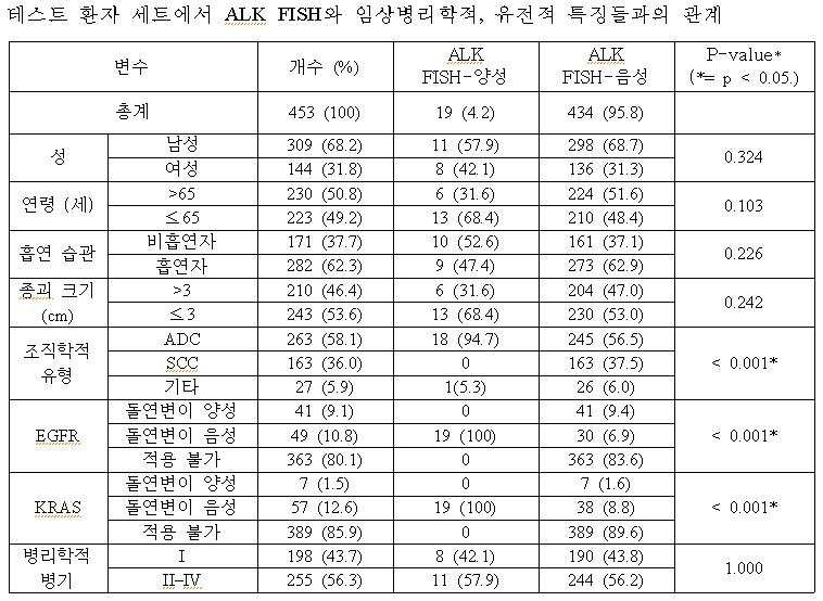
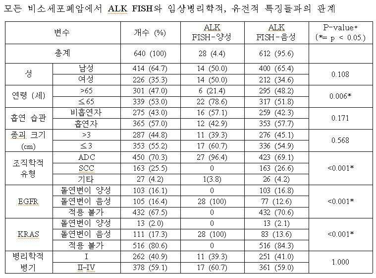
<< 실시예Example 6> 6>
IHCIHC 에 따른 In accordance ALKALK 단백질 발현 및 Protein expression and FISHFISH 결과와의 상관관계 분석 Correlation Analysis with Results
ALK 단백질 발현 패턴은 월등히 우세한 세포질(cytoplasmic) 염색 패턴을 가진 종양 세포에서 특이적으로 관찰되었다. 폐 세포에 연접한 비종양성 기관지 상피(Nonneoplastic bronchial epithelium), alveolar type1 및 type2 폐포세포(pneumocytes), 중간엽 조직(mesenchymal tissue) 및 항염증 세포는 모든 465개의 케이스에서 전부 ALK IHC 음성이었기 때문이다. ALK protein expression patterns were specifically observed in tumor cells with a predominantly cytoplasmic staining pattern. Nonneoplastic bronchial epithelium, alveolar type1 and type2 pneumocytes, mesenchymal tissue, and anti-inflammatory cells in contact with lung cells were all ALK IHC negative in all 465 cases.
ALK가 면역염색된(immunostained) 종양세포 비율은 ALK 단백질 발현이 발견된 대부분의 케이스들에서 사실상 총 종양 세포들의 5% 보다 많이 나타났다. 염색 강도의 정도는 약하고 희미한 세포질 염색(cytoplasmic staining)에서부터 강한, 다크 브라운의 세포질 염색까지 다양하였다. IHC에 의한 ALK 단백질 발현은 NSCLC의 8.6%(40/465)에서 발견되었고, 이는 1+(14/40, 35%), 2+(10/40, 25%) 및 3+(16/40, 40%)로 구성되었다(도 2 내지 5 참조).The proportion of tumor cells immunostained with ALK was substantially greater than 5% of total tumor cells in most cases where ALK protein expression was found. The degree of staining intensity varied from weak and faint cytoplasmic staining to strong, dark brown cytoplasmic staining. ALK protein expression by IHC was found in 8.6% (40/465) of NSCLC, which is 1+ (14/40, 35%), 2+ (10/40, 25%) and 3+ (16/40, 40%) (see Figures 2-5).
유전자 재배열 상태를 반영하는 IHC 양성에 대한 최고의 기준을 결정하기 위해 본 발명자들은 표 4에서와 같이 ALK IHC 및 FISH의 결과를 비교하였다. 모든 ALK IHC 3+ 케이스들은 FISH-양성이었고, 모든 ALK IHC 0/1+ 케이스들은 FISH-음성이었다. ALK IHC 2+ 케이스들의 경우, 30%(3/10)은 FISH-양성이었고 70%(7/10)는 FISH-음성이었다. To determine the best criteria for IHC positivity that reflect gene rearrangement status, we compared the results of ALK IHC and FISH as shown in Table 4. All
그리하여 본 발명가들은 ALK FISH 상태에 대한 ALK IHC의 예측가능성을 분석하였다. ALK IHC 0/1+를 생물학적 음성으로, ALK IHC 3+를 생물학적 양성으로, ALK IHC 2+는 불확실한 것(equivocal)으로 가정했을 때, 생물학적 ALK IHC 해석과 FISH는 강한 상관관계를 보였다((p= 0.000 by Pearson’s chi-square test, Pearson’s R=0.933, p=0.000). ALK IHC 2+(equivocal) 케이스들을 제외하고, ALK IHC(0,1 vs 3) 및 ALK FISH(음성 대 양성)은 완벽하게 상관관계을 이뤘으며, 이는 ALK IHC 3+가 FISH-양성을, 0/1+는 FISH-음성을 강력하게 예측할 수 있음을 암시하는 것이었다. 따라서 본 발명자들은 NSCLC에서의 ALK IHC와 ALK FISH의 임상적 적용의 진단적 알고리즘을 완성할 수 있었다(도 6 참조).Thus we analyzed the predictability of ALK IHC for ALK FISH status. When

<< 실시예Example 7> 7>
ALKALK FISHFISH -양성 -positivity NSCLCNSCLC 를 찾아내기 위한 To find IHCIHC 해석 기준의 입증 Verification of interpretation standard
밸리데이션 세트(validation set)로서, 본 발명가들은 폐 ADC가 유망되는 187명의 환자들로부터 외과적으로 절제한(surgically resected) 또는 생체검사(biopsied)를 통해 획득한 표본을 대상으로 IHC와 FISH 검사를 실시하였다. 그들 중, 14개의 케이스를 ALK 면역염색에 양성이었는데, 이들의 염색패턴을 분석한 결과 ALK IHC 1+은 2개 케이스, ALK IHC 2+는 6개 케이스 및 ALK IHC 3+는 6개 케이스였다. 상기 14개 케이스들은 9명의 여성과 5명의 남성으로부터 채취한 7개의 외과적 절제술을 통한 표본 및 7개의 생체검사(biopsy)를 통한 표본으로 구성되어 있었으며, 환자들의 나이는 28세에서 84세까지로 평균 연령은 55.1세였다.As a validation set, the inventors performed IHC and FISH tests on specimens obtained surgically resected or biopsied from 187 promising pulmonary ADCs. It was. Of these, 14 cases were positive for ALK immunostaining. Analysis of their staining patterns revealed two cases of
FISH 테스트 결과 IHC 음성인 환자들은 전부 FISH 음성이었으며, IHC 1+(생물학적 음성)의 2개 케이스들은 모두 FISH-음성이었던 반면, IHC 3+(생물학적 양성)의 6개의 모든 케이스들은 FISH-음성이었고(표 5 및 6 참조), IHC 2+의 케이스들은 50%(3/6)가 FISH-양성을 나타내었다. FISH-양성 케이스들은 8개의 Break-apart 패턴 및 2개의 IRS 패턴으로 구성되었다.The FISH test showed that all patients with IHC negative were FISH negative, and both cases of
예상한대로, 모든 ALK FISH-양성 케이스들은 EGRF 및 K-RAS 돌연변이에 대해 음성이었으나, 반면에 2개의 ALK IHC 1+ 케이스들은 모두 EGFR 엑손(exon) 19 결실 돌연변이(deletion mutation)를 나타냈다. 이와 더불어, ALK IHC 1+(생물학적 음성) 및 3+(생물학적 양성) 케이스들의 FISH에 대한 완벽한 예측가능성 또한 검증 세트에서 유지되었다. 또한, ALK IHC 2+ 케이스들의 상당 부분은 ALK FISH-양성이었는데, 이는 TMA를 사용한 테스트 환자군 세트(test patients set)의 결과에서도 일관적이었다.As expected, all ALK FISH-positive cases were negative for the EGRF and K-RAS mutations, while the two
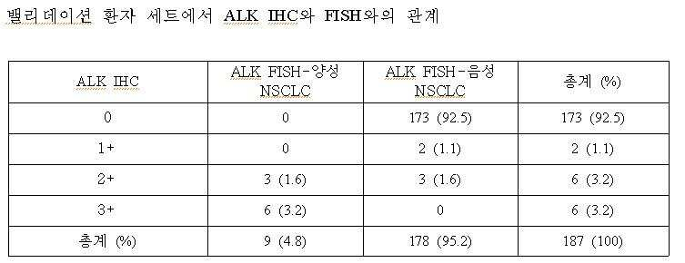
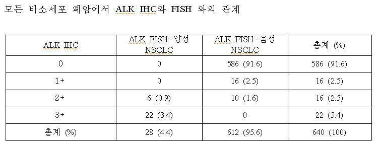
<< 실시예Example 8> 8>
항체 반응 조건에 따른 According to antibody reaction conditions ALKALK IHCIHC 와 Wow FISH 와의With FISH 상관관계 분석 Correlation analysis
본 발명에 따른 ALK 항체 반응을 통한 ALK IHC 실험 결과의 정확도를 검증하기 위하여, 다양한 항체 반응의 조건에 따른 표본들의 ALK IHC 결과와 이의 FISH 결과와의 상관관계를 분석해보았다. ALK 단백질의 과발현을 검출해내는 IHC 결과가 ALK 유전자 재배열을 검출해내는 FISH 결과와 높은 상관관계를 보인다면, 고비용의 장시간이 소요되는 FISH 대신 경제적이며 신속한 IHC 방법을 사용하여 ALK 유전자 재배열의 검출이 가능할 것이기 때문이다. In order to verify the accuracy of the ALK IHC test results through the ALK antibody reaction according to the present invention, the correlation between the ALK IHC results of the samples according to the conditions of various antibody reactions and their FISH results was analyzed. If IHC results in detecting ALK protein overexpression correlate well with FISH results in detecting ALK gene rearrangement, detection of ALK gene rearrangements using an economical and rapid IHC method instead of expensive, long-term FISH Because this will be possible.
따라서 본 발명자들은 본 발명에서 규명하고 있는 ALK 항체반응 조건이 ALK IHC와 FISH 와의 높은 상관관계를 보증해줄 수 있는 것인지 확인해보기 위하여 IHC의 다양한 반응 조건에 따른 FISH 와의 상관관계를 비교 분석해보았다. 즉, 다양한 ALK 항체 반응에 따른 표본들의 IHC 결과를 본 발명의 ALK IHC 염색 패턴 분석 기준에 따른 스코어링 시스템을 통해 점수로 분류하고, 그 중 0, 1+ 스코어를 가진 “생물학적 음성”으로 판단되는 군 및 3+ 스코어의 “생물학적 양성”으로 판단되는 군과의 FISH 결과와의 일치율을 비교하였다. “생물학적 음성군”의 경우 FISH 결과 ALK 전위 여부가 음성, 즉 ALK 유전자 재배열이 발생하지 않았을 경우 두 결과(IHC와 FISH)가 일치한다고 보았고, “생물학적 양성군”의 경우는 FISH 결과 ALK 전위가 발생되어 ALK 유전자 재배열이 생성이 확인되었을 경우 IHC 결과와 FISH 결과가 일치하는 것으로 판단하였다.Therefore, the present inventors compared and analyzed the correlation with FISH according to various reaction conditions of IHC in order to confirm whether the ALK antibody reaction conditions identified in the present invention can guarantee a high correlation between ALK IHC and FISH. That is, the IHC results of the samples according to various ALK antibody responses are classified into scores through a scoring system according to the ALK IHC staining pattern analysis criteria of the present invention, and among them, a group judged as "biological negative" having a 0, 1+ score. And the concordance with the FISH results with the group judged to be "biologically positive" of the 3+ score. In the case of the "biological negative group", the FISH result showed that the ALK translocation was negative, that is, when the ALK gene rearrangement did not occur, the two results (IHC and FISH) matched. Therefore, when ALK gene rearrangement was confirmed, it was determined that the IHC and FISH results were consistent.
항체 반응 조건에 따른 ALK IHC 결과에 대한 정확도를 측정하기 위하여, ALK 항체를 1:10, 1:15, 1:20, 1: 25, 1:30, 1:35, 1:40의 비율로 ALK IHC 실험을 실시하였다. 그 결과, 표 7에 나타난 바와 같이 1:20이나 1:40 등의 비율로 ALK 항체를 처리한 경우에 비해 1:25 및 1:35의 비율로 희석하여 ALK 항체를 처리하였을 때 IHC 결과와 FISH 간의 높은 일치율, 즉 상관관계를 확인할 수 있었다. In order to measure the accuracy of ALK IHC results according to the antibody reaction conditions, ALK antibodies were ALK at a ratio of 1:10, 1:15, 1:20, 1: 25, 1:30, 1:35, 1:40. IHC experiments were conducted. As a result, as shown in Table 7, IHC results and FISH when the ALK antibody was treated by diluting at the ratio of 1:25 and 1:35 compared to the case where the ALK antibody was treated at a ratio of 1:20 or 1:40, etc. The high concordance rate, ie, correlation, was confirmed.
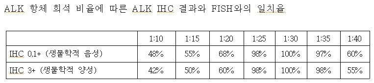
또한, 본 발명자들은 또 다른 ALK 항체 반응 조건인 인큐베이션 시간에 대한 비교 실험을 실시하였다. 표 8에서와 같이 다양한 인큐베이션 시간에 따라 IHC 실험을 수행한 결과, 60분 및 90분, 150분 및 180분 동안 표본 샘플과 ALK 항체를 인큐베이션 시켰을 때와 달리, 110분 및 130분 동안 ALK 항체와 반응시킨 IHC 실험의 결과가 FISH와 높은 일치율을 나타내었다.In addition, the inventors conducted a comparative experiment on incubation time, another ALK antibody reaction condition. As shown in Table 8, the IHC experiment was performed according to various incubation times, and when compared with incubating the sample sample and ALK antibody for 60 minutes, 90 minutes, 150 minutes and 180 minutes, The results of the IHC experiment reacted showed a high agreement with FISH.

더불어, 본 발명자들은 항체 반응 온도에 대한 비교 실험을 실시하였다. 상기 비교 실험에서 밝혀진 최적의 ALK 반응 조건을 반영하여, ALK 항체를 1:30의 비율로 희석하여 120분간 인큐베이션 시키면서, 반응 온도를 다르게 설정하여 실험을 진행한 결과, 표 9에서와 같이 35℃ 및 50℃에서 진행된 IHC 결과와 비교하여 40℃ 및 45℃에서 진행한 IHC 실험 결과가 FISH 결과와 높은 상관관계를 보였다.In addition, the inventors conducted a comparative experiment on the antibody reaction temperature. Reflecting the optimal ALK reaction conditions found in the comparative experiments, while diluting the ALK antibody at a ratio of 1:30 and incubating for 120 minutes, the experiment was carried out by setting the reaction temperature differently, as shown in Table 9 at 35 ° C and The results of IHC experiment at 40 ℃ and 45 ℃ showed a high correlation with the FISH results compared to the results of IHC at 50 ℃.

그리하여, 본 발명자들은 ALK 항체를 1:25~1:35 비율로 희석하여 40℃~45℃ 온도에서 90분~120분 동안 인큐베이션 시키는 ALK 항체 반응이 FISH 결과와 가장 일치율이 높은 탁월한 ALK IHC 결과를 도출해 낼 수 있음을 확인할 수 있었다.
Thus, the present inventors found that the ALK antibody reaction in which the ALK antibody was diluted 1: 25 ~ 1: 35 ratio and incubated for 90 minutes to 120 minutes at the temperature of 40 ° C to 45 ° C yielded excellent ALK IHC result that is the most consistent with the FISH result. It can be confirmed that it can be derived.
이제까지 본 발명에 대하여 그 바람직한 실시예들을 중심으로 살펴보았다. 본 발명이 속하는 기술 분야에서 통상의 지식을 가진 자는 본 발명이 본 발명의 본질적인 특성에서 벗어나지 않는 범위에서 변형된 형태로 구현될 수 있음을 이해할 수 있을 것이다. 그러므로 개시된 실시예들은 한정적인 관점이 아니라 설명적인 관점에서 고려되어야 한다. 본 발명의 범위는 전술한 설명이 아니라 특허청구범위에 나타나 있으며, 그와 동등한 범위 내에 있는 모든 차이점은 본 발명에 포함된 것으로 해석되어야 할 것이다.So far I looked at the center of the preferred embodiment for the present invention. It will be understood by those skilled in the art that various changes in form and details may be made therein without departing from the spirit and scope of the invention as defined by the appended claims. Therefore, the disclosed embodiments should be considered in an illustrative rather than a restrictive sense. The scope of the present invention is defined by the appended claims rather than by the foregoing description, and all differences within the scope of equivalents thereof should be construed as being included in the present invention.
Claims (13)
상기 ALK 항체를 반응시킨 표본을 염색하는 단계; 및
표본의 염색 패턴을 분석하는 단계를 포함하는 ALK 유전자 재배열 검출방법.Diluting the ALK antibody in a ratio of 1:25 to 1:35 in the pretreated sample, and reacting at a temperature of 40 ° C. to 45 ° C. for 110 minutes to 130 minutes;
Staining a sample reacted with the ALK antibody; And
ALK gene rearrangement detection method comprising the step of analyzing the staining pattern of the sample.
상기 ALK 항체 반응은 면역조직화학법(immunohistochemical techniques), 면역 블롯법(immunoblot), 면역침강법(immunoprecipitation), 효소결합 면역흡착분석법(ELISA; enzyme linked immunosorbent assay), 응집법(agglutination) 및 방사능 면역시험법(radio-immuno assay)로 이루어진 군 중에서 선택되는 방법으로 수행하는 것을 특징으로 하는 ALK 유전자 재배열 검출방법.The method of claim 1,
The ALK antibody response is immunohistochemical techniques, immunoblot, immunoprecipitation, enzyme linked immunosorbent assay (ELISA), agglutination and radioimmunoassay. ALK gene rearrangement detection method, characterized in that carried out by a method selected from the group consisting of (radio-immuno assay).
상기 표본의 전처리는 파라핀 절편을 탈파라핀화 시키고, TBE(Tris-Borate-EDTA) 버퍼를 포함한 사염화탄소(CCl4) 용매에서 95℃~105℃, 15분~25분간 열처리하는 것을 특징으로 하는 ALK 유전자 재배열 검출방법.The method of claim 1,
Pretreatment of the sample deparaffinizes the paraffin fragment and ALK gene, characterized in that heat treated at 95 ℃ ~ 105 ℃, 15 minutes to 25 minutes in a carbon tetrachloride (CCl 4 ) solvent containing TBE (Tris-Borate-EDTA) buffer Rearrangement detection method.
상기 염색하는 단계는,
상기 ALK 항체와 결합하는 2차 항체를 처리하는 단계; 및
발색기질을 처리하는 단계를 포함하는 것을 특징으로 하는 ALK 유전자 재배열 검출방법. The method of claim 1,
The dyeing step,
Treating the secondary antibody binding to the ALK antibody; And
ALK gene rearrangement detection method comprising the step of processing the chromogenic substrate.
상기 발색기질은 BCIP/NBT(5-bromo-4-chloro-3-indolyl-phosphate/nitroblue tetrazolium), DAB(diaminobenzidine), TMB(3,3´,5,5´-tetramethylbenzidine), BCIP/INT(5-bromo-4-chloro-3-indolyl phosphate/iodonitrotetrazolium), NF(New fuchsin), FRT(Fast Red TR Salt), ABTS(2,2'-azino-bis(3-ethylbenzothiazoline-6-sulfonic acid)), 4-CN, AEC(3-amino-9-ethylcarbasole), X-Gal 및 IPTG(isopropyl beta-D-thiogalactoside)로 이루어진 군 중에서 선택되는 것을 특징으로 하는 ALK 유전자 재배열 검출방법.The method of claim 5,
The color substrate is BCIP / NBT (5-bromo-4-chloro-3-indolyl-phosphate / nitroblue tetrazolium), DAB (diaminobenzidine), TMB (3,3 ', 5,5'-tetramethylbenzidine), BCIP / INT ( 5-bromo-4-chloro-3-indolyl phosphate / iodonitrotetrazolium), NF (New fuchsin), FRT (Fast Red TR Salt), ABTS (2,2'-azino-bis (3-ethylbenzothiazoline-6-sulfonic acid) ), 4-CN, AEC (3-amino-9-ethylcarbasole), X-Gal and IPTG (isopropyl beta-D-thiogalactoside) ALK gene rearrangement detection method characterized in that it is selected from the group consisting of.
상기 표본의 염색 패턴을 분석하는 단계는 세포질이 염색된 세포 수를 판단하는 단계 또는 세포질의 염색 강도를 판단하는 단계를 포함하는 것을 특징으로 하는 ALK 유전자 재배열 검출 방법.The method of claim 1,
Analyzing the staining pattern of the sample ALK gene rearrangement detection method characterized in that it comprises the step of determining the number of cells stained cytoplasm or the staining intensity of the cytoplasm.
상기 염색된 세포수를 판단할 때 분석대상의 총 세포수를 기준으로 세포질이 염색된 세포 수가 5% 미만일 경우, ALK 유전자 재배열 여부는 음성인 것을 특징으로 하는 ALK 유전자 재배열 검출 방법.The method of claim 7, wherein
When the number of cells stained based on the total number of cells to be analyzed when determining the number of the stained cells is less than 5%, ALK gene rearrangement detection method characterized in that the ALK gene rearrangement is negative.
상기 염색된 세포수를 판단할 때 분석대상의 총 세포수를 기준으로 세포질이 염색된 세포 수가 5% 이상이면서 세포질의 염색 강도가 약한 경우, ALK 유전자 재배열 여부는 음성인 것을 특징으로 하는 ALK 유전자 재배열 검출 방법.The method of claim 7, wherein
When the number of stained cells is determined based on the total number of cells to be analyzed, if the number of cells stained with cytoplasm is 5% or more and the cytoplasm staining is weak, the ALK gene rearrangement is negative, characterized in that the ALK gene rearrangement Rearrangement detection method.
상기 염색된 세포수를 판단할 때 분석대상의 총 세포수를 기준으로 세포질이 염색된 세포 수가 5% 이상이면서 세포질의 염색 강도가 강한 경우, ALK 유전자 재배열 여부는 양성인 것을 특징으로 하는 ALK 유전자 재배열 검출 방법.The method of claim 7, wherein
In determining the number of stained cells, if the number of cells stained with cytoplasm is 5% or more based on the total number of cells to be analyzed and the staining intensity of the cytoplasm is strong, the ALK gene rearrangement is positive. Array Detection Method.
상기 염색된 세포수를 판단할 때 분석대상의 총 세포수를 기준으로 세포질이 염색된 세포 수가 5% 이상이면서 세포질의 염색 강도가 중등도인 경우, FISH(fluorescence in situ hybridization), SISH(Silver In Situ Hybridization) 및 PCR(polymerase chain reaction)로 구성된 군 중에서 선택되는 방법으로 ALK 유전자의 전위여부를 확인하는 단계를 더 포함하는 것을 특징으로 하는 ALK 유전자 재배열 검출방법.The method of claim 7, wherein
When the number of stained cells is determined based on the total number of cells to be analyzed, when the number of cells stained with cytoplasm is 5% or more and the staining intensity of the cytoplasm is moderate, fluorescence in situ hybridization (FISH) and Silver In Situ Hybridization) and PCR (polymerase chain reaction) ALK gene rearrangement detection method characterized in that it further comprises the step of confirming the translocation of the ALK gene in a method selected from the group consisting of.
상기 암은 폐암임을 특징으로 하는 환자의 시료로부터 재배열된 ALK 유전자 포함 여부를 판단하는 방법.The method of claim 12,
The cancer is a method for determining whether or not the rearranged ALK gene from a sample of a patient characterized in that the lung cancer.
Priority Applications (2)
| Application Number | Priority Date | Filing Date | Title |
|---|---|---|---|
| KR1020100118308A KR101264810B1 (en) | 2010-11-25 | 2010-11-25 | Method for Screening of ALK gene rearrangement and diagnosis of cancer using the same |
| PCT/KR2011/002865 WO2012070730A1 (en) | 2010-11-25 | 2011-04-21 | Method for detecting alk gene rearrangement and method for diagnosing cancer using the same |
Applications Claiming Priority (1)
| Application Number | Priority Date | Filing Date | Title |
|---|---|---|---|
| KR1020100118308A KR101264810B1 (en) | 2010-11-25 | 2010-11-25 | Method for Screening of ALK gene rearrangement and diagnosis of cancer using the same |
Publications (2)
| Publication Number | Publication Date |
|---|---|
| KR20120056660A KR20120056660A (en) | 2012-06-04 |
| KR101264810B1 true KR101264810B1 (en) | 2013-05-15 |
Family
ID=46146052
Family Applications (1)
| Application Number | Title | Priority Date | Filing Date |
|---|---|---|---|
| KR1020100118308A Expired - Fee Related KR101264810B1 (en) | 2010-11-25 | 2010-11-25 | Method for Screening of ALK gene rearrangement and diagnosis of cancer using the same |
Country Status (2)
| Country | Link |
|---|---|
| KR (1) | KR101264810B1 (en) |
| WO (1) | WO2012070730A1 (en) |
Cited By (1)
| Publication number | Priority date | Publication date | Assignee | Title |
|---|---|---|---|---|
| KR20210123825A (en) | 2020-04-06 | 2021-10-14 | 서울대학교병원 | Method for detecting of fusion gene |
Families Citing this family (3)
| Publication number | Priority date | Publication date | Assignee | Title |
|---|---|---|---|---|
| WO2014046730A1 (en) * | 2012-09-24 | 2014-03-27 | Ventana Medical Systems, Inc. | Method of identifying treatment responsive non-small cell lung cancer using anaplastic lymphoma kinase (alk) as a marker |
| EP3129477B1 (en) | 2014-04-10 | 2020-05-20 | Memorial Sloan-Kettering Cancer Center | A novel isoform of anaplastic lymphoma kinase and its uses |
| CN119775423B (en) * | 2025-01-20 | 2025-12-02 | 河南赛诺特生物技术有限公司 | A recombinant antibody against human ALK and its application |
Family Cites Families (2)
| Publication number | Priority date | Publication date | Assignee | Title |
|---|---|---|---|---|
| US20030099974A1 (en) * | 2001-07-18 | 2003-05-29 | Millennium Pharmaceuticals, Inc. | Novel genes, compositions, kits and methods for identification, assessment, prevention, and therapy of breast cancer |
| CN112057625A (en) * | 2007-04-13 | 2020-12-11 | 细胞信号技术公司 | ALK kinase gene defect and mutant in human solid tumor |
-
2010
- 2010-11-25 KR KR1020100118308A patent/KR101264810B1/en not_active Expired - Fee Related
-
2011
- 2011-04-21 WO PCT/KR2011/002865 patent/WO2012070730A1/en not_active Ceased
Non-Patent Citations (4)
| Title |
|---|
| Clin Cancer Res. 2009, vol. 15, no. 16, pp. 5216-5223.* |
| Clin Cancer Res. 2010.03., vol. 16, no. 5, pp. 1561-1571. |
| Human Pathology. 2009, vol. 40, pp. 1152-1158.* |
| J Thorac Oncol. 2009, vol. 4, no. 12, pp. 1450-1454. |
Cited By (1)
| Publication number | Priority date | Publication date | Assignee | Title |
|---|---|---|---|---|
| KR20210123825A (en) | 2020-04-06 | 2021-10-14 | 서울대학교병원 | Method for detecting of fusion gene |
Also Published As
| Publication number | Publication date |
|---|---|
| KR20120056660A (en) | 2012-06-04 |
| WO2012070730A1 (en) | 2012-05-31 |
Similar Documents
| Publication | Publication Date | Title |
|---|---|---|
| Kim et al. | Detection of ALK gene rearrangement in non-small cell lung cancer: a comparison of fluorescence in situ hybridization and chromogenic in situ hybridization with correlation of ALK protein expression | |
| Paik et al. | Screening of anaplastic lymphoma kinase rearrangement by immunohistochemistry in non-small cell lung cancer: correlation with fluorescence in situ hybridization | |
| Jacobsen et al. | Expression of vascular endothelial growth factor protein in human renal cell carcinoma | |
| Mino-Kenudson | Immunohistochemistry for predictive biomarkers in non-small cell lung cancer | |
| Furusato et al. | ERG oncoprotein expression in prostate cancer: clonal progression of ERG-positive tumor cells and potential for ERG-based stratification | |
| Rogers et al. | Comparison of methods in the detection of ALK and ROS1 rearrangements in lung cancer | |
| Zhang et al. | Evaluation of ALK rearrangement in Chinese non-small cell lung cancer using FISH, immunohistochemistry, and real-time quantitative RT-PCR on paraffin-embedded tissues | |
| Abdallah et al. | Thymidylate synthase expression in circulating tumor cells: A new tool to predict 5‐fluorouracil resistance in metastatic colorectal cancer patients | |
| Bhalla et al. | Novel dual-color immunohistochemical methods for detecting ERG–PTEN and ERG–SPINK1 status in prostate carcinoma | |
| Sasaki et al. | Usefulness of immunohistochemistry for the detection of the BRAF V600E mutation in Japanese lung adenocarcinoma | |
| Sangale et al. | A robust immunohistochemical assay for detecting PTEN expression in human tumors | |
| Scheble et al. | ERG rearrangement in small cell prostatic and lung cancer | |
| AU2010291710B2 (en) | Methods and compositions for detection of lethal cell and uses thereof | |
| Martin et al. | ALK testing in lung adenocarcinoma: technical aspects to improve FISH evaluation in daily practice | |
| Kanayama et al. | Significant intratumoral heterogeneity of human epidermal growth factor receptor 2 status in gastric cancer: A comparative study of immunohistochemistry, FISH, and dual‐color in situ hybridization | |
| Abe et al. | Heterogeneity of anaplastic lymphoma kinase gene rearrangement in non–small-cell lung carcinomas: A comparative study between small biopsy and excision samples | |
| Bondgaard et al. | High specificity but low sensitivity of mutation-specific antibodies against EGFR mutations in non-small-cell lung cancer | |
| Bravaccini et al. | ALK translocation detection in non‐small cell lung cancer cytological samples obtained by TBNA or EBUS‐TBNA | |
| EP2449383B1 (en) | Diagnostic method for predicting the risk of cancer recurrence based on histone macroh2a isoforms | |
| KR101264810B1 (en) | Method for Screening of ALK gene rearrangement and diagnosis of cancer using the same | |
| Sung et al. | Revisiting ALK (D5F3) immunohistochemistry: Insights into focal staining and neuroendocrine differentiation | |
| Lim et al. | Validation of ALK/ROS1 Dual Break Apart FISH Probe probe in non-small-cell lung cancer | |
| Pertia et al. | Loss of p27 (Kip1) CDKI is a predictor of poor recurrence-free and cancer-specific survival in patients with renal cancer | |
| Liu et al. | Expression of p53, p16 and cyclooxygenase‐2 in esophageal cancer with tissue microarray | |
| JP2009506303A (en) | Predictive methods for cancer chemotherapy |
Legal Events
| Date | Code | Title | Description |
|---|---|---|---|
| A201 | Request for examination | ||
| PA0109 | Patent application |
St.27 status event code: A-0-1-A10-A12-nap-PA0109 |
|
| PA0201 | Request for examination |
St.27 status event code: A-1-2-D10-D11-exm-PA0201 |
|
| P11-X000 | Amendment of application requested |
St.27 status event code: A-2-2-P10-P11-nap-X000 |
|
| P13-X000 | Application amended |
St.27 status event code: A-2-2-P10-P13-nap-X000 |
|
| R17-X000 | Change to representative recorded |
St.27 status event code: A-3-3-R10-R17-oth-X000 |
|
| D13-X000 | Search requested |
St.27 status event code: A-1-2-D10-D13-srh-X000 |
|
| D14-X000 | Search report completed |
St.27 status event code: A-1-2-D10-D14-srh-X000 |
|
| PG1501 | Laying open of application |
St.27 status event code: A-1-1-Q10-Q12-nap-PG1501 |
|
| R17-X000 | Change to representative recorded |
St.27 status event code: A-3-3-R10-R17-oth-X000 |
|
| E902 | Notification of reason for refusal | ||
| PE0902 | Notice of grounds for rejection |
St.27 status event code: A-1-2-D10-D21-exm-PE0902 |
|
| E13-X000 | Pre-grant limitation requested |
St.27 status event code: A-2-3-E10-E13-lim-X000 |
|
| P11-X000 | Amendment of application requested |
St.27 status event code: A-2-2-P10-P11-nap-X000 |
|
| P13-X000 | Application amended |
St.27 status event code: A-2-2-P10-P13-nap-X000 |
|
| E701 | Decision to grant or registration of patent right | ||
| PE0701 | Decision of registration |
St.27 status event code: A-1-2-D10-D22-exm-PE0701 |
|
| N231 | Notification of change of applicant | ||
| PN2301 | Change of applicant |
St.27 status event code: A-3-3-R10-R13-asn-PN2301 St.27 status event code: A-3-3-R10-R11-asn-PN2301 |
|
| GRNT | Written decision to grant | ||
| PR0701 | Registration of establishment |
St.27 status event code: A-2-4-F10-F11-exm-PR0701 |
|
| PR1002 | Payment of registration fee |
St.27 status event code: A-2-2-U10-U11-oth-PR1002 Fee payment year number: 1 |
|
| PG1601 | Publication of registration |
St.27 status event code: A-4-4-Q10-Q13-nap-PG1601 |
|
| PN2301 | Change of applicant |
St.27 status event code: A-5-5-R10-R13-asn-PN2301 St.27 status event code: A-5-5-R10-R11-asn-PN2301 |
|
| PN2301 | Change of applicant |
St.27 status event code: A-5-5-R10-R13-asn-PN2301 St.27 status event code: A-5-5-R10-R11-asn-PN2301 |
|
| FPAY | Annual fee payment |
Payment date: 20160601 Year of fee payment: 4 |
|
| PR1001 | Payment of annual fee |
St.27 status event code: A-4-4-U10-U11-oth-PR1001 Fee payment year number: 4 |
|
| R18-X000 | Changes to party contact information recorded |
St.27 status event code: A-5-5-R10-R18-oth-X000 |
|
| FPAY | Annual fee payment |
Payment date: 20170508 Year of fee payment: 5 |
|
| PR1001 | Payment of annual fee |
St.27 status event code: A-4-4-U10-U11-oth-PR1001 Fee payment year number: 5 |
|
| FPAY | Annual fee payment |
Payment date: 20180409 Year of fee payment: 6 |
|
| PR1001 | Payment of annual fee |
St.27 status event code: A-4-4-U10-U11-oth-PR1001 Fee payment year number: 6 |
|
| P22-X000 | Classification modified |
St.27 status event code: A-4-4-P10-P22-nap-X000 |
|
| FPAY | Annual fee payment |
Payment date: 20190507 Year of fee payment: 7 |
|
| PR1001 | Payment of annual fee |
St.27 status event code: A-4-4-U10-U11-oth-PR1001 Fee payment year number: 7 |
|
| PC1903 | Unpaid annual fee |
St.27 status event code: A-4-4-U10-U13-oth-PC1903 Not in force date: 20200510 Payment event data comment text: Termination Category : DEFAULT_OF_REGISTRATION_FEE |
|
| PC1903 | Unpaid annual fee |
St.27 status event code: N-4-6-H10-H13-oth-PC1903 Ip right cessation event data comment text: Termination Category : DEFAULT_OF_REGISTRATION_FEE Not in force date: 20200510 |
|
| PN2301 | Change of applicant |
St.27 status event code: A-5-5-R10-R13-asn-PN2301 St.27 status event code: A-5-5-R10-R11-asn-PN2301 |
|
| P22-X000 | Classification modified |
St.27 status event code: A-4-4-P10-P22-nap-X000 |