KR100816958B1 - Therapeutic compounds comprising an anti-Fc receptor binder - Google Patents
Therapeutic compounds comprising an anti-Fc receptor binder Download PDFInfo
- Publication number
- KR100816958B1 KR100816958B1 KR1020027001270A KR20027001270A KR100816958B1 KR 100816958 B1 KR100816958 B1 KR 100816958B1 KR 1020027001270 A KR1020027001270 A KR 1020027001270A KR 20027001270 A KR20027001270 A KR 20027001270A KR 100816958 B1 KR100816958 B1 KR 100816958B1
- Authority
- KR
- South Korea
- Prior art keywords
- delete delete
- human
- cells
- antibody
- fragment
- Prior art date
- Legal status (The legal status is an assumption and is not a legal conclusion. Google has not performed a legal analysis and makes no representation as to the accuracy of the status listed.)
- Expired - Fee Related
Links
- NFWSQSCIDYBUOU-UHFFFAOYSA-N CC1=CC=CC1 Chemical compound CC1=CC=CC1 NFWSQSCIDYBUOU-UHFFFAOYSA-N 0.000 description 1
Images


Classifications
-
- C—CHEMISTRY; METALLURGY
- C07—ORGANIC CHEMISTRY
- C07K—PEPTIDES
- C07K19/00—Hybrid peptides, i.e. peptides covalently bound to nucleic acids, or non-covalently bound protein-protein complexes
-
- C—CHEMISTRY; METALLURGY
- C07—ORGANIC CHEMISTRY
- C07K—PEPTIDES
- C07K16/00—Immunoglobulins [IGs], e.g. monoclonal or polyclonal antibodies
- C07K16/18—Immunoglobulins [IGs], e.g. monoclonal or polyclonal antibodies against material from animals or humans
- C07K16/28—Immunoglobulins [IGs], e.g. monoclonal or polyclonal antibodies against material from animals or humans against receptors, cell surface antigens or cell surface determinants
- C07K16/2803—Immunoglobulins [IGs], e.g. monoclonal or polyclonal antibodies against material from animals or humans against receptors, cell surface antigens or cell surface determinants against the immunoglobulin superfamily
- C07K16/283—Immunoglobulins [IGs], e.g. monoclonal or polyclonal antibodies against material from animals or humans against receptors, cell surface antigens or cell surface determinants against the immunoglobulin superfamily against Fc-receptors, e.g. CD16, CD32, CD64
-
- A—HUMAN NECESSITIES
- A61—MEDICAL OR VETERINARY SCIENCE; HYGIENE
- A61K—PREPARATIONS FOR MEDICAL, DENTAL OR TOILETRY PURPOSES
- A61K38/00—Medicinal preparations containing peptides
- A61K38/16—Peptides having more than 20 amino acids; Gastrins; Somatostatins; Melanotropins; Derivatives thereof
- A61K38/17—Peptides having more than 20 amino acids; Gastrins; Somatostatins; Melanotropins; Derivatives thereof from animals; from humans
- A61K38/18—Growth factors; Growth regulators
- A61K38/1808—Epidermal growth factor [EGF] urogastrone
-
- A—HUMAN NECESSITIES
- A61—MEDICAL OR VETERINARY SCIENCE; HYGIENE
- A61P—SPECIFIC THERAPEUTIC ACTIVITY OF CHEMICAL COMPOUNDS OR MEDICINAL PREPARATIONS
- A61P31/00—Antiinfectives, i.e. antibiotics, antiseptics, chemotherapeutics
-
- A—HUMAN NECESSITIES
- A61—MEDICAL OR VETERINARY SCIENCE; HYGIENE
- A61P—SPECIFIC THERAPEUTIC ACTIVITY OF CHEMICAL COMPOUNDS OR MEDICINAL PREPARATIONS
- A61P31/00—Antiinfectives, i.e. antibiotics, antiseptics, chemotherapeutics
- A61P31/10—Antimycotics
-
- A—HUMAN NECESSITIES
- A61—MEDICAL OR VETERINARY SCIENCE; HYGIENE
- A61P—SPECIFIC THERAPEUTIC ACTIVITY OF CHEMICAL COMPOUNDS OR MEDICINAL PREPARATIONS
- A61P31/00—Antiinfectives, i.e. antibiotics, antiseptics, chemotherapeutics
- A61P31/12—Antivirals
-
- A—HUMAN NECESSITIES
- A61—MEDICAL OR VETERINARY SCIENCE; HYGIENE
- A61P—SPECIFIC THERAPEUTIC ACTIVITY OF CHEMICAL COMPOUNDS OR MEDICINAL PREPARATIONS
- A61P31/00—Antiinfectives, i.e. antibiotics, antiseptics, chemotherapeutics
- A61P31/12—Antivirals
- A61P31/14—Antivirals for RNA viruses
- A61P31/18—Antivirals for RNA viruses for HIV
-
- A—HUMAN NECESSITIES
- A61—MEDICAL OR VETERINARY SCIENCE; HYGIENE
- A61P—SPECIFIC THERAPEUTIC ACTIVITY OF CHEMICAL COMPOUNDS OR MEDICINAL PREPARATIONS
- A61P33/00—Antiparasitic agents
-
- A—HUMAN NECESSITIES
- A61—MEDICAL OR VETERINARY SCIENCE; HYGIENE
- A61P—SPECIFIC THERAPEUTIC ACTIVITY OF CHEMICAL COMPOUNDS OR MEDICINAL PREPARATIONS
- A61P33/00—Antiparasitic agents
- A61P33/02—Antiprotozoals, e.g. for leishmaniasis, trichomoniasis, toxoplasmosis
-
- A—HUMAN NECESSITIES
- A61—MEDICAL OR VETERINARY SCIENCE; HYGIENE
- A61P—SPECIFIC THERAPEUTIC ACTIVITY OF CHEMICAL COMPOUNDS OR MEDICINAL PREPARATIONS
- A61P35/00—Antineoplastic agents
-
- A—HUMAN NECESSITIES
- A61—MEDICAL OR VETERINARY SCIENCE; HYGIENE
- A61P—SPECIFIC THERAPEUTIC ACTIVITY OF CHEMICAL COMPOUNDS OR MEDICINAL PREPARATIONS
- A61P37/00—Drugs for immunological or allergic disorders
- A61P37/02—Immunomodulators
-
- C—CHEMISTRY; METALLURGY
- C07—ORGANIC CHEMISTRY
- C07K—PEPTIDES
- C07K14/00—Peptides having more than 20 amino acids; Gastrins; Somatostatins; Melanotropins; Derivatives thereof
- C07K14/435—Peptides having more than 20 amino acids; Gastrins; Somatostatins; Melanotropins; Derivatives thereof from animals; from humans
- C07K14/475—Growth factors; Growth regulators
- C07K14/485—Epidermal growth factor [EGF], i.e. urogastrone
-
- C—CHEMISTRY; METALLURGY
- C07—ORGANIC CHEMISTRY
- C07K—PEPTIDES
- C07K16/00—Immunoglobulins [IGs], e.g. monoclonal or polyclonal antibodies
- C07K16/18—Immunoglobulins [IGs], e.g. monoclonal or polyclonal antibodies against material from animals or humans
- C07K16/28—Immunoglobulins [IGs], e.g. monoclonal or polyclonal antibodies against material from animals or humans against receptors, cell surface antigens or cell surface determinants
- C07K16/2863—Immunoglobulins [IGs], e.g. monoclonal or polyclonal antibodies against material from animals or humans against receptors, cell surface antigens or cell surface determinants against receptors for growth factors, growth regulators
-
- C—CHEMISTRY; METALLURGY
- C07—ORGANIC CHEMISTRY
- C07K—PEPTIDES
- C07K16/00—Immunoglobulins [IGs], e.g. monoclonal or polyclonal antibodies
- C07K16/18—Immunoglobulins [IGs], e.g. monoclonal or polyclonal antibodies against material from animals or humans
- C07K16/32—Immunoglobulins [IGs], e.g. monoclonal or polyclonal antibodies against material from animals or humans against translation products of oncogenes
-
- A—HUMAN NECESSITIES
- A61—MEDICAL OR VETERINARY SCIENCE; HYGIENE
- A61K—PREPARATIONS FOR MEDICAL, DENTAL OR TOILETRY PURPOSES
- A61K39/00—Medicinal preparations containing antigens or antibodies
- A61K2039/505—Medicinal preparations containing antigens or antibodies comprising antibodies
-
- C—CHEMISTRY; METALLURGY
- C07—ORGANIC CHEMISTRY
- C07K—PEPTIDES
- C07K2317/00—Immunoglobulins specific features
- C07K2317/20—Immunoglobulins specific features characterized by taxonomic origin
- C07K2317/24—Immunoglobulins specific features characterized by taxonomic origin containing regions, domains or residues from different species, e.g. chimeric, humanized or veneered
-
- C—CHEMISTRY; METALLURGY
- C07—ORGANIC CHEMISTRY
- C07K—PEPTIDES
- C07K2317/00—Immunoglobulins specific features
- C07K2317/30—Immunoglobulins specific features characterized by aspects of specificity or valency
- C07K2317/31—Immunoglobulins specific features characterized by aspects of specificity or valency multispecific
-
- C—CHEMISTRY; METALLURGY
- C07—ORGANIC CHEMISTRY
- C07K—PEPTIDES
- C07K2317/00—Immunoglobulins specific features
- C07K2317/50—Immunoglobulins specific features characterized by immunoglobulin fragments
- C07K2317/51—Complete heavy chain or Fd fragment, i.e. VH + CH1
-
- C—CHEMISTRY; METALLURGY
- C07—ORGANIC CHEMISTRY
- C07K—PEPTIDES
- C07K2317/00—Immunoglobulins specific features
- C07K2317/50—Immunoglobulins specific features characterized by immunoglobulin fragments
- C07K2317/55—Fab or Fab'
-
- C—CHEMISTRY; METALLURGY
- C07—ORGANIC CHEMISTRY
- C07K—PEPTIDES
- C07K2317/00—Immunoglobulins specific features
- C07K2317/70—Immunoglobulins specific features characterized by effect upon binding to a cell or to an antigen
- C07K2317/73—Inducing cell death, e.g. apoptosis, necrosis or inhibition of cell proliferation
- C07K2317/732—Antibody-dependent cellular cytotoxicity [ADCC]
-
- C—CHEMISTRY; METALLURGY
- C07—ORGANIC CHEMISTRY
- C07K—PEPTIDES
- C07K2317/00—Immunoglobulins specific features
- C07K2317/70—Immunoglobulins specific features characterized by effect upon binding to a cell or to an antigen
- C07K2317/77—Internalization into the cell
-
- C—CHEMISTRY; METALLURGY
- C07—ORGANIC CHEMISTRY
- C07K—PEPTIDES
- C07K2319/00—Fusion polypeptide
Landscapes
- Health & Medical Sciences (AREA)
- Chemical & Material Sciences (AREA)
- Life Sciences & Earth Sciences (AREA)
- Organic Chemistry (AREA)
- General Health & Medical Sciences (AREA)
- Medicinal Chemistry (AREA)
- Veterinary Medicine (AREA)
- Immunology (AREA)
- Public Health (AREA)
- Animal Behavior & Ethology (AREA)
- Pharmacology & Pharmacy (AREA)
- Nuclear Medicine, Radiotherapy & Molecular Imaging (AREA)
- General Chemical & Material Sciences (AREA)
- Chemical Kinetics & Catalysis (AREA)
- Biochemistry (AREA)
- Proteomics, Peptides & Aminoacids (AREA)
- Molecular Biology (AREA)
- Biophysics (AREA)
- Oncology (AREA)
- Genetics & Genomics (AREA)
- Communicable Diseases (AREA)
- Gastroenterology & Hepatology (AREA)
- Virology (AREA)
- Tropical Medicine & Parasitology (AREA)
- Bioinformatics & Cheminformatics (AREA)
- Engineering & Computer Science (AREA)
- Zoology (AREA)
- AIDS & HIV (AREA)
- Toxicology (AREA)
- Epidemiology (AREA)
- Peptides Or Proteins (AREA)
- Medicines Containing Antibodies Or Antigens For Use As Internal Diagnostic Agents (AREA)
- Medicines That Contain Protein Lipid Enzymes And Other Medicines (AREA)
Abstract
본 발명에는 표적 세포의 이펙터 세포-매개의 사멸을 유도하는 다중특이적 분자가 개시되어 있다. 이 분자는, 하나 이상의 표적이 면역 세포 표면 상의 Fc 수용체인 다중의 (2개 이상의) 별개 표적에 결합하기 때문에 "다중특이적"이다. 본 발명의 다중특이적 분자는, Fcγ 수용체 (예, FcγRⅠ) 또는 Fcα 수용체와 같은 Fc 수용체에 결합하는 하나 이상의 부분, 및 종양 세포 또는 병원체 상의 항원과 같은 상이한 표적에 결합하는 하나 이상의 부분으로 이루어진 분자를 포함한다. 본 발명의 다중특이적 분자는, 1개 이상의 항원에 결합된 항원 표출 세포 (APC) 상의 분자 (예를 들어, Fc 수용체)에 결합하는 다중 (즉, 2개 이상의) 부분으로 구성된 항원 "멀티머 복합체"를 포함한다. 이들 멀티머 복합체는 자기항원과 같은 항원을 APC로 표적화시켜, APC에 의한 항원의 인터날리제이션 (내포작용), 가공 및(또는) 표출을 유도하고(하거나) 증진시킨다. 따라서, 이들 분자는 자기항원과 같이 정상적으로는 비-면역원성인 단백질에 대한 생체내 또는 시험관내 면역 반응을 유도하거나 증진시키는데 사용될 수 있다. 구체적인 실시양태에서, 다중특이적 분자의 하나 이상의 부분은 Fc 수용체 또는 표적 세포 상의 수용체 (예, EGF-R 또는 HER2)에 결합하는 인간화 또는 인간 항체 또는 항체 단편 (예를 들어, ScFv 또는 Fab')을 포함한다.The present invention discloses multispecific molecules that induce effector cell-mediated killing of target cells. This molecule is "multispecific" because one or more targets bind to multiple (two or more) separate targets, which are Fc receptors on immune cell surfaces. Multispecific molecules of the invention are molecules consisting of one or more moieties that bind to Fc receptors, such as Fcγ receptors (eg, FcγRI) or Fcα receptors, and one or more moieties that bind to different targets, such as antigens on tumor cells or pathogens. It includes. Multispecific molecules of the invention are antigen “multimers composed of multiple (ie, two or more) moieties that bind to a molecule (eg, an Fc receptor) on an antigen expressing cell (APC) that is bound to one or more antigens. Complex ". These multimer complexes target antigens, such as autoantigens, to APCs, inducing and / or enhancing the internalization (inclusion), processing and / or expression of antigens by APCs. Thus, these molecules can be used to induce or enhance immune responses in vivo or in vitro against proteins that are normally non-immunogenic, such as autoantigens. In specific embodiments, one or more portions of the multispecific molecule are humanized or human antibodies or antibody fragments (eg, ScFv or Fab ′) that bind to Fc receptors or receptors on target cells (eg, EGF-R or HER2). It includes.
항원, 항체, 수용체, 이중특이적, 다중특이적, 복합체, 단편Antigen, antibody, receptor, bispecific, multispecific, complex, fragment
Description
관련 출원Related Applications
본 출원은 1999년 7월 30일 출원된 미국 출원 제09/364,088호의 부분 계속 출원이다. 상기 미국 출원 제09/364,088호는 1998년 11월 6일 출원된 미국 출원 제09/188,082호의 부분 계속 출원이다. 미국 출원 제09/188,082호는 1996년 6월 7일 출원되어 현재 미국 특허 제5,837,243호로 허여된 미국 출원 제08/661,052호의 부분 계속 출원이다. 상기 미국 특허는 "항-Fc 수용체 항체를 포함하는 치료 화합물"이란 제목으로 1995년 6월 7일 출원된 미국 출원 제08/484,172호의 부분 계속 출원이다. 상기 언급한 출원들, 및 거기에 인용된 모든 참고문헌, 허여된 특허 및 공개된 특허 출원은 이 거명을 통해 본 명세서에 포함된다.This application is a partial continuing application of US Application No. 09 / 364,088, filed July 30, 1999. U.S. Application No. 09 / 364,088 is a partial continuing application of U.S. Application No. 09 / 188,082, filed November 6,1998. US application 09 / 188,082 is a partial continuing application of US
면역글로불린 (Ig)은 2개의 중쇄와 2개의 경쇄로 구성되어 있으며, 각각의 쇄는 NH2-말단의 항원-결합 가변 도메인과 항체의 이펙터 (effector) 기능을 담당하는 COOH-말단의 불변 도메인을 포함한다. Ig 중쇄의 COOH-말단 도메인은 Fc 영역을 형성하며, 이는 Fc 수용체 (FcR)라 알려진 특이적 수용체와의 상호작용을 통해 세포 활성을 촉진하는데 관여한다. 모든 Ig 클래스에 대한 Fc 수용체 또는 그 이 소타입 (예를 들어, IgG(FcγR), IgE(FcεR), IgA(FcαR), IgM(FcμR) 및 IgD(FcδR))이 확인되었다. 상이한 이소타입이 지닌 상이한 생물학적 활성은, 부분적으로는 상이한 면역 (작용) 세포 상에서 발현되는 상이한 FcR에 결합하는 능력을 기초로 한다 (Fridman, W.H. (Sept. 1991) The FASEB Jorunal Vol. 5. 2684-2690). FcR에 대해 유도된 쥐의 항체가 만들어졌다 ("Monoclonal Antibodies To Fc Receptors for Immunoglobulin G on Human Mononuclear Phagocytes"라는 제목의 미국 특허 제4,954,617호 및 "Monoclonal Antibody Specific For IgA Receptor"라는 제목의 국제 특허 출원 공개 제WO 81/05871호 참조).Immunoglobulins (Ig) are composed of two heavy and two light chains, each of which comprises an NH 2 -terminal antigen-binding variable domain and a COOH-terminal constant domain responsible for the effector function of the antibody. Include. The COOH-terminal domain of the Ig heavy chain forms an Fc region, which is involved in promoting cellular activity through interaction with specific receptors known as Fc receptors (FcRs). Fc receptors or isotypes thereof (eg IgG (FcγR), IgE (FcεR), IgA (FcαR), IgM (FcμR)) and IgD (FcδR) for all Ig classes have been identified. The different biological activities of different isotypes are based, in part, on their ability to bind to different FcRs expressed on different immune (acting) cells (Fridman, WH (Sept. 1991) The FASEB Jorunal Vol. 5. 2684- 2690). Mouse antibodies directed against FcR have been made (see US Pat. No. 4,954,617 entitled "Monoclonal Antibodies To Fc Receptors for Immunoglobulin G on Human Mononuclear Phagocytes" and International Patent Application entitled "Monoclonal Antibody Specific For IgA Receptor") See WO 81/05871).
쥐의 모노클로날 항체는 인간 치료제로서 유용하며, 간염 바이러스나 인간 면역결핍 바이러스와 같은 인간 병원체에 의해 오염되지 않은 채로 제조될 수 있다. 그러나, 일부의 인간 치료에 쥐의 모노클로날 항체를 사용하는 것은 "외래의" 쥐 단백질에 대한 면역 반응이 발생하는 결과를 초래하였다. 이 반응은 인간 항-생쥐 항체 또는 HAMA 반응 (Schroff, R. et al. (1985), Cancer Res., 45, 879-885)이라 명명되었으며, 이는 인간에서 혈청 질병을 유발시켜 개체의 순환계로부터 쥐의 항체를 빠른 속도로 제거하는 것이다. 인간의 면역 반응은 쥐의 면역글로불린의 가변 및 불변 영역 모두에 대한 것으로 밝혀졌다.Mouse monoclonal antibodies are useful as human therapeutics and can be prepared uncontaminated by human pathogens such as hepatitis virus or human immunodeficiency virus. However, the use of murine monoclonal antibodies in some human treatments has resulted in the development of immune responses to "foreign" murine proteins. This response has been termed the human anti-mouse antibody or HAMA response (Schroff, R. et al. (1985), Cancer Res., 45, 879-885), which induces serum disease in humans, resulting in rats from the individual's circulatory system. To quickly remove the antibody. Human immune responses have been found to be both variable and constant regions of rat immunoglobulins.
재조합 DNA 기술은, 예를 들어 어떤 종의 특정 면역글로불린 영역을 다른 종의 면역글로불린 영역으로 치환함으로써 항체를 변형시키는데 유용할 수 있다. 뉴버거 (Neuberger) 등의 문헌 (특허 협력 조약 특허 출원 제PCT/GB85/00392호)에는 어떤 종의 Ig 분자 중의 상보적 중쇄 및 경쇄 가변 도메인을 다른 종의 상보적 중 쇄 및 경쇄 Ig 불변 도메인과 혼합할 수 있는 방법이 기재되어 있다. 이 방법은 쥐의 불변 영역 도메인을 치환하여 인간 치료에 사용될 수 있는 "키메라" 항체를 생성하는데 이용될 수 있다. 뉴버거 등에 의해 기재된 바와 같이 제조된 키메라 항체는 보체 고정 (complement fixation)과 같은 항체 매개의 이펙터 기능을 효율적으로 자극하는 인간 Fc 영역을 갖지만, 여전히 쥐의 ("외래의") 가변 영역에 대한 인간의 면역 반응을 도출해낼 수 있는 가능성을 가지고 있다.Recombinant DNA techniques may be useful for modifying antibodies, for example, by replacing certain immunoglobulin regions of one species with immunoglobulin regions of another species. Neuberger et al. (Patent Cooperation Treaty Patent Application No. PCT / GB85 / 00392) describe complementary heavy and light chain variable domains in Ig molecules of one species with complementary heavy and light chain Ig constant domains of other species. Methods that can be mixed are described. This method can be used to replace the constant region domains of mice to produce "chimeric" antibodies that can be used for human treatment. Chimeric antibodies prepared as described by New Burger et al. Have human Fc regions that efficiently stimulate antibody-mediated effector functions such as complement fixation, but are still human to the (“foreign”) variable regions of rats. Has the potential to elicit an immune response.
윈터 (Winter)의 문헌 (영국 특허 출원 제GB2188538A호)에는 상보성 결정 영역 (CDR)을 다른 종의 것으로 치환함으로써 항체를 변형시키는 방법이 기재되어 있다. 이 방법은 원하는 결합 특성 (예를 들면, 인간 병원체에 대한 것)이 있는 모노클로날 항체 중 쥐의 가변 영역 도메인의 CDR을 인간의 중쇄 및 경쇄 Ig 가변 영역 도메인으로 치환하는데 이용할 수 있다. 이와 같이 변형된 가변 영역을 인간 Ig의 불변 영역과 혼합하여, 치환된 쥐의 CDR을 제외하고는 전체의 조성이 인간 항체의 것인 항체를 생성할 수 있다. 윈터의 문헌에 기재된 "재구성된" 또는 "인간화" 항체는 쥐의 성분이 훨씬 더 적기 때문에 인간에서 상당히 감소된 면역 반응을 도출해 낸다. 추가로, 순환계에 존재하는 변형된 항체의 반감기는 천연 인간 항체의 반감기와 거의 유사하다. 그러나, 윈터가 진술한 바와 같이, CDR을 바이러스나 세균의 단백질과 같은 항원에 특이적인 다른 항체로부터의 상보적 CDR로 단지 대체하는 것만으로는, 원하는 결합 능력을 보유하는 변형된 항체가 항상 생성될 수는 없다. 사실상, 항체 가변 영역의 골격에 존재하는 일부 아미노산은 CDR을 구성하는 아미노산 잔기들과 상호작용하기 때문에, 항원 결합을 복구하기 위해서는 인간 Ig 가변 영역으로의 아미노산 치환이 필요할 것이다.Winter's document (British patent application GB2188538A) describes a method of modifying an antibody by replacing the complementarity determining region (CDR) with another species. This method can be used to replace the CDRs of the murine variable region domain of a monoclonal antibody with desired binding properties (eg, against human pathogens) with human heavy and light chain Ig variable region domains. The modified region thus modified can be mixed with the constant region of human Ig to produce antibodies in which the entire composition is of a human antibody except for the substituted mouse CDRs. The "reconstituted" or "humanized" antibodies described in Winter's literature elicit a significantly reduced immune response in humans because there are much fewer components of the rat. In addition, the half-life of modified antibodies present in the circulatory system is almost similar to that of native human antibodies. However, as Winter has stated, simply replacing the CDRs with complementary CDRs from other antibodies specific for antigens, such as viral or bacterial proteins, will result in a modified antibody always possessing the desired binding capacity. There is no number. In fact, some of the amino acids present in the backbone of the antibody variable region interact with the amino acid residues that make up the CDR, so amino acid substitutions into the human Ig variable region will be required to restore antigen binding.
항-Fc 수용체 부분 및 항-표적 단백질을 포함하는 이중특이적 항체 (예를 들어, 이종항체 (heteroantibody))가 만들어져 있으며, 이는 암 치료 (예를 들어, 유방암 또는 난소암 치료) 또는 병원체 감염의 치료 (예를 들어, HIV 감염 치료)와 같은 치료에 사용된다 ("Bispecific Heteroantibodies With Dual Effector Functions"라는 제목의 국제 특허 출원 공개 제WO 91/05871호 및 "Bispecific Reagents for AIDS Therapy"라는 제목의 국제 특허 출원 공개 제WO 91/00360호 참조). 또한, 항원 및 항원 표출 세포를 인지하는 이중특이적 분자는, 대상체에 투여되어 면역 반응을 자극할 수 있다 ("Targeted Immunostimulation With Bispecific Reagents"라는 제목의 국제 특허 출원 공개 제WO 92/05793호 참조).Bispecific antibodies (eg, heteroantibodies) have been made comprising anti-Fc receptor moieties and anti-target proteins, which can be used to treat cancer (eg, treat breast or ovarian cancer) or pathogen infections. It is used in therapies such as therapy (e.g., treatment of HIV infection) (International Patent Application Publication No. WO 91/05871 entitled "Bispecific Heteroantibodies With Dual Effector Functions" and International Title "Bispecific Reagents for AIDS Therapy"). See patent application WO 91/00360. In addition, bispecific molecules that recognize antigens and antigen-presenting cells can be administered to a subject to stimulate an immune response (see WO 92/05793, entitled "Targeted Immunostimulation With Bispecific Reagents"). .
<발명의 개요><Overview of invention>
본 발명은 면역 세포를 표적으로 하는, 재조합 및 화학적으로 합성된 다중특이적 분자를 제공한다. 이 분자는, 하나 이상의 표적이 면역 세포 (예를 들어, 이펙터 세포 (effector cell)) 및(또는) 항원 표출 세포 (APC)의 표면 상의 분자인 다중의 (2개 이상의) 별개 표적에 결합하기 때문에 "다중특이적"이다. 바람직한 면역 세포 표적에는 Fc 수용체, 특히 FcδRⅠ 및 FcαR이 포함된다.The present invention provides recombinant and chemically synthesized multispecific molecules that target immune cells. This molecule binds to multiple (two or more) separate targets, where one or more targets are molecules on the surface of immune cells (eg, effector cells) and / or antigen expressing cells (APCs). "Multispecific". Preferred immune cell targets include Fc receptors, in particular FcδRI and FcαR.
한 실시양태에서, 본 발명의 다중특이적 분자는, Fc 수용체와 같은 이펙터 세포 상의 분자에 결합하는 하나 이상의 부분, 및 종양 세포 또는 병원체 상의 항원과 같은 상이한 표적에 결합하는 하나 이상의 부분 (예를 들어, 둘, 셋, 넷 또는 그 이상의 부분)으로 이루어진 분자를 포함한다. 따라서, 이들 분자는 이펙터 세 포 매개의 표적 제거를 유도하는데 사용될 수 있다. 바람직한 실시양태에서, 다중특이적 분자는 인간 또는 인간화 항체 (또는 항체 단편)을 결합 특이적 부위로서 포함한다.In one embodiment, multispecific molecules of the invention comprise one or more moieties that bind to molecules on effector cells, such as Fc receptors, and one or more moieties that bind to different targets, such as antigens on tumor cells or pathogens (eg, , Two, three, four or more moieties). Thus, these molecules can be used to induce effector cell mediated target clearance. In a preferred embodiment, the multispecific molecule comprises a human or humanized antibody (or antibody fragment) as a binding specific site.
다른 실시양태에서, 본 발명의 다중특이적 분자는, 1개 이상의 항원에 결합된 항원 표출 세포 (APC) 상의 분자 (예를 들어, Fc 수용체)에 결합하는 다중 (즉, 둘 이상) 부분으로 구성된 항원 "멀티머 (multimer) 복합체"를 포함한다. 이들 멀티머 복합체는 자기항원과 같은 항원을 APC로 표적화시켜, APC에 의한 항원의 인터날리제이션 (internalization)(내포작용), 가공 및(또는) 표출을 유도하고(하거나) 증진시킨다. 따라서, 이들 분자는 자기항원과 같이 정상적으로는 비-면역원성인 단백질에 대한 생체내 또는 시험관내 면역 반응을 유도하거나 증진시키는데 사용될 수 있다.In other embodiments, multispecific molecules of the invention consist of multiple (ie, two or more) moieties that bind to a molecule (eg, an Fc receptor) on an antigen expressing cell (APC) that is bound to one or more antigens. Antigen “multimer complex”. These multimer complexes target antigens, such as autoantigens, to APCs to induce and / or enhance the internalization (incorporation), processing and / or expression of antigens by APCs. Thus, these molecules can be used to induce or enhance immune responses in vivo or in vitro against proteins that are normally non-immunogenic, such as autoantigens.
다른 측면에서, 본 발명의 특징은 항-Fc 수용체 부분, 항-표적 단백질, 및 임의로 제3 표적에 대해 유도된 제3 부분을 최소한으로 포함하는 다중특이적 분자를 제공하는 것이다. 제3 부분 (또한, 본원에서 "항-증진 인자" (항-EF) 부분이라고도 불리움)은, 예를 들어 항체 또는 항체 단편, 또는 세포 수용체 리간드일 수 있다. 바람직한 실시양태에서, 다중특이적 분자는 인간 또는 인간화 항체 (또는 항체 단편)을 결합 특이적 부위로서 포함한다.In another aspect, it is a feature of the present invention to provide a multispecific molecule comprising minimally an anti-Fc receptor moiety, an anti-target protein, and optionally a third moiety derived for a third target. The third portion (also referred to herein as the "anti-enhancing factor" (anti-EF) portion) can be, for example, an antibody or antibody fragment, or cell receptor ligand. In a preferred embodiment, the multispecific molecule comprises a human or humanized antibody (or antibody fragment) as a binding specific site.
다른 측면에서, 본 발명의 특징은 다중특이적 분자의 생성 방법을 제공하는 것이다. 한 실시양태에서, 2개의 특이적 부위가 동일한 벡터 중에서 코딩되고, 예를 들면 이중특이적 분자 또는 융합 단백질로서 숙주 세포에서 발현 및 수집된다. 다른 실시양태에서, 각 결합 특이적 부위는 별개로 (예를 들어, 재조합적으로) 생성된 후, 예를 들어 항체의 경우에 중쇄의 C-말단 힌지 영역의 술프히드릴 결합을 통해 서로 화학적으로 접합된다.In another aspect, a feature of the present invention is to provide a method for producing multispecific molecules. In one embodiment, two specific sites are encoded in the same vector and expressed and collected in host cells, for example, as bispecific molecules or fusion proteins. In other embodiments, each binding specific site is generated separately (eg, recombinantly) and then chemically with one another, for example, via sulfhydryl binding of the C-terminal hinge region of the heavy chain in the case of an antibody. Are bonded.
또다른 측면에서, 본 발명 특징은 하나 이상의 항원에 결합된 2개 이상의 결합 특이적 부위를 포함하는, 본원에서 "항원 멀티머 복합체"라고도 불리우는 다중특이적 분자를 제공하는 것이다. 결합 특이적 부위는, 이 결합 특이적 부위와의 결합시 분자 복합체의 인터날리제이션을 매개할 수 있는, Fc 수용체와 같이 항원 표출 세포의 표면에 존재하는 성분과 결합한다. 바람직한 실시양태에서, 분자 복합체는 3개 이상의 결합 특이적 부위를 포함하며, 이 결합 특이적 부위들 중 1개 이상이 Fc 수용체 (예를 들어, FcRγI 또는 FcαR)에 특이적이다. 특히 바람직한 실시양태에서, 항원은 복합체가 아닌 형태로 투여되는 경우에 비-면역원성이다.In another aspect, a feature of the present invention is to provide a multispecific molecule, also referred to herein as an "antigen multimer complex," comprising two or more binding specific sites bound to one or more antigens. Binding specific sites bind to components present on the surface of antigen-presenting cells, such as Fc receptors, which can mediate the internalization of the molecular complex upon binding with this binding specific site. In a preferred embodiment, the molecular complex comprises three or more binding specific sites, at least one of which is specific for an Fc receptor (eg, FcRγI or FcαR). In a particularly preferred embodiment, the antigen is non-immunogenic when administered in a non-complex form.
또한, 본 발명의 특징은 대상체에게 본 발명의 항원 멀티머 복합체를 투여함으로써 상기 대상체에서 항원에 대한 면역 반응을 유도하거나 증진시키는 방법을 제공하는 것이다. 추가로, 본 발명에는 대상체에게 본 발명의 항원 멀티머 복합체를 투여함으로써 상기 대상체를 면역화시키는 방법이 포함된다.It is also a feature of the present invention to provide a method of inducing or enhancing an immune response to an antigen in a subject by administering to the subject the antigen multimer complex of the invention. In addition, the invention includes a method of immunizing a subject by administering to the subject the antigen multimer complex of the invention.
또한, 본 발명의 다중특이적 분자는 이 분자가 표적 세포를 이펙터 세포 매개로 사멸시킬 수 있다는 것에 기초하여 다중 면역치료법에 사용할 수 있다. 구체적인 실시양태에서, 다중특이적 분자는 다양한 종양 세포를 죽이거나 그의 성장을 억제하는데 사용될 수 있다.In addition, the multispecific molecules of the present invention can be used in multiple immunotherapy based on the ability of these molecules to kill target cells via effector cell mediation. In specific embodiments, multispecific molecules can be used to kill or inhibit growth of various tumor cells.
도 1은 인간화 FcγRⅠ 항체인 H22의 힌지 영역 부분에 대한 뉴클레오티드 및 아미노산 서열 [A]와, 이것이 변화되어 생성된 말단이 잘린 단일-술프히드릴 버전 [B], 및 더 변화되어 2개의 유일한 클로닝 부위를 갖도록 조작된 것 [C]을 나타낸다. 밑줄이 그어진 뉴클레오티드는 이전 서열로부터 변화된 것을 표시한다. 위에 줄이 그어진 뉴클레오티드는 표시된 제한 부위에 대한 인식 서열이다.1 shows the nucleotide and amino acid sequence [A] for the hinge region portion of H22, a humanized FcγRI antibody, the truncated single-sulphhydryl version [B] resulting from this change, and further two unique cloning sites It is shown to have been operated to have [C]. Underlined nucleotides indicate changes from previous sequences. The nucleotides lined up above are the recognition sequences for the indicated restriction sites.
도 2는 중쇄-EGF 융합체의 발현 작제물인 pJG055의 개략도이다.2 is a schematic of pJG055, an expression construct of the heavy chain-EGF fusion.
도 3은 항-Fc 수용체-리간드 이중특이적 분자를 생성하는 개략도이다.3 is a schematic diagram of generating anti-Fc receptor-ligand bispecific molecules.
도 4는 인간화 Fcγ 수용체-상피 성장 인자 융합 단백질의 활성을 시험하는데 사용된 유동 세포계수 분석의 개략도이다.4 is a schematic of flow cytometry analysis used to test the activity of humanized Fcγ receptor-epithelial growth factor fusion proteins.
도 5는 다양한 농도의 상피 성장 인자 (EGF) 융합 단백질 (H22-EGF 융합체) 및 완전히 인간화된 이중특이적 (BsAb) H447의, EGF 수용체 (EGFR)를 발현하는 1483 세포에 대한 결합을 표시하는 평균 형광 강도 (MFI)를 나타내는 그래프이다.FIG. 5 shows an average indicating binding of various concentrations of epidermal growth factor (EGF) fusion protein (H22-EGF fusion) and 1483 cells expressing the EGF receptor (EGFR) of fully humanized bispecific (BsAb) H447 Graph showing fluorescence intensity (MFI).
도 6은 EGFR에 결합하는 쥐의 항체인 M425의 존재 및 부재하에, 다양한 농도의 EGF 융합 단백질 또는 BsAb H447의 A431 세포에 대한 결합을 나타내는 그래프이다.FIG. 6 is a graph showing the binding of various concentrations of EGF fusion protein or BsAb H447 to A431 cells in the presence and absence of M425, a mouse antibody that binds to EGFR.
도 7은 다양한 농도의 EGF 융합 단백질, BsAb H447 또는 H425 항체의 A431 세포에 대한 결합으로부터 생겨난 항체 의존성 세포독성 (ADCC)을 나타내는 그래프이다.7 is a graph showing antibody dependent cytotoxicity (ADCC) resulting from binding of various concentrations of EGF fusion protein, BsAb H447 or H425 antibody to A431 cells.
도 8은 배지만 존재하는 경우, 25%의 인간 혈청 (HS)을 함유하는 배지가 존재하는 경우, 또는 Fcγ 수용체 항체인 M22의 Fab 단편을 함유하는 배지가 존재하 는 경우에, EGF 융합 단백질, BsAb H447 또는 H425 항체의 결합으로부터 생겨난 ADCC를 나타내는 막대 그래프이다.8 shows EGF fusion protein, BsAb, when only medium is present, when medium containing 25% human serum (HS) is present, or when medium containing Fab fragment of M22, an Fcγ receptor antibody, is present. Bar graph showing ADCC resulting from binding of H447 or H425 antibody.
도 9는 다양한 양의 EGF, H22-EGF, H22의 Fab 단편 (H22 Fab) 또는 H425의 F(ab')2 단편 (H425 F(ab')2)의 존재하에 배양된 생존 A431 세포의 수를 나타내는 도표이다.9 shows the number of viable A431 cells cultured in the presence of varying amounts of EGF, H22-EGF, Fab fragment of H22 (H22 Fab) or F (ab ') 2 fragment of H425 (H425 F (ab') 2 ). It is a chart showing.
도 10은 H22Fd-HRG 융합 단백질의 아미노산 서열을 나타낸다.10 shows the amino acid sequence of the H22Fd-HRG fusion protein.
도 11은 특정 PC-3 또는 SKBr-3 종양 세포를, 인터페론-γ-처리된 단핵구, 및 H22-헤레굴린 융합 단백질을 발현하는 골수종 세포의 상층액을 1:3 또는 1:30으로 희석한 것과 함께 배양하여 생겨난 상기 세포들의 종양 세포 사멸 비율을 나타내는 막대 그래프이다.FIG. 11 shows that specific PC-3 or SKBr-3 tumor cells were diluted 1: 3 or 1:30 supernatant of interferon-γ-treated monocytes and myeloma cells expressing H22-hergululin fusion protein. It is a bar graph showing the rate of tumor cell death of the cells resulting from incubation.
도 12는 단핵구의 존재하 및 다양한 농도의 H22-봄베신 융합 단백질 농축액의 존재하에서 PC-3 종양 세포의 용해를 나타내는 도식이다.FIG. 12 is a schematic showing lysis of PC-3 tumor cells in the presence of monocytes and in the presence of various concentrations of H22-bombesin fusion protein concentrate.
도 13은 o-PDM 또는 DTNB 방법에 의해 생성된 BsAb 447의 활성을 시험하는데 사용된 유동 세포계수 분석의 개략도이다.13 is a schematic of flow cytometry analysis used to test the activity of
도 14는 EGFR 및 FcγRⅠ을 발현하는 A431 세포에 대한, o-PDM 및 DTBN로부터 유래된 다양한 농도의 BsAb 447의 MFI를 나타내는 그래프이다.FIG. 14 is a graph showing MFI of
도 15는 o-PDM 및 DTNB로부터 유래된 BsAb 447의 A431 세포에 대한 결합으로부터 생겨난 항체 의존성 세포독성을 나타내는 그래프이다.FIG. 15 is a graph showing antibody dependent cytotoxicity resulting from binding of
도 16은 삼중특이적 항체의 제작을 도시하는 흐름도이다. FIG. 16 is a flow chart illustrating preparation of trispecific antibodies. FIG.
도 17은 2가의 이중특이적 항체를 3가의 삼중특이적 항체로 변환시키는 것을 도시한다. 2가의 이중특이적 접합체를 환원 후 o-PDM-처리 520C9 Fab'과 혼합하여 TsAb를 생성한다.17 depicts the conversion of bivalent bispecific antibodies to trivalent trispecific antibodies. The bivalent bispecific conjugate is reduced and mixed with o-PDM-treated 520C9 Fab 'to generate TsAbs.
도 18은 HER2/neu (패널 A) 및 EGFR (패널 B)에 대한 이중기능성 형광-활성화 세포 분리 분석을 도시한다.18 depicts bifunctional fluorescence-activated cell separation assays for HER2 / neu (Panel A) and EGFR (Panel B).
도 19는 다양한 농도의 항체 BsAb 또는 TsAb의 표적 세포에 대한 결합을 나타내는 그래프이다. 항체 결합이 증가할수록 평균 형광 강도 (MFI)도 증가한다. 이는 TsAb가 투여량-의존적 방식으로 SKBr-3 세포 상의 HER2/neu 및 가용성 FcγRⅠ 모두에 동시에 결합함을 나타낸다.19 is a graph showing binding of antibody BsAb or TsAb to target cells at various concentrations. As antibody binding increases, so does the mean fluorescence intensity (MFI). This indicates that TsAb simultaneously binds to both HER2 / neu and soluble FcγRI on SKBr-3 cells in a dose-dependent manner.
도 20은 TsAb가 투여량-의존적 방식으로 A431 세포 상의 EGFR 및 가용성 FcγRⅠ 모두에 동시에 결합함을 나타내는 그래프이다. 이 분석은 도 19에서 사용된 것과 유사하다.20 is a graph showing that TsAb simultaneously binds to both EGFR and soluble FcγRI on A431 cells in a dose-dependent manner. This analysis is similar to that used in FIG. 19.
도 21은 TsAb인 M22 x H425 x 520C9와 BsAb인 M22 x 520C9가 SKBR-3 세포의 ADCC를 유도할 수 있으나, BsAb인 M22 x H425는 그렇지 않음을 나타내는 그래프이다. 다양한 농도의 항체를 SKBR-3 세포 및 미리 활성화된 PMN과 함께 인큐베이션시켰다.FIG. 21 is a graph showing that TsAb M22 x H425 x 520C9 and BsAb M22 x 520C9 may induce ADCC of SKBR-3 cells, but BsAb M22 x H425 is not. Various concentrations of antibody were incubated with SKBR-3 cells and preactivated PMN.
도 22는 TsAb인 M22 x H425 x 520C9와 BsAb인 M22 x H425가 A431 세포의 ADCC를 유도할 수 있으나, BsAb인 M22 x 520C9는 그렇지 않음을 나타내는 그래프이다. 이 분석은 도 21의 분석과 유사한 방식으로 수행하였다.22 is a graph showing that TsAb M22 x H425 x 520C9 and BsAb M22 x H425 can induce ADCC of A431 cells, but BsAb M22 x 520C9 is not. This analysis was performed in a similar manner to the analysis of FIG.
도 23은 전체 혈액 조절 분석에 대한 흐름도 (23a) 및 그 분석 결과 (23b)이 다. 이 3가 항체는 단핵구의 표면으로부터 FcγRⅠ를 빠르게 조절한다.23 is a flowchart 23a and the analysis result 23b of the whole blood control analysis. This trivalent antibody rapidly regulates FcγRI from the surface of monocytes.
도 24의 패널 A는 야생형 (TT830) 및 돌연변이 (TT833) 파상풍균의 독소 펩티드를 코딩하는 올리고뉴클레오티드의 아미노산 서열을 나태낸다. 패널 B는 H22Fd-TT 융합 단백질의 도식이다.Panel A of FIG. 24 shows amino acid sequences of oligonucleotides encoding toxin peptides of wild type (TT830) and mutant (TT833) tetanus. Panel B is a schematic of H22Fd-TT fusion protein.
도 25a, 25b 및 25c는 각각 FcγRⅠ 양성 U937 세포에 대한 MDXH210, Fab22-TT830 및 H22-TT833S의 결합을 보여주는 유동 세포계수 분석 결과를 나타낸다. 대시로 표시된 선은 음성 대조군을 나타내고, 실선은 융합 단백질로 염색된 것을 나타내며, 점선은 쥐의 mAb22 F(ab')2에 의해 차단된 융합 단백질의 결합을 나타낸다.25A, 25B and 25C show the results of flow cytometry analysis showing the binding of MDXH210, Fab22-TT830 and H22-TT833S to FcγRI positive U937 cells, respectively. The dashed line represents the negative control, the solid line represents the staining with the fusion protein, and the dotted line represents the binding of the fusion protein blocked by murine mAb22 F (ab ') 2 .
도 26은 FcγRⅠ 양성 U937 세포에 대해 다양한 농도의 융합 단백질 MDXH210, Fab22-TT830 및 Fab22-TT833S를 인큐베이션시켜 얻은 평균 형광 강도를 나타내는 도표이다.FIG. 26 is a chart showing average fluorescence intensity obtained by incubating various concentrations of fusion proteins MDXH210, Fab22-TT830, and Fab22-TT833S for FcγRI positive U937 cells.
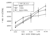
도 27은 조사(照射)된 단핵구, 및 다양한 농도의 TT830, Fab22-TT830 TT 또는 TT947과 함께 인큐베이션된 T 세포의 증식을 나타내는 그래프로, 융합 단백질 Fab22-TT830은 TT830에 비해 Th 에피토프의 표출을 약 1000배 증가시킨다.FIG. 27 is a graph showing the proliferation of irradiated monocytes and T cells incubated with various concentrations of TT830, Fab22-TT830 TT or TT947, wherein the fusion protein Fab22-TT830 reduced expression of Th epitopes relative to TT830.
도 28은 T 세포와 항원을 첨가하기 전에 포화량의 mAb22 F(ab')2와 함께 미리 인큐베이션시키거나 시키지 않은 채로, 1000 nM의 TT830 또는 10 nM의 FAb22-TT830 및 단핵구와 함께 인큐베이션된 T 세포의 증식을 보여주는 막대 그래프이다.FIG. 28 shows T cells incubated with 1000 nM TT830 or 10 nM FAb22-TT830 and monocytes, with or without pre-incubation with saturated amounts of mAb22 F (ab ') 2 prior to addition of T cells and antigen Bar graph showing the proliferation of
도 29는 IgG의 부재 (대조군) 또는 존재하에, 단핵구 및 5 nM의 Fab22-TT830 또는 1000 nM의 TT830과 함께 인큐베이션된 T 세포의 증식을 나타내는 막대 그래프 이다.FIG. 29 is a bar graph showing proliferation of T cells incubated with monocytes and 5 nM Fab22-TT830 or 1000 nM TT830 in the absence (control) or presence of IgG.
도 30의 패널 A 및 B는 단핵구 및 다양한 농도의 TT830 또는 Fab22-TT830과 함께 2일 동안 배양된 T 세포의 상층액에서 IFN-γ (패널 A) 및 IL-4 (패널 B)의 농도를 보여주는 그래프이다.Panels A and B of FIG. 30 show the concentrations of IFN-γ (panel A) and IL-4 (panel B) in supernatants of T cells cultured for 2 days with monocytes and various concentrations of TT830 or Fab22-TT830. It is a graph.
도 31은 단핵구 및 다양한 농도의 TT833S, Fab22-TT833S 또는 TT830과 함께 인큐베이션된 T 세포의 증식을 도시하는 그래프이다.FIG. 31 is a graph depicting proliferation of T cells incubated with monocytes and various concentrations of TT833S, Fab22-TT833S or TT830.
도 32는 다양한 농도의 TT833S와 함께 밤새 미리 인큐베이션되고, TT830 및 단핵구와 함께 2일 동안 인큐베이션된 T 세포의 증식을 나타내는 그래프이다.32 is a graph showing the proliferation of T cells preincubated overnight with various concentrations of TT833S and incubated for 2 days with TT830 and monocytes.
도 33은 다양한 농도의 TT833S 또는 FAb22-TT833S와 함께 밤새 미리 인큐베이션되고, TT830 및 단핵구와 함께 2일 동안 인큐베이션된 T 세포의 증식 억제율을 나타내는 그래프이다.FIG. 33 is a graph showing the proliferation inhibition rate of T cells preincubated with various concentrations of TT833S or FAb22-TT833S overnight and incubated with TT830 and monocytes for 2 days.
도 34는, 먼저 TT830과 함께 4 시간 동안 인큐베이션 (예비-펄스)된 후, T 세포를 첨가하기 전에 10 μM의 TT833S 또는 0.1 μM의 Fab22-TT833S와 함께 밤새 인큐베이션 (채이스)된 다음에, 단핵구와 함께 2일 동안 인큐베이션된 T 세포의 증식을 나타내는 막대 그래프이다.34 is first incubated (pre-pulsed) with TT830 for 4 hours, followed by overnight incubation (chase) with 10 μM of TT833S or 0.1 μM of Fab22-TT833S before adding T cells, followed by monocytes and Bar graph showing proliferation of T cells incubated together for 2 days.
도 35는 단핵구 및 TT830, Fab22-TT830, TT833S 및 Fab22-TT833S와 함께 배양된 T 세포의 상층액에서 인터페론-γ (IFN-γ) 및 IL-4의 농도를 나타내는 막대 그래프이다.FIG. 35 is a bar graph showing the concentration of interferon-γ (IFN-γ) and IL-4 in supernatants of T cells cultured with monocytes and TT830, Fab22-TT830, TT833S and Fab22-TT833S.
도 36은 배지만, TT833S와 함께, 또는 Fab22-TT833S와 함께 단핵구로 1일 동안 자극된 후, 단핵구 및 다양한 농도의 TT830으로 2일 동안 재자극된 T 세포의 증 식을 나타내는 그래프로, 이는 TT833S 및 Fab22-TT833S가 T 세포의 면역성 결여 (anergy)를 초래하지 않음을 의미한다.FIG. 36 is a graph showing proliferation of T cells stimulated with monocytes with monocytes, but with TT833S, or with Fab22-TT833S for 1 day, and then restimulated with monocytes and various concentrations of TT830 for 2 days, TT833S and It means that Fab22-TT833S does not lead to anergy of T cells.
도 37은 FcγRⅠ (H22)에 대한 1개의 결합 특이적 부위 및 발암성 배 (carcinoembryonic) 항원 (CEA)에 대한 1개의 결합 특이적 부위를 갖는 단일쇄 이중특이적 분자를 코딩하는 2개의 발현 작제물 (작제물 321 및 323), 및 FcγRⅠ에 대한 1개의 결합 특이적 부위를 갖는 단일쇄 항체를 코딩하는 1개의 발현 작제물을 나타내는 도식이다. 코딩 영역은 CMV 프로모터 (CMV Pr)의 조절하에 있다. 항체의 중쇄 (VH) 및 경쇄 (LH)로부터의 가변 영역 이외에, 이들 작제물에 의해 코딩되는 단백질은 c-myc으로부터의 펩티드 (c-myc) 및 헥사-히스티딘 펩티드 (H-6)에 융합되어 있다.FIG. 37 shows two expression constructs encoding single chain bispecific molecules having one binding specific site for FcγRI (H22) and one binding specific site for carcinoembryonic antigen (CEA). (
도 38은 발현 작제물 321 (321-A5 및 321-B4)에 의해 코딩되는 단일쇄 이중특이적 분자 H22-항-CEA의 결합 정도, 및 작제물 225 (225-C2)에 의해 코딩되는 단일쇄 H22 항체의 결합 정도를 이중기능성 ELISA로 측정하여 나타낸 막대 그래프이다.FIG. 38 shows the extent of binding of single chain bispecific molecule H22-anti-CEA encoded by expression constructs 321 (321-A5 and 321-B4), and single chain encoded by construct 225 (225-C2). It is a bar graph showing the degree of binding of the H22 antibody measured by bifunctional ELISA.
도 39는 단일쇄 인간화 항-FcγRⅠ 항체의 핵산 서열, 및 이 핵산에 의해 코딩되는 아미노산 서열을 나타낸다.39 shows the nucleic acid sequence of a single chain humanized anti-FcγRI antibody and the amino acid sequence encoded by this nucleic acid.
도 40은 FcγRⅠ에 대한 1개의 결합 특이적 부위 및 CEA에 대한 1개의 결합 특이적 부위를 갖는 단일쇄 이중특이적 분자의 핵산 서열, 및 이 핵산에 의해 코딩되는 아미노산 서열을 나타낸다.FIG. 40 shows the nucleic acid sequence of a single chain bispecific molecule having one binding specific site for FcγRI and one binding specific site for CEA, and the amino acid sequence encoded by this nucleic acid.
도 41은 항원에 연결된 다중 Fab' 단편으로 이루어진 멀티머 복합체의 구성 을 나타내는 개략도이다.Figure 41 is a schematic diagram showing the configuration of a multimer complex consisting of multiple Fab 'fragments linked to the antigen.
도 42a는 정제된 M22 F(ab')2 멀티머 (레인 1) 및 화학적으로 연결된 M22 Fab' 멀티머 복합체 (레인 2)를 나타내는 비-환원 SDS-PAGE 겔이다. 도42b는 M22 F(ab')3 +와 함께 인큐베이션시키면 투여량에 의존적인 방식으로 대식세포의 표면 상에서 CD64의 발현이 최대 50%까지 감소함을 나타내는 그래프이다.42A is a non-reducing SDS-PAGE gel showing purified M22 F (ab ') 2 multimers (lane 1) and chemically linked M22 Fab' multimer complexes (lane 2). Figure 42b is M22 F (ab ') is a graph showing that the expression is reduced by up to 50% of CD64 on the surface of the macrophage to-dependent manner when incubated with the 3 + dose.
도 43a는 한배에서 나온 비-트랜스제닉 자손에 비해, 3회 면역화된 CD64 트랜스제닉 생쥐 모두에서 높은 타이터의 M22-특이적 항체가 생성됨을 보여준다. 도 43b는 FcγRⅠ (CD64) 트랜스제닉 생쥐를 단지 0.25 mg의 멀티머 복합체만으로 면역화시키더라도 검출 가능한 면역 반응이 유도됨을 보여준다.43A shows that high titer M22-specific antibodies are produced in all three immunized CD64 transgenic mice compared to non-transgenic offspring from litter. 43B shows that immunizing FcγRI (CD64) transgenic mice with only 0.25 mg of the multimer complex induces a detectable immune response.
도 44a 및 44b는 FcγRⅠ (CD64) 트랜스제닉 생쥐와 비-트랜스제닉 생쥐의 면역 반응을 항-520C9 및 항-M22 타이터에 의해 판단하여 나타낸 도식이다. M22 멀티머 복합체에 커플링된 쥐의 항체 520C9의 Fab' 단편으로 생쥐를 면역화시켰다.44A and 44B are schematics showing the immune responses of FcγRI (CD64) transgenic mice and non-transgenic mice judged by anti-520C9 and anti-M22 titers. Mice were immunized with Fab ′ fragments of mouse antibody 520C9 coupled to the M22 multimer complex.
도 45a는 항원과 연결된 1개의 M32.2 sFv 영역에 연결된 2개의 H22 sFv 영역을 포함하는, 유전자적으로 연결된 멀티머 복합체 (H22(2x)-32.2-항원 복합체)를 코딩하는 발현 벡터의 맵을 보여준다. 도 45b는 그 결과 생성된 멀티머 복합체의 도면이다.45A shows a map of an expression vector encoding a genetically linked multimer complex (H22 (2x) -32.2-antigen complex) comprising two H22 sFv regions linked to one M32.2 sFv region linked to an antigen. Shows. 45B is a diagram of the resulting multimer composite.
도 46은 M22 Fab x M22 Fab x M32 및 H22sFv-H22sFv-32.2sFv-gp75 멀티머 복합체가 FcγRⅠ에 효율적으로 결합하여 인터날리제이션을 유도할 수 있는 능력을 보여주는 막대 그래프이다. FIG. 46 is a bar graph showing the ability of M22 Fab x M22 Fab x M32 and H22sFv-H22sFv-32.2sFv-gp75 multimer complexes to efficiently bind FcγRI and induce internalization.
도 47a는 항원에 함께 연결된 2개의 H22 sFv 영역을 포함하는, 유전자적으로 연결된 멀티머 복합체 (H22(2x)-항원 멀티머 복합체)를 코딩하는 발현 벡터의 맵을 보여준다. 도 47b는 그 결과 생성된 멀티머 복합체의 도면이다.47A shows a map of an expression vector encoding a genetically linked multimer complex (H22 (2x) -antigen multimer complex) comprising two H22 sFv regions linked together to an antigen. 47B is a diagram of the resulting multimer composite.
도 48은 H22 Fab', H22sFv2-EGF 멀티머 및 H22 Fab2 멀티머를 이용한 결합 경쟁 분석의 결과를 보여주는 그래프이다. 결과는 이들 멀티머에 접합된 M22-피코에리트린의 결합 억제에 의해 나타난다.FIG. 48 is a graph showing the results of binding competition assays using H22 Fab ', H22sFv2-EGF multimers and H22 Fab2 multimers. The result is shown by the inhibition of binding of M22-phycoerythrin conjugated to these multimers.
도 49a는 항원과 연결된 3개의 H22 sFv 단편을 포함하는, 유전자적으로 연결된 멀티머 복합체 (H22(3x)-항원 멀티머 복합체)를 코딩하는 발현 벡터의 맵을 보여준다. 도 49b는 그 결과 생성된 멀티머 복합체의 도면이다.49A shows a map of an expression vector encoding a genetically linked multimer complex (H22 (3x) -antigen multimer complex) comprising three H22 sFv fragments linked to an antigen. 49B is a diagram of the resulting multimer composite.
도 50은 H22(3x)-CEA 멀티머 복합체가 FcγRⅠ의 인터날리제이션을 유도할 수 있음을 다른 멀티머 복합체와 비교해서 보여주는 막대 그래프이다.FIG. 50 is a bar graph showing that H22 (3x) -CEA multimer complexes can induce the internalization of FcγRI as compared to other multimer complexes.
도 51은 항-CD89 (14A8) HuMAb 융합 분자의 생성을 나타내는 개략도이다. pJZ906 (14Afd-EGF) 및 pJZ907 (14A8 L-쇄)를 전기천공법에 의해 NSO 세포로 동시 형질감염시키고, G418과 하이그로마이신을 함유하는 배지에서 선별하였다. pJZ909 (14A8sFv-EGF)를 유사하게 형질감염시키고, G418을 함유하는 배지에서 선별하였다. 융합 단백질을 발현하는 세포주를 제한 희석 클로닝에 의해 서브클로닝하였다.51 is a schematic showing the generation of anti-CD89 (14A8) HuMAb fusion molecules. pJZ906 (14Afd-EGF) and pJZ907 (14A8 L-chain) were cotransfected into NSO cells by electroporation and selected in media containing G418 and hygromycin. pJZ909 (14A8sFv-EGF) was similarly transfected and selected in medium containing G418. Cell lines expressing the fusion protein were subcloned by restriction dilution cloning.
도 52a는 A431 세포 상의 EGF-R에 대한 14A8Fab'-EGF 융합 작제물의 결합 (유동 세포계수법으로 측정)을 나타내는 그래프이다. 도 52b는 A431 세포 상의 EGF-R에 대한 14A8sFv-EGF 융합 작제물의 결합을 나타내는 그래프이다. A431 세포를 피코에리트린에 접합된 염소 항-인간 IgG Fab-2로 염색하였다. EGF를 포함하지 않는 14A8 (인간 모노클로날 항-FcαR 항체, 14.1이라고도 불리움)의 Fab-2 단편을 대조군으로 사용하였다. 도 52c는 융합 단백질 및 상업적으로 얻을 수 있는 EGF가 A431 세포에 대한 결합에서 EGF-FITC 접합체 (7 nM)와 경쟁함을 보여준다.FIG. 52A is a graph showing binding of 14A8Fab'-EGF fusion constructs (measured by flow cytometry) to EGF-R on A431 cells. FIG. 52B is a graph showing binding of the 14A8sFv-EGF fusion construct to EGF-R on A431 cells. A431 cells were stained with goat anti-human IgG Fab-2 conjugated to phycoerythrin. Fab-2 fragment of 14A8 (human monoclonal anti-FcαR antibody, also referred to as 14.1) without EGF was used as a control. 52C shows that the fusion protein and commercially available EGF compete with the EGF-FITC conjugate (7 nM) in binding to A431 cells.
도 53a는 14A8Fab'-EGF 융합 작제물의 U937 세포에 대한 결합 (유동 세포계수법으로 측정)을 보여주는 그래프이다. U937 세포에 결합된 융합 단백질을 피코에리트린에 접합된 염소 항-인간 IgG Fab-2로 염색하였다. EGF-R에 특이적인, 인간화 mAb H415의 Fab-2 단편을 대조군으로 사용하였다. 도 53b는 14A8sFv-EGF 융합 작제물의 U937 세포에 대한 결합을 보여준다. 이 실험은, 다양한 농도의 14A8sFv-EGF가 14A8Fab'-EGF (23 nM)의 결합을 억제하는 능력에 대해 평가한 것 이외에, 도 53a에서와 유사한 방식으로 수행하였다. H22sFv-EGF 융합 단백질을 대조군으로 사용하였다. mAb H22는 U937 세포 상의 상이한 Fc 수용체인 CD64 (FcγRⅠ)에 결합한다.53A is a graph showing binding (measured by flow cytometry) to U937 cells of the 14A8Fab'-EGF fusion construct. Fusion proteins bound to U937 cells were stained with goat anti-human IgG Fab-2 conjugated to phycoerythrin. Fab-2 fragment of humanized mAb H415, specific for EGF-R, was used as a control. 53B shows binding to U937 cells of the 14A8sFv-EGF fusion construct. This experiment was performed in a similar manner as in FIG. 53A, except that various concentrations of 14A8sFv-EGF were evaluated for their ability to inhibit binding of 14A8Fab'-EGF (23 nM). H22sFv-EGF fusion protein was used as a control. mAb H22 binds to CD64 (FcγRI), a different Fc receptor on U937 cells.
도 54는 투여량에 의존적인, A431 세포의 융합 단백질-매개의 세포독성을 보여주는 그래프이다. 인큐베이션 시간을 16 내지 18시간으로 하고, 이펙터 세포 대 표적 세포의 양 (단핵구 및 PMN)을 100:1로 하여 크롬 방출 분석을 수행하였다. 이펙터 세포에는 신선한 단핵구 (도 54a), PMN (도 54b) 및 전체 혈액 (도 54c)이 포함되었다. 14A8의 Fab-2 단편을 대조군으로 사용하였다.FIG. 54 is a graph showing fusion protein-mediated cytotoxicity of A431 cells, depending on the dose. The incubation time was 16-18 hours and the chromium release assay was performed with the amount of effector cells to target cells (monocytes and PMN) at 100: 1. Effector cells included fresh monocytes (FIG. 54A), PMN (FIG. 54B) and whole blood (FIG. 54C). Fab-2 fragment of 14A8 was used as a control.
도 55는 도 54a 내지 54c의 결과를 비교하는 막대 그래프이다.55 is a bar graph comparing the results of FIGS. 54A-54C.
도 56a는 A431 세포의 14A8Fab'-EGF (1㎍/ml) 융합 단백질-매개의 세포독성이 14A8의 Fab-2 단편에 의해 억제됨을 보여주는 막대 그래프이다. 도 56b는 A431 세포의 14A8sFv-EGF (1㎍/ml) 융합 단백질-매개의 세포독성도 14A8의 Fab-2 단편 (40 ㎍/ml)에 의해 억제됨을 보여주는 막대 그래프이다.56A is a bar graph showing that the 14A8Fab'-EGF (1 μg / ml) fusion protein-mediated cytotoxicity of A431 cells is inhibited by Fab-2 fragment of 14A8. FIG. 56B is a bar graph showing 14A8sFv-EGF (1 μg / ml) fusion protein-mediated cytotoxicity of A431 cells is inhibited by Fab-2 fragment (40 μg / ml) of 14A8.
도 57은 이중특이적 단일쇄 융합 분자인 931 및 934와, 이들의 모체인 HuMAb를 나타내는 개략도이다. 14.1 및 3Fe의 가변 경쇄 및 중쇄 영역을 사용하여 931 및 934 (EGF와 함께) 작제물을 생성하였다.FIG. 57 is a schematic showing the bispecific single
도 58은 양성 대조군으로 사용되는, 14.1 및 3F2 (항-HER2)의 Fab' 단편으로 구성되고 화학적으로 접합된 이중특이적 분자를 나타내는 개략도이다.58 is a schematic showing bispecific molecules chemically conjugated and composed of Fab 'fragments of 14.1 and 3F2 (anti-HER2), used as positive controls.
도 59a는 트랜스펙토마 (transfectoma)의 상층액에서 발현된 융합 단백질 931 및 934의 유동 세포계수 분석으로 측정한 스크리닝 (결합) 분석을 나타내는 막대 그래프이다. 형질감염된 (931 및 934) 상층액 및 형질감염되지 않은 (NSO) 상층액을 종양 세포주인 SKBR-3 또는 A431과 함께 인큐베이션시켰다. 융합 단백질의 결합은 피코에리트린에 접합된 염소 항-인간 IgG Fab-2로 염색하여 검출하였다. 도 59b는 융합 단백질 931 및 934의 ADCC 분석을 보여주는 막대 그래프이다. 인큐베이션 시간을 16 내지 18시간으로 하고, 이펙터 세포 (단핵구, PMN) 대 표적 세포 (SKBR-3, A431)의 양을 100:1로 하여 크롬 방출 분석을 수행하였다. 종양 세포 사멸은 특이적 용해를 매개하는지에 대해 형질감염된 (931 및 934) 상층액 및 형질감염되지 않은 (NSO) 상층액을 사용하여 검출하였다.FIG. 59A is a bar graph showing the screening (binding) assay measured by flow cytometry analysis of
도 60a는 단일쇄 융합 단백질 931 및 934의 U937 세포에 대한 결합 (유동 세포계수법으로 측정)을 보여주는 그래프이다. U937 세포에 결합하지 않는 항-EGF-R mAb인 425의 Fab' 단편을 음성 대조군으로 사용하였다. 도 58에 도시된 화학적으 로 연결된 이중특이적 분자 (14.1 x 3F2)를 양성 대조군으로 사용하였다. 도 60b는 단일쇄 융합 단백질 931 및 934의 SKBR-3 세포에 대한 결합 (유동 세포계수법으로 측정)을 보여주는 그래프이다. 14.1 Fab-2를 음성 대조군으로 사용하였다. 도 58에 도시된 화학적으로 연결된 이중특이적 부위 (14.1 x 3F2)를 양성 대조군으로 사용하였다.60A is a graph showing binding (as measured by flow cytometry) to U937 cells of single
도 61은 단일쇄 융합 단백질 934의 A431 세포에 대한 결합 (유동 세포계수법으로 측정)을 보여주는 그래프이다. 이 실험은 EGF-R을 과다 발현하는 A431 종양 세포를 사용한 것 이외에, 도 60에서와 동일한 방법으로 수행하였다. EGF에 융합된 14.1의 sFv 단편으로 이루어진 융합 단백질을 양성 대조군으로 사용하였으며, 14.1 Fab-2를 음성 대조군으로 사용하였다.FIG. 61 is a graph showing binding of single
도 62는 단일쇄 융합 단백질 931에 의한 SKBR3 (도 62a) 및 BT474 (도 62b) 종양 세포의 PMN (이펙터 세포)-매개의 세포 독성을 보여주는 그래프이다. 인큐베이션 시간을 16 내지 18시간으로 하고, 이펙터 세포 대 표적 세포의 양을 100:1로 하여 크롬 방출 분석을 수행하였다. 14.1 Fab-2를 각 실험에서 음성 대조군으로 사용하였다.FIG. 62 is a graph showing PMN (effector cell) -mediated cytotoxicity of SKBR3 (FIG. 62A) and BT474 (FIG. 62B) tumor cells by single
도 63은 단일쇄 융합 단백질 931에 의한 SKBR3 (도 63a) 및 BT474 (도 63b) 종양 세포의 단핵구 (이펙터 세포)-매개의 세포 독성을 보여주는 그래프이다. 인큐베이션 시간을 16 내지 18시간으로 하고, 이펙터 세포 대 표적 세포의 양을 100:1로 하여 크롬 방출 분석을 수행하였다. 14.1 Fab-2를 각 실험에서 음성 대조군으로 사용하였다.
FIG. 63 is a graph showing monocyte (effector cell) -mediated cytotoxicity of SKBR3 (FIG. 63A) and BT474 (FIG. 63B) tumor cells by single
도 64는 51Cr 방출로 측정한, 인간 항-HER2/neu x 항-CD89 이중특이적 분자 (14.1 x 3.F2)에 의해 매개된 다형핵 세포에 의한 SKBR-3 및 BT-474 종양 세포의 항체 의존성 세포 용해를 도시하는 그래프이다.64 shows SKBR-3 and BT-474 tumor cells by polymorphonuclear cells mediated by human anti-HER2 / neu x anti-CD89 bispecific molecule (14.1 x 3.F2), measured by 51 Cr release. It is a graph depicting antibody dependent cell lysis.
도 65는 51Cr 방출로 측정한, 인간 항-HER2/neu x 항-CD89 이중특이적 분자 (14.1 x 3.F2)에 의해 매개된 단핵구에 의한 SKBR-3 및 BT-474 종양 세포의 항체 의존성 세포 용해를 도시하는 그래프이다.FIG. 65 shows antibody dependence of SKBR-3 and BT-474 tumor cells by monocytes mediated by human anti-HER2 / neu x anti-CD89 bispecific molecule (14.1 x 3.F2) measured by 51 Cr release A graph depicting cell lysis.
도 66은 51Cr 방출로 측정한, 인간 항-HER2/neu x 항-CD89 이중특이적 분자 (14.1 x 3.F2)에 의해 매개된 전체 혈액에 의한 BT-474 종양 세포의 항체 의존성 세포 용해를 도시하는 그래프이다.66 shows antibody dependent cell lysis of BT-474 tumor cells by whole blood mediated by human anti-HER2 / neu x anti-CD89 bispecific molecule (14.1 x 3.F2), measured by 51 Cr release. It is a graph to show.
다중특이적 분자Multispecific molecules
본 발명은 면역 세포를 표적으로 하는, 재조합 및 화학적으로 합성된 다중특이적 분자에 관한 것이다. 이 분자는, 하나 이상의 표적이 면역 세포의 표면 상의 분자인 다중의 (2개 이상의) 별개 표적에 결합하기 때문에 "다중특이적"이다. 한 실시양태에서, 본 발명의 다중특이적 분자는 이펙터 세포 상의 분자 (바람직하게는 Fc 수용체)에 결합하는 하나 이상의 부분, 및 종양 세포 또는 병원체 상의 항원과 같은 상이한 표적에 결합하는 하나 이상의 부분 (예를 들어, 둘, 셋, 넷 또는 그 이상의 부분)으로 이루어진 분자를 포함한다. 다른 실시양태에서, 본 발명의 다중특이적 분자는, 1개 이상의 항원에 결합된 항원 표출 세포 (APC) 상의 분자 (예를 들어, Fc 수용체)에 결합하는 다중 (즉, 둘 이상) 부분으로 구성된 항원 "멀티머 복합체"를 포함한다. 이들 멀티머 복합체는 APC에 대한 자기항원과 같은 항원을 표적으로 하여, APC에 의한 항원의 인터날리제이션 (내포작용), 가공 및(또는) 표출을 유도하고(하거나) 증진시킨다. 따라서, 이들 분자는 자기항원과 같이 정상적으로는 비-면역원성인 단백질에 대한 생체내 또는 시험관내 면역 반응을 유도하거나 증진시키는데 사용될 수 있다.The present invention is directed to recombinant and chemically synthesized multispecific molecules that target immune cells. This molecule is "multispecific" because one or more targets bind to multiple (two or more) separate targets, which are molecules on the surface of immune cells. In one embodiment, multispecific molecules of the invention comprise one or more portions that bind to molecules on the effector cell (preferably Fc receptors) and one or more portions that bind to different targets, such as antigens on tumor cells or pathogens (eg, For example, two, three, four or more moieties). In other embodiments, multispecific molecules of the invention consist of multiple (ie, two or more) moieties that bind to a molecule (eg, an Fc receptor) on an antigen expressing cell (APC) that is bound to one or more antigens. Antigen "multimer complex". These multimer complexes target antigens, such as autoantigens against APC, inducing and / or enhancing the internalization (incorporation), processing and / or expression of antigens by APC. Thus, these molecules can be used to induce or enhance immune responses in vivo or in vitro against proteins that are normally non-immunogenic, such as autoantigens.
특히 바람직한 실시양태에서, 본 발명의 다중특이적 분자는 Fc 수용체, 통상적으로는 FcδR (예를 들어, FcδRⅠ) 또는 FcαR에 결합하는 인간 또는 인간화된 (키메라) 항체 또는 항체 단편을 포함한다. 또한, 다중특이적 분자는 상이한 표적 세포 (예를 들어, 종양 세포) 상의 항원에 결합하는 1개 이상의 결합 특이적 부위 (예를 들어, 리간드 및(또는) 다른 인간 항체나 항체 단편)를 포함한다. 그러한 다중특이적 분자는 화학적으로 연결되거나, 또는 융합 단백질로서 유전자적으로 발현될 수 있다. 더욱이, 다중특이적 분자는, 예를 들어 FcδRⅠ 또는 FcαR을 통해 이펙터 세포를 표적으로 하기 때문에, 이들은 이펙터 세포 (예를 들어, PMN, 대식세포 및 단핵구)-매개의 표적 (예를 들어, 종양) 세포 사멸을 유도하거나, 또는 항원에 결합된 경우에 항원 표출 (AP) 이펙터 세포 (예를 들어, 수지상 세포)에 의한 항원 표출을 증진시키는데 이용될 수 있다.In a particularly preferred embodiment, multispecific molecules of the invention comprise human or humanized (chimeric) antibodies or antibody fragments which bind to Fc receptors, typically FcδR (eg FcδRI) or FcαR. Multispecific molecules also include one or more binding specific sites (eg, ligands and / or other human antibodies or antibody fragments) that bind to antigens on different target cells (eg, tumor cells). . Such multispecific molecules can be chemically linked or genetically expressed as fusion proteins. Moreover, since multispecific molecules target effector cells, for example via FcδRI or FcαR, they are effector cells (eg PMN, macrophages and monocytes) -mediated targets (eg tumors) It can be used to induce cell death or to enhance antigen presentation by antigen presenting (AP) effector cells (eg dendritic cells) when bound to an antigen.
본 발명의 다중특이적 분자를 설명하기 위해 사용되는 "∼에 결합하는 부분"이란 용어는 "∼에 대한 결합 특이적 부위"와 교환 가능하게 사용되는 것이다. 이들 용어는 표적 에피토프에 결합하는 다중특이적 분자의 영역을 지칭한다. 하기에 더욱 상세히 기재되는 바와 같이, 이들 부분 또는 결합 특이적 부위에는, 항체, 항체 단편 (예를 들어, Fab, Fab', F(ab')2, Fv 또는 단일쇄 Fv) 및 이들의 모방체 (예를 들어, 항체 또는 항체 단편의 결합을 "모방"하는 펩티드, 화학적 모방체 및 유기 모방체)를 포함하나 이에 한정되지는 않는, 표적 에피토프에 결합할 수 있는 모든 화합물이 포함된다. 구체적인 실시양태에서, 결합 특이적 부위는 H22 (ATCC 기탁번호 CRL11177), M22 (ATCC 기탁번호 HB12147), M32.2 (ATCC 기탁번호 HB9469) 또는 이들의 항원 결합 단편으로부터 선택되며, FcγRⅠ (CD64)에 결합하는 항체를 포함한다. 인간화 항체인 H22 및 그의 쥐 등가물인 M22는 FcγRⅠ의 천연 리간드 (IgG) 결합 부위의 바깥쪽에 결합하는 이점이 있기 때문에, 생체내에 투여시에도 내생성 리간드에 의해 차단되거나 경쟁에서 도태되지 않는다. 다른 구체적인 실시양태에서, 결합 특이적 부위는 FcαR (CD89)에 결합하는 인간 모노클로날 항체 14.1, 또는 HER2에 결합하는 인간 모노클로날 항체 3.F2로부터 선택되는 하나 이상의 항체를 포함한다.The term "part that binds to" used to describe the multispecific molecule of the present invention is used interchangeably with "binding specific site for". These terms refer to regions of multispecific molecules that bind to target epitopes. As described in more detail below, these moieties or binding specific sites include antibodies, antibody fragments (eg, Fab, Fab ', F (ab') 2 , Fv or single chain Fv) and mimetics thereof. All compounds capable of binding to the target epitope are included, including but not limited to (eg, peptides, chemical mimetics, and organic mimetics) that "imitate" the binding of an antibody or antibody fragment. In a specific embodiment, the binding specific site is selected from H22 (ATCC Accession No. CRL11177), M22 (ATCC Accession No. HB12147), M32.2 (ATCC Accession No. HB9469), or antigen binding fragments thereof, to FcγRI (CD64). Antibodies that bind. Humanized antibody H22 and its murine equivalent, M22, have the advantage of binding to the outside of the natural ligand (IgG) binding site of FcγRI, and therefore are not blocked by competition with endogenous ligands or in competition with administration in vivo. In other specific embodiments, the binding specific site comprises one or more antibodies selected from human monoclonal antibody 14.1 that binds FcαR (CD89), or human monoclonal antibody 3.F2 that binds HER2.
본원에 사용되는 항체의 "항원-결합 부분" (또는 단순히 "항체 부분")이란 용어는 항원에 특이적으로 결합하는 능력을 지닌 항체의 하나 이상의 단편을 의미한다 (예를 들어, Fc 수용체). 항체의 항원-결합 기능은 전장 항체의 단편에 의해 수행될 수 있다. 항체의 "항원-결합 부분"이라는 용어에 포함되는 결합 단편의 예로는 (i) VL, VH, CL 및 CH1 도메인으로 이루어진 1가 단편인 Fab 단편; (ii) 힌지 영역에서 디술피드 결합에 의해 연결된 2개의 Fab 단편을 포함하는 2가 단편인 F(ab')2 단편; (iii) VH 및 CH1 도메인으로 이루어진 Fd 단편; (iv) 항체 단일 암 (arm)의 VL 및 VH 도메인으로 이루어진 Fv 단편; (v) VH 도메인으로 이루어진 dAb 단편 (Ward et al., (1989) Nature 341:544-546); 및 (vi) 단리된 상보성 결정 영역 (CDR)이 포함된다. 또한, Fv 단편의 2개 도메인인 VL 및 VH가 별개의 유전자에 의해 코딩되기는 하지만, VL과 VH 영역이 짝을 이루어 1가 분자를 형성하도록 이들을 단일 단백질 쇄로 만들어줄 수 있는 합성 링커에 의한 제조합 방법을 이용하여 이들을 결합시킬 수 있다 (단일쇄 Fv (scFv)로 알려짐; 문헌 [Bird et al. (1988) Science 242:423-426] 및 [Huston et al. (1988) Proc. Natl. Acad. Sci. USA 85:5879-5883] 참조). 또한, 그러한 단일쇄 항체는 항체의 "항원-결합 부분"이라는 용어에 포함되는 것이다. 이들 항체 단편은 당업자에게 공지된 종래의 기술을 이용하여 얻을 수 있고, 단편은 본래 항체와 동일한 방식으로 유용성을 스크리닝할 수 있다.As used herein, the term “antigen-binding portion” (or simply “antibody portion”) of an antibody refers to one or more fragments of an antibody that have the ability to specifically bind an antigen (eg, Fc receptor). The antigen-binding function of an antibody can be performed by fragments of full length antibodies. Examples of binding fragments encompassed by the term “antigen-binding portion” of an antibody include (i) a Fab fragment which is a monovalent fragment consisting of the VL, VH, CL and CH1 domains; (ii) a F (ab ') 2 fragment that is a bivalent fragment comprising two Fab fragments linked by disulfide bonds in the hinge region; (iii) a Fd fragment consisting of the VH and CH1 domains; (iv) a Fv fragment consisting of the VL and VH domains of an antibody single arm; (v) dAb fragment consisting of the VH domain (Ward et al., (1989) Nature 341: 544-546); And (vi) isolated complementarity determining regions (CDRs). In addition, although the two domains of the Fv fragment, VL and VH, are encoded by separate genes, they are produced by synthetic linkers that can make them into a single protein chain so that the VL and VH regions are paired to form a monovalent molecule. Methods can be used to bind them (known as single-chain Fv (scFv); Bird et al. (1988) Science 242: 423-426 and Houston et al. (1988) Proc. Natl. Acad. Sci. USA 85: 5879-5883). Such single chain antibodies are also included within the term "antigen-binding portion" of an antibody. These antibody fragments can be obtained using conventional techniques known to those skilled in the art, and the fragments can be screened for utility in the same manner as the original antibodies.
Ⅰ. 항원 표출 세포 (APC)를 표적으로 하는 항원 멀티머 복합체를 포함하는 다중특이적 분자I. Multispecific molecules comprising antigen multimer complexes targeting antigen presenting cells (APCs)
본 발명의 항원 멀티머 복합체는 하나 이상의 항원에 연결된 항원 표출 세포 표면 상의 성분 (APC 세포 표면 성분)을 표적으로 하는 다중 부분 또는 결합 특이적 부위를 포함한다. 다중 결합 특이적 부위는 APC 세포 표면 성분의 동일한 에피토프 또는 상이한 에피토프들에 결합하거나, 또는 상이한 APC 세포 표면 성분들에 결합할 수 있다. 바람직한 실시양태에서, 멀티머 복합체는 APC 세포 표면 성분( 들)에 대한 3개 이상의 결합 특이적 부위를 포함한다. 표적화를 위한 APC 상의 적합한 성분에는, 결합 특이적 부위와의 결합시 인터날리제이션을 매개하여 상기 결합 특이적 부위에 연결된 항원이 APC에 의해 효율적으로 인터날리제이션되고 표출되게 하는 성분들이 포함된다.Antigen multimer complexes of the invention comprise multiple moieties or binding specific sites that target components (APC cell surface components) on antigen expressing cell surfaces linked to one or more antigens. Multiple binding specific sites can bind to the same epitope or different epitopes of APC cell surface components, or to different APC cell surface components. In a preferred embodiment, the multimer complex comprises three or more binding specific sites for APC cell surface component (s). Suitable components on the APC for targeting include those that mediate internalization upon binding to binding specific sites such that antigens linked to the binding specific sites are efficiently internalized and expressed by APC.
본원에 사용되는 "항원 표출 세포" 또는 "APC"라는 용어는 항원을 인터날리제이션되게 하고 가공하여 항원 결정부가 면역계 (예를 들어, 클래스 Ⅱ-MHC 제한 헬퍼 T 림프구)에 의해 인식될 수 있는 방식으로 MHC-복합체로서 세포의 표면에 표출되게 할 수 있는 면역 세포를 의미한다. 세포가 APC로서 기능할 수 있게 하는 2가지 필수 특성은 내포된 항원을 가공하는 능력 및 클래스 Ⅱ MHC 유전자 산물의 발현이다. 헬퍼 T 세포에 대해 가장 잘 정의된 APC에는 단핵 식세포 (예를 들어, 대식세포), B 림프구, 수지상 세포, 피부의 랑게르한스 세포, 및 인간의 내피 세포가 포함된다.As used herein, the term “antigen expressing cell” or “APC” refers to the manner in which antigens are internalized and processed so that antigenic determinants can be recognized by the immune system (eg, class II-MHC restricted helper T lymphocytes). As an MHC-complex, it means an immune cell capable of being expressed on the surface of the cell. Two essential properties that allow cells to function as APCs are the ability to process the nested antigen and the expression of class II MHC gene products. The best defined APCs for helper T cells include mononuclear phagocytes (eg, macrophages), B lymphocytes, dendritic cells, Langerhans cells of the skin, and human endothelial cells.
바람직한 실시양태에서, 결합 특이적 부위에 의해 표적화된 APC 세포 표면 성분은 Fc 수용체, 통상적으로는 FcγR이다. 따라서, 본 발명의 구체적인 실시양태에서, 멀티머 복합체는 FcγRⅠ 상의 상이한 에피토프를 표적으로 하는 다중 결합 특이적 부위 (예를 들어, 2개 이상, 바람직하게는 3개 이상)을 포함한다. 별법으로, 멀티머는 FcγRⅠ 상의 동일한 에피토프를 표적으로 하는 다중 결합 특이적 부위를 포함할 수도 있다. 그러나, 다른 적합한 APC 세포 표면 성분도 표적이 될 수 있다. 그러한 성분은, 동물 (예를 들어, 인간 FcγRⅠ에 대한 트랜스제닉 생쥐)을 APC로 면역화시켜, 예를 들어 표준 분석 (예를 들면, 웨스턴 블롯)을 이용하 여 이 동물로부터 얻은 혈청에 의해 결합된 세포 표면 성분을 결정함으로써 확인할 수 있다. 이후에, 이들 APC 세포 표면 성분에 결합하는 화합물이 인터날리제이션되게 하는 능력에 대해 상기 성분들을 시험할 수 있다.In a preferred embodiment, the APC cell surface component targeted by the binding specific site is an Fc receptor, typically FcγR. Thus, in specific embodiments of the invention, the multimer complexes comprise multiple binding specific sites (eg, two or more, preferably three or more) that target different epitopes on FcγRI. Alternatively, the multimer may comprise multiple binding specific sites that target the same epitope on FcγRI. However, other suitable APC cell surface components can also be targeted. Such components can be used to immunize an animal (eg, a transgenic mouse against human FcγRI) with APC, for example, a cell bound by serum obtained from this animal, for example using standard assays (eg, Western blot). This can be confirmed by determining the surface component. Thereafter, the components can be tested for their ability to internalize compounds that bind to these APC cell surface components.
따라서, 한 실시양태에서, 멀티머 복합체는 인간 IgG 수용체와 같이 Fc 수용체에 결합하는 하나 이상의 항체 또는 그의 단편 (예를 들어, Fab, Fab', F(ab')2, Fv, 또는 단일쇄 Fv), 예를 들어 FcγRⅠ (CD64), FcγRⅠI (CD32) 및 FcγRⅠII (CD16)을 비롯한 Fc-감마 수용체 (FcγR)를 포함한다. 바람직한 Fcγ 수용체는 친화도가 높은 Fcγ 수용체인 FcγRⅠ이다. 그러나, 인간 IgA 수용체 (예를 들어, FcαRⅠ)와 같은 다른 Fc 수용체도 표적이될 수 있다. Fc 수용체는 APC, 예를 들어, 단핵구 또는 대식세포의 표면 상에 위치한다. 구체적인 실시양태에서, 멀티머 복합체는 수용체의 면역글로불린 (예를 들어, IgG 또는 IgA) 결합 부위와는 구별되는 다른 부위에서 Fc 수용체에 결합한다. 따라서, 멀티머 복합체의 결합은 면역글로불린의 생리적 양에 의해 차단되지 않는다. 바람직한 인간화 항-FcγR 모노클로날 항체는 PCT 출원 WO 94/10332호 및 미국 특허 제4,954,617호에 기재되어 있으며, 이들 문헌의 교시사항은 이 거명을 통해 그 전문이 본 명세서에 포함된다.Thus, in one embodiment, the multimer complex is one or more antibodies or fragments thereof (eg, Fab, Fab ', F (ab') 2 , Fv, or single chain Fv that bind to the Fc receptor, such as a human IgG receptor). ), For example Fc-gamma receptors (FcγR), including FcγRI (CD64), FcγRII (CD32) and FcγRII (CD16). Preferred Fcγ receptors are FcγRI, which is a high affinity Fcγ receptor. However, other Fc receptors, such as human IgA receptors (eg FcαRI), can also be targeted. Fc receptors are located on the surface of APCs, eg monocytes or macrophages. In specific embodiments, the multimer complex binds to the Fc receptor at another site that is distinct from the immunoglobulin (eg, IgG or IgA) binding site of the receptor. Thus, the binding of the multimer complex is not blocked by the physiological amount of immunoglobulins. Preferred humanized anti-FcγR monoclonal antibodies are described in PCT application WO 94/10332 and US Pat. No. 4,954,617, the teachings of which are incorporated herein by reference in their entirety.
본 명세서의 실시예에 기재되는 바와 같이, 본 발명의 항원 멀티머 복합체에 사용하기 적합한 구체적 인간화 및 쥐의 모노클로날 항-FcγRⅠ 항체 및 항체 단편에는, 인간화 항체 H22 (ATCC 기탁번호 CRL11177), 그의 쥐 대응부 M22 (ATCC 기탁번호 HB12147) 및 쥐의 M32.2 (ATCC 기탁번호 HB9469) 뿐만 아니라, 이들 항체의 항원 결합 단편이 포함되지만, 이에 한정되지는 않는다.As described in the Examples herein, specific humanized and murine monoclonal anti-FcγRI antibodies and antibody fragments suitable for use in the antigen multimer complexes of the invention include humanized antibody H22 (ATCC Accession No. CRL11177), Antigen binding fragments of these antibodies, as well as, but not limited to, murine counterpart M22 (ATCC Accession No. HB12147) and murine M32.2 (ATCC Accession No. HB9469).
APC를 표적으로 하는 본 발명의 멀티머 복합체는 하나 이상의 항원을 더 포함한다. 본원에 사용되는 "항원"이란 용어는 면역 세포에 의해 인식될 (즉, T 세포-매개의 면역 반응과 같은 면역 반응을 도출함) 수 있는 임의의 분자 (예를 들어, 단백질, 펩티드, 탄수화물 등)를 의미한다. 항원에는, 숙주에 정상적으로 존재하면서 정상적인 상태에서는 숙주로부터 면역 반응을 도출하지 않는 (면역계가 이들은 "외래" 물질이 아닌 "자기" 물질로 인식하기 때문임) "자기항원" 또는 "자가항원"이 포함된다. 특정 질병에서는, 항원이 정상적인 상태에서는 숙주에 존재하지 않기 때문에 숙주의 면역계에 의한 면역 반응을 도출시켜야 하지만, 그렇지 않는다. 즉, 이러한 항원들은 숙주의 면역계에 의한 인식을 "회피한다". 그러한 항원에는, 예를 들어 특정 종양 항원 및 병원성 (예를 들어, 바이러스성 및 세균성) 항원이 포함된다. 이런 상황에서는 면역 세포에 의한 항원의 인식을 유도하거나 증진시키는 방식으로 항원을 변형시켜, 항원이나 항원을 생성하는 세포 (생명체)를 숙주에서 제거하는 것이 바람직할 것이다. 즉, 면역 세포에 의해 더이상 "자기항원"으로 인식되지 않도록 항원을 변형시킨다.Multimer complexes of the invention targeting APC further comprise one or more antigens. As used herein, the term “antigen” refers to any molecule (eg, protein, peptide, carbohydrate, etc.) that can be recognized by an immune cell (ie, eliciting an immune response such as a T cell-mediated immune response). ). Antigens include "self-antigens" or "autoantigens" that normally exist in the host but do not elicit an immune response from the host under normal conditions (the immune system recognizes them as "self" substances, not "foreign" substances). do. In certain diseases, since the antigen is not present in the host under normal conditions, an immune response by the host's immune system must be elicited, but it is not. That is, these antigens “avoid” recognition by the host's immune system. Such antigens include, for example, certain tumor antigens and pathogenic (eg viral and bacterial) antigens. In such a situation, it would be desirable to modify the antigen in a manner that induces or enhances the recognition of the antigen by immune cells, thereby removing the antigen or cells that produce the antigen from the host. That is, the antigen is modified so that it is no longer recognized as an “self-antigen” by the immune cells.
본 발명의 항원 멀티머 복합체는 하나 이상의 항원을, 당업계에 공지된 표준 가교결합제 및 접합 프로토콜을 이용하여, 본원에 기재된 바와 같은 APC 상의 성분에 대한 다수의 (2개 이상의) 결합 특이적 부위에 화학적으로 연결시킴으로써 제조할 수 있다. 별법으로, 항원 멀티머 복합체는 또한 본원에 기재된 바와 같이, 단일 융합 단백질로서 재조합적으로 제조될 수 있다. Antigen multimer complexes of the invention may be used to provide one or more antigens to multiple (two or more) binding specific sites for components on APC as described herein using standard crosslinkers and conjugation protocols known in the art. It can be prepared by chemically linking. Alternatively, antigen multimer complexes may also be recombinantly prepared as a single fusion protein, as described herein.
따라서, 또다른 실시양태에서, 본 발명은 항원 멀티머 복합체를 코딩하는 핵산을 제공한다. 통상적으로, 핵산은 발현 벡터 내에서 멀티머 복합체의 발현을 조절하는 프로모터 및 기타 유전자 조절 서열들과 작동가능하게 연결되어 있다. 핵산이 다른 핵산 서열과 기능적인 관계에 놓여 있을 때, 이 핵산은 "작동가능하게 연결된" 것이다. 예를 들어, 프로모터 또는 인핸서가 코딩 서열의 전사에 영향을 미치는 경우에, 이 프로모터 또는 인핸서는 상기 코딩에 작동가능하게 연결된 것이다. 전사 조절 서열에 관하여, 작동가능하게 연결된 것이란 연결된 DNA 서열이 2개의 단백질 코딩 영역이 결합되는 곳에서 리딩 프레임에 맞게 인접해 있음을 의미한다. 스위치 (switch) 서열의 경우, 작동가능하게 연결된 것은 이 서열이 스위치 재조합을 초래할 수 있음을 나타낸다.Thus, in another embodiment, the present invention provides nucleic acids encoding antigen multimer complexes. Typically, the nucleic acid is operably linked with promoters and other gene regulatory sequences that regulate the expression of the multimer complex in the expression vector. When a nucleic acid is in a functional relationship with another nucleic acid sequence, the nucleic acid is "operably linked." For example, if a promoter or enhancer affects the transcription of a coding sequence, the promoter or enhancer is operably linked to the coding. With respect to transcriptional control sequences, operably linked means that the linked DNA sequence is contiguous in reading frame where the two protein coding regions are joined. In the case of a switch sequence, operably linked indicates that this sequence can result in switch recombination.
본원에 사용되는 "벡터"란 용어는 자신에 연결된 다른 핵산을 수송할 수 있는 핵산 분자를 의미한다. 벡터의 한 유형으로 "플라스미드"가 있는데, 이는 부가의 DNA 단편이 연결될 수 있는 환상의 이중 가닥 DNA 루프이다. 벡터의 다른 유형으로 바이러스 벡터가 있는데, 여기서 부가의 DNA 단편은 바이러스 게놈 내로 라이게이션될 수 있다. 어떤 벡터들은 자신이 도입되고자 하는 숙주에서 스스로 복제할 수 있다 (예를 들어, 세균의 복제 개시점을 갖는 세균 벡터 및 에피좀이 있는 포유류 벡터). 다른 벡터 (예를 들어, 에피좀이 없는 포유류 벡터)는 숙주 세포로의 도입시 숙주 세포의 게놈 내로 통합되어, 숙주의 게놈과 함께 복제된다. 또한, 특정 벡터는 자신이 작동가능하게 연결된 유전자의 발현을 유도할 수 있다. 그러한 벡터를 본원에서는 "재조합 발현 벡터" (또는 간단히 "발현 벡터")라 명명한다. 일반적으로, 재조합 DNA 기술에 유용한 발현 벡터는 보통 플라스미드의 형태이다. 본 명세서에서, 플라스미드는 가장 흔하게 사용되는 벡터의 형태이기 때문에, "플라스미드"와 "벡터"는 서로 교환 가능하게 사용된다. 그러나, 동등한 기능을 수행하는 바이러스 벡터 (예를 들어, 복제 결핍 레트로바이러스, 아데노바이러스 및 아데노-결합 바이러스)와 같은 다른 형태의 발현 벡터도 본 발명에 포함되는 것이다.As used herein, the term "vector" refers to a nucleic acid molecule capable of transporting other nucleic acids linked thereto. One type of vector is a "plasmid," which is a circular double stranded DNA loop to which additional DNA fragments can be linked. Another type of vector is a viral vector, wherein additional DNA fragments can be ligated into the viral genome. Some vectors can replicate on their own in the host to which they are to be introduced (eg, a bacterial vector with an initiation point for bacteria and a mammalian vector with episomes). Other vectors (eg, mammalian vectors without episomes) integrate into the genome of the host cell upon introduction into the host cell and replicate with the genome of the host. In addition, certain vectors can drive the expression of genes to which they are operatively linked. Such vectors are referred to herein as "recombinant expression vectors" (or simply "expression vectors"). In general, expression vectors of utility in recombinant DNA techniques are usually in the form of plasmids. In the present specification, since the plasmid is in the form of the most commonly used vector, "plasmid" and "vector" are used interchangeably. However, other forms of expression vectors such as viral vectors (eg, replication deficient retroviruses, adenoviruses and adeno-binding viruses) that perform equivalent functions are also included in the present invention.
본원에 사용되는 "재조합 숙주 세포" (또는 간단히 "숙주 세포")라는 용어는 재조합 발현 벡터가 도입되는 세포를 의미한다. 이러한 용어는 특정 주체의 세포 뿐만 아니라 그 세포의 자손도 의미하는 것으로 이해되어야 한다. 돌연변이나 환경의 영향으로 인해 세대가 계속될수록 변이체가 생겨날 수 있기 때문에 사실상 자손들은 모세포와 동일하지는 않지만, 여전히 본원에 사용되는 "숙주 세포"라는 용어의 범위 내에 포함된다.As used herein, the term "recombinant host cell" (or simply "host cell") refers to a cell into which a recombinant expression vector is introduced. This term should be understood to mean the cells of a particular subject as well as the progeny of that cell. Progeny are not, in fact, identical to parental cells because mutations or environmental influences can result in generational generations, but still fall within the scope of the term "host cell" as used herein.
본 발명의 항원 멀티머 복합체는 면역 세포 (예를 들어, APC)에 의한 항원의 인터날리제이션, 가공 및 표출을 유도하거나 증진시키는데 사용될 수 있다. 따라서, 본 발명은 대상체에게 유효량의 항원 멀티머 복합체를 투여함으로써 항원에 대한 면역 반응을 유도하거나 증진시키는 방법을 추가로 제공한다. 예를 들어, 항원 멀티머 복합체는 자기항원 (면역계에 의해 인식되지 않는 종양 항원 포함)에 대한 면역 반응을 유도하기 위해 투여되거나, 또는 종양 항원이나 병원체의 성분과 같은 항원에 대한 면역 반응을 증진시키기 위해 투여될 수 있다. 이와 같이, 항원 멀티머 복합체는 숙주를 면역화시키기 위한 백신으로서 사용될 수도 있다.Antigen multimer complexes of the invention can be used to induce or enhance the internalization, processing and expression of antigens by immune cells (eg APC). Accordingly, the present invention further provides a method of inducing or enhancing an immune response to an antigen by administering to the subject an effective amount of an antigen multimer complex. For example, antigen multimer complexes may be administered to induce an immune response to autoantigens (including tumor antigens not recognized by the immune system), or to enhance immune responses against antigens such as tumor antigens or components of pathogens. May be administered. As such, the antigen multimer complex may be used as a vaccine to immunize a host.
Ⅱ. 이펙터 세포를 표적으로 하는 다중특이적 분자II. Multispecific Molecules Targeting Effector Cells
본 발명의 다중특이적 분자는 이펙터 세포를 표적으로 하는 분자 (예를 들어, 이펙터 세포를 매개로 표적 세포, 병원체, 알레르기원 또는 다른 존재를 제거할 수 있는 분자)를 포함한다. 따라서, 또다른 측면에서 본 발명은, 통상적으로는 Fc 수용체인 이펙터 세포 상의 성분에 결합하는 하나 이상의 부분, 및 종양 세포 또는 병원체의 항원과 같은 다른 표적에 결합하는 하나 이상의 부분 (예를 들어, 둘, 셋, 넷 또는 그 이상의 부분)으로 이루어진 다중특이적 분자를 제공한다.Multispecific molecules of the invention include molecules that target effector cells (eg, molecules capable of removing target cells, pathogens, allergens or other entities via effector cells). Thus, in another aspect, the invention relates to one or more moieties that bind components on effector cells, typically Fc receptors, and one or more moieties that bind to other targets, such as antigens of tumor cells or pathogens (eg, two , Three, four or more moieties).
본원에 사용되는 "이펙터 세포"는 면역 세포를 의미한다. 특이적 이펙터 세포는 특이적 Fc 수용체를 발현하여 특이적 면역 기능을 수행한다. 예를 들어, FcγRⅠ 및 FcαR을 발현하는 단핵구, 대식세포, 중성구 및 수지상 세포는, 표적 세포의 특이적 사멸 및 면역계의 다른 성분에 대한 항원 표출 모두에 관여한다. 따라서, FcγRⅠ 및 FcαR는 이들 이펙터 세포 유형 각각에서 발현되기 때문에, FcγRⅠ 및 FcαR은 본 발명에 사용하기 바람직한 촉진 표적이다. 또한, 이펙터 세포에서 특정 FcR의 발현은 싸이토카인과 같은 체액성 인자에 의해 조절될 수 있다. 예를 들어, FcγRⅠ은 인터페론 감마 (IFN-γ)에 의해 상향 조절되는 것으로 알려졌다. 이러한 증가된 발현은 표적에 대한 FcγRⅠ 세포의 세포 독성 활성을 증가시킨다.As used herein, “effector cell” means immune cell. Specific effector cells express specific Fc receptors to perform specific immune functions. For example, monocytes, macrophages, neutrophils, and dendritic cells expressing FcγRI and FcαR are involved in both specific killing of target cells and antigen presentation to other components of the immune system. Thus, since FcγRI and FcαR are expressed in each of these effector cell types, FcγRI and FcαR are preferred facilitating targets for use in the present invention. In addition, the expression of certain FcRs in effector cells can be regulated by humoral factors such as cytokines. For example, FcγRI is known to be upregulated by interferon gamma (IFN-γ). This increased expression increases the cytotoxic activity of FcγRI cells against the target.
이펙터 세포를 표적으로 하는 다중특이적 분자는, 이펙터 세포를 표적으로 하는 Fc 수용체와 같은 이펙터 세포의 성분에 결합하는 1개 이상의 결합특이적 부위 또는 부분들을 갖는다. 한 실시양태에서, 결합 특이적 부위는 상기 설명한 바와 같은 항체 또는 항체 단편 (예를 들어, Fab' 또는 단일쇄 Fv)에 의해 제공된다. 구체적인 실시양태에서, 항체 또는 항체 단편은 인간 또는 "인간화" 항체 또는 항체 단편이다. 여기서, 인간화 항체는 인간 항체에서 유래했지만 상보적 결정 영역 (CDR)의 일부분 이상이 비-인간 항체로부터 유래한 것이며, 그 부분은 인간 Fc 수용체에 대한 인간화 항체의 특이성을 제공하기 위해 선택된 것이다. 인간화 항체는 비-인간 항체로부터 유래한 CDR을 가지며, 항체 분자의 나머지 분자는 인간 항체로부터 유래한 것이다.Multispecific molecules that target effector cells have one or more binding specific sites or moieties that bind to components of effector cells, such as Fc receptors that target effector cells. In one embodiment, the binding specific site is provided by an antibody or antibody fragment (eg, Fab 'or single chain Fv) as described above. In specific embodiments, the antibody or antibody fragment is a human or "humanized" antibody or antibody fragment. Here, the humanized antibody is derived from a human antibody but at least a portion of the complementary determining region (CDR) is from a non-human antibody, and that portion is selected to provide the specificity of the humanized antibody to the human Fc receptor. Humanized antibodies have CDRs derived from non-human antibodies and the remaining molecules of the antibody molecule are from human antibodies.
항체는 완전한 것, 즉 중쇄와 경쇄, 또는 Fab 또는 (Fab')2 단편과 같은 임의의 단편을 갖는 것일 수 있다. 또한, 항체는 경쇄 또는 중쇄 다이머이거나, 이 거명을 통해 본 명세서에 그 내용이 포함되는 라드너 (Ladner) 등의 미국 특허 제4,946,778호 (1990년 8월 7일 허여됨)에 기재된 Fv 또는 단일쇄 작제물과 같은 임의의 최소 단편일 수 있다. 인간화 항체 또는 단편은 비-인간 CDR을 보유할 수 있는 임의의 인간 항체일 수 있다. 바람직한 인간 항체는 중쇄 가변 영역 (VH)이 공지된 단백질인 NEWM 및 KOL로부터 유래하고 Ig 카파쇄의 가변 영역 (VK)이 REI로부터 유래한 것이다.The antibody may be complete, ie having a heavy chain and a light chain, or any fragment, such as a Fab or (Fab ') 2 fragment. In addition, the antibodies are light chain or heavy chain dimers or Fv or single chains described in US Pat. No. 4,946,778 (August 7, 1990) to Ladner et al., Incorporated herein by reference in its entirety. It may be any minimum fragment such as a construct. The humanized antibody or fragment may be any human antibody capable of carrying a non-human CDR. Preferred human antibodies are those in which the heavy chain variable region (VH) is from known proteins NEWM and KOL and the variable region of Ig kappa chain (VK) is from REI.
인간 항체에 삽입된 비-인간 CDR의 부분은 인간화 항체가 Fc 수용체에 결합하기에 충분하도록 선택된다. 충분한 부분은 CDR의 부분을 인간 항체에 삽입하고, 효소 결합 면역흡수 분석 (ELISA)을 이용하여 생성된 인간화 항체의 결합 능력을 시험하여 선택할 수 있다.The portion of the non-human CDR inserted into the human antibody is selected to be sufficient for the humanized antibody to bind the Fc receptor. A sufficient portion can be selected by inserting a portion of the CDR into a human antibody and testing the binding ability of the resulting humanized antibody using enzyme linked immunosorbent assay (ELISA).
특정 인간 항체의 모든 CDR은 비-인간 CDR의 일부분 이상으로 대체되거나, 또는 CDR의 일부만이 비-인간 CDR로 대체될 수 있다. 인간화 항체가 Fc 수용체에 결합하는데 필요한 수의 CDR을 대체할 필요가 있다. 쥐의 모노클로날 항체 (mAb)인 mAb 22로부터 유래한 비-인간 CDR은 국제 특허 출원 공개 제WO 94/10332호에 기재되어 있으며, 상기 문헌의 내용은 그 전문이 이 거명을 통해 본 명세서에 포함되는 것이다. mAb22 항체는 Fc 수용체에 특이적이며, 또한 이 거명을 통해 그 내용이 본 명세서에 포함되는 미국 특허 제4,954,617호 (1988년 9월 4일 허여됨)에 기재되어 있다. 인간화 mAb22 항체를 생성하는 세포주는 HA022CL1로 지정되어 아메라칸 타입 컬쳐 콜렉션에 1992년 11월 4일 기탁되었으며, 기탁번호 CRL11177을 배정받았다.All CDRs of a particular human antibody may be replaced with at least a portion of a non-human CDR, or only a portion of the CDR may be replaced with a non-human CDR. Humanized antibodies need to replace the required number of CDRs to bind Fc receptors. Non-human CDRs derived from
항체는, 인간 항체 CDR의 일부분 이상을 비-인간 항체로부터 유래한 CDR로 대체할 수 있는 임의의 방법에 의해 인간화될 수 있다. 윈터 (Winter)는 본 발명의 인간화 항체를 제조하는데 사용할 수 있는 방법을, 1987년 3월 26일 출원된 영국 특허 출원 제GB 2188638A호에 기재하고 있으며, 이 문헌은 이 거명을 통해 그 내용이 본 명세서에 포함되는 것이다. 인간 CDR은 국제 특허 출원 공개 제WO 94/10332호 (제목: Humanized Antibodies to Fc Receptors for Immunoglobulin G on Human Mononuclear Phagocytes)에 기재된 올리고뉴클레오티드 부위-지정 돌연변이 유발법을 이용하여 비-인간 CDR로 대체될 수 있다.An antibody can be humanized by any method that can replace at least a portion of a human antibody CDR with a CDR derived from a non-human antibody. Winter describes a method that can be used to prepare the humanized antibodies of the present invention in GB 2188638A, filed March 26, 1987, which is incorporated by reference in its entirety. It is included in the specification. Human CDRs can be replaced with non-human CDRs using oligonucleotide site-directed mutagenesis described in WO 94/10332 (Title: Humanized Antibodies to Fc Receptors for Immunoglobulin G on Human Mononuclear Phagocytes). have.
항-Fc 수용체 부분 이외에, 다중특이적 분자는 "항-표적 부분", 즉 병원체 (예를 들어, 바이러스, 세균, 진균), 병원체 감염 세포, 암세포나 종양 세포 (예를 들어, 유방암, 난소암, 전립선암 등), 또는 대상체 (예를 들어, 인간 또는 동물)가 원하지 않는 다른 세포, 또는 항원이나 그의 변형된 형태를 인식하여 결합하는 항체, 기능성 항체 단편 또는 리간드를 포함할 수 있다. 또한, 표적 부분은 항원을 포함하거나 항원에 대해 유도될 수 있다. 바람직한 실시양태는, 예를 들면 만성 감염의 경우에 순환계에서 항원을 제거하여 종양을 치료하기 위해 면역계를 자극하는데 사용할 수 있는 항원을 함유한다. 특히 바람직한 실시양태는 항-FcR 항체를 함유하는 다가 분자에 부착된 항원을 포함한다.In addition to the anti-Fc receptor moieties, multispecific molecules are “anti-target moieties”, ie pathogens (eg, viruses, bacteria, fungi), pathogen-infected cells, cancer cells or tumor cells (eg, breast cancer, ovarian cancer). , Prostate cancer, etc.), or an antibody, functional antibody fragment, or ligand that the subject (eg, human or animal) recognizes and binds to other cells or antigens or modified forms thereof that are not desired. In addition, the target moiety may comprise or be directed against an antigen. Preferred embodiments contain antigens that can be used to stimulate the immune system to treat tumors, for example by removing antigens from the circulatory system in the case of chronic infection. Particularly preferred embodiments include antigens attached to multivalent molecules containing anti-FcR antibodies.
본 발명의 특정 실시양태에서, 다중특이적 분자는 리간드를 함유한다. 리간드는 분자와 상호작용하는 임의의 리간드일 수 있다. 바람직한 실시양태에서, 리간드는 일부분, 예를 들어 암세포와 같은 표적 세포 상의 표면 단백질에 결합한다. 바람직한 리간드는 수용체, 예를 들어 성장 인자 또는 분화 인자에 대한 리간드를 포함한다. 예를 들어, 다가 분자는 상피 성장 인자, 또는 적어도 수용체 (예를 들어, 상피 성장 인자 수용체)와 상호작용할 수 있는 그의 부분 또는 변이체 형태를 포함한다. 본 발명의 다른 바람직한 실시양태에서,리간드는 봄베신, 가스트린-방출 펩티드 (GRP), 리토린, 뉴로메딘 B 또는 뉴로메딘 C와 같은 작은 펩티드이다. 이 펩티드의 서열은, 이 거명을 통해 그 내용이 본 명세서에 포함되는 미국 특허 제5,217,955호에서 알 수 있다. 또한, 리간드는 임의의 이러한 펩티드들의 변이체 형태일 수 있다. 변이체는 수용체에 대한 결합을 증가 또는 감소시키거나, 또는 수용체에 대한 결합에 영향을 미치지 않을 수 있다. 또한, 리간드의 변이는 효능제를 길항제로 변형시켜 리간드가 오히려 세포 증식의 자극을 억제하게 할 수 있다. 리간드의 변이는 1개 이상의 아미노산을 부가, 결실, 치환 또는 변형시키는 것일 수 있다.In certain embodiments of the invention, the multispecific molecule contains a ligand. The ligand can be any ligand that interacts with the molecule. In a preferred embodiment, the ligand binds to a surface protein on a portion of the target cell, for example cancer cells. Preferred ligands include ligands for receptors, for example growth factors or differentiation factors. For example, multivalent molecules include epidermal growth factors, or portions or variant forms thereof that can interact with at least a receptor (eg, epidermal growth factor receptor). In another preferred embodiment of the invention, the ligand is a small peptide such as bombesin, gastrin-releasing peptide (GRP), ritorin, neuromedin B or neuromedin C. The sequence of this peptide is known from US Pat. No. 5,217,955, the contents of which are incorporated herein by reference. In addition, the ligand may be in the form of a variant of any such peptide. The variant may increase or decrease binding to the receptor or may not affect binding to the receptor. In addition, mutation of the ligand may transform the agonist into an antagonist, allowing the ligand to rather inhibit stimulation of cell proliferation. Variation of the ligand may be the addition, deletion, substitution or modification of one or more amino acids.
본 발명의 특정 실시양태에서, 다가 또는 이중특이적 분자는 항원을 포함한다. 본원에 사용되는 "항원"이란 용어는 임의의 천연 또는 합성 면역원성 물질, 면역원성 물질의 단편 또는 부분, 펩티드성 에피토프, 또는 합텐 (hapten)을 의미한다. 또한, "항원"이란 용어는 비복합화 형태에서는 면역원성이 아니지만, 복합화시 면역원성이 되는 물질도 포함한다. "비복합화"란 용어는 본 발명의 분자 복합체를 형성하기 이전의 물질을 의미한다. "복합화"란 용어는 본 발명의 분자 복합체를 형성하도록 연결된 물질을 의미한다.In certain embodiments of the invention, the multivalent or bispecific molecule comprises an antigen. As used herein, the term “antigen” refers to any natural or synthetic immunogenic material, fragment or portion of an immunogenic material, a peptidic epitope, or hapten. The term "antigen" also includes substances which are not immunogenic in non-complexed form, but which are immunogenic upon complexation. The term "uncomplexed" means a substance prior to forming the molecular complex of the present invention. The term "complexation" refers to materials linked to form the molecular complexes of the present invention.
본 발명의 한 실시양태에서, 이중특이적 또는 다중특이적 분자는 세포에 대한 항원을 표적화하여 상기 세포에 의한 인터날리제이션 과정을 증진시킴으로써, 궁극적으로는 면역 반응을 자극하기 위해 사용된다. 특정 실시양태에서, 이중특이적 결합제는 항원에 특이적으로 결합 (항원의 에피토프에 직접 결합하거나, 또는 항원에 부착된 에피토프에 간접적으로 결합함)함과 동시에 항원의 가공 및 표출을 위해 항원을 인터날리제이션시키는 항원 표출 세포의 표면 수용체에 결합한다. 다른 실시양태에서, 항원은 다중특이적 또는 이중특이적 분자에 연결됨과 동시에 항원 표출 세포의 표면 수용체에 결합한다. 이들 이중특이적 또는 다중특이적 분자의 수용체 결합 성분은 (따라서, 이중특이적 또는 다중특이적 분자 자체도) 항원 표출 세포의 수용체에 결합한다. 어떤 경우에는 분자의 결합이 수용체의 천연 리간드에 의한 실질적인 차단 없이 일어나기도 한다. 그 결과, 수용체에 대한 항원의 표적화는 생리적 양의 리간드에 의해 방해받지 않고, 표적화된 수용체는 리간드 에 결합하여 기능할 수 있는 채로 남아있을 것이다.In one embodiment of the invention, bispecific or multispecific molecules are used to target antigens to cells to enhance the process of internalization by said cells, ultimately to stimulate an immune response. In certain embodiments, the bispecific binding agent specifically binds to the antigen (either directly to the epitope of the antigen or indirectly to the epitope attached to the antigen) while simultaneously intercepting the antigen for processing and expression of the antigen. It binds to the surface receptors of the antigen-presenting cells to nalize. In other embodiments, the antigen is linked to a multispecific or bispecific molecule and simultaneously to the surface receptor of the antigen expressing cell. Receptor binding components of these bispecific or multispecific molecules (and therefore the bispecific or multispecific molecules themselves) bind to the receptors of antigen presenting cells. In some cases, the binding of molecules occurs without substantial blocking by the natural ligands of the receptor. As a result, targeting of the antigen to the receptor will not be interrupted by physiological amounts of the ligand, and the targeted receptor will remain able to bind and function on the ligand.
항원의 한 유형은 알레르기원일 수 있다. "알레르기원"이란 민감한 대상체에서 알레르기 반응 또는 천식 반응을 유도할 수 있는 물질을 의미한다. 알레르기원의 목록은 방대하며, 여기에는 꽃가루, 곤충의 독, 동물의 비듬, 진균의 포자 및 약물 (예를 들어, 페니실린) 등이 포함된다. 천연의 동물 및 식물 알레르기원에는 하기 속의 생물에 특이적인 단백질이 포함된다: 개과 동물 (카니스 파밀리아리스 (Canis familiaris)), 진드기류 (예를 들어, 더마토파고이데스 파리내 (Dermatophagoides farinae)) , 고양이과 동물 (펠리스 도메스티쿠스 (Felis domesticus)), 돼지풀 (암브로시아 아르테미스폴리아 (Ambrosia artemiisfolia)), 호밀풀 (예를 들어, 롤리움 페렌네 (Lolium perenne) 또는 롤리움 멀티플로룸 (Lolium multiflorum)), 삼나무 (크립토메리아 자포닉카 (Cryptomeria japonica)), 알터나리아균 (알터나리아 알터나타 (Alternaria alternata)), 앨더 (Alder), 오리나무 (알누스 굴티노사 (Alnus gultinosa)), 자작나무 (베툴라 베루코사 (Betula verrucosa)), 참나무 (쿼쿠스 알바 (Quercus alba)), 감람나무 (올레아 유로파 (Olea europa)), 쑥 (아르테미시아 불가리스 (Artemisia vulgaris)), 질경이 (예를 들어, 플란타고 란세올라타 (Plantago lanceolata)), 쐐기풀 (예를 들어, 파리에타리아 오피시날리스 (Parietaria officinalis) 또는 파리에타리아 주다이카(Parietaria judaica)), 바퀴벌레 (예를 들어, 블라텔라 저마니카 (Blattella germanica)), 꿀벌 (예를 들어, 아피스 멀티플로룸 (Apis multiflorum)), 측백나무 (예를 들어, 쿠프레수스 셈퍼비렌스 (Cupressus sempervirens), 쿠프레수스 아리조니카 (Cupressus arizonica) 및 쿠프레수스 마크로카르파 (Cupressus macrocarpa)), 향나무 (예를 들어, 주니페루스 사비노이데스 (Juniperus sabinoides), 주니페루스 비르기니아나 (Juniperus virginiana), 주니페루스 콤무니스 (Juniperus communis) 및 주니페루스 아쉐이 (Juniperus ashei), 투야 (Thuya) (예를 들어, 투야 오리엔탈리스 (Thuya orientalis)), 편백나무 (예를 들어, 카매시파리스 오브투사 (Chamaecyparis obtusa)), 바퀴벌레 (예를 들어, 페리플라네타 아메리카나 (Periplaneta americana)), 개밀 (예를 들어, 아그로피론 레펜스 (Agropyron repens)) , 호밀 (예를 들어, 세칼레 세레알레 (Secale cereale)), 밀 (예를 들어, 트리티쿰 애스티붐 (Triticum aestivum)), 오리새 (예를 들어, 닥틸리스 글로메라타 (Dactylis glomerata)), 메도우페스큐 (예를 들어, 페스투카 엘라티어 (Festuca elatior)), 포아풀 (예를 들어, 포아 프라텐시스 (Poa pratensis) 또는 포아 콤프레사 ( Poa compressa)), 귀리 (예를 들어, 아베나 사티바 (Avena sativa)), 흰털새 (예를 들어, 홀쿠스 라나투스 (Holcus lanatus)), 향기풀 (예를 들어, 안톡산툼 오도라툼 (Anthoxanthum odoratum)), 개나래새 (예를 들어, 아레나테룸 엘라티우스 (Arrhenatherum elatius)), 흰겨이삭 (예를 들어, 아그로스티스 알바 (Agrostis alba)), 큰조아재비 (예를 들어, 플레움 프라텐세 (Phleum pratense)), 흰갈풀 (예를 들어, 팔라리스 아룬디나세아 (Phalaris arundinacea)), 바히아그라스 (예를 들어, 파스팔룸 노타툼 (Paspalum notatum)), 수수 (예를 들어, 소르굼 할레펜시스 (Sorghum halepensis)) 및 헤오리체스 (예를 들어, 브로무스 이네르미스 (Bromus inermis)). One type of antigen may be an allergen. “Allergen” refers to a substance that can induce an allergic or asthmatic response in a sensitive subject. The list of allergens is extensive, including pollen, insect poisons, animal dandruff, fungal spores and drugs (eg penicillin). Natural animal and plant allergens include proteins specific to organisms of the following genus: canines ( Canis familiaris ), mites (e.g., Dermatophagoides farinae ) , cats (Felice also scalpel Tea Syracuse (Felis domesticus)), ragweed (Ambrosia Artemis polyamic (Ambrosia artemiisfolia)), rye grass (for example, a roll Solarium page renne (Lolium perenne), or rolls Room with Solarium multiple (Lolium multiflorum )), cedar ( Cryptomeria japonica ), Alternaria ( Alternaria alternata ), Alder, alder ( Alnus gultinosa ), birch (Vespa Cosa Tula beru (Betula verrucosa)), oak (quartz Syracuse alba (Quercus alba)), olive trees (Ole Ah Europa (Olea europa)), mugwort (Artemisia vulgaris Misia (Artemisia vulgaris)), quality (For example, riding flange get on ranse (Plantago lanceolata)), Nettle (e. G., Less day when other Ria operational in Paris (Parietaria officinalis) or other Ria give Ica (Parietaria judaica) to Paris), cockroaches ( For example, Blattella germanica ), bees (e.g. Apis multiflorum ), cypresses (e.g. Cupressus sempervirens ), cupre Cupressus arizonica and Cupressus macrocarpa , juniper (such as Juniperus sabinoides , Juniperus virginiana , and Juniperru Juniperus communis and Juniperus ashei , Thuya (e.g. Thuya orientalis ), Cypress (e.g. Chamaecypar is obtusa )), cockroaches (e.g. Periplaneta americana ), wheat (e.g. Agropyron repens ), rye (e.g. Secale cereale )), Wheat (e.g. Triticum aestivum ), ducklings (e.g. Dactylis glomerata ), meadows pescue (e.g., Festuska elatier) ( Festuca elatior )), Poapool (e.g. Poa pratensis or Poa compressa ), Oats (e.g. Avena sativa ), Whitetail (e.g. For example, Holcus lanatus , scented grass (e.g., Anthoxanthum odoratum ), forsythia (e.g., Arrhenatherum elatius ), White eared wheat (e.g., Agrostis alba ), large mantle (e.g., Phleum pratense ), scallop (e.g. Phalaris arundinacea ), Bahiagrass (e.g. Paspalum notatum ), sorghum (e.g. For example, Sorghum halepensis ) and Heoliches (eg Bromus inermis ).
많은 알레르기원은 두드러기쑥, 잔디 또는 나무의 공기 중 꽃가루에서 발견되거나, 또는 진균류, 동물, 집 먼지 또는 음식에서 발견된다. 한 종류로서, 이들은 단백질가수분해성 분해에 대해 비교적 내성이 있다. 바람직한 알레르기원으로는 비만세포 및 호염기성 세포 상의 IgG에 결합하여 제1형 아나필락시스 (anaphylaxis) 과민반응을 유발하는 것들이 있다. 다가 결합제의 특이적 부위 중 하나 이상이 IgG의 리간드 결합 도메인 바깥쪽에 있는 고친화도 Fc 수용체의 에피토프에 대한 것일 경우, 이 이중특이적 결합제는 대상체의 과민성을 감소시킬 수 있다. 이는 알레르기원이 비만세포 또는 호염기성 세포 상의 IgG에 결합하기 전에 이중특이적 결합제가 IgE-결합 알레르기원과 경쟁하여 제1형 과민반응이 일어날 가능성을 감소시킴으로써 수행된다. 또한, FcγR에 알레르기원을 유도한 결과, 알레르기원에 대한 T 세포 내성 상태가 유도되어 IgE-매개의 제1형 반응을 방해할 수 있었다. 내성은 현행 사용되는 것보다 실질적으로 낮은 양의 알레르기원을 사용하여 알레르기원에 대한 결합에 대해 IgE와 경쟁하는 IgG를 유도함으로써 얻을 수도 있다.Many allergens are found in pollen in the air of hives, grass or trees, or in fungi, animals, house dust or food. As one kind, they are relatively resistant to proteolytic degradation. Preferred allergens are those that bind to IgG on mast cells and basophils to induce
어떤 경우에는 항원성이 낮거나 그 자체에는 항원성이 없는 물질 (예를 들어, 합텐)을 담체 분자, 예를 들어 투여를 위한 거대한 면역원성 단백질 (예를 들면, 세균 독소)에 커플링시키는 것이 바람직할 수도 있다. 이러한 경우, 이중특이적 결합제는 상기 물질 그 자체보다는 상기 물질이 결합된 담체의 에피토프에 결합하게 될 수 있다.In some cases, coupling a low antigenic or non-antigen by itself (eg hapten) to a carrier molecule, such as a large immunogenic protein for administration (eg bacterial toxin) It may be desirable. In such cases, the bispecific binding agent may bind to the epitope of the carrier to which the substance is bound, rather than the substance itself.
본 발명의 다중특이적 또는 이중특이적 분자에 직접적으로 또는 간접적으로 연결된 항원은 가용성이거나 입자성일 수 있으며, 이는 B 세포 에피토프, T 세포 에피토프, 또는 상기 두가지 모두를 가질 수 있다. 항원은 그 기원이 세균, 바이러스 또는 기생 생물일 수 있다. 종종, 항원은 병원성 생물체의 표면 구조 성분을 포함하기도 한다. 예를 들어, 항원은 인간 면역결핍 바이러스 (HIV)의 외피 당단백질 또는 간염 바이러스의 표면 항원과 같은 바이러스 표면 구조를 포함할 수 있다. 또한, 항원은 질병의 치료를 위해 면역 반응이 도출될 수 있는 질병 세포, 예를 들어 종양 세포와 결합될 수 있다. 항원은 인간 유방암 및 난소암 세포에서 발현되는 Her-2/new 원시-종양유전자 산물과 같은 종양-특이적 또는 종양-결합 항원을 포함할 수 있다 (Slamon et al. (1989) Science 244:707).An antigen directly or indirectly linked to a multispecific or bispecific molecule of the invention may be soluble or particulate, which may have a B cell epitope, a T cell epitope, or both. The antigen may be of bacterial, viral or parasitic origin in origin. Often, antigens include surface structural components of pathogenic organisms. For example, the antigen may comprise viral surface structures such as envelope glycoproteins of human immunodeficiency virus (HIV) or surface antigens of hepatitis virus. In addition, the antigen may be associated with disease cells, for example tumor cells, from which an immune response may be elicited for the treatment of the disease. Antigens may include tumor-specific or tumor-binding antigens such as Her-2 / new primitive-tumor gene products expressed in human breast cancer and ovarian cancer cells (Slamon et al. (1989) Science 244: 707). .
대상체의 세포는 본 발명의 다가 분자에 시험관내 또는 생체내에서 노출될 수 있다. 다가 분자는 항원을 배양 중인 항원 표출 세포에 표적화하기 위해 사용될 수 있다. 면역능 세포는 대상체의 혈액으로부터 분리 및 정제된다. 이어서, 상기 세포를 항원을 포함하는 다가 분자에 노출시키거나, 또는 세포를 항원에 대한 결합 특이적 부위를 갖는 다가 분자와 함께 항원에 노출시킨다. 표적화 항원 표출 세포는 항원을 가공하여 자신의 표면에 단편을 표출시킨다. 자극 후, 상기 세포는 대상체에게 되돌아 온다.The cells of the subject can be exposed in vitro or in vivo to the multivalent molecules of the invention. Multivalent molecules can be used to target antigen to antigen expressing cells in culture. Immunogenic cells are isolated and purified from the subject's blood. The cell is then exposed to a multivalent molecule comprising an antigen or the cell is exposed to an antigen with a multivalent molecule having a binding specific site for the antigen. Targeting antigen expressing cells process antigens to express fragments on their surface. After stimulation, the cells are returned to the subject.
본 발명의 방법은 항원에 대한 면역 반응을 증진시키거나 강화시키기 위해 사용될 수 있다. 예를 들어, 본 발명의 방법은 보조를 받지 않은 면역계가 감염을 극복할 수 없는 경우에, 간염 및 AIDS와 같은 만성 감염의 치료에 유용하다. 또한, 이 방법은 침윤 생물체에 대한 면역 반응의 강화가 필요한 급성 감염 단계의 치료에 사용될 수도 있다.The methods of the present invention can be used to enhance or enhance an immune response to an antigen. For example, the methods of the present invention are useful for the treatment of chronic infections such as hepatitis and AIDS when the unassisted immune system cannot overcome the infection. This method may also be used for the treatment of acute stages of infection that require an enhanced immune response to infiltrating organisms.
본 발명의 방법은 보호성 또는 치료성 면역 반응을 얻기 위해 필요한 항원의 투여량을 감소시키거나, 또는 숙주가 항원에 대해 최소한으로도 반응하지 않는 경우에 사용될 수 있다. 일반적으로 바람직하기는 하지만, 알레르기의 경우와 같이 항원이 숙주에 독성을 나타낼 때는 유효량을 낮추는 것이 특히 바람직하다. 항원을 포함하거나 리간드 (예를 들어, 항원과 상호작용하는 항체)를 포함하는 이중특이적 또는 다중특이적 분자를 이용하는 방법 및 용도는 공개된 PCT 출원 PCT/US91/07283호에 추가로 기재되어 있다.The methods of the present invention can be used to reduce the dose of antigen necessary to obtain a protective or therapeutic immune response, or when the host does not at least respond to the antigen. Although generally preferred, it is particularly desirable to lower the effective amount when the antigen is toxic to the host, such as in the case of allergies. Methods and uses of bispecific or multispecific molecules comprising antigens or comprising ligands (eg, antibodies interacting with antigens) are further described in published PCT application PCT / US91 / 07283. .
본 발명의 다른 실시양태에서, 다중특이적 분자는 항원 표출 세포에 의해 T 세포에 변형된 항원의 표출시 T 세포 활성화에 대한 항원의 효과가 변화되도록 변형된 항체를 포함한다. 알란 (Allan) 등은 T 세포를 자극 (예를 들어, T 세포 증식을 자극)하는 펩티드 중 1개 이상의 아미노산을 치환하면, T 세포를 자극할 수 없거나 T 세포에서 면역성 결여를 유도하는 항원이 얻어진다는 것을 사실상 증명하였다. 그러한 변형된 펩티드는 변형 펩티드 리간드 (APL; Altered Peptide Ligand)라 명명한다. 따라서, 이와 같은 APL은 FcγRⅠ에 대한 1개 이상의 결합 특이적 부위를 갖는 이중특이적 또는 다중특이적 분자에 연결될 수 있다. 항원 표출 세포에 의한 이들 분자의 식세포 작용 및 T 세포에 대한 표출시, T 세포의 증식은 억제되거나 불활성화될 수 있다. 따라서, (a) 정상적인 상태에서는 T 세포를 자극하지만, 변형시에는 T 세포의 면역성 결여를 유도하는 항원의 하나 이상의 변이체 펩티드, 및 (b) 하나 이상의 항-FcγRⅠ 항체를 포함하는 다중특이적 분자를 대상체에게 투여하면, 상기 대상체에서 항원에 대한 내성이 유도될 것이다. 따라서, 그러한 다중특이적 또는 이중특이적 분자는 다양한 항원, 예를 들어 자가항원에 대해 대상체에게 내성을 부여하는데 사용될 수 있다. 따라서, 사용되는 항원에 따라, 본 발명의 방법은 면역 반응을 증진시키는 방법, 즉 T 세포를 자극하는 항원을 사용하는 방법을 제공하며, 또한 T 세포 자극을 억제하거나 T 세포의 면역성 결여를 유도함으로써 면역 반응을 감소시키는 방법도 제공한다.In other embodiments of the invention, the multispecific molecule comprises an antibody modified such that the effect of the antigen on T cell activation upon expression of the modified antigen on the T cell by the antigen expressing cell. Allan et al. Have replaced one or more amino acids in a peptide that stimulates T cells (e.g., stimulates T cell proliferation), resulting in an antigen that cannot stimulate T cells or induces immune deficiency in T cells. Virtually proven. Such modified peptides are termed modified peptide ligands (APL; Altered Peptide Ligand). Thus, such APLs can be linked to bispecific or multispecific molecules having one or more binding specific sites for FcγRI. Upon phagocytosis of these molecules by antigen expressing cells and expression on T cells, proliferation of T cells can be inhibited or inactivated. Thus, a multispecific molecule comprising (a) at least one variant peptide of an antigen that stimulates T cells under normal conditions but induces a lack of immunity in T cells, and (b) at least one anti-FcγRI antibody When administered to a subject, resistance to the antigen will be induced in the subject. Thus, such multispecific or bispecific molecules can be used to confer resistance to a subject against a variety of antigens, such as autoantigens. Thus, depending on the antigen used, the method of the present invention provides a method of enhancing an immune response, i.e., using an antigen that stimulates T cells, and also by inhibiting T cell stimulation or inducing a lack of T cell immunity. It also provides a method of reducing an immune response.
또한, 본 발명의 다중특이적 다가 분자는 "항-증진 인자 (항-EF) 부분"을 포함할 수도 있다. "항-증진 인자 부분"은 항체 또는 기능성 항체 단편이거나, 또는 항원에 결합하여 항-Fc 수용체 부분 또는 항-표적 부분의 효과를 증진시키는 리간드일 수 있다. "항-증진 인자 부분"은 Fc 수용체 또는 표적일 수도 있다. 어떤 표적 세포 항원에 결합하는 항-표적 부분 및 다른 표적 항원에 결합하는 항-증진 인자 부분을 포함하는 다가 분자는, 표적 세포가 항원 변형 또는 항원성 변이 (예를 들어, 트리파노좀과 같이 특정 기생 생물에 대해 기재된 바와 같음)를 수행하는 경우에 특히 유용하다. 별법으로, 항-증진 인자 부분은 항-표적 또는 항-Fc 수용체 부분이 결합하는 것과는 상이한 실체에 결합할 수 있다. 예를 들어, 항-증진 인자 부분은 세포독성 T-세포에 결합할 수 있다 (예를 들면, CD2, CD3, CD8, CD28, CD4, CD40, ICAM-1, 또는 표적에 대한 면역 반응을 증진시키는 기타 면역 세포를 통해).In addition, multispecific multivalent molecules of the invention may comprise an "anti-enhancing factor (anti-EF) moiety". An “anti-enhancing factor moiety” may be an antibody or functional antibody fragment, or a ligand that binds to an antigen and enhances the effect of an anti-Fc receptor moiety or an anti-target moiety. An "anti-enhancing factor portion" may be an Fc receptor or target. Multivalent molecules comprising anti-target moieties that bind certain target cell antigens and anti-promoter factor moieties that bind other target antigens are characterized in that the target cell is specific for antigen modification or antigenic variation (e.g., tripanosomes). Particularly useful as described for parasitic organisms). Alternatively, the anti-enhancer factor moiety may bind to an entity different from that to which the anti-target or anti-Fc receptor moiety binds. For example, anti-promoting factor moieties can bind to cytotoxic T-cells (eg, to enhance immune responses against CD2, CD3, CD8, CD28, CD4, CD40, ICAM-1, or targets). Through other immune cells).
다중특이적 분자의 제조 방법Methods of Making Multispecific Molecules
상기 기재된 다중특이적 분자는 여러가지 방법에 의해 제조될 수 있다. 예 를 들어, 2가지 모든 특이적 부위가 동일한 벡터 내에서 코딩되고 발현되어, 동일한 숙주 세포에 수집될 수 있다. 이 방법은 다중특이적 분자가 하기 실시예 2에서와 같이 (리간드) x (Fab) 융합 단백질인 경우에 특히 유용하다. 또한, 본 발명의 이중특이적 분자는 단일쇄 이중특이적 항체, 1개의 단일쇄 항체 및 리간드를 포함하는 단일쇄 이중특이적 분자, 또는 2개의 리간드를 포함하는 이중특이적 분자일 수 있다. 또한, 다가 분자는 단일쇄 분자이거나, 또는 2개 이상의 단일쇄 분자를 포함할 수 있다. 2가 또는 다가 항체를 제조하는 방법은, 예를 들어 미국 특허 제5,260,203호, 미국 특허 제5,455,030호, 미국 특허 제4,881,175호, 미국 특허 제5,132,405호, 미국 특허 제5,091,513호, 미국 특허 제5,476,786호, 미국 특허 제5,013,653호, 미국 특허 제5,258,498호 및 미국 특허 제5,482,858호에 기재되어 있다.The multispecific molecules described above can be prepared by a variety of methods. For example, all two specific sites can be encoded and expressed in the same vector and collected in the same host cell. This method is particularly useful when the multispecific molecule is a (ligand) x (Fab) fusion protein as in Example 2 below. In addition, the bispecific molecules of the invention may be single chain bispecific antibodies, single chain bispecific molecules comprising one single chain antibody and ligand, or bispecific molecules comprising two ligands. In addition, the multivalent molecule may be a single chain molecule or may include two or more single chain molecules. Methods for making bivalent or multivalent antibodies are described, for example, in US Pat. No. 5,260,203, US Pat. No. 5,455,030, US Pat. No. 4,881,175, US Pat. No. 5,132,405, US Pat. No. 5,091,513, US Pat. US 5,013,653, US 5,258,498 and US 5,482,858.
단일쇄 분자의 그에 대한 특이적 표적으로의 결합은 본원의 실시예에 기재된 이중특이적 ELISA에 의해 확인할 수 있다.Binding of single chain molecules to specific targets thereof can be confirmed by bispecific ELISAs described in the Examples herein.
별법으로, 다중특이적 분자의 각 특이적 부위를 별개로 생성하여, 생성된 단백질 또는 펩티드를 서로 접합시킬 수 있다. 예를 들어, 2개의 인간화 항체는 2개의 중쇄 중 힌지 영역 C-말단의 술프히드릴 결합을 통해 접합될 수 있다. 특히 바람직한 실시양태에서, 힌지 영역은 접합 이전에 홀수의 술프히드릴 잔기, 바람직하게는 1개의 잔기를 함유하도록 변형된다.Alternatively, each specific site of a multispecific molecule can be generated separately to conjugate the resulting protein or peptide to each other. For example, two humanized antibodies can be conjugated via sulfhydryl binding of the hinge region C-terminus of two heavy chains. In a particularly preferred embodiment, the hinge region is modified to contain an odd number of sulfhydryl residues, preferably one residue, prior to conjugation.
본 발명의 이중특이적 분자는 하기 실시예에 기재된 방법 또는 당업계에 공지된 방법을 이용하여 항-FcR 및 항-표적 부분을 접합시켜 제조할 수 있다. 예를 들어, 다양한 커플링제 또는 가교결합제가 공유결합성 접합에 사용될 수 있다. 가교결합제의 예로는 단백질 A, 카르보디이미드, N-숙신이미딜-S-아세틸-티오아세테이트 (SATA), N-숙신이미딜-3-(2-피리미딜디티오)프로피오네이트 (SPDP) 및 술포숙신이미딜 4-(N-말레이미도메틸)시클로헥산-1-카르복실레이트 (술포-SMCC) (예를 들어, 문헌 [Karpovsky et al. (1984) J. Exp. Med. 160:1686] 및 [Liu, MA et al. (1985) Proc. Natl. Acad. Sci. USA 82:8648] 참조)가 포함된다. 다른 방법에는 문헌 [Paulus, Behring Ins. Mitt. (1985) No. 78, 118-132], [Brennan et al., Science (1985) 229:81-83] 및 [Glennie et al., J. Immunol. (1987) 139:2367-2375]에 기재된 것들이 포함된다. 바람직한 접합제는 SATA 및 술포-SMCC이며, 이들은 모두 피어스 케미칼 컴퍼니 (Pierce Chemical Co., Rockford, IL)에서 입수할 수 있다.Bispecific molecules of the invention can be prepared by conjugating anti-FcR and anti-target moieties using the methods described in the Examples below or methods known in the art. For example, various coupling agents or crosslinkers can be used for covalent bonding. Examples of crosslinkers include protein A, carbodiimide, N-succinimidyl-S-acetyl-thioacetate (SATA), N-succinimidyl-3- (2-pyrimidyldithio) propionate (SPDP) And sulfosuccinimidyl 4- (N-maleimidomethyl) cyclohexane-1-carboxylate (sulfo-SMCC) (see, eg, Karlpovsky et al. (1984) J. Exp. Med. 160: 1686). And Liu, MA et al. (1985) Proc. Natl. Acad. Sci. USA 82: 8648. Another method is described in Paulus, Behring Ins. Mitt. (1985) No. 78, 118-132, Brennan et al., Science (1985) 229: 81-83, and Glennie et al., J. Immunol. (1987) 139: 2367-2375. Preferred binders are SATA and sulfo-SMCC, all of which are available from Pierce Chemical Co., Rockford, IL.
다중특이적 분자 중의 결합 특이적 부위로서 항체를 이용하는 경우, 바람직한 항체로는 인간 모노클로날 항체 및 그의 인간화 항체 (Fab' 및 단일쇄 Fv와 같은 단편 포함)가 포함된다. 인간화 항체를 제조하는 방법은 당업계에 잘 공지되어 있으며, 하기 실시예에 상세히 기재되어 있다. 온전한 인간 모노클로날 항체를 제조하는 방법도 당업계에 공지되어 있다. 이들 항체는 종래의 모노클로날 항체 방법론, 예를 들어 표준 체세포 혼성화 기술 (Kohler and Milstein, Nature 256:495 (1975))을 비롯한 다양한 기술에 의해 제조할 수 있다. 원칙적으로는 체세포 혼성화 기술이 바람직하지만, B 림프구의 바이러스성 또는 종양유전자성 형질전환과 같이 모노클로날 항체를 제조하는 다른 기술들도 사용할 수 있다. When using antibodies as binding specific sites in multispecific molecules, preferred antibodies include human monoclonal antibodies and humanized antibodies thereof (including fragments such as Fab 'and single chain Fv). Methods of making humanized antibodies are well known in the art and are described in detail in the Examples below. Methods of making intact human monoclonal antibodies are also known in the art. These antibodies can be prepared by a variety of techniques, including conventional monoclonal antibody methodologies such as standard somatic cell hybridization techniques (Kohler and Milstein, Nature 256: 495 (1975)). In principle, somatic hybridization techniques are preferred, but other techniques for making monoclonal antibodies, such as viral or oncogenetic transformation of B lymphocytes, can also be used.
하이브리도마를 제조하기에 바람직한 동물 시스템은 쥐 시스템이다. 생쥐에서 하이브리도마를 생성하는 방법은 매우 잘 확립된 절차이다. 융합을 위해 면역화된 비장세포를 단리하는 면역화 프로토콜 및 기술은 당업계에 공지되어 있다. 융합 파트너 (예를 들어, 쥐의 골수종 세포) 및 융합 절차도 알려져 있다.Preferred animal systems for making hybridomas are mouse systems. The method of producing hybridomas in mice is a very well established procedure. Immunization protocols and techniques for isolating splenocytes immunized for fusion are known in the art. Fusion partners (eg, rat myeloma cells) and fusion procedures are also known.
바람직한 실시양태에서, 인간 모노클로날 항체는 생쥐의 면역계가 아닌 인간 면역계의 일부를 포함하는 트랜스제닉 생쥐를 이용하여 생성된다. 본원에서 "HuMAb"생쥐라 불리우는 이들 트랜스제닉 생쥐는, 내생성 μ 및 κ 좌위 (locus)를 불활성화시키는 표적화된 돌연변이와 함께 정렬되지 않은 인간 중쇄 (μ 및 γ) 및 κ 경쇄 면역글로불린 서열을 코딩하는 인간 면역글로불린 소형 유전자 좌위 (gene minilocus)를 함유한다 (Lonberg, N. et al. (1994) Nature 368(6474):856-859). 따라서, 생쥐의 IgM 또는 κ의 발현은 감소하고, 면역화에 대한 반응으로 도입된 인간 중쇄 및 경쇄 트랜스진 (transgene)이 클래스 스위칭 (class swiching) 및 체세포 돌연변이화되어 친화도가 높은 인간 IgGκ 모노클로날 항체를 생성한다 (Lonberg, N. et al. (1994), 상기 문헌; 및 문헌 [Lonberg, N. (1994) Handbook of Experimental Pharmacology 113:49-101], [Lonberg, N. and Huszar, D. (1995) Intern. Rev. Immunol. Vol. 13:65-93], 및 [Harding, F. and Lonberg, N. (1995) Ann. N. Y. Acad. Sci 764:536-546] 참조). HuMAb 생쥐의 제조는 하기 Ⅱ 단락과, 이 거명을 통해 그 내용이 본 명세서에 포함되는 문헌 (Taylor, L. et al. (1992) Nucleic Acids Research 20:6287-6295; Chen, J. et al. (1993) International Immunology 5:647-656; Tuaillon et al. (1993) Proc. Natl. Acad. Sci USA 90:3720-3724; Choi et al. (1993) Nature Genetics 4:117-123; Chen, J. et al. (1993) EMBO J. 12:821-830; Tuaillon et al. (1994) J. Immunol. 152:2912-2920; Lonberg et al., (1994) Nature 368(6474):856-859; Lonberg, N. (1994) Handbook of Experimental Pharmacology 113:49-101; Taylor, L. et al. (1994) International Immunology 6:579-591; Lonberg, N. and Huszar, D. (1995) Intern. Rev. Immunol. Vol. 13:65-93; Harding, F. and Lonberg, N. (1995) Ann. N. Y Acad Sci 764:536-546; Fishwild, D. et al. (1996) Nature Biotechnology 14:845-851)에 상세히 기재되어 있다. 추가로, 이 거명을 통해 그 전문이 본 명세서에 포함되는 론버그 (Lonberg)와 케이 (Kay) 및 젠팜 인터내셔널 (GenPharm International)의 미국 특허 제5,545,806호 및 제5,625,825; 수라니 (Surani) 등의 미국 특허 제5,545,807호; 1998년 6월 11일 공개된 국제 공개 제WO 98/24884호; 1994년 11월 10일 공개된 국제 공개 제WO 94/25585호; 1993년 6월 24일 공개된 국제 공개 제WO 93/1227호; 1992년 12월 23일 공개된 국제 공개 제WO 92/22645호; 1992년 3월 19일 공개된 국제 공개 제WO 92/03918호를 참조할 수 있다. 별법으로, 실시예 2에 기재된 HCO12 트랜스제닉 생쥐는 온전한 인간 모노클로날 항체를 생성하는데 사용할 수 있다.In a preferred embodiment, human monoclonal antibodies are produced using transgenic mice that comprise part of the human immune system but not the mouse's immune system. These transgenic mice, referred to herein as “HuMAb” mice, encode unaligned human heavy (μ and γ) and κ light chain immunoglobulin sequences with targeted mutations that inactivate endogenous μ and κ locus. It contains human immunoglobulin gene minilocus (Lonberg, N. et al. (1994) Nature 368 (6474): 856-859). Thus, the expression of IgM or κ in mice is reduced, and human heavy and light chain transgenes introduced in response to immunization are subjected to class swiching and somatic mutation, resulting in high affinity human IgGκ monoclonal Generate antibodies (Lonberg, N. et al. (1994), supra; and Lonberg, N. (1994) Handbook of Experimental Pharmacology 113: 49-101, Lonberg, N. and Huszar, D. (1995) Intern. Rev. Immunol. Vol. 13: 65-93, and Harding, F. and Lonberg, N. (1995) Ann. NY Acad. Sci 764: 536-546). The preparation of HuMAb mice is described in the following paragraph II, and by this designation, Taylor, L. et al. (1992) Nucleic Acids Research 20: 6287-6295; Chen, J. et al. (1993) International Immunology 5: 647-656; Tuaillon et al. (1993) Proc. Natl. Acad. Sci USA 90: 3720-3724; Choi et al. (1993) Nature Genetics 4: 117-123; Chen, J et al. (1993) EMBO J. 12: 821-830; Tuaillon et al. (1994) J. Immunol. 152: 2912-2920; Lonberg et al., (1994) Nature 368 (6474): 856-859 Lonberg, N. (1994) Handbook of Experimental Pharmacology 113: 49-101; Taylor, L. et al. (1994) International Immunology 6: 579-591; Lonberg, N. and Huszar, D. (1995) Intern. Rev. Immunol.Vol. 13: 65-93; Harding, F. and Lonberg, N. (1995) Ann.N.Y Acad Sci 764: 536-546; Fishwild, D. et al. (1996) Nature Biotechnology 14 : 845-851. In addition, US Pat. Nos. 5,545,806 and 5,625,825 to Lonberg, Kay and GenPharm International, the disclosures of which are incorporated herein by reference in their entirety; US Pat. No. 5,545,807 to Surani et al .; International Publication No. WO 98/24884, published June 11, 1998; International Publication No. WO 94/25585, published November 10, 1994; International Publication No. WO 93/1227, published June 24, 1993; International Publication No. WO 92/22645, published December 23, 1992; See International Publication No. WO 92/03918, published March 19, 1992. Alternatively, the HCO12 transgenic mice described in Example 2 can be used to generate intact human monoclonal antibodies.
온전한 인간 모노클로날 항체를 생성하기 위해, 문헌 [Lonberg, N. et al. (1994) Nature 368 (6474):856-859], [Fishwild, D. et al. (1996) Nature Biotechnology 14:845-851] 및 WO 98/24884호에 기재된 바와 같이 정제된 또는 순도가 높은 목적 면역원 제제 및(또는) 목적 면역원을 발현하는 세포로 생쥐를 면역 화시킨다. 예를 들어, 정제된 또는 순도가 높은 HER2/neu 항원 제제 (5 내지 20 ㎍)를 복강내 주사하여 HuMAb 생쥐를 면역화시킨다. 정제된 또는 순도가 높은 HER2/neu 항원 제제를 이용하여 면역화시켜도 항체가 생성되지 않은 경우, HER2/neu를 발현하는 세포, 예를 들어 종양 세포주로 생쥐를 면역화시켜 면역 반응을 촉진할 수 있다.To generate intact human monoclonal antibodies, Lonberg, N. et al. (1994) Nature 368 (6474): 856-859, Fishwild, D. et al. (1996) Nature Biotechnology 14: 845-851 and WO 98/24884 to immunize mice with purified or highly purified target immunogen preparations and / or cells expressing the target immunogen. For example, HuMAb mice are immunized by intraperitoneal injection of purified or high purity HER2 / neu antigen preparation (5-20 μg). If immunization with purified or high purity HER2 / neu antigen preparations does not produce antibodies, mice may be immunized with cells expressing HER2 / neu, eg, tumor cell lines, to promote an immune response.
다양한 항체를 사용한 실험이 누적됨에 따라, HuMAb 트랜스제닉 생쥐는 최초에 완전 프로인트 보강제 중의 항원을 복강내 주사 (IP)로 면역시킨 후, 이어서 2주에 1회씩 불완전 프로인트 보강제 중의 항원을 IP로 면역화 (총 6회)시키는 경우에 가장 항체 생성이 잘되는 것으로 밝혀졌다. 안와후방 출혈로 얻은 혈장 샘플로 면역화 프로토콜의 전체 과정을 모니터링할 수 있다. 혈장은 ELISA (하기 기재됨)에 의해 스크리닝될 수 있으며, 목적 면역원에 대한 인간 면역글로불린의 타이터가 충분한 생쥐를 융합에 사용한다. 생쥐에 항체를 정맥내 주사하여 면역화시키고, 3일 후에 생쥐를 죽여 비장을 분리한다. 각 항원에 대해 2 내지 3회의 융합을 수행한다. 각 항원에 대해 6마리의 생쥐가 면역화될 것이다. 예를 들어, HC07 및 HC012 세포주의 총 12마리 HuMAb 생쥐가 면역화될 수 있다.As experiments with various antibodies accumulate, HuMAb transgenic mice are first immunized with intraperitoneal injection (IP) of an antigen in a complete Freund's adjuvant, followed by an IP in the incomplete Freund's adjuvant once every two weeks. The best antibody production was found for immunization (6 total). Plasma samples obtained from posterior orbital bleeding can monitor the entire course of the immunization protocol. Plasma can be screened by ELISA (described below) and mice with sufficient titers of human immunoglobulin for the desired immunogen are used for fusion. Mice are immunized by intravenous injection of antibodies, and after 3 days the mice are killed to separate the spleen. Two to three fusions are performed for each antigen. Six mice will be immunized for each antigen. For example, a total of 12 HuMAb mice can be immunized with HC07 and HC012 cell lines.
이어서, 생쥐 비장세포를 단리하여 표준 프로토콜에 따라 PEG로 생쥐 골수종 세포주에 융합시켰다. 결과로 생성된 하이브리도마를 항원-특이적 항체의 생산에 대해 스크리닝한다. 예를 들어, 면역화된 생쥐로부터 얻은 비장성 림프구의 단일 세포 현탁액을 50% PEG로 1/6 수의 P3X63-Ag8.653 비분비성 생쥐 골수종 세포 (ACTT, CRL1580)에 융합시킨다. 세포를 바닥이 평평한 마이크로타이터 플레이트에 약 2×10⁵의 수로 플레이팅 한 후, 20%의 태아 클론 혈청, 18%의 "653" 조절 배지, 5%의 오리겐 (origen)(IGEN), 4 mM L-글루타민, 1 mM L~글루타민, 1 mM 피루브산나트륨, 5 mM HEPES, 0.055 mM 2-머캅토에탄올, 50 단위/ml 페니실린, 50 mg/ml 스트렙토마이신, 50 mg/ml 젠타마이신 및 1X HAT (시그마 (Sigma); HAT는 융합 24 시간 후에 첨가)를 함유하는 선별 배지에서 2주 동안 배양한다. 2주 후, HAT가 HT로 대체된 배지에서 세포를 배양한다. 이어서, 목적 면역원에 대한 인간 모노클로날 IgM 및 IgG 항체의 존재를 확인하기 위해 각각의 웰을 ELISA로 스크리닝한다. 일단 대규모의 하이브리도마 성장이 일어나면, 보통 10 내지 14일 후에 배지를 관찰한다. 하이브리도마를 분비하는 항체를 다시 플레이팅하고, 다시 스크리닝한 후, 여전히 목적 면역원에 대한 인간 IgG 모노클로날 항체가 존재하면 2회 이상의 제한 희석에 의해 이를 서브클로닝한다. 이어서, 특성화를 위해, 안정한 서브클론을 시험관내에서 배양하여 소량의 항체가 조직 배양 배지 중에 생성되도록 한다.Mouse splenocytes were then isolated and fused to mouse myeloma cell lines with PEG according to standard protocols. The resulting hybridomas are screened for the production of antigen-specific antibodies. For example, single cell suspensions of splenic lymphocytes from immunized mice are fused to 1/6 number of P3X63-Ag8.653 nonsecreting mouse myeloma cells (ACTT, CRL1580) with 50% PEG. Cells were plated on flat-bottomed microtiter plates with a number of about 2 × 10 5 , followed by 20% fetal clone serum, 18% “653” conditioned medium, 5% origen (IGEN), 4 mM L-glutamine, 1 mM L-glutamine, 1 mM sodium pyruvate, 5 mM HEPES, 0.055 mM 2-mercaptoethanol, 50 units / ml penicillin, 50 mg / ml streptomycin, 50 mg / ml gentamicin and 1X HAT Incubate for two weeks in selection medium containing (Sigma; HAT added 24 hours after fusion). After 2 weeks, cells are cultured in medium in which the HAT is replaced with HT. Each well is then screened by ELISA to confirm the presence of human monoclonal IgM and IgG antibodies to the desired immunogen. Once massive hybridoma growth occurs, medium is usually observed after 10-14 days. The antibodies secreting the hybridomas are replated, screened again, and then subcloned if there is still human IgG monoclonal antibody to the desired immunogen by two or more limiting dilutions. For characterization, stable subclones are then cultured in vitro so that a small amount of antibody is produced in tissue culture medium.
인간 모노클로날 항체의 결합을 특성화하기 위해, 면역화된 생쥐로부터 얻은 혈청을 ELISA와 같은 방법으로 시험할 수 있다. 요컨대, 마이크로타이터 플레이트를 PBS 중 약 0.25 ㎍/ml의 목적 면역원 (정제됨)으로 코팅한 후, PBS 중의 5% 소 혈청 알부민으로 차단한다. 목적 면역원으로 면역화된 생쥐로부터 얻은 혈장 희석액을 각 웰에 가하여 37℃에서 1 내지 2 시간 동안 인큐베이션시킨다. 플레이트를 PBS/Tween으로 세척한 후, 알칼리성 포스파타제가 접합된 염소 항-인간 IgG Fc-특 이적 폴리클로날 시약과 함께 37℃에서 1 시간 동안 인큐베이션시킨다. 세척 후, 플레이트를 pNPP 기질 (1 mg/ml)로 발색반응시키고, 405 내지 650의 OD값에서 분석한다. 바람직하게는, 가장 높은 타이터로 발색반응을 나타내는 생쥐를 융합에 사용할 것이다.To characterize the binding of human monoclonal antibodies, serum from immunized mice can be tested by methods such as ELISA. In sum, the microtiter plate is coated with about 0.25 μg / ml of the desired immunogen (purified) in PBS and then blocked with 5% bovine serum albumin in PBS. Plasma dilutions obtained from mice immunized with the desired immunogen are added to each well and incubated for 1 to 2 hours at 37 ° C. The plates are washed with PBS / Tween and then incubated at 37 ° C. for 1 hour with alkaline phosphatase conjugated goat anti-human IgG Fc-specific polyclonal reagents. After washing, the plates are color developed with pNPP substrate (1 mg / ml) and analyzed at OD values of 405-650. Preferably, mice that develop color reaction with the highest titer will be used for fusion.
또한, 상기 기재된 ELISA 분석은 목적 면역원과 양성 반응을 나타내는 하이브리도마의 스크리닝에 사용될 수도 있다. 높은 친화도로 목적 면역원과 결합하는 하이브리도마는 서브클로닝되어 추가로 특성화될 것이다. 모세포의 반응성을 보유 (ELISA에 의함)하는, 각 하이브리도마에서 얻은 하나의 클론을 선택하여 -140℃에서 보관된 5 내지 10개의 바이알 세포 뱅크를 만들고, 항체의 정제에 사용할 수 있다.In addition, the ELISA assay described above can also be used for the screening of hybridomas that test positive for the desired immunogen. Hybridomas that bind the desired immunogen with high affinity will be subcloned and further characterized. One clone from each hybridoma, retaining the reactivity of the parental cells (by ELISA), can be selected to make 5-10 vial cell banks stored at -140 ° C and used for purification of antibodies.
목적 면역원에 대한 인간 모노클로날 항체를 정제하기 위해, 선택된 하이브리도마를 모노클로날 항체 정제용의 2-리터 스피너 (spinner)-플라스크에서 배양한다. 상층액을 여과하여 농축시킨 후, 단백질 A-세파로스 (Pharmacia, Piscataway, NJ)로 친화성 크로마토그래피를 수행한다. 용출된 IgG는 겔 전기영동 및 고성능 액체 크로마토그래피로 순도를 검사할 수 있다. 완충 용액을 PBS로 대체할 수 있고, 농도는 1.43의 소멸 계수를 이용하여 OD280을 측정할 수 있다. 모노클로날 항체를 분취하여 -80℃에 보관한다.To purify human monoclonal antibodies against the immunogen of interest, the selected hybridomas are incubated in a 2-liter spinner-flask for monoclonal antibody purification. The supernatant is concentrated by filtration and then subjected to affinity chromatography with Protein A-Sepharose (Pharmacia, Piscataway, NJ). Eluted IgG can be checked for purity by gel electrophoresis and high performance liquid chromatography. The buffer solution can be replaced with PBS and the concentration can be measured for OD 280 using an extinction coefficient of 1.43. Aliquot monoclonal antibodies and store at -80 ° C.
선택된 인간 모노클로날 항체가 목적 면역원 상의 단일 에피토프에 결합하는지 확인하기 위해, 상업적으로 시판되는 시약 (Pierce, Rockford, IL)을 이용하여 각 항체를 바이오틴으로 표지한다. 표지되지 않은 모노클로날 항체 및 바이오틴으로 표지된 모노클로날 항체를 이용하는 경쟁 연구는, 상기 기재된 바와 같이 목적 면역원으로 코팅된 ELISA 플레이트를 이용하여 수행한다. 바이오틴으로 표지된 mAb의 결합은 스트렙타비딘-알칼리성 포스파타제 프로브로 검출할 수 있다.To confirm that the selected human monoclonal antibodies bind to a single epitope on the desired immunogen, each antibody is labeled with biotin using commercially available reagents (Pierce, Rockford, IL). Competition studies using unlabeled monoclonal antibodies and biotin labeled monoclonal antibodies are performed using ELISA plates coated with the desired immunogen as described above. Binding of biotin-labeled mAbs can be detected with streptavidin-alkaline phosphatase probes.
정제된 항체의 이소타입을 결정하기 위해, 이소타입 ELISA를 수행한다. 마이크로타이터 플레이트의 웰을 4℃에서 10 ㎍/ml의 항-인간 Ig로 밤새 코팅한다. 5% BSA로 차단한 후, 플레이트를 10 ㎍/ml의 모노클로날 항체 또는 정제된 이소타입 대조군과 상온에서 2 시간 동안 반응시킨다. 이어서, 웰을 인간 IgGl 또는 IgM-특이적 알칼리성 포스파타제-접합 프로브와 반응시킨다. 플레이트를 발색반응시키고, 상기와 같이 분석한다.In order to determine the isotype of purified antibodies, isotype ELISAs are performed. The wells of the microtiter plate are coated overnight with anti-human Ig at 10 μg / ml at 4 ° C. After blocking with 5% BSA, the plate is reacted with 10 μg / ml monoclonal antibody or purified isotype control at room temperature for 2 hours. The wells are then reacted with human IgGl or IgM-specific alkaline phosphatase-conjugated probes. The plate is color developed and analyzed as above.
모노클로날 항체가 목적 면역원을 발현하는 생세포, 예를 들어 종양 세포 수용체에 결합함을 입증하기 위해, 유동 세포계수법을 사용하였다. 요컨대, 목적 면역원을 발현하는 세포주 (표준 성장 조건에서 배양됨)를 0.1%의 트윈 (Tween) 80 및 20%의 생쥐 혈청을 함유하는 PBS 중 다양한 농도의 모노클로날 항체와 혼합한 후 37℃에서 1 시간 동안 인큐베이션시킨다. 세척 후, 제1 항체 염색에서와 동일한 조건하에서 세포를 플루오레세인 (Fluorescein)으로 표지된 항-인간 IgG 항체와 반응시킨다. 샘플은 단일 세포에 모아지는 광 및 측면 산란 특성을 이용하는 FACScan 장치로 분석할 수 있다. 유동 세포계수 분석법 이외에 (또는 이를 대신하여) 형광 현미경을 이용하는 다른 분석법이 사용될 수도 있다. 세포는 정확히 상기 기재된 바와 같이 염색되어, 형광 현미경에 의해 관찰될 수 있다. 이 방법은 개개의 세포를 가시화할 수 있게 하지만, 항원의 밀도에 따라 민감성이 감소될 수도 있다.Flow cytometry was used to demonstrate that monoclonal antibodies bind to live cells, eg tumor cell receptors, expressing the desired immunogen. In sum, a cell line expressing the desired immunogen (incubated under standard growth conditions) was mixed with various concentrations of monoclonal antibody in PBS containing 0.1
목적 면역원에 결합하는 인간 IgG를, 웨스턴 블롯팅에 의해 목적 면역원과의 반응성을 추가로 시험할 수 있다. 요컨대, 목적 면역원을 발현하는 세포의 추출물을 제조하여 나트륨 도데실 폴리아크릴아미드 겔 전기영동을 수행한다. 전기영동 후, 분리된 항원을 니트로셀룰로스 막에 옮기고, 20%의 생쥐 혈청으로 차단하고, 시험될 모노클로날 항체로 조사한다. 인간 IgG의 결합은 항-인간 IgG 알칼리성 포스파타제를 이용하고 BCIP/NBT 기질 정제 (Sigma Chem. Co., St. Louis, MO)로 발색반응시켜 검출할 수 있다.Human IgG binding to the target immunogen can be further tested for reactivity with the target immunogen by western blotting. In short, extracts of cells expressing the desired immunogen are prepared and subjected to sodium dodecyl polyacrylamide gel electrophoresis. After electrophoresis, the isolated antigens are transferred to nitrocellulose membranes, blocked with 20% mouse serum and examined with the monoclonal antibodies to be tested. Binding of human IgG can be detected using anti-human IgG alkaline phosphatase and color developed by BCIP / NBT substrate purification (Sigma Chem. Co., St. Louis, Mo.).
다중특이적 분자의 치료 용도Therapeutic Uses of Multispecific Molecules
FcR이 있는 면역 세포 및 특정 표적 세포에 결합할 수 있는 능력에 기초하여, 특정 다중특이적 분자는 암 (예를 들어, 유방암, 난소암, 폐의 소세포 암종), 병원체 감염 (예를 들어, 바이러스 (예, HIV) 감염, 원생동물 (예, 톡소플라스마 곤디 (Toxoplasma gondii)) 감염, 진균 감염 (예, 칸디다증)), 자가 면역 (예를 들어, 혈소판감소성 자반증 및 전신성 루프스)을 비롯한 다양한 질병 또는 증상을 치료하거나 예방하기 위해 대상체에게 투여할 수 있다. 또한, 다중특이적 다가 분자는 표적 세포에 의한 감염에 대해 대상체를 백신화시키기 위해 예방 목적으로 투여될 수 있다.Based on immune cells with FcRs and the ability to bind to specific target cells, certain multispecific molecules can be used for cancer (eg, breast cancer, ovarian cancer, small cell carcinoma of the lung), pathogen infection (eg, viruses). Various diseases including (eg HIV) infections, protozoa (eg Toxoplasma gondii ) infections, fungal infections (eg candidiasis), autoimmune (eg thrombocytopenic purpura and systemic lupus) Or administered to a subject to treat or prevent symptoms. In addition, multispecific multivalent molecules can be administered for prophylactic purposes to vaccinate a subject against infection by target cells.
치료에 사용시, 유효량의 적합한 다중특이적 분자는 이 분자가 의도된 치료 효과를 발휘하게 하는 임의의 방식으로 대상체에게 투여될 수 있다. 바람직한 투 여 경로로는 경구 및 경피 (예를 들어, 패치를 통해)가 포함된다. 다른 투여 경로의 예로는 주사 (피하, 정맥내, 비경구, 복강내, 경막내)가 포함된다. 주사는 볼루스 형태로 수행되거나 연속 관주의 형태로 수행될 수 있다.When used in therapy, an effective amount of a suitable multispecific molecule can be administered to a subject in any manner that causes the molecule to exert its intended therapeutic effect. Preferred routes of administration include oral and transdermal (eg, via patches). Examples of other routes of administration include injection (subcutaneous, intravenous, parenteral, intraperitoneal, intradural). Injection can be in the form of bolus or in the form of continuous irrigation.
다중특이적 분자는 제약상 허용되는 담체와 함께 투여될 수 있다. 본원에 사용되는 "제약상 허용되는 담체"라는 어구는 다중특이적 분자와 동시에 투여되어 상기 분자가 의도한 기능을 수행할 수 있게 하는 물질을 포함하는 것이다. 그러한 담체의 예로는 용액, 용매, 분산 매질, 지연제 및 에멀젼 등이 포함된다. 제약 활성 물질을 위한 그런 매질의 이용은 당업계에 잘 공지되어 있다. 상기 분자와 함께 사용하기 적합한 종래의 모든 담체가 본 발명의 범위 내에 포함된다.Multispecific molecules can be administered with a pharmaceutically acceptable carrier. As used herein, the phrase “pharmaceutically acceptable carrier” is intended to include substances which are administered simultaneously with a multispecific molecule to enable the molecule to perform its intended function. Examples of such carriers include solutions, solvents, dispersion media, retardants and emulsions, and the like. The use of such media for pharmaceutically active substances is well known in the art. All conventional carriers suitable for use with such molecules are included within the scope of the present invention.
다중특이적 분자의 "유효량"이란 용어는 원하는 생물학적 효과를 실현하는데 필요하거나 충분한 양을 의미한다. 예를 들어, 항-표적 부분이 병원성 세포를 인식하는 다중특이적 분자의 유효량은 종양, 암, 또는 세균, 바이러스 또는 진균의 감염을 제거하는데 필요한 양이다. 특정 분야에 적용하기 위한 유효량은, 치료될 질병 또는 증상, 투여될 다중특이적 분자의 종류, 대상체의 크기, 또는 질병 또는 증상의 심각성과 같은 인자에 따라 달라질 수 있다. 당업자는 불필요한 실험없이도 특정 다중특이적 분자의 유효량을 경험적으로 결정할 수 있을 것이다.The term "effective amount" of a multispecific molecule means an amount necessary or sufficient to achieve the desired biological effect. For example, an effective amount of multispecific molecules in which the anti-target moiety recognizes pathogenic cells is the amount necessary to eliminate tumors, cancers, or infections of bacteria, viruses or fungi. An effective amount for application in a particular field may vary depending on factors such as the disease or condition to be treated, the type of multispecific molecule to be administered, the size of the subject, or the severity of the disease or condition. One skilled in the art will be able to empirically determine the effective amount of a particular multispecific molecule without unnecessary experimentation.
본 발명은 하기 실시예에 의해 추가로 예시된다. 실시예는 본 발명을 제한하기 위해 작성된 것은 아니다. 본 출원 전체에 인용된 모든 참고문헌, 계류중인 특허 출원 및 공개된 특허는 거명을 통해 본 명세서에 참고문헌으로 포함되는 것이다.The invention is further illustrated by the following examples. The examples are not intended to limit the invention. All references, pending patent applications, and published patents cited throughout this application are hereby incorporated by reference.
<실시예 1> Fc 수용체 및 항-her2/neu 항체에 특이적인 쥐의 또는 인간화 항체를 포함하는 이중특이적 항체의 제조Example 1 Preparation of Bispecific Antibodies Including Mouse or Humanized Antibodies Specific for Fc Receptors and Anti-her2 / neu Antibodies
모노클로날 항체Monoclonal antibodies
항-FcγRⅠ 모노클로날 항체 (mAb)인 M22, M32.2 및 197은 하이브리도마 상층액으로부터 이온교환 크로마토그래피에 의해 정제하였고, 인간 항-HIV-1 IgG1 mAb인 DZ33은 하이브리도마 상층액으로부터 단백질 A 친화 크로마토그래피 (Pharmacia, Piscataway, NJ) 및 겔 여과에 의해 정제하였다. M32.2는 1987년 7월 1일 아메리칸 타입 컬쳐 콜렉션 (American Type Culture Collection, 12301 Parklawn Drive, Rockville, MD 20852)에 기탁되어 ATCC 기탁번호 HB9469를 배정받았다.M22, M32.2 and 197 anti-FcγRI monoclonal antibodies (mAb) were purified by ion exchange chromatography from hybridoma supernatant, and DZ33, a human anti-HIV-1 IgG1 mAb, was hybridoma supernatant. From Protein A affinity chromatography (Pharmacia, Piscataway, NJ) and gel filtration. M32.2 was assigned the ATCC accession number HB9469 on 1 July 1987 in the American Type Culture Collection (12301 Parklawn Drive, Rockville, MD 20852).
세포주Cell line
쥐의 골수종 NSO (ECACC 85110503)는 비-Ig 합성 세포주로서, 재조합 mAb의 발현을 위해 사용되었다. NSO 세포를 10% 소 태아 혈청 (FBS, Bibco, Paisley, U.K.)이 첨가된 DMEM 배지에서 배양하였다. SKBR-3 (ATCC, Rockville, MD)은 HER2/neu 원시 종양유전자를 과다 발현하는 인간 유방암 세포주로서, 이스코브 변형 둘벨코 배지 (IMDM; Iscove's Modified Dulbecco's Medium, Gibco, Grand Island, NY)에서 배양하였다. U937은 FcγRⅠ을 발현하는 단핵구성 세포주로서, ATCC로부터 얻어 10% FBS (Gibco, Grand Island, NY)가 첨가된 RPM-1640 배지에서 배양하였다. Murine myeloma NSO (ECACC 85110503) is a non-Ig synthetic cell line used for expression of recombinant mAbs. NSO cells were cultured in DMEM medium supplemented with 10% fetal bovine serum (FBS, Bibco, Paisley, U.K.). SKBR-3 (ATCC, Rockville, MD) is a human breast cancer cell line that overexpresses the HER2 / neu primitive oncogene and was cultured in Iscove's Modified Dulbecco's Medium, Gibco, Grand Island, NY (IMDM). . U937 is a mononuclear cell line expressing FcγRI and obtained from ATCC and cultured in RPM-1640 medium to which 10% FBS (Gibco, Grand Island, NY) was added.
쥐의 면역글로불린 V 영역 유전자의 클로닝Cloning of Immunoglobulin V Region Genes in Rats
쥐의 하이브리도마 22로부터의 세포질 RNA를 문헌 [Favaloro, J., R. Treisman and R. Kamen (1982) Transcription maps of polyoma-specific RNA: analysis by two-dimensional S1 gel mapping. Meth. Enzymol. 65:718]에 기재된 바와 같이 제조하였다. Ig V 영역의 cDNA는 RNA를 주형으로, 국제 특허 출원 공개 제WO 94/10332호 (제목: Humanized Antibodies to Fc Receptors for Immunoglobulin G on Human Mononuclear Phagocytes)에 기재된 프라이머 CG1FOR 및 CK2FOR로부터 개시된 역전사를 통해 제조하였다. cDNA 합성은 100 U의 MMLV 역전사효소 (Life Technologies, Paisley, UK)를 이용하는 표준 조건 하에서 수행하였다. VH 및 Vκ cDNA를, 국제 특허 출원 공개 제WO 94/10332호에 기재된 SH2BACK 및 VK7BACK의 cDNA 프라이머를 이용하는 PCR로 증폭하였다 (Orlandi, R., D. H. Gussow, P. T. Jones and G. Winter (1989) (Cloning immunoglobulin variable domains for expression by the polymerase chain reaction), Proc. Natl. Acad. Sci. USA 86:3833). 증폭된 VH 및 Vκ DNA를 정제하여, M13에 클로닝 한 후, T7 DNA 중합효소 (Pharmacia, Piscataway, NJ)를 이용하는 디데옥시법에 의해 서열분석하였다.Cytoplasmic RNA from
키메라 항체 유전자의 제작Construction of Chimeric Antibody Genes
쥐의 V 영역 DNA의 발현 벡터로의 클로닝을 촉진하기 위해, M22 V 영역 유전자의 양쪽 말단에 가까운 인접 부위에 제한 부위를 도입하였다. VH의 경우, VH1BACK 및 VH1FOR (Id.)를 이용하는 PCR에 의해 5'의 PstⅠ 및 3'의 BstEⅡ 부위를 클로닝된 쥐의 VH 유전자에 도입하였다. Vκ의 경우, 프라이머 VK1BACK 및 VK1FOR (Id.)를 이용하여 클로닝된 쥐의 Vκ 유전자에 5'의 PvuⅡ 및 3'의 BglⅡ 부위를 도입하였다. 어떤 경우에는 이들 프라이머에서 1개 이상의 아미노산을 자연 발생적인 것과 다르게 변화시켰다. 이들 V 영역 유전자 (ChVH 및 ChVK)를, Ig 프로모터, 신호 서열 및 스플라이스 부위를 함유하도록 적합한 제한효소를 절단하여 M13VHPCR1 및 M13VKPCR1 (Id.)에 클로닝하였다. M13으로부터 HindⅢ-BamHⅠ 단편으로서 DNA를 절단하여 IgG1 (Takahashi, N. et al., (1982), Structure of human immunoglobulin gamma genes: implications for evolution of a gene family, Cell, 29:671) 및 인간 카파 불변 영역 게놈 DNA (Hieter, R. A. et al., (1980) Cloned human and mouse kappa immunoglobulin constant and J region genes conserve homology in functional segments, Cell 22:197)를 함유하는 발현벡터 pSVgpt 및 pSVhyg에 클로닝하였다.To facilitate cloning of the mouse V region DNA into the expression vector, restriction sites were introduced at adjacent sites close to both ends of the M22 V region gene. For V H , 5 'of PstI and 3' of BstEII sites were introduced into cloned rat V H genes by PCR using VH1BACK and VH1FOR (Id.). In the case of V κ, we were introduced BglⅡ region of 5 'and 3 PvuⅡ of' the V κ gene of the cloned mice using primers VK1BACK and VK1FOR (Id.). In some cases, one or more amino acids in these primers were changed from those naturally occurring. These V region genes (ChVH and ChVK) were cloned into M13VHPCR1 and M13VKPCR1 (Id.) By cleaving suitable restriction enzymes to contain the Ig promoter, signal sequence and splice site. Cleavage of DNA as a HindIII-BamHI fragment from M13 resulted in IgG1 (Takahashi, N. et al., (1982), Structure of human immunoglobulin gamma genes: implications for evolution of a gene family, Cell, 29: 671) and human kappa constant It was cloned into expression vectors pSVgpt and pSVhyg containing region genomic DNA (Hieter, RA et al., (1980) Cloned human and mouse kappa immunoglobulin constant and J region genes conserve homology in functional segments, Cell 22: 197).
인간화 항체 유전자의 제작Construction of Humanized Antibody Genes
NEWM의 인간 VH (Poljak, R. J. et al., Amino acid sequence of the VH region of a human mycloma immunoglobulin, (IgG New), Biochemistry, 16:3412) 및 KOL (Marquat, M. et al., (1980) Crystallographic refinement and atomic models of the intact immunoglobulin molecule Kol and its antigen-binding fragment at 3.OA and 1.9A resolution, J. Mol. Biol. 141:369)을 기초로 2개의 인간화 중쇄를 제작하였다. 인간화 경쇄는 인간 벤스-존스 (Bence-Jones) 단백질인 REI (Epp, O. et al, (1974) Crystal and molecular structure of a dimer composed of the vandible portion of the Bence-Jones protein REI, Eur. J. Biochem. 45:513)로부터 유래하여, 일부 프레임워크 영역 (FR)이 변화된 상태로 제작하였다. 변형은, Vκ 도메인이 인간 소그룹 Ⅰ에 더 전형적인 것이 되로록 하였으며, 여기에는 Thr39, Leu104, Gln105 및 Thr107을 Lys39, Val104, Glu105 및 Lys107로ㅗ 대체한 것이 포함된다. 또한, Met4를 Leu4로 변화시켜 PvuⅡ 제한 부위가 생기도록 하였다.NEWM human V H (Poljak, RJ et al., Amino acid sequence of the VH region of a human mycloma immunoglobulin, (IgG New), Biochemistry, 16: 3412) and KOL (Marquat, M. et al., (1980) Two humanized heavy chains were constructed based on Crystallographic refinement and atomic models of the intact immunoglobulin molecule Kol and its antigen-binding fragment at 3.OA and 1.9A resolution, J. Mol. Biol. 141: 369). The humanized light chain is a human Bence-Jones protein REI (Epp, O. et al, (1974) Crystal and molecular structure of a dimer composed of the vandible portion of the Bence-Jones protein REI, Eur. J. Biochem. 45: 513), with some framework regions (FRs) altered. Transformation, the V κ domain was more typical to lock doero human subgroup Ⅰ, This includes replacement ㅗ the Thr39, Leu104, Thr107 and Gln105 to Lys39, Val104, Glu105, and Lys107. Met4 was also changed to Leu4 to give rise to PvuII restriction sites.
CDR에 무관하게 NEWM VH 및 REI VκFR을 함유하는 DNA를 M13VHPCR1 및 M13VKPCR1 (Favaloro et al., 상기 문헌) 벡터에 클로닝하였다. KOL VH를 코딩하는 DNA를, KOL FR 아미노산을 코딩하는 올리고데옥시리보뉴클레오티드 및 무관한 CDR을 이용하여 일련의 PCR에 의해 제작하였다. 이어서, 이 작제물을 M13VHPCR1에 클로닝하였다.Regardless of the CDR NEWM V H and V κ REI FR M13VHPCR1 and M13VKPCR1 a DNA containing (Favaloro et al., Supra) was cloned into the vector. DNA encoding KOL V H was constructed by a series of PCRs using oligodeoxyribonucleotides encoding KOL FR amino acids and unrelated CDRs. This construct was then cloned into M13VHPCR1.
인간의 FR에 대응하는 뉴클레오티드에 인접한 mAb M22 CDR을 코딩하도록 올리고데옥시리보뉴클레오티드를 합성하였다. NEWM에 기초한 인간화 VH의 경우, 쥐의 FR 아미노산인 Phe27, Ile28 및 Arg71이 항원 결합에 영향을 미칠 가능성이 높았기 때문에 이들을 프라이머에 포함시켰다 (Chothia, C. and A. M. Lesk (1987), Canonical structures for the hypervariable regions of immunoglobulins, J. Mol. Biol., 196:901; Tramontano, A. et al., (1990), Framework residue 71 is a major determinant of the position and conformation of the second hypervariable region in VH domains of immunoglobulins, J. Mol. Biol., 215:175). 인간화 Vκ의 경우, 친화도에 영향을 미칠 수 있는 잔기로서 쥐의 아미노산인 Phe71을 이와 유사하게 포함시켰다 (Foote, J. and G. Winter, (1992), Antibody framework residues affecting the conformation of the hypervariable loops, J. Mol. Biol. 224:487). KOL VH에는 쥐의 FR 잔기를 포함시키지 않았다. 올리고데옥시리보뉴클레오티드의 5'을 인산화시키고, M13 ssDNA 주형을 함유하는 반응액 중에서 M13에 클로닝된 인간 V 영역 유전자에 보편적인 M13 정방향 프라이머를 어닐링시켰다. 2.5 U의 T7 DNA 중합효소 (United States Biochemicals, Cleveland, OH) 및 0.5 U의 T4 DNA 리가아제 (Gibco BRL, Grand Island, NY)로 DNA를 확장 및 라이게이션시켰다. M13 역방향 서열분석 프라이머를 이용하여 1 U의 벤트 (Vent) DNA 중합효소 (New England Biolabs, Beverly, MA)로, 확장/라이게이션 혼합물로부터 돌연변이화 가닥을 우선적으로 증폭시킨 후, M13 정방향 및 역방향 프라이머를 이용하여 PCR에 의해 증폭시켰다. 생성된 DNA를 BamHⅠ 및 HindⅢ로 절단하고, M13 및 DNA 서열 분석에 의해 확인된 삼중 CDR-이식 돌연변이체에 클로닝하였다.Oligodeoxyribonucleotides were synthesized to encode mAb M22 CDRs adjacent to nucleotides corresponding to human FRs. For humanized V H based on NEWM, mouse FR amino acids Phe27, Ile28 and Arg71 were more likely to affect antigen binding and were included in primers (Chothia, C. and AM Lesk (1987), Canonical structures for the hypervariable regions of immunoglobulins, J. Mol. Biol., 196: 901; Tramontano, A. et al., (1990), Framework residue 71 is a major determinant of the position and conformation of the second hypervariable region in VH domains of immunoglobulins, J. Mol. Biol., 215: 175). For humanized V κ, the mouse amino acid Phe71 is similarly included as a residue that may affect affinity (Foote, J. and G. Winter, (1992), Antibody framework residues affecting the conformation of the hypervariable loops, J. Mol. Biol. 224: 487). KOL V H did not include murine FR residues. 5 'of the oligodeoxyribonucleotide was phosphorylated and annealed the M13 forward primer universal to the human V region gene cloned in M13 in the reaction solution containing the M13 ssDNA template. DNA was expanded and ligated with 2.5 U of T7 DNA polymerase (United States Biochemicals, Cleveland, OH) and 0.5 U of T4 DNA ligase (Gibco BRL, Grand Island, NY). M13 forward and reverse primers are first amplified with 1 U Vent DNA polymerase (New England Biolabs, Beverly, Mass.) Using an M13 reverse sequencing primer, followed by preferential amplification of the mutated strand from the extension / ligation mixture. Was amplified by PCR. The resulting DNA was digested with BamHI and HindIII and cloned into triple CDR-transfected mutants identified by M13 and DNA sequencing.
인간화 V 영역을 함유하는 M13 클론의 전체를 서열분석하여 가짜 돌연변이가 없음을 확인하였다. 확인될 클론으로부터의 RF DNA를 HindⅢ 및 BamHⅠ으로 절단하여 pSVgpt 또는 pSVhyg에 클로닝하였으며, 그 결과 인간 IgG1 또는 인간 카파 불 변 영역은 키메라 항체 유전자의 제작시 기재된 바와 같이 정확하게 첨가되었다.The entire M13 clone containing the humanized V region was sequenced to confirm that there were no sham mutations. RF DNA from clones to be identified were cleaved with HindIII and BamHI and cloned into pSVgpt or pSVhyg, with the result that human IgG1 or human kappa constant regions were added exactly as described in the construction of chimeric antibody genes.
재조합 mAb의 발현 및 정제Expression and Purification of Recombinant mAbs
중쇄 (5 ㎍) 및 경쇄 (10 ㎍) 발현 벡터를 PvuⅠ으로 절단하고, 에탄올 침전시킨 후, 물 50 ㎕에 용해시켰다. NSO 세포 (1 내지 2 ×10⁷)를 원심분리에 의해 수확하고, DMEM 0.5 ml에 재현탁시킨 후, 0.4 cm의 전기천공 큐벳 중에 DNA와 함께 혼합하였다. 얼음 위에서 5분 후, 세포에 170 V, 960 μF (진펄서 (GenPulser), Bio-Rad, Melville, NY)의 단일 펄스를 주고, 얼음에서 추가로 15 분 동안 인큐베이션시켰다. 세포를 DMEM 배지 중에서 24 내지 48 시간 동안 복구시켰다. 배지에 미코페놀산 (0.8 ㎍/ml) 및 크산틴 (250 ㎍/ml)을 가하여 선별 배지로 만들었다. 200 ㎕의 분취액을 96-웰 플레이트에 분배하였다. 추가로 10 내지 12일이 지난 후, ELISA에 의해 측정시 가장 높은 양의 항체를 함유하는 것으로 판명된 웰의 세포를 선택하여 제한 희석법으로 클로닝하였다.Heavy (5 μg) and light (10 μg) expression vectors were digested with PvuI, ethanol precipitated and dissolved in 50 μl of water. NSO cells (1-2 × 10 7 ) were harvested by centrifugation, resuspended in 0.5 ml of DMEM, and mixed with DNA in a 0.4 cm electroporation cuvette. After 5 minutes on ice, cells were given a single pulse of 170 V, 960 μF (GenPulser, Bio-Rad, Melville, NY) and incubated for an additional 15 minutes on ice. Cells were recovered for 24 to 48 hours in DMEM medium. Mycophenolic acid (0.8 μg / ml) and xanthine (250 μg / ml) were added to the medium to make a selection medium. 200 μl aliquots were dispensed into 96-well plates. After a further 10-12 days, cells in wells that were found to contain the highest amount of antibody as determined by ELISA were selected and cloned by limiting dilution.
과다 배양된 배양액으로부터 단백질 A 친화 크로마토그래피 (Boehringer Mannheim, Lewes, U.K.)에 의해 항체를 정제하였다. 농도는 A280nm로 측정하였고, ELISA 및 SDS-PAGE로 확인하였다.Antibodies were purified by protein A affinity chromatography (Boehringer Mannheim, Lewes, UK) from the overcultured cultures. The concentration was measured at A 280 nm and confirmed by ELISA and SDS-PAGE.
항체 결합 측정용 ELISAELISA for measuring antibody binding
마이크로타이터 플레이트의 웰을 pH 9.6의 50 mM 중탄산염 완충액 중에서 염소 항-인간 IgM 항체 (Sera-Lab, Crawley Down, U.K.)로 코팅하였다. 플레이트를 1%의 BSA로 차단시킨 후, 일시적으로 형질감염된 COS 세포 (발현 벡터는 시드 박 사 (Dr. Brian Seed, Massachusetts General Hospital, Boston, MA)가 친절하게 제공함)로부터 얻은 인간 FcγRⅠ 및 인간 IgM 중쇄 (sFcγRⅠ-μ)의 세포외 도메인으로 이루어진 가용성 융합 단백질을 가하였다. 재조합 22 또는 대조군 mAb를 λ 경쇄를 함유하는 과량 (2.2 ㎍/웰)의 인간 IgG1 항체 (Sigma, St. Louis, MO)의 존재하에 가하여, 시험 mAb의 Fc 부분을 통한 비특이적 결합을 차단하였다. 결합된 22 mAb를 퍼옥시다제-표지 염소 항-인간 카파쇄 항체 (Sera-Lab, Crawley Down, U.K.) 및 o-페닐렌디아민으로 검출하였다.Wells of microtiter plates were coated with goat anti-human IgM antibody (Sera-Lab, Crawley Down, U.K.) in 50 mM bicarbonate buffer at pH 9.6. After blocking the plate with 1% BSA, human FcγRI and human IgM obtained from transiently transfected COS cells (expression vectors are kindly provided by Dr. Brian Seed, Massachusetts General Hospital, Boston, Mass.) Soluble fusion protein consisting of extracellular domain of heavy chain (sFcγRI-μ) was added. Recombinant 22 or control mAb was added in the presence of excess (2.2 μg / well) human IgG1 antibody (Sigma, St. Louis, MO) containing the λ light chain to block nonspecific binding through the Fc portion of the test mAb. Bound 22 mAb was detected with peroxidase-labeled goat anti-human kappa chain antibody (Sera-Lab, Crawley Down, U.K.) and o-phenylenediamine.
항체의 플루오레세인 표지Fluorescein Labeling of Antibodies
0.1 M의 Na2CO3를 가하여 mAb 용액의 pH를 9.3으로 조정하였다. 플루오레세인 이소-티오시아네이트 (FITC)(Sigma, St. Louis, MO)를 DMSO에 2 mg/ml의 농도로 용해시켰다. 40 ㎍의 FITC를 mAb 1 mg에 가하고, 실온에서 2 시간 동안 인큐베이션시켰다. 플루오로세인으로 표지된 mAb를 G-25 크로마토그래피에 의해 유리 FITC로부터 분리하였다.0.1 M Na 2 CO 3 was added to adjust the pH of the mAb solution to 9.3. Fluorescein iso-thiocyanate (FITC) (Sigma, St. Louis, Mo.) was dissolved in DMSO at a concentration of 2 mg / ml. 40 μg of FITC was added to 1 mg of mAb and incubated for 2 hours at room temperature. Fluorosane labeled mAbs were isolated from free FITC by G-25 chromatography.
혈액 세포의 준비Preparation of Blood Cells
헤파린 처리된 전체 정맥 혈액으로부터 생성된 담황갈색 층을 만들었다. 전체 혈액을 5%의 덱스트란을 2.5:1 (부피/부피)의 비율로 함유하는 RPMI로 희석하였다. 얼음에서 45 분 동안 적혈구가 침강되게 한 후, 상층액을 새로운 튜브에 옮기고 원심분리로 펠렛을 만들었다. 남아있는 백혈구를 저장성 용혈로 제거하였다. 나머지 림프구, 단핵구 및 호중성구는 결합 분석에 사용할 때까지 얼음에 놓아 두 었다. 일부 실험에서, 호중성구를 피콜 하이팍 (ficoll hypaque) 구배 분리 (Pharmacia, Piscataway, NJ)에 의해 단핵 세포로부터 분리하였다. FcγRⅠ을 상향조절하기 위해, 호중성구 및 단핵 세포를 싸이토카인으로 처리하였다. 단핵 세포의 배양액을 37℃, 5% CO2로 48 시간 동안 테플론 접시 (teflon dish)에서 배양하여, 2.5%의 정상 인간 혈청형 AB (Sigma, St. Louis, MO) 및 500 IRU/ml의 IFN-γ (R & D Systems, Minneapolis, MN)를 함유하는 RPMI 중에 4×10⁶개의 세포 농도가 되도록 하였다. 호중성구를 50 ng/ml의 G-CSF (알. 레프 (R. Repp, U. of Erlanger, Germany)가 친절하게 제공함) 및 500 IRU/ml의 IFN-γ가 들어있는 AIM V 배지 (Gibco, Grand Island, NY)에서 48 시간 동안 배양 (37℃, 5% CO2)하였다.A pale brown layer was made from heparinized whole venous blood. Whole blood was diluted with RPMI containing 5% dextran in a ratio of 2.5: 1 (volume / volume). After allowing red blood cells to settle for 45 minutes on ice, the supernatant was transferred to a new tube and pelleted by centrifugation. The remaining white blood cells were removed with hypotonic hemolysis. The remaining lymphocytes, monocytes and neutrophils were left on ice until used for binding assays. In some experiments, neutrophils were isolated from mononuclear cells by ficoll hypaque gradient isolation (Pharmacia, Piscataway, NJ). To upregulate FcγRI, neutrophils and mononuclear cells were treated with cytokines. Cultures of mononuclear cells were incubated in a teflon dish for 48 hours at 37 ° C., 5% CO 2 to give 2.5% normal human serotype AB (Sigma, St. Louis, MO) and 500 IRU / ml of IFN. A concentration of 4 × 10 6 cells was allowed in RPMI containing -γ (R & D Systems, Minneapolis, MN). Neutrophils were expressed in 50 ng / ml G-CSF (kindly provided by R. Repp, U. of Erlanger, Germany) and AIM V medium containing 500 IRU / ml IFN-γ (Gibco, Grand Island, NY) was incubated for 48 hours (37 ℃, 5% CO 2 ).
유동 세포계수법Flow Cytometry
상기 기재된 바와 같이 96-웰 마이크로타이터 플레이트를 이용하여 세포 결합 분석을 수행하였다 (Guyre, P. M. et al., Monoclonal antibodies that bind to distinct epitopes on FcγR are able to trigger receptor function. J. Immunol., 143:1650). 요컨대, 2 mg/ml의 BSA 및 0.05%의 NaN3 (PBA)를 함유하는 pH 7.4의 PBS 중에서 세포를 세척하고, PBA로 2.0×10⁷개 세포/ml의 농도를 조정하였다. FITC로 표지된 항체 및 접합되지 않은 항체를 PBA 중에서 제조하였다. 세포 (25 ㎕), 항체 (25 ㎕) 및 인간 혈청 (25 ㎕), 또는 인간 IgG (10 mg/ml, Sigma, St. Louis, MO)(25 ㎕), 또는 PBA (25 ㎕)를 마이크로타이터 플레이트에 가 하여 얼음에서 45 내지 60 분 동안 방치하였다. PBA로 3회 세척하여 결합하지 않은 항체를 제거하였다. 세포를 1%의 파라포름알데히드로 고정하였다. 형광이 결합된 세포를 벡톤 디킨슨 팩스캔 (Becton Dickinson FACScan)에서 분석하였다.Cell binding assays were performed using 96-well microtiter plates as described above (Guyre, PM et al., Monoclonal antibodies that bind to distinct epitopes on FcγR are able to trigger receptor function. J. Immunol., 143 (1650). In short, cells were washed in PBS at pH 7.4 containing 2 mg / ml BSA and 0.05% NaN 3 (PBA) and the concentration of 2.0 × 10 7 cells / ml was adjusted with PBA. Antibodies labeled with FITC and unconjugated antibodies were prepared in PBA. Microtyping cells (25 μl), antibodies (25 μl) and human serum (25 μl), or human IgG (10 mg / ml, Sigma, St. Louis, MO) (25 μl), or PBA (25 μl) It was added to the rotor plate and left for 45 to 60 minutes on ice. Wash three times with PBA to remove unbound antibody. The cells were fixed with 1% paraformaldehyde. Fluorescence bound cells were analyzed on a Becton Dickinson FACScan.
BsAb 커플링 절차BsAb Coupling Procedure
문헌 [Glennie, M. J. et al., (1987), Preparation and performance of bispecific F (ab' gamma)2, antibody containing thioether-linked Fab'gamma fragments, J. Immunol., 139:2367]에 기재된 방법을 이용하여 BsAb를 제작하였다. mAb 22 (쥐의 항체 및 인간화 항체) 및 520C9 (항-HER2/neu) 항체에 대한 각각의 하이브리도마 세포를 시험관내에서 배양하여 각각의 항체를 제조하였다. 항체들을 펩신으로 분해하여 F(ab')2를 생성하고, 이후에 30℃에서 30 분 동안 10 mM의 머캅토에탄올아민 (MEA)을 가하여 Fab'으로 환원시켰다. 50 mM 아세트산나트륨, 0.5 mM EDTA (pH 5.3, 4℃)로 평형화된 세파덱스 (Sephadex) G-25 컬럼에 Fab' 단편을 적용시켰다. 메탄올/얼음 욕에서 디메틸 포름아미드 중에 용해된 오르토-페닐렌디말레이미드 (o-PDM, 12 mM)를, M 22 x 520C9의 경우에는 쥐의 22 Fab'에 가하고 (절반 부피) H 22 x 520C9의 경우에는 520C9 Fab'에 가하여, 얼음에서 30 분 동안 인큐베이션시켰다. 이어서, 50 mM 아세트산나트륨, 0.5 mM EDTA (pH 5.3, 4℃)로 평형화된 세파덱스 G-25 컬럼에서 Fab'-말레이미드를 유리 o-PDM으로부터 분리하였다. BsAb를 제조하는 경우, M22 Fab'-말레이미드를 520C9 Fab'에 가하거나, 또는 520C9 Fab'-말레이미드를 H22 Fab'에 1:1의 몰비로 가하였다. 아미콘 (Amicon) 챔 버에서 디아플로 (Diaflo) 막을 이용하여 (4℃), 질소하에서 반응물을 출발 부피까지 농축시켰다. 18 시간 후, 1 M Tris-HCl (pH 8.0)을 이용하여 pH를 8.0으로 조정하였다. 이어서, 혼합물을 10 mM MEA로 환원 (30 분, 30℃)시키고, 25 mM 요오도아세트아미드로 알킬화시켰다. PBS로 평형화된 슈퍼덱스 (Superdex) 200 (Pharmacia, Piscataway, NJ) 컬럼에 의해 이중특이적 F(ab')2를 비반응 Fab' 및 다른 생성물로부터 분리하였다.Using the method described in Glennie, MJ et al., (1987), Preparation and performance of bispecific F (ab 'gamma) 2, antibody containing thioether-linked Fab'gamma fragments, J. Immunol., 139: 2367. BsAb was prepared. Each antibody was prepared by incubating each hybridoma cell against mAb 22 (mouse and humanized antibodies) and 520C9 (anti-HER2 / neu) antibodies in vitro. Antibodies were digested with pepsin to produce F (ab ') 2 , which was then reduced to Fab' by adding 10 mM mercaptoethanolamine (MEA) at 30 ° C. for 30 minutes. Fab ′ fragments were applied to a Sephadex G-25 column equilibrated with 50 mM sodium acetate, 0.5 mM EDTA (pH 5.3, 4 ° C.). Ortho-phenylenedimaleimide (o-PDM, 12 mM) dissolved in dimethyl formamide in a methanol / ice bath was added to
항체 의존성 세포 독성 (ADCC)Antibody-Dependent Cytotoxicity (ADCC)
HER2/neu를 과다 발현하는 인간 유방암종 세포인 SKBR-3를 싸이토카인 활성화 호중성구에 의한 용해의 표적으로 사용하였다 (혈액 세포의 준비 참조). 표적을 100 μCi의 51Cr로 1 시간 동안 표지한 후, U-바닥 마이크로타이터 플레이트에서 호중성구 및 항체와 혼합하였다. 37℃에서 5 시간 동안 인큐베이션시킨 후, 상층액을 수집하여 방사성을 분석하였다. 세포 독성은 하기 식에 의해 계산하였다.SKBR-3, a human breast carcinoma cell overexpressing HER2 / neu, was used as a target for lysis by cytokine activated neutrophils (see Preparation of Blood Cells). Targets were labeled with 100 μCi of 51 Cr for 1 hour and then mixed with neutrophils and antibodies in a U-bottom microtiter plate. After incubation at 37 ° C. for 5 hours, the supernatants were collected and analyzed for radioactivity. Cytotoxicity was calculated by the following formula.
% 용해=(실험 CPM - 표적누출 CPM/계면활성제 용해 CPM - 표적누출 CPM)×100%.% Dissolution = (experimental CPM-target leakage CPM / surfactant dissolution CPM-target leakage CPM) × 100%.
비용해도=항체 존재시의 % 용해 - 항체 부재시의 % 용해. 분석은 3배수로 수행하였다.Solubility =% lysis in the presence of antibody-% lysis in the absence of antibody. Analysis was performed in triplicate.
과산화물 유도Peroxide Induction
FcγRⅠ를 통해 과산화물의 방출을 촉진시키는 H22의 능력을 측정하기 위해 U937 세포를 사용하였다 (Pfefferkorn, L. C. and G. R. Yeaman (1994), Association of IgA-Fc receptors (Fc x R) with FcεRIγ2 subunits in U937 cells, J. Immunol. 153:3228; Hallet, H. B. and A. K. Campbell (1983)). U937 세포를 100 U/ml의 INF-γ (Genentech, S. San Francisco, CA)의 존재하에 10% FBS (Hyclone, Logan, UT)가 포함된 RPMI-1640 배지 (Gibco, Grand Island, NY)에서 5일 동안 배양하여, 분화를 유도하고 FcγRⅠ의 발현을 증가시켰다. 실험 당일, 분화된 세포들을 37℃에서 10% FBS가 포함된 신선한 RPMI-1640 배지에서 20분 동안 배양하였다. 이어서, 세포를 펠렛으로 만들고, 1 mM CaCl2, 1 mM MgCl2, 11 mM 글루코스 및 100 ㎍/ml BSA (Sigma, St. Louis, MO)가 보충된 PBS 중에 3 ×10⁶개 세포/ml의 농도로 재현탁시켰다. 과산화물의 방출을 촉진시키기 위해, 0.1 mM 루미놀 (luminol)(Sigma, St. Louis, MO), 0.4 mM 나트륨 바나데이트 (Sigma, St. Louis, MO), 및 mAb인 M22, H22 또는 197 중 하나를 함유하는 100㎕의 반응 용액에 100 ㎕의 세포를 가하고 22℃에서 발광측정기 (luminometer)에 놓아 두었다. 발광측정기에서 반응 용액에 세포를 가한 직후 매 30 내지 40 초마다 과산화물의 자발 생성을 측정하였다. M22, H22 또는 197과 가교결합된 FcγRⅠ에 의해 촉진되는 과산화물을 비교하기 위해, 각 mAb를 10 ㎍/ml의 농도로 사용하였다. mV/초 단위의 과산화물 생성을 20 분 동안 모니터링하였다. MAb M22, M32.2 및 197을 다양한 농도로 가하여, 투여량에 따른 과산화물의 생성을 확인하였다.U937 cells were used to measure the ability of H22 to promote the release of peroxides through FcγRI (Pfefferkorn, LC and GR Yeaman (1994), Association of IgA-Fc receptors (Fc x R) with FcεRIγ2 subunits in U937 cells, J. Immunol. 153: 3228; Hallet, HB and AK Campbell (1983). U937 cells were grown in RPMI-1640 medium (Gibco, Grand Island, NY) containing 10% FBS (Hyclone, Logan, UT) in the presence of 100 U / ml of INF-γ (Genentech, S. San Francisco, CA). Incubation for 5 days induced differentiation and increased expression of FcγRI. On the day of the experiment, differentiated cells were incubated for 20 minutes in fresh RPMI-1640 medium containing 10% FBS at 37 ° C. The cells were then pelleted and 3 × 10 6 cells / ml in PBS supplemented with 1 mM CaCl 2 , 1 mM MgCl 2 , 11 mM glucose and 100 μg / ml BSA (Sigma, St. Louis, Mo.) Resuspend in concentration. To facilitate the release of peroxides, one of 0.1 mM luminol (Sigma, St. Louis, MO), 0.4 mM sodium vanadate (Sigma, St. Louis, MO), and mAb M22, H22 or 197 100 µl of cells were added to 100 µl of the reaction solution, and placed on a luminometer at 22 ° C. The spontaneous generation of peroxides was measured every 30 to 40 seconds immediately after adding cells to the reaction solution in a luminometer. To compare the peroxides promoted by FcγRI crosslinked with M22, H22 or 197, each mAb was used at a concentration of 10 μg / ml. Peroxide production in mV / sec was monitored for 20 minutes. MAb M22, M32.2 and 197 were added at various concentrations to confirm the formation of peroxides at doses.
결과result
쥐의 Ig V 영역 유전자Ig V Region Genes in Mice
쥐의 중쇄 및 카파쇄 불변 영역에 특이적인 프라이머를 이용하여 M22 하이브 리도마 RNA로부터 Ig V 영역 cDNA를 제조하고, 공지된 신호 서열 및(또는) 성숙 V 영역의 5' 서열에 기초한 일련의 프라이머를 추가로 사용하여 PCR에 의해 이를 증폭하였다. SH2BACK/CG1FOR과 VK7BACK/CK2FOR 프라이머의 조합을 이용하여 VH 및 Vκ에 대한 예상된 크기의 PCR 산물을 얻었다. 증폭된 DNA를 적합한 제한 효소로 절단하여 M13에 클로닝한 후, 적어도 24개의 독립 클론으로부터 양쪽 방향의 서열을 결정하였다. 추정 아미노산 서열은 서열 29 및 30에 기재되어 있다. Vκ의 N-말단 잔기 4개는 VKBACK 프라이머에 의해 코딩된 것이다.Ig V region cDNA was prepared from M22 hybridoma RNA using primers specific for the murine heavy and kappa chain constant regions, and a series of primers based on known signal sequences and / or 5 'sequences of mature V regions were prepared. It was further used to amplify it by PCR. The combination of SH2BACK / CG1FOR and VK7BACK / CK2FOR primers was used to obtain PCR products of expected sizes for V H and V κ . The amplified DNA was cleaved with a suitable restriction enzyme and cloned into M13, and the sequences in both directions were determined from at least 24 independent clones. Putative amino acid sequences are set forth in SEQ ID NOs: 29 and 30. The four N-terminal residues of V κ are encoded by the VKBACK primer.
VH 및 Vκ는 각각 쥐의 중쇄 소그룹 ⅢD 및 카파 소그룹 Ⅰ에 속하는 구성원이다 (Kabat, E. A. et al., (1991), Sequences of Proteins of Immunological Interest, 5th Ed., U. S. Department of Health and Human Services). L97 잔기 이외에, M22 Vκ의 아미노산 서열은 쥐의 항-IgG mAb A17 (Shlomchik, M. et al., Variable region sequences of murine IgM anti-IgG monoclonal autoantibodies (rheumatoid factors) Ⅱ Comparison of hybridonias derived by lipopolysaccharide stimulation and secondary protein immunization, J. Exp. Med. 165:970)로부터의 서열과 동일하다.V H and V κ are members of the heavy chain subgroup IIID and kappa subgroup I of mice, respectively (Kabat, EA et al., (1991), Sequences of Proteins of Immunological Interest, 5th Ed., US Department of Health and Human Services) ). In addition to the L97 residue, the amino acid sequence of M22 V κ is derived from murine IgG mAb A17 (Shlomchik, M. et al., Variable region sequences of murine IgM anti-IgG monoclonal autoantibodies (rheumatoid factors) II Comparison of hybridonias derived by lipopolysaccharide stimulation and secondary protein immunization, J. Exp. Med. 165: 970).
인간화 mAb 및 그의 결합에 대한 초기 특성화Initial Characterization for Humanized mAbs and Their Binding
M22 VH FR은 NEWN (57%)(인간 소그룹 Ⅱ) 보다 KOL (인간 소그룹 Ⅲ)에 더 높은 상동성 (79%)을 나타냈다. 이 차이가 결합에 어떤 영향을 미치는지 알아보 기 위해, 쥐의 잔기인 Phe27, Ile28 및 Arg71을 포함하는 NEWM VH에 기초한 중쇄 또는 쥐의 FR 아미노산이 없는 KOL VH에 기초한 중쇄를 제작하였다. 2가지 모든 인간화 VH의 파트너로 동일한 REI-유래의 인간화 경쇄를 사용하였다.M22 V H FR showed higher homology (79%) to KOL (human small group III) than NEWN (57%) (human small group II). To determine how this difference affects binding, a heavy chain based on NEWM V H including the mouse residues Phe27, Ile28 and Arg71 or a heavy chain based on KOL V H without mouse FR amino acids were constructed. The same REI-derived humanized light chain was used as a partner for all two humanized V Hs .
FcγRⅠ/IgM 중쇄 융합 단백질에 대한 결합을 측정하는 ELISA에 의해 인간화 mAb의 친화도를 처음에 평가하였다. 그 데이타는, KOL VH/REI Vκ mAb는 키메라 mAb와 동일한 결합을 나타내는 반면, NEWM VH/REI Vκ mAb는 약 5배 정도 낮은 친화도를 나타내는 것으로 판명되었다. 비특이적 인간 IgG1 mAb의 결합력이 낮다는 것은, 95%가 넘는 인간화 mAb의 결합이 Fc 도메인 보다는 Fv 부분을 통해 이루어짐을 보여주는 것이다.The affinity of humanized mAbs was initially assessed by ELISA measuring binding to FcγRI / IgM heavy chain fusion proteins. The data proved that KOL V H / REI V k mAb showed the same binding as chimeric mAbs, while NEWM V H / REI V k mAb showed about a 5-fold lower affinity. The low avidity of nonspecific human IgG1 mAbs indicates that more than 95% of humanized mAbs are bound through the Fv portion rather than the Fc domain.
결합 친화도를 회복하기 위해 NEWM FR에 추가의 변화가 기대되는 한편, 이러한 변화는 원하지않는 면역 반응을 유발할 수 있는 신규 에피토프를 생성할 수도 있다. 따라서, H22로 명명된 KOL VH/REI Vκ mAb를 선택하여 추가로 결합 특성을 조사하였다.While further changes are expected in NEWM FRs to restore binding affinity, these changes may produce new epitopes that can elicit unwanted immune responses. Therefore, KOL V H / REI V k mAb, designated H22, was selected to further investigate binding properties.
mAbH22의 기능적 특성화Functional characterization of mAbH22
H22 항체의 특이성 및 이소타입을 확립하기 위해 일련의 결합 실험을 수행하였다. 플루오로세인-접합 M22 또는 H22로 염색된 말초 혈액 백혈구는 세포당 약 104 결합 부위로 단핵구에 대한 특이적 결합을 나타냈다. 이와는 반대로, 림프구 또는 비자극 호중성구는 특이적 결합이 거의 또는 전혀 나타나지 않았다 (표 1). A series of binding experiments were performed to establish the specificity and isotype of the H22 antibody. Peripheral blood leukocytes stained with fluorosane-conjugated M22 or H22 showed specific binding to monocytes at about 10 4 binding sites per cell. In contrast, lymphocytes or unstimulated neutrophils showed little or no specific binding (Table 1).
a 세포당 항체 부위, 2회의 평균
a site of antibody per cell, average of 2
FcγRⅠ에 대해 H22가 M22와 동일한 부위에서 결합함을 입증하고 H22가 리간드로서 Fc 결합 도메인에 결합함을 입증하기 위해, 2개의 항-FcγRⅠ 쥐의 mAb (M22 및 M32.2) 및 인간 IgG1 mAb로 경쟁 실험을 수행하였다. 접합되지 않은 H22 및 M22는, FcγRⅠ 상의 Fc 결합 부위가 포화된 과량의 IgG의 존재하에 플루오레세인이 접합된 M22 또는 플루오레세인이 접합된 H22에 대해 동등하게 경쟁하였다. 기대했던 바와 같이, M22와는 FcγRⅠ의 다른 부위에 결합하는 항-FcγRⅠ 항체 M32.2 (Guyre, P. M. et al., J. Immunol. 143:1650)도 또한 M22-FITC와 결쟁할 수 없었다. 또한, H22가 H22-FITC를 억제하지만 무관한 인간 IgG1 mAb는 그렇지 않다는 것은, H22의 V 영역을 통한 FcγRⅠ 결합의 특이성을 확인해주는 것이다.To demonstrate that H22 binds to the same site as M22 for FcγRI and that H22 binds to the Fc binding domain as a ligand, mAb (M22 and M32.2) and human IgG1 mAb of two anti-FcγRI mice Competition experiments were performed. Unconjugated H22 and M22 competed equally against fluorescein conjugated M22 or fluorescein conjugated H22 in the presence of excess IgG saturated with Fc binding sites on FcγRI. As expected, the anti-FcγRI antibody M32.2 (Guyre, P. M. et al., J. Immunol. 143: 1650), which binds to other sites of FcγRI, also could not contend with M22-FITC. In addition, the fact that H22 inhibits H22-FITC but irrelevant human IgG1 mAb confirms the specificity of FcγRI binding through the V region of H22.
H22 (M22는 아님)는 플루오로세인이 접합된 인간 IgG1에 의한 FcγRⅠ에의 Fc 매개 결합에 대해 경쟁할 수 있었다. 이 실험은 H22의 Fc 부분 (M22의 Fc 부분은 아님)이 FcγRⅠ의 Fc 결합 도메인에 결합함을 입증하는 것이다. 이는 인간 IgG1 항체 (쥐의 IgG1은 아님)의 Fc 부분이 FcγRⅠ에 높은 친화도로 결합할 수 있다는 사실과 일치된다. H22 (but not M22) was able to compete for Fc mediated binding to FcγRI by human IgG1 conjugated with fluorosane. This experiment demonstrates that the Fc portion of H22 (but not the Fc portion of M22) binds to the Fc binding domain of FcγRI. This is consistent with the fact that the Fc portion of human IgG1 antibodies (but not IgG1 in mice) can bind with high affinity to FcγRI.
M22의 인간화는 주로 그 면역치료 효능을 증가시키는 것이기 때문에, 단핵구 및 싸이토카인-활성화 호중성구에 대한 H22의 결합 활성은 인간 혈청의 존재하에 측정하였다. H22-FITC는 인간 혈청의 존재 및 부재하에 단핵구 상의 FcγRⅠ에 유사한 친화도로 결합하였다. 이와 반대로, 무관한 인간 IgG-FITC의 Fc-매개 결합은 인간 혈청에 의해 완전히 억제되었다. 이와 유사하게, H22-FITC는 인간 혈청의 존재 및 부재하에 IFN-γ 처리된 호중성구에 유사한 친화도로 결합하였다. 이를 종합해 보면, 데이타는 H22가 Fc 결합 도메인과는 구별되는 다른 부위에 그의 V 영역을 통해 결합하고, 또한 FcγRⅠ의 리간드 결합 도메인에 그의 Fc 영역을 통해 결합함을 보여준다. 전자의 결합 활성은 인간 IgG1의 항체 봉쇄를 효과적으로 극복한다.Since humanization of M22 primarily increases its immunotherapeutic efficacy, the binding activity of H22 to monocytes and cytokine-activated neutrophils was measured in the presence of human serum. H22-FITC bound with similar affinity to FcγRI on monocytes in the presence and absence of human serum. In contrast, Fc-mediated binding of irrelevant human IgG-FITCs was completely inhibited by human serum. Similarly, H22-FITC bound with similar affinity to IFN- [gamma] treated neutrophils in the presence and absence of human serum. Taken together, the data shows that H22 binds through its V region to another site distinct from the Fc binding domain and also through its Fc region to the ligand binding domain of FcγRI. The former binding activity effectively overcomes antibody blockade of human IgG1.
H22 BsAb의 기능적 활성Functional Activity of H22 BsAb
면역치료에 대한 항-FcγRⅠ 항체의 으뜸가는 적용은 FcγRⅠ-포함 이펙터 세포를 종양 세포, 바이러스 또는 바이러스-감염 세포에 연결시키는 BsAb의 개발이다. 그러한 BsAb는 M22로 개발되었고, 따라서 M22 항-종양 BsAb (520C9 x M22)의 능력 및 대응하는 H22 BsAb (520C9 x H22)의 세포독성 매개 능력을 비교했다. 이들 BsAb는 항-HER2/neu 항체의 Fab'에 화학적으로 접합된 H22 또는 M22 Fab'로 이루어져 있으며, 따라서 이펙터 세포 촉진 분자 FcγRⅠ 및 종양 항원에 특이적이다.The primary application of anti-FcγRI antibodies to immunotherapy is the development of BsAbs that link FcγRI-containing effector cells to tumor cells, viruses or virus-infected cells. Such BsAb was developed with M22, thus comparing the ability of M22 anti-tumor BsAb (520C9 x M22) and the cytotoxic mediating ability of the corresponding H22 BsAb (520C9 x H22). These BsAbs consist of H22 or M22 Fab 'chemically conjugated to Fab' of anti-HER2 / neu antibodies and are therefore specific for effector cell promoting molecule FcγRI and tumor antigens.
ADCC 분석에 의해 M22-유래의 BsAb와 H22-유래의 BsAb를 비교하였다. M22- 및 H22-유래의 BsAb는 SKBR-3 세포를 과다 발현하는 HER2/neu의 사멸을 매개하였 다. 쥐의 BsAb 및 인간화 BsAb는 항원 포함 표적 세포의 용해를 유사한 수준으로 나타냈다. 또한, 상기 2가지 BsAb는 모두 인간 혈청의 존재하에 ADCC 활성을 보유하였으나, 과량의 M22 F(ab')2는 사멸을 완전히 억제하였다. 이들 결과를 종합해 보면, H22 BsAb-유도 용해는 M22 에피토프를 통해 매개되며, ADCC는 FcγRⅠ에 특이적임을 알 수 있다.ADCC analysis compared the M22-derived BsAb with the H22-derived BsAb. M22- and H22-derived BsAbs mediated the death of HER2 / neu overexpressing SKBR-3 cells. Rat BsAb and humanized BsAb showed similar levels of lysis of antigen containing target cells. In addition, both BsAbs retained ADCC activity in the presence of human serum, but excess M22 F (ab ') 2 completely inhibited death. Taken together, these results suggest that H22 BsAb-induced lysis is mediated through the M22 epitope and ADCC is specific for FcγRI.
마지막으로, 단핵구-유사 세포주인 U937에 의해 과산화물 생성을 자극하는 H22 및 M22의 능력을 평가하였다. V 영역에 의해 FcγRⅠ에만 결합하는 M22는 매우 낮은 수준의 산소 방출을 유도했는데, 이는 아마도 수용체에 효과적으로 가교결합하지 못해서 그런것 같다. 그러나, Fc 도메인을 통해 리간드로서 결합하고 또한 Fv를 통해 항체로서 결합하여 FcγRⅠ을 가교결합시키는 H22는 실질적으로 더 많은 과산화물의 방출을 유도하였다.Finally, the ability of H22 and M22 to stimulate peroxide production by the monocyte-like cell line U937 was evaluated. M22, which binds only to FcγRI by the V region, induced very low levels of oxygen release, probably because it did not effectively crosslink to the receptor. However, H22, which binds as a ligand via the Fc domain and also as an antibody via Fv, crosslinks FcγRI, induced substantially more peroxide release.
<실시예 2> 기능성 H22-상피 성장 인자 융합 단백질의 생성Example 2 Generation of Functional H22-Epithelial Growth Factor Fusion Proteins
재료 및 방법Materials and methods
발현 벡터 및 클로닝Expression Vectors and Cloning
H22의 중쇄 (pSVgpt) 및 경쇄 (pSVhyg)의 게놈 클론에 대한 발현 벡터는 국제 특허 출원 공개 제WO 94/10332호 (제목: Humanized Antibodies to Fc Receptors for Immunoglobulin G on Human Mononuclear Phagocytes)에 기재된 바와 같다. Fab-리간드 융합 작제물의 경우에는 경쇄를 변화시킬 필요가 없었다. 그러나, 중쇄의 경우에는 HC2 및 CH3 도메인이 제거되고 리간드의 코딩 서열로 대체되었다. 중쇄 벡터는 2개의 BamHⅠ 부위를 함유하는데, 이중 하나는 VH와 CH1 사이의 인트론에 있고, 다른 하나는 CH3의 바로 하류에 있다. BamHⅠ 제한 부위를 이용하여, 불변 도메인을 코딩하는 DNA를, CH1과 대부분의 힌지를 코딩하는 말단 절단 버전으로 대체하였다. 이를 위해, 중합효소 연쇄 반응 (PCR)을 이용하여 도 1에 도시된 바와 같이 변화된 중쇄 단편의 새로운 C-말단이 되게 조작하였다.Expression vectors for genomic clones of the heavy chain (pSVgpt) and light chain (pSVhyg) of H22 are as described in WO 94/10332 (Title: Humanized Antibodies to Fc Receptors for Immunoglobulin G on Human Mononuclear Phagocytes). In the case of Fab-ligand fusion constructs there was no need to change the light chain. However, for the heavy chain the HC2 and CH3 domains were removed and replaced with the coding sequence of the ligand. The heavy chain vector contains two BamHI sites, one of which is at the intron between VH and CH1 and the other just downstream of CH3. The BamHI restriction site was used to replace the DNA encoding the constant domains with a terminal truncated version encoding CH1 and most of the hinges. To this end, polymerase chain reaction (PCR) was used to manipulate the new C-terminus of the changed heavy chain fragment as shown in FIG. 1.
클로닝 제한효소 부위인 XhoⅠ 및 NotⅠ의 하류, 및 VH의 하류에 새로이 PCR 생성된 CH1 단편을 클로닝하기 위해 사용된 BamHⅠ 부위 상류에 있는 번역 종결 코돈으로 이루어진, 도 1c에 도시된 작제물을 사용하여 융합 단백질 작제물을 생성하였다. Fd와 리간드 사이의 가요성을 보유하기 위해 대부분의 힌지 하류에 위치하는 클로닝 부위를 사용하여 EGF 또는 다른 리간드를 코딩하는 DNA를 삽입하였다. 또한, 기존의 작제물에서 유래한 단일 Cys 잔기가 포함되어 있어서 다이머 분자의 형성을 위한 접합이 가능했다.Fusion using the construct shown in FIG. 1C, consisting of a translation termination codon downstream of the cloning restriction enzyme sites XhoI and NotI, and upstream of the BamHI site used to clone the newly PCR-generated CH1 fragment downstream of VH. Protein constructs were generated. Cloning sites located downstream of most hinges were used to insert EGF or other ligand-encoding DNA to retain flexibility between the Fd and the ligand. In addition, a single Cys residue derived from the existing construct was included to allow conjugation for the formation of the dimer molecule.
리간드를 코딩하는 DNA를 PCR에 의해 증폭시켜 N-말단에 XhoⅠ 부위 및 C-말단에 NotⅠ 부위를 갖게 한 후, 상기 기재된 새로이 조작된 H22 중쇄 말단 절단 단편의 동일한 부위에 적당한 리딩 프레임으로 삽입하였다. 상피 성장 인자 (EGF)를 코딩하는 cDNA는 ATCC (#59957)로부터 얻었다. 약 1200 잔기의 전구체 중 성숙 EGF의 아미노산 잔기 53개를 코딩하는 DNA만을, Asn971에서 시작하여 Arg1023으로 끝나도록 클로닝하였다 (Bell, G. I., Fong, N. M., Stempien, M. M., Wormsted, MA., Caput, D., Ku. L., Urdea, M. S., Rall, L. B. & Sanchez-Pescador, R. Human Epidermal Growth Factor Precurser: cDNA Sequence, Expression In Vitro and Gene Organization. Nucl. Acids Res. 14:8427-8446, 1986).The DNA encoding the ligand was amplified by PCR to have the XhoI site at the N-terminus and the NotI site at the C-terminus, and then inserted into the same site of the newly engineered H22 heavy chain terminal cleavage fragment described above in a suitable reading frame. CDNA encoding epithelial growth factor (EGF) was obtained from ATCC (# 59957). Only DNA encoding 53 amino acid residues of mature EGF in a precursor of approximately 1200 residues was cloned starting at Asn971 and ending with Arg1023 (Bell, GI, Fong, NM, Stempien, MM, Wormsted, MA., Caput, D , Ku. L., Urdea, MS, Rall, LB & Sanchez-Pescador, R. Human Epidermal Growth Factor Precurser: cDNA Sequence, Expression In Vitro and Gene Organization.Nucl.Acids Res. 14: 8427-8446, 1986). .
발현Expression
쥐의 골수종 NSO (ECACC 85110503)는 비-Ig 합성 세포주로서, 융합 단백질의 발현을 위해 사용되었다. H22 Fd를 코딩하는 DNA가 프레임에 맞게 EGF에 융합된 (도 2 참조) 최종 발현 벡터인 pSVgpt 작제물을 바이오라드 진 펄서 (BioRad Gene Pulser)를 이용하는 전기천공법에 의해, H22 경쇄를 코딩하는 DNA가 함유된 pSVhyg로 이미 형질감염된 NSO를 형질감염시켰다. 이들 폴리펩티드는 벡터에 존재하는 Ig 프로모터 및 Ig 인핸서에 의해 발현되었으며, 작제물의 N-말단에 위치하는 mAb 22 중쇄 신호 펩티드에 의해 분비되었다. 형질감염 1 또는 2 일 후, 미코페놀산 및 크산틴을 배지에 가하여 DNA가 들어있는 세포를 선별하였다. ELISA에 의해 결합 활성을 입증한 후, 개개의 성장 콜로니를 단리하여 서브클로닝하였다.Murine myeloma NSO (ECACC 85110503) is a non-Ig synthetic cell line used for expression of fusion proteins. The pSVgpt construct, the final expression vector in which the DNA encoding H22 Fd was fused to EGF in a frame (see FIG. 2), was subjected to electroporation using a BioRad Gene Pulser, and the DNA encoding the H22 light chain. NSO already transfected with pSVhyg containing. These polypeptides were expressed by the Ig promoter and Ig enhancer present in the vector and secreted by the
정제refine
H22-EGF 융합 단백질을 발현하는 세포를 서브클로닝하여 증식시켰다. 융합 단백질을 발현하는 클론을 스피너 배양으로 증식 및 배양하고, 상층액을 투명하게 한 후 농축시켰다. 항-인간 카파쇄 친화성 컬럼 (Sterogene, Carlsbad, CA) 상의 친화도 크로마토그래피에 의해 소규모 정제를 수행하였다. 정제된 단백질을 비환원 조건 하에서 5 내지 15 %의 아크릴아미드 구배 겔에서 SDS-PAGE에 의해 분석하였다. 도 3은 항-Fc 수용체-리간드 융합 단백질의 생성을 나타내는 개략도이다.Cells expressing the H22-EGF fusion protein were expanded by subcloning. Clones expressing the fusion protein were propagated and cultured by spinner culture, the supernatant was made clear and concentrated. Small scale purification was performed by affinity chromatography on an anti-human kappa chain affinity column (Sterogene, Carlsbad, Calif.). Purified proteins were analyzed by SDS-PAGE on acrylamide gradient gels under non-reducing conditions. 3 is a schematic showing the production of anti-Fc receptor-ligand fusion proteins.
이중특이적 유동 세포계수법Bispecific Flow Cytometry
융합 단백질이 FcγRⅠ 및 EGFR에 동시에 결합할 수 있음을 보여주기 위해, 유동 세포계수 분석을 개발하였다 (도 4). 이 분석에서, 상이한 농도의 H22-EGF 융합 단백질 부분, 또는 리간드 결합 부위 (E. Merck)에서 EGFR에 결합하는 이중특이적 항체인 BsAb H447 (H22 X H425, 쥐의 모노클로날 항체 M425의 인간화 버전)을, EGF 수용체 (EGFR)(ATCC, Rockville, MD)를 발현하는 세포주인 A431 세포와 함께 인큐베이션시켰다. 세척 후, FcγRⅠ의 세포외 도메인 및 인간 IgM의 Fc 부분으로 이루어진 융합 단백질을 함유하는 상층액을 가하였다. 마지막으로, mAb가 결합하는 부위와는 구별되는 다른 부위에서 FcγRⅠ에 결합하는 피코에리트린 (PE)-표지된 mAb (32.2)를 가하였다. 이 세포를 FACScan으로 분석하였다. 별법으로, EGFR에 대한 결합을 과량 (100 ㎍/ml)의 전체 쥐 mAb 425 (E. Merck)에 의해 차단하고, BsAb 또는 융합 단백질의 결합을 PE-표지된 항-인간 IgG에 의해 검출하였다.To show that the fusion protein can bind to FcγRI and EGFR simultaneously, flow cytometry analysis was developed (FIG. 4). In this assay, a humanized version of BsAb H447 (H22 X H425, mouse monoclonal antibody M425, which is a bispecific antibody that binds EGFR at different concentrations of the H22-EGF fusion protein portion, or ligand binding site (E. Merck)) ) Was incubated with A431 cells, a cell line expressing EGF receptor (EGFR) (ATCC, Rockville, MD). After washing, supernatants containing a fusion protein consisting of the extracellular domain of FcγRI and the Fc portion of human IgM were added. Finally, phycoerythrin (PE) -labeled mAb (32.2) was added that binds FcγRI at another site that is distinct from the site to which the mAb binds. These cells were analyzed by FACScan. Alternatively, binding to EGFR was blocked by excess (100 μg / ml) of the entire murine mAb 425 (E. Merck) and binding of BsAb or fusion protein was detected by PE-labeled anti-human IgG.
ADCCADCC
51Cr 사멸 분석을 이용하여, 융합 단백질에 의해 매개되는 ADCC를 측정하였다. EGFR을 과다발현하는 세포주인 A431을 γ-인터페론 (IFN-γ)에서 배양된 인간 단핵구에 의한 용해의 표적으로 사용하였다. 표적을 100 μCi의 51Cr로 1 시간 동안 표지한 후, U-바닥 마이크로타이터 플레이트에서 이펙터 세포 및 항체와 혼합하였다. 37℃에서 5 시간 동안 인큐베이션시킨 후, 상층액을 수집하여 방사성을 분석하였다. 세포 독성은 하기 식에 의해 계산하였다. Using 51 Cr killing assay, ADCC mediated by fusion proteins was measured. A431, a cell line overexpressing EGFR, was used as a target for lysis by human monocytes cultured in γ-interferon (IFN-γ). Targets were labeled with 100 μCi of 51 Cr for 1 hour and then mixed with effector cells and antibodies in a U-bottom microtiter plate. After incubation at 37 ° C. for 5 hours, the supernatants were collected and analyzed for radioactivity. Cytotoxicity was calculated by the following formula.
% 용해=(실험 CPM - 표적누출 CPM/계면활성제 용해 CPM - 표적누출 CPM)×100%.% Dissolution = (experimental CPM-target leakage CPM / surfactant dissolution CPM-target leakage CPM) × 100%.
비용해도=항체 존재시의 % 용해 - 항체 부재시의 % 용해. 융합 단백질이 ADCC를 매개하는 능력을 각각의 BsAb의 능력과 비교하였다. 또한, 이 분석도 25% 인간 혈청의 존재 하에 수행하여, IgG 또는 인간 혈청에서 발견되는 다른 인자들이 융합 단백질-매개의 ADCC를 억제하지 않음을 입증하였다.Solubility =% lysis in the presence of antibody-% lysis in the absence of antibody. The ability of the fusion protein to mediate ADCC was compared with that of each BsAb. This assay was also performed in the presence of 25% human serum, demonstrating that IgG or other factors found in human serum do not inhibit fusion protein-mediated ADCC.
결과result
정제refine
H22 카파쇄를 발현하는 NSO 세포를 H22-EGF 중쇄 작제물로 형질감염시키고, 미코페놀산 및 크산틴에 내성이 있는 클론을 선별하여 이를 증식시킨 후, 항-인간 카파 컬럼 (Sterogene, Carlsbad, CA) 상에서 상층액으로부터 융합 단백질을 친화-정제하였다. 정제된 단백질을 SDS-PAGE에 의해 분석하였다. 정제된 단백질은 분자량 약 50 내지 55 kDa으로 이동하였으며, 이는 융합 단백질이 이황결합된 다이머가 아닌 모노머로 발현되었음을 의미한다. 또한, 약 25 kDa의 분자량 위치에 밴드 하나가 나타났는데, 이는 아마 유리된 경쇄일 것이다.NSO cells expressing H22 kappa chain were transfected with H22-EGF heavy chain constructs, selected clones resistant to mycophenolic acid and xanthine and propagated, followed by anti-human kappa columns (Sterogene, Carlsbad, CA). The fusion protein was affinity-purified from the supernatant). Purified protein was analyzed by SDS-PAGE. The purified protein shifted to a molecular weight of about 50-55 kDa, which meant that the fusion protein was expressed as a monomer rather than a disulfide dimer. In addition, one band appeared at a molecular weight position of about 25 kDa, which is probably a free light chain.
결합 특이성Binding specificity
융합 단백질이 FcγRⅠ 및 EGFR에 동시에 결합할 수 있음을 보여주기 위해, 이중특이적 FACS 분석을 고안하였다. 도 5는, 하기 실시예 3에 기재된 바와 같이 만들어진 화학적으로 연결된 온전한 인간화 BsAb H447 (H22 (항-FcγRⅠ) x H425) 및 H22-EGF 융합 단백질이 A431 세포 상의 EGFR과 가용성 FcγRⅠ에 대해 투여량-의존적인 방식으로 동시에 결합함을 보여준다.To show that the fusion protein can bind to FcγRI and EGFR simultaneously, bispecific FACS analysis was designed. FIG. 5 shows that the chemically linked intact humanized BsAb H447 (H22 (anti-FcγRI) x H425) and H22-EGF fusion proteins made as described in Example 3 below are dose-dependent for EGFR and soluble FcγRI on A431 cells. Shows that they combine at the same time.
융합 단백질의 EGFR-특이성은, 리간드 결합 부위에서 EGFR에 결합하는 쥐의 mAb인 M425가 융합 단백질 또는 H22 x H425 결합을 억제하는 능력에 의해 입증되었 다. 다양한 농도의 BsAb H447 또는 H22-EGF 융합 단백질을 과량의 M425의 존재 또는 부재하에 A431 세포와 함께 인큐베이션시켰다. 도 6은 BsAb 및 융합 단백질의 결합이 모두 M425에 의해 억제됨을 보여주고, 이는 융합 단백질의 EGFR에 대한 특이성을 입증해준다.The EGFR-specificity of the fusion protein was demonstrated by the ability of M425, a mouse mAb that binds EGFR at the ligand binding site, to inhibit fusion protein or H22 × H425 binding. Various concentrations of BsAb H447 or H22-EGF fusion protein were incubated with A431 cells in the presence or absence of excess M425. 6 shows that the binding of both BsAb and the fusion protein is inhibited by M425, demonstrating the specificity of the fusion protein for EGFR.
ADCCADCC
A431을 표적으로 사용하여 융합 단백질이 ADCC를 매개하는 능력을 분석하였다. IFN-γ의 존재하에 24 시간 동안 배양된 인간 단핵구를 이펙터 세포로서 사용하였다. 도 7은 전체 항체, H425, BsAb H447 (H22 x H425) 및 융합 단백질이 투여량에 의존적으로 A431 세포의 용해를 매개함을 입증하는 것이다. 도 8은, 전체 항체에 의해 매개되는 ADCC는 25%의 인간 혈청 (25% HS)에 의해 억제되는 반면, 융합 단백질에 의해 매개되는 ADCC는 인간 혈청에 의해 억제되지 않음과, 이 실험에서 융합 단백질 매개의 ADCC는 인간 혈청에 의해 증진됨을 입증하는 것이다. 이들 결과는, 생체내에서와 같이 FcγRⅠ-발현 이펙터 세포가 존재하는 경우에도 융합 단백질이 EGFR-과다발현 세포를 사멸시킬 수 있음을 증명함으로써, 이들 분자의 임상적 유용성을 지지한다.A431 was used as a target to analyze the ability of the fusion protein to mediate ADCC. Human monocytes cultured for 24 hours in the presence of IFN- [gamma] were used as effector cells. FIG. 7 demonstrates that total antibody, H425, BsAb H447 (H22 × H425) and fusion proteins mediate lysis of A431 cells depending on dose. 8 shows that ADCC mediated by total antibodies is inhibited by 25% human serum (25% HS), whereas ADCC mediated by fusion proteins is not inhibited by human serum and in this experiment fusion proteins Each ADCC demonstrates that it is enhanced by human serum. These results support the clinical utility of these molecules by demonstrating that fusion proteins can kill EGFR-overexpressing cells even in the presence of FcγRI-expressing effector cells as in vivo.
H22-EGF 융합 단백질의 성장 억제성Growth Inhibition of H22-EGF Fusion Proteins
EGF는 자신에 대한 수용체를 발현하는 정상 세포의 성장을 자극하는 작용을 하지만, 또한 EGF-R을 과다 발현하는 종양 세포의 성장을 억제하는 작용도 한다 (Barnes, D. W. (1982) J. Cell Biol. 93:1; MacLeod, C. L. et al. (1986) J. Cell. Physiol. 127:175). EGF 및 H22-EGF 융합 단백질이 A431 세포의 성장을 억 제하는 능력을 하기와 같이 조사하였다.EGF stimulates the growth of normal cells expressing its receptor, but also inhibits the growth of tumor cells overexpressing EGF-R (Barnes, DW (1982) J. Cell Biol. 93: 1; MacLeod, CL et al. (1986) J. Cell. Physiol. 127: 175). The ability of EGF and H22-EGF fusion proteins to inhibit the growth of A431 cells was investigated as follows.
완전 배지 단독, 또는 EGF, H22-EGF, H22의 Fab 단편 또는 H425의 F(ab')2 단편을 다양한 농도로 함유하는 배지가 들어있는 6-웰 플레이트에 2×10⁴개의 A431 세포를 가하였다. 7일 후, 생존 세포의 수를 헤모싸이토미터 (Hemocytometer)로 계수하였다. 분석을 2배수로 수행하여 평균±표준편차로 값을 기록하였다.2 × 10 4 A431 cells were added to a 6-well plate containing complete medium alone or medium containing various concentrations of EGF, H22-EGF, Fab fragment of H22 or F (ab ′) 2 fragment of H425. . After 7 days, the number of viable cells was counted with a hemocytometer. The analysis was performed twice and the values were recorded as mean ± standard deviation.
결과는 도 9에 나타나 있다. 이들 결과는 EGF 및 H22-EGF가 투여량 의존적인 방식으로 세포의 성장을 상당히 억제함을 보여준다. 이와는 반대로, 높은 농도에서만 약간의 억제 활성이 있는 H425의 F(ab')2 단편과 H22의 Fab 단편은 성장 억제 활성이 없었다.The results are shown in FIG. These results show that EGF and H22-EGF significantly inhibit cell growth in a dose dependent manner. In contrast, the F (ab ') 2 fragment of H425 and the Fab fragment of H22, which had only some inhibitory activity at high concentrations, had no growth inhibitory activity.
따라서, H22-EGF는 FcγRⅠ 및 EGF에 동시에 결합할 수 있으며, 이는 상기 분자가 적절하게 폴딩되었으며 2개의 수용체에 동시에 결합하기 위해 필요한 가요성을 유지하고 있음을 나타낸다. 또한, H22-EGF는 EGF-R을 발현하는 세포주인 A431의 증식을 억제하였으며, 이는 EGF와 유사하게 H22-EGF 융합 단백질도 EGF-R을 통해 신호전달할 수 있음을 나타낸다. 또한, H22-EGF는 FcγRⅠ을 발현하는 이펙터 세포의 존재시 A431의 잠재적 사멸을 매개하였다. 따라서, H22-EGF는 EGF-R을 발현하는 세포에 대해 세포독성 및 세포증식억제성 효과를 모두 매개한다. 종양에 걸린 대상체에게 H22-EGF를 투여하면, 신체의 천연 세포독성 이펙터 세포를 다시 수집하여 잠재적으로는 3가지 상이한 방식 (즉, 세포독성, 성장 억제 및 식세포 작용)으로 종양 세포의 사멸을 매개할 것이다. 또한, 종양 세포의 세포 매개 세포독 성 이외에, H22-EGF에 의해 다시 수집된 이펙터 세포는 염증성 싸이토카인의 분비 및(또는) 종양 특이적 T 세포로의 종양 세포의 가공 및 표출에 의해 항-종양 면역성을 더 증가시킬 수 있다.Thus, H22-EGF can bind to FcγRI and EGF simultaneously, indicating that the molecule is properly folded and maintains the flexibility needed to simultaneously bind to two receptors. In addition, H22-EGF inhibited the proliferation of A431, a cell line expressing EGF-R, indicating that H22-EGF fusion protein can also be signaled through EGF-R, similar to EGF. In addition, H22-EGF mediated the potential killing of A431 in the presence of effector cells expressing FcγRI. Thus, H22-EGF mediates both cytotoxic and cytostatic effects on cells expressing EGF-R. When H22-EGF is administered to a subject with tumors, the body's natural cytotoxic effector cells can be collected again to potentially mediate the death of tumor cells in three different ways (ie, cytotoxicity, growth inhibition and phagocytosis). will be. In addition, in addition to the cell mediated cytotoxicity of tumor cells, effector cells collected again by H22-EGF are anti-tumor immunogenic by secretion of inflammatory cytokines and / or processing and expression of tumor cells into tumor specific T cells. Can be increased further.
<실시예 3> H22-헤레굴린 (H22-gp30) 융합 단백질 매개의 종양 세포 사멸Example 3 H22-Heregulin (H22-gp30) Fusion Protein Mediated Tumor Cell Death
헤레굴린 (heregulin, HRG)은 HER3 및 Her4 분자에 대한 리간드이다. 이들 수용체는, 일부 유방암 세포에서 과다 발현되는 HER2와 헤테로다이머를 형성한다. HER3 및 HER4에 대한 HRG의 친화도는, 이들 분자가 HER2와 헤테로다이머를 형성할 때 현저히 증가한다. 본 실시예는 헤레굴린 및 FcγRⅠ에 대한 결합 특이적 부위를 포함하는 이중특이적 분자가 종양 세포주의 성장을 억제하며, FcγRⅠ-포함 세포독성 이펙터 세포의 존재하에 이들 세포의 융합 단백질 의존성 세포독성을 매개함을 입증하는 것이다.Heregulin (HRG) is a ligand for HER3 and Her4 molecules. These receptors form heterodimers with HER2 overexpressed in some breast cancer cells. The affinity of HRG for HER3 and HER4 increases significantly when these molecules form heterodimers with HER2. This example shows that bispecific molecules comprising binding specific sites for Heregulin and FcγRI inhibit the growth of tumor cell lines and mediate fusion protein dependent cytotoxicity of these cells in the presence of FcγRI-containing cytotoxic effector cells. It is to prove that.
H22-헤레굴린 융합 단백질은 실시예 2에 기재된 H22-EGF 융합 단백질의 경우와 유사한 방식으로 제작하였다. 요컨대, 인간화 항-FcγRⅠ mAb인 H22의 Fd 단편을 코딩하는 게놈 DNA를, HRG의 β2 형태의 EGF 도메인을 코딩하는 cDNA에 융합시켰다. H22-HRG 융합 단백질의 아미노산 서열 (서열 4)은 도 10에 도시되어 있다. 이 융합 단백질은 미국 특허 제5,367,060호에 나타낸 헤레굴린 β2의 아미노산 171 내지 239를 포함한다. 헤레굴린 β2의 다른 부분 및 다른 헤레굴린 분자 (예를 들어, 미국 특허 제5,367,060호에 기재된 것들)도 사용할 수 있다. 결과로 생성된 H22Fd-HRG 발현 벡터를, H22 카파 경쇄를 코딩하는 DNA가 함유된 벡터로 이미 형질감염된 골수종 세포주에 형질감염시켰다. 결과의 융합 단백질은, 이 단백질이 H22 Fab 성분의 힌지 영역에 유리 Cys 잔기를 함유함에도 불구하고 주로 모노머로서 발현되었다. 유동 세포계수법은 이 융합 단백질이 HER2를 과다 발현하는 종양 세포주인 SKBR-3 뿐만 아니라 FcγRⅠ-발현 세포에도 결합할 수 있음을 보여주었다.H22-Heregulin fusion protein was constructed in a manner similar to that of the H22-EGF fusion protein described in Example 2. In short, genomic DNA encoding the Fd fragment of H22, a humanized anti-FcγRI mAb, was fused to cDNA encoding the EGF domain of the β2 form of HRG. The amino acid sequence of the H22-HRG fusion protein (SEQ ID NO: 4) is shown in FIG. 10. This fusion protein comprises amino acids 171 to 239 of Heregulin β2 shown in US Pat. No. 5,367,060. Other portions of heregulin β2 and other heregulin molecules (eg, those described in US Pat. No. 5,367,060) can also be used. The resulting H22Fd-HRG expression vector was transfected into a myeloma cell line already transfected with a vector containing DNA encoding the H22 kappa light chain. The resulting fusion protein was expressed primarily as a monomer even though the protein contained free Cys residues in the hinge region of the H22 Fab component. Flow cytometry showed that this fusion protein could bind to FcγRI-expressing cells as well as SKBR-3, a tumor cell line that overexpresses HER2.
H22Fd-HRG 융합 단백질의 생물학적 활성을 시험하기 위해, 이 융합 단백질을 발현하는 골수종 세포로부터 얻은 상층액을 3배 또는 30배 희석하고, HER2, HER3 및 HER4를 발현하는 PC-3 세포 또는 SKBR-3 종양 세포에, 단핵구 대 표적 종양 세포의 비가 100:1이 되도록 하는 IFN-처리 단핵구의 존재하에 희석액을 가하였다. 단핵구를 INF-γ로 처리하고, 표적 세포를 실시예 2에서와 같이 51Cr로 표지하였다. 비용해도의 %를 실시예 2에 기재된 바와 같이 계산하였다. 그 결과는 도 11에 도시되어 있다. 그 결과, 약 45%의 SKBR3 세포 및 약 49% 이하의 PC-3 세포가 3배 희석된 상층액과 함께 배양시 용해됨을 알아냈다.To test the biological activity of the H22Fd-HRG fusion protein, supernatants obtained from myeloma cells expressing this fusion protein were diluted three or three times and PC-3 cells or SKBR-3 expressing HER2, HER3 and HER4. Tumor cells were added a dilution in the presence of IFN-treated monocytes such that the ratio of monocytes to target tumor cells was 100: 1. Monocytes were treated with INF-γ and target cells were labeled with 51 Cr as in Example 2. % Of non-solubility was calculated as described in Example 2. The result is shown in FIG. As a result, it was found that about 45% of SKBR3 cells and about 49% or less of PC-3 cells were lysed upon incubation with supernatant diluted three-fold.
이 융합 단백질은 SKBR-3 종양 세포의 성장을 억제하며, FcγRⅠ-포함 세포독성 이펙터 세포의 존재시 이들 세포의 융합 단백질 의존성 세포독성을 매개한다. 따라서, 본 실시예의 결과는 항-FcγRⅠ-헤레굴린 융합 단백질이 생리적 조건하에서 항-종양 세포독성 활성을 매개할 수 있음을 보여주고, 그러한 융합 단백질이 다양한 암의 치료에 있어서 치료상 유용할 것임을 시사한다.This fusion protein inhibits the growth of SKBR-3 tumor cells and mediates the fusion protein dependent cytotoxicity of these cells in the presence of FcγRI-containing cytotoxic effector cells. Thus, the results of this example show that anti-FcγRI-Heregulin fusion proteins can mediate anti-tumor cytotoxic activity under physiological conditions and suggest that such fusion proteins will be therapeutically useful in the treatment of various cancers. do.
<실시예 4> H22-봄베신 융합 단백질 매개의 종양 세포 사멸Example 4 H22- Bombesin Fusion Protein Mediated Tumor Cell Death
H22-봄베신 (bombesin) 융합 단백질은 상기 H22-EGF 융합 단백질과 유사하게 제작하였다. 그러나, 봄베신은 짧은 펩티드 (14개의 아미노산 잔기)이므로, PCR 기술을 이용하여 봄베신을 코딩하는 cDNA를 증폭하지 않고 봄베신의 센스 및 안티센스 가닥을 코딩하는 DNA 올리고머를 혼성화시켜 코딩 영역을 생성하였다. H22 항체 중쇄의 카르복실 말단에 융합된 봄베신 펩티드의 아미노산 서열은 -Gln-Arg-Leu-Gly-Asn-Gln-Trp-Ala-Val-Gly-His-Leu-Met-Gly (서열 5)이며, 이는 봄베신의 아미노산 잔기 2 내지 14에 대응하고 펩티드의 카르복실 말단에 부가의 글리신 잔기를 함유하고 있다. 올리고머의 말단은, 혼성화되지는 않지만 그 대신 N-말단에 XhoⅠ 부위 및 C-말단에 NotⅠ 부위에 대한 점착성 말단을 생성하여 상기 기재된 바와 같이 H22 중쇄 발현 벡터에 클로닝될 수 있게 하는 중복 말단이다.H22-bombesin fusion protein was constructed similar to the H22-EGF fusion protein. However, because Bombesin is a short peptide (14 amino acid residues), PCR techniques were used to hybridize DNA oligomers encoding the sense and antisense strands of Bombesin without amplifying cDNA encoding Bombesin to generate coding regions. The amino acid sequence of the bombesin peptide fused to the carboxyl terminus of the H22 antibody heavy chain is -Gln-Arg-Leu-Gly-Asn-Gln-Trp-Ala-Val-Gly-His-Leu-Met-Gly (SEQ ID NO: 5). This corresponds to
H22-봄베신 융합 단백질의 종양 세포 사멸에 대한 생물학적 활성은 H22-EGF 및 H22-헤레굴린 융합 단백질에 대해 상기 기재한 바와 같이 조사하였다. 요컨대, 봄베신 수용체를 포함하는 PC-3 종양 세포를 51Cr로 표지하고, 단핵구 및 다양한 농도의 H22-융합 단백질과 함께 배양한 후, 융합 단백질 의존성 용해를 상기 기재한 바와 같이 측정하였다. 그 결과, 도 12에 도시된 바와 같이, 표적 세포가 용해됨을 나타냄과 동시에, 표적 세포의 용해 수준이 분석에 가해진 융합 단백질의 양에 비례하여 증가함을 보여준다.Biological activity against tumor cell death of the H22-bambesin fusion protein was investigated as described above for the H22-EGF and H22-hergululin fusion proteins. In sum, PC-3 tumor cells containing bombesin receptors were labeled with 51 Cr and incubated with monocytes and various concentrations of H22-fusion protein and then fusion protein dependent lysis was measured as described above. As a result, as shown in FIG. 12, the target cells were lysed, and the lysis level of the target cells was increased in proportion to the amount of fusion protein added to the assay.
H22를 하나의 결합체로 갖고 CD4 (AIDS 저장소) 또는 gp120 (AIDS 저장소)를 제2 결합체로 갖는 융합 단백질도 생성하였다.Fusion proteins with H22 as one binder and CD4 (AIDS reservoir) or gp120 (AIDS reservoir) as second conjugate were also generated.
<실시예 5> 변형된 인간화 항체로부터 이중특이적 항체의 생성Example 5 Generation of Bispecific Antibodies from Modified Humanized Antibodies
단편snippet
재료 및 방법Materials and methods
발현 벡터 및 클로닝Expression Vectors and Cloning
H22의 중쇄 (pSVgpt) 및 경쇄 (pSVhyg)의 게놈 클론에 대한 발현 벡터는 국제 특허 출원 공개 제WO 94/10332호 (제목: Humanized Antibodies to Fc Receptors for Immunoglobulin G on Human Mononuclear Phagocytes)에 기재된 바와 같다. Fab' 작제물의 경우에는 경쇄를 변화시킬 필요가 없었다. 그러나, 중쇄의 경우에는 HC2 및 CH3 도메인이 제거되고 종결 코돈으로 대체되었다. 중쇄 벡터는 2개의 BamHⅠ 부위를 함유하는데, 이중 하나는 VH와 CH1 사이의 인트론에 있고, 다른 하나는 CH3의 바로 하류에 있다. BamHⅠ 제한 부위를 이용하여, 불변 도메인을 코딩하는 DNA를, CH1과 대부분의 힌지를 코딩하는 말단 절단 버전으로 대체하였다. 이를 위해, 중합효소 연쇄 반응 (PCR)을 이용하여 도 1에 도시된 바와 같이 변화된 중쇄 단편의 새로운 C-말단이 되게 조작하였다. 도 1b는 말단이 잘린 단일-술프히드릴 버전의 생성을 위한 변화를 보여준다.Expression vectors for genomic clones of the heavy chain (pSVgpt) and light chain (pSVhyg) of H22 are as described in WO 94/10332 (Title: Humanized Antibodies to Fc Receptors for Immunoglobulin G on Human Mononuclear Phagocytes). For Fab 'constructs there was no need to change the light chain. However, for the heavy chain the HC2 and CH3 domains were removed and replaced with stop codons. The heavy chain vector contains two BamHI sites, one of which is at the intron between VH and CH1 and the other just downstream of CH3. The BamHI restriction site was used to replace the DNA encoding the constant domains with a terminal truncated version encoding CH1 and most of the hinges. To this end, polymerase chain reaction (PCR) was used to manipulate the new C-terminus of the changed heavy chain fragment as shown in FIG. 1. 1B shows the change for generation of truncated single-sulphhydryl versions.
발현Expression
쥐의 골수종 NSO (ECACC 85110503)는 비-Ig 합성 세포주로서, 변형된 H22 항체의 발현을 위해 사용되었다. H22 Fd를 코딩하는 DNA가 융합된 최종 발현 벡터인 pSVgpt 작제물을 바이오라드 진 펄서를 이용하는 전기천공법에 의해, H22 경쇄를 코딩하는 DNA가 함유된 pSVhyg와 함께 동시에 형질감염시켰다. 이들 폴리펩티드는 벡터에 존재하는 Ig 프로모터 및 Ig 인핸서에 의해 발현되었으며, 작제물의 N-말단에 위치하는 mAb 22 중쇄 신호 펩티드에 의해 분비되었다. 형질감염 1 또는 2 일 후, 미코페놀산 및 크산틴을 배지에 가하여 DNA가 들어있는 세포를 선별하였다. FcγRⅠ 결합 활성을 입증한 후, 개개의 성장 콜로니를 단리하여 서브클로닝하였다.Murine myeloma NSO (ECACC 85110503) is a non-Ig synthetic cell line used for the expression of modified H22 antibodies. The pSVgpt construct, the final expression vector fused with the DNA encoding H22 Fd, was simultaneously transfected with pSVhyg containing the DNA encoding the H22 light chain by electroporation using a Biorad Gene Pulsar. These polypeptides were expressed by the Ig promoter and Ig enhancer present in the vector and secreted by the
정제refine
H22 항체의 단일 술프히드릴 형태 및 전체 H425 (항-EGFR) 항체는, 각각 형질감염된 NSO세포를 시험관내에서 배양하여 생산하였다. H425는 단백질 A 친화 크로마토그래피에 의해 정제하였다. H22의 단일 술프히드릴 형태는 Q-세파로스 및 그 후에 SP-세파로스 (Pharmacia, Piscataway, NJ)를 이용하는 이온교환 크로마토그래피에 의해 정제하였다. H22 항체의 단일 술프히드릴 형태의 순도는 SDS-PAGE에 의해 평가하였다.Single sulfhydryl forms of H22 antibodies and total H425 (anti-EGFR) antibodies were produced by incubating transfected NSO cells in vitro, respectively. H425 was purified by Protein A affinity chromatography. The single sulfhydryl form of H22 was purified by ion exchange chromatography using Q-Sepharose and then SP-Sepharose (Pharmacia, Piscataway, NJ). The purity of the single sulfhydryl form of the H22 antibody was assessed by SDS-PAGE.
이중특이적 항체 (BsAb)의 생성Generation of Bispecific Antibodies (BsAb)
문헌 [Glennie, M. J. et al., (1987), Preparation and performance of bispecific F (ab' gamma)2, antibody containing thioether-linked Fab'gamma fragments, J. Immunol., 139:2367]에 기재된 방법을 이용하여 BsAb를 제작하였다. 제한 펩신 단백질가수분해에 의해 pH 3.5의 0.1 M 시트레이트 완충액에서 H425의 F(ab')2를 생성하고, 이온교환 크로마토그래피에 의해 F(ab')2을 정제하였다. 30℃에서 30 분 동안 20 mM의 머캅토에탄올아민 (MEA)을 가하여 mAb를 환원시켰다. 50 mM 아세트산나트륨, 0.5 mM EDTA (pH 5.3, 4℃)로 평형화된 세파덱스 G-25 컬럼에 Fab' 단편을 적용시켰다. 메탄올/얼음 욕에서 디메틸 포름아미드 중에 용해된 오 르토-페닐렌디말레이미드 (o-PDM, 12 mM)를 H22 Fab'에 가하고 (절반 부피), 얼음에서 30 분 동안 인큐베이션시켰다. 이어서, 50 mM 아세트산나트륨, 0.5 mM EDTA (pH 5.3, 4℃)로 평형화된 세파덱스 G-25 컬럼에서 Fab'-말레이미드를 H425 Fab'에 대해 1.2:1의 몰비로 가하였다. 아미콘 챔버에서 디아플로 막을 이용하여 (4℃), 질소하에서 반응물을 출발 부피까지 농축시켰다. 18 시간 후, 1 M Tris-HCl (pH 8.0)을 이용하여 pH를 8.0으로 조정하였다. 이어서, 혼합물을 10 mM MEA로 환원 (30 분, 30℃)시키고, 25 mM 요오도아세트아미드로 알킬화시켰다. PBS로 평형화된 슈퍼덱스 200 (Pharmacia, Piscataway, NJ) 컬럼에 의해 이중특이적 F(ab')2를 비반응 Fab' 및 다른 생성물로부터 분리하였다.Using the method described in Glennie, MJ et al., (1987), Preparation and performance of bispecific F (ab 'gamma) 2, antibody containing thioether-linked Fab'gamma fragments, J. Immunol., 139: 2367. BsAb was prepared. Restriction pepsin proteolysis yielded F (ab ') 2 of H425 in 0.1 M citrate buffer at pH 3.5 and F (ab') 2 was purified by ion exchange chromatography. 20 mM mercaptoethanolamine (MEA) was added at 30 ° C. for 30 minutes to reduce mAb. Fab ′ fragments were applied to a Sephadex G-25 column equilibrated with 50 mM sodium acetate, 0.5 mM EDTA (pH 5.3, 4 ° C.). Ortho-phenylenedimalimide (o-PDM, 12 mM) dissolved in dimethyl formamide in a methanol / ice bath was added to H22 Fab '(half volume) and incubated for 30 minutes on ice. Fab'-maleimide was then added at a molar ratio of 1.2: 1 relative to H425 Fab 'in a Sephadex G-25 column equilibrated with 50 mM sodium acetate, 0.5 mM EDTA (pH 5.3, 4 ° C). The reaction was concentrated to a starting volume under nitrogen using a diaflo membrane (4 ° C.) in an Amicon chamber. After 18 hours, the pH was adjusted to 8.0 with 1 M Tris-HCl (pH 8.0). The mixture was then reduced (10 min, 30 ° C.) with 10 mM MEA and alkylated with 25 mM iodoacetamide. Bispecific F (ab ') 2 was separated from unreacted Fab' and other products by a Superdex 200 (Pharmacia, Piscataway, NJ) column equilibrated with PBS.
이중특이적 유동 세포계수법Bispecific Flow Cytometry
o-PDM 방법에 의해 생성된 BsAb 및 DTNB 방법에 의해 생성된 BsAb가 FcγRⅠ 및 EGFR에 동시에 결합할 수 있음을 보여주기 위해, 유동 세포계수 분석을 개발하였다 (도 13). 이 분석에서, 상이한 농도의 두 BsAb를, EGF 수용체 (EGFR)를 발현하는 세포주인 A431 세포와 함께 인큐베이션시켰다. 세척 후, FcγRⅠ의 세포외 도메인 및 인간 IgM의 Fc 부분으로 이루어진 융합 단백질을 함유하는 상층액을 세포와 함께 배양하였다. 마지막으로, 세포를 FITC-표지된 항-인간 IgM-특이적 항체와 함께 배양하였다. 이어서, 이 세포를 FACScan에 의해 분석하였다.Flow cytometry analysis was developed to show that BsAb generated by the o-PDM method and BsAb generated by the DTNB method can bind to FcγRI and EGFR simultaneously (FIG. 13). In this assay, two BsAbs at different concentrations were incubated with A431 cells, a cell line expressing the EGF receptor (EGFR). After washing, supernatants containing fusion proteins consisting of the extracellular domain of FcγRI and the Fc portion of human IgM were incubated with the cells. Finally, cells were incubated with FITC-labeled anti-human IgM-specific antibodies. The cells were then analyzed by FACScan.
ADCCADCC
51Cr 사멸 분석을 이용하여, BsAb에 의해 매개되는 ADCC를 측정하였다. EGFR을 과다발현하는 세포주인 A431을 γ-인터페론 (IFN-γ)에서 배양된 인간 단핵구에 의한 용해의 표적으로 사용하였다. 표적을 100 μCi의 51Cr로 1 시간 동안 표지한 후, U-바닥 마이크로타이터 플레이트에서 이펙터 세포 및 항체와 혼합하였다. 37℃에서 16 시간 동안 인큐베이션시킨 후, 상층액을 수집하여 방사성을 분석하였다. 세포 독성은 하기 식에 의해 계산하였다. Using 51 Cr killing assay, ADCC mediated by BsAb was measured. A431, a cell line overexpressing EGFR, was used as a target for lysis by human monocytes cultured in γ-interferon (IFN-γ). Targets were labeled with 100 μCi of 51 Cr for 1 hour and then mixed with effector cells and antibodies in a U-bottom microtiter plate. After incubation at 37 ° C. for 16 hours, the supernatants were collected and analyzed for radioactivity. Cytotoxicity was calculated by the following formula.
% 용해=(실험 CPM - 표적누출 CPM/계면활성제 용해 CPM - 표적누출 CPM)×100%.% Dissolution = (experimental CPM-target leakage CPM / surfactant dissolution CPM-target leakage CPM) × 100%.
비용해도=항체 존재시의 % 용해 - 항체 부재시의 % 용해.Solubility =% lysis in the presence of antibody-% lysis in the absence of antibody.
결과result
정제refine
NSO 세포를 말단이 잘린 H22 카파쇄 및 무손상 카파쇄 작제물로 형질감염시켰다. 미코페놀산 및 크산틴에 내성이 있는 클론을 선별하여 이를 증식시킨 후, Q-세파로스 및 그 이후의 SP-세파로스 이온교환 크로마토그래피에 의해 상층액으로부터 단백질을 정제하였다. 정제된 단백질을 SDS-PAGE에 의해 분석하였다. 정제된 단백질은 분자량 약 50 kDa으로 이동하였으며, 이는 융합 단백질이 이황결합된 다이머가 아닌 모노머로 발현되었음을 의미한다.NSO cells were transfected with truncated H22 kappa chain and intact kappa chain constructs. Clones resistant to mycophenolic acid and xanthine were selected and propagated, followed by purification of proteins from supernatants by Q-Sepharose and subsequent SP-Sepharose ion exchange chromatography. Purified protein was analyzed by SDS-PAGE. The purified protein migrated to a molecular weight of about 50 kDa, meaning that the fusion protein was expressed as a monomer rather than a disulfide dimer.
H425 (항-EGFR)의 Fab'에 연결된 단일 술프히드릴 H22로 구성된 BsAb의 제작 및 특성화Construction and Characterization of BsAbs Consisting of Single Sulphhydryl H22 Linked to the Fab 'of H425 (Anti-EGFR)
BsAb를, H22의 단일 술프히드릴 형태가 인간화 항-EGFR mAb인 H425의 Fab' 단편에 연결되도록 제작하였다. BsAb는 링커로서 o-PDM을 이용하여 문헌 [Glennie, M. J. et al., (1987), Preparation and performance of bispecific F (ab' gamma)2, antibody containing thioether-linked Fab'gamma fragments, J. Immunol., 139:2367]에 기재된 방법에 의해 생성하였다. 이 BsAb의 활성을, 펩신 분해 및 전체 H22의 환원으로부터 만들어진 Fab' 단편을 이용하는 DTNB 방법에 의해 생성된 것과 비교하였다. 이들 BsAb가 FcγRⅠ 및 EGFR에 동시에 결합할 수 있음을 입증하기 위해, 이중특이적 FACS 분석을 고안하였다. 도 14는 o-PDM-관여 BsAb와 DTNB 방법에 의해 만들어진 BsAb가 모두 A431 세포 상의 EGFR 및 가용성 FcγRⅠ에 투여량-의존적인 방식으로 동시에 결합함을 보여준다.BsAb was constructed such that the single sulfhydryl form of H22 was linked to the Fab 'fragment of H425, a humanized anti-EGFR mAb. BsAb is described in Glennie, MJ et al., (1987), Preparation and performance of bispecific F (ab 'gamma) 2, antibody containing thioether-linked Fab'gamma fragments, J. Immunol. , 139: 2367. The activity of this BsAb was compared to that produced by the DTNB method using Fab 'fragments made from pepsin digestion and reduction of total H22. To demonstrate that these BsAbs can bind to FcγRI and EGFR simultaneously, bispecific FACS assays were designed. 14 shows that both o-PDM-associated BsAb and BsAb produced by the DTNB method simultaneously bind to EGFR and soluble FcγRI on A431 cells in a dose-dependent manner.
두 BsAb가 ADCC를 매개하는 능력은 A431 세포를 표적으로 사용하여 분석하였다. IFN-γ의 존재하에 24 시간 동안 배양된 인간 단핵구를 이펙터 세포로서 사용하였다. 도 15는 두 BsAb가 필적할만하게 A431 세포의 투여량-의존성 용해를 매개함을 보여준다. 이들 결과는 H22의 말단이 잘린 단일 술프히드릴 형태로부터 생성된 BsAb가 FcγRⅠ-발현 이펙터 세포의 존재시 EGFR을 과다 발현하는 세포를 사멸시킬 수 있음을 입증한다.The ability of both BsAbs to mediate ADCC was analyzed using A431 cells as targets. Human monocytes cultured for 24 hours in the presence of IFN- [gamma] were used as effector cells. 15 shows that two BsAbs mediate comparably to dose-dependent lysis of A431 cells. These results demonstrate that BsAbs generated from a single sulfhydryl form truncated of H22 can kill cells that overexpress EGFR in the presence of FcγRI-expressing effector cells.
<실시예 6> 3가 항체의 제조Example 6 Preparation of Trivalent Antibody
재료 및 방법Materials and methods
세포주, 및 항체 M22, 520C9, H425, SKBR3 및 A431Cell lines, and antibodies M22, 520C9, H425, SKBR3 and A431
M22 및 520C9는 하이브리도마 상층액으로부터 이온교환 크로마토그래피 (Pharmacia, Piscataway, NJ)에 의해 정제하였고, 520C9는 단백질 A 친화 크로마토그래피 (Pharmacia, Piscataway, NJ)에 의해 더 정제하였다. H425는 하이브리도마 상층액으로부터 단백질 A 친화 크로마토그래피 (Pharmacia, Piscataway, NJ)에 의해 정제하였다. M22- 및 520C9-생성 쥐의 하이브리도마는 기존의 문헌 [Guyre et al., (1989) Monoclonal antibodies that bind to distinct epitopes on FcgRI are able to trigger receptor function, J. Immunol. 143:5, 1650-1655] 및 [Frankel et al., (1985) Tissue distribution of breast cancer-associated antigens defined by monoclonal antibodies, J. Biol. Response Modifiers, 4:273-286]에 기재되어 있었다. 쥐의 골수종 NSO (ECACC 85110503)은 비-IgG 합성 세포주로, 인간화 mAb인 H425 (Kettleborough et al., (1991) Humanization of a mouse monoclonal antibody by CDR-grafting: the importance of framework residues on loop conformation, Protein Eng., 4:773)의 발현을 위해 사용되었다. HER2/neu 원시 종양유전자를 과다 발현하는 인간 유방암 세포주인 SKBR-3 (ATCC, Rockville, MD) 및 EGFR (ATCC, Rockville, MD)을 과다 발현하는 인간 편평세포 암종 세포주인 A431 (ATCC, Rockville, MD)을 이스코브 변형 둘벨코 배지 (Gibco, Grand Island, NY)에서 배양하였다.M22 and 520C9 were purified by ion exchange chromatography (Pharmacia, Piscataway, NJ) from hybridoma supernatants, and 520C9 was further purified by Protein A affinity chromatography (Pharmacia, Piscataway, NJ). H425 was purified by protein A affinity chromatography (Pharmacia, Piscataway, NJ) from hybridoma supernatant. Hybridomas of M22- and 520C9-producing mice have been described by Guyre et al., (1989) Monoclonal antibodies that bind to distinct epitopes on FcgRI are able to trigger receptor function, J. Immunol. 143: 5, 1650-1655 and Frankel et al., (1985) Tissue distribution of breast cancer-associated antigens defined by monoclonal antibodies, J. Biol. Response Modifiers, 4: 273-286. Mouse myeloma NSO (ECACC 85110503) is a non-IgG synthetic cell line, humanized mAb H425 (Kettleborough et al., (1991) Humanization of a mouse monoclonal antibody by CDR-grafting: the importance of framework residues on loop conformation, Protein Eng., 4: 773). Human breast cancer cell lines overexpressing the HER2 / neu primitive oncogene SKBR-3 (ATCC, Rockville, MD) and EGFR (ATCC, Rockville, MD) overexpressing human squamous cell carcinoma cell lines A431 (ATCC, Rockville, MD) ) Was incubated in Iskov modified Dulbelco medium (Gibco, Grand Island, NY).
호중성구 제조Neutrophil production
호중성구를 피콜 하이팍 (Pharmacia, Piscataway, NJ) 구배 분리에 의해 단핵 세포로부터 분리하였다. FcγRⅠ을 상향 조절하기 위해, 호중성구를 싸이토카인으로 처리하였다. 호중성구를 2.5%의 정상 인간 혈청형 AB (Sigma, St. Louis, MO), 50 ng/ml의 G-CSF (알. 레프 (R. Repp, U. of Erlanger, Germany)가 친절하게 제공함) 및 100 IRU/ml의 IFN-γ가 들어있는 AIM V 배지 (Gibco, Grand Island, NY)에서 24 내지 48 시간 동안 배양 (37℃, 5% CO2)하였다.Neutrophils were isolated from mononuclear cells by Picol Haipak (Pharmacia, Piscataway, NJ) gradient separation. To upregulate FcγRI, neutrophils were treated with cytokines. Neutrophils were 2.5% normal human serotype AB (Sigma, St. Louis, MO), 50 ng / ml of G-CSF (kindly provided by R. Repp, U. of Erlanger, Germany) And incubated for 24 to 48 hours (37 ° C., 5% CO 2 ) in AIM V medium (Gibco, Grand Island, NY) containing 100 IRU / ml of IFN-γ.
접합 방법Splicing method
문헌 [Glennie, M. J. et al., (1987), Preparation and performance of bispecific F (ab' gamma)2, antibody containing thioether-linked Fab'gamma fragments, J. Immunol., 139:2367]에 기재된 방법을 이용하여 BsAb를 제작하였다. mAb 22, 520C9 (항-HER2/neu, 33) 및 H425 (항-EGFR) 항체에 대한 각각의 하이브리도마 세포를 시험관내에서 배양하여 각각의 항체를 제조하였다. 항체들을 pH 3.5의 0.1 M 시트레이트 완충액에서 제한 펩신 단백질가수분해로 분해하여 F(ab')2를 생성하고, 이온 교환 크로마토그래피에 의해 F(ab')2 단편을 정제하였다. mAb M22 및 H425는 30℃에서 30 분 동안 20 mM의 머캅토에탄올아민 (MEA)을 가하여 Fab'으로 환원시켰다. 50 mM 아세트산나트륨, 0.5 mM EDTA (pH 5.3, 4℃)로 평형화된 세파덱스 G-25 컬럼에 Fab' 단편을 적용시켰다. 메탄올/얼음 욕에서 디메틸 포름아미드 중에 용해된 오르토-페닐렌디말레이미드 (o-PDM, 12 mM)를 쥐의 22 Fab'에 가하고 (절반 부피), 얼음에서 30분 동안 인큐베이션시켰다. 이어서, 50 mM 아세트산나트륨, 0.5 mM EDTA (pH 5.3, 4℃)로 평형화된 세파덱스 G-25 컬럼에서 Fab'-말레이미드를 유리 o-PDM으로부터 분리하였다. BsAb를 제조하는 경우, M22 Fab'-말레이미드를 H425 Fab'에 1:1의 몰비로 가하였다. 아미콘 챔버에서 디아플로 막을 이용하여 (4℃), 질소하에서 반응물을 출발 부피까지 농축시켰다. 18 시간 후, 1 M Tris-HCl (pH 8.0)을 이용하여 pH를 8.0으로 조정하였다. 이어서, 혼합물을 10 mM MEA로 환원 (30 분, 30℃)시키고, 25 mM 요오도아세트아미드로 알킬화시켰다. 인산염 완충 식염수 (PBS)로 평형화된 슈퍼덱스 200 (Pharmacia, Piscataway, NJ) 컬럼에 의해 이중특이적 F(ab')2를 비반응 Fab' 및 다른 생성물로부터 분리하였다. BsAb M22 x 520C9는 H425 대신 520C9를 사용한다는 것을 제외하고는 위와 유사한 방식으로 만들었다.Using the method described in Glennie, MJ et al., (1987), Preparation and performance of bispecific F (ab 'gamma) 2, antibody containing thioether-linked Fab'gamma fragments, J. Immunol., 139: 2367. BsAb was prepared. Each antibody was prepared by incubating each hybridoma cell against
M22 x H425 x 520C9로 구성된 삼중특이적 항체를 두 단계로 만들었다 (도 16). 제1 단계에서, 최종 환원 및 알킬화를 제외하고 M22 x H425 BsAb를 생성하기 위해 상기 기재한 바와 같이 M22를 H425에 연결시키고, 반응물을 DTNB로 처리하여 남아있는 유리 술프히드릴기를 차단하였다. 2가 BsAb는 슈퍼덱스 200 컬럼 상에서 겔 여과에 의해 정제한 후, F(ab')2(SH)로 환원시키고, o-PDM-처리된 520C9와 1:1의 몰비로 혼합하였다. 결과의 삼중특이적 F(ab)3를 슈퍼덱스 200 컬럼 상에서 정제하였다. TsAb를, TSK 3000 컬럼 (ToJo Haas, Japan)을 이용하여 HPLC 크기 배제 크로마토그래피로 분석하였다. 상기와 같은 동일한 절차를 이용하여, m22 Fab' x 32.2 Fab' x m22 Fab'을 포함하는 다른 TsAb를 제작하였다.Trispecific antibodies consisting of M22 × H425 × 520C9 were made in two steps (FIG. 16). In the first step, M22 was linked to H425 as described above to produce M22 × H425 BsAb except for the final reduction and alkylation, and the reaction was treated with DTNB to block the remaining free sulfhydryl groups. Bivalent BsAb was purified by gel filtration on a
이중특이적 유동 세포계수법Bispecific Flow Cytometry
TsAb는 EGFR과 FcγRⅠ에 동시에 결합하거나, 또는 HER2/neu와 FcγRⅠ에 동시에 결합한다. A431 세포 (EGFR의 발현량이 높은 세포) 또는 SKBR-3 세포 (HER2/neu의 발현량이 높은 세포)를 다양한 농도의 BsAb (M22 x 520C9 또는 M22 x H425) 또는 TsAb (M22 x H425 x 520C9)와 함께 배양하였다. 세포를 세척한 후, 가 용성 FcγRⅠ과 함께 인큐베이션시켰다. 22가 결합하는 부위와는 구별되는 다른 부위에서 FcγRⅠ에 결합하는 mAb 32.2-FITC로 가용성 FcγRⅠ를 검출하였다. 이어서, 이 세포를 FACScan에 의해 분석하였다.TsAb binds to EGFR and FcγRI at the same time, or simultaneously to HER2 / neu and FcγRI. A431 cells (high expression cells of EGFR) or SKBR-3 cells (high expression cells of HER2 / neu) are combined with various concentrations of BsAb (M22 x 520C9 or M22 x H425) or TsAb (M22 x H425 x 520C9). Incubated. The cells were washed and then incubated with soluble FcγRI. Soluble FcγRI was detected with mAb 32.2-FITC binding to FcγRI at another site distinct from the site where 22 binds. The cells were then analyzed by FACScan.
ADCCADCC
SKBR-3 세포 또는 A431 세포를 싸이토카인 활성화 호중성구에 의한 용해의 표적으로 사용하였다. 표적을 100 μCi의 51Cr로 1 시간 동안 표지한 후, U-바닥 마이크로타이터 플레이트에서 호중성구 및 항체와 혼합하였다. 37℃에서 16 시간 동안 인큐베이션시킨 후, 상층액을 수집하여 방사성을 분석하였다. 세포 독성은 하기 식에 의해 계산하였다.SKBR-3 cells or A431 cells were used as targets for lysis by cytokine activated neutrophils. Targets were labeled with 100 μCi of 51 Cr for 1 hour and then mixed with neutrophils and antibodies in a U-bottom microtiter plate. After incubation at 37 ° C. for 16 hours, the supernatants were collected and analyzed for radioactivity. Cytotoxicity was calculated by the following formula.
% 용해=(실험 CPM - 표적누출 CPM/계면활성제 용해 CPM - 표적누출 CPM)×100%.% Dissolution = (experimental CPM-target leakage CPM / surfactant dissolution CPM-target leakage CPM) × 100%.
비용해도=항체 존재시의 % 용해 - 항체 부재시의 % 용해. 분석은 3배수로 수행하였다.Solubility =% lysis in the presence of antibody-% lysis in the absence of antibody. Analysis was performed in triplicate.
FcγRⅠ 조절 분석FcγRⅠ Regulatory Analysis
M22 x 32.2 x M22 BsAb를 전체 혈액 중의 단핵구에 대한 FcγRⅠ의 조절을 위해 사용하였다. 이 분석 절차는 폐쇄 흐름도 (도 23a 참조)에 나타나 있다. 도 23b는 이 BsAb 10 ㎍/ml로 처리하면, 단핵구 상의 FcγRⅠ의 발현이 BsAb 처리 이전의 수준에 비해 약 50%로 감소함을 보여준다.M22 × 32.2 × M22 BsAb was used for the regulation of FcγRI for monocytes in whole blood. This analysis procedure is shown in the closing flow chart (see FIG. 23A). 23B shows that treatment with 10 μg / ml of this BsAb reduces the expression of FcγRI on monocytes by about 50% compared to the level before BsAb treatment.
결과result
TsAb의 제작 및 생화학적 특성화Fabrication and Biochemical Characterization of TsAb
도 16에 도시된 흐름도에 따라 TsAb를 만들었다. 제1 단계에서, M22를 H425에 커플링시키고, DTNB로 처리한 후, 결과의 이중특이적 F(ab')2를 겔 여과에 의해 정제하였다. 제2 단계에서, 이 이중특이적 F(ab')2를 환원시키고, o-PDM-처리 520C9 Fab'와 함께 혼합하여 TsAb인 M22 x H425 x 520C9를 얻었다. 이 TsAb는 도 17에 개략적으로 도시되어 있다. 이 도면에서, Fab'-A는 M22를 나타내고, Fab'-B는 H425를 나타내며, Fab'-C는 520C9를 나타낸다.TsAb was made according to the flowchart shown in FIG. 16. In the first step, M22 was coupled to H425, treated with DTNB, and the resulting bispecific F (ab ') 2 was purified by gel filtration. In the second step, this bispecific F (ab ') 2 was reduced and mixed with the o-PDM-treated 520C9 Fab' to obtain M22 x H425 x 520C9, a TsAb. This TsAb is schematically shown in FIG. In this figure, Fab'-A represents M22, Fab'-B represents H425 and Fab'-C represents 520C9.
결합 (Bs FACS)Combined (Bs FACS)
TsAb인 M22 x H425 x 520C9가 FcγRⅠ 및 HER2/neu에 동시에 결합할 수 있음을 보여주기 위해, 이중특이적 FACS 분석을 고안하였다. 이 분석은 도 18a에 개략적으로 도시되어 있다. 도 19는, TsAb가 SKBR-3 세포 상의 HER2/neu 및 가용성 FcγRⅠ에 대해 투여량-의존적인 방식으로 동시에 결합함을 보여준다. 이 분석에서 BsAb인 M22 x H425는 넓은 농도 범위에 걸쳐 무시할만한 신호를 생성하였다. TsAb인 M22 x H425 x 520C9가 FcγRⅠ 및 EGFR에 동시에 결합할 수 있음을 보여주기 위해, EGFR-과다 발현 세포주인 A431을 이 경우에 사용하여 유사한 분석을 고안하였다. 이 분석은 도 18b에 개략적으로 도시되어 있다. 도 20은, TsAb 및 BsAb인 M22 x H425 모두가 A431 세포 상의 EGFR 및 가용성 FcγRⅠ에 대해 투여량-의존적인 방식으로 동시에 결합함을 보여준다. 이 분석에서 BsAb인 M22 x 520C9는 넓은 농도 범위에 걸쳐 무시할만한 신호를 생성하였다.To show that TsAb, M22 x H425 x 520C9, can simultaneously bind FcγRI and HER2 / neu, a bispecific FACS assay was designed. This analysis is shown schematically in FIG. 18A. 19 shows that TsAb simultaneously binds in a dose-dependent manner to HER2 / neu and soluble FcγRI on SKBR-3 cells. In this analysis, BsAb, M22 x H425, produced a negligible signal over a wide concentration range. To show that the TsAb, M22 x H425 x 520C9, can bind to FcγRI and EGFR simultaneously, a similar assay was devised in this case using the EGFR-overexpressing cell line A431. This analysis is shown schematically in FIG. 18B. 20 shows that both TsAb and BsAb, M22 × H425, simultaneously bind to EGFR and soluble FcγRI on A431 cells in a dose-dependent manner. In this analysis, BsAb, M22 x 520C9, produced a negligible signal over a wide concentration range.
ADCCADCC
SKBR-3 또는 A431을 표적으로 사용하여 TsAb가 ADCC를 매개하는 능력을 분석하였다. IFN-γ 및 G-SF의 존재하에 24 내지 48 시간 동안 배양된 인간 호중성구를 이펙터 세포로서 사용하였다. 도 21은 BsAb인 M22 x 520C9와 TsAb인 M22 x H425 x 520C9가 SKBR-3 세포의 용해를 매개하는 반면, BsAb인 M22 x H425는 그렇지 않음을 입증하는 것이다. 한편, 도 22는 BsAb인 M22 x H425와 TsAb가 SKBR-3 세포의 용해를 매개하는 반면, BsAb인 M22 x 520C9는 그렇지 않음을 입증하는 것이다. 이들 결과는, FcγRⅠ-발현 이펙터 세포가 존재하는 경우에 TsAb가 HER2/neu 및 EGFR-과다 발현 세포를 사멸시킬 수 있음을 입증하는 것이다.SKBR-3 or A431 was used as a target to analyze the ability of TsAb to mediate ADCC. Human neutrophils cultured for 24 to 48 hours in the presence of IFN-γ and G-SF were used as effector cells. 21 demonstrates that BsAb M22 x 520C9 and TsAb M22 x H425 x 520C9 mediate lysis of SKBR-3 cells, while BsAb M22 x H425 does not. On the other hand, Figure 22 demonstrates that BsAb M22 x H425 and TsAb mediate lysis of SKBR-3 cells, while BsAb M22 x 520C9 does not. These results demonstrate that TsAb can kill HER2 / neu and EGFR-overexpressing cells in the presence of FcγRI-expressing effector cells.
상기 기재된 삼중특이적 항체에는 항-FcγRⅠ mAb의 쥐 버전인 M22가 포함된다. 그러한 삼중특이적 항체는 인간화 항-FcγRⅠ mAb인 H22의 단일-술프히드릴 형태를 이용하여 제작할 수 있다. 유일한 차이점은 단일-술프히드릴 형태가 이 항체의 F(ab')2로서 분비된다는 것이다. 단일-술프히드릴 형태는 배양 상층액으로부터 Q-세파로스 및 그 이후의 SP-세파로스 (Pharmacia, Piscataway, NJ)를 이용하는 이온교환 크로마토그래피에 의해 정제하였다. 일단 H22의 단일-술프히드릴기가 정제되면, 이 시약을 이용한 삼중 항체의 생성은 상기 M22의 F(ab')2 단편을 이용하는 것과 동일하다.The trispecific antibodies described above include M22, a murine version of the anti-FcγRI mAb. Such trispecific antibodies can be constructed using a single-sulfhydryl form of H22, a humanized anti-FcγRI mAb. The only difference is that the single-sulfhydryl form is secreted as F (ab ') 2 of this antibody. The single-sulfhydryl form was purified by ion exchange chromatography using Q-Sepharose and subsequent SP-Sepharose (Pharmacia, Piscataway, NJ) from the culture supernatant. Once the single-sulfhydryl group of H22 is purified, the generation of triple antibodies using this reagent is the same as using the F (ab ') 2 fragment of M22.
<실시예 7> H22-항원 융합 단백질을 이용한 증진된 항원 표출Example 7 Enhanced Antigen Expression Using H22-Antigen Fusion Protein
본 실시예는 (a) 항-FcγRⅠ 항체의 불변 영역에 유전자적으로 이식된 항원성 펩티드는, 항원 단독인 경우에 비해 항원의 항원 표출과 T 세포 자극이 훨씬 더 효율적이고, (b) 항-FcγRⅠ 항체의 불변 영역에 유전자적으로 이식된 길항성 펩티드는, 길항성 펩티드 단독인 경우에 비해 T 세포 자극에 대한 억제가 훨씬 더 효율적임을 입증한다. 따라서, 그러한 융합 단백질은 생체내에서 펩티드의 항원 표출 세포 (APC)로의 전달이 효과적으로 증가할 것이며, 다양한 치료 방법에 유용할 것이다.This example shows that (a) antigenic peptides genetically implanted in the constant region of an anti-FcγRI antibody are much more efficient in antigen presentation and T cell stimulation of antigens than in antigen alone, and (b) anti- Antagonistic peptides genetically implanted in the constant region of an FcγRI antibody demonstrate that inhibition of T cell stimulation is much more efficient than antagonistic peptide alone. Thus, such fusion proteins will effectively increase the delivery of peptides to antigen expressing cells (APCs) in vivo and will be useful for a variety of therapeutic methods.
재료 및 방법Materials and methods
시약reagent
AIM V (GIBCO, Grand Island, NY)를 배양용 배지로 사용하였다. 파상풍균 톡소이드 (Tetanus Toxoide; TT)는 ACCURACTE CHEMICAL CO. (Westbury, NY)에서 구입하였다. 생쥐 항-FcγRⅠ mAb 22의 멸균되고 내독소 농도가 낮은 F(ab')2 단편 및 이중특이적 Ab인 MDXH210 (항-Her2/neu 종양 Ag mAb 520C9의 Fab'에 화학적으로 연결된 인간화 Ab 22로 이루어짐)은 MEDAREX, INC. (Annandale, NJ)에서 공급하였다. 보편적인 Th 에피토프인 TT, TT830-844 (QYIKANSKFIGITEL (서열 6), 이하 TT830이라 명명함)(Valmori D. et al. (1994) J. Immunol. 152:2921-29), 및 이 에피토프의 돌연변이 형태인 TT833S (QYISANSKFIGITEL (서열 9), 833 위치의 리신이 세린으로 바뀜)를 합성하여, PEPTIDOGENIC CO. (Livermore, CA)에 의해 95%가 넘는 순도로 정제하였다. 다른 보편적인 Th 에피토프인 TT, TT947-967 (FNNFTVSF WLRVPKVSASHLE (서열 12), 이하 TT947이라 명명함)(>80%의 순도)(Valmori D., 상기 문헌)을 본 연구에서 대조군 펩티드로 사용하였다. 상업적으로 시판되는 정맥 내 주사용 (IVI) 인간 IgG를 차단 실험에 사용하였다.AIM V (GIBCO, Grand Island, NY) was used as culture medium. Tetanus toxoide (TT) is known as ACCURACTE CHEMICAL CO. (Westbury, NY). Sterilized low endotoxin F (ab ') 2 fragment of
세포cell
FcγRⅠ을 발현하는 단핵구 세포주인 U937을 ATCC로부터 얻었다. CD4+의 펩티드 TT830-특이적 T 세포를 생성하는 방법은, TT-특이적 T 세포주에 대해 기존에 설명한 프로토콜을 변형시켰다 (Gosselin E. J. (1992) J. Immunol. 149:3477-81). 요컨대, 피콜 하이팍을 이용하여 말초 혈액으로부터 단핵 세포를 단리하였다. 150×106개의 단핵 세포를 10 μM의 TT830이 들어있는 AIM V 50 ml 중에서 자극하였다. 5% CO2 인큐베이터에서 37℃로 3일 동안 배양한 후, HEPES-완충 RPMI 1640으로 플라스크를 1회 세척하여 부착되지 않은 세포 (대부분 비특이적인 세포임)를 제거하였다; 특이적 T 세포 콜로니는 부착성 단핵구와 함께 플라스크에 남아있었다. 20 U/ml의 인간 IL-2 (IMMUNEX, Seattle, WA)가 포함된 AIM V 50 ml, 및 수집된 인간 혈청 1 ml (2%, 최종 농도)을 다시 플라스크에 가하였다. 총 배양시간 10 내지 14 시간 후, T 세포를 수확하고 피콜 하이팍을 통해 죽은 세포를 펠렛으로 만들어, CD4+이면서 Ag에 특이적인 살아있는 T 세포가 풍부한 (95 내지 98%) 개체군을 얻었다. T 세포는 도 3에 도시된 바와 같이 TT830 펩티드에 대해 특이적임을 확인하였다. 냉각 응집법 (cold aggregation method)(Mentzer S. J. et al. (1986) Cell. Immunol. 101:132)을 이용하여 다량의 단핵구를 류코포리시스 팩 (leukophoresis pack)으로부터 정제하였고, 그 결과 순도는 80 내지 90%였다. 나중에 사용하기 위해 단핵구 및 T 세포를 모두 분취하여 냉동 보관하였고, 이는 해동 후에도 정상적으로 기능하였다.U937, a monocyte cell line expressing FcγRI, was obtained from ATCC. The method of generating peptide TT830-specific T cells of CD4 + has modified the protocol previously described for TT-specific T cell lines (Gosselin EJ (1992) J. Immunol. 149: 3477-81). In short, mononuclear cells were isolated from peripheral blood using Ficoll Haipak. 150 × 10 6 mononuclear cells were stimulated in 50 ml of AIM V containing 10 μM of TT830. After 3 days of incubation at 37 ° C. in a 5% CO 2 incubator, the flask was washed once with HEPES-buffered
Ag 표출 분석Ag expression analysis
증식 분석에서, T 세포 (5×10⁴개), 조사(照射)된 단핵구 (3000 rad, 105/웰) 및 다양한 농도의 펩티드 TT830 융합 단백질 Fab22-TT830을 평면-바닥 96-웰 조직 배양 플레이트에서 웰당 최종 부피 200 ㎕로 2일 동안 인큐베이션시켰다. 이어서, 10 ㎕ (1 μCu/웰)의 3H-티미딘을 각 웰에 가하였다. 밤새 인큐베이션시킨 후, 플레이트를 수확하고 액체 섬광 계수기에서 계수하였다. T 세포 증식은 3회 측정한 평균 계수값/분 (CPM) ±SD로 표시하였다. 배경 CPM값 (Ag이 없는 T 세포 및 단핵구)을 모든 데이타에서 감하였다. APL를 이용한 실험은 문헌 [Sette et al., De Magistris (1992) Cell 68:625]에 보고된 프로토콜과 유사하게 수행하였다. 요컨대, 억제 분석의 경우에는 조사(照射)된 단핵구를 다양한 농도의 TT833S 또는 Fab22-TT833S로 밤새 처리하였다. 이어서, 20 nM TT830 및 T 세포를 가하였다. 추가로 2 일 동안 인큐베이션시킨 후, 상기 기재된 바와 같이 T 세포 증식을 측정하였다. "예비-펄스" 실험에서, 조사(照射)된 단핵구를 20 nM TT830으로 펄스 처리하고, 4 시간 후에 10 μM TT833S 또는 0.1 μM Fab22-TT833S를 가하였다. 밤새 인큐베이션시킨 후에 T 세포를 가하였다. 추가로 2 일 인큐베이션시킨 후, T 세포를 조사(照射)된 단핵구 및 TT833S 또는 Fab22-TT833S로 1 일 동안 자극하고, 피콜 하이팍 상에서 원심분리하여 T 세포를 회수한 다음, 단핵구 및 다양한 농도의 TT830으로 2 일 동안 다시 자극하였다. 이어서, 3H-티미딘의 혼입에 의해 T 세포 증식을 측정하고, 3회 측정값의 평균 CPM을 도표로 작성하였다. 어떤 경우, 억제율을 하기 식에 의해 계산하였다:In the proliferation assay, T-cells (5 × 10 4 ), irradiated monocytes (3000 rad, 10 5 / well) and various concentrations of peptide TT830 fusion protein Fab22-TT830 were flat-bottomed 96-well tissue culture plates Incubated at 200 μl final volume per well for 2 days. 10 μl (1 μCu / well) of 3 H-thymidine was then added to each well. After incubation overnight, the plates were harvested and counted in a liquid scintillation counter. T cell proliferation was expressed as mean count value / minute (CPM) ± SD measured three times. Background CPM values (T cells and monocytes without Ag) were subtracted from all data. Experiments with APL were performed similar to the protocol reported in Sette et al., De Magistris (1992) Cell 68: 625. In short, in the inhibition assay, irradiated monocytes were treated overnight with various concentrations of TT833S or Fab22-TT833S. 20 nM TT830 and T cells were then added. After an additional 2 days of incubation, T cell proliferation was measured as described above. In the "pre-pulse" experiment, the irradiated monocytes were pulsed with 20 nM TT830 and after 4 hours either 10 μM TT833S or 0.1 μM Fab22-TT833S was added. T cells were added after overnight incubation. After an additional 2 days of incubation, T cells were stimulated with irradiated monocytes and TT833S or Fab22-TT833S for 1 day, and centrifuged on Piccol Hypak to recover T cells, followed by monocytes and various concentrations of TT830 Stimulated again for 2 days. T cell proliferation was then measured by incorporation of 3 H-thymidine and the mean CPM of the three measurements was plotted. In some cases, inhibition was calculated by the formula:
% 억제 = (CPM비억제 - CPM억제)/CPM비억제 × 100. 모든 실험은 3회 이상 반복하였다.% Inhibition = (CPM non-inhibition -CPM inhibition ) / CPM non-inhibition × 100. All experiments were repeated three more times.
염색 및 유동 세포계수법Staining and Flow Cytometry
염색 절차는 기존에 기재된 방법 (Gosselin E. J. et al. (1990) J. Immunol. 144:1817-22)을 개조하였다. 요컨대, 4℃에서 96-웰 플레이트의 각 웰에 30 ㎕의 RPMI와 1 mg/ml BSA, 및 Fab22-TT830, Fab22-TT833S 또는 BsAb MDXH210 중 어느 하나를 다양한 농도로 함유하도록 가하였다. 4℃에서 1 시간 동안 인큐베이션시킨 후, 플레이트를 원심분리하여 상층액을 버리고, 4℃에서 세포를 PBS/BSA로 3회 세척하였다. 이어서, 세포를 웰당 40 ㎕의 FITC-표지 F(ab')2 염소 항-인간 IgG (JACKSON IMMUNORESEARCH LABORATORIES, INC. West Grove, PA)와 함께 1 시간 동안 인큐베이션시킨 후, PBS/BSA로 3회 세척하고, 1% 파라포름알데히드 (KODAK, Rochester, NY)를 함유하는 PBS/BSA 중에 재현탁시켰다. 이어서, 세포를 FACScan (BECTON DICKINSON & CO., Mountain View, CA)으로 조사하고, 평균 형광 강도 (MFI)를 측정하였다.The staining procedure modified the previously described method (Gosselin EJ et al. (1990) J. Immunol. 144: 1817-22). In sum, each well of a 96-well plate at 4 ° C. was added to contain 30 μl of RPMI and 1 mg / ml BSA, and any one of Fab22-TT830, Fab22-TT833S or BsAb MDXH210 at various concentrations. After incubation at 4 ° C. for 1 hour, the plates were centrifuged to discard supernatant and cells washed 3 times with PBS / BSA at 4 ° C. Cells were then incubated with 40 μl of FITC-labeled F (ab ′) 2 goat anti-human IgG (JACKSON IMMUNORESEARCH LABORATORIES, INC. West Grove, PA) per well for 1 hour before washing three times with PBS / BSA. And resuspended in PBS / BSA containing 1% paraformaldehyde (KODAK, Rochester, NY). Cells were then irradiated with FACScan (BECTON DICKINSON & CO., Mountain View, Calif.) And average fluorescence intensity (MFI) was measured.
싸이토카인 측정Cytokine Measurement
자극 2 일 후 Ag 표출 분석용 96-웰 플레이트로부터 상층액을 수집하여 사용 할 때까지 동결 보관하였다. 이 샘플로부터의 IFN-γ 및 IL-4 양을 특이적 ELISA로 측정하였다. IFN-γ 및 IL-4에 특이적인 ELISA의 Ab 쌍은 PHARMINGEN (San Diego, CA)에서 구입하였다. ELISA 분석은 제조자가 제공하는 프로토콜을 따라 수행하였다.Two days after stimulation, supernatants were collected from Ag-expressing 96-well plates and stored frozen until use. The amount of IFN-γ and IL-4 from this sample was measured by specific ELISA. Ab pairs of ELISA specific for IFN-γ and IL-4 were purchased from PHARMINGEN (San Diego, CA). ELISA analysis was performed following the protocol provided by the manufacturer.
H22-TT 펩티드 융합 단백질의 생성Generation of H22-TT Peptide Fusion Proteins
융합 단백질 Fab22-TT830 및 Fab22-TT833S를 생성하기 위해, 하기 기재되는 방법에 따라 각 펩티드를 코딩하는 합성 올리고뉴클레오티드를 별도로 조작하여 인간화 항-FcγRⅠ mAb 22 (H22)의 중쇄 힌지 영역에 넣었다.To generate the fusion proteins Fab22-TT830 and Fab22-TT833S, synthetic oligonucleotides encoding each peptide were separately engineered into the heavy chain hinge region of humanized anti-FcγRI mAb 22 (H22) according to the method described below.
발현 및 클로닝 벡터Expression and Cloning Vectors
mAb 22의 CDR 영역을 인간 IgG1 프레임워크에 이식하여 mAb 22를 인간화 항체로 만들었다 (상기 내용 및 문헌 [Graziano R. F. et al. (1995) J. Immunol. 155:4996-5002] 참조). H22의 중쇄 게놈 클론에 대한 발현 벡터 (pSVgpt)는 다른 분자, 이 경우에는 TT 펩티드에 대한 코딩 서열이 혼입되도록 변형하였다. CH1, 힌지, 및 새로이 조작된 XhoⅠ과 NotⅠ 클로닝 부위를 함유하는 이 벡터 (도 2 참조)의 BamHⅠ 단편을 pUC19의 BamHⅠ 부위에 삽입하여 벡터 pUC19/H22CH1(X+N)을 생성하였다. 이 벡터를 사용하여, 하기 기재되는 바와 같이 TT 펩티드를 코딩하는 올리고뉴클레오티드 서열을 클로닝하였다.The CDR region of
파상풍균 독소 (TT)를 코딩하는 올리고뉴클레오티드 서열은 코딩 영역의 N-말단에 XhoⅠ 부위가 존재하고 C-말단에 NotⅠ 부위가 존재하도록 디자인되어 있었다 (도 24a 참조). 이들 올리고뉴클레오티드를 합성하고, GENOSYS Biotechnologies (The Woodlands, TX)에 의해 정제하였다. 이어서, 합성 올리고뉴클레오티드를 어닐링시켜 클로닝 벡터 pUC19/H22CH1(X+N)에 라이게이션시켰다. TT 펩티드에 대한 코딩 서열이 혼입된 클론은 제한 매핑에 의해 스크리닝하였다. CH1, 힌지, 및 TT830 또는 TT833S를 함유하는 BamHⅠ 단편을 pUC19에서 잘라낸 후, 이미 VH가 포함된 발현 벡터에 삽입하였다. TT 펩티드와 융합된 H22 중쇄의 최종 발현 작제물은 도 24b에 나타나 있다.The oligonucleotide sequence encoding tetanus toxin (TT) was designed such that the XhoI site was present at the N-terminus of the coding region and the NotI site was at the C-terminus (see Figure 24A). These oligonucleotides were synthesized and purified by GENOSYS Biotechnologies (The Woodlands, TX). The synthetic oligonucleotides were then annealed and ligated to the cloning vector pUC19 / H22CH1 (X + N). Clones incorporating coding sequences for TT peptides were screened by restriction mapping. BamHI fragments containing CH1, hinge, and TT830 or TT833S were cut out of pUC19 and then inserted into expression vectors already containing VH. The final expression construct of the H22 heavy chain fused with TT peptide is shown in FIG. 24B.
H22-TT 융합 단백질의 발현Expression of H22-TT Fusion Protein
쥐의 골수종 NSO (ECACC 85110503)는 비-Ig 합성 세포주로서, H22-TT 융합 단백질의 발현을 위해 사용되었다. 우선, H22 경쇄 코딩 서열을 함유하는 pSVhyg 벡터로 NSO 세포를 형질감염시켰다. 이어서, H22 경쇄를 발현하는 NSO 세포를, TT 코딩 서열에 프레임에 맞게 융합된 H22 H-쇄 Fd 서열을 함유하는 발현 벡터 작제물로 형질감염시켰다 (도 24b). 바이오라드 진 펄서 전기천공 장치를 이용하여 200 V 및 960 μF로 형질감염을 수행하였다. 형질감염 1 또는 2 일 후, 미코페놀산 (0.8 ㎍/ml; SIGMA) 및 크산틴 (2.5 ㎍/ml; SIGMA)을 배지에 가하여 발현 벡터가 성공적으로 들어가 있는 형질감염체를 선별하였다. U937 상의 FcγRⅠ에 대한 배양 상층액의 유동 세포계수법에 의해 입증된 결합 활성에 기초하여 개개의 콜로니를 단리하였다. 양성 콜로니를 제한 희석법에 의해 서브클로닝하였다.Murine myeloma NSO (ECACC 85110503) is a non-Ig synthetic cell line, used for expression of H22-TT fusion protein. First, NSO cells were transfected with a pSVhyg vector containing an H22 light chain coding sequence. NSO cells expressing the H22 light chain were then transfected with an expression vector construct containing the H22 H-chain Fd sequence fused in frame to the TT coding sequence (FIG. 24B). Transfection was performed at 200 V and 960 μF using a Biorad Gene Pulsar electroporator. One or two days after transfection, mycophenolic acid (0.8 μg / ml; SIGMA) and xanthine (2.5 μg / ml; SIGMA) were added to the medium to select transfectants with successful expression of the expression vector. Individual colonies were isolated based on binding activity demonstrated by flow cytometry of the culture supernatant against FcγRI on U937. Positive colonies were subcloned by limiting dilution.
Fab22-TT 융합 단백질의 정제Purification of Fab22-TT Fusion Proteins
Fab22-TT830 융합 단백질을 발현하는 클론 pW5 및 Fab22-TT833S 융합 단백질을 발현하는 클론 pM4를 롤러 바틀 배양액에서 증식시켰다. 상층액을 투명하게 하 여 농축시켰다. 항-H22 친화성 컬럼 상에서 친화 크로마토그래피에 의해 소규모 정제를 수행하였다. 비환원 조건하에서 5 내지 10% 아크릴아미드 구배 겔의 SDS-PAGE 분석 결과, 90%가 넘는 융합 단백질이 순수했으며, 분자량은 예상대로 50 kDa이었다. 단백질 농도는 IgG Fab'의 소멸 계수가 1.53임을 이용하여 280 nm에서의 흡광도에 의해 측정하였다.Clones pW5 expressing Fab22-TT830 fusion protein and clone pM4 expressing Fab22-TT833S fusion protein were propagated in roller bottle culture. The supernatant was concentrated to clarity. Small scale purification was performed by affinity chromatography on an anti-H22 affinity column. SDS-PAGE analysis of 5-10% acrylamide gradient gels under non-reducing conditions showed that over 90% of the fusion proteins were pure and the molecular weight was 50 kDa as expected. Protein concentration was measured by absorbance at 280 nm using an extinction coefficient of IgG Fab ′ of 1.53.
결과result
H22Fd-TT 융합 단백질은 U937 세포에 결합한다H22Fd-TT Fusion Protein Binds to U937 Cells
H22 융합 단백질, Fab22-TT830 및 Fab22-TT833S가 FcγRⅠ에 결합하는 능력을 우선 조사하였다. 동일한 FcγRⅠ-결합 성분 (인간화 mAb 22의 Fab')(Valone F. H. et al. (1995) J. Clin. Oncol. 13:2281-92)을 함유하는 상기한 이중특이적 Ab인 MDXH210을 양성 대조군으로 사용하였다. FcγRⅠ을 구성적으로 발현하는 U937 세포에 대한 융합 단백질 및 MDXH210의 결합은, 인간 IgG에 특이적인 FITC-표지된 염소 Ab로 염색하여 유동 세포계수법으로 측정하였다. 도 24a 및 24b에 나타낸 바와 같이, 융합 단백질인 Fab22-TT830 및 Fab22-TT833S는 MDXH210과 유사하게 투여량-의존적 방식으로 U937 세포에 결합하였다. 융합 단백질의 결합은 쥐의 항-인간 FcγRⅠ mAb 22 F(ab')2에 의해 완전히 차단되었으며, 이는 FcγRⅠ에 대한 융합 단백질의 특이성을 입증해 준다.The ability of H22 fusion proteins, Fab22-TT830 and Fab22-TT833S to bind to FcγRI was first examined. MDXH210, a bispecific Ab as described above, containing the same FcγRI-binding component (Fab ′ of humanized mAb 22) (Valone FH et al. (1995) J. Clin. Oncol. 13: 2281-92) was used as a positive control It was. The binding of the fusion protein and MDXH210 to U937 cells constitutively expressing FcγRI was measured by flow cytometry by staining with FITC-labeled goat Ab specific for human IgG. As shown in FIGS. 24A and 24B, the fusion proteins Fab22-TT830 and Fab22-TT833S bound U937 cells in a dose-dependent manner similar to MDXH210. The binding of the fusion protein was completely blocked by murine anti-human FcγRI mAb 22 F (ab ′) 2 , demonstrating the specificity of the fusion protein for FcγRI.
H22Fd-TT 융합 단백질은 TT 펩티드의 표출을 100 내지 1000배 증진시킨다H22Fd-TT fusion protein enhances expression of TT peptides 100-1000 fold
Th 에피토프인 TT830이 H22의 불변 영역에서 발현시 단핵구에 의해 자가이식 T 세포에 효과적으로 표출되는지 측정하기 위해, 융합 단백질 Fab22-TT830을 Ag 표출 분석에 사용하였다. 도 26에 나타난 바와 같이, 동일한 수준으로 T 세포가 증식하는데 TT830 펩티드 단독보다 약 1,000배 정도 적은 양의 Fab22-TT830이 필요했다. 또한, 도 26은 Fab22-TT830의 표출이 무손상 TT의 표출에 비해 약 10,000배 정도 더 효율적임을 나타내며, 이는 증진된 Fab22-TT830의 표출이 단지 높은 분자량에 의한 것도 아니고 TT830 펩티드에 반하여 Fab22-TT830의 안정성이 증가되어서도 아님을 시사한다. 다른 항원성 TT 에피토프인 TT947은 T 세포를 자극하지 못했으며, 이는 T 세포가 TT830 펩티드에 특이적임을 확인해 준다. 따라서, 이들 결과는 H22의 불변 영역에서 발현된 Th 에피토프가 효과적이고 특이적으로 표출될 수 있다는 것에 대한 분명한 증거를 제공한다.To determine whether Th epitope TT830 is effectively expressed in autologous T cells by monocytes when expressed in the constant region of H22, the fusion protein Fab22-TT830 was used for Ag expression analysis. As shown in FIG. 26, Fab22-TT830 required about 1,000 times less amount of TT830 peptide than proliferation of T cells at the same level. In addition, FIG. 26 shows that the expression of Fab22-TT830 is about 10,000 times more efficient than the expression of intact TT, indicating that enhanced expression of Fab22-TT830 is not only due to high molecular weight, but to Fab22-TT830 as opposed to the TT830 peptide. This suggests that the stability of N is not increased. Another antigenic TT epitope, TT947, failed to stimulate T cells, confirming that T cells are specific for the TT830 peptide. Thus, these results provide clear evidence that Th epitopes expressed in the constant region of H22 can be expressed effectively and specifically.
단핵구 상에서의 FcγRⅠ의 차단은 H22Fd-TT 융합 단백질에 의한 Ag 표출 증진을 상쇄시킨다Blocking of FcγRI on Monocytes Offsets Ag Expression Enhancement by H22Fd-TT Fusion Protein
융합 단백질을 사용한 펩티드 표출의 증진이 FcγRⅠ에 의해 매개되는 것인지 직접 측정하기 위해, Fab22-TT830 또는 TT830 펩티드를 가하기 전에 Ag-표출 단핵구를 mAb 22 F(ab')2로 1 시간 동안 처리하여 Ag-표출 단핵구 상의 FcγRⅠ에 대한 Fab22-TT830의 결합을 차단하였다. 융합 단백질에 의한 펩티드 표출은 mAb 22 F(ab')2에 의해 상쇄되는 반면, TT830의 표출은 영향을 받지 않았다 (도 27). FcγRⅠ에 대한 mAb 22 F(ab')2의 결합이 유리 펩티드의 표출을 증진시키지 못한다는 사실은, FcγRⅠ에 대한 mAb 22의 결합만으로는 단핵구의 기능적 상태가 Ag 표 출을 증진시키는 방향으로 변하도록 할 수 없음을 시사한다. 따라서, 항-FcγRⅠ Ab 22에 대한 펩티드의 결합은 Ag 표출에 대해 관찰된 증진 효과에 필수적인 것 같으며, 이는 증진된 표출이 FcγRⅠ을 통한 효과적인 Ag 포착의 결과인 것 같다.To directly determine whether enhancement of peptide expression with fusion proteins is mediated by FcγRI, Ag-expressing monocytes were treated with mAb 22 F (ab ') 2 for 1 hour before adding Fab22-TT830 or TT830 peptide to Ag- The binding of Fab22-TT830 to FcγRI on expressing monocytes was blocked. Peptide expression by the fusion protein was offset by mAb 22 F (ab ') 2 , whereas expression of TT830 was not affected (FIG. 27). The fact that binding of mAb 22 F (ab ') 2 to FcγRI does not enhance the expression of free peptides, suggesting that binding of
H22에 의한 펩티드 표출의 증진은 인간 IgG의 존재에 영향을 받지 않는다Enhancement of peptide expression by H22 is not affected by the presence of human IgG
생리적 조건하에서, FcγRⅠ의 리간드-결합 도메인은 이 수용체에 대한 AgAb의 효율적인 표적화를 차단하는 IgG에 의해 포화된다. FcγRⅠ의 기능을 촉진시키기 위해 mAb 22의 유도체를 사용하는 유일한 이점은, Ab 22가 리간드 결합 도메인의 바깥쪽에서 에피토프에 결합한다는 것이다. 따라서, ADCC, 식세포 작용 및 Ag 표출과 같이 mAb 22에 의해 촉진된 기능은 생리적 수준의 IgG에 의해 억제되지 않는다 (Gosselin E. J., 상기 문헌; Guyre P. M. (1989) J. Immunol. 143:1650-55). 이와 유사하게, 융합 단백질 Fab22-TT830을 이용한 TT830 펩티드의 증진된 표출은 IgG에 의해 억제되지 않았으며 (도 28), 이는 H22-기재의 융합 단백질도 생체내에서 펩티드 Ag을 FcγRⅠ로 표적화하는데 효과적임을 시사한다.Under physiological conditions, the ligand-binding domain of FcyRl is saturated by IgG, which blocks the efficient targeting of AgAb to this receptor. The only advantage of using a derivative of
H22Fd-TT 융합 단백질에 의해 Ag 표출이 증진되면 IFN-γ 및 IL-4 생성이 증가된다Enhancement of Ag expression by the H22Fd-TT fusion protein increases IFN-γ and IL-4 production
활성화시 T 세포는 증식을 통해 클론을 확장할 뿐만 아니라 IFN-γ 및 IL-4와 같은 싸이토카인도 생성하여 B 세포 분화 및 단핵구 활성화의 이펙터 기능을 발휘한다 (Paul W. E. and Seder, R. A. (1994) Cell 76:241-251). 따라서, H22 융합 단백질에 의해 Ag 표출이 증진된 후에 IFN-γ 및 IL-4의 생성도 관찰되었다. 도 28a 및 28b에 나타낸 바와 같이, IFN-γ 및 IL-4 모두의 생성량이 Fab22-TT830 에 의해, 특히 부분최적화 Ag 농도에서 증가되었다. 그러나, 본 실험에서는 싸이토카인 생성의 증가 (약 20배)가 T 세포 증식의 증가 (약 600배)에 비해 더 적었다.Upon activation, T cells not only expand clones through proliferation, but also produce cytokines such as IFN-γ and IL-4, exerting effector functions of B cell differentiation and monocyte activation (Paul WE and Seder, RA (1994) Cell). 76: 241-251). Thus, production of IFN-γ and IL-4 was also observed after Ag expression was enhanced by the H22 fusion protein. As shown in FIGS. 28A and 28B, the production of both IFN-γ and IL-4 was increased by Fab22-TT830, in particular at partially optimized Ag concentrations. However, in this experiment, the increase in cytokine production (about 20-fold) was less than the increase in T cell proliferation (about 600-fold).
따라서, 항-FcγRⅠ mAb H22의 불변 영역에서 발현된 Th 에피토프는 인간 단핵구에 의해 효과적으로 가공되어 표출될 수 있으며, 그 결과 T 세포의 활성화 및 싸이토카인 생성이 증가하게 된다.Thus, Th epitopes expressed in the constant region of anti-FcγRI mAb H22 can be efficiently processed and expressed by human monocytes, resulting in increased T cell activation and cytokine production.
APL, TT833S 및 Fab22-TT833S의 표출은 T 세포 증식을 자극하지 못한다Expression of APL, TT833S and Fab22-TT833S Do Not Stimulate T Cell Proliferation
알렌 (Allen)과 그 동료들에 의해 변형 펩티드 리간드 (APL)라 명명된, 천연 T 세포 에피토프에서 아미노산 1개 또는 2개가 변화된 펩티드는 T 세포 활성화에 대한 효능제, 부분 효능제 또는 길항제임이 입증되었다 (Sette et al. (1994) Ann Rev. Immunol. 12:413; Evavold et al. (1993) Immunol. Today 14:602). TCR을 통해 특이적 T 세포가 APL을 인식하게 되면, 어떤 경우에는 부분적인 신호전달을 촉진시키고, 그 결과 (i) 초항원 (superantigen)에 의한 T 세포 자극의 억제 (Evavlold et al. (1994) Proc. Natl. Acad. Sci. U.S.A. 91:2300), (ii) T 세포의 면역성 결여 (Sloan-Lancaster et al. (1993) Nature 363:156; Sloan-Lancaster et al. (1994) J. Exp. Med. 185:1195), 또는 (iii) Th1/Th2 분화의 조절 (Nicholson et al. (1995) Immunity 3:397; Pfeiffer et al. (1995) J. Exp. Med. 181:1569; 및 Windhagen et al. (1995) Immunity 2:373)을 초래하게 된다. 부분 효능제는 T 세포에 의한 IL-4와 같은 일부 T 세포의 기능을 자극하지만, T 세포 증식과 같은 다른 기능은 자극하지 못하는 것으로 밝혀졌다 (Evabold et al. (1991) Science 252:1308). 또한, 부분 효능제는 면역성 결여를 유도할 수도 있다. 특정 APL은 TCR과의 상호작용시 검출가능한 어떤 신호전달도 촉진하지 않지만, TCR 길항제로 작용하여 야생형 펩티드 항원에 반응하여 나타나는 T 세포 증식을 억제할 수 있기 때문에 TCR 길항제 (De Magistris et al. (1992) Cell 68:625; Ruppert et al. (1993) Proc. Natl. Acad. Sci. USA 90:2671)라 불리운다.Peptides with one or two amino acid changes in natural T cell epitopes, termed modified peptide ligands (APLs) by Allen and colleagues, have proven to be agonists, partial agonists or antagonists for T cell activation. (Sette et al. (1994) Ann Rev. Immunol. 12: 413; Evavold et al. (1993) Immunol. Today 14: 602). Recognition of APLs by specific T cells via TCR, in some cases, promotes partial signaling, resulting in (i) inhibition of T cell stimulation by superantigens (Evavlold et al. (1994) Proc. Natl. Acad. Sci. USA 91: 2300), (ii) lack of immunity of T cells (Sloan-Lancaster et al. (1993) Nature 363: 156; Sloan-Lancaster et al. (1994) J. Exp. 185: 1195), or (iii) regulation of Th1 / Th2 differentiation (Nicholson et al. (1995) Immunity 3: 397; Pfeiffer et al. (1995) J. Exp. Med. 181: 1569; and Windhagen et. al. (1995) Immunity 2: 373). Partial agonists have been found to stimulate the function of some T cells, such as IL-4 by T cells, but not other functions, such as T cell proliferation (Evabold et al. (1991) Science 252: 1308). In addition, partial agonists can also lead to lack of immunity. Certain APLs do not promote any detectable signaling upon interaction with TCR, but because they can act as TCR antagonists and inhibit T cell proliferation in response to wild-type peptide antigens (De Magistris et al. (1992). Cell 68: 625; Ruppert et al. (1993) Proc. Natl. Acad. Sci. USA 90: 2671).
본 실시예는 파상풍균 독소의 T 세포 에피토프 TT830에 대한 길항제인 TT833S 및 Fab22-TT833S가 T 세포 증식을 자극하지 못함을 입증하는 것이다. 도 30에 나타낸 바와 같이, TT833S에 대해서는 100 μM과 Fab22-TT833S에 대해서는 1 μM이라는 높은 투여량에도 불구하고, TT830-특이적 T 세포의 증식은 관찰되지 않았다. 이는 TT830 펩티드의 833 위치에서 리신을 세린으로 교환한 것 때문에 이 T 세포 에피토프의 T 세포 반응성이 제거되었음을 시사한다. 또한, 펩티드 TT830 및 TT833S가 TT830-특이적 T 세포에 동시에 표출될 때, TT830에 반응하여 나타나는 T 세포 증식은 투여량에 의존적인 방식으로 억제되었으며, 이는 TT833S가 TT830-특이적 T 세포에 대한 길항제로 작용할 수 있음을 의미한다 (도 31).This example demonstrates that TT833S and Fab22-TT833S, antagonists of the T cell epitope TT830 of tetanus toxin, do not stimulate T cell proliferation. As shown in FIG. 30, despite the high dose of 100 μM for TT833S and 1 μM for Fab22-TT833S, no proliferation of TT830-specific T cells was observed. This suggests that the T cell reactivity of this T cell epitope was removed due to the exchange of lysine with serine at position 833 of the TT830 peptide. In addition, when peptides TT830 and TT833S were simultaneously expressed in TT830-specific T cells, T cell proliferation in response to TT830 was inhibited in a dose dependent manner, which indicates that TT833S is an antagonist for TT830-specific T cells. It can act as (FIG. 31).
Fab22-TT833S는 T 세포의 활성화를 억제함에 있어서 TT833S보다 100배 이상 효과적이다Fab22-TT833S is 100 times more effective than TT833S in inhibiting T cell activation
본 실시예는 TT830에 반응하여 나타나는 T 세포 증식을 억제하는데 있어서 TT833S와 Fab22-TT833S의 상대적인 효율을 비교하는 것이다. 도 32에 나타낸 바와 같이, Fab22-TT833S는 TT830-자극 T 세포 증식을 억제함에 있어서 TT833S보다 약 100배 정도 더 효과적이었다. 이는 APL인 TT833S가 mAb H22의 불변 영역에서 발현 되는 경우에 APC에 의해 올바르고 효과적으로 표출될 수 있음을 시사한다. T 세포 증식에 대한 융합 단백질 Fab22-TT833S의 증가된 길항 효능은, 유리 펩티드에 비해 FcγRⅠ에 의해 매개된 Ag 포착에 더 효율적임을 반영한다.This example compares the relative efficiencies of TT833S and Fab22-TT833S in inhibiting T cell proliferation in response to TT830. As shown in FIG. 32, Fab22-TT833S was about 100 times more effective than TT833S in inhibiting TT830-stimulated T cell proliferation. This suggests that APL TT833S can be correctly and effectively expressed by APC when expressed in the constant region of mAb H22. The increased antagonistic potency of the fusion protein Fab22-TT833S on T cell proliferation reflects that it is more efficient for Ag capture mediated by FcγRI than the free peptide.
T 세포 활성화의 억제는 MHC 클래스 Ⅱ 결합에 대한 경쟁이 아니라 T 세포 수용체 결합에 대한 경쟁에 의해 매개된다Inhibition of T cell activation is mediated by competition for T cell receptor binding, not competition for MHC class II binding.
APL TT833S 및 융합 단백질 Fab22-TT833S의 길항 효과는 MHC-결합이나 TCR-결합 또는 둘 모두의 수준에서의 경쟁을 통한 것일 수 있다. 관련된 메카니즘을 알아보기 위해, 세테 (Sette)와 그의 동료들이 최초로 기재한 "예비-펄스" 실험 (DeMagistris, M. T. et al. (1992) Cell 68:625-634)을 수행하였다. 본 실험의 세팅은 억제제 (TT833S)와의 경쟁없이 효능제 (TT830)가 MHC 클래스 Ⅱ에 결합할 수 있게 해주므로, 순수한 MHC 차단제가 아닌 TCR 길항제만이 효능제-자극 T 세포 증식을 효과적으로 억제할 수 있을 것이다. Ag-표출 단핵구를 부분최적 (20 nM) TT830으로 4 시간 동안 펄스 처리하여, TT830이 TT833S와의 경쟁없이 MHC 클래스 Ⅱ에 결합하게 하였다. 이어서, APL인 TT833S 또는 Fab22-TT833S를 예비-펄스 처리된 단핵구와 추가의 16 시간 동안 인큐베이션시켰다. 반응성 T 세포를 가하고, 상기 기재된 바와 같이 T 세포의 증식을 측정하였다. MHC 차단이 최소한의 작용을 하는 그러한 환경하에서도, T 세포 증식은 여전히 억제되었다 (도 33). 따라서, 억제는 MHC 클래스 Ⅱ 결합에 대한 경쟁의 결과가 아니라 T 세포 수용체에 대한 경쟁의 결과인 것 같다.The antagonistic effects of APL TT833S and the fusion protein Fab22-TT833S may be through competition at the level of MHC-binding or TCR-binding or both. To determine the mechanism involved, a "pre-pulse" experiment (DeMagistris, M. T. et al. (1992) Cell 68: 625-634) first described by Sette and his colleagues was performed. The settings in this experiment allow agonists (TT830) to bind to MHC class II without competing with inhibitors (TT833S), so only TCR antagonists, not pure MHC blockers, can effectively inhibit agonist-stimulated T cell proliferation. There will be. Ag-expressing monocytes were pulsed with partial optimal (20 nM) TT830 for 4 hours to allow TT830 to bind to MHC class II without competing with TT833S. APL TT833S or Fab22-TT833S was then incubated with pre-pulsed monocytes for an additional 16 hours. Reactive T cells were added and proliferation of T cells was measured as described above. Even under such circumstances in which MHC blockade has minimal effect, T cell proliferation is still inhibited (FIG. 33). Thus, inhibition is likely to be the result of competition for T cell receptors, not competition for MHC class II binding.
TT833S 및 Fab22-TT833S의 표출은 T 세포에 의한 IL-4 및 IFN-γ의 생성을 자극하지 못한다Expression of TT833S and Fab22-TT833S does not stimulate the production of IL-4 and IFN-γ by T cells
어떤 환경하에서는 APL이 T 세포 싸이토카인 생성은 자극하지만 그 증식은 자극하지 않는다 (Evavold B. D. and Allen P. M. (1991) Science 252:1308-1310). TT833S의 표출이 싸이토카인의 생성을 자극하는지 결정하기 위해, Ag 표출 분석에서 얻은 상층액 중의 IL-4 및 IFN-γ 양을 측정하였다. 도 34에 나타낸 바와 같이, TT833S 및 Fab22-TT833S 모두 T 세포에 의한 IFN-γ 및 IL-4의 생성을 효과적으로 자극하지 못했다.Under certain circumstances, APL stimulates T cell cytokine production but not proliferation (Evavold B. D. and Allen P. M. (1991) Science 252: 1308-1310). To determine if the expression of TT833S stimulates the production of cytokines, the amount of IL-4 and IFN-γ in the supernatants obtained from the Ag expression assay was measured. As shown in FIG. 34, neither TT833S and Fab22-TT833S effectively stimulated the production of IFN-γ and IL-4 by T cells.
TT833S 및 Fab22-TT833S의 표출은 T 세포의 면역성 결여를 유도하지 않는다Expression of TT833S and Fab22-TT833S Does Not Induce Immunity Lack of T Cells
알렌 (Allen)과 그의 동료들은, TCR이 일부 APL-MHC 클래스 Ⅱ 복합체와 상호작용하면 T 세포의 면역성 결여가 유도된다고 보고하였다 (Sloan-Lancaster J. et al. (1993) Nature 363:156-159; Sloan-Lancaster J. et al. (1994) J. Exp. Med. 180:1195-1205). 알렌 등의 것과 유사한 실험 계획을 이용하여 TT833S의 표출이 T 세포의 면역성 결여를 유발하는지 측정하였다. 도 35에 나타낸 바와 같이, T 세포를 APC 및 TT833S 또는 Fab22-TT833S와 함께 1 일 동안 인큐베이션시킨 후 회수했을 때, 이 T 세포는 이후의 항원 투여 실험에 반응했을 뿐만 아니라 APC와만 인큐베이션된 T 세포에도 반응하였다. 따라서, 펩티드 단독 또는 Fab22-TT833S의 사용을 통해 표출된 길항제는 T 세포의 면역성 결여를 유발하지 않았다. 또한, 펩티드가 없는 배양액, TT833S가 있는 배양액 또는 Fab22-TT833S가 있는 배양액으로부터 동일한 비율의 생존 T 세포 (약 50%)가 회수되었으며, 이는 TT833S의 표출이 또한 T 세포의 사멸을 증가시키지도 않았음을 시사한다. Allen and his colleagues have reported that the interaction of TCR with some APL-MHC class II complexes leads to an immunodeficiency of T cells (Sloan-Lancaster J. et al. (1993) Nature 363: 156-159. Sloan-Lancaster J. et al. (1994) J. Exp. Med. 180: 1195-1205). Experimental designs similar to those of Allen et al. Were used to determine whether the expression of TT833S caused an immunodeficiency of T cells. As shown in FIG. 35, when T cells were incubated with APC and TT833S or Fab22-TT833S for 1 day and then recovered, these T cells responded to subsequent antigen administration experiments as well as to T cells incubated with APC only. Reacted. Thus, antagonists expressed through the use of peptide alone or Fab22-TT833S did not cause a lack of immunity of T cells. In addition, the same proportion of viable T cells (approximately 50%) was recovered from cultures without peptides, cultures with TT833S or cultures with Fab22-TT833S, indicating that expression of TT833S also did not increase killing of T cells. Suggest.
항-FcγRⅠ mAb 22 기재의 융합 단백질을 이용하여 FcγRⅠ에 대해 항원성 펩티드를 표적화함으로써 면역성이 약 1000배 증가했다는 관찰은, 상기 융합 단백질이 펩티드 기재의 백신으로, 예를 들어 감염성 질환 및 암에 대해 유용할 것임을 암시한다. 특정 APC 표면 분자에 특이적인 인간 mAb의 불변 도메인 내로 펩티드를 조작하는 것은, 펩티드-기재 백신에 대한 항원 효능을 증가시키는 일반적인 접근법이다.The observation that immunogenicity was increased about 1000-fold by targeting antigenic peptides to FcγRI using
더욱이, APC가 천연 펩티드로 처음 펄스 처리되어 자가면역 반응을 개선시키기 위해 시도하는 경우에 생체내에서 나타날 수 있는 것과 필적할만한 상황이 되었을 때에도 FcγRⅠ-표적화 길항성 펩티드가 TT830-특이적 T 세포의 증식을 억제했다는 관찰은 길항성 펩티드의 표적화된 표출이 T 세포 매개의 자가면역 질환과 같은 질환의 Ag-특이적 치료법으로서 사용될 수 있음을 보여준다. APL-기재의 치료는 류마티스성 관절염 및 다발성 경화증과 같은 T 세포 매개의 자가면역 질환에 대한 항원-특이적 면역치료법을 제공할 것이다. 또한, FcγRⅠ에 대한 1개의 결합 특이적 부위, 및 면역 반응이 과도하게 나타나는 특징이 있는 면역 질환과 관련된 항원의 부분 효능제인 펩티드를 갖는 융합 단백질의 사용은 항원-특이적 면역성 결여를 유도함으로써 그러한 면역 질환의 치료에 유용할 것이다. 따라서 본 발명은, T 세포를 자극하거나, T 세포의 증식 및(또는) 싸이토카인 분비를 차단하거나, 또는 T 세포에서의 면역성 결여를 유도하는 항원의 항원 표출을 증가시키는 방법을 제공함으로써 다양한 면역학적 질환에 대한 치료 방법을 제공한다.Furthermore, FcγRI-targeted antagonistic peptides propagate TT830-specific T cells even when APC is first pulsed with natural peptides and comparable to what may occur in vivo when attempting to improve autoimmune responses. The observation that it inhibited the results showed that targeted expression of antagonistic peptides could be used as Ag-specific treatment of diseases such as T cell mediated autoimmune diseases. Treatment of APL-based will provide antigen-specific immunotherapy for T cell mediated autoimmune diseases such as rheumatoid arthritis and multiple sclerosis. In addition, the use of a fusion protein with one binding specific site for FcγRI, and a peptide that is a partial agonist of an antigen associated with an immune disease characterized by an excessively high immune response, leads to an antigen-specific lack of immunity. It will be useful for the treatment of diseases. Accordingly, the present invention provides a method for increasing antigen expression of antigens that stimulate T cells, block T cell proliferation and / or cytokine secretion, or induce immune deficiency in T cells. Provides a method of treatment for.
<실시예 8> 기능성 단일쇄 항-FcγRⅠ-항-CEA 이중특이적 분자Example 8 Functional Single Chain Anti-FcγRI-Anti-CEA Bispecific Molecules
본 실시예는 항-발암성 배 (항-CEA) 항체에 융합된 인간화 항-FcγRⅠ 항체를 포함하는 재조합 이중특이적 단일쇄 분자가 FcγRⅠ 및 CEA에 결합할 수 있음을 입증하는 것이다.This example demonstrates that recombinant bispecific single chain molecules comprising humanized anti-FcγRI antibodies fused to anti-carcinogenic embryo (anti-CEA) antibodies can bind to FcγRI and CEA.
도 37은 FcγRⅠ에 대한 1개의 결합 특이적 부위 및 제조된 발암성 배 항원 (CEA)에 대한 1개의 결합 특이적 부위를 갖는 이중특이적 단일쇄 분자를 코딩하는 포유류 발현 작제물 (작제물 321 및 323)을 나타내는 도식이다. 작제물 321에 의해 코딩되는 이중특이적 단일쇄 분자 H22-항-CEA의 아미노산 서열 (서열 16)과 이 융합 단백질을 코딩하는 핵산 서열 (서열 15)은 도 40에 기재되어 있다. 작제물 323에 의해 코딩되는 이중특이적 단일쇄 분자 H22-항-CEA는, 단지 H22의 VH쇄와 VL쇄가 바뀌어 있다는 것만이 작제물 321에 의해 코딩되는 융합 단백질과 다르다.FIG. 37 shows mammalian expression constructs encoding constructs bispecific single chain molecules having one binding specific site for FcγRI and one binding specific site for the produced carcinogenic embryonic antigen (CEA) (Construct 321 and 323). The amino acid sequence of the bispecific single chain molecule H22-anti-CEA encoded by construct 321 (SEQ ID NO: 16) and the nucleic acid sequence encoding this fusion protein (SEQ ID NO: 15) are described in FIG. 40. The bispecific single chain molecule H22-anti-CEA encoded by
FcγRⅠ에 대한 1개의 결합 특이적 부위를 갖는 단일쇄 항체를 코딩하는 포유류 발현 작제물 (작제물 225)도 제조하였다. 작제물 225에 의해 코딩되는 단일쇄 항체 H22의 아미노산 서열 (서열 14)과 이 단일쇄 항체를 코딩하는 핵산 서열 (서열 13)은 도 39에 기재되어 있다.A mammalian expression construct (Construct 225) encoding a single chain antibody with one binding specific site for FcγRI was also prepared. The amino acid sequence of single chain antibody H22 encoded by construct 225 (SEQ ID NO: 14) and the nucleic acid sequence encoding this single chain antibody (SEQ ID NO: 13) are described in FIG.
이들 각 작제물을, CMV 프로모터에 의해 발현이 유도되는 pcDNA3 (Invitrogen)의 HindⅢ 및 XbaⅠ 부위에 클로닝하였다. 또한, 이들 각 작제물은, 세포 배양액으로부터 재조합 단백질을 정제하는데 사용되는 c-myc 및 헥사-히스티딘 펩티드를 코딩하는 핵산 서열도 함유하고 있다. c-myc 태그는 인간 c-myc (Evan et al. (1985) Mol. Cell. Biol. 5:3610)의 아미노산 410 내지 420에 대응된다. MFE-23이라 명명된 항-CEA 단일쇄 항체는 추가로 문헌 [Casey et al. (1994) J. Immunol. Methods 179:105] 및 [Chester et al. (1994) Lancet 343:455]에 기재되어 있다.Each of these constructs was cloned into the HindIII and XbaI sites of pcDNA3 (Invitrogen) where expression is induced by the CMV promoter. Each of these constructs also contains nucleic acid sequences encoding c-myc and hexa-histidine peptides used for purifying recombinant proteins from cell culture. The c-myc tag corresponds to amino acids 410-420 of human c-myc (Evan et al. (1985) Mol. Cell. Biol. 5: 3610). Anti-CEA single chain antibodies, designated MFE-23, are further described by Casey et al. (1994) J. Immunol. Methods 179: 105 and Chester et al. (1994) Lancet 343: 455.
단일쇄 이중특이적 분자 H22-항-CEA 및 단일쇄 H22 항체를 사용하여 하기와 같이 결합 분석을 수행하였다. ELISA 플레이트를 CEA로 코팅하고, 5% PBA로 차단하였다. 단일쇄 분자를 코딩하는 작제물로 형질감염된 세포의 상층액 (트랜스펙토마)을 이 플레이트에 가하고, 가용성 FcγRⅠ/IgM-μ (형질감염된 COS 세포로부터의 상층액, 상기와 같음)를 가한 후, 이 플레이트를 알칼리성 포스파타제 (AP)가 접합된 염소 항-인간 IgM과 함께 인큐베이션시키고 RNPP로 발색반응시킨 다음에 405 내지 650 nm에서 플레이트를 판독하여 결합을 검출하였다.Binding assays were performed using single chain bispecific molecules H22-anti-CEA and single chain H22 antibodies as follows. ELISA plates were coated with CEA and blocked with 5% PBA. Supernatant (transspectoma) of cells transfected with the construct encoding the single chain molecule was added to this plate, and soluble FcγRI / IgM-μ (supernatant from transfected COS cells, as above) was added. The plates were incubated with chlorine anti-human IgM conjugated with alkaline phosphatase (AP) and color developed with RNPP followed by reading the plates at 405-650 nm to detect binding.
그 결과를 도 38에 나타내었다. 그 결과는 작제물 321 및 323에 의해 코딩되는 단일쇄 이중특이적 H22-항-CEA 분자가 FcγRⅠ 및 CEA에 모두 결합함을 의미한다. 한편, 단일쇄 H22 항체 (작제물 225에 의해 코딩됨)는 예상했던대로 FcγRⅠ 및 CEA에 결합하지 않았다.The results are shown in FIG. The result means that the single chain bispecific H22-anti-CEA molecules encoded by
<실시예 9> 항원 가공 및 표출을 유도 및(또는) 증진시키기 위한 표적화 CD64 (FcγRⅠ)Example 9 Targeting CD64 (FcγRI) to Induce and / or Enhance Antigen Processing and Expression
인간 CD64 (FcγRⅠ)에 대한 정상적인 비-면역원성 항원 (즉, 자기항원)의 유도가 생체내에서 면역학적 무반응성을 극복할 수 있는지를 결정하기 위해, 항원 표출 세포 상의 Fc 수용체에 대해 유도된 F(ab')2 항체 단편에 연결된 그러한 항체를 포함하는 여러가지 멀티머 복합체를 하기 기재되는 바와 같이 제조 및 시험하였 다. 특히, 인간 CD64에 대한 트랜스제닉 생쥐 뿐만 아니라 한배에서 나온 비-트랜스제닉 자손을 쥐의 항-인간 CD64 mAb인 M22의 F(ab')2 단편으로 면역화시켰다. 2주마다 1회 시행되는 면역화를 4회 받은지 7일 후, 생쥐에서 혈액을 채취하였다. ELISA로 분석시, 트랜스제닉 생쥐 6마리 중 3마리가 상당한 항-M22 특이적 항체 타이터를 나타냈다 (한배에서 나온 비-트랜스제닉 자손은 1마리도 나타나지 않음). 이 항혈청은 M22에 특이적이었으며, 다른 쥐 항체와 반응하지 않았다.F induced against Fc receptors on antigen expressing cells to determine whether induction of normal non-immunogenic antigens (ie autoantigens) against human CD64 (FcγRI) can overcome immunological nonresponsiveness in vivo Various multimer complexes comprising such antibodies linked to (ab ′) 2 antibody fragments were prepared and tested as described below. In particular, transgenic mice against human CD64 as well as non-transgenic offspring from the embryo were immunized with F (ab ′) 2 fragments of M22, the mouse's anti-human CD64 mAb. Seven days after receiving four immunizations once every two weeks, blood was collected from the mice. Assay by ELISA, 3 out of 6 transgenic mice showed significant anti-M22 specific antibody titer (no non-transgenic offspring from litter). This antiserum was specific for M22 and did not react with other rat antibodies.
M22 Fab'의 멀티머 복합체가 항원을 더 효율적으로 인터날리제이션시켜 항원 표출을 증진시키고 더 강한 항-M22 Id 면역 반응을 나타내는지 결정하기 위해, M22 Fab' 분자의 멀티머를 화학적으로 및 유전자적으로 합성하여 시험하였다. 이들 멀티머는 22 F(ab')2 (또는 다른 Fc 수용체 결합제)에 화학적으로 연결된 항원과 부가의 22 Fab' 분자를 포함한다. 최종 멀티머는, 예를 들어 다중의 22 Fab' 암 (예, 2개 이상)과 1개 이상의 항원 분자를 포함하는 여러가지 분자 종으로 이루어진다. 이들 종은 필요에 따라 크기 배제 또는 친화 크로마토그래피에 의해 정제될 수 있다. 도 41은 화학적으로 연결된 멀티머 표적 항원의 개발을 나타내는 도식이다.Multimer complexes of M22 Fab 'molecules are chemically and genetically determined to determine if the multimer complexes of M22 Fab's more efficiently internalize antigens to promote antigen presentation and produce a stronger anti-M22 Id immune response. Was synthesized and tested. These multimers comprise an additional 22 Fab 'molecule and an antigen chemically linked to 22 F (ab') 2 (or other Fc receptor binding agent). The final multimer consists of various molecular species, including, for example, multiple 22 Fab 'cancers (eg, two or more) and one or more antigenic molecules. These species can be purified by size exclusion or affinity chromatography as needed. FIG. 41 is a schematic showing the development of chemically linked multimeric target antigens.
M22의 F(ab')2를 실온에서 20 몰농도의 과량의 숙신이미딜 4-(N-말레이미도메틸)시클로헥산-1-카르복실레이트 (SMCC)와 함께 2 시간 동안 인큐베이션시켜 화학적으로 합성된 멀티머를 제조하였다. G-25 크로마토그래피에 의해, 결과로 생성된 복합체인 F(ab')2-SMCC로부터 유리 SMCC를 제거하였다. 유리 티올 (SH)기가 있 는 항원을 1:1의 몰비로 M22 F(ab')2-SMCC에 가하였다. 결과로 생성된 혼합물을 실온에서 밤새 인큐베이션시켰다. 과량의 요오도아세트아미드를 가하여 반응을 중지시키고, 결과로 생성된 접합체를 크기 배제 크로마토그래피로 정제하였다.F (ab ') 2 of M22 was chemically synthesized by incubating with 20 molar excess of succinimidyl 4- (N-maleimidomethyl) cyclohexane-1-carboxylate (SMCC) at room temperature for 2 hours. Prepared multimers. Free SMCC was removed by G-25 chromatography from the resulting complex, F (ab ') 2 -SMCC. Antigens with free thiol (SH) groups were added to M22 F (ab ') 2 -SMCC in a molar ratio of 1: 1. The resulting mixture was incubated overnight at room temperature. Excess iodoacetamide was added to stop the reaction and the resulting conjugate was purified by size exclusion chromatography.
M22 F(ab')2 + -항원 멀티머는 면역 반응을 증진/유도한다 M22 F (ab ' ) 2 + -antigen multimers enhance / induce immune response
도 42a는 정제된 M22 F(ab')2 (레인 1) 및 화학적으로 연결된 M22 Fab' 멀티머 (레인 2)를 함유하는 비-환원 SDS-PAGE 겔을 보여준다. 멀티머는 F(ab')4, F(ab')5, F(ab')6 및 고분자량의 분자로 표시되는 여러가지 분자 종으로 이루어져 있다.42A shows non-reducing SDS-PAGE gels containing purified M22 F (ab ') 2 (lane 1) and chemically linked M22 Fab' multimers (lane 2). Multimers consist of various molecular species represented by F (ab ') 4 , F (ab') 5 , F (ab ') 6 and high molecular weight molecules.
멀티머인 M22 F(ab')3 +에 대한 M22 F(ab')2의 인간 CD64 인터날리제이션 매개 능력을 측정하기 위해, 다양한 농도의 M22 F(ab')2 또는 M22 F(ab')3 +를 인간 CD64-트랜스제닉 생쥐로부터 단리된 대식세포와 함께 2 시간 동안 인큐베이션시켰다. M22 에피토프와는 구별되는 인간 CD64의 다른 부위에 결합하는 FITC-표지 항-인간 CD64 mAb M32.2 (ATCC 기탁번호 9469호인 하이브리도마에 의해 생성됨)에 의해 인간 CD64 표면 발현을 검출하였다. 도 42에 나타낸 바와 같이, M22 F(ab')2와의 인큐베이션은 대식세포의 표면으로부터 인간 CD64의 상당한 감소를 초래하지 않았다. 그러나, 37℃에서 F(ab')3 +와의 인큐베이션은 투여량에 의존적인 방식으로 인간 CD64의 발현을 최대 50%까지 감소시켰다. 4℃에서 F(ab')3 +와의 인큐베이션은 인간 CD64의 발현을 감소시키지 않았으며, 이는 이 현상이 온도에 의존적임을 입증해 준다.To measure the human CD64 internalization mediating ability of M22 F (ab ') 2 against the multimer M22 F (ab') 3 + , various concentrations of M22 F (ab ') 2 or M22 F (ab') 3 + was incubated for 2 hours with the macrophages isolated from human CD64- transgenic mice. Human CD64 surface expression was detected by FITC-labeled anti-human CD64 mAb M32.2 (generated by hybridoma, ATCC Accession No. 9469) that binds to a different site of human CD64 that is distinct from the M22 epitope. As shown in FIG. 42, incubation with M22 F (ab ′) 2 did not result in a significant reduction of human CD64 from the surface of the macrophages. However, incubation with F (ab ') 3 + at 37 ° C. reduced expression of human CD64 by up to 50% in a dose dependent manner. Incubation with F (ab ') 3 + at 4 ° C. did not reduce the expression of human CD64, demonstrating that this phenomenon is temperature dependent.
생체내에서 조절이 일어나는지 알아보기 위해 추가의 실험을 수행하였다. 인간-CD64 트랜스제닉 생쥐를 M22 F(ab')2 또는 M22 F(ab')3 +로 주사하고, 1 시간 후에 혈액을 채취하였다. 인간 CD64의 표면 발현은, M22 F(ab')3 +로부터의 순환 단핵구에서는 상당히 감소하였으나, M22 F(ab')2 면역화 동물에서는 감소하지 않았다. 표면 인간 CD64의 발현 감소는 F(ab')3 + 멀티머와 함께 수용체의 인터날리제이션을 나타낸다는 가설을 세웠다.Further experiments were conducted to see if regulation occurred in vivo. Human-CD64 transgenic mice were injected with M22 F (ab ') 2 or M22 F (ab') 3 + and blood was collected 1 hour later. Surface expression of human CD64 was significantly reduced in circulating monocytes from M22 F (ab ') 3 + but not in M22 F (ab') 2 immunized animals. It has been hypothesized that decreased expression of surface human CD64 indicates receptor internalization with F (ab ') 3 + multimers.
다음으로, M22 F(ab')3 +로 3회 면역화시킨 후에 인간 CD64-트랜스제닉 생쥐와 한배에서 나온 비-트랜스제닉 생쥐가 항-M22 유전자형 ("Id") 반응을 생성하는 능력을 연구하였다. 도 43a에 나타낸 데이타는 멀티머로 3회 면역화 (25 ㎍/면역화)된 모든 인간 CD64-트랜스제닉 생쥐 (n=12)에서 높은 타이터의 M22-특이적 항체가 생성되었지만, 유사하게 면역화된 한배 자손 (n=10)에서는 M22-특이적 항체가 전혀 생성되지 않음을 증명한다. M22 F(ab')3 +에 의해 도출된 인간 CD64-트랜스제닉 생 쥐에서의 면역 반응은 M22 (Fab')2에 의해 도출된 면역 반응보다 훨씬 양호했다. 멀티머로 면역화된 모든 생쥐는 반응을 나타냈으며, F(ab')2로 면역화시키는 경우에 비해 멀티머로 면역화시키는 경우에 면역화 회수가 더 적게 (3회 대 4회) 필요했다. 이들 결과는 Fab' 멀티머 복합체가 인간 CD64를 통한 항원의 인터날리제이션을 더 잘 유도하고, 그 결과 항원-특이적 면역 반응이 증진됨을 입증한다. 사실상, 도 43b에 나타낸 데이타는, 인간 CD64-트랜스제닉 생쥐를 단지 0.25 ㎍의 멀티머로 면역화시키더라도 대부분의 생쥐에서 검출가능한 면역 반응이 도출됨을 입증한다.Next, the ability to generate anti-M22 genotype ("Id") responses in human CD64-transgenic mice and in non-transgenic mice from embryos after three immunizations with M22 F (ab ') 3 + . The data shown in FIG. 43A shows that a high titer of M22-specific antibody was generated in all human CD64-transgenic mice (n = 12) immunized three times with multimer (25 μg / immunized), but similarly immunized litter offspring (n = 10) demonstrates that no M22-specific antibodies are produced. M22 F (ab ') immune response in a human CD64- transgenic mice derived by raw 3 + is M22 (Fab' was much better than the immune response obtained by a) 2. All mice immunized with multimers responded and required fewer (3 vs. 4) immunizations when immunized with multimers than with F (ab ') 2 . These results demonstrate that Fab 'multimer complexes better induce the internalization of antigens via human CD64, resulting in enhanced antigen-specific immune responses. Indeed, the data shown in FIG. 43B demonstrate that even if immunized human CD64-transgenic mice with only 0.25 μg multimer, a detectable immune response is generated in most mice.
면역 시스템이 종종 많은 종양-관련 항원에 대해 내성을 갖게 되므로, 이들 항원에 대한 강력한 면역 반응를 관찰하는 것은 어렵다. 정상적으로는 비-면역원성인 항원을 인간 CD64로 유도하면 이 항원에 대한 면역 반응이 생겨나는지 결정하기 위해, 모델 항원, 쥐의 모노클로날 항체의 Fab' 단편인 520C9를 M22 멀티머와 커플링시켰다.Since the immune system is often resistant to many tumor-associated antigens, it is difficult to observe a strong immune response to these antigens. To determine if inducing an normally non-immunogenic antigen with human CD64 would result in an immune response to this antigen, the model antigen, Fab 'fragment of mouse monoclonal antibody, 520C9 was coupled with the M22 multimer.
인간 CD64 트랜스제닉 또는 한배에서 나온 비-트랜스제닉 자손을 면역화시킨 후, 항-520C9 Id 타이터와 항-M22 Id 타이터를 모두 평가하였다. 도 44a에 나타낸 바와 같이, 면역화된 인간 CD64 트랜스제닉 생쥐 8마리 중 7마리에서 강한 항-520C9 타이터 및 항-M22 타이터가 나타났다. 비-트랜스제닉 생쥐 7마리에서는 520C9 또는 M22에 대한 측정가능한 타이터가 나타나지 않았다. 이들 데이타는, 정상적으로는 비-면역원성인 단백질을 인간 CD64에 커플링시켜 이 단백질이 인간 CD64+ 항원 표출 세포에 의해 인터날리제이션되면, 상기 항원에 대한 강력한 면역 반응이 생성된다는 것을 입증한다. B 세포 림프종 세포 상의 표면 Ig에 의해 발현되는 유전자형에 특이적인 면역성을 나타내는 것은 강력한 종양 세포-특이적 면역성을 초래할 수 있을 것이라고 몇몇 연구 그룹들이 보고하였다. 본 실시예는 특이적 항-520C9 Id 반응이 520C9 유전자형을 인간 CD64에 유도함으로써 용이하게 도출될 수 있음을 입증한다.After immunizing human CD64 transgenics or non-transgenic offspring from litter, both anti-520C9 Id titers and anti-M22 Id titers were evaluated. As shown in FIG. 44A, 7 out of 8 immunized human CD64 transgenic mice showed strong anti-520C9 titer and anti-M22 titer. Seven non-transgenic mice did not show measurable titers for 520C9 or M22. These data demonstrate that a normally non-immunogenic protein is coupled to human CD64 so that when this protein is internalized by human CD64 + antigen expressing cells, a strong immune response to the antigen is generated. Showing some immunity to genotypes expressed by surface Ig on B cell lymphoma cells may result in potent tumor cell-specific immunity, some research groups have reported. This example demonstrates that specific anti-520C9 Id responses can be readily derived by inducing the 520C9 genotype to human CD64.
주어진 항원, 특히 종양-관련 또는 바이러스성 항원에 대한 면역 반응의 질은 반응의 양만큼이나 중요하다. T 헬퍼 세포는 자신이 분비하는 싸이토카인의 종류, 및 자극시 B 세포에 제공하는 도움의 유형에 따라 Th1 및 Th2로 분류되었다. Th1 세포는 자극시 IL-2 및 IFN-γ를 분비하고, 통상 B 세포가 IgG2a 이소타입의 항체를 분비하도록 자극한다. Th2 세포는 자극시 IL-4 및 IL-5를 분비하고, 통상 B 세포가 IgG1 또는 IgE 이소타입의 항체를 분비하도록 자극한다. 일반적으로, Th1 세포는 면역 반응의 세포성 면역 반응을 자극하는 반면, Th2 세포는 체액성 면역 반응을 자극한다. 인간 CD64-트랜스제닉 생쥐를 M22 멀티머에 커플링된 520C9로 면역화시킨 후, 이소타입-특이적 발색 시약을 사용하여 IgG1 또는 IgG2a 이소타입의 520C9-특이적 항체에 대한 타이터를 다시 조사하였다. 도 42b에 나타낸 데이타는 강력한 IgG1 뿐만 아니라 강력한 IgG2a Id에 특이적인 타이터가 도출되었음을 입증하고, 이는 이 면역화 방법이 Th1 및 Th2 반응을 모두 활성화시킴을 의미한다.The quality of an immune response to a given antigen, in particular a tumor-associated or viral antigen, is as important as the amount of response. T helper cells were classified into Th1 and Th2 according to the type of cytokine they secrete and the type of help they provide to B cells upon stimulation. Th1 cells secrete IL-2 and IFN- [gamma] upon stimulation and usually stimulate B cells to secrete antibodies of the IgG2a isotype. Th2 cells secrete IL-4 and IL-5 upon stimulation and usually stimulate B cells to secrete antibodies of the IgG1 or IgE isotype. Generally, Th1 cells stimulate the cellular immune response of the immune response, while Th2 cells stimulate the humoral immune response. After human CD64-transgenic mice were immunized with 520C9 coupled to M22 multimers, titers for 520C9-specific antibodies of the IgG1 or IgG2a isotype were again examined using isotype-specific colorimetric reagents. The data shown in FIG. 42B demonstrates that a titer specific for potent IgG2 as well as potent IgG2a Id was elicited, indicating that this immunization method activates both Th1 and Th2 responses.
H22sFv-H22sFv-32.2sFv-항원 멀티머는 면역 반응을 유도한다H22sFv-H22sFv-32.2sFv-antigen multimers induce an immune response
도 45a는 유전자적으로 연결된 멀티머 표적 항원을 코딩하는 벡터의 맵을 보여준다. 이 벡터는 인간화 항-FcγRⅠ 항체인 H22 (ATCC 기탁번호 CRL 11177호인 하이브리도마에 의해 생성됨)로부터 얻은 2개의 Fv 영역, 및 항체 M32.2로부터 얻은 1개의 sFv 영역이 함께 항원에 연결된 상태의 단백질을 코딩한다. 결과의 융합 단백질인 H22sFv-H22sFv-32.2sFv-항원 (도 45b에 도시)은 FcγRⅠ의 여러 부위에 결합할 수 있고, 따라서 수용체의 응집을 통해 인터날리제이션을 유도할 수 있다.45A shows a map of a vector encoding a genetically linked multimer target antigen. This vector is a protein in which two Fv regions obtained from the humanized anti-FcγRI antibody H22 (generated by hybridoma, ATCC Accession No. CRL 11177), and one sFv region obtained from antibody M32.2 are linked to the antigen together. Code The resulting fusion protein, H22sFv-H22sFv-32.2sFv-antigen (shown in FIG. 45B), can bind to several sites of FcγRI and thus induce internalization through aggregation of receptors.
쥐의 gp75 (Trp-1 흑색종 항원)를 트랜스제닉 생쥐에서의 모델 연구를 위한 항원으로 사용하여 H22sFv-H22sFv-32.2sFv-항원 멀티머를 만들었다. 이 융합 단백질은 FcγRⅠ에 효율적으로 결합하여 수용체의 인터날리제이션을 유도하는 것으로 밝혀졌다. 도 46에 나타낸 바와 같이, 화학적으로 연결된 M22Fab x M22Fab x M32Fab 및 유전자적으로 연결된 H22sFv-H22sFv-32.2sFv-항원은 각각 FcγRⅠ의 인터날리제이션을 유도하였다. 이 연구는 37℃에서 2 시간 동안 IFN-γ-처리된 U-937 세포에 샘플을 가하여 수행하였다. 이어서, 세포를 4℃에서 인간 IgG1-FITC로 1 시간 동안 염색하여 FcγRⅠ의 발현을 측정하고, FACScan에 의해 샘플을 분석하였다. 조절 비율 (%)은 [1-(샘플의 MFI/대조군의 MFI)]×100%에 의해 계산하였고, 여기서 MFI는 평균 형광 강도를 의미한다.H22sFv-H22sFv-32.2sFv-antigen multimers were made using mouse gp75 (Trp-1 melanoma antigen) as an antigen for model studies in transgenic mice. This fusion protein has been shown to efficiently bind FcγRI and induce internalization of receptors. As shown in FIG. 46, chemically linked M22Fab x M22Fab x M32Fab and genetically linked H22sFv-H22sFv-32.2sFv-antigens each induced the internalization of FcγRI. This study was performed by adding samples to IFN- [gamma] -treated U-937 cells at 37 [deg.] C. for 2 hours. Cells were then stained with human IgG1-FITC for 1 hour at 4 ° C. to measure the expression of FcγRI and samples analyzed by FACScan. The control ratio (%) was calculated by [1- (MFI of sample / MFI of control)] × 100%, where MFI means average fluorescence intensity.
H22sFv2-항원은 H22Fab2-항원과 거의 동등한 친화도도 FcγRⅠ에 결합한다H22sFv2-antigen binds to FcγRI with an affinity almost equivalent to H22Fab2-antigen
도 47a는 항원에 유전자적으로 함께 연결된 2개의 H22 sFv 영역으로 이루어진 융합 단백질을 코딩하는 벡터의 맵을 보여준다. 결과의 융합 단백질인 H22sFv-H22sFv-항원 (도 47b에 도시)은 2개의 FcγRⅠ 분자에 결합할 수 있다. 이 융합 단백질은 단지 1개의 H22 sFv로 이루어진 융합 단백질보다 친화도가 더 높아야 한다. 이 멀티머가 2개의 FcγRⅠ 분자에 결합하는 능력은 또한 수용체의 인터날리제이션을 유도할 수 있다.47A shows a map of a vector encoding a fusion protein consisting of two H22 sFv regions genetically linked together to an antigen. The resulting fusion protein, H22sFv-H22sFv-antigen (shown in FIG. 47B), can bind two FcγRI molecules. This fusion protein should have a higher affinity than a fusion protein consisting of only one H22 sFv. The ability of this multimer to bind two FcγRI molecules can also induce the internalization of the receptor.
이 가설 (즉, H22sFv-H22sFv-항원 융합체가 단일 H22sFv-항원보다 더 높은 친화도로 FcγRⅠ에 결합하는지)을 검증하기 위해, 경쟁 실험을 수행하였다. 미리 IFN-γ로 처리하여 FcγRⅠ의 발현이 증가된 U937 세포를 항체 H22 및 피코에리트린 접합체로 염색하였다. 도 48에 나타낸 바와 같이, 접합체는 다양한 농도의 H22 Fab인 H22sFv2-EGF 융합 단백질 또는 H22 Fab2에 의해 억제되었다. 역시 도 48에 나타낸 바와 같이, H22sFv2-EGF는 H22Fab2와 거의 동등한 친화도로 FcγRⅠ에 결합한다. H22 Fab'과 H22 sFv가 FcγRⅠ에 대해 유사한 친화도를 갖는다는 것은 이미 알려져 있다 (Goldstein et al. (1997) J. Immunol).To test this hypothesis (ie, H22sFv-H22sFv-antigen fusions bind FcγRI with a higher affinity than a single H22sFv-antigen), competition experiments were performed. U937 cells that had been previously treated with IFN-γ and had increased expression of FcγRI were stained with antibody H22 and phycoerythrin conjugates. As shown in FIG. 48, the conjugate was inhibited by H22sFv2-EGF fusion protein or H22 Fab2, which were H22 Fab at various concentrations. As also shown in FIG. 48, H22sFv2-EGF binds to FcγRI with an affinity almost equivalent to H22Fab2. It is already known that H22 Fab ′ and H22 sFv have similar affinity for FcγRI (Goldstein et al. (1997) J. Immunol).
H22sFv-H22sFv-H22sFv-CEA는 FcγRⅠ의 인터날리제이션을 유도한다H22sFv-H22sFv-H22sFv-CEA Induces Internalization of FcγRI
도 49a는 항원에 연결된 3개의 H22 sFv 단편을 포함하는 멀티머 융합 단백질을 코딩하는 벡터의 맵을 보여준다. 융합 단백질 H22sFv-H22sFv-H22sFv-CEA (도 49b에 도시)은 3개의 H22sFv에 연결된 종양 항원 CEA를 포함한다. 이 융합 단백질은 높은 친화도로 FcγRⅠ에 결합 (sFv 분자 3개의 3가 결합 때문임)해야 하며, 3개의 FcγRⅠ 분자를 응집시켜 FcγRⅠ를 인터날리제이션시킬 수 있어야 한다.49A shows a map of a vector encoding a multimer fusion protein comprising three H22 sFv fragments linked to an antigen. Fusion protein H22sFv-H22sFv-H22sFv-CEA (shown in FIG. 49B) comprises a tumor antigen CEA linked to three H22sFv. This fusion protein must bind to FcγRI with high affinity (due to the trivalent binding of three sFv molecules) and be able to aggregate FcγRI by integrating three FcγRI molecules.
유전자적으로 연결된 H22sFv-H22sFv-H22sFv-CEA 융합체가 FcγRⅠ의 인터날리제이션을 유도하는 능력은 상기와 같이 검증하였고, 그 결과를 도 50에 나타냈다. 중요한 것은, H22Fab-CEA는 유사한 농도에서 인터날리제이션을 매개하지 못했 다는 것이며, 이는 H22Fab-CEA가 FcγRⅠ에 1가로 결합하기 때문인 것 같다. 본 실시예는 IFN-γ로 처리된 U937 세포에 샘플을 가하고, 37℃에서 1 시간 또는 밤새 인큐베이션시켜 수행하였다. 이어서, 세포를 4℃에서 M32.2-FITC로 1 시간 동안 염색하여 FcγRⅠ의 발현을 측정하고, FACScan에 의해 샘플을 분석하였다.The ability of the genetically linked H22sFv-H22sFv-H22sFv-CEA fusion to induce the internalization of FcγRI was verified as above and the results are shown in FIG. 50. Importantly, H22Fab-CEA did not mediate internalization at similar concentrations, presumably because H22Fab-CEA binds monovalently to FcγRI. This example was performed by adding a sample to U937 cells treated with IFN-γ and incubating at 37 ° C. for 1 hour or overnight. Cells were then stained with M32.2-FITC for 1 hour at 4 ° C. to measure the expression of FcγRI and samples analyzed by FACScan.
<실시예 10> CD89 (FcαR) 및 종양 항원 (예, EGF 수용체)에 대한 인간 항체 융합 단백질은 종양 세포의 세포 매개 세포독성을 촉진한다Example 10 Human Antibody Fusion Proteins against CD89 (FcαR) and Tumor Antigen (eg, EGF Receptor) Promote Cell Mediated Cytotoxicity of Tumor Cells
하기 연구는 EGF 수용체 (EGF-R)를 발현하는 종양 세포의 이펙터 세포 매개 사멸을 유도하는 이중특이적 융합 작제물의 제조 및 특성화에 관한 것이다. 이 작제물은 항-CD89 (항-FcαR) 항체 (ScFv 또는 Fab')에 연결된 EGF를 포함한다. 따라서, 작제물은 종양 세포로 표적화된 CD89 발현 세포의 이펙터 기능을 활성화시킨다.The following study relates to the preparation and characterization of bispecific fusion constructs that induce effector cell mediated killing of tumor cells expressing EGF receptor (EGF-R). This construct comprises an EGF linked to an anti-CD89 (anti-FcαR) antibody (ScFv or Fab '). Thus, the construct activates the effector function of CD89 expressing cells targeted to tumor cells.
CD89 (FcαR)은 인체에서 가장 풍부한 Ig인 IgA의 Fc 부분에 결합하는 수용체이다. CD89는 주로 다형핵 백혈구 (PMN), 단핵구, 대식세포, 호중성구 및 호산성구를 비롯한 세포독성 면역 이펙터 세포에서 구성적으로 발현된다. Ag-복합체 및 모노머 IgA1 및 IgA2는 CD89에 결합하며, 이는 수용체가 생체내에서 모노머 IgA로 포화되어 있음을 의미한다. 리간드 결합 부위의 내부 또는 외부의 에피토프에 특이적인 mAb에 의한 수지상 이펙터 세포 상의 가교결합 CD89는 탈과립화를 촉진하고, 과산화물을 방출하며, 염증성 싸이토카인을 분비하고, 내포작용 및 식세포 작용을 자극한다. 따라서, CD89는 세포독성 면역 이펙터 세포 상의 임상적으로 적절한 촉진 수용체일 수 있다. CD89 (FcαR) is a receptor that binds to the Fc portion of IgA, the most abundant Ig in the human body. CD89 is mainly constitutively expressed in cytotoxic immune effector cells, including polymorphonuclear leukocytes (PMN), monocytes, macrophages, neutrophils and eosinophils. Ag-complexes and monomers IgA1 and IgA2 bind to CD89, which means that the receptor is saturated with monomer IgA in vivo. Crosslinked CD89 on dendritic effector cells by mAbs specific for epitopes inside or outside the ligand binding site promotes degranulation, releases peroxides, secretes inflammatory cytokines, and stimulates endocytosis and phagocytosis. Thus, CD89 may be a clinically appropriate facilitating receptor on cytotoxic immune effector cells.
Ⅰ형 성장 인자 수용체 족의 구성원은 다양한 암의 발생시에 과다 발현되는 것으로 보고된 티로신-키나아제 결합 수용체들이다. 이 족의 한 구성원은 상피 성장 인자 (EGF-R, c-erbB-1 유전자 산물)에 대한 수용체이다. EGF-R은 거의 모든 두부암 및 경부암, 약 1/3의 유방암 및 난소암에서 과다 발현되며, 전립선암, 신장암, 방광암, 폐암, 뇌암, 췌장암 및 위장관계 암에서 과다 발현될 수 있다. 각질형성세포 및 간세포와 같은 일부 정상 세포에서의 수용체 존재에도 불구하고, 종양 세포 상의 EGF-R 과다 발현은 종양에 대한 특이적 치료법을 개발하는데 유용할 수 있다. 개발된 치료법은 EGF-R에 특이적인 mAb, 또는 EGF와 같은 수용체에 대한 리간드 중 하나를 포함할 수 있다. EGF는 EGF-R을 발현하는 다양한 세포형의 세포 분열 및 분화 기능에 대한 조절제이다. EGF는 53개의 아미노산으로 이루어져 있고, 3개의 이황결합을 함유하는 비글리코실화 폴리펩티드이며, 1207개의 아미노산 전구체로부터 단백질 분해에 의해 가공된 것이다.Members of the type I growth factor receptor family are tyrosine-kinase binding receptors that have been reported to be overexpressed in the development of various cancers. One member of this family is the receptor for epidermal growth factor (EGF-R, c-erbB-1 gene product). EGF-R is overexpressed in almost all head and neck cancers, about 1/3 of breast and ovarian cancers, and can be overexpressed in prostate cancer, kidney cancer, bladder cancer, lung cancer, brain cancer, pancreatic cancer and gastrointestinal cancer. Despite the presence of receptors in some normal cells, such as keratinocytes and hepatocytes, EGF-R overexpression on tumor cells can be useful for developing specific therapies for tumors. The developed therapies may comprise either mAbs specific for EGF-R, or ligands for receptors such as EGF. EGF is a regulator of cell division and differentiation functions of various cell types expressing EGF-R. EGF is an aglycosylated polypeptide consisting of 53 amino acids, containing three disulfide bonds, and is processed by proteolysis from 1207 amino acid precursors.
융합 작제물의 생성Generation of Fusion Constructs
이중특이적 융합 단백질을 생성하기 위해, 이전에 설명된 바와 같은 HuMAb-Mouse (상표명) 기술을 이용하여 완전한 인간 CD89 mAb를 생성하였다. 이 mAb 14A8 (또한 14.1이라고도 불리움)은 이펙터 기능을 촉진하는 능력은 보유하면서 Fc 결합 부위와는 구별되는 다른 부위의 에피토프에 결합한다. 따라서, 항체는 모노머 IgA의 포화에 의해 생겨난 리간드 결합 부위의 잠재적 차단을 극복할 수 있다. 여기서, mAb 14A8의 Fab' 및 sFv 단편을 EGF에 융합시켰다. 간략히 설명하면, 14A8 하이브리도마 세포주를 가용성 CD89로 면역화된 HuMAb 생쥐로부터 얻었다. RT-PCR을 이용하여 상기 세포주에서 얻은 RNA로부터 V-영역을 클로닝하였다. DNA 서열 분석 결과, 항체는 DN1 D-분절(segment) 및 JH4 J-분절이 있는 30.3 VH와, JK3 J-분절이 있는 L18 Vk로 이루어져 있음을 밝혀냈다.To generate bispecific fusion proteins, complete human CD89 mAb was generated using HuMAb-Mouse ™ technology as previously described. This mAb 14A8 (also called 14.1) binds to an epitope at another site that is distinct from the Fc binding site while retaining the ability to promote effector function. Thus, antibodies can overcome potential blocking of ligand binding sites caused by saturation of monomeric IgA. Here, Fab 'and sFv fragments of mAb 14A8 were fused to EGF. Briefly, 14A8 hybridoma cell lines were obtained from HuMAb mice immunized with soluble CD89. RT-PCR was used to clone the V-region from the RNA obtained from the cell line. DNA sequencing revealed that the antibody consisted of 30.3 VH with DN1 D-segment and JH4 J-segment and L18 Vk with JK3 J-segment.
HuMAb-Mouse (상표명)으로부터 생성된 완전한 인간 항-CD89 mAb의 다양한 영역을 코딩하는 DNA를 이용하여, 이중특이적 분자 (BSM)로 작용하는 항체 융합 단백질을 작제하여 발현시켰다. 도 51에 나타낸 바와 같이, 이들 분자는, 여전히 이펙터 세포 기능을 수행하지만 Fc 결합 영역과는 다른 에피토프에서 인간 IgA (FcαRⅠ, CD89)의 Fc 부분에 대한 수용체에 결합하는 항체 단편, 및 다양한 종양 세포주에서 과다 발현되는 상피 성장 인자 수용체 (EGF-R)에 결합하는 암 (arm)으로 이루어져 있었다. BSM을 제조하기 위해, mAb 14A8 (항-CD89)의 Fab 또는 sc-Fv 단편을 EGF에 대한 유동성 펩티드 링커인 EGF-R에 대한 리간드를 통해 융합시켰다. RT-PCR 및 DNA 서열분석을 이용하여 mAb 14A8의 다양한 영역을 클로닝 및 특성화하였다.Antibody fusion proteins acting as bispecific molecules (BSM) were constructed and expressed using DNA encoding various regions of fully human anti-CD89 mAb generated from HuMAb-Mouse ™. As shown in FIG. 51, these molecules still function in effector cells but in antibody fragments that bind to receptors for the Fc portion of human IgA (FcαRI, CD89) in epitopes other than Fc binding regions, and in various tumor cell lines It consisted of an arm that binds to overexpressed epidermal growth factor receptor (EGF-R). To prepare BSMs, Fab or sc-Fv fragments of mAb 14A8 (anti-CD89) were fused through ligands for EGF-R, the flowable peptide linker for EGF. RT-PCR and DNA sequencing were used to clone and characterize various regions of mAb 14A8.
인간 IgG1 CH1과 EGF에 융합된 힌지 영역의 cDNA를 포함하는 발현 벡터에 14A8 VH DNA를 삽입하여 Fab 융합 작제물을 제조하였다. 인간 CL 영역의 cDNA를 포함하는 발현 벡터에 14A8 VL DNA를 이와 유사하게 삽입하였다. 또한, sc-Fv 융합 단백질을 한 벡터로부터의 단일쇄로서 발현시켰다. 도 51에 도식적으로 나타낸 바와 같이, pJZ906 (14Afd-EGF) 및 pJZ907 (14A8 L-쇄)을 전기천공법에 의해 NSO 세포에 동시 형질감염시키고, G418과 하이그로마이신을 모두 함유하는 배지에서 선별하였다. pJZ909 (14A8sFv-EGF)를 유사하게 형질감염시키고, G418을 함유하는 배 지에서 선별하였다. 융합 단백질을 발현하는 세포주를 제한 희석 클로닝에 의해 서브클로닝하였다. 융합 작제물을 정제하기 위해, 융합 단백질을 발현하는 세포주의 상층액을 단백질 L 컬럼에서 구동시키고, 약 2 ㎍의 정제된 단백질을 환원 및 비-환원 조건하에서 4 내지 15%의 트리스-글리신 겔에 로딩하였다.Fab fusion constructs were prepared by inserting 14A8 VH DNA into an expression vector comprising the cDNA of the hinge region fused to human IgG1 CH1 and EGF. 14A8 VL DNA was similarly inserted into the expression vector containing the cDNA of the human CL region. In addition, the sc-Fv fusion protein was expressed as a single chain from one vector. As shown schematically in FIG. 51, pJZ906 (14Afd-EGF) and pJZ907 (14A8 L-chain) were co-transfected into NSO cells by electroporation and selected in medium containing both G418 and hygromycin. . pJZ909 (14A8sFv-EGF) was similarly transfected and selected in medium containing G418. Cell lines expressing the fusion protein were subcloned by restriction dilution cloning. To purify the fusion construct, supernatants of cell lines expressing the fusion protein were run on a Protein L column and about 2 μg of purified protein was run on 4-15% of tris-glycine gel under reducing and non-reducing conditions. Loaded.
종양 세포에 결합하는 융합 작제물의 특성화Characterization of fusion constructs that bind tumor cells
도 52 (a 및 b) 및 도 53에 나타낸 바와 같이, 이들 BSM의 항체 단편 및 EGF 부분이 각각의 세포 표면 수용체에 특이적으로 결합함을 유동 세포계수 실험으로 확인하였다. 도 52a는 A431 세포 상의 EGF-R에 결합하는 14A8Fab'-EGF 융합 단백질을 보여준다. 도 52b는 A431 세포에 결합하는 14A8sFv-EGF를 보여준다 (피코에리트린에 접합된 염소 항-인간 IgG Fab-2로 염색). EGF를 함유하지 않는 14A8의 Fab-2 단편을 대조군으로 사용하였다. 도 52c는 14A8 융합 단백질과 상업적으로 얻은 EGF가 EGF-FITC 접합체 (7 nM)와 A431 세포와의 결합에 대해 경쟁함을 보여준다. 도 53a는 14A8Fab'-EGF 융합 단백질이 CD89를 발현하는 U937 세포 (피코이레트린에 접합된 염소 항-인간 IgG Fab-2로 염색)에 결합함을 보여준다. EGF-R에 특이적인 인간화 mAb H415의 Fab-2 단편을 대조군으로 사용하였다. 도 53b는 14A8sFv-EGF 융합 단백질도 또한 CD89를 발현하는 U937 세포에 결합함을 보여준다. 이 실험은 다양한 농도의 14A8sFv-EGF에 대하여 14A8Fab'-EGF 융합체 (23 nM)를 억제하는 능력을 평가한 것 이외에, 도 53a에서와 유사한 방식으로 수행하였다. H22sFv-EGF 융합 단백질을 다시 대조군으로 사용하였다. mAb H22는 U937 세포 상의 상이한 Fc 수용체인 CD64 (FcγRⅠ)에 결합한다. As shown in Figure 52 (a and b) and Figure 53, it was confirmed by flow cytometry experiments that the antibody fragment and EGF portion of these BSM specifically binds to each cell surface receptor. 52A shows 14A8Fab'-EGF fusion protein binding to EGF-R on A431 cells. 52B shows 14A8sFv-EGF binding to A431 cells (stained with goat anti-human IgG Fab-2 conjugated to phycoerythrin). Fab-2 fragment of 14A8 without EGF was used as a control. 52C shows that 14A8 fusion protein and commercially obtained EGF compete for binding of EGF-FITC conjugate (7 nM) with A431 cells. FIG. 53A shows that 14A8Fab'-EGF fusion protein binds to U937 cells expressing CD89 (stained with goat anti-human IgG Fab-2 conjugated to picoirethrin). Fab-2 fragment of humanized mAb H415 specific for EGF-R was used as a control. 53B shows that 14A8sFv-EGF fusion protein also binds to U937 cells expressing CD89. This experiment was performed in a similar manner as in FIG. 53A, except that the ability to inhibit 14A8Fab'-EGF fusions (23 nM) for various concentrations of 14A8sFv-EGF was evaluated. H22sFv-EGF fusion protein was again used as a control. mAb H22 binds to CD64 (FcγRI), a different Fc receptor on U937 cells.
EGF-R 발현 종양 세포의 이펙터 세포-매개 용해Effector cell-mediated lysis of EGF-R expressing tumor cells
크롬 방출 (세포 용해) 분석으로, 14A8Fab'-EGF 및 14A8sFv-EGF 융합 작제물이, 정제된 CD89 발현 다형핵 (PMN) 백혈구, 단핵구 또는 전체 혈액 이펙터 세포에 대해 투여량에 의존적인 방식으로 EGF-R 발현 종양 세포의 특이적 용해를 매개함을 확인하였다.In chromium release (cell lysis) assays, 14A8Fab'-EGF and 14A8sFv-EGF fusion constructs were subjected to EGF- in a dose dependent manner on purified CD89 expressing polymorphonuclear (PMN) leukocytes, monocytes or whole blood effector cells. It was found to mediate specific lysis of R expressing tumor cells.
도 54a, 54b 및 54c, 및 도 55에 나타낸 바와 같이, 14A8 융합 단백질은 단핵구 (54a), PMN (54b) 및 전체 혈액 (54c)을 이용하여 A431 세포의 세포독성을 매개하였다. 인큐베이션 시간을 16 내지 18시간으로 하고, 이펙터 세포 대 표적 세포의 양 (단핵구 및 PMN)을 100:1로 하여 크롬 방출 분석을 수행하였다. 14A8의 Fab-2 단편을 대조군으로 사용하였다. 도 56에 나타낸 바와 같이, 14A8Fab'-EGF 융합체 (1 ㎍/ml)(56a) 및 14A8sFv-EGF 융합체 (1 ㎍/ml)가 매개하는 A431 세포의 세포독성은 14A8의 Fab-2 단편 (40 ㎍/ml)에 의해 억제되었다. 이는 융합 단백질들에 의한 세포 용해가 CD89에 대한 결합에 의해 특이적으로 매개됨을 입증한다.As shown in FIGS. 54A, 54B and 54C, and 55, 14A8 fusion protein mediated cytotoxicity of A431 cells using monocytes 54a, PMN 54b and whole blood 54c. The incubation time was 16-18 hours and the chromium release assay was performed with the amount of effector cells to target cells (monocytes and PMN) at 100: 1. Fab-2 fragment of 14A8 was used as a control. As shown in FIG. 56, the cytotoxicity of A431 cells mediated by 14A8Fab'-EGF fusions (1 μg / ml) (56a) and 14A8sFv-EGF fusions (1 μg / ml) was determined by the Fab-2 fragment (14 μg) of 14A8. / ml). This demonstrates that cell lysis by fusion proteins is specifically mediated by binding to CD89.
총괄적으로, 앞선 연구들은 EGF를 14A8의 Fab' 또는 sFv 단편에 연결하고 이 작제물을 포유류 세포 배양액에서 발현시킨 후에 단백질 L 크로마토그래피를 이용하여 거의 균일한 상태로 작제물을 정제하는 유전자적 수단에 의해 융합 단백질을 생성하는 것에 대해 기재하고 있다. 융합 단백질의 치료 효능을 증진시킬 수 있는 14A8과 EGF에 있어서, 14A8은 완전히 인간 기원이고 EGF도 인간 기원이기 때문에, 이들 융합 단백질은 인간에서 최소한의 면역성을 나타낸다. 또한, 앞선 연구들은 이들 융합 단백질 14AFab'-EGF와 14A8sFv-EGF가 CD89 및 EGF-R 발현 세포 모두에 결합할 수 있음을 입증한다. 중요한 것은, CD89-발현 면역 이펙터 세포의 존재시 이들 융합 단백질이 투여량에 의존적인 방식으로 EGF-R을 과다 발현하는 세포의 사멸을 매개함을 이 연구가 추가로 입증한다는 것이다. 또한, 전체 혈액의 존재시 종양 세포의 용해도 증명되었다.Overall, previous studies have linked genetic means of linking EGF to Fab 'or sFv fragments of 14A8 and expressing this construct in mammalian cell culture and then purifying the construct in a nearly uniform state using protein L chromatography. To generate a fusion protein. For 14A8 and EGF, which can enhance the therapeutic efficacy of fusion proteins, since 14A8 is of completely human origin and EGF is of human origin, these fusion proteins exhibit minimal immunity in humans. Moreover, previous studies demonstrate that these fusion proteins 14AFab'-EGF and 14A8sFv-EGF can bind to both CD89 and EGF-R expressing cells. Importantly, this study further demonstrates that in the presence of CD89-expressing immune effector cells, these fusion proteins mediate the death of cells that overexpress EGF-R in a dose dependent manner. In addition, the lysis of tumor cells in the presence of whole blood was also demonstrated.
<실시예 11> CD89 (FcαR) 및 종양 항원 (예, EGF 수용체 및(또는) HER2)에 대한 인간 항체 결합부를 포함하는 이중특이적 및 삼중특이적 분자의 종양 세포에 대한 세포 매개 세포독성 촉진Example 11 Promoting Cell Mediated Cytotoxicity to Tumor Cells of Bispecific and Trispecific Molecules Including Human Antibody Binding to CD89 (FcαR) and Tumor Antigen (eg, EGF Receptor and / or HER2)
하기 연구는 EGF 수용체 (EGF-R) 및(또는) HER2/neu (HER2라고도 불리움)를 발현하는 종양세포의 이펙터 세포 매개 사멸을 유도하는 이중특이적 및 삼중특이적 분자의 제조 및 특성화에 관한 것이다. 이중특이적 작제물은, 3.F2라고도 불리우는 인간 모노클로날 항-HER2 항체 (ScFv 또는 Fab')에 연결된, 14.1이라고도 불리우는 인간 모노클로날 항-CD89 (항-FcαR) 항체 (ScFv 또는 Fab')를 포함한다. 삼중특이적 작제물은 EGF에 융합된 이중특이적 항-CD90 (ScFv) x 항-HER2 (ScFv) 작제물을 포함한다. 이들 작제물은 종양 세포로 표적화된 CD89 발현 세포의 이펙터 기능을 활성화시킨다. 또한, 이들 작제물은 완전히 인간 기원이라는 이점을 제공한다.The following studies relate to the preparation and characterization of bispecific and trispecific molecules that induce effector cell mediated killing of tumor cells expressing EGF receptor (EGF-R) and / or HER2 / neu (also called HER2). . The bispecific construct is a human monoclonal anti-CD89 (anti-FcαR) antibody (ScFv or Fab ', also called 14.1, linked to a human monoclonal anti-HER2 antibody (ScFv or Fab'), also called 3.F2. ). Trispecific constructs include bispecific anti-CD90 (ScFv) x anti-HER2 (ScFv) constructs fused to EGF. These constructs activate the effector function of CD89 expressing cells targeted to tumor cells. In addition, these constructs offer the advantage of being fully human origin.
이중특이적 및 삼중특이적 융합 작제물의 생성Generation of Bispecific and Trispecific Fusion Constructs
도 57 및 58은 본 발명의 선별된 이중- 및 삼중-특이적 분자와 이들의 모태인 인간 항체 (HuMAb)를 보여준다. 도 57은 단일쇄 (ScFv) 이중특이적 융합 분자인 931과 934를 보여주는데, 여기서 934는 EGF를 포함하지만 931은 그렇지 않는다 는 것을 제외하면 이들 둘은 서로 동일하다. 14.1 (항-CD89) 및 3F2 (항-HER2)의 가변 경쇄 및 중쇄 영역을 사용하여 작제물을 생성하였다. 도 58은 화학적으로 접합된 Fab' 항-CD89 x 항-HER2 이중특이적 분자를 보여준다. 이 이중특이적 분자를 생성하기 위한 표준 화학 가교결합 절차를 이용하여, 14.1 및 3F2의 Fab' 단편을 이황결합에 의해 연결시켰다.57 and 58 show the selected bi- and tri-specific molecules of the present invention and their native human antibodies (HuMAb). FIG. 57 shows single chain (ScFv)
단일쇄 이중- 및 삼중-특이적 분자 (931 및 934)는, NSO 세포를 플라스미드 (예를 들어, pJZ934)로 형질감염 (항-CD89 x EGF 융합체에 대해 도 51에 나타낸 바와 같음)시키고 G418을 함유하는 배지에서 선별함으로써 제조하였다. 융합 단백질을 발현하는지의 여부에 대해 세포주를 스크리닝하여 제한 희석 클로닝에 의해 서브클로닝하였다. 최종적으로, 융합 단백질을 발현하는 세포주의 상층액을 단백질 L 컬럼 상에 구동시켜 정제한 후, 정제된 단백질 약 2 ㎍을 비환원 및 환원 조건하에서 4 내지 15%의 트리스-글리신 겔 상에 로딩하였다. 934 작제물은 주로 트리머 종으로서 정제되었는데, 이를 환원 조건하에서 모노머로 분해하였다.Single-chain bi- and
종양 세포에 결합하는 이중- 및 삼중-특이적 분자의 특성화Characterization of bi- and tri-specific molecules that bind tumor cells
유동 세포계수법을 사용하여 이중- 및 삼중-특이적 분자의 결합을 특성화하였다. 도 59a는 트랜스펙토마 상층액에서 융합 단백질의 종양 세포에 대한 결합을 보여준다. 형질감염된 세포 (931 및 934)의 상층액과 형질감염되지 않은 세포 (NSO)의 상층액을 SKBR-3 또는 A431 종양 세포주와 함께 인큐베이션시켰다. 이어서, 피코에리트린에 접합된 염소 항-인간 IgG Fab-2로 염색하여 융합 단백질의 결합을 검출하였다. 도 59b에 나타낸 바와 같이, 형질감염된 세포 (931 및 934)로부 터의 상층액도 세포 용해 (ADCC)를 매개하였다. 이들 연구에서, 인큐베이션 시간을 16 내지 18시간으로 하고, 이펙터 세포 (단핵구, PMN) 대 표적 세포 (SKBR-3, A431)의 양을 100:1로 하여 크롬 방출 분석을 수행하였다. 종양 세포 사멸은 특이적 용해를 매개하는지에 대해 형질감염된 (931 및 934) 상층액 및 형질감염되지 않은 (NSO) 상층액을 사용하여 검출하였다.Flow cytometry was used to characterize the binding of bi- and tri-specific molecules. 59A shows binding of fusion proteins to tumor cells in transfectoma supernatants. Supernatants of transfected
도 60a는 정제된 이중- 및 삼중-특이적 분자 (931 및 934)의 U937 세포에 대한 결합 활성 (CD89 수용체를 통함)을 보여준다. U937 세포에 결합하지 않는 항체 425 (항-EGF-R mAb)의 Fab-2 단편을 음성 대조군으로 사용하였다. 화학적으로 커플링된 이중특이적 분자인 항-CD89 (Fab') x 항-HER2 (Fab') (도 58에 도시)을 양성 대조군으로 사용하였다. 도 60b는 SKBR-3 세포에 대한 결합 (HER2 수용체를 통한 결합)을 시험한 것을 제외하고는 도 60a와 동일하다. 항-CD89 항체인 14.1의 Fab-2 단편을 음성 대조군으로 사용하였다. 도 61은 A431 세포에 대한 934 삼중특이적 작제물의 결합을 보여준다. 이 실험은 EGF-R을 과다 발현하는 A431 종양 세포를 사용한 것 이외에 도 60에 나타낸 것과 동일한 방식으로 수행하였다. EGF에 융합된 14.1의 sFv 단편으로 이루어진 융합 단백질을 양성 대조군으로 사용하였고, 항-CD89 항체인 14.1의 Fab-2 단편을 음성 대조군으로 사용하였다.60A shows binding activity (via CD89 receptor) of U937 cells of purified bi- and
EGF-R 및 HER2/neu 발현 종양 세포의 이펙터 세포 매개 용해Effector Cell-mediated Lysis of EGF-R and HER2 / neu-expressing Tumor Cells
단일쇄 이중- 및 삼중-특이적 분자 (931 및 934)도 이펙터 세포의 존재하에 EGF-R 및 HER2/neu를 발현하는 종양 세포의 세포 용해 (ADCC)를 매개하는지 여부에 대해 시험하였다. 인큐베이션 시간을 16 내지 18시간으로 하고, 이펙터 세포 (단 핵구, PMN) 대 표적 세포 (SKBR-3, A431)의 양을 100:1로 하여 크롬 방출 분석을 수행하였다. 종양 세포 사멸은 특이적 용해를 매개하는지에 대해 형질감염된 (931 및 934) 상층액 및 형질감염되지 않은 (NSO) 상층액을 사용하여 검출하였다. 도 59b에 나타낸 바와 같이, 형질감염된 세포 (931 및 934)로부터의 상층액은 단핵구, PMN 및 전체 혈액의 세포 용해 (ADCC)를 매개하였다. 이들 연구에서, 인큐베이션 시간을 16 내지 18시간으로 하고, 이펙터 세포 (단핵구, PMN) 대 표적 세포 (SKBR-3, A431)의 양을 100:1로 하여 크롬 방출 분석을 수행하였다. 종양 세포 사멸은 특이적 용해를 매개하는지에 대해 형질감염된 (931 및 934) 상층액 및 형질감염되지 않은 (NSO) 상층액을 사용하여 검출하였다.Single chain double- and triple-specific molecules (931 and 934) were also tested for mediating cell lysis (ADCC) of tumor cells expressing EGF-R and HER2 / neu in the presence of effector cells. The chromium release assay was performed with an incubation time of 16-18 hours and an amount of effector cells (monocytes, PMN) versus target cells (SKBR-3, A431) of 100: 1. Tumor cell death was detected using the transfected (931 and 934) supernatants and untransfected (NSO) supernatants to mediate specific lysis. As shown in FIG. 59B, supernatants from transfected
도 62 및 도 63은 각각 PMN 및 단핵구의 존재하에서 정제된 단일쇄 이중특이적 작제물인 931에 의한 SKBR-3 세포 (도 62a 및 63a) 및 BT474 세포 (도 62b 및 도 63b)의 이펙터 세포-매개 용해를 보여준다. 인큐베이션 시간을 16 내지 18시간으로 하고, 이펙터 세포 대 표적 세포의 양을 100:1로 하여 크롬 방출 분석을 수행하였다. 항-CD89 항체의 Fab-2 단편을 각 실험에서 음성 대조군으로 사용하였다.62 and 63 show effector cells of SKBR-3 cells (FIGS. 62A and 63A) and BT474 cells (FIGS. 62B and 63B) by single chain bispecific construct 931 purified in the presence of PMN and monocytes, respectively. Shows mediated dissolution. The chromium release assay was performed with an incubation time of 16-18 hours and an amount of effector cells to target cells of 100: 1. Fab-2 fragment of anti-CD89 antibody was used as a negative control in each experiment.
또한, 2개의 가교 결합된 Fab' 항체 단편 (931 및 934 작제물에서의 단일쇄 항체와는 반대임)을 포함하는, 화학적으로 결합된 정제된 이중특이적 분자 14.1 x 3.F2 (도 58)을, 51Cr로 표지된 SKBR-3 또는 BT-474 인간 종양 세포의 다형핵 (PMN) 세포, 단핵구 및 전체 혈액을 사용하여 ADCC 사멸에 대해 시험하였다. 37℃에서 밤새 인큐베이션시킨 후, 배양 상층액으로 방출된 51Cr의 분석에 의해 종양 세포 사 멸량을 측정하였다. 이중특이적 분자의 존재하에서 종양 세포의 용해량으로서 (다형핵 세포만으로의 종양 세포 용해) 비용해도를 계산하였다. 도 64에 나타낸 바와 같이, 이중특이적 분자 14.1 x 3.F2는 투여량에 의존적인 방식으로 HER2/neu를 발현하는 SKBR-3 및 BT-474 종양 세포 모두에서 PMN에 의한 세포 사멸을 매개하였다. 또한 도 65에 나타낸 바와 같이, 이중특이적 분자 14.1 x 3.F2는 투여량에 의존적인 방식으로 HER2/neu를 발현하는 SKBR-3 및 BT-474 종양 세포 모두에서 단핵구에 의한 세포 사멸을 매개하였다. 이 2가지 경우, 14.1 Fab'2 10 ㎍/ml을 가하면 1 ㎍/ml의 이중특이적 분자 14.1 x 3.F2에 의한 종양 세포의 ADCC를 완전히 차단했는데, 이는 세포 사멸이 이펙터 세포에 대한 CD89의 결합에 의해서만 매개되었음을 입증해준다. 도 66은 이중특이적 분자 14.1 x 3.F2가 투여량에 의존적인 방식으로 HER2/neu를 발현하는 BT-474 종양 세포에서 전체 혈액에 의한 세포 사멸을 매개함을 보여준다.In addition, chemically bound purified bispecific molecules 14.1 × 3.F2 comprising two crosslinked Fab ′ antibody fragments (as opposed to single chain antibodies in 931 and 934 constructs) (FIG. 58) Were tested for ADCC killing using polymorphonuclear (PMN) cells, monocytes and whole blood of SKBR-3 or BT-474 human tumor cells labeled with 51 Cr. After overnight incubation at 37 ° C., tumor cell death was measured by analysis of 51 Cr released into the culture supernatant. Nasal solubility was calculated as the amount of lysis of tumor cells in the presence of bispecific molecules (tumor cell lysis only with polymorphonuclear cells only). As shown in FIG. 64, the bispecific molecule 14.1 × 3.F2 mediated cell death by PMN in both SKBR-3 and BT-474 tumor cells expressing HER2 / neu in a dose dependent manner. As also shown in FIG. 65, the bispecific molecule 14.1 × 3.F2 mediated cell death by monocytes in both SKBR-3 and BT-474 tumor cells expressing HER2 / neu in a dose dependent manner. . In both cases, addition of 14.1 Fab ' 2 10 μg / ml completely blocked ADCC of tumor cells by 1 μg / ml of the bispecific molecule 14.1 × 3.F2, which indicated that cell killing of CD89 against effector cells. Prove that it is mediated only by binding. FIG. 66 shows that bispecific molecule 14.1 × 3.F2 mediates cell death by whole blood in BT-474 tumor cells expressing HER2 / neu in a dose dependent manner.
총괄적으로, 앞선 연구들은 인간 모노클로날 항체 (단일쇄 및 Fab' 단편 포함)를 함유하는 이중- 및 삼중-특이적 분자의 생성에 관해 설명하고 있다. 또한, 이들 실시예는 이중- 및 삼중-특이적 분자가 이펙터 세포의 존재시에 HER2/neu 및 EGF-R을 발현하는 종양 세포의 사멸을 효과적으로 매개함을 입증해준다.Collectively, previous studies have described the production of bi- and tri-specific molecules containing human monoclonal antibodies (including single chain and Fab 'fragments). In addition, these examples demonstrate that bi- and tri-specific molecules effectively mediate the death of tumor cells expressing HER2 / neu and EGF-R in the presence of effector cells.
등가물Equivalent
당업자라면 일상적인 실험만을 이용해서도 본원에 기재된 발명의 구체적인 실시양태에 대한 많은 등가물을 인식하거나 또는 확인할 수 있을 것이다. 그러한 등가물은 하기 청구항에 포함되는 것이다.Those skilled in the art will recognize, or be able to ascertain many equivalents to the specific embodiments of the invention described herein using routine experimentation alone. Such equivalents are included in the following claims.
<110> Medarex, Inc. et al. <120> THERAPEUTIC COMPOUNDS COMPRISES OF ANTI-Fc RECEPTOR BINDING AGENTS <130> MXI-043CP3PC <140> 09/523,279 <141> 2000-03-10 <150> 09/364,088 <151> 1999-07-30 <160> 19 <170> PatentIn Ver. 2.0 <210> 1 <211> 24 <212> DNA <213> Mus musculus <220> <221> CDS <222> (1)..(24) <400> 1 act cac aca tgc cca ccg tgc cca 24 Thr His Thr Cys Pro Pro Cys Pro 1 5 <210> 2 <211> 27 <212> DNA <213> Mus musculus <220> <221> CDS <222> (1)..(18) <400> 2 act cac aca tgc cca ccg tgaggatcc 27 Thr His Thr Cys Pro Pro 1 5 <210> 3 <211> 42 <212> DNA <213> Mus musculus <220> <221> CDS <222> (1)..(33) <400> 3 act cac aca tgc tcg agc ctt cac ggc ggc cgc tgaggatcc 42 Thr His Thr Cys Ser Ser Leu His Gly Gly Arg 1 5 10 <210> 4 <211> 300 <212> PRT <213> Mus musculus <400> 4 Glu Val Gln Leu Val Glu Ser Gly Gly Gly Val Val Gln Pro Gly Arg 1 5 10 15 Ser Leu Arg Leu Ser Cys Ser Ser Ser Gly Phe Ile Phe Ser Asp Asn 20 25 30 Tyr Met Tyr Trp Val Arg Gln Ala Pro Gly Lys Gly Leu Glu Trp Val 35 40 45 Ala Thr Ile Ser Asp Gly Gly Ser Tyr Thr Tyr Tyr Pro Asp Ser Val 50 55 60 Lys Gly Arg Phe Thr Ile Ser Arg Asp Asn Ser Lys Asn Thr Leu Phe 65 70 75 80 Leu Gln Met Asp Ser Leu Arg Pro Glu Asp Thr Gly Val Tyr Phe Cys 85 90 95 Ala Arg Gly Tyr Tyr Arg Tyr Glu Gly Ala Met Asp Tyr Trp Gly Gln 100 105 110 Gly Thr Pro Val Thr Val Ser Ser Ala Ser Thr Lys Gly Pro Ser Val 115 120 125 Phe Pro Leu Ala Pro Ser Ser Lys Ser Thr Ser Gly Gly Thr Ala Ala 130 135 140 Leu Gly Cys Leu Val Lys Asp Tyr Phe Pro Glu Arg Val Thr Val Ser 145 150 155 160 Trp Asn Ser Gly Ala Leu Thr Ser Gly Val His Thr Phe Pro Ala Val 165 170 175 Leu Gln Ser Ser Gly Leu Tyr Ser Leu Ser Ser Val Val Thr Val Pro 180 185 190 Ser Ser Ser Leu Gly Thr Gln Thr Tyr Ile Cys Asn Val Asn His Lys 195 200 205 Pro Ser Asn Thr Lys Val Asp Lys Lys Val Glu Pro Lys Ser Cys Asp 210 215 220 Lys Thr His Thr Cys Ser Thr Thr Ser Thr Thr Gly Thr Ser His Leu 225 230 235 240 Val Lys Cys Ala Glu Lys Glu Lys Thr Phe Cys Val Asn Gly Gly Glu 245 250 255 Cys Phe Met Val Lys Asp Leu Ser Asn Pro Ser Arg Tyr Leu Cys Lys 260 265 270 Cys Pro Asn Glu Phe Thr Gly Asp Arg Cys Gln Asn Tyr Val Met Ala 275 280 285 Ser Phe Tyr Lys Ala Glu Glu Leu Tyr Gln Lys Arg 290 295 300 <210> 5 <211> 14 <212> PRT <213> Mus musculus <400> 5 Gln Arg Leu Gly Asn Gln Trp Ala Val Gly His Leu Met Gly 1 5 10 <210> 6 <211> 15 <212> PRT <213> Mus musculus <400> 6 Gln Tyr Ile Lys Ala Asn Ser Lys Phe Ile Gly Ile Thr Glu Leu 1 5 10 15 <210> 7 <211> 54 <212> DNA <213> Mus musculus <400> 7 tcgagccagt acatcaaggc gaattccaag ttcatcggca tcaccgagct ctga 54 <210> 8 <211> 53 <212> DNA <213> Mus musculus <400> 8 cggtcatgta gttccgctta aggttcaagt agccgtagtg gctcgagact ccg 53 <210> 9 <211> 15 <212> PRT <213> Mus musculus <400> 9 Gln Tyr Ile Ser Ala Asn Ser Lys Phe Ile Gly Ile Thr Glu Leu 1 5 10 15 <210> 10 <211> 54 <212> DNA <213> Mus musculus <400> 10 tcgagccagt acatcagcgc gaattccaag ttcatcggca tcaccgagct ctga 54 <210> 11 <211> 53 <212> DNA <213> Mus musculus <400> 11 cggtcatgta gtcgcgctta aggttcaagt agccgtagtg gctcgagact ccg 53 <210> 12 <211> 21 <212> PRT <213> Mus musculus <400> 12 Phe Asn Asn Phe Thr Val Ser Phe Trp Leu Arg Val Pro Lys Val Ser 1 5 10 15 Ala Ser His Leu Glu 20 <210> 13 <211> 913 <212> DNA <213> Mus musculus <220> <221> CDS <222> (11)..(913) <400> 13 aagcttcacc atg gga tgg agc tgt atc atc ctc ttc ttg gtg gcc aca 49 Met Gly Trp Ser Cys Ile Ile Leu Phe Leu Val Ala Thr 1 5 10 gct acc ggt gtc cac tcc gat atc caa ctg gtg gag agc ggt gga ggt 97 Ala Thr Gly Val His Ser Asp Ile Gln Leu Val Glu Ser Gly Gly Gly 15 20 25 gtt gtg caa cct ggc cgg tcc ctg cgc ctg tcc tgc tcc tcg tct ggc 145 Val Val Gln Pro Gly Arg Ser Leu Arg Leu Ser Cys Ser Ser Ser Gly 30 35 40 45 ttc agt ttc agt gac aat tac atg tat tgg gtg aga cag gca cct gga 193 Phe Ser Phe Ser Asp Asn Tyr Met Tyr Trp Val Arg Gln Ala Pro Gly 50 55 60 aaa ggt ctt gag tgg gtt gca acc att agt gat ggt ggt agt tac acc 241 Lys Gly Leu Glu Trp Val Ala Thr Ile Ser Asp Gly Gly Ser Tyr Thr 65 70 75 tac tat cca gac agt gtg aag gga aga ttt aca ata tcg aga gac aac 289 Tyr Tyr Pro Asp Ser Val Lys Gly Arg Phe Thr Ile Ser Arg Asp Asn 80 85 90 agc aag aac aca ttg ttc ctg caa atg gac agc ctg aga ccc gaa gac 337 Ser Lys Asn Thr Leu Phe Leu Gln Met Asp Ser Leu Arg Pro Glu Asp 95 100 105 acc ggg gtc tat ttt tgt gca aga ggc tac tat agg tac gag ggg gct 385 Thr Gly Val Tyr Phe Cys Ala Arg Gly Tyr Tyr Arg Tyr Glu Gly Ala 110 115 120 125 atg gac tac tgg ggc caa ggg acc ccg gtc acc gtg agc tca gga ggt 433 Met Asp Tyr Trp Gly Gln Gly Thr Pro Val Thr Val Ser Ser Gly Gly 130 135 140 ggc ggc tcc gga ggt gga ggc agc gga ggg ggc gga tcc gac atc cag 481 Gly Gly Ser Gly Gly Gly Gly Ser Gly Gly Gly Gly Ser Asp Ile Gln 145 150 155 ctg acc cag agc cca agc agc ctg agc gcc agc gtg ggt gac aga gtg 529 Leu Thr Gln Ser Pro Ser Ser Leu Ser Ala Ser Val Gly Asp Arg Val 160 165 170 acc atc acc tgt aag tcc agt caa agt gtt tta tac agt tca aat cag 577 Thr Ile Thr Cys Lys Ser Ser Gln Ser Val Leu Tyr Ser Ser Asn Gln 175 180 185 aag aac tac ttg gcc tgg tac cag cag aag cca ggt aag gct cca aag 625 Lys Asn Tyr Leu Ala Trp Tyr Gln Gln Lys Pro Gly Lys Ala Pro Lys 190 195 200 205 ctg ctg atc tac tgg gca tcc act agg gaa tct ggt gtg cca agc aga 673 Leu Leu Ile Tyr Trp Ala Ser Thr Arg Glu Ser Gly Val Pro Ser Arg 210 215 220 ttc agc ggt agc ggt agc ggt acc gac ttc acc ttc acc atc agc agc 721 Phe Ser Gly Ser Gly Ser Gly Thr Asp Phe Thr Phe Thr Ile Ser Ser 225 230 235 ctc cag cca gag gac atc gcc acc tac tac tgc cat caa tac ctc tcc 769 Leu Gln Pro Glu Asp Ile Ala Thr Tyr Tyr Cys His Gln Tyr Leu Ser 240 245 250 tcg tgg acg ttc ggc caa ggg acc aag gtg gaa atc aaa tct agc tgc 817 Ser Trp Thr Phe Gly Gln Gly Thr Lys Val Glu Ile Lys Ser Ser Cys 255 260 265 tcg agc gga ggc ggg ggt agc gat atc gcg gcc gca gaa cag aaa ctc 865 Ser Ser Gly Gly Gly Gly Ser Asp Ile Ala Ala Ala Glu Gln Lys Leu 270 275 280 285 atc tca gaa gag gat ctg aat ggc gcc gca cat cac cat cat cac cat 913 Ile Ser Glu Glu Asp Leu Asn Gly Ala Ala His His His His His His 290 295 300 <210> 14 <211> 301 <212> PRT <213> Mus musculus <400> 14 Met Gly Trp Ser Cys Ile Ile Leu Phe Leu Val Ala Thr Ala Thr Gly 1 5 10 15 Val His Ser Asp Ile Gln Leu Val Glu Ser Gly Gly Gly Val Val Gln 20 25 30 Pro Gly Arg Ser Leu Arg Leu Ser Cys Ser Ser Ser Gly Phe Ser Phe 35 40 45 Ser Asp Asn Tyr Met Tyr Trp Val Arg Gln Ala Pro Gly Lys Gly Leu 50 55 60 Glu Trp Val Ala Thr Ile Ser Asp Gly Gly Ser Tyr Thr Tyr Tyr Pro 65 70 75 80 Asp Ser Val Lys Gly Arg Phe Thr Ile Ser Arg Asp Asn Ser Lys Asn 85 90 95 Thr Leu Phe Leu Gln Met Asp Ser Leu Arg Pro Glu Asp Thr Gly Val 100 105 110 Tyr Phe Cys Ala Arg Gly Tyr Tyr Arg Tyr Glu Gly Ala Met Asp Tyr 115 120 125 Trp Gly Gln Gly Thr Pro Val Thr Val Ser Ser Gly Gly Gly Gly Ser 130 135 140 Gly Gly Gly Gly Ser Gly Gly Gly Gly Ser Asp Ile Gln Leu Thr Gln 145 150 155 160 Ser Pro Ser Ser Leu Ser Ala Ser Val Gly Asp Arg Val Thr Ile Thr 165 170 175 Cys Lys Ser Ser Gln Ser Val Leu Tyr Ser Ser Asn Gln Lys Asn Tyr 180 185 190 Leu Ala Trp Tyr Gln Gln Lys Pro Gly Lys Ala Pro Lys Leu Leu Ile 195 200 205 Tyr Trp Ala Ser Thr Arg Glu Ser Gly Val Pro Ser Arg Phe Ser Gly 210 215 220 Ser Gly Ser Gly Thr Asp Phe Thr Phe Thr Ile Ser Ser Leu Gln Pro 225 230 235 240 Glu Asp Ile Ala Thr Tyr Tyr Cys His Gln Tyr Leu Ser Ser Trp Thr 245 250 255 Phe Gly Gln Gly Thr Lys Val Glu Ile Lys Ser Ser Cys Ser Ser Gly 260 265 270 Gly Gly Gly Ser Asp Ile Ala Ala Ala Glu Gln Lys Leu Ile Ser Glu 275 280 285 Glu Asp Leu Asn Gly Ala Ala His His His His His His 290 295 300 <210> 15 <211> 1679 <212> DNA <213> Mus musculus <220> <221> CDS <222> (11)..(1669) <400> 15 aagcttcacc atg gga tgg agc tgt atc atc ctc ttc ttg gtg gcc aca 49 Met Gly Trp Ser Cys Ile Ile Leu Phe Leu Val Ala Thr 1 5 10 gct acc ggt gtc cac tcc gat atc caa ctg gtg gag agc ggt gga ggt 97 Ala Thr Gly Val His Ser Asp Ile Gln Leu Val Glu Ser Gly Gly Gly 15 20 25 gtt gtg caa cct ggc cgg tcc ctg cgc ctg tcc tgc tcc tcg tct ggc 145 Val Val Gln Pro Gly Arg Ser Leu Arg Leu Ser Cys Ser Ser Ser Gly 30 35 40 45 ttc att ttc agt gac aat tac atg tat tgg gtg aga cag gca cct gga 193 Phe Ile Phe Ser Asp Asn Tyr Met Tyr Trp Val Arg Gln Ala Pro Gly 50 55 60 aaa ggt ctt gag tgg gtt gca acc att agt gat ggt ggt agt tac acc 241 Lys Gly Leu Glu Trp Val Ala Thr Ile Ser Asp Gly Gly Ser Tyr Thr 65 70 75 tac tat cca gac agt gtg aag gga aga ttt aca ata tcg aga gac aac 289 Tyr Tyr Pro Asp Ser Val Lys Gly Arg Phe Thr Ile Ser Arg Asp Asn 80 85 90 agc aag aac aca ttg ttc ctg caa atg gac agc ctg aga ccc gaa gac 337 Ser Lys Asn Thr Leu Phe Leu Gln Met Asp Ser Leu Arg Pro Glu Asp 95 100 105 acc ggg gtc tat ttt tgt gca aga ggc tac tat agg tac gag ggg gct 385 Thr Gly Val Tyr Phe Cys Ala Arg Gly Tyr Tyr Arg Tyr Glu Gly Ala 110 115 120 125 atg gac tac tgg ggc caa ggg acc ccg gtc acc gtg agc tca gga ggt 433 Met Asp Tyr Trp Gly Gln Gly Thr Pro Val Thr Val Ser Ser Gly Gly 130 135 140 ggc ggc tcc gga ggt gga ggc agc gga ggg ggc gga tcc gac atc cag 481 Gly Gly Ser Gly Gly Gly Gly Ser Gly Gly Gly Gly Ser Asp Ile Gln 145 150 155 ctg acc cag agc cca agc agc ctg agc gcc agc gtg ggt gac aga gtg 529 Leu Thr Gln Ser Pro Ser Ser Leu Ser Ala Ser Val Gly Asp Arg Val 160 165 170 acc atc acc tgt aag tcc agt caa agt gtt tta tac agt tca aat cag 577 Thr Ile Thr Cys Lys Ser Ser Gln Ser Val Leu Tyr Ser Ser Asn Gln 175 180 185 aag aac tac ttg gcc tgg tac cag cag aag cca ggt aag gct cca aag 625 Lys Asn Tyr Leu Ala Trp Tyr Gln Gln Lys Pro Gly Lys Ala Pro Lys 190 195 200 205 ctg ctg atc tac tgg gca tcc act agg gaa tct ggt gtg cca agc aga 673 Leu Leu Ile Tyr Trp Ala Ser Thr Arg Glu Ser Gly Val Pro Ser Arg 210 215 220 ttc agc ggt agc ggt agc ggt acc gac ttc acc ttc acc atc agc agc 721 Phe Ser Gly Ser Gly Ser Gly Thr Asp Phe Thr Phe Thr Ile Ser Ser 225 230 235 ctc cag cca gag gac atc gcc acc tac tac tgc cat caa tac ctc tcc 769 Leu Gln Pro Glu Asp Ile Ala Thr Tyr Tyr Cys His Gln Tyr Leu Ser 240 245 250 tcg tgg acg ttc ggc caa ggg acc aag gtg gaa atc aaa tct agc tgc 817 Ser Trp Thr Phe Gly Gln Gly Thr Lys Val Glu Ile Lys Ser Ser Cys 255 260 265 tcg agc gga ggc ggg ggt agc gat atc aaa ctg cag cag tct ggg gca 865 Ser Ser Gly Gly Gly Gly Ser Asp Ile Lys Leu Gln Gln Ser Gly Ala 270 275 280 285 gaa ctt gtg agg tca ggg acc tca gtc aag ttg tcc tgc aca gct tct 913 Glu Leu Val Arg Ser Gly Thr Ser Val Lys Leu Ser Cys Thr Ala Ser 290 295 300 ggc ttc aac att aaa gac tcc tat atg cac tgg ttg agg cag ggg cct 961 Gly Phe Asn Ile Lys Asp Ser Tyr Met His Trp Leu Arg Gln Gly Pro 305 310 315 gaa cag ggc ctg gag tgg att gga tgg att gat cct gag aat ggt gat 1009 Glu Gln Gly Leu Glu Trp Ile Gly Trp Ile Asp Pro Glu Asn Gly Asp 320 325 330 act gaa tat gcc ccg aag ttc cag ggc aag gcc act ttt act aca gac 1057 Thr Glu Tyr Ala Pro Lys Phe Gln Gly Lys Ala Thr Phe Thr Thr Asp 335 340 345 aca tcc tcc aac aca gcc tac ctg cag ctg agc agc ctg aca tct gag 1105 Thr Ser Ser Asn Thr Ala Tyr Leu Gln Leu Ser Ser Leu Thr Ser Glu 350 355 360 365 gac act gcc gtc tat tat tgt aat gag ggg act ccg act ggg ccg tac 1153 Asp Thr Ala Val Tyr Tyr Cys Asn Glu Gly Thr Pro Thr Gly Pro Tyr 370 375 380 tac ttt gac tac tgg ggc caa ggg acc acg gtc acc gtc tcc tca ggt 1201 Tyr Phe Asp Tyr Trp Gly Gln Gly Thr Thr Val Thr Val Ser Ser Gly 385 390 395 gga ggc ggt tca ggc gga ggt ggc tct ggc ggt ggc gga tca gaa aat 1249 Gly Gly Gly Ser Gly Gly Gly Gly Ser Gly Gly Gly Gly Ser Glu Asn 400 405 410 gtg ctc acc cag tct cca gca atc atg tct gca tct cca ggg gag aag 1297 Val Leu Thr Gln Ser Pro Ala Ile Met Ser Ala Ser Pro Gly Glu Lys 415 420 425 gtc acc ata acc tgc agt gcc agc tca agt gta agt tac atg cac tgg 1345 Val Thr Ile Thr Cys Ser Ala Ser Ser Ser Val Ser Tyr Met His Trp 430 435 440 445 ttc cag cag aag cca ggc act tct ccc aaa ctc tgg att tat agc aca 1393 Phe Gln Gln Lys Pro Gly Thr Ser Pro Lys Leu Trp Ile Tyr Ser Thr 450 455 460 tcc aac ctg gct tct gga gtc cct gct cgc ttc agt ggc agt gga tct 1441 Ser Asn Leu Ala Ser Gly Val Pro Ala Arg Phe Ser Gly Ser Gly Ser 465 470 475 ggg acc tct tac tct ctc aca atc agc cga atg gag gct gaa gat gct 1489 Gly Thr Ser Tyr Ser Leu Thr Ile Ser Arg Met Glu Ala Glu Asp Ala 480 485 490 gcc act tat tac tgc cag caa cgg agt agt tac cca ctc acg ttc ggt 1537 Ala Thr Tyr Tyr Cys Gln Gln Arg Ser Ser Tyr Pro Leu Thr Phe Gly 495 500 505 gct ggc acc aag ctg gag ctg aaa cgg gcg gca ggc tcg agc gga ggc 1585 Ala Gly Thr Lys Leu Glu Leu Lys Arg Ala Ala Gly Ser Ser Gly Gly 510 515 520 525 ggg ggt agc gat atc gcg gcc gca gaa cag aaa ctc atc tca gaa gag 1633 Gly Gly Ser Asp Ile Ala Ala Ala Glu Gln Lys Leu Ile Ser Glu Glu 530 535 540 gat ctg aat ggc gcc gca cat cac cat cat cac cat tgattctaga 1679 Asp Leu Asn Gly Ala Ala His His His His His His 545 550 <210> 16 <211> 553 <212> PRT <213> Mus musculus <400> 16 Met Gly Trp Ser Cys Ile Ile Leu Phe Leu Val Ala Thr Ala Thr Gly 1 5 10 15 Val His Ser Asp Ile Gln Leu Val Glu Ser Gly Gly Gly Val Val Gln 20 25 30 Pro Gly Arg Ser Leu Arg Leu Ser Cys Ser Ser Ser Gly Phe Ile Phe 35 40 45 Ser Asp Asn Tyr Met Tyr Trp Val Arg Gln Ala Pro Gly Lys Gly Leu 50 55 60 Glu Trp Val Ala Thr Ile Ser Asp Gly Gly Ser Tyr Thr Tyr Tyr Pro 65 70 75 80 Asp Ser Val Lys Gly Arg Phe Thr Ile Ser Arg Asp Asn Ser Lys Asn 85 90 95 Thr Leu Phe Leu Gln Met Asp Ser Leu Arg Pro Glu Asp Thr Gly Val 100 105 110 Tyr Phe Cys Ala Arg Gly Tyr Tyr Arg Tyr Glu Gly Ala Met Asp Tyr 115 120 125 Trp Gly Gln Gly Thr Pro Val Thr Val Ser Ser Gly Gly Gly Gly Ser 130 135 140 Gly Gly Gly Gly Ser Gly Gly Gly Gly Ser Asp Ile Gln Leu Thr Gln 145 150 155 160 Ser Pro Ser Ser Leu Ser Ala Ser Val Gly Asp Arg Val Thr Ile Thr 165 170 175 Cys Lys Ser Ser Gln Ser Val Leu Tyr Ser Ser Asn Gln Lys Asn Tyr 180 185 190 Leu Ala Trp Tyr Gln Gln Lys Pro Gly Lys Ala Pro Lys Leu Leu Ile 195 200 205 Tyr Trp Ala Ser Thr Arg Glu Ser Gly Val Pro Ser Arg Phe Ser Gly 210 215 220 Ser Gly Ser Gly Thr Asp Phe Thr Phe Thr Ile Ser Ser Leu Gln Pro 225 230 235 240 Glu Asp Ile Ala Thr Tyr Tyr Cys His Gln Tyr Leu Ser Ser Trp Thr 245 250 255 Phe Gly Gln Gly Thr Lys Val Glu Ile Lys Ser Ser Cys Ser Ser Gly 260 265 270 Gly Gly Gly Ser Asp Ile Lys Leu Gln Gln Ser Gly Ala Glu Leu Val 275 280 285 Arg Ser Gly Thr Ser Val Lys Leu Ser Cys Thr Ala Ser Gly Phe Asn 290 295 300 Ile Lys Asp Ser Tyr Met His Trp Leu Arg Gln Gly Pro Glu Gln Gly 305 310 315 320 Leu Glu Trp Ile Gly Trp Ile Asp Pro Glu Asn Gly Asp Thr Glu Tyr 325 330 335 Ala Pro Lys Phe Gln Gly Lys Ala Thr Phe Thr Thr Asp Thr Ser Ser 340 345 350 Asn Thr Ala Tyr Leu Gln Leu Ser Ser Leu Thr Ser Glu Asp Thr Ala 355 360 365 Val Tyr Tyr Cys Asn Glu Gly Thr Pro Thr Gly Pro Tyr Tyr Phe Asp 370 375 380 Tyr Trp Gly Gln Gly Thr Thr Val Thr Val Ser Ser Gly Gly Gly Gly 385 390 395 400 Ser Gly Gly Gly Gly Ser Gly Gly Gly Gly Ser Glu Asn Val Leu Thr 405 410 415 Gln Ser Pro Ala Ile Met Ser Ala Ser Pro Gly Glu Lys Val Thr Ile 420 425 430 Thr Cys Ser Ala Ser Ser Ser Val Ser Tyr Met His Trp Phe Gln Gln 435 440 445 Lys Pro Gly Thr Ser Pro Lys Leu Trp Ile Tyr Ser Thr Ser Asn Leu 450 455 460 Ala Ser Gly Val Pro Ala Arg Phe Ser Gly Ser Gly Ser Gly Thr Ser 465 470 475 480 Tyr Ser Leu Thr Ile Ser Arg Met Glu Ala Glu Asp Ala Ala Thr Tyr 485 490 495 Tyr Cys Gln Gln Arg Ser Ser Tyr Pro Leu Thr Phe Gly Ala Gly Thr 500 505 510 Lys Leu Glu Leu Lys Arg Ala Ala Gly Ser Ser Gly Gly Gly Gly Ser 515 520 525 Asp Ile Ala Ala Ala Glu Gln Lys Leu Ile Ser Glu Glu Asp Leu Asn 530 535 540 Gly Ala Ala His His His His His His 545 550 <110> Medarex, Inc. et al. <120> THERAPEUTIC COMPOUNDS COMPRISES OF ANTI-Fc RECEPTOR BINDING AGENTS <130> MXI-043CP3PC <140> 09 / 523,279 <141> 2000-03-10 <150> 09 / 364,088 <151> 1999-07-30 <160> 19 <170> Patent In Ver. 2.0 <210> 1 <211> 24 <212> DNA <213> Mus musculus <220> <221> CDS (222) (1) .. (24) <400> 1 act cac aca tgc cca ccg tgc cca 24 Thr His Thr Cys Pro Pro Cys Pro 1 5 <210> 2 <211> 27 <212> DNA <213> Mus musculus <220> <221> CDS (222) (1) .. (18) <400> 2 act cac aca tgc cca ccg tgaggatcc 27 Thr His Thr Cys Pro Pro 1 5 <210> 3 <211> 42 <212> DNA <213> Mus musculus <220> <221> CDS (222) (1) .. (33) <400> 3 act cac aca tgc tcg agc ctt cac ggc ggc cgc tgaggatcc 42 Thr His Thr Cys Ser Ser Leu His Gly Gly Arg 1 5 10 <210> 4 <211> 300 <212> PRT <213> Mus musculus <400> 4 Glu Val Gln Leu Val Glu Ser Gly Gly Gly Val Val Gln Pro Gly Arg 1 5 10 15 Ser Leu Arg Leu Ser Cys Ser Ser Ser Gly Phe Ile Phe Ser Asp Asn 20 25 30 Tyr Met Tyr Trp Val Arg Gln Ala Pro Gly Lys Gly Leu Glu Trp Val 35 40 45 Ala Thr Ile Ser Asp Gly Gly Ser Tyr Thr Tyr Tyr Pro Asp Ser Val 50 55 60 Lys Gly Arg Phe Thr Ile Ser Arg Asp Asn Ser Lys Asn Thr Leu Phe 65 70 75 80 Leu Gln Met Asp Ser Leu Arg Pro Glu Asp Thr Gly Val Tyr Phe Cys 85 90 95 Ala Arg Gly Tyr Tyr Arg Tyr Glu Gly Ala Met Asp Tyr Trp Gly Gln 100 105 110 Gly Thr Pro Val Thr Val Ser Ser Ala Ser Thr Lys Gly Pro Ser Val 115 120 125 Phe Pro Leu Ala Pro Ser Ser Lys Ser Thr Ser Gly Gly Thr Ala Ala 130 135 140 Leu Gly Cys Leu Val Lys Asp Tyr Phe Pro Glu Arg Val Thr Val Ser 145 150 155 160 Trp Asn Ser Gly Ala Leu Thr Ser Gly Val His Thr Phe Pro Ala Val 165 170 175 Leu Gln Ser Ser Gly Leu Tyr Ser Leu Ser Ser Val Val Thr Val Pro 180 185 190 Ser Ser Ser Leu Gly Thr Gln Thr Tyr Ile Cys Asn Val Asn His Lys 195 200 205 Pro Ser Asn Thr Lys Val Asp Lys Lys Val Glu Pro Lys Ser Cys Asp 210 215 220 Lys Thr His Thr Cys Ser Thr Thr Ser Thr Thr Gly Thr Ser His Leu 225 230 235 240 Val Lys Cys Ala Glu Lys Glu Lys Thr Phe Cys Val Asn Gly Gly Glu 245 250 255 Cys Phe Met Val Lys Asp Leu Ser Asn Pro Ser Arg Tyr Leu Cys Lys 260 265 270 Cys Pro Asn Glu Phe Thr Gly Asp Arg Cys Gln Asn Tyr Val Met Ala 275 280 285 Ser Phe Tyr Lys Ala Glu Glu Leu Tyr Gln Lys Arg 290 295 300 <210> 5 <211> 14 <212> PRT <213> Mus musculus <400> 5 Gln Arg Leu Gly Asn Gln Trp Ala Val Gly His Leu Met Gly 1 5 10 <210> 6 <211> 15 <212> PRT <213> Mus musculus <400> 6 Gln Tyr Ile Lys Ala Asn Ser Lys Phe Ile Gly Ile Thr Glu Leu 1 5 10 15 <210> 7 <211> 54 <212> DNA <213> Mus musculus <400> 7 tcgagccagt acatcaaggc gaattccaag ttcatcggca tcaccgagct ctga 54 <210> 8 <211> 53 <212> DNA <213> Mus musculus <400> 8 cggtcatgta gttccgctta aggttcaagt agccgtagtg gctcgagact ccg 53 <210> 9 <211> 15 <212> PRT <213> Mus musculus <400> 9 Gln Tyr Ile Ser Ala Asn Ser Lys Phe Ile Gly Ile Thr Glu Leu 1 5 10 15 <210> 10 <211> 54 <212> DNA <213> Mus musculus <400> 10 tcgagccagt acatcagcgc gaattccaag ttcatcggca tcaccgagct ctga 54 <210> 11 <211> 53 <212> DNA <213> Mus musculus <400> 11 cggtcatgta gtcgcgctta aggttcaagt agccgtagtg gctcgagact ccg 53 <210> 12 <211> 21 <212> PRT <213> Mus musculus <400> 12 Phe Asn Asn Phe Thr Val Ser Phe Trp Leu Arg Val Pro Lys Val Ser 1 5 10 15 Ala Ser His Leu Glu 20 <210> 13 <211> 913 <212> DNA <213> Mus musculus <220> <221> CDS (222) (11) .. (913) <400> 13 aagcttcacc atg gga tgg agc tgt atc atc ctc ttc ttg gtg gcc aca 49 Met Gly Trp Ser Cys Ile Ile Leu Phe Leu Val Ala Thr 1 5 10 gct acc ggt gtc cac tcc gat atc caa ctg gtg gag agc ggt gga ggt 97 Ala Thr Gly Val His Ser Asp Ile Gln Leu Val Glu Ser Gly Gly Gly 15 20 25 gtt gtg caa cct ggc cgg tcc ctg cgc ctg tcc tgc tcc tcg tct ggc 145 Val Val Gln Pro Gly Arg Ser Leu Arg Leu Ser Cys Ser Ser Ser Gly 30 35 40 45 ttc agt ttc agt gac aat tac atg tat tgg gtg aga cag gca cct gga 193 Phe Ser Phe Ser Asp Asn Tyr Met Tyr Trp Val Arg Gln Ala Pro Gly 50 55 60 aaa ggt ctt gag tgg gtt gca acc att agt gat ggt ggt agt tac acc 241 Lys Gly Leu Glu Trp Val Ala Thr Ile Ser Asp Gly Gly Ser Tyr Thr 65 70 75 tac tat cca gac agt gtg aag gga aga ttt aca ata tcg aga gac aac 289 Tyr Tyr Pro Asp Ser Val Lys Gly Arg Phe Thr Ile Ser Arg Asp Asn 80 85 90 agc aag aac aca ttg ttc ctg caa atg gac agc ctg aga ccc gaa gac 337 Ser Lys Asn Thr Leu Phe Leu Gln Met Asp Ser Leu Arg Pro Glu Asp 95 100 105 acc ggg gtc tat ttt tgt gca aga ggc tac tat agg tac gag ggg gct 385 Thr Gly Val Tyr Phe Cys Ala Arg Gly Tyr Tyr Arg Tyr Glu Gly Ala 110 115 120 125 atg gac tac tgg ggc caa ggg acc ccg gtc acc gtg agc tca gga ggt 433 Met Asp Tyr Trp Gly Gln Gly Thr Pro Val Thr Val Ser Ser Gly Gly 130 135 140 ggc ggc tcc gga ggt gga ggc agc gga ggg ggc gga tcc gac atc cag 481 Gly Gly Ser Gly Gly Gly Gly Ser Gly Gly Gly Gly Ser Asp Ile Gln 145 150 155 ctg acc cag agc cca agc agc ctg agc gcc agc gtg ggt gac aga gtg 529 Leu Thr Gln Ser Pro Ser Ser Leu Ser Ala Ser Val Gly Asp Arg Val 160 165 170 acc atc acc tgt aag tcc agt caa agt gtt tta tac agt tca aat cag 577 Thr Ile Thr Cys Lys Ser Ser Gln Ser Val Leu Tyr Ser Ser Asn Gln 175 180 185 aag aac tac ttg gcc tgg tac cag cag aag cca ggt aag gct cca aag 625 Lys Asn Tyr Leu Ala Trp Tyr Gln Gln Lys Pro Gly Lys Ala Pro Lys 190 195 200 205 ctg ctg atc tac tgg gca tcc act agg gaa tct ggt gtg cca agc aga 673 Leu Leu Ile Tyr Trp Ala Ser Thr Arg Glu Ser Gly Val Pro Ser Arg 210 215 220 ttc agc ggt agc ggt agc ggt acc gac ttc acc ttc acc atc agc agc 721 Phe Ser Gly Ser Gly Ser Gly Thr Asp Phe Thr Phe Thr Ile Ser Ser 225 230 235 ctc cag cca gag gac atc gcc acc tac tac tgc cat caa tac ctc tcc 769 Leu Gln Pro Glu Asp Ile Ala Thr Tyr Tyr Cys His Gln Tyr Leu Ser 240 245 250 tcg tgg acg ttc ggc caa ggg acc aag gtg gaa atc aaa tct agc tgc 817 Ser Trp Thr Phe Gly Gln Gly Thr Lys Val Glu Ile Lys Ser Ser Cys 255 260 265 tcg agc gga ggc ggg ggt agc gat atc gcg gcc gca gaa cag aaa ctc 865 Ser Ser Gly Gly Gly Gly Ser Asp Ile Ala Ala Ala Glu Gln Lys Leu 270 275 280 285 atc tca gaa gag gat ctg aat ggc gcc gca cat cac cat cat cac cat 913 Ile Ser Glu Glu Asp Leu Asn Gly Ala Ala His His His His His His 290 295 300 <210> 14 <211> 301 <212> PRT <213> Mus musculus <400> 14 Met Gly Trp Ser Cys Ile Ile Leu Phe Leu Val Ala Thr Ala Thr Gly 1 5 10 15 Val His Ser Asp Ile Gln Leu Val Glu Ser Gly Gly Gly Val Val Gln 20 25 30 Pro Gly Arg Ser Leu Arg Leu Ser Cys Ser Ser Ser Gly Phe Ser Phe 35 40 45 Ser Asp Asn Tyr Met Tyr Trp Val Arg Gln Ala Pro Gly Lys Gly Leu 50 55 60 Glu Trp Val Ala Thr Ile Ser Asp Gly Gly Ser Tyr Thr Tyr Tyr Pro 65 70 75 80 Asp Ser Val Lys Gly Arg Phe Thr Ile Ser Arg Asp Asn Ser Lys Asn 85 90 95 Thr Leu Phe Leu Gln Met Asp Ser Leu Arg Pro Glu Asp Thr Gly Val 100 105 110 Tyr Phe Cys Ala Arg Gly Tyr Tyr Arg Tyr Glu Gly Ala Met Asp Tyr 115 120 125 Trp Gly Gln Gly Thr Pro Val Thr Val Ser Ser Gly Gly Gly Gly Ser 130 135 140 Gly Gly Gly Gly Ser Gly Gly Gly Gly Ser Asp Ile Gln Leu Thr Gln 145 150 155 160 Ser Pro Ser Ser Leu Ser Ala Ser Val Gly Asp Arg Val Thr Ile Thr 165 170 175 Cys Lys Ser Ser Gln Ser Val Leu Tyr Ser Ser Asn Gln Lys Asn Tyr 180 185 190 Leu Ala Trp Tyr Gln Gln Lys Pro Gly Lys Ala Pro Lys Leu Leu Ile 195 200 205 Tyr Trp Ala Ser Thr Arg Glu Ser Gly Val Pro Ser Arg Phe Ser Gly 210 215 220 Ser Gly Ser Gly Thr Asp Phe Thr Phe Thr Ile Ser Ser Leu Gln Pro 225 230 235 240 Glu Asp Ile Ala Thr Tyr Tyr Cys His Gln Tyr Leu Ser Ser Trp Thr 245 250 255 Phe Gly Gln Gly Thr Lys Val Glu Ile Lys Ser Ser Cys Ser Ser Gly 260 265 270 Gly Gly Gly Ser Asp Ile Ala Ala Ala Glu Gln Lys Leu Ile Ser Glu 275 280 285 Glu Asp Leu Asn Gly Ala Ala His His His His His His 290 295 300 <210> 15 <211> 1679 <212> DNA <213> Mus musculus <220> <221> CDS (222) (11) .. (1669) <400> 15 aagcttcacc atg gga tgg agc tgt atc atc ctc ttc ttg gtg gcc aca 49 Met Gly Trp Ser Cys Ile Ile Leu Phe Leu Val Ala Thr 1 5 10 gct acc ggt gtc cac tcc gat atc caa ctg gtg gag agc ggt gga ggt 97 Ala Thr Gly Val His Ser Asp Ile Gln Leu Val Glu Ser Gly Gly Gly 15 20 25 gtt gtg caa cct ggc cgg tcc ctg cgc ctg tcc tgc tcc tcg tct ggc 145 Val Val Gln Pro Gly Arg Ser Leu Arg Leu Ser Cys Ser Ser Ser Gly 30 35 40 45 ttc att ttc agt gac aat tac atg tat tgg gtg aga cag gca cct gga 193 Phe Ile Phe Ser Asp Asn Tyr Met Tyr Trp Val Arg Gln Ala Pro Gly 50 55 60 aaa ggt ctt gag tgg gtt gca acc att agt gat ggt ggt agt tac acc 241 Lys Gly Leu Glu Trp Val Ala Thr Ile Ser Asp Gly Gly Ser Tyr Thr 65 70 75 tac tat cca gac agt gtg aag gga aga ttt aca ata tcg aga gac aac 289 Tyr Tyr Pro Asp Ser Val Lys Gly Arg Phe Thr Ile Ser Arg Asp Asn 80 85 90 agc aag aac aca ttg ttc ctg caa atg gac agc ctg aga ccc gaa gac 337 Ser Lys Asn Thr Leu Phe Leu Gln Met Asp Ser Leu Arg Pro Glu Asp 95 100 105 acc ggg gtc tat ttt tgt gca aga ggc tac tat agg tac gag ggg gct 385 Thr Gly Val Tyr Phe Cys Ala Arg Gly Tyr Tyr Arg Tyr Glu Gly Ala 110 115 120 125 atg gac tac tgg ggc caa ggg acc ccg gtc acc gtg agc tca gga ggt 433 Met Asp Tyr Trp Gly Gln Gly Thr Pro Val Thr Val Ser Ser Gly Gly 130 135 140 ggc ggc tcc gga ggt gga ggc agc gga ggg ggc gga tcc gac atc cag 481 Gly Gly Ser Gly Gly Gly Gly Ser Gly Gly Gly Gly Ser Asp Ile Gln 145 150 155 ctg acc cag agc cca agc agc ctg agc gcc agc gtg ggt gac aga gtg 529 Leu Thr Gln Ser Pro Ser Ser Leu Ser Ala Ser Val Gly Asp Arg Val 160 165 170 acc atc acc tgt aag tcc agt caa agt gtt tta tac agt tca aat cag 577 Thr Ile Thr Cys Lys Ser Ser Gln Ser Val Leu Tyr Ser Ser Asn Gln 175 180 185 aag aac tac ttg gcc tgg tac cag cag aag cca ggt aag gct cca aag 625 Lys Asn Tyr Leu Ala Trp Tyr Gln Gln Lys Pro Gly Lys Ala Pro Lys 190 195 200 205 ctg ctg atc tac tgg gca tcc act agg gaa tct ggt gtg cca agc aga 673 Leu Leu Ile Tyr Trp Ala Ser Thr Arg Glu Ser Gly Val Pro Ser Arg 210 215 220 ttc agc ggt agc ggt agc ggt acc gac ttc acc ttc acc atc agc agc 721 Phe Ser Gly Ser Gly Ser Gly Thr Asp Phe Thr Phe Thr Ile Ser Ser 225 230 235 ctc cag cca gag gac atc gcc acc tac tac tgc cat caa tac ctc tcc 769 Leu Gln Pro Glu Asp Ile Ala Thr Tyr Tyr Cys His Gln Tyr Leu Ser 240 245 250 tcg tgg acg ttc ggc caa ggg acc aag gtg gaa atc aaa tct agc tgc 817 Ser Trp Thr Phe Gly Gln Gly Thr Lys Val Glu Ile Lys Ser Ser Cys 255 260 265 tcg agc gga ggc ggg ggt agc gat atc aaa ctg cag cag tct ggg gca 865 Ser Ser Gly Gly Gly Gly Ser Asp Ile Lys Leu Gln Gln Ser Gly Ala 270 275 280 285 gaa ctt gtg agg tca ggg acc tca gtc aag ttg tcc tgc aca gct tct 913 Glu Leu Val Arg Ser Gly Thr Ser Val Lys Leu Ser Cys Thr Ala Ser 290 295 300 ggc ttc aac att aaa gac tcc tat atg cac tgg ttg agg cag ggg cct 961 Gly Phe Asn Ile Lys Asp Ser Tyr Met His Trp Leu Arg Gln Gly Pro 305 310 315 gaa cag ggc ctg gag tgg att gga tgg att gat cct gag aat ggt gat 1009 Glu Gln Gly Leu Glu Trp Ile Gly Trp Ile Asp Pro Glu Asn Gly Asp 320 325 330 act gaa tat gcc ccg aag ttc cag ggc aag gcc act ttt act aca gac 1057 Thr Glu Tyr Ala Pro Lys Phe Gln Gly Lys Ala Thr Phe Thr Thr Asp 335 340 345 aca tcc tcc aac aca gcc tac ctg cag ctg agc agc ctg aca tct gag 1105 Thr Ser Ser Asn Thr Ala Tyr Leu Gln Leu Ser Ser Leu Thr Ser Glu 350 355 360 365 gac act gcc gtc tat tat tgt aat gag ggg act ccg act ggg ccg tac 1153 Asp Thr Ala Val Tyr Tyr Cys Asn Glu Gly Thr Pro Thr Gly Pro Tyr 370 375 380 tac ttt gac tac tgg ggc caa ggg acc acg gtc acc gtc tcc tca ggt 1201 Tyr Phe Asp Tyr Trp Gly Gln Gly Thr Thr Val Thr Val Ser Ser Gly 385 390 395 gga ggc ggt tca ggc gga ggt ggc tct ggc ggt ggc gga tca gaa aat 1249 Gly Gly Gly Ser Gly Gly Gly Gly Ser Gly Gly Gly Gly Ser Glu Asn 400 405 410 gtg ctc acc cag tct cca gca atc atg tct gca tct cca ggg gag aag 1297 Val Leu Thr Gln Ser Pro Ala Ile Met Ser Ala Ser Pro Gly Glu Lys 415 420 425 gtc acc ata acc tgc agt gcc agc tca agt gta agt tac atg cac tgg 1345 Val Thr Ile Thr Cys Ser Ala Ser Ser Ser Val Ser Tyr Met His Trp 430 435 440 445 ttc cag cag aag cca ggc act tct ccc aaa ctc tgg att tat agc aca 1393 Phe Gln Gln Lys Pro Gly Thr Ser Pro Lys Leu Trp Ile Tyr Ser Thr 450 455 460 tcc aac ctg gct tct gga gtc cct gct cgc ttc agt ggc agt gga tct 1441 Ser Asn Leu Ala Ser Gly Val Pro Ala Arg Phe Ser Gly Ser Gly Ser 465 470 475 ggg acc tct tac tct ctc aca atc agc cga atg gag gct gaa gat gct 1489 Gly Thr Ser Tyr Ser Leu Thr Ile Ser Arg Met Glu Ala Glu Asp Ala 480 485 490 gcc act tat tac tgc cag caa cgg agt agt tac cca ctc acg ttc ggt 1537 Ala Thr Tyr Tyr Cys Gln Gln Arg Ser Ser Tyr Pro Leu Thr Phe Gly 495 500 505 gct ggc acc aag ctg gag ctg aaa cgg gcg gca ggc tcg agc gga ggc 1585 Ala Gly Thr Lys Leu Glu Leu Lys Arg Ala Ala Gly Ser Ser Gly Gly 510 515 520 525 ggg ggt agc gat atc gcg gcc gca gaa cag aaa ctc atc tca gaa gag 1633 Gly Gly Ser Asp Ile Ala Ala Ala Glu Gln Lys Leu Ile Ser Glu Glu 530 535 540 gat ctg aat ggc gcc gca cat cac cat cat cac cat tgattctaga 1679 Asp Leu Asn Gly Ala Ala His His His His His His 545 550 <210> 16 <211> 553 <212> PRT <213> Mus musculus <400> 16 Met Gly Trp Ser Cys Ile Ile Leu Phe Leu Val Ala Thr Ala Thr Gly 1 5 10 15 Val His Ser Asp Ile Gln Leu Val Glu Ser Gly Gly Gly Val Val Gln 20 25 30 Pro Gly Arg Ser Leu Arg Leu Ser Cys Ser Ser Ser Gly Phe Ile Phe 35 40 45 Ser Asp Asn Tyr Met Tyr Trp Val Arg Gln Ala Pro Gly Lys Gly Leu 50 55 60 Glu Trp Val Ala Thr Ile Ser Asp Gly Gly Ser Tyr Thr Tyr Tyr Pro 65 70 75 80 Asp Ser Val Lys Gly Arg Phe Thr Ile Ser Arg Asp Asn Ser Lys Asn 85 90 95 Thr Leu Phe Leu Gln Met Asp Ser Leu Arg Pro Glu Asp Thr Gly Val 100 105 110 Tyr Phe Cys Ala Arg Gly Tyr Tyr Arg Tyr Glu Gly Ala Met Asp Tyr 115 120 125 Trp Gly Gln Gly Thr Pro Val Thr Val Ser Ser Gly Gly Gly Gly Ser 130 135 140 Gly Gly Gly Gly Ser Gly Gly Gly Gly Ser Asp Ile Gln Leu Thr Gln 145 150 155 160 Ser Pro Ser Ser Leu Ser Ala Ser Val Gly Asp Arg Val Thr Ile Thr 165 170 175 Cys Lys Ser Ser Gln Ser Val Leu Tyr Ser Ser Asn Gln Lys Asn Tyr 180 185 190 Leu Ala Trp Tyr Gln Gln Lys Pro Gly Lys Ala Pro Lys Leu Leu Ile 195 200 205 Tyr Trp Ala Ser Thr Arg Glu Ser Gly Val Pro Ser Arg Phe Ser Gly 210 215 220 Ser Gly Ser Gly Thr Asp Phe Thr Phe Thr Ile Ser Ser Leu Gln Pro 225 230 235 240 Glu Asp Ile Ala Thr Tyr Tyr Cys His Gln Tyr Leu Ser Ser Trp Thr 245 250 255 Phe Gly Gln Gly Thr Lys Val Glu Ile Lys Ser Ser Cys Ser Ser Gly 260 265 270 Gly Gly Gly Ser Asp Ile Lys Leu Gln Gln Ser Gly Ala Glu Leu Val 275 280 285 Arg Ser Gly Thr Ser Val Lys Leu Ser Cys Thr Ala Ser Gly Phe Asn 290 295 300 Ile Lys Asp Ser Tyr Met His Trp Leu Arg Gln Gly Pro Glu Gln Gly 305 310 315 320 Leu Glu Trp Ile Gly Trp Ile Asp Pro Glu Asn Gly Asp Thr Glu Tyr 325 330 335 Ala Pro Lys Phe Gln Gly Lys Ala Thr Phe Thr Thr Asp Thr Ser Ser 340 345 350 Asn Thr Ala Tyr Leu Gln Leu Ser Ser Leu Thr Ser Glu Asp Thr Ala 355 360 365 Val Tyr Tyr Cys Asn Glu Gly Thr Pro Thr Gly Pro Tyr Tyr Phe Asp 370 375 380 Tyr Trp Gly Gln Gly Thr Thr Val Val Val Ser Ser Gly Gly Gly Gly 385 390 395 400 Ser Gly Gly Gly Gly Ser Gly Gly Gly Gly Ser Glu Asn Val Leu Thr 405 410 415 Gln Ser Pro Ala Ile Met Ser Ala Ser Pro Gly Glu Lys Val Thr Ile 420 425 430 Thr Cys Ser Ala Ser Ser Ser Val Ser Tyr Met His Trp Phe Gln Gln 435 440 445 Lys Pro Gly Thr Ser Pro Lys Leu Trp Ile Tyr Ser Thr Ser Asn Leu 450 455 460 Ala Ser Gly Val Pro Ala Arg Phe Ser Gly Ser Gly Ser Gly Thr Ser 465 470 475 480 Tyr Ser Leu Thr Ile Ser Arg Met Glu Ala Glu Asp Ala Ala Thr Tyr 485 490 495 Tyr Cys Gln Gln Arg Ser Ser Tyr Pro Leu Thr Phe Gly Ala Gly Thr 500 505 510 Lys Leu Glu Leu Lys Arg Ala Ala Gly Ser Ser Gly Gly Gly Gly Ser 515 520 525 Asp Ile Ala Ala Ala Glu Gln Lys Leu Ile Ser Glu Glu Asp Leu Asn 530 535 540 Gly Ala Ala His His His His His His 545 550
Claims (100)
Applications Claiming Priority (4)
| Application Number | Priority Date | Filing Date | Title |
|---|---|---|---|
| US09/364,088 US6365161B1 (en) | 1995-06-07 | 1999-07-30 | Therapeutic compounds comprised of anti-FC receptor binding agents |
| US09/364,088 | 1999-07-30 | ||
| US52327900A | 2000-03-10 | 2000-03-10 | |
| US09/523,279 | 2000-03-10 |
Publications (2)
| Publication Number | Publication Date |
|---|---|
| KR20020047103A KR20020047103A (en) | 2002-06-21 |
| KR100816958B1 true KR100816958B1 (en) | 2008-03-26 |
Family
ID=27002335
Family Applications (1)
| Application Number | Title | Priority Date | Filing Date |
|---|---|---|---|
| KR1020027001270A Expired - Fee Related KR100816958B1 (en) | 1999-07-30 | 2000-07-25 | Therapeutic compounds comprising an anti-Fc receptor binder |
Country Status (12)
| Country | Link |
|---|---|
| EP (1) | EP1212366B1 (en) |
| JP (2) | JP4308470B2 (en) |
| KR (1) | KR100816958B1 (en) |
| CN (1) | CN1268645C (en) |
| AT (1) | ATE373017T1 (en) |
| AU (1) | AU779116B2 (en) |
| CA (1) | CA2381565A1 (en) |
| DE (1) | DE60036388D1 (en) |
| HK (1) | HK1049170A1 (en) |
| IL (1) | IL147766A0 (en) |
| MX (1) | MXPA02000960A (en) |
| WO (1) | WO2001009186A2 (en) |
Families Citing this family (22)
| Publication number | Priority date | Publication date | Assignee | Title |
|---|---|---|---|---|
| US7754208B2 (en) | 2001-01-17 | 2010-07-13 | Trubion Pharmaceuticals, Inc. | Binding domain-immunoglobulin fusion proteins |
| IL157274A0 (en) * | 2001-02-12 | 2004-02-19 | Medarex Inc | Human monoclonal antibodies to fc alpha receptor (cd89) |
| US7696320B2 (en) | 2004-08-24 | 2010-04-13 | Domantis Limited | Ligands that have binding specificity for VEGF and/or EGFR and methods of use therefor |
| ES2392525T3 (en) * | 2002-07-15 | 2012-12-11 | F. Hoffmann-La Roche Ag | Cancer treatment with the antibody directed against ErbB2 rhuMAb 2C4 |
| US20040146948A1 (en) * | 2002-10-18 | 2004-07-29 | Centenary Institute Of Cancer Medicine And Cell Biology | Compositions and methods for targeting antigen-presenting cells with antibody single-chain variable region fragments |
| BG65715B1 (en) * | 2003-09-04 | 2009-08-31 | Чавдар ВАСИЛЕВ | Device for selective suppression of pathologic dna-specific b cells |
| US10307481B2 (en) | 2005-07-25 | 2019-06-04 | Aptevo Research And Development Llc | CD37 immunotherapeutics and uses thereof |
| RU2487888C2 (en) | 2006-06-12 | 2013-07-20 | ЭМЕРДЖЕНТ ПРОДАКТ ДИВЕЛОПМЕНТ СИЭТЛ, ЭлЭлСи | Single-chain multivalent binding proteins with effector function |
| NZ603059A (en) | 2008-04-11 | 2014-07-25 | Emergent Product Dev Seattle | Cd37 immunotherapeutic and combination with bifunctional chemotherapeutic thereof |
| JP5265762B2 (en) | 2008-05-13 | 2013-08-14 | ユニバーシティ・オブ・カンザス | Metal Extraction Peptide (MAP) Tag and Related Methods |
| SG10201709018QA (en) * | 2012-03-14 | 2017-11-29 | Regeneron Pharma | Multispecific antigen-binding molecules and uses thereof |
| WO2013181461A2 (en) | 2012-06-01 | 2013-12-05 | University Of Kansas | Metal abstraction peptide with superoxide dismutase activity |
| JP6473138B2 (en) * | 2013-05-03 | 2019-02-20 | イェール ユニバーシティーYale University | Synthetic antibody mimetic compounds (SyAM) targeting cancer, particularly prostate cancer |
| PL405768A1 (en) * | 2013-10-24 | 2015-04-27 | Celther Polska Spółka Z Ograniczoną Odpowiedzialnością | Application of EGFR and HGFR agonists as anticancer drugs, acting on cancer cells with specified phenotype |
| AU2016289480C1 (en) | 2015-07-06 | 2021-10-21 | Regeneron Pharmaceuticals, Inc. | Multispecific antigen-binding molecules and uses thereof |
| UA126278C2 (en) | 2015-09-21 | 2022-09-14 | Аптево Рісьорч Енд Девелопмент Ллс | Cd3 binding polypeptides |
| WO2017190079A1 (en) | 2016-04-28 | 2017-11-02 | Regeneron Pharmaceuticals, Inc. | Methods of making multispecific antigen-binding molecules |
| EP3241847A1 (en) * | 2016-05-06 | 2017-11-08 | MAB Discovery GmbH | Her-2 binding antibodies |
| WO2018140845A2 (en) * | 2017-01-27 | 2018-08-02 | Duke University | Bi-specific antibodies to cd64 and a disease antigen |
| EP3873522A1 (en) * | 2018-10-29 | 2021-09-08 | UMC Utrecht Holding B.V. | Iga mediated killing of aberrant cells by cd47- sirpalpha checkpoint inhibition of neutrophils |
| EP3947441A1 (en) | 2019-03-27 | 2022-02-09 | UMC Utrecht Holding B.V. | Engineered iga antibodies and methods of use |
| CN116419928A (en) * | 2020-11-06 | 2023-07-11 | 高丽大学校产学协力团 | Fcα receptor binding antibody |
Citations (2)
| Publication number | Priority date | Publication date | Assignee | Title |
|---|---|---|---|---|
| WO1998002463A1 (en) | 1996-07-11 | 1998-01-22 | Medarex, Inc. | THERAPEUTIC MULTISPECIFIC COMPOUNDS COMPRISED OF ANTI-FCα RECEPTOR ANTIBODIES |
| WO1998023646A2 (en) | 1996-11-26 | 1998-06-04 | Medarex, Inc. | Binding agents specific for iga receptor |
Family Cites Families (1)
| Publication number | Priority date | Publication date | Assignee | Title |
|---|---|---|---|---|
| ATE181558T1 (en) * | 1989-10-20 | 1999-07-15 | Dartmouth College | IGA RECEPTOR-SPECIFIC MONOCLONAL ANTIBODIES |
-
2000
- 2000-07-25 KR KR1020027001270A patent/KR100816958B1/en not_active Expired - Fee Related
- 2000-07-25 AU AU62358/00A patent/AU779116B2/en not_active Ceased
- 2000-07-25 JP JP2001513992A patent/JP4308470B2/en not_active Expired - Fee Related
- 2000-07-25 IL IL14776600A patent/IL147766A0/en unknown
- 2000-07-25 CA CA002381565A patent/CA2381565A1/en not_active Abandoned
- 2000-07-25 HK HK02108995.6A patent/HK1049170A1/en unknown
- 2000-07-25 CN CNB00813684XA patent/CN1268645C/en not_active Expired - Fee Related
- 2000-07-25 MX MXPA02000960A patent/MXPA02000960A/en active IP Right Grant
- 2000-07-25 DE DE60036388T patent/DE60036388D1/en not_active Expired - Lifetime
- 2000-07-25 EP EP00948934A patent/EP1212366B1/en not_active Expired - Lifetime
- 2000-07-25 AT AT00948934T patent/ATE373017T1/en not_active IP Right Cessation
- 2000-07-25 WO PCT/US2000/020158 patent/WO2001009186A2/en not_active Ceased
-
2006
- 2006-12-12 JP JP2006334364A patent/JP2007119485A/en not_active Withdrawn
Patent Citations (2)
| Publication number | Priority date | Publication date | Assignee | Title |
|---|---|---|---|---|
| WO1998002463A1 (en) | 1996-07-11 | 1998-01-22 | Medarex, Inc. | THERAPEUTIC MULTISPECIFIC COMPOUNDS COMPRISED OF ANTI-FCα RECEPTOR ANTIBODIES |
| WO1998023646A2 (en) | 1996-11-26 | 1998-06-04 | Medarex, Inc. | Binding agents specific for iga receptor |
Also Published As
| Publication number | Publication date |
|---|---|
| WO2001009186A2 (en) | 2001-02-08 |
| HK1049170A1 (en) | 2003-05-02 |
| MXPA02000960A (en) | 2002-07-02 |
| CN1382157A (en) | 2002-11-27 |
| JP4308470B2 (en) | 2009-08-05 |
| DE60036388D1 (en) | 2007-10-25 |
| JP2007119485A (en) | 2007-05-17 |
| JP2003522128A (en) | 2003-07-22 |
| AU6235800A (en) | 2001-02-19 |
| EP1212366B1 (en) | 2007-09-12 |
| ATE373017T1 (en) | 2007-09-15 |
| CN1268645C (en) | 2006-08-09 |
| IL147766A0 (en) | 2002-08-14 |
| CA2381565A1 (en) | 2001-02-08 |
| EP1212366A2 (en) | 2002-06-12 |
| WO2001009186A3 (en) | 2001-05-31 |
| AU779116B2 (en) | 2005-01-06 |
| KR20020047103A (en) | 2002-06-21 |
Similar Documents
| Publication | Publication Date | Title |
|---|---|---|
| US6365161B1 (en) | Therapeutic compounds comprised of anti-FC receptor binding agents | |
| US5837243A (en) | Therapeutic compounds comprised of anti-Fc receptor antibodies | |
| KR100816958B1 (en) | Therapeutic compounds comprising an anti-Fc receptor binder | |
| US20250368743A1 (en) | Fusion immunomodulatory proteins and methods for making same | |
| KR100881471B1 (en) | Human monoclonal antibodies against dendritic cells | |
| JP3519415B2 (en) | Therapeutic multispecific compounds comprising anti-Fcα receptor antibodies | |
| JP5570974B2 (en) | Transgenic animals producing monovalent human antibodies and antibodies obtainable from these animals | |
| KR20210008487A (en) | Anti-CD24 composition and use thereof | |
| US20220411513A1 (en) | Anti-pd-l1 antigen binding protein and application thereof | |
| EP4541821A1 (en) | Bispecific antibody containing anti-cldn18.2 antibody, and pharmaceutical composition and use thereof | |
| AU2003200884B2 (en) | Therapeutic compounds comprised of anti-Fc receptor antibodies | |
| ZA200201314B (en) | Therapeutic compounds comprised of anti-Fc receptor binding agents. | |
| AU5337300A (en) | Therapeutic compounds comprised of anti-Fc receptor antibodies | |
| CN118580373A (en) | A TGF-β and TIGIT bispecific fusion protein and its use |
Legal Events
| Date | Code | Title | Description |
|---|---|---|---|
| E13-X000 | Pre-grant limitation requested |
St.27 status event code: A-2-3-E10-E13-lim-X000 |
|
| PA0105 | International application |
St.27 status event code: A-0-1-A10-A15-nap-PA0105 |
|
| P11-X000 | Amendment of application requested |
St.27 status event code: A-2-2-P10-P11-nap-X000 |
|
| P13-X000 | Application amended |
St.27 status event code: A-2-2-P10-P13-nap-X000 |
|
| PG1501 | Laying open of application |
St.27 status event code: A-1-1-Q10-Q12-nap-PG1501 |
|
| R18-X000 | Changes to party contact information recorded |
St.27 status event code: A-3-3-R10-R18-oth-X000 |
|
| A201 | Request for examination | ||
| P11-X000 | Amendment of application requested |
St.27 status event code: A-2-2-P10-P11-nap-X000 |
|
| P13-X000 | Application amended |
St.27 status event code: A-2-2-P10-P13-nap-X000 |
|
| PA0201 | Request for examination |
St.27 status event code: A-1-2-D10-D11-exm-PA0201 |
|
| E902 | Notification of reason for refusal | ||
| PE0902 | Notice of grounds for rejection |
St.27 status event code: A-1-2-D10-D21-exm-PE0902 |
|
| T11-X000 | Administrative time limit extension requested |
St.27 status event code: U-3-3-T10-T11-oth-X000 |
|
| T11-X000 | Administrative time limit extension requested |
St.27 status event code: U-3-3-T10-T11-oth-X000 |
|
| E13-X000 | Pre-grant limitation requested |
St.27 status event code: A-2-3-E10-E13-lim-X000 |
|
| P11-X000 | Amendment of application requested |
St.27 status event code: A-2-2-P10-P11-nap-X000 |
|
| P13-X000 | Application amended |
St.27 status event code: A-2-2-P10-P13-nap-X000 |
|
| E902 | Notification of reason for refusal | ||
| PE0902 | Notice of grounds for rejection |
St.27 status event code: A-1-2-D10-D21-exm-PE0902 |
|
| T11-X000 | Administrative time limit extension requested |
St.27 status event code: U-3-3-T10-T11-oth-X000 |
|
| E13-X000 | Pre-grant limitation requested |
St.27 status event code: A-2-3-E10-E13-lim-X000 |
|
| P11-X000 | Amendment of application requested |
St.27 status event code: A-2-2-P10-P11-nap-X000 |
|
| P13-X000 | Application amended |
St.27 status event code: A-2-2-P10-P13-nap-X000 |
|
| E701 | Decision to grant or registration of patent right | ||
| PE0701 | Decision of registration |
St.27 status event code: A-1-2-D10-D22-exm-PE0701 |
|
| GRNT | Written decision to grant | ||
| PR0701 | Registration of establishment |
St.27 status event code: A-2-4-F10-F11-exm-PR0701 |
|
| PR1002 | Payment of registration fee |
St.27 status event code: A-2-2-U10-U12-oth-PR1002 Fee payment year number: 1 |
|
| PG1601 | Publication of registration |
St.27 status event code: A-4-4-Q10-Q13-nap-PG1601 |
|
| LAPS | Lapse due to unpaid annual fee | ||
| PC1903 | Unpaid annual fee |
St.27 status event code: A-4-4-U10-U13-oth-PC1903 Not in force date: 20110320 Payment event data comment text: Termination Category : DEFAULT_OF_REGISTRATION_FEE |
|
| PC1903 | Unpaid annual fee |
St.27 status event code: N-4-6-H10-H13-oth-PC1903 Ip right cessation event data comment text: Termination Category : DEFAULT_OF_REGISTRATION_FEE Not in force date: 20110320 |