JP7665659B2 - Multimodal analysis of circulating tumor nucleic acid molecules - Google Patents
Multimodal analysis of circulating tumor nucleic acid molecules Download PDFInfo
- Publication number
- JP7665659B2 JP7665659B2 JP2022577358A JP2022577358A JP7665659B2 JP 7665659 B2 JP7665659 B2 JP 7665659B2 JP 2022577358 A JP2022577358 A JP 2022577358A JP 2022577358 A JP2022577358 A JP 2022577358A JP 7665659 B2 JP7665659 B2 JP 7665659B2
- Authority
- JP
- Japan
- Prior art keywords
- cancer
- cell
- nucleic acid
- dna
- ctdna
- Prior art date
- Legal status (The legal status is an assumption and is not a legal conclusion. Google has not performed a legal analysis and makes no representation as to the accuracy of the status listed.)
- Active
Links
Images


Classifications
-
- C—CHEMISTRY; METALLURGY
- C12—BIOCHEMISTRY; BEER; SPIRITS; WINE; VINEGAR; MICROBIOLOGY; ENZYMOLOGY; MUTATION OR GENETIC ENGINEERING
- C12Q—MEASURING OR TESTING PROCESSES INVOLVING ENZYMES, NUCLEIC ACIDS OR MICROORGANISMS; COMPOSITIONS OR TEST PAPERS THEREFOR; PROCESSES OF PREPARING SUCH COMPOSITIONS; CONDITION-RESPONSIVE CONTROL IN MICROBIOLOGICAL OR ENZYMOLOGICAL PROCESSES
- C12Q1/00—Measuring or testing processes involving enzymes, nucleic acids or microorganisms; Compositions therefor; Processes of preparing such compositions
- C12Q1/68—Measuring or testing processes involving enzymes, nucleic acids or microorganisms; Compositions therefor; Processes of preparing such compositions involving nucleic acids
- C12Q1/6806—Preparing nucleic acids for analysis, e.g. for polymerase chain reaction [PCR] assay
-
- C—CHEMISTRY; METALLURGY
- C12—BIOCHEMISTRY; BEER; SPIRITS; WINE; VINEGAR; MICROBIOLOGY; ENZYMOLOGY; MUTATION OR GENETIC ENGINEERING
- C12Q—MEASURING OR TESTING PROCESSES INVOLVING ENZYMES, NUCLEIC ACIDS OR MICROORGANISMS; COMPOSITIONS OR TEST PAPERS THEREFOR; PROCESSES OF PREPARING SUCH COMPOSITIONS; CONDITION-RESPONSIVE CONTROL IN MICROBIOLOGICAL OR ENZYMOLOGICAL PROCESSES
- C12Q1/00—Measuring or testing processes involving enzymes, nucleic acids or microorganisms; Compositions therefor; Processes of preparing such compositions
- C12Q1/68—Measuring or testing processes involving enzymes, nucleic acids or microorganisms; Compositions therefor; Processes of preparing such compositions involving nucleic acids
- C12Q1/6869—Methods for sequencing
-
- C—CHEMISTRY; METALLURGY
- C12—BIOCHEMISTRY; BEER; SPIRITS; WINE; VINEGAR; MICROBIOLOGY; ENZYMOLOGY; MUTATION OR GENETIC ENGINEERING
- C12Q—MEASURING OR TESTING PROCESSES INVOLVING ENZYMES, NUCLEIC ACIDS OR MICROORGANISMS; COMPOSITIONS OR TEST PAPERS THEREFOR; PROCESSES OF PREPARING SUCH COMPOSITIONS; CONDITION-RESPONSIVE CONTROL IN MICROBIOLOGICAL OR ENZYMOLOGICAL PROCESSES
- C12Q1/00—Measuring or testing processes involving enzymes, nucleic acids or microorganisms; Compositions therefor; Processes of preparing such compositions
- C12Q1/68—Measuring or testing processes involving enzymes, nucleic acids or microorganisms; Compositions therefor; Processes of preparing such compositions involving nucleic acids
- C12Q1/6876—Nucleic acid products used in the analysis of nucleic acids, e.g. primers or probes
- C12Q1/6883—Nucleic acid products used in the analysis of nucleic acids, e.g. primers or probes for diseases caused by alterations of genetic material
- C12Q1/6886—Nucleic acid products used in the analysis of nucleic acids, e.g. primers or probes for diseases caused by alterations of genetic material for cancer
-
- G01N33/575—
-
- G—PHYSICS
- G16—INFORMATION AND COMMUNICATION TECHNOLOGY [ICT] SPECIALLY ADAPTED FOR SPECIFIC APPLICATION FIELDS
- G16B—BIOINFORMATICS, i.e. INFORMATION AND COMMUNICATION TECHNOLOGY [ICT] SPECIALLY ADAPTED FOR GENETIC OR PROTEIN-RELATED DATA PROCESSING IN COMPUTATIONAL MOLECULAR BIOLOGY
- G16B20/00—ICT specially adapted for functional genomics or proteomics, e.g. genotype-phenotype associations
-
- G—PHYSICS
- G16—INFORMATION AND COMMUNICATION TECHNOLOGY [ICT] SPECIALLY ADAPTED FOR SPECIFIC APPLICATION FIELDS
- G16B—BIOINFORMATICS, i.e. INFORMATION AND COMMUNICATION TECHNOLOGY [ICT] SPECIALLY ADAPTED FOR GENETIC OR PROTEIN-RELATED DATA PROCESSING IN COMPUTATIONAL MOLECULAR BIOLOGY
- G16B30/00—ICT specially adapted for sequence analysis involving nucleotides or amino acids
-
- G—PHYSICS
- G16—INFORMATION AND COMMUNICATION TECHNOLOGY [ICT] SPECIALLY ADAPTED FOR SPECIFIC APPLICATION FIELDS
- G16H—HEALTHCARE INFORMATICS, i.e. INFORMATION AND COMMUNICATION TECHNOLOGY [ICT] SPECIALLY ADAPTED FOR THE HANDLING OR PROCESSING OF MEDICAL OR HEALTHCARE DATA
- G16H50/00—ICT specially adapted for medical diagnosis, medical simulation or medical data mining; ICT specially adapted for detecting, monitoring or modelling epidemics or pandemics
- G16H50/20—ICT specially adapted for medical diagnosis, medical simulation or medical data mining; ICT specially adapted for detecting, monitoring or modelling epidemics or pandemics for computer-aided diagnosis, e.g. based on medical expert systems
-
- C—CHEMISTRY; METALLURGY
- C12—BIOCHEMISTRY; BEER; SPIRITS; WINE; VINEGAR; MICROBIOLOGY; ENZYMOLOGY; MUTATION OR GENETIC ENGINEERING
- C12Q—MEASURING OR TESTING PROCESSES INVOLVING ENZYMES, NUCLEIC ACIDS OR MICROORGANISMS; COMPOSITIONS OR TEST PAPERS THEREFOR; PROCESSES OF PREPARING SUCH COMPOSITIONS; CONDITION-RESPONSIVE CONTROL IN MICROBIOLOGICAL OR ENZYMOLOGICAL PROCESSES
- C12Q2522/00—Reaction characterised by the use of non-enzymatic proteins
- C12Q2522/10—Nucleic acid binding proteins
- C12Q2522/101—Single or double stranded nucleic acid binding proteins
-
- C—CHEMISTRY; METALLURGY
- C12—BIOCHEMISTRY; BEER; SPIRITS; WINE; VINEGAR; MICROBIOLOGY; ENZYMOLOGY; MUTATION OR GENETIC ENGINEERING
- C12Q—MEASURING OR TESTING PROCESSES INVOLVING ENZYMES, NUCLEIC ACIDS OR MICROORGANISMS; COMPOSITIONS OR TEST PAPERS THEREFOR; PROCESSES OF PREPARING SUCH COMPOSITIONS; CONDITION-RESPONSIVE CONTROL IN MICROBIOLOGICAL OR ENZYMOLOGICAL PROCESSES
- C12Q2523/00—Reactions characterised by treatment of reaction samples
- C12Q2523/10—Characterised by chemical treatment
- C12Q2523/125—Bisulfite(s)
-
- C—CHEMISTRY; METALLURGY
- C12—BIOCHEMISTRY; BEER; SPIRITS; WINE; VINEGAR; MICROBIOLOGY; ENZYMOLOGY; MUTATION OR GENETIC ENGINEERING
- C12Q—MEASURING OR TESTING PROCESSES INVOLVING ENZYMES, NUCLEIC ACIDS OR MICROORGANISMS; COMPOSITIONS OR TEST PAPERS THEREFOR; PROCESSES OF PREPARING SUCH COMPOSITIONS; CONDITION-RESPONSIVE CONTROL IN MICROBIOLOGICAL OR ENZYMOLOGICAL PROCESSES
- C12Q2535/00—Reactions characterised by the assay type for determining the identity of a nucleotide base or a sequence of oligonucleotides
- C12Q2535/122—Massive parallel sequencing
-
- C—CHEMISTRY; METALLURGY
- C12—BIOCHEMISTRY; BEER; SPIRITS; WINE; VINEGAR; MICROBIOLOGY; ENZYMOLOGY; MUTATION OR GENETIC ENGINEERING
- C12Q—MEASURING OR TESTING PROCESSES INVOLVING ENZYMES, NUCLEIC ACIDS OR MICROORGANISMS; COMPOSITIONS OR TEST PAPERS THEREFOR; PROCESSES OF PREPARING SUCH COMPOSITIONS; CONDITION-RESPONSIVE CONTROL IN MICROBIOLOGICAL OR ENZYMOLOGICAL PROCESSES
- C12Q2537/00—Reactions characterised by the reaction format or use of a specific feature
- C12Q2537/10—Reactions characterised by the reaction format or use of a specific feature the purpose or use of
- C12Q2537/164—Methylation detection other then bisulfite or methylation sensitive restriction endonucleases
-
- C—CHEMISTRY; METALLURGY
- C12—BIOCHEMISTRY; BEER; SPIRITS; WINE; VINEGAR; MICROBIOLOGY; ENZYMOLOGY; MUTATION OR GENETIC ENGINEERING
- C12Q—MEASURING OR TESTING PROCESSES INVOLVING ENZYMES, NUCLEIC ACIDS OR MICROORGANISMS; COMPOSITIONS OR TEST PAPERS THEREFOR; PROCESSES OF PREPARING SUCH COMPOSITIONS; CONDITION-RESPONSIVE CONTROL IN MICROBIOLOGICAL OR ENZYMOLOGICAL PROCESSES
- C12Q2600/00—Oligonucleotides characterized by their use
- C12Q2600/112—Disease subtyping, staging or classification
-
- C—CHEMISTRY; METALLURGY
- C12—BIOCHEMISTRY; BEER; SPIRITS; WINE; VINEGAR; MICROBIOLOGY; ENZYMOLOGY; MUTATION OR GENETIC ENGINEERING
- C12Q—MEASURING OR TESTING PROCESSES INVOLVING ENZYMES, NUCLEIC ACIDS OR MICROORGANISMS; COMPOSITIONS OR TEST PAPERS THEREFOR; PROCESSES OF PREPARING SUCH COMPOSITIONS; CONDITION-RESPONSIVE CONTROL IN MICROBIOLOGICAL OR ENZYMOLOGICAL PROCESSES
- C12Q2600/00—Oligonucleotides characterized by their use
- C12Q2600/154—Methylation markers
-
- C—CHEMISTRY; METALLURGY
- C12—BIOCHEMISTRY; BEER; SPIRITS; WINE; VINEGAR; MICROBIOLOGY; ENZYMOLOGY; MUTATION OR GENETIC ENGINEERING
- C12Q—MEASURING OR TESTING PROCESSES INVOLVING ENZYMES, NUCLEIC ACIDS OR MICROORGANISMS; COMPOSITIONS OR TEST PAPERS THEREFOR; PROCESSES OF PREPARING SUCH COMPOSITIONS; CONDITION-RESPONSIVE CONTROL IN MICROBIOLOGICAL OR ENZYMOLOGICAL PROCESSES
- C12Q2600/00—Oligonucleotides characterized by their use
- C12Q2600/156—Polymorphic or mutational markers
Landscapes
- Chemical & Material Sciences (AREA)
- Health & Medical Sciences (AREA)
- Life Sciences & Earth Sciences (AREA)
- Engineering & Computer Science (AREA)
- Proteomics, Peptides & Aminoacids (AREA)
- Organic Chemistry (AREA)
- Analytical Chemistry (AREA)
- Physics & Mathematics (AREA)
- General Health & Medical Sciences (AREA)
- Bioinformatics & Cheminformatics (AREA)
- Zoology (AREA)
- Wood Science & Technology (AREA)
- Genetics & Genomics (AREA)
- Biotechnology (AREA)
- Biophysics (AREA)
- Immunology (AREA)
- Molecular Biology (AREA)
- Pathology (AREA)
- Medical Informatics (AREA)
- Microbiology (AREA)
- Biochemistry (AREA)
- General Engineering & Computer Science (AREA)
- Biomedical Technology (AREA)
- Public Health (AREA)
- Evolutionary Biology (AREA)
- Spectroscopy & Molecular Physics (AREA)
- Theoretical Computer Science (AREA)
- Bioinformatics & Computational Biology (AREA)
- Oncology (AREA)
- Hospice & Palliative Care (AREA)
- Epidemiology (AREA)
- Primary Health Care (AREA)
- Data Mining & Analysis (AREA)
- Databases & Information Systems (AREA)
- Chemical Kinetics & Catalysis (AREA)
- Measuring Or Testing Involving Enzymes Or Micro-Organisms (AREA)
- Hematology (AREA)
- Urology & Nephrology (AREA)
- Cell Biology (AREA)
- Medicinal Chemistry (AREA)
Description
相互参照
本願は、2020年6月19日に出願された米国仮特許出願第63/041,151号の利益を主張するものであり、その全体が参照により本明細書に組み込まれる。
CROSS-REFERENCE This application claims the benefit of U.S. Provisional Patent Application No. 63/041,151, filed June 19, 2020, which is incorporated by reference in its entirety.
循環腫瘍DNA(ctDNA)は、日常的な臨床的な使用のための非侵襲的な腫瘍特異的バイオマーカーとしての可能性をいっそう実証している。ctDNAは、主に細胞死を経ている腫瘍細胞に由来し、血液を含む様々な体液の循環に放出される。ほとんどのがん患者において、血液由来のセルフリーDNAの大部分は末梢血白血球(PBL)に由来する。したがって、ctDNAの検出および定量には、腫瘍由来の遺伝的およびエピジェネティックな変化の同定が必要である。さらに、観察されたctDNAの画分は、腫瘍の原発部位および疾患の負荷を含むいくつかの因子に応じて、診断時の全セルフリーDNAの0.1%未満~90%の範囲であり得る。ctDNAは、腫瘍の分子状況および疾患の負荷に対する非侵襲的なアクセスをもたらしている。特に、低い存在量のctDNAを有する対象において、高感度でctDNAを検出する方法が、必要である。 Circulating tumor DNA (ctDNA) is increasingly demonstrating its potential as a non-invasive tumor-specific biomarker for routine clinical use. ctDNA is derived primarily from tumor cells undergoing cell death and is released into the circulation of various body fluids, including blood. In most cancer patients, the majority of blood-derived cell-free DNA is derived from peripheral blood leukocytes (PBLs). Thus, detection and quantification of ctDNA requires identification of tumor-derived genetic and epigenetic alterations. Furthermore, the observed fraction of ctDNA can range from less than 0.1% to 90% of the total cell-free DNA at diagnosis, depending on several factors, including the primary site of the tumor and disease burden. ctDNA has provided non-invasive access to the molecular landscape and disease burden of tumors. Methods to detect ctDNA with high sensitivity, especially in subjects with low abundance of ctDNA, are needed.
参照による組み込み
個々の刊行物、特許、または特許出願が具体的かつ個別に参照により組み込まれることを示しているかのように、それと同程度に、本明細書にて言及されるすべての刊行物、特許、および特許出願は、参照により本明細書に組み込まれる。
INCORPORATION BY REFERENCE All publications, patents, and patent applications mentioned in this specification are herein incorporated by reference to the same extent as if each individual publication, patent, or patent application was specifically and individually indicated to be incorporated by reference.
態様において、対象のがん細胞からctDNAが存在することを検出する方法であって、
(a)対象からセルフリーDNAの試料を得る工程、
(b)試料をライブラリ調製に供して、セルフリーメチル化DNAのその後の配列決定を可能にする工程、
(c)第1の量のフィラーDNAが試料に添加されていてもよく、フィラーDNAの少なくとも一部がメチル化され、次いで、さらに、試料を変性されていてもよい工程、
(d)メチル化ポリヌクレオチドに選択的な結合剤を用いたセルフリーメチル化DNAを捕捉する工程、
(e)捕捉されたセルフリーメチル化DNAを配列決定する工程、
(f)健常な個体およびがんの個体由来の対照のセルフリーメチル化DNA配列を用いて、捕捉されたセルフリーメチル化DNAの配列を比較する工程、
(g)捕捉されたセルフリーメチル化DNAの1つ以上の配列とがんの個体由来のセルフリーメチル化DNA配列との間に統計的に有意な類似性がある場合、がん細胞由来のDNAの存在を同定する工程
を含み、捕捉する工程、比較する工程または同定する工程の少なくとも1つにおいて、対象のセルフリーメチル化DNAは、断片の長さのメトリックに従って亜集団に限定される、方法が提供される。
In an embodiment, there is provided a method for detecting the presence of ctDNA in cancer cells of a subject, comprising:
(a) obtaining a sample of cell-free DNA from a subject;
(b) subjecting the sample to library preparation to allow subsequent sequencing of cell-free methylated DNA;
(c) optionally adding a first amount of filler DNA to the sample, at least a portion of the filler DNA being methylated, and then optionally further denaturing the sample;
(d) capturing the cell-free methylated DNA with a binding agent selective for methylated polynucleotides;
(e) sequencing the captured cell-free methylated DNA;
(f) comparing the sequence of the captured cell-free methylated DNA with control cell-free methylated DNA sequences from healthy individuals and individuals with cancer;
(g) identifying the presence of DNA from cancer cells if there is a statistically significant similarity between one or more sequences of the captured cell-free methylated DNA and a cell-free methylated DNA sequence from the individual with cancer, wherein in at least one of the capturing, comparing or identifying steps, the cell-free methylated DNA of interest is restricted to a subpopulation according to a fragment length metric.
態様として、本開示は、対象が疾患を有するか、または疾患を有するリスクがあるかどうかを判定する方法を提供する。方法は、(i)メチル化プロファイル、(ii)変異プロファイル、および(iii)断片の長さプロファイルからなる群から選択される少なくとも1つのプロファイルを生成するために対象から得られたセルフリー核酸試料に由来する複数の核酸分子をシーケンシングに供する工程、および前記対象が前記疾患を有するかまたは疾患のリスクがあるかどうかを少なくとも80%の感度または少なくとも約90%の特異度で判定するために前記少なくとも1つのプロファイルを処理する工程であって、前記セルフリー核酸試料が30ナノグラム(ng)/ミリリットル(ml)未満の複数の核酸分子を含む、処理する工程を含む。 In one aspect, the disclosure provides a method for determining whether a subject has or is at risk for having a disease. The method includes subjecting a plurality of nucleic acid molecules from a cell-free nucleic acid sample obtained from a subject to sequencing to generate at least one profile selected from the group consisting of (i) a methylation profile, (ii) a mutation profile, and (iii) a fragment length profile, and processing the at least one profile to determine whether the subject has or is at risk for the disease with at least 80% sensitivity or at least about 90% specificity, wherein the cell-free nucleic acid sample includes less than 30 nanograms (ng)/milliliter (ml) of a plurality of nucleic acid molecules.
いくつかの実施形態では、セルフリー核酸試料は、10ng/ml未満の前記複数の核酸分子を含む。いくつかの実施形態では、セルフリー核酸試料は、5ng/ml未満の前記複数の核酸分子を含む。いくつかの実施形態では、セルフリー核酸試料は、1ng/ml未満の前記複数の核酸分子を含む。いくつかの実施形態では、(a)に供する工程が、(i)、(ii)および(iii)からなる群から選択される少なくとも2つのプロファイルを生成する。いくつかの実施形態では、少なくとも2つのプロファイルは、前記メチル化プロファイルおよび前記断片の長さプロファイルを含む。 In some embodiments, the cell-free nucleic acid sample comprises less than 10 ng/ml of said plurality of nucleic acid molecules. In some embodiments, the cell-free nucleic acid sample comprises less than 5 ng/ml of said plurality of nucleic acid molecules. In some embodiments, the cell-free nucleic acid sample comprises less than 1 ng/ml of said plurality of nucleic acid molecules. In some embodiments, the subjecting to (a) produces at least two profiles selected from the group consisting of (i), (ii) and (iii). In some embodiments, the at least two profiles comprise said methylation profile and said fragment length profile.
いくつかの実施形態では、少なくとも2つのプロファイルは、前記変異プロファイルおよび前記断片の長さプロファイルを含む。いくつかの実施形態では、少なくとも2つのプロファイルは、前記メチル化プロファイルおよび前記変異プロファイルを含む。いくつかの実施形態では、(a)を供する工程が、前記メチル化プロファイル、前記変異プロファイルおよび前記断片の長さプロファイルを生成する。 In some embodiments, the at least two profiles include the mutation profile and the fragment length profile. In some embodiments, the at least two profiles include the methylation profile and the mutation profile. In some embodiments, the subjecting step (a) produces the methylation profile, the mutation profile, and the fragment length profile.
別の態様では、本開示は、対象のセルフリー核酸試料を処理して、前記対象が疾患を有するかまたは疾患を有するリスクがあるかどうかを判定する方法を提供する。方法は、複数の核酸分子を含む前記セルフリー核酸試料を得る工程、前記複数の核酸分子またはその誘導体を配列決定に供して、複数の配列決定リードを生成する工程、前記複数の配列決定リードをコンピュータ処理して、前記複数の核酸分子について、(i)メチル化プロファイル、(ii)変異プロファイル、および(iii)断片の長さプロファイルを同定する工程、および前記対象が前記疾患を有するかまたは有するリスクがあるかどうかを判定するために、少なくとも前記メチル化プロファイル、前記変異プロファイルおよび前記断片の長さプロファイルを使用する工程を含む。 In another aspect, the disclosure provides a method of processing a cell-free nucleic acid sample from a subject to determine whether the subject has or is at risk for having a disease. The method includes obtaining the cell-free nucleic acid sample comprising a plurality of nucleic acid molecules, subjecting the plurality of nucleic acid molecules or derivatives thereof to sequencing to generate a plurality of sequencing reads, computer processing the plurality of sequencing reads to identify (i) a methylation profile, (ii) a mutation profile, and (iii) a fragment length profile for the plurality of nucleic acid molecules, and using at least the methylation profile, the mutation profile, and the fragment length profile to determine whether the subject has or is at risk for having the disease.
いくつかの実施形態では、疾患はがんを含む。いくつかの実施形態では、がんが、副腎がん、肛門がん、胆管がん、膀胱がん、骨がん、脳/CNS腫瘍、乳がん、キャッスルマン病、子宮頸がん、結腸/直腸がん、子宮内膜がん、食道がん、ユーイングファミリーの腫瘍、眼がん、胆嚢がん、消化管カルチノイド腫瘍、消化管間質腫瘍(gist)、妊娠性栄養膜疾患、ホジキン病、カポジ肉腫、腎臓がん、喉頭および下咽頭がん、白血病(急性リンパ球性、急性骨髄性、慢性リンパ球性、慢性骨髄性、慢性骨髄単球性)、肝臓がん、肺がん(非小細胞、小細胞、肺カルチノイド腫瘍)、リンパ腫、皮膚のリンパ腫、悪性中皮腫、多発性骨髄腫、骨髄異形成症候群、鼻腔および副鼻腔がん、鼻咽頭がん、神経芽細胞腫、非ホジキンリンパ腫、口腔および口腔咽頭がん、骨肉腫、卵巣がん、陰茎がん、下垂体がん、前立腺がん、網膜芽細胞腫、横紋筋肉腫、唾液腺がん、肉腫-成人軟部組織がん、皮膚がん(基底細胞および扁平上皮細胞、黒色腫、メルケル細胞)、小腸がん、胃がん、精巣がん、胸腺がん、甲状腺がん、子宮肉腫、膣がん、外陰がん、ワルデンシュトレームマクログロブリン血症、ウィルムス腫瘍、扁平上皮癌、および頭頸部扁平上皮癌からなる群から選択されるがんからなる群から選択される。いくつかの実施形態では、がんは扁平上皮癌である。いくつかの実施形態では、がんは頭頸部扁平上皮癌である。 In some embodiments, the disease comprises cancer. In some embodiments, the cancer comprises adrenal gland cancer, anal cancer, bile duct cancer, bladder cancer, bone cancer, brain/CNS tumors, breast cancer, Castleman's disease, cervical cancer, colon/rectal cancer, endometrial cancer, esophageal cancer, Ewing's family of tumors, eye cancer, gallbladder cancer, gastrointestinal carcinoid tumors, gastrointestinal stromal tumors (gist), gestational trophoblastic disease, Hodgkin's disease, Kaposi's sarcoma, kidney cancer, laryngeal and hypopharyngeal cancer, leukemia (acute lymphocytic, acute myeloid, chronic lymphocytic, chronic myeloid, chronic myelomonocytic), liver cancer, lung cancer (non-small cell, small cell, lung carcinoid tumors), lymphoma, lymphoma of the skin, malignant mesothelioma, The cancer is selected from the group consisting of cancers selected from the group consisting of multiple myeloma, myelodysplastic syndromes, nasal and paranasal sinus cancer, nasopharyngeal cancer, neuroblastoma, non-Hodgkin's lymphoma, oral and oropharyngeal cancer, osteosarcoma, ovarian cancer, penile cancer, pituitary cancer, prostate cancer, retinoblastoma, rhabdomyosarcoma, salivary gland cancer, sarcoma - adult soft tissue cancer, skin cancer (basal and squamous cell, melanoma, Merkel cell), small intestine cancer, gastric cancer, testicular cancer, thymic cancer, thyroid cancer, uterine sarcoma, vaginal cancer, vulvar cancer, Waldenstrom's macroglobulinemia, Wilms' tumor, squamous cell carcinoma, and head and neck squamous cell carcinoma. In some embodiments, the cancer is squamous cell carcinoma. In some embodiments, the cancer is head and neck squamous cell carcinoma.
いくつかの実施形態では、複数のセルフリー核酸分子は、循環腫瘍核酸分子を含む。いくつかの実施形態では、循環腫瘍核酸は循環腫瘍DNAを含む。いくつかの実施形態では、循環腫瘍核酸は循環腫瘍RNAを含む。いくつかの実施形態では、メチル化プロファイルは、複数の差次的メチル化領域(DMR)を含む。いくつかの実施形態では、複数のDMRはctDNA由来である。いくつかの実施形態では、末梢血白血球に由来する複数のDMRがメチル化プロファイルから除去される。いくつかの実施形態では、複数のDMRが、正常で健常な対象からの対応するゲノム領域と比較して低メチル化レベルを有する少なくとも約56のゲノム領域を含む。いくつかの実施形態では、複数のDMRは、正常で健常な対象からの対応するゲノム領域と比較して、過剰メチル化レベルを有する少なくとも約941のゲノム領域を含む。いくつかの実施形態では、DMRは、少なくとも約300bpのサイズを含む。いくつかの実施形態では、DMRは、少なくとも約100bp~少なくとも約200bpのサイズを含む。いくつかの実施形態では、DMRは、少なくとも約100bp~少なくとも約150bpのサイズを含む。いくつかの実施形態では、DMRが少なくとも8のCpGゲノムアイランドを含む。いくつかの実施形態では、正常で健常な対象は、前記対象と同じリスク因子のセットを含む。 In some embodiments, the plurality of cell-free nucleic acid molecules comprises circulating tumor nucleic acid molecules. In some embodiments, the circulating tumor nucleic acid comprises circulating tumor DNA. In some embodiments, the circulating tumor nucleic acid comprises circulating tumor RNA. In some embodiments, the methylation profile comprises a plurality of differentially methylated regions (DMRs). In some embodiments, the plurality of DMRs are from ctDNA. In some embodiments, the plurality of DMRs from peripheral blood leukocytes are removed from the methylation profile. In some embodiments, the plurality of DMRs comprises at least about 56 genomic regions having a low methylation level compared to corresponding genomic regions from normal healthy subjects. In some embodiments, the plurality of DMRs comprises at least about 941 genomic regions having a hypermethylation level compared to corresponding genomic regions from normal healthy subjects. In some embodiments, the DMRs comprise a size of at least about 300 bp. In some embodiments, the DMRs comprise a size of at least about 100 bp to at least about 200 bp. In some embodiments, the DMRs comprise a size of at least about 100 bp to at least about 150 bp. In some embodiments, the DMR comprises at least 8 CpG genomic islands. In some embodiments, the normal healthy subject comprises the same set of risk factors as the subject.
いくつかの実施形態では、変異プロファイルは、ミスセンス変異体、ナンセンス変異体、欠失変異体、挿入変異体、重複変異体、逆位変異体、フレームシフト変異体、または反復伸長変異体を含む。いくつかの実施形態では、複数の末梢血白血球から得られたゲノムDNA試料に存在する任意の変異体であって、前記複数の末梢血白血球が前記対象から得られ、前記変異プロファイルから除去される、変異体。いくつかの実施形態では、クローン造血に由来する任意の変異体が前記変異プロファイルから除去される。いくつかの実施形態では、変異プロファイルが、遺伝子DNMT3A、TET2、またはASXL1の変異体を含まない。いくつかの実施形態では、変異プロファイルは、標準的ながんドライバ遺伝子を含まない。いくつかの実施形態では、変異プロファイルが非標準的がんドライバ遺伝子を含み、前記非標準的遺伝子がGRIN3AまたはMYCである。 In some embodiments, the mutation profile includes missense, nonsense, deletion, insertion, duplication, inversion, frameshift, or repeat expansion mutants. In some embodiments, any mutations present in a genomic DNA sample obtained from a plurality of peripheral blood leukocytes, the plurality of peripheral blood leukocytes being obtained from the subject, are removed from the mutation profile. In some embodiments, any mutations derived from clonal hematopoiesis are removed from the mutation profile. In some embodiments, the mutation profile does not include mutations in genes DNMT3A, TET2, or ASXL1. In some embodiments, the mutation profile does not include canonical cancer driver genes. In some embodiments, the mutation profile includes non-canonical cancer driver genes, the non-canonical genes being GRIN3A or MYC.
いくつかの実施形態では、断片の長さプロファイルは、少なくとも約80bp~170bpの断片の長さの範囲に基づいてセルフリー核酸分子を選択することを含む。いくつかの実施形態では、断片の長さプロファイルは、少なくとも約100bp~150bpの断片の長さの範囲に基づいてセルフリー核酸分子を選択することを含む。いくつかの実施形態では、循環腫瘍核酸分子が濃縮される。 In some embodiments, the fragment length profile comprises selecting cell-free nucleic acid molecules based on a fragment length range of at least about 80 bp to 170 bp. In some embodiments, the fragment length profile comprises selecting cell-free nucleic acid molecules based on a fragment length range of at least about 100 bp to 150 bp. In some embodiments, circulating tumor nucleic acid molecules are enriched.
いくつかの実施形態では、方法は、前記セルフリー核酸試料をフィラーDNA分子と混合してDNA混合物を生じることをさらに含む。いくつかの実施形態では、フィラーDNA分子は、約50bp~800bpの長さを含む。いくつかの実施形態では、フィラーDNA分子は、約100bp~600bpの長さを含む。いくつかの実施形態では、フィラーDNA分子は、少なくとも約5%のメチル化フィラーDNA分子を含む。いくつかの実施形態では、フィラーDNA分子は、少なくとも約20%のメチル化フィラーDNAを含む。いくつかの実施形態では、フィラーDNA分子は、少なくとも約30%のメチル化フィラーDNAを含む。いくつかの実施形態では、フィラーDNA分子は、少なくとも約50%のメチル化フィラーDNAを含む。 In some embodiments, the method further comprises mixing the cell-free nucleic acid sample with filler DNA molecules to produce a DNA mixture. In some embodiments, the filler DNA molecules comprise a length of about 50 bp to 800 bp. In some embodiments, the filler DNA molecules comprise a length of about 100 bp to 600 bp. In some embodiments, the filler DNA molecules comprise at least about 5% methylated filler DNA molecules. In some embodiments, the filler DNA molecules comprise at least about 20% methylated filler DNA. In some embodiments, the filler DNA molecules comprise at least about 30% methylated filler DNA. In some embodiments, the filler DNA molecules comprise at least about 50% methylated filler DNA.
いくつかの実施形態では、方法は、前記DNA混合物を、メチル化ヌクレオチドに結合するように構成された結合剤とインキュベートして濃縮試料を生成することをさらに含む。いくつかの実施形態では、結合剤は、メチル-CpG結合ドメインを含むタンパク質を含む。いくつかの実施形態では、タンパク質はMBD2タンパク質である。いくつかの実施形態では、結合剤は抗体を含む。いくつかの実施形態では、抗体は5-MeC抗体である。いくつかの実施形態では、抗体は5-ヒドロキシメチルシトシン抗体である。いくつかの実施形態では、配列決定は亜硫酸水素塩配列決定を含まない。いくつかの実施形態では、セルフリー核酸試料は血液試料を含む。いくつかの実施形態では、血液試料は血漿試料を含む。いくつかの実施形態では、方法は、がん組織の起源を検出することをさらに含む。 In some embodiments, the method further comprises incubating the DNA mixture with a binding agent configured to bind to methylated nucleotides to generate an enriched sample. In some embodiments, the binding agent comprises a protein comprising a methyl-CpG binding domain. In some embodiments, the protein is an MBD2 protein. In some embodiments, the binding agent comprises an antibody. In some embodiments, the antibody is a 5-MeC antibody. In some embodiments, the antibody is a 5-hydroxymethylcytosine antibody. In some embodiments, the sequencing does not comprise bisulfite sequencing. In some embodiments, the cell-free nucleic acid sample comprises a blood sample. In some embodiments, the blood sample comprises a plasma sample. In some embodiments, the method further comprises detecting the origin of the cancer tissue.
いくつかの実施形態では、方法は、前記対象の生存率の予後を含む報告を生成することをさらに含む。いくつかの実施形態では、方法は、前記対象に治療を与えることをさらに含む。いくつかの実施形態では、前記疾患の治療に続いて、方法は、前記治療が有効であるかどうかを示す第2の報告を与えることをさらに含む。 In some embodiments, the method further comprises generating a report comprising a prognosis of survival of the subject. In some embodiments, the method further comprises providing a treatment to the subject. In some embodiments, following treatment of the disease, the method further comprises providing a second report indicating whether the treatment is effective.
別の態様において、本開示は、対象が状態を有するか、または状態を有するリスクがあるかどうかを判定するための方法であって、前記対象からの試料の少なくとも一部から得たセルフリー核酸分子をアッセイする工程、表5に列挙される差次的メチル化領域(DMR)に含まれる前記セルフリー核酸分子の少なくとも一部のメチル化レベルを検出する工程、および少なくとも1つのコンピュータプロセッサを使用して、(b)で検出された前記メチル化レベルを、前記表5に列挙されたDMRに含まれる前記セルフリー核酸分子の対応する(1つまたは複数の)部分のメチル化レベルと比較する工程を含む、方法を提供する。 In another aspect, the disclosure provides a method for determining whether a subject has or is at risk for having a condition, the method comprising: assaying cell-free nucleic acid molecules from at least a portion of a sample from the subject; detecting a methylation level of at least a portion of the cell-free nucleic acid molecule that is contained in a differentially methylated region (DMR) listed in Table 5; and comparing, using at least one computer processor, the methylation level detected in (b) to the methylation level of a corresponding portion(s) of the cell-free nucleic acid molecule that is contained in a DMR listed in Table 5.
いくつかの実施形態では、セルフリー核酸分子はctDNAを含む。いくつかの実施形態では、方法は、配列決定分析を実施することを含み、前記配列決定分析がcell-free Methylated DNA ImmunoPrecipitation(cfMeDIP)配列決定を含む。いくつかの実施形態では、検出する工程が、表5に列挙される6つ以上、10以上、15以上、20以上、30以上、40以上、50以上、60以上、70以上、80以上、90以上、または100以上のDMRに含まれる前記核酸分子の少なくとも一部のメチル化レベルを測定することを含む。 In some embodiments, the cell-free nucleic acid molecule comprises ctDNA. In some embodiments, the method comprises performing a sequencing analysis, wherein the sequencing analysis comprises cell-free Methylated DNA ImmunoPrecipitation (cfMeDIP) sequencing. In some embodiments, the detecting step comprises measuring a methylation level of at least a portion of the nucleic acid molecule that is included in 6 or more, 10 or more, 15 or more, 20 or more, 30 or more, 40 or more, 50 or more, 60 or more, 70 or more, 80 or more, 90 or more, or 100 or more DMRs listed in Table 5.
別の態様では、本開示は、対象が疾患の治療を受けた後により高い生存率を有するかどうかを判定する方法であって、前記対象からの試料の少なくとも一部からのセルフリー核酸分子をアッセイする工程、表6に列挙される差次的メチル化領域(DMR)に含まれる前記セルフリー核酸分子の少なくとも一部のメチル化レベルを検出する工程、および少なくとも1つのコンピュータプロセッサを使用して、(b)で検出された前記メチル化レベルを、表6に列挙される前記DMRに含まれる前記セルフリー核酸分子の対応する(1つまたは複数の)部分のメチル化レベルに処理することを含む方法を提供する。 In another aspect, the disclosure provides a method of determining whether a subject has a higher survival rate after undergoing treatment for a disease, comprising: assaying cell-free nucleic acid molecules from at least a portion of a sample from the subject; detecting a methylation level of at least a portion of the cell-free nucleic acid molecule that is contained in a differentially methylated region (DMR) listed in Table 6; and processing, using at least one computer processor, the methylation level detected in (b) into a methylation level of a corresponding portion(s) of the cell-free nucleic acid molecule that is contained in the DMR listed in Table 6.
いくつかの実施形態では、セルフリー核酸分子はctDNAを含む。いくつかの実施形態では、検出する工程は、複合体メチル化スコア(CMS)を提供することを含む。いくつかの実施形態では、CMSは、表6に列挙されたDMRのβ値の合計を含む。いくつかの実施形態では、より高いCMSは、前記対象の生存率がより低いことを示す。いくつかの実施形態では、CMSは、ctDNAの存在量に依存しない。いくつかの実施形態では、疾患は扁平上皮癌である。いくつかの実施形態では、がんは頭頸部扁平上皮癌である。 In some embodiments, the cell-free nucleic acid molecule comprises ctDNA. In some embodiments, the detecting step comprises providing a complex methylation score (CMS). In some embodiments, the CMS comprises the sum of the beta values of the DMRs listed in Table 6. In some embodiments, a higher CMS indicates a lower survival rate for the subject. In some embodiments, the CMS is independent of the amount of ctDNA present. In some embodiments, the disease is squamous cell carcinoma. In some embodiments, the cancer is head and neck squamous cell carcinoma.
別の態様では、本開示は、対象が疾患を有するか、または疾患を有するリスクがあるかどうかを判定するためのシステムであって、(i)メチル化プロファイル、(ii)変異プロファイル、および(iii)断片の長さプロファイルのうちの少なくとも1つのプロファイルを生成するために前記対象から得られたセルフリー核酸試料に由来する複数の核酸分子をシーケンシングに供する工程、および前記対象が前記疾患を有するかまたは前記疾患のリスクがあるかどうかを少なくとも80%の感度または少なくとも約90%の特異度で判定するために前記少なくとも1つのプロファイルを処理する工程であって、前記セルフリー核酸試料が30ng/ml未満の前記複数の核酸分子を含む、処理する工程を含むプロセスを実施するように個別にまたは集合的にプログラムされた1つまたは複数のコンピュータプロセッサを含むシステムを提供する。 In another aspect, the disclosure provides a system for determining whether a subject has or is at risk for having a disease, the system comprising one or more computer processors individually or collectively programmed to perform a process comprising: subjecting a plurality of nucleic acid molecules from a cell-free nucleic acid sample obtained from the subject to sequencing to generate at least one of a methylation profile, (ii) a mutation profile, and (iii) a fragment length profile; and processing the at least one profile to determine whether the subject has or is at risk for the disease with at least 80% sensitivity or at least about 90% specificity, wherein the cell-free nucleic acid sample comprises less than 30 ng/ml of the plurality of nucleic acid molecules.
別の態様では、本開示は、対象のセルフリー核酸試料を処理して、前記対象が疾患を有するかまたは疾患を有するリスクがあるかどうかを判定するシステムであって、複数の核酸分子を含む前記セルフリー核酸試料を得る工程、前記複数の核酸分子またはその誘導体を配列決定に供して、複数の配列決定リードを生成する工程、前記複数の配列決定リードをコンピュータ処理して、前記複数の核酸分子について、(i)メチル化プロファイル、(ii)変異プロファイル、および(iii)断片の長さプロファイルを同定する工程、および前記対象が前記疾患を有するかまたは有するリスクがあるかどうかを判定するために、少なくとも前記メチル化プロファイル、前記変異プロファイルおよび前記断片の長さプロファイルを使用する工程を含むプロセスを実施するように個別にまたは集合的にプログラムされた1つまたは複数のコンピュータプロセッサを含む、システムを提供する。 In another aspect, the disclosure provides a system for processing a cell-free nucleic acid sample of a subject to determine whether the subject has or is at risk for having a disease, the system comprising one or more computer processors individually or collectively programmed to perform a process comprising obtaining the cell-free nucleic acid sample comprising a plurality of nucleic acid molecules; subjecting the plurality of nucleic acid molecules or derivatives thereof to sequencing to generate a plurality of sequencing reads; computer processing the plurality of sequencing reads to identify (i) a methylation profile, (ii) a mutation profile, and (iii) a fragment length profile for the plurality of nucleic acid molecules; and using at least the methylation profile, the mutation profile, and the fragment length profile to determine whether the subject has or is at risk for having the disease.
本発明の好ましい実施形態のこれらおよび他の特徴は、添付の図面を参照する以下の詳細な説明においてより明らかになるであろう。 These and other features of the preferred embodiments of the present invention will become more apparent in the following detailed description taken in conjunction with the accompanying drawings.
以下の説明では、本発明の完全な理解をもたらすために、多数の具体的な詳細が記載される。しかし、本発明は、これらの具体的な詳細がなくとも実施され得ることが理解される。 In the following description, numerous specific details are set forth in order to provide a thorough understanding of the present invention. However, it will be understood that the present invention may be practiced without these specific details.
本開示は、高感度および/または高い特異度で、対象ががんを有する可能性を判定する際のctDNAのマルチモーダル分析のための方法、システムおよびキットを提供する。さらに、本開示は、がんの治療後の微小残存病変(MRD)を検出し、そのようながんの治療が治療上有効であるかどうかを評価するための方法、システムおよびキットを提供する。 The present disclosure provides methods, systems, and kits for multimodal analysis of ctDNA in determining the likelihood that a subject has cancer with high sensitivity and/or high specificity. Additionally, the present disclosure provides methods, systems, and kits for detecting minimal residual disease (MRD) following treatment of cancer and assessing whether treatment of such cancer is therapeutically effective.
治療前にctDNAから特定の分子的特徴を同定することは、予後について知らせることができ、および/または治療に対する予測応答であり得るが、治療後のctDNAの検出は、MRDの同定を促し、再発および/または死亡のリスクが高い患者の同定を補助し得る。安定した感度を成し遂げるために、ほとんどの臨床研究は、少数の領域を調べるctDNA検出方法、一致した腫瘍プロファイリング、および/または高いctDNA存在量の事例を利用している。しかし、低レベルのctDNAを有するか、または患者全体で共通する/既知の異常を欠くがんについては、似た程度の感受性を達成するために、追加の戦略を利用し得る。ゲノムワイドなプロファイリング技術は、極めて多くの領域を網羅することにより、感度を改善するのに役立ち得る。しかし、1%未満の断片の検出を達成するために必要なセルフリーDNAの量および配列決定深度は、法外なコストを要してきた。 Identifying specific molecular features from ctDNA prior to treatment can inform prognosis and/or predict response to treatment, whereas detection of ctDNA after treatment can facilitate identification of MRD and aid in identifying patients at high risk of recurrence and/or death. To achieve consistent sensitivity, most clinical studies utilize ctDNA detection methods that interrogate a small number of regions, matched tumor profiling, and/or cases of high ctDNA abundance. However, for cancers with low levels of ctDNA or lacking common/known abnormalities across patients, additional strategies may be utilized to achieve a similar degree of sensitivity. Genome-wide profiling techniques can help improve sensitivity by covering a very large number of regions. However, the amount of cell-free DNA and sequencing depth required to achieve detection of less than 1% of the fragments have been cost-prohibitive.
高感度のctDNA検出が可能な2つの個別化ゲノムワイドプロファイリング技術が記載されている。1つ目のCAncer Personalized Profiling by deep Sequencing(CAPP-Seq)は、100を超える遺伝子を標的とする広範なハイブリッド捕捉プローブを利用して、対立遺伝子の低頻度変異を同定する。第2の、cell-free Methylated DNA ImmunoPrecipitation sequencing(cfMeDIP-seq)は、抗5-メチルシトシン(抗5mC)抗体を使用することによって、メチル化cfDNA断片を濃縮する。これらのそれぞれの方法による変異または高メチル化事象の同定は、それぞれの利点を有する。適切なエラー抑制ツールが使用され、クローン造血からの変異のいずれかの寄与が考慮されるならば、変異は、それらの不可逆的な性質のためにctDNAをセルフリーDNAの健常な源と区別することができる。DNA高メチル化事象は、潜在的に、がんにおけるより多い数の再発性ゲノム領域に影響を及ぼし、セルフリーDNA分析によって腫瘍の起源を知らせるそれらの能力に寄与する。さらに、がんドライバ遺伝子の近傍での高メチル化事象は、それらの発現に影響を及ぼし、それによって潜在的にがんの挙動を反映し、予後の値を提示し得る。現在までのところ、変異に基づく方法とメチル化に基づく方法との両方の組み合わせを、限局性のがんにおけるctDNAの腫瘍ナイーブでの検出および特性評価の改善に利用した研究はない。 Two personalized genome-wide profiling techniques capable of sensitive ctDNA detection have been described. The first, CAnce Personalized Profiling by deep Sequencing (CAPP-Seq), utilizes a broad range of hybrid capture probes targeting over 100 genes to identify low-frequency allelic variants. The second, cell-free Methylated DNA ImmunoPrecipitation sequencing (cfMeDIP-seq), enriches for methylated cfDNA fragments by using anti-5-methylcytosine (anti-5mC) antibodies. Identification of mutations or hypermethylation events by each of these methods has its own advantages. If appropriate error suppression tools are used and any contribution of mutations from clonal hematopoiesis is considered, mutations can distinguish ctDNA from healthy sources of cell-free DNA due to their irreversible nature. DNA hypermethylation events potentially affect a higher number of recurrent genomic regions in cancer, contributing to their ability to inform tumor origin by cell-free DNA analysis. Furthermore, hypermethylation events in the vicinity of cancer driver genes may affect their expression, thereby potentially reflecting cancer behavior and offering prognostic value. To date, no studies have utilized the combination of both mutation-based and methylation-based methods to improve tumor-naive detection and characterization of ctDNA in localized cancers.
予後の判定、リスク層別化、および疾患の監視のための流体ベースのバイオマーカーの利用は、侵襲的腫瘍サンプリングを必要とせずに治療の決定を導くことによって、患者の転帰を改善し得る。循環腫瘍(ct)DNAは、特に液体生検ツールとして有望であることが示されているが、限局性非転移性がんを有する患者などの疾患の負荷が低い患者では、対の腫瘍プロファイリングが頻繁に必要とされる。本発明者らは、血漿のセルフリーDNA由来の遺伝的およびエピジェネティックな特徴のマルチモーダル解析が、腫瘍ナイーブctDNAプロファイリングの広範な適用を可能にし得ると仮定した。変異およびメチル化に基づくプロファイリングにより、限局性頭頸部がん患者の65%において、ctDNAが同定された。両方のアプローチからの結果は定量的であり、強く相関しており、それらを組み合わせた分析により、腫瘍由来DNA断片の共通の特徴が明らかになった。さらに、ctDNAメチルームは、腫瘍組織学、推定予後バイオマーカー、および治療の応答の動的なパターンを明らかにした。これらの知見は、将来の非侵襲的バイオマーカー発見の取り組みを助け、限局性のがんに対するctDNAの臨床的実施について知らせる。 Utilizing fluid-based biomarkers for prognostication, risk stratification, and disease monitoring may improve patient outcomes by guiding treatment decisions without the need for invasive tumor sampling. Circulating tumor (ct) DNA has shown promise, especially as a liquid biopsy tool, but paired tumor profiling is frequently required in patients with low disease burden, such as those with localized nonmetastatic cancer. We hypothesized that multimodal analysis of genetic and epigenetic features from plasma cell-free DNA may enable widespread application of tumor-naive ctDNA profiling. Mutation- and methylation-based profiling identified ctDNA in 65% of patients with localized head and neck cancer. Results from both approaches were quantitative and strongly correlated, and their combined analysis revealed common features of tumor-derived DNA fragments. Furthermore, the ctDNA methylome revealed dynamic patterns of tumor histology, putative prognostic biomarkers, and treatment response. These findings will aid future non-invasive biomarker discovery efforts and inform the clinical implementation of ctDNA for localized cancers.
セルフリーメチル化DNAを捕捉する特定の方法は、本出願人の国際公開第2017/190215号パンフレットおよび国際公開第2019/010564号パンフレットに記載されており、両方とも参照により組み込まれる。 Particular methods for capturing cell-free methylated DNA are described in Applicant's WO 2017/190215 and WO 2019/010564, both of which are incorporated by reference.
具体的には、本発明者らは、CAPP-SeqおよびcfMeDIP-seqの両方を利用して、限局性頭頸部扁平上皮癌(HNSCC)患者のコホート内で、腫瘍ナイーブctDNA検出を行う。HNSCCは、根治的治療後に頻繁に再発する臨床的に不均一な疾患であり、治療の決定および疾患の管理をより良く知らせるために、ctDNA検出から大きく利益を得ることができる33。本発明者らは、両方の方法を並行して利用すること、ならびに一致したPBLプロファイリングが、高信頼性の腫瘍ナイーブctDNA検出を達成し得ることを実証する。さらに、本発明者らは、組み合わせせた分析が腫瘍由来のDNA断片の共通する分子的な特徴を明らかにすることを示す。最後に、本発明者らは、ctDNAメチルームが腫瘍組織学、推定予後バイオマーカー、および治療反応の動的なパターンを明らかにし、他の疾患の状況での将来のバイオマーカー研究のための青写真を授けることを示す。 Specifically, we utilize both CAPP-Seq and cfMeDIP-seq to perform tumor-naive ctDNA detection in a cohort of localized head and neck squamous cell carcinoma (HNSCC) patients. HNSCC is a clinically heterogeneous disease that frequently relapses after curative treatment and can greatly benefit from ctDNA detection to better inform treatment decisions and disease management. 33 We demonstrate that utilizing both methods in parallel, as well as matched PBL profiling, can achieve reliable tumor-naive ctDNA detection. Furthermore, we show that the combined analysis reveals common molecular features of tumor-derived DNA fragments. Finally, we show that the ctDNA methylome reveals dynamic patterns of tumor histology, putative prognostic biomarkers, and treatment response, providing a blueprint for future biomarker research in other disease settings.
態様において、対象のがん細胞からctDNAが存在することを検出する方法であって、
(a)対象からセルフリーDNAの試料を得る工程、
(b)試料をライブラリ調製に供して、セルフリーメチル化DNAのその後の配列決定を可能にする工程、
(c)第1の量のフィラーDNAが試料に添加されていてもよく、フィラーDNAの少なくとも一部がメチル化され、次いで、さらに、試料を変性されていてもよい工程、
(d)メチル化ポリヌクレオチドに選択的な結合剤を用いたセルフリーメチル化DNAを捕捉する工程、
(e)捕捉されたセルフリーメチル化DNAを配列決定する工程、
(f)健常な個体およびがんの個体由来の対照のセルフリーメチル化DNA配列を用いて、捕捉されたセルフリーメチル化DNAの配列を比較する工程、
(g)捕捉されたセルフリーメチル化DNAの1つ以上の配列とがんの個体由来のセルフリーメチル化DNA配列との間に統計的に有意な類似性がある場合、がん細胞由来のDNAの存在を同定する工程
を含み、捕捉する工程、比較する工程または同定する工程の少なくとも1つにおいて、対象のセルフリーメチル化DNAは、断片の長さのメトリックに従って亜集団に限定される、方法が提供される。
In an embodiment, there is provided a method for detecting the presence of ctDNA in cancer cells of a subject, comprising:
(a) obtaining a sample of cell-free DNA from a subject;
(b) subjecting the sample to library preparation to allow subsequent sequencing of cell-free methylated DNA;
(c) optionally adding a first amount of filler DNA to the sample, at least a portion of the filler DNA being methylated, and then optionally further denaturing the sample;
(d) capturing the cell-free methylated DNA with a binding agent selective for methylated polynucleotides;
(e) sequencing the captured cell-free methylated DNA;
(f) comparing the sequence of the captured cell-free methylated DNA with control cell-free methylated DNA sequences from healthy individuals and individuals with cancer;
(g) identifying the presence of DNA from cancer cells if there is a statistically significant similarity between one or more sequences of the captured cell-free methylated DNA and a cell-free methylated DNA sequence from the individual with cancer, wherein in at least one of the capturing, comparing or identifying steps, the cell-free methylated DNA of interest is restricted to a subpopulation according to a fragment length metric.
ポリメラーゼ連鎖反応(PCR)やそれに続くサンガーシーケンシングなどの様々なシーケンシング技術が当業者に公知である。また、ハイスループット配列決定としても知られる次世代配列決定(NGS)技術も利用可能であり、これには様々な配列決定技術が含まれる、Illumina(Solexa)配列決定、Roche 454配列決定、Ion torrent:Proton/PGM配列決定、SOLiD配列決定、ロングリードシーケンシング(Oxford NanoporeおよびPactbio)が含まれる。NGSは、以前に使用されていたサンガー配列決定よりもはるかに迅速かつ安価にDNAおよびRNAの配列決定を可能にする。いくつかの実施形態では、前記配列決定は、ショートリードシーケンシングのために最適化される。 Various sequencing techniques are known to those skilled in the art, such as polymerase chain reaction (PCR) followed by Sanger sequencing. Next generation sequencing (NGS) techniques, also known as high throughput sequencing, are also available, including various sequencing techniques, including Illumina (Solexa) sequencing, Roche 454 sequencing, Ion torrent: Proton/PGM sequencing, SOLiD sequencing, and long read sequencing (Oxford Nanopore and Pactbio). NGS allows for DNA and RNA sequencing much faster and cheaper than previously used Sanger sequencing. In some embodiments, the sequencing is optimized for short read sequencing.
本明細書で使用される場合、「対象」という用語は、動物界の任意のメンバーを指す。したがって、方法および本明細書に記載されるのは、ヒトおよび獣医学疾患ならびに動物モデルの両方に適用可能である。好ましい対象は、「患者」、すなわち、疾患または状態のために治療または医療が必要であるかどうかを判定するために調査されている、または疾患もしくは状態(例えば、がん)のための医療を受けている、生きているヒトである。 As used herein, the term "subject" refers to any member of the animal kingdom. Thus, the methods and described herein are applicable to both human and veterinary diseases and animal models. A preferred subject is a "patient," i.e., a living human being who is being investigated to determine whether treatment or medical care is required for a disease or condition, or who is receiving medical care for a disease or condition (e.g., cancer).
本明細書で使用される場合、「ゲノム」という用語は、一般に、例えば、対象の遺伝情報の少なくとも一部または全体であり得る、対象由来のゲノム情報を指す。ゲノムは、DNAまたはRNAのいずれかにコードされ得る。ゲノムは、コード領域(例えば、タンパク質をコードする)ならびに非コード領域を含み得る。ゲノムは、生物においてすべての染色体の配列を一緒に含むことができる。例えば、ヒトゲノムは、通常、合計46本の染色体を有する。これらのすべての配列が一緒になってヒトゲノムを構成し得る。 As used herein, the term "genome" generally refers to genomic information from a subject, which may be, for example, at least a portion or all of the subject's genetic information. A genome may be encoded in either DNA or RNA. A genome may include coding regions (e.g., encoding proteins) as well as non-coding regions. A genome may include the sequences of all chromosomes together in an organism. For example, the human genome typically has a total of 46 chromosomes. All of these sequences together may make up the human genome.
本明細書で使用される場合、「核酸」という用語は、2つ以上のヌクレオチド、すなわちデオキシリボヌクレオチド(dNTP)もしくはリボヌクレオチド(rNTP)のいずれか、またはそれらの類似体の任意の長さのヌクレオチドのポリマー形態を含むポリヌクレオチドを指す。核酸の非限定的な例としては、デオキシリボ核酸(DNA)、リボ核酸(RNA)、遺伝子または遺伝子断片のコード領域または非コード領域、連鎖解析から定義される遺伝子座、エクソン、イントロン、メッセンジャーRNA(mRNA)、トランスファーRNA、リボソームRNA、短鎖干渉RNA(siRNA)、短鎖ヘアピンRNA(shRNA)、マイクロRNA(miRNA)、リボザイム、cDNA、組換え核酸、分枝核酸、プラスミド、ベクター、任意の配列の単離されたDNA、任意の配列の単離されたRNA、核酸プローブおよびプライマーが挙げられる。核酸は、メチル化ヌクレオチドおよびヌクレオチド類似体などの1つまたは複数の修飾ヌクレオチドを含み得る。存在する場合、ヌクレオチド構造に対する修飾は、核酸の構築の前または後に行われ得る。核酸のヌクレオチドの配列は、非ヌクレオチド成分によって中断される場合がある。核酸は、重合後に、例えばレポーター因子とのコンジュゲーションまたは結合によってさらに修飾され得る。「変異体」核酸は、少なくとも1つのヌクレオチドがそれぞれ修飾、例えば欠失、挿入または置換されていることを除いて、その元の核酸と同一のヌクレオチド配列を有するポリヌクレオチドである。変異体は、元の核酸のヌクレオチド配列に対し少なくとも約80%、90%、95%、または99%の同一性のヌクレオチド配列を有し得る。 As used herein, the term "nucleic acid" refers to a polynucleotide comprising a polymeric form of nucleotides of any length, either deoxyribonucleotides (dNTPs) or ribonucleotides (rNTPs), or analogs thereof. Non-limiting examples of nucleic acids include deoxyribonucleic acid (DNA), ribonucleic acid (RNA), coding or non-coding regions of a gene or gene fragment, loci defined from linkage analysis, exons, introns, messenger RNA (mRNA), transfer RNA, ribosomal RNA, short interfering RNA (siRNA), short hairpin RNA (shRNA), microRNA (miRNA), ribozymes, cDNA, recombinant nucleic acids, branched nucleic acids, plasmids, vectors, isolated DNA of any sequence, isolated RNA of any sequence, nucleic acid probes, and primers. Nucleic acids may contain one or more modified nucleotides, such as methylated nucleotides and nucleotide analogs. If present, modifications to the nucleotide structure may be made before or after construction of the nucleic acid. The sequence of nucleotides of a nucleic acid may be interrupted by non-nucleotide components. The nucleic acid may be further modified after polymerization, for example, by conjugation or binding to a reporter agent. A "variant" nucleic acid is a polynucleotide having a nucleotide sequence identical to its original nucleic acid, except that at least one nucleotide has been modified, e.g., deleted, inserted, or substituted, respectively. A variant may have a nucleotide sequence that is at least about 80%, 90%, 95%, or 99% identical to the nucleotide sequence of the original nucleic acid.
セルフリーメチル化DNAは、血流を自由に循環するDNAであり、DNAの様々な領域でメチル化されている。試料、例えば血漿試料を採取して、セルフリーメチル化DNAを分析し得る。諸研究が、血液の循環核酸の多くが壊死細胞またはアポトーシス細胞から生じ、アポトーシスからの核酸のレベルの大幅な上昇が、がんなどの疾患で観察されることを明らかにしている。特に、循環DNAががん遺伝子の変異を含む疾患の特徴となる証を有するがんの場合、マイクロサテライト変化、および特定のがんの場合、血漿のウイルスゲノム配列、DNAまたはRNAは、疾患の潜在的なバイオマーカーとしていっそう研究されるようになっている。例えば、全循環DNA中の低レベルの循環腫瘍DNAの定量的アッセイは、臨床的に使用される標準的なバイオマーカーであるがん胎児性抗原と比較して、結腸直腸がんの再発を検出するためのより良好なマーカーとして役立ち得る。循環cfDNAは、循環腫瘍DNA(ctDNA)を含み得る。 Cell-free methylated DNA is DNA that circulates freely in the bloodstream and is methylated at various regions of the DNA. A sample, for example a plasma sample, may be taken and the cell-free methylated DNA analyzed. Studies have revealed that much of the circulating nucleic acid in blood originates from necrotic or apoptotic cells, and significantly elevated levels of nucleic acid from apoptosis are observed in diseases such as cancer. In particular, in the case of cancers where the circulating DNA bears hallmarks of the disease including mutations in cancer genes, microsatellite alterations, and in the case of certain cancers, viral genome sequences, DNA or RNA in plasma are increasingly being investigated as potential biomarkers of disease. For example, quantitative assays of low levels of circulating tumor DNA in total circulating DNA may serve as a better marker for detecting colorectal cancer recurrence compared to carcinoembryonic antigen, the standard biomarker used clinically. Circulating cfDNA may include circulating tumor DNA (ctDNA).
本明細書で使用される場合、「ライブラリ調製」は、リスト末端修復、Aテーリング、アダプターライゲーション、またはDNAのその後の配列決定を可能にするためにセルフリーDNAに対して行われる任意の他の調製を含む。 As used herein, "library preparation" includes end-repair, A-tailing, adapter ligation, or any other preparation performed on cell-free DNA to allow for subsequent sequencing of the DNA.
本明細書で使用される場合、「フィラーDNA」は非コードDNAであり得るか、またはアンプリコンからなり得る。 As used herein, "filler DNA" may be non-coding DNA or may consist of an amplicon.
いくつかの実施形態では、断片の長さのメトリックは断片の長さである。いくつかの好ましい実施形態では、対象のセルフリーメチル化DNAは、<170bp、<165bp、<160bp、<155bp、<150bp、<145bp、<140bp、<135bp、<130bp、<125bp、<120bp、<115bp、<110bp、<105bp、または<100bpの長さを有する断片に限定される。他の好ましい実施形態では、対象のセルフリーメチル化DNAは、約100~約150bp、110~140bpまたは120~130bpの長さを有する断片に限定される。 In some embodiments, the fragment length metric is fragment length. In some preferred embodiments, the subject's cell-free methylated DNA is limited to fragments having lengths of <170 bp, <165 bp, <160 bp, <155 bp, <150 bp, <145 bp, <140 bp, <135 bp, <130 bp, <125 bp, <120 bp, <115 bp, <110 bp, <105 bp, or <100 bp. In other preferred embodiments, the subject's cell-free methylated DNA is limited to fragments having lengths of about 100 to about 150 bp, 110 to 140 bp, or 120 to 130 bp.
いくつかの実施形態では、断片の長さのメトリックは、対象のセルフリーメチル化DNAの断片の長さの分布である。いくつかの好ましい実施形態では、対象のセルフリーメチル化DNAは、長さに基づいて下位50、45、40、35、30、25、20、15または10パーセンタイル内の断片に限定される。 In some embodiments, the fragment length metric is a fragment length distribution of the subject's cell-free methylated DNA. In some preferred embodiments, the subject's cell-free methylated DNA is restricted to fragments in the bottom 50, 45, 40, 35, 30, 25, 20, 15, or 10 percentile based on length.
いくつかの実施形態では、対象のセルフリーメチル化DNAは、差次的メチル化領域(DMR)の断片にさらに限定される。 In some embodiments, the subject's cell-free methylated DNA is further restricted to fragments of differentially methylated regions (DMRs).
いくつかの実施形態では、対象のセルフリーメチル化DNAの制限は、捕捉工程の間である。 In some embodiments, the restriction of cell-free methylated DNA of the subject is during the capture step.
いくつかの実施形態では、対象のセルフリーメチル化DNAの限定は、比較工程の間である。 In some embodiments, the determination of the cell-free methylated DNA of the subject is performed during the comparison step.
いくつかの実施形態では、対象のセルフリーメチル化DNAの制限は、同定工程の間である。 In some embodiments, the restriction of the subject's cell-free methylated DNA is during the identification step.
いくつかの実施形態では、比較工程は、統計的分類器を使用した適合に基づく。DNAメチル化データを使用する統計的分類器は、試料をがんのタイプまたはサブタイプなどの特定の疾患状態に割り当てるために使用され得る。がんのタイプまたはサブタイプの分類という目的のために、分類器は、統計的モデル内の1つまたは複数のDNAメチル化変数(すなわち、特徴)からなり、統計的モデルの出力は、異なる疾患状態を区別するための1つまたは複数の閾値を有する。統計的分類器で使用される特定の(1つまたは複数の)特徴および(1つまたは複数の)閾値は、がんのタイプまたはサブタイプの事前の知識から、最も情報がある可能性が高い特徴の事前の知識から、機械学習から、またはこれらのアプローチの2つ以上の組み合わせから導出され得る。 In some embodiments, the comparison step is based on fitting using a statistical classifier. A statistical classifier using DNA methylation data can be used to assign samples to a particular disease state, such as a cancer type or subtype. For the purpose of cancer type or subtype classification, the classifier consists of one or more DNA methylation variables (i.e., features) in a statistical model, the output of which has one or more thresholds for distinguishing between different disease states. The particular feature(s) and threshold(s) used in the statistical classifier can be derived from prior knowledge of the cancer type or subtype, from prior knowledge of the features most likely to be informative, from machine learning, or from a combination of two or more of these approaches.
いくつかの実施形態では、分類器は機械学習によって導出される。好ましくは、分類器は、弾性ネット分類器、ラッソ、サポートベクターマシン、ランダムフォレスト、またはニューラルネットワークである。 In some embodiments, the classifier is derived by machine learning. Preferably, the classifier is an elastic net classifier, a lasso, a support vector machine, a random forest, or a neural network.
分析されるゲノム空間は、ゲノムワイドであり得るか、または好ましくは調節領域(すなわち、FANTOM5エンハンサー、CpGアイランド、CpGショア、およびCpG Shelf)に限定され得る。 The genomic space analyzed can be genome-wide or preferably restricted to regulatory regions (i.e., FANTOM5 enhancers, CpG islands, CpG shores, and CpG shelves).
好ましくは、回収されたスパイクインメチル化DNAのパーセンテージは、プルダウン効率変動を制御するための共変量として含まれる。 Preferably, the percentage of spike-in methylated DNA recovered is included as a covariate to control for variation in pull-down efficiency.
複数のがんのタイプ(またはサブタイプ)を互いに区別することができる分類器の場合、分類器は、好ましくは、目的の各タイプ(またはサブタイプ)の対の比較に由来する差次的メチル化領域からなる。 For classifiers that can distinguish multiple cancer types (or subtypes) from one another, the classifier preferably consists of differentially methylated regions derived from pairwise comparisons of each type (or subtype) of interest.
いくつかの実施形態では、健常な個体およびがんの個体由来の対照のセルフリーメチル化DNA配列は、健常な個体とがんの個体との間の差次的メチル化領域(DMR)のデータベースに含まれる。 In some embodiments, control cell-free methylated DNA sequences from healthy and cancer individuals are included in a database of differentially methylated regions (DMRs) between healthy and cancer individuals.
いくつかの実施形態では、健常な個体およびがんの個体由来の対照のセルフリーメチル化DNA配列は、血清、脳脊髄液、尿便、痰、胸水、腹水、涙、汗、パップスメア液、内視鏡ブラシリング液など、好ましくは血漿由来の体液から得たセルフリーDNAに由来するDNAにおいて、健常な個体とがんの個体との間として、差次的にメチル化される対照セルフリーメチル化DNA配列に限定される。 In some embodiments, the control cell-free methylated DNA sequences from healthy and cancer individuals are limited to control cell-free methylated DNA sequences that are differentially methylated between healthy and cancer individuals in DNA derived from cell-free DNA obtained from body fluids, preferably plasma-derived, such as serum, cerebrospinal fluid, urine and stool, sputum, pleural fluid, peritoneal fluid, tears, sweat, Pap smear fluid, endoscopic brushing fluid, etc.
試料
試料は、対象から単離された任意の生物学的試料であり得る。例えば、試料は、限定されないが、体液、全血、血小板、血清、血漿、便、赤血球、白血球、内皮細胞、組織生検物、滑液、リンパ液、腹水液、間質液または細胞外液、細胞間の空間の流体、例えば歯肉縁液、骨髄、脳脊髄液、唾液、粘液、痰、精液、汗、尿、鼻ブラッシングからの液、ペップスミアからの液、または任意の他の体液を含み得る。体液は、唾液、血液、または血清を含み得る。試料はまた、腫瘍試料であり得、これは、静脈穿刺、排泄、射精、マッサージ、生検、針吸引、洗浄、掻き取り、外科的切開、または介入もしくは他のアプローチを含むがこれらに限定されない様々なアプローチによって、対象から得ることができる。試料はセルフリー試料(例えば、細胞を実質的に含まない)であり得る。DNA試料は、例えば、十分な熱を使用して変性させることができる。
Samples The sample may be any biological sample isolated from a subject. For example, the sample may include, but is not limited to, bodily fluids, whole blood, platelets, serum, plasma, stool, red blood cells, white blood cells, endothelial cells, tissue biopsies, synovial fluid, lymphatic fluid, ascites fluid, interstitial or extracellular fluid, fluids in the spaces between cells, such as gingival fluid, bone marrow, cerebrospinal fluid, saliva, mucus, sputum, semen, sweat, urine, fluid from nose brushing, fluid from pep smears, or any other bodily fluid. Bodily fluids may include saliva, blood, or serum. The sample may also be a tumor sample, which may be obtained from a subject by various approaches, including, but not limited to, venipuncture, excretion, ejaculation, massage, biopsy, needle aspiration, lavage, scraping, surgical incision, or intervention or other approaches. The sample may be a cell-free sample (e.g., substantially free of cells). The DNA sample may be denatured, for example, using sufficient heat.
いくつかの実施形態では、本開示は、1つ以上の生物学的試料を含むかまたは使用するシステム、方法、またはキットを提供する。本明細書で使用される1つ以上の試料は、核酸を含有するかまたは含有すると推定される任意の物質を含み得る。試料は、対象から得られた生物学的試料を含み得る。いくつかの実施形態では、生物学的試料は液体試料である。 In some embodiments, the disclosure provides systems, methods, or kits that include or use one or more biological samples. As used herein, one or more samples may include any material that contains or is suspected to contain nucleic acid. The sample may include a biological sample obtained from a subject. In some embodiments, the biological sample is a liquid sample.
いくつかの実施形態では、試料は、約100ng、90ng、80ng、75ng、70ng、60ng、50ng、40ng、30ng、20ng、10ng、5ng、1ng未満、またはセルフリー核酸分子の数の間の任意の量を含む。さらに、いくつかの実施形態では、試料は、約1pg未満、約5pg未満、約10pg未満、約20pg未満、約30pg未満、約40pg未満、約50pg未満、約100pg未満、約200pg未満、約500pg未満、約1ng未満、約5ng未満、約10ng未満、約20ng未満、約30ng未満、約40ng未満、約50ng未満、約100ng未満、約200ng未満、約500ng未満、約1000ng未満、またはセルフリー核酸分子の数の間の任意の量を含む。 In some embodiments, the sample comprises less than about 100 ng, 90 ng, 80 ng, 75 ng, 70 ng, 60 ng, 50 ng, 40 ng, 30 ng, 20 ng, 10 ng, 5 ng, 1 ng, or any amount in between the number of cell-free nucleic acid molecules. Further, in some embodiments, the sample contains less than about 1 pg, less than about 5 pg, less than about 10 pg, less than about 20 pg, less than about 30 pg, less than about 40 pg, less than about 50 pg, less than about 100 pg, less than about 200 pg, less than about 500 pg, less than about 1 ng, less than about 5 ng, less than about 10 ng, less than about 20 ng, less than about 30 ng, less than about 40 ng, less than about 50 ng, less than about 100 ng, less than about 200 ng, less than about 500 ng, less than about 1000 ng, or any amount in between the number of cell-free nucleic acid molecules.
いくつかの実施形態では、本開示は、試料にある量のフィラーDNAを充填して混合物試料を生成するための方法およびシステムを含み、混合物試料は、少なくとも約50ng、55ng、60ng、65ng、70ng、75ng、80ng、85ng、90ng、95ng、100ng、120ng、140ng、160ng、180ng、200ng、または核酸混合物の総量の数の間にある任意の量を含む。いくつかの実施形態では、フィラーDNAは、少なくとも約5%、10%、15%、20%、30%、40%、50%、60%、70%、80%、90%、または100%のメチル化フィラーDNAを含み、残りは非メチル化フィラーDNAであり、好ましくは5%~50%、10%~40%、または15%~30%のメチル化フィラーDNAである。いくつかの実施形態では、混合物試料は、20ng~100ng、好ましくは30ng~100ng、より好ましくは50ng~100ngの量のフィラーDNAを含む。いくつかの実施形態では、試料から得たセルフリーDNAおよび第1の量のフィラーDNAは、合わせて少なくとも50ngの総DNA、好ましくは少なくとも100ngの総DNAを含む。 In some embodiments, the disclosure includes methods and systems for filling a sample with an amount of filler DNA to generate a mixture sample, the mixture sample comprising at least about 50 ng, 55 ng, 60 ng, 65 ng, 70 ng, 75 ng, 80 ng, 85 ng, 90 ng, 95 ng, 100 ng, 120 ng, 140 ng, 160 ng, 180 ng, 200 ng, or any amount between the total amount of nucleic acid mixture. In some embodiments, the filler DNA comprises at least about 5%, 10%, 15%, 20%, 30%, 40%, 50%, 60%, 70%, 80%, 90%, or 100% methylated filler DNA, with the remainder being unmethylated filler DNA, preferably 5%-50%, 10%-40%, or 15%-30% methylated filler DNA. In some embodiments, the mixture sample comprises filler DNA in an amount between 20 ng and 100 ng, preferably between 30 ng and 100 ng, more preferably between 50 ng and 100 ng. In some embodiments, the cell-free DNA from the sample and the first amount of filler DNA together comprise at least 50 ng of total DNA, preferably at least 100 ng of total DNA.
いくつかの実施形態では、フィラーDNAは、50bp~800bpの長さ、好ましくは100bp~600bpの長さ、より好ましくは200bp~600bpの長さである。いくつかの実施形態では、フィラーDNAは二本鎖である。フィラーDNAは二本鎖である。例えば、フィラーDNAはジャンクDNAであり得る。また、フィラーDNAは、内因性または外因性のDNAであってもよい。例えば、フィラーDNAは非ヒトDNAであり、好ましい実施形態ではλDNAである。本明細書で使用される場合、「λDNA」は、腸内細菌ファージλDNAを指す。いくつかの実施形態では、フィラーDNAは、ヒトDNAと整合していない。 In some embodiments, the filler DNA is 50 bp to 800 bp in length, preferably 100 bp to 600 bp in length, more preferably 200 bp to 600 bp in length. In some embodiments, the filler DNA is double stranded. The filler DNA is double stranded. For example, the filler DNA can be junk DNA. The filler DNA can also be endogenous or exogenous DNA. For example, the filler DNA is non-human DNA, and in a preferred embodiment is lambda DNA. As used herein, "lambda DNA" refers to enterobacteria phage lambda DNA. In some embodiments, the filler DNA is not aligned with human DNA.
いくつかの実施形態では、試料は、疾患または障害を有する対象の治療の前および/または後に採取され得る。試料は、治療または治療レジメンの最中に対象から得ることができる。治療の効果を経時的にモニターするために、対象から複数の試料を得ることができる。試料は、臨床試験によって確定的な陽性または陰性の診断が得られない疾患または障害を有することが知られているまたは疑われる対象から採取され得る。試料は、疾患または障害を有すると疑われる対象から採取され得る。試料は、疲労、吐き気、体重減少、痛みおよび疼痛、衰弱または出血などの説明できない症状を経験している対象から採取され得る。試料は、説明されている症状を有する対象から採取され得る。試料は、家族歴、年齢、高血圧または高血圧前症、糖尿病または糖尿病前症、過体重または肥満、環境への曝露、生活習慣リスク因子(例えば、喫煙、アルコール摂取、または薬物使用)、または他のリスク因子の存在などの因子によって疾患または障害を発症するリスクがある対象から採取され得る。 In some embodiments, samples may be taken before and/or after treatment of a subject with a disease or disorder. Samples may be obtained from a subject during treatment or a treatment regimen. Multiple samples may be obtained from a subject to monitor the effectiveness of treatment over time. Samples may be taken from subjects known or suspected to have a disease or disorder where clinical trials do not provide a definitive positive or negative diagnosis. Samples may be taken from subjects suspected of having a disease or disorder. Samples may be taken from subjects experiencing unexplained symptoms such as fatigue, nausea, weight loss, aches and pains, weakness or bleeding. Samples may be taken from subjects with explained symptoms. Samples may be taken from subjects at risk for developing a disease or disorder due to factors such as family history, age, hypertension or prehypertension, diabetes or prediabetes, overweight or obesity, environmental exposures, the presence of lifestyle risk factors (e.g., smoking, alcohol intake, or drug use), or other risk factors.
いくつかの実施形態では、試料は、第1の時点で採取され、配列決定され得、次いで、別の試料が、その後の時点で採取され、配列決定され得る。そのような方法は、例えば、疾患の発症または進行を追跡するため長期モニタリングする目的のために使用され得る。いくつかの実施形態では、治療の有効性を判定するために、治療前、治療後、または治療の経過中に疾患の進行を追跡することができる。例えば、本明細書に記載の方法は、医学的治療に応じて疾患の進行または退縮を測定するために、医学的治療の前および後に対象に対して実施され得る。 In some embodiments, a sample may be taken and sequenced at a first time point, and then another sample may be taken and sequenced at a subsequent time point. Such methods may be used for longitudinal monitoring purposes, for example, to track the onset or progression of a disease. In some embodiments, disease progression may be tracked before, after, or during the course of treatment to determine the effectiveness of the treatment. For example, the methods described herein may be performed on a subject before and after medical treatment to measure disease progression or regression in response to the medical treatment.
対象から試料を得た後、試料を処理して、対象の疾患または障害を示すデータセットを生成することができる。例えば、がん関連ゲノム遺伝子座または微生物関連遺伝子座のパネルでの試料のセルフリー核酸分子(例えば、ctDNA分子)の有無または定量的評価は、対象のがんを示し得る。対象から得られた試料を処理することは、(i)複数のセルフリー核酸分子を単離、濃縮または抽出するのに十分な条件に試料を供すること、および(ii)データセット(例えば、核酸配列)を生成するために複数のセルフリー核酸分子をアッセイすることを含み得る。いくつかの実施形態では、複数のセルフリー核酸分子が試料から抽出され、配列決定に供されて、複数の配列決定リードが生成される。 After obtaining a sample from a subject, the sample can be processed to generate a dataset indicative of a disease or disorder in the subject. For example, the presence or absence or quantitative assessment of cell-free nucleic acid molecules (e.g., ctDNA molecules) of the sample at a panel of cancer-associated genomic loci or microbial-associated loci can be indicative of cancer in the subject. Processing a sample obtained from a subject can include (i) subjecting the sample to conditions sufficient to isolate, enrich, or extract a plurality of cell-free nucleic acid molecules, and (ii) assaying the plurality of cell-free nucleic acid molecules to generate a dataset (e.g., nucleic acid sequences). In some embodiments, a plurality of cell-free nucleic acid molecules are extracted from the sample and subjected to sequencing to generate a plurality of sequencing reads.
いくつかの実施形態では、セルフリー核酸分子は、セルフリーリボ核酸(cfRNA)またはセルフリーデオキシリボ核酸(cfDNA)を含み得る。セルフリー核酸分子(例えば、cfRNAまたはcfDNA)は、様々な方法によって試料から抽出され得る。セルフリー核酸分子は、がん関連ゲノム遺伝子座のパネルに対応する核酸(例えば、RNAまたはDNA)分子を濃縮するように構成された複数のプローブによって濃縮され得る。プローブは、がん関連ゲノム遺伝子座のパネルの1つ以上からの核酸配列との配列相補性を有し得る。がん関連ゲノム遺伝子座のパネルは、少なくとも2、少なくとも3、少なくとも4、少なくとも5、少なくとも6、少なくとも7、少なくとも8、少なくとも9、少なくとも10、少なくとも11、少なくとも12、少なくとも13、少なくとも14、少なくとも15、少なくとも16、少なくとも17、少なくとも18、少なくとも19、少なくとも20、少なくとも約25、少なくとも約30、少なくとも約35、少なくとも約40、少なくとも約45、少なくとも約50、少なくとも約55、少なくとも約60、少なくとも約65、少なくとも約70、少なくとも約75、少なくとも約80、少なくとも約85、少なくとも約90、少なくとも約95、少なくとも約100、またはそれより多く、異なるがん関連ゲノム遺伝子座を含み得る。プローブは、1つまたは複数のゲノム遺伝子座(例えば、がん関連ゲノム遺伝子座)の核酸配列(例えば、RNAまたはDNA)と配列相補性を有する核酸分子(例えば、RNAまたはDNA)であり得る。これらの核酸分子は、プライマーまたは濃縮配列であり得る。1つまたは複数のゲノム遺伝子座(例えば、がん関連ゲノム遺伝子座または微生物関連遺伝子座)に対して選択的なプローブを使用する試料のアッセイは、アレイハイブリダイゼーション、ポリメラーゼ連鎖反応(PCR)または核酸配列決定(例えば、RNA配列決定またはDNA配列決定)の使用を含み得る。 In some embodiments, the cell-free nucleic acid molecules may include cell-free ribonucleic acid (cfRNA) or cell-free deoxyribonucleic acid (cfDNA). The cell-free nucleic acid molecules (e.g., cfRNA or cfDNA) may be extracted from the sample by a variety of methods. The cell-free nucleic acid molecules may be enriched by a plurality of probes configured to enrich for nucleic acid (e.g., RNA or DNA) molecules corresponding to a panel of cancer-associated genomic loci. The probes may have sequence complementarity to nucleic acid sequences from one or more of the panel of cancer-associated genomic loci. A panel of cancer-associated genomic loci may include at least 2, at least 3, at least 4, at least 5, at least 6, at least 7, at least 8, at least 9, at least 10, at least 11, at least 12, at least 13, at least 14, at least 15, at least 16, at least 17, at least 18, at least 19, at least 20, at least about 25, at least about 30, at least about 35, at least about 40, at least about 45, at least about 50, at least about 55, at least about 60, at least about 65, at least about 70, at least about 75, at least about 80, at least about 85, at least about 90, at least about 95, at least about 100, or more, different cancer-associated genomic loci. The probes may be nucleic acid molecules (e.g., RNA or DNA) having sequence complementarity to the nucleic acid sequences (e.g., RNA or DNA) of one or more genomic loci (e.g., cancer-associated genomic loci). These nucleic acid molecules may be primers or enrichment sequences. Assaying a sample using probes selective for one or more genomic loci (e.g., cancer-associated genomic loci or microbe-associated loci) can include the use of array hybridization, polymerase chain reaction (PCR), or nucleic acid sequencing (e.g., RNA sequencing or DNA sequencing).
核酸分子の配列決定
本開示は、1つまたは複数のポリヌクレオチドのヌクレオチド塩基の配列を決定するための方法および技術を提供する。ポリヌクレオチドは、例えば、デオキシリボ核酸(DNA)またはリボ核酸(RNA)などの核酸分子であり得、その変異体または誘導体(例えば、一本鎖DNA)を含む。配列決定は、Illumina(登録商標)、Pacific Biosciences(PacBio(登録商標))、Oxford Nanopore(登録商標)またはLife Technologies(Ion Torrent(登録商標))による配列決定システムなどの現在利用可能な様々なシステムによって行うことができるが、これらに限定されない。さらに、対の末端の配列決定といった断片の長さを示す任意の配列決定方法を利用することができる。これに代えて、またはこれに加えて、配列決定は、核酸増幅、ポリメラーゼ連鎖反応(PCR)(例えば、デジタルPCR、定量的PCR、またはリアルタイムPCR)、または等温増幅を用いて行われ得る。そのようなシステムは、対象により与えられた試料からシステムによって生成されるように、対象の遺伝情報(例えば、ヒト)に対応する複数の生の遺伝データを提供することができる。いくつかの例において、そのようなシステムは、配列決定リード(本明細書では「リード」とも)を提供する。リードは、配列決定された核酸分子の配列に対応する一連の核酸塩基を含み得る。いくつかの状況では、本明細書で提供されるシステムおよび方法は、プロテオームの情報と共に使用され得る。
Sequencing of Nucleic Acid Molecules The present disclosure provides methods and techniques for determining the sequence of nucleotide bases of one or more polynucleotides. A polynucleotide may be a nucleic acid molecule, such as, for example, deoxyribonucleic acid (DNA) or ribonucleic acid (RNA), including variants or derivatives thereof (e.g., single-stranded DNA). Sequencing can be performed by a variety of currently available systems, such as, but not limited to, sequencing systems by Illumina®, Pacific Biosciences (PacBio®), Oxford Nanopore®, or Life Technologies (Ion Torrent®). In addition, any sequencing method that indicates fragment length, such as paired end sequencing, can be utilized. Alternatively or additionally, sequencing can be performed using nucleic acid amplification, polymerase chain reaction (PCR) (e.g., digital PCR, quantitative PCR, or real-time PCR), or isothermal amplification. Such systems can provide a plurality of raw genetic data corresponding to the genetic information of a subject (e.g., human) as generated by the system from a sample provided by the subject. In some instances, such systems provide sequencing reads (also "reads" herein). A read can include a series of nucleic acid bases corresponding to the sequence of a sequenced nucleic acid molecule. In some circumstances, the systems and methods provided herein can be used with proteomic information.
いくつかの実施形態では、配列決定リードは、次世代配列決定法または次・次世代配列決定法によって得られる。いくつかの実施形態では、配列決定法は、がんの循環DNA(ctDNA)を定量化するために使用される次世代配列決定ベースの方法である、CAncer Personalized Profiling by deep Sequencing(CAPP-Seq)を含む。この方法は、再発性変異を有することが知られている任意のがんのタイプに対して一般化され得、10,000分子の健常なDNAの変異体DNAの一分子を検出し得る。いくつかの実施形態では、配列決定法は、その全体が本明細書に組み込まれる、Shen et al.,sensitive tumor detection and classification using plasma cell-free DNA methylomes,(2018)Natureによって記載されているようなcfMeDIP配列決定を含む。いくつかの実施形態では、配列決定は亜硫酸水素塩配列決定を含む。 In some embodiments, the sequencing reads are obtained by next-generation sequencing or next-next-generation sequencing. In some embodiments, the sequencing method comprises Cancer Personalized Profiling by deep Sequencing (CAPP-Seq), a next-generation sequencing-based method used to quantify cancer circulating DNA (ctDNA). This method can be generalized to any cancer type known to have recurrent mutations and can detect one molecule of mutant DNA in 10,000 molecules of healthy DNA. In some embodiments, the sequencing method is described in Shen et al., 2003, which is incorporated herein in its entirety. , sensitive tumor detection and classification using plasma cell-free DNA methylomes, (2018) Nature, including cfMeDIP sequencing. In some embodiments, the sequencing includes bisulfite sequencing.
いくつかの実施形態では、配列決定は、例えばバーコード、固有の分子識別子(UMI)、または別のタグを核酸分子またはその断片にライゲーションすることによる、核酸分子またはその断片の修飾を含む。核酸分子またはその断片の一端にバーコード、UMIまたはタグをライゲーションすることにより、配列決定後の核酸分子またはその断片の分析を容易にすることができる。いくつかの実施形態では、バーコードは固有のバーコード(例えば、UMI)である。いくつかの実施形態では、バーコードは固有ではなく、バーコードの配列は、標的核酸の開始配列および停止配列などの内因性配列情報に関連して使用され得る(例えば、標的核酸はバーコードに隣接し、バーコードの配列は、標的核酸の開始部および終了部の配列に関連して、固有にタグ付けされた分子を生成する)。バーコード、UMI、またはタグは、ポリヌクレオチドまたはその断片を入力または標的核酸分子またはその断片と関連付けるために使用される既知の配列であり得る。バーコード、UMI、またはタグは、天然ヌクレオチドまたは非天然(例えば、修飾された)ヌクレオチド(例えば、本明細書に記載されるようなもの)を含み得る。バーコードの配列は、バーコードの配列が配列決定リード内に含まれ得るように、アダプター配列内に含まれ得る。バーコードの配列は、少なくとも4、5、6、7、8、9、10、11、12、13、14、15、16個、またはそれを超えるヌクレオチドの長さを含み得る。場合によっては、バーコードの配列は、十分な長さであってもよく、関連するバーコードの配列に基づいて試料の識別を可能にするために別のバーコードの配列と十分に異なっていてもよい。バーコードの配列、またはバーコードの配列の組み合わせを使用して、「元の」核酸分子またはその断片(例えば、対象から得た試料に存在する核酸分子またはその断片)をタグ付けし、続いて識別することができる。いくつかの場合、バーコードの配列またはバーコードの配列の組み合わせを内因性配列情報と併せて使用して、元の核酸分子またはその断片を同定する。例えば、バーコードの配列、またはバーコードの配列の組み合わせを、バーコード、UMI、またはタグ(例えば、内因性配列の開始および終了)に隣接する内因性配列と共に使用することができる。 In some embodiments, sequencing includes modification of the nucleic acid molecule or fragment thereof, for example, by ligating a barcode, unique molecular identifier (UMI), or another tag to the nucleic acid molecule or fragment thereof. Ligating a barcode, UMI, or tag to one end of the nucleic acid molecule or fragment thereof can facilitate analysis of the nucleic acid molecule or fragment thereof after sequencing. In some embodiments, the barcode is a unique barcode (e.g., a UMI). In some embodiments, the barcode is not unique, and the sequence of the barcode can be used in conjunction with endogenous sequence information, such as the start and stop sequences of the target nucleic acid (e.g., the target nucleic acid is adjacent to the barcode, and the sequence of the barcode is associated with the sequence of the start and end of the target nucleic acid to generate a uniquely tagged molecule). The barcode, UMI, or tag can be a known sequence used to associate a polynucleotide or fragment thereof with an input or target nucleic acid molecule or fragment thereof. The barcode, UMI, or tag can include natural or non-natural (e.g., modified) nucleotides (e.g., as described herein). The sequence of the barcode may be included within the adapter sequence such that the sequence of the barcode may be included within the sequencing read. The sequence of the barcode may include at least 4, 5, 6, 7, 8, 9, 10, 11, 12, 13, 14, 15, 16, or more nucleotides in length. In some cases, the sequence of the barcode may be of sufficient length and may be sufficiently different from the sequence of another barcode to allow identification of a sample based on the sequence of the associated barcode. The sequence of the barcode, or a combination of sequences of the barcode, may be used to tag and subsequently identify an "original" nucleic acid molecule or a fragment thereof (e.g., a nucleic acid molecule or a fragment thereof present in a sample obtained from a subject). In some cases, the sequence of the barcode, or a combination of sequences of the barcode, may be used in conjunction with endogenous sequence information to identify the original nucleic acid molecule or a fragment thereof. For example, the sequence of the barcode, or a combination of sequences of the barcode, may be used with endogenous sequences adjacent to the barcode, UMI, or tag (e.g., the start and end of the endogenous sequence).
核酸分子またはその断片を処理することは、核酸の増幅を行うことを含み得る。例えば、任意のタイプの核酸増幅反応を使用して、標的核酸分子またはその断片を増幅し、増幅産物を生成することができる。核酸増幅方法の非限定的な例としては、逆転写、プライマー伸長、ポリメラーゼ連鎖反応(PCR)、リガーゼ連鎖反応、非対称増幅、ローリングサークル増幅、および多置換増幅(MDA)が挙げられる。PCRの例としては、限定されずに、定量的PCR、リアルタイムPCR、デジタルPCR、エマルジョンPCR、ホットスタートPCR、マルチプレックスPCR、非対称PCR、ネステッドPCR、およびアセンブリPCRが挙げられる。核酸の増幅は、1つ以上のプライマー、プローブ、ポリメラーゼ、緩衝液、酵素、およびデオキシリボヌクレオチドなどの1つ以上の試薬を含み得る。核酸の増幅は等温であってもよく、または熱サイクルを含んでもよい。および/または内因性配列の長さを有する。 Processing the nucleic acid molecule or fragment thereof may include performing nucleic acid amplification. For example, any type of nucleic acid amplification reaction may be used to amplify the target nucleic acid molecule or fragment thereof to generate an amplification product. Non-limiting examples of nucleic acid amplification methods include reverse transcription, primer extension, polymerase chain reaction (PCR), ligase chain reaction, asymmetric amplification, rolling circle amplification, and multiple displacement amplification (MDA). Examples of PCR include, but are not limited to, quantitative PCR, real-time PCR, digital PCR, emulsion PCR, hot start PCR, multiplex PCR, asymmetric PCR, nested PCR, and assembly PCR. The amplification of the nucleic acid may include one or more reagents such as one or more primers, probes, polymerases, buffers, enzymes, and deoxyribonucleotides. The amplification of the nucleic acid may be isothermal or may include thermal cycling. and/or have the length of the endogenous sequence.
メチル化プロファイル
本開示は、疾患/状態を有するかまたはそのような疾患/状態を有すると疑われる対象のメチル化プロファイルを生成するための方法、システムおよびキットを提供し、メチル化プロファイルは、対象が疾患/状態を有するかまたは疾患/状態を有するリスクがあるかどうかを判定するために使用され得る。cfMeDIP-seqを使用する前に、本明細書に開示される試料をライブラリ調製に供する。手短に言えば、末端修復およびAテーリングの後、試料を核酸アダプターにライゲートし、酵素を用いて消化する。試料の項で上述したように、調製したライブラリをフィラー核酸(例えば、フィラーλDNA)と組み合わせて、調製したライブラリの低い存在量のctDNAの影響を最小限に抑え、混合試料を作製することができる。いくつかの実施形態では、疾患/状態が局所的な(非転移性)がんである場合、ctDNAの量は少なく、容易かつ正確に測定および定量化され得ない。混合試料を少なくとも約50ng、80ng、100ng、120ng、150ngまたは200ngにして、さらなる濃縮に供する。
Methylation Profile The present disclosure provides methods, systems and kits for generating a methylation profile of a subject having or suspected of having a disease/condition, which can be used to determine whether the subject has or is at risk of having a disease/condition. Prior to using cfMeDIP-seq, the sample disclosed herein is subjected to library preparation. Briefly, after end repair and A-tailing, the sample is ligated to a nucleic acid adapter and digested with an enzyme. As described above in the sample section, the prepared library can be combined with a filler nucleic acid (e.g., filler lambda DNA) to minimize the effect of low abundance ctDNA in the prepared library and create a mixed sample. In some embodiments, when the disease/condition is a localized (non-metastatic) cancer, the amount of ctDNA is low and cannot be easily and accurately measured and quantified. The mixed sample is brought to at least about 50 ng, 80 ng, 100 ng, 120 ng, 150 ng or 200 ng and subjected to further enrichment.
本明細書に記載の方法、システム、およびキットは、それだけに限ることなく、副腎がん、肛門がん、胆管がん、膀胱がん、骨がん、脳/CNS腫瘍、乳がん、キャッスルマン病、子宮頸がん、結腸/直腸がん、子宮内膜がん、食道がん、ユーイングファミリーの腫瘍、眼がん、胆嚢がん、消化管カルチノイド腫瘍、消化管間質腫瘍(gist)、妊娠性栄養膜疾患、ホジキン病、カポジ肉腫、腎臓がん、喉頭および下咽頭がん、白血病(急性リンパ球性、急性骨髄性、慢性リンパ球性、慢性骨髄性、慢性骨髄単球性)、肝臓がん、肺がん(非小細胞、小細胞、肺カルチノイド腫瘍)、リンパ腫、皮膚のリンパ腫、悪性中皮腫、多発性骨髄腫、骨髄異形成症候群、鼻腔および副鼻腔がん、鼻咽頭がん、神経芽細胞腫、非ホジキンリンパ腫、口腔および口腔咽頭がん、骨肉腫、卵巣がん、陰茎がん、下垂体がん、前立腺がん、網膜芽細胞腫、横紋筋肉腫、唾液腺がん、肉腫-成人軟部組織がん、皮膚がん(基底細胞および扁平上皮細胞、黒色腫、メルケル細胞)、小腸がん、胃がん、精巣がん、胸腺がん、甲状腺がん、子宮肉腫、膣がん、外陰がん、ワルデンシュトレームマクログロブリン血症、ウィルムス腫瘍を含む様々ながんに適用できる。実施形態では、がんは頭頸部扁平上皮癌である。 The methods, systems, and kits described herein are useful in treating, but are not limited to, adrenal gland cancer, anal cancer, bile duct cancer, bladder cancer, bone cancer, brain/CNS tumors, breast cancer, Castleman's disease, cervical cancer, colon/rectal cancer, endometrial cancer, esophageal cancer, Ewing's family of tumors, eye cancer, gallbladder cancer, gastrointestinal carcinoid tumors, gastrointestinal stromal tumors (gist), gestational trophoblastic disease, Hodgkin's disease, Kaposi's sarcoma, kidney cancer, laryngeal and hypopharyngeal cancer, leukemia (acute lymphocytic, acute myeloid, chronic lymphocytic, chronic myeloid, chronic myelomonocytic), liver cancer, lung cancer (non-small cell, small cell, lung carcinoma), The present invention is applicable to a variety of cancers, including lymphoma, lymphoma of the skin, malignant mesothelioma, multiple myeloma, myelodysplastic syndrome, nasal and paranasal sinus cancer, nasopharyngeal cancer, neuroblastoma, non-Hodgkin's lymphoma, oral and oropharyngeal cancer, osteosarcoma, ovarian cancer, penile cancer, pituitary cancer, prostate cancer, retinoblastoma, rhabdomyosarcoma, salivary gland cancer, sarcoma - adult soft tissue cancer, skin cancer (basal and squamous cell, melanoma, Merkel cell), small intestine cancer, gastric cancer, testicular cancer, thymic cancer, thyroid cancer, uterine sarcoma, vaginal cancer, vulvar cancer, Waldenstrom's macroglobulinemia, and Wilms' tumor. In an embodiment, the cancer is head and neck squamous cell carcinoma.
結合剤を使用して混合試料を濃縮することができる。いくつかの実施形態では、結合剤は、メチル-CpG結合ドメインを含むタンパク質である。そのような例示的なタンパク質の1つは、MBD2タンパク質である。本明細書で使用される場合、「メチル-CpG結合ドメイン(MBD)」は、約70残基の長さであり、1つまたは複数の対称的にメチル化されたCpGを含むDNAに結合するタンパク質および酵素の特定のドメインを指す。MeCP2、MBD1、MBD2、MBD4およびBAZ2のMBDは、DNAへの結合を媒介し、MeCP2、MBD1およびMBD2の場合、優先的にはメチル化CpGへの結合を媒介する。ヒトタンパク質MECP2、MBD1、MBD2、MBD3、およびMBD4は、メチル-CpG結合ドメイン(MBD)のそれぞれに存在することによって関連する核タンパク質のファミリーを含む。これらのタンパク質の各々は、MBD3を除いて、メチル化DNAに特異的に結合することができる。 Binding agents can be used to enrich mixed samples. In some embodiments, the binding agent is a protein that contains a methyl-CpG binding domain. One such exemplary protein is the MBD2 protein. As used herein, "methyl-CpG binding domain (MBD)" refers to a specific domain of proteins and enzymes that is approximately 70 residues in length and binds to DNA that contains one or more symmetrically methylated CpGs. The MBDs of MeCP2, MBD1, MBD2, MBD4 and BAZ2 mediate binding to DNA, and in the case of MeCP2, MBD1 and MBD2, preferentially to methylated CpGs. The human proteins MECP2, MBD1, MBD2, MBD3, and MBD4 comprise a family of nuclear proteins related by the presence of each of the methyl-CpG binding domains (MBDs). Each of these proteins, except for MBD3, can specifically bind to methylated DNA.
他の実施形態では、結合剤は抗体であり、セルフリーメチル化DNAを捕捉することは、抗体を使用してセルフリーメチル化DNAを免疫沈降させることを含む。本明細書で使用される場合、「免疫沈降」は、その特定の抗原に特異的に結合する抗体を使用して抗原(ポリペプチドおよびヌクレオチドなど)を溶液から沈殿させる技術を指す。このプロセスは、試料から特定のタンパク質またはDNAを単離および濃縮するために使用され得、手順のある時点で抗体が固体の基材に結合されることを必要とする。固体の基材は、例えばビーズ、例えば磁気ビーズを含む。他の種類のビーズおよび固体の基材を使用してもよい。 In other embodiments, the binding agent is an antibody, and capturing the cell-free methylated DNA comprises immunoprecipitating the cell-free methylated DNA using the antibody. As used herein, "immunoprecipitation" refers to a technique in which antigens (such as polypeptides and nucleotides) are precipitated from solution using an antibody that specifically binds to that particular antigen. This process can be used to isolate and concentrate specific proteins or DNA from a sample, and requires that the antibody be bound to a solid substrate at some point in the procedure. Solid substrates include, for example, beads, e.g., magnetic beads. Other types of beads and solid substrates may also be used.
1つの例示的抗体は5-MeC抗体である。免疫沈降手順のために、いくつかの実施形態では、少なくとも0.05μgの抗体を試料に添加する。一方、より好ましい実施形態では、少なくとも0.16μgの抗体を試料に添加する。免疫沈降反応を確認するために、いくつかの実施形態では、本明細書に記載の方法は、第2の量の対照DNAを試料に添加する工程をさらに含む。 One exemplary antibody is the 5-MeC antibody. For the immunoprecipitation procedure, in some embodiments, at least 0.05 μg of antibody is added to the sample, while in more preferred embodiments, at least 0.16 μg of antibody is added to the sample. To confirm the immunoprecipitation reaction, in some embodiments, the methods described herein further include adding a second amount of control DNA to the sample.
濃縮された試料をさらに増幅し、精製し、配列決定して、複数の配列リードを生成する。複数の配列リードを分析して、複数の差次的メチル化領域(DMR)を同定する。いくつかの実施形態では、複数のDMRは、末梢血白血球(PBL)に由来するセルフリー核酸分子に由来するDMRを含む。いくつかの実施形態では、複数のDMRは、少なくとも約75万個の重複しない約300bpの核酸断片ウィンドウを含む。これらの断片は、8個以上のCpGアイランドを含む。いくつかの実施形態では、DMRは、疾患/状態を有する患者から得られた試料から生成された配列リードを、健常な対照から得られた試料から生成された配列リードと比較することによって、同定される。いくつかの実施形態では、健常な対照は、疾患/状態を発症するための同じリスク因子のセットを含む。いくつかの実施形態では、複数のDMRは、少なくとも約997個のDMR、HNSCCにおける約941個の過剰メチル化およびHNSCCにおける56個の低メチル化を含む(表5)。本明細書において同じ開示のアプローチを使用して、過剰メチル化DMRを異なるがん(例えば、肺がん、膵臓がん、結腸直腸がん)について検出することができ、低メチル化DMRを異なるがんについて検出することができる。


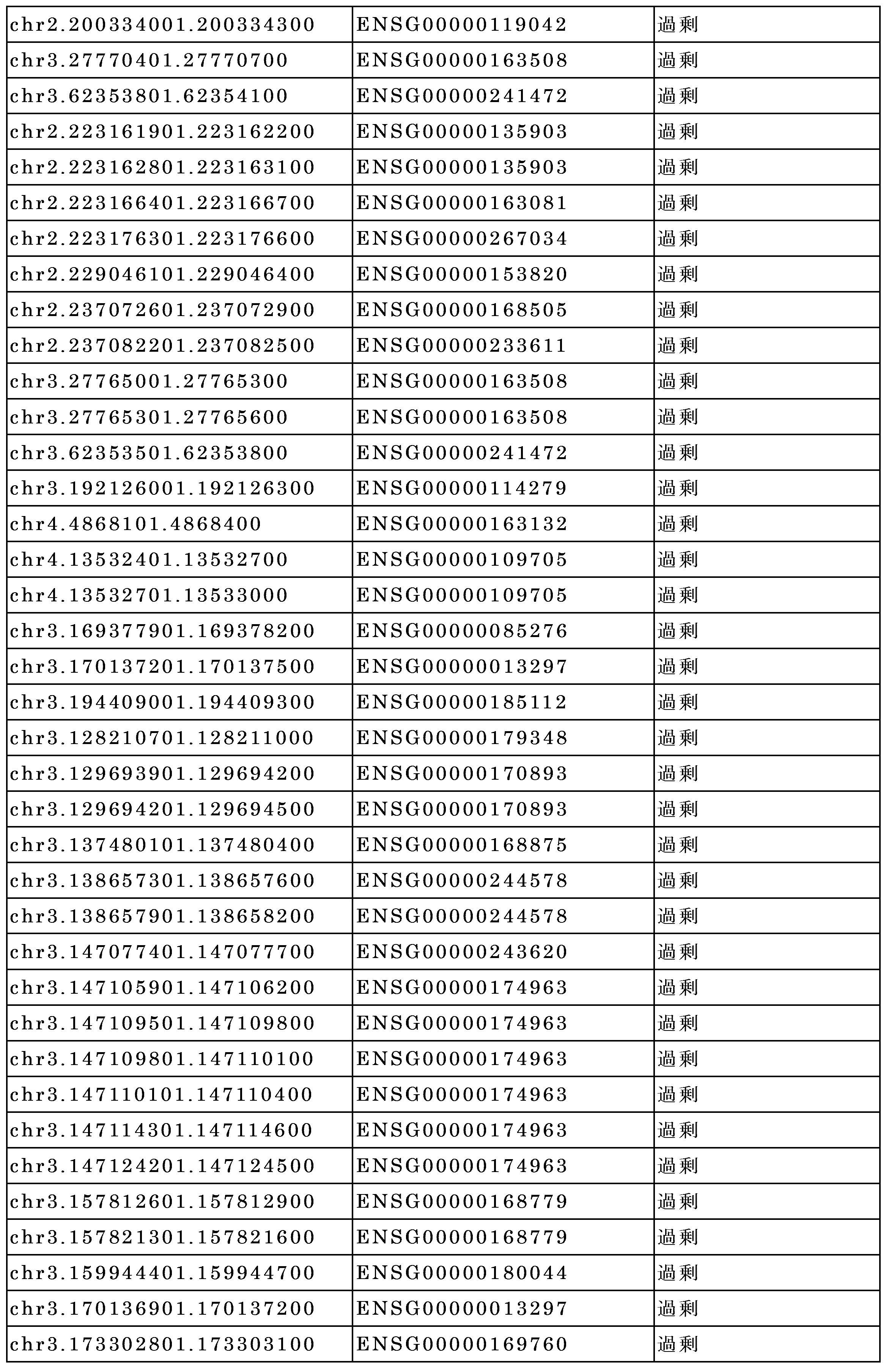
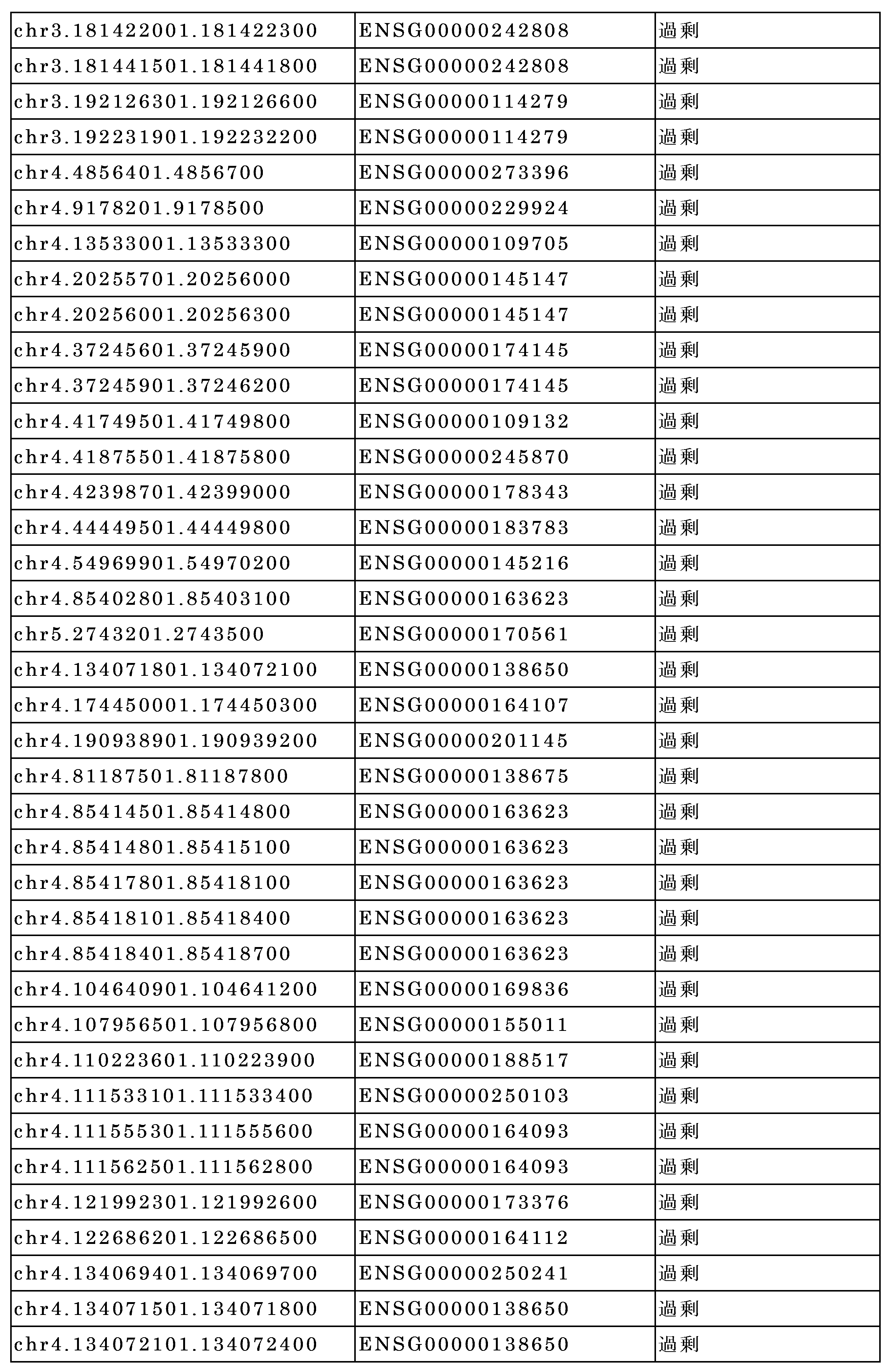

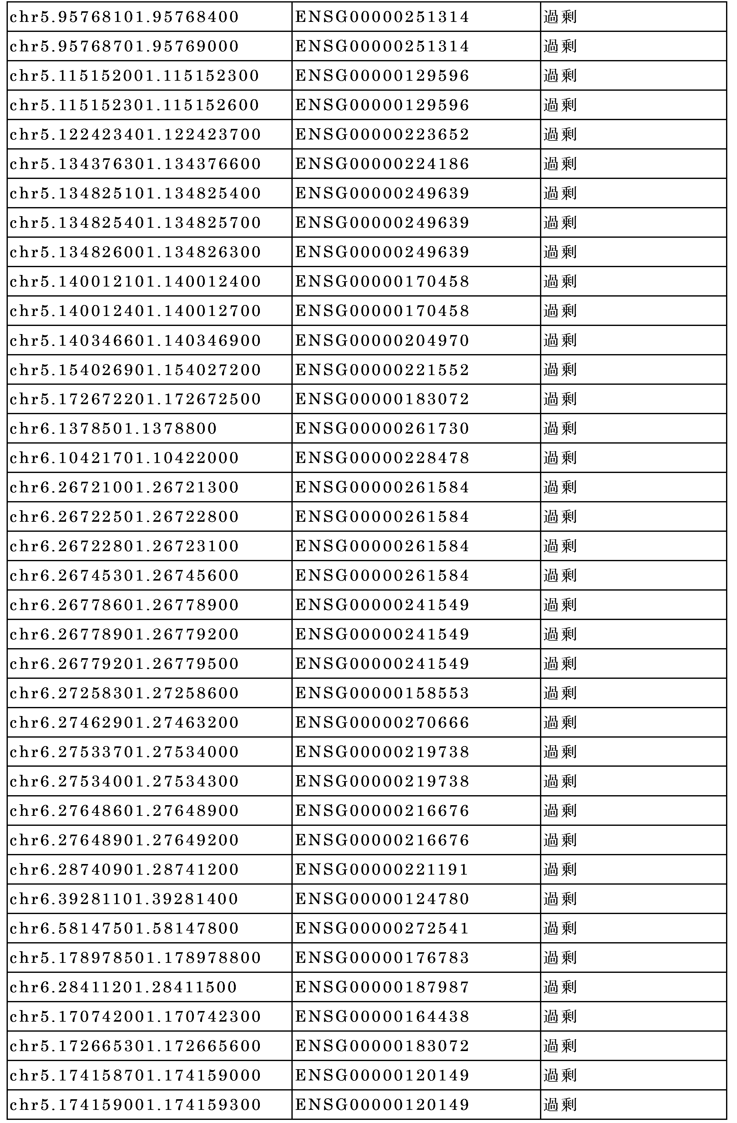


ゲノム変異プロファイル
本開示は、疾患/状態を有するかまたはそのような疾患/状態を有すると疑われる対象の変異プロファイルを生成するための方法、システムおよびキットを提供し、メチル化プロファイルは、対象が疾患/状態を有するかまたは疾患/状態を有するリスクがあるかどうかを判定するために使用され得る。本明細書に開示される試料は、ライブラリ調製および次世代ディープシーケンシング(例えば、CAPP-Seq)に供される。複数の配列決定リードが生成され、分析される。いくつかの実施形態では、ディープシーケンシングは、疾患/状態に関連するゲノム変異の同定を最大化するように構成され得る。例えば、限定することを意図するものではないが、頭頸部扁平上皮癌(HNSCC)については、標準的なHNSCCドライバ遺伝子のパネルをCAPP-seqのセレクタに含めることができる。さらに、肺がんの場合、肺がん駆動遺伝子のパネルがCAPP-seqのセレクタに含まれ得る。さらに、膵臓がんの場合、膵臓がん駆動遺伝子のパネルは、CAPP-seqのためのセレクタに含まれ得る。いくつかの実施形態では、CAPP-seqのセレクタに特定のがんのタイプにおける既知のドライバ効果のない遺伝子を含めることにより、ctDNA検出の感度を高めることができる。
Genomic Mutation Profiles The present disclosure provides methods, systems, and kits for generating a mutation profile of a subject having or suspected of having a disease/condition, and the methylation profile can be used to determine whether the subject has or is at risk for having a disease/condition. Samples disclosed herein are subjected to library preparation and next-generation deep sequencing (e.g., CAPP-Seq). Multiple sequencing reads are generated and analyzed. In some embodiments, the deep sequencing can be configured to maximize the identification of genomic mutations associated with the disease/condition. For example, but not intended to be limiting, for head and neck squamous cell carcinoma (HNSCC), a panel of standard HNSCC driver genes can be included in the selector for CAPP-seq. Additionally, for lung cancer, a panel of lung cancer driver genes can be included in the selector for CAPP-seq. Additionally, for pancreatic cancer, a panel of pancreatic cancer driver genes can be included in the selector for CAPP-seq. In some embodiments, the sensitivity of ctDNA detection can be increased by including genes with no known driver effects in specific cancer types in the selectors of CAPP-seq.
いくつかの実施形態では、ctDNA存在量の相対的な尺度は、平均変異対立遺伝子画分(MAF)から計算される。いくつかの実施形態では、変異の平均MAFは、対象を同定し、その変異プロファイルに含まれるのは、少なくとも約0.01%~少なくとも約10%の範囲である。本明細書に開示される試料のctDNA画分は、少なくとも約0.01%、0.02%、0.03%、0.04%、0.05%、0.06%、0.07%、0.08%、0.09%、0.1%、0.15%、0.2%、0.5%、1%、1.5%、2%、2.5%、3%、3.5%、4%、4.5%、5%、5.5%、6%、6.5%、7%、7.5%、8%、8.5%、9%、9.5%、10%、またはその間の任意の割合である。 In some embodiments, a relative measure of ctDNA abundance is calculated from the mean mutant allele fraction (MAF). In some embodiments, the mean MAF of mutations identifies subjects whose mutation profile includes at least about 0.01% to at least about 10%. The ctDNA fraction of the samples disclosed herein is at least about 0.01%, 0.02%, 0.03%, 0.04%, 0.05%, 0.06%, 0.07%, 0.08%, 0.09%, 0.1%, 0.15%, 0.2%, 0.5%, 1%, 1.5%, 2%, 2.5%, 3%, 3.5%, 4%, 4.5%, 5%, 5.5%, 6%, 6.5%, 7%, 7.5%, 8%, 8.5%, 9%, 9.5%, 10%, or any percentage therebetween.
いくつかの実施形態では、対象の生成された変異プロファイルは、PBLに由来するセルフリー核酸分子に由来する変異の変異体を含まない。いくつかの実施形態では、変異プロファイルは、ミスセンス変異体、ナンセンス変異体、欠失変異体、挿入変異体、重複変異体、逆位変異体、フレームシフト変異体、または反復伸長変異体などの遺伝子多型を含む。いくつかの実施形態では、変異プロファイルは、特定のサイズ範囲のセルフリー核酸分子の画分に由来する変異の変異体を含み得る。 In some embodiments, the generated mutation profile of the subject does not include mutation variants derived from cell-free nucleic acid molecules derived from PBLs. In some embodiments, the mutation profile includes genetic polymorphisms such as missense mutants, nonsense mutants, deletion mutants, insertion mutants, duplication mutants, inversion mutants, frameshift mutants, or repeat expansion mutants. In some embodiments, the mutation profile may include mutation variants derived from a fraction of cell-free nucleic acid molecules of a particular size range.
断片の長さプロファイル
いくつかの実施形態では、ctDNA断片の長さは、健常な対象に由来するセルフリー核酸分子よりも短い。いくつかの実施形態では、少なくとも1つの変異を含むctDNAの長さは、対応する参照対立遺伝子を含むセルフリー核酸分子の長さよりも短い。いくつかの実施形態では、少なくとも1つのDMRを含有するctDNA断片の長さは、対応するゲノム領域を含有するセルフリー核酸分子の断片よりも短い。
Fragment Length Profile In some embodiments, the length of the ctDNA fragment is shorter than that of a cell-free nucleic acid molecule derived from a healthy subject. In some embodiments, the length of the ctDNA containing at least one mutation is shorter than that of a cell-free nucleic acid molecule containing the corresponding reference allele. In some embodiments, the length of the ctDNA fragment containing at least one DMR is shorter than that of the cell-free nucleic acid molecule fragment containing the corresponding genomic region.
いくつかの実施形態では、配列決定は、ctDNA断片の分解を引き起こし、ctDNAの長さの分布の保存を妨げるので、亜硫酸水素塩配列を利用しない。いくつかの実施形態では、ctDNAの断片の長さは、少なくとも60~500bp、80~300bp、90~250bp、80~170bp、または100~150bpである。いくつかの実施形態では、本開示は、特定のサイズのセルフリー分子の選択に基づいて、セルフリー核酸試料の濃縮をもたらす。いくつかの実施形態では、マルチモーダル解析は、複数の核酸分子をそれらの断片の長さに基づいて変異プロファイルに選択的に含めることによって、本明細書に記載の変異プロファイルおよび断片の長さプロファイルを利用することを含む。いくつかの実施形態では、マルチモーダル解析は、複数の核酸分子をそれらの断片の長さに基づいてメチル化プロファイルに選択的に含めることによって、本明細書に記載のメチル化プロファイルおよび断片の長さプロファイルを利用することを含む。いくつかの実施形態では、マルチモーダル解析は、複数の核酸分子をそれらの断片の長さに基づいて変異プロファイルに選択的に含めることによって、および複数の核酸分子をそれらの断片の長さにそれぞれ基づいてメチル化プロファイルに選択的に含めることによって、変異プロファイル、メチル化プロファイル、および断片の長さプロファイルを一緒に利用することを含む。 In some embodiments, sequencing does not utilize bisulfite sequencing, as this would cause degradation of the ctDNA fragments and prevent preservation of the ctDNA length distribution. In some embodiments, the length of the fragments of ctDNA is at least 60-500 bp, 80-300 bp, 90-250 bp, 80-170 bp, or 100-150 bp. In some embodiments, the present disclosure provides enrichment of cell-free nucleic acid samples based on the selection of cell-free molecules of a particular size. In some embodiments, the multimodal analysis includes utilizing the mutation profile and fragment length profile described herein by selectively including multiple nucleic acid molecules in the mutation profile based on their fragment length. In some embodiments, the multimodal analysis includes utilizing the methylation profile and fragment length profile described herein by selectively including multiple nucleic acid molecules in the methylation profile based on their fragment length. In some embodiments, the multimodal analysis involves utilizing a mutation profile, a methylation profile, and a fragment length profile together by selectively including multiple nucleic acid molecules in a mutation profile based on their fragment lengths, and by selectively including multiple nucleic acid molecules in a methylation profile based on their fragment lengths, respectively.
がんを検出し、腫瘍の起源の組織を決定し、予後を提供するための方法およびシステム
本開示は、対象が疾患を有するか、または疾患を有するリスクがあるかどうかを判定するための方法およびシステムを提供し、この方法およびシステムは、(i)メチル化プロファイル、(ii)変異プロファイル、および(iii)断片の長さプロファイルのうちの少なくとも1つのプロファイルを生成するために前記対象から得られたセルフリー核酸試料に由来する複数の核酸分子をシーケンシングに供する工程、および前記対象が前記疾患を有するかまたは前記疾患のリスクがあるかどうかを少なくとも80%の感度または少なくとも約90%の特異度で判定するために前記少なくとも1つのプロファイルを処理する工程であって、前記セルフリー核酸試料が30ng/ml未満の前記複数の核酸分子を含む、処理する工程を含む。いくつかの実施形態では、感度は、少なくとも約80%、81%、82%、83%、84%、85%、86%、87%、88%、89%、90%、91%、92%、93%、94%、95%、96%、97%、98%、99%、99.5%、99.6%、99.7%、99.8%、99.9%、または数字の間の任意のパーセンテージである。いくつかの実施形態では、特異度は、少なくとも約90%、91%、92%、93%、94%、95%、96%、97%、98%、99%、99.1%、99.2%、99.3%、99.4%、99.5%、99.6%、99.7%、99.8%、99.9%、または数字の間の任意のパーセンテージである。
Methods and Systems for Detecting Cancer, Determining Tissue of Origin of Tumors, and Providing a Prognosis The present disclosure provides methods and systems for determining whether a subject has or is at risk of having a disease, comprising subjecting a plurality of nucleic acid molecules from a cell-free nucleic acid sample obtained from the subject to sequencing to generate at least one of: (i) a methylation profile, (ii) a mutation profile, and (iii) a fragment length profile, and processing the at least one profile to determine whether the subject has or is at risk of said disease with at least 80% sensitivity or at least about 90% specificity, wherein the cell-free nucleic acid sample comprises less than 30 ng/ml of the plurality of nucleic acid molecules. In some embodiments, the sensitivity is at least about 80%, 81%, 82%, 83%, 84%, 85%, 86%, 87%, 88%, 89%, 90%, 91%, 92%, 93%, 94%, 95%, 96%, 97%, 98%, 99%, 99.5%, 99.6%, 99.7%, 99.8%, 99.9%, or any percentage between the numbers. In some embodiments, the specificity is at least about 90%, 91%, 92%, 93%, 94%, 95%, 96%, 97%, 98%, 99%, 99.1%, 99.2%, 99.3%, 99.4%, 99.5%, 99.6%, 99.7%, 99.8%, 99.9%, or any percentage between the numbers.
いくつかの実施形態では、方法およびシステムは、前記対象から得られたセルフリー核酸試料に由来する複数の核酸分子を配列決定に供して、(i)メチル化プロファイル、(ii)変異プロファイルおよび(iii)断片の長さプロファイルのうちの少なくとも2つのプロファイルを生成させる工程を含む。方法は、少なくとも約80%、81%、82%、83%、84%、85%、86%、87%、88%、89%、90%、91%、92%、93%、94%、95%、96%、97%、98%、99%、99.5%、99.6%、99.7%、99.8%、99.9%、または数字の間の任意のパーセンテージの感度をもたらす。いくつかの実施形態では、2つのプロファイルを使用する場合の感度は、1つのプロファイルを使用する場合の感度と比較して、任意の数字の間で少なくとも約0.5%、1%、2%、3%、4%、5%、6%、7%、8%、9%、10%、またはパーセンテージだけ増加する。いくつかの実施形態では、3つのプロファイルを使用する場合の感度は、2つのプロファイルを使用する場合の感度と比較して、任意の数字の間で少なくとも約0.5%、1%、2%、3%、4%、5%、6%、7%、8%、9%、10%、またはパーセンテージだけ増加する。 In some embodiments, the methods and systems include subjecting a plurality of nucleic acid molecules from a cell-free nucleic acid sample obtained from the subject to sequencing to generate at least two of (i) a methylation profile, (ii) a mutation profile, and (iii) a fragment length profile. The methods provide a sensitivity of at least about 80%, 81%, 82%, 83%, 84%, 85%, 86%, 87%, 88%, 89%, 90%, 91%, 92%, 93%, 94%, 95%, 96%, 97%, 98%, 99%, 99.5%, 99.6%, 99.7%, 99.8%, 99.9%, or any percentage between the numbers. In some embodiments, the sensitivity when using two profiles is increased by at least about 0.5%, 1%, 2%, 3%, 4%, 5%, 6%, 7%, 8%, 9%, 10%, or a percentage between any numbers compared to the sensitivity when using one profile. In some embodiments, the sensitivity when using three profiles is increased by at least about 0.5%, 1%, 2%, 3%, 4%, 5%, 6%, 7%, 8%, 9%, 10%, or a percentage between any numbers compared to the sensitivity when using two profiles.
さらに、方法は、少なくとも約90%、91%、92%、93%、94%、95%、96%、97%、98%、99%、99.1%、99.2%、99.3%、99.4%、99.5%、99.6%、99.7%、99.8%、99.9%、または数字の間の任意のパーセンテージの特異度をもたらす。いくつかの実施形態では、2つのプロファイルを使用する場合の特異度は、1つのプロファイルを使用する場合の特異度と比較して、任意の数字の間で少なくとも約0.5%、1%、2%、3%、4%、5%、6%、7%、8%、9%、10%、またはパーセンテージだけ増加する。いくつかの実施形態では、3つのプロファイルを使用する場合の特異度は、2つのプロファイルを使用する場合の特異度と比較して、任意の数字の間で少なくとも約0.5%、1%、2%、3%、4%、5%、6%、7%、8%、9%、10%、またはパーセンテージだけ増加する。 Additionally, the method provides a specificity of at least about 90%, 91%, 92%, 93%, 94%, 95%, 96%, 97%, 98%, 99%, 99.1%, 99.2%, 99.3%, 99.4%, 99.5%, 99.6%, 99.7%, 99.8%, 99.9%, or any percentage between numbers. In some embodiments, the specificity when using two profiles is increased by at least about 0.5%, 1%, 2%, 3%, 4%, 5%, 6%, 7%, 8%, 9%, 10%, or any percentage between numbers compared to the specificity when using one profile. In some embodiments, the specificity when three profiles are used is increased by at least about 0.5%, 1%, 2%, 3%, 4%, 5%, 6%, 7%, 8%, 9%, 10%, or any percentage between any numbers compared to the specificity when two profiles are used.
本開示は、対象のセルフリー核酸試料を処理して、前記対象が疾患を有するかまたは疾患を有するリスクがあるかどうかを判定する方法およびシステムであって、複数の核酸分子を含む前記セルフリー核酸試料を得る工程、前記複数の核酸分子またはその誘導体を配列決定に供して、複数の配列決定リードを生成する工程、前記複数の配列決定リードをコンピュータ処理して、前記複数の核酸分子について、(i)メチル化プロファイル、(ii)変異プロファイル、および(iii)断片の長さプロファイルを同定する工程、および前記対象が前記疾患を有するかまたは有するリスクがあるかどうかを判定するために、少なくとも前記メチル化プロファイル、前記変異プロファイルおよび前記断片の長さプロファイルを使用する工程を含む、方法およびシステムを提供する。いくつかの実施形態では、方法は、少なくとも約80%、81%、82%、83%、84%、85%、86%、87%、88%、89%、90%、91%、92%、93%、94%、95%、96%、97%、98%、99%、99.5%、99.6%、99.7%、99.8%、99.9%、または数字の間の任意のパーセンテージの感度をもたらす。方法は、少なくとも約90%、91%、92%、93%、94%、95%、96%、97%、98%、99%、99.1%、99.2%、99.3%、99.4%、99.5%、99.6%、99.7%、99.8%、99.9%、または数字の間の任意のパーセンテージの特異度をもたらす。 The present disclosure provides methods and systems for processing a cell-free nucleic acid sample from a subject to determine whether the subject has or is at risk for having a disease, the methods and systems including obtaining the cell-free nucleic acid sample comprising a plurality of nucleic acid molecules; subjecting the plurality of nucleic acid molecules or derivatives thereof to sequencing to generate a plurality of sequencing reads; computer processing the plurality of sequencing reads to identify (i) a methylation profile, (ii) a mutation profile, and (iii) a fragment length profile for the plurality of nucleic acid molecules; and using at least the methylation profile, the mutation profile, and the fragment length profile to determine whether the subject has or is at risk for having the disease. In some embodiments, the method provides a sensitivity of at least about 80%, 81%, 82%, 83%, 84%, 85%, 86%, 87%, 88%, 89%, 90%, 91%, 92%, 93%, 94%, 95%, 96%, 97%, 98%, 99%, 99.5%, 99.6%, 99.7%, 99.8%, 99.9%, or any percentage between the numbers. The method provides a specificity of at least about 90%, 91%, 92%, 93%, 94%, 95%, 96%, 97%, 98%, 99%, 99.1%, 99.2%, 99.3%, 99.4%, 99.5%, 99.6%, 99.7%, 99.8%, 99.9%, or any percentage between the numbers.
本開示は、複数の差次的メチル化領域(DMR)を同定することを含む、腫瘍の組織の起源を決定するための方法およびシステムを提供し、複数のDMRは、特定のがん(例えば、乳がん、結腸がん、前立腺がん、HSNCC)に特異的であり、セルフリー核酸分子の画分に由来する。いくつかの実施形態では、セルフリー核酸分子の画分はctDNAに由来する。いくつかの実施形態では、方法は少なくとも約80%、81%、82%、83%、84%、85%、86%、87%、88%、89%、90%、91%、92%、93%、94%、95%、96%、97%、98%、99%、99.5%、99.6%、99.7%、99.8%、99.9%、または数字の間の任意のパーセンテージの感度をもたらす。方法は、少なくとも約90%、91%、92%、93%、94%、95%、96%、97%、98%、99%、99.1%、99.2%、99.3%、99.4%、99.5%、99.6%、99.7%、99.8%、99.9%、または数字の間の任意のパーセンテージの特異度をもたらす。 The present disclosure provides methods and systems for determining the tissue origin of a tumor, including identifying multiple differentially methylated regions (DMRs), the multiple DMRs being specific to a particular cancer (e.g., breast cancer, colon cancer, prostate cancer, HSNCC) and derived from a fraction of cell-free nucleic acid molecules. In some embodiments, the fraction of cell-free nucleic acid molecules is derived from ctDNA. In some embodiments, the method results in a sensitivity of at least about 80%, 81%, 82%, 83%, 84%, 85%, 86%, 87%, 88%, 89%, 90%, 91%, 92%, 93%, 94%, 95%, 96%, 97%, 98%, 99%, 99.5%, 99.6%, 99.7%, 99.8%, 99.9%, or any percentage between the numbers. The method results in a specificity of at least about 90%, 91%, 92%, 93%, 94%, 95%, 96%, 97%, 98%, 99%, 99.1%, 99.2%, 99.3%, 99.4%, 99.5%, 99.6%, 99.7%, 99.8%, 99.9%, or any percentage between the numbers.
本開示は、疾患/状態の治療を受けた後に対象に対して予後を得るための方法およびシステムを記載する。例えば、治療は、腫瘍の外科的除去、特定の種類のがんのために設計された化学療法、放射線療法、または免疫療法(例えば、TCR、CARなど)を含む。いくつかの実施形態では、方法またはシステムは、前記対象から得られたセルフリー核酸試料に由来する複数の核酸分子を配列決定に供して、(i)メチル化プロファイル、(ii)変異プロファイルおよび(iii)断片の長さプロファイルのうちの少なくとも1つのプロファイルを生成させること、および少なくとも1つのプロファイルに少なくとも基づいて微小残存病変(MRD)をモニタリングまたは検出することを含む。 The present disclosure describes methods and systems for obtaining a prognosis for a subject after receiving treatment for a disease/condition. For example, the treatment includes surgical removal of a tumor, chemotherapy, radiation therapy, or immunotherapy (e.g., TCR, CAR, etc.) designed for a particular type of cancer. In some embodiments, the method or system includes subjecting a plurality of nucleic acid molecules from a cell-free nucleic acid sample obtained from the subject to sequencing to generate at least one of (i) a methylation profile, (ii) a mutation profile, and (iii) a fragment length profile, and monitoring or detecting minimal residual disease (MRD) based at least on the at least one profile.
本開示は、対象からの試料の少なくとも一部からのセルフリー核酸分子をアッセイすることによって、前記対象が疾患/状態を有するかどうかを判定するため、表5に列挙される差次的メチル化領域(DMR)に含まれる前記セルフリー核酸分子の少なくとも一部のメチル化レベルを検出するため、また少なくとも1つのコンピュータプロセッサを使用して、(b)で検出された前記メチル化レベルを、前記表5に列挙されたDMRに含まれる前記セルフリー核酸分子の対応する(1つまたは複数の)部分のメチル化レベルと比較するための方法およびシステムを提供する。いくつかの実施形態では、表5に列挙される少なくとも約6個以上、10個以上、15個以上、20個以上、30個以上、40個以上、50個以上、60個以上、70個以上、80個以上、90個以上、または100個以上、200個以上、300個以上、400個以上、500個以上、600個以上、または700個以上のDMRのメチル化レベルが測定され、本明細書で論じられる健常な対象の対応するDMRのメチル化レベルと比較される。 The present disclosure provides methods and systems for determining whether a subject has a disease/condition by assaying cell-free nucleic acid molecules from at least a portion of a sample from the subject, detecting a methylation level of at least a portion of the cell-free nucleic acid molecule that falls within a differentially methylated region (DMR) listed in Table 5, and comparing, using at least one computer processor, the methylation level detected in (b) with the methylation level of a corresponding portion(s) of the cell-free nucleic acid molecule that falls within a DMR listed in Table 5. In some embodiments, the methylation levels of at least about 6 or more, 10 or more, 15 or more, 20 or more, 30 or more, 40 or more, 50 or more, 60 or more, 70 or more, 80 or more, 90 or more, or 100 or more, 200 or more, 300 or more, 400 or more, 500 or more, 600 or more, or 700 or more DMRs listed in Table 5 are measured and compared to the methylation levels of the corresponding DMRs in healthy subjects as discussed herein.
対象が正確に診断され、外科的除去、化学療法、放射線療法などのがんを治療するための治療を受けるたら、治療の有効性をモニタリングし、患者の生存率を予測することが重要である。さらに、がん細胞の最小限の残存疾患を検出することが重要である。本開示は、前記対象が疾患の治療を受けた後に生存率が高いかどうかを判定するための方法およびシステムを提供し、この方法およびシステムは、前記対象からの試料の少なくとも一部からのセルフリー核酸分子をアッセイすること、表6に列挙される差次的メチル化領域(DMR)に含まれる前記セルフリー核酸分子の少なくとも一部のメチル化レベルを検出すること、また少なくとも1つのコンピュータプロセッサを使用して、(b)で検出された前記メチル化レベルを、表6に列挙された前記DMRに含まれる前記セルフリー核酸分子の対応する(1つまたは複数の)部分のメチル化レベルと比較することを含む。いくつかの実施形態では、表6に列挙されるDMRは、遺伝子ZSCAN31、LINC01391、GATA2-AS1、STK3、およびOSR1に関連する領域を表す。

いくつかの実施形態では、方法は、免疫沈降反応を確認するために第2の量の対照DNAを試料に添加する工程をさらに含む。 In some embodiments, the method further includes adding a second amount of control DNA to the sample to confirm the immunoprecipitation reaction.
本明細書で使用される場合、「対照」は、陽性対照と陰性対照の両方、または少なくとも陽性対照を含み得る。 As used herein, "control" can include both positive and negative controls, or at least a positive control.
いくつかの実施形態では、方法は、セルフリーメチル化DNAの捕捉を確認するために、第2の量の対照DNAを試料に添加する工程をさらに含む。 In some embodiments, the method further includes adding a second amount of control DNA to the sample to confirm capture of cell-free methylated DNA.
いくつかの実施形態では、がん細胞由来のDNAが存在することを同定することは、がん細胞の起源の組織を同定することをさらに含む。 In some embodiments, identifying the presence of DNA from a cancer cell further includes identifying the tissue of origin of the cancer cell.
いくつかの例では、腫瘍組織サンプリングは困難であるか、または重大なリスクを伴う可能性があり、その場合、腫瘍組織サンプリングを必要とせずにがんを診断および/またはサブタイプに分けることが望ましい場合がある。例えば、肺腫瘍組織サンプリングは、縦隔鏡検査、開胸、または経皮的針生検などの侵襲的処置を必要とし得る。これらの処置は、入院、胸腔チューブ、機械での換気、抗生物質、または他の医学的介入の必要性をもたらし得る。一部の個体は、医学的併存症のために、または優先度のために、腫瘍組織サンプリングに必要な侵襲的処置を受けないことがある。いくつかの例では、腫瘍組織調達の実際の手順は、疑われているがんのサブタイプに依存し得る。他の例では、がんのサブタイプは、同じ個体の体内で経時的に進展し得、侵襲性腫瘍組織サンプリング処置による連続的な評価は、多くの場合非現実的であり、患者が十分に忍容できない。したがって、血液検査による非侵襲性がんのサブタイプ分類は、臨床腫瘍学の実施において多くの有利な用途を有し得る。 In some instances, tumor tissue sampling may be difficult or involve significant risks, in which case it may be desirable to diagnose and/or subtype cancers without the need for tumor tissue sampling. For example, lung tumor tissue sampling may require invasive procedures such as mediastinoscopy, thoracotomy, or percutaneous needle biopsy. These procedures may result in the need for hospitalization, chest tubes, mechanical ventilation, antibiotics, or other medical interventions. Some individuals may not undergo the invasive procedures required for tumor tissue sampling due to medical comorbidities or due to priorities. In some instances, the actual procedure of tumor tissue procurement may depend on the suspected cancer subtype. In other instances, cancer subtypes may evolve over time within the same individual, and serial evaluations by invasive tumor tissue sampling procedures are often impractical and not well tolerated by patients. Thus, noninvasive cancer subtyping by blood tests may have many advantageous applications in the practice of clinical oncology.
したがって、いくつかの実施形態では、がん細胞の起源の組織を同定することは、がんのサブタイプを同定することをさらに含む。好ましくは、がんのサブタイプは、ステージ(例えば、外科手術で治療された早期肺がん対化学療法で治療された後期肺がん)、組織学(例えば、肺がんにおける小細胞癌対腺癌対扁平上皮癌)、遺伝子発現パターンまたは転写因子活性(例えば、乳がんにおけるER状態)、コピー数の異常(例えば、乳がんにおけるHER2の状態)、特異的再編成(例えば、AMLにおけるFLT3)、特異的遺伝子点変異状態(例えば、IDH遺伝子点変異)、およびDNAのメチル化パターン(例えば、脳がんにおけるMGMT遺伝子プロモーターのメチル化)に基づいてがんを区別する。 Thus, in some embodiments, identifying the tissue of origin of the cancer cells further comprises identifying a cancer subtype. Preferably, the cancer subtype distinguishes cancers based on stage (e.g., early lung cancer treated with surgery vs. late lung cancer treated with chemotherapy), histology (e.g., small cell carcinoma vs. adenocarcinoma vs. squamous cell carcinoma in lung cancer), gene expression patterns or transcription factor activity (e.g., ER status in breast cancer), copy number abnormalities (e.g., HER2 status in breast cancer), specific rearrangements (e.g., FLT3 in AML), specific gene point mutation status (e.g., IDH gene point mutation), and DNA methylation patterns (e.g., MGMT gene promoter methylation in brain cancer).
いくつかの実施形態では、工程(f)における比較は、ゲノムワイドで行われる。 In some embodiments, the comparison in step (f) is performed genome-wide.
他の実施形態では、工程(f)における比較は、ゲノムワイドから、限定されないが、FANTOM5エンハンサー、CpGアイランド、CpGショア、CpG Shelfまたは前述の任意の組み合わせなどの特定の調節領域に制限される。 In other embodiments, the comparison in step (f) is restricted genome-wide to specific regulatory regions, such as, but not limited to, FANTOM5 enhancers, CpG islands, CpG shores, CpG shelves, or any combination of the foregoing.
いくつかの実施形態では、本明細書の方法は、がんの検出に使用するためのものである。 In some embodiments, the methods herein are for use in detecting cancer.
いくつかの実施形態では、本明細書の方法は、がんの治療のモニタリングに使用するためのものである。 In some embodiments, the methods herein are for use in monitoring the treatment of cancer.
データ分析システムおよび方法
本明細書に開示される方法およびシステムは、アルゴリズムまたはその使用を含むことができる。1つまたは複数のアルゴリズムは、1つまたは複数の対象から1つまたは複数の試料を分類するために使用され得る。1つまたは複数のアルゴリズムは、1つまたは複数の試料からのデータに適用され得る。データはバイオマーカー発現データを含み得る。本明細書に開示される方法は、1人または複数の対象からの1つまたは複数の試料に分類を割り当てることを含み得る。分類を試料に割り当てることは、メチル化プロファイル、変異プロファイルおよび断片の長さプロファイルにアルゴリズムを適用することを含み得る。場合によっては、少なくとも1つのプロファイルは、対象から得られた試料を疾患または軽微な損傷を有すると分類するための訓練されたアルゴリズムを含むデータ分析システムに入力される。
Data Analysis Systems and Methods The methods and systems disclosed herein may include an algorithm or its use. One or more algorithms may be used to classify one or more samples from one or more subjects. One or more algorithms may be applied to data from one or more samples. The data may include biomarker expression data. The methods disclosed herein may include assigning a classification to one or more samples from one or more subjects. Assigning a classification to a sample may include applying an algorithm to a methylation profile, a mutation profile, and a fragment length profile. In some cases, at least one profile is input into a data analysis system that includes a trained algorithm for classifying a sample obtained from a subject as having a disease or minor injury.
データ分析システムは、訓練されたアルゴリズムであってもよい。アルゴリズムは、線形分類器を含み得る。場合によっては、線形分類器は、線形判別分析、フィッシャー線形判別、ナイーブベイズ分類器、ロジスティック回帰、パーセプトロン、サポートベクターマシン、またはそれらの組み合わせのうちの1つまたは複数を含む。線形分類器は、サポートベクターマシン(SVM)のアルゴリズムであってもよい。アルゴリズムは、双方向分類器を含み得る。双方向分類器は、1つまたは複数の決定木、ランダムフォレスト、ベイジアンネットワーク、サポートベクターマシン、ニューラルネットワーク、またはロジスティック回帰アルゴリズムを含み得る。 The data analysis system may be a trained algorithm. The algorithm may include a linear classifier. In some cases, the linear classifier includes one or more of a linear discriminant analysis, a Fisher linear discriminant, a naive Bayes classifier, a logistic regression, a perceptron, a support vector machine, or a combination thereof. The linear classifier may be a support vector machine (SVM) algorithm. The algorithm may include a bidirectional classifier. The bidirectional classifier may include one or more decision trees, random forests, Bayesian networks, support vector machines, neural networks, or logistic regression algorithms.
アルゴリズムは、1つまたは複数の線形判別分析(LDA)、基本パーセプトロン、弾性ネット、ロジスティック回帰、(カーネル)サポートベクターマシン(SVM)、対角線形判別分析(DLDA)、ゴルビン分類器、パーゼンベース、(カーネル)フィッシャー判別分類器、k近傍法、反復RELIEF、分類木、最尤分類器、ランダムフォレスト、最近接重心、マイクロアレイの予測分析(PAM)、k中間クラスタリング、ファジーC平均クラスタリング、ガウス混合モデル、勾配応答(GR)、勾配ブースティング法(GBM)、楕円ネットロジスティック回帰、ロジスティック回帰、またはそれらの組み合わせを含み得る。アルゴリズムは、対角線形判別分析(DLDA)のアルゴリズムを含み得る。アルゴリズムは、最近接重心のアルゴリズムを含み得る。アルゴリズムは、ランダムフォレストのアルゴリズムを含み得る。いくつかの実施形態では、子癇前症および非子癇前症を識別するために、ロジスティック回帰、ランダムフォレスト、および勾配ブースティング法(GBM)のパフォーマンスは、線形判別分析(LDA)、ニューラルネットワーク、およびサポートベクターマシン(SVM)のパフォーマンスよりも優れている。 The algorithms may include one or more of linear discriminant analysis (LDA), elementary perceptron, elastic net, logistic regression, (kernel) support vector machine (SVM), diagonal linear discriminant analysis (DLDA), Golbin classifier, Parzen base, (kernel) Fisher discriminant classifier, k-nearest neighbors, iterative RELIEF, classification tree, maximum likelihood classifier, random forest, nearest centroid, predictive analysis of microarrays (PAM), k-means clustering, fuzzy C-means clustering, Gaussian mixture model, gradient response (GR), gradient boosting method (GBM), elliptical net logistic regression, logistic regression, or combinations thereof. The algorithms may include a diagonal linear discriminant analysis (DLDA) algorithm. The algorithms may include a nearest centroid algorithm. The algorithms may include a random forest algorithm. In some embodiments, the performance of logistic regression, random forests, and gradient boosting (GBM) for distinguishing pre-eclampsia and non-pre-eclampsia is superior to that of linear discriminant analysis (LDA), neural networks, and support vector machines (SVM).
キット
本開示は、対象の疾患または障害(例えば、がん)を同定またはモニタリングするためのキットを提供する。キットは、対象の試料のがん関連ゲノム遺伝子座のパネルの各々における配列の定量的尺度(例えば、存在、非存在、または相対的な量を示す)を同定するためのプローブを含み得る。試料のがん関連ゲノム遺伝子座のパネルの各々における配列の定量的尺度(例えば、存在、非存在、または相対的な量を示す)は、対象の疾患または障害(例えば、がん)を示し得る。プローブは、試料のがん関連ゲノム遺伝子座(例えば、表3、5および6に列挙されるDMR)のパネルにおける配列に対して選択的であり得る。キットは、プローブを使用して試料を処理し、対象の試料のがん関連ゲノム遺伝子座のパネルのそれぞれにおける配列の定量的尺度(例えば、存在、非存在、または相対的な量を示す)を示すデータセットを生成するための説明書を含み得る。
Kits The present disclosure provides kits for identifying or monitoring a disease or disorder (e.g., cancer) in a subject. The kits may include probes for identifying a quantitative measure (e.g., indicating the presence, absence, or relative amount) of a sequence in each of a panel of cancer-associated genomic loci of a sample of a subject. A quantitative measure (e.g., indicating the presence, absence, or relative amount) of a sequence in each of a panel of cancer-associated genomic loci of a sample of a subject may be indicative of a disease or disorder (e.g., cancer) in the subject. The probes may be selective for sequences in a panel of cancer-associated genomic loci of a sample of a subject (e.g., DMRs listed in Tables 3, 5, and 6). The kits may include instructions for processing the sample using the probes to generate a data set indicative of a quantitative measure (e.g., indicating the presence, absence, or relative amount) of a sequence in each of a panel of cancer-associated genomic loci of a sample of a subject.
キットにあるプローブは、試料のがん関連ゲノム遺伝子座のパネルにおける配列に対して選択的であり得る。キットにあるプローブは、がん関連ゲノム遺伝子座のパネルに対応する核酸(例えば、RNAまたはDNA)分子を選択的に濃縮するように構成され得る。キットにあるプローブは核酸プライマーであり得る。キットにあるプローブは、がん関連ゲノム遺伝子座またはゲノム領域のパネルの1つ以上からの核酸配列との配列相補性を有し得る。がん関連ゲノム遺伝子座または微生物関連ゲノム遺伝子座またはゲノム領域のパネルは、少なくとも2、少なくとも3、少なくとも4、少なくとも5、少なくとも6、少なくとも7、少なくとも8、少なくとも9、少なくとも10、少なくとも11、少なくとも12、少なくとも13、少なくとも14、少なくとも15、少なくとも16、少なくとも17、少なくとも18、少なくとも19、少なくとも20、またはそれより異なるがん関連ゲノム遺伝子座またはゲノム領域のパネルを含み得る。 The probes in the kit may be selective for sequences in the panel of cancer-associated genomic loci of the sample. The probes in the kit may be configured to selectively enrich for nucleic acid (e.g., RNA or DNA) molecules corresponding to the panel of cancer-associated genomic loci. The probes in the kit may be nucleic acid primers. The probes in the kit may have sequence complementarity to nucleic acid sequences from one or more of the panel of cancer-associated genomic loci or genomic regions. The panel of cancer-associated or microbe-associated genomic loci or genomic regions may include a panel of at least 2, at least 3, at least 4, at least 5, at least 6, at least 7, at least 8, at least 9, at least 10, at least 11, at least 12, at least 13, at least 14, at least 15, at least 16, at least 17, at least 18, at least 19, at least 20, or more different cancer-associated genomic loci or genomic regions.
キットの説明書は、セルフリー生物学的試料のがん関連ゲノム遺伝子座のパネルにおける配列に対して選択的であるプローブを使用して、試料をアッセイするための説明書を含み得る。これらのプローブは、がん関連ゲノム遺伝子座の複数のパネルのうちの1つ以上由来の核酸配列(例えば、RNAまたはDNA)との配列相補性を有する核酸分子(例えば、RNAまたはDNA)であり得る。これらの核酸分子は、プライマーまたは濃縮配列であり得る。セルフリー生物試料をアッセイするための説明書は、アレイハイブリダイゼーション、ポリメラーゼ連鎖反応(PCR)、または核酸の配列決定(例えば、DNAの配列決定またはRNAの配列決定)を実施して試料を処理し、試料のがん関連ゲノム遺伝子座のパネルのそれぞれにおける配列の定量的尺度を示す(例えば、存在、非存在、または相対的な量を示す)データセットを生成するための導入を含み得る。試料のがん関連ゲノム遺伝子座のパネルの各々における配列の定量的尺度(例えば、存在、非存在、または相対的な量を示す)は、疾患または障害(例えば、がん)を示し得る。 The kit instructions may include instructions for assaying the sample using probes selective for sequences in the panel of cancer-associated genomic loci of the cell-free biological sample. The probes may be nucleic acid molecules (e.g., RNA or DNA) having sequence complementarity with nucleic acid sequences (e.g., RNA or DNA) from one or more of the panels of cancer-associated genomic loci. The nucleic acid molecules may be primers or enrichment sequences. The instructions for assaying the cell-free biological sample may include instructions for performing array hybridization, polymerase chain reaction (PCR), or nucleic acid sequencing (e.g., DNA sequencing or RNA sequencing) to process the sample and generate a data set indicative of a quantitative measure (e.g., indicative of presence, absence, or relative amount) of sequences in each of the panel of cancer-associated genomic loci of the sample. The quantitative measure (e.g., indicative of presence, absence, or relative amount) of sequences in each of the panel of cancer-associated genomic loci of the sample may be indicative of a disease or disorder (e.g., cancer).
キットの説明書は、アッセイ読み出し値を測定および解釈するための説明書を含み得、それは、試料のがん関連ゲノム遺伝子座のパネルの各々における配列の定量的尺度を示す(例えば、存在、非存在、または相対的な量を示す)データセットを生成するために、がん関連ゲノム遺伝子座のパネルの1つまたは複数において定量化され得る。例えば、がん関連ゲノム遺伝子座のパネルに対応するアレイハイブリダイゼーションまたはポリメラーゼ連鎖反応(PCR)の定量化は、試料のがん関連ゲノム遺伝子座のパネルの各々における配列の定量的尺度を示す(例えば、存在、非存在、または相対的な量を示す)データセットを生成し得る。アッセイ読み取り値は、定量的PCR(qPCR)値、デジタルPCR(dPCR)値、デジタル液滴PCR(ddPCR)値、蛍光の値など、またはそれらの正規化された値を含み得る。 The kit instructions may include instructions for measuring and interpreting assay readouts, which may be quantified at one or more of the panel of cancer-associated genomic loci to generate a data set indicative of a quantitative measure of (e.g., indicative of presence, absence, or relative amount of) sequences at each of the panel of cancer-associated genomic loci of the sample. For example, quantification of array hybridization or polymerase chain reaction (PCR) corresponding to a panel of cancer-associated genomic loci may generate a data set indicative of a quantitative measure of (e.g., indicative of presence, absence, or relative amount of) sequences at each of the panel of cancer-associated genomic loci of the sample. The assay readouts may include quantitative PCR (qPCR) values, digital PCR (dPCR) values, digital droplet PCR (ddPCR) values, fluorescence values, etc., or normalized values thereof.
コンピュータシステム
いくつかの実施形態では、特定の工程は、コンピュータプロセッサによって実行される。本システムおよび方法は、様々な実施形態で実施することができる。適切に構成されたコンピュータデバイス、ならびに関連する通信ネットワーク、デバイス、ソフトウェアおよびファームウェアは、上述の1つまたは複数の実施形態を可能にするためのプラットフォームを提供することができる。例として、図8は、記憶ユニット104およびランダムアクセスメモリ106に接続された中央処理装置(「CPU」)102を含むことができる汎用コンピュータデバイス100を示す。CPU102は、オペレーティングシステム101、アプリケーションプログラム103、およびデータ123を処理することができる。必要に応じて、オペレーティングシステム101、アプリケーションプログラム103、およびデータ123を記憶ユニット104に格納し、メモリ106にロードすることができる。コンピュータデバイス100は、CPU102およびメモリ106に動作可能に接続され、CPU102からの集中的な画像処理計算をオフロードし、CPU102と並列にこれらの計算を実行するグラフィック処理ユニット(GPU)122を、さらに含むことができる。オペレータ107は、ビデオインターフェース105によって接続されたビデオディスプレイ108、およびI/Oインターフェース109によって接続されたキーボード115、マウス112、およびディスクドライブまたはソリッドステートドライブ114などの様々な入出力デバイスを使用して、コンピュータデバイス100と対話することができる。マウス112は、ビデオディスプレイ108のカーソルの移動を制御し、マウスのボタンでビデオディスプレイ108に表示される様々なグラフィカルユーザインターフェース(GUI)の制御を操作するように構成することができる。ディスクドライブまたはソリッドステートドライブ114は、コンピュータ可読媒体116を受け入れるように構成され得る。コンピュータデバイス100は、ネットワークインターフェース111を介してネットワークの一部を形成することができ、コンピュータデバイス100が他の適切に構成されたデータ処理システム(図示せず)と通信することを可能にする。様々なソースからの入力を受信するために、1つまたは複数の異なるタイプのセンサ135を使用することができる。
Computer System In some embodiments, certain steps are performed by a computer processor. The system and method can be implemented in various embodiments. An appropriately configured computer device, as well as associated communication networks, devices, software and firmware, can provide a platform for enabling one or more of the above-described embodiments. By way of example, FIG. 8 shows a general-
本システムおよび方法は、デスクトップコンピュータ、ラップトップコンピュータ、タブレットコンピュータ、またはワイヤレスハンドヘルドを含む実質的に任意の方式のコンピュータデバイスで実施することができる。本システムおよび方法はまた、1つまたは複数のコンピュータデバイスが本発明による方法における様々なプロセス工程の各々を実施することを可能にするためのコンピュータプログラムコードを含むコンピュータ可読/使用可能媒体として実施されてもよい。複数のコンピュータデバイスが動作全体を実行する場合、コンピュータデバイスはネットワーク接続されて動作の様々な工程を分配する。コンピュータ可読媒体またはコンピュータ使用可能媒体という用語は、プログラムコードの任意のタイプの物理的実施形態の1つまたは複数を含むことが理解される。特に、コンピュータ可読/使用可能媒体は、1つまたは複数の可搬型記憶製品(例えば、光ディスク、磁気ディスク、テープなど)、コンピュータおよび/または記憶システムに関連するメモリなどのコンピューティングデバイスの1つまたは複数のデータ記憶部に組み込まれたプログラムコードを含むことができる。 The system and method may be implemented on virtually any type of computing device, including a desktop computer, laptop computer, tablet computer, or wireless handheld. The system and method may also be implemented as a computer readable/usable medium that includes computer program code for enabling one or more computing devices to perform each of the various process steps in the method according to the invention. When multiple computing devices perform the entire operation, the computing devices are networked to distribute the various steps of the operation. It is understood that the term computer readable medium or computer usable medium includes one or more of any type of physical embodiment of the program code. In particular, the computer readable/usable medium may include program code embedded in one or more data storage units of a computing device, such as one or more portable storage products (e.g., optical disks, magnetic disks, tapes, etc.), memory associated with a computer and/or storage system.
本明細書で使用される場合、「プロセッサ」は、任意のタイプのプロセッサ、例えば、任意のタイプの汎用マイクロプロセッサまたはマイクロコントローラ(例えば、Intel(商標)x86、PowerPC(商標)、ARM(商標)プロセッサなど)、デジタル信号処理(DSP)プロセッサ、集積回路、フィールドプログラマブルゲートアレイ(FPGA)、またはそれらの任意の組み合わせであってもよい。 As used herein, a "processor" may refer to any type of processor, for example, any type of general purpose microprocessor or microcontroller (e.g., Intel™ x86, PowerPC™, ARM™ processors, etc.), digital signal processing (DSP) processor, integrated circuit, field programmable gate array (FPGA), or any combination thereof.
本明細書で使用される場合、「メモリ」は、例えば、ランダムアクセスメモリ(RAM)、読み出し専用メモリ(ROM)、コンパクトディスク読み出し専用メモリ(CDROM)、電気光学メモリ、光磁気メモリ、消去可能プログラマブル読み出し専用メモリ(EPROM)、および電気的消去可能プログラマブル読み出し専用メモリ(EEPROM)など、内部または外部のいずれかに配置された任意のタイプのコンピュータメモリの適切な組み合わせを含むことができる。メモリ102の一部は、従来のファイルシステムを使用して編成され、デバイスの全体的な動作を管理するオペレーティングシステムによって制御および管理され得る。 As used herein, "memory" may include any suitable combination of any type of computer memory, either internally or externally located, such as, for example, random access memory (RAM), read only memory (ROM), compact disc read only memory (CDROM), electro-optical memory, magneto-optical memory, erasable programmable read only memory (EPROM), and electrically erasable programmable read only memory (EEPROM). Portions of memory 102 may be organized using a conventional file system and controlled and managed by an operating system that manages the overall operation of the device.
本明細書で使用される場合、「コンピュータ可読記憶媒体」(機械可読媒体、プロセッサ可読媒体、またはコンピュータ可読プログラムコードが具現化されたコンピュータ使用可能媒体とも呼ばれる)は、コンピュータまたは機械によって読み取り可能なフォーマットで、データを記憶することができる媒体である。機械可読媒体は、ディスケット、コンパクトディスク読み出し専用メモリ(CD-ROM)、メモリデバイス(揮発性または不揮発性)、または同様の記憶機構を含む磁気、光学、または電気記憶媒体を含む、任意の適切な有形の非一時的媒体であってもよい。コンピュータ可読記憶媒体は、実行されるとき、プロセッサに、本開示の実施形態による方法の工程を実行させる様々な命令セット、コードシーケンス、構成情報、または他のデータを含むことができる。当業者は、記載された実施態様を実施するために必要な他の命令および動作がまた、コンピュータ可読記憶媒体に記憶され得ることを理解するであろう。コンピュータ可読記憶媒体に記憶された命令は、プロセッサまたは他の適切な処理デバイスによって実行され得て、記載されたタスクを実行するための回路とインターフェースすることができる。 As used herein, a "computer-readable storage medium" (also referred to as a machine-readable medium, a processor-readable medium, or a computer usable medium embodied with computer-readable program code) is a medium capable of storing data in a format readable by a computer or machine. The machine-readable medium may be any suitable tangible, non-transitory medium, including magnetic, optical, or electrical storage media, including diskettes, compact disk read-only memories (CD-ROMs), memory devices (volatile or non-volatile), or similar storage mechanisms. The computer-readable storage medium may include various sets of instructions, code sequences, configuration information, or other data that, when executed, cause a processor to perform steps of methods according to embodiments of the present disclosure. Those skilled in the art will appreciate that other instructions and operations necessary to carry out the described embodiments may also be stored in the computer-readable storage medium. The instructions stored in the computer-readable storage medium may be executed by a processor or other suitable processing device to interface with circuitry to perform the described tasks.
本明細書で使用される場合、「データ構造」は、コンピュータのデータを編成してそれが効率的に使用され得るようにする特定の方法である。データ構造は、1つまたは複数の特定の抽象データ型(ADT)を実装することができ、それは、データ構造に対して実行され得る操作、およびそれらの操作の計算の複雑度を明示する。比較すると、データ構造は、ADTにより得られる仕様の具体的な実装である。 As used herein, a "data structure" is a particular way of organizing computer data so that it can be used efficiently. A data structure may implement one or more particular abstract data types (ADTs), which specify the operations that may be performed on the data structure and the computational complexity of those operations. In comparison, a data structure is a concrete implementation of the specification provided by the ADT.
本発明の利点は、以下の実施例によってさらに説明される。本明細書に記載の実施例およびそれらの特定の細部は、例示のために提示されているにすぎず、本発明の特許請求の範囲を限定するものとして解釈されるべきではない。 The advantages of the present invention are further illustrated by the following examples. The examples and their specific details described herein are presented for illustrative purposes only and should not be construed as limiting the scope of the claims of the present invention.
[実施例]
材料および方法
HNSCCおよび健常なドナーの末梢血白血球(PBL)ならびに血漿の獲得
2014年~2016年の間にHNSCCと診断された患者が、前向きなAnthology of Clinical Outcomes(Wong K.et al.2010)から同定された。すべての研究は、University Health NetworkのResearch Ethics Boardによって承認された。HNSCC患者の試料は、以下の基準、1)診断時の限局性疾患の提示、2)診断時、および少なくとも1つの処置後の時点での血液の採取、3)診断後2年の最小フォローアップ時間に基づいて、Princess Margaret Cancer CentreのHNC Translational Researchプログラムから得た。すべての患者は、アジュバント放射線療法を伴うまたは伴わない手術からなる、治癒を意図した治療を受けた。年齢、性別および現在の喫煙状態が一致する健常なドナーを、前向き肺がんスクリーニングプログラムから同定した。5~10mLの血液をエチレン-ジアミン-四酢酸(EDTA)チューブに採取した。HNSCC患者については、血液を、診断時(ベースライン、BL)ならびに一次手術の3ヶ月後(3M)に採取した。適用可能な場合、追加の血液を、補助放射線療法(PreRT)、中間補助放射線療法(MidRT)の前、および/または一次手術の12ヶ月後(12M)に採取した。血漿を採取の1時間以内に血液から単離し、さらなる処理まで-80℃で保存した。診断時のHNSCC患者または健常なドナーに対する同じ採血から、末梢血白血球がまた単離された。
[Example]
Materials and Methods Obtaining peripheral blood leukocytes (PBLs) and plasma from HNSCC and healthy donors Patients diagnosed with HNSCC between 2014 and 2016 were identified from the prospective Anthology of Clinical Outcomes (Wong K. et al. 2010). All studies were approved by the Research Ethics Board of the University Health Network. HNSCC patient samples were obtained from the HNC Translational Research program at Princess Margaret Cancer Center based on the following criteria: 1) presentation of localized disease at diagnosis, 2) blood collection at diagnosis and at least one post-treatment time point, and 3) a minimum follow-up time of 2 years after diagnosis. All patients received treatment with curative intent consisting of surgery with or without adjuvant radiotherapy. Healthy donors matched for age, sex, and current smoking status were identified from a prospective lung cancer screening program. Five to ten milliliters of blood were collected into ethylenediaminetetraacetic acid (EDTA) tubes. For HNSCC patients, blood was collected at the time of diagnosis (baseline, BL) and 3 months (3M) after primary surgery. When applicable, additional blood was collected before adjuvant radiotherapy (PreRT), intermediate adjuvant radiotherapy (MidRT), and/or 12 months (12M) after primary surgery. Plasma was isolated from blood within 1 hour of collection and stored at -80°C until further processing. Peripheral blood leukocytes were also isolated from the same blood draw for HNSCC patients or healthy donors at diagnosis.
細胞培養
HPV陰性HNSCC細胞株FaDuは、Bradly Wouters博士(Princess Margaret Cancer Center)の厚意により提供され、10%ウシ胎児血清および1%ペニシリン/ストレプトマイシンが補充されたDMEM(Gibco)で培養された。FaDu細胞培養物を、5%CO2を含有する加湿雰囲気において37℃でインキュベートした。STRプロファイリングにより、FaDu細胞の同一性を確認した。使用前に細胞をマイコプラズマ試験e-MycoTMVALiD Mycoplasma PCR Detection Kit、Intron Bio)に供した。
Cell Culture The HPV-negative HNSCC cell line FaDu was kindly provided by Dr. Bradly Wouters (Princess Margaret Cancer Center) and cultured in DMEM (Gibco) supplemented with 10% fetal bovine serum and 1% penicillin/streptomycin. FaDu cell cultures were incubated at 37°C in a humidified atmosphere containing 5% CO2. The identity of FaDu cells was confirmed by STR profiling. Cells were subjected to mycoplasma testing e-MycoTMVALiD Mycoplasma PCR Detection Kit, Intron Bio) before use.
セルフリーDNA(cfDNA)およびPBLゲノムDNA(gDNA)の単離
製造業者の指示に従ってQIAamp Circulating Nucleic Acid Kit(Qiagen)を使用して、総血漿からcfDNAを単離した。ゲノムDNAをPBLから単離し、Covaris M220集束超音波処理装置を用いて150~200塩基対に剪断し、AMPure XP磁気ビーズ(Beckman Coulter)によってサイズ選択して、300塩基対を超える断片を除去した。単離されたcfDNAおよび剪断されたPBLゲノムDNAを、ライブラリ生成前にQubitによって定量した(図9Aおよび図9B)。
Isolation of cell-free DNA (cfDNA) and PBL genomic DNA (gDNA) cfDNA was isolated from total plasma using the QIAamp Circulating Nucleic Acid Kit (Qiagen) according to the manufacturer's instructions. Genomic DNA was isolated from PBLs, sheared to 150-200 base pairs using a Covaris M220 focused sonicator, and size-selected with AMPure XP magnetic beads (Beckman Coulter) to remove fragments >300 base pairs. Isolated cfDNA and sheared PBL genomic DNA were quantified by Qubit prior to library generation (Figure 9A and Figure 9B).
配列決定ライブラリの調製
それぞれ5~10ngまたは10~20ngのDNAをcfMeDIP-seqまたはCAPP-seqのインプットとして使用した。いくつかの改変を加えたKAPA HyperPrep Kit(KAPA Biosystems)を使用して、ライブラリ生成のためにインプットDNAを調製した。ライゲーション時にインプットDNAの両鎖に隣接するランダムな2bpの配列とそれに続く一定の1bpのT配列5’とを組み込むライブラリアダプターを利用した。ライゲーション中のアダプター二量体化を最小限に抑えるために、ライブラリアダプターを100:1というアダプター:DNAのモル比(10ngのcfDNAあたり約0.07uM)で添加し、4℃で17時間一晩インキュベートした。ライゲーション後の浄化後、ライブラリ生成の前に、インプットDNAを40μLの溶出緩衝液(EB、10mMのTris-HCl、pH8.0~8.5)で溶出した。
Sequencing library preparation. 5-10 ng or 10-20 ng of DNA was used as input for cfMeDIP-seq or CAPP-seq, respectively. Input DNA was prepared for library generation using the KAPA HyperPrep Kit (KAPA Biosystems) with some modifications. Library adapters were utilized that incorporate random 2 bp sequences flanking both strands of the input DNA upon ligation followed by a constant 1 bp T sequence 5'. To minimize adapter dimerization during ligation, library adapters were added at a molar ratio of 100:1 adapter:DNA (~0.07 uM per 10 ng cfDNA) and incubated overnight at 4°C for 17 hours. After post-ligation cleanup, input DNA was eluted with 40 μL elution buffer (EB, 10 mM Tris-HCl, pH 8.0-8.5) prior to library generation.
CAPP-seqライブラリの生成
CAPP-seqライブラリの作製を、いくつかの修正を加えてNewman et al.2014に記載されているように行った。ライブラリを10サイクルでPCR増幅し、最大12のインデックス付き増幅ライブラリを500~1000ngで一緒にプールした。COT DNAおよびブロッキングオリゴを添加した後、プールしたライブラリをSpeedVac処理してすべての液体を蒸発させ、13μLの再懸濁混合物(8.5μLの2Xハイブリダイゼーション緩衝液、3.4μLのハイブリダイゼーション成分A、1.1μLのヌクレアーゼ非含有水)に再懸濁した。ハイブリダイゼーションの前に、4μLのハイブリダイゼーションプローブ(すなわち、HNSCCセレクタ)を合計17μLにわたって再懸濁混合物に添加した。ハイブリダイゼーションおよびPCR増幅/浄化の後、ライブラリを30μLのIDTE、pH8.0(1×TE溶液)で溶出した。多重化ライブラリを、Illumina NextSeq/NovaSeq/HiSeq4000でそれぞれ2×75/100/125の対の試行で配列決定した。HNSCCセレクタの設計は、COSMICデータベースからのHNSCCならびにHPV-16ゲノムのE6およびE7領域における頻繁に繰り返されるゲノム変化を組み込んだ(図11)。
CAPP-seq Library Generation CAPP-seq library generation was performed as described by Newman et al. 2014 with some modifications. Libraries were PCR amplified for 10 cycles and up to 12 indexed amplified libraries were pooled together at 500-1000 ng. After addition of COT DNA and blocking oligos, the pooled libraries were SpeedVaced to evaporate all liquid and resuspended in 13 μL of resuspension mix (8.5 μL 2X hybridization buffer, 3.4 μL hybridization component A, 1.1 μL nuclease-free water). Prior to hybridization, 4 μL of hybridization probe (i.e., HNSCC selector) was added to the resuspension mix for a total of 17 μL. After hybridization and PCR amplification/cleanup, libraries were eluted with 30 μL of IDTE, pH 8.0 (1X TE solution). The multiplexed libraries were sequenced in 2x75/100/125 paired runs on an Illumina NextSeq/NovaSeq/HiSeq4000, respectively. The design of the HNSCC selector incorporated frequently recurrent genomic alterations in HNSCC and the E6 and E7 regions of the HPV-16 genome from the COSMIC database (Figure 11).
CAPP-seqライブラリのアライメントおよび質の管理
組み込まれたランダムの分子バーコードに対応する、アラインメントされていない対になったリードの各5’末端の最初の2塩基対を抽出し、照合して4bp分子識別子(UMI)を生成した。第3のT塩基対スペーサーもアライメント前に除去した。対になったリードを、BWA-memによってヒトゲノム(ゲノムアセンブリGRCh37/hg19)にアラインメントし、SAMtools(v 1.3.1)によってソートおよびインデックス付けし、Genome Analysis ToolKit(GATK)BaseRecalibrator(v 3.8)を使用して、最良の実践(参照)に従って、塩基の質のスコアについて再較正した。BAMファイルから得た重複配列をそれらのUMIに基づいて折り畳み、ConsensusCruncherによってシングルトン、単一鎖コンセンサス配列(SSCS)または重複コンセンサス配列(DCS)として標識した44。各ライブラリの質の管理を、FastQC(Babraham Bioinformatics)から得られた様々なメトリック、ならびに捕捉効率(CollectHsMetrics、Picard 2.10.9)、カバレッジの深度(DepthOfCoverage、GATK 3.8)および塩基対位置誤り率(ides-bgreport.pl、Newmanら、2016)を得るための様々なスクリプトによって評価した。
Alignment and quality control of CAPP-seq libraries The first two base pairs at each 5' end of unaligned paired reads, corresponding to the embedded random molecular barcodes, were extracted and matched to generate 4-bp molecular identifiers (UMIs). The third T base pair spacer was also removed before alignment. Paired reads were aligned to the human genome (genome assembly GRCh37/hg19) by BWA-mem, sorted and indexed by SAMtools (v 1.3.1), and recalibrated for base quality scores using Genome Analysis ToolKit (GATK) BaseRecalibrator (v 3.8) according to best practices (ref). Overlapping sequences obtained from the BAM files were collapsed based on their UMIs and labeled as singletons, single-stranded consensus sequences (SSCS) or overlapping consensus sequences (DCS) by ConsensusCruncher.44 Quality control of each library was assessed by different metrics obtained from FastQC (Babraham Bioinformatics) and by different scripts to obtain capture efficiency (CollectHsMetrics, Picard 2.10.9), depth of coverage (DepthOfCoverage, GATK 3.8) and base pair position error rate (ides-bgreport.pl, Newman et al., 2016).
体細胞ヌクレオチド変異体(SNV)の検出およびctDNAの定量
Newmanら2016に記載されているように、統合デジタルエラー抑制(iDES)によって潜在的な配列決定エラーの除去を行った。バックグラウンド研磨を、20名の健常なドナーのcfDNAの試料を訓練コホートとして利用することによって、行った(図12)。下流の分析に外れ値が影響を与えるのを防ぐために、HNSCC cfDNAまたはPBL gDNA試料にわたる配列決定の深度(1500倍以下、5000倍以上)の下位15または上位85パーセンタイルの候補SNV、ならびに平均の配列決定の深度が500倍以下の遺伝子を分析から除外した。クローン造血を説明するために、非生殖系列変異は、血漿に10%未満の変異対立遺伝子画分を有すると定義された。HNSCCのcfDNA試料の候補SNVは、一致したPBLのgDNA試料の二重鎖のサポートおよび完全な非存在でリードをサポートする、3以上の基準に基づいて同定された。同定されたSNVの変異対立遺伝子画分(MAF)を、代替対立遺伝子に対応するリードの数を、代替対立遺伝子および参照対立遺伝子に対応するリードの合計で割ることによって計算した。同定可能なSNVを有するHNSCCのcfDNAの試料各々について、SNVにわたる平均MAFを計算し、ctDNAの存在量の尺度として使用した。1つのみの同定可能なSNVを有するcfDNA試料では、計算されたMAFを使用した。検出可能ながん由来の変異の多くはホモ接合性でなくても、腫瘍の内部でクローン性でなくてもよく、これらの理由から、平均MAFはセルフリーDNA内の真のctDNAの存在量を過小評価したものであり得る。
Somatic Nucleotide Variant (SNV) Detection and ctDNA Quantification Removal of potential sequencing errors was performed by integrated digital error suppression (iDES) as described in Newman et al. 2016. Background polishing was performed by utilizing 20 healthy donor cfDNA samples as a training cohort (Figure 12). To prevent outliers from affecting downstream analysis, candidate SNVs in the bottom 15 or top 85 percentile of sequencing depth (<1500x, >5000x) across HNSCC cfDNA or PBL gDNA samples, as well as genes with average sequencing depth <500x, were excluded from the analysis. To account for clonal hematopoiesis, non-germline mutations were defined as having a mutant allele fraction of <10% in plasma. Candidate SNVs in HNSCC cfDNA samples were identified based on ≥3 criteria, with read support in double stranded support and complete absence in matched PBL gDNA samples. The mutant allele fraction (MAF) of an identified SNV was calculated by dividing the number of reads corresponding to the alternative allele by the sum of the reads corresponding to the alternative and reference alleles. For each HNSCC cfDNA sample with an identifiable SNV, the average MAF across the SNV was calculated and used as a measure of ctDNA abundance. For cfDNA samples with only one identifiable SNV, the calculated MAF was used. Many of the detectable cancer-derived mutations may not be homozygous or clonal within the tumor, and for these reasons, the average MAF may be an underestimate of the true ctDNA abundance in cell-free DNA.
cfMeDIP-seqライブラリの生成
「Sequencing Library Preparation」に記載されているようにライブラリ調製工程を変更して、Shenら2019に記載されているように、cfMeDIP-seqプロトコルを実施した。多重化ライブラリを、Illumina NextSeq/NovaSeq/HiSeq4000でそれぞれ2×75/100/125の対の試行で配列決定した。一般化の可能性のために、cfMeDIP-seqライブラリは、供給源(すなわち、cfDNA、gDNA)にかかわりなく、5~10ngのインプットDNAを利用する任意のMeDIP-seq調製方法として記載されている。
Generation of cfMeDIP-seq libraries The cfMeDIP-seq protocol was performed as described in Shen et al. 2019 with modifications to the library preparation steps as described in "Sequencing Library Preparation". Multiplexed libraries were sequenced on an Illumina NextSeq/NovaSeq/HiSeq4000 in 2x75/100/125 paired runs, respectively. For generalizability, cfMeDIP-seq libraries are described as any MeDIP-seq preparation method utilizing 5-10 ng of input DNA, regardless of source (i.e., cfDNA, gDNA).
cfMeDIP-seqライブラリのアラインメントおよび質の管理
CAPP-seqライブラリのアラインメントおよび質の管理において、前に記載されたように、アラインメントされていない対のリードを処理し、アラインメントし、ソートし、インデックス付けした。BAMファイルからの重複配列をSAMtoolsによって崩壊させた。各ライブラリの質の管理は、FastQC(Babraham Bioinformatics)から得られた様々なメトリック、ならびにCpGカバレッジ(MEDIPS.seqCoverage)および濃縮(MEDIPS.CpGenrich)を含むRパッケージMEDIPS(参照)から得られた様々なメトリックによって評価した。
Alignment and quality control of cfMeDIP-seq libraries For alignment and quality control of CAPP-seq libraries, unaligned paired reads were processed, aligned, sorted and indexed as described previously. Duplicate sequences from BAM files were collapsed by SAMtools. Quality control of each library was assessed by various metrics obtained from FastQC (Babraham Bioinformatics) and the R package MEDIPS (reference), including CpG coverage (MEDIPS.seqCoverage) and enrichment (MEDIPS.CpGenrich).
cfMeDIP-seqプロファイルにおける情報領域の選択
cfMeDIP-seqライブラリのペアリードから生成された断片を、MEDIPS(MEDIPS.createSet)によって重複しない300塩基対ウィンドウ内でカウントし、Reads Per Kilobase per Million(RPKM)によってスケーリングし、WIGフォーマット(MEDIPS.exportWIG)としてエクスポートした。各試料から得たWIGファイルをRによってインポートし、マトリックスとして照合した。分析は、非疾患状況の中での適用を可能にするために、20人の健常なドナーの試料から得たcfDNAおよびPBLの試料に限定された。情報領域は、CpGの密度、およびcfDNAと一致したPBLとの間でのRPKM値の相関の基準に基づいた。CpGの密度(≧nのCpG)に基づくスライディングウィンドウを使用して、≧8個のCpGの最小閾値を選択した。
Selection of informative regions in cfMeDIP-seq profiles. Fragments generated from paired reads of cfMeDIP-seq libraries were counted in non-overlapping 300 base pair windows by MEDIPS (MEDIPS.createSet), scaled by Reads Per Kilobase per Million (RPKM), and exported in WIG format (MEDIPS.exportWIG). WIG files from each sample were imported by R and collated as a matrix. The analysis was restricted to cfDNA and PBL samples from 20 healthy donors to allow application in a non-disease context. Informative regions were based on the criteria of CpG density and correlation of RPKM values between cfDNA and matched PBL. A sliding window based on CpG density (≧n CpGs) was used to select a minimum threshold of ≧8 CpGs.
cfMeDIP-seqライブラリからの絶対的なメチル化の計算
cfMeDIP-seqライブラリの対にしたリードから得た断片を、Selection of Informative Regions in cfMeDIP-seq Profilesに以前に記載されたようにカウントし、MeDEStrand Rパッケージによって絶対的なメチル化レベルにスケーリングした。カウントから絶対的なメチル化を計算するために、ロジスティック回帰モデルを使用して、CpGの密度(すなわち、CpG密度バイアス)に基づいてDNAプルダウンのバイアスを推定した(MeDEStrand.calibrationCurve)。推定されたCpG密度バイアスに基づいて、各ウィンドウ内部のメチル化を、陽性および陰性のDNA鎖からの断片について補正した。補正された断片を有するウィンドウを対数変換し、絶対的なメチル化(MeDEStrand.binMethyl)を表すために、0~1の値にスケーリングした。各cfMeDIP-seq試料からの絶対的なメチル化レベルを、WIG様ファイル(すなわち、トラックラインのないWIGファイルフォーマット)としてエクスポートした。
Calculation of absolute methylation from cfMeDIP-seq libraries Fragments from paired reads of cfMeDIP-seq libraries were counted as previously described in Selection of Informative Regions in cfMeDIP-seq Profiles and scaled to absolute methylation levels by the MeDEStand R package. To calculate absolute methylation from counts, a logistic regression model was used to estimate the bias of DNA pulldown based on the density of CpGs (i.e., CpG density bias) (MeDEStand.calibrationCurve). Based on the estimated CpG density bias, methylation within each window was corrected for fragments from positive and negative DNA strands. Windows with corrected fragments were log-transformed and scaled to values between 0 and 1 to represent absolute methylation (MeDEStrand.binMethyl). Absolute methylation levels from each cfMeDIP-seq sample were exported as WIG-like files (i.e., WIG file format without track lines).
インシリコのPBL枯渇の設計およびパフォーマンスの評価
疾患状況内のウィンドウを濃縮するために、PBLからのメチル化を「インシリコのPBL枯渇」と呼ばれるプロセスによって除去した。分析は、非がん特異的状況の中での適用を可能にするために、20人の健常なドナーの試料のコホートから得たPBL試料に限定された。インシリコのPBL枯渇のための本発明者らの戦略を以下のように実行した:
1.cfMeDIP-seqプロファイルにおける情報領域の選択において記載される各情報ウィンドウについて、健常なドナーのPBL試料にわたる絶対的なメチル化の中央値を計算する。
Design and performance evaluation of in silico PBL depletion To enrich windows within the disease context, methylation from PBLs was removed by a process called "in silico PBL depletion". The analysis was restricted to PBL samples obtained from a cohort of 20 healthy donor samples to allow application in a non-cancer specific context. Our strategy for in silico PBL depletion was implemented as follows:
1. For each informative window described in the selection of informative regions in the cfMeDIP-seq profile, calculate the median absolute methylation across PBL samples of healthy donors.
2.絶対的なメチル化の中央値<0.1の基準に基づいて、PBL枯渇ウィンドウを定義する。 2. Define the PBL depletion window based on the criterion of absolute median methylation <0.1.
3.PBL枯渇ウィンドウ内部のcfDNA試料の分析を制限する。 3. Restrict analysis of cfDNA samples within the PBL depletion window.
インシリコのPBL枯渇戦略のパフォーマンスを、訓練セットとして使用した健常なドナーのコホートから得た枯渇前後のPBL試料の絶対的なメチル化分布を検証セットとして使用したHNSCCコホートと比較することによって、評価した。 The performance of the in silico PBL depletion strategy was evaluated by comparing the absolute methylation distribution of pre- and post-depleted PBL samples from a cohort of healthy donors used as a training set with a HNSCC cohort used as a validation set.
差次的メチル化分析
HNSCC関連の差次的メチル化領域(DMR)のロバストな検出を可能にするために、分析を、CAPP-seqによる血漿の検出可能なSNVを有するHNSCC患者に限定した(n=20/32)。差次的メチル化分析は、インシリコのPBL枯渇後の情報領域に限定された。cfMeDIP-seqプロファイルにおける情報領域の選択において前に記載されたように作製された、HNSCCおよび健常なドナーcfDNA試料からのビニングされた断片数の照合されたマトリックスを、DESeq2 RパッケージによるDMRの同定のために利用した。すべてのcfDNA試料にわたって<10カウントの領域を除去することによってプレフィルタリングを行った。条件(HNSCC対健常なドナー)として定義される単一因子を、差次的メチル化分析中のコントラストに使用した。簡潔には、サイズ因子および分散推定値に基づいて試料をスケーリングし、続いて負の二項一般線形モデルをフィッティングすることによって、差次的メチル化分析を行った。各ウィンドウについて、Wald検定によってHNSCCと健常なドナーの状態との間でP値を計算した。デフォルトのCookの距離カットオフを超える領域内のP値は、調整されたP値の計算から省かれた(Benjamini-Hochberg)。HNSCCのcfDNA試料の有意な過剰メチル化または低メチル化領域(過剰DMR/低DMR)は、調整されたP値<0.1を有するウィンドウとして定義される。
Differential methylation analysis To enable robust detection of HNSCC-associated differentially methylated regions (DMRs), the analysis was restricted to HNSCC patients with detectable SNVs in plasma by CAPP-seq (n=20/32). Differential methylation analysis was restricted to informative regions after in silico PBL depletion. A collated matrix of binned fragment counts from HNSCC and healthy donor cfDNA samples, generated as previously described in the selection of informative regions in cfMeDIP-seq profiles, was utilized for identification of DMRs by the DESeq2 R package. Prefiltering was performed by removing regions with <10 counts across all cfDNA samples. A single factor defined as the condition (HNSCC vs. healthy donor) was used for contrast during differential methylation analysis. Briefly, differential methylation analysis was performed by scaling samples based on a size factor and variance estimate, followed by fitting a negative binomial general linear model. For each window, P values were calculated between HNSCC and healthy donor status by Wald test. P values within regions beyond the default Cook's distance cutoff were omitted from the calculation of adjusted P values (Benjamini-Hochberg). Significantly hyper- or hypomethylated regions (hyper-DMR/hypo-DMR) of HNSCC cfDNA samples were defined as windows with adjusted P values < 0.1.
HNSCCのcfDNA過剰メチル化領域内のCpG特徴の濃縮
アイランド、ショア、shelf、およびopen sea(interCGI)などのCpG特徴は、AnnotationHub Rパッケージ(参照)(hg19_cpgsアノテーション)に定義されている。「annotatr」および「GenomicRanges」Rパッケージを利用する社内Rパッケージを使用して、PBL枯渇領域内の各過剰メチル化ウィンドウ(すなわち、「chr.start.end」)のID座標を、重複するCpG特徴でラベル付けした(図13)。
Enrichment of CpG features within cfDNA hypermethylated regions of HNSCC CpG features such as islands, shores, shelves, and open seas (interCGI) are defined in the AnnotationHub R package (reference) (hg19_cpgs annotation). Using an in-house R package utilizing the "annotatr" and "GenomicRanges" R packages, the ID coordinates of each hypermethylated window (i.e., "chr.start.end") within the PBL-depleted regions were labeled with overlapping CpG features (Figure 13).
観察された特徴の重なり対ヌル分布の濃縮確率を判定するために、1000回のランダムサンプリングを行った。各サンプリングについて、CpGの同一の分布を維持しながら、過剰メチル化ウィンドウの数に基づいて、等しい数のビンを選択した。サンプリング全体の各CpG特徴について、観察された重複数を使用して、それぞれのヌル分布を生成し、続いてzスコアスケールに変換した。各CpG特徴について観察された過剰メチル化領域の重複もzスコア化変換し、ヌル分布から略式の統計を導出した。過剰メチル化ウィンドウから得た観察された重複の推定されるP値を、ヌル分布の観察された重複以上/以下の重複を有するランダムサンプリングの数として、計算した。 1000 random samplings were performed to determine the enrichment probability of observed feature overlap versus the null distribution. For each sampling, an equal number of bins were selected based on the number of hypermethylation windows while maintaining the same distribution of CpGs. For each CpG feature across samplings, the observed overlaps were used to generate respective null distributions, which were subsequently transformed to a z-score scale. The overlaps of observed hypermethylated regions for each CpG feature were also z-score transformed, and informative statistics were derived from the null distributions. The estimated P-value of the observed overlap from the hypermethylation window was calculated as the number of random samplings with overlaps above/below the observed overlap of the null distribution.
腫瘍がんゲノムアトラス(TCGA)からのがん特異的過剰メチル化シトシンによるcfDNA過剰メチル化領域のHNSCCの濃縮
乳房(BRCA)、結腸直腸(COAD)、頭頸部(HNSC)、前立腺(PRAD)、膵臓(PAAD)、肺アデノ(LUAD)、および肺扁平上皮(LUSC)からのすべての原発腫瘍の公的に利用可能なhm450kのプロファイル由来のファイル情報をTCGAからダウンロードした。本発明者らのHNSCCコホートの大部分が口腔の腫瘍を呈することに起因して、HNSC群からのファイルは、「口腔底部」に原発部位を有する患者に限定された(n=55)。同数のhm450kファイルを、残りのがんのタイプのそれぞれから、ならびに健常なPBLの別個のデータベース(GEOシリーズGSE67393)からランダムに選択した。ダウンロードされたファイルのマニフェストが、(図14)に提供される。
Enrichment of HNSCC cfDNA hypermethylated regions with cancer-specific hypermethylated cytosines from the Tumor Cancer Genome Atlas (TCGA) File information from publicly available hm450k profiles of all primary tumors from breast (BRCA), colorectal (COAD), head and neck (HNSC), prostate (PRAD), pancreatic (PAAD), lung adeno (LUAD), and lung squamous (LUSC) was downloaded from TCGA. Due to the majority of our HNSCC cohort presenting oral tumors, files from the HNSC group were restricted to patients with primary sites in the "floor of the mouth" (n=55). An equal number of hm450k files were randomly selected from each of the remaining cancer types as well as from a separate database of healthy PBLs (GEO series GSE67393). A manifest of the downloaded files is provided in (Figure 14).
「腫瘍特異的」過剰メチル化シトシンを生成するために、limmaによる差次的メチル化分析を各々のがんのタイプについて行い、他のがんのタイプならびにPBL(すなわち、造影剤)と個々に比較した。所与のコントラストについて、残差分散および試料β値を組み込んだ各プローブ対象シトシンについて線形モデルをフィッティングし、次いで、コントラスト間の観察された差のP値を、経験的ベイズ平滑化によって計算する。個々の比較に対して所与のがんのタイプにおいてメチル化が上昇した過剰メチル化シトシンを、対数倍率変化0.25以上および調整P値(Benjamini-Hochberg)0.01未満によって定義した。個々のがんのタイプに固有の過剰メチル化シトシンを「腫瘍特異的」と命名した。LUSC、LUADおよびPAADの場合については、腫瘍特異的な過剰メチル化シトシンが全くまたはほとんど同定されなかったので(0、15、18)、その後の分析から省いた。cfMeDIP-seqライブラリとの比較のために、腫瘍特異的過剰メチル化シトシンからの塩基対の位置を、Design of In-silico PBL Depletion and Evaluation of Performanceに記載されているように、インシリコのPBL枯渇後の情報ウィンドウと重ねた。 To generate "tumor-specific" hypermethylated cytosines, differential methylation analysis by limma was performed for each cancer type and compared individually with other cancer types as well as PBL (i.e., contrast agent). For a given contrast, a linear model is fitted for each probed cytosine incorporating the residual variance and sample beta value, and then a P value for the observed difference between contrasts is calculated by empirical Bayes smoothing. Hypermethylated cytosines with elevated methylation in a given cancer type for an individual comparison were defined by a log fold change of 0.25 or greater and an adjusted P value (Benjamini-Hochberg) of less than 0.01. Hypermethylated cytosines unique to an individual cancer type were named "tumor-specific". For LUSC, LUAD, and PAAD cases, no or few tumor-specific hypermethylated cytosines were identified (0, 15, 18), and therefore were omitted from subsequent analyses. For comparison with the cfMeDIP-seq library, base pair positions from tumor-specific hypermethylated cytosines were overlaid with information windows after in silico PBL depletion as described in Design of In-silico PBL Depletion and Evaluation of Performance.
TCGA由来の腫瘍特異的領域とのHNSCC cfDNA過剰メチル化領域についての重複の濃縮を、HNSCCのcfDNA過剰メチル化領域内のCpG特徴の濃縮に記載されているのと同じ方法を使用した10,000回のランダムサンプリングによって評価した。 Enrichment of overlap for HNSCC cfDNA hypermethylated regions with tumor-specific regions from TCGA was assessed by 10,000 random samplings using the same methodology as described in Enrichment of CpG features within cfDNA hypermethylated regions in HNSCC.
cfMeDIP-seqによるctDNA検出の感度および特異度
32個のHNSCCおよび20個の健常なドナーのcfDNA試料のコホートからのcfMeDIP-seqライブラリについて、ctDNA検出を、健常なドナーのcfDNA試料にわたる最大RPKMの平均値よりも大きい個々のHNSCCのcfDNA試料のHNSCCのcfDNA過剰メチル化領域にわたるRPKMの平均値の観察に基づいて、定義した。この定義に基づくctDNA検出の感度および特異度を、受信者動作特性(ROC)曲線分析によって評価した。患者のサブセットにおけるctDNA放出の潜在的な欠如に起因するいかなる交絡的な結果も最小限に抑えるために、ROC曲線分析がまた、CAPP-seqによる検出可能なctDNAを有する32個のHNSCC cfDNA試料のうちの20個のみにおいて行われた。DMR分析によるctDNA検出の精度を評価するための交差検証を行った。手短に言えば、CAPP-Seq陽性患者および健常なドナーを、訓練セット(60%、n=24)および検証セット(40%、n=16)に無作為に割り当て、一方、両方のセットの間で同様のctDNAの存在量(CAPP-Seqによって決定される)を維持した。訓練セットの中のHNSCCと健常なドナーの試料との間の差次的メチル化分析によって、過剰DMRを同定した。これらの過剰DMR内のctDNA検出の感度を、AUROCの値を得るために検証セット内で前述のように評価した(図2C)。ランダムサンプリングを合計50回行った。
Sensitivity and specificity of ctDNA detection by cfMeDIP-seq For cfMeDIP-seq libraries from a cohort of 32 HNSCC and 20 healthy donor cfDNA samples, ctDNA detection was defined based on the observation of a mean RPKM across HNSCC cfDNA hypermethylated regions for individual HNSCC cfDNA samples greater than the mean maximum RPKM across healthy donor cfDNA samples. The sensitivity and specificity of ctDNA detection based on this definition was assessed by receiver operating characteristic (ROC) curve analysis. To minimize any confounding results due to potential lack of ctDNA shedding in a subset of patients, ROC curve analysis was also performed on only 20 of the 32 HNSCC cfDNA samples with detectable ctDNA by CAPP-seq. Cross-validation was performed to assess the accuracy of ctDNA detection by DMR analysis. Briefly, CAPP-Seq positive patients and healthy donors were randomly assigned to a training set (60%, n=24) and a validation set (40%, n=16), while maintaining similar ctDNA abundance (determined by CAPP-Seq) between both sets. Excess DMRs were identified by differential methylation analysis between HNSCC and healthy donor samples in the training set. The sensitivity of ctDNA detection within these excess DMRs was evaluated as previously described in the validation set to obtain AUROC values (Figure 2C). Random sampling was performed a total of 50 times.
CAPP-seqおよびcfMeDIP-seqによって検出されたctDNAの断片の長さの分析
各HNSCC cfDNA CAPP-seqライブラリについて、明示されたSNV(すなわち、シングルトン、SCS、DCS)のすべてのサポート対リードおよび参照対立遺伝子を含む対のリードからの断片の長さの中央値を測定した。断片の長さの中央値が、1を超えるSNVの患者について報告された場合、各SNVからの断片の長さの中央値にわたる中央値を計算した。各HNSCCのcfDNA、cfMeDIP-seqライブラリについて、以前に決定されたHNSCCのcfDNA過剰メチル化領域にマッピングするすべての断片からの断片の長さの中央値を計算した。20人の健常なドナーのコホートの中にメチル化が相対的に存在しないため、各健常なドナーのcfMeDIP-seqライブラリの断片の長さを、いずれかの計算の前に照合した。両方のタイプのライブラリにおいて、断片の長さの分析は、1番目のピーク内のcfDNAに限定された(すなわち、<220塩基対)。
Analysis of ctDNA Fragment Lengths Detected by CAPP-seq and cfMeDIP-seq For each HNSCC cfDNA CAPP-seq library, the median fragment lengths from all supporting paired reads of the specified SNVs (i.e., singletons, SCSs, DCSs) and paired reads containing the reference allele were measured. When median fragment lengths were reported for patients with more than one SNV, the median across median fragment lengths from each SNV was calculated. For each HNSCC cfDNA cfMeDIP-seq library, the median fragment length from all fragments mapping to previously determined HNSCC cfDNA hypermethylated regions was calculated. Due to the relative absence of methylation within the cohort of 20 healthy donors, fragment lengths from each healthy donor cfMeDIP-seq library were cross-checked prior to any calculations. In both types of libraries, fragment length analysis was restricted to cfDNA within the first peak (i.e., <220 base pairs).
過剰DMR内の断片(100~150bpまたは100~220bp)の濃縮を以下のように計算した。以前に設計したPBL枯渇ウィンドウ内のランダムな300bpビンから、合計30回のサンプリングから、同一の数およびCpG密度分布で、予想されるカウントのヌル分布を生成した。各試料について観察されたカウントを、過剰DMRにわたるリードカウントに基づいて決定した。各試料について、平均観察数を平均予想数で割ることに基づいて、濃縮度を計算した。 Enrichment of fragments (100-150 bp or 100-220 bp) within the excess DMR was calculated as follows: A null distribution of expected counts was generated from random 300 bp bins within the previously designed PBL depletion window, with identical counts and CpG density distributions, from a total of 30 samplings. The observed counts for each sample were determined based on the read counts across the excess DMR. For each sample, enrichment was calculated based on the average observed count divided by the average expected count.
教師付き階層クラスタリング
クラスタリングの前に、log2変換を可能にするために、0.1の擬似カウントをcfMeDIP-seqライブラリのすべてのRPKM値に追加した。値をユークリッド変換によってスケーリングし、ウォードの方法によってクラスタリングした。3つの別個のクラスターの任意の数を選択し(k=3)、メチル化クラスター1~3と命名し、その後の分析に使用した。
Supervised Hierarchical Clustering Prior to clustering, a pseudocount of 0.1 was added to all RPKM values of the cfMeDIP-seq library to allow for log2 transformation. Values were scaled by Euclidean transformation and clustered by Ward's method. An arbitrary number of three distinct clusters were chosen (k=3), named methylation clusters 1-3, and used for subsequent analysis.
HNSCC患者の臨床転帰に対するctDNA検出および定量化のメトリック
ctDNA検出の潜在的な臨床的有用性を、3つのメトリックによって評価した。1)CAPP-seqによるSNVの検出、2)cfMeDIP-seqによる過剰メチル化領域における増加した平均RPKMの検出。比較分析のために、患者を以下の基準に基づいて層別化した。1)SNVの有無、2)メチル化クラスター1対メチル化クラスター2+3。患者の特徴を表1に記載する。


cfMeDIP-seq分析によるctDNA由来メチル化の交差検証
ctDNA由来メチル化を同定するためのcfMeDIP-seqのロバスト性を評価するために、受信者動作特性(ROC)曲線分析を行った。ctDNA低/欠如に起因した交絡する結果を最小限に抑えるために、分析を、CAPP-seqによる検出可能なctDNAを有するHNSCC患者に限定した。患者および健常対照のcfMeDIP-seqプロファイルを訓練セット(HNSCC:n=12/20;健常対照:n=12/20)および試験セット(HNSCC:n=8/20;健常対照:n=8/20)に分割した。訓練セットおよび試験セットは、CAPP-Seq分析によって判定されるctDNAの存在量についてバランスを取るようにした。各反復で実行されたROC曲線分析を用いて、合計50回の分割を実行した。
Cross-validation of ctDNA-derived methylation with cfMeDIP-seq analysis Receiver operating characteristic (ROC) curve analysis was performed to assess the robustness of cfMeDIP-seq to identify ctDNA-derived methylation. To minimize confounding results due to low/absent ctDNA, analysis was restricted to HNSCC patients with detectable ctDNA by CAPP-seq. cfMeDIP-seq profiles of patients and healthy controls were split into a training set (HNSCC: n=12/20; healthy controls: n=12/20) and a test set (HNSCC: n=8/20; healthy controls: n=8/20). The training and test sets were balanced for ctDNA abundance as determined by CAPP-Seq analysis. A total of 50 splits were performed with ROC curve analysis performed at each iteration.
TCGA分析によるHNSCCの予後領域の同定
一致したレガシーhm450kおよびRNA発現データを有するTCGAからのすべての利用可能なHNSCCの症例を選択した(n=520)。生存データをJianfangらから得た。hm450kのデータに関して、特定の領域のプローブID間で平均β値を計算することによって、前述のようにメチル化を300bp領域にまとめた。隣接する正常組織と比較してHNSCC原発腫瘍において過剰メチル化された領域を同定するために、独立したウィルコクソン検定を各領域について行った。隣接する正常組織と比較して、原発腫瘍において、調整されたp値<0.05(Holms法)ならびにlog倍数変化1以上である領域を、その後の分析のために選択した。予後と関連する過剰メチル化領域を同定するために、年齢、性別および臨床病期を考慮して、p値<0.05の領域を選択して、多変量Cox回帰を行った。生存分析は、HNSCC cfDNAコホート内で観察されたことを反映して、診断後5年の最大追跡期間に限定された。遺伝子発現の変化に関連する予後の領域をさらに同定するために、スピアマンの相関を、各領域のhm450k原発性腫瘍プロファイルについて、2Kbウィンドウ内の転写物の一致したRNA発現プロファイルに対して計算した。絶対的なRho値>0.3および偽発見率<0.05を有する領域を選択し、ZNF323/ZSCAN31、LINC01395、GATA2-AS1、OSR1、およびSTK3/MST2の発現に関連する5つの予後領域の最終的な同定をもたらした。TCGA患者プロファイルについては、複合メチル化スコア(CMS)を、5つすべての予後領域にわたるβ値の合計を計算することによって得た。cfMeDIP-seqプロファイルの場合、943個すべての過剰DMRにわたるRPKM値を合計1にスケーリングし、CMSを、5つすべての予後領域にわたるこれらのスケーリングされたRPKM値の合計を計算することによって得た。
Identification of prognostic regions of HNSCC by TCGA analysis All available HNSCC cases from TCGA with matched legacy hm450k and RNA expression data were selected (n=520). Survival data were obtained from Jianfang et al. For hm450k data, methylation was summarized in 300-bp regions as previously described by calculating the average beta value among the probe IDs of a particular region. To identify regions that were hypermethylated in HNSCC primary tumors compared to adjacent normal tissues, independent Wilcoxon tests were performed for each region. Regions with an adjusted p-value <0.05 (Holms method) and a log fold change ≥1 in primary tumors compared to adjacent normal tissues were selected for subsequent analysis. To identify hypermethylated regions associated with prognosis, regions with p-value <0.05 were selected to perform multivariate Cox regression, taking into account age, sex, and clinical stage. Survival analysis was restricted to a maximum follow-up period of 5 years after diagnosis, reflecting that observed within the HNSCC cfDNA cohort. To further identify prognostic regions associated with altered gene expression, Spearman's correlations were calculated for each region's hm450k primary tumor profile against matched RNA expression profiles of transcripts within a 2 Kb window. Regions with absolute Rho values >0.3 and false discovery rates <0.05 were selected, resulting in the final identification of five prognostic regions associated with expression of ZNF323/ZSCAN31, LINC01395, GATA2-AS1, OSR1, and STK3/MST2. For TCGA patient profiles, a composite methylation score (CMS) was obtained by calculating the sum of beta values across all five prognostic regions. For cfMeDIP-seq profiles, the RPKM values across all 943 excess DMRs were scaled to a sum of 1, and the CMS was obtained by calculating the sum of these scaled RPKM values across all five prognostic regions.
cfMeDIP-seqによる治療後の血漿試料の長期モニタリング
30/32の患者についてcfMeDIP-seqライブラリの生成に成功した(図17A~図17D)。残りの2人の患者については、不十分な材料が血漿から単離され、および/または質のメトリックを満たさなかった。治療後のcfMeDIP-seqライブラリのctDNA定量化を前述のように行い、差次的メチル化分析によって同定された過剰メチル化領域にわたるRPKMの平均値を計算した。解釈を容易にするために、処理前および処理後の両方のcfMeDIP-seqライブラリを、一致したCAPP-Seqプロファイルによって計算された平均MAFに対する線形回帰に基づいて、パーセントのDNAの値に変換した。残留疾患の高い信頼性の検出を達成するために、0.2%の最小ctDNA画分が、治療後の試料で必要であり、これはすべての健常対照にわたって観察されたRPKMの平均値の最大値に対応した。
Longitudinal monitoring of post-treatment plasma samples with cfMeDIP-seq. cfMeDIP-seq libraries were successfully generated for 30/32 patients (FIGS. 17A-D). For the remaining two patients, insufficient material was isolated from plasma and/or quality metrics were not met. ctDNA quantification of post-treatment cfMeDIP-seq libraries was performed as described above and mean RPKM values across hypermethylated regions identified by differential methylation analysis were calculated. To facilitate interpretation, both pre- and post-treatment cfMeDIP-seq libraries were converted to percent DNA values based on linear regression against the mean MAF calculated by matched CAPP-Seq profiles. To achieve reliable detection of residual disease, a minimum ctDNA fraction of 0.2% was required in post-treatment samples, which corresponded to the maximum mean RPKM observed across all healthy controls.
結果および考察
局在化したHNSCCにおけるセルフリーDNAのマルチモーダルプロファイリング
限局性がんの状況においてctDNAを特性評価するためのマルチモーダルプロファイリングの能力を調べるために、本発明者らは、末梢血試料を連続した時点で収集した前向き観察試験に、32人のHNSCC患者を募集した(図9A、表1)。すべての患者は手術で治療され、サブセットはアジュバント放射線療法(n=14)または化学放射線療法(n=11)を受けた。追跡調査の中央値が43.2ヶ月で、9/32の患者(28%)が再発をした(保険統計2年の無再発生存率:88%)。
Results and Discussion Multimodal Profiling of Cell-Free DNA in Localized HNSCC To investigate the ability of multimodal profiling to characterize ctDNA in the setting of localized cancer, we recruited 32 HNSCC patients into a prospective observational study in which peripheral blood samples were collected at consecutive time points (Figure 9A, Table 1). All patients were treated with surgery, and a subset received adjuvant radiotherapy (n=14) or chemoradiotherapy (n=11). With a median follow-up of 43.2 months, 9/32 patients (28%) had recurrence (Insurance Statistics 2-year recurrence-free survival: 88%).
大部分の患者は、体細胞組織のゲノム/エピゲノムランドスケープを変化させ、前がん病変に寄与することが十分に記載されている重度の喫煙歴を示したので、本発明者らはまた、以前に肺がんスクリーニングプログラムに登録された20人のリスクが適合する健常なドナーからの血液試料を分析した34~37。血漿由来のセルフリーDNAおよびPBL由来のゲノムDNA(gDNA)を血液から共単離し、定量および分析に供した(補足図1A)。健常対照と比較して、転移性疾患における総血漿のセルフリーDNAの有意に上昇したレベルを実証した他の研究とは対照的に、HNSCCコホートと健常なドナーとの間に有意差は認められなかった38~41(補足図1B)。 As most patients exhibited a heavy smoking history, which is well documented to alter the genomic/epigenomic landscape of somatic tissues and contribute to precancerous lesions, we also analyzed blood samples from 20 risk-matched healthy donors previously enrolled in a lung cancer screening program34-37. Plasma-derived cell-free DNA and PBL-derived genomic DNA (gDNA) were co-isolated from blood and subjected to quantification and analysis (Supplementary Fig. 1A ). In contrast to other studies that demonstrated significantly elevated levels of total plasma cell-free DNA in metastatic disease compared to healthy controls, no significant differences were found between the HNSCC cohort and healthy donors38-41 (Supplementary Fig. 1B ).
患者および健常対照からのセルフリーDNAおよびPBLのgDNAのマルチモーダルプロファイリングを行った(図1)。同じ試料を変異およびメチロームプロファイリングの両方に供することによって、本発明者らは、ctDNAの腫瘍ナイーブ検出および特性評価へのそれらの寄与を評価することができた。変異およびメチル化を、それぞれCAncer Personalized Profiling by deep Sequencing(CAPP-Seq)およびcell-free Methylated DNA ImmunoPrecipitationおよびハイスループットシーケンシング(cfMeDIP-seq)を使用して独立してプロファイリングした。さらに、配列決定されたセルフリーDNA断片の長さを得るために、ペアードエンドシーケンシングを両方の方法論に利用した。 Multimodal profiling of cell-free DNA and gDNA of PBLs from patients and healthy controls was performed (Figure 1). By subjecting the same samples to both mutational and methylome profiling, we were able to evaluate their contribution to tumor-naive detection and characterization of ctDNA. Mutations and methylation were profiled independently using CANCER Personalized Profiling by deep Sequencing (CAPP-Seq) and cell-free Methylated DNA ImmunoPrecipitation and high-throughput sequencing (cfMeDIP-seq), respectively. Furthermore, paired-end sequencing was utilized for both methodologies to obtain the length of the sequenced cell-free DNA fragments.
治療前の血漿からの変異ベースのctDNAの腫瘍ナイーブ検出
本発明者らは、最初に、一致した腫瘍試料内での確認なしでの変異ベースのctDNA検出の信頼性を改善するためのアプローチを評価した。最近の研究は、TP53などのctDNA検出のために頻繁に標的化される遺伝子が、クローンで拡大されたPBLに由来する変異を有し得ることを示している。さらに、ctDNAは腫瘍の遺伝的特徴およびエピジェネティックの特徴の両方を含むので、本発明者らは、患者のセルフリーDNAにおける両方の特徴の直交解析がctDNA検出の信頼性を高め得ると推論した。したがって、低い存在量のctDNAの腫瘍ナイーブ検出を高い信頼性で達成するために、cfDNAおよび一致したPBLの両方について、CAPP-SeqおよびcfMeDIP-seqによってそれぞれ変異およびメチル化を独立してプロファイリングした。
Tumor-naive detection of mutation-based ctDNA from pre-treatment plasma We first evaluated approaches to improve the reliability of mutation-based ctDNA detection without confirmation in matched tumor samples. Recent studies have shown that genes frequently targeted for ctDNA detection, such as TP53, may harbor mutations derived from clonally expanded PBLs. Furthermore, because ctDNA contains both genetic and epigenetic features of tumors, we reasoned that orthogonal analysis of both features in patient cell-free DNA may increase the reliability of ctDNA detection. Therefore, to achieve tumor-naive detection of low abundance ctDNA with high reliability, both cfDNA and matched PBLs were independently profiled for mutations and methylation by CAPP-Seq and cfMeDIP-seq, respectively.
腫瘍から得る事前の知識なしにHPV陰性HNSCCにおけるctDNA検出の感度を評価するために、本発明者らはまず、ベースライン血漿試料における変異の存在量を測定した(図2A)。CAPP-Seqを、HNSCC関連変異の数を最大化するように設計された配列決定パネルを用いて行った(表3および図10)。本発明者らはまた、バックグラウンド塩基置換エラーを除去するために、確立されたエラー抑制方法論を採用した。


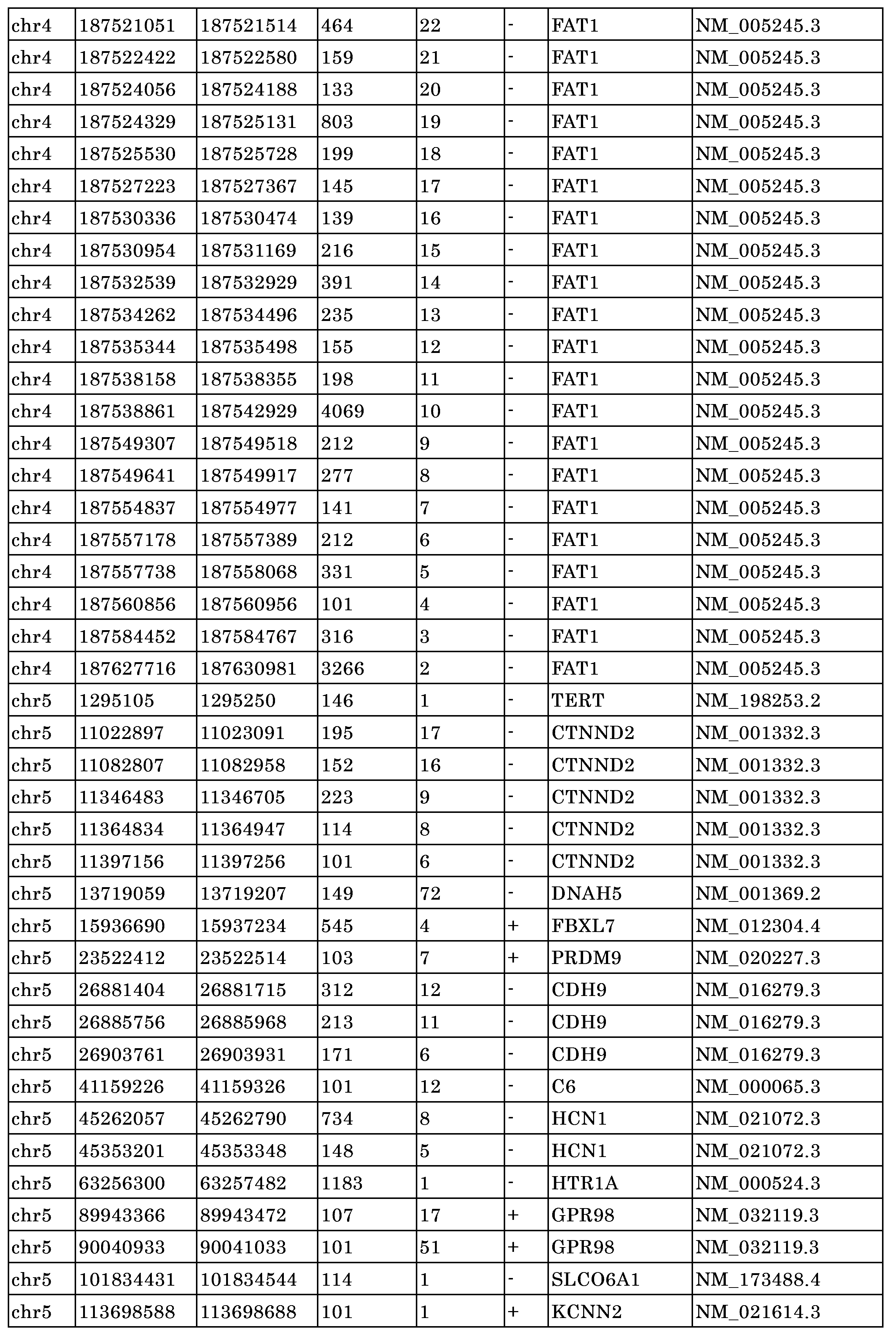


10~30ngのインプットDNAを利用する、CAPP-Seqによる診断時のHNSCC患者および健常なドナーからの血漿およびPBL試料をプロファイリングした。低い存在量でのctDNAの高感度検出を達成するために、本発明者らは、HNSCCにおける検出された変異の数を最大化するように最適化されたCAPP-Seqセレクタを適用した(表2および図10)。本発明者らは、統合デジタルエラー抑制(iDES)によって分析感度をさらに改善し、カスタム分子バーコードを組み込み、健常なドナー血漿試料(方法)内で同定されたバックグラウンド塩基置換エラーを除去した。


血漿プロファイリングおよび可能性のある生殖系列変異の除去に基づいて候補の体細胞一塩基変異体(SNV)を選択した後、本発明者らは、適合したPBLプロファイルとの比較によってクローン造血(CH)による潜在的偽陽性を特性評価した。同定可能な候補のSNVを有する24人の患者のうち、10人が、高度に相関する変異対立遺伝子画分(MAF)を有する一致したPBLプロファイル内で同一のSNVを示した(R=0.94、p=1.392e-07、図2B)。PIK3CAを除いて、これらのSNVを有する遺伝子は各患者に固有であった(図2C)。DNMT3A、TET2、およびASXL1などのCHによって一般に影響を受ける遺伝子はCAPP-Seqセレクタ内に含まれなかったので、一致したcfDNAおよびPBL試料内の患者固有のSNVの本発明者らの知見は、遺伝子レベルフィルタリングよりもこのアプローチの利点をさらに強調する。4人の患者から得た血漿試料は、CHに由来するSNVについて厳密に陽性であり(図2D)、一致したPBLプロファイリングが、低い存在量のctDNAの偽陽性検出を大幅に最小化し得ることを示唆している。 After selecting candidate somatic single nucleotide variants (SNVs) based on plasma profiling and removal of potential germline mutations, we characterized potential false positives due to clonal hematopoiesis (CH) by comparison with matched PBL profiles. Of the 24 patients with identifiable candidate SNVs, 10 showed identical SNVs in matched PBL profiles with highly correlated variant allele fractions (MAFs) (R = 0.94, p = 1.392e -07 , Figure 2B). With the exception of PIK3CA, genes with these SNVs were unique to each patient (Figure 2C). Our finding of patient-specific SNVs in matched cfDNA and PBL samples further highlights the advantages of this approach over gene-level filtering, since genes commonly affected by CH, such as DNMT3A, TET2, and ASXL1, were not included within the CAPP-Seq selector. Plasma samples from four patients were strictly positive for CH-derived SNVs ( Fig. 2D ), suggesting that matched PBL profiling may significantly minimize false-positive detection of low-abundance ctDNA.
CHを潜在的に反映する候補のSNVを除去した後、ctDNAを20人の患者の血漿内で検出した(中央値[範囲]:患者1人あたり3[1~10]のSNV)。これらのSNVの妥当性を評価するために、本発明者らは、結果を、The Cancer Genome Atlas(TCGA)によって公表された279個のHNSCC腫瘍からの全エクソーム解析データと比較して45、TP53(65%対72%)、PIK3CA(20%対21%)、FAT1(15%対23%)およびNOTCH1(10%対19%)を含む高頻度変異遺伝子における類似性を観察した(図2E)。興味深いことに、2人の患者は、これらの遺伝子の中で見られない単一のSNVを示し(GRIN3AおよびMYC、図11)、ctDNAの検出感度を高めるための未知/非ドライバ効果の遺伝子プロファイリングの付加的な有用性を実証した。 After removing candidate SNVs potentially reflecting CH, ctDNA was detected in the plasma of 20 patients (median [range]: 3 [1-10] SNVs per patient). To assess the validity of these SNVs, we compared the results with whole-exome sequencing data from 279 HNSCC tumors published by The Cancer Genome Atlas (TCGA) 45 and observed similarities in hypermutated genes including TP53 (65% vs. 72%), PIK3CA (20% vs. 21%), FAT1 (15% vs. 23%) and NOTCH1 (10% vs. 19%) (Figure 2E). Interestingly, two patients showed single SNVs not found among these genes (GRIN3A and MYC, Figure 11), demonstrating the additional utility of unknown/non-driver effect gene profiling to increase the sensitivity of ctDNA detection.
SNVの平均MAFに基づいてctDNA存在量を計算すると、ctDNAレベルは0.14%~4.83%の範囲であった(図2F)。この検出下限は、約0.14%と推定される、腫瘍ナイーブCAPP-Seq分析を利用する他の者によって以前に記載されたものと類似している。検出不能なctDNAを有する患者を含めて、本発明者らのHNSCCコホートにわたるctDNA存在量の中央値は0.49%であり、CAPP-Seqによって限局性NSCLCにおいて観察されたものと類似していた。 When ctDNA abundance was calculated based on the mean MAF of SNVs, ctDNA levels ranged from 0.14% to 4.83% (Figure 2F). This lower limit of detection is similar to that previously described by others utilizing tumor-naive CAPP-Seq analysis, estimated at approximately 0.14%. Including patients with undetectable ctDNA, the median ctDNA abundance across our HNSCC cohort was 0.49%, similar to that observed in localized NSCLC by CAPP-Seq.
ベースライン血漿からのメチル化ベースのctDNAの腫瘍ナイーブ検出
次に、本発明者らは、HNSCCおよび健常対照試料におけるctDNA関連メチル化パターンを定義しようとした。CAPP-Seqの結果は、PBLから生じる偽陽性変異の影響を示したので、本発明者らは、偽陽性ctDNA関連メチル化の減少が、PBL由来のDNAメチル化シグナルの除去によって達成され得ると推論した。したがって、本発明者らは、セルフリーDNAメチル化シグナルへの寄与を抑制するために、HNSCCおよび健常対照試料からの一致したPBL MeDIP-seqプロファイルを使用し(図3A)、一致したPBLの分析がまた、メチル化ベースのctDNA検出も可能にし得るかどうかを評価した(図3A)。治療前のHNSCCおよび健常なドナーの血漿ならびにPBLを、5~10ngのインプットDNAを利用し、cfMeDIP-seqによって、プロファイリングした。前に記載されたように、メチル化の存在量を、100万あたりのキロベース(RPKM)(方法)のリードに対して正規化されたリードカウントで、染色体1~22にわたる重複しない300bpウィンドウ(n=9,603,454ウィンドウ)で定義した。
Tumor-Naive Detection of Methylation-Based ctDNA from Baseline Plasma Next, we sought to define ctDNA-associated methylation patterns in HNSCC and healthy control samples. Because CAPP-Seq results indicated the impact of false-positive mutations arising from PBLs, we reasoned that reduction of false-positive ctDNA-associated methylation could be achieved by removal of PBL-derived DNA methylation signals. Therefore, we used matched PBL MeDIP-seq profiles from HNSCC and healthy control samples to suppress the contribution to cell-free DNA methylation signals (Figure 3A) and assessed whether analysis of matched PBLs could also enable methylation-based ctDNA detection (Figure 3A). Pre-treatment HNSCC and healthy donor plasma and PBLs were profiled by cfMeDIP-seq utilizing 5-10 ng of input DNA. Methylation abundance was defined in non-overlapping 300-bp windows spanning chromosomes 1-22 (n=9,603,454 windows) with read counts normalized to reads per kilobases per million (RPKM) (Methods) as previously described.
メチル化プルダウンのために利用される抗5mC抗体は、CpGアイランドを含む増加するCpG密度でDNA断片に優先的に結合するので、本発明者らは、この相互作用を最初に特性評価して、cfMeDIP-seqデータ内で高度に表される可能性が高い領域を同定した。本発明者らはまた、MeDIP-seqをHNSCC細胞株FaDuに適用して、がん由来メチル化DNA断片の優先的な結合を評価した。様々な数のCpGを有するウィンドウにわたってDNA断片プルダウン存在量(RPKMの中央値)を比較すると、本発明者らは、PBLおよびFaDuの両方について最大8CpG以上の濃縮の増加を観察した(図12Aおよび図12B)。FaDuは、300bpウィンドウあたり8CpG以上で、PBLと比較してより大きな濃縮を示した。この結果は、FaDuを含むがん細胞におけるCpGアイランドの過剰メチル化の確立された現象と一致している。これらの観察に基づいて、本発明者らは、8以上のCpG(n=702,488)を有するウィンドウがctDNA検出に最も情報があり得、そのためその後のすべての分析に利用されたと判定した。 Because the anti-5mC antibody utilized for methylation pull-down preferentially binds DNA fragments with increasing CpG density, including CpG islands, we first characterized this interaction to identify regions likely to be highly represented within the cfMeDIP-seq data. We also applied MeDIP-seq to the HNSCC cell line FaDu to evaluate preferential binding of cancer-derived methylated DNA fragments. Comparing DNA fragment pull-down abundance (median RPKM) across windows with varying numbers of CpGs, we observed increased enrichment up to 8 CpGs or more for both PBLs and FaDu (Figures 12A and 12B). FaDu showed greater enrichment compared to PBLs at 8 CpGs or more per 300 bp window. This result is consistent with the established phenomenon of CpG island hypermethylation in cancer cells, including FaDu. Based on these observations, we determined that windows with 8 or more CpGs (n=702,488) were most likely to be informative for ctDNA detection and were therefore utilized in all subsequent analyses.
限局性がんを有する患者の場合、血漿のセルフリーDNAの大部分はPBLに由来する。したがって、本発明者らは、セルフリーDNAシグナルへのこの寄与を生物情報学的に抑制するためにPBL MeDIP-seqプロファイルを利用しようとした。本発明者らは、HNSCCおよび健常なドナーcfDNAから作製されたcfMeDIP-seqプロファイル内の各ウィンドウについてのRPKMの値を、FaDu(1×1の比較)、対になっていないPBL(1×51の比較)または対になったPBL(1×1の比較)から作製されたMeDIP-seqプロファイルと比較した。血漿のセルフリーDNAの主な寄与因子であるPBLによれば、ゲノムワイドのメチル化プロファイルは、血漿のセルフリーDNAと、対になったまたは対になっていないPBLとの間で高度に相関していた(それぞれ最頻値R=0.92およびR=0.91)。これらの相関の強さは、血漿のcfDNAに対するPBLの既知のサイズ外の寄与を反映している可能性が高い。対照的に、血漿のセルフリーDNAとFaDuとの間の相関はより弱かった(最頻値R=0.78)(図3B)。 For patients with localized cancer, the majority of plasma cell-free DNA originates from PBLs. We therefore sought to utilize PBL MeDIP-seq profiles to bioinformatically constrain this contribution to cell-free DNA signal. We compared the RPKM values for each window in cfMeDIP-seq profiles made from HNSCC and healthy donor cfDNA to MeDIP-seq profiles made from FaDu (1x1 comparison), unpaired PBLs (1x51 comparison) or paired PBLs (1x1 comparison). With PBLs being the major contributors to plasma cell-free DNA, genome-wide methylation profiles were highly correlated between plasma cell-free DNA and paired or unpaired PBLs (modal R = 0.92 and R = 0.91, respectively). The strength of these correlations likely reflects contributions of PBLs to plasma cfDNA outside their known size. In contrast, the correlation between plasma cell-free DNA and FaDu was weaker (mode R = 0.78) (Figure 3B).
優先的プルダウンを考慮しながら、PBLにわたるメチル化の減少の閾値を選択するために、本発明者らは、MeDEStrand Rパッケージ(方法)によるロジスティック回帰モデリングに基づいて、PBL cfMeDIP-seqプロファイルを絶対的なメチル化レベル(0~1)にスケーリングおよび正規化した。本発明者らは、健常なドナーのPBLにわたって0.1未満の絶対的なメチル化の中央値を示す99,997個のウィンドウを選択した。これらのウィンドウを、除外されたHNSCC PBLに適用したとき、本発明者らは、利用された健常なドナーのPBLの絶対的なメチル化分布と同様の絶対的なメチル化分布を観察し(図3B)、このアプローチの一般化の可能性を実証した。同様に、これらのウィンドウのいずれも個々には、健常なドナーのPBLと比較してHNSCCのPBLにわたって有意に高いメチル化を示さず(図3Cおよび図12B)、ctDNA検出を混乱させ得るHNSCC特異的PBLメチル化のあらゆる供給源を制限した。換言すれば、これらの結果は、対照および局所的に限定されたHPV陰性HNSCC血漿の両方におけるcfDNAメチル化の主な源がPBLに由来し、PBL由来のメチル化の生物情報学的除去がctDNAの定量化を混乱させたシグナルを制限し得ることを確認する。 To select a threshold of reduced methylation across PBLs while allowing for preferential pulldown, we scaled and normalized PBL cfMeDIP-seq profiles to absolute methylation levels (0-1) based on logistic regression modeling with the MeDEStand R package (Methods). We selected 99,997 windows that showed a median absolute methylation value of <0.1 across healthy donor PBLs. When we applied these windows to excluded HNSCC PBLs, we observed an absolute methylation distribution similar to that of the utilized healthy donor PBLs (Figure 3B), demonstrating the generalizability of this approach. Similarly, none of these windows individually showed significantly higher methylation across HNSCC PBLs compared to healthy donor PBLs (Figures 3C and 12B), limiting any source of HNSCC-specific PBL methylation that could confound ctDNA detection. In other words, these results confirm that the major source of cfDNA methylation in both control and locally confined HPV-negative HNSCC plasma is derived from PBLs, and that bioinformatic removal of PBL-derived methylation can limit signals that confound ctDNA quantification.
治療前のメチル化ベースのctDNAの腫瘍ナイーブ検出
HNSCCコホート内の一般的なctDNA由来の過剰メチル化領域を同定するために、本発明者らは、HNSCC患者と、CAPP-Seqによる検出可能なctDNA(n=20)とを、健常なドナーと比較する、差次的メチル化分析を行った。PBLにおけるメチル化のために枯渇される99,994個の300bpウィンドウを利用して、本発明者らは、20人のHNSCC患者をCAPP-Seq-検出可能なctDNAと20人の健常対照と比較することによって、ctDNA由来の差次的メチル化領域(DMR)を同定した。総じて、本発明者らは、HNSCC試料全体で997個の差次的メチル化領域(DMR)(過剰メチル化:941、低メチル化:56)を同定した(図3C)。過剰メチル化領域(過剰DMR)の半分程度が互いに直接隣接していることが見出され、過剰メチル化のブロックは最大1800塩基対の長さに及んだ(図13A)。これらのデータは、同定された過剰DMR内のCpGアイランドが存在することを示唆している。逆に、隣接する低メチル化領域(低DMR)は観察されなかった。300bpの過剰DMRのうち、47.5%は、長さが1800bpにまで及ぶ過剰メチル化シグナルの連続したブロックに存在し(図13A)、典型的には長さが300~3000bpに及ぶCpGアイランドを示した。実際、CpGアイランドは、過剰DMRについて有意に濃縮されていた(図3E)。対照的に、低DMRについてはCpGアイランドが有意に枯渇した(図13B)。
Tumor-Naive Detection of Pre-Treatment Methylation-Based ctDNA To identify common ctDNA-derived hypermethylated regions within the HNSCC cohort, we performed differential methylation analysis comparing HNSCC patients with CAPP-Seq-detectable ctDNA (n=20) with healthy donors. Utilizing 99,994 300-bp windows depleted for methylation in PBLs, we identified ctDNA-derived differentially methylated regions (DMRs) by comparing 20 HNSCC patients with CAPP-Seq-detectable ctDNA and 20 healthy controls. Overall, we identified 997 differentially methylated regions (DMRs) (hypermethylated: 941, hypomethylated: 56) across HNSCC samples (Figure 3C). Approximately half of the hypermethylated regions (hyper-DMRs) were found to be directly adjacent to each other, with blocks of hypermethylation spanning lengths of up to 1800 base pairs (Figure 13A). These data suggest the presence of CpG islands within the identified hyper-DMRs. Conversely, no adjacent hypomethylated regions (hypo-DMRs) were observed. Of the 300 bp hyper-DMRs, 47.5% were present in contiguous blocks of hypermethylated signals spanning up to 1800 bp in length (Figure 13A), typically displaying CpG islands spanning 300-3000 bp in length. Indeed, CpG islands were significantly enriched for hyper-DMRs (Figure 3E). In contrast, CpG islands were significantly depleted for hypo-DMRs (Figure 13B).
これらの過剰DMRが実際にCpGアイランドについて濃縮されているかどうかを判定するために、本発明者らは次に、置換分析(方法)によってCpGアイランド、ショア、shelf、およびopen seaについて、過剰DMRの濃縮を評価した。予想されるように、CpGアイランドの有意な濃縮、ならびにショアおよびopen seaの有意な枯渇が、過剰DMR内で観察された(図3E)。対照的に、低DMRは、がん全体で頻繁に観察されるCpG-スパース領域の低メチル化に従って、open seaが有意に濃縮され、CpGアイランドが枯渇していた(補足図5B)。 To determine whether these over-represented DMRs are indeed enriched for CpG islands, we next assessed enrichment of over-represented DMRs for CpG islands, shores, shelves, and open seas by substitution analysis (Methods). As expected, significant enrichment of CpG islands and significant depletion of shores and open seas were observed within over-represented DMRs (Fig. 3E). In contrast, under-represented DMRs were significantly enriched for open seas and depleted for CpG islands, in line with the hypomethylation of CpG-sparse regions frequently observed across cancers (Supplementary Fig. 5B).
最後に、特定の領域のメチル化は、cfMeDIP-seqを使用して以前に記載されたように起源の組織を区別し得るので、本発明者らはまた、過剰DMRがHNSCCまたは他のがんに特異的な領域を含むかどうかを調べた。腫瘍特異的メチル化領域を同定するために、本発明者らは、TCGA(方法)によってもたらされる原発腫瘍から生成されたHumanMethylation450K(hm450k)のデータを利用した。浸潤性乳癌(BRCA)、結腸腺癌(COAD)、肺扁平上皮癌(LUSC)、前立腺腺癌(PRAD)、HNSCC、膵臓腺癌(PAAD)、およびPBL由来の原発腫瘍を比較して、本発明者らは、BRCA、COAD、PRAD、およびHNSCC(方法)に特異的な十分な過剰メチル化CpG(≧50)を同定した(図14)。予想通り、本発明者らは、HNSC特異的過剰メチル化CpGと重複する血漿由来DMRの有意な濃縮、ならびにBRCA、COADおよびPRAD特異的過剰メチル化CpGにわたる重複の有意な枯渇を観察し(図3F)、このことは、過剰DMRが、様々な他のがんのタイプと比較した場合にHNSCC起源に特異的な領域を含むことを示唆している。 Finally, since methylation of specific regions can distinguish tissues of origin as previously described using cfMeDIP-seq, we also investigated whether the hyper-DMRs included regions specific to HNSCC or other cancers. To identify tumor-specific methylation regions, we utilized HumanMethylation450K (hm450k) data generated from primary tumors derived by TCGA (Methods). Comparing primary tumors from invasive breast cancer (BRCA), colon adenocarcinoma (COAD), lung squamous cell carcinoma (LUSC), prostate adenocarcinoma (PRAD), HNSCC, pancreatic adenocarcinoma (PAAD), and PBL, we identified sufficient hypermethylated CpGs (>50) specific to BRCA, COAD, PRAD, and HNSCC (Methods) (Figure 14). As expected, we observed significant enrichment of plasma-derived DMRs overlapping with HNSC-specific hypermethylated CpGs, as well as significant depletion of overlap across BRCA, COAD, and PRAD-specific hypermethylated CpGs (Figure 3F), suggesting that the excess DMRs include regions specific to HNSCC origin when compared to a variety of other cancer types.
変異ベースとメチル化ベースのctDNA検出は特に一致している
いっそう多くの研究が、血漿のセルフリーDNAの健常な源と比較して、減少した断片の長さに関連するctDNAを記載しており、ロバストな腫瘍ナイーブ検出のためのさらなるメトリックが得られている。標的化された配列決定が、低下した断片の長さでctDNAを検出することが以前に示されているので、本発明者らはまず、本発明者らのCAPP-Seqプロファイルを利用して、本発明者らがHNSCC患者において同様の傾向を観察し得るかどうかを判定した。患者ごとに同定された各SNV(図2E)について、SNV対立遺伝子ならびに重複する参照対立遺伝子を含む断片の長さの中央値を測定した。患者の試料の中で複数のSNVが同定された場合については、すべてのSNVおよびそれらの参照対立遺伝子にわたる中央値を使用した。以前の知見によれば、本発明者らは、患者全体で健常なセルフリーDNAと比較してctDNA断片のサイズの一貫した減少を観察した(中央値[範囲]Δ=-17.5[1~58]bp)(図4A)。これらの変異の平均MAFと断片の長さとの間に有意な関連はなかった(図15A)。
Mutation-based and methylation-based ctDNA detection are particularly concordant More studies have described ctDNA associated with reduced fragment lengths compared to healthy sources of plasma cell-free DNA, providing additional metrics for robust tumor-naive detection. As targeted sequencing has previously been shown to detect ctDNA at reduced fragment lengths, we first utilized our CAPP-Seq profiles to determine whether we might observe a similar trend in HNSCC patients. For each SNV identified per patient (Figure 2E), we measured the median fragment length encompassing the SNV allele as well as the overlapping reference allele. For cases where multiple SNVs were identified in a patient's samples, the median across all SNVs and their reference alleles was used. In accordance with previous findings, we observed a consistent reduction in ctDNA fragment size compared to healthy cell-free DNA across patients (median [range] Δ=-17.5 [1-58] bp) (Figure 4A). There was no significant association between the mean MAF of these mutations and fragment length (Figure 15A).
亜硫酸水素塩ベースのDNAメチル化アプローチとは異なり、cfMeDIP-seqはDNAの分解を引き起こさず、したがって元の断片サイズ分布を維持する。これは、DNAメチル化および断片の長さを同時にマッピングする新規な機会を提供する。各患者について以前に同定された血漿由来過剰DMR内の断片の長さの分布を評価した。これらの領域が本発明者らの健常なドナーにわたる低いメチル化を有するという性質のために、ドナーにわたるDNA断片を、比較のために組み合わせた。変異に基づく分析と同様に、本発明者らは、グループ化された健常対照と比較して、19/20のCAPP-Seq陽性患者からの断片の長さの減少を観察した(中央値[範囲]Δ=-7[1~21]bp)(図4B)。これは、おそらく過剰DMR内のセルフリーDNA断片の健常な組織による部分的な寄与のために、変異ベースの分析と比較して断片の長さの減少が小さいことを示した。この考えを裏付けて、最も短い過剰DMR断片を有する試料は、より高いメチル化ctDNA存在量(ピアソンr=-0.64、p=0.002)を示した(図15B)。小さい断片(100bp~150bp)対大きい断片(151bp~220bp)の比を本発明者らの過剰DMR、ctDNAを濃縮するための以前に記載されたアプローチ、のために使用したとき、本発明者らは、大部分のCAPP-Seq陽性HNSCC試料にわたってctDNAの濃縮の同様の傾向を観察した(中央値[範囲]=28[-8~63]%)(図4C)。 Unlike bisulfite-based DNA methylation approaches, cfMeDIP-seq does not cause DNA degradation and therefore maintains the original fragment size distribution. This provides a novel opportunity to map DNA methylation and fragment length simultaneously. The distribution of fragment lengths within the plasma-derived excess DMRs previously identified for each patient was assessed. Due to the nature of these regions having low methylation across our healthy donors, DNA fragments across donors were combined for comparison. Similar to the mutation-based analysis, we observed a reduction in fragment lengths from 19/20 CAPP-Seq positive patients compared to grouped healthy controls (median [range] Δ=-7 [1-21] bp) (Figure 4B). This indicated a smaller reduction in fragment lengths compared to the mutation-based analysis, likely due to a partial contribution by healthy tissue of cell-free DNA fragments within the excess DMRs. Supporting this idea, samples with the shortest excess DMR fragments showed higher methylated ctDNA abundance (Pearson r = -0.64, p = 0.002) (Figure 15B). When we used the ratio of small fragments (100 bp to 150 bp) to large fragments (151 bp to 220 bp) for our excess DMR, a previously described approach to enrich for ctDNA, we observed a similar trend of enrichment of ctDNA across the majority of CAPP-Seq-positive HNSCC samples (median [range] = 28 [-8 to 63]%) (Figure 4C).
本発明者らのHNSCCコホートにおいて同定された血漿のセルフリーDNA過剰DMRがこれらの小さな断片(100~150bp)内の個体にわたりどのように異なり得るかを評価するために、本発明者らは最初に階層的クラスタリングを行った。4つの主要なクラスターがConsensusClusterPlus Rパッケージを利用して出現し、それぞれが過剰DMRにわたって異なるレベルのメチル化を有した(図4Eおよび図16C)。同様に、3つのクラスターは、CAPP-Seq(図16D)によって判定されるような別個のctDNA存在量によって定義され、これは、過剰DMRメチル化の平均と変異に基づくctDNAの存在量との間の潜在的な関係を示唆している。 To assess how the plasma cell-free DNA excess DMRs identified in our HNSCC cohort may differ across individuals within these small fragments (100-150 bp), we first performed hierarchical clustering. Four major clusters emerged utilizing the ConsensusClusterPlus R package, each with different levels of methylation across the excess DMRs (Figure 4E and Figure 16C). Similarly, three clusters were defined by distinct ctDNA abundances as determined by CAPP-Seq (Figure 16D), suggesting a potential relationship between average excess DMR methylation and mutation-based ctDNA abundance.
次に、本発明者らは、断片の長さがCAPP-SeqおよびcfMeDIP-seqの両方によって同定されたctDNA分子間で一致するかどうかを調査し、本発明者らのマルチモーダルアプローチに対する検証のさらなる層を潜在的にもたらす。バックグラウンドDNA断片がcfMeDIP-seqプロファイル内のctDNAの計算された断片の長さを混同させる可能性を最小限に抑えるために、本発明者らは、過剰DMR全体でメチル化レベルの中央値を超える患者(n=10人のHNSCC患者)に分析を限定した。驚くべきことに、完全に異なるゲノム領域がこれらの2つのプロファイリング手法を用いて表されているにもかかわらず、ctDNA断片の長さは、各患者についての対となったCAPP-SeqプロファイルとcfMeDIP-seqプロファイルとの間で非常に一致していた(図4C)(ピアソンr=0.86、p=0.0016)(CAPP-Seq:43個の異なる変異、cfMeDIP-seq:941個の過剰DMR)。 Next, we investigated whether fragment lengths were concordant between ctDNA molecules identified by both CAPP-Seq and cfMeDIP-seq, potentially providing an additional layer of validation for our multimodal approach. To minimize the possibility that background DNA fragments confound the calculated fragment lengths of ctDNA in cfMeDIP-seq profiles, we limited our analysis to patients (n=10 HNSCC patients) with above-median methylation levels across excess DMRs. Surprisingly, ctDNA fragment lengths were highly concordant between paired CAPP-Seq and cfMeDIP-seq profiles for each patient (Figure 4C) (Pearson r=0.86, p=0.0016) (CAPP-Seq: 43 distinct mutations, cfMeDIP-seq: 941 excess DMRs).
過剰DMRメチル化レベルと変異ベースのctDNA存在量との間の関係をさらに特性評価するために、本発明者らは、941の過剰DMRにわたるRPKMの平均値を、各患者についてCAPP-Seqによって判定されたMAFの平均値と比較した。メチル化クラスター間で観察された傾向と同様に、本発明者らは有意な正の相関(ピアソン相関、R=0.85、p=5e-10)を観察した(図4F)。cfMeDIP-seqによるこれらの過剰DMR内のctDNA検出の感度を評価するために、本発明者らは、本発明者らのHNSCCコホートと健常なドナーとの間のRPKMの平均値を比較した。CAPP-Seq陽性患者(n=20)については、ctDNA検出は特に一致しており(AUC=0.998)、CAPP-Seq陰性患者(n=12)の組み込み時にパフォーマンスがわずかに低下した(AUC=0.944)(図4G)。CAPP-Seq陽性患者および健常なドナーにわたる交差検証(n=50サンプリング)の結果、0.984のAUC中央値がもたらされ(図16A)、本明細書に開示されるアプローチのロバスト性が実証された。 To further characterize the relationship between excess DMR methylation levels and mutation-based ctDNA abundance, we compared the mean RPKM across the 941 excess DMRs to the mean MAF determined by CAPP-Seq for each patient. Similar to the trends observed among methylation clusters, we observed a significant positive correlation (Pearson correlation, R = 0.85, p = 5e-10) (Figure 4F). To assess the sensitivity of ctDNA detection within these excess DMRs by cfMeDIP-seq, we compared the mean RPKM between our HNSCC cohort and healthy donors. For CAPP-Seq-positive patients (n = 20), ctDNA detection was particularly concordant (AUC = 0.998), with a slight decrease in performance upon inclusion of CAPP-Seq-negative patients (n = 12) (AUC = 0.944) (Figure 4G). Cross-validation (n=50 samples) across CAPP-Seq positive patients and healthy donors resulted in a median AUC of 0.984 (Figure 16A), demonstrating the robustness of the approach disclosed herein.
これらの観察に基づいて、本発明者らは、短縮された長さのセルフリーDNA断片に分析を限定することによって、cfMeDIP-seqプロファイル内でctDNAを濃縮し得るかどうかを評価した。本発明者らは、非メチル化ベースのアプローチを使用してctDNAを濃縮する同様の方法が記載されているので、小さい(100~150bp)断片からなる過剰DMR内のセルフリーDNA断片の割合を評価した。実際、これにより、CAPP-Seq陽性HNSCC試料の大部分(中央値[範囲]=28[-8~63]%)にわたってctDNA濃縮がもたらされたが、健常対照のいずれについてももたらされなかった(図4D)。したがって、セルフリーDNA断片のインシリコのサイズ選択は、cfMeDIP-seqライブラリ内のctDNAを濃縮し、腫瘍ナイーブマルチモーダルctDNA分析に寄与し得る。 Based on these observations, we assessed whether ctDNA could be enriched within cfMeDIP-seq profiles by restricting the analysis to cell-free DNA fragments of shortened length. We assessed the proportion of cell-free DNA fragments within the excess DMRs consisting of small (100-150 bp) fragments, as similar methods have been described to enrich for ctDNA using a non-methylation-based approach. Indeed, this led to ctDNA enrichment across the majority of CAPP-Seq-positive HNSCC samples (median [range] = 28 [-8 to 63]%), but not for any of the healthy controls (Figure 4D). Thus, in silico size selection of cell-free DNA fragments may enrich for ctDNA within cfMeDIP-seq libraries and contribute to tumor-naive multimodal ctDNA analysis.
限局性非転移性がんを有する患者において、診断時のCAPP-SeqによるctDNAの検出は、予後不良に関連することが以前に記載されている。同様に、SHOX2およびSEPT9のメチル化によって評価されるctDNAのレベルは、HNSCCにおける予後不良と関連している。したがって、本発明者らは、診断時のCAPP-SeqおよびcfMeDIP-seqによるctDNAの検出または定量化が本発明者らのHNSCCコホート内の臨床転帰と関連するかどうかを尋ねた。実際、CAPP-Seq(すなわち、CAPP-Seq陽性対CAPP-Seq陰性)によるctDNAの検出(ハザード比[HR]=7.6、ログランクp=0.026;補足図8D)、ならびに本発明者らが以前に同定した過剰DMR内のメチル化の増加(すなわち、メチル化クラスター1+2+3対メチル化クラスター4)(HR=4.51、p=0.038;図4G)は、生存期間の短縮と相関していた。この知見と一致して、過剰DMRにわたる平均RPKMは、がんのステージと相関した(補足図8E)。 It has been previously described that in patients with localized nonmetastatic cancer, detection of ctDNA by CAPP-Seq at diagnosis is associated with poor prognosis. Similarly, levels of ctDNA, as assessed by methylation of SHOX2 and SEPT9, are associated with poor prognosis in HNSCC. Therefore, we asked whether detection or quantification of ctDNA by CAPP-Seq and cfMeDIP-seq at diagnosis is associated with clinical outcome within our HNSCC cohort. Indeed, detection of ctDNA by CAPP-Seq (i.e., CAPP-Seq positive vs. CAPP-Seq negative) (hazard ratio [HR] = 7.6, log-rank p = 0.026; Supplementary Fig. 8D), as well as increased methylation within our previously identified excess DMRs (i.e., methylation cluster 1 + 2 + 3 vs. methylation cluster 4) (HR = 4.51, p = 0.038; Fig. 4G), correlated with shorter survival. Consistent with this finding, the mean RPKM across excess DMRs correlated with cancer stage (Supplementary Fig. 8E).
次に、本発明者らは、変異またはメチル化に基づくプロファイリングのいずれかによって同定されたctDNAの断片の長さの中央値を比較した。バックグラウンドDNA断片がcfMeDIP-seqプロファイル内のctDNAの計算された断片の長さを混同させる可能性を最小限に抑えるために、本発明者らは、階層的クラスタリングによって定義されるように高いctDNA存在量を有する患者を選択した(すなわち、メチル化クラスター1および2、図4D、補足図8A~B)。このアプローチでは、完全に異なるゲノム領域がこれらの2つのプロファイリング手法を用いて表されているにもかかわらず、ctDNA断片の長さは、各患者について対となったCAPP-SeqプロファイルとcfMeDIP-seqプロファイルとの間で特に一致していた(R=0.83、p=0.0016)(図4H)。さらに、すべての長さの断片を用いた本発明者らの分析と同様に、本発明者らは、CAPP-Seq(R=-0.79、p=0.0038)による小さい断片比とctDNA断片の長さとの間で同じ関係を観察した(図4I)。
Next, we compared the median fragment lengths of ctDNA identified by either mutation- or methylation-based profiling. To minimize the possibility that background DNA fragments might confound the calculated fragment lengths of ctDNA in cfMeDIP-seq profiles, we selected patients with high ctDNA abundance as defined by hierarchical clustering (i.e.,
これらの結果は、CAPP-SeqおよびcfMeDIP-seqによって検出されるctDNAから観察される断片の長さの同様の減少が、ゲノム領域によるのではなく、腫瘍の固有の特性の結果であり得ること、およびより短い断片の長さを利用することがctDNAのより特異的な同定に寄与し得ることを示唆している。 These results suggest that the similar reduction in fragment lengths observed from ctDNA detected by CAPP-Seq and cfMeDIP-seq may be a result of intrinsic properties of the tumor rather than due to genomic region, and that utilizing shorter fragment lengths may contribute to more specific identification of ctDNA.
予後の判定のためのマルチモーダルctDNA検出の適用
腫瘍ナイーブマルチモーダルctDNA分析の潜在的な臨床応用を評価するために、本発明者らは、HNSCCコホートにおいてctDNAを臨床転帰と比較した。断片の長さの情報に基づくcfMeDIP-seqプロファイルは、一致したCAPP-SeqプロファイルにおいてMAFと強く関連し(ピアソンr=0.85、p=3×10⁻⁹)、941個の過剰DMR内のメチル化強度が実際にctDNAの存在量を反映していることを示唆した(図5C)。重要なことに、交差検証分析により、ctDNAを検出するためのこれらの過剰DMRのロバスト性が確認された(図16C)。変異およびメチル化に基づく方法の両方によってベースラインの血漿において検出されたctDNAを有する患者(n=19)は、検出可能なctDNAを有していない患者(n=8/13)と比較した場合、進行した疾患(すなわち、ステージIII-IVA)を有する可能性が有意に高く(n=18/19)(フィッシャーの正確検定p=0.028)、劇的に不良な全生存率を示した(ハザード比[HR]=7.55、95%信頼区間[CI]=[0.95~59.94]、ログランクp=0.025)(図5G)。比較すると、ステージのみでは、より不良な全生存(HR=2.59、95%CI=[0.32~20.46]、ログランクp=0.35)の患者を予測することができず(図16D)、マルチモーダルctDNAプロファイリングの潜在的な臨床的有用性がさらに実証された。
Application of multimodal ctDNA detection for prognosis To evaluate the potential clinical application of tumor-naive multimodal ctDNA analysis, we compared ctDNA with clinical outcomes in a HNSCC cohort. Fragment length-informed cfMeDIP-seq profiles were strongly associated with MAF in matched CAPP-Seq profiles (Pearson r=0.85, p=3×10⁻⁹), suggesting that methylation intensity within the 941 excess DMRs indeed reflects ctDNA abundance (Figure 5C). Importantly, cross-validation analysis confirmed the robustness of these excess DMRs for detecting ctDNA (Figure 16C). Patients with ctDNA detected in baseline plasma by both mutation- and methylation-based methods (n=19) were significantly more likely to have advanced disease (i.e., stages III-IVA) (n=18/19) when compared with patients without detectable ctDNA (n=8/13) (Fisher's exact test p=0.028) and showed dramatically worse overall survival (hazard ratio [HR]=7.55, 95% confidence interval [CI]=[0.95-59.94], log-rank p=0.025) (Figure 5G). In comparison, stage alone failed to predict patients with worse overall survival (HR=2.59, 95% CI=[0.32-20.46], log-rank p=0.35) (Figure 16D), further demonstrating the potential clinical utility of multimodal ctDNA profiling.
がんドライバの遺伝子発現および結果として生じる機能活性に対するDNAメチル化の既知の効果のために、本発明者らは、特定の遺伝子座におけるctDNAメチル化パターンがctDNA存在量とは無関係に予後の有意性を有し得ると推論した。本発明者らの以前に同定された過剰DMRが、ctDNA存在量とは無関係に予後と関連する特定の領域を含むかどうかを評価するために、本発明者らは、入手可能なすべてのHNSCC患者(n=520)についてTCGAによって得られるDNAメチル化、RNA発現および臨床転帰データを調べた(図5C)。最初に、本発明者らは、TCGA hm450kメチル化アレイデータから異なる300bpウィンドウ内に含まれるすべてのCpGにわたるβの平均値を計算した。分析を、血漿由来過剰DMRと重複するプローブhm450k領域(n=764/941)に限定して、本発明者らが隣接する正常組織(n=50)と比較して原発腫瘍(n=520)の483個の過剰メチル化領域を同定した(ウィルコクソン検定、FDR<0.05、log2FC>1)。本発明者らは、HNSCCならびにTWIST1およびONECUT2で以前に評価されたSEPT9およびSHOX2を含む、市販のメチル化ベースのctDNA診断試験によってプロファイリングされた遺伝子内のCpGの近くに、これらの過剰メチル化領域のいくつかが重複しているかまたは位置していることを観察した(図17A)。これらの結果から、本発明者らの血漿由来過剰DMRの潜在的な臨床的関連性を支持するさらなる証拠が得られる。 Due to the known effect of DNA methylation on gene expression and resulting functional activity of cancer drivers, we reasoned that ctDNA methylation patterns at specific loci may have prognostic significance independent of ctDNA abundance. To assess whether our previously identified excess DMRs contain specific regions associated with prognosis independent of ctDNA abundance, we examined DNA methylation, RNA expression and clinical outcome data obtained by TCGA for all available HNSCC patients (n=520) (Figure 5C). First, we calculated the average value of β across all CpGs contained within different 300 bp windows from the TCGA hm450k methylation array data. Restricting the analysis to probe hm450k regions (n=764/941) overlapping with plasma-derived excess DMRs, we identified 483 hypermethylated regions in primary tumors (n=520) compared with adjacent normal tissues (n=50) (Wilcoxon test, FDR<0.05, log2FC>1). We observed that several of these hypermethylated regions overlapped or were located near CpGs within genes profiled by commercially available methylation-based ctDNA diagnostic tests, including SEPT9 and SHOX2, previously evaluated in HNSCC, as well as TWIST1 and ONECU2 (Figure 17A). These results provide further evidence supporting the potential clinical relevance of our plasma-derived excess DMRs.
本発明者らのHNSCCコホートおよびTCGAのHNSC hm450kのプロファイルによって共通して保持されるこれらの過剰メチル化領域の潜在的な臨床的有用性をさらに調べるために、本発明者らは、利用可能なhm450kのプロファイルおよび疾患特異的生存(DSS)転帰であるすべてのTCGAのHNSCC患者にわたって単変量コックス比例ハザード回帰を行った(n=493/520)。本発明者らは、DSSと有意に関連する33の領域を同定した(p<0.05)。腫瘍形成において機能的役割を有する可能性が高い予後領域をさらに選択するために、本発明者らは、各領域(n=33)のメチル化レベルを2kb内の周囲の遺伝子転写物の発現と比較した。次に、本発明者らは、TCGAのHNSCCコホートを使用して、(1)多変量Cox回帰における予後および(2)隣接遺伝子転写物の発現に関連する483個のDMRのサブセットを同定した。5つの領域が両方の基準を満たすように同定され、各領域のメチル化が増加すると、ZNF323/ZSCAN31、LINC01391、およびGATA2-AS1のより高い発現がもたらされ(図5G、図17A~図17C、またSTK3/MST2およびOSR1のそれぞれのより低い発現がもたらされた(図5H)(図5D)。メチル化の結果としての発現の減少および増加に関連する領域は、それぞれプロモーターまたは第1のエクソン/イントロンおよび遺伝子本体内に存在することが見出された。本発明者らは、これらの5つの領域から複合メチル化スコア(CMS)を構築し(表6)、このスコアに従ってTCGAのHNSCCコホートを層別化した(図5E)。より高いCMSは、劣った生存転帰と有意に関連していた(HR=1.67、95%CI=[1.25,2.21]、ログランクp=3.4×10-4)。 To further explore the potential clinical utility of these hypermethylated regions commonly held by our HNSCC cohort and TCGA HNSSC hm450k profiles, we performed univariate Cox proportional hazards regression across all TCGA HNSCC patients with available hm450k profiles and disease-specific survival (DSS) outcomes (n=493/520). We identified 33 regions significantly associated with DSS (p<0.05). To further select prognostic regions likely to have a functional role in tumorigenesis, we compared the methylation level of each region (n=33) with the expression of surrounding gene transcripts within 2 kb. We then used the TCGA HNSCC cohort to identify a subset of 483 DMRs associated with (1) prognosis in multivariate Cox regression and (2) the expression of neighboring gene transcripts. Five regions were identified as fulfilling both criteria, and increased methylation in each region led to higher expression of ZNF323/ZSCAN31, LINC01391, and GATA2-AS1 (Figure 5G, Figures 17A-C) and lower expression of STK3/MST2 and OSR1, respectively (Figure 5H) (Figure 5D). Regions associated with decreased and increased expression as a result of methylation were found to reside within the promoter or first exon/intron and gene body, respectively. We constructed a composite methylation score (CMS) from these five regions (Table 6) and stratified the TCGA HNSCC cohort according to this score (Figure 5E). Higher CMS was significantly associated with poorer survival outcome (HR=1.67, 95% CI=[1.25, 2.21], log-rank p=3.4× 10⁻⁴ ).
最後に、本発明者らは、ctDNAに適用した場合にCMSかた同様の予後情報が得られるかどうかも評価した。ctDNAを濃縮するために、cfMeDIP-seqライブラリの分析を、上記のように100~150bpの長さの断片に限定した(図4E)。5つの推定予後マーカーによって得られるctDNAメチル化レベルの相対的な寄与を説明するために、本発明者らは、これらの領域からのcfMeDIP-seqのRPKMの値を941個の過剰DMR全体に正規化した。これは、より高いCMSがより不良な生存とわずかに関連するという同様の傾向をもたらし(ログランクp=0.1;HR=3.06)(図5F)、このことは、TCGAから同定されたこれらの推定予後領域のメチル化の増加がまた、cfMeDIP-seqプロファイル内で情報があり得ることを示唆している。さらに、これらの結果は、バイオマーカー発見のために、血漿のセルフリーDNAメチロームプロファイリングを既存のマルチオミックのがんデータベースと組み合わせてどのように活用できるかを強調している。 Finally, we also assessed whether CMS could provide similar prognostic information when applied to ctDNA. To enrich for ctDNA, analysis of cfMeDIP-seq libraries was restricted to fragments of 100-150 bp length as described above (Fig. 4E). To account for the relative contribution of ctDNA methylation levels provided by the five putative prognostic markers, we normalized the cfMeDIP-seq RPKM values from these regions across the 941 excess DMRs. This yielded a similar trend where higher CMS was marginally associated with poorer survival (log-rank p=0.1; HR=3.06) (Fig. 5F), suggesting that increased methylation of these putative prognostic regions identified from TCGA may also be informative within the cfMeDIP-seq profile. Furthermore, these results highlight how plasma cell-free DNA methylome profiling can be leveraged in combination with existing multi-omic cancer databases for biomarker discovery.
cfMeDIP-seqによる最終治療後の疾患の監視
cfMeDIP-seqがHNSCC患者において高感度かつ定量的なctDNA検出を達成したので、本発明者らは、CAPP-seqと同様に、cfMeDIP-seqもctDNA存在量の治療関連変化をモニタリングすることができる可能性があると推論した。治療後のcfMeDIP-seqプロファイル内のctDNAのパーセントを定量するために、本発明者らは、以前に同定された血漿由来の過剰DMR(n=941)にわたって平均RPKMの線形変換を適用し、ctDNAをさらに濃縮するために100~150bpの間の断片のサイズを制限した。すべての健常対照にわたって観察されたRPKMの平均値の最大値に基づいて、0.2%のctDNAの検出閾値を計算した。1つまたは複数の利用可能な治療後試料のCAPP-Seq陽性のHNSCC患者(n=20)については、10ngのインプットcfDNAを利用してcfMeDIP-seqを行った。
Monitoring disease after final treatment with cfMeDIP-seq Because cfMeDIP-seq achieved sensitive and quantitative ctDNA detection in HNSCC patients, we reasoned that, similar to CAPP-seq, cfMeDIP-seq may also be able to monitor treatment-related changes in ctDNA abundance. To quantify the percent of ctDNA in post-treatment cfMeDIP-seq profiles, we applied a linear transformation of the mean RPKM across previously identified plasma-derived excess DMRs (n=941) and restricted the size of fragments between 100-150 bp to further enrich for ctDNA. A detection threshold of 0.2% ctDNA was calculated based on the maximum mean RPKM observed across all healthy controls. For CAPP-Seq positive HNSCC patients (n=20) with one or more available post-treatment samples, cfMeDIP-seq was performed using 10ng input cfDNA.
治療全体にわたるctDNA存在量の変化を測定すると、本発明者らは、完全クリアランス(CC)、部分クリアランス(PC;90%を超える低下)またはクリアランスなし(NC)を示す様々な動態を観察した(図6A 補足図10)。18人の適格患者のうち、5人(28%)がクリアランスなしを示した(図6B)。クリアランスなしの患者は、完全または部分的クリアランスの患者と比較して、疾患の再発を経験する可能性が高かった(HR=8.73、95%CI=[1.5、50.92]、ログランクp=0.0046)(図6C)。興味深いことに、診断時と比較して、最後の試料採取時にctDNA存在量がより多いすべての患者が、疾患の再発を示した。さらに、この群内で疾患の再発が実証されなかった唯一の患者が、追跡不能になったが、原因不明で治療後1年以内に死亡した。cfMeDIP-seqによる検出不能な処置後ctDNAを有する13名の患者については、9名が無病のままであり、追跡調査の中央値は44.4ヶ月であった(最低値=12.2、最大値=58.7)。他の4人の患者のうち、一人は所属リンパ節内に持続性疾患を有し、他の人は最後の採取後3.5~7.7ヶ月(中央値7.4ヶ月)で再発を経験した。注目すべきことに、検出不能な治療後ctDNAを有する患者間のこれらの再発は、最後の収集後に検出可能な治療後ctDNA(中央値[範囲]:3.0[1.7~5.2]ヶ月)を有する患者間の4つの再発と比較してかなり遅延した。まとめると、これらの結果は、cfMeDIP-seqによる血漿のセルフリーDNAメチロームプロファイリングを使用して、根治的治療に対する応答を評価し、急速な再発のリスクが高い患者を同定することができることを実証している。 Measuring the change in ctDNA abundance throughout treatment, we observed a variety of kinetics indicating complete clearance (CC), partial clearance (PC; reduction >90%) or no clearance (NC) (Fig. 6A Supplementary Fig. 10). Of 18 eligible patients, 5 (28%) showed no clearance (Fig. 6B). Patients with no clearance were more likely to experience disease recurrence compared to patients with complete or partial clearance (HR = 8.73, 95% CI = [1.5, 50.92], log-rank p = 0.0046) (Fig. 6C). Interestingly, all patients with higher ctDNA abundance at last sampling compared to diagnosis showed disease recurrence. Moreover, the only patient in this group who did not demonstrate disease recurrence was lost to follow-up but died within 1 year of treatment for unknown reasons. Of the 13 patients with undetectable posttreatment ctDNA by cfMeDIP-seq, 9 remained disease-free with a median follow-up of 44.4 months (minimum = 12.2, maximum = 58.7). Of the other 4 patients, one had persistent disease in regional lymph nodes and the other experienced recurrence 3.5 to 7.7 months (median 7.4 months) after last collection. Of note, these recurrences among patients with undetectable posttreatment ctDNA were significantly delayed compared with the 4 recurrences among patients with detectable posttreatment ctDNA (median [range]: 3.0 [1.7-5.2] months after last collection). Collectively, these results demonstrate that cell-free DNA methylome profiling of plasma by cfMeDIP-seq can be used to assess response to definitive treatment and identify patients at high risk for rapid recurrence.
考察
臨床現場におけるctDNAの広範な実施は、患者全体におよび腫瘍物質の非存在下で適用することができる方法によって加速され得る。記載される研究において、本発明者らは、低ctDNAのHNSCC患者の探索コホート内でのctDNAの腫瘍ナイーブ検出のためのマルチモーダルゲノムワイドセルフリーDNAプロファイリング技術の能力を評価した。本発明者らは、一致したPBLの取り込みが、両方の変異(すなわち、CAPP-Seq)ならびにDNAメチル化(すなわち、cfMeDIP-seq)を使用してctDNA検出を改善することを示す。さらに、検出可能および検出不可能なctDNAを有する患者を層別化するために、CAPP-Seqを利用することによって、本発明者らは、ctDNA由来のメチル化パターンのロバストな同定を達成した。本発明者らは、腫瘍起源(すなわち、断片の長さの短縮)を反映する血漿のセルフリーDNAの生物物理学的特性が分子異常および検出プラットフォームにわたって保存されていることを初めて示した。腫瘍ナイーブctDNA検出および定量化は複数の臨床用途を見出し、ctDNAの存在量およびメチル化パターンの予後関連を調査する。
Discussion The widespread implementation of ctDNA in clinical practice could be accelerated by methods that can be applied across patients and in the absence of tumor material. In the described study, we evaluated the ability of a multimodal genome-wide cell-free DNA profiling technique for tumor-naive detection of ctDNA within an exploratory cohort of HNSCC patients with low ctDNA. We show that incorporation of matched PBLs improves ctDNA detection using both mutations (i.e., CAPP-Seq) as well as DNA methylation (i.e., cfMeDIP-seq). Furthermore, by utilizing CAPP-Seq to stratify patients with detectable and undetectable ctDNA, we achieved robust identification of ctDNA-derived methylation patterns. We are the first to show that biophysical properties of plasma cell-free DNA that reflect tumor origin (i.e., shortened fragment length) are conserved across molecular abnormalities and detection platforms. Tumor naive ctDNA detection and quantification finds multiple clinical applications to explore the prognostic relevance of ctDNA abundance and methylation patterns.
腫瘍ナイーブctDNA検出は、現在、低いctDNA存在量のためにいくつかの制限に遭遇する。最近の研究では、ctDNAの偽陽性検出の主な原因であるクローン造血に由来する変異を同定するために、対になったPBLおよび/または健常対照血漿がプロファイリングされている。しかし、直交メトリックの組み込みは、精度および臨床適用性をさらに改善することができる。ここで、本発明者らは、低いctDNA存在量を有するHNSCC患者のコホート内での腫瘍ナイーブctDNA検出のためのマルチモーダルゲノムワイドセルフリーDNAプロファイリング技術の能力を評価した。本発明者らは、変異ベースおよびメチル化ベースのプロファイリング方法によって検出されたctDNAメトリック(存在量および断片の長さ)間の高度な一致を実証した。さらに、本発明者らは、腫瘍ナイーブマルチモーダルctDNAプロファイリングが、ctDNA存在量とは無関係の推定予後バイオマーカーを同定することによって、ならびに連続試料中のctDNA存在量をモニタリングすることによって値を提示し得ることを示した。 Tumor-naive ctDNA detection currently encounters several limitations due to low ctDNA abundance. Recent studies have profiled paired PBLs and/or healthy control plasma to identify mutations derived from clonal hematopoiesis, which are the main cause of false-positive detection of ctDNA. However, the incorporation of orthogonal metrics can further improve accuracy and clinical applicability. Here, we evaluated the ability of multimodal genome-wide cell-free DNA profiling techniques for tumor-naive ctDNA detection within a cohort of HNSCC patients with low ctDNA abundance. We demonstrated a high degree of concordance between ctDNA metrics (abundance and fragment length) detected by mutation-based and methylation-based profiling methods. Furthermore, we showed that tumor-naive multimodal ctDNA profiling may present value by identifying putative prognostic biomarkers independent of ctDNA abundance, as well as by monitoring ctDNA abundance in serial samples.
ctDNAの腫瘍ナイーブ検出は、研究および臨床の両方の状況において多くの実用的な利点を有する。最近の研究では、感度を改善するために、初期疾患における低い存在量で同定されたctDNA由来領域の検証のために一致した腫瘍プロファイリングが利用されている。しかし、これらのアプローチの1つの制限は、腫瘍のサンプリング不均一性に起因して失われる情報領域の数であり、これは、以前にサンプリングされていないサブクローンに由来する処置後ctDNAに適用された場合にさらに悪化し得る。さらに、これらの腫瘍の情報に基づく検出方法の臨床的利点は、非侵襲性液体生検の主な強度の1つを回避して、生検によって容易に入手可能ながんに限定される。腫瘍ナイーブマルチモーダルプロファイリング戦略を利用することによって、本発明者らは、腫瘍の情報に基づく方法の欠点なしに早期がんにおいて同様の結果を達成した。 Tumor-naive detection of ctDNA has many practical advantages in both research and clinical settings. Recent studies have utilized matched tumor profiling for validation of ctDNA-derived regions identified at low abundance in early disease to improve sensitivity. However, one limitation of these approaches is the number of informative regions lost due to tumor sampling heterogeneity, which may be further exacerbated when applied to post-treatment ctDNA derived from previously unsampled subclones. Furthermore, the clinical benefits of these tumor-informed detection methods are limited to cancers that are readily accessible by biopsy, circumventing one of the main strengths of noninvasive liquid biopsy. By utilizing a tumor-naive multimodal profiling strategy, we have achieved similar results in early cancers without the drawbacks of tumor-informed methods.
これは、限局性がん患者のコホートからのctDNAの包括的検出のために変異およびメチル化プロファイリングを利用する最初の研究である。このマルチモーダルプロファイリングアプローチを他のがんのタイプおよび疾患状況に拡張することは、液体生検の継続的な開発にとって重要である。さらに、HNSCCにおける多数のctDNA研究が、変異、メチル化またはHPVプロファイリングに基づく検出方法を利用して記載されているが、本発明者らは、これまでに知られている標的(すなわち、TP53変異またはSEPT9/SHOX2メチル化)を同定するゲノムワイド変異/メチル化プロファイリング法の最初の適用を、より少ない/未調査の標的に加えて記載した。 This is the first study to utilize mutation and methylation profiling for comprehensive detection of ctDNA from a cohort of patients with localized cancer. Extending this multimodal profiling approach to other cancer types and disease settings will be important for the continued development of liquid biopsy. Furthermore, while numerous ctDNA studies in HNSCC have been described utilizing detection methods based on mutation, methylation or HPV profiling, we describe the first application of genome-wide mutation/methylation profiling methods to identify previously known targets (i.e., TP53 mutations or SEPT9/SHOX2 methylation) in addition to less well-explored/unexplored targets.
ctDNAの腫瘍ナイーブ検出は、研究および臨床の両方の状況において多くの実用的な利点を有している。腫瘍変異プロファイリングは、低い存在量でctDNA検出のための患者特異的マーカーを同定することができるが、そのような個別化アプローチは、十分な変異の負荷を有するがんのタイプから得る高純度腫瘍試料に依存する。個別化アッセイ設計のための変異プロファイリングは、費用および時間がかかる可能性があり、また、原発腫瘍内または転移性クローンにわたるゲノム不均一性の説明をとなることがまれである。さらに、腫瘍組織へのアクセスに依存するctDNA検出方法は、非侵襲性液体生検の重要な利点を減少させる。独立したセルフリーDNA特性を統合することにより、本発明者らは、腫瘍の情報に基づく方法の欠点なしに、初期段階のがんにおける高感度のctDNAの検出を達成した。 Tumor-naive detection of ctDNA has many practical advantages in both research and clinical settings. While tumor mutation profiling can identify patient-specific markers for ctDNA detection at low abundance, such personalized approaches rely on high-purity tumor samples from cancer types with sufficient mutational load. Mutation profiling for personalized assay design can be costly and time-consuming, and rarely accounts for genomic heterogeneity within primary tumors or across metastatic clones. Furthermore, ctDNA detection methods that rely on access to tumor tissue diminish an important advantage of noninvasive liquid biopsy. By integrating independent cell-free DNA characteristics, we have achieved sensitive detection of ctDNA in early-stage cancers without the drawbacks of tumor-informed methods.
本発明者らの分析では、本発明者らは、cfMeDIP-seqを使用してctDNA由来のメチル化パターンを同定するために、CAPP-Seqによって、検出可能なctDNAを有する患者を選択した。このアプローチにより、本発明者らのコホートにおける血漿のセルフリーDNAの腫瘍由来の性質のさらなる検証がなされた。ctDNAメチル化パターンは、ctDNA変異と同様の方法でctDNAの存在量を定量することができた。さらに、メチル化パターンは、腫瘍起源を明らかにし、推定予後および動的バイオマーカーを同定した。CAPP-SeqとcfMeDIP-seqとの組み合わせは、低い存在量ctDNAの詳細な分子的な特性評価を可能にした。変異に基づくctDNA定量化は、血漿中のHNSCC特異的過剰DMRの発見に寄与し、その一部はctDNA存在量を調整した後でも予後であることが確認された。したがって、変異およびメチル化の同時プロファイリングは、定量的、組織特異的、および予後のctDNAバイオマーカーを明らかにすることによって、互いに補完することができる。さらに、メチロームプロファイリングは、再発性またはクローン性変異がほとんどないがんのタイプにおいて特に有用であることを判明し得る。 In our analysis, we selected patients with detectable ctDNA by CAPP-Seq to identify ctDNA-derived methylation patterns using cfMeDIP-seq. This approach provided further validation of the tumor-derived nature of plasma cell-free DNA in our cohort. ctDNA methylation patterns could quantify ctDNA abundance in a similar manner to ctDNA mutations. Furthermore, methylation patterns revealed tumor origin and identified putative prognostic and dynamic biomarkers. The combination of CAPP-Seq and cfMeDIP-seq enabled detailed molecular characterization of low abundance ctDNA. Mutation-based ctDNA quantification contributed to the discovery of HNSCC-specific excess DMRs in plasma, some of which were confirmed to be prognostic even after adjusting for ctDNA abundance. Thus, simultaneous mutation and methylation profiling can complement each other by revealing quantitative, tissue-specific, and prognostic ctDNA biomarkers. Moreover, methylome profiling may prove particularly useful in cancer types with few recurrent or clonal mutations.
以前の研究と同様に、本発明者らはまた、変異およびメチル化ベースのアプローチの両方を使用して、健常なドナーのセルフリーDNAと比較しながら、ctDNA断片の長さの減少を観察した。一貫して平均的な約166~167bpである健常なセルフリーDNAとは異なり、患者間のctDNAの長さは非常に可変であり得る。ctDNA断片の長さに影響を及ぼす因子としては、位置依存的断片化49、転移性疾患対非転移性疾患73、ならびに健常なセルフリーDNA断片化に関与する様々な細胞内/細胞外DNaseの調節不全動態74が挙げられ得る。興味深いことに、本発明者らは、両方の技術が異なる領域および腫瘍由来の異常を探査しているにもかかわらず、CAPP-SeqおよびcfMeDIP-seqによって同定されたctDNAの断片の長さ間の高い一致を、適格な患者について観察した。これらの説得力のあるデータで、がん患者における血漿のセルフリーDNA断片化の関連性および再現性に関するさらなる証拠が得られる。 Similar to previous studies, we also observed a reduction in ctDNA fragment length while comparing with cell-free DNA of healthy donors using both mutation- and methylation-based approaches. Unlike healthy cell-free DNA, which is consistently on average about 166-167 bp, the length of ctDNA between patients can be highly variable. Factors influencing ctDNA fragment length may include position-dependent fragmentation49 , metastatic vs. non-metastatic disease73 , and dysregulated dynamics of various intra/extracellular DNases involved in healthy cell-free DNA fragmentation74 . Interestingly, we observed high concordance between fragment lengths of ctDNA identified by CAPP-Seq and cfMeDIP-seq for eligible patients, even though both techniques probe different regions and tumor-derived abnormalities. These compelling data provide further evidence for the relevance and reproducibility of plasma cell-free DNA fragmentation in cancer patients.
本発明者らは、CAPP-Seqによる検出可能なctDNAまたはcfMeDIP-seqによる上昇したctDNA存在量が、本発明者らのHNSCCコホート内の予後不良と関連することを観察した。これらの結果は、メチル化によるctDNAの検出56、ならびにコピー数収差による存在量の増加75またはHPV検出76が高いリスクの患者を同定した以前のHNSCC ctDNA研究に従っている。腫瘍病期との不完全な関連があり、腫瘍生物学の他の測定されていない特徴がctDNA存在量に寄与し得ることを示唆している。 We observed that detectable ctDNA by CAPP-Seq or elevated ctDNA abundance by cfMeDIP-seq was associated with poor prognosis within our HNSCC cohort. These results are in accordance with previous HNSCC ctDNA studies in which detection of ctDNA by methylation56, as well as increased abundance by copy number aberration75 or HPV detection76 , identified patients at high risk. There was an incomplete association with tumor stage, suggesting that other unmeasured features of tumor biology may contribute to ctDNA abundance.
本発明者らの知る限りでは、ctDNA検出/存在量とは無関係に、おそらく部分的には一般的に使用されるctDNA検出方法の制限のために、HNSCCのセルフリーDNAにおける予後領域を以前に同定した研究はない。本発明者らは、セルフリーDNAメチロームプロファイルが、TCGAデータと併せて、HNSCCにおける新規予後メチル化バイオマーカーを同定する発見ツールとして役立ち得ることを実証した。5つのDMRから構成される複合体メチル化スコアは、メチル化検出プラットフォーム(hm450kおよびcfMeDIP-seq)およびバイオスペシメンタイプ(腫瘍組織および血漿のセルフリーDNA)にわたって一貫した予後関連性を実証した。本発明者らの知見を検証するためには将来より大きなコホートが必要であるが、この研究は、cfMeDIP-seqによるメチル化領域のゲノムワイドな同定が、新規の予後バイオマーカーの発見を可能にし得ることを示している。 To our knowledge, no study has previously identified prognostic regions in cell-free DNA of HNSCC, independent of ctDNA detection/abundance, likely in part due to limitations of commonly used ctDNA detection methods. We demonstrated that cell-free DNA methylome profiles, in conjunction with TCGA data, can serve as a discovery tool to identify novel prognostic methylation biomarkers in HNSCC. A composite methylation score composed of five DMRs demonstrated consistent prognostic associations across methylation detection platforms (hm450k and cfMeDIP-seq) and biospecimen types (cell-free DNA of tumor tissue and plasma). Although larger cohorts are needed in the future to validate our findings, this study indicates that genome-wide identification of methylated regions by cfMeDIP-seq may enable the discovery of novel prognostic biomarkers.
cfMeDIP-seqのパフォーマンスを、疾患の予後と関連して評価した。治療後約0.2%超のctDNAのストリンジェントな閾値を検出可能な疾患として適用することにより、本発明者らは9人の患者のうち4人について疾患の再発を予測することができた。検出可能なctDNAを治療後に有することができなかった再発した(n=4)または持続性疾患を有した(n=1)残りの5人の患者について、本発明者らは典型的には再発までの時間がより長いことを観察し、これらの時点でのctDNAの画分がcfMeDIP-seqの検出の下限を下回っていた可能性があることを示唆した。腫瘍ナイーブ疾患の監視のためにcfMeDIP-seqを利用するその後の研究では、治療後のより頻繁な血漿の収集が、これらの限界に対処するのに役立ち得る。 The performance of cfMeDIP-seq was evaluated in relation to disease prognosis. By applying a stringent threshold of >0.2% ctDNA after treatment as detectable disease, we were able to predict disease recurrence in 4 of 9 patients. For the remaining 5 patients who relapsed (n=4) or had persistent disease (n=1) who failed to have detectable ctDNA after treatment, we typically observed longer times to recurrence, suggesting that the fraction of ctDNA at these time points may have been below the lower limit of detection for cfMeDIP-seq. In subsequent studies utilizing cfMeDIP-seq for monitoring tumor-naive disease, more frequent collection of plasma after treatment may help address these limitations.
本発明者らは、限局性疾患およびHNSCCにおけるマルチモーダルプロファイリングの潜在的な臨床的有用性を実証しているので、これらの方法は、様々ながんのタイプを有する患者のための将来のバイオマーカー発見および最終的には臨床的有用性に寄与する。この研究は、複数の顕著な貢献をしている。第1に、セルフリーDNA変異、メチル化および断片の長さの分析を組み合わせていることである。さらに、本発明者らは、HNSCC患者およびリスクが一致する健常対照の両方からの血漿試料および対にされたPBLを、体系的にプロファイリングした。これらの分析は、ctDNAの検出および特性評価のためのマルチモーダルプロファイリングの最適な取り扱いに関する重要な洞察を明らかにした。例えば、寄与するメチル化シグナルを白血球から除去し、断片の長さの特性を使用して腫瘍由来のメチル化を濃縮する本発明者らの独自のアプローチは、将来の研究にとって有用なものであることが判明するであろう。 Because we have demonstrated the potential clinical utility of multimodal profiling in localized disease and HNSCC, these methods will contribute to future biomarker discovery and ultimately clinical utility for patients with various cancer types. This study makes multiple notable contributions. First, it combines cell-free DNA mutation, methylation and fragment length analysis. Furthermore, we systematically profiled plasma samples and paired PBLs from both HNSCC patients and risk-matched healthy controls. These analyses revealed important insights regarding the optimal handling of multimodal profiling for ctDNA detection and characterization. For example, our unique approach of removing contributing methylation signals from leukocytes and enriching for tumor-derived methylation using fragment length signatures will prove useful for future studies.
結論として、本発明者らは、ctDNAの腫瘍ナイーブCAPP-Seqプロファイリングが、cfMeDIP-seqによるctDNA由来のメチル化の高い信頼性の同定を可能にすることを実証する。cfMeDIP-seqによるエピジェネティックプロファイリングの強度を利用して、本発明者らはさらに、これらのctDNA由来メチル化領域が腫瘍の起源、予後、および治療の反応のマーカーとしての可能性を実証することを示す。本発明者らがHNSCCにおけるcfMeDIP-seqによるctDNA検出の改善された感度について記載したいくつかのアプローチ、例えばPBL枯渇ウィンドウおよび短い断片への分析の制限を組み込むことがまた、臨床的利益のために、様々な他の限局性がんに適用され得る。開示されたフレームワークは、腫瘍組織の利用可能性が制限される他の臨床状況に広く適用可能である。 In conclusion, we demonstrate that tumor-naive CAPP-Seq profiling of ctDNA allows for high-confidence identification of ctDNA-derived methylation by cfMeDIP-seq. Taking advantage of the strength of epigenetic profiling by cfMeDIP-seq, we further show that these ctDNA-derived methylated regions demonstrate potential as markers of tumor origin, prognosis, and treatment response. Incorporating some of the approaches we described for improved sensitivity of ctDNA detection by cfMeDIP-seq in HNSCC, such as PBL depletion window and limiting analysis to short fragments, may also be applied to a variety of other localized cancers for clinical benefit. The disclosed framework is broadly applicable to other clinical situations where tumor tissue availability is limited.
本発明の好ましい実施形態を本明細書で説明したが、本発明の精神または添付の特許請求の範囲から逸脱することなく、本発明に変更を加えることができることは当業者には理解されよう。以下の参考文献にあるものを含む、本明細書に開示されるすべての文書は、参照により組み込まれる。
本発明は一態様において、下記を提供する。
[項目1]
対象のがん細胞から循環腫瘍デオキシリボ核酸(ctDNA)が存在することを検出する方法であって、
(a)前記対象からセルフリーデオキシリボ核酸(DNA)の試料を得る工程、
(b)前記試料をライブラリ調製に供して、前記セルフリーメチル化DNAのその後の配列決定を可能にする工程、
(c)メチル化ポリヌクレオチドに選択的な結合剤を用いたセルフリーメチル化DNAを捕捉する工程、
(d)前記捕捉されたセルフリーメチル化DNAを配列決定する工程、
(e)健常な個体およびがんの個体由来の対照のセルフリーメチル化DNA配列を用いて、前記捕捉されたセルフリーメチル化DNAの前記配列をコンピュータ処理する工程、および
(f)前記捕捉されたセルフリーメチル化DNAの1つ以上の配列とがんの個体由来のセルフリーメチル化DNA配列との間に統計的に有意な類似性がある場合、がん細胞由来のDNAの前記存在を同定する工程を含み、
(d)、(f)および(g)の少なくとも1つにおいて、前記対象のセルフリーメチル化DNAは、断片の長さのメトリックに従って亜集団に限定される、方法。
[項目2]
第1の量のフィラーDNAを前記試料に添加することをさらに含み、前記フィラーDNAの少なくとも一部がメチル化され、次いでさらに前記試料を変性させていてもよい、項目1に記載の方法。
[項目3]
前記断片の長さのメトリックが断片の長さである、項目1に記載の方法。
[項目4]
前記対象のセルフリーメチル化DNAが、<170塩基対(bp)、<165bp、<160bp、<155bp、<150bp、<145bp、<140bp、<135bp、<130bp、<125bp、<120bp、<115bp、<110bp、<105bp、または<100bpの長さを有する断片に限定される、項目2に記載の方法。
[項目5]
前記対象のセルフリーメチル化DNAが、約100~約150bp、110~140bpまたは120~130bpの長さを有する断片に限定される、項目2に記載の方法。
[項目6]
前記断片の長さのメトリックが、前記対象のセルフリーメチル化DNAの前記断片の長さの分布である、項目1に記載の方法。
[項目7]
前記対象のセルフリーメチル化DNAが、長さに基づいて下位50、45、40、35、30、25、20、15または10パーセンタイル内の断片に限定される、項目5に記載の方法。
[項目8]
前記対象のセルフリーメチル化DNAが、差次的メチル化領域(DMR)内の断片にさらに限定される、項目1~6のいずれか一項に記載の方法。
[項目9]
前記対象のセルフリーメチル化DNAがさらに制限される、前記捕捉する工程の間である、項目1~7のいずれか一項に記載の方法。
[項目10]
前記対象のセルフリーメチル化DNAがさらに制限される、前記比較する工程の間である、項目1~7のいずれか一項に記載の方法。
[項目11]
前記限定することが、前記同定することの間である、項目1~7のいずれか一項に記載の方法。
[項目12]
前記試料が前記対象の血液または血漿に由来する、項目1~10のいずれか一項に記載の方法。
[項目13]
(f)が統計的分類器を使用することを含む、項目1~11のいずれか一項に記載の方法。
[項目14]
前記分類器が機械学習によって導出される、項目12に記載の方法。
[項目15]
健常な個体およびがんの個体由来の前記対照のセルフリーメチル化DNA配列は、健常な個体とがんの個体との間の差次的メチル化領域(DMR)のデータベースに含まれる、項目1~14のいずれか一項に記載の方法。
[項目16]
健常な個体およびがんの個体由来の前記対照のセルフリーメチル化DNA配列が、セルフリーDNAに由来するDNAにおいて健常な個体とがんの個体との間として差次的にメチル化されているその対照のセルフリーメチル化DNA配列に限定される、項目1~15のいずれか一項に記載の方法。
[項目17]
前記対照のセルフリーメチル化DNA配列が、血漿に由来するDNAにおいて健常な個体とがんの個体との間として差次的にメチル化される、項目16に記載の方法。
[項目18]
前記試料が、100ng、75ngまたは50ng未満のセルフリーDNAを有する、項目1~17のいずれか一項に記載の方法。
[項目19]
前記フィラーDNAの第1の量は、少なくとも約5%、10%、15%、20%、30%、40%、50%、60%、70%、80%、90%、または100%のメチル化フィラーDNAを含み、残りは非メチル化フィラーDNAであり、好ましくは5%~50%、10%~40%、または15%~30%のメチル化フィラーDNAである、項目1~18のいずれか一項に記載の方法。
[項目20]
前記フィラーDNAの第1の量が、20ng~100ng、好ましくは30ng~100ng、より好ましくは50ng~100ngである、項目1~18のいずれか一項に記載の方法。
[項目21]
前記試料から得た前記セルフリーDNAおよび前記第1の量のフィラーDNAは、合わせて少なくとも50ngの総DNA、好ましくは少なくとも100ngの総DNAを含む、項目1~20のいずれか一項に記載の方法。
[項目22]
前記フィラーDNAが、50bp~800bpの長さ、好ましくは100bp~600bpの長さ、より好ましくは200bp~600bpの長さである、項目1~21のいずれか一項に記載の方法。
[項目23]
前記フィラーDNAが二本鎖である、項目1~22のいずれか一項に記載の方法。
[項目24]
前記フィラーDNAがジャンクDNAである、項目1~11のいずれか一項に記載の方法。
[項目25]
前記フィラーDNAが内因性または外因性DNAである、項目1~12のいずれか一項に記載の方法。
[項目26]
前記フィラーDNAが非ヒトDNA、好ましくはλDNAである、項目25に記載の方法。
[項目27]
前記フィラーDNAがヒトDNAとアライメントしていない、項目1~26のいずれか一項に記載の方法。
[項目28]
前記結合剤が、メチル-CpG結合ドメインを含むタンパク質である、項目1~27のいずれか一項に記載の方法。
[項目29]
前記タンパク質がMBD2タンパク質である、項目1~28のいずれか一項に記載の方法。
[項目30]
(d)が、抗体を使用して前記セルフリーメチル化DNAを免疫沈降させることを含む、項目1~29のいずれか一項に記載の方法。
[項目31]
免疫沈降のために少なくとも0.05μg、好ましくは少なくとも0.16μgの前記抗体を前記試料に添加することを含む、項目30に記載の方法。
[項目32]
前記抗体が5-MeC抗体である、項目30に記載の方法。
[項目33]
前記免疫沈降反応を確認するために、(c)の後に第2の量の対照DNAを前記試料に添加することをさらに含む、項目30に記載の方法。
[項目34]
セルフリーメチル化DNAの前記捕捉を確認するために、(c)の後に第2の量の対照DNAを前記試料に添加することをさらに含む、項目1~32のいずれか一項に記載の方法。
[項目35]
がん細胞由来のDNAが前記存在することを同定することは、がん細胞の起源の組織を同定することをさらに含む、項目1~34のいずれか一項に記載の方法。
[項目36]
前記がん細胞の起源の組織を同定することは、がんのサブタイプを同定することをさらに含む、項目35に記載の方法。
[項目37]
前記がんのサブタイプが、ステージ、組織学、遺伝子発現パターン、コピー数異常、再編成、または点変異状態に基づいて前記がんを区別する、項目36に記載の方法。
[項目38]
(f)がゲノムワイドに行われる、項目1~37のいずれか一項に記載の方法。
[項目39]
(f)がゲノムワイドから特定の調節領域に制限される、項目1~37のいずれか一項に記載の方法。
[項目40]
前記調節領域が、FANTOM5エンハンサー、CpGアイランド、CpGショア、CpG Shelf、または前述物の任意の組み合わせである、項目39に記載の方法。
[項目41]
工程(f)および(g)がコンピュータプロセッサによって実行される、項目1~40のいずれか一項に記載の方法。
[項目42]
前記がんが、副腎がん、肛門がん、胆管がん、膀胱がん、骨がん、脳/CNS腫瘍、乳がん、キャッスルマン病、子宮頸がん、結腸/直腸がん、子宮内膜がん、食道がん、ユーイングファミリーの腫瘍、眼がん、胆嚢がん、消化管カルチノイド腫瘍、消化管間質腫瘍(gist)、妊娠性栄養膜疾患、ホジキン病、カポジ肉腫、腎臓がん、喉頭および下咽頭がん、白血病(急性リンパ球性、急性骨髄性、慢性リンパ球性、慢性骨髄性、慢性骨髄単球性)、肝臓がん、肺がん(非小細胞、小細胞、肺カルチノイド腫瘍)、リンパ腫、皮膚のリンパ腫、悪性中皮腫、多発性骨髄腫、骨髄異形成症候群、鼻腔および副鼻腔がん、鼻咽頭がん、神経芽細胞腫、非ホジキンリンパ腫、口腔および口腔咽頭がん、骨肉腫、卵巣がん、陰茎がん、下垂体がん、前立腺がん、網膜芽細胞腫、横紋筋肉腫、唾液腺がん、肉腫-成人軟部組織がん、皮膚がん(基底細胞および扁平上皮細胞、黒色腫、メルケル細胞)、小腸がん、胃がん、精巣がん、胸腺がん、甲状腺がん、子宮肉腫、膣がん、外陰がん、ワルデンシュトレームマクログロブリン血症、ウィルムス腫瘍からなる群から選択される、項目1~41のいずれか一項に記載の方法。
[項目43]
前記がんが頭頸部扁平上皮癌である、項目1~41のいずれか一項に記載の方法。
[項目44]
前記がんの前記検出に使用するための、項目1~43のいずれか一項に記載の方法。
[項目45]
前記がんの治療のモニタリングに使用するための、項目1~43のいずれか一項に記載の方法。
[項目46]
対象が疾患を有するか、または疾患を有するリスクがあるかどうかを判定するための方法であって、
(a)(i)メチル化プロファイル、(ii)変異プロファイル、および(iii)断片の長さプロファイルからなる群から選択される少なくとも1つのプロファイルを生成するために前記対象から得られたセルフリー核酸試料に由来する複数の核酸分子をシーケンシングに供する工程、および
(b)前記対象が前記疾患を有するかまたは前記疾患のリスクがあるかどうかを少なくとも80%の感度または少なくとも約90%の特異度で判定するために前記少なくとも1つのプロファイルを処理する工程であって、前記セルフリー核酸試料が30ナノグラム(ng)/ミリリットル(ml)未満の前記複数の核酸分子を含む、処理する工程を含む、方法。
[項目47]
前記セルフリー核酸試料が10ng/ml未満の前記複数の核酸分子を含む、項目46に記載の方法。
[項目48]
前記セルフリー核酸試料が5ng/ml未満の前記複数の核酸分子を含む、項目46に記載の方法。
[項目49]
前記セルフリー核酸試料が1ng/ml未満の前記複数の核酸分子を含む、項目46に記載の方法。
[項目50]
(a)に前記供する工程が、(i)、(ii)および(iii)からなる群から選択される少なくとも2つのプロファイルを生成する、項目46に記載の方法。
[項目51]
前記少なくとも2つのプロファイルが、前記メチル化プロファイルおよび前記断片の長さプロファイルを含む、項目50に記載の方法。
[項目52]
前記少なくとも2つのプロファイルが、前記変異プロファイルおよび前記断片の長さプロファイルを含む、項目50に記載の方法。
[項目53]
前記少なくとも2つのプロファイルが、前記メチル化プロファイルおよび前記変異プロファイルを含む、項目50に記載の方法。
[項目54]
(a)を前記供する工程が、前記メチル化プロファイル、前記変異プロファイルおよび前記断片の長さプロファイルを生成する、項目46に記載の方法。
[項目55]
対象のセルフリー核酸試料を処理して、前記対象が疾患を有するかまたは疾患を有するリスクがあるかどうかを判定する方法であって、
(a)複数の核酸分子を含む前記セルフリー核酸試料を得る工程、
(b)前記複数の核酸分子またはその誘導体を配列決定に供して、複数の配列決定リードを生成する工程、
(c)前記複数の配列決定リードをコンピュータ処理して、前記複数の核酸分子について、(i)メチル化プロファイル、(ii)変異プロファイル、および(iii)断片の長さプロファイルを同定する工程、および
(d)前記対象が前記疾患を有するかまたは有するリスクがあるかどうかを判定するために、少なくとも前記メチル化プロファイル、前記変異プロファイルおよび前記断片の長さプロファイルを使用する工程を含む、方法。
[項目56]
前記疾患ががんを含む、項目46~55のいずれか一項に記載の方法。
[項目57]
前記がんが、副腎がん、肛門がん、胆管がん、膀胱がん、骨がん、脳/CNS腫瘍、乳がん、キャッスルマン病、子宮頸がん、結腸/直腸がん、子宮内膜がん、食道がん、ユーイングファミリーの腫瘍、眼がん、胆嚢がん、消化管カルチノイド腫瘍、消化管間質腫瘍(gist)、妊娠性栄養膜疾患、ホジキン病、カポジ肉腫、腎臓がん、喉頭および下咽頭がん、白血病(急性リンパ球性、急性骨髄性、慢性リンパ球性、慢性骨髄性、慢性骨髄単球性)、肝臓がん、肺がん(非小細胞、小細胞、肺カルチノイド腫瘍)、リンパ腫、皮膚のリンパ腫、悪性中皮腫、多発性骨髄腫、骨髄異形成症候群、鼻腔および副鼻腔がん、鼻咽頭がん、神経芽細胞腫、非ホジキンリンパ腫、口腔および口腔咽頭がん、骨肉腫、卵巣がん、陰茎がん、下垂体がん、前立腺がん、網膜芽細胞腫、横紋筋肉腫、唾液腺がん、肉腫-成人軟部組織がん、皮膚がん(基底細胞および扁平上皮細胞、黒色腫、メルケル細胞)、小腸がん、胃がん、精巣がん、胸腺がん、甲状腺がん、子宮肉腫、膣がん、外陰がん、ワルデンシュトレームマクログロブリン血症、ウィルムス腫瘍、扁平上皮癌、および頭頸部扁平上皮癌からなる群から選択される前記がんからなる群から選択される、項目56に記載の方法。
[項目58]
前記がんが扁平上皮癌である、項目57に記載の方法。
[項目59]
前記がんが頭頸部扁平上皮癌である、項目58に記載の方法。
[項目60]
前記複数のセルフリー核酸分子が循環腫瘍核酸分子を含む、項目46~56のいずれか一項に記載の方法。
[項目61]
前記循環腫瘍核酸が循環腫瘍DNAを含む、項目60に記載の方法。
[項目62]
前記循環腫瘍核酸が循環腫瘍RNAを含む、項目60に記載の方法。
[項目63]
前記メチル化プロファイルが、複数の差次的メチル化領域(DMR)を含む、項目46~62のいずれかに記載の方法。
[項目64]
前記複数のDMRがctDNA由来である、項目63に記載の方法。
[項目65]
末梢血白血球に由来する複数のDMRが前記メチル化プロファイルから除去される、項目63に記載の方法。
[項目66]
前記複数のDMRが、正常で健常な対象からの対応するゲノム領域と比較して低メチル化レベルを有する少なくとも約56のゲノム領域を含む、項目63に記載の方法。
[項目67]
前記複数のDMRが、正常で健常な対象からの対応するゲノム領域と比較して、過剰メチル化レベルを有する少なくとも約941のゲノム領域を含む、項目54に記載の方法。
[項目68]
DMRが少なくとも約300bpのサイズを含む、項目63に記載の方法。
[項目69]
DMRが、少なくとも約100bp~少なくとも約200bpのサイズを含む、項目68に記載の方法。
[項目70]
DMRが、少なくとも約100bp~少なくとも約150bpのサイズを含む、項目68に記載の方法。
[項目71]
DMRが少なくとも8のCpGゲノムアイランドを含む、項目63に記載の方法。
[項目72]
前記正常で健常な対象が、前記対象と同じリスク因子のセットを含む、項目66または67のいずれかに記載の方法。
[項目73]
前記変異プロファイルが、ミスセンス変異体、ナンセンス変異体、欠失変異体、挿入変異体、重複変異体、逆位変異体、フレームシフト変異体、または反復伸長変異体を含む、項目45~72のいずれかに記載の方法。
[項目74]
複数の末梢血白血球から得られたゲノムDNA試料に存在する任意の変異体であって、前記複数の末梢血白血球が前記対象から得られ、前記変異プロファイルから除去される、項目45~72のいずれかに記載の方法。
[項目75]
クローン造血に由来するいずれかの変異体が前記変異プロファイルから除去される、項目45~72のいずれかに記載の方法。
[項目76]
前記変異プロファイルが、遺伝子DNMT3A、TET2、またはASXL1の変異体を含まない、項目75に記載の方法。
[項目77]
前記変異プロファイルが標準的がんドライバ遺伝子を含まない、項目75に記載の方法。
[項目78]
前記変異プロファイルが非標準的がんドライバ遺伝子を含み、前記非標準的遺伝子がGRIN3AまたはMYCである、項目75に記載の方法。
[項目79]
前記断片の長さプロファイルが、少なくとも約80bp~170bpの断片の長さの範囲に基づいてセルフリー核酸分子を選択することを含む、項目46~78のいずれかに記載の方法。
[項目80]
前記断片の長さプロファイルが、少なくとも約100bp~150bpの断片の長さの範囲に基づいてセルフリー核酸分子を選択することを含む、項目46~78のいずれかに記載の方法。
[項目81]
前記循環腫瘍核酸分子が濃縮されている、項目79または80のいずれかに記載の方法。
[項目82]
前記セルフリー核酸試料をフィラーDNA分子と混合してDNA混合物を生じることをさらに含む、項目46~81のいずれかに記載の方法。
[項目83]
前記フィラーDNA分子が約50bp~800bpの長さを含む、項目82に記載の方法。
[項目84]
前記フィラーDNA分子が約100bp~600bpの長さを含む、項目82に記載の方法。
[項目85]
前記フィラーDNA分子が、少なくとも約5%のメチル化フィラーDNA分子を含む、項目82に記載の方法。
[項目86]
前記フィラーDNA分子が、少なくとも約20%のメチル化フィラーDNAを含む、項目82に記載の方法。
[項目87]
前記フィラーDNA分子が、少なくとも約30%のメチル化フィラーDNAを含む、項目82に記載の方法。
[項目88]
前記フィラーDNA分子が、少なくとも約50%のメチル化フィラーDNAを含む、項目82に記載の方法。
[項目89]
前記DNA混合物を、メチル化ヌクレオチドに結合するように構成された結合剤とインキュベートして濃縮試料を生成することをさらに含む、項目46~88のいずれかに記載の方法。
[項目90]
前記結合剤が、メチル-CpG結合ドメインを含むタンパク質を含む、項目89に記載の方法。
[項目91]
前記タンパク質がMBD2タンパク質である、項目89に記載の方法。
[項目92]
前記結合剤が抗体を含む、項目89に記載の方法。
[項目93]
前記抗体が5-MeC抗体である、項目89に記載の方法。
[項目94]
前記抗体が5-ヒドロキシメチルシトシン抗体である、項目89に記載の方法。
[項目95]
前記配列決定が亜硫酸水素塩配列決定を含まない、項目46~94のいずれかに記載の方法。
[項目96]
前記セルフリー核酸試料が血液試料を含む、項目46~94のいずれかに記載の方法。
[項目97]
前記血液試料が血漿試料を含む、項目96に記載の方法。
[項目98]
がん組織の起源を検出することをさらに含む、項目46~97のいずれかに記載の方法。
[項目99]
前記対象の生存率の予後を含む報告を生成することをさらに含む、項目46~97のいずれかに記載の方法。
[項目100]
前記対象に治療を与えることをさらに含む、項目46~97のいずれかに記載の方法。
[項目101]
前記疾患の治療に続いて、前記治療が有効であるかどうかを示す第2の報告を与えることをさらに含む、項目46~97のいずれかに記載の方法。
[項目102]
対象が状態を有するか、または状態を有するリスクがあるかどうかを判定するための方法であって、
(a)前記対象からの試料の少なくとも一部から得たセルフリー核酸分子をアッセイする工程、
(b)表5に列挙される差次的メチル化領域(DMR)に含まれる前記セルフリー核酸分子の少なくとも一部のメチル化レベルを検出する工程、および
(c)少なくとも1つのコンピュータプロセッサを使用して、(b)で検出された前記メチル化レベルを、前記表5に列挙されたDMRに含まれる前記セルフリー核酸分子の対応する(1つまたは複数の)部分のメチル化レベルと比較する工程を含む、方法。
[項目103]
前記セルフリー核酸分子がctDNAを含む、項目102に記載の方法。
[項目104]
前記配列決定分析を実施することを含み、前記配列決定分析がcell-free Methylated DNA ImmunoPrecipitation(cfMeDIP)配列決定を含む、項目102に記載の方法。
[項目105]
前記検出する工程が、表5に列挙される6つ以上、10以上、15以上、20以上、30以上、40以上、50以上、60以上、70以上、80以上、90以上、または100以上のDMRに含まれる前記核酸分子の少なくとも一部のメチル化レベルを測定することを含む、項目102に記載の方法。
[項目106]
対象が疾患の治療を受けた後により高い生存率を有するかどうかを判定する方法であって、
(a)前記対象からの試料の少なくとも一部から得たセルフリー核酸分子をアッセイする工程、
(b)表6に列挙される差次的メチル化領域(DMR)に含まれる前記セルフリー核酸分子の少なくとも一部のメチル化レベルを検出する工程、および
(c)少なくとも1つのコンピュータプロセッサを使用して、(b)で検出された前記メチル化レベルを、前記表6に列挙されたDMRに含まれる前記セルフリー核酸分子の対応する(1つまたは複数の)部分のメチル化レベルと処理する工程を含む、方法。
[項目107]
前記セルフリー核酸分子がctDNAを含む、項目106に記載の方法。
[項目108]
前記検出する工程が、複合体メチル化スコア(CMS)を提示することを含む、項目106に記載の方法。
[項目109]
前記CMSが、表6に列挙されたDMRのβ値の合計を含む、項目107に記載の方法。
[項目110]
CMSがより高いことが、前記対象の生存率がより低いことを示す、項目107に記載の方法。
[項目111]
前記CMSがctDNAの存在量に依存しない、項目107に記載の方法。
[項目112]
前記疾患が扁平上皮癌である、項目102~111のいずれかに記載の方法。
[項目113]
前記がんが頭頸部扁平上皮癌である、項目112に記載の方法。
[項目114]
少なくとも約80bp~170bpの断片の長さの範囲に基づいてセルフリー核酸分子を選択することをさらに含む、項目102~113のいずれか一項に記載の方法。
[項目115]
対象が疾患を有するか、または疾患を有するリスクがあるかどうかを判定するためのシステムであって、
(i)メチル化プロファイル、(ii)変異プロファイル、および(iii)断片の長さプロファイルのうちの少なくとも1つのプロファイルを生成するために前記対象から得られたセルフリー核酸試料に由来する複数の核酸分子をシーケンシングに供する工程、および
前記対象が前記疾患を有するかまたは前記疾患のリスクがあるかどうかを少なくとも80%の感度または少なくとも約90%の特異度で判定するために前記少なくとも1つのプロファイルを処理する工程であって、前記セルフリー核酸試料が30ng/ml未満の前記複数の核酸分子を含む、処理する工程
を含むプロセスを実施するように個別にまたは集合的にプログラムされた1つまたは複数のコンピュータプロセッサを含む、システム。
[項目116]
対象のセルフリー核酸試料を処理して、前記対象が疾患を有するかまたは疾患を有するリスクがあるかどうかを判定するシステムであって、
(a)複数の核酸分子を含む前記セルフリー核酸試料を得る工程、
(b)前記複数の核酸分子またはその誘導体を配列決定に供して、複数の配列決定リードを生成する工程、
(c)前記複数の配列決定リードをコンピュータ処理して、前記複数の核酸分子について、(i)メチル化プロファイル、(ii)変異プロファイル、および(iii)断片の長さプロファイルを同定する工程、および
(d)前記対象が前記疾患を有するかまたは有するリスクがあるかどうかを判定するために、少なくとも前記メチル化プロファイル、前記変異プロファイルおよび前記断片の長さプロファイルを使用する工程を含むプロセスを実施するように個別にまたは集合的にプログラムされた1つまたは複数のコンピュータプロセッサを含む、システム。
Although preferred embodiments of the invention have been described herein, those skilled in the art will recognize that modifications can be made to the invention without departing from the spirit of the invention or the scope of the appended claims. All documents disclosed herein, including those in the following references, are incorporated by reference.
In one aspect, the present invention provides the following.
[Item 1]
1. A method for detecting the presence of circulating tumor deoxyribonucleic acid (ctDNA) in cancer cells of a subject, comprising:
(a) obtaining a cell-free deoxyribonucleic acid (DNA) sample from said subject;
(b) subjecting the sample to library preparation to allow subsequent sequencing of the cell-free methylated DNA;
(c) capturing the cell-free methylated DNA with a binding agent selective for methylated polynucleotides;
(d) sequencing the captured cell-free methylated DNA;
(e) computing the sequence of the captured cell-free methylated DNA with control cell-free methylated DNA sequences from healthy individuals and individuals with cancer; and
(f) identifying said presence of DNA from a cancer cell if there is a statistically significant similarity between one or more sequences of the captured cell-free methylated DNA and a cell-free methylated DNA sequence from an individual with cancer;
The method of at least one of (d), (f) and (g), wherein the subject's cell-free methylated DNA is restricted to a subpopulation according to fragment length metrics.
[Item 2]
2. The method of claim 1, further comprising adding a first amount of filler DNA to the sample, wherein at least a portion of the filler DNA is methylated, and then optionally further denaturing the sample.
[Item 3]
2. The method of claim 1, wherein the fragment length metric is the fragment length.
[Item 4]
3. The method of
[Item 5]
3. The method of
[Item 6]
2. The method of claim 1, wherein the fragment length metric is the fragment length distribution of the subject's cell-free methylated DNA.
[Item 7]
6. The method of claim 5, wherein the subject's cell-free methylated DNA is restricted to fragments within the bottom 50, 45, 40, 35, 30, 25, 20, 15, or 10 percentile based on length.
[Item 8]
7. The method of any one of items 1 to 6, wherein the subject's cell-free methylated DNA is further restricted to fragments within differentially methylated regions (DMRs).
[Item 9]
8. The method of any one of items 1 to 7, wherein the cell-free methylated DNA of the subject is further restricted during the capturing step.
[Item 10]
8. The method of any one of items 1 to 7, wherein the subject's cell-free methylated DNA is further restricted during the comparing step.
[Item 11]
8. The method of any one of items 1 to 7, wherein said limiting is during said identifying.
[Item 12]
11. The method of any one of items 1 to 10, wherein the sample is derived from the blood or plasma of the subject.
[Item 13]
12. The method of any one of items 1 to 11, wherein (f) comprises using a statistical classifier.
[Item 14]
13. The method of claim 12, wherein the classifier is derived by machine learning.
[Item 15]
15. The method of any one of items 1 to 14, wherein said control cell-free methylated DNA sequences from healthy individuals and cancer individuals are included in a database of differentially methylated regions (DMRs) between healthy and cancer individuals.
[Item 16]
16. The method of any one of items 1 to 15, wherein the control cell-free methylated DNA sequences from healthy individuals and cancer individuals are limited to those control cell-free methylated DNA sequences that are differentially methylated between healthy individuals and cancer individuals in DNA derived from cell-free DNA.
[Item 17]
17. The method of claim 16, wherein the control cell-free methylated DNA sequence is differentially methylated in DNA derived from plasma between healthy individuals and cancer individuals.
[Item 18]
18. The method of any one of items 1 to 17, wherein the sample has less than 100 ng, 75 ng or 50 ng of cell-free DNA.
[Item 19]
19. The method of any one of items 1 to 18, wherein the first amount of filler DNA comprises at least about 5%, 10%, 15%, 20%, 30%, 40%, 50%, 60%, 70%, 80%, 90%, or 100% methylated filler DNA, with the remainder being unmethylated filler DNA, preferably between 5% and 50%, between 10% and 40%, or between 15% and 30% methylated filler DNA.
[Item 20]
19. The method of any one of items 1 to 18, wherein the first amount of filler DNA is between 20 ng and 100 ng, preferably between 30 ng and 100 ng, more preferably between 50 ng and 100 ng.
[Item 21]
21. The method of any one of items 1 to 20, wherein the cell-free DNA and the first amount of filler DNA obtained from the sample together comprise at least 50 ng of total DNA, preferably at least 100 ng of total DNA.
[Item 22]
22. The method of any one of items 1 to 21, wherein the filler DNA is 50 bp to 800 bp in length, preferably 100 bp to 600 bp in length, more preferably 200 bp to 600 bp in length.
[Item 23]
23. The method of any one of items 1 to 22, wherein the filler DNA is double-stranded.
[Item 24]
12. The method of any one of items 1 to 11, wherein the filler DNA is junk DNA.
[Item 25]
13. The method of any one of items 1 to 12, wherein the filler DNA is endogenous or exogenous DNA.
[Item 26]
26. The method of claim 25, wherein the filler DNA is non-human DNA, preferably lambda DNA.
[Item 27]
27. The method of any one of items 1 to 26, wherein the filler DNA does not align with human DNA.
[Item 28]
28. The method of any one of items 1 to 27, wherein the binding agent is a protein comprising a methyl-CpG binding domain.
[Item 29]
29. The method of any one of items 1 to 28, wherein the protein is an MBD2 protein.
[Item 30]
30. The method of any one of items 1 to 29, wherein (d) comprises immunoprecipitating the cell-free methylated DNA using an antibody.
[Item 31]
31. The method of
[Item 32]
31. The method of
[Item 33]
31. The method of
[Item 34]
33. The method of any one of items 1 to 32, further comprising adding a second amount of control DNA to the sample after (c) to confirm the capture of cell-free methylated DNA.
[Item 35]
35. The method of any one of claims 1 to 34, wherein identifying the presence of DNA from a cancer cell further comprises identifying a tissue of origin of the cancer cell.
[Item 36]
36. The method of claim 35, wherein identifying the tissue of origin of the cancer cells further comprises identifying a subtype of the cancer.
[Item 37]
37. The method of claim 36, wherein the cancer subtype distinguishes the cancer based on stage, histology, gene expression pattern, copy number abnormality, rearrangement, or point mutation status.
[Item 38]
38. The method according to any one of items 1 to 37, wherein (f) is performed genome-wide.
[Item 39]
38. The method according to any one of items 1 to 37, wherein (f) is restricted to a specific regulatory region from a genome-wide perspective.
[Item 40]
40. The method of claim 39, wherein the regulatory region is a FANTOM5 enhancer, a CpG island, a CpG shore, a CpG shelf, or any combination of the foregoing.
[Item 41]
41. The method of any one of items 1 to 40, wherein steps (f) and (g) are carried out by a computer processor.
[Item 42]
The cancers include adrenal gland cancer, anal cancer, bile duct cancer, bladder cancer, bone cancer, brain/CNS tumors, breast cancer, Castleman's disease, cervical cancer, colon/rectal cancer, endometrial cancer, esophageal cancer, Ewing's family of tumors, eye cancer, gallbladder cancer, gastrointestinal carcinoid tumors, gastrointestinal stromal tumors (gist), gestational trophoblastic disease, Hodgkin's disease, Kaposi's sarcoma, kidney cancer, laryngeal and hypopharyngeal cancer, leukemia (acute lymphocytic, acute myeloid, chronic lymphocytic, chronic myeloid, chronic myelomonocytic), liver cancer, lung cancer (non-small cell, small cell, pulmonary carcinoid tumors), lymphoma, lymphoma of the skin, malignant mesothelioma. 42. The method of any one of items 1 to 41, wherein the cancer is selected from the group consisting of: multiple myeloma, myelodysplastic syndromes, nasal cavity and paranasal sinus cancer, nasopharyngeal cancer, neuroblastoma, non-Hodgkin's lymphoma, oral cavity and oropharyngeal cancer, osteosarcoma, ovarian cancer, penile cancer, pituitary cancer, prostate cancer, retinoblastoma, rhabdomyosarcoma, salivary gland cancer, sarcoma - adult soft tissue cancer, skin cancer (basal and squamous cell, melanoma, Merkel cell), small intestine cancer, gastric cancer, testicular cancer, thymic cancer, thyroid cancer, uterine sarcoma, vaginal cancer, vulvar cancer, Waldenstrom's macroglobulinemia, Wilms' tumor.
[Item 43]
42. The method of any one of items 1 to 41, wherein the cancer is head and neck squamous cell carcinoma.
[Item 44]
44. The method according to any one of items 1 to 43 for use in said detection of said cancer.
[Item 45]
44. The method according to any one of items 1 to 43, for use in monitoring the treatment of said cancer.
[Item 46]
1. A method for determining whether a subject has or is at risk for having a disease, comprising:
(a) subjecting a plurality of nucleic acid molecules from a cell-free nucleic acid sample obtained from said subject to sequencing to generate at least one profile selected from the group consisting of: (i) a methylation profile, (ii) a mutation profile, and (iii) a fragment length profile; and
(b) processing the at least one profile to determine whether the subject has or is at risk for the disease with at least 80% sensitivity or at least about 90% specificity, wherein the cell-free nucleic acid sample comprises less than 30 nanograms (ng)/milliliter (ml) of the plurality of nucleic acid molecules.
[Item 47]
47. The method of claim 46, wherein the cell-free nucleic acid sample comprises less than 10 ng/ml of the plurality of nucleic acid molecules.
[Item 48]
47. The method of claim 46, wherein the cell-free nucleic acid sample comprises less than 5 ng/ml of the plurality of nucleic acid molecules.
[Item 49]
47. The method of claim 46, wherein the cell-free nucleic acid sample comprises less than 1 ng/ml of the plurality of nucleic acid molecules.
[Item 50]
47. The method of claim 46, wherein said subjecting to (a) produces at least two profiles selected from the group consisting of (i), (ii) and (iii).
[Item 51]
51. The method of claim 50, wherein the at least two profiles comprise the methylation profile and the fragment length profile.
[Item 52]
51. The method of claim 50, wherein the at least two profiles comprise the mutation profile and the fragment length profile.
[Item 53]
51. The method of claim 50, wherein the at least two profiles comprise the methylation profile and the mutation profile.
[Item 54]
47. The method of claim 46, wherein the subjecting step (a) generates the methylation profile, the mutation profile, and the fragment length profile.
[Item 55]
1. A method of processing a cell-free nucleic acid sample from a subject to determine whether the subject has or is at risk for having a disease, comprising:
(a) obtaining the cell-free nucleic acid sample comprising a plurality of nucleic acid molecules;
(b) subjecting the plurality of nucleic acid molecules or derivatives thereof to sequencing to generate a plurality of sequencing reads;
(c) computationally processing the plurality of sequencing reads to identify (i) a methylation profile, (ii) a mutation profile, and (iii) a fragment length profile for the plurality of nucleic acid molecules; and
(d) using at least said methylation profile, said mutation profile and said fragment length profile to determine whether said subject has or is at risk of having said disease.
[Item 56]
56. The method of any one of claims 46 to 55, wherein the disease comprises cancer.
[Item 57]
The cancers include adrenal gland cancer, anal cancer, bile duct cancer, bladder cancer, bone cancer, brain/CNS tumors, breast cancer, Castleman's disease, cervical cancer, colon/rectal cancer, endometrial cancer, esophageal cancer, Ewing's family of tumors, eye cancer, gallbladder cancer, gastrointestinal carcinoid tumors, gastrointestinal stromal tumors (gist), gestational trophoblastic disease, Hodgkin's disease, Kaposi's sarcoma, kidney cancer, laryngeal and hypopharyngeal cancer, leukemia (acute lymphocytic, acute myeloid, chronic lymphocytic, chronic myeloid, chronic myelomonocytic), liver cancer, lung cancer (non-small cell, small cell, pulmonary carcinoid tumors), lymphoma, lymphoma of the skin, malignant mesothelioma, multiple myeloma, myelodysplastic syndromes ... 57. The method of claim 56, wherein the cancer is selected from the group consisting of adult soft tissue cancer, nasal cavity and paranasal sinus cancer, nasopharyngeal cancer, neuroblastoma, non-Hodgkin's lymphoma, oral cavity and oropharyngeal cancer, osteosarcoma, ovarian cancer, penile cancer, pituitary cancer, prostate cancer, retinoblastoma, rhabdomyosarcoma, salivary gland cancer, sarcoma - adult soft tissue cancer, skin cancer (basal and squamous cell, melanoma, Merkel cell), small intestine cancer, gastric cancer, testicular cancer, thymic cancer, thyroid cancer, uterine sarcoma, vaginal cancer, vulvar cancer, Waldenstrom's macroglobulinemia, Wilms' tumor, squamous cell carcinoma, and head and neck squamous cell carcinoma.
[Item 58]
58. The method of claim 57, wherein the cancer is squamous cell carcinoma.
[Item 59]
59. The method of claim 58, wherein the cancer is head and neck squamous cell carcinoma.
[Item 60]
57. The method of any one of items 46 to 56, wherein the plurality of cell-free nucleic acid molecules comprises circulating tumor nucleic acid molecules.
[Item 61]
61. The method of claim 60, wherein the circulating tumor nucleic acid comprises circulating tumor DNA.
[Item 62]
61. The method of claim 60, wherein the circulating tumor nucleic acid comprises circulating tumor RNA.
[Item 63]
63. The method of any of items 46 to 62, wherein the methylation profile comprises a plurality of differentially methylated regions (DMRs).
[Item 64]
64. The method of claim 63, wherein the multiple DMRs are derived from ctDNA.
[Item 65]
64. The method of claim 63, wherein a plurality of DMRs derived from peripheral blood leukocytes are removed from the methylation profile.
[Item 66]
64. The method of claim 63, wherein the plurality of DMRs comprises at least about 56 genomic regions that have a low methylation level compared to corresponding genomic regions from normal healthy subjects.
[Item 67]
55. The method of claim 54, wherein the plurality of DMRs comprises at least about 941 genomic regions having hypermethylation levels compared to corresponding genomic regions from normal healthy subjects.
[Item 68]
64. The method of claim 63, wherein the DMR comprises a size of at least about 300 bp.
[Item 69]
70. The method of claim 68, wherein the DMR comprises a size of at least about 100 bp to at least about 200 bp.
[Item 70]
70. The method of claim 68, wherein the DMR comprises a size of at least about 100 bp to at least about 150 bp.
[Item 71]
64. The method of claim 63, wherein the DMR comprises at least 8 CpG genomic islands.
[Item 72]
68. The method of any of items 66 or 67, wherein the normal, healthy subject comprises the same set of risk factors as the subject.
[Item 73]
73. The method of any of items 45 to 72, wherein the mutation profile comprises missense mutants, nonsense mutants, deletion mutants, insertion mutants, duplication mutants, inversion mutants, frameshift mutants, or repeat expansion mutants.
[Item 74]
73. The method of any of items 45 to 72, wherein any mutations present in a genomic DNA sample obtained from a plurality of peripheral blood leukocytes, said plurality of peripheral blood leukocytes being obtained from said subject, are removed from said mutation profile.
[Item 75]
73. The method of any of items 45 to 72, wherein any variants derived from clonal hematopoiesis are removed from the mutation profile.
[Item 76]
76. The method of claim 75, wherein the mutation profile does not include mutations of genes DNMT3A, TET2, or ASXL1.
[Item 77]
76. The method of claim 75, wherein the mutation profile does not include canonical cancer driver genes.
[Item 78]
76. The method of claim 75, wherein the mutation profile comprises a non-canonical cancer driver gene, and the non-canonical gene is GRIN3A or MYC.
[Item 79]
79. The method of any of items 46 to 78, comprising selecting cell-free nucleic acid molecules based on the fragment length profile being at least about a fragment length range of 80 bp to 170 bp.
[Item 80]
79. The method of any of items 46 to 78, comprising selecting cell-free nucleic acid molecules based on the fragment length profile being at least about a 100 bp to 150 bp fragment length range.
[Item 81]
81. The method of any of items 79 or 80, wherein the circulating tumor nucleic acid molecules are enriched.
[Item 82]
82. The method of any of items 46 to 81, further comprising mixing the cell-free nucleic acid sample with filler DNA molecules to produce a DNA mixture.
[Item 83]
83. The method of claim 82, wherein the filler DNA molecule comprises a length of about 50 bp to 800 bp.
[Item 84]
83. The method of claim 82, wherein the filler DNA molecule comprises a length of about 100 bp to 600 bp.
[Item 85]
83. The method of claim 82, wherein the filler DNA molecules comprise at least about 5% methylated filler DNA molecules.
[Item 86]
83. The method of claim 82, wherein the filler DNA molecules comprise at least about 20% methylated filler DNA.
[Item 87]
83. The method of claim 82, wherein the filler DNA molecules comprise at least about 30% methylated filler DNA.
[Item 88]
83. The method of claim 82, wherein the filler DNA molecules comprise at least about 50% methylated filler DNA.
[Item 89]
89. The method of any of items 46 to 88, further comprising incubating the DNA mixture with a binding agent configured to bind to methylated nucleotides to produce an enriched sample.
[Item 90]
90. The method of claim 89, wherein the binding agent comprises a protein comprising a methyl-CpG binding domain.
[Item 91]
90. The method of claim 89, wherein the protein is an MBD2 protein.
[Item 92]
90. The method of claim 89, wherein the binding agent comprises an antibody.
[Item 93]
90. The method of claim 89, wherein the antibody is a 5-MeC antibody.
[Item 94]
90. The method of claim 89, wherein the antibody is a 5-hydroxymethylcytosine antibody.
[Item 95]
95. The method of any of items 46 to 94, wherein said sequencing does not include bisulfite sequencing.
[Item 96]
95. The method of any of items 46 to 94, wherein the cell-free nucleic acid sample comprises a blood sample.
[Item 97]
97. The method of claim 96, wherein the blood sample comprises a plasma sample.
[Item 98]
98. The method according to any one of items 46 to 97, further comprising detecting the origin of the cancer tissue.
[Item 99]
98. The method of any of items 46 to 97, further comprising generating a report comprising a prognosis of survival of the subject.
[Item 100]
98. The method of any of items 46 to 97, further comprising administering a treatment to the subject.
[Item 101]
98. The method of any of items 46-97, further comprising, following treatment of the disease, providing a second report indicating whether the treatment is effective.
[Item 102]
1. A method for determining whether a subject has or is at risk for having a condition, comprising:
(a) assaying cell-free nucleic acid molecules obtained from at least a portion of a sample from said subject;
(b) detecting the methylation level of at least a portion of said cell-free nucleic acid molecules that fall within a differentially methylated region (DMR) listed in Table 5; and
(c) using at least one computer processor, comparing the methylation level detected in (b) with the methylation level of a corresponding portion(s) of the cell-free nucleic acid molecule contained in a DMR listed in Table 5.
[Item 103]
103. The method of claim 102, wherein the cell-free nucleic acid molecule comprises ctDNA.
[Item 104]
103. The method of claim 102, comprising performing said sequencing analysis, wherein said sequencing analysis comprises cell-free Methylated DNA ImmunoPrecipitation (cfMeDIP) sequencing.
[Item 105]
103. The method of claim 102, wherein the detecting step comprises measuring a methylation level of at least a portion of the nucleic acid molecules contained in 6 or more, 10 or more, 15 or more, 20 or more, 30 or more, 40 or more, 50 or more, 60 or more, 70 or more, 80 or more, 90 or more, or 100 or more DMRs listed in Table 5.
[Item 106]
1. A method for determining whether a subject has a higher survival rate after undergoing treatment for a disease, comprising:
(a) assaying cell-free nucleic acid molecules obtained from at least a portion of a sample from said subject;
(b) detecting the methylation level of at least a portion of said cell-free nucleic acid molecules that fall within a differentially methylated region (DMR) listed in Table 6; and
(c) processing, using at least one computer processor, the methylation levels detected in (b) with the methylation levels of corresponding portion(s) of the cell-free nucleic acid molecule that are included in the DMRs listed in Table 6.
[Item 107]
107. The method of claim 106, wherein the cell-free nucleic acid molecule comprises ctDNA.
[Item 108]
107. The method of claim 106, wherein the detecting step comprises presenting a complex methylation score (CMS).
[Item 109]
108. The method of claim 107, wherein the CMS comprises the sum of the beta values of the DMRs listed in Table 6.
[Item 110]
108. The method of claim 107, wherein a higher CMS indicates a lower survival rate of the subject.
[Item 111]
108. The method of claim 107, wherein the CMS is independent of the abundance of ctDNA.
[Item 112]
112. The method according to any of items 102 to 111, wherein the disease is squamous cell carcinoma.
[Item 113]
13. The method of claim 112, wherein the cancer is head and neck squamous cell carcinoma.
[Item 114]
114. The method of any one of items 102 to 113, further comprising selecting the cell-free nucleic acid molecules based on a fragment length range of at least about 80 bp to 170 bp.
[Item 115]
1. A system for determining whether a subject has or is at risk for having a disease, comprising:
(i) subjecting a plurality of nucleic acid molecules from a cell-free nucleic acid sample obtained from said subject to sequencing to generate at least one of a methylation profile, (ii) a mutation profile, and (iii) a fragment length profile; and
processing said at least one profile to determine whether said subject has or is at risk for said disease with at least 80% sensitivity or at least about 90% specificity, wherein said cell-free nucleic acid sample comprises less than 30 ng/ml of said plurality of nucleic acid molecules.
A system comprising one or more computer processors individually or collectively programmed to carry out a process comprising:
[Item 116]
1. A system for processing a cell-free nucleic acid sample from a subject to determine whether the subject has or is at risk for having a disease, comprising:
(a) obtaining the cell-free nucleic acid sample comprising a plurality of nucleic acid molecules;
(b) subjecting the plurality of nucleic acid molecules or derivatives thereof to sequencing to generate a plurality of sequencing reads;
(c) computationally processing the plurality of sequencing reads to identify (i) a methylation profile, (ii) a mutation profile, and (iii) a fragment length profile for the plurality of nucleic acid molecules; and
(d) a system comprising one or more computer processors individually or collectively programmed to perform a process comprising using at least the methylation profile, the mutation profile, and the fragment length profile to determine whether the subject has or is at risk of having the disease.
Claims (35)
(a)前記セルフリー核酸試料に由来する複数のセルフリー核酸分子を配列決定に供して、複数の配列決定リードを生成する工程、
(b)前記複数の配列決定リードをコンピュータ処理して、前記複数のセルフリー核酸分子について、(i)メチル化プロファイル、(ii)変異プロファイルであって、前記対象から得られた複数の末梢血白血球から得られたゲノムDNA試料に存在する任意の変異体が、前記変異プロファイルから除去される変異プロファイル、および(iii)断片の長さプロファイルであって、前記複数のセルフリー核酸分子中の各セルフリー核酸分子の長さを含む、断片の長さプロファイルを同定する工程、および
(c)前記対象が前記疾患を有するかまたは有するリスクがあるかどうかを試験するために、少なくとも前記メチル化プロファイル、前記変異プロファイルおよび前記断片の長さプロファイルを、健常対照と比較する工程を含む、方法。 1. A method of treating a cell-free nucleic acid sample from a subject to test the risk of said subject having a disease, comprising:
(a) subjecting a plurality of cell-free nucleic acid molecules from the cell-free nucleic acid sample to sequencing to generate a plurality of sequencing reads;
( b ) computationally processing the plurality of sequencing reads to identify, for the plurality of cell-free nucleic acid molecules, (i) a methylation profile, (ii) a mutation profile, wherein any variants present in genomic DNA samples obtained from a plurality of peripheral blood leukocytes obtained from the subject are removed from the mutation profile, and (iii) a fragment length profile, wherein the fragment length profile comprises a length of each cell-free nucleic acid molecule in the plurality of cell-free nucleic acid molecules; and ( c ) comparing at least the methylation profile, the mutation profile and the fragment length profile to a healthy control to test whether the subject has or is at risk of having the disease.
Priority Applications (1)
| Application Number | Priority Date | Filing Date | Title |
|---|---|---|---|
| JP2024099692A JP2024126029A (en) | 2020-06-19 | 2024-06-20 | Multimodal analysis of circulating tumor nucleic acid molecules |
Applications Claiming Priority (3)
| Application Number | Priority Date | Filing Date | Title |
|---|---|---|---|
| US202063041151P | 2020-06-19 | 2020-06-19 | |
| US63/041,151 | 2020-06-19 | ||
| PCT/CA2021/050842 WO2021253138A1 (en) | 2020-06-19 | 2021-06-18 | Multimodal analysis of circulating tumor nucleic acid molecules |
Related Child Applications (1)
| Application Number | Title | Priority Date | Filing Date |
|---|---|---|---|
| JP2024099692A Division JP2024126029A (en) | 2020-06-19 | 2024-06-20 | Multimodal analysis of circulating tumor nucleic acid molecules |
Publications (2)
| Publication Number | Publication Date |
|---|---|
| JP2023528533A JP2023528533A (en) | 2023-07-04 |
| JP7665659B2 true JP7665659B2 (en) | 2025-04-21 |
Family
ID=79268880
Family Applications (2)
| Application Number | Title | Priority Date | Filing Date |
|---|---|---|---|
| JP2022577358A Active JP7665659B2 (en) | 2020-06-19 | 2021-06-18 | Multimodal analysis of circulating tumor nucleic acid molecules |
| JP2024099692A Pending JP2024126029A (en) | 2020-06-19 | 2024-06-20 | Multimodal analysis of circulating tumor nucleic acid molecules |
Family Applications After (1)
| Application Number | Title | Priority Date | Filing Date |
|---|---|---|---|
| JP2024099692A Pending JP2024126029A (en) | 2020-06-19 | 2024-06-20 | Multimodal analysis of circulating tumor nucleic acid molecules |
Country Status (9)
| Country | Link |
|---|---|
| US (1) | US20230212690A1 (en) |
| EP (1) | EP4168574A4 (en) |
| JP (2) | JP7665659B2 (en) |
| KR (2) | KR20230025895A (en) |
| CN (1) | CN116157539A (en) |
| AU (2) | AU2021291586B2 (en) |
| CA (1) | CA3182321A1 (en) |
| IL (1) | IL299157B2 (en) |
| WO (1) | WO2021253138A1 (en) |
Families Citing this family (5)
| Publication number | Priority date | Publication date | Assignee | Title |
|---|---|---|---|---|
| US20250006375A1 (en) * | 2017-07-12 | 2025-01-02 | University Health Network | Cancer detection and classification using methylome analysis |
| EP4611918A1 (en) * | 2022-10-31 | 2025-09-10 | Gritstone bio, Inc. | Combination panel cell-free dna monitoring |
| AU2024220359A1 (en) | 2023-02-17 | 2025-08-28 | Eg Biomed Au Pty Ltd | Methods for early prediction, treatment response, recurrence and prognosis monitoring of pancreatic cancer |
| WO2024192294A1 (en) * | 2023-03-15 | 2024-09-19 | Adela, Inc. | Methods and systems for generating sequencing libraries |
| WO2025019370A1 (en) * | 2023-07-14 | 2025-01-23 | Natera, Inc. | Methods for assaying circulating tumor dna |
Citations (1)
| Publication number | Priority date | Publication date | Assignee | Title |
|---|---|---|---|---|
| WO2019010564A1 (en) | 2017-07-12 | 2019-01-17 | University Health Network | Cancer detection and classification using methylome analysis |
Family Cites Families (3)
| Publication number | Priority date | Publication date | Assignee | Title |
|---|---|---|---|---|
| US11342047B2 (en) * | 2017-04-21 | 2022-05-24 | Illumina, Inc. | Using cell-free DNA fragment size to detect tumor-associated variant |
| EP3704267A4 (en) * | 2017-11-03 | 2021-08-04 | University Health Network | DETECTION, CLASSIFICATION, PROGNOSIS, THERAPY PREDICTION AND MONITORING OF CANCER THERAPY USING METHYLOMA TEST |
| GB201818159D0 (en) * | 2018-11-07 | 2018-12-19 | Cancer Research Tech Ltd | Enhanced detection of target dna by fragment size analysis |
-
2021
- 2021-06-18 JP JP2022577358A patent/JP7665659B2/en active Active
- 2021-06-18 WO PCT/CA2021/050842 patent/WO2021253138A1/en not_active Ceased
- 2021-06-18 IL IL299157A patent/IL299157B2/en unknown
- 2021-06-18 CN CN202180051234.7A patent/CN116157539A/en active Pending
- 2021-06-18 AU AU2021291586A patent/AU2021291586B2/en active Active
- 2021-06-18 KR KR1020237002210A patent/KR20230025895A/en not_active Ceased
- 2021-06-18 CA CA3182321A patent/CA3182321A1/en active Pending
- 2021-06-18 KR KR1020247021059A patent/KR20240104202A/en not_active Withdrawn
- 2021-06-18 EP EP21825516.4A patent/EP4168574A4/en active Pending
-
2022
- 2022-12-16 US US18/067,661 patent/US20230212690A1/en not_active Abandoned
-
2024
- 2024-05-15 AU AU2024203201A patent/AU2024203201A1/en active Pending
- 2024-06-20 JP JP2024099692A patent/JP2024126029A/en active Pending
Patent Citations (1)
| Publication number | Priority date | Publication date | Assignee | Title |
|---|---|---|---|---|
| WO2019010564A1 (en) | 2017-07-12 | 2019-01-17 | University Health Network | Cancer detection and classification using methylome analysis |
Non-Patent Citations (6)
| Title |
|---|
| BRATMAN, S.V. ET AL.,EXPERT REVIEW OF MOLECULAR DIAGNOSTICS,2015年, 15(6),715-719 |
| BURGENER,JM ET AL.,ABSTRACT PR13: COMPREHENSIVE DETECTION OF CTDNA IN LOCALIZED HEAD AND NECK CANCER BY GENOME- AND METHYLOME-BASED ANALYSIS,CLIN. CANCER RES.,2020年06月01日,26(11 SUPPL.),PR13 |
| Clin. Cancer Res.,2020年06月01日,vol.26, Issue 11_Suppl.,Abstract PR13 |
| Expert Review of Molecular Diagnostics,2015年,vol.15, no.6,p.715-719 |
| MUHANNA, N. ET AL.,SCIENTIFIC REPORTS,2017年,7 16723,1-11 |
| Scientific Reports,2017年,vol.7, 16723,p.1-11 |
Also Published As
| Publication number | Publication date |
|---|---|
| EP4168574A4 (en) | 2024-02-28 |
| AU2021291586B2 (en) | 2024-02-15 |
| EP4168574A1 (en) | 2023-04-26 |
| JP2023528533A (en) | 2023-07-04 |
| AU2021291586A1 (en) | 2023-02-02 |
| KR20230025895A (en) | 2023-02-23 |
| JP2024126029A (en) | 2024-09-19 |
| AU2024203201A1 (en) | 2024-05-30 |
| US20230212690A1 (en) | 2023-07-06 |
| KR20240104202A (en) | 2024-07-04 |
| WO2021253138A1 (en) | 2021-12-23 |
| CN116157539A (en) | 2023-05-23 |
| IL299157B1 (en) | 2025-05-01 |
| CA3182321A1 (en) | 2021-12-23 |
| IL299157B2 (en) | 2025-09-01 |
| IL299157A (en) | 2023-02-01 |
Similar Documents
| Publication | Publication Date | Title |
|---|---|---|
| CN111742062B (en) | Methylation markers for cancer diagnosis | |
| JP7665659B2 (en) | Multimodal analysis of circulating tumor nucleic acid molecules | |
| TW201638815A (en) | Method and system for determining cancer status | |
| Haupts et al. | Comparative analysis of nuclear and mitochondrial DNA from tissue and liquid biopsies of colorectal cancer patients | |
| US20250002904A1 (en) | Methods and systems for generating sequencing libraries | |
| US20240229158A1 (en) | Dna methylation biomarkers for hepatocellular carcinoma | |
| US20250006375A1 (en) | Cancer detection and classification using methylome analysis | |
| WO2022262831A1 (en) | Substance and method for tumor assessment | |
| US20250230507A1 (en) | Methods and systems for cell-free nucleic acid processing | |
| HK40092784A (en) | Multimodal analysis of circulating tumor nucleic acid molecules | |
| US20250163514A1 (en) | Epigenetic biomarkers for the diagnosis of thyroid cancer | |
| US20250140343A1 (en) | Methods for improving minimal residual disease assays | |
| WO2024216205A1 (en) | Methods and systems for cell-free nucleic acid processing | |
| WO2024192294A1 (en) | Methods and systems for generating sequencing libraries | |
| Burgener | Multimodal Profiling of Cell-Free DNA for Detection and Characterization of Circulating Tumour DNA in Low Tumour Burden Settings | |
| JP2025537530A (en) | Highly sensitive and specific determination of DNA methylation profiles | |
| Michel et al. | Non-invasive multi-cancer diagnosis using DNA hypomethylation of LINE-1 retrotransposons | |
| Perez et al. | Cell-free/circulating tumor DNA profiling: from next-generation sequencing-based to digital polymerase chain reaction-based methods | |
| WO2025179073A1 (en) | Methods and systems for tissue informed differentially methylated region analysis | |
| WO2023048713A1 (en) | Compositions and methods for targeted ngs sequencing of cfrna and cftna | |
| Haupts et al. | www. nature. com/scientificreports OPEN Comparative analysis of nuclear and mitochondrial DNA from tissue and liquid biopsies of colorectal cancer patients | |
| HK1248285B (en) | System for determining cancer status |
Legal Events
| Date | Code | Title | Description |
|---|---|---|---|
| A521 | Request for written amendment filed |
Free format text: JAPANESE INTERMEDIATE CODE: A523 Effective date: 20230228 Free format text: JAPANESE INTERMEDIATE CODE: A523 Effective date: 20230220 |
|
| A621 | Written request for application examination |
Free format text: JAPANESE INTERMEDIATE CODE: A621 Effective date: 20230220 |
|
| A871 | Explanation of circumstances concerning accelerated examination |
Free format text: JAPANESE INTERMEDIATE CODE: A871 Effective date: 20230220 |
|
| A131 | Notification of reasons for refusal |
Free format text: JAPANESE INTERMEDIATE CODE: A131 Effective date: 20230620 |
|
| A601 | Written request for extension of time |
Free format text: JAPANESE INTERMEDIATE CODE: A601 Effective date: 20230915 |
|
| A521 | Request for written amendment filed |
Free format text: JAPANESE INTERMEDIATE CODE: A523 Effective date: 20231220 |
|
| A02 | Decision of refusal |
Free format text: JAPANESE INTERMEDIATE CODE: A02 Effective date: 20240220 |
|
| A521 | Request for written amendment filed |
Free format text: JAPANESE INTERMEDIATE CODE: A523 Effective date: 20240620 |
|
| A911 | Transfer to examiner for re-examination before appeal (zenchi) |
Free format text: JAPANESE INTERMEDIATE CODE: A911 Effective date: 20240731 |
|
| A131 | Notification of reasons for refusal |
Free format text: JAPANESE INTERMEDIATE CODE: A131 Effective date: 20241105 |
|
| A601 | Written request for extension of time |
Free format text: JAPANESE INTERMEDIATE CODE: A601 Effective date: 20250204 |
|
| A521 | Request for written amendment filed |
Free format text: JAPANESE INTERMEDIATE CODE: A523 Effective date: 20250305 |
|
| TRDD | Decision of grant or rejection written | ||
| A01 | Written decision to grant a patent or to grant a registration (utility model) |
Free format text: JAPANESE INTERMEDIATE CODE: A01 Effective date: 20250318 |
|
| A61 | First payment of annual fees (during grant procedure) |
Free format text: JAPANESE INTERMEDIATE CODE: A61 Effective date: 20250409 |
|
| R150 | Certificate of patent or registration of utility model |
Ref document number: 7665659 Country of ref document: JP Free format text: JAPANESE INTERMEDIATE CODE: R150 |
|
| RD02 | Notification of acceptance of power of attorney |
Free format text: JAPANESE INTERMEDIATE CODE: R3D02 |