JP7627665B2 - Anti-connective tissue growth factor antibodies and their applications - Google Patents
Anti-connective tissue growth factor antibodies and their applications Download PDFInfo
- Publication number
- JP7627665B2 JP7627665B2 JP2021571607A JP2021571607A JP7627665B2 JP 7627665 B2 JP7627665 B2 JP 7627665B2 JP 2021571607 A JP2021571607 A JP 2021571607A JP 2021571607 A JP2021571607 A JP 2021571607A JP 7627665 B2 JP7627665 B2 JP 7627665B2
- Authority
- JP
- Japan
- Prior art keywords
- seq
- variable region
- antibody
- chain variable
- ctgf
- Prior art date
- Legal status (The legal status is an assumption and is not a legal conclusion. Google has not performed a legal analysis and makes no representation as to the accuracy of the status listed.)
- Active
Links
Images


Classifications
-
- A—HUMAN NECESSITIES
- A61—MEDICAL OR VETERINARY SCIENCE; HYGIENE
- A61P—SPECIFIC THERAPEUTIC ACTIVITY OF CHEMICAL COMPOUNDS OR MEDICINAL PREPARATIONS
- A61P1/00—Drugs for disorders of the alimentary tract or the digestive system
- A61P1/16—Drugs for disorders of the alimentary tract or the digestive system for liver or gallbladder disorders, e.g. hepatoprotective agents, cholagogues, litholytics
-
- A—HUMAN NECESSITIES
- A61—MEDICAL OR VETERINARY SCIENCE; HYGIENE
- A61P—SPECIFIC THERAPEUTIC ACTIVITY OF CHEMICAL COMPOUNDS OR MEDICINAL PREPARATIONS
- A61P11/00—Drugs for disorders of the respiratory system
-
- A—HUMAN NECESSITIES
- A61—MEDICAL OR VETERINARY SCIENCE; HYGIENE
- A61P—SPECIFIC THERAPEUTIC ACTIVITY OF CHEMICAL COMPOUNDS OR MEDICINAL PREPARATIONS
- A61P13/00—Drugs for disorders of the urinary system
- A61P13/12—Drugs for disorders of the urinary system of the kidneys
-
- A—HUMAN NECESSITIES
- A61—MEDICAL OR VETERINARY SCIENCE; HYGIENE
- A61P—SPECIFIC THERAPEUTIC ACTIVITY OF CHEMICAL COMPOUNDS OR MEDICINAL PREPARATIONS
- A61P17/00—Drugs for dermatological disorders
-
- A—HUMAN NECESSITIES
- A61—MEDICAL OR VETERINARY SCIENCE; HYGIENE
- A61P—SPECIFIC THERAPEUTIC ACTIVITY OF CHEMICAL COMPOUNDS OR MEDICINAL PREPARATIONS
- A61P19/00—Drugs for skeletal disorders
- A61P19/02—Drugs for skeletal disorders for joint disorders, e.g. arthritis, arthrosis
-
- A—HUMAN NECESSITIES
- A61—MEDICAL OR VETERINARY SCIENCE; HYGIENE
- A61P—SPECIFIC THERAPEUTIC ACTIVITY OF CHEMICAL COMPOUNDS OR MEDICINAL PREPARATIONS
- A61P27/00—Drugs for disorders of the senses
- A61P27/02—Ophthalmic agents
- A61P27/06—Antiglaucoma agents or miotics
-
- A—HUMAN NECESSITIES
- A61—MEDICAL OR VETERINARY SCIENCE; HYGIENE
- A61P—SPECIFIC THERAPEUTIC ACTIVITY OF CHEMICAL COMPOUNDS OR MEDICINAL PREPARATIONS
- A61P3/00—Drugs for disorders of the metabolism
- A61P3/08—Drugs for disorders of the metabolism for glucose homeostasis
- A61P3/10—Drugs for disorders of the metabolism for glucose homeostasis for hyperglycaemia, e.g. antidiabetics
-
- A—HUMAN NECESSITIES
- A61—MEDICAL OR VETERINARY SCIENCE; HYGIENE
- A61P—SPECIFIC THERAPEUTIC ACTIVITY OF CHEMICAL COMPOUNDS OR MEDICINAL PREPARATIONS
- A61P35/00—Antineoplastic agents
-
- A—HUMAN NECESSITIES
- A61—MEDICAL OR VETERINARY SCIENCE; HYGIENE
- A61P—SPECIFIC THERAPEUTIC ACTIVITY OF CHEMICAL COMPOUNDS OR MEDICINAL PREPARATIONS
- A61P9/00—Drugs for disorders of the cardiovascular system
- A61P9/10—Drugs for disorders of the cardiovascular system for treating ischaemic or atherosclerotic diseases, e.g. antianginal drugs, coronary vasodilators, drugs for myocardial infarction, retinopathy, cerebrovascula insufficiency, renal arteriosclerosis
-
- A—HUMAN NECESSITIES
- A61—MEDICAL OR VETERINARY SCIENCE; HYGIENE
- A61P—SPECIFIC THERAPEUTIC ACTIVITY OF CHEMICAL COMPOUNDS OR MEDICINAL PREPARATIONS
- A61P9/00—Drugs for disorders of the cardiovascular system
- A61P9/12—Antihypertensives
-
- C—CHEMISTRY; METALLURGY
- C07—ORGANIC CHEMISTRY
- C07K—PEPTIDES
- C07K16/00—Immunoglobulins [IGs], e.g. monoclonal or polyclonal antibodies
- C07K16/18—Immunoglobulins [IGs], e.g. monoclonal or polyclonal antibodies against material from animals or humans
- C07K16/22—Immunoglobulins [IGs], e.g. monoclonal or polyclonal antibodies against material from animals or humans against growth factors ; against growth regulators
-
- C—CHEMISTRY; METALLURGY
- C12—BIOCHEMISTRY; BEER; SPIRITS; WINE; VINEGAR; MICROBIOLOGY; ENZYMOLOGY; MUTATION OR GENETIC ENGINEERING
- C12N—MICROORGANISMS OR ENZYMES; COMPOSITIONS THEREOF; PROPAGATING, PRESERVING, OR MAINTAINING MICROORGANISMS; MUTATION OR GENETIC ENGINEERING; CULTURE MEDIA
- C12N15/00—Mutation or genetic engineering; DNA or RNA concerning genetic engineering, vectors, e.g. plasmids, or their isolation, preparation or purification; Use of hosts therefor
- C12N15/09—Recombinant DNA-technology
- C12N15/63—Introduction of foreign genetic material using vectors; Vectors; Use of hosts therefor; Regulation of expression
-
- G—PHYSICS
- G01—MEASURING; TESTING
- G01N—INVESTIGATING OR ANALYSING MATERIALS BY DETERMINING THEIR CHEMICAL OR PHYSICAL PROPERTIES
- G01N33/00—Investigating or analysing materials by specific methods not covered by groups G01N1/00 - G01N31/00
- G01N33/48—Biological material, e.g. blood, urine; Haemocytometers
- G01N33/50—Chemical analysis of biological material, e.g. blood, urine; Testing involving biospecific ligand binding methods; Immunological testing
- G01N33/53—Immunoassay; Biospecific binding assay; Materials therefor
-
- G—PHYSICS
- G01—MEASURING; TESTING
- G01N—INVESTIGATING OR ANALYSING MATERIALS BY DETERMINING THEIR CHEMICAL OR PHYSICAL PROPERTIES
- G01N33/00—Investigating or analysing materials by specific methods not covered by groups G01N1/00 - G01N31/00
- G01N33/48—Biological material, e.g. blood, urine; Haemocytometers
- G01N33/50—Chemical analysis of biological material, e.g. blood, urine; Testing involving biospecific ligand binding methods; Immunological testing
- G01N33/68—Chemical analysis of biological material, e.g. blood, urine; Testing involving biospecific ligand binding methods; Immunological testing involving proteins, peptides or amino acids
- G01N33/6872—Intracellular protein regulatory factors and their receptors, e.g. including ion channels
-
- A—HUMAN NECESSITIES
- A61—MEDICAL OR VETERINARY SCIENCE; HYGIENE
- A61K—PREPARATIONS FOR MEDICAL, DENTAL OR TOILETRY PURPOSES
- A61K39/00—Medicinal preparations containing antigens or antibodies
- A61K2039/505—Medicinal preparations containing antigens or antibodies comprising antibodies
-
- C—CHEMISTRY; METALLURGY
- C07—ORGANIC CHEMISTRY
- C07K—PEPTIDES
- C07K2317/00—Immunoglobulins specific features
- C07K2317/20—Immunoglobulins specific features characterized by taxonomic origin
-
- C—CHEMISTRY; METALLURGY
- C07—ORGANIC CHEMISTRY
- C07K—PEPTIDES
- C07K2317/00—Immunoglobulins specific features
- C07K2317/20—Immunoglobulins specific features characterized by taxonomic origin
- C07K2317/24—Immunoglobulins specific features characterized by taxonomic origin containing regions, domains or residues from different species, e.g. chimeric, humanized or veneered
-
- C—CHEMISTRY; METALLURGY
- C07—ORGANIC CHEMISTRY
- C07K—PEPTIDES
- C07K2317/00—Immunoglobulins specific features
- C07K2317/30—Immunoglobulins specific features characterized by aspects of specificity or valency
- C07K2317/33—Crossreactivity, e.g. for species or epitope, or lack of said crossreactivity
-
- C—CHEMISTRY; METALLURGY
- C07—ORGANIC CHEMISTRY
- C07K—PEPTIDES
- C07K2317/00—Immunoglobulins specific features
- C07K2317/50—Immunoglobulins specific features characterized by immunoglobulin fragments
- C07K2317/56—Immunoglobulins specific features characterized by immunoglobulin fragments variable (Fv) region, i.e. VH and/or VL
- C07K2317/565—Complementarity determining region [CDR]
-
- C—CHEMISTRY; METALLURGY
- C07—ORGANIC CHEMISTRY
- C07K—PEPTIDES
- C07K2317/00—Immunoglobulins specific features
- C07K2317/70—Immunoglobulins specific features characterized by effect upon binding to a cell or to an antigen
- C07K2317/73—Inducing cell death, e.g. apoptosis, necrosis or inhibition of cell proliferation
-
- C—CHEMISTRY; METALLURGY
- C07—ORGANIC CHEMISTRY
- C07K—PEPTIDES
- C07K2317/00—Immunoglobulins specific features
- C07K2317/70—Immunoglobulins specific features characterized by effect upon binding to a cell or to an antigen
- C07K2317/76—Antagonist effect on antigen, e.g. neutralization or inhibition of binding
-
- C—CHEMISTRY; METALLURGY
- C07—ORGANIC CHEMISTRY
- C07K—PEPTIDES
- C07K2317/00—Immunoglobulins specific features
- C07K2317/90—Immunoglobulins specific features characterized by (pharmaco)kinetic aspects or by stability of the immunoglobulin
- C07K2317/92—Affinity (KD), association rate (Ka), dissociation rate (Kd) or EC50 value
-
- G—PHYSICS
- G01—MEASURING; TESTING
- G01N—INVESTIGATING OR ANALYSING MATERIALS BY DETERMINING THEIR CHEMICAL OR PHYSICAL PROPERTIES
- G01N2333/00—Assays involving biological materials from specific organisms or of a specific nature
- G01N2333/435—Assays involving biological materials from specific organisms or of a specific nature from animals; from humans
- G01N2333/705—Assays involving receptors, cell surface antigens or cell surface determinants
- G01N2333/71—Assays involving receptors, cell surface antigens or cell surface determinants for growth factors; for growth regulators
Landscapes
- Health & Medical Sciences (AREA)
- Life Sciences & Earth Sciences (AREA)
- Chemical & Material Sciences (AREA)
- Engineering & Computer Science (AREA)
- Organic Chemistry (AREA)
- General Health & Medical Sciences (AREA)
- Medicinal Chemistry (AREA)
- Bioinformatics & Cheminformatics (AREA)
- Genetics & Genomics (AREA)
- Veterinary Medicine (AREA)
- Animal Behavior & Ethology (AREA)
- Public Health (AREA)
- Chemical Kinetics & Catalysis (AREA)
- General Chemical & Material Sciences (AREA)
- Pharmacology & Pharmacy (AREA)
- Nuclear Medicine, Radiotherapy & Molecular Imaging (AREA)
- Molecular Biology (AREA)
- Immunology (AREA)
- Biomedical Technology (AREA)
- Biochemistry (AREA)
- Biotechnology (AREA)
- Urology & Nephrology (AREA)
- Biophysics (AREA)
- Hematology (AREA)
- Wood Science & Technology (AREA)
- General Engineering & Computer Science (AREA)
- Zoology (AREA)
- Physics & Mathematics (AREA)
- Microbiology (AREA)
- Proteomics, Peptides & Aminoacids (AREA)
- Cell Biology (AREA)
- Diabetes (AREA)
- Plant Pathology (AREA)
- General Physics & Mathematics (AREA)
- Pathology (AREA)
- Analytical Chemistry (AREA)
- Food Science & Technology (AREA)
- Ophthalmology & Optometry (AREA)
- Heart & Thoracic Surgery (AREA)
- Cardiology (AREA)
Description
本願は、中国特許出願第201910480169.4号(発明の名称:抗結合組織成長因子抗体およびその適用;優先日:2019年6月4日)の優先権を主張する。 This application claims priority from Chinese Patent Application No. 201910480169.4 (Title: Anti-connective tissue growth factor antibody and its application; Priority date: June 4, 2019).
発明の分野
本開示は、生物医療の分野に属し、特に、結合組織成長因子(CTGF)に結合する抗体およびその適用に関する。
FIELD OF THE DISCLOSURE The present disclosure is in the biomedical field and, in particular, relates to antibodies that bind to connective tissue growth factor (CTGF) and applications thereof.
発明の背景
ここでの記載は、本開示に関する背景の情報を提供するものに過ぎず、必ずしも従来技術を構成するものではない。
2. Background of the Invention The discussion herein is merely intended to provide background information related to the present disclosure and does not necessarily constitute prior art.
CTGFの発現は、トランスフォーミング増殖因子β(TGFβ)スーパーファミリーのメンバーによって誘導される。このスーパーファミリーは、TGFβ-1、-2および-3、骨形成タンパク質(BMP)-2、ならびにアクチビンを包含する。多くの調節剤(デキサメタゾン、トロンビン、血管内皮増殖因子(VEGF)およびアンギオテンシンIIを包含する)および環境ストレス(高血糖および高血圧を包含する)も、CTGFの発現を誘導する(例えば、Franklin (1997) Int J Biochem Cell Biol 29:79-89; Wunderlich (2000) Graefes Arch Clin Exp Ophthalmol 238: 910-915; Denton and Abraham (2001) Curr Opin Rheumatol 13: 505-511; およびRiewald (2001) Blood 97: 3109-3116; Riser et al. (2000) J Am SocNephrol 11: 25-38; および国際特許出願公開WO 00/13706参照)。 CTGF expression is induced by members of the transforming growth factor beta (TGFβ) superfamily, which includes TGFβ-1, -2, and -3, bone morphogenetic protein (BMP)-2, and activin. Many regulatory agents (including dexamethasone, thrombin, vascular endothelial growth factor (VEGF) and angiotensin II) and environmental stresses (including hyperglycemia and hypertension) also induce expression of CTGF (see, e.g., Franklin (1997) Int J Biochem Cell Biol 29:79-89; Wunderlich (2000) Graefes Arch Clin Exp Ophthalmol 238: 910-915; Denton and Abraham (2001) Curr Opin Rheumatol 13: 505-511; and Riewald (2001) Blood 97: 3109-3116; Riser et al. (2000) J Am SocNephrol 11: 25-38; and International Patent Application Publication No. WO 00/13706).
CTGF発現に対するTGFβの刺激作用は速やか、かつ持続的であり、TGFβの持続的適用を必要としない(Igarashi et al. (1993) Mol BiolCell 4:637-645)。TGFβは、CTGFプロモーター中に存在するDNA調節エレメントを介して転写を活性化して、CTGF発現の増加をもたらす(Grotendorst et al. (1996) Cell Growth Differ 7: 469-480; Grotendorst and Bradham, 米国特許第6,069,006号; Holmes et al. (2001) J Biol Chem 276: 10594-10601)。 The stimulatory effect of TGFβ on CTGF expression is rapid and sustained, and does not require continuous application of TGFβ (Igarashi et al. (1993) Mol Biol Cell 4:637-645). TGFβ activates transcription through DNA regulatory elements present in the CTGF promoter, resulting in increased CTGF expression (Grotendorst et al. (1996) Cell Growth Differ 7: 469-480; Grotendorst and Bradham, U.S. Patent No. 6,069,006; Holmes et al. (2001) J Biol Chem 276: 10594-10601).
CTGF発現は、糸球体腎炎、IgA腎症、巣状および分節性糸球体硬化症、および糖尿病性腎症においてアップレギュレートされる(例えばRiser et al. (2000) J Am Soc Nephrol 11:25-38参照)。CTGF発現細胞数の増加は、慢性尿細管間質性傷害部位においても観察されており、CTGFレベルは傷害の程度に相関する(Ito et al. (1998) Kidney Int 53:853-861)。さらに腎実質の瘢痕形成および硬化を伴うさまざまな腎症においても、糸球体および尿細管間質におけるCTGF発現は増加する。高レベルCTGFは、肝繊維症、心筋梗塞および肺線維症にも関連する。例えば、CTGFは、特発性肺線維症(IPF)患者の生検および気管支肺胞洗浄液に由来する細胞においても著しくアップレギュレートされている(Ujike et al. (2000) Biochem Biophys Res Commun 277:448-454; Abou -Shady et al. (2000) Liver 20: 296-304; Williams et al. (2000) J Hepatol 32: 754-761)。したがって、CTGFは、前記疾患の治療の効果的な標的である。 CTGF expression is upregulated in glomerulonephritis, IgA nephropathy, focal and segmental glomerulosclerosis, and diabetic nephropathy (see, e.g., Riser et al. (2000) J Am Soc Nephrol 11:25-38). Increased numbers of CTGF-expressing cells have also been observed at sites of chronic tubulointerstitial injury, with CTGF levels correlating with the extent of injury (Ito et al. (1998) Kidney Int 53:853-861). Furthermore, glomerular and tubulointerstitial CTGF expression is increased in various nephropathies that involve scarring and sclerosis of the renal parenchyma. High levels of CTGF are also associated with hepatic fibrosis, myocardial infarction, and pulmonary fibrosis. For example, CTGF is also significantly upregulated in cells derived from biopsies and bronchoalveolar lavage fluids of patients with idiopathic pulmonary fibrosis (IPF) (Ujike et al. (2000) Biochem Biophys Res Commun 277:448-454; Abou-Shady et al. (2000) Liver 20: 296-304; Williams et al. (2000) J Hepatol 32: 754-761). Therefore, CTGF is an effective target for the treatment of said diseases.
しかしながら、現在臨床的に用いられている有効なCTGF標的化抗体薬は無く、安全かつ有効な新規CTGF抗体薬の開発が依然必要である。 However, there are currently no effective CTGF-targeting antibody drugs in clinical use, and there is still a need to develop new safe and effective CTGF antibody drugs.
発明の概要
本開示は、抗CTGF抗体を提供する。抗CTGF抗体は、抗ヒトCTGF完全長抗体およびその抗原結合フラグメントを包含する。
SUMMARY OF THE DISCLOSURE The present disclosure provides anti-CTGF antibodies, including anti-human CTGF full-length antibodies and antigen-binding fragments thereof.
いくつかの態様において、抗CTGF抗体は重鎖可変領域および軽鎖可変領域を含み、ここで:
i)重鎖可変領域は配列番号6に示される重鎖可変領域のものと同じHCDR1、HCDR2およびHCDR3配列を含み、軽鎖可変領域は配列番号7に示される軽鎖可変領域のものと同じLCDR1、LCDR2およびLCDR3配列を含む;または
ii)重鎖可変領域は配列番号8に示される重鎖可変領域のものと同じHCDR1、HCDR2およびHCDR3配列を含み、軽鎖可変領域は配列番号9に示される軽鎖可変領域のものと同じLCDR1、LCDR2およびLCDR3配列を含む。
In some embodiments, the anti-CTGF antibody comprises a heavy chain variable region and a light chain variable region, wherein:
i) the heavy chain variable region comprises the same HCDR1, HCDR2 and HCDR3 sequences as those of the heavy chain variable region set forth in SEQ ID NO:6, and the light chain variable region comprises the same LCDR1, LCDR2 and LCDR3 sequences as those of the light chain variable region set forth in SEQ ID NO:7; or
ii) the heavy chain variable region comprises the same HCDR1, HCDR2 and HCDR3 sequences as those of the heavy chain variable region set forth in SEQ ID NO:8, and the light chain variable region comprises the same LCDR1, LCDR2 and LCDR3 sequences as those of the light chain variable region set forth in SEQ ID NO:9.
いくつかの態様において、抗CTGF抗体は重鎖可変領域および軽鎖可変領域を含み、ここで:
ii-i)重鎖可変領域は配列番号69に示される重鎖可変領域のものと同じHCDR1、HCDR2およびHCDR3配列を含み、軽鎖可変領域は配列番号70に示される軽鎖可変領域のものと同じLCDR1、LCDR2およびLCDR3配列を含む;または
ii-ii)重鎖可変領域は配列番号85に示される重鎖可変領域のものと同じHCDR1、HCDR2およびHCDR3配列を含み、軽鎖可変領域は配列番号70に示される軽鎖可変領域のものと同じLCDR1、LCDR2およびLCDR3配列を含む。
In some embodiments, the anti-CTGF antibody comprises a heavy chain variable region and a light chain variable region, wherein:
ii-i) the heavy chain variable region comprises the same HCDR1, HCDR2 and HCDR3 sequences as those of the heavy chain variable region set forth in SEQ ID NO:69, and the light chain variable region comprises the same LCDR1, LCDR2 and LCDR3 sequences as those of the light chain variable region set forth in SEQ ID NO:70; or
ii-ii) the heavy chain variable region comprises the same HCDR1, HCDR2 and HCDR3 sequences as those of the heavy chain variable region set forth in SEQ ID NO:85, and the light chain variable region comprises the same LCDR1, LCDR2 and LCDR3 sequences as those of the light chain variable region set forth in SEQ ID NO:70.
いくつかの態様において、抗CTGF抗体は重鎖可変領域および軽鎖可変領域を含み、ここで:
iii)重鎖可変領域は配列番号10、配列番号11および配列番号12にそれぞれ示されるHCDR1、HCDR2およびHCDR3を含み、軽鎖可変領域は配列番号13、配列番号14および配列番号15にそれぞれ示されるLCDR1、LCDR2およびLCDR3を含む;または
iv)重鎖可変領域は配列番号16、配列番号17および配列番号18にそれぞれ示されるHCDR1、HCDR2およびHCDR3を含み、軽鎖可変領域は配列番号19、配列番号20および配列番号21にそれぞれ示されるLCDR1、LCDR2およびLCDR3を含む。
In some embodiments, the anti-CTGF antibody comprises a heavy chain variable region and a light chain variable region, wherein:
iii) the heavy chain variable region comprises HCDR1, HCDR2 and HCDR3 as set forth in SEQ ID NO:10, SEQ ID NO:11 and SEQ ID NO:12, respectively, and the light chain variable region comprises LCDR1, LCDR2 and LCDR3 as set forth in SEQ ID NO:13, SEQ ID NO:14 and SEQ ID NO:15, respectively; or
iv) the heavy chain variable region comprises HCDR1, HCDR2 and HCDR3 as set forth in SEQ ID NO:16, SEQ ID NO:17 and SEQ ID NO:18, respectively, and the light chain variable region comprises LCDR1, LCDR2 and LCDR3 as set forth in SEQ ID NO:19, SEQ ID NO:20 and SEQ ID NO:21, respectively.
いくつかの態様において、抗CTGF抗体は重鎖可変領域および軽鎖可変領域を含み、ここで:
iv-i)重鎖可変領域は配列番号71、配列番号72および配列番号73にそれぞれ示されるHCDR1、HCDR2およびHCDR3を含み、軽鎖可変領域は配列番号74、配列番号75および配列番号76にそれぞれ示されるLCDR1、LCDR2およびLCDR3を含む;または
iv-ii)重鎖可変領域は配列番号102、配列番号103および配列番号104にそれぞれ示されるHCDR1、HCDR2およびHCDR3を含み、軽鎖可変領域は配列番号74、配列番号75および配列番号76にそれぞれ示されるLCDR1、LCDR2およびLCDR3を含む。
In some embodiments, the anti-CTGF antibody comprises a heavy chain variable region and a light chain variable region, wherein:
iv-i) the heavy chain variable region comprises HCDR1, HCDR2 and HCDR3 set forth in SEQ ID NO:71, SEQ ID NO:72 and SEQ ID NO:73, respectively, and the light chain variable region comprises LCDR1, LCDR2 and LCDR3 set forth in SEQ ID NO:74, SEQ ID NO:75 and SEQ ID NO:76, respectively; or
iv-ii) the heavy chain variable region comprises HCDR1, HCDR2 and HCDR3 as set forth in SEQ ID NO:102, SEQ ID NO:103 and SEQ ID NO:104, respectively, and the light chain variable region comprises LCDR1, LCDR2 and LCDR3 as set forth in SEQ ID NO:74, SEQ ID NO:75 and SEQ ID NO:76, respectively.
いくつかの態様において、抗CTGF抗体は、マウス抗体、キメラ抗体またはヒト化抗体である。 In some embodiments, the anti-CTGF antibody is a murine antibody, a chimeric antibody, or a humanized antibody.
いくつかの態様において、抗CTGF抗体は重鎖可変領域および軽鎖可変領域を含み、ここで:
(v-1)重鎖可変領域のアミノ酸配列は配列番号6、27、28または29との配列同一性が少なくとも90%であり、軽鎖可変領域のアミノ酸配列は配列番号7、22、23、24、25または26との配列同一性が少なくとも90%である;
(vi-1)重鎖可変領域のアミノ酸配列は配列番号8、33、34、35または36との配列同一性が少なくとも90%であり、軽鎖可変領域のアミノ酸配列は配列番号9、30、31または32との配列同一性が少なくとも90%である;または
(vi-i)重鎖可変領域のアミノ酸配列は配列番号69、81、82、83、84または85との配列同一性が少なくとも90%であり、軽鎖可変領域のアミノ酸配列は配列番号70、77、78、79または80との配列同一性が少なくとも90%である。
In some embodiments, the anti-CTGF antibody comprises a heavy chain variable region and a light chain variable region, wherein:
(v-1) the amino acid sequence of the heavy chain variable region has at least 90% sequence identity to SEQ ID NO: 6, 27, 28, or 29, and the amino acid sequence of the light chain variable region has at least 90% sequence identity to SEQ ID NO: 7, 22, 23, 24, 25, or 26;
(vi-1) the amino acid sequence of the heavy chain variable region has at least 90% sequence identity to SEQ ID NO: 8, 33, 34, 35 or 36, and the amino acid sequence of the light chain variable region has at least 90% sequence identity to SEQ ID NO: 9, 30, 31 or 32; or (vi-i) the amino acid sequence of the heavy chain variable region has at least 90% sequence identity to SEQ ID NO: 69, 81, 82, 83, 84 or 85, and the amino acid sequence of the light chain variable region has at least 90% sequence identity to SEQ ID NO: 70, 77, 78, 79 or 80.
いくつかの態様において、抗CTGF抗体は重鎖可変領域および軽鎖可変領域を含み、ここで:
(v)重鎖可変領域は配列番号6、27、28または29に示される重鎖可変領域との同一性が少なくとも90%、91%、92%、93%、94%、95%、96%、97%、98%、99%または100%であり、軽鎖可変領域は配列番号7、22、23、24、25または26に示される軽鎖可変領域との同一性が少なくとも90%、91%、92%、93%、94%、95%、96%、97%、98%、99%または100%である;または
(vi)重鎖可変領域は配列番号8、33、34、35または36に示される重鎖可変領域との同一性が少なくとも90%、91%、92%、93%、94%、95%、96%、97%、98%、99%または100%であり、軽鎖可変領域は配列番号9、30、31または32に示される軽鎖可変領域との同一性が少なくとも90%、91%、92%、93%、94%、95%、96%、97%、98%、99%または100%である;または
重鎖可変領域は配列番号6に示される重鎖可変領域との同一性が少なくとも90%、91%、92%、93%、94%、95%、96%、97%、98%、99%または100%であり、軽鎖可変領域は配列番号7に示される軽鎖可変領域との同一性が少なくとも90%、91%、92%、93%、94%、95%、96%、97%、98%、99%または100%である;
重鎖可変領域は配列番号27、28または29に示される重鎖可変領域との同一性が少なくとも90%、91%、92%、93%、94%、95%、96%、97%、98%、99%または100%であり、軽鎖可変領域は配列番号22、23、24、25または26に示される軽鎖可変領域との同一性が少なくとも90%、91%、92%、93%、94%、95%、96、97%、98%、99%または100%である;
重鎖可変領域は配列番号8に示される重鎖可変領域との同一性が少なくとも90%、91%、92%、93%、94%、95%、96%、97%、98%、99%または100%であり、軽鎖可変領域は配列番号9に示される軽鎖可変領域との配列同一性が少なくとも90%、91%、92%、93%、94%、95%、96%、97%、98%、99%または100%である;
重鎖可変領域は配列番号33、34、35または36に示される重鎖可変領域との同一性が少なくとも90%、91%、92%、93%、94%、95%、96%、97%、98%、99%または100%であり、軽鎖可変領域は配列番号30、31または32に示される軽鎖可変領域との同一性が少なくとも90%、91%、92%、93%、94%、95%、96%、97%、98%、99%または100%である。
In some embodiments, the anti-CTGF antibody comprises a heavy chain variable region and a light chain variable region, wherein:
(v) the heavy chain variable region has at least 90%, 91%, 92%, 93%, 94%, 95%, 96%, 97%, 98%, 99% or 100% identity to the heavy chain variable region set forth in SEQ ID NO: 6, 27, 28 or 29, and the light chain variable region has at least 90%, 91%, 92%, 93%, 94%, 95%, 96%, 97%, 98%, 99% or 100% identity to the light chain variable region set forth in SEQ ID NO: 7, 22, 23, 24, 25 or 26; or (vi) the heavy chain variable region has at least 90%, 91%, 92%, 93%, 94%, 95%, 96%, 97%, 98%, 99% or 100% identity to the heavy chain variable region set forth in SEQ ID NO: 8, 33, 34, 35 or 36, and the light chain variable region has at least 90%, 91%, 92%, 93%, 94%, 95%, 96%, 97%, 98%, 99% or 100% identity to the light chain variable region set forth in SEQ ID NO: 9, 30, 31 or 32; or the heavy chain variable region is at least 90%, 91%, 92%, 93%, 94%, 95%, 96%, 97%, 98%, 99% or 100% identical to the heavy chain variable region set forth in SEQ ID NO:6, and the light chain variable region is at least 90%, 91%, 92%, 93%, 94%, 95%, 96%, 97%, 98%, 99% or 100% identical to the light chain variable region set forth in SEQ ID NO:7;
the heavy chain variable region is at least 90%, 91%, 92%, 93%, 94%, 95%, 96%, 97%, 98%, 99% or 100% identical to the heavy chain variable region set forth in SEQ ID NO:27, 28 or 29, and the light chain variable region is at least 90%, 91%, 92%, 93%, 94%, 95%, 96, 97%, 98%, 99% or 100% identical to the light chain variable region set forth in SEQ ID NO:22, 23, 24, 25 or 26;
the heavy chain variable region has at least 90%, 91%, 92%, 93%, 94%, 95%, 96%, 97%, 98%, 99% or 100% identity to the heavy chain variable region set forth in SEQ ID NO:8, and the light chain variable region has at least 90%, 91%, 92%, 93%, 94%, 95%, 96%, 97%, 98%, 99% or 100% sequence identity to the light chain variable region set forth in SEQ ID NO:9;
The heavy chain variable region has at least 90%, 91%, 92%, 93%, 94%, 95%, 96%, 97%, 98%, 99% or 100% identity to the heavy chain variable region set forth in SEQ ID NO: 33, 34, 35 or 36, and the light chain variable region has at least 90%, 91%, 92%, 93%, 94%, 95%, 96%, 97%, 98%, 99% or 100% identity to the light chain variable region set forth in SEQ ID NO: 30, 31 or 32.
いくつかの態様において、抗CTGF抗体は重鎖可変領域および軽鎖可変領域を含み、ここで:
(vi-ii)重鎖可変領域は配列番号69、81、82、83、84または85に示される重鎖可変領域との同一性が少なくとも90%、91%、92%、93%、94%、95%、96%、97%、98%、99%または100%であり、軽鎖可変領域は配列番号70、77、78、79または80に示される軽鎖可変領域との同一性が少なくとも90%、91%、92%、93%、94%、95%、96%、97%、98%、99%または100%である。
In some embodiments, the anti-CTGF antibody comprises a heavy chain variable region and a light chain variable region, wherein:
(vi-ii) the heavy chain variable region has at least 90%, 91%, 92%, 93%, 94%, 95%, 96%, 97%, 98%, 99% or 100% identity to the heavy chain variable region shown in SEQ ID NO: 69, 81, 82, 83, 84 or 85, and the light chain variable region has at least 90%, 91%, 92%, 93%, 94%, 95%, 96%, 97%, 98%, 99% or 100% identity to the light chain variable region shown in SEQ ID NO: 70, 77, 78, 79 or 80.
抗CTGF抗体のいくつかの態様において、抗CTGF抗体が、ヒト抗体のフレームワーク領域またはそのフレームワーク領域バリアントを含むヒト化抗体であり、フレームワーク領域バリアントが、ヒト抗体軽鎖フレームワーク領域および/または重鎖フレームワーク領域のそれぞれにおいて10個までの復帰変異を有する。 In some embodiments of the anti-CTGF antibody, the anti-CTGF antibody is a humanized antibody comprising a framework region of a human antibody or a variant of the framework region, the variant having up to 10 back mutations in each of the human antibody light chain framework region and/or heavy chain framework region.
いくつかの態様において、抗CTGF抗体は、次に記載される重鎖可変領域および軽鎖可変領域を含む:
(a)軽鎖可変領域が、配列番号13、配列番号14および配列番号15にそれぞれ示されるLCDR1、LCDR2およびLCDR3を含み、4L、36F、43S、45K、47W、58Vまたは71Yからなる群から選択される1つ以上のアミノ酸復帰変異を含む、および/または重鎖可変領域が、配列番号10、配列番号11および配列番号12にそれぞれ示されるHCDR1、HCDR2およびHCDR3を含み、28S、30N、49A、75E、76S、93V、94Eまたは104Dからなる群から選択される1つ以上のアミノ酸復帰変異を含む;または
(b)軽鎖可変領域が、配列番号19、配列番号20および配列番号21にそれぞれ示されるLCDR1、LCDR2およびLCDR3を含み、36V、44F、46Gまたは49Gからなる群から選択される1つ以上の復帰変異を含む、および/または重鎖可変領域が、配列番号16、配列番号17および配列番号18にそれぞれ示されるHCDR1、HCDR2およびHCDR3を含み、44G、49G、27F、48L、67L、71K、78Vまたは80Fからなる群から選択される1つ以上の復帰変異を含む。
In some embodiments, the anti-CTGF antibody comprises the heavy chain variable region and the light chain variable region set forth as follows:
(a) the light chain variable region comprises LCDR1, LCDR2 and LCDR3 set forth in SEQ ID NO: 13, SEQ ID NO: 14 and SEQ ID NO: 15, respectively, and comprises one or more amino acid back mutations selected from the group consisting of 4L, 36F, 43S, 45K, 47W, 58V or 71Y, and/or the heavy chain variable region comprises HCDR1, HCDR2 and HCDR3 set forth in SEQ ID NO: 10, SEQ ID NO: 11 and SEQ ID NO: 12, respectively, and comprises one or more amino acid back mutations selected from the group consisting of 28S, 30N, 49A, 75E, 76S, 93V, 94E or 104D. or (b) the light chain variable region comprises LCDR1, LCDR2 and LCDR3 set forth in SEQ ID NO:19, SEQ ID NO:20 and SEQ ID NO:21, respectively, and comprises one or more back mutations selected from the group consisting of 36V, 44F, 46G or 49G, and/or the heavy chain variable region comprises HCDR1, HCDR2 and HCDR3 set forth in SEQ ID NO:16, SEQ ID NO:17 and SEQ ID NO:18, respectively, and comprises one or more back mutations selected from the group consisting of 44G, 49G, 27F, 48L, 67L, 71K, 78V or 80F.
いくつかの態様において、抗CTGF抗体は、次に記載される重鎖可変領域および軽鎖可変領域を含む:
(b-i)軽鎖可変領域が、配列番号74、配列番号75および配列番号76にそれぞれ示されるLCDR1、LCDR2およびLCDR3を含み、45P、46W、48Y、69Sまたは70Yからなる群から選択される1つ以上の復帰変異を含み、重鎖可変領域が、配列番号71、配列番号72および配列番号73にそれぞれ示されるHCDR1、HCDR2およびHCDR3を含み、27F、38K、48I、67K、68A、70Lまたは72Fからなる群から選択される1つ以上の復帰変異を含む;または
(b-ii)軽鎖可変領域が、配列番号74、配列番号75および配列番号76にそれぞれ示されるLCDR1、LCDR2およびLCDR3を含み、45P、46W、48Y、69Sまたは70Yからなる群から選択される1つ以上の復帰変異を含み、重鎖可変領域が、配列番号102、配列番号103および配列番号104にそれぞれ示されるHCDR1、HCDR2およびHCDR3を含み、27F、38K、48I、67K、68A、70Lまたは72Fからなる群から選択される1つ以上の復帰変異を含む。
In some embodiments, the anti-CTGF antibody comprises the heavy chain variable region and the light chain variable region set forth as follows:
(b-i) the light chain variable region comprises LCDR1, LCDR2 and LCDR3 set forth in SEQ ID NO:74, SEQ ID NO:75 and SEQ ID NO:76, respectively, and comprises one or more backmutations selected from the group consisting of 45P, 46W, 48Y, 69S or 70Y, and the heavy chain variable region comprises HCDR1, HCDR2 and HCDR3 set forth in SEQ ID NO:71, SEQ ID NO:72 and SEQ ID NO:73, respectively, and comprises one or more backmutations selected from the group consisting of 27F, 38K, 48I, 67K, 68A, 70L or 72F; or (b-ii) the light chain variable region comprises LCDR1, LCDR2 and LCDR3 set forth in SEQ ID NO:74, SEQ ID NO:75 and SEQ ID NO:76, respectively, and comprises one or more backmutations selected from the group consisting of 45P, 46W, 48Y, 69S or 70Y; and the heavy chain variable region comprises HCDR1, HCDR2 and HCDR3 set forth in SEQ ID NO:102, SEQ ID NO:103 and SEQ ID NO:104, respectively, and comprises one or more backmutations selected from the group consisting of 27F, 38K, 48I, 67K, 68A, 70L or 72F.
いくつかの態様において、抗CTGF抗体は、次に記載される重鎖可変領域および軽鎖可変領域を含む:
(vii)重鎖可変領域の配列が配列番号6に示され、軽鎖可変領域の配列が配列番号7に示される;
(viii)重鎖可変領域の配列が配列番号27、28または29に示され、軽鎖可変領域の配列が配列番号22、23、24、25または26に示される;
(ix)重鎖可変領域の配列が配列番号8に示され、軽鎖可変領域の配列が配列番号9に示される;または
(x)重鎖可変領域の配列が配列番号33、34、35または36に示され、軽鎖可変領域の配列が配列番号30、31または32に示される。
In some embodiments, the anti-CTGF antibody comprises the heavy chain variable region and the light chain variable region set forth as follows:
(vii) the sequence of the heavy chain variable region is set forth in SEQ ID NO:6 and the sequence of the light chain variable region is set forth in SEQ ID NO:7;
(viii) the sequence of the heavy chain variable region is set forth in SEQ ID NO: 27, 28 or 29 and the sequence of the light chain variable region is set forth in SEQ ID NO: 22, 23, 24, 25 or 26;
(ix) the sequence of the heavy chain variable region is set forth in SEQ ID NO:8 and the sequence of the light chain variable region is set forth in SEQ ID NO:9; or (x) the sequence of the heavy chain variable region is set forth in SEQ ID NO:33, 34, 35 or 36 and the sequence of the light chain variable region is set forth in SEQ ID NO:30, 31 or 32.
いくつかの態様において、抗CTGF抗体は、次に記載される重鎖可変領域および軽鎖可変領域を含む:
(xi)重鎖可変領域のアミノ酸配列が配列番号69に示され、軽鎖可変領域のアミノ酸配列が配列番号70に示される;または
(xii)重鎖可変領域のアミノ酸配列が配列番号81、82、83、84または85に示され、軽鎖可変領域のアミノ酸配列が配列番号77、78、79または80に示される。
In some embodiments, the anti-CTGF antibody comprises the heavy chain variable region and the light chain variable region set forth as follows:
(xi) the amino acid sequence of the heavy chain variable region is set forth in SEQ ID NO:69 and the amino acid sequence of the light chain variable region is set forth in SEQ ID NO:70; or (xii) the amino acid sequence of the heavy chain variable region is set forth in SEQ ID NO:81, 82, 83, 84 or 85 and the amino acid sequence of the light chain variable region is set forth in SEQ ID NO:77, 78, 79 or 80.
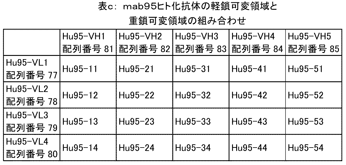
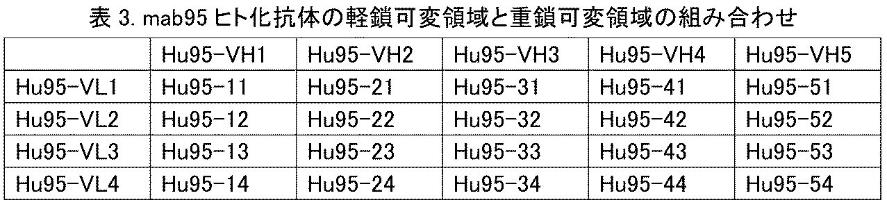
いくつかの態様において、抗CTGF抗体は、次の表a、表bおよび表cに示される重鎖可変領域および軽鎖可変領域を含む: In some embodiments, the anti-CTGF antibody comprises the heavy chain variable region and the light chain variable region shown in the following Tables a, b, and c:


いくつかの態様において、抗CTGF抗体は、次に記載される重鎖可変領域および軽鎖可変領域を含む:
(xiii)重鎖可変領域のアミノ酸配列が配列番号27に示され、軽鎖可変領域のアミノ酸配列が配列番号22に示される;
(xiv)重鎖可変領域のアミノ酸配列が配列番号34に示され、軽鎖可変領域のアミノ酸配列が配列番号30に示される;または
(xv)重鎖可変領域のアミノ酸配列が配列番号85に示され、軽鎖可変領域のアミノ酸配列が配列番号77に示される。
In some embodiments, the anti-CTGF antibody comprises the heavy chain variable region and the light chain variable region set forth as follows:
(xiii) the amino acid sequence of the heavy chain variable region is set forth in SEQ ID NO: 27 and the amino acid sequence of the light chain variable region is set forth in SEQ ID NO: 22;
(xiv) the amino acid sequence of the heavy chain variable region is set forth in SEQ ID NO: 34 and the amino acid sequence of the light chain variable region is set forth in SEQ ID NO: 30; or (xv) the amino acid sequence of the heavy chain variable region is set forth in SEQ ID NO: 85 and the amino acid sequence of the light chain variable region is set forth in SEQ ID NO: 77.
抗体が抗体重鎖定常領域および軽鎖定常領域をさらに含む、抗CTGF抗体のいくつかの態様において、好ましくは、重鎖定常領域は、ヒトIgG1、IgG2、IgG3およびIgG4の定常領域ならびにその通常のバリアントからなる群から選択され、軽鎖定常領域は、ヒト抗体κおよびλ鎖の定常領域ならびにその通常のバリアントからなる群から選択され;より好ましくは、抗体は、配列番号37または38に示される重鎖定常領域、および配列番号39または40に示される軽鎖定常領域を含む。 In some embodiments of the anti-CTGF antibody, in which the antibody further comprises an antibody heavy chain constant region and a light chain constant region, preferably the heavy chain constant region is selected from the group consisting of human IgG1, IgG2, IgG3 and IgG4 constant regions and common variants thereof, and the light chain constant region is selected from the group consisting of human antibody kappa and lambda chain constant regions and common variants thereof; more preferably, the antibody comprises a heavy chain constant region as set forth in SEQ ID NO: 37 or 38, and a light chain constant region as set forth in SEQ ID NO: 39 or 40.
いくつかの態様において、抗CTGF抗体は、次のものを含む:
(c)配列番号41、43、44、45、46、47または48に示される重鎖との同一性が少なくとも85%、90%、91%、92%、93%、94%、95%、96%、97%、98%、99%または100%である重鎖、および配列番号42、49、50、51、52または53に示される軽鎖との同一性が少なくとも85%、90%、91%、92%、93%、94%、95%、96%、97%、98%、99%または100%である軽鎖;
(d)配列番号54、56、57、58、59、60、61、62または63に示される重鎖との同一性が少なくとも85%、90%、91%、92%、93%、94%、95%、96%、97%、98%、99%または100%である重鎖、および配列番号55、64、65または66に示される軽鎖との同一性が少なくとも85%、90%、91%、92%、93%、94%、95%、96%、97%、98%、99%または100%である軽鎖;
(e)配列番号41、43、44、45、46、47または48に示される重鎖、および配列番号42、49、50、51、52または53に示される軽鎖;または
(f)配列番号54、56、57、58、59、60、61、62または63に示される重鎖、および配列番号55、64、65または66に示される軽鎖。
In some embodiments, the anti-CTGF antibody includes:
(c) a heavy chain that is at least 85%, 90%, 91%, 92%, 93%, 94%, 95%, 96%, 97%, 98%, 99% or 100% identical to a heavy chain set forth in SEQ ID NO: 41, 43, 44, 45, 46, 47 or 48, and a light chain that is at least 85%, 90%, 91%, 92%, 93%, 94%, 95%, 96%, 97%, 98%, 99% or 100% identical to a light chain set forth in SEQ ID NO: 42, 49, 50, 51, 52 or 53;
(d) a heavy chain that is at least 85%, 90%, 91%, 92%, 93%, 94%, 95%, 96%, 97%, 98%, 99% or 100% identical to a heavy chain set forth in SEQ ID NO: 54, 56, 57, 58, 59, 60, 61, 62 or 63, and a light chain that is at least 85%, 90%, 91%, 92%, 93%, 94%, 95%, 96%, 97%, 98%, 99% or 100% identical to a light chain set forth in SEQ ID NO: 55, 64, 65 or 66;
(e) a heavy chain set forth in SEQ ID NO: 41, 43, 44, 45, 46, 47 or 48, and a light chain set forth in SEQ ID NO: 42, 49, 50, 51, 52 or 53; or (f) a heavy chain set forth in SEQ ID NO: 54, 56, 57, 58, 59, 60, 61, 62 or 63, and a light chain set forth in SEQ ID NO: 55, 64, 65 or 66.
いくつかの態様において、抗CTGF抗体は、次のものを含む:
(g)配列番号86、88、89、90、91、92、93、94、95、96または97に示される重鎖との同一性が少なくとも85%、90%、91%、92%、93%、94%、95%、96%、97%、98%、99%または100%である重鎖、および配列番号87、98、99、100または101に示される軽鎖との同一性が少なくとも85%、90%、91%、92%、93%、94%、95%、96%、97%、98%、99%または100%である軽鎖;または
(h)配列番号86、88、89、90、91、92、93、94、95、96または97に示される重鎖、および配列番号87、98、99、100または101に示される軽鎖。
In some embodiments, the anti-CTGF antibody includes:
(g) a heavy chain that is at least 85%, 90%, 91%, 92%, 93%, 94%, 95%, 96%, 97%, 98%, 99% or 100% identical to the heavy chain set forth in SEQ ID NO: 86, 88, 89, 90, 91, 92, 93, 94, 95, 96, or 97, and a light chain that is at least 85%, 90%, 91%, 92%, 93%, 94%, 95%, 96%, 97%, 98%, 99% or 100% identical to the light chain set forth in SEQ ID NO: 87, 98, 99, 100, or 101; or (h) a heavy chain set forth in SEQ ID NO: 86, 88, 89, 90, 91, 92, 93, 94, 95, 96, or 97, and a light chain set forth in SEQ ID NO: 87, 98, 99, 100, or 101.
いくつかの態様において、抗CTGF抗体は、次のものを含む:
(j)配列番号46に示される重鎖、および配列番号49に示される軽鎖;
(k)配列番号61に示される重鎖、および配列番号64に示される軽鎖;または
(l)配列番号97に示される重鎖、および配列番号98に示される軽鎖。
In some embodiments, the anti-CTGF antibody includes:
(j) a heavy chain set forth in SEQ ID NO:46, and a light chain set forth in SEQ ID NO:49;
(k) a heavy chain set forth in SEQ ID NO:61 and a light chain set forth in SEQ ID NO:64; or (l) a heavy chain set forth in SEQ ID NO:97 and a light chain set forth in SEQ ID NO:98.
本開示の他のいくつかの側面において、ヒトCTGFとの結合を前記抗CTGF抗体またはその抗原結合フラグメントと競合する抗CTGF抗体が提供される。 In some other aspects of the present disclosure, an anti-CTGF antibody is provided that competes with the anti-CTGF antibody or antigen-binding fragment thereof for binding to human CTGF.
本開示の他のいくつかの側面において、前記抗CTGF抗体をコードする核酸分子が提供される。 In other aspects of the present disclosure, a nucleic acid molecule encoding the anti-CTGF antibody is provided.
本開示の他のいくつかの側面において、前記核酸分子を含む宿主細胞が提供される。 In some other aspects of the present disclosure, a host cell is provided that contains the nucleic acid molecule.
本開示の他のいくつかの側面において、治療有効量または予防有効量の前記抗CTGF抗体または前記核酸分子および1つ以上の薬学的に許容しうる担体、希釈剤、緩衝剤または賦形剤を含む医薬組成物が提供される。 In other aspects of the present disclosure, a pharmaceutical composition is provided that includes a therapeutically or prophylactically effective amount of the anti-CTGF antibody or the nucleic acid molecule and one or more pharma- ceutically acceptable carriers, diluents, buffers, or excipients.
いくつかの特定の態様において、治療有効量または予防有効量とは、組成物の単位用量が前記抗CTGF抗体を0.1~3000mgまたは1~1000mg含むことを意味する。 In some specific embodiments, a therapeutically or prophylactically effective amount means that a unit dose of the composition contains 0.1-3000 mg or 1-1000 mg of the anti-CTGF antibody.
本開示の他のいくつかの側面において、前記抗CTGF抗体を適用するステップを含む、CTGFの免疫アッセイまたは決定の方法が提供される。 In some other aspects of the present disclosure, a method of immunoassaying or determining CTGF is provided, comprising applying the anti-CTGF antibody.
本開示の他のいくつかの側面において、前記抗CTGF抗体を対象またはそのサンプルと接触させるステップを含む、CTGFの免疫アッセイまたは決定の方法が提供される。 In some other aspects of the present disclosure, a method for immunoassaying or determining CTGF is provided, comprising contacting the anti-CTGF antibody with a subject or a sample thereof.
本開示の他のいくつかの側面において、前記抗CTGF抗体を含むキットが提供される。 In some other aspects of the present disclosure, a kit is provided that includes the anti-CTGF antibody.
本開示の他のいくつかの側面において、前記抗CTGF抗体または前記核酸分子または前記医薬組成物を処置有効量で対象に投与することを含む、CTGF関連疾患の処置方法が提供され、ここで、前記疾患は好ましくは、線維性疾患(線維性疾患は好ましくは特発性肺線維症、糖尿病性腎症、糖尿病網膜症、骨関節炎、強皮症、慢性心不全、肝硬変または腎線維症である)、高血圧症、糖尿病、心筋梗塞、関節炎、CTGF関連細胞増殖性疾患、アテローム性動脈硬化、緑内障またはがん(がんは好ましくは、急性リンパ芽球性白血病、皮膚線維腫、乳がん、血管脂肪腫、血管平滑筋腫、結合組織生成がん、前立腺がん、卵巣がん、結腸直腸がん、膵臓がん、胃腸がんまたは肝臓がんである)である。 In some other aspects of the present disclosure, a method for treating a CTGF-related disease is provided, comprising administering to a subject a therapeutically effective amount of the anti-CTGF antibody or the nucleic acid molecule or the pharmaceutical composition, wherein the disease is preferably a fibrotic disease (the fibrotic disease is preferably idiopathic pulmonary fibrosis, diabetic nephropathy, diabetic retinopathy, osteoarthritis, scleroderma, chronic heart failure, liver cirrhosis or renal fibrosis), hypertension, diabetes, myocardial infarction, arthritis, a CTGF-related cell proliferative disease, atherosclerosis, glaucoma or cancer (the cancer is preferably acute lymphoblastic leukemia, dermatofibroma, breast cancer, angiolipoma, angioleiomyoma, connective tissue generating cancer, prostate cancer, ovarian cancer, colorectal cancer, pancreatic cancer, gastrointestinal cancer or liver cancer).
本開示の他のいくつかの側面において、CTGF関連疾患処置用医薬の製造における前記抗CTGF抗体または前記核酸分子または前記医薬組成物の使用が提供され、ここで、前記CTGF関連疾患は、線維性疾患(線維性疾患は好ましくは特発性肺線維症、糖尿病性腎症、糖尿病網膜症、骨関節炎、強皮症、慢性心不全、肝硬変または腎線維症である)、高血圧症、糖尿病、心筋梗塞、関節炎、CTGF関連細胞増殖性疾患、アテローム性動脈硬化、緑内障またはがん(がんは好ましくは、急性リンパ芽球性白血病、皮膚線維腫、乳がん、血管脂肪腫、血管平滑筋腫、結合組織生成がん、前立腺がん、卵巣がん、結腸直腸がん、膵臓がん、胃腸がんまたは肝臓がんである)を包含する。 In some other aspects of the present disclosure, there is provided a use of the anti-CTGF antibody or the nucleic acid molecule or the pharmaceutical composition in the manufacture of a medicament for treating a CTGF-related disease, wherein the CTGF-related disease includes a fibrotic disease (the fibrotic disease is preferably idiopathic pulmonary fibrosis, diabetic nephropathy, diabetic retinopathy, osteoarthritis, scleroderma, chronic heart failure, liver cirrhosis or renal fibrosis), hypertension, diabetes, myocardial infarction, arthritis, a CTGF-related cell proliferative disease, atherosclerosis, glaucoma or cancer (the cancer is preferably acute lymphoblastic leukemia, dermatofibroma, breast cancer, angiolipoma, angioleiomyoma, connective tissue generating cancer, prostate cancer, ovarian cancer, colorectal cancer, pancreatic cancer, gastrointestinal cancer or liver cancer).
本開示の他のいくつかの側面において、CTGF関連疾患の処置における使用のための前記抗CTGF抗体または前記抗CTGF抗体をコードする核酸分子または前記医薬組成物が提供され、ここで、前記CTGF関連疾患は、線維性疾患(線維性疾患は好ましくは特発性肺線維症、糖尿病性腎症、糖尿病網膜症、骨関節炎、強皮症、慢性心不全、肝硬変または腎線維症である)、高血圧症、糖尿病、心筋梗塞、関節炎、CTGF関連細胞増殖性疾患、アテローム性動脈硬化、緑内障またはがん(がんは好ましくは、急性リンパ芽球性白血病、皮膚線維腫、乳がん、血管脂肪腫、血管平滑筋腫、結合組織生成がん、前立腺がん、卵巣がん、結腸直腸がん、膵臓がん、胃腸がんまたは肝臓がんである)を包含する。 In some other aspects of the present disclosure, the anti-CTGF antibody or the nucleic acid molecule encoding the anti-CTGF antibody or the pharmaceutical composition is provided for use in treating a CTGF-related disease, wherein the CTGF-related disease includes a fibrotic disease (the fibrotic disease is preferably idiopathic pulmonary fibrosis, diabetic nephropathy, diabetic retinopathy, osteoarthritis, scleroderma, chronic heart failure, liver cirrhosis or renal fibrosis), hypertension, diabetes, myocardial infarction, arthritis, a CTGF-related cell proliferative disease, atherosclerosis, glaucoma or cancer (the cancer is preferably acute lymphoblastic leukemia, dermatofibroma, breast cancer, angiolipoma, angioleiomyoma, connective tissue generating cancer, prostate cancer, ovarian cancer, colorectal cancer, pancreatic cancer, gastrointestinal cancer or liver cancer).
発明の詳細な説明
用語
本開示のより容易な理解のために、いくつかの技術用語および科学用語を以下に定義する。本書に使用される他の技術用語および科学用語はいずれも、本書において特に別の定義をしない限り、本開示の属する技術分野の専門家に通常理解される意味を有する。
DETAILED DESCRIPTION OF THE PRESENT DISCLOSURE Terminology In order to make the present disclosure more readily understandable, certain technical and scientific terms are defined below. All other technical and scientific terms used herein have the meanings commonly understood by one skilled in the art to which this disclosure belongs, unless otherwise defined herein.
本開示において用いられるアミノ酸の3文字コードおよび1文字コードは、J.biol.chem, 243, p3558 (1968) に記載されるものである。 The three-letter and one-letter codes for amino acids used in this disclosure are those described in J. Biol. Chem, 243, p3558 (1968).
結合組織成長因子(CTGF)は、システインリッチな36kDのヘパリン結合分泌糖タンパク質で、ヒト臍静脈内皮細胞から初めて単離された(例えば、Bradham et al. (1991) J Cell Biol114: 1285-1294; Grotendorst and Bradham, 米国特許第5,408,040号参照)。CTGFは、タンパク質CCN(CTGF、Cyr61、Nov)ファミリー(分泌糖タンパク質)に属し、このファミリーは、血清誘導最初期遺伝子産物Cyr61、推定癌遺伝子Nov、ECM関連タンパク質FISP-12、src誘導遺伝子CEF-10、Wnt誘導分泌タンパク質WISP-3、および増殖抑制タンパク質HICP/rCOPを含む(Brigstock (1999) Endocr Rev 20: 189-206; O'Brian et al. (1990) Mol Cell Biol 10: 3569-3577; Joliot et al. (1992) Mol Cell Biol 12: 10-21; Ryseck et al. (1990) Cell Growth and Diff 2: 225-233; Simmons et al. (1989) Proc Natl Acad Sci USA 86: 1178-1182; Pennica et al. (1998) Proc Natl Acad Sci USA, 95: 14717-14722; およびZhang et al. (1998) Mol Cell Biol 18: 6131-6141)。CCNタンパク質は保存的な38のシステイン残基によって特徴付けられる。この38システイン残基は、全アミノ酸の10%超を構成し、N末端およびC末端ドメインとモジュール構造を形成する。CTGFのモジュール構造は、N末端ドメインのインスリン様成長因子結合タンパク質(IGF-BP)およびフォン・ヴィルブランド因子(VWC)ならびにC末端ドメインのトロンボスポンジン(TSP1)およびシステインノットモチーフの保存的モチーフを含む。 Connective tissue growth factor (CTGF) is a cysteine-rich, 36 kD secreted, heparin-binding glycoprotein first isolated from human umbilical vein endothelial cells (see, e.g., Bradham et al. (1991) J Cell Biol114: 1285-1294; Grotendorst and Bradham, U.S. Patent No. 5,408,040). CTGF belongs to the CCN (CTGF, Cyr61, Nov) family of proteins (secreted glycoproteins), which includes the serum-inducible immediate early gene product Cyr61, the putative oncogene Nov, the ECM-associated protein FISP-12, the src-inducible gene CEF-10, the Wnt-inducible secreted protein WISP-3, and the growth-suppressing protein HICP/rCOP (Brigstock (1999) Endocr Rev 20: 189-206; O'Brian et al. (1990) Mol Cell Biol 10: 3569-3577; Joliot et al. (1992) Mol Cell Biol 12: 10-21; Ryseck et al. (1990) Cell Growth and Diff 2: 225-233; Simmons et al. (1989) Proc Natl Acad Sci USA 86: 1178-1182; Pennica et al. (1998) Proc Natl Acad Sci USA, 95: 14717-14722; and Zhang et al. (1998) Mol Cell Biol 18: 6131-6141). CCN proteins are characterized by 38 conserved cysteine residues, which constitute more than 10% of the total amino acids and form a modular structure with the N- and C-terminal domains. The modular structure of CTGF contains conserved motifs of insulin-like growth factor binding protein (IGF-BP) and von Willebrand factor (VWC) in the N-terminal domain and thrombospondin (TSP1) and cysteine knot motifs in the C-terminal domain.
本書において「抗体」は免疫グロブリンをいい、完全長抗体は、2本の同一の重鎖および2本の同一の軽鎖の間で鎖間ジスルフィド結合によって繋ぎ合わされた4ペプチド鎖構造である。免疫グロブリン重鎖定常領域は、異なるアミノ酸の組成および順序を示し、したがって、異なる抗原性を示す。したがって、免疫グロブリンは5つのタイプに分類することができ、免疫グロブリンアイソタイプ、すなわちIgM、IgD、IgG、IgAおよびIgEと称することができ、それぞれ対応する重鎖はμ、δ、λ、αおよびεである。ヒンジ領域のアミノ酸組成および重鎖ジスルフィド結合の数と位置によって、同じタイプのIgを異なるサブタイプにさらに分類することができ、例えば、IgGをIgG1、IgG2、IgG3およびIgG4に分類することができる。軽鎖は、定常領域の違いに基づいてκまたはλ鎖に分類することができる。5つのIgタイプのそれぞれが、κ鎖またはλ鎖を有しうる。 In this document, "antibody" refers to an immunoglobulin, and a full-length antibody is a four-peptide chain structure connected by interchain disulfide bonds between two identical heavy chains and two identical light chains. The immunoglobulin heavy chain constant regions exhibit different amino acid compositions and sequences, and therefore different antigenicity. Thus, immunoglobulins can be classified into five types, which can be called immunoglobulin isotypes, namely IgM, IgD, IgG, IgA, and IgE, with the corresponding heavy chains being μ, δ, λ, α, and ε. Depending on the amino acid composition of the hinge region and the number and position of the heavy chain disulfide bonds, the same type of Ig can be further classified into different subtypes, for example, IgG can be classified into IgG1, IgG2, IgG3, and IgG4. The light chains can be classified into κ or λ chains based on the differences in the constant region. Each of the five Ig types can have κ or λ chains.
抗体重鎖および軽鎖のN末端から約110アミノ酸の配列は非常にさまざまで、可変領域(Fv領域)として知られ、残りのC末端側アミノ酸配列は比較的一定で、定常領域として知られる。可変領域は3つの超可変領域(HVR)、および比較的配列の保存された4つのフレームワーク領域(FR)を含む。抗体の特異性を決定する3つの超可変領域は、相補性決定領域(CDR)とも呼ばれる。各軽鎖可変領域(VL)および各重鎖可変領域(VH)は、アミノ末端からカルボキシル末端へと次の順序で並ぶ、3つのCDR領域と4つのFR領域からなる:FR1、CDR1、FR2、CDR2、FR3、CDR3およびFR4。軽鎖の3つのCDR領域はLCDR1、LCDR2およびLCDR3と称され、重鎖の3つのCDR領域はHCDR1、HCDR2およびHCDR3と称される。 The sequence of about 110 amino acids from the N-terminus of antibody heavy and light chains is highly variable and known as the variable region (Fv region), while the remaining C-terminal amino acid sequence is relatively constant and known as the constant region. The variable region contains three hypervariable regions (HVR) and four framework regions (FR) with relatively conserved sequences. The three hypervariable regions that determine the specificity of the antibody are also called complementarity determining regions (CDR). Each light chain variable region (VL) and each heavy chain variable region (VH) consists of three CDR regions and four FR regions, arranged in the following order from the amino terminus to the carboxyl terminus: FR1, CDR1, FR2, CDR2, FR3, CDR3, and FR4. The three CDR regions of the light chain are referred to as LCDR1, LCDR2, and LCDR3, and the three CDR regions of the heavy chain are referred to as HCDR1, HCDR2, and HCDR3.
本開示の抗体は、マウス抗体、キメラ抗体、およびヒト化抗体を包含する。 The antibodies of the present disclosure include mouse antibodies, chimeric antibodies, and humanized antibodies.
本書において、用語「マウス抗体」とは、当分野における知識および技術によって作製されるヒトCTGFに対するモノクローナル抗体をいう。作製において、試験対象にCTGF抗原が注射され、その後、所望の配列または機能特性を有する抗体を発現するハイブリドーマが単離される。本開示の好ましい態様において、マウス抗CTGF抗体またはその抗原結合フラグメントは、マウスκ、λ鎖の軽鎖定常領域もしくはそのバリアントをさらに含む、またはマウスIgG1、IgG2、IgG3の重鎖定常領域もしくはそのバリアントをさらに含むことができる。 As used herein, the term "mouse antibody" refers to a monoclonal antibody against human CTGF produced by knowledge and skill in the art. In production, a test subject is injected with CTGF antigen, and then a hybridoma expressing an antibody having the desired sequence or functional characteristics is isolated. In a preferred embodiment of the present disclosure, the mouse anti-CTGF antibody or antigen-binding fragment thereof can further comprise a mouse kappa, lambda chain light chain constant region or variant thereof, or a mouse IgG1, IgG2, IgG3 heavy chain constant region or variant thereof.
用語「キメラ抗体」は、マウス抗体の可変領域をヒト抗体の定常領域に融合することによる抗体であり、そのような抗体は、マウス抗体が誘発する免疫反応を低減することができる。キメラ抗体を作製するには、まず特定のマウスモノクローナル抗体を分泌するハイブリドーマを作製し、マウスハイブリドーマから可変領域遺伝子をクローン化する。次いで、必要に応じてヒト抗体から定常領域遺伝子をクローン化する。マウス可変領域遺伝子とヒト定常領域遺伝子を連結してキメラ遺伝子を形成し、その後これを発現ベクターに挿入することができる。最後に、キメラ抗体分子を真核生物または原核生物系において発現させうる。本開示の好ましい態様において、キメラ抗体の抗体軽鎖は、ヒトκ、λ鎖の軽鎖定常領域もしくはそのバリアントをさらに含む。CTGFキメラ抗体の抗体重鎖は、さらにヒトIgG1、IgG2、IgG3、IgG4の重鎖定常領域もしくはそのバリアントをさらに含み、好ましくはヒトIgG1、IgG2もしくはIgG4、またはアミノ酸変異(例えば、L234Aおよび/またはL235A変異、および/またはS228P変異)を有するIgG1、IgG2もしくはIgG4バリアントの重鎖定常領域を含む。 The term "chimeric antibody" refers to an antibody that is produced by fusing the variable region of a mouse antibody to the constant region of a human antibody, and such an antibody can reduce the immune response elicited by mouse antibodies. To generate a chimeric antibody, first generate a hybridoma that secretes a specific mouse monoclonal antibody, and then clone the variable region gene from the mouse hybridoma. Then, if necessary, clone the constant region gene from a human antibody. The mouse variable region gene and the human constant region gene are linked to form a chimeric gene, which can then be inserted into an expression vector. Finally, the chimeric antibody molecule can be expressed in eukaryotic or prokaryotic systems. In a preferred embodiment of the present disclosure, the antibody light chain of the chimeric antibody further comprises a human kappa, lambda chain light chain constant region or a variant thereof. The antibody heavy chain of the CTGF chimeric antibody further comprises a heavy chain constant region of human IgG1, IgG2, IgG3, IgG4 or a variant thereof, and preferably comprises a heavy chain constant region of human IgG1, IgG2 or IgG4, or an IgG1, IgG2 or IgG4 variant having an amino acid mutation (e.g., an L234A and/or an L235A mutation, and/or an S228P mutation).
用語「ヒト化抗体」は、CDRグラフト抗体とも呼ばれるが、マウスCDR配列をヒト抗体可変領域フレームワークにグラフトすることにより作製される抗体、すなわちさまざまな種類のヒト生殖細胞系列抗体フレームワーク配列中に作製される抗体をいう。ヒト化抗体は、マウスタンパク質成分を多く含むキメラ抗体によって誘発される異種反応を克服することができる。そのようなフレームワーク配列は、生殖細胞系列抗体遺伝子配列を含む公のDNAデータベース、または刊行物から入手することができる。例えば、生殖細胞系列のヒト重鎖および軽鎖可変領域遺伝子DNA配列は、“VBase”ヒト生殖細胞系列配列データベース(www.mrccpe.com.ac.uk/vbaseにおいて利用可能)、およびKabat, EA, et al. 1991 Sequences of Proteins of Immunological Interest, 5th Edに見出すことができる。免疫原性低下によって起こる活性低下を回避するために、ヒト抗体可変領域中のフレームワーク配列を、最小限の、活性の維持または増強のための復帰変異に付すことができる。本開示のヒト化抗体は、酵母ディスプレイによってCDR親和性成熟が行われたヒト抗体をも包含する。 The term "humanized antibody", also called CDR-grafted antibody, refers to an antibody made by grafting mouse CDR sequences into a human antibody variable region framework, i.e., an antibody made into various types of human germline antibody framework sequences. Humanized antibodies can overcome the heterologous reactions elicited by chimeric antibodies that contain many mouse protein components. Such framework sequences can be obtained from public DNA databases or publications that contain germline antibody gene sequences. For example, germline human heavy and light chain variable region gene DNA sequences can be found in the "VBase" human germline sequence database (available at www.mrccpe.com.ac.uk/vbase), and in Kabat, EA, et al. 1991 Sequences of Proteins of Immunological Interest, 5th Ed. To avoid reduced activity caused by reduced immunogenicity, the framework sequences in the human antibody variable regions can be subjected to minimal back mutations to maintain or enhance activity. The humanized antibodies of the present disclosure also include human antibodies that have been subjected to CDR affinity maturation by yeast display.
残基の抗原との接触のせいで、CDRのグラフトは、フレームワーク残基の抗原との接触のせいで、抗体またはその抗原結合フラグメントの抗体に対する親和性の低下をもたらしうる。高度に体細胞的な変異によって、そのような相互作用をもたらすことができる。したがって、そのようなドナーフレームワークアミノ酸をヒト化抗体フレームワークにグラフトすることが依然必要でありうる。非ヒト抗体またはその抗原結合フラグメントに由来する、抗原結合に関与するアミノ酸残基は、動物モノクローナル抗体可変領域の配列および構造を調べることによって特定することができる。生殖細胞系列とは異なるドナーCDRフレームワークアミノ酸残基は、関連するものと考えることができる。最も関連性の高い生殖細胞系列を決定することができない場合は、サブタイプに共通する配列、または類似性パーセンテージの高い動物抗体配列に対して配列を比較することができる。レアなフレームワーク残基は、体細胞における高度の変異の結果と考えられ、結合において重要な役割を果たす。 Due to contact of the residues with the antigen, the grafting of the CDRs may result in a decrease in the affinity of the antibody or antigen-binding fragment thereof for the antibody due to contact of the framework residues with the antigen. High somatic mutations can result in such interactions. Therefore, it may still be necessary to graft such donor framework amino acids onto the humanized antibody framework. Amino acid residues involved in antigen binding from non-human antibodies or antigen-binding fragments thereof can be identified by examining the sequences and structures of animal monoclonal antibody variable regions. Donor CDR framework amino acid residues that differ from the germline can be considered relevant. If it is not possible to determine the most relevant germline, the sequences can be compared against sequences common to the subtype or against animal antibody sequences with a high percentage of similarity. Rare framework residues are likely the result of high somatic mutations and play an important role in binding.
本開示の一態様において、抗体またはその抗原結合フラグメントは、ヒトまたはマウスκ、λ鎖またはそのバリアントに由来する軽鎖定常領域をさらに含む、またはヒトまたはマウスIgG1、IgG2、IgG3、IgG4またはそのバリアントに由来する重鎖定常領域をさらに含み;それは、ヒトIgG1、IgG2もしくはIgG4に由来する、またはアミノ酸変異(例えば、L234Aおよび/またはL235A変異、および/またはS228P変異)を有するIgG1、IgG2もしくはIgG4バリアントに由来する重鎖定常領域を含みうる。 In one aspect of the disclosure, the antibody or antigen-binding fragment thereof further comprises a light chain constant region derived from a human or mouse kappa, lambda chain or variant thereof, or further comprises a heavy chain constant region derived from a human or mouse IgG1, IgG2, IgG3, IgG4 or variant thereof; it may comprise a heavy chain constant region derived from human IgG1, IgG2 or IgG4, or from an IgG1, IgG2 or IgG4 variant having amino acid mutations (e.g., L234A and/or L235A mutations, and/or S228P mutations).
本書において、ヒト抗体重鎖定常領域およびヒト抗体軽鎖定常領域の「通常のバリアント」とは、従来技術において開示される、抗体可変領域の構造および機能を変化させないヒト重鎖または軽鎖定常領域バリアントをいう。バリアントの例は、重鎖定常領域における部位特異的改変およびアミノ酸置換による、IgG1、IgG2、IgG3またはIgG4重鎖定常領域バリアントを包含する。具体的な置換の例は、YTE変異、L234Aおよび/またはL235A変異、S228P変異、またはknob-into-hole構造をもたらす変異(抗体重鎖にknob-Fcとhole-Fcの組み合わせを持たせる)等である。これらの変異は、抗体可変領域の機能を変化することなく抗体に新たな性質を付与することが証明されている。 As used herein, "conventional variants" of human antibody heavy chain constant regions and human antibody light chain constant regions refer to human heavy or light chain constant region variants disclosed in the prior art that do not change the structure and function of the antibody variable region. Examples of variants include IgG1, IgG2, IgG3 or IgG4 heavy chain constant region variants with site-specific modifications and amino acid substitutions in the heavy chain constant region. Specific examples of substitutions include YTE mutations, L234A and/or L235A mutations, S228P mutations, or mutations that result in a knob-into-hole structure (giving the antibody heavy chain a combination of knob-Fc and hole-Fc). These mutations have been shown to confer new properties to antibodies without changing the function of the antibody variable region.
「ヒト抗体(HuMAb)」、「ヒト由来抗体」および「完全ヒト抗体」は、互換的に用いることができ、ヒトに由来する抗体、または特定のヒト抗体を抗原刺激に応答して産生するように当分野で知られる任意の方法で「改変」されている遺伝子改変された生物から得られる抗体であることができ、産生されることができる。いくつかの技術において、ヒト重鎖および軽鎖遺伝子座のエレメントが、生殖細胞系列幹細胞株由来の生物の細胞株に導入され、該細胞株の内因の重鎖および軽鎖遺伝子座が標的化され分断される。該細胞株に含まれる標的化された内因の重鎖および軽鎖遺伝子座は分断される。トランスジェニック生物は、ヒト抗原に特異的なヒト抗体を合成することができ、該生物を用いてヒト抗体を分泌するハイブリドーマを産生させることができる。ヒト抗体はまた、重鎖および軽鎖が1つ以上のヒトDNAソースに由来するヌクレオチド配列でコードされている抗体でありうる。完全ヒト抗体はまた、遺伝子または染色体トランスフェクション法およびファージディスプレイ技術によって作製する、またはインビトロで活性化されたB細胞から作製することができ、それらはいずれも当分野において知られている。 "Human antibody (HuMAb)", "human-derived antibody" and "fully human antibody" may be used interchangeably and may be or may be produced from an antibody derived from a human or from a genetically modified organism that has been "modified" in any manner known in the art to produce specific human antibodies in response to antigenic stimulation. In some techniques, elements of human heavy and light chain loci are introduced into a cell line of an organism derived from a germline stem cell line, and the endogenous heavy and light chain loci of the cell line are targeted and disrupted. The targeted endogenous heavy and light chain loci contained in the cell line are disrupted. The transgenic organism is capable of synthesizing human antibodies specific to human antigens, and the organism can be used to produce hybridomas that secrete human antibodies. Human antibodies may also be antibodies in which the heavy and light chains are encoded by nucleotide sequences derived from one or more human DNA sources. Fully human antibodies may also be produced by genetic or chromosomal transfection methods and phage display techniques, or from in vitro activated B cells, both of which are known in the art.
用語「完全長抗体」、「完全抗体」および「全抗体」は、本書において互換的に用いられ、下記に定義する抗原結合フラグメントと区別される、実質的にインタクトな形態の抗体をいう。該用語は特に、軽鎖および重鎖中に定常領域を含む抗体をさす。 The terms "full-length antibody," "complete antibody," and "whole antibody" are used interchangeably herein and refer to an antibody in a substantially intact form, as distinguished from an antigen-binding fragment, as defined below. The terms particularly refer to antibodies that include constant regions in the light and heavy chains.
本開示の「抗体」は、「完全長抗体」およびその抗原結合フラグメントを包含する。 The "antibody" of this disclosure includes a "full-length antibody" and an antigen-binding fragment thereof.
いくつかの態様において、本開示の完全長抗体は、下記の表1、2および3に挙げられる軽鎖および重鎖の組み合わせに示されるような、軽鎖可変領域と軽鎖定常領域との連結、および重鎖可変領域と重鎖定常領域との連結によって形成される完全長抗体を包含する。当業者は、軽鎖定常領域および重鎖定常領域を、ヒト抗体由来軽鎖定常領域および重鎖定常領域といった、実際の必要に応じたさまざまな抗体ソースから選択することができる。また、表1、2および3に記載される軽鎖および重鎖可変領域のさまざま組み合わせにより、一本鎖抗体(scFv)、Fab、またはscFvもしくはFabを含む他の形態の抗原結合フラグメントを形成することができる。 In some embodiments, the full-length antibody of the present disclosure includes a full-length antibody formed by linking a light chain variable region with a light chain constant region, and linking a heavy chain variable region with a heavy chain constant region, as shown in the combinations of light chains and heavy chains listed in Tables 1, 2, and 3 below. Those skilled in the art can select light chain constant regions and heavy chain constant regions from various antibody sources according to actual needs, such as light chain constant regions and heavy chain constant regions derived from human antibodies. In addition, various combinations of light chain and heavy chain variable regions listed in Tables 1, 2, and 3 can form single chain antibodies (scFv), Fab, or other forms of antigen-binding fragments including scFv or Fab.
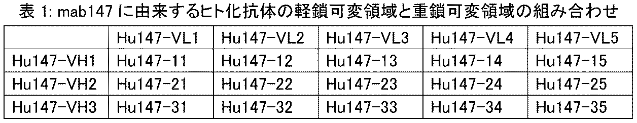

注:Hu164-57は、mab164から誘導されたヒト化抗体Hu164-57が、Hu164-VH5に記載される重鎖可変領域およびHu164-VL7に記載される軽鎖可変領域を有することを意味し;それ以外のものも同じ方式で解釈することができる。 Note: Hu164-57 means that the humanized antibody Hu164-57, derived from mab164, has a heavy chain variable region as set forth in Hu164-VH5 and a light chain variable region as set forth in Hu164-VL7; others can be interpreted in the same manner.
用語「抗原結合フラグメント」または「機能的フラグメント」とは、抗原(例えばCTGF)に特異的に結合する能力を維持する、抗体の1つ以上のフラグメントをいう。完全長抗体のフラグメントは、特異的抗原に結合する機能を達成するために使用できることが示されている。抗体の「抗原結合フラグメント」という用語に関する結合フラグメントの例は、次のものを包含する:(i)VL、VH、CLおよびCH1ドメインからなる一価フラグメントである、Fabフラグメント、(ii)ヒンジ領域でジスルフィド架橋により連結された2つのFabフラグメントを含む二価フラグメントである、F(ab’)2フラグメント、(iii)VHおよびCH1ドメインからなるFdフラグメント、(iv)抗体の1つのアームのVHおよびVLドメインからなるFvフラグメント、(v)VHおよびVLの鎖間ジスルフィド結合によって形成される安定な抗原結合フラグメントである、dsFv、および(vi)scFv、dsFvおよびFabといったフラグメントを含む、ディアボディ、二重特異的抗体および多重特異的抗体。さらに、Fvフラグメントの2つのドメインであるVLおよびVHドメインは2つの別個の遺伝子によりコードされるが、組換え法を用いて合成リンカーによって連結して、VLおよびVHドメインが組み合わされて一価分子が形成されている一本鎖のタンパク質を作製することができる(一本鎖Fv(scFv)と称される;例えば、Bird et al. (1988) Science 242: 423-426; およびHuston et al (1988) Proc. Natl. Acad. Sci USA85:5879-5883参照)。そのような一本鎖抗体も、抗体の「抗原結合フラグメント」という用語に包含される。そのような抗体フラグメントは、当分野において知られる通常の技術を用いて得られ、機能的フラグメントについて、インタクトな抗体の場合と同じ方法を用いてスクリーニングされる。抗原結合部分は、組換えDNA技術によって、またはインタクトな免疫グロブリンの酵素的もしくは化学的切断によって作製することができる。抗体は、さまざまなアイソタイプの形態、例えばIgG(例えばIgG1、IgG2、IgG3またはIgG4サブタイプ)、IgA1、IgA2、IgD、IgEまたはIgM抗体でありうる。 The term "antigen-binding fragment" or "functional fragment" refers to one or more fragments of an antibody that retain the ability to specifically bind to an antigen (e.g., CTGF). It has been shown that fragments of full-length antibodies can be used to achieve the function of binding to a specific antigen. Examples of binding fragments for the term "antigen-binding fragment" of an antibody include: (i) Fab fragment, a monovalent fragment consisting of the VL, VH, CL and CH1 domains; (ii) F(ab') 2 fragment, a bivalent fragment containing two Fab fragments linked by a disulfide bridge at the hinge region; (iii) Fd fragment consisting of the VH and CH1 domains; (iv) Fv fragment consisting of the VH and VL domains of one arm of the antibody; (v) dsFv, a stable antigen-binding fragment formed by interchain disulfide bonds of VH and VL; and (vi) diabodies, bispecific antibodies and multispecific antibodies, including fragments such as scFv, dsFv and Fab. Furthermore, the two domains of the Fv fragment, the VL and VH domains, are encoded by two separate genes, but can be linked by a synthetic linker using recombinant techniques to produce a single-chain protein in which the VL and VH domains combine to form a monovalent molecule (called a single-chain Fv (scFv); see, e.g., Bird et al. (1988) Science 242: 423-426; and Huston et al (1988) Proc. Natl. Acad. Sci USA85:5879-5883). Such single-chain antibodies are also encompassed by the term "antigen-binding fragment" of an antibody. Such antibody fragments are obtained using conventional techniques known in the art and screened for functional fragments using the same methods as for intact antibodies. Antigen-binding portions can be produced by recombinant DNA techniques or by enzymatic or chemical cleavage of intact immunoglobulins. The antibodies can be of various isotypes, for example IgG (e.g. IgG1, IgG2, IgG3 or IgG4 subtypes), IgA1, IgA2, IgD, IgE or IgM antibodies.
Fabは、IgG抗体分子を、パパイン(これはH鎖の位置224のアミノ酸残基を切断する)で処理することによって得られる抗体フラグメントであり、H鎖のN末端側約半分とL鎖全体がジスルフィド結合で連結されている該抗体フラグメントは、約50000の分子量を有し、抗原結合活性を有する。 Fab is an antibody fragment obtained by treating an IgG antibody molecule with papain (which cleaves the amino acid residue at position 224 of the H chain). This antibody fragment, in which approximately the N-terminal half of the H chain and the entire L chain are linked by disulfide bonds, has a molecular weight of approximately 50,000 and has antigen-binding activity.
「F(ab’)2」は、抗原結合活性を有する分子量が約100000の抗体フラグメントであり、これはIgGのヒンジ領域の2つのジスルフィド結合の下流部分をペプシンで消化することによって得られる。F(ab’)2は、ヒンジ領域で連結された2つのFabを含む。 "F(ab') 2 " is an antibody fragment having an antigen-binding activity and a molecular weight of about 100,000, which can be obtained by digesting the downstream part of the two disulfide bonds in the hinge region of IgG with pepsin. F(ab') 2 contains two Fabs linked at the hinge region.
Fab’は、抗原結合活性を有する分子量が約50000の抗体フラグメントであり、前記F(ab’)2のヒンジ領域のジスルフィド結合を切断することによって得られる。本開示のFab’は、特異的にCTGFを認識し、その細胞外領域アミノ酸配列または三次元構造に結合する本開示のF(ab’)2を、ジチオスレイトールのような還元剤で処理することによって作製することができる。 Fab' is an antibody fragment having an antigen-binding activity and a molecular weight of about 50,000, and can be obtained by cleaving the disulfide bond in the hinge region of the F(ab') 2. The Fab' of the present disclosure can be prepared by treating the F(ab') 2 of the present disclosure, which specifically recognizes CTGF and binds to its extracellular domain amino acid sequence or three -dimensional structure, with a reducing agent such as dithiothreitol.
Fab’はさらに、抗体のFab’をコードするDNAを原核生物発現ベクターまたは真核生物発現ベクターに挿入し、該ベクターを原核生物または真核生物に導入してFab’を発現させることによって作製することができる。 Fab' can also be produced by inserting DNA encoding the Fab' of an antibody into a prokaryotic or eukaryotic expression vector and introducing the vector into a prokaryote or eukaryote to express the Fab'.
用語「一本鎖抗体」、「一本鎖Fv」または「scFv」は、リンカーによって抗体重鎖可変ドメイン(または領域;VH)と抗体軽鎖可変ドメイン(または領域;VL)が連結されたものを含む分子をいう。そのようなscFv分子は、一般構造:NH2-VL-リンカー-VH-COOHまたはNH2-VH-リンカー-VL-COOHを有する。従来技術において適当なリンカーは、反復GGGGSアミノ酸配列またはそのバリアント、例えば1~4反復バリアントからなる(Holliger et al. (1993), Proc. Natl. Acad. Sci. USA 90:6444-6448)。本開示において使用しうる他のリンカーは、Alfthan et al. (1995), Protein Eng. 8:725-731, Choi et al. (2001), Eur. J. Immunol. 31:94-106, Hu et al. (1996), Cancer Res. 56:3055-3061, Kipriyanov et al. (1999), J. Mol. Biol. 293:41-56 および Roovers et al. (2001), Cancer Immunolに記載されている。 The term "single chain antibody", "single chain Fv" or "scFv" refers to a molecule comprising an antibody heavy chain variable domain (or region; VH) and an antibody light chain variable domain (or region; VL) linked by a linker. Such scFv molecules have the general structure: NH 2 -VL-linker-VH-COOH or NH 2 -VH-linker-VL-COOH. A suitable linker in the prior art consists of a repeating GGGGS amino acid sequence or a variant thereof, such as a 1-4 repeat variant (Holliger et al. (1993), Proc. Natl. Acad. Sci. USA 90:6444-6448). Other linkers that may be used in the present disclosure are described in Alfthan et al. (1995), Protein Eng. 8:725-731, Choi et al. (2001), Eur. J. Immunol. 31:94-106, Hu et al. (1996), Cancer Res. 56:3055-3061, Kipriyanov et al. (1999), J. Mol. Biol. 293:41-56 and Roovers et al. (2001), Cancer Immunol.
ディアボディは、scFvまたはFabが二量体化されている抗体フラグメントであり、これは、二価の抗原結合活性を有する抗体フラグメントである。二価抗原結合活性において、2つの抗原は同じかまたは異なりうる。 Diabodies are antibody fragments in which scFv or Fab are dimerized, which means that they have bivalent antigen-binding activity. In bivalent antigen-binding activity, the two antigens can be the same or different.
二重特異的および多重特異的抗体とは、CTGFと結合することのできるscFvまたはFabフラグメントを含んで、2つまたはより多くの抗原または抗原決定基に同時に結合することのできる抗体をいう。 Bispecific and multispecific antibodies refer to antibodies that can simultaneously bind to two or more antigens or antigenic determinants, including scFv or Fab fragments capable of binding to CTGF.
本開示のディアボディは、下記ステップによって作製することができる:特異的にヒトCTGFを認識し、その細胞外領域または三次元構造に結合する本開示のモノクローナル抗体のVLおよびVLをコードするcDNAを得る;scFvをコードするDNAを、リンカーペプチドが8未満のアミノ酸残基の長さとなるように作製する;該DNAを原核生物発現ベクターまたは真核生物発現ベクターに挿入する;およびその後、該発現ベクターを原核生物または真核生物に導入してディアボディを発現させる。 The diabody of the present disclosure can be produced by the following steps: obtaining cDNA encoding the VL and VL of a monoclonal antibody of the present disclosure that specifically recognizes human CTGF and binds to its extracellular domain or three-dimensional structure; producing DNA encoding an scFv such that the linker peptide is less than 8 amino acid residues in length; inserting the DNA into a prokaryotic or eukaryotic expression vector; and then introducing the expression vector into a prokaryote or eukaryote to express the diabody.
dsFvは、VHおよびVLのそれぞれにおいて1つのアミノ酸残基をシステイン残基で置換し、次いで置換されたポリペプチドを2つのシステイン残基間でジスルフィド結合によって連結することによって得られる。システイン残基で置換するアミノ酸残基は、既知の方法による抗体の三次元構造予測に基づいて選択することができる(Protein Engineering, 7, 697 (1994))。 dsFv is obtained by substituting one amino acid residue in each of VH and VL with a cysteine residue, and then linking the substituted polypeptides by a disulfide bond between the two cysteine residues. The amino acid residue to be substituted with a cysteine residue can be selected based on the three-dimensional structure prediction of an antibody by a known method (Protein Engineering, 7, 697 (1994)).
本開示の完全長抗体またはその抗原結合フラグメントは、下記ステップによって作製することができる:特異的にヒトCTGFを認識し、その細胞外領域アミノ酸配列または三次元構造に結合する本開示の抗体をコードするcDNAを得る;dsFvをコードするDNAを作製する;該DNAを原核生物発現ベクターまたは真核生物発現ベクターに挿入する;およびその後、該発現ベクターを原核生物または真核生物に導入してdsFvを発現させる。 A full-length antibody or antigen-binding fragment thereof of the present disclosure can be produced by the following steps: obtaining a cDNA encoding an antibody of the present disclosure that specifically recognizes human CTGF and binds to its extracellular domain amino acid sequence or three-dimensional structure; producing DNA encoding a dsFv; inserting the DNA into a prokaryotic or eukaryotic expression vector; and then introducing the expression vector into a prokaryote or eukaryote to express the dsFv.
用語「アミノ酸変化」または「アミノ酸変異」とは、タンパク質またはポリペプチドのバリアントにおける、元のタンパク質またはポリペプチドと比較したアミノ酸の変化または変異をいい、元のタンパク質またはポリペプチドに基づく1、2、3またはそれ以上のアミノ酸挿入、欠失または置換を包含する。 The term "amino acid change" or "amino acid mutation" refers to an amino acid change or mutation in a protein or polypeptide variant compared to the original protein or polypeptide, and includes one, two, three or more amino acid insertions, deletions or substitutions based on the original protein or polypeptide.
用語「抗体フレームワーク」または「FR領域」とは、可変ドメインVLまたはVHの部分であって、該可変ドメインの抗原結合ループ(CDR)の足場としてはたらく部分をいう。これは本質的に、CDRを除いた可変ドメインである。 The term "antibody framework" or "FR region" refers to the portion of a variable domain VL or VH that serves as a scaffold for the antigen binding loops (CDRs) of the variable domain. This is essentially the variable domain minus the CDRs.
用語「相補性決定領域」、「CDR」または「超可変領域」とは、抗原結合に主に寄与する抗体可変ドメイン中に存在する6つの超可変領域の1つをいう。一般に、各重鎖可変領域に3つのCDR(HCDR1、HCDR2、HCDR3)が存在し、各軽鎖可変領域に3つのCDR(LCDR1、LCDR2、LCDR3)が存在する。CDRのアミノ酸配列境界は、「Kabat」ナンバリング基準(Kabat et al. (1991), "Sequences of Proteins of Immunological Interest", 5th edition, Public Health Service, National Institutes of Health, Bethesda, MD参照)、「Chothia」ナンバリング基準(Al-Lazikani et al., (1997) JMB 273:927-948)、およびImmunoGenTics(IMGT)ナンバリング基準(Lefranc MP, Immunologist, 7, 132- 136 (1999); Lefranc, MP, etc., Dev. Comp. Immunol., 27, 55-77 (2003))等を包含する、よく知られたさまざまなスキームのいずれかによって決定することができる。例えば、伝統的なフォーマットでKabat基準にしたがって、重鎖可変ドメイン(VH)のCDRアミノ酸残基が、31~35(HCDR1)、50~65(HCDR2)および95~102(HCDR3)と番号付けられ、軽鎖可変ドメイン(VL)のCDRアミノ酸残基が、24~34(LCDR1)、50~56(LCDR2)および89~97(LCDR3)と番号付けられる。Chothia基準にしたがって、VHのCDRアミノ酸残基が、26~32(HCDR1)、52~56(HCDR2)および95~102(HCDR3)と番号付けられ、VL中のアミノ酸残基が、26~32(LCDR1)、50~52(LCDR2)および91~96(LCDR3)と番号付けられる。KabatおよびChothiaの両者を組み合わせてCDRを限定すると、CDRは、ヒトVH中のアミノ酸残基26~35(HCDR1)、50~65(HCDR2)および95~102(HCDR3)、ならびにヒトVL中のアミノ酸残基24~34(LCDR1)、50~56(LCDR2)および89~97(LCDR3)からなる。IMGT基準にしたがって、VHのCDRアミノ酸残基が、おおよそ26~35(CDR1)、51~57(CDR2)および93~102(CDR3)と番号付けられ、VL中のCDRアミノ酸残基が、おおよそ27~32(CDR1)、50~52(CDR2)および89~97(CDR3)と番号付けられる。IMGT基準にしたがって、IMGT/DomainGap Align Programの使用により、抗体のCDR領域を決定することができる。別に指定しない限り、本開示の態様に関する抗体可変領域およびCDR配列は、「Kabat」ナンバリング基準に適用可能である。 The term "complementarity determining region", "CDR" or "hypervariable region" refers to one of the six hypervariable regions present in an antibody variable domain that primarily contributes to antigen binding. Generally, there are three CDRs in each heavy chain variable region (HCDR1, HCDR2, HCDR3) and three CDRs in each light chain variable region (LCDR1, LCDR2, LCDR3). The amino acid sequence boundaries of the CDRs can be determined according to any of a variety of well-known schemes, including the "Kabat" numbering system (see Kabat et al. (1991), "Sequences of Proteins of Immunological Interest", 5th edition, Public Health Service, National Institutes of Health, Bethesda, MD), the "Chothia" numbering system (Al-Lazikani et al., (1997) JMB 273:927-948), and the ImmunoGenTics (IMGT) numbering system (Lefranc MP, Immunologist, 7, 132- 136 (1999); Lefranc, MP, etc., Dev. Comp. Immunol., 27, 55-77 (2003)). For example, in the traditional format, according to the Kabat criteria, the CDR amino acid residues of the heavy chain variable domain (VH) are numbered 31-35 (HCDR1), 50-65 (HCDR2) and 95-102 (HCDR3), and the CDR amino acid residues of the light chain variable domain (VL) are numbered 24-34 (LCDR1), 50-56 (LCDR2) and 89-97 (LCDR3). According to the Chothia criteria, the CDR amino acid residues of the VH are numbered 26-32 (HCDR1), 52-56 (HCDR2) and 95-102 (HCDR3), and the amino acid residues in the VL are numbered 26-32 (LCDR1), 50-52 (LCDR2) and 91-96 (LCDR3). Combining Kabat and Chothia CDR definitions, the CDRs consist of amino acid residues 26-35 (HCDR1), 50-65 (HCDR2) and 95-102 (HCDR3) in human VH, and amino acid residues 24-34 (LCDR1), 50-56 (LCDR2) and 89-97 (LCDR3) in human VL. According to the IMGT criteria, the CDR amino acid residues in VH are numbered approximately 26-35 (CDR1), 51-57 (CDR2) and 93-102 (CDR3), and the CDR amino acid residues in VL are numbered approximately 27-32 (CDR1), 50-52 (CDR2) and 89-97 (CDR3). According to the IMGT criteria, the CDR regions of an antibody can be determined by using the IMGT/DomainGap Align Program. Unless otherwise specified, antibody variable region and CDR sequences related to aspects of this disclosure are applicable to the "Kabat" numbering system.
用語「エピトープ」または「抗原決定基」とは、免疫グロブリンまたは抗体が特異的に結合する抗原上の部分(例えば、CTGF分子上の特定の部分)をいう。エピトープは典型的には、独特の三次コンフォメーションをなす少なくとも3、4、5、6、7、8、9、10、11、12、13、14または15個の連続または不連続アミノ酸を含む。例えば、Epitope Mapping Protocols in Methods in Molecular Biology, Vol. 66, G.E. Morris, Ed. (1996)を参照されたい。 The term "epitope" or "antigenic determinant" refers to a portion on an antigen (e.g., a particular portion on a CTGF molecule) to which an immunoglobulin or antibody specifically binds. An epitope typically includes at least 3, 4, 5, 6, 7, 8, 9, 10, 11, 12, 13, 14, or 15 consecutive or non-consecutive amino acids that form a unique tertiary conformation. See, e.g., Epitope Mapping Protocols in Methods in Molecular Biology, Vol. 66, G.E. Morris, Ed. (1996).
「~に特異的に結合する」または「~に選択的に結合する」とは、抗体が抗原上の所定のエピトープに結合することをいう。典型的には、抗体は、約10-8M未満の、例えば約10-9M、10-10M、10-11M、10-12M未満の、またはより小さい親和性(KD)で結合する。 "Specifically binds to" or "selectively binds to" refers to an antibody binding to a predetermined epitope on an antigen. Typically, an antibody binds with an affinity (KD) of less than about 10 −8 M, e.g., less than about 10 −9 M, 10 −10 M, 10 −11 M, 10 −12 M, or less.
用語「KD」とは、特定の抗体-抗原相互作用についての解離平衡定数をいう。本開示の抗体は全般に、CTGFに約10-7M未満、例えば約10-8Mまたは10-9M未満の解離平衡定数(KD)で結合し、例えば、細胞表面抗原に対する本開示の抗体の親和性は、KD値をFACS法で測定することによって決定される。 The term "KD" refers to the dissociation equilibrium constant for a particular antibody-antigen interaction. Antibodies of the disclosure generally bind to CTGF with a dissociation equilibrium constant (KD) of less than about 10 −7 M, e.g., less than about 10 −8 M or 10 −9 M, e.g., the affinity of antibodies of the disclosure for cell surface antigens is determined by measuring the KD value by FACS analysis.
用語「競合」は、同じエピトープについて競合する抗原結合タンパク質(例えば中和性の抗原結合タンパク質または中和抗体)の文脈において用いられるとき、試験する抗原結合タンパク質(例えば、抗体またはその免疫学的に機能的なフラグメント)が共通の抗原(例えば、CTGF抗原またはそのフラグメント)に対する参照抗原結合タンパク質(例えば、リガンドまたは参照抗体)の特異的結合を抑制または阻害(例えば減少)するアッセイによって測定される、抗原結合タンパク質間の競合が起こることを意味する。抗原結合タンパク質どうしが競合するかを決定するために、多くの種類の競合結合アッセイが利用可能である。このようなアッセイは例えば、固相直接または間接ラジオイムノアッセイ(RIA)、固相直接または間接酵素免疫測定(EIA)、サンドイッチ競合アッセイ(例えば、Stahli et al., 1983, Methods in Enzymology 9: 242-253参照);固相直接ビオチン-アビジンEIA(例えば、Kirkland et al., 1986, J. Immunol. 137: 3614-3619参照)、固相直接標識アッセイ、固相直接標識サンドイッチアッセイ(例えば、Harlow and Lane, 1988, Antibodies, A Laboratory Manual, Cold Spring Harbor Press参照);I-125標識を用いる固相直接標識RIA(例えば、Morel et al., 1988, Molec. Immunol. 25: 7-15参照);固相直接ビオチン-アビジンEIA(例えば、Cheung, et al., 1990, Virology 176: 546-552参照);および直接標識RIA(Moldenhauer et al., 1990, Scand. J. Immunol. 32: 77-82)である。典型的には、アッセイは、標識していない試験抗原結合タンパク質および標識した参照抗原結合タンパク質の両方を搭載した固相または細胞に結合することのできる精製抗原の使用を伴う。競合阻害が、試験抗原結合タンパク質の存在下の固相または細胞に結合した標識の量を測定することによって、測定される。通常、試験抗原結合タンパク質は過剰に存在する。(抗原結合タンパク質との競合の)競合アッセイによって同定される抗原結合タンパク質は、以下のものを包含する:参照抗原結合タンパク質と同じエピトープに結合する抗原結合タンパク質;および参照抗原結合タンパク質が結合するエピトープに十分に近いエピトープに結合する抗原結合タンパク質で、ここで、2つのエピトープが互いに立体障害して結合を妨害するもの)。競合結合を測定する方法についてのさらなる詳細は、本書の実施例に記載される。典型的には、競合抗原結合タンパク質が過剰に存在するとき、それは、共通の抗原に対する参照抗原結合タンパク質の特異的結合を、少なくとも40~45%、45~50%、50~55%、55~60%、60~65%、65~70%、70~75%もしくは75%以上阻害(例えば減少)しうる。いくつかの場合に、結合は少なくとも80~85%、85~90%、90~95%、95~97%または97%以上阻害される。 The term "competition," when used in the context of competing antigen-binding proteins (e.g., neutralizing antigen-binding proteins or neutralizing antibodies) for the same epitope, means that competition occurs between the antigen-binding proteins as measured by an assay in which the antigen-binding protein being tested (e.g., an antibody or immunologically functional fragment thereof) inhibits or inhibits (e.g., reduces) the specific binding of a reference antigen-binding protein (e.g., a ligand or reference antibody) to a common antigen (e.g., the CTGF antigen or a fragment thereof). Many types of competitive binding assays are available for determining whether antigen-binding proteins compete with each other. Such assays include, for example, solid-phase direct or indirect radioimmunoassays (RIAs), solid-phase direct or indirect enzyme immunoassays (EIAs), sandwich competition assays (see, e.g., Stahli et al., 1983, Methods in Enzymology 9: 242-253); solid-phase direct biotin-avidin EIAs (see, e.g., Kirkland et al., 1986, J. Immunol. 137: 3614-3619), solid-phase direct label assays, solid-phase direct label sandwich assays (see, e.g., Harlow and Lane, 1988, Antibodies, A Laboratory Manual, Cold Spring Harbor Press); solid-phase direct label RIAs using I-125 labels (see, e.g., Morel et al., 1988, Molec. Immunol. 25: 7-15); solid-phase direct biotin-avidin EIAs (see, e.g., Cheung, et al., 1990, Virology 176: 546-552); and direct labeling RIA (Moldenhauer et al., 1990, Scand. J. Immunol. 32: 77-82). Typically, the assay involves the use of purified antigen capable of binding to a solid phase or cells loaded with both unlabeled test antigen binding protein and labeled reference antigen binding protein. Competitive inhibition is measured by measuring the amount of label bound to the solid phase or cells in the presence of the test antigen binding protein. Usually, the test antigen binding protein is present in excess. Antigen binding proteins identified by competitive assays (competition with antigen binding proteins) include: antigen binding proteins that bind to the same epitope as the reference antigen binding protein; and antigen binding proteins that bind to an epitope sufficiently close to that bound by the reference antigen binding protein, where the two epitopes sterically hinder each other to prevent binding. Further details regarding methods for measuring competitive binding are provided in the Examples herein. Typically, when a competing antigen binding protein is present in excess, it can inhibit (e.g., reduce) specific binding of a reference antigen binding protein to a common antigen by at least 40-45%, 45-50%, 50-55%, 55-60%, 60-65%, 65-70%, 70-75%, or 75% or more. In some cases, binding is inhibited by at least 80-85%, 85-90%, 90-95%, 95-97%, or 97% or more.
本書において、用語「核酸分子」とは、DNA分子およびRNA分子をいう。核酸分子は一本鎖または二本鎖であることができ、好ましくは二本鎖DNAまたは一本鎖mRNAもしくは修飾mRNAである。核酸は、別の核酸配列との機能的な関連に置かれるとき、「作動可能に連結」される。例えば、プロモーターまたはエンハンサーは、コーディング配列の転写に影響するならば、該コーディング配列と作動可能に連結される。 As used herein, the term "nucleic acid molecule" refers to DNA molecules and RNA molecules. Nucleic acid molecules can be single-stranded or double-stranded, and are preferably double-stranded DNA or single-stranded or modified mRNA. A nucleic acid is "operably linked" when it is placed into a functional relationship with another nucleic acid sequence. For example, a promoter or enhancer is operably linked to a coding sequence if it affects the transcription of the coding sequence.
アミノ酸配列の「同一性」とは、第1および第2の配列間で、アミノ酸配列が配列同一性が最大となるようにアラインされたとき(必要な場合にはギャップが挿入される)、同一であるアミノ酸残基のパーセンテージをいい、いかなる保存的置換も配列同一性を構成するものとはみなされない。アミノ酸配列同一性パーセンテージを決定する目的のために、アラインメントは、当分野の範囲内のさまざまな方法で、例えば、BLAST、BLAST-2、ALIGN、ALIGN-2またはMegalign(DNASTAR)ソフトウェアといった公に利用可能なコンピュータソフトウェアを用いて達成することができる。当業者は、比較する配列全長にわたる最適なアラインメントを達成するのに必要な任意のアルゴリズムを含み、アラインメント比較のために適当なパラメータを決定することができる。 "Identity" of an amino acid sequence refers to the percentage of amino acid residues that are identical between a first and a second sequence when the amino acid sequences are aligned for maximum sequence identity (with gaps inserted, if necessary); any conservative substitutions are not considered to constitute sequence identity. For purposes of determining percentage amino acid sequence identity, alignment can be achieved in a variety of ways within the skill of the art, for example, using publicly available computer software such as BLAST, BLAST-2, ALIGN, ALIGN-2 or Megalign (DNASTAR) software. Those skilled in the art can determine appropriate parameters for alignment comparison, including any algorithms necessary to achieve optimal alignment over the entire length of the sequences being compared.
用語「発現ベクター」は、それに連結された別の核酸を運ぶことのできる核酸分子をいう。一態様において、ベクターは「プラスミド」であり、これは、そこにさらなるDNA断片を連結することのできる環状の二本鎖DNAループをいう。他の一態様において、ベクターはウイルスベクターであり、該ウイルスのゲノムにさらなるDNA断片を連結することができるものである。本開示のベクターは、導入された宿主細胞中で自己複製することができる(例えば、細菌複製起点を有する細菌ベクター、およびエピソーマル哺乳動物ベクター)、または宿主細胞に導入後、宿主細胞のゲノムに一体化され、それによって宿主ゲノムと一緒に複製することができる(例えば、非エピソーマル哺乳動物ベクター)。 The term "expression vector" refers to a nucleic acid molecule capable of carrying another nucleic acid linked thereto. In one embodiment, the vector is a "plasmid," which refers to a circular double-stranded DNA loop to which additional DNA segments can be ligated. In another embodiment, the vector is a viral vector, the genome of which can be ligated to additional DNA segments. The vectors of the present disclosure can either autonomously replicate in the host cell into which they are introduced (e.g., bacterial vectors having a bacterial origin of replication, and episomal mammalian vectors), or, after introduction into the host cell, can integrate into the genome of the host cell and thereby replicate together with the host genome (e.g., non-episomal mammalian vectors).
抗体および抗原結合フラグメントを作製および精製する方法は、当分野においてよく知られている。例えば、A Laboratory Manual for Antibodies, Cold Spring Harbor Laboratory Press, Cold Spring Harbor, New York, chapters 5-8 and 15。例えば、マウスをヒトCTGFまたはその断片で免疫し、その後、生じた抗体を当分野においてよく知られる通常の方法を用いて復元、精製およびアミノ酸配列シーケンシングすることができる。抗原結合フラグメントも通常の方法で調製することができる。本開示の抗体または抗原結合フラグメントは、非ヒト抗体由来のCDR領域に1つ以上のヒトフレームワーク領域を組みこむように改変される。ヒトFR生殖細胞系列配列を、ImMunoGeneTics(IMGT)からウェブサイトhttp://imgt.cines.frを介して、またはThe Immunoglobulin Facts Book, 2001, ISBN 012441351から、IMGTヒト抗体可変生殖細胞系列遺伝子データベースにMOEソフトウェアを用いてアラインすることによって得ることができる。 Methods for making and purifying antibodies and antigen-binding fragments are well known in the art. See, for example, A Laboratory Manual for Antibodies, Cold Spring Harbor Laboratory Press, Cold Spring Harbor, New York, chapters 5-8 and 15. For example, mice can be immunized with human CTGF or a fragment thereof, and the resulting antibodies can then be renatured, purified, and sequenced using conventional methods well known in the art. Antigen-binding fragments can also be prepared using conventional methods. The antibodies or antigen-binding fragments of the present disclosure are modified to incorporate one or more human framework regions into the CDR regions from a non-human antibody. Human FR germline sequences can be obtained from ImMunoGeneTics (IMGT) via the website http://imgt.cines.fr, or from The Immunoglobulin Facts Book, 2001, ISBN 012441351, by aligning with the IMGT human antibody variable germline gene database using MOE software.
用語「宿主細胞」とは、発現ベクターが導入されている細胞をいう。宿主細胞は、細菌、微生物、植物または動物細胞を包含しうる。導入が容易な細菌は、大腸菌またはサルモネラ株といった腸内細菌科;枯草菌といったバシラス科;肺炎球菌;レンサ球菌およびインフルエンザ菌のメンバーを包含しうる。適当な微生物はサッカロミセス・セレビシエおよびピキア酵母を包含する。適当な動物宿主細胞株はCHO(チャイニーズハムスター卵巣細菌株)、293細菌、およびNS0細胞を包含する。 The term "host cell" refers to a cell into which an expression vector has been introduced. Host cells may include bacteria, microorganisms, plant or animal cells. Bacteria that are amenable to introduction may include members of the Enterobacteriaceae family, such as Escherichia coli or Salmonella strains; Bacillaceae family, such as Bacillus subtilis; Pneumococcus; Streptococcus and Haemophilus influenzae. Suitable microorganisms include Saccharomyces cerevisiae and Pichia yeast. Suitable animal host cell lines include CHO (Chinese Hamster Ovary bacterial strain), 293 bacteria, and NSO cells.
本開示の組換え抗体または抗原結合フラグメントは、通常の方法で作製および精製することができる。例えば、重鎖および軽鎖をコードするcDNA配列をクローン化し、GS発現ベクター中に組換えることができる。次いで、組換え免疫グロブリンを発現するベクターを、CHO細胞に安定にトランスフェクトすることができる。当分野でよく知られる、より推奨される方法として、哺乳動物発現系はグリコシル化を、典型的には高度に保存されたFc領域N末端部位においてもたらしうる。ヒトCTGFに特異的に結合する抗体の発現によって、適当なクローンを得ることができる。陽性クローンを、抗体産生用バイオリアクター内で無血清培養培地中で増幅することができる。抗体が分泌されている培養培地を、通常の方法で精製することができる。例えば、精製を、バッファーで調整したプロテインAまたはGのSepharose FFカラムにおいて行うことができる。非特異的結合成分を洗い流す。結合した抗体をpH勾配によって溶出させ、抗体分画をSDS-PAGEにより検出し、プールする。抗体を一般的な方法で濾過し濃縮する。可溶性混合物およびマルチマーを、サイズ排除またはイオン交換といった一般的な方法で効果的に除去することができる。得られた産物は、直ちに凍結、例えば-70℃で凍結する、または凍結乾燥する必要がある。 The recombinant antibody or antigen-binding fragment of the present disclosure can be produced and purified in a conventional manner. For example, the cDNA sequences encoding the heavy and light chains can be cloned and recombined into a GS expression vector. The vector expressing the recombinant immunoglobulin can then be stably transfected into CHO cells. As is well known in the art and more recommended, mammalian expression systems can provide glycosylation, typically at the highly conserved N-terminal site of the Fc region. Suitable clones can be obtained by expression of an antibody that specifically binds to human CTGF. Positive clones can be amplified in serum-free culture medium in an antibody production bioreactor. The culture medium into which the antibody is secreted can be purified in a conventional manner. For example, purification can be performed on a buffer-conditioned Protein A or G Sepharose FF column. Non-specific binding components are washed off. Bound antibody is eluted by a pH gradient, and antibody fractions are detected by SDS-PAGE and pooled. The antibody is filtered and concentrated in a conventional manner. Soluble mixtures and multimers can be effectively removed by conventional methods such as size exclusion or ion exchange. The resulting product must be immediately frozen, e.g., frozen at -70°C, or lyophilized.
「投与する」、「投薬する」または「処置する」とは、動物、ヒト、実験対象、細胞、組織、器官または生物学的液体に適用されるとき、外来の医薬、治療剤、診断剤または組成物を、動物、ヒト、対象、細胞、組織、器官または生物学的液体と接触させることをいう。「投与する」、「投薬する」または「処置する」とは、例えば治療、薬物動態学的、診断、研究および実験方法をいうことができる。細胞の処置は、薬剤を細胞と接触させること、および薬剤を細胞と接触している液体と接触させることを包含する。「投与する」、「投薬する」または「処置する」とは、インビトロまたはエクスビボで、例えば細胞を、薬剤、診断剤、結合化合物または別の細胞で処理することをも意味する。「処置する」は、ヒト、動物または研究対象に適用されるとき、治療的処置、予防的または抑制的手段、研究および診断適用をいう。 "Administer", "medicate" or "treat", when applied to an animal, human, experimental subject, cell, tissue, organ or biological fluid, refers to contacting an exogenous pharmaceutical, therapeutic, diagnostic or composition with an animal, human, subject, cell, tissue, organ or biological fluid. "Administer", "medicate" or "treat" can refer to, for example, therapeutic, pharmacokinetic, diagnostic, research and experimental methods. Treatment of a cell includes contacting an agent with a cell, and contacting an agent with a fluid that is in contact with a cell. "Administer", "medicate" or "treat" can also mean treating, for example, a cell with a drug, diagnostic, binding compound or another cell, in vitro or ex vivo. "Treat", when applied to a human, animal or experimental subject, refers to therapeutic treatment, prophylactic or suppressive measures, research and diagnostic applications.
「処置」は、本開示の任意の結合化合物を含む組成物といった処置剤を、それが処置活性を示すことがわかっている1つ以上の疾患症状を有する患者に内用または外用で投与することを意味する。典型的には、処置剤は、臨床的に測定可能な程度で、処置する患者または集団において1つ以上の疾患症状を軽減する、該症状の退行を誘導する、または該症状の進行を抑制するのに有効な量で投与される。任意の特定の疾患症状を軽減するのに有効な処置剤の量(「処置有効量」とも称する)は、患者の疾患状態、年齢および体重、また、患者において所望の反応を引き起こす処置剤の能力、といったさまざまな因子によって変化しうる。疾患症状が軽減されたかは、該症状の重篤度または進行状態を評価するために医師または他の熟練ヘルスケア提供者によって典型的に用いられる任意の臨床的測定によって評価することができる。本開示のある態様(例えば処置方法または製造物品)は、標的疾患症状の軽減において全ての患者において有効ではないかもしれないが、スチューデントのt検定、カイ二乗検定、マン・ホイットニーのU検定、クラスカル・ウォリス検定(H検定)、ヨンキー・テルプストラ検定およびウィルコクソン検定といった当分野で知られる任意の統計学的検定によって決定される統計学的に有意な数の患者において標的疾患症状を軽減すべきである。 "Treatment" refers to the administration of a therapeutic agent, such as a composition comprising any of the binding compounds of the present disclosure, either internally or externally to a patient having one or more disease symptoms for which it is known to exhibit therapeutic activity. Typically, the therapeutic agent is administered in an amount effective to reduce, induce regression of, or inhibit the progression of one or more disease symptoms in a clinically measurable degree in the patient or population being treated. The amount of therapeutic agent effective to reduce any particular disease symptom (also referred to as a "therapeutic effective amount") may vary depending on a variety of factors, such as the disease state, age, and weight of the patient, as well as the ability of the therapeutic agent to elicit a desired response in the patient. Reduction of disease symptoms can be assessed by any clinical measurement typically used by a physician or other skilled health care provider to assess the severity or progression of the condition. Certain aspects of the present disclosure (e.g., methods of treatment or articles of manufacture) may not be effective in all patients in reducing the target disease symptoms, but should reduce the target disease symptoms in a statistically significant number of patients as determined by any statistical test known in the art, such as Student's t-test, chi-square test, Mann-Whitney U test, Kruskal-Wallis test (H test), Jonckheer-Terpstra test, and Wilcoxon test.
「保存的改変」または「保存的置換」とは、タンパク質中のアミノ酸が、同様の性質(例えば、電荷、側鎖の大きさ、疎水性/親水性、バックボーンのコンフォメーションおよび剛性等)を有する他のアミノ酸によって、しばしばタンパク質の生物学的活性を変化しないように、置換されることをいう。当業者は、一般にポリペプチドの非本質的領域における単一のアミノ酸置換は生物学的活性を実質的に変化しないことを理解する(例えば、Watson et al. (1987) Molecular Biology of the Gene, The Benjamin/Cummings Pub. Co., Page 224, (4th edition)参照)。さらに、構造的または機能的に同様であるアミノ酸による置換は、生物学的活性を乱す可能性が低い。保存的置換の例を次に示す。 "Conservative modification" or "conservative substitution" refers to the replacement of amino acids in a protein with other amino acids having similar properties (e.g., charge, side chain size, hydrophobicity/hydrophilicity, backbone conformation and rigidity, etc.), often without altering the biological activity of the protein. Those skilled in the art will appreciate that single amino acid substitutions in non-essential regions of a polypeptide generally do not substantially alter biological activity (see, e.g., Watson et al. (1987) Molecular Biology of the Gene, The Benjamin/Cummings Pub. Co., Page 224, (4th edition)). Furthermore, substitutions with amino acids that are structurally or functionally similar are unlikely to disrupt biological activity. Examples of conservative substitutions are given below:

「有効量」または「有効用量」とは、任意の1つ以上の有益なまたは所望の結果を得るのに必要な医薬、化合物または医薬組成物の量をいう。予防的な適用では、有益なまたは所望の結果は、疾患状態、その合併症、および状態発症の過程の中間的病的表現型の生化学的、組織学的および行動学的兆候を包含して、疾患のリスクの解消もしくは低下、重篤度の低下、または発症の遅延を包含する。治療的適用では、有益なまたは所望の結果は、患者における、本開示の標的抗原に関連するさまざまな状態の発症の低下、もしくは状態の1つ以上の症状の改善、状態の処置に必要な他の薬剤の投与量/投与の減少、他の薬剤の効果の増強、および/または本開示の標的抗原に関連する状態の進行の遅延、といった臨床的結果を包含する。 "Effective amount" or "effective dose" refers to the amount of a pharmaceutical agent, compound, or pharmaceutical composition required to obtain any one or more beneficial or desired results. In prophylactic applications, beneficial or desired results include elimination or reduction in risk, reduction in severity, or delay in onset of disease, including biochemical, histological, and behavioral manifestations of the disease state, its complications, and intermediate pathological phenotypes in the course of the condition's development. In therapeutic applications, beneficial or desired results include clinical results in a patient, such as reduction in the onset of various conditions associated with the target antigen of the present disclosure, or amelioration of one or more symptoms of the condition, reduction in the dosage/administration of other drugs required to treat the condition, enhancement of the effects of other drugs, and/or delay in the progression of the condition associated with the target antigen of the present disclosure.
「外来」とは、状況により生物、細胞またはヒトの外部で生成される物質をいう。「内因」とは、状況により細胞、生物または人体の中で生成される物質をいう。 "Exogenous" refers to a substance that is produced outside an organism, cell, or human under certain circumstances. "Endogenous" refers to a substance that is produced within a cell, organism, or human body under certain circumstances.
「相同性」とは、2つのポリヌクレオチド配列間または2つのポリペプチド配列間の配列類似性をいう。比較する2つの配列の両方において、ある位置を同じ塩基またはアミノ酸モノマーサブユニットが占めるとき、例えば、2つのDNA分子それぞれにおいて、ある位置をアデニンが占める場合、それら分子はその位置において相同である。2つの配列間の相同性パーセンテージは、2つの配列間でマッチするまたは相同である位置の数を、比較される位置の数で除し、100を掛けた関数である。例えば、2つの配列が最適にアラインされたとき、2つの配列の10の位置のうちの6がマッチするまたは相同であるならば、2つの配列の相同性は60%であり;2つの配列の100の位置のうちの95がマッチするまたは相同であるならば、2つの配列の相同性は95%である。一般に、2つの配列がアラインされるとき、比較は相同性パーセンテージが最高となるようにして行われる。例えば、比較はBLASTアルゴリズムによって行うことができ、ここで、アルゴリズムのパラメータは、各参照配列の全長にわたり各配列の間でマッチが最大となるように選択される。以下の参考文献は、配列解析にしばしば用いられるBLASTアルゴリズムを参照している:BLASTアルゴリズム(BLAST ALGORITHMS): Altschul, SF et al., (1990) J. Mol. Biol. 215:403-410; Gish, W. et al., (1993) Nature Genet. 3:266-272; Madden, TL et al., (1996) Meth. Enzymol. 266:131-141; Altschul, SF et al., (1997) Nucleic Acids Res. 25:3389-3402; Zhang, J. et al. (1997) Genome Res. 7:649-656。NCBI BLASTから利用可能なものといった他の従来のBLASTアルゴリズムも、当業者によく知られている。 "Homology" refers to the sequence similarity between two polynucleotide sequences or two polypeptide sequences. When the same base or amino acid monomer subunit occupies a position in both of the two sequences being compared, e.g., when a position is occupied by adenine in each of the two DNA molecules, the molecules are homologous at that position. The percentage of homology between two sequences is a function of the number of positions that match or are homologous between the two sequences divided by the number of positions being compared, multiplied by 100. For example, if 6 out of 10 positions in the two sequences are matched or homologous when the two sequences are optimally aligned, the homology of the two sequences is 60%; if 95 out of 100 positions in the two sequences are matched or homologous, the homology of the two sequences is 95%. In general, when the two sequences are aligned, the comparison is made such that the homology percentage is maximized. For example, comparison can be performed by BLAST algorithm, where the parameters of the algorithm are selected to maximize the match between each sequence over the entire length of each reference sequence.The following references refer to the BLAST algorithm, which is often used for sequence analysis: BLAST ALGORITHMS: Altschul, SF et al., (1990) J. Mol. Biol. 215:403-410; Gish, W. et al., (1993) Nature Genet. 3:266-272; Madden, TL et al., (1996) Meth. Enzymol. 266:131-141; Altschul, SF et al., (1997) Nucleic Acids Res. 25:3389-3402; Zhang, J. et al. (1997) Genome Res. 7:649-656. Other conventional BLAST algorithms, such as those available from NCBI BLAST, are also familiar to those of skill in the art.
本書において用いられる「細胞」、「細胞株」および「細胞培養物」なる表現は、互換的に用いられ、そのような表現はいずれも子孫を包含する。したがって、用語「形質転換細胞」および「形質転換された細胞」は、初代対象細胞、および継代数に関わらずそれに由来する培養物を包含する。意図的または非意図的な変異のために、全ての子孫がDNAの内容において正確に同一ではあり得ないことも理解すべきである。元の形質転換細胞においてスクリーニングされたのと同じ機能または生物学的活性を有する変異子孫が包含される。明確な指定が意図される場合、それは文脈から明らかに理解されうる。 As used herein, the terms "cell," "cell line," and "cell culture" are used interchangeably and all such terms include progeny. Thus, the terms "transformed cell" and "transformed cells" include the primary subject cell and cultures derived therefrom regardless of the number of passages. It should also be understood that all progeny may not be precisely identical in DNA content, due to intentional or unintentional mutations. Mutant progeny that have the same function or biological activity as screened for in the originally transformed cell are included. Where a clear designation is intended, it will be clear from the context.
本書において「ポリメラーゼ連鎖反応」または「PCR」とは、例えば米国特許第4683195号に記載されるように、微量の核酸、RNAおよび/またはDNAの特定の部分を増幅する方法または技術をいう。一般に、オリゴヌクレオチドプライマーがデザインできるように、関心の領域の端部のまたは関心の領域を越える配列の情報が利用可能である必要があり;プライマーは、配列が、増幅しようとするテンプレートの対向する鎖と同一または同様でありうる。2つのプライマーの5’末端ヌクレオチドが、増幅される材料の末端と一致することができる。PCRは、特定のRNA配列、トータルゲノムDNAに由来する特定のDNA配列、およびトータル細胞RNAから転写されたcDNA、バクテリオファージ、またはプラスミド配列を増幅するために使用することができる。例えば、全般に、Mullis et al. (1987) Cold Spring Harbor Symp. Ouant. Biol. 51:263; Erlich editors, (1989) PCR TECHNOLOGY (Stockton Press, NY)を参照されたい。本開示において用いるPCRは、試験核酸サンプルを増幅するポリメラーゼ反応方法の一例であるが、これが唯一の例ではない、と考えられる。方法は、核酸の特定の部分を増幅または作製するために核酸ポリメラーゼと共にプライマーとして知られる核酸配列を使用することを含む。 As used herein, "polymerase chain reaction" or "PCR" refers to a method or technique for amplifying specific portions of minute amounts of nucleic acid, RNA and/or DNA, as described, for example, in U.S. Pat. No. 4,683,195. Generally, sequence information at or beyond the region of interest must be available so that oligonucleotide primers can be designed; the primers can be identical or similar in sequence to opposing strands of the template to be amplified. The 5' terminal nucleotides of the two primers can match the ends of the material to be amplified. PCR can be used to amplify specific RNA sequences, specific DNA sequences derived from total genomic DNA, and cDNA transcribed from total cellular RNA, bacteriophage, or plasmid sequences. See generally, e.g., Mullis et al. (1987) Cold Spring Harbor Symp. Ouant. Biol. 51:263; Erlich editors, (1989) PCR TECHNOLOGY (Stockton Press, NY). As used in this disclosure, PCR is one example, but not the only example, of a polymerase reaction method for amplifying a test nucleic acid sample. The method involves using nucleic acid sequences known as primers in conjunction with a nucleic acid polymerase to amplify or create specific portions of a nucleic acid.
「単離された」とは、それに係る分子が実質的に、核酸、タンパク質、脂質、炭水化物といった他の生物学的分子、または細胞デブリおよび増殖培地といった他の物質から分離されている純粋な状態をいう。用語「単離された」は通常、本書に記載される化合物の実験または処置用の使用を実質的に妨害する量で存在するのでない限り、完全に前記のような物質が存在しないこと、または水、バッファーもしくは塩が存在しないことを意味することは意図されない。 "Isolated" refers to a pure state in which the molecule is substantially separated from other biological molecules, such as nucleic acids, proteins, lipids, carbohydrates, or other substances, such as cell debris and growth medium. The term "isolated" is not generally intended to mean completely free of such substances, or free of water, buffers, or salts, unless present in amounts that would substantially interfere with the experimental or therapeutic use of the compounds described herein.
「任意の」または「任意に」とは、それが係る事象または状況が起こりうるが必ずしも起こらないことを意味し、記載は、事象または状況が起こるまたは起こらない場合を包含する。 "Optional" or "optionally" means that the event or circumstance to which it pertains may occur, but does not necessarily occur, and the description encompasses instances where the event or circumstance does or does not occur.
「医薬組成物」とは、本開示の1つ以上の化合物またはその生理学的/薬学的に許容しうる塩もしくはプロドラッグ、および生理学的/薬学的に許容しうる担体および賦形剤といった他の化学化合物を含む混合物をいう。医薬組成物は、生体への投与の促進、有効成分の吸収の促進、およびそれによる生物学的効果の発揮を目的とする。 "Pharmaceutical composition" refers to a mixture containing one or more compounds of the present disclosure or physiologically/pharmaceutical acceptable salts or prodrugs thereof, and other chemical compounds, such as physiologically/pharmaceutical acceptable carriers and excipients. Pharmaceutical compositions are intended to facilitate administration to a living organism, facilitate absorption of the active ingredient, and thereby exert a biological effect.
用語「薬学的に許容しうる担体」とは、抗体または抗原結合フラグメントの送達用製剤中に使用するのに適当な任意の不活性物質をいう。担体は、抗付着剤、付着剤、コーティング剤、崩壊剤、充填剤もしくは希釈剤、保存剤(例えば、抗酸化剤、抗細菌剤または抗真菌剤)、甘味剤、吸収遅延剤、湿潤剤、乳化剤等でありうる。適当な薬学的に許容しうる担体の例は、水、エタノール、ポリオール(例えば、グリセロール、プロピレングリコール、ポリエチレングリコール等)、デキストロース、植物油(例えばオリーブ油)、塩類液、緩衝剤、緩衝塩類液、および等張化剤(例えば、糖、ポリオール、ソルビトールおよび塩化ナトリウム)を包含する。 The term "pharmaceutical acceptable carrier" refers to any inert substance suitable for use in a formulation for delivery of an antibody or antigen-binding fragment. Carriers can be anti-adhesives, adhesives, coatings, disintegrants, fillers or diluents, preservatives (e.g., antioxidants, antibacterial or antifungal agents), sweeteners, absorption retardants, wetting agents, emulsifiers, and the like. Examples of suitable pharmaceutical acceptable carriers include water, ethanol, polyols (e.g., glycerol, propylene glycol, polyethylene glycol, and the like), dextrose, vegetable oils (e.g., olive oil), saline, buffers, buffered saline, and isotonicity agents (e.g., sugars, polyols, sorbitol, and sodium chloride).
さらに、本開示は、本開示の抗CTGF抗体またはその抗原結合フラグメントを有効成分として含む、標的抗原(例えばCTGF)陽性細胞に関連する疾患を処置するための薬剤を包含する。有効成分は、処置有効量で対象に投与され、対象においてCTGF陽性細胞に関連する疾患を処置することができる。処置有効量とは、組成物の単位用量が、前記のヒトCTGFに特異的に結合する完全長抗体またはその抗原結合フラグメントを0.1~3000mg含むことを意味する。 Furthermore, the present disclosure includes a pharmaceutical agent for treating a disease associated with target antigen (e.g., CTGF) positive cells, comprising the anti-CTGF antibody or antigen-binding fragment thereof of the present disclosure as an active ingredient. The active ingredient is administered to a subject in a therapeutically effective amount to treat a disease associated with CTGF positive cells in the subject. A therapeutically effective amount means that a unit dose of the composition contains 0.1 to 3000 mg of the full-length antibody or antigen-binding fragment thereof that specifically binds to human CTGF.
本開示のCTGF関連疾患は、CTGFに関連する疾患である限り限定されない。例えば、本開示の分子によって誘発される処置応答は、ヒトCTGFに結合し、そしてCTGFがその受容体/リガンドに結合するのを阻止することによって、またはCTGFを過剰発現する腫瘍細胞を殺すことによって、役割を発揮することができる。したがって、本開示の分子は、処置適用に適当な製剤または処方中に存在するとき、肺線維症、腎線維症、腫瘍発生および増殖、緑内障、細胞増殖性疾患、白内障、脈絡膜新生血管、網膜剥離、増殖性硝子体網膜症、黄斑変性症、糖尿病網膜症、角膜瘢痕および角膜混濁、嚢胞、血管石灰化低下、膵管腺がん、膵臓がん、黒色腫、放射線誘導性線維症(RIF)、特発性肺線維症、肺リモデリング疾患で、喘息、慢性気管支炎、慢性閉塞性肺疾患(COPD)、嚢胞性繊維症、肺気腫等からなる群から選択されるもの、を場合により包含して、腫瘍またはがん、好ましくは膵臓がん、肺線維症および腎線維症を患うような対象に非常に有用である。 The CTGF-related disease of the present disclosure is not limited to diseases related to CTGF. For example, the treatment response induced by the molecule of the present disclosure can exert a role by binding to human CTGF and blocking CTGF from binding to its receptor/ligand, or by killing tumor cells that overexpress CTGF. Thus, the molecules of the present disclosure, when present in a formulation or formulation suitable for treatment applications, are highly useful in subjects suffering from tumors or cancer, preferably pancreatic cancer, pulmonary fibrosis and renal fibrosis, optionally including pulmonary fibrosis, renal fibrosis, tumor development and growth, glaucoma, cell proliferative diseases, cataracts, choroidal neovascularization, retinal detachment, proliferative vitreoretinopathy, macular degeneration, diabetic retinopathy, corneal scarring and opacification, cysts, vascular hypocalcification, pancreatic ductal adenocarcinoma, pancreatic cancer, melanoma, radiation-induced fibrosis (RIF), idiopathic pulmonary fibrosis, pulmonary remodeling diseases selected from the group consisting of asthma, chronic bronchitis, chronic obstructive pulmonary disease (COPD), cystic fibrosis, emphysema, and the like.
本開示はさらに、特異的に標的抗原(例えばヒトCTGF)を認識し、その細胞外アミノ酸配列または三次元構造に結合する本開示の抗体または抗原結合フラグメントを(有効成分として)含む、標的抗原(例えばCTGF)の免疫アッセイまたは測定の方法、標的抗原(例えばCTGF)の免疫アッセイまたは測定用の試薬、標的抗原(例えばCTGF)発現細胞の免疫アッセイまたは測定の方法、および標的抗原(例えばCTGF)陽性細胞が関与する疾患の診断用の診断剤に関する。 The present disclosure further relates to methods for immunoassaying or measuring a target antigen (e.g., CTGF), reagents for immunoassaying or measuring a target antigen (e.g., CTGF), methods for immunoassaying or measuring a target antigen (e.g., CTGF)-expressing cells, and diagnostic agents for diagnosing diseases involving target antigen (e.g., CTGF)-positive cells, comprising (as an active ingredient) an antibody or antigen-binding fragment of the present disclosure that specifically recognizes the target antigen (e.g., human CTGF) and binds to its extracellular amino acid sequence or three-dimensional structure.
本開示において、標的抗原(例えばCTGF)を検出するまたはその量を測定する方法は、任意の既知の方法でありうる。例えば、それは免疫アッセイまたは測定方法を包含する。 In the present disclosure, the method for detecting or measuring the amount of a target antigen (e.g., CTGF) can be any known method. For example, it includes immunoassays or measurement methods.
免疫アッセイまたは測定方法は、標識した抗原または抗体を用いて抗体または抗原を検出またはその量を測定する方法でありうる。免疫アッセイまたは測定方法の例は、放射性物質標識免疫抗体法(RIA)、酵素免疫測定法(EIAまたはELISA)、蛍光免疫測定法(FIA)、発光免疫測定法、ウエスタンブロッティング、物理化学的方法等を包含する。 An immunoassay or measurement method can be a method that uses a labeled antigen or antibody to detect or measure the amount of an antibody or antigen. Examples of immunoassays or measurement methods include radioimmunoassay (RIA), enzyme immunoassay (EIA or ELISA), fluorescent immunoassay (FIA), luminescence immunoassay, Western blotting, physicochemical methods, etc.
前記のCTGF陽性細胞が関与する疾患は、本開示の抗体または抗体フラグメントを用いてCTGF発現細胞を検出または測定することによって診断することができる。 The above-mentioned diseases involving CTGF-positive cells can be diagnosed by detecting or measuring CTGF-expressing cells using the antibody or antibody fragment of the present disclosure.
ポリペプチド発現細胞は、既知の免疫検出方法によって、好ましくは免疫沈降、蛍光細胞染色、免疫組織染色等によって検出することができる。さらに、FMAT8100HTSシステム(Applied Biosystem)による蛍光抗体染色法といった方法を用いることができる。 Polypeptide-expressing cells can be detected by known immunodetection methods, preferably by immunoprecipitation, fluorescent cell staining, immunohistochemical staining, etc. Furthermore, methods such as fluorescent antibody staining using the FMAT8100HTS system (Applied Biosystem) can be used.
本開示において、標的抗原(例えばCTGF)を発現する細胞を含む可能性がある限り、組織、細胞、血液、血漿、血清、膵液、尿、糞便、組織液または培養培地といった、標的抗原(例えばCTGF)について検出または測定するサンプルに、特に制限はない。 In the present disclosure, there is no particular limitation on the sample in which the target antigen (e.g., CTGF) is detected or measured, such as tissue, cells, blood, plasma, serum, pancreatic juice, urine, feces, tissue fluid, or culture medium, so long as it may contain cells expressing the target antigen (e.g., CTGF).
必要な診断方法に応じて、本開示のモノクローナル抗体またはその抗体フラグメントを含む診断剤は、抗原-抗体反応の実施のための薬剤、または反応の検出のための薬剤をも含みうる。抗原-抗体反応の実施のための薬剤は、バッファー、塩等を包含する。検出に使用する試薬は、免疫アッセイまたは免疫検出方法において通常用いられる薬剤、例えば、モノクローナル抗体、抗体フラグメントまたはそのコンジュゲートを認識する標識された二次抗体、および標識に相当する物質を包含する。 Depending on the required diagnostic method, a diagnostic agent containing the monoclonal antibody or antibody fragment thereof of the present disclosure may also include an agent for carrying out an antigen-antibody reaction or an agent for detecting the reaction. Agents for carrying out an antigen-antibody reaction include buffers, salts, etc. Reagents used for detection include agents typically used in immunoassays or immunodetection methods, such as a labeled secondary antibody that recognizes the monoclonal antibody, the antibody fragment, or a conjugate thereof, and a substance corresponding to the label.
本開示の態様を以下の項にさらに記載する:
[項1]
重鎖可変領域および軽鎖可変領域を含む抗CTGF抗体であって、
i)重鎖可変領域が配列番号6に示される重鎖可変領域のものと同じHCDR1、HCDR2およびHCDR3配列を含み、軽鎖可変領域が配列番号7に示される軽鎖可変領域のものと同じLCDR1、LCDR2およびLCDR3配列を含む;
ii)重鎖可変領域が配列番号8に示される重鎖可変領域のものと同じHCDR1、HCDR2およびHCDR3配列を含み、軽鎖可変領域が配列番号9に示される軽鎖可変領域のものと同じLCDR1、LCDR2およびLCDR3配列を含む;
ii-i)重鎖可変領域が配列番号69に示される重鎖可変領域のものと同じHCDR1、HCDR2およびHCDR3配列を含み、軽鎖可変領域が配列番号70に示される軽鎖可変領域のものと同じLCDR1、LCDR2およびLCDR3配列を含む;または
ii-ii)重鎖可変領域が配列番号85に示される重鎖可変領域のものと同じHCDR1、HCDR2およびHCDR3配列を含み、軽鎖可変領域が配列番号70に示される軽鎖可変領域のものと同じLCDR1、LCDR2およびLCDR3配列を含む、
抗CTGF抗体。
[項2]
iii)重鎖可変領域が配列番号10、配列番号11および配列番号12にそれぞれ示されるHCDR1、HCDR2およびHCDR3を含み、軽鎖可変領域が配列番号13、配列番号14および配列番号15にそれぞれ示されるLCDR1、LCDR2およびLCDR3を含む;
iv)重鎖可変領域が配列番号16、配列番号17および配列番号18にそれぞれ示されるHCDR1、HCDR2およびHCDR3を含み、軽鎖可変領域が配列番号19、配列番号20および配列番号21にそれぞれ示されるLCDR1、LCDR2およびLCDR3を含む;
iv-i)重鎖可変領域が配列番号71、配列番号72および配列番号73にそれぞれ示されるHCDR1、HCDR2およびHCDR3を含み、軽鎖可変領域が配列番号74、配列番号75および配列番号76にそれぞれ示されるLCDR1、LCDR2およびLCDR3を含む;または
iv-ii)重鎖可変領域が配列番号102、配列番号103および配列番号104にそれぞれ示されるHCDR1、HCDR2およびHCDR3を含み、軽鎖可変領域が配列番号74、配列番号75および配列番号76にそれぞれ示されるLCDR1、LCDR2およびLCDR3を含む、
重鎖可変領域および軽鎖可変領域を含む、上記項1に記載の抗CTGF抗体。
[項3]
抗CTGF抗体が、マウス抗体、キメラ抗体またはヒト化抗体である、上記項1または2に記載の抗CTGF抗体。
[項4]
(v)重鎖可変領域のアミノ酸配列が配列番号6、27、28または29との配列同一性が少なくとも90%であり、軽鎖可変領域のアミノ酸配列が配列番号7、22、23、24、25または26との配列同一性が少なくとも90%である;
(vi)重鎖可変領域のアミノ酸配列が配列番号8、33、34、35または36との配列同一性が少なくとも90%であり、軽鎖可変領域のアミノ酸配列が配列番号9、30、31または32との配列同一性が少なくとも90%である;または
(vi-i)重鎖可変領域のアミノ酸配列が配列番号69、81、82、83、84または85との配列同一性が少なくとも90%であり、軽鎖可変領域のアミノ酸配列が配列番号70、77、78、79または80との配列同一性が少なくとも90%である、
重鎖可変領域および軽鎖可変領域を含む、上記項1~3のいずれかに記載の抗CTGF抗体。
[項5]
抗CTGF抗体がヒト化抗体であり、該ヒト化抗体がヒト抗体のフレームワーク領域またはそのフレームワーク領域バリアントを含み、該フレームワーク領域バリアントが、ヒト抗体軽鎖フレームワーク領域および/または重鎖フレームワーク領域のそれぞれにおいて10個までの復帰変異を有し;
好ましくは、該ヒト化抗体が、下記(a)、(b)または(b-i)からなる群から選択される軽鎖可変領域および重鎖可変領域を含む、上記項2に記載の抗CTGF抗体:
(a)配列番号13、配列番号14および配列番号15にそれぞれ示されるLCDR1、LCDR2およびLCDR3、および4L、36F、43S、45K、47W、58Vおよび71Yからなる群から選択される1つ以上の復帰変異を含む軽鎖可変領域、ならびに
配列番号10、配列番号11および配列番号12にそれぞれ示されるHCDR1、HCDR2およびHCDR3、および28S、30N、49A、75E、76S、93V、94Eおよび104Dからなる群から選択される1つ以上の復帰変異を含む重鎖可変領域;
(b)配列番号19、配列番号20および配列番号21にそれぞれ示されるLCDR1、LCDR2およびLCDR3、および36V、44F、46Gおよび49Gからなる群から選択される1つ以上の復帰変異を含む軽鎖可変領域、ならびに
配列番号16、配列番号17および配列番号18にそれぞれ示されるHCDR1、HCDR2およびHCDR3、および44G、49G、27F、48L、67L、71K、78Vおよび80Fからなる群から選択される1つ以上の復帰変異を含む重鎖可変領域;
(b-i)配列番号74、配列番号75および配列番号76にそれぞれ示されるLCDR1、LCDR2およびLCDR3、および45P、46W、48Y、69Sおよび70Yからなる群から選択される1つ以上の復帰変異を含む軽鎖可変領域、ならびに
配列番号71、配列番号72および配列番号73にそれぞれ示されるHCDR1、HCDR2およびHCDR3、および27F、38K、48I、67K、68A、70Lおよび72Fからなる群から選択される1つ以上の復帰変異を含む重鎖可変領域;
または
(b-ii)配列番号74、配列番号75および配列番号76にそれぞれ示されるLCDR1、LCDR2およびLCDR3、および45P、46W、48Y、69Sおよび70Yからなる群から選択される1つ以上の復帰変異を含む軽鎖可変領域、ならびに
配列番号102、配列番号103および配列番号104にそれぞれ示されるHCDR1、HCDR2およびHCDR3、および27F、38K、48I、67K、68A、70Lおよび72Fからなる群から選択される1つ以上の復帰変異を含む重鎖可変領域。
[項6]
(vii)重鎖可変領域のアミノ酸配列が配列番号6に示され、軽鎖可変領域のアミノ酸配列が配列番号7に示される;
(viii)重鎖可変領域のアミノ酸配列が配列番号27、28または29に示され、軽鎖可変領域のアミノ酸配列が配列番号22、23、24、25または26に示される;
(ix)重鎖可変領域のアミノ酸配列が配列番号8に示され、軽鎖可変領域のアミノ酸配列が配列番号9に示される;
(x)重鎖可変領域のアミノ酸配列が配列番号33、34、35または36に示され、軽鎖可変領域のアミノ酸配列が配列番号30、31または32に示される;
(xi)重鎖可変領域のアミノ酸配列が配列番号69に示され、軽鎖可変領域のアミノ酸配列が配列番号70に示される;または
(xii)重鎖可変領域のアミノ酸配列が配列番号81、82、83、84または85に示され、軽鎖可変領域のアミノ酸配列が配列番号77、78、79または80に示される、
重鎖可変領域および軽鎖可変領域を含む、好ましくは、
(xiii)重鎖可変領域のアミノ酸配列が配列番号27に示され、軽鎖可変領域のアミノ酸配列が配列番号22に示される;
(xiv)重鎖可変領域のアミノ酸配列が配列番号34に示され、軽鎖可変領域のアミノ酸配列が配列番号30に示される;または
(xv)重鎖可変領域のアミノ酸配列が配列番号85に示され、軽鎖可変領域のアミノ酸配列が配列番号77に示される、
重鎖可変領域および軽鎖可変領域を含む、上記項5に記載の抗CTGF抗体。
[項7]
抗体が抗体重鎖定常領域および軽鎖定常領域をさらに含み、好ましくは、重鎖定常領域が、ヒトIgG1、IgG2、IgG3およびIgG4の定常領域ならびにそのバリアントからなる群から選択され、軽鎖定常領域が、ヒト抗体κおよびλ鎖の定常領域ならびにそのバリアントからなる群から選択され;より好ましくは、抗体が、配列番号37または38に示される重鎖定常領域、および配列番号39または40に示される軽鎖定常領域を含む、上記項1~6のいずれかに記載の抗CTGF抗体。
[項8]
(c)配列番号41、43、44、45、46、47または48との配列同一性が少なくとも85%である重鎖、および配列番号42、49、50、51、52または53との配列同一性が少なくとも85%である軽鎖;
(d)配列番号54、56、57、58、59、60、61、62または63との配列同一性が少なくとも85%である重鎖、および配列番号55、64、65または66との配列同一性が少なくとも85%である軽鎖;
(e)配列番号41、43、44、45、46、47または48に示される重鎖、および配列番号42、49、50、51、52または53に示される軽鎖;
(f)配列番号54、56、57、58、59、60、61、62または63に示される重鎖、および配列番号55、64、65または66に示される軽鎖;
(g)配列番号86、88、89、90、91、92、93、94、95、96または97との配列同一性が少なくとも85%である重鎖、および配列番号87、98、99、100または101との配列同一性が少なくとも85%である軽鎖;または
(h)配列番号86、88、89、90、91、92、93、94、95、96または97に示される重鎖、および配列番号87、98、99、100または101に示される軽鎖
を含む、好ましくは
(j)配列番号46に示される重鎖、および配列番号49に示される軽鎖;
(k)配列番号61に示される重鎖、および配列番号64に示される軽鎖;または
(l)配列番号97に示される重鎖、および配列番号98に示される軽鎖。
を含む、上記項1~7のいずれかに記載の抗CTGF抗体。
[項9]
ヒトCTGFとの結合を上記項1~8のいずれかに記載の抗CTGF抗体と競合する、単離された抗CTGF抗体。
[項10]
上記項1~9のいずれかに記載の抗CTGF抗体をコードする核酸分子。
[項11]
上記項10に記載の核酸分子を含む宿主細胞。
[項12]
処置有効量の上記項1~9のいずれかに記載の抗CTGF抗体または上記項10に記載の核酸分子、および1つ以上の薬学的に許容しうる担体、希釈剤、緩衝剤または賦形剤を含む医薬組成物。
[項13]
上記項1~9のいずれかに記載の抗CTGF抗体を対象またはそのサンプルと接触させるステップを含む、CTGFの免疫アッセイまたは決定の方法。
[項14]
上記項1~9のいずれかに記載の抗CTGF抗体を含むキット。
[項15]
CTGF関連疾患を治療または予防する方法であって、該方法が、上記項1~9のいずれかに記載の抗CTGF抗体または上記項10に記載の核酸分子または上記項12に記載の医薬組成物を治療有効量または予防有効量で対象に投与することを含み、該CTGF関連疾患が、好ましくは線維性疾患、高血圧症、糖尿病、心筋梗塞、関節炎、CTGF関連細胞増殖性疾患、アテローム性動脈硬化、緑内障またはがんであり;
前記線維性疾患が、好ましくは特発性肺線維症、糖尿病性腎症、糖尿病網膜症、骨関節炎、強皮症、慢性心不全、肝硬変または腎線維症からなる群から選択され;
前記がんが、好ましくは急性リンパ芽球性白血病、皮膚線維腫、乳がん、血管脂肪腫、血管平滑筋腫、結合組織生成がん、前立腺がん、卵巣がん、結腸直腸がん、膵臓がん、胃腸がんまたは肝臓がんからなる群から選択される、
方法。
本開示の1つ以上の態様の詳細が上記に記載される。好ましい方法および材料が下記に記載されるが、ここに記載されるものと同様または同一の任意の方法および材料を、本開示の実施または試験に使用することができる。本開示の他の特徴、目的および利点が、本明細書および特許請求の範囲を通じて明らかとなりうる。本明細書および特許請求の範囲において、文脈からそうでないことが明らかでない限り、単数形は複数形を包含することが意図される。本書において使用される技術用語および科学用語はいずれも、本書において別の定義が明らかになされない限り、本開示が属する分野の専門家によって通常理解される意味を有する。本明細書中に引用される特許および刊行物はいずれも、参照することにより組み込まれる。下記の実施例は、本開示の好ましい態様をより詳細に説明するために供されるものである。実施例は本開示の範囲に何らかの限定をするものと解釈されるべきではなく、本開示の範囲は特許請求の範囲によって規定される。
Aspects of the present disclosure are further described in the following sections:
[Item 1]
An anti-CTGF antibody comprising a heavy chain variable region and a light chain variable region,
i) the heavy chain variable region comprises the same HCDR1, HCDR2 and HCDR3 sequences as those of the heavy chain variable region set forth in SEQ ID NO:6, and the light chain variable region comprises the same LCDR1, LCDR2 and LCDR3 sequences as those of the light chain variable region set forth in SEQ ID NO:7;
ii) the heavy chain variable region comprises the same HCDR1, HCDR2 and HCDR3 sequences as those of the heavy chain variable region set forth in SEQ ID NO:8, and the light chain variable region comprises the same LCDR1, LCDR2 and LCDR3 sequences as those of the light chain variable region set forth in SEQ ID NO:9;
ii-i) the heavy chain variable region comprises the same HCDR1, HCDR2 and HCDR3 sequences as those of the heavy chain variable region set forth in SEQ ID NO:69, and the light chain variable region comprises the same LCDR1, LCDR2 and LCDR3 sequences as those of the light chain variable region set forth in SEQ ID NO:70; or
ii-ii) the heavy chain variable region comprises the same HCDR1, HCDR2 and HCDR3 sequences as those of the heavy chain variable region set forth in SEQ ID NO: 85, and the light chain variable region comprises the same LCDR1, LCDR2 and LCDR3 sequences as those of the light chain variable region set forth in SEQ ID NO: 70;
Anti-CTGF antibody.
[Item 2]
iii) the heavy chain variable region comprises HCDR1, HCDR2 and HCDR3 as set forth in SEQ ID NO:10, SEQ ID NO:11 and SEQ ID NO:12, respectively, and the light chain variable region comprises LCDR1, LCDR2 and LCDR3 as set forth in SEQ ID NO:13, SEQ ID NO:14 and SEQ ID NO:15, respectively;
iv) the heavy chain variable region comprises HCDR1, HCDR2 and HCDR3 as set forth in SEQ ID NO:16, SEQ ID NO:17 and SEQ ID NO:18, respectively, and the light chain variable region comprises LCDR1, LCDR2 and LCDR3 as set forth in SEQ ID NO:19, SEQ ID NO:20 and SEQ ID NO:21, respectively;
iv-i) the heavy chain variable region comprises HCDR1, HCDR2 and HCDR3 set forth in SEQ ID NO:71, SEQ ID NO:72 and SEQ ID NO:73, respectively, and the light chain variable region comprises LCDR1, LCDR2 and LCDR3 set forth in SEQ ID NO:74, SEQ ID NO:75 and SEQ ID NO:76, respectively; or
iv-ii) the heavy chain variable region comprises HCDR1, HCDR2 and HCDR3 as set forth in SEQ ID NO: 102, SEQ ID NO: 103 and SEQ ID NO: 104, respectively, and the light chain variable region comprises LCDR1, LCDR2 and LCDR3 as set forth in SEQ ID NO: 74, SEQ ID NO: 75 and SEQ ID NO: 76, respectively;
2. The anti-CTGF antibody according to item 1, comprising a heavy chain variable region and a light chain variable region.
[Item 3]
3. The anti-CTGF antibody according to item 1 or 2, which is a mouse antibody, a chimeric antibody or a humanized antibody.
[Item 4]
(v) the amino acid sequence of the heavy chain variable region has at least 90% sequence identity to SEQ ID NO: 6, 27, 28, or 29, and the amino acid sequence of the light chain variable region has at least 90% sequence identity to SEQ ID NO: 7, 22, 23, 24, 25, or 26;
(vi) the amino acid sequence of the heavy chain variable region has at least 90% sequence identity to SEQ ID NO: 8, 33, 34, 35, or 36, and the amino acid sequence of the light chain variable region has at least 90% sequence identity to SEQ ID NO: 9, 30, 31, or 32; or
(vi-i) the amino acid sequence of the heavy chain variable region has at least 90% sequence identity to SEQ ID NO: 69, 81, 82, 83, 84 or 85, and the amino acid sequence of the light chain variable region has at least 90% sequence identity to SEQ ID NO: 70, 77, 78, 79 or 80;
4. The anti-CTGF antibody according to any one of items 1 to 3, which comprises a heavy chain variable region and a light chain variable region.
[Item 5]
the anti-CTGF antibody is a humanized antibody, the humanized antibody comprises a framework region of a human antibody or a variant of that framework region, the variant having up to 10 back mutations in each of the human antibody light chain framework region and/or heavy chain framework region;
Preferably, the humanized antibody is an anti-CTGF antibody according to item 2, which comprises a light chain variable region and a heavy chain variable region selected from the group consisting of the following (a), (b) or (bi):
(a) a light chain variable region comprising LCDR1, LCDR2 and LCDR3 as set forth in SEQ ID NO:13, SEQ ID NO:14 and SEQ ID NO:15, respectively, and one or more backmutations selected from the group consisting of 4L, 36F, 43S, 45K, 47W, 58V and 71Y; and
a heavy chain variable region comprising HCDR1, HCDR2 and HCDR3 as set forth in SEQ ID NO:10, SEQ ID NO:11 and SEQ ID NO:12, respectively, and one or more backmutations selected from the group consisting of 28S, 30N, 49A, 75E, 76S, 93V, 94E and 104D;
(b) a light chain variable region comprising LCDR1, LCDR2 and LCDR3 as set forth in SEQ ID NO:19, SEQ ID NO:20 and SEQ ID NO:21, respectively, and one or more backmutations selected from the group consisting of 36V, 44F, 46G and 49G; and
a heavy chain variable region comprising HCDR1, HCDR2 and HCDR3 as set forth in SEQ ID NO:16, SEQ ID NO:17 and SEQ ID NO:18, respectively, and one or more backmutations selected from the group consisting of 44G, 49G, 27F, 48L, 67L, 71K, 78V and 80F;
(b-i) a light chain variable region comprising LCDR1, LCDR2 and LCDR3 as set forth in SEQ ID NO:74, SEQ ID NO:75 and SEQ ID NO:76, respectively, and one or more backmutations selected from the group consisting of 45P, 46W, 48Y, 69S and 70Y; and
a heavy chain variable region comprising HCDR1, HCDR2 and HCDR3 as set forth in SEQ ID NO:71, SEQ ID NO:72 and SEQ ID NO:73, respectively, and one or more backmutations selected from the group consisting of 27F, 38K, 48I, 67K, 68A, 70L and 72F;
or
(b-ii) a light chain variable region comprising LCDR1, LCDR2 and LCDR3 as set forth in SEQ ID NO:74, SEQ ID NO:75 and SEQ ID NO:76, respectively, and one or more backmutations selected from the group consisting of 45P, 46W, 48Y, 69S and 70Y; and
A heavy chain variable region comprising HCDR1, HCDR2 and HCDR3 as set forth in SEQ ID NO:102, SEQ ID NO:103 and SEQ ID NO:104, respectively, and one or more backmutations selected from the group consisting of 27F, 38K, 48I, 67K, 68A, 70L and 72F.
[Item 6]
(vii) the amino acid sequence of the heavy chain variable region is set forth in SEQ ID NO:6 and the amino acid sequence of the light chain variable region is set forth in SEQ ID NO:7;
(viii) the amino acid sequence of the heavy chain variable region is set forth in SEQ ID NO: 27, 28 or 29, and the amino acid sequence of the light chain variable region is set forth in SEQ ID NO: 22, 23, 24, 25 or 26;
(ix) the amino acid sequence of the heavy chain variable region is set forth in SEQ ID NO: 8 and the amino acid sequence of the light chain variable region is set forth in SEQ ID NO: 9;
(x) the amino acid sequence of the heavy chain variable region is set forth in SEQ ID NO: 33, 34, 35 or 36, and the amino acid sequence of the light chain variable region is set forth in SEQ ID NO: 30, 31 or 32;
(xi) the amino acid sequence of the heavy chain variable region is set forth in SEQ ID NO:69 and the amino acid sequence of the light chain variable region is set forth in SEQ ID NO:70; or
(xii) the amino acid sequence of the heavy chain variable region is set forth in SEQ ID NO: 81, 82, 83, 84 or 85, and the amino acid sequence of the light chain variable region is set forth in SEQ ID NO: 77, 78, 79 or 80;
Preferably, the heavy chain variable region and the light chain variable region are
(xiii) the amino acid sequence of the heavy chain variable region is set forth in SEQ ID NO: 27 and the amino acid sequence of the light chain variable region is set forth in SEQ ID NO: 22;
(xiv) the amino acid sequence of the heavy chain variable region is set forth in SEQ ID NO: 34 and the amino acid sequence of the light chain variable region is set forth in SEQ ID NO: 30; or
(xv) the amino acid sequence of the heavy chain variable region is set forth in SEQ ID NO: 85 and the amino acid sequence of the light chain variable region is set forth in SEQ ID NO: 77;
6. The anti-CTGF antibody according to item 5, comprising a heavy chain variable region and a light chain variable region.
[Item 7]
The anti-CTGF antibody according to any one of items 1 to 6, wherein the antibody further comprises an antibody heavy chain constant region and a light chain constant region, and preferably the heavy chain constant region is selected from the group consisting of human IgG1, IgG2, IgG3 and IgG4 constant regions and variants thereof, and the light chain constant region is selected from the group consisting of human antibody κ and λ chain constant regions and variants thereof; more preferably the antibody comprises the heavy chain constant region shown in SEQ ID NO: 37 or 38, and the light chain constant region shown in SEQ ID NO: 39 or 40.
[Item 8]
(c) a heavy chain that has at least 85% sequence identity to SEQ ID NO: 41, 43, 44, 45, 46, 47, or 48, and a light chain that has at least 85% sequence identity to SEQ ID NO: 42, 49, 50, 51, 52, or 53;
(d) a heavy chain that has at least 85% sequence identity to SEQ ID NO: 54, 56, 57, 58, 59, 60, 61, 62, or 63, and a light chain that has at least 85% sequence identity to SEQ ID NO: 55, 64, 65, or 66;
(e) a heavy chain set forth in SEQ ID NO: 41, 43, 44, 45, 46, 47 or 48, and a light chain set forth in SEQ ID NO: 42, 49, 50, 51, 52 or 53;
(f) a heavy chain depicted in SEQ ID NO: 54, 56, 57, 58, 59, 60, 61, 62 or 63, and a light chain depicted in SEQ ID NO: 55, 64, 65 or 66;
(g) a heavy chain that has at least 85% sequence identity to SEQ ID NO: 86, 88, 89, 90, 91, 92, 93, 94, 95, 96, or 97, and a light chain that has at least 85% sequence identity to SEQ ID NO: 87, 98, 99, 100, or 101; or
(h) a heavy chain as set forth in SEQ ID NO: 86, 88, 89, 90, 91, 92, 93, 94, 95, 96 or 97, and a light chain as set forth in SEQ ID NO: 87, 98, 99, 100 or 101.
Preferably
(j) a heavy chain set forth in SEQ ID NO:46, and a light chain set forth in SEQ ID NO:49;
(k) a heavy chain set forth in SEQ ID NO: 61, and a light chain set forth in SEQ ID NO: 64; or
(l) a heavy chain represented by SEQ ID NO: 97, and a light chain represented by SEQ ID NO: 98.
8. The anti-CTGF antibody according to any one of items 1 to 7, comprising:
[Item 9]
An isolated anti-CTGF antibody which competes with the anti-CTGF antibody of any one of items 1 to 8 for binding to human CTGF.
[Item 10]
A nucleic acid molecule encoding the anti-CTGF antibody according to any one of items 1 to 9.
[Item 11]
A host cell comprising the nucleic acid molecule according to claim 10.
[Item 12]
A pharmaceutical composition comprising a therapeutically effective amount of the anti-CTGF antibody according to any one of items 1 to 9 or the nucleic acid molecule according to item 10, and one or more pharma- ceutically acceptable carriers, diluents, buffers or excipients.
[Item 13]
A method for immunoassay or determination of CTGF, comprising the step of contacting a subject or a sample thereof with the anti-CTGF antibody according to any one of items 1 to 9 above.
[Item 14]
A kit comprising the anti-CTGF antibody according to any one of items 1 to 9.
[Item 15]
A method for treating or preventing a CTGF-related disease, the method comprising administering to a subject a therapeutically or prophylactically effective amount of the anti-CTGF antibody according to any one of items 1 to 9 above, or the nucleic acid molecule according to item 10 above, or the pharmaceutical composition according to
The fibrotic disease is preferably selected from the group consisting of idiopathic pulmonary fibrosis, diabetic nephropathy, diabetic retinopathy, osteoarthritis, scleroderma, chronic heart failure, liver cirrhosis or renal fibrosis;
The cancer is preferably selected from the group consisting of acute lymphoblastic leukemia, dermatofibroma, breast cancer, angiolipoma, angioleiomyoma, connective tissue generating cancer, prostate cancer, ovarian cancer, colorectal cancer, pancreatic cancer, gastrointestinal cancer or liver cancer;
method.
The details of one or more aspects of the present disclosure are described above. Preferred methods and materials are described below, although any methods and materials similar or identical to those described herein can be used in the practice or testing of the present disclosure. Other features, objects and advantages of the present disclosure may become apparent throughout the present specification and claims. In the present specification and claims, the singular form is intended to encompass the plural form unless otherwise clear from the context. All technical and scientific terms used herein have the meanings that are commonly understood by those skilled in the art to which the present disclosure belongs, unless otherwise clearly defined herein. All patents and publications cited herein are incorporated by reference. The following examples are provided to more specifically describe the preferred aspects of the present disclosure. The examples should not be construed as limiting the scope of the present disclosure in any way, which is defined by the claims.
実施例1:CTGF抗原および抗体の作製
I.抗原のデザインおよび発現
ヒトCTGFタンパク質(Genbank number: NM_001901.2)を、抗原および検出用タンパク質のアミノ酸配列をデザインするためのテンプレートとして用いた。場合により、CTGFタンパク質またはその断片にさまざまなタグを融合し、pTT5ベクターまたはpXCベクター中に個別にクローン化し、293細胞において一過性に発現させるか、またはLONZA CHO細胞において安定に発現させて、本開示の抗原および検出用タンパク質を得た。特にし示さない限り、以下のCTGF抗原をヒトCTGFという:
Example 1: Preparation of CTGF antigens and antibodies I. Antigen design and expression Human CTGF protein (Genbank number: NM_001901.2) was used as a template to design the amino acid sequences of antigens and detection proteins. CTGF protein or fragments thereof were optionally fused with various tags, cloned separately into pTT5 or pXC vectors, and expressed transiently in 293 cells or stably in LONZA CHO cells to obtain the antigens and detection proteins of the present disclosure. Unless otherwise indicated, the following CTGF antigens are referred to as human CTGF:
ヒトCTGF完全長タンパク質(免疫用);配列番号1:

CTGF-his(ヒトCTGF成熟タンパク質とhisタグの融合タンパク質)を検出に使用することができる;配列番号2:
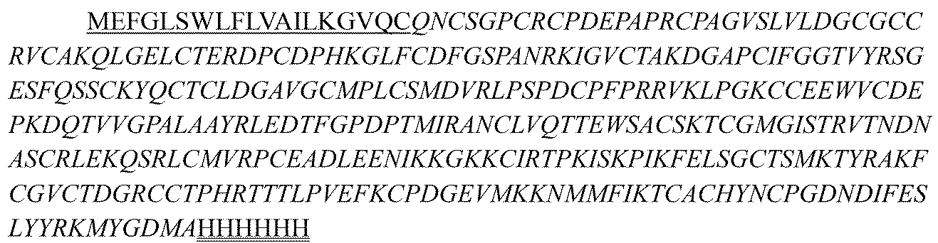
mCTGF-mFc(検出に使用することができる、マウスCTGFコーディング領域とマウスFcタグの融合タンパク質);配列番号3:

マウスCTGFタンパク質-Hisタグ;配列番号4:

カニクイザルCTGF-Fc(検出に使用することができる、カニクイザルCTGFコーディング領域とFcの融合タンパク質);配列番号5:

II.CTGF関連組換えタンパク質の精製、ならびに抗ヒトCTGFハイブリドーマ抗体および組換え抗体の精製
1.プロテインGアフィニティークロマトグラフィーによるハイブリドーマ上清の分離および精製
マウスハイブリドーマ上清の精製のために、プロテインGを用いるアフィニティークロマトグラフィーが好適であった。培養の結果生じたハイブリドーマを遠心分離し、上清を取り、上清の体積に基づいて10~15体積%の1M Tris-HCl(pH8.0~8.5)を加えて、上清のpHを中性に調節した。プロテインGカラムを、カラム容積の3~5倍の6Mグアニジン塩酸塩で洗い、次いでカラム体積の3~5倍の純水で洗い;カラム体積の3~5倍の1×PBS(pH7.4)でカラムを平衡化し;細胞上清を結合のために低流速でロードし、流速は保持時間が約1分間以上となるように調節し;UV吸収がベースラインに低下するまで、カラム体積の3~5倍の1×PBS(pH7.4)でカラムを洗い;サンプルを0.1M酢酸/酢酸ナトリウム(pH3.0)バッファーで溶出し、UV検出にしたがって溶出ピークをプールした。溶出物を速やかに1M Tris-HCl(pH8.0)でpH5~6に調節した。溶出物を、当業者によく知られる方法にしたがって溶液交換に付すことができ、例えば、限外濾過チューブを用いる限外濾過-濃縮によって溶液を所望のバッファー系に交換する、またはG-25脱塩といった分子排除によって溶液を所望のバッファー系に交換する、またはSuperdex 200といった高分離能分子排除カラムの使用によって溶出物から凝集物を除去してサンプル純度を改善することができた。
II. Purification of CTGF-related recombinant proteins, and purification of anti-human CTGF hybridoma antibodies and recombinant antibodies 1. Separation and purification of hybridoma supernatants by protein G affinity chromatography For the purification of mouse hybridoma supernatants, affinity chromatography using protein G was suitable. The hybridomas resulting from the culture were centrifuged, the supernatant was taken, and the pH of the supernatant was adjusted to neutral by adding 10-15% by volume of 1M Tris-HCl (pH 8.0-8.5) based on the volume of the supernatant. The Protein G column was washed with 3-5 column volumes of 6M guanidine hydrochloride, followed by 3-5 column volumes of pure water; equilibrated with 3-5 column volumes of 1x PBS (pH 7.4); loaded cell supernatant at a slow flow rate for binding, and the flow rate was adjusted to give a retention time of about 1 min or more; washed with 3-5 column volumes of 1x PBS (pH 7.4) until the UV absorption fell to baseline; eluted with 0.1M acetic acid/sodium acetate (pH 3.0) buffer, and pooled the elution peak according to UV detection. The eluate was quickly adjusted to pH 5-6 with 1M Tris-HCl (pH 8.0). The eluate can be subjected to solution exchange according to methods well known to those skilled in the art, for example, by ultrafiltration-concentration using ultrafiltration tubing to exchange the solution into the desired buffer system, or by molecular exclusion such as G-25 desalting to exchange the solution into the desired buffer system, or by use of a high resolution molecular exclusion column such as Superdex 200 to remove aggregates from the eluate and improve sample purity.
2.プロテインAアフィニティークロマトグラフィーによる抗体の精製
まず、抗体を発現している細胞培養上清を高速で遠心分離して、上清を集めた。プロテインAアフィニティーカラムをカラム体積の3~5倍の6Mグアニジン塩酸塩で洗い、次いでカラム体積の3~5倍の純水で洗った。カラム体積の3~5倍の、例えば1×PBS(pH7.4)で、カラムを平衡化した。細胞上清を結合のために低流速でロードし、流速は保持時間が約1分間以上となるように調節し;結合が終了したら、UV吸収がベースラインに低下するまで、カラム体積の3~5倍の1×PBS(pH7.4)でカラムを洗った。サンプルを0.1M酢酸/酢酸ナトリウム(pH3.0~3.5)バッファーで溶出し、UV検出にしたがって溶出ピークをプールした。溶出物を速やかに1M Tris-HCl(pH8.0)でpH5~6に調節した。溶出物を、当業者によく知られる方法にしたがって溶液交換に付すことができ、例えば、限外濾過チューブを用いる限外濾過-濃縮によって溶液を所望のバッファー系に交換する、またはG-25脱塩といった分子排除によって溶液を所望のバッファー系に交換する、またはSuperdex 200といった高分離能分子排除カラムの使用によって溶出物から凝集物を除去してサンプル純度を改善することができた。
2. Purification of antibodies by protein A affinity chromatography First, the cell culture supernatant expressing the antibody was centrifuged at high speed to collect the supernatant. The protein A affinity column was washed with 3-5 column volumes of 6 M guanidine hydrochloride, and then with 3-5 column volumes of pure water. The column was equilibrated with 3-5 column volumes, for example, 1×PBS (pH 7.4). The cell supernatant was loaded at a low flow rate for binding, and the flow rate was adjusted so that the retention time was about 1 min or more; when binding was completed, the column was washed with 3-5 column volumes of 1×PBS (pH 7.4) until the UV absorption fell to the baseline. The sample was eluted with 0.1 M acetic acid/sodium acetate (pH 3.0-3.5) buffer, and the elution peak was pooled according to UV detection. The eluate was quickly adjusted to pH 5-6 with 1 M Tris-HCl (pH 8.0). The eluate can be subjected to solution exchange according to methods well known to those skilled in the art, for example, by ultrafiltration-concentration using ultrafiltration tubing to exchange the solution into the desired buffer system, or by molecular exclusion such as G-25 desalting to exchange the solution into the desired buffer system, or by use of a high resolution molecular exclusion column such as Superdex 200 to remove aggregates from the eluate and improve sample purity.
3.CTGF関連組換えタンパク質(例えばヒトCTGF-his)のニッケルカラムによる精製
細胞発現上清サンプルを高速で遠心分離して不純物を除去し、バッファーをPBSと交換し、イミダゾールを終濃度5mMで加えた。ニッケルカラムを5mMイミダゾールを含むPBS溶液で平衡化し、カラム体積の2~5倍で洗った。交換後の上清サンプルを結合のためにカラムにロードし、異なる会社から入手可能なニッケルカラムを媒体として選択することができる。カラムを、A280の読み取りがベースラインに低下するまで、5mMイミダゾールを含むPBS溶液で洗った。非特異的に結合したタンパク質不純物を除去するためにクロマトグラフィーカラムをPBS+10mMイミダゾールで洗い、流出液を集めた。300mMイミダゾールを含むPBS溶液で標的タンパク質を溶出し、溶出ピークを集めた。
3. Purification of CTGF-related recombinant proteins (e.g. human CTGF-his) by nickel column Cell expression supernatant samples were centrifuged at high speed to remove impurities, buffer exchanged with PBS, and imidazole was added at a final concentration of 5 mM. The nickel column was equilibrated with PBS solution containing 5 mM imidazole and washed with 2-5 column volumes. The exchanged supernatant sample was loaded onto the column for binding, nickel columns available from different companies can be selected as the medium. The column was washed with PBS solution containing 5 mM imidazole until the A280 reading fell to baseline. The chromatography column was washed with PBS + 10 mM imidazole to remove non-specifically bound protein impurities, and the flow-through was collected. The target protein was eluted with PBS solution containing 300 mM imidazole, and the elution peak was collected.
集めた溶出物を濃縮したが、その後、それを、PBSを移動相とするゲルクロマトグラフィーSuperdex200(GE)によってさらに精製して、凝集物および不純物タンパク質ピークを除去し標的産物溶出ピークを集めることができる。得られたタンパク質を、電気泳動、ペプチドマッピングおよびLC-MSによって同定し、適正なタンパク質を使用のために小分けした。 The collected eluate was concentrated, which can then be further purified by gel chromatography Superdex200 (GE) with PBS as the mobile phase to remove aggregates and impurity protein peaks and collect the target product elution peak. The resulting proteins were identified by electrophoresis, peptide mapping and LC-MS, and the correct protein was aliquoted for use.
4.CTGF関連組換えタンパク質(例えばヒトCTGF-Fc、カニクイザルCTGF-Fc)のプロテインAアフィニティークロマトグラフィーによる精製
まず、培養上清を高速で遠心分離して上清を集めた。プロテインAアフィニティーカラムを、カラム体積の3~5倍の6Mグアニジン塩酸塩で洗い、次いでカラム体積の3~5倍の純水で洗った。カラム体積の3~5倍の、例えば1×PBS(pH7.4)で、カラムを平衡化した。細胞上清を結合のために低流速でロードし、流速は保持時間が約1分間以上となるように調節し;結合が終了したら、UV吸収がベースラインに低下するまで、カラム体積の3~5倍の1×PBS(pH7.4)でカラムを洗った。サンプルを0.1M酢酸/酢酸ナトリウム(pH3.0~3.5)バッファーで溶出し、UV検出にしたがって溶出ピークをプールした。
4. Purification of CTGF-related recombinant proteins (e.g., human CTGF-Fc, cynomolgus monkey CTGF-Fc) by protein A affinity chromatography First, the culture supernatant was centrifuged at high speed to collect the supernatant. The protein A affinity column was washed with 3-5 column volumes of 6 M guanidine hydrochloride, and then with 3-5 column volumes of pure water. The column was equilibrated with 3-5 column volumes, for example, 1×PBS (pH 7.4). The cell supernatant was loaded at a low flow rate for binding, and the flow rate was adjusted so that the retention time was about 1 minute or more; when binding was completed, the column was washed with 3-5 column volumes of 1×PBS (pH 7.4) until the UV absorption fell to the baseline. The sample was eluted with 0.1 M acetic acid/sodium acetate (pH 3.0-3.5) buffer, and the elution peak was pooled according to UV detection.
採取した溶出物を濃縮したが、その後、それを、PBSを移動相とするゲルクロマトグラフィーSuperdex200(GE)によってさらに精製して、凝集物および不純物タンパク質ピークを除去し標的産物溶出ピークを集めることができる。得られたタンパク質を、電気泳動、ペプチドマッピングおよびLC-MSによって同定し、適正なタンパク質を使用のために小分けした。 The collected eluate was concentrated, which can then be further purified by gel chromatography Superdex200 (GE) with PBS as the mobile phase to remove aggregates and impurity protein peaks and collect the target product elution peak. The resulting proteins were identified by electrophoresis, peptide mapping and LC-MS, and the correct protein was aliquoted for use.
実施例2:抗ヒトCTGFモノクローナル抗体の作製
I.免疫
抗ヒトCTGFモノクローナル抗体を、マウスを免疫することによって作製した。6~8週齢の雌SJL白マウスを実験に使用した(Beijing Charles River Experimental Animal Technology Co., Ltd., animal production license number: SCXK (Beijing) 2012-0001)。飼育環境:SPFレベル。マウスを購入後、12/12時間の明/暗サイクル、温度20~25℃、湿度40~60%の実験室環境に1週間維持した。環境に順応したマウスを下記スキームにしたがって免疫した。免疫用抗原として、CTGF(R&D)およびmCTGF-mFcを使用した。
Example 2: Preparation of anti-human CTGF monoclonal antibody I. Immunization Anti-human CTGF monoclonal antibody was prepared by immunizing mice. 6-8 week old female SJL white mice were used in the experiment (Beijing Charles River Experimental Animal Technology Co., Ltd., animal production license number: SCXK (Beijing) 2012-0001). Rearing environment: SPF level. After purchasing the mice, they were kept in a laboratory environment with a 12/12 hour light/dark cycle, a temperature of 20-25°C, and a humidity of 40-60% for one week. The mice that had adapted to the environment were immunized according to the following scheme. CTGF (R&D) and mCTGF-mFc were used as immunization antigens.
免疫スキーム:25μg/マウス/回のCTGFタンパク質腹腔内注射によってマウスを免疫した。0日目に100μl/マウスを腹腔内(IP)注射し、14日毎に1回の追加免疫を行った。21、35、49および63日目に血液サンプルを採り、マウス血清中の抗体力価をELISA法により測定した。7~9回の免疫後、高い血清抗体力価がプラトーに近づいたマウスを脾細胞融合のために選択した。脾細胞融合の3日前に、追加免疫のために、25μg/マウスで、塩類液中に調製したCTGFタンパク質抗原溶液を腹腔内注射(IP)した。
Immunization scheme: Mice were immunized by intraperitoneal injection of CTGF protein at 25 μg/mouse/time. 100 μl/mouse was injected intraperitoneally (IP) on
II.脾細胞融合
PEG仲介融合法を用いて脾臓リンパ球とミエローマSp2/0(ATCC(登録商標)CRL-8287(商標))を融合することにより、ハイブリドーマ細胞を得た。融合ハイブリドーマ細胞を完全培地(20%FBS、1×HAT、1×OPIを含むIMDM培地)に0.5~1×106/mlの密度で再懸濁し、96ウェルプレートに100μl/ウェルで播種し、37℃および5%CO2で3~4日間インキュベートし、100μl/ウェルのHAT完全培地を補充し、ピンポイントのクローンを形成するまで3~4日間培養を続けた。上清を除去し、200μl/ウェルのHT完全培地(20%FBS、1×HT、1×OPIを含むIMDM培地)を加え、37℃、5%CO2で3日間インキュベートし、その後、ELISA検出に付した。
II. Spleen Cell Fusion Hybridoma cells were obtained by fusing spleen lymphocytes with myeloma Sp2/0 (ATCC® CRL-8287™) using PEG-mediated fusion. The fused hybridoma cells were resuspended in complete medium (IMDM medium containing 20% FBS, 1×HAT, 1×OPI) at a density of 0.5-1×10 6 /ml, seeded in 96-well plates at 100 μl/well, incubated at 37° C. and 5% CO 2 for 3-4 days, supplemented with 100 μl/well of HAT complete medium, and continued to be cultured for 3-4 days until pinpoint clones were formed. The supernatant was removed, and 200 μl/well of HT complete medium (IMDM medium containing 20% FBS, 1×HT, 1×OPI) was added, incubated at 37° C. and 5% CO 2 for 3 days, and then subjected to ELISA detection.
III.ハイブリドーマ細胞のスクリーニング
ハイブリドーマ細胞の増殖密度にしたがって、ハイブリドーマ培養上清を結合ELISA法によって検出した。陽性ウェルの細胞上清を、サルCTGF/マウスCTGF/ヒトCTGFタンパク質との結合についてELISAアッセイによって検出した(具体的な方法は実施例3のものと同じであった)。タンパク質ELISA結合アッセイで陽性のウェルの細胞を適時に増殖させ、凍結保存し;単一細胞クローンが得られるまで2~3回サブクローニングした。
III. Screening of hybridoma cells According to the proliferation density of hybridoma cells, the hybridoma culture supernatant was detected by binding ELISA. The cell supernatant of positive wells was detected by ELISA assay for binding with monkey CTGF/mouse CTGF/human CTGF protein (the specific method was the same as that in Example 3). The cells of wells positive in protein ELISA binding assay were expanded in a timely manner and cryopreserved; subcloned 2-3 times until single cell clones were obtained.
各サブクローニング後の細胞もCTGF結合ELISAによって試験した。上記アッセイによるスクリーニングによってハイブリドーマクローンを得た。無血清細胞培養法によって抗体をさらに調製した。抗体を、試験実施例における使用のために、精製の実施例にしたがって精製した。 Cells after each subcloning were also tested by CTGF binding ELISA. Hybridoma clones were obtained by screening with the above assay. Antibodies were further prepared by serum-free cell culture method. Antibodies were purified according to the purification example for use in the test example.
IV.陽性ハイブリドーマクローンのシーケンシング
陽性ハイブリドーマ由来の配列をクローニングする手順は、次のとおりであった。対数増殖期にあるハイブリドーマ細胞を集めた。Trizol(Invitrogen, Cat No. 15596-018)をキットの指示書にしたがって使用してRNAを抽出し、PrimeScript(商標)逆転写キット(Takara, Cat No. 2680A)を用いて逆転写した。逆転写により生じたcDNAを、マウスIgプライマーセット(Novagen, TB326 Rev. B 0503)を用いるPCRによって増幅し、増幅生成物をシーケンシングに付した。シーケンシング後、マウス抗CTGF抗体mab147、mab164およびmab95を得た。可変領域のアミノ酸配列は次のとおりである:
IV. Sequencing of positive hybridoma clones The procedure for cloning sequences from positive hybridomas was as follows: Hybridoma cells in logarithmic growth phase were collected. RNA was extracted using Trizol (Invitrogen, Cat No. 15596-018) according to the kit's instructions, and reverse transcribed using PrimeScript™ Reverse Transcription Kit (Takara, Cat No. 2680A). The cDNA generated by reverse transcription was amplified by PCR using a mouse Ig primer set (Novagen, TB326 Rev. B 0503), and the amplified product was subjected to sequencing. After sequencing, mouse anti-CTGF antibodies mab147, mab164 and mab95 were obtained. The amino acid sequences of the variable regions are as follows:
mab147のVHアミノ酸配列は、配列番号6:

mab147のVLアミノ酸配列は、配列番号7:

The VL amino acid sequence of mab147 is SEQ ID NO:7:
mab164のVHアミノ酸配列は、配列番号8:

mab164のVLアミノ酸配列は、配列番号9:

The VL amino acid sequence of mab164 is SEQ ID NO:9:
mab95のVHアミノ酸配列は、配列番号69:

mab95のVLアミノ酸配列は、配列番号70:
![]()
The VL amino acid sequence of mab95 is SEQ ID NO:70:
![]()
上記mab147、mab164およびmab95抗体可変領域配列において、イタリック体はFR配列を表し、下線付きイタリック体はCDR配列を表し、配列の並びはFR1-CDR1-FR2-CDR2-FR3-CDR3-FR4である。 In the above mab147, mab164 and mab95 antibody variable region sequences, the italics represent FR sequences, the underlined italics represent CDR sequences, and the sequence arrangement is FR1-CDR1-FR2-CDR2-FR3-CDR3-FR4.

V.ヒトIgG1キメラ抗体の作製
候補分子mab147、mab164およびmab95を増幅し、シーケンシングして、可変領域をコードする遺伝子配列を得た。シーケンシングによって得た配列に基づいてフォワードおよびリバースプライマーをデザインし、シーケンシングされる遺伝子をテンプレートとして使用して各抗体のVH/VK遺伝子フラグメントをPCRにより作製し、次いでそれを相同組換えによって発現ベクターpHr(シグナルペプチドおよびhIgG1/hκ/hλ定常領域遺伝子(CH1-Fc/CL)フラグメントを有する)に挿入して、完全長組換えキメラ抗体の発現プラスミドVH-CH1-Fc-pHr/VL-CL-pHrを作製し、結果として3つのキメラ抗体Ch147、Ch164およびCh95を得た。
V. Construction of human IgG1 chimeric antibodies Candidate molecules mab147, mab164 and mab95 were amplified and sequenced to obtain gene sequences encoding the variable regions. Based on the sequences obtained by sequencing, forward and reverse primers were designed, and the sequenced genes were used as templates to generate VH/VK gene fragments of each antibody by PCR, which were then inserted into expression vector pHr (with signal peptide and hIgG1/hκ/hλ constant region gene (CH1-Fc/CL) fragment) by homologous recombination to generate expression plasmids VH-CH1-Fc-pHr/VL-CL-pHr of full-length recombinant chimeric antibodies, resulting in three chimeric antibodies Ch147, Ch164 and Ch95.
VI.マウス抗ヒトCTGF抗体のヒト化
マウス抗体との相同性の高い重鎖および軽鎖可変領域生殖細胞系列遺伝子を、IMGT(http://imgt.cines.fr)ヒト抗体重鎖および軽鎖可変生殖細胞系列遺伝子データベースに対してMOEソフトウェア(Molecular Operating Environment)のソフトウェアを用いてアラインすることによって、テンプレートとして選択した。マウス抗体CDRを対応するヒトテンプレート中にグラフトして、FR1-CDR1-FR2-CDR2-FR3-CDR3-FR4の並びの可変領域配列を形成した。
VI. Humanization of Mouse Anti-Human CTGF Antibody Highly homologous heavy and light chain variable region germline genes of mouse antibodies were selected as templates by aligning them against the IMGT (http://imgt.cines.fr) human antibody heavy and light chain variable germline gene database using MOE (Molecular Operating Environment) software. The mouse antibody CDRs were grafted into the corresponding human template to form the variable region sequence of FR1-CDR1-FR2-CDR2-FR3-CDR3-FR4.
1.mab147のヒト化
マウス抗体mab147に対しては、ヒト化用の軽鎖テンプレートはIGKV3-11*01およびIGKJ2*01、またはIGKV1-39*01およびIGKJ2*01であり、ヒト化用の重鎖テンプレートは、IGHV3-72*01およびIGHJ1*01であった。マウス抗体mab147の各CDRをそのヒトテンプレートにグラフトし、次いでヒト化抗体のFRのアミノ酸を復帰変異に付した。そのなかで、軽鎖可変領域中の復帰変異は、4L、36F、43S、45K、47W、58Vまたは71Yからなる群から選択される1つ以上を含み、重鎖可変領域中の復帰変異は、28S、30N、49A、75E、76S、93V、94Eまたは104Dからなる群から選択される1つ以上を含み(軽鎖および重鎖可変領域中の復帰変異位置は、Kabatナンバリング基準にしたがって決定した)、その結果、異なる配列の軽鎖可変領域および重鎖可変領域を得た。mab147のCDRを含むヒト化抗体を得たが、その可変領域配列は次のとおりである:
1. Humanization of mab147 For the mouse antibody mab147, the light chain templates for humanization were IGKV3-11*01 and IGKJ2*01, or IGKV1-39*01 and IGKJ2*01, and the heavy chain templates for humanization were IGHV3-72*01 and IGHJ1*01. Each CDR of the mouse antibody mab147 was grafted onto its human template, and then the amino acids in the FR of the humanized antibody were subjected to back mutation. Among them, the back mutations in the light chain variable region included one or more selected from the group consisting of 4L, 36F, 43S, 45K, 47W, 58V or 71Y, and the back mutations in the heavy chain variable region included one or more selected from the group consisting of 28S, 30N, 49A, 75E, 76S, 93V, 94E or 104D (the positions of the back mutations in the light and heavy chain variable regions were determined according to the Kabat numbering standard), resulting in light and heavy chain variable regions with different sequences. A humanized antibody was obtained containing the CDRs of mab147, whose variable region sequence is as follows:
mAb147のヒト化抗体の具体的な可変領域配列は次のとおりである:
Hu147-VL1のアミノ酸配列は、配列番号22:

Hu147-VL2のアミノ酸配列は、配列番号23:

Hu147-VL3のアミノ酸配列は、配列番号24:

Hu147-VL4のアミノ酸配列は、配列番号25:

Hu147-VL5のアミノ酸配列は、配列番号26:

Hu147-VH1のアミノ酸配列は、配列番号27:

Hu147-VH2のアミノ酸配列は、配列番号28:

Hu147-VH3のアミノ酸配列は、配列番号29:

The amino acid sequence of Hu147-VL1 is SEQ ID NO:22:
The amino acid sequence of Hu147-VL2 is SEQ ID NO:23:
The amino acid sequence of Hu147-VL3 is SEQ ID NO:24:
The amino acid sequence of Hu147-VL4 is SEQ ID NO:25:
The amino acid sequence of Hu147-VL5 is SEQ ID NO:26:
The amino acid sequence of Hu147-VH1 is SEQ ID NO:27:
The amino acid sequence of Hu147-VH2 is SEQ ID NO:28:
The amino acid sequence of Hu147-VH3 is SEQ ID NO:29:
注:Kabat基準にしたがって決定されたCDR配列に下線を付しており、配列の並びはFR1-CDR1-FR2-CDR2-FR3-CDR3-FR4の順である。 Note: The CDR sequences determined according to the Kabat criteria are underlined, and the sequence order is FR1-CDR1-FR2-CDR2-FR3-CDR3-FR4.
2.mab164のヒト化
マウス抗体mab164に対しては、ヒト化用の軽鎖テンプレートはそれぞれIGLV7-43*01およびIGLJ2*01、IGLV8-61*01およびIGLJ2*01、またはIGLV1-40*02およびIGLJ2*01からなり、ヒト化用の重鎖テンプレートはそれぞれIGHV2-26*01およびIGHJ6*01、またはIGHV4-31*02およびIGHJ6*01からなった。マウス抗体mab164の各CDRをそのヒトテンプレートにグラフトし、次いでヒト化抗体のFRのアミノ酸を復帰変異に付した。そのなかで、軽鎖可変領域中の復帰変異は、36V、44F、46Gまたは49Gからなる群から選択される1つ以上を含み、重鎖可変領域中の復帰変異は、44G、49G、27F、48L、67L、71K、78Vまたは80Fからなる群から選択される1つ以上を含み(軽鎖および重鎖可変領域中の復帰変異位置はKabatナンバリング基準にしたがって決定した)、その結果、下記可変領域配列を有する、mab164のヒト化重鎖および軽鎖を得た:
2. Humanization of mab164 For the murine antibody mab164, the light chain templates for humanization consisted of IGLV7-43*01 and IGLJ2*01, IGLV8-61*01 and IGLJ2*01, or IGLV1-40*02 and IGLJ2*01, respectively, and the heavy chain templates for humanization consisted of IGHV2-26*01 and IGHJ6*01, or IGHV4-31*02 and IGHJ6*01, respectively. Each CDR of the murine antibody mab164 was grafted onto its human template, and then the amino acids in the FR of the humanized antibody were subjected to back mutation. Among them, the backmutations in the light chain variable region included one or more selected from the group consisting of 36V, 44F, 46G or 49G, and the backmutations in the heavy chain variable region included one or more selected from the group consisting of 44G, 49G, 27F, 48L, 67L, 71K, 78V or 80F (the backmutation positions in the light and heavy chain variable regions were determined according to the Kabat numbering standard), resulting in humanized heavy and light chains of mab164 having the following variable region sequences:
mAb164のヒト化抗体の具体的な可変領域配列は次のとおりである:
Hu164-VL7のアミノ酸配列は、配列番号30:

Hu164-VL8のアミノ酸配列は、配列番号31:

Hu164-VL9のアミノ酸配列は、配列番号32:
![]()
Hu164-VH5のアミノ酸配列は、配列番号33:

Hu164-VH6のアミノ酸配列は、配列番号34:

Hu164-VH7のアミノ酸配列は、配列番号35:

Hu164-VH8のアミノ酸配列は、配列番号36:

The amino acid sequence of Hu164-VL7 is SEQ ID NO:30:
The amino acid sequence of Hu164-VL8 is SEQ ID NO:31:
The amino acid sequence of Hu164-VL9 is SEQ ID NO:32:
![]()
The amino acid sequence of Hu164-VH5 is SEQ ID NO:33:
The amino acid sequence of Hu164-VH6 is SEQ ID NO:34:
The amino acid sequence of Hu164-VH7 is SEQ ID NO:35:
The amino acid sequence of Hu164-VH8 is SEQ ID NO:36:
注:上記Hu164抗体配列において、下線はCDR配列を示し、配列の並びはFR1-CDR1-FR2-CDR2-FR3-CDR3-FR4であり、ここで、CDR配列はKabat基準にしたがって規定される。 Note: In the above Hu164 antibody sequence, the underlines indicate the CDR sequences, and the sequence order is FR1-CDR1-FR2-CDR2-FR3-CDR3-FR4, where the CDR sequences are defined according to the Kabat criteria.
3.mab95のヒト化
マウス抗体mab95に対しては、ヒト化用の軽鎖テンプレートはIGKV3-20*02およびIGKV1-40*01であり、ヒト化用の重鎖テンプレートは、IGHV1-3*01であった。マウス抗体mab95の各CDRをそのヒトテンプレートにグラフトし、次いでヒト化抗体のFRのアミノ酸を復帰変異に付した。そのなかで、軽鎖可変領域中の復帰変異は、45P、46W、48Y、69Sまたは70Yからなる群から選択される1つ以上を含み、重鎖可変領域中の復帰変異は、27F、38K、48I、67K、68A、70Lまたは72Fからなる群から選択される1つ以上を含み(軽鎖および重鎖可変領域中の復帰変異位置は、Kabatナンバリング基準にしたがって決定した)、その結果、異なる配列の軽鎖可変領域および重鎖可変領域を得た。mab95のCDRを含むヒト化抗体を得たが、その可変領域配列は次のとおりである:
3. Humanization of mab95 For the mouse antibody mab95, the light chain templates for humanization were IGKV3-20*02 and IGKV1-40*01, and the heavy chain template for humanization was IGHV1-3*01. Each CDR of the mouse antibody mab95 was grafted onto its human template, and then the amino acids of the FR of the humanized antibody were subjected to back mutations, among which the back mutations in the light chain variable region included one or more selected from the group consisting of 45P, 46W, 48Y, 69S or 70Y, and the back mutations in the heavy chain variable region included one or more selected from the group consisting of 27F, 38K, 48I, 67K, 68A, 70L or 72F (the back mutation positions in the light and heavy chain variable regions were determined according to the Kabat numbering standard), resulting in light and heavy chain variable regions of different sequences. A humanized antibody was obtained containing the CDRs of mab95, the variable region sequences of which are as follows:
mAb95のヒト化抗体の具体的な可変領域配列は次のとおりである:
Hu95-VL1のアミノ酸配列は、配列番号77:

Hu95-VL2のアミノ酸配列は、配列番号78:

Hu95-VL3のアミノ酸配列は、配列番号79:

Hu95-VL4のアミノ酸配列は、配列番号80:

Hu95-VH1のアミノ酸配列は、配列番号81:

Hu95-VH2のアミノ酸配列は、配列番号82:
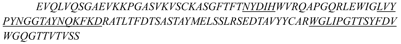
Hu95-VH3のアミノ酸配列は、配列番号83:
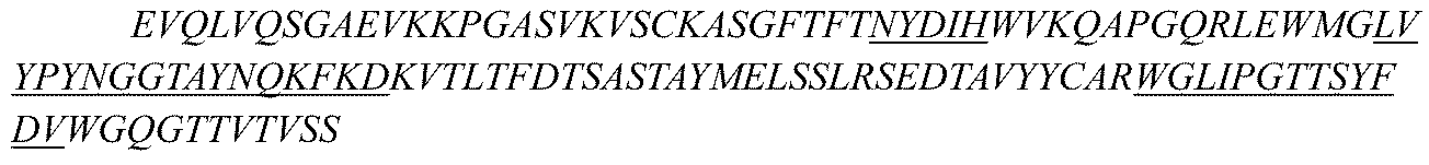
Hu95-VH4のアミノ酸配列は、配列番号84:

Hu95-VH5のアミノ酸配列は、配列番号85:

The amino acid sequence of Hu95-VL1 is SEQ ID NO:77:
The amino acid sequence of Hu95-VL2 is SEQ ID NO:78:
The amino acid sequence of Hu95-VL3 is SEQ ID NO:79:
The amino acid sequence of Hu95-VL4 is SEQ ID NO:80:
The amino acid sequence of Hu95-VH1 is SEQ ID NO:81:
The amino acid sequence of Hu95-VH2 is SEQ ID NO:82:
The amino acid sequence of Hu95-VH3 is SEQ ID NO:83:
The amino acid sequence of Hu95-VH4 is SEQ ID NO:84:
The amino acid sequence of Hu95-VH5 is SEQ ID NO:85:
注:Kabat基準にしたがって決定されたCDR配列に下線を付しており、配列の並びはFR1-CDR1-FR2-CDR2-FR3-CDR3-FR4の順である。 Note: The CDR sequences determined according to the Kabat criteria are underlined, and the sequence order is FR1-CDR1-FR2-CDR2-FR3-CDR3-FR4.
上記のうち、Hu95-VH5のCDR領域はさらに改変されており、HCDR1は配列番号102(NYAIH)で示され、HCDR2は配列番号103(LVYPYTGGTAYNQKFKD)で示され、HCDR3は配列番号104(WGMIPGTNSYFDV)で示される。 Of the above, the CDR regions of Hu95-VH5 have been further modified, with HCDR1 being represented by SEQ ID NO: 102 (NYAIH), HCDR2 being represented by SEQ ID NO: 103 (LVYPYTGGTAYNQKFKD), and HCDR3 being represented by SEQ ID NO: 104 (WGMIPGTNSYFDV).
4.抗CTGFヒト化抗体の作製および発現
プライマーをデザインし、各ヒト化抗体のVH/VL遺伝子フラグメントをPCRによって作製して、相同組換えによって発現ベクターpHr(シグナルペプチドおよび定常領域遺伝子(CH/CL)フラグメントを有する)に挿入することにより、完全長抗体の発現ベクターVH-CH-pHr/VL-CL-pHrを作製した。ヒト化抗体用に、定常領域を、ヒトκ、λ鎖の軽鎖定常領域からなる群から選択することができ、IgG1、IgG2、IgG3もしくはIgG4の重鎖定常領域またはそのバリアントからなる群から選択することができる。非限定的な例は、ヒトIgG1、IgG2またはIgG4の定常領域を抗体の機能を改善するために最適化することを包含する。例えば、M252Y/S254T/T256E(YTE)部位の変異といった定常領域中の点変異、および定常領域中のS228P、F234AおよびL235Aといった変異によって、抗体の半減期を延長することができる。
4. Generation and Expression of Anti-CTGF Humanized Antibody Primers were designed and VH/VL gene fragments of each humanized antibody were generated by PCR and inserted into expression vector pHr (with signal peptide and constant region gene (CH/CL) fragments) by homologous recombination to generate full-length antibody expression vector VH-CH-pHr/VL-CL-pHr. For humanized antibodies, the constant region can be selected from the group consisting of human kappa, lambda light chain constant regions, and IgG1, IgG2, IgG3 or IgG4 heavy chain constant regions or variants thereof. Non-limiting examples include optimizing human IgG1, IgG2 or IgG4 constant regions to improve antibody function. For example, point mutations in the constant region, such as the M252Y/S254T/T256E (YTE) site mutations, and mutations in the constant region, such as S228P, F234A and L235A, can extend the half-life of an antibody.
抗CTGFヒト化抗体定常領域配列の例は次のとおりである:
重鎖定常領域のアミノ酸配列が、配列番号37:

The amino acid sequence of the heavy chain constant region is SEQ ID NO:37:
YTE改変された抗体の重鎖定常領域のアミノ酸配列が、配列番号38:
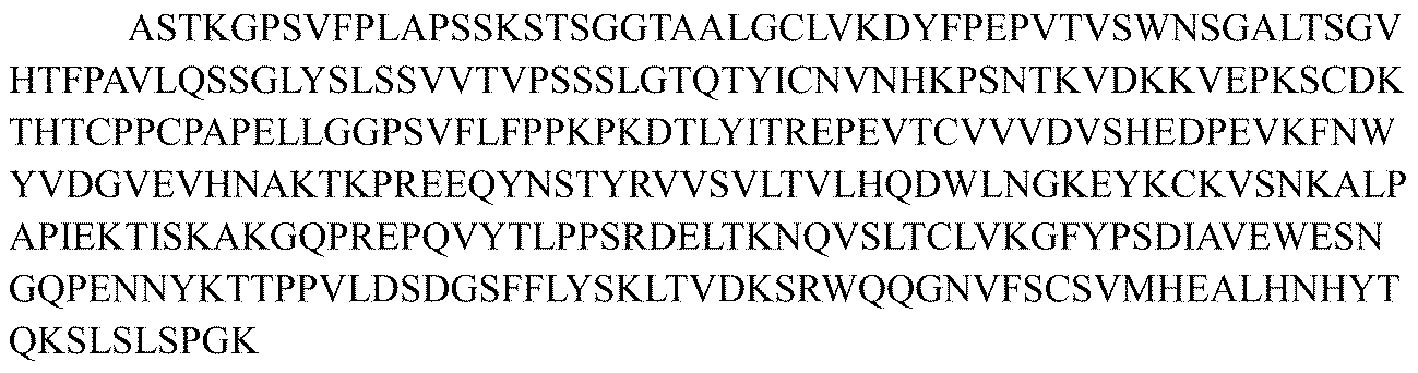
抗CTGFヒト化抗体の軽鎖κ定常領域のアミノ酸配列が、配列番号39:
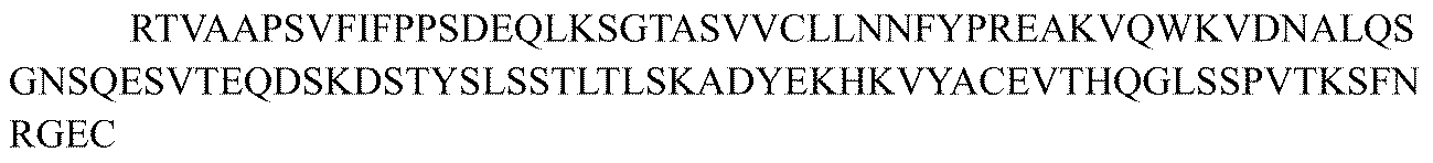
抗CTGFヒト化抗体の軽鎖λ定常領域のアミノ酸配列が、配列番号40:

ヒト化抗体の重鎖可変領域を配列番号37に示される重鎖定常領域に連結し、ヒト化抗体の軽鎖可変領域を配列番号39に示される軽鎖κ定常領域に連結して、次のものを包含するmab147由来の完全長ヒト化抗体を得た:

ヒト化抗体の重鎖可変領域を配列番号38に示される重鎖定常領域YTEバリアントに連結し、ヒト化抗体の軽鎖可変領域を配列番号39に示される軽鎖κ定常領域に連結して、次のものを包含するmab147由来の完全長ヒト化抗体を得た:
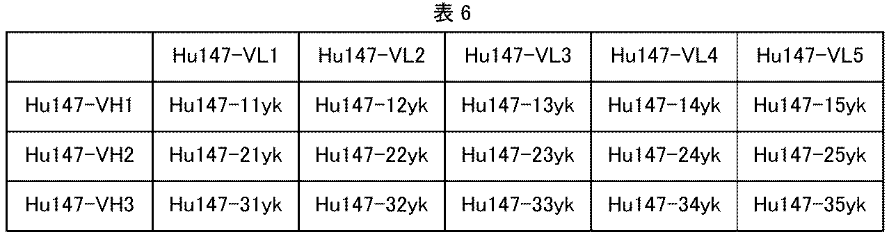
ヒト化抗体の重鎖可変領域を配列番号37に示される重鎖定常領域に連結し、ヒト化抗体の軽鎖可変領域を配列番号40に示される軽鎖λ定常領域に連結して、次のものを包含するmab164由来の完全長ヒト化抗体を得た:

ヒト化抗体の重鎖可変領域を配列番号38に示される重鎖定常領域YTEバリアントに連結し、ヒト化抗体の軽鎖可変領域を配列番号40に示される軽鎖λ定常領域に連結して、次のものを包含するmab164由来の完全長ヒト化抗体を得た:

ヒト化抗体の重鎖可変領域を配列番号37に示される重鎖定常領域に連結し、ヒト化抗体の軽鎖可変領域を配列番号39に示される軽鎖κ定常領域に連結して、次のものを包含するmab95由来の完全長ヒト化抗体を得た:

ヒト化抗体の重鎖可変領域を配列番号38に示される重鎖定常領域YTEバリアントに連結し、ヒト化抗体の軽鎖可変領域を配列番号39に示される軽鎖κ定常領域に連結して、次のものを包含するmab95由来の完全長ヒト化抗体を得た:

完全長抗CTGF抗体のアミノ酸配列の例は次のとおりである:

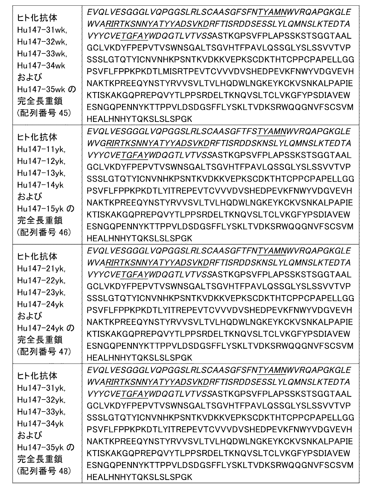


陽性対照CTGF抗体mAb1(CN1829740Bより;抗体名称はパムレブルマブで、FG-3019としても知られる)の配列は次のとおりである:
重鎖(配列番号67):


Heavy Chain (SEQ ID NO:67):
実施例3:CTGF抗体の結合活性の検出
I.ヒトCTGFタンパク質に対するCTGF抗体の結合のELISAアッセイ
ヒトCTGFタンパク質に対する抗ヒトCTGF抗体の結合活性を、ELISAアッセイおよびBiacoreによって試験した。96ウェルマイクロタイタープレートにCTGF組換えタンパク質を直接コーティングした。抗体添加後のシグナル強度を利用して、CTGFに対する抗体の結合活性を測定した。具体的な実験手順は次のとおりであった:
Example 3: Detection of the binding activity of CTGF antibody I. ELISA assay of the binding of CTGF antibody to human CTGF protein The binding activity of anti-human CTGF antibody to human CTGF protein was tested by ELISA assay and Biacore. CTGF recombinant protein was directly coated on a 96-well microtiter plate. The binding activity of the antibody to CTGF was measured using the signal intensity after the addition of the antibody. The specific experimental procedure was as follows:
CTGFタンパク質(R&D, Cat No. 9190-CC)をPBS(pH7.4)(Shanghai Yuanpei, Cat No. B320)バッファーで0.2μg/mlの濃度に希釈し、50μl/ウェルで96ウェルマイクロタイタープレート(Corning, Cat No. CLS3590-100EA)に入れ、37℃のインキュベーター内で2時間、または4℃で一晩(16~18時間)、インキュベートした。液体を除去し、250μl/ウェルのブロッキング溶液(PBSで希釈した5%スキムミルク(BDスキムミルク, Cat No.232100))を加え、37℃のインキュベーター内で3時間、または4℃で一晩(16~18時間)、インキュベートしてブロッキングした。ブロッキングの終了後、ブロッキング溶液を除去し、プレートをPBSTバッファー(0.1%のTween-20を含むPBS、pH7.4)で5回洗い、サンプル希釈溶液で希釈したさまざまな濃度の各試験抗体(ハイブリドーマから精製された抗体、またはヒト化抗体)を50μl/ウェルで加え、37℃のインキュベーター内で1時間インキュベートした。インキュベーションの終了後、プレートをPBSTで3回洗い、サンプル希釈溶液で希釈したHRP標識ヤギ抗マウス二次抗体(Jackson Immuno Research, Cat No. 115-035-003)またはヤギ抗ヒト二次抗体(Jackson Immuno Research, Cat No. 109-035-003)を50μl/ウェルで加え、37℃で1時間インキュベートした。プレートをPBSTで3回洗い、TMB発色基質(KPL, Cat No. 52-00-03)を50μl/ウェルで加え、室温で5~10分間インキュベートし、1M H2SO4を50μl/ウェルで加えて反応を停止した。波長450nmにおける吸光度の値をマイクロプレートリーダー(Molecular Devices, VERSA max)で読み取り、データをGraphPad Prism 5で分析した。ヒトCTGFタンパク質に対するCTGF抗体の結合を、EC50値として計算した。実験結果を図1および表12に示す。 CTGF protein (R&D, Cat No. 9190-CC) was diluted to a concentration of 0.2 μg/ml with PBS (pH 7.4) (Shanghai Yuanpei, Cat No. B320) buffer, and placed in a 96-well microtiter plate (Corning, Cat No. CLS3590-100EA) at 50 μl/well and incubated in a 37°C incubator for 2 hours or at 4°C overnight (16-18 hours). The liquid was removed and 250 μl/well of blocking solution (5% skim milk (BD skim milk, Cat No. 232100) diluted in PBS) was added and incubated in a 37°C incubator for 3 hours or at 4°C overnight (16-18 hours) for blocking. After completion of blocking, the blocking solution was removed, the plate was washed five times with PBST buffer (PBS containing 0.1% Tween-20, pH 7.4), and various concentrations of each test antibody (hybridoma purified antibody or humanized antibody) diluted with sample dilution solution were added at 50 μl/well and incubated for 1 hour in an incubator at 37° C. After completion of incubation, the plate was washed three times with PBST, and HRP-labeled goat anti-mouse secondary antibody (Jackson Immuno Research, Cat No. 115-035-003) or goat anti-human secondary antibody (Jackson Immuno Research, Cat No. 109-035-003) diluted with sample dilution solution was added at 50 μl/well and incubated for 1 hour at 37° C. The plates were washed three times with PBST, TMB chromogenic substrate (KPL, Cat No. 52-00-03) was added at 50 μl/well, incubated at room temperature for 5-10 minutes, and the reaction was stopped by adding 50 μl/well of 1M H2SO4 . The absorbance values at a wavelength of 450 nm were read using a microplate reader (Molecular Devices, VERSA max), and the data were analyzed using GraphPad Prism 5. The binding of the CTGF antibody to the human CTGF protein was calculated as an EC50 value. The experimental results are shown in Figure 1 and Table 12.
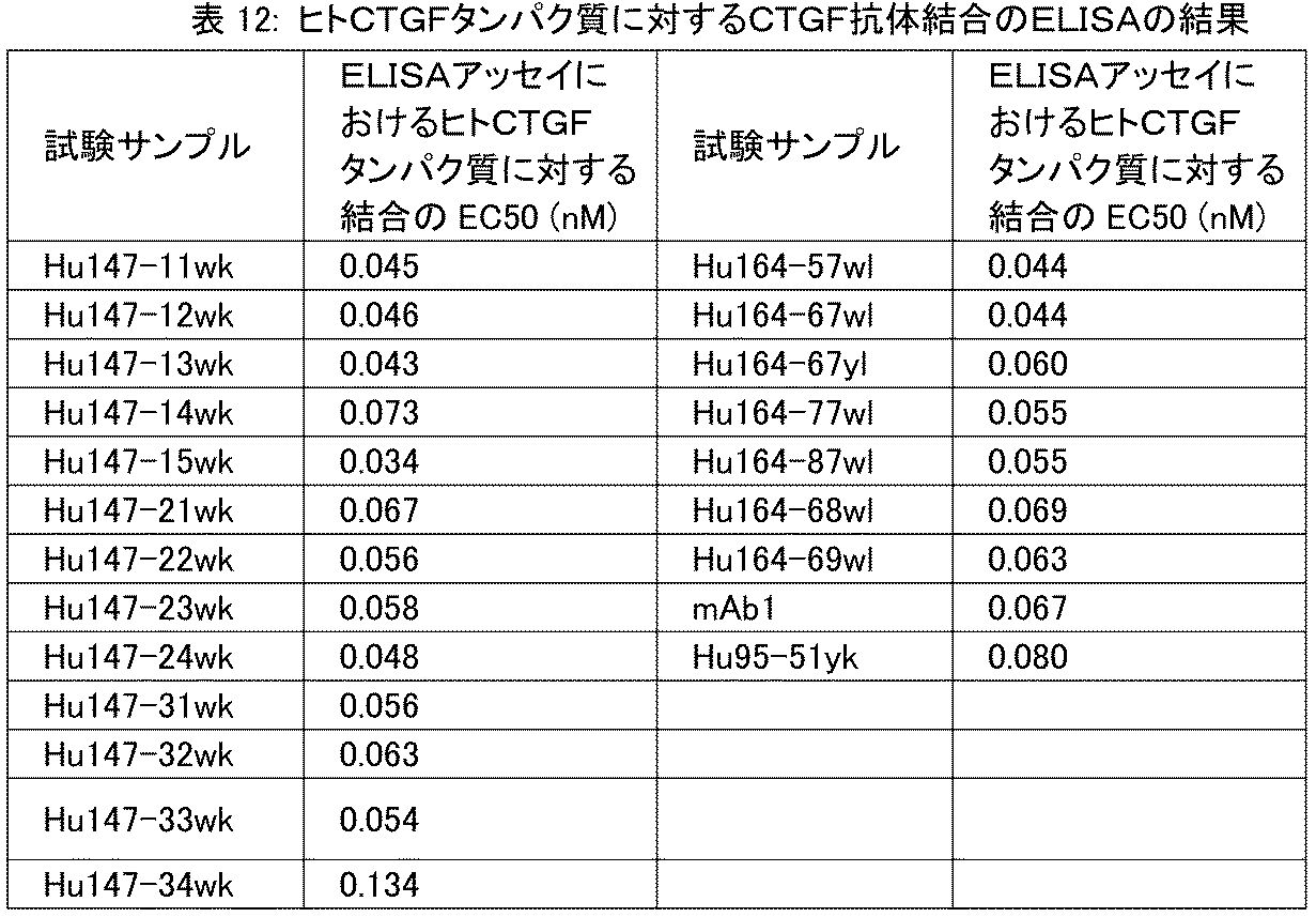
実験結果は、本開示のヒト化完全長抗CTGF抗体はいずれも、ヒトCTGFタンパク質との結合作用に優れていることを示している。 The experimental results show that all of the humanized full-length anti-CTGF antibodies disclosed herein have excellent binding activity with human CTGF protein.
II.カニクイザルCTGFタンパク質に対するCTGF抗体の結合のELISAアッセイ
サルCTGFタンパク質に対する抗サルCTGF抗体の結合活性を、ELISAアッセイによって試験した。96ウェルマイクロタイタープレートに抗mFcタンパク質を直接コーティングし、次いでFcタグ付きカニクイザルCTGFタンパク質をプレートに結合し、その後、抗体を加えた。抗体添加後のシグナル強度を利用して、カニクイザルCTGFに対する抗体の結合活性を測定した。具体的な実験手順は次のとおりであった:
II. ELISA assay of binding of CTGF antibody to cynomolgus CTGF protein The binding activity of anti-monkey CTGF antibody to monkey CTGF protein was tested by ELISA assay. Anti-mFc protein was directly coated on a 96-well microtiter plate, and then Fc-tagged cynomolgus CTGF protein was bound to the plate, and then the antibody was added. The signal intensity after antibody addition was used to measure the binding activity of the antibody to cynomolgus CTGF. The specific experimental procedure was as follows:
抗mFcタンパク質(Sigma, Cat No. M4280)をPBS(pH7.4)(Shanghai Yuanpei, Cat No. B320)バッファーで2μg/mlに希釈し、50μl/ウェルで96ウェルマイクロタイタープレート(Corning, Cat No. CLS3590-100EA)に入れ、37℃のインキュベーター内で2時間インキュベートした。液体を除去し、250μl/ウェルのブロッキング溶液(PBSで希釈した5%スキムミルク(BDスキムミルク, Cat No.232100))を加え、37℃のインキュベーター内で3時間、または4℃で一晩(16~18時間)、インキュベートしてブロッキングした。ブロッキングの終了後、ブロッキング溶液を除去し、プレートをPBSTバッファー(0.1%のTween-20を含むPBS、pH7.4)で3回洗い、50μlの1μg/mlカニクイザルCTGF-mFcタンパク質を各ウェルに加え、37℃のインキュベーター内で1時間インキュベートし、その後、プレートをPBSTバッファー(0.1%のTween-20を含むPBS、pH7.4)で3回洗った。サンプル希釈溶液で希釈したさまざまな濃度の各試験抗体(ハイブリドーマから精製された抗体、またはヒト化抗体)を50μl/ウェルで加え、37℃のインキュベーター内で1時間インキュベートした。インキュベーションの終了後、プレートをPBSTで3回洗い、サンプル希釈溶液で希釈したHRP標識ヤギ抗マウス二次抗体(Jackson Immuno Research, Cat No. 115-035-003)またはヤギ抗ヒト二次抗体(Jackson Immuno Research, Cat No. 109-035-003)を50μl/ウェルで加え、37℃で1時間インキュベートした。プレートをPBSTで5回洗い、TMB発色基質(KPL, Cat No. 52-00-03)を50μl/ウェルで加え、室温で5~10分間インキュベートし、1M H2SO4を50μl/ウェルで加えて反応を停止した。波長450nmにおける吸光度の値をマイクロプレートリーダー(Molecular Devices, VERSA max)で読み取り、データをGraphPad Prism 5で分析した。カニクイザルCTGFタンパク質に対するCTGF抗体の結合を、EC50値として計算した。実験結果を表13に示す。 Anti-mFc protein (Sigma, Cat No. M4280) was diluted to 2 μg/ml in PBS (pH 7.4) (Shanghai Yuanpei, Cat No. B320) buffer, and placed in a 96-well microtiter plate (Corning, Cat No. CLS3590-100EA) at 50 μl/well and incubated in an incubator at 37° C. for 2 hours. The liquid was removed and 250 μl/well of blocking solution (5% skim milk (BD skim milk, Cat No. 232100) diluted in PBS) was added and incubated in an incubator at 37° C. for 3 hours or at 4° C. overnight (16-18 hours) for blocking. After completion of blocking, the blocking solution was removed, the plate was washed three times with PBST buffer (PBS containing 0.1% Tween-20, pH 7.4), 50 μl of 1 μg/ml cynomolgus monkey CTGF-mFc protein was added to each well and incubated for 1 hour in an incubator at 37° C., and then the plate was washed three times with PBST buffer (PBS containing 0.1% Tween-20, pH 7.4). Various concentrations of each test antibody (hybridoma purified antibody or humanized antibody) diluted with sample dilution solution were added at 50 μl/well and incubated for 1 hour in an incubator at 37° C. After the incubation, the plate was washed three times with PBST, and HRP-conjugated goat anti-mouse secondary antibody (Jackson Immuno Research, Cat No. 115-035-003) or goat anti-human secondary antibody (Jackson Immuno Research, Cat No. 109-035-003) diluted in sample diluent was added at 50 μl/well and incubated at 37°C for 1 hour. The plate was washed five times with PBST, and TMB chromogenic substrate (KPL, Cat No. 52-00-03) was added at 50 μl/well, incubated at room temperature for 5-10 minutes, and the reaction was stopped by adding 1M H2SO4 at 50 μl/well. The absorbance values at 450 nm were read using a microplate reader (Molecular Devices, VERSA max), and the data were analyzed using GraphPad Prism 5. Binding of CTGF antibodies to cynomolgus CTGF protein was calculated as an EC50 value. The experimental results are shown in Table 13.

実験結果は、本開示のヒト化完全長抗CTGF抗体はいずれも、サルCTGFタンパク質との結合作用に優れていることを示している。 The experimental results show that all of the humanized full-length anti-CTGF antibodies disclosed herein have excellent binding activity with monkey CTGF protein.
III.マウスCTGFタンパク質に対するCTGF抗体の結合のELISAアッセイ
マウスCTGFタンパク質に対する抗マウスCTGF抗体の結合活性を、ELISAアッセイによって試験した。96ウェルマイクロタイタープレートにマウスCTGF融合タンパク質を直接コーティングした。抗体添加後のシグナル強度を利用して、マウスCTGFに対する抗体の結合活性を測定した。具体的な実験手順は次のとおりであった:
III. ELISA assay of binding of CTGF antibody to mouse CTGF protein The binding activity of anti-mouse CTGF antibody to mouse CTGF protein was tested by ELISA assay. Mouse CTGF fusion protein was directly coated on a 96-well microtiter plate. The binding activity of the antibody to mouse CTGF was measured using the signal intensity after the addition of the antibody. The specific experimental procedure was as follows:
マウスCTGF-hisタンパク質をPBS(pH7.4)(Shanghai Yuanpei, Cat No. B320)バッファーで0.5μg/mlに希釈し、50μl/ウェルで96ウェルマイクロタイタープレート(Corning, Cat No. CLS3590-100EA)に入れ、37℃のインキュベーター内で2時間インキュベートした。液体を除去し、250μl/ウェルのブロッキング溶液(PBSで希釈した5%スキムミルク(BDスキムミルク, Cat No.232100))を加え、37℃のインキュベーター内で3時間、または4℃で一晩(16~18時間)、インキュベートしてブロッキングした。ブロッキングの終了後、ブロッキング溶液を除去し、プレートをPBSTバッファー(0.1%のTween-20を含むPBS、pH7.4)で3回洗い、サンプル希釈溶液で希釈したさまざまな濃度の各試験抗体(ハイブリドーマから精製された抗体、またはヒト化抗体)を50μl/ウェルで加え、37℃のインキュベーター内で1時間インキュベートした。インキュベーションの終了後、プレートをPBSTで3回洗い、サンプル希釈溶液で希釈したHRP標識ヤギ抗マウス二次抗体(Jackson Immuno Research, Cat No. 115-035-003)またはヤギ抗ヒト二次抗体(Jackson Immuno Research, Cat No. 109-035-003)を50μl/ウェルで加え、37℃で1時間インキュベートした。プレートをPBSTで5回洗い、TMB発色基質(KPL, Cat No. 52-00-03)を50μl/ウェルで加え、室温で5~10分間インキュベートし、1M H2SO4を50μl/ウェルで加えて反応を停止した。波長450nmにおける吸光度の値をマイクロプレートリーダー(Molecular Devices, VERSA max)で読み取り、データをGraphPad Prism 5で分析した。マウスCTGFタンパク質に対するCTGF抗体の結合を、EC50値として計算した。実験結果を表14に示す。 Mouse CTGF-his protein was diluted to 0.5 μg/ml with PBS (pH 7.4) (Shanghai Yuanpei, Cat No. B320) buffer, and placed in a 96-well microtiter plate (Corning, Cat No. CLS3590-100EA) at 50 μl/well and incubated in an incubator at 37° C. for 2 hours. The liquid was removed and 250 μl/well of blocking solution (5% skim milk (BD skim milk, Cat No. 232100) diluted in PBS) was added and incubated in an incubator at 37° C. for 3 hours or at 4° C. overnight (16-18 hours) for blocking. After completion of blocking, the blocking solution was removed, the plate was washed three times with PBST buffer (PBS containing 0.1% Tween-20, pH 7.4), and various concentrations of each test antibody (hybridoma purified antibody or humanized antibody) diluted with sample dilution solution were added at 50 μl/well and incubated for 1 hour in an incubator at 37° C. After completion of incubation, the plate was washed three times with PBST, and HRP-labeled goat anti-mouse secondary antibody (Jackson Immuno Research, Cat No. 115-035-003) or goat anti-human secondary antibody (Jackson Immuno Research, Cat No. 109-035-003) diluted with sample dilution solution was added at 50 μl/well and incubated for 1 hour at 37° C. The plate was washed five times with PBST, TMB chromogenic substrate (KPL, Cat No. 52-00-03) was added at 50 μl/well, incubated at room temperature for 5-10 minutes, and the reaction was stopped by adding 50 μl/well of 1M H2SO4 . The absorbance values at a wavelength of 450 nm were read using a microplate reader (Molecular Devices, VERSA max), and the data were analyzed using GraphPad Prism 5. The binding of the CTGF antibody to the mouse CTGF protein was calculated as an EC50 value. The experimental results are shown in Table 14.

実験結果は、本開示のヒト化完全長抗CTGF抗体はいずれも、マウスCTGFタンパク質との結合作用に優れていることを示している。 The experimental results show that all of the humanized full-length anti-CTGF antibodies disclosed herein have excellent binding activity with mouse CTGF protein.
実施例4:CTGF抗体の機能阻害アッセイ
本アッセイにおいて、Biacore装置を使用して、試験するヒト化抗CTGF抗体のヒトTGF-β1機能阻害活性を検出した。
Example 4: CTGF antibody function inhibition assay In this assay, a Biacore instrument was used to detect the human TGF-β1 function inhibition activity of the humanized anti-CTGF antibodies being tested.
まず、抗原のヒトCTGFをCM5バイオセンサーチップ(Cat. # BR-1005-30, GE)上にアミノカップリングによって1500RUの固定化レベルで固定化し、次いで、高濃度のCTGF抗体(150μg/ml)をチップ表面に150秒間流し;50nM TGF-β1タンパク質を120秒間注入し、Biacore T200装置で反応シグナルをリアルタイムで検出して、結合および解離曲線を作成した。各実験サイクルにおいて解離の完了後、バイオセンサーチップを洗い、再生バッファーで再生した。 First, the antigen human CTGF was immobilized on a CM5 biosensor chip (Cat. # BR-1005-30, GE) by amino coupling at an immobilization level of 1500 RU, then a high concentration of CTGF antibody (150 μg/ml) was flowed over the chip surface for 150 seconds; 50 nM TGF-β1 protein was injected for 120 seconds, and the reaction signal was detected in real time by a Biacore T200 device to generate binding and dissociation curves. After the dissociation was completed in each experimental cycle, the biosensor chip was washed and regenerated with regeneration buffer.
得られた実験データを、GE Biacore T200 Evaluationバージョン3.0ソフトウェアによって統計学的に分析した。 The experimental data obtained were statistically analyzed using GE Biacore T200 Evaluation version 3.0 software.
阻害効率=[1-(サンプルのレスポンス値/ブランク対照のレスポンス値)]×100%。具体的な結果を表15に示す。 Inhibition efficiency = [1 - (sample response value / blank control response value)] x 100%. Specific results are shown in Table 15.

実験結果は、本開示のヒト化CTGF抗体はいずれも、高い機能阻害活性を有することを示している。 The experimental results show that all of the humanized CTGF antibodies disclosed herein have high functional inhibitory activity.
Biacoreによる抗CTGF抗体の親和性の測定
本実験において、Biacore装置を使用して、試験するヒト化抗CTGF抗体の、ヒトCTGFおよびマウスCTGFに対する親和性を測定した。
Measurement of the affinity of anti-CTGF antibodies by Biacore In this experiment, the affinity of the tested humanized anti-CTGF antibodies to human CTGF and mouse CTGF was measured using a Biacore instrument.
ある量の各試験抗体をプロテインAバイオセンサーチップ(Cat. # 29127556, GE)に親和捕捉させ、次いで、ある濃度のヒトまたはマウスCTGFをチップ表面に流した。Biacore装置で反応シグナルをリアルタイムで検出して、結合および解離曲線を作成した。各実験サイクルにおいて解離の完了後、バイオセンサーチップを洗い、グリシン-HCl再生溶液(pH1.5)(Cat. # BR-1003-54, GE)で再生した。アッセイ用のランニングバッファーは1×HBS-EPバッファー溶液(Cat. # BR-1001-88, GE)であった。 A certain amount of each test antibody was affinity captured on a Protein A biosensor chip (Cat. # 29127556, GE), and then a certain concentration of human or mouse CTGF was flowed over the chip surface. The reaction signal was detected in real time by a Biacore instrument to generate binding and dissociation curves. After dissociation was completed in each experimental cycle, the biosensor chip was washed and regenerated with glycine-HCl regeneration solution (pH 1.5) (Cat. # BR-1003-54, GE). The running buffer for the assay was 1x HBS-EP buffer solution (Cat. # BR-1001-88, GE).
得られた実験データを、GE Biacore T200 Evaluationバージョン3.0ソフトウェアを用いて(1:1)Langmuirモデルに対してフィッティングし、表16および17に詳細に示すように親和性の値を得た。 The experimental data obtained was fitted to the (1:1) Langmuir model using GE Biacore T200 Evaluation version 3.0 software to obtain affinity values as detailed in Tables 16 and 17.


実験結果は、マウス抗体mAb147およびmAb164から誘導されたキメラ抗体Ch147およびCh164ならびにヒト化抗CTGF抗体はいずれも、ヒトおよびマウスCTGFに高い親和性で結合できることを示している。ヒト生殖細胞系列抗体FR領域における置換も復帰変異も、ヒト化抗体の親和性を損なわず、むしろいくつかの改変は抗原に対する抗体の親和性を高めることができる。 Experimental results show that both the chimeric antibodies Ch147 and Ch164 derived from the mouse antibodies mAb147 and mAb164, as well as the humanized anti-CTGF antibodies, can bind human and mouse CTGF with high affinity. Neither substitutions nor back mutations in the human germline antibody FR regions impair the affinity of the humanized antibodies, rather some modifications can increase the affinity of the antibody for the antigen.
実施例5:インビトロでTGFβによって誘発されるC2C12細胞遊走の、CTGF抗体による抑制のアッセイ
C2C12細胞(マウス筋芽細胞、Cell Bank of Chinese Academy of Sciences, #GNM26)を0.25%トリプシン(Gibco, #25200-072)で消化し、遠心分離し、無血清DMEM培地(Gibco, #10564-029)に再懸濁した。細胞-抗体混合物およびTGFβ1-抗体混合物を無血清DMEM培地で調製した。ここで、細胞密度は2×10⁵/mlであり、組換えヒトTGFβ1(Cell signaling Technology, #8915LC)の濃度は10ng/mlであり、試験する抗体の濃度は30μg/mlであった。
Example 5: Assay of CTGF antibody inhibition of TGFβ-induced C2C12 cell migration in vitro C2C12 cells (mouse myoblasts, Cell Bank of Chinese Academy of Sciences, #GNM26) were digested with 0.25% trypsin (Gibco, #25200-072), centrifuged, and resuspended in serum-free DMEM medium (Gibco, #10564-029). Cell-antibody mixtures and TGFβ1-antibody mixtures were prepared in serum-free DMEM medium, where the cell density was 2×10 5 /ml, the concentration of recombinant human TGFβ1 (Cell signaling Technology, #8915LC) was 10 ng/ml, and the concentration of the tested antibodies was 30 μg/ml.
ChemoTx(登録商標)Disposable Chemotaxis System(Neuro Probe, #106-8)のカバーおよびフィルター膜を開け、TGFβ-抗体混合物を下部チャンバーに30μl/ウェルで入れ、各群に4対のウェルを用いた。フィルター膜で覆い、細胞-抗体混合物を50μl/ウェルで膜上に加え、各群に4対のウェルを用いた。カバーを閉じ、インキュベーター(37℃、5%CO2)内に置いた。 The cover and filter membrane of the ChemoTx® Disposable Chemotaxis System (Neuro Probe, #106-8) were opened, and the TGFβ-antibody mixture was placed in the lower chamber at 30 μl/well, with 4 pairs of wells used for each group. The filter membrane was covered, and the cell-antibody mixture was added onto the membrane at 50 μl/well, with 4 pairs of wells used for each group. The cover was closed and placed in an incubator (37° C., 5% CO 2 ).
インキュベーション後48時間で、フィルター膜上の液体を清潔なペーパータオルで除去し、フィルター膜を開け、10μlの予め冷却した0.25%トリプシンを下部チャンバーへと各ウェルに加え、フィルター膜で覆った。消化後5分で、サンプルを1000rpmで1分間遠心分離した。フィルター膜を開け、20μlのCell Titer-Glo溶液(Promega, #G7573)を下部チャンバーへと各ウェルに加え、室温で10分間インキュベートした。液体を384ウェル白色底プレート(Thermo Scientific, #267462)に移し、マイクロプレートリーダー(BMG, #PheraStar)を用いて化学発光によるプレート読み取りを行った。データをGraphpad Prism 6により分析および処理した。
After 48 hours of incubation, the liquid on the filter membrane was removed with a clean paper towel, the filter membrane was opened, and 10 μl of pre-chilled 0.25% trypsin was added to each well into the lower chamber and covered with the filter membrane. Five minutes after digestion, the samples were centrifuged at 1000 rpm for 1 minute. The filter membrane was opened, and 20 μl of Cell Titer-Glo solution (Promega, #G7573) was added to each well into the lower chamber and incubated at room temperature for 10 minutes. The liquid was transferred to a 384-well white-bottom plate (Thermo Scientific, #267462) and the plate was read by chemiluminescence using a microplate reader (BMG, #PheraStar). Data was analyzed and processed by
抑制率=[(TGFβの平均値-サンプル群の平均値)/(TGFβ群の平均値-未処置群の平均値)]×100% Inhibition rate = [(mean TGFβ - mean sample group) / (mean TGFβ - mean untreated group)] x 100%
実験結果を表18に示す。

結果は、本開示のヒト化抗CTGF抗体が、インビトロでTGFβ1により誘導されるC2C12細胞の遊走能力を抑制できることを示し、陽性対照抗体mAb1と比較してC2C12細胞遊走能力に対する抑制作用がより強いことを示している。 The results show that the humanized anti-CTGF antibody of the present disclosure can inhibit the migration ability of C2C12 cells induced by TGFβ1 in vitro, and shows a stronger inhibitory effect on the migration ability of C2C12 cells compared to the positive control antibody mAb1.
実施例6:インビトロでTGFβによって誘発されるPANC1細胞遊走に対するCTGF抗体の抑制効果を示すアッセイ
PANC1細胞(ヒト膵臓がん細胞、ATCC, #CRL-1469)を0.25%トリプシン(Gibco, #25200-072)で消化し、遠心分離し、0.1%ウシ胎児血清(Gibco, #10099-141)を含むDMEM培地(Gibco, #10564-029)に再懸濁した。細胞-抗体混合物およびTGFβ-抗体混合物を、0.1%ウシ胎児血清を含むDMEM培地で調製した。ここで、細胞密度は4×10⁵/mlであり、組換えヒトTGFβ(Cell signaling Technology, #8915LC)の濃度は10ng/mlであり、試験する抗体の濃度は30μg/mlであった。
Example 6: Assay demonstrating the inhibitory effect of CTGF antibodies on TGFβ-induced PANC1 cell migration in vitro PANC1 cells (human pancreatic cancer cells, ATCC, #CRL-1469) were digested with 0.25% trypsin (Gibco, #25200-072), centrifuged, and resuspended in DMEM medium (Gibco, #10564-029) containing 0.1% fetal bovine serum (Gibco, #10099-141). Cell-antibody mixtures and TGFβ-antibody mixtures were prepared in DMEM medium containing 0.1% fetal bovine serum, where the cell density was 4×10 5 /ml, the concentration of recombinant human TGFβ (Cell signaling Technology, #8915LC) was 10 ng/ml, and the concentration of the tested antibodies was 30 μg/ml.
ChemoTx(登録商標)Disposable Chemotaxis System(Neuro Probe, #106-8)のカバーおよびフィルター膜を開け、TGFβ-抗体混合物を下部チャンバーに30μl/ウェルで入れ、各群に4対のウェルを用いた。フィルター膜で覆い、細胞-抗体混合物を50μl/ウェルで膜上に加え、各群に4対のウェルを用いた。カバーを閉じ、インキュベーター(37℃、5%CO2)内に置いた。 The cover and filter membrane of the ChemoTx® Disposable Chemotaxis System (Neuro Probe, #106-8) were opened, and the TGFβ-antibody mixture was placed in the lower chamber at 30 μl/well, with 4 pairs of wells used for each group. The filter membrane was covered, and the cell-antibody mixture was added onto the membrane at 50 μl/well, with 4 pairs of wells used for each group. The cover was closed and placed in an incubator (37° C., 5% CO 2 ).
インキュベーション後48時間で、フィルター膜上の液体を清潔なペーパータオルで除去し、フィルター膜を開け、10μlの予め冷却した0.25%トリプシンを下部チャンバーへと各ウェルに加え、フィルター膜で覆った。消化後5分で、サンプルを1000rpmで1分間遠心分離した。プレート底に接着した細胞を倒立顕微鏡(Leica, #DMIL LED)下にカウントした。データをGraphpad Prism 6により分析および処理した。抑制率=[(TGFβの平均値-サンプル群の平均値)/(TGFβ群の平均値-未処置群の平均値)]×100%
48 hours after incubation, the liquid on the filter membrane was removed with a clean paper towel, the filter membrane was opened, and 10 μl of pre-chilled 0.25% trypsin was added to each well into the lower chamber and covered with the filter membrane. Five minutes after digestion, the samples were centrifuged at 1000 rpm for 1 minute. The cells attached to the plate bottom were counted under an inverted microscope (Leica, #DMIL LED). The data were analyzed and processed by
実験結果を表19に示す。
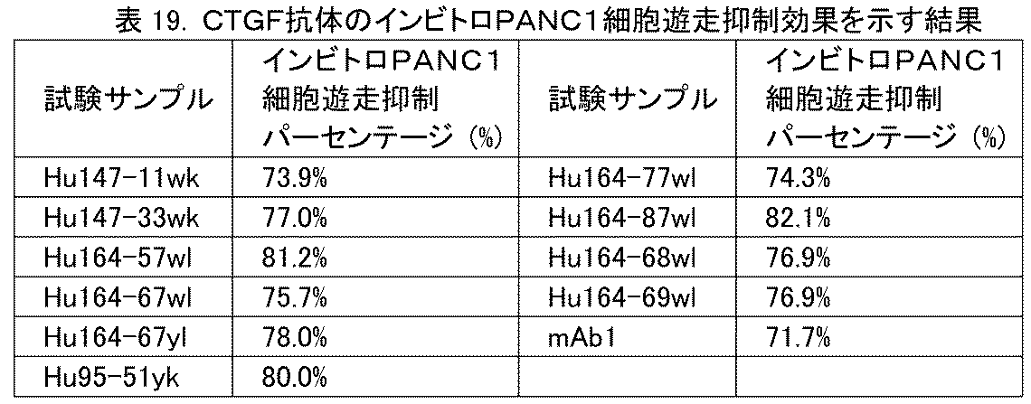
結果は、本開示のヒト化抗CTGF抗体がいずれも、インビトロでTGFβ1により誘導されるPANC1細胞の遊走能力を抑制できることを示している。 The results show that all of the humanized anti-CTGF antibodies disclosed herein can inhibit the migration ability of PANC1 cells induced by TGFβ1 in vitro.
実施例7:インビトロ軟寒天上のPANC1増殖に対する、CTGF抗体による抑制効果のアッセイ
寒天(BD, #214220)に脱イオン水を加えて加熱して、1.4%ゲル溶液を調製した。2×DMEM培地(Thermo, #12100046)をゲル溶液に1:1の体積比で混合し、500μlを24ウェルプレート(Costar, #3524)の各ウェルに入れ、4度に置いて凝固させた。
Example 7: Assay of the inhibitory effect of CTGF antibody on PANC1 proliferation on in vitro soft agar A 1.4% gel solution was prepared by adding deionized water to agar (BD, #214220) and heating it. 2xDMEM medium (Thermo, #12100046) was mixed with the gel solution at a volume ratio of 1:1, and 500 μl was placed in each well of a 24-well plate (Costar, #3524) and placed at 4 degrees to solidify.
PANC-1細胞(ヒト膵臓がん細胞、ATCC, #CRL-1469)を0.25%トリプシン(Gibco, #25200-072)で消化し、遠心分離し、4%ウシ胎児血清(Gibco, #10099-141)を含むDMEM培地(GE, #SH30243.01)で細胞密度5×10⁴細胞/mlに調節した。2×DMEM培地で2mg/mlの抗体を調製した。細胞、抗体および1.4%ゲル溶液を2:1:1の体積比で混合し、200μlを、下方のゲル層で覆われた24ウェルプレートの各ウェルに入れた。上方のゲルが凝固した後、200μlの2%ウシ胎児血清含有DMEM培地を前記24ウェルプレートに加えた。インキュベーター(37℃、5%CO2)内でのインキュベーション後28日で、ハイコンテント分析システム(Molecular Devices, ImageXpress)を使用して画像化し、生成した細胞クローンの領域を分析した。抑制率=(1-サンプル群の平均値/未処置群の平均値)×100%。実験結果を表20に示す。 PANC-1 cells (human pancreatic cancer cells, ATCC, #CRL-1469) were digested with 0.25% trypsin (Gibco, #25200-072), centrifuged, and adjusted to a cell density of 5x10⁴ cells/ml with DMEM medium (GE, #SH30243.01) containing 4% fetal bovine serum (Gibco, #10099-141). 2mg/ml antibody was prepared in 2xDMEM medium. Cells, antibody and 1.4% gel solution were mixed in a 2:1:1 volume ratio, and 200μl was added to each well of a 24-well plate covered with the lower gel layer. After the upper gel solidified, 200μl of DMEM medium containing 2% fetal bovine serum was added to the 24-well plate. After 28 days of incubation in an incubator (37°C, 5% CO2 ), the cells were imaged using a high content analysis system (Molecular Devices, ImageXpress) and the area of the generated cell clones was analyzed. Inhibition rate = (1 - mean value of sample group / mean value of untreated group) x 100%. The experimental results are shown in Table 20.

結果は、mAb147およびmAb164クローンから誘導されたヒト化抗体が、インビトロでヒト膵臓がん細胞(PANC-1細胞)増殖に対する顕著な抑制作用を有することを示している。 The results show that humanized antibodies derived from the mAb147 and mAb164 clones have a significant inhibitory effect on human pancreatic cancer cell (PANC-1 cell) proliferation in vitro.
実施例8:動物におけるCTGF抗体のインビボ生物学的活性
I.ブレオマイシンにより誘発されるマウス肺線維症の、CTGF抗体による抑制効果のアッセイ
実験プロトコル:40匹のマウスを体重にしたがいランダムに次の4群に分けた:正常対照群(PBS、i.p.、qod)、mAb1群(10mg/kg、i.p.、qod)、Hu164-67wl群(10mg/kg、i.p.、qod)、およびHu164-67yl群(10mg/kg、i.p.、qod)。各群10匹で、群分けの詳細は下記の表21に示す。実験の1日目に、溶媒および抗体のそれぞれを、体重にしたがって腹腔内注射し(10ml/kg;正常対照およびモデル群にはPBSを投与した);2時間後にエアロゾルブレオマイシン(50mg/8mlの噴霧を30分間、および保持を5分間)を用いてモデルを構築した。次いで、上記群に応じて溶媒または抗体を1日置きに腹腔内注射した。実験期間は21日間で、全部で11回の腹腔内投与を行った。実験の22日目にマウスを1%ペントバルビタールナトリウム(10ml/kg)の腹腔内注射によって麻酔し、手術台上に仰臥位に固定した。頸の正中線に沿って1cmの皮膚切開を行い、切開の下端は胸の入口に至らしめた。鉗子で皮下結合組織および筋肉を鈍的分離して、気管を露出した。気管の両端上、および気管と食道との間の結合組織を分離して、気管を離した。静脈留置針を挿入し、カニューレが気管に入る場所で繋いで縫合糸で固定した。0.8mlのPBSを充填した1mlシリンジを静脈留置針の入口端に連結し、PBSをゆっくりと気管に注入し、30秒間滞留させ、その後PBSをゆっくりと引き戻した。引き戻しの際、乳状泡状液体を観察することができた。洗浄を3回繰り返し、洗浄液をEPチューブに移し、その後0.5mlのPBSを取り、前記ステップを繰り返した。2つの洗浄液を混合し、1500rpmで5分間遠心分離し、上清を試験用に-20℃で保存した。BALF上清を再度10000rpmで10分間遠心分離し、上清を取って可溶性コラーゲンを検出した(キット:Biocolor, Batch No. AA883)。
Example 8: In vivo biological activity of CTGF antibody in animals I. Assay of the inhibitory effect of CTGF antibody on bleomycin-induced pulmonary fibrosis in mice Experimental protocol: 40 mice were randomly divided into four groups according to body weight: normal control group (PBS, i.p., qod), mAb1 group (10 mg/kg, i.p., qod), Hu164-67wl group (10 mg/kg, i.p., qod), and Hu164-67yl group (10 mg/kg, i.p., qod). There were 10 mice in each group, and the grouping details are shown in Table 21 below. On the first day of the experiment, the vehicle and antibody were each injected intraperitoneally according to body weight (10 ml/kg; normal control and model groups were administered PBS); 2 hours later, the model was established using aerosol bleomycin (50 mg/8 ml nebulization for 30 minutes and retention for 5 minutes). The vehicle or antibody was then injected intraperitoneally every other day according to the group. The experimental period was 21 days, with a total of 11 intraperitoneal injections. On the 22nd day of the experiment, the mice were anesthetized by intraperitoneal injection of 1% sodium pentobarbital (10 ml/kg) and fixed in a supine position on the operating table. A 1 cm skin incision was made along the midline of the neck, with the lower end of the incision reaching the inlet of the chest. The subcutaneous connective tissue and muscle were bluntly separated with forceps to expose the trachea. The connective tissue on both ends of the trachea and between the trachea and the esophagus was separated to free the trachea. The intravenous needle was inserted and tied where the cannula entered the trachea and secured with sutures. A 1 ml syringe filled with 0.8 ml of PBS was connected to the inlet end of the intravenous needle, and the PBS was slowly injected into the trachea and allowed to dwell for 30 seconds, after which the PBS was slowly withdrawn. Upon withdrawal, milky foamy liquid could be observed. The washing was repeated three times, the washing liquid was transferred to an EP tube, then 0.5ml of PBS was taken and the above steps were repeated. The two washing liquids were mixed and centrifuged at 1500 rpm for 5 minutes, and the supernatant was stored at -20°C for testing. The BALF supernatant was centrifuged again at 10000 rpm for 10 minutes, and the supernatant was taken to detect soluble collagen (Kit: Biocolor, Batch No. AA883).
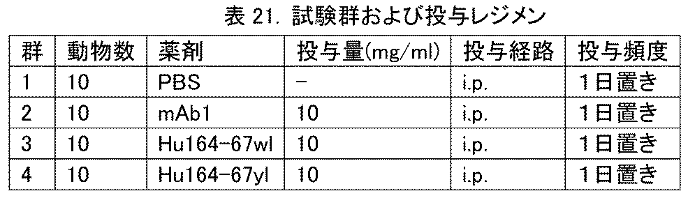
Excel統計ソフトウェアを使用した:平均値をavg(平均)として計算した;SD(標準偏差)値をSTDEV(標準偏差)として計算した;SEM(平均の標準誤差;標本平均の標準偏差)値をSTDEV/SQRT(平方根)(各群の動物数)として計算した;プロッティングにはGraphPad Prismソフトウェアを使用し、データの統計学的分析には二元配置ANOVA(二元配置分散分析)または一元配置ANOVA(一元配置分散分析)を用いた。 Excel statistical software was used: mean values were calculated as avg (average); SD (standard deviation) values were calculated as STDEV (standard deviation); SEM (standard error of the mean; standard deviation of the sample mean) values were calculated as STDEV/SQRT (square root) (number of animals in each group); GraphPad Prism software was used for plotting and two-way ANOVA (two-way analysis of variance) or one-way ANOVA (one-way analysis of variance) were used for statistical analysis of the data.
実験結果を表22に示す。

結果は、Hu164-67wlまたはHu164-67ylが、マウスの肺線維症に対して強い抑制効果を示すことを示している。 The results show that Hu164-67wl or Hu164-67yl exhibits strong inhibitory effects on pulmonary fibrosis in mice.
II.Su86.86ヒト膵臓がん細胞の皮下移植腫瘍の、CTGF抗体による抑制のアッセイ
実験プロトコル:200μlのSU86.86細胞(ATCC, CRL-1837, 3.0×10⁶細胞)をNu/Nuヌードマウスの右脇腹に皮下接種した。腫瘍体積が約140mm3となった接種後11日に、体重または腫瘍体積が大きすぎるまたは小さすぎるマウスを除外した。マウスを腫瘍体積にしたがってランダムに、各群10匹の5群に分け、同日に投与を開始した。抗体を、全部で18日間にわたり週に2回、腹腔内注射した。腫瘍体積および体重を週に1~2回測定し、データを記録した。群分けおよび投与レジメンを表23に示す。
II. Assay of CTGF antibody inhibition of subcutaneously implanted tumors of Su86.86 human pancreatic cancer cells Experimental protocol: 200 μl of SU86.86 cells (ATCC, CRL-1837, 3.0×10 6 cells) were subcutaneously inoculated into the right flank of Nu/Nu nude mice. Mice with too large or small body weight or tumor volume were excluded 11 days after inoculation, when the tumor volume reached approximately 140 mm 3. Mice were randomly divided into 5 groups of 10 mice per group according to tumor volume, and treatment was started on the same day. Antibodies were injected intraperitoneally twice a week for a total of 18 days. Tumor volume and body weight were measured once or twice a week, and the data were recorded. Grouping and treatment regimen are shown in Table 23.
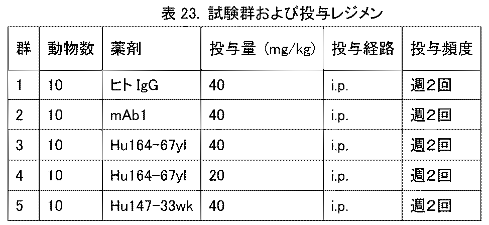
Excel統計ソフトウェアを使用した:平均値をavgとして計算した;SD値をSTDEVとして計算した;SEM値をSTDEV/SQRT(各群の動物数)として計算した;プロッティングにはGraphPad Prismソフトウェアを使用し、データの統計学的分析には二元配置ANOVAまたは一元配置ANOVAを用いた。 Excel statistical software was used: mean values were calculated as avg; SD values were calculated as STDEV; SEM values were calculated as STDEV/SQRT (number of animals in each group); GraphPad Prism software was used for plotting and two-way or one-way ANOVA was used for statistical analysis of data.
次式にしたがって腫瘍体積(V)を計算した:
V=1/2×Llong×Lshort
2
Tumor volume (V) was calculated according to the following formula:
V=1/2×L long ×L short 2
相対腫瘍増殖率T/C(%)=(Tt-T0)/(Ct-C0)×100
ここで、TtおよびCtは、実験終了時の処置群および対照群の腫瘍体積であり;T0およびC0は、実験開始時の腫瘍体積である。
Relative tumor growth rate T/C (%) = (T t - T 0 )/(C t - C 0 ) x 100
where Tt and Ct are the tumor volumes of the treated and control groups at the end of the experiment; T0 and C0 are the tumor volumes at the start of the experiment.
腫瘍抑制率TGI(%)=1-T/C(%) Tumor suppression rate TGI (%) = 1-T/C (%)
実験結果:
D1からD18まで、週に2回皮下注射を行い、SU86.86腫瘍増殖抑制についてCTGF抗体の効果を検出した。実験結果を表24および図2に示す。同じアイソタイプヒトIgGのブランク対照群と比較して、mAb1-40mg/kg、Hu164-67yl-40mg/kg、Hu164-67yl-20mg/kgおよびHu147-33wk(40mg/kg)群の腫瘍抑制率はそれぞれ、45%、53%、47%および54%であった。mAb1-40mg/kg、Hu164-67yl-40mg/kg、Hu164-67yl-20mg/kgおよびHu147-33wk(40mg/kg)群はSu86.86腫瘍の増殖を顕著に抑制することができ(p<0.001)、SU86.86腫瘍に対する抑制効果は用量依存的である。さらに、各群のマウスの体重に顕著な変化は無かった。
Experimental results:
Subcutaneous injection was performed twice a week from D1 to D18 to detect the effect of CTGF antibody on SU86.86 tumor growth inhibition. The experimental results are shown in Table 24 and Figure 2. Compared with the blank control group of the same isotype human IgG, the tumor inhibition rates of mAb1-40mg/kg, Hu164-67yl-40mg/kg, Hu164-67yl-20mg/kg and Hu147-33wk (40mg/kg) groups were 45%, 53%, 47% and 54%, respectively. mAb1-40mg/kg, Hu164-67yl-40mg/kg, Hu164-67yl-20mg/kg and Hu147-33wk (40mg/kg) groups could significantly inhibit the growth of Su86.86 tumor (p<0.001), and the inhibitory effect on SU86.86 tumor was dose-dependent. Moreover, there was no significant change in the body weight of mice in each group.

Claims (15)
ここで、
軽鎖可変領域は、配列番号19、配列番号20および配列番号21にそれぞれ示されるLCDR1、LCDR2およびLCDR3、および36V、44F、46Gおよび49Gからなる群から選択される1つ以上の復帰変異を含み、
重鎖可変領域は、配列番号16、配列番号17および配列番号18にそれぞれ示されるHCDR1、HCDR2およびHCDR3、および44G、49G、27F、48L、67L、71K、78Vおよび80Fからなる群から選択される1つ以上の復帰変異を含み、
軽鎖可変領域および重鎖可変領域中の前記復帰変異位置は、Kabatナンバリング基準にしたがって決定したものである、請求項1~3のいずれかに記載の抗CTGF抗体。 the anti-CTGF antibody is a humanized antibody, the humanized antibody comprises a framework region of a human antibody or a variant of that framework region, the variant having up to 10 back mutations in each of the human antibody light chain framework region and/or heavy chain framework region;
Where:
the light chain variable region comprises LCDR1, LCDR2 and LCDR3 as set forth in SEQ ID NO:19, SEQ ID NO:20 and SEQ ID NO:21, respectively, and one or more backmutations selected from the group consisting of 36V, 44F, 46G and 49G;
the heavy chain variable region comprises HCDR1, HCDR2 and HCDR3 as set forth in SEQ ID NO:16, SEQ ID NO:17 and SEQ ID NO:18, respectively, and one or more back mutations selected from the group consisting of 44G, 49G, 27F, 48L, 67L, 71K, 78V and 80F;
The anti-CTGF antibody according to any one of claims 1 to 3, wherein the backmutation positions in the light chain variable region and the heavy chain variable region are determined according to the Kabat numbering standard.
(x)重鎖可変領域のアミノ酸配列が配列番号33、34、35または36に示され、軽鎖可変領域のアミノ酸配列が配列番号30、31または32に示される、
重鎖可変領域および軽鎖可変領域を含む、請求項1~3のいずれかに記載の抗CTGF抗体。 (ix) the amino acid sequence of the heavy chain variable region is set forth in SEQ ID NO:8 and the amino acid sequence of the light chain variable region is set forth in SEQ ID NO:9; or (x) the amino acid sequence of the heavy chain variable region is set forth in SEQ ID NO:33, 34, 35 or 36 and the amino acid sequence of the light chain variable region is set forth in SEQ ID NO:30, 31 or 32;
The anti-CTGF antibody according to any one of claims 1 to 3 , comprising a heavy chain variable region and a light chain variable region.
重鎖可変領域および軽鎖可変領域を含む、請求項4に記載の抗CTGF抗体。 (xiv) the amino acid sequence of the heavy chain variable region is set forth in SEQ ID NO: 34, and the amino acid sequence of the light chain variable region is set forth in SEQ ID NO: 30;
The anti-CTGF antibody of claim 4 , comprising a heavy chain variable region and a light chain variable region.
前記がんが、急性リンパ芽球性白血病、皮膚線維腫、乳がん、血管脂肪腫、血管平滑筋腫、結合組織生成がん、前立腺がん、卵巣がん、結腸直腸がん、膵臓がん、胃腸がんまたは肝臓がんからなる群から選択される、
請求項14に記載の医薬組成物。 the fibrotic disease is selected from the group consisting of idiopathic pulmonary fibrosis, diabetic nephropathy, diabetic retinopathy, osteoarthritis, scleroderma, chronic heart failure, liver cirrhosis or renal fibrosis;
The cancer is selected from the group consisting of acute lymphoblastic leukemia, dermatofibroma, breast cancer, angiolipoma, angioleiomyoma, connective tissue generating cancer, prostate cancer, ovarian cancer, colorectal cancer, pancreatic cancer, gastrointestinal cancer or liver cancer;
15. The pharmaceutical composition of claim 14.
Applications Claiming Priority (3)
| Application Number | Priority Date | Filing Date | Title |
|---|---|---|---|
| CN201910480169.4 | 2019-06-04 | ||
| CN201910480169 | 2019-06-04 | ||
| PCT/CN2020/094136 WO2020244540A1 (en) | 2019-06-04 | 2020-06-03 | Anti-connective tissue growth factor antibody and application thereof |
Publications (2)
| Publication Number | Publication Date |
|---|---|
| JP2022535810A JP2022535810A (en) | 2022-08-10 |
| JP7627665B2 true JP7627665B2 (en) | 2025-02-06 |
Family
ID=73653057
Family Applications (1)
| Application Number | Title | Priority Date | Filing Date |
|---|---|---|---|
| JP2021571607A Active JP7627665B2 (en) | 2019-06-04 | 2020-06-03 | Anti-connective tissue growth factor antibodies and their applications |
Country Status (13)
| Country | Link |
|---|---|
| US (1) | US20220324953A1 (en) |
| EP (1) | EP3981787A4 (en) |
| JP (1) | JP7627665B2 (en) |
| KR (1) | KR20220016890A (en) |
| CN (2) | CN115925926A (en) |
| AU (1) | AU2020289303A1 (en) |
| BR (1) | BR112021023669A2 (en) |
| CA (1) | CA3142092A1 (en) |
| MX (1) | MX2021014803A (en) |
| MY (1) | MY208454A (en) |
| TW (1) | TW202110880A (en) |
| WO (1) | WO2020244540A1 (en) |
| ZA (1) | ZA202109744B (en) |
Families Citing this family (4)
| Publication number | Priority date | Publication date | Assignee | Title |
|---|---|---|---|---|
| US20230416352A1 (en) * | 2020-12-03 | 2023-12-28 | Jiangsu Hengrui Pharmaceuticals Co., Ltd. | Pharmaceutical composition comprising anti-connective tissue growth factor antibody |
| CN115645525B (en) * | 2022-12-14 | 2023-04-14 | 北京大学第三医院(北京大学第三临床医学院) | Application of CTGF (cytotoxic T-lymphocyte) neutralizing antibody in preparation of medicine for preventing and treating postmenopausal osteoarthritis |
| CN120858112A (en) * | 2023-03-10 | 2025-10-28 | 江西康莱特新森医药原料有限公司 | An antibody targeting CTGF or its antigen-binding fragment and its application |
| WO2025181215A1 (en) * | 2024-03-01 | 2025-09-04 | Boehringer Ingelheim International Gmbh | Antibodies to connective tissue growth factor (ctgf) and uses thereof |
Citations (1)
| Publication number | Priority date | Publication date | Assignee | Title |
|---|---|---|---|---|
| WO2013094723A1 (en) | 2011-12-22 | 2013-06-27 | アステラス製薬株式会社 | Novel anti-human ctgf antibody |
Family Cites Families (9)
| Publication number | Priority date | Publication date | Assignee | Title |
|---|---|---|---|---|
| US4683195A (en) | 1986-01-30 | 1987-07-28 | Cetus Corporation | Process for amplifying, detecting, and/or-cloning nucleic acid sequences |
| US5770209A (en) | 1991-08-30 | 1998-06-23 | University Of South Florida | Acceleration of wound healing using connective tissue growth factor |
| US5408040A (en) | 1991-08-30 | 1995-04-18 | University Of South Florida | Connective tissue growth factor(CTGF) |
| AU1682699A (en) * | 1997-12-25 | 1999-07-19 | Japan Tobacco Inc. | Monoclonal antibody against connective tissue growth factor and medicinal uses thereof |
| JP4537507B2 (en) * | 1997-12-25 | 2010-09-01 | アムジェン インコーポレイテッド | Monoclonal antibody against connective tissue growth factor and pharmaceutical use thereof |
| HK1038318A1 (en) | 1998-09-08 | 2002-03-15 | Henry Ford Health System | Method for detecting, preventing, and treating renal disorders by modulating, regulating, and inhibiting connective tissue growth factor |
| US7405274B2 (en) * | 2003-06-04 | 2008-07-29 | Fibrogen, Inc. | Connective tissue growth factor antibodies |
| CN101884789A (en) * | 2004-02-11 | 2010-11-17 | 法布罗根股份有限公司 | CTGF is as the treatment target spot of diabetic nephropathy |
| JP2009067678A (en) * | 2005-12-07 | 2009-04-02 | Nihon Nosan Kogyo Kk | Antibody against connective tissue growth factor or composition containing the same |
-
2020
- 2020-06-03 CN CN202211129173.4A patent/CN115925926A/en active Pending
- 2020-06-03 MY MYPI2021007035A patent/MY208454A/en unknown
- 2020-06-03 MX MX2021014803A patent/MX2021014803A/en unknown
- 2020-06-03 KR KR1020217042214A patent/KR20220016890A/en active Pending
- 2020-06-03 US US17/615,801 patent/US20220324953A1/en not_active Abandoned
- 2020-06-03 CN CN202080036744.2A patent/CN113840836B/en active Active
- 2020-06-03 JP JP2021571607A patent/JP7627665B2/en active Active
- 2020-06-03 WO PCT/CN2020/094136 patent/WO2020244540A1/en not_active Ceased
- 2020-06-03 CA CA3142092A patent/CA3142092A1/en active Pending
- 2020-06-03 TW TW109118625A patent/TW202110880A/en unknown
- 2020-06-03 BR BR112021023669A patent/BR112021023669A2/en unknown
- 2020-06-03 EP EP20818627.0A patent/EP3981787A4/en active Pending
- 2020-06-03 AU AU2020289303A patent/AU2020289303A1/en not_active Abandoned
-
2021
- 2021-11-29 ZA ZA2021/09744A patent/ZA202109744B/en unknown
Patent Citations (1)
| Publication number | Priority date | Publication date | Assignee | Title |
|---|---|---|---|---|
| WO2013094723A1 (en) | 2011-12-22 | 2013-06-27 | アステラス製薬株式会社 | Novel anti-human ctgf antibody |
Non-Patent Citations (1)
| Title |
|---|
| Aikawa, T. et al.,"Connective tissue growth factor-specific antibody attenuates tumor growth, metastasis, and angiogenesis in an orthotopic mouse model of pancreatic cancer",Mol. Cancer Ther.,2006年,Vol. 5,pp. 1108-1116 |
Also Published As
| Publication number | Publication date |
|---|---|
| CN113840836B (en) | 2022-10-21 |
| CN113840836A (en) | 2021-12-24 |
| MX2021014803A (en) | 2022-01-18 |
| US20220324953A1 (en) | 2022-10-13 |
| JP2022535810A (en) | 2022-08-10 |
| KR20220016890A (en) | 2022-02-10 |
| CA3142092A1 (en) | 2020-12-10 |
| MY208454A (en) | 2025-05-09 |
| WO2020244540A1 (en) | 2020-12-10 |
| ZA202109744B (en) | 2023-05-31 |
| BR112021023669A2 (en) | 2022-03-29 |
| EP3981787A1 (en) | 2022-04-13 |
| TW202110880A (en) | 2021-03-16 |
| CN115925926A (en) | 2023-04-07 |
| AU2020289303A1 (en) | 2022-01-27 |
| EP3981787A4 (en) | 2022-09-14 |
Similar Documents
| Publication | Publication Date | Title |
|---|---|---|
| US11512129B2 (en) | TIGIT antibody, antigen-binding fragment thereof, and medical use thereof | |
| US20220220218A1 (en) | Anti-cd73 antibody, antigen-binding fragment thereof and application thereof | |
| US12428482B2 (en) | Anti-PD-1 antibody, antigen-binding fragment thereof and pharmaceutical use thereof | |
| CN112955473B (en) | Bispecific antibody that specifically binds VEGF and ANG2 | |
| JP7627665B2 (en) | Anti-connective tissue growth factor antibodies and their applications | |
| TWI861347B (en) | Anti-angptl3 antibody and usage thereof | |
| CN110790839A (en) | Anti-PD-1 antibody, its antigen-binding fragment and medical use | |
| TW201725216A (en) | PCSK9 antibody, antigen-binding fragment thereof and medical use thereof | |
| CN112996811B (en) | Bispecific proteins | |
| RU2819228C2 (en) | Connective tissue growth factor antibody and use thereof | |
| RU2819228C9 (en) | Connective tissue growth factor antibody and use thereof | |
| RU2807484C2 (en) | Antibody to pd-1, its antigen-binding fragment and its pharmaceutical use | |
| HK40064840B (en) | Anti-connective tissue growth factor antibody and application thereof | |
| HK40064840A (en) | Anti-connective tissue growth factor antibody and application thereof | |
| HK40051455B (en) | Anti-pd-1 antibody, antigen-binding fragment thereof and pharmaceutical use thereof |
Legal Events
| Date | Code | Title | Description |
|---|---|---|---|
| A621 | Written request for application examination |
Free format text: JAPANESE INTERMEDIATE CODE: A621 Effective date: 20230530 |
|
| A977 | Report on retrieval |
Free format text: JAPANESE INTERMEDIATE CODE: A971007 Effective date: 20240524 |
|
| A131 | Notification of reasons for refusal |
Free format text: JAPANESE INTERMEDIATE CODE: A131 Effective date: 20240528 |
|
| A521 | Request for written amendment filed |
Free format text: JAPANESE INTERMEDIATE CODE: A523 Effective date: 20240828 |
|
| A131 | Notification of reasons for refusal |
Free format text: JAPANESE INTERMEDIATE CODE: A131 Effective date: 20240910 |
|
| A521 | Request for written amendment filed |
Free format text: JAPANESE INTERMEDIATE CODE: A523 Effective date: 20241206 |
|
| TRDD | Decision of grant or rejection written | ||
| A01 | Written decision to grant a patent or to grant a registration (utility model) |
Free format text: JAPANESE INTERMEDIATE CODE: A01 Effective date: 20250114 |
|
| A61 | First payment of annual fees (during grant procedure) |
Free format text: JAPANESE INTERMEDIATE CODE: A61 Effective date: 20250127 |
|
| R150 | Certificate of patent or registration of utility model |
Ref document number: 7627665 Country of ref document: JP Free format text: JAPANESE INTERMEDIATE CODE: R150 |