JP7436075B2 - Structure-based design of therapeutic agents targeting RNA hairpin loops - Google Patents
Structure-based design of therapeutic agents targeting RNA hairpin loops Download PDFInfo
- Publication number
- JP7436075B2 JP7436075B2 JP2022528619A JP2022528619A JP7436075B2 JP 7436075 B2 JP7436075 B2 JP 7436075B2 JP 2022528619 A JP2022528619 A JP 2022528619A JP 2022528619 A JP2022528619 A JP 2022528619A JP 7436075 B2 JP7436075 B2 JP 7436075B2
- Authority
- JP
- Japan
- Prior art keywords
- polynucleotide
- mirna
- pri
- loop
- ribonucleic acid
- Prior art date
- Legal status (The legal status is an assumption and is not a legal conclusion. Google has not performed a legal analysis and makes no representation as to the accuracy of the status listed.)
- Active
Links
Images


Classifications
-
- C—CHEMISTRY; METALLURGY
- C12—BIOCHEMISTRY; BEER; SPIRITS; WINE; VINEGAR; MICROBIOLOGY; ENZYMOLOGY; MUTATION OR GENETIC ENGINEERING
- C12Q—MEASURING OR TESTING PROCESSES INVOLVING ENZYMES, NUCLEIC ACIDS OR MICROORGANISMS; COMPOSITIONS OR TEST PAPERS THEREFOR; PROCESSES OF PREPARING SUCH COMPOSITIONS; CONDITION-RESPONSIVE CONTROL IN MICROBIOLOGICAL OR ENZYMOLOGICAL PROCESSES
- C12Q1/00—Measuring or testing processes involving enzymes, nucleic acids or microorganisms; Compositions therefor; Processes of preparing such compositions
- C12Q1/68—Measuring or testing processes involving enzymes, nucleic acids or microorganisms; Compositions therefor; Processes of preparing such compositions involving nucleic acids
- C12Q1/6876—Nucleic acid products used in the analysis of nucleic acids, e.g. primers or probes
-
- C—CHEMISTRY; METALLURGY
- C12—BIOCHEMISTRY; BEER; SPIRITS; WINE; VINEGAR; MICROBIOLOGY; ENZYMOLOGY; MUTATION OR GENETIC ENGINEERING
- C12N—MICROORGANISMS OR ENZYMES; COMPOSITIONS THEREOF; PROPAGATING, PRESERVING, OR MAINTAINING MICROORGANISMS; MUTATION OR GENETIC ENGINEERING; CULTURE MEDIA
- C12N15/00—Mutation or genetic engineering; DNA or RNA concerning genetic engineering, vectors, e.g. plasmids, or their isolation, preparation or purification; Use of hosts therefor
- C12N15/09—Recombinant DNA-technology
- C12N15/10—Processes for the isolation, preparation or purification of DNA or RNA
- C12N15/1034—Isolating an individual clone by screening libraries
- C12N15/1044—Preparation or screening of libraries displayed on scaffold proteins
-
- C—CHEMISTRY; METALLURGY
- C12—BIOCHEMISTRY; BEER; SPIRITS; WINE; VINEGAR; MICROBIOLOGY; ENZYMOLOGY; MUTATION OR GENETIC ENGINEERING
- C12N—MICROORGANISMS OR ENZYMES; COMPOSITIONS THEREOF; PROPAGATING, PRESERVING, OR MAINTAINING MICROORGANISMS; MUTATION OR GENETIC ENGINEERING; CULTURE MEDIA
- C12N15/00—Mutation or genetic engineering; DNA or RNA concerning genetic engineering, vectors, e.g. plasmids, or their isolation, preparation or purification; Use of hosts therefor
- C12N15/09—Recombinant DNA-technology
- C12N15/11—DNA or RNA fragments; Modified forms thereof; Non-coding nucleic acids having a biological activity
- C12N15/111—General methods applicable to biologically active non-coding nucleic acids
-
- C—CHEMISTRY; METALLURGY
- C12—BIOCHEMISTRY; BEER; SPIRITS; WINE; VINEGAR; MICROBIOLOGY; ENZYMOLOGY; MUTATION OR GENETIC ENGINEERING
- C12N—MICROORGANISMS OR ENZYMES; COMPOSITIONS THEREOF; PROPAGATING, PRESERVING, OR MAINTAINING MICROORGANISMS; MUTATION OR GENETIC ENGINEERING; CULTURE MEDIA
- C12N2310/00—Structure or type of the nucleic acid
- C12N2310/10—Type of nucleic acid
- C12N2310/14—Type of nucleic acid interfering nucleic acids [NA]
- C12N2310/141—MicroRNAs, miRNAs
-
- C—CHEMISTRY; METALLURGY
- C12—BIOCHEMISTRY; BEER; SPIRITS; WINE; VINEGAR; MICROBIOLOGY; ENZYMOLOGY; MUTATION OR GENETIC ENGINEERING
- C12N—MICROORGANISMS OR ENZYMES; COMPOSITIONS THEREOF; PROPAGATING, PRESERVING, OR MAINTAINING MICROORGANISMS; MUTATION OR GENETIC ENGINEERING; CULTURE MEDIA
- C12N2310/00—Structure or type of the nucleic acid
- C12N2310/30—Chemical structure
- C12N2310/35—Nature of the modification
- C12N2310/351—Conjugate
- C12N2310/3519—Fusion with another nucleic acid
-
- C—CHEMISTRY; METALLURGY
- C12—BIOCHEMISTRY; BEER; SPIRITS; WINE; VINEGAR; MICROBIOLOGY; ENZYMOLOGY; MUTATION OR GENETIC ENGINEERING
- C12N—MICROORGANISMS OR ENZYMES; COMPOSITIONS THEREOF; PROPAGATING, PRESERVING, OR MAINTAINING MICROORGANISMS; MUTATION OR GENETIC ENGINEERING; CULTURE MEDIA
- C12N2320/00—Applications; Uses
- C12N2320/10—Applications; Uses in screening processes
Landscapes
- Life Sciences & Earth Sciences (AREA)
- Health & Medical Sciences (AREA)
- Engineering & Computer Science (AREA)
- Chemical & Material Sciences (AREA)
- Genetics & Genomics (AREA)
- Organic Chemistry (AREA)
- Biomedical Technology (AREA)
- Zoology (AREA)
- Wood Science & Technology (AREA)
- Bioinformatics & Cheminformatics (AREA)
- Biotechnology (AREA)
- General Engineering & Computer Science (AREA)
- Molecular Biology (AREA)
- General Health & Medical Sciences (AREA)
- Microbiology (AREA)
- Physics & Mathematics (AREA)
- Biochemistry (AREA)
- Biophysics (AREA)
- Proteomics, Peptides & Aminoacids (AREA)
- Plant Pathology (AREA)
- Analytical Chemistry (AREA)
- Immunology (AREA)
- Crystallography & Structural Chemistry (AREA)
- Bioinformatics & Computational Biology (AREA)
- Measuring Or Testing Involving Enzymes Or Micro-Organisms (AREA)
- Enzymes And Modification Thereof (AREA)
- Pharmaceuticals Containing Other Organic And Inorganic Compounds (AREA)
Description
関連出願の相互参照
本出願は、米国特許法第119条(e)項の下で、2019年11月19日出願の同時係属中の共有に係る米国仮特許出願第62/937,657号(発明の名称「STRUCTURE-BASED DESIGN OF THERAPEUTICS TARGETING RNA HAIRPIN LOOPS」)に基づく利益を主張している。この出願は、本明細書に参考として援用される。
CROSS-REFERENCE TO RELATED APPLICATIONS This application is filed under 35 U.S.C. Claims benefit based on the title of the invention "STRUCTURE-BASED DESIGN OF THERAPEUTICS TARGETING RNA HAIRPIN LOOPS"). This application is incorporated herein by reference.
政府支援の陳述
本発明は、National Science Foundationによって授与された助成金番号1616265の下で政府支援を得て行われた。政府は、本発明において一定の権利を有する。
STATEMENT OF GOVERNMENT SUPPORT This invention was made with government support under Grant No. 1616265 awarded by the National Science Foundation. The Government has certain rights in this invention.
技術分野
本発明は、RNAヘアピンループの三次元構造を決定するために有用な方法および材料に関する。
TECHNICAL FIELD The present invention relates to methods and materials useful for determining the three-dimensional structure of RNA hairpin loops.
発明の背景
RNA分子は、多くの疾患(例えば、がんおよびRNAウイルス感染症)の発生にとって極めて重要である。この理由から、RNA分子は、優れた治療標的である。この文脈において、ほぼ全てのRNAは、それらの機能にとって極めて重要なヘアピン二次構造を形成する。結論として、これらの構造の理解は、これらの分子を標的にする治療剤の同定および設計を促進するために必要である。しかし、RNAを調べる従来の方法(例えば、RNA干渉およびアンチセンスオリゴヌクレオチド)は、制限されており、強い構造を回避する。従来の技術は、RNA構造に関するある種の情報を提供し得るが、これらの技術における制限から、RNAヘアピンループは、治療用インヒビター設計の正当に評価されない標的になっている。
BACKGROUND OF THE INVENTION RNA molecules are critical to the development of many diseases, such as cancer and RNA virus infections. For this reason, RNA molecules are excellent therapeutic targets. In this context, almost all RNAs form hairpin secondary structures that are crucial for their function. In conclusion, an understanding of these structures is necessary to facilitate the identification and design of therapeutic agents that target these molecules. However, traditional methods of interrogating RNA (eg, RNA interference and antisense oligonucleotides) are limited and avoid strong structures. Although conventional techniques can provide some information about RNA structure, limitations in these techniques make RNA hairpin loops an underappreciated target for therapeutic inhibitor design.
RNAヘアピンループの三次元構造に関する情報を得るために有用な新たな方法および材料のための技術の強いニーズが、当該分野に存在する。 There is a strong need in the art for new methods and materials useful for obtaining information about the three-dimensional structure of RNA hairpin loops.
発明の要旨
以下で詳細に記載されるように、本発明者らは、RNAヘアピンループの三次元構造に関する情報を得るために有用な新規な足場指向性結晶学方法を開発した。本明細書で開示されるRNA結晶学足場および関連方法は、RNAヘアピンループの三次元構造、他の薬剤(例えば、阻害剤)とのそれらの関連も同様に、容易にかつ迅速に決定するために使用され得る。本発明の方法において使用される特異的足場RNAは、Thermoanaerobacter pseudethanolicusに由来するYdaOタイプc-ジ-AMPリボスイッチ(直径60Åを超える大きな空洞を有する結晶を容易に形成することが発見されたRNA)である。以下で詳細に考察されるように、本発明者らは、上記ヘアピンが上記空洞の中に収容されるように、目的のRNAがこの足場RNAのP2ステムへと操作され得ることを決定した。次いで、その得られる融合RNAは、足場単独を結晶化するためのものに類似のまたは関連しないかいずれかの条件下で結晶化され得る。次いで、このような分子(例えば、これらの分子単独および/または他の薬剤と会合して)の三次元構造は、X線または電子結晶学の技術等を使用して決定され得る。
SUMMARY OF THE INVENTION As described in detail below, the inventors have developed a novel scaffold-directed crystallography method useful for obtaining information about the three-dimensional structure of RNA hairpin loops. The RNA crystallography scaffolds and related methods disclosed herein easily and rapidly determine the three-dimensional structure of RNA hairpin loops, as well as their association with other agents (e.g., inhibitors). can be used for. The specific scaffold RNA used in the method of the invention is a YdaO type c-di-AMP riboswitch derived from Thermoanaerobacter pseudothanolicus (an RNA that was discovered to readily form crystals with large cavities greater than 60 Å in diameter). It is. As discussed in detail below, we determined that the RNA of interest can be engineered into the P2 stem of this scaffold RNA such that the hairpin is accommodated within the cavity. The resulting fusion RNA can then be crystallized under conditions either similar or unrelated to those for crystallizing the scaffold alone. The three-dimensional structure of such molecules (eg, alone and/or in association with other agents) can then be determined using techniques such as X-ray or electron crystallography.
本明細書で開示されるRNA結晶学足場および関連方法は、標的RNA分子と高い親和性および特異性で相互作用する、天然のおよび化学的に改変されたオリゴヌクレオチド、ならびに低分子薬物のような化合物を同定するために使用され得る。これは、このような化合物とRNAヘアピンループとの間の相互作用が、がんおよびRNAウイルス感染症のような病理において、インビボでそれらの活性を調節し得る様式で、これらの分子の生物学的活性に影響を及ぼし得ることから重要である。さらに、RNAは、生物学および疾患のほぼあらゆる局面に関与することから、本明細書で開示される方法は、ほとんど任意の標的RNAを特異的に調節する方法に関する情報を提供し得る広く適用可能な手順である。結論として、本明細書で開示される方法は、特異的RNAを標的にするオリゴヌクレオチドアナログのような薬剤(広く種々の生物学的プロセス(例えば、ウイルス複製(例えば、重症急性呼吸器症候群コロナウイルス2、C型肝炎ウイルスおよびZikaジカウイルスのような病原体の複製)に関与するプロセス、がんまたは神経変性疾患のような病的状態に関与するプロセス、ならびにタンパク質コード遺伝子を調節するためのmicroRNAの生成に関与するプロセスなど)において機能するものが挙げられる)の観察および評価を可能にする。 The RNA crystallography scaffolds and related methods disclosed herein utilize natural and chemically modified oligonucleotides that interact with target RNA molecules with high affinity and specificity, as well as small molecule drugs. Can be used to identify compounds. This reflects the biology of these molecules in such a way that interactions between such compounds and RNA hairpin loops may modulate their activity in vivo in pathologies such as cancer and RNA virus infections. This is important because it can affect clinical activity. Moreover, since RNA is involved in nearly every aspect of biology and disease, the methods disclosed herein are broadly applicable and can provide information on how to specifically modulate almost any target RNA. This is a simple procedure. In conclusion, the methods disclosed herein can be used to target specific RNA-like drugs such as oligonucleotide analogs that can be used to target a wide variety of biological processes (e.g., viral replication (e.g., severe acute respiratory syndrome coronavirus)). 2, processes involved in the replication of pathogens such as hepatitis C virus and Zika virus, processes involved in pathological conditions such as cancer or neurodegenerative diseases, and the use of microRNAs to regulate protein-coding genes. It enables the observation and evaluation of the processes involved in production (including processes involved in production).
本明細書で開示される発明は、多くの実施形態を有する。本発明の1つの実施形態は、GGUUGCCGAAUCCGAAAGGUACGGAGGAACCGCUUUUUGGGGUUAAUCUGCAGUGAAGCUGCAGUAGGGAUACCUUCUGUCCCGCACCCGACAGCUAACUCCGGAGGCAAUAAAGGAAGGAG(配列番号1)と少なくとも90%の配列同一性を有するリボ核酸を含む物質の組成物である。代表的には、上記ポリヌクレオチドは、配列番号1の配列を含む。この組成物において、リボ核酸の配列番号1の残基14~17(GAAA)は、長さが4~33ヌクレオチドの間である核酸の異種セグメントで置き換えられる(上記で注記した少なくとも90%の配列同一性は、残基14~17においてこのリボ核酸に挿入され得る核酸の異種セグメントを含まない)。これらの組成物において、上記核酸の異種セグメントは、代表的には、天然に存在するRNA分子においてループ構造を形成するものである。本発明のある特定の実施形態において、上記核酸の異種セグメントは、天然に存在するRNA分子において完全なループ構造、および必要に応じて、0~5塩基対の間のステム構造を含む。必要に応じてこれらの組成物は、リボ核酸に結合する薬剤、例えば、上記リボ核酸にハイブリダイズするポリヌクレオチドをさらに含み得る。 The invention disclosed herein has many embodiments. One embodiment of the present invention is that GGUUGCCGAAUCCGAAAGGUACGGAGGAACC A composition of matter comprising a ribonucleic acid having at least 90% sequence identity with GAAGGAG (SEQ ID NO: 1). Typically, the polynucleotide comprises the sequence SEQ ID NO:1. In this composition, residues 14-17 (GAAA) of ribonucleic acid SEQ ID NO: 1 are replaced with a heterologous segment of nucleic acid that is between 4 and 33 nucleotides in length (at least 90% of the sequences noted above). Identity does not include heterologous segments of the nucleic acid that may be inserted into this ribonucleic acid at residues 14-17). In these compositions, the heterologous segment of the nucleic acid is typically one that forms a loop structure in a naturally occurring RNA molecule. In certain embodiments of the invention, the heterologous segment of the nucleic acid comprises a complete loop structure in naturally occurring RNA molecules, and optionally a stem structure between 0 and 5 base pairs. Optionally, these compositions can further include agents that bind to ribonucleic acids, such as polynucleotides that hybridize to the ribonucleic acids.
本発明の別の実施形態は、GGUUGCCGAAUCCGAAAGGUACGGAGGAACCGCUUUUUGGGGUUAAUCUGCAGUGAAGCUGCAGUAGGGAUACCUUCUGUCCCGCACCCGACAGCUAACUCCGGAGGCAAUAAAGGAAGGAG(配列番号1)と少なくとも90%(および必要に応じて100%未満)の同一性を有するリボ核酸をコードするDNA配列を含むプラスミドを含むRNA構造を観察するためのシステムまたはキットである。ある特定の実施形態において、上記プラスミドは、上記リボ核酸を発現もしくは転写するためのプロモーターをさらに含む、および/または上記システムもしくはキットは、RNAポリメラーゼをさらに含む。必要に応じて上記システムまたはキットは、上記プラスミド中の核酸のストレッチにハイブリダイズする1またはこれより多くのプライマーをさらに含む。 Another embodiment of the invention is GGUUGCCGAAUCCGAAAGGUACGGAGGAACCGCUUUUUGGGGGUUAAUCUGCAGUGAAGCUGCAGUAGGGAUACCUUCUGUCCCGCACCCGACAGCUAACUCCGGAGGCAAUAAAAG Observe an RNA structure containing a plasmid containing a DNA sequence encoding a ribonucleic acid with at least 90% (and optionally less than 100%) identity to GAAGGAG (SEQ ID NO: 1) A system or kit for In certain embodiments, the plasmid further comprises a promoter for expressing or transcribing the ribonucleic acid, and/or the system or kit further comprises an RNA polymerase. Optionally, the system or kit further comprises one or more primers that hybridize to a stretch of nucleic acid in the plasmid.
本発明のさらに別の実施形態は、リボ核酸の構造に関する情報を得る方法である。この方法は、配列番号1の残基14~17(GAAA)(または配列番号1と少なくとも90%を有するリボ核酸)を、融合リボ核酸分子を形成するように、長さが4~33ヌクレオチドの間の核酸の異種セグメントで置換する工程、上記融合RNAを結晶化する工程、上記融合リボ核酸分子に対してX線または電子結晶学の技術を行う工程、および次いで、その結果(例えば、X線または電子結晶学の技術の電子密度マップ)を観察して、上記核酸の異種セグメントの三次元構造に関する情報を得る工程を包含する。これらの方法のある特定の実施形態において、上記融合リボ核酸分子は、結晶学の分析の前に、上記リボ核酸に結合する薬剤(例えば、上記リボ核酸にハイブリダイズするポリヌクレオチド)と合わされ、その結果、上記RNA/薬剤複合体の構造が観察され得る。代表的には、これらの方法において、上記結晶学の分析は、リボ核酸に結合する薬剤を欠くコントロールサンプルとの比較を包含する。必要に応じてこれらの方法において、複数の融合リボ核酸分子は、X線または電子結晶学の技術の前に、(例えば、ハイスループットスクリーニングにおいて)上記リボ核酸に結合する複数の薬剤と合わされる。本発明のいくつかの実施形態において、少なくとも2つの薬剤が、上記融合リボ核酸分子と合わされる。 Yet another embodiment of the invention is a method of obtaining information regarding the structure of ribonucleic acids. This method combines residues 14-17 (GAAA) of SEQ ID NO: 1 (or a ribonucleic acid having at least 90% with SEQ ID NO: 1) from 4 to 33 nucleotides in length to form a fused ribonucleic acid molecule. crystallizing the fused RNA, subjecting the fused ribonucleic acid molecule to X-ray or electron crystallography techniques, and then or electron density maps of electron crystallography techniques) to obtain information regarding the three-dimensional structure of the heterologous segment of the nucleic acid. In certain embodiments of these methods, the fused ribonucleic acid molecule is combined with an agent that binds to the ribonucleic acid (e.g., a polynucleotide that hybridizes to the ribonucleic acid) prior to crystallographic analysis. As a result, the structure of the RNA/drug complex can be observed. Typically, in these methods, the crystallographic analysis involves comparison to a control sample lacking the agent that binds to ribonucleic acid. Optionally in these methods, fused ribonucleic acid molecules are combined with agents that bind to the ribonucleic acid (eg, in high-throughput screening) prior to X-ray or electron crystallography techniques. In some embodiments of the invention, at least two agents are combined with the fused ribonucleic acid molecule.
本発明の例証的な作業実施形態において、本発明者らは、pri-miRNAヘアピンループの9つの構造を調べた。これらの試験から、長さ4~8ヌクレオチドのループが、以前に考えられていたよりも多く構造化され、これらのおよび中程度により長いループを、治療剤の優れた標的にすることが決定された。本発明の実施形態において、標的ループは、特定の長さのものである必要はなく、利用可能な例より長い可能性も短い可能性もある。この認識および本発明者らの新規な構造決定法は、当業者が、リードオリゴヌクレオチド化合物を同定し、構造ベースの精密化の反復回数を迅速かつコスト効果的に経ることを可能にする。本発明の方法は、広い適用を有する。なぜならそれらは、感染性疾患およびがん、加齢に関連する病理および神経変性疾患、ならびに遺伝的障害(例えば、ディジョージ症候群など)と戦うために重要なプロセスを標的にするからである。 In an exemplary working embodiment of the invention, we investigated nine structures of pri-miRNA hairpin loops. These studies determined that loops of 4 to 8 nucleotides in length are more structured than previously thought, making these and moderately longer loops excellent targets for therapeutic agents. . In embodiments of the invention, the target loop need not be of any particular length and may be longer or shorter than available examples. This recognition and our novel structure determination methods enable those skilled in the art to identify lead oligonucleotide compounds and undergo multiple iterations of structure-based refinement quickly and cost-effectively. The method of the invention has wide application. Because they target processes important for combating infectious diseases and cancer, age-related pathologies and neurodegenerative diseases, as well as genetic disorders (such as DiGeorge syndrome).
本発明の他の目的、特徴および利点は、以下の詳細な説明から当業者に明らかになる。しかし、詳細な説明および具体例が、本発明のいくつかの実施形態を示す一方で、例証であって限定ではない方法で示されることは、理解されるべきである。本発明の範囲内の多くの変更および改変は、その趣旨から逸脱することなく行われ得、本発明は、全てのこのような改変を包含する。
本発明の実施形態において、例えば以下の項目が提供される。
(項目1)
GGUUGCCGAAUCCGAAAGGUACGGAGGAACCGCUUUUUGGGGUUAAUCUGCAGUGAAGCUGCAGUAGGGAUACCUUCUGUCCCGCACCCGACAGCUAACUCCGGAGGCAAUAAAGGAAGGAG(配列番号1)と少なくとも90%の配列同一性を有するリボ核酸を含む組成物であって、ここで前記リボ核酸の残基14~17(GAAA)は、長さが4~33ヌクレオチドの間である核酸の異種セグメントで置き換えられる、組成物。
(項目2)
前記リボ核酸に結合する薬剤をさらに含む、項目1に記載の組成物。
(項目3)
前記薬剤は、前記リボ核酸にハイブリダイズするポリヌクレオチドである、項目2に記載の組成物。
(項目4)
前記核酸の異種セグメントは、天然に存在するRNA分子においてループ構造を形成する、項目1に記載の組成物。
(項目5)
前記核酸の異種セグメントは、必要に応じて、天然に存在するRNA分子中のステム構造の0~5塩基対の間で完全なループ構造を含む、項目4に記載の組成物。
(項目6)
RNA構造を観察するためのシステム/キットであって、
GGUUGCCGAAUCCGAAAGGUACGGAGGAACCGCUUUUUGGGGUUAAUCUGCAGUGAAGCUGCAGUAGGGAUACCUUCUGUCCCGCACCCGACAGCUAACUCCGGAGGCAAUAAAGGAAGGAG(配列番号1)と少なくとも90%の同一性を有するリボ核酸をコードするDNA配列を含むプラスミド、
を含む、システム/キット。
(項目7)
前記リボ核酸を発現させるためのプロモーターをさらに含む、項目6に記載のシステム/キット。
(項目8)
RNAポリメラーゼをさらに含む、項目7に記載のシステム/キット。
(項目9)
前記プラスミド中の核酸のストレッチにハイブリダイズする1またはこれより多くのプライマーをさらに含む、項目6に記載のシステム/キット。
(項目10)
リボ核酸の構造に関する情報を得る方法であって、前記方法は、
配列番号1と少なくとも90%の同一性を有するリボ核酸を得る工程;
配列番号1におけるループ残基14~17(GAAA)に対応する残基を、融合リボ核酸分子を形成するように、長さが4~33ヌクレオチドの間である核酸の異種セグメントで置換する工程;
前記融合リボ核酸分子を結晶化する工程;
X線または電子結晶学の技術を前記融合リボ核酸分子に対して行う工程;および
前記核酸の異種セグメントの構造に関する情報が得られるように、前記X線または電子結晶学の技術の結果を観察する工程、
を包含する方法。
(項目11)
前記融合リボ核酸分子は、前記結晶学の分析の前に、前記リボ核酸に結合する薬剤と合わされる、項目10に記載の方法。
(項目12)
前記薬剤は、前記リボ核酸にハイブリダイズするポリヌクレオチドである、項目11に記載の方法。
(項目13)
前記結晶学の分析は、前記リボ核酸に結合する薬剤を欠くコントロールサンプルとの比較を含む、項目11に記載の方法。
(項目14)
複数の融合リボ核酸分子は、前記X線または電子結晶学の技術の前に、前記リボ核酸に結合する複数の薬剤と合わされる、項目11に記載の方法。
(項目15)
少なくとも2種の薬剤が、前記融合リボ核酸分子と合わされる、項目14に記載の方法。
(項目16)
ポリヌクレオチドに対して結晶学の分析を行う方法であって、前記方法は、
(a)第1のポリヌクレオチドを選択する工程であって、ここで前記第1のポリヌクレオチドは、第1のmiRNAのポリヌクレオチド配列を含む工程;
(b)前記第1のmiRNAにおける第1のループ領域を形成するポリヌクレオチドのセグメントを同定する工程;
(c)第2のポリヌクレオチドを選択する工程であって、ここで前記第2のポリヌクレオチドは、第2のmiRNAのポリヌクレオチド配列を含む工程;
(d)前記第2のmiRNAにおける第1のループ領域を形成するポリヌクレオチドのセグメントを同定する工程;
(e)前記第1のポリヌクレオチド上の前記第1のループ領域を含む前記ポリヌクレオチドのセグメントが、前記第2のポリヌクレオチド上の前記第1のループ領域を含む前記ポリヌクレオチドのセグメントで置換されるように構築された融合ポリヌクレオチドを形成する工程;および
(f)前記融合ポリヌクレオチドの三次元構造を観察するために、前記融合ポリヌクレオチドを結晶学的に分析する工程;
を包含し、その結果、前記ポリヌクレオチドの結晶学の分析が行われる、方法。
(項目17)
前記第1のmiRNAは、GGUUGCCGAAUCCGAAAGGUACGGAGGAACCGCUUUUUGGGGUUAAUCUGCAGUGAAGCUGCAGUAGGGAUACCUUCUGUCCCGCACCCGACAGCUAACUCCGGAGGCAAUAAAGGAAGGAG(配列番号1)と少なくとも90%の配列同一性を有するmiRNAであり、ここで前記リボ核酸の残基14~17(GAAA)は、長さが4~33ヌクレオチドの間である前記第2のポリヌクレオチド上の前記第1のループ領域を含む核酸の異種セグメントで置き換えられる、項目16に記載の方法。
(項目18)
前記第1のポリヌクレオチドは、配列番号1の配列を含む;および/または
前記第2のmiRNAは、ヒトmiRNAを含む、
項目17に記載の方法。
(項目19)
前記結晶学の分析は、X線または電子結晶学の技術である、項目17に記載の方法。
(項目20)
前記結晶学の分析は、前記融合ポリヌクレオチドに結合する薬剤の存在下で行われる、項目17に記載の方法。
Other objects, features and advantages of the invention will become apparent to those skilled in the art from the following detailed description. It is to be understood, however, that the detailed description and specific examples, while indicating some embodiments of the invention, are presented in an illustrative and not restrictive manner. Many changes and modifications within the scope of this invention may be made without departing from its spirit, and the invention encompasses all such modifications.
In the embodiment of the present invention, the following items are provided, for example.
(Item 1)
GGUUGCCGAAUCCGAAAGGUACGGAGGAACCGCUUUUUGGGGUUAAUCUGCAGUGAAGCUGCAGUAGGGAUACCUUCUGUCCCGCACCCGACAGCUAACUCCGGAGGCAAUAAAGGAAGGGAG ( 1), wherein residues 14-17 (GAAA) of said ribonucleic acid are 4-33 nucleotides in length. A composition that is replaced with a heterologous segment of a nucleic acid that is between.
(Item 2)
The composition of
(Item 3)
3. The composition according to
(Item 4)
2. The composition of
(Item 5)
The composition according to item 4, wherein the heterologous segment of the nucleic acid optionally comprises a complete loop structure between 0 and 5 base pairs of a stem structure in a naturally occurring RNA molecule.
(Item 6)
A system/kit for observing RNA structure, comprising:
GGUUGCCGAAUCCGAAAGGUACGGAGGAACCGCUUUUUGGGGUUAAUCUGCAGUGAAGCUGCAGUAGGGAUACCUUCUGUCCCGCACCCGACAGCUAACUCCGGAGGCAAUAAAGGAAGGGAG ( a plasmid comprising a DNA sequence encoding a ribonucleic acid having at least 90% identity with SEQ ID NO: 1);
system/kit, including:
(Item 7)
The system/kit according to
(Item 8)
The system/kit according to item 7, further comprising an RNA polymerase.
(Item 9)
7. The system/kit according to
(Item 10)
A method for obtaining information regarding the structure of ribonucleic acid, the method comprising:
obtaining a ribonucleic acid having at least 90% identity with SEQ ID NO: 1;
replacing the residues corresponding to loop residues 14-17 (GAAA) in SEQ ID NO: 1 with a heterologous segment of nucleic acid between 4 and 33 nucleotides in length, so as to form a fused ribonucleic acid molecule;
crystallizing the fused ribonucleic acid molecule;
performing X-ray or electron crystallography techniques on said fused ribonucleic acid molecule; and
observing the results of the X-ray or electron crystallography technique so as to obtain information regarding the structure of the heterogeneous segment of the nucleic acid;
How to include.
(Item 11)
11. The method of
(Item 12)
12. The method according to item 11, wherein the agent is a polynucleotide that hybridizes to the ribonucleic acid.
(Item 13)
12. The method of item 11, wherein said crystallographic analysis comprises comparison with a control sample lacking an agent that binds to said ribonucleic acid.
(Item 14)
12. The method of item 11, wherein a plurality of fused ribonucleic acid molecules are combined with a plurality of agents that bind to the ribonucleic acid prior to the X-ray or electron crystallography technique.
(Item 15)
15. The method of item 14, wherein at least two agents are combined with the fused ribonucleic acid molecule.
(Item 16)
A method for performing crystallographic analysis on a polynucleotide, the method comprising:
(a) selecting a first polynucleotide, wherein the first polynucleotide comprises a polynucleotide sequence of a first miRNA;
(b) identifying a polynucleotide segment forming the first loop region in the first miRNA;
(c) selecting a second polynucleotide, wherein the second polynucleotide comprises a polynucleotide sequence of a second miRNA;
(d) identifying a segment of polynucleotide forming the first loop region in the second miRNA;
(e) a segment of the polynucleotide comprising the first loop region on the first polynucleotide is replaced with a segment of the polynucleotide comprising the first loop region on the second polynucleotide; forming a fusion polynucleotide constructed to
(f) crystallographically analyzing the fusion polynucleotide to observe the three-dimensional structure of the fusion polynucleotide;
A method comprising: performing a crystallographic analysis of said polynucleotide.
(Item 17)
The first miRNA is GGUUGCCGAAUCCGAAAGGUACGGAGGAACCGCUUUUGGGGUUAAUCUGCAGUGAAGCUGCAGUAGGGAUACCUUCUGUCCCGCACCCGACAGCUAACUCCGGAGGCAAUA A miRNA having at least 90% sequence identity with AAGGAAGGGAG (SEQ ID NO: 1), where residues 14-17 (GAAA) of the ribonucleic acid are 4-33 nucleotides in length. 17. The method of
(Item 18)
the first polynucleotide comprises the sequence SEQ ID NO: 1; and/or
the second miRNA includes human miRNA,
The method described in item 17.
(Item 19)
18. The method according to item 17, wherein the crystallographic analysis is an X-ray or electron crystallographic technique.
(Item 20)
18. The method of item 17, wherein said crystallographic analysis is performed in the presence of an agent that binds to said fusion polynucleotide.
図面の簡単な説明は、以下の本文中に見出される。 A brief description of the drawings can be found in the text below.
発明の詳細な説明
本明細書で記載されるかまたは言及される技術および手順の多くは、十分に理解され、当業者によって従来の方法論を使用して一般に使用される。好ましい実施形態の説明において、その一部を形成し、本発明が実施され得る具体的実施形態を例証することによって示される添付の図面に対して言及がなされ得る。他の実施形態が利用され得、構造的な変更が本発明の範囲から逸脱することなく行われ得ることは、理解されるべきである。
DETAILED DESCRIPTION OF THE INVENTION Many of the techniques and procedures described or referred to herein are well understood and commonly used by those skilled in the art using conventional methodologies. In the description of the preferred embodiments, reference may be made to the accompanying drawings, which form a part thereof, and in which are shown by way of illustration specific embodiments in which the invention may be practiced. It should be understood that other embodiments may be utilized and structural changes may be made without departing from the scope of the invention.
別段定義されなければ、本明細書で使用される全ての技術用語、表記および他の科学用語または用語法は、本発明が属する分野の当業者によって一般に理解される意味を有することが意図される。いくらかの場合には、一般に理解される意味を有する用語が、明瞭性のためにおよび/または容易な参照のために本明細書で定義され、本明細書中のこのような定義の包含は、当該分野で一般に理解されているものに対する実質的な差異を表すと必ずしも解釈されるべきではない。 Unless otherwise defined, all technical terms, notations, and other scientific terms or terminology used herein are intended to have the meaning commonly understood by one of ordinary skill in the art to which this invention belongs. . In some cases, terms that have commonly understood meanings are defined herein for clarity and/or ease of reference, and the inclusion of such definitions herein is They should not necessarily be construed as representing substantial differences from what is commonly understood in the art.
後生動物のpri-miRNAは、プロセシングの間にマイクロプロセッサ複合体によって認識される特徴的なヘアピン構造へと折りたたまれる。この認識に必須であるのは、ヘアピンステムおよびループを繋ぐ尖端接合部が、DGCR8 RNA結合ヘムドメイン(Rhed)を、そのヘアピンの尖端に指向することである。ここで本発明者らは、足場指向性結晶学方法を記載し、多くのヒトpri-miRNA尖端接合部およびループの構造を報告する。これらの構造は、非標準的な塩基対および少なくとも1個の5’ループ残基が、ヘアピンステムの頂部にスタックするコンセンサスを明らかにする。上記非標準的な対は、溶液中の熱力学的安定性に寄与する。U-U対およびG-A対は、ヒトpri-miRNAの尖端接合部において高度に富化される。本発明者らはまた、Rhedがより長いループをよりきつく結合することを見出し、より短いループを有するpri-miRNAがしばしば不十分にプロセシングされる理由を生化学的に説明する。本発明者らの開示は、pri-miRNAおよびmicroRNA成熟の関連する分子機序を理解するための構造的基礎を提供する。 During processing, metazoan pri-miRNAs fold into a characteristic hairpin structure that is recognized by the microprocessor complex. Essential to this recognition is that the apical junction connecting the hairpin stem and loop directs the DGCR8 RNA-binding heme domain (Rhed) to the apex of the hairpin. Here we describe a scaffold-directed crystallography method and report the structure of a number of human pri-miRNA apical junctions and loops. These structures reveal a consensus that non-canonical base pairs and at least one 5' loop residue stack at the top of the hairpin stem. The non-standard pair contributes to thermodynamic stability in solution. U-U and GA pairs are highly enriched at the apical junction of human pri-miRNA. We also found that Rhed binds longer loops more tightly, providing a biochemical explanation why pri-miRNAs with shorter loops are often poorly processed. Our disclosure provides a structural basis for understanding the relevant molecular mechanisms of pri-miRNA and microRNA maturation.
以下で考察されるように、本発明者らは、miRNA成熟および調節におけるそれらの重要な役割に関して、pri-miRNA尖端接合部およびループの三次元構造を決定するために有用な方法および材料を開発した(7-10)。これらの部分は、pri-miRNAおよびpre-miRNAの両方に存在し、それによって、それらの構造がDroshaおよびDicer切断工程の両方に影響を及ぼす(8)。尖端接合部およびループはまた、創薬の標的である(11)。今日まで、2個のpri-miRNA尖端ステム-ループのみが、NMR分光法を使用してリガンドがない状態(ligand-free state)において構造的に特徴づけられている(6, 11, 12)。上記13-nt pre-miR-20b尖端ループは、十分に定義された強固な構造へと折りたたまれる(6)のに対して、弱いシグナルは、14-nt pri-miR-21ループが構造化されていないことを示唆する(11, 12)。ヒトゲノムは、互いから大きく異なる1,881のpri-miRNAヘアピンをコードする(13)。多数のpri-miRNA構造を調査するために、本発明者らは、結晶格子からの干渉なしに、ヘアピンループ構造の迅速な決定を可能にする足場指向性結晶化技術を開発した。本発明者らは、8個のpri-miRNAに由来する9個の尖端接合部およびループ構造、ならびにそれらのRhedとの相互作用の生化学的特徴付けを報告する。 As discussed below, we developed methods and materials useful for determining the three-dimensional structure of pri-miRNA apical junctions and loops with respect to their important role in miRNA maturation and regulation. I did (7-10). These moieties are present in both pri-miRNA and pre-miRNA, whereby their structure influences both Drosha and Dicer cleavage steps (8). The apical junction and loop are also targets for drug discovery (11). To date, only two pri-miRNA apical stem-loops have been structurally characterized in the ligand-free state using NMR spectroscopy (6, 11, 12). The 13-nt pre-miR-20b apical loop folds into a well-defined and rigid structure (6), whereas the weak signal indicates that the 14-nt pri-miR-21 loop is structured. suggests that it is not (11, 12). The human genome encodes 1,881 pri-miRNA hairpins that are significantly different from each other (13). To investigate large numbers of pri-miRNA structures, we developed a scaffold-directed crystallization technique that allows rapid determination of hairpin loop structures without interference from the crystal lattice. We report the biochemical characterization of nine apical junction and loop structures derived from eight pri-miRNAs and their interaction with Rhed.
本発明の実施形態は、GGUUGCCGAAUCCGAAAGGUACGGAGGAACCGCUUUUUGGGGUUAAUCUGCAGUGAAGCUGCAGUAGGGAUACCUUCUGUCCCGCACCCGACAGCUAACUCCGGAGGCAAUAAAGGAAGGAG(配列番号1)と少なくとも90%の配列同一性を有するリボ核酸を含む物質の組成物を包含する。本発明の実施形態は、好ましくは、配列番号1のポリヌクレオチドと少なくとも約91%、92%、93%、94%、95%、96%、97%、98%、または99%の同一性を示す。パーセント同一性は、ポリヌクレオチド改変体の配列と、配列番号1の全長ポリヌクレオチドの相当する部分とを比較することによって容易に決定され得る(ここで、上記で注記した配列同一性は、残基14~17の代わりにこのリボ核酸に挿入され得る核酸の異種セグメントを含まない)。配列比較のためのいくつかの技術は、当業者に周知のコンピューターアルゴリズム(例えば、AlignまたはBLASTアルゴリズム(Altschul, J. Mol. Biol. 219:555-565, 1991; Henikoff and Henikoff, PNAS USA 89:10915-10919, 1992))を使用することを含む。デフォルトパラメーターが使用され得る。 Embodiments of the present invention include GGUUGCCGAAUCCGAAAGGUACGGAGGAACC Encompasses compositions of matter comprising a ribonucleic acid having at least 90% sequence identity with AGGAG (SEQ ID NO: 1). Embodiments of the invention preferably have at least about 91%, 92%, 93%, 94%, 95%, 96%, 97%, 98%, or 99% identity to the polynucleotide of SEQ ID NO: 1. show. Percent identity can be readily determined by comparing the sequence of a polynucleotide variant with the corresponding portion of the full-length polynucleotide of SEQ ID NO: 1, where the sequence identity noted above refers to 14-17) which may be inserted into this ribonucleic acid. Several techniques for sequence comparison include computer algorithms well known to those skilled in the art, such as the Align or BLAST algorithms (Altschul, J. Mol. Biol. 219:555-565, 1991; Henikoff and Henikoff, PNAS USA 89: 10915-10919, 1992)). Default parameters may be used.
代表的には、上記ポリヌクレオチドは、配列番号1の配列を含む。この比較において、上記リボ核酸の配列番号1の残基14~17(GAAA)は、長さが4~33ヌクレオチドの間の核酸の異種セグメントで置き換えられる(上記で注記した少なくとも90%の配列同一性は、残基14~17においてこのリボ核酸に挿入され得る核酸の異種セグメントを含まない)。1つの例証的な実施形態において、上記ポリヌクレオチドは、GGUUGCCGAAUCCXGGUACGGAGGAACCGCUUUUUGGGGUUAAUCUGCAGUGAAGCUGCAGUAGGGAUACCUUCUGUCCCGCACCCGACAGCUAACUCCGGAGGCAAUAAAGGAAGGAG(配列番号29)を含み、ここでXは、A、U、GおよびCから選択される4~33個の間の異種ヌクレオチド(例えば、天然に存在するRNA分子(例えば、ヒトmiRNA)中の三次元構造を含むもの)を含む。これらの組成物では、上記核酸の異種セグメントは、代表的には、天然に存在するRNA分子における三次元構造(例えば、ループ構造)を形成するものである。本発明のある特定の実施形態において、上記核酸の異種セグメントは、完全なループ構造、および必要に応じて上記天然に存在するRNA分子におけるステム構造の0~5個の間の塩基対を含む。必要に応じてこれらの組成物は、上記リボ核酸に結合する薬剤(例えば、上記リボ核酸にハイブリダイズするポリヌクレオチド)をさらに含み得る。 Typically, the polynucleotide comprises the sequence SEQ ID NO:1. In this comparison, residues 14-17 (GAAA) of SEQ ID NO: 1 of the ribonucleic acid are replaced with a heterologous segment of nucleic acid between 4 and 33 nucleotides in length (at least 90% sequence identity as noted above). does not include heterologous segments of nucleic acid that may be inserted into this ribonucleic acid at residues 14-17). In one exemplary embodiment, the polynucleotide is GGUUGCCGAAUCCXGGUACGGAGGAACCGCUUUUUGGGGUUAAUCUGCAGUGAAGCUGCAGUAGGGAUACCUUCUGUCCCGCACCCGACAGCUAACUCCGGAGGCAAUA AAGGAAGGGAG (SEQ ID NO: 29), where X is between 4 and 33 heterologous nucleotides selected from A, U, G and C (e.g. , including three-dimensional structures in naturally occurring RNA molecules (e.g., human miRNA). In these compositions, the heterologous segments of nucleic acids are typically those that form three-dimensional structures (eg, loop structures) in naturally occurring RNA molecules. In certain embodiments of the invention, the heterologous segment of the nucleic acid comprises a complete loop structure and optionally between 0 and 5 base pairs of a stem structure in the naturally occurring RNA molecule. Optionally, these compositions can further include an agent that binds to the ribonucleic acid (eg, a polynucleotide that hybridizes to the ribonucleic acid).
本発明の別の実施形態は、GGUUGCCGAAUCCGAAAGGUACGGAGGAACCGCUUUUUGGGGUUAAUCUGCAGUGAAGCUGCAGUAGGGAUACCUUCUGUCCCGCACCCGACAGCUAACUCCGGAGGCAAUAAAGGAAGGAG(配列番号1)と少なくとも90%(および必要に応じて100%未満)の同一性を有するリボ核酸をコードするDNA配列を含む1またはこれより多くのプラスミドを含むRNA構造を観察するためのシステムまたはキットである。本発明のいくつかの実施形態において、上記1またはこれより多くのプラスミドは、配列GGTTGCCGAATCC(配列番号27)と少なくとも90%の同一性を有するポリヌクレオチド配列および/または配列GGTACGGAGGAACCGCTTTTTGGGGTTAATCTGCAGTGAAGCTGCAGTAGGGATACCTTCTGTCCCGCACCCGACAGCTAACTCCGGAGGCAATAAAGGAAGGAG(配列番号28)と少なくとも90%の同一性を有するポルヌクレオチド配列を含む。ある特定の実施形態において、上記1またはこれより多くのプラスミドは、上記リボ核酸を発現もしくは転写するためのプロモーターをさらに含む、および/または上記システムまたはキットは、RNAポリメラーゼをさらに含む。必要に応じて、上記システムまたはキットは、上記プラスミド中の核酸のストレッチとハイブリダイズする1もしくはこれより多くのプライマーをさらに含む。 Another embodiment of the invention is GGUUGCCGAAUCCGAAAGGUACGGAGGAACCGCUUUUUGGGGGUUAAUCUGCAGUGAAGCUGCAGUAGGGAUACCUUCUGUCCCGCACCCGACAGCUAACUCCGGAGGCAAUAAAAG one or more plasmids comprising a DNA sequence encoding a ribonucleic acid having at least 90% (and optionally less than 100%) identity with GAAGGAG (SEQ ID NO: 1) This is a system or kit for observing RNA structures containing. In some embodiments of the invention, the one or more plasmids have a polynucleotide sequence and/or sequence GGTACGGAGGAACCGCTTTTTGGGGTTAATCTGCAGTGAAGCTGCAGTAGGGATAACCTTCTGTCCCG ACCCGACAGCTAACTCCGGAGGCAATAAAGGAAGGAG (SEQ ID NO: 28) and Contains polynucleotide sequences with at least 90% identity. In certain embodiments, the one or more plasmids further comprise a promoter for expressing or transcribing the ribonucleic acid, and/or the system or kit further comprises an RNA polymerase. Optionally, the system or kit further comprises one or more primers that hybridize to a stretch of nucleic acid in the plasmid.
本発明のさらに別の実施形態は、リボ核酸の構造に関する情報を得る方法である。この方法は、融合リボ核酸分子を形成するように、配列番号1(または配列番号1と少なくとも90%を有するリボ核酸)の残基14~17(GAAA)に相同な残基を、長さが4~33ヌクレオチドの間である核酸の異種セグメント(例えば、長さが33ヌクレオチドまでの4、5、6、または7ヌクレオチドなどである異種セグメント)で置換する工程、上記融合RNAを結晶化する工程、上記結晶化した融合リボ核酸分子に対して構造分析(例えば、X線または電子結晶学の技術を含むもの)を行う工程、および次いで、上記核酸の異種セグメントの三次元構造に関する情報を得るように結果を観察する工程を包含する。これらの方法のある特定の実施形態において、上記融合リボ核酸分子は、結晶学の分析の前に、上記リボ核酸に結合する薬剤(例えば、上記リボ核酸の異種セグメントに結合するポリヌクレオチドまたは他の薬剤)と合わされ、その結果、上記RNA/薬剤複合体の構造が観察され得る。代表的には、これらの方法において、上記結晶学の分析は、上記リボ核酸に結合する薬剤を欠くコントロールサンプルに対する比較を包含する。必要に応じてこれらの方法において、複数の融合リボ核酸分子は、構造分析(例えば、X線または電子結晶学)技術の前に、上記リボ核酸に結合する複数の薬剤(例えば、ハイスループットスクリーニング技術において)と合わされる。本発明のいくつかの実施形態において、少なくとも2種の薬剤が、上記融合リボ核酸分子と合わされる。 Yet another embodiment of the invention is a method of obtaining information regarding the structure of ribonucleic acids. This method involves combining residues homologous to residues 14-17 (GAAA) of SEQ ID NO: 1 (or a ribonucleic acid having at least 90% of SEQ ID NO: 1) in length to form a fused ribonucleic acid molecule. substituting a heterologous segment of a nucleic acid that is between 4 and 33 nucleotides (e.g., a heterologous segment that is 4, 5, 6, or 7 nucleotides up to 33 nucleotides in length); crystallizing the fusion RNA; , performing structural analysis (e.g., involving X-ray or electron crystallography techniques) on the crystallized fused ribonucleic acid molecule, and then obtaining information regarding the three-dimensional structure of the heterogeneous segments of the nucleic acid. including the step of observing the results. In certain embodiments of these methods, the fused ribonucleic acid molecule is treated with an agent that binds to the ribonucleic acid (e.g., a polynucleotide or other agent that binds to a heterologous segment of the ribonucleic acid) prior to crystallographic analysis. drug), so that the structure of the RNA/drug complex can be observed. Typically, in these methods, the crystallographic analysis involves comparison to a control sample lacking the agent that binds the ribonucleic acid. Optionally in these methods, multiple fused ribonucleic acid molecules are combined with multiple agents that bind to the ribonucleic acid (e.g., high-throughput screening techniques) prior to structural analysis (e.g., X-ray or electron crystallography) techniques. ). In some embodiments of the invention, at least two agents are combined with the fused ribonucleic acid molecule.
本発明の関連する実施形態は、ポリヌクレオチドに対して結晶学の分析を行う方法を包含する。代表的にはこれらの方法は、第1のポリヌクレオチドを選択する工程であって、ここで上記第1のポリヌクレオチドは、第1のmiRNAのポリヌクレオチド配列を含む工程;上記第1のmiRNAにおいて第1のループ領域を形成するポリヌクレオチドのセグメントを同定する工程;第2のポリヌクレオチドを選択する工程であって、ここで上記第2のポリヌクレオチドは、第2のmiRNAのポリヌクレオチド配列を含む工程;上記第2のmiRNAにおける第1のループ領域を形成するポリヌクレオチドのセグメントを同定する工程;上記第1のポリヌクレオチド上の上記第1のループ領域を含む上記ポリヌクレオチドのセグメントが、上記第2のポリヌクレオチド上の上記第1のループ領域を含む上記ポリヌクレオチドのセグメントで置換または交換されるように選択された融合ポリヌクレオチドを形成する工程;および次いで、上記融合ポリヌクレオチドの三次元構造を観察するために、上記融合ポリヌクレオチドを結晶学的に分析する工程;
を包含し、その結果、上記ポリヌクレオチドの結晶学の分析が行われる。これらの方法のある特定の実施形態において、上記第1のmiRNAは、GGUUGCCGAAUCCGAAAGGUACGGAGGAACCGCUUUUUGGGGUUAAUCUGCAGUGAAGCUGCAGUAGGGAUACCUUCUGUCCCGCACCCGACAGCUAACUCCGGAGGCAAUAAAGGAAGGAG(配列番号1)と少なくとも90%の配列同一性を有するmiRNAであり、ここで上記リボ核酸の残基14~17(GAAA)は、長さが4~33ヌクレオチドの間である上記第2のポリヌクレオチド上の前記第1のループ領域を含む核酸の異種セグメントで置き換えられる。本発明のある特定の実施形態において、上記第1のポリヌクレオチドは、配列番号1の配列を含む;および/または上記第2のmiRNAは、ヒトmiRNAを含む。代表的には、これらの方法において、上記結晶学の分析は、X線または電子結晶学の技術である;および/または上記結晶学の分析は、上記融合ポリヌクレオチドに結合する薬剤(例えば、上記第2のポリヌクレオチド上に第1のループ領域を含む核酸のセグメントに対して相同性を有するアンチセンスオリゴヌクレオチド)の存在下で行われる。
Related embodiments of the invention include methods of performing crystallographic analysis on polynucleotides. Typically, these methods include selecting a first polynucleotide, wherein the first polynucleotide comprises a polynucleotide sequence of a first miRNA; identifying a segment of a polynucleotide forming a first loop region; selecting a second polynucleotide, wherein said second polynucleotide comprises a polynucleotide sequence of a second miRNA; step; identifying a segment of a polynucleotide that forms the first loop region in the second miRNA; the segment of the polynucleotide that includes the first loop region on the first polynucleotide is forming a fusion polynucleotide selected to be substituted or exchanged with a segment of said polynucleotide comprising said first loop region on polynucleotide A. crystallographically analyzing the fusion polynucleotide for observation;
and, as a result, a crystallographic analysis of the polynucleotide is performed. In certain embodiments of these methods, the first miRNA is GGUUGCCGAAUCCGAAAGGUACGGAGGAACCGCUUUUUGGGGUUAUCUGCAGUGAAGCUGCAGUAGGGAUACCUUCUGUCCCGCACCCGACAGCUAACUCCG A miRNA having at least 90% sequence identity with GAGGCAAUAAAGGAAGGAG (SEQ ID NO: 1), where residues 14-17 ( GAAA) is replaced with a heterologous segment of nucleic acid comprising said first loop region on said second polynucleotide that is between 4 and 33 nucleotides in length. In certain embodiments of the invention, said first polynucleotide comprises the sequence of SEQ ID NO: 1; and/or said second miRNA comprises a human miRNA. Typically, in these methods, the crystallographic analysis is an X-ray or electron crystallographic technique; and/or the crystallographic analysis includes an agent that binds to the fusion polynucleotide (e.g., in the presence of an antisense oligonucleotide (having homology to the segment of the nucleic acid comprising the first loop region on the second polynucleotide).
本発明の例証的な作業実施形態において、本発明者らは、pri-miRNAヘアピンループの9個の構造を調べた。これらの研究は、長さ4~8ヌクレオチドのループが、以前に考えられていたより構造化されており、これらのおよび中程度により長いループが、治療剤の優れた標的になることを決定した。本発明の実施形態において、標的ループは、特定の長さのものである必要はなく、利用可能な例より長い可能性も短い可能性もある。この認識および本発明者らの新規な構造決定法は、当業者が、リードオリゴヌクレオチド化合物を同定し、構造ベースの精密化の反復回数を迅速かつコスト効果的に経ることを可能にする。本発明の方法は、広い適用を有する。なぜならそれらは、感染性疾患(例えば、2019年のコロナウイルス感染症)およびがん、加齢に関連する病理および神経変性疾患、ならびに遺伝的障害(例えば、デュシェンヌ型筋ジストロフィー、ディジョージ症候群など)と戦うために重要なプロセスを標的にするからである。この1つの例証において、本発明の実施形態は、ヒトがん(特に、低分子または抗体阻害を受けつけないそれらがん)の病因と関連する遺伝子を標的にするように設計された新たなアンチセンス治療剤を試験し調べるために使用され得る。 In an exemplary working embodiment of the invention, we investigated nine structures of pri-miRNA hairpin loops. These studies determined that loops 4-8 nucleotides in length are more structured than previously thought, and that these and moderately longer loops are excellent targets for therapeutic agents. In embodiments of the invention, the target loop need not be of any particular length and may be longer or shorter than available examples. This recognition and our novel structure determination methods enable those skilled in the art to identify lead oligonucleotide compounds and undergo multiple iterations of structure-based refinement quickly and cost-effectively. The method of the invention has wide application. Because they are associated with infectious diseases (e.g. coronavirus disease 2019) and cancer, age-related pathologies and neurodegenerative diseases, as well as genetic disorders (e.g. Duchenne muscular dystrophy, DiGeorge syndrome, etc.). This is because they target processes that are important for combat. In one illustration of this, embodiments of the invention provide novel antisense drugs designed to target genes associated with the pathogenesis of human cancers, particularly those cancers that are not amenable to small molecule or antibody inhibition. It can be used to test and investigate therapeutic agents.
以下で考察されるように、本発明者らは、microRNAのヒト一次転写物(pri-miRNA)の三次元構造を決定した(1)。簡潔には、pri-miRNAは、DroshaリボヌクレアーゼおよびそのRNA結合パートナータンパク質DGCR8を含むマイクロプロセッサ複合体によって核の中で認識され、切断される。Pri-miRNA尖端接合部およびループはまた、他のRNA結合タンパク質およびmicroRNA成熟を調節する代謝産物に関する結合部位である。より重要なことには、このようなpri-miRNA尖端ループは、次いで、ポリヌクレオチド、低分子などのような薬剤によって標的にされる場合に観察され得る。このようにして、成熟した機能的microRNAおよびそれらの構造は、例えば、治療可能性を有する薬剤に結合されるかまたは別の方法でその薬剤によって調節される場合に観察され得る。 As discussed below, we determined the three-dimensional structure of human primary transcripts of microRNAs (pri-miRNAs) (1). Briefly, pri-miRNA is recognized and cleaved in the nucleus by a microprocessor complex that includes Drosha ribonuclease and its RNA binding partner protein DGCR8. The Pri-miRNA apical junction and loop are also binding sites for other RNA binding proteins and metabolites that regulate microRNA maturation. More importantly, such pri-miRNA apical loops can then be observed when targeted by agents such as polynucleotides, small molecules, etc. In this way, mature, functional microRNAs and their structures can be observed, for example, when bound to or otherwise modulated by an agent with therapeutic potential.
本発明のさらなる局面および実施形態は、以下の節において考察される。 Further aspects and embodiments of the invention are discussed in the following sections.
pri-miRNA尖端ループ長の調査
以前の調査から、短い(<10nt)尖端ループを有するpri-miRNAが、マイクロプロセッサによってそれほど効率的にプロセシングされない傾向があることが示された(7)。これのことを考慮し踏まえて、本発明者らは、mfold(14)およびmiRBaseによって提供される類似のものを使用して、本発明者らが生成した推定される二次構造に基づくヒトpri-miRNA尖端ループ配列のリストを編集した(13)。それらの大部分(1,881個のうちの1,314個、すなわち70%)は、10nt長未満であり、最高頻度は、4~6ntの範囲にあった(図1a)。RNA二次構造推定プログラムは、必ずしも安定ではない比較的長いループの中に塩基対を含む傾向にある(6, 11)。本発明者らは、ヘアピンステムから単離される1個または2個の塩基対を無視することによって、この明らかな偏りに部分的に対処した。リストは、より長いループの数をなお過小評価している可能性はあるが、にもかかわらず、本発明者らの知識の最良のものを反映している。従って、大部分のpri-miRNA認識事象に関して、Rhedは、尖端接合部にアクセスするために、比較的短い尖端ループと相互作用しなければならない。
Investigation of pri-miRNA apical loop length Previous studies have shown that pri-miRNAs with short (<10 nt) apical loops tend to be processed less efficiently by microprocessors (7). With this in mind, we used mfold (14) and analogs provided by miRBase to construct human pri based on the predicted secondary structure that we generated. -Compiled a list of miRNA apical loop sequences (13). The majority of them (1,314 of 1,881, or 70%) were less than 10 nt long, with the highest frequency ranging from 4 to 6 nt (Fig. 1a). RNA secondary structure estimation programs tend to include base pairs in relatively long loops that are not necessarily stable (6, 11). We partially addressed this apparent bias by ignoring one or two base pairs isolated from the hairpin stem. The list may still underestimate the number of longer loops, but nevertheless reflects the best of our knowledge. Therefore, for most pri-miRNA recognition events, Rhed must interact with a relatively short apical loop to access the apical junction.
足場指向性結晶学
pri-miRNA尖端接合部およびループの三次元構造を決定するために、本発明者らは、足場指向性結晶学アプローチを開発した。その概念は、標的(未知)配列を、十分に結晶化することが公知でありかつ利用可能な結晶構造を有する足場分子に融合することである。上記融合物は、足場単独に関するものと類似の条件下で結晶化するはずである。その結晶格子は、標的部分を収容できるはずである。上記足場構造は、上記融合物の構造が分子置き換えを介して決定されることを可能にする。
Scaffold-Directed Crystallography To determine the three-dimensional structure of pri-miRNA apical junctions and loops, we developed a scaffold-directed crystallography approach. The concept is to fuse a target (unknown) sequence to a scaffold molecule that is known to crystallize well and has an available crystal structure. The fusion should crystallize under conditions similar to those for the scaffold alone. The crystal lattice should be able to accommodate the target moiety. The scaffold structure allows the structure of the fusion to be determined through molecular displacement.
適切な足場を同定するために、本発明者らは、4つの基準を満たすRNA結晶をProtein Data Bankから掘り起こした。各RNA構造登録のために、本発明者らは、半径Rmaxによって特徴づけられるように、格子空洞の中に収容され得る最大の球を最初に同定した(図1b)。本発明者らは、報告された回折解像度を考慮した。その設計を単純化するために、本発明者らは、非対称性ユニット中に1分子を有する登録物に検索を限定した。最後に、本発明者らは、結晶格子を手動で検討して、NAヘアピンが融合され得るように、格子空洞の方を向いているステムループを見出した。数百もの調査した構造の中で、本発明者らは、これらの要件を満たすただ1つのRNA、Thermoanaerobacter pseudethanolicusに由来するYdaOタイプc-ジ-AMPリボスイッチ(以降は、YdaOと省略)を同定した(15)。 To identify suitable scaffolds, we mined RNA crystals from the Protein Data Bank that met four criteria. For each RNA structure registration, we first identified the largest sphere that could be accommodated within the lattice cavity, as characterized by the radius R max (Fig. 1b). We considered the reported diffraction resolution. To simplify the design, we limited the search to entries with one molecule in the asymmetric unit. Finally, we manually examined the crystal lattice and found stem-loops pointing towards the lattice cavity so that the NA hairpins could be fused. Among hundreds of structures investigated, we identified only one RNA that meets these requirements, the YdaO type c-di-AMP riboswitch (hereafter abbreviated as YdaO) from Thermoanaerobacter pseudothanolicus. I did (15).
上記YdaO結晶格子は、大きな溶媒チャネル
![]()
![]()
短いpri-miRNAループの本発明者らの代表的なセットに関して、本発明者らは、ループとステムからの種々の数の塩基対とを含むYdaO足場との融合物を生成し、結晶化のためにスクリーニングした。本発明者らは、pri-miRNAステムからの0個または1個の塩基対を含む構築物の結晶を得ることに成功した。これらの結晶は、同じ空間群、P3121に属し、類似の格子乗数(cell dimension)を有する(表1)。本発明者らは、X線回折データを収集し、それらの構造を、2.71~3.08Åの範囲に及ぶ解像度で決定した(表1)。3種のpri-miRNAに関して、本発明者らはまた、単一波長異常分散(single-wavelength anomalous dispersion)(SAD)データを、79~115範囲のリダンダンシーで収集した。これらのSADデータは、位相整合(phasing)および精緻化に寄与する。その精緻化したネイティブ構造は、足場部分が野生型(WT)のものと非常に類似であり、C1’平均二乗偏差(RMSD)値が、0.22~1.18Åの範囲に及ぶことを示した。以下で本発明者らは、pri-miRNA部分を記載する。PDB中の大部分のRNAループ構造とは異なり、本発明者らの構造は、リガンドとの結晶接触および相互作用から自由であり、それによって、それら自体の折りたたみ傾向を反映する。 For our representative set of short pri-miRNA loops, we generated fusions with YdaO scaffolds containing the loops and varying numbers of base pairs from the stem and Screened for. We succeeded in obtaining crystals of constructs containing 0 or 1 base pair from the pri-miRNA stem. These crystals belong to the same space group, P3 1 21, and have similar cell dimensions (Table 1). We collected X-ray diffraction data and determined their structures with resolutions ranging from 2.71 to 3.08 Å (Table 1). For the three pri-miRNAs, we also collected single-wavelength anomalous dispersion (SAD) data with redundancy ranging from 79 to 115. These SAD data contribute to phasing and refinement. The refined native structure shows that the scaffold portion is very similar to that of the wild type (WT), with C1' root mean square deviation (RMSD) values ranging from 0.22 to 1.18 Å. Ta. Below we describe the pri-miRNA part. Unlike most RNA loop structures in the PDB, our structures are free from crystal contacts and interactions with ligands, thereby reflecting their own folding tendencies.
pri-miRNA尖端接合部およびループの構造
本発明者らの一連のpri-miRNAループ構造は、4~8ntの範囲に及ぶヒトにおいて最も頻繁なループ長を網羅する。最長のループは、pri-miR-378aからの8ntであった(378a+0bpと称される。図2aおよび図7a)。RNAループは、可撓性であり得ることから、それらはしばしば、電子密度では十分に解像されない。驚くべきことに、378a+0bpの2Fo-Fcマップから、全ての残基に関して明確な密度で高度に構造化されたコンホメーションが明らかにされた。上記378a+0bp構造は、ループの最も外側の残基、C1およびA8が、非標準的な対を形成し、ループスタックの残りから塩基が上に積み重なるプラットフォームを作製することを明らかに示す(図2b)。5’末端では、C2およびU3が、C1の上にスタックする。3’側から、A4、G5、A6、およびA7が、A8の上に4層でスタックする。塩基の2個のスタックを横断して、C2O2-A7N6(3.3Å)、U3O4’-A6N6(3.1Å)、U3N3-A6OP2(2.8Å)、U3O2-A6N7(2.6Å)、およびU32’OH-G5N7(2.7Å)の間の水素結合は、上記ループをさらに安定化する(図2b)。pri-miR-378aのあらゆるループヌクレオチドは、A4を除くH結合によって調整される(coordinated)。
Structure of pri-miRNA apical junctions and loops Our series of pri-miRNA loop structures covers the most frequent loop lengths in humans ranging from 4 to 8 nt. The longest loop was 8 nt from pri-miR-378a (referred to as 378a+0bp; Figures 2a and 7a). Because RNA loops can be flexible, they are often not well resolved in electron density. Surprisingly, the 2F o -F c map of 378a+0 bp revealed a highly structured conformation with a distinct density for all residues. The 378a+0bp structure above clearly shows that the outermost residues of the loop, C1 and A8, form a non-canonical pair, creating a platform on which bases from the rest of the loop stack are stacked (Fig. 2b). . At the 5' end, C2 and U3 stack on top of C1. From the 3' side, A4, G5, A6, and A7 are stacked in four layers on top of A8. Across the two stacks of bases, C2 O2 -A7 N6 (3.3 Å), U3 O4' -A6 N6 (3.1 Å), U3 N3 -A6 OP2 (2.8 Å), U3 O2 -A6 N7 (2.6 Å), and the hydrogen bond between U3 2'OH -G5 N7 (2.7 Å) further stabilizes the loop (Fig. 2b). Every loop nucleotide of pri-miR-378a is coordinated by H-bonds except A4.
本発明者らはまた、上記ステムからの1個の塩基対を有するpri-miR-378a尖端ループの構造を改名した(378a + 1bp, 図2cおよび図7b)。両方のループに関するモデルは、ほぼ一致する(ループ中の全ての非水素原子に対して1.4Å RMSD。図2c)。上記378a + 1bp構造は、非標準的なC1-A8対を確認する。興味深いことに、378a + 0bpおよび378a + 1bpがほぼ同一であるという事実は、ループコンホメーションが、pri-miRNAステムからの末端A:U対によって強くは影響されないことを示唆する。 We also renamed the structure of the pri-miR-378a apical loop with one base pair from the stem (378a + 1 bp, Figures 2c and 7b). The models for both loops are in close agreement (1.4 Å RMSD for all non-hydrogen atoms in the loops; Fig. 2c). The 378a + 1 bp structure above confirms the non-canonical C1-A8 pair. Interestingly, the fact that 378a + 0bp and 378a + 1bp are nearly identical suggests that the loop conformation is not strongly influenced by the terminal A:U pair from the pri-miRNA stem.
pri-miR-340(340 + 1bp)およびpri-miR-300(300 + 0bp)の構造は、7-ntループを含む。上記340 + 1bp構造は、末端A-U対の存在を確認し、これは、予測外のU1-U7対によってキャップされている(図2dおよび図7c)。上記ループの5’末端からのG2およびU3塩基は、U-U対の頂部にスタックする。これは、上記ループの頂部においてより可撓性のコンホメーションの中にちょうど3個の残基(C4、G5、およびU6)を残す。300 + 0bp構造では、上記足場の末端C-G対は、pri-miR-300ステムの最後の塩基対と同一であるので、この構造は、事実上300 + 1bpである。378aおよび340の場合のように、本発明者らは、U1とU7との間の非標準的な対形成を観察する(図2eおよび図7d)。同様に、U1、U2、U3およびA4の間の一連の塩基スタッキング相互作用は、上記ループの5’末端を調整している(order)。U6は、U2塩基と水素結合距離内にあり、ほとんど、別の非標準的な対を形成する。C5は、密度の外側にあり、より可撓性であるようである。 The structures of pri-miR-340 (340 + 1 bp) and pri-miR-300 (300 + 0 bp) contain a 7-nt loop. The 340 + 1 bp structure above confirms the presence of a terminal AU pair, which is capped by an unexpected U1-U7 pair (Fig. 2d and Fig. 7c). The G2 and U3 bases from the 5' end of the loop stack on top of the U-U pair. This leaves just three residues (C4, G5, and U6) in a more flexible conformation at the top of the loop. In the 300 + 0 bp structure, the terminal CG pair of the scaffold is identical to the last base pair of the pri-miR-300 stem, so this structure is effectively 300 + 1 bp. As in the case of 378a and 340, we observe non-canonical pairing between U1 and U7 (Fig. 2e and Fig. 7d). Similarly, a series of base stacking interactions between U1, U2, U3 and A4 order the 5' end of the loop. U6 is within hydrogen bonding distance of the U2 base and almost forms another non-canonical pair. C5 is on the outside of density and appears to be more flexible.
pri-miR-202(6-ntループ)の構造において、本発明者らは、非標準的な塩基対を観察しなかった。しかし、他の構造と同様に、ループの5’末端にあるA1塩基は、pri-miRNAステムの最後のG-C対にスタックする(図2fおよび図7e)。ループの残りは、1σにおいて連続的な電子密度を示すが、本発明者らは、高い信頼性でコンホメーションを決定することはできなかった。全体的に、比較的長い(6~8-nt)pri-miRNAループの構造は、広範囲にわたる塩基スタッキングおよび非標準的な塩基対形成相互作用を明らかにし、これはおそらく、以前に理解されていたよりループを安定化する。結論として、ループ残基が少ないほど、コンホメーション的には可撓性である。 In the structure of pri-miR-202 (6-nt loop), we did not observe any non-canonical base pairs. However, similar to other structures, the A1 base at the 5′ end of the loop stacks into the last GC pair of the pri-miRNA stem (Fig. 2f and Fig. 7e). The remainder of the loop exhibits a continuous electron density at 1σ, but we were unable to determine the conformation with high confidence. Overall, the structure of relatively long (6-8-nt) pri-miRNA loops reveals extensive base stacking and non-canonical base-pairing interactions, which are probably more important than previously understood. Stabilize the loop. In conclusion, the fewer residues in the loop, the more conformationally flexible.
次に、本発明者らは、より短いpri-miRNA末端ループの構造を調査した(4~5nt, 図3)。5-ntループを有するpri-miR-208a(208a + 1bp)の構造は、ステムからの最後のG-C対の上に位置した予測外のA1-U5フーグスティーン対を明らかにした(図3aおよび図7f)。ループの中央部の3nt、U2、C4、およびG3は、一緒におよびフーグスティーン対の中のA1塩基上に塩基スタックする。さらに、340+1bpからの非標準的なU-U対は、pri-miR-449cの構造中のU1とU5との間で繰り返される(図3bおよび図7g)。位置U1およびG2は、上記末端塩基対の上に一緒にスタックし、A3およびU4のみを密度の外側に残す。その2つのペンタループあ、1つのテーマを共有する:最も外側の2つの残基が、非標準的な塩基対を形成するのに対して、中心部の3個の残基が、対形成されず、それらの塩基のうちのいくつかがスタックされる。 Next, we investigated the structure of the shorter pri-miRNA terminal loop (4-5 nt, Figure 3). The structure of pri-miR-208a (208a + 1bp) with a 5-nt loop revealed an unexpected A1-U5 Hoogsteen pair located above the last GC pair from the stem (Fig. 3a and Figure 7f). The middle 3 nt of the loop, U2, C4, and G3, base stack together and on the A1 base in the Hoogsteen pair. Additionally, a non-canonical U-U pair from 340+1 bp is repeated between U1 and U5 in the structure of pri-miR-449c (Fig. 3b and Fig. 7g). Positions U1 and G2 are stacked together above the terminal base pair, leaving only A3 and U4 outside of the density. The two pentaloops share one theme: the two outermost residues form non-canonical base pairs, whereas the three central residues form non-canonical base pairs. First, some of those bases are stacked.
上記の202+1bpの構造と同様に、pri-miR-320b-2(5-ntループ)に関して、ループのA1残基は、ステムの末端A-U対の頂部に位置する(図3cおよび図7h)。最後に、pri-miR-19b-2(19b-2 + 1bp)のテトラループ構造において、5’ループヌクレオチドU1は、末端塩基対の上にスタックし、U1の頂部にA2の部分的スタッキング相互作用がある(図3dおよび図7i)。U3およびG4は、ほとんど電子密度の外側にあるが、G4N7とA2の2’-OHとの間に接触が存在し得る(約2.6Å)。これらの構造は、より長いループ構造中に見られる5’ループ残基の非標準的な対形成および塩基スタッキングがまた、より短いループの折りたたみより優位になることを確認する。 Similar to the 202+1 bp structure above, for pri-miR-320b-2 (5-nt loop), the A1 residue of the loop is located on top of the terminal A-U pair of the stem (Fig. 3c and Fig. 7h). . Finally, in the tetraloop structure of pri-miR-19b-2 (19b-2 + 1 bp), the 5' loop nucleotide U1 stacks on top of the terminal base pair, with a partial stacking interaction of A2 on top of U1. (Fig. 3d and Fig. 7i). Although U3 and G4 are mostly outside the electron density, a contact may exist between G4 N7 and the 2'-OH of A2 (approximately 2.6 Å). These structures confirm that the non-canonical pairing and base stacking of 5' loop residues found in longer loop structures also dominates the folding of shorter loops.
pri-miRNA尖端接合部の構造的コンセンサス
本発明者らのpri-miRNAステム-ループ構造は、末端ループを定義する構造的特徴の共通するセットに向く。これらの特徴をさらに例証するために、本発明者らは、全8個のpri-miRNAループの構造的アラインメントを生成した(図4a)。第1に、本発明者らは、pri-miRNAステムの尖端においてmfold推定の標準的塩基対を常に認める(5’-1が3’-1と対形成)、ループは異なるサイズのものであることから、ここで本発明者らは、5’-1を使用して、pri-miRNA配列の5’末端からの第1の残基を表し、3’-1を使用して、3’末端からの第1の残基残基を表す。第2に、全ての構造において、ループの5’末端上の第1のヌクレオチドは、末端塩基対と塩基スタックする(5’-2は5’-1/3’-1とスタックする)。第3に、8個のループのうちの5個(378a、340、300、208a、449c)において、この塩基スタッキングはまた、非標準的な塩基対(5’-2は3’-2と対形成する)が付随し、尖端ループを2個のヌクレオチド程度、推定されるより効果的に短くする。第4に、全8個の構造は、5’側にある塩基スタッキング相互作用のうちの少なくとも1個のさらなるレベルを明らかにする(5’-3は5’-2上にスタックした)。対照的に、2個の構造のみが、3’側での第2層スタッキングを示す。これらの共通する特徴の他に、pri-miRNAループの他の残基は、極めて異なるコンホメーションをとるようであるか、または可撓性である。
Structural Consensus of pri-miRNA Apical Junctions Our pri-miRNA stem-loop structures point toward a common set of structural features that define the terminal loops. To further illustrate these features, we generated a structural alignment of all eight pri-miRNA loops (Fig. 4a). First, we always observe canonical base pairing of the mfold putative at the tip of the pri-miRNA stem (5'-1 paired with 3'-1), and the loops are of different sizes. Therefore, here we use 5'-1 to represent the first residue from the 5' end of the pri-miRNA sequence and 3'-1 to represent the 3' end represents the first residue residue from. Second, in all structures, the first nucleotide on the 5' end of the loop base stacks with the terminal base pair (5'-2 stacks with 5'-1/3'-1). Third, in five of the eight loops (378a, 340, 300, 208a, 449c), this base stacking also results in non-canonical base pairing (5'-2 pairs with 3'-2). ), which effectively shortens the apical loop by about two nucleotides than predicted. Fourth, all eight structures reveal at least one additional level of base stacking interactions on the 5' side (5'-3 stacked on 5'-2). In contrast, only two structures show second layer stacking on the 3' side. Besides these common features, other residues in the pri-miRNA loop appear to adopt very different conformations or are flexible.
非標準的な塩基対は、熱力学的安定性に寄与する
本発明者らが観察した尖端接合部およびループの構造が、溶液中でのそれらの安定性に寄与するか否かを試験するために、本発明者らは、8個のpri-miRNA配列を共通する5-bpラセンセグメントに融合し(図8a)、光学的融解を使用してそれらの熱力学的パラメーターを測定した。結晶構造におけるように、各pri-miRNA配列は、尖端ループおよびステムからの直ぐに隣り合う標準的な塩基対を含み、その結果、最小の尖端接合部が含まれる。本発明者らは、標準的なステム塩基対が、3個のpri-miRNAにおけるG-C対またはC-G対が他のものにおけるA-U対およびU-A対より安定であることに伴って、全体的な安定性に差次的に寄与すると予測する。しかし、この差異は、本発明者らが測定した折りたたみの自由エネルギー変化(ΔG)を完全には説明しない(表2)。本発明者らは、本発明者らが三次元構造において明らかにした非標準的な対を考慮に入れる場合、傾向が見えてくる。非標準的な対を形成し、末端ステム対としてG-CまたはC-Gを有する2個のpri-miRNA(pri-mir-300およびpri-mir-208a)は、最も安定であるのに対して、非標準的な塩基対を形成せず、A-UまたはU-Aの標準的なステム対を含むもの(pri-mir-320b-2およびpri-mir-19b-2)は、安定性が最小である(図4b)。大部分の他のpri-miRNA配列(これらは、A-U/U-Aステム対を除いて非標準的な対を含む(pri-mir-340およびpri-mir-449c)、または非標準的な対を形成しないがG-C/C-Gステム対を有する(pri-mir-202)かのいずれかである)は、安定性において中間である。pri-mir-378a尖端接合部/ループは、1個の水素結合によって定義されるC-Aの非標準的な対を含み、それによって、安定性が最小の基に由来するものに類似のΔGを示す。まとめると、これらのデータは、pri-miRNA尖端接合部における非標準的な対が、溶液中のそれらの構造的安定性に寄与することを示唆する。
Non-canonical base pairs contribute to thermodynamic stability. To test whether the structures of apical junctions and loops that we observed contribute to their stability in solution. In this study, we fused eight pri-miRNA sequences into a common 5-bp helical segment (Fig. 8a) and measured their thermodynamic parameters using optical melting. As in the crystal structure, each pri-miRNA sequence contains immediately adjacent canonical base pairs from the apical loop and stem, so that minimal apical junctions are included. We found that standard stem base pairing is more stable than the GC or CG pairs in three pri-miRNAs than the AU and UA pairs in others. Accordingly, we predict that it will differentially contribute to overall stability. However, this difference does not completely explain the folding free energy change (ΔG) that we measured (Table 2). A trend emerges when we take into account the non-standard pairings we uncovered in the three-dimensional structure. Two pri-miRNAs (pri-mir-300 and pri-mir-208a) that form a non-canonical pair and have GC or CG as the terminal stem pair are the most stable, whereas Therefore, those that do not form non-standard base pairs and contain the standard stem pair of AU or UA (pri-mir-320b-2 and pri-mir-19b-2) have low stability. is the minimum (Fig. 4b). Most other pri-miRNA sequences, which contain non-canonical pairs with the exception of the AU/UA stem pair (pri-mir-340 and pri-mir-449c), or contain non-canonical (pri-mir-202) that do not form a pair but have a GC/CG stem pair are intermediate in stability. The pri-mir-378a apical junction/loop contains a non-canonical pair of C-A defined by one hydrogen bond, thereby creating a ΔG similar to that derived from the least stable group. shows. Collectively, these data suggest that non-canonical pairing at pri-miRNA apical junctions contributes to their structural stability in solution.
ヒトpri-miRNAは、それらの尖端接合部においてU-U対およびG-A対を優先する
本発明者らは次に、全てのヒトpri-miRNAループ配列を分析することによって、pri-miRNA尖端接合部における非標準的な対の存在量を予測した。1,881のこのような配列の中で、340が、pri-miR-340、pri-miR-300、およびpri-miR-449c構造におけるように対形成する可能性が最も高い5’末端および3’末端の両方でU残基を含む(図4c)。U-U対は、これらの位置における全ての可能な組み合わせの中で最も存在量が多いのに対して、偶然に予測される存在は、181である。この富化は、非常に顕著である。なぜならU-Uを偶然に340回観察するという確率は、181回に関するものより3×10-28倍低いからである。第2の最も存在量が豊富な組み合わせは、5’-Gおよび3’-Aであり、245回認められ、139という最もありそうなカウントのオッズより偶然に起こる可能性が1×10-16倍低い。他の末端組み合わせ(例えば、C-A(122回観察される))のループ配列カウントは、偶然に予測されるものとはそれほど実質的に異ならない(109回、P122/P109=0.42)。従って、本発明者らは、ヒトpri-miRNAが、ヘアピンステムの直ぐ隣のU-U対およびG-A対を優先すると結論付ける。
Human pri-miRNAs prefer U-U and G-A pairs at their apical junctions. We next determined that the pri-miRNA apical We predicted the abundance of non-canonical pairs at junctions. Among the 1,881 such sequences, 340 are the most likely to pair at the 5′ and 3 ' contains U residues at both ends (Fig. 4c). The U-U pair is the most abundant of all possible combinations at these positions, while the predicted presence by chance is 181. This enrichment is very significant. This is because the probability of observing U-U 340 times by chance is 3×10 −28 times lower than that of observing 181 times. The second most abundant combination is 5'-G and 3'-A, which was observed 245 times and is 1 × 10 −16 more likely to occur by chance than the most likely count odds of 139. twice as low. Loop sequence counts for other terminal combinations (eg, CA (observed 122 times)) are not as substantially different from those expected by chance (109 times, P 122 /P 109 =0. 42). Therefore, we conclude that human pri-miRNAs prefer U-U and GA pairs immediately adjacent to the hairpin stem.
興味深いことに、U-UおよびG-Aは、閉じた対として働く場合に、ヘアピンループを安定化することが公知である(16)。本発明者らのpri-miRNAループライブラリーを、U-U対およびG-A対の安定化効果を考慮している二次構造推定に部分的に基づいて構築した。本発明者らは、この小さなボーナスエネルギー期間が、pri-miRNA尖端ループにおいて閉じた対としてU-UおよびG-Aの富化を担うとは考えない。なぜなら大部分のpri-miRNAに関して、ループ配列は、pri-miRNAヘアピンステムの一部として強い標準的な塩基対によって定義されるからである。さらに、他の非標準的な対(例えば、G-G、C-AおよびA-C)はまた、安定化していることが公知である(しかし、わずかに少ない程度まで)が、それらは、pri-miRNA尖端接合部において富化されていない。この結果は、U-UおよびG-Aの非標準的な対が、おそらくそれらの安定化効果および/または特異的な幾何的特徴のために、pri-miRNA尖端接合部によって優先されることを示唆する。 Interestingly, U-U and GA are known to stabilize hairpin loops when acting as a closed pair (16). Our pri-miRNA loop library was constructed based in part on secondary structure estimation taking into account the stabilizing effects of UU and GA pairs. We do not believe that this small bonus energy period is responsible for the enrichment of U-U and GA as closed pairs in the pri-miRNA apical loop. This is because, for most pri-miRNAs, the loop sequence is defined by strong canonical base pairs as part of the pri-miRNA hairpin stem. Additionally, other non-canonical pairs (e.g. GG, CA and AC) are also known to be stabilizing (but to a slightly lesser extent); pri-miRNA not enriched at apical junctions. This result indicates that the non-canonical pairs of U-U and G-A are preferred by the pri-miRNA apical junction, possibly due to their stabilizing effects and/or specific geometric features. suggest.
pri-miRNAループは、他のRNAと構造的特徴を共有する
本発明者らは、本発明者が網羅しなかったループコンホメーションが、pri-miRNAに特有であるか、または他のRNAステム-ループと共有されているかを求めた。この問題に対処するために、本発明者らは、PDBからのRNAヘアピン配列を、本発明者らのpri-miRNA構造に対してスレッド化し、次いで、そのスレッド化したポーズと元のPDBコンホメーションとの間でRMSDを計算した(方法の節を参照のこと)。pri-miR-378aに関して、本発明者らは、わずかに短く(6-ntまたは7-nt)、配列において異なるが、高度に類似の折りたたみを保持する3個のループを同定した(図9)。これらの構造を比較することで、一般化したループモチーフが明らかにされる。これを本発明者らは、3’-プリンリッチスタックと称する(図9b)。3’-プリンリッチスタックにおいて、上記ループの3’側にある4~5個の大部分のプリン塩基が、互いとらせんステムの頂部でスタックする。1個または2個のピリミジンは、ステムから最も遠い位置に見出され得る。ループの5’側に、2または3個のピリミジン残基、最も頻繁にはウリジンが、スタックした残基とステムとの間でリンカーとして働く。これらのリンカーピリミジンは、スタックしたプリンと水素結合を、ときおり、非標準的な塩基対を形成し、これは、全体のループをさらに安定化する。より広く、pri-miR-320b-2構造において、UGAAテトラループ中の3個のプリンは、互いとかつ隣り合うU-A対の頂部でスタックし、本質的に、3’プリンスタックを形成する。多くのpri-miRNAおよび他のヘアピンループは、3’プリンスタックと一致する配列を含む。全体的に、これらの観察は、pri-miRNAループ構造が、pri-miRNAに必ずしも特有ではなく、DGCR8およびDroshaが多くの他の細胞性RNAと相互作用するという以前の報告とも一致することを示唆する(17-21)。
Pri-miRNA Loops Share Structural Features with Other RNAs We hypothesized that the loop conformation, which we did not cover, may be unique to pri-miRNAs or may be similar to other RNA stems. Asked what is shared with the loop. To address this issue, we threaded the RNA hairpin sequences from the PDB against our pri-miRNA structure and then matched the threaded pose with the original PDB conformation. (See Methods section). Regarding pri-miR-378a, we identified three loops that are slightly shorter (6-nt or 7-nt) and different in sequence, but retain highly similar folds (Fig. 9). . Comparing these structures reveals generalized loop motifs. We refer to this as the 3'-purine rich stack (Figure 9b). In a 3'-purine-rich stack, the 4-5 mostly purine bases on the 3' side of the loop stack with each other and at the top of the helical stem. One or two pyrimidines may be found furthest from the stem. On the 5' side of the loop, two or three pyrimidine residues, most often uridine, serve as linkers between the stacked residues and the stem. These linker pyrimidines form hydrogen bonds and occasionally non-canonical base pairs with the stacked purines, which further stabilizes the overall loop. More broadly, in the pri-miR-320b-2 structure, the three purines in the UGAA tetraloop stack with each other and on top of adjacent U-A pairs, essentially forming a 3' purine stack. . Many pri-miRNAs and other hairpin loops contain sequences that match the 3' purine stack. Overall, these observations suggest that pri-miRNA loop structures are not necessarily unique to pri-miRNAs and are also consistent with previous reports that DGCR8 and Drosha interact with many other cellular RNAs. (17-21).
pri-miRNA尖端ループの非対称的なコンホメーションの可撓性
構造的安定性および動力学は、少なくとも2つの理由から、pri-miRNA接合部およびループにとって重要であるようである。第1に、共通するコンホメーション特徴は、安定であると予測される。第2に、動力学的領域は、プロセシングするタンパク質を結合する際に立体障害を回避し、プロセシングにとって都合のよいコンホメーションをとることをより容易にする。これを調査するために、本発明者らは第1に、原子変位パラメーター(ADP(温度因子またはB因子としても公知))が構造決定の間に精緻化されることを検討した。驚くことではないが、ループの頂部の残基は、大きなADPを有する。これは、それらが高度に動力学的であることを示唆する;それに対してステムに近い残基は、共通する構造特徴(例えば、非標準的な対および塩基スタッキング)に関与し、低いADPを有する傾向にある(図10)。重要なことには、大部分のループは、pri-miR-378aを除いて、ループの5’領域においてより高い安定性および3’領域においてより可撓性に向かう傾向を示す。スタックした5’残基は、3’ヌクレオチドより一貫して安定性である。構造間でADPsをさらに比較するために、本発明者らは、残基あたりの平均ADPを計算し、次いで、それらど同じスケールでプロットした(図4d)。ADPsにおけるピークは、大部分の構造にわたって、中央付近からループの3’末端に一貫して位置する。効率的プロセシングにとって重要であると以前に同定されたUGUモチーフ(5, 10)が、ループの5’領域に位置することに注意することは、興味深い。
Asymmetric conformational flexibility of pri-miRNA apical loops Structural stability and dynamics appear to be important for pri-miRNA junctions and loops for at least two reasons. First, common conformational features are predicted to be stable. Second, the dynamic region avoids steric hindrance in binding the protein to be processed, making it easier to adopt a conformation favorable for processing. To investigate this, we first considered that the atomic displacement parameter (ADP (also known as temperature factor or B factor)) is refined during structure determination. Not surprisingly, the residue at the top of the loop has a large ADP. This suggests that they are highly dynamic; whereas residues close to the stem participate in common structural features (e.g. non-canonical pairing and base stacking) and exhibit low ADP. (Figure 10). Importantly, most loops, with the exception of pri-miR-378a, show a trend towards greater stability in the 5' region and more flexibility in the 3' region of the loop. Stacked 5' residues are consistently more stable than 3' nucleotides. To further compare ADPs between structures, we calculated the average ADP per residue and then plotted them all on the same scale (Fig. 4d). The peak in ADPs is consistently located from near the center to the 3' end of the loop across most structures. It is interesting to note that the UGU motif previously identified as important for efficient processing (5, 10) is located in the 5′ region of the loop.
ループ動力学へのより詳細な検討のために、本発明者らは、陽溶媒(explicit solvent)中でpri-miRNA接合部およびループヌクレオチドの分子動力学シミュレーションを行った。単純性のために、そのシミュレーションは、pri-miRNA残基と足場からの2個の塩基対とを含むのみであり、本発明者らは、足場ヌクレオチドの位置を、鎖の巻き戻しを防止するために制限した(詳細に関しては方法の節を参照)。本発明者らは、1μsに対して300Kにおいてシミュレーションを実行し、各残基に対して平均二乗変動(RMSF)を計算することによって得られる軌跡を分析した(図4e)。これらの統計は、中心部から3’ループ残基が、より広い範囲のコンホメーションをサンプリングするというより明確な傾向を裏付ける。 For a more detailed look into the loop dynamics, we performed molecular dynamics simulations of the pri-miRNA junction and loop nucleotides in an explicit solvent. For simplicity, the simulation only includes the pri-miRNA residue and two base pairs from the scaffold, and we position the scaffold nucleotide to prevent strand unwinding. (see Methods section for details). We ran simulations at 300 K for 1 μs and analyzed the resulting trajectories by calculating the root mean square variation (RMSF) for each residue (Fig. 4e). These statistics support a clearer tendency for central to 3' loop residues to sample a wider range of conformations.
Rhed結合親和性と尖端ループ長との間の相関
本発明者らは、ループ長における差異にもかかわらず、Rhedがpri-miRNA尖端接合部の全てをどのようにして認識するかを知りたいと思っていた。本発明者らは、尖端ループとステムからのおよそ20bpを含むpri-miRNAフラグメントに対するRhedの親和性を測定することによって、この問題に対処した(図8b-i)。本発明者らは、電気泳動移動度シフトアッセイ(EMSA)を使用して、各RNAに対するRhed解離定数(Kd)を決定した(図11)。Rhedは、全pri-miRNAフラグメントを、1.9~9.2μMの範囲に及ぶKdで結合した(図5a~h)。このような差異は、認識にとって、特に、pri-miRNAがプロセシング機構に関して競合する場合には重要であり得る。本発明者らは、全体的なループ長に対して結合のΔGをプロットした(図5i)ところ、より長いループのより強固な結合に向かう傾向に気づいた。この傾向は、本発明者らが、本発明者らの3D構造に基づいてループ長を較正した場合により明らかになった(長さから非標準的な対に関与する残基を引く。図5j)。本発明者らの結果は、pri-miRNAループ長の優先性の生化学的な説明を提供するが、本発明者らは、pri-miRNAステムにおける差異が、Rhed親和性の範囲にも寄与する可能性を除外できない。本発明者らは、pri-miR-340(これは、ループの5’側にUGUモチーフを含む)が、この配列を欠く他の構築物と類似の親和性(Kd=3.5μM)でRhedを結合することを注記する。
Correlation between Rhed binding affinity and apical loop length We wanted to know how Rhed recognizes all of the pri-miRNA apical junctions despite differences in loop length. had thought. We addressed this issue by measuring the affinity of Rhed for a pri-miRNA fragment containing approximately 20 bp from the apical loop and stem (Fig. 8b-i). We determined the Rhed dissociation constant (K d ) for each RNA using electrophoretic mobility shift assay (EMSA) (Figure 11). Rhed bound all pri-miRNA fragments with K d ranging from 1.9 to 9.2 μM (Fig. 5a-h). Such differences may be important for recognition, especially when pri-miRNAs compete for processing machinery. We plotted the ΔG of binding against the overall loop length (Fig. 5i) and noticed a trend toward tighter binding for longer loops. This trend became more apparent when we calibrated the loop length based on our 3D structure (length minus residues involved in non-canonical pairs; Figure 5j ). Although our results provide a biochemical explanation for the pri-miRNA loop length preference, we show that differences in pri-miRNA stems also contribute to the range of Rhed tropism. I can't rule out the possibility. We showed that pri-miR-340, which contains a UGU motif on the 5′ side of the loop, was linked to Rhed with similar affinity (K d =3.5 μM) to other constructs lacking this sequence. Note that the .
考察
本発明者らは、足場指向性結晶学が、RNA構造生物学にとっての強力なツールであり得るという概念実証を示す作業実施形態を提供する。この方法は、一般的な固定アームMBP融合技術(ここで標的タンパク質は、MBPに、連続αヘリックスリンカーを介して固定した配向で連結される)に類似であるようである(22)。しかし、本発明者らの操作アプローチは、標的RNAを、足場結晶の格子空隙内に特異的に位置づける。このような設計は、以下のいくつかのさらなる利点を生じる:(1)標的部分が、存在する格子接触を破壊しないことから、融合分子は元の条件下で結晶化され得る;(2)広い範囲の条件の再スクリーニングが不要であることから、最小量の精製した融合RNAが、結晶化に要求される;および(3)標的は、格子の中で隣り合う分子と相互作用せず、それによって、その構造が、溶液中でのコンホメーションを忠実に再現することを可能にする。
Discussion We provide working embodiments that demonstrate proof of concept that scaffold-directed crystallography can be a powerful tool for RNA structural biology. This method appears to be similar to the common fixed-arm MBP fusion technique, where the target protein is linked to MBP in a fixed orientation via a continuous α-helical linker (22). However, our engineering approach specifically positions the target RNA within the lattice voids of the scaffold crystal. Such a design yields several additional advantages: (1) the targeting moiety does not disrupt existing lattice contacts, so the fusion molecule can be crystallized under native conditions; (2) a wide range of (3) the target does not interact with neighboring molecules in the lattice and is This allows its structure to faithfully reproduce its conformation in solution.
この技術を、pri-miRNA認識の問題に適用すると、8個のpri-miRNA尖端接合部およびループ構造の原子レベル調査が提供される。これらのループは、ヒトpri-miRNAの中で最も頻度の高いループ長を網羅する。これらの構造は、尖端ループを閉じる非標準的な塩基対および5’末端におけるさらなる塩基スタッキングが関わる構造的コンセンサスをまとめて明らかにする。このコンセンサスは、pre-miR-20bの以前に報告されたNMR構造によって裏付けられる(6)。pre-miR-20bステムは、G-U対で終結し、隣り合う5’ループヌクレオチド(G)は、その対の頂部にスタックする(図13)。上位20のNMRソリューションの比較から、これらが分子の安定な特徴であることが確認される。pre-miR-21のNMR研究から、尖端接合部における2個のタンデムU-G/G-U対に相当する弱いシグナルが明らかにされ、14-nt尖端ループが他の点で組織だっていないことが示唆された(11)。尖端接合部以外に、本発明者らのおよびNMR構造における尖端ループは、三次元コンホメーションにおいて異なる。これは、それらのコンホメーションが、直接的な特異性決定因子でないことを示唆する。これらのコンホメーションは、それらの個々の機能に関連する。例えば、pri-miR-125aループは、葉酸を結合するためのアプタマードメインとして機能し得る(23)。 Applying this technique to the problem of pri-miRNA recognition provides an atomic-level investigation of eight pri-miRNA apical junction and loop structures. These loops cover the most frequent loop lengths among human pri-miRNAs. These structures collectively reveal a structural consensus involving non-canonical base pairs closing the apical loop and additional base stacking at the 5' end. This consensus is supported by the previously reported NMR structure of pre-miR-20b (6). The pre-miR-20b stem terminates in a GU pair, and adjacent 5' loop nucleotides (G) stack on top of the pair (Figure 13). A comparison of the top 20 NMR solutions confirms that these are stable features of the molecule. NMR studies of pre-miR-21 reveal a weak signal corresponding to two tandem U-G/GU pairs at the apical junction, indicating that the 14-nt apical loop is otherwise disorganized. was suggested (11). Besides the apical junction, the apical loops in our and NMR structures differ in three-dimensional conformation. This suggests that their conformation is not a direct specificity determinant. These conformations are relevant to their respective functions. For example, the pri-miR-125a loop can function as an aptamer domain for binding folic acid (23).
pri-miRNA尖端接合部における非標準的な対の観察は、それ自体としては、重要な構造的意味および機能的意味を有する。本発明者らの光学的融解実験は、これらの対形成が溶液中のRNAの熱力学的安定性に寄与することを示す(図4b)。特に、U-U対およびG-A対は、ヒトpri-miRNAの尖端接合部において高度に富化されている(図4c)。これらの対はしばしば、保存される。例えば、pri-miR-340のU-U対は、ほぼ完全に保存されているのに対して、全ての他の位置ではヌクレオチドバリエーションが起こる。U-U対の唯一のバリエーションは、Pteropus alectoにおけるU-G対による置換である。従って、尖端接合部のこれらの非標準的な対は、miRNA成熟にとって重要である可能性があるが、それらの正確な機能は、未だ決定されていない。マイクロプロセッサは、そのステムを両方の末端において固定することによって、pri-miRNAヘアピンを認識する(24, 25)。最適なpri-miRNAヘアピンステム長は、内部の非標準的な対を参入すれば、35±1bpであると予測される(10)。本発明者らの研究は、尖端接合部の末端非標準的な対が、考慮されるべきであることを示唆する。pri-miR-16-1の以前のハイスループット変異誘発は、その最適より長いステム長に起因すること、およびステムの尖端の標準的な対の破壊が、マイクロプロセッサ切断効率を増大させることを示す(10)。pri-miR-16-1では、G-A対が形成し、ヘアピンステムの末端においてスタックすると予測される。このようなシナリオでは、G-A対は、隣り合う標準的な対とともに破壊される必要がある。このような阻害効果は、RNA結合タンパク質よびRNAヘリカーゼによるmiRNA成熟の活性化を可能にする(26)。逆に、本発明者らは、pri-miRNAらせんステムが最適より短い場合には、非標準的な対が、マイクロプロセッサ複合体の中でヘアピンがフィットするのを助けるのではないかと考える。 As such, the observation of non-canonical pairs at pri-miRNA apical junctions has important structural and functional implications. Our optical melting experiments show that these pairings contribute to the thermodynamic stability of RNA in solution (Fig. 4b). In particular, U-U and GA pairs are highly enriched at the apical junction of human pri-miRNA (Fig. 4c). These pairs are often conserved. For example, the U-U pair of pri-miR-340 is almost completely conserved, whereas nucleotide variations occur at all other positions. The only variation of the U-U pair is its replacement by the U-G pair in Pteropus alecto. Therefore, these non-canonical pairs of apical junctions may be important for miRNA maturation, but their precise function remains to be determined. The microprocessor recognizes the pri-miRNA hairpin by fixing its stem at both ends (24, 25). The optimal pri-miRNA hairpin stem length is predicted to be 35±1 bp, including internal non-canonical pairs (10). Our studies suggest that terminal non-standard pairs of apical junctions should be considered. Previous high-throughput mutagenesis of pri-miR-16-1 shows that due to its longer than optimal stem length, and disruption of the canonical pair of stem tips increases microprocessor cleavage efficiency. (10). In pri-miR-16-1, GA pairs are predicted to form and stack at the end of the hairpin stem. In such a scenario, the GA pair would need to be destroyed along with its standard neighbors. Such inhibitory effects allow activation of miRNA maturation by RNA binding proteins and RNA helicases (26). Conversely, we suspect that if the pri-miRNA helical stem is shorter than optimal, non-canonical pairing may help the hairpin fit within the microprocessor complex.
尖端接合部のコンホメーションはまた、マイクロプロセッサによって優先的に認識され得る。実際に、マイクロプロセッサは、pri-miR-30aステムの(塩基接合部から数えて)35番目のbp位置において、Watson-Crick塩基対よりU-G対を好む(10)。本発明者らは、別のハイスループット変異誘発データ(5)を再度分析したところ、C-A対が、マイクロプロセッサ切断生成物の中で、尖端接合部において高度に富化されていることを見出した(図12)。さらに、5’ループ残基がスタックし、3’ループ部分がより可撓性である傾向は、UGUモチーフが配置され、プロセシング機構による認識のために曝されることを可能にする。さらなる研究が、この考えを試験するために必要とされる。 The conformation of the apical junction can also be preferentially recognized by the microprocessor. In fact, the microprocessor prefers the U-G pair over the Watson-Crick base pair at the 35th bp position (counting from the base junction) of the pri-miR-30a stem (10). We reanalyzed separate high-throughput mutagenesis data (5) and found that the C-A pair is highly enriched at apical junctions among microprocessor cleavage products. (Figure 12). Furthermore, the tendency of the 5' loop residues to stack and the 3' loop portion to be more flexible allows the UGU motif to be positioned and exposed for recognition by the processing machinery. Further research is needed to test this idea.
本発明者らのヒトpri-miRNAループ配列の分析は、それらのうちの大部分が、最適な≧10-ntより短いことを示唆する。4~8ntの間のループ長を有する上記8個のpri-miRNAの中で、本発明者らは、上記ループ長と、Rhedとの結合の自由エネルギー変化との間に相関を認める(図5i)。この相関は、非標準的な対に関与する残基が、ループ長の計算から排除される場合に改善される(図5j)。Rhedへの優先的結合は、pri-miRNAをプロセシングのための有利な位置に保ち、それによって、≧10ntという最適なループ長に関する生化学的な説明を提供する(7)。Rhedに対するΔG結合の差異は、1kcal/mol以内である。本発明者らは、このような中程度の差異が、実質的な生物学的および病理的結論を、特に、マイクロプロセッサが制限される場合に(例えば、多くのがん細胞において)有し得ると考える。マイクロプロセッサへの優先的結合は、尖端接合部とRhedとの相互作用によって代表されるように、ここで、pri-miRNAの中でプロセシングのヒエラルキーを生成する可能性があり、miRNA発現プロフィールを決定するために役立つ。 Our analysis of human pri-miRNA loop sequences suggests that the majority of them are shorter than the optimal ≧10-nt. Among the eight pri-miRNAs with loop lengths between 4 and 8 nt, the present inventors found a correlation between the loop length and the change in free energy of binding with Rhed (Fig. 5i). ). This correlation is improved when residues involved in non-canonical pairs are excluded from loop length calculations (Fig. 5j). Preferential binding to Rhed keeps the pri-miRNA in a favorable position for processing, thereby providing a biochemical explanation for the optimal loop length of ≧10 nt (7). The difference in ΔG binding to Rhed is within 1 kcal/mol. We show that such moderate differences may have substantial biological and pathological conclusions, especially when microprocessors are limited (e.g., in many cancer cells). I think so. Preferential binding to the microprocessor may here generate a processing hierarchy among pri-miRNAs, as typified by interaction of apical junctions with Rhed, determining miRNA expression profiles. useful for.
尖端接合部およびループはまた、細胞質へと輸出されかつmiRNA成熟経路においてDicerリボヌクレアーゼによって切断されるpre-miRNAの一部である。以前の研究は、pre-miRNAのステムおよびループ長が、DroshaおよびDicer両方の切断効率に影響を及ぼし得ることを示した(8)。さらなる研究が、尖端接合部およびループ構造が、どのようにしてDicerプロセシング工程に寄与するかを理解するために必要とされる。さらに、pri-miRNA、mRNAおよびウイルスRNAヘアピンループを標的にする潜在的な治療剤を開発するという実質的な目的が存在する(11, 26, 27)。本発明者らの構造は、pri-miRNAループが、予測されるより多くの構造を含み、これは、結合のエントロピーペナルティーを低減することを示す。本発明者らの結晶化方法は、インヒビターの構造ベースの設計を可能にするはずである。 Apical junctions and loops are also part of the pre-miRNA that is exported to the cytoplasm and cleaved by Dicer ribonuclease in the miRNA maturation pathway. Previous studies showed that the stem and loop length of pre-miRNA can influence the cleavage efficiency of both Drosha and Dicer (8). Further studies are needed to understand how the apical junction and loop structures contribute to the Dicer processing step. Furthermore, there is substantial aim to develop potential therapeutic agents that target pri-miRNAs, mRNAs, and viral RNA hairpin loops (11, 26, 27). Our structures show that the pri-miRNA loop contains more structure than predicted, which reduces the entropy penalty of binding. Our crystallization method should allow structure-based design of inhibitors.
方法
Pri-miRNA尖端ループ分析
尖端ループのおおよそのサイズを判断するために、本発明者らは、miRBase(リリース21)から、全ての註釈付きヒト「ヘアピン」配列およびそれらのゲノム座標をダウンロードした。miRBaseヘアピンは、代表的には、pre-miRNA部分を、基部ステム(basal stem)から種々の数のさらなる塩基対とともに含む。各ヘアピンに関して、本発明者らは、そのゲノム配列を使用して、上記RNAを、5’末端および3’末端において等しい数のヌクレオチド程度、全長が150ntに等しくなるまで延ばした。この150-ntの範囲は、完全なpri-miRNAヘアピンと、その基部接合部のいずれかの側にいくらかの1本鎖RNAとを含んだ。次いで、本発明者らは、全てのpri-miRNAヘアピンに関してMFOLD(14)を使用して推定二次構造を生成し、概して、上位のスコアの構造(すなわち、推定される折りたたみの自由エネルギーが最も低い)を保持した。本発明者らは、全ての推定を手動で検討して、それらが、予測されるヘアピン構造を、ステムの片方の鎖または両方の鎖に由来する成熟miRNA配列とともに反映することを確実にした;mfoldが代替のコンホメーションを推定した場合には、本発明者らは、およそ3個のヘリックスターンのステム長を含んだ最低の自由エネルギーを有する構造を選択した。本発明者らは、上記二次構造を、miRBaseからのものと手動で比較し、上記ステムから単離され、それによって不安定とみなされるヘアピンにおける1~2個の塩基対も排除した。
Methods Pri-miRNA apical loop analysis To determine the approximate size of the apical loop, we downloaded all annotated human "hairpin" sequences and their genomic coordinates from miRBase (Release 21). miRBase hairpins typically include a pre-miRNA portion with a varying number of additional base pairs from the basal stem. For each hairpin, we used its genomic sequence to extend the RNA by an equal number of nucleotides at the 5' and 3' ends until the total length was equal to 150 nt. This 150-nt stretch contained the complete pri-miRNA hairpin and some single-stranded RNA on either side of its proximal junction. We then generated putative secondary structures using MFOLD (14) for all pri-miRNA hairpins and generally determined the top-scoring structures (i.e., those with the highest estimated free energy of folding). low) was maintained. We manually reviewed all estimates to ensure that they reflected the predicted hairpin structure with mature miRNA sequences derived from one or both strands of the stem; When mfold predicted alternative conformations, we selected the structure with the lowest free energy that included a stem length of approximately three helical turns. We manually compared the secondary structure with that from miRBase and also excluded 1-2 base pairs in the hairpin that were isolated from the stem and were therefore considered unstable.
YdaO結晶化足場のPDB掘り起こしおよび同定
本発明者らは第1に、PDBをフィルタリングして、RNA分子のみ(タンパク質もDNAもなし)を含むX線構造を得た。結晶格子中の空隙を同定するために、本発明者らは、以下の工程においてグリッドサーチアルゴリズムを実行するPyMOLスクリプトを書いた。(1)ユニットセルの3×3×3ブロック(すなわち、27コピーのユニットセル)を生成する。このブロックの中心にあるユニットセルは、ユニットセルの内部またはユニットセル間のいずれかに、全ての可能な格子空隙を認める。(2)ユニットセル軸の各々に沿った3個の単位ベクトル(すなわち、長さ1Åのa、b、およびcベクトル)を使用して、それぞれのユニットセルの辺の長さを5で除算したものより小さいi,j,kの整数値に関しては、形態5*i*a + 5*j*b + 5*k*cのグリッド点を反復して生成する。これは、グリッド点に5Åの空間を与える。(3)各グリッド点に関して、スーパーセルにおいて全てのC1’原子までの距離を計算し、最短をRlocalとして同定する。各構造に関して、最大のRlocalをRmaxとして有するグリッド点を同定する。
PDB mining and identification of YdaO crystallization scaffolds We first filtered the PDB to obtain an X-ray structure containing only RNA molecules (no protein or DNA). To identify voids in the crystal lattice, we wrote a PyMOL script that performs a grid search algorithm in the following steps. (1) Generate 3x3x3 blocks of unit cells (ie, 27 copies of unit cells). The unit cell at the center of this block admits all possible lattice voids, either within the unit cell or between the unit cells. (2) Three unit vectors along each of the unit cell axes (i.e., a, b, and c vectors of
適切な足場を見出すために、本発明者らは、次いで、大きなRmax値および非対称ユニットの単一分子を有する構造を手動で検討した。本発明者らは、格子中の空洞へと突出する任意のステム-ループを探すチェーンを追跡した。検討した数百もの候補の中で、YdaOリボスイッチ(PDB ID: 4QK8)のP2ステム-ループのみが、この条件を満たした(15)。 To find a suitable scaffold, we then manually explored structures with large R max values and single molecules of asymmetric units. We tracked the chain looking for any stem-loops that protruded into cavities in the lattice. Among the hundreds of candidates considered, only the P2 stem-loop of the YdaO riboswitch (PDB ID: 4QK8) met this condition (15).
YdaO WTおよびpri-miR-9-1融合RNAの調製ならびにネイティブゲル電気泳動
本発明者らは最初に、隣接するEcoRIおよびBamHI制限部位とともに、5’末端にT7プロモーター配列を、および3’側にHDVリボザイムを含むように、W.T. YdaO構築物を設計した。このフラグメントを、遺伝子ブロック(IDT)として合成し、二重消化し、pUC19プラスミドへとクローニングした。そのクローンを、Sanger配列決定法によって検証した。P2ループヌクレオチドをpri-miRNAステム-ループで置き換えるために、本発明者らは、2回のPCRプロトコールを使用した。全ての反応を、Q5高信頼性DNAポリメラーゼ(New England Biolabs)で、製造業者が推奨した反応工程およびサイクリング条件に従って行った。全ての反応は、同じ逆方向プライマーを含み、これは、HDVの3’末端にアニールし、BamHI部位
![]()
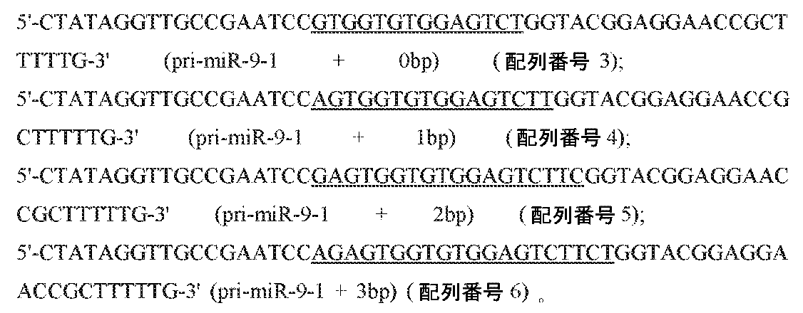
![]()
![]()
![]()
WT YdaOおよびpri-miR-9-1融合構築物に関して、本発明者らは、マキシプレッププラスミド(maxiprep plasmid)を調製し、BamHIで一晩消化することによってそれらを直線状にした。転写反応物は、約400μg 直線化テンプレート、40mM Tris pH7.5、25mM MgCl2、4mM DTT、2mM スペルミジン、40μg 無機ピロホスファターゼ(Sigma)、0.7mg T7 RNAポリメラーゼ、および3mM 各NTPを、総容積5mL中に含んだ。37℃で4.5時間のインキュベーション後、最終MgCl2濃度を40mMに調節し、その反応物をさらに45分間インキュベートした。Mg2+ 濃度の上昇にもかかわらず、本発明者らは、HDVリボザイムによる部分的切断のみを観察した。反応物をエタノール沈殿させ、変性10% ポリアクリルアミドスラブゲルで精製した。その所望の生成物を、UVシャドウイング(UV shadowing)によって可視化し、ゲルから切り出した。ゲル片を砕き、30mL TEN緩衝液(150mM NaCl、20mM Tris pH 7.5、1mM EDTA)中で、4℃において一晩抽出した。次いで、本発明者らは、ゲル片を遠心分離し、RNAを、10-kDa分子量カットオフ(MWCO)を有するAmicon Ultra-15遠心分離フィルターユニットで濃縮した。RNAを、10mM HEPES pH7.5へと3回緩衝液交換し、約50μL 最終容積へと濃縮した。 For the WT YdaO and pri-miR-9-1 fusion constructs, we prepared maxiprep plasmids and linearized them by overnight digestion with BamHI. The transcription reaction contained approximately 400 μg linearized template, 40 mM Tris pH 7.5, 25 mM MgCl 2 , 4 mM DTT, 2 mM spermidine, 40 μg inorganic pyrophosphatase (Sigma), 0.7 mg T7 RNA polymerase, and 3 mM each NTP in total volume. It was contained in 5 mL. After 4.5 hours of incubation at 37°C, the final MgCl2 concentration was adjusted to 40mM and the reaction was incubated for an additional 45 minutes. Despite increasing Mg2 + concentration, we observed only partial cleavage by HDV ribozyme. The reaction was ethanol precipitated and purified on a denaturing 10% polyacrylamide slab gel. The desired product was visualized by UV shadowing and excised from the gel. Gel pieces were crushed and extracted in 30 mL TEN buffer (150 mM NaCl, 20 mM Tris pH 7.5, 1 mM EDTA) overnight at 4°C. We then centrifuged the gel pieces and concentrated the RNA with an Amicon Ultra-15 centrifugal filter unit with a 10-kDa molecular weight cutoff (MWCO). RNA was buffer exchanged three times into 10mM HEPES pH 7.5 and concentrated to approximately 50μL final volume.
ネイティブゲル上での分析のために、5μM RNAストック溶液を、精製RNAの5mM Tris pH 7.0への希釈によって調製した。次に、2.5 μL RNAを、等容積の2×アニーリング緩衝液(35mM Tris pH 7.0、100mM KCl、10mM MgCl2、および20μM c-ジ-AMP(Sigma)を含む)と混合した。その混合物を、90℃で1分間加熱し、続いて、氷上で急速冷却し、次いで、37℃で15分間のインキュベーションを行った。そのアニールしたRNAを、40mM Tris pH 7.0、50mM KCl、5mM MgCl2、20% (v/v) グリセロールおよびキシレンシアノールを含む2×ローディング色素と混合し、Tris-ホウ酸(TB)泳動緩衝液での10% ポリアクリルアミドゲル上で分析した。そのゲルを、Sybr Green IIで染色し、Typhoon 9410 Variable Mode Imager(GE Healthcare)でスキャンした。 For analysis on native gels, a 5 μM RNA stock solution was prepared by diluting purified RNA into 5 mM Tris pH 7.0. Next, 2.5 μL RNA was mixed with an equal volume of 2× annealing buffer containing 35 mM Tris pH 7.0, 100 mM KCl, 10 mM MgCl 2 , and 20 μM c-di-AMP (Sigma). The mixture was heated to 90°C for 1 minute, followed by quick cooling on ice, followed by a 15 minute incubation at 37°C. The annealed RNA was mixed with 2x loading dye containing 40mM Tris pH 7.0, 50mM KCl, 5mM MgCl2 , 20% (v/v) glycerol and xylene cyanol and subjected to Tris-borate (TB) migration. Analyzed on a 10% polyacrylamide gel in buffer. The gel was stained with Sybr Green II and scanned on a Typhoon 9410 Variable Mode Imager (GE Healthcare).
結晶化のためのpri-miRNA-YdaO融合物の調製
本発明者らがpri-miR-9-1融合物に関して観察した不十分なHDV自己切断効率を考慮して、本発明者らは、ストラテジーを変更することを選択した。リボザイムを使用して均質な3’末端を作製する代わりに、本発明者らは、PCRを使用して、アンチセンスDNA鎖上の2個の5’残基を2’-O-メチル化した転写テンプレートを生成した。上記改変は、T7 RNAポリメラーゼによってテンプレート化されていないヌクレオチド付加を低減することが示された(28)。本発明者らは、3回のPCRアプローチを利用して、転写テンプレートを作製した。以下の全ての反応物は、同じ逆方向プライマー、5’-mCmUCCTTCCTTTATTGCCTCC-3’(配列番号8)(ここで「m」は、2’-O-メチル化を示す)を含んだ。第1回のPCRに関しては、本発明者らは、Q5ポリメラーゼとの50μL 反応を設定して、YdaOの3’フラグメントを正方向プライマー、5’-GGTACGGAGGAACCGCTTTTTG-3’(配列番号9)で増幅し、30サイクルの増幅を行った。その生成物をゲル精製し、1μLを、次の回のためのテンプレートとして使用した。第2回のPCRでは、本発明者らは、第1段階からの3’ YdaOフラグメントにアニールしたpri-miRNAループおよびステム配列を含む各構築物に対して特有の正方向プライマーを使用した。プライマー配列は、以下であった:

この反応はまた、50μLであり、Q5ポリメラーゼを30サイクル使用した。第2回のPCRからの生成物を、アガロースゲル電気泳動によって分析して、増幅を確認し、反応物のうちの40μLを、さらに精製することなく第3回のPCRのためのテンプレートとして使用した。2mL PCR反応物を、Phusion高信頼性DNAポリメラーゼ(Thermo-Fisher)および正方向プライマー
5’-GCAGAATTCTAATACGACTCACTATAGGTTGCCGAATCC-3’(配列番号18)
を使用し、35サイクル実行した。
The reaction was also 50 μL and used Q5 polymerase for 30 cycles. The products from the second round of PCR were analyzed by agarose gel electrophoresis to confirm amplification and 40 μL of the reaction was used as template for the third round of PCR without further purification. . The 2 mL PCR reaction was incubated with Phusion high-fidelity DNA polymerase (Thermo-Fisher) and forward primer 5'-GCAGAATTCTAATACGACTCACTATAGGTTGCCGAATCC-3' (SEQ ID NO: 18).
was used for 35 cycles.
第3段階のPCR生成物を、HiTrap Q HPカラム(GE Healthcare)で精製した。緩衝液Aは、10mM NaClおよび10mM HEPES pH 7.5を含んだ;緩衝液Bは、2M NaClを有する以外は同一であった。カラムを20% 緩衝液Bで平衡化し、所望のDNA生成物を、2ml/分で10分間にわたって50% Bへの直線勾配で溶離した。本発明者らは、アガロースゲルでのピーク画分を分析して、それらが正確なサイズの単一のバンドを含んでいることを確認した。次いで、そのピーク画分をプールし、Amiconフィルターユニット(10kDa MWCO)で濃縮し、次いで、水で洗浄して、過剰な塩を除去した。DNAテンプレートの濃度(約200μL 最終容積)を、UV吸光度によって決定した。 The third stage PCR product was purified on a HiTrap Q HP column (GE Healthcare). Buffer A contained 10mM NaCl and 10mM HEPES pH 7.5; Buffer B was the same except with 2M NaCl. The column was equilibrated with 20% Buffer B and the desired DNA product was eluted with a linear gradient to 50% B over 10 minutes at 2 ml/min. We analyzed the peak fractions on an agarose gel and confirmed that they contained a single band of the correct size. The peak fractions were then pooled and concentrated on an Amicon filter unit (10 kDa MWCO) and then washed with water to remove excess salt. The concentration of DNA template (approximately 200 μL final volume) was determined by UV absorbance.
転写反応を、10mL容積中であることおよび2.8fmol DNAテンプレートを含むことを除いて、pri-miR-9-1融合物に関して上記で記載されるとおりに設定した。反応を、37℃において4時間実行し、続いて、フェノール-クロロホルム抽出を行った。上記転写物をAmiconフィルターユニット(10kDa MWCO)で濃縮し、0.1M トリメチルアミン-酢酸(TEAA) pH 7.0で洗浄した。上記RNA(約2mL)を、54℃において恒温にしたWaters XTerra MS C18逆相HPLCカラム(3.5μm 粒度, 寸法4.6×150mm)に注入した。TEAAおよび100% アセトニトリルを移動相として使用した。上記カラムを6% アセトニトリルで洗浄し、RNAを、0.4ml/分において80分間にわたる17% アセトニトリルへの勾配で溶離した。ピーク画分を、変性10% ポリアクリルアミドゲルで分析した。純粋画分をプールし、10mM HEPES pH 7.0へと、Amiconフィルターユニットを使用して緩衝液交換した。上記RNAを、<50μL 最終容積へと濃縮し、その濃度をUV吸光度によって決定した。 The transcription reaction was set up as described above for the pri-miR-9-1 fusion, except in a 10 mL volume and containing 2.8 fmol DNA template. The reaction was carried out for 4 hours at 37°C followed by phenol-chloroform extraction. The transcript was concentrated with an Amicon filter unit (10 kDa MWCO) and washed with 0.1 M trimethylamine-acetic acid (TEAA) pH 7.0. The above RNA (approximately 2 mL) was injected onto a Waters XTerra MS C18 reverse phase HPLC column (3.5 μm particle size, dimensions 4.6 x 150 mm) that was thermostated at 54°C. TEAA and 100% acetonitrile were used as mobile phases. The column was washed with 6% acetonitrile and the RNA was eluted with a gradient to 17% acetonitrile over 80 minutes at 0.4 ml/min. Peak fractions were analyzed on denaturing 10% polyacrylamide gels. Pure fractions were pooled and buffer exchanged to 10mM HEPES pH 7.0 using Amicon filter units. The RNA was concentrated to a final volume of <50 μL, and the concentration was determined by UV absorbance.
結晶化、データ収集、および構造決定
全てのRNA-c-ジAMP複合体を、記載されるように調製した(15)。簡潔には、0.5mM RNA、1mM c-ジ-AMP、100mM KCl、10mM MgCl2、および20mM HEPES pH 7.0を含む溶液を、90℃へと1分間加熱し、氷上で急速冷却し、結晶化する直前に、15分間、37℃において平衡化した。スクリーニングを、0.5mL ウェル溶液を含む24ウェルプレートの中で行った;ハンギングドロップは、1μL RNA+1μL ウェル溶液からなった。プレートを室温でインキュベートし、結晶を、1週間以内に十分なサイズ(100μm~200μm超)へと全体的に成長させた。19b-2 + 1bpに関しては、上記ウェル溶液は、1.7M (NH4)2SO4、0.2M Li2SO4、および0.1M HEPES pH 7.1を含んだ。202 + 1bp、208a + 1bp、および320b-2 + 1bpに関しては、ウェルは、1.9M (NH4)2SO4、0.2M Li2SO4、および0.1M HEPES pH 7.4を含んだ。378a + 0bpのウェル溶液は、1.7M (NH4)2SO4、0.2M Li2SO4、および0.1M HEPES pH 7.4を含んだ。残りの構築物に関しては、結晶化を、0.4μL RNA + 0.4μL ウェル溶液からなるハンギングドロップで96ウェルプレートの中で行った。300 + 1bpに関しては、ウェル溶液は、1.88M (NH4)2SO4、0.248M Li2SO4、および0.1M HEPES pH 7.4を含み、300 + 0bpに関しては、ウェル溶液は、1.90M (NH4)2SO4、0.158M Li2SO4、および0.1M HEPES pH 7.4を保持していた。構築物340 + 1bpは、1.89M (NH4)2SO4、0.214M Li2SO4、および0.1M HEPES pH 7.4を含むウェル溶液から結晶化した。構築物378a + 1bpは、1.63M (NH4)2SO4、0.272 M Li2SO4、および0.1M HEPES pH 7.4から結晶化した。構築物449c + 1bpに関しては、上記ウェルは、1.89M (NH4)2SO4、0.128M Li2SO4、および0.1M HEPES pH 7.4を含んだ。
Crystallization, Data Collection, and Structure Determination All RNA-c-diAMP complexes were prepared as described (15). Briefly, a solution containing 0.5mM RNA, 1mM c-di-AMP, 100mM KCl, 10mM MgCl2 , and 20mM HEPES pH 7.0 was heated to 90°C for 1 min, rapidly cooled on ice, Equilibrate at 37° C. for 15 minutes just before crystallization. Screening was performed in 24-well plates containing 0.5 mL well solution; hanging drops consisted of 1 μL RNA+1 μL well solution. Plates were incubated at room temperature and crystals were allowed to grow to full size (100 μm to >200 μm) throughout within one week. For 19b-2 + 1 bp, the well solution contained 1.7M (NH 4 ) 2 SO 4 , 0.2M Li 2 SO 4 , and 0.1M HEPES pH 7.1. For 202 + 1bp, 208a + 1bp, and 320b -2 + 1bp, the wells contained 1.9M ( NH4 ) 2SO4 , 0.2M Li2SO4 , and 0.1M HEPES pH 7.4. is. The 378a+0bp well solution contained 1.7M ( NH4 ) 2SO4 , 0.2M Li2SO4 , and 0.1M HEPES pH 7.4. For the remaining constructs, crystallization was performed in a 96-well plate with a hanging drop consisting of 0.4 μL RNA + 0.4 μL well solution. For 300 + 1 bp, the well solution contained 1.88 M ( NH4 ) 2SO4 , 0.248 M Li2SO4 , and 0.1 M HEPES pH 7.4 ; for 300 + 0 bp, the well solution contained , 1.90M ( NH4 ) 2SO4 , 0.158M Li2SO4 , and 0.1M HEPES pH 7.4. Construct 340+1bp was crystallized from a well solution containing 1.89M ( NH4 ) 2SO4 , 0.214M Li2SO4 , and 0.1M HEPES pH 7.4.
全ての結晶を、20% (w/v) PEG 3350、20% (v/v) グリセロール、0.2M (NH4)2SO4、0.2M Li2SO4、および0.1M HEPES pH 7.3を含む凍結保護溶液中に短時間浸漬し、液体窒素中で急速凍結した。データを、Advanced Photon Source Beamline 24-ID-CまたはAdvanced Light Source Beamline 8.3.1において100Kで収集した。全ての構築物に関して、本発明者らは、波長 約1Åにおいてネイティブデータセットを収集した。320b-2+1bp、378a+0bp、および449c+1bpに関しては、本発明者らは、それぞれ、1個、2個または3個の結晶から1.9Åにおいてさらなる高リダンダンシーデータセットを収集することによって、リンの異常散乱を測定した。データをインデックス化し、統合し、XDSを使用してスケールを付けた(29)。
All crystals were prepared in 20% (w/v)
異常データが利用可能である場合、本発明者らは、分子置き換え/単一異常分散アプローチ(molecular-replacement/single anomalous dispersion approach)の組み合わせ(MR-SAD)を使用して、部分的実験相を生成した。上記分子置き換えモデルは、YdaO c-ジ-AMPリボスイッチ構造(PDB ID: 4QK8)と、上記モデルから除去されたP2ステム上のGAAAテトラループからなった。相を、PhenixのPhaser-MRプロトコールにおけるデフォルト設定を使用して得た(30)。 When anomalous data are available, we use a combination of molecular-replacement/single anomalous dispersion approach (MR-SAD) to resolve the partial experimental phase. generated. The molecular replacement model consisted of the YdaO c-di-AMP riboswitch structure (PDB ID: 4QK8) and the GAAA tetraloop on the P2 stem that was removed from the model. Phases were obtained using default settings in Phenix's Phaser-MR protocol (30).
全ての構築物に関して、本発明者らは、Phenixを使用してデータへのMRモデル(上記)のリジッドボディーフィット(rigid body fit)を行うことによって、最初の回答を得た(利用可能な場合には、実験相拘束(experimental phase restraints)を含む)。これは、Rwork<30%を有する優れた初期モデルを生成した。次いで、本発明者らは、P2ステムの領域において電子密度マップを精査した。全てのRNAに関して、失われている塩基対およびループについてのさらなる密度を、2Fo-Fcおよび差異マップ(difference map)において明らかに見ることができた。次いで、本発明者らは、Cootにおいて失われている残基をモデル化した(31)。密度が不明確な場合には、本発明者らは、不完全なループでのモデル化を中止し、Phenixでさらなる回数の座標、ADP、およびTLSパラメーター精緻化を行った。これは、代表的には、失われている残基のさらなる密度を明らかにした。ループがいったん完全にモデル化された後、本発明者らは、上記のように、合理的なR因子およびモデル形状が得られるまで精緻化のさらなるラウンドおよび手動での調節を行った。 For all constructs, we obtained an initial answer by performing a rigid body fit of the MR model (described above) to the data using Phenix (where available). (includes experimental phase restraints). This produced an excellent initial model with R work <30%. We then examined the electron density map in the region of the P2 stem. For all RNAs, additional density for missing base pairs and loops could be clearly seen in the 2F o -F c and difference maps. We then modeled the residues missing in Coot (31). In cases where the density was unclear, we stopped modeling with incomplete loops and performed additional rounds of coordinate, ADP, and TLS parameter refinement in Phenix. This typically revealed an additional density of missing residues. Once the loop was fully modeled, we performed further rounds of refinement and manual adjustments until a reasonable R-factor and model shape was obtained, as described above.
シミュレートしたアニーリングコンポジットオミットマップ(annealing composite omit map)を、Phenixにおいて計算した(図7)。19b-2 + 1bp、202 + 1bp、320b-2 + 1bp、340 + 1bp、および378a + 1bpの場合には、標準的アニーリング温度(5000℃)および他のデフォルトパラメーターは、合理的なマップを生成した。しかし、300 + 0bp、300 + 1bpおよび378a + 0bpに関しては、上記デフォルト設定は、壊れた密度の領域を有するノイズの多いマップを生成した。マップの質を改善するために、本発明者らは、アニーリング温度を1000℃へと低減し、省略した領域からバルク溶媒マスクを排除した。このタイプのコンポジットオミットマップは、Polderマップとして公知であり、溶媒マスクがより弱い密度を不明瞭にすることから防止する(32)。 A simulated annealing composite omit map was calculated in Phenix (Figure 7). For 19b-2 + 1bp, 202 + 1bp, 320b-2 + 1bp, 340 + 1bp, and 378a + 1bp, standard annealing temperature (5000 °C) and other default parameters produce reasonable maps. did. However, for 300 + 0 bp, 300 + 1 bp and 378a + 0 bp, the default settings above produced noisy maps with regions of broken density. To improve the quality of the map, we reduced the annealing temperature to 1000 °C and eliminated the bulk solvent mask from the omitted regions. This type of composite omit map is known as a Polder map and prevents solvent masks from obscuring weaker densities (32).
PDBにおける既知のRNAループ構造との比較
本発明者らのpri-miRNAループモデルに対して構造的類似性を有するPDBの中のRNAループを同定するために、本発明者らは、pri-miRNA尖端接合部およびループの座標を最初に抽出した。検索ツールは、上記の足場結晶化を同定するために使用されるRNA構造の同じセットであった。PDBセットからの各構造に関して、本発明者らは、DSSRを使用して、全てのヘアピンループを同定した。本発明者らは、RNA配列を各ヘアピンループから抽出し、pri-miRNA配列より短いループを排除した。pri-miRNAより長いループに関しては、本発明者らは、同じ長さを有するループの全てのフラグメントを得るために、スライディングウインドウを使用した。次いで、各ループ配列を、Rosetta(33)において「rna_thread」ルーチンを使用して、pri-miRNAモデルに対してスレッド化した。PyMOLスクリプトを使用して、本発明者らは、得られたスレッド化モデルを元のヘアピンループに整列させ、その2つのモデルの間でRMSDを計算した。本発明者らは、全てのPDB構造からRMSDデータを集計およびソートして、小さなRMSDを有するループを手動で精査して、構造的類似性を有するヒットを見出した。
Comparison with known RNA loop structures in the PDB To identify RNA loops in the PDB that have structural similarity to our pri-miRNA loop model, we The coordinates of the apical junction and loop were first extracted. The search tool was the same set of RNA structures used to identify scaffold crystallizations above. For each structure from the PDB set, we used DSSR to identify all hairpin loops. We extracted RNA sequences from each hairpin loop and excluded loops shorter than the pri-miRNA sequence. For loops longer than pri-miRNA, we used a sliding window to obtain all fragments of the loop with the same length. Each loop sequence was then threaded to the pri-miRNA model using the “rna_thread” routine in Rosetta (33). Using a PyMOL script, we aligned the resulting threaded model to the original hairpin loop and calculated the RMSD between the two models. We aggregated and sorted the RMSD data from all PDB structures and manually inspected loops with small RMSD to find hits with structural similarity.
光学的融解
光学的融解実験のためのRNAを、合成DNAテンプレートからインビトロで転写した(IDT)。使用したオリゴヌクレオチドテンプレート配列は、以下であった:
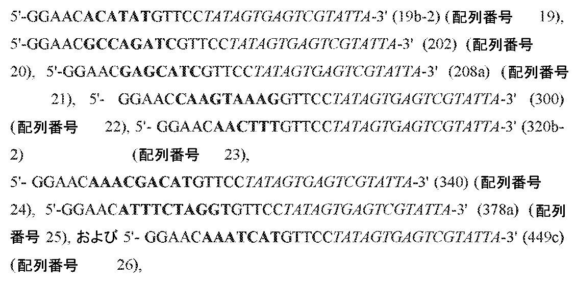
テンプレートを、T7プロモーターに相補的な第2鎖とアニールし、上記に記載されるとおりの大規模(10mL)転写反応に添加した。反応物をエタノール沈殿させ、20% ポリアクリルアミド変性ゲルで精製した。その所望のバンドを、UVシャドウイングによって回収した。ゲル抽出後、サンプルを、水へと緩衝液交換し、Amicon遠心分離フィルターデバイスで濃縮した。
Optical melting RNA for optical melting experiments was transcribed in vitro (IDT) from synthetic DNA templates. The oligonucleotide template sequences used were:
The template was annealed with a second strand complementary to the T7 promoter and added to a large scale (10 mL) transcription reaction as described above. The reaction was ethanol precipitated and purified on a 20% polyacrylamide denaturing gel. The desired band was recovered by UV shadowing. After gel extraction, samples were buffer exchanged into water and concentrated with an Amicon centrifugal filter device.
各RNAに関しては、6つの希釈物のセットを、初期吸光度が約1.0~0.1 AUの範囲に及ぶように、50mM NaClおよび10mM カコジル酸ナトリウム pH 7.0中に調製した。上記サンプルを、95℃へと1分間加熱し、氷上で急速冷却することによってアニールし、続いて、12℃へと平衡化した。融解測定を、ペルチェ素子タイプ(Peltier-type)の温度制御サンプルチェンジャーを装備したCary Bio300 UV可視分光光度計で行った。RNAを12℃から92℃へと、0.8℃/分の速度で加熱する間に、260nmでの吸光度を記録した。融解曲線を、Prism(GraphPad, バージョン7)を使用して分析し、以下の式にフィットさせた:

![]()
![]()
![]()
![]()
電気泳動移動度シフトアッセイ
ヒトヘム結合Rhedタンパク質をE.coliにおいて過剰発現させ、以前に記載される(25)ように、イオン交換およびサイズ排除クロマトグラフィーを使用して精製した。放射性標識したpri-miRNAステム-ループ(図8b~i)を、インビトロ転写によって調製した。DNAテンプレートは、所望の配列とT7プロモーターを網羅するアンチセンスオリゴヌクレオチドからなり、センスオリゴとT7プロモーター配列とにアニールさせた(34)。各20μLの転写反応物は、50fmol テンプレート、40mM Tris pH 7.5、25mM MgCl2、4mM DTT、2mM スペルミジン、2μg T7 RNAポリメラーゼ、0.5mM ATP、3mM UTP、CTP、およびGTPの各々、ならびに3nmol α-32P-ATP(10μCi)を含んだ。転写を、37℃で2時間実行し、そのRNAを変性15% ポリアクリルアミドゲルで精製した。そのRNAを4℃において一晩、TEN緩衝液中で抽出し、イソプロパノール沈殿させ、40μL 水に再懸濁した。
Electrophoretic Mobility Shift Assay Human heme-binding Rhed protein was incubated with E. E. coli was overexpressed and purified using ion exchange and size exclusion chromatography as previously described (25). Radiolabeled pri-miRNA stem-loops (Fig. 8b-i) were prepared by in vitro transcription. The DNA template consisted of an antisense oligonucleotide covering the desired sequence and the T7 promoter and was annealed to the sense oligo and the T7 promoter sequence (34). Each 20μL transcription reaction contained 50fmol template, 40mM Tris pH 7.5, 25mM MgCl2 , 4mM DTT, 2mM spermidine, 2μg T7 RNA polymerase, 0.5mM ATP, 3mM each of UTP, CTP, and GTP, and 3nmol each of Contained α- 32 P-ATP (10 μCi). Transcription was performed for 2 hours at 37°C and the RNA was purified on a denaturing 15% polyacrylamide gel. The RNA was extracted in TEN buffer overnight at 4°C, isopropanol precipitated, and resuspended in 40 μL water.
本発明者らは、近年報告されたEMSA手順を採用して、Rhed-pri-miRNA相互作用を調べた(35)。RNAを、100mM NaCl、20mM Tris pH 8.0中に希釈し、90℃において1分間加熱し、続いて、氷上で急速冷却した。アニールしたRNAを、10% (v/v) グリセロール、0.1mg/ml 酵母tRNA、0.1mg/ml BSA、5μg/ml ヘパリン、0.01% (v/v) オクチルフェノキシポリエトキシエタノール(IGEPAL CA-630)、0.25ユニット RNase-OUTリボヌクレアーゼインヒビター、キシレンシアノール、20mM Tris pH 8.0、および0~20μM Rhedタンパク質を含む結合反応物に添加した。その溶液の最終塩濃度は、150mM NaClであった。結合反応物を、室温において30分間インキュベートし、その後、10% ポリアクリルアミドゲルに載せた。ゲルおよび泳動緩衝液の両方が、80mM NaCl、89.2mM Tris塩基、および89.0mM ホウ酸(最終pH 8.2)を含んだ。ゲルを、110Vで45分間、4℃において泳動させ、次いで乾燥させ、ストレージホスホスクリーン(storage phosphor screen)に曝した。スクリーンを、引き続いて、Typhoonスキャナー(GE Healthcare)でスキャンした。遊離RNAおよび結合RNAのバンドを、Quantity Oneソフトウェア(BioRad)を使用して定量し、PrismにおいてHillの式とフィットさせた。 We employed the recently reported EMSA procedure to investigate Rhed-pri-miRNA interactions (35). RNA was diluted in 100mM NaCl, 20mM Tris pH 8.0 and heated at 90°C for 1 minute, followed by rapid cooling on ice. The annealed RNA was mixed with 10% (v/v) glycerol, 0.1 mg/ml yeast tRNA, 0.1 mg/ml BSA, 5 μg/ml heparin, and 0.01% (v/v) octylphenoxypolyethoxyethanol (IGEPAL). CA-630), 0.25 units RNase-OUT ribonuclease inhibitor, xylene cyanol, 20 mM Tris pH 8.0, and 0-20 μM Rhed protein. The final salt concentration of the solution was 150mM NaCl. Binding reactions were incubated for 30 minutes at room temperature and then loaded onto a 10% polyacrylamide gel. Both the gel and running buffer contained 80mM NaCl, 89.2mM Tris base, and 89.0mM boric acid (final pH 8.2). Gels were run at 110V for 45 minutes at 4°C, then dried and exposed to a storage phosphor screen. The screens were subsequently scanned with a Typhoon scanner (GE Healthcare). Free and bound RNA bands were quantified using Quantity One software (BioRad) and fitted with the Hill equation in Prism.
分子動力学シミュレーション
pri-miRNA残基と足場のP2ステムからの2個のG-C対に相当する座標を、各結晶構造から抽出した。水素を、GROMACSにおけるモデルに追加し(36)、RNAを、TIP3P 水分子を有する先端を切った十二面体ボックスの中で溶解した。そのボックスは、RNAを、それ自体の任意の定期的コピーから少なくとも1nm離して配置するために十分に大きかった。次に、K+イオンおよびCl-イオンを、上記システムに添加して、正味の電荷を中和し、最終KCl濃度 0.1Mを与えた。CHARMM27力場、Verletカットオフスキーム、および粒子-メッシュエバルト静電気(particle-mesh Ewald electrostatics)を、全ての計算に使用した。上記システムを、任意の原子に作用する最大の力が900kJ/mol/nm未満になるまでエネルギーを最小化した。上記システムの最終の位置エネルギー(potential energy)は、-1.3×10⁵kJ/molの範囲にあった。
Molecular Dynamics Simulations The coordinates corresponding to the pri-miRNA residue and the two GC pairs from the P2 stem of the scaffold were extracted from each crystal structure. Hydrogen was added to the model in GROMACS (36) and the RNA was dissolved in a truncated dodecahedral box with TIP3P water molecules. The box was large enough to place the RNA at least 1 nm away from any periodic copies of itself. K + and Cl − ions were then added to the system to neutralize the net charge and give a final KCl concentration of 0.1M. The CHARMM27 force field, Verlet cutoff scheme, and particle-mesh Ewald electrostatics were used for all calculations. The system was energy minimized until the maximum force acting on any atom was less than 900 kJ/mol/nm. The final potential energy of the system was in the range of −1.3×10 5 kJ/mol.
次に、上記システムを、2工程において、先ずNVTアンサンブル中で、次いで、NPTアンサンブル中で最初に平衡化した。両方の平衡化シミュレーションを、2-fs時間工程を使用して、300Kで2nsにわたって実行した。NVTの間に、温度を、速度リスケーリングによって制御した。NPTに関しては、Parrinello-Rahman圧調節器(barostat)を使用して、圧力を1バールで維持した。生成MD実行に関しては、位置拘束を、足場からのG-C対に適用し、全てのpri-miRNAヌクレオチドを非拘束とした。全ての生成シミュレーションを、NPTにおいて2-fs時間工程で合計1μsにわたって実行した。GROMACSにおいてrmsfおよびクラスター化機能を使用して軌跡を分析した。 The system was then first equilibrated in two steps, first in the NVT ensemble and then in the NPT ensemble. Both equilibration simulations were run for 2 ns at 300 K using a 2-fs time step. During NVT, temperature was controlled by rate rescaling. For NPT, pressure was maintained at 1 bar using a Parrinello-Rahman barostat. For the generation MD run, position constraints were applied to the GC pairs from the scaffold, leaving all pri-miRNA nucleotides unconstrained. All production simulations were run for a total of 1 μs with a 2-fs time step in NPT. Trajectories were analyzed using rmsf and clustering functions in GROMACS.
pri-miR-223ハイスループットプロセシングアッセイの再分析
pri-miRNA-223に関して以前に報告したプロセシングアッセイからの配列決定データを、Sequence Read Archive(アクセッション番号: SRA051323)からダウンロードした(5)。pri-miR-223に相当するリードを、Bowtie2を使用して整列させた(37)。未知のヌクレオチドを含む任意のリードを、排除した。入力または選択ライブラリーからのリードを、それらの相当するバーコードによって分離し、Pythonで計数した。
Reanalysis of pri-miR-223 High-Throughput Processing Assay Sequencing data from a previously reported processing assay for pri-miRNA-223 was downloaded from the Sequence Read Archive (accession number: SRA051323) (5). Reads corresponding to pri-miR-223 were aligned using Bowtie2 (37). Any reads containing unknown nucleotides were excluded. Reads from input or selection libraries were separated by their corresponding barcodes and counted in Python.

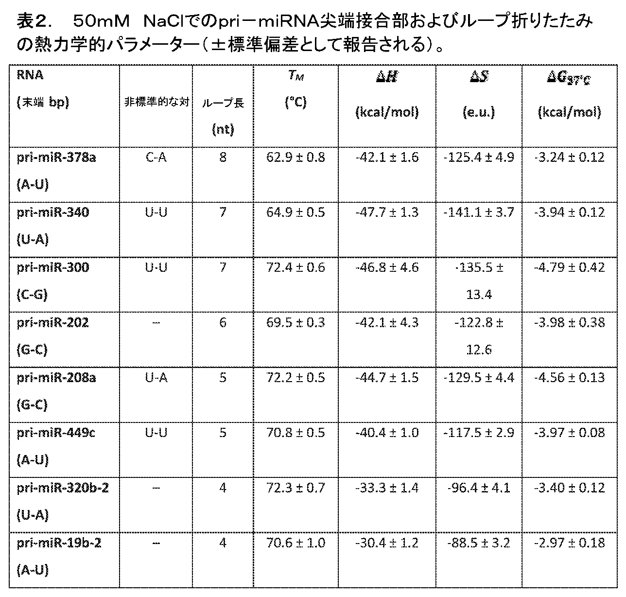
開示される参考文献
1. Ha, M. and V.N. Kim, Regulation of microRNA biogenesis. Nat. Rev. Mol. Cell Biol., 2014. 15: 509-24.
2. Krasilnikov, A.S., et al., Crystal structure of the specificity domain of ribonuclease P. Nature, 2003. 421: 760-4.
3. Reiss, C.W., Y. Xiong, and S.A. Strobel, Structural Basis for Ligand Binding to the Guanidine-I Riboswitch. Structure, 2017. 25: 195-202.
4. Byrne, R.T., et al., The crystal structure of unmodified tRNAPhe from Escherichia coli. Nucleic Acids Res, 2010. 38: 4154-62.
5. Auyeung, V.C., et al., Beyond secondary structure: primary-sequence determinants license pri-miRNA hairpins for processing. Cell, 2013. 152: 844-58.
6. Chen, Y., et al., Rbfox proteins regulate microRNA biogenesis by sequence-specific binding to their precursors and target downstream Dicer. Nucleic Acids Res., 2016. 44: 4381-95.
7. Zeng, Y., R. Yi, and B.R. Cullen, Recognition and cleavage of primary microRNA precursors by the nuclear processing enzyme Drosha. EMBO J, 2005. 24: 138-148.
8. Zhang, X. and Y. Zeng, The terminal loop region controls microRNA processing by Drosha and Dicer. Nucleic Acids Res, 2010. 38: 7689-97.
9. Ma, H., et al., Lower and upper stem-single-stranded RNA junctions together determine the Drosha cleavage site. Proc Natl Acad Sci U S A, 2013. 110: 20687-92.
10. Fang, W. and D.P. Bartel, The Menu of Features that Define Primary MicroRNAs and Enable De Novo Design of MicroRNA Genes. Mol. Cell, 2015. 60: 131-45.
11. Shortridge, M.D., et al., A Macrocyclic Peptide Ligand Binds the Oncogenic MicroRNA-21 Precursor and Suppresses Dicer Processing. ACS Chem. Biol., 2017. 12: 1611-1620.
12. Chirayil, S., et al., NMR characterization of an oligonucleotide model of the miR-21 pre-element. PloS One, 2014. 9: e108231.
13. Kozomara, A. and S. Griffiths-Jones, miRBase: integrating microRNA annotation and deep-sequencing data. Nucleic Acids Res., 2011. 39: D152-7.
14. Zuker, M., Mfold web server for nucleic acid folding and hybridization prediction. Nucleic Acids Res, 2003. 31: 3406-3415.
15. Gao, A. and A. Serganov, Structural insights into recognition of c-di-AMP by the ydaO riboswitch. Nat. Chem. Biol., 2014. 10: 787-92.
16. Serra, M.J., T.J. Axenson, and D.H. Turner, A model for the stabilities of RNA hairpins based on a study of the sequence dependence of stability for hairpins of six nucleotides. Biochemistry, 1994. 33: 14289-96.
17. Triboulet, R., et al., Post-transcriptional control of DGCR8 expression by the Microprocessor. RNA, 2009. 15: 1005-11.
18. Kadener, S., et al., Genome-wide identification of targets of the drosha-pasha/DGCR8 complex. RNA, 2009. 15: 537-45.
19. Macias, S., et al., DGCR8 HITS-CLIP reveals novel functions for the Microprocessor. Nat. Struct. Mol. Biol., 2012. 19: 760-766.
20. Heras, S.R., et al., The Microprocessor controls the activity of mammalian retrotransposons. Nat Struct Mol Biol, 2013. 20: 1173-81.
21. Han, J., et al., Posttranscriptional crossregulation between Drosha and DGCR8. Cell, 2009. 136: 75-84.
22. Moon, A.F., et al., A synergistic approach to protein crystallization: combination of a fixed-arm carrier with surface entropy reduction. Protein Sci., 2010. 19: 901-13.
23. Terasaka, N., et al., A human microRNA precursor binding to folic acid discovered by small RNA transcriptomic SELEX. RNA, 2016. 22: 1918-1928.
24. Nguyen, T.A., et al., Functional Anatomy of the Human Microprocessor. Cell, 2015. 161: 1374-87.
25. Quick-Cleveland, J., et al., The DGCR8 RNA-binding heme domain recognizes primary microRNAs by clamping the Hairpin. Cell Rep., 2014. 7: 1994-2005.
26. Michlewski, G., et al., Posttranscriptional regulation of miRNAs harboring conserved terminal loops. Mol Cell, 2008. 32: 383-93.
27. Brakier-Gingras, L., J. Charbonneau, and S.E. Butcher, Targeting frameshifting in the human immunodeficiency virus. Expert Opin. Ther. Targets, 2012. 16: 249-58.
28. Kao, C., M. Zheng, and S. Rudisser, A simple and efficient method to reduce nontemplated nucleotide addition at the 3 terminus of RNAs transcribed by T7 RNA polymerase. RNA, 1999. 5: 1268-72.
29. Kabsch, W., XDS. Acta Crystallogr. D Biol. Crystallogr., 2010. 66: 125-32.
30. Adams, P.D., et al., PHENIX: a comprehensive Python-based system for macromolecular structure solution. Acta Crystallogr D Biol Crystallogr, 2010. 66: 213-21.
31. Emsley, P., et al., Features and development of Coot. Acta Crystallogr. D Biol. Crystallogr., 2010. 66: 486-501.
32. Liebschner, D., et al., Polder maps: improving OMIT maps by excluding bulk solvent. Acta crystallographica. Section D, Structural biology, 2017. 73: 148-157.
33. Cheng, C.Y., F.C. Chou, and R. Das, Modeling complex RNA tertiary folds with Rosetta. Methods Enzymol., 2015. 553: 35-64.
34. Milligan, J.F., et al., Oligoribonucleotide synthesis using T7 RNA polymerase and synthetic DNA templates. Nucleic Acids Res., 1987. 15: 8783-98.
35. Partin, A.C., et al., Heme enables proper positioning of Drosha and DGCR8 on primary microRNAs. Nat. Commun., 2017. 8: 1737.
36. Abraham, M.J., et al., GROMACS: High performance molecular simulations through multi-level parallelism from laptops to supercomputers. SoftwareX, 2015. 1-2: 19-25.
37. Langmead, B. and S.L. Salzberg, Fast gapped-read alignment with Bowtie 2. Nat. Methods, 2012. 9: 357-9.
References disclosed
1. Ha, M. and VN Kim, Regulation of microRNA biogenesis. Nat. Rev. Mol. Cell Biol., 2014. 15: 509-24.
2. Krasilnikov, AS, et al., Crystal structure of the specificity domain of ribonuclease P. Nature, 2003. 421: 760-4.
3. Reiss, CW, Y. Xiong, and SA Strobel, Structural Basis for Ligand Binding to the Guanidine-I Riboswitch. Structure, 2017. 25: 195-202.
4. Byrne, RT, et al., The crystal structure of unmodified tRNAPhe from Escherichia coli. Nucleic Acids Res, 2010. 38: 4154-62.
5. Auyeung, VC, et al., Beyond secondary structure: primary-sequence determinants license pri-miRNA hairpins for processing. Cell, 2013. 152: 844-58.
6. Chen, Y., et al., Rbfox proteins regulate microRNA biogenesis by sequence-specific binding to their precursors and target downstream Dicer. Nucleic Acids Res., 2016. 44: 4381-95.
7. Zeng, Y., R. Yi, and BR Cullen, Recognition and cleavage of primary microRNA precursors by the nuclear processing enzyme Drosha. EMBO J, 2005. 24: 138-148.
8. Zhang, X. and Y. Zeng, The terminal loop region controls microRNA processing by Drosha and Dicer. Nucleic Acids Res, 2010. 38: 7689-97.
9. Ma, H., et al., Lower and upper stem-single-stranded RNA junctions together determine the Drosha cleavage site. Proc Natl Acad Sci USA, 2013. 110: 20687-92.
10. Fang, W. and DP Bartel, The Menu of Features that Define Primary MicroRNAs and Enable De Novo Design of MicroRNA Genes. Mol. Cell, 2015. 60: 131-45.
11. Shortridge, MD, et al., A Macrocyclic Peptide Ligand Binds the Oncogenic MicroRNA-21 Precursor and Suppresses Dicer Processing. ACS Chem. Biol., 2017. 12: 1611-1620.
12. Chirayil, S., et al., NMR characterization of an oligonucleotide model of the miR-21 pre-element. PloS One, 2014. 9: e108231.
13. Kozomara, A. and S. Griffiths-Jones, miRBase: integrating microRNA annotation and deep-sequencing data. Nucleic Acids Res., 2011. 39: D152-7.
14. Zuker, M., Mfold web server for nucleic acid folding and hybridization prediction. Nucleic Acids Res, 2003. 31: 3406-3415.
15. Gao, A. and A. Serganov, Structural insights into recognition of c-di-AMP by the ydaO riboswitch. Nat. Chem. Biol., 2014. 10: 787-92.
16. Serra, MJ, TJ Axenson, and DH Turner, A model for the stabilities of RNA hairpins based on a study of the sequence dependence of stability for hairpins of six nucleotides. Biochemistry, 1994. 33: 14289-96.
17. Triboulet, R., et al., Post-transcriptional control of DGCR8 expression by the Microprocessor. RNA, 2009. 15: 1005-11.
18. Kadener, S., et al., Genome-wide identification of targets of the drosha-pasha/DGCR8 complex. RNA, 2009. 15: 537-45.
19. Macias, S., et al., DGCR8 HITS-CLIP reveals novel functions for the Microprocessor. Nat. Struct. Mol. Biol., 2012. 19: 760-766.
20. Heras, SR, et al., The Microprocessor controls the activity of mammalian retrotransposons. Nat Struct Mol Biol, 2013. 20: 1173-81.
21. Han, J., et al., Posttranscriptional crossregulation between Drosha and DGCR8. Cell, 2009. 136: 75-84.
22. Moon, AF, et al., A synergistic approach to protein crystallization: combination of a fixed-arm carrier with surface entropy reduction. Protein Sci., 2010. 19: 901-13.
23. Terasaka, N., et al., A human microRNA precursor binding to folic acid discovered by small RNA transcriptomic SELEX. RNA, 2016. 22: 1918-1928.
24. Nguyen, TA, et al., Functional Anatomy of the Human Microprocessor. Cell, 2015. 161: 1374-87.
25. Quick-Cleveland, J., et al., The DGCR8 RNA-binding heme domain recognizes primary microRNAs by clamping the Hairpin. Cell Rep., 2014. 7: 1994-2005.
26. Michlewski, G., et al., Posttranscriptional regulation of miRNAs harboring conserved terminal loops. Mol Cell, 2008. 32: 383-93.
27. Brakier-Gingras, L., J. Charbonneau, and SE Butcher, Targeting frameshifting in the human immunodeficiency virus. Expert Opin. Ther. Targets, 2012. 16: 249-58.
28. Kao, C., M. Zheng, and S. Rudisser, A simple and efficient method to reduce nontemplated nucleotide addition at the 3 terminus of RNAs transcribed by T7 RNA polymerase. RNA, 1999. 5: 1268-72.
29. Kabsch, W., XDS. Acta Crystallogr. D Biol. Crystallogr., 2010. 66: 125-32.
30. Adams, PD, et al., PHENIX: a comprehensive Python-based system for macromolecular structure solution. Acta Crystallogr D Biol Crystallogr, 2010. 66: 213-21.
31. Emsley, P., et al., Features and development of Coot. Acta Crystallogr. D Biol. Crystallogr., 2010. 66: 486-501.
32. Liebschner, D., et al., Polder maps: improving OMIT maps by excluding bulk solvent. Acta crystallographica. Section D, Structural biology, 2017. 73: 148-157.
33. Cheng, CY, FC Chou, and R. Das, Modeling complex RNA tertiary folds with Rosetta. Methods Enzymol., 2015. 553: 35-64.
34. Milligan, JF, et al., Oligoribonucleotide synthesis using T7 RNA polymerase and synthetic DNA templates. Nucleic Acids Res., 1987. 15: 8783-98.
35. Partin, AC, et al., Heme enables proper positioning of Drosha and DGCR8 on primary microRNAs. Nat. Commun., 2017. 8: 1737.
36. Abraham, MJ, et al., GROMACS: High performance molecular simulations through multi-level parallelism from laptops to supercomputers. SoftwareX, 2015. 1-2: 19-25.
37. Langmead, B. and SL Salzberg, Fast gapped-read alignment with
結論
これは、本発明の好ましい実施形態の説明を結論づける。本発明の1またはこれより多くの実施形態の前述の説明は、例証および説明の目艇で示されている。本発明を網羅する、または開示される正確な形態に限定することは意図されない。多くの改変およびバリエーションは、上記の教示に照らして可能である。
Conclusion This concludes the description of the preferred embodiment of the invention. The foregoing description of one or more embodiments of the invention has been presented in terms of illustration and description. It is not intended to be exhaustive or to limit the invention to the precise form disclosed. Many modifications and variations are possible in light of the above teaching.
本明細書で引用される全ての刊行物、特許および特許出願は、全ての目的のためにそれらの全体において本明細書に参考として援用される。 All publications, patents and patent applications cited herein are herein incorporated by reference in their entirety for all purposes.
Claims (20)
GGUUGCCGAAUCCGAAAGGUACGGAGGAACCGCUUUUUGGGGUUAAUCUGCAGUGAAGCUGCAGUAGGGAUACCUUCUGUCCCGCACCCGACAGCUAACUCCGGAGGCAAUAAAGGAAGGAG(配列番号1)と少なくとも90%の同一性を有するリボ核酸をコードするDNA配列を含むプラスミド、
を含む、システム/キット。 A system/kit for observing RNA structure, comprising:
GGUUGCCGAAUCCGAAAGGUACGGAGGAACCGCUUUUUGGGGUUAAUCUGCAGUGAAGCUGCAGUAGGGAUACCUUCUGUCCCGCACCCGACAGCUAACUCCGGAGGCAAUAAAGGAAGGGAG ( a plasmid comprising a DNA sequence encoding a ribonucleic acid having at least 90% identity with SEQ ID NO: 1);
system/kit, including:
配列番号1と少なくとも90%の同一性を有するリボ核酸を得る工程;
配列番号1におけるループ残基14~17(GAAA)に対応する残基を、融合リボ核酸分子を形成するように、長さが4~33ヌクレオチドの間である核酸の異種セグメントで置換する工程;
前記融合リボ核酸分子を結晶化する工程;
X線または電子結晶学の技術を前記融合リボ核酸分子に対して行う工程;および
前記核酸の異種セグメントの構造に関する情報が得られるように、前記X線または電子結晶学の技術の結果を観察する工程、
を包含する方法。 A method for obtaining information regarding the structure of ribonucleic acid, the method comprising:
obtaining a ribonucleic acid having at least 90% identity with SEQ ID NO: 1;
replacing the residues corresponding to loop residues 14-17 (GAAA) in SEQ ID NO: 1 with a heterologous segment of nucleic acid between 4 and 33 nucleotides in length, so as to form a fused ribonucleic acid molecule;
crystallizing the fused ribonucleic acid molecule;
performing an X-ray or electron crystallographic technique on said fused ribonucleic acid molecule; and observing the results of said X-ray or electron crystallographic technique so as to obtain information regarding the structure of the heterogeneous segment of said nucleic acid. process,
How to include.
(a)第1のポリヌクレオチドを選択する工程であって、ここで前記第1のポリヌクレオチドは、第1のmiRNAのポリヌクレオチド配列を含む工程;
(b)前記第1のmiRNAにおける第1のループ領域を形成するポリヌクレオチドのセグメントを同定する工程;
(c)第2のポリヌクレオチドを選択する工程であって、ここで前記第2のポリヌクレオチドは、第2のmiRNAのポリヌクレオチド配列を含む工程;
(d)前記第2のmiRNAにおける第1のループ領域を形成するポリヌクレオチドのセグメントを同定する工程;
(e)前記第1のポリヌクレオチド上の前記第1のループ領域を含む前記ポリヌクレオチドのセグメントが、前記第2のポリヌクレオチド上の前記第1のループ領域を含む前記ポリヌクレオチドのセグメントで置換されるように構築された融合ポリヌクレオチドを形成する工程;および
(f)前記融合ポリヌクレオチドの三次元構造を観察するために、前記融合ポリヌクレオチドを結晶学的に分析する工程;
を包含し、その結果、前記ポリヌクレオチドの結晶学の分析が行われ、
ここで、前記第1のmiRNAは、GGUUGCCGAAUCCGAAAGGUACGGAGGAACCGCUUUUUGGGGUUAAUCUGCAGUGAAGCUGCAGUAGGGAUACCUUCUGUCCCGCACCCGACAGCUAACUCCGGAGGCAAUAAAGGAAGGAG(配列番号1)と少なくとも90%の配列同一性を有するmiRNAである、方法。 A method for performing crystallographic analysis on a polynucleotide, the method comprising:
(a) selecting a first polynucleotide, wherein the first polynucleotide comprises a polynucleotide sequence of a first miRNA;
(b) identifying a polynucleotide segment forming the first loop region in the first miRNA;
(c) selecting a second polynucleotide, wherein the second polynucleotide comprises a polynucleotide sequence of a second miRNA;
(d) identifying a segment of polynucleotide forming the first loop region in the second miRNA;
(e) a segment of the polynucleotide comprising the first loop region on the first polynucleotide is replaced with a segment of the polynucleotide comprising the first loop region on the second polynucleotide; (f) crystallographically analyzing the fusion polynucleotide to observe the three-dimensional structure of the fusion polynucleotide;
resulting in a crystallographic analysis of said polynucleotide ;
Here, the first miRNA is GGUUGCCGAAUCCGAAAGGUACGGAGGAACCGCUUUUUGGGGUUAAUCUGCAGUGAAGCUGCAGUAGGGAUACCUUCUGUCCCGCACCCGACAGCUAACUCCGGAGGCAA A method, wherein the miRNA has at least 90% sequence identity with UAAAGGAAGGAG (SEQ ID NO: 1) .
前記第2のmiRNAは、ヒトmiRNAを含む、
請求項17に記載の方法。 the first polynucleotide comprises the sequence SEQ ID NO: 1; and/or the second miRNA comprises a human miRNA.
18. The method according to claim 17.
18. The method of claim 17, wherein said crystallographic analysis is performed in the presence of an agent that binds to said fusion polynucleotide.
Applications Claiming Priority (3)
| Application Number | Priority Date | Filing Date | Title |
|---|---|---|---|
| US201962937657P | 2019-11-19 | 2019-11-19 | |
| US62/937,657 | 2019-11-19 | ||
| PCT/US2020/061299 WO2021102153A1 (en) | 2019-11-19 | 2020-11-19 | Structure-based design of therapeutics targeting rna hairpin loops |
Publications (2)
| Publication Number | Publication Date |
|---|---|
| JP2023501749A JP2023501749A (en) | 2023-01-18 |
| JP7436075B2 true JP7436075B2 (en) | 2024-02-21 |
Family
ID=75980949
Family Applications (1)
| Application Number | Title | Priority Date | Filing Date |
|---|---|---|---|
| JP2022528619A Active JP7436075B2 (en) | 2019-11-19 | 2020-11-19 | Structure-based design of therapeutic agents targeting RNA hairpin loops |
Country Status (5)
| Country | Link |
|---|---|
| US (1) | US20230002825A1 (en) |
| EP (1) | EP4061822A4 (en) |
| JP (1) | JP7436075B2 (en) |
| CN (1) | CN115038710B (en) |
| WO (1) | WO2021102153A1 (en) |
Citations (1)
| Publication number | Priority date | Publication date | Assignee | Title |
|---|---|---|---|---|
| US20110172981A1 (en) | 2008-09-26 | 2011-07-14 | The Regents Of The University Of Michigan | High throughput ensemble-based docking and elucidation of three-dimensional structural confirmations of flexible biomolecular targets |
Family Cites Families (1)
| Publication number | Priority date | Publication date | Assignee | Title |
|---|---|---|---|---|
| EP2426218A1 (en) * | 2007-05-29 | 2012-03-07 | Yale University | Riboswitches and methods and compositions for use of and with riboswitches |
-
2020
- 2020-11-19 JP JP2022528619A patent/JP7436075B2/en active Active
- 2020-11-19 EP EP20890836.8A patent/EP4061822A4/en active Pending
- 2020-11-19 WO PCT/US2020/061299 patent/WO2021102153A1/en not_active Ceased
- 2020-11-19 CN CN202080093268.8A patent/CN115038710B/en active Active
- 2020-11-19 US US17/776,943 patent/US20230002825A1/en active Pending
Patent Citations (1)
| Publication number | Priority date | Publication date | Assignee | Title |
|---|---|---|---|---|
| US20110172981A1 (en) | 2008-09-26 | 2011-07-14 | The Regents Of The University Of Michigan | High throughput ensemble-based docking and elucidation of three-dimensional structural confirmations of flexible biomolecular targets |
Non-Patent Citations (1)
| Title |
|---|
| GAO, A. et al.,Structural insights into recoginition of c-di-AMP by the ydaO riboswitch,Nature chemical biology ,10(9),2014年,pp.787-792 |
Also Published As
| Publication number | Publication date |
|---|---|
| US20230002825A1 (en) | 2023-01-05 |
| WO2021102153A1 (en) | 2021-05-27 |
| CN115038710A (en) | 2022-09-09 |
| EP4061822A1 (en) | 2022-09-28 |
| CN115038710B (en) | 2025-06-10 |
| EP4061822A4 (en) | 2023-12-13 |
| JP2023501749A (en) | 2023-01-18 |
Similar Documents
| Publication | Publication Date | Title |
|---|---|---|
| Cao et al. | Identification of RNA structures and their roles in RNA functions | |
| Ontiveros et al. | The chemical diversity of RNA modifications | |
| Nam et al. | Molecular basis for interaction of let-7 microRNAs with Lin28 | |
| Nakanishi et al. | Structure of yeast Argonaute with guide RNA | |
| Park et al. | Dicer recognizes the 5′ end of RNA for efficient and accurate processing | |
| Wang et al. | Structure of the guide-strand-containing argonaute silencing complex | |
| Gan et al. | A stepwise model for double‐stranded RNA processing by ribonuclease III | |
| Lim et al. | Thermal stability of DNA quadruplex–duplex hybrids | |
| JP2022137068A (en) | Highly specific genome editing using chemically modified guide RNA | |
| Sperling et al. | Structure and function of the Pre-mRNA splicing machine | |
| Oliveira et al. | Improving aptamer performance with nucleic acid mimics: de novo and post-SELEX approaches | |
| JP7566741B2 (en) | Programmable siRNA and uses thereof | |
| Esposito et al. | Backbone modified TBA analogues endowed with antiproliferative activity | |
| Gebert et al. | A structured RNA motif locks Argonaute2: miR-122 onto the 5’end of the HCV genome | |
| JP7436075B2 (en) | Structure-based design of therapeutic agents targeting RNA hairpin loops | |
| Shoffner et al. | Structures of microRNA-precursor apical junctions and loops reveal non-canonical base pairs important for processing | |
| Dadhwal et al. | Substrate promiscuity of Dicer toward precursors of the let-7 family and their 3′-end modifications: G. Dadhwal et al. | |
| Kuwahara | Progress in chemically modified nucleic acid aptamers | |
| Sweetapple et al. | Biophysics of microRNA-34a targeting and its influence on down-regulation | |
| CN105960410A (en) | Methods for structural determination of selenium derivatized nucleic acid complexes | |
| Bush | The Interrogation of Cas9 Aptamers and sgRNA Structures Through SELEX | |
| Petersen | Poly (UG) RNA Structure and Dynamics | |
| US11326169B2 (en) | Method of producing an aptamer and uses thereof | |
| Fang et al. | Syntheses of Pyrimidine-Modified Seleno-DNAs as Stable Antisense Molecules | |
| Haurwitz | The CRISPR endoribonuclease Csy4 utilizes unusual sequence-and structure-specific mechanisms to recognize and process crRNAs |
Legal Events
| Date | Code | Title | Description |
|---|---|---|---|
| A621 | Written request for application examination |
Free format text: JAPANESE INTERMEDIATE CODE: A621 Effective date: 20220617 |
|
| A131 | Notification of reasons for refusal |
Free format text: JAPANESE INTERMEDIATE CODE: A131 Effective date: 20230705 |
|
| A601 | Written request for extension of time |
Free format text: JAPANESE INTERMEDIATE CODE: A601 Effective date: 20230927 |
|
| A521 | Request for written amendment filed |
Free format text: JAPANESE INTERMEDIATE CODE: A523 Effective date: 20231121 |
|
| TRDD | Decision of grant or rejection written | ||
| A01 | Written decision to grant a patent or to grant a registration (utility model) |
Free format text: JAPANESE INTERMEDIATE CODE: A01 Effective date: 20240105 |
|
| A61 | First payment of annual fees (during grant procedure) |
Free format text: JAPANESE INTERMEDIATE CODE: A61 Effective date: 20240201 |
|
| R150 | Certificate of patent or registration of utility model |
Ref document number: 7436075 Country of ref document: JP Free format text: JAPANESE INTERMEDIATE CODE: R150 |