JP7417533B2 - Modified oncolytic adenovirus - Google Patents
Modified oncolytic adenovirus Download PDFInfo
- Publication number
- JP7417533B2 JP7417533B2 JP2020550115A JP2020550115A JP7417533B2 JP 7417533 B2 JP7417533 B2 JP 7417533B2 JP 2020550115 A JP2020550115 A JP 2020550115A JP 2020550115 A JP2020550115 A JP 2020550115A JP 7417533 B2 JP7417533 B2 JP 7417533B2
- Authority
- JP
- Japan
- Prior art keywords
- cancer
- ox40l
- cd40l
- adenovirus
- cells
- Prior art date
- Legal status (The legal status is an assumption and is not a legal conclusion. Google has not performed a legal analysis and makes no representation as to the accuracy of the status listed.)
- Active
Links
- 241000701161 unidentified adenovirus Species 0.000 title claims description 66
- 230000000174 oncolytic effect Effects 0.000 title description 12
- 108010042215 OX40 Ligand Proteins 0.000 claims description 100
- 102000004473 OX40 Ligand Human genes 0.000 claims description 97
- 108010029697 CD40 Ligand Proteins 0.000 claims description 88
- 102100032937 CD40 ligand Human genes 0.000 claims description 86
- 210000004027 cell Anatomy 0.000 claims description 86
- 206010028980 Neoplasm Diseases 0.000 claims description 84
- 241000700605 Viruses Species 0.000 claims description 71
- 108700019146 Transgenes Proteins 0.000 claims description 59
- 201000011510 cancer Diseases 0.000 claims description 36
- 108090000623 proteins and genes Proteins 0.000 claims description 33
- 238000011282 treatment Methods 0.000 claims description 28
- 238000012217 deletion Methods 0.000 claims description 27
- 230000037430 deletion Effects 0.000 claims description 27
- 238000012545 processing Methods 0.000 claims description 19
- 238000012224 gene deletion Methods 0.000 claims description 15
- 239000000203 mixture Substances 0.000 claims description 9
- 230000006037 cell lysis Effects 0.000 claims description 8
- 239000002773 nucleotide Substances 0.000 claims description 7
- 125000003729 nucleotide group Chemical group 0.000 claims description 7
- 101100059511 Homo sapiens CD40LG gene Proteins 0.000 claims description 6
- 101000764263 Homo sapiens Tumor necrosis factor ligand superfamily member 4 Proteins 0.000 claims description 6
- 108020005067 RNA Splice Sites Proteins 0.000 claims description 6
- 238000003776 cleavage reaction Methods 0.000 claims description 6
- 102000051450 human TNFSF4 Human genes 0.000 claims description 6
- 201000001441 melanoma Diseases 0.000 claims description 6
- 230000003362 replicative effect Effects 0.000 claims description 6
- 230000007017 scission Effects 0.000 claims description 6
- 108700026758 Adenovirus hexon capsid Proteins 0.000 claims description 5
- 238000003780 insertion Methods 0.000 claims description 5
- 230000037431 insertion Effects 0.000 claims description 5
- 230000010076 replication Effects 0.000 claims description 5
- 206010009944 Colon cancer Diseases 0.000 claims description 4
- 208000008839 Kidney Neoplasms Diseases 0.000 claims description 4
- 206010061902 Pancreatic neoplasm Diseases 0.000 claims description 4
- 206010038389 Renal cancer Diseases 0.000 claims description 4
- 201000000052 gastrinoma Diseases 0.000 claims description 4
- 230000002601 intratumoral effect Effects 0.000 claims description 4
- 201000010982 kidney cancer Diseases 0.000 claims description 4
- 201000007270 liver cancer Diseases 0.000 claims description 4
- 208000014018 liver neoplasm Diseases 0.000 claims description 4
- 208000015486 malignant pancreatic neoplasm Diseases 0.000 claims description 4
- 201000002528 pancreatic cancer Diseases 0.000 claims description 4
- 208000008443 pancreatic carcinoma Diseases 0.000 claims description 4
- 239000008194 pharmaceutical composition Substances 0.000 claims description 4
- 206010008342 Cervix carcinoma Diseases 0.000 claims description 3
- 241000710198 Foot-and-mouth disease virus Species 0.000 claims description 3
- 208000003445 Mouth Neoplasms Diseases 0.000 claims description 3
- 201000000582 Retinoblastoma Diseases 0.000 claims description 3
- 208000006105 Uterine Cervical Neoplasms Diseases 0.000 claims description 3
- 201000010881 cervical cancer Diseases 0.000 claims description 3
- 239000002299 complementary DNA Substances 0.000 claims description 3
- 239000003814 drug Substances 0.000 claims description 3
- 208000014829 head and neck neoplasm Diseases 0.000 claims description 3
- 208000012987 lip and oral cavity carcinoma Diseases 0.000 claims description 3
- 230000002101 lytic effect Effects 0.000 claims description 3
- 238000011144 upstream manufacturing Methods 0.000 claims description 3
- 206010061424 Anal cancer Diseases 0.000 claims description 2
- 208000007860 Anus Neoplasms Diseases 0.000 claims description 2
- 206010004272 Benign hydatidiform mole Diseases 0.000 claims description 2
- 206010005003 Bladder cancer Diseases 0.000 claims description 2
- 206010005949 Bone cancer Diseases 0.000 claims description 2
- 208000018084 Bone neoplasm Diseases 0.000 claims description 2
- 208000003174 Brain Neoplasms Diseases 0.000 claims description 2
- 206010006187 Breast cancer Diseases 0.000 claims description 2
- 208000026310 Breast neoplasm Diseases 0.000 claims description 2
- 206010007270 Carcinoid syndrome Diseases 0.000 claims description 2
- 201000009030 Carcinoma Diseases 0.000 claims description 2
- 208000005243 Chondrosarcoma Diseases 0.000 claims description 2
- 208000006332 Choriocarcinoma Diseases 0.000 claims description 2
- 208000001333 Colorectal Neoplasms Diseases 0.000 claims description 2
- 206010014733 Endometrial cancer Diseases 0.000 claims description 2
- 206010014759 Endometrial neoplasm Diseases 0.000 claims description 2
- 208000000461 Esophageal Neoplasms Diseases 0.000 claims description 2
- 208000006168 Ewing Sarcoma Diseases 0.000 claims description 2
- 201000008808 Fibrosarcoma Diseases 0.000 claims description 2
- 208000022072 Gallbladder Neoplasms Diseases 0.000 claims description 2
- 206010067807 Gingival cancer Diseases 0.000 claims description 2
- 206010018404 Glucagonoma Diseases 0.000 claims description 2
- 208000017604 Hodgkin disease Diseases 0.000 claims description 2
- 208000010747 Hodgkins lymphoma Diseases 0.000 claims description 2
- 208000006937 Hydatidiform mole Diseases 0.000 claims description 2
- 208000007766 Kaposi sarcoma Diseases 0.000 claims description 2
- 206010062038 Lip neoplasm Diseases 0.000 claims description 2
- 206010058467 Lung neoplasm malignant Diseases 0.000 claims description 2
- 208000032271 Malignant tumor of penis Diseases 0.000 claims description 2
- 208000015021 Meningeal Neoplasms Diseases 0.000 claims description 2
- 206010027406 Mesothelioma Diseases 0.000 claims description 2
- 208000034578 Multiple myelomas Diseases 0.000 claims description 2
- 208000001894 Nasopharyngeal Neoplasms Diseases 0.000 claims description 2
- 206010061306 Nasopharyngeal cancer Diseases 0.000 claims description 2
- 206010029260 Neuroblastoma Diseases 0.000 claims description 2
- 208000005890 Neuroma Diseases 0.000 claims description 2
- 208000015914 Non-Hodgkin lymphomas Diseases 0.000 claims description 2
- 206010030155 Oesophageal carcinoma Diseases 0.000 claims description 2
- 201000010133 Oligodendroglioma Diseases 0.000 claims description 2
- 208000010191 Osteitis Deformans Diseases 0.000 claims description 2
- 208000000035 Osteochondroma Diseases 0.000 claims description 2
- 206010033128 Ovarian cancer Diseases 0.000 claims description 2
- 206010061535 Ovarian neoplasm Diseases 0.000 claims description 2
- 208000027868 Paget disease Diseases 0.000 claims description 2
- 208000000821 Parathyroid Neoplasms Diseases 0.000 claims description 2
- 208000004091 Parotid Neoplasms Diseases 0.000 claims description 2
- 208000002471 Penile Neoplasms Diseases 0.000 claims description 2
- 206010034299 Penile cancer Diseases 0.000 claims description 2
- 208000009565 Pharyngeal Neoplasms Diseases 0.000 claims description 2
- 206010034811 Pharyngeal cancer Diseases 0.000 claims description 2
- 208000007913 Pituitary Neoplasms Diseases 0.000 claims description 2
- 206010035226 Plasma cell myeloma Diseases 0.000 claims description 2
- 206010036832 Prolactinoma Diseases 0.000 claims description 2
- 206010060862 Prostate cancer Diseases 0.000 claims description 2
- 208000000236 Prostatic Neoplasms Diseases 0.000 claims description 2
- 208000015634 Rectal Neoplasms Diseases 0.000 claims description 2
- 208000004337 Salivary Gland Neoplasms Diseases 0.000 claims description 2
- 206010061934 Salivary gland cancer Diseases 0.000 claims description 2
- 206010039491 Sarcoma Diseases 0.000 claims description 2
- 208000000453 Skin Neoplasms Diseases 0.000 claims description 2
- 208000021712 Soft tissue sarcoma Diseases 0.000 claims description 2
- 208000005718 Stomach Neoplasms Diseases 0.000 claims description 2
- 208000000389 T-cell leukemia Diseases 0.000 claims description 2
- 208000028530 T-cell lymphoblastic leukemia/lymphoma Diseases 0.000 claims description 2
- 206010042971 T-cell lymphoma Diseases 0.000 claims description 2
- 208000027585 T-cell non-Hodgkin lymphoma Diseases 0.000 claims description 2
- 208000024313 Testicular Neoplasms Diseases 0.000 claims description 2
- 206010057644 Testis cancer Diseases 0.000 claims description 2
- 206010043515 Throat cancer Diseases 0.000 claims description 2
- 208000000728 Thymus Neoplasms Diseases 0.000 claims description 2
- 208000024770 Thyroid neoplasm Diseases 0.000 claims description 2
- 206010062129 Tongue neoplasm Diseases 0.000 claims description 2
- 206010044002 Tonsil cancer Diseases 0.000 claims description 2
- 208000006842 Tonsillar Neoplasms Diseases 0.000 claims description 2
- 208000023915 Ureteral Neoplasms Diseases 0.000 claims description 2
- 206010046392 Ureteric cancer Diseases 0.000 claims description 2
- 208000007097 Urinary Bladder Neoplasms Diseases 0.000 claims description 2
- 208000002495 Uterine Neoplasms Diseases 0.000 claims description 2
- 208000014070 Vestibular schwannoma Diseases 0.000 claims description 2
- 206010047741 Vulval cancer Diseases 0.000 claims description 2
- 208000004354 Vulvar Neoplasms Diseases 0.000 claims description 2
- 208000008383 Wilms tumor Diseases 0.000 claims description 2
- 201000008629 Zollinger-Ellison syndrome Diseases 0.000 claims description 2
- 208000004064 acoustic neuroma Diseases 0.000 claims description 2
- 201000005188 adrenal gland cancer Diseases 0.000 claims description 2
- 208000024447 adrenal gland neoplasm Diseases 0.000 claims description 2
- 201000011165 anus cancer Diseases 0.000 claims description 2
- 208000002458 carcinoid tumor Diseases 0.000 claims description 2
- 208000006990 cholangiocarcinoma Diseases 0.000 claims description 2
- 208000029742 colonic neoplasm Diseases 0.000 claims description 2
- 201000010918 connective tissue cancer Diseases 0.000 claims description 2
- 230000002124 endocrine Effects 0.000 claims description 2
- 201000004101 esophageal cancer Diseases 0.000 claims description 2
- 208000024519 eye neoplasm Diseases 0.000 claims description 2
- 201000010175 gallbladder cancer Diseases 0.000 claims description 2
- 206010017758 gastric cancer Diseases 0.000 claims description 2
- 208000015419 gastrin-producing neuroendocrine tumor Diseases 0.000 claims description 2
- 210000001035 gastrointestinal tract Anatomy 0.000 claims description 2
- 201000007116 gestational trophoblastic neoplasm Diseases 0.000 claims description 2
- 201000010235 heart cancer Diseases 0.000 claims description 2
- 208000024348 heart neoplasm Diseases 0.000 claims description 2
- 206010073071 hepatocellular carcinoma Diseases 0.000 claims description 2
- 231100000844 hepatocellular carcinoma Toxicity 0.000 claims description 2
- 206010022498 insulinoma Diseases 0.000 claims description 2
- 201000002313 intestinal cancer Diseases 0.000 claims description 2
- 201000006721 lip cancer Diseases 0.000 claims description 2
- 201000005202 lung cancer Diseases 0.000 claims description 2
- 208000020816 lung neoplasm Diseases 0.000 claims description 2
- 208000022006 malignant tumor of meninges Diseases 0.000 claims description 2
- 208000026045 malignant tumor of parathyroid gland Diseases 0.000 claims description 2
- 208000027202 mammary Paget disease Diseases 0.000 claims description 2
- 206010027191 meningioma Diseases 0.000 claims description 2
- 201000008026 nephroblastoma Diseases 0.000 claims description 2
- 210000005036 nerve Anatomy 0.000 claims description 2
- 201000008106 ocular cancer Diseases 0.000 claims description 2
- 210000003254 palate Anatomy 0.000 claims description 2
- 208000021255 pancreatic insulinoma Diseases 0.000 claims description 2
- 201000001219 parotid gland cancer Diseases 0.000 claims description 2
- 201000002628 peritoneum cancer Diseases 0.000 claims description 2
- 201000002511 pituitary cancer Diseases 0.000 claims description 2
- 201000003437 pleural cancer Diseases 0.000 claims description 2
- 230000008488 polyadenylation Effects 0.000 claims description 2
- 208000030153 prolactin-producing pituitary gland adenoma Diseases 0.000 claims description 2
- 206010038038 rectal cancer Diseases 0.000 claims description 2
- 201000001275 rectum cancer Diseases 0.000 claims description 2
- 201000000849 skin cancer Diseases 0.000 claims description 2
- 201000002314 small intestine cancer Diseases 0.000 claims description 2
- 230000000392 somatic effect Effects 0.000 claims description 2
- 206010062261 spinal cord neoplasm Diseases 0.000 claims description 2
- 201000011549 stomach cancer Diseases 0.000 claims description 2
- 201000003120 testicular cancer Diseases 0.000 claims description 2
- 201000002510 thyroid cancer Diseases 0.000 claims description 2
- 201000006134 tongue cancer Diseases 0.000 claims description 2
- 208000029387 trophoblastic neoplasm Diseases 0.000 claims description 2
- 201000005112 urinary bladder cancer Diseases 0.000 claims description 2
- 206010046766 uterine cancer Diseases 0.000 claims description 2
- 206010046885 vaginal cancer Diseases 0.000 claims description 2
- 208000013139 vaginal neoplasm Diseases 0.000 claims description 2
- 208000006542 von Hippel-Lindau disease Diseases 0.000 claims description 2
- 201000005102 vulva cancer Diseases 0.000 claims description 2
- 206010017533 Fungal infection Diseases 0.000 claims 1
- 208000005016 Intestinal Neoplasms Diseases 0.000 claims 1
- 230000002538 fungal effect Effects 0.000 claims 1
- 208000024386 fungal infectious disease Diseases 0.000 claims 1
- 238000001361 intraarterial administration Methods 0.000 claims 1
- 238000007918 intramuscular administration Methods 0.000 claims 1
- 239000007928 intraperitoneal injection Substances 0.000 claims 1
- 238000001990 intravenous administration Methods 0.000 claims 1
- 208000028591 pheochromocytoma Diseases 0.000 claims 1
- 101710165473 Tumor necrosis factor receptor superfamily member 4 Proteins 0.000 description 34
- 102100022153 Tumor necrosis factor receptor superfamily member 4 Human genes 0.000 description 34
- 108090000765 processed proteins & peptides Proteins 0.000 description 34
- 239000012634 fragment Substances 0.000 description 26
- 210000001744 T-lymphocyte Anatomy 0.000 description 17
- 230000003612 virological effect Effects 0.000 description 17
- 230000014509 gene expression Effects 0.000 description 16
- 244000309459 oncolytic virus Species 0.000 description 15
- 239000000427 antigen Substances 0.000 description 14
- 108091007433 antigens Proteins 0.000 description 14
- 102000036639 antigens Human genes 0.000 description 14
- SSOORFWOBGFTHL-OTEJMHTDSA-N (4S)-5-[[(2S)-1-[[(2S)-1-[[(2S)-1-[[(2S)-1-[[(2S)-1-[[(2S)-1-[[(2S)-1-[[(2S)-6-amino-1-[[(2S)-1-[[(2S)-1-[[(2S)-1-[[(2S)-1-[[2-[(2S)-2-[[(2S)-1-[[(2S)-1-[[(2S)-1-[[(2S)-1-[[(2S)-1-[[(2S)-1-[[(2S)-6-amino-1-[[(2S)-1-[[(2S)-1-[[(2S,3S)-1-[[(2S)-1-[[(2S)-1-[[(2S)-6-amino-1-[[(2S)-1-[[(2S)-1-[[(2S)-1-[[(2S)-1-[[(2S)-1-[[(2S)-5-amino-1-[[(2S)-1-[[(2S)-1-[[(2S)-6-amino-1-[[(2S)-6-amino-1-[[(2S)-1-[[(2S)-1-[[(2S)-5-amino-1-[[(2S)-5-carbamimidamido-1-[[(2S)-5-carbamimidamido-1-[[(1S)-4-carbamimidamido-1-carboxybutyl]amino]-1-oxopentan-2-yl]amino]-1-oxopentan-2-yl]amino]-1,5-dioxopentan-2-yl]amino]-5-carbamimidamido-1-oxopentan-2-yl]amino]-5-carbamimidamido-1-oxopentan-2-yl]amino]-1-oxohexan-2-yl]amino]-1-oxohexan-2-yl]amino]-5-carbamimidamido-1-oxopentan-2-yl]amino]-4-methyl-1-oxopentan-2-yl]amino]-1,5-dioxopentan-2-yl]amino]-4-methyl-1-oxopentan-2-yl]amino]-3-hydroxy-1-oxopropan-2-yl]amino]-3-hydroxy-1-oxopropan-2-yl]amino]-3-hydroxy-1-oxopropan-2-yl]amino]-1-oxopropan-2-yl]amino]-1-oxohexan-2-yl]amino]-3-hydroxy-1-oxopropan-2-yl]amino]-1-oxo-3-phenylpropan-2-yl]amino]-3-methyl-1-oxopentan-2-yl]amino]-3-methyl-1-oxobutan-2-yl]amino]-5-carbamimidamido-1-oxopentan-2-yl]amino]-1-oxohexan-2-yl]amino]-3-methyl-1-oxobutan-2-yl]amino]-5-carbamimidamido-1-oxopentan-2-yl]amino]-3-methyl-1-oxobutan-2-yl]amino]-4-methyl-1-oxopentan-2-yl]amino]-1-oxopropan-2-yl]amino]-5-carbamimidamido-1-oxopentan-2-yl]carbamoyl]pyrrolidin-1-yl]-2-oxoethyl]amino]-3-(1H-indol-3-yl)-1-oxopropan-2-yl]amino]-4-methyl-1-oxopentan-2-yl]amino]-1-oxo-3-phenylpropan-2-yl]amino]-5-carbamimidamido-1-oxopentan-2-yl]amino]-1-oxohexan-2-yl]amino]-3-methyl-1-oxobutan-2-yl]amino]-5-carbamimidamido-1-oxopentan-2-yl]amino]-4-methyl-1-oxopentan-2-yl]amino]-1-oxo-3-phenylpropan-2-yl]amino]-3-(1H-imidazol-4-yl)-1-oxopropan-2-yl]amino]-3-methyl-1-oxobutan-2-yl]amino]-4-methyl-1-oxopentan-2-yl]amino]-4-[[(2S)-2-[[(2S)-2-[[(2S)-2,6-diaminohexanoyl]amino]-3-methylbutanoyl]amino]propanoyl]amino]-5-oxopentanoic acid Chemical compound CC[C@H](C)[C@H](NC(=O)[C@@H](NC(=O)[C@H](CCCNC(N)=N)NC(=O)[C@H](CCCCN)NC(=O)[C@@H](NC(=O)[C@H](CCCNC(N)=N)NC(=O)[C@@H](NC(=O)[C@H](CC(C)C)NC(=O)[C@H](C)NC(=O)[C@H](CCCNC(N)=N)NC(=O)[C@@H]1CCCN1C(=O)CNC(=O)[C@H](Cc1c[nH]c2ccccc12)NC(=O)[C@H](CC(C)C)NC(=O)[C@H](Cc1ccccc1)NC(=O)[C@H](CCCNC(N)=N)NC(=O)[C@H](CCCCN)NC(=O)[C@@H](NC(=O)[C@H](CCCNC(N)=N)NC(=O)[C@H](CC(C)C)NC(=O)[C@H](Cc1ccccc1)NC(=O)[C@H](Cc1c[nH]cn1)NC(=O)[C@@H](NC(=O)[C@H](CC(C)C)NC(=O)[C@H](CCC(O)=O)NC(=O)[C@H](C)NC(=O)[C@@H](NC(=O)[C@@H](N)CCCCN)C(C)C)C(C)C)C(C)C)C(C)C)C(C)C)C(C)C)C(=O)N[C@@H](Cc1ccccc1)C(=O)N[C@@H](CO)C(=O)N[C@@H](CCCCN)C(=O)N[C@@H](C)C(=O)N[C@@H](CO)C(=O)N[C@@H](CO)C(=O)N[C@@H](CO)C(=O)N[C@@H](CC(C)C)C(=O)N[C@@H](CCC(N)=O)C(=O)N[C@@H](CC(C)C)C(=O)N[C@@H](CCCNC(N)=N)C(=O)N[C@@H](CCCCN)C(=O)N[C@@H](CCCCN)C(=O)N[C@@H](CCCNC(N)=N)C(=O)N[C@@H](CCCNC(N)=N)C(=O)N[C@@H](CCC(N)=O)C(=O)N[C@@H](CCCNC(N)=N)C(=O)N[C@@H](CCCNC(N)=N)C(=O)N[C@@H](CCCNC(N)=N)C(O)=O SSOORFWOBGFTHL-OTEJMHTDSA-N 0.000 description 12
- 241001465754 Metazoa Species 0.000 description 12
- 238000006243 chemical reaction Methods 0.000 description 12
- 101150013553 CD40 gene Proteins 0.000 description 11
- 239000011248 coating agent Substances 0.000 description 11
- 238000000576 coating method Methods 0.000 description 11
- 210000001266 CD8-positive T-lymphocyte Anatomy 0.000 description 9
- 108060008682 Tumor Necrosis Factor Proteins 0.000 description 8
- 102000000852 Tumor Necrosis Factor-alpha Human genes 0.000 description 8
- 102100040245 Tumor necrosis factor receptor superfamily member 5 Human genes 0.000 description 8
- 238000013459 approach Methods 0.000 description 8
- 238000000684 flow cytometry Methods 0.000 description 8
- 230000028993 immune response Effects 0.000 description 8
- 230000002163 immunogen Effects 0.000 description 8
- 239000003446 ligand Substances 0.000 description 8
- 238000000034 method Methods 0.000 description 8
- 210000003819 peripheral blood mononuclear cell Anatomy 0.000 description 8
- 230000004913 activation Effects 0.000 description 7
- 210000000612 antigen-presenting cell Anatomy 0.000 description 7
- 210000002865 immune cell Anatomy 0.000 description 7
- 230000001506 immunosuppresive effect Effects 0.000 description 7
- 239000013612 plasmid Substances 0.000 description 7
- 239000000047 product Substances 0.000 description 7
- 230000004044 response Effects 0.000 description 7
- 239000006228 supernatant Substances 0.000 description 7
- 108060001084 Luciferase Proteins 0.000 description 6
- 239000005089 Luciferase Substances 0.000 description 6
- 241000699670 Mus sp. Species 0.000 description 6
- 230000000139 costimulatory effect Effects 0.000 description 6
- 230000000694 effects Effects 0.000 description 6
- 210000000987 immune system Anatomy 0.000 description 6
- 102000004169 proteins and genes Human genes 0.000 description 6
- 102000005962 receptors Human genes 0.000 description 6
- 108020003175 receptors Proteins 0.000 description 6
- 230000004614 tumor growth Effects 0.000 description 6
- 101100263837 Bovine ephemeral fever virus (strain BB7721) beta gene Proteins 0.000 description 5
- 102100025570 Cancer/testis antigen 1 Human genes 0.000 description 5
- 101100316840 Enterobacteria phage P4 Beta gene Proteins 0.000 description 5
- 101000856237 Homo sapiens Cancer/testis antigen 1 Proteins 0.000 description 5
- 150000001413 amino acids Chemical class 0.000 description 5
- 238000004458 analytical method Methods 0.000 description 5
- 210000000234 capsid Anatomy 0.000 description 5
- 239000000499 gel Substances 0.000 description 5
- 238000011577 humanized mouse model Methods 0.000 description 5
- 230000003308 immunostimulating effect Effects 0.000 description 5
- 239000002609 medium Substances 0.000 description 5
- 230000006798 recombination Effects 0.000 description 5
- 238000005215 recombination Methods 0.000 description 5
- FWMNVWWHGCHHJJ-SKKKGAJSSA-N 4-amino-1-[(2r)-6-amino-2-[[(2r)-2-[[(2r)-2-[[(2r)-2-amino-3-phenylpropanoyl]amino]-3-phenylpropanoyl]amino]-4-methylpentanoyl]amino]hexanoyl]piperidine-4-carboxylic acid Chemical compound C([C@H](C(=O)N[C@H](CC(C)C)C(=O)N[C@H](CCCCN)C(=O)N1CCC(N)(CC1)C(O)=O)NC(=O)[C@H](N)CC=1C=CC=CC=1)C1=CC=CC=C1 FWMNVWWHGCHHJJ-SKKKGAJSSA-N 0.000 description 4
- 239000012103 Alexa Fluor 488 Substances 0.000 description 4
- 102000016928 DNA-directed DNA polymerase Human genes 0.000 description 4
- 108010014303 DNA-directed DNA polymerase Proteins 0.000 description 4
- 238000012408 PCR amplification Methods 0.000 description 4
- 238000002835 absorbance Methods 0.000 description 4
- 210000003719 b-lymphocyte Anatomy 0.000 description 4
- 238000010367 cloning Methods 0.000 description 4
- 210000004443 dendritic cell Anatomy 0.000 description 4
- 238000010790 dilution Methods 0.000 description 4
- 239000012895 dilution Substances 0.000 description 4
- 239000012636 effector Substances 0.000 description 4
- 230000006870 function Effects 0.000 description 4
- 238000002825 functional assay Methods 0.000 description 4
- 230000036039 immunity Effects 0.000 description 4
- 208000015181 infectious disease Diseases 0.000 description 4
- 230000031146 intracellular signal transduction Effects 0.000 description 4
- 239000013642 negative control Substances 0.000 description 4
- 102000004196 processed proteins & peptides Human genes 0.000 description 4
- 230000002441 reversible effect Effects 0.000 description 4
- 238000003118 sandwich ELISA Methods 0.000 description 4
- 238000012163 sequencing technique Methods 0.000 description 4
- 239000000758 substrate Substances 0.000 description 4
- 210000004881 tumor cell Anatomy 0.000 description 4
- 102000005707 Desmoglein 2 Human genes 0.000 description 3
- 108010045583 Desmoglein 2 Proteins 0.000 description 3
- 101710145505 Fiber protein Proteins 0.000 description 3
- 108010004141 HLA-B35 Antigen Proteins 0.000 description 3
- 101000738771 Homo sapiens Receptor-type tyrosine-protein phosphatase C Proteins 0.000 description 3
- 206010062016 Immunosuppression Diseases 0.000 description 3
- 206010061218 Inflammation Diseases 0.000 description 3
- 241000699666 Mus <mouse, genus> Species 0.000 description 3
- 102100037422 Receptor-type tyrosine-protein phosphatase C Human genes 0.000 description 3
- 108700008625 Reporter Genes Proteins 0.000 description 3
- 230000005867 T cell response Effects 0.000 description 3
- 239000002671 adjuvant Substances 0.000 description 3
- 230000000259 anti-tumor effect Effects 0.000 description 3
- 238000005415 bioluminescence Methods 0.000 description 3
- 230000029918 bioluminescence Effects 0.000 description 3
- 230000015572 biosynthetic process Effects 0.000 description 3
- 238000005119 centrifugation Methods 0.000 description 3
- 230000001419 dependent effect Effects 0.000 description 3
- 238000001962 electrophoresis Methods 0.000 description 3
- 238000012869 ethanol precipitation Methods 0.000 description 3
- 230000008595 infiltration Effects 0.000 description 3
- 238000001764 infiltration Methods 0.000 description 3
- 230000004054 inflammatory process Effects 0.000 description 3
- 230000003993 interaction Effects 0.000 description 3
- 230000007246 mechanism Effects 0.000 description 3
- 230000004048 modification Effects 0.000 description 3
- 238000012986 modification Methods 0.000 description 3
- 230000037361 pathway Effects 0.000 description 3
- 229940023041 peptide vaccine Drugs 0.000 description 3
- 230000034190 positive regulation of NF-kappaB transcription factor activity Effects 0.000 description 3
- 210000003289 regulatory T cell Anatomy 0.000 description 3
- 238000006467 substitution reaction Methods 0.000 description 3
- 238000003786 synthesis reaction Methods 0.000 description 3
- 238000013518 transcription Methods 0.000 description 3
- 230000035897 transcription Effects 0.000 description 3
- 239000013598 vector Substances 0.000 description 3
- 238000005406 washing Methods 0.000 description 3
- 229920000936 Agarose Polymers 0.000 description 2
- 229940045513 CTLA4 antagonist Drugs 0.000 description 2
- 241000283707 Capra Species 0.000 description 2
- 108020004705 Codon Proteins 0.000 description 2
- 102000008198 Coxsackie and Adenovirus Receptor Like Membrane Protein Human genes 0.000 description 2
- 108010035601 Coxsackie and Adenovirus Receptor Like Membrane Protein Proteins 0.000 description 2
- 102100039498 Cytotoxic T-lymphocyte protein 4 Human genes 0.000 description 2
- 239000006144 Dulbecco’s modified Eagle's medium Substances 0.000 description 2
- 241000588724 Escherichia coli Species 0.000 description 2
- 108090000331 Firefly luciferases Proteins 0.000 description 2
- 101000914484 Homo sapiens T-lymphocyte activation antigen CD80 Proteins 0.000 description 2
- 241000598171 Human adenovirus sp. Species 0.000 description 2
- 102000008070 Interferon-gamma Human genes 0.000 description 2
- 108010074328 Interferon-gamma Proteins 0.000 description 2
- 108091034117 Oligonucleotide Proteins 0.000 description 2
- 108700020796 Oncogene Proteins 0.000 description 2
- 102100040678 Programmed cell death protein 1 Human genes 0.000 description 2
- 101710089372 Programmed cell death protein 1 Proteins 0.000 description 2
- ONIBWKKTOPOVIA-UHFFFAOYSA-N Proline Natural products OC(=O)C1CCCN1 ONIBWKKTOPOVIA-UHFFFAOYSA-N 0.000 description 2
- 108091081024 Start codon Proteins 0.000 description 2
- 102100027222 T-lymphocyte activation antigen CD80 Human genes 0.000 description 2
- 230000005809 anti-tumor immunity Effects 0.000 description 2
- 238000003556 assay Methods 0.000 description 2
- 238000004113 cell culture Methods 0.000 description 2
- 230000004940 costimulation Effects 0.000 description 2
- 230000007783 downstream signaling Effects 0.000 description 2
- 230000009977 dual effect Effects 0.000 description 2
- 210000003162 effector t lymphocyte Anatomy 0.000 description 2
- 210000002472 endoplasmic reticulum Anatomy 0.000 description 2
- 239000000835 fiber Substances 0.000 description 2
- -1 glycyl-prolyl Chemical group 0.000 description 2
- 210000002443 helper t lymphocyte Anatomy 0.000 description 2
- 238000009169 immunotherapy Methods 0.000 description 2
- 230000006698 induction Effects 0.000 description 2
- 230000001939 inductive effect Effects 0.000 description 2
- 230000002757 inflammatory effect Effects 0.000 description 2
- 238000002347 injection Methods 0.000 description 2
- 239000007924 injection Substances 0.000 description 2
- 229960003130 interferon gamma Drugs 0.000 description 2
- 238000004020 luminiscence type Methods 0.000 description 2
- 230000001404 mediated effect Effects 0.000 description 2
- 238000010172 mouse model Methods 0.000 description 2
- 210000005259 peripheral blood Anatomy 0.000 description 2
- 239000011886 peripheral blood Substances 0.000 description 2
- 229920001184 polypeptide Polymers 0.000 description 2
- 230000035755 proliferation Effects 0.000 description 2
- 230000001681 protective effect Effects 0.000 description 2
- 208000011581 secondary neoplasm Diseases 0.000 description 2
- 230000028327 secretion Effects 0.000 description 2
- 230000000638 stimulation Effects 0.000 description 2
- 230000004083 survival effect Effects 0.000 description 2
- 230000008685 targeting Effects 0.000 description 2
- 238000012360 testing method Methods 0.000 description 2
- 238000013519 translation Methods 0.000 description 2
- 230000029812 viral genome replication Effects 0.000 description 2
- UAIUNKRWKOVEES-UHFFFAOYSA-N 3,3',5,5'-tetramethylbenzidine Chemical compound CC1=C(N)C(C)=CC(C=2C=C(C)C(N)=C(C)C=2)=C1 UAIUNKRWKOVEES-UHFFFAOYSA-N 0.000 description 1
- 102100034540 Adenomatous polyposis coli protein Human genes 0.000 description 1
- 208000010370 Adenoviridae Infections Diseases 0.000 description 1
- 206010060931 Adenovirus infection Diseases 0.000 description 1
- 102100021569 Apoptosis regulator Bcl-2 Human genes 0.000 description 1
- 102000008096 B7-H1 Antigen Human genes 0.000 description 1
- 108010074708 B7-H1 Antigen Proteins 0.000 description 1
- 108010021064 CTLA-4 Antigen Proteins 0.000 description 1
- 108091026890 Coding region Proteins 0.000 description 1
- 208000035473 Communicable disease Diseases 0.000 description 1
- CMSMOCZEIVJLDB-UHFFFAOYSA-N Cyclophosphamide Chemical compound ClCCN(CCCl)P1(=O)NCCCO1 CMSMOCZEIVJLDB-UHFFFAOYSA-N 0.000 description 1
- 102000004127 Cytokines Human genes 0.000 description 1
- 108090000695 Cytokines Proteins 0.000 description 1
- 101710114790 Cytotoxic T-lymphocyte protein 4 Proteins 0.000 description 1
- 101100440985 Danio rerio crad gene Proteins 0.000 description 1
- 108010093502 E2F Transcription Factors Proteins 0.000 description 1
- 102000001388 E2F Transcription Factors Human genes 0.000 description 1
- 101710199711 Early E1A protein Proteins 0.000 description 1
- 102000004961 Furin Human genes 0.000 description 1
- 108090001126 Furin Proteins 0.000 description 1
- 201000003741 Gastrointestinal carcinoma Diseases 0.000 description 1
- 102100031573 Hematopoietic progenitor cell antigen CD34 Human genes 0.000 description 1
- 101000971171 Homo sapiens Apoptosis regulator Bcl-2 Proteins 0.000 description 1
- 101000746373 Homo sapiens Granulocyte-macrophage colony-stimulating factor Proteins 0.000 description 1
- 101000777663 Homo sapiens Hematopoietic progenitor cell antigen CD34 Proteins 0.000 description 1
- 101000574441 Mus musculus Alkaline phosphatase, germ cell type Proteins 0.000 description 1
- 101100440987 Mus musculus Cracd gene Proteins 0.000 description 1
- 101100467905 Mus musculus Rdh16 gene Proteins 0.000 description 1
- 208000031888 Mycoses Diseases 0.000 description 1
- 108010057466 NF-kappa B Proteins 0.000 description 1
- 102000003945 NF-kappa B Human genes 0.000 description 1
- 108700026244 Open Reading Frames Proteins 0.000 description 1
- 241000283973 Oryctolagus cuniculus Species 0.000 description 1
- 102000035195 Peptidases Human genes 0.000 description 1
- 108091005804 Peptidases Proteins 0.000 description 1
- 241000709664 Picornaviridae Species 0.000 description 1
- 229920001213 Polysorbate 20 Polymers 0.000 description 1
- 102000000763 Survivin Human genes 0.000 description 1
- 108010002687 Survivin Proteins 0.000 description 1
- 230000006044 T cell activation Effects 0.000 description 1
- 229940126530 T cell activator Drugs 0.000 description 1
- 108091008874 T cell receptors Proteins 0.000 description 1
- 102000016266 T-Cell Antigen Receptors Human genes 0.000 description 1
- 108010000449 TNF-Related Apoptosis-Inducing Ligand Receptors Proteins 0.000 description 1
- 102000002259 TNF-Related Apoptosis-Inducing Ligand Receptors Human genes 0.000 description 1
- 108020005038 Terminator Codon Proteins 0.000 description 1
- 108091005956 Type II transmembrane proteins Proteins 0.000 description 1
- 108700005077 Viral Genes Proteins 0.000 description 1
- 208000036142 Viral infection Diseases 0.000 description 1
- JLCPHMBAVCMARE-UHFFFAOYSA-N [3-[[3-[[3-[[3-[[3-[[3-[[3-[[3-[[3-[[3-[[3-[[5-(2-amino-6-oxo-1H-purin-9-yl)-3-[[3-[[3-[[3-[[3-[[3-[[5-(2-amino-6-oxo-1H-purin-9-yl)-3-[[5-(2-amino-6-oxo-1H-purin-9-yl)-3-hydroxyoxolan-2-yl]methoxy-hydroxyphosphoryl]oxyoxolan-2-yl]methoxy-hydroxyphosphoryl]oxy-5-(5-methyl-2,4-dioxopyrimidin-1-yl)oxolan-2-yl]methoxy-hydroxyphosphoryl]oxy-5-(6-aminopurin-9-yl)oxolan-2-yl]methoxy-hydroxyphosphoryl]oxy-5-(6-aminopurin-9-yl)oxolan-2-yl]methoxy-hydroxyphosphoryl]oxy-5-(6-aminopurin-9-yl)oxolan-2-yl]methoxy-hydroxyphosphoryl]oxy-5-(6-aminopurin-9-yl)oxolan-2-yl]methoxy-hydroxyphosphoryl]oxyoxolan-2-yl]methoxy-hydroxyphosphoryl]oxy-5-(5-methyl-2,4-dioxopyrimidin-1-yl)oxolan-2-yl]methoxy-hydroxyphosphoryl]oxy-5-(4-amino-2-oxopyrimidin-1-yl)oxolan-2-yl]methoxy-hydroxyphosphoryl]oxy-5-(5-methyl-2,4-dioxopyrimidin-1-yl)oxolan-2-yl]methoxy-hydroxyphosphoryl]oxy-5-(5-methyl-2,4-dioxopyrimidin-1-yl)oxolan-2-yl]methoxy-hydroxyphosphoryl]oxy-5-(6-aminopurin-9-yl)oxolan-2-yl]methoxy-hydroxyphosphoryl]oxy-5-(6-aminopurin-9-yl)oxolan-2-yl]methoxy-hydroxyphosphoryl]oxy-5-(4-amino-2-oxopyrimidin-1-yl)oxolan-2-yl]methoxy-hydroxyphosphoryl]oxy-5-(4-amino-2-oxopyrimidin-1-yl)oxolan-2-yl]methoxy-hydroxyphosphoryl]oxy-5-(4-amino-2-oxopyrimidin-1-yl)oxolan-2-yl]methoxy-hydroxyphosphoryl]oxy-5-(6-aminopurin-9-yl)oxolan-2-yl]methoxy-hydroxyphosphoryl]oxy-5-(4-amino-2-oxopyrimidin-1-yl)oxolan-2-yl]methyl [5-(6-aminopurin-9-yl)-2-(hydroxymethyl)oxolan-3-yl] hydrogen phosphate Polymers Cc1cn(C2CC(OP(O)(=O)OCC3OC(CC3OP(O)(=O)OCC3OC(CC3O)n3cnc4c3nc(N)[nH]c4=O)n3cnc4c3nc(N)[nH]c4=O)C(COP(O)(=O)OC3CC(OC3COP(O)(=O)OC3CC(OC3COP(O)(=O)OC3CC(OC3COP(O)(=O)OC3CC(OC3COP(O)(=O)OC3CC(OC3COP(O)(=O)OC3CC(OC3COP(O)(=O)OC3CC(OC3COP(O)(=O)OC3CC(OC3COP(O)(=O)OC3CC(OC3COP(O)(=O)OC3CC(OC3COP(O)(=O)OC3CC(OC3COP(O)(=O)OC3CC(OC3COP(O)(=O)OC3CC(OC3COP(O)(=O)OC3CC(OC3COP(O)(=O)OC3CC(OC3COP(O)(=O)OC3CC(OC3COP(O)(=O)OC3CC(OC3CO)n3cnc4c(N)ncnc34)n3ccc(N)nc3=O)n3cnc4c(N)ncnc34)n3ccc(N)nc3=O)n3ccc(N)nc3=O)n3ccc(N)nc3=O)n3cnc4c(N)ncnc34)n3cnc4c(N)ncnc34)n3cc(C)c(=O)[nH]c3=O)n3cc(C)c(=O)[nH]c3=O)n3ccc(N)nc3=O)n3cc(C)c(=O)[nH]c3=O)n3cnc4c3nc(N)[nH]c4=O)n3cnc4c(N)ncnc34)n3cnc4c(N)ncnc34)n3cnc4c(N)ncnc34)n3cnc4c(N)ncnc34)O2)c(=O)[nH]c1=O JLCPHMBAVCMARE-UHFFFAOYSA-N 0.000 description 1
- 238000011481 absorbance measurement Methods 0.000 description 1
- 239000013543 active substance Substances 0.000 description 1
- 230000004721 adaptive immunity Effects 0.000 description 1
- 208000011589 adenoviridae infectious disease Diseases 0.000 description 1
- 239000011543 agarose gel Substances 0.000 description 1
- 239000000556 agonist Substances 0.000 description 1
- 230000003321 amplification Effects 0.000 description 1
- 230000002424 anti-apoptotic effect Effects 0.000 description 1
- 230000000840 anti-viral effect Effects 0.000 description 1
- 230000005975 antitumor immune response Effects 0.000 description 1
- 230000007416 antiviral immune response Effects 0.000 description 1
- 230000006907 apoptotic process Effects 0.000 description 1
- 230000009286 beneficial effect Effects 0.000 description 1
- 210000004369 blood Anatomy 0.000 description 1
- 239000008280 blood Substances 0.000 description 1
- 238000009566 cancer vaccine Methods 0.000 description 1
- 229940022399 cancer vaccine Drugs 0.000 description 1
- 230000015556 catabolic process Effects 0.000 description 1
- 230000022534 cell killing Effects 0.000 description 1
- 210000000170 cell membrane Anatomy 0.000 description 1
- 229940030156 cell vaccine Drugs 0.000 description 1
- 230000001413 cellular effect Effects 0.000 description 1
- 239000007795 chemical reaction product Substances 0.000 description 1
- 239000003153 chemical reaction reagent Substances 0.000 description 1
- 239000003795 chemical substances by application Substances 0.000 description 1
- 239000013599 cloning vector Substances 0.000 description 1
- 150000001875 compounds Chemical group 0.000 description 1
- 230000001010 compromised effect Effects 0.000 description 1
- 239000005289 controlled pore glass Substances 0.000 description 1
- 229960004397 cyclophosphamide Drugs 0.000 description 1
- 230000009089 cytolysis Effects 0.000 description 1
- 230000001086 cytosolic effect Effects 0.000 description 1
- 210000001151 cytotoxic T lymphocyte Anatomy 0.000 description 1
- 230000002950 deficient Effects 0.000 description 1
- 238000006731 degradation reaction Methods 0.000 description 1
- 238000001514 detection method Methods 0.000 description 1
- 238000005516 engineering process Methods 0.000 description 1
- 238000013401 experimental design Methods 0.000 description 1
- 210000004700 fetal blood Anatomy 0.000 description 1
- 108020001507 fusion proteins Proteins 0.000 description 1
- 102000037865 fusion proteins Human genes 0.000 description 1
- 210000003958 hematopoietic stem cell Anatomy 0.000 description 1
- 230000006801 homologous recombination Effects 0.000 description 1
- 238000002744 homologous recombination Methods 0.000 description 1
- 102000046157 human CSF2 Human genes 0.000 description 1
- 230000002519 immonomodulatory effect Effects 0.000 description 1
- 230000003053 immunization Effects 0.000 description 1
- 238000002649 immunization Methods 0.000 description 1
- 230000037449 immunogenic cell death Effects 0.000 description 1
- 230000000495 immunoinflammatory effect Effects 0.000 description 1
- 230000001024 immunotherapeutic effect Effects 0.000 description 1
- 230000001771 impaired effect Effects 0.000 description 1
- 238000000338 in vitro Methods 0.000 description 1
- 238000001727 in vivo Methods 0.000 description 1
- 238000011065 in-situ storage Methods 0.000 description 1
- 238000010348 incorporation Methods 0.000 description 1
- 238000011534 incubation Methods 0.000 description 1
- 210000003292 kidney cell Anatomy 0.000 description 1
- 230000002147 killing effect Effects 0.000 description 1
- 239000007791 liquid phase Substances 0.000 description 1
- 238000003670 luciferase enzyme activity assay Methods 0.000 description 1
- 210000004698 lymphocyte Anatomy 0.000 description 1
- 239000006166 lysate Substances 0.000 description 1
- 239000012139 lysis buffer Substances 0.000 description 1
- 210000002540 macrophage Anatomy 0.000 description 1
- 238000012423 maintenance Methods 0.000 description 1
- 208000026037 malignant tumor of neck Diseases 0.000 description 1
- 238000004519 manufacturing process Methods 0.000 description 1
- 239000000463 material Substances 0.000 description 1
- 210000003071 memory t lymphocyte Anatomy 0.000 description 1
- 238000012737 microarray-based gene expression Methods 0.000 description 1
- 210000001616 monocyte Anatomy 0.000 description 1
- 238000012243 multiplex automated genomic engineering Methods 0.000 description 1
- 238000003199 nucleic acid amplification method Methods 0.000 description 1
- 238000011275 oncology therapy Methods 0.000 description 1
- 201000005443 oral cavity cancer Diseases 0.000 description 1
- 238000004806 packaging method and process Methods 0.000 description 1
- 230000002085 persistent effect Effects 0.000 description 1
- 239000012071 phase Substances 0.000 description 1
- 230000004962 physiological condition Effects 0.000 description 1
- 235000010486 polyoxyethylene sorbitan monolaurate Nutrition 0.000 description 1
- 239000000256 polyoxyethylene sorbitan monolaurate Substances 0.000 description 1
- 230000003389 potentiating effect Effects 0.000 description 1
- 230000037452 priming Effects 0.000 description 1
- 235000019833 protease Nutrition 0.000 description 1
- 230000029610 recognition of host Effects 0.000 description 1
- 230000003248 secreting effect Effects 0.000 description 1
- 230000011664 signaling Effects 0.000 description 1
- 239000011343 solid material Substances 0.000 description 1
- 238000010532 solid phase synthesis reaction Methods 0.000 description 1
- 241000894007 species Species 0.000 description 1
- 239000000126 substance Chemical group 0.000 description 1
- 238000012385 systemic delivery Methods 0.000 description 1
- 230000009885 systemic effect Effects 0.000 description 1
- 230000002123 temporal effect Effects 0.000 description 1
- 229940124597 therapeutic agent Drugs 0.000 description 1
- 230000001225 therapeutic effect Effects 0.000 description 1
- 210000001519 tissue Anatomy 0.000 description 1
- 238000001890 transfection Methods 0.000 description 1
- 238000011269 treatment regimen Methods 0.000 description 1
- 230000001960 triggered effect Effects 0.000 description 1
- 239000013638 trimer Substances 0.000 description 1
- 230000037455 tumor specific immune response Effects 0.000 description 1
- 231100000588 tumorigenic Toxicity 0.000 description 1
- 230000000381 tumorigenic effect Effects 0.000 description 1
- 230000003827 upregulation Effects 0.000 description 1
- 238000002255 vaccination Methods 0.000 description 1
- 229960005486 vaccine Drugs 0.000 description 1
- 230000009385 viral infection Effects 0.000 description 1
Images


Classifications
-
- C—CHEMISTRY; METALLURGY
- C12—BIOCHEMISTRY; BEER; SPIRITS; WINE; VINEGAR; MICROBIOLOGY; ENZYMOLOGY; MUTATION OR GENETIC ENGINEERING
- C12N—MICROORGANISMS OR ENZYMES; COMPOSITIONS THEREOF; PROPAGATING, PRESERVING, OR MAINTAINING MICROORGANISMS; MUTATION OR GENETIC ENGINEERING; CULTURE MEDIA
- C12N7/00—Viruses; Bacteriophages; Compositions thereof; Preparation or purification thereof
-
- C—CHEMISTRY; METALLURGY
- C12—BIOCHEMISTRY; BEER; SPIRITS; WINE; VINEGAR; MICROBIOLOGY; ENZYMOLOGY; MUTATION OR GENETIC ENGINEERING
- C12N—MICROORGANISMS OR ENZYMES; COMPOSITIONS THEREOF; PROPAGATING, PRESERVING, OR MAINTAINING MICROORGANISMS; MUTATION OR GENETIC ENGINEERING; CULTURE MEDIA
- C12N15/00—Mutation or genetic engineering; DNA or RNA concerning genetic engineering, vectors, e.g. plasmids, or their isolation, preparation or purification; Use of hosts therefor
- C12N15/09—Recombinant DNA-technology
- C12N15/63—Introduction of foreign genetic material using vectors; Vectors; Use of hosts therefor; Regulation of expression
- C12N15/79—Vectors or expression systems specially adapted for eukaryotic hosts
- C12N15/85—Vectors or expression systems specially adapted for eukaryotic hosts for animal cells
- C12N15/86—Viral vectors
-
- A—HUMAN NECESSITIES
- A61—MEDICAL OR VETERINARY SCIENCE; HYGIENE
- A61K—PREPARATIONS FOR MEDICAL, DENTAL OR TOILETRY PURPOSES
- A61K35/00—Medicinal preparations containing materials or reaction products thereof with undetermined constitution
- A61K35/66—Microorganisms or materials therefrom
- A61K35/76—Viruses; Subviral particles; Bacteriophages
- A61K35/761—Adenovirus
-
- A—HUMAN NECESSITIES
- A61—MEDICAL OR VETERINARY SCIENCE; HYGIENE
- A61K—PREPARATIONS FOR MEDICAL, DENTAL OR TOILETRY PURPOSES
- A61K39/00—Medicinal preparations containing antigens or antibodies
- A61K39/0005—Vertebrate antigens
- A61K39/0011—Cancer antigens
-
- A—HUMAN NECESSITIES
- A61—MEDICAL OR VETERINARY SCIENCE; HYGIENE
- A61K—PREPARATIONS FOR MEDICAL, DENTAL OR TOILETRY PURPOSES
- A61K39/00—Medicinal preparations containing antigens or antibodies
- A61K39/0005—Vertebrate antigens
- A61K39/0011—Cancer antigens
- A61K39/001184—Cancer testis antigens, e.g. SSX, BAGE, GAGE or SAGE
- A61K39/001186—MAGE
-
- A—HUMAN NECESSITIES
- A61—MEDICAL OR VETERINARY SCIENCE; HYGIENE
- A61K—PREPARATIONS FOR MEDICAL, DENTAL OR TOILETRY PURPOSES
- A61K39/00—Medicinal preparations containing antigens or antibodies
- A61K39/0005—Vertebrate antigens
- A61K39/0011—Cancer antigens
- A61K39/001184—Cancer testis antigens, e.g. SSX, BAGE, GAGE or SAGE
- A61K39/001188—NY-ESO
-
- A—HUMAN NECESSITIES
- A61—MEDICAL OR VETERINARY SCIENCE; HYGIENE
- A61K—PREPARATIONS FOR MEDICAL, DENTAL OR TOILETRY PURPOSES
- A61K45/00—Medicinal preparations containing active ingredients not provided for in groups A61K31/00 - A61K41/00
- A61K45/06—Mixtures of active ingredients without chemical characterisation, e.g. antiphlogistics and cardiaca
-
- A—HUMAN NECESSITIES
- A61—MEDICAL OR VETERINARY SCIENCE; HYGIENE
- A61P—SPECIFIC THERAPEUTIC ACTIVITY OF CHEMICAL COMPOUNDS OR MEDICINAL PREPARATIONS
- A61P35/00—Antineoplastic agents
-
- A—HUMAN NECESSITIES
- A61—MEDICAL OR VETERINARY SCIENCE; HYGIENE
- A61K—PREPARATIONS FOR MEDICAL, DENTAL OR TOILETRY PURPOSES
- A61K39/00—Medicinal preparations containing antigens or antibodies
- A61K2039/51—Medicinal preparations containing antigens or antibodies comprising whole cells, viruses or DNA/RNA
- A61K2039/525—Virus
-
- A—HUMAN NECESSITIES
- A61—MEDICAL OR VETERINARY SCIENCE; HYGIENE
- A61K—PREPARATIONS FOR MEDICAL, DENTAL OR TOILETRY PURPOSES
- A61K39/00—Medicinal preparations containing antigens or antibodies
- A61K2039/54—Medicinal preparations containing antigens or antibodies characterised by the route of administration
-
- A—HUMAN NECESSITIES
- A61—MEDICAL OR VETERINARY SCIENCE; HYGIENE
- A61K—PREPARATIONS FOR MEDICAL, DENTAL OR TOILETRY PURPOSES
- A61K39/00—Medicinal preparations containing antigens or antibodies
- A61K2039/545—Medicinal preparations containing antigens or antibodies characterised by the dose, timing or administration schedule
-
- A—HUMAN NECESSITIES
- A61—MEDICAL OR VETERINARY SCIENCE; HYGIENE
- A61K—PREPARATIONS FOR MEDICAL, DENTAL OR TOILETRY PURPOSES
- A61K39/00—Medicinal preparations containing antigens or antibodies
- A61K2039/60—Medicinal preparations containing antigens or antibodies characteristics by the carrier linked to the antigen
- A61K2039/6031—Proteins
-
- A—HUMAN NECESSITIES
- A61—MEDICAL OR VETERINARY SCIENCE; HYGIENE
- A61K—PREPARATIONS FOR MEDICAL, DENTAL OR TOILETRY PURPOSES
- A61K39/00—Medicinal preparations containing antigens or antibodies
- A61K2039/80—Vaccine for a specifically defined cancer
-
- A—HUMAN NECESSITIES
- A61—MEDICAL OR VETERINARY SCIENCE; HYGIENE
- A61K—PREPARATIONS FOR MEDICAL, DENTAL OR TOILETRY PURPOSES
- A61K39/00—Medicinal preparations containing antigens or antibodies
- A61K2039/80—Vaccine for a specifically defined cancer
- A61K2039/876—Skin, melanoma
-
- C—CHEMISTRY; METALLURGY
- C07—ORGANIC CHEMISTRY
- C07K—PEPTIDES
- C07K14/00—Peptides having more than 20 amino acids; Gastrins; Somatostatins; Melanotropins; Derivatives thereof
- C07K14/435—Peptides having more than 20 amino acids; Gastrins; Somatostatins; Melanotropins; Derivatives thereof from animals; from humans
- C07K14/705—Receptors; Cell surface antigens; Cell surface determinants
- C07K14/70575—NGF/TNF-superfamily, e.g. CD70, CD95L, CD153, CD154
-
- C—CHEMISTRY; METALLURGY
- C12—BIOCHEMISTRY; BEER; SPIRITS; WINE; VINEGAR; MICROBIOLOGY; ENZYMOLOGY; MUTATION OR GENETIC ENGINEERING
- C12N—MICROORGANISMS OR ENZYMES; COMPOSITIONS THEREOF; PROPAGATING, PRESERVING, OR MAINTAINING MICROORGANISMS; MUTATION OR GENETIC ENGINEERING; CULTURE MEDIA
- C12N2710/00—MICROORGANISMS OR ENZYMES; COMPOSITIONS THEREOF; PROPAGATING, PRESERVING, OR MAINTAINING MICROORGANISMS; MUTATION OR GENETIC ENGINEERING; CULTURE MEDIA dsDNA viruses
- C12N2710/00011—Details
- C12N2710/10011—Adenoviridae
- C12N2710/10311—Mastadenovirus, e.g. human or simian adenoviruses
- C12N2710/10321—Viruses as such, e.g. new isolates, mutants or their genomic sequences
-
- C—CHEMISTRY; METALLURGY
- C12—BIOCHEMISTRY; BEER; SPIRITS; WINE; VINEGAR; MICROBIOLOGY; ENZYMOLOGY; MUTATION OR GENETIC ENGINEERING
- C12N—MICROORGANISMS OR ENZYMES; COMPOSITIONS THEREOF; PROPAGATING, PRESERVING, OR MAINTAINING MICROORGANISMS; MUTATION OR GENETIC ENGINEERING; CULTURE MEDIA
- C12N2710/00—MICROORGANISMS OR ENZYMES; COMPOSITIONS THEREOF; PROPAGATING, PRESERVING, OR MAINTAINING MICROORGANISMS; MUTATION OR GENETIC ENGINEERING; CULTURE MEDIA dsDNA viruses
- C12N2710/00011—Details
- C12N2710/10011—Adenoviridae
- C12N2710/10311—Mastadenovirus, e.g. human or simian adenoviruses
- C12N2710/10322—New viral proteins or individual genes, new structural or functional aspects of known viral proteins or genes
-
- C—CHEMISTRY; METALLURGY
- C12—BIOCHEMISTRY; BEER; SPIRITS; WINE; VINEGAR; MICROBIOLOGY; ENZYMOLOGY; MUTATION OR GENETIC ENGINEERING
- C12N—MICROORGANISMS OR ENZYMES; COMPOSITIONS THEREOF; PROPAGATING, PRESERVING, OR MAINTAINING MICROORGANISMS; MUTATION OR GENETIC ENGINEERING; CULTURE MEDIA
- C12N2710/00—MICROORGANISMS OR ENZYMES; COMPOSITIONS THEREOF; PROPAGATING, PRESERVING, OR MAINTAINING MICROORGANISMS; MUTATION OR GENETIC ENGINEERING; CULTURE MEDIA dsDNA viruses
- C12N2710/00011—Details
- C12N2710/10011—Adenoviridae
- C12N2710/10311—Mastadenovirus, e.g. human or simian adenoviruses
- C12N2710/10332—Use of virus as therapeutic agent, other than vaccine, e.g. as cytolytic agent
-
- C—CHEMISTRY; METALLURGY
- C12—BIOCHEMISTRY; BEER; SPIRITS; WINE; VINEGAR; MICROBIOLOGY; ENZYMOLOGY; MUTATION OR GENETIC ENGINEERING
- C12N—MICROORGANISMS OR ENZYMES; COMPOSITIONS THEREOF; PROPAGATING, PRESERVING, OR MAINTAINING MICROORGANISMS; MUTATION OR GENETIC ENGINEERING; CULTURE MEDIA
- C12N2710/00—MICROORGANISMS OR ENZYMES; COMPOSITIONS THEREOF; PROPAGATING, PRESERVING, OR MAINTAINING MICROORGANISMS; MUTATION OR GENETIC ENGINEERING; CULTURE MEDIA dsDNA viruses
- C12N2710/00011—Details
- C12N2710/10011—Adenoviridae
- C12N2710/10311—Mastadenovirus, e.g. human or simian adenoviruses
- C12N2710/10341—Use of virus, viral particle or viral elements as a vector
- C12N2710/10343—Use of virus, viral particle or viral elements as a vector viral genome or elements thereof as genetic vector
-
- C—CHEMISTRY; METALLURGY
- C12—BIOCHEMISTRY; BEER; SPIRITS; WINE; VINEGAR; MICROBIOLOGY; ENZYMOLOGY; MUTATION OR GENETIC ENGINEERING
- C12N—MICROORGANISMS OR ENZYMES; COMPOSITIONS THEREOF; PROPAGATING, PRESERVING, OR MAINTAINING MICROORGANISMS; MUTATION OR GENETIC ENGINEERING; CULTURE MEDIA
- C12N2710/00—MICROORGANISMS OR ENZYMES; COMPOSITIONS THEREOF; PROPAGATING, PRESERVING, OR MAINTAINING MICROORGANISMS; MUTATION OR GENETIC ENGINEERING; CULTURE MEDIA dsDNA viruses
- C12N2710/00011—Details
- C12N2710/10011—Adenoviridae
- C12N2710/10311—Mastadenovirus, e.g. human or simian adenoviruses
- C12N2710/10371—Demonstrated in vivo effect
Landscapes
- Health & Medical Sciences (AREA)
- Life Sciences & Earth Sciences (AREA)
- Chemical & Material Sciences (AREA)
- General Health & Medical Sciences (AREA)
- Medicinal Chemistry (AREA)
- Veterinary Medicine (AREA)
- Public Health (AREA)
- Pharmacology & Pharmacy (AREA)
- Animal Behavior & Ethology (AREA)
- Microbiology (AREA)
- Epidemiology (AREA)
- Organic Chemistry (AREA)
- Virology (AREA)
- Mycology (AREA)
- Genetics & Genomics (AREA)
- Immunology (AREA)
- Engineering & Computer Science (AREA)
- Bioinformatics & Cheminformatics (AREA)
- Wood Science & Technology (AREA)
- Zoology (AREA)
- Oncology (AREA)
- Biomedical Technology (AREA)
- Biotechnology (AREA)
- General Engineering & Computer Science (AREA)
- Biochemistry (AREA)
- Chemical Kinetics & Catalysis (AREA)
- General Chemical & Material Sciences (AREA)
- Nuclear Medicine, Radiotherapy & Molecular Imaging (AREA)
- Physics & Mathematics (AREA)
- Biophysics (AREA)
- Molecular Biology (AREA)
- Plant Pathology (AREA)
- Medicines Containing Material From Animals Or Micro-Organisms (AREA)
- Micro-Organisms Or Cultivation Processes Thereof (AREA)
- Pharmaceuticals Containing Other Organic And Inorganic Compounds (AREA)
Description
本発明は、改変された複製能力および腫瘍溶解性を有するアデノウイルス;それを含む医薬組成物;およびそれを用いた癌の治療方法に関する。 The present invention relates to adenoviruses with altered replication ability and oncolytic properties; pharmaceutical compositions containing the same; and methods of treating cancer using the same.
癌治療における腫瘍溶解性ウイルスの役割の認識は、癌を標的として攻撃するための免疫療法や患者自身の免疫系の刺激が普及することにつれて、過去10年の間に劇的に変化してきた。今世紀初頭、腫瘍溶解性ウイルスは腫瘍溶解を介して腫瘍細胞を溶解する固有の能力を介してのみ作用し、癌治療において活性物質として認識された。最近、癌ワクチンとしての使用が関心を集めており、免疫系を活性化するための腫瘍崩壊時に癌細胞から腫瘍抗原を放出するそれらの能力は、癌に対する最終的な免疫療法をデザインする際の重要な特徴として認識されている。 The recognition of the role of oncolytic viruses in cancer treatment has changed dramatically over the past decade with the proliferation of immunotherapy and stimulation of a patient's own immune system to target and attack cancer. At the beginning of this century, oncolytic viruses were recognized as active agents in cancer therapy, acting solely through their inherent ability to lyse tumor cells via oncolysis. Recently, their use as cancer vaccines has attracted interest, and their ability to release tumor antigens from cancer cells during tumor lysis to activate the immune system is an important consideration in designing the ultimate immunotherapy against cancer. recognized as an important feature.
アデノウイルスは、感染症に対する様々なワクチンアプローチにおいてベクターとしてしばしば使用される免疫原性の高いウイルスである。重要なことに、これらは、免疫応答をプライミングおよびブーストする両方の例外的な能力を有する。さらに、腫瘍内に腫瘍崩壊性アデノウイルスが存在し、それが引き起こす免疫原性細胞死は、インターフェロンγ(IFNγ)のようなTH1型免疫モジュレーターの発現を引き起こすことによって、臨床的に重要な抗腫瘍免疫が生じるためのより感受性の高い状態に向かって、敵対的な腫瘍微小環境を形作る可能性が高い。腫瘍への免疫細胞浸潤は腫瘍溶解性ウイルスによる治療の結果としてしばしば起こるものであり、重要なことに、アデノウイルスは、癌免疫において重要なエフェクター細胞であるCD8+ T細胞による浸潤を誘導する。アデノウイルスは免疫原性癌細胞溶解を引き起こし、その際に、免疫系から以前に隠されているか、または免疫原性状況で提示されていない、固有の患者特異的新抗原を含む腫瘍抗原が、免疫原性環境に放出される。これは、腫瘍溶解性アデノウイルスによって引き起こされる腫瘍特異的免疫応答の基礎である。 Adenoviruses are highly immunogenic viruses that are often used as vectors in various vaccine approaches against infectious diseases. Importantly, they have an exceptional ability to both prime and boost immune responses. In addition, the presence of oncolytic adenoviruses within tumors and the immunogenic cell death they cause may have clinically important anti-tumor effects by triggering the expression of TH1-type immune modulators such as interferon-gamma (IFN-gamma). It is likely to shape a hostile tumor microenvironment towards a more susceptible state for immunity to develop. Immune cell infiltration of tumors often occurs as a result of treatment with oncolytic viruses, and importantly, adenoviruses induce infiltration by CD8+ T cells, which are important effector cells in cancer immunity. Adenoviruses cause immunogenic cancer cell lysis, during which tumor antigens, including unique patient-specific neoantigens previously hidden from the immune system or not presented in an immunogenic context, are Released into an immunogenic environment. This is the basis of the tumor-specific immune response triggered by oncolytic adenoviruses.
しかし、腫瘍は体の免疫細胞に対抗するためにいくつかの免疫抑制機構を進化させてきた。免疫細胞は、その活性化およびエフェクター機能を調節する細胞表面分子、特に補助刺激分子および補助阻害分子を発現する。ネガティブフィードバック分子、すなわちチェックポイント分子は通常の生理学的状況下で自己寛容を可能にするが、腫瘍細胞によって重度の免疫抑制を引き起こすためにしばしば利用される。最もよく特徴づけられているチェックポイント経路は、細胞傷害性Tリンパ球蛋白質4(CTLA-4)およびプログラム細胞死蛋白質1経路(PD-1/PD-L1)である。腫瘍内のこれらの強力な免疫抑制機構のために、ウイルス誘導性の抗腫瘍免疫応答は免疫刺激性導入遺伝子の使用によって強化されない限り、弱いことができる。免疫刺激性ヒト導入遺伝子をコードする高度免疫原性腫瘍溶解性アデノウイルスの存在が免疫療法アプローチに対してより感受性である「免疫炎症」表現型に向かって腫瘍微小環境を形作るので、現在のアプローチは、腫瘍部位での免疫抑制の問題に取り組んでいる。重要なことに、現在のアプローチの腫瘍溶解性ウイルスは抗PD1、抗PD-L1または抗CTLA-4分子のようなチェックポイントモジュレーターと組み合わせて利用して、免疫抑制性腫瘍環境に対抗し、強力な抗免疫応答を引き起こすことができる。
However, tumors have evolved several immunosuppressive mechanisms to combat the body's immune cells. Immune cells express cell surface molecules, particularly co-stimulatory and co-inhibitory molecules, that modulate their activation and effector functions. Negative feedback molecules, or checkpoint molecules, enable self-tolerance under normal physiological conditions, but are often exploited by tumor cells to cause severe immunosuppression. The best characterized checkpoint pathways are cytotoxic T lymphocyte protein 4 (CTLA-4) and programmed
本発明の第1の態様によれば、標的癌細胞において溶解活性を有する改変された複製アデノウイルスであって:
a) アミノ酸923~946をコードするヌクレオチドの欠失である、E1A遺伝子欠失;
b) 血清型5 Adのノブが血清型3 Adのノブによって置換されている、アデノウイルス繊維タンパク質のノブの5/3キメラ置換;
c) 14.7k遺伝子欠失であって、前記欠失が野生型アデノウイルスに関して塩基対30448~30834の欠失であり、配列GGA GGA GAT GAC TGAが、GGA GAC GAC TGAに置換されるているもの;
d) gp19k遺伝子欠失および7.1k遺伝子欠失であって、前記欠失が野生型アデノウイルスに関して塩基対28541~29211の欠失であるもの;
を含む、改変された複製アデノウイルスが提供される。
According to a first aspect of the invention, a modified replicating adenovirus having lytic activity in target cancer cells, comprising:
a) E1A gene deletion, which is a deletion of the nucleotides encoding amino acids 923-946;
b) 5/3 chimeric replacement of the knob of the adenovirus fiber protein, where the knob of
c) 14.7k gene deletion, said deletion being a deletion of base pairs 30448 to 30834 with respect to the wild-type adenovirus, and the sequence GGA GGA GAT GAC TGA is replaced by GGA GAC GAC TGA. thing;
d) gp19k gene deletion and 7.1k gene deletion, said deletion being a deletion of base pairs 28541-29211 with respect to wild-type adenovirus;
A modified replicating adenovirus is provided, comprising a modified replicating adenovirus.
上記の改変アデノウイルスにおいて、アミノ酸923~946は、野生型(wt)Ad5配列から欠失される。ウイルスE1A蛋白質は網膜芽細胞腫(Rb)分子に結合できず、ウイルス遺伝子転写のためにRbから転写因子E2Fを放出するので、この欠失は安全対策である。したがって、アデノウイルスは宿主細胞中の遊離E2Fの存在に依存しており、遊離E2Fが常に利用可能な分裂正常細胞または癌細胞のいずれかにおいてそのゲノムを複製することができる。したがって、修飾は非分裂細胞を比較的保護し、分裂細胞または癌細胞に対して標的化される。 In the modified adenoviruses described above, amino acids 923-946 are deleted from the wild-type (wt) Ad5 sequence. This deletion is a safety measure because the viral E1A protein is unable to bind to retinoblastoma (Rb) molecules and releases the transcription factor E2F from Rb for viral gene transcription. Adenoviruses are therefore dependent on the presence of free E2F in host cells and can replicate their genomes in either dividing normal cells or cancer cells where free E2F is always available. Therefore, the modification is relatively protective of non-dividing cells and targeted against dividing cells or cancer cells.
さらに、5/3キメラ置換、すなわち、血清型5アデノウイルス線維ノブ領域を血清型3アデノウイルスノブ領域のものと置換することにより、ウイルスがAd5天然受容体コクサッキー-アデノウイルス受容体(CAR)を回避し、代わりにインターナリゼーションのためにAd3天然受容体デスモグレイン2(DSG2)を使用することを可能にする。DSG2は癌細胞に豊富に存在する。従って、この改変は非分裂細胞を比較的保護し、そして分裂細胞または癌細胞に対して標的化される。
Additionally, a 5/3 chimeric substitution, i.e., replacing the
アデノウイルス感染は、ウイルス表面上の特殊なタンパク質、すなわちアデノウイルス繊維タンパク質、特にアデノウイルス繊維タンパク質の球状カルボキシ末端ドメイン(カルボキシ末端ノブドメインと呼ばれる)の手段による宿主細胞受容体の認識で始まる。従って、本明細書中において、アデノウイルス繊維タンパク質のノブへは、アデノウイルス繊維タンパク質の球状カルボキシ末端ドメインを意味する。 Adenovirus infection begins with the recognition of host cell receptors by means of specialized proteins on the virus surface, namely the adenovirus fiber protein, specifically the globular carboxy-terminal domain of the adenovirus fiber protein (termed the carboxy-terminal knob domain). Thus, as used herein, reference to the knob of the adenoviral fiber protein refers to the globular carboxy-terminal domain of the adenoviral fiber protein.
さらに、14.7k遺伝子欠失は、感染細胞がTNFalpha誘導細胞溶解によって死滅するのを防ぎ、したがって欠失は有利である。重要なことは、14.7k遺伝子の始まりと上流のRIDベータ(14.5k)遺伝子には短い重複が見られることから、最後のアミノ酸を削除せずに14.7k遺伝子の改変を行い、RIDベータ遺伝子の停止コドンを取り除くことができるようにすることである。具体的には、ネイティブジャンクション配列が下線配列がRIDベータ遺伝子のC末端であり、残りが14.7k遺伝子のN末端で「GGAGGAGATGACTGATTAGGTA(配列番号3)」として読み取られる。アミノ酸への翻訳は、RIDベータ遺伝子のGG D停止(「GGA GGA GAT GAC TGA:配列番号1」)として読み取られる。従って、RIDベータ遺伝子のORF内のATGを排除して、導入遺伝子が欠失部位内に挿入される場合に誤った読み取りがないようにし、そしてRIDベータのC末端を機能的に維持することを確実にするために、本発明では、RIDベータの配列をわずかに変化させた:GGA GGA GAC GAC TGA(配列番号2)。このように、配列は依然としてG G D D停止として読み取られるが、導入遺伝子発現を妨害し得る任意の不正確な下流リードスルーのためのATGを含まない。 Additionally, the 14.7k gene deletion prevents infected cells from dying by TNFalpha-induced cell lysis, thus the deletion is advantageous. Importantly, since there is a short overlap between the beginning of the 14.7k gene and the upstream RIDbeta (14.5k) gene, we modified the 14.7k gene without deleting the last amino acid, resulting in RID The goal is to be able to remove the stop codon of the beta gene. Specifically, in the native junction sequence, the underlined sequence is the C-terminus of the RID beta gene, and the rest is the N-terminus of the 14.7k gene, which is read as " GGAGGAGATGACTGA TTAGGTA (SEQ ID NO: 3)". Translation into amino acids is read as a GG D stop (“GGA GGA G AT G AC TGA: SEQ ID NO: 1”) of the RID beta gene. Therefore, we eliminated the ATG within the ORF of the RIDbeta gene to avoid false reading when the transgene is inserted into the deletion site and to maintain the C-terminus of RIDbeta functionally. To ensure this, we have slightly changed the sequence of RID beta: GGA GGA GA C GAC TGA (SEQ ID NO: 2). This way, the sequence will still read as a GGDD stop, but will not contain the ATG for any inaccurate downstream read-through that could interfere with transgene expression.
最後に、gp19kは感染細胞上のMHCIをダウンレギュレートする遺伝子であり、アデノウイルス複製、パッケージングなどに重複しているため、除去することができる。さらに、アデノウイルスが免疫系から隠すために用いる免疫調節遺伝子であるため、それを欠失させることは有益である。7.1kはTRAIL受容体2の分解に関連する遺伝子であり、小胞体(ER)からのCa2+放出により誘導されるアポトーシスを阻害する。しかし、7.1k遺伝子の終止コドンはgp19kのオープンリーディングフレーム内に存在するので、この欠失部位に導入遺伝子は付加されない。
Finally, gp19k is a gene that down-regulates MHCI on infected cells and can be removed because it overlaps with adenovirus replication, packaging, etc. Additionally, deleting it is beneficial because it is an immunomodulatory gene that adenoviruses use to hide from the immune system. 7.1k is a gene associated with the degradation of
本発明の好ましい実施形態において、前記アデノウイルスは、14.7k遺伝子欠失におけるOX40L、理想的にはヒトOX40Lをコードする分子の挿入によってさらに改変される。OX40LはT細胞アクチベーターであり、従って、本発明の機能に有利である。 In a preferred embodiment of the invention, said adenovirus is further modified by the insertion of a molecule encoding OX40L, ideally human OX40L, in the 14.7k gene deletion. OX40L is a T cell activator and is therefore advantageous for the function of the present invention.
OX40L遺伝子は独自の開始コドン(ATG)を有しており、14.7k遺伝子削除に挿入されると、RIDベータ遺伝子の停止コドンとOX40Lの開始コドンの間にCC塩基ペアを加えて、翻訳を最適化すること(すなわち、Kozak配列ACCATGGを生成すること)が望ましい。 The OX40L gene has its own start codon (ATG), which when inserted into the 14.7k gene deletion adds a CC base pair between the stop codon of the RID beta gene and the start codon of OX40L to prevent translation. It is desirable to optimize (ie, generate the Kozak sequence ACCATGG).
最も好ましくは、ヒトOX40LがE3B領域に位置し、遺伝子14.7K欠失を置換する。RIDベータ遺伝子の3’末端と14.7Kの5’末端はwtアデノいウイルスと重複しており、したがって上述したT/C改変は、トランス遺伝子の正確な転写を可能にするためにRIDベータの3’末端に行われた。 Most preferably, human OX40L is located in the E3B region and replaces the gene 14.7K deletion. The 3' end of the RID beta gene and the 5' end of 14.7K overlap with the wt adenovirus, so the T/C modifications described above are necessary to ensure that the RID beta performed at the 3' end.
本発明のさらに好ましい実施形態では、前記アデノウイルスがCD40L、理想的にはヒトCD40Lの挿入によって、交互に、またはさらに修飾される。ヒトCD40Lはウイルスの後期領域、特に後期領域3(L3)、理想的には23K遺伝子の下流に挿入される。CD40Lは抗原提示細胞(APC)を活性化し、従って、本発明の機能に有利である。 In a further preferred embodiment of the invention, said adenovirus is alternately or additionally modified by the insertion of CD40L, ideally human CD40L. Human CD40L is inserted into the late region of the virus, specifically late region 3 (L3), ideally downstream of the 23K gene. CD40L activates antigen presenting cells (APCs) and is therefore advantageous for the function of the present invention.
好ましくは、導入遺伝子CD40Lがそのポリアデニル化部位に先立って、アデノウイルス23K遺伝子のコード領域の直後に位置する。典型的には(これに限定されるものではないが)、遺伝子を収容するための欠失は行われず、したがって、導入遺伝子の発現はアデノウイルス選択的スプライシング機構および導入遺伝子の前のスプライス受容部位(SAS)(例えば、US2006/0292682 A1およびWO2006/012393から適合されたSAS)に依存する。 Preferably, the transgene CD40L is located immediately following the coding region of the adenovirus 23K gene, prior to its polyadenylation site. Typically, but not exclusively, deletions are not made to accommodate the gene, and therefore expression of the transgene is dependent on the adenoviral alternative splicing machinery and the splice acceptor site in front of the transgene. (SAS) (e.g. SAS adapted from US2006/0292682 A1 and WO2006/012393).
本発明のなお好ましい実施形態において、前記アデノウイルスはCD40Lの転写を補助するために、導入遺伝子CD40Lの上流にスプライスアクセプター部位(SAS)および/またはKozak配列を挿入することによってさらに改変される。 In a further preferred embodiment of the invention, said adenovirus is further modified by inserting a splice acceptor site (SAS) and/or a Kozak sequence upstream of the transgene CD40L to assist in the transcription of CD40L.
あるいは、最も好ましくは、CD40L導入遺伝子がOX40Lのすぐ下流に、以下のいずれかを用いて挿入される:
A) 口蹄疫ウイルス2Aプロセシング部位(F2A) (例えば、図中のAd5/3-D24-OX40L-F2A-CD40L、コンストラクトC1を参照)
B) 豚テシオウイルス1 2Aプロセシング部位(P2A)(例えば、図中のAd5/3-D24-OX40L-P2A-CD40L、コンストラクトC3を参照)。
Alternatively, and most preferably, the CD40L transgene is inserted immediately downstream of OX40L using either:
A) Foot-and-mouth disease virus 2A processing site (F2A) (see, for example, Ad5/3-D24-OX40L-F2A-CD40L, construct C1 in the figure)
B)
2Aプロセシング部位は2つの導入遺伝子の間に挿入され、理想的には、2Aプロセシング部位の両方が、切断部位(例えば、フリン切断部位: RKRR)およびSGSG-リンカーによって先行されるこれにより、導入遺伝子の有効な切断を確実にできる。 The 2A processing site is inserted between two transgenes, ideally both of the 2A processing sites are preceded by a cleavage site (e.g. furin cleavage site: RKRR) and an SGSG-linker, whereby the transgene can ensure effective cutting.
当業者は、2Aプロセシング部位がピコルナウイルスにおいて見出される「自己切断」小ペプチドであることを理解するであろう。宿主リボソームは2AペプチドのC末端におけるグリシル-プロリルペプチド結合の合成をスキップし、2Aペプチドとそのすぐ下流のペプチドとの間の切断を導く。結果として、切断された下流ペプチドはそのN末端にプロリンを有し、これは、本明細書に記載されるウイルス構築物から産生されるCD40Lタンパク質がそのN末端にプロリンを有することを意味する。 Those skilled in the art will appreciate that the 2A processing site is a "self-cleaving" small peptide found in picornaviruses. The host ribosome skips the synthesis of the glycyl-prolyl peptide bond at the C-terminus of the 2A peptide, leading to cleavage between the 2A peptide and its immediate downstream peptide. As a result, the truncated downstream peptide has a proline at its N-terminus, meaning that the CD40L protein produced from the viral constructs described herein has a proline at its N-terminus.
従って、本発明の好ましい実施形態において、前記改変アデノウイルスは、1つ、より好ましくは2つの導入遺伝子:OX40LおよびCD40Lを含む。さらに、典型的には前記CD40L導入遺伝子が前記OX40L導入遺伝子のほぼすぐまたはすぐ下流に、またはその逆に提供されるが、これらに限定されるものではない。 Therefore, in a preferred embodiment of the invention said modified adenovirus comprises one, more preferably two transgenes: OX40L and CD40L. Additionally, typically, but not limited to, the CD40L transgene is provided approximately immediately or immediately downstream of the OX40L transgene, or vice versa.
抗原-MHCI/II複合体によるT細胞レセプターの結合が、ナイーブT細胞の活性化の主なシグナルであるシグナル1を構成している。しかし、シグナル1は、エフェクターT細胞の生産的生成および維持を開始するのに十分ではない。CD8+T細胞の完全な活性化には活性化されたAPCまたはヘルパーT細胞上に存在する補助刺激分子によって駆動されるさらなるシグナルが必要であるが、腫瘍上に存在することはまれである。本発明において導入遺伝子として使用するために選択した共刺激分子、すなわちCD40LおよびOX40Lは、リガンドの腫瘍壊死因子(TNF)スーパーファミリーのメンバーである。TNFスーパーファミリーリガンドの大部分は、B細胞、T細胞、ナチュラルキラー(NK)細胞、単球およびDCを含む免疫系に関与する細胞上で主に発現される。
Binding of T cell receptors by antigen-MHCI/II complexes constitutes
OX40リガンド(OX40L)とCD40リガンド(CD40L)は、比較的長い細胞外ドメインと短い細胞質領域をもつII型膜貫通タンパク質である。細胞外ドメイン(特に、TNF相同ドメイン)は、リガンドの機能性に不可欠な受容体結合特異性を示す。ほとんどのTNFスーパーファミリーリガンドは、細胞表面上のホモトリマーとして発現され、そして不活性であるか、または可溶性三量体融合タンパク質として不十分に活性である。従って、膜貫通領域を有さない可溶性形態は、本発明者らのアプローチに適用可能ではない。それゆえ、本発明では、各導入遺伝子のcDNA全体を利用する。このようにして、導入遺伝子産物は細胞から産生されるタンパク質中に自然に存在するすべてのドメイン、すなわち、タンパク質を分泌経路に導き、それを細胞膜に保持する膜貫通ドメイン、ならびにプロテイナーゼによる細胞表面からの切断を引き起こす自然に存在する部位を含む。 OX40 ligand (OX40L) and CD40 ligand (CD40L) are type II transmembrane proteins with relatively long extracellular domains and short cytoplasmic regions. The extracellular domain (particularly the TNF homology domain) exhibits receptor binding specificity that is essential for ligand functionality. Most TNF superfamily ligands are expressed as homotrimers on the cell surface and are inactive or poorly active as soluble trimeric fusion proteins. Therefore, soluble forms without transmembrane regions are not applicable to our approach. Therefore, the present invention utilizes the entire cDNA of each transgene. In this way, the transgene product contains all the domains naturally present in proteins produced by the cell, namely the transmembrane domains that guide the protein into the secretory pathway and retain it in the cell membrane, as well as the cell surface removal by proteinases. Contains naturally occurring sites that cause cleavage.
したがって、本発明の好ましい実施形態では、少なくとも1つまたは各導入遺伝子の全体をコードする分子、理想的には少なくとも1つまたは各導入遺伝子の全体をコードするcDNAが前記アデノウイルスに挿入される。 Therefore, in a preferred embodiment of the invention, a molecule encoding at least one or each transgene in its entirety, ideally a cDNA encoding at least one or each transgene in its entirety, is inserted into said adenovirus.
主に、抗腫瘍免疫を誘発/増強するための治療剤として補助刺激分子を利用するアプローチは、TNFリガンドのレセプターに特異的なモノクローナルアゴニスト抗体、またはリガンドの可溶性形態のいずれかを使用する。このアプローチは、治療分子の全身送達を必要とする。対照的に、われわれのアプローチは、リガンドが感染がん細胞の細胞機構によって産生される膜係留タンパク質として同族の受容体に提示される、体内の自然な状況に似ている。 Primarily, approaches that utilize costimulatory molecules as therapeutic agents to induce/enhance anti-tumor immunity use either monoclonal agonist antibodies specific for the receptor for TNF ligand, or soluble forms of the ligand. This approach requires systemic delivery of therapeutic molecules. In contrast, our approach resembles the natural situation in the body, where ligands are presented to their cognate receptors as membrane-tethered proteins produced by the cellular machinery of infected cancer cells.
CD4+T細胞は、持続性ウイルス感染における効果的なCD8+T細胞応答の維持に極めて重要な役割を果たす。共刺激因子導入遺伝子を有する本発明の新規ウイルス構築物は、腫瘍環境内でTh1型抗ウイルス免疫を誘発する。ウイルス表面に腫瘍抗原が存在し、局所的な炎症があると他の腫瘍抗原が放出されるとともに、決定因子が広がることで、抗腫瘍免疫T細胞クローンが形成される。CD8+細胞傷害性T細胞のエフェクター期を維持するために、共刺激分子(すなわち、OX40LおよびCD40L)は、免疫抑制性癌の治療に適用したい独自の特徴を有する。 CD4+ T cells play a crucial role in maintaining an effective CD8+ T cell response in persistent viral infections. The novel viral constructs of the invention carrying costimulatory factor transgenes induce Th1-type antiviral immunity within the tumor environment. The presence of tumor antigens on the viral surface and local inflammation results in the release of other tumor antigens and the spread of determinants, leading to the formation of anti-tumor immune T cell clones. To maintain the effector phase of CD8+ cytotoxic T cells, co-stimulatory molecules (ie, OX40L and CD40L) have unique characteristics that make them attractive for the treatment of immunosuppressive cancers.
驚くべきことに、我々のデータは免疫刺激遺伝子、ヒトOX40LおよびヒトCD40Lの14.7K座標への追加はバックボーンウイルスAd5/3D24または免疫刺激遺伝子(例えばヒトGM-CSF)を持つウイルスと比較して、本発明のウイルスのがん効能を損なうものではないことを示している。これは削除されたgp19K/7.1K遺伝子に代わるものである。導入遺伝子がウイルス複製に大きく影響するのは、導入遺伝子の大きさと、感染した細胞に対する導入遺伝子の直接的な影響によるの可能性があることから、これは驚くべきことである。さらに、14.7K遺伝子の削除は、gp19K/7.1K遺伝子の削除ほど広範に研究されていないため、特に14.7K削除サイトに遺伝子を組み込む場合、ウイルスの複製機械に予期せぬ結果をもたらす可能性がある。 Surprisingly, our data show that the addition of immunostimulatory genes, human OX40L and human CD40L to the 14.7K coordinates compared to backbone viruses Ad5/3D24 or viruses with immunostimulatory genes (e.g. human GM-CSF) This indicates that the cancer efficacy of the virus of the present invention is not impaired. This replaces the deleted gp19K/7.1K gene. This is surprising since the large effect of a transgene on viral replication may be due to the size of the transgene and its direct effect on infected cells. Furthermore, deletion of the 14.7K gene has not been studied as extensively as deletion of the gp19K/7.1K gene, leading to unexpected consequences on the viral replication machinery, especially when integrating genes into the 14.7K deletion site. there is a possibility.
また、本発明のウイルスは14.7K遺伝子軌跡から機能的ヒト導入遺伝子を生産できることも示している。これは、2つの導入遺伝子の間にウイルスの2Aプロセシング部位をもつ転写カセットを用いたため、予想外であった。 The virus of the invention also shows that it is possible to produce a functional human transgene from the 14.7K gene locus. This was unexpected since we used a transcription cassette with a viral 2A processing site between the two transgenes.
さらに、本発明者らは、本発明のウイルスがMAGE-A3およびNY-ESO-1特異的免疫応答を誘発することができることも実証した。 Furthermore, we have also demonstrated that the viruses of the invention are capable of eliciting MAGE-A3 and NY-ESO-1 specific immune responses.
したがって、さらなる態様において、本発明は、少なくとも1つの本発明による複製能力および標的細胞溶解性を有するアデノウイルスと、適切なキャリアとを含む医薬組成物に関する。 Therefore, in a further aspect, the invention relates to a pharmaceutical composition comprising at least one replication-competent and target cell lytic adenovirus according to the invention and a suitable carrier.
OX40/OX40L
OX40Lは、樹状細胞、B細胞およびマクロファージを含む活性化されたAPC上に発現される。それは、3つのOX40分子に結合することを可能にする三量体として細胞表面上に発現される。T細胞の活性化は、OX40Lの受容体であるOX40のCD8+およびCD4+ T細胞上での発現に必要である。OX40の誘導は24時間以内に起こり、最初のTCR刺激の48~72時間後にピークに達し、典型的には3~4日続く。T細胞上に発現されるOX40受容体の相対的レベルは、CD80またはCD86を発現するプロフェッショナル抗原提示細胞との接触を介して、またはTNFαまたは類似の炎症性サイトカインが豊富な環境を介して、局所環境によって大きく影響される。さらに、OX40Lの発現は、通常、主に炎症部位で認められる。したがって、TCRシグナル伝達を介して活性化されるOX40発現T細胞は、炎症部位においてOX40Lを介して補助刺激を受ける。OX40を介する同時刺激のこの特異性は炎症性環境から受信される危険シグナルに類似しており、それによって、その使用に安全管理層を追加する。さらに、プライミングイベント後のT細胞表面でのOX40の時間的発現は、エフェクターT細胞の後期増殖および生存におけるその重要性を示唆する。OX40刺激T細胞における抗アポトーシス分子BCL-2、BCL-xL、およびスルビビンの活性化は、クローン増殖の増加および記憶T細胞のより大きなプールの原因であることが示唆される。
OX40/OX40L
OX40L is expressed on activated APCs including dendritic cells, B cells and macrophages. It is expressed on the cell surface as a trimer that allows it to bind three OX40 molecules. T cell activation is required for the expression of OX40, the receptor for OX40L, on CD8+ and CD4+ T cells. Induction of OX40 occurs within 24 hours, peaks 48-72 hours after initial TCR stimulation, and typically lasts 3-4 days. The relative levels of OX40 receptors expressed on T cells are increased locally through contact with professional antigen-presenting cells expressing CD80 or CD86, or through an environment rich in TNFα or similar inflammatory cytokines. It is greatly influenced by the environment. Furthermore, OX40L expression is usually found primarily at sites of inflammation. Therefore, OX40-expressing T cells activated via TCR signaling receive costimulation via OX40L at sites of inflammation. This specificity of OX40-mediated co-stimulation is similar to danger signals received from the inflammatory environment, thereby adding a layer of safety to its use. Furthermore, the temporal expression of OX40 on the T cell surface after the priming event suggests its importance in late proliferation and survival of effector T cells. Activation of anti-apoptotic molecules BCL-2, BCL-xL, and survivin in OX40-stimulated T cells is suggested to be responsible for increased clonal expansion and a larger pool of memory T cells.
CD40/CD40L
補助刺激シグナルをT細胞に直接提供することに加えて、腫瘍抗原を交差提示する可能性の高いAPCの活性化を促進し、その場で補助刺激リガンド発現の増加を達成することも魅力的である。CD40は、B細胞およびそれを恒常的に発現するDCの機能にとってきわめて重要である。CD40リガンド(CD40L)は、主に活性化されたヘルパーT細胞に発現している。CD40の連結はAPCを免許化し、それらが、部分的にはIL-12分泌の誘導を介して、またB7ファミリーメンバー(CD80、CD86)のアップレギュレーションを介して、エフェクターCTL応答を駆動することを可能にすることが示されている。CD40/CD40L共刺激は効果的な抗腫瘍T細胞応答の誘導に重要であり、腫瘍細胞ベースのワクチンへのCD40Lの取り込みは、マウスにおける免疫原性の低い腫瘍に対する免疫応答を有意に増強することが示されており、抗腫瘍免疫におけるAPCを介した適応免疫に対する間接的効果の重要性を強調している。
CD40/CD40L
In addition to directly providing costimulatory signals to T cells, it is also attractive to promote activation of APCs that are likely to cross-present tumor antigens and achieve increased costimulatory ligand expression in situ. be. CD40 is critical for the function of B cells and the DCs that constitutively express it. CD40 ligand (CD40L) is mainly expressed on activated helper T cells. Ligation of CD40 licenses APCs and they drive effector CTL responses, in part through induction of IL-12 secretion and through upregulation of B7 family members (CD80, CD86). It has been shown that it is possible. CD40/CD40L costimulation is important for inducing effective antitumor T cell responses, and incorporation of CD40L into tumor cell-based vaccines significantly enhances immune responses against poorly immunogenic tumors in mice. has been shown, highlighting the importance of indirect effects on APC-mediated adaptive immunity in antitumor immunity.
本発明のなおさらなる好ましい実施形態において、前記アデノウイルスは任意の型および種のアデノウイルスであり得、例えば、ヒトアデノウイルスに限定されないが、最も典型的にはヒトアデノウイルスである。最も好ましいのは、アデノウイルスが腫瘍に対する抗ウイルス免疫応答を転換しながら、癌細胞を複製し、殺傷することができることである。 In a still further preferred embodiment of the invention, said adenovirus may be any type and species of adenovirus, including, but not limited to, a human adenovirus, most typically a human adenovirus. Most preferably, the adenovirus is capable of replicating and killing cancer cells while directing an antiviral immune response against the tumor.
上記から、本発明の改変アデノウイルスは、癌に対する免疫応答、特に腫瘍環境(ここで、典型的には免疫系が癌細胞によって使用される回避メカニズムによって損なわれる)において刺激するように操作されたことになる。 From the above, it can be seen that the modified adenoviruses of the present invention have been engineered to stimulate the immune response against cancer, particularly in the tumor environment, where the immune system is typically compromised by evasion mechanisms used by cancer cells. It turns out.
本発明の好ましい実施形態では、前記ウイルスが前記アデノウイルスベクターによって遺伝的にコードされることなく、ウイルスキャプシド上に共有結合または非共有結合で付着した以下のポリペプチドの少なくとも1つを有する
i) VFGIELMEVDPIGHLYIFAT [配列番号:1];
ii) YLAMPFATPMEAELARRSLA [配列番号:2];
iii) RGPESRLLEFYLAMPFATPM [配列番号:3];または
iv) それらと少なくとも60%同一であるポリペプチド。
In a preferred embodiment of the invention, said virus is not genetically encoded by said adenoviral vector and has at least one of the following polypeptides attached covalently or non-covalently on the viral capsid: i) VFGIELMEVDPIGHLYIFAT [SEQ ID NO: 1];
ii) YLAMPFATPMEAELARRSLA [SEQ ID NO: 2];
iii) RGPESRLLEFYLAMPFATPM [SEQ ID NO: 3]; or iv) a polypeptide at least 60% identical thereto.
従って、さらなる態様において、本発明は患者における癌を処置する方法に関し、この方法は、本発明に従う、少なくとも1つの複製能力および標的細胞溶解を有する改変アデノウイルスを含む組成物の有効量を患者に投与する工程を包含する。 Accordingly, in a further aspect, the invention relates to a method of treating cancer in a patient, which method comprises administering to the patient an effective amount of a composition comprising a modified adenovirus having at least one replication-competent and targeted cell lysis according to the invention. the step of administering.
さらに、または代替的に、本発明は、癌を治療する際に使用するための、本発明による少なくとも1つの複製能力および標的細胞溶解を有する改変アデノウイルスに関する。 Additionally or alternatively, the invention relates to a modified adenovirus having at least one replication-competent and targeted cell lysis according to the invention for use in treating cancer.
さらに、または代替的に、本発明は、癌を治療するための、本発明による少なくとも1つの複製能力および標的細胞溶解を有する改変アデノウイルスの使用に関する。 Additionally or alternatively, the present invention relates to the use of a modified adenovirus having at least one replication competency and targeted cell lysis according to the present invention for treating cancer.
さらに、または代替的に、本発明は、癌を治療するための医薬の製造における、本発明による少なくとも1つの複製能力および標的細胞溶解を有する改変アデノウイルスの使用に関する。 Additionally or alternatively, the invention relates to the use of a modified adenovirus having at least one replication-competent and targeted cell lysis according to the invention in the manufacture of a medicament for treating cancer.
最も好ましくは、本明細書における癌は、以下の癌のいずれか1つ以上を含む:鼻咽頭がん、滑膜がん、肝細胞がん、腎がん、結合組織がん、黒色腫、肺がん、腸がん、結腸がん、直腸がん、結腸直腸がん、脳がん、喉がん、口腔がん、肝がん、骨がん、膵臓癌、絨毛癌、ガストリノーマ、フェオクロモサイトーマ、プロラクチノーマ、T細胞白血病/リンパ腫、神経腫、フォン・ヒッペル・リンダウ病、ゾリンジャー・エリソン症候群、副腎癌、肛門癌、胆管癌、膀胱癌、尿管癌、乏突起膠腫、神経芽細胞腫、髄膜腫、脊髄腫瘍、骨軟骨腫、軟骨肉腫、ユーイング肉腫、原発部位不明の癌、カルシノイド、胃腸管の癌腫、線維肉腫、乳癌、パジェット病、頸部癌、食道癌、胆嚢癌、頭部癌、眼がん、頸がん、腎臓がん、ウィルムス腫瘍、肝がん、カポシ肉腫、前立腺がん、精巣がん、ホジキン病、非ホジキンリンパ腫、皮膚がん、中皮腫、多発性骨髄腫、卵巣がん、内分泌膵臓がん、グルカゴノーマ、副甲状腺がん、陰茎がん、下垂体がん、軟組織肉腫、網膜芽細胞腫、小腸がん、胃がん、胸腺がん、甲状腺がん、栄養芽細胞がん(trophoblastic cancer)、胞状奇胎(hydatidiform mole)、子宮がん、子宮内膜がん、膣がん、外陰がん、聴神経腫、真菌性真菌症、インスリン腫、カルチノイド症候群、体細胞スタチン腫、歯肉がん、心臓がん、唇がん、髄膜がん、口腔がん、神経がん、口蓋がん、耳下腺がん、腹膜がん、咽頭がん、胸膜がん、唾液腺がん、舌がん、扁桃がん。 Most preferably, cancer herein includes any one or more of the following cancers: nasopharyngeal cancer, synovial cancer, hepatocellular carcinoma, renal cancer, connective tissue cancer, melanoma, Lung cancer, bowel cancer, colon cancer, rectal cancer, colorectal cancer, brain cancer, throat cancer, oral cancer, liver cancer, bone cancer, pancreatic cancer, choriocarcinoma, gastrinoma, pheochromocytosis Thoma, prolactinoma, T-cell leukemia/lymphoma, neuroma, von Hippel-Lindau disease, Zollinger-Ellison syndrome, adrenal cancer, anal cancer, cholangiocarcinoma, bladder cancer, ureteral cancer, oligodendroglioma, neuroblastoma , meningioma, spinal cord tumor, osteochondroma, chondrosarcoma, Ewing's sarcoma, cancer of unknown primary site, carcinoid, carcinoma of the gastrointestinal tract, fibrosarcoma, breast cancer, Paget's disease, cervical cancer, esophageal cancer, gallbladder cancer, head cancer cancer, eye cancer, neck cancer, kidney cancer, Wilms tumor, liver cancer, Kaposi's sarcoma, prostate cancer, testicular cancer, Hodgkin's disease, non-Hodgkin's lymphoma, skin cancer, mesothelioma, multiple Myeloma, ovarian cancer, endocrine pancreatic cancer, glucagonoma, parathyroid cancer, penile cancer, pituitary cancer, soft tissue sarcoma, retinoblastoma, small intestine cancer, stomach cancer, thymic cancer, thyroid cancer, Trophoblastic cancer, hydatidiform mole, uterine cancer, endometrial cancer, vaginal cancer, vulvar cancer, acoustic neuroma, mycoses, insulinoma, carcinoid syndrome, Somatic statinoma, gingival cancer, heart cancer, lip cancer, meningeal cancer, oral cavity cancer, nerve cancer, palate cancer, parotid cancer, peritoneal cancer, pharyngeal cancer, pleural cancer Well, salivary gland cancer, tongue cancer, tonsil cancer.
本発明は、少なくとも1つ、理想的には2つの免疫刺激剤、すなわちOX40LおよびCD40Lを使用する癌治療療法における活性アジュバントとしての、安全性および生存のために最適化された改変アデノウイルスの使用に関することになる。 The present invention describes the use of engineered adenoviruses optimized for safety and survival as active adjuvants in cancer treatment regimens using at least one and ideally two immunostimulatory agents, namely OX40L and CD40L. It will be related to.
改変されたアデノウイルスは標的ペプチドに対する最適な免疫応答に必要な危険シグナルを提供するが、それが感染してゲノムを複製する癌細胞を腫瘍化する能力も保持するので、活性アジュバントとして作用する。腫瘍溶解性細胞殺傷は本質的に免疫原性であり、これは、ペプチド/腫瘍に対する免疫応答を強化するよう腫瘍微小環境における変化を引き起こす。 Although the engineered adenovirus provides the danger signals necessary for an optimal immune response to the target peptide, it also retains the ability to infect and tumorigenic cancer cells that replicate its genome, thus acting as an active adjuvant. Oncolytic cell killing is immunogenic in nature, which causes changes in the tumor microenvironment to enhance the immune response against the peptide/tumor.
本願の特許請求の範囲および本発明の前述の説明において、文脈が明示的な言葉または必要な含意のために別途必要とする場合を除いて、単語「含む(comprises)」または「含む(comprises)」もしくは「含む(comprising)」などの変形は包括的な意味で使用される、すなわち、記載された特徴の存在を指定するが、本発明の様々な実施形態におけるさらなる特徴の存在または追加を排除するものではない。 In the claims of this application and the foregoing description of the invention, the words "comprises" or "comprises" are used, unless the context requires otherwise by explicit language or necessary connotation. Variations such as "" or "comprising" are used in an inclusive sense, i.e., specifying the presence of the recited feature, but excluding the presence or addition of further features in various embodiments of the invention. It's not something you do.
本明細書に引用された、任意の特許または特許出願を含む全ての参考文献は、参照により本明細書に組み込まれる。いかなる参考文献も先行技術を構成することは許可されない。さらに、先行技術のいずれも、当該技術分野における一般的な知識の一部を構成することを認めるものではない。 All references, including any patents or patent applications, cited herein are incorporated by reference. No reference is permitted to constitute prior art. Furthermore, no admission is made that any of the prior art constitutes part of the general knowledge in the art.
本発明の各態様の好ましい特徴は、他の態様のいずれかに関連して記載されるとおりであり得る。 Preferred features of each aspect of the invention may be as described in relation to any of the other aspects.
本発明の他の特徴は、以下の実施例から明らかになるのであろう。一般的に、本発明は、本明細書(添付の特許請求の範囲および図面を含む)に開示された特徴の任意の新規なもの、または任意の新規な組み合わせに及ぶ。したがって、本発明の特定の態様、実施形態または例に関連して記載される特徴、整数、特性、化合物または化学部分は、本明細書に記載される任意の他の態様、実施形態または例に適合しない限り適用可能であると理解されるべきである。 Other features of the invention will become apparent from the following examples. In general, the invention extends to any novel or novel combination of features disclosed in the specification, including the appended claims and drawings. Accordingly, any feature, integer, property, compound or chemical moiety described in connection with a particular aspect, embodiment or example of the invention may apply to any other aspect, embodiment or example described herein. It should be understood that it is applicable as long as it is not compatible.
さらに、特に断らない限り、本明細書に開示される任意の特徴は、同じまたは同様の目的を果たす代替の特徴によって置き換えられてもよい。 Furthermore, unless stated otherwise, any feature disclosed herein may be replaced by alternative features serving the same or similar purpose.
本明細書の記載および特許請求の範囲を通して、単数形はその状況が他のことを要求していない限り、複数形を含む。特に、不定冠詞が使用される場合は本明細書がその状況が他のことを要求していない限り、単数だけでなく複数も意図していると理解されたい。 Throughout the description and claims of this specification, the singular encompasses the plural unless the context otherwise requires. In particular, when the indefinite article is used, it is to be understood that this specification intends the singular as well as the plural, unless the context requires otherwise.
本発明の実施形態は、以下の実施例を参照して説明される。 Embodiments of the invention are described with reference to the following examples.
図1は、断片GA-OX40L/F2A/CD40LおよびGA-OX40L/P2A/CD40LのPCR増幅Gibsonアセンブリ反応のアガロース電気泳動分析を示す。レーン1において、アセンブルされたフラグメント1、2および3のPCR増幅は、3974bpのサイズの完全長GA-OX40L/F2A/CD40Lフラグメントを作製する。レーン2において、アセンブルされたフラグメント1、2および3のPCR増幅は、3929bpのサイズの完全長GA-OX40L/P2A/CD40Lフラグメントを作製する。レーン3では、3564bpのサイズの断片を増幅するバックボーンウイルスプラスミドpAd5/3D24のPCR増幅を行った。レーン1および2の両方において、ウイルスバックボーンのいくらかの増幅も見ることができることに注意されたい。
Figure 1 shows agarose electrophoresis analysis of PCR amplified Gibson assembly reactions of fragments GA-OX40L/F2A/CD40L and GA-OX40L/P2A/CD40L. In
図2は、フローサイトメトリーにより、ウイルス産生OX40L蛋白質の受容体OX40結合能が確認されたことを示すものである。OX40受容体抗体(ウサギ)およびAlexa fluor 488で標識したヤギ抗ウサギ抗体を用いて、感染したA549細胞表面上のウイルスから発現したOX40Lタンパク質を結合させた。未染色細胞、未感染染色細胞、および導入遺伝子を含まないウイルスに感染した染色細胞を陰性対照として使用した。データは、A)ヒストグラムまたはB)絶対頻度または比例頻度の平均として提示される。GM = Alexa fluor 488標識陽性細胞の幾何平均;Freq親 = Alexa fluor 488標識陽性細胞の割合;Ctrl-virus = Ad5/3D24、同一の骨格だが導入遺伝子を持たないウイルス;OX40L-virus = OX40Lのみを導入遺伝子とするウイルス;OX40L/CD40L.C1およびOX40L/CD40L.C3 = OX40LおよびCD40Lを導入遺伝子とするウイルス。 FIG. 2 shows that the receptor OX40 binding ability of the virus-produced OX40L protein was confirmed by flow cytometry. An OX40 receptor antibody (rabbit) and a goat anti-rabbit antibody labeled with Alexa fluor 488 were used to bind the OX40L protein expressed from the virus on the surface of infected A549 cells. Unstained cells, uninfected stained cells, and stained cells infected with virus without the transgene were used as negative controls. Data are presented as A) histograms or B) averages of absolute or proportional frequencies. GM = geometric mean of Alexa fluor 488 label-positive cells; Freq parent = percentage of Alexa fluor 488 label-positive cells; Ctrl-virus = Ad5/3D24, virus with the same skeleton but no transgene; OX40L-virus = OX40L only Virus used as transgene; OX40L/CD40L. C1 and OX40L/CD40L. C3 = Virus with OX40L and CD40L as transgenes.
図3は、受容体OX40に結合可能なOX40Lは、Ad5/3-D24-OX40L-CD40.C1(図中C1)およびAd5/3-D24-OX40L-CD40.C3(図中C3)ダブルトランスジーンウイルスならびにOX40L-トランスジーンのみを発現するウイルス(発現対照として使用)から発現されることを示す。機能的サンドイッチELISAを、ウイルスから感染細胞の上清中に発現されたOX40Lの天然型の検出のために使用した。非感染細胞からの上清を陰性対照として使用した。希釈は、図中の各サンプルについて描かれている。 FIG. 3 shows that OX40L capable of binding to receptor OX40 is Ad5/3-D24-OX40L-CD40. C1 (C1 in the figure) and Ad5/3-D24-OX40L-CD40. C3 (C3 in the figure) indicates expression from a double transgene virus as well as a virus expressing only the OX40L-transgene (used as an expression control). A functional sandwich ELISA was used for the detection of the native form of OX40L expressed from the virus in the supernatant of infected cells. Supernatant from uninfected cells was used as a negative control. Dilutions are depicted for each sample in the figure.
図4は、レポータールシフェラーゼ系を用いてHEK-293細胞を発現するOX40受容体を用いて決定したウイルス発現OX40Lの機能性を示す。導入遺伝子を持たないウイルス(Ctrl‐ウイルス)、導入遺伝子としてOX40Lのみを持つウイルス(OX40L‐ウイルス)、導入遺伝子としてOX40LとCD40Lを持つウイルス(OX40L/CD40L.C1とOX40L/CD40L.C3)について、OX40L/OX40相互作用依存性ルシフェラーゼ活性を誘発する能力を分析した。明確なルシフェラーゼ活性がOX40L発現ウイルスで検出され、ウイルスゲノムから発現されたOX40Lが機能的であることを示した。 Figure 4 shows the functionality of virally expressed OX40L determined using OX40 receptor expressing HEK-293 cells using a reporter luciferase system. Regarding viruses that do not have a transgene (Ctrl-virus), viruses that have only OX40L as a transgene (OX40L-virus), and viruses that have OX40L and CD40L as transgenes (OX40L/CD40L.C1 and OX40L/CD40L.C3), The ability to induce OX40L/OX40 interaction-dependent luciferase activity was analyzed. Clear luciferase activity was detected in the OX40L-expressing virus, indicating that OX40L expressed from the viral genome was functional.
図5は、機能的CD40L発現レベルを決定するために、Ramos Blue細胞アッセイを用いて測定した吸光度を示すものである。(A)二重導入遺伝子ウイルス(OX40L/CD40L.C1およびOX40L/CD40L.C3)からCD40Lを発現させ、導入遺伝子を持たないウイルス(Ctrlウイルス)または単一導入遺伝子としてのOX40L(OX40Lウイルス)、または非感染細胞(A549)を陰性対照として用いた。(B)組換えヒトCD40Lで処理したRamos Blue細胞の吸光度測定。 Figure 5 shows the absorbance measured using the Ramos Blue cell assay to determine functional CD40L expression levels. (A) Expressing CD40L from dual transgene viruses (OX40L/CD40L.C1 and OX40L/CD40L.C3), with no transgene (Ctrl virus) or OX40L as a single transgene (OX40L virus); Alternatively, uninfected cells (A549) were used as a negative control. (B) Absorbance measurement of Ramos Blue cells treated with recombinant human CD40L.
図6は、ペプチドコーティングなしの腫瘍溶解性ウイルス(VALO-C1)、NYESO-1またはMAGE-A3-ペプチド抗原コーティングを有する腫瘍溶解性ウイルス(PeptiCRAd)またはペプチド単独で処理した場合の、処理および対側の未処理腫瘍におけるT細胞の頻度を示す。CD3+T細胞(A)、CD4+T細胞(B)およびCD8+T細胞(C)の数は、各治療群の腫瘍組織1g当たりの細胞として描出される。処置は、モックと比較して、全ての群においてより高いT細胞頻度をもたらした。最も多かったのはVALO-C1またはPeptiCRAdで治療された腫瘍であった。 Figure 6 shows the treatment and response of oncolytic virus without peptide coating (VALO-C1), oncolytic virus with NYESO-1 or MAGE-A3-peptide antigen coating (PeptiCRAd) or peptide alone. The frequency of T cells in side untreated tumors is shown. The numbers of CD3+ T cells (A), CD4+ T cells (B) and CD8+ T cells (C) are depicted as cells per gram of tumor tissue for each treatment group. Treatment resulted in higher T cell frequencies in all groups compared to mock. The most common tumors were treated with VALO-C1 or PeptiCRAd.
図7は、ペプチドコーティングを伴わない腫瘍溶解性ウイルス(VALO-C1)、NYESO-1またはMAGE-A3ペプチド抗原コーティングを伴う腫瘍溶解性ウイルス(PeptiCRAd)またはペプチド単独で処理した場合の、処理および対側の未処理腫瘍におけるすべての免疫細胞(CD45+細胞)の頻度を示す。頻度はすべての群で類似しており、偽処置動物ではいくらか低い数であった。 Figure 7 shows the treatment and response of oncolytic virus without peptide coating (VALO-C1), oncolytic virus with NYESO-1 or MAGE-A3 peptide antigen coating (PeptiCRAd) or peptide alone. Shows the frequency of all immune cells (CD45+ cells) in side untreated tumors. Frequencies were similar in all groups, with somewhat lower numbers in sham-treated animals.
図8は、VALO-C1およびPeptiCRAd処理が処理された腫瘍におけるすべてのTILからの制御性T細胞の割合を減少させることを示す。 Figure 8 shows that VALO-C1 and PeptiCRAd treatment reduces the proportion of regulatory T cells from all TILs in treated tumors.
図9は、ペプチCRAd(NY-ESO-1およびMAGE-A3タンパク質でコーティングされたOX40LおよびCD40L発現ウイルスを含む)が大きな十分に確立された腫瘍に対して治療を開始しても、ヒト化マウスメラノーマモデルにおいて腫瘍増殖を停止させることができることを示している。実験計画: 2x10⁶ SK‐MEL‐2細胞をNOD/Shi‐scid/IL‐2Rγ欠損免疫不全マウスの側面に1日目に皮下(1匹当たり1腫瘍)移植した。13日目に、5x106のPBMCを静脈内注射した。16日目に、5x104形質細胞様細胞および骨髄性樹状細胞を腫瘍内に注射した。1×109の用量での腫瘍内ペプチドCRAd処置を、16日目、17日目、18日目(初回投与)、および25日目(追加免疫)に与えた。最初のPeptiCRad投与は、DC注射直後に投与した。腫瘍増殖を追跡した。動物を32日目に屠殺した。PeptiCRAd = Ad5/3‐D24‐OX40L‐CD40L、E1Aにおける24bp欠損を伴うオンコリチックアデノビラス、NY‐ESO‐1およびMAGE‐A3ペプチドで被覆した14.7K軌跡から発現した5/3キメラキャプシドおよびCD40LおよびOX40Lトランスジェン; OX40LペプチCRAd = Ad5/3‐D24‐OX40L、E1Aにおける24bp欠損を伴うオンコリチックアデノビラス、14.7K軌跡から発現した5/3キメラキャプシドおよびOX40Lトランスジェン、NY‐ESO‐1およびMAGE‐A3ペプチドで被覆した。
Figure 9 shows that pepti-CRAd (containing OX40L- and CD40L-expressing viruses coated with NY-ESO-1 and MAGE-A3 proteins) is effective in humanized mice even when treatment is initiated against large, well-established tumors. It has been shown that tumor growth can be stopped in a melanoma model. Experimental Design: 2x10 6 SK-MEL-2 cells were implanted subcutaneously (one tumor per mouse) into the flanks of NOD/Shi-scid/IL-2Rγ-deficient immunodeficient mice on
図10は、模擬処置動物と比較して、OX40L(単独)-PeptiCRAdが末梢血中のMAGE-A3特異的CD8+ T細胞数を増加させたことを示している。PeptiCRAdで処理した2匹の動物は、血液中のMAGE‐A3特異的CD8+ T細胞数の増加も示した。抗MAGE-A3 T細胞は、32日目の前述の腫瘍増殖試験の終了時にフローサイトメトリー(五量体分析)により評価した。
Figure 10 shows that OX40L (alone)-PeptiCRAd increased the number of MAGE-A3-specific CD8+ T cells in peripheral blood compared to mock-treated animals. Two animals treated with PeptiCRAd also showed increased numbers of MAGE-A3-specific CD8+ T cells in the blood. Anti-MAGE-A3 T cells were evaluated by flow cytometry (pentamer analysis) at the end of the tumor growth study described above on
詳細
<材料および方法>
アミノ酸923-946をコードするヌクレオチドのE1A遺伝子欠失を有する腫瘍溶解性アデノウイルスの作製
24塩基対の欠失を、E1A領域を標的とするシャトルプラスミドを使用することによってAd5バックボーン配列に導入し、このクローニング方法はKanervaらに記載されており、この欠失はFueyoらに最初に記載されている。
Details <Materials and methods>
Generation of an oncolytic adenovirus with an E1A gene deletion of nucleotides encoding amino acids 923-946 A 24 base pair deletion was introduced into the Ad5 backbone sequence by using a shuttle plasmid targeting the E1A region, This cloning method is described in Kanerva et al., and this deletion was first described in Fueyo et al.
アデノウイルス繊維蛋白質の5/3キメラ置換を有する腫瘍溶解性アデノウイルスの作製
血清型5ノブを、修飾されたファイバー領域を有するシャトルプラスミドを用いて血清型3ノブと置換し、相同組換えを介してウイルスバックボーンに配列を導入した。特定のクローニング方法は、Kanervaらに記載されている。
Generation of oncolytic adenovirus with a 5/3 chimeric substitution of adenoviral fiber protein. The
野生型アデノウイルスに関して塩基対30448~30834の14.7k遺伝子欠失を有し、配列GGA GGA GAT GAC TGA(配列番号:1)がGGA GGAC GAC TGA(配列番号:2)に置換された腫瘍溶解性アデノウイルスの作製、および野生型アデノウイルスに関してgp19k遺伝子欠失および塩基対28541~29211の7.1k遺伝子欠失を有する腫瘍溶解性アデノウイルスの作製。 Oncolysis with a 14.7k gene deletion from base pairs 30448 to 30834 relative to wild-type adenovirus, with the sequence GGA GGA GAT GAC TGA (SEQ ID NO: 1) replaced by GGA GGAC GAC TGA (SEQ ID NO: 2) Generation of an oncolytic adenovirus with a gp19k gene deletion and a 7.1k gene deletion of base pairs 28541-29211 with respect to wild-type adenovirus.
GGA GGA GAC GAC TGA (配列番号2)に対するGGA GGA GAT GAC TGA(配列番号1)の14.7K欠失および置換(14.7Kの代わりに導入遺伝子OX40Lの挿入を伴う)ならびにgp19k/7.1k遺伝子の欠失を、化学合成によってシャトルプラスミド(pShuttle-OX40L)に導入した。Vector NTIプログラムで設計された仮想配列に基づいて、重複オリゴヌクレオチドをGeneArt(Thermo Fisher Scientific)で設計し、これは一緒になって全配列を構成した。固体材料として制御細孔ガラスを適用した固相合成によりオリゴ合成を達成した。次いで、オリゴを液相に放出し、完全に自動化されたアセンブリステーション上でPCRを使用することによってアセンブルした。合成的にクローニングした配列をpMXクローニングベクターに導入し、配列決定によって検証した。 14.7K deletion and substitution of GGA GGA GA C GAC TGA (SEQ ID NO: 2) to GGA GGA GA T GAC TGA (SEQ ID NO: 1) (with insertion of transgene OX40L in place of 14.7K) and gp19k/7 The deletion of the .1k gene was introduced into the shuttle plasmid (pShuttle-OX40L) by chemical synthesis. Based on the virtual sequence designed with the Vector NTI program, overlapping oligonucleotides were designed with GeneArt (Thermo Fisher Scientific), which together constituted the entire sequence. Oligo synthesis was achieved by solid phase synthesis using controlled pore glass as the solid material. The oligos were then released into the liquid phase and assembled by using PCR on a fully automated assembly station. Synthetically cloned sequences were introduced into the pMX cloning vector and verified by sequencing.
pAd5/3-D24-OX40L/F2A/CD40LおよびpAd5/3-D24-OX40L/P2A/CD40Lを得るためのpAD5/3-D24へのOX40LおよびCD40Lのクローニング
OX40LおよびCD40L遺伝子の両方を含み、口蹄疫ウイルス2Aプロセシング部位(F2A)または豚テシオウイルス1-2Aプロセシング部位(P2A)のいずれか一方を導入遺伝子間に挿入して共翻訳プロセシングを行うウイルスを作製するために、各コンストラクトの3断片をPCR法で増幅した。
Cloning of OX40L and CD40L into pAD5/3-D24 to obtain pAd5/3-D24-OX40L/F2A/CD40L and pAd5/3-D24-OX40L/P2A/CD40L Containing both OX40L and CD40L genes, foot-and-mouth disease virus In order to create a virus that performs co-translational processing by inserting either the 2A processing site (F2A) or the porcine Tesiovirus 1-2A processing site (P2A) between the transgenes, three fragments of each construct were subjected to PCR. It was amplified.
F2A含有構築物については、OX40L配列およびF2Aプロセシング部位の一部を含有するフラグメント1を、プライマーGibson OX40LおよびF2A逆OX40Lを使用してpShuttle-OX40Lから増幅し(表1で使用される全てのプライマーのリストを参照のこと)、F2Aプロセシング部位の一部を含有するフラグメント2を、そしてCD40Lの完全配列を、プライマーF2A順方向CD40LおよびF2A逆方向CD40Lを使用してpShuttle-CD40Lから増幅した。
For the F2A-containing construct,
P2A含有構築物については、OX40L配列およびP2Aプロセシング部位の一部を含有する断片1を、プライマーGibson OX40LおよびP2A逆OX40Lを使用してpShuttle-OX40Lから増幅し(表1で使用される全てのプライマーのリストを参照のこと)、P2Aプロセシング部位の一部を含有する断片2を、プライマーP2A順方向CD40LおよびP2A逆方向CD40Lを使用してpShuttle-CD40Lから増幅した。
For the P2A-containing construct,
F2AおよびP2A構築物の両方について、CD40Lの3’末端に隣接するアデノウイルスゲノム配列を含むフラグメント3を、プライマーF2AフォワードAdeno末端(挿入有り、無し)およびGibson OX40L REVを使用して、pShuttle-OX40Lから増幅した。全てのPCR反応はPhusion High-Fidelity DNA Polymerase(Thermo Fisher製、F530)を用いて、製造業者の説明書に従って行い、続いてDpnI処理(NEB、R0176)を行った。反応物をNucleoSpin(登録商標)ゲルおよびPCRクリーンアップキット(MACHEREY-NAGEL製、740609.50)で精製した。次いで、精製したフラグメントを一緒に組み立てて、Gibsonアセンブリマスターミックス(NEB、E2611)を用いてGA-OX40L/F2A/CD40LおよびGA-OX40L/P2A/CD40Lを作製し、続いて、プライマーGibson OX40L FWおよびGibson OX40L REVを用いて組み立てたフラグメントのPCR増幅を行った(最終フラグメントのアガロースゲル分析については図1を参照)。
For both the F2A and P2A constructs,
GA-OX40L/F2A/CD40LまたはGA-OX40L/P2A/CD40Lをウイルス骨格に導入するために、pAd5/3-D24をSrfI(NEB、R0629L)およびBarI(SibEnzyme(登録商標)、E548)で消化し、続いてエタノール沈殿させた。消化されたウイルス骨格pAd5/3-D24を、Gibsonアセンブリマスターミックス(NEB、E2611)を用いて、GA-OX40L/F2A/CD40LまたはGA-OX40L/P2A/CD40Lフラグメントと組み立て、pAd5/3-D24-OX40L/P2A/CD40LおよびpAd5/3-D24-OX40L/P2A/CD40Lを生成した。Gibsonアセンブリ反応物を、製造業者の説明書に従って、NEB(登録商標)5-α Competent Ecoli(NEB、C2987H)に形質転換した。陽性コロニーをPCRによってスクリーニングし、構築物を配列決定することによって正しい組換え事象をさらに確認した。 To introduce GA-OX40L/F2A/CD40L or GA-OX40L/P2A/CD40L into the viral backbone, pAd5/3-D24 was digested with SrfI (NEB, R0629L) and BarI (SibEnzyme®, E548). , followed by ethanol precipitation. Digested viral scaffold pAd5/3-D24 was assembled with GA-OX40L/F2A/CD40L or GA-OX40L/P2A/CD40L fragment using Gibson assembly master mix (NEB, E2611) to form pAd5/3-D24- OX40L/P2A/CD40L and pAd5/3-D24-OX40L/P2A/CD40L were generated. The Gibson assembly reaction was transformed into NEB® 5-α Competent Ecoli (NEB, C2987H) according to the manufacturer's instructions. Positive colonies were screened by PCR and the correct recombination event was further confirmed by sequencing the construct.
pAd5/3-D24-CD40L-OX40Lを得るためのpAD5/3-D24へのCD40LおよびOX40Lのクローニング
特異的相同組換え反応(ブランド名Gibson Assembly、New England Biolabs)によってウイルス骨格pAd5/3-D24にクローニングされるCD40L(CD40L-GA)を含むフラグメントを作製するために、3つのPCR産物をまずGibsonアセンブリ組換え反応と一緒にアセンブルした。一緒に融合されたフラグメントは、以下の配列を含んでいた:フラグメントAがpAd5/3-D24プラスミドのヌクレオチド21376~22114に対応する。フラグメントBはpShuttle-CD40Lプラスミドのヌクレオチド999~2623に相当する。フラグメントCは、pAd5/3-D24プラスミドのヌクレオチド22820-27107に対応する(使用したプライマーおよびプライマー配列のリストについては表1b-3bを参照のこと)。PCR反応はPhusion High-Fidelity DNA Polymerase(Thermo Fisher製、F530)を用いて製造者の指示に従って行い、反応物をNucleoSpin(登録商標)GelおよびPCR Clean-upキット(MACHEREY-NAGEL製、740609.50)で精製した。次に、CD40L-GAを作製するために、精製されたPCRフラグメントを、製造業者の説明書(Gibson assembly master mix、NEB E2611)に従って、Gibson Assembly組換え反応によって一緒に組み立てた。相同組換え後、新たに作製されたCD40L-GAフラグメントを、製造業者の説明書に従って、Phusion High-Fidelity DNAポリメラーゼを使用して、プライマーAAおよびDD(プライマー配列については、表1bおよび3bを参照)を使用するPCRによってさらに増幅した。PCR増幅したCD40L-GAを、NucleoSpin(登録商標)GelおよびPCR Clean-upキットを使用してゲル精製した。CD40L-GAをウイルス骨格にクローニングするために、pAd5/3-D24をSpeI(NEB、R0133S)およびAsiSI(NEB、R0630S)で消化し、続いてエタノール沈殿させた。消化されたウイルス骨格pAd5/3-D24を、Gibsonアセンブリマスターミックス(NEB、E2611)を使用して、製造者の指示に従ってCD40L-GAフラグメントとアセンブルして、pAd5/3-D24-CD40Lを生成した。Gibsonアセンブリ反応物を、製造業者の指示に従ってNEB(登録商標)5-αコンピテント大腸菌(NEB、C2987H)に形質転換した。陽性コロニーをPCRによってスクリーニングし、構築物を配列決定することによって正しい組換えをさらに確認した。最終構築物、すなわち、CD40LおよびOX40L遺伝子の両方を含むウイルスを作製するために、OX40L(OX40L-GA)を含むフラグメントを、以下のプライマーを使用して増幅した:Gibson OX40L FWおよびGibson OX40L REV(ヌクレオチド11~3287に対応するpShuttle-OX40L中の領域を増幅する)プライマー配列については、表4bを参照のこと。PCR反応はPhusion High-Fidelity DNA Polymerase(Thermo Fisher、F530)を用いて、製造者の説明書に従って行い、反応物をNucleoSpin(登録商標)GelおよびPCR Clean-upキット(MACHEREY-NAGEL製、740609.50)で精製した。OX40L-GAをウイルス骨格にクローニングするために、pAd5/3-D24-CD40LをSrfI(NEB、R0629L)およびBarI(SibEnzyme(登録商標)、E548)で消化し、続いてエタノール沈殿させた。消化されたウイルス骨格pAd5/3-D24-CD40Lを、Gibsonアセンブリマスターミックス(NEB、E2611)を使用して、製造者の指示に従ってOX40L-GAフラグメントと組み立て、pAd5/3-D24-CD40L-OX40Lを生成した。Gibsonアセンブリ反応物を、製造業者の指示に従ってNEB(登録商標)5-αコンピテント大腸菌(NEB、C2987H)に形質転換した。陽性コロニーをPCRによってスクリーニングし、構築物を配列決定することによって正しい組換えをさらに確認した。
Cloning of CD40L and OX40L into pAD5/3-D24 to obtain pAd5/3-D24-CD40L-OX40L To generate the fragment containing CD40L (CD40L-GA) to be cloned, the three PCR products were first assembled together in a Gibson assembly recombination reaction. The fragments fused together contained the following sequences: fragment A corresponds to nucleotides 21376-22114 of the pAd5/3-D24 plasmid. Fragment B corresponds to nucleotides 999-2623 of the pShuttle-CD40L plasmid. Fragment C corresponds to nucleotides 22820-27107 of the pAd5/3-D24 plasmid (see Tables 1b-3b for list of primers and primer sequences used). The PCR reaction was performed using Phusion High-Fidelity DNA Polymerase (manufactured by Thermo Fisher, F530) according to the manufacturer's instructions, and the reaction product was mixed with NucleoSpin® Gel and PCR Clean-up Kit (manufactured by MACHEREY-NAGEL, 740609.50). ). The purified PCR fragments were then assembled together by Gibson Assembly recombination reaction according to the manufacturer's instructions (Gibson assembly master mix, NEB E2611) to generate CD40L-GA. After homologous recombination, the newly generated CD40L-GA fragment was isolated using Phusion High-Fidelity DNA polymerase according to the manufacturer's instructions with primers AA and DD (see Tables 1b and 3b for primer sequences). ) was further amplified by PCR using PCR amplified CD40L-GA was gel purified using NucleoSpin® Gel and PCR Clean-up kit. To clone CD40L-GA into the viral backbone, pAd5/3-D24 was digested with SpeI (NEB, R0133S) and AsiSI (NEB, R0630S) followed by ethanol precipitation. The digested viral backbone pAd5/3-D24 was assembled with the CD40L-GA fragment using Gibson assembly master mix (NEB, E2611) according to the manufacturer's instructions to generate pAd5/3-D24-CD40L. . The Gibson assembly reaction was transformed into NEB® 5-α competent E. coli (NEB, C2987H) according to the manufacturer's instructions. Positive colonies were screened by PCR and correct recombination was further confirmed by sequencing the construct. To generate the final construct, a virus containing both CD40L and OX40L genes, the fragment containing OX40L (OX40L-GA) was amplified using the following primers: Gibson OX40L FW and Gibson OX40L REV (nucleotide See Table 4b for primer sequences (which amplify the region in pShuttle-OX40L corresponding to 11-3287). The PCR reaction was performed using Phusion High-Fidelity DNA Polymerase (Thermo Fisher, F530) according to the manufacturer's instructions, and the reaction was mixed with NucleoSpin® Gel and PCR Clean-up Kit (MACHEREY-NAGEL, 740609. 50). To clone OX40L-GA into the viral backbone, pAd5/3-D24-CD40L was digested with SrfI (NEB, R0629L) and BarI (SibEnzyme®, E548) followed by ethanol precipitation. The digested viral backbone pAd5/3-D24-CD40L was assembled with the OX40L-GA fragment using Gibson Assembly Master Mix (NEB, E2611) according to the manufacturer's instructions to create pAd5/3-D24-CD40L-OX40L. generated. The Gibson assembly reaction was transformed into NEB® 5-α competent E. coli (NEB, C2987H) according to the manufacturer's instructions. Positive colonies were screened by PCR and correct recombination was further confirmed by sequencing the construct.
1つの導入遺伝子または2つの導入遺伝子を有する改変されたアデノウイルスのインビトロ試験のための方法
OX40L/OX40相互作用を測定するためのフローサイトメトリー分析
フローサイトメトリー分析を行って、ウイルスから発現されたOX40Lがその天然受容体OX40に結合できることを確認した(図2)。ヒトA549細胞を6ウェルプレート上に平板培養し、ダブルトランスジーンウイルス(Ad5/3-D24-OX40L-CD40L.C1およびAd5/3-D24-OX40L-CD40L-CD40L.C3、OX40L/CD40L.C1およびOX40L/CD40L.C3と呼ばれる)、OX40Lのみを有するウイルス(図ではOX40L-ウイルスと呼ばれるAd5/3-D24-OX40L)または導入遺伝子を持たないウイルス(Ad5/3-D24、ctrl-ウイルスと呼ばれる、細胞あたり10ウイルス)を感染多重度10で感染させた。
Methods for In Vitro Testing of Modified Adenoviruses Carrying One or Two Transgenes Flow Cytometry Analysis to Determine OX40L/OX40 Interaction Flow cytometry analysis was performed to determine whether the virus expressed from the virus We confirmed that OX40L can bind to its natural receptor OX40 (Figure 2). Human A549 cells were plated on 6-well plates and infected with double transgene viruses (Ad5/3-D24-OX40L-CD40L.C1 and Ad5/3-D24-OX40L-CD40L-CD40L.C3, OX40L/CD40L.C1 and OX40L/CD40L.C3), a virus with only OX40L (Ad5/3-D24-OX40L, called OX40L-virus in the figure) or a virus without the transgene (Ad5/3-D24, called ctrl-virus, 10 viruses per cell) were infected at a multiplicity of infection of 10.
感染後72時間後、細胞を採取しカウントし、3x10OBセルを96の井戸プレート・複製物上で井戸あたりに塗布した。プレートを400gで5分間遠心分離し、PBSに再懸濁した。この工程を2回繰り返し、次いで、細胞を、OX40受容体抗体およびヤギ抗ウサギAlexa fluor 488抗体の混合物中に懸濁し、30分間インキュベートし、3回洗浄し、次いで、BD Accuriフローサイトメーター中で泳動して、OX40L/OX40複合体細胞の幾何平均を検出した。データをFlowJoソフトウェアで分析した。 Seventy-two hours after infection, cells were harvested and counted, and 3x10 OB cells were plated per well on 96-well plates in duplicate. Plates were centrifuged at 400g for 5 minutes and resuspended in PBS. This step was repeated twice, then cells were suspended in a mixture of OX40 receptor antibody and goat anti-rabbit Alexa fluor 488 antibody, incubated for 30 minutes, washed three times, and then incubated in a BD Accuri flow cytometer. The geometric mean of OX40L/OX40 complex cells was detected by electrophoresis. Data were analyzed with FlowJo software.
OX40L/OX40相互作用を検証するためのサンドイッチELISA
ウイルスから発現されたOX40Lがその天然受容体であるOX40に結合することができることをさらに検証するために、機能的サンドイッチELISAを行った(図3)。96ウェルプレートを、その天然形態の2 ug/mlのOX40受容体で一晩コーティングし、続いてPBS中の0.05% Tween20v/vで3回洗浄した。ウイルス感染A549細胞からの上清をウェルに添加し、プレートを37℃で1時間インキュベートし、続いて再び3回洗浄した。ウェルをマウス抗ヒトOX40L抗体(PBS中1:1000希釈)と共に1時間インキュベートし、3回洗浄し、抗マウス-HRPコンジュゲート(PBS中1:1000希釈)と共に1時間インキュベートした。プレートを3回洗浄した後、90μlのTMB基質を添加し、プレートを暗所で室温で10分間インキュベートした。抗マウス抗体に結合したHRPは基質TMBと比色的に反応し、色の強度を450nmで分光光度的に測定した。
Sandwich ELISA to verify OX40L/OX40 interaction
To further verify that OX40L expressed from the virus can bind to its natural receptor OX40, a functional sandwich ELISA was performed (Figure 3). 96-well plates were coated with 2 ug/ml of OX40 receptor in its native form overnight followed by washing three times with 0.05% Tween 20 v/v in PBS. Supernatants from virus-infected A549 cells were added to the wells, and the plates were incubated for 1 hour at 37°C, followed by washing again three times. Wells were incubated with mouse anti-human OX40L antibody (1:1000 dilution in PBS) for 1 hour, washed three times, and incubated with anti-mouse-HRP conjugate (1:1000 dilution in PBS) for 1 hour. After washing the plates three times, 90 μl of TMB substrate was added and the plates were incubated for 10 minutes at room temperature in the dark. HRP bound to anti-mouse antibody reacted colorimetrically with the substrate TMB, and the color intensity was measured spectrophotometrically at 450 nm.
OX40/NF-kB-HEK293組換え細胞株を用いた機能性アッセイ
ウイルスが産生するOX40L(単一導入遺伝子ウイルスまたは二重導入遺伝子ウイルスのいずれか)が機能的であり、その受容体に結合すると下流シグナルを活性化できることを検証するため、OX40を恒常的に発現するヒト胚性腎細胞株293(HEK-293)を用いた機能性アッセイを実施した(図4)。
Functional Assay Using the OX40/NF-kB-HEK293 Recombinant Cell Line The OX40L produced by the virus (either a single transgene virus or a dual transgene virus) is functional and when bound to its receptor. To verify that downstream signals can be activated, a functional assay was performed using the human embryonic kidney cell line 293 (HEK-293), which constitutively expresses OX40 (FIG. 4).
OX40L遺伝子を発現するウイルスに感染させたA549細胞培養の培地でOX40L産物を産生する。培地を回収し、OX40受容体を構成的に発現するOX40/NF-κBレポーター-HEK293細胞の培養物に添加する。OX40LのOX40受容体への結合は、NF-κB活性化を介してホタルLuciferaseレポーター遺伝子の発現を導く細胞内シグナル伝達経路を誘発する。ルシフェラーゼ活性を、ONEステップルシフェラーゼアッセイシステムおよびルミノメーターを用いて測定し、既知濃度のOX40L標準の相対生物発光を決定する。次いで、ウイルスサンプルのOX40L濃度を、ルミネセンス読み取り値および標準曲線に基づいて分析することができる。OX40L濃度を決定する前に、既知濃度の組換えヒトOX40Lを用いてOX40Lの標準曲線を定義した。 The OX40L product is produced in the medium of A549 cell cultures infected with a virus expressing the OX40L gene. The medium is collected and added to a culture of OX40/NF-κB reporter-HEK293 cells that constitutively express the OX40 receptor. Binding of OX40L to the OX40 receptor triggers an intracellular signaling pathway that leads to expression of the firefly Luciferase reporter gene via NF-κB activation. Luciferase activity is measured using the ONE Step Luciferase Assay System and a luminometer to determine the relative bioluminescence of OX40L standards of known concentrations. The OX40L concentration of the virus sample can then be analyzed based on the luminescence readings and the standard curve. Before determining the OX40L concentration, a standard curve for OX40L was defined using known concentrations of recombinant human OX40L.
簡潔に述べると、1,5x10OBA549細胞を、10% DMEM中の96ウェルプレート上にプレーティングした。翌日、A549細胞を10ウイルス、すなわち細胞当たり10ウイルスの多重感染に感染させ、2% DMEM中の単一導入遺伝子ウイルス(図中ではOX40Lウイルスと呼ばれるAd5/3-D24-OX40L)、二重導入遺伝子ウイルス(Ad5/3-D24-OX40L-CD40L.C1およびAd5/3-D24-OX40L-CD40L.C3、OX40L/CD40L.C1およびOX40L/CD40L.C3と呼ばれる)または導入遺伝子を持たないウイルス(Ad5/3-D24、ctrlウイルスと呼ばれる)のいずれかを用いた。 Briefly, 1,5x10 OBA549 cells were plated on 96-well plates in 10% DMEM. The next day, A549 cells were infected with a multiplicity of 10 viruses, i.e. 10 viruses per cell, with single transgene virus (Ad5/3-D24-OX40L, referred to as OX40L virus in the figure), double transfection in 2% DMEM. Gene viruses (referred to as Ad5/3-D24-OX40L-CD40L.C1 and Ad5/3-D24-OX40L-CD40L.C3, OX40L/CD40L.C1 and OX40L/CD40L.C3) or viruses without transgenes (Ad5 /3-D24, called ctrl virus) was used.
いくつかのウェルは、ネガティブコントロールとして使用するために感染させないままにした。感染の72時間後、細胞を500gで5分間遠心分離し、培地を廃棄し、100ulの10% MEM中の2×10OBOX40/NF-kB-HEK293細胞をA549細胞の上に添加した。400gで1分間遠心分離した後、細胞を37oCで6時間インキュベートした後、溶解緩衝液で溶解し、透明な96ウェルプレートウェル上に20 ulのそれぞれの溶解物を添加した。基質として100 ulのルシフェラーゼ試薬を添加した後、ルミノメーターで直ちに発光を読み取った。 Some wells were left uninfected to serve as negative controls. After 72 hours of infection, the cells were centrifuged at 500 g for 5 minutes, the medium was discarded, and 2x10 OBOX40/NF-kB-HEK293 cells in 100 ul of 10% MEM were added on top of the A549 cells. After centrifugation at 400 g for 1 min, cells were incubated at 37 o C for 6 h, then lysed with lysis buffer and 20 ul of each lysate was added onto clear 96-well plate wells. After adding 100 ul of luciferase reagent as substrate, the luminescence was read immediately in a luminometer.
CD40L導入遺伝子産物の機能性アッセイ
ダブルトランスジーンウイルスから発現されたCD40Lタンパク質がその天然受容体CD40と結合し、CD40発現細胞上の下流シグナルを活性化できることを検証するために、Ramos Blue細胞に基づく機能性アッセイを実施した(図5)。
Functional assay of the CD40L transgene product. To verify that the CD40L protein expressed from the double transgene virus can bind to its natural receptor CD40 and activate downstream signals on CD40-expressing cells, we used a CD40L transgene product based on Ramos Blue cells. A functional assay was performed (Figure 5).
Ramos Blueは、NF-κB誘導性SEAP(分泌型胚性アルカリホスファターゼ)レポーター遺伝子を安定発現するBリンパ球細胞株である。CD40L産物は、CD40L遺伝子を発現するPeptiCRAdウイルスに感染させた細胞培養の培地中で産生される。培地を回収し、CD40受容体を構成的に発現するRamos Blue細胞の培養物に添加する。CD40LのCD40への結合はSEAPの分泌を導く細胞内シグナル伝達経路を誘発し、これは基質を青色に変え、620~655nmで分光光度的に測定することができる。機能性CD40Lの相対濃度は、組換えヒトCD40Lの標準曲線を用いて決定する。 Ramos Blue is a B lymphocyte cell line that stably expresses the NF-κB-inducible SEAP (secreted embryonic alkaline phosphatase) reporter gene. CD40L products are produced in the medium of cell cultures infected with PeptiCRAd virus expressing the CD40L gene. The medium is collected and added to a culture of Ramos Blue cells that constitutively express the CD40 receptor. Binding of CD40L to CD40 triggers an intracellular signaling pathway leading to the secretion of SEAP, which turns the substrate blue and can be measured spectrophotometrically at 620-655 nm. The relative concentration of functional CD40L is determined using a standard curve of recombinant human CD40L.
簡潔に述べると、ヒトA549細胞を6ウェルプレート上に平板培養し、ダブルトランスジーンウイルス(OX40L/CD40L.C1またはOX40L/CD40L.C3)、導入遺伝子としてOX40Lのみを有するウイルス(OX40L-ウイルス)または導入遺伝子を持たないウイルス(Ctrl-ウイルス)に感染させ、感染の多重度は10であった。72時間後に上清を回収し、500gで5分間遠心分離することにより、細胞および細胞破片を除去した。2倍希釈系列を、100 ug/mlの出発濃度で、上清および組換えCD40Lタンパク質から調製した。100μlの各上清を96ウェルプレート(100μlにプレーティング)中の4×10OBRamos Blue細胞に添加し、プレートを37OOCoODCで18時間インキュベートした。インキュベーション後、400gで5分間遠心分離することによって細胞をペレット化し、上清40μlを新しい96ウェルプレートに添加した。160 μlのQUANTI-Blue基質を添加し、プレートを1時間インキュベートし、SEAPレベルを分光光度的に測定した。 Briefly, human A549 cells were plated on 6-well plates and treated with double transgene viruses (OX40L/CD40L.C1 or OX40L/CD40L.C3), viruses with only OX40L as a transgene (OX40L-virus), or It was infected with a virus without the transgene (Ctrl-virus), and the multiplicity of infection was 10. Supernatants were collected after 72 hours and cells and cell debris were removed by centrifugation at 500 g for 5 minutes. A 2-fold dilution series was prepared from the supernatant and recombinant CD40L protein at a starting concentration of 100 ug/ml. 100 μl of each supernatant was added to 4×10 OBRamos Blue cells in a 96-well plate (plated in 100 μl) and the plates were incubated with 37OOCoODC for 18 hours. After incubation, cells were pelleted by centrifugation at 400 g for 5 minutes and 40 μl of supernatant was added to a new 96-well plate. 160 μl of QUANTI-Blue substrate was added, plates were incubated for 1 hour, and SEAP levels were measured spectrophotometrically.
ペプチド抗原コーティングの有無において、改変アデノウイルスをインビボで試験する方法
NOD/Shi‐scid/IL‐2Rγnull免疫不全マウスを、ヒト臍帯血から単離した造血幹細胞(CD34+,HLA‐B35+)を用いてヒト化した。A375ヒト黒色腫腫瘍を皮下に移植し(100 ul当たり2×10⁶細胞)、動物をヒト化速度および腫瘍の大きさに基づいて群に無作為化した。動物を、ペプチドコーティングなしの腫瘍溶解ウイルス(VALO-C1)またはペプチド抗原コーティングありのnclolyticウイルス(PeptiCRAd)(両方の群についてウイルス用量1×10⁸;1×10⁷の準最適用量もまた、PeptiCRAdについて試験した)で処置した。ペプチドワクチン(0.12または30ug)をアジュバントとしてPoly-ICを皮内投与した。
Method for testing modified adenoviruses in vivo with and without peptide antigen coating NOD/Shi-scid/IL-2Rγnull immunodeficient mice were tested using hematopoietic stem cells (CD34+, HLA-B35+) isolated from human umbilical cord blood. It became. A375 human melanoma tumors were implanted subcutaneously (2 x 10 6 cells per 100 ul) and animals were randomized into groups based on humanization rate and tumor size. Animals were exposed to oncolytic virus without peptide coating (VALO-C1) or nclolytic virus with peptide antigen coating (PeptiCRAd) (viral dose 1 x 10 8 for both groups; suboptimal dose of 1 x 10 7 was also administered to PeptiCRAd). tested). Poly-IC was administered intradermally with a peptide vaccine (0.12 or 30 ug) as an adjuvant.
処理は、シクロホスファミドのボーラス投与(1mg/マウスi.v.)による無作為化(D0)の25日後に開始した。治療は、1、2、3および12日目に腫瘍内(模擬、ウイルスおよびPeptiCRAd)または皮内(ペプチド対照)に行った。二次腫瘍は、3回目の治療(5日目)の2日後に対側側腹部に移植した。二次性腫瘍に対する治療は行われなかった。
Treatment started 25 days after randomization (D0) with a bolus of cyclophosphamide (1 mg/mouse iv). Treatments were given intratumorally (mock, virus and PeptiCRAd) or intradermally (peptide control) on
末梢血単核細胞(PBMC)および腫瘍浸潤CD8+リンパ球(TIL)を、ペプチド抗原NY‐ESO‐1およびMAGE特異的CD8+T細胞について、デキストラマー分析によるフローサイトメトリーにより分析した。PBMCとTILの間の異なる免疫細胞サブセットを評価した。フローサイトメトリー分析は、Attune NxT Flow Cytometer(Life Technologies製)で行った。 Peripheral blood mononuclear cells (PBMCs) and tumor-infiltrating CD8+ lymphocytes (TILs) were analyzed by flow cytometry with dextramer analysis for peptide antigen NY-ESO-1 and MAGE-specific CD8+ T cells. Different immune cell subsets between PBMCs and TILs were evaluated. Flow cytometry analysis was performed with an Attune NxT Flow Cytometer (manufactured by Life Technologies).
PBMCマウスモデル免疫化
35匹の8週齢のNOD-Prkdcem26Cd52/IL-2Rγ 26em Cd22/NjuCrl免疫不全マウス(NCG)に、2.106のSKMEL-2腫瘍細胞(HLA-B35+)を右側腹部に移植した(0日目)。13日目に、2人の異なったドナーからの5×106のHLA-B35+ヒト末梢血単核細胞を静脈内注射した。NYESO-1およびMAGE-A3 -複合体化5/3カプシドを用い、OX40L発現ウイルス(「OX40L PeptiCRAd」)またはNYESO-1およびMAGE-A3 -複合体化5/3カプシド含有OX40LおよびCD40L発現ウイルス(「PeptiCRAd」)を含む腫瘍内処置を、それぞれのペプチドと複合体化した1x 10 9 VPのウイルス投与量で16日目に開始した。最初のPeptiCRAd処理と同時に、50’000自家形質細胞様および骨髄系樹状細胞を腫瘍内に注入した。17日目、18日目(初回治療でプライム)および25日目(ブースト)に、腫瘍内PeptiCRAd注射により、樹状細胞の添加なしに腫瘍を処置した。治療スキーマを図10に示し、腫瘍の増殖を追跡した。動物を32日目に屠殺した。OX40L-PeptiCRadとPeptiCRAdは、14.7K軌跡と5/3キメリック繊維から表現されるヒトOX40Lトランスジーンである削除gp19k/7.1K領域、E1Aに24bpの削除を含んでいる。
PBMC Mouse Model Immunization Thirty-five 8-week-old NOD-Prkdcem26Cd52/IL-2Rγ 26<em>Cd22/NjuCrl immunodeficient mice (NCG) were injected with 2.10 6 SKMEL-2 tumor cells (HLA-B35+) into the right flank. Transplanted (day 0). On
結果:
ウイルスから発現されるOX40Lの機能性
フローサイトメトリー分析ならびにサンドイッチELISAはウイルスから感染細胞の細胞表面上に発現されたOX40L導入遺伝子産物が細胞表面からある程度脱落するとともに、その受容体OX40に結合することができることを示している(それぞれ図2および3)。最も重要なことは、ウイルスから発現したOX40Lの機能性を解析すると、安定してOX40受容体を発現しているHEK-293細胞を利用すると、明らかな下流遺伝子の活性化が見られたことである(図4)。OX40LのOX40への結合はこれらの細胞における細胞内シグナル伝達経路を誘発し、NF-κB活性化を介してホタルLuciferaseレポーター遺伝子の発現を導く。OX40L発現ウイルスに感染させたA549細胞を用いて得られた生物ルミネセンスのレベルは導入遺伝子または陰性細胞対照を用いないウイルスを用いて得られた生物ルミネセンスレベルと比較した場合、OX40Lタンパク質が機能的であり、OX40に結合すると下流のシグナル伝達を活性化することを明確に示す。
result:
Functionality of OX40L expressed from the virus Flow cytometry analysis and sandwich ELISA revealed that the OX40L transgene product expressed from the virus on the cell surface of infected cells is shed to some extent from the cell surface and binds to its receptor OX40. (Figures 2 and 3, respectively). Most importantly, when we analyzed the functionality of OX40L expressed from the virus, we found that when HEK-293 cells, which stably express the OX40 receptor, were used, clear activation of downstream genes was observed. Yes (Figure 4). Binding of OX40L to OX40 triggers intracellular signaling pathways in these cells, leading to expression of the firefly Luciferase reporter gene via NF-κB activation. The levels of bioluminescence obtained using A549 cells infected with the OX40L-expressing virus indicate that the OX40L protein is functional when compared to the bioluminescence levels obtained using the virus without the transgene or negative cell control. We clearly show that OX40 activates downstream signaling upon binding to OX40.
OX40L/CD40L発現ウイルスから発現されるCD40Lの機能性
CD40受容体を安定発現するRamos Blue細胞を利用してCD40L機能性を解析したところ、明らかな下流遺伝子活性化が見られた(Figure 5)。CD40LがCD40に結合すると、これらの細胞で細胞内シグナル伝達経路が引き起こされ、NF-κBの活性化を介してSEAP遺伝子の発現が引き起こされる。CD40L発現ウイルスに感染させたA549細胞を用いた場合に得られた吸光度レベルは導入遺伝子または陰性細胞対照としてCD40Lを含まないウイルスを用いて得られた吸光度レベルと比較した場合、CD40Lタンパク質が機能しており、CD40に結合すると下流のシグナル伝達を活性化することを明確に示している。
Functionality of CD40L expressed from OX40L/CD40L-expressing virus When CD40L functionality was analyzed using Ramos Blue cells that stably express the CD40 receptor, clear downstream gene activation was observed (Figure 5). Binding of CD40L to CD40 triggers intracellular signaling pathways in these cells, leading to SEAP gene expression through activation of NF-κB. The absorbance levels obtained using A549 cells infected with a CD40L-expressing virus indicate that the CD40L protein is functional when compared to the absorbance levels obtained using a virus that does not contain the transgene or CD40L as a negative cell control. This clearly shows that binding to CD40 activates downstream signaling.
ヒト化マウスモデルにおいて、ペプチド表面抗原の有無にかかわらず、改変腫瘍溶解ウイルスはペプチド特異的免疫応答を誘発する
全ての活性処理(ペプチド単独、ペプチドを含まないウイルス[VALO-C1]、およびペプチドを含むウイルス[PeptiCRAd])は、模擬処理動物と比較して、原発腫瘍における免疫細胞数を増加させた。VALO-C1およびPeptiCRAd-1処置動物の両方は処置後にペプチドワクチンまたは模擬処置動物と比較して、原発腫瘍においてより多くのT細胞(CD3、CD4、CD8)を示したが、全体的な浸潤免疫細胞(CD45)の数はすべての群で類似していた(それぞれ、図6および7)。
In a humanized mouse model, engineered oncolytic viruses elicit peptide-specific immune responses with or without peptide surface antigens. [PeptiCRAd]) increased the number of immune cells in primary tumors compared to mock-treated animals. Both VALO-C1 and PeptiCRAd-1 treated animals showed more T cells (CD3, CD4, CD8) in the primary tumor compared to peptide vaccine or mock treated animals after treatment, but overall infiltrating immunity The number of cells (CD45) was similar in all groups (Figures 6 and 7, respectively).
さらに、T調節細胞(CD3+/CD4+/FoxP3+)の数は、ペプチドワクチンまたは模擬処置動物由来の原発腫瘍と比較して、VALO-C1およびPeptiCRAd-1処置原発腫瘍で少なかった(図8)。このことは、腫瘍内に投与された免疫原性アデノウイルス(裸のウイルスVALO-C1またはPeptiCRAd-1のいずれか)が局所の免疫抑制を低下させることによって腫瘍微小環境を調節することを示唆している。 Additionally, the number of T regulatory cells (CD3+/CD4+/FoxP3+) was lower in VALO-C1 and PeptiCRAd-1 treated primary tumors compared to primary tumors from peptide vaccine or mock-treated animals (Figure 8). This suggests that intratumorally administered immunogenic adenovirus (either naked virus VALO-C1 or PeptiCRAd-1) modulates the tumor microenvironment by reducing local immunosuppression. ing.
全身性腫瘍標的CD8+ T細胞応答および未治療の遠隔腫瘍へのCD8+ TILの浸潤を誘発する上で、腫瘍溶解性アデノウイルス(PeptiCRAd)またはペプチド抗原非存在下(VALO-C1)は、スーパーまたは標準的ペプチドワクチン接種に対するものである。このデータは、PeptiCRAdが標準的な腫瘍崩壊性ウイルスの腫瘍標的特異性を改善することを示唆している。 Oncolytic adenovirus (PeptiCRAd) or in the absence of peptide antigen (VALO-C1) are superimposed on inducing systemic tumor-targeted CD8+ T cell responses and infiltration of CD8+ TILs into untreated distant tumors. or to standard peptide vaccination. This data suggests that PeptiCRAd improves the tumor targeting specificity of standard oncolytic viruses.
ペプチCRAdはPBMCマウスモデルにおいてペプチド特異的免疫応答を誘発する
NY-ESO-1-およびMAGE-A3-複合体化ペプチCRAdによる治療は、大きな十分に確立された腫瘍に対して治療を開始した場合でも、ヒト化マウスメラノーマモデルにおいて腫瘍増殖停止をもたらした(図9)。OX40L-PeptiCRAdで処理したマウスは、模擬処理マウスよりもPBMCの全CD8+ T細胞の中でMAGE-A3特異的CD8+ T細胞が有意に多く、PeptiCRAd処理がヒト化マウスにおいてペプチド特異的応答を誘発できることを示した(図10)。
Pepti-CRAd induces peptide-specific immune responses in a PBMC mouse model Treatment with NY-ESO-1- and MAGE-A3-conjugated pepti-CRAds is effective when treatment is initiated against large, well-established tumors. However, it resulted in tumor growth arrest in a humanized mouse melanoma model (Figure 9). Mice treated with OX40L-PeptiCRAd had significantly more MAGE-A3-specific CD8+ T cells among total CD8+ T cells in PBMC than mock-treated mice, indicating that PeptiCRAd treatment enhanced peptide-specific responses in humanized mice. It was shown that it could be induced (Fig. 10).
参考文献
Kanerva et al 2003 Mol Ther 12:449-458.。
References
Kanerva et al 2003 Mol Ther 12:449-458. .
Fueyo et al 2000.。Oncogene 19:2-12.。 Fueyo et al 2000. . Oncogene 19:2-12. .
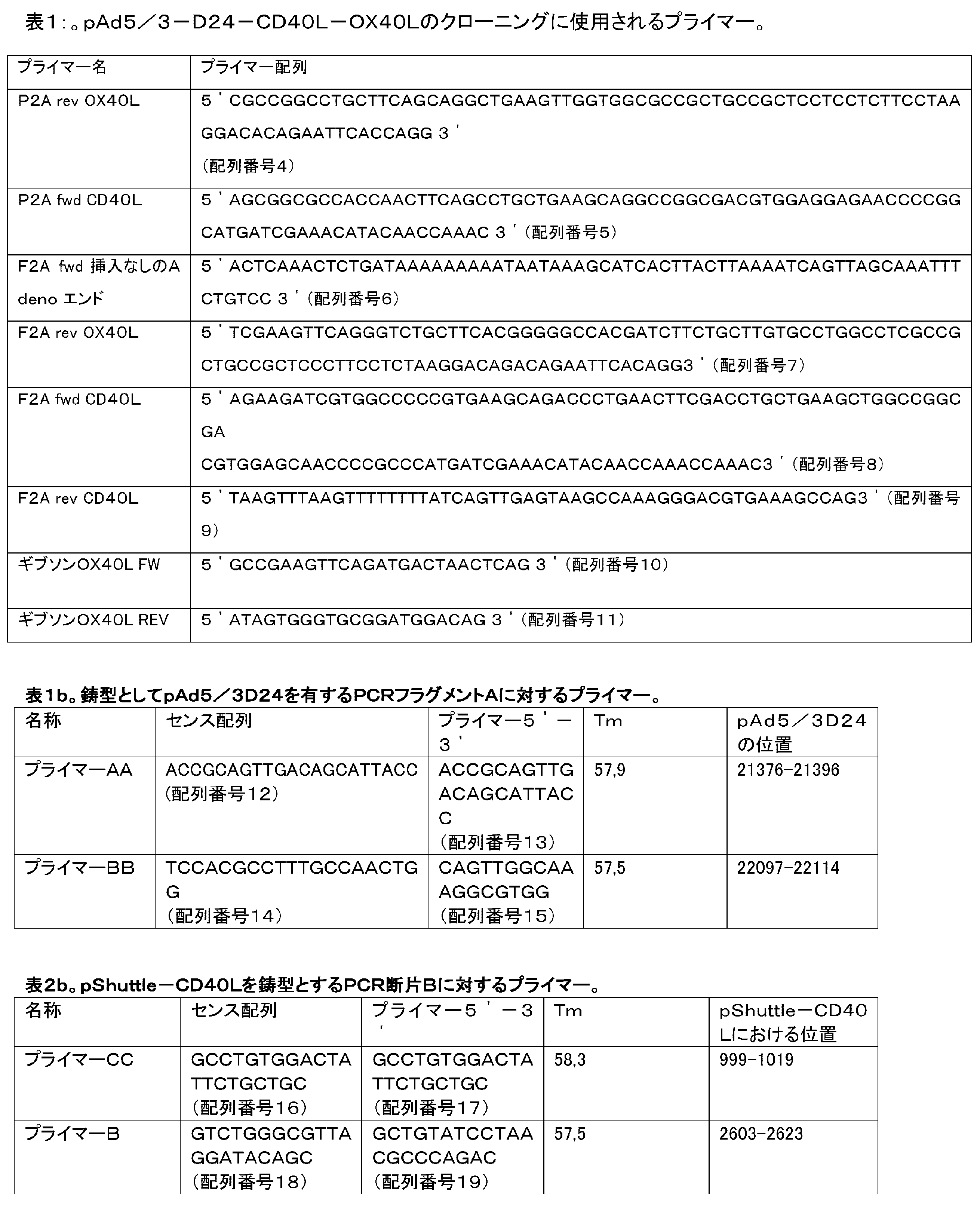

Claims (17)
a) 塩基対923~946をコードするヌクレオチドの欠失である、E1A遺伝子欠失;
b) 血清型5 Adのノブが血清型3 Adのノブによって置換されている、アデノウイルス繊維タンパク質のノブの5/3キメラ置換;
c) 14.7k遺伝子欠失であって、前記欠失が野生型アデノウイルスに関して塩基対30446~30460の欠失であり、欠失した配列GGA GGA GAT GAC TGAが、GGA GGA GAC GAC TGAに置換されているもの;
d) gp19k遺伝子欠失および7.1k遺伝子欠失であって、前記欠失が野生型アデノウイルスに関して塩基対28541~29211の欠失であるもの;
を含む、改変された複製アデノウイルス。 A modified replicating adenovirus of serotype 5 having lytic activity in target cancer cells, comprising:
a) E1A gene deletion, which is a deletion of nucleotides encoding base pairs 923-946;
b) 5/3 chimeric replacement of the knob of the adenovirus fiber protein, where the knob of serotype 5 Ad is replaced by the knob of serotype 3 Ad;
c) 14.7k gene deletion, said deletion being a deletion of base pairs 3044 6 to 30 460 with respect to wild-type adenovirus, and the deleted sequence GGA GGA GAT GAC TGA is GGA GGA GAC GAC TGA is replaced with;
d) gp19k gene deletion and 7.1k gene deletion, said deletion being a deletion of base pairs 28541-29211 with respect to wild-type adenovirus;
modified replicating adenoviruses, including:
Applications Claiming Priority (5)
| Application Number | Priority Date | Filing Date | Title |
|---|---|---|---|
| GB1804473.5 | 2018-03-21 | ||
| GBGB1804473.5A GB201804473D0 (en) | 2018-03-21 | 2018-03-21 | Modified oncolytic adenoviruses |
| GBGB1814867.6A GB201814867D0 (en) | 2018-03-21 | 2018-09-13 | Modified Oncolytic Adenoviruses |
| GB1814867.6 | 2018-09-13 | ||
| PCT/EP2019/056768 WO2019179977A1 (en) | 2018-03-21 | 2019-03-19 | Modified oncolytic adenoviruses |
Publications (2)
| Publication Number | Publication Date |
|---|---|
| JP2021518126A JP2021518126A (en) | 2021-08-02 |
| JP7417533B2 true JP7417533B2 (en) | 2024-01-18 |
Family
ID=62017873
Family Applications (1)
| Application Number | Title | Priority Date | Filing Date |
|---|---|---|---|
| JP2020550115A Active JP7417533B2 (en) | 2018-03-21 | 2019-03-19 | Modified oncolytic adenovirus |
Country Status (13)
| Country | Link |
|---|---|
| US (1) | US20210017501A1 (en) |
| EP (1) | EP3768305B1 (en) |
| JP (1) | JP7417533B2 (en) |
| KR (1) | KR102760462B1 (en) |
| CN (1) | CN112118864B (en) |
| AU (1) | AU2019239033B2 (en) |
| CA (1) | CA3094131A1 (en) |
| DK (1) | DK3768305T5 (en) |
| ES (1) | ES2935745T3 (en) |
| FI (1) | FI3768305T3 (en) |
| GB (2) | GB201804473D0 (en) |
| WO (1) | WO2019179977A1 (en) |
| ZA (1) | ZA202005848B (en) |
Families Citing this family (9)
| Publication number | Priority date | Publication date | Assignee | Title |
|---|---|---|---|---|
| EP2971008B1 (en) | 2013-03-14 | 2018-07-25 | Salk Institute for Biological Studies | Oncolytic adenovirus compositions |
| CN108064275A (en) | 2014-09-24 | 2018-05-22 | 萨克生物研究学院 | Oncolytic tumour virus and application method |
| ES2933174T3 (en) | 2016-02-23 | 2023-02-02 | Salk Inst For Biological Studi | Exogenous gene expression in therapeutic adenovirus for minimal impact on viral kinetics |
| WO2017147265A1 (en) | 2016-02-23 | 2017-08-31 | Salk Institute For Biological Studies | High throughput assay for measuring adenovirus replication kinetics |
| EP3532082A4 (en) | 2016-12-12 | 2020-08-26 | Salk Institute for Biological Studies | SYNTHETIC ADENOVIRUS DIRECTIVE TO TUMORS AND USES THEREOF |
| GB201804468D0 (en) * | 2018-03-21 | 2018-05-02 | Valo Therapeutics Oy | PeptiCRAd Cancer Therapy |
| JP7430395B2 (en) | 2018-04-09 | 2024-02-13 | ソーク インスティテュート フォー バイオロジカル スタディーズ | Oncolytic adenovirus compositions with enhanced replication characteristics |
| CN113699122B (en) * | 2021-08-31 | 2023-10-20 | 中国科学院大学宁波华美医院 | Polygene fusion oncolytic adenovirus and construction method and application thereof |
| EP4404947A4 (en) | 2021-09-23 | 2025-12-10 | Sagittarius Bio Inc | Adenoviras and methods for using adenoviras |
Citations (3)
| Publication number | Priority date | Publication date | Assignee | Title |
|---|---|---|---|---|
| JP2003501097A (en) | 1999-06-07 | 2003-01-14 | ザ・ユニバーシティ・オブ・ブリティッシュ・コロンビア | Inhibition of apoptosis by adenovirus E3 / 6.7K |
| JP2016522805A (en) | 2013-04-18 | 2016-08-04 | ティーアイエルティー・バイオセラピューティクス・オーワイTILT Biotherapeutics Oy | Enhanced adoptive cell therapy |
| WO2017118864A1 (en) | 2016-01-08 | 2017-07-13 | Replimune Limited | Modified oncolytic viurs |
Family Cites Families (13)
| Publication number | Priority date | Publication date | Assignee | Title |
|---|---|---|---|---|
| EP1369487A3 (en) * | 1994-08-16 | 2004-04-07 | Crucell Holland B.V. | Recombinant vectors derived from adenovirus for use in gene therapy |
| US7829329B2 (en) * | 1998-04-24 | 2010-11-09 | Onyx Pharmaceuticals, Inc. | Adenoviral vectors for treating disease |
| CN1357050A (en) * | 1999-07-02 | 2002-07-03 | 昂尼克斯药物公司 | Adenoviral vectors for disease treatment |
| WO2010051367A1 (en) * | 2008-10-31 | 2010-05-06 | The Trustees Of The University Of Pennsylvania | Simian adenoviruses sadv-43, -45,-48,-49, and -50 and uses thereof |
| KR101761094B1 (en) * | 2008-12-22 | 2017-08-04 | 터르고백스 오와이 | Oncolytic adenoviral vectors and methods and uses related thereto |
| RU2012107702A (en) * | 2009-07-31 | 2013-09-10 | Пэксвэкс, Инк. | ADENOVIRUS VECTORS |
| CN103221423B (en) * | 2010-09-24 | 2015-09-30 | 昂克斯治疗有限公司 | Oncolytic adenovirus carrier and relative method and purposes |
| CN107073089A (en) * | 2014-05-19 | 2017-08-18 | 瓦洛治疗公司 | coated oncolytic adenovirus for cancer vaccine |
| TWI636271B (en) * | 2014-11-20 | 2018-09-21 | 力智電子股份有限公司 | Apparatus and method for measuring capacity of cell module |
| EP3463405A4 (en) * | 2016-05-27 | 2020-02-26 | DNAtrix, Inc. | ADENOVIRUS AND IMMUNOMODULATORY POLYTHERAPY |
| PL3293201T3 (en) * | 2016-09-12 | 2021-06-14 | Targovax Oy | Combining adenovirus and checkpoint inhibitors for treating cancer |
| US10232053B2 (en) * | 2016-12-30 | 2019-03-19 | Trieza Therapeutics, Inc. | Immunomodulatory oncolytic adenoviral vectors, and methods of production and use thereof for treatment of cancer |
| GB201804468D0 (en) * | 2018-03-21 | 2018-05-02 | Valo Therapeutics Oy | PeptiCRAd Cancer Therapy |
-
2018
- 2018-03-21 GB GBGB1804473.5A patent/GB201804473D0/en not_active Ceased
- 2018-09-13 GB GBGB1814867.6A patent/GB201814867D0/en not_active Ceased
-
2019
- 2019-03-19 CA CA3094131A patent/CA3094131A1/en active Pending
- 2019-03-19 ES ES19712178T patent/ES2935745T3/en active Active
- 2019-03-19 JP JP2020550115A patent/JP7417533B2/en active Active
- 2019-03-19 AU AU2019239033A patent/AU2019239033B2/en active Active
- 2019-03-19 CN CN201980032730.0A patent/CN112118864B/en active Active
- 2019-03-19 EP EP19712178.3A patent/EP3768305B1/en active Active
- 2019-03-19 KR KR1020207029697A patent/KR102760462B1/en active Active
- 2019-03-19 FI FIEP19712178.3T patent/FI3768305T3/en active
- 2019-03-19 US US17/040,017 patent/US20210017501A1/en active Pending
- 2019-03-19 DK DK19712178.3T patent/DK3768305T5/en active
- 2019-03-19 WO PCT/EP2019/056768 patent/WO2019179977A1/en not_active Ceased
-
2020
- 2020-09-21 ZA ZA2020/05848A patent/ZA202005848B/en unknown
Patent Citations (3)
| Publication number | Priority date | Publication date | Assignee | Title |
|---|---|---|---|---|
| JP2003501097A (en) | 1999-06-07 | 2003-01-14 | ザ・ユニバーシティ・オブ・ブリティッシュ・コロンビア | Inhibition of apoptosis by adenovirus E3 / 6.7K |
| JP2016522805A (en) | 2013-04-18 | 2016-08-04 | ティーアイエルティー・バイオセラピューティクス・オーワイTILT Biotherapeutics Oy | Enhanced adoptive cell therapy |
| WO2017118864A1 (en) | 2016-01-08 | 2017-07-13 | Replimune Limited | Modified oncolytic viurs |
Non-Patent Citations (1)
| Title |
|---|
| Cancer Gene Ther.,2008年,Vol. 15,pp.9-17 |
Also Published As
| Publication number | Publication date |
|---|---|
| KR102760462B1 (en) | 2025-01-31 |
| CA3094131A1 (en) | 2019-09-26 |
| AU2019239033A1 (en) | 2020-10-08 |
| DK3768305T5 (en) | 2024-08-19 |
| AU2019239033B2 (en) | 2025-03-06 |
| GB201814867D0 (en) | 2018-10-31 |
| CN112118864B (en) | 2024-06-25 |
| FI3768305T3 (en) | 2023-01-31 |
| RU2020131337A (en) | 2022-04-28 |
| ZA202005848B (en) | 2022-01-26 |
| EP3768305B1 (en) | 2022-11-30 |
| DK3768305T3 (en) | 2023-01-23 |
| BR112020019303A2 (en) | 2021-01-05 |
| JP2021518126A (en) | 2021-08-02 |
| ES2935745T3 (en) | 2023-03-09 |
| CN112118864A (en) | 2020-12-22 |
| EP3768305A1 (en) | 2021-01-27 |
| US20210017501A1 (en) | 2021-01-21 |
| GB201804473D0 (en) | 2018-05-02 |
| KR20200135811A (en) | 2020-12-03 |
| WO2019179977A1 (en) | 2019-09-26 |
Similar Documents
| Publication | Publication Date | Title |
|---|---|---|
| JP7417533B2 (en) | Modified oncolytic adenovirus | |
| JP6928380B2 (en) | Oncolytic adenovirus encoding B7 protein | |
| KR102857666B1 (en) | Group B adenovirus encoding anti-TCR-complex antibodies or fragments | |
| TR201802728T4 (en) | Oncolytic adenoviruses equipped with heterologous genes. | |
| CN110856751A (en) | Therapeutic agents comprising nucleic acid and TCR-modified immune cells and uses thereof | |
| CN112135902A (en) | cancer therapy | |
| US20230190922A1 (en) | Recombinant MVA Viruses for Intratumoral and/or Intravenous Administration for Treating Cancer | |
| RU2788638C2 (en) | Modified oncolytic adeno viruses | |
| RU2788638C9 (en) | Modified oncolytic adeno viruses | |
| EP4452305A1 (en) | Recombinant mva viruses for intraperitoneal administration for treating cancer | |
| HK40044918A (en) | Modified oncolytic adenoviruses | |
| HK40044918B (en) | Modified oncolytic adenoviruses | |
| BR112020019303B1 (en) | MODIFIED REPLICATING ADENOVIRUS SEROTYPE 5 HAVING LYTIC ACTIVITY ON TARGET CANCER CELLS, PHARMACEUTICAL COMPOSITION AND USE | |
| AU2019385665B2 (en) | Therapy for treating cancer with an intratumoral and/or intravenous administration of a recombinant mva encoding 4-ibbl (cd137l) and/or cd40l |
Legal Events
| Date | Code | Title | Description |
|---|---|---|---|
| A529 | Written submission of copy of amendment under article 34 pct |
Free format text: JAPANESE INTERMEDIATE CODE: A529 Effective date: 20201117 |
|
| A621 | Written request for application examination |
Free format text: JAPANESE INTERMEDIATE CODE: A621 Effective date: 20220311 |
|
| A977 | Report on retrieval |
Free format text: JAPANESE INTERMEDIATE CODE: A971007 Effective date: 20230125 |
|
| A131 | Notification of reasons for refusal |
Free format text: JAPANESE INTERMEDIATE CODE: A131 Effective date: 20230207 |
|
| A521 | Request for written amendment filed |
Free format text: JAPANESE INTERMEDIATE CODE: A523 Effective date: 20230502 |
|
| A131 | Notification of reasons for refusal |
Free format text: JAPANESE INTERMEDIATE CODE: A131 Effective date: 20230801 |
|
| A521 | Request for written amendment filed |
Free format text: JAPANESE INTERMEDIATE CODE: A523 Effective date: 20231013 |
|
| TRDD | Decision of grant or rejection written | ||
| A01 | Written decision to grant a patent or to grant a registration (utility model) |
Free format text: JAPANESE INTERMEDIATE CODE: A01 Effective date: 20231219 |
|
| A61 | First payment of annual fees (during grant procedure) |
Free format text: JAPANESE INTERMEDIATE CODE: A61 Effective date: 20240105 |
|
| R150 | Certificate of patent or registration of utility model |
Ref document number: 7417533 Country of ref document: JP Free format text: JAPANESE INTERMEDIATE CODE: R150 |