JP7334127B2 - Efficient delivery of large cargoes to cells on porous supports - Google Patents
Efficient delivery of large cargoes to cells on porous supports Download PDFInfo
- Publication number
- JP7334127B2 JP7334127B2 JP2020005893A JP2020005893A JP7334127B2 JP 7334127 B2 JP7334127 B2 JP 7334127B2 JP 2020005893 A JP2020005893 A JP 2020005893A JP 2020005893 A JP2020005893 A JP 2020005893A JP 7334127 B2 JP7334127 B2 JP 7334127B2
- Authority
- JP
- Japan
- Prior art keywords
- membrane
- cells
- reservoir chamber
- cargo
- porous membrane
- Prior art date
- Legal status (The legal status is an assumption and is not a legal conclusion. Google has not performed a legal analysis and makes no representation as to the accuracy of the status listed.)
- Active
Links
Images


Classifications
-
- C—CHEMISTRY; METALLURGY
- C12—BIOCHEMISTRY; BEER; SPIRITS; WINE; VINEGAR; MICROBIOLOGY; ENZYMOLOGY; MUTATION OR GENETIC ENGINEERING
- C12N—MICROORGANISMS OR ENZYMES; COMPOSITIONS THEREOF; PROPAGATING, PRESERVING, OR MAINTAINING MICROORGANISMS; MUTATION OR GENETIC ENGINEERING; CULTURE MEDIA
- C12N15/00—Mutation or genetic engineering; DNA or RNA concerning genetic engineering, vectors, e.g. plasmids, or their isolation, preparation or purification; Use of hosts therefor
- C12N15/09—Recombinant DNA-technology
- C12N15/87—Introduction of foreign genetic material using processes not otherwise provided for, e.g. co-transformation
- C12N15/89—Introduction of foreign genetic material using processes not otherwise provided for, e.g. co-transformation using microinjection
-
- C—CHEMISTRY; METALLURGY
- C12—BIOCHEMISTRY; BEER; SPIRITS; WINE; VINEGAR; MICROBIOLOGY; ENZYMOLOGY; MUTATION OR GENETIC ENGINEERING
- C12N—MICROORGANISMS OR ENZYMES; COMPOSITIONS THEREOF; PROPAGATING, PRESERVING, OR MAINTAINING MICROORGANISMS; MUTATION OR GENETIC ENGINEERING; CULTURE MEDIA
- C12N15/00—Mutation or genetic engineering; DNA or RNA concerning genetic engineering, vectors, e.g. plasmids, or their isolation, preparation or purification; Use of hosts therefor
- C12N15/09—Recombinant DNA-technology
- C12N15/87—Introduction of foreign genetic material using processes not otherwise provided for, e.g. co-transformation
-
- C—CHEMISTRY; METALLURGY
- C12—BIOCHEMISTRY; BEER; SPIRITS; WINE; VINEGAR; MICROBIOLOGY; ENZYMOLOGY; MUTATION OR GENETIC ENGINEERING
- C12M—APPARATUS FOR ENZYMOLOGY OR MICROBIOLOGY; APPARATUS FOR CULTURING MICROORGANISMS FOR PRODUCING BIOMASS, FOR GROWING CELLS OR FOR OBTAINING FERMENTATION OR METABOLIC PRODUCTS, i.e. BIOREACTORS OR FERMENTERS
- C12M25/00—Means for supporting, enclosing or fixing the microorganisms, e.g. immunocoatings
- C12M25/02—Membranes; Filters
-
- C—CHEMISTRY; METALLURGY
- C12—BIOCHEMISTRY; BEER; SPIRITS; WINE; VINEGAR; MICROBIOLOGY; ENZYMOLOGY; MUTATION OR GENETIC ENGINEERING
- C12M—APPARATUS FOR ENZYMOLOGY OR MICROBIOLOGY; APPARATUS FOR CULTURING MICROORGANISMS FOR PRODUCING BIOMASS, FOR GROWING CELLS OR FOR OBTAINING FERMENTATION OR METABOLIC PRODUCTS, i.e. BIOREACTORS OR FERMENTERS
- C12M33/00—Means for introduction, transport, positioning, extraction, harvesting, peeling or sampling of biological material in or from the apparatus
- C12M33/04—Means for introduction, transport, positioning, extraction, harvesting, peeling or sampling of biological material in or from the apparatus by injection or suction, e.g. using pipettes, syringes, needles
-
- C—CHEMISTRY; METALLURGY
- C12—BIOCHEMISTRY; BEER; SPIRITS; WINE; VINEGAR; MICROBIOLOGY; ENZYMOLOGY; MUTATION OR GENETIC ENGINEERING
- C12M—APPARATUS FOR ENZYMOLOGY OR MICROBIOLOGY; APPARATUS FOR CULTURING MICROORGANISMS FOR PRODUCING BIOMASS, FOR GROWING CELLS OR FOR OBTAINING FERMENTATION OR METABOLIC PRODUCTS, i.e. BIOREACTORS OR FERMENTERS
- C12M35/00—Means for application of stress for stimulating the growth of microorganisms or the generation of fermentation or metabolic products; Means for electroporation or cell fusion
- C12M35/04—Mechanical means, e.g. sonic waves, stretching forces, pressure or shear stimuli
Landscapes
- Health & Medical Sciences (AREA)
- Engineering & Computer Science (AREA)
- Life Sciences & Earth Sciences (AREA)
- Genetics & Genomics (AREA)
- Organic Chemistry (AREA)
- Bioinformatics & Cheminformatics (AREA)
- Wood Science & Technology (AREA)
- Zoology (AREA)
- Chemical & Material Sciences (AREA)
- Biotechnology (AREA)
- Biomedical Technology (AREA)
- General Engineering & Computer Science (AREA)
- Biochemistry (AREA)
- General Health & Medical Sciences (AREA)
- Microbiology (AREA)
- Sustainable Development (AREA)
- Cell Biology (AREA)
- Molecular Biology (AREA)
- Physics & Mathematics (AREA)
- Biophysics (AREA)
- Plant Pathology (AREA)
- Mechanical Engineering (AREA)
- Immunology (AREA)
- Micro-Organisms Or Cultivation Processes Thereof (AREA)
- Apparatus Associated With Microorganisms And Enzymes (AREA)
Description
関連出願の相互参照
本出願は、2014年3月28日に出願されたUSSN61/972,145の利益及び優先権を主張するものであり、この出願はあらゆる目的のために参照によってその全体を本明細書に組み込んだものとする。
政府支援の陳述
[適用なし]
CROSS-REFERENCE TO RELATED APPLICATIONS This application claims the benefit of and priority to USSN 61/972,145 filed March 28, 2014, which application is hereby incorporated by reference in its entirety for all purposes. shall be incorporated in the specification.
Statement of Government Support [Not applicable]
タンパク質、DNA、RNA、染色体、核、ならびに量子ドット、表面増強ラマン散乱(SERS)粒子及びミクロビーズなどの無生物粒子を含むカーゴを1サイズの広い範囲にわたって哺乳動物細胞に運搬することは、生物学の多くの分野において極めて望ましい。エンドサイトーシスなどの送達方法はエンドソームにカーゴを封入することができ、この場合、pHが低い微小環境及び溶菌酵素がカーゴ分解につながることが多い(Luo and Saltzman(2000) Nat. Biotechnol. 18: 33-37)。ウイルス及び化学的送達方法は、ウイルス内にカーゴをパッケージングするか、または取り込みを向上させる化学的複合体を形成する(Naldiniら、(1996) Science, 272: 263-267; Felgnerら、(1987) Proc. Natl. Acad. Sci. USA, 84: 7413-7417)。しかしながら、毒性、細胞種固有の取り込み、さらに重要なことに限られたカーゴ詰め込み容量がカーゴサイズ及び運搬可能な細胞種に著しい制約を課す(Luo and Saltzman、上記)。 The delivery of cargo, including proteins, DNA, RNA, chromosomes, nuclei, and inanimate particles such as quantum dots, surface-enhanced Raman scattering (SERS) particles and microbeads, into mammalian cells over a wide range of one size has been shown to be useful in biology. highly desirable in many areas of Delivery methods such as endocytosis can entrap cargo in endosomes, where low pH microenvironments and lytic enzymes often lead to cargo degradation (Luo and Saltzman (2000) Nat. Biotechnol. 18: 33-37). Viral and chemical delivery methods package cargo within viruses or form chemical complexes that enhance uptake (Naldini et al., (1996) Science, 272: 263-267; Felgner et al., (1987). ) Proc. Natl. Acad. Sci. USA, 84: 7413-7417). However, toxicity, cell-type specific uptake and, more importantly, limited cargo packing capacity impose significant constraints on cargo size and transportable cell types (Luo and Saltzman, supra).
物理的運搬方法としては、ランダムに分散したナノスケールの孔をもたらすエレクトロポレーション(Chuら、(1987) Nucleic Acids Res. 15: 1311-1326)及びソノポレーション(Mitragotri(2005) Nat. Rev. Drug Discovery, 4: 255-260)、ならびにレーザー焦点において細胞膜に孔を形成するオプトポレーション(Tirlapur and Konig(2002) Nature, 418: 290-291; Vogelら、(2005) Appl. Phys. B: Laser Opt., 81: 1015-1047; Clarkら、(2006) J. Biomed. Opt., 11: 014034)が挙げられる。こうした孔を通って、小さなカーゴは熱拡散または電場によって細胞に送達される。こうした方法による大きなカーゴの送達は、遅いカーゴの拡散速度及び孔サイズの増加に伴う細胞生存率の低下のために効率が低い(Stevensonら、(2006) Opt. Express, 14: 7125-7133)。マイクロキャピラリー注入(King(2004) Methods in Molecular Biology 245: Gene Delivery to Mammalian Cells 1; Humana Press Inc.: Totowa, NJ)は、鋭いガラスの先端を使用して送達のために機械的に細胞膜を貫通させる。しかしながら、細胞生存率を維持するために、膜貫通による機械的外傷は典型的なピペットの先端の直径を0.5umに制限する(Hanら、(2998) J. Nanomed. Nanotechnol. Biol. Med., 4: 215-225)。 Physical delivery methods include electroporation (Chu et al. (1987) Nucleic Acids Res. 15: 1311-1326) and sonoporation (Mitragotri (2005) Nat. Rev.), which leads to randomly distributed nanoscale pores. Drug Discovery, 4: 255-260), and optoporation, which forms pores in the cell membrane at the laser focus (Tirlapur and Konig (2002) Nature, 418: 290-291; Vogel et al., (2005) Appl. Phys. B: Laser Opt., 81: 1015-1047; Clark et al., (2006) J. Biomed. Opt., 11: 014034). Through these pores small cargoes are delivered to cells by thermal diffusion or electric fields. Delivery of large cargoes by such methods is less efficient due to slow cargo diffusion rates and decreased cell viability with increasing pore size (Stevenson et al. (2006) Opt. Express, 14: 7125-7133). Microcapillary injection (King (2004) Methods in Molecular Biology 245: Gene Delivery to Mammalian Cells 1; Humana Press Inc.: Totowa, NJ) uses a sharp glass tip to mechanically penetrate cell membranes for delivery. Let However, to maintain cell viability, transmembrane mechanical trauma limits the typical pipette tip diameter to 0.5 um (Han et al. (2998) J. Nanomed. Nanotechnol. Biol. Med. , 4:215-225).
ピペットを詰まらせ、カーゴを切断するため、ピペットの先端よりも大きなカーゴを注入することはできない。エレクトロポレーションをマイクロキャピラリー注入と組み合わせる電気注入は、接触する細胞膜を電場により軟化させた後に穏やかに細胞へ機械的に貫通させることによってRNA及びプラスミドDNAなどの小分子の生細胞への送達(Boudesら、(208) J. Neurosci. Meth., 170: 204-211; Kitamuraら、(2008) Nat. Meth., 5: 61-67)ならびに人工脂質ベシクルへの細菌送達(Hurtig and Orwar(2008) Soft Matter, 4: 1515-1520)を実証した。あるいは、単純脂質アシストマイクロインジェクション(SLAM)技術(Laffafian and Hallett(1998) Biophys. J., 75:2558-2563)は、ガラスマイクロキャピラリーの先端において合成脂質分子を組み込む。SLAMマイクロピペットの細胞膜との接触は、脂質分子を細胞膜と融合させて、カーゴ送達のための連続的及び一時的な経路を形成する。この方法は、細胞膜を貫くマイクロピペットの先端のジグザグに刺す動きを回避する。しかしながら、カーゴ及び細胞膜との親油性相互作用は、細胞ならびに送達カーゴに望ましくない生物学的影響を与える可能性があり、この方法を特定の細胞種及びカーゴ含量に限定する。 Cargo larger than the tip of the pipette cannot be injected as this will clog the pipette and cut the cargo. Electroinjection, which combines electroporation with microcapillary injection, delivers small molecules such as RNA and plasmid DNA into living cells by softening the contacting cell membrane with an electric field and then gently mechanically penetrating the cell (Boudes et al., 2002). (208) J. Neurosci. Meth., 170: 204-211; Kitamura et al., (2008) Nat. Soft Matter, 4: 1515-1520). Alternatively, the simple lipid-assisted microinjection (SLAM) technique (Laffafian and Hallett (1998) Biophys. J., 75:2558-2563) incorporates synthetic lipid molecules at the tips of glass microcapillaries. Contact of the SLAM micropipette with the cell membrane causes the lipid molecules to fuse with the cell membrane, forming a continuous and transient pathway for cargo delivery. This method avoids the zigzagging motion of the micropipette tip through the cell membrane. However, lipophilic interactions with cargo and cell membranes can have undesirable biological effects on cells and delivered cargo, limiting this method to certain cell types and cargo contents.
さまざまな態様において、本明細書において意図される本発明(複数可)は、以下の実施形態のいずれか1つ以上を含んでもよいが、これらに限定される必要はない。 In various aspects, the invention(s) contemplated herein may include, but need not be limited to, any one or more of the following embodiments.
実施形態1:大きなカーゴを真核細胞に送達する方法であり、前記方法は、多孔質膜の片側に配置された前記細胞を準備することと、前記多孔質膜の反対側にあるリザーバ室に配置された溶液中の前記カーゴを準備することと、前記カーゴを、前記多孔質膜を構成する孔を通過させるのに十分な圧力を前記リザーバ室に印加することとを含み、前記カーゴは細胞膜を通って前記細胞に入る、前記方法。 Embodiment 1: A method of delivering large cargo to eukaryotic cells, said method comprising providing said cells located on one side of a porous membrane and placing said cells in a reservoir chamber on the opposite side of said porous membrane. providing the cargo in a deposited solution; and applying a pressure to the reservoir chamber sufficient to force the cargo through the pores that make up the porous membrane, wherein the cargo is in the cell membrane. The method, wherein the cell is entered through
実施形態2:前記リザーバ室の容積は、約10μLから約500μLまで、または約40μLから約500μLまで、または約50μLから約400μLまで、または約60μLから約300μLまで、または約70μLから約200μLまで、または約80μLから約150μLまで、または約10μLから約1mLまで、または約10μLから約500μLまで、または約10μLから約100μLまでに及ぶ、実施形態1に記載の方法。 Embodiment 2: the volume of said reservoir chamber is from about 10 μL to about 500 μL, or from about 40 μL to about 500 μL, or from about 50 μL to about 400 μL, or from about 60 μL to about 300 μL, or from about 70 μL to about 200 μL; or from about 80 μL to about 150 μL, or from about 10 μL to about 1 mL, or from about 10 μL to about 500 μL, or from about 10 μL to about 100 μL.
実施形態3:前記リザーバ室の容積は約100μLである、実施形態2に記載の方法。 Embodiment 3: The method of embodiment 2, wherein the reservoir chamber has a volume of about 100 μL.
実施形態4:前記多孔質膜の厚さは、約5μmから約30μm、または約5μmから約20μm、または約5μmから約15μmに及ぶ、実施形態1~3のいずれか1つに記載の方法。 Embodiment 4: The method of any one of embodiments 1-3, wherein the porous membrane has a thickness ranging from about 5 μm to about 30 μm, or from about 5 μm to about 20 μm, or from about 5 μm to about 15 μm.
実施形態5:前記多孔質膜の厚さは約10μmである、実施形態4に記載の方法。 Embodiment 5: The method of embodiment 4, wherein the porous membrane has a thickness of about 10 μm.
実施形態6:前記多孔質膜の孔サイズの平均または中央値は、約100nmから約20μmまでまたは約20μmまで、または約500nmから約8μmまで、または約1μmから約5μmまでに及ぶ、実施形態1~5のいずれか1つに記載の方法。 Embodiment 6: The average or median pore size of said porous membrane ranges from about 100 nm to about 20 μm, or to about 20 μm, or from about 500 nm to about 8 μm, or from about 1 μm to about 5 μm, Embodiment 1 6. The method according to any one of 1 to 5.
実施形態7:前記多孔質膜の孔サイズの中央値または平均は約1μmである、実施形態6に記載の方法。 Embodiment 7: The method of embodiment 6, wherein the median or average pore size of said porous membrane is about 1 μm.
実施形態8:前記多孔質膜の孔サイズの中央値または平均は約3μmである、実施形態6に記載の方法。 Embodiment 8: The method of embodiment 6, wherein the median or average pore size of said porous membrane is about 3 μm.
実施形態9:前記多孔質膜の孔サイズの中央値または平均は約5μmである、実施形態6に記載の方法。 Embodiment 9: The method of embodiment 6, wherein the median or average pore size of said porous membrane is about 5 μm.
実施形態10:前記多孔質膜は、約1×10⁵孔/cm2から最大約1×107孔/cm2、または約5×105孔/cm2から最大約5×106、または約1×105孔/cm2から最大約1×107孔/cm2を含む、実施形態1~9のいずれか1つに記載の方法。 Embodiment 10: The porous membrane has about 1 x 10⁵ pores/ cm2 up to about 1 x 10⁷ pores/ cm2 , or about 5 x 10⁵ pores/ cm2 up to about 5 x 10⁶ , or 10. The method of any one of embodiments 1-9, comprising from about 1×10 5 pores/cm 2 up to about 1×10 7 pores/cm 2 .
実施形態11:前記多孔質膜は、約1.6×10⁶孔/cm2で約1μm直径の平均孔サイズを含む、実施形態10に記載の方法。 Embodiment 11: The method of embodiment 10, wherein the porous membrane comprises an average pore size of about 1 μm diameter at about 1.6×10 6 pores/cm 2 .
実施形態12:前記多孔質膜は、約8×10⁵孔/cm2で約3μm直径の平均孔サイズを含む、実施形態10に記載の方法。 Embodiment 12: The method of embodiment 10, wherein the porous membrane comprises an average pore size of about 3 μm diameter at about 8×10 5 pores/cm 2 .
実施形態13:前記膜はポリマー膜を含む、実施形態1~12のいずれか1つに記載の方法。 Embodiment 13: The method of any one of embodiments 1-12, wherein said membrane comprises a polymer membrane.
実施形態14:前記膜は、ナイロン膜、ナイロンメッシュ、フィルター膜、ポリテトラフルオロエチレン(PTFE)膜、延伸ポリテトラフルオロエチレン(ePTFE)膜、ポリエステル膜、ポリエーテルエーテルケトン(PEEK)膜、延伸ポリエーテルエーテルケトン(ePEEK)膜、ポリエチレン(PE)膜、ポリプロピレン(PP)膜、ポリビニリデンフルオライド(PVDF)膜、エチルビニルアセタート(EVA)膜、熱可塑性ポリウレタン(TPU)膜、ポリエーテルスルホン(PES)膜、ポリカーボネート膜及びポリエチレンテレフタラート(PET)膜からなる群から選択される材料を含む、実施形態1~12のいずれか1つに記載の方法。 Embodiment 14: The membrane comprises nylon membrane, nylon mesh, filter membrane, polytetrafluoroethylene (PTFE) membrane, expanded polytetrafluoroethylene (ePTFE) membrane, polyester membrane, polyetheretherketone (PEEK) membrane, expanded poly Ether ether ketone (ePEEK) membrane, polyethylene (PE) membrane, polypropylene (PP) membrane, polyvinylidene fluoride (PVDF) membrane, ethyl vinyl acetate (EVA) membrane, thermoplastic polyurethane (TPU) membrane, polyether sulfone ( 13. The method of any one of embodiments 1-12, comprising a material selected from the group consisting of PES) membranes, polycarbonate membranes and polyethylene terephthalate (PET) membranes.
実施形態15:前記膜は、ポリエステル膜、ポリカーボネート膜またはポリエチレンテレフタラート(PET)膜を含む、実施形態14に記載の方法。 Embodiment 15: The method of embodiment 14, wherein said membrane comprises a polyester membrane, a polycarbonate membrane or a polyethylene terephthalate (PET) membrane.
実施形態16:前記圧力を印加することは、前記多孔質膜のゆがみを引き起こす、実施形態1~15のいずれか1つに記載の方法。 Embodiment 16: The method of any one of embodiments 1-15, wherein applying the pressure causes distortion of the porous membrane.
実施形態17:前記ゆがみは、約20μmから、または約50μmから、または約100μmから、または約500μmから約1cmまで、または約500mmまで、または約300mmまで、または約100mmまでに及ぶ、実施形態16に記載の方法。 Embodiment 17: Said deflection ranges from about 20 μm, or from about 50 μm, or from about 100 μm, or from about 500 μm to about 1 cm, or to about 500 mm, or to about 300 mm, or to about 100 mm, Embodiment 16 The method described in .
実施形態18:前記圧力を印加することは、一時的な圧力を印加することを含む、実施形態1~17のいずれか1つに記載の方法。 Embodiment 18: The method of any one of embodiments 1-17, wherein said applying pressure comprises applying a temporary pressure.
実施形態19:前記圧力を印加することは、圧力を約1ミリ秒から最大約1分間、または約100ミリ秒から最大約1分間、または約1秒から最大約1分間印加することを含む、実施形態1~18のいずれか1つに記載の方法。 Embodiment 19: Applying the pressure comprises applying pressure for about 1 millisecond up to about 1 minute, or about 100 milliseconds up to about 1 minute, or about 1 second up to about 1 minute. 19. The method of any one of embodiments 1-18.
実施形態20:前記圧力を印加することは、ポートを通して前記リザーバ室の中へ圧力を印加することを含む、実施形態1~19のいずれか1つに記載の方法。 Embodiment 20: The method of any one of embodiments 1-19, wherein applying pressure comprises applying pressure through a port into the reservoir chamber.
実施形態21:前記圧力を印加することは、前記リザーバ室が一杯で閉鎖されているときに前記リザーバ室の壁を撓ませることを含む、実施形態1~19のいずれか1つに記載の方法。 Embodiment 21: The method of any one of embodiments 1-19, wherein applying pressure comprises deflecting a wall of the reservoir chamber when the reservoir chamber is full and closed. .
実施形態22:前記圧力を印加することは、前記リザーバ室の壁を通して溶液を注入することを含む、実施形態1~19のいずれか1つに記載の方法。 Embodiment 22: The method of any one of Embodiments 1-19, wherein said applying pressure comprises injecting a solution through a wall of said reservoir chamber.
実施形態23:リザーバ室に配置された溶液中の前記カーゴを前記準備することは、ポートを通して前記リザーバ室に前記溶液を導入することを含む、実施形態1~22のいずれか1つに記載の方法。 Embodiment 23: According to any one of embodiments 1-22, wherein said preparing said cargo in a solution disposed in a reservoir chamber comprises introducing said solution into said reservoir chamber through a port. Method.
実施形態24:リザーバ室に配置された溶液中の前記カーゴを前記準備することは、前記カーゴ溶液を前記リザーバにピペットで入れることを含む、実施形態1~22のいずれか1つに記載の方法。 Embodiment 24: The method of any one of embodiments 1-22, wherein said preparing said cargo in a solution disposed in a reservoir chamber comprises pipetting said cargo solution into said reservoir. .
実施形態25:リザーバ室に配置された溶液中の前記カーゴを前記準備することは、前記多孔質膜を前記リバーザ室上または内に置く前に前記リザーバ室に装填することを含む、実施形態1~22のいずれか1つに記載の方法。 Embodiment 25: Said preparing said cargo in solution disposed in a reservoir chamber comprises loading said reservoir chamber prior to placing said porous membrane on or in said reservoir chamber. 23. The method of any one of .
実施形態26:リザーバ室に配置された溶液中の前記カーゴを前記準備することは、前記溶液を前記リザーバ室の壁を貫通する針を通して注入することを含む、実施形態1~22のいずれか1つに記載の方法。 Embodiment 26: Any one of embodiments 1-22, wherein said preparing said cargo in a solution disposed in a reservoir chamber comprises injecting said solution through a needle penetrating a wall of said reservoir chamber. the method described in Section 1.
実施形態27:リザーバ室に配置された溶液中の前記カーゴを前記準備することは、前記溶液を前記膜を通過させて前記リザーバ室に装填することを含む、実施形態1~22のいずれか1つに記載の方法。 Embodiment 27: Any one of embodiments 1-22, wherein said preparing said cargo in a solution disposed in a reservoir chamber comprises loading said solution through said membrane into said reservoir chamber the method described in Section 1.
実施形態28:前記カーゴは、リポソーム内にパッケージングされた天然染色体もしくは染色体フラグメント、合成染色体、細菌、合成粒子、細胞内真菌、細胞内原生動物、DNA及び/またはRNA(例えば、リポフェクタミン)ならびに細胞小器官からなる群から選択される1つあるいは部分を含む、実施形態1~27のいずれか1つに記載の方法。 Embodiment 28: The cargo comprises natural chromosomes or chromosome fragments, synthetic chromosomes, bacteria, synthetic particles, intracellular fungi, intracellular protozoa, DNA and/or RNA (e.g. Lipofectamine) and cells packaged in liposomes. 28. The method of any one of embodiments 1-27, comprising one or a portion selected from the group consisting of organelles.
実施形態29:前記カーゴは細胞核を含む、実施形態28に記載の方法。 Embodiment 29: The method of Embodiment 28, wherein said cargo comprises cell nuclei.
実施形態30:前記カーゴはミトコンドリアを含む、実施形態28に記載の方法。 Embodiment 30: The method of Embodiment 28, wherein said cargo comprises mitochondria.
実施形態31:前記カーゴは染色体または染色体フラグメントを含む、実施形態28に記載の方法。 Embodiment 31: The method of Embodiment 28, wherein said cargo comprises a chromosome or chromosome fragment.
実施形態32:前記カーゴは人工染色体を含む、実施形態28に記載の方法。 Embodiment 32: The method of embodiment 28, wherein said cargo comprises an artificial chromosome.
実施形態33:前記カーゴは細菌を含む、実施形態28に記載の方法。 Embodiment 33: The method of embodiment 28, wherein said cargo comprises bacteria.
実施形態34:前記細胞は、脊椎動物細胞、真菌細胞及び酵母菌細胞からなる群から選択される、実施形態1~33のいずれか1つに記載の方法。 Embodiment 34: The method of any one of embodiments 1-33, wherein said cells are selected from the group consisting of vertebrate cells, fungal cells and yeast cells.
実施形態35:前記細胞は、哺乳動物細胞、昆虫細胞及び無脊椎動物細胞からなる群から選択される、実施形態1~33のいずれか1つに記載の方法。 Embodiment 35: The method of any one of embodiments 1-33, wherein said cells are selected from the group consisting of mammalian cells, insect cells and invertebrate cells.
実施形態36:前記細胞は哺乳動物細胞を含む、実施形態35に記載の方法。 Embodiment 36: The method of embodiment 35, wherein said cells comprise mammalian cells.
実施形態37:前記細胞はヒト細胞を含む、実施形態35に記載の方法。 Embodiment 37: The method of Embodiment 35, wherein said cells comprise human cells.
実施形態38:前記細胞は非ヒト哺乳動物細胞を含む、実施形態35に記載の方法。 Embodiment 38: The method of embodiment 35, wherein said cells comprise non-human mammalian cells.
実施形態39:前記細胞はリンパ球または幹細胞を含む、実施形態36~38のいずれか1つに記載の方法。 Embodiment 39: The method of any one of embodiments 36-38, wherein said cells comprise lymphocytes or stem cells.
実施形態40:前記細胞は、成体幹細胞、胚性幹細胞、臍帯血幹細胞及び人工多能性幹細胞からなる群から選択される幹細胞を含む、実施形態39に記載の方法。 Embodiment 40: The method of embodiment 39, wherein said cells comprise stem cells selected from the group consisting of adult stem cells, embryonic stem cells, cord blood stem cells and induced pluripotent stem cells.
実施形態41:前記細胞は分化した体細胞を含む、実施形態36~38のいずれか1つに記載の方法。 Embodiment 41: The method of any one of embodiments 36-38, wherein said cells comprise differentiated somatic cells.
実施形態42:前記細胞は細胞株からの細胞を含む、実施形態1~33のいずれか1つに記載の方法。 Embodiment 42: The method of any one of embodiments 1-33, wherein said cells comprise cells from a cell line.
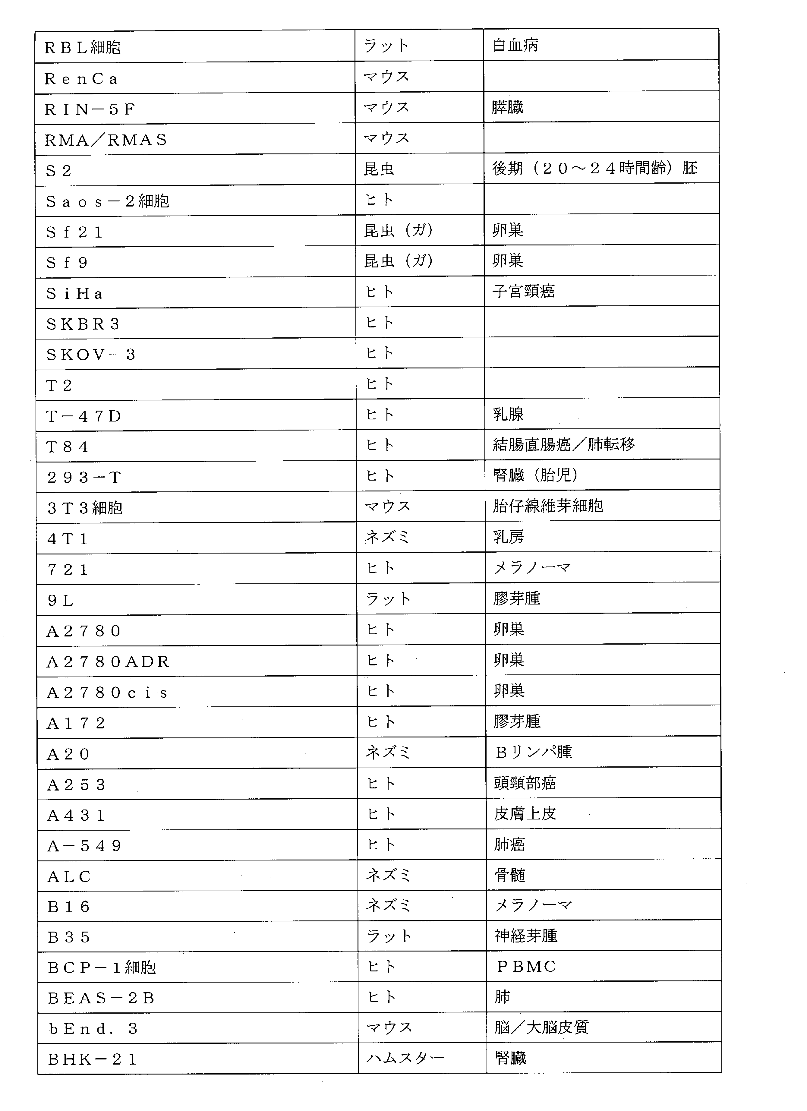
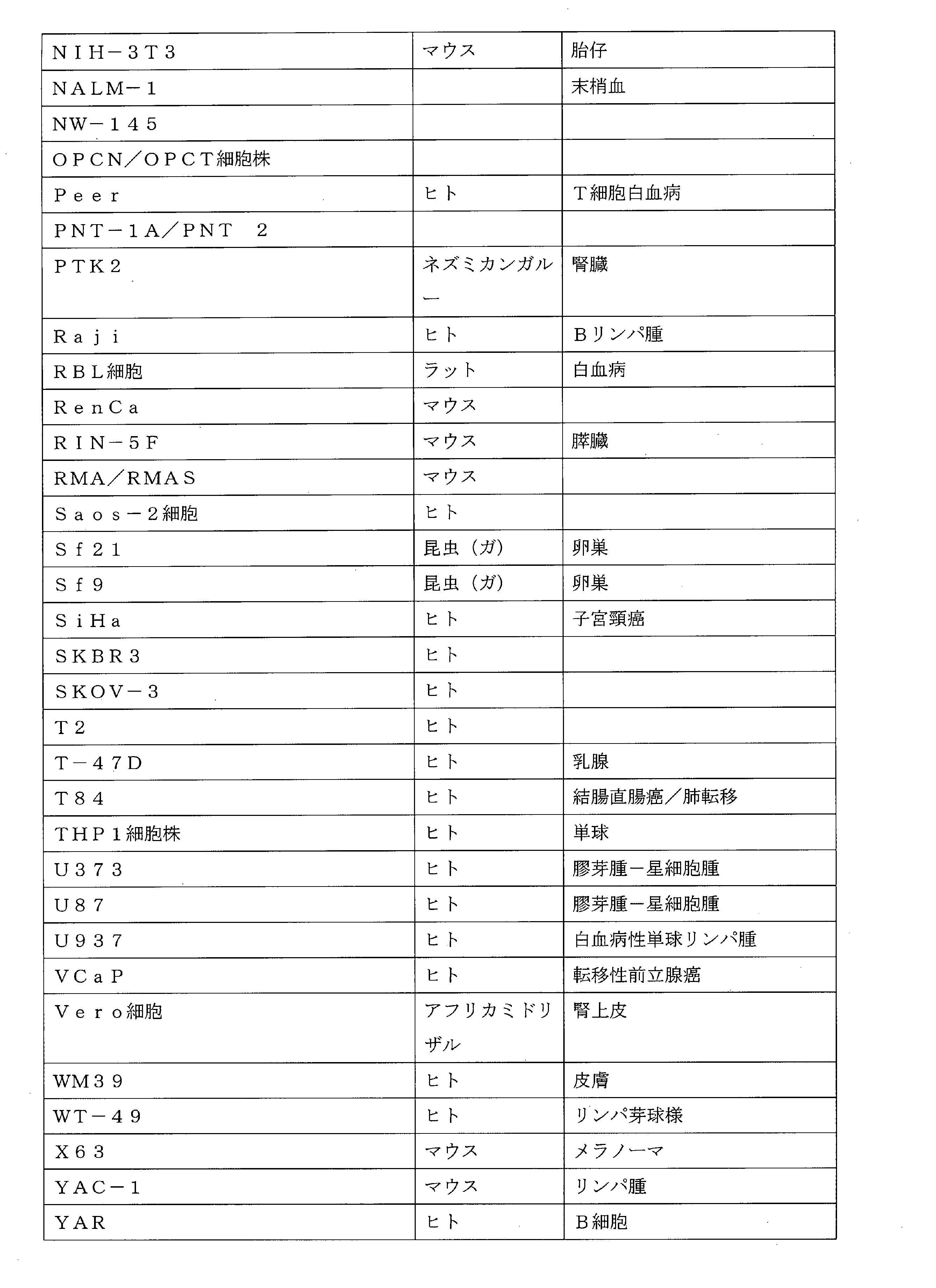
実施形態43:前記細胞は表1に列挙される細胞株からの細胞を含む、実施形態42に記載の方法。 Embodiment 43: The method of embodiment 42, wherein said cells comprise cells from cell lines listed in Table 1.
実施形態44:前記細胞は、HeLa、National Cancer Instituteの60種の癌細胞株(NCI60)、ESTDABデータベース、DU145(前立腺癌)、Lncap(前立腺癌)、MCF-7(乳癌)、MDA-MB-438(乳癌)、PC3(前立腺癌)、T47D(乳癌)、THP-1(急性骨髄性白血病)、U87(膠芽腫)、SHSY5Yヒト神経芽腫細胞、骨髄腫からクローン化される及びSaos-2細胞(骨癌)からなる群から選択される細胞株からの細胞を含む、実施形態42に記載の方法。 Embodiment 44: The cells are selected from HeLa, National Cancer Institute's 60 cancer cell lines (NCI60), ESTDAB database, DU145 (prostate cancer), Lncap (prostate cancer), MCF-7 (breast cancer), MDA-MB- 438 (breast cancer), PC3 (prostate cancer), T47D (breast cancer), THP-1 (acute myeloid leukemia), U87 (glioblastoma), cloned from SHSY5Y human neuroblastoma cells, myeloma and Saos- 43. The method of embodiment 42, comprising cells from a cell line selected from the group consisting of: 2 cells (bone cancer).
実施形態45:前記細胞は、前記多孔質膜上で培養される、実施形態1~44のいずれか1つに記載の方法。 Embodiment 45: The method of any one of embodiments 1-44, wherein said cells are cultured on said porous membrane.
実施形態46:前記細胞は、前記多孔質膜上で付着層として培養される、実施形態1~44のいずれか1つに記載の方法。 Embodiment 46: The method of any one of embodiments 1-44, wherein said cells are cultured as an adherent layer on said porous membrane.
実施形態47:前記細胞は、前記多孔質膜上でコンフルエンスになるまで培養される、実施形態45~46のいずれか1つに記載の方法。 Embodiment 47: The method of any one of embodiments 45-46, wherein said cells are cultured on said porous membrane to confluence.
実施形態48:前記多孔質膜は、金属薄膜または金属ナノ粒子を載せていない、実施形態1~47のいずれか1つに記載の方法。 Embodiment 48: The method of any one of embodiments 1-47, wherein the porous membrane is not loaded with a metal thin film or metal nanoparticles.
実施形態49:前記方法は、前記膜の表面を加熱することを含まない、実施形態1~48のいずれか1つに記載の方法。 Embodiment 49: The method of any one of embodiments 1-48, wherein said method does not comprise heating the surface of said membrane.
実施形態50:前記方法は、前記膜の表面をレーザーにより加熱することを含まない、実施形態1~48のいずれか1つに記載の方法。 Embodiment 50: The method of any one of embodiments 1-48, wherein said method does not include heating the surface of said film with a laser.
実施形態51:多孔質膜と、前記多孔質膜の片側にある容積が約500μL未満のリザーバ室とを備える大きなカーゴを真核細胞に送達するためのデバイス。 Embodiment 51: A device for delivering large cargo to eukaryotic cells comprising a porous membrane and a reservoir chamber having a volume of less than about 500 μL on one side of said porous membrane.
実施形態52:前記リザーバ室の容積は、約40μLから約500μL、または約50μLから約400μL、または約60μLから約300μL、または約70μLから約200μL、または約80μLから約150μL、または約10μLから約100μLまでに及ぶ、実施形態51に記載のデバイス。 Embodiment 52: The volume of said reservoir chamber is from about 40 μL to about 500 μL, or from about 50 μL to about 400 μL, or from about 60 μL to about 300 μL, or from about 70 μL to about 200 μL, or from about 80 μL to about 150 μL, or from about 10 μL to about 52. A device according to embodiment 51, ranging up to 100 μL.
実施形態53:前記リザーバ室の容積は約100μLである、実施形態2に記載のデバイス。 Embodiment 53: The device of Embodiment 2, wherein the reservoir chamber has a volume of about 100 μL.
実施形態54:前記多孔質膜の厚さは、約5μmから約30μm、または約5μmから約20μm、または約5μmから約15μmに及ぶ、実施形態51~53のいずれか1つに記載のデバイス。 Embodiment 54: The device of any one of embodiments 51-53, wherein the porous membrane has a thickness ranging from about 5 μm to about 30 μm, or from about 5 μm to about 20 μm, or from about 5 μm to about 15 μm.
実施形態55:前記多孔質膜の厚さは約10μmである、実施形態54に記載のデバイス。 Embodiment 55: The device of embodiment 54, wherein the porous membrane has a thickness of about 10 μm.
実施形態56:前記多孔質膜の孔サイズの平均または中央値は、約100nmから約20μmまでまたは約20μmまで、または約500nmから約8μmまで、または約1μmから約5μmまでに及ぶ、実施形態51~55のいずれか1つに記載のデバイス。 Embodiment 56: The average or median pore size of said porous membrane ranges from about 100 nm to about 20 μm, or to about 20 μm, or from about 500 nm to about 8 μm, or from about 1 μm to about 5 μm, embodiment 51 56. The device of any one of .
実施形態57:前記多孔質膜の孔サイズの中央値または平均は約1μmである、実施形態56に記載のデバイス。 Embodiment 57: The device of embodiment 56, wherein the porous membrane has a median or average pore size of about 1 μm.
実施形態58:前記多孔質膜の孔サイズの中央値または平均は約3μmである、実施形態56に記載のデバイス。 Embodiment 58: The device of embodiment 56, wherein the porous membrane has a median or average pore size of about 3 μm.
実施形態59:前記多孔質膜の孔サイズの中央値または平均は約5μmである、実施形態56に記載のデバイス。 Embodiment 59: The device of embodiment 56, wherein the porous membrane has a median or average pore size of about 5 μm.
実施形態60:前記多孔質膜は、約1×10⁵孔/cm2から最大約1×107孔/cm2、または約5×105孔/cm2から最大約5×106、または約1×105孔/cm2から最大約1×107孔/cm2を含む、実施形態51~59のいずれか1つに記載のデバイス。 Embodiment 60: The porous membrane has from about 1 x 10⁵ pores / cm2 up to about 1 x 10⁷ pores/ cm2 , or from about 5 x 10⁵ pores/ cm2 up to about 5 x 10⁶ , or 60. The device of any one of embodiments 51-59, comprising from about 1 x 10 5 pores/cm 2 up to about 1 x 10 7 pores/cm 2 .
実施形態61:前記多孔質膜は、約1.6×10⁶孔/cm2で約1μm直径の平均孔サイズを含む、実施形態60に記載のデバイス。 Embodiment 61: The device of embodiment 60, wherein the porous membrane comprises an average pore size of about 1 μm diameter at about 1.6×10 6 pores/cm 2 .
実施形態62:前記多孔質膜は、約8×10⁵孔/cm2で約3μm直径の平均孔サイズを含む、実施形態60に記載のデバイス。 Embodiment 62: The device of embodiment 60, wherein the porous membrane comprises an average pore size of about 3 μm diameter at about 8×10 5 pores/cm 2 .
実施形態63:前記膜はポリマー膜を含む、実施形態51~62のいずれか1つに記載のデバイス。 Embodiment 63: The device of any one of embodiments 51-62, wherein said membrane comprises a polymer membrane.
実施形態64:前記膜は、ナイロン膜、ナイロンメッシュ、フィルター膜、ポリテトラフルオロエチレン(PTFE)膜、延伸ポリテトラフルオロエチレン(ePTFE)膜、ポリエーテルエーテルケトン(PEEK)膜、延伸ポリエーテルエーテルケトン(ePEEK)膜、ポリエチレン(PE)膜、ポリプロピレン(PP)膜、ポリビニリデンフルオライド(PVDF)膜、エチルビニルアセタート(EVA)膜、熱可塑性ポリウレタン(TPU)膜及びポリエーテルスルホン(PES)膜からなる群から選択される材料を含む、実施形態51~62のいずれか1つに記載のデバイス。 Embodiment 64: Said membrane is a nylon membrane, nylon mesh, filter membrane, polytetrafluoroethylene (PTFE) membrane, expanded polytetrafluoroethylene (ePTFE) membrane, polyetheretherketone (PEEK) membrane, expanded polyetheretherketone (ePEEK) membrane, polyethylene (PE) membrane, polypropylene (PP) membrane, polyvinylidene fluoride (PVDF) membrane, ethyl vinyl acetate (EVA) membrane, thermoplastic polyurethane (TPU) membrane and polyethersulfone (PES) membrane 63. The device according to any one of embodiments 51-62, comprising a material selected from the group consisting of:
実施形態65:前記膜は、ポリエステル膜、ポリカーボネート膜またはポリエチレンテレフタラート(PET)膜を含む、実施形態51~62のいずれか1つに記載のデバイス。 Embodiment 65: The device of any one of embodiments 51-62, wherein said membrane comprises a polyester membrane, a polycarbonate membrane or a polyethylene terephthalate (PET) membrane.
実施形態66:前記リザーバ室は、溶液を前記リザーバ室に導入するよう構成されたポートまたはチャネルと流体連結している、実施形態51~65のいずれか1つに記載のデバイス。 Embodiment 66: The device of any one of embodiments 51-65, wherein said reservoir chamber is in fluid communication with a port or channel configured to introduce a solution into said reservoir chamber.
実施形態67:前記リザーバ室は閉鎖及び/または密閉した(例えば、リザーバからの流れは多孔質膜を通してのみ生じることができる)、実施形態51~65のいずれか1つに記載のデバイス。 Embodiment 67: The device of any one of embodiments 51-65, wherein the reservoir chamber is closed and/or sealed (eg, flow from the reservoir can only occur through the porous membrane).
実施形態68:リザーバ室は、前記真核細胞に送達されるカーゴを含む溶液を含有する、実施形態51~65のいずれか1つに記載のデバイス。 Embodiment 68: The device of any one of embodiments 51-65, wherein the reservoir chamber contains a solution comprising cargo to be delivered to said eukaryotic cells.
実施形態69:前記カーゴは、リポソーム内にパッケージングされた天然染色体もしくは染色体フラグメント、合成染色体、細菌、合成粒子、細胞内真菌、細胞内原生動物、DNA及び/またはRNAあるいは脂質粒子ならびに細胞小器官からなる群から選択される1つあるいは部分を含む、実施形態68に記載のデバイス。 Embodiment 69: The cargo is a natural chromosome or chromosomal fragment, synthetic chromosome, bacterium, synthetic particle, intracellular fungi, intracellular protozoa, DNA and/or RNA or lipid particles and organelles packaged in liposomes 69. The device of embodiment 68, comprising one or a portion selected from the group consisting of:
実施形態70:前記カーゴは細胞核を含む、実施形態69に記載のデバイス。 Embodiment 70: The device of Embodiment 69, wherein said cargo comprises cell nuclei.
実施形態71:前記カーゴはミトコンドリアを含む、実施形態69に記載のデバイス。 Embodiment 71 The device of embodiment 69, wherein said cargo comprises mitochondria.
実施形態72:前記カーゴは染色体または染色体フラグメントを含む、実施形態69に記載のデバイス。 Embodiment 72: The device of embodiment 69, wherein said cargo comprises a chromosome or chromosome fragment.
実施形態73:前記カーゴは人工染色体を含む、実施形態69に記載のデバイス。 Embodiment 73: The device of Embodiment 69, wherein said cargo comprises an artificial chromosome.
実施形態74:前記カーゴは細菌を含む、実施形態69に記載のデバイス。 Embodiment 74: The device of embodiment 69, wherein said cargo comprises bacteria.
実施形態75:真核細胞は、前記リザーバ室に近接する側と反対の前記多孔質膜の表面に配置される、実施形態51~74のいずれか1つに記載のデバイス。 Embodiment 75: The device of any one of embodiments 51-74, wherein eukaryotic cells are disposed on the surface of said porous membrane opposite the side proximate to said reservoir chamber.
実施形態76:前記細胞は、哺乳動物細胞、昆虫細胞及び無脊椎動物細胞からなる群から選択される、実施形態74に記載のデバイス。 Embodiment 76: The device of embodiment 74, wherein said cells are selected from the group consisting of mammalian cells, insect cells and invertebrate cells.
実施形態77:前記細胞は哺乳動物細胞を含む、実施形態76に記載のデバイス。 Embodiment 77: The device of embodiment 76, wherein said cells comprise mammalian cells.
実施形態78:前記細胞はヒト細胞を含む、実施形態76に記載のデバイス。 Embodiment 78: The device of Embodiment 76, wherein said cells comprise human cells.
実施形態79:前記細胞は非ヒト哺乳動物細胞を含む、実施形態76に記載のデバイス。 Embodiment 79: The device of embodiment 76, wherein said cells comprise non-human mammalian cells.
実施形態80:前記細胞はリンパ球または幹細胞を含む、実施形態77~79のいずれか1つに記載のデバイス。 Embodiment 80: The device of any one of embodiments 77-79, wherein said cells comprise lymphocytes or stem cells.
実施形態81:前記細胞は、成体幹細胞、胚性幹細胞、臍帯血幹細胞及び人工多能性幹細胞からなる群から選択される幹細胞を含む、実施形態80に記載のデバイス。 Embodiment 81: The device of embodiment 80, wherein said cells comprise stem cells selected from the group consisting of adult stem cells, embryonic stem cells, cord blood stem cells and induced pluripotent stem cells.
実施形態82:前記細胞は分化した体細胞を含む、実施形態77~79のいずれか1つに記載のデバイス。 Embodiment 82: The device of any one of embodiments 77-79, wherein said cells comprise differentiated somatic cells.
実施形態83:前記細胞は細胞株からの細胞を含む、実施形態51~74のいずれか1つに記載のデバイス。 Embodiment 83: The device of any one of embodiments 51-74, wherein said cells comprise cells from a cell line.
実施形態84:前記細胞は表1に列挙される細胞株からの細胞を含む、実施形態83に記載のデバイス。 Embodiment 84: The device of embodiment 83, wherein said cells comprise cells from cell lines listed in Table 1.
実施形態85:前記細胞は、HeLa、National Cancer Instituteの60種の癌細胞株(NCI60)、ESTDABデータベース、DU145(前立腺癌)、Lncap(前立腺癌)、MCF-7(乳癌)、MDA-MB-438(乳癌)、PC3(前立腺癌)、T47D(乳癌)、THP-1(急性骨髄性白血病)、U87(膠芽腫)、SHSY5Yヒト神経芽腫細胞、骨髄腫からクローン化される及びSaos-2細胞(骨癌)からなる群から選択される細胞株からの細胞を含む、実施形態83に記載のデバイス。 Embodiment 85: The cells are selected from HeLa, National Cancer Institute's 60 cancer cell lines (NCI60), ESTDAB database, DU145 (prostate cancer), Lncap (prostate cancer), MCF-7 (breast cancer), MDA-MB- 438 (breast cancer), PC3 (prostate cancer), T47D (breast cancer), THP-1 (acute myeloid leukemia), U87 (glioblastoma), cloned from SHSY5Y human neuroblastoma cells, myeloma and Saos- 84. The device of embodiment 83, comprising cells from a cell line selected from the group consisting of: 2 cells (bone cancer).
実施形態86:前記細胞は、前記多孔質膜上で培養される、実施形態51~85のいずれか1つに記載のデバイス。 Embodiment 86: The device of any one of embodiments 51-85, wherein said cells are cultured on said porous membrane.
実施形態87:前記細胞は、前記多孔質膜上で付着層として培養される、実施形態51~85のいずれか1つに記載のデバイス。 Embodiment 87: The device of any one of embodiments 51-85, wherein said cells are cultured as an adherent layer on said porous membrane.
実施形態88:前記細胞は、前記多孔質膜上でコンフルエンスになるまで培養される、実施形態86~87のいずれか1つに記載のデバイス。 Embodiment 88: The device of any one of embodiments 86-87, wherein said cells are cultured on said porous membrane to confluence.
実施形態89:前記多孔質膜は、金属薄膜または金属ナノ粒子を載せていない、実施形態51~88のいずれか1つに記載のデバイス。 Embodiment 89: The device of any one of embodiments 51-88, wherein said porous membrane is not loaded with a metal thin film or metal nanoparticles.
実施形態90:前記方法は、前記膜の表面を加熱することを含まない、実施形態51~89のいずれか1つに記載のデバイス。 Embodiment 90: The device of any one of embodiments 51-89, wherein said method does not comprise heating the surface of said membrane.
実施形態91:実施形態51~90のいずれか1つに記載の第1のデバイスと、実施形態51~90のいずれか1つに記載の第2のデバイスとを含み、前記第1のデバイス及び前記第2のデバイスが、前記第1のデバイス及び前記第2のデバイスを構成する前記リザーバ室と流体連結しているポート及び/またはチャンネルを備え、前記ポート及び/またはチャンネルは互いに流体連結しているか、あるいは前記第1のデバイス及び前記第2のデバイスが、前記第1のデバイス及び前記第2のデバイスを構成する前記リザーバ室と流体連結しているポート及び/またはチャンネルを備え、前記ポート及び/またはチャンネルは互いに流体連結していない、大きなカーゴを真核細胞に送達するためのシステム。 Embodiment 91: A first device according to any one of embodiments 51-90 and a second device according to any one of embodiments 51-90, wherein said first device and said second device comprising ports and/or channels in fluid communication with said reservoir chambers constituting said first device and said second device, said ports and/or channels in fluid communication with each other; or said first device and said second device comprise ports and/or channels in fluid communication with said reservoir chambers constituting said first device and said second device; /or a system for delivering large cargo to eukaryotic cells, wherein the channels are not in fluid communication with each other.
実施形態92:前記第1のデバイス及び前記第2のデバイスは、前記第1のデバイス及び前記第2のデバイスを構成する前記リザーバ室と流体連結しているポート及び/またはチャンネルを備え、前記ポート及び/またはチャンネルは互いに流体連結している、実施形態91に記載のシステム。 Embodiment 92: Said first device and said second device comprise ports and/or channels in fluid communication with said reservoir chambers constituting said first device and said second device, said port and/or the channels are in fluid communication with each other.
実施形態93:前記第1のデバイス及び前記第2のデバイスは、前記第1のデバイス及び前記第2のデバイスを構成する前記リザーバ室と流体連結しているポート及び/またはチャンネルを備え、前記ポート及び/またはチャンネルは互いに流体連結していない、実施形態91に記載のシステム。 Embodiment 93: Said first device and said second device comprise ports and/or channels in fluid communication with said reservoir chambers constituting said first device and said second device, said port and/or the channels are not fluidly connected to each other.
実施形態94:前記第1のデバイスのリザーバ室に存在するカーゴは、前記第2のデバイスのリザーバ室に存在するカーゴとは異なる、実施形態93に記載のシステム。 Embodiment 94: The system of embodiment 93, wherein the cargo present in the reservoir chamber of the first device is different than the cargo present in the reservoir chamber of the second device.
実施形態95:前記第1のデバイスに存在する真核細胞は、前記第2のデバイスの真核細胞と同じ種類である、実施形態91~94のいずれか1つに記載のシステム。 Embodiment 95: The system of any one of embodiments 91-94, wherein eukaryotic cells present in said first device are of the same type as eukaryotic cells in said second device.
実施形態96:前記第1のデバイスに存在する真核細胞は、前記第2のデバイスの真核細胞とは異なる、実施形態91~94のいずれか1つに記載のシステム。 Embodiment 96: The system of any one of embodiments 91-94, wherein eukaryotic cells present in said first device are different than eukaryotic cells in said second device.
実施形態97:前記システムは、実施形態51~90のいずれか1つに記載の第3のデバイスを備え、
前記第1のデバイス及び前記第3のデバイスが、前記第1のデバイス及び前記第3のデバイスを構成するリザーバ室と流体連結しているポート及び/またはチャンネルを備え、前記ポート及び/またはチャンネルは互いに流体連結しているか、あるいは前記第1のデバイス及び前記第3のデバイスが、前記第1のデバイス及び前記第3のデバイスを構成するリザーバ室と流体連結しているポート及び/またはチャンネルを備え、前記ポート及び/またはチャンネルは互いに流体連結していない、実施形態91~96のいずれか1つに記載のシステム。
Embodiment 97 The system comprises a third device according to any one of embodiments 51-90,
Said first device and said third device comprise ports and/or channels in fluid communication with reservoir chambers constituting said first device and said third device, said ports and/or channels being said first device and said third device comprising ports and/or channels in fluid communication with each other or with reservoir chambers constituting said first device and said third device; 97. The system of any one of embodiments 91-96, wherein said ports and/or channels are not fluidly connected to each other.
定義
「カーゴ」という用語は、細胞への送達に対して本明細書中で使用される場合、細胞に送達するのが望ましい任意の部分を指す。実例となるカーゴとしては、細胞小器官、完全な染色体もしくは細菌、大きな核酸またはタンパク質コンストラクト、合成粒子及び同種のものが挙げられるが、これらに限定されるものではない。
DEFINITIONS The term "cargo" as used herein for delivery to a cell refers to any moiety that is desired to be delivered to the cell. Illustrative cargoes include, but are not limited to, organelles, complete chromosomes or bacteria, large nucleic acid or protein constructs, synthetic particles, and the like.
「大きなカーゴ」という用語は、(長さ及び/もしくは幅でならびに/または直径で)約100nmから、または約500nmから、または約800nmから、または約1μmから、または約3μmから、または約5μmから約20μmまで、または約15μmまで、または約10μmまでのサイズに及ぶカーゴを指す。特定の実施形態において、大きなカーゴは、約100nm(例えば、脂質またはリポソーム複合体中のDNA及び/またはRNA)から約10μmまで(例えば、染色体、核など)のサイズに及ぶ。 The term "large cargo" means (in length and/or width and/or diameter) from about 100 nm, or from about 500 nm, or from about 800 nm, or from about 1 μm, or from about 3 μm, or from about 5 μm. Refers to cargo ranging in size up to about 20 μm, or up to about 15 μm, or up to about 10 μm. In certain embodiments, large cargos range in size from about 100 nm (eg, DNA and/or RNA in lipid or liposome complexes) to about 10 μm (eg, chromosomes, nuclei, etc.).
「密閉された」または「閉鎖された」という用語は、密閉及び/または閉鎖されたリザーバ室に対して使用される場合、リザーバからの流出が多孔質膜を通してのみ起こり得る、またはそれが主として多孔質膜を通して起こることを示す。 The term "sealed" or "closed" when used for a reservoir chamber that is sealed and/or closed, means that the outflow from the reservoir can only occur through the porous membrane or it is primarily porous. indicates that it occurs through the membrane.
さまざまな実施形態において、カーゴ、とりわけ「大きな」カーゴを細胞に送達するための改善された方法及びデバイスが本明細書中で提供される。本明細書に記載の方法及びデバイスは、以下に限定されるものではないが、単離ミトコンドリア及び細菌を含む大きなカーゴを哺乳動物細胞に前例のないスループット及び容易さで運搬する。105細胞への同時送達が数秒以内に達成されることが証明され、細胞生存率(>90%)が観察された。 In various embodiments, provided herein are improved methods and devices for delivering cargo, particularly "large" cargo, to cells. The methods and devices described herein deliver large cargoes, including but not limited to isolated mitochondria and bacteria, to mammalian cells with unprecedented throughput and ease. Simultaneous delivery to 10 5 cells was demonstrated to be achieved within seconds and cell viability (>90%) was observed.
送達方法の実例となるが非限定的な一実施形態が図1に示される。そこに示されるとおり、レシピエント細胞(例えば、真核細胞)は、膜を貫通する孔を有する多孔質膜(例えば、10μm厚のポリマー膜)上に置かれるか、またはそこで培養される。レシピエント細胞にトランスフェクションされるカーゴは溶液に入れられ、多孔質膜の反対側にあるリザーバ室に装填される。下部リザーバ室に圧力が印加されてカーゴ懸濁液を膜孔を通してレシピエント細胞の方へ送り込む。特定の実施形態において、ポリマー膜は、圧力により動かされる流れのためにわずかに変形する。加圧の直後にトランスフェクト細胞が確認できる。 One illustrative but non-limiting embodiment of the delivery method is shown in FIG. As shown therein, recipient cells (eg, eukaryotic cells) are plated or cultured on a porous membrane (eg, a 10 μm thick polymer membrane) having pores through the membrane. Cargo to be transfected into recipient cells is placed in solution and loaded into a reservoir chamber on the opposite side of the porous membrane. Pressure is applied to the lower reservoir chamber to force the cargo suspension through the membrane pores towards the recipient cells. In certain embodiments, the polymer membrane deforms slightly due to pressure driven flow. Transfected cells can be seen immediately after pressurization.
トランスフェクションデバイス
特定の実施形態において、本明細書に記載される方法に使用するためのトランスフェクションデバイスが提供される。特定の実例となるが非限定的な実施形態において、トランスフェクションデバイスは、リザーバ室上に配置された本明細書に記載されるとおりの多孔質膜を備える(例えば、図2Aを参照)。
Transfection Devices In certain embodiments, transfection devices for use in the methods described herein are provided. In certain illustrative but non-limiting embodiments, the transfection device comprises a porous membrane as described herein disposed over the reservoir chamber (see, eg, FIG. 2A).
図2Aに示されるとおり、リザーバ室は、レシピエント細胞にトランスフェクションされるカーゴを含有するよう構成される。レシピエント細胞を支持するため、且つ特定の実施形態においては、それらの細胞を付着層として多孔質膜上で培養することを可能にするために多孔質膜も選択される。 As shown in Figure 2A, the reservoir chamber is configured to contain cargo to be transfected into recipient cells. A porous membrane is also selected to support the recipient cells and, in certain embodiments, to allow those cells to be cultured on the porous membrane as an adherent layer.
さまざまな実施形態において、細胞は、付着層として培養される必要はない。例えば、特定の実施形態において、細胞は、例えば、直接堆積及び/または遠心沈殿によって単に膜に吸着している。特定の実施形態において、細胞は、さまざまな付着分子を使用して膜に付着させられる(例えば、インテグリン、カドヘリン、セレクチン及びさまざまな合成リンカー(例えば、ヘテロ二官能性またはホモ二官能性ペプチドリンカー)。特定の実施形態において、細胞は、ゲルマトリックス(例えば、ゼラチン、HYDROMATRIX(登録商標)、MAXGEL(登録商標)、コラーゲンゲル、ハイドロゲルなど)によって膜に「付着させられる」。 In various embodiments, cells need not be cultured as adherent layers. For example, in certain embodiments, cells are simply adsorbed to the membrane, eg, by direct sedimentation and/or centrifugation. In certain embodiments, cells are attached to membranes using various attachment molecules (e.g., integrins, cadherins, selectins, and various synthetic linkers (e.g., heterobifunctional or homobifunctional peptide linkers). In certain embodiments, cells are "attached" to the membrane by a gel matrix (eg, gelatin, HYDROMATRIX®, MAXGEL®, collagen gels, hydrogels, etc.).
特定の実施形態において、リザーバ室は、カーゴ溶液の導入及び/またはトランスフェクション中のリザーバ室の加圧のためのポート及び/またはチャネルを備えていてもよいが、下記で説明するとおり、そのようなポートまたはチャネルは必須ではない。 In certain embodiments, the reservoir chamber may include ports and/or channels for introduction of cargo solution and/or pressurization of the reservoir chamber during transfection, as described below. A valid port or channel is not required.
実例となるが非限定的なさまざまな実施形態において、リザーバ室の容積は、約10μLから、または約20μLから、または約30μLから、または約40μLから、または約50μLから、または約60μLから、または約70μLから、または約80μLから約500μLまで、または約400μLまで、または約300μLまで、または約200μLまで、または約100μLまでに及ぶ。 In various illustrative but non-limiting embodiments, the volume of the reservoir chamber is from about 10 μL, or from about 20 μL, or from about 30 μL, or from about 40 μL, or from about 50 μL, or from about 60 μL, or Ranging from about 70 μL, or from about 80 μL to about 500 μL, or to about 400 μL, or to about 300 μL, or to about 200 μL, or to about 100 μL.
さまざまな実施形態において、多孔質膜及び/またはリザーバ室は、真核細胞及び細胞に送達されるカーゴと適合した本質的にあらゆる材料から作製される。適した材料としては、セラミック、ガラス及びプラスチックが挙げられるが、これらに限定されるものではない。特定の実施形態では、リザーバ室は硬質の/堅いプラスチック(例えば、ポリエチレン、ポリプロピレンなど)から作製されるが、他の実施形態においては、リザーバ室は可撓性ポリマー(例えば、PDMSまたはその他のポリマー)から作製される。 In various embodiments, the porous membrane and/or reservoir chamber is made from essentially any material that is compatible with the eukaryotic cells and the cargo delivered to the cells. Suitable materials include, but are not limited to ceramics, glasses and plastics. In certain embodiments, the reservoir chamber is made from a rigid/rigid plastic (eg, polyethylene, polypropylene, etc.), while in other embodiments the reservoir chamber is made from a flexible polymer (eg, PDMS or other polymer). ).
上記のとおり、本トランスフェクション方法は、多孔質膜、特定の実施形態においては、可撓性多孔質膜を利用する。多種多様な材料(例えば、ナイロンまたはナイロンメッシュ、フィルター膜、ポリテトラフルオロエチレン(PTFE)、延伸ポリテトラフルオロエチレン(ePTFE)、ポリエーテルエーテルケトン(PEEK)、延伸ポリエーテルエーテルケトン(ePEEK)、ポリエチレン(PE)、ポリプロピレン(PP)、ポリビニリデンフルオライド(PVDF)、エチルビニルアセタート(EVA)、熱可塑性ポリウレタン(TPU)、ポリエーテルスルホン(PES)及び同種のもの)の多孔質膜が利用可能である。特定の実施形態において、多孔質の硬質材料(例えば、多孔質セラミック、多孔質ガラスなど)も意図される。多孔質膜は、当業者に周知であり、さまざまな孔サイズで多くの製造業者(例えば、Porex Corp. フェアバーン、ジョージア州などを参照)から商業的に入手可能である。 As noted above, the present transfection methods utilize porous membranes, in certain embodiments, flexible porous membranes. A wide variety of materials such as nylon or nylon mesh, filter membranes, polytetrafluoroethylene (PTFE), expanded polytetrafluoroethylene (ePTFE), polyetheretherketone (PEEK), expanded polyetheretherketone (ePEEK), polyethylene (PE), polypropylene (PP), polyvinylidene fluoride (PVDF), ethyl vinyl acetate (EVA), thermoplastic polyurethane (TPU), polyethersulfone (PES) and the like) are available. is. Porous hard materials (eg, porous ceramics, porous glasses, etc.) are also contemplated in certain embodiments. Porous membranes are well known to those of skill in the art and are commercially available from many manufacturers (see, eg, Porex Corp. Fairburn, Ga., etc.) in a variety of pore sizes.
特定の実施形態において、多孔質膜の厚さは、約3μmから、または約5μmから、または約7μmから約30μmまで、または約25μmまで、または約20μmまで、または約15μmまで、または約10μmまでに及ぶ。特定の実施形態において、多孔質膜の厚さは約10μmである。 In certain embodiments, the thickness of the porous membrane is from about 3 μm, or from about 5 μm, or from about 7 μm to about 30 μm, or up to about 25 μm, or up to about 20 μm, or up to about 15 μm, or up to about 10 μm. up to In certain embodiments, the thickness of the porous membrane is about 10 μm.
特定の実施形態において、多孔質膜の孔サイズの平均または中央値は、約50nmから、または約100nmから、または約200nmから、または約300nmから、または約400nmから、または約500nmから、または約600nmから、または約700nmから、または約800nmから、または約900nmから、または約1μmから約30μmまで、または約20μmまで、または約15μmまで、または約10μmまで、または約8μmまで、または約5μmまでに及ぶ。特定の実施形態において、前記多孔質膜の孔サイズの中央値または平均は約1μmまたは約3μmまたは約5μmである。 In certain embodiments, the average or median pore size of the porous membrane is from about 50 nm, or from about 100 nm, or from about 200 nm, or from about 300 nm, or from about 400 nm, or from about 500 nm, or from about from 600 nm, or from about 700 nm, or from about 800 nm, or from about 900 nm, or from about 1 μm to about 30 μm, or to about 20 μm, or to about 15 μm, or to about 10 μm, or to about 8 μm, or to about 5 μm up to In certain embodiments, the median or average pore size of said porous membrane is about 1 μm or about 3 μm or about 5 μm.
典型的な実施形態において、膜の孔密度は、平均してその上の細胞の少なくとも半分が少なくとも1個の孔上、または少なくとも2個の孔上、または少なくとも3個の孔上、または少なくとも4個の孔上、または少なくとも5個の孔上、または少なくとも10個の孔上に位置するよう十分に高い。特定の実施形態において、多孔質膜は、約1×10⁵孔/cm2から最大約1×107孔/cm2、または約5×105孔/cm2から最大約5×106、または約1×105孔/cm2から最大約1×107孔/cm2を含む。特定の実施形態において、多孔質膜は、約1.6×106孔/cm2で約1μm直径の平均孔サイズまたは約8×105孔/cm2で約3μm直径の平均孔サイズを含む。 In exemplary embodiments, the pore density of the membrane is such that on average at least half of the cells on it are on at least 1 pore, or on at least 2 pores, or on at least 3 pores, or on at least 4 pores. Sufficiently high to lie over 10 holes, or over at least 5 holes, or over at least 10 holes. In certain embodiments , the porous membrane has from about 1×10 5 pores/cm 2 up to about 1×10 7 pores/cm 2 , or from about 5×10 5 pores/cm 2 up to about 5×10 6 , or from about 1×10 5 pores/cm 2 up to about 1×10 7 pores/cm 2 . In certain embodiments, the porous membrane comprises about 1.6×10 6 pores/cm 2 with an average pore size of about 1 μm diameter or about 8×10 5 pores/cm 2 with an average pore size of about 3 μm diameter. .
本明細書に記載されるトランスフェクションデバイスの構築及びそれらを組み込むことができるマイクロ流体デバイスに使用されてもよい多くの形式、材料及びサイズスケールがある。一部の実施形態において、トランスフェクションデバイスならびに存在する場合、接続流体チャンネル及び/またはポートは、PDMS(またはその他のポリマー)から成り、ソフトリソグラフィを使用して作製されるが、多孔質膜は一般にそのような膜の供給業者から購入される。 There are many formats, materials and size scales that may be used to construct the transfection devices described herein and microfluidic devices that can incorporate them. In some embodiments, the transfection device and, if present, connecting fluidic channels and/or ports are composed of PDMS (or other polymer) and are fabricated using soft lithography, although porous membranes are generally purchased from a supplier of such membranes.
PDMSは、以下に限定されるものではないが低コスト、光透過性、成形の容易さ及びエラストマーの特性を含むさまざまな理由のため本明細書に記載されるデバイスの作製に対して魅力的な材料である。PDMSは、従来のシロキサンの化学的性質及び細胞培養の要件(例えば、低毒性、気体透過性)の両方との適合性を含む望ましい化学的特性も有する。実例となるソフトリソグラフィ法において、1つ以上のリザーバ室ならびに存在する場合、結合したポート及び/またはチャンネルを形成するために原型が作製される。この原型は、微細機械加工プロセス、フォトリソグラフィプロセスによって、または当業者に既知の任意の数の方法によって製作されてもよい。そのような方法としては、ウェットエッチング、電子ビーム真空蒸着、フォトリソグラフィ、プラズマ増強化学蒸着、分子線エピタキシー、反応性イオンエッチング及び/または化学的促進イオンビームミリングが挙げられるが、これらに限定されるものではない(Choudhury(1997) The Handbook of Microlithography, Micromachining, and Microfabrication, Soc. Photo-Optical Instru. Engineer.; Bard & Faulkner, Fundamentals of Microfabrication)。 PDMS is attractive for fabrication of the devices described herein for a variety of reasons including, but not limited to, low cost, optical transparency, ease of molding, and elastomeric properties. material. PDMS also has desirable chemical properties, including compatibility with both traditional siloxane chemistries and cell culture requirements (eg, low toxicity, gas permeability). In an illustrative soft lithography method, a prototype is created to form one or more reservoir chambers and, if present, associated ports and/or channels. The master may be fabricated by micromachining processes, photolithographic processes, or by any number of methods known to those skilled in the art. Such methods include, but are not limited to, wet etching, electron beam vacuum deposition, photolithography, plasma enhanced chemical vapor deposition, molecular beam epitaxy, reactive ion etching and/or chemically enhanced ion beam milling. (Choudhury (1997) The Handbook of Microlithography, Micromachining, and Microfabrication, Soc. Photo-Optical Instrument. Engineer.; Bard & Faulkner, Fundamentals of Microfabrication).
作製されると、原型は前駆ポリマーに曝露され、それが、その後、硬化させられてPDMSのパターンが形成された複製品を形成する。複製品が原型から取り除かれ、形が整えられ、必要とされる場合、流体注入口が加えられる。ポリマー複製品は、任意にプラズマ(例えば、O2プラズマ)で処理され、ガラスなどの適した支持体と結合させられてもよい。O2プラズマによるPDMSの処理は、適した支持体とコンフォーマル接触させられた場合にしっかりと、不可逆的に密閉する表面を形成し、水溶液と併せて使用される場合にマイナスに帯電した流体チャネル壁を形成する利点を有する。こうした固定された電荷は、デバイスを通して流体を移動させるために使用することができる動電学的な送り込みを助ける。液滴形成デバイスの上記の作製はPDMSを使用するが、数々の他の材料がこのポリマーと置き換えられてもよく、またはこのポリマーと共に使用されてもよいことが認識されるべきである。例としては、ポリオレフィンプラストマー、ペルフルオロポリエチレン、ポリウレタン、ポリイミド及び架橋フェノール/ホルムアルデヒドポリマー樹脂が挙げられるが、これらに限定されるものではない。 Once fabricated, the master is exposed to a precursor polymer, which is then cured to form a patterned replica of PDMS. The replica is removed from the master, trimmed, and fluid inlets added if required. The polymer replica may optionally be treated with plasma (eg, O2 plasma) and bonded to a suitable support such as glass. Treatment of PDMS with O plasma forms a surface that tightly and irreversibly seals when brought into conformal contact with a suitable support, and negatively charged fluidic channels when used in conjunction with aqueous solutions. It has the advantage of forming a wall. Such fixed charges facilitate electrokinetic pumping that can be used to move fluids through the device. Although the above fabrication of droplet forming devices uses PDMS, it should be recognized that numerous other materials may be substituted for or used with this polymer. Examples include, but are not limited to, polyolefin plastomers, perfluoropolyethylene, polyurethanes, polyimides and crosslinked phenol/formaldehyde polymer resins.
トランスフェクションデバイス操作
特定の実施形態において、本明細書に記載される方法は、多孔質膜の片側に配置された「レシピエント細胞」を準備することと、多孔質膜の反対側にあるリザーバ室に配置された溶液中のカーゴを準備することと、カーゴを多孔質膜を構成する孔を通過させるのに十分な圧力をリザーバ室に印加することとを含み、カーゴは細胞膜を通って前記細胞に入る。特定の実施形態において、リザーバ室の加圧には、多孔質膜の変形が伴う。
Transfection Device Operation In certain embodiments, the methods described herein involve providing a "recipient cell" located on one side of the porous membrane and a reservoir chamber on the opposite side of the porous membrane. and applying a pressure to the reservoir chamber sufficient to force the cargo through the pores that make up the porous membrane, the cargo passing through the cell membrane into the cell. to go into. In certain embodiments, pressurization of the reservoir chamber is accompanied by deformation of the porous membrane.
カーゴ含有溶液は、いくつかの都合がよい任意の方法によってリザーバ室に導入することができる。例えば、実例となるが非限定的な一実施形態において、カーゴ含有溶液は、ポートまたはチャネルを通ってリザーバ室に導入される(例えば、図2Aを参照)。 The cargo-containing solution can be introduced into the reservoir chamber by any of several convenient methods. For example, in one illustrative but non-limiting embodiment, the cargo-containing solution is introduced into the reservoir chamber through a port or channel (see, eg, FIG. 2A).
実例となるが非限定的な別の実施形態において、リザーバ室は、連続するチャネルの一部として作製される。チャネルの流出部は、バルブを伴って構成されてもよい。カーゴ溶液は、リザーバ室へ入り、そこを通って流される。望ましい場合、バルブが閉められ、リザーバーチャネルを密封し、圧力水頭が維持されて高い圧力をもたらすことができ、またはリザーバ室はさらに加圧されてもよい。1種のカーゴを満たしたチャネルに沿って複数のリザーバーチャネルが構成される場合、バルブは特に有用である(例えば、図7Bを参照)。 In another illustrative but non-limiting embodiment, the reservoir chamber is fabricated as part of a continuous channel. The outlet of the channel may be configured with a valve. The cargo solution enters and flows through the reservoir chamber. If desired, a valve can be closed to seal the reservoir channel, a pressure head maintained to provide a high pressure, or the reservoir chamber can be further pressurized. Valves are particularly useful when multiple reservoir channels are configured along a single cargo-filled channel (see, eg, FIG. 7B).
実例となるが非限定的な別の実施形態において、カーゴ含有溶液は、リザーバ室の上または中に(例えば、細胞を載せている)多孔質膜を置く前にリザーバ室に装填される。 In another illustrative but non-limiting embodiment, the cargo-containing solution is loaded into the reservoir chamber prior to placing the porous membrane (eg, bearing the cells) on or in the reservoir chamber.
実例となるが非限定的な別の実施形態において、カーゴ含有溶液は、リザーバ室の壁を貫通する針によりリザーバ室へ注入される。そのような実施形態において、カーゴ溶液の導入のためのポートまたはチャネルは容易に省くことができる。 In another illustrative but non-limiting embodiment, the cargo-containing solution is injected into the reservoir chamber by a needle that penetrates the wall of the reservoir chamber. In such embodiments, the port or channel for introduction of cargo solution can be easily omitted.
実例となるが非限定的な別の実施形態において、カーゴ含有溶液を単に多孔質膜を通過させてリザーバ室に装填してもよい。 In another illustrative but non-limiting embodiment, the cargo-containing solution may simply be passed through the porous membrane and loaded into the reservoir chamber.
リザーバ室はまた、いくつかの任意の方法によって加圧されてもよい。例えば、多孔質リザーバ室がチャネルまたはポートと流体連結している場合、ポンプまたは重力送りを使用してそのポートに流体または気体圧力が印加されてもよい。 The reservoir chamber may also be pressurized by any of several methods. For example, if the porous reservoir chamber is in fluid communication with a channel or port, fluid or gas pressure may be applied to that port using a pump or gravity feed.
実例となる別の実施形態において、リザーバ室が密閉され、例えば、手動により、単にリザーバ室の1つ以上の壁を内側へ変形させること、マイクロマニピュレーターを使用すること、多孔質リザーバ室が配置されている容器内に圧力をかけること、電気機械アクチュエータ使用することなどによって圧力が印加されてもよい。 In another illustrative embodiment, the reservoir chamber is sealed, e.g., by simply deforming one or more walls of the reservoir chamber inward, using a micromanipulator, or placing a porous reservoir chamber. Pressure may be applied by exerting pressure within a container that holds the pressure, by using an electromechanical actuator, or the like.
実例となる別の実施形態において、リザーバ室が密閉され、カーゴ含有溶液または付加的な溶液のいずれかをリザーバーチャネルに例えば、シリンジ、シリンジポンプもしくはその他の注入デバイスを使用して注入することによって圧力が印加されてもよい。 In another illustrative embodiment, the reservoir chamber is sealed and pressure is applied by injecting either the cargo-containing solution or the additional solution into the reservoir channel, for example, using a syringe, syringe pump or other injection device. may be applied.
特定の実施形態において、圧力は一時的に印加される。そのような例のさまざまな実施形態において、約1ミリ秒から、または約10ミリ秒から、または約20ミリ秒から、または約50ミリ秒から、または約80ミリ秒から、または約100ミリ秒から、または約500ミリ秒から、または約1秒から、または約5秒から、または約10秒から約20秒まで、または約30秒まで、または約40秒まで、または約50秒まで、または約1分まで、または約1.5分まで、または約2分まで、または約2.5分まで、または約5分まで、または約10分までに及ぶ時間、圧力が印加される。特定の実施形態において、圧力は、約100ミリ秒から約1分までに及ぶ時間印加される。 In certain embodiments, pressure is applied temporarily. In various embodiments of such examples, from about 1 ms, or from about 10 ms, or from about 20 ms, or from about 50 ms, or from about 80 ms, or from about 100 ms from, or from about 500 milliseconds, or from about 1 second, or from about 5 seconds, or from about 10 seconds to about 20 seconds, or up to about 30 seconds, or up to about 40 seconds, or up to about 50 seconds, or Pressure is applied for a time ranging up to about 1 minute, or up to about 1.5 minutes, or up to about 2 minutes, or up to about 2.5 minutes, or up to about 5 minutes, or up to about 10 minutes. In certain embodiments, pressure is applied for a time ranging from about 100 milliseconds to about 1 minute.
細胞は標準的な方法を使用して多孔質マトリックスにつけられる。一般に、細胞は、多孔質マトリックス上で培養されてもよい。培養は、トランスフェクションデバイス中で行われてもよく、あるいは、別々に行われ、細胞を載せている多孔質マトリックスがその後、トランスフェクションデバイスに移されてもよい。特定の実施形態において、細胞は付着層として培養される。特定の実施形態において、細胞は、トランスフェクションの前または後のいずれかにコンフルエンスになるなで培養される。 Cells are attached to the porous matrix using standard methods. Generally, cells may be cultured on a porous matrix. Cultivation may be performed in the transfection device, or may be performed separately and the porous matrix bearing the cells may then be transferred to the transfection device. In certain embodiments, cells are cultured as adherent layers. In certain embodiments, cells are cultured to confluence either before or after transfection.
前述の操作の方法は、実例となる、非限定的なものである。本明細書において提供される教示を使用して当業者は、特定の細胞及び/またはカーゴの種類に適用させるためにトランスフェクション方法及びデバイスを慣行的に最適化することができる。 The foregoing methods of operation are illustrative and non-limiting. Using the teachings provided herein, one skilled in the art can routinely optimize transfection methods and devices for application to specific cell and/or cargo types.
トランスフェクションシステム
特定の実施形態において、複数のトランスフェクションデバイスは、トランスフェクションシステムに連結される。かかる実施形態において、各トランスフェクションデバイスは、異なるカーゴを各デバイスに存在する細胞に充填する/トランスフェクトするよう構成されてもよい。したがって、例えば、特定の実施形態において、単一のシステムは、そのシステムを構成する異なるトランスフェクションデバイス用の異なる装填ポート/チャンネルを備えてもよい(例えば、図7Aを参照)。あるいは、システムを構成する各トランスフェクションデバイスは、その他の装填手段を使用して異なるカーゴが装填されてもよい(例えば、本明細書に記載されるとおり)。
Transfection System In certain embodiments, multiple transfection devices are linked to a transfection system. In such embodiments, each transfection device may be configured to load/transfect cells present in each device with a different cargo. Thus, for example, in certain embodiments, a single system may comprise different loading ports/channels for the different transfection devices that make up the system (see, eg, FIG. 7A). Alternatively, each transfection device making up the system may be loaded with a different cargo using other loading means (eg, as described herein).
特定の実施形態において、単一のシステムは、そのシステムを構成する異なるトランスフェクションデバイス用の共通の装填ポート/チャンネルを備えてもよい(例えば、図7Aを参照)。したがって、システムを構成するトランスフェクションデバイスのすべて、または一部が共通のカーゴ装填チャネル/ポートを共有してもよい(例えば、図7Bを参照)。異なる細胞が、共通のチャネル上の各デバイスを構成する多孔質膜上で培養されてもよく、それにより共通のカーゴによる異なる細胞種のトランスフェクションを容易にする。 In certain embodiments, a single system may include common loading ports/channels for the different transfection devices that make up the system (see, eg, FIG. 7A). Thus, all or some of the transfection devices that make up the system may share a common cargo loading channel/port (see, eg, FIG. 7B). Different cells may be cultured on the porous membranes that make up each device on a common channel, thereby facilitating transfection of different cell types with common cargo.
特定の実施形態において、こうした構成は、単一のシステムにおいて組み合わされてもよく、それにより、共通のカーゴ装填チャネル/ポートを共有するデバイスの部分及び別々のカーゴ装填チャンネル/ポートを有するデバイスの部分を提供する。 In certain embodiments, such configurations may be combined in a single system whereby portions of the device sharing a common cargo loading channel/port and portions of the device having separate cargo loading channels/ports are combined. I will provide a.
当然のことながら、これらの構成は、実例となる、非限定的なものである。本明細書において提供される教示を使用して、多数の他の構成が当業者に利用可能であろう。 Of course, these configurations are illustrative and non-limiting. Numerous other configurations will be available to those skilled in the art using the teachings provided herein.
モジュラーシステム
特定の実施形態において、本明細書に記載されるトランスフェクションプラットフォームは、容易に既存の設備と一体化させることができる「モジュール」として提供される。例えば、特定の実施形態において、トランスフェクション支持体は、既存の顕微鏡に付け加えることができるか、または顕微鏡のステージに取って代わることができる形式で提供される。特定の実施形態において、支持体は、倒立顕微鏡(例えば、Zeis倒立顕微鏡)のx/y/zステージに取って代わるよう構成される。
Modular Systems In certain embodiments, the transfection platforms described herein are provided as "modules" that can be easily integrated with existing equipment. For example, in certain embodiments, the transfection support is provided in a format that can be added to an existing microscope or replace the microscope stage. In certain embodiments, the support is configured to replace the x/y/z stage of an inverted microscope (eg, Zeis inverted microscope).
特定の実施形態において、トランスフェクション支持体はマイクロ流体システム(例えば、ラブオンチップシステム)として及び/またはマイクロ流体システムと一体化させることができるモジュールとして提供される。 In certain embodiments, the transfection support is provided as a microfluidic system (eg, a lab-on-a-chip system) and/or as a module that can be integrated with a microfluidic system.
送達可能な材料(カーゴ)
本明細書に記載の方法及びデバイスを使用して本質的にあらゆる所望の材料を細胞に送達することが可能であると考えられる。そのような材料としては、核酸、タンパク質、細胞小器官、薬物送達粒子、プローブ、標識などが挙げられるが、これらに限定されるものではない。複数の実施形態において、カーゴは、天然染色体または染色体フラグメント、合成染色体、細菌、合成粒子、細胞内真菌(例えば、Pneumocystis jirovecii,Histoplasma capsulatum,Cryptococcus neoformansなど)、細胞内原生動物(例えば、アピコンプレックス門(例えば、Plasmodium属の種、Toxoplasma gondii、Cryptosporidium parvum)、トリパノソーマ科(例えば、Leishmania属の種、Trypanosoma cruziなど)及び同種のもの)ならびに細胞小器官(例えば、核、核小体、ミトコンドリア、クロロプラスト、リボソーム、リソソーム及び同種のもの)からなる群から選択される1つ以上の部分を含む。
Deliverable material (cargo)
It is believed that essentially any desired material can be delivered to cells using the methods and devices described herein. Such materials include, but are not limited to, nucleic acids, proteins, organelles, drug delivery particles, probes, labels, and the like. In some embodiments, the cargo is a natural chromosome or chromosomal fragment, a synthetic chromosome, a bacterium, a synthetic particle, an intracellular fungus (e.g., Pneumocystis jirovecii, Histoplasma capsulatum, Cryptococcus neoformans, etc.), an intracellular protozoan (e.g., Apicomplexa). (e.g. Plasmodium spp., Toxoplasma gondii, Cryptosporidium parvum), Trypanosomaceae (e.g. Leishmania spp., Trypanosoma cruzi, etc.) and congeners) and organelles (e.g. nucleus, nucleolus, mitochondria, chloroplasts) one or more moieties selected from the group consisting of plastids, ribosomes, lysosomes and the like.
特定の実施形態において、カーゴは、核及び/またはクロロプラスト及び/または核小体及び/またはミトコンドリアを含む。 In certain embodiments, the cargo comprises nuclei and/or chloroplasts and/or nucleoli and/or mitochondria.
特定の実施形態において、カーゴは、完全な染色体または染色体フラグメントまたは合成染色体(例えば、BAC(細菌人工染色体))を含む。本明細書に記載のデバイス及び方法は、完全なもしくは部分的な天然染色体または合成染色体を送達するために使用することができると考えられる。BACと同様に、従前の方法では大半の細胞種に形質導入することができない大きな染色体または染色体フラグメントを、例えば、とりわけ、ヒトトリソミー障害(例えば、ダウン症候群及びクラインフェルター症候群)のモデルを確立するために本明細書に記載の方法によって細胞に運搬することができる。 In certain embodiments, the cargo comprises a complete chromosome or chromosomal fragment or synthetic chromosome (eg, BAC (bacterial artificial chromosome)). It is contemplated that the devices and methods described herein can be used to deliver complete or partial natural or synthetic chromosomes. Similar to BACs, large chromosomes or chromosome fragments that cannot be transduced into most cell types by previous methods, e.g., to establish models of human trisomy disorders such as Down's syndrome and Klinefelter's syndrome, among others. can be delivered to cells by the methods described herein.
特定の実施形態において、カーゴは、以下に限定されるものではないが、さまざまな細菌、真菌及び原生動物を含む細胞内病原体を含む。さまざまな無生物粒子のトランスフェクションも考えられる。そのような粒子としては、量子ドット、表面増強ラマン散乱(SERS)粒子、ミクロビーズ及び同種のものが挙げられるが、これらに限定されるものではない。 In certain embodiments, the cargo comprises intracellular pathogens including, but not limited to, various bacteria, fungi and protozoa. Transfection of various inanimate particles is also conceivable. Such particles include, but are not limited to, quantum dots, surface enhanced Raman scattering (SERS) particles, microbeads and the like.
これらのカーゴは実例となる、非限定的なものであることが意図されることが認識される。本明細書において提供される教示を使用して、多数のその他のカーゴ、とりわけ大きなカーゴが細胞にトランスフェクションされてもよい。 It is recognized that these cargoes are intended to be illustrative and non-limiting. Numerous other cargoes, particularly large cargoes, may be transfected into cells using the teachings provided herein.
細胞種
本明細書に記載の方法及びデバイスは、細胞膜を有する本質的にあらゆる細胞と共に使用することができると考えられる。したがって、さまざまな実施形態において、本明細書に記載の方法及びデバイスを使用して本質的にあらゆる真核細胞をトランスフェクションすることができると考えられる。したがって、例えば、本明細書に記載される方法を使用してトランスフェクションすることができる適した細胞としては、脊椎動物細胞、真菌細胞及び酵母菌細胞が挙げられるが、これらに限定されるものではない。特定の実施形態において、細胞は、哺乳動物細胞、昆虫細胞または無脊椎動物細胞である。
Cell Types It is believed that the methods and devices described herein can be used with essentially any cell that has a cell membrane. Thus, in various embodiments, it is contemplated that the methods and devices described herein can be used to transfect essentially any eukaryotic cell. Thus, for example, suitable cells that can be transfected using the methods described herein include, but are not limited to, vertebrate, fungal and yeast cells. do not have. In certain embodiments, the cells are mammalian, insect or invertebrate cells.
一般的に、本明細書に記載される方法は、ヒト哺乳動物細胞及び非ヒト哺乳動物細胞(例えば、非ヒト霊長類、イヌ科動物、ウマ科動物、ネコ科動物、ブタのような動物、ウシ科動物、有蹄動物、ウサギ目の動物及び同種のもの)の両方を含む哺乳動物細胞を用いて実施されることになる。 In general, the methods described herein can be used with human mammalian cells and non-human mammalian cells (e.g., non-human primates, canines, equines, felines, animals such as swine, It will be performed using mammalian cells, including both bovid, ungulate, lagomorphs and congeners).
特定の実施形態において、トランスフェクト細胞は、幹細胞または分化が方向付けられた前駆細胞を含む。特定の実施形態において、幹細胞としては、成体幹細胞、胎生幹細胞、臍帯血幹細胞、酸による先祖帰り幹細胞及び人工多能性幹細胞(iPSC)が挙げられる。 In certain embodiments, the transfected cells comprise stem cells or committed progenitor cells. In certain embodiments, stem cells include adult stem cells, fetal stem cells, cord blood stem cells, acid-induced ancestral stem cells and induced pluripotent stem cells (iPSCs).
特定の実施形態において、細胞はリンパ球またはその他の分化した体細胞を含む。 In certain embodiments, the cells comprise lymphocytes or other differentiated somatic cells.
特定の実施形態において、細胞は、細胞株からの細胞を含む。適した細胞株としては、例えば、HeLa,National Cancer Instituteの60種の癌細胞株(NCI60)、ESTDABデータベース、DU145(前立腺癌)、Lncap(前立腺癌)、MCF-7(乳癌)、MDA-MB-438(乳癌)、PC3(前立腺癌)、T47D(乳癌)、THP-1(急性骨髄性白血病)、U87(膠芽腫)、SHSY5Yヒト神経芽腫細胞、骨髄腫からクローン化される、Saos-2細胞(骨癌)、及び同種のものが挙げられる。 In certain embodiments, the cells comprise cells from cell lines. Suitable cell lines include, for example, HeLa, National Cancer Institute 60 cancer cell lines (NCI60), ESTDAB database, DU145 (prostate cancer), Lncap (prostate cancer), MCF-7 (breast cancer), MDA-MB -438 (breast cancer), PC3 (prostate cancer), T47D (breast cancer), THP-1 (acute myeloid leukemia), U87 (glioblastoma), SHSY5Y human neuroblastoma cells, cloned from myeloma, Saos -2 cells (bone cancer), and the like.
特定の実施形態において、適した細胞株としては、表1に列挙される細胞株が挙げられるが、これらに限定されるものではない。 In certain embodiments, suitable cell lines include, but are not limited to, those listed in Table 1.
さまざまな実施形態において、細胞は、多孔質膜上で培養される(例えば、1時間以上、または2時間以上または4時間以上、または6時間以上または12時間以上、または1日以上、または2日以上、または3日以上、または4日以上、または5日以上、または6日以上、または1週間以上、または2週間以上。


当然のことながら、前述の細胞種は、実例となる、非限定的なものであることが意図される。多数の他の真核細胞種が本明細書に記載の方法及びデバイスに容易に使用することができることが認識されるであろう。
キット。
Of course, the aforementioned cell types are intended to be illustrative and non-limiting. It will be appreciated that many other eukaryotic cell types can readily be used in the methods and devices described herein.
kit.
以下の実施例は、説明するために提供されるものであって、特許請求される発明を限定するために提供されるものではない。 The following examples are offered to illustrate, not to limit the claimed invention.
実施例1 多孔質支持体上の細胞への大きなカーゴの送達
本発明者らは、単離された機能するミトコンドリアのmtDNA枯渇rho(0)細胞への送達を実演し、ドナー細胞ミトコンドリアのゲノムを含有するトランスフォームされたrho(0)細胞株を得た。安定してトランスフォームされたトランスミトコンドリア細胞株を得る発生頻度は約10-4であり、これは各送達実験の多数のコロニーに対応する。実質的に、トランスミトコンドリア細胞株が毎回得られることになる。送達プロセスを図1に示す。レシピエント細胞を、膜を貫通する孔(1.6×10⁶孔/cm2で1μm直径の孔または8×105孔/cm2で3μmの孔)を有する10μm厚のポリマー膜上で培養した。ドナー細胞からのミトコンドリアを単離し、ミトコンドリア懸濁液(タンパク質濃度10mg/mL)をポリマー膜の反対側にあるリザーバ室に装填した。特定の実施形態において、本方法をポリエステル、ポリカーボネート及びポリエチレンテレフタラート(PET)膜を使用して試験した。リザーバ室の容量は100μLである。下部リザーバ室に一時的な圧力を印加して、ミトコンドリア懸濁液を膜孔を通してレシピエント細胞の方へ送り込む。圧力により動かされた流れのためポリマー膜はわずかに変形し、送達後にレシピエント細胞及び膜孔内でミトコンドリア及びミトコンドリア凝集体が観察された(図3)。
Example 1 Delivery of Large Cargoes to Cells on Porous Supports A transformed rho(0) cell line was obtained containing The frequency of obtaining stably transformed transmitochondrial cell lines is approximately 10 −4 , corresponding to a large number of colonies in each delivery experiment. Essentially, a transmitochondrial cell line will be obtained every time. The delivery process is shown in FIG. Recipient cells are cultured on 10 μm thick polymer membranes with pores through the membrane (1 μm diameter pores at 1.6×10 6 pores/cm 2 or 3 μm pores at 8×10 5 pores/cm 2 ). did. Mitochondria from donor cells were isolated and a mitochondrial suspension (10 mg/mL protein concentration) was loaded into the reservoir chamber opposite the polymer membrane. In certain embodiments, the method was tested using polyester, polycarbonate and polyethylene terephthalate (PET) films. The volume of the reservoir chamber is 100 μL. A transient pressure is applied to the lower reservoir chamber to force the mitochondrial suspension through the membrane pores towards the recipient cells. The polymer membrane deformed slightly due to the pressure-driven flow, and mitochondria and mitochondrial aggregates were observed within recipient cells and membrane pores after delivery (Fig. 3).
送達の24時間後に細胞を回収し、送達されたミトコンドリアを含有する成功した形質転換細胞を選択するために制限培地で培養した(図4)。D-ループ高頻度可変領域のミトコンドリアDNAの直接配列決定は、形質転換細胞コロニーが、送達されたドナー細胞ミトコンドリアから生じたmtDNAを含有していたことをさらに裏づけた(図5)。 Cells were harvested 24 hours after delivery and cultured in restricted medium to select for successfully transformed cells containing delivered mitochondria (Fig. 4). Direct sequencing of the mitochondrial DNA of the D-loop hypervariable region further confirmed that the transformed cell colonies contained mtDNA originating from the delivered donor cell mitochondria (Fig. 5).
ミトコンドリア送達効率を比較するために、以下の実験も行った:
(A)100μLのミトコンドリア懸濁液を多孔質膜上で培養されるレシピエント細胞に添加し、24時間コインキュベートした;及び
(B)100μLのミトコンドリア懸濁液を多孔質膜上に回転させてつけた後、レシピエント細胞を接種し、24時間インキュベートした。
The following experiments were also performed to compare mitochondrial delivery efficiency:
(A) 100 μL of mitochondrial suspension was added to recipient cells cultured on the porous membrane and co-incubated for 24 hours; After plating, recipient cells were inoculated and incubated for 24 hours.
表2は、これらの実験からの形質転換細胞コロニーの数及び形質転換細胞発生頻度を列記する。方法(A)及び(B)を使用して生存可能なコロニーは得られなかった。

本発明の送達方法を使用して、本発明者らは、異なるミトコンドリアゲノムを用いて再現性よくいくつかのトランスミトコンドリア細胞株を形成した。この結果を表3に概要を示す。

細菌の送達を実証するために、非食細胞で細菌を効率的に内部移行しないHeLa細胞を、3μmの孔を有するポリマー膜上で培養した。1010細菌/mLのGFP Francisella novicida細菌懸濁液をリザーバ室に装填した。流体の送り込み及び細菌送達に続いて、細胞を高濃度の抗生物質(10mg/mLのゲンタマイシン)で30分間処理して細胞外細菌を死滅させた。インキュベーションの9時間後、細胞質基質内で自己複製しているGFP細菌によってわかるとおり84%のHeLa細胞が細胞内細菌を含有する(図6)。 To demonstrate bacterial delivery, HeLa cells, which are non-phagocytic and do not internalize bacteria efficiently, were cultured on polymer membranes with 3 μm pores. A 10 10 bacteria/mL GFP Francisella novicida bacterial suspension was loaded into the reservoir chamber. Following fluid pumping and bacterial delivery, cells were treated with a high concentration of antibiotic (10 mg/mL gentamicin) for 30 minutes to kill extracellular bacteria. After 9 hours of incubation, 84% of HeLa cells contain intracellular bacteria as evidenced by self-replicating GFP bacteria within the cytosol (Fig. 6).
当然のことながら、本明細書に記載される例及び実施形態は、説明の目的のためのものに過ぎず、それを鑑みさまざまな改変または変更が当業者に示されることになるが、それらは本出願の趣旨及び範囲ならびに添付の特許請求の範囲に含まれるべきである。本明細書中で引用されたすべての刊行物、特許及び特許出願は、あらゆる目的のためにその全体を参照により本明細書に組み込んだものとする。 It should be understood that the examples and embodiments described herein are for illustrative purposes only, in light of which various modifications or alterations will be suggested to those skilled in the art, which may include: It is to be comprehended within the spirit and scope of this application and the appended claims. All publications, patents and patent applications cited herein are hereby incorporated by reference in their entirety for all purposes.
Claims (19)
多孔質膜の片側に配置された前記細胞を準備することと、
前記多孔質膜の反対側にあるリザーバ室に配置された溶液中の前記カーゴを準備することと、
前記カーゴを前記多孔質膜を構成する孔を通過させるのに十分な圧力を前記リザーバ室に印加することとを含み、前記カーゴは細胞膜を通って前記細胞に入る、前記方法。 1. A method of delivering large size cargo ranging from 100 nm to 10 μm to eukaryotic cells, said method comprising:
providing the cells arranged on one side of a porous membrane;
providing the cargo in solution located in a reservoir chamber opposite the porous membrane;
applying a pressure to the reservoir chamber sufficient to force the cargo through the pores comprising the porous membrane, wherein the cargo enters the cell through a cell membrane.
前記多孔質膜の孔サイズの平均もしくは中央値は、100nmから20μmまでもしくは20μmまで、もしくは500nmから8μmまで、もしくは1μmから5μmまでに及び:及び/または
前記多孔質膜は、1×10⁵孔/cm2から最大1×107孔/cm2、もしくは5×105孔/cm2から最大5×106、もしくは1×105孔/cm2から最大1×107孔/cm2を含む、請求項1または2に記載の方法。 The thickness of said porous membrane ranges from 5 μm to 30 μm, or from 5 μm to 20 μm, or from 5 μm to 15 μm; and/or the average pore size of said porous membrane or a median value of 100 nm to 20 μm or 20 μm, or 500 nm to 8 μm, or 1 μm to 5 μm, and/or said porous membrane is 1 ×10 5 pores/cm 2 up to 1 ×10 7 pores/cm 2 , or 5 ×10 5 pores/cm 2 up to 5 ×10 6 , or 1 ×10 5 pores/cm 2 . 3. The method of claim 1 or 2 , comprising cm2 up to 1 x 10⁷ pores/ cm2 .
前記膜は、ナイロン膜、ナイロンメッシュ、ポリテトラフルオロエチレン(PTFE)膜、延伸ポリテトラフルオロエチレン(ePTFE)膜、ポリエステル膜、ポリエーテルエーテルケトン(PEEK)膜、延伸ポリエーテルエーテルケトン(ePEEK)膜、ポリエチレン(PE)膜、ポリプロピレン(PP)膜、ポリビニリデンフルオライド(PVDF)膜、エチルビニルアセタート(EVA)膜、熱可塑性ポリウレタン(TPU)膜、ポリエーテルスルホン(PES)膜、ポリカーボネート膜及びポリエチレンテレフタラート(PET)膜からなる群から選択される材料を含む、請求項1~3のいずれか一項に記載の方法。 said membrane comprises a polymer membrane; and/or said membrane comprises nylon membrane, nylon mesh, polytetrafluoroethylene (PTFE) membrane, expanded polytetrafluoroethylene (ePTFE) membrane, polyester membrane, polyetheretherketone (PEEK) membranes, expanded polyetheretherketone (ePEEK) membranes, polyethylene (PE) membranes, polypropylene (PP) membranes, polyvinylidene fluoride (PVDF) membranes, ethyl vinyl acetate (EVA) membranes, thermoplastic polyurethane (TPU) membranes, A method according to any one of claims 1 to 3, comprising a material selected from the group consisting of polyethersulfone (PES) membranes, polycarbonate membranes and polyethylene terephthalate (PET) membranes.
前記圧力を印加することは、前記リザーバ室が一杯で閉鎖されているときに前記リザーバ室の壁を撓ませることを含み;または
前記圧力を印加することは、前記リザーバ室の壁を通して溶液を注入することを含む、請求項1~6のいずれか一項に記載の方法。 applying pressure includes applying pressure into the reservoir chamber through a port; or applying pressure includes applying pressure to a wall of the reservoir chamber when the reservoir chamber is full and closed or wherein applying pressure comprises injecting a solution through a wall of the reservoir chamber.
リザーバ室に配置された溶液中の前記カーゴを準備することは、前記カーゴ溶液を前記リザーバにピペットで入れることを含み;または
リザーバ室に配置された溶液中の前記カーゴを準備することは、前記多孔質膜を前記リバーザ室上もしくは内に置く前に前記リザーバ室に装填することを含み;または
リザーバ室に配置された溶液中の前記カーゴを準備することは、前記溶液を前記リザーバ室の壁を貫通する針を通して注入することを含み;または
リザーバ室に配置された溶液中の前記カーゴを準備することは、前記溶液を前記膜を通過させて前記リザーバ室に装填することを含む、請求項1~7のいずれか一項に記載の方法。 preparing the cargo in solution disposed in a reservoir chamber includes introducing the solution into the reservoir chamber through a port; or preparing the cargo in solution disposed in the reservoir chamber; pipetting the cargo solution into the reservoir; or preparing the cargo in solution disposed in the reservoir chamber prior to placing the porous membrane on or in the reservoir chamber. or preparing the cargo in a solution disposed in a reservoir chamber includes injecting the solution through a needle penetrating the wall of the reservoir chamber; or 8. The method of any one of claims 1-7 , wherein providing the cargo in a liquid solution comprises loading the solution through the membrane into the reservoir chamber.
前記細胞は、哺乳動物細胞、昆虫細胞及び無脊椎動物細胞からなる群から選択され;または
前記細胞はリンパ球もしくは幹細胞を含み;または
前記細胞は、成体幹細胞、胚性幹細胞、臍帯血幹細胞及び人工多能性幹細胞からなる群から選択される幹細胞を含む、請求項1~9のいずれか一項に記載の方法。 said cells are selected from the group consisting of vertebrate cells, fungal cells and yeast cells; or said cells are selected from the group consisting of mammalian cells, insect cells and invertebrate cells; or said cells are lymphocytes or stem cells; or said cells comprise stem cells selected from the group consisting of adult stem cells, embryonic stem cells , cord blood stem cells and induced pluripotent stem cells. Method.
前記細胞は、前記多孔質膜上で培養され;または
前記細胞は、前記多孔質膜上で付着層として培養される、請求項1~10のいずれか一項に記載の方法。 said cells are attached to said porous membrane by one or more of adsorption, adhesion molecules, centrifugal force and gel matrix; or said cells are cultured on said porous membrane; A method according to any one of claims 1 to 10 , cultured as an adherent layer on a membrane.
前記多孔質膜の片側にある、容積が500μL未満のリザーバ室と
を備える、100nmから10μmまでの範囲のサイズの大きなカーゴを真核細胞に送達するためのデバイス。 a porous membrane;
and a reservoir chamber with a volume of less than 500 μL on one side of said porous membrane.
前記多孔質膜の孔サイズの平均もしくは中央値は、100nmから20μmまでもしくは20μmまで、もしくは500nmから8μmまで、もしくは1μmから5μmまでに及び;並びに/または
前記多孔質膜は、1×10⁵孔/cm2から最大1×107孔/cm2、もしくは5×105孔/cm2から最大5×106、もしくは1×105孔/cm2から最大1×107孔/cm2を含む、請求項12に記載のデバイス。 The thickness of said porous membrane ranges from 5 μm to 30 μm, or from 5 μm to 20 μm, or from 5 μm to 15 μm; and/or the average pore size of said porous membrane or median ranges from 100 nm to 20 μm or 20 μm, or from 500 nm to 8 μm, or from 1 μm to 5 μm; and/or said porous membrane is 1 ×10 5 pores/cm 2 up to 1 ×10 7 pores/cm 2 , or 5 ×10 5 pores/cm 2 up to 5 ×10 6 , or 1 ×10 5 pores/cm 2 . 13. The device of claim 12 , comprising cm2 up to 1 x 10⁷ pores/ cm2 .
前記膜は、ナイロン膜、ナイロンメッシュ、フィルター膜、ポリテトラフルオロエチレン(PTFE)膜、延伸ポリテトラフルオロエチレン(ePTFE)膜、ポリエーテルエーテルケトン(PEEK)膜、延伸ポリエーテルエーテルケトン(ePEEK)膜、ポリエチレン(PE)膜、ポリプロピレン(PP)膜、ポリビニリデンフルオライド(PVDF)膜、エチルビニルアセタート(EVA)膜、熱可塑性ポリウレタン(TPU)膜及びポリエーテルスルホン(PES)膜からなる群から選択される材料を含み;または
前記膜は、ポリエステル膜、ポリカーボネート膜もしくはポリエチレンテレフタラート(PET)膜を含む、請求項12または13に記載のデバイス。 said membrane comprises a polymer membrane; or said membrane comprises a nylon membrane, nylon mesh, filter membrane, polytetrafluoroethylene (PTFE) membrane, expanded polytetrafluoroethylene (ePTFE) membrane, polyetheretherketone (PEEK) membrane, Expanded polyether ether ketone (ePEEK) membrane, polyethylene (PE) membrane, polypropylene (PP) membrane, polyvinylidene fluoride (PVDF) membrane, ethyl vinyl acetate (EVA) membrane, thermoplastic polyurethane (TPU) membrane and polyether 14. The device of claim 12 or 13 , comprising a material selected from the group consisting of sulfone (PES) membranes; or said membrane comprising a polyester membrane, a polycarbonate membrane or a polyethylene terephthalate (PET) membrane.
前記リザーバ室は閉鎖及び/もしくは密閉した(例えば、前記リザーバからの流れは前記多孔質膜を通してのみ生じることができる)、請求項12~14のいずれか一項に記載のデバイス。 The reservoir chamber is in fluid communication with a port or channel configured to introduce a solution into the reservoir chamber; or the reservoir chamber is closed and/or sealed (e.g., flow from the reservoir is can only occur through the membrane), the device according to any one of claims 12-14 .
前記細胞は、前記多孔質膜上で付着層として培養され;及び/または
前記細胞は、前記多孔質膜上でコンフルエンスになるまで培養される、請求項17に記載のデバイス。 said cells are cultured on said porous membrane; and/or said cells are cultured on said porous membrane as an adherent layer; and/or said cells are cultured on said porous membrane to confluence 18. The device of claim 17 , wherein:
請求項12~18のいずれか一項に記載の第2のデバイスと
を含み、
前記第1のデバイス及び前記第2のデバイスが、前記第1のデバイス及び前記第2のデバイスを構成する前記リザーバ室と流体連結しているポート及び/またはチャンネルを備え、前記ポート及び/またはチャンネルは互いに流体連結しているか、あるいは前記第1のデバイス及び前記第2のデバイスが、前記第1のデバイス及び前記第2のデバイスを構成する前記リザーバ室と流体連結しているポート及び/またはチャンネルを備え、前記ポート及び/またはチャンネルは互いに流体連結していない、大きなカーゴを真核細胞に送達するためのシステム。 a first device according to any one of claims 12 to 18 ;
a second device according to any one of claims 12 to 18 ,
Said first device and said second device comprise ports and/or channels in fluid communication with said reservoir chambers constituting said first device and said second device, said ports and/or channels are in fluid communication with each other, or said first device and said second device are in fluid communication with said reservoir chambers constituting said first device and said second device. wherein said ports and/or channels are not in fluid communication with each other.
Applications Claiming Priority (3)
| Application Number | Priority Date | Filing Date | Title |
|---|---|---|---|
| US201461972145P | 2014-03-28 | 2014-03-28 | |
| US61/972,145 | 2014-03-28 | ||
| JP2017502931A JP6839073B2 (en) | 2014-03-28 | 2015-03-26 | Efficient delivery of large cargo to cells on a porous support |
Related Parent Applications (1)
| Application Number | Title | Priority Date | Filing Date |
|---|---|---|---|
| JP2017502931A Division JP6839073B2 (en) | 2014-03-28 | 2015-03-26 | Efficient delivery of large cargo to cells on a porous support |
Publications (2)
| Publication Number | Publication Date |
|---|---|
| JP2020062049A JP2020062049A (en) | 2020-04-23 |
| JP7334127B2 true JP7334127B2 (en) | 2023-08-28 |
Family
ID=54196419
Family Applications (2)
| Application Number | Title | Priority Date | Filing Date |
|---|---|---|---|
| JP2017502931A Active JP6839073B2 (en) | 2014-03-28 | 2015-03-26 | Efficient delivery of large cargo to cells on a porous support |
| JP2020005893A Active JP7334127B2 (en) | 2014-03-28 | 2020-01-17 | Efficient delivery of large cargoes to cells on porous supports |
Family Applications Before (1)
| Application Number | Title | Priority Date | Filing Date |
|---|---|---|---|
| JP2017502931A Active JP6839073B2 (en) | 2014-03-28 | 2015-03-26 | Efficient delivery of large cargo to cells on a porous support |
Country Status (9)
| Country | Link |
|---|---|
| US (2) | US10472651B2 (en) |
| EP (1) | EP3122885A4 (en) |
| JP (2) | JP6839073B2 (en) |
| KR (1) | KR102323015B1 (en) |
| CN (1) | CN106460011A (en) |
| AU (1) | AU2015235932B2 (en) |
| CA (1) | CA2947539C (en) |
| IL (1) | IL248092A0 (en) |
| WO (1) | WO2015148842A1 (en) |
Families Citing this family (39)
| Publication number | Priority date | Publication date | Assignee | Title |
|---|---|---|---|---|
| CA2873204C (en) | 2011-05-13 | 2021-10-19 | The Regents Of The University Of California | Photothermal substrates for selective transfection of cells |
| US9950282B2 (en) | 2012-03-15 | 2018-04-24 | Flodesign Sonics, Inc. | Electronic configuration and control for acoustic standing wave generation |
| US10689609B2 (en) | 2012-03-15 | 2020-06-23 | Flodesign Sonics, Inc. | Acoustic bioreactor processes |
| US9752113B2 (en) | 2012-03-15 | 2017-09-05 | Flodesign Sonics, Inc. | Acoustic perfusion devices |
| US10967298B2 (en) | 2012-03-15 | 2021-04-06 | Flodesign Sonics, Inc. | Driver and control for variable impedence load |
| US9458450B2 (en) | 2012-03-15 | 2016-10-04 | Flodesign Sonics, Inc. | Acoustophoretic separation technology using multi-dimensional standing waves |
| US10322949B2 (en) | 2012-03-15 | 2019-06-18 | Flodesign Sonics, Inc. | Transducer and reflector configurations for an acoustophoretic device |
| US9745548B2 (en) | 2012-03-15 | 2017-08-29 | Flodesign Sonics, Inc. | Acoustic perfusion devices |
| US10704021B2 (en) | 2012-03-15 | 2020-07-07 | Flodesign Sonics, Inc. | Acoustic perfusion devices |
| US10737953B2 (en) | 2012-04-20 | 2020-08-11 | Flodesign Sonics, Inc. | Acoustophoretic method for use in bioreactors |
| CA2905999A1 (en) | 2013-03-15 | 2014-09-25 | The Regents Of The University Of California | High-throughput cargo delivery into live cells using photothermal platforms |
| US9745569B2 (en) | 2013-09-13 | 2017-08-29 | Flodesign Sonics, Inc. | System for generating high concentration factors for low cell density suspensions |
| CN105939767B (en) | 2014-01-08 | 2018-04-06 | 弗洛设计声能学公司 | Sound electrophoretic apparatus with alliteration electrophoresis chamber |
| US10472651B2 (en) | 2014-03-28 | 2019-11-12 | The Regents Of The University Of California | Efficient delivery of large cargos into cells on a porous substrate |
| US9744483B2 (en) | 2014-07-02 | 2017-08-29 | Flodesign Sonics, Inc. | Large scale acoustic separation device |
| US10760040B1 (en) | 2014-07-03 | 2020-09-01 | NanoCav, LLC | Mechanical transfection devices and methods |
| US10081816B1 (en) | 2014-07-03 | 2018-09-25 | Nant Holdings Ip, Llc | Mechanical transfection devices and methods |
| US11377651B2 (en) | 2016-10-19 | 2022-07-05 | Flodesign Sonics, Inc. | Cell therapy processes utilizing acoustophoresis |
| US11708572B2 (en) | 2015-04-29 | 2023-07-25 | Flodesign Sonics, Inc. | Acoustic cell separation techniques and processes |
| US11021699B2 (en) | 2015-04-29 | 2021-06-01 | FioDesign Sonics, Inc. | Separation using angled acoustic waves |
| US11459540B2 (en) | 2015-07-28 | 2022-10-04 | Flodesign Sonics, Inc. | Expanded bed affinity selection |
| US11474085B2 (en) | 2015-07-28 | 2022-10-18 | Flodesign Sonics, Inc. | Expanded bed affinity selection |
| FR3043093B1 (en) | 2015-10-30 | 2024-09-27 | Commissariat Energie Atomique | DEVICE FOR TREATING AT LEAST ONE BIOLOGICAL CELL, IN PARTICULAR FOR INTRACELLULAR DELIVERY |
| US11214789B2 (en) | 2016-05-03 | 2022-01-04 | Flodesign Sonics, Inc. | Concentration and washing of particles with acoustics |
| US11085035B2 (en) | 2016-05-03 | 2021-08-10 | Flodesign Sonics, Inc. | Therapeutic cell washing, concentration, and separation utilizing acoustophoresis |
| US20190203230A1 (en) * | 2016-09-28 | 2019-07-04 | Novartis Ag | Porous membrane-based macromolecule delivery system |
| WO2018075830A1 (en) | 2016-10-19 | 2018-04-26 | Flodesign Sonics, Inc. | Affinity cell extraction by acoustics |
| US20190264229A1 (en) * | 2016-10-21 | 2019-08-29 | Agency For Science, Technology And Research | Device, system and method for intracellular delivery of substances |
| CN114900773A (en) | 2017-12-14 | 2022-08-12 | 弗洛设计声能学公司 | Acoustophoretic system, method for operating acoustophoretic system, and method for controlling acoustic transducer and acoustic system |
| EP3824091A1 (en) * | 2018-07-19 | 2021-05-26 | Nanocav, LLC | Devices and methods for mitochondria replacement and for generating cellular therapeutics |
| WO2020069129A1 (en) * | 2018-09-28 | 2020-04-02 | The Regents Of The University Of California | Nanopore membrane device and methods of use thereof |
| WO2020116353A1 (en) * | 2018-12-07 | 2020-06-11 | 株式会社ダイセル | Device that introduces substance to cells |
| EP4119650A4 (en) * | 2020-03-10 | 2024-05-22 | Hynts Tech Corporation | Intracellular substance introduction and extraction technique via composite nanotubes |
| US20230105923A1 (en) * | 2020-03-26 | 2023-04-06 | Agency For Science, Technology And Research | A method and system for introducing one or more exogenous substances into an immune cell |
| US12472495B2 (en) * | 2020-10-28 | 2025-11-18 | Northwestern University | Microfluidic device for live cell manipulation and analysis |
| US11781100B2 (en) * | 2020-12-03 | 2023-10-10 | Applied Materials, Inc. | All-in-one bioreactor for therapeutic cells manufacturing |
| WO2025083467A2 (en) * | 2023-10-17 | 2025-04-24 | Spot Biosystems Ltd. | Multi-well electroporation devices and methods |
| WO2025117977A1 (en) * | 2023-11-30 | 2025-06-05 | Met Biotechnology, Inc. | Electroporation systems and methods for cargo loading into extracellular vesicles |
| WO2025151923A1 (en) * | 2024-01-17 | 2025-07-24 | Newsouth Innovations Pty Limited | Apparatus for production of engineered cells |
Citations (2)
| Publication number | Priority date | Publication date | Assignee | Title |
|---|---|---|---|---|
| US20070173470A1 (en) | 2006-01-23 | 2007-07-26 | Chi-Hung Lin | Methods for delivering extracellular target into cells |
| JP2008237221A (en) | 1996-11-07 | 2008-10-09 | Univ Leland Stanford Jr | Pressure-mediated transport of molecules or microparticles into cells |
Family Cites Families (45)
| Publication number | Priority date | Publication date | Assignee | Title |
|---|---|---|---|---|
| US5310674A (en) | 1982-05-10 | 1994-05-10 | Bar-Ilan University | Apertured cell carrier |
| JPH01141582A (en) | 1987-11-27 | 1989-06-02 | Shimadzu Corp | gene transfer device |
| US4917462A (en) | 1988-06-15 | 1990-04-17 | Cornell Research Foundation, Inc. | Near field scanning optical microscopy |
| US5080586A (en) | 1990-09-24 | 1992-01-14 | Osada Research Institute, Ltd. | Apical foramen position detector for use in dental treatment |
| JP2783984B2 (en) | 1995-04-21 | 1998-08-06 | プリマハム株式会社 | Micromanipulation device and cell manipulation method using the same |
| US6052286A (en) | 1997-04-11 | 2000-04-18 | Texas Instruments Incorporated | Restrained center core anisotropically conductive adhesive |
| WO2003046171A1 (en) * | 2001-11-27 | 2003-06-05 | Cellectricon Ab | A method for combined sequential agent delivery and electroporation for cell structures and use thereof |
| AU2898699A (en) | 1998-03-06 | 1999-09-20 | Altea Technologies, Inc. | Photothermal structure for biomedical applications, and method therefor |
| WO1999046588A1 (en) | 1998-03-12 | 1999-09-16 | Isao Karube | Apparatus for automatically measuring minute membrane potential |
| EP1063287A4 (en) | 1998-03-12 | 2006-12-06 | Toudai Tlo Ltd | METHODS FOR PIERCING A CELL IN A SPECIFIC SITE |
| JP3035608B2 (en) | 1998-07-09 | 2000-04-24 | 農林水産省食品総合研究所長 | Microcapillary array, method for manufacturing the same, and substance injection device |
| US6468657B1 (en) | 1998-12-04 | 2002-10-22 | The Regents Of The University Of California | Controllable ion-exchange membranes |
| DE19905571C1 (en) | 1999-02-11 | 2000-11-16 | Bosch Gmbh Robert | Process for creating conical holes using a laser beam |
| US6555781B2 (en) | 1999-05-10 | 2003-04-29 | Nanyang Technological University | Ultrashort pulsed laser micromachining/submicromachining using an acoustooptic scanning device with dispersion compensation |
| US6300108B1 (en) | 1999-07-21 | 2001-10-09 | The Regents Of The University Of California | Controlled electroporation and mass transfer across cell membranes |
| US7252699B2 (en) | 2000-12-15 | 2007-08-07 | The Arizona Board Of Regents | Method for patterning metal using nanoparticle containing precursors |
| US20020099356A1 (en) * | 2001-01-19 | 2002-07-25 | Unger Evan C. | Transmembrane transport apparatus and method |
| US6555161B1 (en) | 2001-05-18 | 2003-04-29 | Ensci Inc. | Process for producing thin film metal oxide coated substrates |
| AU2003233458A1 (en) | 2002-03-29 | 2003-10-13 | Array Bioscience Corporation | Methods for manufacturing nanoparticle structures using hydrophobic or charged surfaces |
| CA2428187C (en) | 2002-05-08 | 2012-10-02 | National Research Council Of Canada | Method of fabricating sub-micron structures in transparent dielectric materials |
| JP5010793B2 (en) | 2002-07-09 | 2012-08-29 | 独立行政法人科学技術振興機構 | Method and apparatus for introducing intracellularly introduced substance into animal cells using electroinjection method |
| US6737158B1 (en) | 2002-10-30 | 2004-05-18 | Gore Enterprise Holdings, Inc. | Porous polymeric membrane toughened composites |
| US7264723B2 (en) | 2002-11-01 | 2007-09-04 | Sandia Corporation | Dialysis on microchips using thin porous polymer membranes |
| US20040101822A1 (en) | 2002-11-26 | 2004-05-27 | Ulrich Wiesner | Fluorescent silica-based nanoparticles |
| EP1615988A2 (en) | 2003-01-10 | 2006-01-18 | Reveo, Inc. | Highly controllable electroporation and applications thereof |
| US20060047254A1 (en) | 2003-04-04 | 2006-03-02 | Ravi Nallakrishnan | Phacoemulsification needle |
| US7306823B2 (en) | 2004-09-18 | 2007-12-11 | Nanosolar, Inc. | Coated nanoparticles and quantum dots for solution-based fabrication of photovoltaic cells |
| US7824620B2 (en) | 2004-09-21 | 2010-11-02 | The Trustees Of The University Of Pennsylvania | Nano- and micro-scale structures: methods, devices and applications thereof |
| JP2006149227A (en) | 2004-11-25 | 2006-06-15 | Fujitsu Ltd | Apparatus and method for capturing minute objects |
| US7666494B2 (en) | 2005-05-04 | 2010-02-23 | 3M Innovative Properties Company | Microporous article having metallic nanoparticle coating |
| GB0512216D0 (en) * | 2005-06-15 | 2005-07-27 | Capsant Neurotechnologies Ltd | Device |
| US9260688B2 (en) | 2005-07-07 | 2016-02-16 | The Regents Of The University Of California | Methods and apparatus for cell culture array |
| US9149564B2 (en) | 2006-06-23 | 2015-10-06 | The Regents Of The University Of California | Articles comprising large-surface-area bio-compatible materials and methods for making and using them |
| AU2007333220A1 (en) | 2006-12-08 | 2008-06-19 | The Regents Of The University Of California | Remotely triggered release from heatable surfaces |
| WO2008127743A2 (en) | 2007-01-05 | 2008-10-23 | William Marsh Rice University | Composition for targeted drug delivery and controlled release |
| JP2008238184A (en) | 2007-03-26 | 2008-10-09 | Mitsubishi Electric Corp | Laser processing equipment |
| JP5205798B2 (en) | 2007-04-27 | 2013-06-05 | 富士通株式会社 | Microinjection device, capture plate, and microinjection method |
| US20110117648A1 (en) | 2007-07-26 | 2011-05-19 | The Regents Of The University Of California | Single cell surgery tool and a cell transfection device utilizing the photothermal properties of thin films and/or metal nanoparticles |
| JP2011067176A (en) * | 2009-09-28 | 2011-04-07 | Saitama Univ | Introduction of material into animal cell by utilizing pressure change |
| US20120160095A1 (en) | 2010-11-29 | 2012-06-28 | The Regents Of The University Of Colorado, A Body Corporate | Novel Nanoporous Supported Lyotropic Liquid Crystal Polymer Membranes and Methods of Preparing and Using Same |
| US20120245042A1 (en) | 2011-03-14 | 2012-09-27 | The Trustees Of The University Of Pennsylvania | Debubbler for microfluidic systems |
| CA2873204C (en) | 2011-05-13 | 2021-10-19 | The Regents Of The University Of California | Photothermal substrates for selective transfection of cells |
| CA2905999A1 (en) | 2013-03-15 | 2014-09-25 | The Regents Of The University Of California | High-throughput cargo delivery into live cells using photothermal platforms |
| US10472651B2 (en) | 2014-03-28 | 2019-11-12 | The Regents Of The University Of California | Efficient delivery of large cargos into cells on a porous substrate |
| WO2019139914A1 (en) * | 2018-01-09 | 2019-07-18 | Amnion Life, LLC | Systems, methods, and devices for artificial placentas and amniotic bed incubators |
-
2015
- 2015-03-26 US US15/129,387 patent/US10472651B2/en active Active
- 2015-03-26 JP JP2017502931A patent/JP6839073B2/en active Active
- 2015-03-26 AU AU2015235932A patent/AU2015235932B2/en active Active
- 2015-03-26 KR KR1020167029900A patent/KR102323015B1/en active Active
- 2015-03-26 EP EP15769138.7A patent/EP3122885A4/en active Pending
- 2015-03-26 CA CA2947539A patent/CA2947539C/en active Active
- 2015-03-26 CN CN201580026218.7A patent/CN106460011A/en active Pending
- 2015-03-26 WO PCT/US2015/022813 patent/WO2015148842A1/en not_active Ceased
-
2016
- 2016-09-27 IL IL248092A patent/IL248092A0/en active IP Right Grant
-
2019
- 2019-10-14 US US16/601,513 patent/US11987803B2/en active Active
-
2020
- 2020-01-17 JP JP2020005893A patent/JP7334127B2/en active Active
Patent Citations (2)
| Publication number | Priority date | Publication date | Assignee | Title |
|---|---|---|---|---|
| JP2008237221A (en) | 1996-11-07 | 2008-10-09 | Univ Leland Stanford Jr | Pressure-mediated transport of molecules or microparticles into cells |
| US20070173470A1 (en) | 2006-01-23 | 2007-07-26 | Chi-Hung Lin | Methods for delivering extracellular target into cells |
Non-Patent Citations (1)
| Title |
|---|
| 14th international conference on miniaturized systems for chemistry and life sciences ,2010年,p.491-493 |
Also Published As
| Publication number | Publication date |
|---|---|
| US10472651B2 (en) | 2019-11-12 |
| JP2017510300A (en) | 2017-04-13 |
| AU2015235932B2 (en) | 2021-08-05 |
| AU2015235932A1 (en) | 2016-11-03 |
| US20200199628A1 (en) | 2020-06-25 |
| CN106460011A (en) | 2017-02-22 |
| JP2020062049A (en) | 2020-04-23 |
| EP3122885A4 (en) | 2018-03-14 |
| US20170175139A1 (en) | 2017-06-22 |
| CA2947539A1 (en) | 2015-10-01 |
| IL248092A0 (en) | 2016-11-30 |
| JP6839073B2 (en) | 2021-03-03 |
| WO2015148842A1 (en) | 2015-10-01 |
| KR102323015B1 (en) | 2021-11-05 |
| CA2947539C (en) | 2022-10-04 |
| US11987803B2 (en) | 2024-05-21 |
| KR20160145623A (en) | 2016-12-20 |
| EP3122885A1 (en) | 2017-02-01 |
Similar Documents
| Publication | Publication Date | Title |
|---|---|---|
| JP7334127B2 (en) | Efficient delivery of large cargoes to cells on porous supports | |
| Hur et al. | Microfluidic and nanofluidic intracellular delivery | |
| JP7026434B2 (en) | Disposable cartridge for electroporation | |
| US20240084236A1 (en) | Method and Apparatus for High Throughput High Efficiency Transfection of Cells | |
| US20190275520A1 (en) | Flow-through microfluidic methods and devices featuring membrane-perturbing surface interactions for intracellular delivery | |
| US20230114435A1 (en) | Devices and methods for increasing throughput of flow-based electroporation systems | |
| US11999966B2 (en) | Apparatuses and methods using nanostraws to deliver biologically relevant cargo into non-adherent cells | |
| US20230109873A1 (en) | Devices, methods, and systems for electroporation using controlled parameters | |
| US11046976B2 (en) | Mechanical transfection devices and methods | |
| Chen | Dynamic Control of Chimeric Antigen Receptor Expression via External Shear Induced | |
| Chen | Dynamic Control of Chimeric Antigen Receptor Expression Via External Shear Induced Intracellular Delivery | |
| Chang et al. | Nonviral Transfection Methods of Efficient Gene Delivery: Micro-/Nano-Technology for Electroporation |
Legal Events
| Date | Code | Title | Description |
|---|---|---|---|
| A521 | Request for written amendment filed |
Free format text: JAPANESE INTERMEDIATE CODE: A523 Effective date: 20200214 |
|
| A621 | Written request for application examination |
Free format text: JAPANESE INTERMEDIATE CODE: A621 Effective date: 20200214 |
|
| A131 | Notification of reasons for refusal |
Free format text: JAPANESE INTERMEDIATE CODE: A131 Effective date: 20210208 |
|
| A601 | Written request for extension of time |
Free format text: JAPANESE INTERMEDIATE CODE: A601 Effective date: 20210507 |
|
| A521 | Request for written amendment filed |
Free format text: JAPANESE INTERMEDIATE CODE: A523 Effective date: 20210810 |
|
| A02 | Decision of refusal |
Free format text: JAPANESE INTERMEDIATE CODE: A02 Effective date: 20211213 |
|
| C60 | Trial request (containing other claim documents, opposition documents) |
Free format text: JAPANESE INTERMEDIATE CODE: C60 Effective date: 20220412 |
|
| C22 | Notice of designation (change) of administrative judge |
Free format text: JAPANESE INTERMEDIATE CODE: C22 Effective date: 20230320 |
|
| C22 | Notice of designation (change) of administrative judge |
Free format text: JAPANESE INTERMEDIATE CODE: C22 Effective date: 20230410 |
|
| A61 | First payment of annual fees (during grant procedure) |
Free format text: JAPANESE INTERMEDIATE CODE: A61 Effective date: 20230816 |
|
| R150 | Certificate of patent or registration of utility model |
Ref document number: 7334127 Country of ref document: JP Free format text: JAPANESE INTERMEDIATE CODE: R150 |