JP7219254B2 - Methods and compositions for reducing immunosuppression by tumor cells - Google Patents
Methods and compositions for reducing immunosuppression by tumor cells Download PDFInfo
- Publication number
- JP7219254B2 JP7219254B2 JP2020195684A JP2020195684A JP7219254B2 JP 7219254 B2 JP7219254 B2 JP 7219254B2 JP 2020195684 A JP2020195684 A JP 2020195684A JP 2020195684 A JP2020195684 A JP 2020195684A JP 7219254 B2 JP7219254 B2 JP 7219254B2
- Authority
- JP
- Japan
- Prior art keywords
- cells
- shrna
- sequence
- cell
- tumor
- Prior art date
- Legal status (The legal status is an assumption and is not a legal conclusion. Google has not performed a legal analysis and makes no representation as to the accuracy of the status listed.)
- Active
Links
Images


Classifications
-
- C—CHEMISTRY; METALLURGY
- C12—BIOCHEMISTRY; BEER; SPIRITS; WINE; VINEGAR; MICROBIOLOGY; ENZYMOLOGY; MUTATION OR GENETIC ENGINEERING
- C12N—MICROORGANISMS OR ENZYMES; COMPOSITIONS THEREOF; PROPAGATING, PRESERVING, OR MAINTAINING MICROORGANISMS; MUTATION OR GENETIC ENGINEERING; CULTURE MEDIA
- C12N15/00—Mutation or genetic engineering; DNA or RNA concerning genetic engineering, vectors, e.g. plasmids, or their isolation, preparation or purification; Use of hosts therefor
- C12N15/09—Recombinant DNA-technology
- C12N15/11—DNA or RNA fragments; Modified forms thereof; Non-coding nucleic acids having a biological activity
- C12N15/113—Non-coding nucleic acids modulating the expression of genes, e.g. antisense oligonucleotides; Antisense DNA or RNA; Triplex- forming oligonucleotides; Catalytic nucleic acids, e.g. ribozymes; Nucleic acids used in co-suppression or gene silencing
- C12N15/1137—Non-coding nucleic acids modulating the expression of genes, e.g. antisense oligonucleotides; Antisense DNA or RNA; Triplex- forming oligonucleotides; Catalytic nucleic acids, e.g. ribozymes; Nucleic acids used in co-suppression or gene silencing against enzymes
-
- A—HUMAN NECESSITIES
- A61—MEDICAL OR VETERINARY SCIENCE; HYGIENE
- A61K—PREPARATIONS FOR MEDICAL, DENTAL OR TOILETRY PURPOSES
- A61K35/00—Medicinal preparations containing materials or reaction products thereof with undetermined constitution
- A61K35/12—Materials from mammals; Compositions comprising non-specified tissues or cells; Compositions comprising non-embryonic stem cells; Genetically modified cells
- A61K35/14—Blood; Artificial blood
- A61K35/17—Lymphocytes; B-cells; T-cells; Natural killer cells; Interferon-activated or cytokine-activated lymphocytes
-
- A—HUMAN NECESSITIES
- A61—MEDICAL OR VETERINARY SCIENCE; HYGIENE
- A61K—PREPARATIONS FOR MEDICAL, DENTAL OR TOILETRY PURPOSES
- A61K39/00—Medicinal preparations containing antigens or antibodies
- A61K39/0005—Vertebrate antigens
- A61K39/0011—Cancer antigens
-
- A—HUMAN NECESSITIES
- A61—MEDICAL OR VETERINARY SCIENCE; HYGIENE
- A61K—PREPARATIONS FOR MEDICAL, DENTAL OR TOILETRY PURPOSES
- A61K40/00—Cellular immunotherapy
- A61K40/10—Cellular immunotherapy characterised by the cell type used
- A61K40/11—T-cells, e.g. tumour infiltrating lymphocytes [TIL] or regulatory T [Treg] cells; Lymphokine-activated killer [LAK] cells
-
- A—HUMAN NECESSITIES
- A61—MEDICAL OR VETERINARY SCIENCE; HYGIENE
- A61K—PREPARATIONS FOR MEDICAL, DENTAL OR TOILETRY PURPOSES
- A61K40/00—Cellular immunotherapy
- A61K40/30—Cellular immunotherapy characterised by the recombinant expression of specific molecules in the cells of the immune system
- A61K40/31—Chimeric antigen receptors [CAR]
-
- A—HUMAN NECESSITIES
- A61—MEDICAL OR VETERINARY SCIENCE; HYGIENE
- A61K—PREPARATIONS FOR MEDICAL, DENTAL OR TOILETRY PURPOSES
- A61K40/00—Cellular immunotherapy
- A61K40/40—Cellular immunotherapy characterised by antigens that are targeted or presented by cells of the immune system
- A61K40/41—Vertebrate antigens
- A61K40/42—Cancer antigens
-
- A—HUMAN NECESSITIES
- A61—MEDICAL OR VETERINARY SCIENCE; HYGIENE
- A61P—SPECIFIC THERAPEUTIC ACTIVITY OF CHEMICAL COMPOUNDS OR MEDICINAL PREPARATIONS
- A61P35/00—Antineoplastic agents
-
- A—HUMAN NECESSITIES
- A61—MEDICAL OR VETERINARY SCIENCE; HYGIENE
- A61P—SPECIFIC THERAPEUTIC ACTIVITY OF CHEMICAL COMPOUNDS OR MEDICINAL PREPARATIONS
- A61P37/00—Drugs for immunological or allergic disorders
- A61P37/02—Immunomodulators
- A61P37/04—Immunostimulants
-
- A—HUMAN NECESSITIES
- A61—MEDICAL OR VETERINARY SCIENCE; HYGIENE
- A61P—SPECIFIC THERAPEUTIC ACTIVITY OF CHEMICAL COMPOUNDS OR MEDICINAL PREPARATIONS
- A61P43/00—Drugs for specific purposes, not provided for in groups A61P1/00-A61P41/00
-
- C—CHEMISTRY; METALLURGY
- C07—ORGANIC CHEMISTRY
- C07K—PEPTIDES
- C07K14/00—Peptides having more than 20 amino acids; Gastrins; Somatostatins; Melanotropins; Derivatives thereof
- C07K14/435—Peptides having more than 20 amino acids; Gastrins; Somatostatins; Melanotropins; Derivatives thereof from animals; from humans
- C07K14/705—Receptors; Cell surface antigens; Cell surface determinants
- C07K14/70503—Immunoglobulin superfamily
- C07K14/7051—T-cell receptor (TcR)-CD3 complex
-
- C—CHEMISTRY; METALLURGY
- C07—ORGANIC CHEMISTRY
- C07K—PEPTIDES
- C07K16/00—Immunoglobulins [IGs], e.g. monoclonal or polyclonal antibodies
- C07K16/18—Immunoglobulins [IGs], e.g. monoclonal or polyclonal antibodies against material from animals or humans
- C07K16/32—Immunoglobulins [IGs], e.g. monoclonal or polyclonal antibodies against material from animals or humans against translation products of oncogenes
-
- C—CHEMISTRY; METALLURGY
- C12—BIOCHEMISTRY; BEER; SPIRITS; WINE; VINEGAR; MICROBIOLOGY; ENZYMOLOGY; MUTATION OR GENETIC ENGINEERING
- C12N—MICROORGANISMS OR ENZYMES; COMPOSITIONS THEREOF; PROPAGATING, PRESERVING, OR MAINTAINING MICROORGANISMS; MUTATION OR GENETIC ENGINEERING; CULTURE MEDIA
- C12N15/00—Mutation or genetic engineering; DNA or RNA concerning genetic engineering, vectors, e.g. plasmids, or their isolation, preparation or purification; Use of hosts therefor
- C12N15/09—Recombinant DNA-technology
- C12N15/11—DNA or RNA fragments; Modified forms thereof; Non-coding nucleic acids having a biological activity
- C12N15/113—Non-coding nucleic acids modulating the expression of genes, e.g. antisense oligonucleotides; Antisense DNA or RNA; Triplex- forming oligonucleotides; Catalytic nucleic acids, e.g. ribozymes; Nucleic acids used in co-suppression or gene silencing
-
- C—CHEMISTRY; METALLURGY
- C12—BIOCHEMISTRY; BEER; SPIRITS; WINE; VINEGAR; MICROBIOLOGY; ENZYMOLOGY; MUTATION OR GENETIC ENGINEERING
- C12N—MICROORGANISMS OR ENZYMES; COMPOSITIONS THEREOF; PROPAGATING, PRESERVING, OR MAINTAINING MICROORGANISMS; MUTATION OR GENETIC ENGINEERING; CULTURE MEDIA
- C12N5/00—Undifferentiated human, animal or plant cells, e.g. cell lines; Tissues; Cultivation or maintenance thereof; Culture media therefor
- C12N5/06—Animal cells or tissues; Human cells or tissues
- C12N5/0602—Vertebrate cells
- C12N5/0634—Cells from the blood or the immune system
- C12N5/0636—T lymphocytes
- C12N5/0638—Cytotoxic T lymphocytes [CTL] or lymphokine activated killer cells [LAK]
-
- C—CHEMISTRY; METALLURGY
- C12—BIOCHEMISTRY; BEER; SPIRITS; WINE; VINEGAR; MICROBIOLOGY; ENZYMOLOGY; MUTATION OR GENETIC ENGINEERING
- C12Q—MEASURING OR TESTING PROCESSES INVOLVING ENZYMES, NUCLEIC ACIDS OR MICROORGANISMS; COMPOSITIONS OR TEST PAPERS THEREFOR; PROCESSES OF PREPARING SUCH COMPOSITIONS; CONDITION-RESPONSIVE CONTROL IN MICROBIOLOGICAL OR ENZYMOLOGICAL PROCESSES
- C12Q1/00—Measuring or testing processes involving enzymes, nucleic acids or microorganisms; Compositions therefor; Processes of preparing such compositions
- C12Q1/68—Measuring or testing processes involving enzymes, nucleic acids or microorganisms; Compositions therefor; Processes of preparing such compositions involving nucleic acids
- C12Q1/6876—Nucleic acid products used in the analysis of nucleic acids, e.g. primers or probes
- C12Q1/6883—Nucleic acid products used in the analysis of nucleic acids, e.g. primers or probes for diseases caused by alterations of genetic material
- C12Q1/6886—Nucleic acid products used in the analysis of nucleic acids, e.g. primers or probes for diseases caused by alterations of genetic material for cancer
-
- A—HUMAN NECESSITIES
- A61—MEDICAL OR VETERINARY SCIENCE; HYGIENE
- A61K—PREPARATIONS FOR MEDICAL, DENTAL OR TOILETRY PURPOSES
- A61K39/00—Medicinal preparations containing antigens or antibodies
- A61K2039/51—Medicinal preparations containing antigens or antibodies comprising whole cells, viruses or DNA/RNA
- A61K2039/515—Animal cells
- A61K2039/5156—Animal cells expressing foreign proteins
-
- A—HUMAN NECESSITIES
- A61—MEDICAL OR VETERINARY SCIENCE; HYGIENE
- A61K—PREPARATIONS FOR MEDICAL, DENTAL OR TOILETRY PURPOSES
- A61K39/00—Medicinal preparations containing antigens or antibodies
- A61K2039/51—Medicinal preparations containing antigens or antibodies comprising whole cells, viruses or DNA/RNA
- A61K2039/515—Animal cells
- A61K2039/5158—Antigen-pulsed cells, e.g. T-cells
-
- A—HUMAN NECESSITIES
- A61—MEDICAL OR VETERINARY SCIENCE; HYGIENE
- A61K—PREPARATIONS FOR MEDICAL, DENTAL OR TOILETRY PURPOSES
- A61K39/00—Medicinal preparations containing antigens or antibodies
- A61K2039/58—Medicinal preparations containing antigens or antibodies raising an immune response against a target which is not the antigen used for immunisation
- A61K2039/585—Medicinal preparations containing antigens or antibodies raising an immune response against a target which is not the antigen used for immunisation wherein the target is cancer
-
- C—CHEMISTRY; METALLURGY
- C07—ORGANIC CHEMISTRY
- C07K—PEPTIDES
- C07K2317/00—Immunoglobulins specific features
- C07K2317/20—Immunoglobulins specific features characterized by taxonomic origin
- C07K2317/24—Immunoglobulins specific features characterized by taxonomic origin containing regions, domains or residues from different species, e.g. chimeric, humanized or veneered
-
- C—CHEMISTRY; METALLURGY
- C07—ORGANIC CHEMISTRY
- C07K—PEPTIDES
- C07K2317/00—Immunoglobulins specific features
- C07K2317/50—Immunoglobulins specific features characterized by immunoglobulin fragments
- C07K2317/55—Fab or Fab'
-
- C—CHEMISTRY; METALLURGY
- C07—ORGANIC CHEMISTRY
- C07K—PEPTIDES
- C07K2317/00—Immunoglobulins specific features
- C07K2317/60—Immunoglobulins specific features characterized by non-natural combinations of immunoglobulin fragments
- C07K2317/62—Immunoglobulins specific features characterized by non-natural combinations of immunoglobulin fragments comprising only variable region components
- C07K2317/622—Single chain antibody (scFv)
-
- C—CHEMISTRY; METALLURGY
- C07—ORGANIC CHEMISTRY
- C07K—PEPTIDES
- C07K2317/00—Immunoglobulins specific features
- C07K2317/70—Immunoglobulins specific features characterized by effect upon binding to a cell or to an antigen
- C07K2317/76—Antagonist effect on antigen, e.g. neutralization or inhibition of binding
-
- C—CHEMISTRY; METALLURGY
- C07—ORGANIC CHEMISTRY
- C07K—PEPTIDES
- C07K2319/00—Fusion polypeptide
- C07K2319/33—Fusion polypeptide fusions for targeting to specific cell types, e.g. tissue specific targeting, targeting of a bacterial subspecies
-
- C—CHEMISTRY; METALLURGY
- C12—BIOCHEMISTRY; BEER; SPIRITS; WINE; VINEGAR; MICROBIOLOGY; ENZYMOLOGY; MUTATION OR GENETIC ENGINEERING
- C12N—MICROORGANISMS OR ENZYMES; COMPOSITIONS THEREOF; PROPAGATING, PRESERVING, OR MAINTAINING MICROORGANISMS; MUTATION OR GENETIC ENGINEERING; CULTURE MEDIA
- C12N2310/00—Structure or type of the nucleic acid
- C12N2310/10—Type of nucleic acid
- C12N2310/14—Type of nucleic acid interfering nucleic acids [NA]
-
- C—CHEMISTRY; METALLURGY
- C12—BIOCHEMISTRY; BEER; SPIRITS; WINE; VINEGAR; MICROBIOLOGY; ENZYMOLOGY; MUTATION OR GENETIC ENGINEERING
- C12N—MICROORGANISMS OR ENZYMES; COMPOSITIONS THEREOF; PROPAGATING, PRESERVING, OR MAINTAINING MICROORGANISMS; MUTATION OR GENETIC ENGINEERING; CULTURE MEDIA
- C12N2310/00—Structure or type of the nucleic acid
- C12N2310/50—Physical structure
- C12N2310/53—Physical structure partially self-complementary or closed
- C12N2310/531—Stem-loop; Hairpin
-
- C—CHEMISTRY; METALLURGY
- C12—BIOCHEMISTRY; BEER; SPIRITS; WINE; VINEGAR; MICROBIOLOGY; ENZYMOLOGY; MUTATION OR GENETIC ENGINEERING
- C12N—MICROORGANISMS OR ENZYMES; COMPOSITIONS THEREOF; PROPAGATING, PRESERVING, OR MAINTAINING MICROORGANISMS; MUTATION OR GENETIC ENGINEERING; CULTURE MEDIA
- C12N2320/00—Applications; Uses
- C12N2320/30—Special therapeutic applications
- C12N2320/31—Combination therapy
-
- C—CHEMISTRY; METALLURGY
- C12—BIOCHEMISTRY; BEER; SPIRITS; WINE; VINEGAR; MICROBIOLOGY; ENZYMOLOGY; MUTATION OR GENETIC ENGINEERING
- C12N—MICROORGANISMS OR ENZYMES; COMPOSITIONS THEREOF; PROPAGATING, PRESERVING, OR MAINTAINING MICROORGANISMS; MUTATION OR GENETIC ENGINEERING; CULTURE MEDIA
- C12N2320/00—Applications; Uses
- C12N2320/30—Special therapeutic applications
- C12N2320/32—Special delivery means, e.g. tissue-specific
-
- C—CHEMISTRY; METALLURGY
- C12—BIOCHEMISTRY; BEER; SPIRITS; WINE; VINEGAR; MICROBIOLOGY; ENZYMOLOGY; MUTATION OR GENETIC ENGINEERING
- C12N—MICROORGANISMS OR ENZYMES; COMPOSITIONS THEREOF; PROPAGATING, PRESERVING, OR MAINTAINING MICROORGANISMS; MUTATION OR GENETIC ENGINEERING; CULTURE MEDIA
- C12N2510/00—Genetically modified cells
-
- C—CHEMISTRY; METALLURGY
- C12—BIOCHEMISTRY; BEER; SPIRITS; WINE; VINEGAR; MICROBIOLOGY; ENZYMOLOGY; MUTATION OR GENETIC ENGINEERING
- C12Q—MEASURING OR TESTING PROCESSES INVOLVING ENZYMES, NUCLEIC ACIDS OR MICROORGANISMS; COMPOSITIONS OR TEST PAPERS THEREFOR; PROCESSES OF PREPARING SUCH COMPOSITIONS; CONDITION-RESPONSIVE CONTROL IN MICROBIOLOGICAL OR ENZYMOLOGICAL PROCESSES
- C12Q2600/00—Oligonucleotides characterized by their use
- C12Q2600/178—Oligonucleotides characterized by their use miRNA, siRNA or ncRNA
Landscapes
- Health & Medical Sciences (AREA)
- Life Sciences & Earth Sciences (AREA)
- Engineering & Computer Science (AREA)
- Chemical & Material Sciences (AREA)
- Organic Chemistry (AREA)
- Genetics & Genomics (AREA)
- Biomedical Technology (AREA)
- Immunology (AREA)
- General Health & Medical Sciences (AREA)
- Zoology (AREA)
- Bioinformatics & Cheminformatics (AREA)
- Wood Science & Technology (AREA)
- Biotechnology (AREA)
- General Engineering & Computer Science (AREA)
- Biochemistry (AREA)
- Molecular Biology (AREA)
- Veterinary Medicine (AREA)
- Public Health (AREA)
- Animal Behavior & Ethology (AREA)
- Microbiology (AREA)
- Medicinal Chemistry (AREA)
- Biophysics (AREA)
- Cell Biology (AREA)
- Epidemiology (AREA)
- Proteomics, Peptides & Aminoacids (AREA)
- Pharmacology & Pharmacy (AREA)
- Hematology (AREA)
- Physics & Mathematics (AREA)
- Nuclear Medicine, Radiotherapy & Molecular Imaging (AREA)
- Chemical Kinetics & Catalysis (AREA)
- General Chemical & Material Sciences (AREA)
- Plant Pathology (AREA)
- Oncology (AREA)
- Analytical Chemistry (AREA)
- Pathology (AREA)
- Virology (AREA)
- Gastroenterology & Hepatology (AREA)
- Toxicology (AREA)
- Hospice & Palliative Care (AREA)
- Developmental Biology & Embryology (AREA)
Description
本出願は、2014年1月21日付けで出願された米国仮出願第61/929,821号、2013年12月27日付けで出願された米国仮出願第61/921,303号、および2013年6月10日付けで出願された米国仮出願第61/833,298号の優先権および利益を主張するものであり、これらの内容は、この参照によりそれらの全体が開示に組み入れられる。 This application is based on U.S. Provisional Application No. 61/929,821 filed January 21, 2014, U.S. Provisional Application No. 61/921,303 filed December 27, 2013, and U.S. Provisional Application No. 61/921,303 filed December 27, 2013; No. 61/833,298, filed Jun. 10, 2003, the contents of which are incorporated by reference in their entireties.
政府支援
本発明は、国立衛生研究所(National Institutes of Health)から付与された認可番
号1R01CA173750-01およびT32AI07386、ならびに国立癌研究所(National Cancer Institute)から付与された認可番号P30-CA14051に基づ
く政府支援によりなされた。政府は、本発明において一定の権利を有する。
GOVERNMENT SUPPORT This invention was made by the government under grant numbers 1R01CA173750-01 and T32AI07386 awarded by the National Institutes of Health and grant number P30-CA14051 awarded by the National Cancer Institute. Made with support. The Government has certain rights in this invention.
本発明は、インビボにおける免疫療法の標的を発見する方法、免疫療法の標的(例えば、shRNA、shRNAを発現する免疫応答性細胞、ならびにいくつかのケースにおいてはがん細胞を標的とする受容体、例えばキメラ抗原受容体(CAR))をモジュレートする治療用組成物、および関連する使用方法に関する。 The present invention provides methods for discovering immunotherapeutic targets in vivo, receptors targeting immunotherapeutic targets (e.g., shRNAs, immunoresponsive cells expressing shRNAs, and in some cases cancer cells, For example, it relates to therapeutic compositions that modulate chimeric antigen receptors (CARs), and related methods of use.
細胞傷害性T細胞は、免疫介在性のがんの制御において中心的な役割を果たし1~3、T細胞上の阻害性受容体を標的とするモノクローナル抗体は、進行疾患を有する患者において有意な臨床上の利益を誘導することができる4~6。腫瘍は、生存のために、それら自身の成長を促進して、宿主の免疫系からうまく逃れて、腫瘍の微環境におけるT細胞の活性を効果的にブロックする多数の免疫抑制メカニズムを発達させてきた。このことは、腫瘍細胞に対する細胞傷害機能を有するCD8T細胞による強い浸潤は複数のタイプのヒトがんにおいて好都合な予後に関連するため1、3、8、腫瘍学における中核的な問題である。この天然の防御機構は、大部分の患者において、腫瘍、その間質、制御性T細胞および骨髄の細胞集団から放出される複数の阻害シグナルによって著しく鈍くなる9~11。腫瘍回避に関与する様々な分子および細胞免疫抑制メカニズムが同定されている。ある特定のこれらのメカニズムは、免疫系の抗腫瘍性エフェクター細胞を標的とする。しかしながら、免疫抑制性の腫瘍内におけるT細胞機能の喪失を引き起こす調節メカニズムの多くは、依然として不明である。がん免疫療法の限定的な成功を改善するには、宿主の免疫系を逃れるための腫瘍細胞によって開始される免疫抑制経路を阻害する新しいアプローチが必要である。 Cytotoxic T cells play a central role in immune-mediated cancer control 1-3 and monoclonal antibodies targeting inhibitory receptors on T cells have been shown to be significant in patients with advanced disease. can induce clinical benefit4-6 . To survive, tumors have developed numerous immunosuppressive mechanisms that promote their own growth, successfully evade the host's immune system, and effectively block the activity of T cells in the tumor microenvironment. rice field. This is a core question in oncology, as strong infiltration by CD8 T cells with cytotoxic function against tumor cells is associated with favorable prognosis in multiple types of human cancer 1,3,8 . This natural defense mechanism is severely blunt in most patients by multiple inhibitory signals released from tumor, stroma, regulatory T-cell and myeloid cell populations 9-11 . Various molecular and cellular immunosuppressive mechanisms have been identified that are involved in tumor escape. Certain of these mechanisms target anti-tumor effector cells of the immune system. However, many of the regulatory mechanisms that cause loss of T-cell function in immunosuppressive tumors remain unclear. To ameliorate the limited success of cancer immunotherapy, new approaches are needed to block immunosuppressive pathways initiated by tumor cells to escape the host's immune system.
本発明の開示は、免疫細胞を不活性化および/または抑制するための腫瘍細胞によって使用される免疫抑制経路を阻害するための標的を提供する。 The present disclosure provides targets for inhibiting immunosuppressive pathways used by tumor cells to inactivate and/or suppress immune cells.
本開示はまた、治療可能性を有するshRNAに関する組成物および方法も提供する。 The disclosure also provides compositions and methods relating to shRNAs with therapeutic potential.
本開示はまた、T細胞の機能を阻害する遺伝子をサイレンシングすることができる少なくとも1種のshRNAまたは他の核酸分子を発現する、T細胞などの免疫応答性細胞(例えば、腫瘍抗原を標的とする細胞)も提供する。 The disclosure also provides immunoresponsive cells, such as T cells, that express at least one shRNA or other nucleic acid molecule capable of silencing genes that inhibit T cell function (e.g., tumor antigen-targeted cells). cells) are also provided.
本開示はまた、shRNAおよび腫瘍抗原に向けられた少なくとも1種のキメラ抗原受
容体を発現する少なくとも1種のベクターを含む、T細胞などの免疫応答性細胞も提供する。
The disclosure also provides immunoresponsive cells, such as T cells, comprising at least one vector expressing shRNA and at least one chimeric antigen receptor directed against a tumor antigen.
いくつかの実施態様において、本開示は、T細胞の機能を阻害する遺伝子をサイレンシングすることができるshRNAをコードするベクターを含む、腫瘍特異性を有する免疫応答性細胞を提供する。いくつかの形態において、shRNA配列は、Ppp2r2d、Eif2ak3、Arhgap5、Smad2、Akap81、Rbks、Egr2、Dgka、Cblb、Mdfic、Entpd1、Dgkz、Vamp7、Hipk1、Nuak2、Alk、Pdzk1ip1、Inpp5b、Socs1、Jun、Nptxr、Socs3、F11r、Fyn、Ypel2、Pkd1、Grk6、Cdkn2a、Sbf1、Ipmk、Rock1、Stk17b、Mast2、Pdp1、Yes1、Met、Ppm1g、Blvrb、Tnk1、Prkab2、Trpm7またはPpp3ccからなる群より選択される遺伝子の発現を低減させる。別の形態において、shRNAは、配列番号604~620および653~677からなる群より選択される核酸配列に相補的な15個の連続したヌクレオチドを含む。いくつかの形態において、免疫応答性細胞は、腫瘍特異的なT細胞受容体をコードするベクターをさらに含む。いくつかの形態において、免疫応答性細胞は、腫瘍浸潤リンパ球(TIL)、ナチュラルキラーT細胞(NKT)、細胞傷害性Tリンパ球(CTL)、およびCD4T細胞からなる群より選択される。 In some embodiments, the present disclosure provides immunoresponsive cells with tumor specificity comprising vectors encoding shRNAs capable of silencing genes that inhibit T cell function. In some forms, the shRNA sequence is Ppp2r2d, Eif2ak3, Arhgap5, Smad2, Akap81, Rbks, Egr2, Dgka, Cblb, Mdfic, Entpd1, Dgkz, Vamp7, Hipk1, Nuak2, Alk, Pdzk1ip1, Inpp5b, Socs1 selected from the group consisting of Nptxr, Socs3, F11r, Fyn, Ypel2, Pkd1, Grk6, Cdkn2a, Sbf1, Ipmk, Rock1, Stk17b, Mast2, Pdp1, Yes1, Met, Ppm1g, Blvrb, Tnk1, Prkab2, Trpm7 or Ppp3cc Reduces gene expression. In another form, the shRNA comprises 15 contiguous nucleotides complementary to a nucleic acid sequence selected from the group consisting of SEQ ID NOs:604-620 and 653-677. In some forms, the immunocompetent cell further comprises a vector encoding a tumor-specific T cell receptor. In some forms, the immunoreactive cells are selected from the group consisting of tumor infiltrating lymphocytes (TIL), natural killer T cells (NKT), cytotoxic T lymphocytes (CTL), and CD4 T cells.
いくつかの実施態様において、免疫応答性細胞は、CARをコードするベクターを含み、ここでCARは、抗原結合ドメイン、膜貫通ドメイン、および刺激性ドメインを含む。いくつかの形態において、抗原結合ドメインは、腫瘍抗原または病原体抗原と結合する。例示的な腫瘍抗原としては、例えば、前立腺特異的な膜抗原(PSMA)、癌胎児性抗原(CEA)、CD19、CD20、CD22、ROR1、メソテリン、CD333/IL3Ra、c-Met、グリコリピドF77、EGFRvIII、GD-2、NY-ESO-1 TCR、ERBB2、BIRC5、CEACAM5、WDR46、BAGE、CSAG2、DCT、MAGED4、GAGE1、GAGE2、GAGE3、GAGE4、GAGE5、GAGE6、GAGE7、GAGE8、IL13RA2、MAGEA1、MAGEA2、MAGEA3、MAGEA4、MAGEA6、MAGEA9、MAGEA10、MAGEA12、MAGEB1、MAGEB2、MAGEC2、TP53、TYR、TYRP1、SAGE1、SYCP1、SSX2、SSX4、KRAS、PRAME、NRAS、ACTN4、CTNNB1、CASP8、CDC27、CDK4、EEF2、FN1、HSPA1B、LPGAT1、ME1、HHAT、TRAPPC1、MUM3、MYO1B、PAPOLG、OS9、PTPRK、TPI1、ADFP、AFP、AIM2、ANXA2、ART4、CLCA2、CPSF1、PPIB、EPHA2、EPHA3、FGF5、CA9、TERT、MGAT5、CEL、F4.2、CAN、ETV6、BIRC7、CSF1、OGT、MUC1、MUC2、MUM1、CTAG1A、CTAG2、CTAG、MRPL28、FOLH1、RAGE、SFMBT1、KAAG1、SART1、TSPYL1、SART3、SOX10、TRG、WT1、TACSTD1、SILV、SCGB2A2、MC1R、MLANA、GPR143、OCA2、KLK3、SUPT7L、ARTC1、BRAF、CASP5、CDKN2A、UBXD5、EFTUD2、GPNMB、NFYC、PRDX5、ZUBR1、SIRT2、SNRPD1、HERV-K-MEL、CXorf61、CCDC110、VENTXP1、SPA17、KLK4、ANKRD30A、RAB38、CCND1、CYP1B1、MDM2、MMP2、ZNF395、RNF43、SCRN1、STEAP1、707-AP、TGFBR2、PXDNL、AKAP13、PRTN3、PSCA、RHAMM、ACPP、ACRBP、LCK、RCVRN、RPS2、RPL10A、SLC45A3、BCL2L1、DKK1、ENAH、CSPG4、RGS5、BCR、BCR-ABL、ABL-BCR、DEK、DEK-CAN、ETV6-AML1、LDLR-FUT、NPM1-AL
K1、PML-RARA、SYT-SSX1、SYT-SSX2、FLT3、ABL1、AML1、LDLR、FUT1、NPM1、ALK、PML1、RARA、SYT、SSX1、MSLN、UBE2V1、HNRPL、WHSC2、EIF4EBP1、WNK2、OAS3、BCL-2、MCL1、CTSH、ABCC3、BST2、MFGE8、TPBG、FMOD、XAGE1、RPSA、COTL1、CALR3、PA2G4、EZH2、FMNL1、HPSE、APC、UBE2A、BCAP31、TOP2A、TOP2B、ITGB8、RPA1、ABI2、CCNI、CDC2、SEPT2、STAT1、LRP1、ADAM17、JUP、DDR1、ITPR2、HMOX1、TPM4、BAAT、DNAJC8、TAPBP、LGALS3BP、PAGE4、PAK2、CDKN1A、PTHLH、SOX2、SOX11、TRPM8、TYMS、ATIC、PGK1、SOX4、TOR3A、TRGC2、BTBD2、SLBP、EGFR、IER3、TTK、LY6K、IGF2BP3、GPC3、SLC35A4、HSMD、H3F3A、ALDH1A1、MFI2、MMP14、SDCBP、PARP12、MET、CCNB1、PAX3-FKHR、PAX3、FOXO1、XBP1、SYND1、ETV5、HSPA1A、HMHA1、TRIM68、およびそれらのあらゆる組み合わせが挙げられる。いくつかの形態において、抗原結合ドメインは、抗体の抗原結合フラグメント(例えば、FabまたはscFv)である。このようなCARの細胞内ドメインは、T細胞受容体および共刺激分子由来の細胞質内のシグナル伝達ドメインを含有する。
In some embodiments, the immunoresponsive cell comprises a vector encoding a CAR, wherein the CAR comprises an antigen binding domain, a transmembrane domain, and a stimulatory domain. In some forms, the antigen binding domain binds a tumor antigen or pathogen antigen. Exemplary tumor antigens include, for example, prostate specific membrane antigen (PSMA), carcinoembryonic antigen (CEA), CD19, CD20, CD22, ROR1, mesothelin, CD333/IL3Ra, c-Met, glycolipid F77, EGFRvIII , GD-2, NY-ESO-1 TCR, ERBB2, BIRC5, CEACAM5, WDR46, BAGE, CSAG2, DCT, MAGED4, GAGE1, GAGE2, GAGE3, GAGE4, GAGE5, GAGE6, GAGE7, GAGE8, IL13RA2, MAGEA1, MAGEA2, MAGEA3, MAGEA4, MAGEA6, MAGEA9, MAGEA10, MAGEA12, MAGEB1, MAGEB2, MAGEC2, TP53, TYR, TYRP1, SAGE1, SYCP1, SSX2, SSX4, KRAS, PRAME, NRAS, ACTN4, CTNNB1, CASP8, CDC27, CDK4, EEFF4, FN1, HSPA1B, LPGAT1, ME1, HHAT, TRAPPC1, MUM3, MYO1B, PAPOLG, OS9, PTPRK, TPI1, ADFP, AFP, AIM2, ANXA2, ART4, CLCA2, CPSF1, PPIB, EPHA2, EPHA3, FGF5, CA9, TERT, MGAT5, CEL, F4.2, CAN, ETV6, BIRC7, CSF1, OGT, MUC1, MUC2, MUM1, CTAG1A, CTAG2, CTAG, MRPL28, FOLH1, RAGE, SFMBT1, KAAG1, SART1, TSPYL1, SART3, SOX10, TRG, WT1, TACSTD1, SILV, SCGB2A2, MC1R, MLANA, GPR143, OCA2, KLK3, SUPT7L, ARTC1, BRAF, CASP5, CDKN2A, UBXD5, EFTUD2, GPNMB, NFYC, PRDX5, ZUBR1, SIRT2, SNRPD1, HERV-K-MEL, CXorf61, CCDC110, VENTXP1, SPA17, KLK4, ANKRD30A, RAB38, CCND1, CYP1B1, MDM2, MMP2, ZNF395, RNF43, SCRN1, STEAP1, 707-AP, TGFBR2, PXDNL, AKAP13, PRTN3, PSCA, RHAMM, PPCAMM, LCK, RCVRN, RPS2, RPL10A, SLC45A3, BCL2L1 , DKK1, ENAH, CSPG4, RGS5, BCR, BCR-ABL, ABL-BCR, DEK, DEK-CAN, ETV6-AML1, LDLR-FUT, NPM1-AL
K1, PML-RARA, SYT-SSX1, SYT-SSX2, FLT3, ABL1, AML1, LDLR, FUT1, NPM1, ALK, PML1, RARA, SYT, SSX1, MSLN, UBE2V1, HNRPL, WHSC2, EIF4EBP1, WNK2, OAS3, BCL-2, MCL1, CTSH, ABCC3, BST2, MFGE8, TPBG, FMOD, XAGE1, RPSA, COTL1, CALR3, PA2G4, EZH2, FMNL1, HPSE, APC, UBE2A, BCAP31, TOP2A, TOP2B, ITGB8, RPA1, ABI2, CCNI, CDC2, SEPT2, STAT1, LRP1, ADAM17, JUP, DDR1, ITPR2, HMOX1, TPM4, BAAT, DNAJC8, TAPBP, LGALS3BP, PAGE4, PAK2, CDKN1A, PTHLH, SOX2, SOX11, TRPM8, TYMS, ATIC, PGK1, SOX4, TOR3A, TRGC2, BTBD2, SLBP, EGFR, IER3, TTK, LY6K, IGF2BP3, GPC3, SLC35A4, HSMD, H3F3A, ALDH1A1, MFI2, MMP14, SDCBP, PARP12, MET, CCNB1, PAX3-FKHR, PAX3, FOXO1, XBP1, SYND1, ETV5, HSPA1A, HMHA1, TRIM68, and any combination thereof. In some forms, the antigen binding domain is an antigen binding fragment of an antibody (eg, Fab or scFv). The intracellular domains of such CARs contain cytoplasmic signaling domains from T cell receptors and co-stimulatory molecules.
いくつかの実施態様において、ベクターは、プラスミド、レトロウイルスベクター、またはレンチウイルスベクターである。 In some embodiments, the vector is a plasmid, retroviral vector, or lentiviral vector.
いくつかの実施態様において、本開示は、shRNA配列をコードする単離された核酸分子を提供する。別の実施態様において、本開示は、CARをコードする単離された核酸分子を提供する。さらに別の実施態様において、本開示は、CARおよびshRNA配列をコードする単離された核酸分子を提供する。いくつかの形態において、shRNA配列をコードする単離された核酸は、Ppp2r2d、Eif2ak3、Arhgap5、Smad2、Akap81、Rbks、Egr2、Dgka、Cblb、Mdfic、Entpd1、Dgkz、Vamp7、Hipk1、Nuak2、Alk、Pdzk1ip1、またはInpp5b、Socs1、Jun、Nptxr、Socs3、F11r、Fyn、Ypel2、Pkd1、Grk6、Cdkn2a、Sbf1、Ipmk、Rock1、Stk17b、Mast2、Pdp1、Yes1、Met、Ppm1g、Blvrb、Tnk1、Prkab2、Trpm7またはPpp3ccからなる群より選択される遺伝子の発現を低減させる。別の形態において、単離された核酸は、配列番号604~620および653~677からなる群より選択される核酸配列に相補的な15個の連続したヌクレオチドを含むshRNAをコードする。 In some embodiments, the present disclosure provides isolated nucleic acid molecules encoding shRNA sequences. In another embodiment, the disclosure provides an isolated nucleic acid molecule encoding a CAR. In yet another embodiment, the present disclosure provides isolated nucleic acid molecules encoding CAR and shRNA sequences. In some forms, the isolated nucleic acid encoding the shRNA sequence is Ppp2r2d, Eif2ak3, Arhgap5, Smad2, Akap81, Rbks, Egr2, Dgka, Cblb, Mdfic, Entpdl, Dgkz, Vamp7, Hipkl, Nuak2, Alk, Pdzk1ip1, or Inpp5b, Socs1, Jun, Nptxr, Socs3, F11r, Fyn, Ypel2, Pkd1, Grk6, Cdkn2a, Sbf1, Ipmk, Rock1, Stk17b, Mast2, Pdp1, Yes1, Met, Ppm1g, Blvrb, Trprkmb2, or reduce the expression of a gene selected from the group consisting of Ppp3cc. In another aspect, the isolated nucleic acid encodes an shRNA comprising 15 contiguous nucleotides complementary to a nucleic acid sequence selected from the group consisting of SEQ ID NOs:604-620 and 653-677.
いくつかの実施態様において、単離された核酸は、抗原結合ドメイン、膜貫通ドメイン、刺激性ドメイン、および共刺激ドメインを含むCARをコードする。いくつかの実施態様において、抗原結合ドメインは、抗体の抗原結合フラグメント(例えば、FabまたはscFv)である。いくつかの実施態様において、抗原結合ドメインは、T細胞受容体および共刺激分子由来の細胞質内のシグナル伝達ドメインである。 In some embodiments, the isolated nucleic acid encodes a CAR comprising an antigen binding domain, a transmembrane domain, a stimulatory domain, and a co-stimulatory domain. In some embodiments, the antigen binding domain is an antigen binding fragment of an antibody (eg, Fab or scFv). In some embodiments, the antigen binding domain is a cytoplasmic signaling domain from T cell receptors and co-stimulatory molecules.
いくつかの実施態様において、抗原結合ドメインは、腫瘍抗原(例えば、固形腫瘍、リンパ系の腫瘍、黒色腫、癌腫、肉腫、腺癌、リンパ腫、白血病、腎臓がん、乳がん、肺がん、膀胱がん、結腸がん、卵巣がん、前立腺がん、膵臓がん、胃がん、脳がん、頭頸部がん、皮膚がん、子宮がん、精巣がん、神経膠腫、食道がん、および肝臓がんに関連する腫瘍抗原)と結合する。 In some embodiments, the antigen-binding domain is a tumor antigen (e.g., solid tumor, lymphoid tumor, melanoma, carcinoma, sarcoma, adenocarcinoma, lymphoma, leukemia, renal cancer, breast cancer, lung cancer, bladder cancer). , colon cancer, ovarian cancer, prostate cancer, pancreatic cancer, gastric cancer, brain cancer, head and neck cancer, skin cancer, uterine cancer, testicular cancer, glioma, esophageal cancer, and liver Binds cancer-associated tumor antigens).
いくつかの実施態様において、本開示は、shRNA配列をコードする単離された核酸
、CARをコードする単離された核酸、またはCARおよびshRNA配列をコードする単離された核酸を含むベクターを提供する。いくつかの形態において、ベクターは、プラスミド、レンチウイルスベクター、レトロウイルスベクター、アデノウイルスベクター、アデノ随伴ウイルスベクターである。shRNAは、RNAポリメラーゼIIプロモーターまたはRNAポリメラーゼIIIプロモーターに作動可能なように連結できる。
In some embodiments, the disclosure provides an isolated nucleic acid encoding an shRNA sequence, an isolated nucleic acid encoding a CAR, or a vector comprising an isolated nucleic acid encoding a CAR and an shRNA sequence. do. In some forms, the vector is a plasmid, lentiviral vector, retroviral vector, adenoviral vector, adeno-associated viral vector. A shRNA can be operably linked to an RNA polymerase II promoter or an RNA polymerase III promoter.
さらに他の実施態様において、本発明は、本発明に係る免疫応答性細胞、および医薬的に許容されるキャリアーを含む組成物を提供する。 In still other embodiments, the invention provides compositions comprising immunoresponsive cells of the invention and a pharmaceutically acceptable carrier.
いくつかの実施態様において、本開示は、CARをコードする第一のベクターおよびshRNA配列をコードする第二のベクターでトランスフェクトされた免疫応答性細胞を提供する。いくつかの形態において、shRNA配列は、Ppp2r2d、Eif2ak3、Arhgap5、Smad2、Akap81、Rbks、Egr2、Dgka、Cblb、Map3k3、Mdfic、Entpd1、Dgkz、Vamp7、Hipk1、Nuak2、Alk、Pdzk1ip1、Inpp5b、Socs1、Jun、Nptxr、Socs3、F11r、Fyn、Ypel2、Pkd1、Grk6、Cdkn2a、Sbf1、Ipmk、Rock1、Stk17b、Mast2、Pdp1、Yes1、Met、Ppm1g、Blvrb、Tnk1、Prkab2、Trpm7またはPpp3ccからなる群より選択される遺伝子の発現を低減させる。別の形態において、shRNAは、配列番号604~620および653~677からなる群より選択される核酸配列に相補的な15個の連続したヌクレオチドを含む。いくつかの形態において、免疫応答性細胞は、腫瘍特異的なT細胞受容体をコードするベクターをさらに含む。いくつかの形態において、免疫応答性細胞は、腫瘍浸潤リンパ球(TIL)、ナチュラルキラーT細胞(NKT)、細胞傷害性Tリンパ球(CTL)、およびCD4T細胞からなる群より選択される。 In some embodiments, the present disclosure provides immunocompetent cells transfected with a first vector encoding a CAR and a second vector encoding an shRNA sequence. In some forms, the shRNA sequence is Ppp2r2d, Eif2ak3, Arhgap5, Smad2, Akap81, Rbks, Egr2, Dgka, Cblb, Map3k3, Mdfic, Entpd1, Dgkz, Vamp7, Hipk1, Nuak2, Alk, Pdzk1ip1, Sopp5, selected from the group consisting of Jun, Nptxr, Socs3, F11r, Fyn, Ypel2, Pkd1, Grk6, Cdkn2a, Sbf1, Ipmk, Rock1, Stk17b, Mast2, Pdp1, Yes1, Met, Ppm1g, Blvrb, Tnk1, Prkab2, Trpm7 or Ppp3cc reduce the expression of genes that are In another form, the shRNA comprises 15 contiguous nucleotides complementary to a nucleic acid sequence selected from the group consisting of SEQ ID NOs:604-620 and 653-677. In some forms, the immunocompetent cell further comprises a vector encoding a tumor-specific T cell receptor. In some forms, the immunoreactive cells are selected from the group consisting of tumor infiltrating lymphocytes (TIL), natural killer T cells (NKT), cytotoxic T lymphocytes (CTL), and CD4 T cells.
いくつかの実施態様において、本開示は、対象におけるがんの治療方法であって、該方法は、腫瘍特異的なT細胞受容体またはCARおよびshRNAを発現するように改変された自己T細胞を対象に投与することを含み、shRNA配列は、Ppp2r2d、Eif2ak3、Arhgap5、Smad2、Akap81、Rbks、Egr2、Dgka、Cblb、Map3k3、Mdfic、Entpd1、Dgkz、Vamp7、Hipk1、Nuak2、Alk、Pdzk1ip1、Inpp5b、Socs1、Jun、Nptxr、Socs3、F11r、Fyn、Ypel2、Pkd1、Grk6、Cdkn2a、Sbf1、Ipmk、Rock1、Stk17b、Mast2、Pdp1、Yes1、Met、Ppm1g、Blvrb、Tnk1、Prkab2、Trpm7またはPpp3ccからなる群より選択される遺伝子の発現を低減させる、上記方法を提供する。いくつかの形態において、shRNA配列は、配列番号604~620および653~677からなる群より選択される核酸配列に相補的な15個の連続したヌクレオチドを含み;CARは、抗原結合ドメイン、膜貫通ドメイン、刺激性ドメイン、および共刺激ドメインを含む。いくつかの形態において、CARは、抗原結合ドメイン、膜貫通ドメイン、刺激性ドメイン、および共刺激ドメインを含む。 In some embodiments, the present disclosure is a method of treating cancer in a subject, the method comprising treating autologous T cells modified to express a tumor-specific T cell receptor or CAR and shRNA. administering to the subject, wherein the shRNA sequence is Ppp2r2d, Eif2ak3, Arhgap5, Smad2, Akap81, Rbks, Egr2, Dgka, Cblb, Map3k3, Mdfic, Entpd1, Dgkz, Vamp7, Hipk1, Nuak2, Alk, Pdzk1ip1, The group consisting of Socs1, Jun, Nptxr, Socs3, F11r, Fyn, Ypel2, Pkd1, Grk6, Cdkn2a, Sbf1, Ipmk, Rock1, Stk17b, Mast2, Pdp1, Yes1, Met, Ppm1g, Blvrb, Tnk1, Prkab2, Trpm7 or Ppp3 The above method is provided, wherein the expression of more selected genes is reduced. In some forms, the shRNA sequence comprises 15 contiguous nucleotides complementary to a nucleic acid sequence selected from the group consisting of SEQ ID NOs:604-620 and 653-677; Includes domains, stimulatory domains, and co-stimulatory domains. In some forms, a CAR comprises an antigen-binding domain, a transmembrane domain, a stimulatory domain, and a co-stimulatory domain.
いくつかの実施態様において、本開示は、対象におけるがんの治療方法であって、本発明の腫瘍特異的なT細胞受容体またはCARおよびshRNAを発現するように改変された自己T細胞を対象に投与することを含む、上記方法を提供する。さらに別の実施態様において、本開示は、T細胞の機能を阻害する遺伝子をサイレンシングすることによる、それを必要とする対象におけるがんの治療方法であって、該方法は、ベクターを含む免疫応答性細胞を対象に投与することを含み、ベクターは、本発明の腫瘍特異的なT細胞受容体またはCARおよびshRNA配列をコードする、上記治療方法を提供する。 In some embodiments, the present disclosure provides a method of treating cancer in a subject comprising autologous T cells modified to express the tumor-specific T cell receptor or CAR and shRNA of the present invention. The above method is provided, comprising administering to In yet another embodiment, the present disclosure is a method of treating cancer in a subject in need thereof by silencing a gene that inhibits T cell function, the method comprising an immune system comprising a vector. A method of treatment as described above comprising administering responsive cells to a subject, wherein the vector encodes the tumor-specific T cell receptor or CAR and shRNA sequences of the invention.
いくつかの実施態様において、本開示は、免疫応答性T細胞の機能を阻害する遺伝子の同定方法であって、shRNAを発現するベクターを内包する免疫応答性T細胞の集団を用意すること、免疫応答性T細胞の集団を免疫抑制性の腫瘍と接触させること、免疫抑制性の腫瘍内でshRNAがT細胞の機能を回復させるかどうかを決定すること、および腫瘍内でT細胞の機能を回復させるshRNAに関連する遺伝子を、腫瘍浸潤T細胞の機能を阻害する遺伝子と同定することを含む、上記同定方法を提供する。 In some embodiments, the present disclosure provides a method of identifying genes that inhibit the function of immunocompetent T cells, comprising: providing a population of immunocompetent T cells harboring a vector expressing an shRNA; Contacting a population of responsive T cells with immunosuppressive tumors, determining whether shRNAs restore T cell function within immunosuppressive tumors, and restoring T cell function within tumors The above identification method, comprising identifying a gene associated with an shRNA that induces a tumor-infiltrating T cell as a gene that inhibits the function of tumor-infiltrating T cells.
いくつかの実施態様において、本開示は、それを必要とする対象において免疫反応を増加させるための方法であって、Ppp2r2d、Eif2ak3、Arhgap5、Smad2、Akap81、Rbks、Egr2、Dgka、Cblb、Mdfic、Entpd1、Dgkz、Vamp7、Hipk1、Nuak2、Alk、Pdzk1ip1、Inpp5b、Socs1、Jun、Nptxr、Socs3、F11r、Fyn、Ypel2、Pkd1、Grk6、Cdkn2a、Sbf1、Ipmk、Rock1、Stk17b、Mast2、Pdp1、Yes1、Met、Ppm1g、Blvrb、Tnk1、Prkab2、Trpm7およびPpp3ccからなる群より選択される遺伝子の活性をモジュレートする治療剤を投与することを含む、上記方法を提供する。 In some embodiments, the disclosure provides a method for increasing an immune response in a subject in need thereof, comprising Ppp2r2d, Eif2ak3, Arhgap5, Smad2, Akap81, Rbks, Egr2, Dgka, Cblb, Mdfic, Entpd1, Dgkz, Vamp7, Hipk1, Nuak2, Alk, Pdzk1ip1, Inpp5b, Socs1, Jun, Nptxr, Socs3, F11r, Fyn, Ypel2, Pkd1, Grk6, Cdkn2a, Sbf1, Ipmk, Rock1, Stk1, Stk17b, The above methods are provided comprising administering a therapeutic agent that modulates the activity of a gene selected from the group consisting of Met, Ppm1g, Blvrb, Tnk1, Prkab2, Trpm7 and Ppp3cc.
いくつかのケースにおいて、shRNAをコードする配列は、配列番号604~620または配列番号653~677のいずれかに相補的な15~25(15、16、17、18、19、20、21、22、23、24または25)ヌクレオチドを含む第一の配列および1つのミスマッチを含むかまたはミスマッチを含まない(すなわち第一の配列に完全に相補的な)第一の配列の逆の相補物である第二の配列、ならびに第一の配列と第二の配列との間に位置する5~9ヌクレオチドの第三の配列を含む。 In some cases, the sequence encoding the shRNA is 15-25 (15, 16, 17, 18, 19, 20, 21, 22 , 23, 24 or 25) nucleotides and the reverse complement of the first sequence with one or no mismatches (i.e. perfectly complementary to the first sequence) a second sequence and a third sequence of 5-9 nucleotides located between the first and second sequences.
特に他の定義がない限り、本明細書において使用される全ての専門用語や科学用語は、本発明が属する分野の当業者が一般的に理解するものと同じ意味を有する。本発明で使用するための方法および材料が本明細書で説明されるが、当業界において公知の他の好適な方法および材料も使用することができる。材料、方法、および例は単なる例示にすぎず、限定することを意図しない。本明細書で述べられた全ての公報、特許出願、特許、配列、データベースの登録、および他の参考文献は、参照によりそれら全体が開示に組み入れられる。矛盾がある場合は、定義を含め本明細書を優先させる。 Unless defined otherwise, all technical and scientific terms used herein have the same meaning as commonly understood by one of ordinary skill in the art to which this invention belongs. Although methods and materials for use in the present invention are described herein, other suitable methods and materials known in the art can also be used. The materials, methods, and examples are illustrative only and not intended to be limiting. All publications, patent applications, patents, sequences, database entries, and other references mentioned herein are incorporated by reference in their entirety. In case of conflict, the present specification, including definitions, will control.
本発明の他の特徴および利点は、以下の詳細な説明および図面から、さらに特許請求の範囲から明らかであると予想される。 Other features and advantages of the invention will be apparent from the following detailed description and drawings, and from the claims.
図面の説明
特許または出願ファイルは、カラーで作成された少なくとも1枚の図面を含有する。カラーの図面を含むこの特許または公開特許公報のコピーは、要請と必要料金の支払いに応じて庁により提供される。
Drawing Description The patent or application file contains at least one drawing executed in color. Copies of this patent or published patent publication with color drawing(s) will be provided by the Office upon request and payment of the necessary fee.
本発明の開示は、腫瘍浸潤性T細胞の機能をブロックする遺伝子を同定することを目的とした、プールされた小ヘアピンRNA(shRNA)をスクリーニングするアプローチを使用することにより、免疫抑制性の腫瘍内におけるT細胞機能の喪失を引き起こす調節メカニズムを、インビボで系統的に見出すことができるという観察に部分的に基づく。上記の背景の章で説明したように、腫瘍関連の免疫抑制メカニズムは、腫瘍微小環境中でのT細胞の活性を活発にブロックする。本明細書で説明される方法は、複数の阻害シグナルがあるにもかかわらず、腫瘍中で安定したT細胞の浸潤および蓄積を可能にするshRNAを同定する。後述するように、本方法は、腫瘍による免疫抑制に関与する遺伝子の発現をサイレンシングして、腫瘍における強化されたT細胞の浸潤および蓄積およびアポトーシス耐性をもたらすshRNAを同定する。 The present disclosure is directed to identifying genes that block the function of tumor-infiltrating T cells, by using a pooled small hairpin RNA (shRNA) screening approach to reduce immunosuppressive tumors. It is based in part on the observation that regulatory mechanisms that cause loss of T cell function in vivo can be systematically discovered in vivo. As explained in the background section above, tumor-associated immunosuppressive mechanisms actively block the activity of T cells in the tumor microenvironment. The methods described herein identify shRNAs that allow stable T cell infiltration and accumulation in tumors despite multiple inhibitory signals. As described below, the method identifies shRNAs that silence the expression of genes involved in immunosuppression by tumors, leading to enhanced T cell infiltration and accumulation and resistance to apoptosis in tumors.
いくつかの場合において、本開示は、腫瘍微小環境内におけるT細胞機能の喪失を引き起こす調節メカニズムを具体的に同定するための方法を提供する。これらの方法は、shRNAを発現するベクターを内包するT細胞の集団を用意すること;T細胞の集団を免疫抑制性の腫瘍と接触させること;免疫抑制性の腫瘍内でshRNAがT細胞の機能を回復させる(例えば、腫瘍微小環境内で浸潤および増殖するT細胞の能力を回復させる)かどうかを決定すること;腫瘍内でT細胞の機能を回復させるshRNAに関連する遺伝子を、腫瘍微小環境内でT細胞の機能を阻害する遺伝子と同定することを包含し得る。 In some cases, the present disclosure provides methods for specifically identifying regulatory mechanisms that cause loss of T cell function within the tumor microenvironment. These methods involve providing a population of T cells harboring a vector that expresses the shRNA; contacting the population of T cells with an immunosuppressive tumor; (e.g., restore the ability of T cells to invade and proliferate within the tumor microenvironment); This can include identifying genes that inhibit T cell function within.
本開示は、腫瘍の免疫抑制作用を低減させるための標的遺伝子を提供する。標的遺伝子の発現は、免疫細胞、例えば腫瘍関連抗原を認識するT細胞において低減させることができ、標的遺伝子の発現の低減は、腫瘍関連の免疫抑制メカニズムを逃れる細胞の能力を高めることができる。 The present disclosure provides target genes for reducing immunosuppressive effects of tumors. Target gene expression can be reduced in immune cells, such as T cells that recognize tumor-associated antigens, and reduction of target gene expression can enhance the cells' ability to evade tumor-associated immunosuppressive mechanisms.
本開示は、腫瘍浸潤性T細胞の機能を損なう遺伝子の発現を低減する(例えば、サイレンシングする、除去する、ノックダウンする、ノックアウトする、または減少させる)shRNAを提供する。これらのshRNAは、shRNAで形質導入されたT細胞の腫瘍への移入、それに続く腫瘍およびリンパ系器官における全てのshRNAの提示を定量するためのディープシーケンシングにより同定された。本明細書で開示された代表的なshRNAとしては、例えば、Ppp2r2d、Eif2ak3、Arhgap5、Smad2、Akap81、Rbks、Egr2、Dgka、Cblb、Mdfic、Entpd1、Dgkz、Vamp7、Hipk1、Nuak2、Alk、Pdzk1ip1、Inpp5b、Socs1、Jun、Nptxr、Socs3、F11r、Fyn、Ypel2、Pkd1、Grk6、Cdkn2a、Sbf1、Ipmk、Rock1、Stk17b、Mast2、Pdp1、Yes1、Met、Ppm1g、Blvrb、Tnk1、Prkab2、Trpm7およびPpp3ccなどの遺伝子の活性を低減するshRNAが挙げられる。 The present disclosure provides shRNAs that reduce (eg, silence, remove, knockdown, knockout or decrease) the expression of genes that impair the function of tumor-infiltrating T cells. These shRNAs were identified by transfer of shRNA-transduced T cells into tumors, followed by deep sequencing to quantify the presentation of all shRNAs in tumors and lymphoid organs. Representative shRNAs disclosed herein include, for example, Ppp2r2d, Eif2ak3, Arhgap5, Smad2, Akap81, Rbks, Egr2, Dgka, Cblb, Mdfic, Entpd1, Dgkz, Vamp7, Hipk1, Nuak2, Alk, Pdzk1ip1, Inpp5b, Socs1, Jun, Nptxr, Socs3, F11r, Fyn, Ypel2, Pkd1, Grk6, Cdkn2a, Sbf1, Ipmk, Rock1, Stk17b, Mast2, Pdp1, Yes1, Met, Ppm1g, Blvrb, Tnk1, Prkab2, Trp3mc and P, Trp3mc and P etc. shRNAs that reduce the activity of genes in
いくつかの場合において、本開示は、腫瘍浸潤性T細胞の機能をブロックする遺伝子の発現をサイレンシングするshRNAに関する治療用組成物(例えば、単離された核酸分子、shRNAをコードする核酸分子を発現するベクターを包含する)を提供する。他の形態において、本開示は、本明細書で説明されるshRNAを発現することができるベクターを内包する改変免疫応答性細胞(例えば、T細胞、例えばナチュラルキラーT細胞(NKT)、細胞傷害性Tリンパ球(CTL)、および制御性T細胞など)を提供する。別の形態において、改変免疫応答性細胞は、腫瘍特異的な抗原を標的とする抗原結合ドメインを有するCARを発現することができるベクターをさらに内包する。 In some cases, the present disclosure provides therapeutic compositions (e.g., isolated nucleic acid molecules, shRNA-encoding nucleic acid molecules) directed to shRNAs that silence the expression of genes that block the function of tumor-infiltrating T cells. including expression vectors). In another aspect, the present disclosure provides modified immunoresponsive cells (e.g., T cells, e.g., natural killer T cells (NKT), cytotoxic T lymphocytes (CTLs), and regulatory T cells). In another aspect, the modified immunoresponsive cell further contains a vector capable of expressing a CAR having an antigen binding domain that targets a tumor-specific antigen.
RNA干渉
生物医学的調査における最も重要な近年の発見の1つはRNA干渉(RNAi)経路であり、この経路は、多くの遺伝子の活性を調節するための細胞によって使用される。RNAiの原理は、治療標的同定に関する多くの新しい可能性を開いてきた。RNA干渉(RNAi)は、ゲノム-スケール、遺伝子機能のハイスループット分析にとって有効なツールである。用語「RNA干渉」(RNAi)、またはいわゆる転写後遺伝子サイレンシング(PTGS)は、RNA分子が遺伝子発現を阻害する生物学的プロセスを指す。「RNA干渉剤」は、本明細書で使用される場合、RNA干渉(RNAi)によって、標的遺伝子、例えば本発明の標的遺伝子の発現に干渉するかまたはそれを阻害するあらゆる薬剤と定義される。このようなRNA干渉剤としては、これらに限定されないが、標的遺伝子、例えば本発明の標的遺伝子に相同なRNA分子を包含する核酸分子、またはそれらのフラグメント、短鎖干渉RNA(siRNA)、短鎖ヘアピンRNA(shRNA)、およびRNA干渉(RNAi)により標的遺伝子の発現に干渉するかまたはそれを阻害する小分子が挙げられる。
One of the most important recent discoveries in RNA interference biomedical research is the RNA interference (RNAi) pathway, which is used by cells to regulate the activity of many genes. The principle of RNAi has opened up many new possibilities for therapeutic target identification. RNA interference (RNAi) is a powerful tool for genome-scale, high-throughput analysis of gene function. The term "RNA interference" (RNAi), or so-called post-transcriptional gene silencing (PTGS), refers to the biological process by which RNA molecules inhibit gene expression. An "RNA interfering agent," as used herein, is defined as any agent that interferes with or inhibits the expression of a target gene, eg, a target gene of the invention, by RNA interference (RNAi). Such RNA interfering agents include, but are not limited to, target genes, such as nucleic acid molecules that include RNA molecules homologous to target genes of the invention, or fragments thereof, short interfering RNA (siRNA), short Hairpin RNAs (shRNAs), and small molecules that interfere with or inhibit the expression of target genes by RNA interference (RNAi).
「RNA干渉(RNAi)」は、標的遺伝子と同一であるかまたは高度に類似した配列のRNAを発現または導入させることにより、その標的化された遺伝子から転写されたメッセンジャーRNA(mRNA)の配列特異的な分解またはPTGSが起こり、それによって標的遺伝子の発現が阻害されるプロセスである。このプロセスは、植物、無脊椎動物、および哺乳動物細胞で説明されている。またRNAiは、核酸分子、例えば合成siRNAまたはRNA干渉剤を導入して、標的遺伝子の発現を阻害またはサイレンシングすることによっても開始される。「標的遺伝子発現の阻害」または「マーカー遺伝子発現の阻害」は、本明細書で使用される場合、標的遺伝子(例えば、本発明のマーカー遺伝子)もしくは標的遺伝子によってコードされたタンパク質、例えば本発明のマーカータンパク質の、発現またはタンパク質活性もしくはレベルにおけるあらゆる減少を包含する。このような減少は、標的遺伝子の発現、またはRNA干渉剤によって標的化されていない標的遺伝子によってコードされたタンパク質の活性もしくはレベルと比較して、少なくとも30
%、40%、50%、60%、70%、80%、90%、95%もしくは99%またはそれより高い減少であってもよい。
"RNA interference (RNAi)" refers to the sequence-specific detection of messenger RNA (mRNA) transcribed from a targeted gene by causing it to express or introduce RNA of sequence identical to or highly similar to that targeted gene. PTGS, the process by which target gene expression is inhibited. This process has been described in plant, invertebrate, and mammalian cells. RNAi is also initiated by introducing nucleic acid molecules, such as synthetic siRNAs or RNA interfering agents, to inhibit or silence the expression of target genes. "Inhibition of target gene expression" or "inhibition of marker gene expression" as used herein refers to a target gene (e.g., a marker gene of the invention) or a protein encoded by a target gene, e.g. Any reduction in expression or protein activity or level of the marker protein is included. Such reduction is at least 30% compared to the expression of the target gene or the activity or level of the protein encoded by the target gene not targeted by the RNA interfering agent.
%, 40%, 50%, 60%, 70%, 80%, 90%, 95% or 99% reduction or higher.
「短鎖干渉RNA」(siRNA)は、本明細書では「短鎖干渉」とも記載されており、標的遺伝子の発現を阻害するように機能する薬剤と定義される。これらは、RNAiを誘導し、RNA誘導型サイレンシング複合体(RISC)による転写後の遺伝子サイレンシングを起こすためのエフェクター分子である。化学合成可能なsiRNAに加えて、転写後遺伝子サイレンシングのための可能性のあるエフェクター分子の形態の様々な他の系が利用可能であり、このような系としては、短鎖ヘアピンRNA(shRNA)、長鎖dsRNA、短鎖一過性RNA、およびマイクロRNA(miRNA)が挙げられる。これらのエフェクター分子はいずれも、例えばshRNAのケースのようにsiRNAにプロセシングされるか、またはmiRNAのケースの場合のように直接的に遺伝子サイレンシングを助ける。したがって本発明は、RNAiにより転写後遺伝子サイレンシングを達成するための、shRNA、加えてRNAの他のあらゆる好適な形態の使用を包含する。shRNAの使用は、標的遺伝子の抑制が典型的には長期かつ安定的であるという点で、化学合成されたsiRNAの使用を超える利点を有する。siRNAは、化学合成されてもよいし、インビトロでの転写によって生産されてもよいし、または宿主細胞内で発現されたshRNAから生産されてもよい。 A "short interfering RNA" (siRNA), also described herein as a "short interfering RNA", is defined as an agent that functions to inhibit the expression of a target gene. These are effector molecules to induce RNAi and post-transcriptional gene silencing by the RNA-induced silencing complex (RISC). In addition to chemically synthesizable siRNAs, a variety of other systems are available in the form of potential effector molecules for post-transcriptional gene silencing, including short hairpin RNAs (shRNAs ), long dsRNAs, short transient RNAs, and microRNAs (miRNAs). All of these effector molecules are either processed into siRNAs, eg in the case of shRNAs, or directly aid in gene silencing, as in the case of miRNAs. Accordingly, the present invention encompasses the use of shRNA, as well as any other suitable form of RNA, to achieve post-transcriptional gene silencing by RNAi. The use of shRNAs has advantages over the use of chemically synthesized siRNAs in that target gene silencing is typically long-lasting and stable. siRNAs can be chemically synthesized, produced by in vitro transcription, or produced from shRNA expressed in a host cell.
一実施態様において、siRNAは、小ヘアピン(または、いわゆるステムループ)RNA(shRNA)である。これらのshRNAは、短い(例えば、19~25ヌクレオチドの)アンチセンス鎖、それに続いて5~9ヌクレオチドのループ、および相補的センス鎖で構成される。その代わりに、ヌクレオチドループ構造の前にセンス鎖が存在し、その後にアンチセンス鎖が続いてもよい。これらのshRNAは、プラスミド、レトロウイルス、およびレンチウイルスに含有されていてもよい。 In one embodiment, the siRNA is a small hairpin (or so-called stem-loop) RNA (shRNA). These shRNAs consist of a short (eg, 19-25 nucleotide) antisense strand, followed by a 5-9 nucleotide loop, and a complementary sense strand. Alternatively, the nucleotide loop structure may be preceded by the sense strand, followed by the antisense strand. These shRNAs may be contained in plasmids, retroviruses, and lentiviruses.
RNA干渉により誘導された「遺伝子サイレンシング」は、本明細書で使用される場合、細胞における標的遺伝子のmRNAレベルが、RNA干渉が導入されていない細胞で見出されるmRNAレベルの、少なくとも約5%、約10%、約20%、約30%、約40%、約50%、約60%、約70%、約80%、約90%、約95%、約99%、約100%減少していることを指す。好ましい一実施態様において、mRNAレベルは、少なくとも約70%、約80%、約90%、約95%、約99%、約100%減少している。 "Gene silencing" induced by RNA interference, as used herein, means that the mRNA levels of a target gene in a cell are at least about 5% of the mRNA levels found in cells where RNA interference has not been introduced. , about 10%, about 20%, about 30%, about 40%, about 50%, about 60%, about 70%, about 80%, about 90%, about 95%, about 99%, about 100% It means that In one preferred embodiment, mRNA levels are reduced by at least about 70%, about 80%, about 90%, about 95%, about 99%, about 100%.
用語「低減した」または「低減する」は、本明細書で使用される場合、一般的に、参照レベルと比較して少なくとも10%の減少、例えば、少なくとも約20%、または少なくとも約30%、または少なくとも約40%、または少なくとも約50%、または少なくとも約60%、または少なくとも約70%、または少なくとも約80%、または少なくとも約90%の減少、またはその値を含め最大100%の減少、または参照レベルと比較して10~100%の間のあらゆる整数値の減少を意味する。 The terms "reduced" or "reduce" as used herein generally are a reduction of at least 10% compared to a reference level, such as at least about 20%, or at least about 30%, or a reduction of at least about 40%, or at least about 50%, or at least about 60%, or at least about 70%, or at least about 80%, or at least about 90%, or up to a 100% reduction, including those values, or Any integer reduction between 10 and 100% compared to the reference level is meant.
用語「増加した」または「増加する」は、本明細書で使用される場合、一般的に、参照レベルと比較して少なくとも10%の増加、例えば、少なくとも約20%、または少なくとも約30%、または少なくとも約40%、または少なくとも約50%、または少なくとも約60%、または少なくとも約70%、または少なくとも約80%、または少なくとも約90%の増加、またはその値を含め最大100%の増加、または参照レベルと比較して10~100%の間のあらゆる整数値の増加、または約2倍、または約3倍、または約4倍、または約5倍または約10倍の増加、または参照レベルと比較して2倍から10倍の間のあらゆる増加、またはそれより多くを意味する。 The terms "increased" or "increase" as used herein generally increase by at least 10% compared to a reference level, such as at least about 20%, or at least about 30%, or an increase of at least about 40%, or at least about 50%, or at least about 60%, or at least about 70%, or at least about 80%, or at least about 90%, or an increase of up to 100%, including those values, or Any integer value increase between 10-100% compared to the reference level, or about 2-fold, or about 3-fold, or about 4-fold, or about 5-fold or about 10-fold increase, or compared to the reference level , means any increase between 2-fold and 10-fold, or more.
免疫応答性細胞
いくつかの実施態様において、本開示は、抗原を認識する受容体の少なくとも1種を発現する、T細胞、細胞傷害性T細胞、腫瘍浸潤リンパ球(TIL)、調節性(CD4)T細胞、およびナチュラルキラー(NKT)細胞などの免疫応答性細胞を提供する。いずれかの形態において、免疫応答性細胞は、少なくとも1種の腫瘍特異的な抗原を認識する受容体を発現する。いくつかの形態において、腫瘍細胞抗原特異的なT細胞、NKT細胞、TIL、CTL細胞または他の免疫応答性細胞が使用される。免疫応答性細胞の非限定的な例としては、T細胞、例えば、αβ-TCR+T細胞(例えば、CD8+T細胞またはCD4+T細胞)、γδ-TCR+T細胞、腫瘍浸潤リンパ球(TIL)、ナチュラルキラーT細胞(NKT)、細胞傷害性Tリンパ球(CTL)、およびCD4T細胞などが挙げられる。
Immunoreactive cells In some embodiments, the present disclosure provides T cells, cytotoxic T cells, tumor infiltrating lymphocytes (TIL), regulatory (CD4) cells that express at least one of a receptor that recognizes an antigen. ) T cells, and immune responsive cells such as natural killer (NKT) cells. In either form, the immunoresponsive cells express receptors that recognize at least one tumor-specific antigen. In some forms, tumor cell antigen-specific T cells, NKT cells, TILs, CTL cells or other immunoresponsive cells are used. Non-limiting examples of immunoresponsive cells include T cells, such as αβ-TCR+ T cells (e.g., CD8+ T cells or CD4+ T cells), γδ-TCR+ T cells, tumor infiltrating lymphocytes (TIL), natural killer T cells ( NKT), cytotoxic T lymphocytes (CTL), and CD4 T cells.
核酸組成物
いくつかの実施態様において、本開示は、配列番号604~620および653~677からなる群より選択される核酸配列に相補的な少なくとも12、15、20または25個の連続したヌクレオチド配列を含むshRNA配列をコードする単離された核酸を提供する。またshRNAは、2つの配列が細胞内でプロセシングされて、配列番号604~620および653~677のうち1つによってコードされたタンパク質の発現を阻害するsiRNAを提供することができるステムループshRNAが形成されるような、2つの配列間に位置する連続したヌクレオチド配列および短い配列の逆の相補物、ならびにそれらの組成物も包含する。
Nucleic Acid Compositions In some embodiments, the present disclosure provides at least 12, 15, 20 or 25 contiguous nucleotide sequences complementary to a nucleic acid sequence selected from the group consisting of SEQ ID NOs:604-620 and 653-677. An isolated nucleic acid encoding an shRNA sequence comprising is provided. The shRNA also has two sequences that are processed intracellularly to form a stem-loop shRNA that can provide an siRNA that inhibits expression of the protein encoded by one of SEQ ID NOs:604-620 and 653-677. Also included are the reverse complements of contiguous nucleotide sequences and short sequences located between two sequences, and compositions thereof, as described.
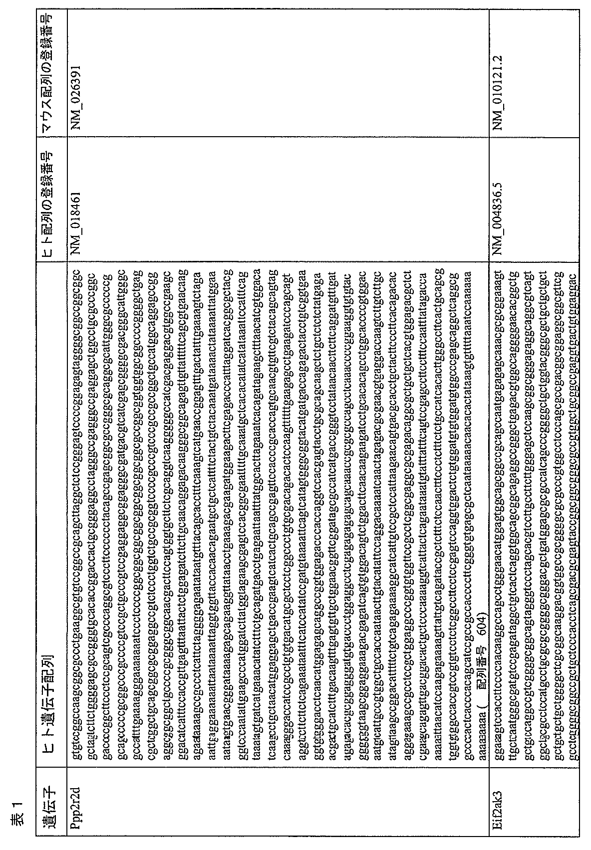
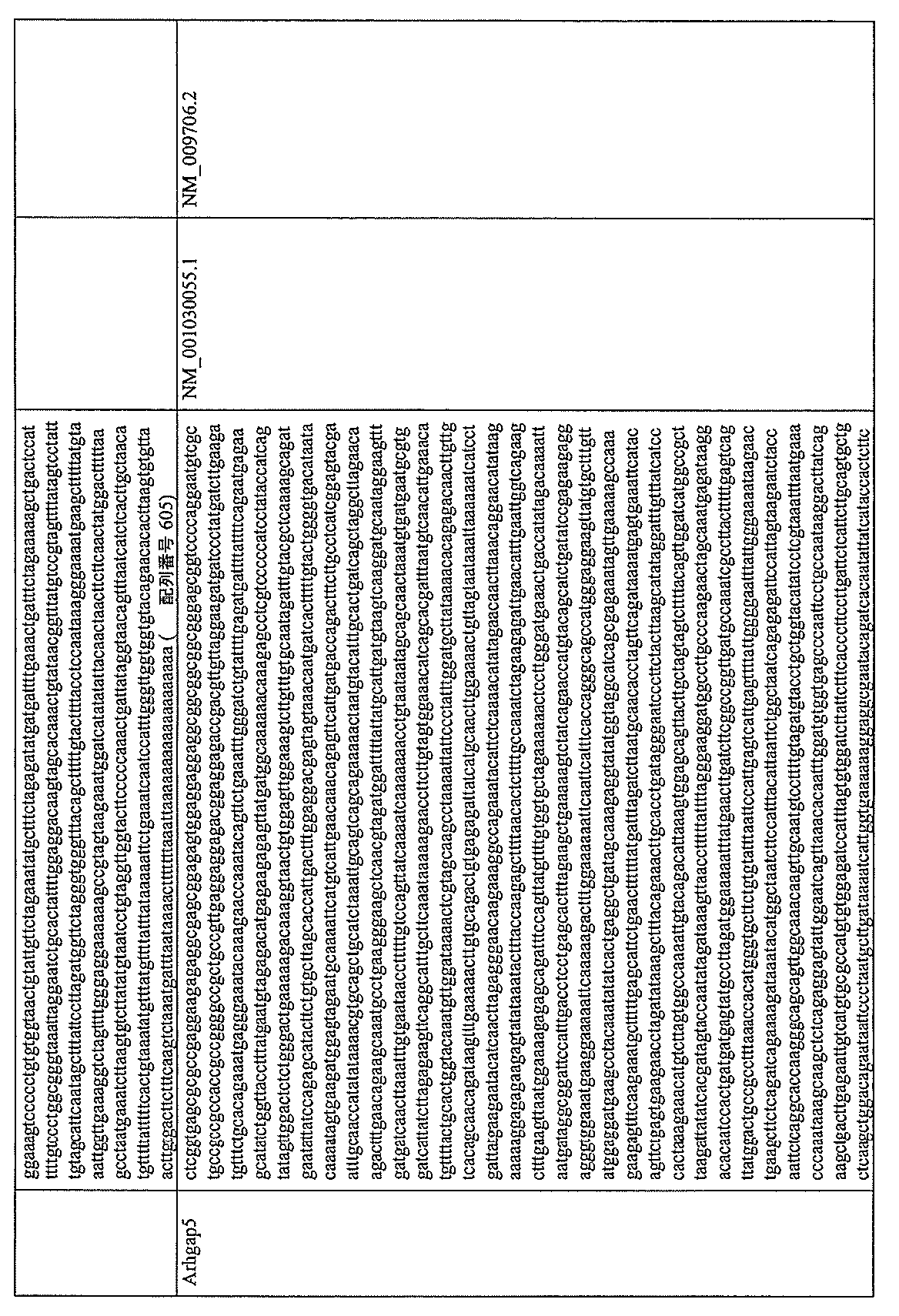
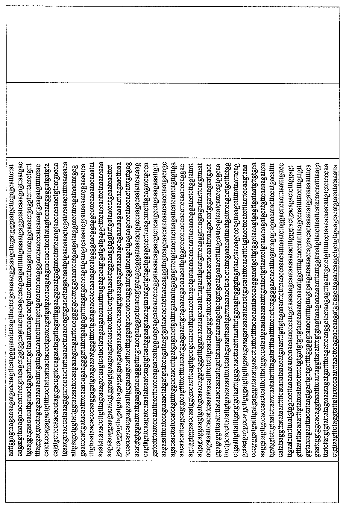
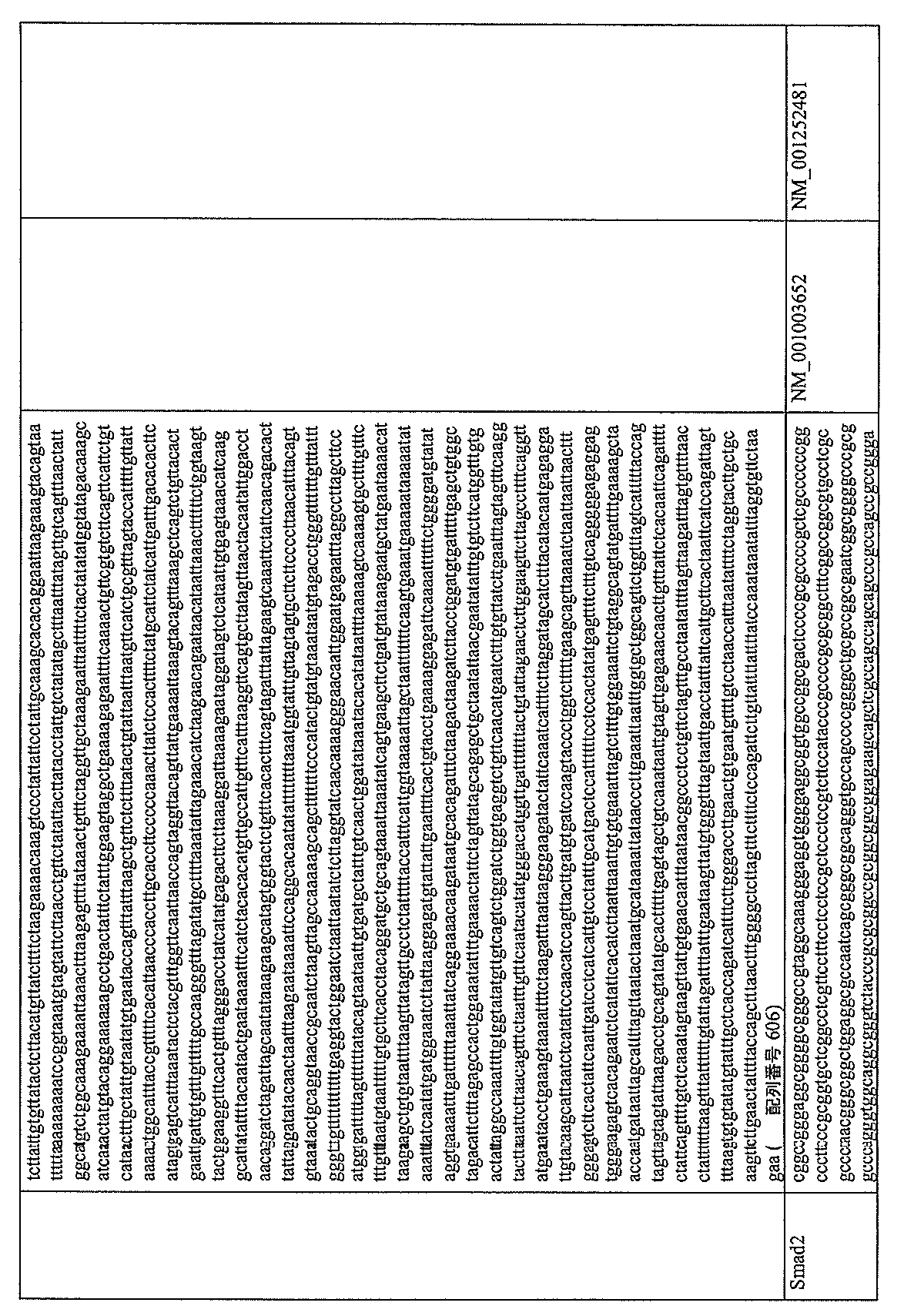
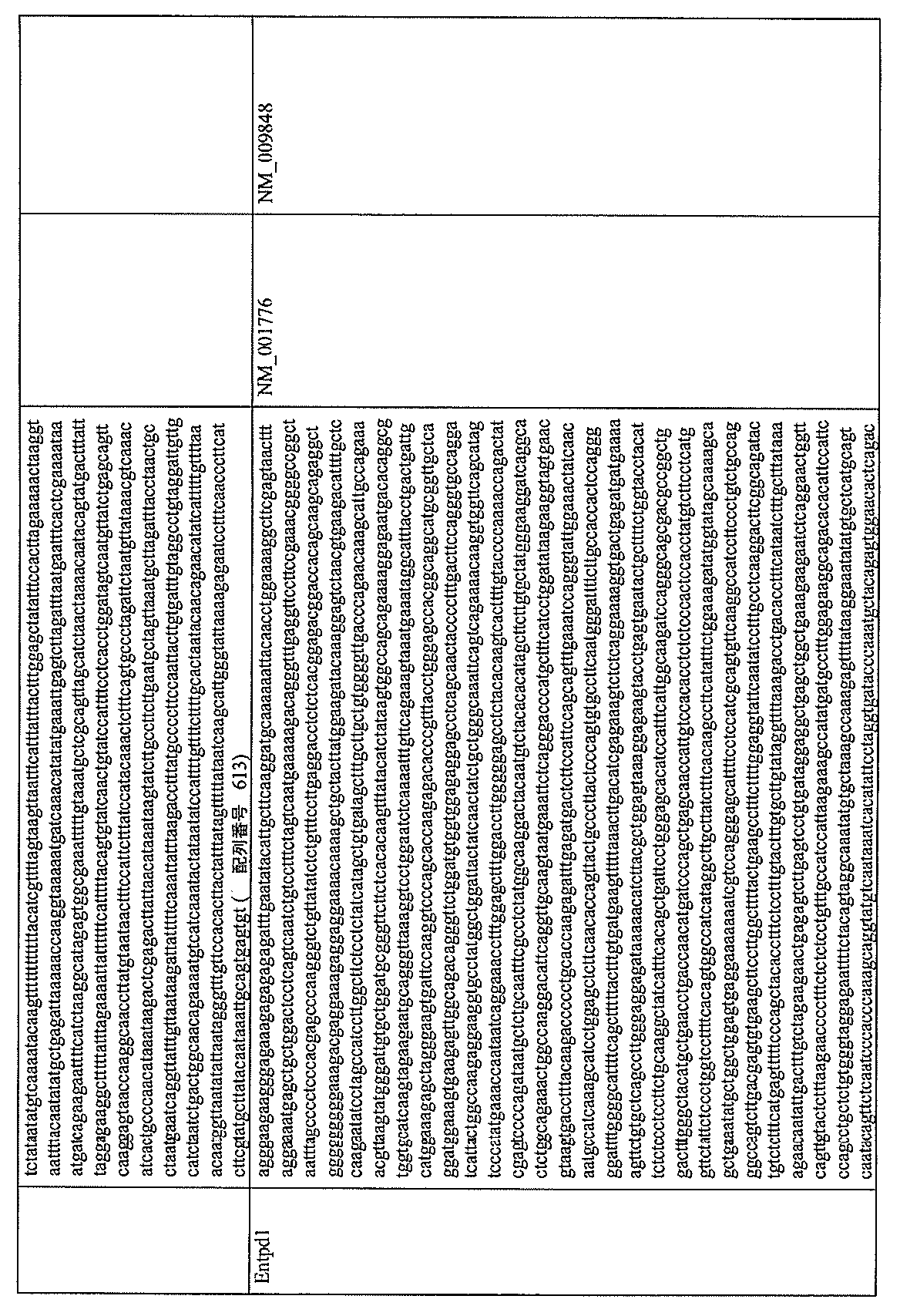
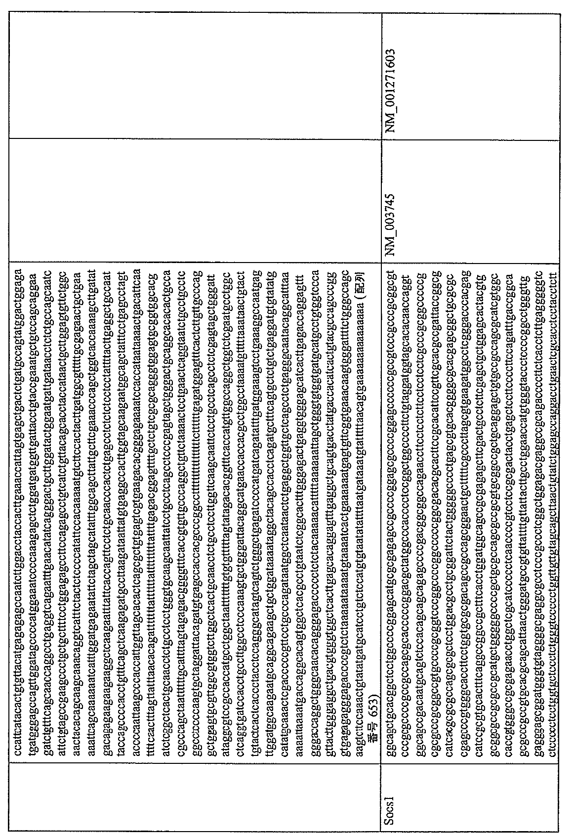
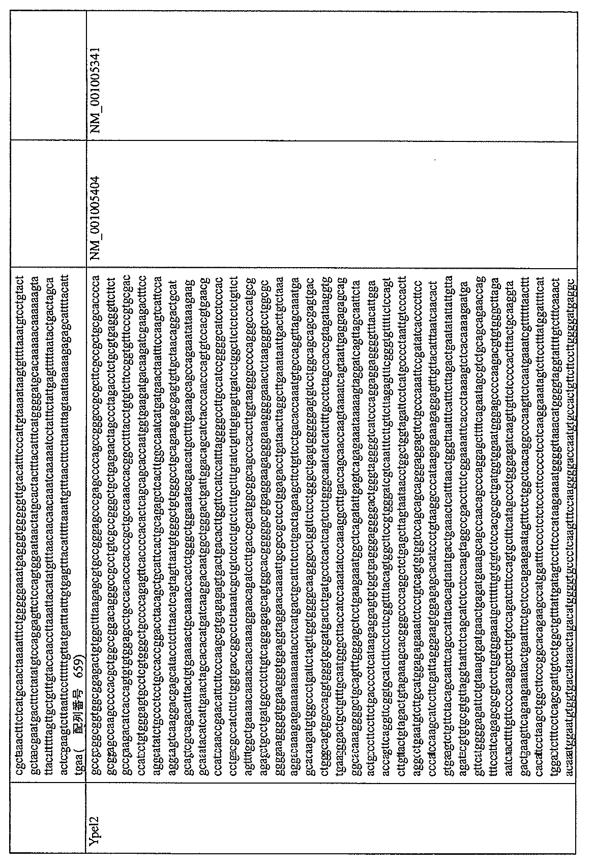
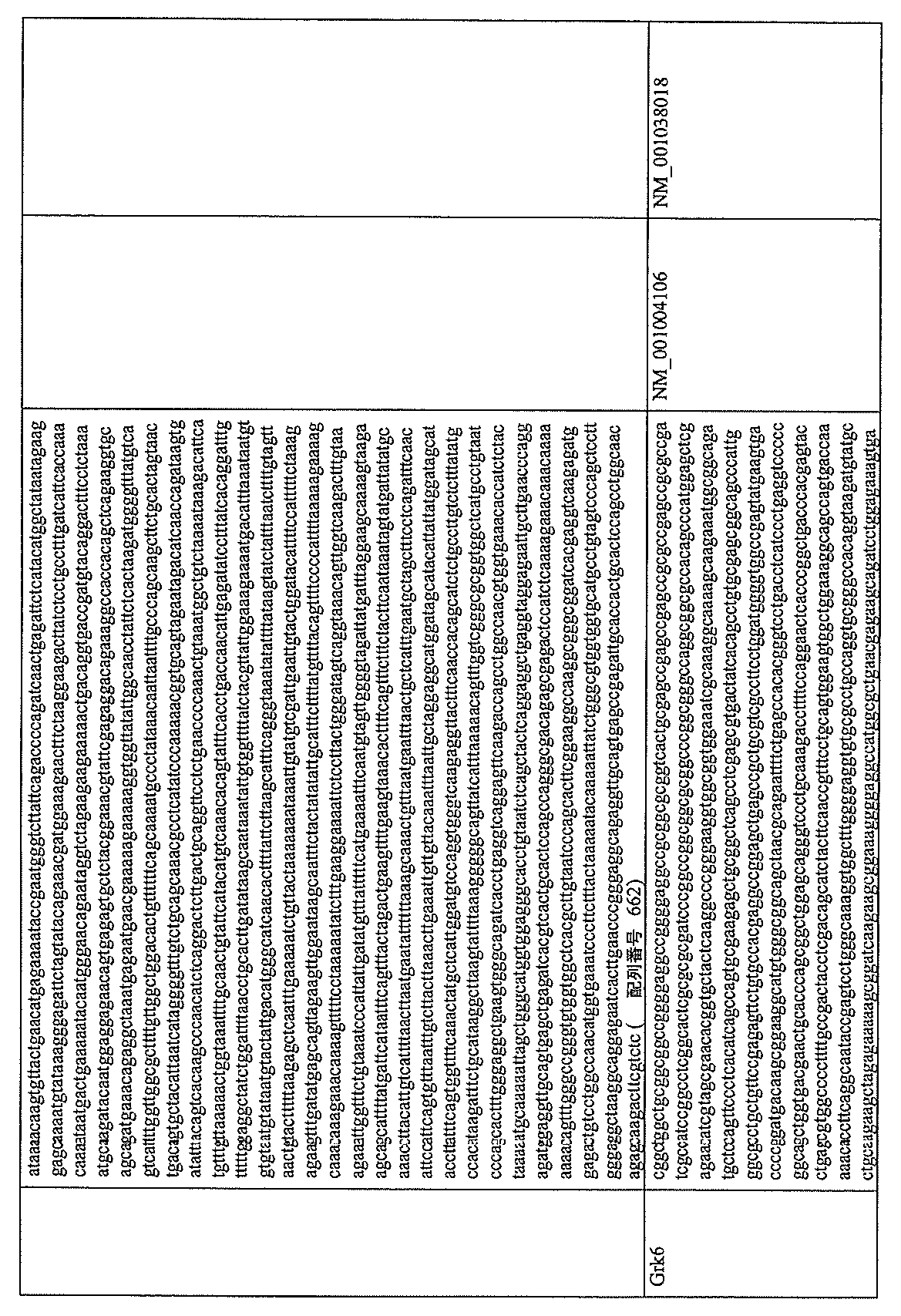
表1に、本発明においてT細胞の腫瘍免疫抑制に関与すると同定された遺伝子の一覧を示す。
〔表1〕
Table 1 shows a list of genes identified in the present invention to be involved in tumor immunosuppression of T cells.
[Table 1]


いくつかの形態において、本組成物の核酸は、表2に提供される配列を標的とするshRNA配列をコードする。表2はさらに、ディープシーケンシング分析に基づき選択されたshRNAの腫瘍対脾臓における濃縮(「濃縮倍率」)を示す。 In some forms, the nucleic acids of the composition encode shRNA sequences that target the sequences provided in Table 2. Table 2 further shows the enrichment in tumor versus spleen (“fold enrichment”) of selected shRNAs based on deep sequencing analysis.
〔表2〕 [Table 2]


脾臓と比較して腫瘍において少なくとも3種以上のshRNAの濃縮倍率を示すshRNAは、より活性な標的配列領域を示す。 shRNAs exhibiting fold enrichment of at least 3 or more shRNAs in tumor compared to spleen indicate more active target sequence regions.
いくつかの形態において、本組成物の核酸は、表2aに提供されるヒトPpp2r2dおよびCblb配列を標的とするshRNA配列をコードする。 In some forms, the nucleic acids of the composition encode shRNA sequences that target the human Ppp2r2d and Cblb sequences provided in Table 2a.

他の実施態様において、本開示は、配列番号372、373、374、375、376、377、378、378、379、380、381、382、383、384、385、または386に記載の少なくとも12、少なくとも15、少なくとも20、または少なくとも25個の連続したヌクレオチドと同一な、Ppp2r2d標的配列に相補的なshRNA配列をコードする単離された核酸を提供する。 In other embodiments, the present disclosure provides at least 12 of SEQ ID NOs: An isolated nucleic acid encoding an shRNA sequence complementary to a Ppp2r2d target sequence identical to at least 15, at least 20, or at least 25 contiguous nucleotides is provided.
他の実施態様において、本開示は、配列番号372、373、374、375、376、377、378、378、379、380、381、382、383、384、385、または386に記載の、マウス標的配列に対応するヒトPp2r2d配列に相補的な配列を含むshRNAをコードする単離された核酸を提供する。 In other embodiments, the present disclosure provides a mouse target as set forth in SEQ ID NOs: 372, 373, 374, 375, 376, 377, 378, 378, 379, 380, 381, 382, 383, 384, 385, or 386. An isolated nucleic acid encoding an shRNA comprising a sequence complementary to a human Pp2r2d sequence corresponding to the sequence is provided.
他の実施態様において、本開示は、配列番号133、134、135、136、137、138、139、140、141、142、143、144、145、146または147に記載の、少なくとも12、少なくとも15、少なくとも20、または少なくとも25個の連続したヌクレオチドと同一なEif2ak3標的配列に相補的なshRNA配列をコードする単離された核酸を提供する。 In other embodiments, the present disclosure provides at least 12, at least 15, SEQ ID NOs: 133, 134, 135, 136, 137, 138, 139, 140, 141, 142, 143, 144, 145, 146 or 147. , an isolated nucleic acid encoding an shRNA sequence complementary to an Eif2ak3 target sequence identical to at least 20, or at least 25 contiguous nucleotides.
他の実施態様において、本開示は、配列番号133、134、135、136、137、138、139、140、141、142、143、144、145、146または147に記載の、マウス標的配列に対応するヒトEif2ak3配列に相補的な配列を含むshRNAをコードする単離された核酸を提供する。 In other embodiments, the disclosure corresponds to a mouse target sequence set forth in SEQ ID NOs: 133, 134, 135, 136, 137, 138, 139, 140, 141, 142, 143, 144, 145, 146 or 147 An isolated nucleic acid encoding an shRNA comprising a sequence complementary to the human Eif2ak3 sequence is provided.
他の実施態様において、本開示は、配列番号32、33、34、35、36、37、38、39、40、41、または42に記載の、少なくとも12、少なくとも15、少なくとも20、または少なくとも25個の連続したヌクレオチドと同一なArhgap5標的配列に相補的なshRNA配列をコードする単離された核酸を提供する。 In other embodiments, the present disclosure provides at least 12, at least 15, at least 20, or at least 25 of SEQ ID NOS: 32, 33, 34, 35, 36, 37, 38, 39, 40, 41, or 42. An isolated nucleic acid is provided that encodes an shRNA sequence complementary to an Arhgap5 target sequence that is identical to three consecutive nucleotides.
他の実施態様において、本開示は、配列番号32、33、34、35、36、37、38、39、40、41、または42に記載の、マウス標的配列に対応するヒトArhgap5配列に相補的な配列を含むshRNAをコードする単離された核酸を提供する。 In other embodiments, the present disclosure provides a human Arhgap5 sequence complementary to the mouse target sequence set forth in SEQ ID NOs: 32, 33, 34, 35, 36, 37, 38, 39, 40, 41, or 42. An isolated nucleic acid encoding an shRNA comprising a sequence is provided.
他の実施態様において、本開示は、配列番号476、477、478、479、480、481、482、483、484、485、486、487、488、489、または490に記載の、少なくとも12、少なくとも15、少なくとも20、または少なくとも25個の連続したヌクレオチドと同一なSmad2標的配列に相補的なshRNA配列をコードする単離された核酸を提供する。 In other embodiments, the present disclosure provides at least 12, at least An isolated nucleic acid encoding an shRNA sequence complementary to a Smad2 target sequence identical to 15, at least 20, or at least 25 contiguous nucleotides is provided.
他の実施態様において、本開示は、配列番号476、477、478、479、480、481、482、483、484、485、486、487、488、489、または490に記載の、マウス標的配列に対応するヒトSmad2配列に相補的な配列を含むshRNAをコードする単離された核酸を提供する。 In other embodiments, the present disclosure provides a murine target sequence as set forth in An isolated nucleic acid encoding an shRNA comprising a sequence complementary to the corresponding human Smad2 sequence is provided.
他の実施態様において、本開示は、配列番号1、2、3、4、5、6、7、8、9、10、11、12、13、14、または15に記載の、少なくとも12、少なくとも15、少なくとも20、または少なくとも25個の連続したヌクレオチドと同一なAkap8l標的配列に相補的なshRNA配列をコードする単離された核酸を提供する。 In other embodiments, the present disclosure provides at least 12, at least An isolated nucleic acid encoding an shRNA sequence complementary to an Akap8l target sequence identical to 15, at least 20, or at least 25 contiguous nucleotides is provided.
他の実施態様において、本開示は、配列番号1、2、3、4、5、6、7、8、9、10、11、12、13、14、または15に記載の、マウス標的配列に対応するヒトAkap8l配列に相補的な配列を含むshRNAをコードする単離された核酸を提供する。 In other embodiments, the present disclosure provides a mouse target sequence set forth in SEQ ID NOs: 1, 2, 3, 4, 5, 6, 7, 8, 9, 10, 11, 12, 13, 14, or 15. An isolated nucleic acid encoding an shRNA comprising a sequence complementary to the corresponding human Akap8l sequence is provided.
他の実施態様において、本開示は、配列番号431、432、433、434、435、436、437、438、439、440、441、442、443、444、または445に記載の、少なくとも12、少なくとも15、少なくとも20、または少なくとも25個の連続したヌクレオチドと同一なRbks標的配列に相補的なshRNA配列をコードする単離された核酸を提供する。 In other embodiments, the present disclosure provides at least 12, at least An isolated nucleic acid encoding an shRNA sequence complementary to an Rbks target sequence identical to 15, at least 20, or at least 25 contiguous nucleotides is provided.
他の実施態様において、本開示は、配列番号431、432、433、434、435、436、437、438、439、440、441、442、443、444、または445に記載の、マウス標的配列に対応するヒトRbks配列に相補的な配列を含むshRNAをコードする単離された核酸を提供する。 In other embodiments, the present disclosure provides a murine target sequence as set forth in An isolated nucleic acid encoding an shRNA comprising a sequence complementary to the corresponding human Rbks sequence is provided.
他の実施態様において、本開示は、配列番号118、119、120、121、122、123、124、125、126、127、128、129、130、131、または132に記載の、少なくとも12、少なくとも15、少なくとも20、または少なくとも25個の連続したヌクレオチドと同一なEgr2標的配列に相補的なshRNA配列をコードする単離された核酸を提供する。 In other embodiments, the present disclosure provides at least 12, at least An isolated nucleic acid encoding an shRNA sequence complementary to an Egr2 target sequence identical to 15, at least 20, or at least 25 contiguous nucleotides is provided.
他の実施態様において、本開示は、配列番号118、119、120、121、122、123、124、125、126、127、128、129、130、131、または132に記載の、マウス標的配列に対応するヒトEgr2配列に相補的な配列を含むshRNAをコードする単離された核酸を提供する。 In other embodiments, the present disclosure provides a murine target sequence as set forth in An isolated nucleic acid encoding an shRNA comprising a sequence complementary to the corresponding human Egr2 sequence is provided.
他の実施態様において、本開示は、配列番号88、89、90、91、92、93、94、95、96、97、98、99、100、101、102、103、104、105、106、107、108、109、110、111、112、113、114、115、116または117に記載の、少なくとも12、少なくとも15、少なくとも20、ま
たは少なくとも25個の連続したヌクレオチドと同一なDgka標的配列に相補的なshRNA配列をコードする単離された核酸を提供する。
In other embodiments, the disclosure provides SEQ ID NOs: 88, 89, 90, 91, 92, 93, 94, 95, 96, 97, 98, 99, 100, 101, 102, 103, 104, 105, 106, complementary to a Dgka target sequence identical to at least 12, at least 15, at least 20, or at least 25 contiguous nucleotides according to An isolated nucleic acid encoding a specific shRNA sequence is provided.
他の実施態様において、本開示は、配列番号88、89、90、91、92、93、94、95、96、97、98、99、100、101、102、103、104、105、106、107、108、109、110、111、112、113、114、115、116または117に記載の、マウス標的配列に対応するヒトDgka配列に相補的な配列を含むshRNAをコードする単離された核酸を提供する。 In other embodiments, the disclosure provides SEQ ID NOs: 88, 89, 90, 91, 92, 93, 94, 95, 96, 97, 98, 99, 100, 101, 102, 103, 104, 105, 106, 107, 108, 109, 110, 111, 112, 113, 114, 115, 116 or 117. An isolated nucleic acid encoding an shRNA comprising a sequence complementary to the human Dgka sequence corresponding to the murine target sequence of 117. I will provide a.
他の実施態様において、本開示は、配列番号58、59、60、61、62、63、64、65、66、67、68、69、70、71、または72に記載の、少なくとも12、少なくとも15、少なくとも20、または少なくとも25個の連続したヌクレオチドと同一なCblb標的配列に相補的なshRNA配列をコードする単離された核酸を提供する。 In other embodiments, the present disclosure provides at least 12, at least An isolated nucleic acid encoding an shRNA sequence complementary to a Cblb target sequence identical to 15, at least 20, or at least 25 contiguous nucleotides is provided.
他の実施態様において、本開示は、配列番号58、59、60、61、62、63、64、65、66、67、68、69、70、71、または72に記載の、マウス標的配列に対応するヒトCblb配列に相補的な配列を含むshRNAをコードする単離された核酸を提供する。 In other embodiments, the present disclosure provides a murine target sequence as set forth in An isolated nucleic acid encoding an shRNA comprising a sequence complementary to the corresponding human Cblb sequence is provided.
他の実施態様において、本開示は、配列番号285、286、287、288、289、290、291、292、293、294、295、296、297、298、または299に記載の、少なくとも12、少なくとも15、少なくとも20、または少なくとも25個の連続したヌクレオチドと同一なMdfic標的配列に相補的なshRNA配列をコードする単離された核酸を提供する。 In other embodiments, the present disclosure provides at least 12, at least An isolated nucleic acid encoding an shRNA sequence complementary to an Mdfic target sequence identical to 15, at least 20, or at least 25 contiguous nucleotides is provided.
他の実施態様において、本開示は、配列番号285、286、287、288、289、290、291、292、293、294、295、296、297、298、または299に記載の、マウス標的配列に対応するヒトMdfic配列に相補的な配列を含むshRNAをコードする単離された核酸を提供する。 In other embodiments, the present disclosure provides a murine target sequence as set forth in An isolated nucleic acid encoding an shRNA comprising a sequence complementary to the corresponding human Mdfic sequence is provided.
他の実施態様において、本開示は、配列番号148、149、150、151、152、153、154、155、156、157、158、159、160、161、または162に記載の、少なくとも12、少なくとも15、少なくとも20、または少なくとも25個の連続したヌクレオチドと同一なEntpd1標的配列に相補的なshRNA配列をコードする単離された核酸を提供する。 In other embodiments, the present disclosure provides at least 12, at least An isolated nucleic acid encoding an shRNA sequence complementary to an Entpd1 target sequence identical to 15, at least 20, or at least 25 contiguous nucleotides is provided.
他の実施態様において、本開示は、配列番号148、149、150、151、152、153、154、155、156、157、158、159、160、161、または162に記載の、マウス標的配列に対応するヒトEntpd1配列に相補的な配列を含むshRNAをコードする単離された核酸を提供する。 In other embodiments, the present disclosure provides a murine target sequence as set forth in An isolated nucleic acid encoding an shRNA comprising a sequence complementary to the corresponding human Entpd1 sequence is provided.
他の実施態様において、本開示は、配列番号574、575、576、577、578、579、580、581、582、583、584、585、586、または587に記載の、少なくとも12、少なくとも15、少なくとも20、または少なくとも25個の連続したヌクレオチドと同一なVamp7標的配列に相補的なshRNA配列をコードする単離された核酸を提供する。 In other embodiments, the present disclosure provides at least 12, at least 15, at least 15, set forth in SEQ ID NOs: An isolated nucleic acid encoding an shRNA sequence complementary to a Vamp7 target sequence identical to at least 20, or at least 25 contiguous nucleotides is provided.
他の実施態様において、本開示は、配列番号574、575、576、577、578、579、580、581、582、583、584、585、586、または587に
記載の、マウス標的配列に対応するヒトVamp7配列に相補的な配列を含むshRNAをコードする単離された核酸を提供する。
In other embodiments, the disclosure corresponds to a mouse target sequence set forth in SEQ ID NOs: 574, 575, 576, 577, 578, 579, 580, 581, 582, 583, 584, 585, 586, or 587 An isolated nucleic acid encoding an shRNA comprising a sequence complementary to a human Vamp7 sequence is provided.
他の実施態様において、本開示は、配列番号208、209、210、211、212、213、214、215、216、217、218、219、220、221、または222に記載の、少なくとも12、少なくとも15、少なくとも20、または少なくとも25個の連続したヌクレオチドと同一なHipk1標的配列に相補的なshRNA配列をコードする単離された核酸を提供する。 In other embodiments, the present disclosure provides at least 12, at least An isolated nucleic acid encoding an shRNA sequence complementary to a Hipk1 target sequence identical to 15, at least 20, or at least 25 contiguous nucleotides is provided.
他の実施態様において、本開示は、配列番号208、209、210、211、212、213、214、215、216、217、218、219、220、221、または222に記載の、マウス標的配列に対応するヒトHipk1配列に相補的な配列を含むshRNAをコードする単離された核酸を提供する。 In other embodiments, the present disclosure provides a murine target sequence as set forth in An isolated nucleic acid encoding an shRNA comprising a sequence complementary to the corresponding human Hipk1 sequence is provided.
他の実施態様において、本開示は、配列番号315、316、317、318、319、320、321、322、323、324、325、326、327、328、または329に記載の、少なくとも12、少なくとも15、少なくとも20、または少なくとも25個の連続したヌクレオチドと同一なNuak2標的配列に相補的なshRNA配列をコードする単離された核酸を提供する。 In other embodiments, the present disclosure provides at least 12, at least An isolated nucleic acid encoding an shRNA sequence complementary to a Nuak2 target sequence identical to 15, at least 20, or at least 25 contiguous nucleotides is provided.
他の実施態様において、本開示は、配列番号315、316、317、318、319、320、321、322、323、324、325、326、327、328、または329に記載の、マウス標的配列に対応するヒトNuak2配列に相補的な配列を含むshRNAをコードする単離された核酸を提供する。 In other embodiments, the present disclosure provides a murine target sequence as set forth in An isolated nucleic acid encoding an shRNA comprising a sequence complementary to the corresponding human Nuak2 sequence is provided.
他の実施態様において、本開示は、配列番号16、17、18、19、20、21、22、23、24、25、26、27、28、29、30、または31に記載の、少なくとも12、少なくとも15、少なくとも20、または少なくとも25個の連続したヌクレオチドと同一なAlk標的配列に相補的なshRNA配列をコードする単離された核酸を提供する。 In other embodiments, the present disclosure provides at least 12 , at least 20, or at least 25 contiguous nucleotides that are identical to an Alk target sequence.
他の実施態様において、本開示は、配列番号16、17、18、19、20、21、22、23、24、25、26、27、28、29、30、または31に記載の、マウス標的配列に対応するヒトAlk配列に相補的な配列を含むshRNAをコードする単離された核酸を提供する。 In other embodiments, the present disclosure provides a mouse target as set forth in SEQ ID NOs: 16, 17, 18, 19, 20, 21, 22, 23, 24, 25, 26, 27, 28, 29, 30, or 31. An isolated nucleic acid encoding an shRNA comprising a sequence complementary to a human Alk sequence corresponding to the sequence is provided.
他の実施態様において、本開示は、配列番号330、331、332、333、334、335、336、337、338、339、340、または341に記載の、少なくとも12、少なくとも15、少なくとも20、または少なくとも25個の連続したヌクレオチドと同一なPdzk1ip1標的配列に相補的なshRNA配列をコードする単離された核酸を提供する。 In other embodiments, the present disclosure provides at least 12, at least 15, at least 20, or An isolated nucleic acid encoding an shRNA sequence complementary to a Pdzk1ip1 target sequence identical to at least 25 contiguous nucleotides is provided.
他の実施態様において、本開示は、配列番号330、331、332、333、334、335、336、337、338、339、340、または341に記載の、マウス標的配列に対応するヒトPdzk1ip1配列に相補的な配列を含むshRNAをコードする単離された核酸を提供する。 In other embodiments, the present disclosure provides human Pdzk1ip1 sequences corresponding to mouse target sequences set forth in An isolated nucleic acid encoding an shRNA comprising a complementary sequence is provided.
他の実施態様において、本開示は、配列番号52、53、54、55、56または57に記載の、少なくとも12、少なくとも15、少なくとも20、または少なくとも25個の連続したヌクレオチドと同一なBlvrb標的配列に相補的なshRNA配列をコード
する単離された核酸を提供する。
In other embodiments, the present disclosure provides Blvrb target sequences identical to at least 12, at least 15, at least 20, or at least 25 contiguous nucleotides set forth in SEQ ID NOS: 52, 53, 54, 55, 56 or 57. An isolated nucleic acid encoding a shRNA sequence complementary to is provided.
他の実施態様において、本開示は、配列番号52、53、54、55、56または57に記載の、マウス標的配列に対応するヒトBlvrbに相補的な配列を含むshRNAをコードする単離された核酸を提供する。 In other embodiments, the present disclosure provides an isolated shRNA encoding shRNA comprising a sequence complementary to human Blvrb corresponding to a murine target sequence as set forth in SEQ ID NOS: 52, 53, 54, 55, 56 or 57. A nucleic acid is provided.
他の実施態様において、本開示は、配列番号83、84、85、86または87に記載の、少なくとも12、少なくとも15、少なくとも20、または少なくとも25個の連続したヌクレオチドと同一なCdkn2a標的配列に相補的なshRNA配列をコードする単離された核酸を提供する。 In other embodiments, the present disclosure provides a Cdkn2a target sequence that is identical to at least 12, at least 15, at least 20, or at least 25 contiguous nucleotides set forth in SEQ ID NO:83, 84, 85, 86 or 87. An isolated nucleic acid encoding a specific shRNA sequence is provided.
他の実施態様において、本開示は、配列番号83、84、85、86または87に記載の、マウス標的配列に対応するヒトCdkn2aに相補的な配列を含むshRNAをコードする単離された核酸を提供する。 In other embodiments, the present disclosure provides an isolated nucleic acid encoding an shRNA comprising a sequence complementary to human Cdkn2a corresponding to a murine target sequence set forth in SEQ ID NOS:83, 84, 85, 86 or 87. offer.
他の実施態様において、本開示は、配列番号175、176または177に記載の、少なくとも12、少なくとも15、少なくとも20、または少なくとも25個の連続したヌクレオチドと同一なF11r標的配列に相補的なshRNA配列をコードする単離された核酸を提供する。 In other embodiments, the present disclosure provides shRNA sequences complementary to F11r target sequences identical to at least 12, at least 15, at least 20, or at least 25 contiguous nucleotides set forth in SEQ ID NO: 175, 176 or 177 An isolated nucleic acid encoding is provided.
他の実施態様において、本開示は、配列番号175、176または177に記載の、マウス標的配列に対応するヒトF11rに相補的な配列を含むshRNAをコードする単離された核酸を提供する。 In another embodiment, the present disclosure provides an isolated nucleic acid encoding an shRNA comprising a sequence complementary to human F11r corresponding to the mouse target sequence set forth in SEQ ID NOS: 175, 176 or 177.
他の実施態様において、本開示は、配列番号187、191または192に記載の、少なくとも12、少なくとも15、少なくとも20、または少なくとも25個の連続したヌクレオチドと同一なFyn標的配列に相補的なshRNA配列をコードする単離された核酸を提供する。 In other embodiments, the present disclosure provides shRNA sequences complementary to Fyn target sequences identical to at least 12, at least 15, at least 20, or at least 25 contiguous nucleotides set forth in SEQ ID NO: 187, 191 or 192. An isolated nucleic acid encoding is provided.
他の実施態様において、本開示は、配列番号187、191または192に記載の、マウス標的配列に対応するヒトFynに相補的な配列を含むshRNAをコードする単離された核酸を提供する。 In other embodiments, the present disclosure provides an isolated nucleic acid encoding an shRNA comprising a sequence complementary to human Fyn corresponding to a murine target sequence set forth in SEQ ID NO: 187, 191 or 192.
他の実施態様において、本開示は、配列番号204、205、206または207に記載の、少なくとも12、少なくとも15、少なくとも20、または少なくとも25個の連続したヌクレオチドと同一なGrk6標的配列に相補的なshRNA配列をコードする単離された核酸を提供する。 In other embodiments, the present disclosure provides a Grk6 target sequence that is identical to at least 12, at least 15, at least 20, or at least 25 contiguous nucleotides set forth in SEQ ID NO: 204, 205, 206 or 207. An isolated nucleic acid encoding the shRNA sequence is provided.
他の実施態様において、本開示は、配列番号204、205、206または207に記載の、マウス標的配列に対応するヒトGrk6に相補的な配列を含むshRNAをコードする単離された核酸を提供する。 In other embodiments, the disclosure provides an isolated nucleic acid encoding an shRNA comprising a sequence complementary to human Grk6 corresponding to a murine target sequence as set forth in SEQ ID NOS: 204, 205, 206 or 207. .
他の実施態様において、本開示は、配列番号232、234、235、236または237に記載の、少なくとも12、少なくとも15、少なくとも20、または少なくとも25個の連続したヌクレオチドと同一なInpp5b標的配列に相補的なshRNA配列をコードする単離された核酸を提供する。 In other embodiments, the present disclosure provides an Inpp5b target sequence that is identical to at least 12, at least 15, at least 20, or at least 25 contiguous nucleotides set forth in SEQ ID NO: 232, 234, 235, 236 or 237. An isolated nucleic acid encoding a specific shRNA sequence is provided.
他の実施態様において、本開示は、配列番号232、234、235、236または237に記載の、マウス標的配列に対応するヒトInpp5bに相補的な配列を含むshRNAをコードする単離された核酸を提供する。 In other embodiments, the present disclosure provides an isolated nucleic acid encoding an shRNA comprising a sequence complementary to human Inpp5b corresponding to a murine target sequence set forth in SEQ ID NOS: 232, 234, 235, 236 or 237. offer.
他の実施態様において、本開示は、配列番号248、249、250、251または252に記載の、少なくとも12、少なくとも15、少なくとも20、または少なくとも25個の連続したヌクレオチドと同一なImpk標的配列に相補的なshRNA配列をコードする単離された核酸を提供する。 In other embodiments, the present disclosure provides an Impk target sequence that is identical to at least 12, at least 15, at least 20, or at least 25 contiguous nucleotides set forth in SEQ ID NO: 248, 249, 250, 251 or 252. An isolated nucleic acid encoding a specific shRNA sequence is provided.
他の実施態様において、本開示は、配列番号248、249、250、251または252に記載の、マウス標的配列に対応するヒトImpkに相補的な配列を含むshRNAをコードする単離された核酸を提供する。 In other embodiments, the present disclosure provides an isolated nucleic acid encoding an shRNA comprising a sequence complementary to human Impk corresponding to a murine target sequence set forth in SEQ ID NOS: 248, 249, 250, 251 or 252. offer.
他の実施態様において、本開示は、配列番号263、264、265、266、267、268または269に記載の、少なくとも12、少なくとも15、少なくとも20、または少なくとも25個の連続したヌクレオチドと同一なJun標的配列に相補的なshRNA配列をコードする単離された核酸を提供する。 In other embodiments, the present disclosure provides a Jun sequence identical to at least 12, at least 15, at least 20, or at least 25 contiguous nucleotides set forth in SEQ ID NOS: 263, 264, 265, 266, 267, 268 or 269. An isolated nucleic acid encoding an shRNA sequence complementary to a target sequence is provided.
他の実施態様において、本開示は、配列番号263、264、265、266、267、268または269に記載の、マウス標的配列に対応するヒトJunに相補的な配列を含むshRNAをコードする単離された核酸を提供する。 In other embodiments, the present disclosure provides an isolated shRNA encoding shRNA comprising a sequence complementary to human Jun corresponding to a mouse target sequence as set forth in SEQ ID NOS: 263, 264, 265, 266, 267, 268 or 269. providing a nucleic acid prepared by
他の実施態様において、本開示は、配列番号281、282、283または284に記載の、少なくとも12、少なくとも15、少なくとも20、または少なくとも25個の連続したヌクレオチドと同一なMast2標的配列に相補的なshRNA配列をコードする単離された核酸を提供する。 In other embodiments, the present disclosure provides a Mast2 target sequence that is identical to at least 12, at least 15, at least 20, or at least 25 contiguous nucleotides set forth in SEQ ID NO: 281, 282, 283 or 284. An isolated nucleic acid encoding the shRNA sequence is provided.
他の実施態様において、本開示は、配列番号281、282、283または284に記載の、マウス標的配列に対応するヒトMast2に相補的な配列を含むshRNAをコードする単離された核酸を提供する。 In other embodiments, the present disclosure provides an isolated nucleic acid encoding an shRNA comprising a sequence complementary to human Mast2 corresponding to a murine target sequence as set forth in SEQ ID NO: 281, 282, 283 or 284. .
他の実施態様において、本開示は、配列番号311、312、313または314に記載の、少なくとも12、少なくとも15、少なくとも20、または少なくとも25個の連続したヌクレオチドと同一なNptxr標的配列に相補的なshRNA配列をコードする単離された核酸を提供する。 In other embodiments, the present disclosure provides a Nptxr target sequence that is identical to at least 12, at least 15, at least 20, or at least 25 contiguous nucleotides set forth in SEQ ID NO: 311, 312, 313 or 314. An isolated nucleic acid encoding the shRNA sequence is provided.
他の実施態様において、本開示は、配列番号311、312、313または314に記載の、マウス標的配列に対応するヒトNptxrに相補的な配列を含むshRNAをコードする単離された核酸を提供する。 In other embodiments, the present disclosure provides an isolated nucleic acid encoding an shRNA comprising a sequence complementary to human Nptxr corresponding to a murine target sequence set forth in SEQ ID NOS: 311, 312, 313 or 314. .
他の実施態様において、本開示は、配列番号351、352、353、354、355または356に記載の、少なくとも12、少なくとも15、少なくとも20、または少なくとも25個の連続したヌクレオチドと同一なPkd1標的配列に相補的なshRNA配列をコードする単離された核酸を提供する。 In other embodiments, the present disclosure provides a Pkdl target sequence identical to at least 12, at least 15, at least 20, or at least 25 contiguous nucleotides set forth in SEQ ID NOS: 351, 352, 353, 354, 355 or 356. An isolated nucleic acid encoding a shRNA sequence complementary to is provided.
他の実施態様において、本開示は、配列番号351、352、353、354、355または356に記載の、マウス標的配列に対応するヒトPkd1に相補的な配列を含むshRNAをコードする単離された核酸を提供する。 In other embodiments, the present disclosure provides an isolated shRNA encoding shRNA comprising a sequence complementary to human Pkdl corresponding to a mouse target sequence as set forth in SEQ ID NOS: 351, 352, 353, 354, 355 or 356. A nucleic acid is provided.
他の実施態様において、本開示は、配列番号367、368、369、370または371に記載の、少なくとも12、少なくとも15、少なくとも20、または少なくとも25個の連続したヌクレオチドと同一なPpm1g標的配列に相補的なshRNA配列をコードする単離された核酸を提供する。 In other embodiments, the present disclosure provides a Ppm1g target sequence that is identical to at least 12, at least 15, at least 20, or at least 25 contiguous nucleotides set forth in SEQ ID NOs: 367, 368, 369, 370 or 371. An isolated nucleic acid encoding a specific shRNA sequence is provided.
他の実施態様において、本開示は、配列番号367、368、369、370または371に記載の、マウス標的配列に対応するヒトPpm1gに相補的な配列を含むshRNAをコードする単離された核酸を提供する。 In other embodiments, the present disclosure provides an isolated nucleic acid encoding an shRNA comprising a sequence complementary to human Ppm1g corresponding to a murine target sequence as set forth in SEQ ID NOS: 367, 368, 369, 370 or 371. offer.
他の実施態様において、本開示は、配列番号399、400または401に記載の、少なくとも12、少なくとも15、少なくとも20、または少なくとも25個の連続したヌクレオチドと同一なPpp3cc標的配列に相補的なshRNA配列をコードする単離された核酸を提供する。 In other embodiments, the present disclosure provides shRNA sequences complementary to a Ppp3cc target sequence identical to at least 12, at least 15, at least 20, or at least 25 contiguous nucleotides set forth in SEQ ID NO: 399, 400 or 401. An isolated nucleic acid encoding is provided.
他の実施態様において、本開示は、配列番号399、400または401に記載の、マウス標的配列に対応するヒトPpp3ccに相補的な配列を含むshRNAをコードする単離された核酸を提供する。 In other embodiments, the present disclosure provides an isolated nucleic acid encoding an shRNA comprising a sequence complementary to human Ppp3cc corresponding to a murine target sequence set forth in SEQ ID NO:399, 400 or 401.
他の実施態様において、本開示は、配列番号414、415または416に記載の、少なくとも12、少なくとも15、少なくとも20、または少なくとも25個の連続したヌクレオチドと同一なPrkab2標的配列に相補的なshRNA配列をコードする単離された核酸を提供する。 In other embodiments, the present disclosure provides shRNA sequences complementary to Prkab2 target sequences identical to at least 12, at least 15, at least 20, or at least 25 contiguous nucleotides set forth in SEQ ID NOS: 414, 415, or 416. An isolated nucleic acid encoding is provided.
他の実施態様において、本開示は、配列番号414、415または416に記載の、マウス標的配列に対応するヒトPrkab2に相補的な配列を含むshRNAをコードする単離された核酸を提供する。 In other embodiments, the disclosure provides an isolated nucleic acid encoding an shRNA comprising a sequence complementary to human Prkab2 corresponding to the mouse target sequence set forth in SEQ ID NOs:414, 415 or 416.
他の実施態様において、本開示は、配列番号426、427、428、429または430に記載の、少なくとも12、少なくとも15、少なくとも20、または少なくとも25個の連続したヌクレオチドと同一なPtpn2標的配列に相補的なshRNA配列をコードする単離された核酸を提供する。 In other embodiments, the present disclosure provides a Ptpn2 target sequence that is identical to at least 12, at least 15, at least 20, or at least 25 contiguous nucleotides set forth in SEQ ID NO: 426, 427, 428, 429 or 430. An isolated nucleic acid encoding a specific shRNA sequence is provided.
他の実施態様において、本開示は、配列番号426、427、428、429または430に記載の、マウス標的配列に対応するヒトPtpn2に相補的な配列を含むshRNAをコードする単離された核酸を提供する。 In other embodiments, the present disclosure provides an isolated nucleic acid encoding an shRNA comprising a sequence complementary to human Ptpn2 corresponding to a murine target sequence set forth in SEQ ID NO: 426, 427, 428, 429 or 430. offer.
他の実施態様において、本開示は、配列番号457、458、459または460に記載の、少なくとも12、少なくとも15、少なくとも20、または少なくとも25個の連続したヌクレオチドと同一なRock1標的配列に相補的なshRNA配列をコードする単離された核酸を提供する。 In other embodiments, the present disclosure provides a Rock1 target sequence that is identical to at least 12, at least 15, at least 20, or at least 25 contiguous nucleotides set forth in SEQ ID NO: 457, 458, 459 or 460. An isolated nucleic acid encoding the shRNA sequence is provided.
他の実施態様において、本開示は、配列番号457、458、459または460に記載の、マウス標的配列に対応するヒトRock1に相補的な配列を含むshRNAをコードする単離された核酸を提供する。 In other embodiments, the disclosure provides an isolated nucleic acid encoding an shRNA comprising a sequence complementary to human Rock1 corresponding to a murine target sequence set forth in SEQ ID NOS: 457, 458, 459 or 460. .
他の実施態様において、本開示は、配列番号470、471、472、473、474または475に記載の、少なくとも12、少なくとも15、少なくとも20、または少なくとも25個の連続したヌクレオチドと同一なSbf1標的配列に相補的なshRNA配列をコードする単離された核酸を提供する。 In other embodiments, the present disclosure provides Sbf1 target sequences identical to at least 12, at least 15, at least 20, or at least 25 contiguous nucleotides set forth in SEQ ID NOS: 470, 471, 472, 473, 474 or 475. An isolated nucleic acid encoding a shRNA sequence complementary to is provided.
他の実施態様において、本開示は、配列番号470、471、472、473、474または475に記載の、マウス標的配列に対応するヒトSbf1に相補的な配列を含むshRNAをコードする単離された核酸を提供する。 In other embodiments, the present disclosure provides an isolated shRNA encoding shRNA comprising a sequence complementary to human Sbf1 corresponding to a mouse target sequence as set forth in SEQ ID NO: 470, 471, 472, 473, 474 or 475. A nucleic acid is provided.
他の実施態様において、本開示は、配列番号504、505、506、507、508、509または510に記載の、少なくとも12、少なくとも15、少なくとも20、または少なくとも25個の連続したヌクレオチドと同一なSocs1標的配列に相補的なshRNA配列をコードする単離された核酸を提供する。 In other embodiments, the present disclosure provides Socsl identical to at least 12, at least 15, at least 20, or at least 25 contiguous nucleotides set forth in SEQ ID NOS: 504, 505, 506, 507, 508, 509 or 510. An isolated nucleic acid encoding an shRNA sequence complementary to a target sequence is provided.
他の実施態様において、本開示は、配列番号504、505、506、507、508、509または510に記載の、マウス標的配列に対応するヒトSocs1に相補的な配列を含むshRNAをコードする単離された核酸を提供する。 In other embodiments, the present disclosure provides an isolated shRNA encoding shRNA comprising a sequence complementary to human Socsl corresponding to a murine target sequence as set forth in SEQ ID NOS: 504, 505, 506, 507, 508, 509 or 510. providing a nucleic acid prepared by
他の実施態様において、本開示は、配列番号524、525、526、527または528に記載の、少なくとも12、少なくとも15、少なくとも20、または少なくとも25個の連続したヌクレオチドと同一なSocs3標的配列に相補的なshRNA配列をコードする単離された核酸を提供する。 In other embodiments, the present disclosure provides a Socs3 target sequence that is identical to at least 12, at least 15, at least 20, or at least 25 contiguous nucleotides set forth in SEQ ID NOS: 524, 525, 526, 527 or 528. An isolated nucleic acid encoding a specific shRNA sequence is provided.
他の実施態様において、本開示は、配列番号524、525、526、527または528に記載の、マウス標的配列に対応するヒトSocs3に相補的な配列を含むshRNAをコードする単離された核酸を提供する。 In other embodiments, the present disclosure provides an isolated nucleic acid encoding an shRNA comprising a sequence complementary to human Socs3 corresponding to a mouse target sequence as set forth in SEQ ID NOS: 524, 525, 526, 527 or 528. offer.
他の実施態様において、本開示は、配列番号539、540、541、542または543に記載の、少なくとも12、少なくとも15、少なくとも20、または少なくとも25個の連続したヌクレオチドと同一なStk17b標的配列に相補的なshRNA配列をコードする単離された核酸を提供する。 In other embodiments, the present disclosure provides a Stk17b target sequence that is identical to at least 12, at least 15, at least 20, or at least 25 contiguous nucleotides set forth in SEQ ID NOS: 539, 540, 541, 542 or 543. An isolated nucleic acid encoding a specific shRNA sequence is provided.
他の実施態様において、本開示は、配列番号539、540、541、542または543に記載の、マウス標的配列に対応するヒトStk17bに相補的な配列を含むshRNAをコードする単離された核酸を提供する。 In other embodiments, the present disclosure provides an isolated nucleic acid encoding an shRNA comprising a sequence complementary to human Stk17b corresponding to a murine target sequence set forth in SEQ ID NOS: 539, 540, 541, 542 or 543. offer.
他の実施態様において、本開示は、配列番号556、557または558に記載の、少なくとも12、少なくとも15、少なくとも20、または少なくとも25個の連続したヌクレオチドと同一なTnk1標的配列に相補的なshRNA配列をコードする単離された核酸を提供する。 In other embodiments, the present disclosure provides shRNA sequences complementary to a Tnk1 target sequence identical to at least 12, at least 15, at least 20, or at least 25 contiguous nucleotides set forth in SEQ ID NOS: 556, 557 or 558. An isolated nucleic acid encoding is provided.
他の実施態様において、本開示は、配列番号556、557または558に記載の、マウス標的配列に対応するヒトTnk1に相補的な配列を含むshRNAをコードする単離された核酸を提供する。 In other embodiments, the present disclosure provides an isolated nucleic acid encoding an shRNA comprising a sequence complementary to human Tnk1 corresponding to a murine target sequence set forth in SEQ ID NO:556, 557 or 558.
他の実施態様において、本開示は、配列番号569、570、571、572または573に記載の、少なくとも12、少なくとも15、少なくとも20、または少なくとも25個の連続したヌクレオチドと同一なTrpm7標的配列に相補的なshRNA配列をコードする単離された核酸を提供する。 In other embodiments, the present disclosure provides a Trpm7 target sequence that is identical to at least 12, at least 15, at least 20, or at least 25 contiguous nucleotides set forth in SEQ ID NOS: 569, 570, 571, 572 or 573. An isolated nucleic acid encoding a specific shRNA sequence is provided.
他の実施態様において、本開示は、配列番号569、570、571、572または573に記載の、マウス標的配列に対応するヒトTrpm7に相補的な配列を含むshRNAをコードする単離された核酸を提供する。 In other embodiments, the present disclosure provides an isolated nucleic acid encoding an shRNA comprising a sequence complementary to human Trpm7 corresponding to a mouse target sequence as set forth in SEQ ID NOS: 569, 570, 571, 572 or 573. offer.
他の実施態様において、本開示は、配列番号600、601、602または603に記載の、少なくとも12、少なくとも15、少なくとも20、または少なくとも25個の連続したヌクレオチドと同一なYes1標的配列に相補的なshRNA配列をコードする単離された核酸を提供する。 In other embodiments, the present disclosure provides a Yes1 target sequence identical to at least 12, at least 15, at least 20, or at least 25 contiguous nucleotides set forth in SEQ ID NO: 600, 601, 602 or 603. An isolated nucleic acid encoding the shRNA sequence is provided.
他の実施態様において、本開示は、配列番号600、601、602または603に記載の、マウス標的配列に対応するヒトYes1に相補的な配列を含むshRNAをコードする単離された核酸を提供する。 In other embodiments, the present disclosure provides an isolated nucleic acid encoding an shRNA comprising a sequence complementary to human Yes1 corresponding to a murine target sequence set forth in SEQ ID NO: 600, 601, 602 or 603. .
あらゆる実施態様において、マウス標的配列に対応するヒト配列は、ヒト遺伝子配列に完全に対応する配列であり、例えば、選択されたマウス標的配列の、少なくとも12、少なくとも15、少なくとも20、または少なくとも25個の連続したヌクレオチドが有し得るヌクレオチドミスマッチは、ゼロ、1、2、3または4個である。 In any embodiment, a human sequence corresponding to a mouse target sequence is a sequence that fully corresponds to a human gene sequence, e.g., at least 12, at least 15, at least 20, or at least 25 of the selected mouse target sequences. consecutive nucleotides can have zero, 1, 2, 3 or 4 nucleotide mismatches.
単離された核酸は、例えばDNA分子であってもよく、ただし、天然に存在するゲノム中でそのDNA分子に隣接して通常見出される核酸配列の1つが、除去されているかまたは存在しない。したがって、単離された核酸としては、これらに限定されないが、他の配列から独立した、別の分子として存在するDNA分子(例えば、化学合成された核酸、cDNA、またはPCRもしくは制限エンドヌクレアーゼ処理によって生産されたゲノムDNAフラグメント)、加えて、ベクター、自律複製型プラスミド、ウイルス(例えば、レトロウイルス、レンチウイルス、アデノウイルス、アデノ随伴ウイルス、またはヘルペスウイルス)に取り込まれるDNA、または原核生物もしくは真核生物のゲノムDNAに取り込まれるDNAが挙げられる。加えて、単離された核酸は、ハイブリッドまたは融合核酸の一部である組換えDNA分子などの加工された核酸を包含し得る。例えばcDNAライブラリーまたはゲノムライブラリー、またはゲノムDNAの制限酵素消化物を含有するゲル切片中の数百から数百万の他の核酸のなかに存在する核酸は、単離された核酸とはみなされないものとする。 An isolated nucleic acid can be, for example, a DNA molecule, provided that one of the nucleic acid sequences normally found adjacent to the DNA molecule in the naturally occurring genome has been removed or absent. An isolated nucleic acid therefore includes, but is not limited to, a DNA molecule that exists as a separate molecule, independent of other sequences (e.g., chemically synthesized nucleic acids, cDNA, or DNA molecules that have been isolated by PCR or restriction endonuclease treatment). produced genomic DNA fragments), as well as DNA incorporated into vectors, autonomously replicating plasmids, viruses (e.g., retroviruses, lentiviruses, adenoviruses, adeno-associated viruses, or herpes viruses), or prokaryotic or eukaryotic DNA that is incorporated into the genomic DNA of an organism is included. In addition, isolated nucleic acid can include engineered nucleic acids, such as recombinant DNA molecules that are part of hybrid or fusion nucleic acids. A nucleic acid that is present, for example, in a cDNA library or genomic library, or among hundreds to millions of other nucleic acids in gel slices containing restriction enzyme digests of genomic DNA, is not considered an isolated nucleic acid. shall not be
配列同一性パーセントの計算では、2つの配列を並べて、2つの配列間のヌクレオチドまたはアミノ酸残基の同一なマッチの数を決定する。同一なマッチの数を、並べられた領域の長さ(すなわち、並べられたヌクレオチドまたはアミノ酸残基の数)で割り、100を掛けて、配列同一性パーセント値を得る。並べられた領域の長さは、最大で最も短い配列の全長サイズの、一方または両方の配列の一部であり得ることが理解されるものと予想される。また、単一の配列は、1つより多くの他の配列と並べることができ、したがって、並べられた領域ごとに異なる配列同一性パーセント値を有する可能性があることも理解されるものと予想される。同一性値のパーセントは、通常、最も近い整数に概算されることに留意する。例えば、78.1%、78.2%、78.3%、および78.4%は、端数を切り下げて78%にされ、一方で78.5%、78.6%、78.7%、78.8%、および78.9%は、端数を切り上げて79%にされる。また、並べられた領域の長さは常に整数であることにも留意する。 In calculating percent sequence identity, two sequences are aligned and the number of identical matches of nucleotides or amino acid residues between the two sequences is determined. The number of identical matches is divided by the length of the aligned region (ie, number of nucleotides or amino acid residues aligned) and multiplied by 100 to give a percent sequence identity value. It will be appreciated that the length of the aligned regions can be a fraction of one or both sequences up to the full length size of the shortest sequence. It is also to be understood that a single sequence can be aligned with more than one other sequence and thus can have different percent sequence identity values for each aligned region. be done. Note that percent identity values are generally approximated to the nearest whole number. For example, 78.1%, 78.2%, 78.3%, and 78.4% are rounded down to 78%, while 78.5%, 78.6%, 78.7%, 78.8% and 78.9% are rounded up to 79%. Also note that the length of the aligned regions is always an integer.
用語「配列同一性パーセント」は、本明細書で使用される場合、いずれかの所与のクエリー配列と対象配列との間の同一性の程度を指す。あらゆるクエリー核酸またはアミノ酸配列、例えば転写因子の、別の対象核酸またはアミノ酸配列に対する同一性パーセントは、以下のように決定することができる。 The term "percent sequence identity" as used herein refers to the degree of identity between any given query sequence and the subject sequence. The percent identity of any query nucleic acid or amino acid sequence, eg, transcription factor, to another nucleic acid or amino acid sequence of interest can be determined as follows.
また、用語「相補的ヌクレオチド配列」は、本明細書で使用される場合、「アンチセンス配列」としても知られており、タンパク質をコードする「センス」核酸の配列に完全に相補的な核酸の配列(例えば、二本鎖cDNA分子のコード鎖に相補的であるか、またはmRNA配列に相補的な)を指す。本明細書では、少なくとも約15、16、17、18、19、20、21、22、23、24、もしくは25ヌクレオチドもしくは全遺伝子コード鎖、またはそれらの一部のみに相補的な配列を含む核酸分子が提供される。 The term "complementary nucleotide sequence", as used herein, is also known as "antisense sequence" and is a nucleic acid sequence that is completely complementary to a sequence of a protein-encoding "sense" nucleic acid. Refers to a sequence (eg, complementary to the coding strand of a double-stranded cDNA molecule or complementary to an mRNA sequence). As used herein, a nucleic acid comprising a sequence complementary to at least about 15, 16, 17, 18, 19, 20, 21, 22, 23, 24, or 25 nucleotides or an entire gene coding strand, or only a portion thereof A molecule is provided.
用語「ヌクレオチド配列に対応する」は、本明細書で使用される場合、同一な配列をコードする核酸のヌクレオチド配列を指す。いくつかの場合において、アンチセンスヌクレ
オチド(核酸)またはsiRNA(阻害的低分子RNA)が標的配列にハイブリダイズする場合、特定のアンチセンスまたは阻害的低分子RNA(siRNA)配列は、標的配列に実質的に相補的であり、したがってポリペプチドをコードするmRNAの部分に特異的に結合すると予想される。そのようなものとして、典型的には、そのような核酸の配列はmRNAの標的配列に高度に相補的であると予想され、配列全体が有し得るミスマッチの数は、1、2、3、4、5、6、7、8、9、または10塩基以下と予想される。多くの例において、核酸の配列にとって、正確なマッチであること、すなわちオリゴヌクレオチドが特異的に結合する配列に完全に相補的であり、それゆえに相補的ストレッチにわたりミスマッチがゼロであることが望ましい場合がある。高度に相補的な配列は、典型的にはmRNAの標的配列領域に極めて特異的に結合し、それゆえに標的mRNA配列のポリペプチド産物への翻訳を低減させること、および/またはさらには阻害することにおいて高度に効率的であると予想される。
The term "corresponding to a nucleotide sequence" as used herein refers to the nucleotide sequence of a nucleic acid that encodes the same sequence. In some cases, a particular antisense or inhibitory small RNA (siRNA) sequence is substantially equivalent to a target sequence when the antisense nucleotide (nucleic acid) or siRNA (inhibitory small RNA) hybridizes to the target sequence. are complementary to each other and are therefore expected to bind specifically to the portion of the mRNA that encodes the polypeptide. As such, typically the sequence of such nucleic acids is expected to be highly complementary to the target sequence of the mRNA, and the number of mismatches the overall sequence may have is 1, 2, 3, Expected to be 4, 5, 6, 7, 8, 9, or 10 bases or less. In many instances, it is desirable for a sequence of nucleic acids to be an exact match, i.e., perfectly complementary to the sequence to which the oligonucleotide specifically binds and thus have zero mismatches over the complementary stretch. There is Highly complementary sequences typically bind with high specificity to target sequence regions of mRNA, thus reducing and/or even inhibiting translation of the target mRNA sequence into a polypeptide product. is expected to be highly efficient in
用語「ベクター」は、本明細書で使用される場合、あらゆるウイルスまたは非ウイルスベクター、加えて二本鎖または一本鎖の直鎖状または環状の形態のあらゆるプラスミド、コスミド、ファージまたはバイナリーベクターを指し、これらは、自己感染性または可動性であってもよいし、またはそうでなくてもよく、さらに、細胞のゲノムに統合することによって原核または真核宿主細胞を形質転換させることもでき、または染色体外に存在していてもよい(例えば、複製起点を有する自律複製プラスミド)。本発明の実施における使用のために、当業界において公知のあらゆるベクターが想定される。 The term "vector" as used herein includes any viral or non-viral vector, as well as any plasmid, cosmid, phage or binary vector in double- or single-stranded linear or circular form. , which may or may not be self-infecting or mobile, and may transform prokaryotic or eukaryotic host cells by integrating into the genome of the cell; or may be extrachromosomal (eg, an autonomously replicating plasmid with an origin of replication). Any vector known in the art is envisioned for use in the practice of the present invention.
ベクターは、ウイルスベクターであってもよいし、または非ウイルスベクターであってもよい。ウイルスベクターが使用される場合、ウイルスベクターは複製欠損型であることが好ましく、このようなウイルスベクターは、例えば複製に関してコードする全てのウイルス核酸を除去することによって得ることができる。複製欠損型ウイルスベクターは、それでもなおその感染特性を保持すると予想され、複製アデノウイルスベクターと類似の方式で細胞に入るが、一度細胞に入ると、複製欠損型ウイルスベクターは複製したりまたは繁殖したりすることはない。またベクターは、リポソームおよびナノ粒子、ならびに細胞にDNA分子を送達する他の手段も包含する。 The vector may be a viral vector or a non-viral vector. If a viral vector is used, it is preferably replication-defective, and such a viral vector can be obtained, for example, by removing all viral nucleic acids that code for replication. Replication-defective viral vectors are expected to nevertheless retain their infectious properties and enter cells in a manner similar to replicating adenoviral vectors, but once in cells replication-defective viral vectors cannot replicate or propagate. never Vectors also include liposomes and nanoparticles and other means of delivering DNA molecules to cells.
用語「ウイルスベクター」は、細胞への核酸コンストラクトのキャリアーとしての、ウイルス、またはウイルス関連ベクターの使用を指す。コンストラクトは、細胞への感染または形質導入のための、非複製欠損ウイルスゲノム、例えばアデノウイルス、アデノ随伴ウイルス(AAV)、または単純疱疹ウイルス(HSV)またはレトロウイルスやレンチウイルスベクターなどのその他のものに統合およびパッケージ化されていてもよい。ベクターは、細胞のゲノムに取り込まれてもよいし、またはそうでなくてもよい。 The term "viral vector" refers to the use of viruses, or virus-related vectors, as carriers of nucleic acid constructs into cells. Constructs are non-replication defective viral genomes such as adenovirus, adeno-associated virus (AAV), or herpes simplex virus (HSV) or others such as retroviruses or lentiviral vectors for infecting or transducing cells. may be integrated and packaged in A vector may or may not integrate into the genome of the cell.
「コード」は、規定のヌクレオチド配列(すなわち、rRNA、tRNAおよびmRNA)または規定のアミノ酸配列およびのいずれかを有する生物学的プロセスにおける他のポリマーおよび高分子の合成のテンプレートとしてとして役立つ、ポリヌクレオチド中のヌクレオチドの特定の配列、例えば遺伝子、cDNA、またはmRNAの本来有する特性と、それにより生じる生物学的な特性を指す。したがって、遺伝子に対応するmRNAの転写および翻訳が、細胞または他の生物系中でタンパク質を産生する場合、その遺伝子はタンパク質をコードする。コード鎖、すなわちそのヌクレオチド配列がmRNA配列と同一であり、通常配列リストに記載されているもの、および非コード鎖、すなわち遺伝子またはcDNAの転写用のテンプレートとして使用されるものはどちらも、タンパク質またはその遺伝子またはcDNAの他の産物をコードするということができる。 A "code" is a polynucleotide that has either a defined nucleotide sequence (i.e., rRNA, tRNA and mRNA) or a defined amino acid sequence and serves as a template for the synthesis of other polymers and macromolecules in biological processes. Refers to the inherent properties of a particular sequence of nucleotides, such as a gene, cDNA, or mRNA, and the biological properties that result from them. Thus, a gene encodes a protein if transcription and translation of the mRNA corresponding to that gene produces the protein in a cell or other biological system. Both the coding strand, i.e. the one whose nucleotide sequence is identical to the mRNA sequence and usually listed in sequence listings, and the non-coding strand, i.e. the one used as a template for transcription of a gene or cDNA, are proteins or It can be said to encode other products of that gene or cDNA.
用語「発現」は、本明細書で使用される場合、そのプロモーターによって駆動される特定のヌクレオチド配列の転写および/または翻訳と定義される。 The term "expression" as used herein is defined as the transcription and/or translation of a specific nucleotide sequence driven by its promoter.
ベクターが作動可能(operatively)なように結合している遺伝子の発現を指令するこ
とができるベクターは、本明細書では「発現ベクター」と称される。したがって、「発現ベクター」は、発現させようとするヌクレオチド配列に作動可能に結合した発現制御配列を含む組換えポリヌクレオチドを含む特殊化したベクターである。発現ベクターは、発現のための十分なシス作用性エレメントを含み、発現のための他のエレメントは、宿主細胞により供給される場合もあるし、またはインビトロでの発現系中で供給される場合もある。発現ベクターは、当業界において公知のあらゆるベクターを包含し、例えば、組換えポリヌクレオチドを取り込んだコスミド、プラスミド(例えば、裸の、またはリポソームに含有された)およびウイルス(例えば、レンチウイルス、レトロウイルス、アデノウイルス、およびアデノ随伴ウイルス)である。
Vectors capable of directing the expression of genes to which they are operatively linked are referred to herein as "expression vectors." Thus, an "expression vector" is a specialized vector that contains a recombinant polynucleotide that includes expression control sequences operably linked to a nucleotide sequence to be expressed. An expression vector contains sufficient cis-acting elements for expression and other elements for expression may be supplied by the host cell or in an in vitro expression system. be. Expression vectors include any vector known in the art, such as cosmids, plasmids (e.g., naked or contained in liposomes) and viruses (e.g., lentiviruses, retroviruses) that incorporate recombinant polynucleotides. , adenovirus, and adeno-associated virus).
いくつかの形態において、本開示は、本明細書で説明されるshRNAを発現することができるベクターを内包するように改変され、CARを発現するようにさらに改変された細胞を提供する。一形態において、shRNAおよびCARは、同じベクターで発現される。別の形態において、shRNAおよびCARは、別のベクターで発現される。 In some aspects, the disclosure provides cells modified to harbor a vector capable of expressing an shRNA described herein and further modified to express a CAR. In one form, the shRNA and CAR are expressed in the same vector. In another form, the shRNA and CAR are expressed on separate vectors.
いくつかの実施態様において、本明細書で説明される改変細胞は、免疫応答性細胞である。いくつかの形態において、免疫応答性細胞は、抗原を認識する受容体の少なくとも1種を発現する。いずれかの形態において、免疫応答性細胞は、少なくとも1種の腫瘍特異的な抗原を認識する受容体を発現する。いくつかの形態において、腫瘍細胞抗原特異的なT細胞、NKT細胞、TIL、CTL細胞または他の免疫応答性細胞が使用される。免疫応答性細胞の非限定的な例としては、T細胞、例えばαβ-TCR+T細胞(例えば、CD8+T細胞またはCD4+T細胞)、γδ-TCR+T細胞、腫瘍浸潤リンパ球(TIL)、ナチュラルキラーT細胞(NKT)、細胞傷害性Tリンパ球(CTL)、およびCD4T細胞などが挙げられる。 In some embodiments, the modified cells described herein are immunoresponsive cells. In some forms, the immunoresponsive cells express at least one receptor that recognizes the antigen. In either form, the immunoresponsive cells express receptors that recognize at least one tumor-specific antigen. In some forms, tumor cell antigen-specific T cells, NKT cells, TILs, CTL cells or other immunoresponsive cells are used. Non-limiting examples of immunoresponsive cells include T cells such as αβ-TCR+ T cells (e.g. CD8+ T cells or CD4+ T cells), γδ-TCR+ T cells, tumor infiltrating lymphocytes (TIL), natural killer T cells (NKT ), cytotoxic T lymphocytes (CTL), and CD4 T cells.
本発明の免疫応答性細胞(例えば、T細胞、NKT細胞、TIL、CTL細胞、またはそれらの前駆細胞)を含む組成物は、がん治療のために、対象に全身供給されてもよいし、または直接供給されてもよい。一実施態様において、本発明の細胞は、目的の臓器(例えば、がんに罹った臓器)に直接注射される。その代わりに、遺伝子改変免疫応答性細胞を含む組成物は、例えば循環系(例えば、腫瘍の血管系)に投与することによって目的の臓器に間接的に供給される。インビトロまたはインビボでT細胞、NKT細胞、TIL、CTL細胞の産生を増加させるために、細胞への投与の前、その間またはその後に、拡大および分化のための薬剤が供給されてもよい。 Compositions comprising immunoresponsive cells (e.g., T cells, NKT cells, TILs, CTL cells, or progenitor cells thereof) of the invention may be systemically delivered to a subject for cancer therapy, or may be supplied directly. In one embodiment, the cells of the invention are injected directly into the organ of interest (eg, a cancer-affected organ). Alternatively, compositions comprising genetically modified immunoresponsive cells are delivered to the organ of interest indirectly, eg, by administration into the circulatory system (eg, the vasculature of a tumor). Before, during or after administration to the cells, agents for expansion and differentiation may be supplied to increase the production of T cells, NKT cells, TILs, CTL cells in vitro or in vivo.
改変免疫応答性細胞は、あらゆる生理学的に許容できるビヒクル中で、通常血管内に投与することができるが、これらはまた、細胞が再生および分化にとって適切な部位(例えば、胸腺)を発見することができる骨または他の好都合な部位に導入することもできる。通常、少なくとも1×10⁵個の細胞が投与されると予想され、最終的には1×10¹⁰個またはそれより多くに達する。本発明の免疫応答性細胞は、精製した細胞集団を含んでいてもよい。当業者は、様々な周知の方法、例えば蛍光活性化セルソーティング(FACS)を使用して集団中の遺伝子改変免疫応答性細胞のパーセンテージを容易に決定することができる。遺伝子改変免疫応答性細胞を含む集団中の好ましい純度の範囲は、約50~約55%、約55~約60%、および約65~約70%である。より好ましくは、純度は、約70~約75%、約75~約80%、約80~約85%であり、さらにより好ましくは、純度は、約85~約90%、約90~約95%、および約95~約100%である。投薬量は、当業者によって容易に調整することができる(例えば、純度が低い場合は、投薬量の増加を要する場合がある)。 Modified immunoresponsive cells can be administered, usually intravascularly, in any physiologically acceptable vehicle, but they also help the cells find suitable sites for regeneration and differentiation (e.g., the thymus). It can also be introduced into the bone or other convenient site where it is possible to do so. Usually, it is expected that at least 1×10 5 cells will be administered, eventually reaching 1×10 10 or more. Immunoreactive cells of the invention may comprise purified cell populations. One skilled in the art can readily determine the percentage of genetically modified immunoreactive cells in a population using various well-known methods, such as fluorescence-activated cell sorting (FACS). Preferred ranges of purity in populations comprising genetically modified immunoresponsive cells are from about 50 to about 55%, from about 55 to about 60%, and from about 65 to about 70%. More preferably, the purity is from about 70 to about 75%, from about 75 to about 80%, from about 80 to about 85%, and even more preferably from about 85 to about 90%, from about 90 to about 95%. %, and from about 95 to about 100%. Dosages can be readily adjusted by one of ordinary skill in the art (eg, less pure may require an increased dosage).
細胞は、注射、カテーテル、または同種のものによって導入されてもよい。必要に応じて、これらに限定されないが、インターロイキン、例えばIL-2、IL-3、IL-6、およびIL-11、同様に他のインターロイキン、コロニー刺激因子、例えばG-、M-およびGM-CSF、インターフェロン、例えばガンマ-インターフェロンおよびエリスロポイエチンなどの要素が包含されていてもよい。 Cells may be introduced by injection, catheter, or the like. Optionally, interleukins such as, but not limited to, IL-2, IL-3, IL-6, and IL-11, as well as other interleukins, colony-stimulating factors such as G-, M- and Factors such as GM-CSF, interferons such as gamma-interferon and erythropoietin may be included.
本発明の組成物は、本発明の免疫応答性細胞またはそれらの前駆細胞および医薬的に許容されるキャリアーを含む医薬組成物を包含する。投与は、自己であってもよいし、または異種であってもよい。例えば、免疫応答性細胞、または前駆細胞を1の対象から得て、同じ対象または異なる適合する対象に投与してもよい。 Compositions of the invention include pharmaceutical compositions comprising immunoresponsive cells of the invention or their progenitor cells and a pharmaceutically acceptable carrier. Administration can be autologous or heterologous. For example, immunoresponsive cells, or progenitor cells, may be obtained from one subject and administered to the same subject or a different matched subject.
キメラ抗原受容体
いくつかの場合において、本発明は、腫瘍細胞抗原に向けられた抗原結合ドメインを含むキメラ抗原受容体(CAR)を提供する。CARは、腫瘍細胞抗原を認識する細胞外部分を含有する人工的に構築されたハイブリッドタンパク質またはポリペプチドである(例えば、抗体の抗原結合ドメイン(scFv)およびT細胞受容体由来の細胞質内のシグナル伝達ドメインおよび共刺激ドメイン。(Kalos Mら、Sci Transl Med.2011年8月10日;3
(95))。Kalosらは、CD19を標的とするCART細胞の生成を説明しており、慢性
リンパ球性白血病患者において、CARがT細胞が介在する有力な抗腫瘍作用を改変したことを実証している。CARの特徴としては、T細胞の特異性および反応性をMHCに制限されない方式で選択された標的にリダイレクトして、モノクローナル抗体の抗原-結合特性を利用するそれらの能力が挙げられる。CAR改変T細胞は、インビボで複製する潜在力を有し、長期持続することから持続的な腫瘍制御を可能にし、繰り返しの抗体注入への必要性を回避する。(Kalos Mら、Sci Transl Med.2011年8月10日;3(95))。MH
Cに制限されない抗原認識は、CAR発現T細胞、抗原プロセシングとは独立した抗原認識能力をもたらし、したがって腫瘍回避の主要なメカニズムを迂回する。その上、有利なことに、CARは、T細胞中で発現されると、内因性T細胞受容体(TCR)のアルファおよびベータ鎖と二量体化しない。CAR改変T細胞は、WO2012/079000およびWO2012/09999、ならびにMiloneら、2009年、Mol.Ther.17:1453で詳
細に説明されている。
Chimeric Antigen Receptors In some cases, the invention provides chimeric antigen receptors (CARs) comprising an antigen binding domain directed against a tumor cell antigen. CARs are artificially constructed hybrid proteins or polypeptides containing an extracellular portion that recognizes tumor cell antigens (e.g., the antigen-binding domain (scFv) of an antibody and a cytoplasmic signal from a T-cell receptor). Transduction and co-stimulatory domains (Kalos M et al., Sci Transl Med. 10 Aug. 2011; 3
(95)). Kalos et al. describe the generation of CD19-targeted CAR T cells and demonstrate that CAR modified potent T cell-mediated anti-tumor effects in patients with chronic lymphocytic leukemia. CARs are characterized by their ability to redirect T cell specificity and reactivity to selected targets in an MHC-nonrestricted manner, taking advantage of the antigen-binding properties of monoclonal antibodies. CAR-modified T cells have the potential to replicate in vivo and are long-lasting, allowing for sustained tumor control and avoiding the need for repeated antibody infusions. (Kalos M et al., Sci Transl Med. Aug. 10, 2011; 3(95)). MH
Non-C-restricted antigen recognition results in CAR-expressing T cells, the ability to recognize antigen independent of antigen processing, thus bypassing a major mechanism of tumor escape. Moreover, CAR advantageously does not dimerize with the alpha and beta chains of the endogenous T cell receptor (TCR) when expressed in T cells. CAR-modified T cells are described in WO2012/079000 and WO2012/09999 and Milone et al., 2009, Mol. Ther. 17:1453.
CARは、標的化される抗原を認識する分子の結合部位(すなわち、「抗原結合ドメイン」)を、リンパ球活性化を引き起こすシグナル伝達の開始に関与する、従来の免疫受容体の1つまたはそれより多くのドメイン(例えば、「刺激性ドメイン」または「シグナル伝達ドメイン」)と組み合わせる。 A CAR binds the binding site (i.e., the "antigen-binding domain") of a molecule that recognizes a targeted antigen to one or more of the conventional immune receptors involved in the initiation of signaling that leads to lymphocyte activation. Combine with more domains (eg, “stimulatory domains” or “signaling domains”).
いくつかの実施態様において、使用される結合部分は、標的化される腫瘍抗原に高親和性を有するモノクローナル抗体(mAb)のFab(抗原結合)フラグメントの構造由来である。Fabは2つの遺伝子の生成物であるため、対応する配列は通常、軽鎖由来のペプチド上に重鎖を折り重ねてそれらの天然立体配置を形成する短いリンカーフラグメントを介して組み合わされ、単鎖フラグメント可変(scFv)領域が作り出される。 In some embodiments, the binding moieties used are derived from the structure of Fab (antigen-binding) fragments of monoclonal antibodies (mAbs) that have high affinity for the targeted tumor antigen. Since Fabs are the product of two genes, the corresponding sequences are usually combined via a short linker fragment that folds the heavy chain onto a peptide from the light chain to form their native configuration, resulting in a single chain A fragment variable (scFv) region is created.
Fvまたは(scFv)抗体フラグメントは抗体のVHおよびVLドメインを含み、ここでこれらのドメインは、単一のポリペプチド鎖中に存在する。一般的に、Fvポリペプチドは、VHおよびVLドメインの間にポリペプチドリンカーをさらに含んでおり、それによりscFvは、抗原結合にとって望ましい構造を形成することができる。 Fv or (scFv) antibody fragments comprise the VH and VL domains of antibody, wherein these domains are present in a single polypeptide chain. Generally, the Fv polypeptide further comprises a polypeptide linker between the VH and VL domains, which allows the scFv to form the desired structure for antigen binding.
いくつかの実施態様において、使用される結合部分は、T細胞受容体および共刺激分子由来の細胞質内のシグナル伝達ドメイン由来である。 In some embodiments, the binding moieties used are derived from cytoplasmic signaling domains from T cell receptors and co-stimulatory molecules.
いくつかの実施態様において、CARのシグナル伝達部分は通常、TCR/CD3複合体のゼータ(ζ)鎖25、またはあまり一般的ではないが免疫グロブリン受容体FcεRIのガンマ(γ)鎖26、27、またはCD3-イプシロン(ε)鎖28の細胞内ドメインを含有し、膜貫通領域は、同じ分子由来である。 In some embodiments, the signaling portion of the CAR is usually the zeta (ζ) chain of the TCR/CD3 complex 25 or, less commonly, the gamma (γ) chain of the immunoglobulin receptor FcεRI 26,27 , or contains the intracellular domain of the CD3-epsilon (ε) chain 28 and the transmembrane region is derived from the same molecule.
いくつかの形態において、CARは、抗原結合ドメイン、膜貫通ドメイン、刺激性ドメイン、および共刺激ドメインを含む。本発明のさらなる実施態様は、関連の核酸、組換え発現ベクター、宿主細胞、細胞の集団、抗体、またはそれらの抗原結合部分、および本発明のCARに関する医薬組成物を提供する。 In some forms, a CAR comprises an antigen-binding domain, a transmembrane domain, a stimulatory domain, and a co-stimulatory domain. Further embodiments of the invention provide related nucleic acids, recombinant expression vectors, host cells, populations of cells, antibodies or antigen-binding portions thereof, and pharmaceutical compositions relating to the CAR of the invention.
一形態において、抗原結合ドメインは、腫瘍細胞抗原に結合する。用語「腫瘍細胞抗原」または「腫瘍抗原」は、本明細書で使用される場合、免疫反応を誘導できる腫瘍によって発現されるあらゆるポリペプチドを指す。腫瘍抗原の非限定的な例としては、例えば、前立腺特異的な膜抗原(PSMA)、癌胎児性抗原(CEA)、CD19、CD20、CD22、ROR1、メソテリン、CD333/IL3Ra、c-Met、グリコリピドF77、EGFRvIII、GD-2、NY-ESO-1TCR、ERBB2、BIRC5、CEACAM5、WDR46、BAGE、CSAG2、DCT、MAGED4、GAGE1、GAGE2、GAGE3、GAGE4、GAGE5、GAGE6、GAGE7、GAGE8、IL13RA2、MAGEA1、MAGEA2、MAGEA3、MAGEA4、MAGEA6、MAGEA9、MAGEA10、MAGEA12、MAGEB1、MAGEB2、MAGEC2、TP53、TYR、TYRP1、SAGE1、SYCP1、SSX2、SSX4、KRAS、PRAME、NRAS、ACTN4、CTNNB1、CASP8、CDC27、CDK4、EEF2、FN1、HSPA1B、LPGAT1、ME1、HHAT、TRAPPC1、MUM3、MYO1B、PAPOLG、OS9、PTPRK、TPI1、ADFP、AFP、AIM2、ANXA2、ART4、CLCA2、CPSF1、PPIB、EPHA2、EPHA3、FGF5、CA9、TERT、MGAT5、CEL、F4.2、CAN、ETV6、BIRC7、CSF1、OGT、MUC1、MUC2、MUM1、CTAG1A、CTAG2、CTAG、MRPL28、FOLH1、RAGE、SFMBT1、KAAG1、SART1、TSPYL1、SART3、SOX10、TRG、WT1、TACSTD1、SILV、SCGB2A2、MC1R、MLANA、GPR143、OCA2、KLK3、SUPT7L、ARTC1、BRAF、CASP5、CDKN2A、UBXD5、EFTUD2、GPNMB、NFYC、PRDX5、ZUBR1、SIRT2、SNRPD1、HERV-K-MEL、CXorf61、CCDC110、VENTXP1、SPA17、KLK4、ANKRD30A、RAB38、CCND1、CYP1B1、MDM2、MMP2、ZNF395、RNF43、SCRN1、STEAP1、707-AP、TGFBR2、PXDNL、AKAP13、PRTN3、PSCA、RHAMM、ACPP、ACRBP、LCK、RCVRN、RPS2、RPL10A、SLC45A3、BCL2L1、DKK1、ENAH、CSPG4、RGS5、BCR、BCR-ABL、ABL-BCR、DEK、DEK-CAN、ETV6-AML1、LDLR-FUT、NPM1-ALK1、PML-RARA、SYT-SSX1、SYT-SSX2、FLT3、ABL1、AML1、LDLR、FUT1、NPM1、ALK、PML1、RARA、SYT、SSX1、MSLN、UBE2V1、HNRPL、WHSC2、EIF4EBP1、WNK2、OAS3、BCL-2、MCL1、CTSH、ABCC3、BST2、MFGE8、TPBG、FMOD、XAGE1、RPSA、COTL1、CALR3、PA2G4、EZH2、FMNL1、HPSE、APC、UBE2A、BCAP31、TOP2A、TOP2B、ITGB8、RPA1、ABI2、CCNI、CDC2、SEPT2、STAT1、LRP1、ADAM17、JUP、DDR1、ITPR2、HMOX1、TPM4、BAAT、DNAJC8、TAPBP、LGALS3BP、PAGE4、PAK2、CDKN1A、PTHLH、SOX2、SOX11、TRPM8、TYMS、ATIC、PGK1、SOX4、TOR3A、TRGC2
、BTBD2、SLBP、EGFR、IER3、TTK、LY6K、IGF2BP3、GPC3、SLC35A4、HSMD、H3F3A、ALDH1A1、MFI2、MMP14、SDCBP、PARP12、MET、CCNB1、PAX3-FKHR、PAX3、FOXO1、XBP1、SYND1、ETV5、HSPA1A、HMHA1、TRIM68、およびそれらのあらゆる組み合わせが挙げられる。
In one form, the antigen binding domain binds to a tumor cell antigen. The terms "tumor cell antigen" or "tumor antigen" as used herein refer to any polypeptide expressed by a tumor capable of inducing an immune response. Non-limiting examples of tumor antigens include prostate specific membrane antigen (PSMA), carcinoembryonic antigen (CEA), CD19, CD20, CD22, ROR1, mesothelin, CD333/IL3Ra, c-Met, glycolipid F77, EGFRvIII, GD-2, NY-ESO-1 TCR, ERBB2, BIRC5, CEACAM5, WDR46, BAGE, CSAG2, DCT, MAGED4, GAGE1, GAGE2, GAGE3, GAGE4, GAGE5, GAGE6, GAGE7, GAGE8, IL13RA2, MAGEA1, MAGEA2, MAGEA3, MAGEA4, MAGEA6, MAGEA9, MAGEA10, MAGEA12, MAGEB1, MAGEB2, MAGEC2, TP53, TYR, TYRP1, SAGE1, SYCP1, SSX2, SSX4, KRAS, PRAME, NRAS, ACTN4, CTNNB1, CASP8, CDK4CDC27 EEF2, FN1, HSPA1B, LPGAT1, ME1, HHAT, TRAPPC1, MUM3, MYO1B, PAPOLG, OS9, PTPRK, TPI1, ADFP, AFP, AIM2, ANXA2, ART4, CLCA2, CPSF1, PPIB, EPHA2, EPHA3, FGF5, CA9, TERT, MGAT5, CEL, F4.2, CAN, ETV6, BIRC7, CSF1, OGT, MUC1, MUC2, MUM1, CTAG1A, CTAG2, CTAG, MRPL28, FOLH1, RAGE, SFMBT1, KAAG1, SART1, TSPYL1, SART3, SOX10, TRG, WT1, TACSTD1, SILV, SCGB2A2, MC1R, MLANA, GPR143, OCA2, KLK3, SUPT7L, ARTC1, BRAF, CASP5, CDKN2A, UBXD5, EFTUD2, GPNMB, NFYC, PRDX5, ZUBR1, SIRT2, SNRPD1, HERV-K- MEL, CXorf61, CCDC110, VENTXP1, SPA17, KLK4, ANKRD30A, RAB38, CCND1, CYP1B1, MDM2, MMP2, ZNF395, RNF43, SCRN1, STEAP1, 707-AP, TGFBR2, PXDNL, AKAP13, PRTN3, PSCA, MAC, RHA ACRBP, LCK, RCVRN, RPS2, RPL10A, SLC45A3, BCL2 L1, DKK1, ENAH, CSPG4, RGS5, BCR, BCR-ABL, ABL-BCR, DEK, DEK-CAN, ETV6-AML1, LDLR-FUT, NPM1-ALK1, PML-RARA, SYT-SSX1, SYT-SSX2, FLT3, ABL1, AML1, LDLR, FUT1, NPM1, ALK, PML1, RARA, SYT, SSX1, MSLN, UBE2V1, HNRPL, WHSC2, EIF4EBP1, WNK2, OAS3, BCL-2, MCL1, CTSH, ABCC3, BST2, MFGE8, TPBG, FMOD, XAGE1, RPSA, COTL1, CALR3, PA2G4, EZH2, FMNL1, HPSE, APC, UBE2A, BCAP31, TOP2A, TOP2B, ITGB8, RPA1, ABI2, CCNI, CDC2, SEPT2, STAT1, LRP1, ADAM17, JUP, DDR1, ITPR2, HMOX1, TPM4, BAAT, DNAJC8, TAPBP, LGALS3BP, PAGE4, PAK2, CDKN1A, PTHLH, SOX2, SOX11, TRPM8, TYMS, ATIC, PGK1, SOX4, TOR3A, TRGC2
, BTBD2, SLBP, EGFR, IER3, TTK, LY6K, IGF2BP3, GPC3, SLC35A4, HSMD, H3F3A, ALDH1A1, MFI2, MMP14, SDCBP, PARP12, MET, CCNB1, PAX3-FKHR, PAX3, FOXO1, XBP1, SYND1, ETV5 , HSPA1A, HMHA1, TRIM68, and any combination thereof.
本発明は、一般的に、本発明のshRNAおよび望ましいCARを安定して発現するように遺伝子改変されたT細胞の使用に関する。CAR発現T細胞は、一般的に、CART細胞と称される。CAR発現T細胞は、本明細書では、CART細胞またはCAR改変T細胞と称される。このような細胞は、好ましくは、その表面上で抗体結合ドメインを安定して発現して、MHCとは独立した新規の抗原特異性が付与されるように遺伝子改変されていてもよい。いくつかの場合において、T細胞は、特異的な抗体の抗原認識ドメインを細胞内刺激性ドメイン(例えば、シグナル伝達ドメイン)と組み合わせたCARを安定して発現するように、遺伝子改変される。したがってCARは、抗原結合ドメインに加えて、TCR/CD3複合体のゼータ(ζ)鎖、免疫グロブリン受容体FcεRI26、27のガンマ(γ)鎖、またはCD3-イプシロン(ε)鎖の細胞内ドメインを包含していてもよい。またCARは、同じ分子からの膜貫通領域または他のI型膜貫通タンパク質、例えばCD4、CD8およびCD28を包含していてもよい。 The present invention generally relates to the use of T cells genetically modified to stably express the shRNA of the invention and the desired CAR. CAR-expressing T cells are commonly referred to as CAR T cells. CAR-expressing T cells are referred to herein as CAR T cells or CAR modified T cells. Such cells may preferably be genetically modified to stably express antibody binding domains on their surface, conferring novel MHC-independent antigen specificity. In some cases, T cells are genetically engineered to stably express a CAR that combines the antigen-recognition domain of a specific antibody with an intracellular stimulatory domain (eg, signaling domain). Thus, in addition to the antigen-binding domain, the CAR contains the intracellular domain of the zeta (ζ) chain of the TCR/CD3 complex, the gamma (γ) chain of the immunoglobulin receptor FcεRI26,27, or the CD3-epsilon (ε) chain. may contain. A CAR may also include transmembrane regions or other type I transmembrane proteins from the same molecule, such as CD4, CD8 and CD28.
一実施態様において、本発明のCARは、抗原認識ドメインを有する細胞外ドメイン、膜貫通ドメイン、および細胞質内ドメインを含む。 In one embodiment, the CAR of the invention comprises an extracellular domain, a transmembrane domain, and an intracytoplasmic domain with an antigen recognition domain.
一実施態様において、CAR中のドメインのうち1つと自然に会合する膜貫通ドメインが使用される。別の実施態様において、細胞質内ドメインは、刺激性ドメインおよび共刺激ドメインを含むように設計されてもよい。 In one embodiment, a transmembrane domain that naturally associates with one of the domains in the CAR is used. In another embodiment, the intracytoplasmic domain may be designed to contain a stimulatory domain and a co-stimulatory domain.
CARは、CD28、CD134/OX40、CD137/4-1BB、Lck、ICOSまたはDAP10などの共刺激分子の細胞質内部分を包含していてもよい。 CARs may include the cytoplasmic portion of co-stimulatory molecules such as CD28, CD134/OX40, CD137/4-1BB, Lck, ICOS or DAP10.
本開示はまた、養子細胞治療(ACT)の方策にも関する。ACTは、がんを治療するために、患者に治療用リンパ球を投与する手法である。このアプローチは、エクスビボでの腫瘍特異的なT細胞リンパ球の生成とそれらの患者への注入を必然的に伴う。リンパ球注入に加えて、例えば、宿主を予め調整すること(放射線または化学療法で)やリンパ球増殖因子(例えばIL-2)を投与することといったT細胞の取得およびそれらの免疫反応を支援する他の方法で宿主を処理してもよい。このような腫瘍特異的なリンパ球を生成するための方法の1つは、抗原特異的なT細胞の拡大を含むものである。 The present disclosure also relates to adoptive cell therapy (ACT) strategies. ACT is a procedure for administering therapeutic lymphocytes to a patient to treat cancer. This approach entails the generation of tumor-specific T-cell lymphocytes ex vivo and their infusion into the patient. In addition to lymphocyte infusion, assisting in the acquisition of T cells and their immune response, for example by preconditioning the host (with radiation or chemotherapy) or administering lymphocyte growth factors (eg IL-2) Other methods of treating the host may also be used. One method for generating such tumor-specific lymphocytes involves expansion of antigen-specific T cells.
一実施態様において、本発明は、本発明のshRNAおよび腫瘍抗原に向けられた望ましいCAR発現T細胞の生成を提供する。改変T細胞は、1)本明細書で説明される標的遺伝子の発現を低減させることができるshRNAと、2)望ましいCARとの両方をコードするベクター(例えば、プラスミド、レンチウイルスベクター、レトロウイルスベクター、アデノウイルスベクター、アデノ随伴ウイルスベクター)を細胞に導入することによって生成することができる。本発明の改変T細胞は、インビボで複製することができ、その結果として、腫瘍制御をもたらすことができる長期持久性が生じる。 In one embodiment, the invention provides for the generation of desirable CAR-expressing T cells directed against an shRNA of the invention and a tumor antigen. Engineered T cells can be derived from vectors (e.g., plasmids, lentiviral vectors, retroviral vectors) encoding both 1) an shRNA capable of reducing expression of a target gene described herein and 2) a desired CAR. , adenovirus vector, adeno-associated virus vector) into cells. The modified T cells of the invention are capable of replicating in vivo, resulting in long-term persistence that can provide tumor control.
一形態において、本開示は、T細胞の機能を阻害する遺伝子をサイレンシングすることができる組成物を投与することを含む、がんの治療方法を提供する。一実施態様において、本方法は、本発明のshRNAおよび腫瘍抗原に向けられた望ましいCAR発現T細胞の投与に関する。一形態において、投与しようとするT細胞は、本発明のshRNAおよび腫瘍抗原に向けられた望ましいCARをコードするベクターを含む。 In one aspect, the disclosure provides a method of treating cancer comprising administering a composition capable of silencing genes that inhibit T cell function. In one embodiment, the method involves administration of shRNA of the invention and desired CAR-expressing T cells directed against a tumor antigen. In one form, the T cells to be administered comprise the shRNA of the invention and a vector encoding the desired CAR directed against a tumor antigen.
医薬製剤
いくつかの場合において、本明細書で開示された治療用組成物は、腫瘍を標的とするT細胞に加えて、がんの治療に使用される化合物、薬物、および/または薬剤を包含していてもよい。このような化合物、薬物、および/または薬剤としては、例えば、所与のがんへの免疫反応を刺激する化学療法薬、小分子薬物または抗体を挙げることができる。他の例において、治療用組成物としては、例えば、Ppp2r2d、Eif2ak3、Arhgap5、Smad2、Akap81、Rbks、Egr2、Dgka、Cblb、Mdfic、Entpd1、Dgkz、Vamp7、Hipk1、Nuak2、Alk、Pdzk1ip1、Inpp5b、Socs1、Jun、Nptxr、Socs3、F11r、Fyn、Ypel2、Pkd1、Grk6、Cdkn2a、Sbf1、Ipmk、Rock1、Stk17b、Mast2、Pdp1、Yes1、Met、Ppm1g、Blvrb、Tnk1、Prkab2、Trpm7およびPpp3ccからなる群より選択される遺伝子の発現および/または活性をサイレンシングする、低減させる、除去する、ノックダウンする、ノックアウトする、または減少させる1種またはそれより多くの小分子阻害剤を挙げることができる。したがって、本発明は、Ppp2r2d、Eif2ak3、Arhgap5、Smad2、Akap81、Rbks、Egr2、Dgka、Cblb、Mdfic、Entpd1、Dgkz、Vamp7、Hipk1、Nuak2、Alk、Pdzk1ip1、Inpp5b、Socs1、Jun、Nptxr、Socs3、F11r、Fyn、Ypel2、Pkd1、Grk6、Cdkn2a、Sbf1、Ipmk、Rock1、Stk17b、Mast2、Pdp1、Yes1、Met、Ppm1g、Blvrb、Tnk1、Prkab2、Trpm7またはPpp3ccの、1種またはそれより多くの阻害剤を提供する。
Pharmaceutical formulations In some cases, the therapeutic compositions disclosed herein include compounds, drugs, and/or agents used in the treatment of cancer in addition to tumor-targeting T cells. You may have Such compounds, drugs, and/or agents can include, for example, chemotherapeutic agents, small molecule drugs or antibodies that stimulate an immune response to a given cancer. In other examples, therapeutic compositions include, for example, Ppp2r2d, Eif2ak3, Arhgap5, Smad2, Akap81, Rbks, Egr2, Dgka, Cblb, Mdfic, Entpd1, Dgkz, Vamp7, Hipk1, Nuak2, Alk, Pdzk1ip1, Inpp5b, The group consisting of Socs1, Jun, Nptxr, Socs3, F11r, Fyn, Ypel2, Pkd1, Grk6, Cdkn2a, Sbf1, Ipmk, Rock1, Stk17b, Mast2, Pdp1, Yes1, Met, Ppm1g, Blvrb, Tnk1, Prkab2, Trpm7 and Ppp3 One or more small molecule inhibitors can be included that silence, reduce, remove, knockdown, knockout or reduce the expression and/or activity of more selected genes. Accordingly, the present invention provides the an inhibitor of one or more of F11r, Fyn, Ypel2, Pkd1, Grk6, Cdkn2a, Sbf1, Ipmk, Rock1, Stk17b, Mast2, Pdp1, Yes1, Met, Ppm1g, Blvrb, Tnk1, Prkab2, Trpm7 or Ppp3cc I will provide a.
一形態において、本発明は、1種またはそれより多くの、Ppp2r2dの阻害剤を提供する。 In one aspect, the invention provides one or more inhibitors of Ppp2r2d.
別の形態において、本発明は、1種またはそれより多くの、Eif2ak3の阻害剤を提供する。 In another aspect, the invention provides one or more inhibitors of Eif2ak3.
別の形態において、本発明は、1種またはそれより多くの、Arhgap5の阻害剤を提供する。 In another aspect, the invention provides one or more inhibitors of Arhgap5.
別の形態において、本発明は、1種またはそれより多くの、Smad2の阻害剤を提供する。 In another aspect, the invention provides one or more inhibitors of Smad2.
別の形態において、本発明は、1種またはそれより多くの、Akap81の阻害剤を提供する。 In another aspect, the invention provides one or more inhibitors of Akap81.
別の形態において、本発明は、1種またはそれより多くの、Rbksの阻害剤を提供する。 In another aspect, the invention provides one or more inhibitors of Rbks.
別の形態において、本発明は、1種またはそれより多くの、Egr2の阻害剤を提供する。 In another aspect, the invention provides one or more inhibitors of Egr2.
別の形態において、本発明は、1種またはそれより多くの、Dgkaの阻害剤を提供する。 In another aspect, the invention provides one or more inhibitors of Dgka.
別の形態において、本発明は、1種またはそれより多くの、Cblbの阻害剤を提供する。 In another aspect, the invention provides one or more inhibitors of Cblb.
別の形態において、本発明は、1種またはそれより多くの、Map3k3の阻害剤を提供する。 In another aspect, the invention provides one or more inhibitors of Map3k3.
別の形態において、本発明は、1つまたはそれより多くの、vMdficの阻害剤を提供する。 In another aspect, the invention provides one or more inhibitors of vMdfic.
別の形態において、本発明は、1種またはそれより多くの、Entpd1の阻害剤を提供する。 In another aspect, the invention provides one or more inhibitors of Entpd1.
別の形態において、本発明は、1種またはそれより多くの、Dgkzの阻害剤を提供する。 In another aspect, the invention provides one or more inhibitors of Dgkz.
別の形態において、本発明は、1種またはそれより多くの、Vamp7の阻害剤を提供する。 In another aspect, the invention provides one or more inhibitors of Vamp7.
別の形態において、本発明は、1種またはそれより多くの、Nuak2の阻害剤を提供する。 In another aspect, the invention provides one or more inhibitors of Nuak2.
別の形態において、本発明は、1種またはそれより多くの、Hipk1の阻害剤を提供する。 In another aspect, the invention provides one or more inhibitors of Hipk1.
別の形態において、本発明は、1種またはそれより多くの、Alkの阻害剤を提供する。一実施態様において、Alkの阻害剤としては、例えば、CH5424802(ホフマン-ラロシュ(Hoffmann-La Roche))、LDK378(ノバルティス(Novartis))、
クリゾチニブおよびPF-02341066(ファイザー(Pfizer))またはAP26113(アリアド・ファーマシューティカルズ(Ariad Pharmaceuticals))が挙げられる
。
In another aspect, the invention provides one or more inhibitors of Alk. In one embodiment, inhibitors of Alk include, for example, CH5424802 (Hoffmann-La Roche), LDK378 (Novartis),
Crizotinib and PF-02341066 (Pfizer) or AP26113 (Ariad Pharmaceuticals).
別の形態において、本発明は、1種またはそれより多くの、Pdzk1ip1の阻害剤を提供する。 In another aspect, the invention provides one or more inhibitors of Pdzk1ip1.
いくつかの場合において、治療用組成物は、例えば、サイトカイン、ケモカインおよび他の生物学的なシグナル伝達分子、腫瘍特異的なワクチン、細胞がんワクチン(例えば、GM-CSF導入がん細胞)、腫瘍特異的なモノクローナル抗体、自己および同種幹細胞レスキュー(例えば、移植片対腫瘍効果を増大させるため)、他の治療抗体、分子標的療法、抗血管新生療法、治療目的の感染因子(例えば腫瘍局在細菌)、ならびに遺伝子治療を包含していてもよい。 In some cases, therapeutic compositions include, for example, cytokines, chemokines and other biological signaling molecules, tumor-specific vaccines, cellular cancer vaccines (eg, GM-CSF transduced cancer cells), Tumor-specific monoclonal antibodies, autologous and allogeneic stem cell rescue (e.g. to increase graft-versus-tumor efficacy), other therapeutic antibodies, targeted therapies, anti-angiogenic therapies, infectious agents for therapeutic purposes (e.g. tumor localization) bacteria), as well as gene therapy.
いくつかの場合において、本明細書で開示される治療用組成物は、医薬組成物として、または医薬組成物中で使用するために製剤化することができる。このような組成物は、あらゆる経路、例えば、食品医薬品局(FDA)に承認されたあらゆる経路を介した対象への投与のために製剤化または適合させることができる。例示的な方法は、FDAのCDERデータ標準マニュアル、バージョン番号004(これは、fda.give/cder/dsm/DRG/drg00301.htmで利用可能である)で説明されている。 In some cases, the therapeutic compositions disclosed herein can be formulated as or for use in pharmaceutical compositions. Such compositions can be formulated or adapted for administration to a subject via any route, including any route approved by the Food and Drug Administration (FDA). An exemplary method is described in the FDA's CDER Data Standards Manual, Version No. 004, which is available at fda.give/cder/dsm/DRG/drg00301.htm.
いくつかの場合において、医薬組成物は、有効量の1種またはそれより多くのペプチドを包含し得る。用語「有効量」および「治療するのに有効な」は、本明細書で使用される場合、意図した作用または生理学的な転帰をもたらすためにそれを投与している状況において効果的な期間にわたる1種またはそれより多くのペプチドの量または濃度(短期また
は長期投与および周期的または連続的な投与を包含する)を指す。
In some cases, a pharmaceutical composition may include an effective amount of one or more peptides. The terms "effective amount" and "therapeutically effective" as used herein are for a period of time effective under the circumstances in which it is being administered to produce the intended effect or physiological outcome. Refers to the amount or concentration of one or more peptides (including short-term or long-term administration and periodic or continuous administration).
本発明の医薬組成物は、あらゆる従来の非毒性の医薬的に許容されるキャリアー、アジュバントまたはビヒクルを含有していてもよい。いくつかのケースにおいて、製剤化された化合物またはその送達形態の安定性を強化するために、調合物のpHを、医薬的に許容される酸、塩基または緩衝液で調整してもよい。 The pharmaceutical compositions of this invention may contain any conventional non-toxic pharmaceutically acceptable carriers, adjuvants or vehicles. In some cases, the pH of the formulation may be adjusted with pharmaceutically acceptable acids, bases or buffers to enhance the stability of the formulated compound or its delivery form.
方法
いくつかの場合において、方法は、状態または疾患(例えば、がん)を有するかまたは有していたヒト対象の選択を包含し得る。いくつかの場合において、好適な対象としては、例えば、状態または疾患を有するかまたは有していたが、疾患またはその態様が消散して、疾患の症状の低減を示す対象(例えば、同じ状態または疾患を有する他の対象(例えば、対象の大部分)と比べて)、および/または状態または疾患と共に、例えば無症状の状況で(例えば、同じ状態または疾患を有する他の対象(例えば、対象の大部分)と比べて)、長期間生存している対象(例えば、同じ状態または疾患を有する他の対象(例えば、対象の大部分)と比べて)が挙げられる。
Methods In some cases, methods can involve selecting a human subject that has or has had a condition or disease (eg, cancer). In some cases, suitable subjects include, for example, those who have or had a condition or disease, but whose disease or aspect thereof has resolved and who exhibit reduced symptoms of the disease (e.g., the same condition or (e.g., compared to other subjects with the disease (e.g., the majority of subjects)) and/or with the condition or disease, e.g. a majority)), long-lived subjects (eg, relative to other subjects (eg, a majority of subjects) with the same condition or disease).
用語「対象」は、本明細書で使用される場合、あらゆる動物を指す。いくつかの場合において、対象は、哺乳動物である。いくつかの場合において、用語「対象」は、本明細書で使用される場合、ヒト(例えば、ヒト、女性、または子供)を指す。本方法で使用するためのサンプルとしては、血清サンプル、例えば選択された対象から得られた血清サンプルが挙げられる。 The term "subject" as used herein refers to any animal. In some cases, the subject is a mammal. In some cases, the term "subject," as used herein, refers to humans (eg, humans, women, or children). Samples for use in the present methods include serum samples, such as serum samples obtained from selected subjects.
いくつかの場合において、対象の選択は、対象(例えば、候補対象)からサンプルを得ること、および対象が選択に好適であるという適応に関してサンプルをテストすることを包含していてもよい。いくつかの場合において、健康管理の専門家によって、状態または疾患を過去に有していた、または現在有すると対象を確認または同定することができる。いくつかの場合において、状態または疾患に対する陽性免疫反応の表示は、患者記録、家族歴、および/または陽性免疫反応の適応の検出から作製することができる。いくつかの場合において、対象の選択において複数の団体が包含されてもよい。例えば、第一の団体が、候補対象からサンプルを得てもよいし、第二の団体が、サンプルを試験してもよい。いくつかの場合において、対象は、医師(例えば、一般開業医)により選択および/または紹介されてもよい。いくつかの場合において、対象の選択は、選択された対象からサンプルを得ること、およびサンプルを貯蔵すること、および/または本明細書で開示された方法で使用することを包含し得る。サンプルとしては、例えば、細胞または細胞集団を挙げることができる。 In some cases, subject selection may involve obtaining a sample from a subject (eg, a candidate subject) and testing the sample for indications that the subject is suitable for selection. In some cases, a subject can be confirmed or identified as having had or currently has a condition or disease by a health care professional. In some cases, an indication of a positive immune response to a condition or disease can be generated from patient records, family history, and/or detection of indications of a positive immune response. In some cases, multiple entities may be included in the selection of subjects. For example, a first party may obtain samples from candidate subjects and a second party may test the samples. In some cases, subjects may be selected and/or referred by a physician (eg, general practitioner). In some cases, subject selection can involve obtaining a sample from the selected subject and storing and/or using the sample in the methods disclosed herein. A sample can include, for example, a cell or cell population.
使用方法
いくつかの実施態様において、本開示は、それを必要とする対象において免疫反応を増加させるための方法を提供する。本開示は、がんを有する対象の治療(治療)に特に有用な療法を提供する。いくつかの場合において、本開示は、対象に、本明細書で開示された組成物を投与することを包含する、治療方法を提供する。
Methods of Use In some embodiments, the present disclosure provides methods for increasing an immune response in a subject in need thereof. The present disclosure provides therapies that are particularly useful for the treatment (treatment) of subjects with cancer. In some cases, this disclosure provides methods of treatment that include administering the compositions disclosed herein to a subject.
本明細書において、対象におけるがんまたはがんの症状を治療および/または予防するための方法であって、治療有効量のT細胞の機能を阻害する遺伝子をサイレンシングすることができる組成物(例えば、本発明のshRNAおよび腫瘍抗原に向けられた望ましいCAR発現免疫応答性T細胞)を対象に投与することを含む、方法が提供される。いくつかのケースにおいて、T細胞は、治療しようとする患者由来であり、本明細書で説明される標的遺伝子の発現を低減させるCARおよびshRNAを発現するように改変されている。 Provided herein is a method for treating and/or preventing cancer or a condition of cancer in a subject, comprising a therapeutically effective amount of a composition capable of silencing a gene that inhibits T cell function ( For example, methods are provided comprising administering to a subject an shRNA of the invention and a desired CAR-expressing immunoreactive T cell directed against a tumor antigen. In some cases, the T cells are from the patient to be treated and have been modified to express CARs and shRNAs that reduce expression of the target genes described herein.
いくつかの実施態様において、がんは、癌腫、肉腫、腺癌、リンパ腫、白血病など、例えば固形およびリンパ系のがん、腎臓がん、乳がん、肺がん、膀胱がん、結腸がん、卵巣がん、前立腺がん、膵臓がん、胃がん、脳がん、頭頸部がん、皮膚がん、子宮がん、精巣がん、神経膠腫、食道がん、および肝臓がん、例えば肝細胞癌、リンパ腫、例えばB急性リンパ芽球性リンパ腫、非ホジキンリンパ腫(例えば、バーキット、小細胞、および大細胞リンパ腫)およびホジキンリンパ腫、白血病(例えばAML、ALL、およびCML)、ならびに多発性骨髄腫である。いくつかの実施態様において、がんは、黒色腫である。いくつかの実施態様において、がんは、形質細胞の悪性腫瘍、例えば、多発性骨髄腫(MM)または形質細胞の前がん状態である。いくつかの実施態様において、対象は、がんを有する、またはがんに罹りやすいと診断されている。 In some embodiments, the cancer is carcinoma, sarcoma, adenocarcinoma, lymphoma, leukemia, such as solid and lymphoid cancer, renal cancer, breast cancer, lung cancer, bladder cancer, colon cancer, ovarian cancer. cancer, prostate cancer, pancreatic cancer, gastric cancer, brain cancer, head and neck cancer, skin cancer, uterine cancer, testicular cancer, glioma, esophageal cancer, and liver cancer such as hepatocellular carcinoma , lymphomas such as B acute lymphoblastic lymphoma, non-Hodgkin's lymphomas (e.g., Burkitt, small cell, and large cell lymphoma) and Hodgkin's lymphoma, leukemias (e.g., AML, ALL, and CML), and multiple myeloma be. In some embodiments, the cancer is melanoma. In some embodiments, the cancer is a plasma cell malignancy, eg, multiple myeloma (MM) or a plasma cell precancerous condition. In some embodiments, the subject has been diagnosed as having or being predisposed to cancer.
「がん」は、本明細書で使用される場合、ヒトのがんおよび癌腫、肉腫、腺癌、リンパ腫、白血病など、例えば固形およびリンパ系のがん、腎臓がん、乳がん、肺がん、膀胱がん、結腸がん、卵巣がん、前立腺がん、膵臓がん、胃がん、脳がん、頭頸部がん、皮膚がん、子宮がん、精巣がん、神経膠腫、食道がん、および肝臓がん、例えば肝細胞癌、リンパ腫、例えばB急性リンパ芽球性リンパ腫、非ホジキンリンパ腫(例えば、バーキット、小細胞、および大細胞リンパ腫)およびホジキンリンパ腫、白血病(例えばAML、ALL、およびCML)、ならびに多発性骨髄腫を指す。 "Cancer" as used herein refers to human cancers and carcinomas, sarcomas, adenocarcinoma, lymphomas, leukemias, etc., including solid and lymphoid cancers, renal cancer, breast cancer, lung cancer, bladder cancer. cancer, colon cancer, ovarian cancer, prostate cancer, pancreatic cancer, stomach cancer, brain cancer, head and neck cancer, skin cancer, uterine cancer, testicular cancer, glioma, esophageal cancer, and liver cancer, such as hepatocellular carcinoma, lymphomas, such as B acute lymphoblastic lymphoma, non-Hodgkin's lymphoma (eg, Burkitt's, small cell, and large cell lymphoma) and Hodgkin's lymphoma, leukemia (eg, AML, ALL, and CML), as well as multiple myeloma.
用語「抗腫瘍作用」は、本明細書で使用される場合、腫瘍体積の減少、腫瘍細胞数の減少、転移数の減少、予測寿命の増加、またはがんの状態に関連する様々な生理学的症状の改善によって証明することができる生物学的作用を指す。また「抗腫瘍作用」は、第一の場所における腫瘍出現の予防における本発明のペプチド、ポリヌクレオチド、細胞および抗体の能力によっても証明することができる。 The term "anti-tumor effect" as used herein refers to a decrease in tumor volume, a decrease in tumor cell number, a decrease in metastasis number, an increase in life expectancy, or various physiological effects associated with cancer status. Refers to a biological effect that can be evidenced by an improvement in symptoms. "Anti-tumor activity" can also be demonstrated by the ability of the peptides, polynucleotides, cells and antibodies of the invention in preventing tumor appearance in the first place.
用語「治療(処置)」または「治療すること」は、本明細書で使用される場合、対象が罹っている疾患または状態を、部分的または完全に軽減、抑制、改善、および/または緩和することを指す。いくつかの場合において、治療の結果、対象が罹っている疾患または状態が継続的にない状態にすることができる。 The terms "treatment" or "treating" as used herein partially or completely alleviate, inhibit, ameliorate, and/or alleviate a disease or condition afflicting a subject. point to In some cases, treatment can result in a subject being free of the disease or condition from which it afflicts.
一般的に、方法は、状態または疾患の危険性があるかまたはそれを有する対象を選択することを包含する。いくつかの場合において、対象の状態または疾患は、本明細書で開示された医薬組成物で治療することができる。例えば、いくつかの場合において、方法は、がんを有する対象を選択することを包含し、ここで例えば、対象のがんは、腫瘍内でのT細胞の蓄積および浸潤を増加させることによって治療することができる。 Generally, the method involves selecting a subject at risk for or having the condition or disease. In some cases, the subject's condition or disease can be treated with a pharmaceutical composition disclosed herein. For example, in some cases, the method includes selecting a subject with cancer, wherein, for example, the subject's cancer is treated by increasing T cell accumulation and infiltration within the tumor. can do.
いくつかの場合において、治療方法は、対象が罹っている疾患または状態の予防または治療における必要性に応じて、単回投与、複数回投与、および繰り返しの投与を包含し得る。いくつかの場合において、治療方法は、治療の前に、治療中に、および/または治療後に対象における疾患のレベルを査定することを包含し得る。いくつかの場合において、治療は、対象における疾患のレベルの減少が検出されるまで継続することができる。 In some cases, methods of treatment may include single, multiple, and repeated administrations as required in the prevention or treatment of the disease or condition with which the subject is afflicted. In some cases, a method of treatment can include assessing the level of disease in a subject before, during, and/or after treatment. In some cases, treatment can be continued until a decrease in the level of disease in the subject is detected.
投与後、対象を評価して、それらの疾患のレベルを検出、査定、または決定することができる。いくつかの場合において、対象における疾患のレベルの変化(例えば、低減)が検出されるまで、治療を継続してもよい。 After administration, subjects can be evaluated to detect, assess, or determine their level of disease. In some cases, treatment may be continued until a change (eg, reduction) in the level of disease in the subject is detected.
患者の状態が改善されたら(例えば、対象における疾患のレベルが変化(例えば、減少)したら)、必要に応じて、本発明の化合物、組成物または組み合わせの維持量を投与してもよい。その後、投薬量もしくは投与頻度、またはそれら両方を、症状に応じて、改善
された状態が保持されるレベルに低減させてもよい。しかしながら、患者は、長期的に、何らかの疾患症状の再発時における間欠式治療を必要とする場合もある。
Once the patient's condition has improved (e.g., the level of disease in the subject has changed (e.g., decreased)), a maintenance dose of a compound, composition or combination of this invention may be administered, if desired. Thereafter, the dosage or frequency, or both, may be reduced, depending on the condition, to a level that maintains the improved condition. Patients may, however, require intermittent treatment on a long-term basis upon any recurrence of disease symptoms.
また、本明細書で開示された方法のいずれかおよび組成物のいずれかを、1種またはそれより多くの治療剤と組み合わせることも本発明の範囲内である。治療剤としては、これらに限定されないが、小分子、ペプチド、抗体、リボザイム、アンチセンスオリゴヌクレオチド、化学療法剤および放射線が挙げられる。 It is also within the scope of the invention to combine any of the methods and any of the compositions disclosed herein with one or more therapeutic agents. Therapeutic agents include, but are not limited to, small molecules, peptides, antibodies, ribozymes, antisense oligonucleotides, chemotherapeutic agents and radiation.
また、従来の療法の用量/毒性を低減させること、および/または従来の療法の感度を増加させることのいずれかによりこのような療法の効能を強化するために、本明細書で開示された方法のいずれかおよび組成物のいずれかを、従来のがん療法や様々な薬物と組み合わせることも本発明の範囲内である。従来の療法の1つは、放射線療法の使用である。別の従来の療法は、化学療法薬の使用であり、化学療法薬は、アルキル化剤、代謝拮抗物質、アントラサイクリン、植物性アルカロイド、トポイソメラーゼ阻害剤、および抗腫瘍剤に分類することができる。これらの薬物はいずれも、細胞分裂またはDNA合成に作用して、何らかの方式で機能する。他の従来のがん療法は、DNAに直接干渉しない薬剤である。本発明と組み合わせるためのこのような薬剤の例としては、例えば、がん細胞成長に関与する特異的な酵素をブロックする「小分子」薬物を挙げることができる。モノクローナル抗体、がんワクチン、血管形成抑制薬、および遺伝子治療は、がん細胞成長にも干渉することから、本明細書で開示された組成物および方法とも組み合わせることが可能な標的療法である。 Also, the methods disclosed herein to enhance the efficacy of conventional therapies by either reducing the dose/toxicity of such therapies and/or increasing the sensitivity of such therapies. It is also within the scope of the present invention to combine any of the and any of the compositions with conventional cancer therapies and various drugs. One conventional therapy is the use of radiation therapy. Another conventional therapy is the use of chemotherapeutic agents, which can be classified as alkylating agents, antimetabolites, anthracyclines, plant alkaloids, topoisomerase inhibitors, and antineoplastic agents. All of these drugs function in some way by affecting cell division or DNA synthesis. Other conventional cancer therapies are agents that do not directly interfere with DNA. Examples of such agents for combination with the present invention include, for example, "small molecule" drugs that block specific enzymes involved in cancer cell growth. Monoclonal antibodies, cancer vaccines, anti-angiogenic drugs, and gene therapy are targeted therapies that can also be combined with the compositions and methods disclosed herein, as they also interfere with cancer cell growth.
試験化合物をスクリーニングする方法
がんの治療において有用な薬剤、例えば、Ppp2r2d、Eif2ak3、Arhgap5、Smad2、Akap81、Rbks、Egr2、Dgka、Cblb、Mdfic、Entpd1、Dgkz、Vamp7、Hipk1、Nuak2、Alk、Pdzk1ip1、Inpp5b、Socs1、Jun、Nptxr、Socs3、F11r、Fyn、Ypel2、Pkd1、Grk6、Cdkn2a、Sbf1、Ipmk、Rock1、Stk17b、Mast2、Pdp1、Yes1、Met、Ppm1g、Blvrb、Tnk1、Prkab2、Trpm7およびPpp3ccからなる群より選択される遺伝子の発現および/または活性をサイレンシングする、低減させる、除去する、ノックダウンする、ノックアウトする、モジュレートする、または減少させる試験化合物を同定するための、試験化合物、例えば、ポリペプチド、ポリヌクレオチド、無機または有機の大分子または小分子試験化合物をスクリーニングするための方法が、本明細書に包含される。
Methods for Screening Test Compounds Agents useful in the treatment of cancer, e.g. , Inpp5b, Socs1, Jun, Nptxr, Socs3, F11r, Fyn, Ypel2, Pkd1, Grk6, Cdkn2a, Sbf1, Ipmk, Rock1, Stk17b, Mast2, Pdp1, Yes1, Met, Ppm1g, Blvrb, Tnk1, Prpp7P and Trpp7P test compounds for identifying test compounds that silence, reduce, eliminate, knockdown, knockout, modulate or decrease the expression and/or activity of a gene selected from the group consisting of For example, methods for screening polypeptides, polynucleotides, inorganic or organic large or small molecule test compounds are encompassed herein.
「小分子」は、本明細書で使用される場合、分子量が約3,000ダルトン未満の小さい有機または無機分子を指す。本発明に有用な小分子は、一般的に、3,000ダルトン(Da)未満の分子量を有する。小分子は、例えば、少なくとも約100Da~約3,000Da(例えば、約100~約3,000Da、約100~約2500Da、約100~約2,000Da、約100~約1,750Da、約100~約1,500Da、約100~約1,250Da、約100~約1,000Da、約100~約750Da、約100~約500Da、約200~約1500、約500~約1000、約300~約1000Da、または約100~約250Da)であってもよい。 "Small molecule" as used herein refers to small organic or inorganic molecules with a molecular weight of less than about 3,000 Daltons. Small molecules useful in the present invention generally have molecular weights less than 3,000 Daltons (Da). Small molecules are, for example, at least about 100 Da to about 3,000 Da (eg, about 100 to about 3,000 Da, about 100 to about 2500 Da, about 100 to about 2,000 Da, about 100 to about 1,750 Da, about 100 to about 1,500 Da, about 100 to about 1,250 Da, about 100 to about 1,000 Da, about 100 to about 750 Da, about 100 to about 500 Da, about 200 to about 1500, about 500 to about 1000, about 300 to about 1000 Da , or from about 100 to about 250 Da).
試験化合物は、例えば、天然産物またはコンビナトリアルケミストリーライブラリーの構成要素であってもよい。電荷、芳香族性、水素結合、フレキシビリティー、サイズ、側鎖の長さ、疎水性、および剛性などの様々な機能を取り扱うために、多様な分子の一群が使用されると予想される。小分子の合成に好適なコンビナトリアル技術は、例えば、ObrechtおよびVillalgordo、Solid-Supported Combinatorial and Parallel Synthesis of Sm
all-Molecular-Weight Compound Libraries、ペルガモン-エルゼビアサイエンス社(Pergamon-Elsevier Science Limited)(1998)で例示されているように当業界において公知であり、「スプリット・アンド・プール」または「パラレル」合成技術、固相および液相技術、ならびにコーディング技術などの技術が挙げられる(例えば、Czarnik、Curr. Opin. Chem. Bio. 1:60~6(1997)を参照)。加えて、多数の小分子ライブラリーが市販さ
れている。多数の好適な小分子試験化合物が、参照によりその全体が本明細書に組み入れられる米国特許第6,503,713号に列挙されている。
Test compounds can be, for example, natural products or members of a combinatorial chemical library. Diverse molecular families are expected to be used to address various functions such as charge, aromaticity, hydrogen bonding, flexibility, size, side chain length, hydrophobicity, and rigidity. Suitable combinatorial techniques for the synthesis of small molecules are, for example, Obrecht and Villalgordo, Solid-Supported Combinatorial and Parallel Synthesis of Sm
all-Molecular-Weight Compound Libraries , known in the art as exemplified in Pergamon-Elsevier Science Limited (1998), "split and pool" or "parallel" synthesis techniques , solid- and liquid-phase techniques, and coding techniques (see, eg, Czarnik, Curr. Opin. Chem. Bio. 1:60-6 (1997)). Additionally, numerous small molecule libraries are commercially available. A number of suitable small molecule test compounds are listed in US Pat. No. 6,503,713, which is incorporated herein by reference in its entirety.
本発明の方法を使用してスクリーニングしたライブラリーは、様々なタイプの試験化合物を含むものでもよい。所与のライブラリーは、構造的に関連する、または関連しない試験化合物の一群を含む可能性がある。いくつかの実施態様において、試験化合物は、ペプチドまたはペプチド模擬体分子である。いくつかの実施態様において、試験化合物は、核酸である。 Libraries screened using the methods of the invention may contain test compounds of various types. A given library may contain a population of structurally related or unrelated test compounds. In some embodiments, the test compound is a peptide or peptidomimetic molecule. In some embodiments, test compounds are nucleic acids.
いくつかの実施態様において、試験化合物およびそれらのライブラリーは、第一の試験化合物、例えば、標的ポリペプチドの公知の天然結合パートナーに構造的に類似する第一の試験化合物、または例えば、当業界において公知の方法もしくは本明細書で説明される方法を使用して、標的ポリペプチドに結合できると同定された第一の小分子の構造を変更し、その構造を結果生じた生物活性と関連付けること、例えば、構造-活性相関研究を行うことにより系統的に得ることができる。当業者は理解していると予想されるように、このような構造-活性相関を作り出すための様々な標準的な方法がある。したがって、一部の例では、その作業の大部分を実験的に行う場合もあるが、他の例では、内因性ポリペプチドまたはそれらの部分の三次元構造を、小分子化合物または化合物の合理的な設計のための開始点として使用することができる。例えば、一実施態様において、一般的な小分子ライブラリーは、例えば、本明細書で説明される方法を使用してスクリーニングされる。 In some embodiments, test compounds and libraries thereof are first test compounds, e.g., first test compounds that are structurally similar to a known natural binding partner of the target polypeptide, or altering the structure of a first small molecule identified as capable of binding to a target polypeptide using methods known in or described herein and relating that structure to the resulting biological activity can be systematically obtained, for example, by conducting structure-activity relationship studies. As one of ordinary skill in the art would appreciate, there are a variety of standard methods for generating such structure-activity relationships. Thus, in some instances, much of the work may be done experimentally, while in other instances, the three-dimensional structure of endogenous polypeptides or portions thereof may be determined by rationalization of small molecule compounds or compounds. can be used as a starting point for any design. For example, in one embodiment, a general small molecule library is screened using, for example, the methods described herein.
いくつかの実施態様において、試験化合物は、テストサンプル、例えば細胞または生きた組織もしくは臓器、例えば目に適用され、試験化合物の1種またはそれより多くの作用が評価される。培養細胞または初代細胞において、例えば、Ppp2r2d、Eif2ak3、Arhgap5、Smad2、Akap81、Rbks、Egr2、Dgka、Cblb、Mdfic、Entpd1、Dgkz、Vamp7、Hipk1、Nuak2、Alk、Pdzk1ip1、Inpp5b、Socs1、Jun、Nptxr、Socs3、F11r、Fyn、Ypel2、Pkd1、Grk6、Cdkn2a、Sbf1、Ipmk、Rock1、Stk17b、Mast2、Pdp1、Yes1、Met、Ppm1g、Blvrb、Tnk1、Prkab2、Trpm7およびPpp3ccからなる群より選択される遺伝子の発現および/または活性をサイレンシングする、低減させる、除去する、ノックダウンする、ノックアウトする、モジュレートする、または減少させる試験化合物の能力が評価される。 In some embodiments, a test compound is applied to a test sample, eg, a cell or living tissue or organ, eg, an eye, and one or more effects of the test compound are assessed. in cultured or primary cells, e.g. , Socs3, F11r, Fyn, Ypel2, Pkd1, Grk6, Cdkn2a, Sbf1, Ipmk, Rock1, Stk17b, Mast2, Pdp1, Yes1, Met, Ppm1g, Blvrb, Tnk1, Prkab2, Trpm7 and a gene selected from the group consisting of Ppp3cc The ability of a test compound to silence, reduce, eliminate, knockdown, knockout, modulate, or decrease the expression and/or activity of is evaluated.
いくつかの実施態様において、テストサンプルは、本明細書で説明されているようなインビボの障害モデルであるか、またはそれに由来するものである(例えば、それから採取したサンプル)。例えば、動物モデル、例えばラットなどのげっ歯類を使用することができる。 In some embodiments, the test sample is or is derived from (eg, a sample taken from) an in vivo model of injury as described herein. For example, animal models, such as rodents such as rats, can be used.
これらの作用のそれぞれを評価するための方法は当業界において公知である。例えば、タンパク質の発現をモジュレートする能力は、遺伝子またはタンパク質レベルで、例えば定量PCRまたはイムノアッセイ法を使用して評価することができる。いくつかの実施態様において、ハイスループット法、例えば、当業界において公知の通りのタンパク質または遺伝子チップ(例えば、Griffithsら編、Modern genetic Analysisに記載の第12章、Genomics、1999、W. H. Freeman and Company;EkinsおよびChu、Trends in Biotechnology
、1999、17:217~218;MacBeathおよびSchreiber、Science 2000、289(5485):1760~1763;Simpson、Proteins and Proteomics: A Laboratory Manual、Cold Spring Harbor Laboratory Press;2002;Hardiman、Microarrays Methods and Applications: Nuts & Bolts、DNA Press、2003を参照)が、Ppp2r2d、Eif2ak3、Arhgap5、
Smad2、Akap81、Rbks、Egr2、Dgka、Cblb、Mdfic、Entpd1、Dgkz、Vamp7、Hipk1、Nuak2、Alk、Pdzk1ip1、Inpp5b、Socs1、Jun、Nptxr、Socs3、F11r、Fyn、Ypel2、Pkd1、Grk6、Cdkn2a、Sbf1、Ipmk、Rock1、Stk17b、Mast2、Pdp1、Yes1、Met、Ppm1g、Blvrb、Tnk1、Prkab2、Trpm7およびPpp3ccの活性または遺伝子発現に対する作用を検出するのに使用することができる。
Methods for assessing each of these effects are known in the art. For example, the ability to modulate protein expression can be assessed at the gene or protein level using, for example, quantitative PCR or immunoassay methods. In some embodiments, high-throughput methods, such as protein or gene chips, as known in the art (e.g., Griffiths et al., eds., Modern genetic Analysis ,
1999, 17:217-218; MacBeath and Schreiber,
Smad2, Akap81, Rbks, Egr2, Dgka, Cblb, Mdfic, Entpd1, Dgkz, Vamp7, Hipk1, Nuak2, Alk, Pdzk1ip1, Inpp5b, Socs1, Jun, Nptxr, Socs3, F11r, Fyn, Cdk2, Ypel2, Pkd1 Sbf1, Ipmk, Rock1, Stk17b, Mast2, Pdp1, Yes1, Met, Ppm1g, Blvrb, Tnk1, Prkab2, Trpm7 and Ppp3cc can be used to detect the activity or effect on gene expression.
本明細書で説明される方法によりスクリーニングされ、Ppp2r2d、Eif2ak3、Arhgap5、Smad2、Akap81、Rbks、Egr2、Dgka、Cblb、Mdfic、Entpd1、Dgkz、Vamp7、Hipk1、Nuak2、Alk、Pdzk1ip1、Inpp5b、Socs1、Jun、Nptxr、Socs3、F11r、Fyn、Ypel2、Pkd1、Grk6、Cdkn2a、Sbf1、Ipmk、Rock1、Stk17b、Mast2、Pdp1、Yes1、Met、Ppm1g、Blvrb、Tnk1、Prkab2、Trpm7およびPpp3ccからなる群より選択される遺伝子の発現および/または活性をサイレンシングする、低減させる、除去する、ノックダウンする、ノックアウトする、または減少させることが決定された試験化合物を、候補化合物とみなすことができる。例えばインビボにおけるがんなどの障害のモデルにおいてスクリーニングされ、障害に対して、例えば障害の1つまたはそれより多くの症状に対して望ましい作用を有することが決定された候補化合物を、候補治療剤とみなすことができる。候補治療剤を臨床条件で一度スクリーニングすれば、それは治療剤である。候補化合物、候補治療剤、および治療剤を場合により最適化および/または誘導体化し、生理学的に許容できる添加剤と配合して、医薬組成物を形成してもよい。 Screened by the methods described herein, Ppp2r2d, Eif2ak3, Arhgap5, Smad2, Akap81, Rbks, Egr2, Dgka, Cblb, Mdfic, Entpd1, Dgkz, Vamp7, Hipk1, Nuak2, Alk, Pdzk1ip1, Inpp5b, Socs1, Jun, Nptxr, Socs3, F11r, Fyn, Ypel2, Pkd1, Grk6, Cdkn2a, Sbf1, Ipmk, Rock1, Stk17b, Mast2, Pdp1, Yes1, Met, Ppm1g, Blvrb, Tnk1, Prkab2, Trpm7 and Ppp3cc selected from the group A test compound determined to silence, reduce, remove, knockdown, knockout, or decrease the expression and/or activity of a gene that is affected can be considered a candidate compound. A candidate compound that has been screened in a model of a disorder, e.g., cancer in vivo, and determined to have a desired effect on a disorder, e.g., on one or more symptoms of the disorder, is a candidate therapeutic. can be regarded as Once a candidate therapeutic agent has been screened in a clinical setting, it is a therapeutic agent. Candidate compounds, candidate therapeutic agents, and therapeutic agents may optionally be optimized and/or derivatized and combined with physiologically acceptable excipients to form pharmaceutical compositions.
したがって、第一のスクリーニングで「ヒット」と同定された試験化合物(例えば、免疫細胞を不活性化および/または抑制するために腫瘍細胞が使用する免疫抑制経路を阻害する試験化合物)を選択し、例えば合理的設計を使用して、結合親和性、結合活性、特異性、または他のパラメーターが最適化されるように系統的に変更することができる。このような最適化はまた、本明細書で説明される方法を使用するためにスクリーニングされてもよい。したがって、一実施態様において、本発明は、当業界において公知の方法および/または本明細書で説明される方法を使用して、化合物の第一のライブラリーをスクリーニングすること、そのライブラリーにおいて1つまたはそれより多くのヒットを同定すること、そのようなヒットを系統的に構造変更させて、そのヒットに構造的に関連した化合物の第二のライブラリーを作り出すこと、および本明細書で説明される方法を使用して第二のライブラリーをスクリーニングすることを包含する。 Thus, selecting test compounds identified as "hits" in the first screen (e.g., test compounds that inhibit immunosuppressive pathways used by tumor cells to inactivate and/or suppress immune cells), For example, rational design can be used to systematically alter binding affinity, avidity, specificity, or other parameter to optimize. Such optimization may also be screened using the methods described herein. Accordingly, in one embodiment, the present invention provides for screening a first library of compounds, using methods known in the art and/or methods described herein, wherein one identifying one or more hits, systematically permuting such hits to create a second library of compounds structurally related to the hits, and screening a second library using the method described in .
以下の実施例において本発明をさらに説明するが、実施例は、特許請求の範囲に記載された本発明の範囲を限定しない。 The invention is further described in the following examples, which do not limit the scope of the invention described in the claims.
近年の研究から、細胞傷害性T細胞は、免疫介在性のがんの制御において中心的な役割を果たし1~3、T細胞上の阻害性受容体を標的とするモノクローナル抗体は、進行疾患を有する患者において有意な臨床上の利益を誘導することができることが示されている4~6。しかしながら、免疫抑制性の腫瘍内におけるT細胞機能の喪失を引き起こす調節メカニズムの多くが依然として未知のままである。以下の実施例において、本発明者らは、腫瘍微小環境においてこのような調節メカニズムをインビボで系統的に見出すことができ
ることを実証する。本発明者らは、主要な阻害剤を標的とするshRNAが、複数の阻害シグナルがあるにもかかわらず、腫瘍中で安定したT細胞の浸潤および蓄積を可能にするであろうと仮定した。腫瘍浸潤CD8T細胞の機能をブロックする遺伝子を同定することを目的としたプールshRNAスクリーニングアプローチを使用して、shRNAが形質導入されたT細胞を腫瘍を有するマウスに移入し、続いてディープシーケンシングして腫瘍およびリンパ系器官における全てのヘアピンの出現を定量することにより、候補shRNAを発見した。shRNAの大部分が、腫瘍ではT細胞の蓄積を誘導したが、脾臓では誘導しなかったことから、組織各所で示差的な作用を有するshRNAを発見する実行可能性が実証された。標的の1つは、PP2Aホスファターゼの調節サブユニットであるPpp2r2dであった7。対照shRNAが形質導入されたT細胞は、黒色腫細胞を認識するとアポトーシスを起こしたが、Ppp2r2dのshRNAが形質導入されたT細胞は、強化された増殖およびアポトーシス耐性のために腫瘍で蓄積された。またPpp2r2dのshRNAを発現するT細胞は、腫瘍増殖も顕著に遅らせた。このインビボでのアプローチは、関連組織の微小環境における複雑な免疫機能を詳細に分析するための広範な用途を有する。
Recent studies have shown that cytotoxic T cells play a central role in immune-mediated cancer control 1-3 , and monoclonal antibodies targeting inhibitory receptors on T cells can prevent advanced disease. It has been shown that it can induce significant clinical benefit in patients with 4-6 . However, many of the regulatory mechanisms that cause loss of T cell function within immunosuppressive tumors remain unknown. In the examples below, we demonstrate that such regulatory mechanisms can be systematically discovered in vivo in the tumor microenvironment. We hypothesized that shRNAs targeting key inhibitors would allow stable T cell infiltration and accumulation in tumors despite multiple inhibitory signals. Using a pooled shRNA screening approach aimed at identifying genes that block the function of tumor-infiltrating CD8 T cells, shRNA-transduced T cells were transferred into tumor-bearing mice and subsequently deep-sequenced. Candidate shRNAs were discovered by quantifying the appearance of all hairpins in tumors and lymphoid organs. The majority of shRNAs induced T-cell accumulation in tumors but not spleens, demonstrating the feasibility of discovering shRNAs with differential effects across tissues. One of the targets was Ppp2r2d, the regulatory subunit of PP2A phosphatase 7 . Control shRNA-transduced T cells underwent apoptosis upon recognition of melanoma cells, whereas Ppp2r2d shRNA-transduced T cells accumulated in tumors due to enhanced proliferation and resistance to apoptosis. . T cells expressing Ppp2r2d shRNA also significantly slowed tumor growth. This in vivo approach has broad applications for dissecting the complex immune functions in the relevant tissue microenvironment.
免疫細胞は、全身にわたり複雑な監視機能を実行し、別個の組織微小環境で多くの様々なタイプの細胞と相互作用する。免疫反応をモジュレートするための治療標的は、典型的にはインビトロで同定され、プロセス後期において動物モデルでテストされる。ここで本発明者らは、どのようにして免疫調節のための標的をインビボで系統的に見出すことができるかという課題に取り組んだ。このことは、腫瘍細胞に対する細胞傷害機能を有するCD8T細胞による強い浸潤は複数のタイプのヒトがんにおいて好都合な予後に関連するため、腫瘍学における中核的な問題である1、3、8。残念ながら、この天然の防御機構は、大部分の患者において、腫瘍、その間質、制御性T細胞および骨髄の細胞集団から放出される複数の阻害シグナルによって著しく鈍くなる9~11。 Immune cells perform complex surveillance functions throughout the body, interacting with many different cell types in distinct tissue microenvironments. Therapeutic targets for modulating the immune response are typically identified in vitro and tested in animal models late in the process. The inventors here addressed the question of how targets for immunomodulation can be systematically discovered in vivo. This is a core question in oncology, as strong infiltration by CD8 T cells with cytotoxic function against tumor cells is associated with favorable prognosis in multiple types of human cancer 1,3,8 . Unfortunately, this natural defense mechanism is severely blunt in most patients by multiple inhibitory signals released from tumor, stroma, regulatory T-cell and myeloid cell populations 9-11 .
プールされたshRNAライブラリーは、強力な発見ツールであることが示された12~14。本発明者らは、腫瘍関連抗原によるT細胞受容体の誘発後のT細胞の大規模な増殖能を利用して、CD8T細胞の機能を回復させることができるshRNAをインビボで系統的に見出すことができると推論した。プールからのごく一部のshRNAの部分集合が、T細胞に導入されると、T細胞の増殖を回復させて、結果として腫瘍内におけるそれらの濃縮をもたらすと予想される。各プール中の活性shRNAの過剰出現は、腫瘍および二次リンパ器官からのshRNAカセットをディープシーケンシングすることによって定量することができる(図1)。 Pooled shRNA libraries have been shown to be powerful discovery tools 12-14 . The present inventors exploited the large-scale proliferative potential of T cells after triggering of T cell receptors by tumor-associated antigens to systematically discover shRNAs in vivo that can restore CD8 T cell function. I inferred that A small subset of shRNAs from the pool, when introduced into T cells, is expected to restore T cell proliferation, resulting in their enrichment within the tumor. The overrepresentation of active shRNAs in each pool can be quantified by deep sequencing shRNA cassettes from tumors and secondary lymphoid organs (Fig. 1).
実験動物。C57BL/6マウス、TRP-1マウス(チロシナーゼ関連タンパク質1に特異的なT細胞受容体(TCR)を発現するトランスジェニックマウス)23、pmel-1マウス(gp100に特異的なTCRを発現するトランスジェニックマウス)18、およびb2m-/-マウス24を、ジャクソン研究所(Jackson Laboratory)から購入した。Rag1-/-OT-Iマウス16を、タコニック・ファーム社(Taconic Farms,
Inc.)から購入した。ダナ-ファーバー癌研究所(Dana-Farber Cancer Institute)の
動物施設でマウスを飼育した。全ての実験手法は、ダナ-ファーバー癌研究所の動物管理使用委員会によって承認された。
experimental animals. C57BL/6 mice, TRP-1 mice (transgenic mice expressing a T-cell receptor (TCR) specific for tyrosinase-related protein 1) 23 , pmel-1 mice (transgenic mice expressing a TCR specific for gp100) mice) 18 , and b2m−/− mice 24 were purchased from The Jackson Laboratory. Rag1−/−OT-I mice 16 were obtained from Taconic Farms,
Inc.). Mice were maintained in the animal facility of the Dana-Farber Cancer Institute. All experimental procedures were approved by the Animal Care and Use Committee of the Dana-Farber Cancer Institute.
細胞株。治療困難な高侵襲性の腫瘍であるB16黒色腫は、OT-IT細胞受容体トランスジェニックマウスからのCD8T細胞によって認識される代用の腫瘍抗原オバルブミン(Ova)を発現する16,17。EL4胸腺腫38およびB16-F10黒色腫15細胞を、10%FBS、2mMのL-グルタミン、100μg/mlのストレプトマイシンおよび100μg/mlのペニシリンが補充されたRPMI1640中で維持した。600μg/mLのG418(インビトロジェン(Invitrogen))が添加された同じ培地中
で、オバルブミンを発現するB16腫瘍細胞(B16-Ova)を維持した。
cell line. B16 melanoma, a difficult-to-treat and highly aggressive tumor, expresses the surrogate tumor antigen ovalbumin (Ova), which is recognized by CD8 T cells from OT-IT cell receptor transgenic mice 16,17 . EL4 thymoma 38 and B16-F10 melanoma 15 cells were maintained in RPMI 1640 supplemented with 10% FBS, 2 mM L-glutamine, 100 μg/ml streptomycin and 100 μg/ml penicillin. B16 tumor cells expressing ovalbumin (B16-Ova) were maintained in the same medium supplemented with 600 μg/mL G418 (Invitrogen).
ベクターおよびshRNA配列。機能不全性のT細胞(アネルギーまたは疲弊)で過剰発現された255種の遺伝子に関してshRNAを選択した。RNAiコンソーシアムからpLKO.3Gベクターを得た。GFPを対応するレポーター遺伝子で置き換えてpLKO.3Gベクターを改造することにより、pLKO-Thy1.1、pLKO-アメトリン、pLKO-RFP、pLKO-TFPベクターを得た。表3に、10種の選択されたshRNAによって標的化されたマウスのPpp2r2dおよびCblb配列を示す(shRNA活性の順に(最大から最小に)列挙)。対照shRNAによって標的化されたLacZ標的配列も列挙する。全ての他の標的配列は、表2で見ることができる。 Vector and shRNA sequences. shRNAs were selected for 255 genes overexpressed in dysfunctional T cells (anergy or exhaustion). From the RNAi Consortium pLKO. A 3G vector was obtained. GFP was replaced with the corresponding reporter gene to generate pLKO. By modifying the 3G vector, pLKO-Thy1.1, pLKO-Ametrine, pLKO-RFP and pLKO-TFP vectors were obtained. Table 3 shows the mouse Ppp2r2d and Cblb sequences targeted by ten selected shRNAs (listed in order of shRNA activity (highest to lowest)). Also listed are the LacZ target sequences targeted by the control shRNA. All other target sequences can be seen in Table 2.

抗体およびフローサイトメトリー。単一細胞の懸濁液を、PBS、2%FBS中で、標識抗体で4℃で20分間染色し、続いて氷冷PBS、2%FBSで2回洗浄した。FACSAria(BDバイオサイエンス(BD Biosciences))およびFlowJoソフトウェア(トリスター(TriStar))を使用して細胞を分析/ソートした。使用された抗体は、
CD4、CD8、Vα2、Vβ5.1/5.2、Thy1.1、CD25、CD44、CD62L、CD69、CD122、CD127、IFNγ、TNFα(バイオレジェンド(BioLegend))、PD-1、TIM-3、LAG-3、グランザイムB、およびH-2
Kb(バイオレジェンド)、Vα3.2(イーバイオサイエンス(eBioscience))、V
β13、Vβ14(BDバイオサイエンス)、ホスホ-Akt(Ser473)およびホスホ-Bad(Ser112)(セル・シグナリング(Cell Signaling))に特異的なものであった。アネキシンV(バイオレジェンド)または活性化されたカスパーゼ-3抗体(セル・シグナリング)で標識することによってアポトーシス細胞を検出した。マウス抗CD3/CD28ビーズを、インビトロジェン(Invitrogen)から購入した。
Antibodies and flow cytometry. Single cell suspensions were stained with labeled antibodies in PBS, 2% FBS for 20 minutes at 4°C, followed by two washes with ice-cold PBS, 2% FBS. Cells were analyzed/sorted using FACSAria (BD Biosciences) and FlowJo software (TriStar). The antibody used was
CD4, CD8, Vα2, Vβ5.1/5.2, Thy1.1, CD25, CD44, CD62L, CD69, CD122, CD127, IFNγ, TNFα (BioLegend), PD-1, TIM-3, LAG -3, Granzyme B, and H-2
Kb (Biolegend), Vα3.2 (eBioscience), V
It was specific for β13, Vβ14 (BD Biosciences), phospho-Akt (Ser473) and phospho-Bad (Ser112) (Cell Signaling). Apoptotic cells were detected by labeling with annexin V (Biolegend) or activated caspase-3 antibody (Cell Signaling). Mouse anti-CD3/CD28 beads were purchased from Invitrogen.
腫瘍からのT細胞の単離。5mLのPBS、2%FBSを含有するペトリ皿中でB16-Ova黒色腫を小片に切断し、PBSで洗浄した。2%FBS、50U/mLのIV型コラゲナーゼ(インビトロジェン)、20U/mLのDNアーゼ(ロシュ(Roche))が
補充されたRPMI(15mL)中に腫瘍を再懸濁し、サンプルを37℃で2時間インキュベートし、ジェントルMACSディソシエーター(gentleMACS Dissociator)(ミルテニー・バイオテク(Miltenyi Biotech))を使用して組織をさらに解離させた。懸濁液をでPBS3回洗浄し、70μMのろ過器を通過させた。密度勾配遠心分離によってリンパ球を単離し、次いでFACSAria(BDバイオサイエンス)を使用したフローサイトメトリーによって分析またはソートのどちらかを行った。
Isolation of T cells from tumors. B16-Ova melanoma was cut into small pieces in Petri dishes containing 5 mL PBS, 2% FBS and washed with PBS. Tumors were resuspended in RPMI (15 mL) supplemented with 2% FBS, 50 U/mL type IV collagenase (Invitrogen), 20 U/mL DNase (Roche) and samples were incubated at 37° C. for 2 hours. After incubation, the tissue was further dissociated using a gentleMACS Dissociator (Miltenyi Biotech). The suspension was washed with PBS three times and passed through a 70 μM filter. Lymphocytes were isolated by density gradient centrifugation and then either analyzed or sorted by flow cytometry using a FACSAria (BD Biosciences).
T細胞アポトーシス。サイトカインで前治療したOT-I細胞を、LacZまたはPpp2r2dのshRNAで形質導入し、14日目のB16-Ova腫瘍を有するマウスに注射した。7日後、活性化されたカスパーゼ-3抗体(セル・シグナリング)を使用して細胞内染色を行い、FACS分析でCD8/Thy1.1二重陽性T細胞をゲーティングした。
T cell apoptosis. Cytokine-pretreated OT-I cells were transduced with LacZ or Ppp2r2d shRNA and injected into
免疫蛍光法および免疫組織化学。LacZまたはPpp2r2dのshRNAを発現するOT-IT細胞(GFP発現ベクター)で治療したマウスからのB16-Ova腫瘍を、最適切削温度 (O.C.T.:optimal cutting temperature)化合物(ティシュー・テック(Tissue-Tek))中で低温保存した。低温保存した腫瘍からの10μmの切片を0.2%トリトン(Triton)X-100で膜透過化し、4%パラホルムアルデヒド中で固定して、DAPIと組み合わせたGFP抗体(モレキュラー・プローブス(Molecular Probes))で染色した。タネル検出のために、TACS2TdTブルー標識(トレビゲン(Trevigen))で製造元の指示に基づき切片を染色した。レーザー走査共焦点顕微鏡(ライカ(Leica)SP5X)を使用してサンプルを可視化し、ImageJソフトウェア(NIH)を
用いて分析した。
Immunofluorescence and immunohistochemistry. B16-Ova tumors from mice treated with OT-IT cells expressing LacZ or Ppp2r2d shRNA (GFP expression vector) were treated with optimal cutting temperature (OCT) compound (Tissue-Tek). Stored cold inside. 10 μm sections from cryopreserved tumors were permeabilized with 0.2% Triton X-100, fixed in 4% paraformaldehyde and treated with GFP antibody in combination with DAPI (Molecular Probes). ). For Tunel detection, sections were stained with TACS2TdT blue label (Trevigen) according to the manufacturer's instructions. Samples were visualized using a laser scanning confocal microscope (Leica SP5X) and analyzed using ImageJ software (NIH).
qRT-PCRアッセイ。TRIzol試薬(インビトロジェン)を使用してトータルRNAを抽出した。ハイキャパシティーcDNA逆転写キット(アプライド・バイオシステムズ(Applied Biosystems))でRNAを逆転写した。ABI7900HT機器をSYBRグリーン(ABI)と共に使用してリアルタイム定量PCR反応を3連で行った。正規化のためにRpl23のレベルを使用した。以下のプライマーを使用した:Ppp2r2dフォワードGGAAGCCGACATCATCTCCAC(配列番号622)、Ppp2r2dリバースGTGAGCGCGGCCTTTATTCT(配列番号623);CblbフォワードGGTCGCATTTTGGGGATTATTGA(配列番号624)、CblbリバースTTTGGCACAGTCTTACCACTTT(配列番号625);Rpl23フォワードCTGTGAAGGGAATCAAGGGA(配列番号626)およびRpl23リバースTGTCGAATTACCACTGCTGG(配列番号627)。 qRT-PCR assay. Total RNA was extracted using TRIzol reagent (Invitrogen). RNA was reverse transcribed with a high capacity cDNA reverse transcription kit (Applied Biosystems). Real-time quantitative PCR reactions were performed in triplicate using an ABI 7900HT instrument with SYBR green (ABI). Rpl23 levels were used for normalization.以下のプライマーを使用した:Ppp2r2dフォワードGGAAGCCGACATCATCTCCAC(配列番号622)、Ppp2r2dリバースGTGAGCGCGGCCTTTATTCT(配列番号623);CblbフォワードGGTCGCATTTTGGGGATTATTGA(配列番号624)、CblbリバースTTTGGCACAGTCTTACCACTTT(配列番号625);Rpl23フォワードCTGTGAAGGGAATCAAGGGA(配列番号626 ) and Rpl23 reverse TGTCGAATTACCACTGCTGG (SEQ ID NO: 627).
マイクロアレイ分析。IL-7/IL-15と培養したOT-IT細胞を、5種の試験用shRNA(Ppp2r2d、Arhgap5、Alk、Egr2、Ptpn2)のうち1種またはLacZ対照shRNAで形質導入した。感染細胞を、レポーターとしてベクターによってコードされたGFPを使用してソートして純化した。14日目のB16-Ova腫瘍を有するマウスにT細胞(5×106個)を静脈内注射した。7日後、shRNAを発現するOT-IT細胞(CD8+GFP+)を腫瘍および脾臓から単離した。細胞を2回ソートして高純度にし、アフィメトリックス(Affymetrix)の遺伝子発現プロファイリング(マウスゲノム430 2.0アレイ)用に、TRIzol試薬(インビトロジェン)を使用してトータルRNAを抽出した。各shRNAのアレイを3連で実行した
(マウス6匹/グループ)。
Microarray analysis. OT-IT cells cultured with IL-7/IL-15 were transduced with one of five test shRNAs (Ppp2r2d, Arhgap5, Alk, Egr2, Ptpn2) or a LacZ control shRNA. Infected cells were purified by sorting using vector-encoded GFP as a reporter.
単一細胞レベルでのナノウェルにおけるサイトカイン産生分析
材料。T細胞活性化に使用された抗体は、抗マウスCD3および抗マウスCD28(バイオレジェンド)であった。分泌されたサイトカインを捕獲するのに使用された抗体は、抗マウスIFNγ(バイオレジェンド)、抗マウスIL-2(バイオレジェンド)、抗マウスTNFα(バイオレジェンド)および抗マウスGM-CSF(バイオレジェンド)であった。検出抗体は、抗マウスIFNγ(バイオレジェンド)、抗マウスIL-2(バイオレジェンド)、抗マウスTNFα(バイオレジェンド)および抗マウスGM-CSF(バイオレジェンド)であり、これらを、適切なアレクサフルオロ(Alexa Fluor)色素(
インビトロジェン)で製造元の説明書に従って蛍光標識した。支持された二重層を調製するのに使用された脂質は、1,2-ジオレオイル-sn-グリセロ-3-ホスホコリン(DOPC)および1,2-ジオレオイル-sn-グリセロ-3-ホスホエタノールアミン-N-(capビオチニル)(ビオチニルCap PE)(アバンティ・ポーラー・リピッド(Avanti Polar Lipids))であった。
Cytokine production analysis materials in nanowells at the single cell level . Antibodies used for T cell activation were anti-mouse CD3 and anti-mouse CD28 (Biolegend). Antibodies used to capture secreted cytokines were anti-mouse IFNγ (Biolegend), anti-mouse IL-2 (Biolegend), anti-mouse TNFα (Biolegend) and anti-mouse GM-CSF (Biolegend). Met. The detection antibodies were anti-mouse IFNγ (Biolegend), anti-mouse IL-2 (Biolegend), anti-mouse TNFα (Biolegend) and anti-mouse GM-CSF (Biolegend), which were labeled with the appropriate Alexa Fluor ( Alexa Fluor) dyes (
Invitrogen) according to the manufacturer's instructions. Lipids used to prepare supported bilayers were 1,2-dioleoyl-sn-glycero-3-phosphocholine (DOPC) and 1,2-dioleoyl-sn-glycero-3-phosphoethanolamine-N - (cap biotinyl) (biotinyl Cap PE) (Avanti Polar Lipids).
ナノウェルのPDMSアレイの製作および支持された脂質二重層の調製。10:1のベース/触媒重量比に調製されたポリジメチルシロキサン(PDMS、ダウ・コーニング(Dow Corning))を、マイクロパターンを有するシリコンマスターを包み込む特注の型に
注入することによってナノウェルのアレイを製造した。ナノウェルのアレイを70℃で4~16時間硬化した。各アレイは72×24個のブロックで構成され、それぞれ7×7(50μm×50μm×50μm)個のナノウェルのサブアレイを含有する(合計84,672ウェル)。PDMSアレイを3インチ×1インチのスライドガラスに直接接着させ、厚さ1mmの層を形成した。支持された脂質二重層を、上述したようにして(14)調製した。ナノウェルのPDMSアレイに2モル%ビオチン-Cap-PE脂質を含有するDOPCリポソームを適用することによって二重層を生成した。表面を脱イオン水で濯いで、過量のリポソームを除去した。使用前、脂質二重層をPBS中のBSA(100μg/mL)で45分間ブロックした。次いで二重層を、100μg/mLのBSAのPBS溶液中で1μg/mLのストレプトアビジンとインキュベートし、続いてビオチン化CD3およびCD28抗体とインキュベートした。細胞を添加する前、デバイスを広範囲にわたりPBSで濯いだ。
Fabrication of PDMS arrays of nanowells and preparation of supported lipid bilayers. Arrays of nanowells are fabricated by injecting polydimethylsiloxane (PDMS, Dow Corning) prepared at a base/catalyst weight ratio of 10:1 into a custom-made mold encasing a micropatterned silicon master. bottom. Arrays of nanowells were cured at 70° C. for 4-16 hours. Each array is composed of 72×24 blocks, each containing a subarray of 7×7 (50 μm×50 μm×50 μm) nanowells (total of 84,672 wells). The PDMS array was glued directly to a 3″×1″ glass slide to form a 1 mm thick layer. Supported lipid bilayers were prepared as previously described (14). Bilayers were generated by applying DOPC liposomes containing 2 mol % biotin-Cap-PE lipids to PDMS arrays of nanowells. The surface was rinsed with deionized water to remove excess liposomes. Prior to use, lipid bilayers were blocked with BSA (100 μg/mL) in PBS for 45 minutes. Bilayers were then incubated with 1 μg/mL streptavidin in 100 μg/mL BSA in PBS, followed by biotinylated CD3 and CD28 antibodies. Prior to adding cells, the device was extensively rinsed with PBS.
マイクロエングレービング(Microengraving)。ホウ酸緩衝液(50mMホウ酸ナトリウム、8mMスクロース、および50mMのNaCl、pH9.0)で捕捉抗体を10μg/mLの最終濃度まで希釈し、エポキシ改変スライドの表面上に室温で1時間堆積させた。スライドを、PBST(0.05%(v/v)トゥイーン(Tween)20を含むPB
S)中の3%無脂肪乳で室温で30分間ブロックし、PBSで洗浄し、その後、それらをナノウェルのPDMSアレイとの接触に適用した。T細胞の懸濁液をナノウェル表面上に分配し、媒体中の支持された脂質二重層で改変して、そのままウェルに定着させた。アレイに適用された懸濁細胞の密度を経験的に最適化して、単一細胞によるウェルの占有率を最大化した(典型的にはウェルの約30%)。細胞がローディングされたウェルをインキュベートした後、捕捉抗体でコーティングされたスライドガラスを、サイトカイン捕捉のためにローディング済みアレイに設置した。マイクロアレイとスライドガラスとを、ハイブリダイゼーションチャンバー(アジレント・テクノロジーズ(Agilent Technologies)、G2534A)中での圧縮によって一体化し、37℃、5%CO2で1時間インキュベートした。次いでスライドガラスをアレイから分離し、PBS中に入れた。
Microengraving. Capture antibody was diluted in borate buffer (50 mM sodium borate, 8 mM sucrose, and 50 mM NaCl, pH 9.0) to a final concentration of 10 μg/mL and deposited onto the surface of epoxy-modified slides for 1 hour at room temperature. . Slides were washed in PBST (PB containing 0.05% (v/v) Tween 20).
S) in 3% non-fat milk for 30 min at room temperature and washed with PBS before they were applied in contact with the PDMS array of nanowells. A suspension of T cells was distributed over the nanowell surface, modified with a supported lipid bilayer in the medium, and allowed to settle in the well. The density of suspension cells applied to the array was optimized empirically to maximize well occupancy by single cells (typically about 30% of wells). After incubating the cell-loaded wells, capture antibody-coated glass slides were placed on the loaded arrays for cytokine capture. Microarrays and slides were combined by compression in a hybridization chamber (Agilent Technologies, G2534A) and incubated at 37° C., 5% CO 2 for 1 hour. Slides were then separated from the array and placed in PBS.
マイクロエングレービング後、スライドを、ブロッキング緩衝液(PBS、10mg/mLのBSA、0.05%(v/v)トゥイーン-20、2%マウス血清および2mMアジ化ナトリウム)と共に30分間インキュベートし、PBST(PBS+0.05%v/
vトゥイーン-20)で洗浄し、次いで1μg/mLの蛍光検出抗体と共に25℃で45分間インキュベートした。スライドをPBSTおよびPBSで洗浄し、水で簡単に濯ぎ、N2ストリームを用いて乾燥させた。各実験の最後に、プリントされたスライドに使用された同じ検出抗体を用いて参照スライドを生成した。参照スライドの場合、抗体を水で希釈し、ブランクのポリ-L-リシンスライド(1μL/スポット)にスポットし、参照スライドを真空中で乾燥させた。ジーンピックス(Genepix)4200ALマイクロアレイ
スキャナー(モレキュラーデバイス(Molecular Devices))を使用してスライドをスキ
ャンした。ジーンピックス・プロ(Genepix Pro)を使用して各スポットの蛍光強度の中
央値を抽出した。
After micro-engraving, slides were incubated with blocking buffer (PBS, 10 mg/mL BSA, 0.05% (v/v) Tween-20, 2% mouse serum and 2 mM sodium azide) for 30 minutes, PBST (PBS + 0.05% v/
vTween-20) and then incubated with 1 μg/ml fluorescence detection antibody at 25° C. for 45 minutes. Slides were washed with PBST and PBS, rinsed briefly with water, and dried with a stream of N2 . At the end of each experiment, a reference slide was generated using the same detection antibody used for the printed slides. For reference slides, antibodies were diluted in water and spotted onto blank poly-L-lysine slides (1 μL/spot) and the reference slides were dried in vacuo. Slides were scanned using a Genepix 4200AL microarray scanner (Molecular Devices). The median fluorescence intensity of each spot was extracted using Genepix Pro.
チップ上の画像ベースの血球計算法。イメージングの前に、T細胞を、セルマスク(CellMask(商標))プラズマメンブレンステイン(インビトロジェン、ライフ・テクノロジーズ(Life Technologies))およびSYTOXグリーン(死細胞検出用、ライフ・テク
ノロジーズ)で染色した。顕微鏡上に、カバースリップをアレイ上面に置いた、細胞をローディングしたナノウェルのアレイを、上向きにマウントした。自動式倒立落射蛍光顕微鏡(カール・ツァイス(Carl Zeiss))で画像を得た。透過光および落射蛍光顕微鏡写真を、ブロックごとに収集した(ブロック1つ当たり7×7個のマイクロウェル)。各ウェルに存在する細胞の数および各標識の平均蛍光強度を決定できるように特注されたプログラムを使用して、結果得られた画像のコレクションを分析した。生存可能なT細胞のみを分析対象とした。それらの細胞はGFPを発現していたが、利用された顕微鏡取得設定では、死細胞の同定を可能にするSYTOXグリーンと比較して、GFPの蛍光強度は無視できる程度であった。
Image-based cytometry on chip. Prior to imaging, T cells were stained with CellMask™ plasma membrane stain (Invitrogen, Life Technologies) and SYTOX green (for dead cell detection, Life Technologies). An array of cell-loaded nanowells was mounted face up on the microscope with a coverslip on top of the array. Images were obtained with an automated inverted epifluorescence microscope (Carl Zeiss). Transmitted light and epifluorescence micrographs were collected for each block (7 x 7 microwells per block). The resulting collection of images was analyzed using a custom program that allowed determination of the number of cells present in each well and the mean fluorescence intensity of each label. Only viable T cells were included in the analysis. The cells expressed GFP, but the fluorescence intensity of GFP was negligible compared to SYTOX green, which allowed the identification of dead cells in the microscope acquisition settings utilized.
データ分析。チップ上の血球計算法とプリントされたサイトカインの両方から抽出されたデータを、マイクロソフトエクセルで、アレイ内の各ウェルに割り当てられた一意識別子を使用して突き合わせた。単一細胞のみを含有するウェルが含まれるようにデータセットをフィルタリングした。シグナルの漏れを補って、所与の細胞から捕捉されたサイトカインに関して測定された蛍光強度を分泌速度に変換するために、以前にせつめいされたようにして、公知の量の検出抗体を用いて作成された標準検量線(参照スライドより)からのデータを使用して、測定された強度を多数の分子に変換した(Han, Q.ら、Multidimensional analysis of the frequencies and rates of cytokine secretion from single cells by quantitative microengraving. Lab Chip 10、1391~1400、doi:10.1039/b926849a(2010)。
data analysis. Data extracted from both on-chip cytometry and printed cytokines were collated in Microsoft Excel using a unique identifier assigned to each well in the array. The dataset was filtered to include wells containing only single cells. To compensate for signal leakage and convert the fluorescence intensity measured for the cytokine captured from a given cell into a secretion rate, as described previously, using a known amount of detection antibody Data from a generated standard calibration curve (from the reference slide) were used to transform the measured intensities into a number of molecules (Han, Q. et al., Multidimensional analysis of the frequencies and rates of cytokine secretion from single cells by quantitative microengraving.
実施例1:インビボでの免疫療法標的のRNAiの発見
2回の大規模な一次スクリーニングを行い、第一のスクリーニングは、機能不全性のT細胞で過剰発現された遺伝子(アネルギーまたは疲弊したT細胞;255種の遺伝子、1,275種のshRNAを2つのプールに分割した)に焦点を当て、第二のスクリーニングは、キナーゼ/ホスファターゼ(1,307種の遺伝子、6,535種のshRNAを7つのプールに分割した)に焦点を当てた(表4)。これらの一次スクリーニングにおいて、各遺伝子は約5種のshRNAによって表された。
Example 1: Discovery of RNAi for immunotherapeutic targets in vivo Two large-scale primary screens were performed, the first screening for genes overexpressed in dysfunctional T cells (anergy or exhausted T cells). 255 genes, 1,275 shRNAs split into two pools), the second screen focused on kinases/phosphatases (1,307 genes, 6,535 shRNAs divided into 7 divided into two pools) (Table 4). In these primary screens, each gene was represented by approximately 5 shRNAs.

機能不全性のT細胞(アネルギーまたは疲弊した状況)31~37で過剰発現された255種の遺伝子を標的とするshRNAと1,307種のキナーゼ/ホスファターゼ遺伝子(遺伝子1種当たり約5種のshRNA)を、RNAiコンソーシアム(TRC;ブロードインスティテュート(Broad Institute)、米国マサチューセッツ州ケンブリッジ)
から得た。9つのプールを作り出し、shRNAをpLKO-Thy1.1レンチウイルスベクターにサブクローニングした。また各プールは、85種の陰性対照のshRNA(shRNAの数:GFP、24;LacZ、20;ルシフェラーゼ、25;RFP、16)も含有していた。陰性選択(ステムセルテクノロジーズ(Stemcell Technologies))
によって単離されたOT-IT細胞を、完全RPMI培地(RPMI1640、10%FBS、20mMのHEPES、1mMのピルビン酸ナトリウム、0.05mMの2-メルカプトエタノール、2mMのL-グルタミン、100μg/mlのストレプトマイシンおよび100μg/mlのペニシリン)中のIL-7(5ng/mL、ペプロテック(Peprotech))およびIL-15(100ng/mL、ペプロテック)と共に培養した。2日
目、レトロネクチン(5μg/mL)でコーティングされた24-ウェルプレート中の硫酸プロタミン(5μg/mL)が補充されたレンチウイルスのプール(9つのレンチウイルスshRNAプールおよびLacZ対照shRNAレンチウイルスベクター対照)で、OT-IT細胞を15の重複感染度(MOI)でスピンインフェクションした。典型的には、プールごとに約5×106個のOT-1 T細胞を感染させた。
shRNAs targeting 255 genes and 1,307 kinase/phosphatase genes (approximately 5 shRNAs per gene) overexpressed in dysfunctional T cells (anergy or exhaustion) 31-37 ) was obtained by the RNAi Consortium (TRC; Broad Institute, Cambridge, MA, USA).
obtained from Nine pools were generated and shRNAs were subcloned into the pLKO-Thy1.1 lentiviral vector. Each pool also contained 85 negative control shRNAs (number of shRNAs: GFP, 24; LacZ, 20; luciferase, 25; RFP, 16). Negative selection (Stemcell Technologies)
The OT-IT cells isolated by the were incubated with IL-7 (5 ng/mL, Peprotech) and IL-15 (100 ng/mL, Peprotech) in streptomycin and 100 μg/ml penicillin). On
感染後、完全RPMI培地中のIL-7(2.5ng/mL)、IL-15(50ng/mL)およびIL-2(2ng/mL)でOT-I細胞を培養した。5日目、生きたshRNAが形質導入されたTを、死細胞除去キット(ミルテニー)を使用して濃縮し、Thy1.1マーカー(ステムセルテクノロジーズ)に基づき50~60%のThy1.1陽性率に達するまで感染細胞を陽性選択した。Thy1.1レポーターの表面発現によって形質導入成功をモニターした(図2)。14日目のB16-Ova腫瘍を有するC57BL/6マウス(shRNAプール1つ当たり15匹のマウス)(これは、T細胞単離およびPCRに十分な細胞が提供されるように選ばれた動物の数である)にT細胞(5´106)を静脈内注射した。ディープシーケンシング用の開始集団として5×106個の濃縮OT-I細胞をゲノムDNAから単離した。7日後、ゲノムDNAの単離のために腫瘍、脾臓、腫瘍流入領域リンパ節および無関係のリンパ節からフローサイトメトリーを行い、続いてshRNAカセットのPCR増幅を行うことによって、shRNAを発現するT細胞(CD8+Vα2+Vβ5+Thy1.1+)を単離した。(図3)ゲノムDNAを単離し(キアゲン(Qiagen))、shRNAカセットのPCRによってディープシーケンシングテンプレートを生成した。各プール中でのshRNAの出現を、イルミナ(Illumina)ゲノムアナライザーを使用したディープシーケンシングによって分析した30。各プールにおける対照shRNAの平均読み取り値を使用してデータを正規化した。T細胞に
おける発現レベルに基づく第二のスクリーニングのためにキナーゼ/ホスファターゼ遺伝子を選択した。
After infection, OT-I cells were cultured with IL-7 (2.5 ng/mL), IL-15 (50 ng/mL) and IL-2 (2 ng/mL) in complete RPMI medium. On
開始のT細胞集団(例えばSHP-1)と比較して、テストされた全組織で所定の遺伝子に関してshRNAが過剰出現されたが、これは、腫瘍抗原のTCR認識から独立した強化された増殖の指標である。他の遺伝子に関して、腫瘍内におけるshRNAの選択的な喪失が見られた(例えばZAP-70、これは、T細胞活性化経路における重要なキナーゼ)。本発明者らは、そのshRNAが腫瘍で実質的な過剰出現を示したが、脾臓、二次リンパ器官ではそうではなかった遺伝子に、本発明者らの分析を集中させた。多数のshRNAについて、腫瘍における実質的なT細胞の蓄積が、免疫抑制性の環境にもかかわらず観察された。二次スクリーニングのために、本発明者らは、約15種のshRNAによって各候補遺伝子が提示された集中プールを作り出した。 shRNAs were overrepresented for given genes in all tissues tested compared to the starting T cell population (eg, SHP-1), suggesting enhanced proliferation independent of TCR recognition of tumor antigens. is an indicator. Selective loss of shRNAs in tumors was seen for other genes (eg ZAP-70, a key kinase in the T cell activation pathway). We focused our analysis on genes whose shRNAs showed substantial overrepresentation in tumors, but not in spleen, a secondary lymphoid organ. For many shRNAs, substantial T cell accumulation in tumors was observed despite the immunosuppressive environment. For secondary screening, we created a focused pool in which each candidate gene was represented by approximately 15 shRNAs.
図4に、この分析からの一次データを、3種の遺伝子、すなわち:LacZ(陰性対照)、Cblb(T細胞受容体の内在化を誘導するE3ユビキチンリガーゼ)19およびPpp2r2d(T細胞中で、これまで研究されていない)に関して示す。Ppp2r2dおよびCblbの両方に関して、5種のshRNAが、脾臓と比較して腫瘍で(赤色)実質的に増加したが、LacZのshRNAについて濃縮は観察されなかった。総合すると、43種の遺伝子が、脾臓と比較して腫瘍において3種またはそれより多くのshRNAについて、以下の基準:4倍以上の濃縮を満たした(表5、図4、図5)。その群は、本発明者らのアプローチを立証する、これまでT細胞受容体シグナル伝達の阻害剤として同定された遺伝子産物(Cblb、Dgka、Dgkz、Ptpn2を包含する)、加えて他の周知のT細胞の機能の阻害剤として同定された遺伝子産物(例えばSmad2、Socs1、Socs3、Egr2)、(表5、表6)20~22を包含する。表5に、第二のスクリーニングからの候補遺伝子の機能的な分類を記載する。 Primary data from this analysis are presented in Figure 4 for three genes: LacZ (negative control), Cblb (E3 ubiquitin ligase that induces T cell receptor internalization) 19 and Ppp2r2d (in T cells, not previously studied). For both Ppp2r2d and Cblb, 5 shRNAs were substantially increased in tumors (red) compared to spleen, whereas no enrichment was observed for LacZ shRNA. Altogether, 43 genes met the following criteria: 4-fold or greater enrichment for 3 or more shRNAs in tumor compared to spleen (Table 5, Figure 4, Figure 5). The group includes gene products previously identified as inhibitors of T-cell receptor signaling (including Cblb, Dgka, Dgkz, Ptpn2), as well as other well-known Includes gene products identified as inhibitors of T cell function (eg Smad2, Socs1, Socs3, Egr2), (Tables 5, 6) 20-22 . Table 5 lists the functional grouping of candidate genes from the second screen.
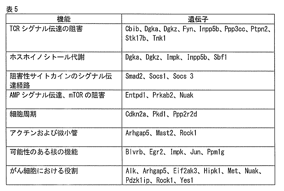
腫瘍における実質的な過剰出現を示したが、脾臓、二次リンパ器官では示さなかったshRNAを有する遺伝子に焦点を当てて、二次スクリーニングを行った。免疫抑制性の環
境にもかかわらず、腫瘍における実質的なT細胞の蓄積を多数のshRNAについて観察した。これらの二次スクリーニングに関して、遺伝子1つ当たり合計約15種のshRNAが得られるように各遺伝子(IDT)につき約10種の追加のshRNAを合成した。これらの集中プールは、85種の陰性対照shRNAを含有していた。2種の対照shRNA(1つはRFPに関し、1つはルシフェラーゼに関する)は、脾臓と比較して腫瘍において一定の濃縮を示した(それぞれ4.0倍と5.1倍)。第二のスクリーニングにおけるカットオフを、脾臓と比較して腫瘍において4倍以上の濃縮を示す3種以上のshRNAと定義した。T細胞に、レポータータンパク質(GFP、TFP、RFPまたはアメトリン蛍光タンパク質、Thy1.1)と共に個々のshRNAを導入することによって、スクリーニング結果を細胞レベルで検証した。このアプローチは、動物(グループ1つ当たり3匹のマウス)において5種のshRNAを同時に試験することが可能であった。24時間後、加えて3、5および7日後に、shRNAが形質導入されたT細胞の増殖を、CFSE希釈に基づき可視化した。加えて3、5および7日目に、IFNγ、TNFαおよびアイソタイプ対照に関して細胞内染色を行った。表6に、機能不全のT細胞プールshRNAライブラリーの一次および二次スクリーニングからの結果を示す。少なくとも3種のshRNAが腫瘍において4倍を超える濃縮を示した遺伝子をそれらの機能の簡単な説明と共に列挙する。表7に、キナーゼおよびホスファターゼshRNAライブラリーの二次スクリーニングからの結果を示す。
A secondary screen was performed, focusing on genes with shRNAs that showed substantial overrepresentation in tumors, but not in the spleen, secondary lymphoid organs. Despite the immunosuppressive environment, substantial T cell accumulation in tumors was observed for many shRNAs. For these secondary screens, approximately 10 additional shRNAs were synthesized for each gene (IDT) for a total of approximately 15 shRNAs per gene. These focused pools contained 85 negative control shRNAs. Two control shRNAs (one for RFP and one for luciferase) showed constant enrichment in tumor compared to spleen (4.0- and 5.1-fold, respectively). The cutoff in the second screen was defined as 3 or more shRNAs showing 4-fold or greater enrichment in tumor compared to spleen. Screening results were validated at the cellular level by transfecting T cells with individual shRNAs along with reporter proteins (GFP, TFP, RFP or ametrine fluorescent protein, Thy1.1). This approach allowed simultaneous testing of 5 shRNAs in animals (3 mice per group). After 24 hours, as well as after 3, 5 and 7 days, proliferation of shRNA-transduced T cells was visualized based on CFSE dilution. In addition, intracellular staining was performed on

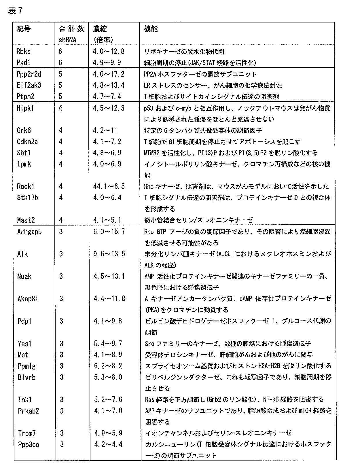
実施例2:B16黒色腫におけるshRNAによって駆動されるCD4およびCD8T細胞の拡大
ディープシーケンシング分析からの陽性shRNAを、5つの異なるレポータータンパク質(GFP、TFP、RFPまたはアメトリン蛍光タンパク質、Thy1.1)をコードするレンチウイルスベクターにクローニングした。サイトカインで前処理したOT-IT細胞を、単一のshRNAとレポータータンパク質との発現を駆動するレンチウイルスベクターで形質導入し、各集団の1×10⁶個のT細胞を混合し、14日目のB16-Ova腫瘍を有するC57BL/6マウスに共に静脈内注射した。7日後、腫瘍、脾臓および
リンパ節からT細胞を単離し、レポーター陽性のCD8+Vα2+Vβ5+T細胞のパーセンテージを、フローサイトメトリーによって共に導入されたレポーターに基づき決定した。脾臓と比較した腫瘍における濃縮倍率を、特定のレポーターを発現する各臓器におけるOT-IT細胞のパーセンテージに基づき計算した。CD8OT-IT細胞中で対照であるLacZのshRNAを発現させた場合、shRNA発現CD8OT-IT細胞の頻度は、脾臓と比較して腫瘍でより低かった(約2分の1)。対照的に、試験用shRNAは、腫瘍ではCD8OT-IT細胞の蓄積を誘導したが、脾臓では誘導しなかった(図6、図7)。これらのshRNAのうち7種(例えば、Ppp2r2D、Eif2ak3、Arhgap5、Smad2、Akap8I、RbksおよびEgr2)について、腫瘍におけるT細胞の蓄積は、脾臓と比較して10倍より多かった。PP2Aホスファターゼ7の調節サブユニットであるPpp2r2dを標的とするshRNAで、最も強い表現型が観察された。
Example 2: Positive shRNAs from extended deep sequencing analysis of shRNA-driven CD4 and CD8 T cells in B16 melanoma were analyzed with five different reporter proteins (GFP, TFP, RFP or ametrine fluorescent protein, Thy1.1). Cloned into an encoding lentiviral vector. Cytokine-pretreated OT-IT cells were transduced with lentiviral vectors driving expression of a single shRNA and a reporter protein, and 1×10 6 T cells from each population were mixed and treated at
Ppp2r2dまたはLacZのshRNAを発現するCD8+OT-IまたはCD4+TRP-1T細胞を、14日目のB16-Ova腫瘍を有するマウスに注射した。腫瘍および脾臓において、Thy1.1レポーターを使用して、shRNAを発現するT細胞を同定した(図8、Thy1.1+CD8T細胞の%、左のパネル)。2×10⁶個のshRNAを発現する細胞の移入から7日後に、腫瘍および脾臓において、LacZまたはPpp2r2dのshRNAを発現するT細胞の総数を決定した(図8、右のパネル)。腫瘍におけるPpp2r2d対LacZのshRNAを発現するT細胞の濃縮倍率を示す。Ppp2r2dのshRNAは、OT-ICD8T細胞の蓄積を誘導しただけでなく、CD4T細胞の蓄積も誘導し(TRP-1TCRトランスジェニックマウス由来)23、腫瘍におけるT細胞数は、LacZのshRNAではなくPpp2r2dを発現した場合により有意に高かった(CD8の場合、36.3倍;CD4T細胞の場合、16.2倍)(図8)。
CD8 + OT-I or CD4 + TRP-1 T cells expressing Ppp2r2d or LacZ shRNA were injected into
Ppp2r2dまたはCblbのshRNAのパネルを発現する細胞に関する脾臓と比較した腫瘍におけるT細胞の濃縮(図17、上のパネル)、Ppp2r2dおよびCblbのmRNAレベルも、T細胞移入前にqPCRによって測定した(図17、下のパネル)。shRNAに関して最も強い腫瘍におけるT細胞濃縮が観察され、ノックダウン効率はmRNAレベルで80%より大きかった(Ppp2r2dおよびCblbの両方についてshRNA番号1および2)。CD8T細胞の蓄積は、Ppp2r2dのノックダウンの程度と相関しており、最大のインビボ活性を有する2種のPpp2r2dのshRNAは、最小レベルのPpp2r2dのmRNAを誘導した(図17)。
Enrichment of T cells in tumors compared to spleen for cells expressing a panel of Ppp2r2d or Cblb shRNAs (Fig. 17, top panel), Ppp2r2d and Cblb mRNA levels were also measured by qPCR prior to T cell transfer (Fig. 17, lower panel). The strongest T cell enrichment in tumors was observed for shRNAs, with knockdown efficiencies greater than 80% at the mRNA level (
またPpp2r2dのノックダウンも、定量的マススペクトロメトリーアプローチを使用してタンパク質レベルで確認された(図18)。マススペクトロメトリーによる細胞溶解産物由来タンパク質の絶対定量(AQUA)に関してこれまでに報告されているアプローチを使用して、タンパク質レベルにおけるPpp2r2dのshRNA発現の作用を測定した(Gerber, S.A.、Rush, J.、Stemman, O.、Kirschner, M.W.およびGygi, S.P. Absolute quantification of proteins and phosphoproteins from cell lysates by tandem
MS. PNAS、100、6940~6945(2003)。この方策は、マススペクトロメトリー分析のための内部標準として安定な同位体が取り込まれた合成ペプチドを使用する「選択的反応をモニターする」アプローチに基づく。LacZまたはPpp2r2dのshRNAを発現するOT-I細胞を、FACSを使用してソートして純化した。プロテアーゼ阻害剤カクテル(シグマ(Sigma))、1mMのEDTAおよび1mMのPMSFを含有する1mlの
MPER抽出試薬(ピアース(Pierce))中で、細胞(1×10⁶個)を、氷上で15分間、時々ボルテックス混合しながら溶解させた。死細胞片を遠心分離で除去し、タンパク質上清をろ過した(0.2μmのSpinX遠心分離機フィルター、コースター(Costar))。タンパク質濃度を、ブラッドフォードアッセイ(Biorad)およびUV280nm分
析(ナノドロップ(Nanodrop)の機器)によって決定した。SDS-PAGEによって0.1mgの細胞タンパク質を分離し、クーマシーブルー試薬(ピアース)で染色した。45~60kDaの分子量範囲に相当するゲルのバンドを切り出し、続いてトリプシンを用いてタンパク質をゲル中で消化した。溶出したペプチドを、LCMS/MS(LTQ XLオービトラップ(Orbitrap)、サーモ・サイエンティフィック)による定量化のために、300fmolの同位体標識Ppp2r2d(FFEEPEDPSS[13C-15N-R]-OH)(配列番号628)およびアクチンB(GYSFTTTAE[13C-15N-R]-OH)(配列番号629)ペプチド(21センチュリーバイオケミカルズ(21st Century Biochemicals))でスパイクした。PP2Aの他の調節サブユニットとは
異なるタンパク質の領域から、Ppp2r2dペプチドを選択した。最初に、LacZのshRNAサンプルのLC-MS/MS実行を分析して、モニターしていたPpp2r2dおよびアクチンBペプチドの場所を特定した。逆相カラムから対応する内因性ペプチドで共に溶出させた絶対定量AQUAペプチドは、なおより高いMW(10Da)を有することから、豊富なペプチドのフラグメントイオンを使用して、内因性およびAQUAペプチドに関してピーク強度の比率を決定することを可能にした。3連のサンプルをSDS-PAGE-LC-MS/MSによって分析し、スチューデントの両側t検定を使用したグラフパッド・プリズム6.0ソフトウェアを使用して統計的有意性を決定した(F検定、*p=0.0062)。
Ppp2r2d knockdown was also confirmed at the protein level using a quantitative mass spectrometry approach (Figure 18). A previously reported approach for absolute quantification of protein from cell lysates by mass spectrometry (AQUA) was used to measure the effects of Ppp2r2d shRNA expression on the protein level (Gerber, SA, Rush, J. , Stemman, O., Kirschner, MW and Gygi, SP Absolute quantification of proteins and phosphoproteins from cell lysates by tandem
MS. PNAS, 100, 6940-6945 (2003). This strategy is based on a "selective reaction monitoring" approach that uses stable isotope-incorporated synthetic peptides as internal standards for mass spectrometry analysis. OT-I cells expressing LacZ or Ppp2r2d shRNA were sorted and purified using FACS. Cells (1×10 6 ) were lysed in 1 ml of MPER extraction reagent (Pierce) containing protease inhibitor cocktail (Sigma), 1 mM EDTA and 1 mM PMSF on ice for 15 minutes. Dissolve with occasional vortex mixing. Dead cell debris was removed by centrifugation and the protein supernatant was filtered (0.2 μm SpinX centrifuge filter, Costar). Protein concentration was determined by Bradford assay (Biorad) and UV280 nm analysis (Nanodrop instrument). 0.1 mg of cellular proteins were separated by SDS-PAGE and stained with Coomassie blue reagent (Pierce). The gel band corresponding to the 45-60 kDa molecular weight range was excised and the proteins were subsequently digested in the gel using trypsin. Eluted peptides were treated with 300 fmol of isotopically labeled Ppp2r2d (FFEEPEDPSS[13C-15N-R]-OH) (seq. #628) and actin B (GYSFTTTAE[13C-15N-R]-OH) (SEQ ID NO:629) peptide (21st Century Biochemicals). The Ppp2r2d peptide was chosen from a region of the protein distinct from other regulatory subunits of PP2A. First, an LC-MS/MS run of the LacZ shRNA sample was analyzed to localize the Ppp2r2d and actin B peptides that were being monitored. Absolute quantitation AQUA peptide co-eluted with the corresponding endogenous peptide from the reversed-phase column has an even higher MW (10 Da), thus using the abundant peptide fragment ions, peaks for endogenous and AQUA peptides made it possible to determine the ratio of intensities. Triplicate samples were analyzed by SDS-PAGE-LC-MS/MS and statistical significance was determined using GraphPad Prism 6.0 software with Student's two-tailed t-test (F-test, * p=0.0062).
Ppp2r2dのshRNAの特異性を決定した。変異Ppp2r2dのcDNA(野生型タンパク質配列を有するが、shRNA結合部位に変異したDNA配列を有する)がPpp2r2dのshRNAと共に導入された場合、表現型が逆転することから、Ppp2r2dのshRNA活性は特異的であった(図9、10a~c)。さらに、OT-ICD8T細胞は、脾臓と比較して腫瘍においてPpp2r2dを過剰発現したころから(あらゆるshRNA発現の非存在下で)、OT-ICD8T細胞は、腫瘍においてT細胞の機能を阻害するシグナル伝達ネットワークの固有の構成要素であることが示唆される(図19)。 The specificity of the Ppp2r2d shRNA was determined. The shRNA activity of Ppp2r2d is not specific, as the phenotype is reversed when the mutant Ppp2r2d cDNA (which has the wild-type protein sequence but has the mutated DNA sequence at the shRNA binding site) is introduced along with the Ppp2r2d shRNA. There was (FIGS. 9, 10a-c). In addition, since OT-ICD8 T cells overexpressed Ppp2r2d in tumors compared to spleen (in the absence of any shRNA expression), OT-ICD8 T cells could be used in tumor signaling to inhibit T cell function. It is suggested that it is a unique component of the network (Fig. 19).
OT-IT細胞を、LacZのshRNA、Ppp2r2dのshRNA、Ppp2r2dのshRNAの発現を駆動するレンチウイルスベクターで形質導入した。タンパク質配列は保存されているがshRNA結合部位が破壊された変異Ppp2r2dのcDNAを生成した。フォワードプライマー:GGATCCATGGCAGGAGCTGGAGGC(配列番号630)およびリバースプライマー:GCTAGCATTAATTTTGTCCTGGAATATATACAAGTTATTGGTGG(配列番号631)を使用したRT-PCRにより、野生型Ppp2r2dのcDNAを単離した。フォワードプライマー:TCCATCCCCACCAATGTAACGTGTTTGTTTACAGCAGCAGCAAGG(配列番号634)およびリバースプライマー:AAACAAACACGTTACATTGGTGGGGATGGAACTCTGCGGCAGTGA(配列番号635)を使用したオーバーラップPCR(タンパク質のコード配列を保護する)により、Ppp2r2dのshRNAの標的配列、CCCACATCAGTGCAATGTATT(配列番号632)をTCCCCACCAATGTAACGTGTT(配列番号633)に変異させた(図10a)。野生型と変異Ppp2r2d両方のcDNAを、2Aリボゾームスキップペプチド-GFP配列(細胞中で化学量論的なPpp2r2dおよびGFP発現をもたらす)を用いて改変pLKO.3ベクターにクローニングした。コンストラクトをEL4胸腺腫細胞に導入した。GFP発現EL4細胞をソートして純化し、次いでThy1.1レポーターの発現を駆動するLacZまたはPpp2r2dのshRNAレンチウイルスベクターで形質導入した。shRNAが形質導入された(Thy1.1+)細胞を、フローサイトメトリーによってGFP発現に関して分析した。Ppp2r2dのshRNAは、野生型Ppp2r2dの場合、GFPレベルを低減させた。Ppp2r2dのshRNAは、変異Ppp2r2dのcDNAを発現する細胞中でGFPレポーター
の発現を低減させることができなかったことから、shRNA結合部位がうまく変異したことが実証された(図10a)。
OT-IT cells were transduced with lentiviral vectors driving expression of LacZ shRNA, Ppp2r2d shRNA, Ppp2r2d shRNA. A mutant Ppp2r2d cDNA was generated in which the protein sequence was conserved but the shRNA binding site was disrupted. Wild-type Ppp2r2d cDNA was isolated by RT-PCR using forward primer: GGATCCATGGCAGGAGCTGGAGGC (SEQ ID NO: 630) and reverse primer: GCTAGCATTAATTTTGTCCTGGAATATATACAAGTTATTGGTGG (SEQ ID NO: 631). The target sequence of the shRNA of Ppp2r2d, CCCACATGTCAGT3 (SEQ ID NO: 635), was obtained by overlapping PCR (protecting the coding sequence of the protein) using forward primer: TCCATCCCCACCAATGTAACGTGTTTGTTTACAGCAGCAGCAAGG (SEQ ID NO: 634) and reverse primer: AAACAAAACACGTTACATTGGTGGGGATGGAACTCTGCGGCAGTGA (SEQ ID NO: 635). It was mutated to TCCCCACCAATGTAACGTGTT (SEQ ID NO: 633) (Fig. 10a). Both wild-type and mutant Ppp2r2d cDNAs were transformed into modified pLKO. 3 vector. Constructs were introduced into EL4 thymoma cells. GFP-expressing EL4 cells were sorted and purified, then transduced with LacZ or Ppp2r2d shRNA lentiviral vectors driving expression of the Thy1.1 reporter. The shRNA-transduced (Thy1.1 + ) cells were analyzed for GFP expression by flow cytometry. Ppp2r2d shRNA reduced GFP levels in the case of wild-type Ppp2r2d. The Ppp2r2d shRNA was unable to reduce the expression of the GFP reporter in cells expressing the mutated Ppp2r2d cDNA, demonstrating that the shRNA binding site was successfully mutated (Fig. 10a).
またPpp2r2d変異cDNAの発現も、Ppp2r2dのshRNAにより誘導された表現型を予防する(図10b)。1つのベクターでPpp2r2dのshRNAと変異Ppp2r2dのcDNAとの共発現をもたらす変異Ppp2r2dのcDNA-2A-GFPコンストラクトに、Ppp2r2dのshRNAをクローニングした。LacZのshRNA(Thy1.1)、Ppp2r2dのshRNA(アメトリン)またはPpp2r2dのshRNAに加えて変異Ppp2r2dのcDNA(GFP)をコードするレンチウイルスで、OT-IT細胞を別々に感染させた(図10b)。次いでこれらの3つの集団を同じ比率で混合し、14日目のB16-Ova腫瘍を有するマウスに注射した。7日目に、各T細胞集団を、腫瘍および脾臓においてOT-I(CD8+Vα2+Vβ5+)T細胞でのゲーティングにより定量し、続いてThy1.1、アメトリンまたはGFP発現でマーキングされた集団を分析した。腫瘍および脾臓における各T細胞集団のパーセンテージを、Vα2+Vβ5+T細胞でのゲーティングによって定量し、形質導入された細胞を、Thy1.1またはアメトリン/GFP蛍光レポーターの発現に基づき検出した。図10bに結果を示す。(2回の独立した実験からの代表的なデータ、実験1回当たりn=3匹のマウス)。
Expression of the Ppp2r2d mutant cDNA also prevented the Ppp2r2d shRNA-induced phenotype (Fig. 10b). The Ppp2r2d shRNA was cloned into the mutated Ppp2r2d cDNA-2A-GFP construct that results in co-expression of the Ppp2r2d shRNA and the mutated Ppp2r2d cDNA in one vector. OT-IT cells were separately infected with lentiviruses encoding LacZ shRNA (Thy1.1), Ppp2r2d shRNA (Ametrine) or Ppp2r2d shRNA plus mutant Ppp2r2d cDNA (GFP) (Fig. 10b). . These three populations were then mixed in equal proportions and injected into
図10cは、LacZのshRNA、Ppp2r2dのshRNA、およびPpp2r2dのshRNAに加えてPpp2r2d変異cDNAで形質導入されたOT-IT細胞におけるPpp2r2d発現に関するリアルタイムPCR分析を示す。最大のインビボ活性を有するPpp2r2dのshRNAも、最小レベルのPpp2r2dのmRNAと関連していた(図11)。 FIG. 10c shows real-time PCR analysis of Ppp2r2d expression in OT-IT cells transduced with LacZ shRNA, Ppp2r2d shRNA, and Ppp2r2d shRNA plus Ppp2r2d mutant cDNA. The Ppp2r2d shRNAs with the greatest in vivo activity were also associated with the lowest levels of Ppp2r2d mRNA (FIG. 11).
試験用または対照shRNAを発現する腫瘍浸潤T細胞のマイクロアレイ分析によれば、各shRNAは、別個の群の遺伝子発現変化を誘導し、一部は特定のshRNA間でオーバーラップしていたことが示された(図12a~c)。2種の遺伝子(Egr2およびPtpn2)は、T細胞において公知の機能を有する。腫瘍対脾臓における濃縮を、二次スクリーニングからのディープシーケンシング結果に基づき計算した(図12a)。mRNAの平均発現レベルのクラスター化は、LacZ対照shRNAもしくは5つの試験用shRNAsのうち1つを発現する脾臓中のT細胞または腫瘍によって有意に調節されることが見出された(図12b)。有意な発現の差は、LacZ対照shRNAまたは5つの試験用shRNA(Alk、Arhgap5、Egr2、Ptpn2またはPpp2r2d)のうち1つを発現するT細胞間におけるAnovaのp値として、<0.01と定義された(JMP-ゲノミクス6.0、SASインスティテュート社)。クラスター化後に、1つまたはそれより多くの治療群において有意に調節されたmRNAが示される(ファストワード)。図12cは、5つの試験用shRNA(上述したようにAnovaでp<0.01と定義されたシグネチャー)のうち1つで形質導入された腫瘍浸潤T細胞による発現シグネチャー間のオーバーラップを示すベン図である。オーバーラップする楕円で示されるように、5つのシグネチャーのあらゆる組み合わせで多数のオーバーラップするプローブ番号が示される。添付の表に、無作為抽出により予測されるものに対するオーバーラップの有意性(フィッシャーの正確検定)を示す。 Microarray analysis of tumor-infiltrating T cells expressing test or control shRNAs showed that each shRNA induced distinct groups of gene expression changes, some of which overlapped between specific shRNAs. (FIGS. 12a-c). Two genes (Egr2 and Ptpn2) have known functions in T cells. Enrichments in tumor versus spleen were calculated based on deep sequencing results from the secondary screen (Fig. 12a). Clustering of mean expression levels of mRNA was found to be significantly modulated by T cells in the spleen or tumors expressing the LacZ control shRNA or 1 of the 5 test shRNAs (Fig. 12b). A significant difference in expression was defined as an Anova p-value of <0.01 between T cells expressing the LacZ control shRNA or one of the five test shRNAs (Alk, Arhgap5, Egr2, Ptpn2 or Ppp2r2d). (JMP-Genomics 6.0, SAS Institute). After clustering, significantly regulated mRNAs in one or more treatment groups are shown (fast words). Figure 12c is a Venn diagram showing the overlap between expression signatures by tumor-infiltrating T cells transduced with 1 of the 5 test shRNAs (signatures defined p<0.01 in Anova as described above). is. A number of overlapping probe numbers are shown for every combination of five signatures, as indicated by the overlapping ellipses. The attached table shows the significance of the overlap (Fisher's exact test) to that predicted by random sampling.
実施例3:Ppp2r2dにより誘導されたT細胞の機能の変化
この実施例で、腫瘍におけるPpp2r2dのshRNAにより、T細胞の蓄積、具体的にはT細胞の浸潤、蓄積およびアポトーシスを駆動する細胞メカニズムを試験した。細胞質色素であるCFSEで標識されたOT-ICD8T細胞の移入によって、腫瘍へのT細胞浸潤を査定した。Ppp2r2dまたはLacZのshRNAを発現するOT-IT細胞をCFSEで標識し、B16-Ova腫瘍を有するマウスに注射した。24時間後、形
質導入されたT細胞を腫瘍および脾臓から単離し、フローサイトメトリーによって定量した。LacZまたはPpp2r2dのshRNAを発現するOT-IT細胞を、Thy1.1レポーターを使用して精製し、サイトカインが添加されていない完全RPMI培地中で24時間培養した。フィコール密度勾配遠心分離(シグマ)によって単離された生細胞をCFSE(カルボキシフルオレセインジアセテート、スクシンイミジルエステル、インビトロジェン)で標識し、2×106個の標識細胞を、14日目のB16-Ova腫瘍を有するマウスに注射した。CFSE希釈を、移入から24時間ならびに3、5および7日目にフローサイトメトリーによって定量した。加えて、3、5および7日目に、IFNγ、TNFαおよびアイソタイプ対照(BD)に関して細胞内染色を行った。1日目の腫瘍では、Ppp2r2dまたはLacZのshRNAが形質導入されたCD8T細胞の頻度において差は観察されず、これは、T細胞浸潤への実質的な作用の反論となった(図13a)。しかしながら、その後のタイムポイント(3および5日目)の分析から、LacZのshRNAが形質導入されたT細胞と比較した、Ppp2r2dによるより高い程度の増殖(CFSE希釈に基づく)が実証された(図13b、図20a)。またPpp2r2dのshRNAが形質導入されたT細胞は、抗腫瘍免疫にとって重要なサイトカインであるより高いレベルのインターフェロン-γも産生した(図13e)。T細胞の蓄積は、腫瘍では強化されたが腫瘍流入領域リンパ節ではそれより低い程度にしか強化されなかったたことから、Ppp2r2dの作用はT細胞受容体活性化の下流であった。対照的に、関連抗原がT細胞に提示されない無関係のリンパ節または脾臓では、蓄積は観察されなかった(図15)。LacZのshRNAが形質導入されたT細胞については、腫瘍中にこのような細胞は少数しか存在しないにもかかわらず、かなりの程度のT細胞の蓄積も観察された(7日目までにCFSE色素は十分希釈された)。これは、LacZのshRNAが形質導入されたT細胞はアポトーシスによって失われたことを示唆していた。実際に、LacZ対照shRNA(Ppp2r2dのshRNAではなく)が発現された場合(図13g、図20b)、より大きなパーセンテージの腫瘍浸潤T細胞が、活性なカスパーゼ-3に特異的な抗体で標識された。さらに、CD8T細胞とB16-Ova腫瘍細胞との共培養から、LacZのshRNAを発現するT細胞の大部分がアポトーシスを起こしたが(65.7%)、ほとんどのPpp2r2dのshRNAが形質導入されたT細胞が生存可能であった(89.5%、図13c)ことが示された。
Example 3: Ppp2r2d-induced alterations in T cell function In this example, Ppp2r2d shRNA in tumors induced T cell accumulation, specifically the cellular mechanisms driving T cell infiltration, accumulation and apoptosis. tested. T cell infiltration into tumors was assessed by transfer of OT-ICD8 T cells labeled with the cytoplasmic dye CFSE. OT-IT cells expressing Ppp2r2d or LacZ shRNA were labeled with CFSE and injected into B16-Ova tumor-bearing mice. Twenty-four hours later, transduced T cells were isolated from tumor and spleen and quantified by flow cytometry. OT-IT cells expressing LacZ or Ppp2r2d shRNA were purified using the Thy1.1 reporter and cultured for 24 hours in complete RPMI medium without added cytokines. Live cells isolated by Ficoll density gradient centrifugation (Sigma) were labeled with CFSE (carboxyfluorescein diacetate, succinimidyl ester, Invitrogen) and 2 x 10 labeled cells were isolated from
LacZまたはPpp2r2dのshRNAを発現するOT-IT細胞を、上述したようにThy1.1発現に基づき精製し、CFSEで標識した。CFSE標識OT-IT細胞(1×105個)を、96-ウェルプレート中のウェル1つ当たり5×104個のB16-Ova細胞と共に72時間培養した。アッセイ前に、B16-Ova細胞を1ng/mLのIFNγに48時間晒し(インビトロで発現されないMHCクラスIを誘導するため)、3回洗浄した。CD8+細胞のアネキシンV標識化によって、OT-IT細胞のアポトーシスを検出した(図13c)。BD細胞内染色キットを使用して、ホスホ-AKT(Ser473)、ホスホ-Bad(Ser112)、Bcl-2およびアイソタイプ対照の細胞内染色を48時間行った。CD8T細胞とB16-Ova腫瘍細胞との共培養から、実際に、LacZのshRNAを発現するT細胞の大部分がアポトーシスを起こしたが(65.7%)、Ppp2r2dのshRNAが形質導入されたT細胞の大部分は生存可能であった(89.5%、図13c)ことが示された。CD28共刺激の非存在下でPpp2r2dおよびLacZのshRNAを発現するT細胞を固定されたCD3抗体で刺激したところ、類似の表現型が観察された(図14)。具体的に言えば、B16-Ova細胞(2×105個)を雌C57BL/6マウス(10週齢)の皮下に注射した。12日目に、マウスが保持する類似サイズの腫瘍を7つのグループに分割した(マウス7~8匹/グループ)。12日目および17日目に、Ppp2r2dまたはLacZのshRNAベクターで感染させた抗CD3/CD28ビーズ活性化CD4TRP-1または/およびCD8OT-IT細胞(それぞれ2×106個のT細胞)を静脈内注射した。B16腫瘍の治療のために、10日目に、Ppp2r2dまたはLacZのshRNAを発現する抗
CD3/CD28ビーズ活性化CD4TRP-1およびCD8pmel-1T細胞(それぞれ3×106個のT細胞)でマウスを治療した。移入後3日毎に腫瘍のサイズを測定し、長さ×幅として計算した。最も長い軸で20mm以上の腫瘍を有するマウスを殺した。
OT-IT cells expressing LacZ or Ppp2r2d shRNA were purified based on Thy1.1 expression and labeled with CFSE as described above. CFSE-labeled OT-IT cells (1×10 5 ) were cultured with 5×10 4 B16-Ova cells per well in 96-well plates for 72 hours. Prior to assay, B16-Ova cells were exposed to 1 ng/mL IFNγ for 48 hours (to induce unexpressed MHC class I in vitro) and washed three times. Apoptosis of OT-IT cells was detected by Annexin V labeling of CD8 + cells (Fig. 13c). Intracellular staining of phospho-AKT (Ser473), phospho-Bad (Ser112), Bcl-2 and isotype control was performed for 48 hours using the BD intracellular staining kit. From co-cultures of CD8 T cells and B16-Ova tumor cells, indeed, the majority of T cells expressing LacZ shRNA were apoptotic (65.7%), whereas Ppp2r2d shRNA transduced T cells It was shown that the majority of cells were viable (89.5%, Figure 13c). A similar phenotype was observed when T cells expressing Ppp2r2d and LacZ shRNAs were stimulated with immobilized CD3 antibody in the absence of CD28 costimulation (FIG. 14). Specifically, B16-Ova cells (2×10 5 ) were injected subcutaneously into female C57BL/6 mice (10 weeks old). On
これらの結果から、Ppp2r2dのshRNAが形質導入されたCD8T細胞は、B16-Ova腫瘍細胞によって直接提示されたそれらの抗原を認識する場合であっても、それらは増殖して生存できる可能性があることが示唆された。この概念は、MHCクラスIタンパク質の発現が欠失したb2m-/-マウスへの腫瘍細胞の埋め込みによって試験された24。このようなマウスにおいて、宿主の専門の抗原提示細胞ではなく腫瘍細胞のみが、腫瘍抗原をT細胞に提示することができる。実際に、Ppp2r2dのshRNAが形質導入されたOT-ICD8T細胞は、b2m-/-マウスにおいてB16-Ova腫瘍内では大量の蓄積を示したが(図12f)、Ova抗原の発現が欠失した反対に位置するB16腫瘍では極めて少数のT細胞しか存在しなかった。したがってPpp2r2dのshRNAを発現するT細胞は、好適な共刺激シグナルの欠失および阻害性の微小環境にもかかわらず、腫瘍細胞に応答して効果的に増殖し生存することができた。 These results suggest that Ppp2r2d shRNA-transduced CD8 T cells may be able to proliferate and survive even when they recognize their antigens directly presented by B16-Ova tumor cells. It has been suggested. This concept was tested by implanting tumor cells into b2m−/− mice that lack expression of MHC class I proteins 24 . In such mice, only tumor cells, not the host's professional antigen-presenting cells, are able to present tumor antigens to T cells. Indeed, OT-ICD8 T cells transduced with Ppp2r2d shRNA showed massive accumulation within B16-Ova tumors in b2m−/− mice (FIG. 12f), whereas expression of Ova antigen was lost. Very few T cells were present in B16 tumors located at . Thus, T cells expressing the Ppp2r2d shRNA were able to effectively proliferate and survive in response to tumor cells despite the lack of suitable co-stimulatory signals and the inhibitory microenvironment.
また、ナノウェルデバイスを使用した単一細胞レベルでの腫瘍浸潤T細胞のエクスビボの分析も、Ppp2r2dのサイレンシングは、T細胞によるサイトカイン産生を増加させることを実証した(図21a~c)。脂質二重層において、CD3/CD28抗体によりT細胞を3時間活性化し、続いて抗体でコーティングしたスライド上で1時間サイトカイン捕捉を行った。CD8T細胞は、IFNγ、IL-2およびGM-CSFについてはより高い分泌速度を示し、より大きい割合のT細胞が1種より多くのサイトカインを示した(図21b、c)。細胞内サイトカイン染色によって、より多数のIFNγ産生T細胞の存在が確認された(図21d、図20)。 Ex vivo analysis of tumor-infiltrating T cells at the single-cell level using nanowell devices also demonstrated that silencing of Ppp2r2d increased cytokine production by T cells (Fig. 21a-c). In the lipid bilayer, T cells were activated with CD3/CD28 antibodies for 3 hours, followed by cytokine capture on antibody-coated slides for 1 hour. CD8 T cells showed higher secretion rates for IFNγ, IL-2 and GM-CSF, with a greater proportion of T cells showing more than one cytokine (Fig. 21b,c). Intracellular cytokine staining confirmed the presence of higher numbers of IFNγ-producing T cells (Fig. 21d, Fig. 20).
PP2Aホスファターゼは、触媒サブユニットおよび足場サブユニットで構成され、その基質特異性は、多くの調節サブユニットの1つによって決定される7。Ppp2r2dは、分裂間期と後期の際にPP2AをCdk1基質に向かわせることにより、有糸分裂に入らないようにして有糸分裂からの退出を誘導する25。PP2Aは、BAD介在アポトーシスにとってゲートキーパー的な役割を果たす。リン酸化BADは、14-3-3によってサイトゾル中にその不活性型で隔離されるが、脱リン酸化BADはミトコンドリアに標的化されて、そこでBcl-XLおよびBcl-2との結合によって細胞死を引き起こす26。またPP2Aホスファターゼは、CD28およびCTLA-4の細胞質内ドメイン、加えてCarma1(NF-κB経路上流)と相互作用することも示されたが、調節サブユニットがこれらの活性に必要であることは知られていなかった。必要な生化学研究に好適なPpp2r2d抗体は現在のところ入手が不可能である。 PP2A phosphatases are composed of catalytic and scaffolding subunits, and their substrate specificity is determined by one of a number of regulatory subunits 7 . Ppp2r2d prevents mitosis entry and induces mitotic exit by directing PP2A to Cdk1 substrates during interphase and anaphase 25 . PP2A plays a gatekeeper role for BAD-mediated apoptosis. Phosphorylated BAD is sequestered in its inactive form in the cytosol by 14-3-3, whereas dephosphorylated BAD is targeted to mitochondria, where it binds to Bcl- XL and Bcl-2. cause cell death 26 . PP2A phosphatase was also shown to interact with the cytoplasmic domains of CD28 and CTLA-4, as well as Carma1 (upstream of the NF-κB pathway), although regulatory subunits are known to be required for these activities. It wasn't done. A Ppp2r2d antibody suitable for the required biochemical studies is not currently available.
実施例4:Ppp2r2dのサイレンシングはCD4およびCD8T細胞の抗腫瘍活性を強化する
養子T細胞治療の効能を強化するPpp2r2dのshRNAの能力を査定した。B16-Ova腫瘍細胞(2×10⁵個)を、雌C57BL/6マウス(10週齢)の皮下に注射した。12日目に、マウスが保持する類似サイズの腫瘍を7つのグループ(マウス7~8匹/グループ)に分割し、それらを、T細胞なし、2×106個のshRNAが形質導入されたTRP-1CD4T細胞、2×106個のshRNAで感染させたOT-ICD8T細胞のいずれか、またはCD4およびCD8T細胞の両方(12日目および17日目)で処理した。12日目および17日目に、グループに応じて、抗CD3/CD28ビーズによって活性化されたCD4TRP-1、または/およびPpp2r2dもしくはLacZのshRNAベクターで感染させたCD8OT-IT細胞(それぞれ2×106個のT細胞)を静脈内注射した。B16腫瘍の治療のために、10日目に、Ppp2r2dまたはLacZのshRNAを発現する抗CD3/CD28ビーズ活性化CD4TRP-1
およびCD8pmel-1T細胞(それぞれ3×10⁶個のT細胞)で、マウスを治療した。移入後3日毎に腫瘍のサイズを測定し、長さ×幅として計算した。最も長い軸で20mm以上の腫瘍を有するマウスを殺した。Ppp2r2d-サイレンシングはCD4およびCD8T細胞の治療活性を改善し、Ppp2r2dのshRNAが形質導入されたCD4およびCD8T細胞を共に投与した場合に相乗効果が観察された(図16a、b)。またPpp2r2dのshRNAは、内因性腫瘍抗原に特異的なT細胞(pmel-1CD8T細胞およびTRP-1CD4T細胞)に導入した場合に抗腫瘍応答も強化した(図16c)。
Example 4: Silencing of Ppp2r2d enhances the antitumor activity of CD4 and CD8 T cells The ability of Ppp2r2d shRNAs to enhance the efficacy of adoptive T cell therapy was assessed. B16-Ova tumor cells (2×10 5 ) were injected subcutaneously into female C57BL/6 mice (10 weeks old). On
and CD8pmel-1 T cells (3×10 6 T cells each). Tumor sizes were measured every 3 days after transfer and calculated as length x width. Mice with tumors greater than or equal to 20 mm along the longest axis were killed. Ppp2r2d-silencing improved the therapeutic activity of CD4 and CD8 T cells, and a synergistic effect was observed when Ppp2r2d shRNA-transduced CD4 and CD8 T cells were administered together (Fig. 16a,b). Ppp2r2d shRNA also enhanced antitumor responses when introduced into T cells specific for endogenous tumor antigens (pmel-1CD8 T cells and TRP-1CD4 T cells) (FIG. 16c).
Ppp2r2dがサイレンシングされたT細胞は、腫瘍においてエフェクター表現型を獲得し(図22a)、細胞の30%より多くがグランザイムBを発現した(図23a)。腫瘍におけるこのようなエフェクターT細胞の著しい増加と一致して(図23b)、タネル染色から、LacZのshRNAではなくPpp2r2dを発現するT細胞が存在する場合、腫瘍においてアポトーシスが増加することが実証された(図23c)。B16黒色腫は高侵襲性の腫瘍であるが、その理由の一つはMHCクラスI発現が極めて低いためである。興味深いことに、LacZのshRNAではなくPpp2r2dを発現するT細胞は、腫瘍細胞によってMHCクラスI発現(H-2Kb)を有意に増加させ(図23d)、これは場合により、T細胞によるIFNγ分泌の増加が観察されたことに起因する可能性がある(図21a~c、図13e)。Ppp2r2dのshRNAは、腫瘍浸潤T細胞における阻害性PD-1またはLAG-3受容体の発現を低減させないことから、その作用機序は、これらの公知のT細胞の機能の負の調節因子とは異なることが実証される(図22b)。この発見から、これらの細胞内と細胞表面分子とを標的とする組み合わせのアプローチが示唆される。 Ppp2r2d-silenced T cells acquired an effector phenotype in tumors (Fig. 22a), with more than 30% of cells expressing granzyme B (Fig. 23a). Consistent with such a marked increase in effector T cells in tumors (Fig. 23b), Tunel staining demonstrated increased apoptosis in tumors when T cells expressing Ppp2r2d, but not the LacZ shRNA, were present. (Fig. 23c). B16 melanoma is a highly aggressive tumor, in part because of its extremely low MHC class I expression. Interestingly, T cells expressing Ppp2r2d, but not the LacZ shRNA, significantly increased MHC class I expression (H-2Kb) by tumor cells (Fig. 23d), possibly suggesting IFNγ secretion by T cells. This may be due to the observed increase (Fig. 21a-c, Fig. 13e). The Ppp2r2d shRNA does not reduce the expression of inhibitory PD-1 or LAG-3 receptors on tumor-infiltrating T cells, suggesting that its mechanism of action is different from these known negative regulators of T cell function. A difference is demonstrated (Fig. 22b). This finding suggests a combinatorial approach targeting these intracellular and cell surface molecules.
これらの結果は、複合組織の微小環境における免疫療法のための、新規の標的のインビボにおける発見の実行可能性を確立する。本発明者らは、二次リンパ器官と比較した腫瘍におけるT細胞の蓄積で例示されるような組織間で示差的な作用を有する遺伝子を発見することが可能であることを示した。組織選択的な作用を有する遺伝子について、T細胞の蓄積および生存は、T細胞受容体の制御下である可能性が高いために、関連抗原の提示がない組織では起こらない。インビボで特定の免疫細胞機能の制御を調査するために、本明細書に記載されたアプローチの多くのバリエーションを想定することができる。例えば、腫瘍において重要なT細胞エフェクター機能を制御する遺伝子を発見するために、サイトカインまたは細胞傷害性分子の発現のための蛍光レポーター(グランザイムB、パーフォリン)を本発明者らのアプローチに統合してもよい。 These results establish the feasibility of in vivo discovery of novel targets for immunotherapy in the complex tissue microenvironment. The inventors have shown that it is possible to discover genes that have differential effects across tissues as exemplified by the accumulation of T cells in tumors compared to secondary lymphoid organs. For genes with tissue-selective action, T-cell accumulation and survival is likely under control of the T-cell receptor and thus does not occur in tissues lacking relevant antigen presentation. Many variations of the approaches described herein can be envisioned to investigate the regulation of specific immune cell functions in vivo. For example, fluorescent reporters (granzyme B, perforin) for the expression of cytokines or cytotoxic molecules have been integrated into our approach to discover genes that control important T cell effector functions in tumors. good too.
主要な調節スイッチを標的化することは、がんおよび他の病理におけるT細胞の活性を改変する新しいアプローチを提供する可能性がある。このようなT細胞ベースの療法の効能は、腫瘍微小環境におけるT細胞の機能を阻害する遺伝子のshRNA介在サイレンシングによって強化される可能性がある。 Targeting key regulatory switches may provide new approaches to alter T cell activity in cancer and other pathologies. The efficacy of such T-cell-based therapies may be enhanced by shRNA-mediated silencing of genes that inhibit T-cell function in the tumor microenvironment.
他の実施態様
本発明をそれらの詳細な説明と共に説明したが、前述の説明は、本発明の範囲を例示するものであり、それらを限定しないことが意図されており、本発明の範囲は、添付の特許請求の範囲の範囲によって定義されることが理解されるものとする。他の形態、利点、および改変は、以下の特許請求の範囲内である。
国際出願時の特許請求の範囲の請求項(1)~(51)
(1) ベクターを含む腫瘍特異性を有する免疫応答性細胞であって、ベクターは、shRNAをコードする配列を含み、shRNAは、配列番号604~620および653~677からなる群より選択される核酸配列に相補的な15個の連続したヌクレオチドを含む、上記免疫応答性細胞。
(2) 免疫応答性細胞が、腫瘍浸潤リンパ球(TIL)、ナチュラルキラーT細胞(NKT)、細胞傷害性Tリンパ球(CTL)、およびCD4T細胞からなる群より選択される、請求項1に記載の免疫応答性細胞。
(3) 免疫応答性細胞が、腫瘍特異的なT細胞受容体を発現する、請求項1に記載の免疫応答性細胞。
(4) 免疫応答性細胞が、キメラ抗原受容体(CAR)をコードするベクターをさらに含み、CARは、抗原結合ドメイン、膜貫通ドメイン、および刺激性ドメインを含む、請求項1に記載の免疫応答性細胞。
(5) shRNA配列が、Ppp2r2d、Eif2ak3、Arhgap5、Smad2、Akap81、Rbks、Egr2、Dgka、Cblb、Mdfic、Entpd1、Dgkz、Vamp7、Hipk1、Nuak2、Alk、Pdzk1ip1、Inpp5b、Socs1、Jun、Nptxr、Socs3、F11r、Fyn、Ypel2、Pkd1、Grk6、Cdkn2a、Sbf1、Ipmk、Rock1、Stk17b、Mast2、Pdp1、Yes1、Met、Ppm1g、Blvrb、Tnk1、Prkab2、Trpm7またはPpp3ccからなる群より選択される遺伝子の発現を低減させる、請求項1に記載の免疫応答性細胞。
(6) shRNAが、切断依存性shRNAまたは切断非依存性shRNAから選択される、請求項1に記載の免疫応答性細胞。
(7) 抗原結合ドメインが、腫瘍抗原または病原体抗原と結合する、請求項4に記載の免疫応答性細胞。
(8) 腫瘍抗原が、前立腺特異的な膜抗原(PSMA)、癌胎児性抗原(CEA)、CD19、CD20、CD22、ROR1、メソテリン、CD333/IL3Ra、c-Met、グリコリピドF77、EGFRvIII、GD-2、NY-ESO-1TCR、ERBB2、BIRC5、CEACAM5、WDR46、BAGE、CSAG2、DCT、MAGED4、GAGE1、GAGE2、GAGE3、GAGE4、GAGE5、GAGE6、GAGE7、GAGE8、IL13RA2、MAGEA1、MAGEA2、MAGEA3、MAGEA4、MAGEA6、MAGEA9、MAGEA10、MAGEA12、MAGEB1、MAGEB2、MAGEC2、TP53、TYR、TYRP1、SAGE1、SYCP1、SSX2、SSX4、KRAS、PRAME、NRAS、ACTN4、CTNNB1、CASP8、CDC27、CDK4、EEF2、FN1、HSPA1B、LPGAT1、ME1、HHAT、TRAPPC1、MUM3、MYO1B、PAPOLG、OS9、PTPRK、TPI1、ADFP、AFP、AIM2、ANXA2、ART4、CLCA2、CPSF1、PPIB、EPHA2、EPHA3、FGF5、CA9、TERT、MGAT5、CEL、F4.2、CAN、ETV6、BIRC7、CSF1、OGT、MUC1、MUC2、MUM1、CTAG1A、CTAG2、CTAG、MRPL28、FOLH1、RAGE、SFMBT1、KAAG1、SART1、TSPYL1、SART3、SOX10、TRG、WT1、TACSTD1、SILV、SCGB2A2、MC1R、MLANA、GPR143、OCA2、KLK3、SUPT7L、ARTC1、BRAF、CASP5、CDKN2A、UBXD5、EFTUD2、GPNMB、NFYC、PRDX5、ZUBR1、SIRT2、SNRPD1、HERV-K-MEL、CXorf61、CCDC110、VENTXP1、SPA17、KLK4、ANKRD30A、RAB38、CCND1、CYP1B1、MDM2、MMP2、ZNF395、RNF43、SCRN1、STEAP1、707-AP、TGFBR2、PXDNL、AKAP13、PRTN3、PSCA、RHAMM、ACPP、ACRBP、LCK、RCVRN、RPS2、RPL10A、SLC45A3、BCL2L1、DKK1、ENAH、CSPG4、RGS5、BCR、BCR-ABL、ABL-BCR、DEK、DEK-CAN、ETV6-AML1、LDLR-FUT、NPM1-ALK1、PML-RARA、SYT-SSX1、SYT-SSX2、FLT3、ABL1、AML1、LDLR、FUT1、NPM1、ALK、PML1、RARA、SYT、SSX1、MSLN、UBE2V1、HNRPL、WHSC2、EIF4EBP1、WNK2、OAS3、BCL-2、MCL1、CTSH、ABCC3、BST2、MFGE8、TPBG、FMOD、XAGE1、RPSA、COTL1、CALR3、PA2G4、EZH2、FMNL1、HPSE、APC、UBE2A、BCAP31、TOP2A、TOP2B、ITGB8、RPA1、ABI2、CCNI、CDC2、SEPT2、STAT1、LRP1、ADAM17、JUP、DDR1、ITPR2、HMOX1、TPM4、BAAT、DNAJC8、TAPBP、LGALS3BP、PAGE4、PAK2、CDKN1A、PTHLH、SOX2、SOX11、TRPM8、TYMS、ATIC、PGK1、SOX4、TOR3A、TRGC2、BTBD2、SLBP、EGFR、IER3、TTK、LY6K、IGF2BP3、GPC3、SLC35A4、HSMD、H3F3A、ALDH1A1、MFI2、MMP14、SDCBP、PARP12、MET、CCNB1、PAX3-FKHR、PAX3、FOXO1、XBP1、SYND1、ETV5、HSPA1A、HMHA1、TRIM68、およびそれらのあらゆる組み合わせからなる群より選択される、請求項7に記載の免疫応答性細胞。
(9) 抗原結合ドメインが、抗体の抗原結合フラグメントである、請求項4に記載の免疫応答性細胞。
(10) 抗原結合フラグメントが、FabまたはscFvである、請求項9に記載の免疫応答性細胞。
(11) CARが、共刺激ドメインをさらに含む、請求項4に記載の免疫応答性細胞。
(12) ベクターが、プラスミド、レトロウイルスベクター、またはレンチウイルスベクターである、請求項4に記載の免疫応答性細胞。
(13) キメラ抗原受容体(CAR)およびshRNAをコードする配列をコードする単離された核酸であって、shRNAは、配列番号604~620および653~677からなる群より選択される核酸配列に相補的な15個の連続したヌクレオチドを含み、CARは、抗原結合ドメイン、膜貫通ドメイン、刺激性ドメイン、および共刺激ドメインを含む、上記核酸。
(14) shRNA配列が、Ppp2r2d、Eif2ak3、Arhgap5、Smad2、Akap81、Rbks、Egr2、Dgka、Cblb、Mdfic、Entpd1、Dgkz、Vamp7、Hipk1、Nuak2、Alk、Pdzk1ip1、Inpp5b、Socs1、Jun、Nptxr、Socs3、F11r、Fyn、Ypel2、Pkd1、Grk6、Cdkn2a、Sbf1、Ipmk、Rock1、Stk17b、Mast2、Pdp1、Yes1、Met、Ppm1g、Blvrb、Tnk1、Prkab2、Trpm7およびPpp3cc shRNAからなる群より選択される遺伝子の発現を低減させる、請求項13に記載の単離された核酸。
(15) 抗原結合ドメインが、抗体の抗原結合フラグメントである、請求項4に記載の単離された核酸。
(16) 抗原結合フラグメントが、FabまたはscFvである、請求項15に記載の単離された核酸。
(17) 抗原結合ドメインが、腫瘍抗原と結合する、請求項13に記載の単離された核酸。
(18) 腫瘍抗原が、黒色腫、癌腫、肉腫、腺癌、リンパ腫、白血病、腎臓がん、乳がん、肺がん、膀胱がん、結腸がん、卵巣がん、前立腺がん、膵臓がん、胃がん、脳がん、頭頸部がん、皮膚がん、子宮がん、精巣がん、神経膠腫、食道がん、および肝臓がんに関連する、請求項17に記載の単離された核酸。
(19) 腫瘍抗原が、固形腫瘍またはリンパ系の腫瘍に関連する、請求項17に記載の単離された核酸。
(20) 請求項13に記載の核酸を含むベクター。
(21) ベクターが、プラスミド、レンチウイルスベクター、レトロウイルスベクター、アデノウイルスベクター、アデノ随伴ウイルスベクターである、請求項20に記載のベクター。
(22) shRNAをコードする配列が、RNAポリメラーゼIIプロモーターまたはRNAポリメラーゼIIIプロモーターに作動可能に連結している、請求項20に記載のベクター。
(23) 請求項13に記載の核酸を含む、免疫応答性細胞。
(24) 免疫応答性細胞が腫瘍特異的である、請求項23に記載の免疫応答性細胞。
(25) 免疫応答性細胞が、腫瘍浸潤リンパ球(TIL)、ナチュラルキラーT細胞(NKT)、細胞傷害性Tリンパ球(CTL)、およびCD4T細胞からなる群より選択される、請求項24に記載の免疫応答性細胞。
(26) 請求項1または請求項23に記載の免疫応答性細胞および医薬的に許容されるキャリアーを含む組成物。
(27) Ppp2r2d、Eif2ak3、Arhgap5、Smad2、Akap81、Rbks、Egr2、Dgka、Cblb、Mdfic、Entpd1、Dgkz、Vamp7、Hipk1、Nuak2、Alk、Pdzk1ip1、Inpp5b、Socs1、Jun、Nptxr、Socs3、F11r、Fyn、Ypel2、Pkd1、Grk6、Cdkn2a、Sbf1、Ipmk、Rock1、Stk17b、Mast2、Pdp1、Yes1、Met、Ppm1g、Blvrb、Tnk1、Prkab2、Trpm7またはPpp3ccの阻害剤をさらに含む、請求項26に記載の組成物。
(28) 免疫応答性T細胞が、CD8+またはCD4+T細胞である、請求項26に記載の組成物。
(29) 免疫応答性T細胞が、腫瘍浸潤リンパ球(TIL)、ナチュラルキラーT細胞(NKT)、細胞傷害性Tリンパ球(CTL)、およびCD4T細胞からなる群より選択され、ここで抗原は、腫瘍または病原体抗原である、請求項26に記載の組成物。
(30) キメラ抗原受容体(CAR)をコードする第一のベクターおよびshRNAをコードする配列を含む第二のベクターでトランスフェクトされた免疫応答性細胞であって、shRNAは、配列番号604~620および653~677からなる群より選択される核酸配列に相補的な15個の連続したヌクレオチドを含み;CARは、抗原結合ドメイン、膜貫通ドメイン、刺激性ドメイン、および共刺激ドメインを含む、上記免疫応答性細胞。
(31) 請求項13に記載の核酸分子を内包するヒトT細胞。
(32) 対象におけるがんの治療方法であって、該方法は、腫瘍特異的なT細胞受容体またはキメラ抗原受容体(CAR)およびshRNAを発現するように改変された自己T細胞を対象に投与することを含み、shRNAは、配列番号604~620および653~677からなる群より選択される核酸配列に相補的な15個の連続したヌクレオチドを含み;CARは、抗原結合ドメイン、膜貫通ドメイン、刺激性ドメイン、および共刺激ドメインを含む、上記治療方法。
(33) T細胞の機能を阻害する遺伝子をサイレンシングすることによる、それを必要とする対象におけるがんの治療方法であって、該方法は、ベクターを含む免疫応答性細胞を対象に投与することを含み、ここでベクターは、腫瘍特異的なT細胞受容体またはキメラ抗原受容体(CAR)およびshRNA配列をコードし、shRNA配列は、配列番号604~620および653~677からなる群より選択される核酸配列によってコードされたmRNA配列に相補的な少なくとも12/15/20/25個の連続したヌクレオチドの配列を含み;CARは、抗原結合ドメイン、膜貫通ドメイン、刺激性ドメイン、および共刺激ドメインを含む、上記治療方法。
(34) ベクターが、プラスミド、レンチウイルスベクター、レトロウイルスベクター、アデノウイルスベクター、アデノ随伴ウイルスベクターである、請求項33に記載の方法。
(35) shRNA配列が、Cblb、Dgka、Dgkz、Entpd1、Fyn、Ptpn2、Smad2、Socs1またはSocs3からなる群より選択される遺伝子の発現を低減させる、請求項1に記載の免疫応答性細胞。
(36) T細胞の機能を阻害する遺伝子をサイレンシングすることによる、それを必要とする対象におけるがんの治療方法であって、該方法は、ベクターを含む免疫応答性細胞を対象に投与することを含み、ベクターは、腫瘍特異的なT細胞受容体またはキメラ抗原受容体(CAR)およびshRNA配列をコードし、shRNA配列は、配列番号604~620および653~677からなる群より選択される核酸配列によってコードされたmRNA配列に相補的な少なくとも12/15/20/25個の連続したヌクレオチドの配列を含む、上記治療方法。
(37) 免疫応答性T細胞の機能を阻害する遺伝子の同定方法であって、shRNAを発現するベクターを内包する免疫応答性T細胞の集団を用意すること;免疫応答性T細胞の集団を免疫抑制性の腫瘍と接触させること;免疫抑制性の腫瘍内でshRNAがT細胞の機能を回復させるかどうかを決定すること;腫瘍内でT細胞の機能を回復させるshRNAに関連する遺伝子を、腫瘍浸潤T細胞の機能を阻害する遺伝子と同定すること、を含む、上記同定方法。
(38) それを必要とする対象において免疫反応を増加させるための方法であって、Ppp2r2d、Eif2ak3、Arhgap5、Smad2、Akap81、Rbks、Egr2、Dgka、Cblb、Mdfic、Entpd1、Dgkz、Vamp7、Hipk1、Nuak2、Alk、Pdzk1ip1、Inpp5b、Socs1、Jun、Nptxr、Socs3、F11r、Fyn、Ypel2、Pkd1、Grk6、Cdkn2a、Sbf1、Ipmk、Rock1、Stk17b、Mast2、Pdp1、Yes1、Met、Ppm1g、Blvrb、Tnk1、Prkab2、Trpm7およびPpp3ccからなる群より選択される遺伝子の活性をモジュレートする治療剤を投与することを含む、方法。
(39) 治療剤が、小分子、ペプチド、抗体、リボザイムおよびアンチセンスオリゴヌクレオチドからなる群より選択される、請求項38に記載の方法。
(40) 腫瘍特異性および免疫抑制に対する増加した耐性を有する免疫応答性細胞を調製するための方法であって、該方法は、腫瘍特異性を有する免疫応答性細胞を提供すること;および、該細胞にshRNAをコードする配列を含むベクターを導入することを含み、shRNAは、配列番号604~620および653~677からなる群より選択される核酸配列に相補的な15個の連続したヌクレオチドを含む、上記方法。
(41) 免疫応答性細胞が、腫瘍浸潤リンパ球(TIL)、ナチュラルキラーT細胞(NKT)、細胞傷害性Tリンパ球(CTL)、およびCD4T細胞からなる群より選択される、請求項40に記載の方法。
(42) 免疫応答性細胞が、腫瘍特異的なT細胞受容体を発現する、請求項40に記載の方法。
(43) 免疫応答性細胞が、キメラ抗原受容体(CAR)をコードするベクターを含み、
CARは、抗原結合ドメイン、膜貫通ドメイン、および刺激性ドメインを含む、請求項40に記載の方法。
(44) shRNA配列が、Ppp2r2d、Eif2ak3、Arhgap5、Smad2、Akap81、Rbks、Egr2、Dgka、Cblb、Mdfic、Entpd1、Dgkz、Vamp7、Hipk1、Nuak2、Alk、Pdzk1ip1、Inpp5b、Socs1、Jun、Nptxr、Socs3、F11r、Fyn、Ypel2、Pkd1、Grk6、Cdkn2a、Sbf1、Ipmk、Rock1、Stk17b、Mast2、Pdp1、Yes1、Met、Ppm1g、Blvrb、Tnk1、Prkab2、Trpm7またはPpp3ccからなる群より選択される遺伝子の発現を低減させる、請求項40に記載の方法。
(45) shRNAが、切断依存性shRNAまたは切断非依存性shRNAから選択される、請求項39に記載の方法。
(46) shRNAをコードする配列が、配列番号604~620または配列番号653~677のいずれかに相補的な15~25ヌクレオチドを含む第一の配列および1つのミスマッチを含むかまたはミスマッチを含まない第一の配列の逆の相補物である第二の配列、ならびに第一の配列と第二の配列との間に位置する5~9ヌクレオチドの第三の配列を含む、請求項1または30に記載の免疫応答性細胞。
(47) 第一の配列が、配列番号604~620または配列番号653~677のいずれかに相補的な19~25ヌクレオチドを含む、請求項46に記載の免疫応答性細胞。
(48) 第二の配列が、第二の配列に完全に相補的である、請求項46に記載の免疫応答性細胞。
(49) shRNAをコードする配列が、配列番号604~620または配列番号653~677のいずれかに相補的な15~25ヌクレオチドを含む第一の配列および1つのミスマッチを含むかまたはミスマッチを含まない第一の配列の逆の相補物である第二の配列、ならびに第一の配列と第二の配列との間に位置する5~9ヌクレオチドの第三の配列を含む、請求項13に記載の単離された核酸分子。
(50) 第一の配列が、配列番号604~620または配列番号653~677のいずれかに相補的な19~25ヌクレオチドを含む、請求項49に記載の単離された核酸分子。
(51) 第二の配列が、第二の配列に完全に相補的である、請求項49に記載の単離された核酸分子。
OTHER EMBODIMENTS While the present invention has been described in conjunction with detailed descriptions thereof, the foregoing description is intended to be illustrative of, and not limiting of, the scope of the invention, which scope includes: It is to be understood that it is defined by the scope of the appended claims. Other features, advantages, and modifications are within the scope of the following claims.
Claims (1) to (51) in the scope of claims as filed internationally
(1) an immunocompetent cell with tumor specificity comprising a vector, wherein the vector comprises a sequence encoding shRNA, wherein the shRNA is a nucleic acid selected from the group consisting of SEQ ID NOS:604-620 and 653-677; The above immunoresponsive cell comprising 15 contiguous nucleotides complementary to the sequence.
(2)
(3) The immunoresponsive cell of
(4) The immune response of
(5) the shRNA sequence is Ppp2r2d, Eif2ak3, Arhgap5, Smad2, Akap81, Rbks, Egr2, Dgka, Cblb, Mdfic, Entpd1, Dgkz, Vamp7, Hipk1, Nuak2, Alk, Pdzk1ip1, Inpp5bx, Socs1, , F11r, Fyn, Ypel2, Pkd1, Grk6, Cdkn2a, Sbf1, Ipmk, Rock1, Stk17b, Mast2, Pdp1, Yes1, Met, Ppm1g, Blvrb, Tnk1, Prkab2, Trpm7 or
(6) The immunoresponsive cell of
(7) The immunoresponsive cell of
(8) the tumor antigen is prostate-specific membrane antigen (PSMA), carcinoembryonic antigen (CEA), CD19, CD20, CD22, ROR1, mesothelin, CD333/IL3Ra, c-Met, glycolipid F77, EGFRvIII, GD- 2, NY-ESO-1TCR, ERBB2, BIRC5, CEACAM5, WDR46, BAGE, CSAG2, DCT, MAGED4, GAGE1, GAGE2, GAGE3, GAGE4, GAGE5, GAGE6, GAGE7, GAGE8, IL13RA2, MAGEA1, MAGEA2, MAGEA3, MAGEA4, MAGEA6, MAGEA9, MAGEA10, MAGEA12, MAGEB1, MAGEB2, MAGEC2, TP53, TYR, TYRP1, SAGE1, SYCP1, SSX2, SSX4, KRAS, PRAME, NRAS, ACTN4, CTNNB1, CASP8, CDC27, CDK4, EEF2, FN1, HSPA1B, LPGAT1, ME1, HHAT, TRAPPC1, MUM3, MYO1B, PAPOLG, OS9, PTPRK, TPI1, ADFP, AFP, AIM2, ANXA2, ART4, CLCA2, CPSF1, PPIB, EPHA2, EPHA3, FGF5, CA9, TERT, MGAT5, CEL, F4.2, CAN, ETV6, BIRC7, CSF1, OGT, MUC1, MUC2, MUM1, CTAG1A, CTAG2, CTAG, MRPL28, FOLH1, RAGE, SFMBT1, KAAG1, SART1, TSPYL1, SART3, SOX10, TRG, WT1, TACSTD1, SILV, SCGB2A2, MC1R, MLANA, GPR143, OCA2, KLK3, SUPT7L, ARTC1, BRAF, CASP5, CDKN2A, UBXD5, EFTUD2, GPNMB, NFYC, PRDX5, ZUBR1, SIRT2, SNRPD1, HERV-K-MEL, CXorf61, CCDC110, VENTXP1, SPA17, KLK4, ANKRD30A, RAB38, CCND1, CYP1B1, MDM2, MMP2, ZNF395, RNF43, SCRN1, STEAP1, 707-AP, TGFBR2, PXDNL, AKAP13, PRTN3, PSCA, RHAMM, ACPP, ACRBP, LCVRNK, RCVRNK RPS2, RPL10A, SLC45A3, BCL2L1, DKK1, EN AH, CSPG4, RGS5, BCR, BCR-ABL, ABL-BCR, DEK, DEK-CAN, ETV6-AML1, LDLR-FUT, NPM1-ALK1, PML-RARA, SYT-SSX1, SYT-SSX2, FLT3, ABL1, AML1, LDLR, FUT1, NPM1, ALK, PML1, RARA, SYT, SSX1, MSLN, UBE2V1, HNRPL, WHSC2, EIF4EBP1, WNK2, OAS3, BCL-2, MCL1, CTSH, ABCC3, BST2, MFGE8, TPBG, FMOD, XAGE1, RPSA, COTL1, CALR3, PA2G4, EZH2, FMNL1, HPSE, APC, UBE2A, BCAP31, TOP2A, TOP2B, ITGB8, RPA1, ABI2, CCNI, CDC2, SEPT2, STAT1, LRP1, ADAM17, JUP, DDR1, ITPR2, HMOX1, TPM4, BAAT, DNAJC8, TAPBP, LGALS3BP, PAGE4, PAK2, CDKN1A, PTHLH, SOX2, SOX11, TRPM8, TYMS, ATIC, PGK1, SOX4, TOR3A, TRGC2, BTBD2, SLBP, EGFR, IER3, TTK, LY6K, from IGF2BP3, GPC3, SLC35A4, HSMD, H3F3A, ALDH1A1, MFI2, MMP14, SDCBP, PARP12, MET, CCNB1, PAX3-FKHR, PAX3, FOXO1, XBP1, SYND1, ETV5, HSPA1A, HMHA1, TRIM68, and any combination thereof 8. The immunoresponsive cell of
(9) The immunoresponsive cell of
(10) The immunoresponsive cell of
(11) The immunoresponsive cell of
(12) The immunoresponsive cell of
(13) An isolated nucleic acid encoding a sequence encoding a chimeric antigen receptor (CAR) and an shRNA, wherein the shRNA comprises a nucleic acid sequence selected from the group consisting of SEQ ID NOs:604-620 and 653-677. The above nucleic acid comprising 15 contiguous nucleotides complementary, wherein the CAR comprises an antigen binding domain, a transmembrane domain, a stimulatory domain and a co-stimulatory domain.
(14) the shRNA sequence is Ppp2r2d, Eif2ak3, Arhgap5, Smad2, Akap81, Rbks, Egr2, Dgka, Cblb, Mdfic, Entpd1, Dgkz, Vamp7, Hipk1, Nuak2, Alk, Pdzk1ip1, Inpp5b, Npp5b, Socs1 , F11r, Fyn, Ypel2, Pkd1, Grk6, Cdkn2a, Sbf1, Ipmk, Rock1, Stk17b, Mast2, Pdp1, Yes1, Met, Ppm1g, Blvrb, Tnk1, Prkab2, Trpm7 and
(15) The isolated nucleic acid of
(16) The isolated nucleic acid of
(17) The isolated nucleic acid of claim 13, wherein the antigen binding domain binds to a tumor antigen.
(18) Tumor antigen is melanoma, carcinoma, sarcoma, adenocarcinoma, lymphoma, leukemia, kidney cancer, breast cancer, lung cancer, bladder cancer, colon cancer, ovarian cancer, prostate cancer, pancreatic cancer,
(19) The isolated nucleic acid of claim 17, wherein the tumor antigen is associated with a solid tumor or lymphoid tumor.
(20) A vector comprising the nucleic acid according to (13).
(21) The vector according to (20), wherein the vector is a plasmid, lentiviral vector, retroviral vector, adenoviral vector, adeno-associated viral vector.
(22) The vector of
(23) An immunoresponsive cell comprising the nucleic acid of claim 13.
(24) The immunoresponsive cell of
(25)
(26) A composition comprising the immunoresponsive cells of
(27) Ppp2r2d, Eif2ak3, Arhgap5, Smad2, Akap81, Rbks, Egr2, Dgka, Cblb, Mdfic, Entpd1, Dgkz, Vamp7, Hipk1, Nuak2, Alk, Pdzk1ip1, Inpp5b, Soc1, Fc1, Jpcrxun, Npn , Ypel2, Pkd1, Grk6, Cdkn2a, Sbf1, Ipmk, Rock1, Stk17b, Mast2, Pdp1, Yes1, Met, Ppm1g, Blvrb, Tnk1, Prkab2, Trpm7 or Ppp3cc. thing.
(28) The composition of claim 26, wherein the immunoreactive T cells are CD8 + or CD4 + T cells.
(29) the immunoreactive T cells are selected from the group consisting of tumor infiltrating lymphocytes (TIL), natural killer T cells (NKT), cytotoxic T lymphocytes (CTL), and CD4 T cells, wherein the antigen is , a tumor or pathogen antigen.
(30) An immunocompetent cell transfected with a first vector encoding a chimeric antigen receptor (CAR) and a second vector comprising a sequence encoding an shRNA, wherein the shRNA comprises SEQ ID NOS:604-620 and 15 contiguous nucleotides complementary to a nucleic acid sequence selected from the group consisting of 653-677; responsive cells.
(31) Human T cells encapsulating the nucleic acid molecule according to (13).
(32) A method of treating cancer in a subject, comprising treating autologous T cells that have been modified to express a tumor-specific T cell receptor or chimeric antigen receptor (CAR) and shRNA. The shRNA comprises 15 contiguous nucleotides complementary to a nucleic acid sequence selected from the group consisting of SEQ ID NOS: 604-620 and 653-677; CAR is an antigen binding domain, a transmembrane domain , a stimulatory domain, and a co-stimulatory domain.
(33) A method of treating cancer in a subject in need thereof by silencing genes that inhibit T cell function, the method comprising administering to the subject immunoresponsive cells containing the vector wherein the vector encodes a tumor-specific T-cell receptor or chimeric antigen receptor (CAR) and shRNA sequences, wherein the shRNA sequences are selected from the group consisting of SEQ ID NOS:604-620 and 653-677 a sequence of at least 12/15/20/25 contiguous nucleotides complementary to the mRNA sequence encoded by the nucleic acid sequence encoded by the CAR; The above methods of treatment, including domains.
(34) The method of
(35) The immunoresponsive cell of
(36) A method of treating cancer in a subject in need thereof by silencing genes that inhibit T cell function, the method comprising administering to the subject immunoresponsive cells containing the vector wherein the vector encodes a tumor-specific T-cell receptor or chimeric antigen receptor (CAR) and an shRNA sequence, wherein the shRNA sequence is selected from the group consisting of SEQ ID NOS:604-620 and 653-677 A therapeutic method as described above comprising a sequence of at least 12/15/20/25 contiguous nucleotides complementary to the mRNA sequence encoded by the nucleic acid sequence.
(37) A method for identifying a gene that inhibits the function of immunoresponsive T cells, comprising: providing a population of immunoresponsive T cells harboring a vector expressing shRNA; determining whether shRNAs restore T cell function in immunosuppressive tumors; gene associated with shRNAs that restore T cell function in tumors; The above identification method, comprising identifying a gene that inhibits the function of infiltrating T cells.
(38) A method for increasing an immune response in a subject in need thereof, comprising: Nuak2, Alk, Pdzk1ip1, Inpp5b, Socs1, Jun, Nptxr, Socs3, F11r, Fyn, Ypel2, Pkd1, Grk6, Cdkn2a, Sbf1, Ipmk, Rock1, Stk17b, Mast2, Pdp1, Yes1, Met, Pdp1, Tpm1bg, Bnklvg, A method comprising administering a therapeutic agent that modulates the activity of a gene selected from the group consisting of Prkab2, Trpm7 and Ppp3cc.
(39) The method of claim 38, wherein the therapeutic agent is selected from the group consisting of small molecules, peptides, antibodies, ribozymes and antisense oligonucleotides.
(40) A method for preparing an immunoresponsive cell with tumor specificity and increased resistance to immunosuppression, said method providing an immunoresponsive cell with tumor specificity; introducing into the cell a vector comprising a sequence encoding an shRNA, the shRNA comprising 15 contiguous nucleotides complementary to a nucleic acid sequence selected from the group consisting of SEQ ID NOS: 604-620 and 653-677 , the method above.
(41) according to
(42) The method of
(43) the immunocompetent cell comprises a vector encoding a chimeric antigen receptor (CAR);
41. The method of
(44) The shRNA sequence is Ppp2r2d, Eif2ak3, Arhgap5, Smad2, Akap81, Rbks, Egr2, Dgka, Cblb, Mdfic, Entpd1, Dgkz, Vamp7, Hipk1, Nuak2, Alk, Pdzk1ip1, Inpp5b, Npp5b, Socs1 , F11r, Fyn, Ypel2, Pkd1, Grk6, Cdkn2a, Sbf1, Ipmk, Rock1, Stk17b, Mast2, Pdp1, Yes1, Met, Ppm1g, Blvrb, Tnk1, Prkab2, Trpm7 or Ppp3cc 41. The method of
(45) The method of
(46) the shRNA-encoding sequence comprises a first sequence comprising 15-25 nucleotides complementary to any of SEQ ID NOs:604-620 or SEQ ID NOs:653-677 and one or no
(47) The immunoresponsive cell of item 46, wherein the first sequence comprises 19-25 nucleotides complementary to any of SEQ ID NOs:604-620 or SEQ ID NOs:653-677.
(48) The immunoresponsive cell of claim 46, wherein the second sequence is fully complementary to the second sequence.
(49) the shRNA-encoding sequence comprises a first sequence comprising 15-25 nucleotides complementary to any of SEQ ID NOs:604-620 or SEQ ID NOs:653-677 and one or no
(50) The isolated nucleic acid molecule of item 49, wherein the first sequence comprises 19-25 nucleotides complementary to any of SEQ ID NOs:604-620 or SEQ ID NOs:653-677.
(51) The isolated nucleic acid molecule of claim 49, wherein the second sequence is fully complementary to the second sequence.
参考文献
1. Galon, J., et al. Type, density, and location of immune cells within human colorectal tumors predict clinical outcome. Science 313, 1960-1964 (2006).
2. Hamanishi, J., et al. Programmed cell death 1 ligand 1 and tumor-infiltrating
CD8+ T lymphocytes are prognostic factors of human ovarian cancer. Proceedings of the National Academy of Sciences of the United States of America 104, 3360-3365 (2007).
3. Mahmoud, S.M., et al. Tumor-Infiltrating CD8+ Lymphocytes Predict Clinical Outcome in Breast Cancer. J Clin Oncol 29, 1949-1955 (2011).
4. Topalian, S.L., et al. Safety, activity, and immune correlates of anti-PD-1 antibody in cancer. The New England journal of medicine 366, 2443-2454 (2012).
5. Brahmer, J.R., et al. Safety and activity of anti-PD-L1 antibody in patients with advanced cancer. The New England journal of medicine 366, 2455-2465 (2012).6. Hodi, F.S., et al. Improved Survival with Ipilimumab in Patients with Metastatic Melanoma. N Engl J Med (2011).
7. Barr, F.A., Elliott, P.R. & Gruneberg, U. Protein phosphatases and the regulation of mitosis. J Cell Sci 124, 2323-2334 (2011).
8. Pages, F., et al. In situ cytotoxic and memory T cells predict outcome in patients with early-stage colorectal cancer. J Clin Oncol 27, 5944-5951 (2009).
9. Shiao, S.L., Ganesan, A.P., Rugo, H.S. & Coussens, L.M. Immune microenvironments in solid tumors: new targets for therapy. Genes Dev 25, 2559-2572 (2011).
10. Gabrilovich, D.I. & Nagaraj, S. Myeloid-derived suppressor cells as regulators of the immune system. Nat Rev Immunol 9, 162-174 (2009).
11. Topalian, S.L., Drake, C.G. & Pardoll, D.M. Targeting the PD-1/B7-H1(PD-L1) pathway to activate anti-tumor immunity. Current opinion in immunology 24, 207-212 (2012).
12. Westbrook, T.F., et al. A genetic screen for candidate tumor suppressors identifies REST. Cell 121, 837-848 (2005).
13. Luo, B., et al. Highly parallel identification of essential genes in cancer cells. Proceedings of the National Academy of Sciences of the United States of America 105, 20380-20385 (2008).
14. Zender, L., et al. An oncogenomics-based in vivo RNAi screen identifies tumor suppressors in liver cancer. Cell 135, 852-864 (2008).
15. Fidler, I.J. Biological behavior of malignant melanoma cells correlated to their survival in vivo. Cancer research 35, 218-224 (1975).
16. Hogquist, K.A., et al. T cell receptor antagonist peptides induce positive selection. Cell 76, 17-27 (1994).
17. Bellone, M., et al. Relevance of the tumor antigen in the validation of three vaccination strategies for melanoma. Journal of immunology 165, 2651-2656 (2000).
18. Overwijk, W.W., et al. Tumor regression and autoimmunity after reversal of a
functionally tolerant state of self-reactive CD8+ T cells. The Journal of experimental medicine 198, 569-580 (2003).
19. Paolino, M. & Penninger, J.M. Cbl-b in T-cell activation. Semin Immunopathol
32, 137-148 (2010).
20. Zheng, Y., Zha, Y. & Gajewski, T.F. Molecular regulation of T-cell anergy. EMBO Rep 9, 50-55 (2008).
21. Doody, K.M., Bourdeau, A. & Tremblay, M.L. T-cell protein tyrosine phosphatase is a key regulator in immune cell signaling: lessons from the knockout mouse model and implications in human disease. Immunological reviews 228, 325-341 (2009).
22. Tamiya, T., Kashiwagi, I., Takahashi, R., Yasukawa, H. & Yoshimura, A. Suppressors of cytokine signaling (SOCS) proteins and JAK/STAT pathways: regulation of T-cell inflammation by SOCS1 and SOCS3. Arterioscler Thromb Vasc Biol 31, 980-
985 (2011).
23. Muranski, P., et al. Tumor-specific Th17-polarized cells eradicate large established melanoma. Blood 112, 362-373 (2008).
24. Koller, B.H., Marrack, P., Kappler, J.W. & Smithies, O. Normal development of mice deficient in beta 2M, MHC class I proteins, and CD8+ T cells. Science 248, 1227-1230 (1990).
25. Mochida, S., Maslen, S.L., Skehel, M. & Hunt, T. Greatwall phosphorylates an
inhibitor of protein phosphatase 2A that is essential for mitosis. Science 330,
1670-1673 (2010).
26. Chiang, C.W., et al. Protein phosphatase 2A dephosphorylation of phosphoserine 112 plays the gatekeeper role for BAD-mediated apoptosis. Mol Cell Biol 23, 6350-6362 (2003).
27. Turtle, C.J., Hudecek, M., Jensen, M.C. & Riddell, S.R. Engineered T cells for anti-cancer therapy. Current opinion in immunology 24, 633-639 (2012).
28. Restifo, N.P., Dudley, M.E. & Rosenberg, S.A. Adoptive immunotherapy for cancer: harnessing the T cell response. Nature reviews. Immunology 12, 269-281 (2012).
29. Bollard, C.M., Rooney, C.M. & Heslop, H.E. T-cell therapy in the treatment of post-transplant lymphoproliferative disease. Nat Rev Clin Oncol 9, 510-519 (2012).
30. Ashton, J.M., et al. Gene sets identified with oncogene cooperativity analysis regulate in vivo growth and survival of leukemia stem cells. Cell Stem Cell 11, 359-372 (2012).
31. Wherry, E.J., et al. Molecular signature of CD8+ T cell exhaustion during chronic viral infection. Immunity 27, 670-684 (2007).
32. Parish, I.A., et al. The molecular signature of CD8+ T cells undergoing deletional tolerance. Blood 113, 4575-4585 (2009).
33. Macian, F., et al. Transcriptional mechanisms underlying lymphocyte tolerance. Cell 109, 719-731 (2002).
34. Zha, Y., et al. T cell anergy is reversed by active Ras and is regulated by diacylglycerol kinase-alpha. Nat Immunol 7, 1166-1173 (2006).
35. Lopes, A.R., et al. Bim-mediated deletion of antigen-specific CD8 T cells in
patients unable to control HBV infection. The Journal of clinical investigation
118, 1835-1845 (2008).
36. Kurella, S., et al. Transcriptional modulation of TCR, Notch and Wnt signaling pathways in SEB-anergized CD4+ T cells. Genes Immun 6, 596-608 (2005).
37. Xu, T., et al. Microarray analysis reveals differences in gene expression of
circulating CD8(+) T cells in melanoma patients and healthy donors. Cancer research 64, 3661-3667 (2004).
38. Gorer, P.A. Studies in antibody response of mice to tumour inoculation. Br J
Cancer 4, 372-379 (1950).
References
1. Galon, J., et al. Type, density, and location of immune cells within human colorectal tumors predict clinical outcome. Science 313, 1960-1964 (2006).
2. Hamanishi, J., et al.
CD8+ T lymphocytes are prognostic factors of human ovarian cancer. Proceedings of the National Academy of Sciences of the United States of
3. Mahmoud, SM, et al. Tumor-Infiltrating CD8+ Lymphocytes Predict Clinical Outcome in Breast Cancer.
4. Topalian, SL, et al. Safety, activity, and immune correlates of anti-PD-1 antibody in cancer. The New England journal of medicine 366, 2443-2454 (2012).
5. Brahmer, JR, et al. Safety and activity of anti-PD-L1 antibody in patients with advanced cancer. The New England journal of medicine 366, 2455-2465 (2012).6. Hodi, FS, et al. Survival with Ipilimumab in Patients with Metastatic Melanoma. N Engl J Med (2011).
7. Barr, FA, Elliott, PR & Gruneberg, U. Protein phosphatases and the regulation of mitosis. J Cell Sci 124, 2323-2334 (2011).
8. Pages, F., et al. In situ cytotoxic and memory T cells predict outcome in patients with early-stage colorectal cancer.
9. Shiao, SL, Ganesan, AP, Rugo, HS & Coussens, LM Immune microenvironments in solid tumors: new targets for therapy.
10. Gabrilovich, DI & Nagaraj, S. Myeloid-derived suppressor cells as regulators of the immune system.
11. Topalian, SL, Drake, CG & Pardoll, DM Targeting the PD-1/B7-H1(PD-L1) pathway to activate anti-tumor immunity. Current opinion in
12. Westbrook, TF, et al. A genetic screen for candidate tumor suppressors identifies REST. Cell 121, 837-848 (2005).
13. Luo, B., et al. Highly parallel identification of essential genes in cancer cells. Proceedings of the National Academy of Sciences of the United States of
14. Zender, L., et al. An oncogenomics-based in vivo RNAi screen identifies tumor suppressors in liver cancer. Cell 135, 852-864 (2008).
15. Fidler, IJ Biological behavior of malignant melanoma cells correlated to their survival in vivo.
16. Hogquist, KA, et al. T cell receptor antagonist peptides induce positive selection. Cell 76, 17-27 (1994).
17. Bellone, M., et al. Relevance of the tumor antigen in the validation of three vaccination strategies for melanoma. Journal of immunology 165, 2651-2656 (2000).
18. Overwijk, WW, et al. Tumor regression and autoimmunity after reversal of a
functionally tolerant state of self-reactive CD8+ T cells. The Journal of experimental medicine 198, 569-580 (2003).
19. Paolino, M. & Penninger, JM Cbl-b in T-cell activation. Semin Immunopathol
32, 137-148 (2010).
20. Zheng, Y., Zha, Y. & Gajewski, TF Molecular regulation of T-cell anergy.
21. Doody, KM, Bourdeau, A. & Tremblay, ML T-cell protein tyrosine phosphatase is a key regulator in immune cell signaling: lessons from the knockout mouse model and implications in human disease. Immunological reviews 228, 325-341 (2009) ).
22. Tamiya, T., Kashiwagi, I., Takahashi, R., Yasukawa, H. & Yoshimura, A. Suppressors of cytokine signaling (SOCS) proteins and JAK/STAT pathways: regulation of T-cell inflammation by SOCS1 and SOCS3 Arterioscler
985 (2011).
23. Muranski, P., et al. Tumor-specific Th17-polarized cells eradicate large established melanoma. Blood 112, 362-373 (2008).
24. Koller, BH, Marrack, P., Kappler, JW & Smithies, O. Normal development of mice deficient in beta 2M, MHC class I proteins, and CD8+ T cells. Science 248, 1227-1230 (1990).
25. Mochida, S., Maslen, SL, Skehel, M. & Hunt, T. Greatwall phosphorylates an
inhibitor of
1670-1673 (2010).
26. Chiang, CW, et al.
27. Turtle, CJ, Hudecek, M., Jensen, MC & Riddell, SR Engineered T cells for anti-cancer therapy. Current opinion in
28. Restifo, NP, Dudley, ME & Rosenberg, SA Adoptive immunotherapy for cancer: harnessing the T cell response. Nature reviews.
29. Bollard, CM, Rooney, CM & Heslop, HE T-cell therapy in the treatment of post-transplant lymphoproliferative disease. Nat
30. Ashton, JM, et al. Gene sets identified with oncogene cooperativity analysis Regulate in vivo growth and survival of leukemia stem cells.
31. Wherry, EJ, et al. Molecular signature of CD8+ T cell exhaustion during chronic viral infection.
32. Parish, IA, et al. The molecular signature of CD8+ T cells undergoing deletional tolerance. Blood 113, 4575-4585 (2009).
33. Macian, F., et al. Transcriptional mechanisms underlying lymphocyte tolerance. Cell 109, 719-731 (2002).
34. Zha, Y., et al. T cell anergy is reversed by active Ras and is regulated by diacylglycerol kinase-alpha.
35. Lopes, AR, et al.
patients unable to control HBV infection.
118, 1835-1845 (2008).
36. Kurella, S., et al. Transcriptional modulation of TCR, Notch and Wnt signaling pathways in SEB-anergized CD4+ T cells.
37. Xu, T., et al. Microarray analysis reveals differences in gene expression of
circulating CD8(+) T cells in melanoma patients and healthy donors. Cancer research 64, 3661-3667 (2004).
38. Gorer, PA Studies in antibody response of mice to tumor inoculation. Br J
Claims (41)
shRNAは、配列番号58-72および645-652からなる群から選択される、配列番号612の核酸配列に相補的な19個の連続したヌクレオチドを含み、
該shRNA配列はCblbの発現を低減させる、上記T細胞。 A T cell with tumor specificity comprising a vector, the vector comprising a sequence encoding an shRNA,
The shRNA comprises 19 contiguous nucleotides complementary to the nucleic acid sequence of SEQ ID NO:612 selected from the group consisting of SEQ ID NOs:58-72 and 645-652 ;
The T cell above, wherein said shRNA sequence reduces expression of Cblb.
CARは、抗原結合ドメイン、膜貫通ドメイン、および刺激性ドメインを含む、請求項1に記載のT細胞。 the T cell further comprising a vector encoding a chimeric antigen receptor (CAR);
2. The T cell of claim 1, wherein the CAR comprises an antigen binding domain, a transmembrane domain and a stimulatory domain.
shRNAは、配列番号58-72および645-652からなる群から選択される、配列番号612の核酸配列に相補的な19個の連続したヌクレオチドを含み、
CARは、抗原結合ドメイン、膜貫通ドメイン、刺激性ドメイン、および共刺激ドメインを含み、
抗原結合ドメインは、腫瘍抗原に結合し、
前記shRNA配列は、Cblbの発現を低減させる、上記核酸。 An isolated nucleic acid encoding a sequence encoding a chimeric antigen receptor (CAR) and an shRNA, comprising:
The shRNA comprises 19 contiguous nucleotides complementary to the nucleic acid sequence of SEQ ID NO:612 selected from the group consisting of SEQ ID NOs:58-72 and 645-652 ;
CARs contain an antigen-binding domain, a transmembrane domain, a stimulatory domain, and a co-stimulatory domain;
the antigen binding domain binds to a tumor antigen,
The above nucleic acid, wherein the shRNA sequence reduces expression of Cblb.
shRNAは、配列番号58-72および645-652からなる群から選択される、配列番号612の核酸配列に相補的な19個の連続したヌクレオチドを含み;
CARは、抗原結合ドメイン、膜貫通ドメイン、刺激性ドメイン、および共刺激ドメインを含み、
抗原結合ドメインは、腫瘍抗原に結合し、
前記shRNA配列は、Cblbの発現を低減させる、上記T細胞。 A T cell transfected with a first vector encoding a chimeric antigen receptor (CAR) and a second vector comprising a sequence encoding an shRNA,
The shRNA comprises 19 contiguous nucleotides complementary to the nucleic acid sequence of SEQ ID NO:612 selected from the group consisting of SEQ ID NOS:58-72 and 645-652 ;
CARs contain an antigen-binding domain, a transmembrane domain, a stimulatory domain, and a co-stimulatory domain;
the antigen binding domain binds to a tumor antigen,
The T cell above, wherein said shRNA sequence reduces expression of Cblb.
shRNAは、配列番号58-72および645-652からなる群から選択される、配列番号612の核酸配列に相補的な19個の連続したヌクレオチドを含み;
CARは、抗原結合ドメイン、膜貫通ドメイン、刺激性ドメイン、および共刺激ドメインを含み、
抗原結合ドメインは、腫瘍抗原に結合し、
前記shRNA配列は、Cblbの発現を低減させる、上記薬剤。 An agent for use in treating cancer in a subject, said agent expressing a tumor-specific T cell receptor and shRNA or a chimeric antigen receptor (CAR) and shRNA for T cell enrichment in tumor cells. comprising autologous T cells that have been modified to
The shRNA comprises 19 contiguous nucleotides complementary to the nucleic acid sequence of SEQ ID NO:612 selected from the group consisting of SEQ ID NOS:58-72 and 645-652 ;
CARs contain an antigen-binding domain, a transmembrane domain, a stimulatory domain, and a co-stimulatory domain;
the antigen binding domain binds to a tumor antigen,
The above agent, wherein the shRNA sequence reduces expression of Cblb.
該薬剤は、ベクターを含むT細胞を含み、ここでベクターは、腫瘍特異的なT細胞受容体およびshRNA配列またはキメラ抗原受容体(CAR)およびshRNA配列をコードし、
shRNA配列は、配列番号58-72および645-652からなる群から選択される、配列番号612の核酸配列によってコードされたmRNA配列に相補的な19~25個の連続したヌクレオチドの配列を含み;
CARは、抗原結合ドメイン、膜貫通ドメイン、刺激性ドメイン、および共刺激ドメインを含み、
抗原結合ドメインは、腫瘍抗原に結合し、
前記shRNA配列は、Cblbの発現を低減させる、上記薬剤。 An agent for use in treating cancer in a subject in need thereof by silencing genes that inhibit T-cell function, comprising:
The agent comprises a T cell comprising a vector, wherein the vector encodes a tumor-specific T cell receptor and shRNA sequence or chimeric antigen receptor (CAR) and shRNA sequence;
The shRNA sequence comprises a sequence of 19-25 contiguous nucleotides complementary to the mRNA sequence encoded by the nucleic acid sequence of SEQ ID NO:612 selected from the group consisting of SEQ ID NOs:58-72 and 645-652 ;
CARs contain an antigen-binding domain, a transmembrane domain, a stimulatory domain, and a co-stimulatory domain;
the antigen binding domain binds to a tumor antigen,
The above agent, wherein the shRNA sequence reduces expression of Cblb.
該薬剤は、ベクターを含むT細胞を含み、ベクターは、腫瘍特異的なT細胞受容体およびshRNA配列またはキメラ抗原受容体(CAR)およびshRNA配列をコードし、
CARは、抗原結合ドメイン、膜貫通ドメイン、および刺激性ドメインを含み、
抗原結合ドメインは、腫瘍抗原に結合し、
shRNA配列は、配列番号58-72および645-652からなる群から選択される、配列番号612の核酸配列によってコードされたmRNA配列に相補的な19~25個の連続したヌクレオチドの配列を含み、
前記shRNA配列はCblbの発現を低減させる、上記薬剤。 An agent for use in treating cancer in a subject in need thereof by silencing genes that inhibit T-cell function, comprising:
the agent comprises a T cell containing a vector, the vector encoding a tumor-specific T cell receptor and shRNA sequence or a chimeric antigen receptor (CAR) and shRNA sequence;
CARs contain an antigen-binding domain, a transmembrane domain, and a stimulatory domain;
the antigen binding domain binds to a tumor antigen,
The shRNA sequence comprises a sequence of 19-25 contiguous nucleotides complementary to the mRNA sequence encoded by the nucleic acid sequence of SEQ ID NO:612 selected from the group consisting of SEQ ID NOs:58-72 and 645-652 ;
The above agent, wherein the shRNA sequence reduces expression of Cblb.
該方法は、腫瘍特異性を有するT細胞にshRNAをコードする配列を含むベクターを導入することを含み、
shRNAは、配列番号58-72および645-652からなる群から選択される、配列番号612の核酸配列に相補的な19個の連続したヌクレオチドを含み、
前記shRNA配列はCblbの発現を低減させる、上記方法。 An in vitro method for preparing T cells with tumor specificity and increased resistance to immunosuppression, comprising:
The method comprises introducing a vector comprising a sequence encoding an shRNA into a T cell with tumor specificity;
The shRNA comprises 19 contiguous nucleotides complementary to the nucleic acid sequence of SEQ ID NO:612 selected from the group consisting of SEQ ID NOs:58-72 and 645-652 ;
The above method, wherein said shRNA sequence reduces expression of Cblb.
CARは、抗原結合ドメイン、膜貫通ドメイン、および刺激性ドメインを含み、
抗原結合ドメインは腫瘍抗原に結合する、請求項33に記載のインビトロの方法。 the T cell comprises a vector encoding a chimeric antigen receptor (CAR);
CARs contain an antigen-binding domain, a transmembrane domain, and a stimulatory domain;
34. The in vitro method of claim 33, wherein the antigen binding domain binds a tumor antigen.
Applications Claiming Priority (6)
| Application Number | Priority Date | Filing Date | Title |
|---|---|---|---|
| US201361833298P | 2013-06-10 | 2013-06-10 | |
| US61/833,298 | 2013-06-10 | ||
| US201361921303P | 2013-12-27 | 2013-12-27 | |
| US61/921,303 | 2013-12-27 | ||
| US201461929821P | 2014-01-21 | 2014-01-21 | |
| US61/929,821 | 2014-01-21 |
Related Parent Applications (1)
| Application Number | Title | Priority Date | Filing Date |
|---|---|---|---|
| JP2019113315A Division JP2019176868A (en) | 2013-06-10 | 2019-06-19 | Methods and compositions for reducing immunosuppression by tumor cells |
Publications (2)
| Publication Number | Publication Date |
|---|---|
| JP2021040648A JP2021040648A (en) | 2021-03-18 |
| JP7219254B2 true JP7219254B2 (en) | 2023-02-07 |
Family
ID=51063858
Family Applications (3)
| Application Number | Title | Priority Date | Filing Date |
|---|---|---|---|
| JP2016519595A Active JP6546160B2 (en) | 2013-06-10 | 2014-06-10 | Methods and compositions for reducing immunosuppression by tumor cells |
| JP2019113315A Pending JP2019176868A (en) | 2013-06-10 | 2019-06-19 | Methods and compositions for reducing immunosuppression by tumor cells |
| JP2020195684A Active JP7219254B2 (en) | 2013-06-10 | 2020-11-26 | Methods and compositions for reducing immunosuppression by tumor cells |
Family Applications Before (2)
| Application Number | Title | Priority Date | Filing Date |
|---|---|---|---|
| JP2016519595A Active JP6546160B2 (en) | 2013-06-10 | 2014-06-10 | Methods and compositions for reducing immunosuppression by tumor cells |
| JP2019113315A Pending JP2019176868A (en) | 2013-06-10 | 2019-06-19 | Methods and compositions for reducing immunosuppression by tumor cells |
Country Status (11)
| Country | Link |
|---|---|
| US (4) | US9944931B2 (en) |
| EP (2) | EP3008173B1 (en) |
| JP (3) | JP6546160B2 (en) |
| KR (3) | KR102301464B1 (en) |
| CN (1) | CN105431524B (en) |
| AU (2) | AU2014278323B2 (en) |
| CA (2) | CA3051222C (en) |
| EA (1) | EA035475B1 (en) |
| ES (1) | ES2897579T3 (en) |
| MX (2) | MX2015016963A (en) |
| WO (1) | WO2014201021A2 (en) |
Families Citing this family (92)
| Publication number | Priority date | Publication date | Assignee | Title |
|---|---|---|---|---|
| SG11201505858VA (en) | 2013-01-28 | 2015-09-29 | St Jude Childrens Res Hospital | A chimeric receptor with nkg2d specificity for use in cell therapy against cancer and infectious disease |
| CA3051222C (en) * | 2013-06-10 | 2023-01-24 | Dana-Farber Cancer Institute, Inc. | Methods and compositions for reducing immunosupression by tumor cells |
| KR102211120B1 (en) | 2014-05-15 | 2021-02-03 | 내셔널 유니버시티 오브 싱가포르 | Modified natural killer cells and uses thereof |
| JP2017524348A (en) * | 2014-06-10 | 2017-08-31 | モナッシュ ユニバーシティ | Method for producing leukocytes using PTPN2 inhibition for adoptive cell transfer |
| TW201617368A (en) | 2014-09-05 | 2016-05-16 | 史坦森特瑞斯公司 | Novel anti-MFI2 antibody and method of use |
| ES2869972T3 (en) * | 2015-01-26 | 2021-10-26 | Cellectis | MAb-Targeted Chimeric Antigen Receptor Systems for Sorting / Depleting Genomanipulated Immune Cells |
| WO2016170541A1 (en) | 2015-04-21 | 2016-10-27 | Enlivex Therapeutics Ltd. | Therapeutic pooled blood apoptotic cell preparations and uses thereof |
| EP3258943B1 (en) | 2015-02-18 | 2021-05-12 | Enlivex Therapeutics Ltd. | Combination immune therapy and cytokine control therapy for cancer treatment |
| US11000548B2 (en) | 2015-02-18 | 2021-05-11 | Enlivex Therapeutics Ltd | Combination immune therapy and cytokine control therapy for cancer treatment |
| US11304976B2 (en) | 2015-02-18 | 2022-04-19 | Enlivex Therapeutics Ltd | Combination immune therapy and cytokine control therapy for cancer treatment |
| US11318163B2 (en) | 2015-02-18 | 2022-05-03 | Enlivex Therapeutics Ltd | Combination immune therapy and cytokine control therapy for cancer treatment |
| US11596652B2 (en) | 2015-02-18 | 2023-03-07 | Enlivex Therapeutics R&D Ltd | Early apoptotic cells for use in treating sepsis |
| US11497767B2 (en) | 2015-02-18 | 2022-11-15 | Enlivex Therapeutics R&D Ltd | Combination immune therapy and cytokine control therapy for cancer treatment |
| US20200390811A1 (en) * | 2015-04-23 | 2020-12-17 | The Trustees Of The University Of Pennsylvania | Compositions to disrupt protein kinase a anchoring and uses thereof |
| WO2016191315A1 (en) * | 2015-05-22 | 2016-12-01 | Stcube & Co., Inc. | Screening methods for targets for cancer therapy |
| KR20170054262A (en) | 2015-11-09 | 2017-05-17 | 사회복지법인 삼성생명공익재단 | SOCS suppressed stem cell with increased immunosuppression and use thereof |
| WO2017082562A1 (en) * | 2015-11-09 | 2017-05-18 | 사회복지법인 삼성생명공익재단 | Stem cell with suppressed socs and improved immunosuppressive ability and use thereof |
| CN106967685B (en) * | 2016-01-13 | 2020-06-02 | 北京马力喏生物科技有限公司 | Transgenic lymphocytes co-expressing anti-EGFRvIII chimeric antigen receptors and immune checkpoint inhibitory molecules and uses thereof |
| CN106967681B (en) * | 2016-01-13 | 2020-06-05 | 北京马力喏生物科技有限公司 | Therapeutic composition for the treatment of brain glioblastoma |
| CN107034193B (en) * | 2016-02-03 | 2020-06-05 | 北京马力喏生物科技有限公司 | Therapeutic compositions for the treatment of B-cell leukemia and B-cell lymphoma |
| KR20180110141A (en) | 2016-02-18 | 2018-10-08 | 엔리벡스 테라퓨틱스 리미티드 | Combination of immunotherapy and cytokine regulating therapy to treat cancer |
| WO2017156178A1 (en) * | 2016-03-08 | 2017-09-14 | Maverick Therapeutics, Inc. | Inducible binding proteins and methods of use |
| US12128102B2 (en) | 2016-03-08 | 2024-10-29 | Takeda Pharmaceutical Company Limited | Constrained conditionally activated binding proteins |
| GB201604490D0 (en) | 2016-03-16 | 2016-04-27 | Immatics Biotechnologies Gmbh | Peptides combination of peptides for use in immunotherapy against cancers |
| EP3440102B1 (en) | 2016-04-08 | 2024-06-05 | Adaptimmune Limited | T cell receptors |
| BR112018070637A2 (en) * | 2016-04-08 | 2019-02-05 | Adaptimmune Ltd | t cell receptors |
| ES2937699T3 (en) * | 2016-04-22 | 2023-03-30 | Crage Medical Co Ltd | Compositions and methods of cell immunotherapy |
| CA3032581A1 (en) * | 2016-08-01 | 2018-02-08 | Novartis Ag | Treatment of cancer using a chimeric antigen receptor in combination with an inhibitor of a pro-m2 macrophage molecule |
| CN109844118B (en) * | 2016-08-12 | 2024-08-27 | 株式会社图尔金 | Manipulated immune modulators and altered immunity therefrom |
| CA3039068A1 (en) | 2016-10-07 | 2018-04-12 | Secarna Pharmaceuticals Gmbh & Co. Kg | Immunosuppression-reverting oligonucleotides inhibiting the expression of cd39 |
| CN107936109B (en) * | 2016-10-12 | 2022-02-08 | 香雪生命科学技术(广东)有限公司 | Tumor antigen short peptide derived from SAGE1 |
| CN107987156B (en) * | 2016-10-27 | 2022-10-21 | 中国科学院广州生物医药与健康研究院 | TCR for recognizing SAGE1 antigen short peptide |
| CN107987155A (en) * | 2016-10-27 | 2018-05-04 | 中国科学院广州生物医药与健康研究院 | Identify the φt cell receptor of SAGE1 antigen small peptides |
| US11332713B2 (en) | 2016-11-16 | 2022-05-17 | KSQ Therapeutics, Inc. | Gene-regulating compositions and methods for improved immunotherapy |
| WO2018106972A1 (en) * | 2016-12-07 | 2018-06-14 | La Jolla Institute For Allergy And Immunology | Compositions for cancer treatment and methods and uses for cancer treatment and prognosis |
| PL3551221T3 (en) | 2016-12-08 | 2022-02-28 | Immatics Biotechnologies Gmbh | NEW T-CELL RECEPTORS AND IMMUNOTHERAPY WITH THEIR APPLICATION |
| DE102016123847B3 (en) * | 2016-12-08 | 2018-04-05 | Immatics Biotechnologies Gmbh | New T cell receptors and their use in immunotherapy |
| JP2020503043A (en) * | 2016-12-30 | 2020-01-30 | セルラリティ インコーポレイテッド | Genetically modified natural killer cells |
| KR101793474B1 (en) * | 2017-01-04 | 2017-11-07 | 한국과학기술원 | Pharmaceutical composition for preventing or treating inflammatory diseases comprising inositol polyphosphate multikinase inhibitor as an active ingredient |
| CN108342363B (en) * | 2017-01-25 | 2021-02-12 | 北京马力喏生物科技有限公司 | Transgenic lymphocytes co-expressing anti-MSLN chimeric antigen receptor and immune checkpoint inhibitory molecules and uses thereof |
| EP3585398A4 (en) * | 2017-02-23 | 2020-08-19 | Board of Regents of the University of Nebraska | COMPOSITIONS AND METHODS OF TREATMENT OF CANCER |
| KR20240057444A (en) | 2017-03-27 | 2024-05-02 | 내셔널 유니버시티 오브 싱가포르 | Truncated nkg2d chimeric receptors and uses thereof in natural killer cell immunotherapy |
| US11896616B2 (en) | 2017-03-27 | 2024-02-13 | National University Of Singapore | Stimulatory cell lines for ex vivo expansion and activation of natural killer cells |
| CN107058232B (en) * | 2017-04-12 | 2018-03-30 | 上海优卡迪生物医药科技有限公司 | Cholesterol turns repressed CAR T cells of lipase SOAT1 and its preparation method and application |
| US11344578B2 (en) | 2017-04-19 | 2022-05-31 | Board Of Regents, The University Of Texas System | Immune cells expressing engineered antigen receptors |
| US12275963B2 (en) * | 2017-05-08 | 2025-04-15 | Toolgen Incorporated | Artificially manipulated immune cell |
| CN108853144A (en) * | 2017-05-16 | 2018-11-23 | 科济生物医药(上海)有限公司 | The combination of toll-like receptor agonist and immune effector cell |
| KR101970764B1 (en) * | 2017-05-19 | 2019-04-22 | 아주대학교산학협력단 | COTL1 Protein Involved in Maintaining Homeostasis of Hematopoietic Stem Cells and Use Thereof |
| CN109251980A (en) * | 2017-07-14 | 2019-01-22 | 中国人民解放军第八医院 | Bladder Cancer T cell spectrum model and its construction method and building system |
| PE20212205A1 (en) | 2017-09-08 | 2021-11-18 | Maverick Therapeutics Inc | RESTRICTED CONDITIONALLY ACTIVATED BINDING PROTEINS |
| US12491153B2 (en) | 2018-01-03 | 2025-12-09 | Qu Biologics Inc. | Innate targeting of adoptive cellular therapies |
| SG11202006541UA (en) * | 2018-01-08 | 2020-08-28 | Iovance Biotherapeutics Inc | Processes for generating til products enriched for tumor antigen-specific t-cells |
| WO2019138354A1 (en) * | 2018-01-12 | 2019-07-18 | Curocell Inc. | Enhanced immune cells using dual shrna and composition including the same |
| WO2019148006A1 (en) * | 2018-01-26 | 2019-08-01 | City Of Hope | Chimeric antigen receptors and methods for reducing off target toxicity |
| SG11202007156QA (en) | 2018-02-09 | 2020-08-28 | Nat Univ Singapore | Activating chimeric receptors and uses thereof in natural killer cell immunotherapy |
| JP7273421B2 (en) | 2018-02-21 | 2023-05-15 | ボード オブ リージェンツ,ザ ユニバーシティ オブ テキサス システム | Methods and uses thereof for activation and expansion of natural killer cells |
| CN108424932B (en) * | 2018-03-13 | 2021-01-05 | 北京多赢时代转化医学研究院 | Recombinant oncolytic adenovirus, recombinant oncolytic adenovirus vector for preparing recombinant oncolytic adenovirus, and construction method and application thereof |
| AU2019234926B2 (en) | 2018-03-15 | 2026-02-05 | KSQ Therapeutics, Inc. | Gene-regulating compositions and methods for improved immunotherapy |
| BR112020018658A2 (en) | 2018-03-15 | 2020-12-29 | KSQ Therapeutics, Inc. | GENE REGULATION COMPOSITIONS AND METHODS FOR IMPROVED IMU-NOTERAPY |
| SG11202008976YA (en) | 2018-04-02 | 2020-10-29 | Nat Univ Singapore | Neutralization of human cytokines with membrane-bound anti-cytokine non-signaling binders expressed in immune cells |
| US20210236548A1 (en) * | 2018-04-20 | 2021-08-05 | The Regents Of The University Of California | Treatment of prostate cancer using chimeric antigen receptors |
| US12473533B2 (en) | 2018-07-09 | 2025-11-18 | The Regents Of The University Of California | Gene targets for T-cell-based immunotherapy |
| MA53167A (en) * | 2018-07-18 | 2021-05-26 | Amgen Inc | STEAP1 CHEMERICAL RECEPTORS AND ASSOCIATED PROCEDURES FOR USE |
| WO2020023949A2 (en) * | 2018-07-27 | 2020-01-30 | Human Vaccines Project | Predictive biomarkers for an immune response |
| WO2020044239A1 (en) | 2018-08-29 | 2020-03-05 | National University Of Singapore | A method to specifically stimulate survival and expansion of genetically-modified immune cells |
| WO2020047306A1 (en) * | 2018-08-30 | 2020-03-05 | Innovative Cellular Therapeutics CO., LTD. | Chimeric antigen receptor cells for treating solid tumor |
| SG11202108452WA (en) * | 2019-02-04 | 2021-09-29 | Ksq Therapeutics Inc | Combination gene targets for improved immunotherapy |
| US20220017863A1 (en) * | 2019-02-08 | 2022-01-20 | H. Lee Moffitt Cancer Center And Research Institute, Inc. | Sirt2-ablated chimeric t cells |
| CA3131533A1 (en) | 2019-03-05 | 2020-09-10 | Nkarta, Inc. | Cd19-directed chimeric antigen receptors and uses thereof in immunotherapy |
| EP3934762A1 (en) | 2019-03-05 | 2022-01-12 | Takeda Pharmaceutical Company Limited | Constrained conditionally activated binding proteins |
| CN110101863A (en) * | 2019-04-04 | 2019-08-09 | 上海大学 | Inhibit the application of HIPK1 gene expression |
| US12134655B2 (en) | 2019-04-30 | 2024-11-05 | Absci Corporation | Cancer associated antibody compositions and methods of use |
| JP2022531450A (en) * | 2019-05-07 | 2022-07-06 | ボード オブ リージェンツ,ザ ユニバーシティ オブ テキサス システム | Targeting Otub1 in immunotherapy |
| BR112022001697A2 (en) * | 2019-07-30 | 2022-06-07 | Univ Health Network | T cell receptors and methods of using them |
| EP4004217A4 (en) * | 2019-07-30 | 2024-06-26 | University Health Network | T CELL RECEPTORS AND METHODS OF USE THEREOF |
| US20230107770A1 (en) * | 2020-02-20 | 2023-04-06 | H. Lee Moffitt Cancer Center And Research Institute, Inc. | Method of enhancing immunotherapy using er stress pathway inhibitors |
| US20230304031A1 (en) * | 2020-05-11 | 2023-09-28 | Abintus Bio, Inc. | Vectors and methods for in vivo transduction |
| CN113827727B (en) * | 2020-06-24 | 2025-04-29 | 上海交通大学医学院附属瑞金医院 | Application of PTPN2 inhibitors in KRAS mutant tumors |
| AU2021297458A1 (en) * | 2020-06-25 | 2023-02-02 | The Methodist Hospital | Antigen-specific t cell receptors and chimeric antigen receptors, and methods of use in immune signaling modulation for cancer immunotherapy |
| IL301045A (en) * | 2020-09-01 | 2023-05-01 | Nat Inst Biotechnology Negev Ltd | Restoration of the immune system by cellular therapy |
| CN112080527A (en) * | 2020-09-16 | 2020-12-15 | 南京市第一医院 | Recombinant expression vector, chimeric antigen receptor T cell with reduced exhaustion and application thereof |
| EP4247850A1 (en) | 2020-11-20 | 2023-09-27 | Simcere Innovation, Inc. | Armed dual car-t compositions and methods for cancer immunotherapy |
| EP4292599A4 (en) | 2021-03-12 | 2024-09-11 | FUJIFILM Corporation | METHOD FOR PRODUCING A THERAPEUTIC AGENT FOR ARTHROPATHY AND THERAPEUTIC AGENT FOR ARTHROPATHY |
| WO2022204487A1 (en) * | 2021-03-26 | 2022-09-29 | Duke University | Systems and methods for exosome delivery of micrornas for cellular reprogramming |
| JP7670273B2 (en) * | 2021-03-31 | 2025-04-30 | Phc株式会社 | Information management device and computer program |
| US20240191226A1 (en) * | 2021-04-23 | 2024-06-13 | University Of Pittsburgh - Of The Commonwealth System Of Higher Education | Smad2 inhibition in beta cells for type 2 diabetes therapy |
| CN113736742B (en) * | 2021-09-08 | 2023-07-21 | 河南省医药科学研究院 | Application of PRTN3 gene as target for activating cytotoxic immune cells in tumor immunotherapy |
| PE20241173A1 (en) | 2021-10-14 | 2024-05-28 | Arsenal Biosciences Inc | IMMUNE CELLS THAT HAVE CO-EXPRESSED HCRNA AND LOGIC GATE SYSTEMS |
| WO2023137437A2 (en) * | 2022-01-14 | 2023-07-20 | Regents Of The University Of Minnesota | Materials and methods for incorporating a transgene into the socs3 locus of an immune cell |
| CN118256444A (en) * | 2022-04-26 | 2024-06-28 | 深圳市体内生物医药科技有限公司 | Chimeric antigen receptor T cell and preparation method and application thereof |
| CN116004623B (en) * | 2022-10-19 | 2023-09-01 | 威海市立医院 | shRNA sequence for targeted silencing of LRP1 gene expression, preparation method and application thereof |
| KR20250158048A (en) | 2023-03-03 | 2025-11-05 | 아스널 바이오사이언시스, 인크. | Systems targeting PSMA and CA9 |
Citations (3)
| Publication number | Priority date | Publication date | Assignee | Title |
|---|---|---|---|---|
| WO2008033403A2 (en) | 2006-09-13 | 2008-03-20 | The Trustees Of Columbia University In The City Of New York | Agents and methods to elicit anti-tumor immune response |
| WO2009062199A1 (en) | 2007-11-09 | 2009-05-14 | Fox Chase Cancer Center | EGFR/NEDD9/TGF-β LNTERACTOME AND METHODS OF USE THEREOF FOR THE IDENTIFICATION OF AGENTS HAVING EFFICACY IN THE TREATMENT OF HYPERPROLIFERATIVE DISORDERS |
| JP2011507806A (en) | 2007-12-10 | 2011-03-10 | メディツィーニシェ・ウニヴェルジテート・インスブルック | Methods for increasing immune response |
Family Cites Families (9)
| Publication number | Priority date | Publication date | Assignee | Title |
|---|---|---|---|---|
| EP1226115A4 (en) | 1999-10-04 | 2006-03-15 | Univ New Jersey Med | NEW CARBAMATES AND UREES |
| US20040180338A1 (en) | 2000-05-23 | 2004-09-16 | Marc Delepine | Mutated eukariotic transalation initiation factor 2 alpha kinase3, eif2ak3, in patients with neonatal insuluin-dependant diabetes and multiple epiphyseal dyslapsia (wolcott-rallison syndrome) |
| WO2007084775A2 (en) | 2006-01-20 | 2007-07-26 | The Trustees Of The University Of Pennsylvania | Compositions and methods for modulation of suppressor t cell activation |
| CN201789682U (en) | 2010-07-23 | 2011-04-06 | 中兴通讯股份有限公司 | Four-layered through-hole printed circuit board and mobile terminal employing same |
| WO2012038918A1 (en) * | 2010-09-23 | 2012-03-29 | Centre National De La Recherche Scientifique (Cnrs) | Therapeutic product inhibitor of the cell proliferation and biological applications thereof |
| PH12013501201A1 (en) | 2010-12-09 | 2013-07-29 | Univ Pennsylvania | Use of chimeric antigen receptor-modified t cells to treat cancer |
| WO2013121042A1 (en) * | 2012-02-16 | 2013-08-22 | Vib Vzw | PP2A SUBUNITS IN DNA REPAIR, THE PP2A B55α SUBUNIT AS NOVEL PHD2 INTERACTING PROTEIN, AND IMPLICATIONS FOR CANCER |
| CN103113470B (en) * | 2013-02-27 | 2015-04-22 | 四川大学 | Genetically engineered lymphocyte targeting Human EGFR (Epidermal Growth Factor Receptor), preparation method and application of genetically engineered lymphocyte |
| CA3051222C (en) * | 2013-06-10 | 2023-01-24 | Dana-Farber Cancer Institute, Inc. | Methods and compositions for reducing immunosupression by tumor cells |
-
2014
- 2014-06-10 CA CA3051222A patent/CA3051222C/en active Active
- 2014-06-10 EA EA201592269A patent/EA035475B1/en unknown
- 2014-06-10 KR KR1020157034717A patent/KR102301464B1/en active Active
- 2014-06-10 US US14/897,210 patent/US9944931B2/en active Active
- 2014-06-10 WO PCT/US2014/041739 patent/WO2014201021A2/en not_active Ceased
- 2014-06-10 ES ES14735794T patent/ES2897579T3/en active Active
- 2014-06-10 EP EP14735794.1A patent/EP3008173B1/en active Active
- 2014-06-10 JP JP2016519595A patent/JP6546160B2/en active Active
- 2014-06-10 CN CN201480033025.XA patent/CN105431524B/en active Active
- 2014-06-10 EP EP21160675.1A patent/EP3892293A1/en active Pending
- 2014-06-10 KR KR1020227045046A patent/KR20230005422A/en not_active Ceased
- 2014-06-10 KR KR1020217028677A patent/KR20210115051A/en not_active Ceased
- 2014-06-10 CA CA2912389A patent/CA2912389C/en active Active
- 2014-06-10 AU AU2014278323A patent/AU2014278323B2/en active Active
- 2014-06-10 MX MX2015016963A patent/MX2015016963A/en active IP Right Grant
-
2015
- 2015-12-09 MX MX2020001450A patent/MX2020001450A/en unknown
-
2018
- 2018-04-03 US US15/944,330 patent/US10876120B2/en active Active
-
2019
- 2019-06-19 JP JP2019113315A patent/JP2019176868A/en active Pending
-
2020
- 2020-08-28 AU AU2020223762A patent/AU2020223762A1/en not_active Abandoned
- 2020-11-24 US US17/102,787 patent/US11597934B2/en active Active
- 2020-11-26 JP JP2020195684A patent/JP7219254B2/en active Active
-
2023
- 2023-01-03 US US18/149,520 patent/US20230383298A1/en active Pending
Patent Citations (3)
| Publication number | Priority date | Publication date | Assignee | Title |
|---|---|---|---|---|
| WO2008033403A2 (en) | 2006-09-13 | 2008-03-20 | The Trustees Of Columbia University In The City Of New York | Agents and methods to elicit anti-tumor immune response |
| WO2009062199A1 (en) | 2007-11-09 | 2009-05-14 | Fox Chase Cancer Center | EGFR/NEDD9/TGF-β LNTERACTOME AND METHODS OF USE THEREOF FOR THE IDENTIFICATION OF AGENTS HAVING EFFICACY IN THE TREATMENT OF HYPERPROLIFERATIVE DISORDERS |
| JP2011507806A (en) | 2007-12-10 | 2011-03-10 | メディツィーニシェ・ウニヴェルジテート・インスブルック | Methods for increasing immune response |
Non-Patent Citations (3)
| Title |
|---|
| CURRAN K. J. et al.,J. Gene Med,2012年,14,p.405-415 |
| GE Q. et al.,RNA,2010年,16,p.106-117 |
| HINTERLEITNER R. et al.,PLOS ONE,2012年,Volume 7 Issue 9,e44295 |
Also Published As
Similar Documents
| Publication | Publication Date | Title |
|---|---|---|
| JP7219254B2 (en) | Methods and compositions for reducing immunosuppression by tumor cells | |
| Mandula et al. | Ablation of the endoplasmic reticulum stress kinase PERK induces paraptosis and type I interferon to promote anti-tumor T cell responses | |
| Xiao et al. | CD4+ T-cell epitope-based heterologous prime-boost vaccination potentiates anti-tumor immunity and PD-1/PD-L1 immunotherapy | |
| US12171783B2 (en) | Methods and compositions for targeting developmental and oncogenic programs in H3K27M gliomas | |
| EP3368689B1 (en) | Composition for modulating immune responses by use of immune cell gene signature | |
| CN106459989B (en) | Human mesothelin chimeric antigen receptor and uses thereof | |
| WO2017075451A1 (en) | Compositions and methods for evaluating and modulating immune responses by detecting and targeting pou2af1 | |
| Tsuchiya et al. | Type I interferon delivery by iPSC-derived myeloid cells elicits antitumor immunity via XCR1+ dendritic cells | |
| WO2017075465A1 (en) | Compositions and methods for evaluating and modulating immune responses by detecting and targeting gata3 | |
| US20210347847A1 (en) | Therapeutic targeting of malignant cells using tumor markers | |
| WO2024073775A2 (en) | Compositions and methods for enhancing adoptive t cell therapeutics | |
| JP2023521345A (en) | Compositions and methods for enhancing activation and cytolytic activity of CD8+ T cells by disruption of the SAGA (SPT-ADA-GCN5-acetyltransferase) complex | |
| Qi et al. | Overcoming resistance to immune checkpoint therapy in PTEN-null prostate cancer by sequential intermittent anti-PI3Kα/β/δ and anti-PD-1 treatment | |
| BR112015030822B1 (en) | VECTOR, ISOLATED NUCLEIC ACID, COMPOSITION, METHOD FOR PREPARING A T CELL AND ISOLATED NUCLEIC ACID MOLECULE | |
| Park | Discovery of Novel Cancer Immunotherapy Targets with Next Generation Genome Engineering Methods | |
| Kerr | Modulation of T Cells to Promote an Anti-Tumor Response: Activation And Inhibition Of T Cells For Efficacy In T-Cell Lymphomas And Melanoma | |
| Sugimura | Generation of functional macrophages from human embryonic organoids | |
| HK40103142A (en) | In vivo crispr screening system for discovering therapeutic targets in cd8 t cells | |
| WO2024156006A2 (en) | Targeting mhc-i antigen presentation modulators for immunotherapy for cancer and autoimmune disorders |
Legal Events
| Date | Code | Title | Description |
|---|---|---|---|
| A521 | Request for written amendment filed |
Free format text: JAPANESE INTERMEDIATE CODE: A523 Effective date: 20201207 |
|
| A621 | Written request for application examination |
Free format text: JAPANESE INTERMEDIATE CODE: A621 Effective date: 20201207 |
|
| A131 | Notification of reasons for refusal |
Free format text: JAPANESE INTERMEDIATE CODE: A131 Effective date: 20211104 |
|
| A521 | Request for written amendment filed |
Free format text: JAPANESE INTERMEDIATE CODE: A523 Effective date: 20220202 |
|
| A521 | Request for written amendment filed |
Free format text: JAPANESE INTERMEDIATE CODE: A821 Effective date: 20220204 |
|
| A131 | Notification of reasons for refusal |
Free format text: JAPANESE INTERMEDIATE CODE: A131 Effective date: 20220512 |
|
| A601 | Written request for extension of time |
Free format text: JAPANESE INTERMEDIATE CODE: A601 Effective date: 20220810 |
|
| A601 | Written request for extension of time |
Free format text: JAPANESE INTERMEDIATE CODE: A601 Effective date: 20221004 |
|
| A521 | Request for written amendment filed |
Free format text: JAPANESE INTERMEDIATE CODE: A523 Effective date: 20221111 |
|
| TRDD | Decision of grant or rejection written | ||
| A01 | Written decision to grant a patent or to grant a registration (utility model) |
Free format text: JAPANESE INTERMEDIATE CODE: A01 Effective date: 20230116 |
|
| A61 | First payment of annual fees (during grant procedure) |
Free format text: JAPANESE INTERMEDIATE CODE: A61 Effective date: 20230126 |
|
| R150 | Certificate of patent or registration of utility model |
Ref document number: 7219254 Country of ref document: JP Free format text: JAPANESE INTERMEDIATE CODE: R150 |
|
| R250 | Receipt of annual fees |
Free format text: JAPANESE INTERMEDIATE CODE: R250 |