JP6941098B2 - Equipment and methods for determining SUVs in radiation tomography - Google Patents
Equipment and methods for determining SUVs in radiation tomography Download PDFInfo
- Publication number
- JP6941098B2 JP6941098B2 JP2018521613A JP2018521613A JP6941098B2 JP 6941098 B2 JP6941098 B2 JP 6941098B2 JP 2018521613 A JP2018521613 A JP 2018521613A JP 2018521613 A JP2018521613 A JP 2018521613A JP 6941098 B2 JP6941098 B2 JP 6941098B2
- Authority
- JP
- Japan
- Prior art keywords
- patient
- suv
- data
- dose
- administration
- Prior art date
- Legal status (The legal status is an assumption and is not a legal conclusion. Google has not performed a legal analysis and makes no representation as to the accuracy of the status listed.)
- Expired - Fee Related
Links
Images


Classifications
-
- A—HUMAN NECESSITIES
- A61—MEDICAL OR VETERINARY SCIENCE; HYGIENE
- A61B—DIAGNOSIS; SURGERY; IDENTIFICATION
- A61B6/00—Apparatus or devices for radiation diagnosis; Apparatus or devices for radiation diagnosis combined with radiation therapy equipment
- A61B6/02—Arrangements for diagnosis sequentially in different planes; Stereoscopic radiation diagnosis
- A61B6/03—Computed tomography [CT]
- A61B6/037—Emission tomography
-
- A—HUMAN NECESSITIES
- A61—MEDICAL OR VETERINARY SCIENCE; HYGIENE
- A61B—DIAGNOSIS; SURGERY; IDENTIFICATION
- A61B5/00—Measuring for diagnostic purposes; Identification of persons
- A61B5/02—Detecting, measuring or recording for evaluating the cardiovascular system, e.g. pulse, heart rate, blood pressure or blood flow
- A61B5/024—Measuring pulse rate or heart rate
- A61B5/02416—Measuring pulse rate or heart rate using photoplethysmograph signals, e.g. generated by infrared radiation
-
- A—HUMAN NECESSITIES
- A61—MEDICAL OR VETERINARY SCIENCE; HYGIENE
- A61B—DIAGNOSIS; SURGERY; IDENTIFICATION
- A61B5/00—Measuring for diagnostic purposes; Identification of persons
- A61B5/02—Detecting, measuring or recording for evaluating the cardiovascular system, e.g. pulse, heart rate, blood pressure or blood flow
- A61B5/024—Measuring pulse rate or heart rate
- A61B5/02438—Measuring pulse rate or heart rate with portable devices, e.g. worn by the patient
-
- A—HUMAN NECESSITIES
- A61—MEDICAL OR VETERINARY SCIENCE; HYGIENE
- A61B—DIAGNOSIS; SURGERY; IDENTIFICATION
- A61B5/00—Measuring for diagnostic purposes; Identification of persons
- A61B5/103—Measuring devices for testing the shape, pattern, colour, size or movement of the body or parts thereof, for diagnostic purposes
- A61B5/11—Measuring movement of the entire body or parts thereof, e.g. head or hand tremor or mobility of a limb
- A61B5/1113—Local tracking of patients, e.g. in a hospital or private home
- A61B5/1114—Tracking parts of the body
-
- A—HUMAN NECESSITIES
- A61—MEDICAL OR VETERINARY SCIENCE; HYGIENE
- A61B—DIAGNOSIS; SURGERY; IDENTIFICATION
- A61B5/00—Measuring for diagnostic purposes; Identification of persons
- A61B5/103—Measuring devices for testing the shape, pattern, colour, size or movement of the body or parts thereof, for diagnostic purposes
- A61B5/11—Measuring movement of the entire body or parts thereof, e.g. head or hand tremor or mobility of a limb
- A61B5/1118—Determining activity level
-
- A—HUMAN NECESSITIES
- A61—MEDICAL OR VETERINARY SCIENCE; HYGIENE
- A61B—DIAGNOSIS; SURGERY; IDENTIFICATION
- A61B5/00—Measuring for diagnostic purposes; Identification of persons
- A61B5/68—Arrangements of detecting, measuring or recording means, e.g. sensors, in relation to patient
- A61B5/6801—Arrangements of detecting, measuring or recording means, e.g. sensors, in relation to patient specially adapted to be attached to or worn on the body surface
- A61B5/6802—Sensor mounted on worn items
- A61B5/681—Wristwatch-type devices
-
- A—HUMAN NECESSITIES
- A61—MEDICAL OR VETERINARY SCIENCE; HYGIENE
- A61B—DIAGNOSIS; SURGERY; IDENTIFICATION
- A61B6/00—Apparatus or devices for radiation diagnosis; Apparatus or devices for radiation diagnosis combined with radiation therapy equipment
- A61B6/42—Arrangements for detecting radiation specially adapted for radiation diagnosis
- A61B6/4208—Arrangements for detecting radiation specially adapted for radiation diagnosis characterised by using a particular type of detector
- A61B6/4258—Arrangements for detecting radiation specially adapted for radiation diagnosis characterised by using a particular type of detector for detecting non x-ray radiation, e.g. gamma radiation
-
- A—HUMAN NECESSITIES
- A61—MEDICAL OR VETERINARY SCIENCE; HYGIENE
- A61B—DIAGNOSIS; SURGERY; IDENTIFICATION
- A61B6/00—Apparatus or devices for radiation diagnosis; Apparatus or devices for radiation diagnosis combined with radiation therapy equipment
- A61B6/46—Arrangements for interfacing with the operator or the patient
- A61B6/467—Arrangements for interfacing with the operator or the patient characterised by special input means
-
- A—HUMAN NECESSITIES
- A61—MEDICAL OR VETERINARY SCIENCE; HYGIENE
- A61B—DIAGNOSIS; SURGERY; IDENTIFICATION
- A61B6/00—Apparatus or devices for radiation diagnosis; Apparatus or devices for radiation diagnosis combined with radiation therapy equipment
- A61B6/54—Control of apparatus or devices for radiation diagnosis
- A61B6/545—Control of apparatus or devices for radiation diagnosis involving automatic set-up of acquisition parameters
-
- A—HUMAN NECESSITIES
- A61—MEDICAL OR VETERINARY SCIENCE; HYGIENE
- A61B—DIAGNOSIS; SURGERY; IDENTIFICATION
- A61B6/00—Apparatus or devices for radiation diagnosis; Apparatus or devices for radiation diagnosis combined with radiation therapy equipment
- A61B6/56—Details of data transmission or power supply, e.g. use of slip rings
-
- G—PHYSICS
- G16—INFORMATION AND COMMUNICATION TECHNOLOGY [ICT] SPECIALLY ADAPTED FOR SPECIFIC APPLICATION FIELDS
- G16H—HEALTHCARE INFORMATICS, i.e. INFORMATION AND COMMUNICATION TECHNOLOGY [ICT] SPECIALLY ADAPTED FOR THE HANDLING OR PROCESSING OF MEDICAL OR HEALTHCARE DATA
- G16H10/00—ICT specially adapted for the handling or processing of patient-related medical or healthcare data
- G16H10/60—ICT specially adapted for the handling or processing of patient-related medical or healthcare data for patient-specific data, e.g. for electronic patient records
-
- G—PHYSICS
- G16—INFORMATION AND COMMUNICATION TECHNOLOGY [ICT] SPECIALLY ADAPTED FOR SPECIFIC APPLICATION FIELDS
- G16H—HEALTHCARE INFORMATICS, i.e. INFORMATION AND COMMUNICATION TECHNOLOGY [ICT] SPECIALLY ADAPTED FOR THE HANDLING OR PROCESSING OF MEDICAL OR HEALTHCARE DATA
- G16H40/00—ICT specially adapted for the management or administration of healthcare resources or facilities; ICT specially adapted for the management or operation of medical equipment or devices
- G16H40/60—ICT specially adapted for the management or administration of healthcare resources or facilities; ICT specially adapted for the management or operation of medical equipment or devices for the operation of medical equipment or devices
- G16H40/63—ICT specially adapted for the management or administration of healthcare resources or facilities; ICT specially adapted for the management or operation of medical equipment or devices for the operation of medical equipment or devices for local operation
Landscapes
- Health & Medical Sciences (AREA)
- Life Sciences & Earth Sciences (AREA)
- Engineering & Computer Science (AREA)
- Medical Informatics (AREA)
- Public Health (AREA)
- General Health & Medical Sciences (AREA)
- Biomedical Technology (AREA)
- Animal Behavior & Ethology (AREA)
- Veterinary Medicine (AREA)
- Biophysics (AREA)
- Heart & Thoracic Surgery (AREA)
- Physics & Mathematics (AREA)
- Molecular Biology (AREA)
- Surgery (AREA)
- Pathology (AREA)
- Radiology & Medical Imaging (AREA)
- High Energy & Nuclear Physics (AREA)
- Nuclear Medicine, Radiotherapy & Molecular Imaging (AREA)
- Optics & Photonics (AREA)
- Cardiology (AREA)
- Physiology (AREA)
- Dentistry (AREA)
- Oral & Maxillofacial Surgery (AREA)
- Epidemiology (AREA)
- Primary Health Care (AREA)
- Human Computer Interaction (AREA)
- Computer Networks & Wireless Communication (AREA)
- General Business, Economics & Management (AREA)
- Business, Economics & Management (AREA)
- Nuclear Medicine (AREA)
Description
本発明は、PET又は単一光子放射算出断層撮影(SPECT:single−photon emission computed tomography)などの放射断層撮影においてSUV(standard uptake value)を判定する装置及び方法に関する。本発明はさらに、PET又はSPECTシステムなどの放射断層撮影システムと、特にそのような放射断層撮影システムで使用するための、人(例えば、患者、技師、患者に付き添う支援臨床スタッフ、及び/又は介護者)によって装着されるように構成されたウェアラブル装置とに関する。 The present invention relates to an apparatus and method for determining SUV (standardized uptake value) in radiated tomography such as PET or single-photon emission computed tomography (SPECT). The present invention further relates to a radiated tomography system such as a PET or SPECT system and, in particular, a person (eg, patient, technician, patient-accompanied supporting clinical staff, and / or caregiver) for use in such a radiated tomography system. A wearable device configured to be worn by a person).
陽電子放射断層撮影(PET)は、例えばFDG、FET、FLT、FMISO等の代謝活性造影剤の生体分布に関する定量的な情報の抽出を可能にする医療撮像モダリティである。PETでは、投与された、代謝的に活性のある放射性医薬品の分布を視覚的に表すことができるだけでなく、その放射性医薬品(本明細書では放射性トレーサとも呼ぶ)のうちどれほどが特定の領域内に蓄積したかを定量化することができる。例えば、FDG(グルコース類似体)を投与すると、FDGが細胞の内部に閉じ込められることから、細胞の内部へ向かうグルコースの運搬を定量化することができる。腫瘍細胞は、代謝的に非常に活発であり、正常な細胞と比べて高いレベルのFDGを取り込んで保持する。PETを用いると、放射性同位体からの何回の崩壊が特定の領域内で計数されたかを正確に知ることができる。これにより、それらの数を、以前に行われた、又は後に行われるPETスキャンと比較し、取り込みと保持が安定したままであるか、低下したか、又は増大したかを評価することが可能になる。この評価は、特に腫瘍医学において、疾患が療法に反応するかどうかの評価のために最も重要である。 Positron emission tomography (PET) is a medical imaging modality that enables the extraction of quantitative information regarding the biological distribution of metabolically active contrast agents such as FDG, FET, FLT, and FMISO. PET not only allows a visual representation of the distribution of administered, metabolically active radiopharmaceuticals, but also how many of the radiopharmaceuticals (also referred to herein as radiotracers) are within a particular region. It is possible to quantify whether it has accumulated. For example, when FDG (glucose analog) is administered, FDG is trapped inside the cell, so that the transport of glucose toward the inside of the cell can be quantified. Tumor cells are metabolically very active and uptake and retain higher levels of FDG than normal cells. PET can be used to know exactly how many decays from a radioisotope were counted within a particular region. This allows them to be compared to previously or later PET scans to assess whether uptake and retention remained stable, decreased, or increased. Become. This assessment is of paramount importance for assessing whether a disease responds to therapy, especially in oncology medicine.
実用上の簡便性のために、SUVは、崩壊の回数を直接使用するのではなく、臨床ルーチンで算出される。SUVは、病変の相対的な平均活動度及び相対的な最大活動度を定量化するために使用される。SUV値の正確な算出は、療法に対して腫瘍を評価するために特に重要である。RECIST、PERCIST、EORTC、WHOなどのいくつかの評価基準が存在し、このうち、評価基準PERCIST及びEORTCは、治療を受けている腫瘍内でSUV値を評価する。これらの手法は、癌が療法に反応するか否かを判断するために定量PET画像をどのように解釈するかを示し、例えば、EORTC基準は、1回のPETスキャンから2回目のPETスキャンまでにSUVが15%以上増加する場合は、癌疾患を進行性と分類することを推奨している。そのため、SUV値の算出の改良は、疾患の評価の改良につながる。 For practical convenience, SUVs are calculated in clinical routines rather than using the number of disintegrations directly. SUVs are used to quantify the relative mean activity and relative maximum activity of lesions. Accurate calculation of SUV values is especially important for assessing tumors for therapy. There are several criteria such as RECIST, PERCIST, EORTC, WHO, among which the criteria PERCIST and EORTC evaluate SUV values within the tumor being treated. These techniques show how to interpret quantitative PET images to determine if the cancer responds to therapy, for example, the EORTC criteria from one PET scan to a second PET scan. If the SUV increases by 15% or more, it is recommended to classify the cancer disease as advanced. Therefore, the improvement of the calculation of the SUV value leads to the improvement of the evaluation of the disease.
この目的のために、再現されたPET画像を変換して、画像ピクセルごとの投与された放射性医薬品の絶対放射能にする。この形態でPET画像を解釈することの利点は、進展や治療に対する反応を予測するために、異なる時点における身体の臓器又は部位への臨床的に疑わしいトレーサ蓄積を比較できることである。しかし、PETの定量化は、いくつかの要因により阻まれることがある。これらの要因と、それに関連する誤りの発生源の影響を最小にするために、良好な実践の指針を定義したいくつかの標準が定義されている。SUV値は、PET画像のピクセルごとに算出される。特徴化を向上させるために、通常は、決定された関心領域(ROI)の最大値(SUVmax)と、決定されたROIの平均値(SUVmean)とが報告される。 For this purpose, the reproduced PET image is transformed into the absolute radioactivity of the administered radiopharmaceutical per image pixel. The advantage of interpreting PET images in this form is that clinically suspicious tracer accumulation in organs or sites of the body at different time points can be compared to predict progression or response to treatment. However, PET quantification can be hampered by several factors. Several standards have been defined that define good practice guidelines to minimize the impact of these factors and their associated sources of error. The SUV value is calculated for each pixel of the PET image. To improve characterization, usually the maximum value of the determined region of interest (ROI) (SUVmax) and the average value of the determined ROI (SUVmean) are reported.
一般には3つの要因がSUVの算出に関連し、それらは、すなわち、組織中の放射性トレーサの濃度、注入線量、及び患者体重である。組織中の放射性トレーサの濃度は一般に再現された画像から抽出されるのに対し、患者体重及び注入線量は臨床医によって提供されなければならない。SUVを算出する代替方式も、患者の身長又は合計体表面積に依拠する。注入線量を判定するには、充填された注射器を線量キャリブレータに置く。そして、臨床医が線量を書き留め、それを、正確な測定時刻と共に患者用のマスク及び画像情報にタイプ入力しなければならない。理想的には、これらのステップを放射性トレーサの注入後に繰り返して、注射器中の残留線量を推定する。これらのステップはすべて、特に一般には臨床スタッフの仕事量が多いことから、煩雑で誤りを起こしやすい。患者のストレスと、良好でない待機条件は、放射性トレーサ(例えばFDG)が筋肉又は褐色脂肪に取り込まれる結果となり、したがってSUVの定量化に影響することから、患者の快適性も主要な影響因子である。 In general, three factors are involved in the calculation of SUVs, namely the concentration of radioactive tracer in the tissue, the infusion dose, and the patient weight. The concentration of radioactive tracer in the tissue is generally extracted from the reproduced image, whereas the patient weight and infusion dose must be provided by the clinician. Alternative methods for calculating SUVs also depend on the patient's height or total body surface area. To determine the infusion dose, place the filled syringe in the dose calibrator. The clinician must then write down the dose and type it into the patient's mask and imaging information with the exact measurement time. Ideally, these steps are repeated after injection of the radioactive tracer to estimate the residual dose in the syringe. All of these steps are cumbersome and error-prone, especially due to the heavy workload of clinical staff. Patient comfort is also a major influencing factor, as patient stress and poor waiting conditions result in the uptake of radiotracers (eg, FDG) into muscle or brown fat and thus affect SUV quantification. ..
この結果、SUVの算出に影響するいくつかの主要な要因があり、それらはすなわち、PETと線量キャリブレータとの間の相対的キャリブレーション、注射器/投与システム内の残留放射能、PETと線量キャリブレータとの間の不正確なクロック同期、注入時間対キャリブレーション時間、患者の体重及び身長の不正確な記録、並びに患者の快適性である。 As a result, there are several major factors that influence the calculation of SUVs: relative calibration between PET and dose calibrator, residual radioactivity in the syringe / dosing system, PET and dose calibrator and Inaccurate clock synchronization between, injection time vs. calibration time, inaccurate recording of patient weight and height, and patient comfort.
米国特許出願公開第2011/112856(A1)号は、複数の医療撮像手順を管理する方法を開示する。この方法は、複数の医療撮像手順を行うために指定される複数の患者各々に、機械可読タグを関連付け、複数の患者のうち少なくとも1人の進行を、各自の医療撮像手順時に監視し、進行に従って、複数の医療撮像手順を行うための少なくとも1つの医療撮像リソースを管理することを有する。 U.S. Patent Application Publication No. 2011/112856 (A1) discloses a method of managing multiple medical imaging procedures. This method associates a machine-readable tag with each of a plurality of patients designated to perform multiple medical imaging procedures and monitors and progresses at least one of the patients during their medical imaging procedure. According to, it has to manage at least one medical imaging resource for performing multiple medical imaging procedures.
米国特許出願公開第20130131422(A1)号は、線量キャリブレーション情報を通信するための様々なシステム及び方法を開示する。方法の1つは、放射性医薬品の線量キャリブレーション情報を線量キャリブレータにおいて判定することを有する。この方法は、線量キャリブレーション情報をメモリに自動的に記憶することも有する。この方法はさらに、記憶された線量キャリブレーション情報をホストシステムに通信することを有する。 U.S. Patent Application Publication No. 20130131422 (A1) discloses various systems and methods for communicating dose calibration information. One of the methods involves determining dose calibration information for radiopharmaceuticals in a dose calibrator. This method also includes automatically storing dose calibration information in memory. The method further comprises communicating the stored dose calibration information to the host system.
本発明の目的は、放射断層撮影においてSUVを判定する装置及び方法を提供することであり、それにより、SUVの判定がより正確になり、煩雑性が低下し(特に臨床スタッフにとって)、患者の快適性が増し、誤りが起きにくくなる。 An object of the present invention is to provide an apparatus and method for determining an SUV in radiated tomography, which makes the determination of an SUV more accurate, less complicated (especially for clinical staff), and of the patient. Increases comfort and makes mistakes less likely.
本発明のさらなる目的は、SUVの判定でそのような利点を実現することを可能にする、対応する放射断層撮影システムと、特にそのような放射断層撮影システムで使用するためのウェアラブル装置とを提供することである。 A further object of the present invention is to provide a corresponding radiated tomography system and, in particular, a wearable device for use in such a radiated tomography system, which makes it possible to realize such an advantage in determining an SUV. It is to be.
本発明の第1の態様では、放射断層撮影においてSUVを判定する装置が提示され、この装置は、
− 1つ又は複数のウェアラブル装置及び/又は1つ又は複数の生物測定データ獲得ユニットから、患者による放射性トレーサ取り込みの時刻を含む、SUVの判定に必要とされるSUV関連データを取得すると共に、SUVの判定に影響し得る1つ又は複数の事象に関係する事象データを取得するための入力と、
− 取得された事象データから、SUVの判定に影響する1つ又は複数の異常事象を示す異常事象情報を判定するように構成された異常事象判定ユニットと、
− 異常事象情報を考慮して、SUV関連データからSUVを判定するように構成されたSUV判定ユニットと、を備える。
In the first aspect of the present invention, a device for determining an SUV in radiation tomography is presented, and this device is
-Obtain SUV-related data needed to determine an SUV, including the time of radiotracer uptake by a patient, from one or more wearable devices and / or one or more biometric data acquisition units, and SUVs. Input for acquiring event data related to one or more events that may affect the judgment of
-Anomalous event determination unit configured to determine anomalous event information indicating one or more anomalous events that affect SUV determination from the acquired event data.
-It includes an SUV determination unit configured to determine an SUV from SUV-related data in consideration of abnormal event information.
本発明のさらなる態様では、患者によって装着されるように構成されたウェアラブル装置が提示され、このウェアラブル装置は、
− ウェアラブル装置を患者の身体に保持するための保持要素と、
− 患者による放射性トレーサ取り込みの時刻を含む、放射断層撮影におけるSUVの判定に必要とされるSUV関連データと、SUVの判定に影響し得る1つ又は複数の事象に関係する事象データとを感知する1つ又は複数の感知要素と、
− 感知されたSUV関連データ及び事象データを出力するための出力と、を備える。
In a further aspect of the invention, a wearable device configured to be worn by a patient is presented, the wearable device.
-A holding element for holding the wearable device on the patient's body,
-Senses SUV-related data required for SUV determination in radiotomography, including the time of patient uptake of the radioactive tracer, and event data related to one or more events that may affect SUV determination. With one or more sensing elements,
-It includes an output for outputting the detected SUV-related data and event data.
本発明のさらなる態様では、放射断層撮影システムが提示され、この放射断層撮影システムは、
− 患者の画像データを獲得するための撮像装置と、
− 生物測定データを獲得し、生物測定データを1つ又は複数のウェアラブル装置に提供するための、特に体重計、身長計、血圧計、及び/又は血糖分析器である、1つ又は複数の生物測定データ獲得ユニットと、
− 1つ又は複数のウェアラブル装置及び/又は1つ又は複数の生物測定データ獲得ユニットから取得されたSUV関連データ及び事象データから、SUVを判定する請求項1に記載の装置と、
− 判定されたSUVを使用して、獲得された画像データを評価する評価ユニットと、を備える。
In a further aspect of the invention, a radiated tomography system is presented, which is a radiated tomography system.
− An imaging device for acquiring patient image data,
-One or more organisms, especially weight scales, height scales, sphygmomanometers, and / or blood pressure analyzers, for acquiring biometric data and providing the biometric data to one or more wearable devices. Measurement data acquisition unit and
-The device according to
-It includes an evaluation unit that evaluates the acquired image data using the determined SUV.
本発明のさらに他の態様では、それに対応する、SUVを判定する方法が提示される。 In yet another aspect of the invention, a corresponding method of determining an SUV is presented.
本発明のさらに他の態様では、コンピュータ上で実行されると本明細書に開示される方法のステップをコンピュータに行わせるプログラムコード手段を備えたコンピュータプログラムと、プロセッサによって実行されると、本明細書に開示される方法を行わせるコンピュータプログラムが記憶された非一時的なコンピュータ可読記録媒体とが提供される。 In yet another aspect of the invention, a computer program comprising a program code means that causes the computer to perform the steps of the methods disclosed herein when executed on a computer, and the present specification when executed by a processor. A non-temporary computer-readable recording medium is provided in which a computer program that causes the method disclosed in the document to be performed is stored.
本発明の好ましい実施形態は、従属請求項に定義される。請求される方法、システム、コンピュータプログラム、及び媒体は、請求される装置及び請求されるウェアラブル装置、特に、従属請求項に定義され、本明細書に開示されるものと同様及び/又は同一の好ましい実施形態を有することを理解されたい。 Preferred embodiments of the present invention are defined in the dependent claims. The claimed methods, systems, computer programs, and media are the claimed devices and the claimed wearable devices, in particular the same and / or the same preferred as those defined in the dependent claims and disclosed herein. It should be understood that it has an embodiment.
上記で説明したように、定量PETの従来のワークフローは、非常に煩雑で、誤りを起こしやすい。間違って算出されたSUV値はしばしば、以下のワークフローステップ、すなわち、患者の体重を計測し、操作用の撮像システム制御コンソールに値を挿入するステップ、放射線量を測定するステップ、測定時刻を記録するステップ、注入時刻を記録するステップ、及び残留放射線量を記録するステップ、の1つにおけるミスが原因で取得される。これらステップの1つにおける誤りは、しばしば、臨床スタッフの仕事量が多いために生じ、誤ったSUV値につながる。本発明により提案されるようにこれらのステップを自動化すると、より信頼性の高いSUV値とより容易で高速なワークフローにつながり、したがって患者の処理量が増大し、PET又はSPECTシステムなどの放射断層撮影システムのオペレータに対する投資へのリターンが向上する。 As described above, the traditional workflow of quantitative PET is very cumbersome and error prone. Incorrectly calculated SUV values often record the following workflow steps: weighing the patient and inserting the value into the imaging system control console for operation, measuring the radiation dose, and recording the measurement time. Obtained due to a mistake in one of the steps, the step of recording the injection time, and the step of recording the residual radiation dose. Errors in one of these steps often result from the heavy workload of clinical staff, leading to incorrect SUV values. Automating these steps as proposed by the present invention leads to more reliable SUV values and easier and faster workflows, thus increasing patient throughput and radiating tomography such as PET or SPECT systems. Improves return on investment for system operators.
さらに、患者のストレスと、良好でない待機条件は、放射性トレーサが筋肉又は褐色脂肪に取り込まれる結果となり、したがってSUVの定量化に不利に影響することから、本発明により患者の快適性が増大される。しかし、筋肉の緊張を通常伴う患者のストレスは従来は検出されず、画像再現工程になってから現れる。本発明を使用することにより、これを検出し、改善することができる。 In addition, patient stress and poor waiting conditions result in the uptake of the radioactive tracer into muscle or brown fat, which adversely affects the quantification of SUVs, thus increasing patient comfort by the present invention. .. However, patient stress, which is usually accompanied by muscle tension, has not been detected in the past and appears after the image reproduction process. By using the present invention, this can be detected and improved.
本発明の一要素によると、統合的なハードウェア解決法が提案され、これは、SUVの判定に必要とされるSUV関連データの一つである、患者による放射性トレーサ取り込みの少なくとも時刻の発生時刻を記録する。さらに、例えば種々の時間事象を含む、SUVの判定に影響し得る1つ又は複数の事象に関係する事象データが、患者の準備、安静、及び実際の検査から記録される。患者はウェアラブル装置を装着し、ウェアラブル装置は好ましくは、それら種々の事象を、好ましくはそれぞれの事象の発生時刻と共に感知し記録することが可能なソフトウェアアプリを搭載している。ソフトウェアアプリは、インテリジェントな体重計及び身長計、血圧及び血糖分析器からのデジタル情報を処理することも可能であってよい。これらのデータはSUVを判定するためにその後処理される。 According to one element of the invention, an integrated hardware solution has been proposed, which is at least the time of day of patient uptake of the radioactive tracer, which is one of the SUV-related data needed to determine SUVs. To record. In addition, event data relating to one or more events that may affect the determination of the SUV, including, for example, various temporal events, is recorded from patient preparation, rest, and actual examination. The patient wears a wearable device, which preferably comprises a software application capable of sensing and recording these various events, preferably along with the time of occurrence of each event. The software app may also be able to process digital information from intelligent scales and height scales, blood pressure and blood glucose analyzers. These data are then processed to determine the SUV.
特に、本発明によれば、SUVの判定に影響する1つ又は複数の異常事象を示す異常事象情報が判定される。この文脈における「異常」とは、ある事象が、その発生、発生時刻、発生強度、又は事象の任意の他の特性がSUVの判定に影響を与えるような事象であり得ることを意味する。 In particular, according to the present invention, abnormal event information indicating one or more abnormal events that affect the determination of the SUV is determined. By "abnormality" in this context is meant that an event can be one in which its occurrence, time of occurrence, intensity, or any other characteristic of the event influences the determination of the SUV.
一実施形態では、SUV判定ユニットは、患者の組織中の放射性トレーサ放射能濃度と、放射性トレーサ取り込みの投与線量と、患者の生物測定データ、特に患者の体重、身長、及び/又は全表面積とを含むSUV関連データから、SUVを判定するように構成される。これらのデータは、接続された他の装置(生物測定データ獲得ユニット、例えばインテリジェントな体重計若しくは身長計)から、及び/若しくは、例えばウェアラブル装置への(ユーザ若しくは患者からの)入力を介して自動的に取得されても、又は、病院の中央データベースに記憶された患者の記録、例えば患者の電子健康記録など、中央のストレージから取り出されてもよい。 In one embodiment, the SUV determination unit determines the concentration of radioactivity in the patient's tissue, the dose of radiotracer uptake, and the patient's biometric data, particularly the patient's weight, height, and / or total surface area. It is configured to determine the SUV from the SUV-related data included. These data are automatically delivered from other connected devices (biometric data acquisition units, such as intelligent weight scales or height gauges) and / or via input (from users or patients), for example, to wearable devices. The patient's records may be retrieved or retrieved from a central storage, such as a patient's electronic health record stored in the hospital's central database.
この目的のために、上記装置は、放射性トレーサ線量キャリブレータ、放射性トレーサ線量注入器、1つ又は複数の生物測定データ獲得ユニット、特に体重計、身長計、血圧計、及び/若しくは血糖分析器、並びに/又は撮像装置と相互接続して、患者の組織の放射性トレーサ放射能濃度と、放射性トレーサ取り込みの投与線量と、患者の生物測定データとを含む1つ又は複数のSUV関連データを取得するためのインターフェースをさらに備えてよい。 To this end, the devices include a radioactive tracer dose calibrator, a radioactive tracer dose injector, one or more biometric data acquisition units, in particular weight scales, height meters, sphygmomanometers, and / or blood glucose analyzers, and / Or interconnected with an imaging device to obtain one or more SUV-related data, including the radioactivity concentration of the patient's tissue, the dose of radiotracer uptake, and the patient's biometric data. Further interfaces may be provided.
別の実施形態では、前記入力は、1つ又は複数の事象の時刻を含む事象データを取得するようにさらに構成され、異常事象判定ユニットは、異常事象情報を判定する際に1つ又は複数の事象の時刻を使用するように構成される。 In another embodiment, the input is further configured to acquire event data including the time of one or more events, and the anomalous event determination unit is one or more in determining anomalous event information. It is configured to use the time of the event.
さらなる実施形態によれば、提案される装置は、特に、1つ又は複数のウェアラブル装置及び/又は1つ又は複数の生物測定データ獲得ユニットに提供されるシステムクロックを生成するために、上記装置を、1つ又は複数のウェアラブル装置及び/又は1つ又は複数の生物測定データ獲得ユニットと時間同期するための同期ユニットをさらに備える。好ましくは、システムの中央コンピュータや撮像装置など、提案されるシステムのすべての構成要素が同期される。例えば、放射能/注入測定値を記録するために使用される撮像施設内のすべてのクロックが、標準時刻基準の+/−1秒以内に同期される。これらには、対象者の研究に関係するあらゆるクロック又は計時システム、特に放射性核種キャリブレータ、注入室、撮像スキャナ、及び獲得コンピュータに関連するものが含まれる。さらに、ウェアラブル装置は好ましくは、線量キャリブレータ、注入器、及び撮像装置など、システムの残りの要素と同期された状態に保たれる。一実施形態では、各事象の発生時刻は、ウェアラブル装置によってマスターロガーに送信され、マスターロガーはその情報を撮像装置制御コンソールに送る。検査ごとに、これらのパラメータをその後記憶し、リアルタイムで、又は検査後に監視することができる。 According to a further embodiment, the proposed device specifically comprises the device to generate a system clock provided to one or more wearable devices and / or one or more biometric data acquisition units. It further comprises one or more wearable devices and / or a synchronization unit for time synchronization with one or more biometric data acquisition units. Preferably, all components of the proposed system, such as the system's central computer and imaging device, are synchronized. For example, all clocks in the imaging facility used to record radioactivity / injection measurements are synchronized within +/- 1 second of standard time reference. These include those related to any clock or timekeeping system involved in the subject's research, especially radionuclide calibrators, injection chambers, imaging scanners, and acquisition computers. In addition, the wearable device is preferably kept in sync with the rest of the system, such as the dose calibrator, syringe, and imaging device. In one embodiment, the time of occurrence of each event is transmitted by the wearable device to the master logger, which sends the information to the imaging device control console. For each test, these parameters can then be stored and monitored in real time or after the test.
SUVの判定で使用する情報を収集するために、患者によって装着することができるウェアラブル装置が提案される。一般に、ウェアラブル装置は、任意の形態を有することができ、任意の身体部分に装着することができるが、好ましくは手首装着型装置の形態であり、その場合、保持要素は手首バンドである。他の実施形態は、例えば、ベルト又はネックレス式に装着される装置など、身体装着型装置であってもよい。一実施形態では、健康状態が良好でない虚弱な患者の場合、ウェアラブル装置は、技師、介護者、又は患者に付き添う支援臨床スタッフによって携行される。 A wearable device that can be worn by the patient is proposed to collect information used in SUV determination. In general, the wearable device can have any form and can be worn on any body part, but is preferably in the form of a wrist-worn device, in which case the holding element is a wrist band. Other embodiments may be body-worn devices, such as devices worn in a belt or necklace style. In one embodiment, in the case of a frail patient who is in poor health, the wearable device is carried by a technician, caregiver, or supporting clinical staff accompanying the patient.
ウェアラブル装置は一般に、SUV関連データ、及びSUVの判定に影響し得る1つ又は複数の事象に関係する事象データを感知する1つ又は複数の感知要素を備える。そのような感知要素は、一般に、種々の形態及び/又は機能を有し、動きセンサ、加速度計、位置検出センサ、生命兆候センサ(例えば、心拍センサ、温度センサ、呼吸センサ、血圧センサ、皮膚導電率センサ、SpO2センサ等)、ガンマ線センサ、カメラ、又は、データを獲得するために、若しくはSUVの判定で使用されるデータの品質を監視するために使用できる任意の他の感知要素、の1つ又は複数を含み得る。 Wearable devices generally include SUV-related data and one or more sensing elements that sense event data related to one or more events that may affect the determination of the SUV. Such sensing elements generally have various forms and / or functions, such as motion sensors, accelerometers, position detection sensors, life sign sensors (eg, heart rate sensors, temperature sensors, breathing sensors, blood pressure sensors, skin conductivity). Rate sensor, SpO2 sensor, etc.), gamma ray sensor, camera, or one of any other sensing elements that can be used to acquire data or monitor the quality of the data used in SUV determination. Or may include more than one.
ウェアラブル装置は、事象データとして感知された1つ又は複数の事象の発生時刻を記録する時間測定ユニットをさらに備えてよく、前記出力は、事象データを、関係する事象の発生時刻と共に出力するように構成される。SUV関連事象が発生する時刻は、SUVの判定で重要な役割を果たすことが分かっている。 The wearable device may further include a time measuring unit that records the time of occurrence of one or more events perceived as event data, the output being such that the event data is output along with the time of occurrence of the relevant event. It is composed. It is known that the time when an SUV-related event occurs plays an important role in determining an SUV.
SUVを判定する装置との関連で上述したように、システムのすべての要素の時間同期が好ましい。したがって、一実施形態では、ウェアラブル装置は、特に例えばマスタクロックユニットによって生成されて配布される、提供されたシステムクロックに基づいて、ウェアラブル装置を、本明細書に開示されるようなSUVを判定する装置と時間同期するための同期ユニットをさらに備える。 As mentioned above in the context of the SUV determination device, time synchronization of all elements of the system is preferred. Thus, in one embodiment, the wearable device determines the SUV as disclosed herein, especially based on the provided system clock generated and distributed by the master clock unit. It also has a synchronization unit for time synchronization with the device.
ウェアラブル装置は、ユーザ情報の入力及び/又は出力を可能にするユーザインターフェースをさらに備えてよい。例えば、ユーザ(患者でもあり得る)は、例えば、自身の体重など、SUVの判定に関連するデータを入力する、又は事象データを入力することができる(例えば、特定の事象(例えば放射性トレーサの注入)を確認するためのボタンがあってよく、それに応答してその事象の時刻が自動的に記録される)。さらに、残りの待ち時間や、やるべきことを行うように求めるユーザへの要求等の情報がユーザに提供されてもよい。 The wearable device may further include a user interface that allows input and / or output of user information. For example, the user (which can also be a patient) can enter data related to the determination of SUV, such as his or her weight, or event data (eg, injection of a particular event (eg, an infusion of a radioactive tracer). There may be a button to confirm), and the time of the event is automatically recorded in response). In addition, information such as the remaining waiting time and the request to the user to do what should be done may be provided to the user.
SUVの判定に使用されるデータは、一般に、患者によって装着された1つ又は複数のウェアラブル装置により提供される。しかし、これらのデータの一部は、別の方式で、例えば、ウェアラブル装置のユーザインターフェース若しくはSUV判定装置の入力を介して、又は、一実施形態で提案されるように、1つもしくは複数の事象データを感知する1つもしくは複数の追加的なセンサからの(有線若しくは無線の)送信法で取得されてもよい。例えば、SUVの計算や品質評定に関連する患者の生物測定データ獲得ユニット(例えば、体重計、身長計、血圧、血糖分析器)を使用して、患者の生物測定データを獲得し、デジタル形態でウェアラブル装置に送信することができる。 The data used to determine the SUV is generally provided by one or more wearable devices worn by the patient. However, some of these data may be present in another manner, for example, through the user interface of the wearable device or the input of the SUV determination device, or as proposed in one embodiment, one or more events. It may be acquired by transmission (wired or wireless) from one or more additional sensors that sense the data. For example, a patient biometric data acquisition unit (eg, scale, height scale, blood pressure, blood glucose analyzer) related to SUV calculations and quality assessments can be used to acquire patient biometric data and in digital form. It can be sent to a wearable device.
SUVを判定する装置は、1つ又は複数の追加的なセンサによって感知された1つ又は複数の事象データをSUVの判定で追加的に使用するようにさらに構成されてもよい。そのような追加的なセンサには、例えば、例えば患者が歩き回っているときに患者を監視して、患者が例えばトイレを使用するか、又は速く移動するかどうか等を認識する1つ又は複数のカメラが含まれる。さらに、人の通過を監視するセンサが使用されてよい。またさらに、従来の心拍数センサやSpO2センサなどの従来の身体装着型センサ、又は遠隔フォトプレスチモグラフィのために構成されたカメラを使用して、1つ又は複数の生命兆候を獲得してもよい。 The device for determining the SUV may be further configured to additionally use one or more event data sensed by one or more additional sensors in the determination of the SUV. Such additional sensors may include, for example, one or more monitoring of the patient as the patient is walking around, recognizing whether the patient, for example, uses the toilet or moves fast. Includes camera. In addition, sensors that monitor the passage of people may be used. Furthermore, even if one or more life signs are acquired using a conventional body-worn sensor such as a conventional heart rate sensor or SpO2 sensor, or a camera configured for remote photoprestimography. good.
一実施形態では、ウェアラブル装置は、ワークフロー特有のデータのログ記録及び通信作業を支援するように作られたセンサの固有の集合を備えた、モバイルの患者追跡装置(核医学(NM)トラッカ又はNMTとも呼ばれる)と考えることができる。NMTは、好ましくは、(例えばその記録手順時に)患者に割り当てられて手渡され、患者が当該部門を再度離れるまで患者に随伴する。上述したように、ウェアラブル装置は、例えば手順に関係する情報を患者に表示するための単純なユーザインターフェースを提供し得る。装置は、内蔵ガンマ線センサを使用して、放射性トレーサ注入事象を自動的に検出する。さらなるワークフロー関連情報が、例えば(近距離)RFインターフェース及び/又は内蔵カメラ(例えば、QRタグ若しくはOCR技術と組み合わせる)を介して、手順中に捕捉される。例えば、医院内の既存の無線ネットワークを介して、NMTが、SUV判定装置に相当する中央の基地局と通信して、収集された経路(動き)追跡情報並びに記録された技術的及び生理学的パラメータを転送する。次いで、SUV判定装置(ある種のサーバ又は基地局として実施されてもよい)で実行されるアプリケーションが受信データを分析し、ワークフロー状態の整合性を調べ、望ましくない事象がある場合には医療スタッフへの警告を発する。アプリケーションはさらに、遠隔で接続されたNMTにさらなる情報を送出するためのインターフェースを提供してもよい。 In one embodiment, the wearable device is a mobile patient tracking device (nuclear medicine (NM) tracker or NMT) with a unique set of sensors designed to assist workflow-specific data logging and communication tasks. Also called). The NMT is preferably assigned and handed to the patient (eg, during the recording procedure) and accompanies the patient until the patient leaves the department again. As mentioned above, the wearable device may provide, for example, a simple user interface for displaying information related to the procedure to the patient. The device uses a built-in gamma ray sensor to automatically detect radiotracer injection events. Further workflow-related information is captured during the procedure, eg, via a (short range) RF interface and / or a built-in camera (eg, in combination with a QR tag or OCR technology). For example, over an existing wireless network in the clinic, the NMT communicates with a central base station, which corresponds to an SUV determination device, to collect path (motion) tracking information and recorded technical and physiological parameters. To transfer. An application running on an SUV determiner (which may be implemented as some kind of server or base station) then analyzes the received data, checks the consistency of the workflow state, and medical staff if there are unwanted events. Issue a warning to. The application may also provide an interface for sending more information to the remotely connected NMT.
好ましくは、ウェアラブル装置は、例えばWLAN、Bluetooth(登録商標)、モバイル通信、又は任意の他の(好ましくは無線の)通信手段を使用して、1つ又は複数の生物測定データ獲得ユニットと電子的にデータを交換するための通信ユニットをさらに備える。 Preferably, the wearable device is electronically with one or more biometric data acquisition units using, for example, WLAN, Bluetooth®, mobile communication, or any other (preferably wireless) communication means. It is further equipped with a communication unit for exchanging data.
本発明のこれら及び他の態様は、以後本明細書に記載される実施形態から明らかになり、それらの実施形態を参照して説明される。 These and other aspects of the invention will become apparent from the embodiments described herein and will be described with reference to those embodiments.
核医学(NM)撮像は、組織中の標的特有の放射性トレーサの取り込み及び代謝回転を測定することにより、健康状態の判定に貢献する分子及び細胞レベルの機能情報を非侵襲的に提供する。投与された放射性同位体の崩壊から発される放射線が、体外センサを使用して検出される。SPECTでは、これらの検出器(ガンマカメラ)はガントリーに載って患者の周囲を回転するのに対し、PETでは、通例、固定されたリング検出器装置を使用して、発された放射線パターンをマッピングする。体内の放射性トレーサ分布の視覚的な2D又は3D表現に再度変換してから、見つかった非生理学的なトレーサ蓄積が、進行中の疾患特有の過程を示す優位性があるかどうかが臨床医によりさらに評価される。特定の臨床用途、及び例えば治療中の疾患の監視には、再現された画像データのこの定量的な評定は、局所的な放射性トレーサ密度の定量的分析で拡張されるようになりつつある。それにより、しばしば、根底にある細胞過程の変化を、肉眼的な組織の変化よりも(はるかに)早く発見することができる。例えば腫瘍病変の大きさが急速には変化しない場合でも、例えば治療中の局所的な代謝や信号伝達速度への重大な影響が、関係する反応を早期に示すことがある。 Nuclear medicine (NM) imaging provides non-invasive molecular and cellular level functional information that contributes to the determination of health status by measuring the uptake and turnover of target-specific radiotracers in tissues. Radiation emitted from the decay of the administered radioisotope is detected using an in vitro sensor. In SPECT, these detectors (gamma cameras) rest on the gantry and rotate around the patient, whereas in PET, a fixed ring detector device is typically used to map the emitted radiation pattern. do. After reconverting to a visual 2D or 3D representation of the radiotracer distribution in the body, clinicians further determine whether the non-physiological tracer accumulation found has the advantage of indicating an ongoing disease-specific process. Be evaluated. For specific clinical applications, such as monitoring disease during treatment, this quantitative assessment of reproduced image data is being extended with a quantitative analysis of local radiotracer densities. It can often detect changes in the underlying cellular process (much faster) than changes in macroscopic tissue. For example, even if the size of the tumor lesion does not change rapidly, significant effects on local metabolism and signaling rate, for example during treatment, may indicate the relevant response early.
定量NMデータを正確に評価するには、予め定められたワークフローステップに厳格に従うことが必要となる。必須の定期的な品質保証手順を行い、その手順を装置ごとに文書に記録しなければならない。再現された画像強度を放射能値に変換するのを可能にするために、同位体に特有のキャリブレーション係数を個々のスキャナごとに決定しなければならない。様々な生理学的パラメータ(例えばPETの場合は、グルコース、クレアチニン、TSH、BP、患者体重)を、患者に実際に投与された放射性トレーサの放射能についてのタイムスタンプ付きの情報と共に収集しなければならない。データ獲得の開始及び継続時間をログに記録しなければならないだけでなく、最終的な画像を再現し、関心対象の領域数量を抽出するために適用される、その後行われ得る後処理ステップに関する設定も記録しなければならない。この複雑な手順と情報の処理のために、定量的データの評定は過失を起こしやすく、その結果、最適でない成果変動が生じ、これは特に多施設研究でそうであるが、単一施設試験でも同様である。 Accurate evaluation of quantitative NM data requires strict adherence to predetermined workflow steps. Mandatory regular quality assurance procedures shall be performed and the procedures shall be documented for each device. Isotope-specific calibration factors must be determined for each scanner to allow the reproduced image intensity to be converted to radioactivity values. Various physiological parameters (eg glucose, creatinine, TSH, BP, patient weight in the case of PET) must be collected along with time-stamped information about the radioactivity of the radiotracer actually administered to the patient. .. Settings for possible subsequent post-processing steps applied to reproduce the final image and extract the area quantity of interest, as well as having to log the start and duration of data acquisition. Must also be recorded. Due to this complex procedure and processing of information, rating quantitative data is error-prone, resulting in suboptimal outcome variation, especially in multicenter studies, but also in single-center trials. The same is true.
適用されるリガンド及び放射性同位体の組み合わせの結合及び崩壊の性質に応じて、撮像ワークフローは、トレーサの投与から撮像手順の開始までの間に遅延を必要とする(通例は45〜60分)。時間的に最適化されたNMスキャナ機器の使用のために、患者は、事前に検査を受け、注入を受け、他の患者のデータ獲得の間待機ゾーンに留まるように求められる。 Depending on the nature of the binding and disruption of the ligand and radioisotope combination applied, the imaging workflow requires a delay between administration of the tracer and the start of the imaging procedure (typically 45-60 minutes). Due to the use of time-optimized NM scanner equipment, patients are required to be pre-examined, infused, and stay in the waiting zone during data acquisition of other patients.
準備された放射性トレーサの適正な静脈内注入は、後続のどのステップのためにも重要である。臨床医は、放射性トレーサが偶発的に組織の注入部位に留まらないことを確認しなければならず、また、最適な患者/研究の管理では、注入後固定された時間枠内に画像の獲得を開始することが推奨されるため、注入時刻を記録しなければならない。しかし、薬剤注入のための標準的な技術以外で、検出された放射性トレーサボーラスに基づいてこのログ記録手順を自動的に行うことを支援するシステム及び装置は現在存在しない。 Proper intravenous injection of the prepared radioactive tracer is important for any subsequent step. Clinicians must ensure that the radiotracer does not accidentally stay at the site of tissue injection, and optimal patient / study management ensures image acquisition within a fixed time frame after injection. The injection time must be recorded as it is recommended to start. However, other than standard techniques for drug injection, there are currently no systems or devices to assist in automatically performing this logging procedure based on the detected radiotracer bolus.
特にPETの場合、待機中の患者は、関係する生理学的なトレーサ蓄積を回避するために、筋肉活動(例えば歩き回るなど)を少なくしなければならない。推奨からの関連する逸脱があった場合には、記録して、データ分析時にこの挙動を考慮しなければならない。しかし、臨床の人員配置状況は通例、技術的には容易に実現できるそのような個人ごとの監視を行う余裕がない。 Especially in the case of PET, waiting patients must reduce muscle activity (eg, walking around) to avoid the associated physiological tracer accumulation. Any relevant deviations from the recommendations should be recorded and this behavior taken into account when analyzing the data. However, clinical staffing is usually not affordable for such individualized monitoring, which is technically easily feasible.
さらに、獲得の開始前に、患者は一般に膀胱を空にするように求められるが、これは、高い局所的活動に関係する撮像アーチファクトと、スキャン中の急な排尿衝動のリスクとを回避すると共に、膀胱の線量を低減するためである。しかし、かなり多くの場合、患者がこの明示的な勧告にまだ従っていなかったことが判明し、これはその後、特に高齢で動くことが難しい患者では、(現在のスキャン及び後続のスキャンに)好ましくない遅延につながることに留意する。これらの(蓄積する)遅延は、患者の一番最近のトイレの使用をログに記録する(及び遠隔から医療スタッフに知らせる)ことが可能なモバイル監視装置を介して、低減することができる。 In addition, before the onset of acquisition, patients are generally asked to empty the bladder, which avoids imaging artifacts associated with high local activity and the risk of sudden micturition urges during scanning. , To reduce the dose in the bladder. However, quite often it turns out that patients have not yet followed this explicit recommendation, which is then preferred (for current and subsequent scans), especially in older and difficult-to-move patients. Keep in mind that it leads to no delay. These (accumulating) delays can be reduced via a mobile monitoring device that can log (and remotely inform medical staff) the patient's most recent toilet use.
遅延が生じた場合(例えば、疑われる診断をさらに強化するために追加的なスキャンを行わなければならないとき)、後にスケジュールされてまだ準備/注入を受けていない患者は、待機時間を当該部門の外で過ごすことを許される。しかし、私的な通信(例えば私有の携帯電話による)は、通常は病院エリアの内部では制限されているか、さらには技術的に禁止されているため、簡便な必要時の患者の呼び戻しは、現在サポートされていない。その結果、患者はしばしば当該部門の待機ゾーンに留まっていなければならない。例えば(新たな)準備/獲得時間に関して患者と部門との間に(承認された)信号伝達を確立することは、モバイルの患者追跡装置を使用すれば容易に提供することができる。 In the event of a delay (eg, when additional scans must be performed to further enhance the suspected diagnosis), patients who are later scheduled and have not yet received preparation / injection will have to wait for the department in question. Allowed to spend time outside. However, since private communications (eg by private cell phones) are usually restricted or even technically prohibited inside the hospital area, convenient recall of patients when needed is now available. Not supported. As a result, patients often have to stay in the waiting zone of the department. Establishing (approved) signaling between the patient and the department, for example with respect to (new) preparation / acquisition time, can be easily provided using a mobile patient tracking device.
上記で論じた問題の1つ又は複数が本発明によって対処される。図1は、本発明による放射断層撮影システム1の一実施形態の概略図を示す。このシステムは、患者の医療画像を獲得するための、例えば、PETシステム、SPECTシステム、又はPET/SPECT、PET/MR、PET/CT、若しくはSPECT/CTの複合システムである。
One or more of the problems discussed above are addressed by the present invention. FIG. 1 shows a schematic view of an embodiment of the
システム1は、患者の画像データを獲得するための撮像装置10を備える。撮像装置10は、例えば、ガントリー及びガンマ検出器と、任意でX線撮像システム又はCT撮像システムとを備えた、従来のPET撮像又はSPECT撮像ユニットである。
The
システム1は、患者(又は介護者など患者に同行する者)によって装着されるように構成された1つ又は複数のウェアラブル装置20をさらに備える。ウェアラブル装置20は、例えば腕時計式に装着されるように構成され、SUVの判定に必要とされるSUV関連データと、SUVの判定に影響し得る1つ又は複数の事象に関係する事象データとを獲得するように構成される。そのようなウェアラブル装置20の実施形態については、下記でより詳細に説明する。また、患者が1つのみのウェアラブル装置を装着するのではなく、異なる種類のデータを取得するために2つ以上の(好ましくは異なる)ウェアラブル装置を装着することも可能であり得る。
The
システム1は、好ましくは、生物測定データを獲得するための1つ又は複数の生物測定データ獲得ユニット30、31をさらに備える。そのようなユニットには、これらに限定されないが、患者の体重を獲得するための体重計、患者の身長を測定するための身長計、患者の血圧を測定するための血圧計、及び/又は患者のグルコースレベルを分析するための血糖分析器、の1つ又は複数が含まれる。
The
システム1は、1つ又は複数のウェアラブル装置20及び/又は1つ又は複数の生物測定データ獲得ユニット30、31から取得されたSUV関連データ及び事象データから、SUVを判定する装置40をさらに備える。そのような装置40は、ハードウェア、ソフトウェア、又はハードウェアとソフトウェアの組み合わせを使用して実施することができる。特定の実施では、適切にプログラムされたプロセッサ又はコンピュータを使用する。そのような装置40の実施形態については下記でより詳細に説明する。
The
システム1は、判定されたSUVを使用して、獲得された画像データを評価する評価ユニット50をさらに備える。評価ユニット50も、ハードウェア、ソフトウェア、又はハードウェアとソフトウェアの組み合わせを使用して実施することができる。特定の実施では、適切にプログラムされたプロセッサ又はコンピュータ、例えば装置40を実施するために使用されるものと同じプロセッサ又はコンピュータを使用する。例えば、撮像装置10により患者の画像データによって獲得されるデータから医療画像を画像再現するために使用されるワークステーションが、装置40及び評価ユニット50を実施するためにも使用される。
The
システム1は、1つ又は複数の事象データを感知する1つ又は複数の追加的なセンサ60、61をさらに備えてよく、それらの事象データはその後、SUVの判定時にSUVを判定するために装置40によって使用される。そのような追加的なセンサには、これらに限定されないが、患者の移動及び/若しくは身体活動、及び/若しくは患者のいる場所を記録するために患者に携行される移動センサ60(例えば、手首に装着される装置の内部に実施されるGPSセンサ若しくは屋内ナビゲートセンサ)、又は同じ目的のためのカメラ61が含まれる。他のセンサには、室内に留まっている時間の記録を含めて、患者がドアを通過するかどうかを記録するためにドアに設けられる通過センサが含まれる。例えば、患者に装着されているタグから、又は患者の他の特徴的な特徴から(例えば顔認識、音声認識等を使用する)、患者を認識するリーダが設けられてよい。
The
システム1は、放射性トレーサの放射能及び/若しくはボリューム放射能を判定するための放射性トレーサ線量キャリブレータ70、並びに/又は、放射性トレーサを自動注入(例えば静脈内輸液)するための放射性トレーサ線量注入器80をさらに備えてよい。
The
システム1は、システム1の構成要素間を時間同期するための同期装置90をさらに備えてよい。
The
撮像装置10は、患者の組織の放射性トレーサ放射能濃度、放射性トレーサ取り込みの投与線量、及び患者の生物測定データなど、1つ又は複数のSUV関連データを取得するためにも使用することができる。
The
システム1の構成要素は好ましくは、それぞれの装置が各自の機能を行うために、獲得されたデータを、必要に応じて他の構成要素、例えばSUV判定装置40及び/又は評価ユニット50に提供するように構成される。例えば、生物測定データ獲得ユニット30、31によって獲得された生物測定データは、好ましくは無線送信を通じて(例えば、Wi−Fi(登録商標)、Bluetooth(登録商標)、モバイル通信、又は病院ネットワークを介して)、ウェアラブル装置20に提供される。その他の構成要素も、好ましくは、データの無線(又は有線)送信及び/又は受信のために構成される。
The components of the
図2は、本発明による、SUVを判定する装置40の一実施形態の概略図を示す。装置40は、例えば、患者又は技師、介護者、若しくは支援臨床スタッフによって装着された1つ又は複数のウェアラブル装置20及び/又は1つ又は複数の生物測定データ獲得ユニット30、31から、SUVを判定するために必要とされるSUV関連データを取得するための入力41を備える。このSUV関連データは、特に、患者による放射性トレーサ取り込みの時刻を含む。さらに、SUVの判定に影響し得る1つ又は複数の事象に関係する事象データが、特に、ウェアラブル装置20、1つ又は複数の生物測定データ獲得ユニット30、31、及び/又は1つ又は複数の追加的なセンサ60、61から取得される。入力41は、例えば、他の構成要素から無線又は有線でデータを受信又は取り出すためのデータインターフェースとして実施される。
FIG. 2 shows a schematic view of an embodiment of the
装置40はさらに、取得された事象データから、SUVの判定に影響する1つ又は複数の異常事象を示す異常事象情報を判定するように構成された異常事象判定ユニット42と、異常事象情報を考慮してSUV関連データからSUVを判定するように構成されたSUV判定ユニット43とを備える。異常事象判定ユニット42は、例えば、プロセッサ又はコンピュータ上にプログラムされた処理ユニット又はアルゴリズムとして実施される。ここでは、「異常」とは、事象の発生、発生時刻、発生強度、又は事象の任意の他の特性がSUVの判定に影響を与えるようなものと理解されるものとする。
The
SUV判定ユニット43は、患者の組織中の放射性トレーサ放射能濃度と、放射性トレーサ取り込みの投与線量と、患者の生物測定データ、特に患者の体重、身長、及び/又は全表面積とを含むSUV関連データから、SUVを判定するように構成されてよい。
The
装置40は、放射性トレーサ線量キャリブレータ70、放射性トレーサ線量注入器80、1つ又は複数の生物測定データ獲得ユニット30、31、及び/又は撮像装置10と相互接続して、患者の組織の放射性トレーサ放射能濃度と、放射性トレーサ取り込みの投与量と、患者の生物測定データとを含む1つ又は複数のSUV関連データを取得するためのインターフェース44をさらに備えてよく、SUV関連データはその後、異常事象の判定及び/又はSUVの判定で使用される。インターフェース44も、有線又は無線のデータインターフェースとして構成されてよく、さらには入力41と同じ又は同様のインターフェースであってもよい。
The
入力41はさらに、例えば1つ又は複数の追加的なセンサ60、61から、1つ又は複数の事象の時刻を含む事象データを取得するように構成されてよい。これらの事象データはその後、異常事象判定ユニット42によって使用され、異常事象判定ユニット42は、異常事象情報を判定する際に1つ又は複数の事象の時刻を使用するように構成される。
The
好ましくは、装置40は、装置自身を、1つ又は複数のウェアラブル装置20及び/又は1つ又は複数の生物測定データ獲得ユニット30、31と時間同期するための同期ユニット45をさらに備える。一実施形態では、同期ユニット45がシステムクロックを生成し、それが、1つ又は複数のウェアラブル装置20及び/又は1つ又は複数の生物測定データ獲得ユニット30、31、特にシステム1のすべての他の構成要素に提供され、すなわち、同期ユニット45がシステムの同期装置90の作業を担い、したがって同期装置90は省略することができる。
Preferably, the
図3は、本発明によるウェアラブル装置20aの第1の実施形態の概略図を示す。ウェアラブル装置20aは、ウェアラブル装置を患者の身体に保持するための保持要素21を備える。保持要素21は、例えば、ウェアラブル装置20aを腕時計式に装着するための手首バンドであり、その場合、ウェアラブル装置20aは好ましくは腕時計の形態を有する。他の実施形態では、保持要素21は、ウェアラブル装置20aを何らかの方式で患者の身体に付けるためのベルト、チェーン、シール等を含み得る。
FIG. 3 shows a schematic view of a first embodiment of the
ウェアラブル装置20aは、SUVの判定に必要とされるSUV関連データを感知する1つ又は複数の感知要素22、23をさらに備える。SUV関連データは、患者による放射性トレーサ取り込みの時刻を少なくとも含むが、好ましくは、下記で説明するようにさらに他のデータを含む。さらに、1つ又は複数の感知要素22、23は、SUVの判定に影響し得る1つ又は複数の事象に関係する事象データを感知するように構成される。これらの事象データは、例えば、患者の移動及び/又は活動を反映する移動データ及び/又は活動データを含み、好ましくは、移動/活動の種類だけでなく、場所、時刻、強度、及び/又は継続時間も含む。感知要素22、23は、例えば、動きセンサ、加速度計、位置検出センサ、生命兆候センサ、ガンマ線センサ、及びカメラ、の1つ又は複数を含む。
The
感知されたSUV関連データ及び事象データを出力するために出力24が提供される。出力24は、例えば、SUV判定装置40の入力41とデータを交換することができる、データを有線又は無線出力するためのデータインターフェースとして実施される。
ウェアラブル装置20aは、事象データとして感知された1つ又は複数の事象の発生時刻を記録する時間測定ユニット25をさらに備えてよく、出力24は、事象データを、関係する事象の発生時刻と共に出力するように構成されてよい。
The
ウェアラブル装置20aは、特に、例えばシステム1の同期装置90又は装置40の同期ユニット45により提供される、提供されたシステムクロックに基づいて、ウェアラブル装置20aを、SUVを判定する装置40と時間同期するための同期ユニット26をさらに備えてよい。
The
さらに、ウェアラブル装置20aは、ユーザ情報の入力及び/又は出力を可能にするユーザインターフェース27を備えてよい。これにより、ユーザ、特に患者は、所定の事象が発生した場合等に、SUV関連データ、事象データ、及び/又は生物測定データを入力することができ、例えば、何らかの個人データを入力したり、ボタンを押したりすることができる。さらに、患者は、例えば特定の活動を行うことや、特定の場所に行くこと等を、ユーザインターフェース27を介して通知又は指示される。ユーザインターフェース27は、例えば、ディスプレイ、キーボード、タッチ画面等として実施される。
Further, the
またさらに、ウェアラブル装置20aは、1つ又は複数の生物測定データ獲得ユニット30、31及び/又はシステム1の他の構成要素との間で、特に出力24を介して、電子的にデータを交換するための通信ユニット28を備えてよい。
Furthermore, the
ウェアラブル装置20aの要素22〜28は好ましくは筐体29の中に配置され、筐体29には好ましくは保持要素21が付けられる。
The
ある事象がSUVの判定に影響しているかどうか(及びどれほど影響するか)を知るために、SUVの結果に直接影響することが一般に知られている特定の事象を追跡することができる(例えば、取り込み期間が50〜70分以内であるかどうか、又は体重を事前に測定したか否かなど)。この目的のために、装置40には、それら事象の正しい時間順序、又は所与の測定に許容される範囲を定義する撮像プロトコルを搭載することができる。そのプロトコルが遵守されていない場合、成果SUVから引き出される結論は何らかの形で損なわれ、その場合は可能であれば勘案しなければならない。
In order to know if (and how much) an event affects the determination of an SUV, it is possible to track a particular event that is generally known to directly affect the outcome of the SUV (eg, for example. Whether the uptake period is within 50-70 minutes, or whether the body weight was measured in advance, etc.). For this purpose, the
例えば、第1の例として、1回目のスキャンの場合、患者は、注入後55〜75分以内にスキャンされなければならず(55〜75分の取り込み期間)、理想値は60分である。同じ患者の2回目の検査(例えば、何らかの療法後の追跡PET検査)では、プロトコルによれば、取り込み期間は、目標である1回目±10分に等しくなければならない。逸脱は最大±15分まで許される。ウェアラブル装置は、患者が1回目のPETを行っているのか、それとも追跡検査を行っているのかを把握し、これら異なる時間事象を記録している。この取り込み期間が守られないたびに、装置は、技師に警告を提供しなければならない。他のプロトコルが、異なる目標及び許容取り込み期間を定義する場合もある。ウェアラブル装置の設定は、特定の臨床サービス各々によって遵守されるプロトコルに応じて決まる。 For example, as a first example, for the first scan, the patient must be scanned within 55-75 minutes after injection (55-75 minute uptake period), with an ideal value of 60 minutes. For a second examination of the same patient (eg, follow-up PET examination after some therapy), according to the protocol, the uptake period should be equal to the target first ± 10 minutes. Deviations are allowed up to ± 15 minutes. The wearable device keeps track of whether the patient is undergoing a first PET scan or a follow-up examination and records these different time events. Whenever this uptake period is not met, the device shall provide a warning to the technician. Other protocols may define different goals and permissible uptake periods. The settings of the wearable device depend on the protocol adhered to by each particular clinical service.
第2の例では、ウェアラブル装置が、搭載されたガンマ線センサを介して患者の腕の内部の局所放射活性を時間の関数として測定することにより、トレーサの静脈傍投与の発生の示唆をも得ることができる。静脈傍注入が発生した場合、ガンマ線センサは、非常にゆっくりした崩壊を有する反応を提供し、注入が完全には成功しなかったこと、及びいくらかのトレーサが注入部位、例えば患者の腕に閉じ込められるであろうことを臨床スタッフに警告することができる。その場合には、そのような発生を記録して、画像分析中に勘案しなければならない。 In the second example, the wearable device also obtains an indication of the occurrence of paravenous administration of the tracer by measuring the local radioactivity inside the patient's arm as a function of time via an on-board gamma-ray sensor. Can be done. When a paravenous infusion occurs, the gamma-ray sensor provides a response with a very slow disintegration, the infusion was not completely successful, and some tracers are trapped at the injection site, eg, the patient's arm. Can warn clinical staff that it will be. In that case, such occurrences must be recorded and taken into account during image analysis.
第3の例では、取り込み期間中の筋肉活動を考慮することができる。健康組織への不要な放射性トレーサ蓄積につながる筋肉の活性化を最小にするために、患者は、取り込み期間の間は静かにし、移動を最小にするように指示される。この期間中の動きのパターンは、センサを通じてウェアラブル装置によってログに記録することができる。患者画像の分析時、臨床医は、異常な移動が発生し、それにより検査対象の腫瘍又は病変に相関しない異常な放射性トレーサ蓄積が特定の身体領域に生じた可能性があるかどうかを判定することができる。 In the third example, muscle activity during the uptake period can be considered. To minimize muscle activation leading to unwanted radiotracer accumulation in healthy tissues, patients are instructed to be quiet during the uptake period and to minimize migration. Movement patterns during this period can be logged by the wearable device through the sensor. When analyzing patient images, the clinician determines whether abnormal migration has occurred, which may have resulted in abnormal radioactive tracer accumulation in a particular body area that does not correlate with the tumor or lesion under test. be able to.
第4の例では、血漿グルコースが、細胞中に運搬される18F−FDG(PET撮像で最も多く使用される放射性トレーサ)と競合し、ヘキソキナーゼによる燐酸化反応が考慮される。FDGの取り込みは、血漿グルコース濃度と反比例する。そのため、FDG注入時の血清グルコース濃度を取得しなければならない。無線リンクを介してウェアラブル装置に接続されたグルコース携帯型システムを使用して、その時刻におけるグルコースの量を記録することができ、この場合も、起こりうる転記の誤りをそのようにして回避する。血清グルコースが高い場合は、腫瘍医学PET撮像で偽陰性の所見が増える可能性がある。 In the fourth example, plasma glucose competes with 18F-FDG (the most commonly used radiotracer for PET imaging) carried into cells, and the phosphorylation reaction by hexokinase is considered. FDG uptake is inversely proportional to plasma glucose concentration. Therefore, the serum glucose concentration at the time of FDG injection must be obtained. A glucose portable system connected to the wearable device via a wireless link can be used to record the amount of glucose at that time, again thus avoiding possible transcription errors. High serum glucose may increase false-negative findings on oncological PET imaging.
Uniform Protocols for Imaging in Clinical Trials(UPICT)(18)F−FDG PET/CTプロトコル(これは現在http://jnm.snmjournals.org/content/56/6/955.longで得ることができる)のような一部のプロトコルは、特定の時間制限内に発生する必要のある特定の事象、並びに測定値又は患者状態の目標範囲を定義している。それらの時間制限及び/又は範囲の外側では、獲得された撮像は完全に信頼することはできず、これは、例えば、上記の最大及び最小の許容取り込み期間に関する説明のように、成果SUVから導き出される結論に偏りがある可能性があることを意味する。静脈傍注入が発生した場合、UPICTプロトコルでは、これも異なるシナリオに分類する(些少、中程度、及び重大)。中程度から重大のシナリオで取得された画像は、推定される静脈傍注入の量に基づいて補正が導出されない限り、療法反応の評定には使用すべきでない。検査前に正確に推定されていれば、静脈傍注入部位の放射性トレーサ放射能閉じ込めの量を使用して、本明細書に記載されるSUVの式の合計注入放射能を補正することができる。 Uniform Protocols for Imaging in Clinical Trials (UPICT) (18) F-FDG PET / CT protocol (which is currently available at http://jnm.snmjonals.org/content/56/6/955.long). Some protocols, such as, define specific events that need to occur within a specific time limit, as well as a target range of measurements or patient conditions. Outside of those time limits and / or ranges, the acquired imaging is not completely reliable, which is derived from the outcome SUV, for example, as described above for maximum and minimum permissible uptake periods. It means that the conclusions to be made may be biased. When paravenous infusion occurs, the UPICT protocol also classifies it into different scenarios (minor, moderate, and severe). Images taken in moderate to critical scenarios should not be used to assess therapeutic response unless corrections are derived based on the estimated amount of parainjection. The amount of radiotracer radioactivity confinement at the paravenous injection site can be used to correct the total infusion radioactivity of the SUV formula described herein if accurately estimated prior to examination.
UPICTは、注入時の血漿グルコースの許容レベルも定義する。糖尿病でない患者の場合、濃度が200mg/dLを超えると、スキャンをスケジュールし直す必要があり、150〜200mg/dLの間では、紹介元の医師に相談しなければならない。150mg/dlを下回る値は、許容可能とみなされる。 UPICT also defines the tolerable level of plasma glucose at the time of infusion. For non-diabetic patients, if the concentration exceeds 200 mg / dL, the scan should be rescheduled and between 150-200 mg / dL, the referral physician should be consulted. Values below 150 mg / dl are considered acceptable.
SUVの判定は、以下のようにして行うことができる。再現された画像(例えばPET画像)は、L×M×Nのボクセルからなる3Dボリュームである。L、M、及びNは、画像獲得プロトコルに応じて、等しい場合も異なる場合もある。通例、各ボクセルは、1×1×1mm3〜4×4×4mm3の間である。各ボクセルのSUVは、以下の数式により与えられる。
患者体重の他に、SUVには、「除脂肪体重(Lean Body Mass)」や「体表面積(Body Surface Area)」など、他の正規化係数が存在する。体重の他に除脂肪体重及び体表面積を推定するには、各患者がPETスキャンの開始前に測定されている場合には、身長が利用できる。 In addition to patient body weight, SUVs have other normalizing factors such as "lean body mass" and "body surface area". To estimate lean body mass and body surface area in addition to body weight, height is available if each patient was measured prior to the start of the PET scan.
腫瘍へのトレーサの取り込みを最大にし、健康組織への取り込みを最小にするために、患者は、T0にトレーサを注入した直後から、T1に実際のPETスキャンが開始するまで、50〜70分の間安静にしていなければならない。放射性トレーサの指数関数的な崩壊のために、患者組織中の再現された放射能は、eΔT/τによって与えられる係数によって補正しなければならず、ここで、τは放射性トレーサの半減期であり(最も広く使用される放射性トレーサ18F−FDGの場合、PETではこれは約110分である)、ΔT=T1−T0である。 To maximize tracer uptake into tumors and minimize uptake into healthy tissues, patients should have 50-70 immediately after injecting tracer at T 0 until the actual PET scan begins at T 1. You have to stay calm for a minute. Due to the exponential decay of the radioactive tracer, the reproduced radioactivity in the patient tissue must be corrected by the coefficient given by e ΔT / τ , where τ is the half-life of the radioactive tracer. Yes (for the most widely used radioactive tracer 18F-FDG, this is about 110 minutes for PET) and ΔT = T 1 −T 0 .
ウェアラブル装置によって取得されたデータは、注入の時刻T0についての直接の情報を提供することができ、一方、撮像システムは、時刻T1における再現された放射能を提供する。両方の情報を使用して、放射性トレーサ取り込み期間ΔT=T1−T0で補正された再現された放射能を算出する。また、ウェアラブル装置は、無線体重計から患者体重についての情報を収集することができ、これにより、例えば、SUV成果の精度に直接影響する可能性のある転記の誤りを回避する。可能な一実施形態では、ウェアラブル装置は、無線通信チャネルを介して、自動線量注入器から、合計注入放射能及び注入時刻T0も収集することができる。代替実施形態では、技師又は臨床スタッフが、少し時間が経ってからではなく、注入の直後に、ウェアラブルユーザインターフェースに注入量をタイプ入力する。 The data acquired by the wearable device can provide direct information about the injection time T 0 , while the imaging system provides the reproduced radioactivity at time T 1. Both pieces of information are used to calculate the reproducible radioactivity corrected for the radioactive tracer uptake period ΔT = T 1 −T 0. The wearable device can also collect information about the patient's weight from the wireless scale, thereby avoiding, for example, transcription errors that can directly affect the accuracy of SUV outcomes. In one possible embodiment, the wearable device, via a wireless communication channel, from the automatic dose injector, total injected radioactivity and injected the time T 0 can also be collected. In an alternative embodiment, the technician or clinical staff types the injection volume into the wearable user interface immediately after the injection, rather than after some time.
例えば、取り込みから30分後にトイレに行くといった特定の事象は、患者が撮像プロトコルに準拠したことを裏付けるために、記録すべきである。それらは直接SUVの算出式に取り入れられるのではなく、再現された撮像が、腫瘍の存在を覆い隠す可能性のある生理学的放射性トレーサ背景によって影響されないことを保証する特定の作業である。膀胱の洗い流しは、患者組織中の再現された放射能を損なう背景放射能を防止するために必要とされる。50〜70分の取り込み期間中の患者の動き及び不安レベルは記録しなければならず、その理由は、それが健康な筋肉への取り込みを増大させ、それが言うまでもなく腫瘍領域内の合計取り込みに影響するためである。これが、ウェアラブル装置が動きセンサを備え、技師への警報を発し得る理由である。
Certain events, such as going to the
図4は、本発明によるウェアラブル装置20bの第2の実施形態の正面図を示し、これについて、以下で特定の使用シナリオの文脈で説明する。ウェアラブル装置20bは、手首装着型装置として構成される(保持要素は、図示していないが、ウェアラブル装置20aの場合と同じであってよい)。図4は特に、ユーザインターフェース27の周りの筐体29を示しており、ユーザインターフェース27は、タッチ画面の形態であり、例えば所定の事象の時刻などの情報を入力するため、又は所定の行動を促すために患者が操作することのできる複数のボタンを表示する。
FIG. 4 shows a front view of a second embodiment of the
ユーザインターフェース27のボタンは、例えば以下を含む。
− 患者の受け入れを記録するボタン271(すなわち、NM部門における患者の受け入れ。例えば、患者がウェアラブル装置を受け取り、ボタンが押されて、NM部門における受け入れが行われたことを示すとき)、
− 注入時刻を記録するボタン272、
− 患者が安静エリアに入ったことを記録するボタン273、
− 患者が安静エリアを出たことを記録するボタン274、
− 患者が洗面所にいることを記録するボタン275、
− 撮像検査(例えばPET検査)の開始を記録するボタン276、
− 撮像検査の終了を記録するボタン277、
− 患者の退出を記録するボタン278、及び
− 支援の呼び出しを開始するボタン279。
The buttons on the user interface 27 include, for example:
-
-
-
-
-
-
-
-
他の事象についての、その他の、より多い又はより少ないボタンも予想することができる。 Other, more or less buttons for other events can also be expected.
上記で説明したように、使用中、ウェアラブル装置20bは、患者に取り付けられ、例えば、中央のステーションとのNFC、Wi−Fi(登録商標)、Bluetooth(登録商標)、又は同等の無線リンクを介して、(好ましくは双方向の)通信リンクを確立することができる。GPS又は同等の屋内測位受信機も含まれてよい。特に動き検出のための、バイオセンサ、加速度計等のような他の生物測定データ獲得手段も含まれてよい。さらに、検査中に患者が受けるガンマ線量についてのリアルタイム値及び基準値を提供するために、搭載されたガンマ線量計が含まれてもよい。
As described above, in use, the
さらに、ウェアラブル装置20bは好ましくは、中央のステーションと同期され、撮像手順に関連する、例えばPET定量化に関連する臨床手順ごとに1つのボタンを含んでいるソフトウェアアプリケーション(「アプリ」)を搭載している。アプリは、各臨床事象の時刻及び関連する情報(例えば患者体重、注入線量等)を記録することができる。
In addition, the
異常事象もウェアラブル装置20bで記録することができ、異常事象は例えば、搭載された測位受信機を使用した、検査(例えばPET検査)前の安静室における患者の移動、又は、充分な放射性トレーサの取り込みを阻害する可能性のある運動性ストレスのような生理学的事象である。
Abnormal events can also be recorded with the
図1に示される同期装置90などの中央のステーション、又はSUV判定装置50が、基準システムクロックを生成することができる。クロックは、無線でウェアラブル装置20bに配布され、上記で説明したように、有線又は無線接続によりシステムのその他の構成要素に(例えば、患者への注入が行われる場所、自動注入器/投薬器、PETデータ獲得システム、PET検査コンソールに)配布される。
A central station, such as the
種々の臨床事象又は異常事象のストリームが、発生の時刻と共に、ウェアラブル装置20bによって生成され、それがSUV判定装置40に送信され、好ましくは患者検査へのリンクと共に、データベースに記憶される。SUV関連事象が発生する時刻は、SUVの判定で重要な役割を果たすことが分かっている。例えば、例示的な状況において、目標取り込み期間は60分であるが、職員の仕事量のために、患者を早く撮像スキャナに連れて行くことができず、上記の75分となった。しかし、それは臨床ルーチンで行われたことではないため、技師は、取り込み期間の正確な開始時刻を記録しておらず、余分の15分を無視して単に60分と想定する。その場合、SUVの判定で生じる誤差はほぼ15%となり、検査後の画像分析段階で測定されるSUV成果に直接の影響を与える。その結果、もはやSUVの定量的な評定を行っていないことになるため、医師による画像解釈を誤らせる可能性があり、その患者に対して異なる種類の治療が選択され得る。
A stream of various clinical or abnormal events is generated by the
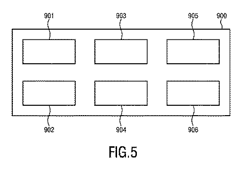
システム1の特定のアーキテクチャでは、中央の役割は同期装置90によって担われ、これはマスタクロック生成器及び検査ロガーとも呼ぶことができ、すなわち、同期時間を生成して配布するだけでなく、事象をログに記録することもできる。そのようなマスタクロック生成器及び検査ロガー900の汎用的な実施の概略図が図5に示される。専用ハードウェアで動作するFPGAの本格的コンピュータを使用するか、又は同等のプロセッサ901を使用して、種々のハードウェア実施が可能である。装置900は、外界からクロック信号を受信する。正確な同期は、例えば、IHE ITインフラストラクチャ技術フレームワークで定義されるConsistent Time Integration Profileを使用して達成することができる。Consistent Time Profileは、ネットワークタイムプロトコル(NTP)(www.NTP.orgにて定義される)の使用を必要とする。
In a particular architecture of
搭載された電力分配(ユニット902)、臨床事象を一時的に記憶するためのメモリ及びローカル大容量記憶(ユニット903)に関係する補助ユニットの他に、装置900は、線量キャリブレータ70(同じくLANインターフェースを有する線量キャリブレータの場合)、線量注入投薬器/自動潅流80(利用可能な場合)、生物測定データ獲得ユニット30、31、及び撮像装置10と相互接続するために、いくつかのLAN物理的インターフェース904(銅線又はファイバ)を有する。LANリンクは、同期クロック並びにそれに関連する特定の臨床事象の時刻をさらに再配布するために使用される。装置900は、患者が使用するウェアラブル装置との通信を保つための無線ハードウェアユニット905と、例えば処理ソフトウェアを記憶するためのメモリ906とをさらに備える。
In addition to the on-board power distribution (unit 902), memory for temporary storage of clinical events and auxiliary units related to local mass storage (unit 903), the
上記の問題(及び関係する他の問題)を解決するための本発明の一実施形態によれば、NMワークフロー特有のデータのログ記録及び通信作業を支援するように作られたセンサの固有の集合を備えた、モバイルの患者追跡装置(本明細書では全般にウェアラブル装置と呼ぶが、NMトラッカ又はNMTとも呼ばれる)を使用する。ウェアラブル装置は、(例えばその記録手順時に)患者に割り当てられて手渡され、患者が当該部門を再度離れるまで患者に随伴する。ウェアラブル装置は、手順に関係する情報を患者に表示するための単純なユーザインターフェースを提供する。装置は、内蔵ガンマ線センサを使用して、放射性トレーサ注入事象を自動的に検出する。さらなるワークフロー関連情報が、(近距離)RFインターフェース及び/又は内蔵カメラ(例えば、QRタグ若しくはOCR技術と組み合わせる)を介して、手順中に捕捉されてもよい。例えば、医院内の既存の無線ネットワークを介して、装置が中央のSUV判定装置と通信して、収集された経路(動き)追跡情報並びに記録された技術的及び生理学的パラメータを転送する。SUV判定装置で実行されるアプリケーションが受信データを分析し、ワークフロー状態の整合性を調べ、望ましくない事象がある場合には医療スタッフへの警告を発する。アプリケーションはさらに、遠隔で接続されたウェアラブル装置にさらなる情報を送出するためのインターフェースも提供する。 According to one embodiment of the invention for solving the above problems (and other related problems), a unique set of sensors designed to support NM workflow-specific data logging and communication tasks. A mobile patient tracking device (generally referred to herein as a wearable device, but also referred to as an NM tracker or NMT) is used. The wearable device is assigned and handed to the patient (eg, during the recording procedure) and accompanies the patient until the patient leaves the department again. The wearable device provides a simple user interface for displaying information related to the procedure to the patient. The device uses a built-in gamma ray sensor to automatically detect radiotracer injection events. Further workflow-related information may be captured during the procedure via a (short-range) RF interface and / or a built-in camera (eg, in combination with QR tags or OCR technology). For example, over an existing wireless network in the clinic, the device communicates with a central SUV determination device to transfer the collected path (motion) tracking information as well as the recorded technical and physiological parameters. An application running on the SUV determination device analyzes the received data, checks the consistency of the workflow state, and issues a warning to medical staff if there is an undesired event. The application also provides an interface for sending more information to remotely connected wearable devices.
ウェアラブル装置は、偶発的な誤使用の明白な可能性を阻むように設計することができる。一例として、患者が例えばトイレに行くときに付き添いの親戚に装置を預けるのを回避するために、ウェアラブル装置は、邪魔にならない方式で患者に取り付けられるべきである。これを支援する可能な設計は、(スマート)腕時計の形態であり得る。さらに、ウェアラブル装置の設計は、殺菌等に関する特殊な臨床要件を考慮すべきである。 Wearable devices can be designed to prevent the obvious possibility of accidental misuse. As an example, the wearable device should be attached to the patient in an unobtrusive manner to avoid leaving the device to an attendant relative, for example when going to the bathroom. A possible design to support this could be in the form of a (smart) wristwatch. In addition, the design of wearable devices should take into account special clinical requirements such as sterilization.
ウェアラブル装置の一実施形態は、患者に情報を伝えるための特化されたディスプレイ、患者がフィードバックを提供できる簡便な方式(例えば新しいスケジュールを遠隔から確認するため)、無線双方向通信インターフェース(802.11x、Bluetooth(登録商標)...)、内蔵デジタルカメラ、3D加速度センサ、ガンマ線センサ、データ処理ユニット、及び内部バッテリを無線で再充電するためのインターフェース(例えばQi技術による)を備える。ガンマ線センサモジュール(例えばFGMOSFET技術で実現される)は、ガンマ線の標準的なNM同位体粒子エネルギーを確実に検出するように設計される。ガンマ線センサモジュールは、好ましくは、1つの空間方向により高い感度を提供する。患者に取り付けられる場合、好ましい方向は身体に向かう方向である。 One embodiment of the wearable device is a specialized display for communicating information to the patient, a convenient method for the patient to provide feedback (eg, to remotely confirm a new schedule), and a wireless two-way communication interface (802. It includes 11x, Bluetooth® ...), a built-in digital camera, a 3D acceleration sensor, a gamma ray sensor, a data processing unit, and an interface (eg, by Qi technology) for wirelessly recharging the internal battery. Gamma ray sensor modules (eg, realized by FGMOSFET technology) are designed to reliably detect standard NM isotope particle energies for gamma rays. The gamma ray sensor module preferably provides higher sensitivity in one spatial direction. When attached to the patient, the preferred direction is towards the body.
以下に、ウェアラブル装置の例示的な使用シナリオを、追加的な技術的情報と併せて記載する。 An exemplary use scenario for a wearable device is described below, along with additional technical information.
ウェアラブル装置は、中央に収納しておき、例えば患者記録デスクで再充電することができる。充電中に、ウェアラブル装置の内部クロックは、中央のクロックユニットと同期される。SUV判定装置40(NMTサーバ又はNMT基地局と呼ばれることもある)にまだ転送されていないデータは、この時間中に安全にアップロードされ、最終的に装置から削除される。 The wearable device can be stored in the center and recharged, for example, at the patient recording desk. During charging, the wearable device's internal clock is synchronized with the central clock unit. Data that has not yet been transferred to the SUV determination device 40 (sometimes referred to as an NMT server or NMT base station) is safely uploaded during this time and eventually deleted from the device.
充電ステーションから外されると、ウェアラブル装置は自動的にアクティブモードに設定され、新しい患者に割り当てられるのを待つ。割り当ては、例えば、ウェアラブル装置のカメラの前方に患者記録QRコード(登録商標)/バーコードをかざすことによって行うことができる。有効な患者IDタグを識別すると、装置は、検査の特有のワークフローステップに従って、状態機械で制御される追跡モードに切り替わる(例えば色の変化によりウェアラブル装置に表示される)。 When removed from the charging station, the wearable device is automatically set to active mode, waiting to be assigned to a new patient. The assignment can be made, for example, by holding the patient record QR code® / barcode in front of the camera of the wearable device. Upon identifying a valid patient ID tag, the device switches to a state machine-controlled tracking mode (eg, displayed on the wearable device by color change) according to the examination's specific workflow steps.
臨床エリア内での患者の移動は、動きセンサの情報を介して、及び/又は複数のWLANアクセスポイントからの信号強度の分析を介して記録することができる。情報は、さらなる評価のために、頻繁に自動でSUV判定装置にアップロードされる。臨床環境における特殊なデータセキュリティ/プライバシー要件を勘案するために、すべての交換されるデータは暗号化される。自動の動きパターン分析を適用して、患者がさらなる支援及び誘導を必要としていることをスタッフに認識させ、検査結果を最適化する。医療スタッフは、例えば患者がトイレに行ったか、及び最後に行ったのがいつであるかなどの追加的な関係情報に、SUV判定装置を介して直接アクセスすることもできる。さらに、患者が広い各部門を通ってたどり着くのを助けるために、動き検出を使用して、場所の誘導を直接患者に提供することができる。 Patient movement within the clinical area can be recorded via motion sensor information and / or through analysis of signal strength from multiple WLAN access points. Information is frequently and automatically uploaded to the SUV determination device for further evaluation. All exchanged data is encrypted to account for the specific data security / privacy requirements in the clinical environment. Apply automated motion pattern analysis to remind staff that patients need further assistance and guidance and optimize test results. Medical staff can also have direct access via the SUV determination device for additional relevant information, such as when the patient went to the bathroom and when it last went. In addition, motion detection can be used to provide location guidance directly to the patient to help the patient reach through a wide range of departments.
スケジューリングの遅延が生じた場合、まだ準備/注入を受けていない患者は、ディスプレイを介して新しいスケジューリング情報を受信することができる。これにより、それらの患者は、待機エリアを離れて、例えば医院の休養ゾーンを訪れることができる。対話(例えば確認)要求が、特殊な視覚的信号(例えばウェアラブル装置の点滅)を介して追加的に示されてもよい。 In the event of a scheduling delay, patients who have not yet received preparation / injection can receive new scheduling information via the display. This allows them to leave the waiting area and visit, for example, the rest zone of the clinic. Dialogue (eg, confirmation) requests may be additionally indicated via special visual signals (eg, blinking wearable devices).
新しいワークフロー位置/ステップに入るときに、追加的なパラメータが記憶され、ウェアラブル装置を介して転送されてもよい。例えば、注入が準備されるとき、注射器には、関係する放射性トレーサ種類及びキャリブレーション(例えば、タイムスタンプ、量)の情報を含んでいる(例えば印刷された)QRタグが付加される。ウェアラブル装置のカメラを介してそのQRコード(登録商標)を認識することにより、内部のガンマ線センサが起動する。ウェアラブル装置は、放射性トレーサがそこから体内に入る患者の末端部に取り付けられ(例えば、右の内側肘静脈に注入する場合は左手首に装着される)、放射性トレーサボーラスを直ちに検出し、その事象をタイムスタンプと共にログに記録する。SUV判定装置40に転送されたデータにより、SUV判定装置40のアプリケーションは、研究に最適な画像獲得スケジュールを決定することができる。ドエルカウンタで判定された注射器内の残りトレーサ放射能を加えることにより、SUV判定装置40は、どれだけの量のトレーサがどの時点に患者に投与されたかを計算するためのすべてのパラメータを正確に把握する。
Additional parameters may be stored and transferred via the wearable device when entering a new workflow position / step. For example, when an infusion is prepared, the syringe is attached with a (eg, printed) QR tag containing information on the radiotracer type and calibration (eg, time stamp, amount) involved. By recognizing the QR code (registered trademark) through the camera of the wearable device, the internal gamma ray sensor is activated. The wearable device is attached to the end of the patient from which the radioactive tracer enters the body (eg, on the left wrist when injecting into the right medial elbow vein) and immediately detects the radioactive tracer bolus and the event. Is logged with a time stamp. The data transferred to the
その後行われる画像獲得の開始及び継続時間をログに記録することができる(例えば、寝台の位置決め中の典型的な向き及び移動パターンを検出することによる。最終的には、例えばPET/CT、SPECT/CTのスキャナ組み合わせによる解剖学的/調査用スキャン中の追加的なガンマ線量の検出と組み合わせる)。 Subsequent start and duration of image acquisition can be logged (eg, by detecting typical orientation and movement patterns during bunk positioning; ultimately, eg PET / CT, SPECT. Combined with detection of additional gamma doses during anatomical / investigation scans with / CT scanner combination).
ワークフローの完了後、臨床医は、患者との聞き取り中に、その後行われる定量的データ分析のためのすべての関連する患者情報がSUV判定装置40に転送されたかどうかを調べることができる。これは、例えば、装置の特定の色コードによって示される。これが該当しない場合、臨床医は、患者が家に帰される前に、このステップを完結させるようにする。
After the workflow is complete, the clinician can determine during the interview with the patient whether all relevant patient information for subsequent quantitative data analysis has been transferred to the
部門を離れる際、患者はウェアラブル装置を受付に返却し、そこで装置ステータスがもう一度検査され、清掃/再充電のために装置が準備される。 Upon leaving the department, the patient returns the wearable device to the reception, where the device status is checked again and the device is prepared for cleaning / recharging.
上記の開示に従い、様々な例を下記に記載する。 Following the above disclosure, various examples are described below.
例1. 放射断層撮影においてSUVを判定する装置であって、前記装置は、
− 1つ又は複数のウェアラブル装置20、20a、20b及び/又は1つ又は複数の生物測定データ獲得ユニット30、31から、患者による放射性トレーサ取り込みの時刻を含む、SUVの判定に必要とされるSUV関連データを取得すると共に、SUVの判定に影響し得る1つ又は複数の事象に関係する事象データを取得するための入力41と、
− 取得された事象データから、SUVの判定に影響する1つ又は複数の異常事象を示す異常事象情報を判定する異常事象判定ユニット42と、
− 異常事象情報を考慮して、前記SUV関連データからSUVを判定するSUV判定ユニット43と、を備える。
Example 1. A device for determining SUV in radiation tomography, the device
-From one or more
-From the acquired event data, an abnormal
− The
例2. 例1の装置であって、
前記SUV判定ユニット43は、患者の組織中の放射性トレーサ放射能濃度と、放射性トレーサ取り込みの投与線量と、患者の生物測定データ、特に患者の体重、身長、及び/又は全表面積とを含むSUV関連データから、SUVを判定するように構成される。
Example 2. The device of Example 1
The
例3. 例1の装置であって、
放射性トレーサ線量キャリブレータ、放射性トレーサ線量注入器、1つ又は複数の生物測定データ獲得ユニット、特に、体重計、身長計、血圧計、及び/若しくは血糖分析器、並びに/又は撮像装置と相互接続して、患者の組織の放射性トレーサ放射能濃度と、放射性トレーサ取り込みの投与線量と、患者の生物測定データとを含む1つ又は複数のSUV関連データ取得するためのインターフェース44をさらに備える。
Example 3. The device of Example 1
Interoperated with a radioactive tracer dose calibrator, a radioactive tracer dose injector, one or more biometric data acquisition units, in particular weight scales, height meters, sphygmomanometers, and / or blood glucose analyzers, and / or imaging devices. Further comprises an
例4. 例1の装置であって、
前記入力41は、1つ又は複数の事象の時刻を含む事象データを取得するようにさらに構成され、前記異常事象判定ユニット42は、異常事象情報を判定する際に1つ又は複数の事象の時刻を使用するように構成される。
Example 4. The device of Example 1
The
例5. 例1の装置であって、
特に、1つ又は複数のウェアラブル装置及び/又は1つ又は複数の生物測定データ獲得ユニットに提供されるシステムクロックを生成するために、装置を、1つ又は複数のウェアラブル装置及び/又は1つ又は複数の生物測定データ獲得ユニットと時間同期するための同期ユニット45をさらに備える。
Example 5. The device of Example 1
In particular, to generate the system clock provided to one or more wearable devices and / or one or more biometric data acquisition units, the device may be one or more wearable devices and / or one or more. A synchronization unit 45 for time synchronization with a plurality of biometric data acquisition units is further provided.
例6. 放射断層撮影においてSUVを判定する方法であって、前記方法は、
− 1つ又は複数のウェアラブル装置20、20a、20b及び/又は1つ又は複数の生物測定データ獲得ユニット30、31から、患者による放射性トレーサ取り込みの時刻を含む、SUVの判定に必要とされるSUV関連データを取得すると共に、SUVの判定に影響し得る1つ又は複数の事象に関係する事象データを取得するステップと、
− 取得された事象データから、UVの判定に影響する1つ又は複数の異常事象を示す異常事象情報を判定するステップと、
− 異常事象情報を考慮して、前記SUV関連データからSUVを判定するステップと、を有する。
Example 6. It is a method of determining an SUV in radiation tomography, and the above method is
-From one or more
-From the acquired event data, a step to determine abnormal event information indicating one or more abnormal events that affect the UV determination, and
-It has a step of determining an SUV from the SUV-related data in consideration of the abnormal event information.
例7. 患者によって装着されるように構成されたウェアラブル装置であって、前記ウェアラブル装置は、
− ウェアラブル装置を患者の身体に保持するための保持要素21と、
− 患者による放射性トレーサ取り込みの時刻を含む、放射断層撮影におけるSUVの判定に必要とされるSUV関連データと、SUVの判定に影響し得る1つ又は複数の事象に関係する事象データとを感知する1つ又は複数の感知要素22、23と、
− 感知されたSUV関連データ及び事象データを出力するための出力24と、を備える。
Example 7. A wearable device configured to be worn by a patient, said wearable device.
-A
-Senses SUV-related data required for SUV determination in radiotomography, including the time of patient uptake of the radioactive tracer, and event data related to one or more events that may affect SUV determination. One or
-It includes an
例8. 例7のウェアラブル装置であって、
前記感知要素は、動きセンサ、加速度計、位置検出センサ、生命兆候センサ、放射線センサ、及びカメラ、の1つ又は複数を含む。
Example 8. The wearable device of Example 7
The sensing element includes one or more of a motion sensor, an accelerometer, a position detection sensor, a life sign sensor, a radiation sensor, and a camera.
例9. 例7のウェアラブル装置であって、
事象データとして感知された1つ又は複数の事象の発生時刻を記録する時間測定ユニット25をさらに備え、
前記出力24は、事象データを、関係する事象の発生時刻と共に出力するように構成される。
Example 9. The wearable device of Example 7
A
The
例10. 例7のウェアラブル装置であって、
提供されるシステムクロックに特に基づいて、ウェアラブル装置を、例1のSUVを判定する装置と時間同期するための同期ユニット26をさらに備える。
Example 10. The wearable device of Example 7
A
例11. 例7のウェアラブル装置であって、
ユーザ情報の入力及び/又は出力を可能にするユーザインターフェース27をさらに備える。
Example 11. The wearable device of Example 7
A user interface 27 that enables input and / or output of user information is further provided.
例12. 例7のウェアラブル装置であって、
1つ又は複数の生物測定データ獲得ユニット30、31と電子的にデータを交換するための通信ユニット28をさらに備える。
Example 12. The wearable device of Example 7
It further comprises a
例13. 放射断層撮影システム、特に粒子放射断層撮影(PET)又は単一光子放射算出断層撮影(SPECT)システムであって、前記システムは、
− 患者の画像データを獲得するための撮像装置10と、
− 患者によって装着されるように構成された、例7の1つ又は複数のウェアラブル装置20、20a、20bと、
− 生物測定データを獲得し、前記生物測定データを1つ又は複数のウェアラブル装置に提供するための、特に体重計、身長計、血圧計、及び/又は血糖分析器である、1つ又は複数の生物測定データ獲得ユニット30、31と、
− 1つ又は複数のウェアラブル装置及び/又は1つ又は複数の生物測定データ獲得ユニットから取得されたSUV関連データ及び事象データから、SUVを判定する例1の装置40と、
− 判定されたSUVを使用して、獲得された画像データを評価する評価ユニット50と、を備える。
Example 13. A radiation tomography system, particularly a particle emission tomography (PET) or single photon emission calculation tomography (SPECT) system, said system.
− An
-With one or more
-One or more, especially weight scales, height scales, sphygmomanometers, and / or blood glucose analyzers, for acquiring biometric data and providing said biometric data to one or more wearable devices. Biometric
− The
-It includes an
例14. 例13のシステムであって、
1つ又は複数の事象データを感知する1つ又は複数の追加的なセンサ60、61をさらに備え、SUVを判定する前記装置40は、1つ又は複数の追加的なセンサ60、61によって感知された1つ又は複数の事象データをSUVの判定で追加的に使用するように構成される。
Example 14. The system of Example 13
The
例15. コンピュータプログラムであって、前記コンピュータプログラムがコンピュータ上で実行されると例6の方法のステップをコンピュータに実行させるプログラムコード手段を備えたコンピュータプログラム。 Example 15. A computer program comprising a program code means that causes the computer to perform the steps of the method of Example 6 when the computer program is executed on the computer.
図2を参照して説明した本発明の別の実施形態によれば、撮像施設で放射性トレーサ線量が投与された患者の放射断層撮影撮像手順中にSUVを判定する装置40が開示される。放射断層撮影撮像手順は、例えば、PET又はSPECT撮像手順である。装置40は、SUVの判定に必要とされるSUV関連データと、SUVの判定に影響し得る1つ又は複数の事象に関係する事象データとを受信するように構成される少なくとも1つの入力41、44を含む。SUV関連データは、患者への放射性トレーサ線量の投与の時刻、患者に投与された放射性トレーサ線量、患者の体内の領域における放射性トレーサ放射能濃度、患者の体重、患者の身長、患者の全表面積、患者への放射性トレーサ線量の投与後の注射器内の残留放射能を示すキャリブレーションデータ、のうちの1つ又は複数を含み得る。これらのSUV関連データのうち1つ又は複数を、上記でSUVの計算式との関連で説明したように、放射断層撮影撮像手順からのSUVの算出で使用することができる。本発明は、本明細書では事象データと記載される1つ又は複数の事象が、そのようにして算出されるSUVの精度に影響し得ることを認識する。そのため、装置40の少なくとも1つの入力41、44によって受信される事象データは、i)患者の放射断層撮影撮像手順が行われる時刻、ii)放射性トレーサ線量の投与から放射断層撮影撮像手順の開始までの期間中の患者の動きを示す患者動きデータ、iii)放射性トレーサ線量の投与から放射断層撮影撮像手順の開始までの期間中の、撮像施設内での患者の位置を示す患者位置データ、iv)放射性トレーサ線量の投与から放射断層撮影撮像手順の開始までの期間中の患者の生命兆候を示す患者生命兆候データのうちの少なくとも1つを含み得る。i)に関して、放射性トレーサ線量の投与から所定の時間以内に撮像手順が開始されない場合、又は実際に完了されない場合、線量の崩壊性のために、計算されたSUVに誤差が生じる可能性がある。しかし、これらは下記で説明するように補正することができる。ii)に関して、放射性トレーサ線量の投与から断層撮影撮像手順中のその分布の測定までのいわゆる取り込み期間中の患者の動きも、同様に、計算されたSUVに影響する可能性がある。例えば、この期間中の過剰な運動は、放射性トレーサの代謝とその分布に影響し得る。この場合も、これらは下記で説明するように補正することができる。iii)に関して、例えば患者が要求される部屋で安静にしているかどうか、又はトイレに行っているかどうか、又は実際病院内のどこか別の場所にいるのか、それとも撮像施設にいるのかを示す患者位置データも、計算されたSUVに影響する可能性がある。この場合も、下記で説明するように、そのようなデータを使用して、下記で説明するようにSUVを補正することができる。iv)に関して、取り込み期間中の患者生命兆候、例えば、患者心拍数センサ、体温、呼吸、血圧、皮膚の導電率、血液酸素化すなわちSPO2、血糖、の1つ又は複数も、計算されたSUVの誤差を示す場合がある。例えば、これらのデータのいずれかが所定の範囲外にあるときは、例えば取り込み期間中の過剰な運動が原因で誤差が予想され得る。この場合も、これらは下記で説明するように補正することができる。装置40は、上記の事象データの1つ又は複数に基づいて、SUVの判定に影響し得る1つ又は複数の異常事象を示す異常事象情報を判定するように構成される異常事象判定ユニット42も含む。上記で概説したように、異常事象判定ユニットは、関連するデータを評定し、それを予想範囲又は予想データと比較し、必要な場合は、異常が発生した旨のフラグを立てる。さらに、装置40は、異常事象情報を考慮してSUV関連データからSUVを判定するように構成されるSUV判定ユニット43を含む。i)に関して、例えば、撮像の継続時間を延長若しくは短縮することができ、又は計算されたSUVに補正係数を適用してもよい。ii)、iii)、及びiv)に関して、例えば、過剰な運動を勘案する補正係数をSUVに適用する、又は断層撮影撮像手順の開始時刻若しくは継続時間を標準値から修正してもよい。有利な点として、単に放射性トレーサの取り込みと断層撮影撮像手順を破棄し、スケジュールし直すのではなく、本発明の装置は、記載された様々な異常事象を補償することにより、正確なSUVの算出を可能にする。
According to another embodiment of the invention described with reference to FIG. 2, an
図2、図3、及び図4を参照して説明した本発明の別の実施形態によれば、SUVを判定する方法が開示される。この方法は、撮像施設で放射性トレーサ線量が投与された患者の放射断層撮影撮像手順中に放射断層撮影において使用され得る。この方法は、a)SUVの判定に必要とされるSUV関連データを取得するステップと、b)SUVの判定に影響し得る1つ又は複数の事象に関係する事象データを取得するステップと、c)取得された事象データに基づいて、SUVの判定に影響する1つ又は複数の異常事象を示す異常事象情報を判定するステップと、d)異常事象情報を考慮して、SUV関連データに基づいてSUVを判定するステップとを有する。SUV関連データ及び事象データは、先行する実施形態で説明したものと同様であってよい。上記の方法のステップは、コンピュータ可読形式で命令又はプログラムコードとして記憶されてよい。さらに、それらの方法のステップは、コンピュータによって実行されてよい。 According to another embodiment of the present invention described with reference to FIGS. 2, 3, and 4, a method for determining an SUV is disclosed. This method can be used in radiated tomography during a radiated tomography imaging procedure for patients who have been administered a radioactive tracer dose at an imaging facility. This method includes a) a step of acquiring SUV-related data required for SUV determination, b) a step of acquiring event data related to one or more events that may affect the SUV determination, and c. ) Based on the acquired event data, the step of determining the abnormal event information indicating one or more abnormal events that affect the determination of the SUV, and d) Considering the abnormal event information, based on the SUV-related data. It has a step of determining an SUV. The SUV-related data and event data may be similar to those described in the preceding embodiments. The steps of the above method may be stored as instructions or program code in computer readable form. In addition, the steps in those methods may be performed by a computer.
図2及び図3を参照して説明した本発明の別の実施形態によれば、ウェアラブル装置20、20a、20bが開示される。ウェアラブル装置は、撮像施設で放射性トレーサ線量が投与された患者の放射断層撮影撮像手順中に、SUVを判定するために使用することができる。ウェアラブル装置は、ウェアラブル装置を患者の身体に取り付けるための、例えばストラップ、ベルト、又はネックレスである保持要素21と、放射断層撮影におけるSUVの判定に必要とされるSUV関連データを感知する1つ又は複数の感知要素22、23と、を備える。感知されたSUV関連データは、先行する実施形態に関連して記載した上記データの1つ又は複数であってよい。好ましくは、SUV関連データは、少なくとも、患者への放射性トレーサ線量の投与の時刻を含む。さらに、ウェアラブル装置は、感知されたSUV関連データを出力するための出力24を含む。好ましい構成では、患者への放射性トレーサ線量の投与の時刻を感知する1つ又は複数の感知要素22、23は、a)放射性トレーサ線量注入器80が患者に放射性トレーサ線量を投与する時刻を無線で受信するように構成された無線センサ、及びb)ウェアラブル装置が患者によって装着されたときに、患者の身体から発されるガンマ線を感知するようになされたガンマ線センサのうちの少なくとも1つを含み、ガンマ線センサはさらに、感知されたガンマ線が所定の閾値条件を満たした時刻を示すタイムスタンプを提供するように構成される。無線センサは、したがって、後に例えば装置40によって読み出し、患者の体内の領域についてのSUVを判定するために使用できる患者上にあるSUV関連データストレージを提供する。無線センサは、自動化されたデータ転送を提供し、それにより、手作業で記録される線量投与時刻と比べて人的誤りの可能性を低減する。さらに、データはウェアラブル装置によって獲得されるため、本質的に患者に特有であり、患者データを取り違える可能性が低減される。ある構成では、ガンマ線センサ(b)を単独で使用して、例えば、静脈若しくは動脈、又は四肢、すなわち手首や脚などの患者の他の体内領域から、患者への放射性トレーサの注入後の流入を監視し、それにより、患者に線量が投与された時刻を得ることができる。これを使用して、手作業で記録された注入時刻を検証する、又は実際の注入時刻を提供することができる。別の構成では、自動化された線量注入の時刻を確定するために、ガンマ線センサ(b)を無線センサ(a)と組み合わせて使用し、例えば、放射性トレーサがそもそも実際に患者に注入されたことを特定する。例えば近距離通信、RFID、及びBluetooth(登録商標)で使用されるような無線RFを例えばこの目的のために使用することができるが、これらの特定の実施は制限的なものとみなすべきではない。実際、RF、赤外線、超音波、及び光通信を含む、任意の無線送受信機システムを使用して、要求されるデータを転送することができる。この実施形態では、上記で説明したように、追加的なSUV関連データを受信するために無線センサが使用されてもよい。これは、無線送信システムから無線で受信することができる。これらには、例えば、患者に投与された放射性トレーサ線量、又は患者への放射性トレーサ線量の投与後の注射器内の残留放射能を示す放射性トレーサ線量キャリブレーションデータ(これらはいずれも例えば放射性トレーサ線量注入器80から受信される)、無線患者体重計から受信される患者の体重、無線身長測定システム、若しくは入力として患者の身長及び体重を有するモデルに基づいて患者の表面積を判定するように構成されたコンピュータシステムから受信される、患者の身長若しくは表面積、が含まれる。この実施形態では、無線センサを追加的に使用して、SUVの判定に影響し得る1つ又は複数の事象に関する事象データを受信し、記憶することができる。事象データについては上記で説明しており、1つ又は複数のセンサから無線で受信されるか、又はウェアラブル装置の一部を構成するセンサによって直接感知される。そのようなセンサは、加速度計と、患者位置センサと、心拍数センサ、体温センサ、呼吸センサ、血圧センサ、皮膚導電率センサ、SPO2センサ、血糖センサ、の群から選択される患者生命兆候センサとを含む。有利な点として、そのように定義されたウェアラブル装置20、20a、20bを使用して、患者のSUVを提供する際に使用するために装置40への入力として働くことができる、様々なSUV関連データ及び/又は様々な事象データを収集し、記憶することができる。任意で、ウェアラブル装置20、20a、20bは、対応する事象データの測定時刻を記録する時間測定ユニット25を含んでよく、これは、事象データ及び/又は対応する事象データの測定時刻を出力するように構成される。任意で、ウェアラブル装置20、20a、20bは、ウェアラブル装置を、SUVを判定する装置40と時間同期するための同期ユニット26を含んでよい。
According to another embodiment of the present invention described with reference to FIGS. 2 and 3,
本発明の別の実施形態によれば、撮像装置10が開示される。撮像装置10は、患者の画像データを獲得するための、特にPET又はSPECT撮像システムである放射断層撮影撮像システムと、先行する実施形態において上記で説明したような1つ又は複数のウェアラブル装置20、20a、20bと、上記で説明したようなSUVを判定する装置40と、を備える。装置40は、装置40の少なくとも1つの入力41、44によって受信されたSUV関連データ及び事象データに基づいて、並びに、1つ又は複数のウェアラブル装置20、20a、20bによって出力された少なくともSUV関連データに基づいて、SUVを判定するように構成される。さらに、装置40は、判定されたSUVを使用して、獲得された画像データを評価する評価ユニット50を含む。任意で、撮像装置10の装置40は、1つ又は複数のウェアラブル装置20、20a、20bによって出力された事象データにさらに基づいて、SUVを判定するように構成されてもよい。
According to another embodiment of the present invention, the
本発明について図面及び上述の説明で例示し、説明したが、そのような例示及び説明は、説明的又は例示的であり、制限的なものではないと考えるべきであり、本発明は開示される実施形態に制限されない。図面、本開示、及び添付の特許請求の範囲の考察から、請求される本発明を実施する際に、開示される実施形態の他の変形例を当業者によって理解し、実施することができる。 Although the present invention has been illustrated and described in the drawings and the above description, such illustrations and explanations should be considered to be descriptive or exemplary and not restrictive, and the invention is disclosed. It is not limited to the embodiment. From consideration of the drawings, the present disclosure, and the appended claims, those skilled in the art will be able to understand and implement other variations of the disclosed embodiments in carrying out the claimed invention.
特許請求の範囲において、単語「〜を含む」は他の要素又はステップを排除せず、不定冠詞の「a」及び「an」は複数を排除しない。単一の要素又は他のユニットが、特許請求の範囲に記載されるいくつかの項目の機能を実現することができる。特定の手段が相互に異なる従属請求項に記載されているという単なる事実は、それらの手段の組み合わせを有利に使用できないことを意味するものではない。 In the claims, the word "contains" does not exclude other elements or steps, and the indefinite articles "a" and "an" do not exclude more than one. A single element or other unit can realize the function of several items described in the claims. The mere fact that certain means are described in different dependent claims does not mean that the combination of those means cannot be used in an advantageous manner.
コンピュータプログラムは、他のハードウェアと共に又はその一部として供給される光学記憶媒体又は固体媒体などの適切な非一時的媒体で記憶/配布することができるが、インターネット又は他の有線若しくは無線の遠隔通信システムを介するなど、他の形態で配布されてもよい。 Computer programs can be stored / distributed on suitable non-temporary media such as optical storage media or solid-state media supplied with or as part of other hardware, but on the Internet or other wired or wireless remote. It may be distributed in other forms, such as via a communication system.
特許請求の範囲内に参照符号がある場合、範囲を制限するものとは解釈すべきでない。 If there is a reference code within the claims, it should not be construed as limiting the scope.
Claims (15)
SUVの判定に必要とされるSUV関連データと、前記SUVの判定に影響する1つ又は複数の事象に関係する事象データとを受信する少なくとも1つの入力、を備え、
前記SUV関連データは、少なくとも、前記患者への前記放射性トレーサ線量の投与の時刻を含み、
前記事象データは、
前記放射性トレーサ線量の投与から前記放射断層撮影撮像手順の開始までの期間中の前記患者の動きを示す患者動きデータ、
前記放射性トレーサ線量の投与から前記放射断層撮影撮像手順の開始までの期間中の、前記撮像施設内での前記患者の位置を示す患者位置データ、
前記放射性トレーサ線量の投与から前記放射断層撮影撮像手順の開始までの期間中の前記患者の生命兆候を示す患者生命兆候データ、
のうちの少なくとも1つを含み、
前記装置は、
前記事象データに基づいて、前記SUVの判定に影響する1つ又は複数の異常事象を示す異常事象情報を判定する異常事象判定ユニットと、
前記異常事象情報を考慮して、前記SUV関連データに基づいて前記SUVを判定するSUV判定ユニットと
をさらに備える、装置。 A device that determines an SUV during a radiotomography imaging procedure for a patient to whom a radioactive tracer dose has been administered at an imaging facility.
It comprises at least one input for receiving SUV-related data required for SUV determination and event data related to one or more events affecting the SUV determination.
The SUV-related data includes at least the time of administration of the radiotracer dose to the patient.
It said event data,
Before Symbol patient motion data the indicating movement of the patient during the period from administration of the radiotracer dose until the beginning of the emission tomography imaging procedure,
Patient location data indicating the location of the patient within the imaging facility during the period from administration of the radioactive tracer dose to the start of the radiotomography imaging procedure,
Patient life sign data showing the life signs of the patient during the period from the administration of the radioactive tracer dose to the start of the radiotomography imaging procedure,
Including at least one of
The device is
An abnormal event determination unit that determines abnormal event information indicating one or more abnormal events that affect the determination of the SUV based on the event data, and an abnormal event determination unit.
An apparatus further comprising an SUV determination unit that determines the SUV based on the SUV-related data in consideration of the abnormal event information.
前記患者に投与された放射性トレーサ線量、
前記患者の体内の領域における放射性トレーサ放射能濃度、
前記患者の体重、
前記患者の身長、
前記患者の全表面積、
のうちの少なくとも1つをさらに含む、請求項1に記載の装置。 The SUV-related data is
Radiotracer dose administered to said patient,
Radioactivity tracer radioactivity concentration in the area inside the patient's body,
The patient's weight,
The height of the patient,
The total surface area of the patient,
The device of claim 1, further comprising at least one of.
SUVの判定に必要とされるSUV関連データと、前記SUVの判定に影響する1つ又は複数の事象に関係する事象データとを受信する少なくとも1つの入力、を備え、
前記SUV関連データは、少なくとも、前記患者への前記放射性トレーサ線量の投与の時刻を含み、
前記事象データは、
前記患者の放射断層撮影撮像手順が行われる時刻、
前記放射性トレーサ線量の投与から前記放射断層撮影撮像手順の開始までの期間中の前記患者の動きを示す患者動きデータ、
前記放射性トレーサ線量の投与から前記放射断層撮影撮像手順の開始までの期間中の、前記撮像施設内での前記患者の位置を示す患者位置データ、
前記放射性トレーサ線量の投与から前記放射断層撮影撮像手順の開始までの期間中の前記患者の生命兆候を示す患者生命兆候データ、
のうちの少なくとも1つを含み、
前記装置は、
前記事象データに基づいて、前記SUVの判定に影響する1つ又は複数の異常事象を示す異常事象情報を判定する異常事象判定ユニットと、
前記異常事象情報を考慮して、前記SUV関連データに基づいて前記SUVを判定するSUV判定ユニットと
をさらに備え、
前記少なくとも1つの入力は、1つ又は複数の患者ウェアラブル装置から、
前記患者への前記放射性トレーサ線量の投与の時刻、
前記患者の放射断層撮影撮像手順が行われる時刻、
前記患者に投与された前記放射性トレーサ線量、
前記放射性トレーサ線量の投与から前記放射断層撮影撮像手順の開始までの期間中の前記患者の動きを示す前記患者動きデータ、
前記放射性トレーサ線量の投与から前記放射断層撮影撮像手順の開始までの期間中の、前記撮像施設内での前記患者の位置を示す前記患者位置データ、
前記放射性トレーサ線量の投与から前記放射断層撮影撮像手順の開始までの期間中の前記患者の生命兆候を示す前記患者生命兆候データ、
のうちの少なくとも1つを受信する、装置。 A device that determines an SUV during a radiotomography imaging procedure for a patient to whom a radioactive tracer dose has been administered at an imaging facility.
It comprises at least one input for receiving SUV-related data required for SUV determination and event data related to one or more events affecting the SUV determination.
The SUV-related data includes at least the time of administration of the radiotracer dose to the patient.
The event data is
The time when the patient's radiation tomography imaging procedure is performed,
Patient movement data showing the movement of the patient during the period from the administration of the radioactive tracer dose to the start of the radiation tomography imaging procedure,
Patient location data indicating the location of the patient within the imaging facility during the period from administration of the radioactive tracer dose to the start of the radiotomography imaging procedure,
Patient life sign data showing the life signs of the patient during the period from the administration of the radioactive tracer dose to the start of the radiotomography imaging procedure,
Including at least one of
The device is
An abnormal event determination unit that determines abnormal event information indicating one or more abnormal events that affect the determination of the SUV based on the event data, and an abnormal event determination unit.
With the SUV determination unit that determines the SUV based on the SUV-related data in consideration of the abnormal event information.
With more
The at least one input is from one or more patient wearable devices.
Time of administration of the radiotracer dose to the patient,
The time when the patient's radiation tomography imaging procedure is performed,
The radiotracer dose administered to the patient,
The patient movement data showing the movement of the patient during the period from the administration of the radioactive tracer dose to the start of the radiation tomography imaging procedure.
The patient position data indicating the position of the patient in the imaging facility during the period from the administration of the radioactive tracer dose to the start of the radiation tomography imaging procedure.
The patient life sign data, which shows the life sign of the patient during the period from the administration of the radioactive tracer dose to the start of the radiotomography imaging procedure.
At least one receiving the, equipment of.
SUVの判定に必要とされるSUV関連データを取得するステップであって、前記SUV関連データは、少なくとも、前記患者への前記放射性トレーサ線量の投与の時刻を含む、ステップと、
前記SUVの判定に影響する1つ又は複数の事象に関係する事象データを取得するステップであって、前記事象データは、
前記放射性トレーサ線量の投与から前記放射断層撮影撮像手順の開始までの期間中の前記患者の動きを示す患者動きデータ、
前記放射性トレーサ線量の前記投与から前記放射断層撮影撮像手順の開始までの期間中の、前記撮像施設内での前記患者の位置を示す患者位置データ、
患者生命兆候データ、
のうちの少なくとも1つを含む、ステップと、
取得された前記事象データに基づいて、前記SUVの判定に影響する1つ又は複数の異常事象を示す異常事象情報を判定するステップと、
前記異常事象情報を考慮して、前記SUV関連データに基づいて前記SUVを判定するステップとを有する方法。 A method of determining an SUV during a radiotomography imaging procedure for a patient to whom a radiotracer dose has been administered at an imaging facility, wherein the method is:
A step of acquiring SUV-related data required for determining an SUV, wherein the SUV-related data includes at least the time of administration of the radiotracer dose to the patient.
A step of obtaining event data related to one or more events that affect the determination of the SUV, the event data,
Before Symbol patient motion data the indicating movement of the patient during the period from administration of the radiotracer dose until the beginning of the emission tomography imaging procedure,
Patient location data indicating the location of the patient within the imaging facility during the period from the administration of the radioactive tracer dose to the start of the radiotomography imaging procedure.
Patient life sign data,
Steps, including at least one of
Based on the acquired event data, a step of determining abnormal event information indicating one or more abnormal events that affect the determination of the SUV, and
A method having a step of determining the SUV based on the SUV-related data in consideration of the abnormal event information.
前記患者に投与された放射性トレーサ線量、
前記患者の体内の領域における放射性トレーサ放射能濃度、
前記患者の体重、
前記患者の身長、
前記患者の全表面積、
のうちの少なくとも1つを取得するステップをさらに含む、請求項6に記載の方法。 The step of acquiring SUV-related data is the following data,
Radiotracer dose administered to said patient,
Radioactivity tracer radioactivity concentration in the area inside the patient's body,
The patient's weight,
The height of the patient,
The total surface area of the patient,
The method of claim 6, further comprising the step of obtaining at least one of.
前記ウェアラブル装置を前記患者の身体に保持するための保持要素と、
前記患者への前記放射性トレーサ線量の投与の時刻を少なくとも含む、放射断層撮影におけるSUVの判定に必要とされるSUV関連データを感知する1つ又は複数の感知要素と、
感知された前記SUV関連データを出力するための出力と、を備え、
前記患者への前記放射性トレーサ線量の投与の前記時刻を感知する前記1つ又は複数の感知要素は、
放射性トレーサ線量注入器が前記患者に前記放射性トレーサ線量を投与する時刻を無線で受信する無線センサと、
前記ウェアラブル装置が前記患者によって装着されたときに、前記患者の身体から発されるガンマ線を感知するガンマ線センサであって、さらに、感知されたガンマ線が所定の閾値条件を満たした時刻を示すタイムスタンプを提供するガンマ線センサと
のうちの少なくとも1つを備える、ウェアラブル装置。 A wearable device for determining an SUV during a radiation tomography imaging procedure for a patient to whom a radioactive tracer dose has been administered at an imaging facility.
A holding element for holding the wearable device on the patient's body,
One or more sensing elements that sense SUV-related data needed to determine an SUV in radiotomography, including at least the time of administration of the radioactive tracer dose to the patient.
With an output for outputting the detected SUV-related data,
The one or more sensing elements that sense the time of administration of the radiotracer dose to the patient.
A radio sensor that wirelessly receives the time when the radiotracer dose injector administers the radiotracer dose to the patient.
A gamma ray sensor that detects gamma rays emitted from the patient's body when the wearable device is worn by the patient, and a time stamp indicating the time when the sensed gamma rays satisfy a predetermined threshold condition. A wearable device comprising at least one of the gamma ray sensors provided.
前記患者に投与された前記放射性トレーサ線量、
前記患者への前記放射性トレーサ線量の投与後の注射器内の残留放射能を示す放射性トレーサ線量キャリブレーションデータ、
前記患者の体重、
前記患者の身長、
前記患者の表面積
のうちの少なくとも1つを示すSUV関連データを無線で受信する無線センサを含む、請求項9に記載のウェアラブル装置。 The one or more sensing elements that sense SUV-related data are
The radiotracer dose administered to the patient,
Radiotracer dose calibration data showing residual radioactivity in the syringe after administration of the radiotracer dose to the patient,
The patient's weight,
The height of the patient,
The wearable device of claim 9, comprising a wireless sensor that wirelessly receives SUV-related data indicating at least one of the patient's surface areas.
加速度計と、患者位置センサと、心拍数センサ、体温センサ、呼吸センサ、血圧センサ、皮膚導電率センサ、SPO2センサ、血糖センサの群から選択される患者生命兆候センサと、及び/又は、
前記患者の心拍数、前記患者の体温、前記患者の呼吸、前記患者の血圧、前記患者の皮膚導電率、前記患者のSPO2、前記患者の血糖、カメラ画像データに基づく前記撮像施設内での前記患者の位置のうちの少なくとも1つを示すデータを受信する無線センサとのうちの少なくとも1つを含む、請求項9又は10に記載のウェアラブル装置。 The one or more sensing elements further sense event data related to the one or more events that affect the determination of the SUV, and the output further outputs the event data and said one or more. Sensing element of
Accelerator, patient position sensor, heart rate sensor, body temperature sensor, breathing sensor, blood pressure sensor, skin conductivity sensor, SPO2 sensor, patient life sign sensor selected from the group of blood glucose sensor, and / or
The patient's heart rate, the patient's body temperature, the patient's breathing, the patient's blood pressure, the patient's skin conductivity, the patient's SPO2, the patient's blood glucose, the said in the imaging facility based on camera image data. The wearable device of claim 9 or 10, comprising at least one of a radio sensor that receives data indicating at least one of the patient's locations.
前記出力は、前記事象データと、前記対応する事象データの前記測定時刻とを出力する、請求項9又は10に記載のウェアラブル装置。 It also has a time measurement unit that records the measurement time of the corresponding event data.
The wearable device according to claim 9 or 10, wherein the output outputs the event data and the measurement time of the corresponding event data.
前記ウェアラブル装置とを時間同期するための同期ユニットをさらに備える、請求項9に記載のウェアラブル装置。 Radiation tomography of a patient to whom a radiation tracer dose was administered at an imaging facility A device for determining SUV during an imaging procedure, and the device for determining SUV is the SUV-related data required for determining SUV and the above. The SUV-related data comprises at least the time of administration of the radiotracer dose to the patient, comprising at least one input to receive event data relating to one or more events affecting the determination of the SUV. The event data includes the patient movement data indicating the movement of the patient during the period from the administration of the radiation tracer dose to the start of the radiation tomography imaging procedure at the time when the radiation tomography imaging procedure of the patient is performed. , Patient position data indicating the position of the patient in the imaging facility during the period from the administration of the radiation tracer dose to the start of the radiation tomography imaging procedure, and the radiation tomography imaging from the administration of the radiation tracer dose. A device that includes at least one of the patient life sign data indicating the patient's life sign during the period up to the start of the procedure and determines the SUV determines the SUV based on the event data. An abnormal event determination unit that determines abnormal event information indicating one or more abnormal events that affect it, and an SUV determination unit that determines the SUV based on the SUV-related data in consideration of the abnormal event information. A device for determining the SUV, and a device for determining the SUV .
The wearable device according to claim 9, further comprising a synchronization unit for time-synchronizing with the wearable device.
1つ又は複数の請求項9に記載のウェアラブル装置と、
撮像施設で放射性トレーサ線量が投与された患者の放射断層撮影撮像手順中にSUVを判定する装置であって、該SUVを判定する装置は、SUVの判定に必要とされるSUV関連データと、前記SUVの判定に影響する1つ又は複数の事象に関係する事象データとを受信する少なくとも1つの入力、を備え、前記SUV関連データは、少なくとも、前記患者への前記放射性トレーサ線量の投与の時刻を含み、前記事象データは、前記患者の放射断層撮影撮像手順が行われる時刻、前記放射性トレーサ線量の投与から前記放射断層撮影撮像手順の開始までの期間中の前記患者の動きを示す患者動きデータ、前記放射性トレーサ線量の投与から前記放射断層撮影撮像手順の開始までの期間中の、前記撮像施設内での前記患者の位置を示す患者位置データ、前記放射性トレーサ線量の投与から前記放射断層撮影撮像手順の開始までの期間中の前記患者の生命兆候を示す患者生命兆候データ、のうちの少なくとも1つを含み、当該SUVを判定する装置は、前記事象データに基づいて、前記SUVの判定に影響する1つ又は複数の異常事象を示す異常事象情報を判定する異常事象判定ユニットと、前記異常事象情報を考慮して、前記SUV関連データに基づいて前記SUVを判定するSUV判定ユニットとをさらに備え、前記SUVを判定する装置の少なくとも1つの入力によって受信されたSUV関連データ及び事象データに基づいて、並びに、前記1つ又は複数のウェアラブル装置によって出力された少なくとも前記SUV関連データに基づいて、SUVを判定する、当該SUVを判定する装置と、
判定された前記SUVを使用して、獲得された前記画像データを評価する評価ユニットと
を備える、撮像装置。 Radiation tomography imaging systems, especially PET or SPECT imaging systems, for acquiring patient image data,
One or more wearable devices according to claim 9,
A device for determining an SUV during a radiation tomography imaging procedure for a patient to whom a radioactive tracer dose has been administered at an imaging facility, the device for determining the SUV is the SUV-related data required for the determination of the SUV and the above. The SUV-related data comprises at least the time of administration of the radioactive tracer dose to the patient, comprising at least one input to receive event data relating to one or more events affecting the determination of the SUV. The event data includes patient movement data indicating the movement of the patient during the period from the administration of the radioactive tracer dose to the start of the radiation tomography imaging procedure at the time when the radiation tomography imaging procedure of the patient is performed. , Patient position data indicating the position of the patient in the imaging facility during the period from the administration of the radioactive tracer dose to the start of the radiation tomography imaging procedure, and the radiation tomography imaging from the administration of the radiotracer dose. An apparatus that includes at least one of patient life sign data indicating the patient's life sign during the period up to the start of the procedure and determines the SUV determines the SUV based on the event data. An abnormal event determination unit that determines abnormal event information indicating one or more abnormal events that affect it, and an SUV determination unit that determines the SUV based on the SUV-related data in consideration of the abnormal event information. wherein at least one based on SUV relevant data and event data received by the input of the apparatus for determining SUV, and, based on at least the SUV-related data output by the one or more wearable device, A device that determines an SUV, a device that determines the SUV,
An imaging device including an evaluation unit that evaluates the acquired image data using the determined SUV.
Applications Claiming Priority (3)
| Application Number | Priority Date | Filing Date | Title |
|---|---|---|---|
| EP15191876.0 | 2015-10-28 | ||
| EP15191876 | 2015-10-28 | ||
| PCT/EP2016/075302 WO2017072030A1 (en) | 2015-10-28 | 2016-10-21 | Device and method for suv determination in emission tomography |
Publications (2)
| Publication Number | Publication Date |
|---|---|
| JP2018536156A JP2018536156A (en) | 2018-12-06 |
| JP6941098B2 true JP6941098B2 (en) | 2021-09-29 |
Family
ID=54476701
Family Applications (1)
| Application Number | Title | Priority Date | Filing Date |
|---|---|---|---|
| JP2018521613A Expired - Fee Related JP6941098B2 (en) | 2015-10-28 | 2016-10-21 | Equipment and methods for determining SUVs in radiation tomography |
Country Status (5)
| Country | Link |
|---|---|
| US (1) | US10849571B2 (en) |
| EP (1) | EP3367907B1 (en) |
| JP (1) | JP6941098B2 (en) |
| CN (1) | CN108348212B (en) |
| WO (1) | WO2017072030A1 (en) |
Families Citing this family (39)
| Publication number | Priority date | Publication date | Assignee | Title |
|---|---|---|---|---|
| US10448844B2 (en) | 2015-08-31 | 2019-10-22 | Masimo Corporation | Systems and methods for patient fall detection |
| JP6941098B2 (en) * | 2015-10-28 | 2021-09-29 | コーニンクレッカ フィリップス エヌ ヴェKoninklijke Philips N.V. | Equipment and methods for determining SUVs in radiation tomography |
| CN113350655B (en) | 2016-02-24 | 2024-03-19 | 禾木(中国)生物工程有限公司 | Nerve vascular catheter with enhanced flexibility |
| EP3565511B1 (en) | 2017-01-06 | 2023-10-18 | Incept, LLC | Thromboresistant coatings for aneurysm treatment devices |
| JP7173595B2 (en) * | 2017-05-31 | 2022-11-16 | ザ・ロイヤル・インスティテューション・フォア・ザ・アドバンスメント・オブ・ラーニング/マクギル・ユニヴァーシティ | Noninvasive Measurement of Arterial Input Functions in Positron Emission Tomography Images |
| US11051773B2 (en) * | 2017-12-04 | 2021-07-06 | General Electric Company | Systems and methods for imaging with improved dosages |
| CN111867474B (en) * | 2018-01-03 | 2024-12-20 | 皇家飞利浦有限公司 | Full-dose PET image estimation from low-dose PET images using deep learning |
| US11395665B2 (en) | 2018-05-01 | 2022-07-26 | Incept, Llc | Devices and methods for removing obstructive material, from an intravascular site |
| AU2019262972B2 (en) | 2018-05-01 | 2025-02-27 | Incept, Llc | Devices and methods for removing obstructive material from an intravascular site |
| US11471582B2 (en) | 2018-07-06 | 2022-10-18 | Incept, Llc | Vacuum transfer tool for extendable catheter |
| US11517335B2 (en) | 2018-07-06 | 2022-12-06 | Incept, Llc | Sealed neurovascular extendable catheter |
| US11766539B2 (en) | 2019-03-29 | 2023-09-26 | Incept, Llc | Enhanced flexibility neurovascular catheter |
| BR112021020780A2 (en) | 2019-04-17 | 2021-12-14 | Masimo Corp | Electrocardiogram (ECG) device, blood pressure monitoring device, blood pressure monitor, blood pressure cuff, mounting for enabling a caregiver to attach a physiological monitoring device to a user's arm, charging station for providing power to a physiological monitoring device, non-invasive blood pressure monitor and method for a non-invasive blood pressure monitor |
| USD985498S1 (en) | 2019-08-16 | 2023-05-09 | Masimo Corporation | Connector |
| USD919100S1 (en) | 2019-08-16 | 2021-05-11 | Masimo Corporation | Holder for a patient monitor |
| USD919094S1 (en) | 2019-08-16 | 2021-05-11 | Masimo Corporation | Blood pressure device |
| USD917704S1 (en) | 2019-08-16 | 2021-04-27 | Masimo Corporation | Patient monitor |
| CN110477941B (en) * | 2019-08-19 | 2023-06-27 | 上海联影医疗科技股份有限公司 | Uptake value correction method, device, computer equipment and readable storage medium |
| CN110464326B (en) | 2019-08-19 | 2022-05-10 | 上海联影医疗科技股份有限公司 | Scanning parameter recommendation method, system, device and storage medium |
| CN110599472B (en) * | 2019-09-03 | 2021-03-02 | 佛山原子医疗设备有限公司 | Method and system for calculating SUV normalization coefficient in SPECT quantitative tomographic images |
| CN113347916B (en) * | 2019-10-15 | 2025-04-25 | 因普瑞缇夫护理公司 | Systems and methods for multivariate stroke detection |
| USD927699S1 (en) | 2019-10-18 | 2021-08-10 | Masimo Corporation | Electrode pad |
| US20210315598A1 (en) | 2019-12-18 | 2021-10-14 | Imperative Care, Inc. | Methods of placing large bore aspiration catheters |
| US11633272B2 (en) | 2019-12-18 | 2023-04-25 | Imperative Care, Inc. | Manually rotatable thrombus engagement tool |
| WO2021127004A1 (en) | 2019-12-18 | 2021-06-24 | Imperative Care, Inc. | Methods and systems for treating venous thromboembolic disease |
| CA3171899A1 (en) | 2020-03-10 | 2021-09-16 | Imperative Care, Inc. | Enhanced flexibility neurovascular catheter |
| USD933232S1 (en) | 2020-05-11 | 2021-10-12 | Masimo Corporation | Blood pressure monitor |
| USD979516S1 (en) | 2020-05-11 | 2023-02-28 | Masimo Corporation | Connector |
| EP4166085A4 (en) * | 2020-07-13 | 2023-07-05 | Shanghai United Imaging Healthcare Co., Ltd. | MEDICAL DEVICE IMAGE SCANNING METHOD AND SYSTEM |
| US11207497B1 (en) | 2020-08-11 | 2021-12-28 | Imperative Care, Inc. | Catheter with enhanced tensile strength |
| CN112415562A (en) * | 2020-12-10 | 2021-02-26 | 重庆建安仪器有限责任公司 | Fixed area gamma radiation monitoring system and monitoring method |
| US20230052862A1 (en) | 2021-08-12 | 2023-02-16 | Imperative Care, Inc. | Sterile packaging assembly for robotic interventional device |
| USD1077996S1 (en) | 2021-10-18 | 2025-06-03 | Imperative Care, Inc. | Inline fluid filter |
| CN114259247B (en) * | 2021-12-29 | 2025-03-25 | 上海联影医疗科技股份有限公司 | Dynamic PET scanning method, device, electronic device and storage medium |
| US20230342914A1 (en) * | 2022-04-21 | 2023-10-26 | Canon Medical Systems Corporation | Method and system for monitoring a patient emotional state and segmenting obtained emission data based on the patient emotional state data |
| DE102022207474A1 (en) | 2022-07-21 | 2023-07-06 | Siemens Healthcare Gmbh | Device and method for evaluating medical scanner data |
| USD1069809S1 (en) | 2022-08-25 | 2025-04-08 | Kandu Health, Inc. | Display screen or portion thereof with graphical user interface |
| EP4390963A1 (en) * | 2022-12-22 | 2024-06-26 | Koninklijke Philips N.V. | Method for determining a workflow phase of a medical system in a medical environment |
| US20250078257A1 (en) * | 2023-09-01 | 2025-03-06 | Siemens Medical Solutions Usa, Inc. | Calibration of activity concentration uptake |
Family Cites Families (17)
| Publication number | Priority date | Publication date | Assignee | Title |
|---|---|---|---|---|
| WO2006129301A2 (en) * | 2005-06-01 | 2006-12-07 | Spectrum Dynamics | Unified management of radiopharmaceutical dispensing, administration, and imaging |
| JP2006015055A (en) * | 2004-07-05 | 2006-01-19 | Universal Giken:Kk | Radioactivity detection sensor, radioactivity measurement method using this sensor, and radiopharmaceutical automatic administration device |
| EP1909853B1 (en) * | 2005-07-19 | 2015-03-18 | Biosensors International Group, Ltd. | Imaging protocols |
| CN100479779C (en) * | 2006-03-13 | 2009-04-22 | 山东省肿瘤医院 | Phantom model sport platform and method for sport simulating |
| JP4656008B2 (en) | 2006-06-13 | 2011-03-23 | 株式会社島津製作所 | Nuclear medicine diagnostic equipment |
| WO2010004536A2 (en) | 2008-07-11 | 2010-01-14 | Spectrum Dynamics Llc | Medical imaging procedures and method and system for managing medical imaging procedures |
| GB0818490D0 (en) * | 2008-10-09 | 2008-11-12 | Siemens Medical Solutions | Post injection interval time correction of SUV in static PET scans |
| US8787643B2 (en) * | 2009-02-17 | 2014-07-22 | Koninklijke Philips B.V. | Functional imaging |
| WO2011070484A2 (en) * | 2009-12-08 | 2011-06-16 | Koninklijke Philips Electronics N.V. | A method and a correction system for correcting tracer-uptake measurements |
| BR112013009256A2 (en) | 2010-10-19 | 2021-03-30 | Koninklijke Philips Electronics N.V | PATIENT MONITORING SYSTEM, PATIENT MONITORING METHOD AND COMPUTER PROGRAM |
| EP2643710B1 (en) * | 2010-11-23 | 2017-01-11 | Koninklijke Philips N.V. | Pet calibrations with varying coincidence windows |
| US20130131422A1 (en) * | 2011-11-23 | 2013-05-23 | General Electric Company | Systems and methods for communicating dose calibration information |
| US9063165B2 (en) * | 2012-06-01 | 2015-06-23 | Landauer, Inc. | System for motion and activity correlation with dose for occupational and environmental dosimetry |
| US9606245B1 (en) * | 2015-03-24 | 2017-03-28 | The Research Foundation For The State University Of New York | Autonomous gamma, X-ray, and particle detector |
| JP6941098B2 (en) * | 2015-10-28 | 2021-09-29 | コーニンクレッカ フィリップス エヌ ヴェKoninklijke Philips N.V. | Equipment and methods for determining SUVs in radiation tomography |
| US10182735B2 (en) * | 2015-10-29 | 2019-01-22 | Cardiac Pacemakers, Inc. | Systems and methods for detecting atrial tachyarrhythmia using heart sounds |
| US10506987B2 (en) * | 2016-07-06 | 2019-12-17 | Cardiac Pacemakers, Inc. | Readmission risk assessment based on chronobiological rhythms |
-
2016
- 2016-10-21 JP JP2018521613A patent/JP6941098B2/en not_active Expired - Fee Related
- 2016-10-21 WO PCT/EP2016/075302 patent/WO2017072030A1/en not_active Ceased
- 2016-10-21 CN CN201680063618.XA patent/CN108348212B/en not_active Expired - Fee Related
- 2016-10-21 EP EP16788052.5A patent/EP3367907B1/en not_active Not-in-force
- 2016-10-21 US US15/766,861 patent/US10849571B2/en not_active Expired - Fee Related
Also Published As
| Publication number | Publication date |
|---|---|
| CN108348212B (en) | 2022-03-29 |
| WO2017072030A1 (en) | 2017-05-04 |
| EP3367907B1 (en) | 2020-12-09 |
| EP3367907A1 (en) | 2018-09-05 |
| US10849571B2 (en) | 2020-12-01 |
| JP2018536156A (en) | 2018-12-06 |
| US20180289340A1 (en) | 2018-10-11 |
| CN108348212A (en) | 2018-07-31 |
Similar Documents
| Publication | Publication Date | Title |
|---|---|---|
| JP6941098B2 (en) | Equipment and methods for determining SUVs in radiation tomography | |
| AU2012261799B2 (en) | System and method for rapid quantitative dynamic molecular imaging scans | |
| RU2556590C2 (en) | Functional visualisation | |
| JP5997166B2 (en) | Anxiety monitoring | |
| US11660054B2 (en) | Medical diagnostic and treatment systems and their methods of use | |
| US11596335B2 (en) | System and methods for remotely monitoring lean muscle mass | |
| US20040183683A1 (en) | Medical support system and medical support apparatus | |
| US12433547B2 (en) | Perfusion imaging | |
| CN106170318A (en) | Equipment for measuring multiple parameters of patients undergoing radiopharmaceutical therapy | |
| US20240366176A1 (en) | Systems and methods for injection dose calibration in pet imaging | |
| WO2024182473A1 (en) | System, method, and computer program product for automated diagnostic imaging procedures | |
| McNeil et al. | A 5-Minute rest period improves the accuracy of blood pressure measurement at time of patient intake |
Legal Events
| Date | Code | Title | Description |
|---|---|---|---|
| A621 | Written request for application examination |
Free format text: JAPANESE INTERMEDIATE CODE: A621 Effective date: 20191017 |
|
| A977 | Report on retrieval |
Free format text: JAPANESE INTERMEDIATE CODE: A971007 Effective date: 20200924 |
|
| A131 | Notification of reasons for refusal |
Free format text: JAPANESE INTERMEDIATE CODE: A131 Effective date: 20201006 |
|
| A601 | Written request for extension of time |
Free format text: JAPANESE INTERMEDIATE CODE: A601 Effective date: 20201221 |
|
| A521 | Request for written amendment filed |
Free format text: JAPANESE INTERMEDIATE CODE: A523 Effective date: 20210402 |
|
| TRDD | Decision of grant or rejection written | ||
| A01 | Written decision to grant a patent or to grant a registration (utility model) |
Free format text: JAPANESE INTERMEDIATE CODE: A01 Effective date: 20210806 |
|
| A61 | First payment of annual fees (during grant procedure) |
Free format text: JAPANESE INTERMEDIATE CODE: A61 Effective date: 20210903 |
|
| R150 | Certificate of patent or registration of utility model |
Ref document number: 6941098 Country of ref document: JP Free format text: JAPANESE INTERMEDIATE CODE: R150 |
|
| LAPS | Cancellation because of no payment of annual fees |