JP6712160B2 - Pretreatment method for measuring endotoxin and its measuring method - Google Patents
Pretreatment method for measuring endotoxin and its measuring method Download PDFInfo
- Publication number
- JP6712160B2 JP6712160B2 JP2016066890A JP2016066890A JP6712160B2 JP 6712160 B2 JP6712160 B2 JP 6712160B2 JP 2016066890 A JP2016066890 A JP 2016066890A JP 2016066890 A JP2016066890 A JP 2016066890A JP 6712160 B2 JP6712160 B2 JP 6712160B2
- Authority
- JP
- Japan
- Prior art keywords
- endotoxin
- solution
- enzyme
- sample
- measuring
- Prior art date
- Legal status (The legal status is an assumption and is not a legal conclusion. Google has not performed a legal analysis and makes no representation as to the accuracy of the status listed.)
- Active
Links
- 239000002158 endotoxin Substances 0.000 title claims description 158
- 238000000034 method Methods 0.000 title claims description 59
- 238000002203 pretreatment Methods 0.000 title claims description 37
- 238000012360 testing method Methods 0.000 claims description 66
- 102000004190 Enzymes Human genes 0.000 claims description 59
- 108090000790 Enzymes Proteins 0.000 claims description 59
- 238000005259 measurement Methods 0.000 claims description 58
- 239000000560 biocompatible material Substances 0.000 claims description 40
- 241000239218 Limulus Species 0.000 claims description 18
- 108091005804 Peptidases Proteins 0.000 claims description 8
- 102000035195 Peptidases Human genes 0.000 claims description 8
- 239000002904 solvent Substances 0.000 claims description 6
- 230000000415 inactivating effect Effects 0.000 claims description 5
- 239000000243 solution Substances 0.000 description 70
- 238000011084 recovery Methods 0.000 description 67
- 239000012488 sample solution Substances 0.000 description 57
- 239000000523 sample Substances 0.000 description 53
- 229940088598 enzyme Drugs 0.000 description 51
- 210000001519 tissue Anatomy 0.000 description 38
- HEMHJVSKTPXQMS-UHFFFAOYSA-M sodium hydroxide Inorganic materials [OH-].[Na+] HEMHJVSKTPXQMS-UHFFFAOYSA-M 0.000 description 35
- XLYOFNOQVPJJNP-UHFFFAOYSA-N water Substances O XLYOFNOQVPJJNP-UHFFFAOYSA-N 0.000 description 30
- 108010067770 Endopeptidase K Proteins 0.000 description 29
- 239000012153 distilled water Substances 0.000 description 17
- 238000004364 calculation method Methods 0.000 description 13
- 239000004365 Protease Substances 0.000 description 12
- 238000002360 preparation method Methods 0.000 description 12
- 108090000526 Papain Proteins 0.000 description 10
- 239000003153 chemical reaction reagent Substances 0.000 description 10
- 239000000463 material Substances 0.000 description 10
- 229940055729 papain Drugs 0.000 description 10
- 235000019834 papain Nutrition 0.000 description 10
- 239000012085 test solution Substances 0.000 description 10
- 238000004458 analytical method Methods 0.000 description 7
- 238000001879 gelation Methods 0.000 description 7
- 210000000709 aorta Anatomy 0.000 description 6
- 239000003814 drug Substances 0.000 description 6
- 102000008186 Collagen Human genes 0.000 description 5
- 108010035532 Collagen Proteins 0.000 description 5
- LFQSCWFLJHTTHZ-UHFFFAOYSA-N Ethanol Chemical compound CCO LFQSCWFLJHTTHZ-UHFFFAOYSA-N 0.000 description 5
- 229920001436 collagen Polymers 0.000 description 5
- 230000000052 comparative effect Effects 0.000 description 5
- 230000009849 deactivation Effects 0.000 description 5
- 210000003516 pericardium Anatomy 0.000 description 5
- 210000003491 skin Anatomy 0.000 description 5
- 241000283690 Bos taurus Species 0.000 description 4
- VEXZGXHMUGYJMC-UHFFFAOYSA-N Hydrochloric acid Chemical compound Cl VEXZGXHMUGYJMC-UHFFFAOYSA-N 0.000 description 4
- 239000007864 aqueous solution Substances 0.000 description 4
- 244000144972 livestock Species 0.000 description 4
- 238000009931 pascalization Methods 0.000 description 4
- 235000018102 proteins Nutrition 0.000 description 4
- 102000004169 proteins and genes Human genes 0.000 description 4
- 108090000623 proteins and genes Proteins 0.000 description 4
- 239000000126 substance Substances 0.000 description 4
- 238000005406 washing Methods 0.000 description 4
- 241001529572 Chaceon affinis Species 0.000 description 3
- 241001465754 Metazoa Species 0.000 description 3
- 241000286209 Phasianidae Species 0.000 description 3
- 210000000601 blood cell Anatomy 0.000 description 3
- 210000004087 cornea Anatomy 0.000 description 3
- 238000007865 diluting Methods 0.000 description 3
- 229940079593 drug Drugs 0.000 description 3
- 230000001293 nucleolytic effect Effects 0.000 description 3
- 241000272517 Anseriformes Species 0.000 description 2
- 241000271566 Aves Species 0.000 description 2
- 241000894006 Bacteria Species 0.000 description 2
- 241000287828 Gallus gallus Species 0.000 description 2
- 206010061218 Inflammation Diseases 0.000 description 2
- 239000012480 LAL reagent Substances 0.000 description 2
- 241000283973 Oryctolagus cuniculus Species 0.000 description 2
- 239000004698 Polyethylene Substances 0.000 description 2
- 241000282335 Procyon Species 0.000 description 2
- 206010037660 Pyrexia Diseases 0.000 description 2
- 210000000577 adipose tissue Anatomy 0.000 description 2
- 239000003513 alkali Substances 0.000 description 2
- 239000012620 biological material Substances 0.000 description 2
- 210000004369 blood Anatomy 0.000 description 2
- 239000008280 blood Substances 0.000 description 2
- 210000004204 blood vessel Anatomy 0.000 description 2
- 238000011088 calibration curve Methods 0.000 description 2
- 210000004027 cell Anatomy 0.000 description 2
- 235000013330 chicken meat Nutrition 0.000 description 2
- 238000004737 colorimetric analysis Methods 0.000 description 2
- 238000004090 dissolution Methods 0.000 description 2
- 238000009472 formulation Methods 0.000 description 2
- 230000006698 induction Effects 0.000 description 2
- 230000004054 inflammatory process Effects 0.000 description 2
- 210000000936 intestine Anatomy 0.000 description 2
- 239000012567 medical material Substances 0.000 description 2
- 239000011259 mixed solution Substances 0.000 description 2
- 239000000203 mixture Substances 0.000 description 2
- 239000002504 physiological saline solution Substances 0.000 description 2
- -1 polyethylene Polymers 0.000 description 2
- 229920000573 polyethylene Polymers 0.000 description 2
- 238000012827 research and development Methods 0.000 description 2
- 210000001525 retina Anatomy 0.000 description 2
- 230000035939 shock Effects 0.000 description 2
- 238000010561 standard procedure Methods 0.000 description 2
- 239000000758 substrate Substances 0.000 description 2
- 239000004094 surface-active agent Substances 0.000 description 2
- 229920001059 synthetic polymer Polymers 0.000 description 2
- 210000004876 tela submucosa Anatomy 0.000 description 2
- 238000010998 test method Methods 0.000 description 2
- JJTUDXZGHPGLLC-IMJSIDKUSA-N 4511-42-6 Chemical compound C[C@@H]1OC(=O)[C@H](C)OC1=O JJTUDXZGHPGLLC-IMJSIDKUSA-N 0.000 description 1
- 235000002198 Annona diversifolia Nutrition 0.000 description 1
- 241000726096 Aratinga Species 0.000 description 1
- 102000035101 Aspartic proteases Human genes 0.000 description 1
- 108091005502 Aspartic proteases Proteins 0.000 description 1
- 108010004032 Bromelains Proteins 0.000 description 1
- 241000282832 Camelidae Species 0.000 description 1
- 241000282421 Canidae Species 0.000 description 1
- 241000282472 Canis lupus familiaris Species 0.000 description 1
- 241000283707 Capra Species 0.000 description 1
- 240000006432 Carica papaya Species 0.000 description 1
- 235000009467 Carica papaya Nutrition 0.000 description 1
- 108010076667 Caspases Proteins 0.000 description 1
- 102000011727 Caspases Human genes 0.000 description 1
- 241000700198 Cavia Species 0.000 description 1
- 241000282994 Cervidae Species 0.000 description 1
- 108090000317 Chymotrypsin Proteins 0.000 description 1
- 241000699800 Cricetinae Species 0.000 description 1
- 108010005843 Cysteine Proteases Proteins 0.000 description 1
- 102000005927 Cysteine Proteases Human genes 0.000 description 1
- 241001269524 Dura Species 0.000 description 1
- 241000196324 Embryophyta Species 0.000 description 1
- 241000283086 Equidae Species 0.000 description 1
- 241000283074 Equus asinus Species 0.000 description 1
- 241000283073 Equus caballus Species 0.000 description 1
- 108090000371 Esterases Proteins 0.000 description 1
- 241000282326 Felis catus Species 0.000 description 1
- 102000003886 Glycoproteins Human genes 0.000 description 1
- 108090000288 Glycoproteins Proteins 0.000 description 1
- 241000282412 Homo Species 0.000 description 1
- 241000282838 Lama Species 0.000 description 1
- 241000124008 Mammalia Species 0.000 description 1
- 108010006035 Metalloproteases Proteins 0.000 description 1
- 102000005741 Metalloproteases Human genes 0.000 description 1
- 241000699670 Mus sp. Species 0.000 description 1
- 241000428199 Mustelinae Species 0.000 description 1
- 241000272458 Numididae Species 0.000 description 1
- 241001494479 Pecora Species 0.000 description 1
- 102000057297 Pepsin A Human genes 0.000 description 1
- 108090000284 Pepsin A Proteins 0.000 description 1
- 239000002202 Polyethylene glycol Substances 0.000 description 1
- 241000287530 Psittaciformes Species 0.000 description 1
- 241000700159 Rattus Species 0.000 description 1
- 241000555745 Sciuridae Species 0.000 description 1
- 108010022999 Serine Proteases Proteins 0.000 description 1
- 102000012479 Serine Proteases Human genes 0.000 description 1
- 241000271567 Struthioniformes Species 0.000 description 1
- 108090000787 Subtilisin Proteins 0.000 description 1
- 241000282887 Suidae Species 0.000 description 1
- 108090001109 Thermolysin Proteins 0.000 description 1
- 108090000631 Trypsin Proteins 0.000 description 1
- 102000004142 Trypsin Human genes 0.000 description 1
- 241000251539 Vertebrata <Metazoa> Species 0.000 description 1
- 241001416177 Vicugna pacos Species 0.000 description 1
- 239000002253 acid Substances 0.000 description 1
- 230000002378 acidificating effect Effects 0.000 description 1
- 238000013019 agitation Methods 0.000 description 1
- 210000001691 amnion Anatomy 0.000 description 1
- 239000003945 anionic surfactant Substances 0.000 description 1
- 230000000890 antigenic effect Effects 0.000 description 1
- 210000000436 anus Anatomy 0.000 description 1
- 235000013361 beverage Nutrition 0.000 description 1
- 230000000035 biogenic effect Effects 0.000 description 1
- 230000004071 biological effect Effects 0.000 description 1
- 210000000988 bone and bone Anatomy 0.000 description 1
- 210000004556 brain Anatomy 0.000 description 1
- 235000019835 bromelain Nutrition 0.000 description 1
- 210000000845 cartilage Anatomy 0.000 description 1
- 210000002421 cell wall Anatomy 0.000 description 1
- 239000002738 chelating agent Substances 0.000 description 1
- 229960002376 chymotrypsin Drugs 0.000 description 1
- 238000011109 contamination Methods 0.000 description 1
- 229920001577 copolymer Polymers 0.000 description 1
- 238000005520 cutting process Methods 0.000 description 1
- 230000016396 cytokine production Effects 0.000 description 1
- 230000003247 decreasing effect Effects 0.000 description 1
- 210000004207 dermis Anatomy 0.000 description 1
- 238000001514 detection method Methods 0.000 description 1
- 239000012895 dilution Substances 0.000 description 1
- 238000010790 dilution Methods 0.000 description 1
- 210000001951 dura mater Anatomy 0.000 description 1
- 239000000428 dust Substances 0.000 description 1
- 239000008151 electrolyte solution Substances 0.000 description 1
- 230000001424 embryocidal effect Effects 0.000 description 1
- 230000002255 enzymatic effect Effects 0.000 description 1
- 210000003238 esophagus Anatomy 0.000 description 1
- 210000003195 fascia Anatomy 0.000 description 1
- 235000013305 food Nutrition 0.000 description 1
- 239000011521 glass Substances 0.000 description 1
- 230000009931 harmful effect Effects 0.000 description 1
- 210000002216 heart Anatomy 0.000 description 1
- 210000003709 heart valve Anatomy 0.000 description 1
- 238000010438 heat treatment Methods 0.000 description 1
- 239000000819 hypertonic solution Substances 0.000 description 1
- 229940021223 hypertonic solution Drugs 0.000 description 1
- 239000000815 hypotonic solution Substances 0.000 description 1
- 230000008105 immune reaction Effects 0.000 description 1
- 238000003018 immunoassay Methods 0.000 description 1
- 230000002779 inactivation Effects 0.000 description 1
- 230000000968 intestinal effect Effects 0.000 description 1
- 210000003734 kidney Anatomy 0.000 description 1
- 210000002429 large intestine Anatomy 0.000 description 1
- 229920006008 lipopolysaccharide Polymers 0.000 description 1
- 210000004185 liver Anatomy 0.000 description 1
- 210000004072 lung Anatomy 0.000 description 1
- 238000000691 measurement method Methods 0.000 description 1
- 238000004848 nephelometry Methods 0.000 description 1
- 210000005036 nerve Anatomy 0.000 description 1
- 239000002736 nonionic surfactant Substances 0.000 description 1
- 108020004707 nucleic acids Proteins 0.000 description 1
- 102000039446 nucleic acids Human genes 0.000 description 1
- 150000007523 nucleic acids Chemical class 0.000 description 1
- 210000001672 ovary Anatomy 0.000 description 1
- 210000003101 oviduct Anatomy 0.000 description 1
- 239000007800 oxidant agent Substances 0.000 description 1
- 210000002741 palatine tonsil Anatomy 0.000 description 1
- 210000000496 pancreas Anatomy 0.000 description 1
- 229940111202 pepsin Drugs 0.000 description 1
- 210000004303 peritoneum Anatomy 0.000 description 1
- 210000002826 placenta Anatomy 0.000 description 1
- 229920001223 polyethylene glycol Polymers 0.000 description 1
- 229920002635 polyurethane Polymers 0.000 description 1
- 239000004814 polyurethane Substances 0.000 description 1
- 108090000765 processed proteins & peptides Proteins 0.000 description 1
- 229940024999 proteolytic enzymes for treatment of wounds and ulcers Drugs 0.000 description 1
- 238000003127 radioimmunoassay Methods 0.000 description 1
- 230000035945 sensitivity Effects 0.000 description 1
- 210000002027 skeletal muscle Anatomy 0.000 description 1
- 210000000813 small intestine Anatomy 0.000 description 1
- 241000894007 species Species 0.000 description 1
- 210000000278 spinal cord Anatomy 0.000 description 1
- 210000000952 spleen Anatomy 0.000 description 1
- 239000003381 stabilizer Substances 0.000 description 1
- 230000000087 stabilizing effect Effects 0.000 description 1
- 210000002784 stomach Anatomy 0.000 description 1
- 210000002435 tendon Anatomy 0.000 description 1
- 210000001550 testis Anatomy 0.000 description 1
- 238000010257 thawing Methods 0.000 description 1
- 238000009210 therapy by ultrasound Methods 0.000 description 1
- 210000002105 tongue Anatomy 0.000 description 1
- 238000002054 transplantation Methods 0.000 description 1
- 239000012588 trypsin Substances 0.000 description 1
- 238000002137 ultrasound extraction Methods 0.000 description 1
- 210000003954 umbilical cord Anatomy 0.000 description 1
- 210000000626 ureter Anatomy 0.000 description 1
- 210000003708 urethra Anatomy 0.000 description 1
- 210000003932 urinary bladder Anatomy 0.000 description 1
- 210000004291 uterus Anatomy 0.000 description 1
Landscapes
- Investigating Or Analysing Biological Materials (AREA)
- Sampling And Sample Adjustment (AREA)
- Measuring Or Testing Involving Enzymes Or Micro-Organisms (AREA)
Description
本発明は、エンドトキシンを測定するための前処理方法及び該前処理方法を行ったエンドトキシン測定用試料、さらにはエンドトキシンの測定方法に関する。 The present invention relates to a pretreatment method for measuring endotoxin, an endotoxin-measuring sample obtained by performing the pretreatment method, and an endotoxin measuring method.
エンドトキシンはグラム陰性菌の細胞壁に存在するリポ多糖であり、内毒素ともいわれている。グラム陰性菌は動物の腸内や皮膚表面、植物表面等に定常的に存在するに留まらず、埃等に付着して空気中を浮遊しており、同菌の死滅や菌体の分裂により、エンドトキシンは放出されることになる。 Endotoxin is a lipopolysaccharide existing in the cell wall of Gram-negative bacteria and is also called endotoxin. Gram-negative bacteria are not only constantly present in animal intestines, skin surfaces, plant surfaces, etc., but also adhere to dust and float in the air. The endotoxin will be released.
このエンドトキシンは免疫反応の誘導、サイトカイン生産の誘導等、種々の生物活性を有しており、人の血液中に侵入したときには、発熱、炎症、さらにはショック死等を引き起こすという問題がある。そのため、医薬品や血液に直接接触する医療器具等がエンドトキシンで汚染された場合、ごく微量でも重篤な結果を招くことがあり、これらのエンドトキシン汚染量は厳密に管理されなければならない。 This endotoxin has various biological activities such as induction of immune reaction and induction of cytokine production, and when it enters human blood, there is a problem that it causes fever, inflammation, and even shock death. Therefore, when a drug or a medical device that is in direct contact with blood is contaminated with endotoxin, serious results may be brought about even with a very small amount, and the amount of endotoxin contamination must be strictly controlled.
エンドトキシンを測定する方法としては、ウサギ発熱性試験、鶏胚致死試験、ラジオイムノアッセイ、エンザイムイムノアッセイ等がある。それらに比べて鋭敏で操作が簡易かつ迅速な測定方法としてカブトガニ血球由来成分であるリムルス試薬(Limulus Amebocyte Lysate)を利用したリムルステストが広く行われている(特許文献1、2)。 Methods for measuring endotoxin include rabbit febrility test, chicken embryo lethal test, radioimmunoassay, and enzyme immunoassay. As a measuring method that is more sensitive, easier to operate and quicker than those, a Limulus test using Limulus Amebocyte Lysate, which is a component derived from horseshoe crab blood cells, is widely performed (Patent Documents 1 and 2).
リムルステストとはリムルス試薬(LAL試薬)にエンドトキシンを含む水溶液を加えると同溶液が凝固する性質を応用した試験法で、1988年には日本薬局方にエンドトキシン試験法として収載されている。また2011年には日米欧の3薬局方の国際調和合意に基づいた改正がなされている。このリムルステストとしては、ゲル化法(ゲル化転倒法)、比濁法(比濁時間分析法、非特許文献2)、合成基質法(比色法、非特許文献3)等が実施されており、特に、そのための分析試薬や分析装置が市販されている比濁時間分析法が広く実施されている。 The Limulus test is a test method that applies the property of adding an aqueous solution containing endotoxin to a Limulus reagent (LAL reagent) to coagulate the solution, and was listed in the Japanese Pharmacopoeia in 1988 as an endotoxin test method. In 2011, revisions were made based on the international harmonization agreement of the three pharmacopoeias of Japan, the United States and Europe. As the Limulus test, gelation method (gelation inversion method), nephelometry (nephelometric time analysis method, non-patent document 2), synthetic substrate method (colorimetric method, non-patent document 3), etc. are carried out. In particular, the turbidimetric time analysis method for which analytical reagents and analyzers therefor are commercially available is widely practiced.
これらのリムルステストにおいては、エンドトキシン量を測定しようとする試料に含まれるエンドトキシンの全量を水溶液とする必要がある。例えば医薬品であれば水に溶解し水溶液とし、水に溶解しないガラスや金属性の医療器具に付着するエンドトキシン量を測定する場合は、これらの器具を水に浸し、含まれるエンドトキシンを均一に水中に移さなければ、正確なエンドトキシン量を測定したことにならない。そのため、水に溶解しにくい医薬品や、器具の表面や内部に付着するエンドトキシンを均一に水に溶解させるために、超音波を使用する超音波抽出法や、Vortex処理法等が行われている。 In these Limulus tests, the total amount of endotoxin contained in the sample for which the amount of endotoxin is to be measured needs to be an aqueous solution. For example, when measuring the amount of endotoxin adhering to a glass or metallic medical device that does not dissolve in water, dissolve it in water and dissolve the endotoxin contained in the drug evenly in water. If it is not transferred, the amount of endotoxin will not be measured accurately. Therefore, in order to uniformly dissolve the water-insoluble drug and the endotoxin adhering to the surface or inside of the device in water, an ultrasonic extraction method using ultrasonic waves, a Vortex treatment method, etc. are performed.
一方、近年、生体適合性材料を使用した医療材料が利用されるようになっている。特に、他人又は他種の動物の組織を移植する場合の拒絶反応を低減するために、生体由来組織から細胞を除去する脱細胞化処理を施した、脱細胞化組織を利用した脱細胞化材料が開発されている。例えば脱細胞化の方法としては、界面活性剤を使用する方法(特許文献3、4)、酵素を使用する方法(特許文献5)、酸化剤を使用する方法(特許文献6)、高静水圧処理による方法(特許文献7〜9)、凍結融解処理による方法(特許文献10、11)、高張電解質溶液で処理する方法(特許文献12)等が知られており、脱細胞化組織は、皮膚(特許文献10、11)、角膜(特許文献8)、血管(特許文献13、14)等への応用が提案されている。 On the other hand, in recent years, medical materials using biocompatible materials have come to be used. In particular, a decellularized material using a decellularized tissue that has been subjected to decellularization treatment for removing cells from a tissue derived from a living body in order to reduce rejection when transplanting a tissue of another person or an animal of another species Is being developed. For example, as a decellularization method, a method using a surfactant (Patent Documents 3 and 4), a method using an enzyme (Patent Document 5), a method using an oxidizing agent (Patent Document 6), and high hydrostatic pressure A method by treatment (Patent Documents 7 to 9), a method by freeze-thaw treatment (Patent Documents 10 and 11), a method by treatment with a hypertonic electrolyte solution (Patent Document 12) and the like are known, and the decellularized tissue is skin. (Patent Documents 10 and 11), cornea (Patent Document 8), blood vessels (Patent Documents 13 and 14), and the like have been proposed.
これら脱細胞化材料等の生体由来材料は、コラーゲン等の、水に難溶性又は不溶性の成分を主成分とするため、これらに含まれるエンドトキシン量を測定しようとする場合、測定水溶液に、試料表面に存在するエンドトキシンしか溶出せず、試料内部にエンドトキシンが残り、試料中に存在するエンドトキシンの全量を溶出させることができず、そのため、試料中に存在するエンドトキシンの全量を正確に測定できないという問題があった。 Since bio-derived materials such as decellularized materials are mainly composed of water-insoluble or insoluble components such as collagen, when the amount of endotoxin contained in these is to be measured, the sample surface should be added to the measurement aqueous solution. The endotoxin present in the sample is eluted, and the endotoxin remains inside the sample, and the total amount of endotoxin present in the sample cannot be eluted.Therefore, there is a problem that the total amount of endotoxin present in the sample cannot be accurately measured. there were.
またエンドトキシンの測定において、エンドトキシンが測定試料中で不安定化した場合は、測定時に行う添加回収試験が適切に実施できないという問題があり、測定試料中のエンドトキシンは安定化している必要がある。 Further, in the measurement of endotoxin, when the endotoxin becomes unstable in the measurement sample, there is a problem that the addition recovery test performed at the time of measurement cannot be properly performed, and the endotoxin in the measurement sample needs to be stabilized.
従って本発明の目的は、エンドトキシン量を正確に測定するための前処理方法を提供することにある。また本発明の目的は、特に生体適合性材料中のエンドトキシン量を正確に測定するための前処理方法を提供することにある。また本発明の目的は、該前処理方法を行った、正確なエンドトキシン量を測定できるエンドトキシン測定用試料を提供することにある。また本発明の目的は、該前処理方法後に測定を行う、正確なエンドトキシン量を測定できるエンドトキシンの測定方法を提供することにある。 Therefore, an object of the present invention is to provide a pretreatment method for accurately measuring the amount of endotoxin. Another object of the present invention is to provide a pretreatment method for accurately measuring the amount of endotoxin in a biocompatible material. It is another object of the present invention to provide an endotoxin-measuring sample which has been subjected to the pretreatment method and can accurately measure the amount of endotoxin. Another object of the present invention is to provide a method for measuring endotoxin, which can be accurately measured after the pretreatment method.
本発明者らは、上記課題を解決すべく鋭意検討した結果、本発明を完成するに至った。
すなわち本発明は、試料を酵素で処理し溶解する工程を有し、該工程の後に、酵素を失活させる工程を有する、エンドトキシン測定のための前処理方法を提供するものである。
また本発明は、酵素を失活させる工程において溶液の温度を上昇させて酵素を失活させる、前記のエンドトキシン測定のための前処理方法を提供するものである。
また本発明は、酵素がタンパク質分解酵素である前記のエンドトキシン測定のための前処理方法を提供するものである。
また本発明は、酵素で処理した後の溶液が均一に溶解している前記のエンドトキシン測定のための前処理方法を提供するものである。
また本発明は、試料が生体適合性材料である前記のエンドトキシン測定のための前処理方法を提供するものである。
また本発明は、生体適合性材料が脱細胞化組織である前記のエンドトキシン測定のための前処理方法を提供するものである。
The present inventors have completed the present invention as a result of intensive studies to solve the above problems.
That is, the present invention provides a pretreatment method for endotoxin measurement, which comprises a step of treating a sample with an enzyme to dissolve it, and a step of deactivating the enzyme after the step.
The present invention also provides the above pretreatment method for measuring endotoxin, which comprises inactivating the enzyme by raising the temperature of the solution in the step of inactivating the enzyme.
The present invention also provides a pretreatment method for measuring the endotoxin, wherein the enzyme is a proteolytic enzyme.
The present invention also provides the above-mentioned pretreatment method for measuring endotoxin in which the solution after being treated with the enzyme is uniformly dissolved.
The present invention also provides a pretreatment method for measuring the endotoxin, wherein the sample is a biocompatible material.
The present invention also provides a pretreatment method for measuring the endotoxin, wherein the biocompatible material is a decellularized tissue.
また本発明は、前記のエンドトキシン測定のための前処理方法を行ったエンドトキシン測定用試料を提供するものである。
また本発明は、エンドトキシンが安定化している前記のエンドトキシン測定用試料を提供するものである。
また本発明は、前記のエンドトキシン測定のための前処理方法により前処理を行った後に、エンドトキシン測定を行う、エンドトキシンの測定方法を提供するものである。
また本発明は、エンドトキシン測定がリムルステストによって行われる前記のエンドトキシンの測定方法を提供するものである。
The present invention also provides a sample for endotoxin measurement, which has been subjected to the pretreatment method for measuring endotoxin described above.
The present invention also provides the above-mentioned sample for measuring endotoxin in which endotoxin is stabilized.
The present invention also provides a method for measuring endotoxin, which comprises performing pretreatment by the pretreatment method for measuring endotoxin described above and then measuring endotoxin.
The present invention also provides the above-mentioned method for measuring endotoxin, in which endotoxin measurement is performed by a Limulus test.
本発明によれば、試料中、特に生体適合性材料中のエンドトキシン量を正確に測定することができる。 According to the present invention, the amount of endotoxin in a sample, particularly in a biocompatible material, can be accurately measured.
まず本発明のエンドトキシン測定のための前処理方法(以下、単に本発明の前処理方法ともいう)が行われる試料について説明する。
本発明の前処理方法は、エンドトキシン量を測定しようとする試料に対して行われる。試料は、エンドトキシン量を測定しようとする試料であれば特に限定されないが、エンドトキシン量を正確に測定できることから、生体適合性材料が好ましい。生体適合性材料の他には、ペプチド医薬品、タンパク質医薬品等の医薬品、食品、飲料等も試料として用いることができる。
First, a sample on which the pretreatment method for measuring endotoxin of the present invention (hereinafter, also simply referred to as the pretreatment method of the present invention) is performed will be described.
The pretreatment method of the present invention is performed on a sample whose endotoxin amount is to be measured. The sample is not particularly limited as long as it is a sample for which the amount of endotoxin is to be measured, but a biocompatible material is preferable because the amount of endotoxin can be accurately measured. In addition to biocompatible materials, pharmaceuticals such as peptide pharmaceuticals and protein pharmaceuticals, foods, beverages and the like can be used as samples.
生体適合性材料とは、移植や治療等の医療行為で生体に使用した場合に、著しく有害な影響を及ぼさない材料のことをいい、ポリエチレングリコールやポリウレタン、L−ラクチド/ε―カプロラクトン共重合体等の合成高分子や、生物由来材料が挙げられる。生物由来材料の「生物由来」とは「動物由来」を指し、好ましくは、「脊椎動物由来」を指す。生物由来材料は、生物の生体由来の組織(「生体由来組織」又は「生体組織」とも称する)が好ましく、ヒトに対する医療用途に使用されることより、正確なエンドトキシン量の測定が必要な「哺乳類の生体組織」又は「鳥類由来の生体組織」が特に好ましい。また入手の容易さから医療用途に使用されやすい、哺乳類の家畜、鳥類の家畜又はヒト由来の生体組織が好ましい。哺乳類の家畜としては、ウシ、ウマ、ラクダ、リャマ、ロバ、ヤク、ヒツジ、ブタ、ヤギ、シカ、アルパカ、イヌ、タヌキ、イタチ、キツネ、ネコ、ウサギ、ハムスター、モルモット、ラット、マウス、リス、アライグマ等が挙げられる。また、鳥類の家畜としては、インコ、オウム、ニワトリ、アヒル、七面鳥、ガチョウ、ホロホロ鳥、キジ、ダチョウ、ウズラ等が挙げられる。これらの中でも、生体適合性材料としては、ウシ、ブタ、ウマ又はヒト由来の生体組織が好ましく、入手し易く、安全性の観点から、ブタ由来の生体組織がより好ましい。 The biocompatible material refers to a material that does not exert a significantly harmful effect when used on a living body in a medical procedure such as transplantation or treatment. Polyethylene glycol, polyurethane, L-lactide/ε-caprolactone copolymer. Synthetic polymers such as, and biological materials. The “biological origin” of the biogenic material refers to “animal origin”, and preferably “vertebrate origin”. The biological material is preferably a tissue derived from a living organism of a living organism (also referred to as “living tissue” or “living tissue”), and is used in medical applications for humans, and thus requires accurate measurement of the amount of endotoxin. Is particularly preferred. In addition, mammalian livestock, avian livestock, or human-derived living tissues, which are easily available and easily used for medical purposes, are preferable. As mammal livestock, cows, horses, camels, llamas, donkeys, yaks, sheep, pigs, goats, deer, alpaca, dogs, raccoons, weasels, foxes, cats, rabbits, hamsters, guinea pigs, rats, mice, squirrels, Examples include raccoons. Examples of livestock of birds include parakeets, parrots, chickens, ducks, turkeys, geese, guinea fowls, pheasants, ostriches and quails. Among these, as the biocompatible material, a bovine, porcine, equine or human-derived biological tissue is preferable, and a porcine-derived biological tissue is more preferable from the viewpoint of easy availability and safety.
生体組織の部位としては、水に難溶性又は不溶性なことよりエンドトキシン量の測定時に水溶液とすることが困難である、タンパク質や糖タンパク質(例えばコラーゲンやプロテオグリカン等)等で構成されている部位が好ましく、このような部位としては、例えば、肝臓、腎臓、尿管、膀胱、尿道、舌、扁桃、食道、胃、小腸、大腸、腸管、肛門、膵臓、心臓、血管、脾臓、肺、脳、骨、脊髄、軟骨、精巣、子宮、卵管、卵巣、胎盤、角膜、骨格筋、腱、神経、皮膚、筋膜、心膜、硬膜、臍帯、心臓弁膜、角膜、羊膜、硬膜、腹膜(大網膜、小網膜)、横隔膜、粘膜下層、小腸粘膜下組織、その他コラーゲン含有組織が挙げられる。 As a part of a biological tissue, a part composed of a protein or a glycoprotein (such as collagen or proteoglycan) or the like, which is difficult to form an aqueous solution when measuring the amount of endotoxin because it is poorly soluble or insoluble in water, is preferable. , Such as liver, kidney, ureter, bladder, urethra, tongue, tonsil, esophagus, stomach, small intestine, large intestine, intestine, anus, pancreas, heart, blood vessel, spleen, lung, brain, bone , Spinal cord, cartilage, testis, uterus, fallopian tube, ovary, placenta, cornea, skeletal muscle, tendon, nerve, skin, fascia, pericardium, dura, umbilical cord, heart valve, cornea, amniotic membrane, dura mater, peritoneum ( (Large retina, small retina), diaphragm, submucosa, small intestinal submucosa, and other collagen-containing tissues.
生体適合性材料は、タンパク質を主成分として構成されているものが好ましく、コラーゲンを主成分として構成されているものがより好ましい。
また、人工コラーゲンや人工タンパク質等、人工的に製造したものも、生体適合性材料として挙げられる。
The biocompatible material is preferably one containing protein as a main component, and more preferably one containing collagen as a main component.
In addition, artificially produced materials such as artificial collagen and artificial protein can also be used as the biocompatible material.
これらの生体適合性材料の中でも、脱細胞化処理した生体組織(「脱細胞化組織」とも称する)が特に好ましい。脱細胞化処理とは、細胞及び核酸成分等の抗原性の高い成分を除去する処理であり、この処理を行うことにより生体への移植組織として使用した場合に起こる拒絶反応を抑制することができる。そのため脱細胞化組織は、移植材料に好適に使用され、移植材料等の医療材料に使用される際に、材料にエンドトキシンが含まれていた場合、発熱、炎症、さらにはショック死等を引き起こすという問題があり、材料中のエンドトキシン量は正確に測定されなければならない。そのため、本発明の前処理方法及び測定方法を用いる対象として脱細胞化組織は好適である。 Among these biocompatible materials, decellularized biological tissue (also referred to as “decellularized tissue”) is particularly preferable. The decellularization treatment is a treatment for removing highly antigenic components such as cells and nucleic acid components, and by performing this treatment, it is possible to suppress the rejection reaction that occurs when it is used as a transplant tissue in a living body. .. Therefore, the decellularized tissue is suitable for use as a transplant material, and when used as a medical material such as a transplant material, if the material contains endotoxin, it causes fever, inflammation, and even shock death. There are problems and the amount of endotoxin in the material must be accurately measured. Therefore, the decellularized tissue is suitable as a target for using the pretreatment method and the measurement method of the present invention.
脱細胞化処理の方法は特に限定されず、従来公知の方法が挙げられる。脱細胞化処理の例を挙げると、物理的撹拌、超音波処理、凍結融解法、高静水圧法、高張液低張液法、アニオン性界面活性剤やノニオン性界面活性剤等による界面活性剤による処理、蛋白分解酵素や核酸分解酵素等による酵素処理、アルコール溶剤による処理等が挙げられ、これらの2種以上を組み合わせる場合もある。 The method of decellularization treatment is not particularly limited, and examples thereof include conventionally known methods. Examples of decellularization treatment include physical agitation, ultrasonic treatment, freeze-thawing method, high hydrostatic pressure method, hypertonic solution hypotonic solution method, surfactants such as anionic surfactants and nonionic surfactants. Treatment, enzymatic treatment with proteolytic enzyme or nucleolytic enzyme, treatment with alcohol solvent and the like, and two or more of them may be combined.
次に、本発明の前処理方法の手順について説明する。本発明の前処理方法は、エンドトキシン量を測定しようとする試料、特に生体適合性材料に対して行われる。以下、生体適合性材料に対して行う場合を例に説明する。 Next, the procedure of the pretreatment method of the present invention will be described. The pretreatment method of the present invention is performed on a sample, particularly a biocompatible material, whose endotoxin amount is to be measured. Hereinafter, a case of performing the biocompatible material will be described as an example.
本発明の前処理方法は、生体適合性材料の試料を、酵素で処理し溶解する工程、好ましくは水に溶解する工程を有することを特徴とする。生体適合性材料を溶解する水は、蒸留水又は純水が好ましい。 The pretreatment method of the present invention is characterized by having a step of treating a sample of a biocompatible material with an enzyme to dissolve it, preferably dissolving it in water. The water that dissolves the biocompatible material is preferably distilled water or pure water.
酵素は、生体適合性材料の溶解のし易さから、タンパク質分解酵素が好ましく、タンパク質分解酵素の例としては、プロテイナーゼK、キモトリプシン、トリプシン、スブチリシン等のセリンプロテアーゼ、ペプシン、カテペプシン等のアスパラギン酸プロテアーゼ(酸性プロテアーゼ)、サーモリシン等の金属プロテアーゼ、パパイン、カスパーゼ、ブロメライン等のシステインプロテアーゼが挙げられる。なかでも、生体適合性材料の溶解のし易さから、プロテイナーゼKが好ましい。なお、タンパク質分解酵素以外では、例えば生体適合性材料がエステル結合を有する合成高分子の場合、エステラーゼ等を酵素として用いることもできる。酵素は、そのままの状態で使用する場合があり、水等の溶媒に溶解している溶液製剤の状態のものを使用する場合もある。溶液製剤には酵素を安定化するための安定剤やキレート剤等が含まれている場合もある。酵素は市販されているものを使用することができる。 The enzyme is preferably a proteolytic enzyme from the viewpoint of easy dissolution of the biocompatible material. Examples of the proteolytic enzyme include serine proteases such as proteinase K, chymotrypsin, trypsin and subtilisin, and aspartic proteases such as pepsin and catepepsin. (Acidic proteases), metalloproteases such as thermolysin, and cysteine proteases such as papain, caspase, and bromelain. Of these, proteinase K is preferable because of its ease of dissolving the biocompatible material. In addition to proteolytic enzymes, esterase or the like can be used as the enzyme, for example, when the biocompatible material is a synthetic polymer having an ester bond. The enzyme may be used as it is, or may be used in the form of a solution formulation dissolved in a solvent such as water. The solution formulation may contain a stabilizer or a chelating agent for stabilizing the enzyme. As the enzyme, a commercially available one can be used.
本発明の前処理方法においては、生体適合性材料及び酵素を水等の溶媒中に加えることにより、生体適合性材料を酵素で処理することができる。例えば、酵素を水等の溶媒に溶解して酵素溶液を調製し、該酵素溶液に生体適合性材料を浸漬させることにより、生体適合性材料を酵素で処理することができる。酵素処理の際、使用する酵素の量は、生体適合性材料の0.10gあたり、酵素溶液の状態で、0.01mlから10mlが好ましく、0.1mlから5.0mlがより好ましく、酵素溶液中の酵素の濃度は、3.0U/mlから300U/mlが好ましく、6.0U/mlから180U/mlがより好ましい。 In the pretreatment method of the present invention, the biocompatible material and the enzyme can be treated with the enzyme by adding the biocompatible material and the enzyme to a solvent such as water. For example, the biocompatible material can be treated with the enzyme by dissolving the enzyme in a solvent such as water to prepare an enzyme solution and immersing the biocompatible material in the enzyme solution. At the time of enzyme treatment, the amount of the enzyme used is preferably 0.01 ml to 10 ml, more preferably 0.1 ml to 5.0 ml, and more preferably 0.1 ml to 5.0 ml in the state of the enzyme solution per 0.10 g of the biocompatible material. The enzyme concentration is preferably 3.0 U/ml to 300 U/ml, more preferably 6.0 U/ml to 180 U/ml.
酵素処理する温度は、酵素の至適温度に依存するが、10℃から65℃が好ましく、35℃から65℃がより好ましく、40℃から60℃がより一層好ましい。酵素処理する時間は、生体適合性材料が溶解するまでの時間とすることができ、特に制限はないが、0.2時間から48時間が好ましく、0.5時間から36時間がより好ましく、1時間から24時間がより一層好ましい。また、酵素処理中に、容器を密閉し振盪操作を行うことも好ましい。 The temperature of the enzyme treatment depends on the optimum temperature of the enzyme, but is preferably 10°C to 65°C, more preferably 35°C to 65°C, and even more preferably 40°C to 60°C. The time for the enzyme treatment can be the time until the biocompatible material is dissolved, and is not particularly limited, but is preferably 0.2 hours to 48 hours, more preferably 0.5 hours to 36 hours, and more preferably 1 Even more preferably from 24 hours to 24 hours. It is also preferable to close the container and perform a shaking operation during the enzyme treatment.
酵素処理は、生体適合性材料中に含まれる正確なエンドトキシン量の測定のため、生体適合性材料が溶解するまで行い、好ましくは均一に溶解するまで行う。定法に従って温度、時間等の酵素処理条件を適宜選択することにより、生体適合性材料を溶解する(好ましくは均一に溶解する)ことができる。溶解状態は目視により判断することができる。 The enzyme treatment is performed until the biocompatible material dissolves, preferably until homogeneously dissolved, in order to accurately measure the amount of endotoxin contained in the biocompatible material. The biocompatible material can be dissolved (preferably uniformly dissolved) by appropriately selecting the enzyme treatment conditions such as temperature and time according to a standard method. The dissolved state can be visually determined.
本発明の前処理方法においては、このようにして生体適合性材料を酵素で処理し溶解する工程を行うことにより、生体適合性材料が溶解した溶液を得ることができる。本発明の前処理方法は、かかる工程の後に、酵素を失活させる工程を有する。酵素処理した後の上記溶液を加熱し温度上昇させることで酵素を失活させる工程を有することが、より正確なエンドトキシン量が測定できるため好ましい。酵素を失活させる温度は、75℃から100℃が好ましく、75℃から90℃がより好ましく、75℃から85℃がより一層好ましい。失活処理時間は1分から60分が好ましく、2分から30分がより好ましく、5分から15分がより一層好ましい。酵素の失活を、エタノール等の溶媒を加える失活方法、酸又はアルカリを加えてpHを変化させる失活方法等で行った場合、リムルステストにおいて、リムルス試薬を滴下後にゲル化してしまい、添加回収試験が実施できないため、これらの失活方法は適さない。 In the pretreatment method of the present invention, a solution in which the biocompatible material is dissolved can be obtained by thus performing the step of treating the biocompatible material with an enzyme and dissolving it. The pretreatment method of the present invention has a step of deactivating the enzyme after such step. It is preferable to have a step of inactivating the enzyme by heating the temperature of the above solution after the enzyme treatment to raise the temperature, since the amount of endotoxin can be measured more accurately. The temperature for inactivating the enzyme is preferably 75°C to 100°C, more preferably 75°C to 90°C, and even more preferably 75°C to 85°C. The deactivation treatment time is preferably 1 minute to 60 minutes, more preferably 2 minutes to 30 minutes, still more preferably 5 minutes to 15 minutes. When the enzyme is deactivated by a deactivation method in which a solvent such as ethanol is added or a deactivation method in which an acid or an alkali is added to change the pH, in the Limulus test, gelation occurs after the Limulus reagent is dropped and the addition is recovered. These deactivation methods are not suitable because no tests can be performed.
本発明の前処理方法で処理された試料(前処理した溶液)は、エンドトキシン測定用試料として好適である。前処理した溶液は、そのまま又は水(蒸留水若しくは純水)で希釈して、エンドトキシンの測定に用いることができる。本発明の前処理方法で処理された試料中のエンドトキシンは安定化しているため好ましい。 The sample treated by the pretreatment method of the present invention (pretreated solution) is suitable as a sample for endotoxin measurement. The pretreated solution can be used as it is or diluted with water (distilled water or pure water) for measurement of endotoxin. The endotoxin in the sample treated by the pretreatment method of the present invention is preferable because it is stabilized.
エンドトキシンの測定方法としては、カブトガニ血球由来成分であるリムルス試薬を利用したリムルステストが好ましい。リムルステストには、ゲル化法(ゲル化転倒法)、比濁法(比濁時間分析法)、合成基質法(比色法)等が挙げられ、特に、比濁時間分析法が好ましい。
リムルステストに使用するリムルス試薬は市販のものが使用でき、また市販されているエンドトキシン測定用のキットや測定機器も使用できる。例えば、和光純薬工業株式会社製のエンドトキシン測定システムであるトキシノメーター等が挙げられる。
As a method for measuring endotoxin, a Limulus test using a Limulus reagent, which is a component derived from horseshoe crab blood cells, is preferable. Examples of the Limulus test include a gelation method (gelation inversion method), a nephelometric method (nephelometric time analysis method), a synthetic substrate method (colorimetric method), and the like, and the nephelometric time analysis method is particularly preferable.
A commercially available Limulus reagent can be used for the Limulus test, and a commercially available kit or measuring instrument for measuring endotoxin can also be used. For example, a toxinometer, which is an endotoxin measurement system manufactured by Wako Pure Chemical Industries, Ltd., may be mentioned.
以下の実施例及び比較例に用いられる蒸留水は、すべてエンドトキシンフリーであるものを使用した。また、使用した器具は、あらかじめオートクレーブ処理等を行って、エンドトキシンフリーとしたものである。 All the distilled water used in the following Examples and Comparative Examples were endotoxin-free. The equipment used is endotoxin-free by subjecting it to autoclave treatment in advance.
(使用試薬)
・ET溶液(エンドトキシン溶液)
コントロールスタンダードエンドトキシン(Limulus ES−II Single Test wako、和光純薬工業株式会社製、以下CSEと略記する)を蒸留水で希釈し、これを適宜希釈したものを使用した。
・Proteinase K溶液(プロテイナーゼK溶液)
600U/mlのProteinase K(recombinant、PCRGrade、Roche)を蒸留水で希釈し、これを適宜希釈したものを使用した。今回試験に使用したProteinase K溶液のエンドトキシン濃度の測定を行ったところ、検出限界値未満(0.0014EU/ml未満)であった。この結果より、使用したProteinase K溶液に含まれるエンドトキシンは、本測定には影響しないことが分かった。尚、EUとは、エンドトキシンユニットを指す。
(Reagent used)
・ET solution (endotoxin solution)
Control standard endotoxin (Limulus ES-II Single Test wako, manufactured by Wako Pure Chemical Industries, Ltd., hereinafter abbreviated as CSE) was diluted with distilled water, and an appropriate dilution thereof was used.
・Proteinase K solution (proteinase K solution)
600 U/ml of Proteinase K (recombinant, PCR Grade, Roche) was diluted with distilled water, and this was diluted appropriately and used. When the endotoxin concentration of the Proteinase K solution used in this test was measured, it was below the detection limit (less than 0.0014 EU/ml). From this result, it was found that the endotoxin contained in the used Proteinase K solution did not affect this measurement. EU refers to an endotoxin unit.
〔実施例1〕
(生体適合性材料の測定試料の準備)
屠畜場からブタ大動脈を購入し、冷蔵で搬送した。このブタ大動脈を切り開いて採取したシート状の内膜(以下、内膜シート)と生理食塩水をポリエチンレン製バッグに入れてシールし、研究開発用高圧処理装置((株)神戸製鋼所製:Dr.CHEF)で100〜1000MPaにて高静水圧処理を行った。処理したシートを核酸分解酵素含有洗浄液及びアルコール含有洗浄液により洗浄し、脱細胞化組織である脱細胞化ブタ大動脈シートを得た。
得られた脱細胞化ブタ大動脈シートから1cm×1cmの試験片を2枚採取し、それぞれ重量を測定し、それぞれ2.0mlマイクロチューブに入れた。マイクロチューブの1本は添加回収試験用サンプルとし、1本はエンドトキシン測定用サンプルとした。
[Example 1]
(Preparation of measurement sample of biocompatible material)
Porcine aortas were purchased from a slaughterhouse and shipped refrigerated. The porcine aorta was cut open and the sheet-shaped intima (hereinafter referred to as intimal sheet) and physiological saline were placed in a polyethylene bag and sealed, and a high-pressure processing device for research and development (Kobe Steel Co., Ltd.: Dr. .CHEF) at 100 to 1000 MPa. The treated sheet was washed with a nucleolytic enzyme-containing washing solution and an alcohol-containing washing solution to obtain a decellularized porcine aortic sheet as a decellularized tissue.
From the obtained decellularized porcine aortic sheet, two 1 cm×1 cm test pieces were sampled, weighed, and placed in 2.0 ml microtubes. One of the microtubes was a sample for addition recovery test, and one was a sample for endotoxin measurement.
(添加回収試験用サンプル溶液の調製)
Proteinase K溶液をET溶液で希釈し、120U/mlの0.15EU/ml含有Proteinase K溶液を1.0ml調製した。
添加回収試験用サンプルに、120U/mlの0.15EU/ml含有Proteinase K溶液1.0mlを添加し、50℃で3時間酵素処理し、脱細胞化組織の試験片を均一に溶解させた。処理後、Proteinase Kを、80℃8分で失活させ、溶液を室温に戻した後、蒸留水を用いて10倍希釈した。
(Preparation of sample solution for addition recovery test)
The Proteinase K solution was diluted with an ET solution to prepare 1.0 ml of a 120 U/ml Proteinase K solution containing 0.15 EU/ml.
To the sample for addition recovery test, 1.0 ml of proteinase K solution containing 120 U/ml of 0.15 EU/ml was added, and enzyme treatment was performed at 50° C. for 3 hours to uniformly dissolve the decellularized tissue test piece. After the treatment, Proteinase K was inactivated at 80° C. for 8 minutes, the solution was returned to room temperature, and then diluted 10 times with distilled water.
(エンドトキシン測定用サンプル溶液の調製)
エンドトキシン測定用サンプルに、120U/mlのProteinase K溶液を1.0ml添加し、50℃で3時間酵素処理し、脱細胞化組織の試験片を均一に溶解させた。処理後、Proteinase Kを、80℃8分で失活させ、溶液を室温に戻した後、蒸留水を用いて10倍希釈した。
(Preparation of sample solution for endotoxin measurement)
A 120 U/ml Proteinase K solution (1.0 ml) was added to the endotoxin measurement sample, and the sample was treated with an enzyme at 50° C. for 3 hours to uniformly dissolve the decellularized tissue test piece. After the treatment, Proteinase K was inactivated at 80° C. for 8 minutes, the solution was returned to room temperature, and then diluted 10 times with distilled water.
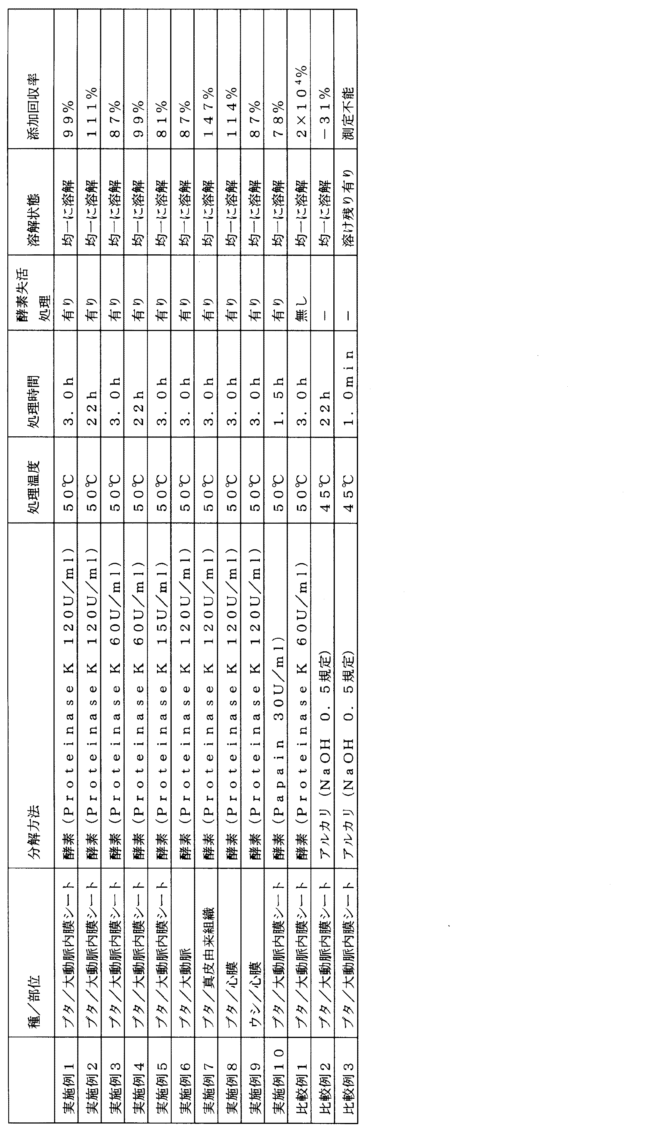
この添加回収試験用サンプル溶液とエンドトキシン測定用サンプル溶液を試料として用いて、下記<エンドトキシン濃度の測定方法>によりエンドトキシン濃度の測定を行い、下記<添加回収率の算出方法>で添加回収率を算出した。結果を表1に示す。 Using this sample solution for addition recovery test and sample solution for endotoxin measurement as a sample, the endotoxin concentration is measured by the following <method for measuring endotoxin concentration>, and the addition recovery rate is calculated by <calculation method for addition recovery rate> below. did. The results are shown in Table 1.
<エンドトキシン濃度の測定方法>
和光純薬工業株式会社製トキシノメーター(ET−6000/J)を用いて、比濁時間分析法の常法に従って、以下のようにエンドトキシン濃度の測定を行った。
すなわち、添加回収試験用サンプル溶液とエンドトキシン測定用サンプル溶液を試料として、それぞれの200μlを、和光純薬工業株式会社製のLimulus ES−2 Single Test wako(ゲル化感度:0.015EU/ml)のリムルス試薬(凍結乾燥品。カブトガニ血球抽出物(AL)を含有する。)に添加し、数秒間攪拌し、混合液を得た後、37℃保温下に、トキシノメーター(ET−6000/J)を用いて、上記混合液の透過光量が、測定開始から5%減少するまでの時間(以下、Tgと略記する。)を測定した。別に、蒸留水と濃度既知のエンドトキシン溶液を用いて同様の測定を行い、エンドトキシン濃度とTgとの関係を表す検量線を作成した。この検量線に基づいて、試料中のエンドトキシンの濃度を算出した。
<Measuring method of endotoxin concentration>
The endotoxin concentration was measured using a Wako Pure Chemical Industries, Ltd. Toxinometer (ET-6000/J) in accordance with the standard method of turbidimetric time analysis as follows.
That is, using the sample solution for addition recovery test and the sample solution for endotoxin measurement as samples, 200 μl of each of Limulus ES-2 Single Test wako (gelation sensitivity: 0.015 EU/ml) manufactured by Wako Pure Chemical Industries, Ltd. Limulus reagent (freeze-dried product. Contains horseshoe crab blood cell extract (AL)) and stirred for a few seconds to obtain a mixed solution, which was then kept at 37°C under a toxinometer (ET-6000/J). ) Was used to measure the time (hereinafter, abbreviated as Tg) from the start of measurement until the amount of transmitted light of the mixed solution decreased by 5%. Separately, the same measurement was performed using distilled water and an endotoxin solution of known concentration, and a calibration curve showing the relationship between the endotoxin concentration and Tg was created. Based on this calibration curve, the concentration of endotoxin in the sample was calculated.
<添加回収率の算出方法>
(添加回収試験用サンプル溶液のエンドトキシン量測定値(EU/ml)−エンドトキシン測定用サンプル溶液のエンドトキシン量測定値(EU/ml))/エンドトキシン測定用サンプル溶液のエンドトキシン含有量(0.015EU/ml)×100=添加回収率(%)
<Calculation method of addition recovery rate>
(Measured value of endotoxin amount of sample solution for addition recovery test (EU/ml)-Measured value of endotoxin amount of sample solution for endotoxin measurement (EU/ml))/Endotoxin content of sample solution for endotoxin measurement (0.015 EU/ml) )×100=additional recovery rate (%)
〔実施例2〕
0.15EU/ml含有Proteinase K溶液及びProteinase K溶液による脱細胞化組織の試験片を均一に溶解させるための酵素処理の時間を3時間から22時間に変えた以外は、実施例1と同様の手順で添加回収試験用サンプル溶液とエンドトキシン測定用サンプル溶液を調製した。この添加回収試験用サンプル溶液とエンドトキシン測定用サンプル溶液を試料として用いて、前記<エンドトキシン濃度の測定方法>によりエンドトキシン濃度の測定を行い、前記<添加回収率の算出方法>で添加回収率を算出した。結果を表1に示す。
[Example 2]
Similar to Example 1, except that the time of the enzyme treatment for uniformly dissolving the decellularized tissue test pieces by the 0.15 EU/ml-containing Proteinase K solution and the Proteinase K solution was changed from 3 hours to 22 hours. A sample solution for addition recovery test and a sample solution for endotoxin measurement were prepared by the procedure. Using this sample solution for addition recovery test and sample solution for endotoxin measurement as a sample, the endotoxin concentration is measured by the <method for measuring endotoxin concentration>, and the addition recovery rate is calculated by <calculation method for addition recovery rate>. did. The results are shown in Table 1.
〔実施例3〕
酵素処理し脱細胞化組織の試験片を均一に溶解させるのに用いた0.15EU/ml含有Proteinase K溶液及びProteinase K溶液の濃度が60U/mlであること以外は、実施例1と同様の手順で添加回収試験用サンプル溶液とエンドトキシン測定用サンプル溶液を調製した。この添加回収試験用サンプル溶液とエンドトキシン測定用サンプル溶液を試料として用いて、前記<エンドトキシン濃度の測定方法>によりエンドトキシン濃度の測定を行い、前記<添加回収率の算出方法>で添加回収率を算出した。結果を表1に示す。
[Example 3]
Similar to Example 1, except that the concentration of the 0.15 EU/ml-containing Proteinase K solution and the Proteinase K solution used for uniformly dissolving the test piece of the decellularized tissue treated with the enzyme was 60 U/ml. A sample solution for addition recovery test and a sample solution for endotoxin measurement were prepared by the procedure. Using this sample solution for addition recovery test and sample solution for endotoxin measurement as a sample, the endotoxin concentration is measured by the <method for measuring endotoxin concentration>, and the addition recovery rate is calculated by <calculation method for addition recovery rate>. did. The results are shown in Table 1.
〔実施例4〕
0.15EU/ml含有Proteinase K溶液及びProteinase K溶液による脱細胞化組織の試験片を均一に溶解させるための酵素処理の時間を3時間から22時間に変えた以外は、実施例3と同様の手順で添加回収試験用サンプル溶液とエンドトキシン測定用サンプル溶液を試料として調製した。この添加回収試験用サンプル溶液とエンドトキシン測定用サンプル溶液を用いて、前記<エンドトキシン濃度の測定方法>によりエンドトキシン濃度の測定を行い、前記<添加回収率の算出方法>で添加回収率を算出した。結果を表1に示す。
[Example 4]
Similar to Example 3 except that the time of the enzyme treatment for uniformly dissolving the test piece of the decellularized tissue with the 0.15 EU/ml Proteinase K solution and the Proteinase K solution was changed from 3 hours to 22 hours. The sample solution for addition recovery test and the sample solution for endotoxin measurement were prepared as samples by the procedure. Using this sample solution for addition recovery test and the sample solution for endotoxin measurement, the endotoxin concentration was measured by the <method for measuring endotoxin concentration>, and the addition recovery rate was calculated by <calculation method for addition recovery rate>. The results are shown in Table 1.
〔実施例5〕
酵素処理し脱細胞化組織の試験片を均一に溶解させるのに用いた0.15EU/ml含有Proteinase K溶液及びProteinase K溶液の濃度が15U/mlであること以外は、実施例1と同様の手順で添加回収試験用サンプル溶液とエンドトキシン測定用サンプル溶液を調製した。この添加回収試験用サンプル溶液とエンドトキシン測定用サンプル溶液を試料として用いて、前記<エンドトキシン濃度の測定方法>によりエンドトキシン濃度の測定を行い、前記<添加回収率の算出方法>で添加回収率を算出した。結果を表1に示す。
[Example 5]
Similar to Example 1, except that the concentration of the 0.15 EU/ml-containing Proteinase K solution and the Proteinase K solution used for uniformly dissolving the test piece of the decellularized tissue treated with the enzyme was 15 U/ml. A sample solution for addition recovery test and a sample solution for endotoxin measurement were prepared by the procedure. Using this sample solution for addition recovery test and sample solution for endotoxin measurement as a sample, the endotoxin concentration is measured by the <method for measuring endotoxin concentration>, and the addition recovery rate is calculated by <calculation method for addition recovery rate>. did. The results are shown in Table 1.
〔実施例6〕
屠畜場からブタ大動脈を購入し、冷蔵で搬送した。このブタ大動脈を切り開いてシート状にした。以下実施例1と同様にして、脱細胞化した後、添加回収試験用サンプル溶液とエンドトキシン測定用サンプル溶液を調製した。この添加回収試験用サンプル溶液とエンドトキシン測定用サンプル溶液を試料として用いて、前記<エンドトキシン濃度の測定方法>によりエンドトキシン濃度の測定を行い、前記<添加回収率の算出方法>で添加回収率を算出した。結果を表1に示す。
[Example 6]
Porcine aortas were purchased from a slaughterhouse and shipped refrigerated. The pig aorta was cut open to form a sheet. Thereafter, in the same manner as in Example 1, after decellularization, a sample solution for addition recovery test and a sample solution for endotoxin measurement were prepared. Using this sample solution for addition recovery test and sample solution for endotoxin measurement as a sample, the endotoxin concentration is measured by the <method for measuring endotoxin concentration>, and the addition recovery rate is calculated by <calculation method for addition recovery rate>. did. The results are shown in Table 1.
〔実施例7〕
屠畜場からブタ皮膚を購入し、冷蔵で搬送した。このブタ皮膚から、真皮層を含む組織を採取した。以下実施例1と同様にして、脱細胞化した後、添加回収試験用サンプル溶液とエンドトキシン測定用サンプル溶液を調製した。この添加回収試験用サンプル溶液とエンドトキシン測定用サンプル溶液を試料として用いて、前記<エンドトキシン濃度の測定方法>によりエンドトキシン濃度の測定を行い、前記<添加回収率の算出方法>で添加回収率を算出した。結果を表1に示す。
[Example 7]
Pig skin was purchased from a slaughterhouse and shipped refrigerated. A tissue containing a dermis layer was collected from this pig skin. Thereafter, in the same manner as in Example 1, after decellularization, a sample solution for addition recovery test and a sample solution for endotoxin measurement were prepared. Using this sample solution for addition recovery test and sample solution for endotoxin measurement as a sample, the endotoxin concentration is measured by the <method for measuring endotoxin concentration>, and the addition recovery rate is calculated by <calculation method for addition recovery rate>. did. The results are shown in Table 1.
〔実施例8〕
屠畜場からブタ心膜を購入し、冷蔵で搬送した。このブタ心膜から、脂肪組織を除去した。以下実施例1と同様にして、脱細胞化した後、添加回収試験用サンプル溶液とエンドトキシン測定用サンプル溶液を調製した。この添加回収試験用サンプル溶液とエンドトキシン測定用サンプル溶液を試料として用いて、前記<エンドトキシン濃度の測定方法>によりエンドトキシン濃度の測定を行い、前記<添加回収率の算出方法>で添加回収率を算出した。結果を表1に示す。
[Example 8]
Pig pericardium was purchased from a slaughterhouse and shipped refrigerated. Adipose tissue was removed from the porcine pericardium. Thereafter, in the same manner as in Example 1, after decellularization, a sample solution for addition recovery test and a sample solution for endotoxin measurement were prepared. Using this sample solution for addition recovery test and sample solution for endotoxin measurement as a sample, the endotoxin concentration is measured by the <method for measuring endotoxin concentration>, and the addition recovery rate is calculated by <calculation method for addition recovery rate>. did. The results are shown in Table 1.
〔実施例9〕
屠畜場からウシ心膜を購入し、冷蔵で搬送した。このウシ心膜から、脂肪組織を除去した。以下実施例1と同様にして、脱細胞化した後、添加回収試験用サンプル溶液とエンドトキシン測定用サンプル溶液を調製した。この添加回収試験用サンプル溶液とエンドトキシン測定用サンプル溶液を試料として用いて、前記<エンドトキシン濃度の測定方法>によりエンドトキシン濃度の測定を行い、前記<添加回収率の算出方法>で添加回収率を算出した。結果を表1に示す。
[Example 9]
Bovine pericardium was purchased from a slaughterhouse and shipped refrigerated. Adipose tissue was removed from the bovine pericardium. Thereafter, in the same manner as in Example 1, after decellularization, a sample solution for addition recovery test and a sample solution for endotoxin measurement were prepared. Using this sample solution for addition recovery test and sample solution for endotoxin measurement as a sample, the endotoxin concentration is measured by the <method for measuring endotoxin concentration>, and the addition recovery rate is calculated by <calculation method for addition recovery rate>. did. The results are shown in Table 1.
〔実施例10〕
(使用試薬)
・ET溶液(エンドトキシン溶液)
CSEを蒸留水で希釈し、これを適宜希釈したものを使用した。
・Papain溶液(パパイン溶液)
Papain(Carica papaya、Roche)を蒸留水で希釈し、これを適宜希釈したものを使用した。
[Example 10]
(Reagent used)
・ET solution (endotoxin solution)
CSE was diluted with distilled water and used by appropriately diluting it.
・Papain solution (papain solution)
Papain (Carica papaya, Roche) was diluted with distilled water and used by appropriately diluting it.
(生体適合性材料の測定試料の準備)
実施例1と同様の手順で作製した。
(Preparation of measurement sample of biocompatible material)
It was manufactured by the same procedure as in Example 1.
(添加回収試験用サンプル溶液の調製)
Papain溶液をET溶液で希釈し、30U/mlの0.15EU/ml含有Papain溶液を1.0ml調製した。
添加回収試験用サンプルに、30U/mlの0.15EU/ml含有Papain溶液1.0mlを添加し、50℃で1.5時間酵素処理し、脱細胞化組織の試験片を均一に溶解させた。処理後、Papainを、80℃8分で失活させ、溶液を室温に戻した後、蒸留水を用いて10倍希釈した。
(Preparation of sample solution for addition recovery test)
The Papain solution was diluted with the ET solution to prepare 1.0 ml of a Papain solution containing 30 U/ml of 0.15 EU/ml.
To the sample for addition recovery test, 1.0 ml of Papain solution containing 30 U/ml of 0.15 EU/ml was added, and enzyme treatment was performed at 50° C. for 1.5 hours to uniformly dissolve the decellularized tissue test piece. .. After the treatment, Papain was inactivated at 80° C. for 8 minutes, the solution was returned to room temperature, and then diluted 10 times with distilled water.
(エンドトキシン測定用サンプル溶液の調製)
エンドトキシン測定用サンプルに、30U/mlのPapain溶液を1.0ml添加し、50℃で1.5時間酵素処理し、脱細胞化組織の試験片を均一に溶解させた。処理後、Papainを、80℃8分で失活させ、溶液を室温に戻した後、蒸留水を用いて10倍希釈した。
(Preparation of sample solution for endotoxin measurement)
To a sample for endotoxin measurement, 1.0 ml of a 30 U/ml Papain solution was added and treated with an enzyme at 50° C. for 1.5 hours to uniformly dissolve the decellularized tissue test piece. After the treatment, Papain was inactivated at 80° C. for 8 minutes, the solution was returned to room temperature, and then diluted 10 times with distilled water.
この添加回収試験用サンプル溶液とエンドトキシン測定用サンプル溶液を試料として用いて、前記<エンドトキシン濃度の測定方法>によりエンドトキシン濃度の測定を行い、前記<添加回収率の算出方法>で添加回収率を算出した。結果を表1に示す。
釈した。
Using this sample solution for addition recovery test and sample solution for endotoxin measurement as a sample, the endotoxin concentration is measured by the <method for measuring endotoxin concentration>, and the addition recovery rate is calculated by <calculation method for addition recovery rate>. did. The results are shown in Table 1.
I released it.
〔比較例1〕
(生体適合性材料の測定試料の準備)
実施例1と同様の手順で作製した。
[Comparative Example 1]
(Preparation of measurement sample of biocompatible material)
It was manufactured by the same procedure as in Example 1.
(添加回収試験用サンプル溶液の調製)
Proteinase K溶液をET溶液で希釈し、60U/mlの0.15EU/ml含有Proteinase K溶液を2.0ml調製した。
添加回収試験用サンプルに、60U/mlの0.15EU/ml含有Proteinase K溶液1.0mlを添加し、50℃で3時間酵素処理し、脱細胞化組織の試験片を均一に溶解させた。その後、Proteinase Kの失活処理をせず、溶液を室温に戻した後、蒸留水を用いて10倍希釈した。
(Preparation of sample solution for addition recovery test)
The Proteinase K solution was diluted with the ET solution to prepare 2.0 ml of the Proteinase K solution containing 60 U/ml of 0.15 EU/ml.
To the sample for addition recovery test, 1.0 ml of Proteinase K solution containing 60 U/ml of 0.15 EU/ml was added, and enzyme treatment was performed at 50° C. for 3 hours to uniformly dissolve the decellularized tissue test piece. Then, after deactivating the proteinase K, the solution was returned to room temperature and then diluted 10-fold with distilled water.
(エンドトキシン測定用サンプル溶液の調製)
エンドトキシン測定用サンプルに、60U/mlのProteinase K溶液を1.0ml添加し、50℃で3時間酵素処理し、脱細胞化組織の試験片を均一に溶解させた。その後、Proteinase Kの失活処理をせず、溶液を室温に戻した後、蒸留水を用いて10倍希釈した。
この添加回収試験用サンプル溶液とエンドトキシン測定用サンプル溶液を試料として用いて、前記<エンドトキシン濃度の測定方法>によりエンドトキシン濃度の測定を行い、前記<添加回収率の算出方法>で添加回収率を算出した。結果を表1に示す。この結果より、Proteinase Kの失活処理をしない場合、添加回収率は200%を大きく超えてしまった。添加回収率は50〜200%の間でなければならないため(日本薬局方)、酵素の失活処理をしない方法は、分析方法として適さないことがわかる。
添加回収率が200%を大きく超えてしまった理由は定かではないが、その理由のひとつとして、残存したProteinase Kがリムルス試薬のゲル化カスケードを開始させてしまったため、疑陽性を示したと考えられる。
(Preparation of sample solution for endotoxin measurement)
To the sample for endotoxin measurement, 1.0 ml of a 60 U/ml proteinase K solution was added, and the sample was treated with an enzyme at 50° C. for 3 hours to uniformly dissolve the decellularized tissue test piece. Then, after deactivating the proteinase K, the solution was returned to room temperature and then diluted 10-fold with distilled water.
Using this sample solution for addition recovery test and sample solution for endotoxin measurement as a sample, the endotoxin concentration is measured by the <method for measuring endotoxin concentration>, and the addition recovery rate is calculated by <calculation method for addition recovery rate>. did. The results are shown in Table 1. From this result, in the case where the inactivation treatment of Proteinase K was not performed, the addition recovery rate greatly exceeded 200%. Since the addition recovery rate must be between 50 and 200% (Japanese Pharmacopoeia), it can be seen that the method without enzyme deactivation treatment is not suitable as an analytical method.
The reason why the spiked recovery rate greatly exceeded 200% is not clear, but one of the reasons is that the remaining Proteinase K initiated the gelation cascade of the Limulus reagent, and it is considered that a false positive was shown. ..
〔比較例2〕
(使用試薬)
・ET溶液
CSEを蒸留水で希釈し、これを適宜希釈したものを使用した。
・NaOH溶液
蒸留水に水酸化ナトリウム(NaOH)を溶解し、40mg/ml(1.0規定)のNaOH溶液を調製した。これを適宜希釈したものを使用した。
・HCl溶液
蒸留水で塩酸を希釈し、18mg/ml(0.5規定)のHCl溶液を調製した。
[Comparative Example 2]
(Reagent used)
-ET solution CSE was diluted with distilled water and used by appropriately diluting it.
-NaOH solution Sodium hydroxide (NaOH) was dissolved in distilled water to prepare a 40 mg/ml (1.0 N) NaOH solution. This was appropriately diluted and used.
-HCl solution Hydrochloric acid was diluted with distilled water to prepare an 18 mg/ml (0.5 N) HCl solution.
(生体適合性材料の測定試料の準備)
屠畜場からブタ大動脈を購入し、冷蔵で搬送した。このブタ大動脈を切り開いて採取したシート状の内膜(以下、内膜シート)と生理食塩水をポリエチンレン製バッグに入れてシールし、研究開発用高圧処理装置((株)神戸製鋼所製:Dr.CHEF)で100〜1000MPaにて高静水圧処理を行った。処理したシートを核酸分解酵素含有洗浄液及びアルコール含有洗浄液により洗浄し、脱細胞化組織である脱細胞化ブタ大動脈シートを得た。
脱細胞化ブタ大動脈シートから1cm×1cmの試験片を2枚採取し、それぞれ重量を測定し、それぞれ2.0mlマイクロチューブに入れた。1本は添加回収試験用サンプルとし、1本はエンドトキシン測定用サンプルとした。
(Preparation of measurement sample of biocompatible material)
Porcine aortas were purchased from a slaughterhouse and shipped refrigerated. A sheet-shaped intima (hereinafter referred to as intimal sheet) obtained by cutting open the porcine aorta and physiological saline were placed in a polyethylene bag and sealed, and a high-pressure processing device for research and development (Kobe Steel Co., Ltd.: Dr. .CHEF) at 100 to 1000 MPa. The treated sheet was washed with a nucleolytic enzyme-containing washing solution and an alcohol-containing washing solution to obtain a decellularized porcine aortic sheet as a decellularized tissue.
Two 1 cm×1 cm test pieces were taken from the decellularized porcine aortic sheet, weighed, and placed in 2.0 ml microtubes. One was used as a sample for addition recovery test and one was used as a sample for endotoxin measurement.
(添加回収試験用サンプル溶液の調製)
NaOH溶液をET溶液で希釈し、0.5規定の0.30EU/ml含有NaOH溶液を2.0ml調製した。
添加回収試験用サンプルに、0.5規定の0.30EU/ml含有NaOH溶液1.0mlを添加し、45℃で22時間処理して、脱細胞化組織の試験片を均一に溶解させた。
(Preparation of sample solution for addition recovery test)
The NaOH solution was diluted with the ET solution to prepare 2.0 ml of 0.5N 0.30 EU/ml-containing NaOH solution.
To the sample for addition recovery test, 1.0 ml of 0.5N 0.30 EU/ml-containing NaOH solution was added, and treated at 45° C. for 22 hours to uniformly dissolve the decellularized tissue test piece.
(エンドトキシン測定用サンプル溶液の調製)
エンドトキシン測定用サンプルに、0.5規定のNaOH溶液を1.0ml添加し、45℃で22時間処理して、脱細胞化組織の試験片を均一に溶解させた。
(Preparation of sample solution for endotoxin measurement)
To a sample for endotoxin measurement, 1.0 ml of 0.5 N NaOH solution was added and treated at 45° C. for 22 hours to uniformly dissolve the test piece of decellularized tissue.
この添加回収試験用サンプル溶液とエンドトキシン測定用サンプル溶液を、それぞれHCl溶液で中和し、前記<エンドトキシン濃度の測定方法>によりエンドトキシン濃度の測定を行い、前記<添加回収率の算出方法>で添加回収率を算出した。結果を表1に示す。この結果より、NaOH溶液で生体適合性材料である脱細胞化組織を溶解した場合、添加回収率が0%未満となることが分かった。添加回収率は50〜200%の間でなければならないため(日本薬局方)、水酸化ナトリウム等のアルカリを使用して生体適合性材料である脱細胞化組織を溶解させる方法は、分析方法として適さないことがわかる。添加回収率が0%未満となる理由は定かではないが、その理由のひとつとしては、水酸化ナトリウム溶液中のエンドトキシンは非常に不安定であり、22時間処理することで、添加したエンドトキシン全量が不活化されたため、疑陰性を示したと考えられる。 The sample solution for addition recovery test and the sample solution for endotoxin measurement are each neutralized with an HCl solution, the endotoxin concentration is measured by the <method for measuring endotoxin concentration>, and the addition is made by <calculation method for addition recovery rate>. The recovery rate was calculated. The results are shown in Table 1. From this result, it was found that when the decellularized tissue, which is a biocompatible material, was dissolved with a NaOH solution, the recovery rate of addition was less than 0%. Since the recovery rate of addition must be between 50 and 200% (Japanese Pharmacopoeia), the method of dissolving the decellularized tissue, which is a biocompatible material, using an alkali such as sodium hydroxide is an analytical method. Turns out it's not suitable The reason why the addition recovery rate is less than 0% is not clear, but one of the reasons is that endotoxin in the sodium hydroxide solution is very unstable, and the total amount of the added endotoxin is treated by treating for 22 hours. Since it was inactivated, it is probable that it showed a false negative.
〔比較例3〕
0.30EU/ml含有NaOH溶液による脱細胞化組織への処理時間を1分間に変更し、比較例2と同様の手順で添加回収試験用サンプル溶液とエンドトキシン測定用サンプル溶液を調製した。
添加回収試験用サンプル溶液、エンドトキシン測定用サンプル溶液とも、脱細胞化組織の試験片は均一に溶解せず溶け残りがあったため、脱細胞化組織溶解時のエンドトキシン濃度を測定することができなかった。
[Comparative Example 3]
The treatment time for the decellularized tissue with the 0.30 EU/ml NaOH solution was changed to 1 minute, and a sample solution for addition recovery test and a sample solution for endotoxin measurement were prepared in the same procedure as in Comparative Example 2.
In both the sample solution for addition recovery test and the sample solution for endotoxin measurement, the test piece of decellularized tissue did not dissolve uniformly and remained undissolved, so it was not possible to measure the endotoxin concentration during dissolution of decellularized tissue ..
Claims (11)
酵素で処理した後の溶液が均一に溶解している、エンドトキシン測定のための前処理方法。 A pretreatment method for measuring endotoxin, which comprises a step of treating a sample with an enzyme to dissolve the sample, and a step of deactivating the enzyme after the step ,
A pretreatment method for measuring endotoxin, in which the solution after treatment with the enzyme is uniformly dissolved.
The method for measuring endotoxin according to claim 10, wherein the endotoxin measurement is performed by a Limulus test.
Priority Applications (1)
| Application Number | Priority Date | Filing Date | Title |
|---|---|---|---|
| JP2016066890A JP6712160B2 (en) | 2016-03-29 | 2016-03-29 | Pretreatment method for measuring endotoxin and its measuring method |
Applications Claiming Priority (1)
| Application Number | Priority Date | Filing Date | Title |
|---|---|---|---|
| JP2016066890A JP6712160B2 (en) | 2016-03-29 | 2016-03-29 | Pretreatment method for measuring endotoxin and its measuring method |
Publications (2)
| Publication Number | Publication Date |
|---|---|
| JP2017181217A JP2017181217A (en) | 2017-10-05 |
| JP6712160B2 true JP6712160B2 (en) | 2020-06-17 |
Family
ID=60004384
Family Applications (1)
| Application Number | Title | Priority Date | Filing Date |
|---|---|---|---|
| JP2016066890A Active JP6712160B2 (en) | 2016-03-29 | 2016-03-29 | Pretreatment method for measuring endotoxin and its measuring method |
Country Status (1)
| Country | Link |
|---|---|
| JP (1) | JP6712160B2 (en) |
Families Citing this family (1)
| Publication number | Priority date | Publication date | Assignee | Title |
|---|---|---|---|---|
| JPWO2017038473A1 (en) * | 2015-08-28 | 2018-06-14 | 天野エンザイム株式会社 | Endotoxin reducing thermolysin |
Family Cites Families (7)
| Publication number | Priority date | Publication date | Assignee | Title |
|---|---|---|---|---|
| JP3524120B2 (en) * | 1992-05-08 | 2004-05-10 | 生化学工業株式会社 | Pretreatment agent, pretreatment method, measurement method using pretreated sample, measurement kit, and sample determination method |
| JP2002507907A (en) * | 1997-06-27 | 2002-03-12 | バーダー、アウグスチヌス | Biosynthetic implant and method for producing the same |
| EP1239897B1 (en) * | 1999-12-22 | 2004-03-24 | Acell, Inc. | Tissue regenerative composition |
| JP2009513269A (en) * | 2005-10-27 | 2009-04-02 | アルフォース, ジャン−エリック ダブリュー. | A cell-free, bioabsorbable tissue regeneration matrix produced by incubating cell-free blood products |
| EP2076279B1 (en) * | 2006-10-06 | 2014-08-27 | Anthrogenesis Corporation | Native (telopeptide) placental collagen compositions |
| GB201215725D0 (en) * | 2012-09-04 | 2012-10-17 | Univ Leeds | Composite connective tissue and bone implants |
| CN106794227B (en) * | 2014-08-25 | 2022-03-01 | Hli细胞疗法有限责任公司 | Extracellular matrix compositions |
-
2016
- 2016-03-29 JP JP2016066890A patent/JP6712160B2/en active Active
Also Published As
| Publication number | Publication date |
|---|---|
| JP2017181217A (en) | 2017-10-05 |
Similar Documents
| Publication | Publication Date | Title |
|---|---|---|
| Milan et al. | Decellularization and preservation of human skin: A platform for tissue engineering and reconstructive surgery | |
| JP6480911B2 (en) | Method of decellularizing tissue grafts | |
| Fu et al. | Decellularization of porcine skeletal muscle extracellular matrix for the formulation of a matrix hydrogel: a preliminary study | |
| KR102501235B1 (en) | decellularized tissue | |
| KR101410533B1 (en) | A method for treating material derived from biological tissue | |
| KR20160025502A (en) | Hybrid gel containing particulate decellularized tissue | |
| Anilkumar et al. | Biomaterial properties of cholecyst‐derived scaffold recovered by a non‐detergent/enzymatic method | |
| Mawad et al. | Lysozyme depolymerization of photo-activated chitosan adhesive films | |
| Mahmood et al. | Prevalence and Associated Risk Factors of Cystic Echinococcosis in Food Animals--A Neglected and Prevailing Zoonosis. | |
| Jensen et al. | A non-traumatic Staphylococcus aureus osteomyelitis model in pigs | |
| CN107796674B (en) | Method for evaluating eye irritation injury and repair by long-term culture of animal cornea | |
| JP6712160B2 (en) | Pretreatment method for measuring endotoxin and its measuring method | |
| US10487135B2 (en) | Method for preparing low endotoxin collagen | |
| CA3247704A1 (en) | Purified mucus | |
| Kumar et al. | Effects of crosslinking treatments on the physical properties of acellular fish swim bladder | |
| KR102181811B1 (en) | Method for Decellularization of Tissue | |
| TW202233828A (en) | Decellularized tissue composition | |
| Crosby-Durrani et al. | Clinical and pathological features of bovine ischaemic teat necrosis | |
| AU2015415667A1 (en) | Method for increasing collagen yield, and collagen prepared using same | |
| CA2765893C (en) | New method for isolating trichinella or other parasites from organic tissue | |
| CN116850347B (en) | Hydrogel of injectable fat extracellular matrix composite | |
| JP7649016B2 (en) | Porous scaffold for cell culture and method for producing same | |
| KR101837118B1 (en) | A Method for Extracting Collagen using Bacterial Fermentation | |
| Abuseir et al. | Protein profile of the cysts of Taenia hydatigena, Taenia saginata, Echinococcus granulosus and Taenia ovis | |
| JP2016073220A (en) | Disinfection evaluation method of Acanthamoeba |
Legal Events
| Date | Code | Title | Description |
|---|---|---|---|
| A621 | Written request for application examination |
Free format text: JAPANESE INTERMEDIATE CODE: A621 Effective date: 20181228 |
|
| A977 | Report on retrieval |
Free format text: JAPANESE INTERMEDIATE CODE: A971007 Effective date: 20191009 |
|
| A131 | Notification of reasons for refusal |
Free format text: JAPANESE INTERMEDIATE CODE: A131 Effective date: 20191023 |
|
| A521 | Written amendment |
Free format text: JAPANESE INTERMEDIATE CODE: A523 Effective date: 20191212 |
|
| TRDD | Decision of grant or rejection written | ||
| A01 | Written decision to grant a patent or to grant a registration (utility model) |
Free format text: JAPANESE INTERMEDIATE CODE: A01 Effective date: 20200512 |
|
| A61 | First payment of annual fees (during grant procedure) |
Free format text: JAPANESE INTERMEDIATE CODE: A61 Effective date: 20200529 |
|
| R150 | Certificate of patent or registration of utility model |
Ref document number: 6712160 Country of ref document: JP Free format text: JAPANESE INTERMEDIATE CODE: R150 |