JP5926180B2 - Methods of cell programming for tolerogenic therapy - Google Patents
Methods of cell programming for tolerogenic therapy Download PDFInfo
- Publication number
- JP5926180B2 JP5926180B2 JP2012523125A JP2012523125A JP5926180B2 JP 5926180 B2 JP5926180 B2 JP 5926180B2 JP 2012523125 A JP2012523125 A JP 2012523125A JP 2012523125 A JP2012523125 A JP 2012523125A JP 5926180 B2 JP5926180 B2 JP 5926180B2
- Authority
- JP
- Japan
- Prior art keywords
- cells
- csf
- antigen
- pharmaceutical composition
- tslp
- Prior art date
- Legal status (The legal status is an assumption and is not a legal conclusion. Google has not performed a legal analysis and makes no representation as to the accuracy of the status listed.)
- Active
Links
- 230000003614 tolerogenic effect Effects 0.000 title claims description 17
- 238000000034 method Methods 0.000 title description 53
- 238000002560 therapeutic procedure Methods 0.000 title description 2
- 108010017213 Granulocyte-Macrophage Colony-Stimulating Factor Proteins 0.000 claims description 189
- 210000004443 dendritic cell Anatomy 0.000 claims description 123
- 210000004027 cell Anatomy 0.000 claims description 118
- 102000036639 antigens Human genes 0.000 claims description 94
- 108091007433 antigens Proteins 0.000 claims description 94
- 239000000427 antigen Substances 0.000 claims description 93
- 208000028169 periodontal disease Diseases 0.000 claims description 72
- FHVDTGUDJYJELY-UHFFFAOYSA-N 6-{[2-carboxy-4,5-dihydroxy-6-(phosphanyloxy)oxan-3-yl]oxy}-4,5-dihydroxy-3-phosphanyloxane-2-carboxylic acid Chemical compound O1C(C(O)=O)C(P)C(O)C(O)C1OC1C(C(O)=O)OC(OP)C(O)C1O FHVDTGUDJYJELY-UHFFFAOYSA-N 0.000 claims description 71
- 229940072056 alginate Drugs 0.000 claims description 70
- 235000010443 alginic acid Nutrition 0.000 claims description 70
- 229920000615 alginic acid Polymers 0.000 claims description 70
- 239000000203 mixture Substances 0.000 claims description 67
- 230000028993 immune response Effects 0.000 claims description 47
- 206010020751 Hypersensitivity Diseases 0.000 claims description 32
- 108010058846 Ovalbumin Proteins 0.000 claims description 28
- 229940092253 ovalbumin Drugs 0.000 claims description 28
- 230000007815 allergy Effects 0.000 claims description 25
- 208000023275 Autoimmune disease Diseases 0.000 claims description 24
- 102100024506 Bone morphogenetic protein 2 Human genes 0.000 claims description 23
- 108010049931 Bone Morphogenetic Protein 2 Proteins 0.000 claims description 22
- 208000037265 diseases, disorders, signs and symptoms Diseases 0.000 claims description 21
- 208000006673 asthma Diseases 0.000 claims description 19
- 239000013566 allergen Substances 0.000 claims description 18
- 208000037976 chronic inflammation Diseases 0.000 claims description 18
- 108090000623 proteins and genes Proteins 0.000 claims description 18
- 230000001717 pathogenic effect Effects 0.000 claims description 16
- 208000026935 allergic disease Diseases 0.000 claims description 15
- 239000013612 plasmid Substances 0.000 claims description 15
- 235000018102 proteins Nutrition 0.000 claims description 15
- 102000004169 proteins and genes Human genes 0.000 claims description 15
- 239000000017 hydrogel Substances 0.000 claims description 14
- 239000003659 bee venom Substances 0.000 claims description 13
- 230000006058 immune tolerance Effects 0.000 claims description 12
- 230000007115 recruitment Effects 0.000 claims description 12
- 239000004005 microsphere Substances 0.000 claims description 11
- UREBDLICKHMUKA-CXSFZGCWSA-N dexamethasone Chemical compound C1CC2=CC(=O)C=C[C@]2(C)[C@]2(F)[C@@H]1[C@@H]1C[C@@H](C)[C@@](C(=O)CO)(O)[C@@]1(C)C[C@@H]2O UREBDLICKHMUKA-CXSFZGCWSA-N 0.000 claims description 10
- 229960003957 dexamethasone Drugs 0.000 claims description 10
- NOESYZHRGYRDHS-UHFFFAOYSA-N insulin Chemical compound N1C(=O)C(NC(=O)C(CCC(N)=O)NC(=O)C(CCC(O)=O)NC(=O)C(C(C)C)NC(=O)C(NC(=O)CN)C(C)CC)CSSCC(C(NC(CO)C(=O)NC(CC(C)C)C(=O)NC(CC=2C=CC(O)=CC=2)C(=O)NC(CCC(N)=O)C(=O)NC(CC(C)C)C(=O)NC(CCC(O)=O)C(=O)NC(CC(N)=O)C(=O)NC(CC=2C=CC(O)=CC=2)C(=O)NC(CSSCC(NC(=O)C(C(C)C)NC(=O)C(CC(C)C)NC(=O)C(CC=2C=CC(O)=CC=2)NC(=O)C(CC(C)C)NC(=O)C(C)NC(=O)C(CCC(O)=O)NC(=O)C(C(C)C)NC(=O)C(CC(C)C)NC(=O)C(CC=2NC=NC=2)NC(=O)C(CO)NC(=O)CNC2=O)C(=O)NCC(=O)NC(CCC(O)=O)C(=O)NC(CCCNC(N)=N)C(=O)NCC(=O)NC(CC=3C=CC=CC=3)C(=O)NC(CC=3C=CC=CC=3)C(=O)NC(CC=3C=CC(O)=CC=3)C(=O)NC(C(C)O)C(=O)N3C(CCC3)C(=O)NC(CCCCN)C(=O)NC(C)C(O)=O)C(=O)NC(CC(N)=O)C(O)=O)=O)NC(=O)C(C(C)CC)NC(=O)C(CO)NC(=O)C(C(C)O)NC(=O)C1CSSCC2NC(=O)C(CC(C)C)NC(=O)C(NC(=O)C(CCC(N)=O)NC(=O)C(CC(N)=O)NC(=O)C(NC(=O)C(N)CC=1C=CC=CC=1)C(C)C)CC1=CN=CN1 NOESYZHRGYRDHS-UHFFFAOYSA-N 0.000 claims description 10
- 206010067584 Type 1 diabetes mellitus Diseases 0.000 claims description 9
- 102000002689 Toll-like receptor Human genes 0.000 claims description 8
- 108020000411 Toll-like receptor Proteins 0.000 claims description 8
- 230000002992 thymic effect Effects 0.000 claims description 8
- 241000282326 Felis catus Species 0.000 claims description 7
- 230000002912 lymphogenic effect Effects 0.000 claims description 7
- 235000013336 milk Nutrition 0.000 claims description 7
- 239000008267 milk Substances 0.000 claims description 7
- 210000004080 milk Anatomy 0.000 claims description 7
- 244000052769 pathogen Species 0.000 claims description 7
- 230000002829 reductive effect Effects 0.000 claims description 7
- 240000007087 Apium graveolens Species 0.000 claims description 6
- 206010028980 Neoplasm Diseases 0.000 claims description 6
- 239000000556 agonist Substances 0.000 claims description 6
- 208000035475 disorder Diseases 0.000 claims description 6
- 230000001575 pathological effect Effects 0.000 claims description 6
- 230000009467 reduction Effects 0.000 claims description 6
- 235000009109 Betula pendula Nutrition 0.000 claims description 5
- 102000004877 Insulin Human genes 0.000 claims description 5
- 108090001061 Insulin Proteins 0.000 claims description 5
- 229940125396 insulin Drugs 0.000 claims description 5
- KISWVXRQTGLFGD-UHFFFAOYSA-N 2-[[2-[[6-amino-2-[[2-[[2-[[5-amino-2-[[2-[[1-[2-[[6-amino-2-[(2,5-diamino-5-oxopentanoyl)amino]hexanoyl]amino]-5-(diaminomethylideneamino)pentanoyl]pyrrolidine-2-carbonyl]amino]-3-hydroxypropanoyl]amino]-5-oxopentanoyl]amino]-5-(diaminomethylideneamino)p Chemical compound C1CCN(C(=O)C(CCCN=C(N)N)NC(=O)C(CCCCN)NC(=O)C(N)CCC(N)=O)C1C(=O)NC(CO)C(=O)NC(CCC(N)=O)C(=O)NC(CCCN=C(N)N)C(=O)NC(CO)C(=O)NC(CCCCN)C(=O)NC(C(=O)NC(CC(C)C)C(O)=O)CC1=CC=C(O)C=C1 KISWVXRQTGLFGD-UHFFFAOYSA-N 0.000 claims description 4
- 244000036975 Ambrosia artemisiifolia Species 0.000 claims description 4
- 235000003129 Ambrosia artemisiifolia var elatior Nutrition 0.000 claims description 4
- 206010003827 Autoimmune hepatitis Diseases 0.000 claims description 4
- 208000015943 Coeliac disease Diseases 0.000 claims description 4
- 244000141359 Malus pumila Species 0.000 claims description 4
- 102000047918 Myelin Basic Human genes 0.000 claims description 4
- 101710107068 Myelin basic protein Proteins 0.000 claims description 4
- 208000021386 Sjogren Syndrome Diseases 0.000 claims description 4
- 235000003484 annual ragweed Nutrition 0.000 claims description 4
- 230000006472 autoimmune response Effects 0.000 claims description 4
- 235000006263 bur ragweed Nutrition 0.000 claims description 4
- 235000003488 common ragweed Nutrition 0.000 claims description 4
- 201000006417 multiple sclerosis Diseases 0.000 claims description 4
- 235000009736 ragweed Nutrition 0.000 claims description 4
- 241000219496 Alnus Species 0.000 claims description 3
- 241000219498 Alnus glutinosa Species 0.000 claims description 3
- 101000641574 Apis mellifera Venom dipeptidyl peptidase 4 Proteins 0.000 claims description 3
- 235000002764 Apium graveolens Nutrition 0.000 claims description 3
- 235000015849 Apium graveolens Dulce Group Nutrition 0.000 claims description 3
- 235000010591 Appio Nutrition 0.000 claims description 3
- 244000105624 Arachis hypogaea Species 0.000 claims description 3
- 241000219430 Betula pendula Species 0.000 claims description 3
- 241000726811 Carpinus betulus Species 0.000 claims description 3
- 102000006303 Chaperonin 60 Human genes 0.000 claims description 3
- 108010058432 Chaperonin 60 Proteins 0.000 claims description 3
- 108010026206 Conalbumin Proteins 0.000 claims description 3
- 208000011231 Crohn disease Diseases 0.000 claims description 3
- 241000238740 Dermatophagoides pteronyssinus Species 0.000 claims description 3
- 108010003272 Hyaluronate lyase Proteins 0.000 claims description 3
- 102000001974 Hyaluronidases Human genes 0.000 claims description 3
- WHUUTDBJXJRKMK-VKHMYHEASA-N L-glutamic acid Chemical compound OC(=O)[C@@H](N)CCC(O)=O WHUUTDBJXJRKMK-VKHMYHEASA-N 0.000 claims description 3
- 102000004407 Lactalbumin Human genes 0.000 claims description 3
- 108090000942 Lactalbumin Proteins 0.000 claims description 3
- 102100037611 Lysophospholipase Human genes 0.000 claims description 3
- 102100033468 Lysozyme C Human genes 0.000 claims description 3
- 108010014251 Muramidase Proteins 0.000 claims description 3
- 102000006386 Myelin Proteins Human genes 0.000 claims description 3
- 108010083674 Myelin Proteins Proteins 0.000 claims description 3
- 102000055324 Myelin Proteolipid Human genes 0.000 claims description 3
- 101710094913 Myelin proteolipid protein Proteins 0.000 claims description 3
- 102000002233 Myelin-Oligodendrocyte Glycoprotein Human genes 0.000 claims description 3
- 108010000123 Myelin-Oligodendrocyte Glycoprotein Proteins 0.000 claims description 3
- 108010062010 N-Acetylmuramoyl-L-alanine Amidase Proteins 0.000 claims description 3
- 244000046052 Phaseolus vulgaris Species 0.000 claims description 3
- 235000010627 Phaseolus vulgaris Nutrition 0.000 claims description 3
- 108010058864 Phospholipases A2 Proteins 0.000 claims description 3
- 101710093543 Probable non-specific lipid-transfer protein Proteins 0.000 claims description 3
- 108010076181 Proinsulin Proteins 0.000 claims description 3
- 229940123384 Toll-like receptor (TLR) agonist Drugs 0.000 claims description 3
- 239000001387 apium graveolens Substances 0.000 claims description 3
- 239000005018 casein Substances 0.000 claims description 3
- BECPQYXYKAMYBN-UHFFFAOYSA-N casein, tech. Chemical compound NCCCCC(C(O)=O)N=C(O)C(CC(O)=O)N=C(O)C(CCC(O)=N)N=C(O)C(CC(C)C)N=C(O)C(CCC(O)=O)N=C(O)C(CC(O)=O)N=C(O)C(CCC(O)=O)N=C(O)C(C(C)O)N=C(O)C(CCC(O)=N)N=C(O)C(CCC(O)=N)N=C(O)C(CCC(O)=N)N=C(O)C(CCC(O)=O)N=C(O)C(CCC(O)=O)N=C(O)C(COP(O)(O)=O)N=C(O)C(CCC(O)=N)N=C(O)C(N)CC1=CC=CC=C1 BECPQYXYKAMYBN-UHFFFAOYSA-N 0.000 claims description 3
- 235000021240 caseins Nutrition 0.000 claims description 3
- 208000037893 chronic inflammatory disorder Diseases 0.000 claims description 3
- 239000000428 dust Substances 0.000 claims description 3
- 229930195712 glutamate Natural products 0.000 claims description 3
- 229960002773 hyaluronidase Drugs 0.000 claims description 3
- 239000000411 inducer Substances 0.000 claims description 3
- 235000010335 lysozyme Nutrition 0.000 claims description 3
- 239000004325 lysozyme Substances 0.000 claims description 3
- 229960000274 lysozyme Drugs 0.000 claims description 3
- 210000005012 myelin Anatomy 0.000 claims description 3
- 210000004248 oligodendroglia Anatomy 0.000 claims description 3
- 235000020232 peanut Nutrition 0.000 claims description 3
- 206010039073 rheumatoid arthritis Diseases 0.000 claims description 3
- 241000894007 species Species 0.000 claims description 3
- 235000021241 α-lactalbumin Nutrition 0.000 claims description 3
- 208000002874 Acne Vulgaris Diseases 0.000 claims description 2
- 208000003343 Antiphospholipid Syndrome Diseases 0.000 claims description 2
- 206010069002 Autoimmune pancreatitis Diseases 0.000 claims description 2
- 208000023328 Basedow disease Diseases 0.000 claims description 2
- 244000274847 Betula papyrifera Species 0.000 claims description 2
- 235000009113 Betula papyrifera Nutrition 0.000 claims description 2
- 235000010928 Betula populifolia Nutrition 0.000 claims description 2
- 235000002992 Betula pubescens Nutrition 0.000 claims description 2
- 208000008439 Biliary Liver Cirrhosis Diseases 0.000 claims description 2
- 208000033222 Biliary cirrhosis primary Diseases 0.000 claims description 2
- 206010009900 Colitis ulcerative Diseases 0.000 claims description 2
- 240000007582 Corylus avellana Species 0.000 claims description 2
- 235000007466 Corylus avellana Nutrition 0.000 claims description 2
- 206010012438 Dermatitis atopic Diseases 0.000 claims description 2
- 102100036264 Glucose-6-phosphatase catalytic subunit 1 Human genes 0.000 claims description 2
- 201000005569 Gout Diseases 0.000 claims description 2
- 208000015023 Graves' disease Diseases 0.000 claims description 2
- 208000035895 Guillain-Barré syndrome Diseases 0.000 claims description 2
- 208000001204 Hashimoto Disease Diseases 0.000 claims description 2
- 208000030836 Hashimoto thyroiditis Diseases 0.000 claims description 2
- 101000930910 Homo sapiens Glucose-6-phosphatase catalytic subunit 1 Proteins 0.000 claims description 2
- 206010021245 Idiopathic thrombocytopenic purpura Diseases 0.000 claims description 2
- 208000022559 Inflammatory bowel disease Diseases 0.000 claims description 2
- 206010049567 Miller Fisher syndrome Diseases 0.000 claims description 2
- 201000002481 Myositis Diseases 0.000 claims description 2
- 208000012654 Primary biliary cholangitis Diseases 0.000 claims description 2
- 201000004681 Psoriasis Diseases 0.000 claims description 2
- 206010039710 Scleroderma Diseases 0.000 claims description 2
- 208000031981 Thrombocytopenic Idiopathic Purpura Diseases 0.000 claims description 2
- 201000006704 Ulcerative Colitis Diseases 0.000 claims description 2
- 206010047642 Vitiligo Diseases 0.000 claims description 2
- 108010046377 Whey Proteins Proteins 0.000 claims description 2
- 206010000496 acne Diseases 0.000 claims description 2
- 208000004631 alopecia areata Diseases 0.000 claims description 2
- 201000008937 atopic dermatitis Diseases 0.000 claims description 2
- 201000003710 autoimmune thrombocytopenic purpura Diseases 0.000 claims description 2
- 208000007475 hemolytic anemia Diseases 0.000 claims description 2
- 206010022498 insulinoma Diseases 0.000 claims description 2
- 206010028417 myasthenia gravis Diseases 0.000 claims description 2
- 208000021255 pancreatic insulinoma Diseases 0.000 claims description 2
- 208000005987 polymyositis Diseases 0.000 claims description 2
- 201000000596 systemic lupus erythematosus Diseases 0.000 claims description 2
- 235000021119 whey protein Nutrition 0.000 claims description 2
- 239000008194 pharmaceutical composition Substances 0.000 claims 19
- 102000004457 Granulocyte-Macrophage Colony-Stimulating Factor Human genes 0.000 claims 2
- 235000011430 Malus pumila Nutrition 0.000 claims 2
- 102000007544 Whey Proteins Human genes 0.000 claims 1
- 230000006041 cell recruitment Effects 0.000 claims 1
- 208000027866 inflammatory disease Diseases 0.000 claims 1
- 102100039620 Granulocyte-macrophage colony-stimulating factor Human genes 0.000 description 188
- 239000000499 gel Substances 0.000 description 146
- 210000001519 tissue Anatomy 0.000 description 96
- 101000861452 Homo sapiens Forkhead box protein P3 Proteins 0.000 description 77
- 102100027581 Forkhead box protein P3 Human genes 0.000 description 74
- 210000001744 T-lymphocyte Anatomy 0.000 description 71
- 241000699670 Mus sp. Species 0.000 description 69
- 210000003289 regulatory T cell Anatomy 0.000 description 69
- 229920000642 polymer Polymers 0.000 description 64
- 230000004054 inflammatory process Effects 0.000 description 58
- 206010061218 Inflammation Diseases 0.000 description 57
- 239000007924 injection Substances 0.000 description 50
- 238000002347 injection Methods 0.000 description 50
- 239000000463 material Substances 0.000 description 41
- 230000003239 periodontal effect Effects 0.000 description 36
- 208000002679 Alveolar Bone Loss Diseases 0.000 description 34
- 230000014509 gene expression Effects 0.000 description 34
- 208000006386 Bone Resorption Diseases 0.000 description 33
- 230000024279 bone resorption Effects 0.000 description 33
- 230000004044 response Effects 0.000 description 33
- 230000002757 inflammatory effect Effects 0.000 description 32
- 230000010478 bone regeneration Effects 0.000 description 31
- 206010065687 Bone loss Diseases 0.000 description 30
- 230000004913 activation Effects 0.000 description 30
- 230000000694 effects Effects 0.000 description 30
- 230000003902 lesion Effects 0.000 description 30
- 241000699666 Mus <mouse, genus> Species 0.000 description 29
- 102000004127 Cytokines Human genes 0.000 description 28
- 108090000695 Cytokines Proteins 0.000 description 28
- 238000001727 in vivo Methods 0.000 description 28
- 102000003814 Interleukin-10 Human genes 0.000 description 26
- 108090000174 Interleukin-10 Proteins 0.000 description 26
- 241001465754 Metazoa Species 0.000 description 26
- 238000000338 in vitro Methods 0.000 description 25
- 238000002604 ultrasonography Methods 0.000 description 25
- 229940046168 CpG oligodeoxynucleotide Drugs 0.000 description 23
- 230000006698 induction Effects 0.000 description 21
- 229920001606 poly(lactic acid-co-glycolic acid) Polymers 0.000 description 21
- 102100022297 Integrin alpha-X Human genes 0.000 description 20
- 210000000988 bone and bone Anatomy 0.000 description 20
- 210000002602 induced regulatory T cell Anatomy 0.000 description 20
- 230000001404 mediated effect Effects 0.000 description 20
- 101001057504 Homo sapiens Interferon-stimulated gene 20 kDa protein Proteins 0.000 description 19
- 101001055144 Homo sapiens Interleukin-2 receptor subunit alpha Proteins 0.000 description 19
- 102100027268 Interferon-stimulated gene 20 kDa protein Human genes 0.000 description 19
- 230000001965 increasing effect Effects 0.000 description 19
- 201000001245 periodontitis Diseases 0.000 description 19
- 238000013459 approach Methods 0.000 description 17
- 210000001165 lymph node Anatomy 0.000 description 17
- 238000011282 treatment Methods 0.000 description 17
- OIRDTQYFTABQOQ-KQYNXXCUSA-N adenosine Chemical compound C1=NC=2C(N)=NC=NC=2N1[C@@H]1O[C@H](CO)[C@@H](O)[C@H]1O OIRDTQYFTABQOQ-KQYNXXCUSA-N 0.000 description 16
- 230000006020 chronic inflammation Effects 0.000 description 16
- 238000002054 transplantation Methods 0.000 description 16
- 201000010099 disease Diseases 0.000 description 15
- 241000894006 Bacteria Species 0.000 description 14
- 102000004887 Transforming Growth Factor beta Human genes 0.000 description 14
- 108090001012 Transforming Growth Factor beta Proteins 0.000 description 14
- 230000015556 catabolic process Effects 0.000 description 14
- 238000006731 degradation reaction Methods 0.000 description 14
- 230000008929 regeneration Effects 0.000 description 14
- 238000011069 regeneration method Methods 0.000 description 14
- 238000012546 transfer Methods 0.000 description 14
- 230000003110 anti-inflammatory effect Effects 0.000 description 13
- 210000003719 b-lymphocyte Anatomy 0.000 description 13
- 239000012620 biological material Substances 0.000 description 13
- -1 imidazoquinoline compounds Chemical class 0.000 description 13
- 238000004519 manufacturing process Methods 0.000 description 13
- 108090000765 processed proteins & peptides Proteins 0.000 description 13
- ZRKFYGHZFMAOKI-QMGMOQQFSA-N tgfbeta Chemical compound C([C@H](NC(=O)[C@H](C(C)C)NC(=O)CNC(=O)[C@H](CCC(O)=O)NC(=O)[C@H](CCCNC(N)=N)NC(=O)[C@H](CC(N)=O)NC(=O)[C@H](CC(C)C)NC(=O)[C@H]([C@@H](C)O)NC(=O)[C@H](CCC(O)=O)NC(=O)[C@H]([C@@H](C)O)NC(=O)[C@H](CC(C)C)NC(=O)CNC(=O)[C@H](C)NC(=O)[C@H](CO)NC(=O)[C@H](CCC(N)=O)NC(=O)[C@@H](NC(=O)[C@H](C)NC(=O)[C@H](C)NC(=O)[C@@H](NC(=O)[C@H](CC(C)C)NC(=O)[C@@H](N)CCSC)C(C)C)[C@@H](C)CC)C(=O)N[C@@H]([C@@H](C)O)C(=O)N[C@@H](C(C)C)C(=O)N[C@@H](CC=1C=CC=CC=1)C(=O)N[C@@H](C)C(=O)N1[C@@H](CCC1)C(=O)N[C@@H]([C@@H](C)O)C(=O)N[C@@H](CC(N)=O)C(=O)N[C@@H](CCC(O)=O)C(=O)N[C@@H](C)C(=O)N[C@@H](CC=1C=CC=CC=1)C(=O)N[C@@H](CCCNC(N)=N)C(=O)N[C@@H](C)C(=O)N[C@@H](CC(C)C)C(=O)N1[C@@H](CCC1)C(=O)N1[C@@H](CCC1)C(=O)N[C@@H](CCCNC(N)=N)C(=O)N[C@@H](CCC(O)=O)C(=O)N[C@@H](CCCNC(N)=N)C(=O)N[C@@H](CO)C(=O)N[C@@H](CCCNC(N)=N)C(=O)N[C@@H](CC(C)C)C(=O)N[C@@H](CC(C)C)C(O)=O)C1=CC=C(O)C=C1 ZRKFYGHZFMAOKI-QMGMOQQFSA-N 0.000 description 13
- 238000002255 vaccination Methods 0.000 description 13
- 108020004414 DNA Proteins 0.000 description 12
- 239000002671 adjuvant Substances 0.000 description 12
- 238000004458 analytical method Methods 0.000 description 12
- 230000015572 biosynthetic process Effects 0.000 description 12
- 230000001419 dependent effect Effects 0.000 description 12
- 108010048367 enhanced green fluorescent protein Proteins 0.000 description 12
- 238000000684 flow cytometry Methods 0.000 description 12
- 230000035755 proliferation Effects 0.000 description 12
- 239000000126 substance Substances 0.000 description 12
- 108010003205 Vasoactive Intestinal Peptide Proteins 0.000 description 11
- 102400000015 Vasoactive intestinal peptide Human genes 0.000 description 11
- 230000006378 damage Effects 0.000 description 11
- 239000002158 endotoxin Substances 0.000 description 11
- 238000010172 mouse model Methods 0.000 description 11
- 229960005486 vaccine Drugs 0.000 description 11
- 108010002350 Interleukin-2 Proteins 0.000 description 10
- 102000000588 Interleukin-2 Human genes 0.000 description 10
- 229920002873 Polyethylenimine Polymers 0.000 description 10
- 241000700159 Rattus Species 0.000 description 10
- 230000001580 bacterial effect Effects 0.000 description 10
- 238000001514 detection method Methods 0.000 description 10
- 238000011161 development Methods 0.000 description 10
- 230000018109 developmental process Effects 0.000 description 10
- 210000000987 immune system Anatomy 0.000 description 10
- VBUWHHLIZKOSMS-RIWXPGAOSA-N invicorp Chemical compound C([C@@H](C(=O)N[C@@H](CC(C)C)C(=O)N[C@@H](CC(N)=O)C(=O)N[C@@H](CO)C(=O)N[C@@H]([C@@H](C)CC)C(=O)N[C@@H](CC(C)C)C(=O)N[C@@H](CC(N)=O)C(O)=O)NC(=O)[C@H](CCCCN)NC(=O)[C@H](CCCCN)NC(=O)[C@@H](NC(=O)[C@H](C)NC(=O)[C@H](CCSC)NC(=O)[C@H](CCC(N)=O)NC(=O)[C@H](CCCCN)NC(=O)[C@H](CCCNC(N)=N)NC(=O)[C@H](CC(C)C)NC(=O)[C@H](CCCNC(N)=N)NC(=O)[C@@H](NC(=O)[C@H](CC=1C=CC(O)=CC=1)NC(=O)[C@H](CC(N)=O)NC(=O)[C@H](CC(O)=O)NC(=O)[C@@H](NC(=O)[C@H](CC=1C=CC=CC=1)NC(=O)[C@@H](NC(=O)[C@H](C)NC(=O)[C@H](CC(O)=O)NC(=O)[C@H](CO)NC(=O)[C@@H](N)CC=1NC=NC=1)C(C)C)[C@@H](C)O)[C@@H](C)O)C(C)C)C1=CC=C(O)C=C1 VBUWHHLIZKOSMS-RIWXPGAOSA-N 0.000 description 10
- 229920006008 lipopolysaccharide Polymers 0.000 description 10
- 210000002997 osteoclast Anatomy 0.000 description 10
- 102000005962 receptors Human genes 0.000 description 10
- 108020003175 receptors Proteins 0.000 description 10
- 238000010186 staining Methods 0.000 description 10
- 230000000638 stimulation Effects 0.000 description 10
- 102000002260 Alkaline Phosphatase Human genes 0.000 description 9
- 108020004774 Alkaline Phosphatase Proteins 0.000 description 9
- 102100037850 Interferon gamma Human genes 0.000 description 9
- 108010074328 Interferon-gamma Proteins 0.000 description 9
- MWUXSHHQAYIFBG-UHFFFAOYSA-N Nitric oxide Chemical compound O=[N] MWUXSHHQAYIFBG-UHFFFAOYSA-N 0.000 description 9
- 230000033289 adaptive immune response Effects 0.000 description 9
- 210000001185 bone marrow Anatomy 0.000 description 9
- 235000013601 eggs Nutrition 0.000 description 9
- 230000005012 migration Effects 0.000 description 9
- 238000013508 migration Methods 0.000 description 9
- 230000001105 regulatory effect Effects 0.000 description 9
- ZKHQWZAMYRWXGA-KQYNXXCUSA-N Adenosine triphosphate Chemical compound C1=NC=2C(N)=NC=NC=2N1[C@@H]1O[C@H](COP(O)(=O)OP(O)(=O)OP(O)(O)=O)[C@@H](O)[C@H]1O ZKHQWZAMYRWXGA-KQYNXXCUSA-N 0.000 description 8
- 102000007350 Bone Morphogenetic Proteins Human genes 0.000 description 8
- 108010007726 Bone Morphogenetic Proteins Proteins 0.000 description 8
- 239000002126 C01EB10 - Adenosine Substances 0.000 description 8
- NTYJJOPFIAHURM-UHFFFAOYSA-N Histamine Chemical compound NCCC1=CN=CN1 NTYJJOPFIAHURM-UHFFFAOYSA-N 0.000 description 8
- 102100037765 Periostin Human genes 0.000 description 8
- 101710199268 Periostin Proteins 0.000 description 8
- 241000605862 Porphyromonas gingivalis Species 0.000 description 8
- 229960005305 adenosine Drugs 0.000 description 8
- 229940112869 bone morphogenetic protein Drugs 0.000 description 8
- 206010012601 diabetes mellitus Diseases 0.000 description 8
- 210000003162 effector t lymphocyte Anatomy 0.000 description 8
- 230000006870 function Effects 0.000 description 8
- 238000011534 incubation Methods 0.000 description 8
- 230000001939 inductive effect Effects 0.000 description 8
- 210000004698 lymphocyte Anatomy 0.000 description 8
- 230000035800 maturation Effects 0.000 description 8
- 239000012528 membrane Substances 0.000 description 8
- 229920001184 polypeptide Polymers 0.000 description 8
- 102000004196 processed proteins & peptides Human genes 0.000 description 8
- 210000002966 serum Anatomy 0.000 description 8
- 210000004988 splenocyte Anatomy 0.000 description 8
- 102000019034 Chemokines Human genes 0.000 description 7
- 108010012236 Chemokines Proteins 0.000 description 7
- 102000013462 Interleukin-12 Human genes 0.000 description 7
- 108010065805 Interleukin-12 Proteins 0.000 description 7
- 102100040247 Tumor necrosis factor Human genes 0.000 description 7
- 230000005784 autoimmunity Effects 0.000 description 7
- 230000000975 bioactive effect Effects 0.000 description 7
- 230000008859 change Effects 0.000 description 7
- 239000003814 drug Substances 0.000 description 7
- 238000002474 experimental method Methods 0.000 description 7
- 230000036039 immunity Effects 0.000 description 7
- 238000005259 measurement Methods 0.000 description 7
- 210000005087 mononuclear cell Anatomy 0.000 description 7
- 210000003491 skin Anatomy 0.000 description 7
- 230000017423 tissue regeneration Effects 0.000 description 7
- 230000032258 transport Effects 0.000 description 7
- 208000035143 Bacterial infection Diseases 0.000 description 6
- 238000002965 ELISA Methods 0.000 description 6
- 108010043121 Green Fluorescent Proteins Proteins 0.000 description 6
- 102000004144 Green Fluorescent Proteins Human genes 0.000 description 6
- NWIBSHFKIJFRCO-WUDYKRTCSA-N Mytomycin Chemical compound C1N2C(C(C(C)=C(N)C3=O)=O)=C3[C@@H](COC(N)=O)[C@@]2(OC)[C@@H]2[C@H]1N2 NWIBSHFKIJFRCO-WUDYKRTCSA-N 0.000 description 6
- 108060008682 Tumor Necrosis Factor Proteins 0.000 description 6
- 208000035896 Twin-reversed arterial perfusion sequence Diseases 0.000 description 6
- SHGAZHPCJJPHSC-YCNIQYBTSA-N all-trans-retinoic acid Chemical compound OC(=O)\C=C(/C)\C=C\C=C(/C)\C=C\C1=C(C)CCCC1(C)C SHGAZHPCJJPHSC-YCNIQYBTSA-N 0.000 description 6
- 229960004784 allergens Drugs 0.000 description 6
- 238000003556 assay Methods 0.000 description 6
- 208000022362 bacterial infectious disease Diseases 0.000 description 6
- 230000008901 benefit Effects 0.000 description 6
- 230000007547 defect Effects 0.000 description 6
- 230000004069 differentiation Effects 0.000 description 6
- 239000012636 effector Substances 0.000 description 6
- 238000005538 encapsulation Methods 0.000 description 6
- 210000002950 fibroblast Anatomy 0.000 description 6
- 238000001943 fluorescence-activated cell sorting Methods 0.000 description 6
- 239000005090 green fluorescent protein Substances 0.000 description 6
- 230000003053 immunization Effects 0.000 description 6
- 238000002649 immunization Methods 0.000 description 6
- 210000004969 inflammatory cell Anatomy 0.000 description 6
- 230000028709 inflammatory response Effects 0.000 description 6
- 239000003446 ligand Substances 0.000 description 6
- 210000000440 neutrophil Anatomy 0.000 description 6
- 210000000963 osteoblast Anatomy 0.000 description 6
- 230000008569 process Effects 0.000 description 6
- 229930002330 retinoic acid Natural products 0.000 description 6
- 230000002459 sustained effect Effects 0.000 description 6
- 238000012360 testing method Methods 0.000 description 6
- 229960001727 tretinoin Drugs 0.000 description 6
- 239000003981 vehicle Substances 0.000 description 6
- NHBKXEKEPDILRR-UHFFFAOYSA-N 2,3-bis(butanoylsulfanyl)propyl butanoate Chemical compound CCCC(=O)OCC(SC(=O)CCC)CSC(=O)CCC NHBKXEKEPDILRR-UHFFFAOYSA-N 0.000 description 5
- 108090001005 Interleukin-6 Proteins 0.000 description 5
- 102000004889 Interleukin-6 Human genes 0.000 description 5
- 230000002159 abnormal effect Effects 0.000 description 5
- 229940037003 alum Drugs 0.000 description 5
- 230000001684 chronic effect Effects 0.000 description 5
- 238000004132 cross linking Methods 0.000 description 5
- LOKCTEFSRHRXRJ-UHFFFAOYSA-I dipotassium trisodium dihydrogen phosphate hydrogen phosphate dichloride Chemical compound P(=O)(O)(O)[O-].[K+].P(=O)(O)([O-])[O-].[Na+].[Na+].[Cl-].[K+].[Cl-].[Na+] LOKCTEFSRHRXRJ-UHFFFAOYSA-I 0.000 description 5
- 229940079593 drug Drugs 0.000 description 5
- 210000004195 gingiva Anatomy 0.000 description 5
- 210000003630 histaminocyte Anatomy 0.000 description 5
- 230000005934 immune activation Effects 0.000 description 5
- 238000003364 immunohistochemistry Methods 0.000 description 5
- 230000006054 immunological memory Effects 0.000 description 5
- 208000015181 infectious disease Diseases 0.000 description 5
- 230000008595 infiltration Effects 0.000 description 5
- 238000001764 infiltration Methods 0.000 description 5
- 230000002401 inhibitory effect Effects 0.000 description 5
- 210000003041 ligament Anatomy 0.000 description 5
- 210000002540 macrophage Anatomy 0.000 description 5
- 239000011159 matrix material Substances 0.000 description 5
- 210000000214 mouth Anatomy 0.000 description 5
- 230000011164 ossification Effects 0.000 description 5
- 230000002188 osteogenic effect Effects 0.000 description 5
- 230000002138 osteoinductive effect Effects 0.000 description 5
- 239000002953 phosphate buffered saline Substances 0.000 description 5
- 230000000770 proinflammatory effect Effects 0.000 description 5
- 239000000243 solution Substances 0.000 description 5
- MZOFCQQQCNRIBI-VMXHOPILSA-N (3s)-4-[[(2s)-1-[[(2s)-1-[[(1s)-1-carboxy-2-hydroxyethyl]amino]-4-methyl-1-oxopentan-2-yl]amino]-5-(diaminomethylideneamino)-1-oxopentan-2-yl]amino]-3-[[2-[[(2s)-2,6-diaminohexanoyl]amino]acetyl]amino]-4-oxobutanoic acid Chemical compound OC[C@@H](C(O)=O)NC(=O)[C@H](CC(C)C)NC(=O)[C@H](CCCN=C(N)N)NC(=O)[C@H](CC(O)=O)NC(=O)CNC(=O)[C@@H](N)CCCCN MZOFCQQQCNRIBI-VMXHOPILSA-N 0.000 description 4
- 108091032973 (ribonucleotides)n+m Proteins 0.000 description 4
- 102100036301 C-C chemokine receptor type 7 Human genes 0.000 description 4
- 101150027879 FOXP3 gene Proteins 0.000 description 4
- NYHBQMYGNKIUIF-UUOKFMHZSA-N Guanosine Chemical class C1=NC=2C(=O)NC(N)=NC=2N1[C@@H]1O[C@H](CO)[C@@H](O)[C@H]1O NYHBQMYGNKIUIF-UUOKFMHZSA-N 0.000 description 4
- 101000716065 Homo sapiens C-C chemokine receptor type 7 Proteins 0.000 description 4
- 108010038512 Platelet-Derived Growth Factor Proteins 0.000 description 4
- 102000010780 Platelet-Derived Growth Factor Human genes 0.000 description 4
- 206010041660 Splenomegaly Diseases 0.000 description 4
- 108010009583 Transforming Growth Factors Proteins 0.000 description 4
- 102000009618 Transforming Growth Factors Human genes 0.000 description 4
- 229930003316 Vitamin D Natural products 0.000 description 4
- QYSXJUFSXHHAJI-XFEUOLMDSA-N Vitamin D3 Natural products C1(/[C@@H]2CC[C@@H]([C@]2(CCC1)C)[C@H](C)CCCC(C)C)=C/C=C1\C[C@@H](O)CCC1=C QYSXJUFSXHHAJI-XFEUOLMDSA-N 0.000 description 4
- 230000003044 adaptive effect Effects 0.000 description 4
- 230000000172 allergic effect Effects 0.000 description 4
- 208000030961 allergic reaction Diseases 0.000 description 4
- 238000010171 animal model Methods 0.000 description 4
- 230000005875 antibody response Effects 0.000 description 4
- 208000010668 atopic eczema Diseases 0.000 description 4
- 239000002975 chemoattractant Substances 0.000 description 4
- 210000002808 connective tissue Anatomy 0.000 description 4
- 230000001276 controlling effect Effects 0.000 description 4
- 239000012228 culture supernatant Substances 0.000 description 4
- 238000009826 distribution Methods 0.000 description 4
- 238000011156 evaluation Methods 0.000 description 4
- 239000003102 growth factor Substances 0.000 description 4
- 229960001340 histamine Drugs 0.000 description 4
- 229940124669 imidazoquinoline Drugs 0.000 description 4
- 238000011065 in-situ storage Methods 0.000 description 4
- 230000004807 localization Effects 0.000 description 4
- 239000006166 lysate Substances 0.000 description 4
- 230000007246 mechanism Effects 0.000 description 4
- 210000002379 periodontal ligament Anatomy 0.000 description 4
- 239000011148 porous material Substances 0.000 description 4
- 238000002360 preparation method Methods 0.000 description 4
- 238000012545 processing Methods 0.000 description 4
- 238000011002 quantification Methods 0.000 description 4
- 230000000306 recurrent effect Effects 0.000 description 4
- 238000011160 research Methods 0.000 description 4
- 238000000527 sonication Methods 0.000 description 4
- 210000000952 spleen Anatomy 0.000 description 4
- 238000010561 standard procedure Methods 0.000 description 4
- 238000007920 subcutaneous administration Methods 0.000 description 4
- 230000001629 suppression Effects 0.000 description 4
- 238000013268 sustained release Methods 0.000 description 4
- 239000012730 sustained-release form Substances 0.000 description 4
- 230000009885 systemic effect Effects 0.000 description 4
- 230000001225 therapeutic effect Effects 0.000 description 4
- 235000019166 vitamin D Nutrition 0.000 description 4
- 239000011710 vitamin D Substances 0.000 description 4
- 150000003710 vitamin D derivatives Chemical class 0.000 description 4
- 229940046008 vitamin d Drugs 0.000 description 4
- 102100022464 5'-nucleotidase Human genes 0.000 description 3
- 241000606749 Aggregatibacter actinomycetemcomitans Species 0.000 description 3
- 102100035875 C-C chemokine receptor type 5 Human genes 0.000 description 3
- 101710149870 C-C chemokine receptor type 5 Proteins 0.000 description 3
- OYPRJOBELJOOCE-UHFFFAOYSA-N Calcium Chemical compound [Ca] OYPRJOBELJOOCE-UHFFFAOYSA-N 0.000 description 3
- 108010082161 Chemokine CCL19 Proteins 0.000 description 3
- 102000003805 Chemokine CCL19 Human genes 0.000 description 3
- 102000008186 Collagen Human genes 0.000 description 3
- 108010035532 Collagen Proteins 0.000 description 3
- 102100029722 Ectonucleoside triphosphate diphosphohydrolase 1 Human genes 0.000 description 3
- 241000588724 Escherichia coli Species 0.000 description 3
- 241000206602 Eukaryota Species 0.000 description 3
- 101000678236 Homo sapiens 5'-nucleotidase Proteins 0.000 description 3
- 101001012447 Homo sapiens Ectonucleoside triphosphate diphosphohydrolase 1 Proteins 0.000 description 3
- 101000669402 Homo sapiens Toll-like receptor 7 Proteins 0.000 description 3
- 101000800483 Homo sapiens Toll-like receptor 8 Proteins 0.000 description 3
- 102000018682 Interleukin Receptor Common gamma Subunit Human genes 0.000 description 3
- 108010066719 Interleukin Receptor Common gamma Subunit Proteins 0.000 description 3
- 108090000172 Interleukin-15 Proteins 0.000 description 3
- 101100153388 Mus musculus Tlr7 gene Proteins 0.000 description 3
- 108091034117 Oligonucleotide Proteins 0.000 description 3
- 206010072574 Periodontal inflammation Diseases 0.000 description 3
- 229920001710 Polyorthoester Polymers 0.000 description 3
- 238000000692 Student's t-test Methods 0.000 description 3
- 108091008874 T cell receptors Proteins 0.000 description 3
- 102000016266 T-Cell Antigen Receptors Human genes 0.000 description 3
- 230000029662 T-helper 1 type immune response Effects 0.000 description 3
- 210000000447 Th1 cell Anatomy 0.000 description 3
- IQFYYKKMVGJFEH-XLPZGREQSA-N Thymidine Chemical compound O=C1NC(=O)C(C)=CN1[C@@H]1O[C@H](CO)[C@@H](O)C1 IQFYYKKMVGJFEH-XLPZGREQSA-N 0.000 description 3
- 208000027418 Wounds and injury Diseases 0.000 description 3
- 230000009471 action Effects 0.000 description 3
- 208000003455 anaphylaxis Diseases 0.000 description 3
- 239000011324 bead Substances 0.000 description 3
- 210000004369 blood Anatomy 0.000 description 3
- 239000008280 blood Substances 0.000 description 3
- 210000002798 bone marrow cell Anatomy 0.000 description 3
- 239000011575 calcium Substances 0.000 description 3
- 229910052791 calcium Inorganic materials 0.000 description 3
- 238000004113 cell culture Methods 0.000 description 3
- 230000024245 cell differentiation Effects 0.000 description 3
- 230000012292 cell migration Effects 0.000 description 3
- 238000006243 chemical reaction Methods 0.000 description 3
- 229920001436 collagen Polymers 0.000 description 3
- 238000013270 controlled release Methods 0.000 description 3
- 230000002596 correlated effect Effects 0.000 description 3
- 231100000433 cytotoxic Toxicity 0.000 description 3
- 230000001472 cytotoxic effect Effects 0.000 description 3
- 230000001066 destructive effect Effects 0.000 description 3
- 238000001415 gene therapy Methods 0.000 description 3
- 230000001744 histochemical effect Effects 0.000 description 3
- 230000002519 immonomodulatory effect Effects 0.000 description 3
- 238000000099 in vitro assay Methods 0.000 description 3
- 238000007912 intraperitoneal administration Methods 0.000 description 3
- 230000007774 longterm Effects 0.000 description 3
- 229960004857 mitomycin Drugs 0.000 description 3
- 230000001483 mobilizing effect Effects 0.000 description 3
- 230000003647 oxidation Effects 0.000 description 3
- 238000007254 oxidation reaction Methods 0.000 description 3
- 230000008506 pathogenesis Effects 0.000 description 3
- 239000002745 poly(ortho ester) Substances 0.000 description 3
- 230000003389 potentiating effect Effects 0.000 description 3
- 230000008439 repair process Effects 0.000 description 3
- 230000002441 reversible effect Effects 0.000 description 3
- 239000000523 sample Substances 0.000 description 3
- 210000004989 spleen cell Anatomy 0.000 description 3
- 230000003393 splenic effect Effects 0.000 description 3
- 208000024891 symptom Diseases 0.000 description 3
- 230000008685 targeting Effects 0.000 description 3
- 230000000451 tissue damage Effects 0.000 description 3
- 238000001890 transfection Methods 0.000 description 3
- YBJHBAHKTGYVGT-ZKWXMUAHSA-N (+)-Biotin Chemical compound N1C(=O)N[C@@H]2[C@H](CCCCC(=O)O)SC[C@@H]21 YBJHBAHKTGYVGT-ZKWXMUAHSA-N 0.000 description 2
- 102000040650 (ribonucleotides)n+m Human genes 0.000 description 2
- RKDVKSZUMVYZHH-UHFFFAOYSA-N 1,4-dioxane-2,5-dione Chemical compound O=C1COC(=O)CO1 RKDVKSZUMVYZHH-UHFFFAOYSA-N 0.000 description 2
- 206010027654 Allergic conditions Diseases 0.000 description 2
- 206010002199 Anaphylactic shock Diseases 0.000 description 2
- BSYNRYMUTXBXSQ-UHFFFAOYSA-N Aspirin Chemical compound CC(=O)OC1=CC=CC=C1C(O)=O BSYNRYMUTXBXSQ-UHFFFAOYSA-N 0.000 description 2
- 102100024222 B-lymphocyte antigen CD19 Human genes 0.000 description 2
- 108010049955 Bone Morphogenetic Protein 4 Proteins 0.000 description 2
- 102100024505 Bone morphogenetic protein 4 Human genes 0.000 description 2
- 102100036842 C-C motif chemokine 19 Human genes 0.000 description 2
- 238000011746 C57BL/6J (JAX™ mouse strain) Methods 0.000 description 2
- 108700012434 CCL3 Proteins 0.000 description 2
- 101150013553 CD40 gene Proteins 0.000 description 2
- 102000008203 CTLA-4 Antigen Human genes 0.000 description 2
- 108010021064 CTLA-4 Antigen Proteins 0.000 description 2
- 102000000013 Chemokine CCL3 Human genes 0.000 description 2
- 108010078239 Chemokine CX3CL1 Proteins 0.000 description 2
- 208000035473 Communicable disease Diseases 0.000 description 2
- MIKUYHXYGGJMLM-GIMIYPNGSA-N Crotonoside Natural products C1=NC2=C(N)NC(=O)N=C2N1[C@H]1O[C@@H](CO)[C@H](O)[C@@H]1O MIKUYHXYGGJMLM-GIMIYPNGSA-N 0.000 description 2
- NYHBQMYGNKIUIF-UHFFFAOYSA-N D-guanosine Natural products C1=2NC(N)=NC(=O)C=2N=CN1C1OC(CO)C(O)C1O NYHBQMYGNKIUIF-UHFFFAOYSA-N 0.000 description 2
- 241000192125 Firmicutes Species 0.000 description 2
- 238000012413 Fluorescence activated cell sorting analysis Methods 0.000 description 2
- WSFSSNUMVMOOMR-UHFFFAOYSA-N Formaldehyde Chemical compound O=C WSFSSNUMVMOOMR-UHFFFAOYSA-N 0.000 description 2
- 102000013818 Fractalkine Human genes 0.000 description 2
- WQZGKKKJIJFFOK-GASJEMHNSA-N Glucose Natural products OC[C@H]1OC(O)[C@H](O)[C@@H](O)[C@@H]1O WQZGKKKJIJFFOK-GASJEMHNSA-N 0.000 description 2
- WZUVPPKBWHMQCE-UHFFFAOYSA-N Haematoxylin Chemical compound C12=CC(O)=C(O)C=C2CC2(O)C1C1=CC=C(O)C(O)=C1OC2 WZUVPPKBWHMQCE-UHFFFAOYSA-N 0.000 description 2
- 101000980825 Homo sapiens B-lymphocyte antigen CD19 Proteins 0.000 description 2
- 101000713106 Homo sapiens C-C motif chemokine 19 Proteins 0.000 description 2
- 101000914514 Homo sapiens T-cell-specific surface glycoprotein CD28 Proteins 0.000 description 2
- 101000914484 Homo sapiens T-lymphocyte activation antigen CD80 Proteins 0.000 description 2
- 102100026632 Mimecan Human genes 0.000 description 2
- 108010079246 OMPA outer membrane proteins Proteins 0.000 description 2
- 101800002327 Osteoinductive factor Proteins 0.000 description 2
- 208000037581 Persistent Infection Diseases 0.000 description 2
- 102000014128 RANK Ligand Human genes 0.000 description 2
- 108010025832 RANK Ligand Proteins 0.000 description 2
- VYPSYNLAJGMNEJ-UHFFFAOYSA-N Silicium dioxide Chemical compound O=[Si]=O VYPSYNLAJGMNEJ-UHFFFAOYSA-N 0.000 description 2
- FAPWRFPIFSIZLT-UHFFFAOYSA-M Sodium chloride Chemical compound [Na+].[Cl-] FAPWRFPIFSIZLT-UHFFFAOYSA-M 0.000 description 2
- 102100027213 T-cell-specific surface glycoprotein CD28 Human genes 0.000 description 2
- 102100027222 T-lymphocyte activation antigen CD80 Human genes 0.000 description 2
- 101150067147 TDC gene Proteins 0.000 description 2
- 208000028911 Temporomandibular Joint disease Diseases 0.000 description 2
- 102100039390 Toll-like receptor 7 Human genes 0.000 description 2
- 208000008312 Tooth Loss Diseases 0.000 description 2
- 102100040245 Tumor necrosis factor receptor superfamily member 5 Human genes 0.000 description 2
- 108010073929 Vascular Endothelial Growth Factor A Proteins 0.000 description 2
- 102000005789 Vascular Endothelial Growth Factors Human genes 0.000 description 2
- 108010019530 Vascular Endothelial Growth Factors Proteins 0.000 description 2
- 206010052428 Wound Diseases 0.000 description 2
- JLCPHMBAVCMARE-UHFFFAOYSA-N [3-[[3-[[3-[[3-[[3-[[3-[[3-[[3-[[3-[[3-[[3-[[5-(2-amino-6-oxo-1H-purin-9-yl)-3-[[3-[[3-[[3-[[3-[[3-[[5-(2-amino-6-oxo-1H-purin-9-yl)-3-[[5-(2-amino-6-oxo-1H-purin-9-yl)-3-hydroxyoxolan-2-yl]methoxy-hydroxyphosphoryl]oxyoxolan-2-yl]methoxy-hydroxyphosphoryl]oxy-5-(5-methyl-2,4-dioxopyrimidin-1-yl)oxolan-2-yl]methoxy-hydroxyphosphoryl]oxy-5-(6-aminopurin-9-yl)oxolan-2-yl]methoxy-hydroxyphosphoryl]oxy-5-(6-aminopurin-9-yl)oxolan-2-yl]methoxy-hydroxyphosphoryl]oxy-5-(6-aminopurin-9-yl)oxolan-2-yl]methoxy-hydroxyphosphoryl]oxy-5-(6-aminopurin-9-yl)oxolan-2-yl]methoxy-hydroxyphosphoryl]oxyoxolan-2-yl]methoxy-hydroxyphosphoryl]oxy-5-(5-methyl-2,4-dioxopyrimidin-1-yl)oxolan-2-yl]methoxy-hydroxyphosphoryl]oxy-5-(4-amino-2-oxopyrimidin-1-yl)oxolan-2-yl]methoxy-hydroxyphosphoryl]oxy-5-(5-methyl-2,4-dioxopyrimidin-1-yl)oxolan-2-yl]methoxy-hydroxyphosphoryl]oxy-5-(5-methyl-2,4-dioxopyrimidin-1-yl)oxolan-2-yl]methoxy-hydroxyphosphoryl]oxy-5-(6-aminopurin-9-yl)oxolan-2-yl]methoxy-hydroxyphosphoryl]oxy-5-(6-aminopurin-9-yl)oxolan-2-yl]methoxy-hydroxyphosphoryl]oxy-5-(4-amino-2-oxopyrimidin-1-yl)oxolan-2-yl]methoxy-hydroxyphosphoryl]oxy-5-(4-amino-2-oxopyrimidin-1-yl)oxolan-2-yl]methoxy-hydroxyphosphoryl]oxy-5-(4-amino-2-oxopyrimidin-1-yl)oxolan-2-yl]methoxy-hydroxyphosphoryl]oxy-5-(6-aminopurin-9-yl)oxolan-2-yl]methoxy-hydroxyphosphoryl]oxy-5-(4-amino-2-oxopyrimidin-1-yl)oxolan-2-yl]methyl [5-(6-aminopurin-9-yl)-2-(hydroxymethyl)oxolan-3-yl] hydrogen phosphate Polymers Cc1cn(C2CC(OP(O)(=O)OCC3OC(CC3OP(O)(=O)OCC3OC(CC3O)n3cnc4c3nc(N)[nH]c4=O)n3cnc4c3nc(N)[nH]c4=O)C(COP(O)(=O)OC3CC(OC3COP(O)(=O)OC3CC(OC3COP(O)(=O)OC3CC(OC3COP(O)(=O)OC3CC(OC3COP(O)(=O)OC3CC(OC3COP(O)(=O)OC3CC(OC3COP(O)(=O)OC3CC(OC3COP(O)(=O)OC3CC(OC3COP(O)(=O)OC3CC(OC3COP(O)(=O)OC3CC(OC3COP(O)(=O)OC3CC(OC3COP(O)(=O)OC3CC(OC3COP(O)(=O)OC3CC(OC3COP(O)(=O)OC3CC(OC3COP(O)(=O)OC3CC(OC3COP(O)(=O)OC3CC(OC3COP(O)(=O)OC3CC(OC3CO)n3cnc4c(N)ncnc34)n3ccc(N)nc3=O)n3cnc4c(N)ncnc34)n3ccc(N)nc3=O)n3ccc(N)nc3=O)n3ccc(N)nc3=O)n3cnc4c(N)ncnc34)n3cnc4c(N)ncnc34)n3cc(C)c(=O)[nH]c3=O)n3cc(C)c(=O)[nH]c3=O)n3ccc(N)nc3=O)n3cc(C)c(=O)[nH]c3=O)n3cnc4c3nc(N)[nH]c4=O)n3cnc4c(N)ncnc34)n3cnc4c(N)ncnc34)n3cnc4c(N)ncnc34)n3cnc4c(N)ncnc34)O2)c(=O)[nH]c1=O JLCPHMBAVCMARE-UHFFFAOYSA-N 0.000 description 2
- 229960001138 acetylsalicylic acid Drugs 0.000 description 2
- 239000012190 activator Substances 0.000 description 2
- 208000024716 acute asthma Diseases 0.000 description 2
- 230000002411 adverse Effects 0.000 description 2
- 230000030741 antigen processing and presentation Effects 0.000 description 2
- 210000000612 antigen-presenting cell Anatomy 0.000 description 2
- 230000001363 autoimmune Effects 0.000 description 2
- 230000004888 barrier function Effects 0.000 description 2
- 230000009286 beneficial effect Effects 0.000 description 2
- 230000036760 body temperature Effects 0.000 description 2
- OSGAYBCDTDRGGQ-UHFFFAOYSA-L calcium sulfate Chemical compound [Ca+2].[O-]S([O-])(=O)=O OSGAYBCDTDRGGQ-UHFFFAOYSA-L 0.000 description 2
- 150000001768 cations Chemical class 0.000 description 2
- 230000011712 cell development Effects 0.000 description 2
- 239000013592 cell lysate Substances 0.000 description 2
- 230000004663 cell proliferation Effects 0.000 description 2
- 239000006285 cell suspension Substances 0.000 description 2
- 230000001413 cellular effect Effects 0.000 description 2
- 239000000919 ceramic Substances 0.000 description 2
- 239000003795 chemical substances by application Substances 0.000 description 2
- 230000010002 chemokinesis Effects 0.000 description 2
- 230000035605 chemotaxis Effects 0.000 description 2
- 150000001875 compounds Chemical class 0.000 description 2
- 238000004624 confocal microscopy Methods 0.000 description 2
- OPTASPLRGRRNAP-UHFFFAOYSA-N cytosine Chemical compound NC=1C=CNC(=O)N=1 OPTASPLRGRRNAP-UHFFFAOYSA-N 0.000 description 2
- 230000007123 defense Effects 0.000 description 2
- 238000009792 diffusion process Methods 0.000 description 2
- 230000005750 disease progression Effects 0.000 description 2
- 231100000673 dose–response relationship Toxicity 0.000 description 2
- 238000012377 drug delivery Methods 0.000 description 2
- 230000002500 effect on skin Effects 0.000 description 2
- 238000000605 extraction Methods 0.000 description 2
- 108700014844 flt3 ligand Proteins 0.000 description 2
- 238000009472 formulation Methods 0.000 description 2
- 238000003209 gene knockout Methods 0.000 description 2
- 239000008103 glucose Substances 0.000 description 2
- 229940029575 guanosine Drugs 0.000 description 2
- 102000045720 human TLR8 Human genes 0.000 description 2
- 201000001421 hyperglycemia Diseases 0.000 description 2
- 229960002751 imiquimod Drugs 0.000 description 2
- DOUYETYNHWVLEO-UHFFFAOYSA-N imiquimod Chemical compound C1=CC=CC2=C3N(CC(C)C)C=NC3=C(N)N=C21 DOUYETYNHWVLEO-UHFFFAOYSA-N 0.000 description 2
- 210000002865 immune cell Anatomy 0.000 description 2
- 238000003125 immunofluorescent labeling Methods 0.000 description 2
- 230000002134 immunopathologic effect Effects 0.000 description 2
- 239000007943 implant Substances 0.000 description 2
- 238000010348 incorporation Methods 0.000 description 2
- 239000003112 inhibitor Substances 0.000 description 2
- 229960003130 interferon gamma Drugs 0.000 description 2
- 229940076144 interleukin-10 Drugs 0.000 description 2
- 230000031261 interleukin-10 production Effects 0.000 description 2
- 239000007928 intraperitoneal injection Substances 0.000 description 2
- 230000002427 irreversible effect Effects 0.000 description 2
- 238000002955 isolation Methods 0.000 description 2
- 210000001847 jaw Anatomy 0.000 description 2
- 238000002826 magnetic-activated cell sorting Methods 0.000 description 2
- 239000003550 marker Substances 0.000 description 2
- 210000002050 maxilla Anatomy 0.000 description 2
- 238000010603 microCT Methods 0.000 description 2
- 238000010232 migration assay Methods 0.000 description 2
- 210000002501 natural regulatory T cell Anatomy 0.000 description 2
- 230000003472 neutralizing effect Effects 0.000 description 2
- 230000001019 normoglycemic effect Effects 0.000 description 2
- 102000039446 nucleic acids Human genes 0.000 description 2
- 108020004707 nucleic acids Proteins 0.000 description 2
- 150000007523 nucleic acids Chemical class 0.000 description 2
- 210000004940 nucleus Anatomy 0.000 description 2
- 239000002245 particle Substances 0.000 description 2
- 230000037361 pathway Effects 0.000 description 2
- 210000005259 peripheral blood Anatomy 0.000 description 2
- 239000011886 peripheral blood Substances 0.000 description 2
- 230000002093 peripheral effect Effects 0.000 description 2
- 239000013600 plasmid vector Substances 0.000 description 2
- 210000004180 plasmocyte Anatomy 0.000 description 2
- 229920000747 poly(lactic acid) Polymers 0.000 description 2
- 239000013641 positive control Substances 0.000 description 2
- 230000002062 proliferating effect Effects 0.000 description 2
- 230000001737 promoting effect Effects 0.000 description 2
- ZAHRKKWIAAJSAO-UHFFFAOYSA-N rapamycin Natural products COCC(O)C(=C/C(C)C(=O)CC(OC(=O)C1CCCCN1C(=O)C(=O)C2(O)OC(CC(OC)C(=CC=CC=CC(C)CC(C)C(=O)C)C)CCC2C)C(C)CC3CCC(O)C(C3)OC)C ZAHRKKWIAAJSAO-UHFFFAOYSA-N 0.000 description 2
- 238000011808 rodent model Methods 0.000 description 2
- 230000028327 secretion Effects 0.000 description 2
- 230000011664 signaling Effects 0.000 description 2
- 229960002930 sirolimus Drugs 0.000 description 2
- QFJCIRLUMZQUOT-HPLJOQBZSA-N sirolimus Chemical compound C1C[C@@H](O)[C@H](OC)C[C@@H]1C[C@@H](C)[C@H]1OC(=O)[C@@H]2CCCCN2C(=O)C(=O)[C@](O)(O2)[C@H](C)CC[C@H]2C[C@H](OC)/C(C)=C/C=C/C=C/[C@@H](C)C[C@@H](C)C(=O)[C@H](OC)[C@H](O)/C(C)=C/[C@@H](C)C(=O)C1 QFJCIRLUMZQUOT-HPLJOQBZSA-N 0.000 description 2
- 239000011780 sodium chloride Substances 0.000 description 2
- JQWHASGSAFIOCM-UHFFFAOYSA-M sodium periodate Chemical compound [Na+].[O-]I(=O)(=O)=O JQWHASGSAFIOCM-UHFFFAOYSA-M 0.000 description 2
- 210000000130 stem cell Anatomy 0.000 description 2
- 239000007929 subcutaneous injection Substances 0.000 description 2
- 238000010254 subcutaneous injection Methods 0.000 description 2
- 231100000827 tissue damage Toxicity 0.000 description 2
- 230000001960 triggered effect Effects 0.000 description 2
- 230000030432 tumor necrosis factor (ligand) superfamily member 11 production Effects 0.000 description 2
- 230000029069 type 2 immune response Effects 0.000 description 2
- KIUKXJAPPMFGSW-DNGZLQJQSA-N (2S,3S,4S,5R,6R)-6-[(2S,3R,4R,5S,6R)-3-Acetamido-2-[(2S,3S,4R,5R,6R)-6-[(2R,3R,4R,5S,6R)-3-acetamido-2,5-dihydroxy-6-(hydroxymethyl)oxan-4-yl]oxy-2-carboxy-4,5-dihydroxyoxan-3-yl]oxy-5-hydroxy-6-(hydroxymethyl)oxan-4-yl]oxy-3,4,5-trihydroxyoxane-2-carboxylic acid Chemical compound CC(=O)N[C@H]1[C@H](O)O[C@H](CO)[C@@H](O)[C@@H]1O[C@H]1[C@H](O)[C@@H](O)[C@H](O[C@H]2[C@@H]([C@@H](O[C@H]3[C@@H]([C@@H](O)[C@H](O)[C@H](O3)C(O)=O)O)[C@H](O)[C@@H](CO)O2)NC(C)=O)[C@@H](C(O)=O)O1 KIUKXJAPPMFGSW-DNGZLQJQSA-N 0.000 description 1
- AEMOLEFTQBMNLQ-AZLKCVHYSA-N (2r,3s,4s,5s,6r)-3,4,5,6-tetrahydroxyoxane-2-carboxylic acid Chemical group O[C@@H]1O[C@@H](C(O)=O)[C@@H](O)[C@H](O)[C@@H]1O AEMOLEFTQBMNLQ-AZLKCVHYSA-N 0.000 description 1
- AEMOLEFTQBMNLQ-SYJWYVCOSA-N (2s,3s,4s,5s,6r)-3,4,5,6-tetrahydroxyoxane-2-carboxylic acid Chemical compound O[C@@H]1O[C@H](C(O)=O)[C@@H](O)[C@H](O)[C@@H]1O AEMOLEFTQBMNLQ-SYJWYVCOSA-N 0.000 description 1
- FHJATBIERQTCTN-UHFFFAOYSA-N 1-[4-amino-2-(ethylaminomethyl)imidazo[4,5-c]quinolin-1-yl]-2-methylpropan-2-ol Chemical compound C1=CC=CC2=C(N(C(CNCC)=N3)CC(C)(C)O)C3=C(N)N=C21 FHJATBIERQTCTN-UHFFFAOYSA-N 0.000 description 1
- 108020004465 16S ribosomal RNA Proteins 0.000 description 1
- VDCRFBBZFHHYGT-IOSLPCCCSA-N 2-amino-9-[(2r,3r,4s,5r)-3,4-dihydroxy-5-(hydroxymethyl)oxolan-2-yl]-7-prop-2-enyl-3h-purine-6,8-dione Chemical compound O=C1N(CC=C)C=2C(=O)NC(N)=NC=2N1[C@@H]1O[C@H](CO)[C@@H](O)[C@H]1O VDCRFBBZFHHYGT-IOSLPCCCSA-N 0.000 description 1
- 102100032814 ATP-dependent zinc metalloprotease YME1L1 Human genes 0.000 description 1
- 101000992180 Acinetobacter baumannii (strain ATCC 19606 / DSM 30007 / JCM 6841 / CCUG 19606 / CIP 70.34 / NBRC 109757 / NCIMB 12457 / NCTC 12156 / 81) Outer membrane protein Omp38 Proteins 0.000 description 1
- 241000251468 Actinopterygii Species 0.000 description 1
- 229930024421 Adenine Natural products 0.000 description 1
- GFFGJBXGBJISGV-UHFFFAOYSA-N Adenine Chemical class NC1=NC=NC2=C1N=CN2 GFFGJBXGBJISGV-UHFFFAOYSA-N 0.000 description 1
- 102000009346 Adenosine receptors Human genes 0.000 description 1
- 108050000203 Adenosine receptors Proteins 0.000 description 1
- 108010088751 Albumins Proteins 0.000 description 1
- 102000009027 Albumins Human genes 0.000 description 1
- 208000035285 Allergic Seasonal Rhinitis Diseases 0.000 description 1
- 208000024827 Alzheimer disease Diseases 0.000 description 1
- 244000144725 Amygdalus communis Species 0.000 description 1
- 241000208223 Anacardiaceae Species 0.000 description 1
- 206010002198 Anaphylactic reaction Diseases 0.000 description 1
- 241001144661 Apis mellifera iberica Species 0.000 description 1
- 206010003399 Arthropod bite Diseases 0.000 description 1
- 206010003402 Arthropod sting Diseases 0.000 description 1
- 210000002237 B-cell of pancreatic islet Anatomy 0.000 description 1
- 108010045634 B7 Antigens Proteins 0.000 description 1
- 102000005738 B7 Antigens Human genes 0.000 description 1
- 108010001478 Bacitracin Proteins 0.000 description 1
- DWRXFEITVBNRMK-UHFFFAOYSA-N Beta-D-1-Arabinofuranosylthymine Natural products O=C1NC(=O)C(C)=CN1C1C(O)C(O)C(CO)O1 DWRXFEITVBNRMK-UHFFFAOYSA-N 0.000 description 1
- 102000004506 Blood Proteins Human genes 0.000 description 1
- 108010017384 Blood Proteins Proteins 0.000 description 1
- 108010049870 Bone Morphogenetic Protein 7 Proteins 0.000 description 1
- 102100022544 Bone morphogenetic protein 7 Human genes 0.000 description 1
- 102100022545 Bone morphogenetic protein 8B Human genes 0.000 description 1
- 241000283690 Bos taurus Species 0.000 description 1
- 108091003079 Bovine Serum Albumin Proteins 0.000 description 1
- 102100031151 C-C chemokine receptor type 2 Human genes 0.000 description 1
- 101710149815 C-C chemokine receptor type 2 Proteins 0.000 description 1
- 101710155857 C-C motif chemokine 2 Proteins 0.000 description 1
- 102100021943 C-C motif chemokine 2 Human genes 0.000 description 1
- 108090000312 Calcium Channels Proteins 0.000 description 1
- 102000003922 Calcium Channels Human genes 0.000 description 1
- BHPQYMZQTOCNFJ-UHFFFAOYSA-N Calcium cation Chemical compound [Ca+2] BHPQYMZQTOCNFJ-UHFFFAOYSA-N 0.000 description 1
- 241000282472 Canis lupus familiaris Species 0.000 description 1
- 244000025254 Cannabis sativa Species 0.000 description 1
- 241000283707 Capra Species 0.000 description 1
- 208000024172 Cardiovascular disease Diseases 0.000 description 1
- 235000009025 Carya illinoensis Nutrition 0.000 description 1
- 244000068645 Carya illinoensis Species 0.000 description 1
- 101710098119 Chaperonin GroEL 2 Proteins 0.000 description 1
- 102000009410 Chemokine receptor Human genes 0.000 description 1
- 108050000299 Chemokine receptor Proteins 0.000 description 1
- 229920002101 Chitin Polymers 0.000 description 1
- 108010044226 Class 8 Receptor-Like Protein Tyrosine Phosphatases Proteins 0.000 description 1
- 208000003322 Coinfection Diseases 0.000 description 1
- 102000029816 Collagenase Human genes 0.000 description 1
- 108060005980 Collagenase Proteins 0.000 description 1
- 206010010904 Convulsion Diseases 0.000 description 1
- 241000723382 Corylus Species 0.000 description 1
- AEMOLEFTQBMNLQ-VANFPWTGSA-N D-mannopyranuronic acid Chemical group OC1O[C@H](C(O)=O)[C@@H](O)[C@H](O)[C@@H]1O AEMOLEFTQBMNLQ-VANFPWTGSA-N 0.000 description 1
- 206010011968 Decreased immune responsiveness Diseases 0.000 description 1
- 102100025012 Dipeptidyl peptidase 4 Human genes 0.000 description 1
- 206010061818 Disease progression Diseases 0.000 description 1
- 208000017701 Endocrine disease Diseases 0.000 description 1
- 241000283086 Equidae Species 0.000 description 1
- 206010015548 Euthanasia Diseases 0.000 description 1
- 108010087819 Fc receptors Proteins 0.000 description 1
- 102000009109 Fc receptors Human genes 0.000 description 1
- 102000009123 Fibrin Human genes 0.000 description 1
- 108010073385 Fibrin Proteins 0.000 description 1
- BWGVNKXGVNDBDI-UHFFFAOYSA-N Fibrin monomer Chemical compound CNC(=O)CNC(=O)CN BWGVNKXGVNDBDI-UHFFFAOYSA-N 0.000 description 1
- 102000016359 Fibronectins Human genes 0.000 description 1
- 108010067306 Fibronectins Proteins 0.000 description 1
- 108010040721 Flagellin Proteins 0.000 description 1
- 102100020715 Fms-related tyrosine kinase 3 ligand protein Human genes 0.000 description 1
- 101710162577 Fms-related tyrosine kinase 3 ligand protein Proteins 0.000 description 1
- 208000004262 Food Hypersensitivity Diseases 0.000 description 1
- 108010010803 Gelatin Proteins 0.000 description 1
- 206010064571 Gene mutation Diseases 0.000 description 1
- 229930182566 Gentamicin Natural products 0.000 description 1
- CEAZRRDELHUEMR-URQXQFDESA-N Gentamicin Chemical compound O1[C@H](C(C)NC)CC[C@@H](N)[C@H]1O[C@H]1[C@H](O)[C@@H](O[C@@H]2[C@@H]([C@@H](NC)[C@@](C)(O)CO2)O)[C@H](N)C[C@@H]1N CEAZRRDELHUEMR-URQXQFDESA-N 0.000 description 1
- 208000003433 Gingival Pocket Diseases 0.000 description 1
- 102100036255 Glucose-6-phosphatase 2 Human genes 0.000 description 1
- 101710172364 Glucose-6-phosphatase 2 Proteins 0.000 description 1
- 108010069236 Goserelin Proteins 0.000 description 1
- BLCLNMBMMGCOAS-URPVMXJPSA-N Goserelin Chemical compound C([C@@H](C(=O)N[C@H](COC(C)(C)C)C(=O)N[C@@H](CC(C)C)C(=O)N[C@@H](CCCN=C(N)N)C(=O)N1[C@@H](CCC1)C(=O)NNC(N)=O)NC(=O)[C@H](CO)NC(=O)[C@H](CC=1C2=CC=CC=C2NC=1)NC(=O)[C@H](CC=1NC=NC=1)NC(=O)[C@H]1NC(=O)CC1)C1=CC=C(O)C=C1 BLCLNMBMMGCOAS-URPVMXJPSA-N 0.000 description 1
- 241000606790 Haemophilus Species 0.000 description 1
- 101001086530 Haemophilus influenzae (strain ATCC 51907 / DSM 11121 / KW20 / Rd) Outer membrane protein P5 Proteins 0.000 description 1
- 241000238631 Hexapoda Species 0.000 description 1
- 102100026122 High affinity immunoglobulin gamma Fc receptor I Human genes 0.000 description 1
- 241000282412 Homo Species 0.000 description 1
- 101000762366 Homo sapiens Bone morphogenetic protein 2 Proteins 0.000 description 1
- 101000762379 Homo sapiens Bone morphogenetic protein 4 Proteins 0.000 description 1
- 101000899368 Homo sapiens Bone morphogenetic protein 8B Proteins 0.000 description 1
- 101000908391 Homo sapiens Dipeptidyl peptidase 4 Proteins 0.000 description 1
- 101000746373 Homo sapiens Granulocyte-macrophage colony-stimulating factor Proteins 0.000 description 1
- 101000913074 Homo sapiens High affinity immunoglobulin gamma Fc receptor I Proteins 0.000 description 1
- 101100341519 Homo sapiens ITGAX gene Proteins 0.000 description 1
- 101001008922 Homo sapiens Kallikrein-11 Proteins 0.000 description 1
- 101000917826 Homo sapiens Low affinity immunoglobulin gamma Fc region receptor II-a Proteins 0.000 description 1
- 101000917824 Homo sapiens Low affinity immunoglobulin gamma Fc region receptor II-b Proteins 0.000 description 1
- 101000917858 Homo sapiens Low affinity immunoglobulin gamma Fc region receptor III-A Proteins 0.000 description 1
- 101000917839 Homo sapiens Low affinity immunoglobulin gamma Fc region receptor III-B Proteins 0.000 description 1
- 101000669447 Homo sapiens Toll-like receptor 4 Proteins 0.000 description 1
- 101000611183 Homo sapiens Tumor necrosis factor Proteins 0.000 description 1
- 101000798130 Homo sapiens Tumor necrosis factor receptor superfamily member 11B Proteins 0.000 description 1
- 108060003951 Immunoglobulin Proteins 0.000 description 1
- 101000668058 Infectious salmon anemia virus (isolate Atlantic salmon/Norway/810/9/99) RNA-directed RNA polymerase catalytic subunit Proteins 0.000 description 1
- 102100039093 Insulinoma-associated protein 2 Human genes 0.000 description 1
- 102000006992 Interferon-alpha Human genes 0.000 description 1
- 108010047761 Interferon-alpha Proteins 0.000 description 1
- 102000000589 Interleukin-1 Human genes 0.000 description 1
- 108010002352 Interleukin-1 Proteins 0.000 description 1
- 102000003777 Interleukin-1 beta Human genes 0.000 description 1
- 108090000193 Interleukin-1 beta Proteins 0.000 description 1
- 102000013691 Interleukin-17 Human genes 0.000 description 1
- 108050003558 Interleukin-17 Proteins 0.000 description 1
- 108090000978 Interleukin-4 Proteins 0.000 description 1
- 102000004388 Interleukin-4 Human genes 0.000 description 1
- 108010002586 Interleukin-7 Proteins 0.000 description 1
- 241001002197 Juglans mollis Species 0.000 description 1
- 102100027612 Kallikrein-11 Human genes 0.000 description 1
- 102100031413 L-dopachrome tautomerase Human genes 0.000 description 1
- 102000008192 Lactoglobulins Human genes 0.000 description 1
- 108010060630 Lactoglobulins Proteins 0.000 description 1
- 208000007811 Latex Hypersensitivity Diseases 0.000 description 1
- 241000222722 Leishmania <genus> Species 0.000 description 1
- 102100029204 Low affinity immunoglobulin gamma Fc region receptor II-a Human genes 0.000 description 1
- 102100029185 Low affinity immunoglobulin gamma Fc region receptor III-B Human genes 0.000 description 1
- 108060001084 Luciferase Proteins 0.000 description 1
- 239000005089 Luciferase Substances 0.000 description 1
- 102000043129 MHC class I family Human genes 0.000 description 1
- 108091054437 MHC class I family Proteins 0.000 description 1
- 102000043131 MHC class II family Human genes 0.000 description 1
- 108091054438 MHC class II family Proteins 0.000 description 1
- 108010009474 Macrophage Inflammatory Proteins Proteins 0.000 description 1
- 102000009571 Macrophage Inflammatory Proteins Human genes 0.000 description 1
- 102000007557 Melanoma-Specific Antigens Human genes 0.000 description 1
- 108010071463 Melanoma-Specific Antigens Proteins 0.000 description 1
- MSFSPUZXLOGKHJ-UHFFFAOYSA-N Muraminsaeure Natural products OC(=O)C(C)OC1C(N)C(O)OC(CO)C1O MSFSPUZXLOGKHJ-UHFFFAOYSA-N 0.000 description 1
- 101100120552 Mus musculus Foxp3 gene Proteins 0.000 description 1
- 101000746372 Mus musculus Granulocyte-macrophage colony-stimulating factor Proteins 0.000 description 1
- 101001043827 Mus musculus Interleukin-2 Proteins 0.000 description 1
- 241000282339 Mustela Species 0.000 description 1
- 229910002651 NO3 Inorganic materials 0.000 description 1
- NHNBFGGVMKEFGY-UHFFFAOYSA-N Nitrate Chemical compound [O-][N+]([O-])=O NHNBFGGVMKEFGY-UHFFFAOYSA-N 0.000 description 1
- IOVCWXUNBOPUCH-UHFFFAOYSA-M Nitrite anion Chemical compound [O-]N=O IOVCWXUNBOPUCH-UHFFFAOYSA-M 0.000 description 1
- 206010048685 Oral infection Diseases 0.000 description 1
- 206010031149 Osteitis Diseases 0.000 description 1
- 108090000573 Osteocalcin Proteins 0.000 description 1
- 102000004067 Osteocalcin Human genes 0.000 description 1
- 241001464887 Parvimonas micra Species 0.000 description 1
- 241000606860 Pasteurella Species 0.000 description 1
- 241000606752 Pasteurellaceae Species 0.000 description 1
- 241001494479 Pecora Species 0.000 description 1
- 108010013639 Peptidoglycan Proteins 0.000 description 1
- 240000006711 Pistacia vera Species 0.000 description 1
- 239000004698 Polyethylene Substances 0.000 description 1
- 229920000954 Polyglycolide Polymers 0.000 description 1
- 241001135221 Prevotella intermedia Species 0.000 description 1
- 101800000795 Proadrenomedullin N-20 terminal peptide Proteins 0.000 description 1
- 206010037422 Pulmonary mycosis Diseases 0.000 description 1
- 201000004328 Pulpitis Diseases 0.000 description 1
- 206010037464 Pulpitis dental Diseases 0.000 description 1
- 241000238711 Pyroglyphidae Species 0.000 description 1
- 102000007056 Recombinant Fusion Proteins Human genes 0.000 description 1
- 108010008281 Recombinant Fusion Proteins Proteins 0.000 description 1
- 241000606583 Rodentibacter pneumotropicus Species 0.000 description 1
- 238000011579 SCID mouse model Methods 0.000 description 1
- 241000242678 Schistosoma Species 0.000 description 1
- 241000580858 Simian-Human immunodeficiency virus Species 0.000 description 1
- 229920002472 Starch Polymers 0.000 description 1
- 241000194046 Streptococcus intermedius Species 0.000 description 1
- 208000006011 Stroke Diseases 0.000 description 1
- 241000282887 Suidae Species 0.000 description 1
- 230000006044 T cell activation Effects 0.000 description 1
- 230000006052 T cell proliferation Effects 0.000 description 1
- 241001135235 Tannerella forsythia Species 0.000 description 1
- 108010060818 Toll-Like Receptor 9 Proteins 0.000 description 1
- 102100039360 Toll-like receptor 4 Human genes 0.000 description 1
- 102100033110 Toll-like receptor 8 Human genes 0.000 description 1
- 102100033117 Toll-like receptor 9 Human genes 0.000 description 1
- 102000006747 Transforming Growth Factor alpha Human genes 0.000 description 1
- 101800004564 Transforming growth factor alpha Proteins 0.000 description 1
- 108700019146 Transgenes Proteins 0.000 description 1
- 235000021307 Triticum Nutrition 0.000 description 1
- 244000098338 Triticum aestivum Species 0.000 description 1
- 102100028787 Tumor necrosis factor receptor superfamily member 11A Human genes 0.000 description 1
- 101710178436 Tumor necrosis factor receptor superfamily member 11A Proteins 0.000 description 1
- 102100032236 Tumor necrosis factor receptor superfamily member 11B Human genes 0.000 description 1
- 102100039094 Tyrosinase Human genes 0.000 description 1
- 108060008724 Tyrosinase Proteins 0.000 description 1
- 108010059993 Vancomycin Proteins 0.000 description 1
- 241000700605 Viruses Species 0.000 description 1
- 210000001766 X chromosome Anatomy 0.000 description 1
- 102000004248 Zinc Transporter 8 Human genes 0.000 description 1
- 108090000702 Zinc Transporter 8 Proteins 0.000 description 1
- 238000010521 absorption reaction Methods 0.000 description 1
- 239000002253 acid Substances 0.000 description 1
- 230000003213 activating effect Effects 0.000 description 1
- 239000013543 active substance Substances 0.000 description 1
- 230000001154 acute effect Effects 0.000 description 1
- 201000009329 acute pericementitis Diseases 0.000 description 1
- 229960000643 adenine Drugs 0.000 description 1
- IBVAQQYNSHJXBV-UHFFFAOYSA-N adipic acid dihydrazide Chemical compound NNC(=O)CCCCC(=O)NN IBVAQQYNSHJXBV-UHFFFAOYSA-N 0.000 description 1
- 239000000443 aerosol Substances 0.000 description 1
- 201000009961 allergic asthma Diseases 0.000 description 1
- 235000020224 almond Nutrition 0.000 description 1
- 230000036783 anaphylactic response Effects 0.000 description 1
- 239000003242 anti bacterial agent Substances 0.000 description 1
- 230000000844 anti-bacterial effect Effects 0.000 description 1
- 229940124599 anti-inflammatory drug Drugs 0.000 description 1
- 230000000259 anti-tumor effect Effects 0.000 description 1
- 230000005809 anti-tumor immunity Effects 0.000 description 1
- 229940088710 antibiotic agent Drugs 0.000 description 1
- 230000005975 antitumor immune response Effects 0.000 description 1
- 230000006907 apoptotic process Effects 0.000 description 1
- 239000005667 attractant Substances 0.000 description 1
- 229960003071 bacitracin Drugs 0.000 description 1
- 229930184125 bacitracin Natural products 0.000 description 1
- CLKOFPXJLQSYAH-ABRJDSQDSA-N bacitracin A Chemical compound C1SC([C@@H](N)[C@@H](C)CC)=N[C@@H]1C(=O)N[C@@H](CC(C)C)C(=O)N[C@H](CCC(O)=O)C(=O)N[C@@H]([C@@H](C)CC)C(=O)N[C@@H]1C(=O)N[C@H](CCCN)C(=O)N[C@@H]([C@@H](C)CC)C(=O)N[C@H](CC=2C=CC=CC=2)C(=O)N[C@@H](CC=2N=CNC=2)C(=O)N[C@H](CC(O)=O)C(=O)N[C@@H](CC(N)=O)C(=O)NCCCC1 CLKOFPXJLQSYAH-ABRJDSQDSA-N 0.000 description 1
- IQFYYKKMVGJFEH-UHFFFAOYSA-N beta-L-thymidine Natural products O=C1NC(=O)C(C)=CN1C1OC(CO)C(O)C1 IQFYYKKMVGJFEH-UHFFFAOYSA-N 0.000 description 1
- 239000012867 bioactive agent Substances 0.000 description 1
- 229920002988 biodegradable polymer Polymers 0.000 description 1
- 239000004621 biodegradable polymer Substances 0.000 description 1
- 230000004071 biological effect Effects 0.000 description 1
- 239000003181 biological factor Substances 0.000 description 1
- 230000033228 biological regulation Effects 0.000 description 1
- 239000012472 biological sample Substances 0.000 description 1
- 229960002685 biotin Drugs 0.000 description 1
- 235000020958 biotin Nutrition 0.000 description 1
- 239000011616 biotin Substances 0.000 description 1
- 230000000903 blocking effect Effects 0.000 description 1
- 239000001045 blue dye Substances 0.000 description 1
- 210000001124 body fluid Anatomy 0.000 description 1
- 239000010839 body fluid Substances 0.000 description 1
- 210000002449 bone cell Anatomy 0.000 description 1
- 230000010072 bone remodeling Effects 0.000 description 1
- 208000019664 bone resorption disease Diseases 0.000 description 1
- 229940098773 bovine serum albumin Drugs 0.000 description 1
- 229910001424 calcium ion Inorganic materials 0.000 description 1
- 239000001506 calcium phosphate Substances 0.000 description 1
- 229910000389 calcium phosphate Inorganic materials 0.000 description 1
- 235000011010 calcium phosphates Nutrition 0.000 description 1
- 201000011510 cancer Diseases 0.000 description 1
- 125000002843 carboxylic acid group Chemical group 0.000 description 1
- 239000000969 carrier Substances 0.000 description 1
- 210000000845 cartilage Anatomy 0.000 description 1
- 235000020226 cashew nut Nutrition 0.000 description 1
- 238000000423 cell based assay Methods 0.000 description 1
- 210000000170 cell membrane Anatomy 0.000 description 1
- 230000009087 cell motility Effects 0.000 description 1
- 230000003833 cell viability Effects 0.000 description 1
- 230000036755 cellular response Effects 0.000 description 1
- 239000001913 cellulose Substances 0.000 description 1
- 229920002678 cellulose Polymers 0.000 description 1
- 238000005119 centrifugation Methods 0.000 description 1
- 238000012512 characterization method Methods 0.000 description 1
- 239000003153 chemical reaction reagent Substances 0.000 description 1
- 230000031902 chemoattractant activity Effects 0.000 description 1
- 208000034391 chronic adult periodontitis Diseases 0.000 description 1
- 230000012085 chronic inflammatory response Effects 0.000 description 1
- 208000001277 chronic periodontitis Diseases 0.000 description 1
- 238000003501 co-culture Methods 0.000 description 1
- 230000008045 co-localization Effects 0.000 description 1
- 229960002424 collagenase Drugs 0.000 description 1
- 238000007398 colorimetric assay Methods 0.000 description 1
- 238000004440 column chromatography Methods 0.000 description 1
- 230000000295 complement effect Effects 0.000 description 1
- 239000002131 composite material Substances 0.000 description 1
- 238000009833 condensation Methods 0.000 description 1
- 230000005494 condensation Effects 0.000 description 1
- 239000000356 contaminant Substances 0.000 description 1
- 239000000599 controlled substance Substances 0.000 description 1
- 238000007796 conventional method Methods 0.000 description 1
- 229920001577 copolymer Polymers 0.000 description 1
- 230000008878 coupling Effects 0.000 description 1
- 238000010168 coupling process Methods 0.000 description 1
- 238000005859 coupling reaction Methods 0.000 description 1
- 230000009260 cross reactivity Effects 0.000 description 1
- 230000016396 cytokine production Effects 0.000 description 1
- 229940104302 cytosine Drugs 0.000 description 1
- 230000003013 cytotoxicity Effects 0.000 description 1
- 231100000135 cytotoxicity Toxicity 0.000 description 1
- 230000007423 decrease Effects 0.000 description 1
- 230000002950 deficient Effects 0.000 description 1
- 230000003111 delayed effect Effects 0.000 description 1
- 238000012217 deletion Methods 0.000 description 1
- 230000037430 deletion Effects 0.000 description 1
- 239000002978 dental impression material Substances 0.000 description 1
- 230000008021 deposition Effects 0.000 description 1
- 239000007933 dermal patch Substances 0.000 description 1
- 210000004207 dermis Anatomy 0.000 description 1
- 238000013461 design Methods 0.000 description 1
- 238000010586 diagram Methods 0.000 description 1
- XEYBRNLFEZDVAW-ARSRFYASSA-N dinoprostone Chemical compound CCCCC[C@H](O)\C=C\[C@H]1[C@H](O)CC(=O)[C@@H]1C\C=C/CCCC(O)=O XEYBRNLFEZDVAW-ARSRFYASSA-N 0.000 description 1
- 229960002986 dinoprostone Drugs 0.000 description 1
- 230000008034 disappearance Effects 0.000 description 1
- 230000002222 downregulating effect Effects 0.000 description 1
- 239000000975 dye Substances 0.000 description 1
- 230000004821 effect on bone Effects 0.000 description 1
- 230000013020 embryo development Effects 0.000 description 1
- 239000000839 emulsion Substances 0.000 description 1
- 230000002124 endocrine Effects 0.000 description 1
- 238000005516 engineering process Methods 0.000 description 1
- 208000037902 enteropathy Diseases 0.000 description 1
- YQGOJNYOYNNSMM-UHFFFAOYSA-N eosin Chemical compound [Na+].OC(=O)C1=CC=CC=C1C1=C2C=C(Br)C(=O)C(Br)=C2OC2=C(Br)C(O)=C(Br)C=C21 YQGOJNYOYNNSMM-UHFFFAOYSA-N 0.000 description 1
- 210000002919 epithelial cell Anatomy 0.000 description 1
- 230000001610 euglycemic effect Effects 0.000 description 1
- 230000005713 exacerbation Effects 0.000 description 1
- 230000005284 excitation Effects 0.000 description 1
- 229950003499 fibrin Drugs 0.000 description 1
- 238000001914 filtration Methods 0.000 description 1
- 235000019688 fish Nutrition 0.000 description 1
- MHMNJMPURVTYEJ-UHFFFAOYSA-N fluorescein-5-isothiocyanate Chemical compound O1C(=O)C2=CC(N=C=S)=CC=C2C21C1=CC=C(O)C=C1OC1=CC(O)=CC=C21 MHMNJMPURVTYEJ-UHFFFAOYSA-N 0.000 description 1
- 238000000799 fluorescence microscopy Methods 0.000 description 1
- 239000007850 fluorescent dye Substances 0.000 description 1
- 238000005187 foaming Methods 0.000 description 1
- 235000013373 food additive Nutrition 0.000 description 1
- 239000002778 food additive Substances 0.000 description 1
- 235000020932 food allergy Nutrition 0.000 description 1
- 239000012634 fragment Substances 0.000 description 1
- 229940124670 gardiquimod Drugs 0.000 description 1
- 238000001502 gel electrophoresis Methods 0.000 description 1
- 239000007863 gel particle Substances 0.000 description 1
- 229920000159 gelatin Polymers 0.000 description 1
- 239000008273 gelatin Substances 0.000 description 1
- 235000019322 gelatine Nutrition 0.000 description 1
- 235000011852 gelatine desserts Nutrition 0.000 description 1
- 238000001476 gene delivery Methods 0.000 description 1
- 102000054767 gene variant Human genes 0.000 description 1
- 229960002518 gentamicin Drugs 0.000 description 1
- 125000005613 guluronic acid group Chemical group 0.000 description 1
- 108700032403 hazel Cor a I Proteins 0.000 description 1
- 230000035876 healing Effects 0.000 description 1
- 230000036541 health Effects 0.000 description 1
- 208000019622 heart disease Diseases 0.000 description 1
- 238000007490 hematoxylin and eosin (H&E) staining Methods 0.000 description 1
- 238000004128 high performance liquid chromatography Methods 0.000 description 1
- 238000010562 histological examination Methods 0.000 description 1
- 229920001519 homopolymer Polymers 0.000 description 1
- 229940088597 hormone Drugs 0.000 description 1
- 239000005556 hormone Substances 0.000 description 1
- 230000007236 host immunity Effects 0.000 description 1
- 229940046533 house dust mites Drugs 0.000 description 1
- 102000046148 human BMP4 Human genes 0.000 description 1
- 102000053917 human FOXP3 Human genes 0.000 description 1
- 229920002674 hyaluronan Polymers 0.000 description 1
- 229960003160 hyaluronic acid Drugs 0.000 description 1
- 210000004408 hybridoma Anatomy 0.000 description 1
- 208000003532 hypothyroidism Diseases 0.000 description 1
- 230000002989 hypothyroidism Effects 0.000 description 1
- 238000010191 image analysis Methods 0.000 description 1
- 230000003100 immobilizing effect Effects 0.000 description 1
- 230000008938 immune dysregulation Effects 0.000 description 1
- 239000012642 immune effector Substances 0.000 description 1
- 208000026278 immune system disease Diseases 0.000 description 1
- 230000002163 immunogen Effects 0.000 description 1
- 102000018358 immunoglobulin Human genes 0.000 description 1
- 230000016784 immunoglobulin production Effects 0.000 description 1
- 238000011532 immunohistochemical staining Methods 0.000 description 1
- 229940121354 immunomodulator Drugs 0.000 description 1
- 238000009169 immunotherapy Methods 0.000 description 1
- 230000001771 impaired effect Effects 0.000 description 1
- 238000002513 implantation Methods 0.000 description 1
- 230000001976 improved effect Effects 0.000 description 1
- 230000004968 inflammatory condition Effects 0.000 description 1
- 239000003999 initiator Substances 0.000 description 1
- 208000014674 injury Diseases 0.000 description 1
- 210000005007 innate immune system Anatomy 0.000 description 1
- 229910052500 inorganic mineral Inorganic materials 0.000 description 1
- 230000003914 insulin secretion Effects 0.000 description 1
- 230000003993 interaction Effects 0.000 description 1
- 230000019734 interleukin-12 production Effects 0.000 description 1
- 230000017307 interleukin-4 production Effects 0.000 description 1
- 208000028774 intestinal disease Diseases 0.000 description 1
- 230000002601 intratumoral effect Effects 0.000 description 1
- 230000000366 juvenile effect Effects 0.000 description 1
- 238000002372 labelling Methods 0.000 description 1
- JJTUDXZGHPGLLC-UHFFFAOYSA-N lactide Chemical compound CC1OC(=O)C(C)OC1=O JJTUDXZGHPGLLC-UHFFFAOYSA-N 0.000 description 1
- 238000002386 leaching Methods 0.000 description 1
- RGLRXNKKBLIBQS-XNHQSDQCSA-N leuprolide acetate Chemical compound CC(O)=O.CCNC(=O)[C@@H]1CCCN1C(=O)[C@H](CCCNC(N)=N)NC(=O)[C@H](CC(C)C)NC(=O)[C@@H](CC(C)C)NC(=O)[C@@H](NC(=O)[C@H](CO)NC(=O)[C@H](CC=1C2=CC=CC=C2NC=1)NC(=O)[C@H](CC=1N=CNC=1)NC(=O)[C@H]1NC(=O)CC1)CC1=CC=C(O)C=C1 RGLRXNKKBLIBQS-XNHQSDQCSA-N 0.000 description 1
- GZQKNULLWNGMCW-PWQABINMSA-N lipid A (E. coli) Chemical compound O1[C@H](CO)[C@@H](OP(O)(O)=O)[C@H](OC(=O)C[C@@H](CCCCCCCCCCC)OC(=O)CCCCCCCCCCCCC)[C@@H](NC(=O)C[C@@H](CCCCCCCCCCC)OC(=O)CCCCCCCCCCC)[C@@H]1OC[C@@H]1[C@@H](O)[C@H](OC(=O)C[C@H](O)CCCCCCCCCCC)[C@@H](NC(=O)C[C@H](O)CCCCCCCCCCC)[C@@H](OP(O)(O)=O)O1 GZQKNULLWNGMCW-PWQABINMSA-N 0.000 description 1
- 238000011068 loading method Methods 0.000 description 1
- 230000005923 long-lasting effect Effects 0.000 description 1
- 208000018773 low birth weight Diseases 0.000 description 1
- 231100000533 low birth weight Toxicity 0.000 description 1
- 229950005634 loxoribine Drugs 0.000 description 1
- 210000004072 lung Anatomy 0.000 description 1
- 210000002751 lymph Anatomy 0.000 description 1
- 229920002521 macromolecule Polymers 0.000 description 1
- 230000014759 maintenance of location Effects 0.000 description 1
- 235000013372 meat Nutrition 0.000 description 1
- 201000001441 melanoma Diseases 0.000 description 1
- 230000015654 memory Effects 0.000 description 1
- 230000006996 mental state Effects 0.000 description 1
- 230000004060 metabolic process Effects 0.000 description 1
- 239000007769 metal material Substances 0.000 description 1
- 230000000813 microbial effect Effects 0.000 description 1
- 244000005700 microbiome Species 0.000 description 1
- 238000007431 microscopic evaluation Methods 0.000 description 1
- 230000003278 mimic effect Effects 0.000 description 1
- 239000011707 mineral Substances 0.000 description 1
- 238000002156 mixing Methods 0.000 description 1
- 239000003068 molecular probe Substances 0.000 description 1
- 238000012544 monitoring process Methods 0.000 description 1
- 210000001616 monocyte Anatomy 0.000 description 1
- 230000028550 monocyte chemotaxis Effects 0.000 description 1
- 150000004712 monophosphates Chemical class 0.000 description 1
- 230000035772 mutation Effects 0.000 description 1
- OHDXDNUPVVYWOV-UHFFFAOYSA-N n-methyl-1-(2-naphthalen-1-ylsulfanylphenyl)methanamine Chemical compound CNCC1=CC=CC=C1SC1=CC=CC2=CC=CC=C12 OHDXDNUPVVYWOV-UHFFFAOYSA-N 0.000 description 1
- 230000009707 neogenesis Effects 0.000 description 1
- 235000014571 nuts Nutrition 0.000 description 1
- 229940046166 oligodeoxynucleotide Drugs 0.000 description 1
- 230000008816 organ damage Effects 0.000 description 1
- 230000008520 organization Effects 0.000 description 1
- 150000002905 orthoesters Chemical class 0.000 description 1
- 230000001599 osteoclastic effect Effects 0.000 description 1
- 210000004663 osteoprogenitor cell Anatomy 0.000 description 1
- 210000003254 palate Anatomy 0.000 description 1
- 210000000496 pancreas Anatomy 0.000 description 1
- 206010033675 panniculitis Diseases 0.000 description 1
- 239000012188 paraffin wax Substances 0.000 description 1
- 230000007918 pathogenicity Effects 0.000 description 1
- 230000001991 pathophysiological effect Effects 0.000 description 1
- 206010034674 peritonitis Diseases 0.000 description 1
- 230000035699 permeability Effects 0.000 description 1
- 230000002085 persistent effect Effects 0.000 description 1
- 239000000546 pharmaceutical excipient Substances 0.000 description 1
- INAAIJLSXJJHOZ-UHFFFAOYSA-N pibenzimol Chemical compound C1CN(C)CCN1C1=CC=C(N=C(N2)C=3C=C4NC(=NC4=CC=3)C=3C=CC(O)=CC=3)C2=C1 INAAIJLSXJJHOZ-UHFFFAOYSA-N 0.000 description 1
- 235000020233 pistachio Nutrition 0.000 description 1
- 229920000435 poly(dimethylsiloxane) Polymers 0.000 description 1
- 239000005014 poly(hydroxyalkanoate) Substances 0.000 description 1
- 229940065514 poly(lactide) Drugs 0.000 description 1
- 229920003229 poly(methyl methacrylate) Polymers 0.000 description 1
- 229920002463 poly(p-dioxanone) polymer Polymers 0.000 description 1
- 229920002627 poly(phosphazenes) Polymers 0.000 description 1
- 238000002264 polyacrylamide gel electrophoresis Methods 0.000 description 1
- 229920001610 polycaprolactone Polymers 0.000 description 1
- 239000000622 polydioxanone Substances 0.000 description 1
- 229920000573 polyethylene Polymers 0.000 description 1
- 229920001223 polyethylene glycol Polymers 0.000 description 1
- 229920000139 polyethylene terephthalate Polymers 0.000 description 1
- 239000005020 polyethylene terephthalate Substances 0.000 description 1
- 229920002643 polyglutamic acid Polymers 0.000 description 1
- 229920000903 polyhydroxyalkanoate Polymers 0.000 description 1
- 229940115272 polyinosinic:polycytidylic acid Drugs 0.000 description 1
- 238000012667 polymer degradation Methods 0.000 description 1
- 239000004926 polymethyl methacrylate Substances 0.000 description 1
- 102000054765 polymorphisms of proteins Human genes 0.000 description 1
- 108091033319 polynucleotide Proteins 0.000 description 1
- 102000040430 polynucleotide Human genes 0.000 description 1
- 239000002157 polynucleotide Substances 0.000 description 1
- 229920001299 polypropylene fumarate Polymers 0.000 description 1
- 229920001282 polysaccharide Polymers 0.000 description 1
- 239000005017 polysaccharide Substances 0.000 description 1
- 150000004804 polysaccharides Chemical class 0.000 description 1
- 229920001343 polytetrafluoroethylene Polymers 0.000 description 1
- 229920002635 polyurethane Polymers 0.000 description 1
- 239000004814 polyurethane Substances 0.000 description 1
- 229920002451 polyvinyl alcohol Polymers 0.000 description 1
- 239000003361 porogen Substances 0.000 description 1
- 230000030786 positive chemotaxis Effects 0.000 description 1
- 239000000843 powder Substances 0.000 description 1
- 230000002265 prevention Effects 0.000 description 1
- 230000000750 progressive effect Effects 0.000 description 1
- 230000002035 prolonged effect Effects 0.000 description 1
- XEYBRNLFEZDVAW-UHFFFAOYSA-N prostaglandin E2 Natural products CCCCCC(O)C=CC1C(O)CC(=O)C1CC=CCCCC(O)=O XEYBRNLFEZDVAW-UHFFFAOYSA-N 0.000 description 1
- 230000001681 protective effect Effects 0.000 description 1
- 238000000751 protein extraction Methods 0.000 description 1
- 239000000700 radioactive tracer Substances 0.000 description 1
- 238000011552 rat model Methods 0.000 description 1
- 230000001172 regenerating effect Effects 0.000 description 1
- 230000025053 regulation of cell proliferation Effects 0.000 description 1
- 238000012552 review Methods 0.000 description 1
- 238000012216 screening Methods 0.000 description 1
- 210000005212 secondary lymphoid organ Anatomy 0.000 description 1
- 238000012163 sequencing technique Methods 0.000 description 1
- 230000001568 sexual effect Effects 0.000 description 1
- 235000015170 shellfish Nutrition 0.000 description 1
- 239000000377 silicon dioxide Substances 0.000 description 1
- 210000003625 skull Anatomy 0.000 description 1
- 239000002002 slurry Substances 0.000 description 1
- 235000010413 sodium alginate Nutrition 0.000 description 1
- 210000004872 soft tissue Anatomy 0.000 description 1
- 238000007447 staining method Methods 0.000 description 1
- 239000008107 starch Substances 0.000 description 1
- 235000019698 starch Nutrition 0.000 description 1
- 238000007619 statistical method Methods 0.000 description 1
- 210000004304 subcutaneous tissue Anatomy 0.000 description 1
- 239000006228 supernatant Substances 0.000 description 1
- 239000013589 supplement Substances 0.000 description 1
- 238000001356 surgical procedure Methods 0.000 description 1
- 208000011580 syndromic disease Diseases 0.000 description 1
- 230000002194 synthesizing effect Effects 0.000 description 1
- 238000007910 systemic administration Methods 0.000 description 1
- MPLHNVLQVRSVEE-UHFFFAOYSA-N texas red Chemical compound [O-]S(=O)(=O)C1=CC(S(Cl)(=O)=O)=CC=C1C(C1=CC=2CCCN3CCCC(C=23)=C1O1)=C2C1=C(CCC1)C3=[N+]1CCCC3=C2 MPLHNVLQVRSVEE-UHFFFAOYSA-N 0.000 description 1
- RYYWUUFWQRZTIU-UHFFFAOYSA-K thiophosphate Chemical compound [O-]P([O-])([O-])=S RYYWUUFWQRZTIU-UHFFFAOYSA-K 0.000 description 1
- 229940104230 thymidine Drugs 0.000 description 1
- 230000000699 topical effect Effects 0.000 description 1
- 230000009261 transgenic effect Effects 0.000 description 1
- 230000001052 transient effect Effects 0.000 description 1
- 229940046536 tree pollen allergenic extract Drugs 0.000 description 1
- QORWJWZARLRLPR-UHFFFAOYSA-H tricalcium bis(phosphate) Chemical compound [Ca+2].[Ca+2].[Ca+2].[O-]P([O-])([O-])=O.[O-]P([O-])([O-])=O QORWJWZARLRLPR-UHFFFAOYSA-H 0.000 description 1
- 230000003827 upregulation Effects 0.000 description 1
- 229960003165 vancomycin Drugs 0.000 description 1
- MYPYJXKWCTUITO-LYRMYLQWSA-N vancomycin Chemical compound O([C@@H]1[C@@H](O)[C@H](O)[C@@H](CO)O[C@H]1OC1=C2C=C3C=C1OC1=CC=C(C=C1Cl)[C@@H](O)[C@H](C(N[C@@H](CC(N)=O)C(=O)N[C@H]3C(=O)N[C@H]1C(=O)N[C@H](C(N[C@@H](C3=CC(O)=CC(O)=C3C=3C(O)=CC=C1C=3)C(O)=O)=O)[C@H](O)C1=CC=C(C(=C1)Cl)O2)=O)NC(=O)[C@@H](CC(C)C)NC)[C@H]1C[C@](C)(N)[C@H](O)[C@H](C)O1 MYPYJXKWCTUITO-LYRMYLQWSA-N 0.000 description 1
- MYPYJXKWCTUITO-UHFFFAOYSA-N vancomycin Natural products O1C(C(=C2)Cl)=CC=C2C(O)C(C(NC(C2=CC(O)=CC(O)=C2C=2C(O)=CC=C3C=2)C(O)=O)=O)NC(=O)C3NC(=O)C2NC(=O)C(CC(N)=O)NC(=O)C(NC(=O)C(CC(C)C)NC)C(O)C(C=C3Cl)=CC=C3OC3=CC2=CC1=C3OC1OC(CO)C(O)C(O)C1OC1CC(C)(N)C(O)C(C)O1 MYPYJXKWCTUITO-UHFFFAOYSA-N 0.000 description 1
- 238000012800 visualization Methods 0.000 description 1
- XLYOFNOQVPJJNP-UHFFFAOYSA-N water Substances O XLYOFNOQVPJJNP-UHFFFAOYSA-N 0.000 description 1
- 230000029663 wound healing Effects 0.000 description 1
- 229940033942 zoladex Drugs 0.000 description 1
Images


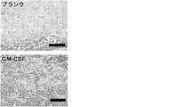


Classifications
-
- A—HUMAN NECESSITIES
- A61—MEDICAL OR VETERINARY SCIENCE; HYGIENE
- A61K—PREPARATIONS FOR MEDICAL, DENTAL OR TOILETRY PURPOSES
- A61K39/00—Medicinal preparations containing antigens or antibodies
- A61K39/0005—Vertebrate antigens
- A61K39/0008—Antigens related to auto-immune diseases; Preparations to induce self-tolerance
-
- A—HUMAN NECESSITIES
- A61—MEDICAL OR VETERINARY SCIENCE; HYGIENE
- A61K—PREPARATIONS FOR MEDICAL, DENTAL OR TOILETRY PURPOSES
- A61K39/00—Medicinal preparations containing antigens or antibodies
- A61K39/0005—Vertebrate antigens
- A61K39/0011—Cancer antigens
-
- A—HUMAN NECESSITIES
- A61—MEDICAL OR VETERINARY SCIENCE; HYGIENE
- A61K—PREPARATIONS FOR MEDICAL, DENTAL OR TOILETRY PURPOSES
- A61K39/00—Medicinal preparations containing antigens or antibodies
- A61K39/02—Bacterial antigens
- A61K39/102—Pasteurellales, e.g. Actinobacillus, Pasteurella; Haemophilus
-
- A—HUMAN NECESSITIES
- A61—MEDICAL OR VETERINARY SCIENCE; HYGIENE
- A61K—PREPARATIONS FOR MEDICAL, DENTAL OR TOILETRY PURPOSES
- A61K39/00—Medicinal preparations containing antigens or antibodies
- A61K39/35—Allergens
-
- A—HUMAN NECESSITIES
- A61—MEDICAL OR VETERINARY SCIENCE; HYGIENE
- A61K—PREPARATIONS FOR MEDICAL, DENTAL OR TOILETRY PURPOSES
- A61K39/00—Medicinal preparations containing antigens or antibodies
- A61K39/39—Medicinal preparations containing antigens or antibodies characterised by the immunostimulating additives, e.g. chemical adjuvants
-
- A—HUMAN NECESSITIES
- A61—MEDICAL OR VETERINARY SCIENCE; HYGIENE
- A61P—SPECIFIC THERAPEUTIC ACTIVITY OF CHEMICAL COMPOUNDS OR MEDICINAL PREPARATIONS
- A61P1/00—Drugs for disorders of the alimentary tract or the digestive system
- A61P1/02—Stomatological preparations, e.g. drugs for caries, aphtae, periodontitis
-
- A—HUMAN NECESSITIES
- A61—MEDICAL OR VETERINARY SCIENCE; HYGIENE
- A61P—SPECIFIC THERAPEUTIC ACTIVITY OF CHEMICAL COMPOUNDS OR MEDICINAL PREPARATIONS
- A61P25/00—Drugs for disorders of the nervous system
-
- A—HUMAN NECESSITIES
- A61—MEDICAL OR VETERINARY SCIENCE; HYGIENE
- A61P—SPECIFIC THERAPEUTIC ACTIVITY OF CHEMICAL COMPOUNDS OR MEDICINAL PREPARATIONS
- A61P29/00—Non-central analgesic, antipyretic or antiinflammatory agents, e.g. antirheumatic agents; Non-steroidal antiinflammatory drugs [NSAID]
-
- A—HUMAN NECESSITIES
- A61—MEDICAL OR VETERINARY SCIENCE; HYGIENE
- A61P—SPECIFIC THERAPEUTIC ACTIVITY OF CHEMICAL COMPOUNDS OR MEDICINAL PREPARATIONS
- A61P3/00—Drugs for disorders of the metabolism
- A61P3/08—Drugs for disorders of the metabolism for glucose homeostasis
- A61P3/10—Drugs for disorders of the metabolism for glucose homeostasis for hyperglycaemia, e.g. antidiabetics
-
- A—HUMAN NECESSITIES
- A61—MEDICAL OR VETERINARY SCIENCE; HYGIENE
- A61P—SPECIFIC THERAPEUTIC ACTIVITY OF CHEMICAL COMPOUNDS OR MEDICINAL PREPARATIONS
- A61P37/00—Drugs for immunological or allergic disorders
- A61P37/02—Immunomodulators
-
- A—HUMAN NECESSITIES
- A61—MEDICAL OR VETERINARY SCIENCE; HYGIENE
- A61P—SPECIFIC THERAPEUTIC ACTIVITY OF CHEMICAL COMPOUNDS OR MEDICINAL PREPARATIONS
- A61P37/00—Drugs for immunological or allergic disorders
- A61P37/02—Immunomodulators
- A61P37/04—Immunostimulants
-
- A—HUMAN NECESSITIES
- A61—MEDICAL OR VETERINARY SCIENCE; HYGIENE
- A61P—SPECIFIC THERAPEUTIC ACTIVITY OF CHEMICAL COMPOUNDS OR MEDICINAL PREPARATIONS
- A61P37/00—Drugs for immunological or allergic disorders
- A61P37/02—Immunomodulators
- A61P37/06—Immunosuppressants, e.g. drugs for graft rejection
-
- A—HUMAN NECESSITIES
- A61—MEDICAL OR VETERINARY SCIENCE; HYGIENE
- A61P—SPECIFIC THERAPEUTIC ACTIVITY OF CHEMICAL COMPOUNDS OR MEDICINAL PREPARATIONS
- A61P37/00—Drugs for immunological or allergic disorders
- A61P37/08—Antiallergic agents
-
- A—HUMAN NECESSITIES
- A61—MEDICAL OR VETERINARY SCIENCE; HYGIENE
- A61K—PREPARATIONS FOR MEDICAL, DENTAL OR TOILETRY PURPOSES
- A61K39/00—Medicinal preparations containing antigens or antibodies
- A61K2039/51—Medicinal preparations containing antigens or antibodies comprising whole cells, viruses or DNA/RNA
- A61K2039/515—Animal cells
- A61K2039/5158—Antigen-pulsed cells, e.g. T-cells
-
- A—HUMAN NECESSITIES
- A61—MEDICAL OR VETERINARY SCIENCE; HYGIENE
- A61K—PREPARATIONS FOR MEDICAL, DENTAL OR TOILETRY PURPOSES
- A61K39/00—Medicinal preparations containing antigens or antibodies
- A61K2039/555—Medicinal preparations containing antigens or antibodies characterised by a specific combination antigen/adjuvant
- A61K2039/55511—Organic adjuvants
- A61K2039/55522—Cytokines; Lymphokines; Interferons
-
- A—HUMAN NECESSITIES
- A61—MEDICAL OR VETERINARY SCIENCE; HYGIENE
- A61K—PREPARATIONS FOR MEDICAL, DENTAL OR TOILETRY PURPOSES
- A61K39/00—Medicinal preparations containing antigens or antibodies
- A61K2039/555—Medicinal preparations containing antigens or antibodies characterised by a specific combination antigen/adjuvant
- A61K2039/55511—Organic adjuvants
- A61K2039/55561—CpG containing adjuvants; Oligonucleotide containing adjuvants
-
- A—HUMAN NECESSITIES
- A61—MEDICAL OR VETERINARY SCIENCE; HYGIENE
- A61K—PREPARATIONS FOR MEDICAL, DENTAL OR TOILETRY PURPOSES
- A61K39/00—Medicinal preparations containing antigens or antibodies
- A61K2039/57—Medicinal preparations containing antigens or antibodies characterised by the type of response, e.g. Th1, Th2
- A61K2039/577—Medicinal preparations containing antigens or antibodies characterised by the type of response, e.g. Th1, Th2 tolerising response
-
- A—HUMAN NECESSITIES
- A61—MEDICAL OR VETERINARY SCIENCE; HYGIENE
- A61K—PREPARATIONS FOR MEDICAL, DENTAL OR TOILETRY PURPOSES
- A61K39/00—Medicinal preparations containing antigens or antibodies
- A61K2039/70—Multivalent vaccine
Landscapes
- Health & Medical Sciences (AREA)
- Life Sciences & Earth Sciences (AREA)
- Chemical & Material Sciences (AREA)
- Immunology (AREA)
- Animal Behavior & Ethology (AREA)
- Veterinary Medicine (AREA)
- Medicinal Chemistry (AREA)
- Public Health (AREA)
- General Health & Medical Sciences (AREA)
- Pharmacology & Pharmacy (AREA)
- Chemical Kinetics & Catalysis (AREA)
- General Chemical & Material Sciences (AREA)
- Epidemiology (AREA)
- Mycology (AREA)
- Microbiology (AREA)
- Nuclear Medicine, Radiotherapy & Molecular Imaging (AREA)
- Bioinformatics & Cheminformatics (AREA)
- Engineering & Computer Science (AREA)
- Organic Chemistry (AREA)
- Diabetes (AREA)
- Rheumatology (AREA)
- Oncology (AREA)
- Hematology (AREA)
- Emergency Medicine (AREA)
- Pulmonology (AREA)
- Biomedical Technology (AREA)
- Neurology (AREA)
- Neurosurgery (AREA)
- Transplantation (AREA)
- Obesity (AREA)
- Endocrinology (AREA)
- Pain & Pain Management (AREA)
- Medicines Containing Antibodies Or Antigens For Use As Internal Diagnostic Agents (AREA)
- Medicines That Contain Protein Lipid Enzymes And Other Medicines (AREA)
- Pharmaceuticals Containing Other Organic And Inorganic Compounds (AREA)
- Materials For Medical Uses (AREA)
- Medicinal Preparation (AREA)
- Acyclic And Carbocyclic Compounds In Medicinal Compositions (AREA)
- Medicines Containing Material From Animals Or Micro-Organisms (AREA)
- Micro-Organisms Or Cultivation Processes Thereof (AREA)
Description
連邦政府資金による研究に関する言明
本発明は、一つには、国立衛生研究所が与える助成金番号5R01DE019917-02の下で米国政府による資金援助を受けてなされた。米国政府は本発明において一定の権利を有する。
STATEMENT REGARDING FEDERALLY SPONSORED RESEARCH This invention was made in part with funding from the US Government under grant number 5R01DE019917-02 awarded by the National Institutes of Health. The US government has certain rights in this invention.
発明の分野
本発明は免疫寛容に関する。
The present invention relates to immune tolerance.
背景
異常または調節異常な免疫反応は多数の病的状態の発症機序である。そのような状態には、自己免疫障害、および慢性炎症によって特徴付けられる状態が含まれる。
BACKGROUND Abnormal or dysregulated immune responses are the pathogenesis of many pathological conditions. Such conditions include autoimmune disorders and conditions characterized by chronic inflammation.
自己免疫は、免疫系が宿主の組織または細胞を異物と誤認識する状態である。自己免疫疾患は世界中で何百万人もの個体に影響を与えている。よく見られる自己免疫障害には、1型糖尿病、クローン病、関節リウマチおよび多発性硬化症が含まれる。
Autoimmunity is a condition in which the immune system misrecognizes host tissues or cells as foreign. Autoimmune diseases affect millions of individuals worldwide. Common autoimmune disorders include
慢性炎症はがん、糖尿病、うつ病、心臓疾患、脳卒中、アルツハイマー病、歯周炎および多くの他の病態に関係があるとされている。異常または調節異常な免疫反応はまた、喘息およびアレルギーに関係しており、例えば、喘息は、多くのアレルゲンにより誘因される、よく見られる疾患である。 Chronic inflammation has been implicated in cancer, diabetes, depression, heart disease, stroke, Alzheimer's disease, periodontitis and many other conditions. Abnormal or dysregulated immune responses are also associated with asthma and allergies, for example asthma is a common disease that is triggered by many allergens.
概要
本発明は、自己免疫、アレルギー/喘息、および体内での慢性的または不適切な炎症、例えば、組織/臓器損傷および破壊をもたらす炎症の長年の臨床的問題に対する解決策を提供する。本組成物および方法は、個体の免疫反応を病的反応または致命的反応から離し、生産反応または非損傷反応へと方向づける。樹状細胞(DC)は自己免疫疾患に対する防御において主要な役割を果たす。また、調節性T細胞(Treg)は自己抗原または外来抗原に対する有害な免疫病理学的反応の阻害において重要な役目を果たす。これらの細胞種は、免疫反応の方向を変えて非炎症状態および非破壊状態を提供する目的で操作をする標的となる。
Overview The present invention provides a solution to many years of clinical problems of autoimmunity, allergy / asthma, and chronic or inappropriate inflammation in the body, such as inflammation resulting in tissue / organ damage and destruction. The present compositions and methods separate an individual's immune response from a pathological or fatal response and direct it to a production or undamaged response. Dendritic cells (DC) play a major role in the defense against autoimmune diseases. Regulatory T cells (Treg) also play an important role in inhibiting adverse immunopathological responses to self or foreign antigens. These cell types are targeted for manipulation in order to change the direction of the immune response to provide non-inflammatory and non-destructive conditions.
したがって、本発明は、抗原、動員組成物および寛容原を含む足場組成物を特徴とする。この足場組成物は自己免疫の低減に有用である。抗原は、精製された組成物(例えば、タンパク質)であるか、または望ましくない免疫反応が導かれる細胞から調製された細胞溶解物である。例示的な動員組成物には、顆粒球マクロファージコロニー刺激因子(GM-CSF; AAA52578)、FMS様チロシンキナーゼ3リガンド(AAA17999.1)、N-ホルミルペプチド、フラクタルキン(P78423)または単球走化性タンパク質-1 (P13500.1)が含まれる。例示的な寛容原(すなわち、免疫反応の低減または免疫寛容を誘導する作用物質)には、胸腺間質性リンパ球新生因子(TSLP; Q969D9.1))、デキサメタゾン、ビタミンD、レチノイン酸、ラパマイシン、アスピリン、形質転換増殖因子β(P01137)、インターロイキン-10 (P01137)、血管作動性腸管ペプチド(CAI21764)または血管内皮増殖因子(AAL27435)が含まれる。足場には任意で、toll様受容体(TLR)アゴニストのようなTh1促進剤、例えば、CpGのようなポリヌクレオチドがさらに含まれてもよい。Th1促進剤は、病原体関連分子パターン(PAMP)または微生物関連分子パターン(MAMP)またはアラルミンによって特徴付けられることが多い。PAMPまたはMAMPは、TLRを介して先天性免疫系の細胞により認識される、病原体の群と関連する分子である。例えば、細菌性リポ多糖類(LPS)、つまり細菌のグラム陰性細菌細胞膜上に見出される内毒素は、TLR 4によって認識される。他のPAMPには、細菌フラジェリン、グラム陽性細菌由来のリポタイコ酸、ペプチドグリカン、およびウイルスと通常関連する核酸変種、例えば、二本鎖RNA (dsRNA)または非メチル化CpGモチーフなどが含まれる。したがって、さらなる例示的なTh1促進剤には、それぞれポリ(I:C)、LPS/MPLA (一リン酸脂質A)、またはイミキモドなどの、TLR 3、4、または7のアゴニストが含まれる。例示的なTLRリガンドには、以下の化合物が含まれる: TLR7リガンド(ヒトおよびマウスTLR7)-CL264 (アデニン類似体)、ガルジキモド(商標) (イミダゾキノリン化合物)、イミキモド(イミダゾキノリン化合物)およびロキソリビン(グアノシン類似体); TLR8リガンド(ヒトTLR8およびマウスTLR7) - 一本鎖RNA; 大腸菌(E. coli) RNA; TLR7/8リガンド - (ヒト、マウスTLR7およびヒトTLR8)-CL075 (チアゾロキノリン化合物)、CL097 (水溶性R848)、イミダゾキノリン化合物、ポリ(dT) (チミジンホモ重合体ホスホロチオエートオリゴヌクレオチド(ODN))、ならびにR848 (イミダゾキノリン化合物)。
The invention thus features a scaffold composition comprising an antigen, a recruitment composition and a tolerogen. This scaffold composition is useful for reducing autoimmunity. An antigen is a purified composition (eg, a protein) or a cell lysate prepared from cells that induce an unwanted immune response. Exemplary mobilization compositions include granulocyte macrophage colony stimulating factor (GM-CSF; AAA52578), FMS-
足場は、その中に負荷された因子の持続放出を、時空間的に制御された形で媒介する。例えば、因子または抗原のボーラス送達と比べ、数日(例えば、1、2、3、4、5、7、10、12、14日またはそれ以上)にわたって因子が放出される。ボーラス送達は、体内での短期提示のために効果がほとんどもしくは全くないか、逆効果をもたらすか、または非常に高用量が提供されるならば望ましくない免疫反応をもたらすことが多いのに対し、足場による送達ではそのような事象が回避される。好ましくは、足場は、非炎症性の重合体組成物、例えば、アルギン酸塩、ポリ(エチレングリコール)、ヒアルロン酸、コラーゲン、ゼラチン、ポリ(ビニルアルコール)、フィブリン、ポリ(グルタミン酸)、両親媒性ペプチド、シルク、フィブロネクチン、キチン、ポリ(メタクリル酸メチル)、ポリ(テレフタル酸エチレン)、ポリ(ジメチルシロキサン)、ポリ(テトラフルオロエチレン)、ポリエチレン、ポリウレタン、ポリ(グリコール酸)、ポリ(乳酸)、ポリ(カプロラクトン)、ポリ(ラクチド-コ-グリコリド)、ポリジオキサノン、ポリグリコナート、BAK; ポリ(オルトエステルI)、ポリ(オルトエステル)II、ポリ(オルトエステル)III、ポリ(オルトエステル)IV、ポリプロピレンフマレート、ポリ[(カルボキシフェノキシ)プロパン-セバシン酸]、ポリ[ピロメリチルイミドアラニン-コ-1,6-ビス(p-カルボキシフェノキシ)ヘキサン]、ポリホスファゼン、デンプン、セルロース、アルブミン、ポリヒドロキシアルカノエート、または当技術分野において公知の他のもの(Polymers as Biomaterials for Tissue Engineering and Controlled Drug Delivery. Lakshmi S. Nair & Cato T. Laurencin, Adv Biochem Engin/Biotechnol (2006) 102: 47-90 DOI 10.1007/b137240)などから作出される。あるいは、低レベルの炎症をもたらす重合体組成物も、樹状細胞の動員および/または活性化に、特に該細胞をTh1反応の方向へ偏向させるのに役立ちうるため、有用でありうる。ポリ(ラクチド)、ポリ(グリコリド)、それらの共重合体および他のさまざまな医用重合体もこの関連で有用でありうる。セラミック材料または金属材料を利用して、制御可能な形でこれらの因子を提示することもできる。例えば、リン酸カルシウム材料が有用である。骨との関連では、シリカまたは他のセラミックも有用である。 The scaffold mediates the sustained release of factors loaded therein in a spatiotemporally controlled manner. For example, the factor is released over several days (eg, 1, 2, 3, 4, 5, 7, 10, 12, 14 days or more) compared to bolus delivery of the factor or antigen. Bolus delivery often has little or no effect due to short-term presentation in the body, has an adverse effect, or often produces an undesirable immune response if very high doses are provided. Delivery by the scaffold avoids such an event. Preferably, the scaffold is a non-inflammatory polymer composition such as alginate, poly (ethylene glycol), hyaluronic acid, collagen, gelatin, poly (vinyl alcohol), fibrin, poly (glutamic acid), amphiphilic peptide , Silk, fibronectin, chitin, poly (methyl methacrylate), poly (ethylene terephthalate), poly (dimethylsiloxane), poly (tetrafluoroethylene), polyethylene, polyurethane, poly (glycolic acid), poly (lactic acid), poly (Caprolactone), poly (lactide-co-glycolide), polydioxanone, polyglycolate, BAK; poly (orthoester I), poly (orthoester) II, poly (orthoester) III, poly (orthoester) IV, polypropylene Fumarate, poly [(carboxyphenoxy) propane-sebacic acid], poly [pyromelli Luimalanine-co-1,6-bis (p-carboxyphenoxy) hexane], polyphosphazene, starch, cellulose, albumin, polyhydroxyalkanoate, or others known in the art (Polymers as Biomaterials for Tissue Engineering and Controlled Drug Delivery. Lakshmi S. Nair & Cato T. Laurencin, Adv Biochem Engin / Biotechnol (2006) 102: 47-90 DOI 10.1007 / b137240). Alternatively, polymeric compositions that result in low levels of inflammation may also be useful because they can help to recruit and / or activate dendritic cells, particularly to deflect the cells in the direction of the Th1 response. Poly (lactide), poly (glycolide), their copolymers and various other medical polymers may also be useful in this context. Ceramic or metallic materials can also be utilized to present these factors in a controllable manner. For example, calcium phosphate materials are useful. Silica or other ceramics are also useful in the context of bone.
一部の例では、複合材料を利用することができる。例えば、免疫活性化因子(例えば、抗原、寛容原またはTh1促進剤)をポリ(ラクチド-コ-グリコリド) (PLG)ミクロスフェアのようなミクロスフェアの中にカプセル封入し、これを次いで、アルギン酸塩ゲルのようなヒドロゲルの中に分散させる。細胞、例えば、DCおよび/またはTregは、表面もしくは表面の近くに、または足場の中に動員され、そこで該細胞は上記の抗原および他の因子に曝露されながら一定時間存留することができ、その後、リンパ節のような体組織に移動し、そこで該細胞は免疫寛容を誘導するように機能する。あるいは、細胞を伴う足場が二次リンパ器官の模倣体を作出しうる。負荷された足場との接触の後、そのような細胞は活性化されて、免疫反応をTh1/Th2/Th17反応(自己免疫および慢性炎症)からTreg反応へ、または(アレルギー/喘息の場合)病原性のTh2状態からTh1状態の方向へと方向を変えられる。免疫反応をTh2反応から離し、Treg反応の方向へと方向づけることにより、アレルギー、喘息における臨床的有益性が得られる。自己免疫の場合、治療方法は、自己免疫疾患を患っているかまたは発症するリスクがある対象を特定し、負荷された足場(抗原(自己抗原)+動員組成物+寛容原)を対象に投与し、それによってTh1/Th17からT調節性偏向免疫反応への免疫反応の変化をもたらすことにより行われる。アレルギー/喘息の場合、治療方法は、アレルギー反応または喘息を患っているかまたは発症するリスクがある対象を特定し、負荷された足場(抗原(アレルゲン)+動員組成物+アジュバント(Th1促進性のアジュバント))を対象に投与し、それによって(アレルギー/喘息) Th2反応からTh1偏向免疫反応への免疫反応の変化をもたらすことにより行われる。 In some examples, composite materials can be utilized. For example, immune activators (e.g., antigens, tolerogens or Th1 promoters) are encapsulated in microspheres such as poly (lactide-co-glycolide) (PLG) microspheres which are then alginate. Disperse in a hydrogel such as a gel. Cells, such as DC and / or Treg, are mobilized at or near the surface or into the scaffold, where they can remain for a period of time while being exposed to the above antigens and other factors. Migrates to body tissues such as lymph nodes, where the cells function to induce immune tolerance. Alternatively, scaffolds with cells can create mimics of secondary lymphoid organs. After contact with the loaded scaffold, such cells are activated to shift the immune response from a Th1 / Th2 / Th17 response (autoimmunity and chronic inflammation) to a Treg response or (in the case of allergy / asthma) pathogenesis The direction can be changed from the sex Th2 state to the Th1 state. By separating the immune response from the Th2 response and in the direction of the Treg response, clinical benefits in allergies and asthma can be obtained. In the case of autoimmunity, the treatment method identifies a subject suffering from or at risk of developing an autoimmune disease and administering a loaded scaffold (antigen (self-antigen) + mobilization composition + tolerogen) to the subject. This is done by bringing about a change in the immune response from Th1 / Th17 to a T-regulated biased immune response. In the case of allergy / asthma, treatment methods identify subjects with or at risk of developing an allergic reaction or asthma, loaded scaffold (antigen (allergen) + mobilization composition + adjuvant (Th1-promoted adjuvant) )) Is administered to the subject, thereby (allergy / asthma) by causing a change in the immune response from a Th2 response to a Th1-biased immune response.
それゆえ、Th1媒介性の抗原特異的免疫反応へと選択的に方向づける方法は、抗原、動員組成物、およびアジュバントを含む足場を対象に投与することにより行われる。樹状細胞は、足場に動員され、抗原に曝露され、その後、曝露に基づき病原性のTh2免疫反応と比べてTh1免疫反応が選択的に生じるように教育/活性化された状態で、足場から離れて対象の組織へ移動する。結果として、免疫反応はTh2経路に対してTh1経路の方へ効果的に傾けられ、または偏向される。そのような偏向は、局所的または対象由来の血液もしくは血清のような体液中のサイトカインの量およびレベルを測定することにより検出される。例えば、Th1反応はインターフェロン-γ(IFN-γ)の増加によって特徴付けられる。上記のように、足場には任意で、Th1促進剤が含まれてもよい。 Thus, a method of selectively directing to a Th1-mediated antigen-specific immune response is performed by administering to the subject a scaffold comprising an antigen, a recruitment composition, and an adjuvant. Dendritic cells are mobilized to the scaffold, exposed to the antigen, and then educated / activated from the scaffold, educated and activated to selectively generate a Th1 immune response relative to the pathogenic Th2 immune response based on the exposure. Move away to the target organization. As a result, the immune response is effectively tilted or biased towards the Th1 pathway relative to the Th2 pathway. Such bias is detected by measuring the amount and level of cytokines in body fluids such as blood or serum locally or from the subject. For example, the Th1 response is characterized by an increase in interferon-γ (IFN-γ). As noted above, the scaffold may optionally include a Th1 promoter.
本組成物および方法はヒト対象の処置に適している; しかしながら、本組成物および方法はまた、イヌおよびネコのような伴侶動物、ならびにウシ、ウマ、ヒツジ、ヤギ、ブタのような家畜にも適用可能である。 The compositions and methods are suitable for the treatment of human subjects; however, the compositions and methods are also applicable to companion animals such as dogs and cats and domestic animals such as cattle, horses, sheep, goats, pigs. Applicable.
足場は、個体の免疫系を操作して、異常な、誤った、またはその他の点で不適切な免疫反応、例えば、組織損傷または破壊を引き起こす免疫反応によって特徴付けられるいくつかの病的状態を処置するのに有用である。そのような状態には自己免疫疾患が含まれる。例えば、自己免疫障害の重症度を低減させる方法は、自己免疫障害を患っている対象を特定し、抗原(例えば、精製された抗原または加工処理された細胞溶解物)、動員組成物および寛容原を含む足場組成物を対象に投与することによって行われる。好ましくは、抗原は、病的自己免疫反応が導かれる細胞に由来するかまたは関連する。一つの例では、自己免疫障害は1型糖尿病であり、抗原は膵臓細胞関連ペプチドまたはタンパク質抗原、例えば、インスリン、プロインスリン、グルタミン酸デカルボキシラーゼ-65 (GAD65)、インスリノーマ関連タンパク質2、熱ショックタンパク質60、ZnT8、および膵島特異的グルコース-6-ホスファターゼ触媒サブユニット関連タンパク質またはAnderson et al., Annual Review of Immunology, 2005. 23: p. 447-485; またはWaldron-Lynch et al., Endocrinology and Metabolism Clinics of North America, 2009.38(2): p.303)に記述されているその他のものを含む。別の例では、自己免疫障害は多発性硬化症であり、ペプチドまたはタンパク質抗原はミエリン塩基性タンパク質、ミエリンプロテオリピドタンパク質、ミエリン関連オリゴデンドロサイト塩基性タンパク質、および/またはミエリンオリゴデンドロサイト糖タンパク質を含む。自己免疫疾患/状態のさらなる例としては、クローン病、関節リウマチ、全身性エリテマトーデス、強皮症、円形脱毛症、抗リン脂質抗体症候群、自己免疫性肝炎、セリアック病、グレーブス病、ギランバレー症候群、橋本病、溶血性貧血、特発性血小板減少性紫斑病、炎症性腸疾患、潰瘍性大腸炎、炎症性筋疾患、多発性筋炎、重症筋無力症、原発性胆汁性肝硬変症、乾癬、シェーグレン症候群、白斑、痛風、セリアック病、アトピー性皮膚炎、尋常性座瘡、自己免疫性肝炎および自己免疫性膵炎が挙げられる。
Scaffolds manipulate an individual's immune system to display some pathological condition characterized by an abnormal, false, or otherwise inappropriate immune response, such as an immune response that causes tissue damage or destruction. Useful to treat. Such conditions include autoimmune diseases. For example, a method for reducing the severity of an autoimmune disorder identifies a subject suffering from an autoimmune disorder, an antigen (e.g., a purified antigen or a processed cell lysate), a mobilization composition, and a tolerogen. By administering to the subject a scaffold composition comprising Preferably, the antigen is derived from or associated with a cell from which a pathological autoimmune response is induced. In one example, the autoimmune disorder is
足場はまた、慢性炎症障害またはアレルギー/喘息などの、他の免疫障害を処置するか、またはその重症度を低減させるのに有用である。これに関連して、本方法は、慢性炎症またはアレルギー/喘息を患っている対象を特定する段階、ならびにその障害と関連する抗原、動員組成物およびアジュバントを含む足場組成物を対象に投与する段階を含む。ワクチンは、アレルギーに対する病原反応を低減/除去することによって急性喘息悪化または発作を低減させるのに有用である。アレルギーおよび喘息の場合、抗原は、対象においてアレルギー症状、例えば、ヒスタミン放出もしくはアナフィラキシーを引き起こすか、または急性喘息発作を誘発するアレルゲンを含む。アレルゲンは、例えば、(Amb a 1 (ブタクサアレルゲン)、Der p2 (ヤケヒョウダニ(Dermatophagoides pteronyssinus)アレルゲン、つまりイエダニの主要種かつ喘息の主な誘導因子)、Betv 1 (主要な白樺(シラカバ(Betula verrucosa)花粉抗原)、ハンノキ(Alnus glutinosa) (ハンノキ)由来のAln g I、塘蒿(Apium graveolens) (セロリ)由来のApi G I、セイヨウシデ(Carpinus betulus) (ホームビーン)由来のCar b I、セイヨウハシバミ(Corylus avellana) (西洋ハシバミ)由来のCor a I、セイヨウリンゴ(Malus domestica) (リンゴ)由来のMal d I、ホスホリパーゼA2 (ハチ毒)、ヒアルロニダーゼ(ハチ毒)、アレルゲンC (ハチ毒)、Api m 6 (ハチ毒)、Fel d 1 (ネコ)、Fel d 4 (ネコ)、Gal d 1 (卵)、オボトランスフェリン(卵)、リゾチーム(卵)、オボアルブミン(卵)、カゼイン(乳)および乳漿タンパク質(α-ラクトアルブミンおよびβ-ラクトグロブリン、乳)、ならびにAra h 1〜Ara h 8 (ピーナッツ)を含む。本組成物および方法は、多数のアレルギー状態、例えば、ラテックスアレルギー; ブタクサ、牧草、樹木花粉およびイエダニに対するアレルギー; 乳、卵、ピーナッツ、樹木ナッツ(例えば、クルミ、アーモンド、カシュー、ピスタチオ、ペカン)、小麦、魚および貝に対するアレルギーのような食物アレルギー; 枯草熱; ならびに伴侶動物、昆虫、例えば、ハチ毒/ハチ刺されまたは蚊刺されに対するアレルギーの重症度を低減させるのにならびにそれらのアレルギーを処置するのに有用である。好ましくは、抗原は腫瘍抗原または腫瘍溶解物ではない。 Scaffolds are also useful for treating or reducing the severity of other immune disorders, such as chronic inflammatory disorders or allergies / asthma. In this regard, the method includes identifying a subject suffering from chronic inflammation or allergy / asthma and administering to the subject a scaffold composition comprising antigens, mobilization compositions and adjuvants associated with the disorder. including. Vaccines are useful for reducing acute asthma exacerbations or seizures by reducing / eliminating pathogenic responses to allergies. In the case of allergies and asthma, the antigen includes allergens that cause allergic symptoms, such as histamine release or anaphylaxis, or induce an acute asthma attack in the subject. Allergens include, for example, (Amb a 1 (Ragweed allergen), Der p2 (Dermatophagoides pteronyssinus allergen, the main species of house dust mite and the main inducer of asthma), Betv 1 (Betula verrucosa) Pollen antigen), Alng g from Alnus glutinosa (Alnus), Api GI from Apium graveolens (celery), Car b I from Carpinus betulus (Home Bean), Hazel Cor a I from Corylus avellana), Mal d I from Malus domestica apple, phospholipase A2 (bee venom), hyaluronidase (bee venom), allergen C (bee venom), Api m 6 (bee venom), Fel d 1 (cat), Fel d 4 (cat), Gal d 1 (egg), ovotransferrin (egg), lysozyme (egg), ovalbumin (egg), casein (milk) and milk Serum protein (α-lactalbumin and β- The composition and method includes a number of allergic conditions such as latex allergies; allergy to ragweed, grass, tree pollen and house dust mites; milk, Food allergies such as allergies to eggs, peanuts, tree nuts (e.g. walnuts, almonds, cashews, pistachios, pecans), wheat, fish and shellfish; hay fever; and companion animals, insects e.g. bee venom / bee stings or Useful to reduce the severity of allergies to mosquito bites as well as to treat those allergies Preferably, the antigen is not a tumor antigen or tumor lysate.
また、本発明のなかには、抗原特異的な免疫寛容を誘発して疾患の重症度を低減させるための、上記の負荷された足場および対象への注射または移植用の薬学的に許容される賦形剤を含むワクチンがある。他の投与経路には、足場を含む皮膚パッチを局所的に貼付すること、または個体の肺もしくは鼻道へエアロゾルにより足場組成物を送達することが含まれる。 Also included in the present invention is a pharmaceutically acceptable excipient for injection or transplantation into the above-mentioned loaded scaffold and subject to induce antigen-specific immune tolerance and reduce disease severity. There are vaccines that contain drugs. Other routes of administration include topically applying a skin patch containing the scaffold or delivering the scaffold composition by aerosol to the lungs or nasal passages of the individual.
本足場およびシステムは、上記の状態に加えて、歯周炎の処置に有用である。樹状細胞を動員してその活性化を非炎症性の表現型へと促す、インビボで用いるための生体材料システムの一例には、生体材料マトリックスまたは足場、例えば、アルギン酸塩のようなヒドロゲル、および歯周炎のような歯または歯周の状態で用いるためのGM-CSFまたは胸腺間質性リンパ球新生因子(TSLP)などの生物活性因子が含まれる。歯周炎は、歯周靱帯、セメント質および歯槽骨を含む歯の支持構造に影響を与える破壊性の疾患である。歯周炎は、ポルフィロモナス・ジンジバリス(Porphyromonas gingivalis)、プレボテラ・インテルメディア(Prevotella intermedia)、バクテロイデス・フォーサイサス(Bacteroides forsythus)、アクチノバシラス・アクチノミセテムコミタンス(Actinobacillus actinomycetemcomitans)のようなグラム陰性細菌、ならびにペプトストレプトコッカス・ミクロス(Peptostreptococcus micros)およびストレプトコッカス・インターメジウス(Streptococcus intermedius)のようなグラム陽性細菌による、慢性の混合感染を示す。 The scaffold and system are useful for the treatment of periodontitis in addition to the conditions described above. An example of a biomaterial system for in vivo use that recruits dendritic cells and drives its activation to a non-inflammatory phenotype includes a biomaterial matrix or scaffold, eg, a hydrogel such as alginate, and Bioactive factors such as GM-CSF or thymic stromal lymphocyte neogenesis factor (TSLP) for use in teeth or periodontal conditions such as periodontitis are included. Periodontitis is a destructive disease that affects the support structure of teeth, including periodontal ligaments, cementum and alveolar bone. Periodontitis is caused by Porphyromonas gingivalis, Prevotella intermedia, Bacteroides forsythus, Actinobacillus actinomycetemcomitans negative bacteria such as Actinobacillus actinomycetemcomitans. And chronic mixed infections by Gram positive bacteria such as Peptostreptococcus micros and Streptococcus intermedius.
本方法は歯周疾患での調節性T細胞による炎症調節に取り組む。DCは調節性T細胞の誘導に加え、エフェクタ細胞においてアネルギーおよびアポトーシスを引き起こすことができる。他の機構には、Th1、Th2、Th17およびTreg間のバランスを変化させることが含まれる。例えば、TSLPはTh2免疫を増強することが公知であり、Treg数を増加させることに加えてTh2反応を増大しうる。本材料は多数の寛容原性DCを動員かつプログラミングして調節性T細胞分化を促進し、歯周炎のげっ歯類モデルで炎症を媒介する。より具体的には、適切に活性化されたDCのリンパ節への動員、適切な活性化、および移動は、大量の調節性T細胞の形成と、エフェクタT細胞の減少とをもたらし、歯周炎症の低減をもたらす。 This method addresses the modulation of inflammation by regulatory T cells in periodontal disease. In addition to inducing regulatory T cells, DC can cause anergy and apoptosis in effector cells. Other mechanisms include changing the balance between Th1, Th2, Th17 and Treg. For example, TSLP is known to enhance Th2 immunity and can increase the Th2 response in addition to increasing the number of Tregs. This material mobilizes and programs numerous tolerogenic DCs to promote regulatory T cell differentiation and mediates inflammation in a rodent model of periodontitis. More specifically, recruitment of properly activated DCs to lymph nodes, proper activation, and migration results in the formation of large amounts of regulatory T cells and the reduction of effector T cells, and periodontal Results in reduced inflammation.
本発明の別の局面では、再生の促進に合わせた炎症の媒介について取り組む。具体的には、材料システムから送達される、BMP-2をコードするプラスミドDNA (pDNA)であり、該材料システムは、炎症を抑制し、DC標的化を介して炎症を低減させ、かつ歯周炎のげっ歯類モデルで歯槽骨を再生するための誘導的手法の有効性を増強する。例えば、まず炎症を低減させ、その後、局所的なBMP-2発現の誘導を介して骨再生を能動的に指令する材料から、顕著な歯槽骨再生がもたらされる。 Another aspect of the invention addresses the mediation of inflammation in concert with the promotion of regeneration. Specifically, plasmid DNA (pDNA) encoding BMP-2 delivered from a material system, which suppresses inflammation, reduces inflammation via DC targeting, and periodontal To enhance the effectiveness of inductive techniques for regenerating alveolar bone in a rodent model of flame. For example, significant alveolar bone regeneration results from materials that first reduce inflammation and then actively command bone regeneration through induction of local BMP-2 expression.
本発明は、炎症主導型の歯周疾患進行を調節し、次いで、炎症抑制の成功の後に再生を能動的に推進して機能する材料を提供する。さらに、本明細書において記述される組成物および方法は、組織再生誘導法(GTR)のための新たな材料に容易につながりうる。細胞運動に対する物理的障壁を単に与えるだけの現行のGTR膜とは異なり、この新たな材料は、局所免疫および組織を再構築する細胞集団をインサイチュで能動的に調節する。より広くは、炎症は、シェーグレン症候群および他の自己免疫疾患、ならびに顎関節障害のいくつかの形態を含めて、歯科および医科におけるその他多くの臨床的課題を構成する一つの要素である。本発明は、炎症媒介性の組織破壊によって特徴付けられるこれらの疾患の多くを処置するうえで広い有用性を有する。さらに、本材料システムはまた、DC輸送、活性化、T細胞分化、および免疫系と炎症との間の関係を探索する基礎研究のための新規かつ有用なツールを提供する。上記の状態および疾患に加えて、本組成物および方法はまた、創傷治癒で、例えば、くすぶり続ける創傷を処置し、それによって免疫系を創傷の軽減および治癒の方向へ変化させるのに有用である。 The present invention provides materials that regulate inflammation-driven periodontal disease progression and then actively promote regeneration after successful inflammation suppression. Furthermore, the compositions and methods described herein can readily lead to new materials for tissue regeneration induction (GTR). Unlike current GTR membranes that merely provide a physical barrier to cell motility, this new material actively regulates local immunity and cell populations that remodel tissue in situ. More broadly, inflammation is one component that constitutes many other clinical challenges in dentistry and medicine, including Sjogren's syndrome and other autoimmune diseases, as well as some forms of temporomandibular disorders. The present invention has broad utility in treating many of these diseases characterized by inflammation-mediated tissue destruction. Furthermore, the material system also provides a new and useful tool for basic research exploring DC transport, activation, T cell differentiation, and the relationship between the immune system and inflammation. In addition to the above conditions and diseases, the present compositions and methods are also useful in wound healing, e.g., treating smoldering wounds, thereby changing the immune system in the direction of wound relief and healing. .
足場に負荷するのに用いられるポリペプチドおよび他の組成物は、それらが天然に存在する状態から精製されるか、または別の方法で加工処理/改変される。例えば、実質的に純粋なポリペプチド、因子またはその変種は、好ましくは、該ポリペプチドをコードする組み換え核酸の発現により、または該タンパク質を化学的に合成することにより得られる。ポリペプチドまたはタンパク質は、その天然状態において付随する夾雑物(タンパク質および他の天然の有機分子)から分離されている場合に実質的に純粋である。典型的には、ポリペプチドは、調製物中のタンパク質の少なくとも60重量%を構成する場合に実質的に純粋である。好ましくは、調製物中のタンパク質は、少なくとも75重量%、より好ましくは少なくとも90重量%、および最も好ましくは少なくとも99重量%である。純度は任意の適切な方法、例えば、カラムクロマトグラフィー、ポリアクリルアミドゲル電気泳動またはHPLC分析によって測定される。したがって、実質的に純粋なポリペプチドは、真核生物に由来するが、大腸菌もしくは別の原核生物、または該ポリペプチドが元は由来した真核生物以外の真核生物において産生される組み換えポリペプチドを含む。 Polypeptides and other compositions used to load the scaffold are either purified from their naturally occurring state or otherwise processed / modified. For example, a substantially pure polypeptide, factor or variant thereof is preferably obtained by expression of a recombinant nucleic acid encoding the polypeptide or by chemically synthesizing the protein. A polypeptide or protein is substantially pure when it is separated from its associated contaminants (proteins and other natural organic molecules). Typically, a polypeptide is substantially pure when it constitutes at least 60% by weight of the protein in the preparation. Preferably, the protein in the preparation is at least 75% by weight, more preferably at least 90% by weight, and most preferably at least 99% by weight. Purity is measured by any appropriate method, for example, column chromatography, polyacrylamide gel electrophoresis or HPLC analysis. Thus, a substantially pure polypeptide is derived from a eukaryote, but a recombinant polypeptide produced in E. coli or another prokaryote, or a eukaryote other than the eukaryote from which the polypeptide was originally derived. including.
場合によっては、本方法において用いられる樹状細胞または他の細胞、例えば、マクロファージ、B細胞、T細胞のような免疫細胞は、精製または単離される。細胞に関して、「単離された」という用語は、細胞が、一緒になって天然に存在している他の細胞種または細胞物質を実質的に含まないことを意味する。例えば、特定の組織種または表現型の細胞のサンプルは、これが細胞集団の少なくとも60%である場合に「実質的に純粋」である。好ましくは、調製物は細胞集団の少なくとも75%、より好ましくは少なくとも90%、および最も好ましくは少なくとも99%または100%である。純度は任意の適当な標準的方法により、例えば、蛍光活性化細胞選別(FACS)により測定される。他の状況では、細胞は加工処理され、例えば、破壊され/溶解され、その溶解物は、足場中の抗原として用いるために分画される。 In some cases, dendritic cells or other cells used in the present methods, eg, immune cells such as macrophages, B cells, T cells, are purified or isolated. With respect to cells, the term “isolated” means that the cells are substantially free of other cell types or cellular materials that naturally occur together. For example, a sample of cells of a particular tissue type or phenotype is “substantially pure” if it is at least 60% of the cell population. Preferably the preparation is at least 75% of the cell population, more preferably at least 90%, and most preferably at least 99% or 100%. Purity is measured by any appropriate standard method, eg, fluorescence activated cell sorting (FACS). In other situations, the cells are processed, eg, destroyed / lysed, and the lysate is fractionated for use as an antigen in the scaffold.
[本発明1001]
抗原、動員組成物、および寛容原を含む足場組成物。
[本発明1002]
Th1促進剤をさらに含む、本発明1001の組成物。
[本発明1003]
前記寛容原が、胸腺間質性リンパ球新生因子、デキサメタゾン、ビタミンD、レチノイン酸、ラパマイシン、アスピリン、形質転換増殖因子β、インターロイキン-10、血管作動性腸管ペプチドまたは血管内皮増殖因子を含む、本発明1001の組成物。
[本発明1004]
前記動員組成物が、GM-CSF、FMS様チロシンキナーゼ3リガンド、N-ホルミルペプチド、フラクタルキンまたは単球走化性タンパク質-1を含む、本発明1001の組成物。
[本発明1005]
前記Th1促進剤がtoll様受容体(TLR)アゴニストを含む、本発明1002の組成物。
[本発明1006]
前記TLRアゴニストがCpGを含む、本発明1005の組成物。
[本発明1007]
前記Th1促進剤が病原体関連分子パターン組成物またはアラルミンを含む、本発明1005の組成物。
[本発明1008]
前記Th1促進剤がTLR 3、4または7のアゴニストを含む、本発明1002の組成物。
[本発明1009]
前記抗原が自己抗原を含む、本発明1001の組成物。
[本発明1010]
アレルゲン、動員組成物およびTh1促進性のアジュバントを含む足場組成物。
[本発明1011]
Th1媒介性の抗原特異的免疫反応へと選択的に方向づける方法であって、該方法が、抗原、動員組成物および寛容原を含むゲル足場を対象に投与する段階を含み、樹状細胞またはTreg細胞が、該足場に動員され、該抗原に曝露され、かつ該足場から離れて該対象の組織へと移動し、Th2免疫反応と比べてTh1免疫反応が選択的に生じる、方法。
[本発明1012]
前記足場がTh1促進剤をさらに含む、本発明1011の方法。
[本発明1013]
自己免疫障害の重症度を低減させる方法であって、該方法が、自己免疫障害を患っている対象を特定する段階、ならびに抗原、動員組成物、および寛容原を含む足場組成物を該対象に投与する段階を含み、該抗原が、該障害と関連する病的自己免疫反応が導かれる細胞に由来する、方法。
[本発明1014]
前記自己免疫障害が1型糖尿病であり、かつ前記抗原が膵臓細胞抗原を含む、本発明1013の方法。
[本発明1015]
前記抗原が、インスリン、プロインスリン、グルタミン酸デカルボキシラーゼ-65 (GAD65)、インスリノーマ関連タンパク質2、熱ショックタンパク質60、ZnT8または膵島特異的グルコース-6-ホスファターゼ触媒サブユニットを含む、本発明1014の方法。
[本発明1016]
前記自己免疫障害が多発性硬化症である、本発明1013の方法。
[本発明1017]
前記抗原が、ミエリン塩基性タンパク質、ミエリン塩基性タンパク質、ミエリンプロテオリピドタンパク質、ミエリン関連オリゴデンドロサイト塩基性タンパク質、またはミエリンオリゴデンドロサイト糖タンパク質を含む、本発明1016の方法。
[本発明1018]
慢性炎症障害またはアレルギーを患っている対象を特定する段階、ならびに該障害またはアレルギーと関連する抗原、動員組成物、およびアジュバントを含む足場組成物を該対象に投与する段階を含む、慢性炎症障害またはアレルギーの重症度を低減させる方法。
[本発明1019]
前記抗原がアレルゲンを含む、本発明1018の方法。
[本発明1020]
前記アレルゲンが、(Amb a 1 (ブタクサアレルゲン)、Der p2 (ヤケヒョウダニ(Dermatophagoides pteronyssinus)アレルゲン、つまりイエダニの主要種かつ喘息の主な誘導因子)、Betv 1 (主要な白樺(シラカバ(Betula verrucosa)花粉抗原)、ハンノキ(Alnus glutinosa) (ハンノキ)由来のAln g I、塘蒿(Apium graveolens) (セロリ)由来のApi G I、セイヨウシデ(Carpinus betulus) (ホームビーン)由来のCar b I、セイヨウハシバミ(Corylus avellana) (西洋ハシバミ)由来のCor a I、セイヨウリンゴ(Malus domestica) (リンゴ)由来のMaI d I、ホスホリパーゼA2 (ハチ毒)、ヒアルロニダーゼ(ハチ毒)、アレルゲンC (ハチ毒)、Api m 6 (ハチ毒)、Fel d 1 (ネコ)、Fel d 4 (ネコ)、Gal d 1 (卵)、オボトランスフェリン(卵)、リゾチーム(卵)、オボアルブミン(卵)、カゼイン(乳)および乳漿タンパク質(α-ラクトアルブミンおよびβ-ラクトグロブリン、乳)、ならびにAra h 1〜Ara h 8 (ピーナッツ)を含む、本発明1019の方法。
[本発明1021]
生体材料マトリックス、およびGM-CSFまたは胸腺間質性リンパ球新生因子(TSLP)などの生物活性因子を含み、樹状細胞を動員してその活性化を非炎症性の表現型へと促す、インビボで用いるための生体材料システム。
[本発明1022]
歯周疾患における調節性T細胞による炎症調節に影響を与える方法であって、該方法が、寛容原性樹状細胞を動員して調節性T細胞分化を促進するようプログラミングし、かつ活性化樹状細胞のリンパ節への動員、活性化、および移動を介して炎症を媒介する生体材料を提供する段階を含み、それによって調節性T細胞の形成、エフェクタT細胞の減少および歯周炎症の低減がもたらされる、方法。
[本発明1023]
歯周炎において歯槽骨を再生するための、骨再生の促進と協調して炎症を低下させるインビボで用いるための生体材料システムであって、該生体材料システムが、BMP-2をコードするプラスミドDNAを含み、該プラスミドが、DCの標的化を介して炎症を抑制および低減させる生体材料システムから送達される、生体材料システム。
本発明のこれらのおよび他の可能性は、本発明そのものとともに、以下の図面、詳細な説明および特許請求の範囲を読んだ後に、より十分に理解されるであろう。本明細書において引用される全ての参考文献は、参照により本明細書に組み入れられる。配列は、www.ncbi.nlm.nih.gov/genbank/のEntrezタンパク質データベースを用い、本明細書において提供される配列識別子を用いてオンラインで公的に利用可能である。
[Invention 1001]
A scaffold composition comprising an antigen, a mobilization composition, and a tolerogen.
[Invention 1002]
The composition of the present invention 1001 further comprising a Th1 promoter.
[Invention 1003]
The tolerogen comprises thymic stromal lymphogenic factor, dexamethasone, vitamin D, retinoic acid, rapamycin, aspirin, transforming growth factor β, interleukin-10, vasoactive intestinal peptide or vascular endothelial growth factor, The composition of the present invention 1001.
[Invention 1004]
The composition of the invention 1001 wherein the recruitment composition comprises GM-CSF, FMS-
[Invention 1005]
The composition of the present invention 1002 wherein the Th1 promoter comprises a toll-like receptor (TLR) agonist.
[Invention 1006]
The composition of the present invention 1005, wherein the TLR agonist comprises CpG.
[Invention 1007]
The composition of the present invention 1005, wherein the Th1 promoter comprises a pathogen-associated molecular pattern composition or alarmin.
[Invention 1008]
The composition of the invention 1002 wherein the Th1 promoter comprises a
[Invention 1009]
The composition of the present invention 1001 wherein the antigen comprises a self antigen.
[Invention 1010]
A scaffold composition comprising an allergen, a mobilization composition and a Th1-promoting adjuvant.
[Invention 1011]
A method of selectively directing to a Th1-mediated antigen-specific immune response, the method comprising administering to a subject a gel scaffold comprising an antigen, a recruitment composition and a tolerogen, comprising dendritic cells or Treg A method wherein cells are recruited to the scaffold, exposed to the antigen, and migrate away from the scaffold to the tissue of interest, resulting in a selective Th1 immune response compared to a Th2 immune response.
[Invention 1012]
The method of the present invention 1011 wherein the scaffold further comprises a Th1 promoter.
[Invention 1013]
A method of reducing the severity of an autoimmune disorder comprising identifying a subject suffering from an autoimmune disorder and a scaffold composition comprising an antigen, a mobilization composition, and a tolerogen to the subject. Administering the antigen, wherein the antigen is derived from a cell that induces a pathological autoimmune response associated with the disorder.
[Invention 1014]
The method of the present invention 1013 wherein the autoimmune disorder is
[Invention 1015]
The method of the present invention 1014 wherein the antigen comprises insulin, proinsulin, glutamate decarboxylase-65 (GAD65), insulinoma-associated
[Invention 1016]
The method of the present invention 1013 wherein the autoimmune disorder is multiple sclerosis.
[Invention 1017]
The method of the present invention 1016 wherein the antigen comprises myelin basic protein, myelin basic protein, myelin proteolipid protein, myelin-related oligodendrocyte basic protein, or myelin oligodendrocyte glycoprotein.
[Invention 1018]
Identifying a subject suffering from a chronic inflammatory disorder or allergy, and administering to the subject a scaffold composition comprising an antigen, mobilization composition, and adjuvant associated with the disorder or allergy A method to reduce the severity of allergies.
[Invention 1019]
The method of the present invention 1018 wherein said antigen comprises an allergen.
[Invention 1020]
The allergens include (Amb a 1 (ragweed allergen), Der p2 (Dermatophagoides pteronyssinus allergen, the major species of house dust mite and the main inducer of asthma), Betv 1 (major white birch (Betula verrucosa) pollen Antigen), Alng g I from Alnus glutinosa (Alnus), Api GI from Apium graveolens (celery), Car b I from Carpinus betulus (home bean), Corylus Cora I from avellana), MaI d I from Malus domestica apple, phospholipase A2 (bee venom), hyaluronidase (bee venom), allergen C (bee venom), Ap m 6 (Bee venom), Fel d 1 (cat), Fel d 4 (cat), Gal d 1 (egg), ovotransferrin (egg), lysozyme (egg), ovalbumin (egg), casein (milk) and whey Proteins (α-lactalbumin and β-lacto Globulin, milk), and
[Invention 1021]
In vivo, including a biomaterial matrix and a bioactive factor such as GM-CSF or thymic stromal lymphogenic factor (TSLP) to recruit dendritic cells and drive their activation to a non-inflammatory phenotype Biomaterial system for use in.
[Invention 1022]
A method of influencing the regulation of inflammation by regulatory T cells in periodontal disease, wherein the method is programmed to recruit tolerogenic dendritic cells to promote regulatory T cell differentiation and activate trees Providing biomaterials that mediate inflammation through recruitment, activation, and migration of dendritic cells to the lymph nodes, thereby forming regulatory T cells, reducing effector T cells, and reducing periodontal inflammation Will bring the way.
[Invention 1023]
A biomaterial system for in vivo use to reduce alveolar bone in periodontitis and to reduce inflammation in concert with the promotion of bone regeneration, wherein the biomaterial system encodes BMP-2 plasmid DNA A biomaterial system wherein the plasmid is delivered from a biomaterial system that suppresses and reduces inflammation via DC targeting.
These and other possibilities of the present invention, together with the invention itself, will be more fully understood after reading the following drawings, detailed description and claims. All references cited herein are hereby incorporated by reference. The sequences are publicly available online using the Entrez protein database at www.ncbi.nlm.nih.gov/genbank/ using the sequence identifiers provided herein.
詳細な説明
本明細書において記述される足場およびシステムは、DCの活性化を局所的に制御し、かつ免疫反応を非病原性の状態に偏向させるきっかけの時空間的提示を媒介する。この足場および方法を用いて、不適切な免疫活性化により特徴付けられる疾患もしくは障害を患っているか、または発症するリスクがあると特定された対象を処置する。生体材料システム(負荷足場)は、DCを動員し、DCの活性化を、寛容原性もしくは非炎症性の表現型へと促す(自己免疫/炎症)か、または活性化状態へと促して病的状態で起こる異常なまたは誤調節された免疫反応を修正する(アレルギー/喘息)。
DETAILED DESCRIPTION The scaffolds and systems described herein mediate the spatiotemporal presentation of triggers that locally control DC activation and bias the immune response to a non-pathogenic state. This scaffold and method is used to treat subjects identified as having or at risk of developing a disease or disorder characterized by inappropriate immune activation. Biomaterial systems (loading scaffolds) mobilize DCs and promote DC activation to a tolerogenic or non-inflammatory phenotype (autoimmunity / inflammation) or to an activated state Corrects abnormal or misregulated immune responses that occur in the mental state (allergy / asthma).
自己免疫疾患の場合、足場は抗原(自己抗原)、動員組成物および寛容原を含む。アレルギーまたは喘息の場合、足場は抗原(アレルゲン)、動員組成物およびアジュバント(例えば、CpGのようなTh1促進性のアジュバント)を含む。Treg細胞の作出は、免疫反応を、病原性Tエフェクタから離れる方向へ、および免疫系のTreg、Th1、Th17部門(arms)のような他の免疫エフェクタの方向へと方向づけることによって、臨床的有益性をもたらす。 In the case of an autoimmune disease, the scaffold includes an antigen (autoantigen), a recruitment composition and a tolerogen. In the case of allergy or asthma, the scaffold includes an antigen (allergen), a recruitment composition and an adjuvant (eg, a Th1-promoting adjuvant such as CpG). The generation of Treg cells is clinically beneficial by directing the immune response away from pathogenic T effectors and towards other immune effectors such as the Treg, Th1, Th17 divisions (arms) of the immune system. Bring sex.
ワクチンは、免疫系をTh1/Th17からT調節性偏向免疫反応へ(自己免疫)、およびTh2反応からTh1偏向免疫反応へ(アレルギー/喘息)と方向を変えさせることにより、病原性免疫の疾患を和らげる。 Vaccines reduce pathogenic immunity diseases by redirecting the immune system from Th1 / Th17 to a T-regulated biased immune response (autoimmunity) and from a Th2 response to a Th1 biased immune response (allergy / asthma). relieve.
足場
例示的な足場はPLG (アレルギーもしくは喘息の場合)またはアルギン酸塩(歯周炎の原因となる糖尿病のような自己免疫疾患の場合)を用いて作出された。PLGを圧縮させ、発泡させ、浸出させて(90%の圧縮粉末で構成されている250 μm〜400 μmのポロゲン(これを後で浸出させて))、多孔質材を作出した。ゲルは、典型的には、1〜20%の重合体、例えば、1〜5%または1〜2%のアルギン酸塩である。足場を作出する方法は当技術分野において公知である、例えば、USSN 11/638,796またはPCT/US2009/000914。重合体は好ましくは架橋される。例えば、1〜2%のアルギン酸塩が二価陽イオン(例えば、カルシウム)の存在下でイオン結合的に架橋された。あるいは、分子の時空間的提示および制御分解を調整するために、過ヨウ素酸ナトリウムでの酸化によりアルギン酸塩鎖を分子で誘導体化し、アジピン酸ジヒドラジドと架橋させることにより、アルギン酸塩は共有結合的に架橋される。
Scaffolding Exemplary scaffolds were created using PLG (for allergies or asthma) or alginate (for autoimmune diseases such as diabetes that cause periodontitis). PLG was compressed, foamed and leached (250 μm to 400 μm porogen composed of 90% compressed powder, which was later leached) to create a porous material. The gel is typically 1-20% polymer, for example 1-5% or 1-2% alginate. Methods for creating scaffolds are known in the art, for
免疫系をTh1/Th17からT調節性偏向免疫反応へと方向を変えさせることにより病原性免疫の疾患を和らげるワクチン
GM-CSFはインビトロで骨髄樹状細胞のケモキネシスを増強した。GM-CSF (およそ1 μg/ゲル)有りまたは無しのアルギン酸塩ゲルをペトリ皿の中に入れ、骨髄由来マウス樹状細胞を含有するコラーゲンで囲んだ(図14A)。タイムラプス撮影により細胞を8時間追跡した。細胞の速度を初期および最終の位置の値から計算し、μm/分の単位で図14BおよびCにプロットした。アルギン酸塩に対する走化性は正のx座標として与えられる(正のxは半径方向内向きである)。各ドットは細胞1個の速度を反映し、各ドットは実験3回の代表である。GM-CSFの存在下での細胞の平均遊走速度は、GM-CSFの非存在下での1.1 μm/分と比べて3.1 μm/分であった。対照での速度およびアルギン酸塩ゲルでの速度は、図14Dに示されており、p< 0.01で有意差があると分かった。これらのデータは、GM-CSFが樹状細胞の移動の速度を増大させ、したがって樹状細胞の遊走を促進することを示している。
Vaccines that relieve disease of pathogenic immunity by redirecting the immune system from Th1 / Th17 to T-regulated biased immune response
GM-CSF enhanced myeloid dendritic cell chemokinesis in vitro. Alginate gel with or without GM-CSF (approximately 1 μg / gel) was placed in a Petri dish and surrounded by collagen containing bone marrow derived mouse dendritic cells (FIG. 14A). Cells were followed for 8 hours by time-lapse photography. Cell velocities were calculated from initial and final position values and plotted in FIGS. 14B and C in units of μm / min. Chemotaxis to alginate is given as a positive x coordinate (positive x is radially inward). Each dot reflects the speed of one cell, and each dot is representative of three experiments. The average migration rate of cells in the presence of GM-CSF was 3.1 μm / min compared to 1.1 μm / min in the absence of GM-CSF. The rate on the control and the rate on the alginate gel are shown in FIG. 14D and were found to be significantly different at p <0.01. These data indicate that GM-CSF increases the rate of dendritic cell migration and thus promotes dendritic cell migration.
生体材料の足場をインビボで観察するために、アルギン酸塩ゲルを皮内に注射した(図15)。アルギン酸塩ゲル60 μLをマウスの皮膚へ皮内注射した。動物の安楽死の後に皮膚の真皮側から写真画像を撮影した。青色色素をアルギン酸塩ゲルに取り込んだ後に、可視化のため架橋させた。
To observe the biomaterial scaffold in vivo, an alginate gel was injected intradermally (FIG. 15).
インビボでのGM-CSF負荷アルギン酸塩ゲルへのDCの動員
図16A〜Bは、アルギン酸塩ゲルを含有する皮膚切片の免疫蛍光染色の結果を示し、核、MHCクラスIIおよびCD11cを示している。GM-CSF 0 μg (A)または3 μg (B)を含有するゲルを、注射から7日後に外植した。白色の点線は真皮組織(左側)とアルギン酸塩ゲル(右側)との間の境界を示す。スケールバーは50 μmである。ブランクゲル vs GM-CSF 3 ugを負荷したゲルでCD11c+細胞から構成されている組織切片中の領域を7日後に定量化した。ImageJを用いて、染色された切片の画像解析を行った(動物n=3/状態)。*P < 0.02。データは、樹状細胞はインビボでGM-CSF負荷ゲルに動員されたことが実証している。
DC mobilization to GM-CSF loaded alginate gels in vivo FIGS. 16A-B show the results of immunofluorescence staining of skin sections containing alginate gels, showing nuclei, MHC class II and CD11c. Gels containing 0 μg (A) or 3 μg (B) of GM-CSF were explanted 7 days after injection. The white dotted line indicates the boundary between dermal tissue (left side) and alginate gel (right side). The scale bar is 50 μm. Areas in tissue sections composed of CD11c + cells with blank gel vs 3 ug loaded GM-CSF gel were quantified after 7 days. Image analysis was performed on the stained sections using ImageJ (animal n = 3 / state). * P <0.02. The data demonstrates that dendritic cells were recruited to GM-CSF loaded gels in vivo.
GM-CSF/TSLP負荷ゲルはT調節性(Treg)細胞を動員する
Treg細胞はインビボで、GM-CSFおよびTSLPを放出するアルギン酸塩ゲルに隣接して検出された。TSLPは、Treg細胞によって媒介される免疫寛容を促進し、このような細胞の抑制活性の調節において直接的および間接的な役割を果たす。末梢的には、TSLPの主な影響はDCに対するものである; しかしながら、T細胞はTSLPに対する受容体を有し、同様に影響を受ける。Tregは歯周病に対して治療上有効な手段となるため有益であるが、Th2反応への切り替え(Th1→Treg/Th2)も伴う。他の疾患の場合、圧倒的にTreg反応は望ましい; 後者の場合は、TGF-βおよびIL-10のような因子が利用される。
GM-CSF / TSLP-loaded gel mobilizes T regulatory (Treg) cells
Treg cells were detected in vivo adjacent to an alginate gel that releases GM-CSF and TSLP. TSLP promotes immune tolerance mediated by Treg cells and plays a direct and indirect role in regulating the suppressive activity of such cells. Peripherally, the main effect of TSLP is on DC; however, T cells have receptors for TSLP and are similarly affected. Treg is beneficial because it is a therapeutically effective tool for periodontal disease, but it also involves switching to a Th2 response (Th1 → Treg / Th2). For other diseases, a Treg response is overwhelmingly desirable; in the latter case, factors such as TGF-β and IL-10 are utilized.
図7においてCD4+CD25+ Treg細胞で特異的に発現される転写因子FoxP3の発現を検出することにより、細胞が特定された。図17のパネルAおよびBは、アルギン酸塩ゲルを含有する皮膚切片の免疫蛍光染色の結果を示し、核(灰色のドット)およびFoxP3 (輝くドット)を示している。全てのゲルにはGM-CSF 3 μgが含まれた。パネル(A)中のゲルにはTSLPが含まれていなかった(0 μg)のに対し、パネル(B)中のゲルにはTSLP 1 μgが含まれていた。ゲルを注射から7日後に移植し、分析した。白色の点線は真皮組織(左側)とアルギン酸塩ゲル(右側)との間の境界を示す。スケールバーは50 μmである。GM-CSFおよびTSLPの両方を含有するゲルを用いて、多数の輝くドット(FoxP3陽性Treg細胞)が検出された。これらのデータから、GM-CSF単独またはアルギン酸塩単独と比べて、GM-CSFおよびTSLPの両方を含有するゲルには増加した数のTreg細胞が動員されることが示される。 In FIG. 7, cells were identified by detecting the expression of the transcription factor FoxP3 that is specifically expressed in CD4 + CD25 + Treg cells. Panels A and B of FIG. 17 show the results of immunofluorescence staining of skin sections containing alginate gel, showing nuclei (grey dots) and FoxP3 (shining dots). All gels contained 3 μg GM-CSF. The gel in panel (A) did not contain TSLP (0 μg), whereas the gel in panel (B) contained 1 μg TSLP. Gels were implanted and analyzed 7 days after injection. The white dotted line indicates the boundary between dermal tissue (left side) and alginate gel (right side). The scale bar is 50 μm. A number of bright dots (FoxP3-positive Treg cells) were detected using gels containing both GM-CSF and TSLP. These data indicate that an increased number of Treg cells are mobilized in gels containing both GM-CSF and TSLP compared to GM-CSF alone or alginate alone.
1型糖尿病に対する樹状細胞免疫療法
本明細書において記述されるゲル足場は、当技術分野において認識されている1型糖尿病(T1DM)の自己免疫モデルで評価された。このモデルでは、膵臓中にてラットインスリンプロモーター(RIP)の制御下でオボアルブミン(OVA)を発現するトランスジェニック動物(RIP-OVAモデル)を利用する(例えば、Proc Natl Acad Sci U S A. 1999 October 26; 96(22): 12703-12707; またはBlanas et al., 1996. Science 274(5293): 1707-9.を参照のこと)。OVA特異的なCD8陽性(細胞傷害性T)細胞を静脈内に養子移植して自己免疫性糖尿病を誘導し、確立する。より具体的には、養子移植されたT細胞は、膵臓β細胞上に提示されたオボアルブミンを認識し、これらの細胞を攻撃し、勢いの弱くなったインスリン分泌および糖尿病をもたらす。
Dendritic cell immunotherapy for
図18は、さまざまな用量のOT-I脾細胞の注射後の、正常血糖RIP-OVAマウスの割合を経時的に示す。6×10⁶、2×10⁶、0.67×10⁶または0.22×10⁶個の静脈内投与された活性化CD8+Va2+ OT-I脾細胞を、1群あたり4匹のマウスに注射した。およそ2×10⁶個の細胞の養子移植は1週間で糖尿病を引き起こす。300 mg/dLを超える血中グルコースの読み出しの状態が3日続くことを、高血糖と定義した。0.67×106〜2×106個のT細胞が疾患の誘導に対する臨界閾値である。本明細書において記述されるようにT細胞の運命に影響を及ぼす治療と同時にこのレベルで細胞が投与されるなら、最終的に糖尿病になる動物の数およびそれらが糖尿病になる速度は、治療なく細胞の養子移植だけの対照動物と比べて大幅に変えられる。 FIG. 18 shows the percentage of normoglycemic RIP-OVA mice over time after injection of various doses of OT-I splenocytes. 6 × 10 6 , 2 × 10 6 , 0.67 × 10 6, or 0.22 × 10 6 intravenously administered activated CD8 + Va2 + OT-I splenocytes were injected into 4 mice per group. Adoptive transfer of approximately 2 x 10 6 cells causes diabetes in a week. Hyperglycemia was defined as a blood glucose reading exceeding 300 mg / dL for 3 days. 0.67 × 10 6 to 2 × 10 6 T cells are the critical threshold for disease induction. If cells are administered at this level at the same time as a treatment that affects T cell fate as described herein, the number of animals that eventually become diabetic and the rate at which they become diabetic can be determined without treatment. This is a significant change compared to control animals with only adoptive cell transfer.
同じモデルシステムを用いて、アルギン酸塩ゲルの足場を皮内に移植した。次いで、アルギン酸塩の皮内移植から10日後、2×10⁶個のOT-I脾細胞の注射に続いて経時的に正常血糖マウスの割合を決定した(図19)。どの動物もアルギン酸塩の注射を受けた。TSLPと同様に、デキサメタゾン(dex)は、免疫寛容を誘導する組成物である。この実験では、アルギン酸塩ゲルへ負荷してデキサメタゾンの放出を遅延させる前に、デキサメタゾンをポリ(ラクチド-コ-グリコリド) (PLG)ミクロスフェアの中に入れた。アルギン酸塩ゲルの組成は以下の通りであった: PLG: ブランクのポリ(ラクチド-コ-グリコリド)ミクロスフェア、PLGA-dex: ポリ(ラクチド-コ-グリコリド)ミクロスフェアの中に入れられたデキサメタゾン(100 ng)、ova: オボアルブミン(25 ug)、GMCSF: 顆粒球マクロファージコロニー刺激因子(6 ug)、BSA: ウシ血清アルブミン(25 ug)。300 mg/dLを超える血中グルコースの読み出しの状態が3日続くことを高血糖と定義した。6匹またはそれ以上のマウスを各群に含めた。デキサメタゾンはインスリンの作用を遮断するが、抗原 + 寛容原の制御された時空間的提示は他群と比べPLGA-dex + Ova + GM-CSF群において糖尿病の改善(より高い正常血糖率およびより遅い疾患発症)をもたらし、足場との関連において寛容原、抗原および動員剤の組み合わせはインビボで特に膵臓細胞に対する糖尿病関連の自己免疫反応の低減をもたらすことが実証された。 Using the same model system, an alginate gel scaffold was implanted intradermally. Then, 10 days after the intradermal alginate transplantation, the percentage of normoglycemic mice was determined over time following injection of 2 × 10 6 OT-I splenocytes (FIG. 19). All animals received an alginate injection. Like TSLP, dexamethasone (dex) is a composition that induces immune tolerance. In this experiment, dexamethasone was placed in poly (lactide-co-glycolide) (PLG) microspheres before being loaded onto an alginate gel to delay the release of dexamethasone. The composition of the alginate gel was as follows: PLG: blank poly (lactide-co-glycolide) microspheres, PLGA-dex: dexamethasone encased in poly (lactide-co-glycolide) microspheres ( 100 ng), ova: ovalbumin (25 ug), GMCSF: granulocyte macrophage colony stimulating factor (6 ug), BSA: bovine serum albumin (25 ug). Hyperglycemia was defined as blood glucose readings exceeding 300 mg / dL for 3 days. Six or more mice were included in each group. Dexamethasone blocks the action of insulin, but controlled spatiotemporal presentation of antigen + tolerogen improves diabetes in the PLGA-dex + Ova + GM-CSF group (higher euglycemia and slower It has been demonstrated that the combination of tolerogens, antigens and mobilizing agents in the context of a scaffold leads to a reduction in diabetes-related autoimmune responses in vivo, particularly against pancreatic cells.
アレルギー状態の減弱化のためのワクチン
免疫グロブリンE (IgE)は、通常、体内に少量で存在するが、アレルギー疾患において主要な役割を果たす抗体の一種である。肥満細胞の表面は結合性IgEのための受容体を含む。IgEが肥満細胞に結合すると、アレルギー反応のカスケードが始まりうる。IgE抗体はアレルゲン(抗原)に結合し、脱顆粒および肥満細胞からの物質の放出、例えば、ヒスタミンの放出を誘発し、炎症を引き起こす。アレルゲンはT細胞を誘導してB細胞を活性化する(Th2反応)が、このB細胞は成熟してさらに多くの抗体を産生かつ放出する形質細胞となり、それによってアレルギー反応を持続させる。
Vaccine immunoglobulin E (IgE) for attenuating allergic conditions is a kind of antibody that is usually present in the body in small amounts but plays a major role in allergic diseases. The surface of mast cells contains receptors for bound IgE. When IgE binds to mast cells, a cascade of allergic reactions can begin. IgE antibodies bind to allergens (antigens) and induce degranulation and release of substances from mast cells, eg, release of histamine, causing inflammation. Allergens induce T cells and activate B cells (Th2 response), but these B cells mature into plasma cells that produce and release more antibodies, thereby sustaining the allergic reaction.
足場に基づくワクチンは、免疫系をTh2からTh1偏向反応へと方向を変えさせることにより、アレルギー、喘息、および異常な免疫活性化によって特徴付けられる他の状態を和らげるように作出された。足場に基づくワクチンは、肥満細胞脱顆粒によるヒスタミン(および他の炎症性分子)の放出が引き起こすアレルギー症状をもたらすIgEの産生を低減させた。 Scaffold-based vaccines were created to relieve allergies, asthma, and other conditions characterized by abnormal immune activation by redirecting the immune system from Th2 to a Th1-biased response. The scaffold-based vaccine reduced the production of IgE resulting in allergic symptoms caused by the release of histamine (and other inflammatory molecules) by mast cell degranulation.
ワクチン接種に応じた抗体産生について最初に評価した。Balb/cマウスを処置せずに放置した(初回ワクチン接種なしの対照)。他のマウスには、足場の中に取り込まれたオボアルブミン10 μg (Ova足場)、足場の中に取り込まれたオボアルブミン10 μg + GM-CSF 3 μg (Ova + GM足場)、足場の中に取り込まれたオボアルブミン10 μg + GM-CSF 3 μgおよびCpG 100 μg (Ova + GM + CpG足場)を投与し、またはオボアルブミン10 μg + GM-CSF 3 μgおよびCpG 100 μgを腹腔内に注射(ボーラスIP (Ova、GM、CpG))した。ポリラクチド-コ-グリコリド (PLG)の足場は発泡、つまり粒子浸出技術によって作出された。13日後に動物から血清を回収し、ova特異的IgE抗体の力価についてELISAによりアッセイした。足場ワクチンは側腹部の皮下に投与した。ボーラスIP注射はIgE抗体反応をもたらした。しかしながら、足場を用いた(すなわち、時空間的な形での制御放出を用いた)足場媒介性の因子送達は、抗体反応を生じなかった(図20)。それゆえ、足場による送達戦略では、IgE/肥満細胞脱顆粒が媒介するアレルギー反応の生成が促進されない。 Initially evaluated for antibody production in response to vaccination. Balb / c mice were left untreated (control without first vaccination). Other mice included 10 μg of ovalbumin incorporated into the scaffold (Ova scaffold), 10 μg of ovalbumin incorporated into the scaffold + 3 μg of GM-CSF (Ova + GM scaffold), Administer 10 μg of incorporated ovalbumin + 3 μg of GM-CSF and 100 μg of CpG (Ova + GM + CpG scaffold) or inject intraperitoneally with 10 μg of ovalbumin + 3 μg of GM-CSF and 100 μg of CpG ( Bolus IP (Ova, GM, CpG)). Polylactide-co-glycolide (PLG) scaffolds were created by foaming, a particle leaching technique. Serum was collected from the animals after 13 days and assayed by ELISA for the titer of ova-specific IgE antibodies. The scaffold vaccine was administered subcutaneously on the flank. Bolus IP injection resulted in an IgE antibody response. However, scaffold-mediated factor delivery using the scaffold (ie, using controlled release in a spatiotemporal manner) did not produce an antibody response (FIG. 20). Therefore, delivery strategies by scaffolds do not promote the generation of allergic reactions mediated by IgE / mast cell degranulation.
14日目に、ミョウバン(アジュバント)に吸着されたオボアルブミンを全てのマウスにワクチン接種した。13日後に、血清オボアルブミン特異的なIgEを定量化した(27日目)。N = 5〜10匹の動物。マウスにOva抗原 + ミョウバン(アジュバント)を投与して、Th2媒介性のアレルギー反応を誘発した。データから、抗原 + 動員剤(GM-CSF) + Th1促進/刺激因子(CpG)を含有する足場のワクチン接種はTh2媒介性のアレルギー反応を低減させ、Th1媒介性の反応を選択的に増大させ、アレルギー媒介物質の低減をもたらすことが示される。
On
ワクチンによって誘発された免疫反応をさらに特徴付けた。Balb/cマウスを処置せずに放置した(初回ワクチン接種なし)。他のマウスには、足場の中に取り込まれたオボアルブミン10 μg (Ova足場)、足場の中に取り込まれたオボアルブミン10 μg + GM-CSF 3 μg (Ova + GM足場)、または足場の中に取り込まれたオボアルブミン10 μg + GM-CSF 3 μgおよびCpG 100 μg (Ova + GM + CpG足場)を投与した。14日後に、ミョウバンに吸着されたオボアルブミンを全てのマウスにワクチン接種し、14日後(28日目)に、動物由来の脾細胞をオボアルブミンとともに培養した。細胞培養上清から培地を回収し、ELISAを用いIFN-γ産生またはIL-4産生についてアッセイした。N = 5〜10匹の動物。この結果は、足場中、全3種の因子(Ova + GM + CpG足場)でのワクチン接種がIFN-γのレベル増大をもたらすことを示し、それによって、Th1免疫反応の方向への(およびTh2アレルギー反応から離れる方向への)シフトを実証するものであった。
The immune response elicited by the vaccine was further characterized. Balb / c mice were left untreated (no first vaccination). Other mice include 10 μg ovalbumin incorporated into the scaffold (Ova scaffold), 10 μg ovalbumin incorporated into the scaffold + 3 μg GM-CSF (Ova + GM scaffold), or within the scaffold. Ovalbumin 10 μg + GM-
CpGのボーラス投与は脾腫と関連していることもあった。それゆえ、ワクチン投与後の脾臓肥大を評価するために実験を行った。この結果から、ボーラス投与が脾腫をもたらすことが示された; しかしながら、足場中での因子(例えば、抗原/動員剤/Th1刺激剤; Ova/GM-CSF/CpG)の送達は脾腫をもたらさなかった。したがって、足場からの因子の制御された時空間的放出の利点は、副作用である脾腫の回避である。足場およびそれを使用する方法は、抗体によるDCの標的化およびエクスビボでのDC養子移植を含め、免疫系における樹状細胞を活用するように開発された他の戦略と比べて多くの他の利点を有する。従前の技術は特異性を欠いており、足場とは違って、抗原が検出される微小環境を十分に制御するものではない。養子移植は高価で、一過性であり、多くの細胞が投与後に死滅するか、または十分には機能しない。本明細書で記述される足場システムはいっそう安価であり、インプラントの有効期間を通して細胞の方向づけを行い(連続処理 vs バッチ処理)、低い細胞生存度および低機能化をもたらすエクスビボでの細胞処理を必要としない。 Bolus administration of CpG was sometimes associated with splenomegaly. Therefore, experiments were conducted to evaluate spleen enlargement after vaccine administration. The results indicated that bolus administration resulted in splenomegaly; however, delivery of factors (eg, antigen / mobilizer / Th1 stimulant; Ova / GM-CSF / CpG) in the scaffold did not result in splenomegaly It was. Thus, the advantage of controlled spatiotemporal release of factors from the scaffold is the avoidance of the side effect of splenomegaly. Scaffolding and methods of using it have many other advantages over other strategies developed to exploit dendritic cells in the immune system, including antibody targeting of DC and ex vivo DC adoptive transfer Have Previous techniques lack specificity and, unlike scaffolds, do not adequately control the microenvironment in which antigens are detected. Adoptive transfer is expensive and transient, and many cells die after administration or do not function well. The scaffold system described herein is less expensive and requires cell treatment ex vivo that directs cells throughout the life of the implant (continuous processing vs. batch processing) resulting in low cell viability and reduced functionality And not.
抗原誘発が引き起こすアナフィラキシーショックのアレルギー動物モデルにおいてワクチン接種を評価した。ヒスタミン放出は温度の変化(対象の体温の低下)をもたらすが、これをアレルギー反応の重症度の指標として用いた。0日目に、Balb/cマウスにミョウバン(alum)中のオボアルブミン10 μgを投与し; オボアルブミン10 μg + GM-CSF 3 μgおよびCpG 100 μgを皮下に(ボーラス)投与し; 足場中でオボアルブミン10 g + GM-CSF 3 μgおよびCpG 100 μgを皮下に(足場)投与し; または初回処置なし(初回なし)とした。2週目、5週目および8週目に、ミョウバンに吸着されたオボアルブミンを動物にワクチン接種し、11週目に動物の腹腔内にオボアルブミン1 mgを投与した。n=7または8、エラーバー SEM。図22に示した結果から、抗原 + 動員組成物 + アジュバントを負荷した足場によるワクチン接種がアレルギー症状の低減をもたらすことが示される。
Vaccination was evaluated in an allergic animal model of anaphylactic shock caused by antigen induction. Histamine release results in a change in temperature (a decrease in the subject's body temperature), which was used as an indicator of the severity of the allergic reaction. On
歯周炎および他の炎症性歯科状態または歯周状態の処置のためのゲル足場材料に基づくワクチン
慢性炎症は、歯槽骨の進行性消失および歯の脱落をもたらしうる慢性炎症によって特徴付けられる、歯周炎を含む歯科の最も喫緊の疾患の多くの主要素である。さまざまなモルフォゲンおよび細胞集団の送達を含め、歯周炎の破壊的影響を反転できる可能性があるいくつかの組織工学および再生戦略が特定されているが、それらの実用性は慢性炎症状態に特有の不適切な微小環境によって損なわれる可能性が高い。歯周炎における炎症は細菌感染にも微生物に対する過度に攻撃的な免疫反応にも関連し、このことが、免疫反応への干渉を介して炎症を調節しようという努力につながった。それゆえ、歯周炎の処置に対する新規の治療手法を考案することが急務である。
A vaccine based on a gel scaffold material for the treatment of periodontitis and other inflammatory dental or periodontal conditions Chronic inflammation is characterized by chronic inflammation that can lead to progressive loss of alveolar bone and tooth loss. It is the main component of many of the most urgent diseases of dentistry, including peritonitis. Several tissue engineering and regeneration strategies have been identified that could reverse the devastating effects of periodontitis, including delivery of various morphogens and cell populations, but their utility is specific to chronic inflammatory conditions Are likely to be damaged by inappropriate microenvironment. Inflammation in periodontitis is associated with both bacterial infections and overly aggressive immune responses against microorganisms, leading to efforts to regulate inflammation through interference with the immune response. Therefore, there is an urgent need to devise new therapeutic approaches for the treatment of periodontitis.
慢性炎症は、持続的な組織破壊により特徴付けられ、歯周炎、歯髄炎、シェーグレン症候群およびある種の顎関節障害を含む、多くの経口疾患および頭蓋顔面疾患の要素である。特に、歯周疾患(PD)は、歯周囲の炎症、軟組織破壊および骨吸収により特徴付けられ、歯の脱落を引き起こす。約30%の成人米国人がわずかな歯周炎を有し、成人人口の5%が重篤な歯周炎を経験している。また、PDは心血管疾患および低出生体重などの、さまざまな全身性疾患の病原性を悪化させる傾向があるので、PDは、損なわれた宿主防御を示す個体においてとりわけ、疾病率および死亡率に寄与しうる。組織再生誘導(GTR)膜は、歯周組織再生を増強するためによく用いられており、これらの膜は物理的障壁を提供して、上を覆う歯肉由来の上皮細胞が欠陥部位に浸潤するのを防ぎ、歯槽骨の再生および歯への再付着に干渉するのを防ぐ。GTR膜は、おそらく再生に対するその受動的アプローチにより、通例、十分予測可能な形ではないが、再生を増強しうる。それゆえ、PDの処置に対する新規の治療手法を考案することが急務である。 Chronic inflammation is characterized by persistent tissue destruction and is a component of many oral and craniofacial diseases, including periodontitis, pulpitis, Sjogren's syndrome and certain temporomandibular disorders. In particular, periodontal disease (PD) is characterized by inflammation around the teeth, soft tissue destruction and bone resorption, causing tooth loss. Approximately 30% of adult Americans have a slight periodontitis, and 5% of the adult population experience severe periodontitis. PD also tends to exacerbate morbidity and mortality, especially in individuals with impaired host defense, because PD tends to exacerbate the pathogenicity of various systemic diseases such as cardiovascular disease and low birth weight. Can contribute. Tissue regeneration-inducing (GTR) membranes are often used to enhance periodontal tissue regeneration, and these membranes provide a physical barrier to infiltrate the defect site with overlying gingival-derived epithelial cells And prevent interference with alveolar bone regeneration and reattachment to the teeth. GTR membranes can enhance regeneration, although not usually well predictable, probably due to their passive approach to regeneration. Therefore, there is an urgent need to devise new therapeutic approaches for the treatment of PD.
歯周疾患の主な合併症の一つは、患部の歯の脱落を引き起こす不可逆的な骨吸収である。PDは、歯にコロニーを形成している細菌の機械的除去、および/または抗生物質による全身もしくは局所処置によって現在のところ処置されている。これらの手法は細菌負荷を低減させ、適切な口腔衛生と組み合わせた場合、疾患の進行を遅延させることができるが、それらは、組織破壊を推進する慢性炎症に真正面から取り組むものではなく、失われた組織構造の再生を促進するものでもない。PDにおける病原性の骨損失は、破骨細胞分化因子RANKLを産生するリンパ球によって誘導される。骨損失につながるPD進行の予防のための一つの手法は、歯周組織における細菌感染に対するT細胞およびB細胞の反応を調節することである。PDのラットモデルによってもマウスモデルによっても、そのような手法は実際に、免疫-RANKL媒介性の骨吸収の阻害で効果的であった。本明細書において記述される方法および組成物では、慢性炎症反応が消散されてさらなる組織破壊を遮断するにちがいなく、適切な生物活性剤の包含を通じて欠損組織の再生が能動的に促進されるにちがいない。 One of the major complications of periodontal disease is irreversible bone resorption that causes loss of the affected teeth. PD is currently treated by mechanical removal of bacteria colonizing the teeth and / or systemic or local treatment with antibiotics. While these approaches can reduce bacterial burden and slow disease progression when combined with proper oral hygiene, they are not addressed and faced with chronic inflammation that promotes tissue destruction. It also does not promote the regeneration of the organizational structure. Pathogenic bone loss in PD is induced by lymphocytes that produce the osteoclast differentiation factor RANKL. One approach for the prevention of PD progression leading to bone loss is to modulate T and B cell responses to bacterial infection in periodontal tissue. In both the rat and mouse models of PD, such an approach was indeed effective at inhibiting immune-RANKL-mediated bone resorption. In the methods and compositions described herein, the chronic inflammatory response must be resolved to block further tissue destruction, and the regeneration of defective tissue can be actively promoted through the inclusion of appropriate bioactive agents. No.
歯周炎症の低減およびPDに対して過去に失われた骨の再生。例えば、リンパ球によって誘発される骨吸収および炎症の発病過程(図1)は、局所的に活性化された免疫寛容誘発樹状細胞(tDC)を介してFOXP3(+) T調節性(Treg)細胞により抑制される。調節性T細胞(Treg)の形成を促進するDCによる炎症性免疫反応の寛解の後、病変部において失われた骨は、骨形態形成タンパク質(BMP)をコードするプラスミドベクターの局所的送達によって再構築される。この材料は低侵襲送達(すなわち、歯肉注射)を用いて投与され、機能的に異なる生物活性化合物の時間的に制御された放出を提供する。この装置は、(a) Tregの動員および増殖を介して炎症を抑えるための初期のDCプログラミングを促進し、(b) 続いて、骨再生を誘導するためのBMP-2をコードするプラスミドベクターの放出を促進する。 Reduction of periodontal inflammation and bone regeneration previously lost to PD. For example, the pathogenesis of bone resorption and inflammation induced by lymphocytes (Figure 1) is FOXP3 (+) T-regulatory (Treg) via locally activated immune tolerance-induced dendritic cells (tDC) Suppressed by cells. After remission of the inflammatory immune response by DC that promotes the formation of regulatory T cells (Treg), bone lost in the lesion is regenerated by local delivery of a plasmid vector encoding bone morphogenetic protein (BMP). Built. This material is administered using minimally invasive delivery (ie, gingival injection) and provides time-controlled release of functionally different bioactive compounds. This device (a) facilitates early DC programming to reduce inflammation via Treg mobilization and proliferation, and (b) subsequently, a plasmid vector encoding BMP-2 to induce bone regeneration. Promotes release.
T細胞およびB細胞はヒトおよび動物モデルでPDでの骨吸収において主要な役割を果たす。活動性の歯周病変部はB細胞およびT細胞の顕著な浸潤によって特徴付けられる。具体的には、形質細胞が全細胞浸潤物の50%〜60%を構成しており、これがPDと他の慢性感染疾患とを区別している。破骨細胞分化因子、つまりNF-kBリガンドの受容体活性化因子(RANKL)は、PDを伴う歯肉組織において活性化T細胞およびB細胞により特徴的に発現されるが、健常な歯肉組織においてはこれらの細胞により発現されない。患者の歯肉組織においてT細胞およびB細胞上に発現されたRANKLは、RANKL依存的にインビトロでの破骨細胞形成を誘導するのに十分強力であった。RANKLがほぼ全ての炎症性骨吸収疾患で破骨細胞分化および活性化の誘因として関与しているという所見は、この標的に取り組むことの重要性を強調するものである。 T and B cells play a major role in bone resorption in PD in human and animal models. Active periodontal lesions are characterized by marked infiltration of B and T cells. Specifically, plasma cells constitute 50% -60% of the total cellular infiltrate, which distinguishes PD from other chronic infectious diseases. Osteoclast differentiation factor, the receptor activator of NF-kB ligand (RANKL), is characteristically expressed by activated T cells and B cells in gingival tissues with PD, but in healthy gingival tissues It is not expressed by these cells. RANKL expressed on T cells and B cells in the patient's gingival tissue was potent enough to induce osteoclast formation in vitro in a RANKL-dependent manner. The finding that RANKL is involved in triggering osteoclast differentiation and activation in almost all inflammatory bone resorption diseases underscores the importance of addressing this target.
マウスモデルはPDでの骨再生過程におけるDCおよびTregの役割の研究用技術と認識されており、この場合、生菌感染に対する免疫反応により炎症性の歯周骨吸収が誘導される(図1)。B細胞抗原としてT細胞抗原A.アクチノミセテムコミタンス(A. actinomycetemcomitans) (Aa) Omp29またはAa細菌全体の局所注射を受けたラット歯周組織において、RANKLを発現する抗原特異的T細胞またはB細胞の養子移植は骨損失を誘導しうる。骨吸収過程におけるT細胞の関与は、2つの阻害剤: (1) CTLA4-Ig (APCによって発現されるB7副刺激分子へのT細胞CD28の結合の結合阻害剤) および(2) カリオトキシン(T細胞特異的なカルシウムチャネルKv1.3の遮断薬)によって実証された。具体的には、カリオトキシンは活性化ラットT細胞によるRANKLの産生を阻害する。侵攻性(若年性)歯周疾患を有する患者から単離されたAa特異的ヒトT細胞株の養子移植は、3日ごとにAaを経口摂取したNOD/SCIDマウスにおいて顕著な歯周骨損失を誘導することができた。 The mouse model is recognized as a research technique for the role of DC and Treg in the bone regeneration process in PD, in which inflammatory periodontal bone resorption is induced by an immune response to live bacterial infection (Figure 1). . T cell antigen as a B cell antigen A. actinomycetemcomitans (Aa) Antigen-specific T cells or B expressing RANKL in rat periodontal tissues that received local injection of whole Omp29 or Aa bacteria Adoptive transplantation of cells can induce bone loss. T cells are involved in the process of bone resorption through two inhibitors: (1) CTLA4-Ig (a binding inhibitor of T cell CD28 binding to B7 costimulatory molecules expressed by APC) and (2) caryotoxin ( T cell-specific calcium channel Kv1.3 blocker). Specifically, caliotoxin inhibits RANKL production by activated rat T cells. Adoptive transplantation of Aa-specific human T cell lines isolated from patients with aggressive (juvenile) periodontal disease resulted in significant periodontal bone loss in NOD / SCID mice orally ingesting Aa every 3 days I was able to guide.
Aa免疫マウスおよびラットに対して誘導された免疫反応は、歯周病原性適応免疫反応(PPAIR)を呈する。ラットモデルの過去の研究によって、Aaに感染した限局型侵襲性歯周炎(LAP)患者の大部分の病態生理学的状態、および成人型歯周炎のいくつかの特徴が再現されている。このモデルは、生菌感染よりもむしろ歯肉組織への人工的な細菌抗原注射に依る。さらに、種々の遺伝子ノックアウトラット系統がないことで、細菌感染媒介性のPDとの宿主遺伝子の連鎖の解明が妨げられている。PDのマウスモデルはヒトPDの重要な特徴の多く、およびRANKL誘導に関連するものを含め、マウスでの適応免疫反応の病原性転帰を再現しており、歯周組織において誘導される骨吸収という点で有用である。 The immune response induced against Aa immunized mice and rats exhibits a periodontal pathogenic adaptive immune response (PPAIR). Past studies of the rat model have reproduced the pathophysiological state of most patients with localized invasive periodontitis (LAP) infected with Aa and some characteristics of adult periodontitis. This model relies on artificial bacterial antigen injection into the gingival tissue rather than viable infection. Furthermore, the absence of various gene knockout rat strains hinders the elucidation of host gene linkages with bacterial infection-mediated PD. The mouse model of PD reproduces the pathogenic consequences of the adaptive immune response in mice, including many of the important features of human PD and those associated with RANKL induction, called bone resorption induced in periodontal tissue Useful in terms.
Tregは適応性Tエフェクタ細胞の過剰反応を抑制し、炎症を抑える。Tregは、動物の実験的自己免疫疾患のいくつかで抑制機能を示すT細胞のサブセットとしてそもそも発見された。TregはIL-10およびTGF-βなどの、抗原非特異的な抑制因子を産生する。さらに、それらは、DC活性化を下方制御しかつT細胞免疫反応の強力な負の調節因子である細胞傷害性Tリンパ球抗原4 (CTLA-4)を構成的に発現する。 Treg suppresses excessive responses of adaptive T effector cells and suppresses inflammation. Treg was originally discovered as a subset of T cells that showed inhibitory function in some of the experimental autoimmune diseases of animals. Treg produces non-antigen-specific suppressors such as IL-10 and TGF-β. Furthermore, they constitutively express cytotoxic T lymphocyte antigen 4 (CTLA-4), which downregulates DC activation and is a potent negative regulator of T cell immune responses.
Tregによって媒介される抗炎症効果は細胞外アデノシンの上方制御からも生じるが、これは、Tregが、CD39およびCD73の作用を介して細胞外ATPをこの抗炎症性メディエータに変換するためである。細胞外ATPは炎症を促進するため、損傷細胞または活性化好中球から放出されたATPは、危険シグナルのイニシエーターまたは天然のアジュバントとして関与している。全てのリンパ球系列細胞のなかで、TregだけがCD39およびCD73の両方を発現することが報告されており、アデノシンスカベンジャーを抑制することもできる。アデノシンは4種の受容体を通じて媒介されるさまざまな免疫調節活性を有する。Tリンパ球は高親和性のA2ARおよび低親和性のA2BRを主に発現する。マクロファージおよび好中球はその活性化状態に応じて全4種のアデノシン受容体を発現することができ、B細胞はA2ARを発現する。A2ARの関与はIL-12の産生を阻害するが、ヒト単球および樹状細胞によるIL-10の産生を増大させ、好中球によって媒介されるいくつかの細胞傷害性機能を選択的に低減させる。Tregの主な生物学的役割は、炎症性因子を産生する適応免疫反応の抑制であるように思われる。それゆえ、Tregの形成および機能を操作する能力は、PDを含むいくつかの炎症性免疫関連疾患に対する新規の治療手法を提供する(図2)。頻回の投薬を必要とする一般的な抗炎症薬と比べて、いったん十分な数のTregが作出されれば、それらは免疫記憶機能により、急性期においてだけでなく長時間にわたっても、PPAIRにより誘導される炎症を抑制できるものと予想される。 The anti-inflammatory effect mediated by Treg also arises from the up-regulation of extracellular adenosine because Treg converts extracellular ATP to this anti-inflammatory mediator through the action of CD39 and CD73. Since extracellular ATP promotes inflammation, ATP released from damaged cells or activated neutrophils is implicated as a danger signal initiator or natural adjuvant. Among all lymphocyte lineage cells, only Treg has been reported to express both CD39 and CD73 and can also suppress adenosine scavengers. Adenosine has various immunomodulatory activities mediated through four receptors. T lymphocytes mainly express high affinity A2AR and low affinity A2BR. Macrophages and neutrophils can express all four adenosine receptors, depending on their activation state, and B cells express A2AR. Involvement of A2AR inhibits IL-12 production but increases IL-10 production by human monocytes and dendritic cells and selectively reduces some cytotoxic functions mediated by neutrophils Let me. The main biological role of Treg appears to be the suppression of the adaptive immune response that produces inflammatory factors. Therefore, the ability to manipulate Treg formation and function provides a novel therapeutic approach for several inflammatory immune related diseases, including PD (Figure 2). Compared to common anti-inflammatory drugs that require frequent dosing, once a sufficient number of Tregs have been created, they can be activated by PPAIR, not only in the acute phase but also for a long time, due to immune memory function. It is expected that the induced inflammation can be suppressed.
Tregは転写因子FOXP3の発現レベルを介して特定される。変異FOXP3遺伝子を有する患者は、自己免疫性多腺性内分泌不全症(とりわけ1型糖尿病および甲状腺機能低下症において)ならびに腸疾患(「X染色体連鎖免疫調節異常・多発性内分泌障害・腸症(IPEX)症候群」として特徴付けられる)を示す。FOXP3遺伝子変異も示す、IPEXヒトとScurfyマウスとの間の表現型の類似性から、FOXP3の変異がヒトIPEXおよびマウスScurfyの共通の原因であることが示唆される。FOXP3遺伝子変種(多型)は自己免疫疾患および他の慢性感染に対する感受性に結び付けられることもある。重要なことには、FOXP3(+)細胞はヒト歯肉組織に存在し、また、意義深いことには、FOXP3の発現レベルは健常な歯肉組織と比べて病変歯肉組織において減少するように思われる。さらにより重要なことには、FOXP3(+) T細胞は、PDを呈する患者の歯肉組織においてRANKLを発現せず、FOXP3(+) T細胞はPPAIRの抑制に関与している可能性のあることを示唆している。さらに、Treg関連の抗炎症性サイトカインIL-10は、細菌抗原またはTCR/CD28結合のいずれかによりインビトロで刺激されたヒト末梢血T細胞において、sRANKLの発現により抑制される。したがって、FOXP3+ T細胞は歯周衛生の維持: (a) ヒト歯肉組織のT細胞におけるRANKLとFOXP3との間の多様かつ排他的な発現パターン、および(b) 活性化T細胞により産生されるRANKLおよび他の炎症性サイトカインの抑制に関与している。
Treg is identified through the expression level of the transcription factor FOXP3. Patients with a mutated FOXP3 gene may have autoimmune multigonadal endocrine insufficiency (especially in
Treg細胞は慢性感染に対する適応免疫反応の大きさを制限し、激しい抗菌免疫反応によって引き起こされる付随的な組織損傷を防ぐ。歯周疾患は複数菌感染であるため、そのような莫大で多様な細菌を歯肉組織Tregがいかにして認識し、同時に、膨大な数の細菌との反応もする適応エフェクタT細胞を歯肉組織Tregがいかにして調節するかを解明することには意味がある。いくつかの証拠から、CD25(+)FOXP3(+)CD4(+) Treg細胞は、とりわけ感染に応じて、CD25(-)CD4(+)適応T細胞集団から誘導できることが示唆される。これらは誘導Treg細胞(iTreg)と呼ばれることが多く、その誘導は、天然に存在するTreg (nTreg)集団に酷似しているが、特にIL-10またはTGF-βの存在下で、末梢の活性化によって作出される。FOXP3(+) Treg全集団内のT細胞受容体(TCR)の多様性は、FOXP3(-)CD4 T細胞の多様性を超える。リーシュマニア(Leishmania)、住血吸虫(Schistosoma)、およびHIVを含む種々の感染疾患において、抗原特異的Tregの存在が見出されている。これらの結果は全て、Tregが外来抗原を認識する機構と一致している。歯周疾患は複数菌感染であるため、膨大な数の細菌との反応もする活性化された適応エフェクタT細胞と関連する炎症を抑制する際にTregを利用することは意味がある。 Treg cells limit the magnitude of the adaptive immune response to chronic infection and prevent collateral tissue damage caused by intense antibacterial immune responses. Since periodontal disease is a multi-bacterial infection, how gingival tissue Treg recognizes such a vast and diverse bacterium, and at the same time, adaptive effector T cells that react with a vast number of bacteria are also identified as gingival tissue Treg. It is meaningful to elucidate how it regulates. Some evidence suggests that CD25 (+) FOXP3 (+) CD4 (+) Treg cells can be derived from a CD25 (−) CD4 (+) adapted T cell population, especially depending on the infection. These are often referred to as induced Treg cells (iTreg), and their induction closely resembles the naturally occurring Treg (nTreg) population, but peripheral activity, especially in the presence of IL-10 or TGF-β Created by The diversity of T cell receptors (TCR) within the entire FOXP3 (+) Treg population exceeds that of FOXP3 (−) CD4 T cells. The presence of antigen-specific Tregs has been found in a variety of infectious diseases including Leishmania, Schistosoma, and HIV. All of these results are consistent with the mechanism by which Treg recognizes foreign antigens. Since periodontal disease is a multi-bacterial infection, it makes sense to use Treg in suppressing inflammation associated with activated adaptive effector T cells that also react with a vast number of bacteria.
免疫反応(例えば、Treg誘導)は抗原提示細胞のネットワークによって統合され、おそらく、これらの細胞種のなかで最も重要なのがDCである。組織常在性DCは抗原を常に監視しかつ捕獲し、抗原断片をT細胞に提示する。DCによる抗原提示は、抗原に対する免疫反応を免疫活性化または免疫寛容のいずれかに方向づける際に鍵となる役割を果たす。健常な歯肉組織では、経口共生細菌に対する免疫寛容が誘導されるのに対し、前述の通り、歯周病原菌に対するIgG抗体反応の上昇によって実証されるように、PDとの関連で歯周病原菌に対して免疫活性化が誘発される。寛容 vs 活性化というこれらの二つの相反する結果は、歯肉組織に存在するDCによって制御される。寛容誘導性DC (tDC)は調節性DCとも呼ばれる。免疫活性化を防ぐためにtDCが用いる一つの方法は、抗原提示中にiTreg細胞を作出することである。DCの成熟および活性化の状態はTreg発生にとって欠かせない: 炎症刺激に応じて活性化されかつ成熟するDCは免疫反応を誘発するが、対照的に、未成熟または「半成熟」なDCはTregの作出によって媒介される寛容を誘導する。tDCの主要な表現型の特徴は、IL-10の産生、ならびにIL-12およびエフェクタT細胞を抗原刺激する他のサイトカインの低産生または無産生である。いくつかのシグナルおよびサイトカインがDCの輸送および活性化を指令する。TNF、IL-1、IL-6およびPGE2を含む複数の炎症性サイトカインが、DC活性化を媒介し、エクスビボでDCを成熟させるために頻繁に用いられる。 Immune responses (eg, Treg induction) are integrated by a network of antigen presenting cells, and perhaps the most important of these cell types is DC. Tissue resident DC constantly monitors and captures antigen and presents antigen fragments to T cells. Antigen presentation by DCs plays a key role in directing immune responses to antigens to either immune activation or tolerance. Healthy gingival tissue induces immune tolerance against oral symbiotic bacteria, as described above, against periodontal pathogens in the context of PD, as demonstrated by the increased IgG antibody response to periodontal pathogens. And immune activation is induced. These two conflicting results of tolerance vs activation are controlled by DCs present in the gingival tissue. Tolerance-induced DC (tDC) is also called regulatory DC. One method used by tDC to prevent immune activation is to create iTreg cells during antigen presentation. DC maturation and activation status is essential for Treg development: DCs that are activated and mature in response to inflammatory stimuli elicit an immune response, whereas immature or “semi-mature” DCs Induces tolerance mediated by Treg production. The major phenotypic characteristics of tDC are the production of IL-10 and the low or no production of IL-12 and other cytokines that antigen-stimulate effector T cells. Several signals and cytokines direct DC trafficking and activation. Multiple inflammatory cytokines, including TNF, IL-1, IL-6 and PGE2, mediate DC activation and are frequently used to mature DC ex vivo.
顆粒球マクロファージコロニー刺激因子(GM-CSF)は、免疫反応の生成中のDCの動員および増殖に関する特に強力な刺激因子であり、インビボでDC輸送を操作するのに有用である。単独または組み合わせで使われる、TGFβ、胸腺間質性リンパ球新生因子(TSLP)、血管作動性腸管ペプチド(VIP)およびレチノイン酸(RA)を含む種々の外因性因子が、DCの成熟を特定の方向に方向づけ、寛容およびTreg発生を誘導する。 Granulocyte-macrophage colony stimulating factor (GM-CSF) is a particularly potent stimulator of DC mobilization and proliferation during the generation of immune responses and is useful for manipulating DC transport in vivo. A variety of exogenous factors, used alone or in combination, including TGFβ, thymic stromal lymphogenic factor (TSLP), vasoactive intestinal peptide (VIP) and retinoic acid (RA), identify DC maturation Orients and induces tolerance and Treg development.
モルフォゲン
組織常在細胞による骨形成を能動的に促進するいくつかのモルフォゲン(例えば、骨形態形成タンパク質(BMP)、血小板由来増殖因子(PDGF))が、歯槽骨の再生を促進するのに有用である。その過程において、TGF-βスーパーファミリーのメンバーBMPが鍵となる役割を果たす。BMPは、種々の生理学的役割を有する二量体分子である。BMP-2〜BMP-8は、胚発生および組織修復において重要な役割を果たす骨形成タンパク質である。高度に精製された組み換え型として利用することが可能な最初のBMPであるBMP-2およびBMP-7は骨再生の役割を果たす。BMP-2は、骨および軟骨前駆細胞の、骨細胞表現型への分化因子として主に作用する。BMP-2は、再生された骨の成熟および硬化を改善することに加えて、骨形成の誘導能および骨欠損の治癒能を示した。PDGFは、細胞増殖の調節、マトリックス沈着、および走化性を含む複数の機能を有するタンパク質であり、その歯周再生の促進能についても調べられている。PDGF送達は、歯周の靱帯および骨の修復ならびに歯表面への靱帯付着に影響を及ぼす。骨再生治療における活性剤として組み換えタンパク質が用いられる。あるいは、モルフォゲンを輸送するために局所遺伝子治療戦略が用いられる。
Several morphogens that actively promote bone formation by morphogenous tissue resident cells (e.g., bone morphogenetic protein (BMP), platelet-derived growth factor (PDGF)) are useful to promote alveolar bone regeneration. is there. In that process, the TGF-β superfamily member BMP plays a key role. BMPs are dimeric molecules that have various physiological roles. BMP-2 to BMP-8 are bone morphogenetic proteins that play an important role in embryonic development and tissue repair. BMP-2 and BMP-7, the first BMPs available as highly purified recombinants, play a role in bone regeneration. BMP-2 acts primarily as a differentiation factor for bone and cartilage progenitor cells into the bone cell phenotype. In addition to improving the maturation and hardening of regenerated bone, BMP-2 showed the ability to induce bone formation and to heal bone defects. PDGF is a protein having multiple functions including regulation of cell proliferation, matrix deposition, and chemotaxis, and its ability to promote periodontal regeneration has also been investigated. PDGF delivery affects periodontal ligament and bone repair and ligament attachment to the tooth surface. Recombinant proteins are used as active agents in bone regeneration therapy. Alternatively, local gene therapy strategies are used to transport morphogens.
遺伝子治療を介した増殖因子の持続的な局所産生および分泌は、短い半減期および炎症環境に対する感受性に関連したタンパク質送達のある種の制限を克服し、組織欠損部位での因子の存在のタイミングの調節も可能にする。小規模の臨床試験および動物実験から、増殖因子の局所発現を推進して骨再生を推進するために、アデノウイルス遺伝子送達手法、または移植の前にインビトロで遺伝子操作された細胞集団の移植を利用した成功例が立証されている。増殖因子をコードする遺伝子を含むプラスミドDNAの送達が好ましい。プラスミド送達では大量投与を要し、これによって、約7日間またはそれ以下の間、導入遺伝子の発現が生じる。重合体デポ剤からのプラスミドDNA送達は、トランスフェクションの効率およびモルフォゲン送達の持続時間を増大する。 Sustained local production and secretion of growth factors via gene therapy overcomes certain limitations of protein delivery related to short half-life and susceptibility to inflammatory environments, and the timing of the presence of factors at the site of tissue defects Allows adjustment. Use adenoviral gene delivery techniques or transplantation of cell populations that have been genetically engineered in vitro prior to transplantation to drive local regeneration of growth factors and promote bone regeneration from small clinical trials and animal experiments Successful cases have been proven. Delivery of plasmid DNA containing a gene encoding a growth factor is preferred. Plasmid delivery requires large doses, which result in transgene expression for about 7 days or less. Plasmid DNA delivery from a polymeric depot increases the efficiency of transfection and the duration of morphogen delivery.
送達システム
強力かつ特異的な免疫反応および骨形成反応を生成するためのインサイチュでのDCおよび宿主骨前駆細胞のプログラミングには、これらの細胞に作用する種々のシグナルを時空間で的確に制御することが必要になる。所望の作用部位での分子の局所的かつ持続的送達を提供するための一つの手法は、そのカプセル封入とそれに続く重合体システムからの放出によるものである。この手法を用いて、分子は重合体から(例えば、重合体分解を介して)ゆっくりかつ制御可能に放出され、その送達の用量および速度は、負荷される薬物の量、薬物の取り込みに用いられる過程、および媒体を製造するために用いられる重合体に依存する。さらに、重合体システムは、例えば外部誘因として超音波を用いて、カプセル封入された生物活性分子の放出を外部から調節することを可能にする。さまざまな生物活性巨大分子の局所的かつ持続的な送達を可能とするために、種々の異なる重合体およびさまざまな物理的形態の重合体が開発されている。GTR膜を製造するためにも使われる、ラクチドおよびグリコリドの生分解性重合体(PLG)は、ホルモンの長時間送達のために臨床的に用いられる(Lupron Depot(登録商標)ミクロスフェア[Takeda Chemical]およびZoladex微小円筒状インプラント[Zeneca Pharmaceuticals])。マクロファージ炎症性タンパク質(MIP-3β)の放出を維持するPLGミクロスフェアは、インビトロでマウス樹状細胞に対して化学誘引活性がある。重合体の棒も、MIP-3βを腫瘍の溶解物または抗原とともに局所的に同時送達するために用いられており、樹状細胞の動員を誘導し、抗腫瘍免疫を生じる抗原特異的な細胞傷害性Tリンパ球活性を誘導することを可能にしている。
In- situ DC and host osteoprogenitor cell programming in situ to generate powerful and specific immune and osteogenic responses by precisely controlling the various signals acting on these cells in space and time Is required. One approach to provide local and sustained delivery of molecules at the desired site of action is by its encapsulation and subsequent release from the polymer system. Using this approach, molecules are slowly and controllably released from the polymer (e.g., via polymer degradation), and the dose and rate of delivery is used for the amount of drug loaded, drug uptake Depends on the process and the polymer used to produce the medium. Furthermore, the polymer system makes it possible to externally regulate the release of the encapsulated bioactive molecule, for example using ultrasound as an external trigger. A variety of different polymers and polymers in various physical forms have been developed to allow local and sustained delivery of various bioactive macromolecules. Lactide and glycolide biodegradable polymers (PLG), also used to manufacture GTR membranes, are used clinically for long-term delivery of hormones (Lupron Depot® microspheres [Takeda Chemical And Zoladex microcylindrical implants [Zeneca Pharmaceuticals]). PLG microspheres that maintain the release of macrophage inflammatory protein (MIP-3β) are chemoattractant to mouse dendritic cells in vitro. Polymeric rods are also used to locally co-deliver MIP-3β with tumor lysates or antigens, inducing dendritic cell mobilization and antigen-specific cytotoxicity resulting in anti-tumor immunity It is possible to induce sex T lymphocyte activity.
GM-CSFおよびIL-12を負荷したミクロスフェアの腫瘍内注射は防御免疫を作出することが示された。アルギン酸塩由来の重合体つまり適当なデポ剤システムは、免疫調節性のきっかけおよび骨形成の刺激のための担体として用いられている。アルギン酸塩は、(1-4)結合したβ-D-マンヌロン酸およびα-L-グルロン酸残基から構成される直鎖状多糖類であり、親水性である。アルギン酸塩ゲルは、おそらくカルボン酸基により、非特異的なタンパク質吸収をほとんど促進せず、食品添加物、歯科用印象材料として、ならびに種々の他の医学的および非医学的用途において広範な使用歴を有する。それは、純粋な形態では、移植時にマクロファージ活性化または炎症反応をほとんど誘発しない。アルギン酸のナトリウム塩は水溶性であるが、カルシウムまたは他の二価陽イオンの結合の後にゲル化して、低侵襲的に体内に容易に導入できるゲルを生ずる。これらの材料システムは、インビボでDC輸送を量的に制御する能力、およびDC活性化を特異的に調節する能力を有する。そのような材料システムは宿主免疫および炎症反応の制御をもたらし、その上、歯周組織再生を能動的に促進するシグナルを同時にもたらす。 Intratumoral injection of microspheres loaded with GM-CSF and IL-12 has been shown to create protective immunity. Alginate-derived polymers, or suitable depot systems, have been used as carriers for immunomodulatory triggers and bone formation stimulation. Alginate is a linear polysaccharide composed of (1-4) linked β-D-mannuronic acid and α-L-guluronic acid residues, and is hydrophilic. Alginate gels hardly promote non-specific protein absorption, presumably due to carboxylic acid groups, and have a wide history of use as food additives, dental impression materials, and in various other medical and non-medical applications. Have It, in pure form, induces little macrophage activation or inflammatory response upon transplantation. The sodium salt of alginic acid is water soluble but gels after the binding of calcium or other divalent cations to yield a gel that can be easily introduced into the body in a minimally invasive manner. These material systems have the ability to quantitatively control DC transport in vivo and the ability to specifically regulate DC activation. Such material systems provide control of host immunity and inflammatory responses, as well as signals that actively promote periodontal tissue regeneration.
歯周疾患(PD)における慢性炎症
慢性炎症を伴うPDは、免疫細胞の関与を介して骨吸収を促進する(図1)。それゆえ、材料、具体的にはヒドロゲルは、病変組織に導入されると、まず免疫反応のバランスを変化させるシグナルを送達して炎症を改善し、その後、BMP-2をコードするpDNAの、要望に応じた局所的送達をもたらす。これらの組成物および方法は有意な骨再生を引き起こす(図2)。DCは、免疫系の中心的なとりまとめ役として標的化され、強力な抗原提示細胞である。他の細胞種は免疫調節のための標的を提供することができ、本明細書において記述される戦略はそれらの細胞種に同様に適用可能である。本発明は、TregとエフェクタT細胞との間のバランスを変化させて慢性炎症を改善するためにDCをプログラムする材料システムを提供する。IL-10のような抗炎症性サイトカインを産生しかつ適応免疫反応を抑制するTregの能力によって、Tregは慢性炎症過程を改善するのに魅力的な標的となる。材料システムは、骨誘導刺激を提供するその能力と組み合わせて、体内におけるDCおよびT細胞の数、輸送、および状態をより的確に制御する機会を提供する。
Chronic inflammation in periodontal disease (PD) PD with chronic inflammation promotes bone resorption through the involvement of immune cells (Figure 1). Therefore, when a material, specifically a hydrogel, is introduced into a diseased tissue, it first delivers a signal that alters the balance of the immune response to improve inflammation, and then a pDNA encoding BMP-2 is desired. In response to local delivery. These compositions and methods cause significant bone regeneration (Figure 2). DC is a powerful antigen presenting cell targeted as a central coordinator of the immune system. Other cell types can provide targets for immunomodulation, and the strategies described herein are equally applicable to those cell types. The present invention provides a material system that programs DCs to alter the balance between Treg and effector T cells to improve chronic inflammation. The ability of Treg to produce anti-inflammatory cytokines such as IL-10 and suppress adaptive immune responses makes Treg an attractive target for improving chronic inflammatory processes. The material system, combined with its ability to provide osteoinductive stimulation, provides an opportunity to more precisely control the number, transport, and state of DCs and T cells in the body.
本発明の別の局面において、骨再生は、BMP-2をコードするプラスミドDNAの局所的送達を伴う誘導的手法によって促進される。局所遺伝子治療を用いて、骨形成、具体的にはpDNA手法を促進する。この治療システムは骨誘導因子の送達を炎症の能動的消失と組み合わせたものであり、いったん炎症が弱まれば骨誘導因子が外部誘発的に放出される。特定の態様において、アルギン酸塩ヒドロゲルは材料プラットホームとして用いられる。これらのゲルは低侵襲的に体内に導入され、タンパク質、pDNA、および他の分子を送達し、それらのインビボでの分布および持続時間を調節するのに有用であると分かった。アルギン酸塩ヒドロゲルは超音波を介した誘発的放出に特に有用である。 In another aspect of the invention, bone regeneration is promoted by an inductive technique involving local delivery of plasmid DNA encoding BMP-2. Use local gene therapy to promote bone formation, specifically the pDNA approach. This therapeutic system combines the delivery of osteoinductive factors with the active disappearance of inflammation, and once the inflammation has weakened, the osteoinductive factor is released externally. In certain embodiments, an alginate hydrogel is used as a material platform. These gels were introduced minimally invasively into the body and were found to be useful in delivering proteins, pDNA, and other molecules and regulating their in vivo distribution and duration. Alginate hydrogels are particularly useful for triggered release via ultrasound.
さらに、多数の宿主DCを動員するための、およびこれらのDCを寛容状態(tDC)に効果的に誘導するための材料システムに関して、GM-CSFはDCを動員するためのきっかけであり、TSLPは動員されたDCをtDC表現型へと押し進める。GM-CSFは周辺組織に放出されてDCを動員し、その増殖を促進し、通常、未成熟DCの数を増やし、その上、適切なTSLP曝露によってこれらの細胞はtDCに変換される。GM-CSFおよびTSLPの局所送達とtDCとの関係は、活性化DCの数を最小限に抑えながらtDCの作出をもたらす。 Furthermore, with respect to material systems for mobilizing a large number of host DCs and effectively inducing these DCs into a tolerant state (tDC), GM-CSF is an opportunity to mobilize DCs and TSLP Push the mobilized DC to the tDC phenotype. GM-CSF is released into the surrounding tissue and mobilizes DCs to promote their proliferation, usually increasing the number of immature DCs, and these cells are converted to tDCs by appropriate TSLP exposure. The relationship between local delivery of GM-CSF and TSLP and tDC results in the production of tDC while minimizing the number of activated DCs.
一つの態様では、GM-CSFおよびTSLPの作用、ならびにアルギン酸塩ゲルを介したそれらの送達を特徴付ける。GM-CSFはDCの動員および増殖の強力なシグナルであり、GM-CSF濃度はDCの成熟を阻害しかつ寛容を誘導するその能力の鍵になる。TSLPはT細胞寛容を惹起しかつ維持するその能力によってtDCを作出する。血管作動性腸管ペプチド、ビタミンD、およびレチノイン酸を含めて、tDCおよびTregの形成を増強するいくつかの他の因子が特定されており、これらを単独でまたはTSLPとの組み合わせで用いることができる。 In one embodiment, the effects of GM-CSF and TSLP and their delivery via alginate gels are characterized. GM-CSF is a strong signal of DC mobilization and proliferation, and GM-CSF concentration is key to its ability to inhibit DC maturation and induce tolerance. TSLP creates tDCs by their ability to induce and maintain T cell tolerance. Several other factors that enhance tDC and Treg formation have been identified, including vasoactive intestinal peptides, vitamin D, and retinoic acid, which can be used alone or in combination with TSLP .
インサイチュでtDCを動員しかつ生じさせるよう時空間的に適切に提示されるGM-CSFおよびTSLPを含有する材料が開発された。持続的なGM-CSFおよびTSLPの曝露(10〜500 ng/ml GM-CSF; 10〜200 ng/ml TSLP)の効果が本明細書において記述される。FACS分析および用いた他の分析方法は、成熟マーカー(例えば、MHCII、CD40、CD80 (B7-1)、CD86 (B7-2)、およびCCR7)の欠失、DCによるサイトカイン分泌(TNF-α、IL-6、IL-12、IFN-α、IL-10; tDCは低レベルのCD40、CD80、CD86、MHCIIおよび高レベルのIL-10によって特定される)の評価によりDC集団を特徴付けることである。拡散チャンバを用いて細胞動員に及ぼすGM-CSFの勾配の効果を評価する。 Materials containing GM-CSF and TSLP have been developed that are adequately presented in space and time to mobilize and generate tDC in situ. The effects of sustained GM-CSF and TSLP exposure (10-500 ng / ml GM-CSF; 10-200 ng / ml TSLP) are described herein. FACS analysis and other analytical methods used include deletion of maturation markers (eg, MHCII, CD40, CD80 (B7-1), CD86 (B7-2), and CCR7), cytokine secretion by DC (TNF-α, IL-6, IL-12, IFN-α, IL-10; tDC is to characterize the DC population by assessing (specified by low levels of CD40, CD80, CD86, MHCII and high levels of IL-10) . Assess the effect of GM-CSF gradient on cell mobilization using a diffusion chamber.
重合体組成、分子量分布および酸化の程度の制御により、さまざまな流体力学的/機械的特性および分解速度を有するアルギン酸塩ゲルが作出される。用いたアルギン酸塩の配合物は、カルシウムで架橋された、75%の酸化低分子量MVGアルギン酸塩および25%の高分子量MVGアルギン酸塩から構成される二成分アルギン酸塩であった。この足場組成物はGM-CSFおよびTSLPの局所的送達を可能にする。GM-CSFおよびTSLPの放出速度はゲル架橋および分解速度に依存し、例えば、ゲルは時間枠およそ1〜2週の間、持続的放出をもたらす。これらの分子は、他の増殖因子およびpDNAの場合で以前に立証されている通り、架橋中にゲルへ直接取り込まれる。放出があまりに急速に行われる(例えば、1〜2日以内にゲルが枯渇する)場合、まず初めに因子をPLGミクロスフェアの中にカプセル封入し、これが次いで架橋中にゲルつまりアルギン酸塩ゲルへ取り込まれることによって、放出を遅延させることができる。この手法では、PLG粒子からの放出が全体的な放出を調節し、この速度はPLGのMWおよび組成を変化させることによって調整される。GM-CSFおよびTSLPの放出速度は、ヨウ素化された因子を用いて因子のカプセル封入後にインビトロで分析される。例えば、GM-CSFは2日〜3週の期間にわたって放出される。放出された因子の生物活性は、当技術分野において公知の、標準的な、細胞に基づくアッセイ法を用いて確認される。 Control of the polymer composition, molecular weight distribution and degree of oxidation produces alginate gels with various hydrodynamic / mechanical properties and degradation rates. The alginate formulation used was a binary alginate composed of 75% oxidized low molecular weight MVG alginate and 25% high molecular weight MVG alginate crosslinked with calcium. This scaffold composition allows local delivery of GM-CSF and TSLP. The release rate of GM-CSF and TSLP depends on the gel crosslinking and degradation rate, for example, the gel provides sustained release for a time frame of approximately 1-2 weeks. These molecules are incorporated directly into the gel during cross-linking as previously demonstrated in the case of other growth factors and pDNA. If release occurs too quickly (e.g. gel depleted within 1-2 days), the factor is first encapsulated in PLG microspheres, which are then incorporated into the gel or alginate gel during crosslinking The release can be delayed. In this approach, the release from the PLG particles modulates the overall release, and this rate is tuned by changing the MW and composition of the PLG. The release rates of GM-CSF and TSLP are analyzed in vitro after encapsulation of the factors using iodinated factors. For example, GM-CSF is released over a period of 2 days to 3 weeks. The biological activity of the released factor is confirmed using standard, cell-based assays known in the art.
ゲルはマウス歯周組織の歯槽骨喪失の部位に注射される(例えば、1.5 μl)。 The gel is injected at the site of alveolar bone loss in mouse periodontal tissue (eg, 1.5 μl).
宿主DCを動員するGM-CSFおよびTSLPの能力(図4)から、適切なGM-CSF用量が200 ng〜10,000 ngに及ぶことが示唆される。以下の因子を用いて評価を行った。
マウスサイトカイン/ケモカインパネル-24-Plex

Mouse cytokine / chemokine panel-24-Plex
GM-CSFの提示は多数の動員DCを生じ、GM-CSF濃度とDC成熟との間の相関関係が得られる(例えば、DC成熟は高いGM-CSF濃度で阻害される)。言い換えれば、GM-CSFの放出動態および用量を制御することにより、GM-CSFは動員因子としてだけでなく寛容原性因子としても作用しうる。例えば、高濃度のGM-CSFで、樹状細胞は寛容原性になりうる。GM-CSFで十分な数のDCが動員されなければ、ゲルからの外因性Flt3リガンドの放出が任意で用いられてもよい。TSLPは、特に炎症シグナル(例えば、LPS)の存在下において、DCの活性化を指令するのに欠かせない。GM-CSFに対するTSLPの用量はこの現象の一因となる。例えば、足場中の各因子の範囲は0.1 μg〜10 μgであり、例えば、足場は各因子1 μgを用いて作出された。TSLPの代わりにTGF-β、IL-10、rレチノイン酸、ビタミンDおよび/または血管作動性腸管ペプチドが任意で添加または使用されてもよい。アルギン酸塩またはPLGは好ましい重合体である; しかしながら、他の重合体ならびにゲル内でのTSLPおよびGM-CSFの固定化の方法は当技術分野において公知である。 Presentation of GM-CSF results in a large number of recruited DCs and a correlation between GM-CSF concentration and DC maturation is obtained (eg, DC maturation is inhibited at high GM-CSF concentrations). In other words, by controlling the release kinetics and dose of GM-CSF, GM-CSF can act not only as a mobilization factor but also as a tolerogenic factor. For example, at high concentrations of GM-CSF, dendritic cells can become tolerogenic. Release of exogenous Flt3 ligand from the gel may optionally be used if a sufficient number of DCs are not mobilized with GM-CSF. TSLP is essential to direct the activation of DCs, particularly in the presence of inflammatory signals (eg LPS). TSLP doses to GM-CSF contribute to this phenomenon. For example, the range of each factor in the scaffold was 0.1 μg to 10 μg, for example, a scaffold was created with 1 μg of each factor. Instead of TSLP, TGF-β, IL-10, r retinoic acid, vitamin D and / or vasoactive intestinal peptide may optionally be added or used. Alginate or PLG is the preferred polymer; however, other polymers and methods of immobilizing TSLP and GM-CSF in the gel are known in the art.
GM-CSFおよびTSLPを提示する材料でPDに関連する炎症を調節することにより、Treg細胞の形成が誘導され、PDを有するマウスでの炎症が改善される。PDを有するヒト患者において見出される炎症性骨吸収は、骨破壊性のRANKLを産生する活性化適応免疫T細胞(およびB細胞)、ならびにT細胞および他の付随の炎症細胞からの炎症促進性サイトカイン(IL-1-β、IFN-γ)の発現により引き起こされる付随的な炎症性傷害により誘発されることが示された。T細胞の活性化を抑制することで、歯周疾患と関連する慢性炎症および骨吸収が消散される。GM-CSF/TSLP材料ゲルシステムを用いて抗炎症性Treg細胞(iTreg)を局所的に誘導することで、GM-CSFおよびTSLPによるiTreg形成によって作出されるtDCが示され、適応免疫反応の活性化によって誘導される炎症性骨吸収が阻害される。炎症のレベルは、歯肉組織に存在する炎症性化学メディエータ(PGE2、酸化窒素、ATPおよびアデノシン)の測定、および炎症細胞の存在によってモニタリングされる。 Modulating PD-related inflammation with materials presenting GM-CSF and TSLP induces Treg cell formation and improves inflammation in mice with PD. Inflammatory bone resorption found in human patients with PD is a pro-inflammatory cytokine from activated adaptive immune T cells (and B cells) that produce osteoclastic RANKL, and T cells and other associated inflammatory cells It was shown to be induced by concomitant inflammatory injury caused by the expression of (IL-1-β, IFN-γ). Inhibiting T cell activation resolves chronic inflammation and bone resorption associated with periodontal disease. Local induction of anti-inflammatory Treg cells (iTreg) using the GM-CSF / TSLP material gel system shows tDC produced by iTreg formation by GM-CSF and TSLP, and the activity of the adaptive immune response Inflammation induced bone resorption is inhibited. The level of inflammation is monitored by the measurement of inflammatory chemical mediators (PGE 2 , nitric oxide, ATP and adenosine) present in gingival tissue and the presence of inflammatory cells.
歯周疾患におけるtDCの誘導
PDマウスモデルは、「歯周病原性適応免疫反応(PPAIR)」と名付けられた、口腔内に住む細菌に対する免疫反応の活性化の後に、垂直性の歯周骨損失を引き起こす。垂直性骨損失はヒト型の歯周疾患と最も密接に関連しており、このPDモデルは、(1) 組織ホモジネート中の炎症促進性サイトカインの測定による炎症反応の評価; (2) FOXP3-EGFP-KIマウスを用いたFOXP3+ Treg細胞の局在および数の評価; (3) 三重色共焦点顕微鏡検査およびフローサイトメトリーによる炎症細胞の表現型の評価; (4) 骨破壊性の破骨細胞(TRAP)、骨形成性の骨芽細胞(ペリオスチン/アルカリホスファターゼ[ALP])および靭帯線維芽細胞(ペリオスチン/ALP)の存在の評価; ならびに(5) 骨吸収のレベルの評価を可能にする。膜に基づくGTRシステムに代えて、ゲルに基づく送達システムの選択は、垂直性骨損失を再構築するための低侵襲(非外科用)材料システムとして有用である。より具体的には、ゲルの1回の歯肉注射で、GM-CSF/TSLPが適切に送達される。垂直性骨吸収病変部位の窩壁は、足場に頼らずに、材料を保持するための空間となる。この手法の基礎をなす原理の実証の成功後、これらのゲルを現行の膜に基づくGTRシステムに対する補充物として用い、またはこれらのきっかけを同様に提供するGTRシステムを開発することもできよう。
Induction of tDC in periodontal disease
The PD mouse model causes vertical periodontal bone loss after activation of an immune response against bacteria living in the oral cavity, termed “periodontal pathogenic adaptive immune response (PPAIR)”. Vertical bone loss is most closely associated with human-type periodontal disease, and this PD model (1) assesses inflammatory response by measuring pro-inflammatory cytokines in tissue homogenates; (2) FOXP3-EGFP Evaluation of localization and number of FOXP3 + Treg cells using KI mice; (3) Evaluation of inflammatory cell phenotype by triple color confocal microscopy and flow cytometry; (4) Osteoclasts of osteoclast ( TRAP), assessing the presence of osteogenic osteoblasts (periostin / alkaline phosphatase [ALP]) and ligament fibroblasts (periostin / ALP); and (5) allowing assessment of the level of bone resorption. Instead of a membrane-based GTR system, the selection of a gel-based delivery system is useful as a minimally invasive (non-surgical) material system for reconstructing vertical bone loss. More specifically, a single gingival injection of gel properly delivers GM-CSF / TSLP. The fossa wall at the site of the vertical bone resorption lesion becomes a space for holding the material without depending on the scaffold. After successful demonstration of the principles underlying this approach, these gels could be used as supplements to current membrane-based GTR systems or GTR systems could be developed that provide these triggers as well.
注目すべきことに、GM-CSF/TSLPゲルが注射されたマウス歯周骨損失病変部において、IL-10+CD11c+ DC細胞とともに数が増加したFOXP3+ Treg細胞が観察された(図9)。これらのデータは、tDCがFOXP3+ Treg細胞の局所濃縮を増強する(またはFOXP3+ Treg細胞の作出を促進する)ことを示す。GM-CSF/TSLP送達ゲルはtDCを誘導した。これらの局面は、歯周骨損失病変部におけるアルギン酸塩ゲルでのGM-CSF/TSLPの送達によるiTreg誘導の動態を示す。iTreg細胞の局所形成が骨再構築系(すなわち、破骨細胞 vs 骨芽細胞および靭帯線維芽細胞)ならびに骨吸収の継続に及ぼす影響が観察された。 Remarkably, FOXP3 + Treg cells with increased numbers along with IL-10 + CD11c + DC cells were observed in mouse periodontal bone loss lesions injected with GM-CSF / TSLP gel (FIG. 9). These data indicate that tDC enhances local enrichment of FOXP3 + Treg cells (or promotes the generation of FOXP3 + Treg cells). GM-CSF / TSLP delivery gel induced tDC. These aspects show the kinetics of iTreg induction by delivery of GM-CSF / TSLP with alginate gel in periodontal bone loss lesions. The effects of local formation of iTreg cells on the bone remodeling system (ie, osteoclasts vs osteoblasts and ligament fibroblasts) and continuation of bone resorption were observed.
GM-CSFは用量依存的にDCの動員および増殖を増強した(図3A〜3B)。しかしながら、高濃度(>100 ng/ml)のGM-CSFはLN由来のケモカインCCL19の方向へのDCの遊走を阻害した(図3C)。免疫組織化学的染色から、高濃度のGM-CSFはTNF-αおよびLPSの刺激を介したDCの活性化も抑制することが、MHCIIおよびCCL19受容体CCR7の発現の下方制御により明らかにされた(図3D)。これらの結果は、局所的に高い濃度のGM-CSFが多数のDCを動員し、破壊的免疫反応を生成できる表現型へのDC活性化を阻止することを示す。 GM-CSF enhanced DC recruitment and proliferation in a dose-dependent manner (FIGS. 3A-3B). However, high concentrations (> 100 ng / ml) of GM-CSF inhibited DC migration in the direction of LN-derived chemokine CCL19 (FIG. 3C). Immunohistochemical staining revealed that high concentrations of GM-CSF also suppressed DC activation through stimulation of TNF-α and LPS by down-regulating expression of MHCII and CCL19 receptor CCR7 (Figure 3D). These results indicate that locally high concentrations of GM-CSF mobilize large numbers of DCs and prevent DC activation to a phenotype that can generate a destructive immune response.
GM-CSF/TSLPは、DCを動員し、続いてiTregを活性化し、骨形成分子をコードするpDNAの材料に基づく局所的な送達をインビトロにおいて提供し、骨再生をもたらす。 GM-CSF / TSLP mobilizes DCs and subsequently activates iTreg, providing local delivery in vitro based on the material of pDNA encoding osteogenic molecules, resulting in bone regeneration.
重合体送達媒体は、関心対象の組織への導入後に、インビボにおいて規定の時空間的な形でGM-CSFを提示する。例示的な媒体は、最初の5日以内に生物活性GM-CSF負荷量のおよそ60%を素早く放出し、引き続き次の10日間にわたって生物活性GM-CSFを緩徐かつ持続的に放出し(図4A)、周辺組織を通じた因子の拡散を可能にし、常在性DCを効果的に動員する。重合体にGM-CSF 3 μgを負荷し、これをC57BL/6Jマウスの背部皮下部位へ移植した。14日目の時点での組織学的分析から、当該部位での全細胞浸潤は対照(取り込まれたGM-CSFなし)と比べて有意に増強されていることが明らかにされた(図4B)。CD11c(+)CD86(+) DCに対するFACS分析から、GM-CSFは、全細胞数だけでなく、DCであった浸潤細胞の割合も増大させていることが示された(図4C〜4D)。材料移植部位でのDC数の増加は長期間維持された(図4E)。インビトロでの試験によって予測されたように、インビボでのDCの動員に及ぼすGM-CSFの効果は用量依存的であった(図4F)。
The polymer delivery vehicle presents GM-CSF in a defined spatiotemporal form in vivo after introduction into the tissue of interest. An exemplary vehicle rapidly releases approximately 60% of the bioactive GM-CSF load within the first 5 days, followed by a slow and sustained release of bioactive GM-CSF over the next 10 days (FIG. 4A). ), Enabling the diffusion of factors through surrounding tissues and effectively mobilizing resident DCs. The polymer was loaded with 3 μg of GM-CSF and transplanted into the subcutaneous part of the back of C57BL / 6J mice. Histological analysis at
本発明は、寛容原性DC (tDC)およびその後のiTreg細胞濃縮をもたらすGM-CSF、ならびに適切なDC影響因子の、材料に基づく局所適用を提供する。候補生体因子には、胸腺間質性リンパ球新生因子(TSLP)、血管作動性腸管ペプチド(VIP)、および形質転換増殖因子-β(TGF-β)が含まれる。スクリーニングは、誘導されたDCの抗炎症特性に基づく。C57BL/6マウスの骨髄(BM)から単離された単核細胞を、TSLP、VIPまたはTGF-βの存在下でGM-CSFとインビトロでインキュベーションすることにより、細菌刺激に応じた炎症促進性サイトカインIL-6およびIL-12の発現が、GM-CSFだけで誘導されたDCと比べて低減された(図5)。しかしながら、GM-CSF/TSLP誘導性のDCは、細菌攻撃に応答して、抗炎症性サイトカインIL-10を、他の組み合わせと比べて最も高いレベルで産生した。興味深いことに、BM細胞からのGM-CSF媒介性のDC分化の収率は、TGF-bを伴う場合のCD11c+/CD86+ DCの低い収率(10.5%)とは対照的に、TSLPの添加により変化しなかった (全BM細胞中のCD11c+/CD86+; GM-CSFだけ, 14.7% vs GM-CSF+TSLP, 14.6%) (図5、表1)。全体として、これらの所見から、GM-CSFとTSLPとの組み合わせは、抗炎症性の表現型を有するDCを効率的に誘導することが示される。 The present invention provides material-based topical application of GM-CSF resulting in tolerogenic DC (tDC) and subsequent iTreg cell enrichment, as well as appropriate DC affecting factors. Candidate biological factors include thymic stromal lymphogenic factor (TSLP), vasoactive intestinal peptide (VIP), and transforming growth factor-β (TGF-β). Screening is based on the induced anti-inflammatory properties of DC. Pro-inflammatory cytokines in response to bacterial stimulation by in vitro incubation of mononuclear cells isolated from bone marrow (BM) of C57BL / 6 mice with GM-CSF in the presence of TSLP, VIP or TGF-β IL-6 and IL-12 expression was reduced compared to DCs induced with GM-CSF alone (FIG. 5). However, GM-CSF / TSLP-induced DCs produced the anti-inflammatory cytokine IL-10 at the highest levels compared to other combinations in response to bacterial challenge. Interestingly, the yield of GM-CSF mediated DC differentiation from BM cells was in contrast to the low yield of CD11c + / CD86 + DC with TGF-b (10.5%), with the addition of TSLP. There was no change (CD11c + / CD86 + in all BM cells; GM-CSF alone, 14.7% vs GM-CSF + TSLP, 14.6%) (Figure 5, Table 1). Overall, these findings indicate that the combination of GM-CSF and TSLP efficiently induces DCs with an anti-inflammatory phenotype.
材料に基づくGM-CSF/TSLPの送達がインビボで局所的に寛容原性DCを誘導することを実証するために、GM-CSF (1 μg)およびTSLP (1 μg)の混合物、ならびにGM-CSFだけ(1 μg)を含有する重合体媒体をFOXP3-EGFP-KIマウス(C57BL6バックグラウンド)の歯周骨吸収窩に注射し、局所DC細胞に及ぼすその効果を決定するように評価した。7日後、空の対照重合体の注射と比べて、GM-CSF/TSLPを含有する重合体を投与されたマウスの歯周窩において、CD11c+IL-10+ DCの比率の著しい増加が観察された(図6)。これらの所見から、重合体によるTSLPおよびGM-CSFの局所送達は、以前に発生した歯周骨吸収病変部において、高IL-10発現が示す、抗炎症活性を持ったDCのGM-CSF媒介性分化をプラス方向に傾けうることが示される。 To demonstrate that material-based GM-CSF / TSLP delivery locally induces tolerogenic DCs in vivo, a mixture of GM-CSF (1 μg) and TSLP (1 μg), and GM-CSF Polymer medium containing only 1 μg was injected into the periodontal resorption fossa of FOXP3-EGFP-KI mice (C57BL6 background) and evaluated to determine its effect on local DC cells. After 7 days, a significant increase in the ratio of CD11c + IL-10 + DC was observed in the periodontals of mice administered with a polymer containing GM-CSF / TSLP, compared to injection of an empty control polymer. (Fig. 6). Based on these findings, local delivery of TSLP and GM-CSF by the polymer is GM-CSF mediated by DC with anti-inflammatory activity, which shows high IL-10 expression in previously developed periodontal bone resorption lesions It is shown that sexual differentiation can be tilted in the positive direction.
DCの動員だけでなくT細胞産生の調節も行う本発明の材料システムの能力についても調べた。これらの研究は、「DC活性化因子」(シトシンおよびグアノシンに富むオリゴヌクレオチド; CpG-ODN; DCにおいて危険シグナルを誘発するTLR9リガンド、ならびに黒色腫特異的抗原)をGM-CSFとともに材料の中に含めることによって、黒色腫に対する抗腫瘍免疫反応を誘発するように行われた。とはいえ、「免疫反応を活性化する」そのような手法は「炎症性免疫反応を抑制する」手法とは矛盾するにもかかわらず、その結果から、材料システムによる特異的かつ定量的な免疫反応の生成能力が実証される。具体的には、対照の非処置部位(<1% CD8)と比べて、当該部位における全細胞の17%超がCD8(+)であった(図6A)。この結果は、重合体送達媒体に隣接する組織に浸潤したT細胞の数が、GM-CSF、抗原、およびCpG-ODGNの送達により増加したことを示している。MHCクラスI/チロシナーゼ関連タンパク質(TRP2)で単離脾細胞を染色することにより、特異的な記憶免疫反応の生成が示された。この分析により、より低いCpG用量(0 μgまたは50 μgのいずれか)を提示するマトリックス(脾細胞の0.17%および0.25%)と比べて、GM-CSF、抗原、およびCpG-ODNでワクチン接種されたマウスにおけるTRP2特異的なCD8 T細胞の有意な増殖(0.55%脾細胞、1.57×10⁵ + 5.5×10⁴個の細胞)が明らかにされた(図6B)。上記でおよび次項(図10)で示されるように、GM-CSFおよびCpGオリゴヌクレオチドを送達する材料は、IL-12を発現するDCの活性化によって抗腫瘍CD8 T細胞を活性化し、またGM-CSFおよびTSLPの送達時には対照的に、IL-10を産生する寛容原性DCの活性化および分化によってTreg細胞を活性化するという所見から、免疫反応を調節するこの手法の威力が確認されるものである。 The ability of the material system of the present invention to regulate T cell production as well as DC mobilization was also investigated. These studies show that "DC activators" (cytosine and guanosine-rich oligonucleotides; CpG-ODN; TLR9 ligands that induce danger signals in DC, and melanoma-specific antigens) in the material along with GM-CSF Inclusion was done to elicit an anti-tumor immune response against melanoma. Nonetheless, such an approach that “activates an immune response” is inconsistent with an approach that “suppresses an inflammatory immune response”, but the results indicate that specific and quantitative The ability to produce the reaction is demonstrated. Specifically, compared to the control untreated site (<1% CD8), more than 17% of all cells at that site were CD8 (+) (FIG. 6A). This result indicates that the number of T cells infiltrating the tissue adjacent to the polymer delivery vehicle was increased by delivery of GM-CSF, antigen, and CpG-ODGN. Staining isolated splenocytes with MHC class I / tyrosinase-related protein (TRP2) showed the generation of specific memory immune responses. This analysis vaccinated with GM-CSF, antigen, and CpG-ODN compared to a matrix (0.17% and 0.25% of splenocytes) presenting a lower CpG dose (either 0 or 50 μg) Significant proliferation of TRP2-specific CD8 T cells (0.55% splenocytes, 1.57 × 10 5 + 5.5 × 10 4 cells) was revealed in these mice (FIG. 6B). As indicated above and in the next section (FIG. 10), the material that delivers GM-CSF and CpG oligonucleotides activates anti-tumor CD8 T cells by activation of DCs that express IL-12, and also GM- In contrast to the delivery of CSF and TSLP, the finding that Treg cells are activated by activation and differentiation of tolerogenic DCs that produce IL-10 confirms the power of this technique to modulate immune responses It is.
また、ヒトにおいて見られる免疫病理学的な基礎を満たすPDマウスモデルを用い、PDの骨損失病変部において再生を誘導できるだけでなくPPAIRも抑制できる低侵襲材料システムの効力について研究した。このモデルは、マウスの口腔内に住む細菌に対する適応免疫反応の誘導によってRANKL依存的な歯周骨損失を発現する。16S rRNA配列法を用いることにより、自家で繁殖されたBALB/cマウスが口腔内共生細菌パスツレラ・ニューモトロピカ(Pasteurella pneumotropica) (Pp)を有することが本発明において発見された。Ppは通性嫌気性グラム(-)細菌であり、Aaと同様に、Ppはバシトラシンおよびバンコマイシンに耐性を示すが、ゲンタマイシンに感受性を示す。AaおよびPp、ならびにヘモフィルス(Haemophilus)は、系統発生学的に同じパスツレラ(Pasteurellaceae)科に属する。Pp外膜タンパク質OmpAはAa Omp29の相同体である。BALB/cマウスにおけるPpの自然な口腔内定着は、それ自体が潜在的であり、この口腔内共生Ppに対して免疫寛容が誘導されるので、いずれの病原的特徴も示さなかった。これを支持するように、パスツレラ(Pasteurella)はフェレットの歯肉溝の中に共生することも報告された。Pp担持マウスへの (1) Aa反応性Th1系統の養子移植; または(2) 固定化全Aaの末梢免疫(背部皮下注射)のいずれかから30日後に、Aa Omp29に対するIgG抗体反応の上昇、ならびに歯肉組織におけるTNF-αおよびRANKLの産生の増大とともに、歯周骨損失(水平性)が立証された。歯肉組織に浸潤しているT細胞はPD誘導マウス群においてRANKLを発現したが、対照群においてはそうでなかった。さらに、OPG-Fcの全身投与はこのマウスPDモデルにおいて誘導された歯周骨損失を阻害し、誘導された歯周骨損失がRANKL依存的であることを示すものであった。「無Pp」BALB/cマウスへのAa免疫は歯周骨損失を示さず、骨損失誘導には、マウス歯肉組織にT細胞抗原を送達する口腔内に定着した共生Pp細菌が必要とされることを示すものであった。Aa免疫Pp+マウスの血清IgGはAaともPpとも反応したが、調べた他の口腔内細菌または大腸菌とは反応しなかった。Aa Omp29とPp OmpAとの間のこの非常に異なる交差反応性により、Aa抗原でのPp+マウスの免疫によって歯周骨損失を引き起こすPPAIRの誘導が可能になる。実際に、Omp29は、Aaに感染したLAP患者において血清IgG抗体により認識される最も顕著な抗原のうちの一つである。 We also investigated the efficacy of a minimally invasive material system that not only induces regeneration in the bone loss lesions of PD but also suppresses PPAIR, using a PD mouse model that satisfies the immunopathological basis found in humans. This model expresses RANKL-dependent periodontal bone loss by inducing an adaptive immune response against bacteria that live in the oral cavity of mice. By using the 16S rRNA sequencing method, it was discovered in the present invention that BALB / c mice bred in-house have oral commensal bacteria Pasteurella pneumotropica (Pp). Pp is a facultative anaerobic gram (-) bacterium and, like Aa, Pp is resistant to bacitracin and vancomycin, but sensitive to gentamicin. Aa and Pp, and Haemophilus belong to the same phylogenetic family Pasteurellaceae. The Pp outer membrane protein OmpA is a homologue of Aa Omp29. The natural oral colonization of Pp in BALB / c mice did not show any pathogenic features, as it was potential and immune tolerance was induced against this oral symbiotic Pp. To support this, Pasteurella was also reported to coexist in the gingival crevice of ferrets. Increased IgG antibody response to Aa Omp29, 30 days after either (1) adoptive transfer of Aa-reactive Th1 strain into Pp-bearing mice; or (2) peripheral immunization (back subcutaneous injection) of immobilized whole Aa, As well as increased production of TNF-α and RANKL in gingival tissues, periodontal bone loss (horizontality) was demonstrated. T cells infiltrating gingival tissues expressed RANKL in the PD-induced mice group, but not in the control group. Furthermore, systemic administration of OPG-Fc inhibited periodontal bone loss induced in this mouse PD model, indicating that the induced periodontal bone loss is RANKL-dependent. Aa immunity to "no Pp" BALB / c mice does not show periodontal bone loss, and bone loss induction requires commensal Pp bacteria established in the oral cavity that deliver T cell antigens to mouse gingival tissue It showed that. Serum IgG from Aa immunized Pp + mice reacted with both Aa and Pp, but not with other oral bacteria or E. coli examined. This very different cross-reactivity between Aa Omp29 and Pp OmpA allows the induction of PPAIR that causes periodontal bone loss by immunization of Pp + mice with Aa antigen. Indeed, Omp29 is one of the most prominent antigens recognized by serum IgG antibodies in LAP patients infected with Aa.
P.ジンジバリス口腔内感染のマウスモデルは最も頻繁に調べられているが、これらのP.ジンジバリス感染モデルはPPAIRとは異なる機構を呈するように思われる。これは、P.ジンジバリス抗原に対するIgG抗体の上昇により呈される適応免疫反応の誘導が、P.ジンジバリス感染媒介性の歯周骨損失を、増強するのではなく改善することが原因で起こる。これはヒト歯周骨吸収に必ずしも対応していない。P.ジンジバリス誘導マウスPDモデルの別の欠点は、ヒトPDが「水平性」および「垂直性」の両方の歯周骨損失によって特徴付けられるのに対し、「水平性の歯周骨損失」のみの誘導による点である。いくつかの病原が提唱されているが、水平性の骨損失は、慢性歯周疾患が穏やかに進行する場合に起こるといわれており、その一方で垂直性の骨損失は、重篤な再発性歯周炎または重篤な急性歯周炎が進行する場合と示唆されている。その差異は、提唱される研究との関連において重要である。というのは、「水平性の」歯周骨損失は非外科的な歯周処置によって維持されうるのに対し、「垂直性の骨損失」はGTR外科手術が実際に必要とされる臨床症例であるためである(図8)。 Although mouse models of P. gingivalis oral infection are most frequently investigated, these P. gingivalis infection models appear to exhibit a different mechanism than PPAIR. This occurs because induction of the adaptive immune response exhibited by elevated IgG antibodies to P. gingivalis antigen improves rather than enhances P. gingivalis infection-mediated periodontal bone loss. This does not necessarily correspond to human periodontal bone resorption. Another drawback of the P. gingivalis-induced mouse PD model is that human PD is characterized by both "horizontal" and "vertical" periodontal bone loss, whereas only "horizontal periodontal bone loss" It is a point by induction of. Several pathogens have been proposed, but horizontal bone loss is said to occur when chronic periodontal disease progresses mildly, whereas vertical bone loss is severely recurrent. It has been suggested that periodontitis or severe acute periodontitis progresses. The difference is important in the context of the proposed research. This is because “horizontal” periodontal bone loss can be maintained by non-surgical periodontal procedures, whereas “vertical bone loss” is a clinical case where GTR surgery is actually needed. This is because there is (FIG. 8).
BALB/c系統について公開されているのと同じプロトコルにしたがった、C57BL/6系統マウスを用いての、マウスPDモデルでの炎症性結合組織による垂直性の歯周骨損失から、大量の不可逆的な「垂直性」の歯周骨損失が実証された(図7)。これは、重篤なPDを有する大部分のヒト患者において見られる歯周骨損失を反映している。というのは、いったん発症すると、重度の炎症の消散後でさえも、垂直性の骨損失が残り続けるからである。例えば、マウスの抜歯窩の位置での骨破壊は、15日以内に新生骨で完全に充填される。対照的に、PPAIRによって誘導された垂直性の骨損失は残存したままであり、抜歯によって引き起こされた骨損失とPDによって引き起こされた骨損失との間の骨再生過程の有意な差異を示している。これまでに公にされているPD動物モデルのほとんどが垂直性の歯周骨損失を発現しておらず、これらの動物モデルにおいて誘導された歯周骨損失の大部分が水平に発現するようであり、炎症の消散後に可逆的のようであることは注目に値する。それゆえ、この新たに樹立されたマウスモデルは、損失骨の再生を誘導するだけでなく炎症を下方制御もする低侵襲材料システムを評価する理想的なプラットホームを提供する。図7 (7g: 対照; 7h: PD病変部)に示されるように、PDマウスは、TRAP+破骨細胞を伴う炎症性結合組織で満たされた垂直性の骨損失を発生する。したがって、本明細書において記述されるGM-CSF/TSLP送達重合体のような低侵襲材料システムを、骨損失病変部における炎症反応も骨再生も評価できるように炎症骨損失病変部に投与することができる。 Large amount of irreversible from vertical periodontal bone loss due to inflammatory connective tissue in a mouse PD model using C57BL / 6 strain mice following the same protocol published for BALB / c strain A “vertical” periodontal bone loss was demonstrated (FIG. 7). This reflects the periodontal bone loss seen in most human patients with severe PD. This is because once developed, vertical bone loss remains even after the resolution of severe inflammation. For example, bone destruction at the location of the extraction socket in a mouse is completely filled with new bone within 15 days. In contrast, the vertical bone loss induced by PPAIR remains, indicating a significant difference in the bone regeneration process between bone loss caused by extraction and bone loss caused by PD Yes. Most PD animal models that have been published so far have not developed vertical periodontal bone loss, and most of the periodontal bone loss induced in these animal models appears to be horizontal. It is noteworthy that it appears to be reversible after inflammation has resolved. This newly established mouse model therefore provides an ideal platform for evaluating minimally invasive material systems that not only induce regeneration of lost bone but also down-regulate inflammation. As shown in FIG. 7 (7g: control; 7h: PD lesion), PD mice develop vertical bone loss filled with inflammatory connective tissue with TRAP + osteoclasts. Therefore, administering a minimally invasive material system, such as the GM-CSF / TSLP delivery polymer described herein, to an inflammatory bone loss lesion so that the inflammatory response and bone regeneration in the bone loss lesion can be assessed. Can do.
FOXP3+ CD4 T細胞の養子移植は、PPAIRにより誘導されたインビボでのマウス骨吸収を阻害する。PPAIRにより引き起こされる歯周骨吸収がFOXP3+ Treg細胞の増加により抑制され得るかどうかを調べるために、TGF-b、IL-2、およびAa抗原で刺激された脾臓T細胞からCD25+ FOXP3+ CD4+ iTreg細胞を単離し(FOXP3+CD25+ 細胞は全CD4 T細胞の79.8%であった)、該細胞を、0日目、2日目、および4日目に、固定化Aaで(背部皮下に)免疫されたPp+ BALB/cマウスに養子移植した。インビトロでのアッセイ法において、CD25+FOXP3+CD4+ iTreg細胞は、抗原/APCで刺激されたAa特異的Th1エフェクタ細胞の増殖および該細胞によるRANKL産生を抑制した(図8B)。対照として、非免疫マウスおよびAa免疫マウス(養子移植なし)を調製した。Aa免疫から30日後において、Omp29に対するIgG1反応の上昇、局所歯肉組織におけるIFN-γおよびsRANKLの上昇(図8Dおよび8E)、ならびに歯周骨吸収(図8C)によって決定されるように、Aa免疫マウスにおいてPPAIRが観察された。Aa全身免疫を受けたマウスへのCD25+FOXP3+CD4+ iTreg細胞の移植は、陽性対照動物群と比べて以下のPPAIRの特徴を有意に阻害した: (1) Omp29に対するIgG1反応の増加; (2) 歯肉組織におけるIFN-gおよびsRANKLの濃度(図8Dおよび8E); ならびに(3) 局所歯周骨吸収(図8C)。iTreg細胞の移植によって歯肉組織における抗炎症性サイトカインIL-10の量が有意に増した(図8F)。これらの結果から、CD25+FOXP3+CD4+ iTreg細胞の局所増殖が、実際に、局所歯肉組織においてIL-10の産生を活性化しながら、sRANKLおよびIFN-γを抑制する機構により、PPAIRによって誘導される歯周の炎症性骨吸収を阻害することが強く示唆される。この所見は、歯周炎症を抑制する際の材料システムの効力が、養子移植によってではなく、寛容原性DCの活性化を介した宿主iTreg細胞の増加によって作出されうるため、本発明との関連で重要でありうる。
Adoptive transfer of FOXP3 + CD4 T cells inhibits PPAIR-induced in vivo mouse bone resorption. To investigate whether periodontal bone resorption caused by PPAIR can be suppressed by an increase in FOXP3 + Treg cells, CD25 + FOXP3 + CD4 + iTreg cells from splenic T cells stimulated with TGF-b, IL-2, and Aa antigens Isolated (FOXP3 + CD25 + cells were 79.8% of total CD4 T cells) and the cells were immunized with immobilized Aa (subcutaneously on the back) on
GM-CSF/TSLPを送達する重合体の局所注射は、マウス歯肉組織および局所リンパ節(LN)においてFOXP3+ T細胞を増加させる。歯周骨吸収病変部および局所(頸部)リンパ節において得られるTreg細胞の比率に及ぼす重合体の効果について、PD誘導性のFOXP3-EGFP-KIマウス(C57BL6バックグラウンド)の歯周骨吸収窩への重合体送達媒体の注射を評価した。GM-CSF (1 μg)およびTSLP (1 μg)の混合物を含有する重合体を歯周骨吸収窩(骨損失病変部は固定化Aa注射によるPPAIR誘導から30日後に発現した)に注射してから7日後において、GM-CSF/TSLP送達重合体を受けたマウスの頸部リンパ節ではFOXP3+EGFP+ Treg細胞の割合の増加が観察されたのに対し、GM-CSF (1 μg)だけを有する重合体の注射を受けた場合には、対照である空の重合体の注射を受けた場合と比べた局所リンパ節でのFOXP3+EGFP+ Treg細胞のそのような増加は示されなかった(図9)。興味深いことに、PD病変部の結合組織において、GM-CSF/TSLP重合体およびGM-CSF重合体を投与されたマウスではFOXP3+細胞の著しい浸潤が観察されたが、いずれの注射も受けなかったマウスの骨損失病変部ではほとんどFOXP3+細胞が検出されなかった。興味深いことに、いくつかの炎症性細胞浸潤物から構成されている病巣においてFOXP3+細胞が見出され、これにより、注射された重合体は、Treg細胞が寛容原性DCと反応するための足場を提供しうることが示唆された。この根拠を支持するように、GM-CSF/TSLP重合体を投与された病変部においてFOXP3+細胞および寛容原性DCの同時局在が観察された(図9C)。それゆえ、GM-CSF/TSLP重合体材料送達システムは、歯周骨吸収病変部および局所リンパ節において抗炎症性FOXP3+ Treg細胞を明らかに増殖させた。 Local injection of the polymer delivering GM-CSF / TSLP increases FOXP3 + T cells in mouse gingival tissue and local lymph nodes (LN). Periodontal resorption fossa in PD-induced FOXP3-EGFP-KI mice (C57BL6 background) on the effect of polymer on the ratio of Treg cells obtained in periodontal bone resorption lesions and local (cervical) lymph nodes The polymer delivery vehicle injection into was evaluated. A polymer containing a mixture of GM-CSF (1 μg) and TSLP (1 μg) was injected into the periodontal bone resorption fossa (bone loss lesions developed 30 days after PPAIR induction with immobilized Aa injection) 7 days after treatment, an increase in the proportion of FOXP3 + EGFP + Treg cells was observed in the cervical lymph nodes of mice receiving GM-CSF / TSLP delivery polymer, whereas only GM-CSF (1 μg) was present When receiving a polymer injection, there was no such increase in FOXP3 + EGFP + Treg cells in local lymph nodes compared to receiving a control empty polymer injection (FIG. 9). ). Interestingly, in mice that received GM-CSF / TSLP polymer and GM-CSF polymer in the connective tissue of PD lesions, mice that received significant infiltration of FOXP3 + cells but did not receive any injections Most FOXP3 + cells were not detected in the bone loss lesions. Interestingly, FOXP3 + cells were found in lesions composed of several inflammatory cell infiltrates, which allowed the injected polymer to provide a scaffold for Treg cells to react with tolerogenic DCs. It was suggested that it could be provided. To support this rationale, co-localization of FOXP3 + cells and tolerogenic DCs was observed in lesions administered GM-CSF / TSLP polymer (FIG. 9C). Therefore, the GM-CSF / TSLP polymeric material delivery system clearly proliferated anti-inflammatory FOXP3 + Treg cells in periodontal bone resorption lesions and local lymph nodes.
局所的pDNA送達および組織再生のための材料、ならびに持続的pDNA放出のための重合体システムを開発して、重合体の分解速度に依存した動態でのpDNAの局所的送達および持続的発現を可能にした。pDNAのカプセル封入のために、PLGのマクロ多孔性の足場を用いることができる。カプセル封入後の放出は、カプセル封入に用いた特定のPLGの分解速度により調節され、10〜30日に及ぶ時間の間、プラスミドDNAの持続的放出を可能にする。pDNAの取り込みを増強するため、および重合体によって包含される領域にプラスミドを局在化するため、重合体媒体への組み込みの前にPEIでpDNAを凝縮した。非凝縮またはPEIで凝縮したマーカー遺伝子(ルシフェラーゼ)を含有する足場の移植により、非凝縮DNAについては短時間の発現が、PEIで凝縮したDNAについては非常に高く長時間持続する発現がもたらされた。さらに、BMP-2またはBMP-4をコードするPEIで凝縮したpDNAの送達重合体を移植することにより、宿主細胞による長時間のBMP-4発現(図10A)、および重合体だけの場合、非凝縮pDNA送達の場合、または処理なしの場合よりも顕著に多い骨再生が起きた(図10B〜10D)。 Develop materials for local pDNA delivery and tissue regeneration, as well as polymer systems for sustained pDNA release, enabling local delivery and sustained expression of pDNA with kinetics dependent on the degradation rate of the polymer I made it. PLG macroporous scaffolds can be used for pDNA encapsulation. Release after encapsulation is controlled by the degradation rate of the specific PLG used for encapsulation, allowing sustained release of plasmid DNA over a period of 10-30 days. In order to enhance the uptake of pDNA and to localize the plasmid in the region encompassed by the polymer, the pDNA was condensed with PEI prior to incorporation into the polymer medium. Transplantation of scaffolds containing non-condensed or PEI-condensed marker genes (luciferase) results in short-time expression for non-condensed DNA and very high and long-lasting expression for DNA condensed with PEI It was. In addition, by implanting a delivery polymer of pDNA condensed with PEI encoding BMP-2 or BMP-4, prolonged BMP-4 expression by the host cell (Figure 10A), and in the case of the polymer alone, non- Significantly more bone regeneration occurred than with condensed pDNA delivery or without treatment (FIGS. 10B-10D).
この手法は、注射可能なアルギン酸塩ゲルにも適用可能である。アルギン酸塩ゲルの分解速度は、該ゲルを構成する重合体鎖の分子量分布を制御することにより変えられる。ゲルの分解速度(図11A)は、ゲルの中にカプセル封入されたPEI凝縮pDNAの放出のタイミングと強く相関していた(図11B)。インビトロおよびインビボでのpDNA発現のタイミングはゲル分解速度により調節され、pDNA送達のためのこの手法は、コードされるモルフォゲンの生理的に適切なインビボ発現、および局所組織再生に及ぼす顕著な効果をもたらした。 This approach is also applicable to injectable alginate gels. The degradation rate of the alginate gel can be changed by controlling the molecular weight distribution of the polymer chains constituting the gel. The degradation rate of the gel (FIG. 11A) was strongly correlated with the timing of release of PEI condensed pDNA encapsulated in the gel (FIG. 11B). The timing of pDNA expression in vitro and in vivo is regulated by the rate of gel degradation, and this approach for pDNA delivery has a significant effect on the physiologically relevant in vivo expression of the encoded morphogen and local tissue regeneration It was.
本発明は、送達媒体からのpDNA放出の調節を利用して、慢性炎症の改善後の、骨形成因子をコードするpDNAの送達を提供する。超音波は歯周組織に留置された材料からの薬物の制御放出に対する外部誘因となりうるため、超音波照射を用いてアルギン酸塩ヒドロゲルからのpDNAの放出を誘発することができる。超音波は、薬物輸送を増強するために皮膚を透過化するという観点から過去の薬物送達試験において幅広く探究されているが、本発明では、超音波適用中のゲル構造の一時的破壊を利用して、ゲルの中にカプセル封入されたpDNAの放出を増強する。ゲル(図12A中の単一成分ゲル)を形成するために高分子量の非酸化型アルギン酸塩を使用することにより、このゲルはゆっくり分解することに起因して、pDNAのバックグラウンドの放出が最小限になった(図12)。適切な超音波照射の適用は、pDNA放出速度の1000倍増加をもたらし、その速度は照射の中断後にベースラインのレベルまで素早く戻った(図12)。超音波の適用によるpDNA放出の増加は、生体サンプルに関する過去の研究において述べられているように、ゲル構造の大規模な摂動と相関していた。pDNA放出速度のベースラインレベルへのその後の急速な戻りは、ゲル構造の元の状態への反転と相関していた。超音波の後に「修復する」アルギン酸塩ゲルの能力は、その環境における該ゲルとカルシウムイオンとの可逆的な架橋に起因する可能性が高い。したがって本発明は、非炎症状態への免疫反応の転換を最初に可能とするのに十分な時点での、生体材料マトリックスからの骨形成刺激をコードするpDNAの放出のタイミングの的確な制御を提供する。 The present invention utilizes the modulation of pDNA release from delivery vehicles to provide delivery of pDNA encoding osteogenic factors after amelioration of chronic inflammation. Since ultrasound can be an external trigger for controlled release of drugs from materials placed in periodontal tissue, ultrasound irradiation can be used to induce the release of pDNA from alginate hydrogels. While ultrasound has been extensively explored in past drug delivery studies in terms of permeabilizing the skin to enhance drug transport, the present invention takes advantage of the temporary destruction of the gel structure during ultrasound application. To enhance the release of pDNA encapsulated in the gel. By using high molecular weight non-oxidized alginate to form a gel (single component gel in FIG. 12A), this gel is slowly degraded, resulting in minimal release of pDNA background. Limited (Figure 12). Appropriate sonication application resulted in a 1000-fold increase in pDNA release rate, which quickly returned to baseline levels after cessation of irradiation (FIG. 12). The increase in pDNA release with the application of ultrasound correlated with massive perturbations in the gel structure, as described in previous studies on biological samples. Subsequent rapid return of the pDNA release rate to the baseline level correlated with the reversion of the gel structure to its original state. The ability of an alginate gel to “repair” after ultrasound is likely due to reversible cross-linking of the gel with calcium ions in its environment. Thus, the present invention provides precise control of the timing of release of pDNA encoding osteogenic stimuli from the biomaterial matrix at a time sufficient to initially allow the conversion of the immune response to a non-inflammatory state To do.
マウスPDモデルにおける歯肉Treg細胞誘導の動態の分析
アルギン酸塩ゲルによるGM-CSF/TSLPの送達でのTreg細胞の誘導および局所炎症環境の変化にかかる時間を決定するために実験を行った。GM-CSF/TSLPゲルの注射によって炎症が十分にかつ効率的に抑えられる至適時点を知ることで、材料システムからの、BMP2をコードするpDNAの放出に最適なタイミングが示される。
Analysis of the kinetics of gingival Treg cell induction in a mouse PD model Experiments were performed to determine the time taken to induce Treg cells and change the local inflammatory environment upon delivery of GM-CSF / TSLP by alginate gel. Knowing the optimal time point at which inflammation is sufficiently and efficiently suppressed by injection of the GM-CSF / TSLP gel indicates the optimal timing for release of pDNA encoding BMP2 from the material system.
口腔内にPpを持つFOXP3-EGFP-KIマウス(8週齢、雄12匹/群)に固定化Aaの免疫(0、2および4日目に細菌109個/部位/日の背部皮下注射)を与える。30日目の時点で、上顎大臼歯の歯肉ポケットの厳密な検査により、歯周骨損失の発現を確認する。30日目の時点でのPp抗原に対するIgG反応の上昇から、垂直性の骨損失の発現をPPAIRが成功裏に誘導することが確認されるので、Pp OmpA (Aa Omp29の相同体)を含む、交差反応性の免疫原性抗原とともに、PpおよびAaに対する血清IgG反応をELISAによって測定する。30日目の時点での左側と右側の間の骨損失のレベルが各動物で対称であると仮定して、抗CD25 MAbによるCD25+FOXP3+ Tregの枯渇有りまたは無しでのゲルの口蓋上顎注射により、GM-CSF/TSLPの効果および誘導されたTreg細胞の役割を評価する。
A群: (1) 左側へのモックの空ゲルの注射、および2) 右側の口蓋上顎歯肉へのGM-CSF/TSLPの注射;
B群: A群と同じ歯肉注射、しかしゲル注射の3日前に抗CD25 MAb (500 μg/マウス、i.v. ATCCのラットMAbハイブリドーマクローンPC61)をマウスに投与;
C群: A群と同じ歯肉注射、しかしゲル注射の3日前に対照である精製ラットIgG (500 μg/マウス、i.v.)をマウスに投与;
D群: 左側へのモックの空ゲルの注射、しかし右側の口蓋上顎歯肉への注射なし。
Immunization of immobilized Aa to FOXP3-EGFP-KI mice (8 weeks old, 12 males / group) with Pp in the oral cavity (10 9 bacteria / site / day subcutaneous injection on
Group A: (1) Mock empty gel injection to the left, and 2) GM-CSF / TSLP injection to the right palatal maxillary gingiva;
Group B: Same gingival injection as Group A, but mice received anti-CD25 MAb (500 μg / mouse, iv ATCC rat MAb hybridoma clone PC61) 3 days before gel injection;
Group C: Mice given the same gingival injection as Group A, but control purified rat IgG (500 μg / mouse, iv) 3 days before gel injection;
Group D: Injection of mock empty gel on the left side, but no injection on the right palatal maxillary gingiva.
アルギン酸塩ゲルを骨損失病変部に注射した(1.5 μl/部位)。33、37、44および58日目(= それぞれ、ゲルの注射から3、7、14および28日後)に動物を殺処理する。30日目に殺処理される対照である非処置C57BL/6マウスは、GM-CSF/TSLPゲルによる処置前の炎症反応および骨損失レベルに関するベースラインの情報を提供する。30日目の時点でフローサイトメトリーを用いてB群およびC群から単離された末梢血中のCD25+FOXP3+細胞の検出により、B群におけるCD25+FOXP3+ Treg細胞の枯渇を確認する。ゲルからのTSLP/GM-CSF提示のタイミングおよび用量を決定し、異なる2〜3用量について試験する。分析には以下のものを含めた: (1) FOXP3+EGFP+ Treg細胞および他の炎症細胞種(例えば、マクロファージ、好中球)の検出のための蛍光免疫組織化学、歯肉組織サイトカイン測定、歯肉組織における炎症性化学メディエータの検出、ならびにフローサイトメトリーによる頸部リンパ節におけるFOXP3+EGFP+ Treg細胞および他のリンパ球表現型の測定; (2) 脱灰歯周組織におけるTRAP+破骨細胞、ペリオスチン+/ALP+骨芽細胞およびペリオスチン+/ALP+靭帯線維芽細胞の分析; ならびに(3) マイクロCTを用いた骨吸収の程度、および定量的組織形態検査。
Alginate gel was injected into the bone loss lesion (1.5 μl / site). Animals are sacrificed on days 33, 37, 44 and 58 (= 3, 7, 14 and 28 days after gel injection, respectively). Control, untreated C57BL / 6 mice sacrificed on
iTreg反応の免疫記憶に及ぼすGM-CSF/TSLPゲルの効果の評価
PPAIRの再発活性化により攻撃したときの、免疫記憶の誘発でのGM-CSF/TSLPのゲル送達の効力について調べた。いったんiTreg反応の免疫記憶を誘導できれば、同じ部位での病原性の歯周骨損失の再発エピソード、および異なる部位での将来の歯周骨損失の発現を予防できるはずなので、免疫記憶の局面は重要である。
Evaluation of effect of GM-CSF / TSLP gel on immune memory of iTreg reaction
The efficacy of gel delivery of GM-CSF / TSLP in inducing immune memory when challenged by recurrent activation of PPAIR was examined. The immune memory aspect is important because once the immune memory of the iTreg response can be induced, it should be able to prevent recurrent episodes of pathogenic periodontal bone loss at the same site and the development of future periodontal bone loss at different sites It is.
PDを上述のように誘導した。30日の時点で、A群およびB群に同一の歯肉注射: (1) 左側へのモックの空ゲルの注射、および (2) 右側の口蓋上顎歯肉へのGM-CSF/TSLPの注射を与える。しかし、44日目の時点で、A群には生理食塩水中でのAa/Pp交差反応性Th1細胞の移入の養子移植(i.v.)を与える。というのは、これが歯周骨損失を引き起こすことが示されているからである。そのようなTh1細胞の移入はPPAIRの二次的(再発)活性化を構成する。B群のマウスには対照の生理食塩水(i.v.)注射を与える。動物を51日目の時点(= ゲルの注射から21日後およびTh1細胞の移入から7日後)で殺処理する。30日目に殺処理される対照である非処置C57BL/6マウスは、GM-CSF/TSLPゲルによる処置なしの炎症反応および骨損失レベルに関するベースラインの情報を提供する。分析には以下を含める: (1) FOXP3+EGFP+ Treg細胞および他の炎症細胞種の検出のための蛍光免疫組織化学、歯肉組織サイトカインおよび他の化学メディエータの測定、ならびにフローサイトメトリーによる頸部リンパ節におけるFOXP3+EGFP+ Treg細胞および他のリンパ球表現型の測定; (2) 脱灰歯周組織におけるTRAP+破骨細胞、ペリオスチン+/ALP+骨芽細胞およびペリオスチン+/ALP+靭帯線維芽細胞の分析; ならびに(3) 歯周骨損失の測定。
PD was induced as described above. At 30 days, group A and group B have identical gingival injections: (1) Mock empty gel injection to the left and (2) GM-CSF / TSLP injection to the right palatal maxillary gingiva . However, at day 44, group A is given an adoptive transfer (i.v.) of Aa / Pp cross-reactive Th1 cell transfer in saline. This has been shown to cause periodontal bone loss. Such transfer of Th1 cells constitutes secondary (recurrent) activation of PPAIR. Group B mice receive a control saline (i.v.) injection. Animals are sacrificed at day 51 (= 21 days after gel injection and 7 days after Th1 cell transfer). Control, untreated C57BL / 6 mice sacrificed on
tDCとiTregとの関係
一連の研究により、GM-CSF/TSLP誘導性の寛容原性DC (tDC)とTreg細胞の局所発生との関係について取り組んだ。GM-CSF/TSLP誘導性のtDCによって産生されるケモカインおよび共通γ鎖(γc)受容体依存的なサイトカインがTreg細胞の胸腺外発生に及ぼす機能的役割。 Treg細胞は、肺真菌症のマウスモデルにおいて真菌に感染した病変部へとCCR5依存的に移動し、GM-CSFで刺激されたDCにより発現されることも公知のMIP-1αなどのCCR5リガンドに応答してTreg細胞は感染病変部へと移動し、TGFαおよびIL-2によるエクスビボ刺激によって全脾臓細胞からCD25+CD4+ Treg細胞が発生しうる。結果(図8)から、TGF-βおよびIL-2によるマウス全脾臓細胞のエクスビボ刺激はFOXP3+ T細胞の発生を上方制御することが実証され、FOXP3+ Treg細胞が適切な刺激に応じてエクスビボで増殖可能であることが示唆された。共通γ鎖(γc)受容体依存的なサイトカインはTreg細胞増殖に必要とされるが、これはγc遺伝子ノックアウトマウスにおけるTreg細胞の欠如によって実証される。いくつかのγc受容体依存的なサイトカイン、例えば、IL-2、IL-7およびIL-15は、Tregの発生を上方制御する。γc受容体も使うTSLPは、Treg細胞の発生を誘導しないので、ゲルから放出されたTSLPはTreg発生を直接は誘導しない。しかし、DCは主要なγc受容体依存的なサイトカインIL-2を産生しない。それゆえ、GM-CSFによる刺激後にDCによって誘導されるIL-15 (Ge et all, 2002)がγc受容体依存的なサイトカインとしてTregの増殖を促進する。tDCがnTregからFOXP3+ Treg細胞の局所発生を誘導しないなら、非Treg細胞、すなわち、FOXP3(-)CD4(+) T細胞はPD病変部へ移動し、tDCとの情報交換によりFOXP3(+) iTreg細胞へ分化しうる。かくして、これらの実験により、GM-CSF/TSLP誘導性のtDCによって産生されるインビトロのケモカインおよび共通γ鎖(γc)受容体依存的なサイトカイン、ならびにそれらがFOXP3+ Treg細胞の化学誘引および発生において担う機能的役割が調べられた。
The relationship between tDC and iTreg addressed the relationship between GM-CSF / TSLP-induced tolerogenic DC (tDC) and local development of Treg cells. Functional role of chemokines and common gamma chain (γc) receptor-dependent cytokines produced by GM-CSF / TSLP-induced tDC on the extrathymic development of Treg cells. Treg cells migrate to fungal-infected lesions in a mouse model of pulmonary mycosis in a CCR5-dependent manner and are expressed by DCs stimulated with GM-CSF and expressed on CCR5 ligands such as MIP-1α. In response, Treg cells migrate to the infected lesion, and CD25 + CD4 + Treg cells can be generated from whole spleen cells by ex vivo stimulation with TGFα and IL-2. The results (Figure 8) demonstrate that ex vivo stimulation of mouse whole spleen cells with TGF-β and IL-2 upregulates FOXP3 + T cell development, and FOXP3 + Treg cells proliferate ex vivo in response to appropriate stimulation It was suggested that it was possible. A common gamma chain (γc) receptor-dependent cytokine is required for Treg cell proliferation, as evidenced by the lack of Treg cells in γc gene knockout mice. Several γc receptor-dependent cytokines, such as IL-2, IL-7 and IL-15, up-regulate Treg development. TSLP, which also uses the γc receptor, does not induce Treg cell development, so TSLP released from the gel does not directly induce Treg development. However, DC does not produce the major γc receptor-dependent cytokine IL-2. Therefore, IL-15 induced by DC after stimulation with GM-CSF (Ge et all, 2002) promotes Treg proliferation as a γc receptor-dependent cytokine. If tDC does not induce local development of FOXP3 + Treg cells from nTreg, non-Treg cells, i.e., FOXP3 (-) CD4 (+) T cells, migrate to PD lesions and exchange information with tDC for FOXP3 (+) iTreg Can differentiate into cells. Thus, these experiments show that in vitro chemokines and common γ chain (γc) receptor-dependent cytokines produced by GM-CSF / TSLP-induced tDC, and they are responsible for chemoattraction and development of FOXP3 + Treg cells The functional role was investigated.
GM-CSF/TSLP誘導性のtDCによって産生されるサイトカインおよびケモカインの測定; TSLP (10 ng/ml)の存在下または非存在下でのGM-CSF (10 ng/ml)との骨髄細胞のインキュベーションによりCD11+ DCをインビトロで誘導する。7日間のインキュベーションの後、抗CD11c MAb結合MACSビーズ(DC単離キット、Miltenyi Biotech)を用いて、骨髄細胞培養物からCD11c+ DCを単離する。抗CD11c MAb結合MACSビーズを用いて、MNC単離の7日前に、GM-CSFゲル、GM-CSF/TSLPゲルまたは対照の空ゲル(GM-CSFおよびTSLP、それぞれ、1 ugおよび1 ug; 1.5 ulのゲル/部位)が注射されているマウスの背部皮下組織より新鮮単離した単核細胞(MNC)からCD11c+ DCを分離する。必要に応じて用量および濃度を調整する。これらのDCを細菌刺激(固定化Aa、固定化P.ジンジバリス、Aa-LPSもしくはPg-LPS)または炎症促進性因子(IL1-α)の存在下または非存在下、インビトロでインキュベートし、そのケモカインおよびサイトカインの発現レベルを、Luminex multiplexシステムを用いてMouse Cytokine/Chemokine Panel-24-Plex (Millipore; 表1参照)により定量的に測定する。DCからのATPおよびアデノシンの検出にかかわらず、炎症性化学メディエータ(PGE2、NO、ATPおよびアデノシン)の産生もモニタリングする。 Measurement of cytokines and chemokines produced by GM-CSF / TSLP-induced tDC; incubation of bone marrow cells with GM-CSF (10 ng / ml) in the presence or absence of TSLP (10 ng / ml) Induces CD11 + DC in vitro. After 7 days of incubation, CD11c + DC are isolated from bone marrow cell cultures using anti-CD11c MAb-conjugated MACS beads (DC isolation kit, Miltenyi Biotech). GM-CSF gel, GM-CSF / TSLP gel or control blank gel (GM-CSF and TSLP, 1 ug and 1 ug, respectively, 1.5 days prior to MNC isolation using anti-CD11c MAb-conjugated MACS beads CD11c + DCs are isolated from mononuclear cells (MNC) freshly isolated from dorsal subcutaneous tissue of mice injected with ul gel / site). Adjust dose and concentration as necessary. These DCs were incubated in vitro in the presence or absence of bacterial stimuli (immobilized Aa, immobilized P. gingivalis, Aa-LPS or Pg-LPS) or pro-inflammatory factors (IL1-α) and their chemokines And the expression level of cytokines is quantitatively measured by Mouse Cytokine / Chemokine Panel-24-Plex (Millipore; see Table 1) using Luminex multiplex system. Regardless of the detection of ATP and adenosine from DC, the production of inflammatory chemical mediators (PGE 2 , NO, ATP and adenosine) is also monitored.
インビトロでのアッセイ法により、DCから分泌されるTreg細胞の化学誘引因子について調べた。Aa刺激またはIL-1α刺激したCD11c+ DCの培養上清を移行システムの下側の区画に入れ、一方でFOXP3(+)EGFP(+) Treg細胞または対照のFOXP3(-) CD4 T細胞を細胞分別によってFOXP3-EGFP-KIマウスから新鮮単離し、細胞培養インサート(孔径5 μm, Millipore)にアプライする。下側の区画へ移動しているFOXP3(+) Treg細胞、または対照のFOXP3(-) CD4 T細胞の動態および数をモニタリングする。Treg誘引因子の機能的役割を評価するために、DC培養物の上清を含む下側の区画にケモカインに対する中和mAbをアプライする。MIP-1αは、tDCから分泌されるTreg化学誘引物質である。組み換えサイトカインは、このTreg細胞移動アッセイ法において陽性対照の化学誘引因子として働く。フローサイトメトリーを用いて、移動性のFOXP3(+) Treg細胞または対照のFOXP3(-) CD4 T細胞上に発現されたCCR2、CCR5、および他のケモカイン受容体の発現をモニタリングする。
The chemoattractant of Treg cells secreted from DC was examined by an in vitro assay. Place Aa-stimulated or IL-1α-stimulated CD11c + DC culture supernatant in the lower compartment of the transfer system, while sorting FOXP3 (+) EGFP (+) Treg cells or control FOXP3 (-) CD4 T cells To freshly isolate from FOXP3-EGFP-KI mice and apply to cell culture inserts (pore
インビトロでのアッセイ法により、DCから分泌されるFOXP3+ Treg発生因子について調べた。CD11c+ DCを、Aa抗原の存在下または非存在下において、FOXP3-EGFP-KIマウスの脾臓から単離されたFOXP3(+)Treg細胞およびFOXP3(-) CD4 T細胞と共培養した。インキュベーションから3、7および14日後に、フローサイトメトリーを用いてFOXP3(+)Treg細胞の比率を分析する。図13に示した、可能性のある結果のスキームから観測できるように、このアッセイ系でFOXP3-EGFP-KIマウスを用いることの利点は、(1) 生きたFOXP3(+)Treg細胞をFOXP3-EGFP-KIマウスから単離できる; および(2) FOXP3遺伝子を発現しない前駆細胞からの成熟Treg細胞の発生をEGFP発現の検出によってモニタリングできるので、DC媒介性のTreg発生がFOXP3(+)Treg細胞またはFOXP3(-) CD4 T細胞から起きたかどうかを導き出せることである。Treg増殖サイトカインの機能的役割を評価するために、サイトカインに対する中和mAbをDCとT細胞との共培養物にアプライする。IL-15は、tDCから分泌される主要なTreg増殖サイトカインでありうる。 FOXP3 + Treg generating factor secreted from DC was examined by in vitro assay. CD11c + DC were co-cultured with FOXP3 (+) Treg cells and FOXP3 (−) CD4 T cells isolated from the spleens of FOXP3-EGFP-KI mice in the presence or absence of Aa antigen. After 3, 7 and 14 days after incubation, the ratio of FOXP3 (+) Treg cells is analyzed using flow cytometry. As can be observed from the possible results scheme shown in Figure 13, the advantages of using FOXP3-EGFP-KI mice in this assay system are: (1) live FOXP3 (+) Treg cells are Can be isolated from EGFP-KI mice; and (2) DCT-mediated Treg development can be monitored by detection of EGFP expression from mature Treg cells from progenitor cells that do not express the FOXP3 gene. Or it can be derived whether it originated from FOXP3 (-) CD4 T cells. To evaluate the functional role of Treg proliferating cytokines, neutralizing mAbs against cytokines are applied to DC and T cell co-cultures. IL-15 may be the major Treg proliferating cytokine secreted from tDC.
炎症はGM-CSF/TSLPゲルの注射後7日(37日目)までPD病変部において抑制され、かつその抑制効果は最後の試験日である58日目まで続く。 Inflammation is suppressed in PD lesions up to 7 days (day 37) after injection of GM-CSF / TSLP gel, and the inhibitory effect continues until day 58, the last test day.
抗炎症シグナル伝達および骨再生のための骨誘導シグナル伝達の組み合わせ
開発されかつ試験される免疫プログラミングシステムの有用性を、骨誘導のきっかけの同時送達を介して骨再生を増強するその能力について評価する。この手法では、GM-CSF/TSLPを放出するゲルと同一のゲルを用いて、BMP-2をコードするpDNAを送達することにより、炎症の阻止および骨再生の能動的促進の両方を行う。ゲルシステムの有用性は、要求に応じて外部シグナル(超音波照射)によりpDNAを放出するその能力によって増強される。超音波は、その非侵襲性、深部組織透過性、および焦点化されかつ制御される能力を含めて、この用途にいくつかの利点を提供する。この送達システムを用いて、まず炎症を消失させ、続いてpDNAを放出して歯槽骨再生を促進する。
Assessing the usefulness of an immune programming system that is developed and tested in combination with osteoinductive signaling for anti-inflammatory signaling and bone regeneration for its ability to enhance bone regeneration through simultaneous delivery of osteoinductive triggers . In this approach, the same gel that releases GM-CSF / TSLP is used to deliver both pMP encoding BMP-2 to both prevent inflammation and actively promote bone regeneration. The utility of the gel system is enhanced by its ability to release pDNA by external signal (sonication) on demand. Ultrasound offers several advantages for this application, including its non-invasiveness, deep tissue permeability, and ability to be focused and controlled. Using this delivery system, inflammation is first cleared, followed by release of pDNA to promote alveolar bone regeneration.
最初の試験では、アルギン酸塩ゲルからの超音波処理誘発性のpDNA放出を特徴付け、その後の試験では、PDモデルでのゲルからのpDNAの放出を用いて骨再生を調べる。超音波を用いて、複数日のインキュベーションの後にアルギン酸塩ゲルからのpDNAの放出を誘発することができる。PEI凝縮pDNAおよび非凝縮pDNAの双方をアルギン酸塩ゲルの中にカプセル封入し、受動的pDNA放出を定量化する。凝縮はpDNAの取り込みおよび発現を劇的に上方制御し、放出に及ぼす超音波の影響はその異なるサイズおよび電荷に起因して二つのpDNA形態で異なりうるので、PEI凝縮pDNAについて調べる。分解時間が2〜3週から6ヶ月超まで異なるゲルをpDNAカプセル封入に用いると、ゲル分解のない状態では受動的pDNA放出がほとんどまたは全く起こらない。(GM-CSF/TSLPの放出が初期に起こり、炎症の改善後にだけBMPをコードするpDNAの放出が誘発されるという意図した用途を模倣するため) 1〜3週に及ぶ時間の間、ゲルをインキュベートした後、pDNA放出に及ぼす低周波数超音波照射の各種領域(20〜50 kHzの周波数、0.1〜10ワットの強度、1〜15分の持続時間)の影響について調べる。Hoechst 33258色素および蛍光光度計(Hoefer DyNA Quant 200, Pharmacia Biotech, Uppsala, Sweden)を用いて、放出培地中のDNAの濃度をアッセイする。ゲル電気泳動を用いて、放出されたプラスミドの構造的完全性について調べる。GM-CSFおよびTSLPの大部分が放出されるまで超音波は開始されないので、GM-CSFおよびTSLPの放出に及ぼす超音波の影響はほとんど予想されないが、照射中にGM-CSFおよびTSLPの放出をモニタリングして、ゲルの中に残っている残存GM-CSF/TSLPの放出に超音波が影響を与えるかどうかについて決定する。
The first study characterizes sonication-induced pDNA release from alginate gels, and subsequent studies examine bone regeneration using pDNA release from the gel in a PD model. Ultrasound can be used to trigger the release of pDNA from alginate gels after multiple days of incubation. Both PEI condensed and non-condensed pDNA are encapsulated in an alginate gel to quantify passive pDNA release. Condensation dramatically upregulates pDNA uptake and expression, and PEI condensed pDNA is examined because the effect of ultrasound on release can differ between the two pDNA forms due to its different size and charge. When gels with different degradation times from 2-3 weeks to more than 6 months are used for pDNA encapsulation, there is little or no passive pDNA release without gel degradation. (To mimic the intended use where release of GM-CSF / TSLP occurs early and triggers release of pDNA encoding BMP only after amelioration of inflammation). After incubation, the effect of various regions of low frequency ultrasound irradiation (frequency of 20-50 kHz, intensity of 0.1-10 watts, duration of 1-15 minutes) on pDNA release is examined. A Hoechst 33258 dye and fluorimeter (
インビトロにて述べたのと同じようにしてインビボで超音波がpDNAを調節できることを確認し、かつ骨再生試験に適したpDNA用量を決定するために、pDNAを要望に応じてゲルから放出してインビボでのトランスフェクションを可能にする能力について調べる。GFPをコードするpDNAを含有するゲルを、正常マウス(歯周疾患なし)の口蓋上顎歯肉へ注射し、導入後7〜21日に及ぶ時点で超音波に供する。適切な周波数、強度および照射の持続時間のガイドとしてインビトロでの研究を用いる。例示的な超音波日程には、1〜7日に及ぶ時間枠の間、1日に1回の適用が含まれる。各照射期間の終了時から1日後に、動物を殺処理し、組織診断と、組織中での全体的なGFP発現の生化学的定量化の両方のために組織切片を得る。非凝縮pDNAおよびPEI凝縮pDNAをこれらの試験において比較したところ、カプセル封入されたpDNAの用量は1 μg〜100 μgまで違っていた。組織切片をGFPについて免疫染色し、pDNA発現について定量的に調べ、GFPレベルを組織溶解物においても定量化し、発現を定量化する。 In order to confirm that ultrasound can modulate pDNA in vivo as described in vitro and to determine a suitable pDNA dose for bone regeneration studies, pDNA can be released from the gel upon request. Explore the ability to allow transfection in vivo. Gels containing pDNA encoding GFP are injected into the upper palate gingiva of normal mice (no periodontal disease) and subjected to ultrasound at 7-21 days after introduction. In vitro studies are used as a guide for appropriate frequency, intensity and duration of irradiation. An exemplary ultrasound schedule includes application once a day for a time frame ranging from 1 to 7 days. One day after the end of each irradiation period, the animals are sacrificed and tissue sections are obtained for both histology and biochemical quantification of overall GFP expression in the tissues. When non-condensed and PEI condensed pDNA were compared in these studies, the dose of encapsulated pDNA varied from 1 μg to 100 μg. Tissue sections are immunostained for GFP, examined quantitatively for pDNA expression, GFP levels are also quantified in tissue lysates, and expression is quantified.
本発明の別の態様では、PDマウスモデルにおいて、まず炎症を改善し、続いて再生を能動的に促進するためのゲルシステムの影響を提供する。PDは、歯の周りで組織破壊および骨吸収をもたらす慢性炎症によって特徴付けられる。PDの誘導後、GM-CSFと、TSLPと、BMP-2をコードするpDNAとを含有するゲルを30日目に注射する。十分な時間を経過させて炎症を存在させた後に、超音波照射を開始して、BMP-2をコードするpDNAを放出させる。ゲル留置後2、4および8週の時点で、軟組織および硬組織を回収し、分析する。歯肉組織に存在する炎症性化学メディエータの測定によって炎症のレベルをモニタリングし、また、BMP-2レベルをELISAで定量化して遺伝子発現を調べる。マイクロCTによって骨再生を定量化し、また、組織学的分析を行って骨量の定量的組織形態検査を可能にする。対照には処置なし、pDNAだけを含有する(GM-CSF/TSLPなしの)ゲル、およびブランクのゲルを含める。6/時点/条件のサンプルサイズが骨再生について必要な試験であると予想される。
In another aspect of the present invention, in a PD mouse model, the effect of a gel system to first improve inflammation and subsequently actively promote regeneration is provided. PD is characterized by chronic inflammation that results in tissue destruction and bone resorption around the teeth. After induction of PD, a gel containing GM-CSF, TSLP, and pDNA encoding BMP-2 is injected on
炎症を低減させることにより、骨誘導因子のみの場合と比べて、骨誘導因子の送達から生じる骨再生が劇的に向上される。超音波は、インビトロでもインビボでも、アルギン酸塩ゲルからのpDNAの放出を制御するのに有用な誘因となり、単一のゲルによるGM-CSF/TSLPおよびプラスミドの適切な放出動態での送達を可能にする。場合によっては、分解から生じるゲル構造の変化により、ゲル分解速度と超音波誘発性の放出との間に相互作用が存在することもある。2回のゲル注射、つまりGM-CSF/TLSPを送達して炎症を改善するための1回目の注射、および炎症が低減された後でBMP-2をコードするpDNAを送達するための2回目の注射を用いることができる。 By reducing inflammation, bone regeneration resulting from the delivery of osteoinductive factors is dramatically improved compared to osteoinductive factor alone. Ultrasound is a useful trigger for controlling the release of pDNA from alginate gels in vitro and in vivo, enabling delivery of GM-CSF / TSLP and plasmids with appropriate release kinetics through a single gel To do. In some cases, there may be an interaction between the rate of gel degradation and ultrasound-induced release due to changes in gel structure resulting from degradation. Two gel injections: a first injection to deliver GM-CSF / TLSP to improve inflammation and a second to deliver pMP encoding BMP-2 after inflammation is reduced Injection can be used.
局所的な高レベルのBMP-2は骨再生を顕著に増強する。再生に及ぼす超音波の主な効果はゲルからのpDNAの放出により誘発されるが、超音波はまた、細胞によるpDNAの取り込みを増強し、したがって、pDNAの放出に及ぼす効果に加えて、または該効果なしで、局所的に送達されたpDNAの発現を直接増強する。 Local high levels of BMP-2 significantly enhance bone regeneration. While the main effect of ultrasound on regeneration is induced by the release of pDNA from the gel, ultrasound also enhances the uptake of pDNA by the cells and thus in addition to or on the effect on the release of pDNA Directly enhances the expression of locally delivered pDNA without any effect.
本明細書において記述される歯周試験には、以下の材料および方法を用いた。 The following materials and methods were used for the periodontal test described herein.
インビトロでのDCアッセイ法
5 μmの孔径を有する6.5 mmのトランスウェルディッシュ(Costar, Cambridge, MA)で移動アッセイ法を行う。DCの移動に及ぼすGM-CSFおよびTSLP (Invivogen, San Diego, CA)の効果を、下側ウェルの中に組み換えマウスGM-CSFおよびTSLPを入れ、上側ウェルの中に5×10⁵個のDCを入れることによって評価する。DC活性化に及ぼすGM-CSFおよびTSLPの効果を評価するために、細胞を24時間、さまざまな濃度のTSLPおよびGM-CSFとともに細菌刺激しながら(固定化Aa、固定化P.ジンジバリス、Aa-LPSまたはPg-LPSとともに)培養し、その後、細胞を洗浄し、10%ホルマリン中で固定化する。細胞を、下記のように蛍光免疫組織化学用に調製し、蛍光顕微鏡検査(Olympus, Center Valley, PA)を用いて調べる。また、細胞をFACSによって分析し、アイソタイプ対照を用いて陽性染色にしたがいゲーティングし、各表面抗原について陽性染色の細胞の割合を記録する。DC成熟の結果として上方制御されたサイトカインの発現を下記のように定量化する。
In vitro DC assay
Migration assays are performed in 6.5 mm transwell dishes (Costar, Cambridge, MA) with a 5 μm pore size. The effect of GM-CSF and TSLP (Invivogen, San Diego, CA) on DC migration was determined by placing recombinant mouse GM-CSF and TSLP in the lower well and 5 × 10 5 DCs in the upper well. Evaluate by putting To evaluate the effect of GM-CSF and TSLP on DC activation, cells were stimulated with various concentrations of TSLP and GM-CSF for 24 hours (immobilized Aa, immobilized P. gingivalis, Aa- Culture (with LPS or Pg-LPS), after which the cells are washed and fixed in 10% formalin. Cells are prepared for fluorescent immunohistochemistry as described below and examined using fluorescence microscopy (Olympus, Center Valley, PA). Cells are also analyzed by FACS, gated according to positive staining using an isotype control, and the percentage of positive staining cells for each surface antigen is recorded. Cytokine expression up-regulated as a result of DC maturation is quantified as follows.
ゲルの作製
流体力学的特性、物理的特性および分解特性を調節するため、マンヌロン酸残基 対 グルロン酸残基、分子量分布、および酸化度が異なるアルギン酸塩からゲルを作出する。タンパク質およびプラスミドDNA配合物について既述したように、因子を含有するアルギン酸塩溶液を硫酸カルシウムスラリーと混合することによって、ヒドロゲルを調製する。必要なら、標準的な二重乳濁技法を用いて、PLGミクロスフェア中への因子のカプセル封入を最初に行う。
Gel Preparation To adjust hydrodynamic, physical and degradation properties, gels are made from alginate with different mannuronic acid residues vs. guluronic acid residues, molecular weight distribution, and degree of oxidation. A hydrogel is prepared by mixing an alginate solution containing the factor with a calcium sulfate slurry as described above for protein and plasmid DNA formulations. If necessary, the factor is first encapsulated in PLG microspheres using standard double emulsion techniques.
GM-CSF、TSLPおよびpDNAの、インビトロ放出試験における定量化ならびにインビボにおける濃度
GM-CSF、TSLPおよびpDNAの取り込みの効率、ならびに放出の動態を決定するために、125Iで標識した因子(Perkin Elmer)を追跡子として利用し、ゲルをリン酸緩衝溶液(PBS) (37℃)の中に入れる。さまざまな時点で、PBS放出用培地を回収し、足場から放出された125I-因子の量を、各時点でγ計数器を用いて決定し、ゲルの中に取り込まれた全125I-因子に対して規準化する。GM-CSF生物活性の保持について評価するために、負荷されたゲルを、3 μmの孔径を有する6.5 mmのトランスウェルディッシュ(Costar, Cambridge, MA)の上側のウェルの中に入れ、下側のウェルの中で培養されるJAWS II細胞(DC細胞株)の増殖を、さまざまな時点で血球計数器からの細胞計数を用いて評価する。インビボでのGM-CSFおよびTSLPの濃度を決定するために、ゲル周辺の組織を切り出し、組織タンパク質抽出試薬(Pierce)で消化する。遠心分離の後、続いて上清中のGM-CSFおよびTSLPの濃度を製造元の使用説明書にしたがってELISA (R&D systems)で分析する。
Quantification and in vivo concentrations of GM-CSF, TSLP and pDNA in in vitro release studies
To determine the efficiency of GM-CSF, TSLP and pDNA incorporation, and release kinetics, 125 I-labeled factor (Perkin Elmer) was used as a tracer and the gel was phosphate buffered saline (PBS) (37 ℃). At various time points, the PBS release medium is collected, and the amount of 125 I-factor released from the scaffold is determined at each time point using a gamma counter and total 125 I-factor incorporated into the gel. Normalize against. To assess retention of GM-CSF bioactivity, the loaded gel was placed in the upper well of a 6.5 mm transwell dish (Costar, Cambridge, MA) with a 3 μm pore size and the lower Proliferation of JAWS II cells (DC cell line) cultured in wells is assessed using cell counts from a hemocytometer at various time points. To determine the concentration of GM-CSF and TSLP in vivo, the tissue surrounding the gel is excised and digested with tissue protein extraction reagent (Pierce). Following centrifugation, the GM-CSF and TSLP concentrations in the supernatant are subsequently analyzed by ELISA (R & D systems) according to the manufacturer's instructions.
インビボでのDC移動および活性化アッセイ法
さまざまな組み合わせの因子を有するゲルをマウスの歯肉へ注射する。組織学的検査のため、ゲルおよび周辺組織を切り出し、Z-fix溶液中で固定し、パラフィン中で包埋し、ヘマトキシリンおよびエオシンで染色する。DC動員を分析するため、ゲルおよび周辺組織をさまざまな時点で切り出し、45分間37℃で撹拌されたコラゲナーゼ溶液(Worthingtion, 250 U/ml)を用いて組織を単一細胞懸濁液へ消化する。次に、細胞懸濁液を40 mmの濾過に流し込んでゲル粒子から細胞を単離し、細胞をペレット化し、冷PBSで洗浄し、Z2コールターカウンタ(Beckman Coulter)を用いて計数する。次いで、得られた細胞集団を蛍光マーカーに結合された一次抗体で染色して、フローサイトメトリーによる分析を可能にする。アイソタイプ対照を用いて陽性標識にしたがい細胞をゲーティングし、各表面抗原について陽性染色の細胞の割合を記録する。
In Vivo DC Migration and Activation Assays Gels with various combinations of factors are injected into mouse gingiva. For histological examination, the gel and surrounding tissue are excised, fixed in Z-fix solution, embedded in paraffin, and stained with hematoxylin and eosin. To analyze DC mobilization, the gel and surrounding tissue are excised at various time points and digested into a single cell suspension using collagenase solution (Worthingtion, 250 U / ml) stirred at 37 ° C for 45 minutes . The cell suspension is then poured into 40 mm filtration to isolate the cells from the gel particles, the cells pelleted, washed with cold PBS and counted using a Z2 Coulter counter (Beckman Coulter). The resulting cell population is then stained with a primary antibody conjugated to a fluorescent marker to allow analysis by flow cytometry. Use isotype control to gate cells following positive labeling, and record the percentage of cells that stained positive for each surface antigen.
蛍光免疫組織化学
歯肉組織および頸部LNにおける特異的細胞の組織局在パターンを評価するために、共焦点顕微鏡分析を利用する。3色染色手順を用いて、鍵になるサブセット、つまりtDC (CD11cおよびCD86およびIL-10の陽性細胞)、成熟DC (CCR7、B7-2、MHCIIについて陽性)、FOXP3+ T細胞(EGFP、IL-10およびTGF-b)、FOXP3+CD25+ T細胞(EGFP、CD25、IL-10)、RANKL+CD3+ T細胞(RANKL、CD3およびTNF-a)ならびにRANKL+CD19+ B細胞を染色する。FOXP3+ T細胞、RANKL+CD3+ T細胞、DC (CD11c+)、B細胞(CD19+)、マクロファージ(F4/80+)、および好中球(CD64+)におけるCD26、CD39、およびCD73の発現もモニタリングする。ビオチン結合OPG-Fc/TR-アビジンの組み合わせによってRANKLの検出を行う。特異的モノクローナル一次抗体、続いて蛍光色素を結合させた二次抗体による従来の方法を用いて、他の分子を染色する: 1次色、FITC (発光/励起、488/515 nm); 2次色、Texas Red (595/615); および3次色、APC/Cy5.5 (595/690)。
Fluorescent immunohistochemistry Confocal microscopic analysis is utilized to assess tissue localization patterns of specific cells in gingival tissue and cervical LN. Using a three-color staining procedure, a key subset: tDC (CD11c and CD86 and IL-10 positive cells), mature DC (positive for CCR7, B7-2, MHCII), FOXP3 + T cells (EGFP, IL- 10 and TGF-b), FOXP3 + CD25 + T cells (EGFP, CD25, IL-10), RANKL + CD3 + T cells (RANKL, CD3 and TNF-a) and RANKL + CD19 + B cells are stained. The expression of CD26, CD39, and CD73 in FOXP3 + T cells, RANKL + CD3 + T cells, DC (CD11c +), B cells (CD19 +), macrophages (F4 / 80 +), and neutrophils (CD64 +) is also monitored. Detection of RANKL is performed by a combination of biotin-conjugated OPG-Fc / TR-avidin. Stain other molecules using a conventional method with a specific monoclonal primary antibody followed by a secondary antibody conjugated with a fluorescent dye: primary color, FITC (emission / excitation, 488/515 nm); secondary Color, Texas Red (595/615); and tertiary color, APC / Cy5.5 (595/690).
フローサイトメトリー
フローサイトメトリーによって、歯肉組織および局所頸部リンパ節中の種々の細胞の割合を分析する。細胞を(CD16/CD32に反応性の)ラットMAb 2.4G2とプレインキュベートすることによって、Fc受容体との非特異的な抗体結合をブロッキングする。3色染色法をtDC、成熟DC、EGFP+FOXP3+ T細胞およびRANKL+CD3+ T細胞の検出のために利用する。
Flow cytometry Flow cytometry analyzes the percentage of various cells in gingival tissue and local cervical lymph nodes. Cells are preincubated with rat MAb 2.4G2 (reactive to CD16 / CD32) to block non-specific antibody binding to the Fc receptor. A three-color staining method is utilized for detection of tDC, mature DC, EGFP + FOXP3 + T cells and RANKL + CD3 + T cells.
培地および歯肉組織ホモジネートからのサイトカインの検出
標準的な方法を用いて、培地またはマウス歯肉組織ホモジネート中のIL-10、RANKL、OPG、オステオカルシン、TNF-a、IFN-g、TGF-b1、IL-1b、IL-2、IL-4、IL-6、IL-12およびIL-17のような、サイトカインおよび他のマーカーを検出した。
Detection of cytokines from medium and gingival tissue homogenate Using standard methods, IL-10, RANKL, OPG, osteocalcin, TNF-a, IFN-g, TGF-b1, IL- in medium or mouse gingival tissue homogenate Cytokines and other markers such as 1b, IL-2, IL-4, IL-6, IL-12 and IL-17 were detected.
歯肉組織に存在する炎症性化学メディエータの検出
炎症促進性化学メディエータ(PGE2、酸化窒素[NO]、およびATP)も抗炎症性化学メディエータ(アデノシン)も測定する。PGE2はLuminexに基づくPGE2検出キット(Cayman Chemical)を用いて測定する。組織ホモジネートに存在する酸化窒素は、硝酸塩/亜硝酸塩比色アッセイキット(Cayman Chemical)によって測定する。ATPおよびアデノシンの濃度はSarissaprobe(登録商標)-ATPおよびSarissaprobe(登録商標)-ADOセンサー(Sarissa Biomedical, Coventry, UK)を用いて測定する。
Detection of Inflammatory Chemical Mediators Present in Gingival Tissue Both pro-inflammatory chemical mediators (PGE 2 , nitric oxide [NO], and ATP) and anti-inflammatory chemical mediators (adenosine) are measured. PGE 2 is measured using a Luminex based PGE 2 detection kit (Cayman Chemical). Nitric oxide present in tissue homogenates is measured by a nitrate / nitrite colorimetric assay kit (Cayman Chemical). The concentration of ATP and adenosine is measured using Sarissaprobe®-ATP and Sarissaprobe®-ADO sensor (Sarissa Biomedical, Coventry, UK).
歯周骨における破骨細胞に対するTRAPの染色ならびに骨芽細胞および歯周靭帯線維芽細胞に対するペリオスチン/ALPの染色
33、37、44および58日目に殺処理された動物の上顎骨を脱灰し、組織切片に対するTRAPの染色によって破骨細胞を決定する。また、組織切片をペリオスチンおよびアルカリホスファターゼに対して染色し、骨芽細胞および歯周靭帯線維芽細胞の局在について決定する。
Staining of TRAP for osteoclasts in periodontal bone and staining of periostin / ALP for osteoblasts and periodontal ligament fibroblasts
The maxilla of animals sacrificed on days 33, 37, 44 and 58 is decalcified and osteoclasts are determined by staining TRAP on tissue sections. Tissue sections are also stained for periostin and alkaline phosphatase to determine the localization of osteoblasts and periodontal ligament fibroblasts.
pDNA試験
CMVプロモーターを含み、かつ緑色蛍光タンパク質(GFP) (Aldevron, Fargo ND)または骨形成タンパク質2 (BMP-2) (Aldevron)をコードするプラスミドDNAを用いる。より効率的なトランスフェクションのために、分岐ポリエチレンイミン(PEI, MW=25000, Sigma-Aldrich)を用いてプラスミドDNAを凝縮する。
pDNA test
Plasmid DNA containing the CMV promoter and encoding green fluorescent protein (GFP) (Aldevron, Fargo ND) or bone morphogenetic protein 2 (BMP-2) (Aldevron) is used. For more efficient transfection, plasmid DNA is condensed using branched polyethyleneimine (PEI, MW = 25000, Sigma-Aldrich).
超音波の適用
Omnisound 3000はゲルからのpDNAの放出を媒介するためのものである。インビトロで超音波に供されるゲルの構造を処理後のさまざまな時点で流体力学的特性の分析により調べて、ゲル構造の永続的変化、および処理後の修復時間を決定する。標準的な方法を用いてpDNAの放出、構造および遺伝子発現を評価する。インビボでの試験の場合、組織表面のアクアソニックカップリングゲルに1-cm2のトランスデューサヘッドを用いる; 熱電対を組織部位へ挿入して局所温度を測定する。
Application of ultrasound
Omnisound 3000 is for mediating the release of pDNA from the gel. The structure of the gel subjected to ultrasound in vitro is examined by analysis of hydrodynamic properties at various times after processing to determine permanent changes in the gel structure and repair time after processing. Standard methods are used to assess pDNA release, structure and gene expression. For in vivo testing, a 1-cm 2 transducer head is used for the aquasonic coupling gel on the tissue surface; a thermocouple is inserted into the tissue site and the local temperature is measured.
骨吸収の程度のモニタリング
組織を、最初にマイクロCTによって分析し、その後、組織学的に分析して骨形成の程度を決定する。デジタルマイクロCT画像を撮影し、25μm×25μm×25μmのメッシュサイズで3次元画像へ再構成する。スキャニングはBrigham and Womanのコア施設で利用可能なGE-EVS高分解能MicroCT Systemにて、手数料方式で実施することができる。骨量を測定し、較正された骨塩密度を決定する。類骨の場合はGoldner's Trichrome染色で、または石化組織の場合はvon Kossa染色で染色されたプラスチック包埋切片から、標準的な方法を用いて定量的組織形態分析を行う。
Monitoring the extent of bone resorption Tissue is first analyzed by micro-CT and then histologically to determine the extent of bone formation. Digital micro CT images are taken and reconstructed into 3D images with a mesh size of 25μm × 25μm × 25μm. Scanning can be done on a fee basis with the GE-EVS high resolution MicroCT System available at the Brigham and Woman core facility. Measure bone mass and determine calibrated bone mineral density. Quantitative histomorphometry is performed using standard methods from plastic-embedded sections stained with Goldner's Trichrome staining for osteoids or von Kossa staining for petrified tissues.
統計的設計および分析
50%を超える実験条件間の差異の統計的有意性を確立させるために、予備的研究において決定された標準偏差を使い、InStat Software (Agoura Hills, CA)を用いて全実験のためのサンプル数を計算する。スチューデントのt検定(両側比較)を用いて統計分析を行い、InStat 2.01ソフトウェアを用いて分析する。条件間の差異は、p < 0.05なら、有意とみなす。
Statistical design and analysis
To establish statistical significance of differences between experimental conditions greater than 50%, use standard deviations determined in preliminary studies and use InStat Software (Agoura Hills, CA) for sample size for all experiments Calculate Statistical analysis is performed using Student's t-test (two-sided comparison) and analyzed using InStat 2.01 software. Differences between conditions are considered significant if p <0.05.
本明細書において引用される全ての米国特許および公表または非公表の米国特許出願は、参照により本明細書に組み入れられる。本明細書において引用される全ての公表された外国特許および特許出願は、参照により本明細書に組み入れられる。本明細書において引用される公表された他の全ての参考文献、文書、原稿および科学論文は、参照により本明細書に組み入れられる。 All US patents and published or unpublished US patent applications cited herein are hereby incorporated by reference. All published foreign patents and patent applications cited herein are hereby incorporated by reference. All other published references, documents, manuscripts and scientific articles cited herein are hereby incorporated by reference.
本発明をその好ましい態様に関連して詳細に示し、記述してきたが、その中で、添付の特許請求の範囲に包含される本発明の範囲から逸脱することなく形態および詳細における種々の変更を行えることが当業者によって理解されよう。 While the invention has been shown and described in detail in connection with preferred embodiments thereof, various changes in form and detail may be made therein without departing from the scope of the invention as encompassed by the appended claims. One skilled in the art will understand that this can be done.
Claims (19)
該動員組成物がGM-CSFを含み、
該寛容原が胸腺間質性リンパ球新生因子(TSLP)を含み、
該抗原が、該障害と関連する病的自己免疫反応が導かれる細胞に由来する、
前記薬学的組成物。 Pharmaceutical composition comprising an antigen that is not a tumor antigen, a dendritic cell recruitment composition, a tolerogen that induces immune tolerance or a reduction in immune response, and a scaffold composition comprising a hydrogel to reduce the severity of an autoimmune disorder A thing,
The mobilization composition comprises GM-CSF;
The tolerogen includes thymic stromal lymphogenic factor (TSLP);
The antigen is derived from a cell from which a pathological autoimmune response associated with the disorder is derived;
Said pharmaceutical composition.
該動員組成物がGM-CSFを含み、
該寛容原がTSLPを含み、
該抗原が該障害またはアレルギーと関連する抗原を含む、
前記薬学的組成物。 A pharmacy comprising an antigen that is not a tumor antigen, a dendritic cell mobilization composition, a tolerogen that induces immune tolerance or reduced immune response, and a scaffold composition comprising a hydrogel to reduce the severity of a chronic inflammatory disorder or allergy A composition comprising:
The mobilization composition comprises GM-CSF;
The tolerogen includes TSLP,
Comprises an antigen the antigen is associated with the disorder or allergies,
Said pharmaceutical composition.
Applications Claiming Priority (3)
| Application Number | Priority Date | Filing Date | Title |
|---|---|---|---|
| US23016909P | 2009-07-31 | 2009-07-31 | |
| US61/230,169 | 2009-07-31 | ||
| PCT/US2010/044117 WO2011014871A1 (en) | 2009-07-31 | 2010-08-02 | Programming of cells for tolerogenic therapies |
Related Child Applications (1)
| Application Number | Title | Priority Date | Filing Date |
|---|---|---|---|
| JP2016085195A Division JP2016185956A (en) | 2009-07-31 | 2016-04-21 | Methods for programming of cells for tolerogenic therapies |
Publications (3)
| Publication Number | Publication Date |
|---|---|
| JP2013501005A JP2013501005A (en) | 2013-01-10 |
| JP2013501005A5 JP2013501005A5 (en) | 2013-12-19 |
| JP5926180B2 true JP5926180B2 (en) | 2016-05-25 |
Family
ID=43529737
Family Applications (2)
| Application Number | Title | Priority Date | Filing Date |
|---|---|---|---|
| JP2012523125A Active JP5926180B2 (en) | 2009-07-31 | 2010-08-02 | Methods of cell programming for tolerogenic therapy |
| JP2016085195A Pending JP2016185956A (en) | 2009-07-31 | 2016-04-21 | Methods for programming of cells for tolerogenic therapies |
Family Applications After (1)
| Application Number | Title | Priority Date | Filing Date |
|---|---|---|---|
| JP2016085195A Pending JP2016185956A (en) | 2009-07-31 | 2016-04-21 | Methods for programming of cells for tolerogenic therapies |
Country Status (6)
| Country | Link |
|---|---|
| US (4) | US8728456B2 (en) |
| EP (1) | EP2461828B1 (en) |
| JP (2) | JP5926180B2 (en) |
| AU (2) | AU2010278702C1 (en) |
| CA (1) | CA2768552A1 (en) |
| WO (1) | WO2011014871A1 (en) |
Families Citing this family (42)
| Publication number | Priority date | Publication date | Assignee | Title |
|---|---|---|---|---|
| AU2006326405B2 (en) | 2005-12-13 | 2013-10-31 | President And Fellows Of Harvard College | Scaffolds for cell transplantation |
| US9770535B2 (en) | 2007-06-21 | 2017-09-26 | President And Fellows Of Harvard College | Scaffolds for cell collection or elimination |
| CA2715460C (en) | 2008-02-13 | 2020-02-18 | President And Fellows Of Harvard College | Continuous cell programming devices |
| US9370558B2 (en) | 2008-02-13 | 2016-06-21 | President And Fellows Of Harvard College | Controlled delivery of TLR agonists in structural polymeric devices |
| US9012399B2 (en) | 2008-05-30 | 2015-04-21 | President And Fellows Of Harvard College | Controlled release of growth factors and signaling molecules for promoting angiogenesis |
| WO2010120749A2 (en) | 2009-04-13 | 2010-10-21 | President And Fellow Of Harvard College | Harnessing cell dynamics to engineer materials |
| US11090363B2 (en) | 2009-07-10 | 2021-08-17 | University of Pittsburgh—of the Commonwealth System of Higher Education | Vasoactive intestinal peptide release from microparticles |
| WO2011006029A1 (en) | 2009-07-10 | 2011-01-13 | University Of Pittsburgh - Of The Commonwealth System Of Higher Education | Artificial cell constructs for inducing immunological tolerance |
| JP5926180B2 (en) | 2009-07-31 | 2016-05-25 | プレジデント・アンド・フェロウズ・オブ・ハーバード・カレッジ | Methods of cell programming for tolerogenic therapy |
| US20120321665A1 (en) * | 2009-12-14 | 2012-12-20 | Benaroya Research Institute At Virginia Mason | Compositions and methods for treating airway inflammatory diseases |
| US9610328B2 (en) | 2010-03-05 | 2017-04-04 | President And Fellows Of Harvard College | Enhancement of skeletal muscle stem cell engraftment by dual delivery of VEGF and IGF-1 |
| WO2011163669A2 (en) | 2010-06-25 | 2011-12-29 | President And Fellows Of Harvard College | Co-delivery of stimulatory and inhibitory factors to create temporally stable and spatially restricted zones |
| KR102157971B1 (en) | 2010-10-06 | 2020-09-18 | 프레지던트 앤드 펠로우즈 오브 하바드 칼리지 | Injectable, pore-forming hydrogels for materials-based cell therapies |
| US9603894B2 (en) | 2010-11-08 | 2017-03-28 | President And Fellows Of Harvard College | Materials presenting notch signaling molecules to control cell behavior |
| US10647959B2 (en) | 2011-04-27 | 2020-05-12 | President And Fellows Of Harvard College | Cell-friendly inverse opal hydrogels for cell encapsulation, drug and protein delivery, and functional nanoparticle encapsulation |
| JP6359966B2 (en) | 2011-04-28 | 2018-07-18 | プレジデント・アンド・フェロウズ・オブ・ハーバード・カレッジ | Injectable preformed macroscopic three-dimensional scaffold for minimally invasive administration |
| US9675561B2 (en) | 2011-04-28 | 2017-06-13 | President And Fellows Of Harvard College | Injectable cryogel vaccine devices and methods of use thereof |
| EP2527428A1 (en) * | 2011-05-26 | 2012-11-28 | Hospital Clínic de Barcelona | Tolerogenic dendritic cells and their use in cell therapy |
| EP2714073B1 (en) | 2011-06-03 | 2021-03-10 | President and Fellows of Harvard College | In situ antigen-generating cancer vaccine |
| WO2012178160A2 (en) | 2011-06-23 | 2012-12-27 | University Of Florida Research Foundation, Inc. | Materials and methods for modulating immune responses |
| US8753309B2 (en) | 2011-06-24 | 2014-06-17 | The Invention Science Fund I, Llc | Device, system, and method including micro-patterned cell treatment array |
| EP3662896B1 (en) | 2012-04-16 | 2024-02-28 | President and Fellows of Harvard College | Mesoporous silica compositions for modulating immune responses |
| US9387189B2 (en) * | 2013-05-22 | 2016-07-12 | Professional Compounding Centers Of America (Pcca) | Antibiotic composition comprising a chemotactic agent and a nutrient dispersion |
| EP2813242A1 (en) * | 2013-06-13 | 2014-12-17 | PLS-Design GmbH | Low molecular weight immune-modulators as adjuvants for specific immunotherapy |
| CN103665099A (en) * | 2013-12-04 | 2014-03-26 | 大理学院 | Effective polypeptide parts in Polybia spp. insect venom, and cardio-cerebral thrombosis use thereof |
| RU2701346C1 (en) * | 2013-12-06 | 2019-09-25 | Дзе Юнайтед Стейтс Оф Америка, Эз Репрезентед Бай Дзе Секретари, Департмент Оф Хелс Энд Хьюман Сёрвисез | Chimeric antigen receptors specific for thymic stromal lymphopoietin receptor, and methods for use thereof |
| AU2015223035A1 (en) | 2014-02-26 | 2016-09-22 | Luma Therapeutics, Inc. | Ultraviolet phototherapy apparatuses and methods |
| EP2918262B1 (en) * | 2014-03-10 | 2023-08-09 | PLS-Design GmbH | Induction of antigen-specific tolerance by peripheral phagocytosis |
| EP3137105A4 (en) | 2014-04-30 | 2017-12-27 | President and Fellows of Harvard College | Combination vaccine devices and methods of killing cancer cells |
| CA3012602A1 (en) | 2015-01-30 | 2016-08-04 | President And Fellows Of Harvard College | Peritumoral and intratumoral materials for cancer therapy |
| US11150242B2 (en) | 2015-04-10 | 2021-10-19 | President And Fellows Of Harvard College | Immune cell trapping devices and methods for making and using the same |
| JP2018527286A (en) * | 2015-07-17 | 2018-09-20 | ブライアン ジェイ ツェルニキCZERNIECKI, Brian, J. | Identification of immunogenic MHC class II peptides for immune-based therapy |
| WO2017058923A1 (en) | 2015-09-28 | 2017-04-06 | East Carolina University | Aluminum based adjuvants for tolerogenic vaccination |
| CN115531609A (en) | 2016-02-06 | 2022-12-30 | 哈佛学院校长同事会 | Remodeling hematopoietic niches to reconstitute immunity |
| EP3413852A4 (en) | 2016-02-09 | 2019-07-31 | Luma Therapeutics, Inc. | Methods, compositions and apparatuses for treating psoriasis by phototherapy |
| US10813988B2 (en) | 2016-02-16 | 2020-10-27 | President And Fellows Of Harvard College | Pathogen vaccines and methods of producing and using the same |
| US11555177B2 (en) | 2016-07-13 | 2023-01-17 | President And Fellows Of Harvard College | Antigen-presenting cell-mimetic scaffolds and methods for making and using the same |
| CN118086388A (en) | 2016-11-01 | 2024-05-28 | 诺和诺德股份有限公司 | Tolerogenic DNA vaccines |
| WO2019157158A2 (en) * | 2018-02-08 | 2019-08-15 | The Board Of Trustees Of The Leland Stanford Junior University | Methods for allogenic hematopoietic stem cell transplantation |
| WO2020061129A1 (en) | 2018-09-19 | 2020-03-26 | President And Fellows Of Harvard College | Compositions and methods for labeling and modulation of cells in vitro and in vivo |
| US11279745B2 (en) | 2019-04-26 | 2022-03-22 | Novo Nordisk A/S | Tolerogenic DNA vaccine |
| JP7330827B2 (en) * | 2019-09-10 | 2023-08-22 | 株式会社日立製作所 | DATA PROCESSING DEVICE, DATA PROCESSING METHOD, AND DATA PROCESSING PROGRAM |
Family Cites Families (163)
| Publication number | Priority date | Publication date | Assignee | Title |
|---|---|---|---|---|
| US3773919A (en) | 1969-10-23 | 1973-11-20 | Du Pont | Polylactide-drug mixtures |
| US4522811A (en) | 1982-07-08 | 1985-06-11 | Syntex (U.S.A.) Inc. | Serial injection of muramyldipeptides and liposomes enhances the anti-infective activity of muramyldipeptides |
| US4946778A (en) | 1987-09-21 | 1990-08-07 | Genex Corporation | Single polypeptide chain binding molecules |
| US5132405A (en) | 1987-05-21 | 1992-07-21 | Creative Biomolecules, Inc. | Biosynthetic antibody binding sites |
| US5091513A (en) | 1987-05-21 | 1992-02-25 | Creative Biomolecules, Inc. | Biosynthetic antibody binding sites |
| US5073627A (en) | 1989-08-22 | 1991-12-17 | Immunex Corporation | Fusion proteins comprising GM-CSF and IL-3 |
| GB9206504D0 (en) | 1992-03-25 | 1992-05-06 | Jevco Ltd | Heteromorphic sponges as wound implants |
| JPH09506866A (en) | 1993-12-14 | 1997-07-08 | ジョーンズ ホプキンス ユニバーシティー スクール オブ メディシン | Controlled release of pharmaceutically active substances for immunotherapy |
| US6429199B1 (en) | 1994-07-15 | 2002-08-06 | University Of Iowa Research Foundation | Immunostimulatory nucleic acid molecules for activating dendritic cells |
| US5625048A (en) | 1994-11-10 | 1997-04-29 | The Regents Of The University Of California | Modified green fluorescent proteins |
| US6251396B1 (en) | 1994-11-18 | 2001-06-26 | Neurocrine Biosciences, Inc. | Methods for treatment of multiple sclerosis using peptide analogs of human myelin basic protein |
| US6329499B1 (en) * | 1994-11-18 | 2001-12-11 | Neurocrine Biosciences, Inc. | Methods for treatment of multiple sclerosis using peptide analogues of human myelin basic protein |
| IL118376A0 (en) | 1996-05-22 | 1996-09-12 | Univ Ben Gurion | Polysaccharide sponges for cell culture and transplantation |
| AU3214797A (en) | 1996-05-28 | 1998-01-05 | Regents Of The University Of Michigan, The | Engineering oral tissues |
| US6124128A (en) | 1996-08-16 | 2000-09-26 | The Regents Of The University Of California | Long wavelength engineered fluorescent proteins |
| AU733716B2 (en) | 1996-09-19 | 2001-05-24 | Regents Of The University Of Michigan, The | Polymers containing polysaccharides such as alginates or modified alginates |
| GB2318577B (en) | 1996-10-28 | 2000-02-02 | Johnson & Johnson Medical | Solvent dried polysaccharide sponges |
| GB2323282B (en) | 1997-03-17 | 2001-03-07 | Bristol Myers Squibb Co | Improvements relating to hygiene and medical products |
| EP0991705B1 (en) | 1997-03-31 | 2003-09-10 | The Regents Of The University Of Michigan | Open pore biodegradable matrices |
| EP1014897B1 (en) | 1997-05-30 | 2008-10-29 | Osteobiologics, Inc. | Fiber-reinforced, porous, biodegradable implant device |
| DE69935507T2 (en) | 1998-04-03 | 2007-12-06 | University Of Iowa Research Foundation | METHOD AND PRODUCTS FOR STIMULATING THE IMMUNE SYSTEM BY IMMUNOTHERAPEUTIC OLIGONUCLEOTIDES AND CYTOKINES |
| US7427602B1 (en) | 1998-05-13 | 2008-09-23 | The Regents Of The University Of Michigan | Sustained DNA delivery from structural matrices |
| FR2780730B1 (en) | 1998-07-01 | 2000-10-13 | Corneal Ind | INJECTABLE BIPHASIC COMPOSITIONS, ESPECIALLY USEFUL IN RESTORATIVE AND AESTHETIC SURGERIES |
| CA2338928A1 (en) | 1998-07-30 | 2000-02-10 | Hynda K. Kleinman | Thymosin .beta.4 promotes wound repair |
| WO2000021572A2 (en) | 1998-10-09 | 2000-04-20 | The University Of Michigan | Hydrogels and water soluble polymeric carriers for drug delivery |
| AU2875700A (en) | 1999-02-26 | 2000-09-14 | Chiron Corporation | Microemulsions with adsorbed macromoelecules and microparticles |
| WO2000056375A2 (en) | 1999-03-19 | 2000-09-28 | The Regents Of The University Of Michigan | Mineralization and cellular patterning on biomaterial surfaces |
| US6767928B1 (en) | 1999-03-19 | 2004-07-27 | The Regents Of The University Of Michigan | Mineralization and biological modification of biomaterial surfaces |
| PT1163019E (en) | 1999-03-25 | 2007-12-06 | Metabolix Inc | Medical devices and applications of polyhydroxyalkanoate polymers |
| JP2002542326A (en) | 1999-04-09 | 2002-12-10 | ザ・リージェンツ・オブ・ザ・ユニバーシティ・オブ・ミシガン | Manufacture of porous hydrogel products |
| AU782297B2 (en) | 1999-06-30 | 2005-07-14 | Ethicon Inc. | Porous tissue scaffoldings for the repair or regeneration of tissue |
| US8084258B2 (en) | 1999-07-12 | 2011-12-27 | University Of Basel | Manipulation of tissue of organ type using the notch pathway |
| WO2001010421A1 (en) | 1999-08-06 | 2001-02-15 | Board Of Regents, The University Of Texas System | Drug releasing biodegradable fiber implant |
| WO2001035932A2 (en) | 1999-11-18 | 2001-05-25 | The Regents Of The University Of Michigan | Sustained drug delivery from structural matrices |
| US6790840B1 (en) | 1999-11-26 | 2004-09-14 | The Regents Of The University Of Michigan | Reversibly cross-linked hydrogels |
| US20020098167A1 (en) | 2000-07-31 | 2002-07-25 | Piero Anversa | Methods and compositions for the repair and/or regeneration of damaged myocardium |
| WO2002016557A2 (en) | 2000-08-21 | 2002-02-28 | Rice University | Tissue engineering scaffolds promoting matrix protein production |
| EP1315829B1 (en) | 2000-09-09 | 2010-07-28 | The Research Foundation Of State University Of New York | Method and compositions for isolating metastatic cancer cells, and use in measuring metastatic potential of a cancer thereof |
| US6748954B2 (en) | 2000-10-27 | 2004-06-15 | The Regents Of The University Of Michigan | Drug release from polymer matrices through mechanical stimulation |
| AU2002212739A1 (en) | 2000-11-14 | 2002-05-27 | Osteogenesis Co., Ltd. | Compositions for forming bone or periodontium and injections for forming bone orperiodontium |
| CA2434320A1 (en) * | 2001-01-24 | 2002-08-01 | Schering Corporation | Chemokines as adjuvants of immune response |
| US7029697B2 (en) | 2001-02-14 | 2006-04-18 | Northwestern University | Controlled surface-associated delivery of genes and oligonucleotides |
| JP4215512B2 (en) | 2001-02-26 | 2009-01-28 | ザ リージェンツ オブ ザ ユニバーシティ オブ カリフォルニア | Non-oligomerized tandem fluorescent protein |
| JP2002263981A (en) | 2001-03-14 | 2002-09-17 | Murata Mach Ltd | Suction control device for plate suction-lifting device |
| US6656488B2 (en) | 2001-04-11 | 2003-12-02 | Ethicon Endo-Surgery, Inc. | Bioabsorbable bag containing bioabsorbable materials of different bioabsorption rates for tissue engineering |
| EP1390076A4 (en) | 2001-04-27 | 2004-12-15 | Xcyte Therapies Inc | MATURATION OF CELLS PRESENTING ANTIGENS USING T ACTIVE LYMPHOCYTES |
| CA2450720A1 (en) | 2001-06-13 | 2002-12-19 | Massachusetts Institute Of Technology | In vivo bioreactors |
| US7297343B2 (en) | 2001-07-31 | 2007-11-20 | Biosurface Engineering Technologies, Inc. | Bioactive medical films |
| AU2002361468A1 (en) | 2001-08-14 | 2003-03-18 | The Government Of The United States Of America As Represented By The Secretary Of Health And Human S | Method for rapid generation of mature dendritic cells |
| CA2412012C (en) | 2001-11-20 | 2011-08-02 | Ed. Geistlich Soehne Ag Fuer Chemische Industrie | Resorbable extracellular matrix containing collagen i and collagen ii for reconstruction of cartilage |
| WO2003057072A2 (en) | 2001-12-31 | 2003-07-17 | Ares Laboratories, Llc | Hemostatic compositions and methods for controlling bleeding |
| US7575759B2 (en) | 2002-01-02 | 2009-08-18 | The Regents Of The University Of Michigan | Tissue engineering scaffolds |
| US20070026518A1 (en) | 2005-03-29 | 2007-02-01 | The Regents Of The University Of California | Controlling stem cell destiny with tunable matrices |
| US6811777B2 (en) | 2002-04-13 | 2004-11-02 | Allan Mishra | Compositions and minimally invasive methods for treating incomplete connective tissue repair |
| AU2003234159A1 (en) | 2002-04-22 | 2003-11-03 | Purdue Research Foundation | Hydrogels having enhanced elasticity and mechanical strength properties |
| AU2003219383B2 (en) | 2002-04-22 | 2010-08-26 | Bioniche Life Sciences Inc. | Oligonucleotide compositions and their use for the modulation of immune responses |
| AU2003249606A1 (en) | 2002-05-13 | 2003-12-02 | Massachusetts Institute Of Technology | Angiogenesis and cardiac tissue engineering with peptide hydrogels and related compositions and methods of use thereof |
| AUPS312602A0 (en) | 2002-06-21 | 2002-07-18 | James Cook University | Organ arrest, protection, preservation and recovery |
| US7332160B2 (en) | 2002-07-12 | 2008-02-19 | Boston Scientific Scimed, Inc. | Medical device and method for tissue removal and repair |
| ES2327040T3 (en) * | 2002-09-12 | 2009-10-23 | Oncotherapy Science, Inc. | KDR PEPTIDES AND VACCINES THAT CONTAIN THEM. |
| US20040136968A1 (en) | 2002-09-27 | 2004-07-15 | Verigen Ag | Autologous cells on a support matrix for tissue repair |
| US20040063206A1 (en) | 2002-09-30 | 2004-04-01 | Rowley Jon A. | Programmable scaffold and method for making and using the same |
| WO2004030706A2 (en) | 2002-10-01 | 2004-04-15 | Law Peter K | Bioactive implants |
| US8940292B2 (en) | 2003-01-28 | 2015-01-27 | Wake Forest University Health Sciences | Enhancement of angiogenesis to grafts using cells engineered to produce growth factors |
| SE0301109D0 (en) | 2003-04-14 | 2003-04-14 | Mallen Huang | Nucleotide vaccine composition |
| MXPA05011896A (en) | 2003-05-05 | 2006-05-25 | Univ Ben Gurion | Injectable cross-linked polymeric preparations and uses thereof. |
| EP1475434A1 (en) | 2003-05-09 | 2004-11-10 | Oncoscience AG | Method for storing tumor cells |
| JP2007528848A (en) * | 2003-07-09 | 2007-10-18 | ヴァックスデザイン・コーポレーション | Programmed immune response using vaccination nodes |
| US20060264380A1 (en) | 2003-07-21 | 2006-11-23 | Mats Hellstrom | Compounds and Methods for Promoting Angiogenesis |
| WO2005026318A2 (en) * | 2003-07-31 | 2005-03-24 | Endacea, Inc. | Methods and compositions for producing antigenic responses |
| GB0317999D0 (en) | 2003-07-31 | 2003-09-03 | Univ Liege | Improvements in or relating to drug delivery systems |
| JP5059406B2 (en) | 2003-08-29 | 2012-10-24 | メイヨ フオンデーシヨン フオー メデイカル エジユケーシヨン アンド リサーチ | Hydrogel porogens for the production of biodegradable scaffolds |
| EP2481422A3 (en) | 2003-09-03 | 2013-04-03 | Dendritherapeutics, Inc. | Multiplex vaccines |
| JP5021309B2 (en) | 2003-10-16 | 2012-09-05 | ステファン ジョン ラルフ | Immunomodulatory compositions and methods of use thereof |
| US8162925B2 (en) | 2003-11-07 | 2012-04-24 | Carnegie Mellon University | Robot for minimally invasive interventions |
| CA2448995A1 (en) | 2003-11-12 | 2005-05-12 | James Keenan | Device and method for attracting diseased cells and foreign substances |
| JP2005160669A (en) | 2003-12-02 | 2005-06-23 | Olympus Corp | Manufacturing method of biological tissue prosthesis |
| JP2005170816A (en) | 2003-12-09 | 2005-06-30 | Naoki Ishiguro | Material for restoring cartilage, and method for producing the same |
| EP1697511A4 (en) | 2003-12-11 | 2007-11-21 | Vaxdesign Corp | Immunotherapy compositions, method of making and method of use thereof |
| US11395865B2 (en) | 2004-02-09 | 2022-07-26 | DePuy Synthes Products, Inc. | Scaffolds with viable tissue |
| US7192693B2 (en) | 2004-02-24 | 2007-03-20 | University Of Washington | Methods for photopatterning hydrogels |
| WO2005104755A2 (en) | 2004-04-28 | 2005-11-10 | Vaxdesign Corporation | Artificial immune system: methods for making and use |
| CA2589602A1 (en) * | 2004-09-01 | 2006-04-13 | Genvec, Inc. | Adenoviral vectors able to transduce apcs, potential use in immune response generation |
| AU2005295927B2 (en) | 2004-10-12 | 2012-02-02 | Fmc Biopolymer As | Self-gelling alginate systems and uses thereof |
| EP1877000B1 (en) | 2005-03-23 | 2012-10-17 | Mayo Foundation For Medical Education And Research | Photocrosslinkable oligo(poly (ethylene glycol) fumarate) hydrogels for cell and drug delivery |
| US20070003595A1 (en) | 2005-04-19 | 2007-01-04 | Shaopeng Wang | Three dimensional micro-environments and methods of making and using same |
| JP5185813B2 (en) | 2005-04-26 | 2013-04-17 | エーザイ・アール・アンド・ディー・マネジメント株式会社 | Compositions and methods for cancer immunotherapy |
| WO2006119619A1 (en) | 2005-05-06 | 2006-11-16 | Replicor Inc. | Oligonucleotides inhibiting cell proliferation |
| ITPI20050071A1 (en) | 2005-06-20 | 2006-12-21 | Giuseppe Calvosa | COMPOUND COMPOSITION FOR FABRIC REPLACEMENT / REGENERATION |
| US7645742B2 (en) | 2005-06-22 | 2010-01-12 | Advocare International, L.P. | Composition for enhancing cellular energy |
| WO2007022188A2 (en) | 2005-08-15 | 2007-02-22 | New York Society For The Relief Of The Ruptured And Crippled Maintaining The Hospital For Special Surgery | A porous non-biodegradable hydrogel admixed with a chemoattractant for tissue replacement |
| BRPI0503817A (en) | 2005-09-12 | 2007-05-15 | Cristalia Prod Quimicos Farm | immunogenic complex formed by nanostructured silica mesoporous encapsulated vaccine antigens |
| JP5925408B2 (en) | 2005-09-23 | 2016-05-25 | タイジェニックス、ソシエダッド、アノニマ、ウニペルソナルTigenix S.A.U. | Cell population having immunomodulatory activity, isolation methods and uses |
| US20070081972A1 (en) | 2005-09-30 | 2007-04-12 | The University Of Iowa Research Foundation | Polymer-based delivery system for immunotherapy of cancer |
| US20070116680A1 (en) | 2005-11-18 | 2007-05-24 | Rensselaer Polytechnic Institute | Stem cells within gel microenvironments |
| EP2119450B1 (en) | 2005-11-29 | 2013-02-13 | Actogenix N.V. | Induction of mucosal tolerance to pancreatic islet beta-cell autoantigens |
| KR100687281B1 (en) | 2005-11-30 | 2007-02-27 | 한국과학기술연구원 | Injection-type temperature sensitive pluronic derivative hydrogel for tissue regeneration incorporating a physiologically active substance and its preparation method |
| AU2006326405B2 (en) | 2005-12-13 | 2013-10-31 | President And Fellows Of Harvard College | Scaffolds for cell transplantation |
| EP1806395A1 (en) | 2006-01-06 | 2007-07-11 | Stichting Sanquin Bloedvoorziening | Maturation of dendritic cells |
| US7615556B2 (en) * | 2006-01-27 | 2009-11-10 | Bristol-Myers Squibb Company | Piperazinyl derivatives as modulators of chemokine receptor activity |
| US20070178159A1 (en) | 2006-01-30 | 2007-08-02 | Alza Corporation | In-Situ Forming Porous Scaffold |
| US20070190646A1 (en) | 2006-02-10 | 2007-08-16 | The Trustees Of The University Of Pennsylvania | Regulating stem cell differentiation by controlling matrix elasticity |
| US20100015709A1 (en) | 2006-02-10 | 2010-01-21 | Trustees Of The University Of Pennsylvania | Regulating Stem Cell Differentiation By Controlling 2D and 3D Matrix Elasticity |
| WO2007097710A1 (en) | 2006-02-27 | 2007-08-30 | Agency For Science, Technology And Research | Curable bone cement |
| US9456860B2 (en) | 2006-03-14 | 2016-10-04 | Kci Licensing, Inc. | Bioresorbable foaming tissue dressing |
| GB0605521D0 (en) | 2006-03-18 | 2006-04-26 | Isis Innovation | Adjuvant |
| US20090297579A1 (en) | 2006-06-01 | 2009-12-03 | Massachusetts Institute Of Technology | Control of Cells and Cell Multipotentiality in Three Dimensional Matrices |
| US20080138416A1 (en) | 2006-06-13 | 2008-06-12 | Fmc Biopolymer As | Method and systems for using biopolymer-based beads and hydrogels |
| US20070298067A1 (en) | 2006-06-22 | 2007-12-27 | Boston Scientific Scimed, Inc. | Control release drug coating for medical devices |
| CA2690973A1 (en) | 2006-06-23 | 2007-12-27 | Paul M. Simon | Targeted immune conjugates |
| US7993918B2 (en) | 2006-08-04 | 2011-08-09 | Anthrogenesis Corporation | Tumor suppression using placental stem cells |
| KR100802139B1 (en) | 2006-08-08 | 2008-02-11 | 한국생명공학연구원 | Gold Nanocage Containing Magnetic Nanoparticles |
| KR100818708B1 (en) | 2006-08-18 | 2008-04-01 | 주식회사 하이닉스반도체 | Method of manufacturing semiconductor device including surface cleaning |
| US8084055B2 (en) | 2006-09-21 | 2011-12-27 | Purdue Research Foundation | Collagen preparation and method of isolation |
| EP3590503A1 (en) * | 2006-10-12 | 2020-01-08 | The University of Queensland | Compositions and methods for modulating immune responses |
| KR20090082468A (en) | 2006-11-09 | 2009-07-30 | 케이씨아이 라이센싱 인코포레이티드 | Porous bioresorbable dressing conformable to a wound and methods of making same |
| WO2008086486A1 (en) | 2007-01-10 | 2008-07-17 | The Regents Of The University Of Michigan | Porous objects having immobilized encapsulated biomolecules |
| CN101678090B (en) * | 2007-03-07 | 2012-04-11 | 乌第有限合伙公司 | Compositions and methods for preventing and treating autoimmune diseases |
| JP2010522540A (en) * | 2007-03-21 | 2010-07-08 | アイディー バイオメディカル コーポレイション オブ ケベック | Chimeric antigen |
| US8501905B2 (en) | 2007-03-22 | 2013-08-06 | The Regents Of The University Of California | Synthetic cell platforms and methods of use thereof |
| WO2008131074A1 (en) | 2007-04-19 | 2008-10-30 | University Of Pittsburgh - Of The Commonwealth System Of Higher Education | Use of toll-like receptor-9 agonists, toll-like receptor-4 antagonists, and/or nuclear oligomerization domain-2 agonists for the treatment or prevention of toll-like receptor-4-associated disorders |
| AU2008258646A1 (en) * | 2007-06-05 | 2008-12-11 | Fondazione Centro San Raffaele Del Monte Tabor | Induction of tolerogenic phenotype in mature dendritic cells |
| US20090136470A1 (en) * | 2007-06-13 | 2009-05-28 | Hilde Cheroutre | Regulatory t cells and methods of making and using same |
| US9770535B2 (en) | 2007-06-21 | 2017-09-26 | President And Fellows Of Harvard College | Scaffolds for cell collection or elimination |
| DK2173859T3 (en) | 2007-06-29 | 2016-08-22 | Makoto Funaki | Soft-gel systems for the modulation of stem cell development |
| CN102317303A (en) * | 2007-07-31 | 2012-01-11 | 约翰·霍普金斯大学 | Polypeptide-nucleic acid conjugate for immunoprophylaxis or immunotherapy for neoplastic or infectious disorders |
| US20090192079A1 (en) | 2007-10-09 | 2009-07-30 | Genzyme Corporation | Prolonged delivery of heparin-binding growth factors from heparin-derivatized collagen |
| KR100900837B1 (en) * | 2007-12-07 | 2009-06-04 | (주)두비엘 | Potent vaccine composition comprising lipopeptides and poly (I: C) as adjuvant |
| EP2072617A1 (en) | 2007-12-12 | 2009-06-24 | Trimed Biotech GmbH | Method for producing dendritic cells |
| DE102008008522A1 (en) | 2008-02-11 | 2009-08-13 | Magforce Nanotechnologies Ag | Implantable nanoparticle-containing products |
| US9370558B2 (en) | 2008-02-13 | 2016-06-21 | President And Fellows Of Harvard College | Controlled delivery of TLR agonists in structural polymeric devices |
| WO2011063336A2 (en) | 2009-11-20 | 2011-05-26 | President And Fellows Of Harvard College | Secondary site of antigen stimulation for therapeutic vaccination |
| CA2715460C (en) | 2008-02-13 | 2020-02-18 | President And Fellows Of Harvard College | Continuous cell programming devices |
| WO2009117721A1 (en) | 2008-03-21 | 2009-09-24 | 3D Biotek, Llc | Hybrid biomedical device fabricated from biomaterials and coated with a cell culture derived ecm coating |
| US9012399B2 (en) | 2008-05-30 | 2015-04-21 | President And Fellows Of Harvard College | Controlled release of growth factors and signaling molecules for promoting angiogenesis |
| WO2009155583A1 (en) | 2008-06-19 | 2009-12-23 | The Trustees Of The University Of Pennsylvania | Biomaterials for tissue replacement |
| US8889124B2 (en) * | 2008-09-25 | 2014-11-18 | The Board Of Trustees Of The Leland Stanford Junior University | Tolerogenic populations of dendritic cells |
| US8940331B2 (en) | 2008-11-22 | 2015-01-27 | The Board Of Trustees Of The Leland Stanford Junior University | Hydrogels, methods of making hydrogels, methods of using hydrogels, and methods of isolating, trapping, attracting, and/or killing cancer cells |
| KR101132732B1 (en) | 2008-11-26 | 2012-04-06 | 한국과학기술연구원 | Intelligent porous biodegradable polymer scaffolds for in situ tissue regeneration and method for the preparation thereof |
| US8273373B2 (en) | 2008-12-30 | 2012-09-25 | Case Western Reserve University | Photocrosslinked biodegradable hydrogel |
| WO2010120749A2 (en) | 2009-04-13 | 2010-10-21 | President And Fellow Of Harvard College | Harnessing cell dynamics to engineer materials |
| US8551749B2 (en) | 2009-04-23 | 2013-10-08 | The Invention Science Fund I, Llc | Device including bone cage and method for treatment of disease in a subject |
| JP5926180B2 (en) | 2009-07-31 | 2016-05-25 | プレジデント・アンド・フェロウズ・オブ・ハーバード・カレッジ | Methods of cell programming for tolerogenic therapy |
| CN101655611B (en) | 2009-09-11 | 2011-06-08 | 中国科学院长春应用化学研究所 | Preparation method of inverse opal hydrogel photonic crystal with double layer hybridized structure |
| WO2011074573A1 (en) | 2009-12-18 | 2011-06-23 | 花王株式会社 | Method for producing mesoporous silica particles |
| JP5603063B2 (en) | 2009-12-21 | 2014-10-08 | 花王株式会社 | Method for producing composite silica particles |
| US9610328B2 (en) | 2010-03-05 | 2017-04-04 | President And Fellows Of Harvard College | Enhancement of skeletal muscle stem cell engraftment by dual delivery of VEGF and IGF-1 |
| US20130029030A1 (en) | 2010-03-15 | 2013-01-31 | Kristian Larsen | Method for promotion of hemostasis and/or wound healing |
| US20110300186A1 (en) | 2010-04-14 | 2011-12-08 | Battelle Memorial Institute | Functionalized Nano- and Micro-materials for Medical Therapies |
| MX352324B (en) | 2010-05-26 | 2017-11-17 | Selecta Biosciences Inc Star | Multivalent synthetic nanocarrier vaccines. |
| GB201009273D0 (en) | 2010-06-03 | 2010-07-21 | Glaxosmithkline Biolog Sa | Novel vaccine |
| WO2011163669A2 (en) | 2010-06-25 | 2011-12-29 | President And Fellows Of Harvard College | Co-delivery of stimulatory and inhibitory factors to create temporally stable and spatially restricted zones |
| JP2013531043A (en) | 2010-07-16 | 2013-08-01 | ザ・ジョンズ・ホプキンス・ユニバーシティー | Methods and compositions for cancer immunotherapy |
| AU2011285639B2 (en) | 2010-08-04 | 2014-12-18 | Georgia Tech Research Corporation | Devices, systems, and methods for excavating cancer cells |
| KR102157971B1 (en) | 2010-10-06 | 2020-09-18 | 프레지던트 앤드 펠로우즈 오브 하바드 칼리지 | Injectable, pore-forming hydrogels for materials-based cell therapies |
| US9603894B2 (en) | 2010-11-08 | 2017-03-28 | President And Fellows Of Harvard College | Materials presenting notch signaling molecules to control cell behavior |
| US10647959B2 (en) | 2011-04-27 | 2020-05-12 | President And Fellows Of Harvard College | Cell-friendly inverse opal hydrogels for cell encapsulation, drug and protein delivery, and functional nanoparticle encapsulation |
| US9675561B2 (en) | 2011-04-28 | 2017-06-13 | President And Fellows Of Harvard College | Injectable cryogel vaccine devices and methods of use thereof |
| JP6359966B2 (en) | 2011-04-28 | 2018-07-18 | プレジデント・アンド・フェロウズ・オブ・ハーバード・カレッジ | Injectable preformed macroscopic three-dimensional scaffold for minimally invasive administration |
| EP2714073B1 (en) | 2011-06-03 | 2021-03-10 | President and Fellows of Harvard College | In situ antigen-generating cancer vaccine |
| AU2012272937B2 (en) | 2011-06-24 | 2016-09-29 | Arqule, Inc | Substituted imidazopyridinyl-aminopyridine compounds |
| CN109125718A (en) | 2012-01-13 | 2019-01-04 | 哈佛学院董事会 | The control transmitting of TLR agonist in topology convergence device |
| EP3662896B1 (en) | 2012-04-16 | 2024-02-28 | President and Fellows of Harvard College | Mesoporous silica compositions for modulating immune responses |
-
2010
- 2010-08-02 JP JP2012523125A patent/JP5926180B2/en active Active
- 2010-08-02 EP EP10805169.9A patent/EP2461828B1/en active Active
- 2010-08-02 WO PCT/US2010/044117 patent/WO2011014871A1/en active Application Filing
- 2010-08-02 AU AU2010278702A patent/AU2010278702C1/en not_active Ceased
- 2010-08-02 US US13/386,950 patent/US8728456B2/en active Active
- 2010-08-02 CA CA2768552A patent/CA2768552A1/en not_active Abandoned
-
2014
- 2014-02-20 US US14/185,494 patent/US9381235B2/en active Active
-
2016
- 2016-04-04 AU AU2016202087A patent/AU2016202087B2/en not_active Ceased
- 2016-04-21 JP JP2016085195A patent/JP2016185956A/en active Pending
- 2016-05-05 US US15/147,442 patent/US10080789B2/en active Active
-
2018
- 2018-09-05 US US16/121,988 patent/US20190183992A1/en not_active Abandoned
Also Published As
| Publication number | Publication date |
|---|---|
| US20190183992A1 (en) | 2019-06-20 |
| US10080789B2 (en) | 2018-09-25 |
| AU2010278702B2 (en) | 2016-01-07 |
| AU2010278702A1 (en) | 2012-02-02 |
| JP2013501005A (en) | 2013-01-10 |
| US8728456B2 (en) | 2014-05-20 |
| US20120121539A1 (en) | 2012-05-17 |
| US9381235B2 (en) | 2016-07-05 |
| AU2016202087A1 (en) | 2016-04-28 |
| US20160317630A1 (en) | 2016-11-03 |
| EP2461828B1 (en) | 2017-06-21 |
| EP2461828A4 (en) | 2013-01-23 |
| JP2016185956A (en) | 2016-10-27 |
| CA2768552A1 (en) | 2011-02-03 |
| WO2011014871A1 (en) | 2011-02-03 |
| US20140234423A1 (en) | 2014-08-21 |
| AU2016202087B2 (en) | 2017-05-04 |
| AU2010278702C1 (en) | 2016-07-14 |
| EP2461828A1 (en) | 2012-06-13 |
Similar Documents
| Publication | Publication Date | Title |
|---|---|---|
| JP5926180B2 (en) | Methods of cell programming for tolerogenic therapy | |
| US20180117171A1 (en) | Immunoconjugates for programming or reprogramming of cells | |
| US11413245B2 (en) | Artificial cell constructs for cellular manipulation | |
| Elamanchili et al. | “Pathogen-mimicking” nanoparticles for vaccine delivery to dendritic cells | |
| Schellack et al. | IC31, a novel adjuvant signaling via TLR9, induces potent cellular and humoral immune responses | |
| US20160220668A1 (en) | Continuous Cell Programming Devices | |
| US20180303760A1 (en) | Antigen-specific, tolerance-inducing microparticles and uses thereof | |
| JP2011026328A (en) | Oligonucleotide composition and use thereof for modulation of immune response | |
| JP2012524793A (en) | Hydrogel for combined delivery of immunomodulatory biomolecules | |
| Nguyen et al. | γδ T cells in rheumatic diseases: from fundamental mechanisms to autoimmunity | |
| US20100233197A1 (en) | Method for generating tolerogenic dendritic cells employing decreased temperature | |
| JP2021500414A (en) | Treatment of immune disorders by administration of antigen-specific preparations | |
| JP2023527205A (en) | Improved vaccine formulation | |
| WO2024006849A1 (en) | Methods and compositions for cas immune tolerance induction to support crispr-cas in vivo gene editing | |
| Glowacki | Treating periodontal disease through the recruitment and expansion of endogenous regulatory T cells | |
| TW201828983A (en) | Use of cationic biodegradable polyceramic microparticles for vaccine delivery | |
| Smith | An Investigation of Oral Tolerance and Priming in Vivo |
Legal Events
| Date | Code | Title | Description |
|---|---|---|---|
| A621 | Written request for application examination |
Free format text: JAPANESE INTERMEDIATE CODE: A621 Effective date: 20130730 |
|
| A521 | Request for written amendment filed |
Free format text: JAPANESE INTERMEDIATE CODE: A523 Effective date: 20131028 |
|
| A131 | Notification of reasons for refusal |
Free format text: JAPANESE INTERMEDIATE CODE: A131 Effective date: 20140818 |
|
| A601 | Written request for extension of time |
Free format text: JAPANESE INTERMEDIATE CODE: A601 Effective date: 20141117 |
|
| A602 | Written permission of extension of time |
Free format text: JAPANESE INTERMEDIATE CODE: A602 Effective date: 20141125 |
|
| A521 | Request for written amendment filed |
Free format text: JAPANESE INTERMEDIATE CODE: A523 Effective date: 20150218 |
|
| RD04 | Notification of resignation of power of attorney |
Free format text: JAPANESE INTERMEDIATE CODE: A7424 Effective date: 20150323 |
|
| A131 | Notification of reasons for refusal |
Free format text: JAPANESE INTERMEDIATE CODE: A131 Effective date: 20150805 |
|
| A601 | Written request for extension of time |
Free format text: JAPANESE INTERMEDIATE CODE: A601 Effective date: 20151104 |
|
| A521 | Request for written amendment filed |
Free format text: JAPANESE INTERMEDIATE CODE: A523 Effective date: 20160205 |
|
| TRDD | Decision of grant or rejection written | ||
| A01 | Written decision to grant a patent or to grant a registration (utility model) |
Free format text: JAPANESE INTERMEDIATE CODE: A01 Effective date: 20160323 |
|
| A61 | First payment of annual fees (during grant procedure) |
Free format text: JAPANESE INTERMEDIATE CODE: A61 Effective date: 20160421 |
|
| R150 | Certificate of patent or registration of utility model |
Ref document number: 5926180 Country of ref document: JP Free format text: JAPANESE INTERMEDIATE CODE: R150 |
|
| R250 | Receipt of annual fees |
Free format text: JAPANESE INTERMEDIATE CODE: R250 |
|
| R250 | Receipt of annual fees |
Free format text: JAPANESE INTERMEDIATE CODE: R250 |
|
| R250 | Receipt of annual fees |
Free format text: JAPANESE INTERMEDIATE CODE: R250 |
|
| R250 | Receipt of annual fees |
Free format text: JAPANESE INTERMEDIATE CODE: R250 |
|
| R250 | Receipt of annual fees |
Free format text: JAPANESE INTERMEDIATE CODE: R250 |
|
| R250 | Receipt of annual fees |
Free format text: JAPANESE INTERMEDIATE CODE: R250 |