JP5892791B2 - Methods and kits for monitoring the effects of immunomodulators on adaptive immunity - Google Patents
Methods and kits for monitoring the effects of immunomodulators on adaptive immunity Download PDFInfo
- Publication number
- JP5892791B2 JP5892791B2 JP2011509485A JP2011509485A JP5892791B2 JP 5892791 B2 JP5892791 B2 JP 5892791B2 JP 2011509485 A JP2011509485 A JP 2011509485A JP 2011509485 A JP2011509485 A JP 2011509485A JP 5892791 B2 JP5892791 B2 JP 5892791B2
- Authority
- JP
- Japan
- Prior art keywords
- marker
- nucleic acid
- probe
- group
- transcript
- Prior art date
- Legal status (The legal status is an assumption and is not a legal conclusion. Google has not performed a legal analysis and makes no representation as to the accuracy of the status listed.)
- Expired - Fee Related
Links
Images


Classifications
-
- C—CHEMISTRY; METALLURGY
- C12—BIOCHEMISTRY; BEER; SPIRITS; WINE; VINEGAR; MICROBIOLOGY; ENZYMOLOGY; MUTATION OR GENETIC ENGINEERING
- C12Q—MEASURING OR TESTING PROCESSES INVOLVING ENZYMES, NUCLEIC ACIDS OR MICROORGANISMS; COMPOSITIONS OR TEST PAPERS THEREFOR; PROCESSES OF PREPARING SUCH COMPOSITIONS; CONDITION-RESPONSIVE CONTROL IN MICROBIOLOGICAL OR ENZYMOLOGICAL PROCESSES
- C12Q1/00—Measuring or testing processes involving enzymes, nucleic acids or microorganisms; Compositions therefor; Processes of preparing such compositions
- C12Q1/68—Measuring or testing processes involving enzymes, nucleic acids or microorganisms; Compositions therefor; Processes of preparing such compositions involving nucleic acids
- C12Q1/6876—Nucleic acid products used in the analysis of nucleic acids, e.g. primers or probes
- C12Q1/6883—Nucleic acid products used in the analysis of nucleic acids, e.g. primers or probes for diseases caused by alterations of genetic material
-
- G—PHYSICS
- G06—COMPUTING OR CALCULATING; COUNTING
- G06Q—INFORMATION AND COMMUNICATION TECHNOLOGY [ICT] SPECIALLY ADAPTED FOR ADMINISTRATIVE, COMMERCIAL, FINANCIAL, MANAGERIAL OR SUPERVISORY PURPOSES; SYSTEMS OR METHODS SPECIALLY ADAPTED FOR ADMINISTRATIVE, COMMERCIAL, FINANCIAL, MANAGERIAL OR SUPERVISORY PURPOSES, NOT OTHERWISE PROVIDED FOR
- G06Q20/00—Payment architectures, schemes or protocols
- G06Q20/38—Payment protocols; Details thereof
- G06Q20/40—Authorisation, e.g. identification of payer or payee, verification of customer or shop credentials; Review and approval of payers, e.g. check credit lines or negative lists
-
- G—PHYSICS
- G16—INFORMATION AND COMMUNICATION TECHNOLOGY [ICT] SPECIALLY ADAPTED FOR SPECIFIC APPLICATION FIELDS
- G16H—HEALTHCARE INFORMATICS, i.e. INFORMATION AND COMMUNICATION TECHNOLOGY [ICT] SPECIALLY ADAPTED FOR THE HANDLING OR PROCESSING OF MEDICAL OR HEALTHCARE DATA
- G16H10/00—ICT specially adapted for the handling or processing of patient-related medical or healthcare data
- G16H10/40—ICT specially adapted for the handling or processing of patient-related medical or healthcare data for data related to laboratory analysis, e.g. patient specimen analysis
-
- C—CHEMISTRY; METALLURGY
- C12—BIOCHEMISTRY; BEER; SPIRITS; WINE; VINEGAR; MICROBIOLOGY; ENZYMOLOGY; MUTATION OR GENETIC ENGINEERING
- C12Q—MEASURING OR TESTING PROCESSES INVOLVING ENZYMES, NUCLEIC ACIDS OR MICROORGANISMS; COMPOSITIONS OR TEST PAPERS THEREFOR; PROCESSES OF PREPARING SUCH COMPOSITIONS; CONDITION-RESPONSIVE CONTROL IN MICROBIOLOGICAL OR ENZYMOLOGICAL PROCESSES
- C12Q2600/00—Oligonucleotides characterized by their use
- C12Q2600/106—Pharmacogenomics, i.e. genetic variability in individual responses to drugs and drug metabolism
-
- C—CHEMISTRY; METALLURGY
- C12—BIOCHEMISTRY; BEER; SPIRITS; WINE; VINEGAR; MICROBIOLOGY; ENZYMOLOGY; MUTATION OR GENETIC ENGINEERING
- C12Q—MEASURING OR TESTING PROCESSES INVOLVING ENZYMES, NUCLEIC ACIDS OR MICROORGANISMS; COMPOSITIONS OR TEST PAPERS THEREFOR; PROCESSES OF PREPARING SUCH COMPOSITIONS; CONDITION-RESPONSIVE CONTROL IN MICROBIOLOGICAL OR ENZYMOLOGICAL PROCESSES
- C12Q2600/00—Oligonucleotides characterized by their use
- C12Q2600/118—Prognosis of disease development
-
- C—CHEMISTRY; METALLURGY
- C12—BIOCHEMISTRY; BEER; SPIRITS; WINE; VINEGAR; MICROBIOLOGY; ENZYMOLOGY; MUTATION OR GENETIC ENGINEERING
- C12Q—MEASURING OR TESTING PROCESSES INVOLVING ENZYMES, NUCLEIC ACIDS OR MICROORGANISMS; COMPOSITIONS OR TEST PAPERS THEREFOR; PROCESSES OF PREPARING SUCH COMPOSITIONS; CONDITION-RESPONSIVE CONTROL IN MICROBIOLOGICAL OR ENZYMOLOGICAL PROCESSES
- C12Q2600/00—Oligonucleotides characterized by their use
- C12Q2600/136—Screening for pharmacological compounds
-
- C—CHEMISTRY; METALLURGY
- C12—BIOCHEMISTRY; BEER; SPIRITS; WINE; VINEGAR; MICROBIOLOGY; ENZYMOLOGY; MUTATION OR GENETIC ENGINEERING
- C12Q—MEASURING OR TESTING PROCESSES INVOLVING ENZYMES, NUCLEIC ACIDS OR MICROORGANISMS; COMPOSITIONS OR TEST PAPERS THEREFOR; PROCESSES OF PREPARING SUCH COMPOSITIONS; CONDITION-RESPONSIVE CONTROL IN MICROBIOLOGICAL OR ENZYMOLOGICAL PROCESSES
- C12Q2600/00—Oligonucleotides characterized by their use
- C12Q2600/158—Expression markers
-
- Y—GENERAL TAGGING OF NEW TECHNOLOGICAL DEVELOPMENTS; GENERAL TAGGING OF CROSS-SECTIONAL TECHNOLOGIES SPANNING OVER SEVERAL SECTIONS OF THE IPC; TECHNICAL SUBJECTS COVERED BY FORMER USPC CROSS-REFERENCE ART COLLECTIONS [XRACs] AND DIGESTS
- Y02—TECHNOLOGIES OR APPLICATIONS FOR MITIGATION OR ADAPTATION AGAINST CLIMATE CHANGE
- Y02A—TECHNOLOGIES FOR ADAPTATION TO CLIMATE CHANGE
- Y02A90/00—Technologies having an indirect contribution to adaptation to climate change
- Y02A90/10—Information and communication technologies [ICT] supporting adaptation to climate change, e.g. for weather forecasting or climate simulation
Landscapes
- Chemical & Material Sciences (AREA)
- Engineering & Computer Science (AREA)
- Health & Medical Sciences (AREA)
- Life Sciences & Earth Sciences (AREA)
- Organic Chemistry (AREA)
- Proteomics, Peptides & Aminoacids (AREA)
- General Health & Medical Sciences (AREA)
- Wood Science & Technology (AREA)
- Analytical Chemistry (AREA)
- Zoology (AREA)
- Genetics & Genomics (AREA)
- Business, Economics & Management (AREA)
- Physics & Mathematics (AREA)
- Accounting & Taxation (AREA)
- Biochemistry (AREA)
- Immunology (AREA)
- Primary Health Care (AREA)
- Pathology (AREA)
- Bioinformatics & Cheminformatics (AREA)
- Molecular Biology (AREA)
- General Engineering & Computer Science (AREA)
- Microbiology (AREA)
- Biophysics (AREA)
- Biotechnology (AREA)
- Public Health (AREA)
- Medical Informatics (AREA)
- Epidemiology (AREA)
- Theoretical Computer Science (AREA)
- Strategic Management (AREA)
- General Business, Economics & Management (AREA)
- General Physics & Mathematics (AREA)
- Computer Security & Cryptography (AREA)
- Finance (AREA)
- Measuring Or Testing Involving Enzymes Or Micro-Organisms (AREA)
- Tourism & Hospitality (AREA)
- Investigating Or Analysing Biological Materials (AREA)
- Acyclic And Carbocyclic Compounds In Medicinal Compositions (AREA)
- Child & Adolescent Psychology (AREA)
- Economics (AREA)
- Human Resources & Organizations (AREA)
Description
(関連出願への相互参照)
本願は、2008年5月14日に出願された米国仮特許出願第61/127,522号の利益を主張し、この米国仮特許出願の全体の内容は、本明細書中に参考として援用される。
(Cross-reference to related applications)
This application claims the benefit of US Provisional Patent Application No. 61 / 127,522, filed May 14, 2008, the entire contents of which are incorporated herein by reference. The
(発明の背景)
胚中心は、二次リンパ器官(例えば、脾臓、リンパ節、消化管付随リンパ組織など)および慢性炎症組織(例えば、リウマチ性滑膜)の中の濾胞の独特な下位構造である。胚中心の中の機能活性は、哺乳動物種の防御免疫のために重要である。効率的な抗体応答を生成するために重要である胚中心によって推進される生物学的活性には、Bリンパ球のクローン性増殖、免疫グロブリンアイソタイプのクラススイッチ、および免疫グロブリン遺伝子の体細胞変異が含まれる。
(Background of the Invention)
The germinal center is a unique substructure of follicles in secondary lymphoid organs (eg, spleen, lymph nodes, gut associated lymphoid tissue, etc.) and chronic inflammatory tissue (eg, rheumatoid synovium). Functional activity within the germinal center is important for the protective immunity of mammalian species. Biological activities driven by germinal centers that are important for generating efficient antibody responses include clonal expansion of B lymphocytes, class switching of immunoglobulin isotypes, and somatic mutation of immunoglobulin genes. included.
二次リンパ器官の胚中心は、抗原に対する高親和性抗体応答を生成し、病原体に対する防御免疫を提供する。胚中心は、いくつかの自己免疫疾患の病因を推進する自己抗体もまた生成する。多くの免疫抑制薬物は、胚中心の活性を減少させ(すなわち、萎縮を引き起こす)、抗体応答を阻害する。従って、これらの薬物は、免疫抑制の規模に依存して、自己免疫疾患を治療するための効力を有する可能性がありそして/または被験体を感染しやすくする可能性がある。逆に、例えば、ワクチン接種による免疫系の刺激は、胚中心の活性を増加させ、防御抗体応答を生じさせる。胚中心の活性を評価するための慣習は、解剖病理学者による二次リンパ器官からの組織切片における胚中心の形態の評価である。二次リンパ器官から組織を得ることを必要とするこの手順は、侵襲的な手順であり、これは患者に対して不快およびリスクをもたらす。代替手順はヒトワクチン接種研究であり、その中で被験体は特定の抗原で免役され、この抗原に対する抗体応答が血清サンプルを介してモニターされる。このアプローチの主要なリスクは、被験体の免疫系の刺激であり、これは、些細なもの(すなわち、発熱および倦怠感)から重篤なもの(すなわち、死亡)までの範囲の有害事象を引き起こし得る。免疫反応性を評価するより安全、より面倒でない、かつより侵襲性でない方法論が臨床医によって好まれる。胚中心活性の日常的な非侵襲的な試験は、有害な状態を発症するリスクがある患者を同定でき、有害な状態を治療することに付随する病的状態およびコストを回避するために、その状態を予防する予防的措置を促すことができる。 The germinal center of the secondary lymphoid organ generates a high affinity antibody response to the antigen and provides protective immunity against the pathogen. The germinal center also produces autoantibodies that drive the pathogenesis of several autoimmune diseases. Many immunosuppressive drugs reduce germinal center activity (ie cause atrophy) and inhibit antibody responses. Thus, depending on the magnitude of immunosuppression, these drugs may have efficacy for treating autoimmune diseases and / or may make a subject more susceptible to infection. Conversely, stimulation of the immune system, for example by vaccination, increases germinal center activity and produces a protective antibody response. A convention for assessing germinal center activity is the assessment of germinal center morphology in tissue sections from secondary lymphoid organs by an anatomical pathologist. This procedure that requires obtaining tissue from secondary lymphoid organs is an invasive procedure that causes discomfort and risk to the patient. An alternative procedure is a human vaccination study in which subjects are immunized with a specific antigen and the antibody response to this antigen is monitored via a serum sample. The main risk of this approach is stimulation of the subject's immune system, which causes adverse events ranging from minor (ie fever and malaise) to severe (ie death). obtain. Safer, less cumbersome and less invasive methodologies for assessing immunoreactivity are preferred by clinicians. Routine non-invasive testing of germinal center activity can identify patients at risk of developing a harmful condition and to avoid the morbidity and costs associated with treating the harmful condition Precautionary measures to prevent the condition can be encouraged.
本発明は、濾胞性胚中心活性の非侵襲性測定による適応できる免疫系の免疫応答性の評価に関する。胚中心の活性は、ワクチン接種研究における抗原に対する抗体応答と正の相関がある。胚中心の活性は、治療剤によって引き起こされる有害事象と逆の相関がある。例えば、脾胚中心萎縮は、病原体、例えば、細菌感染または癌、例えば、リンパ腫に対する感受性の増加を導き得る。別の例において、胚中心過形成は、免疫を増強するためのワクチン接種の効力を示し得る。 The present invention relates to the evaluation of the immune response of an adaptable immune system by non-invasive measurement of follicular germinal center activity. Germ center activity is positively correlated with antibody response to antigen in vaccination studies. Germ center activity is inversely correlated with adverse events caused by therapeutic agents. For example, splenic germinal center atrophy can lead to increased susceptibility to pathogens such as bacterial infections or cancers such as lymphomas. In another example, germinal center hyperplasia may indicate the efficacy of vaccination to enhance immunity.
(発明の要旨)
1つの態様において、本発明は、胚中心活性の判定において有用な組成物およびキットを提供する。別の態様において、本発明は、胚中心活性を判定するための方法を提供する。これらの態様において、患者サンプルにおけるバイオマーカーが測定される。1つの実施形態において、この活性は増加されている(すなわち、過形成によって)。この実施形態において、バイオマーカーのレベルの増加が測定できる。別の実施形態において、この活性は減少されている(すなわち、萎縮によって)。この実施形態において、バイオマーカーのレベルの減少が測定できる。
本発明は、例えば以下の項目を提供する。
(項目1)
生殖細胞系列スイッチ転写物、サークル転写物、およびDNA組換えエフェクターからなる群より選択されるマーカーを検出するためのプローブを含むキット。
(項目2)
前記プローブが、配列番号34、35、36、37、53、54、55、56、および57からなる群より選択される核酸に対して少なくとも90%同一である核酸試薬を含む、項目1に記載のキット。
(項目3)
生殖細胞系列スイッチ転写物、サークル転写物、およびDNA組換えエフェクターからなる群より選択される少なくとも2つのマーカーを検出するための少なくとも2つのプローブを含むキット。
(項目4)
前記少なくとも2つのプローブが複数の核酸試薬を含み、その各々が、配列番号34、35、36、37、53、54、55、56、および57からなる群より選択される核酸に対して少なくとも90%同一である、項目3に記載のキット。
(項目5)
患者が日和見感染およびリンパ腫からなる群より選択される障害に対して感受性であるか否かを決定するための方法であって、
a.治療レジメンを用いて治療する前に該患者からサンプルを入手する工程;
b.生殖細胞系列スイッチ転写物、サークル転写物、およびDNA組換えエフェクターからなる群より選択されるマーカーを検出するためのプローブと該サンプルを接触させる工程;
c.該サンプル中の該マーカーの量を決定するために該サンプルに結合したプローブの量を測定する工程;
d.該治療レジメンを用いて該患者を治療する工程;
e.いくつかの治療後に該患者からさらなるサンプルを入手する工程;
fおよびg.該さらなるサンプルに対して工程bおよびcを反復する工程;ならびに
h.いくつかの治療後の該サンプル中の該マーカーの量が治療前の該サンプル中のレベルよりも少ない場合、該患者が該障害に対して感受性であると決定する工程
を包含する、方法。
(項目6)
患者が日和見感染およびリンパ腫からなる群より選択される障害に対して感受性であるか否かを決定するための方法であって、
a.該患者からサンプルを入手する工程;
b.生殖細胞系列スイッチ転写物、サークル転写物、およびDNA組換えエフェクターからなる群より選択されるマーカーを検出するためのプローブと該サンプルを接触させる工程;
c.該サンプル中の該マーカーの量を決定するために該サンプルに結合した該プローブの量を測定する工程;
d.該マーカーの量があらかじめ決定した標準よりも少ない場合、該患者が該障害に対して感受性であると決定する工程
を包含する、方法。
(項目7)
免疫抑制からの患者の回復を決定するための方法であって、
a.該患者からサンプルを入手する工程;
b.生殖細胞系列スイッチ転写物、サークル転写物、およびDNA組換えエフェクターからなる群より選択されるマーカーを検出するためのプローブと該サンプルを接触させる工程;
c.該サンプル中の該マーカーの量を決定するために該サンプルに結合した該プローブの量を測定する工程;ならびに
d.該マーカーの量があらかじめ決定した標準よりも多い場合、該患者が免疫抑制から回復したと決定する工程
を包含する、方法。
(項目8)
ワクチン接種が有効であるか否かを決定するための方法であって、
a.患者からサンプルを入手する工程;
b.生殖細胞系列スイッチ転写物、サークル転写物、およびDNA組換えエフェクターからなる群より選択されるマーカーを検出するためのプローブと該サンプルを接触させる工程;
c.該サンプル中の該マーカーの量を決定するために該サンプルに結合した該プローブの量を測定する工程;
d.該マーカーの量があらかじめ決定した標準よりも多い場合、該患者が有効なワクチン接種を受けたと決定する工程
を包含する、方法。
(項目9)
前記サンプルに加えるための安定剤をさらに含む、項目1に記載のキット。
(項目10)
前記安定剤がRNA安定剤である、項目9に記載のキット。
(項目11)
安定剤を含むサンプル収集容器をさらに含む、項目1に記載のキット。
(項目12)
前記安定剤がRNA安定剤である、項目11に記載のキット。
(項目13)
前記サンプルが末梢血サンプルである、項目5に記載の方法。
(項目14)
前記サンプルをB細胞について富化する工程をさらに含む、項目5に記載の方法。
(項目15)
少なくとも2つのマーカーのレベルが決定される、項目5に記載の方法。
(項目16)
前記サンプル中のバイオマーカーのRNAの量が決定される、項目5に記載の方法。
(項目17)
前記サンプル中の前記RNAが安定化される、項目16に記載の方法。
(項目18)
前記マーカーが、生殖細胞系列転写物ミュー(GLT−μ)、ガンマ1含有サークル転写物(CT−γ1)、ガンマ2含有サークル転写物(CT−γ2)、活性化誘導性シチジンデアミナーゼ(AID)、免疫グロブリン重鎖タイプG1(IGHG1)、および免疫グロブリン重鎖タイプA2(IGHA2)からなる群より選択される、項目5に記載の方法。
(項目19)
慢性免疫障害の継続治療のために支払うか否かを決定する方法であって、
a)生殖細胞系列スイッチ転写物、サークル転写物、およびDNA組換えエフェクターからなる群より選択されるマーカーを含む、バイオマーカーまたはバイオマーカーセットの発現レベルを得る工程;ならびに
b)該発現レベルが患者が免疫抑制されていないことを示す場合に支払いを認定する工程
を包含する、方法。
(項目20)
前記マーカーが、生殖細胞系列転写物ミュー(GLT−μ)、ガンマ1含有サークル転写物(CT−γ1)、ガンマ2含有サークル転写物(CT−γ2)、活性化誘導性シチジンデアミナーゼ(AID)、免疫グロブリン重鎖タイプG1(IGHG1)、および免疫グロブリン重鎖タイプA2(IGHA2)からなる群より選択される、項目19に記載の方法。
(項目21)
免疫調節物質を同定するためのスクリーニングのための方法であって、
a)B細胞を含むサンプルを試験薬剤と接触させる工程;
b)生殖細胞系列スイッチ転写物、サークル転写物、およびDNA組換えエフェクターからなる群より選択されるマーカーの発現レベルを測定する工程;ならびに
c)該マーカーの該発現レベルが、該試験薬剤と接触されなかったサンプル中の該レベルと有意に異なる場合、該試験薬剤が免疫調節物質であると決定する工程
を包含する、方法。
(Summary of the Invention)
In one aspect, the present invention provides compositions and kits useful in determining germinal center activity. In another aspect, the present invention provides a method for determining germinal center activity. In these embodiments, the biomarker in the patient sample is measured. In one embodiment, this activity is increased (ie, by hyperplasia). In this embodiment, an increase in the level of the biomarker can be measured. In another embodiment, the activity is decreased (ie, by atrophy). In this embodiment, a decrease in the level of the biomarker can be measured.
For example, the present invention provides the following items.
(Item 1)
A kit comprising a probe for detecting a marker selected from the group consisting of a germline switch transcript, a circle transcript, and a DNA recombinant effector.
(Item 2)
(Item 3)
A kit comprising at least two probes for detecting at least two markers selected from the group consisting of germline switch transcripts, circle transcripts, and DNA recombinant effectors.
(Item 4)
The at least two probes comprise a plurality of nucleic acid reagents, each of which is at least 90 for a nucleic acid selected from the group consisting of SEQ ID NOs: 34, 35, 36, 37, 53, 54, 55, 56, and 57.
(Item 5)
A method for determining whether a patient is susceptible to a disorder selected from the group consisting of opportunistic infections and lymphomas, comprising:
a. Obtaining a sample from the patient prior to treatment with the treatment regimen;
b. Contacting the sample with a probe for detecting a marker selected from the group consisting of germline switch transcripts, circle transcripts, and DNA recombinant effectors;
c. Measuring the amount of probe bound to the sample to determine the amount of the marker in the sample;
d. Treating the patient with the treatment regimen;
e. Obtaining additional samples from the patient after several treatments;
f and g. Repeating steps b and c for the further sample; and
h. Determining that the patient is susceptible to the disorder if the amount of the marker in the sample after some treatment is less than the level in the sample prior to treatment
Including the method.
(Item 6)
A method for determining whether a patient is susceptible to a disorder selected from the group consisting of opportunistic infections and lymphomas, comprising:
a. Obtaining a sample from the patient;
b. Contacting the sample with a probe for detecting a marker selected from the group consisting of germline switch transcripts, circle transcripts, and DNA recombinant effectors;
c. Measuring the amount of the probe bound to the sample to determine the amount of the marker in the sample;
d. Determining that the patient is susceptible to the disorder if the amount of the marker is less than a predetermined standard
Including the method.
(Item 7)
A method for determining a patient's recovery from immunosuppression, comprising:
a. Obtaining a sample from the patient;
b. Contacting the sample with a probe for detecting a marker selected from the group consisting of germline switch transcripts, circle transcripts, and DNA recombinant effectors;
c. Measuring the amount of the probe bound to the sample to determine the amount of the marker in the sample; and
d. Determining that the patient has recovered from immunosuppression if the amount of the marker is greater than a predetermined standard
Including the method.
(Item 8)
A method for determining whether vaccination is effective, comprising:
a. Obtaining a sample from a patient;
b. Contacting the sample with a probe for detecting a marker selected from the group consisting of germline switch transcripts, circle transcripts, and DNA recombinant effectors;
c. Measuring the amount of the probe bound to the sample to determine the amount of the marker in the sample;
d. Determining that the patient has received effective vaccination if the amount of the marker is greater than a predetermined standard;
Including the method.
(Item 9)
The kit of
(Item 10)
(Item 11)
The kit according to
(Item 12)
Item 12. The kit according to Item 11, wherein the stabilizer is an RNA stabilizer.
(Item 13)
(Item 14)
6. The method of
(Item 15)
6. The method of
(Item 16)
6. The method of
(Item 17)
The method of item 16, wherein the RNA in the sample is stabilized.
(Item 18)
The markers are germline transcript mu (GLT-μ),
(Item 19)
A method of determining whether to pay for continued treatment of chronic immune disorders,
a) obtaining an expression level of a biomarker or biomarker set comprising a marker selected from the group consisting of germline switch transcripts, circle transcripts, and DNA recombinant effectors;
b) authorizing payment if the expression level indicates that the patient is not immunosuppressed
Including the method.
(Item 20)
The markers are germline transcript mu (GLT-μ),
(Item 21)
A method for screening to identify an immunomodulator comprising:
a) contacting a sample comprising B cells with a test agent;
b) measuring the expression level of a marker selected from the group consisting of germline switch transcripts, circle transcripts, and DNA recombinant effectors; and
c) determining that the test agent is an immunomodulator if the expression level of the marker is significantly different from the level in a sample not contacted with the test agent.
Including the method.
ある実施形態において、バイオマーカーは、病原性の状態および疾患を治療するために投与された薬剤の治療活性の測定を可能にする。 In certain embodiments, the biomarker allows measurement of the therapeutic activity of an agent administered to treat pathogenic conditions and diseases.
他の実施形態において、バイオマーカーは、例えば、疾患を予防するために、ワクチン接種の効力の測定を可能にする。 In other embodiments, the biomarker allows measurement of vaccination efficacy, eg, to prevent disease.
他の実施形態において、バイオマーカーは、薬物誘導性の脾胚中心萎縮を予測する。 In other embodiments, the biomarker predicts drug-induced splenic germinal center atrophy.
他の実施形態において、有害な免疫抑制についての患者のリスクは、経時的な胚中心活性の差またはトレンドの分析によってモニターできる。別の実施形態において、バイオマーカーは、患者のための治療レジメンの選択を可能にする。 In other embodiments, a patient's risk for adverse immunosuppression can be monitored by analysis of differences or trends in germinal center activity over time. In another embodiment, the biomarker allows selection of a treatment regimen for the patient.
さらなる実施形態において、本発明は、胚中心活性の刺激に関するものであり、従って、例えば、ワクチン接種後の病原性の障害からの防御のための免疫の獲得に関する。 In a further embodiment, the present invention relates to stimulation of germinal center activity and thus relates to the acquisition of immunity for protection from pathogenic disorders after, for example, vaccination.
バイオマーカーは、好ましくは、末梢血サンプル中で測定される。好ましいバイオマーカーには、生殖細胞系列転写物ミュー(GLT−μ)、活性化誘導性シチジンデアミナーゼ(AID)、環状転写物(circular transcript)含有ガンマ1および2(CT−γ1および2)、免疫グロブリン重鎖遺伝子座G1アイソタイプ(IGHG1)、および/または免疫グロブリン重鎖遺伝子座A1アイソタイプ(IGHA1)が含まれる。
The biomarker is preferably measured in a peripheral blood sample. Preferred biomarkers include germline transcript mu (GLT-μ), activation-induced cytidine deaminase (AID), circular
好ましい方法は、バイオマーカーの核酸転写物を分析する。 Preferred methods analyze biomarker nucleic acid transcripts.
本発明の他の特徴および利点は、以下の詳細な説明および本特許請求の範囲から明らかである。 Other features and advantages of the invention will be apparent from the following detailed description and from the claims.
発明の詳細な説明
他に定義されない限り、本明細書で使用されるすべての技術的用語および科学的用語は、本発明が属する技術分野の当業者によって共通して理解されるのと同じ意味を有する。本明細書に記載されるものと同様または等価な方法および材料が、本明細書の実施または試験において使用することができるが、好ましい方法および材料が本明細書に記載される。本願を通して引用されるすべてのデータベースアクセッション記録の内容(例えば、代表的な公的な識別子ID、例えば、Entrez、GenBank、RefSeqが付随する配列)は、参照により本明細書によって援用される。
DETAILED DESCRIPTION OF THE INVENTION Unless defined otherwise, all technical and scientific terms used herein have the same meaning as commonly understood by one of ordinary skill in the art to which this invention belongs. Have. Although methods and materials similar or equivalent to those described herein can be used in the practice or testing of the present specification, the preferred methods and materials are described herein. The contents of all database accession records cited throughout this application (eg, sequences accompanied by representative public identifier IDs, eg, Entrez, GenBank, RefSeq) are hereby incorporated by reference.
冠詞「1つの」(「a」)および「1つの」(「an」)は、該冠詞の文法的対象の1つまたは1つより多く(すなわち、少なくとも1つ)をいうために本明細書で使用される。例として、「1つの要素」は少なくとも1つの要素を意味し、1つより多くの要素を含むことができる。 The articles “one” (“a”) and “one” (“an”) are used herein to refer to one or more (ie, at least one) of the grammatical objects of the article. Used in. By way of example, “an element” means at least one element and can include more than one element.
本明細書で使用される場合、「非侵襲」という用語は、被験体に最小限の被害を負わせる手順をいう。臨床的適用の場合において、非侵襲性サンプリング手順は、例えば、ウォークイン設定で、典型的には、麻酔なし、および/または外科的履行もしくは縫合なしで、迅速に実施することができる。非侵襲性サンプルの例には、血液、血清、唾液、尿、口腔スワブ、咽喉培養物、糞便サンプル、および子宮頸部スメアが含まれる。非侵襲的な診断分析にはX線、磁気共鳴画像化法が含まれる。 As used herein, the term “non-invasive” refers to a procedure that causes minimal damage to a subject. In the case of clinical applications, non-invasive sampling procedures can be performed rapidly, for example, in a walk-in setting, typically without anesthesia and / or without surgical implementation or sutures. Examples of non-invasive samples include blood, serum, saliva, urine, oral swabs, throat cultures, stool samples, and cervical smears. Non-invasive diagnostic analysis includes X-ray and magnetic resonance imaging.
「免疫抑制薬剤」という用語は、本明細書で使用される場合、免疫応答を阻害し得る化合物をいう。 The term “immunosuppressive agent” as used herein refers to a compound that can inhibit an immune response.
「マーカー」または「バイオマーカー」は、未処理の組織または細胞における発現のレベルからの組織または細胞におけるその発現のレベルの変化が、免疫抑制もしくはリンパ増殖性障害などの疾患状態を含む影響を受けたか変化した免疫の状態、または防御免疫と関連する遺伝子である。「マーカー核酸」は、本発明のマーカーまたはバイオマーカーによってコードされるかまたはそれに対応する核酸(例えば、mRNA、cDNA)である。このようなマーカー核酸には、任意のバイオマーカーの完全または部分配列、またはこのような配列の相補物を含むDNA(例えば、cDNA)が含まれる。マーカー核酸には、すべてのチミジン残基がウリジン残基で置き換えられている、任意のバイオマーカーの完全または部分配列またはこのような配列の相補物を含むRNA、ゲノムDNAのセンス鎖またはアンチセンス鎖(すなわち、そこに存在する任意のイントロンを含む)、ゲノムDNAの転写によって生成されるRNA(すなわち、スプライシング前)、ゲノムDNAから転写されたRNAのスプライシングによって生成されるRNA、およびスプライシングされたRNAの翻訳によって生成されるタンパク質(すなわち、膜貫通シグナル配列などの正常に切断された領域の切断の前と後の両方のタンパク質を含む)もまた含まれる。本明細書で使用される場合、「マーカー」には、ゲノムDNA(スプライシングされたRNAを含む)の転写によって生成されたRNAの逆転写によって作製されたcDNAもまた含まれてもよい。「マーカータンパク質」は、本発明のマーカーによってコードされるか、または本発明のマーカーに対応するタンパク質である。「タンパク質」という用語および「ポリペプチド」という用語は交換可能に使用される。 A “marker” or “biomarker” is a change in the level of expression in a tissue or cell from the level of expression in an untreated tissue or cell that is affected, including disease states such as immunosuppression or lymphoproliferative disorders. A gene that is associated with an altered or altered immunity status or protective immunity. A “marker nucleic acid” is a nucleic acid (eg, mRNA, cDNA) encoded by or corresponding to a marker or biomarker of the invention. Such marker nucleic acids include DNA (eg, cDNA) comprising the complete or partial sequence of any biomarker, or the complement of such a sequence. Marker nucleic acids include RNA, genomic DNA sense strand or antisense strand comprising the complete or partial sequence of any biomarker, or the complement of such sequence, in which all thymidine residues are replaced with uridine residues (Ie, including any introns present therein), RNA produced by transcription of genomic DNA (ie, prior to splicing), RNA produced by splicing RNA transcribed from genomic DNA, and spliced RNA Also included are proteins produced by translation of (including proteins both before and after cleavage of normally cleaved regions such as transmembrane signal sequences). As used herein, a “marker” may also include cDNA produced by reverse transcription of RNA generated by transcription of genomic DNA (including spliced RNA). A “marker protein” is a protein encoded by or corresponding to a marker of the invention. The terms “protein” and “polypeptide” are used interchangeably.
「プローブ」という用語は、特に意図される標的分子、例えば、本発明のマーカーに選択的に結合することができる任意の分子をいう。プローブは、当業者によって合成できるか、または適切な生物学的調製物から誘導できるかのいずれかである。標的分子の検出の目的のために、プローブは、本明細書に記載されるように、標識されるように特に設計されてもよい。プローブとして利用できる分子の例には、RNA、DNA、タンパク質、抗体、および有機分子が含まれるがこれらに限定されない。 The term “probe” refers to any molecule that is capable of selectively binding to a specifically intended target molecule, eg, a marker of the present invention. Probes can either be synthesized by those skilled in the art or can be derived from a suitable biological preparation. For the purpose of target molecule detection, the probe may be specifically designed to be labeled as described herein. Examples of molecules that can be used as probes include, but are not limited to, RNA, DNA, proteins, antibodies, and organic molecules.
「正常」レベルのマーカーの発現は、免疫抑制、自己免疫、もしくはリンパ増殖性障害などの疾患状態に苦しめられていないものを含む免疫が変化していない被験体もしくはワクチン接種を受けていない被験体、または試験薬剤を用いる治療の前の患者において、類似の環境または応答状況にある細胞中のマーカーの発現のレベルである。正常レベルのマーカーの発現は、「対照サンプル」(例えば、マーカー関連疾患状態を有さない健常被験体からのサンプル)の発現のレベル、好ましくは、いくつかの対照サンプル中のマーカーの平均発現レベルをまた指すことができる。対照サンプルは、対照データベースから構成されてもよい。あるいは、「正常」レベルのマーカーの発現は、患者からの類似の環境または応答状況にある非疾患細胞中のマーカーの発現のレベルである。 The expression of "normal" level markers is a subject whose immune status has not changed or who has not been vaccinated, including those not suffering from disease states such as immunosuppression, autoimmunity, or lymphoproliferative disorders Or the level of expression of the marker in cells in a similar environment or response situation in a patient prior to treatment with the test agent. Normal level of marker expression is the level of expression of a “control sample” (eg, a sample from a healthy subject without a marker-related disease state), preferably the average expression level of the marker in several control samples. Can also be pointed to. The control sample may consist of a control database. Alternatively, the expression of a “normal” level marker is the level of expression of the marker in non-diseased cells in a similar environment or response situation from the patient.
マーカーの「過剰発現」および「過小発現」とは、発現を評価するために利用されるアッセイの標準誤差よりも大きい、試験サンプル中のマーカーの正常レベルの発現よりも、それぞれ、より大きなレベルまたはより小さなレベルの患者のマーカーの発現をいう(例えば、1.5倍よりも大きい、少なくとも2倍、少なくとも3倍、より大きいかまたはより小さいレベルなど)。「有意な」発現レベルは、本明細書に提供される方法によって決定されるようなバイオマーカーセットについて予め決定したスコアに合致するか、またはそれよりも上もしくは下であるかのいずれかであるレベルをいってもよい。 Marker "overexpression" and "underexpression" are respectively greater levels than the normal level expression of a marker in a test sample that is greater than the standard error of the assay utilized to assess expression, or Refers to lower levels of patient marker expression (eg, greater than 1.5 fold, at least 2 fold, at least 3 fold, greater or lesser levels, etc.). A “significant” expression level is either consistent with a score predetermined for a biomarker set as determined by the methods provided herein, or above or below it. You may enter a level.
「転写されたポリヌクレオチド」または「ヌクレオチド転写物」または「転写物」とは、本発明のマーカーの転写、および、もしあれば、RNA転写物の通常の転写後プロセシング(例えば、スプライシング)、ならびにRNA転写物の逆転写によって作られる成熟mRNAのすべてまたは一部と相補的であるかまたは相同であるポリヌクレオチド(例えば、mRNA、hnRNA、cDNA、またはこのようなRNAまたはcDNAのアナログ)をいう。 “Transcribed polynucleotide” or “nucleotide transcript” or “transcript” refers to the transcription of a marker of the invention, and the normal post-transcriptional processing (eg, splicing) of an RNA transcript, if any, and A polynucleotide (eg, mRNA, hnRNA, cDNA, or analog of such RNA or cDNA) that is complementary or homologous to all or part of a mature mRNA made by reverse transcription of an RNA transcript.
「相補性」とは、2つの核酸鎖の領域間、または同じ核酸鎖の2つの領域間の配列相補性の広い概念をいう。第1の核酸領域のアデニン残基は、第1の領域に対して逆平行である第2の核酸領域の残基と、その残基がチミンまたはウラシルである場合に特異的水素結合を形成すること(「塩基対形成」)ができることが知られている。同様に、第1の核酸鎖のシトシン残基は、第1の鎖に対して逆平行である第2の核酸鎖の残基と、その残基がグアニンである場合に、塩基対を形成することができることが知られている。該2つの領域が逆平行様式で配置され、核酸の第1の領域の少なくとも1つのヌクレオチド残基が核酸の第2の領域の残基と塩基対形成が可能である場合に、核酸の第1の領域は、同じかまたは異なる核酸の第2の領域に相補的である。好ましくは、第1の領域は第1の部分を含み、第2の領域は第2の部分を含み、それによって、第1の部分および第2の部分が逆平行様式で配置される場合、第1の部分のヌクレオチド残基の少なくとも約50%、および好ましくは少なくとも約75%、少なくとも約90%、または少なくとも約95%が、第2の部分におけるヌクレオチド残基と塩基対形成することができる。より好ましくは、第1の部分のすべてのヌクレオチド残基が、第2の部分のヌクレオチド残基と塩基対形成することができる。 “Complementarity” refers to the broad concept of sequence complementarity between regions of two nucleic acid strands or between two regions of the same nucleic acid strand. The adenine residue of the first nucleic acid region forms a specific hydrogen bond with the residue of the second nucleic acid region that is antiparallel to the first region when the residue is thymine or uracil. ("Base pairing") is known to be possible. Similarly, a cytosine residue of a first nucleic acid strand forms a base pair with a residue of a second nucleic acid strand that is antiparallel to the first strand when the residue is guanine. It is known that it can be. The first of the nucleic acid when the two regions are arranged in an antiparallel manner and at least one nucleotide residue of the first region of the nucleic acid is capable of base pairing with a residue of the second region of the nucleic acid. This region is complementary to a second region of the same or different nucleic acid. Preferably, the first region comprises a first part and the second region comprises a second part, whereby when the first part and the second part are arranged in an antiparallel manner, At least about 50%, and preferably at least about 75%, at least about 90%, or at least about 95% of the nucleotide residues in one portion can base pair with the nucleotide residues in the second portion. More preferably, all nucleotide residues of the first part are capable of base pairing with nucleotide residues of the second part.
「相同」とは、本明細書で使用される場合、同じ核酸鎖の2つの領域間、または2つの異なる核酸鎖の領域間でのヌクレオチドの配列類似性をいう。両方の領域中のヌクレオチド残基の位置が同じヌクレオチド残基によって占められているならば、それらの領域はその位置で相同である。各領域の少なくとも1つのヌクレオチド残基の位置が同じ残基によって占められている場合、第1の領域が第2の領域に対して相同である。2つの領域間の相同性は、同じヌクレオチド残基によって占められている2つの領域のヌクレオチド残基の位置の割合によって表現される。例として、ヌクレオチド配列5’−ATTGCC−3’を有する領域およびヌクレオチド配列5’−TATGGC−3’を有する領域は、50%相同性を共有している。好ましくは、第1の領域は第1の部分を含み、第2の領域は第2の部分を含み、それによって、各々の部分のヌクレオチド残基の位置の少なくとも約50%、および好ましくは少なくとも約75%、少なくとも約90%、または少なくとも約95%が、同じヌクレオチド残基によって占められる。より好ましくは、各々の部分のすべてのヌクレオチド残基の位置が同じヌクレオチド残基によって占められている。 “Homologous” as used herein refers to the sequence similarity of nucleotides between two regions of the same nucleic acid strand or between regions of two different nucleic acid strands. If the position of a nucleotide residue in both regions is occupied by the same nucleotide residue, those regions are homologous at that position. A first region is homologous to a second region if at least one nucleotide residue position in each region is occupied by the same residue. Homology between two regions is expressed by the proportion of the nucleotide residue positions in the two regions occupied by the same nucleotide residue. By way of example, a region having the nucleotide sequence 5'-ATTGCC-3 'and a region having the nucleotide sequence 5'-TATGGC-3' share 50% homology. Preferably, the first region comprises a first portion and the second region comprises a second portion, whereby at least about 50% of the position of the nucleotide residue in each portion, and preferably at least about 75%, at least about 90%, or at least about 95% are occupied by the same nucleotide residues. More preferably, all nucleotide residues in each part are occupied by the same nucleotide residue.
本明細書で使用される場合、「薬剤」という用語は、治療的またはインビトロプロトコールにおいて、患者または単離された細胞がそれに曝露され得る何らかの因子として広範に定義される。本発明の状況において、このような薬剤には、免疫調節薬剤、ならびに本明細書においてさらに詳細に記載されるような化学療法剤が含まれるがこれらに限定されない。 As used herein, the term “agent” is broadly defined as any factor to which a patient or isolated cell can be exposed in therapeutic or in vitro protocols. In the context of the present invention, such agents include, but are not limited to, immunomodulatory agents as well as chemotherapeutic agents as described in more detail herein.
本明細書中で他に特定されない限り、「抗体(単数)」および「抗体(複数)」という用語は、天然に存在する型の抗体(例えば、IgG、IgA、IgM、IgE)および組換え抗体、例えば、単鎖抗体、キメラ抗体およびヒト化抗体、および多重特異的抗体、ならびに前述のすべてのフラグメントおよび誘導体(このフラグメントおよび誘導体は、少なくとも1つの抗原結合部位を有する)を広範に包含する。抗体誘導体は、抗体に結合体化されたタンパク質部分または化学部分を包含してもよい。 Unless otherwise specified herein, the terms "antibody (s)" and "antibodies (s)" refer to naturally occurring types of antibodies (eg, IgG, IgA, IgM, IgE) and recombinant antibodies For example, broadly encompass single chain antibodies, chimeric and humanized antibodies, and multispecific antibodies, as well as all the aforementioned fragments and derivatives, which have at least one antigen binding site. An antibody derivative may include a protein or chemical moiety conjugated to the antibody.
キットは、本発明のマーカーまたはマーカーセットを特異的に検出するための、少なくとも1つの試薬、例えば、プローブを含む、任意の製造品(例えば、パッケージまたは容器)である。この製造品は、本発明の方法を実施するための単位として、宣伝で促進し、分配し、または販売されてもよい。このようなキットに含まれる試薬には、バイオマーカー発現における使用のための核酸プローブ/プライマーおよび/または抗体が含まれる。加えて、本発明のキットは、好ましくは、適切な検出アッセイを説明する説明書を含んでもよい。このようなキットは、免疫抑制またはリンパ増殖性障害などの疾患状態の徴候を示すリスクがある患者、特に、例えば、慢性疾患に対して療を受けている患者を含む、免疫調節物質を用いる治療後に胚中心の萎縮の存在の可能性を示す患者を診断および評価するために、例えば、臨床設定において、好都合に使用できる。 A kit is any article of manufacture (eg, package or container) that contains at least one reagent, eg, a probe, for specifically detecting a marker or marker set of the present invention. This article of manufacture may be promoted, distributed or sold in advertising as a unit for performing the method of the present invention. The reagents included in such kits include nucleic acid probes / primers and / or antibodies for use in biomarker expression. In addition, the kit of the invention may preferably include instructions describing a suitable detection assay. Such kits may be used to treat with immunomodulators, including patients at risk for showing signs of disease states such as immunosuppression or lymphoproliferative disorders, particularly those who are being treated for chronic diseases, for example. It can be conveniently used, for example, in a clinical setting, to diagnose and evaluate patients who later show the potential for the presence of germinal center atrophy.
胚中心の活性の測定を通した免疫応答性の評価が本明細書に記載される。例えば、血液サンプル中で、非侵襲性で,好都合な、または低コスト手段によってこのような能力を評価することもまた記載される。胚中心活性を決定するための典型的な方法は、組織切片(これは典型的には組織の生検から調製され、形態分析のために組織を収集する工程を包含する侵襲的な手順であり;多くの自己免疫兆候のためには実施されない)、または扱いにくくかつ不便なワクチン接種試験、例えば、T細胞依存性抗体応答(TDAR)、または抗原曝露試験、例えば、遅延型過敏症のための試験(例えば、ツベルクリンテスト)を利用する。本発明は、患者における慢性疾患を治療するための適切な治療レジメンを決定、評価、助言、または提供するための方法を提供する。本明細書に開示されるキットおよび方法を使用して治療をモニターすることは、治療レジメンの調整、治療の中止、または代替治療の使用を通して、有害事象についての潜在性を同定することができ、それらの予防を可能にし、従って、病的状態、死亡率、および治療コストの救済を可能にする。本発明は、胚中心の活性を測定するための組成物、キット、および方法を提供し、胚中心活性および適応免疫系の免疫応答性を評価する非侵襲性手段として広範に利用できる。 Evaluation of immune responsiveness through measurement of germinal center activity is described herein. For example, evaluating such capabilities in a blood sample by non-invasive, convenient or low-cost means is also described. A typical method for determining germinal center activity is a tissue section (this is an invasive procedure that typically involves preparing a tissue biopsy and collecting the tissue for morphological analysis. Not carried out for many autoimmune signs), or cumbersome and inconvenient vaccination tests such as T cell dependent antibody response (TDAR), or antigen exposure tests such as delayed hypersensitivity A test (eg tuberculin test) is used. The present invention provides a method for determining, evaluating, advising or providing an appropriate treatment regimen for treating a chronic disease in a patient. Monitoring treatment using the kits and methods disclosed herein can identify potential for adverse events through adjusting treatment regimens, discontinuing treatment, or using alternative therapies, Allows their prevention, thus allowing relief of morbidity, mortality, and treatment costs. The present invention provides compositions, kits, and methods for measuring germinal center activity and can be widely used as a non-invasive means for evaluating germinal center activity and immune responsiveness of the adaptive immune system.
本発明の組成物、キット、および方法は、とりわけ、以下の用途を有する:
a)ヒト患者における適応免疫の状態を評価すること;
b)ヒト患者における免疫抑制の程度を評価すること;
c)患者における免疫活性化の程度を評価すること;
d)試験薬剤の免疫調節の潜在能力を評価すること;
e)適応免疫が免疫調節薬剤の除去後に回復したか否かを決定すること;
f)免疫グロブリンアイソタイプクラススイッチの変化に関連する障害にある患者の臨床的結果を予測すること;
g)過剰IgM:HIGM1、HIGM2、HIGM3、HIGM4、およびHIGM5(例えば、分類不能型免疫不全症(CVID)および免疫グロブリンA欠損)を伴う免疫不全などの、免疫グロブリンアイソタイプクラススイッチを攪乱する疾患に、患者が罹患しているか否かを評価すること;
h)新生物の組織学的型を評価すること;
i)びまん性大細胞型B細胞リンパ腫に対する治療の有効性を評価すること;
j)分化しつつあるB細胞または成熟B細胞の存在を評価すること;
k)患者が感染にかかるリスクがあるか否かを予測すること;
l)患者がリンパ腫または白血病にかかるリスクがあるか否かを予測すること;
m)患者における免疫調節治療の効力を評価すること;
n)ワクチン接種の効力をモニターすること;
o)患者における慢性疾患を治療するための組成物または治療を選択すること;
p)試験化合物の免疫調節の潜在能力を評価すること;
q)患者における日和見感染の発症を予防すること;および
r)環境毒素の免疫調節の潜在能力を評価すること。
The compositions, kits, and methods of the present invention have the following uses, among others:
a) assessing the state of adaptive immunity in human patients;
b) assessing the degree of immunosuppression in human patients;
c) assessing the degree of immune activation in the patient;
d) assessing the immunomodulating potential of the test drug;
e) determining whether adaptive immunity has recovered after removal of the immunomodulatory agent;
f) predicting the clinical outcome of patients with disorders associated with changes in immunoglobulin isotype class switches;
g) Excess IgM: for diseases that disrupt immunoglobulin isotype class switching, such as immunodeficiency with HIGM1, HIGM2, HIGM3, HIGM4, and HIGM5 (eg, non-typeable immunodeficiency (CVID) and immunoglobulin A deficiency) Assessing whether the patient is affected;
h) assessing the histological type of the neoplasm;
i) assessing the efficacy of treatment for diffuse large B-cell lymphoma;
j) assessing the presence of differentiating or mature B cells;
k) predicting whether the patient is at risk for infection;
l) predicting whether a patient is at risk for developing lymphoma or leukemia;
m) assessing the efficacy of immunomodulatory treatment in patients;
n) monitoring the efficacy of vaccination;
o) selecting a composition or treatment for treating a chronic disease in a patient;
p) assessing the immunomodulating potential of the test compound;
q) prevent the development of opportunistic infections in patients; and r) assess the potential for immunomodulation of environmental toxins.
血液からの未成熟リンパ球、例えば、未成熟B細胞は、典型的には、二次リンパ器官(例えば、脾臓、リンパ節、またはパイアー斑(および時折、関節の炎症が存在する場合には、さらなる慢性炎症の部位、例えば、滑膜、または呼吸器の炎症が存在する場合には、粘膜)に入り、ここでは、抗原によって刺激され、指示するサイトカインを分泌しているT細胞による方向付けの際に、これらは、クローン性増殖、分化、および親和性成熟を受け、そして抗体の産生を開始する。 Immature lymphocytes from blood, such as immature B cells, are typically secondary lymphoid organs (eg, spleen, lymph nodes, or Peyer's plaques (and occasionally if joint inflammation is present, Enter the site of further chronic inflammation, for example, the synovium, or the mucosa if respiratory inflammation is present, where it is stimulated by antigens and directed by T cells secreting cytokines that direct In time, they undergo clonal expansion, differentiation, and affinity maturation and initiate antibody production.
クローン性増殖は、抗原活性化B細胞の増殖であり、これは抗体応答の規模を増加させるためのメカニズムである。これは、増殖している細胞のマーカーおよび/またはサンプル中のB細胞の数を測定することによってモニターできる。CD19およびCD20の発現はBリンパ球に独特であり、それらの発現は、IKKβおよびNF−κB活性によって調節されているようである。それゆえに、脾胚中心萎縮についてのバイオマーカーはCD19またはCD20であり得、末梢血中でのそのレベルはBリンパ球のレベルを決定することで測定できる。 Clonal proliferation is the proliferation of antigen-activated B cells, which is a mechanism for increasing the magnitude of the antibody response. This can be monitored by measuring markers of proliferating cells and / or the number of B cells in the sample. CD19 and CD20 expression is unique to B lymphocytes and their expression appears to be regulated by IKKβ and NF-κB activity. Therefore, the biomarker for splenic germinal center atrophy can be CD19 or CD20, and its level in peripheral blood can be measured by determining the level of B lymphocytes.
免疫グロブリンアイソタイプクラススイッチ(ICS)は分化活性であり、それによって、初期抗体応答のアイソタイプであるIgM(すなわち、B細胞がT細胞からのいかなる分化の指示もなしで産生するもの)が、二次抗体応答の間に、免疫グロブリンG型、A型、またはE型(それぞれ、IgG、IgA、またはIgE)にスイッチされる。抗体応答のアイソタイプのスイッチは、免疫系のさらなる代替的なエフェクター機能を病原体との戦いに集中させる。それゆえに、免疫グロブリンICSは、IgG、IgA、およびIgEのレベルを測定することによってモニターできる(Snapperら(1997)Immunity 6:217−23、ChaudhuriおよびAlt(2004)Nat.Rev.Immunol.4:541−52,655)。免疫グロブリンICSは、無菌環境で収容されている実験用動物の末梢血中のIgG、IgA、およびIgEのレベルを測定することによってモニターできる。これらの技術は、無菌環境に収容されていない動物または被験体においては有効ではない。なぜなら、それらの免疫系が事前に刺激されており、結果的にIgの大きな蓄積を有し、胚中心活性に対して特に関連する比較的小さな変化の検出を除外するからである。比較的長い半減期を示す免疫グロブリン(Ig)の大きな事前に存在しているプールに起因して、デノボ産生の変化をモニターする前に、あらかじめ存在するIgのレベルの減少を待つことは重要であり得る。例えば、血清IgGの半減期は27日間である。 The immunoglobulin isotype class switch (ICS) is differentiation active, whereby IgM, an isotype of the initial antibody response (ie, that B cells produce without any differentiation instruction from T cells) is secondary During the antibody response, it is switched to immunoglobulin G type, A type, or E type (IgG, IgA, or IgE, respectively). Antibody response isotype switches focus additional alternative effector functions of the immune system on the fight against pathogens. Therefore, immunoglobulin ICS can be monitored by measuring levels of IgG, IgA, and IgE (Snapper et al. (1997) Immunity 6: 217-23, Chaudhuri and Alt (2004) Nat. Rev. Immunol. 4: 541-52, 655). Immunoglobulin ICS can be monitored by measuring the levels of IgG, IgA, and IgE in the peripheral blood of laboratory animals housed in a sterile environment. These techniques are not effective in animals or subjects that are not housed in a sterile environment. This is because their immune system is pre-stimulated and consequently has a large accumulation of Ig, eliminating the detection of relatively small changes particularly relevant to germinal center activity. Due to the large pre-existing pool of immunoglobulins (Ig) that exhibit a relatively long half-life, it is important to wait for a decrease in pre-existing Ig levels before monitoring changes in de novo production. possible. For example, serum IgG has a half-life of 27 days.
胚中心活性は、好ましくは、B細胞の分化および/または増殖の証拠を定量することによって測定される。特定の態様において、患者の胚中心活性に関連するパラメーターの値を決定または確認することには、mRNAの検出が含まれる。このような検出は、例えば、PCR、ノーザン、ヌクレオチドアレイ検出、適切な核酸の検出の能力があるプローブを使用するインビボ画像処理を含む任意の関連方法によって実行できる。他の態様において、サンプル中のバイオマーカーの発現に関連するパラメーターの値を決定することには、タンパク質の検出が含まれる。このような検出は、例えば、ELISA、ウェスタンブロット、イムノアッセイ、タンパク質アレイ検出、適切なペプチドの検出の能力があるプローブを使用するインビボ画像処理を含むタンパク質検出のための任意の関連方法を使用して実行できる。免疫応答性を測定する標準的な方法は、非臨床的研究におけるT細胞依存性抗体応答(TDAR)とも呼ばれるワクチン接種研究である。TDAR試験において、新たな抗原に対する応答を被験体が開始する能力が測定される。これは、外来性の物質、例えば、キーホールリンペットヘモシアニンで被験体を免疫すること、および再導入の際にリコールの量を決定することによって実施される。臨床試験において、または日常的な治療的処置の間のワクチン接種研究は、骨が折れ、不便であり、費用がかかり、そして被験体における有害反応のリスクを提示する可能性がある。別の方法の試験は、フローサイトメトリーによって末梢血中のB細胞の組成を同定することである。しかし、フローサイトメトリーは、いくつかの臨床現場で、世界的規模でサンプルをアッセイするために(すなわち、フェーズIII試験、外来診療所)利用することはできない。なぜなら、これは、高価な機械を使用し、訓練された操作者を必要とするからである。 Germinal center activity is preferably measured by quantifying evidence of B cell differentiation and / or proliferation. In certain embodiments, determining or confirming the value of a parameter associated with a patient's germinal center activity includes detection of mRNA. Such detection can be performed by any relevant method including, for example, PCR, Northern, nucleotide array detection, in vivo imaging using probes capable of detecting appropriate nucleic acids. In other embodiments, determining the value of a parameter associated with the expression of a biomarker in a sample includes detection of the protein. Such detection can be performed using any relevant method for protein detection including, for example, ELISA, Western blot, immunoassay, protein array detection, in vivo imaging using probes capable of detecting appropriate peptides. Can be executed. The standard method of measuring immune responsiveness is a vaccination study, also called T cell-dependent antibody response (TDAR) in nonclinical studies. In the TDAR test, the ability of a subject to initiate a response to a new antigen is measured. This is accomplished by immunizing the subject with an exogenous substance, such as keyhole limpet hemocyanin, and determining the amount of recall upon reintroduction. Vaccination studies in clinical trials or during routine therapeutic treatment can be laborious, inconvenient, expensive and present a risk of adverse reactions in the subject. Another method of testing is to identify the composition of B cells in peripheral blood by flow cytometry. However, flow cytometry is not available for assaying samples on a global scale (ie, phase III trials, outpatient clinics) in some clinical settings. This is because it uses expensive machines and requires a trained operator.
転写をモニターするアッセイ、例えば、Ig転写物を測定するアッセイは萎縮の初期の検出のために好ましい。a)転写は翻訳の前に終了する、b)Ig転写物はタンパク質よりも安定ではない(これらはより短い半減期を有する)、そしてc)スイッチ転写物などのいくつかの意味のある転写物はタンパク質に翻訳されない、というのがその理由である。スイッチ転写物は、脾胚中心萎縮についての潜在的なバイオマーカーを提示するICSに独特な中間生成物である(Snapperら(1997)Immunity 6:217−23、ChaudhuriおよびAlt(2004)Nat.Rev.Immunol.4:541−52;訂正、655頁)。生殖細胞系列スイッチ転写物、例えば、生殖細胞系列転写物ミュー(GLT−μ)は、ICSを特徴付けるデオキシリボ核酸(DNA)組換え反応の基質であるIg遺伝子座の周辺でクロマチンが脱凝縮する場合、スイッチ組換えの直前に生成される(Snapperら、前出、ChaudhuriおよびAlt、前出)。サークル転写物、例えば、γ1および2を含むもの(CT−γ1およびCT−γ2)は、別の型のスイッチ転写物であり、ICSプロセスにおいて後で産生される。これらは、本質的にICSの間のDNA組換えの副産物である切除されたゲノムDNAからの転写物である(Snapperら、前出、ChaudhuriおよびAlt、前出)。これらの転写物のレベルの測定は、DNA組換えのどの段階が起こっているかの同定を可能にする。活性化誘導性シチジンデアミナーゼ(AID、AICDA;Revyら(2000)Cell 102:565−75、Imaiら(2003)Nature Immunol.4:1023−28)、ウラシル−DNAグリコシラーゼ(UNG、Imaiら、前出)、重症複合型免疫不全症(SCID)遺伝子産物(Rolinkら(1996)Immunity 5:319−30)、Kuヘテロダイマー(ku70/ku86、Manisら(1998)J.Exp.Med.187:2081−9)、もしくはku80(Casellasら(1998)EMBO J.17:2404−11)などのさらなる遺伝子の発現、またはDNA依存性セリン/スレオニンタンパク質キナーゼ(DNA−PK;p350)(Snapperら、前出;Zelazowskiら(1997)J.Immunol.159:2559−62)もしくは組換え活性化遺伝子1(RAG1、Girschickら(2001)J.Immunol.166:377−86)の核局在もしくは活性が、ヒトICSの間のDNA組換えのために必要とされる。 Assays that monitor transcription, such as assays that measure Ig transcripts, are preferred for early detection of atrophy. a) transcription ends before translation, b) Ig transcripts are less stable than proteins (these have a shorter half-life), and c) some meaningful transcripts such as switch transcripts The reason is that is not translated into protein. The switch transcript is a unique intermediate product for ICS that presents a potential biomarker for splenic germinal center atrophy (Snapper et al. (1997) Immunity 6: 217-23, Chaudhuri and Alt (2004) Nat. Rev. Immunol.4: 541-52; correction, page 655). Germline switch transcripts, such as germline transcript mu (GLT-μ), are decondensed around chromatin around the Ig locus that is a substrate for the deoxyribonucleic acid (DNA) recombination reaction that characterizes ICS. Produced immediately prior to switch recombination (Snapper et al., Supra, Chaudhuri and Alt, supra). Circle transcripts, such as those containing γ1 and 2 (CT-γ1 and CT-γ2) are another type of switch transcript and are later produced in the ICS process. These are transcripts from excised genomic DNA that is essentially a by-product of DNA recombination during ICS (Snapper et al., Supra, Chaudhuri and Alt, supra). Measurement of the level of these transcripts allows identification of which stage of DNA recombination is occurring. Activation-inducible cytidine deaminase (AID, AICDA; Revy et al. (2000) Cell 102: 565-75, Imai et al. (2003) Nature Immunol. 4: 1023-28), uracil-DNA glycosylase (UNG, Imai et al., Supra). ), Severe combined immunodeficiency (SCID) gene product (Rolink et al. (1996) Immunity 5: 319-30), Ku heterodimer (ku70 / ku86, Manis et al. (1998) J. Exp. Med. 187: 2081- 9), or expression of additional genes such as ku80 (Casellas et al. (1998) EMBO J. 17: 2404-11), or DNA-dependent serine / threonine protein kinase (DNA-PK; p350) (Snapper et al. Supra; Zelazowski et al. (1997) J. Immunol. 159: 2559-62) or recombinant activation gene 1 (RAG1, Girschick et al. (2001) J. Immunol. 166: 377-86) , Required for DNA recombination between human ICS.
恒常性は、胚中心反応に関与するBリンパ球の二次リンパ器官から他の器官(例えば、骨髄)までの血管系を介した移動を必然的に伴う。AIDの発現は、最近、胚中心反応に関与したBリンパ球に排他的に制限されている。AID遺伝子における変異はICSを妨害し、サークル、転写物によってではなく生殖細胞系列の存在によって、過形成(すなわち、巨大)胚中心によって、および深刻な免疫不全(例えば、過剰の免疫グロブリンM2型および5型(HIGM2およびHIGM5))によって特徴付けられる。ヒトとマウスの両方が、初期ICS活性を示すAIDおよび生殖細胞系列スイッチ転写物を発現するが、後期ICS活性を示すDNA組換えから生じるサークル転写物を発現しない(Revyら、前出、Imaiら、前出)。それゆえに、スイッチ転写物およびAICDA発現のレベルを測定することは、ICSのどの段階が影響を受けているかに関する情報を提供し、試験薬剤に応答した胚中心の萎縮および免疫抑制をモニターする際のそれらの潜在的な有用性を例証する。B細胞においてNF−κBシグナル伝達を無効にする、過剰免疫グロブリン(Ig)M1型および3型(HIGM1およびHIGM3)に伴うヒト免疫不全もまた、形成不全胚中心、免疫不全、および再発性細菌感染によって特徴付けられる(Rameshら(1999)Primary Immunodeficiency Diseases:A Molecular and Genetic Approach,Oxford University Press,New York,233−49頁、Castigliら(1994)Proc.Natl.Acad.Sci.USA 91:12135−9、Ferrariら(2001)Proc.Natl.Acad.Sci.USA 98:12614−9)。AIDの発現はBリンパ球に独特であるのに対して、UNGは遍在的に発現されている。さらに、これらのスイッチ転写物は末梢血に存在している。恒常性が、記憶B細胞および形質芽球の二次リンパ器官から骨髄までの血管系を介した移動を含むからである(KunkelおよびButcher(2003)Nat.Rev.Immunol.3:822−9)。 Homeostasis entails the movement of B lymphocytes involved in the germinal center reaction through the vasculature from secondary lymphoid organs to other organs (eg bone marrow). AID expression is recently restricted exclusively to B lymphocytes involved in germinal center reactions. Mutations in the AID gene interfere with ICS, by circles, the presence of germline rather than by transcripts, by hyperplastic (ie, giant) germinal centers, and by severe immune deficiencies (eg, excess immunoglobulin M2 type and Type 5 (HIGM2 and HIGM5)). Both humans and mice express AID and germline switch transcripts that exhibit early ICS activity, but do not express circle transcripts resulting from DNA recombination that exhibit late ICS activity (Revy et al., Supra, Imai et al. , Supra). Therefore, measuring the level of switch transcripts and AICDA expression provides information on which stages of ICS are affected and in monitoring germinal center atrophy and immunosuppression in response to test agents. Illustrate their potential usefulness. Human immunodeficiency with hyperimmunoglobulin (Ig) M1 and 3 (HIGM1 and HIGM3), which abolish NF-κB signaling in B cells, is also dysplastic germinal center, immunodeficiency, and recurrent bacterial infection (Ramesh et al. (1999) Primary Immunity Diseases: A Molecular and Genetic Approach, Oxford University Press, New York, pp. 143-49, CastAg. 9, Ferrari et al. (2001) Proc. Natl. Acad. Sci. USA 98: 12614-9). While AID expression is unique to B lymphocytes, UNG is ubiquitously expressed. In addition, these switch transcripts are present in peripheral blood. This is because homeostasis involves the migration of memory B cells and plasmablasts from the secondary lymphoid organs to the bone marrow via the vasculature (Kunkel and Butcher (2003) Nat. Rev. Immunol. 3: 822-9). .
あるいは、IgSの体細胞変異を測定することによって、親和性の成熟が脾胚中心萎縮のバイオマーカーとして使用できる。しかし、これは労働集約的かつ比較的高価な方法論である。 Alternatively, affinity maturation can be used as a biomarker for splenic germinal atrophy by measuring IgS somatic mutations. However, this is a labor intensive and relatively expensive methodology.
測定するための例示的なバイオマーカーには、免疫グロブリン鎖、例えば、ミュー、デルタ、ガンマ、アルファ、またはイプシロン重鎖を含有する生殖細胞系列スイッチ転写物(例えば、生殖細胞系列転写物ミュー(GLT−μ)、例えば、GenBankアクセッション番号NG_001019(2006年1月10日版)の塩基961381〜967501の領域中の配列番号1、または第14染色体上の免疫グロブリン重鎖(IGH)遺伝子座における他の配列);免疫グロブリン鎖、例えば、ミュー、デルタ、ガンマ、アルファ、またはイプシロン重鎖を含有するサークルスイッチ転写物(例えば、ガンマ1および2を含むもの(CT−γ1およびCT−γ2、例えば、NG_001019(2006年1月10日版)の塩基967201〜1048261の領域中の配列番号2、または第14染色体上の免疫グロブリン重鎖(IGH)遺伝子座における他の配列;ミュー、デルタ、ガンマ、アルファ、またはイプシロン重鎖を含有する環状DNAフラグメント;ならびに/あるいはDNA組換えエフェクター(例えば、活性化誘導性シチジンデアミナーゼ、AIDまたはAIDCA、GenBankアクセッション番号NM_020661、配列番号3、4;UNG、GenBankアクセッション番号NM_080911、配列番号5、6;Ku70、GenBankアクセッション番号NM_001469、配列番号7、8;Ku80、GenBankアクセッション番号NM_021141、配列番号9、10;またはRAG1、NM_000448、配列番号11、12)または軽鎖(例えば、免疫グロブリン軽鎖カッパ(IgL−κ)GenBankアクセッション番号BC070336、配列番号13、14;IgL−λ、BC012159、配列番号15、16)を含む、生殖細胞系列活性化のマーカーが含まれる。好ましいバイオマーカーは、胚中心B細胞に限定された発現を有する。本明細書に開示されるように検出するための好ましいバイオマーカーには、GLT−μ、AICDA(AID)、CT−γ1および2、例えば、NG_001019の塩基1080134〜1081837の領域におけるIGHG1、例えば、NC_000014、配列番号20;IGHG2;IGHG3;IGHG4;例えば、GenBankアクセッション番号NG_001019の塩基1114540−1116066、配列番号21の領域におけるIGHA1;IGHA2、および/またはIGHEが含まれる。(Entrez遺伝子データベース(National Center for Biotechnology Information,Bethesda,MD)、ならびにNG_001019において提供された支持する情報は、配列番号によって本明細書で説明されない各バイオマーカーについての領域を規定する。当業者は、データベース情報を再検討し、他のバイオマーカー、例えば、IGHG2、IGHG3、IGHG4、IGHA2、IGHEについての領域を容易に得ることができる。)最も好ましいバイオマーカーには、GLT−μ、AICDA(AID)、CT−γ1および2、IGHG1、および/またはIGHA2が含まれる。
Exemplary biomarkers for measuring include germline switch transcripts (eg, germline transcript mu (GLT) containing immunoglobulin chains such as mu, delta, gamma, alpha, or epsilon heavy chains. -Μ), for example, SEQ ID NO: 1 in the region of bases 9613831 to 967501 of GenBank accession number NG_001019 (January 10, 2006 edition), or other at the immunoglobulin heavy chain (IGH) locus on chromosome 14 A circle switch transcript containing immunoglobulin chains, such as mu, delta, gamma, alpha, or epsilon heavy chains (eg, including
バイオマーカーの選択は、免疫調節物質の作用の経路に従って調整できる。例えば、腫瘍壊死因子アルファ(TNF‐α)経路およびNF−κB経路の活性化は、この方法においてマーカーとして使用できる遺伝子の発現の調節を生じる。TNF−αおよびインターロイキン−1β(IL−1β)の発現はIKKβおよびNF−κBによって調節されるので、TNF−αおよびインターロイキン−1β(IL−1β)のレベルは測定でき、そしてこのような免疫調節物質の活性についての陽性対照として、その活性についての薬力学的(機構的)マーカーを表す。しかし、これらの転写物は遍在的な発現パターンを有し、これらが多くの型の血液細胞で発現されることを意味する。これもまたNF−κBによって調節されるIgL−κの発現もまた、腫瘍壊死因子アルファ(TNF−α)経路またはNF−κB経路に影響を与える免疫調節物質の活性についての潜在的な機構的マーカーとして測定できる。しかし、TNF−αおよびIL−1βとは異なり、これは、Bリンパ球によって独占的に発現され、従って、IGL−κレベルを測定することはB細胞区画においてこのような免疫調節物質の活性のモニタリングを許容できるという点で標的細胞特異的である。従って、免疫調節物質がTNFα経路またはNF‐κB経路に影響を与える作用のメカニズムを有する場合、好ましいバイオマーカーはIGL−κである。 The choice of biomarker can be adjusted according to the route of action of the immunomodulator. For example, activation of the tumor necrosis factor alpha (TNF-α) and NF-κB pathways results in the regulation of the expression of genes that can be used as markers in this method. Since the expression of TNF-α and interleukin-1β (IL-1β) is regulated by IKKβ and NF-κB, the levels of TNF-α and interleukin-1β (IL-1β) can be measured, and such As a positive control for the activity of an immunomodulator, a pharmacodynamic (mechanistic) marker for that activity is represented. However, these transcripts have a ubiquitous expression pattern, meaning that they are expressed in many types of blood cells. IgL-κ expression, which is also regulated by NF-κB, is also a potential mechanistic marker for the activity of immunomodulators affecting the tumor necrosis factor alpha (TNF-α) pathway or the NF-κB pathway Can be measured as However, unlike TNF-α and IL-1β, it is expressed exclusively by B lymphocytes, and thus measuring IGL-κ levels is indicative of the activity of such immunomodulators in the B cell compartment. Target cell specific in that monitoring is acceptable. Thus, if the immunomodulator has a mechanism of action that affects the TNFα or NF-κB pathway, the preferred biomarker is IGL-κ.
「生物学的サンプル」という用語は、被験体から単離された組織、細胞、体液およびその単離物、ならびに被験体の中に存在する組織、細胞、および体液を含むことが意図される。血中の種々の型の細胞の頻度についての一般的な組織学的情報に基づいて、後胚中心B細胞(例えば、形質芽球)は、B細胞の小さなサブセットとして、全血中の細胞集団の非常に低いパーセンテージである。驚くべきことに、ICSに代表的な転写物は、末梢血サンプル中で検出可能である。従って、例えば、適応免疫のインビトロ測定のために、好ましい非侵襲性サンプルは末梢血サンプルを含む。従って、末梢血中の細胞は、ICSのために試験できる。血液収集容器は、好ましくは、抗凝血剤、例えば、ヘパリンまたはエチレンジアミン四酢酸(EDTA)、クエン酸ナトリウムまたはデキストロースもしくはアルブミンなどの血液の完全性を保存するための添加物を有するクエン酸塩溶液、または緩衝液、例えば、リン酸塩を含む。バイオマーカーの量がサンプル中のそのRNAのレベルを測定することによって測定される場合、RNA安定剤、例えば、リボヌクレアーゼを阻害する薬剤をサンプルに加えることができる。バイオマーカーの量がサンプル中のそのタンパク質のレベルを測定することによって測定される場合、タンパク質安定剤、例えば、プロテアーゼを阻害する薬剤をサンプルに加えることができる。血液収集容器の例は、血液収集の際にRNAの安定化のために有用である、PAXGENE(登録商標)チューブ(PREANALYTIX,Valencia,CA)である。末梢血サンプルは、修飾でき、例えば、分画でき、分別でき、または濃縮できる(例えば、抗体産生B細胞で富化されたサンプルを生じるため)。修飾サンプルの例には、例えば、陰性選択、例えば、赤血球からの白血球の分離(例えば、高密度糖またはポリマー溶液(例えば、FICOLL(登録商標)溶液(GE healthcare,Piscataway,NJのAmersham Biosciences部門)またはHISTOPAQUE(登録商標)−1077溶液、Sigma−Aldrich Biotechnology LPおよびSigma−Aldrich Co.,St.Louis,MO)を通しての分画遠心分離)、および/または選択剤にB細胞を結合させることによる陽性選択(例えば、直接的単離(例えば、B細胞マーカーに結合する磁気ビーズ(例えば、Miltenyi Biotec,Auburn,CAより)を含む細胞の溶液への磁場の適用または蛍光活性化細胞分取)のためのCD19、CD38、CD138、またはCD30などのB細胞マーカーに結合する試薬)によって収集できる形質芽球が含まれる。あるいは、B細胞系統、例えば、B細胞リンパ腫(例えば、BC−3)がアッセイできる。当業者は、本発明の方法において使用される適切な細胞を容易に選択および入手できる(例えば、アメリカンタイプカルチャーコレクション(American Type Culture Collection)(ATCC(登録商標)),Manassas,VAから)。B細胞を選択するために修飾されたサンプルは、例えば、Ku70、RAG1などのマーカーを測定するために有用であり得、これらの発現はICSの顕著な特徴であるが、胚中心B細胞に限られていない。本発明の組成物または方法が、患者における適応免疫の能力を予測するため、または治療プロトコールの有効性をモニターするために使用される場合、治療される患者からの組織または血液サンプルは好ましい供給源である。 The term “biological sample” is intended to include tissues, cells, fluids and their isolates isolated from a subject, as well as tissues, cells and fluids present in a subject. Based on general histological information about the frequency of various types of cells in the blood, posterior germinal center B cells (eg, plasmablasts) can be expressed as a small subset of B cells as a cell population in whole blood. Is a very low percentage. Surprisingly, transcripts typical of ICS can be detected in peripheral blood samples. Thus, for example, for in vitro measurements of adaptive immunity, preferred non-invasive samples include peripheral blood samples. Thus, cells in peripheral blood can be tested for ICS. The blood collection container is preferably an anticoagulant such as heparin or ethylenediaminetetraacetic acid (EDTA), sodium citrate or citrate solution with additives for preserving blood integrity such as dextrose or albumin Or a buffer such as phosphate. If the amount of biomarker is measured by measuring the level of that RNA in the sample, an RNA stabilizer, eg, an agent that inhibits ribonuclease, can be added to the sample. If the amount of biomarker is measured by measuring the level of that protein in the sample, a protein stabilizer, eg, an agent that inhibits a protease, can be added to the sample. An example of a blood collection container is a PAXGENE® tube (PREANALYTIX, Valencia, Calif.) That is useful for RNA stabilization during blood collection. Peripheral blood samples can be modified, eg, fractionated, fractionated, or concentrated (eg, to yield a sample enriched with antibody-producing B cells). Examples of modified samples include, for example, negative selection, eg, separation of leukocytes from red blood cells (eg, high density sugar or polymer solution (eg, FICOLL® solution (Amersham Biosciences Division, GE healthcare, Piscataway, NJ)) Or fractional centrifugation through HISTOPAQUE®-1077 solution, Sigma-Aldrich Biotechnology LP and Sigma-Aldrich Co., St. Louis, MO), and / or positive by binding B cells to selective agents Selection (e.g., direct isolation (e.g., magnetic field onto a solution of cells containing magnetic beads (e.g., from Miltenyi Biotec, Auburn, CA) that bind to B cell markers) CD19, CD38, CD138, or reagent that binds to a B cell marker, such as CD30) plasmablast capable collected by are included for use or fluorescence activated cell sorting). Alternatively, a B cell lineage, eg, a B cell lymphoma (eg, BC-3) can be assayed. One skilled in the art can readily select and obtain suitable cells for use in the methods of the present invention (eg, from the American Type Culture Collection (ATCC®), Manassas, Va.). Samples modified to select B cells can be useful, for example, to measure markers such as Ku70, RAG1, and their expression is a hallmark of ICS but is restricted to germinal center B cells. It is not done. When the composition or method of the invention is used to predict the ability of adaptive immunity in a patient or to monitor the effectiveness of a treatment protocol, a tissue or blood sample from the patient to be treated is a preferred source. It is.
サンプル、例えば、血液または修飾血液は、サンプル中のマーカーの量を評価する前に、様々な周知の収集後調製および保存の技術(例えば、核酸および/またはタンパク質の抽出、固定、保存、凍結、限外濾過、濃縮、エバポレーション、遠心分離など)に供することができる。 Samples, such as blood or modified blood, can be prepared using a variety of well-known post-collection preparation and storage techniques (eg, extraction, fixation, storage, freezing, nucleic acid and / or protein) prior to assessing the amount of marker in the sample. Ultrafiltration, concentration, evaporation, centrifugation, etc.).
特定の実施形態において、マーカーに対応するmRNAのレベルは、当該分野で公知である方法を使用して、生物学的サンプル中でインサイチュとインビトロの両方の形式で決定できる。多くの発現検出方法は単離されたRNAを使用する。インビトロ方法のために、mRNAの単離に対して選択されない任意のRNA単離技術は、腫瘍細胞からのRNAの精製のために利用できる(例えば、Ausubelら編、Current Protocols in Molecular Biology,John Wiley & Sons,New York 1987−1999を参照のこと)。加えて、大量の組織サンプルが、例えば、Chomczynski(1989、米国特許第4,843,155号)の1段階RNA単離プロセスなどの当業者に周知の技術を使用して容易に処理できる。RNAは、標準的な手順(例えば、ChomczynskiおよびSacchi(1987)Anal.Biochem.162:156−159を参照のこと)、溶液(例えば、トリゾール、TRI REAGENT(登録商標)(Molecular Research Center,Inc.,Cincinnati,OH;米国特許第5,346,994号を参照のこと)、またはキット(例えば、QIAGEN(登録商標)Group RNEASY(登録商標)単離キット(Valencia,CA)またはLEUKOLOCK(商標)Total RNA Isolation System,Ambion division of Applied Biosystems,Austin,TX)を使用して単離できる。 In certain embodiments, the level of mRNA corresponding to a marker can be determined both in situ and in vitro in a biological sample using methods known in the art. Many expression detection methods use isolated RNA. Any RNA isolation technique that is not selected for the isolation of mRNA for in vitro methods can be used for the purification of RNA from tumor cells (eg, Ausubel et al., Current Protocols in Molecular Biology, John Wiley). & Sons, New York 1987-1999). In addition, large tissue samples can be readily processed using techniques well known to those skilled in the art, such as the one-step RNA isolation process of Chomczynski (1989, US Pat. No. 4,843,155). RNA can be obtained using standard procedures (see, eg, Chomczynski and Sacchi (1987) Anal. Biochem. 162: 156-159), solutions (eg, Trizol, TRI REAGENT® (Molecular Research Center, Inc.). , Cincinnati, OH; see US Pat. No. 5,346,994), or kits (eg, QIAGEN® Group RNEASY® Isolation Kit (Valencia, Calif.) Or LEUKOLOCK® Total. RNA Isolation System, Ambion division of Applied Biosystems, Austin, TX).
さらなる工程がDNAを除去するために利用されてもよい。細胞溶解は非イオン性界面活性剤を用いて達成でき、続いて、微量遠心分離を行って、核、従って、細胞DNAのバルクを除去する。1つの実施形態において、RNAは、グアニジニウムチオシアネート溶解を使用して目的の種々の型の細胞から抽出され、続いてCsCl遠心分離を行って、DNAからRNAを分離する(Chirgwinら(1979)Biochemistry 18:5294−99)。ポリ(A)+RNAは、オリゴdTセルロースを用いる選択により選択される(Sambrookら(1989)Molecular Cloning−−A Laboratory Manual(第2版)、Cold Spring Harbor Laboratory,Cold Spring Harbor,N.Y.を参照のこと)。あるいは、DNAからのRNAの分離は、例えば、熱フェノールまたはフェノール/クロロホルム/イソアミルアルコールを用いる有機抽出によって達成できる。所望される場合、リボヌクレアーゼ阻害剤が溶解緩衝液に加えられてもよい。同様に、特定の細胞型については、プロトコールにタンパク質変性/消化工程を加えることが所望され得る。多くの適用のために、転移RNA(tRNA)およびリボソームRNA(rRNA)などの他の細胞RNAに関して、mRNAを優先的に富化することが所望される。多くのmRNAはそれらの3’末端にポリ(A)テールを含む。このことは、mRNAが、例えば、セルロースまたはSEPHADEX.R(商標)媒体などの、固体支持体に結合されたオリゴ(dT)またはポリ(U)を使用するアフィニティークロマトグラフィーによって富化されることを可能にする(Ausubelら(1994)Current Protocols In Molecular Biology、第2巻、Current Protocols Publishing,New Yorkを参照のこと)。一旦結合すると、ポリ(A)+mRNAは、2mM EDTA/0.1% SDSを使用してアフィニティーカラムから溶出される。 Additional steps may be utilized to remove the DNA. Cell lysis can be achieved using non-ionic detergents, followed by microcentrifugation to remove nuclei and thus bulk of cellular DNA. In one embodiment, RNA is extracted from various types of cells of interest using guanidinium thiocyanate lysis followed by CsCl centrifugation to separate RNA from DNA (Chirgwin et al. (1979). Biochemistry 18: 5294-99). Poly (A) + RNA is selected by selection with oligo dT cellulose (Sambrook et al. (1989) Molecular Cloning--A Laboratory Manual (2nd edition), Cold Spring Harbor Laboratory, Cold Spring Harbor, N.). See Alternatively, separation of RNA from DNA can be achieved, for example, by organic extraction using hot phenol or phenol / chloroform / isoamyl alcohol. If desired, a ribonuclease inhibitor may be added to the lysis buffer. Similarly, for certain cell types, it may be desirable to add a protein denaturation / digestion step to the protocol. For many applications, it is desirable to preferentially enrich mRNA with respect to other cellular RNAs such as transfer RNA (tRNA) and ribosomal RNA (rRNA). Many mRNAs contain a poly (A) tail at their 3 'end. This means that the mRNA is, for example, cellulose or SEPHADEX. Allowing enrichment by affinity chromatography using oligo (dT) or poly (U) bound to a solid support, such as R ™ media (Ausubel et al. (1994) Current Protocols In Molecular Biology, Vol. 2, Current Protocols Publishing, New York). Once bound, poly (A) + mRNA is eluted from the affinity column using 2 mM EDTA / 0.1% SDS.
RNAのサンプルは、複数の異なるRNA分子を含むことができ、各々の異なるRNA分子が異なるヌクレオチド配列を有する。特定の実施形態において、RNAサンプル中のRNA分子は、少なくとも100個の異なるヌクレオチド配列を含む。より好ましくは、RNAサンプルのRNA分子は、マーカー遺伝子の各々に対応するRNA分子を含む。 The sample of RNA can comprise a plurality of different RNA molecules, each different RNA molecule having a different nucleotide sequence. In certain embodiments, the RNA molecules in the RNA sample comprise at least 100 different nucleotide sequences. More preferably, the RNA molecules of the RNA sample include RNA molecules corresponding to each of the marker genes.
胚中心活性のレベル、例えば、アイソタイプクラススイッチまたはBリンパ球のクローン性増殖の程度は、例えば、直接的または間接的に、任意の適切なアッセイを使用して測定できる。本発明のマーカーの発現は、転写された核酸および/または翻訳されたタンパク質の発現を検出するための広範な種々の周知の方法のいずれかによって評価されてもよい。このような方法の非限定的な例には、分泌タンパク質、細胞表面タンパク質、細胞質タンパク質、または核タンパク質の検出のための免疫学的方法、タンパク質精製方法、タンパク質の機能または活性のアッセイ、核酸ハイブリダイゼーション法、核酸逆転写方法、および核酸増幅方法が含まれる。これらの方法には、遺伝子アレイ/チップ技術、RT−PCR、インサイチュハイブリダイゼーション、免疫組織化学、イムノブロッティング、FISH(蛍光インサイチュハイブリダイゼーション)、FACS分析、ノーザンブロット、サザンブロット、または細胞遺伝学的分析が含まれる。従って、本発明の検出方法は、例えば、インビトロならびにインビボの生物学的サンプル中で、RNA、mRNA、タンパク質、cDNA、またはゲノムDNAを検出するために使用できる。さらに、本発明のマーカーに対応するポリペプチドまたは核酸の検出のためのインビボ技術には、バイオマーカーを検出するための標識プローブ、例えば、バイオマーカーの転写物に相補的な核酸、または標識された抗体、Fc受容体、またはポリペプチド(例えば、免疫グロブリン)もしくはDNA組換えエフェクターに対して指向される抗原を被験体に導入することが含まれる。例えば、抗体は、被験体中のその存在および位置が標準的な画像化技術によって検出できる放射性マーカーで標識できる。これらのアッセイは、種々の方法で実施できる。当業者は、マーカー(複数可)、組織サンプル、および問題のアイソタイプの性質に基づいて、これらおよび他の適切かつ利用可能な方法から選択することができる。いくつかの方法は、後の節でより詳細に記載される。異なる方法および方法の組み合わせが、異なる場合において、または例えば、異なる慢性疾患もしくは患者集団において適切であり得る。 The level of germinal center activity, such as the degree of isotype class switching or clonal expansion of B lymphocytes, can be measured using any suitable assay, for example, directly or indirectly. The expression of the markers of the present invention may be assessed by any of a wide variety of well-known methods for detecting the expression of transcribed nucleic acids and / or translated proteins. Non-limiting examples of such methods include immunological methods for detection of secreted proteins, cell surface proteins, cytoplasmic proteins, or nuclear proteins, protein purification methods, protein function or activity assays, nucleic acid high Hybridization methods, nucleic acid reverse transcription methods, and nucleic acid amplification methods are included. These methods include gene array / chip technology, RT-PCR, in situ hybridization, immunohistochemistry, immunoblotting, FISH (fluorescence in situ hybridization), FACS analysis, Northern blot, Southern blot, or cytogenetic analysis Is included. Thus, the detection methods of the invention can be used to detect RNA, mRNA, protein, cDNA, or genomic DNA, for example, in biological samples in vitro as well as in vivo. Furthermore, in vivo techniques for detection of polypeptides or nucleic acids corresponding to the markers of the invention include labeled probes for detecting biomarkers, eg, nucleic acids complementary to transcripts of biomarkers, or labeled Introducing an antigen directed against an antibody, Fc receptor, or polypeptide (eg, immunoglobulin) or DNA recombinant effector is included. For example, the antibody can be labeled with a radioactive marker whose presence and location in a subject can be detected by standard imaging techniques. These assays can be performed in a variety of ways. One skilled in the art can select from these and other suitable and available methods based on the nature of the marker (s), the tissue sample, and the isotype in question. Some methods are described in more detail in later sections. Different methods and combinations of methods may be appropriate in different cases or, for example, in different chronic diseases or patient populations.
生物学的サンプル中の本発明のバイオマーカーに対応する核酸の存在または非存在を検出するための例示的な方法は、試験被験体から生物学的サンプル(例えば、血液サンプル)を入手する工程、およびその生物学的サンプルを、核酸(例えば、RNA、mRNA、ゲノムDNA、またはcDNA)を検出する能力がある化合物または薬剤と接触させる工程を包含する。例えば、mRNAの検出のためのインビトロ技術には、好ましくは、GLP認可された実験室条件下で、PCR、ノーザンハイブリダイゼーション、インサイチュハイブリダイゼーション、ヌクレオチドアレイ検出、およびTAQMAN(登録商標)遺伝子発現アッセイ(Applied Biosystems,Foster City,CA)が含まれる。ゲノムDNAの検出のためのインビトロ技術には、サザンハイブリダイゼーションが含まれる。 An exemplary method for detecting the presence or absence of a nucleic acid corresponding to a biomarker of the present invention in a biological sample comprises obtaining a biological sample (eg, a blood sample) from a test subject; And contacting the biological sample with a compound or agent capable of detecting a nucleic acid (eg, RNA, mRNA, genomic DNA, or cDNA). For example, in vitro techniques for the detection of mRNA preferably include PCR, Northern hybridization, in situ hybridization, nucleotide array detection, and TAQMAN® gene expression assay (under GLP approved laboratory conditions). Applied Biosystems, Foster City, CA). In vitro techniques for detection of genomic DNA include Southern hybridization.
1つの実施形態において、マーカーの発現は、患者サンプル中の細胞からmRNA/cDNA(すなわち、転写されたポリヌクレオチド)を調製することによって、およびそのmRNA/cDNAを、マーカー核酸の相補物またはそのフラグメントである参照ポリヌクレオチドとハイブリダイズさせることによって評価される。cDNAは、場合により、参照ポリヌクレオチドとのハイブリダイゼーションの前に;好ましくは、参照ポリヌクレオチドが増幅されずに、種々のポリメラーゼ連鎖反応方法のいずれかを使用して、増幅できる。1種以上のマーカーの発現は、同様に、定量的PCRを使用して検出でき、マーカー(複数可)の発現のレベルを評価することができる。あるいは、本発明のマーカーの変異体または改変体(例えば、一塩基多型、欠失など)を検出する多くの公知の方法のいずれかが、患者におけるマーカーの存在を検出するために使用されてもよい。例えば、AICDA(AID)の変異型の測定は、免疫応答性のレベルを示すことができる(Revyら(2000)Cell 102:565−75)。 In one embodiment, marker expression is accomplished by preparing mRNA / cDNA (ie, a transcribed polynucleotide) from cells in a patient sample and the mRNA / cDNA is the complement of a marker nucleic acid or a fragment thereof. Is evaluated by hybridizing with a reference polynucleotide. The cDNA can optionally be amplified prior to hybridization with a reference polynucleotide; preferably using any of a variety of polymerase chain reaction methods without amplification of the reference polynucleotide. The expression of one or more markers can likewise be detected using quantitative PCR and the level of expression of the marker (s) can be assessed. Alternatively, any of a number of known methods for detecting variants or variants (eg, single nucleotide polymorphisms, deletions, etc.) of the markers of the present invention can be used to detect the presence of a marker in a patient. Also good. For example, measurement of a variant of AICDA (AID) can indicate the level of immune responsiveness (Revy et al. (2000) Cell 102: 565-75).
本発明のマーカーに対応するポリペプチドの検出のためのインビトロ技術には、酸素結合免疫吸着アッセイ(ELISA)、ウェスタンブロット、タンパク質アレイ、免疫沈殿、および免疫蛍光が含まれる。このような例において、マーカーの発現は、その正常な翻訳後修飾のすべてまたは一部を受けたマーカータンパク質を含む、マーカータンパク質またはそのフラグメントを特異的に結合する、抗体(例えば、放射性標識、発色団標識、蛍光団標識、または酵素標識された抗体)、抗体誘導体(例えば、基質と、またはタンパク質−リガンド対(例えば、ビオチン−ストレプトアビジン)のタンパク質もしくはリガンドと結合体化された抗体)、または抗体フラグメント(例えば、単鎖抗体、単離された抗体超可変ドメインなど)を使用して評価される。 In vitro techniques for detection of polypeptides corresponding to the markers of the present invention include oxygen-linked immunosorbent assay (ELISA), Western blot, protein array, immunoprecipitation, and immunofluorescence. In such examples, the expression of the marker is an antibody that specifically binds the marker protein or fragment thereof, including a marker protein that has undergone all or part of its normal post-translational modifications (eg, radiolabeling, color development). Group-labeled, fluorophore-labeled, or enzyme-labeled antibody), antibody derivative (eg, antibody conjugated to a substrate or a protein or ligand of a protein-ligand pair (eg, biotin-streptavidin)), or Antibody fragments (eg, single chain antibodies, isolated antibody hypervariable domains, etc.) are evaluated.
直接測定の例は転写物の定量である。本明細書で使用される場合、発現のレベルまたは量とは、マーカーによってコードされるmRNAの発現の絶対レベルまたはマーカーによってコードされるタンパク質の発現の絶対レベルをいう。選択されたマーカーの絶対的な発現レベルに基づいて決定を行うことの代替として、決定は、標準化された発現レベルに基づいてもよい。発現レベルは、バイオマーカーではない、例えば、構成的に発現されるハウスキーピング役にある、対照マーカーの発現に対してバイオマーカーの発現を比較する際に、バイオマーカーの絶対的な発現レベルを補正することによって標準化される。標準化のための適切なマーカーには、アクチン遺伝子またはベータ−2ミクログロブリンなどのハウスキーピング遺伝子もまた含まれる。データ標準化目的のための参照バイオマーカーには、遍在的に発現され、および/またはその発現が免疫調節物質によって調節されないマーカーが含まれる。好ましい参照マーカーには、18SリボソームRNA(18S、GenBankアクセッション番号X03205、配列番号17)およびベータ−2ミクログロブリン(B2M、GenBankアクセッション番号NM_004048、配列番号18、19)の転写物が含まれる。構成的に発現される遺伝子は当該分野で公知であり、関連組織および/または患者の状況ならびに分析方法に従って同定および選択できる。このような標準化は、1つのサンプル中の発現レベルを、別のサンプルと、例えば、異なる時間または異なる被験体からのサンプルの間で比較することを可能にする。さらに、発現レベルは、相対的な発現レベルとして提供できる。マーカーまたはマーカーセットの相対的な発現レベルを決定するために、バイオマーカーまたはバイオマーカーセットの発現のレベルは、問題のサンプルについての発現レベルの決定の前に、ベースラインを確立するために、少なくとも1つ、好ましくは、2、3、4、5、またはそれを超えるサンプル、例えば、7、10、15、20、または50以上のサンプルについて決定される。ベースライン測定値を確立するために、より大きな数のサンプル中でアッセイされるバイオマーカーまたはバイオマーカーセットの各々の平均発現レベルが決定され、これは問題のバイオマーカーまたはバイオマーカーセットについてのベースライン発現レベルとして使用される、次いで、試験サンプルについて決定されたバイオマーカーまたはバイオマーカーセットの発現レベル(発現の絶対的レベル)は、そのバイオマーカーまたはバイオマーカーセットについて得られたベースライン発現値で割る。これは、相対的発現レベルを提供し、極度のレベルの胚中心活性を同定する際に補助となる。 An example of direct measurement is the quantification of transcripts. As used herein, the level or amount of expression refers to the absolute level of expression of the mRNA encoded by the marker or the expression of the protein encoded by the marker. As an alternative to making a determination based on the absolute expression level of the selected marker, the determination may be based on a normalized expression level. Expression level is not a biomarker, for example, it corrects the absolute expression level of a biomarker when comparing the expression of a biomarker to the expression of a control marker that is in a constitutively expressed housekeeping role To be standardized. Suitable markers for normalization also include housekeeping genes such as the actin gene or beta-2 microglobulin. Reference biomarkers for data normalization purposes include markers that are ubiquitously expressed and / or whose expression is not regulated by immunomodulators. Preferred reference markers include transcripts of 18S ribosomal RNA (18S, GenBank accession number X03205, SEQ ID NO: 17) and beta-2 microglobulin (B2M, GenBank accession number NM_004048, SEQ ID NOs: 18, 19). Genes that are constitutively expressed are known in the art and can be identified and selected according to relevant tissue and / or patient status and analytical methods. Such normalization allows expression levels in one sample to be compared with another sample, for example between samples from different times or different subjects. Furthermore, the expression level can be provided as a relative expression level. In order to determine the relative expression level of a marker or marker set, the level of expression of a biomarker or biomarker set is at least to establish a baseline prior to determining the expression level for the sample in question. Determined for one, preferably 2, 3, 4, 5, or more samples, eg, 7, 10, 15, 20, or 50 or more samples. To establish a baseline measurement, the average expression level of each biomarker or biomarker set assayed in a larger number of samples is determined, which is the baseline for the biomarker or biomarker set in question. The expression level (absolute level of expression) that is used as the expression level and then determined for the test sample is then divided by the baseline expression value obtained for that biomarker or biomarker set . This provides relative expression levels and aids in identifying extreme levels of germinal center activity.
いくつかのマーカー、例えば、AICDA(AID)およびIGL−κは、mRNAとして発現され(例えば、ポリアデニル化されている)、最終的にはタンパク質として翻訳されるが、いくつかのマーカー、例えば、GLT−μおよびCTはそうではない。従って、検出方法および発現レベルの測定が適宜選択される。cDNA増幅方法およびタンパク質検出方法を含む選択が、翻訳されたバイオマーカーのために使用でき;核酸測定および増幅方法が、翻訳されないバイオマーカーのために使用される。プライマーおよび核酸プローブは、特定の転写物のために最適化できる。ICSにおいて使用される転写物のための商業的なアッセイは、定量的RT−PCR系、例えば、Applied Biosystems,Inc.,Foster City,CAからのAssay−On−Demand形式(複数)とともに使用のために利用可能である。システムの改変、例えば、TAQMAN(登録商標)低密度アレイアップグレード(Applied Biosystems,Foster City,CA)が、サンプルの高スループット分析を可能にし得る。 Some markers, such as AICDA (AID) and IGL-κ, are expressed as mRNA (eg, polyadenylated) and ultimately translated as proteins, but some markers, eg, GLT -Μ and CT are not. Therefore, the detection method and the measurement of the expression level are appropriately selected. Selections including cDNA amplification methods and protein detection methods can be used for translated biomarkers; nucleic acid measurement and amplification methods are used for untranslated biomarkers. Primers and nucleic acid probes can be optimized for a particular transcript. Commercial assays for transcripts used in ICS are quantitative RT-PCR systems, such as Applied Biosystems, Inc. Available for use with the Assay-On-Demand format (s) from Foster City, CA. Modifications to the system, such as TAQMAN® low density array upgrade (Applied Biosystems, Foster City, Calif.) May allow for high-throughput analysis of samples.
好ましいプライマーまたは核酸プローブは、バイオマーカー多型領域の特異的対立遺伝子改変体に相補的であり、かつバイオマーカー遺伝子と選択的にハイブリダイズするために十分な長さであるヌクレオチド配列を含む。好ましい実施形態において、プライマーまたは核酸プローブ、例えば、実質的に精製されたオリゴヌクレオチドは、ストリンジェントな条件下で、約6個、8個、10個、または12個、好ましくは、15個、20個、25個、30個、40個、50個、60個、75個、100個、またはそれを超える連続するヌクレオチドのバイオマーカー遺伝子にハイブリダイズするヌクレオチド配列を有する領域を含む。さらにより好ましい実施形態において、プライマーまたは核酸プローブは、バイオマーカーヌクレオチド配列にハイブリダイズする能力があり、配列番号22〜57のいずれかに示される任意の配列、またはその相補物のヌクレオチド配列を含む。例えば、配列番号22〜57のいずれかまたはその相補物に示される少なくとも約15個の連続するヌクレオチド、少なくとも約25個のヌクレオチド、または約15〜約20個のヌクレオチドを有するヌクレオチド配列を含むプライマーまたは核酸プローブが、本発明によって提供される。約25ヌクレオチドより多くの配列を有するプライマーまたは核酸プローブもまた、本発明の範囲内にある。別の実施形態において、プライマーまたは核酸プローブは、配列番号22〜57のいずれかに示される任意の配列、またはその相補物のヌクレオチド配列に対して、少なくとも70%、好ましくは75%、80%、または85%、より好ましくは、90%、95%、または97%同一である配列を有し得る。核酸アナログは、ハイブリダイゼーションのための結合部位として使用できる。適切な核酸アナログの例はペプチド核酸である(例えば、Egholmら、Nature 363:566 568(1993);米国特許第5,539,083号を参照のこと)。プライマーまたは核酸プローブは、好ましくは、結合エネルギー、塩基組成、配列の複雑さ、交差ハイブリダイゼーション結合エネルギー、および二次構造を考慮に入れるアルゴリズムを使用して選択される(Friendら、国際特許公開WO 01/05935、2001年1月25日公開;Hughesら、Nat.Biotech.19:342−7(2001)を参照のこと)。本発明の好ましいプライマーまたは核酸プローブは、各転写物について独特であり、かつその特定の転写物のみを増幅および検出するためにPCRにおいて使用できる配列を結合するプライマーである。当業者は、当該分野における技術、例えば、標準配列、変異体、または対立遺伝子改変体に結合するプライマーまたは核酸プローブの潜在能力を、プライマーまたは核酸プローブ中の縮重度またはGC含量を操作することによって調整することを使用して、本明細書に開示されるバイオマーカーまたは同様の特徴を有する関連するバイオマーカー、例えば、ICSまたは胚中心活性における役割を有するバイオマーカーのためのプライマーおよび核酸プローブを設計することができる。Oligoバージョン5.0(National Biosciences,Plymouth,MN)などの当該分野において周知であるコンピュータプログラムは、必要とされる特異性および最適な増幅特性を有するプライマーを設計する際に有用である。完全に相補的な核酸プローブおよびプライマーが、本明細書に記載されるバイオマーカーおよびその多型または対立遺伝子を検出するために好ましいが、完全な相補性からの逸脱が意図され、ここでは、このような逸脱は、分子が標的領域に特異的にハイブリダイズすることを妨害しない。例えば、オリゴヌクレオチドプライマーは、その5’末端に非相補的なフラグメントを有してもよく、プライマーの残りは標的領域に対して相補的である。あるいは、非相補的なヌクレオチドは、得られるプローブまたはプライマーが標的領域に特異的にハイブリダイズする能力がなおある限り、核酸プローブまたはプライマー中に分散されていてもよい。 Preferred primers or nucleic acid probes comprise a nucleotide sequence that is complementary to a specific allelic variant of the biomarker polymorphic region and that is long enough to selectively hybridize with the biomarker gene. In preferred embodiments, a primer or nucleic acid probe, eg, a substantially purified oligonucleotide, under stringent conditions is about 6, 8, 10, or 12, preferably 15, , 25, 30, 40, 50, 60, 75, 100, or more regions containing nucleotide sequences that hybridize to a biomarker gene of contiguous nucleotides. In an even more preferred embodiment, the primer or nucleic acid probe is capable of hybridizing to the biomarker nucleotide sequence and comprises any sequence shown in any of SEQ ID NOs: 22-57, or the nucleotide sequence of its complement. For example, a primer comprising a nucleotide sequence having at least about 15 contiguous nucleotides, at least about 25 nucleotides, or from about 15 to about 20 nucleotides as shown in any of SEQ ID NOs: 22-57 or its complement, or Nucleic acid probes are provided by the present invention. Primers or nucleic acid probes having sequences greater than about 25 nucleotides are also within the scope of the invention. In another embodiment, the primer or nucleic acid probe is at least 70%, preferably 75%, 80%, relative to any sequence shown in any of SEQ ID NOs: 22-57, or the nucleotide sequence of its complement. Alternatively, it may have a sequence that is 85%, more preferably 90%, 95%, or 97% identical. Nucleic acid analogs can be used as binding sites for hybridization. An example of a suitable nucleic acid analog is a peptide nucleic acid (see, eg, Egholm et al., Nature 363: 566 568 (1993); US Pat. No. 5,539,083). Primers or nucleic acid probes are preferably selected using an algorithm that takes into account binding energy, base composition, sequence complexity, cross-hybridization binding energy, and secondary structure (Friend et al., International Patent Publication WO 01/05935, published January 25, 2001; see Hughes et al., Nat. Biotech. 19: 342-7 (2001)). Preferred primers or nucleic acid probes of the invention are primers that bind sequences that are unique for each transcript and that can be used in PCR to amplify and detect only that particular transcript. One skilled in the art will know the potential of a primer or nucleic acid probe to bind to a standard sequence, variant, or allelic variant, such as by manipulating the degeneracy or GC content in the primer or nucleic acid probe. Designing primers and nucleic acid probes for biomarkers disclosed herein or related biomarkers with similar characteristics, eg, biomarkers that have a role in ICS or germinal center activity can do. Computer programs well known in the art such as Oligo version 5.0 (National Biosciences, Plymouth, MN) are useful in designing primers with the required specificity and optimal amplification characteristics. Although fully complementary nucleic acid probes and primers are preferred for detecting the biomarkers described herein and polymorphisms or alleles thereof, deviations from complete complementarity are contemplated, here Such deviations do not prevent the molecule from specifically hybridizing to the target region. For example, an oligonucleotide primer may have a non-complementary fragment at its 5 'end, with the remainder of the primer being complementary to the target region. Alternatively, non-complementary nucleotides may be dispersed in the nucleic acid probe or primer as long as the resulting probe or primer is still capable of specifically hybridizing to the target region.
GLT−μの測定のために検出するための(例えば、配列番号1を検出するための)好ましい配列は、以下の配列またはその相補物:5’プライマーのための配列番号58ならびに/またはプローブおよび3’プライマーのための配列番号59の少なくとも10個、15個、20個、25個、30個、35個、50個、75個、または100個、またはそれを超えるヌクレオチドのフラグメントを含む。GLT−μを検出、増幅、および/または測定する場合に使用するための5’プライマーまたはその相補物の例は、配列番号53、55、または34(それぞれ、GLT1、GLT2、およびGLT3)である。GLT−μを検出、増幅、および/または測定する場合に使用するためのプローブまたはその相補物の例は配列番号36である。GLT−μを検出、増幅、および/または測定する場合に使用するための3’プライマーまたはその逆向き相補物の例は、配列番号35または54である。上記のGLT−μプライマーおよびプローブ(配列番号53、55、34、36、35、または54)またはその相補物を含む核酸配列は、定量的RT−PCRのためのセットとして、別々にまたは一緒に使用できる。 Preferred sequences for detection for measurement of GLT-μ (eg for detecting SEQ ID NO 1) are the following sequences or their complements: SEQ ID NO 58 for 5 ′ primers and / or probes and Include fragments of at least 10, 15, 20, 25, 30, 35, 50, 75, or 100 or more nucleotides of SEQ ID NO: 59 for the 3 ′ primer. Examples of 5 ′ primers or their complements for use in detecting, amplifying and / or measuring GLT-μ are SEQ ID NOs: 53, 55, or 34 (GLT1, GLT2, and GLT3, respectively) . An example of a probe or its complement for use in detecting, amplifying and / or measuring GLT-μ is SEQ ID NO: 36. An example of a 3 'primer or its reverse complement for use in detecting, amplifying and / or measuring GLT-μ is SEQ ID NO: 35 or 54. Nucleic acid sequences comprising the above GLT-μ primers and probes (SEQ ID NO: 53, 55, 34, 36, 35, or 54) or their complements can be separately or together as a set for quantitative RT-PCR. Can be used.
CT−γ1および2の測定のために検出するための(例えば、配列番号2を検出するための)好ましい配列は、以下の配列またはその相補物:5’プライマーのための配列番号60ならびに/またはプローブおよび3’プライマーのための配列番号59の少なくとも10個、15個、20個、25個、30個、35個、50個、75個、または100個、またはそれを超えるヌクレオチドのフラグメントを含む。CT−γ1および2を検出、増幅、および/または測定する場合に使用するための5’プライマーまたはその相補物の例は、配列番号37、56、または57(それぞれ、CT1、CT2、およびCT3)である。CT−γ1および2を検出、増幅、および/または測定する場合に使用するためのプローブまたはその相補物の例は配列番号36である。CT−γ1および2を検出、増幅、および/または測定する場合に使用するための3’プライマーまたはその逆向き相補物の例は、配列番号35または54である。上記のCT−γ1および2プライマーおよびプローブ(配列番号37、56、57、36、35、または54)またはその相補物を含む核酸配列は、定量的RT−PCRのためのセットとして、別々にまたは一緒に使用できる。 Preferred sequences for detection for the measurement of CT-γ1 and 2 (eg for detecting SEQ ID NO: 2) are the following sequences or their complements: SEQ ID NO: 60 for the 5 ′ primer and / or Includes fragments of at least 10, 15, 20, 25, 30, 35, 50, 75, or 100 or more nucleotides of SEQ ID NO: 59 for the probe and 3 ′ primer . Examples of 5 ′ primers or their complements for use in detecting, amplifying and / or measuring CT-γ1 and 2 are SEQ ID NO: 37, 56, or 57 (CT1, CT2, and CT3, respectively) It is. An example of a probe or its complement for use in detecting, amplifying and / or measuring CT-γ1 and 2 is SEQ ID NO: 36. An example of a 3 'primer or its reverse complement for use in detecting, amplifying and / or measuring CT-γ1 and 2 is SEQ ID NO: 35 or 54. Nucleic acid sequences comprising the above CT-γ1 and 2 primers and probe (SEQ ID NO: 37, 56, 57, 36, 35, or 54) or complements thereof, separately or as a set for quantitative RT-PCR Can be used together.
胚中心活性の徴候は、1バイオマーカー、2バイオマーカー、3バイオマーカー、4バイオマーカー、5バイオマーカー、6バイオマーカー、7バイオマーカー、8バイオマーカー、9バイオマーカー、10バイオマーカー、またはそれを超える、例えば、15、20、または25バイオマーカーのレベルを研究することによって評価できる。バイオマーカーは、免疫応答性の別の尺度、例えば、組織学的マーカー(すなわち、二次リンパ器官の形成不全または過形成)、白血球のサブセットの頻度、サイトカインのレベル、T細胞依存性抗体応答(TDAR)と組み合わせて研究できる。 Signs of germinal center activity can be 1 biomarker, 2 biomarker, 3 biomarker, 4 biomarker, 5 biomarker, 6 biomarker, 7 biomarker, 8 biomarker, 9 biomarker, 10 biomarker, or It can be assessed by studying levels of greater, eg, 15, 20, or 25 biomarkers. Biomarkers are another measure of immune responsiveness, such as histological markers (ie secondary lymphoid organ dysplasia or hyperplasia), leukocyte subset frequency, cytokine levels, T cell dependent antibody response ( Study in combination with TDAR).
統計的方法は、バイオマーカーの発現のレベルの測定、例えば、転写物の測定の際に、胚中心活性の決定を補助することができる。1つの転写物のレベルは、複数の時点、例えば、薬剤、例えば、免疫調節物質での治療前、治療中、治療後に測定できる。例えば、経時的に、ベースラインからのマーカーの発現の変化の進行を決定するために、発現の結果は、反復測定線形回帰モデルによって分析できる(Littell、Miliken、Stroup、Wolfinger、Schabenberger(2006)SAS for Mixed Models、第2版、SAS Institute,Inc.,Cary,NC):
等式1
Yijk−Yij0=Yij0+処理i+日数k+(処理*日数)ik+εijk
ここで、Yijkは、i回目治療中のj番目の動物のk日目に対するlog2変換式(ハウスキーピング遺伝子に対して標準化されている)であり、Yij0はi回目治療中のj番目の動物の定められたベースラインlog2変換式(ハウスキーピング遺伝子に対して標準化されている)であり、日数kはカテゴリー変数として処理され、εijkは有意の誤差項(residual error term)である。共分散マトリックス(例えば、一次自己回帰、化合物対称性、空間べき乗則(spatial power law))は、経時的に各動物に対して反復測定をモデル化するために特定できる。さらに、各処理時点は、治療値がビヒクルとは有意に異なったか否かを試験するために、ビヒクル群における同じ時点に戻って比較できる。
Statistical methods can assist in determining germinal center activity in measuring the level of biomarker expression, eg, measuring transcripts. The level of one transcript can be measured at multiple time points, eg, before, during, and after treatment with an agent, eg, an immunomodulator. For example, to determine the progression of changes in marker expression from baseline over time, expression results can be analyzed by a repeated measures linear regression model (Littell, Milliken, Stroop, Wolfinger, Schavenberger (2006) SAS). for Mixed Models, 2nd edition, SAS Institute, Inc., Cary, NC):
Y ijk −Y ij0 = Y ij0 + processing i + number of days k + (processing * number of days) ik + ε ijk
Here, Y ijk is a log 2 conversion formula (standardized with respect to the housekeeping gene) for the k-th day of the j-th animal during the i-th treatment, and Y ij0 is the j-th treatment during the i-th treatment. Is a defined baseline log 2 transformation formula (standardized for housekeeping genes) for animals in which k is treated as a categorical variable and ε ijk is a significant error term (residual error term) . A covariance matrix (eg, first order autoregression, compound symmetry, spatial power law) can be specified to model repeated measurements for each animal over time. In addition, each treatment time point can be compared back to the same time point in the vehicle group to test whether the treatment value was significantly different from the vehicle.
多数の他の方法が、データを分析するために使用できる。例えば、相対的な発現値がサイクル数の代わりに分析できる。これらの値は、倍数変化としてまたはベースラインからの絶対的な差としてのいずれかで試験することができる。加えて、反復測定分散分析(ANOVA)は、分散がすべての群および時点にわたって等しい場合に使用できる。最後の(または他の)時点におけるベースラインからの観察された変化は、対応のあるt検定またはウイルコクソンの符号付順位検定を使用して、データが正常に分布していない場合に、処理群がビヒクル群と有意に異なったか否かを比較するために分析できる。 A number of other methods can be used to analyze the data. For example, relative expression values can be analyzed instead of cycle number. These values can be tested either as a fold change or as an absolute difference from baseline. In addition, repeated measures analysis of variance (ANOVA) can be used when the variance is equal across all groups and time points. The observed change from baseline at the last (or other) time point is the treatment group when the data is not normally distributed using the paired t-test or Wilcoxon signed rank test. Can be analyzed to compare whether or not was significantly different from the vehicle group.
1つの時点から次の時点までの発現の差は、胚中心活性の変化を示すことができる。ベースラインレベルは、処理の前、例えば、時間ゼロ、処理の1日前、3日前、1週間前、および/または2週間前、またはそれを超える週数の前に、処理の前に1回、2回、3回、4回、またはそれを超える回数で発現を測定することによって決定できる。あるいは、ベースラインレベルは、多数の被験体、例えば、上記に議論したように、治療を受けないか、または治療をまだ受けていない、同じ健康状態または障害を有する正常被験体または患者から決定できる。あるいは、the National Center for Biotechnology Information(NCBI,Bethesda,MD)におけるthe Gene Expression Omnibus(GEO)プログラムを用いて蓄積した発現値を使用できる。免疫応答性に対する免疫調節物質の効果を試験するために、バイオマーカーの発現が、ある治療後の任意の時点または複数の時点において、例えば、治療の1日後、2日後、3日後、5日後、1週間後、2週間後、3週間後、4週間後、1ヶ月後、2ヶ月後、3ヶ月後、および/または6ヶ月後またはそれを超える月数の後に測定できる。例えば、バイオマーカーの発現のレベルは、ある治療後に1回、または複数の間隔で、例えば、治療の間、1週間、2週間、4週間、または2ヶ月、3ヶ月、またはそれより長い月数の間隔で測定できる。逆に、免疫調節物質の投与を停止した後に正常な免疫応答性の回復または再生が存在したか否かを決定するために、バイオマーカーの発現は、最後の治療の後の任意の時間または複数の時間において、例えば、最後の治療の1日後、2日後、3日後、5日後、1週間後、2週間後、3週間後、4週間後、1ヶ月後、2ヶ月後、3ヶ月後、および/または6ヶ月後またはそれを超える月数の後に測定できる。当業者は、種々の要因、例えば、免疫調節物質の薬物動態学、治療期間、免疫調節物質の薬力学、患者の年齢、障害の性質、または免疫調節物質の作用のメカニズムに依存して、バイオマーカーの発現レベルを評価するための1つまたは複数の時点を決定する。負方向のトレンド、すなわち、ベースラインもしくはあらかじめ決定した免疫応答性のバイオマーカーの発現の標準と比較した量の減少は、胚中心活性の減少、例えば、萎縮を示す。正方向のトレンド、すなわち、ベースラインもしくはあらかじめ決定した免疫応答性のバイオマーカーの発現の標準と比較した量の増加は、胚中心活性の増加、例えば、過形成またはクローン性増殖を示す。 Differences in expression from one time point to the next can indicate a change in germinal center activity. Baseline level is once before treatment, for example, at time zero, 1 day before treatment, 3 days before, 1 week before, and / or 2 weeks before, or more It can be determined by measuring expression at 2, 3, 4, or more times. Alternatively, baseline levels can be determined from a large number of subjects, eg, normal subjects or patients with the same health condition or disorder who have not been treated or have not been treated as discussed above. . Alternatively, expression values accumulated using the the Gene Expression Omnibus (GEO) program at the National Center for Biotechnology Information (NCBI, Bethesda, MD) can be used. In order to test the effect of an immunomodulator on immune responsiveness, the expression of the biomarker can be determined at any time or multiple times after a treatment, e.g. 1 day, 2 days, 3 days, 5 days after treatment, It can be measured after 1 week, 2 weeks, 3 weeks, 4 weeks, 1 month, 2 months, 3 months, and / or after 6 months or more. For example, the level of biomarker expression may be one or more intervals after a treatment, eg, during treatment, for 1 week, 2 weeks, 4 weeks, or 2 months, 3 months, or longer. Can be measured at intervals. Conversely, biomarker expression may be determined at any time or multiple times after the last treatment to determine if there was a restoration or regeneration of normal immune responsiveness after stopping administration of the immunomodulator. For example, 1 day, 2 days, 3 days, 5 days, 1 week, 2 weeks, 3 weeks, 4 weeks, 1 month, 2 months, 3 months after the last treatment, And / or after 6 months or more. Depending on various factors such as the pharmacokinetics of the immunomodulator, the duration of treatment, the pharmacodynamics of the immunomodulator, the age of the patient, the nature of the disorder, or the mechanism of action of the immunomodulator, One or more time points for assessing the expression level of the marker are determined. A negative trend, ie a decrease in the amount compared to a baseline or predetermined immunoreactive biomarker expression standard, indicates a decrease in germinal center activity, eg atrophy. A positive trend, ie an increase in the amount compared to baseline or a predetermined expression of immunoresponsive biomarkers, indicates an increase in germinal center activity, eg hyperplasia or clonal expansion.
本発明の組成物、キット、および方法は、本発明の1種以上のマーカーの発現レベルの差の検出に依存するので、マーカーの発現のレベルの差が、ベースラインレベルおよび治療レベルの少なくとも1つにおいて発現を評価するために使用される方法の最小検出限界よりも有意に大きいことが好ましい。好ましくは、胚中心活性の差は、バイオマーカーの発現の2倍、2.5倍、3倍、3.5倍、4倍、4.5倍、5倍、7倍,10倍、またはそれを超える倍数の差によって測定される。例えば、2倍、2.5倍、3倍、3.5倍、4倍、4.5倍、5倍、7倍,10倍高い、またはそれを超える倍数高い発現は、より高い胚中心活性を示す;2倍低い(すなわち、半分)、2.5倍低い、3倍低い、3.5倍低い、4倍低い、4.5倍低い、5倍低い、7倍低い,10倍低い(すなわち、10分の1)、またはそれを超える倍数低い発現は、より低い胚中心活性を示す。本発明の任意のマーカーまたはマーカーの組み合わせ、ならびに本発明のマーカーと組み合わせた任意の公知のマーカーが、本発明の組成物、キット、および方法において使用されてもよい。一般的に、萎縮したかまたは過形成である胚中心からの細胞中のマーカーの発現のレベルと、正常な胚中心からの細胞中の同じマーカーの発現のレベルの間の差が可能な限り大きいマーカーを使用することが好ましい。この差は、マーカーの発現を評価するための方法の検出限界と同程度に小さくあり得るが、差は少なくとも評価方法の標準誤差よりも大きく、好ましくは、同じマーカーの発現のレベルが、胚中心を経験していないかまたは試験薬剤を経験していないB細胞を含むサンプル中で測定されるときよりも、少なくとも2倍、3倍、4倍、5倍、6倍、7倍、8倍、9倍、10倍、15倍、20倍、25倍、100倍、500倍、1000倍大きい、またはそれを超える倍数大きい差であることが好ましい。差は、信頼水準、例えば、p<0.05、好ましくは、p<0.02、より好ましくは、p<0.01によって認定できる。
Since the compositions, kits, and methods of the invention rely on the detection of a difference in the expression level of one or more markers of the invention, the difference in the level of expression of the marker is at least one of a baseline level and a therapeutic level. It is preferably significantly greater than the minimum detection limit of the method used to assess expression in one. Preferably, the difference in germinal center activity is 2 fold, 2.5 fold, 3 fold, 3.5 fold, 4 fold, 4.5 fold, 5 fold, 7 fold, 10 fold, or more Measured by the difference in multiples exceeding. For example, a 2-fold, 2.5-fold, 3-fold, 3.5-fold, 4-fold, 4.5-fold, 5-fold, 7-fold, 10-fold higher expression, or a fold higher expression is higher
1種より多くのバイオマーカー、例えば、2、3、4、5、6、7、8、9、10、12、15、20、または25またはそれを超える数のバイオマーカーのセットの測定は、発現プロフィールまたは胚中心活性を示すトレンドを提供することができる。ある実施形態において、予測バイオマーカーのセットは、2、3、4、5、6、7、8、9、10、12、15、20、または25バイオマーカー以下のバイオマーカーを含む。ある実施形態において、予測マーカーセットは、B細胞のアイソタイプクラススイッチ、クローン性増殖、または成熟と関連する複数の遺伝子を含む。セット中のバイオマーカーの発現を評価することを通しての胚中心活性の分析は、統計的方法、例えば、胚中心活性のクラスまたはトレンドに対する、セット中のマーカーの発現レベルの寄与に影響を与え得る変数、例えば、各バイオマーカーについての測定またはハイブリダイゼーション効率のシグナル対ノイズ比を考慮に入れる重み付き投票分析(weighted voting analysis)を伴うことができる。バイオマーカーセット、例えば、2、3、4、5、6、7、8、9、10、12、15、20、または25またはそれを超える数のバイオマーカーのセットは、本明細書に記載される少なくとも1種のバイオマーカー、例えば、ICSを示すマーカー(例えば、18SリボソームRNAおよび/またはベータ−2ミクログロブリン(B2M)(データ標準化目的のため)、ならびにGLT−μ、AICDA(AID)、CT−γ1および2、IGL−κ、Ku70、RAG1、IGHG1、IGHG2、IGHG3、IGHG4、IGHA1、IGHA2、および/またはIGHEを検出するための1つまたは複数のプローブを含む。好ましいバイオマーカーセット、例えば、2、3、4、5、6、7、8、9、10、12、15、20、または25またはそれを超える数のバイオマーカーのセットは、少なくとも1つまたは少なくとも2つ以上の好ましいバイオマーカー、例えば、GLT−μ、AICDA(AID)、CT−γ1、CT−γ2、IGHG1、および/またはIGHA2の少なくとも1つまたは少なくとも2つを検出するための1つまたは複数のプローブを含む。免疫調節物質がTNFα経路またはNF−κB経路を標的とする作用のメカニズムを有する場合、バイオマーカーセットに含めるべき好ましいバイオマーカーはIGL−κである。選択されたバイオマーカーセットは、本明細書に提供されるバイオマーカーから集めることができ、または本明細書に提供される方法、および当該分野において公知である類似の方法を使用して、胚中心活性化因子、ICS発現生成物、もしくはICSエフェクターの中から選択できる。本発明のアッセイにおける使用のための新規なマーカーを適格とするための方法は、ICSバイオマーカーの発現の差(例えば、ベースラインからの倍数変化)と、胚中心活性の組織学的変化とを相関させることである、この関連性を判断するための有用な方法は、標準的な統計的方法を使用して、ピアソン積率相関係数rの値を求めること、および/または最小二乗プロットを作成することの後で、決定係数r2を計算することである。好ましい相関は、ICSバイオマーカーのレベルに対して、胚中心の反応性(萎縮の逆)を分析することである。好ましくは、相関の結果(r2、例えば、この分析におけるデータの直線の傾き)が少なくとも0.1〜0.2、より好ましくは、少なくとも0.3〜0.5、最も好ましくは、少なくとも0.6〜0.8以上、1.0(すなわち、完全な正の相関)までである場合、遺伝子産物はICSのバイオマーカーとして選択される。いくつかのバイオマーカーは、胚中心活性に対する正の相関に伴って変化し得(すなわち、胚中心活性レベルと同じ様式で発現レベルを変化させる、例えば、胚中心活性レベルが減少される場合には減少する)、そしていくつかのバイオマーカーは、胚中心活性に対する負の相関に伴って変化し得る(すなわち、胚中心活性レベルと反対の様式で発現レベルを変化させる、例えば、胚中心活性レベルが減少される場合には増加する)。
Measuring a set of more than one biomarker, for example 2, 3, 4, 5, 6, 7, 8, 9, 10, 12, 15, 20, or 25 or more biomarkers, Trends can be provided that show expression profiles or germinal center activity. In certain embodiments, the set of predictive biomarkers comprises no more than 2, 3, 4, 5, 6, 7, 8, 9, 10, 12, 15, 20, or 25 biomarkers. In certain embodiments, the predictive marker set comprises a plurality of genes associated with B cell isotype class switching, clonal expansion, or maturation. Analysis of germinal center activity through assessing the expression of biomarkers in the set is a statistical method, such as variables that can affect the contribution of the expression level of the marker in the set to the class or trend of germinal center activity For example, it may involve a weighted voting analysis that takes into account the measurement or hybridization efficiency signal-to-noise ratio for each biomarker. Biomarker sets, such as sets of 2, 3, 4, 5, 6, 7, 8, 9, 10, 12, 15, 20, or 25 or more biomarkers are described herein. At least one biomarker, such as a marker indicative of ICS (eg, 18S ribosomal RNA and / or beta-2 microglobulin (B2M) (for data normalization purposes), and GLT-μ, AICDA (AID), CT -Comprises one or more probes for detecting γ1 and 2, IGL-κ, Ku70, RAG1, IGHG1, IGHG2, IGHG3, IGHG4, IGHA1, IGHA2, and / or IGHE Preferred biomarker sets, eg 2, 3, 4, 5, 6, 7, 8, 9, 10, 12, 15, 20, and The set of 25 or more biomarkers is at least one or at least two or more preferred biomarkers, such as GLT-μ, AICDA (AID), CT-γ1, CT-γ2, IGHG1, and / or Includes one or more probes for detecting at least one or at least two of
アッセイにおける使用のための新規なマーカーを適格とするための別の方法は、試験薬剤を用いる処理の前および処理の後で、多数の被験体における多数の胚中心活性化因子、ICS発現生成物、またはICSエフェクターの発現をアッセイすることである。発現の結果は、前処理サンプルと比較して、処理後に所定の方向の大きな変化を示すマーカーの同定を可能にする。統計的に有意な変化を示す遺伝子を同定するための反復測定線形回帰モデルを構築できる。次に、これらの有意な遺伝子を順位付けするために、時間曲線に対するベースラインからの変化の下の面積を計算できる。これは、最大の統計的に有意な変化を示す遺伝子のリストを生じることができる。大きな発現の変化を伴う遺伝子の機能の再検討は、胚中心活性またはICSにおける遺伝子の役割に依存して、トレンド、例えば、胚中心の過形成または萎縮(例えば、免疫抑制の予測)を評価するための遺伝子の適合性を認定することを補助できる。本発明者らは、主成分分析、クラスタリング手法(例えば、k平均法、階層的)、多変量分散分析(MANOVA)、または線形回帰技術のような方法を使用することによって、セットの中のいくつかの遺伝子を一緒に合わせることができた。このような遺伝子(または遺伝子の群)をマーカーとして使用するために、ベースラインからの2倍、2.5倍、3倍、3.5倍、4倍、4.5倍、5倍、7倍,10倍、またはそれを超える倍数の発現の差を示す遺伝子が、マーカーセットに含まれる。例えば、マーカーの大部分が特定の結果を示す場合、例えば、ベースラインまたは参照からの有意差、好ましくは、60%、70%、80%、90%、95%、もしくはそれを超える%のマーカー;またはより多くのマーカー、例えば、10%多い、20%多い、30%多い、40%多いマーカーが、他の方向ではない1つの方向の有意な結果を示す場合、発現プロフィール、例えば、集合マーカーセットのベースラインまたは参照からの発現レベルの差の複合がトレンドにあることを示す。 Another method for qualifying new markers for use in assays is a number of germinal center activators, ICS expression products in a number of subjects, before and after treatment with a test agent. Or assaying the expression of an ICS effector. The expression results allow for the identification of markers that show a large change in a given direction after treatment compared to the pretreated sample. A repeated measures linear regression model can be constructed to identify genes that show statistically significant changes. The area under the change from baseline to the time curve can then be calculated to rank these significant genes. This can result in a list of genes that show the greatest statistically significant change. A review of gene function with large expression changes evaluates trends, eg, germinal center hyperplasia or atrophy (eg, prediction of immunosuppression), depending on germinal center activity or the role of the gene in ICS Can help to certify the suitability of genes for. We use several methods in the set by using methods such as principal component analysis, clustering techniques (eg, k-means, hierarchical), multivariate analysis of variance (MANOVA), or linear regression techniques. I was able to match those genes together. To use such a gene (or group of genes) as a marker, 2x, 2.5x, 3x, 3.5x, 4x, 4.5x, 5x, 7 from baseline Genes that show expression differences of fold, 10 fold or more are included in the marker set. For example, if the majority of markers show a particular result, for example, a significant difference from baseline or reference, preferably 60%, 70%, 80%, 90%, 95%, or more% marker Or if more markers, eg, 10% more, 20% more, 30% more, 40% more, show a significant result in one direction than the other, an expression profile, eg, an aggregation marker Indicates that the trend is a composite of differences in expression levels from the set baseline or reference.
本発明の組成物、キット、および方法が、適応免疫の能力、例えば、患者における免疫抑制または免疫を特徴付けるために使用される場合、本発明のバイオマーカーまたはバイオマーカーのセットは、有意な結果が、試験薬剤で治療される患者の少なくとも約20%、好ましくは少なくとも約40%、60%、または80%、より好ましくは実質的に全ての患者において得られるように選択されることが好ましい。好ましくは、本発明のバイオマーカーまたはバイオマーカーのセットは、約10%より高い陽性予想値(PPV)が一般的な集団について得られるように選択される(より好ましくは、80%より高いアッセイ特異性と結び付けられる)。 When the compositions, kits, and methods of the present invention are used to characterize adaptive immunity capabilities, such as immunosuppression or immunity in a patient, the biomarkers or sets of biomarkers of the present invention have significant results. Preferably, at least about 20%, preferably at least about 40%, 60%, or 80%, more preferably selected to be obtained in substantially all patients treated with a test agent. Preferably, a biomarker or set of biomarkers of the invention is selected such that a positive predictive value (PPV) greater than about 10% is obtained for the general population (more preferably, assay specificity greater than 80% Connected with sex).
従って、1つの実施形態において、例えば、核酸アレイを使用する実施形態において、個体の特定の適応免疫関連状態を決定する方法は、(1)個体からの標識された標的核酸プローブまたはプライマーを、プレート、例えば、本明細書に記載されるマーカーセットまたは本明細書に記載されるパラメーターに従って決定されるマーカーセットの1つを含むマイクロアレイにハイブリダイズさせる工程;(2)標準または対照の核酸プローブまたはプライマーをマイクロアレイにハイブリダイズさせる工程であって、この標準又は対照の分子は標的分子とは示差的に標識されている、工程;および(3)被験体と、対照、参照、もしくはベースラインの間の転写レベルの比率(もしくは差)、または単に個体の絶対的な転写レベルを決定する工程;および(4)(3)からの結果を、所定の標準、プロフィール、鋳型、またはトレンドと比較する工程であって、ここで、上記決定する工程は本明細書に記載されるような統計の手段によって達成でき、その差、またはその差の欠如が、個体の適応免疫関連状態を決定する、工程を包含する。 Thus, in one embodiment, for example, in an embodiment using a nucleic acid array, a method for determining a particular adaptive immune-related state of an individual comprises: (1) labeling a labeled target nucleic acid probe or primer from the individual Hybridizing to, for example, a microarray comprising one of the marker sets described herein or one of the marker sets determined according to the parameters described herein; (2) a standard or control nucleic acid probe or primer Hybridizing to a microarray, wherein the standard or control molecule is differentially labeled with the target molecule; and (3) between the subject and the control, reference or baseline Determine the ratio (or difference) of transcription levels, or simply the absolute transcription level of an individual And (4) comparing the results from (3) to a predetermined standard, profile, template, or trend, wherein the determining step is a statistic as described herein. Wherein the difference, or lack of the difference, determines the adaptive immune-related status of the individual.
本発明は、診断アッセイ、予後アッセイ、薬理ゲノム学、および臨床試験をモニターすることが予後的(予想的)目的のために使用でき、それによって、個体を予防的に治療する、予測医学の分野に関する。従って、本発明の1つの局面は、個体が免疫抑制を発症するリスクがあるか否か、またはワクチン接種が防御免疫を提供することに成功するか否かを決定するために、1つ以上のバイオマーカータンパク質または核酸の発現のレベルを決定するための診断アッセイに関する。このようなアッセイは、予後的または予測的目的のために使用でき、それによって、症状の発症の前に個体を予防的に治療し、または個体の治療レジメンを調整する。 The present invention is in the field of predictive medicine where monitoring diagnostic assays, prognostic assays, pharmacogenomics, and clinical trials can be used for prognostic (predictive) purposes, thereby treating an individual prophylactically. About. Thus, one aspect of the present invention is to determine whether an individual is at risk of developing immunosuppression or whether vaccination is successful in providing protective immunity. It relates to diagnostic assays for determining the level of expression of biomarker proteins or nucleic acids. Such assays can be used for prognostic or predictive purposes, thereby treating an individual prophylactically or adjusting an individual's treatment regimen prior to the onset of symptoms.
本発明のなお別の局面は、臨床試験における本発明のバイオマーカーの発現または活性に対する、薬剤、例えば、免疫抑制を阻害するため、または任意の他の障害、例えば、慢性障害を治療もしくは予防するためのいずれかのために投与される薬物または他の化合物の影響を(すなわち、このような治療が有し得る任意の慢性的な効果を理解するために)モニターすることに関する。これらの薬剤は以下の節でさらに詳細に説明される。 Yet another aspect of the present invention is to treat or prevent an agent, eg, immunosuppression, or any other disorder, eg, chronic disorder, on the expression or activity of the biomarker of the invention in clinical trials. It relates to monitoring the effects of drugs or other compounds administered for any (ie, to understand any chronic effects that such treatment may have). These agents are described in further detail in the following sections.
本発明のマーカーは、1つ以上の障害もしくは疾患状態のための、または疾患状態に導く状態のための、および特に、前立腺癌の代理マーカーとして働き得る。本明細書で使用される場合、「代理マーカー」は、疾患もしくは障害の非存在もしくは存在と相関し、または疾患もしくは障害の進行と相関する(例えば、腫瘍の存在もしくは非存在と相関する)、目的の生化学的マーカーである。このようなマーカーの存在または量は疾患には依存しない。それゆえに、これらのマーカーは、治療の特定の経過が疾患状態または障害を軽減する際に有効であるか否かを示すように働き得る。代理マーカーは、疾患状態もしくは障害の存在もしくは程度が標準的な方法論を通しては評価することが難しい場合(例えば、病原体への曝露前、もしくは初期段階腫瘍)、または潜在的に危険な臨床的エンドポイントに達する前に、適応免疫もしくは疾患の進行の可能性の評価が所望される場合に(例えば、本明細書に記載されるバイオマーカーの使用または心臓血管疾患の評価は、代理マーカーとしてコレステロールレベルを使用して行われてもよく、そしてHIV感染の分析は、代理マーカーとしてHIV RNAレベルを使用して行われてもよく、心筋梗塞または完全に発症したAIDSの望ましくない臨床結果よりも十分に先立っている)、特に有用である。当該分野における代理マーカーの使用の例には、Koomenら(2000)J.Mass.Spectrom.35:258−264;およびJames(1994)AIDS Treatment News Archive 209が含まれる。 The markers of the present invention can serve as a surrogate marker for prostate cancer for one or more disorders or disease states, or for conditions leading to disease states, and in particular. As used herein, a “surrogate marker” correlates with the absence or presence of a disease or disorder, or with the progression of a disease or disorder (eg, correlates with the presence or absence of a tumor) The target biochemical marker. The presence or amount of such markers is independent of the disease. Thus, these markers can serve to indicate whether a particular course of treatment is effective in alleviating a disease state or disorder. Surrogate markers are clinical endpoints where the presence or extent of a disease state or disorder is difficult to assess through standard methodologies (eg, before exposure to pathogens or early stage tumors), or potentially dangerous clinical endpoints If assessment of the likelihood of adaptive immunity or disease progression is desired before reaching (e.g., using the biomarkers described herein or assessing cardiovascular disease, the cholesterol level as a surrogate marker And analysis of HIV infection may be performed using HIV RNA levels as a surrogate marker, well ahead of the undesirable clinical outcome of myocardial infarction or fully developed AIDS Is particularly useful. Examples of the use of surrogate markers in the art include Koomen et al. (2000) J. MoI. Mass. Spectrom. 35: 258-264; and James (1994) AIDS Treatment News Archive 209.
本発明のマーカーは、薬力学的マーカーとしてもまた有用である。本明細書で使用される場合、「薬力学的マーカー」とは、薬物の効果と特異的に相関する目的の生化学的マーカーである。薬力学的マーカーの存在または量は、薬物がそのために投与されている疾患状態または障害に関連しない;従って、このマーカーの存在または量は、被験体における薬物の存在または活性を示す。例えば、薬力学的マーカーは、そのマーカーが、薬物のレベルと関連して、生物学的組織の中で、発現もしくは転写されるか、または発現されないもしくは転写されないかのいずれかであるという点で、その生物学的組織の中で薬物の濃度を示し得る。この様式において、薬物の分布または取り込みは、薬力学的マーカーによってモニターされてもよい。同様に、薬力学的マーカーの存在または量は、薬物の代謝生成物の存在または量に関連し得、その結果、そのマーカーの存在また量がインビボでの薬物の相対分解速度を示す。薬力学的マーカーは、薬物の効果の検出の感度を増加させることにおいて、特に有用であり、薬物が低用量で投与される場合に、特に有用である。少量の薬物でさえ複数ラウンドのマーカーの転写または発現を活性化するために十分であり得るので、増幅されたマーカーは、薬物それ自体よりもより容易に検出可能である量であり得る。また、マーカーは、マーカーそれ自体の性質によってより容易に検出される可能性があり;例えば、本明細書に記載される方法を使用して、抗体は、タンパク質マーカーのための免疫ベースの検出系の中で利用されてもよく、またはマーカー特異的な放射性標識された核酸プローブがRNAマーカーを検出するために使用されてもよい。さらに、薬力学的マーカーの使用は、可能性がある直接的な観察の範囲を超えた薬物治療に起因して、メカニズムベースのリスクの予想を提供し得る。当該分野における薬力学的マーカーの使用の例には、Matsudaら、米国特許第6,033,862号;Hattisら(1991)Env.Health Perspect.90:229−238;Schentag(1999)Am.J.Health−Syst.Pharm.56 補遺3:S21−S24;およびNicolau(1999)Am,J.Health−Syst.Pharm.56 補遺3:S16−S20が含まれる。 The marker of the present invention is also useful as a pharmacodynamic marker. As used herein, a “pharmacodynamic marker” is a biochemical marker of interest that specifically correlates with the effect of a drug. The presence or amount of a pharmacodynamic marker is not related to the disease state or disorder for which the drug is being administered; therefore, the presence or amount of this marker indicates the presence or activity of the drug in the subject. For example, a pharmacodynamic marker is that the marker is either expressed or transcribed or not expressed or transcribed in biological tissue in relation to the level of the drug. The drug concentration in the biological tissue may be indicated. In this manner, drug distribution or uptake may be monitored by pharmacodynamic markers. Similarly, the presence or amount of a pharmacodynamic marker can be related to the presence or amount of a metabolite of the drug, so that the presence or amount of the marker indicates the relative degradation rate of the drug in vivo. Pharmacodynamic markers are particularly useful in increasing the sensitivity of detection of drug effects, and are particularly useful when drugs are administered at low doses. Since even a small amount of drug may be sufficient to activate multiple rounds of marker transcription or expression, the amplified marker may be in an amount that is more easily detectable than the drug itself. Also, a marker may be more easily detected due to the nature of the marker itself; for example, using the methods described herein, an antibody can be an immune-based detection system for protein markers. Or a marker-specific radiolabeled nucleic acid probe may be used to detect RNA markers. Furthermore, the use of pharmacodynamic markers can provide mechanism-based risk predictions due to drug treatment beyond the scope of possible direct observations. Examples of the use of pharmacodynamic markers in the art include Matsuda et al., US Pat. No. 6,033,862; Hattis et al. (1991) Env. Health Perspect. 90: 229-238; Schentag (1999) Am. J. et al. Health-Syst. Pharm. 56 Addendum 3: S21-S24; and Nicolau (1999) Am, J. et al. Health-Syst. Pharm. 56 Addendum 3: Includes S16-S20.
本発明のマーカーの発現のレベルに対する薬剤(例えば、薬物化合物)の影響をモニターすることは、基本的な薬物スクリーニングにおいてのみならず、臨床試験または治療の間においてもまた適用できる。例えば、マーカー発現に影響を与える薬剤の有効性は、臨床試験または慢性疾患のために治療を受けている被験体においてモニターできる。好ましい実施形態において、本発明は、薬剤、例えば、免疫調節物質(例えば、アゴニスト、アンタゴニスト、ペプチド模倣物、タンパク質、ペプチド、核酸、小分子、または他の薬物候補)を用いて、被験体の治療の有効性をモニターするための方法を提供し、この方法は、(i)薬剤の投与の前に、被験体から投与前サンプルを入手する工程;(ii)上記投与前サンプル中の本発明の1種以上の選択マーカーの発現のレベルを検出する工程;(iii)被験体から、1つ以上の投与中サンプルおよび/または投与後サンプルを入手する工程;(iv)投与中サンプルおよび/または投与後サンプル中のマーカー(複数可)の発現のレベルを検出する工程;(v)投与前サンプル中のマーカーの(複数可)発現のレベルを、投与中サンプル(1種または複数)および/または投与後サンプル(1種または複数)中のマーカー(複数可)発現のレベルと比較する工程;ならびに(vi)適宜、被験体への薬剤の投与を変更する工程を包含する。例えば、治療の過程の間のバイオマーカーの発現の増加は、有効なワクチン接種または過形成であり得る。逆に、バイオマーカーの発現の減少は、治療によって引き起こされる免疫抑制、ならびに、投薬量を減少させることの望ましさ、例えば、より低用量の投与および/またはより低頻度の投与スケジュール、例えば、治療を受容する他の患者について推奨されているよりも統計的に少ない用量、および/または治療を受容する患者について推奨されているよりも低頻度の投薬、および場合により、異なるメカニズムによって障害を治療できる薬剤を加えること;または治療薬剤を変更することを示し得る。 Monitoring the effect of an agent (eg, drug compound) on the level of expression of a marker of the present invention is applicable not only in basic drug screening but also during clinical trials or treatment. For example, the effectiveness of an agent that affects marker expression can be monitored in a subject undergoing treatment for a clinical trial or chronic disease. In preferred embodiments, the invention treats a subject with an agent, eg, an immunomodulator (eg, agonist, antagonist, peptidomimetic, protein, peptide, nucleic acid, small molecule, or other drug candidate). (I) obtaining a pre-dose sample from a subject prior to administration of the drug; (ii) of the present invention in the pre-dose sample; Detecting the level of expression of one or more selectable markers; (iii) obtaining one or more in-administration samples and / or post-administration samples from a subject; (iv) in-administration samples and / or administration Detecting the level of expression of the marker (s) in the post sample; (v) determining the level of expression of the marker (s) in the pre-dose sample during the administration Comparing the level (s) of the marker (s) in the sample (s) and / or post-administration sample (s); and (vi) modifying the administration of the drug to the subject as appropriate. Is included. For example, increased biomarker expression during the course of therapy can be effective vaccination or hyperplasia. Conversely, decreased biomarker expression may lead to immunosuppression caused by treatment, as well as the desirability of reducing dosage, eg, lower doses and / or less frequent dosing schedules, eg, treatment Disorders can be treated by statistically lower doses than recommended for other patients receiving steroids and / or less frequent medication than recommended for patients receiving treatment, and possibly by different mechanisms It may indicate adding an agent; or changing a therapeutic agent.
多くの慢性疾患が免疫調節物質で治療される。これらの治療を受ける患者にとって日和見感染のリスクが存在する。日和見感染は、重大な病的状態と関連し、しばしば治療への挑戦である。単純な、非侵襲的な試験は、有害事象のリスクを同定するために胚中心活性をモニターでき、従って、治療レジメンが適宜調整できる。 Many chronic diseases are treated with immunomodulators. There is a risk of opportunistic infections for patients receiving these treatments. Opportunistic infections are associated with serious morbidity and are often therapeutic challenges. A simple, non-invasive test can monitor germinal center activity to identify the risk of adverse events, and the treatment regimen can be adjusted accordingly.
この試験を使ってモニターする価値がある薬剤には、以下が含まれるがこれらに限定されない免疫調節物質が含まれる:抗TNF薬剤、例えば、ENBREL(登録商標)エタネルセプト(Immunex Corp.,Thousand Oaks,CA)、REMICADE(登録商標)インフリキシマブ(Centocor Inc.,Malvern,PA)、スルファサラジン)HUMIRA(登録商標)アダリムマブ(Abbott Laboratories,Abbott Park,IL)、CIMZIA(登録商標)セルトリズマブ ペゴル(CDP870、UCB S.A.Corp.,Brussels,Belgium、ヒト化TNFインヒビターモノクローナル抗体のPEG化Fab’フラグメント(Bリンパ球に対するその効果はAnolikら(2008)J.Immunol.180:688−692)において言及されている)、CDP571;IカッパBキナーゼ(IKK)の阻害剤、例えば、IKKβ阻害剤(例えば、ベータ−カルボリン、例えば、N−(6−クロロ−9H−ベータ−カルボリン−8−イル)ニコチンアミド(PS−1145)、米国特許第6,627,637号、同第7,026,331号、N−(6−クロロ−7−メトキシ−9H−ベータ−カルボリン−8−イル)−2−メチル−ニコチンアミド(ML120B、Nagashimaら(2006)Blood 107:4266−73)、米国特許出願公開第20040235839号、Millennium Pharmaceuticals,Inc.,Cambridge,MA、BMS−345541、4(2’−アミノエチル)アミノ−1,8−ジメチルイミダゾ(1,2−a)キノキサリン、Bristol Myers Squibb,Princeton,NJ);CD40アンタゴニスト;CD40リガンド(例えば、TNX−100 抗CD40抗体、5D12、Bリンパ球に対する効果はdeVosら(2004)Eur.J.Immunol.34:3446−55において言及されている)、RITUXAN(登録商標)リツキシマブ(Idec Pharmaceuticals Corp.,San Diego,CA);抗CD20抗体(Bリンパ球に対するその効果はNymanら(2007)Blood 109:4930−5、Anolikら(2007)Arthritis Rheum.56:3044−56において言及されている);ステロイド性抗炎症化合物、例えば、グルココルチコイド(例えば、プレドニゾン、プレドニゾロン、副腎皮質刺激ホルモン(ACTH)、デキサメタゾン、メチルプレドニゾロン、ヒドロコルチゾン、トリアムシノロン、Bリンパ球に対する効果はSacksteinおよびBorenstein(1995)J.Invest.Med.43:68−77、Scheinmanら(1995)Science 270:283−6において言及されている);免疫抑制薬剤(例えば、アザチオプレン、6−メルカプトプリン、カルシニューリン阻害剤(例えば、シクロスポリンA、Bリンパ球に対する効果はKuperら(2007)Toxicol.Pathol.35:226−32、Goreら(2008)Toxicology 197:23−35)において言及されている);およびその他の免疫調節物質(例えば、サリドマイド、インターロイキン(例えば、組換えヒトIL−10、組換えヒトIL−11、IL−2アンタゴニスト(例えば、PROGRAF(登録商標)タクロリムス、Apellas Pharma Inc.,Tokyo,JP,FK−506;またはIL−2受容体アルファ鎖(CD25)に対する抗体、例えば、SIMULECT(登録商標)バシリキシマブ、Novartis Pharmaceuticals Corp.East Hanover,NJ,またはZENAPAX(登録商標)ダクリズマブ、Hoffman−LaRoche Corp.Nutley,NJ);CD−80アンタゴニスト(例えば、ORENCIA(登録商標)アバタセプト、Bristol−Myers Squibb,Princeton,NJ);抗IL−1またはその他のIL−1アンタゴニスト(例えば、KINERET(登録商標)アナキンラ、Amgen Inc.,Thousand Oaks,CA);およびシグナル伝達阻害剤(例えば、ラパマイシン、Bリンパ球に対するその効果は、Woodlandら(2008)Blood 111:750−60において言及されている)。 Agents worth monitoring using this test include, but are not limited to, immunomodulators: anti-TNF agents, such as ENBREL® etanercept (Immunex Corp., Thousand Oaks, CA), REMICADE® infliximab (Centocor Inc., Malvern, PA), sulfasalazine HUMIRA® adalimumab (Abbott Laboratories, Abbott Park, IL), CIMZIA (registered trademark) P A. Corp., Brussels, Belgium, PEGylated Fab ′ fragment of humanized TNF inhibitor monoclonal antibody (on B lymphocytes) Its effect is referred to in Anolik et al. (2008) J. Immunol. 180: 688-692)), CDP571; inhibitors of Ikappa B kinase (IKK), eg, IKKβ inhibitors (eg, beta-carboline). For example, N- (6-chloro-9H-beta-carbolin-8-yl) nicotinamide (PS-1145), US Pat. Nos. 6,627,637, 7,026,331, N- ( 6-Chloro-7-methoxy-9H-beta-carbolin-8-yl) -2-methyl-nicotinamide (ML120B, Nagashima et al. (2006) Blood 107: 4266-73), US Patent Application Publication No. 20040235839, Millennium Pharmaceuticals, Inc., Cambr idge, MA, BMS-345541, 4 (2′-aminoethyl) amino-1,8-dimethylimidazo (1,2-a) quinoxaline, Bristol Myers Squibb, Princeton, NJ); CD40 antagonist; CD40 ligand (eg, Effects on TNX-100 anti-CD40 antibody, 5D12, B lymphocytes are mentioned in deVos et al. (2004) Eur. J. Immunol. 34: 3446-55), RITUXAN® rituximab (Idec Pharmaceuticals Corp., San Diego, CA); anti-CD20 antibody (its effect on B lymphocytes is Nyman et al. (2007) Blood 109: 4930-5, Anorik et al. (2007) Artrit s Rheum. 56: 3044-56); for steroidal anti-inflammatory compounds such as glucocorticoids (eg prednisone, prednisolone, corticotropin (ACTH), dexamethasone, methylprednisolone, hydrocortisone, triamcinolone, B lymphocytes Effects are described in Sackstein and Borenstein (1995) J. Invest. Med. 43: 68-77, Schennan et al. (1995) Science 270: 283-6); immunosuppressive drugs (eg, azathioprene, 6-mercaptopurine) , Calcineurin inhibitors (eg, effects on cyclosporin A, B lymphocytes are described by Kuper et al. (2007) Toxicol. Pathol. 35: 226-3. , Gore et al. (2008) Toxicology 197: 23-35); and other immunomodulators (eg, thalidomide, interleukins (eg, recombinant human IL-10, recombinant human IL-11, IL-2 antagonists (eg, PROGRAF® tacrolimus, Apellas Pharma Inc., Tokyo, JP, FK-506; or antibodies to IL-2 receptor alpha chain (CD25), eg, SIMULECT® basiliximab, Novartis Pharmaceuticals Corp. East Hanover, NJ, or ZENAPAX® Daclizumab, Hoffman-LaRoche Corp. Nutley, NJ); CD 80 antagonists (eg, ORENCIA® abatacept, Bristol-Myers Squibb, Princeton, NJ); anti-IL-1 or other IL-1 antagonists (eg, KINERET® Anakinra, Amgen Inc., Thousand Oaks, CA); and signaling inhibitors (eg, rapamycin, its effect on B lymphocytes is mentioned in Woodland et al. (2008) Blood 111: 750-60).
胚中心活性に対するその効果が試験またはモニターできるさらなる薬剤には以下が含まれる:インテグリンアンタゴニスト、例えば、アルファ4ベータ1インテグリンのアンタゴニスト(α4β1またはVLA−4アンタゴニスト、例えば、TYSABRI(登録商標)ナタリズマブ、Biogen IdecおよびElan Corp.,Dublin,IE,Cambridge、MAまたはフィラテグラスト、SB−683699、GlaxoSmithKline,London UK);アルファ4ベータ7インテグリンのアンタゴニスト(α4β7アンタゴニスト、例えば、ベドリズマブ、Millennium Pharmaceuticals,Inc.,Cambridge,MA);ベータ2インテグリンのアンタゴニスト(β2またはCD18、アンタゴニスト、例えば、LFA−1(CD11a/CD18)アンタゴニスト、例えば、RAPTIVA(登録商標)エファリズマブ、Genentech,Inc.,South San Francisco,CA);およびプロテアソーム阻害剤、例えば、ペプチジルボロン酸、例えば、VELCADE(登録商標)ボルテゾミブ、Millennium Pharmaceuticals,Inc.,Cambridge,MA;ペプチドアルデヒド、例えば、米国特許第5,693,617号を参照のこと;ペプチジルエポキシケトン、例えば、米国特許第6,831,099号を参照のこと;アルファ−ケトアミド、例えば、米国特許第6,310,057号を参照のこと;またはラクタシスチンおよびサリノスポラミドおよびそれらのアナログ、例えば、米国特許第5,756,764号、国際特許公開WO 05/002572、または米国特許第7276530号を参照のこと。 Additional agents whose effects on germinal center activity can be tested or monitored include: integrin antagonists such as alpha4beta1 integrin antagonists (alpha4beta1 or VLA-4 antagonists such as TYSABRI® natalizumab, Biogen Idec and Elan Corp., Dublin, IE, Cambridge, MA or Filategrasto, SB-683699, GlaxoSmithKline, London UK; , MA); antagonists of beta2 integrin ( β2 or CD18, antagonists such as LFA-1 (CD11a / CD18) antagonists such as RAPTIVA® efalizumab, Genentech, Inc., South San Francisco, Calif.); and proteasome inhibitors such as peptidylboronic acid, For example, VELCADE® Bortezomib, Millennium Pharmaceuticals, Inc. , Cambridge, MA; peptide aldehydes, eg, see US Pat. No. 5,693,617; peptidyl epoxy ketones, eg, see US Pat. No. 6,831,099; alpha-ketoamides, eg, See US Pat. No. 6,310,057; or lactacystin and salinosporamide and analogs thereof, such as US Pat. No. 5,756,764, International Patent Publication WO 05/002572, or US Pat. No. 7,276,530. checking ...
その使用が胚中心活性をモニターすることから利益を得ることができる薬剤によって治療される障害の例には以下が含まれる:自己免疫障害(例えば、全身性エリテマトーデス、免疫媒介性糸球体腎炎、インスリン依存性糖尿病、多発性硬化症、橋本甲状腺炎、グレーブス病、および関節炎(例えば、若年性関節リウマチ、乾癬性関節炎、関節リウマチ、強直性脊椎炎、反応性関節炎、ライム病、および骨関節炎);過度のTNFαによって媒介される障害;炎症性腸疾患、例えば、クローン病および潰瘍性大腸炎、硬化性胆管炎、セリアック病、回腸嚢炎、好酸球性胃腸炎;皮膚病、例えば、接触皮膚炎、乾癬、および化膿性汗腺炎;移植片拒絶、移植片対宿主病、サルコイドーシス、喘息、慢性閉塞性肺疾患およびアレルギー性鼻炎などの呼吸器の炎症性疾患および障害;食物アレルギー、好酸球増加症、結膜炎、糸球体腎炎を含む消化管アレルギー;寄生虫(例えば、リーシュマニア症)などの特定の病原体感受性、HIVを含む特定のウイルス感染症、ならびに結核およびらい腫型ハンセン病を含む細菌感染症;癌、例えば、B細胞新生物(例えば、びまん性大B細胞型リンパ腫、濾胞性リンパ腫、およびB細胞前リンパ性白血病)。 Examples of disorders that are treated with agents whose use can benefit from monitoring germinal center activity include: autoimmune disorders (eg, systemic lupus erythematosus, immune-mediated glomerulonephritis, insulin Dependent diabetes, multiple sclerosis, Hashimoto's thyroiditis, Graves' disease, and arthritis (eg, juvenile rheumatoid arthritis, psoriatic arthritis, rheumatoid arthritis, ankylosing spondylitis, reactive arthritis, Lyme disease, and osteoarthritis); Disorders mediated by excessive TNFα; inflammatory bowel diseases such as Crohn's disease and ulcerative colitis, sclerosing cholangitis, celiac disease, ileocystitis, eosinophilic gastroenteritis; skin diseases such as contact dermatitis , Psoriasis, and suppurative dysentery; graft rejection, graft-versus-host disease, sarcoidosis, asthma, chronic obstructive pulmonary disease and allergic rhinitis Inflammatory diseases and disorders of the respiratory tract; Gastrointestinal allergies including food allergies, eosinophilia, conjunctivitis, glomerulonephritis; certain pathogen susceptibility such as parasites (eg leishmaniasis), certain including HIV Viral infections, and bacterial infections including tuberculosis and lepromatous leprosy; cancers such as B cell neoplasms (eg, diffuse large B cell lymphoma, follicular lymphoma, and B cell prolymphocytic leukemia).
日和見感染症/疾患の患者は、以下にかかるリスクがある:細菌感染によって引き起こされる疾患、例えば、結核、リステリア症(listerosis)、肺炎、細菌性赤痢、およびサルモネラ;ウイルス感染、例えば、ヘルペスウイルス、ポックスウイルス、およびアデノウイルス、サイトメガロウイルス、水痘帯状疱疹ウイルス、またはエプスタイン−バーウイルスによる感染。特定の障害は、生存している細胞の数の増加と関連し、これは、アポトーシスが阻害されるか、または望ましくない低い割合で起こる場合に、産生されかつ生存または増殖することを継続する。これらの障害には、癌(特に濾胞性リンパ腫、形質芽球リンパ腫、慢性骨髄性白血病、多発性骨髄腫、菌状息肉腫、メラノーマ、結腸癌、肺癌、p53の変異と関連する癌、および乳癌、前立腺癌、および卵巣癌などのホルモン依存性の腫瘍)が含まれる。発生の間に生じるか、または免疫応答の間に体細胞変異の結果として発生する自己免疫細胞を除去することの失敗が自己免疫疾患を生じ得る。従って、自己免疫障害(例えば、全身性エリテマトーデス、免疫媒介性糸球体腎炎、および関節炎)が、望ましくない低レベルのアポトーシスによって引き起こされる可能性がある。 Patients with opportunistic infections / diseases are at risk of: diseases caused by bacterial infections such as tuberculosis, listeriosis, pneumonia, bacterial dysentery, and salmonella; viral infections such as herpes virus, Infections with poxviruses and adenoviruses, cytomegaloviruses, varicella-zoster virus, or Epstein-Barr virus. Certain disorders are associated with an increase in the number of surviving cells, which continue to be produced and survive or proliferate if apoptosis is inhibited or occurs at an undesirably low rate. These disorders include cancer (especially follicular lymphoma, plasmablast lymphoma, chronic myelogenous leukemia, multiple myeloma, mycosis fungoides, melanoma, colon cancer, lung cancer, cancer associated with mutations in p53, and breast cancer , Hormone-dependent tumors such as prostate cancer, and ovarian cancer). Failure to remove autoimmune cells that occur during development or as a result of somatic mutation during an immune response can result in an autoimmune disease. Thus, autoimmune disorders (eg, systemic lupus erythematosus, immune-mediated glomerulonephritis, and arthritis) can be caused by undesirable low levels of apoptosis.
本発明はまた、適応免疫を調節するための試験化合物の効力を評価する方法もまた含む。上に記載したように、本発明のマーカーの発現のレベルの差は適応免疫の能力と相関する。本発明のバイオマーカーのいくつかの発現のレベルの変化がアイソタイプクラススイッチを示すことが認識される。従って、患者(例えば、免疫抑制を有するヒトまたはワクチン接種を受けたヒト)における適応免疫に影響を与える化合物は、本発明のバイオマーカーの1つ以上の発現のレベルが、そのマーカーについての発現の正常レベルとは異なるレベル(すなわち、それぞれ成熟B細胞または生殖細胞系列B細胞におけるマーカーの発現レベル)まで変化を引き起こす。 The invention also includes a method of assessing the efficacy of a test compound for modulating adaptive immunity. As described above, differences in the level of expression of the markers of the invention correlate with the ability of adaptive immunity. It will be appreciated that changes in the level of expression of some of the biomarkers of the invention indicate an isotype class switch. Thus, a compound that affects adaptive immunity in a patient (eg, a human having immunosuppression or a vaccinated human) has a level of expression of one or more of the biomarkers of the invention that is expressed for that marker. It causes a change to a level different from the normal level (ie, the expression level of the marker in mature B cells or germline B cells, respectively).
従って、この方法は、B細胞を含み、試験化合物の存在下で維持された第1のサンプル中のマーカーの発現を、B細胞を含みかつ試験化合物の非存在下で維持された第2のサンプル中のマーカーの発現と比較する工程を包含する。試験化合物の存在下での本発明のマーカーの有意に低下した発現は、試験化合物がB細胞活性を阻害することの表示である。サンプルは、例えば、患者から得られた正常B細胞を含む単一サンプルのアリコート、患者から得られた正常B細胞を含むプールしたサンプル、正常B細胞株の細胞、患者から得られたB細胞を含む単一サンプルのアリコート、患者から得られたB細胞を含むプールされたサンプル、B細胞系の細胞などであってもよい。1つの実施形態において、B細胞を含むサンプルは患者から得られ、種々のB細胞活性を阻害するために有効であることが知られている複数の化合物が、患者において免疫を誘導する可能性が最も高いかまたは免疫抑制を誘導しない化合物を同定するために試験される。 Thus, the method comprises the expression of a marker in a first sample containing B cells and maintained in the presence of a test compound to a second sample containing B cells and maintained in the absence of the test compound. Comparing with the expression of the marker therein. A significantly reduced expression of the marker of the invention in the presence of the test compound is an indication that the test compound inhibits B cell activity. Samples can be, for example, aliquots of single samples containing normal B cells obtained from patients, pooled samples containing normal B cells obtained from patients, cells of normal B cell lines, B cells obtained from patients. It may be an aliquot of a single sample containing, a pooled sample containing B cells obtained from a patient, a cell of a B cell line, and the like. In one embodiment, a sample containing B cells is obtained from a patient, and multiple compounds known to be effective for inhibiting various B cell activities may induce immunity in the patient. Tested to identify compounds that are highest or do not induce immunosuppression.
この方法は、同様に、患者における慢性疾患を治療するための治療の効力、または有害反応を引き起こす治療の潜在能力を評価するために使用されてもよい。この方法において、B細胞を含むサンプルの対(1つは治療に供され、他方は治療に供されない)における本発明の1つ以上のマーカーの発現のレベルが評価される。試験化合物の効力を評価する方法と同様に、治療が本発明のマーカーの発現の有意により低いレベルを誘導する場合、その治療は胚中心萎縮を引き起こし得る。上記のように、選択された患者からのサンプルがこの方法において使用される場合、患者における適応免疫を調整するために有効である可能性が最も高い治療を選択するために、代替治療がインビトロで評価され得る。逆に、選択された患者からのサンプルがこの方法において使用される場合、有害な免疫抑制を引き起こす可能性が最も少ないように治療薬剤または用量を選択するために、代替治療がインビトロで評価され得る。 This method may also be used to assess the efficacy of a treatment for treating a chronic disease in a patient or the potential for treatment to cause an adverse reaction. In this method, the level of expression of one or more markers of the present invention in a pair of samples containing B cells (one subject to treatment and the other not subject to treatment) is assessed. Similar to the method of assessing the efficacy of a test compound, if the treatment induces significantly lower levels of expression of the markers of the invention, the treatment can cause germinal center atrophy. As noted above, when samples from selected patients are used in this method, alternative therapies are selected in vitro to select the treatment that is most likely to be effective for modulating adaptive immunity in the patient. Can be evaluated. Conversely, if a sample from a selected patient is used in this method, alternative therapies can be evaluated in vitro to select a therapeutic agent or dose that is least likely to cause adverse immune suppression. .
検出方法
このような診断および予後アッセイの一般的原理は、マーカーおよびプローブを含み得るサンプルすなわち反応混合物を適切な条件下で調製する工程、そして十分な時間マーカーおよびプローブが相互作用しかつ結合することを可能にさせ、これによって複合体を形成させる工程、この複合体を除去しかつ/または反応混合物中で検出し得る工程を包含する。これらのアッセイは、種々の方法で実施することができる。
Detection Methods The general principle of such diagnostic and prognostic assays is that a sample or reaction mixture that can contain markers and probes is prepared under appropriate conditions, and that the markers and probes interact and bind for sufficient time. Allowing the formation of a complex, which can be removed and / or detected in the reaction mixture. These assays can be performed in a variety of ways.
例えば、このようなアッセイを行うための1つの方法は、基材とも呼ばれる固相支持体上にマーカーまたはプローブを固着させる工程、および反応の終わりに固相上に固着された標的マーカー/プローブ複合体を検出する工程を包含する。このような方法の1つの実施形態において、マーカーの存在および/または濃度についてアッセイされる被験体からのサンプルは、キャリアまたは固相支持体に固着できる。別の実施形態において、プローブが固相に固着でき、被験体からのサンプルが固着されていないアッセイの成分として反応させられ得るという逆の状況が可能である。このような実施形態の1つの例には、サンプルの発現の分析のために固着された予測的なマーカーまたはマーカーセットを含むアレイまたはチップの使用が含まれる。 For example, one method for conducting such an assay includes anchoring a marker or probe on a solid support, also referred to as a substrate, and a target marker / probe complex anchored on the solid phase at the end of the reaction. Detecting the body. In one embodiment of such a method, a sample from a subject that is assayed for the presence and / or concentration of the marker can be affixed to a carrier or solid support. In another embodiment, the reverse situation is possible where the probe can be anchored to a solid phase and a sample from a subject can be reacted as a component of an unbound assay. One example of such an embodiment includes the use of an array or chip that includes predictive markers or marker sets anchored for analysis of sample expression.
固相にアッセイ成分を固着するための多くの確立された方法が存在する。これらには、非限定的に、ビオチンおよびストレプトアビジンの結合体化を通して固定化されるマーカーまたはプローブ分子が含まれる。このようなビオチン化アッセイ成分は、当該分野において公知である技術を使用して(例えば、ビオチン化キット、Pierce Chemicals,Rockford,IL)、ビオチンNHS(N−ヒドロキシスクシンイミド)から調製でき、ストレプトアビジンコートされた96ウェルプレートのウェル(Pierce Chemical)に固定化される。特定の実施形態において、固定化アッセイ成分を有する表面は、前もって調製でき、保存できる。 There are many established methods for anchoring assay components to a solid phase. These include, but are not limited to, marker or probe molecules that are immobilized through conjugation of biotin and streptavidin. Such biotinylated assay components can be prepared from biotin NHS (N-hydroxysuccinimide) using techniques known in the art (eg, biotinylation kit, Pierce Chemicals, Rockford, IL) and streptavidin coated Immobilized in a well (Pierce Chemical) of a 96-well plate. In certain embodiments, the surface with immobilized assay components can be prepared in advance and stored.
このようなアッセイのための他の適切なキャリアまたは固相支持体には、マーカーまたはプローブが属する分子のクラスを結合することができる任意の物質が含まれる。周知の支持体またはキャリアには、ガラス、ポリスチレン、ナイロン、ポリプロピレン、ナイロン、ポリエチレン、デキストラン、アミラーゼ、天然および修飾セルロース、ポリアクリルアミド、ハンレイ岩、およびマグネタイトが含まれるがこれらに限定されない。当業者は、抗体または抗原を結合するための多くの他の適切なキャリアを知っており、本発明を用いる使用のためにこのような支持体を適合することができる。例えば、血液細胞から単離されたタンパク質は、ポリアクリルアミド電気泳動法で泳動でき、ニトロセルロースなどの固体支持体に固定化できる。次いで、支持体は、適切な緩衝液で洗浄し、続いて、検出可能に標識された抗体を用いる処理を行うことができる。次いで、この固体支持体は、未結合の抗体を取り除くために、緩衝液を用いる2度目の洗浄ができる。次いで、固体支持体上に結合した標識の量が、従来の手段によって検出できる。 Other suitable carriers or solid phase supports for such assays include any substance capable of binding the class of molecule to which the marker or probe belongs. Well known supports or carriers include, but are not limited to, glass, polystyrene, nylon, polypropylene, nylon, polyethylene, dextran, amylase, natural and modified cellulose, polyacrylamide, hanley rock, and magnetite. Those skilled in the art will know many other suitable carriers for binding antibodies or antigens and can adapt such supports for use with the present invention. For example, proteins isolated from blood cells can be run by polyacrylamide electrophoresis and immobilized on a solid support such as nitrocellulose. The support can then be washed with a suitable buffer followed by treatment with detectably labeled antibody. The solid support can then be washed a second time with buffer to remove unbound antibody. The amount of label bound on the solid support can then be detected by conventional means.
上記に言及したアプローチを用いてアッセイを実施するために、固定化されていない成分が固相に加えられ、この上に第2の成分が固着される。反応が完了した後、形成したいかなる複合体も固相上に固定化されたままであるような条件下で、複合体を形成していない成分が取り除かれてもよい(例えば、洗浄によって)。固相に固着されたマーカー/プローブ複合体の検出は、本明細書に概略されている多数の方法において達成できる。 In order to perform the assay using the approach referred to above, the non-immobilized component is added to the solid phase, on which the second component is anchored. After the reaction is complete, non-complexed components may be removed (eg, by washing) under conditions such that any complexes formed will remain immobilized on the solid phase. Detection of marker / probe complexes anchored to a solid phase can be accomplished in a number of ways as outlined herein.
好ましい実施形態において、プローブは、それが固着されていないアッセイ成分である場合、直接的または間接的に、本明細書で議論されかつ当業者に周知である検出可能な標識を用いて、アッセイの検出および読み取りの目的のために標識できる。「標識された」という用語は、プローブ(例えば、核酸または抗体)に関しては、標識可能な物質をプローブに結合させること(すなわち、物理的に連結させること)によるプローブの直接的標識、ならびに直接的に標識される別の試薬との反応性によってプローブの間接的標識を包含することが意図される。間接的標識の例には、蛍光標識された二次抗体を使用する一次抗体の検出が含まれる。例えば、蛍光エネルギー移動(FET、例えば、Lakowiczら、米国特許第5,631,169号;Stavrianopoulosら、米国特許第4,868,103号を参照のこと)の技術を利用することによって、いずれかの成分(マーカーまたはプローブ)のさらなる操作または標識なしで、マーカー/プローブ複合体形成を直接的に検出することもまた可能である。第1の「ドナー」分子上の蛍光団標識は、適切な波長の入射光を用いる励起の際に、その放射される蛍光エネルギーが第2の「アクセプター」分子上の蛍光標識によって吸収されるように選択され、これは、吸収されたエネルギーに起因して、次々に蛍光を発することが可能である。あるいは、「ドナー」タンパク質分子は、トリプトファン残基の天然の蛍光エネルギーを単に利用してもよい。「アクセプター」分子標識が「ドナー」のそれから区別され得るように、異なる光の波長を放射する標識が選択される。標識間のエネルギー移動の効率は分子を隔てる距離に関連するので、分子間の空間的な関係が評価できる。結合が分子間で起こる状況において、アッセイにおける「アクセプター」分子標識の蛍光放射が最大であるべきである。FET結合事象は、当該分野において周知である標準的な蛍光検出手段を通して好都合に測定できる(例えば、蛍光光度計を使用する)。 In a preferred embodiment, the probe, if it is an unattached assay component, directly or indirectly, using a detectable label as discussed herein and well known to those skilled in the art, Can be labeled for detection and reading purposes. The term “labeled” refers to the direct labeling of a probe by attaching (ie, physically linking) a labelable substance to the probe, as well as directly with respect to the probe (eg, nucleic acid or antibody). It is intended to include indirect labeling of the probe by reactivity with another reagent that is labeled with. An example of indirect labeling includes detection of a primary antibody using a fluorescently labeled secondary antibody. For example, by utilizing the technology of fluorescence energy transfer (FET, eg, Lakowicz et al., US Pat. No. 5,631,169; Stavrianopoulos et al., US Pat. No. 4,868,103), either It is also possible to directly detect marker / probe complex formation without further manipulation or labeling of the components (markers or probes). The fluorophore label on the first “donor” molecule allows its emitted fluorescence energy to be absorbed by the fluorescent label on the second “acceptor” molecule upon excitation with the appropriate wavelength of incident light. Which can in turn fluoresce due to the absorbed energy. Alternatively, the “donor” protein molecule may simply utilize the natural fluorescent energy of tryptophan residues. Labels that emit different wavelengths of light are selected so that the “acceptor” molecular label can be distinguished from that of the “donor”. Since the efficiency of energy transfer between labels is related to the distance separating the molecules, the spatial relationship between the molecules can be evaluated. In situations where binding occurs between molecules, the fluorescence emission of the “acceptor” molecular label in the assay should be maximal. FET binding events can be conveniently measured through standard fluorescence detection means well known in the art (eg, using a fluorimeter).
別の実施形態において、プローブがマーカーを認識する能力の決定は、リアルタイムバイオモレキュラー相互作用分析(Biomolecular Interaction Analysis)(BIA)などの技術を利用することによって、いずれかのアッセイ成分(プローブまたはマーカー)を標識することなく達成できる(例えば、Sjolander,S.およびUrbaniczky,C.(1991)Anal.Chem.63:2338−2345ならびにSzaboら(1995)Curr.Opin.Struct.Biol.5:699−705を参照のこと)。本明細書で使用される場合、「BIA」または「表面プラズモン共鳴」は、いかなる相互作用物質も標識することなく、リアルタイムで生物特異的相互作用を研究するための技術である(例えば、BIACORE(商標))。結合表面における質量の変化(結合事象を示す)は、表面の近くの光の屈折率の変化(表面プラズモン共鳴(SPR)の光学現象)を生じ、生物学的分子間のリアルタイム反応の指標として使用できる検出可能なシグナルを生じる。 In another embodiment, the determination of the ability of a probe to recognize a marker is determined by utilizing any assay component (probe or marker) by utilizing techniques such as Real-Time Biomolecular Interaction Analysis (BIA). (Eg, Sjorander, S. and Urbanicsky, C. (1991) Anal. Chem. 63: 2338-2345 and Szabo et al. (1995) Curr. Opin. Struct. Biol. 5: 699-705). checking). As used herein, “BIA” or “surface plasmon resonance” is a technique for studying biospecific interactions in real time without labeling any interactants (eg, BIACORE ( Trademark)). A change in mass at the binding surface (indicating a binding event) results in a change in the refractive index of light near the surface (an optical phenomenon of surface plasmon resonance (SPR)) and is used as an indicator of real-time reactions between biological molecules. Produces a detectable signal.
あるいは、別の実施形態において、類似の診断および予後のアッセイは、液相中の溶質としてのマーカーおよびプローブを用いて実施できる。このようなアッセイにおいて、複合体化されたマーカーおよびプローブは、分画遠心分離、クロマトグラフィー、電気泳動、および免疫沈殿が含まれるがこれらに限定されない、多数の標準的な技術のいずれかによって、複合体化されていない成分から分離される。分画遠心分離において、マーカー/プローブ複合体は、それらの異なるサイズおよび密度に基づく複合体の異なる沈降平衡に起因して、一連の遠心分離工程を通して、複合体化されていないアッセイ成分から分離されてもよい(例えば、Rivas、G.およびMinton、A.P.(1993)Trends Biochem Sci.18:284−7を参照のこと)。標準的なクロマトグラフィー技術もまた、複合体化されていない分子から複合体化された分子を分離するために利用できる。例えば、ゲル濾過クロマトグラフィーは、サイズに基づいて、およびカラム形式での適切なゲル濾過樹脂の利用を通して分子を分離し、例えば、比較的大きな複合体が、比較的小さな複合体化されていない成分から分離されてもよい。同様に、複合体化されていない成分と比較して、比較的異なる電荷特性のマーカー/プローブ複合体は、例えば、イオン交換クロマトグラフィー樹脂の利用を通して、複合体化されていない成分から複合体を区別するために利用されてもよい。このような樹脂およびクロマトグラフィー技術は当業者に周知である(例えば、Heegaard,N.H.(1998)J.Mol.Recognit.11:141−8;Hage,D.S.およびTweed,S.A.(1997)J.Chromatogr.B.Biomed.Sci.Appl.699:499−525を参照のこと)。ゲル電気泳動もまた、未結合成分から複合体化したアッセイ成分を分離するために利用されてもよい(例えば、Ausubelら編、Current Protocols in Molecular Biology,John Wiley & Sons,New York,1987−1999を参照のこと)。この技術において、タンパク質または核酸複合体は、例えば、サイズまたは電荷に基づいて分離される。電気泳動プロセスの間の結合相互作用を維持するために、還元剤の非存在下での非変性ゲルマトリックス材料および条件が典型的には好ましい。特定のアッセイおよびその成分に対する適切な条件は、当業者に周知である。 Alternatively, in another embodiment, similar diagnostic and prognostic assays can be performed using markers and probes as solutes in the liquid phase. In such assays, complexed markers and probes are obtained by any of a number of standard techniques, including but not limited to differential centrifugation, chromatography, electrophoresis, and immunoprecipitation. Separated from uncomplexed components. In differential centrifugation, marker / probe complexes are separated from uncomplexed assay components through a series of centrifugation steps due to the different sedimentation equilibrium of the complexes based on their different size and density. (See, eg, Rivas, G. and Minton, AP (1993) Trends Biochem Sci. 18: 284-7). Standard chromatographic techniques can also be used to separate complexed molecules from uncomplexed molecules. For example, gel filtration chromatography separates molecules based on size and through the use of suitable gel filtration resins in column format, for example, relatively large complexes are relatively small uncomplexed components. May be separated from Similarly, marker / probe complexes of relatively different charge characteristics compared to uncomplexed components can be used to dissociate complexes from uncomplexed components, for example through the use of ion exchange chromatography resins. It may be used to distinguish. Such resins and chromatographic techniques are well known to those skilled in the art (see, for example, Heegaard, NH (1998) J. Mol. Recognit. 11: 141-8; Hage, DS and Tweed, S .; A. (1997) J. Chromatogr.B.Biomed.Sci.Appl.699: 499-525). Gel electrophoresis may also be utilized to separate complexed assay components from unbound components (see, eg, Ausubel et al., Current Protocols in Molecular Biology, John Wiley & Sons, New York, 1987-1999). checking). In this technique, protein or nucleic acid complexes are separated based on size or charge, for example. Non-denaturing gel matrix materials and conditions in the absence of reducing agent are typically preferred to maintain binding interactions during the electrophoretic process. Appropriate conditions for a particular assay and its components are well known to those of skill in the art.
単離されたmRNAは、サザンまたはノーザン分析、ポリメラーゼ連鎖反応およびTAQMAN(登録商標)遺伝子発現アッセイ(Applied Biosystems,Foster City,CA)、ならびにプローブアッセイを含むがこれらに限定されないハイブリダイゼーションまたは増幅アッセイにおいて使用できる。mRNAレベルの検出のための1つの好ましい診断方法は、検出される遺伝子によってコードされるmRNAにハイブリダイズできる核酸分子(プローブ)と、単離されたmRNAを接触させる工程を包含する。核酸プローブは、例えば、バイオマーカー転写物の少なくとも7、15、20、25、30、50、75、100、125、150、175、200、250、または500以上の連続するヌクレオチドのオリゴヌクレオチドなどの全長cDNAまたはその一部であり得、かつストリンジェントな条件下で本発明のマーカーをコードするmRNAまたはゲノムDNAに特異的にハイブリダイズするために十分であり得る。核酸プローブの正確な長さは、日常的に考慮されかつ当業者によって実施される多くの要因に依存する。本発明の核酸プローブは、当該分野において公知である任意の適切な方法論を使用する化学合成によって調製されてもよく、組換え技術によって製造されてもよく、または例えば、制限消化によって生物学的サンプルから誘導されてもよい。本発明の診断アッセイにおける使用のための他の適切なプローブは本明細書に記載される。このプローブは、そこに結合された標識基、例えば、放射性同位元素、蛍光化合物、酵素、酵素コファクター、ハプテン、配列タグ、タンパク質、または抗体を含むことができる。核酸は、塩基部分、糖部分、またはリン酸塩バックボーンにおいて修飾できる。核酸標識の例は、SUPER(商標)修飾塩基技術(Modified Base Technology)(Nanogen,Bothell,WA、米国特許第7,045,610号を参照のこと)を使用して組み込まれる。発現のレベルは、例えば、増幅されたDNAレベルを測定した後に一般的な核酸レベルとして(例えば、DNA挿入色素、例えば、SYBRグリーン色素(Qiagen Inc.,Valencia,CA)を使用して)、または例えば、プローブに基づく設計を使用して、標識したプローブを用いて特定の核酸として測定できる。好ましいTAQMAN(登録商標)アッセイ形式は、特異性およびシグナル対ノイズ比を増加させるためのプローブベースの設計を使用する。 Isolated mRNA is used in hybridization or amplification assays including, but not limited to, Southern or Northern analysis, polymerase chain reaction and TAQMAN® gene expression assays (Applied Biosystems, Foster City, Calif.), And probe assays. Can be used. One preferred diagnostic method for detection of mRNA levels involves contacting the isolated mRNA with a nucleic acid molecule (probe) that can hybridize to the mRNA encoded by the gene being detected. A nucleic acid probe is, for example, an oligonucleotide of at least 7, 15, 20, 25, 30, 50, 75, 100, 125, 150, 175, 200, 250, or 500 or more contiguous nucleotides of a biomarker transcript. It may be a full-length cDNA or part thereof and may be sufficient to specifically hybridize to mRNA or genomic DNA encoding a marker of the present invention under stringent conditions. The exact length of the nucleic acid probe depends on many factors that are routinely considered and practiced by those skilled in the art. The nucleic acid probes of the invention may be prepared by chemical synthesis using any suitable methodology known in the art, may be produced by recombinant techniques, or biological samples such as by restriction digestion. May be derived from Other suitable probes for use in the diagnostic assays of the invention are described herein. The probe can include a labeling group attached thereto, such as a radioisotope, a fluorescent compound, an enzyme, an enzyme cofactor, a hapten, a sequence tag, a protein, or an antibody. Nucleic acids can be modified at the base moiety, sugar moiety, or phosphate backbone. Examples of nucleic acid labels are incorporated using SUPER ™ Modified Base Technology (see Nanogen, Bothell, WA, US Pat. No. 7,045,610). The level of expression is, for example, as a general nucleic acid level after measuring the amplified DNA level (eg, using a DNA intercalating dye, eg, SYBR Green dye (Qiagen Inc., Valencia, Calif.)), Or For example, a probe-based design can be used to measure a specific nucleic acid with a labeled probe. A preferred TAQMAN® assay format uses a probe-based design to increase specificity and signal to noise ratio.
このようなプローブは、ICSの間に転写された核酸分子、または被験体からの細胞のサンプル中のDNA組換えエフェクタータンパク質をコードする核酸分子のレベルを測定すること、例えば、転写物、mRNAレベルを検出すること、またはそのタンパク質をコードする遺伝子が変異または欠失されたか否かを決定することなどにより、タンパク質を発現する細胞または組織を同定するための診断試験キットの一部として使用できる。核酸プローブとのRNAまたはcDNAのハイブリダイゼーションは、問題のマーカーが発現されていることを示す。本発明は、遺伝コードの縮重に起因して、マーカータンパク質(配列番号4、6、8、10、12、14、または16の配列を有するタンパク質)をコードしている核酸のヌクレオチド配列とは異なり、このように、同じタンパク質をコードしている核酸分子を検出する工程をさらに包含する。アミノ酸配列の変化に導くDNA配列多型が集団(例えば、ヒト集団)の中に存在し得ることが当業者によって理解されている。このような遺伝子多型は、天然の対立遺伝子の変異(variation)に起因して、集団の中の個体の間で存在し得る。対立遺伝子は、所定の遺伝子座において代替的に存在する遺伝子の群の1つである。このような天然の対立遺伝子の変異は、典型的には、所定の遺伝子のヌクレオチド配列の1〜5%の相違(variance)を生じ得る。代替的な対立遺伝子は、多数の異なる個体において目的の遺伝子を配列決定することによって同定できる。これは、様々な個体における同じ遺伝子座を同定するために、ハイブリダイゼーションプローブを使用することによって容易に実行できる。天然の対立遺伝子の変異の結果であり、かつ機能的活性を変化させない、任意のおよびすべてのこのようなヌクレオチド変異およびその結果としてのアミノ酸多型を検出することは、本発明の範囲内であることを意味する。加えて、RNA発現レベルに影響を与えるDNA多型もまた存在し得、これは、その遺伝子の全体の発現レベルに影響を与え得る(例えば、調節または分解に影響を与えることによって)ことが理解される。 Such probes measure nucleic acid molecules transcribed during ICS, or nucleic acid molecules encoding DNA recombinant effector proteins in a sample of cells from a subject, eg, transcripts, mRNA levels Can be used as part of a diagnostic test kit to identify cells or tissues that express the protein, such as by detecting or determining whether the gene encoding the protein has been mutated or deleted. Hybridization of RNA or cDNA with the nucleic acid probe indicates that the marker in question is being expressed. The present invention relates to the nucleotide sequence of a nucleic acid encoding a marker protein (protein having the sequence of SEQ ID NO: 4, 6, 8, 10, 12, 14, or 16) due to the degeneracy of the genetic code. Differently, thus further comprising detecting a nucleic acid molecule encoding the same protein. It is understood by those skilled in the art that DNA sequence polymorphisms that lead to amino acid sequence changes may exist in a population (eg, a human population). Such genetic polymorphisms can exist among individuals in a population due to natural allelic variations. An allele is one of a group of genes that are alternatively present at a given locus. Such natural allelic variations can typically result in 1-5% variance in the nucleotide sequence of a given gene. Alternative alleles can be identified by sequencing the gene of interest in a number of different individuals. This can be easily done by using hybridization probes to identify the same locus in different individuals. It is within the scope of the invention to detect any and all such nucleotide variations and resulting amino acid polymorphisms that are the result of natural allelic variation and do not alter functional activity. Means that. In addition, it is understood that there may also be DNA polymorphisms that affect RNA expression levels, which can affect the overall expression level of the gene (eg, by affecting regulation or degradation). Is done.
本発明の核酸は、一本鎖DNA(例えば、オリゴヌクレオチド)、二本鎖DNA(例えば、二本鎖オリゴヌクレオチド)またはRNAであり得る。好ましい本発明の核酸は、プローブまたはプライマーとして使用できる。本発明のプライマーとは、目的の領域に隣接しかつ伸長される核酸配列、または目的の領域を網羅する核酸配列にハイブリダイズする核酸をいう。本明細書で使用される場合、「ハイブリダイズ」という用語は、互いに対して有意に同一であるかまたは相同であるヌクレオチド配列が互いに対してハイブリダイズしたままの状態にあるハイブリダイゼーションおよび洗浄のための条件を説明することを意味する。好ましくは、その条件は、配列が、引き続く増幅および/または検出のために、互いにハイブリダイズしたままである互いに対して少なくとも約70%、より好ましくは、少なくとも約80%、さらにより好ましくは、少なくとも約85%、90%、または95%同一であるようなものである。ストリンジェントな条件は、含まれるヌクレオチド配列の長さに従って変化するが、当業者に公知であり、Current Protocols in Molecular Biology、Ausubelら編.、John Wiley & Sons,Inc.(1995)、2、4、および6節において見出されるか、またはその教示に基づいて決定され得る。このような条件を決定するためのさらなるストリンジェントな条件および処方は、Molecular Cloning:A Laboratory Manual,Sambrookら、Cold Spring Harbor Press,Cold Spring Harbor,NY(1989)、第7章、第9章、および第11章において見出すことができる。少なくとも長さが10塩基対であるハイブリッドについてのストリンジェントなハイブリダイゼーション条件の好ましい非限定的な例には、4×塩化ナトリウム/クエン酸ナトリウム(SSC)、約65〜70℃でのハイブリダイゼーション(または4×SSCプラス50%ホルムアミド、約42〜50℃でのハイブリダイゼーション)、続いて、1×SSC、約65〜70℃での1回以上の洗浄が含まれる。このようなハイブリッドのための高ストリンジェントハイブリダイゼーション条件の好ましい非限定的な例には、1×SSC、約65〜70℃でのハイブリダイゼーション(または1×SSCプラス50%ホルムアミド、約42〜50℃でのハイブリダイゼーション)、続いて、0.3×SSC、約65〜70℃での1回以上の洗浄が含まれる。このようなハイブリッドのための低ストリンジェントハイブリダイゼーションの好ましい非限定的な例には、4×SSC、約50〜60℃でのハイブリダイゼーション(または6×SSCプラス50%ホルムアミド、約40〜50℃での代替的なハイブリダイゼーション)、続いて、2×SSC、約50〜60℃での1回以上の洗浄が含まれる。上記に列挙された値の中間の範囲、例えば、65〜70℃または42〜50℃もまた、本発明によって包含されることもまた意図される。ストリンジェントなハイブリダイゼーション条件の別の例は、6×塩化ナトリウム/クエン酸ナトリウム(SSC)、約45℃におけるハイブリダイゼーション、続いて、0.2×SSC、0.1% SDS、50〜65℃における一回以上の洗浄である。ストリンジェントなハイブリダイゼーション緩衝液のさらなる例は、1M NaCl、50mM 2−(N−モルホリノ)エタンスルホン酸(MES)緩衝液(pH 6.5)、0.5% サルコシンナトリウムおよび30% ホルムアミド中のハイブリダイゼーションである。SSPE(1×SSPEは0.15M NaCl、10mM NaH2PO4、および1.25mM EDTA、pH 7.4である)は、ハイブリダイゼーション緩衝液および洗浄緩衝液中で、SSC(1×SSCは0.15M NaClおよび15mM クエン酸ナトリウムである)の代わりに置き換えることができる;洗浄は、各ハイブリダイゼーションが完了後15分間実行される。長さが50塩基対未満であることが予想されるハイブリッドのハイブリダイゼーション温度は、ハイブリッドの融解温度(Tm)よりも5〜10℃低くあるべきであり、ここで、Tmは以下の式に従って決定される。長さが18塩基対未満のハイブリッドについては、Tm(℃)=2(A+T塩基の数)+4(G+C塩基の数)である。長さが18〜49塩基対の間のハイブリッドについては、Tm(℃)=81.5+16.6(log10[Na+])+0.41(%G+C)−(600/N)であり、ここで、Nはハイブリッド中の塩基の数であり、[Na+]はハイブリダイゼーション緩衝液中のナトリウムイオンの濃度である(1×SSCに対する[Na+]=0.165M)。メンブレン、例えば、ニトロセルロースメンブレンまたはナイロンメンブレンへの核酸分子の非特異的ハイブリダイゼーションを減少させるために、さらなる試薬がハイブリダイゼーション緩衝液および/または洗浄緩衝液に加えられてもよいこともまた当業者によって認識されており、これには、ブロッキング剤(例えば、BSAまたはサケまたはニシン精子キャリアDNA)、界面活性剤(例えば、SDS)、キレート剤(例えば、EDTA)、フィコール、ポリビニルピロリドン(PVP)などが含まれるがこれらに限定されない。ナイロンメンブレンを使用する場合、特に、さらなる好ましい非限定的なストリンジェントなハイブリダイゼーション条件の例は、0.25〜0.5M NaH2PO4、7% SDS、約65℃でのハイブリダイゼーション、続いて、0.02M NaH2PO4、1% SDS、65℃での1回以上の洗浄であり、例えば、ChurchおよびGilbert(1984)Proc.Natl.Acad.Sci.USA 81:1991−1995を参照のこと(または代替的には、0.2×SSC、1% SDS)。プライマーまたは核酸プローブは、検出方法において単独で使用でき、またはプライマーは、検出方法において、少なくとも1種の他のプライマーまたは核酸プローブと一緒に使用できる。プライマーは、核酸の少なくとも一部を増幅するためにもまた使用できる。本発明の核酸プローブとは、目的の領域にハイブリダイズし、さらに伸長されない核酸をいう。例えば、核酸プローブは、バイオマーカーの多型領域に特異的にハイブリダイズし、そして患者のDNAへのハイブリダイゼーションもしくはハイブリダイゼーションの非存在、または形成したハイブリッドの型によって、バイオマーカーの多型領域の対立遺伝子改変体の同定または胚中心活性の量を示す、核酸である。
The nucleic acids of the invention can be single stranded DNA (eg, oligonucleotide), double stranded DNA (eg, double stranded oligonucleotide) or RNA. Preferred nucleic acids of the invention can be used as probes or primers. The primer of the present invention refers to a nucleic acid sequence that is adjacent to and extends from a region of interest, or a nucleic acid that hybridizes to a nucleic acid sequence that covers the region of interest. As used herein, the term “hybridize” refers to hybridization and washing in which nucleotide sequences that are significantly identical or homologous to each other remain hybridized to each other. It is meant to explain the conditions. Preferably, the conditions are such that the sequences are at least about 70%, more preferably at least about 80%, even more preferably at least about each other that remain hybridized to each other for subsequent amplification and / or detection. It is such that it is about 85%, 90%, or 95% identical. Stringent conditions vary according to the length of the contained nucleotide sequence, but are known to those skilled in the art and are described in Current Protocols in Molecular Biology, Ausubel et al. John Wiley & Sons, Inc. (1995), found in
1つの形式において、例えば、単離されたRNAをアガロースゲル上で泳動すること、およびゲルからのRNAをニトロセルロースなどのメンブレンに移すことによって、RNAは固体表面に固定化され、プローブと接触される。代替的な形式において、核酸プローブ(複数可)は固体表面上に固定化され、RNAは、例えば、ICSまたはB細胞クローン性増殖を示す少なくとも1種のバイオマーカーを含むバイオマーカーセットを使用して、AFFYMETRIX(登録商標)遺伝子チップアレイ(Santa Clara,CA)またはカスタム化アレイ中でプローブ(複数可)と接触される。当業者は、本発明のマーカーによってコ−ドされるRNAのレベルを検出する際の使用のために公知のRNA検出法を容易に採用することができる。例えば、高密度マイクロアレイまたは分枝DNAアッセイは、より以前の節において記載されたようにB細胞を単離するために修飾されたサンプルなどのサンプル中でより高濃度のB細胞から利益を得ることができる。関連する実施形態において、サンプルから得られた転写されたポリヌクレオチドの混合物は、マーカー核酸の少なくとも一部(例えば、少なくとも7個、10個、15個、20個、25個、30個、40個、50個、100個、500個、またはそれを超えるヌクレオチド残基)と相補的または相同であるポリヌクレオチドをその上に固定した基材と接触される。マーカーと相補性または相同であるポリヌクレオチドは、基材上で示差的に検出可能である場合(例えば、異なる色素団もしくは蛍光団を使用して、または異なる選択された位置に固定されて検出可能である)、複数のマーカーの発現のレベルが、単一の基材(例えば、選択された位置に固定されたポリヌクレオチドの「遺伝子チップ」マイクロアレイ)を使用して同時に評価できる。1つの核酸の別の核酸とのハイブリダイゼーションを含む、マーカー発現を評価する方法が使用される場合、ストリンジェントなハイブリダイゼーション条件下でハイブリダイゼーションが実施されることが好ましい。 In one format, the RNA is immobilized on a solid surface and contacted with a probe, for example, by running the isolated RNA on an agarose gel and transferring the RNA from the gel to a membrane such as nitrocellulose. The In an alternative format, the nucleic acid probe (s) are immobilized on a solid surface and the RNA is used, for example, using a biomarker set comprising at least one biomarker that exhibits ICS or B cell clonal expansion. AFFYMETRIX® gene chip array (Santa Clara, CA) or contacted with the probe (s) in a customized array. One skilled in the art can readily employ known RNA detection methods for use in detecting the level of RNA encoded by the markers of the present invention. For example, a high density microarray or branched DNA assay would benefit from a higher concentration of B cells in a sample, such as a sample modified to isolate B cells as described in earlier sections. Can do. In related embodiments, the mixture of transcribed polynucleotides obtained from the sample is at least a portion of the marker nucleic acid (eg, at least 7, 10, 15, 20, 25, 30, 40). , 50, 100, 500, or more nucleotide residues) is contacted with a substrate having immobilized thereon a polynucleotide that is complementary or homologous. Polynucleotides that are complementary or homologous to the marker can be detected when differentially detectable on the substrate (eg, using different chromophores or fluorophores, or immobilized at different selected positions) ), The level of expression of multiple markers can be assessed simultaneously using a single substrate (eg, a “gene chip” microarray of polynucleotides immobilized at selected locations). When a method for assessing marker expression is used that involves the hybridization of one nucleic acid with another nucleic acid, it is preferred that the hybridization be performed under stringent hybridization conditions.
サンプル中の本発明のマーカーに対応するRNAのレベルを決定するための代替方法は、例えば、RT−PCR(Mullis、1987、米国特許第4,683,202号に示される実験の実施形態)、リガーゼ連鎖反応(Barany、1991、Proc.Natl.Acad.Sci.USA、88:189−193)、自律配列複製(Guatelliら、1990、Proc.Natl.Acad.Sci.USA 87:1874−1878)、転写増幅系(Kwohら、1989、Proc.Natl.Acad.Sci.USA 86:1173−1177)、Q−ベータレプリカーゼ(Lizardiら、1988、Bio/Technology 6:1197)、ローリングサークル複製(Lizardiら、米国特許第5,854,033号)、または任意の他の核酸増幅法による核酸増幅のプロセス、続いて、当業者に周知である技術を使用する増幅した分子の検出を含む。これらの検出スキームは、このような分子が非常に少ない数で存在する場合に、核酸分子の検出のためにとりわけ有用である。本明細書で使用される場合、増幅プライマーは、遺伝子の5’または3’領域(それぞれプラス鎖およびマイナス鎖、またはその逆)にアニールすることができ、かつその間の短い領域を含む核酸分子の対であると定義される。一般的に、増幅プライマーは、約10〜約30ヌクレオチド長であり、約50〜約200ヌクレオチド長の領域に隣接する。適切な条件下で、適切な試薬を用いて、このようなプライマーは、プライマーに隣接するヌクレオチド配列を含む核酸分子の増幅を可能にする。 An alternative method for determining the level of RNA corresponding to a marker of the invention in a sample is, for example, RT-PCR (experimental embodiment shown in Mullis, 1987, US Pat. No. 4,683,202), Ligase chain reaction (Barany, 1991, Proc. Natl. Acad. Sci. USA, 88: 189-193), autonomous sequence replication (Guatelli et al., 1990, Proc. Natl. Acad. Sci. USA 87: 1874-1878), Transcription amplification system (Kwoh et al., 1989, Proc. Natl. Acad. Sci. USA 86: 1173-1177), Q-beta replicase (Lizardi et al., 1988, Bio / Technology 6: 1197), rolling circle replication (Lizardi et al., US special No. 5,854,033), or any other process of nucleic acid amplification by the nucleic acid amplification method, followed by the detection of the amplified molecules using techniques well known to those skilled in the art. These detection schemes are particularly useful for the detection of nucleic acid molecules when such molecules are present in very low numbers. As used herein, an amplification primer can be annealed to the 5 ′ or 3 ′ region of a gene (plus and minus strands, respectively, or vice versa) and includes a short region between them. Defined as a pair. In general, amplification primers are from about 10 to about 30 nucleotides in length and flank a region from about 50 to about 200 nucleotides in length. Under appropriate conditions, with appropriate reagents, such primers allow amplification of nucleic acid molecules that contain nucleotide sequences adjacent to the primers.
インサイチュ方法のために、RNAは、検出の前に細胞から単離される必要はない。このような方法において、細胞または組織サンプルは、公知の組織学的方法を使用して調製/処理される。次いで、サンプルは支持体、典型的には、ガラススライドに固定化され、次いで、マーカーをコードするRNAにハイブリダイズできるプローブと接触される。 For in situ methods, the RNA need not be isolated from the cells prior to detection. In such methods, cell or tissue samples are prepared / processed using known histological methods. The sample is then immobilized on a support, typically a glass slide, and then contacted with a probe that can hybridize to RNA encoding the marker.
本発明の別の実施形態において、マーカーに対応するポリペプチドが検出される。本発明のポリペプチドを検出するための好ましい作用物質は、本発明のマーカーに対応するポリペプチドに結合する能力がある抗体、好ましくは、検出可能な標識を有する抗体である。抗体は、ポリクローナル、またはより好ましくは、モノクローナルであり得る。インタクトな抗体、またはそのフラグメント(例えば、FabまたはF(ab’)2)が使用できる。 In another embodiment of the invention, a polypeptide corresponding to the marker is detected. A preferred agent for detecting a polypeptide of the invention is an antibody capable of binding to a polypeptide corresponding to a marker of the invention, preferably an antibody with a detectable label. The antibody may be polyclonal, or more preferably monoclonal. An intact antibody, or a fragment thereof (eg, Fab or F (ab ′) 2 ) can be used.
サンプルが所定の抗体に結合するタンパク質を含むか否かを決定するための種々の形式が利用できる。このような形式の例には、酵素免疫アッセイ(EIA)、ラジオイムノアッセイ(RIA)、ウェスタンブロット分析、および酵素結合免疫吸着アッセイ(ELISA)が含まれるがこれらに限定されない。当業者は、B細胞が本発明のマーカーを発現するか否かを決定する際の使用のために公知のタンパク質/抗体検出方法を容易に適合できる。 Various formats are available for determining whether a sample contains a protein that binds to a given antibody. Examples of such formats include, but are not limited to, enzyme immunoassay (EIA), radioimmunoassay (RIA), Western blot analysis, and enzyme linked immunosorbent assay (ELISA). One skilled in the art can readily adapt known protein / antibody detection methods for use in determining whether B cells express a marker of the invention.
マーカーに対応するポリペプチドのレベルを決定するための別の方法は質量分析法である。例えば、インタクトなタンパク質またはペプチド、例えば、トリプシンペプチドは、1種以上のポリペプチドマーカーを含むサンプル、例えば、血液サンプル、リンパサンプル、または他のサンプルから分析できる。この方法は、この方法の感度を増加するために、豊富なタンパク質、例えば、血清アルブミンの量を低下させるためにサンプルを処理する工程をさらに含み得る。例えば、液体クロマトグラフィーはサンプルを分画するために使用でき、従って、サンプルの部分が、質量分析法によって別々に分析できる。これらの工程は、別々のシステムまたは組み合わせた液体クロマトグラフィー/質量分析法システム(LC/MS、例えば、Liaoら(2004)Arthritis Rheum.50:3792−3803を参照のこと)において実施できる。質量分析法システムは、タンデム(MS/MS)モードにおいてもまたあり得る。タンパク質またはペプチド混合物の電荷状態分布は、1回または複数回のスキャンにわたって獲得でき、かつ統計的な方法によって、例えば、LC/MSシステムにおける保持時間および質量対電荷比(m/z)を使用して分析でき、胚中心活性または適応免疫の能力を評価し、これには、プロテアソーム阻害および/またはグルココルチコイド治療に対して応答性または非応答性である患者からのサンプルを試験する工程を含む。使用できる質量スペクトル分析装置の例は、イオントラップシステム(ThermoFinnigan,San Jose,CA)または四重極飛行時間型質量分析計(Applied Biosystems,Foster City,CA)である。この方法は、例えば、飛行時間型を伴うマトリクス支援レーザー脱離イオン化(MALDI−TOF)質量分析法におけるペプチド質量フィンガープリンティングの工程をさらに含み得る。この方法は、トリプシンペプチドの1つ以上を配列決定する工程をさらに含むことができる。この方法の結果は、例えば、the National Center for Biotechnology Information,Bethesda,MDまたはthe Swiss Institute for Bioinformatics,Geneva,Switzerlandによって維持されている一次配列データベースから、質量分析法トリプシンペプチドm/zベースピークに基づいて、タンパク質を同定するために使用できる。 Another method for determining the level of polypeptide corresponding to a marker is mass spectrometry. For example, an intact protein or peptide, eg, a trypsin peptide, can be analyzed from a sample containing one or more polypeptide markers, eg, a blood sample, a lymph sample, or other sample. The method can further include processing the sample to reduce the amount of abundant proteins, eg, serum albumin, to increase the sensitivity of the method. For example, liquid chromatography can be used to fractionate a sample so that portions of the sample can be analyzed separately by mass spectrometry. These steps can be performed in separate systems or combined liquid chromatography / mass spectrometry systems (LC / MS, see, eg, Liao et al. (2004) Arthritis Rheum. 50: 3792-3803). The mass spectrometry system can also be in tandem (MS / MS) mode. The charge state distribution of a protein or peptide mixture can be obtained over one or more scans, and by statistical methods, eg using retention times and mass-to-charge ratios (m / z) in LC / MS systems. And assessing the ability of germinal center activity or adaptive immunity, including testing samples from patients that are responsive or non-responsive to proteasome inhibition and / or glucocorticoid therapy. Examples of mass spectrometers that can be used are ion trap systems (ThermoFinnigan, San Jose, CA) or quadrupole time-of-flight mass spectrometers (Applied Biosystems, Foster City, CA). The method may further include the step of peptide mass fingerprinting in, for example, matrix-assisted laser desorption ionization (MALDI-TOF) mass spectrometry with time-of-flight. The method can further comprise sequencing one or more of the tryptic peptides. The results of this method can be obtained from, for example, a mass-based method based on a peptide sequence based on mass spectrometry based on a primary sequence database maintained by the National Center for Biotechnology Information, Bethesda, MD or the Swiss Institute for Bioinformatics, Geneva, Switzerland. Can be used to identify proteins.
電子装置読み取り可能なアレイ
少なくとも1つの本発明の予測マーカーを含有する読み取り可能なアレイを含む電子装置もまた、本発明の方法と共同した使用が意図される。本明細書で使用される場合、「電子装置読み取り可能な媒体」とは、電子装置によって直接読み取り可能でありかつアクセス可能であるデータまたは情報を保存、保持、または含有するための任意の適切な媒体をいう。本明細書で使用される場合、「電子装置」という用語は、データまたは情報を保存するために構成または適合されている任意の適切な計算または処理装置または他のデバイスを含むことが意図される。本発明に伴う使用のため、および記録された情報のモニタリングのために適切な電子装置の例には、スタンドアロンコンピュータ装置;ローカルエリアネットワーク(LAN)、広域ネットワーク(WAN)、インターネット、イントラネット、およびエクストラネットを含むネットワーク;携帯情報端末(PDA)、携帯電話、ポケットベルなどのような電子器具;ならびにローカルおよび分散処理システムが含まれる。本明細書で使用される場合、「記録された」とは、電子装置読み取り可能媒体上に情報を保存またはコード化するためのプロセスをいう。当業者は、本発明のマーカーを含む製造物を生成するために、公知の媒体上に情報を記録するための現在公知の方法のいずれかを容易に採用することができる。
Electronic device readable array An electronic device comprising a readable array containing at least one predictive marker of the present invention is also contemplated for use in conjunction with the methods of the present invention. As used herein, “electronic device-readable medium” refers to any suitable device for storing, retaining, or containing data or information that is directly readable and accessible by an electronic device. Say medium. As used herein, the term “electronic device” is intended to include any suitable computing or processing device or other device that is configured or adapted to store data or information. . Examples of electronic devices suitable for use with the present invention and for monitoring recorded information include stand-alone computing devices; local area networks (LAN), wide area networks (WAN), the Internet, intranets, and extras Networks including the net; electronic appliances such as personal digital assistants (PDAs), cell phones, pagers, etc .; and local and distributed processing systems. As used herein, “recorded” refers to a process for storing or encoding information on an electronic device readable medium. One of ordinary skill in the art can readily employ any of the currently known methods for recording information on known media to produce a product comprising the marker of the present invention.
例えば、マイクロアレイシステムは周知であり、例えば、遺伝子発現(例えば、RNA検出、タンパク質検出)または代謝物産生の評価によるかに関わらず、サンプルの評価のために当該分野において使用される。本発明に従う使用のためのマイクロアレイは、本明細書に記載されるように、治療レジメンに対する応答および/または非応答に特徴的である、本発明の予測マーカー(複数可)の1つ以上のプローブを含む。1つの実施形態において、このマイクロアレイは、短期的な生存者における発現の増加を実証するマーカー、および患者の中での長期的な生存者における発現の増加を実証する遺伝子からなる群より選択される1種以上のマーカーに対応する1種以上のプローブを含む。多数の異なるマイクロアレイ構成およびそれらの製造のための方法が当業者に公知であり、例えば、以下に開示されている:米国特許第5,242,974号;同第5,384,261号;同第5,405,783号;同第5,412,087号;同第5,424,186号;同第5,429,807号;同第5,436,327号;同第5,445,934号;同第5,556,752号;同第5,405,783号;同第5,412,087号;同第5,424,186号;同第5,429,807号;同第5,436,327号;同第5,472,672号;同第5,527,681号;同第5,529,756号;同第5,545,531号;同第5,554,501号;同第5,561,071号;同第5,571,639号;同第5,593,839号;同第5,624,711号;同第5,700,637号;同第5,744,305号;同第5,770,456号;同第5,770,722号;同第5,837,832号;同第5,856,101号;同第5,874,219号;同第5,885,837号;同第5,919,523号;同第5981185号;同第6,022,963号;同第6,077,674号;同第6,156,501号;同第6261776号;同第6346413号;同第6440677号;同第6451536号;同第6576424号;同第6610482号;同第5,143,854号;同第5,288,644号;同第5,324,633号;同第5,432,049号;同第5,470,710号;同第5,492,806号;同第5,503,980号;同第5,510,270号;同第5,525,464号;同第5,547,839号;同第5,580,732号;同第5,661,028号;同第5,848,659号;および同第5,874,219号;Shenaら(1998)、Tibtech 16:301;Dugganら(1999)Nat.Genet.21:10;Bowtellら(1999)Nat.Genet.21:25;Lipshutzら(1999)Nature Genet.21:20−24,1999;Blanchardら(1996)Biosensors and Bioelectronics,11:687−90;Maskosら、(1993)Nucleic Acids Res.21:4663−69;Hughesら(2001)Nat.Biotechol.19:342、2001;これらの各々は、参照により本明細書に援用される。組織マイクロアレイは、タンパク質同定のために使用できる(Hansら(2004)Blood 103:275−282を参照のこと)。ファージ−エピトープマイクロアレイは、タンパク質(1種または複数)が患者において自己抗体を誘導するか否かに基づいて、サンプル中の1種以上のタンパク質を同定するために使用できる(Bradfordら(2006)Urol.Oncol.24:237−242)。 For example, microarray systems are well known and used in the art for the evaluation of samples, for example, whether by gene expression (eg, RNA detection, protein detection) or metabolite production evaluation. A microarray for use in accordance with the present invention, as described herein, is characterized by one or more probes of the predictive marker (s) of the present invention that are characteristic of response and / or non-response to a treatment regimen. including. In one embodiment, the microarray is selected from the group consisting of markers that demonstrate increased expression in short-term survivors and genes that demonstrate increased expression in long-term survivors among patients. One or more probes corresponding to one or more markers are included. A number of different microarray configurations and methods for their production are known to those skilled in the art and are disclosed, for example, in: US Pat. Nos. 5,242,974; 5,384,261; No. 5,405,783; No. 5,412,087; No. 5,424,186; No. 5,429,807; No. 5,436,327; No. 5,445, No. 934; No. 5,556,752; No. 5,405,783; No. 5,412,087; No. 5,424,186; No. 5,429,807; No. 5,436,327; No. 5,472,672; No. 5,527,681; No. 5,529,756; No. 5,545,531; No. 5,554,501 No. 5,561,071; No. 5,571,639; No. No. 5,593,839; No. 5,624,711; No. 5,700,637; No. 5,744,305; No. 5,770,456; No. 5,770,722 No. 5,837,832; No. 5,856,101; No. 5,874,219; No. 5,885,837; No. 5,919,523; No. 5,981185 No. 6,022,963; No. 6,077,674; No. 6,156,501; No. 626176; No. 6344413; No. 6440677; No. 6451536; No. 6,576,424; No. 6,610,482; No. 5,143,854; No. 5,288,644; No. 5,324,633; No. 5,432,049; No. 5,470 710; No. 5,492,80 No. 5,503,980; No. 5,510,270; No. 5,525,464; No. 5,547,839; No. 5,580,732; No. 5 No. 5,661,28; No. 5,848,659; and No. 5,874,219; Shena et al. (1998), Tibtech 16: 301; Dugan et al. (1999) Nat. Genet. 21:10; Bowtel et al. (1999) Nat. Genet. 21:25; Lipshutz et al. (1999) Nature Genet. 21: 20-24, 1999; Blanchard et al. (1996) Biosensors and Bioelectronics, 11: 687-90; Maskos et al. (1993) Nucleic Acids Res. 21: 4663-69; Hughes et al. (2001) Nat. Biotechol. 19: 342, 2001; each of which is incorporated herein by reference. Tissue microarrays can be used for protein identification (see Hans et al. (2004) Blood 103: 275-282). Phage-epitope microarrays can be used to identify one or more proteins in a sample based on whether the protein or proteins induce autoantibodies in a patient (Bradford et al. (2006) Urol Oncol.24: 237-242).
従って、このマイクロアレイは、本明細書に同定される1種以上のバイオマーカー、例えば、ICSまたは胚中心活性を示すものに対応する1種以上のプローブを含む。このマイクロアレイは、ICSまたは胚中心活性を示す、例えば、少なくとも2種、少なくとも5種、少なくとも10種、少なくとも15種、少なくとも20種、少なくとも25種、少なくとも30種、少なくとも35種、少なくとも40種、少なくとも45種、少なくとも50種、少なくとも75種、または少なくとも100種のバイオマーカーに対応するプローブを含むことができる。このマイクロアレイは、本明細書に示されるような1種以上のバイオマーカーに対応するプローブを含み得る。なおさらに、このマイクロアレイは、本明細書に示されるような、そして本明細書に示される方法に従って選択および編集されてもよい完全なマーカーセットを含んでもよい。このマイクロアレイは、アレイ中の1種以上の予測マーカーまたは予測マーカーセットの発現をアッセイするために使用できる。1つの例において、アレイは、アレイ中のマーカーの発現プロフィールを確認するために、サンプル中の1種より多くの予測マーカーまたはマーカーセットの発現をアッセイするために使用できる。この様式において、約44,000種までのマーカーが発現に対して同時にアッセイできる。これは、1つ以上のサンプル中で特異的に発現されるマーカーの一群を示す発現プロフィールが開発されることを可能にする。なおさらに、これは、適応免疫の能力または免疫抑制の程度を評価するために、発現プロフィールが開発されることを可能にする。 Thus, the microarray includes one or more probes corresponding to one or more biomarkers identified herein, such as those exhibiting ICS or germinal center activity. The microarray exhibits ICS or germinal center activity, eg, at least 2, at least 5, at least 10, at least 15, at least 20, at least 25, at least 30, at least 35, at least 40, Probes corresponding to at least 45, at least 50, at least 75, or at least 100 biomarkers can be included. The microarray can include probes corresponding to one or more biomarkers as set forth herein. Still further, the microarray may include a complete marker set as shown herein and which may be selected and edited according to the methods shown herein. The microarray can be used to assay expression of one or more predictive markers or predictive marker sets in the array. In one example, the array can be used to assay the expression of more than one predictive marker or marker set in the sample to confirm the expression profile of the markers in the array. In this manner, up to about 44,000 markers can be assayed for expression simultaneously. This allows an expression profile that represents a group of markers that are specifically expressed in one or more samples to be developed. Still further, this allows expression profiles to be developed to assess the ability of adaptive immunity or the degree of immunosuppression.
このアレイは、正常細胞および罹患した(例えば、血液、例えば、B)細胞における1種以上のマーカーの示差的な発現パターンを確認するためにも有用である。これは、適応免疫に影響を受けた被験体、例えば、萎縮または過形成胚中心を有する被験体の同定を容易にするためのツールとして役立ち得る予測マーカーの一群を提供する。さらに、このアレイは、参照発現レベルに対する参照マーカーの発現を確認するために有用である。別の例において、このアレイは、アレイ中の1種以上のバイオマーカーの発現の時間経過をモニターするために使用できる。 This array is also useful for confirming the differential expression pattern of one or more markers in normal cells and diseased (eg, blood, eg, B) cells. This provides a group of predictive markers that can serve as a tool to facilitate the identification of subjects affected by adaptive immunity, eg, subjects with atrophic or hyperplastic germinal centers. Furthermore, this array is useful for confirming the expression of a reference marker relative to a reference expression level. In another example, the array can be used to monitor the time course of expression of one or more biomarkers in the array.
このような定性的な決定に加えて、本発明はマーカー発現の定量を可能にする。従って、予測マーカーは、マーカーセット、またはサンプル中の発現のレベルによる短期もしくは長期の徴候に基づいてグループ分けできる。これは、例えば、本明細書に提供される方法に従って発現レベルをスコア付けすることによってサンプル中の適応免疫の短期的または長期的な徴候を確認することにおいて有用である。 In addition to such qualitative determinations, the present invention allows quantification of marker expression. Thus, predictive markers can be grouped based on marker sets or short-term or long-term symptoms depending on the level of expression in the sample. This is useful, for example, in identifying short-term or long-term signs of adaptive immunity in a sample by scoring expression levels according to the methods provided herein.
このアレイは、同じ細胞または異なる細胞における他のバイオマーカーの発現に対するマーカーの発現の効果を確認するためにもまた有用である。これは、例えば、患者が有害に影響を受けている(例えば、試験薬剤によって免疫抑制されている)ことが予測される場合に、治療的介入のための代替の分子標的の選択を提供する。 This array is also useful to confirm the effect of marker expression on the expression of other biomarkers in the same or different cells. This provides for the selection of alternative molecular targets for therapeutic intervention, for example when a patient is predicted to be adversely affected (eg, immunosuppressed by a test agent).
試薬およびキット
本発明は、生物学的サンプル(例えば、血液サンプルなどの免疫系関連体液)中の本発明のマーカーに対応するポリペプチドまたは核酸の存在を検出するためのキットもまた包含する。このようなキットは、適応免疫の能力を評価するために、例えば、被験体が免疫抑制を発症するリスクの増加があるか、または適応免疫の増加を有するかを決定するために使用できる。例えば、このキットは、生物学的サンプル中の本発明のマーカーに対応するポリペプチドまたは転写されたRNAを検出する能力がある標識された化合物または作用物質、およびサンプル中のポリペプチドまたはRNAの量を決定するための手段を含むことができる。マーカータンパク質との結合のための適切な試薬には、抗体、抗体誘導体、抗体フラグメントなどが含まれる。マーカー核酸(例えば、ゲノムDNA、mRNA、スプライシングされたmRNA、cDNAなど)との結合のための適切な試薬には、相補的核酸が含まれる。このキットはまた、アッセイできかつ試験サンプルと比較できる、対照もしくは参照サンプル、または一連の対照もしくは参照サンプルもまた含むことができる。例えば、このキットは、例えば、本明細書に記載された1種以上のバイオマーカー又は参照マーカー、例えば、サンプルまたは時点(timepoint)の間でアッセイを標準化させるハウスキーピングマーカーを含む、陽性対照サンプルを有してもよい。例として、このキットは、相補性核酸にアニーリングするために、または抗体が特異的に結合するタンパク質に対してその抗体を結合させるために適切である液体(例えば、緩衝液)、および1つ以上のサンプル区画を含んでもよい。本発明のキットは、本発明の方法を実施するために有用なさらなる要素、例えば、サンプル収集容器、例えば、チューブ、および場合により、例えば、サンプリングの時間と分析の時間の間の時間または有害な保存条件および取り扱い条件が存在し得る場合に、検出されるバイオマーカーの量を最適化するための手段を場合により含んでもよい。例えば、このキットは、上記に記載されるように、サンプル中のB細胞の数を増加させるための手段、細胞材料の調製のための緩衝剤、保存剤、安定剤、もしくはさらなる試薬、または提供される方法における使用のためのプローブ;および単独であるか、または提供されるプローブ(複数可)に結合体化されるかもしくはその中に取り込まれる検出可能な標識を含むことができる。1つの例示的な実施形態において、サンプル収集容器を含むキットは、例えば、上記に記載されるような、または当業者に公知である、抗凝血剤および/または安定剤を含むチューブを含むことができる。このキットは、検出可能な標識(例えば、酵素または基質)を検出するために必要な成分をさらに含むことができる。マーカーセットのために、このキットは、バイオマーカーを検出する際の使用のためのマーカーセットアレイまたはチップを含むことができる。キットはまた、キットを使用して得られた結果を解釈するための説明書を含むことができる。このキットは、本明細書に記載される1種以上のバイオマーカー、例えば、2種、3種、4種、5種、またはそれを超えるバイオマーカーを検出するための試薬を含むことができる。
Reagents and Kits The present invention also encompasses kits for detecting the presence of a polypeptide or nucleic acid corresponding to a marker of the present invention in a biological sample (eg, an immune system related body fluid such as a blood sample). Such kits can be used to assess the ability of adaptive immunity, for example, to determine whether a subject has an increased risk of developing immunosuppression or has an increased adaptive immunity. For example, the kit includes a labeled compound or agent capable of detecting a polypeptide or transcribed RNA corresponding to a marker of the invention in a biological sample, and the amount of polypeptide or RNA in the sample. Means for determining can be included. Suitable reagents for binding to the marker protein include antibodies, antibody derivatives, antibody fragments, and the like. Suitable reagents for binding to a marker nucleic acid (eg, genomic DNA, mRNA, spliced mRNA, cDNA, etc.) include complementary nucleic acids. The kit can also include a control or reference sample or a series of control or reference samples that can be assayed and compared to the test sample. For example, the kit can contain a positive control sample, eg, containing one or more biomarkers or reference markers described herein, eg, a housekeeping marker that normalizes the assay between samples or timepoints. You may have. By way of example, the kit is a liquid (eg, buffer) that is suitable for annealing to complementary nucleic acids or for binding the antibody to a protein to which the antibody specifically binds, and one or more Of sample compartments. The kits of the present invention are further elements useful for performing the methods of the present invention, such as sample collection containers, such as tubes, and optionally, for example, the time between the time of sampling and the time of analysis or harmful Means may optionally be included for optimizing the amount of biomarker detected where storage conditions and handling conditions may exist. For example, the kit may provide a means for increasing the number of B cells in the sample, a buffer, preservative, stabilizer, or additional reagent for the preparation of cellular material, or as described above A probe for use in the method to be performed; and a detectable label alone or conjugated to or incorporated into the provided probe (s). In one exemplary embodiment, a kit comprising a sample collection container comprises a tube containing an anticoagulant and / or stabilizer, for example as described above or known to those skilled in the art. Can do. The kit can further include components necessary to detect a detectable label (eg, an enzyme or a substrate). For marker sets, the kit can include a marker set array or chip for use in detecting biomarkers. The kit can also include instructions for interpreting the results obtained using the kit. The kit can include reagents for detecting one or more biomarkers described herein, eg, 2, 3, 4, 5, or more biomarkers.
1つの実施形態において、このキットは、少なくとも1種のバイオマーカー、例えば、ICSを示すマーカー(例えば、生殖細胞系列スイッチ転写物、サークル転写物、およびDNA組換えエフェクター)を検出するためのプローブを含む。例示的な実施形態において、このキットは、配列番号1〜21、58、59、および60からなる群より選択されるバイオマーカーを検出するための(すなわち、配列番号1、2、3、4、5、6、7、8、9、10、11、12、13、14、15、16、17、18、19、20、21、58、59、および60からなる群より選択されるバイオマーカーを検出するための)プローブを含む。好ましい実施形態において、このキットは、GLT−μ、AICDA(AID)、CT−γ1および2、IGHG1および/またはIGHA2からなる群より選択されるバイオマーカーを検出するためのプローブを含む。関連する実施形態において、このキットは、配列番号22〜57からなる群より選択される核酸配列を含むかまたはそこから誘導される核酸プローブ(例えば、そのフラグメントまたは改変体(例えば、相同または相補的))を含む。好ましい実施形態において、このキットは、配列番号53、55、34、36、35、54、37、56、および57からなる群より選択される核酸配列を含むかまたはそこから誘導される核酸プローブ(例えば、そのフラグメントまたは改変体(例えば、相同または相補的))を含む。 In one embodiment, the kit comprises a probe for detecting at least one biomarker, eg, a marker indicative of ICS (eg, germline switch transcript, circle transcript, and DNA recombinant effector). Including. In an exemplary embodiment, the kit detects a biomarker selected from the group consisting of SEQ ID NOs: 1-21, 58, 59, and 60 (ie, SEQ ID NOs: 1, 2, 3, 4, A biomarker selected from the group consisting of 5, 6, 7, 8, 9, 10, 11, 12, 13, 14, 15, 16, 17, 18, 19, 20, 21, 58, 59, and 60 Probe) for detection. In a preferred embodiment, the kit comprises a probe for detecting a biomarker selected from the group consisting of GLT-μ, AICDA (AID), CT-γ1 and 2, IGHG1 and / or IGHA2. In a related embodiment, the kit includes a nucleic acid probe (eg, a fragment or variant thereof (eg, homologous or complementary) comprising or derived from a nucleic acid sequence selected from the group consisting of SEQ ID NOs: 22-57. ))including. In a preferred embodiment, the kit comprises a nucleic acid probe selected from or derived from a nucleic acid sequence selected from the group consisting of SEQ ID NOs: 53, 55, 34, 36, 35, 54, 37, 56, and 57 ( For example, fragments or variants thereof (eg, homologous or complementary)).
核酸プローブを含むキット、例えば、オリゴヌクレオチドベースのキットについて、このキットは、例えば、場合により基材に固定された、本発明のマーカーに対応する核酸配列にハイブリダイズするオリゴヌクレオチド(標識されているかまたは標識されていない)などの1種以上の核酸試薬;基材と結合されていない標識されたオリゴヌクレオチド、本発明のマーカーに対応する核酸分子を増幅するために有用なPCRプライマー対、分子ビーコンプローブ、本発明のマーカーに対応する少なくとも2つの核酸配列にハイブリダイズするオリゴヌクレオチドを含むマーカーセットなどを含み得る。このキットはRNA安定剤を含み得る。 For kits containing nucleic acid probes, eg, oligonucleotide-based kits, the kit may be, for example, an oligonucleotide that is hybridized to a nucleic acid sequence corresponding to a marker of the invention, optionally immobilized on a substrate (whether labeled? One or more nucleic acid reagents, such as unlabeled); labeled oligonucleotides not bound to a substrate, PCR primer pairs useful for amplifying nucleic acid molecules corresponding to the markers of the invention, molecular beacons Probes, marker sets comprising oligonucleotides that hybridize to at least two nucleic acid sequences corresponding to the markers of the invention, and the like can be included. The kit can include an RNA stabilizer.
タンパク質プローブを含むキット、例えば、抗体ベースのキットについては、このキットは、例えば、(1)本発明のマーカーに対応するポリペプチドに結合する第1の抗体(例えば、固体支持体に結合する);および場合により、(2)ポリペプチドまたは第1の抗体のいずれかに結合し、かつ検出可能な標識に結合体化されている、第2の異なる抗体を含み得る。このキットはタンパク質安定剤をさらに含み得る。このキットは、サンプルからプローブまでの非バイオマーカー物質の非特異的結合の量を減少させるための試薬を含み得る。試薬の例には、非イオン性界面活性剤、アルブミンもしくはカゼインを含むものなどの非特異的タンパク質含有溶液、または当業者に公知である他の物質が含まれる。 For a kit comprising a protein probe, eg, an antibody-based kit, the kit is, for example, (1) a first antibody that binds to a polypeptide corresponding to a marker of the invention (eg, binds to a solid support). And optionally (2) a second different antibody that binds to either the polypeptide or the first antibody and is conjugated to a detectable label. The kit can further include a protein stabilizer. The kit can include reagents for reducing the amount of non-specific binding of non-biomarker material from the sample to the probe. Examples of reagents include non-ionic surfactants, non-specific protein-containing solutions such as those containing albumin or casein, or other substances known to those skilled in the art.
本発明の予測マーカーに対応する単離されたポリペプチド、またはそのフラグメントは、ポリクローナル抗体およびモノクローナル抗体調製のための標準的な技術を使用して抗体を生成するための免疫原として使用できる。例えば、免疫原は、典型的には、ウサギ、ヤギ、マウス、または他の哺乳動物または脊椎動物などの適切な(すなわち、免疫応答性のある)被験体を免疫することによって抗体を調製するために使用される。適切な免疫原調製物は、例えば、組換えで発現されたかまたは化学合成されたポリペプチドを含み得る。この調製物は、フロイント完全または不完全アジュバントなどのアジュバント、または同様の免疫刺激剤をさらに含むことができる。 An isolated polypeptide corresponding to the predictive marker of the present invention, or a fragment thereof, can be used as an immunogen to generate antibodies using standard techniques for polyclonal and monoclonal antibody preparation. For example, an immunogen typically prepares an antibody by immunizing a suitable (ie, immunoresponsive) subject such as a rabbit, goat, mouse, or other mammal or vertebrate. Used for. Suitable immunogenic preparations can include, for example, recombinantly expressed or chemically synthesized polypeptides. The preparation can further include an adjuvant, such as Freund's complete or incomplete adjuvant, or similar immunostimulatory agent.
抗体は、免疫グロブリン分子および免疫グロブリン分子の免疫学的に活性な部分、すなわち、本発明のポリペプチド(例えば、本発明のポリペプチドのエピトープ)などの抗原に特異的に結合する抗原結合部位を含む分子を含む。所定の本発明のポリペプチドに特異的に結合する分子は、そのポリペプチドを結合するが、サンプル、例えば、そのポリペプチドを天然に含む生物学的サンプル中の他の分子を実質的に結合しない。免疫グロブリン分子の免疫学的に活性な部分の例には、ペプシンなどの酵素で抗体を処理することによって生成できるF(ab)およびF(ab’)2フラグメントが含まれる。本発明はポリクローナル抗体およびモノクローナル抗体を提供する。前述のいずれかの合成改変体および遺伝子操作された改変体(例えば、米国特許第6,331,415号を参照のこと)もまた本発明によって意図される。ポリクローナル抗体およびモノクローナル抗体は、従来のマウスモノクローナル抗体方法論、例えば、KohlerおよびMilstein、Nature 256:495(1975)の標準的な体細胞ハイブリダイゼーション技術、ヒトB細胞ハイブリドーマ技術(Kozborら、1983,Immunol.Today 4:72)、EBV−ハイブリドーマ技術(Coleら、77−96頁 Monoclonal Antibodies and Cancer Therapy、Alan R.Liss、Inc.、1985所収を参照のこと)またはトリオーマ技術を含む種々の技術によって産生できる。一般的には、Harlow,E.およびLane,D.(1988)Antibodies:A Laboratory Manual,Cold Spring Harbor Laboratory Press,Cold Spring Harbor,NY;ならびにCurrent Protocols in Immunology,Coliganら編、John Wiley & Sons,New York,1994を参照のこと。好ましくは、診断適用のために、抗体はモノクローナル抗体である。加えて、インビボ適用における使用のために、本発明の抗体は、好ましくは、ヒト抗体またはヒト化抗体である。本発明のモノクローナル抗体を産生するハイブリドーマ細胞は、例えば、標準的なELISAアッセイを使用して、目的のポリペプチドを結合する抗体について、ハイブリドーマ培養上清をスクリーニングすることによって検出される。 An antibody comprises an antigen binding site that specifically binds an antigen, such as an immunoglobulin molecule and an immunologically active portion of an immunoglobulin molecule, ie, a polypeptide of the invention (eg, an epitope of a polypeptide of the invention). Including molecules. A molecule that specifically binds to a given polypeptide of the invention binds the polypeptide but does not substantially bind other molecules in the sample, eg, a biological sample that naturally contains the polypeptide. . Examples of immunologically active portions of immunoglobulin molecules include F (ab) and F (ab ′) 2 fragments that can be generated by treating an antibody with an enzyme such as pepsin. The present invention provides polyclonal and monoclonal antibodies. Any of the aforementioned synthetic and genetically engineered variants (see, eg, US Pat. No. 6,331,415) are also contemplated by the present invention. Polyclonal and monoclonal antibodies can be prepared using conventional mouse monoclonal antibody methodologies, such as the standard somatic cell hybridization technology of Kohler and Milstein, Nature 256: 495 (1975), the human B cell hybridoma technology (Kozbor et al., 1983, Immunol. Today 4:72), EBV-hybridoma technology (see Cole et al., Pages 77-96, Monoclonal Antibodies and Cancer Therapy, Alan R. Liss, Inc., 1985) or can be produced by various techniques including trioma technology . See generally Harlow, E .; And Lane, D .; (1988) Antibodies: A Laboratory Manual, Cold Spring Harbor Laboratory Press, Cold Spring Harbor, NY; and Current Protocols in Immunology, Wol. Preferably, for diagnostic applications, the antibody is a monoclonal antibody. In addition, for use in in vivo applications, the antibodies of the present invention are preferably human antibodies or humanized antibodies. Hybridoma cells producing a monoclonal antibody of the invention are detected by screening the hybridoma culture supernatant for an antibody that binds the polypeptide of interest using, for example, a standard ELISA assay.
所望される場合、抗体分子は、被験体から(例えば、被験体の血液または血清から)収集または単離でき、IgG画分を得るためのプロテインAクロマトグラフィーなどの周知の技術によってさらに精製できる。あるいは、本発明のタンパク質またはポリペプチドに特異的な抗体は、実質的に精製されたかまたは精製された抗体を得るために、例えば、アフィニティークロマトグラフィーによって選択できるかまたは(例えば、部分精製できる)かまたは精製できる。実質的に精製された抗体組成物は、この状況において、抗体サンプルが、所望のタンパク質または本発明のポリペプチドのエピトープ以外のエピトープに指向される夾雑抗体を最大で30%(乾燥重量)のみを含み、そして、抗体サンプルの、好ましくは最大で20%、さらにより好ましくは最大で10%、および最も好ましくは、最大で5%(乾燥重量)が夾雑抗体であることを意味する。精製された抗体組成物とは、組成物中の抗体の少なくとも99%が所望のタンパク質または本発明のポリペプチドに対して指向されていることを意味する。 If desired, antibody molecules can be collected or isolated from a subject (eg, from the subject's blood or serum) and further purified by well-known techniques such as protein A chromatography to obtain an IgG fraction. Alternatively, an antibody specific for a protein or polypeptide of the invention can be selected, eg, by affinity chromatography, or (eg, can be partially purified) to obtain a substantially purified or purified antibody. Or it can be purified. Substantially purified antibody compositions are used in this situation where the antibody sample contains only up to 30% (dry weight) of contaminating antibodies directed to an epitope other than the epitope of the desired protein or polypeptide of the invention. Means that preferably up to 20%, even more preferably up to 10%, and most preferably up to 5% (dry weight) of the antibody sample is contaminating antibody. A purified antibody composition means that at least 99% of the antibodies in the composition are directed against the desired protein or polypeptide of the invention.
本発明のマーカーに対応するポリペプチドに対して指向される抗体(例えば、モノクローナル抗体)は、マーカーの発現のレベルおよびパターンを評価するために、マーカー(例えば、細胞サンプル中)を検出するために使用できる。抗体はまた、臨床試験手順の一部として、例えば、例えば所定の治療レジメンの効力を決定するために、診断に使用して組織または体液中(例えば、血液サンプル中)のタンパク質レベルをモニターすることができる。検出は、検出可能な物質に抗体を結合することによって容易にできる。検出可能な物質の例には、種々の酵素、補欠分子族、蛍光物質、発光物質、生物発光物質、および放射性物質が含まれる。適切な酵素の例には、西洋ワサビペルオキシダーゼ、アルカリホスファターゼ、β−ガラクトシダーゼ、またはアセチルコリンエステラーゼが含まれ;適切な補欠分子族複合体の例には、ストレプトアビジン/ビオチンおよびアビジン/ビオチンが含まれ;適切な蛍光物質の例には、ウンベリフェロン、フルオレセイン、フルオレセインイソチオシアネート、ローダミン、ジクロロトリアジニルアミンフルオレセイン、ダンシルクロライド、またはフィコエリトリンが含まれ;発光物質の例にはルミノールが含まれ;生物発光物質の例には、ルシフェラーゼ、ルシフェリン、およびエクオリンが含まれ、そして適切な放射性物質の例には、125I、131I、35S、または3Hが含まれる。 An antibody directed against a polypeptide corresponding to a marker of the invention (eg, a monoclonal antibody) is used to detect the marker (eg, in a cell sample) to assess the level and pattern of expression of the marker. Can be used. Antibodies may also be used as diagnostics to monitor protein levels in tissues or fluids (eg, in blood samples) as part of a clinical trial procedure, eg, to determine the efficacy of a given treatment regimen. Can do. Detection can be facilitated by coupling the antibody to a detectable substance. Examples of detectable substances include various enzymes, prosthetic groups, fluorescent materials, luminescent materials, bioluminescent materials, and radioactive materials. Examples of suitable enzymes include horseradish peroxidase, alkaline phosphatase, β-galactosidase, or acetylcholinesterase; examples of suitable prosthetic group complexes include streptavidin / biotin and avidin / biotin; Examples of suitable fluorescent materials include umbelliferone, fluorescein, fluorescein isothiocyanate, rhodamine, dichlorotriazinylamine fluorescein, dansyl chloride, or phycoerythrin; examples of luminescent materials include luminol; bioluminescent materials Examples of such include luciferase, luciferin, and aequorin, and examples of suitable radioactive materials include 125 I, 131 I, 35 S, or 3 H.
従って、1つの態様において、本発明は、実質的に精製された抗体またはそのフラグメント、および非ヒト抗体またはそのフラグメントを提供し、その抗体またはフラグメントは、本明細書で同定されるマーカーによってコードされるアミノ酸配列を含むポリペプチドに特異的に結合する。本発明の実質的に精製された抗体またはそのフラグメントは、ヒト抗体、非ヒト抗体、キメラ抗体、および/またはヒト化抗体であり得る。 Accordingly, in one aspect, the invention provides a substantially purified antibody or fragment thereof, and a non-human antibody or fragment thereof, wherein the antibody or fragment is encoded by a marker identified herein. Specifically binds to a polypeptide comprising an amino acid sequence. The substantially purified antibody or fragment thereof of the present invention can be a human antibody, a non-human antibody, a chimeric antibody, and / or a humanized antibody.
別の態様において、本発明は、本発明の予測マーカーの核酸分子によってコードされるアミノ酸配列を含むポリペプチドに特異的に結合する非ヒト抗体またはそのフラグメントを提供する。このような非ヒト抗体は、ヤギ、マウス、ヒツジ、ウマ、ニワトリ、ウサギ、またはラットの抗体であり得る。あるいは、本発明の非ヒト抗体は、キメラ抗体および/またはヒト化抗体であり得る。加えて、本発明の非ヒト抗体は、ポリクローナル抗体またはモノクローナル抗体であり得る。 In another aspect, the present invention provides a non-human antibody or fragment thereof that specifically binds to a polypeptide comprising an amino acid sequence encoded by a predictive marker nucleic acid molecule of the present invention. Such non-human antibodies can be goat, mouse, sheep, horse, chicken, rabbit, or rat antibodies. Alternatively, the non-human antibody of the present invention can be a chimeric antibody and / or a humanized antibody. In addition, the non-human antibodies of the present invention can be polyclonal antibodies or monoclonal antibodies.
実質的に精製された抗体またはそのフラグメントは、本発明のポリペプチドのシグナルペプチド、分泌配列、細胞外ドメイン、膜貫通ドメインもしくは細胞質ドメイン、または細胞膜に特異的に結合してもよい。本発明の、実質的に精製された抗体もしくはそのフラグメント、非ヒト抗体もしくはそのフラグメント、および/またはモノクローナル抗体もしくはそのフラグメントは、本発明のアミノ酸配列の分泌配列または細胞外ドメインに特異的に結合する。 A substantially purified antibody or fragment thereof may specifically bind to a signal peptide, secretory sequence, extracellular domain, transmembrane or cytoplasmic domain, or cell membrane of a polypeptide of the invention. The substantially purified antibody or fragment thereof, non-human antibody or fragment thereof, and / or monoclonal antibody or fragment thereof of the present invention specifically binds to the secretory sequence or extracellular domain of the amino acid sequence of the present invention. .
本発明は、検出可能な物質に結合体化された本発明の抗体、および使用のための説明書を含むキットもまた提供する。本発明のなお別の態様は、本発明のプローブおよび薬学的に受容可能なキャリアを含む診断用組成物である。1つの実施形態において、この診断用組成物は、本発明の抗体、検出可能な部分、および薬学的に受容可能なキャリアを含む。 The invention also provides a kit comprising an antibody of the invention conjugated to a detectable substance and instructions for use. Yet another aspect of the invention is a diagnostic composition comprising a probe of the invention and a pharmaceutically acceptable carrier. In one embodiment, the diagnostic composition comprises an antibody of the invention, a detectable moiety, and a pharmaceutically acceptable carrier.
情報の使用
免疫調節物質を受容する患者の治療レジメン、例えば、本明細書に記載されるようなICSを増加または減少させるレジメンに対する支払いの認可の処理または支払いの処理のための方法は、患者のバイオマーカー発現レベルを再検討する工程、評価の結果に基づいて治療レジメンのために支払いが行われるべきか否かに関する決定または助言を行うさらなる工程、およびこの決定または助言を伝達または記録するさらなる工程を包含する。
Use of information A method for processing payment authorization or processing payments for a treatment regimen of a patient receiving an immunomodulator, for example, a regimen that increases or decreases ICS as described herein, includes: Reviewing biomarker expression levels, further steps to make decisions or advice on whether payment should be made for a treatment regimen based on the results of the assessment, and further steps to communicate or record this decision or advice Is included.
1つの方法において、例えば、患者のマーカー発現レベル(例えば、本明細書に記載されるバイオマーカーまたはバイオマーカーセットを評価することの結果)に関する、または患者が免疫応答性に対して有益なまたは有害な作用を有することが予測されるか否かに関する情報が第三者、例えば、病院、医院、政府機関、補償機関、または保険会社(例えば、生命保険会社)に提供される(例えば、連絡される、例えば、電子的に連絡される)。例えば、医学的手順の選択、医学的処置に対する支払い、補償機関による支払い、またはサービスもしくは保険に対するコストが情報の機能であり得る。例えば、第三者は情報を受け取り、その情報に少なくとも部分的に基づく決定を行い、そして場合によりその情報を連絡するか、またはその情報に基づいて、手順の選択、支払い、支払いのレベル、補償範囲などの選択を行う。この方法において、本明細書に記載されるバイオマーカーから選択されるかまたはそこから誘導されるバイオマーカーまたはバイオマーカーセットの発現プロフィールまたは発現レベルのトレンドが決定される。 In one method, for example, with respect to a patient's marker expression level (eg, as a result of evaluating a biomarker or biomarker set described herein), or where the patient is beneficial or harmful to immune responsiveness Information on whether or not it is expected to have a positive effect is provided (eg, contacted) to a third party, eg, a hospital, clinic, government agency, compensation agency, or insurance company (eg, a life insurance company) E.g. contacted electronically). For example, the selection of a medical procedure, payment for a medical procedure, payment by a compensation agency, or cost for a service or insurance can be a function of the information. For example, a third party receives the information, makes a decision based at least in part on the information, and optionally communicates the information or based on the information, procedure selection, payment, payment level, compensation Select the range. In this method, a trend in the expression profile or expression level of a biomarker or biomarker set selected from or derived from the biomarkers described herein is determined.
1つの実施形態において、保険料(例えば、生命保険または医療保険)は、1種以上のマーカー発現レベルに関する情報の機能として評価され、例えば、バイオマーカーまたはバイオマーカーセット、例えば、関連する発現のレベルが、免疫応答性(例えば、発現レベルまたは発現プロフィールのトレンド)に対し有益であるかまたは有害な作用を有する。本明細書に記載される患者のバイオマーカーまたは患者のバイオマーカーセットが保証された候補(または保険の保証範囲を探っている候補)および参照値(例えば、罹患していない人または治療していない人)の間で示差的に発現されている場合、例えば、保険料は増加できる(例えば、特定のパーセンテージで)。保険料はまた、マーカー発現レベル、例えば、本明細書に記載されているバイオマーカーまたはバイオマーカーセットを評価することの結果に依存して拡大縮小もできる。例えば、保険料は、例えば、マーカー発現レベル(例えば、本明細書に記載されるバイオマーカーまたはバイオマーカーセットを評価することの結果)の機能として、リスクを分配することを評価することができる。別の例において、保険料は、免疫応答性に対する有益な効果または有害な作用を有する患者から得られた保険数理上のデータの関数として評価される。 In one embodiment, the premium (eg, life insurance or medical insurance) is assessed as a function of information regarding one or more marker expression levels, eg, a biomarker or biomarker set, eg, the level of expression associated Have beneficial or deleterious effects on immune responsiveness (eg, expression level or expression profile trends). Candidates (or candidates seeking insurance coverage) and reference values for which the patient biomarkers or patient biomarker sets described herein are warranted (eg, unaffected or untreated) For example, premiums can be increased (eg, by a certain percentage) if they are differentially expressed between people. Insurance premiums can also be scaled depending on the level of marker expression, eg, the results of evaluating a biomarker or biomarker set described herein. For example, insurance premiums can be assessed to distribute risk as a function of, for example, marker expression levels (eg, the result of evaluating a biomarker or biomarker set described herein). In another example, premiums are evaluated as a function of actuarial data obtained from patients with beneficial or adverse effects on immune responsiveness.
マーカー発現レベル(例えば、本明細書に記載されるバイオマーカーまたはバイオマーカーセットを評価することの結果)に関する情報(例えば、発現レベルまたは発現プロフィールのトレンド)は、例えば、生命保険についての下書き段階で使用できる。この情報は、被験体に関するプロフィールに取り込むことができる。プロフィール中での他の情報は、例えば、誕生日、性別、結婚歴、銀行情報、クレジット情報、子供などを含み得る。保険証券は、プロフィールにおける情報の1つ以上の他の項目とともに、マーカー発現レベルに関する情報(例えば、本明細書に記載される予測マーカーまたは予測マーカーセットを評価することの結果)の機能として推奨できる。保険料またはリスク評価もまた、バイオマーカーまたはバイオマーカーセットの情報の機能として評価できる。1つの実施において、点数は、免疫応答性に対する有益な効果または有害な作用を示す結果に基づいて割り当てられる。 Information regarding marker expression levels (eg, the results of evaluating a biomarker or biomarker set described herein) (eg, expression level or expression profile trends) is available, for example, at the draft stage for life insurance Can be used. This information can be captured in a profile for the subject. Other information in the profile may include, for example, birthday, gender, marriage history, bank information, credit information, children, etc. An insurance policy can be recommended as a function of information about the level of marker expression (eg, the results of evaluating a predictive marker or set of predictive markers described herein) along with one or more other items of information in the profile. . Insurance premium or risk assessment can also be assessed as a function of the information of the biomarker or biomarker set. In one implementation, a score is assigned based on results indicating a beneficial or deleterious effect on immune responsiveness.
1つの実施形態において、マーカー発現レベル(例えば、本明細書に記載されるバイオマーカーまたはバイオマーカーセットを評価することの結果)に関する情報は、被験体に提供されたサービスまたは治療のために支払うための資金の移動を許可するか否かを決定する(または本明細書に言及される別の決定を行う)機能によって分析される。例えば、本明細書に記載されるバイオマーカーまたはバイオマーカーセットの発現を分析することの結果は、被験体が免疫応答性に対する有益な効果または有害な作用を有することを示し得、このことは、治療過程が必要であることを示唆し、それによって、被験体に提供されるサービスまたは治療のために支払うことの許可を示すかまたは行う結果を誘発する。1つの例において、本明細書に記載される複数のバイオマーカーから選択されるかまたはそこから誘導されるバイオマーカーまたはバイオマーカーセットの発現レベルにおける発現プロフィールまたはそのトレンドが決定され、発現レベルにおける発現プロフィールまたはそのトレンドが免疫応答性に対して有益な効果を同定する場合には、支払いが許可される。例えば、実体、例えば、病院、養護施設、政府機関、または保険会社または医療費を支払うかまたは補償する他の実体は、当事者、例えば、被験体患者以外の当事者が、患者に提供されるサービス(例えば、特定の治療)または治療のために支払うか否かを決定するために、本明細書に記載される方法の結果を使用することができる。例えば、第1の実体、例えば、保険会社は、患者に提供されたサービスまたは処置について、患者にまたは患者の代わりに資金の支払いを提供するか否かを、例えば、第三者、例えば、商品もしくはサービスの提供者、病院、医師、または他の介護者に補償するか否か決定するために本明細書に記載の方法の結果を使用することができる。例えば、第1の実体、例えば、保険会社は、保険プランまたはプログラム、例えば、健康保険または生命保険のプランまたはプログラムにおいて個人を継続、中断、登録するか否かを決定するために本明細書に記載された方法の結果を使用することができる。 In one embodiment, information regarding marker expression levels (eg, the results of evaluating a biomarker or biomarker set described herein) is to be paid for a service or treatment provided to the subject. Is analyzed by a function that determines whether or not to allow the transfer of funds (or make another decision referred to herein). For example, the results of analyzing the expression of a biomarker or biomarker set described herein may indicate that the subject has a beneficial or deleterious effect on immune responsiveness, Suggests that a course of treatment is necessary, thereby indicating a service provided to the subject or permission to pay for the treatment or triggering a result to be performed. In one example, an expression profile or trend thereof at the expression level of a biomarker or biomarker set selected from or derived from a plurality of biomarkers described herein is determined and expression at the expression level Payment is permitted if the profile or its trend identifies a beneficial effect on immune responsiveness. For example, an entity, such as a hospital, nursing home, government agency, or insurance company or other entity that pays or compensates for medical expenses, is a service provided to a patient by a party, eg, a party other than the subject patient ( For example, the results of the methods described herein can be used to determine whether to pay for a particular treatment) or treatment. For example, a first entity, for example, an insurance company, may provide a payment for a patient or on behalf of a patient for a service or treatment provided to the patient, for example, a third party, for example, a product. Alternatively, the results of the methods described herein can be used to determine whether to compensate a service provider, hospital, physician, or other caregiver. For example, a first entity, such as an insurance company, may use herein to determine whether to continue, suspend, or register an individual in an insurance plan or program, such as a health insurance or life insurance plan or program. The results of the described method can be used.
1つの態様において、本開示は、データを提供する方法を特徴とする。この方法は、支払いが提供されるか否かを決定するために、記録、例えば、本明細書に記載される記録を提供するように、例えば、本明細書に記載される方法によって生成された、本発明に記載されるデータを提供する工程を包含する。ある実施形態において、データは、コンピュータ、コンパクトディスク、電話、ファクシミリ、eメール、または手紙によって提供される。ある実施形態において、データは、第1の当事者によって、第2の当事者へ提供される。ある実施形態において、第1の当事者は、被験体、ヘルスケア提供者、治療する医師、健康維持組織(HMO)、病院、政府機関、または薬物を販売または供給する実体から選択される。ある実施形態において、第2の当事者は、第三者の支払者、保険会社、雇用者、雇用者が出資した健康プラン、HMO、または政府機関である。ある実施形態において、第1の当事者は、被験体、ヘルスケア提供者、治療する医師、HMO、病院、保険会社、または薬物を販売または供給する実体から選択される第2の当事者は政府機関である。ある実施形態において、第1の当事者は、被験体、ヘルスケア提供者、治療する医師、HMO、病院、保険会社、または薬物を販売または供給する実体から選択され、第2の当事者は保険会社である。 In one aspect, the disclosure features a method of providing data. This method was generated by a method described herein, for example, to provide a record, eg, a record described herein, to determine whether payment is provided. Providing the data described in the present invention. In certain embodiments, the data is provided by computer, compact disc, telephone, facsimile, email, or letter. In certain embodiments, the data is provided by the first party to the second party. In certain embodiments, the first party is selected from a subject, a health care provider, a treating physician, a health maintenance organization (HMO), a hospital, a government agency, or an entity that sells or supplies drugs. In certain embodiments, the second party is a third party payer, insurance company, employer, employer-funded health plan, HMO, or government agency. In certain embodiments, the first party is a subject, a health care provider, a treating physician, an HMO, a hospital, an insurance company, or a second party selected from an entity that sells or supplies drugs is a government agency. is there. In certain embodiments, the first party is selected from a subject, a health care provider, a treating physician, an HMO, a hospital, an insurance company, or an entity that sells or supplies drugs, and the second party is an insurance company. is there.
別の態様において、本開示は、患者のためのバイオマーカーまたはバイオマーカーセットについてのリストおよび発現の値を含む記録(例えば、コンピュータ読み取り可能な記録)を特徴とする。ある実施形態において、この記録には、各マーカーについて1つより多くの値が含まれる。 In another aspect, the disclosure features a record (eg, a computer readable record) that includes a list and expression values for biomarkers or biomarker sets for a patient. In certain embodiments, this record includes more than one value for each marker.
本発明は、ここで、以下の実施例によって例証され、これはいずれの場合であっても限定を意図しない。 The invention will now be illustrated by the following examples, which are not intended to be limiting in any case.
実施例1.マウスにおけるICSの測定
バイオマーカー転写物は、特異的IカッパBキナーゼベータ(IKKβ)阻害剤、ベータ−カルボリンを用いる治療の際に脾胚中心萎縮の代理尺度としてのそれらの潜在性について試験した。マウスにおける脾胚中心萎縮はBリンパ球のIKKβの阻害に起因する可能性がある。なぜなら、マウスにおけるBリンパ球中のIKKβ遺伝子の標的化された欠失が同様の表現型を誘導するからである(Pasparakisら(2002)J.Exp.Med.196:743−52、Liら(2003)J.Immunol.170:4630−7、Renら(2002)J.Immunol.168:577−87)。遍在的に発現されており、NF−κB経路によって調節されていない内因性参照転写物(18S)の発現もまた、転写物データの標準化を可能にするために測定した。
Example 1. Measurement of ICS in Mice Biomarker transcripts were tested for their potential as a surrogate measure of splenic germinal center atrophy during treatment with the specific I kappa B kinase beta (IKKβ) inhibitor, beta-carboline. Splenic germinal center atrophy in mice may result from inhibition of B lymphocyte IKKβ. This is because targeted deletion of the IKKβ gene in B lymphocytes in mice induces a similar phenotype (Pasparakis et al. (2002) J. Exp. Med. 196: 743-52, Li et al. ( 2003) J. Immunol. 170: 4630-7, Ren et al. (2002) J. Immunol. 168: 577-87). Expression of an endogenous reference transcript (18S) that is ubiquitously expressed and not regulated by the NF-κB pathway was also measured to allow normalization of transcript data.
雌性C57BL/6マウス(Charles River Laboratories,Bedford,MA)をこれらの研究において使用し、ベータ−カルボリン、N−(6−クロロ−7−メトキシ−9H−ベータ−カルボリン−8−イル)−2−メチル−ニコチンアミド(ML120B)で処置し、続いて、3つの異なる治療レジメン、A、B、およびCを行った。処置後、動物は安楽死させ、組織を収集し、凍結した。マウスBcl−6に対する抗体を使用して、凍結切片(6μm)の免疫組織化学(IHC)によって、胚中心を同定した。T細胞もまた、マウスCD3に対する抗体を使用して、連続凍結切片のIHCによって同定した。胚中心およびT細胞のサイズおよび/または頻度を、専門医師会認定の(board−certified)解剖病理学者によって、0(低)から4(高)スケールを使用して点数付けされた。加えて、サンプルは、表1に列挙される核酸プローブおよびプライマーを使用するqRT−PCRによって分析した。 Female C57BL / 6 mice (Charles River Laboratories, Bedford, Mass.) Were used in these studies and were beta-carboline, N- (6-chloro-7-methoxy-9H-beta-carbolin-8-yl) -2- Treatment with methyl-nicotinamide (ML120B) was followed by three different therapeutic regimens, A, B, and C. After treatment, the animals were euthanized and tissues were collected and frozen. Germ centers were identified by immunohistochemistry (IHC) of frozen sections (6 μm) using an antibody against mouse Bcl-6. T cells were also identified by serial frozen section IHC using antibodies to mouse CD3. Germinal center and T cell size and / or frequency were scored by a board-certified anatomical pathologist using a 0 (low) to 4 (high) scale. In addition, samples were analyzed by qRT-PCR using the nucleic acid probes and primers listed in Table 1.
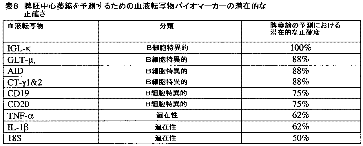
以下の2段落はqRT−PCRのための方法を提供する。
The following two paragraphs provide a method for qRT-PCR.
全リボ核酸(RNA)は、10個の脾臓凍結切片から単離し、各6μMを、製造業者のプロトコールに従って、BIOROBOT(登録商標)8000 Workstation(QIAGEN INC.,Valencia,CA)上で、RNEASY(登録商標)Universal Tissue 8000キット(QIAGEN Inc.,Valencia,CA)を使用して、DNA分解酵素(deoxyribonucleic acidase)(DNASE)処理した。RNAの純度および収率は、NANODROP(登録商標)ND−1000(NANODROP Technologies,Wilmington,DE)を使用して、220〜750nmの分光測光を介して評価した。RNAの完全性は、AGILENT 2100 Bioanalyzer(AGILENT Technologies,Foster City,CA)上でRNA 6000 NANO LABCHIP(登録商標)キット(AGILENT Technologies,Foster City,CA)を用いて測定し、そしてRNA完全性数(RNA integrity number)(RIN)アルゴリズム(Schroederら(2006)BMC Mol.Biol.7:3)を使用して計算した。DNA分解酵素処理したRNAをサンプルから抽出して−80℃に保存した。第1鎖相補的デオキシリボ核酸(cDNA)合成は、オリゴチミジル酸(オリゴ[dT])とランダムヘキサマーの両方をプライミングのために使用した以外は、TAQMAN(登録商標)Gold RT Master Mix(Applied Biosystems,Foster City,CA)を用いて製造業者のプロトコールに従って実施した。合成したcDNAの品質は、リアルタイムポリメラーゼ連鎖反応(PCR)システム、ABI PRISM(登録商標)7900HT配列検出システム(Applied Biosystems,Foster City,CA)を用いて内因性参照遺伝子の発現プロフィールを生成することによって評価した。相補的デオキシリボ核酸サンプルは−20℃に保存した。
Total ribonucleic acid (RNA) was isolated from 10 spleen frozen sections and each 6 μM was RNEASY® (registered trademark) on BIOBOBOT® 8000 Workstation (QIAGEN INC., Valencia, Calif.) According to manufacturer's protocol. Trademark: Universal Tissue 8000 kit (QIAGEN Inc., Valencia, Calif.) Was used for deoxyribonucleic acidase (DNASE) treatment. RNA purity and yield were assessed via spectrophotometry at 220-750 nm using a NANODROP® ND-1000 (NANODROP Technologies, Wilmington, DE). RNA integrity was measured using the
すべてのバイオマーカーの配列(表1)は、マウスおよびヒトのデオキシリボ核酸(DNA)配列(National Center for Biotechnology Information,Bethesda,MDに維持されているGENBANK(登録商標)データベース[オンライン])に由来した。定量的逆転写酵素ポリメラーゼ連鎖反応アッセイは、SYBR−グリーン検出システム(Qiagen Inc.,Valencia,CA)を使用して、ABI PRISM(登録商標)7900HT Sequence Detection System(Applied Biosystems,Inc.(Foster City,CA))上で実施した。すべての定量的アッセイは、ユニバーサルサーマルサイクリングパラメーターを使用して実行した:DNAポリメラーゼを活性化するために、95℃2分間保持、次いで、40回の3部構成のサイクル(95℃20秒間、58℃20秒間、および76℃20秒間)を実行した。データは配列検出システム(SDS)ソフトウェア、バージョン1.7(Applied Biosystems,Inc.(Foster City,CA))を使用して分析した。第1段階は、各サンプルについての増幅プロットを生成することであり、これは、x軸上のサイクル数に対する、y軸上のRnの変化(ΔRn)を示した(ここで、Rnはパッシブリファレンスに対して標準化されたレポーター色素の蛍光放射強度である)。各増幅プロットから、閾値サイクル(Ct)値を計算した。Ct値は、ΔRnの統計的に有意な増加が最初に検出され、増幅プロットの切片の点および閾値としてグラフ上で示されるサイクルとして定義される。Ct値はEXCEL(登録商標)(MICROSOFT Corp.,Redmond,WA)スプレッドシートにエクスポートし、相対的な発現(x)は等式2に示されるように計算した。
All biomarker sequences (Table 1) were derived from mouse and human deoxyribonucleic acid (DNA) sequences (GENBANK® database [online] maintained at National Center for Biotechnology Information, Bethesda, MD) . The quantitative reverse transcriptase polymerase chain reaction assay was performed using the SYBR-Green detection system (Qiagen Inc., Valencia, Calif.) Using the ABI PRISM® 7900HT Sequence Detection System (Applied Biosystems, Inc. CA)). All quantitative assays were performed using universal thermal cycling parameters: hold at 95 ° C. for 2 minutes to activate the DNA polymerase, then 40 three-part cycles (95 ° C. for 20 seconds, 58 ° ℃ 20 seconds, and 76 ℃ 20 seconds). Data was analyzed using Sequence Detection System (SDS) software, version 1.7 (Applied Biosystems, Inc. (Foster City, CA)). The first step was to generate an amplification plot for each sample, which showed the change in Rn (ΔRn) on the y-axis with respect to the number of cycles on the x-axis, where Rn is a passive reference Is the fluorescence emission intensity of the reporter dye normalized to). From each amplification plot, a threshold cycle (Ct) value was calculated. The Ct value is defined as the cycle in which a statistically significant increase in ΔRn is first detected and shown on the graph as the intercept point and threshold of the amplification plot. Ct values were exported to an EXCEL® (MICROSOFT Corp., Redmond, WA) spreadsheet and relative expression (x) was calculated as shown in
等式2 X=出力(2,−Ct)×100,000,000
マウスに、6時間、示されるように、単回経口用量のML120Bを与えた(n=4/群)。マウスミュー重鎖生殖細胞系列転写物のレベルは、連続凍結切片から単離したRNA中で、qRT−PCRによって測定した。胚中心の頻度およびミュー生殖細胞系列転写物のレベルは、ML120Bへの曝露の最初の6時間以内に50%減少したのに対し、T細胞の頻度は変化しないままであった(図1)。
Equation 2 X = Output (2, −Ct) × 100,000,000
Mice were given a single oral dose of ML120B for 6 hours as indicated (n = 4 / group). Mouse mu heavy germline transcript levels were measured by qRT-PCR in RNA isolated from serial frozen sections. Germ center frequency and mu germline transcript levels were reduced by 50% within the first 6 hours of exposure to ML120B, whereas T cell frequency remained unchanged (FIG. 1).
マウスに、18時間、示されるように、単回経口用量のML120Bを与えた(n=4/群)。マウスミュー重鎖生殖細胞系列転写物のレベルは、連続凍結切片から単離したRNA中で、qRT−PCRによって測定した。胚中心の頻度およびミュー生殖細胞系列転写物のレベルは、ML120Bへの曝露の18時間以内に用量依存的な減少を示し、300mg/kgでは、胚中心において10倍の減少、およびミュー生殖細胞系列転写物において1000倍の減少が生じた(図2)。T細胞の頻度および18S rRNAのレベルは、対照的に、変化しないままであった(図2)。 Mice were given a single oral dose of ML120B as indicated (n = 4 / group) for 18 hours. Mouse mu heavy germline transcript levels were measured by qRT-PCR in RNA isolated from serial frozen sections. Germ center frequency and mu germline transcript levels showed a dose-dependent decrease within 18 hours of exposure to ML120B, with a 10-fold decrease in germinal centers at 300 mg / kg, and mu germline A 1000-fold reduction occurred in the transcript (FIG. 2). T cell frequency and 18S rRNA levels remained, in contrast, unchanged (FIG. 2).
マウスに、4日間、示されるように、胃管栄養法によって、毎日2回の経口用量のML120Bを与えた(n=4/群)。マウスミュー、ガンマ3、およびガンマ1重鎖生殖細胞系列転写物、AICDA転写物、ガンマ1および2サークル転写物、および18S rRNAのレベルは、連続凍結切片から単離したRNA中で、qRT−PCRによって測定した。胚中心の頻度、ならびにミュー、ガンマ3およびガンマ1重鎖生殖細胞系列転写物,AICDA転写物、ガンマ1および2サークル転写物のレベルは、ML120Bに対して用量依存的な減少を示した(図3)。Bcl−6を発現する胚中心は300mg/kgで10倍減少した(図3)。生殖細胞系列転写物ミュー、ガンマ3およびガンマ1は、300mg/kgで10〜1000倍減少した(図3)。AICDAをコードする転写物は300mg/kgで100〜1000倍減少した(図3)。サークル転写物ガンマ1および2は100mg/kgで10倍減少した(図3)。18S rRNAのレベルは、対照的に、変化しないままであった(図3)。
Mice were given 2 daily oral doses of ML120B by gavage as indicated (n = 4 / group) for 4 days. The levels of mouse mu,
実施例2 サルにおけるICSの測定
独自開発の免疫調節物質、試験薬剤Aをこれらの研究において試験薬剤として使用した。リンパ球転写物(CD19、CD20、GLT−μ、CTγ1および2、AID、TNF−α、IL−1β、および18S)に対する試験薬剤Aの効果は、十分に確立され、堅固な転写物アッセイである、定量的逆転写ポリメラーゼ連鎖反応(qRT−PCR)を使用して測定し、これは、優秀な感度および特異性を実証する。
Example 2 Measurement of ICS in monkeys A proprietary immunomodulator, test drug A, was used as a test drug in these studies. The effect of test drug A on lymphocyte transcripts (CD19, CD20, GLT-μ, CTγ1 and 2, AID, TNF-α, IL-1β, and 18S) is a well-established and robust transcript assay , Measured using quantitative reverse transcription polymerase chain reaction (qRT-PCR), which demonstrates excellent sensitivity and specificity.
材料および方法
雌性カニクイザル(Macaca fascicularis,Charles River Laboratories(Sparks,NV;未処置の(naive)、未経産かつ非妊娠、2.0〜4.0kg)投与群あたり4匹の動物、総計16匹)。これらのサルは体重の順番の分布により研究群に割り当てた。濾過した水道水を自由に利用可能にした。
Materials and Methods Female cynomolgus monkeys (Macaca fascicularis, Charles River Laboratories (Sparks, NV; naïve, naive, non-pregnant, 2.0-4.0 kg), 4 animals per dosing group, total 16 ). These monkeys were assigned to study groups according to the distribution of body weight. Filtered tap water was made freely available.
サルは、28日間、毎日10ml/kg、0.1M クエン酸緩衝液(pH 2.7±0.05)中に製剤化した試験薬剤Aを受容した。水中クエン酸緩衝液(0.1M;PH 2.7±0.05)をこの試験で使用する対照とした。試験薬剤Aと対照品の両方は5℃±3℃で保存した。動物は、28日間、毎日1回、試験薬剤Aまたは対照製剤を受容した(投薬フェーズ)。用量は最も近い時点で記録された体重に基づいた。動物に、0(ビヒクル対照)、40、60、または100mg/kgの用量で、10ml/kgの単回容量で、経口胃管栄養法によって投与した。各投薬後、胃管栄養法チューブ中に残っているいかなる残留物の対照/試験薬剤Aも、5mlの水道水を投与することによって動物に投与した。 Monkeys received test drug A formulated in 10 ml / kg, 0.1 M citrate buffer (pH 2.7 ± 0.05) daily for 28 days. A citrate buffer in water (0.1 M; PH 2.7 ± 0.05) served as the control used in this study. Both Test Agent A and the control product were stored at 5 ° C. ± 3 ° C. Animals received test drug A or control formulations once daily for 28 days (dose phase). The dose was based on the body weight recorded at the nearest time point. Animals were dosed by oral gavage at a single dose of 10 ml / kg at doses of 0 (vehicle control), 40, 60, or 100 mg / kg. After each dose, any residual control / test drug A remaining in the gavage tube was administered to the animals by administering 5 ml of tap water.
−9日目、−2日目、7日目、14日目、21日目、および28日目に一度、各動物からの約2.0mlの末梢血を、血管穿刺を介して入手し、製造業者の説明書に従って、PAXGENE(登録商標)チューブ(PREANALYTIX,Valencia,CA)に収集した。剖検(29日目)の際に、すべての動物の脾臓試料を液体窒素中で急速冷凍した。血液サンプルおよび脾臓試料は、Millennium Pharmaceuticals,Inc.(Cambridge,MA)にドライアイス上で運搬するまで−70℃で保存し、そして−80℃で保存した。 -Once days 9, -2, 7, 14, 21, and 28, approximately 2.0 ml of peripheral blood from each animal was obtained via vascular puncture, Collected in PAXGENE® tubes (PREANALYTIX, Valencia, CA) according to manufacturer's instructions. At necropsy (day 29), spleen samples of all animals were snap frozen in liquid nitrogen. Blood samples and spleen samples were obtained from Millennium Pharmaceuticals, Inc. (Cambridge, MA) was stored at -70 ° C until transported on dry ice and stored at -80 ° C.
バイオマーカー測定
リボ核酸単離 総リボ核酸(RNA)は10個の脾臓凍結切片から単離し、各5μMを、製造業者のプロトコールに従って、BIOROBOT(登録商標)8000 Workstation(QIAGEN INC.,Valencia,CA)上で、RNEASY(登録商標)Universal Tissue 8000キット(QIAGEN Inc.,Valencia,CA)を使用して、DNA分解酵素(DNASE)処理した。血液からの総RNA抽出は、製造業者のプロトコールに従って、PAXGENE(登録商標)96 Blood RNAキット(PREANALYTIX,Valencia,CA)を使用して実施した。RNAの純度および収率は、NANODROP(登録商標)ND−1000(NANODROP Technologies,Wilmington,DE)を使用して、220〜750nmの分光測光を介して評価した、RNAの完全性は、AGILENT 2100 Bioanalyzer(AGILENT Technologies,Foster City,CA)上でRNA 6000 NANO LABCHIP(登録商標)キット(AGILENT Technologies,Foster City,CA)を用いて測定し、そしてRNA完全性数(RIN)アルゴリズム(Schroederら、2006)を使用して計算した。DNA分解酵素処理したRNAをサンプルから抽出して−80℃に保存した。第1鎖相補的デオキシリボ核酸(cDNA)合成は、オリゴチミジル酸(オリゴ[dT])とランダムヘキサマーの両方をプライミングのために使用した以外は、TAQMAN(登録商標)Gold RT Master Mix(Applied Biosystems,Foster City,CA)を製造業者のプロトコールに従って実施した。合成したcDNAの品質は、リアルタイムポリメラーゼ連鎖反応(PCR)システム、ABI PRISM(登録商標)7900HT配列検出システム(Applied Biosystems,Foster City,CA)を用いて内因性参照遺伝子の発現プロフィールを生成することによって評価した。相補的デオキシリボ核酸サンプルは−20℃に保存した。
Biomarker Measurement Ribonucleic Acid Isolation Total ribonucleic acid (RNA) was isolated from 10 spleen frozen sections and 5 μM each, according to manufacturer's protocol, BIOROBOT® 8000 Workstation (QIAGEN INC., Valencia, CA) Above, RNEASY® Universal Tissue 8000 kit (QIAGEN Inc., Valencia, Calif.) Was used for DNA-degrading enzyme (DNASE) treatment. Total RNA extraction from blood was performed using the PAXGENE® 96 Blood RNA kit (PREANALYTIX, Valencia, Calif.) According to the manufacturer's protocol. RNA purity and yield were assessed via spectrophotometry between 220 and 750 nm using NANODROP® ND-1000 (NANODROP Technologies, Wilmington, DE). RNA integrity was determined by the AGILENT 2100 Bioanalyzer. (AGILENT Technologies, Foster City, Calif.) Using the
定量的逆転写酵素ポリメラーゼ連鎖反応試薬の設計および確証 試薬の配列(表2)は、ヒトのデオキシリボ核酸(DNA)配列(National Center for Biotechnology Information,Bethesda,MDに維持されているGENBANK(登録商標)データベース[オンライン])に由来した。 Design and Validation of Quantitative Reverse Transcriptase Polymerase Chain Reaction Reagents Reagent sequences (Table 2) are maintained in the human deoxyribonucleic acid (DNA) sequence (National Center for Biotechnology Information, Bethesda, MD) GENBANK® Database [online]).
* = SUPER(商標)Modified Base Technology、Nanogen (Bothell, WA)より.
a: Applied Biosystems, Inc. (Foster City, CA).より、要求に応じてアッセイ
プローブはMGB ECLIPSE(商標)プローブ(NANOGEN,Bothell,WA)であった。各プライマーおよびプローブ対は、NANOGEN(Bothell,WA)によって合成し、合成鋳型およびヒトcDNA標準を使用してPCR効率を測定することによって確証した。確証したアッセイは直線状増幅および7桁の規模にわたって>99%効率を実証した(データ示さず)。確証したプライマーおよびプローブ対はMillennium Pharmaceuticals,Inc.(Cambridge,MA)に送り、発現プロフィール研究における使用の前にヒト陽性対照cDNA標準に対して再度確証した(データ示さず)。IGL−κおよびAIDアッセイは、TAQMAN(登録商標)試薬番号Hs00736177_m1およびHs00757808_m1をそれぞれ使用した(Applied Biosystems,Inc.(Foster City,CA)。
* = From SUPER ™ Modified Base Technology, Nanogen (Bothell, WA).
a : Applied Biosystems, Inc. (Foster City, CA). Thus, upon request, the assay probe was an MGB ECLIPSE ™ probe (NANOGEN, Bothell, WA). Each primer and probe pair was synthesized by NANOGEN (Bothell, WA) and verified by measuring PCR efficiency using synthetic templates and human cDNA standards. The validated assay demonstrated> 99% efficiency over linear amplification and 7 orders of magnitude (data not shown). Validated primer and probe pairs are available from Millennium Pharmaceuticals, Inc. (Cambridge, MA) and revalidated against human positive control cDNA standards prior to use in expression profile studies (data not shown). IGL-κ and AID assays used TAQMAN® reagent numbers Hs00736177_m1 and Hs00757808_m1, respectively (Applied Biosystems, Inc. (Foster City, Calif.)).
定量的逆転写酵素ポリメラーゼ連鎖反応アッセイ 定量的逆転写酵素ポリメラーゼ連鎖反応アッセイは、ABI PRISM(登録商標)7900HT Sequence Detection System(Applied Biosystems,Inc.(Foster City,CA)上で実施した。NANOGEN(Bothell,WA)ガイドラインを使用して設計したすべての定量的アッセイは、ユニバーサルサーマルサイクリングパラメーターを使用して実行した:DNAポリメラーゼを活性化するために、95℃2分間保持、次いで、40回の3部構成のサイクル(95℃20秒間、58℃20秒間、および76℃20秒間)を実行した。 Quantitative Reverse Transcriptase Polymerase Chain Reaction Assay The quantitative reverse transcriptase polymerase chain reaction assay was performed on the ABI PRISM® 7900HT Sequence Detection System (Applied Biosystems, Inc. (Foster City, Calif.)). , WA) All quantitative assays designed using the guidelines were performed using universal thermal cycling parameters: 95 ° C. for 2 minutes to activate DNA polymerase, then 40 parts in 3 parts Configuration cycles (95 ° C. for 20 seconds, 58 ° C. for 20 seconds, and 76 ° C. for 20 seconds) were performed.
データ分析
生データ分析 データは配列検出システム(SDS)ソフトウェア、バージョン1.7(Applied Biosystems,Inc.(Foster City,CA))を使用して分析した。第1段階は、各サンプルについての増幅プロットを生成することであり、これは、x軸上のサイクル数に対する、y軸上のRnの変化(ΔRn)を示した(ここで、Rnはパッシブリファレンスに対して標準化されたレポーター色素の蛍光放射強度である)。各増幅プロットから、閾値サイクル(Ct)値を計算した。Ct値は、ΔRnの統計的に有意な増加が最初に検出され、増幅プロットの切片の点および閾値としてグラフ上で示されるサイクルとして定義される。Ct値はEXCEL(登録商標)(MICROSOFT Corp.,Redmond,WA)スプレッドシートにエクスポートし、相対的な発現(x)は等式2に示されるように計算した(実施例1を参照のこと)。
Data Analysis Raw Data Analysis Data were analyzed using Sequence Detection System (SDS) software, version 1.7 (Applied Biosystems, Inc. (Foster City, Calif.)). The first step was to generate an amplification plot for each sample, which showed the change in Rn (ΔRn) on the y-axis with respect to the number of cycles on the x-axis, where Rn is a passive reference Is the fluorescence emission intensity of the reporter dye normalized to). From each amplification plot, a threshold cycle (Ct) value was calculated. The Ct value is defined as the cycle in which a statistically significant increase in ΔRn is first detected and shown on the graph as the intercept point and threshold of the amplification plot. Ct values were exported to an EXCEL® (MICROSOFT Corp., Redmond, WA) spreadsheet and relative expression (x) was calculated as shown in Equation 2 (see Example 1). .
統計的分析 血液中で試験した各転写物について、各動物のベースラインは、−9日目および−2日目のサイクル数の平均として定義した。動物のベースラインは、各投薬後の時点において動物のサイクル数から減算した。この差は、動物のベースラインサイクル数、試験薬剤Aの投薬量、血液サンプルが採取された時点(投薬後日数)、および用量と時間の間の相互作用についての独立変数を含んだ、反復測定線形回帰モデルを使用して分析した。モデルにおいて時間に対する用量相互作用が0.05よりも大きなp値を有した場合には、相互作用の項はモデルから除かれ、用量の間の差を評価した。 Statistical analysis For each transcript tested in blood, the baseline for each animal was defined as the average of the number of cycles on days -9 and -2. The animal baseline was subtracted from the animal's cycle number at each post-dose time point. This difference is an iterative measure that includes the animal's baseline cycle number, the dose of study drug A, the time when the blood sample was taken (days after dosing), and the independent variables for the interaction between dose and time. Analysis was performed using a linear regression model. If the dose interaction with time had a p-value greater than 0.05 in the model, the interaction term was removed from the model and the difference between doses was evaluated.
利用したモデルは等式3に示す。
The model used is shown in
等式3. yitd−yit0=yit0+処理t+日数d+(処理*日数)td+eitd
ここで:yitd=d日目および処理tにおける動物iについてのサイクル数;yit0=ベースライン(d=0)における動物iおよび処理tについてのサイクル数;処理t=処理のtレベル(ビヒクル、40、60、または100mg/kgの試験薬剤);日数d=日数のdレベル(7、14、21、または28日);および(処理*日数)td=処理と日数の間の相互作用(これは、処理群の間で経時的なトレンドの有意差が存在しなかったというヌル仮説を試験した);およびeitd=同じ動物上での経時的な反復読み取りからの相関を組み入れた説明できない誤差。
Where: y itd = number of cycles for animal i at day d and treatment t; y it0 = number of cycles for animal i and treatment t at baseline (d = 0); treatment t = t level of treatment (vehicle) , 40, 60, or 100 mg / kg test drug); days d = d level of days (7, 14, 21, or 28 days); and (treatment * days) td = interaction between treatment and days ( This tested the null hypothesis that there was no significant trend trend over time between treatment groups); and e itd = could not be explained incorporating correlation from repeated readings over time on the same animal error.
29日目の脾臓において試験した各転写物について、クルスカル−ワリス検定を実施して、転写物サイクル数に関して試験薬剤A用量群のいずれかの間で何らかの有意差が存在したか否かを評価した。すべての仮説は、0.05のタイプIエラー率(両側)で試験した。これは調査の分析であるので、試験した転写物の数および組織型を説明するための多重比較のための調整は行わなかった。
For each transcript tested in the spleen on
組織学 ヘマトキシリンおよびエオシン(H&E)染色スライドを、ゲノム分析のために使用したものと隣接する脾臓凍結切片(5μm)から調製した。脾臓切片の反応性(過形成)は、2名の盲検検査者(blinded reviewer)によって独立して評価され、その1名は専門医師会認定のヒト解剖病理学者であった。反応性は、5つの40倍視野における胚中心の頻度および過形成の外見の測定に基づいて、ゼロ(最小)から4(最大)スケールに点数付けした。これらの分析は、0.9のピアソン相関係数値(r2)を示し、分析が互いに一致していることを示した。 Histology Hematoxylin and eosin (H & E) stained slides were prepared from frozen spleen sections (5 μm) adjacent to those used for genomic analysis. The reactivity of the spleen sections (hyperplasia) was assessed independently by two blind reviewers, one of whom was a specialist anatomical pathologist certified by a professional medical association. Reactivity was scored from zero (minimum) to 4 (maximum) scale based on measurements of germinal center frequency and hyperplasia appearance in five 40x fields. These analyzes showed a Pearson correlation coefficient value (r 2 ) of 0.9, indicating that the analyzes were consistent with each other.
結果および考察
一般的免疫学的評価 二次リンパ器官の組織病理学は、試験薬剤AによるGCの用量依存性萎縮を示した(図4を参照のこと)。試験薬剤Aは濾胞樹状細胞を減少させ、B細胞の増殖を阻害する。二次リンパ器官において、B細胞またはT細胞の頻度に変化はなかった。増殖B細胞において100倍より多い減少、濾胞樹状細胞においては100倍より多い減少が存在した。試験薬剤Aは、免疫蛍光によって見られるように、二次リンパ器官において、未成熟B細胞の相対的な頻度を増加させる。試験薬剤Aは、二次リンパ器官において成熟B細胞の頻度を減少させる。カニクイザル脾臓における免疫蛍光染色されたIgG+胚中心の画像分析は、IgG+胚中心の頻度の用量依存的な減少を示した(図5)。
Results and Discussion General Immunological Evaluation Histopathology of the secondary lymphoid organs showed a dose-dependent atrophy of GC with test drug A (see FIG. 4). Test drug A reduces follicular dendritic cells and inhibits B cell proliferation. There was no change in the frequency of B cells or T cells in the secondary lymphoid organs. There was a more than 100-fold decrease in proliferating B cells and a more than 100-fold decrease in follicular dendritic cells. Test agent A increases the relative frequency of immature B cells in secondary lymphoid organs as seen by immunofluorescence. Test drug A reduces the frequency of mature B cells in secondary lymphoid organs. Image analysis of immunofluorescently stained IgG + germinal centers in cynomolgus monkey spleen showed a dose-dependent decrease in the frequency of IgG + germinal centers (FIG. 5).
試験薬剤Aは、末梢血中の後胚中心B細胞の頻度を減少させた。末梢血のフローサイトメトリーによって見られるように、全B細胞の頻度には変化がなかった(図6A)。しかし、前GC B細胞の同時的な増加(図6C)を伴う、後胚中心(GC) B細胞の減少が存在した(図6B)。 Test drug A decreased the frequency of posterior germinal center B cells in peripheral blood. There was no change in the frequency of total B cells as seen by peripheral blood flow cytometry (FIG. 6A). However, there was a decrease in posterior germinal center (GC) B cells with a concomitant increase in pre GC B cells (FIG. 6C) (FIG. 6B).
脾臓凍結切片の組織学的分析 2名の独立した盲検検査者による各動物の脾臓からのH&E切片の分析は、胚中心萎縮が本研究において達成されたことを確認した(図4を参照のこと)。ビヒクル対照群(動物番号1101〜1104)は完全に相同であり、各動物は、高度に反応性の胚中心を示し、萎縮を示さなかった。40mg/kg用量群(動物番号2101〜2104)は、顕著な不均一性を示し、1匹の動物が低い胚中心反応性を示し、別の動物が中程度の反応性を示し、そして他の2匹が高い反応性を示した。60mg/kg用量群(動物番号3101〜3104)は、比較的均一であり、3匹の動物が低い反応性を示し、1匹が高い反応性を示した。100mg/kg用量群(動物番号4101〜4104)は完全に均一であり、4匹すべての動物が低い胚中心反応性を示した。100mg/kg群の動物のサブセットは、細菌感染の徴候もまた示した。2名の独立した盲検検査者の定量的分析は互いに一致しており(r2=0.9)、これは、被験体間で観察される不均一性が分析の変動性に起因するものではなく、組織中の固有の差に起因することを示した。用量によるデータの分析は、ビヒクル対照群と比較した場合に、100mg/kg用量群における胚中心の反応性の統計的に有意な減少(p<0.05)を例証した。
Histological analysis of frozen spleen sections Analysis of H & E sections from each animal's spleen by two independent blind examiners confirmed that germinal center atrophy was achieved in this study (see FIG. 4). about). The vehicle control group (animal numbers 1101-1104) was completely homologous, and each animal showed highly reactive germinal centers and no atrophy. The 40 mg / kg dose group (
脾臓凍結切片の定量的逆転写酵素ポリメラーゼ連鎖反応分析 H&E切片(すなわち、連続切片)で縁取りした凍結切片から単離したRNAは、qRT−PCR転写物アッセイで分析して、これらの転写物(18S、TNF−α、IL−1β、CD20、CD19、IGL−κ、GLT−μ、AID、ならびにCT−γ1および2)の発現プロフィールが胚中心萎縮を反映するかどうかを決定した。脾臓転写物において用量−応答傾向が存在した。なぜなら、試験したすべての遺伝子について転写物の数が、高用量の試験薬剤Aとともに減少し、AIDおよびTNF−αについて観察された最大の差は(表3)、対照(ビヒクル)動物と100mg/kg処置動物についての値を例証しているからである。 Quantitative reverse transcriptase polymerase chain reaction analysis of frozen spleen sections RNA isolated from frozen sections bordered by H & E sections (ie, serial sections) was analyzed in the qRT-PCR transcript assay and these transcripts (18S It was determined whether the expression profiles of TNF-α, IL-1β, CD20, CD19, IGL-κ, GLT-μ, AID, and CT-γ1 and 2) reflect germinal center atrophy. There was a dose-response trend in spleen transcripts. Because the number of transcripts for all genes tested decreased with higher doses of Test Drug A, the largest difference observed for AID and TNF-α (Table 3) was 100 mg / kg treated with control (vehicle) animals. This is because the values for animals are illustrated.
b: 0および100mg/kg用量群についての中央値データは有意差を例証することを含んだ。
b : Median data for the 0 and 100 mg / kg dose groups included illustrating significant differences.
各遺伝子について観察された有意性の大きな部分は、ビヒクル対照群と100mg/kg用量群の間の差によるものであった(表4)。これらの分析はまた、内部参照として使用される転写物である18Sに対する有意な用量依存性効果を示唆した。この効果は小さく(<2倍効果)、これが生物学的効果を表したのか、または技術的なアーティファクトであったのかは不明である。それにも関わらず、これらのデータは、内因性参照転写物である18S値の差についてのデータを標準化した後で再分析し、結論は変わらないままであった(表5)。これらの転写物の発現プロフィールは、反応性の組織学的測定基準(histologic metrics)と正の相関があった。 The large portion of significance observed for each gene was due to the difference between the vehicle control group and the 100 mg / kg dose group (Table 4). These analyzes also suggested a significant dose-dependent effect on 18S, a transcript used as an internal reference. This effect is small (<2 fold effect) and it is unclear whether this represented a biological effect or was a technical artifact. Nevertheless, these data were reanalyzed after normalizing the data for differences in 18S values, endogenous reference transcripts, and the conclusions remained unchanged (Table 5). The expression profiles of these transcripts were positively correlated with reactive histological metrics.
b: データは従来的な内部参照転写物、18Sに対して標準化した(18Sは試験薬剤Aによって調節されないと仮定した)。中央値デルタCt=タンパク質Xの中央Ct値−所定のサンプルの18Sの中央Ct値。0および100mg/kg用量群の中央値データは有意差を例証することを含んだ。
b : Data normalized to a conventional internal reference transcript, 18S (assuming 18S is not regulated by test drug A). Median delta Ct = median Ct value for protein X−18S median Ct value for a given sample. Median data for the 0 and 100 mg / kg dose groups included illustrating significant differences.
試験薬剤Aは、B細胞の1つの免疫組織学的マーカー、CD20に対するわずかな用量依存性効果を示したが、脾臓における別のマーカー、CD19に対してはそうではなかった。CD20に対する効果は小さく、そして統計的に有意であるが、末梢血サンプル中で再生されなかった;試験薬剤Aは、末梢血中でCD20レベルに有意な影響を与えなかった。脾臓の全体の細胞性(例えば、器官重量、組織学)は用量依存的な様式で変化しなかった。脾臓サンプルからの凍結切片中のB細胞の内容物に焦点を当てたさらなる免疫組織化学的分析は、CD20タンパク質を発現するB細胞の頻度が用量群の間で違わなかったことを示した(データ示さず)。これらのさらなるデータは、脾臓におけるCD20転写物において観察される小さな差は組織の効果ではなく、生物学的有意性ではなさそうである。 Test drug A showed a slight dose-dependent effect on one immunohistological marker of B cells, CD20, but not on another marker in the spleen, CD19. The effect on CD20 was small and statistically significant but was not regenerated in peripheral blood samples; test drug A did not significantly affect CD20 levels in peripheral blood. The overall cellularity of the spleen (eg organ weight, histology) did not change in a dose-dependent manner. Further immunohistochemical analysis focusing on the contents of B cells in frozen sections from spleen samples showed that the frequency of B cells expressing CD20 protein was not different between dose groups (data Not shown). These additional data suggest that the small differences observed in CD20 transcripts in the spleen are not tissue effects but likely biological significance.
末梢血の定量的逆転写酵素ポリメラーゼ連鎖反応分析 末梢血におけるIGL−κ、GLT−μ、AID、およびCT−γ1および2転写物のレベルにおける変化もまた、有意に用量依存的であり、転写レベルがより高用量の試験薬剤Aに伴って減少する(表6;治療p値)。 Quantitative reverse transcriptase polymerase chain reaction analysis of peripheral blood Changes in the levels of IGL-κ, GLT-μ, AID, and CT-γ1 and 2 transcripts in peripheral blood are also significantly dose-dependent and transcription levels Decrease with higher doses of Test Drug A (Table 6; treatment p-value).
a:全体のp値は、処置群の中央値の間で差が存在しないという仮説を検定した。ダンのポスト検定は各処置群をビヒクルに対して比較した。
a : The overall p-value tested the hypothesis that there was no difference between the medians of the treatment groups. Dan's post test compared each treatment group to the vehicle.
各遺伝子について観察された有意性の大きな部分は、ビヒクル対照群と100mg/kg用量群の間の差によるものであった。他の試験薬剤A治療群は、ビヒクル対照群とは大きく違わなかった。処置群の中の動物の間で考慮すべき変動性が存在した(図7A−DはGLT−ミュー(ST1)についての結果を例証する)。反復測定線形モデルは、転写物のいずれかについての処置群の間での経時的なトレンドにおいて有意差が存在しないことを示した(表6、時間−治療相互作用p値)。 The large portion of significance observed for each gene was due to the difference between the vehicle control group and the 100 mg / kg dose group. The other study drug A treatment groups were not significantly different from the vehicle control group. There was variability to consider between animals in the treatment group (FIGS. 7A-D illustrate the results for GLT-mu (ST1)). A repeated measures linear model showed that there was no significant difference in the trend over time between treatment groups for any of the transcripts (Table 6, time-treatment interaction p-value).
組織学および定量的逆転写酵素ポリメラーゼ連鎖反応データの比較
組織学および脾臓qRT−PCRデータの比較は、IGL−κ、GLT−μ、AID、およびCT−γ1および2転写物のレベルが、胚中心反応性を予測することを実証した。個々の動物からのデータの分析は、組織学的胚中心反応性データと、これらの転写物のレベルとの間の正の相関を強調した。これらの正の相関は、用量群によるデータの分析において、特に、動物間の変動が最大であった40および60mg/kg用量群において明白ではなかった。組織学は、動物番号1101、1102、1103、1104、2102、2103、および3101が高い胚中心反応性を示したのに対して、動物番号2101、2104、3102、3103、3104、4101、4102、4103、および4104が低い胚中心反応性を示したことを示した。すべての脾臓からのすべての転写物についてのデータの分析はIGL−κ、GLT−μ、AID、およびCT−γ1および2が類似のパターンを示したことを明らかにした。無影響量(NOEL)は、有害ではないと見なされるこれらの新規な転写物バイオマーカーの変化に基づいて、60mg/kgであると決定した。このNOELは、組織学的アッセイによって決定されるものと一致する。感染は、以前の研究において、無有害作用量(NOAEL)を定義した有害反応を考慮した。感染症はこの研究においては観察されなかった。
Comparison of histology and quantitative reverse transcriptase polymerase chain reaction data Comparison of histology and spleen qRT-PCR data shows that levels of IGL-κ, GLT-μ, AID, and CT-γ1 and 2 transcripts are germinal centers It was demonstrated to predict reactivity. Analysis of data from individual animals highlighted a positive correlation between histological germinal center reactivity data and the levels of these transcripts. These positive correlations were not evident in the analysis of the data by dose group, particularly in the 40 and 60 mg / kg dose groups where variability between animals was greatest. Histology showed that
より重要なことに、末梢血中のIGL−κ、GLT−μ、AID、CT−γ1および2、CD19、およびCD20転写物は、組織学からの胚中心反応性データとの最高の相関を示した(表7)。 More importantly, IGL-κ, GLT-μ, AID, CT-γ1 and 2, CD19, and CD20 transcripts in peripheral blood show the highest correlation with germinal center reactivity data from histology (Table 7).
動物は、血液転写物レベルの傾きの値に基づいて、それらの胚中心の反応性に関して、正常または異常(すなわち、潜在的に萎縮性)として分類した。Bリンパ球によってのみ発現される末梢血バイオマーカー(IGL−κ、GLT−μ、AID、およびCT−γ1および2)は、組織学的データと最も強力な関連性を示した(表8)。 Animals were classified as normal or abnormal (ie potentially atrophic) with respect to their germinal center reactivity based on the value of the slope of the blood transcript level. Peripheral blood biomarkers expressed only by B lymphocytes (IGL-κ, GLT-μ, AID, and CT-γ1 and 2) showed the strongest association with histological data (Table 8).
第3のバイオマーカーのサブカテゴリーには、その発現がNF−κB活性と関連しており、遍在性に発現されているもの(TNF−αおよびIL−1β)が含まれる。これらの2つの転写物は、CD19およびCD20よりも予測性が低く、各々が動物の62%を正確に分類した(表8)。 A third biomarker subcategory includes those whose expression is associated with NF-κB activity and ubiquitously expressed (TNF-α and IL-1β). These two transcripts were less predictive than CD19 and CD20, each correctly classifying 62% of the animals (Table 8).
研究した最後のバイオマーカー、18Sは、非機構的かつ遍在性のバイオマーカーであり、予測性であると期待されなかった。これは、正常または異常として50%の動物を正確に分類し、これは、偶然と大差がない(表8)。 The last biomarker studied, 18S, was a non-mechanistic and ubiquitous biomarker and was not expected to be predictive. This correctly classifies 50% of animals as normal or abnormal, which is not much different by chance (Table 8).
結論
本研究の状況の中で、過剰量の試験薬剤Aによって引き起こされる脾胚中心萎縮を予測するために、いくつかのバイオマーカー転写物(IGL−κ、GLT−μ、AID、およびCT−γ1および2)を末梢血中で同定した。NOELは、これらの新規な転写物バイオマーカーの変化に基づいて60mg/kgであると決定され、これは有害ではないと見なされる。このNOELは組織学的アッセイによって決定されたものと一致する。感染は、以前の研究においてNOAELを定義した有害反応と見なされる。感染は本研究において観察されなかった。
CONCLUSION Within the context of this study, several biomarker transcripts (IGL-κ, GLT-μ, AID, and CT-γ1 were used to predict splenic germinal center atrophy caused by excessive amounts of test drug A. And 2) were identified in peripheral blood. The NOEL is determined to be 60 mg / kg based on changes in these novel transcript biomarkers, which are considered not harmful. This NOEL is consistent with that determined by histological assay. Infection is considered an adverse reaction that has defined NOAEL in previous studies. Infection was not observed in this study.
実施例3 ヒト末梢血におけるICSの測定
末梢血は、健常ヒトボランティアから収集し、脾胚中心萎縮を検出するための、代理組織として利用した。サンプルは、19例のドナーから、各4つの時点で(2週間離す)収集し、RNAはサルのアッセイについて記載したように単離し、そしてICSマーカー、例えば、GLT−μおよびサークル転写物CT−γ1および2(GLT1、GLT2、GLT3、CT1、CT2、およびCT3を使用する)の発現を決定した。検出試薬は、ヒトとサルの間のコンセンサス配列(配列については表13を参照のこと)を検出するように設計した。すべてのマーカーを末梢血中で検出した(図8AおよびB)。相対的発現レベルは一般的な低レベルの転写物を示す。ボランティアは一般的に健康であったので、時点間またはボランティア間で差の予測は存在しなかった。それゆえに、差が検出できることを示すために、サンプルは、スイッチ転写物を検出する可能性を増加させるように修飾した。ポリサッカリドクッションを通してのスピニングのネガティブ選択によって、形質芽球をサンプル中で富化し、次いで、アッセイし、またはCD138検出試薬(Miltenyi Biotec,Auburn,CA)に結合体化された磁気ビーズを使用する陽性選択によって、さらに選択した。結果を図9に示す。ICS転写物(サークル転写物CT−γ1および2)のレベルは、全血液サンプル中のヒト形質芽球の頻度に比例する。
Example 3 Measurement of ICS in Human Peripheral Blood Peripheral blood was collected from healthy human volunteers and used as a surrogate tissue to detect splenic germinal center atrophy. Samples were collected from 19 donors at 4 time points (2 weeks apart), RNA was isolated as described for the monkey assay, and ICS markers such as GLT-μ and circle transcript CT- Expression of γ1 and 2 (using GLT1, GLT2, GLT3, CT1, CT2, and CT3) was determined. The detection reagent was designed to detect consensus sequences between humans and monkeys (see Table 13 for sequences). All markers were detected in peripheral blood (FIGS. 8A and B). Relative expression levels indicate a general low level transcript. Since volunteers were generally healthy, there was no prediction of differences between time points or between volunteers. Therefore, in order to show that the difference can be detected, the sample was modified to increase the likelihood of detecting switch transcripts. Positive selection using magnetic beads conjugated to a CD138 detection reagent (Miltenyi Biotec, Auburn, Calif.) By enriching plasmablasts in the sample by negative selection for spinning through a polysaccharide cushion Further selection by selection. The results are shown in FIG. The level of ICS transcripts (circle transcripts CT-γ1 and 2) is proportional to the frequency of human plasmablasts in the whole blood sample.
実施例4 サルにおけるICSの測定
実施例1〜3は、スイッチ転写物が検出可能であり、それらのレベルの変化が、マウス、マカク属カニクイザル(cynomolgous macaque)、および正常ヒト被験体の末梢血において測定できることを実証した。実施例2もまた、末梢血中のスイッチ転写物のレベルが、マカク属カニクイザルの脾臓中の濾胞性胚中心萎縮の程度を反映することを実証した。現在の研究の目的は、1)13週間の研究の状況においてこれらの以前の観察を確証すること、2)末梢血中のスイッチ転写物のレベルが、細菌感染の臨床徴候の前駆マーカーとしてもまた役立つことができるか否かを評価することであった。
Example 4 Measurement of ICS in monkeys Examples 1 to 3 show that switch transcripts can be detected and that changes in their levels are observed in the peripheral blood of mice, cynomolgus macaques, and normal human subjects. It was demonstrated that it can be measured. Example 2 also demonstrated that the level of switch transcripts in peripheral blood reflects the extent of follicular germinal center atrophy in the spleen of macaque cynomolgus monkeys. The purpose of the current study is 1) to confirm these previous observations in the 13-week study setting, and 2) the level of switch transcripts in the peripheral blood is also a progenitor marker for clinical signs of bacterial infection It was to evaluate whether it could help.
雄性と雌性の両方のカニクイザル(Macaca fascicularis,Charles River Laboratories(Sparks,NV;未処置の、未経産かつ非妊娠、2.0〜4.0kg、総計40匹)。これらのサルは体重の順番の分布により研究群に割り当てた。濾過した水道水を自由に利用可能にした。 Both male and female cynomolgus monkeys (Macaca fascicularis, Charles River Laboratories (Sparks, NV; untreated, naive and non-pregnant, 2.0-4.0 kg, 40 total), these monkeys in order of weight Assigned to study group according to the distribution of filtered tap water freely available.
サルは、13週間(91日間)の間、毎日1回、実施例2と同様に製剤化し、保存し、および投与した試験薬剤A、または対照を受容した。用量は、0、20、40、または80mg/kgであった。 The monkeys received Test Drug A, or control, formulated, stored and administered as in Example 2 once daily for 13 weeks (91 days). The dose was 0, 20, 40, or 80 mg / kg.
末梢血サンプルは、実施例2と同じ手順を使用して収集した。サンプルは、投薬の開始前(−2週および−1週)ならびに1週、2週、3週、4週、13週、および17週(回復期間の最後)の間に入手した。血液サンプルは−70℃で保存し、Millennium Pharmaceuticals,Inc.(Millennium Cambridge,MA)にドライアイスで運搬し、そして−80℃で保存した。 Peripheral blood samples were collected using the same procedure as in Example 2. Samples were obtained before the start of dosing (-2 weeks and -1 weeks) and between 1 week, 2 weeks, 3 weeks, 4 weeks, 13 weeks, and 17 weeks (end of recovery period). Blood samples were stored at -70 ° C and were collected from Millennium Pharmaceuticals, Inc. (Millennium Cambridge, MA) was delivered on dry ice and stored at -80 ° C.
脾臓生検試料は、すべて安楽死させた動物から収集した。32例の動物(4匹/性別/群)は、最後の投薬(92日目)の1日後に安楽死させ、8例の動物(4匹の対照、4匹の80mg/kg動物、2匹/性別/群)は最後の投薬の4週間後に安楽死させた(回復群、121日目および122日目)。剖検はすべての安楽死させた動物について実施し、脾臓生検試料は、中性緩衝化10%ホルマリン中で保存した。 All spleen biopsy samples were collected from euthanized animals. Thirty-two animals (4 / sex / group) were euthanized one day after the last dose (day 92) and 8 animals (4 controls, 4 80 mg / kg animals, 2 animals) / Sex / group) were euthanized 4 weeks after the last dose (recovery group, days 121 and 122). Necropsy was performed on all euthanized animals and spleen biopsy samples were stored in neutral buffered 10% formalin.
RNA単離 血液サンプルからの総RNA抽出、RNA QCおよびcDNA生成は、実施例2において使用したのと同様の一般的工程を使用して、Asuragen Inc.(Austin TX)において実施した。cDNAサンプルは−20℃で保存した。 RNA Isolation Total RNA extraction from blood samples, RNA QC and cDNA generation were performed using the same general steps used in Example 2, using Asuragen Inc. (Austin TX). The cDNA sample was stored at -20 ° C.
定量的逆転写酵素ポリメラーゼ連鎖反応 すべてのバイオマーカーの配列は、ヒトのデオキシリボ核酸(DNA)配列(GENBANK(登録商標)核酸データベース、National Center for Biotechnology Information,Bethesda,MD)に由来した。GLT−μおよびCT1(Nanogen,Bothell,WA)のためのプライマーおよびMGB Eclipse(商標)プローブは分析から設計した。各プライマーおよびプローブ対は、Nanogenによって合成され、合成鋳型およびヒトcDNA標準を使用してPCR効率を測定することによって確証した。確証したアッセイは線形増幅および7桁の規模にわたって>99%効率を実証した(データ示さず)。確証したプライマーおよびプローブ対はMillennium(Cambridge,MA)に送り、発現プロフィール実験における使用の前にヒト陽性対照cDNA標準に対して再度確証した。表13に見い出されるThe Applied Biosystem Assay on Demand(AOD)アッセイは、Millennium DSD718 TaqMan(登録商標)低密度アレイ(Applied Biosystems;Foster City,CA)に構成された。 Quantitative reverse transcriptase polymerase chain reaction All biomarker sequences were derived from the human deoxyribonucleic acid (DNA) sequence (GENBANK® Nucleic Acid Database, National Center for Biotechnology Information, Bethesda, MD). Primers and MGB Eclipse ™ probes for GLT-μ and CT1 (Nanogen, Bothell, WA) were designed from the analysis. Each primer and probe pair was synthesized by Nanogen and verified by measuring PCR efficiency using synthetic templates and human cDNA standards. The validated assay demonstrated> 99% efficiency over linear amplification and 7 orders of magnitude (data not shown). Validated primer and probe pairs were sent to Millennium (Cambridge, Mass.) And revalidated against human positive control cDNA standards prior to use in expression profile experiments. The Applied Biosystem Assay on Demand (AOD) assay found in Table 13 was configured on a Millennium DSD718 TaqMan® low density array (Applied Biosystems; Foster City, CA).
生データ分析 データは配列検出システム(SDS)ソフトウェア、バージョン2.2(Applied Biosystems,Foster City,CA)を使用して分析した。第1段階は、各サンプルについての増幅プロットを生成することであり、これは、x軸上のサイクル数に対する、y軸上のRnの変化(ΔRn)を示した(ここで、Rnはパッシブリファレンスに対して標準化されたレポーター色素の蛍光放射強度である)。各増幅プロットから、閾値サイクル(Ct)値を計算した。Ct値は、バックグラウンドレベル(すなわち、ノイズ)よりも上であるΔRnの統計的に有意な増加が検出されるサイクル数として定義される。Ct値は、増幅プロットの切片の点および閾値としてグラフ上で示される。Ct値はEXCEL(登録商標)(Microsoft Corp.,Redmond,WA)スプレッドシートにエクスポートした。生のCt値の解釈は、各アッセイへのインプットであったサンプルの量の変動性によって混同される可能性がある。このような技術的な変動性に対して標準化する従来の方法は、目的の転写物(この場合は、CT1、GLT3、IGLL、IGLK、AICDA、IGHG1、またはIGHA1)の生のCt値から、内部の、不変の、内因性の参照転写物(18SまたはB2MG)の生のCt値を減算して、デルタサイクル閾(dCt)値を生成することである。18S rRNA(18S)またはβ−2ミクログロブリン(B2MG)の転写物は、従来の不変の内因性参照転写物である。本研究におけるデータの分析は、本研究において評価される内因性参照転写物の中で18Sが最も不変であることを例証する。サイクル閾値およびdCtはログ低2(log2)関数である:1Ctの増加はシグナルの倍加を表すのに対して、1Ctの減少はシグナルが半分になることを表す。それゆえに、転写物のレベルは、等式3:[X=出力(2,−dCt)×1000]を使用して標準化されたレベルとして表される。 Raw Data Analysis Data were analyzed using Sequence Detection System (SDS) software, version 2.2 (Applied Biosystems, Foster City, CA). The first step was to generate an amplification plot for each sample, which showed the change in Rn (ΔRn) on the y-axis with respect to the number of cycles on the x-axis, where Rn is a passive reference Is the fluorescence emission intensity of the reporter dye normalized to). From each amplification plot, a threshold cycle (Ct) value was calculated. The Ct value is defined as the number of cycles in which a statistically significant increase in ΔRn that is above the background level (ie, noise) is detected. Ct values are shown on the graph as intercept points and thresholds in the amplification plot. Ct values were exported to an EXCEL® (Microsoft Corp., Redmond, WA) spreadsheet. The interpretation of raw Ct values can be confused by the variability in the amount of sample that was input to each assay. Conventional methods to standardize for such technical variability are based on the raw Ct value of the target transcript (in this case CT1, GLT3, IGLL, IGLK, AICDA, IGHG1, or IGHA1) Subtracting the raw Ct value of an unchanged, endogenous reference transcript (18S or B2MG) to generate a delta cycle threshold (dCt) value. Transcripts of 18S rRNA (18S) or β-2 microglobulin (B2MG) are conventional, unchanging endogenous reference transcripts. Analysis of the data in this study illustrates that 18S is the most invariant among the endogenous reference transcripts evaluated in this study. Cycle threshold and dCt are log low 2 (log 2 ) functions: an increase of 1 Ct represents doubling of the signal, while a decrease of 1 Ct represents halving of the signal. Therefore, transcript levels are expressed as standardized levels using Equation 3: [X = output (2, −dCt) × 1000].
本研究において試験された転写物の大部分は(GLT3、CT1、AICDA、IGHG1、およびIGHA1)、これらが胚中心反応の生成物であるので、同時調節されているようである。他の同時調節されている転写物の状況において、任意の単一の転写物における変化のトレンド分析は、任意の単一の転写物の変化がすべての転写物における非特異的なトレンドを表すのか、または転写物の特定のサブセットに限定されるかどうかの決定を可能にする。しかし、異なる転写物の標準化レベルは、100,000倍異なる可能性があり、これは、直接的な比較分析を妨げる。データは、ベースラインからの倍数変化の計算によってシグナルの規模の変動性について標準化され、従って、転写物にまたがる比較を単純化する。ベースラインからの倍数変化は、所定の時点における転写物の標準化された値を、2つのベースライン標準化レベルの値(−2週および−1週)の平均で分割することによって計算した。 The majority of transcripts tested in this study (GLT3, CT1, AICDA, IGHG1, and IGHA1) appear to be co-regulated because they are products of the germinal center response. In other co-regulated transcript situations, the trend analysis of changes in any single transcript shows that any single transcript change represents a non-specific trend in all transcripts Or whether it is limited to a specific subset of transcripts. However, the normalization levels of different transcripts can differ 100,000 times, which precludes direct comparative analysis. Data are normalized for signal size variability by calculation of fold change from baseline, thus simplifying comparisons across transcripts. Fold change from baseline was calculated by dividing the normalized value of the transcript at a given time point by the average of the two baseline normalization level values (-2 weeks and -1 week).
結果
一般的免疫機能 高用量動物のサブセットにおける細菌感染を別として、臨床的観察において治療に関連するものは存在しなかった。他の点では臨床病理学は正常であった。解剖病理学は、リンパ系胚中心の萎縮が存在したこと以外は正常であった。免疫毒物学評価は、補体経路の阻害を示さなかった;食作用機能または呼吸バーストの阻害はなかった;ナチュラルキラー(NK)機能の阻害はなかった;血液の白血球の主要なサブセットの頻度の変化はなかった;血清免疫グロブリンのレベルに変化はなかった;しかし、高用量動物において、キーホールリンペットヘモシアニン(KLH)(図10)に対するT細胞依存性抗体応答(TDAR)の阻害があった。損なわれたTDARは感染の臨床徴候と関連し(図10);新生抗原およびリコール抗原に対するIgG応答の用量依存的阻害であった。症状を示す動物は、最低の抗KLH力価を示したが、全体のIgレベルは変化しなかった。
Results General immune function Apart from bacterial infections in a subset of high-dose animals, there were no treatment related clinical observations. In other respects, clinical pathology was normal. Anatomical pathology was normal except that there was atrophy of lymphoid germinal centers. Immunotoxicology assessment showed no inhibition of complement pathway; no inhibition of phagocytic function or respiratory burst; no inhibition of natural killer (NK) function; frequency of major subsets of blood leukocytes There was no change; serum immunoglobulin levels did not change; however, there was an inhibition of the T cell-dependent antibody response (TDAR) to keyhole limpet hemocyanin (KLH) (Figure 10) in high-dose animals . Impaired TDAR was associated with clinical signs of infection (FIG. 10); dose-dependent inhibition of IgG response to nascent and recall antigens. Symptomatic animals showed the lowest anti-KLH titer, but overall Ig levels did not change.
組織学的分析 2名の独立した盲検検査者による各動物の脾臓からのH&E切片の分析は、本研究において、用量依存的な様式で、胚中心の萎縮が起こったことを確認した。最小の萎縮は40mg/kg群の2例の動物、および80mg/kg群の1例の動物において観察された。軽度の萎縮は、40mg/kg群の1例の動物、および80mg/kg群の1例の動物において観察した。中程度の萎縮は、80mg/kg群の2例の動物において観察された。40mg/kg群および80mg/kg群の中での高い動物間の変動性に起因する、有意差は用量間では観察されなかった。13週目に安楽死させた各動物の反応性もまた、胚中心の過形成の頻度および規模の評価に基づいて、各試料を、相対的なスケール、本質的に1(反応性が最も低い)〜30(最も反応性である)のランキングによってスコア付けした。相対的なスケールは、末梢血中の転写物のレベルに対する組織学的変化の比較を容易にした。本研究において達成された胚中心萎縮の全体の程度は、以前の28日間の研究において達成したものよりも少なく(実施例2)、これは、より低用量の投与(40、60、および100mg/kgと比較して、20、40、および80mg/kg)、および/または以前の28日間の研究と比較して13週間の研究におけるより低い曝露に達することから生じた可能性がある。
Histological analysis Analysis of H & E sections from the spleen of each animal by two independent blind examiners confirmed that germinal center atrophy occurred in this study in a dose-dependent manner. Minimal atrophy was observed in 2 animals in the 40 mg / kg group and 1 animal in the 80 mg / kg group. Mild atrophy was observed in one animal in the 40 mg / kg group and one animal in the 80 mg / kg group. Moderate atrophy was observed in 2 animals in the 80 mg / kg group. No significant difference was observed between doses due to high inter-animal variability in the 40 mg / kg and 80 mg / kg groups. The reactivity of each animal euthanized at
定量的RT−PCR 用量群間の任意の転写物について有意差は観察されなかった。GLT3、CT1、AICDA、IGHA1、IGDG1、IGLK、およびIGLL転写物のレベルは、ビヒクル対照群における個々の動物の中で、経時的に協調的な変動を示した(例えば、図11Aを参照のこと)。この変動は、経時的なこれらの転写物のレベルの生物学的な(技術的ではない)変動を表す可能性が最も高い。なぜなら、GLT3およびCT1アッセイは、AICDA、IGHA1、IGDG1、IGLK、およびIGLLのアッセイ(すなわち、TaqManプローブおよびTLDAマイクロ流体カード)とは異なる技術(すなわち、MGB Eclipseプローブ)および形式(384ウェルプレート)で実施したからである。 Quantitative RT-PCR No significant difference was observed for any transcript between dose groups. The levels of GLT3, CT1, AICDA, IGHA1, IGDG1, IGLK, and IGLL transcripts showed coordinated variation over time among individual animals in the vehicle control group (see, eg, FIG. 11A). ). This variation is most likely to represent a biological (non-technical) variation in the level of these transcripts over time. Because the GLT3 and CT1 assays are in a different technique (ie MGB Eclipse probe) and format (384 well plate) than the AICDA, IGHA1, IGDG1, IGLK, and IGLL assays (ie TaqMan probes and TLDA microfluidic cards). This is because it was implemented.
IGHA1、IGDG1、GLT3、AICDA、CT1、IGLK、またはIGLL転写物のレベルに対する試験薬剤Aの効果は、20mg/kg群の個々の動物については観察されなかった。 No effect of test agent A on the levels of IGHA1, IGDG1, GLT3, AICDA, CT1, IGLK, or IGLL transcripts was observed for individual animals in the 20 mg / kg group.
IGHA1、IGDG1、GLT3、AICDA、CT1、IGLK、またはIGLL転写物のレベルに対する試験薬剤Aの効果は、一般的には、40mg/kg群については観察されなかったが、3〜13週の間の動物3001、3003、および3501においてはいくつかが存在した可能性がある。 The effect of test drug A on the levels of IGHA1, IGDG1, GLT3, AICDA, CT1, IGLK, or IGLL transcripts was generally not observed for the 40 mg / kg group, but between 3 and 13 weeks Some may exist in animals 3001, 3003, and 3501.
試験薬剤A(80mg/kg)に応答した、IGHA1、IGDG1、GLT3、AICDA、CT1、IGLK、およびIGLL転写物のレベルの変化は、動物4004、4005、および4006について観察された(例えば、図11Bを参照のこと)。動物4004および4006の投薬は、これらの動物の瀕死状態に起因して、4週目の間に終了した。動物4005は瀕死ではなく、投薬を継続した。この動物の状態は、抗生物質の投与に伴って一貫して改善され、17週目には正常な臨床状態に戻ったからである。動物4005において13週目に観察したIGHA1、IGDG1、GLT3、AICDA、CT1、IGLK、およびIGLL転写物のレベルに対する試験薬剤Aの効果は、これらの転写物のレベルが17週目までにベースラインレベルまで戻り、これは、試験薬剤Aの最後の投薬の4週間後であったという点で可逆的であった。2つの対照内因性参照転写物(18SおよびB2MG)の間で用量群の間に差がなく、試験薬剤Aに対する応答の変化もなかった。
Changes in the levels of IGHA1, IGDG1, GLT3, AICDA, CT1, IGLK, and IGLL transcripts in response to test drug A (80 mg / kg) were observed for animals 4004, 4005, and 4006 (eg, FIG. 11B). checking). Dosing of animals 4004 and 4006 was terminated during the 4th week due to the moribund state of these animals. Animal 4005 was not moribund and continued on medication. The animal's condition improved consistently with antibiotic administration and returned to normal clinical condition at 17 weeks. The effect of test agent A on the levels of IGHA1, IGDG1, GLT3, AICDA, CT1, IGLK, and IGLL transcripts observed at
末梢血における転写物のレベルに対する、脾胚中心の萎縮の比較 末梢血におけるスイッチ転写物のレベルについての平均値との、組織学的胚中心萎縮についての平均値の比較は、用量との有意な相関を明らかにしなかった(データ示さず)。個々の動物の分析はまた、剖検の際の(13週目)個々の動物についての脾胚中心の相対的な組織学的反応性を、ベースライン(−2週目および−1週目)から剖検までの(4週目および13週目)末梢血中のスイッチ転写物のレベルの変化に対して比較することによって実施した。脾臓における胚中心の大部分の萎縮を示した動物(例えば、動物4006)はまた、一般的に、末梢血中のいくつかの転写物のレベルの減少を示した。これらのデータの定量的分析は、血中レベルのGLT3、CT1、およびIGLLの減少は、胚中心反応性の減少と、弱いが正の相関を示した(r2約0.2)ことを明らかにした。より弱い相関は他の転写物について観察された。脾胚中心萎縮を示す動物は、一般的に、末梢血中でより低いレベルのGLT3およびCT1を有した;しかし、動物4503および4504は、これらが中程度の萎縮を示しながら、末梢血中においてなお高いGLT3およびCT1のレベルを示すという点で例外である。これらの動物は、これらの切片が動脈周囲リンパ鞘(PALS)のリンパ過形成を含んだという点でもまた独特であることが注目されるべきである。リンパ球の過形成は、反応性胚中心の顕著な特徴である。胚中心の萎縮に対する補償的応答として、ICSはこれらの2例の動物のPALSの中では存在しないこと、およびこれが末梢血中の不釣り合いに高いレベルのGLT3およびCT1の供給源であり得ることが可能性としてある。これらの形態学的に例外的な脾臓(4503および4504)は、これらの比較の結果に重い影響を与える。動物4503および4504が域外値と見なされ、分析から除外される場合、胚中心反応性の減少(例えば、萎縮)と血液中のこれらの転写物のレベルの減少の間の全体の相関は、GLT3およびCT1について実質的に改善された(r2約0.6−0.7、図12AおよびB)。他の転写物の再分析における域外値としての同じ動物の除外はまた、AICDA、IGDG1、およびIGLKについてもより高い相関を生じたが(図12C、E、G)、IGHA1、IGLL、および18Sについてはそうではなかった(図12D、F、およびH)。
Comparison of splenic germinal center atrophy against transcript level in peripheral blood Comparison of mean value for histological germinal center atrophy with mean value for switch transcript level in peripheral blood is significant compared to dose Correlation was not revealed (data not shown). Analysis of individual animals also revealed the relative histological reactivity of the splenic germinal centers for individual animals at necropsy (week 13) from baseline (-2 weeks and -1 weeks). This was done by comparing against changes in the level of switch transcripts in peripheral blood until necropsy (
末梢血中の転写物のレベルに対する、感染の臨床徴候の比較 感染の臨床徴候(すなわち、細菌性赤痢)は、動物4004については13日目に、動物4006については20日目に、および動物4005については29日目に観察された。感染の臨床徴候の発生は、徴候の発症の前およびその間の時点について(−2週、−1週、1週、2週、3週、および4週)末梢血中で転写物のレベルの変化に対して比較した。個々の動物における転写物の傾きの値は、−2週から4週までの転写物が、この期間にわたってベースライン値からの倍数変化の線形回帰を計算することによって定量されたという点で変化の規模を表す(表14)。例えば、動物4004は、GLT3およびCT1のレベルの最大減少を示したのに対して、動物4501は、AICDAの最大減少を示し、4005はIGHG1、4006はIGHAなどであった。各転写物の傾きをランク付けすることによって、ならびにこのランク値が無症候性動物から臨床的に症候性動物(4004、4005、および4006)を区別するか否かを決定することによって、これらの潜在的に安全なバイオマーカーの相対的な予測値を評価できる。GLT3またはCT1についての傾きのランク付けにより、動物4004、4005、および4006をトップの3つの位置に分離し;症候性動物はGLT3またはCT1のレベルの最大減少を示した(表14)。AICDA、IGHG、IGHA、またはIGLKについてのランク付け傾きは、3つの症候性動物の内の2つ(4005および4006)をトップの3つの位置に(表14)、そして第3の動物(4004)をすべての動物の内の上部五分位内に分離した(表14)。IGLLについてのランク付け傾きは、3つの症候性動物の内の2つ(4005および4006)をすべての動物の内の上部五分位内に分離した(表14)。対照転写物18SおよびB2MGについてのランク付け傾きは無症候性動物から症候性動物を分離しなかった;動物4004、4405、および4006についての傾き値は、試験したすべての動物の中でランダムに分布していた(データ示さず)。これらのデータは、GLT3またはCT1のレベルの変化が本研究における感染の臨床的徴候の最も正確な予測因子であったことを集合的に示す。
Comparison of clinical signs of infection to the level of transcripts in peripheral blood Clinical signs of infection (ie, bacterial dysentery) were observed on
結論
試験薬剤Aに対する高曝露は、胚中心の萎縮、抗体応答の阻害、および細菌感染を引き起こした。試験薬剤Aは、B細胞の分化および抗体応答をデノボ阻害する。カニクイザル組織または末梢血中のスイッチ転写物のレベルの変化は、胚中心萎縮、TDARおよび感染の臨床徴候と相関する。スイッチ転写物のレベルの変化は、臨床被験体における免疫抑制の非侵襲的かつ先行的マーカーであり得る。
Conclusion High exposure to study drug A caused germinal center atrophy, inhibition of antibody response, and bacterial infection. Test agent A de novo inhibits B cell differentiation and antibody response. Changes in the level of switch transcripts in cynomolgus monkey tissue or peripheral blood correlate with germinal center atrophy, TDAR and clinical signs of infection. Changes in the level of switch transcripts can be non-invasive and prior markers of immunosuppression in clinical subjects.
引用文献、係属中の特許出願、および公開された特許を含む、本願を通して引用されるすべての文書は、参照によりそれらの全体が本明細書に明確に援用される。 All documents cited throughout this application, including cited references, pending patent applications, and published patents, are hereby expressly incorporated by reference in their entirety.
等価物
本発明の好ましい実施形態が特定の用語を使用して記載されてきたが、このような記載は、例示目的のみであり、本発明の技術思想または範囲から逸脱することなく、変更および変形が行われ得ることが理解される。当業者は、日常的な実験にすぎないものを使用して、本明細書に記載された本発明の特定の実施形態の多くの等価物を認識し、または確認することができる。このような等価物は、以下の特許請求の範囲に含まれることが意図される。
Equivalents While preferred embodiments of the invention have been described using specific terms, such description is for illustrative purposes only, and modifications and variations are possible without departing from the spirit or scope of the invention. It is understood that can be performed. Those skilled in the art will recognize, or be able to ascertain using no more than routine experimentation, many equivalents to the specific embodiments of the invention described herein. Such equivalents are intended to be encompassed by the following claims.
Claims (25)
a.治療レジメンを用いて治療する前に該患者から得られた末梢血サンプルと、該マーカーを検出するためのプローブとを接触させる工程;
b.該サンプル中の該マーカーの量を決定するために該末梢血サンプルに結合した該プローブの量を測定する工程;および
c.該治療レジメンを用いたいくつかの治療後に該患者から得られたさらなる末梢血サンプルについて工程aおよびbを反復する工程;
を包含し、
いくつかの治療後の該末梢血サンプル中の該マーカーの量が治療前の該サンプル中のレベルよりも少ない場合、該患者が該障害に対して感受性であることの指標であり、そして、該マーカーは、配列番号1〜3、13および58〜60からなる群より選択される核酸配列に対して少なくとも90%同一である核酸配列を含み、該プローブは、核酸プローブである、方法。 Germline transcript mu (GLT-μ), circle transcript-containing gamma 1 (CT-γ1), circle transcript-containing gamma 2 (CT-γ2), activation-induced cytidine deaminase (AID) and immunoglobulin light chain A method wherein the amount of a marker selected from the group consisting of kappa (IgL-κ) is used as an indicator of whether a patient is susceptible to a disorder selected from the group consisting of opportunistic infections and lymphomas,
a. Contacting a peripheral blood sample obtained from the patient prior to treatment with a treatment regimen with a probe for detecting the marker;
b. Measuring the amount of the probe bound to the peripheral blood sample to determine the amount of the marker in the sample; and c. Repeating steps a and b for additional peripheral blood samples obtained from the patient after several treatments with the treatment regimen;
Including
If the amount of the marker in the peripheral blood sample after some treatment is less than the level in the sample before treatment is an indication that the patient is susceptible to the disorder; and marker comprises a nucleic acid sequence that is at least 90% identical to a nucleic acid sequence selected from the group consisting of SEQ ID NO: 1 3 13 Contact and 58-60, the probe is a nucleic acid probe.
a.該患者から得られた末梢血サンプルと、該マーカーを検出するためのプローブとを接触させる工程;および
b.該末梢血サンプル中の該マーカーの量を決定するために該末梢血サンプルに結合した該プローブの量を測定する工程;
を包含し、
該マーカーの量があらかじめ決定した標準よりも少ない場合、該患者が該障害に対して感受性であることの指標であり、そして、該マーカーは、配列番号1〜3、13および58〜60からなる群より選択される核酸配列に対して少なくとも90%同一である核酸配列を含み、該プローブは、核酸プローブである、方法。 Germline transcript mu (GLT-μ), circle transcript-containing gamma 1 (CT-γ1), circle transcript-containing gamma 2 (CT-γ2), activation-induced cytidine deaminase (AID) and immunoglobulin light chain A method wherein the amount of a marker selected from the group consisting of kappa (IgL-κ) is used as an indicator of whether a patient is susceptible to a disorder selected from the group consisting of opportunistic infections and lymphomas,
a. Contacting a peripheral blood sample obtained from the patient with a probe for detecting the marker; and b. Measuring the amount of the probe bound to the peripheral blood sample to determine the amount of the marker in the peripheral blood sample;
Including
If the amount of the marker is smaller than the pre-determined standard, an indication that the patient is sensitive to the disorder, and the marker from SEQ ID NO: 1 to 3 13 Contact and 58-60 A method comprising a nucleic acid sequence that is at least 90% identical to a nucleic acid sequence selected from the group, wherein the probe is a nucleic acid probe.
a.該患者から得られた末梢血サンプルと、該マーカーを検出するためのプローブとを接触させる工程;および
b.該末梢血サンプル中の該マーカーの量を決定するために該末梢血サンプルに結合した該プローブの量を測定する工程;
を包含し、
該マーカーの量があらかじめ決定した標準よりも多い場合、該患者が免疫抑制から回復したことの指標であり、そして、該マーカーは、配列番号1〜3、13および58〜60からなる群より選択される核酸配列に対して少なくとも90%同一である核酸配列を含み、該プローブは、核酸プローブである、方法。 Germline transcript mu (GLT-μ), circle transcript-containing gamma 1 (CT-γ1), circle transcript-containing gamma 2 (CT-γ2), activation-induced cytidine deaminase (AID) and immunoglobulin light chain A method wherein the amount of a marker selected from the group consisting of kappa (IgL-κ) is used as an indicator of patient recovery from immunosuppression,
a. Contacting a peripheral blood sample obtained from the patient with a probe for detecting the marker; and b. Measuring the amount of the probe bound to the peripheral blood sample to determine the amount of the marker in the peripheral blood sample;
Including
If the amount of the marker is greater than a pre-determined standard, and an indication that the patient has recovered from immunosuppression, and the marker, from the group consisting of SEQ ID NO: 1 3 13 Contact and 58-60 A method comprising a nucleic acid sequence that is at least 90% identical to a selected nucleic acid sequence, wherein the probe is a nucleic acid probe.
a.患者から得られた末梢血サンプルと、該マーカーを検出するためのプローブとを接触させる工程;および
b.該末梢血サンプル中の該マーカーの量を決定するために該末梢血サンプルに結合した該プローブの量を測定する工程;
を包含し、
該マーカーの量があらかじめ決定した標準よりも多い場合、該患者が有効なワクチン接種を受けたことの指標であり、そして、該マーカーは、配列番号1〜3、13および58〜60からなる群より選択される核酸配列に対して少なくとも90%同一である核酸配列を含み、該プローブは、核酸プローブである、方法。 Germline transcript mu (GLT-μ), circle transcript-containing gamma 1 (CT-γ1), circle transcript-containing gamma 2 (CT-γ2), activation-induced cytidine deaminase (AID) and immunoglobulin light chain A method of using an amount of a marker selected from the group consisting of kappa (IgL-κ) as an indicator of whether or not vaccination is effective,
a. Contacting a peripheral blood sample obtained from a patient with a probe for detecting the marker; and b. Measuring the amount of the probe bound to the peripheral blood sample to determine the amount of the marker in the peripheral blood sample;
Including
If the amount of the marker is greater than a pre-determined standard, and an indication that the patient has received a valid vaccination and the marker, consisting of SEQ ID NO: 1 3 13 Contact and 58-60 A method comprising a nucleic acid sequence that is at least 90% identical to a nucleic acid sequence selected from the group, wherein the probe is a nucleic acid probe.
a)末梢血サンプルにおいて、バイオマーカーまたはバイオマーカーセットの発現レベルを得るための手段であって、該バイオマーカーまたはバイオマーカーセットが、生殖細胞系列転写物ミュー(GLT−μ)、サークル転写物含有ガンマ1(CT−γ1)、サークル転写物含有ガンマ2(CT−γ2)、活性化誘導性シチジンデアミナーゼ(AID)および免疫グロブリン軽鎖カッパ(IgL−κ)からなる群より選択されるマーカーを含む、手段;ならびに
b)該発現レベルが該患者が免疫抑制されていないことを示す場合に資金の支払いの認定を許可することを示すデータを提供するための手段
を含み、そして、該マーカーは、配列番号1〜3、13および58〜60からなる群より選択される核酸配列に対して少なくとも90%同一である核酸配列を含む、電子装置。 An electronic device for assisting the determination of whether to pay money for the continuation treatment of chronic immune disorders,
a) Means for obtaining the expression level of a biomarker or biomarker set in a peripheral blood sample, wherein the biomarker or biomarker set contains a germline transcript mu (GLT-μ), circle transcript Contains a marker selected from the group consisting of gamma 1 (CT-γ1), circle transcript-containing gamma 2 (CT-γ2), activation-induced cytidine deaminase (AID) and immunoglobulin light chain kappa (IgL-κ) And b) means for providing data indicating that authorization of payment of funds is permitted if the expression level indicates that the patient is not immunosuppressed, and the marker comprises: at least 90% similar to the nucleic acid sequence selected from the group consisting of SEQ ID NO: 1 3 13 Contact and 58-60 Comprising a nucleic acid sequence is an electronic device.
a.患者から得られた末梢血サンプルと、該マーカーを検出するためのプローブとを接触させる工程;および
b.該末梢血サンプル中の該マーカーの量を決定するために該末梢血サンプルに結合した該プローブの量を測定する工程;および
c.該末梢血サンプル中の該マーカーの量を、ベースラインまたは予め決定した標準と比較する工程であって、ここで:
(i)該マーカーの量がベースラインまたは予め決定した標準よりも多い場合、該患者の適応免疫が、免疫調節薬剤の除去後に回復されたものとしての指標となるか、または、該患者が、有効なワクチン接種を受けたものとしての指標となり;そして
(ii)該マーカーの量がベースラインまたは予め決定した標準よりも少ない場合、該患者が、日和見感染およびリンパ腫からなる群より選択される障害に対して感受性であるとしての指標となる、工程
を包含し、ここで、該マーカーは、配列番号1〜3、13および58〜60からなる群より選択される核酸配列に対して少なくとも90%同一である核酸配列を含み、該プローブは、核酸プローブである、方法。 Germline transcript mu (GLT-μ), circle transcript-containing gamma 1 (CT-γ1), circle transcript-containing gamma 2 (CT-γ2), activation-induced cytidine deaminase (AID) and immunoglobulin light chain A method in which the amount of a marker selected from the group consisting of kappa (IgL-κ) is used as an indicator of germinal center activity of a patient:
a. Contacting a peripheral blood sample obtained from a patient with a probe for detecting the marker; and b. Measuring the amount of the probe bound to the peripheral blood sample to determine the amount of the marker in the peripheral blood sample; and c. Comparing the amount of the marker in the peripheral blood sample to a baseline or a predetermined standard, wherein:
(I) if the amount of the marker is greater than baseline or a predetermined standard, the patient's adaptive immunity is an indication that it has been recovered after removal of the immunomodulatory agent, or the patient has An indication of effective vaccination; and (ii) a disorder in which the patient is selected from the group consisting of opportunistic infections and lymphomas if the amount of the marker is less than baseline or a predetermined standard is indicative of as being susceptible to, include a step, wherein the marker is SEQ ID NO: 1 to 3 13 least 90 with respect to the nucleic acid sequence selected from the group consisting of Contact and 58-60 A method comprising a nucleic acid sequence that is% identical and the probe is a nucleic acid probe.
a)B細胞を含む末梢血サンプルを試験薬剤と接触させる工程;ならびに
b)生殖細胞系列転写物ミュー(GLT−μ)、サークル転写物含有ガンマ1(CT−γ1)、サークル転写物含有ガンマ2(CT−γ2)、活性化誘導性シチジンデアミナーゼ(AID)および免疫グロブリン軽鎖カッパ(IgL−κ)からなる群より選択されるマーカーの発現レベルを測定する工程であって、該測定する工程が、プローブに該末梢血試料を接触させることを含み、該プローブは核酸プローブである、工程;
を包含し、
該マーカーの該発現レベルが、該試験薬剤と接触されなかったサンプル中の該レベルと有意に異なる場合、該試験薬剤が免疫調節物質であることの指標であり、そして、該マーカーは、配列番号1〜3、13および58〜60からなる群より選択される核酸配列に対して少なくとも90%同一である核酸配列を含む、方法。 A method for screening to identify an immunomodulator comprising:
a) contacting a peripheral blood sample containing B cells with a test agent; and b) germline transcript mu (GLT-μ), circle transcript-containing gamma 1 (CT-γ1), circle transcript-containing gamma 2 Measuring the expression level of a marker selected from the group consisting of (CT-γ2), activation-induced cytidine deaminase (AID) and immunoglobulin light chain kappa (IgL-κ), Contacting the peripheral blood sample with a probe, wherein the probe is a nucleic acid probe;
Including
If the expression level of the marker is significantly different from the level in a sample that has not been contacted with the test agent, it is an indication that the test agent is an immunomodulator and the marker is SEQ ID NO: 1-3, 13 comprising a nucleic acid sequence that is at least 90% identical to a nucleic acid sequence selected from the group consisting of contact and 58-60, methods.
Applications Claiming Priority (3)
| Application Number | Priority Date | Filing Date | Title |
|---|---|---|---|
| US12752208P | 2008-05-14 | 2008-05-14 | |
| US61/127,522 | 2008-05-14 | ||
| PCT/US2009/002981 WO2010096036A2 (en) | 2008-05-14 | 2009-05-14 | Methods and kits for monitoring the effects of immunomodulators on adaptive immunity |
Publications (3)
| Publication Number | Publication Date |
|---|---|
| JP2011520446A JP2011520446A (en) | 2011-07-21 |
| JP2011520446A5 JP2011520446A5 (en) | 2012-06-28 |
| JP5892791B2 true JP5892791B2 (en) | 2016-03-23 |
Family
ID=42244398
Family Applications (1)
| Application Number | Title | Priority Date | Filing Date |
|---|---|---|---|
| JP2011509485A Expired - Fee Related JP5892791B2 (en) | 2008-05-14 | 2009-05-14 | Methods and kits for monitoring the effects of immunomodulators on adaptive immunity |
Country Status (4)
| Country | Link |
|---|---|
| US (2) | US20110111417A1 (en) |
| EP (1) | EP2297347B1 (en) |
| JP (1) | JP5892791B2 (en) |
| WO (1) | WO2010096036A2 (en) |
Families Citing this family (1)
| Publication number | Priority date | Publication date | Assignee | Title |
|---|---|---|---|---|
| CN112924700A (en) * | 2021-01-26 | 2021-06-08 | 海南医学院 | Coating process of Keyhole Limpet Hemocyanin (KLH) and quantitative detection kit for mouse KLH specific IgG antibody |
Family Cites Families (72)
| Publication number | Priority date | Publication date | Assignee | Title |
|---|---|---|---|---|
| US4816567A (en) | 1983-04-08 | 1989-03-28 | Genentech, Inc. | Recombinant immunoglobin preparations |
| US4683202A (en) | 1985-03-28 | 1987-07-28 | Cetus Corporation | Process for amplifying nucleic acid sequences |
| US4868103A (en) | 1986-02-19 | 1989-09-19 | Enzo Biochem, Inc. | Analyte detection by means of energy transfer |
| US5541308A (en) * | 1986-11-24 | 1996-07-30 | Gen-Probe Incorporated | Nucleic acid probes for detection and/or quantitation of non-viral organisms |
| US5525464A (en) | 1987-04-01 | 1996-06-11 | Hyseq, Inc. | Method of sequencing by hybridization of oligonucleotide probes |
| US5202231A (en) | 1987-04-01 | 1993-04-13 | Drmanac Radoje T | Method of sequencing of genomes by hybridization of oligonucleotide probes |
| US4843155A (en) | 1987-11-19 | 1989-06-27 | Piotr Chomczynski | Product and process for isolating RNA |
| US5700637A (en) | 1988-05-03 | 1997-12-23 | Isis Innovation Limited | Apparatus and method for analyzing polynucleotide sequences and method of generating oligonucleotide arrays |
| GB8822228D0 (en) | 1988-09-21 | 1988-10-26 | Southern E M | Support-bound oligonucleotides |
| US5242974A (en) | 1991-11-22 | 1993-09-07 | Affymax Technologies N.V. | Polymer reversal on solid surfaces |
| US5143854A (en) | 1989-06-07 | 1992-09-01 | Affymax Technologies N.V. | Large scale photolithographic solid phase synthesis of polypeptides and receptor binding screening thereof |
| US6346413B1 (en) | 1989-06-07 | 2002-02-12 | Affymetrix, Inc. | Polymer arrays |
| US5800992A (en) | 1989-06-07 | 1998-09-01 | Fodor; Stephen P.A. | Method of detecting nucleic acids |
| US5424186A (en) | 1989-06-07 | 1995-06-13 | Affymax Technologies N.V. | Very large scale immobilized polymer synthesis |
| US5744101A (en) | 1989-06-07 | 1998-04-28 | Affymax Technologies N.V. | Photolabile nucleoside protecting groups |
| US5527681A (en) | 1989-06-07 | 1996-06-18 | Affymax Technologies N.V. | Immobilized molecular synthesis of systematically substituted compounds |
| US5547839A (en) | 1989-06-07 | 1996-08-20 | Affymax Technologies N.V. | Sequencing of surface immobilized polymers utilizing microflourescence detection |
| DE3924454A1 (en) | 1989-07-24 | 1991-02-07 | Cornelis P Prof Dr Hollenberg | THE APPLICATION OF DNA AND DNA TECHNOLOGY FOR THE CONSTRUCTION OF NETWORKS FOR USE IN CHIP CONSTRUCTION AND CHIP PRODUCTION (DNA CHIPS) |
| EP0430881A3 (en) | 1989-11-29 | 1991-10-23 | Ciba-Geigy Ag | Photochromic compounds, process for their preparation and their use |
| US5288644A (en) | 1990-04-04 | 1994-02-22 | The Rockefeller University | Instrument and method for the sequencing of genome |
| WO1992010588A1 (en) | 1990-12-06 | 1992-06-25 | Affymax Technologies N.V. | Sequencing by hybridization of a target nucleic acid to a matrix of defined oligonucleotides |
| US5412087A (en) | 1992-04-24 | 1995-05-02 | Affymax Technologies N.V. | Spatially-addressable immobilization of oligonucleotides and other biological polymers on surfaces |
| US5384261A (en) | 1991-11-22 | 1995-01-24 | Affymax Technologies N.V. | Very large scale immobilized polymer synthesis using mechanically directed flow paths |
| US5324633A (en) | 1991-11-22 | 1994-06-28 | Affymax Technologies N.V. | Method and apparatus for measuring binding affinity |
| DE69326967T2 (en) | 1992-01-17 | 2000-06-15 | Lakowicz, Joseph R. | Phase modulation energy transfer fluoroimmunoassay |
| US5346994A (en) | 1992-01-28 | 1994-09-13 | Piotr Chomczynski | Shelf-stable product and process for isolating RNA, DNA and proteins |
| EP0636186B1 (en) | 1992-04-03 | 1998-11-25 | The Perkin-Elmer Corporation | Probe composition and method |
| US5554501A (en) | 1992-10-29 | 1996-09-10 | Beckman Instruments, Inc. | Biopolymer synthesis using surface activated biaxially oriented polypropylene |
| US5503980A (en) | 1992-11-06 | 1996-04-02 | Trustees Of Boston University | Positional sequencing by hybridization |
| DE4311230C2 (en) | 1993-04-02 | 1996-12-19 | Mannesmann Ag | Non-track-bound vehicle with an electric motor |
| US5837832A (en) | 1993-06-25 | 1998-11-17 | Affymetrix, Inc. | Arrays of nucleic acid probes on biological chips |
| WO1995011995A1 (en) * | 1993-10-26 | 1995-05-04 | Affymax Technologies N.V. | Arrays of nucleic acid probes on biological chips |
| US5472672A (en) | 1993-10-22 | 1995-12-05 | The Board Of Trustees Of The Leland Stanford Junior University | Apparatus and method for polymer synthesis using arrays |
| US5470710A (en) | 1993-10-22 | 1995-11-28 | University Of Utah | Automated hybridization/imaging device for fluorescent multiplex DNA sequencing |
| US6156501A (en) | 1993-10-26 | 2000-12-05 | Affymetrix, Inc. | Arrays of modified nucleic acid probes and methods of use |
| US5429807A (en) | 1993-10-28 | 1995-07-04 | Beckman Instruments, Inc. | Method and apparatus for creating biopolymer arrays on a solid support surface |
| US5539083A (en) | 1994-02-23 | 1996-07-23 | Isis Pharmaceuticals, Inc. | Peptide nucleic acid combinatorial libraries and improved methods of synthesis |
| US5693617A (en) | 1994-03-15 | 1997-12-02 | Proscript, Inc. | Inhibitors of the 26s proteolytic complex and the 20s proteasome contained therein |
| EP0758403B1 (en) | 1994-05-05 | 1998-06-24 | Beckman Instruments, Inc. | Oligonucleotide repeat arrays |
| US5571639A (en) | 1994-05-24 | 1996-11-05 | Affymax Technologies N.V. | Computer-aided engineering system for design of sequence arrays and lithographic masks |
| US5556752A (en) | 1994-10-24 | 1996-09-17 | Affymetrix, Inc. | Surface-bound, unimolecular, double-stranded DNA |
| US6335358B1 (en) | 1995-04-12 | 2002-01-01 | President And Fellows Of Harvard College | Lactacystin analogs |
| US5624711A (en) | 1995-04-27 | 1997-04-29 | Affymax Technologies, N.V. | Derivatization of solid supports and methods for oligomer synthesis |
| US5545531A (en) | 1995-06-07 | 1996-08-13 | Affymax Technologies N.V. | Methods for making a device for concurrently processing multiple biological chip assays |
| US5661028A (en) | 1995-09-29 | 1997-08-26 | Lockheed Martin Energy Systems, Inc. | Large scale DNA microsequencing device |
| US5854033A (en) | 1995-11-21 | 1998-12-29 | Yale University | Rolling circle replication reporter systems |
| US6022963A (en) | 1995-12-15 | 2000-02-08 | Affymetrix, Inc. | Synthesis of oligonucleotide arrays using photocleavable protecting groups |
| US6013440A (en) | 1996-03-11 | 2000-01-11 | Affymetrix, Inc. | Nucleic acid affinity columns |
| US6033862A (en) | 1996-10-30 | 2000-03-07 | Tokuyama Corporation | Marker and immunological reagent for dialysis-related amyloidosis, diabetes mellitus and diabetes mellitus complications |
| US6518017B1 (en) * | 1997-10-02 | 2003-02-11 | Oasis Biosciences Incorporated | Combinatorial antisense library |
| US6096778A (en) | 1997-10-07 | 2000-08-01 | Cephalon, Inc. | α-ketoamide multicatalytic protease inhibitors |
| US7045610B2 (en) | 1998-04-03 | 2006-05-16 | Epoch Biosciences, Inc. | Modified oligonucleotides for mismatch discrimination |
| US6831099B1 (en) | 1999-05-12 | 2004-12-14 | Yale University | Enzyme inhibition |
| US7013221B1 (en) | 1999-07-16 | 2006-03-14 | Rosetta Inpharmatics Llc | Iterative probe design and detailed expression profiling with flexible in-situ synthesis arrays |
| US6077674A (en) | 1999-10-27 | 2000-06-20 | Agilent Technologies Inc. | Method of producing oligonucleotide arrays with features of high purity |
| HK1054039B (en) | 2000-03-15 | 2010-04-16 | 萨诺费-阿文蒂斯德国有限公司 | Substituded beta-carbolines |
| US20030166300A1 (en) * | 2000-08-30 | 2003-09-04 | Tang Y. Tom | Growth-related inflammatory and immune response protein |
| WO2003031934A2 (en) * | 2001-10-09 | 2003-04-17 | Chiron Corporation | Identification of oligonucleotides for the capture, detection and quantitation of hepatitis b viral dna |
| US20040161773A1 (en) * | 2002-09-30 | 2004-08-19 | The Children's Mercy Hospital | Subtelomeric DNA probes and method of producing the same |
| DK1816461T3 (en) * | 2002-10-16 | 2020-04-14 | Streck Laboratories Inc | Method and apparatus for collecting and securing cells for analysis |
| US20040137539A1 (en) * | 2003-01-10 | 2004-07-15 | Bradford Sherry A. | Cancer comprehensive method for identifying cancer protein patterns and determination of cancer treatment strategies |
| CN1802375A (en) | 2003-04-09 | 2006-07-12 | 千禧药品公司 | Beta-carbolines useful for treating inflammatory disease |
| MXPA05013982A (en) | 2003-06-20 | 2006-05-25 | Nereus Pharmaceuticals Inc | Use of [3.2.0] heterocyclic compounds and analogs thereof for the treatment of cancer, inflammation and infectious diseases. |
| JP4143498B2 (en) * | 2003-07-31 | 2008-09-03 | 独立行政法人科学技術振興機構 | How to determine schizophrenia |
| US20050130172A1 (en) * | 2003-12-16 | 2005-06-16 | Bayer Corporation | Identification and verification of methylation marker sequences |
| JP2005312435A (en) * | 2004-03-29 | 2005-11-10 | Kazuhito Rokutan | How to evaluate depression |
| EP2025679A3 (en) | 2004-04-30 | 2009-07-08 | Nereus Pharmaceuticals, Inc. | [3.2.0] Heterocyclic compounds and methods of using the same |
| GB0419728D0 (en) * | 2004-09-06 | 2004-10-06 | Novartis Ag | Organic compounds |
| US20060183893A1 (en) * | 2005-01-25 | 2006-08-17 | North Don A | Nucleic acids for apoptosis of cancer cells |
| EP1915461B1 (en) * | 2005-08-17 | 2018-08-01 | Dx4U GmbH | Composition and method for determination of ck19 expression |
| JPWO2007032496A1 (en) * | 2005-09-16 | 2009-03-19 | 国立大学法人徳島大学 | Method for determining the risk of developing type 2 diabetes |
| JP4870976B2 (en) * | 2005-11-10 | 2012-02-08 | 株式会社Dnaチップ研究所 | Test method for autoimmune disease using whole blood |
-
2009
- 2009-05-14 JP JP2011509485A patent/JP5892791B2/en not_active Expired - Fee Related
- 2009-05-14 EP EP09812449.8A patent/EP2297347B1/en active Active
- 2009-05-14 WO PCT/US2009/002981 patent/WO2010096036A2/en not_active Ceased
- 2009-05-14 US US12/991,994 patent/US20110111417A1/en not_active Abandoned
-
2015
- 2015-10-01 US US14/872,477 patent/US20160251716A1/en not_active Abandoned
Also Published As
| Publication number | Publication date |
|---|---|
| US20110111417A1 (en) | 2011-05-12 |
| JP2011520446A (en) | 2011-07-21 |
| WO2010096036A3 (en) | 2010-11-18 |
| EP2297347B1 (en) | 2017-03-08 |
| US20160251716A1 (en) | 2016-09-01 |
| WO2010096036A2 (en) | 2010-08-26 |
| EP2297347A2 (en) | 2011-03-23 |
Similar Documents
| Publication | Publication Date | Title |
|---|---|---|
| US20160312309A1 (en) | Assessment of chromosomal alterations to predict clinical outcome of bortezomib treatment | |
| JP5996429B2 (en) | Method for treatment, diagnosis and monitoring of rheumatoid arthritis | |
| EP2679995A1 (en) | Biomarkers predictive of the responsiveness to TNF-alfa inhibitors in autoimmune disorders | |
| CN104302180B (en) | Biomarkers of response to inhibitors of NEDD8 activating enzyme (NAE) | |
| JP2013503643A5 (en) | ||
| WO2014055543A2 (en) | Biomarkers and methods to predict response to inhibitors and uses thereof | |
| US11262358B2 (en) | Infiltrating immune cell proportions predict anti-TNF response in colon biopsies | |
| CN104244979A (en) | Methods of treating ankylosing spondylitis using IL-17 antagonists | |
| EP2776586B1 (en) | Biomarkers of response to proteasome inhibitors | |
| US20160017428A1 (en) | Recombination Sequence (RS) Rearrangement Frequency as a Measure of Central B Cell Tolerance | |
| EP2447374B1 (en) | In vitro method for the prognosis or prediction of the response in patients with rheumatoid arthritis treated with agents that recognize the cd20 membrane receptor in b lymphocytes | |
| Lee et al. | Dinucleotide repeat polymorphism in intron II of human Toll‐like receptor 2 gene and susceptibility to rheumatoid arthritis | |
| JP5892791B2 (en) | Methods and kits for monitoring the effects of immunomodulators on adaptive immunity | |
| Elkhawaga et al. | Clinical significance of TNFAIP3 rs2230926 T> G gene polymorphism in Egyptian cases with rheumatoid arthritis | |
| HK1232255B (en) | Method of assessing risk of pml | |
| HK1232255A1 (en) | Method of assessing risk of pml |
Legal Events
| Date | Code | Title | Description |
|---|---|---|---|
| A521 | Request for written amendment filed |
Free format text: JAPANESE INTERMEDIATE CODE: A523 Effective date: 20110120 |
|
| A521 | Request for written amendment filed |
Free format text: JAPANESE INTERMEDIATE CODE: A523 Effective date: 20120511 |
|
| A621 | Written request for application examination |
Free format text: JAPANESE INTERMEDIATE CODE: A621 Effective date: 20120511 |
|
| A131 | Notification of reasons for refusal |
Free format text: JAPANESE INTERMEDIATE CODE: A131 Effective date: 20140109 |
|
| A601 | Written request for extension of time |
Free format text: JAPANESE INTERMEDIATE CODE: A601 Effective date: 20140408 |
|
| A602 | Written permission of extension of time |
Free format text: JAPANESE INTERMEDIATE CODE: A602 Effective date: 20140415 |
|
| A601 | Written request for extension of time |
Free format text: JAPANESE INTERMEDIATE CODE: A601 Effective date: 20140528 |
|
| A602 | Written permission of extension of time |
Free format text: JAPANESE INTERMEDIATE CODE: A602 Effective date: 20140604 |
|
| A521 | Request for written amendment filed |
Free format text: JAPANESE INTERMEDIATE CODE: A523 Effective date: 20140708 |
|
| A131 | Notification of reasons for refusal |
Free format text: JAPANESE INTERMEDIATE CODE: A131 Effective date: 20141211 |
|
| A601 | Written request for extension of time |
Free format text: JAPANESE INTERMEDIATE CODE: A601 Effective date: 20150310 |
|
| A601 | Written request for extension of time |
Free format text: JAPANESE INTERMEDIATE CODE: A601 Effective date: 20150409 |
|
| A601 | Written request for extension of time |
Free format text: JAPANESE INTERMEDIATE CODE: A601 Effective date: 20150508 |
|
| A521 | Request for written amendment filed |
Free format text: JAPANESE INTERMEDIATE CODE: A523 Effective date: 20150528 |
|
| A131 | Notification of reasons for refusal |
Free format text: JAPANESE INTERMEDIATE CODE: A131 Effective date: 20150626 |
|
| A601 | Written request for extension of time |
Free format text: JAPANESE INTERMEDIATE CODE: A601 Effective date: 20150925 |
|
| A601 | Written request for extension of time |
Free format text: JAPANESE INTERMEDIATE CODE: A601 Effective date: 20151023 |
|
| A601 | Written request for extension of time |
Free format text: JAPANESE INTERMEDIATE CODE: A601 Effective date: 20151125 |
|
| A521 | Request for written amendment filed |
Free format text: JAPANESE INTERMEDIATE CODE: A523 Effective date: 20151225 |
|
| TRDD | Decision of grant or rejection written | ||
| A01 | Written decision to grant a patent or to grant a registration (utility model) |
Free format text: JAPANESE INTERMEDIATE CODE: A01 Effective date: 20160127 |
|
| A61 | First payment of annual fees (during grant procedure) |
Free format text: JAPANESE INTERMEDIATE CODE: A61 Effective date: 20160223 |
|
| R150 | Certificate of patent or registration of utility model |
Ref document number: 5892791 Country of ref document: JP Free format text: JAPANESE INTERMEDIATE CODE: R150 |
|
| R250 | Receipt of annual fees |
Free format text: JAPANESE INTERMEDIATE CODE: R250 |
|
| R250 | Receipt of annual fees |
Free format text: JAPANESE INTERMEDIATE CODE: R250 |
|
| R250 | Receipt of annual fees |
Free format text: JAPANESE INTERMEDIATE CODE: R250 |
|
| S111 | Request for change of ownership or part of ownership |
Free format text: JAPANESE INTERMEDIATE CODE: R313113 |
|
| R350 | Written notification of registration of transfer |
Free format text: JAPANESE INTERMEDIATE CODE: R350 |
|
| R250 | Receipt of annual fees |
Free format text: JAPANESE INTERMEDIATE CODE: R250 |
|
| R250 | Receipt of annual fees |
Free format text: JAPANESE INTERMEDIATE CODE: R250 |
|
| R250 | Receipt of annual fees |
Free format text: JAPANESE INTERMEDIATE CODE: R250 |
|
| LAPS | Cancellation because of no payment of annual fees |