JP5646772B2 - Cardiac nerve treatment using detected pressure - Google Patents
Cardiac nerve treatment using detected pressure Download PDFInfo
- Publication number
- JP5646772B2 JP5646772B2 JP2013547489A JP2013547489A JP5646772B2 JP 5646772 B2 JP5646772 B2 JP 5646772B2 JP 2013547489 A JP2013547489 A JP 2013547489A JP 2013547489 A JP2013547489 A JP 2013547489A JP 5646772 B2 JP5646772 B2 JP 5646772B2
- Authority
- JP
- Japan
- Prior art keywords
- pressure
- vst
- intensity
- stimulation
- laryngeal vibration
- Prior art date
- Legal status (The legal status is an assumption and is not a legal conclusion. Google has not performed a legal analysis and makes no representation as to the accuracy of the status listed.)
- Expired - Fee Related
Links
Images


Classifications
-
- A—HUMAN NECESSITIES
- A61—MEDICAL OR VETERINARY SCIENCE; HYGIENE
- A61N—ELECTROTHERAPY; MAGNETOTHERAPY; RADIATION THERAPY; ULTRASOUND THERAPY
- A61N1/00—Electrotherapy; Circuits therefor
- A61N1/18—Applying electric currents by contact electrodes
- A61N1/32—Applying electric currents by contact electrodes alternating or intermittent currents
- A61N1/36—Applying electric currents by contact electrodes alternating or intermittent currents for stimulation
- A61N1/3605—Implantable neurostimulators for stimulating central or peripheral nerve system
- A61N1/3606—Implantable neurostimulators for stimulating central or peripheral nerve system adapted for a particular treatment
- A61N1/36114—Cardiac control, e.g. by vagal stimulation
-
- A—HUMAN NECESSITIES
- A61—MEDICAL OR VETERINARY SCIENCE; HYGIENE
- A61B—DIAGNOSIS; SURGERY; IDENTIFICATION
- A61B5/00—Measuring for diagnostic purposes; Identification of persons
- A61B5/02—Detecting, measuring or recording for evaluating the cardiovascular system, e.g. pulse, heart rate, blood pressure or blood flow
- A61B5/021—Measuring pressure in heart or blood vessels
- A61B5/0215—Measuring pressure in heart or blood vessels by means inserted into the body
-
- A—HUMAN NECESSITIES
- A61—MEDICAL OR VETERINARY SCIENCE; HYGIENE
- A61B—DIAGNOSIS; SURGERY; IDENTIFICATION
- A61B5/00—Measuring for diagnostic purposes; Identification of persons
- A61B5/08—Measuring devices for evaluating the respiratory organs
- A61B5/0823—Detecting or evaluating cough events
-
- A—HUMAN NECESSITIES
- A61—MEDICAL OR VETERINARY SCIENCE; HYGIENE
- A61N—ELECTROTHERAPY; MAGNETOTHERAPY; RADIATION THERAPY; ULTRASOUND THERAPY
- A61N1/00—Electrotherapy; Circuits therefor
- A61N1/18—Applying electric currents by contact electrodes
- A61N1/32—Applying electric currents by contact electrodes alternating or intermittent currents
- A61N1/36—Applying electric currents by contact electrodes alternating or intermittent currents for stimulation
- A61N1/3605—Implantable neurostimulators for stimulating central or peripheral nerve system
- A61N1/36128—Control systems
- A61N1/36135—Control systems using physiological parameters
- A61N1/36139—Control systems using physiological parameters with automatic adjustment
-
- A—HUMAN NECESSITIES
- A61—MEDICAL OR VETERINARY SCIENCE; HYGIENE
- A61N—ELECTROTHERAPY; MAGNETOTHERAPY; RADIATION THERAPY; ULTRASOUND THERAPY
- A61N1/00—Electrotherapy; Circuits therefor
- A61N1/18—Applying electric currents by contact electrodes
- A61N1/32—Applying electric currents by contact electrodes alternating or intermittent currents
- A61N1/36—Applying electric currents by contact electrodes alternating or intermittent currents for stimulation
- A61N1/3605—Implantable neurostimulators for stimulating central or peripheral nerve system
- A61N1/3606—Implantable neurostimulators for stimulating central or peripheral nerve system adapted for a particular treatment
- A61N1/36114—Cardiac control, e.g. by vagal stimulation
- A61N1/36117—Cardiac control, e.g. by vagal stimulation for treating hypertension
-
- A—HUMAN NECESSITIES
- A61—MEDICAL OR VETERINARY SCIENCE; HYGIENE
- A61N—ELECTROTHERAPY; MAGNETOTHERAPY; RADIATION THERAPY; ULTRASOUND THERAPY
- A61N1/00—Electrotherapy; Circuits therefor
- A61N1/18—Applying electric currents by contact electrodes
- A61N1/32—Applying electric currents by contact electrodes alternating or intermittent currents
- A61N1/36—Applying electric currents by contact electrodes alternating or intermittent currents for stimulation
- A61N1/362—Heart stimulators
- A61N1/365—Heart stimulators controlled by a physiological parameter, e.g. heart potential
- A61N1/36514—Heart stimulators controlled by a physiological parameter, e.g. heart potential controlled by a physiological quantity other than heart potential, e.g. blood pressure
- A61N1/36564—Heart stimulators controlled by a physiological parameter, e.g. heart potential controlled by a physiological quantity other than heart potential, e.g. blood pressure controlled by blood pressure
-
- A—HUMAN NECESSITIES
- A61—MEDICAL OR VETERINARY SCIENCE; HYGIENE
- A61N—ELECTROTHERAPY; MAGNETOTHERAPY; RADIATION THERAPY; ULTRASOUND THERAPY
- A61N1/00—Electrotherapy; Circuits therefor
- A61N1/02—Details
- A61N1/04—Electrodes
- A61N1/05—Electrodes for implantation or insertion into the body, e.g. heart electrode
- A61N1/056—Transvascular endocardial electrode systems
- A61N2001/0585—Coronary sinus electrodes
Landscapes
- Health & Medical Sciences (AREA)
- Life Sciences & Earth Sciences (AREA)
- Public Health (AREA)
- General Health & Medical Sciences (AREA)
- Engineering & Computer Science (AREA)
- Biomedical Technology (AREA)
- Veterinary Medicine (AREA)
- Animal Behavior & Ethology (AREA)
- Physiology (AREA)
- Heart & Thoracic Surgery (AREA)
- Biophysics (AREA)
- Cardiology (AREA)
- Nuclear Medicine, Radiotherapy & Molecular Imaging (AREA)
- Radiology & Medical Imaging (AREA)
- Neurosurgery (AREA)
- Neurology (AREA)
- Pulmonology (AREA)
- Physics & Mathematics (AREA)
- Pathology (AREA)
- Medical Informatics (AREA)
- Molecular Biology (AREA)
- Surgery (AREA)
- Vascular Medicine (AREA)
- Electrotherapy Devices (AREA)
Description
本出願は、一般に医療装置に関し、より具体的には、神経刺激を送達するためのシステム、装置及び方法に関する。
本出願は、引用により本明細書に組み入れられるArcot−Krishnamurthy他による2010年12月29日出願の「SYSTEMS AND METHODS FOR USING SENSED PRESSURE FOR NEURO CARDIAC THERAPY」と題する米国特許仮出願第61/427,985号の、米国特許法第119条(e)のもとでの優先権の利益を主張する。
The present application relates generally to medical devices, and more specifically to systems, devices and methods for delivering neural stimulation.
This application is a US Provisional Application No. 61/427, entitled “SYSTEMS AND METHODS FOR USING PRESSURE FOR NEURO CARDICA THERAPY”, filed on Dec. 29, 2010, by Arcot-Krishnamurthy et al., Which is incorporated herein by reference. Claims the benefit of priority under US Patent Section 119 (e).
迷走神経刺激のような神経刺激が多くの症状の治療として提案されている。神経刺激治療の例には、睡眠時呼吸障害などの呼吸障害、高血圧症などの治療のための血圧制御、心臓律動管理、心筋梗塞症及び虚血症、心不全(HF)、てんかん、抑うつ症、疼痛、片頭痛、摂食障害及び肥満、並びに運動障害のための神経刺激治療が含まれる。 Neural stimulation such as vagus nerve stimulation has been proposed as a treatment for many symptoms. Examples of neural stimulation treatment include respiratory disorders such as sleep disordered breathing, blood pressure control for treatment of hypertension, cardiac rhythm management, myocardial infarction and ischemia, heart failure (HF), epilepsy, depression, Includes neural stimulation therapy for pain, migraine, eating and obesity, and movement disorders.
喉頭振動を検出する方法の実施形態により、喉頭振動により引き起こされる患者の頸部領域内の圧力変化を検知するように構成された埋込型圧力センサを用いて圧力が検知される。圧力を検知することは、圧力を複数回検知して複数の検知圧力値を提供することを含む。複数の検知圧力値を分析して、喉頭振動を確認する。 According to an embodiment of a method for detecting laryngeal vibrations, pressure is sensed using an implantable pressure sensor configured to sense pressure changes in the patient's neck region caused by laryngeal vibrations. Sensing the pressure includes sensing the pressure multiple times to provide a plurality of sensed pressure values. Analyze multiple detected pressure values to confirm laryngeal vibration.
咳を検出する方法の実施形態により、咳により引き起こされる患者の頸部領域内の圧力を検知するように構成された埋込型圧力センサを用いて圧力が検知される。圧力を検知することは、圧力を複数回検知して複数の検知圧力値を提供することを含む。複数の検知圧力値を分析して、咳を確認する。 According to an embodiment of a method for detecting cough, pressure is sensed using an implantable pressure sensor configured to sense pressure in a patient's neck region caused by cough. Sensing the pressure includes sensing the pressure multiple times to provide a plurality of sensed pressure values. Analyze multiple detected pressure values to check for cough.
方法の実施形態は、患者の迷走神経に迷走神経刺激治療(VST)を送達することと、喉頭振動を検出してVSTが迷走神経を捕捉しているかどうか判断することとを含む。喉頭振動を検出することは、喉頭振動により引き起こされる患者の頸部領域内の圧力を検知するように構成された埋込型圧力センサを用いて圧力を検知することであって、圧力を複数回検知して複数の検知圧力値を提供することを含む、圧力を検知することと、複数の検知圧力値を分析して喉頭振動を確認することとを含む。 Method embodiments include delivering a vagus nerve stimulation therapy (VST) to a patient's vagus nerve and detecting whether the laryngeal vibration is capturing the vagus nerve. Detecting laryngeal vibration is sensing pressure using an implantable pressure sensor configured to sense pressure in the patient's cervical region caused by laryngeal vibration, the pressure being applied multiple times. Detecting pressure, including sensing and providing a plurality of sensed pressure values, and analyzing the sensed pressure values to confirm laryngeal vibration.
方法の実施形態は、患者の迷走神経に迷走神経刺激治療を送達するための閾値決定ルーチンを実行することを含む。閾値決定ルーチンを実行することは、迷走神経にVSTを送達することと、VSTの強度を複数の強度段階で増大させることと、各強度段階において喉頭振動をモニタすることとを含む。喉頭振動をモニタすることは、喉頭振動により引き起こされる患者の頸部領域内の圧力を検知するように構成された埋込型圧力センサを用いて圧力を検知することであって、各強度段階について圧力を複数回検知して複数の検知圧力値を提供することを含む、圧力を検知することと、各強度段階について複数の検知圧力値を分析して喉頭振動を確認することとを含む。 An embodiment of the method includes performing a threshold determination routine for delivering vagus nerve stimulation therapy to the patient's vagus nerve. Performing the threshold determination routine includes delivering VST to the vagus nerve, increasing the intensity of the VST in multiple intensity steps, and monitoring laryngeal vibrations in each intensity step. Monitoring laryngeal vibration is detecting pressure using an implantable pressure sensor configured to detect pressure in the patient's cervical region caused by laryngeal vibration, for each intensity stage Sensing pressure, including sensing the pressure multiple times to provide a plurality of sensed pressure values, and analyzing the sensed pressure values for each intensity stage to check for laryngeal vibrations.
装置の実施形態は、患者の迷走神経に迷走神経刺激治療(VST)を送達するように構成される。装置の実施形態は、神経刺激器、埋込型圧力センサ、及び圧力分析器を含む。神経刺激器は、患者の頸部領域内の迷走神経にVSTを送達するように構成される。埋込型圧力センサは、頸部領域内に埋込まれるように、及び、喉頭振動によって引き起こされる頸部領域内の圧力変化を検出するように構成される。圧力センサは、検知圧力値を生成するように構成される。圧力分析器は、圧力センサによって生成された検知圧力値を分析するように構成される。分析器は、検知圧力値から喉頭振動又は咳を検出するように構成される。 Embodiments of the device are configured to deliver vagus nerve stimulation therapy (VST) to the patient's vagus nerve. Apparatus embodiments include a neural stimulator, an implantable pressure sensor, and a pressure analyzer. The neurostimulator is configured to deliver VST to the vagus nerve in the patient's cervical region. The implantable pressure sensor is configured to be implanted within the cervical region and to detect pressure changes within the cervical region caused by laryngeal vibrations. The pressure sensor is configured to generate a sensed pressure value. The pressure analyzer is configured to analyze the sensed pressure value generated by the pressure sensor. The analyzer is configured to detect laryngeal vibration or cough from the sensed pressure value.
この要約は、本出願の教示のうちの幾つかの概要であり、本発明の主題の排他的又は網羅的な取扱いを意図したものではない。本発明の主題に関するさらなる詳細は以下の詳細な説明及び添付の特許請求の範囲に見出される。本発明の範囲は、添付の特許請求の範囲及びそれらの均等物によって定められる。 This summary is an overview of some of the teachings of the present application and is not intended for the exclusive or exhaustive treatment of the subject matter of the present invention. Further details regarding the present subject matter are found in the following detailed description and appended claims. The scope of the present invention is defined by the appended claims and their equivalents.
種々の実施形態が添付の図面の図中に例として示される。それら実施形態は、例証的なものであり、本発明の主題の網羅的又は排他的な実施形態であることを意図したものではない。 Various embodiments are shown by way of example in the figures of the accompanying drawings. These embodiments are illustrative and are not intended to be exhaustive or exclusive embodiments of the present subject matter.
本発明の主題の以下の詳細な説明は、本発明の主題を実施することができる特定の態様及び実施形態を例証として示す添付の図面を参照する。これらの実施形態は、当業者が本発明の主題を実施することを可能にするほど十分詳細に説明される。他の実施形態を利用することができ、本発明の主題の範囲から逸脱することなく、構造的、論理的、及び電気的な変更を行うことができる。本開示における「実施形態」「一実施形態」又は「種々の実施形態」への言及は、必ずしも同じ実施形態に対するものではなく、このような言及は1つよりも多くの実施形態を考えている。従って、以下の詳細な説明は、限定的な意味でとらえるべきではなく、その範囲は、添付の特許請求の範囲とそのような特許請求の範囲が権利を付与する法的な均等物の全範囲とによってのみ定められる。 The following detailed description of the present inventive subject matter refers to the accompanying drawings that illustrate, by way of illustration, specific aspects and embodiments in which the present inventive subject matter can be implemented. These embodiments are described in sufficient detail to enable those skilled in the art to practice the inventive subject matter. Other embodiments may be utilized and structural, logical, and electrical changes may be made without departing from the scope of the inventive subject matter. References to “an embodiment”, “one embodiment”, or “various embodiments” in this disclosure are not necessarily to the same embodiment, and such references contemplate more than one embodiment. . The following detailed description is, therefore, not to be taken in a limiting sense, and the scope includes the appended claims and the full scope of legal equivalents to which such claims are entitled. It is determined only by.
自律神経系(ANS)は、「不随意」器官を調節し、他方、随意(骨格)筋の収縮は、体性運動神経によって制御される。不随意器官の例は、呼吸及び消化器官を含み、血管及び心臓も含む。多くの場合、ANSは、不随意に、反射的に機能して、例えば、腺を調節し、皮膚、目、胃、腸及び膀胱の筋肉を調節し、並びに、心筋及び血管の周囲の筋肉を調節する。 The autonomic nervous system (ANS) regulates “involuntary” organs, while the contraction of voluntary (skeletal) muscles is controlled by somatic motor nerves. Examples of involuntary organs include respiratory and digestive organs, including blood vessels and the heart. In many cases, the ANS involuntarily functions reflexively, for example, regulating the glands, regulating the muscles of the skin, eyes, stomach, intestines and bladder, and the muscles around the heart and blood vessels. Adjust.
ANSは、交感神経系及び副交感神経系を含む。交感神経系は、ストレス及び非常時の「闘争又は逃走反応」に関連する。他の効果の中でもとりわけ「闘争及び逃走反応」は血圧及び心拍数を上昇させて骨格筋の血流を増やし、消化を低下させて「闘争又は逃亡」のためのエネルギーを提供する。副交感神経系は、弛緩及び「休息及び消化反応」に関連し、他の効果の中でもとりわけ血圧及び心拍数を低減し、消化を高めてエネルギーを保存する。ANSは、正常な内部機能を維持し、体性神経系と共に働く。求心性神経は、神経中枢に向けてインパルスを伝達し、遠心性神経は、神経中枢から離れるようにインパルスを伝達する。 The ANS includes the sympathetic nervous system and the parasympathetic nervous system. The sympathetic nervous system is associated with stress and emergency “fight or runaway responses”. Among other effects, “struggle and runaway response” increases blood pressure and heart rate to increase skeletal muscle blood flow and lowers digestion to provide energy for “fight or escape”. The parasympathetic nervous system is associated with relaxation and “rest and digestive response” and, among other effects, reduces blood pressure and heart rate, enhances digestion and conserves energy. ANS maintains normal internal function and works with the somatic nervous system. Afferent nerves transmit impulses toward the nerve center, and efferent nerves transmit impulses away from the nerve center.
交感及び副交感神経系を刺激することで、心拍数、血圧及び他の生理反応を引き起すことができる。例えば、交感神経系を刺激することは、瞳孔を散大させ、唾液及び粘液生成を低下させ、気管支筋を弛緩させ、胃の不随意収縮(蠕動)の連続波及び胃の運動性を低下させ、肝臓によるグリコーゲンからグルコースへの転化を増やし、腎臓による尿分泌を減らし、膀胱の壁を弛緩させて括約筋を閉じる。副交感神経系を刺激すること(交感神経系を抑制すること)は、瞳孔を収縮させ、唾液及び粘液生成を増やし、気管支筋を収縮させ、胃及び大腸における分泌及び運動性を高め、小腸における消化を高め、尿分泌を増やし、膀胱の壁を収縮させて括約筋を弛緩させる。交感神経及び副交感神経系に関連付けられた機能は多数あり、互いに複雑に統合され得る。 Stimulation of the sympathetic and parasympathetic nervous systems can cause heart rate, blood pressure, and other physiological responses. For example, stimulating the sympathetic nervous system dilates the pupil, reduces saliva and mucus production, relaxes bronchial muscles, reduces the continuous wave of involuntary contraction of the stomach (peristalsis), and reduces gastric motility. Increases the conversion of glycogen to glucose by the liver, reduces urine secretion by the kidneys, relaxes the bladder wall and closes the sphincter. Stimulating the parasympathetic nervous system (suppressing the sympathetic nervous system) contracts the pupil, increases saliva and mucus production, contracts the bronchial muscles, increases secretion and motility in the stomach and large intestine, digestion in the small intestine , Increase urine secretion, contract the bladder wall and relax the sphincter. There are many functions associated with the sympathetic and parasympathetic nervous systems that can be complexly integrated with each other.
副交感神経の活動の低下は、様々な心臓血管系疾患の発症及び進行に寄与する。本発明の主題の幾つかの実施形態は、自律神経の緊張を調節することによって様々な心臓血管系疾患を予防的に又は治療的に処置するために用いることができる。心臓血管系疾患を処置するための神経刺激を、本明細書では心臓神経療法(NCT)と呼ぶ。心臓血管系疾患を処置するために用いる迷走神経刺激は、VST又はNCTのどちらかで呼ぶことができる。しかし、VSTは、非心臓血管系疾患に対して行うことができ、NCTは、迷走神経以外の他の神経を刺激することによって行うことができる。心臓血管系の疾患又は症状の例には、HF、高血圧症、及び心臓リモデリングが含まれる。これらの症状を以下で簡単に説明する。 Reduced parasympathetic activity contributes to the development and progression of various cardiovascular diseases. Some embodiments of the present inventive subject matter can be used to prophylactically or therapeutically treat various cardiovascular diseases by modulating autonomic tone. Neural stimulation for treating cardiovascular disease is referred to herein as cardiac neurotherapy (NCT). The vagus nerve stimulation used to treat cardiovascular disease can be referred to as either VST or NCT. However, VST can be performed on non-cardiovascular diseases, and NCT can be performed by stimulating other nerves than the vagus nerve. Examples of cardiovascular diseases or conditions include HF, hypertension, and cardiac remodeling. These symptoms are briefly described below.
HFは、末梢組織の代謝要求を満たすのに適切なレベルを下回り得る正常以下の心拍出量を心臓機能が生じさせる、臨床的症候群を指す。HFは、付随する静脈及び肺のうっ血によるうっ血性心不全(CHF)として現れる場合がある。HFは、虚血性心疾患などの様々な病因に起因する場合がある。HF患者は、自律神経のバランスが損なわれており、これは、LV機能不全及び死亡率の上昇に関連付けられる。 HF refers to a clinical syndrome in which cardiac function causes subnormal cardiac output that can be below levels appropriate to meet the metabolic demands of peripheral tissues. HF may manifest as congestive heart failure (CHF) due to concomitant venous and pulmonary congestion. HF can result from a variety of etiologies such as ischemic heart disease. HF patients have impaired autonomic balance, which is associated with increased LV dysfunction and increased mortality.
高血圧症は、心臓病及び他の関連する心臓の併存症の原因である。高血圧症は、血管が収縮するときに生じる。その結果、心臓はより高い血圧で血流を維持するためにより激しく働き、それがHFに寄与する場合がある。高血圧症は一般に、心臓血管損傷又は他の悪影響を引き起し易いレベルまでの全身動脈血圧の一時的又は持続的な上昇などの、高い血圧に関係する。高血圧症は、収縮期血圧140mmHg以上又は拡張期血圧90mmHg以上と定義されている。管理されていない高血圧症の帰結には、網膜血管疾患及び脳卒中、左心室肥大及び不全、心筋梗塞、解離性動脈瘤、及び腎血管疾患が含まれるが、これらに限定されない。一般の多くの人々、並びにペースメーカ又は除細動器を埋込まれた患者の多くが、高血圧症を患っている。血圧及び高血圧症を低減することができれば、これらの人々の長期死亡率並びに生活の質を改善することができる。高血圧症を患っている多くの患者には、例えば生活様式の変更及び高血圧症薬に関連した治療のような処置が効かない。
Hypertension is a cause of heart disease and other related heart comorbidities. Hypertension occurs when blood vessels contract. As a result, the heart works harder to maintain blood flow at higher blood pressure, which may contribute to HF. Hypertension is generally associated with high blood pressure, such as a temporary or sustained increase in systemic arterial blood pressure to a level that is likely to cause cardiovascular injury or other adverse effects. Hypertension is defined as systolic blood pressure 140 mmHg or higher or
心臓リモデリングは、心筋梗塞(MI)又はその他の心拍出量低下の原因の後で結果として生じ得る、構造的、生化学的、神経ホルモン的及び電気生理学的要因を伴う心室の複雑なリモデリング・プロセスを指す。心室リモデリングは、心室の拡張期充満圧を上昇させ、それによりいわゆる前負荷(即ち、心拡張期の最後に心室内の血液の体積によって心室が伸張する度合い)を増加させるいわゆる後方不全に起因して心拍出量を増大させるように作用する、生理学的補償機構によって誘発される。前負荷の増大は、心収縮期中の1回拍出量を増加させ、これはフランク・スターリングの法則として知られている現象である。しかし、ある期間にわたって増大した前負荷によって心室が伸張した場合、心室は大きくなる。心室体積が大きくなると、所与の収縮期圧力における心室壁のストレスの増大を引き起こす。心室が行う圧力・体積仕事の増大に加えて、これは心室心筋の肥大への刺激として作用する。拡張の不利益は、正常な残存心筋に課せられる余分な作業負荷、及び肥大への刺激になる壁張力の増大(ラプラスの法則)である。肥大が張力の増大に適切に適合しない場合、さらなる進行性の拡張を生じさせる悪循環が起こる。心臓が拡張し始めると、求心性圧受容器信号及び心肺受容器信号が血管運動中枢神経系制御中枢に送られ、この制御中枢は、ホルモン分泌及び交感神経放電で反応する。血行力学、交感神経系及びホルモンの変化(例えば、アンジオテンシン変換酵素(ACE)活動の存在又は非存在など)の組合せが、心室リモデリングに関与する細胞構造の有害な変化の原因となる。肥大を引き起こす持続的ストレスは、心筋細胞のアポトーシス(即ち、プログラム細胞死)と、心臓機能のさらなる悪化の原因となる最終的な壁薄化とを誘発する。従って、心室拡張と肥大とは、初めは補償的であり心拍出量を増大させ得るが、このプロセスは、最終的には収縮期機能不全及び拡張期機能不全の両方をもたらす。心室リモデリングの程度は、MI及び心不全後の患者の死亡率上昇と正の相関を有することが示されている。 Cardiac remodeling is a complex remodeling of the ventricle with structural, biochemical, neurohormonal and electrophysiological factors that can result after myocardial infarction (MI) or other causes of decreased cardiac output. Refers to the modeling process. Ventricular remodeling results from so-called posterior failure, which increases the ventricular diastolic filling pressure, thereby increasing the so-called preload (ie, the extent to which the ventricle is stretched by the volume of blood in the ventricle at the end of diastole) And triggered by a physiological compensation mechanism that acts to increase cardiac output. Increased preload increases stroke volume during systole, a phenomenon known as Frank Stirling's law. However, if the ventricle stretches due to an increased preload over a period of time, the ventricle becomes larger. Larger ventricular volume causes an increase in ventricular wall stress at a given systolic pressure. In addition to the increased pressure and volume work performed by the ventricles, this acts as a stimulus to ventricular myocardial hypertrophy. The disadvantages of dilation are the extra work load imposed on the normal remaining myocardium and the increased wall tension (Laplace's law) that stimulates hypertrophy. If hypertrophy does not adequately accommodate the increased tension, a vicious cycle occurs that causes further progressive dilation. As the heart begins to dilate, afferent baroreceptor and cardiopulmonary receptor signals are sent to the vasomotor central nervous system control center, which responds with hormone secretion and sympathetic discharge. A combination of hemodynamics, sympathetic nervous system, and hormonal changes (eg, the presence or absence of angiotensin converting enzyme (ACE) activity) contributes to deleterious changes in cellular structures involved in ventricular remodeling. The persistent stress that causes hypertrophy induces cardiomyocyte apoptosis (ie, programmed cell death) and eventual wall thinning that causes further deterioration of cardiac function. Thus, although ventricular dilatation and hypertrophy are initially compensatory and can increase cardiac output, this process ultimately results in both systolic and diastolic dysfunction. The degree of ventricular remodeling has been shown to have a positive correlation with MI and increased mortality in patients after heart failure.
迷走神経は、種々異なる刺激閾値で漸増する(recruited)多くの神経路を有する複雑な生理学的構造である。迷走神経刺激に対する種々の生理反応には、VST強度の種々の閾値が付随する。例えば、図1は、図の左側から右側へと増大するVST強度を示し、さらにVSTに対する種々の生理反応を誘発する強度閾値を示す。VSTは、生理反応「A」を、VSTが生理反応「B」を引き起こす強度よりも低い強度で引き起こし、この生理反応「B」は、VSTが生理反応「C」を引き起す強度より低いVST強度で生じる。換言すれば、VSTは、反応「A」を引き起す前にある一定のレベルに達する必要があり、反応「A」に加えて反応「B」を引き起すにはより高い強度、そして、反応「A」及び「B」に加えて反応「C」を引き起すにはさらに高い強度に達する必要がある。 The vagus nerve is a complex physiological structure with many neural pathways that are recruited at different stimulation thresholds. Different physiological responses to vagus nerve stimulation are accompanied by different thresholds of VST intensity. For example, FIG. 1 shows VST intensity increasing from the left side to the right side of the figure, and further shows intensity thresholds that induce various physiological responses to VST. VST causes a physiological response “A” at a lower intensity than the intensity at which VST causes physiological response “B”, which is lower than the intensity at which VST causes physiological response “C”. It occurs in. In other words, VST needs to reach a certain level before initiating reaction “A”, higher intensity to elicit reaction “B” in addition to reaction “A”, and reaction “ In addition to “A” and “B”, higher strength needs to be reached to cause reaction “C”.
心臓機能及びリモデリングに対するVSTの有益な効果は、必ずしも心拍数低下により仲介される必要はない。即ち、VSTは、VSTに付随する望ましくない変時性効果、並びに高強度刺激に起因する咳、筋刺激などの他の副作用を伴わずに、患者に利益を与えることができる。むしろ、抗炎症性、抗交感神経性、及び抗アポトーシス性のメディエータが、心拍数低下が実現する強度よりも低いVST強度においてトリガされる。これらのメディエータは、VSTが心臓血管系疾患に対する治療効果を提供する経路として機能する。 The beneficial effects of VST on cardiac function and remodeling need not necessarily be mediated by heart rate reduction. That is, VST can benefit patients without the undesired chronotropic effects associated with VST, as well as other side effects such as cough and muscle stimulation resulting from high intensity stimulation. Rather, anti-inflammatory, anti-sympathetic, and anti-apoptotic mediators are triggered at a VST intensity that is lower than the intensity at which heart rate reduction is achieved. These mediators function as a pathway through which VST provides a therapeutic effect against cardiovascular disease.
低いVST強度における生理反応は、HFのような心臓血管系疾患に対して治療的に有効な結果を有する。これらの反応は、これらの治療のための経路を仲介し又は提供する。低いVST強度におけるHFに対して有益な反応の例には、抗炎症性、抗交感神経性、及び抗アポトーシス性反応、並びに酸化窒素(NO)の増加が含まれる。高いVST強度における生理反応は、望ましくない場合がある。患者がVSTに耐える能力を低下させる場合がある、高いVST強度に対する反応の例には、心拍数低下、持続性AV(房室)伝導、血管拡張、及び咳が含まれるがこれらに限定されない。これらの反応のうちの少なくとも幾つかは、ある種の治療に対しては望ましいが、その他の治療には望ましくない場合がある。限定ではなく例示として、心拍数を低下させるか又はAV伝導を長引かせるVSTは、幾つかの心臓血管系疾患を処置するためには望ましい場合があるが、他の心臓血管系疾患には望ましくない場合がある。VSTの強度は、刺激信号のパラメータを調整することによって調整することができる。例えば、信号の振幅(例えば、電流又は電圧)を大きくして信号の強度を増大させることができる。振幅の代りに又はそれに加えて、他の刺激パラメータを調整することができる。例えば、刺激強度は、刺激信号の周波数、刺激バースト周波数、パルス幅及び/又はデューティサイクルによって変えることができる。 Physiological responses at low VST intensity have therapeutically effective results for cardiovascular diseases such as HF. These responses mediate or provide a pathway for these treatments. Examples of beneficial responses to HF at low VST intensity include anti-inflammatory, anti-sympathetic and anti-apoptotic responses, and increased nitric oxide (NO). Physiological responses at high VST intensity may be undesirable. Examples of responses to high VST intensity that may reduce a patient's ability to withstand VST include, but are not limited to, reduced heart rate, sustained AV (atrioventricular) conduction, vasodilation, and cough. At least some of these responses may be desirable for certain treatments but may not be desirable for other treatments. By way of example and not limitation, VST that lowers heart rate or prolongs AV conduction may be desirable for treating some cardiovascular diseases, but not for other cardiovascular diseases. There is a case. The intensity of VST can be adjusted by adjusting the parameters of the stimulus signal. For example, the amplitude of the signal (eg, current or voltage) can be increased to increase the strength of the signal. Other stimulation parameters can be adjusted instead of or in addition to amplitude. For example, the stimulus intensity can vary with the frequency of the stimulus signal, the stimulus burst frequency, the pulse width and / or the duty cycle.
図2は、図の左側から右側へと増大するVST強度を示し、さらに、VST強度の上限を定めるために用いられるVSTに対する望ましくない生理反応を誘発する強度閾値と、VSTに対する別の生理反応を誘発する別の強度閾値とを示す。例えば、咳のVST強度閾値を上限として用いることができ、喉頭振動反応のVST強度閾値を下限として用いることができる。幾つかの実施形態において、上限を定める生理反応は、検出される筋刺激である。大きい筋刺激又は外来性の刺激は、患者にとって厄介な場合がある。 FIG. 2 shows the VST intensity increasing from the left side to the right side of the figure, and further shows the intensity threshold that induces an undesirable physiological response to the VST used to define the upper limit of the VST intensity and another physiological response to the VST. And another intensity threshold to trigger. For example, the VST intensity threshold for cough can be used as the upper limit, and the VST intensity threshold for laryngeal vibration response can be used as the lower limit. In some embodiments, the physiological response that defines the upper limit is a muscle stimulus that is detected. Large muscle or extraneous stimulation can be troublesome for the patient.
迷走神経捕捉閾値は、最初に喉頭振動を引き起すA繊維を漸増し、次いで咳の副作用が検出されるまで強度を高めることによって設定することができる。強度は、喉頭振動を引き起す強度と咳を引き起す強度との間に設定される。例えば、刺激信号の振幅を大きくしてVST強度を高めた場合に、1mAで喉頭振動が引き起され、2.5mAで咳が引き起されたとすると、ペーシング振幅は、2.1乃至2.4mAに設定することができる。しかし、可能な刺激ベクトルが複数存在するので、医師による閾値の決定には時間がかかる。本発明の主題の幾つかの実施形態は、装置/リードに基づく歪みゲージを用いて、喉頭振動及び咳によって引き起される圧力変化を計測し、治療閾値プログラミングをガイドする。種々異なるアルゴリズムのバージョンを用いて、手術中の短期フィードバックを提供することも可能であり、又は比較的長期の経過観察モニタリングを行うことも可能である。 The vagus nerve capture threshold can be set by first gradually increasing the A fibers that cause laryngeal vibration and then increasing the intensity until cough side effects are detected. The intensity is set between the intensity causing the laryngeal vibration and the intensity causing the cough. For example, when the amplitude of the stimulation signal is increased and the VST intensity is increased, assuming that laryngeal vibration is caused at 1 mA and cough is caused at 2.5 mA, the pacing amplitude is 2.1 to 2.4 mA. Can be set to However, since there are a plurality of possible stimulus vectors, it takes time for the doctor to determine the threshold value. Some embodiments of the present inventive subject matter use device / lead based strain gauges to measure pressure changes caused by laryngeal vibrations and cough and guide therapy threshold programming. Different algorithmic versions can be used to provide short-term feedback during surgery or relatively long-term follow-up monitoring.
図3は、図の左側から右側へと増大するVST強度を示し、さらに、VSTに対する生理反応を誘発する強度閾値を用いてVST強度レベルを設定することができることを示す。例えば、喉頭振動がVST強度レベル「X」で観測される場合、治療に有効なVST強度レベルは、「X」の百分率(例えば、「X」の約75%又は「X」の約125%)として、又は「X」からの偏り「Z」(例えば、「X」から「Z」を引く、又は「X」に「Z」を足す)として設定することができる。図4に一般的に示すように、生理反応を誘発する強度閾値を用いて、VST強度の範囲の上限及び下限を設定することができる。VST強度範囲を調整する装置の機能は、VSTに対する検出される生理反応(例えば喉頭振動)に基づいて限定することができる。例えば、装置は、VST強度の調整を、「X」のY1%からY2%まで(例えば、喉頭振動強度の50%から150%まで)に限定することができる。例として、下範囲を「X」の100%下とし、上範囲を「X」の100%上とすることができ、又は下範囲及び上範囲の両方とも「X」の100%下とすることができ、又は両方とも「X」の100%上とすることができる。代替的に、偏り(「X」から「Z1」及び/又は「Z2」)(図示せず)を、許容できる強度範囲の始端又は許容できる強度範囲の終端のうちの少なくとも1つに用いることができる。 FIG. 3 shows the VST intensity increasing from the left side to the right side of the figure, and further shows that the VST intensity level can be set using an intensity threshold that induces a physiological response to VST. For example, if laryngeal vibration is observed at a VST intensity level “X”, the therapeutically effective VST intensity level is a percentage of “X” (eg, about 75% of “X” or about 125% of “X”). Or “Z” from “X” (for example, “Z” is subtracted from “X”, or “Z” is added to “X”). As generally shown in FIG. 4, the upper and lower limits of the VST intensity range can be set using an intensity threshold that induces a physiological response. The function of the device to adjust the VST intensity range can be limited based on the detected physiological response to the VST (eg, laryngeal vibration). For example, the device can limit the adjustment of VST intensity to Y1% to Y2% of “X” (eg, 50% to 150% of laryngeal vibration intensity). For example, the lower range can be 100% below “X” and the upper range can be 100% above “X”, or both the lower and upper ranges can be 100% below “X”. Or both can be 100% above “X”. Alternatively, a bias ("X" to "Z1" and / or "Z2") (not shown) may be used for at least one of the start of the acceptable intensity range or the end of the acceptable intensity range. it can.
図5は、図の左側から右側へと増大するVST強度を示し、さらに、VST強度を設定するのに用いられるVSTに対する生理反応を誘発する強度閾値を示す。例えば、咳が観測される場合、治療に有効なVSTの強度レベルは、咳を誘発したVST強度の百分率、偏り、又は他の関数として設定することができる。図6に一般的に示すように、VST強度範囲を調節する装置の機能は、VSTに対する検出された咳反応に基づいて限定することができる。例えば、装置は、VST強度の調整を、咳を引き起したVST強度(「X」)の百分率又は他の関数の範囲に限定することができる。例として、下範囲を「X」の100%下とし、上範囲を「X」の100%上とすることができ、又は範囲の下限及び上限の両方とも「X」の100%下とすることができ、又は両方とも「X」の100%上とすることができる。現在のところ、殆どの治療に対して、下限及び上限の両方を「X」の100%下とする。 FIG. 5 shows the VST intensity increasing from the left side to the right side of the figure, and further shows the intensity threshold that induces a physiological response to the VST used to set the VST intensity. For example, if cough is observed, the therapeutically effective VST intensity level can be set as a percentage, bias, or other function of the VST intensity that induced the cough. As generally shown in FIG. 6, the function of the device to adjust the VST intensity range can be limited based on the detected cough response to VST. For example, the device can limit the adjustment of the VST intensity to a percentage of the VST intensity (“X”) that caused the cough or other function range. For example, the lower range can be 100% below “X” and the upper range can be 100% above “X”, or both the lower and upper limits of the range can be 100% below “X”. Or both can be 100% above “X”. Currently, for most treatments, both the lower and upper limits are 100% below “X”.
図7A及び図7Bは、リード100及び圧力検知素子101の実施形態を示す。リード100は、近位領域103及び遠位領域104を定める細長いボディ102を含む。遠位領域104は、遠位チップ106で終端する遠位端部分105を有する。リード100は、リードボディ102を通って延びる少なくとも1つの導電体に動作可能に結合した少なくとも1つの電極107をさらに含む。さらに、リード100は、近位領域103内でリードボディ102を貫通する近位開口部108と、遠位領域104内でリードボディ102を貫通する遠位開口109とを含む。内腔(図示せず)が、リードボディ102内で近位領域103と遠位領域104との間に長手方向に延びる。遠位開口部109は、内腔と連通する。リード100は、遠位領域104の予備成形部分110を含み、これは内部空間111を定めるラセン形状を呈する。
7A and 7B show an embodiment of the
圧力検知素子101は、細長い可撓性導電部材112及び圧力トランスデューサ113を含む。図示するように、例証的な実施形態において、圧力検知素子101は、近位開口108を通ってリードボディ102内を長手方向に延び、遠位開口109を通って、圧力トランスデューサ113がリード100の外部に配置されるようにリードボディ102から出る。さらに図示するように、幾つかの実施形態は、圧力トランスデューサ113を、リード100のラセン型予備成形部分110によって定められる内部空間111内に配置する。
The
幾つかの実施形態において、圧力検知素子は、単一の圧力トランスデューサのみを含み、幾つかの実施形態においては、圧力検知素子は、その長さに沿って複数の圧力トランスデューサを含むことができる。幾つかの実施形態は、異なる位置で同時に圧力を検知することが可能である。 In some embodiments, the pressure sensing element includes only a single pressure transducer, and in some embodiments, the pressure sensing element can include multiple pressure transducers along its length. Some embodiments are capable of sensing pressure simultaneously at different locations.
圧力トランスデューサ113は、埋込まれた範囲内の圧力パラメータを表す電気信号を検知し生成するように動作する。例えば、種々の実施形態は、圧力センサを頸動脈鞘内、又は頸動脈鞘の外側に近接して埋め込み、喉頭振動又は咳によって影響を受ける圧力パラメータを検知する。例示であり限定ではなく、実施形態は、リードを内頸静脈などの血管114内に配置する。導電部材は、埋込まれた医療装置内で圧力トランスデューサ及び関連構成要素を動作可能に結合し、それゆえにその長さに沿って電気的に絶縁されている。
The
図8は、リード及び圧力検知素子の実施形態を示す。図示されたリード100は、リード上に、電極107を含み、さらに1つ又はそれ以上の圧力トランスデューサ113を含む。図示されたリードは、圧力トランスデューサを、リードを取り巻く輪として示す。
FIG. 8 shows an embodiment of the lead and pressure sensing element. The
用量設定(titration)は、本明細書で用いる場合、刺激の用量を調整して、最終的に治療又は予防に有効なレベルにするプロセスのことを指す。用量は、所与の時間枠における神経刺激の量又は強度を含み、そしてまた、ある期間にわたって送達される神経刺激の回数も含む。神経刺激の強度は、神経刺激の振幅、デューティサイクル、継続時間及び/又は周波数、又はある期間にわたって行われる神経刺激事象の数などのパラメータを調整することによって、調整することができる。図9は、間欠的神経刺激(INS)の表現を示す。この図は、1つの刺激パルス又は一組の刺激パルス群(即ち、バースト115)が送達される刺激オンの間隔と刺激パルスが何も送達されない刺激オフの間隔とが交互に現れる神経刺激の時間的経過を示す。従って、例えば、幾つかの実施形態は、図9に示す神経刺激バースト内で複数の単相パルス又は2相パルスを送達する。バースト115内で送達されるパルスは、あるパルス周波数で送達することができる。これらのパルスはまた、ある振幅を有する。パルス周波数及びパルス振幅の両方が、神経刺激療法の用量に影響を及ぼす。刺激オン間隔の継続時間は、時として刺激継続時間又はバースト継続時間と呼ばれることがある。バースト継続時間もまた、神経刺激療法の用量に影響を及ぼす。刺激オン間隔の開始は、時間的基準点であるNS事象である。逐次的なNS事象間の時間間隔は、INS間隔であり、これは、時として刺激周期又はバースト周期116と呼ばれる。バースト周期116、又はある期間にわたって起きる神経刺激事象の数もまた、神経刺激の用量に影響を及ぼす。神経刺激の印加が間欠的であるために、神経刺激の印加中、刺激継続時間(即ち、オン間隔)は刺激周期(即ち、INS間隔)よりも短い。INSのオフ間隔の継続時間は、オン間隔の継続時間とINS間隔とによって決定される。INS間隔に対するオン間隔の継続時間(例えば、比として表される)は、時としてINSのデューティサイクルと呼ばれることがある。
Titration, as used herein, refers to the process of adjusting the dose of stimulation to a level that is ultimately effective for treatment or prevention. The dose includes the amount or intensity of neural stimulation in a given time frame and also includes the number of neural stimulations delivered over a period of time. The intensity of the neural stimulation can be adjusted by adjusting parameters such as the amplitude, duty cycle, duration and / or frequency of the neural stimulation, or the number of neural stimulation events that take place over a period of time. FIG. 9 shows a representation of intermittent neural stimulation (INS). This figure shows the time of neural stimulation in which a stimulation-on interval in which one stimulation pulse or a set of stimulation pulses (ie, burst 115) is delivered and a stimulation-off interval in which no stimulation pulse is delivered alternately appear. Shows the progress. Thus, for example, some embodiments deliver multiple monophasic or biphasic pulses within the neural stimulation burst shown in FIG. The pulses delivered in
図10は、各パラメータセットが刺激治療の刺激用量又は強度を段階的に変化(増大又は減少)させる定められたパラメータセット(例えば、パラメータセット1からパラメータセットNまで)を通じて漸次逓増することによる漸増用量設定(up−titration)ルーチンを制御するための、種々の実施形態による、刺激制御回路によって動作可能な命令118を含むメモリ117を示す。このメモリは、図11の治療用量設定/調整モジュール119の一部分として説明することができる。このメモリは、複数の神経刺激パラメータセットを含むことができ、ここで各セットは神経刺激のためのパラメータ値の一意の組合せを含み、パラメータ値の一意の組合せの各々は、神経刺激療法をある強度レベルで施すように定められる。これらの命令は、治療の強度を治療が所望の長期強度となるまで変化(増大又は減少)させるように、スケジュールに従って複数の神経刺激パラメータセットを経て段階的に進行するための命令を含む。種々の実施形態は、所望の治療強度レベルを提供する治療パラメータ(例えば、振幅、パルス幅、デューティサイクル)の望ましい組合せを自動的に見つけ出す神経刺激ルーチンを提供する。
FIG. 10 shows incremental increases by incrementally increasing through a defined parameter set (eg, from parameter set 1 to parameter set N) where each parameter set gradually changes (increases or decreases) the stimulation dose or intensity of the stimulation treatment. FIG. 7A illustrates a
図11は、治療調整モジュールとも呼ぶことができる治療用量設定モジュール119の実施形態を示す。種々の実施形態により、刺激制御回路は、刺激特徴120のいずれか1つ又はいずれかの組合せを設定又は調整するように適合される。刺激特徴の例には、刺激信号の振幅、周波数、極性及び波の形態が含まれる。波の形態の例には、矩形波、三角波、正弦波、及び、天然の圧反射刺激を模倣する所望の高調波を有する波が含まれる。刺激出力回路の幾つかの実施形態は、予め定められた振幅、形態、パルス幅及び極性を有する刺激信号を生成するように適合され、且つ、コントローラからの制御信号に応じて、振幅、波の形態、パルス幅及び極性のうちの少なくとも1つを修正するようにさらに適合される。神経刺激回路の幾つかの実施形態は、予め定められた周波数の刺激信号を生成するように適合され、且つ、コントローラからの制御信号に応じて刺激信号の周波数を修正するようにさらに適合される。
FIG. 11 illustrates an embodiment of a treatment
治療用量設定モジュール119は、治療調整モジュールとも呼ばれ、神経標的に用いられる刺激電極を変更するか又は神経刺激の神経標的を変更するなど、刺激部位121を変更するようにプログラムすることができる。例えば、多重電極カフの異なる電極を用いて、神経標的を刺激することができる。神経標的の例には、左右の迷走神経及びそれらの分枝、圧受容器、頸動脈洞、及び頸動脈洞枝が含まれる。自律神経標的は、求心路及び遠心路を含むことができ、交感神経及び副交感神経を含むことができる。刺激は、神経トラフィックを刺激する刺激又は神経トラフィックを抑制する刺激を含むことができる。従って、交感神経反応を引き起す刺激は、交感神経刺激及び/又は副交感神経抑制を伴うことができ、副交感神経反応を引き起す刺激は、副交感神経刺激及び/又は交感神経抑制を伴うことができる。
The therapeutic
治療用量設定モジュール119は、刺激ベクトル122を変更するようにプログラムすることができる。ベクトルは、電極間の刺激ベクトル、又はトランスデューサに対する刺激ベクトルを含むことができる。例えば、2つの電極間の刺激ベクトルを反転させることができる。刺激ベクトルの反転の1つの潜在的な用途は、神経標的における神経活動を刺激することから神経標的における神経活動を抑制することへと変更することを含む。電極のより複雑な組合せを用いて、2つの電極間又は3つ以上の電極間により多くの潜在的な刺激ベクトルを提供することができる。1つの潜在的な刺激ベクトルの用途は、選択的神経刺激(例えば、迷走神経の幾つかの軸索の選択的刺激)又は選択的刺激とより全般的な神経幹の刺激との間での変更を含む。
The treatment
治療用量設定モジュール119は、メモリに格納された刺激ルーチン又はスケジュール1223のような刺激命令に従って神経刺激を制御するようにプログラムすることができる。神経刺激は、所定の周波数の刺激パルス列である刺激バーストの形で送達することができる。刺激バーストは、バースト継続時間及びバースト間隔によって特徴付けることができる。バースト継続時間は、バーストが続く時間の長さである。バースト間隔は、逐次的なバーストの開始点間の時間によって特定することができる。バーストのプラグラムパターンは、バースト継続時間とバースト間隔との任意の組合せを含むことができる。1つのバースト継続時間及びバースト間隔を有する単純なバーストパターンは、プログムされた期間にわたって周期的に続けることもでき、又はより複雑なスケジュールに従うこともできる。バーストのプラグラムパターンは、複数のバースト継続時間及びバースト間隔列から構成されたより複雑なパターンにすることができる。バーストのプラグラムパターンは、デューティサイクルによって特徴付けられ、これは一定時間の神経刺激オンと一定時間の神経刺激オフとの繰返しサイクルを指す。デューティサイクルは、オン時間及びサイクル時間によって指定され、従って、オン時間/サイクル時間の単位を有することができる。幾つかの実施形態により、制御回路は、刺激信号の各パルスを始動することによって刺激回路により生成される神経刺激を制御する。幾つかの実施形態において、刺激制御回路は、刺激信号パルス列を始動し、ここで刺激信号は、コントローラ回路からの命令に、所定の周波数及びバースト継続時間のパルス列を生成することで応答する。パルス列の所定の周波数及びバースト継続時間は、プログラム可能とすることができる。パルス列内のパルスのパターンは、1つのバースト継続時間及びバースト間隔を有する単純なバーストパターンにすることもでき、又は複数のバースト継続時間及びバースト間隔を有するより複雑なバーストパターンに従うこともできる。幾つかの実施形態において、刺激制御回路は、神経刺激セッションを始動するように及び神経刺激セッションを終了するように、刺激出力回路を制御する。制御回路の制御下の神経刺激セッションのバースト継続時間は、プログラム可能とすることができる。コントローラは、割込信号に応答して神経刺激セッションを終了することもでき、この割込信号は、例えば、1つ又はそれ以上の検知されたパラメータ、又は神経刺激を停止することが望ましいと判断される何らかの他の条件によって生成されたものであり得る。
The therapeutic
装置は、メモリ内に格納されたプログラムされた治療スケジュール又はルーチンを含むことができ、プログラム可能な刺激スケジュールを実行するために用いることができるクロック又はタイマをさらに含むことができる。例えば、医師は、一日のうちの時刻に基づいて毎日/毎週の治療のスケジュールをプログラムすることができる。刺激セッションは、第1のプログラムされた時間に開始することができ、第2のプログラムされた時間に終了することができる。種々の実施形態は、ユーザによってトリガされた信号に基づいて、刺激セッションを始動及び/又は終了する。種々の実施形態は、検知されたデータを用いて、刺激セッションを有効化及び/又は無効化する。 The device can include a programmed treatment schedule or routine stored in memory and can further include a clock or timer that can be used to execute a programmable stimulation schedule. For example, a physician can program a daily / weekly treatment schedule based on the time of day. The stimulation session can begin at a first programmed time and end at a second programmed time. Various embodiments initiate and / or terminate a stimulation session based on a user triggered signal. Various embodiments use the sensed data to enable and / or disable a stimulation session.
種々の実施形態によれば、刺激スケジュールは、神経刺激治療が送達される時間間隔又は期間のことを指す。スケジュールは、開始時間及び終了時間、又は開始時間及び継続時間によって定めることができる。種々のスケジュールは、治療を定期的に送達する。限定ではなく例示として、装置は、毎日深夜から2AMまで治療を送達する、又は6時間毎に1時間治療を送達する、又は毎日2時間治療を送達する、又はより複雑な時間割に従う治療スケジュールでプログラムすることができる。種々の装置の実施形態は、例えば、検知された運動時間、患者の休息又は睡眠、特定の位置/姿勢、低い心拍数レベルなどの有効化状態を条件としてプログラムされたスケジュールに従って治療を施す。例えば、刺激は、刺激を有効化する検出事象に基づいて、心臓周期に同期させることができる。治療スケジュールは、刺激をどのように送達するかを指定することもできる。 According to various embodiments, a stimulation schedule refers to the time interval or period during which neural stimulation therapy is delivered. The schedule can be defined by start time and end time, or start time and duration. Various schedules deliver treatments regularly. By way of example and not limitation, the device is programmed with a treatment schedule that delivers treatment from midnight to 2AM daily, or delivers 1 hour treatment every 6 hours, or delivers treatment for 2 hours daily, or according to a more complex timetable. can do. Various device embodiments deliver therapy according to a programmed schedule subject to validation conditions such as, for example, sensed exercise time, patient rest or sleep, specific position / posture, low heart rate level, and the like. For example, the stimulus can be synchronized to the cardiac cycle based on a detection event that enables the stimulus. The treatment schedule can also specify how the stimulus is delivered.
幾つかの実施形態は、1つ又はそれ以上の刺激パラメータをオフ状態からオン部分の開始時のプログラムされた強度まで増大させるための立ち上がり(ramp−up)時間を変更するように構成される。患者は、デューティサイクルの開始時に急激な変化が存在しなければ、より高い刺激に耐えることができるであろう。この立ち上がり時間の間に増大させるパラメータは、例えば振幅、又は刺激の強度に影響を及ぼす他のパラメータ若しくは他のパラメータの組合せとすることができる。 Some embodiments are configured to change the ramp-up time to increase one or more stimulation parameters from an off state to a programmed intensity at the beginning of the on portion. The patient will be able to withstand higher stimuli if there is no sudden change at the beginning of the duty cycle. The parameter that is increased during this rise time can be, for example, amplitude or other parameter or combination of other parameters that affect the intensity of the stimulus.
図12は、NCT治療の強度をある期間にわたって増大させるルーチンの実施形態を示す。強度を増分124で増大させる。図示した実施形態において、神経刺激に対する喉頭振動反応のような下限の生理反応を検出するために閾値決定ルーチン125が実行される。種々の実施形態において、神経刺激に対する上限の生理反応を検出するために、咳検出ルーチン126又は他の副作用検出ルーチンが実行される。幾つかの実施形態は、筋刺激を検出するための識別アルゴリズムを提供する。大きな筋刺激は、咳と似たように見えると予期される。幾つかの実施形態は、NCT治療の強度をある期間にわたって減少させて、神経刺激に対する生理反応(例えば、下限及び/又は上限)を検出する。
FIG. 12 shows a routine embodiment for increasing the intensity of NCT treatment over a period of time. Increase the intensity in
本発明の主題は、神経刺激に対する生理反応によって引き起される圧力変化を検知するように構成された圧力センサを用いて生理反応を検知する。具体的な例として、幾つかの実施形態は、喉頭振動によって引き起される圧力特性の変化を検知し、幾つかの実施形態は、咳によって引き起される圧力特性の変化を検知する。限定ではなく例示として、喉頭振動及び咳を用いて、NCTの下限及び上限を決定することができる。圧力を複数回検知して、複数の検知圧力値がもたらされる。複数の検知圧力値を分析して、喉頭振動を確認する。幾つかの実施形態は、圧力センサを、頸動脈鞘の中に又はこれに近接して、喉頭振動によって引き起された圧力特性の変化をセンサが検知できる位置に、血管外に配置する。幾つかの実施形態は、圧力センサを、頸動脈鞘内の中又はこれに近接して、喉頭振動によって引き起された圧力特性の変化をセンサが検知できるように、血管内に配置する。 The present subject matter detects a physiological response using a pressure sensor configured to detect a pressure change caused by the physiological response to a neural stimulus. As a specific example, some embodiments detect changes in pressure characteristics caused by laryngeal vibrations, and some embodiments detect changes in pressure characteristics caused by cough. By way of example and not limitation, laryngeal vibration and cough can be used to determine the lower and upper limits of NCT. Detecting pressure multiple times results in multiple detected pressure values. Analyze multiple detected pressure values to confirm laryngeal vibration. Some embodiments place the pressure sensor extravascularly in or near the carotid sheath at a location where the sensor can detect changes in pressure characteristics caused by laryngeal vibrations. Some embodiments place a pressure sensor in the blood vessel so that the sensor can detect changes in pressure characteristics caused by laryngeal vibrations in or near the carotid sheath.
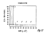
図13及び図14は、例として、中程度及び軽度の打診(tapping)を捉えることができる検知圧力信号を示す。これらの図は、頸部近くの軽度の打診によって引き起される圧力/歪み信号の変化を示す。圧力変化は、打診がセンサの位置から4インチ離れていたときでも、軽度及び中程度の強度の両方の打診に対して敏感である。圧力の絶対測定又は変動測定は、振動を捉えるのに有効である。種々の実施形態は、複数の検知圧力値を分析して、NCTに対する喉頭振動及び/又は咳といった生理反応を検出する。幾つかの実施形態は、神経刺激バーストのタイミングを生理反応のタイミングと対比して、生理反応がNCTに起因するかどうか決定する。種々の実施形態によれば、生理反応は、絶対圧力値の変化を検出すること、検知圧力値の平均を検出すること、及び/又は検知圧力値の変動を検出することによって検出される。図15は、喉頭振動が起きたときの検知圧力の変動の逓増を示す。図16は、咳が起きたときの検知圧力の変動の逓増を示す。限定ではなく例示として、幾つかのシステムの実施形態は、強度増大に伴う検知圧力の変動の傾向を監視し、検知圧力の傾向が閾値より大きく変化した場合に生理反応を検出する。 FIG. 13 and FIG. 14 show, for example, a detected pressure signal that can capture moderate and mild tapping. These figures show the change in pressure / strain signal caused by a mild percussion near the neck. The pressure change is sensitive to both mild and moderate intensity percussion even when the percussion is 4 inches away from the sensor location. Absolute or variable pressure measurements are effective in capturing vibrations. Various embodiments analyze multiple sensed pressure values to detect physiological responses such as laryngeal vibrations and / or cough to NCT. Some embodiments contrast the timing of the neural stimulation burst with the timing of the physiological response to determine whether the physiological response is due to NCT. According to various embodiments, a physiological response is detected by detecting a change in absolute pressure value, detecting an average of sensed pressure values, and / or detecting a variation in sensed pressure value. FIG. 15 shows the increasing variation in the detected pressure when laryngeal vibration occurs. FIG. 16 shows the increasing variation in detected pressure when cough occurs. By way of illustration and not limitation, some system embodiments monitor the trend of sense pressure variation with increasing intensity and detect a physiological response if the sense pressure trend changes more than a threshold.
種々の実施形態は、リード先端部内又は缶内に歪みゲージ(圧力センサ)を組み込む。幾つかの実施形態は、歪みゲージをリード内又は埋込み中に用いられる他の一時的なガイドカテーテル内に組み込む。幾つかの実施形態は、複数の歪みゲージを用いる。例えば、リード上の圧力センサ及びパルス発生器上の圧力センサは、リード先端部の、缶に対する動きを区別するために用いることができ、身体の動きによって誘発される信号変化に関する補正を行うために用いることができる。幾つかの実施形態は、位相コヒーレント検出を用いる。幾つかの実施形態は、圧力センサを、インピーダンスセンサ及び/又は加速度計のような他のセンサと共に用い、及び/又は患者又は医師のフィードバックを用いる。 Various embodiments incorporate a strain gauge (pressure sensor) in the lead tip or in the can. Some embodiments incorporate a strain gauge into the lead or other temporary guide catheter used during implantation. Some embodiments use multiple strain gauges. For example, the pressure sensor on the lead and the pressure sensor on the pulse generator can be used to distinguish the movement of the lead tip relative to the can to make corrections for signal changes induced by body movement. Can be used. Some embodiments use phase coherent detection. Some embodiments use pressure sensors with other sensors such as impedance sensors and / or accelerometers and / or use patient or physician feedback.
本発明の主題の種々の実施形態は、埋込み中に使用されるプログラムされたプロセスを提供する。圧力センサを用いて基線尺度を取得し、刺激強度を段階的に増大させながら刺激が送達される。幾つかの実施形態は、刺激強度を段階的に減少させる。刺激は、各設定において既知の時間にわたって送達される。歪み測定が各々のプリセット時間窓に対して行われる。幾つかの実施形態は、位相コヒーレント検出を用いて周波数フィルタリングを提供する。歪み測定値が分析される。幾つかの実施形態は、歪み測定値変動の尺度を計算する。使用できる方法の例は、標準偏差、範囲、又は、四分位数間範囲(IQR)のような百分位数間範囲(Inter−Percentile Range)を計算することを含む。幾つかの実施形態は、圧力測定値平均の尺度を計算する。使用できる例は、平均値又は中央値を計算することを含む。第1段階の変化が、喉頭振動の開始であると判断される。喉頭振動が開始する強度が、閾値を提供する機能において用いられる。閾値が高い場合にはリードの再配置が推奨される。決定された刺激閾値が記録される。 Various embodiments of the present subject matter provide a programmed process used during embedding. A baseline measure is obtained using a pressure sensor, and the stimulus is delivered while increasing the stimulus intensity in steps. Some embodiments reduce the stimulus intensity in steps. Stimulation is delivered over a known time in each setting. A distortion measurement is made for each preset time window. Some embodiments provide frequency filtering using phase coherent detection. Strain measurements are analyzed. Some embodiments calculate a measure of strain measurement variation. Examples of methods that can be used include calculating a standard deviation, a range, or an inter-percentile range such as an inter-quartile range (IQR). Some embodiments calculate a measure of the average pressure measurement. Examples that can be used include calculating the mean or median. It is determined that the first stage change is the start of laryngeal vibration. The intensity at which the laryngeal vibration begins is used in the function of providing a threshold. Lead relocation is recommended when the threshold is high. The determined stimulation threshold is recorded.
本発明の主題の種々の実施形態は、経過観察(フォローアップ)中に使用されるプログラムされたプロセスを提供する。圧力センサを用いて基線尺度を取得し、刺激強度を段階的に増大させながら刺激が送達される。刺激は、各設定において既知の時間にわたって送達される。歪み測定が各々のプリセット時間窓に対して行われる。幾つかの実施形態は、位相コヒーレント検出を用いて周波数フィルタリングを提供する。幾つかの実施形態は、VSTパルスの送達後の歪み測定値の変化を用い、VSTパルスの送達後の特定の時間窓を見る。典型的には、喉頭振動は、頸部領域におけるVSTの送達後の短時間(〜10ms)の後に起る。従って、各VSTパルスの送達後に、特定の時間窓に従って、歪み測定値の変化を探すことになる。歪み測定値が分析される。幾つかの実施形態は、歪み測定値変動の尺度を計算する。使用できる方法の例は、標準偏差、範囲、又は、四分位数範囲(IQR)のような百分位数範囲を計算することを含む。幾つかの実施形態は、圧力測定値平均の尺度を計算する。使用できる例は、平均値又は中央値を計算することを含む。第1段階の変化が、喉頭振動の開始であると判断される。喉頭振動が開始する強度が、閾値を与える関数において用いられる。例えば、喉頭振動が1mAにおいて検知されたならば、強度閾値を2mAに設定することができる。第2段階の変化を用いて咳を検出することができる。例えば、咳が3.2mAにおいて起こる場合、閾値を3mAに設定することができる。埋込み中又は経過観察中に閾値が高過ぎる場合、物理的再配置及び電子的再配置の両方でのリード再配置を推奨することができる。幾つかの実施形態は、自動刺激閾値決定及び自動刺激捕捉確認を実行する。幾つかの実施形態は、心拍間隔に基づいて自動ルーチンを実行し、例えばそれを各刺激後に実行する。幾つかの実施形態は、リード/電極の移動、リード/電極の状態をモニタするために閾値の周期的な経過観察を行い、治癒をモニタする。幾つかの実施形態は、続けざまの咳が検出された場合、一時的にNCTをオフにするか又はNCTの強度を低減する。 Various embodiments of the present inventive subject matter provide a programmed process used during follow-up. A baseline measure is obtained using a pressure sensor, and the stimulus is delivered while increasing the stimulus intensity in steps. Stimulation is delivered over a known time in each setting. A distortion measurement is made for each preset time window. Some embodiments provide frequency filtering using phase coherent detection. Some embodiments use the change in strain measurements after delivery of the VST pulse and look at a specific time window after delivery of the VST pulse. Typically, laryngeal vibrations occur after a short time (-10 ms) after delivery of VST in the cervical region. Thus, after delivery of each VST pulse, it will look for a change in the distortion measurement according to a specific time window. Strain measurements are analyzed. Some embodiments calculate a measure of strain measurement variation. Examples of methods that can be used include calculating a percentile range such as a standard deviation, range, or quartile range (IQR). Some embodiments calculate a measure of the average pressure measurement. Examples that can be used include calculating the mean or median. It is determined that the first stage change is the start of laryngeal vibration. The intensity at which the laryngeal vibration begins is used in a function that provides a threshold. For example, if laryngeal vibration is detected at 1 mA, the intensity threshold can be set to 2 mA. The change in the second stage can be used to detect cough. For example, if cough occurs at 3.2 mA, the threshold can be set to 3 mA. If the threshold is too high during implantation or follow-up, lead relocation with both physical and electronic relocation can be recommended. Some embodiments perform automatic stimulus threshold determination and automatic stimulus capture confirmation. Some embodiments perform an automatic routine based on the heart rate interval, for example, after each stimulus. Some embodiments monitor the healing by performing periodic follow-up of thresholds to monitor lead / electrode movement, lead / electrode status. Some embodiments temporarily turn off the NCT or reduce the intensity of the NCT if a continuous cough is detected.
本発明の主題の種々の実施形態は、治療の検証に用いられるプログラムされたプロセスを提供する。喉頭振動捕捉が、間欠的に、例えば、限定ではなく例示として、6時間毎、12時間毎、1日毎、1週毎に定期的に検査される。幾つかの実施形態は、喉頭振動捕捉を要求に応じてチェックする。例えば、ユーザは、喉頭振動捕捉のチェックを手動で要求することができる。喉頭振動捕捉がチェックされるたびに、複数の圧力値が検知され分析される。喉頭振動の有無がログ記録され追跡される。捕捉が連続的に不在の場合には、治療設定が修正される。最大設定用量が喉頭振動を引き起さない場合には、通知が送られるか又は警告が発せられる。捕捉が定期的に存在する場合には、ヒストグラムを作成して、捕捉があった測定の回数又は測定の百分率をタイムスタンプとともに伝えることができる。「治療は時間の50%送達された」のようなメッセージを、表示するか又はそれ以外の方法で伝えることができる。 Various embodiments of the present subject matter provide a programmed process that is used for treatment validation. Laryngeal vibration capture is inspected intermittently, for example, by way of example and not limitation, periodically every 6 hours, every 12 hours, every day, every week. Some embodiments check laryngeal vibration capture on demand. For example, the user can manually request a check for laryngeal vibration capture. Each time laryngeal vibration capture is checked, multiple pressure values are detected and analyzed. The presence or absence of laryngeal vibration is logged and tracked. In the absence of continuous capture, the treatment setting is modified. If the maximum set dose does not cause laryngeal vibration, a notification is sent or a warning is issued. If acquisitions are present regularly, a histogram can be created to convey the number of measurements that have been acquired or the percentage of measurements along with a time stamp. A message such as “The therapy was delivered 50% of the time” can be displayed or otherwise communicated.
図17は、種々の実施形態によるVSTシステムを示す。1つの埋込型装置で、全VSTシステムを提供することができる。幾つかの実施形態は、埋込型迷走神経刺激器の埋込み中などに、モニタ機能を提供する外部装置を用いる。幾つかの実施形態は、埋込型リードと外部刺激器とを用いる。図示したVSTシステム127は、VSTを与えるためのパルス発生器128、VSTの強度を変更又は変調するための変調器129、及びフィードバックを与えるためのVST反応モニタ130を含む。自律神経系は、概略的に131で示される。所望の神経刺激を与えるために適当な電極132が用いられ、神経刺激によって影響を受けるパラメータを検知するためにセンサ133が用いられる。VSTに迅速に反応する生理学的パラメータを、閉ループシステムにおいて又は埋込みプロセス中に用いることができる。そのようなパラメータの例には、心拍数、喉頭振動、血圧、呼吸、及び電気記録図のパラメータが含まれる。本発明の主題は、NCTによって引き起される喉頭振動又は咳を検出するために歪みゲージ又は圧力センサを用いる。神経刺激に対する副交感神経系の全体的反応を示す迅速且つ予測可能な反応を有するその他の心臓血管パラメータ及びその他の代理パラメータを用いることができる。より遅い反応を有するその他のパラメータを用いて、治療に有効な用量が送達されていることを確認することができる。センサ及び電極は、単一リード上で一体化することもでき又は複数のリードを用いることもできる。さらに、種々のシステムの実施形態は、別個の又は一体型の埋込型心臓律動管理装置と通信することができる埋込型神経刺激器を用いて機能を実施する。
FIG. 17 illustrates a VST system according to various embodiments. A single implantable device can provide an entire VST system. Some embodiments use an external device that provides a monitoring function, such as during implantation of an implantable vagus nerve stimulator. Some embodiments use an implantable lead and an external stimulator. The illustrated VST system 127 includes a pulse generator 128 for providing VST, a
図示した反応モニタ130は、有刺激時間中のパラメータをモニタして有刺激時間に対応するパラメータ値を示す第1のフィードバック信号134を与え、無刺激時間中のパラメータをモニタして無刺激時間に対応するパラメータ値を示す第2のフィードバック信号135を与える。信号134及び135は、別々の線で描かれている。これらの信号134及び135は、異なる信号経路上又は同じ信号経路上で送ることができる。比較器136は、第1及び第2のフィードバック信号134及び135を受取り、これらの信号に基づいてパラメータ値の検出された変化を判断する。さらに、比較器は、検出された変化と、装置内にプログラムすることができる許容される変化とを比較する。例えば、装置は、VST中の心拍数減少が、無刺激のときの心拍数に対して所定の百分率(例えば、95%程度)を下回らないことを許容するようにプログラムすることができる。装置は、VST中の心拍数減少が、無刺激のときの心拍数からある数量値(例えば、毎分5拍)だけ少ない心拍数を下回らないことを許容するように、数量値を用いてプログラムすることができる。検出された変化(信号134及び135に基づく)と許容される変化との比較が比較結果141を与え、これを用いて変調器を適切に制御して、施されるVSTを調整する。
The illustrated reaction monitor 130 monitors a parameter during the stimulation time and provides a first feedback signal 134 indicating a parameter value corresponding to the stimulation time, and monitors the parameter during the stimulation time to the stimulation time. A
図示した装置は、圧力センサ137と、圧力変動分析器のような圧力分析器138とを含む。分析器は、複数の検知圧力値を分析して、喉頭振動及び/又は咳が神経刺激によって引き起されたかどうかを決定する。装置は、咳を表すことができるような上限値140及び喉頭振動を表すことができるような下限値139を用いてプログラムすることができる。圧力分析器138の出力は、下限及び上限と比較され、VST強度が限界から外れていないか判断される。 The illustrated apparatus includes a pressure sensor 137 and a pressure analyzer 138, such as a pressure fluctuation analyzer. The analyzer analyzes the plurality of sensed pressure values to determine whether laryngeal vibration and / or cough was caused by neural stimulation. The device can be programmed with an upper limit 140 that can represent cough and a lower limit 139 that can represent laryngeal vibration. The output of the pressure analyzer 138 is compared with the lower limit and the upper limit to determine whether the VST intensity is out of the limit.
幾つかの実施形態は、VST強度の上限によって制限されるVST強度を調整する治療プロトコルを使用し、幾つかの実施形態においてはVST強度の下限によって制限される治療プロトコルを使用する。VST強度は、本発明の主題により設定される許容限界内で、血圧、呼吸、及び電気記録図測定値などの他のパラメータに基づいて調整することができる。幾つかの治療プロトコルは、VST強度の上限及び/又は下限を、スケジュール(例えば、1日のうちの時刻)又は検知データ(例えば、活動)に基づいて調整する。 Some embodiments use a treatment protocol that adjusts the VST intensity limited by the upper limit of VST intensity, and in some embodiments, uses a treatment protocol that is limited by the lower limit of VST intensity. The VST intensity can be adjusted based on other parameters such as blood pressure, respiration, and electrogram measurements within acceptable limits set by the subject matter of the present invention. Some treatment protocols adjust the upper and / or lower limits of VST intensity based on a schedule (eg, time of day) or detection data (eg, activity).
種々の変調器の実施形態は、VST強度を、VSTを与えるのに用いられる刺激信号の振幅を変更すること、VSTを与えるのに用いられる刺激信号の周波数を変更すること、VSTを与えるのに用いられる刺激信号のバースト周波数を変更すること、VSTを与えるのに用いられる刺激信号のパルス幅を変更すること、VSTを与えるのに用いられる刺激信号のデューティサイクルを変更すること、又はこれらの刺激信号特性の2つ又はそれ以上の種々の組合せにより調整する。 Various modulator embodiments may provide VST intensity, changing the amplitude of the stimulus signal used to provide VST, changing the frequency of the stimulus signal used to provide VST, and providing VST. Changing the burst frequency of the stimulus signal used, changing the pulse width of the stimulus signal used to provide the VST, changing the duty cycle of the stimulus signal used to provide the VST, or these stimuli Adjust by various combinations of two or more of the signal characteristics.
VSTを送達するための図示したシステムは、広範な治療用途に有用である。広範な治療用途の例は、心臓組織のリモデリングを防ぐため及び心臓血管系疾患状態にある心臓組織を逆リモデリングするために刺激を与えることを伴う。VSTは、例えば、毎分のうちの一部分(約10秒)の間、与えることができる。VST用量は、刺激の継続時間又はデューティサイクルを調整することによって調整することができる(例えば、1分毎に約5秒又は15秒、又は30秒毎に約5乃至15秒、又は2分毎に約5乃至30秒、又は5分毎に約5秒乃至3分、又は連続刺激)。実施形態によれば、VSTは、遠心性軸索及び求心性軸索の両方を非選択的に刺激する。例示した値は、例示として提示したものであり限定ではない。数日、数週間、数ヶ月及び数年の過程にわたって、VSTに対する生理反応は、神経の順応、組織のカプセル化、線維症、インピーダンス変化などといった多くの理由で変化し得る。種々の閉ループシステムの実施形態は、VSTに対する迅速且つ予測可能な反応を有する少なくとも1つのパラメータをモニタし、このモニタしたパラメータを用いて、副交感神経系の所望の刺激をもたらすように神経刺激信号を適切に変化させる。幾つかの実施形態は、心拍数をモニタする。幾つかの実施形態は、喉頭振動をモニタし、VSTが喉頭振動を誘発するように必要に応じてVST強度を調整する。 The illustrated system for delivering VST is useful for a wide range of therapeutic applications. Examples of a wide range of therapeutic applications involve providing stimulation to prevent remodeling of heart tissue and reverse remodeling of heart tissue that is in a cardiovascular disease state. The VST can be given, for example, for a portion of each minute (about 10 seconds). The VST dose can be adjusted by adjusting the duration or duty cycle of the stimulus (eg, about 5 or 15 seconds per minute, or about 5 to 15 seconds every 30 seconds, or every 2 minutes About 5 to 30 seconds, or about 5 seconds to 3 minutes every 5 minutes, or continuous stimulation). According to embodiments, VST nonselectively stimulates both efferent and afferent axons. The illustrated values are presented as examples and are not limiting. Over the course of days, weeks, months and years, the physiological response to VST can change for a number of reasons, such as nerve adaptation, tissue encapsulation, fibrosis, impedance changes, and the like. Various closed-loop system embodiments monitor at least one parameter having a rapid and predictable response to the VST and use the monitored parameter to generate a neural stimulation signal to provide a desired stimulation of the parasympathetic nervous system. Change appropriately. Some embodiments monitor heart rate. Some embodiments monitor laryngeal vibration and adjust the VST intensity as necessary so that the VST induces laryngeal vibration.
開ループVSTシステムは、VSTの心拍数効果を回避するか又は減らすようにVST強度を設定する。開ループVSTシステムの場合、VST試験中に心拍数がモニタされる。このVST試験は、心拍数閾値を決定するための比較的大きな人口母集団に基づくことができる。VST試験はまた、埋込み手術中に、観測される心拍数減少を用いて迷走神経の捕捉を確認し、心拍数減少が観測される強度閾値を決定し、この強度閾値を用いて、上限を与えるか又はそれ以外の方法でVST強度を心拍数閾値より低く設定するプロセスを用いて、実施することができる。幾つかの実施形態によれば、VST強度の下限は、埋込みプロセス中に設定することができる。例えば、喉頭振動は、心拍数効果が検出されるVST強度レベルより低いVST強度レベルにおいて、患者が感じるか又は加速度計のようなセンサによって検知される。パラメータ設定の組合せは、あらゆる著しい徐脈効果を回避するように選択される。幾つかの実施形態は、あらゆる徐脈効果を回避する。幾つかの実施形態は、比較的著しくない量の心拍数低下(例えば、VST中の心拍数がVSTなしの場合の心拍数の95%)を許容する。VST強度の上限は、VSTなしの場合の本来の心拍数からVSTによって引き起される許容される心拍数の変化に基づく。 The open loop VST system sets the VST intensity to avoid or reduce the heart rate effect of VST. For an open loop VST system, the heart rate is monitored during the VST test. This VST test can be based on a relatively large population population to determine heart rate thresholds. The VST test also verifies the capture of the vagus nerve using the observed heart rate reduction during implantation surgery, determines the intensity threshold at which heart rate reduction is observed, and uses this intensity threshold to give an upper limit Alternatively, or otherwise, it can be implemented using a process that sets the VST intensity below the heart rate threshold. According to some embodiments, the lower limit of VST intensity can be set during the implantation process. For example, laryngeal vibrations are felt by a patient or detected by a sensor such as an accelerometer at a VST intensity level that is lower than the VST intensity level at which the heart rate effect is detected. The combination of parameter settings is selected to avoid any significant bradycardic effects. Some embodiments avoid any bradycardic effect. Some embodiments allow a relatively insignificant amount of heart rate reduction (eg, 95% of the heart rate when the heart rate during VST is without VST). The upper limit of VST intensity is based on the change in allowable heart rate caused by VST from the original heart rate without VST.
図18は、本発明の主題の種々の実施形態による、神経刺激(NS)構成要素143及び心臓律動管理(CRM)構成要素144を含む埋込型医療装置(IMD)142を示す。図示した装置は、コントローラ及びメモリを含む。種々の実施形態によれば、コントローラは、神経刺激及びCRM機能を実施するハードウェア、ソフトウェア、又はハードウェアとソフトウェアとの組合せを含む。例えば、本開示で論じたプログラムされた治療アプリケーションは、メモリ内に具体化され、プロセッサによって実行される、コンピュータ可読命令として格納することが可能である。例えば、本明細書で開示されるような、治療スケジュール、プログラム可能パラメータ、及び閾値検出又は用量設定アルゴリズムをメモリに格納することができる。さらに、幾つかの実施形態は、神経刺激の閾値を検出するための閾値検出ルーチンを格納し、幾つかの実施形態は、用量を用量設定するための用量設定ルーチンを格納する。種々の実施形態によれば、コントローラは、メモリ内に埋め込まれた命令を実行して神経刺激及びCRM機能を実施するためのプロセッサを含む。図示した神経刺激治療145は、心不全又は他の心臓血管系疾患を治療するためのVSTのようなVSTを含むことができる。種々の実施形態は、CRM治療146、例えば、徐脈ペーシング、ATPなどの抗頻脈性不整脈治療、除細動及び電気的除細動、並びに心臓再同期治療(CRT)を含む。図示した装置はさらに、プログラマ又は別の外部若しくは内部装置との通信に用いるための送受信機147及び関連回路を含む。種々の実施形態は、遠隔計測コイルを含む。
FIG. 18 illustrates an implantable medical device (IMD) 142 that includes a neural stimulation (NS)
CRM治療構成要素144は、コントローラの制御下で1つ又はそれ以上の電極を用いて心臓を刺激する及び/又は心臓信号を検知する構成要素を含む。図示したCRM治療部分は、電極を通して心臓を刺激する電気信号を供給するために用いるパルス発生器148を含み、さらに、検知心臓信号を検出し処理するための検知回路149を含む。インタフェース150は、コントローラ143と、パルス発生器148及び検知回路149との間の通信に用いるように一般的に描かれている。3つの電極が、CRM治療を施すために使用するように例として描かれている。しかし、本発明の主題は、特定の数の電極部位に限定されない。各々の電極は、それ自体のパルス発生器及び検知回路を含むことができる。しかし、本発明の主題はそのように限定されない。パルス発生及び検知機能は、複数の電極で機能するように多重化することができる。
The
NS治療構成要素143は、コントローラの制御下で、神経刺激標的を刺激する、及び/又は、神経活動に、若しくは心拍数、血圧、呼吸などの神経活動の代理に関連付けられたパラメータを検知する、構成要素を含む。3つのインタフェース151が、神経刺激を与えるために使用されるように描かれている。しかし、本発明の主題は、特定の数のインタフェースにも、又はいかなる特定の刺激機能若しくは検知機能にも限定されない。パルス発生器152は、神経刺激標的を刺激するために使用される1つ又は複数のトランスデューサ/電極に電気パルスを供給するために用いられる。種々の実施形態によれば、パルス発生器は、刺激パルスの振幅、刺激パルスのパルス幅、刺激パルスの周波数、パルスのバースト周波数、並びに、パルスの形態、例えば、矩形波、三角波、正弦波、及び白色ノイズ又は他の信号を模倣するための所望の高調波を有する波を設定するための、及び幾つかの実施形態においてはそれらを変更するための回路を含む。検知回路153は、センサ、例えば、神経活動、心拍数、血圧、呼吸などのセンサからの信号を検出して処理するために用いられる。センサは、喉頭振動を検知するのに用いることができる。センサは、状態を検出するのに用いることができる(例えば、活動を検出するのに用いられる加速度計)。インタフェース151は、コントローラ143と、パルス発生器152及び検知回路153との間の通信に用いられるように一般的に描かれている。各インタフェースは、例えば、別個のリードを制御するために用いることができる。NS治療部分の種々の実施形態は、神経標的を刺激するためのパルス発生器のみを含む。図示した装置は、さらにクロック/タイマ154を含み、これを用いて、プログラムされた刺激プロトコル及び/又はスケジュールに従って、プログラムされた治療を送達することができる。
図19は、種々の実施形態による、マイクロプロセッサをベースとする埋込型装置の実施形態のシステム図を示す。この装置のコントローラは、マイクロプロセッサ155であり、これは双方向データバスを介してメモリ156と通信する。コントローラは、状態機械型設計を用いて他の型式の論理回路(例えば、個別の構成要素又はプラグラム可能論理アレイ)によって実装することができる。本明細書で用いる場合、「回路」という用語は、個別の論理回路又はマイクロセッサのプログラミングのいずれかを指すものと解釈されたい。図に示したのは、「A」乃至「C」で示された検知及びペーシングチャネルの3つの例であり、リング電極157A−C及びチップ電極158A−Cを有する双極リード、検知増幅器159A−C、ペーシング刺激160A−C、並びにチャネルインタフェース161A−Cを含む。従って、各チャネルは、電極に接続されたパルス発生器で構成されたペーシングチャネルと、電極に接続された検知増幅器で構成された検知チャネルとを含む。チャネルインタフェース161A−Cは、マイクロプロセッサ155と双方向通信し、各インタフェースは、検知増幅器からの検知信号入力をデジタル化するためのアナログ・デジタル変換器と、ペーシングパルスを出力するため、ペーシングパルス振幅を変更するため、及び検知増幅器の利得及び閾値を調整するためにマクロプロセッサによって書き込むことができるレジスタとを含むことができる。ペースメーカの検知回路は、特定のチャネルによって生成された電気記録図信号(即ち、電極によって検知される、心臓の電気的活動を表す電圧)が指定された検出閾値を超えたときに、心房センス又は心室センスのいずれかであるチャンバセンスを検出する。特定のペーシングモードに用いられるペーシングアルゴリズムは、それらセンスを用いて、ペーシングをトリガ又は抑制する。固有の心房及び/又は心室レートは、それぞれ心房センス間及び心室センス間の時間間隔を測定することによって測定することができ、心房及び心室の頻脈性不整脈を検出するのに用いることができる。
FIG. 19 shows a system diagram of an embodiment of a microprocessor-based implantable device, according to various embodiments. The controller for this device is a
各々の双極リードの電極は、リード内の導電体を介して、マイクロプロセッサにより制御されるスイッチングネットワーク162に接続される。スイッチングネットワークを用いて、電極を、本来の心臓活動を検出するために検知増幅器の入力部に切替え、ペーシングパルスを送出するためにパルス発生器の出力部に切替える。スイッチングネットワークはまた、装置が、リードのリング電極及びチップ電極の両方を用いる双極モード、又はリードの電極のうちの一方のみを接地電極として機能する装置筐体(缶)163又は別のリード上の電極と共に用いる単極モードのいずれかで検知又はペーシングを行うことを可能にする。さらに、ショックを要する(shockable)頻脈性不整脈が検出されたときに心房又は心室にショック電極(例えば、電極165及び166)を介して除細動ショックを送達するために、ショックパルス発生器164がコントローラにインタフェースされる。
The electrode of each bipolar lead is connected via a conductor in the lead to a
神経刺激チャネルは、チャネルD及びEとして識別され、副交感神経刺激を送達し及び/又は交感神経を抑制するために装置内に組み込まれ、ここで、一方のチャネルは、第1の電極167D及び第2の電極168Dを有する双極リード、ペーシング刺激169D、及びチャネルインタフェース170Dを含み、他方のチャネルは、第1の電極167E及び第2の電極168Eを有する双極リード、ペーシング刺激169E、及びチャネルインタフェース170Eを含む。他の実施形態は、単極リードを用いることができ、その場合、神経刺激パルスは、缶又は別の電極を基準とする。他の実施形態は、3極又は多極リードを用いることができる。種々の実施形態において、各チャネルのためのパルス発生器は、神経刺激パルス列を出力し、その振幅、周波数、デューティサイクルなどはコントローラによって変えることができる。この実施形態において、神経刺激チャネルの各々は、適切な神経標的の近傍に血管内配置することができるリードを用いる。他の型式のリード及び/又は電極を使用することもできる。神経カフ電極を血管内配置される電極の代りに用いて神経刺激を与えることができる。幾つかの実施形態においては、神経刺激電極のリードは、無線リンクで置き換えられる。センサ171は、マイクロプロセッサによって、捕捉対象(例えば、喉頭振動)、治療の効力(例えば、心拍数、血圧)を判断するため、及び/又は事象(例えば咳)若しくは状態(例えば、活動センサ)を検出するために使用される。
Nerve stimulation channels are identified as channels D and E and are incorporated into the device to deliver parasympathetic stimulation and / or suppress sympathetic nerves, where one channel is the first electrode 167D and the second electrode. A bipolar lead having two
この図は、マイクロプロセッサに接続された遠隔計測インタフェース172を示し、これは外部装置との通信に用いることができる。図示したマイクロプロセッサは、神経刺激治療ルーチン及び心筋(CRM)刺激ルーチンを実施することが可能である。NS治療ルーチンの例は、心筋治療を提供するためのVST治療を含む。NS治療ルーチンは、本明細書で説明するルーチン又はアルゴリズムも含む。心筋治療ルーチンの例には、徐脈ペーシング治療、電気的除細動又は除細動治療のような抗頻脈性不整脈ショック治療、抗頻脈性不整脈ペーシング治療(ATP)、及び心臓再同期治療(CRT)が含まれる。
This figure shows a
図20−図21は、VSTを施すように適合されたシステムの実施形態を示し、これは左右両方の迷走神経を刺激することができる両側性システムとして描かれている。当業者であれば、本開示を読んで理解すれば、システムを右迷走神経のみを刺激するように設計することができること、システムを左迷走神経のみを刺激するように設計することができること、及びシステムを左右両方の迷走神経を両側的に刺激するように設計することができることを理解するであろう。システムは、神経トラフィックを刺激する(迷走神経が刺激されたときに副交感神経反応をもたらす)ように、又は神経トラフィックを抑制する(迷走神経が抑制されたときに交感神経反応をもたらす)ように設計することができる。種々の実施形態は、単方向刺激、又は神経内の幾つかの神経線維の選択的刺激を送達する。図20−図21は、迷走神経を刺激するためのリードの使用を示す。無線技術でリードを置き換えて、リードレス電極を、迷走神経を刺激するように適合させ、さらに、VSTの制御に使用するための埋込型システムと無線通信するように適合させることができる。 FIGS. 20-21 illustrate an embodiment of a system adapted to apply VST, which is depicted as a bilateral system capable of stimulating both the left and right vagus nerves. Those skilled in the art, upon reading and understanding the present disclosure, can design the system to stimulate only the right vagus nerve, can design the system to stimulate only the left vagus nerve, and It will be appreciated that the system can be designed to stimulate both the left and right vagus nerves bilaterally. The system is designed to stimulate nerve traffic (provides a parasympathetic response when the vagus nerve is stimulated) or to suppress nerve traffic (provides a sympathetic reaction when the vagus nerve is inhibited) can do. Various embodiments deliver unidirectional stimulation or selective stimulation of several nerve fibers within the nerve. FIGS. 20-21 illustrate the use of leads to stimulate the vagus nerve. The leads can be replaced with wireless technology and the leadless electrode can be adapted to stimulate the vagus nerve and further adapted to communicate wirelessly with an implantable system for use in controlling the VST.
図20は、IMD173が患者の胸部の皮下又は筋肉下に置かれ、リード174が迷走神経を刺激するように配置されたシステムの実施形態を示す。種々の実施形態によれば、神経刺激リード174は、皮下を通って神経標的に達し、神経標的を刺激するための神経カフ電極を有することができる。幾つかの迷走神経刺激リードの実施形態は、神経標的近傍の血管に血管内を通して送り込まれ、血管内の電極を用いて経血管的に神経標的を刺激する。例えば、幾つかの実施形態では、内頸静脈内に配置された電極を用いて迷走神経を刺激する。他の実施形態では、気管、内頸静脈の喉頭枝、及び鎖骨下静脈の内部から、神経標的に刺激が送達される。神経標的は、超音波及び光エネルギー波形といった他のエネルギー波形を用いて刺激することができる。図示したシステムは、装置の筐体上のリードレスECG電極175を含む。これらのECG電極は、例えば、心拍数を検出するのに用いることができる。
FIG. 20 shows an embodiment of a system where the IMD 173 is placed subcutaneously or submuscularly in the patient's chest and the
図21は、種々の実施形態による、患者胸部の皮下又は筋肉下に置かれ、リード177が心臓にCRM治療を施すように配置され、さらに、リード178が迷走神経などの神経標的における神経トラフィックを刺激及び/又は抑制するように配置された、IMD176を示す。種々の実施形態によれば、神経刺激リードは、皮下を通って神経標的に達し、神経標的を刺激するための神経カフ電極を有することができる。幾つかのリードの実施形態は、神経標的近傍の血管に血管内を通して送り込まれ、血管内のトランスデューサを用いて経血管的に神経標的を刺激する。例えば、幾つかの実施形態は、内頸静脈内に配置された電極を用いて迷走神経を標的にする。
FIG. 21 is placed under the skin or muscle of a patient's chest according to various embodiments, the lead 177 is positioned to perform CRM therapy on the heart, and the
図22は、外部システム179の実施形態を示すブロック図である。外部システムは、幾つかの実施形態において、プログラマを含む。図示した実施形態において、外部システムは、患者管理システムを含む。図示したように、外部システムは、外部装置180、電気通信ネットワーク181、及び遠隔装置182を含む患者管理システムである。外部装置180は、埋込型医療装置(IMD)の近傍に配置され、IMDと通信するための外部遠隔計測システム183を含む。遠隔装置は、1つ又はそれ以上の遠隔地に配置され、ネットワークを通して外部装置と通信し、従って、医師又は他の介護者が遠隔地から患者をモニタして処置することを可能にし、及び/又は、1つ又はそれ以上の遠隔地から種々の処置リソースにアクセスすることを可能にする。図示した遠隔装置は、ユーザインタフェース184を含む。種々の実施形態によれば、外部装置は、神経刺激器、プログラマ、又は、コンピュータ、携帯データ端末若しくは携帯電話のような他の装置を含む。外部装置は、種々の実施形態において、適当な通信チャネル上で互いに通信するように適合された2つの装置、例えば、限定ではなく例示としてコンピュータなどを含む。外部装置は、例えば患者の不快感を示すフィードバックを与えるために、患者又は医師が使用することができる。
FIG. 22 is a block diagram illustrating an embodiment of the
当業者が本発明の主題を読んで把握すれば理解するように、本発明の主題の種々の実施形態は、患者の治療受容性を改善し、治療の有効レベルを維持し、治療管理における患者の自由度を可能にし、NCTを受ける患者の生活の質を全般的に向上させる。本明細書で図示及び説明したモジュール及び他の回路は、ソフトウェア、ハードウェア、ファームウェア及びそれらの組合せを用いて実装することができる。 As those skilled in the art will appreciate upon reading and understanding the subject matter of the present invention, various embodiments of the subject matter of the present invention improve patient therapeutic acceptability, maintain an effective level of treatment, and provide patient control in treatment management. Freedom of life and generally improve the quality of life of patients undergoing NCT. The modules and other circuits shown and described herein can be implemented using software, hardware, firmware, and combinations thereof.
上記の詳細な説明は、例証的であることを意図したものであり、限定を意図したものではない。上記の説明を読んで理解すれば、当業者には他の実施形態は明らかとなろう。従って、本発明の範囲は、添付の特許請求の範囲とそのような特許請求の範囲が権利を付与する均等物の全範囲とに関して定められるべきである。 The above detailed description is intended to be illustrative and not limiting. Other embodiments will be apparent to those of skill in the art upon reading and understanding the above description. Accordingly, the scope of the invention should be determined with reference to the appended claims, along with the full scope of equivalents to which such claims are entitled.
100、174、177、178:リード
101:圧力検知素子
102:リードボディ
103:近位領域
104:遠位領域
105:遠位端部分
106:遠位チップ
107:電極
108:近位開口部
109:遠位開口部
110:予備成形部分
111:内部空間
112:導電部材
113:圧力トランスデューサ
114:血管
115:バースト
116:バースト周期
127:VSTシステム
142、173、176:埋込型医療装置(IMD)
143:神経刺激(NS)構成要素
144:心臓律動管理(CRM)構成要素
148、152:パルス発生器
149、153:検知回路
157A−C:リング電極
158A−C:チップ電極
159A−C:検知増幅器
160A−C、169D、169E:ペーシング刺激
162:スイッチングネットワーク
163:装置筐体(缶)
165、166、167D、167E、168D、168E:電極
175:ECG電極
100, 174, 177, 178: Lead 101: Pressure sensing element 102: Lead body 103: Proximal region 104: Distal region 105: Distal end portion 106: Distal tip 107: Electrode 108: Proximal opening 109: Distal opening 110: preformed portion 111: interior space 112: conductive member 113: pressure transducer 114: blood vessel 115: burst 116: burst period 127: VST system 142, 173, 176: implantable medical device (IMD)
143: Neural stimulation (NS) component 144: Cardiac rhythm management (CRM)
165, 166, 167D, 167E, 168D, 168E: Electrode 175: ECG electrode
Claims (12)
喉頭振動によって引き起される患者の頸部領域内の圧力変化を検知するように構成された埋込型圧力センサを用いて圧力を検知する手段であって、圧力を複数回検知して複数の検知圧力値を提供する手段を含む、圧力を検知する手段と、
前記喉頭振動を確認するために、前記複数の検知圧力値を分析する手段と、
患者の迷走神経に迷走神経刺激治療(VST)を送達するための閾値決定ルーチンを実行する手段であって、
前記迷走神経にVSTを所定のVST強度レベルで送達する手段、
前記VSTの強度を複数の強度段階で増大させる手段、及び
各強度段階において喉頭振動をモニタする手段、
を含む、閾値決定ルーチンを実行する手段と、
閾値検証ルーチンを間欠的に実行する手段であって、
前記閾値検証ルーチンが実行されるたびに、圧力を複数回検知して複数の検知圧力値を提供する手段、
前記閾値検証ルーチンが実行されるたびに、前記複数の検知圧力値の変動を分析して前記VST強度レベルにおける前記喉頭振動を確認する手段、及び
前記複数の検知圧力値の前記変動を分析して得られた結果を記録する手段、又は前記VST強度レベルにおいて喉頭振動が確認されない場合に患者又は医師に通知する手段、又は前記VST強度レベルにおいて前記喉頭振動が確認されない場合に、喉頭振動が確認される強度段階を識別する手段、
を含む、閾値検証ルーチンを間欠的に実行する手段と、
を含むことを特徴とするシステム。 A system for detecting laryngeal vibrations,
Means for detecting pressure using an implantable pressure sensor configured to detect pressure changes in a patient's neck region caused by laryngeal vibration, wherein the pressure is detected multiple times Means for sensing pressure, including means for providing a sensed pressure value;
Means for analyzing the plurality of detected pressure values to confirm the laryngeal vibration;
Means for performing a thresholding routine for delivering vagal stimulation therapy (VST) to a patient's vagus nerve, comprising:
Means for delivering VST to the vagus nerve at a predetermined VST intensity level;
Means for increasing the intensity of the VST at a plurality of intensity stages; and means for monitoring laryngeal vibration at each intensity stage;
Means for executing a threshold determination routine, comprising:
Means for intermittently executing a threshold verification routine,
Means for detecting a pressure multiple times and providing a plurality of detected pressure values each time the threshold verification routine is executed;
Means for analyzing the variation of the plurality of detected pressure values each time the threshold verification routine is executed to confirm the laryngeal vibration at the VST intensity level; and
Means for recording a result obtained by analyzing the variation of the plurality of detected pressure values , or means for notifying a patient or doctor when no laryngeal vibration is confirmed at the VST intensity level, or at the VST intensity level. A means to identify the intensity level at which laryngeal vibration is observed if laryngeal vibration is not confirmed,
Means for intermittently executing a threshold verification routine including:
A system characterized by including.
喉頭振動が見出された強度段階より高い複数の強度段階の各々について、前記埋込型圧力センサを用いて圧力を検知して複数の検知圧力値を提供する手段と、
喉頭振動が見出された強度段階より高い複数の強度段階の各々について前記複数の検知圧力値を分析する手段であって、前記検知圧力値の絶対値、平均又は変動における喉頭振動レベルからの増大が咳を示す、分析する手段と
を含むことを特徴とする請求項4〜請求項9のいずれかに記載のシステム。 Means for performing a cough detection routine for delivering a VST after laryngeal vibration is found, the means for performing the cough detection routine comprising:
Means for detecting a pressure using the implantable pressure sensor to provide a plurality of detected pressure values for each of a plurality of intensity steps higher than the intensity step in which laryngeal vibration was found;
Means for analyzing said plurality of detected pressure values for each of a plurality of intensity steps higher than the intensity step at which the laryngeal vibration was found, the increase from the laryngeal vibration level in absolute value, average or variation of said detected pressure value 10. A system according to any one of claims 4 to 9, characterized in that comprises a means for analyzing which indicates cough.
Applications Claiming Priority (3)
| Application Number | Priority Date | Filing Date | Title |
|---|---|---|---|
| US201061427985P | 2010-12-29 | 2010-12-29 | |
| US61/427,985 | 2010-12-29 | ||
| PCT/US2011/062921 WO2012091849A1 (en) | 2010-12-29 | 2011-12-01 | Neuro cardiac therapy using sensed pressure |
Publications (2)
| Publication Number | Publication Date |
|---|---|
| JP2014503310A JP2014503310A (en) | 2014-02-13 |
| JP5646772B2 true JP5646772B2 (en) | 2014-12-24 |
Family
ID=45217733
Family Applications (1)
| Application Number | Title | Priority Date | Filing Date |
|---|---|---|---|
| JP2013547489A Expired - Fee Related JP5646772B2 (en) | 2010-12-29 | 2011-12-01 | Cardiac nerve treatment using detected pressure |
Country Status (4)
| Country | Link |
|---|---|
| US (1) | US9393417B2 (en) |
| EP (1) | EP2658601A1 (en) |
| JP (1) | JP5646772B2 (en) |
| WO (1) | WO2012091849A1 (en) |
Families Citing this family (7)
| Publication number | Priority date | Publication date | Assignee | Title |
|---|---|---|---|---|
| AU2011353038B2 (en) | 2010-12-29 | 2015-01-22 | Cardiac Pacemakers, Inc. | Neuro cardiac therapy using electrical impedance |
| US9855431B2 (en) | 2012-03-19 | 2018-01-02 | Cardiac Pacemakers, Inc. | Systems and methods for monitoring for nerve damage |
| JP6085429B2 (en) * | 2012-07-19 | 2017-02-22 | 株式会社 東北テクノアーチ | Surface-type vagus nerve electrical stimulation device with cardiac function monitoring device |
| EP2977078B1 (en) | 2014-07-23 | 2017-10-25 | Sorin CRM SAS | Active implantable medical device for therapy by vagus nerve stimulation, with dynamically adjusting stimulation periods |
| EP3180069B1 (en) | 2014-08-17 | 2020-05-13 | Nine Continents Medical, Inc. | Miniature implatable neurostimulator system for sciatic nerves and their branches |
| US12053630B2 (en) | 2014-08-17 | 2024-08-06 | Coloplast A/S | Implantable pulse generator with automatic jump-start |
| US10542961B2 (en) | 2015-06-15 | 2020-01-28 | The Research Foundation For The State University Of New York | System and method for infrasonic cardiac monitoring |
Family Cites Families (27)
| Publication number | Priority date | Publication date | Assignee | Title |
|---|---|---|---|---|
| AU2003901025A0 (en) | 2003-02-28 | 2003-03-20 | The University Of Melbourne | Cochlear implant found processing method and system |
| WO2004091503A2 (en) * | 2003-04-10 | 2004-10-28 | Vivometrics, Inc. | Systems and methods for respiratory event detection |
| US7502650B2 (en) | 2003-09-22 | 2009-03-10 | Cvrx, Inc. | Baroreceptor activation for epilepsy control |
| EP1879501A4 (en) * | 2005-04-29 | 2009-05-20 | Oren Gavriely | Cough detector |
| US7907997B2 (en) | 2005-05-11 | 2011-03-15 | Cardiac Pacemakers, Inc. | Enhancements to the detection of pulmonary edema when using transthoracic impedance |
| US7551958B2 (en) | 2005-05-24 | 2009-06-23 | Cardiac Pacemakers, Inc. | Safety control system for implantable neural stimulator |
| US7662105B2 (en) | 2005-12-14 | 2010-02-16 | Cardiac Pacemakers, Inc. | Systems and methods for determining respiration metrics |
| US8103341B2 (en) * | 2006-08-25 | 2012-01-24 | Cardiac Pacemakers, Inc. | System for abating neural stimulation side effects |
| US7801603B2 (en) * | 2006-09-01 | 2010-09-21 | Cardiac Pacemakers, Inc. | Method and apparatus for optimizing vagal nerve stimulation using laryngeal activity |
| US8406877B2 (en) | 2007-03-19 | 2013-03-26 | Cardiac Pacemakers, Inc. | Selective nerve stimulation with optionally closed-loop capabilities |
| US8224436B2 (en) | 2007-04-02 | 2012-07-17 | Cardiac Research, Inc. | Unidirectional neural stimulation systems, devices and methods |
| US8483831B1 (en) * | 2008-02-15 | 2013-07-09 | Holaira, Inc. | System and method for bronchial dilation |
| JP5427233B2 (en) | 2008-07-08 | 2014-02-26 | カーディアック ペースメイカーズ, インコーポレイテッド | Medical system delivering vagus nerve stimulation |
| EP3714771A1 (en) | 2008-10-01 | 2020-09-30 | Inspire Medical Systems, Inc. | System for treating sleep apnea transvenously |
| US9026206B2 (en) * | 2008-10-31 | 2015-05-05 | Medtronic, Inc. | Therapy system including cardiac rhythm therapy and neurostimulation capabilities |
| US9370654B2 (en) * | 2009-01-27 | 2016-06-21 | Medtronic, Inc. | High frequency stimulation to block laryngeal stimulation during vagal nerve stimulation |
| EP2401029A1 (en) | 2009-01-30 | 2012-01-04 | Medtronic, Inc. | Closed-loop neurostimulation to treat pulmonary edema |
| CN102481453B (en) | 2009-07-15 | 2014-10-08 | 心脏起搏器股份公司 | Physiological vibration detection in implanted medical devices |
| JP5613234B2 (en) | 2009-07-15 | 2014-10-22 | カーディアック ペースメイカーズ, インコーポレイテッド | Remote pace detection in implantable medical devices |
| AU2010273544B2 (en) | 2009-07-15 | 2013-05-02 | Cardiac Pacemakers, Inc. | Remote sensing in an implantable medical device |
| CN102958563B (en) | 2010-05-11 | 2016-01-13 | 心脏起搏器股份公司 | There is the implanted medical equipment of the device automatically recovering the treatment interrupted |
| WO2011159527A2 (en) | 2010-06-16 | 2011-12-22 | Cardiac Pacemakers, Inc. | Automatic neural stimulation titration sweep |
| US8433419B2 (en) | 2010-10-13 | 2013-04-30 | Cardiac Pacemakers, Inc. | Method and apparatus for controlling neurostimulation according to physical state |
| CN103249453B (en) | 2010-12-07 | 2016-03-16 | 心脏起搏器股份公司 | Systems and methods for increasing stimulation dose |
| AU2011353038B2 (en) | 2010-12-29 | 2015-01-22 | Cardiac Pacemakers, Inc. | Neuro cardiac therapy using electrical impedance |
| WO2012148845A1 (en) | 2011-04-25 | 2012-11-01 | Cardiac Pacemakers, Inc. | Systems to account for neck movement during nerve stimulation |
| US8594805B2 (en) | 2011-08-23 | 2013-11-26 | Cardiac Pacemakers, Inc. | Systems to detect vagus capture |
-
2011
- 2011-12-01 US US13/309,328 patent/US9393417B2/en not_active Expired - Fee Related
- 2011-12-01 WO PCT/US2011/062921 patent/WO2012091849A1/en not_active Ceased
- 2011-12-01 JP JP2013547489A patent/JP5646772B2/en not_active Expired - Fee Related
- 2011-12-01 EP EP11793643.5A patent/EP2658601A1/en not_active Withdrawn
Also Published As
| Publication number | Publication date |
|---|---|
| US20120172741A1 (en) | 2012-07-05 |
| JP2014503310A (en) | 2014-02-13 |
| EP2658601A1 (en) | 2013-11-06 |
| US9393417B2 (en) | 2016-07-19 |
| WO2012091849A1 (en) | 2012-07-05 |
Similar Documents
| Publication | Publication Date | Title |
|---|---|---|
| JP5806334B2 (en) | Cardiac nerve treatment using electrical impedance | |
| US11446501B2 (en) | Systems and methods for delivering vagal nerve stimulation | |
| US9572989B2 (en) | Systems and methods to account for neck movement during nerve stimulation | |
| JP6133393B2 (en) | System and method for monitoring nerve damage | |
| US8630716B2 (en) | Systems and methods for providing neural stimulation transitions | |
| JP5646772B2 (en) | Cardiac nerve treatment using detected pressure | |
| AU2012261486A1 (en) | Systems for delivering vagal nerve stimulation |
Legal Events
| Date | Code | Title | Description |
|---|---|---|---|
| A977 | Report on retrieval |
Free format text: JAPANESE INTERMEDIATE CODE: A971007 Effective date: 20140530 |
|
| A131 | Notification of reasons for refusal |
Free format text: JAPANESE INTERMEDIATE CODE: A131 Effective date: 20140616 |
|
| A521 | Request for written amendment filed |
Free format text: JAPANESE INTERMEDIATE CODE: A523 Effective date: 20140916 |
|
| TRDD | Decision of grant or rejection written | ||
| A01 | Written decision to grant a patent or to grant a registration (utility model) |
Free format text: JAPANESE INTERMEDIATE CODE: A01 Effective date: 20141008 |
|
| A61 | First payment of annual fees (during grant procedure) |
Free format text: JAPANESE INTERMEDIATE CODE: A61 Effective date: 20141105 |
|
| R150 | Certificate of patent or registration of utility model |
Ref document number: 5646772 Country of ref document: JP Free format text: JAPANESE INTERMEDIATE CODE: R150 |
|
| RD03 | Notification of appointment of power of attorney |
Free format text: JAPANESE INTERMEDIATE CODE: R3D03 |
|
| LAPS | Cancellation because of no payment of annual fees |