JP5602629B2 - Diagnostic sensor unit - Google Patents
Diagnostic sensor unit Download PDFInfo
- Publication number
- JP5602629B2 JP5602629B2 JP2010523329A JP2010523329A JP5602629B2 JP 5602629 B2 JP5602629 B2 JP 5602629B2 JP 2010523329 A JP2010523329 A JP 2010523329A JP 2010523329 A JP2010523329 A JP 2010523329A JP 5602629 B2 JP5602629 B2 JP 5602629B2
- Authority
- JP
- Japan
- Prior art keywords
- sensor
- radiation
- sensor unit
- unit
- measurement
- Prior art date
- Legal status (The legal status is an assumption and is not a legal conclusion. Google has not performed a legal analysis and makes no representation as to the accuracy of the status listed.)
- Active
Links
- 238000005259 measurement Methods 0.000 claims description 124
- 230000005855 radiation Effects 0.000 claims description 103
- 230000003287 optical effect Effects 0.000 claims description 32
- 238000003745 diagnosis Methods 0.000 claims description 7
- 230000000644 propagated effect Effects 0.000 claims description 7
- 239000011888 foil Substances 0.000 claims description 5
- 239000004020 conductor Substances 0.000 claims description 4
- 230000001678 irradiating effect Effects 0.000 claims description 4
- 238000004891 communication Methods 0.000 claims description 3
- 210000001519 tissue Anatomy 0.000 description 68
- 229910052760 oxygen Inorganic materials 0.000 description 43
- 239000001301 oxygen Substances 0.000 description 43
- QVGXLLKOCUKJST-UHFFFAOYSA-N atomic oxygen Chemical compound [O] QVGXLLKOCUKJST-UHFFFAOYSA-N 0.000 description 42
- WQZGKKKJIJFFOK-GASJEMHNSA-N Glucose Natural products OC[C@H]1OC(O)[C@H](O)[C@@H](O)[C@@H]1O WQZGKKKJIJFFOK-GASJEMHNSA-N 0.000 description 32
- 210000004369 blood Anatomy 0.000 description 32
- 239000008280 blood Substances 0.000 description 32
- 239000008103 glucose Substances 0.000 description 32
- 238000011156 evaluation Methods 0.000 description 20
- 238000002847 impedance measurement Methods 0.000 description 19
- 230000036284 oxygen consumption Effects 0.000 description 17
- 230000010412 perfusion Effects 0.000 description 15
- 108010054147 Hemoglobins Proteins 0.000 description 13
- 102000001554 Hemoglobins Human genes 0.000 description 13
- 235000015097 nutrients Nutrition 0.000 description 11
- 239000000835 fiber Substances 0.000 description 10
- 230000004060 metabolic process Effects 0.000 description 10
- 238000004458 analytical method Methods 0.000 description 9
- 230000006870 function Effects 0.000 description 9
- 230000020169 heat generation Effects 0.000 description 9
- 108010064719 Oxyhemoglobins Proteins 0.000 description 8
- 230000036772 blood pressure Effects 0.000 description 8
- 230000002503 metabolic effect Effects 0.000 description 8
- 238000000034 method Methods 0.000 description 8
- 239000000203 mixture Substances 0.000 description 8
- 238000000691 measurement method Methods 0.000 description 7
- 210000003491 skin Anatomy 0.000 description 7
- 239000003925 fat Substances 0.000 description 6
- 230000031700 light absorption Effects 0.000 description 6
- 230000008859 change Effects 0.000 description 5
- 238000012545 processing Methods 0.000 description 5
- 230000009467 reduction Effects 0.000 description 5
- 230000003595 spectral effect Effects 0.000 description 5
- 230000009102 absorption Effects 0.000 description 4
- 238000010521 absorption reaction Methods 0.000 description 4
- 150000001720 carbohydrates Chemical class 0.000 description 4
- 235000014633 carbohydrates Nutrition 0.000 description 4
- 230000008602 contraction Effects 0.000 description 4
- 238000012937 correction Methods 0.000 description 4
- 201000010099 disease Diseases 0.000 description 4
- 208000037265 diseases, disorders, signs and symptoms Diseases 0.000 description 4
- 230000000694 effects Effects 0.000 description 4
- 230000003862 health status Effects 0.000 description 4
- 238000002496 oximetry Methods 0.000 description 4
- 230000007704 transition Effects 0.000 description 4
- 241000282414 Homo sapiens Species 0.000 description 3
- 210000001367 artery Anatomy 0.000 description 3
- 230000008901 benefit Effects 0.000 description 3
- 230000007423 decrease Effects 0.000 description 3
- 230000001419 dependent effect Effects 0.000 description 3
- 238000009795 derivation Methods 0.000 description 3
- 238000002405 diagnostic procedure Methods 0.000 description 3
- 238000010586 diagram Methods 0.000 description 3
- 238000002106 pulse oximetry Methods 0.000 description 3
- 239000000758 substrate Substances 0.000 description 3
- 230000002123 temporal effect Effects 0.000 description 3
- INGWEZCOABYORO-UHFFFAOYSA-N 2-(furan-2-yl)-7-methyl-1h-1,8-naphthyridin-4-one Chemical compound N=1C2=NC(C)=CC=C2C(O)=CC=1C1=CC=CO1 INGWEZCOABYORO-UHFFFAOYSA-N 0.000 description 2
- ZKHQWZAMYRWXGA-KQYNXXCUSA-J ATP(4-) Chemical compound C1=NC=2C(N)=NC=NC=2N1[C@@H]1O[C@H](COP([O-])(=O)OP([O-])(=O)OP([O-])([O-])=O)[C@@H](O)[C@H]1O ZKHQWZAMYRWXGA-KQYNXXCUSA-J 0.000 description 2
- ZKHQWZAMYRWXGA-UHFFFAOYSA-N Adenosine triphosphate Natural products C1=NC=2C(N)=NC=NC=2N1C1OC(COP(O)(=O)OP(O)(=O)OP(O)(O)=O)C(O)C1O ZKHQWZAMYRWXGA-UHFFFAOYSA-N 0.000 description 2
- 208000024172 Cardiovascular disease Diseases 0.000 description 2
- 206010003119 arrhythmia Diseases 0.000 description 2
- 210000004204 blood vessel Anatomy 0.000 description 2
- 238000004422 calculation algorithm Methods 0.000 description 2
- 238000004364 calculation method Methods 0.000 description 2
- 108010002255 deoxyhemoglobin Proteins 0.000 description 2
- 238000013461 design Methods 0.000 description 2
- 235000014113 dietary fatty acids Nutrition 0.000 description 2
- 230000002526 effect on cardiovascular system Effects 0.000 description 2
- 230000037149 energy metabolism Effects 0.000 description 2
- 210000002615 epidermis Anatomy 0.000 description 2
- 229930195729 fatty acid Natural products 0.000 description 2
- 239000000194 fatty acid Substances 0.000 description 2
- 150000004665 fatty acids Chemical class 0.000 description 2
- 230000037406 food intake Effects 0.000 description 2
- 238000009532 heart rate measurement Methods 0.000 description 2
- 238000010348 incorporation Methods 0.000 description 2
- NOESYZHRGYRDHS-UHFFFAOYSA-N insulin Chemical compound N1C(=O)C(NC(=O)C(CCC(N)=O)NC(=O)C(CCC(O)=O)NC(=O)C(C(C)C)NC(=O)C(NC(=O)CN)C(C)CC)CSSCC(C(NC(CO)C(=O)NC(CC(C)C)C(=O)NC(CC=2C=CC(O)=CC=2)C(=O)NC(CCC(N)=O)C(=O)NC(CC(C)C)C(=O)NC(CCC(O)=O)C(=O)NC(CC(N)=O)C(=O)NC(CC=2C=CC(O)=CC=2)C(=O)NC(CSSCC(NC(=O)C(C(C)C)NC(=O)C(CC(C)C)NC(=O)C(CC=2C=CC(O)=CC=2)NC(=O)C(CC(C)C)NC(=O)C(C)NC(=O)C(CCC(O)=O)NC(=O)C(C(C)C)NC(=O)C(CC(C)C)NC(=O)C(CC=2NC=NC=2)NC(=O)C(CO)NC(=O)CNC2=O)C(=O)NCC(=O)NC(CCC(O)=O)C(=O)NC(CCCNC(N)=N)C(=O)NCC(=O)NC(CC=3C=CC=CC=3)C(=O)NC(CC=3C=CC=CC=3)C(=O)NC(CC=3C=CC(O)=CC=3)C(=O)NC(C(C)O)C(=O)N3C(CCC3)C(=O)NC(CCCCN)C(=O)NC(C)C(O)=O)C(=O)NC(CC(N)=O)C(O)=O)=O)NC(=O)C(C(C)CC)NC(=O)C(CO)NC(=O)C(C(C)O)NC(=O)C1CSSCC2NC(=O)C(CC(C)C)NC(=O)C(NC(=O)C(CCC(N)=O)NC(=O)C(CC(N)=O)NC(=O)C(NC(=O)C(N)CC=1C=CC=CC=1)C(C)C)CC1=CN=CN1 NOESYZHRGYRDHS-UHFFFAOYSA-N 0.000 description 2
- 230000003834 intracellular effect Effects 0.000 description 2
- 235000006286 nutrient intake Nutrition 0.000 description 2
- 230000035479 physiological effects, processes and functions Effects 0.000 description 2
- 230000008569 process Effects 0.000 description 2
- 102000004169 proteins and genes Human genes 0.000 description 2
- 108090000623 proteins and genes Proteins 0.000 description 2
- 230000036962 time dependent Effects 0.000 description 2
- 238000012546 transfer Methods 0.000 description 2
- 230000002861 ventricular Effects 0.000 description 2
- XLYOFNOQVPJJNP-UHFFFAOYSA-N water Substances O XLYOFNOQVPJJNP-UHFFFAOYSA-N 0.000 description 2
- 230000002407 ATP formation Effects 0.000 description 1
- 206010003210 Arteriosclerosis Diseases 0.000 description 1
- 206010003671 Atrioventricular Block Diseases 0.000 description 1
- 101100366935 Caenorhabditis elegans sto-2 gene Proteins 0.000 description 1
- 108010014173 Factor X Proteins 0.000 description 1
- 206010061216 Infarction Diseases 0.000 description 1
- 102000004877 Insulin Human genes 0.000 description 1
- 108090001061 Insulin Proteins 0.000 description 1
- 208000007888 Sinus Tachycardia Diseases 0.000 description 1
- 206010040738 Sinus arrest Diseases 0.000 description 1
- 206010040741 Sinus bradycardia Diseases 0.000 description 1
- TTWYZDPBDWHJOR-IDIVVRGQSA-L adenosine triphosphate disodium Chemical compound [Na+].[Na+].C1=NC=2C(N)=NC=NC=2N1[C@@H]1O[C@H](COP(O)(=O)OP(O)(=O)OP([O-])([O-])=O)[C@@H](O)[C@H]1O TTWYZDPBDWHJOR-IDIVVRGQSA-L 0.000 description 1
- 230000032683 aging Effects 0.000 description 1
- 150000001413 amino acids Chemical class 0.000 description 1
- 210000000709 aorta Anatomy 0.000 description 1
- 230000006793 arrhythmia Effects 0.000 description 1
- 208000011775 arteriosclerosis disease Diseases 0.000 description 1
- 230000001746 atrial effect Effects 0.000 description 1
- WQZGKKKJIJFFOK-VFUOTHLCSA-N beta-D-glucose Chemical compound OC[C@H]1O[C@@H](O)[C@H](O)[C@@H](O)[C@@H]1O WQZGKKKJIJFFOK-VFUOTHLCSA-N 0.000 description 1
- 238000011871 bio-impedance analysis Methods 0.000 description 1
- 230000008827 biological function Effects 0.000 description 1
- 230000017531 blood circulation Effects 0.000 description 1
- 230000008081 blood perfusion Effects 0.000 description 1
- 230000036996 cardiovascular health Effects 0.000 description 1
- 210000000748 cardiovascular system Anatomy 0.000 description 1
- 238000006243 chemical reaction Methods 0.000 description 1
- 230000001684 chronic effect Effects 0.000 description 1
- 238000001514 detection method Methods 0.000 description 1
- 239000003814 drug Substances 0.000 description 1
- 210000000624 ear auricle Anatomy 0.000 description 1
- 230000005670 electromagnetic radiation Effects 0.000 description 1
- 238000005265 energy consumption Methods 0.000 description 1
- 235000013305 food Nutrition 0.000 description 1
- 239000011521 glass Substances 0.000 description 1
- 230000004153 glucose metabolism Effects 0.000 description 1
- 230000036541 health Effects 0.000 description 1
- 230000006698 induction Effects 0.000 description 1
- 230000007574 infarction Effects 0.000 description 1
- 238000009413 insulation Methods 0.000 description 1
- 229940125396 insulin Drugs 0.000 description 1
- 238000004519 manufacturing process Methods 0.000 description 1
- 208000030159 metabolic disease Diseases 0.000 description 1
- 238000006241 metabolic reaction Methods 0.000 description 1
- 230000002107 myocardial effect Effects 0.000 description 1
- 230000007935 neutral effect Effects 0.000 description 1
- 239000013307 optical fiber Substances 0.000 description 1
- 150000002926 oxygen Chemical class 0.000 description 1
- 231100000915 pathological change Toxicity 0.000 description 1
- 230000036285 pathological change Effects 0.000 description 1
- 230000002093 peripheral effect Effects 0.000 description 1
- 230000006461 physiological response Effects 0.000 description 1
- 230000010349 pulsation Effects 0.000 description 1
- 238000000718 qrs complex Methods 0.000 description 1
- 230000003014 reinforcing effect Effects 0.000 description 1
- 230000004044 response Effects 0.000 description 1
- 230000029054 response to nutrient Effects 0.000 description 1
- 238000012216 screening Methods 0.000 description 1
- 239000004065 semiconductor Substances 0.000 description 1
- 230000008326 skin blood flow Effects 0.000 description 1
- 230000006641 stabilisation Effects 0.000 description 1
- 238000011105 stabilization Methods 0.000 description 1
- 229910001220 stainless steel Inorganic materials 0.000 description 1
- 239000010935 stainless steel Substances 0.000 description 1
- 230000000638 stimulation Effects 0.000 description 1
- 239000000126 substance Substances 0.000 description 1
- 230000000153 supplemental effect Effects 0.000 description 1
- 238000012360 testing method Methods 0.000 description 1
- 230000002792 vascular Effects 0.000 description 1
- 210000005166 vasculature Anatomy 0.000 description 1
- 210000003462 vein Anatomy 0.000 description 1
Images


Classifications
-
- A—HUMAN NECESSITIES
- A61—MEDICAL OR VETERINARY SCIENCE; HYGIENE
- A61B—DIAGNOSIS; SURGERY; IDENTIFICATION
- A61B5/00—Measuring for diagnostic purposes; Identification of persons
-
- A—HUMAN NECESSITIES
- A61—MEDICAL OR VETERINARY SCIENCE; HYGIENE
- A61B—DIAGNOSIS; SURGERY; IDENTIFICATION
- A61B5/00—Measuring for diagnostic purposes; Identification of persons
- A61B5/24—Detecting, measuring or recording bioelectric or biomagnetic signals of the body or parts thereof
- A61B5/316—Modalities, i.e. specific diagnostic methods
- A61B5/318—Heart-related electrical modalities, e.g. electrocardiography [ECG]
-
- A—HUMAN NECESSITIES
- A61—MEDICAL OR VETERINARY SCIENCE; HYGIENE
- A61B—DIAGNOSIS; SURGERY; IDENTIFICATION
- A61B5/00—Measuring for diagnostic purposes; Identification of persons
- A61B5/0002—Remote monitoring of patients using telemetry, e.g. transmission of vital signals via a communication network
- A61B5/0004—Remote monitoring of patients using telemetry, e.g. transmission of vital signals via a communication network characterised by the type of physiological signal transmitted
- A61B5/0006—ECG or EEG signals
-
- A—HUMAN NECESSITIES
- A61—MEDICAL OR VETERINARY SCIENCE; HYGIENE
- A61B—DIAGNOSIS; SURGERY; IDENTIFICATION
- A61B5/00—Measuring for diagnostic purposes; Identification of persons
- A61B5/01—Measuring temperature of body parts ; Diagnostic temperature sensing, e.g. for malignant or inflamed tissue
-
- A—HUMAN NECESSITIES
- A61—MEDICAL OR VETERINARY SCIENCE; HYGIENE
- A61B—DIAGNOSIS; SURGERY; IDENTIFICATION
- A61B5/00—Measuring for diagnostic purposes; Identification of persons
- A61B5/02—Detecting, measuring or recording for evaluating the cardiovascular system, e.g. pulse, heart rate, blood pressure or blood flow
- A61B5/021—Measuring pressure in heart or blood vessels
- A61B5/022—Measuring pressure in heart or blood vessels by applying pressure to close blood vessels, e.g. against the skin; Ophthalmodynamometers
-
- A—HUMAN NECESSITIES
- A61—MEDICAL OR VETERINARY SCIENCE; HYGIENE
- A61B—DIAGNOSIS; SURGERY; IDENTIFICATION
- A61B5/00—Measuring for diagnostic purposes; Identification of persons
- A61B5/02—Detecting, measuring or recording for evaluating the cardiovascular system, e.g. pulse, heart rate, blood pressure or blood flow
- A61B5/024—Measuring pulse rate or heart rate
- A61B5/02416—Measuring pulse rate or heart rate using photoplethysmograph signals, e.g. generated by infrared radiation
-
- A—HUMAN NECESSITIES
- A61—MEDICAL OR VETERINARY SCIENCE; HYGIENE
- A61B—DIAGNOSIS; SURGERY; IDENTIFICATION
- A61B5/00—Measuring for diagnostic purposes; Identification of persons
- A61B5/02—Detecting, measuring or recording for evaluating the cardiovascular system, e.g. pulse, heart rate, blood pressure or blood flow
- A61B5/024—Measuring pulse rate or heart rate
- A61B5/02438—Measuring pulse rate or heart rate with portable devices, e.g. worn by the patient
-
- A—HUMAN NECESSITIES
- A61—MEDICAL OR VETERINARY SCIENCE; HYGIENE
- A61B—DIAGNOSIS; SURGERY; IDENTIFICATION
- A61B5/00—Measuring for diagnostic purposes; Identification of persons
- A61B5/05—Detecting, measuring or recording for diagnosis by means of electric currents or magnetic fields; Measuring using microwaves or radio waves
- A61B5/053—Measuring electrical impedance or conductance of a portion of the body
- A61B5/0537—Measuring body composition by impedance, e.g. tissue hydration or fat content
-
- A—HUMAN NECESSITIES
- A61—MEDICAL OR VETERINARY SCIENCE; HYGIENE
- A61B—DIAGNOSIS; SURGERY; IDENTIFICATION
- A61B5/00—Measuring for diagnostic purposes; Identification of persons
- A61B5/145—Measuring characteristics of blood in vivo, e.g. gas concentration or pH-value ; Measuring characteristics of body fluids or tissues, e.g. interstitial fluid or cerebral tissue
- A61B5/14532—Measuring characteristics of blood in vivo, e.g. gas concentration or pH-value ; Measuring characteristics of body fluids or tissues, e.g. interstitial fluid or cerebral tissue for measuring glucose, e.g. by tissue impedance measurement
-
- A—HUMAN NECESSITIES
- A61—MEDICAL OR VETERINARY SCIENCE; HYGIENE
- A61B—DIAGNOSIS; SURGERY; IDENTIFICATION
- A61B5/00—Measuring for diagnostic purposes; Identification of persons
- A61B5/145—Measuring characteristics of blood in vivo, e.g. gas concentration or pH-value ; Measuring characteristics of body fluids or tissues, e.g. interstitial fluid or cerebral tissue
- A61B5/1455—Measuring characteristics of blood in vivo, e.g. gas concentration or pH-value ; Measuring characteristics of body fluids or tissues, e.g. interstitial fluid or cerebral tissue using optical sensors, e.g. spectral photometrical oximeters
-
- A—HUMAN NECESSITIES
- A61—MEDICAL OR VETERINARY SCIENCE; HYGIENE
- A61B—DIAGNOSIS; SURGERY; IDENTIFICATION
- A61B5/00—Measuring for diagnostic purposes; Identification of persons
- A61B5/24—Detecting, measuring or recording bioelectric or biomagnetic signals of the body or parts thereof
- A61B5/25—Bioelectric electrodes therefor
- A61B5/279—Bioelectric electrodes therefor specially adapted for particular uses
- A61B5/28—Bioelectric electrodes therefor specially adapted for particular uses for electrocardiography [ECG]
-
- A—HUMAN NECESSITIES
- A61—MEDICAL OR VETERINARY SCIENCE; HYGIENE
- A61B—DIAGNOSIS; SURGERY; IDENTIFICATION
- A61B5/00—Measuring for diagnostic purposes; Identification of persons
- A61B5/24—Detecting, measuring or recording bioelectric or biomagnetic signals of the body or parts thereof
- A61B5/316—Modalities, i.e. specific diagnostic methods
- A61B5/318—Heart-related electrical modalities, e.g. electrocardiography [ECG]
- A61B5/332—Portable devices specially adapted therefor
-
- A—HUMAN NECESSITIES
- A61—MEDICAL OR VETERINARY SCIENCE; HYGIENE
- A61B—DIAGNOSIS; SURGERY; IDENTIFICATION
- A61B5/00—Measuring for diagnostic purposes; Identification of persons
- A61B5/68—Arrangements of detecting, measuring or recording means, e.g. sensors, in relation to patient
- A61B5/6887—Arrangements of detecting, measuring or recording means, e.g. sensors, in relation to patient mounted on external non-worn devices, e.g. non-medical devices
Landscapes
- Health & Medical Sciences (AREA)
- Life Sciences & Earth Sciences (AREA)
- Physics & Mathematics (AREA)
- Engineering & Computer Science (AREA)
- Surgery (AREA)
- Public Health (AREA)
- Biophysics (AREA)
- Pathology (AREA)
- Biomedical Technology (AREA)
- Heart & Thoracic Surgery (AREA)
- Medical Informatics (AREA)
- Molecular Biology (AREA)
- Veterinary Medicine (AREA)
- Animal Behavior & Ethology (AREA)
- General Health & Medical Sciences (AREA)
- Cardiology (AREA)
- Physiology (AREA)
- Optics & Photonics (AREA)
- Vascular Medicine (AREA)
- Spectroscopy & Molecular Physics (AREA)
- Nuclear Medicine, Radiotherapy & Molecular Imaging (AREA)
- Radiology & Medical Imaging (AREA)
- Emergency Medicine (AREA)
- Ophthalmology & Optometry (AREA)
- Computer Networks & Wireless Communication (AREA)
- Measurement Of The Respiration, Hearing Ability, Form, And Blood Characteristics Of Living Organisms (AREA)
Description
本発明は、皮膚表面に近い体組織の少なくとも1種の生理学的パラメータを非侵襲的に検出するための、光学測定ユニットを有する診断用センサユニットであって、光学測定ユニットが、検査すべき体組織に放射線を照射する少なくとも1個の放射線源と、体組織によって散乱及び/又は伝播された放射線を検出するための少なくとも1個の放射線センサとを備え、少なくとも1個の放射線源と少なくとも1個の放射線センサが共通のセンサハウジング内に配置された診断用センサユニットに関する。 The present invention relates to a diagnostic sensor unit having an optical measurement unit for non-invasively detecting at least one physiological parameter of a body tissue close to the skin surface, the optical measurement unit comprising a body to be examined. At least one radiation source for irradiating the tissue and at least one radiation sensor for detecting radiation scattered and / or propagated by the body tissue, at least one radiation source and at least one The present invention relates to a diagnostic sensor unit in which the radiation sensors are arranged in a common sensor housing.
体組織に酸素を提供することが人間の最も重要な生体機能であることが知られている。このため、今日ではオキシメトリー(血中酸素飽和度測定)診断法が医学において非常に重要なものとなっている。所謂パルスオキシメータが日常的に使用されている。その様なパルスオキシメータの診断用センサユニットは、典型的には、波長の異なる赤色光又は赤外光を体組織内に放射する2個の光源を備えた光学測定ユニットを有する。光は体組織内で散乱され、一部は吸収される。散乱した光は、好適なフォトセル(フォトダイオード)の形態の光センサによって最終的に検出される。典型的には、市販のパルスオキシメータは、第1に660nmの波長範囲の光を使用する。この範囲では、酸化ヘモグロビン及び還元ヘモグロビンの光吸収が非常に異なる。従って、フォトセンサによって検出された散乱光の強度は、検査中の体組織が酸素の豊富な血液又は酸素の少ない血液によってどの程度強く潅流されているかによって変化する。第2に810nmの波長範囲の光を一般に使用する。この光の波長は、所謂、近赤外スペクトル域に入る。このスペクトル域では、酸化ヘモグロビンと還元ヘモグロビンの光の吸収は実質的に同じである。公知のパルスオキシメータは、更に、パルスオキシメータによって覆われた微小血管系内における心臓拍動中の血液量の変化を表すプレチスモグラフ信号、即ち容積脈波信号を発生することができる(所謂、フォトプレチスモグラフィ(光電式容積脈波記録法))。上で述べたスペクトル域の異なる光波長を使用した場合、異なる光吸収度から血液の酸素含有率(酸素飽和度)に関する結論を引き出すことができる。通常のパルスオキシメータは、患者の指先又は耳たぶで使用される。従って、体組織のこれらの領域における微小血管系の血液潅流から容積脈波信号が発生される。恐らく、EKG(心電図)が心血管の病気の診断のために最も頻繁に使用される検査法である。EKG装置の診断用センサユニットは、2個以上のEKG電極を用いて、検査すべき患者の体から電気信号を得る。このようにして得たEKGは、刺激の進行と後退の間において心臓に発生する生体電圧を表す。EKGは、診断のために評価できる多くのパラメータを含んでいる。拍動中の心筋収縮時には、EKGは明確なピークを示し、これはRピークとも呼ばれる。更に、EKGは、Rピークに先行する所謂P波を含む。Rピークの後には所謂T波が続く。EKGにおけるRピークの直前又は直後の最小値はそれぞれQ及びSと呼ばれる。心血管の診断のために関心のあるパラメータは、P波の持続時間、P波の振幅、PQ間の時間、QRS複合波の持続時間、QT間の時間、及びT波の振幅である。上記のパラメータの絶対値及びパラメータの比率の両方から心血管系の健康状態に関する結論を引き出すことができる。 It is known that providing oxygen to body tissues is the most important biological function of human beings. For this reason, oximetry (blood oxygen saturation measurement) diagnostic methods are very important in medicine today. So-called pulse oximeters are routinely used. Such a diagnostic sensor unit of a pulse oximeter typically has an optical measurement unit with two light sources that emit red or infrared light of different wavelengths into the body tissue. Light is scattered within the body tissue and partly absorbed. The scattered light is finally detected by a light sensor in the form of a suitable photocell (photodiode). Typically, commercial pulse oximeters primarily use light in the 660 nm wavelength range. In this range, the light absorption of oxyhemoglobin and reduced hemoglobin is very different. Therefore, the intensity of scattered light detected by the photosensor varies depending on how strongly the body tissue under examination is perfused with oxygen-rich blood or oxygen-poor blood. Second, light in the wavelength range of 810 nm is generally used. The wavelength of this light falls in the so-called near infrared spectral range. In this spectral region, the light absorption of oxyhemoglobin and reduced hemoglobin is substantially the same. Known pulse oximeters can also generate plethysmographic signals, i.e. volume pulse signals, representing changes in blood volume during heart beats in the microvasculature covered by the pulse oximeter (so-called photopulses). Plethysmography (photoelectric plethysmography). When different light wavelengths in the spectral range mentioned above are used, conclusions regarding blood oxygen content (oxygen saturation) can be drawn from different light absorptions. Conventional pulse oximeters are used on the patient's fingertips or ear lobes. Thus, volumetric pulse signals are generated from microvascular blood perfusion in these regions of body tissue. Perhaps EKG (electrocardiogram) is the most frequently used test for the diagnosis of cardiovascular disease. The diagnostic sensor unit of the EKG device uses two or more EKG electrodes to obtain electrical signals from the patient's body to be examined. The EKG obtained in this way represents a bioelectric voltage generated in the heart between the progress and retreat of the stimulus. The EKG contains many parameters that can be evaluated for diagnosis. During myocardial contraction during pulsation, EKG shows a distinct peak, also called the R peak. Furthermore, the EKG includes a so-called P wave preceding the R peak. The so-called T wave follows the R peak. The minimum values immediately before or after the R peak in EKG are called Q and S, respectively. Parameters of interest for cardiovascular diagnosis are the duration of the P wave, the amplitude of the P wave, the time between PQs, the duration of the QRS complex, the time between QTs, and the amplitude of the T wave. Conclusions regarding cardiovascular health can be drawn from both the absolute values of the above parameters and the ratio of the parameters.
例えば、集団検診において、心血管系の疾病及び代謝性疾病についての患者の健康状態に関する情報を迅速且つ信頼性の高い方法で得るためには、異なる診断法を組み合わせて使用すること(例えば、パルスオキシメトリーをEKG測定と組み合わせる)ことが特に有利であることが最近知られるようになった。 For example, in a mass screening, a combination of different diagnostic methods can be used to obtain information about the patient's health status for cardiovascular and metabolic diseases in a rapid and reliable manner (eg, pulsed It has recently become known to be particularly advantageous to combine oximetry with EKG measurements.
この様な背景技術に対し、従来技術に比べて機能性を拡張した、生理学的パラメータの非侵襲的測定のための診断用センサユニットを利用可能にするという課題に基づいて本発明はなされたものである。特に、コスト的に有利な方法で製造できるだけでなく、例えば、自己診断で疾病を高い信頼性で早期に検出するため、並びに罹患している疾病の連続的な監視を行うためにユーザーが便利に且つ容易に使用できるセンサユニットを作ることが考えられる。 The present invention has been made on the basis of the problem of making available a diagnostic sensor unit for non-invasive measurement of physiological parameters, which has an expanded functionality compared to the prior art. It is. In particular, not only can it be manufactured in a cost-effective manner, but it is also convenient for the user, for example to detect diseases early with high reliability, as well as to continuously monitor disease It is also conceivable to make a sensor unit that can be used easily.
【0005】
【課題を解決するための手段】
本発明は、最初に示したタイプのセンサユニットを発展させることによってこの課題を達成するものである。即ち、2個以上のEKG電極によってEKG信号を検出するためのEKGユニットを設け、EKGユニットの少なくとも1個のEKG電極を、導電材料の平面状の箔又はシートとして構成すると共に、光学測定ユニットによって覆われた体組織の領域における皮膚表面にEKG電極が接触するようにセンサハウジングのハウジング表面上に配置したものである。またこのEKG電極(7)には、少なくとも1個の放射線源(4)から放出されて検査すべき体組織に入る放射線を通過させるための少なくとも1個の開口部(410)を設ける。
[0005]
[Means for Solving the Problems]
The present invention accomplishes this task by developing a sensor unit of the type shown at the outset. That is, an EKG unit for detecting an EKG signal by two or more EKG electrodes is provided, and at least one EKG electrode of the EKG unit is configured as a flat foil or sheet of conductive material, and by an optical measurement unit. It is arranged on the housing surface of the sensor housing so that the EKG electrode contacts the skin surface in the covered body tissue region. The EKG electrode (7) is provided with at least one opening (410) for allowing radiation emitted from at least one radiation source (4) to enter the body tissue to be examined.
本発明によって光学測定ユニットとEKGユニットを一体化することにより、複数の診断的測定値を出力するコンパクトなセンサユニットが作られる。検査中の患者の健康状態に関する診断結果を表す情報を迅速に且つ高い信頼性で得るためにこれらの測定値を個別に又は組み合わせて評価できる。コンパクトなセンサユニットは、完全に機能する部品として大量にコスト的に有利な方法で予め製造し、殆どの種類の診断装置に組み込むことができる。実際の測定は、特に簡単で便利な方法で行うことができる。この目的のために、センサハウジングの表面は、検査すべき体組織の領域内の皮膚と接触させられる。これは、例えば患者の指をセンサユニットのハウジング表面に置くことによって行われる。そして、センサユニットに接触する皮膚の部位を介して光学測定とEKG誘導(EKG derivation)が同時に行われる。 By integrating the optical measurement unit and the EKG unit according to the present invention, a compact sensor unit that outputs a plurality of diagnostic measurement values can be produced. These measurements can be evaluated individually or in combination in order to quickly and reliably obtain information representing diagnostic results regarding the health status of the patient under examination. A compact sensor unit can be prefabricated in large quantities in a cost-effective manner as a fully functional part and can be incorporated into most types of diagnostic devices. The actual measurement can be performed in a particularly simple and convenient way. For this purpose, the surface of the sensor housing is brought into contact with the skin in the region of the body tissue to be examined. This is done, for example, by placing the patient's finger on the housing surface of the sensor unit. Then, optical measurement and EKG derivation are performed simultaneously through the part of the skin that contacts the sensor unit.
本発明によれば、診断用センサユニットは、オキシメトリー及び/又はプレチスモグラフィ測定信号を発生する光学測定ユニットを有する。これにより、装置のユーザーの体組織への酸素の供給の監視及び/又は容積脈波信号の発生を行うことができる。 According to the invention, the diagnostic sensor unit comprises an optical measurement unit that generates oximetry and / or plethysmographic measurement signals. Thereby, the supply of oxygen to the body tissue of the user of the apparatus can be monitored and / or the volume pulse wave signal can be generated.
本発明によるセンサユニットの光学測定ユニットは、検査中の体組織に電磁放射線を照射するための放射線源と、体組織によって散乱及び/又は伝播された放射線を検出するための少なくとも1個の放射線センサとを有する。通常の発光ダイオード又はレーザダイオードを、対応するスペクトル域において光学的放射線、即ち光を放出する放射線源として使用できる。検査中の体組織における放射線の吸収を、本発明による装置を使用して少なくとも2種の(より好ましくは3種の)異なる光波長で測定し、これによって、血液の酸素濃度及び組織の潅流を測定することが特に有利であることが証明された。 The optical measuring unit of the sensor unit according to the invention comprises a radiation source for irradiating electromagnetic radiation to the body tissue under examination and at least one radiation sensor for detecting radiation scattered and / or propagated by the body tissue And have. Conventional light emitting diodes or laser diodes can be used as radiation sources that emit optical radiation, ie light, in the corresponding spectral range. The absorption of radiation in the body tissue under examination is measured at least two (more preferably three) different light wavelengths using the device according to the invention, whereby blood oxygen concentration and tissue perfusion are measured. It has proven particularly advantageous to measure.
実際的な実施形態によれば、本発明によるセンサユニットの光学測定ユニットは、体組織によって散乱及び/又は伝播された放射線を検出する少なくとも2個の放射線センサを有しており、放射線センサは放射線源からの距離が相違するように配置されている。これにより、それぞれの場合において、体組織内で放射線が移動した距離に関する結論を導き出すことが可能となる。これに基づき、異なる深さの組織層における血液と組織中の酸素濃度を調査できる。これに関連し、下側の組織層からの測定信号は動脈の血液によってより強く影響され、放射線の吸収は表面に近い領域の毛細血管系中の血液によってより強く影響されるという事実を利用できる。 According to a practical embodiment, the optical measurement unit of the sensor unit according to the invention comprises at least two radiation sensors for detecting radiation scattered and / or propagated by body tissue, the radiation sensors being radiation It is arranged so that the distance from the source is different. This makes it possible in each case to draw a conclusion regarding the distance traveled by radiation within the body tissue. Based on this, it is possible to investigate the oxygen concentration in blood and tissue in tissue layers of different depths. In this connection, it is possible to take advantage of the fact that the measurement signal from the underlying tissue layer is more strongly influenced by arterial blood and the absorption of radiation is more strongly affected by blood in the capillary system in the region near the surface. .
検査中の体組織の異なる部分を照射する少なくとも2個の放射線源を設けた本発明のセンサユニットの実施形態は有利である。この様にして、光吸収度の示差測定(differential measurement)を簡単に行うことができる。これにより、酸素に富んだ血液又は酸素の少ない血液による検査中の体組織の潅流の代謝による変化を調べることができる。これに関連し、局所酸素消費量は組織の代謝活動によって変化するという事実を利用している。変化する酸素消費量を測定することにより、酸素消費量に直接相関する局所エネルギー消費量に関する結論を得ることができる。これによってグルコースレベルに関する結論を得ることができることは特に興味深い。従って、本発明によるセンサユニットは血中グルコースレベルの非侵襲的測定も可能にするため、有利である。 An embodiment of the sensor unit according to the invention with at least two radiation sources for illuminating different parts of the body tissue under examination is advantageous. In this way, a differential measurement of light absorption can be easily performed. This makes it possible to examine changes in perfusion of body tissues during metabolism with oxygen-rich blood or oxygen-poor blood. In this context, it takes advantage of the fact that local oxygen consumption varies with tissue metabolic activity. By measuring the changing oxygen consumption, a conclusion can be drawn regarding local energy consumption that directly correlates to oxygen consumption. It is particularly interesting that this allows us to draw conclusions regarding glucose levels. The sensor unit according to the invention is therefore advantageous because it also allows non-invasive measurement of blood glucose levels.
本発明によるセンサユニットの光学測定ユニットの2個の放射線源は、放射線源によって照射された部分が、それぞれの場合において、酸素の少ない血液及び酸素に富んだ血液による潅流に関して異なる影響を受けるように設計しなければならない。これは、例えば、少なくとも2個の放射線源が異なる空間放出特性を有していることで達成される。例えば、類似の波長(例えば630nmと650nm)の発光ダイオードとレーザを放射線源として使用できる。しかし、2個の放射線源は放出開口角が異なる。例えば、発光ダイオードは大きな開口角で検査中の体組織内に光を放出するのに対し、レーザダイオードの光は非常に小さい開口角で体組織に入る。この結果、体組織の異なる部分が2個の放射線源を用いて検出される。開口角が大きいために、発光ダイオードは、レーザよりも、潅流されていない表皮のより大きな部分を検出すする。潅流されていない表皮はヘモグロビン濃度の変化によって実際上影響されない。従って、体組織によって散乱及び/又は伝播された発光ダイオードの放射線の強度は、レーザの放射線の強度よりもヘモグロビン濃度の変化に対する依存性が少ない。必須条件は、2個の放射線源によって放出された放射線の波長が、それぞれの場合において、放射線の酸化ヘモグロビンと還元ヘモグロビンによる吸収の程度が異なるように選択されることである。従って、波長は、600nmと700nmの間、好ましくは、630nmと650nmの間である。 The two radiation sources of the optical measurement unit of the sensor unit according to the invention are such that the part irradiated by the radiation source is affected differently in each case with respect to perfusion with oxygen-poor blood and oxygen-rich blood. Must design. This is achieved, for example, by having at least two radiation sources having different spatial emission characteristics. For example, light emitting diodes and lasers of similar wavelengths (eg, 630 nm and 650 nm) can be used as radiation sources. However, the two radiation sources have different emission aperture angles. For example, light emitting diodes emit light into the body tissue under examination with a large aperture angle, whereas laser diode light enters body tissue with a very small aperture angle. As a result, different parts of the body tissue are detected using two radiation sources. Due to the large opening angle, the light emitting diode detects a larger part of the unperfused epidermis than the laser. Unperfused epidermis is practically unaffected by changes in hemoglobin concentration. Therefore, the intensity of the light emitting diode radiation scattered and / or propagated by the body tissue is less dependent on the change in hemoglobin concentration than the intensity of the laser radiation. A prerequisite is that the wavelength of the radiation emitted by the two radiation sources is chosen in each case so that the extent of absorption of the radiation by oxyhemoglobin and reduced hemoglobin is different. Thus, the wavelength is between 600 nm and 700 nm, preferably between 630 nm and 650 nm.
センサユニットの実際的な実施形態によれば、少なくとも1個の放射線源が、導光要素、例えば光ファイバに接続されている。単数又は複数の放射線源によって放出された放射線は、導光要素によってセンサハウジングの表面に導かれる。共通の基板に接合された複数の放射線源(例えば、複数のLEDチップ)の放射線を単一の導光要素に結合することができ有利である。これに関連し、異なる放射線源を異なる方法で導光要素に結合できる。この様にして、検査すべき体組織へ放射線を照射する異なる放射線源に異なる放出特性を持たせることができる。 According to a practical embodiment of the sensor unit, at least one radiation source is connected to a light guide element, for example an optical fiber. Radiation emitted by the radiation source or sources is directed to the surface of the sensor housing by the light guide element. Advantageously, the radiation of multiple radiation sources (eg, multiple LED chips) bonded to a common substrate can be combined into a single light guide element. In this connection, different radiation sources can be coupled to the light guide element in different ways. In this way, different radiation sources that irradiate the body tissue to be examined can have different emission characteristics.
本発明によるセンサユニットは、体組織で散乱及び/又は伝播された少なくとも2個の放射線源の放射線から局所代謝パラメータを測定するように構成でき、有利である。検査中の体組織内で酸素が消費されると、酸化ヘモグロビンが還元ヘモグロビンに変換される。体組織の異なる部分から来る2個の放射線源の放射線を比較することによって、酸化ヘモグロビンと還元ヘモグロビンとの濃度比の変化を測定できる。これにより、局所酸素消費量、及び最終的には(間接的に)血中グルコースレベルを測定できる。 The sensor unit according to the invention can advantageously be configured to measure local metabolic parameters from the radiation of at least two radiation sources scattered and / or propagated in the body tissue. When oxygen is consumed in the body tissue under examination, oxyhemoglobin is converted to reduced hemoglobin. By comparing the radiation of two radiation sources coming from different parts of the body tissue, changes in the concentration ratio of oxyhemoglobin and deoxyhemoglobin can be measured. This allows measurement of local oxygen consumption and ultimately (indirectly) blood glucose levels.
本発明によるセンサユニットのEKGユニットは、2個以上のEKG電極を介してEKG信号を検出する。この様にして、本発明によるセンサユニットの機能範囲を従来のシステムに比べて広げることができ、有利である。本発明によるセンサユニットは、パルスオキシメトリー信号とEKG信号を組み合わせて検出すると共に評価することを可能にする。この目的のために、光学的に測定された容積脈波信号とEKG信号の時間的推移を評価するための評価ユニットを設けることが実用的である。この評価ユニットをセンサユニットと一体の部分とすることができる。同様に、評価ユニットをセンサユニットと分離し、適切なデータ接続によって測定信号をセンサユニットから評価ユニットに転送することもできる。適切なプログラム制御によって、評価ユニットがEKG信号中のRピークを自動的に認識できる。この様にして、心拍動の正確な時点を自動的に測定できる。更に、適切なプログラム制御によって、評価ユニットは、容積脈波信号の最大値を認識できる。この容積脈波信号の最大値に基づき、心拍動によって起動された脈波が、センサユニットによって検出が行われる周辺測定位置に到達した時間を測定できる。こうして、最終的に、EKG信号中のRピークとこれに続く容積脈波信号の最大値の間の時間間隔を測定できる。この時間間隔は、所謂脈波速度の測定値である。他方、この脈波速度に基づき血圧に関する意見を述べることができる。何故なら、脈波速度の短縮には血圧の増加が伴い、脈波速度が長くなった場合には血圧が低下していると結論を出すことができる。脈波速度から血圧を正確に測定することは不可能であるが、傾向だけは示すことができる。更に、脈波速度は、血液の密度に依存しており、特に血管壁(例えば、大動脈)の弾力性に依存している。そして、動脈硬化の可能性があるとの結論を血管の弾力性から引き出すことができる。心拍数の絶対値、心拍数の変化、及びそれに対応する心臓の不整脈もこの評価に含めることができる。従って、洞性頻拍、洞性徐脈、洞停止、及び所謂補充収縮等の不整脈を自動的に測定できる。EKG信号を使用して、心拍動中の心臓の心房収縮時間、心室収縮時間、及び心室弛緩時間等を測定できる。更には、心臓における電気刺激信号線の所謂ブロック(房室ブロック、索枝ブロック等)及び潅流の問題や梗塞に関する予備診断が可能である。脈の推移におけるその他の不規則性も容積脈波信号を使用して測定できる。本発明によれば、少なくとも2個のEKG電極の一方が、その他の測定ユニットも含むセンサハウジングの表面に配置されている。それぞれの場合において、患者が2個の電極を異なる四肢で(例えば、電極の片方を片方の手で)触れることができるように他方のEKG電極を配置するのが実用的である。 The EKG unit of the sensor unit according to the present invention detects an EKG signal via two or more EKG electrodes. In this way, the functional range of the sensor unit according to the present invention can be increased compared to conventional systems, which is advantageous. The sensor unit according to the invention makes it possible to detect and evaluate in combination the pulse oximetry signal and the EKG signal. For this purpose, it is practical to provide an evaluation unit for evaluating the temporal transition of optically measured volume pulse wave signals and EKG signals. This evaluation unit can be an integral part of the sensor unit. Similarly, the evaluation unit can be separated from the sensor unit and the measurement signal can be transferred from the sensor unit to the evaluation unit by means of a suitable data connection. With appropriate program control, the evaluation unit can automatically recognize the R peak in the EKG signal. In this way, the exact time of heartbeat can be automatically measured. Furthermore, with appropriate program control, the evaluation unit can recognize the maximum value of the volume pulse signal. Based on the maximum value of the volume pulse wave signal, it is possible to measure the time when the pulse wave activated by the heartbeat arrives at the peripheral measurement position where detection is performed by the sensor unit. Thus, finally, the time interval between the R peak in the EKG signal and the maximum value of the volume pulse signal that follows can be measured. This time interval is a so-called pulse wave velocity measurement. On the other hand, an opinion regarding blood pressure can be stated based on the pulse wave velocity. This is because it can be concluded that the reduction of the pulse wave velocity is accompanied by an increase in blood pressure, and the blood pressure is lowered when the pulse wave velocity is increased. Although it is impossible to accurately measure blood pressure from pulse wave velocity, only a trend can be shown. Furthermore, the pulse wave velocity depends on the blood density, and in particular on the elasticity of the blood vessel wall (for example, the aorta). And the conclusion that there is a possibility of arteriosclerosis can be drawn from the elasticity of the blood vessels. Absolute values of heart rate, changes in heart rate, and corresponding cardiac arrhythmias can also be included in this assessment. Therefore, it is possible to automatically measure arrhythmias such as sinus tachycardia, sinus bradycardia, sinus arrest, and so-called supplemental contraction. The EKG signal can be used to measure heart atrial contraction time, ventricular contraction time, ventricular relaxation time, etc. during heartbeat. In addition, so-called blocks (atrioventricular block, branch branch block, etc.) of electrical stimulation signal lines in the heart and preliminary diagnosis regarding perfusion problems and infarcts are possible. Other irregularities in the pulse progression can also be measured using the volume pulse signal. According to the invention, one of the at least two EKG electrodes is arranged on the surface of the sensor housing which also contains the other measurement units. In each case, it is practical to place the other EKG electrode so that the patient can touch the two electrodes with different limbs (eg, one electrode with one hand).
本発明は、特に、単一のセンサユニットにおいて異なる診断方法を組み合わせることによって局所代謝パラメータの測定を可能にできるとの認識に基づくものである。 The invention is based in particular on the recognition that local metabolic parameters can be measured by combining different diagnostic methods in a single sensor unit.
局所酸素消費量を測定するために、酸素飽和度測定によって測定される動脈酸素濃度に加え、本発明によるセンサユニットによって、例えば、組織内の毛細血管酸素濃度を測定できる。しかし、この目的のためには検査中の体組織の組成が分かっていなければならない。決定的なパラメータは、体組織の局所脂肪含有率及び/又は水分含有率である。これらのパラメータは、例えば、生体電気インピーダンス測定によって決定できる。 In order to measure the local oxygen consumption, in addition to the arterial oxygen concentration measured by oxygen saturation measurement, for example, the capillary oxygen concentration in the tissue can be measured by the sensor unit according to the invention. For this purpose, however, the composition of the body tissue under examination must be known. The decisive parameter is the local fat content and / or water content of the body tissue. These parameters can be determined, for example, by bioelectrical impedance measurement.
本発明の実際的な実施形態によれば、単一のセンサユニットにおいて、従来の(光学的な)酸素飽和度測定ユニットがEKGユニットだけでなく生体電気インピーダンス測定ユニットと組み合せられる。検査中の体組織の組成は、体電気インピーダンス測定ユニットによって得た測定信号から決定できる。これに基づき、組織内の毛細血管酸素飽和度は、例えば、本発明によるセンサユニットの測定ユニットに接続された適切なプログラム制御評価ユニットによって、センサユニットの酸素飽和度測定信号から決定できる。動脈酸素飽和度(SaO2)及び静脈酸素飽和度(SvO2)は、検査中の組織の関数として毛細血管(動静脈)酸素飽和度(StO2)を決定する。次式が成り立つ。
K*SvO2+(1−K)*SaO2 = StO2
上式において、Kは、検査中の組織における動脈の静脈に対する容積割合に依存する組織依存性の補正係数である。平均では、この値は、0.5より僅かに小さい。本発明によれば、それぞれの場合において、組織によって決まるこの値は、上記の式から静脈酸素飽和度を決定するために、生体電気インピーダンス測定により決定することができる。本発明によるセンサユニットを用いて、検査中の体組織の潅流V、即ち、潅流に関係する容積変化を測定できる。
VO2 = V*(SaO2−SvO2)
上式によって、測定位置における代謝活動の測定値である局所酸素消費量VO2を最終的に計算できる。
According to a practical embodiment of the invention, a conventional (optical) oxygen saturation measurement unit is combined with a bioelectrical impedance measurement unit as well as an EKG unit in a single sensor unit. The composition of the body tissue under examination can be determined from the measurement signal obtained by the body electrical impedance measurement unit. Based on this, the capillary oxygen saturation in the tissue can be determined from the oxygen saturation measurement signal of the sensor unit, for example by a suitable program-controlled evaluation unit connected to the measurement unit of the sensor unit according to the invention. Arterial oxygen saturation (SaO 2 ) and venous oxygen saturation (SvO 2 ) determine capillary (arteriovenous) oxygen saturation (StO 2 ) as a function of the tissue under examination. The following equation holds.
K * SvO 2 + (1- K) *
In the above equation, K is a tissue-dependent correction factor that depends on the volume ratio of arterial veins in the tissue under examination. On average, this value is slightly less than 0.5. According to the present invention, in each case, this value determined by the tissue can be determined by bioelectrical impedance measurement to determine venous oxygen saturation from the above equation. The sensor unit according to the invention can be used to measure perfusion V of the body tissue under examination, ie the volume change related to perfusion.
VO 2 = V * (SaO 2 -SvO 2)
By the above formula, the local oxygen consumption VO 2 which is a measured value of the metabolic activity at the measurement position can be finally calculated.
給電電極又は測定電極を生体電気インピーダンス測定のためにセンサハウジングのハウジング表面に配置し、生体インピーダンス測定を酸素飽和度測定及びEKG測定と同時に行えるようにすると実用的である。これに関連し、体組織の同じ領域、即ち、センサハウジングの表面に接触している患者の部位に対して全ての測定方法が同時に行われる。 It is practical to arrange a feeding electrode or a measurement electrode on the housing surface of the sensor housing for bioelectrical impedance measurement so that bioimpedance measurement can be performed simultaneously with oxygen saturation measurement and EKG measurement. In this connection, all measurement methods are performed simultaneously on the same region of body tissue, ie the part of the patient in contact with the surface of the sensor housing.
別の有利な実施形態によれば、本発明によるセンサユニットは、一体化された温度又は熱センサを有している。このセンサは局所熱発生量を測定するのに使用できる。最も単純な場合は、温度センサ(例えば、NTC要素)は、測定位置において皮膚の表面温度を測定するように構成されている。好ましくは、各位置、各時間及び各深さでの熱測定を測定位置で行うことができる。熱交換に基づき局所代謝活動に関する結論を導き出すことができる。更に、熱センサは局所潅流を測定するのにも適している。熱測定に関するより詳細な背景情報については、Nitzanらの論文(Meir Nitzan, Boris Khanokh, "Infrared Radiometry of Thermally Insulated Skin for the Assessment of Skin Blood Flow," Optical Engineering 33, 1994, No. 9, p. 2953 to 2956)を参照されたい。全体としては、熱センサは、代謝パラメータを決定するのに有利に使用できるデータを提供する。 According to another advantageous embodiment, the sensor unit according to the invention has an integrated temperature or thermal sensor. This sensor can be used to measure local heat generation. In the simplest case, a temperature sensor (eg, NTC element) is configured to measure the skin surface temperature at the measurement location. Preferably, the heat measurement at each position, each time and each depth can be performed at the measurement position. Conclusions on local metabolic activity can be drawn based on heat exchange. Furthermore, the thermal sensor is also suitable for measuring local perfusion. For more background information on thermal measurement, see Nitzan et al. (Meir Nitzan, Boris Khanokh, "Infrared Radiometry of Thermally Insulated Skin for the Assessment of Skin Blood Flow," Optical Engineering 33, 1994, No. 9, p. 2953 to 2956). Overall, the thermal sensor provides data that can be used advantageously to determine metabolic parameters.
本発明により上記の測定方法、即ち酸素飽和度測定、EKG測定、温度又は熱測定(オプションとして、生体電気インピーダンス測定)の組み合わせることは特に有利である。全ての測定信号は、適したアルゴリズムを用いて、上記のプログラム制御評価ユニットによって評価でき、また組み合わせることができる。異なる測定法を組み合わせることにより、病理学的変化の認識を非常に効果的に且つ高い信頼性で行うことができる。全てのパラメータを組み合わせて、ユーザーが容易に解釈でき、全体的な健康状態に関する直接的で事実に基づいた情報をユーザーに提供できる包括的な指標を得ることができる。 It is particularly advantageous to combine the above measuring methods according to the invention, namely oxygen saturation measurement, EKG measurement, temperature or thermal measurement (optionally bioelectrical impedance measurement). All measurement signals can be evaluated and combined by the program control evaluation unit described above using a suitable algorithm. By combining different measurement methods, pathological changes can be recognized very effectively and with high reliability. All parameters can be combined to provide a comprehensive indicator that the user can easily interpret and provide the user with direct and factual information about the overall health status.
上記の様に、本発明によるセンサユニットにおいて組み合わせ得る異なる測定法を組み合わせることにより、グルコース濃度の非侵襲的な間接測定が可能となるため、更に有利である。本発明による装置による血中グルコースレベルの決定に使用できる方法を以下により詳細に説明する。 As described above, combining different measurement methods that can be combined in the sensor unit according to the present invention is more advantageous because it enables non-invasive indirect measurement of glucose concentration. Methods that can be used to determine blood glucose levels with the device according to the invention are described in more detail below.
本発明によるセンサユニットは、代謝によって影響されるデータを測定する。これに関連し、検査中の患者が摂取する栄養分の組成とエネルギー代謝が大きな役割を果たしていることは明らかである。代謝に関与する栄養分は、本質的に炭水化物、脂肪及びタンパク質であることが知られている。更なる処理により、炭水化物はグルコースに変換され、タンパク質はアミノ酸に変換され、脂肪は脂肪酸に変換される。エネルギーキャリヤは、酸素と共に体組織の細胞に変換され、ATP(アデノシン三リン酸)を産生し、エネルギーを発する。ATPは、体自体の実際のエネルギーキャリヤである。ATPを産生するのにグルコースを使用することは好ましい。しかし、(例えば、インシュリンの不足によって)グルコースからのATPを産生が抑制されている場合、酸化する脂肪酸が増える。しかし、このプロセスにおける酸素消費は一定ではない。 The sensor unit according to the invention measures data affected by metabolism. In this context, it is clear that the composition of nutrients and the energy metabolism that the patient under examination plays a major role. Nutrients involved in metabolism are known to be essentially carbohydrates, fats and proteins. Further processing converts carbohydrates to glucose, proteins to amino acids, and fats to fatty acids. The energy carrier is converted into cells of the body tissue together with oxygen, producing ATP (adenosine triphosphate) and emitting energy. ATP is the actual energy carrier of the body itself. It is preferred to use glucose to produce ATP. However, when production of ATP from glucose is suppressed (eg, due to lack of insulin), more fatty acids are oxidized. However, the oxygen consumption in this process is not constant.
栄養分の摂取に対する人体の代謝反応は、上述の様に、栄養分組成に特異的に依存する。例えば、体の血管系は、消費した食物を消化するのに体が必要とするエネルギーの量に応じて反応する。栄養摂取に対する体の反応は、本発明によるセンサユニットを使用して測定できる脈波速度、及び血圧と脈拍に基づいて測定できる。脈波速度並びに血圧と脈拍は、栄養分の摂取が始まると直ぐに変化する。これに関連し、それぞれの場合において、最大値と最大値の時点は、栄養分の組成によって影響される。脈波速度の推移と絶対高さ、血圧及び脈拍は、摂取した栄養分の組成を決定するのに使用できる。 As described above, the metabolic reaction of the human body to the intake of nutrients specifically depends on the nutrient composition. For example, the body's vasculature responds according to the amount of energy the body needs to digest consumed food. The body's response to nutrient intake can be measured based on pulse wave velocity, blood pressure and pulse, which can be measured using a sensor unit according to the present invention. Pulse wave velocity and blood pressure and pulse change as soon as nutrient intake begins. In this context, in each case, the maximum value and the time point of the maximum value are influenced by the composition of the nutrients. The transition and absolute height of pulse wave velocity, blood pressure and pulse can be used to determine the composition of ingested nutrients.
人体の代謝は、本質的に、通常の状態、即ち静止しており、所謂熱的中立状態にある場合のグルコース代謝によって決定される。このため、通常の状態にある体組織の細胞内のグルコース濃度は、熱発生量と酸素消費量の関数となる。次の式が当てはまる。
[Glu] = f1(ΔT,VO2)
上式において、[Glu]はグルコース濃度を表す。熱発生量ΔTは、本発明によるセンサユニットの熱センサによって測定できる。例えば、動脈の温度と、断熱が完全な場合に皮膚表面が到達するであろう温度との差から決定できる(ΔT=T∞−Tartery)。f1(ΔT,VO2)は、グルコース濃度の熱発生量と酸素消費量に対する関数依存性を示す。酸素消費量は、前記したように、静脈酸素飽和度と動脈酸素飽和度との差、及び潅流から決まる。しかし、栄養素の摂取中又は摂取直後のグルコース濃度を決定するためには、エネルギー代謝における脂肪代謝の割合を表す補正項を考慮しなければならない。次の式が当てはまる。
[Glu] = f1(ΔT,VO2)+X*f2(ΔT,VO2)
上式において、Xは、栄養分の摂取の後は負となる係数である。これに関連し、Xは摂取した栄養分の組成に依存する。特に、Xは脂肪と炭水化物が代謝に関与する割合に依存する。係数Xは上記のように脈波速度の時間的推移を使用して決定できる。炭水化物又はグルコースが直接消費された場合、Xは0である。値Xが増加すると、摂取した栄養分中の脂肪の割合が増加する。脈波速度の時間的推移、血圧及び/又は脈拍から補正係数Xを決定するためには、それぞれの場合において、装置のユーザーに適応させるための較正が通常必要となる。f2(ΔT,VO2)は、脂肪代謝についての、熱発生量と酸素消費量に対するグルコース濃度の関数依存性を示す。
The metabolism of the human body is essentially determined by glucose metabolism when in a normal state, i.e. stationary and in a so-called thermal neutral state. For this reason, the intracellular glucose concentration in the body tissue in a normal state is a function of the heat generation amount and the oxygen consumption amount. The following formula applies:
[Glu] = f 1 (ΔT, VO 2 )
In the above formula, [Glu] represents the glucose concentration. The heat generation amount ΔT can be measured by the heat sensor of the sensor unit according to the present invention. For example, it can be determined from the difference between the temperature of the artery and the temperature that the skin surface will reach when insulation is complete (ΔT = T∞−T artery ). f 1 (ΔT, VO 2 ) indicates the function dependency of the glucose concentration on the heat generation amount and the oxygen consumption amount. As described above, the oxygen consumption is determined from the difference between venous oxygen saturation and arterial oxygen saturation and perfusion. However, to determine the glucose concentration during or immediately after ingestion of nutrients, a correction term representing the proportion of fat metabolism in energy metabolism must be considered. The following formula applies:
[Glu] = f 1 (ΔT, VO 2 ) + X * f 2 (ΔT, VO 2 )
In the above formula, X is a coefficient that becomes negative after intake of nutrients. In this connection, X depends on the composition of the nutrients consumed. In particular, X depends on the proportion of fat and carbohydrate involved in metabolism. The coefficient X can be determined using the temporal transition of the pulse wave velocity as described above. X is 0 when carbohydrate or glucose is consumed directly. As the value X increases, the proportion of fat in the ingested nutrient increases. In order to determine the correction factor X from the time course of the pulse wave velocity, the blood pressure and / or the pulse, in each case a calibration is usually required to adapt to the user of the device. f 2 (ΔT, VO 2 ) indicates the function dependence of glucose concentration on the amount of heat generation and oxygen consumption for fat metabolism.
本発明によるセンサユニットは(組み込まれた或いは別個の上記した評価ユニットとの組み合わせで)、局所酸素消費量及び局所熱発生量から局所グルコース濃度を決定するのに使用できる。この目的のために、センサユニットは適した測定法を有していなければならない。上で述べたように、酸素消費量の測定は、酸素飽和度測定を生体電気インピーダンス測定と組み合わせることによって行える。熱発生量を決定するためには、前述の熱センサが更に必要となる。最後に、上に示した関数的関係に従ってグルコース濃度を計算できるようにするためには、例えば、脈波速度の時間的推移から補正係数Xを決定しなければならない。これも、上で述べたように、EKG信号とプレチスモグラフィ信号の組み合わせ測定で行うことができる。従って、グルコース濃度を決定するためには、本発明によるセンサユニットがパルスオキシメータ、EKGユニット、生体電気インピーダンス測定ユニット及び熱センサを組み合わせることが実用的である。 The sensor unit according to the invention (in combination with a built-in or separate evaluation unit) can be used to determine the local glucose concentration from local oxygen consumption and local heat production. For this purpose, the sensor unit must have a suitable measurement method. As mentioned above, the measurement of oxygen consumption can be performed by combining the oxygen saturation measurement with the bioelectrical impedance measurement. In order to determine the heat generation amount, the above-described heat sensor is further required. Finally, in order to be able to calculate the glucose concentration according to the functional relationship shown above, for example, the correction coefficient X must be determined from the temporal transition of the pulse wave velocity. As described above, this can also be performed by combined measurement of the EKG signal and the plethysmography signal. Therefore, in order to determine the glucose concentration, it is practical that the sensor unit according to the present invention combines a pulse oximeter, an EKG unit, a bioelectrical impedance measuring unit and a thermal sensor.
上で概説した方法は、最初は細胞内のグルコース濃度を決定できるだけである。血中グルコース濃度との関係を次の単純化した式で表すことができる。
[Glu]cell = a+b*ln(c*[Glu]blood)
定数a、b及びcは、検査中の患者の個々の生理機能に依存する。従って、センサユニットに接続された評価ユニットは、局所グルコース濃度から血中グルコースレベルを決定できるように更に設定でき、この場合、患者の生理機能依存するパラメータを考慮しなければならない。これらのパラメータは、対応する較正で決定できる。例えば、従来の方法で、侵襲的に決定した血中グルコース値と比較することによって決定できる。
The method outlined above can only initially determine the intracellular glucose concentration. The relationship with blood glucose concentration can be expressed by the following simplified formula.
[Glu] cell = a + b * ln (c * [Glu] blood )
The constants a, b and c depend on the individual physiology of the patient under examination. Accordingly, the evaluation unit connected to the sensor unit can be further configured to determine the blood glucose level from the local glucose concentration, in which case the parameters dependent on the patient's physiology must be taken into account. These parameters can be determined with a corresponding calibration. For example, it can be determined by comparison with invasively determined blood glucose values in a conventional manner.
実際的な使用のために、本発明によるセンサユニットは、任意の所望のプログラム制御装置、例えば、コンピュータ、携帯電話、手持ち型装置等に接続でき、これにより、測定信号を評価するための機能が、プログラム制御装置上で動作するソフトウエアによって達成される。センサユニットが小型であるため、このユニットを、眼鏡、腕時計、宝石等の任意の所望のアクセサリーに組み込むことができ、或いは衣服(所謂「スマート衣服」)に組み込むことができる。この実施形態の場合、何れの場合も、センサユニットが得た測定信号を処理するために、例えば、プログラム制御装置内に存在するデータ処理電子機器が使用される。これは、対応するソフトウエアを利用できるようにすることで容易に行うことができる。同時に、ソフトウエアによって決定された診断データをメモリーに記憶できる。これにより、疾病の進行及び対応する治療の効果を追跡し、記録できる。センサユニットによって検出され評価された診断データを遠隔地に転送できるため実用的である。データ転送は、例えば、データネットワーク(例えば、インターネット)を介して行うことができる。或いは、本発明によるセンサユニットが例えば携帯電話に組み込まれている場合、診断データを携帯無線ネットワークを介して転送できる。生の測定信号、又は評価済みの診断データは、より詳細な分析と記録のため、及び個々の値の経時変化を監視するために、例えば、中央位置(「健康管理センター」)に転送できる。その場所で、必要であれば、そこに記憶された患者データ(慢性疾患又は以前の疾患に関する情報を含む)を考慮して、例えば、適切な分析アルゴリズムによってデータが評価される。その結果を、それぞれの場合において、携帯電話のユーザーに、ユーザーの健康状態を知らせるために、例えば、データネットワーク又は通信ネットワークを介して携帯電話に送り返すことができる。必要であれば、中央位置から、本発明によるセンサユニットによる他の目的とする測定も開始できる。更に、拡張した分析のために、評価結果に基づく患者への質問をデータネットワーク又は通信ネットワークを介して送ることができる。データと評価結果は、治療する医師に自動的に送ることができる。測定及び評価結果から医学的緊急性が明らかとなった場合、必要な対策(例えば、緊急サービスへの自動警報)を直ぐに開始できる。遠隔地へデータを転送できることの別の利点は、測定信号を評価するために必要なソフトウエアを装置自体に実装する必要がなく、データが受信される中央位置にソフトウエアを保持すると共に管理するだけでよいことである。 For practical use, the sensor unit according to the invention can be connected to any desired program control device, for example a computer, a mobile phone, a handheld device, etc., thereby providing a function for evaluating the measurement signal. Achieved by software running on the program controller. Due to the small size of the sensor unit, this unit can be incorporated into any desired accessory such as glasses, watches, jewelry, etc., or it can be incorporated into clothing (so-called “smart clothing”). In this embodiment, in any case, for example, data processing electronics present in the program control device are used to process the measurement signal obtained by the sensor unit. This can be easily done by making the corresponding software available. At the same time, diagnostic data determined by the software can be stored in the memory. Thereby, the progress of the disease and the effect of the corresponding treatment can be tracked and recorded. This is practical because diagnostic data detected and evaluated by the sensor unit can be transferred to a remote location. The data transfer can be performed via a data network (for example, the Internet), for example. Alternatively, when the sensor unit according to the present invention is incorporated in, for example, a mobile phone, diagnostic data can be transferred via a mobile wireless network. Raw measurement signals, or evaluated diagnostic data, can be transferred to, for example, a central location ("Health Center") for more detailed analysis and recording and to monitor the aging of individual values. At that location, if necessary, the data is evaluated, for example by a suitable analysis algorithm, taking into account patient data stored therein (including information on chronic or previous illnesses). The result can in each case be sent back to the mobile phone via, for example, a data network or a communication network, in order to inform the mobile phone user of the user's health status. If necessary, other desired measurements with the sensor unit according to the invention can also be started from the central position. Furthermore, questions for the patient based on the assessment results can be sent over a data network or a communication network for extended analysis. Data and evaluation results can be automatically sent to the treating physician. If the medical urgency becomes clear from the measurement and evaluation results, the necessary countermeasures (for example, automatic alarms for emergency services) can be started immediately. Another advantage of being able to transfer data to a remote location is that the software needed to evaluate the measurement signal does not need to be implemented in the device itself, but is maintained and managed in a central location where the data is received. It is only necessary.
パルスオキシメトリー測定の場合、光学センサに対する体組織(例えば、指)の接触圧が測定信号に大きく影響する。従って、本発明によるセンサユニットに、体組織の接触圧を測定する手段を設けると実用的になる。これは、例えば、ピエゾ抵抗素子の形態の従来の圧力センサとすることができる。接触圧を測定するための光学的方法も使用可能である。同様に、(パルスオキシメトリー)信号自体から接触圧を測定することが可能である。何故なら、接触圧は測定信号に対して固有の影響があるためである。測定された接触圧は、例えば潅流に対する接触圧の影響を補正するために、測定信号を更に評価する際に考慮できる。 In the case of pulse oximetry measurement, the contact pressure of the body tissue (for example, a finger) with respect to the optical sensor greatly affects the measurement signal. Therefore, it is practical to provide the sensor unit according to the present invention with means for measuring the contact pressure of the body tissue. This can be, for example, a conventional pressure sensor in the form of a piezoresistive element. Optical methods for measuring contact pressure can also be used. Similarly, it is possible to measure the contact pressure from the (pulse oximetry) signal itself. This is because the contact pressure has an inherent effect on the measurement signal. The measured contact pressure can be taken into account when further evaluating the measurement signal, for example to correct the influence of the contact pressure on perfusion.
本発明によれば、光学測定ユニット、EKGユニット、及び適用できる場合には、温度又は熱センサが共通のセンサハウジング内に収容される。例えば、導電性箔又は導電性シートの形態の平面状のEKG電極がセンサハウジングの上部に設けられていると実用的である。EKG電極は、少なくとも1個の放射線源が放出した放射線が通過するための少なくとも1個の開口部を有している。平面状のEKG電極が、温度又は熱センサのための別の開口部を有していると実用的である。放射線源、放射線センサ、及び温度又は熱センサは、センサハウジング内の共通の基盤上に配置できる。これにより、必要とされる測定法が、任意の診断装置に容易に且つ柔軟に組み込むことができるユニットを構成するセンサハウジング内で組み合わせられる。センサハウジングは、本発明の趣旨において容易に且つ柔軟に使用できるために1cm×1cm×1cm未満の寸法を有する。センサハウジングの上部には、生体電気インピーダンス測定を更に可能にするために、インピーダンス測定ユニットの給電又は測定電極として機能する少なくとも1個の追加の平面状の電極を形成できる。これに関連し、何れの場合も存在するEKG電極を生体インピーダンス測定のための給電又は測定電極としても使用すると実用的である。纏めると、種々の測定法を含む非常にコンパクトな集積センサユニットが得られる。上記したように患者の代謝と心血管系を同時に検査するために、検査すべき体組織の同じ領域(例えば、センサハウジングの表面に接触する患者の指)に対して全ての測定法を行うことができる。これにより、特に簡単で効果的な方法で測定を行うことができる。 According to the present invention, the optical measurement unit, the EKG unit, and, if applicable, the temperature or thermal sensor are housed in a common sensor housing. For example, it is practical that a planar EKG electrode in the form of a conductive foil or a conductive sheet is provided on the top of the sensor housing. The EKG electrode has at least one opening through which radiation emitted by at least one radiation source passes. It is practical if the planar EKG electrode has another opening for the temperature or thermal sensor. The radiation source, radiation sensor, and temperature or thermal sensor can be located on a common substrate within the sensor housing. This allows the required measurement methods to be combined in a sensor housing that constitutes a unit that can be easily and flexibly incorporated into any diagnostic device. The sensor housing has dimensions of less than 1 cm × 1 cm × 1 cm so that it can be used easily and flexibly in the spirit of the invention. At the top of the sensor housing, at least one additional planar electrode can be formed that functions as a power supply or measurement electrode for the impedance measurement unit to further enable bioelectrical impedance measurements. In this connection, it is practical to use the existing EKG electrode as a power supply or measurement electrode for bioimpedance measurement. In summary, a very compact integrated sensor unit including various measurement methods is obtained. Perform all measurements on the same area of body tissue to be examined (eg, a patient's finger that contacts the surface of the sensor housing) to simultaneously examine the patient's metabolism and cardiovascular system as described above. Can do. This makes it possible to measure in a particularly simple and effective manner.
本発明の例示的実施形態を図面を参照してより詳細に説明する。 Exemplary embodiments of the invention will be described in more detail with reference to the drawings.
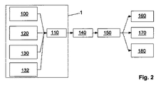
図1は、全体を参照番号1で示す本発明によるセンサユニットを示し、このセンサユニットは、コンピュータ2とキーボード3とから成るコンピュータシステムに組み込まれている。センサユニット1は、キーボード3のユーザーインターフェースで利用できる種々の測定法を有している。コンピュータシステムのユーザーは、測定を行うために指先でセンサユニット1に触る。例えば、発光ダイオードの形態の光源4、4’がセンサユニット1に組み込まれており、異なる波長の光を放出できる。この目的のために、異なる発光半導体素子が共通のセンサハウジング(図1には示されていない)に収容されている。また、異なる光源からキーボード3のユーザーインターフェースに光を案内するため光波伝導体を使用することも可能である。また、センサユニット1は1個以上のフォトセンサ5を有している。フォトセンサ5は、光源4、4’の極近くに設けられている。センサ5は、ユーザーの指先の組織内で散乱した光源4又は4’からの光を受ける。更に、熱センサ6が光源4、4’の直近に設けられている。この様にして、熱測定に基づく潅流の測定が、光学測定と同じ測定位置で行われることが保証される。更に、生体電気インピーダンスを測定するための合計で4個の電極7、7’がセンサユニット1の表面に設けられている。装置のユーザーは、手で2個の電極7、7’に同時に触る。2個の接触表面の内の一方が測定位置に電流を供給し、他方の接触表面が電圧測定に使用される。この様にして、測定結果が測定電極の接触抵抗によって影響されないことが保証される。参照番号7で示した2個の電極は、EKGユニットのEKG電極としても使用される。このEKGユニットもセンサユニット1に組み込まれている。何れの場合も、2個の電極が指で触られ、2点誘導(two-point derivation)(腕間測定)が行われる。キーボード3に組み込まれたセンサユニット1によって記録された測定信号はコンピュータ2によって処理される。この様にして得た生理学的パラメータは、コンピュータ2に接続されたモニタ9の表示画面8に出力される。動脈酸素飽和度(SaO2)、毛細血管酸素飽和度(StO2)及び静脈酸素飽和度(SvO2)が表示される。更に、心拍数測定値(HR)及び組織の脂肪含有率(BF)が表示される。最後に、血中グルコース値(BG)も表示される。ユーザーは、何時でも、関心のある生理学的パラメータを測定できる。この目的のためにユーザーが行うことは、通用はキーボード3を操作する指を電極7、7’の上に置くだけである。そして、測定信号がコンピュータ2によって処理された直後にパラメータがモニタ9を使用して表示される。従って、事実上、装置1のユーザーは、生理学的パラメータの測定のために、コンピュータ2での仕事を中断する必要がない。
FIG. 1 shows a sensor unit according to the invention, generally designated by the
図1に示すセンサユニット1の例示的実施形態においては、検査中の体組織の異なる部分を照射する2個の放射線源4、4’が設けられている。この目的のために、2個の放射線源4、4’は、異なる空間放射特性、即ち異なる放射角度を有している。放射線源4は発光ダイオードであり、放射線源4’はレーザ、例えば、所謂VCSELレーザ(垂直共振器面発光レーザ)である。発光ダイオード4とレーザ4’は、非常に近い波長(例えば、630nmと650nm)の光を放射するが、異なる開口角(例えば、25°と55°)を有している。図に示す配列により、上記したように、代謝によって引き起こされる血液中の酸素含有量の変化の示差測定が可能となる。この目的のために、何れの場合も、2個の放射線源4、4’によって放出された放射線の放射線の波長は、光の吸収の程度が酸化ヘモグロビンと還元ヘモグロビンとで異なる範囲に入らなければならない。血液の酸素含有量(酸素飽和度)の絶対値測定のためには、酸化ヘモグロビンと還元ヘモグロビンの光の吸収が実質的同じになるスペクトル域(所謂等吸収点)に波長が入る他の放射線源(図1には示されていない)が存在しなければならない。発光ダイオードとレーザによってそれぞれ放出された光は、対応する光案内ファイバによってキーボードのユーザーインターフェース上の対応する位置に導くことができる。この場合、対応するファイバ端が図1において参照符号4、4’で示される。発光ダイオードとレーザは、検査すべき体組織内に所望の異なる開口角で光を放出するように、対応するファイバに結合することが可能である。従って、体組織の異なる部分が2個の放射線源によって検査される。開口角がより大きいため、発光ダイオードで検査される体組織の潅流のない表皮の割合は、レーザの場合よりも大きくなる。体組織内で散乱され、部分的に吸収された光(放射線源4の光と放射線源4’の光の両方)はセンサ5によって検出される。センサ5は、センサユニット1の表面の上に直接配置する必要はない。それに替え、センサユニット1の内部に配置されたセンサに光案内ファイバによって光を導くことができる。放射線源4からの光を放射線源4’からの光と区別するために、2個の光源4、4’を異なる時間変調で動作させることができ、センサ5によって検出された信号はこれに応じて復調される。それに替え、2個の放射線源4、4’の放射を異なる波長に基づいて区別することもできる。放射線源4、4’によって放出された放射線の強度は、体組織を通過する際の径路長に応じて低下し、強度の低下と吸収された物質(酸化ヘモグロビン)の濃度との関係は公知のランベルトベールの法則によって与えられる。図1のセンサ5によって、各場合において、関心のある強度低下のパラメータを、放射線源4、4’によって覆われた体組織の部分について明確に別々に測定できる。このように区別された測定を行うために、異なる放射線源4、4’に割り当てるべき強度低下のパラメータは、好適なプログラム制御評価ユニットによって互いに関係付けることができる。最も単純な場合、各場合において、2個の放射線源4、4’の放射線の強度低下のパラメータから比率(quotients)が計算される。これらの比率の変化から、代謝の変化に関する結論を導き出すことができる。例えば、栄養の摂取後に血中グルコースレベルが上昇した場合、これに応じてより多くのグルコースが、(ある時間遅れの後)体組織の細胞に入り、そこで変換される。これに関連し、酸素が使用される。細胞は血液を介してこの酸素を得る。これに関連し、酸化ヘモグロビンは、酸素を放出することで還元ヘモグロビンになる。従って、酸化ヘモグロビンに対する還元ヘモグロビンの割合が増加する。放射線源4、4’の放出の開口角が異なるため、各場合において、ヘモグロビン濃度の変化の強度の低下に対する影響は相違する。従って、ヘモグロビン濃度の変化は、強度低下のパラメータの比率から検出できる。これにより、酸素消費量に関する結論を間接的に引き出すことができる。酸素消費量は血中グルコースレベルに依存するため、血中グルコースレベルも、既に述べた放射線の吸収の示差測定によって測定できる。実際的な補助手段として、光学測定と並行して、生体インピーダンス分析が行われ、この目的のために、図1に示す電極7、7’が設けられている。生体インピーダンス測定の主要な目的は、局所潅流の測定である。これは、酸素消費量、従って血中グルコースレベルの測定における追加のパラメータとして使用できる。また、開口角度が異なる放射線を1個の放射線源4のみで発生することができ、これは対応する光学要素(例えば、ビームスプリッタ、レンズ等)を使用することで行われる。
In the exemplary embodiment of the
図2は、本発明によるセンサユニット1の構造をブロック図として概略的に示す。センサユニット1は、各場合において、測定位置での体組織の血管系内における酸素濃度を光学測定するための光学測定ユニット100を有する。光学測定ユニット100によって記録されたオキシメトリー(酸素飽和度測定)及びプレチスモグラフィー(体積変動測定)信号が分析ユニット110に送られる。装置1の別の必須の構成要素は、局所熱発生量を測定するための熱測定ユニット120である。この熱測定ユニット120は、各場合において、検査中の体の部位を断熱する特別な熱センサである。従って、この部位では、血流のみによって熱が吸収され、又は熱が放出される。このため、温度の時間分解(time-resolved)測定によって潅流と熱の発生を測定できる。潅流が強い場合、検査中の体の部位は非常に短い時間で最高温度に達する。潅流が少ない場合、より長い時間がかかる。加えて、測定温度の外挿によって、動脈温度に関する結論を引き出すことができる。何故なら、測定位置の温度は動脈の温度と局所熱発生量のみによって決定されるからである。熱測定ユニット120によって記録された測定信号も更なる処理のために分析ユニット110に送られる。更に、センサユニットは、生体電気インピーダンス測定によって局所組織パラメータを検出するインピーダンス測定ユニット130を有している。インピーダンス測定ユニット130の測定信号も分析ユニット110によって処理される。最後に、本発明によれば、EKG信号を検出するためのEKGユニット132が設けられている。EKGユニット132もEKG信号の処理のために分析ユニット110に接続されている。光学測定ユニット100は、それに割り当てられた図1に示すセンサユニット1の光源4及び光センサ5を備えている。熱測定ユニット120は熱センサ6に接続されている。インピーダンス測定ユニット130は、センサユニット1の電極7、7’を介して測定信号を検出する。分析ユニット110は、全ての測定信号の前処理を行う。この目的のために、回路網の周波数である50Hz又は60Hzの範囲の干渉を除去するために、信号はバンドパスフィルタを通る。更に、信号の雑音が抑制される。分析ユニット110を通過した後、光学測定ユニット100、熱測定ユニット120、インピーダンス測定ユニット130及びEKGユニット132の処理信号が評価ユニット140に到達する。評価ユニット140は、測定信号から診断を行うのに必須なパラメータを計算する。評価ユニット140の機能は本質的にはソフトウエアで実現される。従って、図示の例示的実施形態では、評価ユニット140は、実際のセンサユニット1の必須の部分ではない。最初に、検査中の体組織の組成(水分含有量、脂肪含有量等)がインピーダンス測定ユニット130の時間依存的に記録された測定信号から計算される。光学測定ユニット100の信号から動脈の酸素飽和度が計算されると共に、(インピーダンス測定に基づいて前もって決定された組織パラメータに基いて)毛細血管の酸素飽和度が計算される。更に、潅流及び動脈温度が、熱測定ユニット120の測定信号及び、時間依存インピーダンス測定から導き出すことができる体積変動測定データから測定される。脈波速度は、EKGユニット132の信号及び光学測定ユニット100の信号から測定される。最後に、静脈の酸素飽和度、及び測定位置における他の代謝パラメータ、特に局所酸素消費量及びグルコース濃度が、それ迄に行われた全ての計算結果から評価ユニット140によって計算される。計算結果は診断ユニット150によって解釈される。コンピュータ2上のソフトウエアで実現される診断ユニット150は、評価ユニット140が計算した局所代謝パラメータを評価する。評価ユニット140と診断ユニット150はグラフィックユニット160に接続され、このグラフィックユニット160はモニタ9を制御して測定結果を表示する。得られたデータはメモリーユニット170に記憶できる。詳細には、各場合において、測定の日時も同時に記憶される。更に、インターフェースユニット180が設けられており、このインターフェースユニット180は、計算された生理学的パラメータを転送するためにコンピュータ2をデータネットワークに接続する。インターフェースユニット180により、データとパラメータ、詳細には、メモリーユニット170に記憶されたデータとパラメータを、詳細には示していない治療医のPCに転送できる。その場所で、データをより詳細に分析できる。特に、センサユニット1を用いて長時間に亘って記録されたデータとパラメータの変化を調べ、これから、罹患している疾病の進行に関する結論を導き出すことができる。
FIG. 2 schematically shows the structure of the
図3は、本発明によるセンサユニット1の第2の使用例、即ち携帯電話10を示す。装置10の前面に通常の操作キー11が見える。センサユニット1の診断測定センサは、装置10の側面に面一で組み付けられている。携帯電話10のユーザーは、測定を行うためにそれらを指で触る。生体電気インピーダンスを測定するための合計で4個の電極7、7’が携帯電話10の側面のハウジング表面に設けられている。携帯電話10のユーザーは、手で2個の電極7、7’に同時に触る。各々の場合において、2個の電極が指で触られ、2点誘導(two-point derivation)(腕間測定)が行われる。携帯電話10のセンサユニット1に組み込まれた種々のセンサによって記録された測定信号は、携帯電話10のマイクロプロセッサ(詳細には示していない)によって処理される。この様にして得た生理学的パラメータは、携帯電話10の表示装置12に出力される。ユーザーは、何時でも関心のある生理学的パラメータを測定できる。この目的のためにユーザーが行うことは、通常はキー11を操作する指を電極7、7’の上に置くだけである。携帯電話10のソフトウエアコントローラは、接触を自動的に認識して測定を開始する。そして、携帯電話10のマイクロプロセッサによって測定信号が処理された直後に、表示装置12によってパラメータが表示される。センサユニット1を組み込むことによって医療装置として構成された携帯電話10の機能は、本質的に前述したような血中グルコース値の非侵襲的測定のための間接的な方法に基づいており、この方法では、グルコースの影響、即ち、グルコースによって開始される体内の生理学的反応のエネルギー変換が検査される。図1に示す例示的実施形態の説明についての対応する記載を参照する。キーボード3と同様に、携帯電話10の場合も、光源4、4’及びセンサ5をハウジング表面に直接設ける必要はない。それに替え、光案内ファイバによってハウジング表面から又はハウジング表面へ光を導くようにし、ハウジングの内部に実際の光源とセンサを設けることもできる。複数の光源及び/又はセンサを単一の光案内ファイバに結合することもできる。
FIG. 3 shows a second example of use of the
図4は、本発明による診断用センサユニット1の設計を示す。センサユニット1の種々の測定ユニットが、外側寸法が非常に小さいセンサハウジング400に組み込まれている。薄い導電箔からなる平面状のEKG電極7がセンサハウジング400の表面に配置されている。センサユニットがコンピュータのキーボード又は携帯装置に組み込まれると、異なる四肢でのEKG誘導のためのEKG電極7と別の電極(図4には示されていない)にユーザーが触れることができるようにセンサハウジング400が配置されている。EKG電極が薄いステンレス鋼の箔であれば実用的である。図示の例示的実施形態では、微小ハウジング400の寸法が5mm(W)×8mm(L)×1.8mm(H)と小さいため、市販されている種々の装置の種々のハウジングにセンサユニットに設置でき、柔軟性があると共にコスト的に有利である。動脈の血液中の酸素飽和度の同時測定のために、光学測定ユニット、即ちパルスオキシメータがセンサハウジング400に組み込まれている。このユニットは2個以上の光放射源を有し、光放射源からの放射線がEKG電極7の開口部410を通過する。更に、パルスオキシメータは、例えば、フォトダイオードの形態の2個の放射線センサを有する。体組織(例えば、電極7の上に置かれた指の組織)内で散乱した光は、電極7の2個の開口部420、430を通して放射線センサに入る。開口部420、430は、開口部410からの距離が相違するように設けられている。センサユニットにおいて、ハウジング400内の2個以上の光放射源(例えば、発光ダイオード)からの光は光案内ファイバ又は適切な導光体に結合されており、全ての放射線源のための単一の開口部410だけが微小ハウジングの上面に位置しており、センサユニットの全ての放射線源の光が検査すべき体組織の同じ位置に入る。フォトダイオードは、それぞれの場合において、光案内ファイバ又は適切に構成された導光体に個別に結合されている。光学測定ユニットは、検査中の体組織内を循環する血液の酸素飽和量と容積脈波の同時測定を可能にする。発光ダイオードだけでなく、垂直共振器面発光レーザ(VCSEL)等の他の放射線源をこの目的のために使用することが実用的である。検査中の組織の熱特性を同時に測定するために、温度センサ、即ちサーミスタがセンサハウジングに組み込まれる。このセンサのために別の開口部440がEKG電極7に設けられている。サーミスタは、検査中の体組織と良好な熱接触が得られるようにセンサハウジング400内に配置されている。図示の例示的実施形態では、光放射源の光案内ファイバのための開口部410と第1のフォトダイオードの光案内ファイバのための開口部420との間にサーミスタが位置している。センサユニットには容易にインピーダンス測定ユニットを追加できる。この目的のために、少なくとも1個の追加の平面状電極(図4には示されていない)をセンサハウジング400の上面に設けなければならない。この電極は、インピーダンス測定ユニットの給電又は測定電極として働く。同一の測定電極を使用して生体インピーダンス信号とEKG信号を検出することが実用的である。センサユニットの電気的接触(例えば、携帯電話の電子部品との接触)のために、全ての組み込み測定ユニットを備えたセンサハウジング400が、適切な導体トラックによってリボンケーブル450上に直接取り付けられており、ケーブル450を使用したセンサユニット1の簡単な電気組み立てが可能となる。リボンケーブル450は、安定化のために好適な位置に補強部材460を有することができる。
FIG. 4 shows the design of the
図5は、図4に関連して上で述べた導光要素500を示し、合計で4個のLEDチップ501、502、503、504が要素500の下側に結合されており、本発明によるセンサユニット1の光学測定ユニットの光源を構成する。単一の導光要素500によって、全てのLED501、502、503、504から放出された放射線がセンサハウジング400の表面まで導かれる。4個のLED501、502、503、504は、基板(不図示)、例えばPCBに互いに隣合って接着されている。
FIG. 5 shows the
図6は、本発明の別の例示的実施形態を示し、合計で4個の電極7、7’、7’’7’’’がセンサハウジング400の上面に配置されており、(局所)生体電気インピーダンス測定及びEKG誘導のための給電及び測定電極として使用できる。電極7、7’、7’’、7’’’は絶縁ストリップ13によって互いに分離されている。
FIG. 6 illustrates another exemplary embodiment of the present invention, where a total of four
Claims (13)
2個以上のEKG電極(7)によってEKG信号を検出するEKGユニット(132)が設けられ、EKGユニット(132)の少なくとも1個のEKG電極(7)が、導電材料の平面状の箔又はシートとして構成されると共に、光学測定ユニット(100)によって覆われた体組織の領域内の皮膚表面にEKG電極(7)が接触するように、センサハウジング(400)のハウジング表面上に配置され、このEKG電極(7)が、少なくとも1個の放射線源(4)から放出されて検査すべき体組織に入る放射線を通過させるための少なくとも1個の開口部(410)を有しており、
温度又は熱センサ(6)がセンサハウジング(400)の中又は上に設けられ、
放射線源、放射線センサ、及び温度又は熱センサが、センサハウジング内の共通の基盤若しくはリボンケーブル上に配置されていることを特徴とする診断用センサユニット。 A diagnostic sensor unit for non-invasively detecting at least one physiological parameter of body tissue close to the skin surface, comprising at least one radiation source (4) for irradiating the body tissue to be examined, An optical measurement unit (100) comprising at least one radiation sensor (5) for detecting radiation scattered and / or propagated by the tissue, at least one radiation source (4) and at least one radiation sensor (5) is a diagnostic sensor unit disposed in a common sensor housing (400),
An EKG unit (132) for detecting an EKG signal by two or more EKG electrodes (7) is provided, and at least one EKG electrode (7) of the EKG unit (132) is a planar foil or sheet of conductive material And is disposed on the housing surface of the sensor housing (400) such that the EKG electrode (7) contacts the skin surface in the region of body tissue covered by the optical measurement unit (100). The EKG electrode (7) has at least one opening (410) for passing radiation emitted from at least one radiation source (4) and entering the body tissue to be examined;
A temperature or thermal sensor (6) is provided in or on the sensor housing (400);
A diagnostic sensor unit, wherein the radiation source, the radiation sensor, and the temperature or heat sensor are arranged on a common base or ribbon cable in the sensor housing.
Applications Claiming Priority (3)
| Application Number | Priority Date | Filing Date | Title |
|---|---|---|---|
| DE102007042551.3 | 2007-09-07 | ||
| DE102007042551 | 2007-09-07 | ||
| PCT/EP2008/007330 WO2009033624A1 (en) | 2007-09-07 | 2008-09-08 | Diagnostic sensor unit |
Publications (3)
| Publication Number | Publication Date |
|---|---|
| JP2010537751A JP2010537751A (en) | 2010-12-09 |
| JP2010537751A5 JP2010537751A5 (en) | 2011-10-27 |
| JP5602629B2 true JP5602629B2 (en) | 2014-10-08 |
Family
ID=40219244
Family Applications (1)
| Application Number | Title | Priority Date | Filing Date |
|---|---|---|---|
| JP2010523329A Active JP5602629B2 (en) | 2007-09-07 | 2008-09-08 | Diagnostic sensor unit |
Country Status (6)
| Country | Link |
|---|---|
| US (1) | US20100222652A1 (en) |
| EP (1) | EP2203113B1 (en) |
| JP (1) | JP5602629B2 (en) |
| KR (1) | KR101562807B1 (en) |
| CN (1) | CN101827555B (en) |
| WO (1) | WO2009033624A1 (en) |
Families Citing this family (83)
| Publication number | Priority date | Publication date | Assignee | Title |
|---|---|---|---|---|
| DK1889198T3 (en) | 2005-04-28 | 2015-02-09 | Proteus Digital Health Inc | Pharma-informatics system |
| US8730031B2 (en) | 2005-04-28 | 2014-05-20 | Proteus Digital Health, Inc. | Communication system using an implantable device |
| US8802183B2 (en) | 2005-04-28 | 2014-08-12 | Proteus Digital Health, Inc. | Communication system with enhanced partial power source and method of manufacturing same |
| US9198608B2 (en) | 2005-04-28 | 2015-12-01 | Proteus Digital Health, Inc. | Communication system incorporated in a container |
| US8912908B2 (en) | 2005-04-28 | 2014-12-16 | Proteus Digital Health, Inc. | Communication system with remote activation |
| US8836513B2 (en) | 2006-04-28 | 2014-09-16 | Proteus Digital Health, Inc. | Communication system incorporated in an ingestible product |
| JP5714210B2 (en) | 2005-09-01 | 2015-05-07 | プロテウス デジタル ヘルス, インコーポレイテッド | Implantable wireless communication system |
| CN101496042A (en) | 2006-05-02 | 2009-07-29 | 普罗秋斯生物医学公司 | Patient customized therapeutic regimens |
| WO2008066617A2 (en) | 2006-10-17 | 2008-06-05 | Proteus Biomedical, Inc. | Low voltage oscillator for medical devices |
| US8945005B2 (en) | 2006-10-25 | 2015-02-03 | Proteus Digital Health, Inc. | Controlled activation ingestible identifier |
| CA2781625C (en) | 2006-11-10 | 2015-09-29 | Rem Scientific Enterprises, Inc. | Rotating fluid measurement device and method |
| US8718193B2 (en) | 2006-11-20 | 2014-05-06 | Proteus Digital Health, Inc. | Active signal processing personal health signal receivers |
| AU2008210291B2 (en) | 2007-02-01 | 2013-10-03 | Otsuka Pharmaceutical Co., Ltd. | Ingestible event marker systems |
| CN103066226B (en) | 2007-02-14 | 2016-09-14 | 普罗透斯数字保健公司 | There is the in-body power source of high surface area electrode |
| EP2063771A1 (en) | 2007-03-09 | 2009-06-03 | Proteus Biomedical, Inc. | In-body device having a deployable antenna |
| EP2124725A1 (en) | 2007-03-09 | 2009-12-02 | Proteus Biomedical, Inc. | In-body device having a multi-directional transmitter |
| US8115618B2 (en) | 2007-05-24 | 2012-02-14 | Proteus Biomedical, Inc. | RFID antenna for in-body device |
| EP2192946B1 (en) | 2007-09-25 | 2022-09-14 | Otsuka Pharmaceutical Co., Ltd. | In-body device with virtual dipole signal amplification |
| DK2268261T3 (en) | 2008-03-05 | 2017-08-28 | Proteus Digital Health Inc | Edible event markers with multi-mode communications and systems as well as methods for using them |
| CA2730275C (en) | 2008-07-08 | 2019-05-21 | Proteus Biomedical, Inc. | Ingestible event marker data framework |
| MY154217A (en) | 2008-08-13 | 2015-05-15 | Proteus Digital Health Inc | Ingestible circuitry |
| WO2010057049A2 (en) | 2008-11-13 | 2010-05-20 | Proteus Biomedical, Inc. | Ingestible therapy activator system and method |
| KR101126153B1 (en) | 2008-12-11 | 2012-03-22 | 프로테우스 바이오메디컬, 인코포레이티드 | Evaluation of gastrointestinal function using portable electroviscerography systems and methods of using the same |
| US9439566B2 (en) | 2008-12-15 | 2016-09-13 | Proteus Digital Health, Inc. | Re-wearable wireless device |
| US9659423B2 (en) | 2008-12-15 | 2017-05-23 | Proteus Digital Health, Inc. | Personal authentication apparatus system and method |
| TWI503101B (en) | 2008-12-15 | 2015-10-11 | Proteus Digital Health Inc | Body-associated receiver and method |
| WO2013012869A1 (en) | 2011-07-21 | 2013-01-24 | Proteus Digital Health, Inc. | Mobile communication device, system, and method |
| JP5785097B2 (en) | 2009-01-06 | 2015-09-24 | プロテウス デジタル ヘルス, インコーポレイテッド | Pharmaceutical dosage delivery system |
| WO2010080843A2 (en) | 2009-01-06 | 2010-07-15 | Proteus Biomedical, Inc. | Ingestion-related biofeedback and personalized medical therapy method and system |
| US20100240972A1 (en) * | 2009-03-20 | 2010-09-23 | Nellcor Puritan Bennett Llc | Slider Spot Check Pulse Oximeter |
| GB2480965B (en) | 2009-03-25 | 2014-10-08 | Proteus Digital Health Inc | Probablistic pharmacokinetic and pharmacodynamic modeling |
| EA201190281A1 (en) | 2009-04-28 | 2012-04-30 | Протиус Байомедикал, Инк. | HIGHLY RELIABLE SWALLOWED MODE TABLETS AND METHODS OF THEIR APPLICATION |
| EP2432458A4 (en) | 2009-05-12 | 2014-02-12 | Proteus Digital Health Inc | Ingestible event markers comprising an ingestible component |
| US8558563B2 (en) | 2009-08-21 | 2013-10-15 | Proteus Digital Health, Inc. | Apparatus and method for measuring biochemical parameters |
| TWI517050B (en) | 2009-11-04 | 2016-01-11 | 普羅托斯數位健康公司 | System for supply chain management |
| UA109424C2 (en) | 2009-12-02 | 2015-08-25 | PHARMACEUTICAL PRODUCT, PHARMACEUTICAL TABLE WITH ELECTRONIC MARKER AND METHOD OF MANUFACTURING PHARMACEUTICAL TABLETS | |
| AU2011210648B2 (en) | 2010-02-01 | 2014-10-16 | Otsuka Pharmaceutical Co., Ltd. | Data gathering system |
| SG10201502720VA (en) | 2010-04-07 | 2015-05-28 | Proteus Digital Health Inc | Miniature ingestible device |
| TWI557672B (en) | 2010-05-19 | 2016-11-11 | 波提亞斯數位康健公司 | Computer system and computer-implemented method to track medication from manufacturer to a patient, apparatus and method for confirming delivery of medication to a patient, patient interface device |
| DE102010032531A1 (en) | 2010-07-28 | 2012-02-02 | Ingo Flore | Portable diagnostic measuring device |
| US9107806B2 (en) | 2010-11-22 | 2015-08-18 | Proteus Digital Health, Inc. | Ingestible device with pharmaceutical product |
| US8761853B2 (en) * | 2011-01-20 | 2014-06-24 | Nitto Denko Corporation | Devices and methods for non-invasive optical physiological measurements |
| WO2012125425A2 (en) | 2011-03-11 | 2012-09-20 | Proteus Biomedical, Inc. | Wearable personal body associated device with various physical configurations |
| WO2015112603A1 (en) | 2014-01-21 | 2015-07-30 | Proteus Digital Health, Inc. | Masticable ingestible product and communication system therefor |
| US9756874B2 (en) | 2011-07-11 | 2017-09-12 | Proteus Digital Health, Inc. | Masticable ingestible product and communication system therefor |
| US9235683B2 (en) | 2011-11-09 | 2016-01-12 | Proteus Digital Health, Inc. | Apparatus, system, and method for managing adherence to a regimen |
| US9693697B2 (en) | 2012-03-29 | 2017-07-04 | Benny Tal | Hand-held device having health monitoring capabilities |
| US20140031646A1 (en) * | 2012-03-29 | 2014-01-30 | Sergey Yakirevich | Blood pressure estimation using a hand-held device |
| KR20150038038A (en) | 2012-07-23 | 2015-04-08 | 프로테우스 디지털 헬스, 인코포레이티드 | Techniques for manufacturing ingestible event markers comprising an ingestible component |
| US20140051941A1 (en) * | 2012-08-17 | 2014-02-20 | Rare Light, Inc. | Obtaining physiological measurements using a portable device |
| CN104661590B (en) * | 2012-09-24 | 2016-11-02 | 株式会社村田制作所 | Biosensor and the manufacture method of biosensor |
| ES2683709T3 (en) | 2012-10-18 | 2018-09-27 | Proteus Digital Health, Inc. | Apparatus, system, and procedure for adaptively optimizing energy dissipation and diffusion energy in a power source for a communication device |
| WO2014088768A2 (en) * | 2012-12-04 | 2014-06-12 | Brain Tree Analytics Llc | Multi purpose electrode |
| TWI659994B (en) | 2013-01-29 | 2019-05-21 | 美商普羅托斯數位健康公司 | Highly-swellable polymeric films and compositions comprising the same |
| WO2014144738A1 (en) | 2013-03-15 | 2014-09-18 | Proteus Digital Health, Inc. | Metal detector apparatus, system, and method |
| JP6498177B2 (en) | 2013-03-15 | 2019-04-10 | プロテウス デジタル ヘルス, インコーポレイテッド | Identity authentication system and method |
| EP3005281A4 (en) | 2013-06-04 | 2017-06-28 | Proteus Digital Health, Inc. | System, apparatus and methods for data collection and assessing outcomes |
| US9796576B2 (en) | 2013-08-30 | 2017-10-24 | Proteus Digital Health, Inc. | Container with electronically controlled interlock |
| AU2014321320B2 (en) | 2013-09-20 | 2019-03-14 | Otsuka Pharmaceutical Co., Ltd. | Methods, devices and systems for receiving and decoding a signal in the presence of noise using slices and warping |
| JP2016537924A (en) | 2013-09-24 | 2016-12-01 | プロテウス デジタル ヘルス, インコーポレイテッド | Method and apparatus for use with electromagnetic signals received at frequencies that are not accurately known in advance |
| US10084880B2 (en) | 2013-11-04 | 2018-09-25 | Proteus Digital Health, Inc. | Social media networking based on physiologic information |
| CN106255449A (en) * | 2014-05-05 | 2016-12-21 | 思勘度股份有限公司 | There is the mancarried device of the multiple integrated sensors scanned for vital sign |
| US10588555B2 (en) | 2014-05-20 | 2020-03-17 | Pioneer Corporation | Pulse oximeter |
| US10206632B2 (en) * | 2014-07-25 | 2019-02-19 | The Trustees Of Dartmouth College | Systems and methods for cardiovascular-dynamics correlated imaging |
| WO2016085506A1 (en) * | 2014-11-26 | 2016-06-02 | Rem Scientific Enterprises, Inc | Systems and methods for reducing casing noise effects |
| UA120610C2 (en) * | 2015-01-12 | 2020-01-10 | Чінь-Хун Лу | Capacitive sensing head device for measuring frequency of acupuncture point of human body |
| CN104757972B (en) * | 2015-03-16 | 2024-02-06 | 思澜科技(成都)有限公司 | Biopsy clip for bioimpedance measurement |
| US10856783B2 (en) * | 2015-04-28 | 2020-12-08 | Kyocera Corporation | Electronic device and system |
| US11051543B2 (en) | 2015-07-21 | 2021-07-06 | Otsuka Pharmaceutical Co. Ltd. | Alginate on adhesive bilayer laminate film |
| CN105518595B (en) * | 2015-09-18 | 2018-09-04 | 京东方科技集团股份有限公司 | Touch-control sensing device and its operating method, display device |
| CN108024727B (en) * | 2015-09-25 | 2021-10-12 | 三线性生物公司 | Biosensor and method for measuring the same |
| DE102016007255A1 (en) | 2016-06-15 | 2017-12-21 | Audi Ag | Garment with integrated sensor device, method of using the garment in a plurality of automobiles, system comprising a motor vehicle and a garment |
| CN111772582A (en) | 2016-07-22 | 2020-10-16 | 普罗秋斯数字健康公司 | Electromagnetic sensing and detection of ingestible event markers |
| IL265827B2 (en) | 2016-10-26 | 2023-03-01 | Proteus Digital Health Inc | Methods for manufacturing capsules with ingestible event markers |
| KR20190065086A (en) * | 2017-12-01 | 2019-06-11 | 삼성전자주식회사 | Apparatus and method for bio information processing |
| CN112469327A (en) * | 2018-06-22 | 2021-03-09 | I·弗洛尔 | Measuring device |
| WO2020020801A1 (en) * | 2018-07-27 | 2020-01-30 | Biotronik Se & Co. Kg | Device; system and method for determining fluid accumulation in the lower extremities of a patient |
| EP3884309B1 (en) * | 2018-11-20 | 2025-02-19 | Xa Tek, Inc. | Portable dielectric spectroscopy device |
| PL428519A1 (en) * | 2019-01-08 | 2020-07-13 | Politechnika Śląska | Peripheral device supporting psychological tests |
| EP3705041A1 (en) * | 2019-03-05 | 2020-09-09 | Luciole Medical AG | Sensor assembly |
| CN111854753B (en) * | 2020-06-02 | 2023-05-23 | 深圳全景空间工业有限公司 | Modeling method for indoor space |
| CN111818447B (en) * | 2020-06-02 | 2023-05-23 | 深圳全景空间工业有限公司 | Sensor network of indoor human living environment |
| CN111811568A (en) * | 2020-06-02 | 2020-10-23 | 深圳全景空间工业有限公司 | Integrated sensor of indoor human settlements environment |
Family Cites Families (25)
| Publication number | Priority date | Publication date | Assignee | Title |
|---|---|---|---|---|
| US5237178A (en) * | 1990-06-27 | 1993-08-17 | Rosenthal Robert D | Non-invasive near-infrared quantitative measurement instrument |
| DE4342105A1 (en) * | 1993-12-12 | 1995-06-14 | Cho Ok Kyung | Method and device for the non-invasive determination of the concentration of glucose in parts of the human body, in particular in human blood, by performing highly accurate temperature measurements of the human body |
| DE4423663A1 (en) * | 1994-07-06 | 1996-01-11 | Med Science Gmbh | Method and device for detecting thermal interactions between the human body and the device according to the invention and their correlation with the glucose concentration in human blood |
| JPH09122083A (en) * | 1995-10-30 | 1997-05-13 | Noboru Akasaka | Patient monitoring apparatus mountable regularly |
| US6041247A (en) * | 1995-11-29 | 2000-03-21 | Instrumentarium Corp | Non-invasive optical measuring sensor and measuring method |
| JP4092769B2 (en) * | 1998-04-21 | 2008-05-28 | 松下電器産業株式会社 | Biological monitor device |
| JP2000083933A (en) * | 1998-07-17 | 2000-03-28 | Nippon Koden Corp | Instrument for measuring concentration of light absorptive material in vital tissue |
| US6331162B1 (en) * | 1999-02-01 | 2001-12-18 | Gary F. Mitchell | Pulse wave velocity measuring device |
| WO2001028416A1 (en) * | 1999-09-24 | 2001-04-26 | Healthetech, Inc. | Physiological monitor and associated computation, display and communication unit |
| US6616613B1 (en) * | 2000-04-27 | 2003-09-09 | Vitalsines International, Inc. | Physiological signal monitoring system |
| JP3475156B2 (en) * | 2000-08-01 | 2003-12-08 | 三洋電機株式会社 | Vital sensor |
| DE10136355A1 (en) * | 2001-07-26 | 2003-02-13 | Niels Rahe-Meyer | Device for monitoring vital parameters of an animal or human body consists of a portable bag with sensors, analysis electronics and visual and audible output means as well as interfaces for connection to other devices |
| GB2381530A (en) * | 2001-11-01 | 2003-05-07 | Oxonica Ltd | Water-soluble particles of luminescent materials and their use in Biotagging |
| JP2004024551A (en) * | 2002-06-26 | 2004-01-29 | Renesas Technology Corp | Semiconductor device for sensor system |
| US6879850B2 (en) * | 2002-08-16 | 2005-04-12 | Optical Sensors Incorporated | Pulse oximeter with motion detection |
| JP4813058B2 (en) * | 2002-10-09 | 2011-11-09 | ボディーメディア インコーポレイテッド | Device for detecting, receiving, deriving and displaying human physiological and contextual information |
| US20050148882A1 (en) * | 2004-01-06 | 2005-07-07 | Triage Wireless, Incc. | Vital signs monitor used for conditioning a patient's response |
| US7305262B2 (en) * | 2003-12-11 | 2007-12-04 | Ge Medical Systems Information Technologies, Inc. | Apparatus and method for acquiring oximetry and electrocardiogram signals |
| CA2555807A1 (en) * | 2004-02-12 | 2005-08-25 | Biopeak Corporation | Non-invasive method and apparatus for determining a physiological parameter |
| US20060009697A1 (en) * | 2004-04-07 | 2006-01-12 | Triage Wireless, Inc. | Wireless, internet-based system for measuring vital signs from a plurality of patients in a hospital or medical clinic |
| DE102005013429A1 (en) * | 2005-03-21 | 2006-09-28 | Flore, Ingo, Dr. | Mobile diagnostic device |
| EP2260756A1 (en) * | 2005-08-09 | 2010-12-15 | Flore, Ingo | Medical measuring device |
| JP2007105329A (en) * | 2005-10-14 | 2007-04-26 | Hitachi Ltd | Blood sugar level measuring apparauts, and metabolic rate measuring apparatus |
| US7486977B2 (en) * | 2005-10-27 | 2009-02-03 | Smiths Medical Pm, Inc. | Single use pulse oximeter |
| US9603521B2 (en) * | 2006-11-23 | 2017-03-28 | Ingo Flore | Medical measuring device |
-
2008
- 2008-09-08 KR KR1020107006263A patent/KR101562807B1/en active IP Right Grant
- 2008-09-08 EP EP08801909.6A patent/EP2203113B1/en active Active
- 2008-09-08 US US12/733,549 patent/US20100222652A1/en not_active Abandoned
- 2008-09-08 WO PCT/EP2008/007330 patent/WO2009033624A1/en active Application Filing
- 2008-09-08 CN CN2008801121472A patent/CN101827555B/en active Active
- 2008-09-08 JP JP2010523329A patent/JP5602629B2/en active Active
Also Published As
| Publication number | Publication date |
|---|---|
| JP2010537751A (en) | 2010-12-09 |
| CN101827555B (en) | 2012-11-21 |
| EP2203113A1 (en) | 2010-07-07 |
| EP2203113B1 (en) | 2015-02-25 |
| KR20100068400A (en) | 2010-06-23 |
| CN101827555A (en) | 2010-09-08 |
| US20100222652A1 (en) | 2010-09-02 |
| WO2009033624A1 (en) | 2009-03-19 |
| KR101562807B1 (en) | 2015-11-02 |
Similar Documents
| Publication | Publication Date | Title |
|---|---|---|
| JP5602629B2 (en) | Diagnostic sensor unit | |
| JP5377320B2 (en) | Medical measuring device | |
| JP6125743B2 (en) | Medical measuring device for bioelectrical impedance measurement | |
| US10226190B2 (en) | Diagnostic measuring device | |
| US20180177412A1 (en) | Medical Measuring Device | |
| US8428673B2 (en) | Medical measuring device | |
| CN101605495B (en) | Medical measuring device | |
| JP4903980B2 (en) | Pulse oximeter and operation method thereof | |
| US20060224073A1 (en) | Integrated physiological signal assessing device | |
| US20140323879A1 (en) | Systems, devices and methods for monitoring hemodynamics | |
| TWI379662B (en) | Diagnostic measuring device | |
| TWI409050B (en) | Pulse sensing device | |
| Cheang | Feasibility of non-contact photoplethysmography |
Legal Events
| Date | Code | Title | Description |
|---|---|---|---|
| A521 | Request for written amendment filed |
Free format text: JAPANESE INTERMEDIATE CODE: A523 Effective date: 20110907 |
|
| A621 | Written request for application examination |
Free format text: JAPANESE INTERMEDIATE CODE: A621 Effective date: 20110907 |
|
| A977 | Report on retrieval |
Free format text: JAPANESE INTERMEDIATE CODE: A971007 Effective date: 20130816 |
|
| A131 | Notification of reasons for refusal |
Free format text: JAPANESE INTERMEDIATE CODE: A131 Effective date: 20130827 |
|
| A601 | Written request for extension of time |
Free format text: JAPANESE INTERMEDIATE CODE: A601 Effective date: 20131127 |
|
| A602 | Written permission of extension of time |
Free format text: JAPANESE INTERMEDIATE CODE: A602 Effective date: 20131204 |
|
| A601 | Written request for extension of time |
Free format text: JAPANESE INTERMEDIATE CODE: A601 Effective date: 20131227 |
|
| A602 | Written permission of extension of time |
Free format text: JAPANESE INTERMEDIATE CODE: A602 Effective date: 20140110 |
|
| A601 | Written request for extension of time |
Free format text: JAPANESE INTERMEDIATE CODE: A601 Effective date: 20140127 |
|
| A602 | Written permission of extension of time |
Free format text: JAPANESE INTERMEDIATE CODE: A602 Effective date: 20140203 |
|
| A521 | Request for written amendment filed |
Free format text: JAPANESE INTERMEDIATE CODE: A523 Effective date: 20140224 |
|
| A131 | Notification of reasons for refusal |
Free format text: JAPANESE INTERMEDIATE CODE: A131 Effective date: 20140513 |
|
| A521 | Request for written amendment filed |
Free format text: JAPANESE INTERMEDIATE CODE: A523 Effective date: 20140708 |
|
| TRDD | Decision of grant or rejection written | ||
| A01 | Written decision to grant a patent or to grant a registration (utility model) |
Free format text: JAPANESE INTERMEDIATE CODE: A01 Effective date: 20140729 |
|
| A61 | First payment of annual fees (during grant procedure) |
Free format text: JAPANESE INTERMEDIATE CODE: A61 Effective date: 20140820 |
|
| R150 | Certificate of patent or registration of utility model |
Ref document number: 5602629 Country of ref document: JP Free format text: JAPANESE INTERMEDIATE CODE: R150 |
|
| R250 | Receipt of annual fees |
Free format text: JAPANESE INTERMEDIATE CODE: R250 |
|
| R250 | Receipt of annual fees |
Free format text: JAPANESE INTERMEDIATE CODE: R250 |
|
| R250 | Receipt of annual fees |
Free format text: JAPANESE INTERMEDIATE CODE: R250 |
|
| R250 | Receipt of annual fees |
Free format text: JAPANESE INTERMEDIATE CODE: R250 |
|
| R250 | Receipt of annual fees |
Free format text: JAPANESE INTERMEDIATE CODE: R250 |
|
| R250 | Receipt of annual fees |
Free format text: JAPANESE INTERMEDIATE CODE: R250 |
|
| R250 | Receipt of annual fees |
Free format text: JAPANESE INTERMEDIATE CODE: R250 |
|
| R250 | Receipt of annual fees |
Free format text: JAPANESE INTERMEDIATE CODE: R250 |