JP4854547B2 - Silver fine particle and nucleic acid complex and method for producing the same - Google Patents
Silver fine particle and nucleic acid complex and method for producing the same Download PDFInfo
- Publication number
- JP4854547B2 JP4854547B2 JP2007059748A JP2007059748A JP4854547B2 JP 4854547 B2 JP4854547 B2 JP 4854547B2 JP 2007059748 A JP2007059748 A JP 2007059748A JP 2007059748 A JP2007059748 A JP 2007059748A JP 4854547 B2 JP4854547 B2 JP 4854547B2
- Authority
- JP
- Japan
- Prior art keywords
- silver
- nucleic acid
- fine particles
- complex
- complex according
- Prior art date
- Legal status (The legal status is an assumption and is not a legal conclusion. Google has not performed a legal analysis and makes no representation as to the accuracy of the status listed.)
- Expired - Fee Related
Links
Images


Landscapes
- Investigating Or Analysing Biological Materials (AREA)
- Investigating Or Analysing Materials By The Use Of Chemical Reactions (AREA)
- Manufacture Of Metal Powder And Suspensions Thereof (AREA)
- Powder Metallurgy (AREA)
Description
本発明は、銀微粒子と核酸の複合体及びその製造方法に関する。また、本発明は、バイオ分野において用いられる、前記複合体を含む蛍光染色剤にも関する。 The present invention relates to a complex of silver fine particles and a nucleic acid and a method for producing the same. The present invention also relates to a fluorescent stain containing the complex used in the bio field.
生体試料を蛍光染色して特定の組織を識別する方法として、低分子蛍光色素自身や蛍光色素を含有あるいは結合させた高分子微粒子、発光性半導体微粒子(量子ドット)を用いる技術がある(例えば、非特許文献1:NANOTECHNOLOGY 16 (2): R9-R25 FEB 2005)。
しかし、有機色素を用いた場合は励起光による蛍光色素の退色の問題があり、粒径が100nm以下の蛍光性高分子微粒子の場合には蛍光強度が不足する問題点がある。また粒径が数ナノメートルでも発光する半導体微粒子はサイズ的には小さく量子収率が高いことから非常に注目されているが、カドミウムを用いているために細胞に対する毒性が強いことが問題となっている(例えば、非特許文献2:Langmuir, 23, 1974-1980,(2007))。
As a method of identifying a specific tissue by fluorescent staining of a biological sample, there is a technique using a low molecular fluorescent dye itself, a polymer fine particle containing or bound with a fluorescent dye, or a light emitting semiconductor fine particle (quantum dot) (for example, Non-patent document 1: NANOTECHNOLOGY 16 (2): R9-R25 FEB 2005).
However, when an organic dye is used, there is a problem of fading of the fluorescent dye due to excitation light, and in the case of fluorescent polymer fine particles having a particle size of 100 nm or less, there is a problem that the fluorescence intensity is insufficient. Semiconductor fine particles that emit light even with a particle size of several nanometers are attracting a great deal of attention because they are small in size and have a high quantum yield. However, since cadmium is used, the problem is that they are highly toxic to cells. (For example, Non-Patent Document 2: Langmuir, 23, 1974-1980, (2007)).
一方、金、銀または銅等の金属ナノ粒子の凝集体表面に分子が吸着すると、吸着分子のラマン散乱断面積が通常のラマン散乱断面積に比べて106〜1014倍程度増強され、強いラマン散乱を示すことがレーザー分光測定で明らかにされている(例えば、非特許文献3:J. Raman Spectrosc. 36, 3421-3427(2005))。
しかし、従来の作製方法で得られる単一の金属ナノ粒子からは強いラマン散乱や発光を示すことはほとんど無く、光学顕微鏡で生体試料を観察する場合の染色剤としては好ましくない。また、従来の金属微粒子の作製方法の場合、還元剤、分散剤などの除去が困難であり、発光性や安全性等の点から、生体試料の染色剤として用いることはできない(例えば、非特許文4:J. Phys. Chem. 86, 3391-3395 (1982);非特許文献5:Mater. Lett. 60, 834-838 (2006))。
On the other hand, when a molecule is adsorbed on the surface of an aggregate of metal nanoparticles such as gold, silver, or copper, the Raman scattering cross section of the adsorbed molecule is enhanced by about 10 6 to 10 14 times as compared with a normal Raman scattering cross section, which is strong. It has been clarified by laser spectroscopic measurement that it shows Raman scattering (for example, Non-Patent Document 3: J. Raman Spectrosc. 36, 3421-3427 (2005)).
However, a single metal nanoparticle obtained by a conventional production method hardly shows strong Raman scattering or light emission, which is not preferable as a staining agent when a biological sample is observed with an optical microscope. In addition, in the case of a conventional method for producing fine metal particles, it is difficult to remove a reducing agent, a dispersing agent, and the like, and it cannot be used as a stain for a biological sample from the viewpoints of luminescence and safety (for example, non-patent) Sentence 4: J. Phys. Chem. 86, 3391-3395 (1982); Non-Patent Document 5: Mater. Lett. 60, 834-838 (2006)).
上記のような状況下、比較的小さい粒径でも必要レベル以上の蛍光を示し、退色の影響が少なく、毒性が低いなどの性質を有する金属微粒子およびその製造方法が求められている。 Under the circumstances as described above, there is a demand for metal fine particles having properties such as fluorescence exceeding a necessary level even with a relatively small particle size, little influence of fading and low toxicity, and a method for producing the same.
本発明者らは、特定の銀微粒子と核酸を複合してなる複合体が上記のような性質を満足すること、また、そのような複合体を容易に製作し得る方法を見出し、本発明を完成した。すなわち、本発明は、以下に示す、銀微粒子と核酸の複合体、その製造方法、前記複合体を含む蛍光染色剤などを提供する。 The present inventors have found that a complex formed by combining a specific silver fine particle and a nucleic acid satisfies the above-mentioned properties, and a method by which such a complex can be easily produced. completed. That is, the present invention provides the following composite of silver fine particles and nucleic acid, a method for producing the same, a fluorescent staining agent containing the complex, and the like.
(1)直径が5nm〜500nmである銀微粒子と核酸の複合体。
(2)直径が5nm〜50nmである、上記(1)に記載の複合体。
(3)前記銀微粒子が複数の結晶構造を有する、上記(1)又は(2)に記載の複合体。
(4)前記銀微粒子がモザイク状構造を有する、上記(3)に記載の複合体。
(5)前記核酸が、アデニン及び/又はグアニンを含む5〜50個の塩基からなるDNA又はRNAである、上記(1)〜(4)のいずれかに記載の複合体。
(6)前記核酸が、アデニン又はグアニンを含む一本鎖DNA又はRNAである、上記(5)に記載の複合体。
(7)前記核酸が、アデニンの5〜50量体から選択されるものである、上記(6)に記載の複合体。
(8)前記核酸が、グアニンの5〜50量体から選択されるものである、上記(6)に記載の複合体。
(9)前記DNA又はRNAが末端に官能基を有する、上記(5)〜(8)のいずれかに記載の複合体。
(10)前記官能基が、ビオチン又はチオールである、上記(9)に記載の複合体。
(11)前記銀微粒子内部及び/又は外部に前記核酸が存在する状態で該銀粒子と該核酸が複合化されている、上記(1)〜(10)のいずれかに記載の複合体。
(12)表面が前記核酸で覆われている、上記(1)〜(11)のいずれかに記載の複合体。
(13)ラマン散乱を示す、上記(1)〜(12)のいずれかに記載の複合体。
(14)前記核酸と前記銀微粒子の相互作用による蛍光を示す上記(13)に記載の複合体。
(15)上記(1)〜(14)のいずれかに記載の複合体を含む蛍光染色剤。
(16)銀イオンおよび核酸を含む溶液を用いて、直径が5nm〜500nmである銀微粒子と核酸の複合体を製造する方法。
(17)前記銀イオンおよび核酸を含む溶液中で、銀イオンを還元する、上記(16)に記載の方法。
(18)還元剤の添加によって還元を行う、上記(17)に記載の方法。
(19)紫外線照射によって還元を行う、上記(17)に記載の方法。
(20)上記(16)〜(19)のいずれかに記載の方法によって得られる、銀粒子と核酸の複合体。
(1) A complex of silver fine particles having a diameter of 5 nm to 500 nm and a nucleic acid.
(2) The composite according to (1), wherein the diameter is 5 nm to 50 nm.
(3) The composite according to (1) or (2), wherein the silver fine particles have a plurality of crystal structures.
(4) The composite according to (3), wherein the silver fine particles have a mosaic structure.
(5) The complex according to any one of (1) to (4), wherein the nucleic acid is DNA or RNA consisting of 5 to 50 bases containing adenine and / or guanine.
(6) The complex according to (5), wherein the nucleic acid is single-stranded DNA or RNA containing adenine or guanine.
(7) The complex according to (6), wherein the nucleic acid is selected from 5 to 50-mer of adenine.
(8) The complex according to (6) above, wherein the nucleic acid is selected from guanine 5 to 50-mers.
(9) The complex according to any one of (5) to (8), wherein the DNA or RNA has a functional group at its terminal.
(10) The complex according to (9), wherein the functional group is biotin or thiol.
(11) The complex according to any one of (1) to (10), wherein the silver particles and the nucleic acid are complexed in a state where the nucleic acid is present inside and / or outside of the silver fine particles.
(12) The complex according to any one of (1) to (11), wherein the surface is covered with the nucleic acid.
(13) The complex according to any one of (1) to (12), which shows Raman scattering.
(14) The complex according to (13), which exhibits fluorescence due to an interaction between the nucleic acid and the silver fine particles.
(15) A fluorescent staining agent comprising the complex according to any one of (1) to (14) above.
(16) A method for producing a complex of silver fine particles having a diameter of 5 nm to 500 nm and a nucleic acid, using a solution containing silver ions and a nucleic acid.
(17) The method according to (16) above, wherein silver ions are reduced in a solution containing the silver ions and the nucleic acid.
(18) The method according to (17), wherein the reduction is performed by adding a reducing agent.
(19) The method according to (17) above, wherein the reduction is performed by ultraviolet irradiation.
(20) A silver particle and nucleic acid complex obtained by the method according to any one of (16) to (19) above.
本発明の好ましい態様に係る複合体は、比較的小さい粒径でも必要レベル以上の蛍光を示し、退色の影響が少なく、毒性が低いなどの利点を有する。したがって、そのような複合体は、バイオ分野で用いられる蛍光染色剤として好適である。
また、本発明の複合体の製造方法によれば、上記のような性質を有する銀微粒子と核酸の複合体を容易に製造することができる。
The complex according to a preferred embodiment of the present invention has advantages such as exhibiting fluorescence more than a necessary level even with a relatively small particle size, little influence of fading, and low toxicity. Therefore, such a complex is suitable as a fluorescent stain used in the bio field.
Further, according to the method for producing a complex of the present invention, a complex of silver fine particles and nucleic acid having the above properties can be easily produced.
1.発明の概要
本発明は、銀微粒子と核酸の複合体及びその製造方法、前記複合体を含む蛍光染色剤などに関する。
通常の蛍光色素や発光微粒子は発光波長を制御するために、分子構造や微粒子サイズなどを制御する必要がある。しかしながら、本発明の銀微粒子と核酸の複合体では励起光に対して波長シフトを起こす発光(ラマン散乱)を利用しているために特別な構造制御を行う必要性はない。また、核酸(例えば、DNA)を分散剤として用いているために生体適合性が高く、細胞内に導入しても細胞死を誘起しにくいことを特徴としている。
本発明の製造方法においては、通常、水溶液中に生体高分子である核酸(DNA、RNAなど)と銀イオンを溶解し、還元剤や光照射によって銀イオンを還元することで、銀微粒子と核酸がDNAと複合化された複合体を作製する。核酸は銀イオンと相互作用しやすく、核酸/銀微粒子複合体を作製する際には分散剤および保護剤として有効な成分となる。同時に銀微粒子や核酸単体では蛍光を示すことはないが、核酸と銀微粒子が複合化することによって強い発光(ラマン散乱と電荷移動蛍光)を示すことによって生体適合性の高い発光性微粒子を提供することができる。
1. SUMMARY OF THE INVENTION The present invention relates to a silver fine particle / nucleic acid complex, a method for producing the same, a fluorescent stain containing the complex, and the like.
In order to control the emission wavelength of ordinary fluorescent dyes and luminescent particles, it is necessary to control the molecular structure, particle size, and the like. However, since the silver fine particle / nucleic acid complex of the present invention utilizes light emission (Raman scattering) that causes a wavelength shift with respect to excitation light, there is no need to perform special structure control. In addition, since nucleic acid (for example, DNA) is used as a dispersant, the biocompatibility is high and cell death is hardly induced even when introduced into cells.
In the production method of the present invention, usually, nucleic acid (DNA, RNA, etc.), which is a biopolymer, and silver ions are dissolved in an aqueous solution, and silver ions are reduced by reducing the silver ions with a reducing agent or light irradiation. Produces a complex that is complexed with DNA. Nucleic acid is likely to interact with silver ions, and becomes an effective component as a dispersant and a protective agent when producing a nucleic acid / silver fine particle complex. At the same time, silver fine particles and nucleic acid alone do not exhibit fluorescence, but the combination of nucleic acid and silver fine particles provides strong luminescence (Raman scattering and charge transfer fluorescence) to provide highly biocompatible luminescent fine particles. be able to.
2.銀微粒子と核酸の複合体
本発明の第1の態様は、直径が5nm〜500nmである銀微粒子と核酸の複合体に関する。
本発明の複合体は、銀微粒子と核酸とが複合されてなる略球状の複合体であり、その直径は5〜500nm、好ましくは5〜50nm、さらに好ましくは、10〜20nmである。
ここで複合体に含まれる銀微粒子は、単一の結晶格子からできているものでもよいし、複数の結晶構造を有しているものでもよい。また、複数の結晶がモザイク状に融合した微粒子であるものも含まれる。ここでモザイク状とは、結晶が規則性を持たずに集合した状態であり、部分的には結晶間に核酸の一部が入り込んだ状態をいう。
2. Silver fine particle and nucleic acid complex The first aspect of the present invention relates to a silver fine particle and nucleic acid complex having a diameter of 5 nm to 500 nm.
The complex of the present invention is a substantially spherical complex formed by complexing silver fine particles and nucleic acid, and has a diameter of 5 to 500 nm, preferably 5 to 50 nm, and more preferably 10 to 20 nm.
Here, the silver fine particles contained in the composite may be made of a single crystal lattice or may have a plurality of crystal structures. Also included are fine particles in which a plurality of crystals are fused in a mosaic pattern. Here, the mosaic shape is a state in which crystals are gathered without regularity, and partly refers to a state in which a part of nucleic acid enters between crystals.
本発明が提供する銀粒子に複合化される核酸は、DNA、RNA、それらのホスホジエステル誘導体、PNA、2−O−メチルRNA、オリゴヌクレオチド、ペプチド核酸等を挙げることができるが、これらに限定されるものではない。
本発明で用いられる核酸としては、DNA又はRNAが好ましく、特に、アデニン及び/又はグアニンを含む2個以上、好ましくは、5〜100個、より好ましくは5〜50個、さらに好ましくは10〜30個の塩基からなるDNA又はRNAが好ましい。ここで用いられるDNAまたはRNAは一本鎖であっても、二本鎖であってもよい。
ここで用いられる核酸はこれに限定されるわけではないが、プリン塩基構造を有するアデニン及び/またはグアニンを多く含むことが好ましい。したがって、ここで用いられる核酸は好ましくは、全構成ヌクレオチド中50%以上、より好ましくは60%以上、さらに好ましくは70%、さらに好ましくは80%以上(ヌクレオチド数で換算)の構成ヌクレオチドが、アデニン及び/またはグアニンであることが好ましい。
またより均質な複合体を調整する観点からは、全構成ヌクレオチドが単一のヌクレオチドであることが好ましい。したがって、全ヌクレオチドがアデニン、グアニン、チミンおよびシトシンのいずれかから構成される核酸は本発明において好ましく用いることができる。
また、本発明において特に好ましく用いられる核酸は、アデニンまたはグアニンの5〜50量体(好ましくは10〜30量体、より好ましくは10〜20量体)から選択される。
Examples of the nucleic acid complexed with the silver particles provided by the present invention include DNA, RNA, phosphodiester derivatives thereof, PNA, 2-O-methyl RNA, oligonucleotide, peptide nucleic acid, and the like. Is not to be done.
The nucleic acid used in the present invention is preferably DNA or RNA, particularly 2 or more containing adenine and / or guanine, preferably 5 to 100, more preferably 5 to 50, and still more preferably 10 to 30. DNA or RNA consisting of a single base is preferred. The DNA or RNA used here may be single-stranded or double-stranded.
The nucleic acid used here is not limited to this, but preferably contains a large amount of adenine and / or guanine having a purine base structure. Therefore, the nucleic acid used here is preferably 50% or more, more preferably 60% or more, more preferably 70%, more preferably 80% or more (converted in terms of the number of nucleotides) of the constituting nucleotides. And / or guanine.
Further, from the viewpoint of preparing a more homogeneous complex, it is preferable that all constituent nucleotides are single nucleotides. Therefore, a nucleic acid in which all nucleotides are composed of any one of adenine, guanine, thymine and cytosine can be preferably used in the present invention.
The nucleic acid particularly preferably used in the present invention is selected from adenine or guanine 5-50 mer (preferably 10-30 mer, more preferably 10-20 mer).
また、本発明の複合体を蛍光染色剤として用いる場合には、ターゲットとなるタンパク質を認識する抗体などを結合させることが好ましい。このためには、複合体中の核酸の末端にビオチン、チオールなどの官能基を結合させるとよい。
本発明の複合体は、略球状である。この複合体の真球度は、特に限定されないが、好ましくは0.7以上、より好ましくは0.8以上である。
また、本発明の好ましい態様に係る複合体は、銀微粒子内部及び/又は外部に核酸が存在する状態で該銀粒子と該核酸が複合化されている。しかし、細胞毒性を低減化するためには、銀微粒子の表面が核酸で覆われていることが望ましい。核酸が銀微粒子の表面を被覆する程度は、細胞毒性を所望のレベルまでに低減させることができる限り特に限定されない。好ましくは、本発明の複合体において、銀微粒子の表面の30%以上、より好ましくは50%以上、さらに好ましくは70%以上が核酸で覆われている。
When the complex of the present invention is used as a fluorescent stain, it is preferable to bind an antibody that recognizes a target protein. For this purpose, a functional group such as biotin or thiol is preferably bonded to the end of the nucleic acid in the complex.
The composite of the present invention is substantially spherical. The sphericity of this complex is not particularly limited, but is preferably 0.7 or more, more preferably 0.8 or more.
In the complex according to a preferred embodiment of the present invention, the silver particles and the nucleic acid are complexed in a state where the nucleic acid is present inside and / or outside the silver fine particles. However, in order to reduce cytotoxicity, it is desirable that the surface of the silver fine particles be covered with nucleic acid. The extent to which the nucleic acid covers the surface of the silver fine particles is not particularly limited as long as the cytotoxicity can be reduced to a desired level. Preferably, in the complex of the present invention, 30% or more, more preferably 50% or more, and further preferably 70% or more of the surface of the silver fine particles is covered with the nucleic acid.
3.本発明の複合体の製造方法
次に、本発明の複合体の製造方法について説明する。
本発明は、銀イオンおよび核酸を含む溶液を用いて、上記した銀微粒子と核酸の複合体を製造する方法を提供する。より具体的には、本発明の製造方法においては、DNAやオリゴヌクレオチドなどの核酸を、水または緩衝液に溶解しておき、これを硝酸銀水溶液などの銀イオンを含む水溶液と混合する。この混合水溶液に紫外線等の光を照射することや還元剤を用いて、還元することによって、核酸で被膜され、核酸と複合化した銀微粒子を作製することができる。
3. Next, a method for producing the composite of the present invention will be described.
The present invention provides a method for producing the above-mentioned complex of silver fine particles and nucleic acid using a solution containing silver ions and nucleic acid. More specifically, in the production method of the present invention, a nucleic acid such as DNA or oligonucleotide is dissolved in water or a buffer solution and mixed with an aqueous solution containing silver ions such as an aqueous silver nitrate solution. Silver fine particles coated with nucleic acid and complexed with nucleic acid can be prepared by irradiating the mixed aqueous solution with light such as ultraviolet rays or reducing with a reducing agent.
本発明の銀微粒子の作製に必要な銀イオンを生成する化合物として、塩化銀、硫酸銀、硝酸銀、リン酸銀、亜硫酸銀、亜硝酸銀、亜リン酸銀等を挙げることができる。なかでも
硝酸銀を好ましく使用することができる。これらの水溶液の濃度は10M〜10mMでが望ましいが、コスト等の点を考慮すると10mM〜100mMが好ましい。
As a compound which produces | generates silver ion required for preparation of the silver fine particle of this invention, silver chloride, silver sulfate, silver nitrate, silver phosphate, silver sulfite, silver nitrite, silver phosphite etc. can be mentioned. Of these, silver nitrate can be preferably used. The concentration of these aqueous solutions is preferably 10M to 10mM, but 10mM to 100mM is preferable in view of cost and the like.
これらの銀イオンを生成する化合物は、水溶液として使用される。化合物は硫酸水溶液、あるいは塩酸水溶液、トリス-塩酸緩衝溶液(Tris-HCl buffer)が好ましいが、特にトリス-塩酸緩衝溶液が好ましい。 These compounds that produce silver ions are used as aqueous solutions. The compound is preferably a sulfuric acid aqueous solution, a hydrochloric acid aqueous solution, or a Tris-HCl buffer solution, and particularly preferably a Tris-HCl buffer solution.
銀イオンから銀を析出させるための還元剤としては、種々のものが使用できるが、水溶液中で安定な還元剤例えば、水素化ホウ素ナトリウム、ジメチルアミンボラン、ヒドラジン、アスコルビン酸ナトリウム、クエン酸ナトリウムなどの水溶液を用いることが好ましい。還元剤水溶液の濃度は、特に制限されないが、通常100mm〜100mMである。 Various reducing agents for precipitating silver from silver ions can be used, but reducing agents that are stable in aqueous solution such as sodium borohydride, dimethylamine borane, hydrazine, sodium ascorbate, sodium citrate, etc. It is preferable to use an aqueous solution of The concentration of the reducing agent aqueous solution is not particularly limited, but is usually 100 to 100 mM.
銀を析出させるための光による還元は、紫外線によって行うことができるが、紫外線照射方法としては、254nm から365nm までの領域にある波長を主波長とする紫外線を照射することが好ましい。例えば254nm を主波長とする低圧U V ランプ、365nm を主波長とする高圧U V ランプなどが使用できる。紫外線の照射量、照射時間に特に制限はないが、1時間以内が好ましく、さらに好ましくは10分以内である。
また、窒素ガスレーザー(337nm、10nsパルス)で還元して銀微粒子を得ることも可能である。
Reduction by light for precipitating silver can be performed by ultraviolet rays, but as an ultraviolet irradiation method, it is preferable to irradiate ultraviolet rays having a wavelength in the region from 254 nm to 365 nm as a main wavelength. For example, a low pressure U V lamp having a dominant wavelength of 254 nm, a high pressure U V lamp having a dominant wavelength of 365 nm, or the like can be used. Although there is no restriction | limiting in particular in the irradiation amount and irradiation time of an ultraviolet-ray, less than 1 hour is preferable, More preferably, it is less than 10 minutes.
It is also possible to obtain silver fine particles by reduction with a nitrogen gas laser (337 nm, 10 ns pulse).
4.本発明の蛍光染色剤
上記のような構造を有する本発明の複合体は、核酸と銀微粒子の相互作用による蛍光(特に、ラマン散乱)を示す。したがって、このような複合体は、バイオ分野で用いられる蛍光染色剤(あるいは蛍光標識剤)として用いることができる。蛍光染色剤として用いる場合は、前述のように、複合体中の核酸の末端にビオチン、チオールなどの官能基を結合させておき、その官能基とターゲットとなるタンパク質を認識する抗体などを結合させておく。本発明の蛍光染色剤は、公知の手法にしたがって用いることができる(nature biotechnology, 21(1) 41-46 (2003)。
上記で作製した銀微粒子と核酸の複合体は、水に安定に分散・溶解しており、生体適合性を示すために安全に細胞に取り込まれる。光照射を行えば、銀微粒子は細胞に取り込まれても発色する。特に内部に形成された粒子の構造がモザイク状を形成する微粒子は、粒子間に挟まれたホットスポットでラマン散乱が増強され、このラマン散乱のため長時間露光し続けても消光しない。したがって、本発明の蛍光染色剤は例えば細胞染色のイメージング試薬やゲル電気泳動用の染色試薬、タンパク質の蛍光定量法に用いる試薬等として利用可能である。
4). Fluorescent staining agent of the present invention The complex of the present invention having the structure as described above exhibits fluorescence (particularly Raman scattering) due to the interaction between nucleic acid and silver fine particles. Therefore, such a complex can be used as a fluorescent stain (or fluorescent labeling agent) used in the bio field. When used as a fluorescent stain, as described above, a functional group such as biotin or thiol is bound to the end of the nucleic acid in the complex, and an antibody that recognizes the target protein is bound to the functional group. Keep it. The fluorescent staining agent of the present invention can be used according to a known technique (nature biotechnology, 21 (1) 41-46 (2003).
The silver fine particle-nucleic acid complex prepared above is stably dispersed and dissolved in water, and can be safely taken up by cells to exhibit biocompatibility. When light irradiation is performed, the silver fine particles are colored even when taken up by cells. In particular, fine particles in which the structure of particles formed inside forms a mosaic shape have enhanced Raman scattering at hot spots sandwiched between the particles, and the Raman scattering does not quench even if the exposure is continued for a long time. Therefore, the fluorescent staining agent of the present invention can be used, for example, as an imaging reagent for cell staining, a staining reagent for gel electrophoresis, a reagent used in a fluorescence quantification method for proteins, and the like.
以下、実施例を参照して本発明の詳細を説明するが、本発明はこれに限定されるものではない。 Hereinafter, the present invention will be described in detail with reference to examples, but the present invention is not limited thereto.
実施例1:dA20(デオキシアデニン20量体)を用いた微粒子作製
ゲルろ過精製したオリゴヌクレオチドdA20(デオキシアデニン20量体)をユニット換算(塩基数濃度)で10mMとなるようにTris-HCl緩衝水溶液(10mM Tris-HCl (pH7.8 ,20℃))を用いて調製した。硝酸銀水溶液AgNO3 (和光純薬)を使用して10mMになるように調製した。
Nucleobase(塩基):Ag+イオン=1:1(モル比)でdA20と硝酸銀水溶液を混合し、最終濃度が100μMとなるように10mM Tris-HCl緩衝水溶液で希釈した。この混合水溶液1mlを4mlのガラス瓶に入れた。瓶の上部からUV(紫外線)スポット光源([LIGHTNINGCURETM L8868, HAMAMATSU],300μW/cm2 ,360nm)を用いて紫外線を5分間照射した。
Example 1: Preparation of microparticles using dA20 (deoxyadenine 20-mer) Gel filtration-purified oligonucleotide dA20 (deoxyadenine 20-mer) Tris-HCl buffer aqueous solution so that the unit conversion (base number concentration) is 10 mM. (10 mM Tris-HCl (pH 7.8, 20 ° C.)). A silver nitrate aqueous solution AgNO 3 (Wako Pure Chemical Industries) was used to prepare 10 mM.
DA20 and an aqueous silver nitrate solution were mixed at Nucleobase (base): Ag + ion = 1: 1 (molar ratio), and diluted with a 10 mM Tris-HCl buffer aqueous solution so that the final concentration was 100 μM. 1 ml of this mixed aqueous solution was placed in a 4 ml glass bottle. Ultraviolet rays were irradiated for 5 minutes from the top of the bottle using a UV (ultraviolet) spot light source ([LIGHTNINGCURE ™ L8868, HAMAMATSU], 300 μW / cm 2 , 360 nm).
実施例2:微粒子の構造評価
実施例1で示されるUV照射後に、溶液のUV-Vis吸収スペクトル[UV-1650PC, SHIMADZU]を測定した。
銀イオンとdA20を含む緩衝溶液中に紫外線を照射した結果、410nm付近に新たな吸収が観測された(図1)。これは既に知られている銀ナノ微粒子のプラズモン吸収と一致することから、銀イオンとdA20を含む緩衝溶液中から銀ナノ微粒子の形成が確認された。
また、透過型電子顕微鏡(TEM)[ HD-2000,HITACHI ]による形状観察を行った。(図2)その結果、粒径20nm程度のナノ粒子が形成されることが明らかとなった。。
さらにEDAX(エネルギー分散型蛍光X線分光装置)により微粒子の元素分析を行った結果、Ag3d由来のピークが観察されさた。このことから作製された微粒子が銀ナノ微粒子であることが明らかとなった。
Example 2 Structural Evaluation of Fine Particles After UV irradiation shown in Example 1, a UV-Vis absorption spectrum [UV-1650PC, SHIMADZU] of the solution was measured.
As a result of irradiating ultraviolet light into a buffer solution containing silver ions and dA20, new absorption was observed at around 410 nm (FIG. 1). Since this coincides with the already known plasmon absorption of silver nanoparticles, the formation of silver nanoparticles was confirmed from a buffer solution containing silver ions and dA20.
The shape was observed with a transmission electron microscope (TEM) [HD-2000, HITACHI]. (FIG. 2) As a result, it was revealed that nanoparticles having a particle size of about 20 nm were formed. .
Furthermore, as a result of elemental analysis of fine particles using EDAX (energy dispersive X-ray fluorescence spectrometer), a peak derived from Ag3d was observed. From this, it was revealed that the prepared fine particles were silver nano-particles.
実施例3:微粒子の内部構造
実施例1で形成した銀ナノ粒子の内部構造及び表面状態について、より詳細に観察を行った電子顕微鏡写真を図3に示す。図3(a)の銀ナノ粒子には複数の銀の格子像が確認できた。さらに図3(a)の銀ナノ粒子から(c)のような回折像が得られた。このことから微粒子内部に銀結晶の存在が確認された。通常、クエン酸還元法などの通常の方法にて作製した銀ナノ粒子では、粒子全体の結晶格子が一定方向に揃っているはずであるが、本方法で作製した銀ナノ粒子は格子の向きが場所により異なっている。すなわち、今回作製した微粒子は1つの結晶からなる粒子ではなく、粒子内部には多数の銀ナノ結晶が存在しており、それらが集合して1つの粒子を形成したモザイク状の粒子である。
さらに粒子表面を見ると厚さ3-4nmの保護層が存在している(図3(b))。このことからオリゴヌクレオチドが銀ナノ粒子表面を被覆していると考えられる。
また、写真での濃淡部分は電子照射で生じる帯電よる揺らぎが観測されることから、導電性でない成分の存在が明らかとなった。つまり、微粒子内部に非導電性成分であるdA20が含有されていることが示唆された。
以上の結果から、作製した微粒子がdA20と複数の銀ナノ結晶が複合化した微粒子であることが示唆された。
Example 3: Internal structure of fine particles FIG. 3 shows an electron micrograph in which the internal structure and surface state of the silver nanoparticles formed in Example 1 were observed in more detail. A plurality of silver lattice images were confirmed in the silver nanoparticles shown in FIG. Furthermore, a diffraction image as shown in (c) was obtained from the silver nanoparticles shown in FIG. This confirmed the presence of silver crystals inside the fine particles. Usually, in silver nanoparticles prepared by a usual method such as citrate reduction method, the crystal lattice of the whole particle should be aligned in a certain direction, but the silver nanoparticles prepared by this method have a lattice orientation It depends on the location. That is, the fine particles produced this time are not particles composed of a single crystal, but are a plurality of silver nanocrystals inside the particles, which are mosaic-like particles that are aggregated to form a single particle.
Further, when the particle surface is seen, a protective layer having a thickness of 3-4 nm is present (FIG. 3 (b)). From this, it is considered that the oligonucleotide covers the surface of the silver nanoparticles.
In the shaded portion of the photograph, fluctuations due to charging caused by electron irradiation were observed, which revealed the presence of non-conductive components. That is, it was suggested that dA20 which is a non-conductive component is contained in the fine particles.
From the above results, it was suggested that the prepared fine particles were fine particles in which dA20 and a plurality of silver nanocrystals were combined.
実施例4:微粒子の表面状態の測定
実施例1で形成した、微粒子表面電荷状態をレーザーゼータ電位計[ELS-8000,大塚電子]を使用して測定した。その結果、ゼータ電位は-24.77mVであったことから、表面は負に荷電していることが分かった(図4)。これにより粒子同士の静電反発を引き起こすことで微粒子が凝集せずに分散状態を保つ。また、負電荷はdA20のリン酸基によると考えられる。
銀ナノ粒子表面にdA20が存在するかどうか調べるため、以下のような実験を行った。dA20を用いて作製した銀ナノ粒子分散溶液200μl(100μM)に、アデニンと相補的な塩基を持つdT20またはpoly(U)、非相補的な塩基であるdG20を200μl(100μM)を加え5分間室温で放置した。それぞれの溶液を洗浄したガラス基板上に10μl滴下し、窒素ガス雰囲気下で乾燥させた。そのサンプルを蛍光顕微鏡[BX51, U-LH100HGAPO, Olympus]、顕微鏡カメラ[DP71, Olympus]により観察した。対物レンズはUplanAPO 100x/1.35 oil Iris、暗視野照明により観察を行った。
作製した銀ナノ粒子を観察したところ(図5(a))、個々の銀ナノ粒子による暗視野像が観察され、凝集体は観察されなかった。次に、銀ナノ粒子にアデニンとは水素結合を形成しないグアニンの20量体であるdG20を加えた場合も同様の分散状態であった(図5(b))。一方、アデニンと相補的な塩基であるチミンの連続配列であるdT20、poly(U)を加えた場合には、図5(c),(d)のように多数の凝集体が観察された。このことから、銀ナノ粒子表面にdA20の核酸塩基(アデニン)が突出しており、チミンとアデニン間での水素結合が形成された結果、微粒子が凝集したためと考えられる。このことから銀ナノ粒子最表面にはdA20のリン酸基だけではなく、核酸塩基も存在していることが明らかとなった。
Example 4: Measurement of surface state of fine particles The surface charge state of fine particles formed in Example 1 was measured using a laser zeta potentiometer [ELS-8000, Otsuka Electronics]. As a result, the zeta potential was -24.77 mV, indicating that the surface was negatively charged (Figure 4). This causes electrostatic repulsion between the particles, thereby maintaining the dispersed state without causing the fine particles to aggregate. The negative charge is thought to be due to the phosphate group of dA20.
In order to examine whether dA20 is present on the surface of the silver nanoparticles, the following experiment was conducted. Add 200 μl (100 μM) of dT20 or poly (U) with a base complementary to adenine and 200 μl (100 μM) of dG20, which is a non-complementary base, to 200 μl (100 μM) of a silver nanoparticle dispersion prepared using dA20 for 5 minutes at room temperature. Left alone. 10 μl of each solution was dropped onto a cleaned glass substrate and dried in a nitrogen gas atmosphere. The sample was observed with a fluorescence microscope [BX51, U-LH100HGAPO, Olympus] and a microscope camera [DP71, Olympus]. The objective lens was UplanAPO 100x / 1.35 oil Iris and observed with dark field illumination.
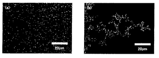
When the produced silver nanoparticles were observed (FIG. 5 (a)), dark field images of the individual silver nanoparticles were observed, and no aggregates were observed. Next, when dG20, which is a 20-mer of guanine that does not form a hydrogen bond with adenine, was added to silver nanoparticles, the same dispersion state was observed (FIG. 5 (b)). On the other hand, when dT20, poly (U), which is a continuous sequence of thymine which is a base complementary to adenine, was added, a large number of aggregates were observed as shown in FIGS. 5 (c) and 5 (d). This suggests that the nucleobase (adenine) of dA20 protrudes from the surface of the silver nanoparticle, and as a result of the formation of hydrogen bonds between thymine and adenine, the fine particles aggregated. This revealed that not only the phosphate group of dA20 but also a nucleobase was present on the outermost surface of the silver nanoparticle.
実施例5:微粒子の発光特性評価
実施例1で作製した銀ナノ微粒子の発光特性を調べるために以下のような実験を行った。dA20を用いて作製した銀ナノ粒子分散溶液200μl(100μM)を洗浄したガラス基板上に10μl滴下し、窒素ガス雰囲気下で乾燥させた。そのサンプルを蛍光顕微鏡[BX51, U-LH100HGAPO, Olympus]、顕微鏡カメラ[DP71, Olympus]によりB励起、G励起、U励起で観察した。(対物レンズはUplanAPO 100x/1.35 oil Iris)その結果を図6に示すが、どの励起光波長を用いた場合においても微粒子からの発光が観察された。
このことから、今回作製した微粒子が幅広い励起波長に対応した発光を示すことが明らかとなった。
Example 5: Evaluation of luminous characteristics of fine particles In order to examine the luminous characteristics of the silver nanoparticles prepared in Example 1, the following experiment was conducted. 10 μl of 200 μl (100 μM) of a silver nanoparticle dispersion prepared using dA20 was dropped on a cleaned glass substrate and dried in a nitrogen gas atmosphere. The sample was observed by B excitation, G excitation, and U excitation with a fluorescence microscope [BX51, U-LH100HGAPO, Olympus] and a microscope camera [DP71, Olympus]. (The objective lens is UplanAPO 100x / 1.35 oil Iris) The results are shown in FIG. 6, and light emission from the fine particles was observed at any excitation light wavelength.
From this, it became clear that the microparticles produced this time emit light corresponding to a wide range of excitation wavelengths.
実施例6:微粒子の発光起源の同定
実施例1で作製した銀微粒子は、G励起の波長帯にはほとんど吸収が無いことがUV吸収スペクトルから判明している。そのため発光の起源を同定するために、微粒子からの発光スペクトルの顕微分光測定を行った。サンプルはガラス基板状に固定化した状態で測定を行った。励起光源としてアルゴンイオンレーザー(発振波長514nm)を用いた。レーザーを顕微鏡に導入し、対物レンズで集光してサンプルを励起した。励起光レーザーの散乱光をフィルターでカットし、サンプルからの発光を光ファイバーで集光後、分光器に導入した。分光された光をCCDカメラで検出し、スペクトル(図7)を得た。
このスペクトルには530nm〜570nmのブロードな発光と534nm(ラマンシフト730cm-1)、552nm(ラマンシフト1320cm-1)の鋭いピークが観察された。この鋭いピークはアデニンのラマン散乱に帰属される。
このことから微粒子からの発光はアデニンと銀微粒子が相互作用した結果の蛍光とラマン散乱であることが明らかとなった。ラマン散乱は励起光からシフトした波長で観測されることから、あらゆる励起波長で観察される。
Example 6: Identification of emission origin of fine particles It is found from the UV absorption spectrum that the silver fine particles prepared in Example 1 have almost no absorption in the wavelength band of G excitation. Therefore, in order to identify the origin of luminescence, microspectroscopy of the emission spectrum from the fine particles was performed. The sample was measured with the glass substrate fixed. An argon ion laser (oscillation wavelength 514 nm) was used as an excitation light source. A laser was introduced into the microscope, and the sample was excited by focusing with an objective lens. The scattered light of the excitation light laser was cut with a filter, and the light emitted from the sample was collected with an optical fiber and then introduced into a spectrometer. The spectrally separated light was detected with a CCD camera to obtain a spectrum (FIG. 7).
In this spectrum, broad emission of 530 nm to 570 nm and sharp peaks of 534 nm (Raman shift 730 cm −1 ) and 552 nm (Raman shift 1320 cm −1 ) were observed. This sharp peak is attributed to Raman scattering of adenine.
From this, it became clear that the light emission from the fine particles was fluorescence and Raman scattering as a result of the interaction between adenine and silver fine particles. Since Raman scattering is observed at a wavelength shifted from the excitation light, it is observed at any excitation wavelength.
実施例7:微粒子の退色性についての評価
次に実施例1で作製した銀微粒子の退色性について検討を行った。
市販されている蛍光性半導体微粒子(EviDotsTMCdSe/ZnS core-shell型ナノ微粒子)
との比較を行った。EviDotsTMを銀微粒子と同様にガラス基板状に滴下したサンプルを用意した。EviDotsTM,銀微粒子サンプルに顕微鏡下でB励起(470-495nm)を10分間照射した。その後、それぞれの励起光を用いて発光強度について調べた。その結果、EviDotsTMはUV照射後に著しく蛍光強度が減衰したのに対し、銀微粒子は全く蛍光強度の減衰が観察されなかった。これは銀ナノ微粒子の発光がラマン散乱に起因する成分を含んでいるために退色が起こりにくく、蛍光性半導体微粒子よりも退色しにくいと考えられる。
Example 7: Evaluation of fading property of fine particles Next, the fading property of silver fine particles prepared in Example 1 was examined.
Commercially available fluorescent semiconductor particles (EviDots TM CdSe / ZnS core-shell type nanoparticles)
And compared. A sample in which EviDots ™ was dropped onto a glass substrate in the same manner as the silver fine particles was prepared. EviDots ™ , a silver fine particle sample, was irradiated with B excitation (470-495 nm) for 10 minutes under a microscope. Thereafter, the emission intensity was examined using each excitation light. As a result, the fluorescence intensity of EviDots ™ was remarkably attenuated after UV irradiation, whereas no decrease in fluorescence intensity was observed for the silver fine particles. This is thought to be due to the fact that the light emission of the silver nanoparticle contains a component due to Raman scattering, so that fading does not easily occur and is less likely to fade than the fluorescent semiconductor fine particle.
実施例8:細胞への毒性試験
次に実施例1で作製した銀微粒子の細胞毒性試験について示す。
作製した微粒子をHeLa細胞に滴下し、無血清培地中、 5% CO2 , 37℃ の条件で6時間培養を行った。その後、PBSで溶液を洗浄して不要な微粒子を取り除き、共焦点レーザー走査型顕微鏡(FV300、オリンパス)で観察を行った。
その結果を図9に示す。伸展したHeLa細胞内に緑色の発光が観察された。このことから作製した銀微粒子は細胞内へ取り込まれ、細胞内でも発光することが確認された。
また、銀微粒子溶液を滴下後、24時間培養を行った。
24時間培養後、細胞を観察したところ細胞は分裂し、増殖していることが観察された(図10)。このことから、銀微粒子はdA20という生体高分子で保護されているために細胞毒性がほとんど無いことが明らかとなった。
Example 8: Toxicity test to cells Next, a cytotoxicity test of the silver fine particles prepared in Example 1 will be described.
The prepared microparticles were dropped onto HeLa cells and cultured in a serum-free medium at 5% CO 2 and 37 ° C. for 6 hours. Thereafter, the solution was washed with PBS to remove unnecessary fine particles, and observed with a confocal laser scanning microscope (FV300, Olympus).
The result is shown in FIG. Green luminescence was observed in the extended HeLa cells. From this, it was confirmed that the produced silver fine particles were taken into the cells and emitted light even inside the cells.
Further, after the silver fine particle solution was dropped, the culture was performed for 24 hours.
When the cells were observed after culturing for 24 hours, it was observed that the cells were dividing and proliferating (FIG. 10). From this, it was clarified that the silver fine particles are hardly cytotoxic because they are protected by a biopolymer called dA20.
比較実施例1:クエン酸を加えて作製した銀微粒子
Silver nitrate 36mgを200mlの超純水に溶解し、撹拌しながら沸騰させた。1重量%に調製したSodium citrate水溶液を4ml加え、沸騰させながら10分間撹拌して銀ナノ粒子を作製した。このようにして作製された銀ナノ粒子についての、電子顕微鏡写真(TEM)を図11に示す。図11に示されるようにクエン酸還元によって作製した場合には粒形、形状ともに不均一であった。また実施例1で作製された銀ナノ粒子のような内部構造は見られなかった。
Comparative Example 1: Silver fine particles prepared by adding citric acid
36 mg of silver nitrate was dissolved in 200 ml of ultrapure water and boiled with stirring. 4 ml of an aqueous solution of sodium citrate prepared to 1% by weight was added and stirred for 10 minutes while boiling to produce silver nanoparticles. An electron micrograph (TEM) of the silver nanoparticles thus produced is shown in FIG. As shown in FIG. 11, when produced by citric acid reduction, both the particle shape and shape were non-uniform. Moreover, the internal structure like the silver nanoparticle produced in Example 1 was not seen.
実施例9:銀ナノ粒子形成における溶液の効果
(1)試薬及び溶液の調製
DNAとしてアデニン20量体であるオリゴヌクレオチドdA20を用意した。また、銀イオン水溶液はSilver nitrate(AgNO3)[和光純薬]を使用して10 mMになるように超純水で調製した。
緩衝溶液として10mM Tris-HCl水溶液pH7.8,20℃、pH7.0、pH9.0,20℃を用意した。また、その他の溶液として10mM Bis-Tris緩衝水溶液(Bis(2-hydroxoyethyl)iminotirs-(hydroxymethyl)methane, pH 5.8, 20℃)、10mMリン酸緩衝水溶液(Sodium dihydrogenphosphate dehydrate, Sodium phosphate dibasic basic dodecahydrate, pH 6.8, 20℃)、10mM 2-アミノエタノール(AE)水溶液(2-Aminoethanol,pH 8.1, 20℃)、10mM 2-ジメチルアミノエタノール(DMAE)水溶液(N,N-Dimethylethanolamine, pH 8.0, 20℃)、トリメチロールエタン(TME)水溶液(Trimethylol ethane, pH 5.8, 20℃)を用意した。Bis-Tris緩衝水溶液、AE水溶液、DMAE水溶液は塩酸を用いてpHを調製した。Tris, Bis-Tris, AE, DMAE, TMEの構造を図12に示す。
Example 9: Effect of solution on silver nanoparticle formation (1) Preparation of reagent and solution
Oligonucleotide dA20, which is an adenine 20mer, was prepared as DNA. Further, aqueous silver ion solution was prepared by Silver nitrate (AgNO 3) so as to 10 mM using the Wako Pure Chemical] ultrapure water.
As a buffer solution, 10 mM Tris-HCl aqueous solution pH 7.8, 20 ° C, pH 7.0, pH 9.0, 20 ° C were prepared. Other solutions include 10 mM Bis-Tris buffer aqueous solution (Bis (2-hydroxoyethyl) iminotirs- (hydroxymethyl) methane, pH 5.8, 20 ° C), 10 mM phosphate buffer aqueous solution (Sodium dihydrogenphosphate dehydrate, Sodium phosphate dibasic basic dodecahydrate, pH 6.8, 20 ° C), 10 mM 2-aminoethanol (AE) aqueous solution (2-Aminoethanol, pH 8.1, 20 ° C), 10 mM 2-dimethylaminoethanol (DMAE) aqueous solution (N, N-Dimethylethanolamine, pH 8.0, 20 ° C) A trimethylol ethane (TME) aqueous solution (Trimethylol ethane, pH 5.8, 20 ° C.) was prepared. The pH of Bis-Tris buffered aqueous solution, AE aqueous solution, and DMAE aqueous solution was adjusted using hydrochloric acid. The structure of Tris, Bis-Tris, AE, DMAE, and TME is shown in FIG.
(2)UV照射実験
アデニン:Ag+イオン=1:1(モル比)を最終濃度100μMとなるように超純水、あるいは各種緩衝水溶液で希釈した混合水溶液を作製し、1mlを4mlのガラス瓶に入れた。それぞれ瓶の上部からUVスポット光源([LIGHTNINGCURETM L8868, HAMAMATSU],300μW/cm2 ,360nm)を用いて紫外線を5分間照射した。
(2) UV irradiation experiment Prepare a mixed aqueous solution in which adenine: Ag + ion = 1: 1 (molar ratio) is diluted with ultrapure water or various buffered aqueous solutions to a final concentration of 100 μM, and 1 ml is put into a 4 ml glass bottle. I put it in. Each was irradiated with ultraviolet rays from the top of the bottle for 5 minutes using a UV spot light source ([LIGHTNINGCURE ™ L8868, HAMAMATSU], 300 μW / cm 2 , 360 nm).
(3)様々な溶液中における光還元
超純水中で光還元を行った結果を図13 (a), (b)に、10mM Tris-HCl緩衝水溶液で光還元を行った結果を図13(c), (d)に示す。図13 (a),(c)はAgNO3のみ、図13(b),(d)はAgNO3とdA20を添加した溶液である。図13(a)、(b)の溶液は紫外線照射後にも吸収スペクトルの変化は見られないため、超純水中ではオリゴヌクレオチドであるdA20存在下においても銀ナノ粒子は形成しないことが明らかとなった。
それに対し、Tris-HCl緩衝水溶液中で光還元を行った場合には、dA20の有無にかかわらず410nm付近にプラズモン吸収由来のピークが観測されたので、銀イオンが還元し、銀ナノ粒子が形成していると考えられる。
次にTris-HCl緩衝水溶液中において銀イオンの光還元が起こる理由として、Trisの構造に含まれるアミンが光照射により還元剤として作用したことが考えられる。そこで図12に示すTrisと似た構造をもつ様々な分子を用いて光還元が進行するかどうか検討した。
結果を図14に示す。AE, DMAE, TMEは緩衝能を持たないため、ジアンミン銀(I)錯体が形成しないpHに調製してから実験を行った。アミンを有する2-アミノエタノール(AE)、2-ジメチルアミノエタノール(DMAE)中で光還元を行った場合(図14(a), (b))、それぞれ1級アミン、3級アミンの違いに関わらず銀ナノ粒子が形成した。それに対し、アミンを含まないトリメチロールエタン(TME)中(図14(c))、無機緩衝水溶液であるリン酸緩衝水溶液中では(図14(d))、UV照射前後において吸収スペクトルの変化が見られないことから、銀イオンの光還元は進行していないと考えられる。このことから光還元のメカニズムとして、UV光を照射することで溶液中に含まれるアミンが還元剤として働き、銀イオンを還元できることが明らかとなった。
(3) Results of photoreduction in photoreduced ultrapure water in various solutions are shown in FIGS. 13 (a) and 13 (b), and results of photoreduction in a 10 mM Tris-HCl buffer aqueous solution are shown in FIG. It is shown in c) and (d). FIGS. 13A and 13C show AgNO 3 only, and FIGS. 13B and 13D show solutions in which AgNO 3 and dA20 are added. In the solutions of FIGS. 13 (a) and (b), no change in absorption spectrum is observed even after irradiation with ultraviolet light, and it is clear that silver nanoparticles are not formed even in the presence of the oligonucleotide dA20 in ultrapure water. became.
In contrast, when photoreduction was performed in an aqueous Tris-HCl buffer solution, a peak derived from plasmon absorption was observed around 410 nm regardless of the presence or absence of dA20, so silver ions were reduced and silver nanoparticles were formed. it seems to do.
Next, the reason why the silver ion photoreduction occurs in the Tris-HCl buffer aqueous solution may be that the amine contained in the structure of Tris acted as a reducing agent by light irradiation. Therefore, it was examined whether photoreduction proceeds using various molecules having a structure similar to Tris shown in FIG.
The results are shown in FIG. Since AE, DMAE, and TME have no buffering capacity, experiments were conducted after adjusting the pH so that diammine silver (I) complex was not formed. When photoreduction is performed in 2-aminoethanol (AE) and 2-dimethylaminoethanol (DMAE) with amine (Fig. 14 (a), (b)), the difference between primary amine and tertiary amine is Regardless, silver nanoparticles were formed. On the other hand, in trimethylolethane (TME) containing no amine (FIG. 14 (c)) and in an aqueous phosphate buffer solution (FIG. 14 (d)), the absorption spectrum changes before and after UV irradiation. Since it is not seen, it is considered that photoreduction of silver ions has not progressed. From this, it was clarified that the amine contained in the solution acts as a reducing agent and can reduce silver ions by irradiating UV light as a mechanism of photoreduction.
(4)光還元におけるpH依存性
次に溶液のpHの影響について検討した。Tris-HCl緩衝水溶液のpHを変化させた結果を図15に示す。pH5.8の条件ではTris-HClの緩衝能を超えているため、Bis-Trisを代用した。Trisのようなアミンを含む化合物(図12参照)が存在する場合、銀イオンはpHにより3つの状態をとる。
1. Ag+
2. Ag2O 2Ag+ + 2OH- ⇔ Ag2O + 2H2O
3. Ag(NH3)+ Ag2O + 4NH3(aq) + H2O ⇔ 2[Ag(NH3)2]+ + 2OH-
図15(b)から、pH5.8、pH7.0の条件ではpH7.8の場合と同様に、410nm付近の強いプラズモン吸収が観測された。また、pH9.0の溶液中においてもブロードではあるが410nmのプラズモン吸収が見られ、銀ナノ粒子の形成が確認された。
このことから、発光性銀ナノ微粒子作製にはアミンを有する水溶液、緩衝溶液が必要であり、pHには依存しないことが明らかとなった。
(4) pH dependence in photoreduction Next, the effect of the pH of the solution was examined. The results of changing the pH of the Tris-HCl buffer aqueous solution are shown in FIG. Bis-Tris was substituted because it exceeded the buffer capacity of Tris-HCl at pH 5.8. When a compound containing an amine such as Tris (see FIG. 12) is present, the silver ion takes three states depending on pH.
1. Ag +
2. Ag 2 O 2Ag + + 2OH - ⇔ Ag 2 O + 2H 2 O
3. Ag (NH 3) + Ag 2 O + 4NH 3 (aq) + H 2 O ⇔ 2 [Ag (NH 3) 2] + + 2OH -
From FIG. 15 (b), strong plasmon absorption around 410 nm was observed under the conditions of pH 5.8 and pH 7.0 as in the case of pH 7.8. Moreover, although it was broad also in the solution of pH 9.0, 410 nm plasmon absorption was seen, and formation of the silver nanoparticle was confirmed.
From this, it became clear that the production of luminescent silver nanoparticles requires an aqueous solution containing an amine and a buffer solution, and does not depend on pH.
実施例10:各種核酸塩基での微粒子作製
(1)試薬及び溶液の調製
核酸を含む分子として、オリゴヌクレオチド(dA5, dT5, dA20, dC20, dG20, dT20, dA50, dT50)は、すべてゲルろ過して精製したものを、3'末端ビオチン化オリゴヌクレオチド(3'-biotin-dA20)(図16)はHPLCで精製したものを用意した。また、モノヌクレオチド(AMP, CMP, GMP, TMP)、ポリヌクレオチド(Poly(A), Poly(C), Poly(G), Poly(U))、2本鎖DNA(λDNA)、環状DNA(M13mp8 RFIDNA)は精製せずに用いた。これら
はすべて塩基濃度で10mMとなるようにTris-HCl緩衝水溶液(10mM Tris-HCl (pH7.8, 20℃))を用いて調製した。
銀イオン水溶液はSilver nitrateを使用して10mMになるように超純水で調製した。
Example 10: Preparation of microparticles with various nucleobases (1) Preparation of reagents and solutions All oligonucleotides (dA5, dT5, dA20, dC20, dG20, dT20, dA50, dT50) were gel filtered as molecules containing nucleic acids. The 3′-terminal biotinylated oligonucleotide (3′-biotin-dA20) (FIG. 16) was prepared by HPLC purification. In addition, mononucleotide (AMP, CMP, GMP, TMP), polynucleotide (Poly (A), Poly (C), Poly (G), Poly (U)), double-stranded DNA (λDNA), circular DNA (M13mp8) RFIDNA) was used without purification. These were all prepared using a Tris-HCl buffer aqueous solution (10 mM Tris-HCl (pH 7.8, 20 ° C.)) so that the base concentration was 10 mM.
The silver ion aqueous solution was prepared with ultrapure water to 10 mM using silver nitrate.
(2)金属ナノ粒子の作製
DNAと硝酸銀水溶液を塩基数:Ag+イオン=1:1(モル比)、最終濃度100μMとなるように10mM Tris-HCl緩衝水溶液で希釈した混合水溶液を各DNAについて作製した。
この溶液1mlを4mlのガラス瓶に入れた。それぞれ瓶の上部からUVスポット光源([LIGHTNINGCURETM L8868, HAMAMATSU],300μW/cm2 ,360nm)を用いて紫外線を5分間照射した。
(2) Preparation of metal nanoparticles
A mixed aqueous solution was prepared for each DNA by diluting DNA and an aqueous silver nitrate solution with a 10 mM Tris-HCl buffer aqueous solution so that the base number: Ag + ion = 1: 1 (molar ratio) and a final concentration of 100 μM.
1 ml of this solution was placed in a 4 ml glass bottle. Each was irradiated with ultraviolet rays from the top of the bottle for 5 minutes using a UV spot light source ([LIGHTNINGCURE ™ L8868, HAMAMATSU], 300 μW / cm 2 , 360 nm).
(3)塩基配列の差異による効果
dA20. dC20, dG20, dT20の塩基配列のオリゴヌクレオチドを用いた場合のUV-Vis吸収スペクトル結果を図17に示す。各スペクトルを比較すると、dA20, dG20を用いた時、410nm近傍の強いプラズモン吸収が観測された。一方、dC20, dT20においても弱いながらプラズモン吸収が観察された。これらのサンプルをTEMにより観察したところ、dA20・dG20を用いた場合には平均粒径20nm程度の比較的均一な銀ナノ粒子が、dC20・dT20では50nm以上の粒子が多数見られ、それらが凝集していた(図18)。
このことから、プリン塩基をもつDNAを用いた場合に、小さいナノ微粒子が生成することが明らかとなった。
(3) Effects due to differences in nucleotide sequences
FIG. 17 shows the results of UV-Vis absorption spectra when oligonucleotides having the base sequences of dA20. dC20, dG20, and dT20 were used. When the spectra were compared, strong plasmon absorption near 410 nm was observed when dA20 and dG20 were used. On the other hand, plasmon absorption was also observed in dC20 and dT20, although they were weak. When these samples were observed by TEM, when dA20 / dG20 was used, relatively uniform silver nanoparticles with an average particle size of about 20 nm were observed, and with dC20 / dT20, a large number of particles of 50 nm or more were observed. (Figure 18).
From this, it was clarified that small nanoparticles were formed when DNA with purine base was used.
(4)DNAの鎖長の差異による効果
オリゴヌクレオチドの鎖長が銀ナノ粒子形成にどのような影響を及ぼすかを調べるため、鎖長の異なるアデニンの連続配列(AMP, dA5, dA20, dA50, poly(A))を用意した。AMPは単量体、poly(A)は数千塩基程度の不揃いな長さの高分子である。UV-Vis吸収スペクトル測定結果を図19に示す。5つのオリゴヌクレオチドのうち単量体であるAMPを用いた場合のみ、UV光照射後にも銀ナノ粒子形成を示すプラズモン吸収が観測されなかった。単量体では1分子DNAが銀クラスターを形成・集合させるといった役割を果たすことができないためだと考えられる。dA5・dA20・dA50に関しては、銀ナノ粒子の粒径増大に伴うプラズモン吸収のシフトは見られなかったが、Poly(A)を用いた場合には500-700nmのベースラインが上昇している(図20)ことから、様々な粒径の銀ナノ粒子が形成していることが明らかとなった。これはpoly(A)の鎖長が不揃いであるために、銀ナノ粒子も様々な粒径が生成したと考えられる。
以上の結果から、2量体以上の鎖長を持つことで微粒子の作製が可能であることが示された。また、鎖長を変化させることで形成される微粒子サイズを変化させることが可能である。
(4) Effects due to differences in DNA chain length In order to investigate how the oligonucleotide chain length affects the formation of silver nanoparticles, continuous sequences of adenine (AMP, dA5, dA20, dA50, poly (A)) was prepared. AMP is a monomer, and poly (A) is a polymer with an irregular length of about several thousand bases. The measurement results of UV-Vis absorption spectrum are shown in FIG. Only when AMP, which is a monomer among the five oligonucleotides, was used, plasmon absorption indicating silver nanoparticle formation was not observed after UV light irradiation. This is probably because one molecule of DNA cannot play the role of forming and assembling silver clusters in the monomer. For dA5, dA20, and dA50, there was no shift in plasmon absorption as the silver nanoparticle size increased, but the baseline of 500-700 nm increased when Poly (A) was used ( FIG. 20) revealed that silver nanoparticles with various particle sizes were formed. This is thought to be due to the fact that poly (A) chain lengths are not uniform, so silver nanoparticles also have various particle sizes.
From the above results, it was shown that fine particles can be produced by having a chain length of a dimer or more. Moreover, it is possible to change the size of the fine particles formed by changing the chain length.
(5)二本鎖DNAでの微粒子作製
ここまでの実験ではすべて1本鎖のオリゴヌクレオチドを使用してきた。そこで2本鎖の長鎖DNAであるλDNA (48,502bp, 16.5μm)、環状DNAであるM13mp8 RFIDNA (7,228
bp)を使用し、同様の実験を行った。λDNAを用いた場合のUV照射前後における吸収スペクトルの変化を図21に示す。これまでの実験方法と同様に5分間UV光を照射したところ、405nmを中心とするプラズモン吸収が観測されたことから、λDNAを用いた場合にも銀ナノ微粒子の作製が確認された。また、環状DNAを用いた場合には、1本鎖DNAにより作製した場合と同様、ナノ粒子が生成した。環状DNAの場合には平均粒径は約40nmであった。
このように1本鎖DNAだけでなく、直鎖2本鎖DNA、あるいは環状DNAにおいてもナノ粒子の形成が可能である。
(5) Preparation of microparticles with double-stranded DNA All the experiments so far have used single-stranded oligonucleotides. Therefore, λDNA (48,502bp, 16.5μm), which is a double-stranded long DNA, and M13mp8 RFIDNA (7,228), which is a circular DNA.
bp) and similar experiments were performed. FIG. 21 shows the change in absorption spectrum before and after UV irradiation when λDNA is used. When UV light was irradiated for 5 minutes in the same manner as in the previous experimental method, plasmon absorption centered at 405 nm was observed, confirming the production of silver nanoparticles even when λDNA was used. In addition, when circular DNA was used, nanoparticles were generated as in the case of using single-stranded DNA. In the case of circular DNA, the average particle size was about 40 nm.
In this way, nanoparticles can be formed not only in single-stranded DNA but also in linear double-stranded DNA or circular DNA.
(6)ビオチンを提示した銀ナノ粒子の作製
作製した銀微粒子にDNAの核酸塩基以外の分子認識能を持たせるために、機能性の官能基や色素分子を付加したDNAでの微粒子作製を検討した。
末端をビオチン化したdA20を用いて銀ナノ粒子を他と同様の条件で作製した。UV照射前後における吸収スペクトルの変化および形成した粒子のTEM画像を図22、図23に示す。末端を修飾していないdA20を用いて作製した場合と同様、UV照射後には銀ナノ粒子形成を示すプラズモン吸収が見られ、TEM画像より平均粒径20nm程度の銀ナノ粒子が形成していた。DNA末端を修飾しても同様の銀ナノ粒子が形成することが明らかとなった。
次に微粒子表面にビオチンが存在するかどうか調べるため、ストレプトアビジンを加えて微粒子が凝集するか確認した(図24)。作製した銀ナノ粒子分散溶液に200μl(100μM)に0.2mg/mlに調製したストレプトアビジン溶液(ストレプトアビジンはStreptomyces avidinii由来のStreptavidin, TypeII[和光純薬]を使用し、TE buffer (1
0mM Tris-HCl, 1mM EDTA , pH 7.8 ,20℃)により0.2mg/mlに調製した)を20μl 混合して4℃で30分間放置した。溶液を洗浄したガラス基板上に10μl滴下し、窒素ガス雰囲気下で乾燥させた。その後、顕微鏡下の暗視野照明により観察を行った。
結果を図23に示す。dA20で被覆した銀ナノ粒子の場合と同様、末端ビオチン化したdA20で作製した銀ナノ粒子のみでは分散している様子が観察された(図25(a))。この銀ナノ粒子にストレプトアビジンを加えると、図24に示した模式図通り、銀ナノ粒子同士がストレプトアビジンにより架橋され、凝集体を形成していることが分かった(図25(b))。このことから、銀ナノ粒子表面にはビオチンが存在していることが明らかとなった。本実験では、ビオチンーアビジン結合を利用したが、オリゴヌクレオチドの末端に様々な官能基を修飾することで、分子認識能を有する銀ナノ粒子が非常に簡便に作製できる。
(6) Preparation of silver nanoparticles presenting biotin In order to make the prepared silver microparticles have the ability to recognize molecules other than DNA nucleobases, the preparation of microparticles with DNA to which functional functional groups and dye molecules are added is examined. did.
Silver nanoparticles were prepared using the same biotinylated dA20 under the same conditions as the others. Changes in absorption spectra before and after UV irradiation and TEM images of the formed particles are shown in FIGS. As in the case of using dA20 with no terminal modification, plasmon absorption indicating silver nanoparticle formation was observed after UV irradiation, and silver nanoparticles having an average particle diameter of about 20 nm were formed from the TEM image. It was revealed that similar silver nanoparticles were formed even when DNA ends were modified.
Next, in order to examine whether biotin is present on the surface of the fine particles, streptavidin was added to confirm whether the fine particles were aggregated (FIG. 24). Streptavidin solution prepared to 0.2 mg / ml in 200 μl (100 μM) to the prepared silver nanoparticle dispersion solution (Streptavidin is Streptavidin, Type II [Wako Pure Chemicals] derived from Streptomyces avidinii, and TE buffer (1
(0 mM Tris-HCl, 1 mM EDTA, pH 7.8, 20 ° C.) was adjusted to 0.2 mg / ml) and mixed at 20 ° C. for 30 minutes. 10 μl of the solution was dropped on the washed glass substrate and dried in a nitrogen gas atmosphere. Thereafter, observation was performed by dark field illumination under a microscope.
The results are shown in FIG. As in the case of silver nanoparticles coated with dA20, it was observed that only silver nanoparticles prepared with terminal biotinylated dA20 were dispersed (FIG. 25 (a)). When streptavidin was added to the silver nanoparticles, it was found that the silver nanoparticles were cross-linked by streptavidin to form aggregates as shown in the schematic diagram of FIG. 24 (FIG. 25 (b)). This revealed that biotin was present on the surface of the silver nanoparticles. In this experiment, a biotin-avidin bond was used, but silver nanoparticles having molecular recognition ability can be prepared very simply by modifying various functional groups at the end of the oligonucleotide.
以上のように、本発明の銀微粒子と核酸の複合体は、通常、核酸で被覆されているため、細胞に取り込まれ易い。また、本発明の複合体は、細胞毒性も低く、細胞内で、長時間安定した発光を呈するので、例えば細胞染色のイメージング試薬、ゲル電気泳動用の染色試薬、タンパク質の蛍光定量法に用いる試薬等として利用可能である。 As described above, the silver fine particle-nucleic acid complex of the present invention is usually coated with a nucleic acid, and thus is easily taken up by cells. In addition, the complex of the present invention has low cytotoxicity and exhibits stable luminescence for a long time in the cell. For example, an imaging reagent for cell staining, a staining reagent for gel electrophoresis, and a reagent used for fluorescence determination of protein Etc. are available.
Claims (16)
Priority Applications (1)
| Application Number | Priority Date | Filing Date | Title |
|---|---|---|---|
| JP2007059748A JP4854547B2 (en) | 2007-03-09 | 2007-03-09 | Silver fine particle and nucleic acid complex and method for producing the same |
Applications Claiming Priority (1)
| Application Number | Priority Date | Filing Date | Title |
|---|---|---|---|
| JP2007059748A JP4854547B2 (en) | 2007-03-09 | 2007-03-09 | Silver fine particle and nucleic acid complex and method for producing the same |
Publications (2)
| Publication Number | Publication Date |
|---|---|
| JP2008224274A JP2008224274A (en) | 2008-09-25 |
| JP4854547B2 true JP4854547B2 (en) | 2012-01-18 |
Family
ID=39843094
Family Applications (1)
| Application Number | Title | Priority Date | Filing Date |
|---|---|---|---|
| JP2007059748A Expired - Fee Related JP4854547B2 (en) | 2007-03-09 | 2007-03-09 | Silver fine particle and nucleic acid complex and method for producing the same |
Country Status (1)
| Country | Link |
|---|---|
| JP (1) | JP4854547B2 (en) |
Families Citing this family (10)
| Publication number | Priority date | Publication date | Assignee | Title |
|---|---|---|---|---|
| JP2010126761A (en) * | 2008-11-27 | 2010-06-10 | Sumitomo Chemical Co Ltd | Method for producing silver nanostructure |
| WO2011152098A1 (en) * | 2010-06-04 | 2011-12-08 | コニカミノルタエムジー株式会社 | Developing solution for immunochromatography method, and measurement method using same |
| WO2011158829A1 (en) * | 2010-06-15 | 2011-12-22 | 日産化学工業株式会社 | Metal particles for surface-enhanced raman scattering and molecular sensing |
| EP2594940A1 (en) * | 2011-11-16 | 2013-05-22 | Koninklijke Philips Electronics N.V. | Particle repulsion to enhance surface contact in magnetic particle immunoassays |
| JP2013177678A (en) * | 2012-01-31 | 2013-09-09 | Nissei Bio Kk | Composite of silver fine particle and dna and method for producing the same |
| CN103264165B (en) * | 2013-04-22 | 2016-05-18 | 浙江师范大学 | A kind of method of synthesizing silver nanoclusters taking single stranded DNA as template |
| KR101689659B1 (en) * | 2014-07-02 | 2016-12-27 | 기초과학연구원 | DNA-based silver nanoclusters having silver ion mediated specific fluorescence enhancement and method for the preparation thereof, and silver ion sensor |
| EP3277324B1 (en) * | 2015-03-30 | 2019-05-01 | Council Of Scientific & Industrial Research | A process for the preparation of bio-organic coated gold and silver nanoparticles using blue light |
| CN115931826B (en) * | 2023-01-30 | 2025-07-15 | 中国工程物理研究院材料研究所 | A SERS detection method for uranyl ions based on streptavidin-assisted silver nanoparticle self-assembly |
| CN118064423A (en) * | 2024-02-04 | 2024-05-24 | 上海交通大学 | A method for rapid assembly of gold nanorods and polyadenine DNA |
Family Cites Families (5)
| Publication number | Priority date | Publication date | Assignee | Title |
|---|---|---|---|---|
| WO2002096262A2 (en) * | 2001-05-25 | 2002-12-05 | Northwestern University | Non-alloying core shell nanoparticles |
| CN1672052A (en) * | 2002-06-27 | 2005-09-21 | 佐治亚技术研究公司 | Nano-sized optical fluorescence labels and uses thereof |
| JP4787938B2 (en) * | 2003-03-28 | 2011-10-05 | ザ・プロウボウスト・フェロウズ・ファウンデーション・スカラーズ・アンド・ザ・アザー・メンバーズ・オブ・ボード・オブ・ザ・カレッジ・オブ・ザ・ホリー・アンド・アンデバイデッド・トリニティ・オブ・クイーン | Detecting sensor using silver nanoparticles |
| JP4636454B2 (en) * | 2003-05-13 | 2011-02-23 | 三菱マテリアル株式会社 | Manufacturing method and use of metal nanorods |
| US20050148100A1 (en) * | 2003-12-30 | 2005-07-07 | Intel Corporation | Methods and devices for using Raman-active probe constructs to assay biological samples |
-
2007
- 2007-03-09 JP JP2007059748A patent/JP4854547B2/en not_active Expired - Fee Related
Also Published As
| Publication number | Publication date |
|---|---|
| JP2008224274A (en) | 2008-09-25 |
Similar Documents
| Publication | Publication Date | Title |
|---|---|---|
| JP4854547B2 (en) | Silver fine particle and nucleic acid complex and method for producing the same | |
| Díez et al. | Fluorescent silver nanoclusters | |
| Shang et al. | Ultra-small fluorescent metal nanoclusters: synthesis and biological applications | |
| Chhabra et al. | Distance-dependent interactions between gold nanoparticles and fluorescent molecules with DNA as tunable spacers | |
| Xie et al. | Homogeneous silver-coated nanoparticle substrates for enhanced fluorescence detection | |
| JP3885054B2 (en) | Non-alloy core-shell nanoparticles | |
| JP5746469B2 (en) | Nanoparticle aggregation guided by DNA | |
| WO2009061783A2 (en) | Dna microarray having hairpin probes tethered to nanostructured metal surface | |
| AU2002341539A1 (en) | Non-alloying core shell nanoparticles | |
| Canton et al. | Modified Stöber synthesis of highly luminescent dye-doped silica nanoparticles | |
| Navarro et al. | Plasmonic bipyramids for fluorescence enhancement and protection against photobleaching | |
| WO2004111602A2 (en) | Colorimetric and fluorescent methods for sensing of oligonucleotides | |
| WO2010014820A2 (en) | Particles for use in supported nucleic acid ligation and detection sequencing | |
| Han et al. | Size control and photophysical properties of quantum dots prepared via a novel tunable hydrothermal route | |
| Zhao et al. | Deterministic assembly of single emitters in sub-5 nanometer optical cavity formed by gold nanorod dimers on three-dimensional DNA origami | |
| US20060166249A1 (en) | Methods for separating short single-stranded nucleic acid from long single-and double-stranded nucleic acid, and associated biomolecular assays | |
| Haroone et al. | Fluorescence enhancement strategy for evaluation of the minor groove binder DAPI to complementary ssDNA sequence including telomere mimics in (ssDNA@ DAPI/LDH) n ultrathin films | |
| Ivanova et al. | DNA as a template for synthesis of fluorescent gold nanoclusters | |
| Lee et al. | Preparation of silica–silver heterogeneous nanocomposite particles by one-pot preparation strategy using polyol process: Size-controlled immobilization of silver nanoparticles | |
| Saraswathi et al. | Sequence programmed DNA three-way junctions for templated assembly of fluorescent silver nanoclusters | |
| Yuan et al. | Hyperbranched polyamine assisted synthesis of dual-luminescent gold composite with pH responsive character | |
| Deák et al. | The first solid-state route to luminescent Au (I)—glutathionate and its pH-controlled transformation into ultrasmall oligomeric Au10–12 (SG) 10–12 nanoclusters for application in cancer radiotheraphy | |
| JP2008527999A (en) | Methods for separating short single stranded nucleic acids from long single stranded nucleic acids and double stranded nucleic acids, and related biomolecular assays | |
| Vasudev et al. | Optoelectronic signatures of DNA-based hybrid nanostructures | |
| JP5082860B2 (en) | Si / Si3N4 type nanoparticles, biological material labeling agent using the nanoparticles, and method for producing the nanoparticles |
Legal Events
| Date | Code | Title | Description |
|---|---|---|---|
| A621 | Written request for application examination |
Free format text: JAPANESE INTERMEDIATE CODE: A621 Effective date: 20091110 |
|
| A131 | Notification of reasons for refusal |
Free format text: JAPANESE INTERMEDIATE CODE: A131 Effective date: 20110426 |
|
| A977 | Report on retrieval |
Free format text: JAPANESE INTERMEDIATE CODE: A971007 Effective date: 20110427 |
|
| A521 | Request for written amendment filed |
Free format text: JAPANESE INTERMEDIATE CODE: A523 Effective date: 20110616 |
|
| TRDD | Decision of grant or rejection written | ||
| A01 | Written decision to grant a patent or to grant a registration (utility model) |
Free format text: JAPANESE INTERMEDIATE CODE: A01 Effective date: 20111004 |
|
| A01 | Written decision to grant a patent or to grant a registration (utility model) |
Free format text: JAPANESE INTERMEDIATE CODE: A01 |
|
| A61 | First payment of annual fees (during grant procedure) |
Free format text: JAPANESE INTERMEDIATE CODE: A61 Effective date: 20111025 |
|
| FPAY | Renewal fee payment (event date is renewal date of database) |
Free format text: PAYMENT UNTIL: 20141104 Year of fee payment: 3 |
|
| R150 | Certificate of patent or registration of utility model |
Free format text: JAPANESE INTERMEDIATE CODE: R150 |
|
| LAPS | Cancellation because of no payment of annual fees |