JP4671365B2 - Method for producing high-density cultured tissue and high-density cultured tissue - Google Patents
Method for producing high-density cultured tissue and high-density cultured tissue Download PDFInfo
- Publication number
- JP4671365B2 JP4671365B2 JP2007503665A JP2007503665A JP4671365B2 JP 4671365 B2 JP4671365 B2 JP 4671365B2 JP 2007503665 A JP2007503665 A JP 2007503665A JP 2007503665 A JP2007503665 A JP 2007503665A JP 4671365 B2 JP4671365 B2 JP 4671365B2
- Authority
- JP
- Japan
- Prior art keywords
- density
- culture
- producing
- liquid flow
- cultured tissue
- Prior art date
- Legal status (The legal status is an assumption and is not a legal conclusion. Google has not performed a legal analysis and makes no representation as to the accuracy of the status listed.)
- Active
Links
- 238000004519 manufacturing process Methods 0.000 title claims description 48
- 210000001519 tissue Anatomy 0.000 claims description 121
- 239000007788 liquid Substances 0.000 claims description 89
- 102000010834 Extracellular Matrix Proteins Human genes 0.000 claims description 43
- 108010037362 Extracellular Matrix Proteins Proteins 0.000 claims description 43
- 210000002744 extracellular matrix Anatomy 0.000 claims description 43
- 210000004102 animal cell Anatomy 0.000 claims description 37
- 210000004027 cell Anatomy 0.000 claims description 37
- 238000000034 method Methods 0.000 claims description 31
- 238000004113 cell culture Methods 0.000 claims description 25
- 102000008186 Collagen Human genes 0.000 claims description 21
- 108010035532 Collagen Proteins 0.000 claims description 21
- 229920001436 collagen Polymers 0.000 claims description 21
- 239000000203 mixture Substances 0.000 claims description 19
- 210000002950 fibroblast Anatomy 0.000 claims description 15
- 229910052751 metal Inorganic materials 0.000 claims description 13
- 239000002184 metal Substances 0.000 claims description 13
- 210000004204 blood vessel Anatomy 0.000 claims description 12
- 238000012258 culturing Methods 0.000 claims description 10
- 239000000126 substance Substances 0.000 claims description 9
- 239000002473 artificial blood Substances 0.000 claims description 8
- 239000012530 fluid Substances 0.000 claims description 8
- 239000013543 active substance Substances 0.000 claims description 7
- 238000009472 formulation Methods 0.000 claims description 6
- 210000000329 smooth muscle myocyte Anatomy 0.000 claims description 6
- 210000001082 somatic cell Anatomy 0.000 claims description 6
- 230000010261 cell growth Effects 0.000 claims description 5
- 239000003102 growth factor Substances 0.000 claims description 5
- 239000012528 membrane Substances 0.000 claims description 5
- 239000011148 porous material Substances 0.000 claims description 5
- 210000003491 skin Anatomy 0.000 claims description 5
- 102000016942 Elastin Human genes 0.000 claims description 4
- 108010014258 Elastin Proteins 0.000 claims description 4
- 108010022355 Fibroins Proteins 0.000 claims description 4
- 102000016611 Proteoglycans Human genes 0.000 claims description 4
- 108010067787 Proteoglycans Proteins 0.000 claims description 4
- 229920002549 elastin Polymers 0.000 claims description 4
- 229940088597 hormone Drugs 0.000 claims description 4
- 210000000056 organ Anatomy 0.000 claims description 4
- 210000003556 vascular endothelial cell Anatomy 0.000 claims description 4
- 229920002101 Chitin Polymers 0.000 claims description 3
- 229920001661 Chitosan Polymers 0.000 claims description 3
- 108010067306 Fibronectins Proteins 0.000 claims description 3
- 102000016359 Fibronectins Human genes 0.000 claims description 3
- 108010085895 Laminin Proteins 0.000 claims description 3
- 102000007547 Laminin Human genes 0.000 claims description 3
- 230000009471 action Effects 0.000 claims description 3
- 210000000845 cartilage Anatomy 0.000 claims description 3
- 239000000919 ceramic Substances 0.000 claims description 3
- 210000001612 chondrocyte Anatomy 0.000 claims description 3
- 150000001875 compounds Chemical class 0.000 claims description 3
- 210000001671 embryonic stem cell Anatomy 0.000 claims description 3
- 210000001339 epidermal cell Anatomy 0.000 claims description 3
- 108060002895 fibrillin Proteins 0.000 claims description 3
- 102000013370 fibrillin Human genes 0.000 claims description 3
- 210000003494 hepatocyte Anatomy 0.000 claims description 3
- 239000005556 hormone Substances 0.000 claims description 3
- 210000005036 nerve Anatomy 0.000 claims description 3
- 210000004498 neuroglial cell Anatomy 0.000 claims description 3
- 239000004745 nonwoven fabric Substances 0.000 claims description 3
- 230000000144 pharmacologic effect Effects 0.000 claims description 3
- 229920003002 synthetic resin Polymers 0.000 claims description 3
- 239000000057 synthetic resin Substances 0.000 claims description 3
- 210000004881 tumor cell Anatomy 0.000 claims description 3
- 210000003205 muscle Anatomy 0.000 claims description 2
- 239000005445 natural material Substances 0.000 claims description 2
- 239000000243 solution Substances 0.000 description 37
- 239000002609 medium Substances 0.000 description 22
- 102000012422 Collagen Type I Human genes 0.000 description 15
- 108010022452 Collagen Type I Proteins 0.000 description 15
- 238000010992 reflux Methods 0.000 description 12
- 239000000758 substrate Substances 0.000 description 12
- 239000000463 material Substances 0.000 description 9
- 206010028980 Neoplasm Diseases 0.000 description 8
- 239000001963 growth medium Substances 0.000 description 8
- 201000011510 cancer Diseases 0.000 description 7
- 238000002474 experimental method Methods 0.000 description 6
- 230000003287 optical effect Effects 0.000 description 6
- 239000000835 fiber Substances 0.000 description 5
- 229910001220 stainless steel Inorganic materials 0.000 description 5
- CIWBSHSKHKDKBQ-JLAZNSOCSA-N Ascorbic acid Chemical compound OC[C@H](O)[C@H]1OC(=O)C(O)=C1O CIWBSHSKHKDKBQ-JLAZNSOCSA-N 0.000 description 4
- 241001465754 Metazoa Species 0.000 description 4
- 230000015572 biosynthetic process Effects 0.000 description 4
- 239000006143 cell culture medium Substances 0.000 description 4
- 239000000512 collagen gel Substances 0.000 description 4
- 238000010586 diagram Methods 0.000 description 4
- 239000003814 drug Substances 0.000 description 4
- 239000000499 gel Substances 0.000 description 4
- NOESYZHRGYRDHS-UHFFFAOYSA-N insulin Chemical compound N1C(=O)C(NC(=O)C(CCC(N)=O)NC(=O)C(CCC(O)=O)NC(=O)C(C(C)C)NC(=O)C(NC(=O)CN)C(C)CC)CSSCC(C(NC(CO)C(=O)NC(CC(C)C)C(=O)NC(CC=2C=CC(O)=CC=2)C(=O)NC(CCC(N)=O)C(=O)NC(CC(C)C)C(=O)NC(CCC(O)=O)C(=O)NC(CC(N)=O)C(=O)NC(CC=2C=CC(O)=CC=2)C(=O)NC(CSSCC(NC(=O)C(C(C)C)NC(=O)C(CC(C)C)NC(=O)C(CC=2C=CC(O)=CC=2)NC(=O)C(CC(C)C)NC(=O)C(C)NC(=O)C(CCC(O)=O)NC(=O)C(C(C)C)NC(=O)C(CC(C)C)NC(=O)C(CC=2NC=NC=2)NC(=O)C(CO)NC(=O)CNC2=O)C(=O)NCC(=O)NC(CCC(O)=O)C(=O)NC(CCCNC(N)=N)C(=O)NCC(=O)NC(CC=3C=CC=CC=3)C(=O)NC(CC=3C=CC=CC=3)C(=O)NC(CC=3C=CC(O)=CC=3)C(=O)NC(C(C)O)C(=O)N3C(CCC3)C(=O)NC(CCCCN)C(=O)NC(C)C(O)=O)C(=O)NC(CC(N)=O)C(O)=O)=O)NC(=O)C(C(C)CC)NC(=O)C(CO)NC(=O)C(C(C)O)NC(=O)C1CSSCC2NC(=O)C(CC(C)C)NC(=O)C(NC(=O)C(CCC(N)=O)NC(=O)C(CC(N)=O)NC(=O)C(NC(=O)C(N)CC=1C=CC=CC=1)C(C)C)CC1=CN=CN1 NOESYZHRGYRDHS-UHFFFAOYSA-N 0.000 description 4
- 210000004379 membrane Anatomy 0.000 description 4
- 230000001172 regenerating effect Effects 0.000 description 4
- 210000002460 smooth muscle Anatomy 0.000 description 4
- 238000011144 upstream manufacturing Methods 0.000 description 4
- 108091003079 Bovine Serum Albumin Proteins 0.000 description 3
- 229920004934 Dacron® Polymers 0.000 description 3
- 238000009825 accumulation Methods 0.000 description 3
- QVGXLLKOCUKJST-UHFFFAOYSA-N atomic oxygen Chemical compound [O] QVGXLLKOCUKJST-UHFFFAOYSA-N 0.000 description 3
- 230000021164 cell adhesion Effects 0.000 description 3
- 108010082117 matrigel Proteins 0.000 description 3
- 238000012986 modification Methods 0.000 description 3
- 230000004048 modification Effects 0.000 description 3
- 239000002547 new drug Substances 0.000 description 3
- 229910052760 oxygen Inorganic materials 0.000 description 3
- 239000001301 oxygen Substances 0.000 description 3
- 230000002093 peripheral effect Effects 0.000 description 3
- 229920000728 polyester Polymers 0.000 description 3
- 239000005020 polyethylene terephthalate Substances 0.000 description 3
- 235000018102 proteins Nutrition 0.000 description 3
- 102000004169 proteins and genes Human genes 0.000 description 3
- 108090000623 proteins and genes Proteins 0.000 description 3
- 239000010935 stainless steel Substances 0.000 description 3
- 210000000626 ureter Anatomy 0.000 description 3
- YBJHBAHKTGYVGT-ZKWXMUAHSA-N (+)-Biotin Chemical compound N1C(=O)N[C@@H]2[C@H](CCCCC(=O)O)SC[C@@H]21 YBJHBAHKTGYVGT-ZKWXMUAHSA-N 0.000 description 2
- 102000004190 Enzymes Human genes 0.000 description 2
- 108090000790 Enzymes Proteins 0.000 description 2
- 101800003838 Epidermal growth factor Proteins 0.000 description 2
- 102400001368 Epidermal growth factor Human genes 0.000 description 2
- DHCLVCXQIBBOPH-UHFFFAOYSA-N Glycerol 2-phosphate Chemical compound OCC(CO)OP(O)(O)=O DHCLVCXQIBBOPH-UHFFFAOYSA-N 0.000 description 2
- AEMRFAOFKBGASW-UHFFFAOYSA-N Glycolic acid Chemical compound OCC(O)=O AEMRFAOFKBGASW-UHFFFAOYSA-N 0.000 description 2
- 102000004877 Insulin Human genes 0.000 description 2
- 108090001061 Insulin Proteins 0.000 description 2
- -1 L-ascorbic acid) Chemical compound 0.000 description 2
- JGSARLDLIJGVTE-MBNYWOFBSA-N Penicillin G Chemical compound N([C@H]1[C@H]2SC([C@@H](N2C1=O)C(O)=O)(C)C)C(=O)CC1=CC=CC=C1 JGSARLDLIJGVTE-MBNYWOFBSA-N 0.000 description 2
- RJKFOVLPORLFTN-LEKSSAKUSA-N Progesterone Chemical compound C1CC2=CC(=O)CC[C@]2(C)[C@@H]2[C@@H]1[C@@H]1CC[C@H](C(=O)C)[C@@]1(C)CC2 RJKFOVLPORLFTN-LEKSSAKUSA-N 0.000 description 2
- LCTONWCANYUPML-UHFFFAOYSA-N Pyruvic acid Chemical compound CC(=O)C(O)=O LCTONWCANYUPML-UHFFFAOYSA-N 0.000 description 2
- XUIMIQQOPSSXEZ-UHFFFAOYSA-N Silicon Chemical group [Si] XUIMIQQOPSSXEZ-UHFFFAOYSA-N 0.000 description 2
- 102000004338 Transferrin Human genes 0.000 description 2
- 239000002253 acid Substances 0.000 description 2
- 238000004458 analytical method Methods 0.000 description 2
- 229960005070 ascorbic acid Drugs 0.000 description 2
- 210000002469 basement membrane Anatomy 0.000 description 2
- HVYWMOMLDIMFJA-DPAQBDIFSA-N cholesterol Chemical compound C1C=C2C[C@@H](O)CC[C@]2(C)[C@@H]2[C@@H]1[C@@H]1CC[C@H]([C@H](C)CCCC(C)C)[C@@]1(C)CC2 HVYWMOMLDIMFJA-DPAQBDIFSA-N 0.000 description 2
- 230000000295 complement effect Effects 0.000 description 2
- 229920001577 copolymer Polymers 0.000 description 2
- 239000002537 cosmetic Substances 0.000 description 2
- 239000012531 culture fluid Substances 0.000 description 2
- 210000004207 dermis Anatomy 0.000 description 2
- 238000011161 development Methods 0.000 description 2
- 230000000694 effects Effects 0.000 description 2
- 229940088598 enzyme Drugs 0.000 description 2
- 229940116977 epidermal growth factor Drugs 0.000 description 2
- 239000012091 fetal bovine serum Substances 0.000 description 2
- 238000007490 hematoxylin and eosin (H&E) staining Methods 0.000 description 2
- JYGXADMDTFJGBT-VWUMJDOOSA-N hydrocortisone Chemical compound O=C1CC[C@]2(C)[C@H]3[C@@H](O)C[C@](C)([C@@](CC4)(O)C(=O)CO)[C@@H]4[C@@H]3CCC2=C1 JYGXADMDTFJGBT-VWUMJDOOSA-N 0.000 description 2
- 229940125396 insulin Drugs 0.000 description 2
- JVTAAEKCZFNVCJ-UHFFFAOYSA-N lactic acid Chemical compound CC(O)C(O)=O JVTAAEKCZFNVCJ-UHFFFAOYSA-N 0.000 description 2
- 150000002632 lipids Chemical class 0.000 description 2
- 239000007758 minimum essential medium Substances 0.000 description 2
- 230000007935 neutral effect Effects 0.000 description 2
- 238000000879 optical micrograph Methods 0.000 description 2
- 229920000747 poly(lactic acid) Polymers 0.000 description 2
- 239000004626 polylactic acid Substances 0.000 description 2
- 229920000642 polymer Polymers 0.000 description 2
- 229920002379 silicone rubber Polymers 0.000 description 2
- 238000002415 sodium dodecyl sulfate polyacrylamide gel electrophoresis Methods 0.000 description 2
- 238000003756 stirring Methods 0.000 description 2
- UCSJYZPVAKXKNQ-HZYVHMACSA-N streptomycin Chemical compound CN[C@H]1[C@H](O)[C@@H](O)[C@H](CO)O[C@H]1O[C@@H]1[C@](C=O)(O)[C@H](C)O[C@H]1O[C@@H]1[C@@H](NC(N)=N)[C@H](O)[C@@H](NC(N)=N)[C@H](O)[C@H]1O UCSJYZPVAKXKNQ-HZYVHMACSA-N 0.000 description 2
- 239000012581 transferrin Substances 0.000 description 2
- 238000002054 transplantation Methods 0.000 description 2
- VBEQCZHXXJYVRD-GACYYNSASA-N uroanthelone Chemical compound C([C@@H](C(=O)N[C@H](C(=O)N[C@@H](CS)C(=O)N[C@@H](CC(N)=O)C(=O)N[C@@H](CS)C(=O)N[C@H](C(=O)N[C@@H]([C@@H](C)CC)C(=O)NCC(=O)N[C@@H](CC=1C=CC(O)=CC=1)C(=O)N[C@@H](CO)C(=O)NCC(=O)N[C@@H](CC(O)=O)C(=O)N[C@@H](CCCNC(N)=N)C(=O)N[C@@H](CS)C(=O)N[C@@H](CCC(N)=O)C(=O)N[C@@H]([C@@H](C)O)C(=O)N[C@@H](CCCNC(N)=N)C(=O)N[C@@H](CC(O)=O)C(=O)N[C@@H](CC(C)C)C(=O)N[C@@H](CCCNC(N)=N)C(=O)N[C@@H](CC=1C2=CC=CC=C2NC=1)C(=O)N[C@@H](CC=1C2=CC=CC=C2NC=1)C(=O)N[C@@H](CCC(O)=O)C(=O)N[C@@H](CC(C)C)C(=O)N[C@@H](CCCNC(N)=N)C(O)=O)C(C)C)[C@@H](C)O)NC(=O)[C@H](CO)NC(=O)[C@H](CC(O)=O)NC(=O)[C@H](CC(C)C)NC(=O)[C@H](CO)NC(=O)[C@H](CCC(O)=O)NC(=O)[C@@H](NC(=O)[C@H](CC=1NC=NC=1)NC(=O)[C@H](CCSC)NC(=O)[C@H](CS)NC(=O)[C@@H](NC(=O)CNC(=O)CNC(=O)[C@H](CC(N)=O)NC(=O)[C@H](CC(C)C)NC(=O)[C@H](CS)NC(=O)[C@H](CC=1C=CC(O)=CC=1)NC(=O)CNC(=O)[C@H](CC(O)=O)NC(=O)[C@H](CC=1C=CC(O)=CC=1)NC(=O)[C@H](CO)NC(=O)[C@H](CO)NC(=O)[C@H]1N(CCC1)C(=O)[C@H](CS)NC(=O)CNC(=O)[C@H]1N(CCC1)C(=O)[C@H](CC=1C=CC(O)=CC=1)NC(=O)[C@H](CO)NC(=O)[C@@H](N)CC(N)=O)C(C)C)[C@@H](C)CC)C1=CC=C(O)C=C1 VBEQCZHXXJYVRD-GACYYNSASA-N 0.000 description 2
- KIUKXJAPPMFGSW-DNGZLQJQSA-N (2S,3S,4S,5R,6R)-6-[(2S,3R,4R,5S,6R)-3-Acetamido-2-[(2S,3S,4R,5R,6R)-6-[(2R,3R,4R,5S,6R)-3-acetamido-2,5-dihydroxy-6-(hydroxymethyl)oxan-4-yl]oxy-2-carboxy-4,5-dihydroxyoxan-3-yl]oxy-5-hydroxy-6-(hydroxymethyl)oxan-4-yl]oxy-3,4,5-trihydroxyoxane-2-carboxylic acid Chemical compound CC(=O)N[C@H]1[C@H](O)O[C@H](CO)[C@@H](O)[C@@H]1O[C@H]1[C@H](O)[C@@H](O)[C@H](O[C@H]2[C@@H]([C@@H](O[C@H]3[C@@H]([C@@H](O)[C@H](O)[C@H](O3)C(O)=O)O)[C@H](O)[C@@H](CO)O2)NC(C)=O)[C@@H](C(O)=O)O1 KIUKXJAPPMFGSW-DNGZLQJQSA-N 0.000 description 1
- XQQUSYWGKLRJRA-RABCQHRBSA-N (2s)-2-[[(2s)-2-[[(2s)-2-[[(2s)-6-amino-2-[[(2s,3s)-2-amino-3-methylpentanoyl]amino]hexanoyl]amino]-3-methylbutanoyl]amino]propanoyl]amino]-3-methylbutanoic acid Chemical compound CC[C@H](C)[C@H](N)C(=O)N[C@@H](CCCCN)C(=O)N[C@@H](C(C)C)C(=O)N[C@@H](C)C(=O)N[C@@H](C(C)C)C(O)=O XQQUSYWGKLRJRA-RABCQHRBSA-N 0.000 description 1
- MWOGMBZGFFZBMK-LJZWMIMPSA-N (2s)-2-[[(2s)-2-[[2-[[(2s,3s)-2-[[(2s)-2-amino-3-(4-hydroxyphenyl)propanoyl]amino]-3-methylpentanoyl]amino]acetyl]amino]-3-hydroxypropanoyl]amino]-5-(diaminomethylideneamino)pentanoic acid Chemical compound NC(N)=NCCC[C@@H](C(O)=O)NC(=O)[C@H](CO)NC(=O)CNC(=O)[C@H]([C@@H](C)CC)NC(=O)[C@@H](N)CC1=CC=C(O)C=C1 MWOGMBZGFFZBMK-LJZWMIMPSA-N 0.000 description 1
- RGNVSYKVCGAEHK-GUBZILKMSA-N (3s)-3-[[2-[[(2s)-2-[(2-aminoacetyl)amino]-5-(diaminomethylideneamino)pentanoyl]amino]acetyl]amino]-4-[[(1s)-1-carboxy-2-hydroxyethyl]amino]-4-oxobutanoic acid Chemical compound NC(N)=NCCC[C@H](NC(=O)CN)C(=O)NCC(=O)N[C@@H](CC(O)=O)C(=O)N[C@@H](CO)C(O)=O RGNVSYKVCGAEHK-GUBZILKMSA-N 0.000 description 1
- NNRFRJQMBSBXGO-CIUDSAMLSA-N (3s)-3-[[2-[[(2s)-2-amino-5-(diaminomethylideneamino)pentanoyl]amino]acetyl]amino]-4-[[(1s)-1-carboxy-2-hydroxyethyl]amino]-4-oxobutanoic acid Chemical compound NC(N)=NCCC[C@H](N)C(=O)NCC(=O)N[C@@H](CC(O)=O)C(=O)N[C@@H](CO)C(O)=O NNRFRJQMBSBXGO-CIUDSAMLSA-N 0.000 description 1
- OYHQOLUKZRVURQ-NTGFUMLPSA-N (9Z,12Z)-9,10,12,13-tetratritiooctadeca-9,12-dienoic acid Chemical compound C(CCCCCCC\C(=C(/C\C(=C(/CCCCC)\[3H])\[3H])\[3H])\[3H])(=O)O OYHQOLUKZRVURQ-NTGFUMLPSA-N 0.000 description 1
- KYWKJKVQLGANBH-UHFFFAOYSA-N 3-butyl-1-methyl-7h-purine-2,6-dione Chemical compound O=C1N(C)C(=O)N(CCCC)C2=C1NC=N2 KYWKJKVQLGANBH-UHFFFAOYSA-N 0.000 description 1
- SJZRECIVHVDYJC-UHFFFAOYSA-M 4-hydroxybutyrate Chemical compound OCCCC([O-])=O SJZRECIVHVDYJC-UHFFFAOYSA-M 0.000 description 1
- SQDAZGGFXASXDW-UHFFFAOYSA-N 5-bromo-2-(trifluoromethoxy)pyridine Chemical compound FC(F)(F)OC1=CC=C(Br)C=N1 SQDAZGGFXASXDW-UHFFFAOYSA-N 0.000 description 1
- 102100026802 72 kDa type IV collagenase Human genes 0.000 description 1
- 101710151806 72 kDa type IV collagenase Proteins 0.000 description 1
- 241000251468 Actinopterygii Species 0.000 description 1
- IYMAXBFPHPZYIK-BQBZGAKWSA-N Arg-Gly-Asp Chemical compound NC(N)=NCCC[C@H](N)C(=O)NCC(=O)N[C@@H](CC(O)=O)C(O)=O IYMAXBFPHPZYIK-BQBZGAKWSA-N 0.000 description 1
- 241000894006 Bacteria Species 0.000 description 1
- 241000283690 Bos taurus Species 0.000 description 1
- 229920001287 Chondroitin sulfate Polymers 0.000 description 1
- 102000000503 Collagen Type II Human genes 0.000 description 1
- 108010041390 Collagen Type II Proteins 0.000 description 1
- 102000004266 Collagen Type IV Human genes 0.000 description 1
- 108010042086 Collagen Type IV Proteins 0.000 description 1
- XUIIKFGFIJCVMT-GFCCVEGCSA-N D-thyroxine Chemical compound IC1=CC(C[C@@H](N)C(O)=O)=CC(I)=C1OC1=CC(I)=C(O)C(I)=C1 XUIIKFGFIJCVMT-GFCCVEGCSA-N 0.000 description 1
- 230000006820 DNA synthesis Effects 0.000 description 1
- 229920000045 Dermatan sulfate Polymers 0.000 description 1
- 229920002307 Dextran Polymers 0.000 description 1
- 108010073385 Fibrin Proteins 0.000 description 1
- 102000009123 Fibrin Human genes 0.000 description 1
- BWGVNKXGVNDBDI-UHFFFAOYSA-N Fibrin monomer Chemical compound CNC(=O)CNC(=O)CN BWGVNKXGVNDBDI-UHFFFAOYSA-N 0.000 description 1
- 102000018233 Fibroblast Growth Factor Human genes 0.000 description 1
- 108050007372 Fibroblast Growth Factor Proteins 0.000 description 1
- 229910001111 Fine metal Inorganic materials 0.000 description 1
- 101000766307 Gallus gallus Ovotransferrin Proteins 0.000 description 1
- 108010010803 Gelatin Proteins 0.000 description 1
- 108010068370 Glutens Proteins 0.000 description 1
- 229920002971 Heparan sulfate Polymers 0.000 description 1
- HTTJABKRGRZYRN-UHFFFAOYSA-N Heparin Chemical compound OC1C(NC(=O)C)C(O)OC(COS(O)(=O)=O)C1OC1C(OS(O)(=O)=O)C(O)C(OC2C(C(OS(O)(=O)=O)C(OC3C(C(O)C(O)C(O3)C(O)=O)OS(O)(=O)=O)C(CO)O2)NS(O)(=O)=O)C(C(O)=O)O1 HTTJABKRGRZYRN-UHFFFAOYSA-N 0.000 description 1
- 108090000100 Hepatocyte Growth Factor Proteins 0.000 description 1
- 102000003745 Hepatocyte Growth Factor Human genes 0.000 description 1
- 108010033040 Histones Proteins 0.000 description 1
- 101000829980 Homo sapiens Ral guanine nucleotide dissociation stimulator Proteins 0.000 description 1
- 206010020751 Hypersensitivity Diseases 0.000 description 1
- 229920000288 Keratan sulfate Polymers 0.000 description 1
- 239000002211 L-ascorbic acid Substances 0.000 description 1
- 235000000069 L-ascorbic acid Nutrition 0.000 description 1
- 102100030412 Matrix metalloproteinase-9 Human genes 0.000 description 1
- 108010015302 Matrix metalloproteinase-9 Proteins 0.000 description 1
- 102000005741 Metalloproteases Human genes 0.000 description 1
- 108010006035 Metalloproteases Proteins 0.000 description 1
- 108010038807 Oligopeptides Proteins 0.000 description 1
- 102000015636 Oligopeptides Human genes 0.000 description 1
- 102000057297 Pepsin A Human genes 0.000 description 1
- 108090000284 Pepsin A Proteins 0.000 description 1
- 102000010780 Platelet-Derived Growth Factor Human genes 0.000 description 1
- 108010038512 Platelet-Derived Growth Factor Proteins 0.000 description 1
- 239000002202 Polyethylene glycol Substances 0.000 description 1
- 108010039918 Polylysine Proteins 0.000 description 1
- 102000029797 Prion Human genes 0.000 description 1
- 108091000054 Prion Proteins 0.000 description 1
- 230000006819 RNA synthesis Effects 0.000 description 1
- 102100023320 Ral guanine nucleotide dissociation stimulator Human genes 0.000 description 1
- 108010071390 Serum Albumin Proteins 0.000 description 1
- 102000007562 Serum Albumin Human genes 0.000 description 1
- 229920002472 Starch Polymers 0.000 description 1
- 208000005718 Stomach Neoplasms Diseases 0.000 description 1
- AUYYCJSJGJYCDS-LBPRGKRZSA-N Thyrolar Chemical compound IC1=CC(C[C@H](N)C(O)=O)=CC(I)=C1OC1=CC=C(O)C(I)=C1 AUYYCJSJGJYCDS-LBPRGKRZSA-N 0.000 description 1
- 108090000901 Transferrin Proteins 0.000 description 1
- 241000700605 Viruses Species 0.000 description 1
- 229930003316 Vitamin D Natural products 0.000 description 1
- QYSXJUFSXHHAJI-XFEUOLMDSA-N Vitamin D3 Natural products C1(/[C@@H]2CC[C@@H]([C@]2(CCC1)C)[C@H](C)CCCC(C)C)=C/C=C1\C[C@@H](O)CCC1=C QYSXJUFSXHHAJI-XFEUOLMDSA-N 0.000 description 1
- 230000001464 adherent effect Effects 0.000 description 1
- 229920000615 alginic acid Polymers 0.000 description 1
- 239000000783 alginic acid Substances 0.000 description 1
- 235000010443 alginic acid Nutrition 0.000 description 1
- 229960001126 alginic acid Drugs 0.000 description 1
- 150000004781 alginic acids Chemical class 0.000 description 1
- 239000003513 alkali Substances 0.000 description 1
- SHGAZHPCJJPHSC-YCNIQYBTSA-N all-trans-retinoic acid Chemical compound OC(=O)\C=C(/C)\C=C\C=C(/C)\C=C\C1=C(C)CCCC1(C)C SHGAZHPCJJPHSC-YCNIQYBTSA-N 0.000 description 1
- 239000003242 anti bacterial agent Substances 0.000 description 1
- 229940088710 antibiotic agent Drugs 0.000 description 1
- 239000012237 artificial material Substances 0.000 description 1
- 235000010323 ascorbic acid Nutrition 0.000 description 1
- 239000011668 ascorbic acid Substances 0.000 description 1
- 239000002585 base Substances 0.000 description 1
- 239000011324 bead Substances 0.000 description 1
- 239000000560 biocompatible material Substances 0.000 description 1
- 229920002988 biodegradable polymer Polymers 0.000 description 1
- 239000004621 biodegradable polymer Substances 0.000 description 1
- 229920001222 biopolymer Polymers 0.000 description 1
- 229960002685 biotin Drugs 0.000 description 1
- 235000020958 biotin Nutrition 0.000 description 1
- 239000011616 biotin Substances 0.000 description 1
- 229920001400 block copolymer Polymers 0.000 description 1
- 210000000988 bone and bone Anatomy 0.000 description 1
- FAPWYRCQGJNNSJ-UBKPKTQASA-L calcium D-pantothenic acid Chemical compound [Ca+2].OCC(C)(C)[C@@H](O)C(=O)NCCC([O-])=O.OCC(C)(C)[C@@H](O)C(=O)NCCC([O-])=O FAPWYRCQGJNNSJ-UBKPKTQASA-L 0.000 description 1
- 229960002079 calcium pantothenate Drugs 0.000 description 1
- 230000005907 cancer growth Effects 0.000 description 1
- 239000003054 catalyst Substances 0.000 description 1
- 230000004956 cell adhesive effect Effects 0.000 description 1
- 230000001413 cellular effect Effects 0.000 description 1
- 230000008859 change Effects 0.000 description 1
- 238000007385 chemical modification Methods 0.000 description 1
- 238000006243 chemical reaction Methods 0.000 description 1
- 235000012000 cholesterol Nutrition 0.000 description 1
- 229940059329 chondroitin sulfate Drugs 0.000 description 1
- 238000004581 coalescence Methods 0.000 description 1
- 239000002131 composite material Substances 0.000 description 1
- 239000000470 constituent Substances 0.000 description 1
- 238000011109 contamination Methods 0.000 description 1
- 238000007796 conventional method Methods 0.000 description 1
- 238000012136 culture method Methods 0.000 description 1
- 210000004748 cultured cell Anatomy 0.000 description 1
- AVJBPWGFOQAPRH-FWMKGIEWSA-L dermatan sulfate Chemical compound CC(=O)N[C@H]1[C@H](O)O[C@H](CO)[C@H](OS([O-])(=O)=O)[C@@H]1O[C@H]1[C@H](O)[C@@H](O)[C@H](O)[C@H](C([O-])=O)O1 AVJBPWGFOQAPRH-FWMKGIEWSA-L 0.000 description 1
- 229940051593 dermatan sulfate Drugs 0.000 description 1
- 238000013461 design Methods 0.000 description 1
- 229960003957 dexamethasone Drugs 0.000 description 1
- UREBDLICKHMUKA-CXSFZGCWSA-N dexamethasone Chemical compound C1CC2=CC(=O)C=C[C@]2(C)[C@]2(F)[C@@H]1[C@@H]1C[C@@H](C)[C@@](C(=O)CO)(O)[C@@]1(C)C[C@@H]2O UREBDLICKHMUKA-CXSFZGCWSA-N 0.000 description 1
- 239000001177 diphosphate Substances 0.000 description 1
- 235000011180 diphosphates Nutrition 0.000 description 1
- 238000000635 electron micrograph Methods 0.000 description 1
- 238000005516 engineering process Methods 0.000 description 1
- 230000002255 enzymatic effect Effects 0.000 description 1
- 210000002919 epithelial cell Anatomy 0.000 description 1
- 239000012894 fetal calf serum Substances 0.000 description 1
- 229950003499 fibrin Drugs 0.000 description 1
- 229940126864 fibroblast growth factor Drugs 0.000 description 1
- 206010017758 gastric cancer Diseases 0.000 description 1
- 210000001035 gastrointestinal tract Anatomy 0.000 description 1
- 239000008273 gelatin Substances 0.000 description 1
- 229920000159 gelatin Polymers 0.000 description 1
- 238000007804 gelatin zymography Methods 0.000 description 1
- 235000019322 gelatine Nutrition 0.000 description 1
- 235000011852 gelatine desserts Nutrition 0.000 description 1
- 238000001879 gelation Methods 0.000 description 1
- 238000010353 genetic engineering Methods 0.000 description 1
- 239000003862 glucocorticoid Substances 0.000 description 1
- 235000021312 gluten Nutrition 0.000 description 1
- 108010034892 glycyl-arginyl-glycyl-aspartyl-serine Proteins 0.000 description 1
- 230000012010 growth Effects 0.000 description 1
- 239000000122 growth hormone Substances 0.000 description 1
- 210000002216 heart Anatomy 0.000 description 1
- 229920000669 heparin Polymers 0.000 description 1
- 229960002897 heparin Drugs 0.000 description 1
- 229920002674 hyaluronan Polymers 0.000 description 1
- 229960003160 hyaluronic acid Drugs 0.000 description 1
- 229960000890 hydrocortisone Drugs 0.000 description 1
- 238000000338 in vitro Methods 0.000 description 1
- 208000015181 infectious disease Diseases 0.000 description 1
- 210000000936 intestine Anatomy 0.000 description 1
- 108010088381 isoleucyl-lysyl-valyl-alanyl-valine Proteins 0.000 description 1
- KXCLCNHUUKTANI-RBIYJLQWSA-N keratan Chemical compound CC(=O)N[C@@H]1[C@@H](O)C[C@@H](COS(O)(=O)=O)O[C@H]1O[C@@H]1[C@@H](O)[C@H](O[C@@H]2[C@H](O[C@@H](O[C@H]3[C@H]([C@@H](COS(O)(=O)=O)O[C@@H](O)[C@@H]3O)O)[C@H](NC(C)=O)[C@H]2O)COS(O)(=O)=O)O[C@H](COS(O)(=O)=O)[C@@H]1O KXCLCNHUUKTANI-RBIYJLQWSA-N 0.000 description 1
- 235000014655 lactic acid Nutrition 0.000 description 1
- 239000004310 lactic acid Substances 0.000 description 1
- 238000003475 lamination Methods 0.000 description 1
- 210000004185 liver Anatomy 0.000 description 1
- 238000003760 magnetic stirring Methods 0.000 description 1
- 239000011159 matrix material Substances 0.000 description 1
- 239000011259 mixed solution Substances 0.000 description 1
- 238000002156 mixing Methods 0.000 description 1
- PJUIMOJAAPLTRJ-UHFFFAOYSA-N monothioglycerol Chemical compound OCC(O)CS PJUIMOJAAPLTRJ-UHFFFAOYSA-N 0.000 description 1
- 210000002184 nasal cartilage Anatomy 0.000 description 1
- 238000010899 nucleation Methods 0.000 description 1
- 239000002777 nucleoside Substances 0.000 description 1
- 125000003835 nucleoside group Chemical group 0.000 description 1
- 235000015097 nutrients Nutrition 0.000 description 1
- 230000008520 organization Effects 0.000 description 1
- 239000000123 paper Substances 0.000 description 1
- 244000052769 pathogen Species 0.000 description 1
- 230000001717 pathogenic effect Effects 0.000 description 1
- 229940056360 penicillin g Drugs 0.000 description 1
- 230000000737 periodic effect Effects 0.000 description 1
- 239000012466 permeate Substances 0.000 description 1
- 239000004417 polycarbonate Substances 0.000 description 1
- 229920000515 polycarbonate Polymers 0.000 description 1
- 229920001223 polyethylene glycol Polymers 0.000 description 1
- 229920000656 polylysine Polymers 0.000 description 1
- 229920001451 polypropylene glycol Polymers 0.000 description 1
- 229920001282 polysaccharide Polymers 0.000 description 1
- 239000005017 polysaccharide Substances 0.000 description 1
- 150000004804 polysaccharides Chemical class 0.000 description 1
- 238000002360 preparation method Methods 0.000 description 1
- 102000004196 processed proteins & peptides Human genes 0.000 description 1
- 108090000765 processed proteins & peptides Proteins 0.000 description 1
- 239000000047 product Substances 0.000 description 1
- 239000000186 progesterone Substances 0.000 description 1
- 229960003387 progesterone Drugs 0.000 description 1
- 230000013777 protein digestion Effects 0.000 description 1
- 229940107700 pyruvic acid Drugs 0.000 description 1
- 239000002994 raw material Substances 0.000 description 1
- 238000011160 research Methods 0.000 description 1
- 229930002330 retinoic acid Natural products 0.000 description 1
- 210000002966 serum Anatomy 0.000 description 1
- 229910052710 silicon Inorganic materials 0.000 description 1
- 239000010703 silicon Substances 0.000 description 1
- 210000002027 skeletal muscle Anatomy 0.000 description 1
- 230000003381 solubilizing effect Effects 0.000 description 1
- 235000019698 starch Nutrition 0.000 description 1
- 239000008107 starch Substances 0.000 description 1
- 201000011549 stomach cancer Diseases 0.000 description 1
- 229960005322 streptomycin Drugs 0.000 description 1
- 230000004083 survival effect Effects 0.000 description 1
- 229940037128 systemic glucocorticoids Drugs 0.000 description 1
- 210000002435 tendon Anatomy 0.000 description 1
- 229940034208 thyroxine Drugs 0.000 description 1
- XUIIKFGFIJCVMT-UHFFFAOYSA-N thyroxine-binding globulin Natural products IC1=CC(CC([NH3+])C([O-])=O)=CC(I)=C1OC1=CC(I)=C(O)C(I)=C1 XUIIKFGFIJCVMT-UHFFFAOYSA-N 0.000 description 1
- 238000013334 tissue model Methods 0.000 description 1
- 230000001052 transient effect Effects 0.000 description 1
- 229960001727 tretinoin Drugs 0.000 description 1
- 210000004231 tunica media Anatomy 0.000 description 1
- 108010052768 tyrosyl-isoleucyl-glycyl-seryl-arginine Proteins 0.000 description 1
- 229940088594 vitamin Drugs 0.000 description 1
- 239000011782 vitamin Substances 0.000 description 1
- 229930003231 vitamin Natural products 0.000 description 1
- 235000013343 vitamin Nutrition 0.000 description 1
- 235000019166 vitamin D Nutrition 0.000 description 1
- 239000011710 vitamin D Substances 0.000 description 1
- 150000003710 vitamin D derivatives Chemical class 0.000 description 1
- 229940046008 vitamin d Drugs 0.000 description 1
- 238000009941 weaving Methods 0.000 description 1
- 239000002759 woven fabric Substances 0.000 description 1
- 238000007805 zymography Methods 0.000 description 1
- PAPBSGBWRJIAAV-UHFFFAOYSA-N ε-Caprolactone Chemical compound O=C1CCCCCO1 PAPBSGBWRJIAAV-UHFFFAOYSA-N 0.000 description 1
Images


Classifications
-
- C—CHEMISTRY; METALLURGY
- C12—BIOCHEMISTRY; BEER; SPIRITS; WINE; VINEGAR; MICROBIOLOGY; ENZYMOLOGY; MUTATION OR GENETIC ENGINEERING
- C12M—APPARATUS FOR ENZYMOLOGY OR MICROBIOLOGY; APPARATUS FOR CULTURING MICROORGANISMS FOR PRODUCING BIOMASS, FOR GROWING CELLS OR FOR OBTAINING FERMENTATION OR METABOLIC PRODUCTS, i.e. BIOREACTORS OR FERMENTERS
- C12M21/00—Bioreactors or fermenters specially adapted for specific uses
- C12M21/08—Bioreactors or fermenters specially adapted for specific uses for producing artificial tissue or for ex-vivo cultivation of tissue
-
- C—CHEMISTRY; METALLURGY
- C12—BIOCHEMISTRY; BEER; SPIRITS; WINE; VINEGAR; MICROBIOLOGY; ENZYMOLOGY; MUTATION OR GENETIC ENGINEERING
- C12M—APPARATUS FOR ENZYMOLOGY OR MICROBIOLOGY; APPARATUS FOR CULTURING MICROORGANISMS FOR PRODUCING BIOMASS, FOR GROWING CELLS OR FOR OBTAINING FERMENTATION OR METABOLIC PRODUCTS, i.e. BIOREACTORS OR FERMENTERS
- C12M25/00—Means for supporting, enclosing or fixing the microorganisms, e.g. immunocoatings
- C12M25/02—Membranes; Filters
-
- C—CHEMISTRY; METALLURGY
- C12—BIOCHEMISTRY; BEER; SPIRITS; WINE; VINEGAR; MICROBIOLOGY; ENZYMOLOGY; MUTATION OR GENETIC ENGINEERING
- C12M—APPARATUS FOR ENZYMOLOGY OR MICROBIOLOGY; APPARATUS FOR CULTURING MICROORGANISMS FOR PRODUCING BIOMASS, FOR GROWING CELLS OR FOR OBTAINING FERMENTATION OR METABOLIC PRODUCTS, i.e. BIOREACTORS OR FERMENTERS
- C12M41/00—Means for regulation, monitoring, measurement or control, e.g. flow regulation
Landscapes
- Health & Medical Sciences (AREA)
- Engineering & Computer Science (AREA)
- Life Sciences & Earth Sciences (AREA)
- Chemical & Material Sciences (AREA)
- Wood Science & Technology (AREA)
- Organic Chemistry (AREA)
- Bioinformatics & Cheminformatics (AREA)
- Zoology (AREA)
- Biomedical Technology (AREA)
- Genetics & Genomics (AREA)
- General Health & Medical Sciences (AREA)
- Sustainable Development (AREA)
- Microbiology (AREA)
- Biochemistry (AREA)
- General Engineering & Computer Science (AREA)
- Biotechnology (AREA)
- Molecular Biology (AREA)
- Analytical Chemistry (AREA)
- Immunology (AREA)
- Micro-Organisms Or Cultivation Processes Thereof (AREA)
- Apparatus Associated With Microorganisms And Enzymes (AREA)
- Materials For Medical Uses (AREA)
Description
本発明は、高密度培養組織の製造方法、高密度培養組織及び高密度培養装置に関する。さらに詳しくは、人工臓器、人工骨、人工皮膚等の再生医療用または各種実験用の高密度培養組織の製造方法及びこの方法により得られる高密度培養組織並びにこの製造方法に用いる装置に関する。 The present invention relates to a method for producing a high-density culture tissue, a high-density culture tissue, and a high-density culture apparatus. More specifically, the present invention relates to a method for producing a high-density cultured tissue for regenerative medicine or various experiments such as an artificial organ, artificial bone, artificial skin, etc., a high-density cultured tissue obtained by this method, and an apparatus used for this production method.
近年、様々な細胞を生体外で培養できるようになってきているが、これらの細胞を有機的に立体配置する技術は肝臓などの比較的均一な構成の組織に限られている。従来、三次元培養法として提唱されている技術は、接着基質(足場材料)を予め作成し、これに細胞を播種して培養液中で培養するか(例えば、特開平06−277050号公報(特許文献1)、特開平10−52261号公報(特許文献2)、特開2001−120255号公報(特許文献3)、特開2003−265169号公報(特許文献4)、WO2004/078954号パンフレット(特許文献5)、特開2004−65087号公報(特許文献6)等))、ディッシュ(ペトリ皿)上で接着基質と細胞とを混合して培養する方法しかなかった。 In recent years, various cells can be cultured in vitro, but the technology for organically arranging these cells is limited to a relatively uniform tissue such as the liver. Conventionally, a technique proposed as a three-dimensional culture method is to prepare an adhesion substrate (scaffold material) in advance, seed cells on this and culture in a culture solution (for example, JP-A-06-277050 ( Patent Document 1), JP-A-10-52261 (Patent Document 2), JP-A 2001-120255 (Patent Document 3), JP-A 2003-265169 (Patent Document 4), WO 2004/078954 pamphlet ( Patent Document 5), Japanese Patent Application Laid-Open Publication No. 2004-65087 (Patent Document 6), etc.)), and only a method of mixing and culturing an adhesion substrate and cells on a dish (petri dish).
しかし、前者の場合は細胞を接着基質内に遊走させる必要があり長期間培養を続けなければならない。後者の場合には接着基質は非常に希薄な組織であり、播種した細胞が基質を収縮させて高密度になるまで長期間培養を続けなければならない。いずれの方法を採用するにしても2週間程度の培養期間が必要であり、その間に細胞から接着基質を分解する酵素が分泌されるため、一度形成された高密度組織が分解されてしまう場合があった。
このように、三次元的に高密度化された培養組織は、移植医療、生命科学の実験や新薬の治験等での有用性が期待されているが、作成期間が長く利用期間が短いという理由から未だ充分に普及していないのが現状である。However, in the former case, it is necessary to migrate the cells into the adhesion substrate, and the culture must be continued for a long time. In the latter case, the adherent substrate is a very dilute tissue and the culture must be continued for a long time until the seeded cells shrink the substrate to a high density. Whichever method is adopted, a culture period of about 2 weeks is required, and during this time, enzymes that degrade the adhesion substrate are secreted from the cells, so that once formed high-density tissue may be degraded. there were.
As described above, the three-dimensionally densified cultured tissue is expected to be useful in transplantation medicine, life science experiments, new drug clinical trials, etc. However, the current situation is that it has not yet spread widely.
従って、本発明の目的は、簡単な操作によって細胞が高密度に集積した生体組織類似の人工組織を迅速に作成する方法を提供することにある。特に、本発明は、再生医療の材料として有用な様々な移植用組織並びに化粧品や新薬開発時に行われる動物実験を補完しうる組織を医療現場で簡単にかつ迅速に作成可能とすることを目的とする。 Accordingly, an object of the present invention is to provide a method for quickly creating a living tissue-like artificial tissue in which cells are densely accumulated by a simple operation. In particular, the present invention is intended to enable easy and quick creation of various transplant tissues useful as materials for regenerative medicine and tissues that can complement animal experiments performed during the development of cosmetics and new drugs at medical sites. To do.
本発明者は、上記課題を解決するべく鋭意検討を行なった結果、細胞外マトリックス成分と1種類または複数の動物細胞を含む細胞培養液を循環培養する経路内に、メッシュ部材と液流制御部材とを、液流制御部材が液流に対向して前記メッシュ部材の裏面に位置するように接触または近接させて配設すれば、細胞が接着する基質の形成と細胞の播種を同時に行なうことが可能であり、上記構成を適用することにより、従来法に比べ格段に早く目的とする組織が作成できることを見出し本発明を完成するに至った。 As a result of intensive studies to solve the above problems, the present inventor has found that a mesh member and a fluid flow control member are provided in a path for circulating and culturing a cell culture solution containing an extracellular matrix component and one or more animal cells. Are arranged in contact with or close to each other so that the liquid flow control member faces the liquid flow and is located on the back surface of the mesh member, the formation of the substrate to which the cells adhere and the seeding of the cells can be performed simultaneously. It has been found that by applying the above configuration, the target organization can be created much faster than the conventional method, and the present invention has been completed.
すなわち、本発明は、以下の高密度培養組織の製造方法及びこれにより得られる高密度培養組織並びにこの製造方法に用いる装置を提供する。
1.細胞外マトリックス成分と1種類または複数の動物細胞を含む細胞培養液を循環培養する経路内に、メッシュ部材と液流制御部材とを、液流制御部材が液流に対して前記メッシュ部材の裏面に位置するように接触または近接させて配設し、前記メッシュ部材の表面に細胞外マトリックス分子と動物細胞を高密度に集積させることを特徴とする高密度培養組織の製造方法。
2.細胞外マトリックス成分が、コラーゲン、エラスチン、プロテオグリカン、フィブリリン、フィブロネクチン、ラミニン、キチン、キトサンもしくはこれらに化学的修飾が加えられた化合物またはこれらの2種類以上の混合物を含む前記1記載の高密度培養組織の製造方法。
3.動物細胞が、体細胞、腫瘍細胞および胚性幹細胞またはこれらの2種類以上の混合物を含む前記1記載の高密度培養組織の製造方法。
4.体細胞が、線維芽細胞、肝細胞、血管内皮細胞、表皮細胞、軟骨細胞、神経膠細胞、平滑筋細胞から選択される前記3記載の高密度培養組織の製造方法。
5.培養液がさらに生理活性物質を含む前記1記載の高密度培養組織の製造方法。
6.生理活性物質が、細胞成長因子、ホルモン及び/または薬理作用を有する天然もしくは合成化学物質またはこれらの2種類以上の混合物を含む前記5記載の高密度培養組織の製造方法。
7.培養温度が35〜40℃で培養時間が6時間〜9日である前記1記載の高密度培養組織の製造方法。
8.液流制御部材が液流透過性多孔性材料である前記1記載の高密度培養組織の製造方法。
9.液流透過性多孔性材料が、濾紙、不織布または絹フィブロイン膜である前記7記載の高密度培養組織の製造方法。
10.メッシュ部材が、金属、セラミック、合成樹脂または天然材料である前記1記載の高密度培養組織の製造方法。
11.液流制御部材とメッシュ部材が一体的に形成されている前記1記載の高密度培養組織の製造方法。
12.一体的に形成された液流制御部材とメッシュ部材が人工血管である前記11記載の高密度培養組織の製造方法。
13.メッシュ部材と液流制御部材が筒状であり、筒の中心から外周に向けての液流中にメッシュ部材を液流制御部材の内側に配設する前記1記載の高密度培養組織の製造方法。
14.メッシュ部材と液流制御部材が円筒状であり、これらを同軸に配設してその中心軸から外周に向けて前記培養液を流し、外周で回収した培養液を前記中心軸に循環し、これによって前記メッシュの表面に細胞外マトリックス分子と動物細胞を高密度に集積させる前記13記載の高密度培養組織の製造方法。
15.メッシュ部材と液流制御部材が平面状であり、両者を平行に、かつ液流制御部材が液流に対して前記メッシュ部材の裏面に位置するように接触または近接させて配設する前記1記載の高密度培養組織の製造方法。
16.メッシュ部材と液流制御部材を水平に、上方から下方に向けた液流中に上下に配設する前記15記載の高密度培養組織の製造方法。
17.前記1〜16のいずれかに記載の方法により高密度培養組織を製造した後、得られた高密度培養組織を取り出し、細胞外マトリックス成分と1種類または複数の動物細胞を含む同一または異なる処方の非循環培養液中で培養を継続する高密度培養組織の製造方法。
18.前記1〜16のいずれかに記載の方法により高密度培養組織を製造した後、得られた高密度培養組織を取り出し、または引き続いて、細胞外マトリックス成分と1種類または複数の動物細胞を含む同一または異なる培養液を用いて前記組織上に異なる高密度培養組織を形成する操作を少なくとも1回行って積層型高密度培養組織を形成する高密度培養組織の製造方法。
19.前記18に記載の方法で製造した積層型高密度培養組織を取り出し、細胞外マトリックス成分と1種類または複数の動物細胞を含む同一または異なる処方の非循環培養液中で培養を継続する積層型高密度培養組織の製造方法。
20.高密度培養組織が、皮膚、軟骨、血管、神経、筋肉または各種臓器である前記1〜19のいずれかに記載の高密度培養組織の製造方法。
21.前記1〜20のいずれかに記載の方法により製造される高密度培養組織。
22.細胞外マトリックス成分と1種類または複数の動物細胞を含む細胞培養液を収容するための液溜め、前記細胞培養液を循環するためのポンプ手段、筒状容器本体を有するリアクター及びこれらをつないで閉鎖循環路を構成する管路を有し、前記リアクターは、その内部に筒状メッシュ部材と筒状液流制御部材とを接触または近接させてメッシュ部材が液流制御部材の内側に位置するように同軸に保持して、前記細胞培養液をメッシュ部材上に内側から外側に向けて流通させる液の出入口を有する、前記メッシュ部材の表面に細胞外マトリックス分子と動物細胞を集積させる高密度細胞培養装置。
23.細胞外マトリックス成分と1種類または複数の動物細胞を含む細胞培養液を収容するための液溜め、前記細胞培養液を循環するためのポンプ手段、筒状容器本体を有するリアクター及びこれらをつないで閉鎖循環路を構成する管路を有し、前記リアクターは、その内部に平面状メッシュ部材と平面状液流制御部材を接触または近接させて水平に上下に保持して、前記細胞培養液をメッシュ部材上に上方から下方に向けて流通させる液の出入口を有する、前記メッシュ部材の表面に細胞外マトリックス分子と動物細胞を集積させる高密度細胞培養装置。That is, this invention provides the manufacturing method of the following high-density culture tissues, the high-density culture tissue obtained by this, and the apparatus used for this manufacturing method.
1. A mesh member and a liquid flow control member are disposed in a path for circulating and culturing a cell culture solution containing an extracellular matrix component and one or more animal cells. A method for producing a high-density cultured tissue, wherein the extracellular matrix molecules and animal cells are densely accumulated on the surface of the mesh member so as to be positioned in contact with or close to each other.
2. 2. The high-density culture tissue according to 1 above, wherein the extracellular matrix component comprises collagen, elastin, proteoglycan, fibrillin, fibronectin, laminin, chitin, chitosan, a compound to which these are chemically modified, or a mixture of two or more thereof. Manufacturing method.
3. 2. The method for producing a high-density cultured tissue according to 1 above, wherein the animal cells include somatic cells, tumor cells and embryonic stem cells, or a mixture of two or more thereof.
4). 4. The method for producing a high-density cultured tissue according to 3 above, wherein the somatic cells are selected from fibroblasts, hepatocytes, vascular endothelial cells, epidermal cells, chondrocytes, glial cells, and smooth muscle cells.
5. 2. The method for producing a high-density cultured tissue according to 1 above, wherein the culture solution further contains a physiologically active substance.
6). 6. The method for producing a high-density cultured tissue according to 5 above, wherein the physiologically active substance comprises a cell growth factor, a hormone and / or a natural or synthetic chemical substance having a pharmacological action, or a mixture of two or more thereof.
7). 2. The method for producing a high-density cultured tissue according to 1 above, wherein the culture temperature is 35 to 40 ° C. and the culture time is 6 hours to 9 days.
8). 2. The method for producing a high-density cultured tissue according to 1 above, wherein the liquid flow control member is a liquid flow permeable porous material.
9. 8. The method for producing a high-density cultured tissue according to 7 above, wherein the liquid-permeable porous material is filter paper, nonwoven fabric, or silk fibroin membrane.
10. 2. The method for producing a high-density cultured tissue according to 1 above, wherein the mesh member is a metal, ceramic, synthetic resin, or natural material.
11. 2. The method for producing a high-density cultured tissue according to 1 above, wherein the liquid flow control member and the mesh member are integrally formed.
12 12. The method for producing a high-density cultured tissue according to 11 above, wherein the integrally formed liquid flow control member and mesh member are artificial blood vessels.
13. 2. The method for producing a high-density culture tissue according to 1 above, wherein the mesh member and the liquid flow control member are cylindrical, and the mesh member is disposed inside the liquid flow control member during the liquid flow from the center of the tube toward the outer periphery. .
14 The mesh member and the liquid flow control member are cylindrical, and are arranged coaxially to flow the culture solution from the central axis toward the outer periphery, and circulate the culture solution collected at the outer periphery to the central shaft. 14. The method for producing a high-density cultured tissue according to 13, wherein extracellular matrix molecules and animal cells are accumulated at a high density on the surface of the mesh.
15. The mesh member and the liquid flow control member are planar, and are arranged in parallel with each other and in contact with or in proximity to the liquid flow control member so as to be positioned on the back surface of the mesh member with respect to the liquid flow. A method for producing a high-density cultured tissue.
16. 16. The method for producing a high-density cultured tissue according to 15 above, wherein the mesh member and the liquid flow control member are disposed vertically in a liquid flow directed horizontally from above to below.
17. After producing a high-density cultured tissue by the method according to any one of 1 to 16, the obtained high-density cultured tissue is removed, and the same or different formulation containing an extracellular matrix component and one or more animal cells is used. A method for producing a high-density cultured tissue in which culture is continued in a non-circulating culture solution.
18. After producing a high-density cultured tissue by the method according to any one of 1 to 16, the obtained high-density cultured tissue is taken out, or subsequently, the same containing an extracellular matrix component and one or more animal cells Or the manufacturing method of the high-density culture tissue which performs the operation which forms a different high-density culture tissue on the said tissue using a different culture solution at least once, and forms a lamination type high-density culture tissue.
19. The stacked high-density culture tissue produced by the method described in 18 above is taken out and the culture is continued in a non-circulating culture solution of the same or different formulation containing an extracellular matrix component and one or more animal cells. A method for producing a density cultured tissue.
20. 20. The method for producing a high-density cultured tissue according to any one of 1 to 19, wherein the high-density cultured tissue is skin, cartilage, blood vessel, nerve, muscle, or various organs.
21. The high-density culture tissue manufactured by the method in any one of said 1-20.
22. A liquid reservoir for containing a cell culture solution containing an extracellular matrix component and one or more animal cells, a pump means for circulating the cell culture solution, a reactor having a cylindrical container body, and a closed connection between them The reactor has a pipe line that constitutes a circulation path, and the reactor has a cylindrical mesh member and a cylindrical liquid flow control member in contact with or close to each other so that the mesh member is positioned inside the liquid flow control member. A high-density cell culturing apparatus for collecting extracellular matrix molecules and animal cells on the surface of the mesh member, having a liquid inlet / outlet for allowing the cell culture liquid to flow from the inside to the outside on the mesh member while being held coaxially .
23. A liquid reservoir for containing a cell culture solution containing an extracellular matrix component and one or more animal cells, a pump means for circulating the cell culture solution, a reactor having a cylindrical container body, and a closed connection between them The reactor has a pipeline that constitutes a circulation path, and the reactor holds the planar culture member and the planar fluid flow control member in contact with or in close proximity to each other and holds the cell culture solution up and down horizontally. A high-density cell culturing apparatus for accumulating extracellular matrix molecules and animal cells on the surface of the mesh member, which has a liquid inlet / outlet that flows from above to below.
本発明によれば、簡単な操作によって細胞外マトリックスと動物細胞が高密度に集積した生体組織に類似した人工組織が迅速に作成できる。本発明により再生医療の材料としてさまざまな移植用組織、並びに化粧品や新薬開発時に行われる動物実験を補完しうる組織を医療または研究の現場で簡単にかつ迅速に作成することが可能となる。 According to the present invention, an artificial tissue similar to a biological tissue in which an extracellular matrix and animal cells are densely accumulated can be quickly created by a simple operation. The present invention makes it possible to easily and quickly create various transplanted tissues as materials for regenerative medicine, and tissues that can complement animal experiments performed during the development of cosmetics and new drugs at medical or research sites.
本発明では、上記の通り、細胞外マトリックス成分と1種類または複数の動物細胞を含む細胞培養液を循環培養する経路内に、メッシュ部材と液流制御部材を接触または近接させて配設する。この際、培養液の流れから見て上流側にメッシュ部材を配置させ、前記メッシュ部材の表面に細胞外マトリックス分子と動物細胞を高密度に集積させる。 In the present invention, as described above, the mesh member and the fluid flow control member are disposed in contact with or in close proximity to each other in a path for circulating and culturing a cell culture solution containing an extracellular matrix component and one or more animal cells. At this time, the mesh member is arranged on the upstream side as viewed from the flow of the culture solution, and extracellular matrix molecules and animal cells are accumulated at a high density on the surface of the mesh member.
上記培養液をメッシュ部材と液流制御部材を接触または近接させて配設することにより、培養液の流速を局所的に低減させ、細胞培養液に懸濁している細胞外マトリックス成分と動物細胞の濃度を局所的に高めることができ、この結果、メッシュ部材上に細胞外マトリックス分子と動物細胞が高密度に集積される。 By arranging the culture medium in contact with or in close proximity to the mesh member and the liquid flow control member, the flow rate of the culture medium is locally reduced, and the extracellular matrix components and animal cells suspended in the cell culture medium are reduced. The concentration can be locally increased. As a result, extracellular matrix molecules and animal cells are densely accumulated on the mesh member.
細胞外マトリックス分子と動物細胞の高密度な集積を均一に行なうためには、メッシュ部材と液流制御部材に対し培養液流を概ね均一に流すものとする。これは、第1の態様としては、メッシュ部材と液流制御部材を筒状の部材とし、これらをメッシュ部材が内側になるように同軸状に配置してメッシュ部材の内部から外側に向けて培養液を流すことにより実現できる。第2の態様としては、メッシュ部材と液流制御部材を平面状の部材とし、これらを平行に配置してメッシュ部材表面に対して概ね直角に培養液を流すことにより実現できる。 In order to uniformly collect extracellular matrix molecules and animal cells at a high density, the culture fluid flow is made to flow substantially uniformly through the mesh member and the fluid flow control member. In the first aspect, the mesh member and the liquid flow control member are cylindrical members, which are arranged coaxially so that the mesh member is on the inside, and cultured from the inside to the outside of the mesh member. This can be realized by flowing a liquid. The second aspect can be realized by using a mesh member and a liquid flow control member as planar members, arranging them in parallel, and flowing the culture solution substantially perpendicularly to the surface of the mesh member.
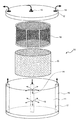
第1の態様は、特に円筒状に設けたメッシュ部材と液流制御部材に対し培養液流を円筒の中心軸から放射状に流す形態が好ましい。このような形態は、典型的には、図1に示すような円筒状のリアクター10によって実現できる。このリアクターは、基本的には円筒状の容器本体11、蓋12からなり、容器本体の中央には表面に複数の孔13を有する中心筒14が設けられ、また、蓋12の中央には開口部15があって、蓋12でリアクターを密閉したときに開口部15が中心筒14内の中空部と繋がって液の流路を形成するようになっている。あるいは、中心筒14は開口部15と一体に蓋12に固定されていてもよい。また、容器本体11の外周には容器内と外部とを繋ぐ別の管路16が設けられている。この管路16は1または複数設けられ、容器内に向けて開いた孔17を有し、また、管路16の上端は蓋12の対応位置に空けられた対応する開口部18と繋がって、液の流路を形成する。これにより、開口部15からリアクター10内に液を導入し、開口部18から液を取り出すことが可能になっている。もっとも、外周から液を取り出すための構成は任意であり、ここに挙げた構成に限られるものではない。
The first aspect is particularly preferably a mode in which the culture fluid flow is caused to flow radially from the central axis of the cylinder with respect to the mesh member and the fluid flow control member provided in a cylindrical shape. Such a configuration can be typically realized by a
本発明においては、前記中心筒14の周りにこれとほぼ同軸となるように、円筒状メッシュ部材19、前記円筒メッシュ部材の外面と容器内面との間に液流制御部材20を配設する。図1ではメッシュ部材19と液流制御部材20を取り出して示してあるが、使用時にはこれら円筒メッシュ部材と液流制御部材は前者が内側になるように容器内に装着する。例えば、円筒メッシュ部材19は、図示するように枠の内側にメッシュを固定ないし装着したものであり、可撓性シートまたはフィルムである液流制御部材をメッシュ部材19の枠の外側に巻き付けることができる。
培養液を前記開口部15からリアクター10内に送入することにより、その表面の孔13から容器内部に向けて、細胞外マトリックス成分と動物細胞を含む細胞培養液が半径方向に湧出する(図中の矢印)。培養液はメッシュ部材19と液流制御部材20を通り、容器内面で回収され、後述のようにポンプ手段を通して前記中心筒内に循環される。好ましくは循環経路内に、循環路内の圧力を感知する装置を設ける。In the present invention, the liquid
By feeding the culture solution into the
第2の態様は、特に平行に設けたメッシュ部材と液流制御部材が平面状の部材に対し培養液流をメッシュ部材の側から流す形態が好ましい。このような形態は、例えば、図2に示すように、下部に複数の孔21を有する筒状部材22を流路内に設置することにより実現される。
本発明では、筒状部材22内にメッシュからなる平面状部材23、その下側に液流制御部材24を配設する。好ましくは、筒状部材22は、その内周にリブ25を備えており、必要に応じて漏出防止用部材(例えば、シリコンリング)26、27、支持用メッシュ28を装入し、その上に液流制御部材26と平面状メッシュ部材24を重ねる。さらに、液の漏出防止用部材(例えば、内筒29)を重ねる。図2ではこれらの部材を取り出して示してあるが、使用時にはこれらの部材を、上記のように筒状部材22内のリブ25で固定されるように装着し、流路内に設置する。
例えば、第1の実施形態のリアクター10において中心筒14を取り除いた容器に上記筒状部材22を設置する。なお、リアクター内に入った液が筒状部材22の外部を流れた場合、高密度組織の形成効率が低下するので、筒状部材22または内筒29の高さを調整して、リアクターの蓋と筒状部材22または内筒29が密着するように設計することが好ましい。筒状部材22内に入った液は下部の複数の孔21からリアクター容器内に流出し、第1の態様と同様にして容器内面で回収される。なお、高密度組織が形成された後、筒状部材22をリアクター容器から取り出し、これを下から突起状部材で押し上げることにより、メッシュ部材24を容易に取り出すことができる。The second mode is particularly preferably a mode in which the mesh member and the liquid flow control member provided in parallel flow the culture solution flow from the mesh member side with respect to the planar member. Such a form is realized, for example, by installing a
In the present invention, a
For example, the said
装置全体の構成例としては、図4に示すように、リアクター10、培地溜め30、循環ポンプ40、フローセル50を管路で接続しインキュベーター60内に設置した閉鎖循環式培養装置が挙げられる。好ましくは、DO(溶存酸素)センサー70等のセンサーとその計測値の表示装置80、さらに培地溜め30内の培地を撹拌するためのスターラー90を設置する。スターラー90は、例えば、培地溜め内に入れた磁気撹拌子を回転させる磁気回転装置である。
As an example of the configuration of the entire apparatus, as shown in FIG. 4, there is a closed circulation culture apparatus in which a
なお、上記で例として挙げたリアクター容器は、特公平2−109966号公報)に記載されており、市販品も存在するが、容器内に生体担体触媒等を充填し、外周から内部に向かって培地を流すものであり、本発明で規定する構成により高密度な細胞培養装置として用いるものではない。特に、本発明者らの検討によれば、内部から外周に向かって培地を流す構成により、従来に比べ短時間で高密度な細胞培養が実現できるという予想外の知見が得られている。 In addition, although the reactor container mentioned as an example above is described in Japanese Patent Publication No. 2-109966) and a commercial item exists, the biocarrier catalyst etc. are filled in the container, and it goes to the inside from the outer periphery. The medium is flowed, and is not used as a high-density cell culture device by the configuration defined in the present invention. In particular, according to the study by the present inventors, an unexpected finding has been obtained that a high-density cell culture can be realized in a shorter time than in the prior art by flowing the culture medium from the inside toward the outer circumference.
メッシュ部材は、細胞外マトリックス成分と動物細胞の混合体である高密度細胞培養物を支持するに足るものであればよく、通常、液流を大きく妨げない程度の網目を有する部材である。具体的には、100μm〜1mm程度、より好ましくは100μm〜0.5mm程度の孔を有する。例えば、直径0.08〜0.1mm程度の針金を織って形成した100μm〜300μm程度のメッシュが利用できる。メッシュ部材の材料は金属(例えば、ステンレス)、合成樹脂(例えば、ポリエステル)、セラミック、人工材料等のいずれでもよい。通常は滅菌や洗浄操作の容易な金属製メッシュが好ましいが、例えば、人工血管材料等の生体適合性材料を用いれば、その上に高密度細胞組織を形成して生体に適用することも可能である。 The mesh member only needs to be sufficient to support a high-density cell culture that is a mixture of extracellular matrix components and animal cells, and is usually a member having a mesh that does not greatly disturb the liquid flow. Specifically, it has a hole of about 100 μm to 1 mm, more preferably about 100 μm to 0.5 mm. For example, a mesh of about 100 μm to 300 μm formed by weaving a wire having a diameter of about 0.08 to 0.1 mm can be used. The material of the mesh member may be any of metal (for example, stainless steel), synthetic resin (for example, polyester), ceramic, and artificial material. Usually, a metal mesh that is easy to sterilize and clean is preferred. For example, if a biocompatible material such as an artificial blood vessel material is used, a high-density cellular tissue can be formed on the mesh and applied to a living body. is there.
液流制御部材は、液流を透過させつつもその流れを減速させる部材であれば特に限定されないが、通常は、液流透過性多孔性材料、特に液流透過性の多孔性膜である。このような膜の例としては、濾紙、織布、不織布または絹フィブロイン膜が挙げられる。 The liquid flow control member is not particularly limited as long as it is a member that allows the liquid flow to permeate while decelerating the flow, but is usually a liquid flow permeable porous material, particularly a liquid flow permeable porous membrane. Examples of such membranes include filter paper, woven fabric, nonwoven fabric or silk fibroin membrane.
本発明では、メッシュ部材と液流制御部材を接触または近接させて配設する。ここで、近接と言う場合、液流制御部材による溶液の停滞がメッシュ部材近傍で生じるものであればよく、通常は数mm程度以下、好ましくは約1mm以下である。メッシュ部材と液流制御部材はいずれを(液流から見て)上流側に配置してもよいが、メッシュ部材(特に金属製メッシュ)を上流側に配置した場合には細胞外マトリックス成分と動物細胞のみからなる高密度細胞培養組織を容易に得ることができる。なお、細胞外マトリックス成分と動物細胞からなる高密度細胞培養組織と液流制御部材との複合部材を得る目的の場合は、液流制御部材を上流側に配置してもよい。また、メッシュ部材と液流制御部材は一体化されていてもよい。例えば、慣用の人工血管材料は、ポリエステルニットでステンレス製円筒を裏打ちした構造を採っており、本発明のメッシュ部材−液流制御部材の代替部材として用いることができる。 In the present invention, the mesh member and the liquid flow control member are disposed in contact with or in proximity to each other. Here, the term “proximity” is sufficient as long as the stagnation of the solution by the liquid flow control member occurs in the vicinity of the mesh member. Either the mesh member or the liquid flow control member may be arranged upstream (as viewed from the liquid flow), but when the mesh member (particularly a metal mesh) is arranged upstream, the extracellular matrix component and the animal A high-density cell culture tissue consisting only of cells can be easily obtained. In addition, in the case of the objective of obtaining the composite member of the high-density cell culture tissue which consists of an extracellular matrix component and an animal cell, and a fluid flow control member, you may arrange | position a fluid flow control member upstream. Further, the mesh member and the liquid flow control member may be integrated. For example, a conventional artificial blood vessel material has a structure in which a stainless steel cylinder is lined with polyester knit, and can be used as an alternative member of the mesh member-liquid flow control member of the present invention.
メッシュ部材と液流制御部材の上記以外の寸法条件(面積、ラジアルフロー型リアクターにおいては直径)は成長させようとする細胞の種類や組織の大きさにもよるが、細胞培養液の循環速度がメッシュ部材または液流制御部材近傍において、例えば、4〜10μl/cm2/秒程度、好ましくは6〜8μl/cm2/秒程度となる程度のものであればよい。The dimensional conditions (area, diameter in radial flow type reactors) other than the above for the mesh member and the liquid flow control member depend on the type of cell to be grown and the size of the tissue, but the circulation rate of the cell culture solution is in the mesh member or liquid flow control member near, for example, 4~10μl / cm 2 / sec, preferably about as long as the extent that the 6~8μl / cm 2 / sec approximately.
本発明において、細胞培養液中に含まれる細胞外マトリックス成分は細胞接着の基材として37℃、中性pH領域において重合ないし相互接着可能な分子であればよいが、典型的には結合組織中に見られる物質である。このような物質の例としては、例えば、コラーゲン、エラスチン、プロテオグリカン、フィブリリン、フィブロネクチン、ラミニン、キチン、キトサン等が挙げられる。これらの細胞外マトリックス成分は単独で用いてもよいし、2種以上の組み合わせとして用いてもよい。また、上記各成分は種々の化学的修飾を受けたものでものでもよい。修飾は生体内で通常見られる修飾でもよいし、種々の活性や特性を賦与する為の人工的な修飾でもよい。さらに、上記各成分の構成成分(例えば、プロテオグリカンについて、ヒアルロン酸、コンドロイチン硫酸、デルマタン硫酸、ヘパラン硫酸、ヘパリン、ケラタン硫酸などのグリコサミノグリカン等)も含まれ得る。 In the present invention, the extracellular matrix component contained in the cell culture solution may be any molecule that can be polymerized or mutually adhered in a neutral pH region at 37 ° C. as a cell adhesion substrate. It is a substance found in Examples of such substances include collagen, elastin, proteoglycan, fibrillin, fibronectin, laminin, chitin, chitosan and the like. These extracellular matrix components may be used alone or in combination of two or more. Each of the above components may be subjected to various chemical modifications. The modification may be a modification usually found in the living body or an artificial modification for imparting various activities and characteristics. Furthermore, constituent components of the above components (for example, for proteoglycans, hyaluronic acid, chondroitin sulfate, dermatan sulfate, heparan sulfate, heparin, keratan sulfate, etc.) can also be included.
好ましくは、コラーゲンもしくはエラスチンまたはこれらと上記成分の1種以上の組み合わせであり、特に好ましくはコラーゲンまたはコラーゲンと上記成分の1種以上の組み合わせである。どの成分が好ましいかは目的とする培養組織のタイプにより決定される。 Preferred is collagen or elastin, or a combination of one or more of these and the above components, and particularly preferred is a combination of collagen or collagen and one or more of the above components. Which component is preferred is determined by the type of tissue culture of interest.
コラーゲンとしては、従来公知のいずれのコラーゲンも用い得る。例えば、I型、II型、III型、IV型、V型等のコラーゲンを用いることができる。
こうしたコラーゲンは、得ようとするコラーゲンを含む生体組織を原料として、酸、酵素、アルカリ等により可溶化して用いることができる。また、アレルギー反応や拒否反応を解消ないし抑制するため、酵素処理によって分子末端のテロペプチドを全部または一部を除去することが好ましい。このようなコラーゲン材料としては、例えば、豚皮由来I型コラーゲン、豚腱由来I型コラーゲン、牛鼻軟骨由来II型コラーゲン、魚から抽出したI型コラーゲン、遺伝子組換え型のコラーゲンあるいはこれらの混合物等が挙げられる。但し、これらは例示であり、目的に応じ他の種類も利用可能である。例えば、基底膜に相当する組織を形成する場合にはIV型を用いる。As the collagen, any conventionally known collagen can be used. For example, collagens such as type I, type II, type III, type IV, and type V can be used.
Such collagen can be used by solubilizing with an acid, an enzyme, an alkali, or the like using a living tissue containing the collagen to be obtained as a raw material. In order to eliminate or suppress allergic reactions and rejection reactions, it is preferable to remove all or part of the telopeptide at the molecular end by enzymatic treatment. Examples of such collagen materials include pig skin-derived type I collagen, porcine tendon-derived type I collagen, bovine nasal cartilage-derived type II collagen, type I collagen extracted from fish, genetically modified collagen, or a mixture thereof. Is mentioned. However, these are examples, and other types can be used according to the purpose. For example, type IV is used when forming a tissue corresponding to the basement membrane.
細胞培養液中に含まれる動物細胞は、目的に応じて適宜選択され特に限定されないが、体細胞、腫瘍細胞、胚性幹細胞等が挙げられる。体細胞としては、例えば、線維芽細胞、肝細胞、血管内皮細胞、表皮細胞、上皮細胞、軟骨細胞、神経膠細胞および平滑筋細胞等が挙げられる。これらは単独でもよいし、2種類以上の混合物でもよい。 The animal cells contained in the cell culture medium are appropriately selected according to the purpose and are not particularly limited, and examples include somatic cells, tumor cells, embryonic stem cells and the like. Examples of somatic cells include fibroblasts, hepatocytes, vascular endothelial cells, epidermal cells, epithelial cells, chondrocytes, glial cells and smooth muscle cells. These may be used singly or as a mixture of two or more.
細胞培養液の基本組成は、培養対象とする動物細胞の種類にもよるが、慣用の天然培地または合成培地を用い得る。動物由来物質からの細菌やウイルスなどの感染、供給の時期や場所による組成のばらつき等の点を考慮すれば、合成培地がより好ましい。合成培地としては、特に限定はされないが、例えば、α−MEM(Minimum Essential Medium)、Eagle MEM、Dulbecco MEM(DMEM)、RPM I 1640培地、CMRC培地、HAM培地、DME/F12培地、199培地、MCDB培地等を挙げることができる。適宜、慣用される血清等を添加してもよい。天然培地としては、通常公知の天然培地を挙げることができ、特に限定はされない。これらは単独で用いても、2種以上を併用してもよい。 Although the basic composition of the cell culture medium depends on the type of animal cells to be cultured, a conventional natural medium or synthetic medium can be used. A synthetic medium is more preferable in consideration of infection from bacteria or viruses from animal-derived substances, variation in composition depending on the timing and place of supply, and the like. Although it does not specifically limit as a synthetic medium, For example, (alpha) -MEM (Minimum Essential Medium), Eagle MEM, Dulbecco MEM (DMEM), RPM I 1640 medium, CMRC medium, HAM medium, DME / F12 medium, 199 medium, MCDB medium etc. can be mentioned. A conventionally used serum or the like may be added as appropriate. Examples of the natural medium include usually known natural media, and are not particularly limited. These may be used alone or in combination of two or more.
細胞培養液中細胞外マトリックス成分の含有量は、培養開始時において0.1〜0.5mg/ml、好ましくは0.2〜0.3mg/ml程度である。 The content of the extracellular matrix component in the cell culture solution is about 0.1 to 0.5 mg / ml, preferably about 0.2 to 0.3 mg / ml at the start of the culture.
なお、細胞培養液は、上記細胞外マトリックス成分とともに、細胞付着を促進する他の物質、例えば、ポリリジン、ヒストン、グルテン、ゼラチン、フィブリン、フィブロイン等のペプチドやタンパク質;RGD、RGDS,GRGDS,YIGSR,IKVAV等の細胞接着性オリゴペプチドまたは遺伝子工学的にこれらの配列を組み込んだ合成タンパク質;アルギン酸、デンプン、デキストラン等の多糖およびこれらの誘導体;乳酸、グリコール酸、カプロラクトンおよびヒドロキシブチレートの重合体またはこれらの共重合体並びにこれらの重合体または共重合体とポリエチレングリコールもしくはポリプロピレングリコールとのブロックコポリマー等の生体分解性高分子を含んでもよい。 In addition, the cell culture medium contains other substances that promote cell adhesion together with the extracellular matrix component, such as peptides and proteins such as polylysine, histone, gluten, gelatin, fibrin, fibroin; RGD, RGDS, GRGDS, YIGSR, Cell-adhesive oligopeptides such as IKVAV or synthetic proteins incorporating these sequences by genetic engineering; polysaccharides such as alginic acid, starch, dextran and their derivatives; polymers of lactic acid, glycolic acid, caprolactone and hydroxybutyrate or these Copolymers of these and biodegradable polymers such as block copolymers of these polymers or copolymers with polyethylene glycol or polypropylene glycol may also be included.
また、培養液は、上記以外の生理活性物質を含んでもよい。このような生理活性物質の例としては、細胞成長因子、ホルモン及び/または薬理作用を有する天然もしくは合成化学物質が挙げられる。このような物質を添加することにより、機能を付与したり変化させることができる。また、還流条件を変えることによって自然界には存在しない合成化合物を含有させた細胞組み込み型の組織を作成できる。 Moreover, the culture solution may contain physiologically active substances other than those described above. Examples of such physiologically active substances include cell growth factors, hormones and / or natural or synthetic chemical substances having a pharmacological action. By adding such a substance, a function can be imparted or changed. In addition, by changing the reflux conditions, a cell-incorporated tissue containing a synthetic compound that does not exist in nature can be prepared.
細胞成長因子は、特に限定はされないが、例えば、表皮成長因子、上皮成長因子、線維芽細胞成長因子、血小板由来成長因子、肝細胞成長因子及びインシュリン等を挙げることができる。培養しようとする細胞の種類に応じて他の細胞成長因子を用いることも可能である。 The cell growth factor is not particularly limited, and examples thereof include epidermal growth factor, epidermal growth factor, fibroblast growth factor, platelet-derived growth factor, hepatocyte growth factor, and insulin. Other cell growth factors can be used depending on the type of cells to be cultured.
ホルモンは、特に限定はされないが、例えば、インシュリン、トランスフェリン、デキサメタゾン、ヒドロコルチゾン、チロキシン、3,3',5−トリヨードチロニン、1−メチル−3−ブチルキサンチン、プロゲステロンなどを挙げることができる。これらは単独で用いても、2種以上を併用してもよい。 The hormone is not particularly limited, and examples thereof include insulin, transferrin, dexamethasone, hydrocortisone, thyroxine, 3,3 ′, 5-triiodothyronine, 1-methyl-3-butylxanthine, progesterone and the like. These may be used alone or in combination of two or more.
その他の生理活性物質は、例えば、アスコルビン酸(特に、L−アスコルビン酸)、ビオチン、パントテン酸カルシウム、アスコルビン酸二リン酸、ビタミンD等のビタミン類、血清アルブミン、トランスフェリン等のタンパク質、脂質、脂質酸源、リノール酸、コレステロール、ピルビン酸、DNAおよびRNA合成用ヌクレオシド、グルココルチコイド、レチノイン酸、β−グリセロホスフェート、モノチオグリセロール、各種の抗生物質等を挙げることができる。なお、これらは例示であって、目的に応じてこれ以外の成分を用いることもできる。上記成分は単独で用いても、2種以上を併用してもよい。 Other physiologically active substances include, for example, ascorbic acid (particularly L-ascorbic acid), biotin, calcium pantothenate, ascorbyl diphosphate, vitamin D and other vitamins, proteins such as serum albumin and transferrin, lipids and lipids Examples include acid sources, linoleic acid, cholesterol, pyruvic acid, nucleosides for DNA and RNA synthesis, glucocorticoids, retinoic acid, β-glycerophosphate, monothioglycerol, and various antibiotics. These are merely examples, and other components may be used depending on the purpose. The above components may be used alone or in combination of two or more.
培養は通常の条件により、所望の大きさ(厚さ)の高密度培養組織が生成するまで行なえばよい。典型的には、培養温度は35〜40℃であり、培養時間は6時間〜9日である。上述のように、従来の高密度培養組織の製造方法では2週間以上の期間を要している。本発明によれば、必要な培養時間が大幅に短縮される。 The culture may be performed under normal conditions until a high-density cultured tissue having a desired size (thickness) is generated. Typically, the culture temperature is 35 to 40 ° C., and the culture time is 6 hours to 9 days. As described above, the conventional high-density culture tissue manufacturing method requires a period of two weeks or more. According to the present invention, the required culture time is significantly reduced.
また、本発明によれば、上記のいずれかに記載の方法により高密度培養組織を製造した後、得られた高密度培養組織を取り出し、細胞外マトリックス成分と1種類または複数の動物細胞を含む同一または異なる処方の非循環培養液中で培養を継続する高密度培養組織の製造方法が提供される。ここで、非循環培養条件とは、例えば、ディッシュ上での培養である。このような方法を採ることにより、新たに積層した細胞が生体に近い状態で増殖分化することが期待される。 In addition, according to the present invention, after producing a high-density cultured tissue by any of the methods described above, the obtained high-density cultured tissue is taken out and contains an extracellular matrix component and one or more animal cells. Provided is a method for producing a high-density cultured tissue in which cultivation is continued in a non-circulating culture solution of the same or different formulation. Here, the non-circulating culture condition is, for example, culture on a dish. By adopting such a method, it is expected that newly stacked cells proliferate and differentiate in a state close to a living body.
また、本発明によれば、上記のいずれかに記載の方法により高密度培養組織を製造した後、得られた高密度培養組織を取り出し、または引き続いて、細胞外マトリックス成分と1種類または複数の動物細胞を含む同一または異なる培養液を用いて前記組織上に異なる高密度培養組織を形成する操作を少なくとも1回行って積層型の高密度培養組織を形成することができる。 In addition, according to the present invention, after producing a high-density cultured tissue by any of the methods described above, the obtained high-density cultured tissue is taken out, or subsequently, an extracellular matrix component and one or more kinds of A stacked high-density culture tissue can be formed by performing at least one operation of forming different high-density culture tissues on the tissue using the same or different culture solutions containing animal cells.
例えば、細胞外マトリックス成分の種類や濃度、栄養成分の種類や濃度、または添加する成分の種類や濃度、あるいは温度やpH等の培養条件を連続してまたは断続的に変化させて培養することも可能であり、より生体に近い細胞外マトリックス環境を培養装置内で作成可能である。また、細胞接着基材だけでなく複数の細胞種(例えば、平滑筋細胞と血管内皮細胞など)を同時にあるいは時間差を持って閉鎖循環式培養装置内に投入することにより腸や尿管など一定の傾斜構造を持つ組織を再生することも可能である。 For example, the type and concentration of the extracellular matrix component, the type and concentration of the nutrient component, the type and concentration of the component to be added, or the culture conditions such as temperature and pH may be changed continuously or intermittently. It is possible to create an extracellular matrix environment closer to the living body in the culture apparatus. In addition to the cell adhesion substrate, a plurality of cell types (for example, smooth muscle cells and vascular endothelial cells, etc.) can be introduced into a closed circulation culture device at the same time or with a time lag so that certain intestines, ureters, etc. It is also possible to regenerate a tissue having an inclined structure.
さらに、この方法で製造した積層型高密度培養組織を取り出し、細胞外マトリックス成分と1種類または複数の動物細胞を含む同一または異なる処方の非循環培養液中で培養を継続することもできる。 Furthermore, the stacked high-density culture tissue produced by this method can be taken out, and the culture can be continued in a non-circulating culture solution of the same or different formulation containing an extracellular matrix component and one or more animal cells.
このように、本発明によれば、均一な高密度培養組織の迅速かつ確実な形成が可能であるとともに、複数の構造を一体化ないし複合化した高密度培養組織の迅速かつ確実な形成が可能である。このような高密度培養組織としては、人体各部の組織が含まれ、例えば、皮膚、軟骨、血管、神経、尿管、心臓、骨格筋または各種臓器及び腫瘍組織が挙げられる。 As described above, according to the present invention, it is possible to quickly and surely form a uniform high-density culture tissue, and it is possible to quickly and surely form a high-density culture tissue in which a plurality of structures are integrated or combined. It is. Such high-density cultured tissues include tissues of various parts of the human body, and examples include skin, cartilage, blood vessels, nerves, ureters, heart, skeletal muscle, various organs, and tumor tissues.
以下に実施例を挙げて本発明をより具体的に説明する。
実施例1
[高密度培養装置1]
高密度培養装置1は、図1に示すタイプのものであり以下のように構成した。
直径22mm、高さ17mmのステンレス製メッシュ(メッシュサイズ:133×266μm)を含む円筒の外周に濾紙(東洋濾紙(株)製JIS P−3801一種)を巻き付けて、メッシュ−液流制御部材を形成した。なお、メッシュは支持枠の内面に張られており、メッシュと液流制御部材間には、最大で上記支持部材の厚み程度の間隔が隔てられている。Hereinafter, the present invention will be described more specifically with reference to examples.
Example 1
[High-density culture device 1]
The high-density culture apparatus 1 is of the type shown in FIG. 1 and configured as follows.
A filter paper (a kind of JIS P-3801 manufactured by Toyo Filter Paper Co., Ltd.) is wrapped around a cylinder including a stainless steel mesh (mesh size: 133 × 266 μm) having a diameter of 22 mm and a height of 17 mm to form a mesh-liquid flow control member. did. Note that the mesh is stretched on the inner surface of the support frame, and the mesh and the liquid flow control member are spaced apart from each other by the maximum thickness of the support member.
このメッシュ−液流制御部材をラジアルフロー型バイオリアクター(エイブル(株)製BRK−05)内に挿入し、培地溜め、フローセル、DOセンサー(溶存酸素計)、循環路内の圧力を感知する装置、循環ポンプとともに閉鎖循環系を構成した。なお、上記ラジアルフロー型バイオリアクターは、容量約5mlのポリカーボネート製容器からなり、複数の孔を表面に有する中空の中心軸と複数の孔を有する外周面を有する。容器には、中心軸内と繋がる孔を有する蓋が附属しており、この孔を通して液を送入する。 This mesh-liquid flow control member is inserted into a radial flow bioreactor (BRK-05 manufactured by Able Co., Ltd.), and a medium reservoir, a flow cell, a DO sensor (dissolved oxygen meter), and a device for sensing pressure in the circulation path A closed circulation system was constructed with a circulation pump. The radial flow type bioreactor is made of a polycarbonate container having a capacity of about 5 ml, and has a hollow central axis having a plurality of holes on the surface and an outer peripheral surface having a plurality of holes. A lid having a hole connected to the inside of the central shaft is attached to the container, and the liquid is fed through the hole.
なお、メーカーの説明書によれば、これは、容器内にビーズやスポンジ等の担体を入れ、外周面から中心軸に向けて培地を送り込み循環する装置であるが、以下の実験では、容器内にはメッシュ−液流制御部材以外のものは装入せず、中心軸から外周面への液流となるように構成した。図4に示すようにこれらをCO2インキュベーター内に設置し、また、磁気撹拌子によって培地溜め内の培地を撹拌して酸素供給及びゲル化防止を行いつつ培地を循環させた。According to the manufacturer's instructions, this is a device that puts a carrier such as beads or sponge in the container and feeds and circulates the medium from the outer peripheral surface toward the central axis. Nothing other than the mesh-liquid flow control member was inserted, and the liquid flow from the central axis to the outer peripheral surface was configured. As shown in FIG. 4, these were placed in a CO 2 incubator, and the medium in the medium reservoir was stirred with a magnetic stir bar to circulate the medium while supplying oxygen and preventing gelation.
[培養実験]
培養液(DMEM+10%FBS(ウシ胎児血清)+100unit/mlペニシリンG、100μg/mlストレプトマイシン)とマウス正常線維芽細胞(5.0×107細胞)の混合液250mlにI型コラーゲン((株)高研製;蛋白消化酵素ペプシンを用いて牛皮より抽出したもの)を0.5mg/mlの割合で添加した。コラーゲン添加後、培養液を上記閉鎖循環系内において流速7ml/分で還流した。還流液を24時間毎に5mlずつ採取し、I型コラーゲン濃度とマトリックス・メタロプロテアーゼ(MMP)活性をそれぞれSDS−PAGEとザイモグラフィーにより解析した。[Culture experiment]
Type I collagen (Strain) was added to 250 ml of a mixture of culture medium (DMEM + 10% FBS (fetal bovine serum) +100 unit / ml penicillin G, 100 μg / ml streptomycin) and mouse normal fibroblasts (5.0 × 10 7 cells). Kenken; extracted from cowhide using protein digestion enzyme pepsin) was added at a rate of 0.5 mg / ml. After adding collagen, the culture solution was refluxed at a flow rate of 7 ml / min in the closed circulation system. Five ml of the reflux solution was collected every 24 hours, and the type I collagen concentration and matrix metalloprotease (MMP) activity were analyzed by SDS-PAGE and zymography, respectively.
還流開始後、前記メッシュ−液流制御部材のメッシュ側には、白色の滑らかな物質が集積し始めた。還流開始後3日目、6日目、9日目にリアクターから前記メッシュ−液流制御部材を回収したところ、3日目の時点でメッシュ上に厚さ2mm、幅17mm、長さ59mm、湿重量約1.0gの集積物が形成されていた(図5)。それ以後も若干の重量の増加が見られたが形態上の変化は認められなかった。
回収した集積物を光学顕微鏡(400倍)で観察したところ、基質と細胞から構成されていた。これを走査型電子顕微鏡で観察したところ、基質は直径約140nmの細線維が分岐・吻合を繰り返す網状構造を呈していた(図6)。この線維はコラーゲン細線維の特徴である周期性横紋を有しており、培養液中のI型コラーゲンはSDS−PAGE分析で経時的減少を示していることから基質がI型コラーゲンの自己重合体で形成されていることが確認された。After the start of reflux, a white smooth substance started to accumulate on the mesh side of the mesh-liquid flow control member. The mesh-liquid flow control member was recovered from the reactor on the 3rd, 6th, and 9th days after the start of reflux. As a result, on the 3rd day, the mesh was 2 mm thick, 17 mm wide, 59 mm long, and wet. An aggregate having a weight of about 1.0 g was formed (FIG. 5). After that, there was a slight increase in weight, but no change in morphology was observed.
When the collected aggregate was observed with an optical microscope (400 times), it was composed of a substrate and cells. When this was observed with a scanning electron microscope, the substrate exhibited a network structure in which fine fibers having a diameter of about 140 nm repeatedly branched and anastomosed (FIG. 6). These fibers have periodic striation, which is characteristic of collagen fibrils, and the type I collagen in the culture medium shows a decrease with time in SDS-PAGE analysis. It was confirmed that they were formed by coalescence.
還流開始後、DOセンサーの測定値は経時的に減少傾向を示し、細胞の生存・成長を裏付けていた。光学顕微鏡観察では、3日目の時点から線維芽細胞が一部紡錘形を示し、真皮層に存在する線維芽細胞と形態的に類似していた。なお、HE染色、電子顕微鏡観察の結果によれば、集積物中の細胞濃度は3〜9日の間では培養日数に関係なく400倍の視野中55〜70個程度であった。ゼラチン・ザイモグラフィーによる解析では、一過性のMMP−2及びMMP−9上昇が観察されたが、9日間の培養期間中、いずれのゲルにおいても細胞塊は観察されず散在性に存在した。
以上の通り、本発明の方法によれば、ディッシュ上での培養と異なり、真皮内により近い形態で高密度の線維芽細胞の培養が実現できる。After the start of reflux, the DO sensor readings showed a declining trend over time, confirming cell survival and growth. In light microscopic observation, fibroblasts partially showed a spindle shape from the third day, and were morphologically similar to fibroblasts present in the dermis layer. According to the results of HE staining and electron microscope observation, the cell concentration in the accumulation was about 55 to 70 in the field of view of 400 times regardless of the number of culture days between 3 and 9 days. In the analysis by gelatin zymography, transient increases in MMP-2 and MMP-9 were observed, but no cell clumps were observed in any gel during the 9-day culture period, and they were scattered.
As described above, according to the method of the present invention, unlike the culture on a dish, it is possible to culture a high-density fibroblast in a form closer to the dermis.
実施例2
実施例1と同じくI型コラーゲン0.5mg/mlに調整した培養液250mlを用意し、ヒト胃癌細胞(KATO III)1.0×10⁷細胞/250ml及びこれと同数のヒト線維芽細胞(TIG101)を上記閉鎖循環式培養装置にて4日間還流したところ、実施例1と同じように厚さ1〜2mm、幅17mm、長さ59mmの人工組織を得た。実施例1と同様に培養液中の溶存I型コラーゲンは還流培養によって減少しており、電子顕微鏡観察の結果からも人工組織の基質はI型コラーゲンの自己重合体で形成されていることが確認できた。 Example 2
As in Example 1, 250 ml of a culture solution adjusted to 0.5 mg / ml type I collagen was prepared. 1.0 × 10 7 cells / 250 ml of human gastric cancer cells (KATO III) and the same number of human fibroblasts (TIG101) ) Was refluxed for 4 days in the above closed circulation culture apparatus, and an artificial tissue having a thickness of 1-2 mm, a width of 17 mm, and a length of 59 mm was obtained in the same manner as in Example 1. As in Example 1, dissolved type I collagen in the culture broth was reduced by reflux culture, and the results of electron microscopic observation confirmed that the substrate of the artificial tissue was formed of a type I collagen autopolymer. did it.
光学顕微鏡的に観察したところ多数の硬癌細胞と線維芽細胞がコラーゲン細線維の網状構造の中にヒト癌組織によく似た状態で形成されている様子が観察された(図7)。
なお、この例では硬癌細胞だけをI型コラーゲンとともに還流培養しても多くの癌細胞は萎縮してしまい癌組織を得ることはできず、正常線維芽細胞とともにコラーゲンゲル内に培養することでのみヒトの癌組織モデルを作成できた。When observed under an optical microscope, it was observed that a large number of hard cancer cells and fibroblasts were formed in a network structure of collagen fibrils in a state similar to human cancer tissue (FIG. 7).
In this example, even if only hard cancer cells are reflux-cultured with type I collagen, many cancer cells shrink and cancer tissue cannot be obtained. By culturing them in a collagen gel with normal fibroblasts, Only a human cancer tissue model could be created.
実施例3
実施例1の前記メッシュ−液流制御部材に代えて、これとほぼ同形状同寸法のコラーゲン被覆平織りダクロン人工血管(Intergard−W(商標))をリアクター内に置いて5日間培養した。なお、用いたダクロン人工血管は既に移植手術に供されている製品であり、ポリエステル繊維のニット編み構造からなる。 Example 3
Instead of the mesh-liquid flow control member of Example 1, a collagen-coated plain weave Dacron artificial blood vessel (Intergard-W (trademark)) having almost the same shape and size as this was placed in a reactor and cultured for 5 days. The Dacron artificial blood vessel used is a product that has already been used for transplantation, and has a polyester fiber knit structure.
回収した人工組織のコラーゲンゲルは厚さ約3mmで、しかも均一で実施例1、2に比べて厚い(図8)。光学顕微鏡にて検索したところダクロン人工血管上にできた組織内には還流した細胞が観察された(図9)。
以上のように、本発明によれば、人工血管上に容易に人工組織を形成することが可能であり、人工生体高分子材料と組み合わせて人工組織を作成できることが確認された。また、組み合わせを例えば平滑筋とIV型コラーゲンに代えることにより血管の中膜(平滑筋の層)を人工的に作成することができる。The recovered collagen gel of the artificial tissue has a thickness of about 3 mm and is uniform and thicker than those of Examples 1 and 2 (FIG. 8). As a result of searching with an optical microscope, refluxed cells were observed in the tissue formed on the Dacron artificial blood vessel (FIG. 9).
As described above, according to the present invention, it was confirmed that an artificial tissue can be easily formed on an artificial blood vessel, and an artificial tissue can be created in combination with an artificial biopolymer material. In addition, by changing the combination to, for example, smooth muscle and type IV collagen, the vascular media (smooth muscle layer) can be artificially created.
実施例4
[高密度培養装置2]
高密度培養装置2は、図2に示すタイプのものであり以下のように構成した。
外径21.3mm、内径19.3mm、高さ20.5mmの下部に縦4.2mm、長さ11.2mmの開口部を3箇所有する金属製円筒を用意した。なお、この金属製円筒には、その内周に幅1.1mmのリブを設けた。この円筒内に、シリコン製ゴムリング(外径19.3mm、内径17.0m、高さ0.5mm)、濾紙支持用の円盤状ステンレス金網(直径18.7mm(以下同じ)、35〜50メッシュ)、濾紙(東洋濾紙(株)製JIS P−3801一種)、高密度培養組織製造用の円盤状ステンレス金網(100メッシュ)、シリコン製ゴムリング(外径19.3mm、内径17.0m、厚さ0.7mm)を置き、さらに金属製円筒の内側にこれと接するような金属製内筒(外径19.2mm、内径17.0m、高さ0.5mm)を配置した。内側に配置した金属製円筒(内筒)はリアクターに蓋を閉めた状態でその下面に密着するようにし、蓋中央の孔から培養液を導入した際に、還流培養液がすべて金属製円筒の内側を通るようにした。 Example 4
[High-density culture device 2]
The high-
A metal cylinder having three openings with a longitudinal length of 4.2 mm and a length of 11.2 mm was prepared in a lower portion having an outer diameter of 21.3 mm, an inner diameter of 19.3 mm, and a height of 20.5 mm. The metal cylinder was provided with a rib having a width of 1.1 mm on the inner periphery thereof. Inside this cylinder, a silicon rubber ring (outer diameter 19.3 mm, inner diameter 17.0 m, height 0.5 mm), disk-shaped stainless steel wire mesh (diameter 18.7 mm (hereinafter the same)), 35-50 mesh ), Filter paper (type JIS P-3801 manufactured by Toyo Filter Paper Co., Ltd.), disc-shaped stainless steel wire mesh (100 mesh) for producing high-density culture tissue, silicon rubber ring (outer diameter 19.3 mm, inner diameter 17.0 m, thickness A metal inner cylinder (outer diameter: 19.2 mm, inner diameter: 17.0 m, height: 0.5 mm) was placed inside the metal cylinder. The metal cylinder (inner cylinder) placed inside is in close contact with the bottom surface of the reactor with the lid closed, and when the culture medium is introduced from the hole in the center of the lid, all the reflux culture medium is the metal cylinder. I tried to pass inside.
[培養実験]
高密度培養装置を上記装置に変更し、培養細胞をヒト由来癌細胞(1.0×10⁷細胞)としコラーゲン濃度を0.5mg/ml、培養液の総量を200mlとし、上記の通りリアクター蓋中央から液を導入して20時間還流した。なお、培養液及びコラーゲンは実施例1と同じものを使用した。この結果、上部メッシュ上に、ほぼその全面にわたって滑らかな白色の集積物が得られた(図10)。
還流停止後、金属製円筒をリアクターから取り外し、円盤状の基部(直径50mm)の中央に円柱金属棒(直径16.5mm、高さ約25mm)を取り付けた部材の上に円筒をゆっくりと載せて円筒内部のメッシュを取り出した。
メッシュ上の集積物は厚さが均一に約0.5mmであり、顕微鏡観察の結果、コラーゲン線維中に癌細胞の増殖が観察された。[Culture experiment]
The high-density culture device was changed to the above device, the cultured cells were human-derived cancer cells (1.0 × 10 7 cells), the collagen concentration was 0.5 mg / ml, and the total amount of the culture solution was 200 ml. The liquid was introduced from the center and refluxed for 20 hours. The culture medium and collagen used were the same as in Example 1. As a result, a smooth white accumulation was obtained on the upper mesh substantially over the entire surface (FIG. 10).
After the reflux is stopped, the metal cylinder is removed from the reactor, and the cylinder is slowly placed on a member having a cylindrical metal rod (diameter 16.5 mm, height approximately 25 mm) attached to the center of the disc-shaped base (
The accumulation on the mesh was uniformly about 0.5 mm in thickness, and as a result of microscopic observation, growth of cancer cells was observed in the collagen fibers.
実施例5
図3に示すように、実施例4で用いた平板状ゲル作製装置(図2)のろ紙(24)を除き、目の細かい金属メッシュ(23)上にポリ乳酸線維のニット網構造からなる直径18.7mmのシート(31)を配置した。
このリアクター内に、I型コラーゲン0.5mg/mlに調整した培養液250mlを用意し、ヒト線維芽細胞(HFO小2)2×107個/250mlを4時間、5ml/minの流速にて環流した。 Example 5
As shown in FIG. 3, the diameter comprising a knit network structure of polylactic acid fibers on a fine metal mesh (23) except for the filter paper (24) of the flat gel preparation apparatus (FIG. 2) used in Example 4. An 18.7 mm sheet (31) was placed.
In this reactor, 250 ml of culture solution adjusted to 0.5 mg / ml type I collagen was prepared, and 2 × 10 7 human fibroblasts (HFO small 2) / 250 ml were flown for 4 hours at a flow rate of 5 ml / min. Circulated.
0.5mg/ml I型コラーゲンと線維芽細胞の混合液に替えて、引き続き0.1mg/ml Matrigel(基底膜の代替標品)50mlに差し替えて2時間還流を続けた。この還流終了直前に大動脈平滑筋(Clontech 社AoSMC)4×10⁶個をリアクター内に注入し、還流を終えた。
リアクター内からは厚さ2.4mm直径17.5mmのゲルを回収できた。なお、今回の実施例では還流液に牛胎児血清を添加していない。
この回収物を光学顕微鏡で観察したところ、線維芽細胞を含むI型コラーゲン細線維層と重合したマトリゲル内に平滑筋細胞を持った層が階層的に作製されていた(図11)。
また、走査電子顕微鏡による観察では最下層からポリ乳酸ゲル層、I型コラーゲン細線維と線維芽細胞よりなる層、マトリゲルと平滑筋から構成された層が区別された(図12)。The mixture was changed to a mixed solution of 0.5 mg / ml type I collagen and fibroblasts, subsequently replaced with 50 ml of 0.1 mg / ml Matrigel (an alternative sample of basement membrane), and reflux was continued for 2 hours. Immediately before the completion of the reflux, 4 × 10 6 aortic smooth muscles (Clontech AoSMC) were injected into the reactor to complete the reflux.
From the reactor, a gel having a thickness of 2.4 mm and a diameter of 17.5 mm could be recovered. In this example, fetal calf serum was not added to the reflux solution.
When this recovered material was observed with an optical microscope, a layer having smooth muscle cells was hierarchically formed in a Matrigel polymerized with a type I collagen fibril layer containing fibroblasts (FIG. 11).
In the observation with a scanning electron microscope, a polylactic acid gel layer, a layer composed of type I collagen fibrils and fibroblasts, and a layer composed of matrigel and smooth muscle were distinguished from the bottom layer (FIG. 12).
本発明によれば、三次元立体培養と高密度培養を繰り返すことにより階層的に異なった細胞外マトリックスと細胞の層を重ねることができる。コラーゲンは中性培養液では2mg/ml程度が限界で、この濃度では細線維形成が急速に起こるためコラーゲンゲル作成は容易でない。しかしながら閉鎖循環式高密度培養装置では0.5mg/mlと低濃度I型コラーゲン溶液を用いるため未熟練者でも容易に細胞組み込み型コラーゲンゲルを作成することができる。
本発明を利用することにより(1)BSE病原体であるプリオン混入の危険性を回避できる、(2)血管,腸管,尿管などの階層構造を持った組織を再生することができる可能性が示される。
According to the present invention, the extracellular matrix and the layer of cells that are hierarchically different can be stacked by repeating three-dimensional three-dimensional culture and high-density culture. Collagen is limited to about 2 mg / ml in a neutral culture solution, and at this concentration, formation of a collagen gel is not easy because fibril formation occurs rapidly. However, since the closed circulation type high-density culture apparatus uses a low concentration type I collagen solution of 0.5 mg / ml, even an unskilled person can easily create a cell-incorporated collagen gel.
By using the present invention, (1) the risk of prion contamination as a BSE pathogen can be avoided, and (2) the possibility of regenerating a tissue having a hierarchical structure such as blood vessels, intestinal tracts, ureters, etc. It is.
10 リアクター
11 容器本体
12 蓋
13 孔
14 中心筒
15 開口部
16 管路
17 孔
18 開口部
19 メッシュ部材
20 液流制御部材
21 孔
22 筒状部材
23 平面状メッシュ部材
24 液流制御部材
25 リブ
26、27 シリコンリング
28 支持用メッシュ
29 内筒
30 培地溜め
31 シート
40 循環ポンプ
50 フローセル
60 インキュベーター
70 センサー
80 表示装置
90 スターラーDESCRIPTION OF
Claims (23)
Applications Claiming Priority (3)
| Application Number | Priority Date | Filing Date | Title |
|---|---|---|---|
| JP2005038261 | 2005-02-15 | ||
| JP2005038261 | 2005-02-15 | ||
| PCT/JP2006/302561 WO2006088029A1 (en) | 2005-02-15 | 2006-02-14 | Method of producing high-density cultured tissue and high-density cultured tissue |
Publications (2)
| Publication Number | Publication Date |
|---|---|
| JPWO2006088029A1 JPWO2006088029A1 (en) | 2008-07-03 |
| JP4671365B2 true JP4671365B2 (en) | 2011-04-13 |
Family
ID=36916435
Family Applications (1)
| Application Number | Title | Priority Date | Filing Date |
|---|---|---|---|
| JP2007503665A Active JP4671365B2 (en) | 2005-02-15 | 2006-02-14 | Method for producing high-density cultured tissue and high-density cultured tissue |
Country Status (4)
| Country | Link |
|---|---|
| US (1) | US20100203638A1 (en) |
| EP (1) | EP1857543B1 (en) |
| JP (1) | JP4671365B2 (en) |
| WO (1) | WO2006088029A1 (en) |
Cited By (3)
| Publication number | Priority date | Publication date | Assignee | Title |
|---|---|---|---|---|
| WO2013105665A1 (en) | 2012-01-12 | 2013-07-18 | 株式会社ニッピ | Collagen structure, and method for producing collagen structure |
| KR101393108B1 (en) * | 2013-07-18 | 2014-05-13 | 한국생산기술연구원 | Compact animall cell incubator and method for incubation of animal cell |
| KR20180118482A (en) * | 2017-04-21 | 2018-10-31 | 한국과학기술연구원 | Composition for preparing a biocompatible tissue, method preparing the same and method treating a disease requiring a vascularization |
Families Citing this family (25)
| Publication number | Priority date | Publication date | Assignee | Title |
|---|---|---|---|---|
| US20090061508A1 (en) | 2007-08-27 | 2009-03-05 | Biomimetics Technologies Inc | Laminar flow reactor |
| PT103906A (en) * | 2007-12-20 | 2009-08-31 | Ass For The Advancement Of Tis | DYNAMIC SYSTEMS OF CELL CULTURE IN THREE-DIMENSIONAL SUPPORTS |
| JP2010172247A (en) * | 2009-01-29 | 2010-08-12 | Kitasato Institute | Method for producing lamination type high density cultured artificial tissue and lamination type high density cultured artificial tissue |
| DE102009008923B4 (en) * | 2009-02-13 | 2011-05-05 | Hennig, Jörn, Dipl.-Ing. | Cell seeding chamber |
| ES2372108B1 (en) * | 2009-10-26 | 2013-02-11 | Ebers Medical Technology, S.L | FLOW BIORREACTOR FOR CELL CULTURE. |
| WO2011142425A1 (en) * | 2010-05-12 | 2011-11-17 | 国立大学法人香川大学 | Material for regeneration of epithelial-layer-containing tissue and method for evaluating regeneration |
| EP2720862B1 (en) | 2011-06-17 | 2016-08-24 | Fiberweb, Inc. | Vapor permeable, substantially water impermeable multilayer article |
| WO2012178027A2 (en) | 2011-06-23 | 2012-12-27 | Fiberweb, Inc. | Vapor-permeable, substantially water-impermeable multilayer article |
| WO2012177996A2 (en) | 2011-06-23 | 2012-12-27 | Fiberweb, Inc. | Vapor permeable, substantially water impermeable multilayer article |
| US9765459B2 (en) | 2011-06-24 | 2017-09-19 | Fiberweb, Llc | Vapor-permeable, substantially water-impermeable multilayer article |
| KR101422345B1 (en) * | 2012-10-19 | 2014-07-22 | 한국생명공학연구원 | Dual structured cell culture dish |
| KR101613618B1 (en) | 2014-07-09 | 2016-04-20 | 전세화 | A method for preparing the three-dimensionally cultured skin comprising dermis and epidermis, and the cultured skin made therefrom |
| CA2980287C (en) * | 2015-03-19 | 2022-08-16 | President And Fellows Of Harvard College | Incubating enclosure |
| JP6710211B2 (en) * | 2015-08-03 | 2020-06-17 | 富士フイルム株式会社 | Cell structure, non-human model animal, method for producing non-human model animal, and method for evaluating test substance |
| JP2018183058A (en) * | 2015-09-24 | 2018-11-22 | 株式会社村田製作所 | Cell culture method and cell culture device |
| GB2554635A (en) * | 2016-08-03 | 2018-04-11 | Northwick Park Institute For Medical Res Ltd | Bioreactors and methods for processing biological material |
| AU2017316545B2 (en) | 2016-08-21 | 2022-12-08 | Adva Biotechnology Ltd. | Bioreactor and methods of use thereof |
| WO2018135252A1 (en) * | 2017-01-18 | 2018-07-26 | 国立研究開発法人農業・食品産業技術総合研究機構 | Semipermeable membrane and uses thereof |
| DE102017213923A1 (en) | 2017-08-10 | 2019-02-14 | Robert Bosch Gesellschaft Für Medizinische Forschung Mbh | Device for cultivating tissue sections |
| WO2020163329A1 (en) | 2019-02-05 | 2020-08-13 | Corning Incorporated | Woven cell culture substrates |
| US11118151B2 (en) * | 2019-11-05 | 2021-09-14 | Corning Incorporated | Fixed bed bioreactor and methods of using the same |
| US20210222114A1 (en) | 2020-01-20 | 2021-07-22 | Adva Biotechnology Ltd. | Device and method for controlling a bioreactor |
| EP4133054A4 (en) * | 2020-04-06 | 2024-10-02 | Mission Barns, Inc. | SCALABLE BIOREACTOR SYSTEMS AND METHODS OF USE THEREOF |
| US11732232B2 (en) | 2020-08-14 | 2023-08-22 | Korea Research Institute Of Bioscience And Biotechnology | Biomimetic cell culture apparatus and cell culture system comprising the same |
| JPWO2023145957A1 (en) * | 2022-01-31 | 2023-08-03 |
Citations (4)
| Publication number | Priority date | Publication date | Assignee | Title |
|---|---|---|---|---|
| JPH07298876A (en) * | 1994-03-09 | 1995-11-14 | Res Dev Corp Of Japan | Liquid-permeable cell culture carrier, culture method and culture apparatus using this carrier |
| JPH1052261A (en) * | 1996-08-09 | 1998-02-24 | Sumitomo Bakelite Co Ltd | Reconstructed mammalian tissue |
| JP2002142752A (en) * | 2000-11-13 | 2002-05-21 | Asahi Techno Glass Corp | Collagen-coated cell culture vessel and method for producing the same |
| JP2002335949A (en) * | 2001-05-22 | 2002-11-26 | Inst Of Physical & Chemical Res | Three-dimensional tissue culture method of cells using honeycomb structure film |
Family Cites Families (10)
| Publication number | Priority date | Publication date | Assignee | Title |
|---|---|---|---|---|
| US4087327A (en) * | 1976-04-12 | 1978-05-02 | Monsanto Company | Mammalion cell culture process |
| US4720462A (en) * | 1985-11-05 | 1988-01-19 | Robert Rosenson | Culture system for the culture of solid tissue masses and method of using the same |
| US5605835A (en) * | 1988-05-23 | 1997-02-25 | Regents Of The University Of Minnesota | Bioreactor device with application as a bioartificial liver |
| JPH02109966A (en) * | 1988-10-20 | 1990-04-23 | Kirin Brewery Co Ltd | Radial flow packed bed-type bioreactor |
| JPH06277050A (en) * | 1993-03-26 | 1994-10-04 | Kanegafuchi Chem Ind Co Ltd | Immobilization material for animal cell and culture method |
| JP4601746B2 (en) * | 1999-10-25 | 2010-12-22 | エイブル株式会社 | Three-dimensional animal cell culture apparatus and culture method |
| US6428802B1 (en) * | 1999-12-29 | 2002-08-06 | Children's Medical Center Corp. | Preparing artificial organs by forming polylayers of different cell populations on a substrate |
| JP2003047461A (en) * | 2001-07-31 | 2003-02-18 | Astec:Kk | Method for carrying out high-density cell culture of cell by using apatite sheet, culturing device and cell culture module |
| JP4344112B2 (en) * | 2002-03-19 | 2009-10-14 | 泰彦 田畑 | Biological tissue-like structure, bone marrow stem cell culture method and culture kit |
| GB2401612A (en) * | 2003-05-13 | 2004-11-17 | Univ Manchester | Method and device for culturing tissue |
-
2006
- 2006-02-14 WO PCT/JP2006/302561 patent/WO2006088029A1/en active Application Filing
- 2006-02-14 JP JP2007503665A patent/JP4671365B2/en active Active
- 2006-02-14 US US11/884,308 patent/US20100203638A1/en not_active Abandoned
- 2006-02-14 EP EP06713702A patent/EP1857543B1/en active Active
Patent Citations (4)
| Publication number | Priority date | Publication date | Assignee | Title |
|---|---|---|---|---|
| JPH07298876A (en) * | 1994-03-09 | 1995-11-14 | Res Dev Corp Of Japan | Liquid-permeable cell culture carrier, culture method and culture apparatus using this carrier |
| JPH1052261A (en) * | 1996-08-09 | 1998-02-24 | Sumitomo Bakelite Co Ltd | Reconstructed mammalian tissue |
| JP2002142752A (en) * | 2000-11-13 | 2002-05-21 | Asahi Techno Glass Corp | Collagen-coated cell culture vessel and method for producing the same |
| JP2002335949A (en) * | 2001-05-22 | 2002-11-26 | Inst Of Physical & Chemical Res | Three-dimensional tissue culture method of cells using honeycomb structure film |
Cited By (5)
| Publication number | Priority date | Publication date | Assignee | Title |
|---|---|---|---|---|
| WO2013105665A1 (en) | 2012-01-12 | 2013-07-18 | 株式会社ニッピ | Collagen structure, and method for producing collagen structure |
| KR101393108B1 (en) * | 2013-07-18 | 2014-05-13 | 한국생산기술연구원 | Compact animall cell incubator and method for incubation of animal cell |
| WO2015008949A1 (en) * | 2013-07-18 | 2015-01-22 | 한국생산기술연구원 | Simple animal cell incubator and method for incubating animal cells using same |
| US10041032B2 (en) | 2013-07-18 | 2018-08-07 | Korea Institute Of Industrial Technology | Simple animal cell culture device and method for culturing animal cells using same |
| KR20180118482A (en) * | 2017-04-21 | 2018-10-31 | 한국과학기술연구원 | Composition for preparing a biocompatible tissue, method preparing the same and method treating a disease requiring a vascularization |
Also Published As
| Publication number | Publication date |
|---|---|
| WO2006088029A1 (en) | 2006-08-24 |
| EP1857543A4 (en) | 2010-09-22 |
| EP1857543A1 (en) | 2007-11-21 |
| US20100203638A1 (en) | 2010-08-12 |
| JPWO2006088029A1 (en) | 2008-07-03 |
| EP1857543B1 (en) | 2012-04-04 |
Similar Documents
| Publication | Publication Date | Title |
|---|---|---|
| JP4671365B2 (en) | Method for producing high-density cultured tissue and high-density cultured tissue | |
| WO2010087397A1 (en) | Process for producing laminated high-density cultured artificial tissue, and laminated high-density cultured artificial tissue | |
| Carrier et al. | Perfusion improves tissue architecture of engineered cardiac muscle | |
| Godbey et al. | In vitro systems for tissue engineering | |
| EP0422209B1 (en) | Implants for large volumes of cells on polymeric matrices | |
| Liao et al. | Stem cells and biomimetic materials strategies for tissue engineering | |
| Kulig et al. | Hepatic tissue engineering | |
| Tsang et al. | Fabrication of 3D hepatic tissues by additive photopatterning of cellular hydrogels | |
| CN106421916B (en) | Organization engineering skin and preparation method thereof | |
| CA2330104A1 (en) | Creation of three-dimensional tissues | |
| KR19990022216A (en) | Naturally Secreted Extracellular Matrix Compositions and Methods | |
| Vunjak-Novakovic et al. | Cell seeding of polymer scaffolds | |
| JP5669741B2 (en) | Culture system | |
| Kaur et al. | Advances in biomaterials for hepatic tissue engineering | |
| WO2005014774A1 (en) | Carrier for culturing animal cell, and method for culturing or transplanting animal cell using said carrier for culture | |
| US20030003089A1 (en) | Tissue engineered cardiac constructs | |
| Song et al. | Three-dimensional expansion: in suspension culture of SD rat's osteoblasts in a rotating wall vessel bioreactor | |
| Obregón et al. | Bioreactors in tissue engineering | |
| JP2002542817A (en) | Modular cell carrier system for three-dimensional cell growth | |
| Kulkarni et al. | Cell immobilization strategies for tissue engineering: Recent trends and future perspectives | |
| Ichinohe et al. | Proliferation, osteogenic differentiation, and distribution of rat bone marrow stromal cells in nonwoven fabrics by different culture methods | |
| Levenberg et al. | Embryonic stem cells in tissue engineering | |
| AU2003270112A1 (en) | Bioreactor with expandable surface area for culturing cells | |
| CN114381419B (en) | Bionic artificial liver tissue and preparation method and application thereof | |
| Vunjak-Novakovic | Tissue engineering: basic considerations |
Legal Events
| Date | Code | Title | Description |
|---|---|---|---|
| A131 | Notification of reasons for refusal |
Free format text: JAPANESE INTERMEDIATE CODE: A131 Effective date: 20101022 |
|
| A521 | Request for written amendment filed |
Free format text: JAPANESE INTERMEDIATE CODE: A523 Effective date: 20101214 |
|
| TRDD | Decision of grant or rejection written | ||
| A01 | Written decision to grant a patent or to grant a registration (utility model) |
Free format text: JAPANESE INTERMEDIATE CODE: A01 Effective date: 20110114 |
|
| A01 | Written decision to grant a patent or to grant a registration (utility model) |
Free format text: JAPANESE INTERMEDIATE CODE: A01 |
|
| A61 | First payment of annual fees (during grant procedure) |
Free format text: JAPANESE INTERMEDIATE CODE: A61 Effective date: 20110114 |
|
| R150 | Certificate of patent or registration of utility model |
Ref document number: 4671365 Country of ref document: JP Free format text: JAPANESE INTERMEDIATE CODE: R150 Free format text: JAPANESE INTERMEDIATE CODE: R150 |
|
| FPAY | Renewal fee payment (event date is renewal date of database) |
Free format text: PAYMENT UNTIL: 20140128 Year of fee payment: 3 |
|
| R250 | Receipt of annual fees |
Free format text: JAPANESE INTERMEDIATE CODE: R250 |
|
| R250 | Receipt of annual fees |
Free format text: JAPANESE INTERMEDIATE CODE: R250 |
|
| R250 | Receipt of annual fees |
Free format text: JAPANESE INTERMEDIATE CODE: R250 |
|
| R250 | Receipt of annual fees |
Free format text: JAPANESE INTERMEDIATE CODE: R250 |
|
| R250 | Receipt of annual fees |
Free format text: JAPANESE INTERMEDIATE CODE: R250 |
|
| R250 | Receipt of annual fees |
Free format text: JAPANESE INTERMEDIATE CODE: R250 |
|
| R250 | Receipt of annual fees |
Free format text: JAPANESE INTERMEDIATE CODE: R250 |
|
| R250 | Receipt of annual fees |
Free format text: JAPANESE INTERMEDIATE CODE: R250 |
|
| R250 | Receipt of annual fees |
Free format text: JAPANESE INTERMEDIATE CODE: R250 |
|
| R250 | Receipt of annual fees |
Free format text: JAPANESE INTERMEDIATE CODE: R250 |
|
| R250 | Receipt of annual fees |
Free format text: JAPANESE INTERMEDIATE CODE: R250 |
|
| R250 | Receipt of annual fees |
Free format text: JAPANESE INTERMEDIATE CODE: R250 |