JP4276301B2 - Electrophoretic analysis of molecules using immobilized probes - Google Patents
Electrophoretic analysis of molecules using immobilized probes Download PDFInfo
- Publication number
- JP4276301B2 JP4276301B2 JP54957698A JP54957698A JP4276301B2 JP 4276301 B2 JP4276301 B2 JP 4276301B2 JP 54957698 A JP54957698 A JP 54957698A JP 54957698 A JP54957698 A JP 54957698A JP 4276301 B2 JP4276301 B2 JP 4276301B2
- Authority
- JP
- Japan
- Prior art keywords
- medium
- capture probe
- capture
- target
- electrophoresis
- Prior art date
- Legal status (The legal status is an assumption and is not a legal conclusion. Google has not performed a legal analysis and makes no representation as to the accuracy of the status listed.)
- Expired - Fee Related
Links
- 238000001962 electrophoresis Methods 0.000 title claims abstract description 88
- 239000000523 sample Substances 0.000 title claims description 394
- 238000000034 method Methods 0.000 claims abstract description 92
- 150000007523 nucleic acids Chemical class 0.000 claims description 128
- 108020004707 nucleic acids Proteins 0.000 claims description 125
- 102000039446 nucleic acids Human genes 0.000 claims description 125
- 239000011159 matrix material Substances 0.000 claims description 72
- 238000012360 testing method Methods 0.000 claims description 47
- 238000009739 binding Methods 0.000 claims description 45
- 230000027455 binding Effects 0.000 claims description 39
- 108090000623 proteins and genes Proteins 0.000 claims description 36
- 229920000642 polymer Polymers 0.000 claims description 27
- 102000004169 proteins and genes Human genes 0.000 claims description 26
- 239000000126 substance Substances 0.000 claims description 16
- HRPVXLWXLXDGHG-UHFFFAOYSA-N Acrylamide Chemical group NC(=O)C=C HRPVXLWXLXDGHG-UHFFFAOYSA-N 0.000 claims description 13
- 229920000936 Agarose Polymers 0.000 claims description 12
- 239000000178 monomer Substances 0.000 claims description 12
- 108091023037 Aptamer Proteins 0.000 claims description 11
- 230000005684 electric field Effects 0.000 claims description 10
- 229920002472 Starch Polymers 0.000 claims description 9
- 230000015572 biosynthetic process Effects 0.000 claims description 9
- 239000000463 material Substances 0.000 claims description 9
- 239000008107 starch Substances 0.000 claims description 9
- 235000019698 starch Nutrition 0.000 claims description 9
- 229920002401 polyacrylamide Polymers 0.000 claims description 8
- 239000002131 composite material Substances 0.000 claims description 5
- 238000002360 preparation method Methods 0.000 claims description 5
- 125000006850 spacer group Chemical group 0.000 claims description 5
- 238000012546 transfer Methods 0.000 claims description 5
- 239000002202 Polyethylene glycol Substances 0.000 claims description 4
- 230000001965 increasing effect Effects 0.000 claims description 4
- 229920001223 polyethylene glycol Polymers 0.000 claims description 4
- 238000007334 copolymerization reaction Methods 0.000 claims description 3
- 229910019142 PO4 Inorganic materials 0.000 claims description 2
- 238000010828 elution Methods 0.000 claims description 2
- VKFFWRNLDPAXOT-UHFFFAOYSA-N n-(6-aminohexyl)-2-methylprop-2-enamide Chemical group CC(=C)C(=O)NCCCCCCN VKFFWRNLDPAXOT-UHFFFAOYSA-N 0.000 claims description 2
- 239000010452 phosphate Substances 0.000 claims description 2
- 238000011109 contamination Methods 0.000 claims 1
- NBIIXXVUZAFLBC-UHFFFAOYSA-K phosphate Chemical compound [O-]P([O-])([O-])=O NBIIXXVUZAFLBC-UHFFFAOYSA-K 0.000 claims 1
- 239000000499 gel Substances 0.000 description 91
- 239000010410 layer Substances 0.000 description 51
- 238000009396 hybridization Methods 0.000 description 35
- 238000004458 analytical method Methods 0.000 description 22
- 230000000295 complement effect Effects 0.000 description 20
- 238000001514 detection method Methods 0.000 description 20
- 239000000203 mixture Substances 0.000 description 16
- 125000003729 nucleotide group Chemical group 0.000 description 16
- 239000002773 nucleotide Substances 0.000 description 15
- 238000006116 polymerization reaction Methods 0.000 description 15
- 238000002474 experimental method Methods 0.000 description 14
- 108020004414 DNA Proteins 0.000 description 11
- 108091034117 Oligonucleotide Proteins 0.000 description 11
- 238000003556 assay Methods 0.000 description 11
- 238000013508 migration Methods 0.000 description 11
- 230000005012 migration Effects 0.000 description 11
- 239000000243 solution Substances 0.000 description 11
- 108091093037 Peptide nucleic acid Proteins 0.000 description 10
- 239000000872 buffer Substances 0.000 description 8
- MGNCLNQXLYJVJD-UHFFFAOYSA-N cyanuric chloride Chemical compound ClC1=NC(Cl)=NC(Cl)=N1 MGNCLNQXLYJVJD-UHFFFAOYSA-N 0.000 description 8
- 230000003993 interaction Effects 0.000 description 8
- 230000037230 mobility Effects 0.000 description 8
- 230000003287 optical effect Effects 0.000 description 8
- 230000009870 specific binding Effects 0.000 description 8
- 108091032973 (ribonucleotides)n+m Proteins 0.000 description 7
- 238000000746 purification Methods 0.000 description 7
- ZHNUHDYFZUAESO-UHFFFAOYSA-N Formamide Chemical compound NC=O ZHNUHDYFZUAESO-UHFFFAOYSA-N 0.000 description 6
- JLCPHMBAVCMARE-UHFFFAOYSA-N [3-[[3-[[3-[[3-[[3-[[3-[[3-[[3-[[3-[[3-[[3-[[5-(2-amino-6-oxo-1H-purin-9-yl)-3-[[3-[[3-[[3-[[3-[[3-[[5-(2-amino-6-oxo-1H-purin-9-yl)-3-[[5-(2-amino-6-oxo-1H-purin-9-yl)-3-hydroxyoxolan-2-yl]methoxy-hydroxyphosphoryl]oxyoxolan-2-yl]methoxy-hydroxyphosphoryl]oxy-5-(5-methyl-2,4-dioxopyrimidin-1-yl)oxolan-2-yl]methoxy-hydroxyphosphoryl]oxy-5-(6-aminopurin-9-yl)oxolan-2-yl]methoxy-hydroxyphosphoryl]oxy-5-(6-aminopurin-9-yl)oxolan-2-yl]methoxy-hydroxyphosphoryl]oxy-5-(6-aminopurin-9-yl)oxolan-2-yl]methoxy-hydroxyphosphoryl]oxy-5-(6-aminopurin-9-yl)oxolan-2-yl]methoxy-hydroxyphosphoryl]oxyoxolan-2-yl]methoxy-hydroxyphosphoryl]oxy-5-(5-methyl-2,4-dioxopyrimidin-1-yl)oxolan-2-yl]methoxy-hydroxyphosphoryl]oxy-5-(4-amino-2-oxopyrimidin-1-yl)oxolan-2-yl]methoxy-hydroxyphosphoryl]oxy-5-(5-methyl-2,4-dioxopyrimidin-1-yl)oxolan-2-yl]methoxy-hydroxyphosphoryl]oxy-5-(5-methyl-2,4-dioxopyrimidin-1-yl)oxolan-2-yl]methoxy-hydroxyphosphoryl]oxy-5-(6-aminopurin-9-yl)oxolan-2-yl]methoxy-hydroxyphosphoryl]oxy-5-(6-aminopurin-9-yl)oxolan-2-yl]methoxy-hydroxyphosphoryl]oxy-5-(4-amino-2-oxopyrimidin-1-yl)oxolan-2-yl]methoxy-hydroxyphosphoryl]oxy-5-(4-amino-2-oxopyrimidin-1-yl)oxolan-2-yl]methoxy-hydroxyphosphoryl]oxy-5-(4-amino-2-oxopyrimidin-1-yl)oxolan-2-yl]methoxy-hydroxyphosphoryl]oxy-5-(6-aminopurin-9-yl)oxolan-2-yl]methoxy-hydroxyphosphoryl]oxy-5-(4-amino-2-oxopyrimidin-1-yl)oxolan-2-yl]methyl [5-(6-aminopurin-9-yl)-2-(hydroxymethyl)oxolan-3-yl] hydrogen phosphate Polymers Cc1cn(C2CC(OP(O)(=O)OCC3OC(CC3OP(O)(=O)OCC3OC(CC3O)n3cnc4c3nc(N)[nH]c4=O)n3cnc4c3nc(N)[nH]c4=O)C(COP(O)(=O)OC3CC(OC3COP(O)(=O)OC3CC(OC3COP(O)(=O)OC3CC(OC3COP(O)(=O)OC3CC(OC3COP(O)(=O)OC3CC(OC3COP(O)(=O)OC3CC(OC3COP(O)(=O)OC3CC(OC3COP(O)(=O)OC3CC(OC3COP(O)(=O)OC3CC(OC3COP(O)(=O)OC3CC(OC3COP(O)(=O)OC3CC(OC3COP(O)(=O)OC3CC(OC3COP(O)(=O)OC3CC(OC3COP(O)(=O)OC3CC(OC3COP(O)(=O)OC3CC(OC3COP(O)(=O)OC3CC(OC3COP(O)(=O)OC3CC(OC3CO)n3cnc4c(N)ncnc34)n3ccc(N)nc3=O)n3cnc4c(N)ncnc34)n3ccc(N)nc3=O)n3ccc(N)nc3=O)n3ccc(N)nc3=O)n3cnc4c(N)ncnc34)n3cnc4c(N)ncnc34)n3cc(C)c(=O)[nH]c3=O)n3cc(C)c(=O)[nH]c3=O)n3ccc(N)nc3=O)n3cc(C)c(=O)[nH]c3=O)n3cnc4c3nc(N)[nH]c4=O)n3cnc4c(N)ncnc34)n3cnc4c(N)ncnc34)n3cnc4c(N)ncnc34)n3cnc4c(N)ncnc34)O2)c(=O)[nH]c1=O JLCPHMBAVCMARE-UHFFFAOYSA-N 0.000 description 6
- 238000005251 capillar electrophoresis Methods 0.000 description 6
- 238000006243 chemical reaction Methods 0.000 description 6
- GNBHRKFJIUUOQI-UHFFFAOYSA-N fluorescein Chemical compound O1C(=O)C2=CC=CC=C2C21C1=CC=C(O)C=C1OC1=CC(O)=CC=C21 GNBHRKFJIUUOQI-UHFFFAOYSA-N 0.000 description 6
- 238000002372 labelling Methods 0.000 description 6
- 239000002245 particle Substances 0.000 description 6
- 238000000926 separation method Methods 0.000 description 6
- 239000007787 solid Substances 0.000 description 6
- 241000894006 Bacteria Species 0.000 description 5
- 229920002678 cellulose Polymers 0.000 description 5
- 239000001913 cellulose Substances 0.000 description 5
- 239000003153 chemical reaction reagent Substances 0.000 description 5
- 102000044158 nucleic acid binding protein Human genes 0.000 description 5
- 108700020942 nucleic acid binding protein Proteins 0.000 description 5
- 230000002829 reductive effect Effects 0.000 description 5
- 238000011160 research Methods 0.000 description 5
- 239000007790 solid phase Substances 0.000 description 5
- XLYOFNOQVPJJNP-UHFFFAOYSA-N water Substances O XLYOFNOQVPJJNP-UHFFFAOYSA-N 0.000 description 5
- YBJHBAHKTGYVGT-ZKWXMUAHSA-N (+)-Biotin Chemical compound N1C(=O)N[C@@H]2[C@H](CCCCC(=O)O)SC[C@@H]21 YBJHBAHKTGYVGT-ZKWXMUAHSA-N 0.000 description 4
- QTBSBXVTEAMEQO-UHFFFAOYSA-N Acetic acid Chemical compound CC(O)=O QTBSBXVTEAMEQO-UHFFFAOYSA-N 0.000 description 4
- 102000053602 DNA Human genes 0.000 description 4
- 229920002307 Dextran Polymers 0.000 description 4
- KCXVZYZYPLLWCC-UHFFFAOYSA-N EDTA Chemical compound OC(=O)CN(CC(O)=O)CCN(CC(O)=O)CC(O)=O KCXVZYZYPLLWCC-UHFFFAOYSA-N 0.000 description 4
- FAPWRFPIFSIZLT-UHFFFAOYSA-M Sodium chloride Chemical compound [Na+].[Cl-] FAPWRFPIFSIZLT-UHFFFAOYSA-M 0.000 description 4
- 230000004913 activation Effects 0.000 description 4
- ROOXNKNUYICQNP-UHFFFAOYSA-N ammonium persulfate Chemical compound [NH4+].[NH4+].[O-]S(=O)(=O)OOS([O-])(=O)=O ROOXNKNUYICQNP-UHFFFAOYSA-N 0.000 description 4
- 238000003491 array Methods 0.000 description 4
- 150000001875 compounds Chemical class 0.000 description 4
- 229960002086 dextran Drugs 0.000 description 4
- 238000012986 modification Methods 0.000 description 4
- 230000004048 modification Effects 0.000 description 4
- 239000000843 powder Substances 0.000 description 4
- 150000003141 primary amines Chemical group 0.000 description 4
- 238000012552 review Methods 0.000 description 4
- 238000007873 sieving Methods 0.000 description 4
- 241000894007 species Species 0.000 description 4
- RYHBNJHYFVUHQT-UHFFFAOYSA-N 1,4-Dioxane Chemical compound C1COCCO1 RYHBNJHYFVUHQT-UHFFFAOYSA-N 0.000 description 3
- OSBLTNPMIGYQGY-UHFFFAOYSA-N 2-amino-2-(hydroxymethyl)propane-1,3-diol;2-[2-[bis(carboxymethyl)amino]ethyl-(carboxymethyl)amino]acetic acid;boric acid Chemical compound OB(O)O.OCC(N)(CO)CO.OC(=O)CN(CC(O)=O)CCN(CC(O)=O)CC(O)=O OSBLTNPMIGYQGY-UHFFFAOYSA-N 0.000 description 3
- HFVPDKWVDPEQCO-UHFFFAOYSA-N 2-methylideneoctanamide Chemical group CCCCCCC(=C)C(N)=O HFVPDKWVDPEQCO-UHFFFAOYSA-N 0.000 description 3
- CSCPPACGZOOCGX-UHFFFAOYSA-N Acetone Chemical compound CC(C)=O CSCPPACGZOOCGX-UHFFFAOYSA-N 0.000 description 3
- WEVYAHXRMPXWCK-UHFFFAOYSA-N Acetonitrile Chemical compound CC#N WEVYAHXRMPXWCK-UHFFFAOYSA-N 0.000 description 3
- 229920000663 Hydroxyethyl cellulose Polymers 0.000 description 3
- 239000004354 Hydroxyethyl cellulose Substances 0.000 description 3
- 108020005187 Oligonucleotide Probes Proteins 0.000 description 3
- 108020004682 Single-Stranded DNA Proteins 0.000 description 3
- HEMHJVSKTPXQMS-UHFFFAOYSA-M Sodium hydroxide Chemical compound [OH-].[Na+] HEMHJVSKTPXQMS-UHFFFAOYSA-M 0.000 description 3
- 239000008051 TBE buffer Substances 0.000 description 3
- 108090000190 Thrombin Proteins 0.000 description 3
- 238000013459 approach Methods 0.000 description 3
- 230000008901 benefit Effects 0.000 description 3
- 102000023732 binding proteins Human genes 0.000 description 3
- 108091008324 binding proteins Proteins 0.000 description 3
- 239000007853 buffer solution Substances 0.000 description 3
- 239000000969 carrier Substances 0.000 description 3
- 239000013592 cell lysate Substances 0.000 description 3
- 238000013461 design Methods 0.000 description 3
- 238000000502 dialysis Methods 0.000 description 3
- 150000004676 glycans Chemical class 0.000 description 3
- 235000019447 hydroxyethyl cellulose Nutrition 0.000 description 3
- 238000011065 in-situ storage Methods 0.000 description 3
- 239000007791 liquid phase Substances 0.000 description 3
- 238000004519 manufacturing process Methods 0.000 description 3
- 229910052757 nitrogen Inorganic materials 0.000 description 3
- 238000007899 nucleic acid hybridization Methods 0.000 description 3
- 239000002751 oligonucleotide probe Substances 0.000 description 3
- 229920001282 polysaccharide Polymers 0.000 description 3
- 239000005017 polysaccharide Substances 0.000 description 3
- 230000005855 radiation Effects 0.000 description 3
- 230000000717 retained effect Effects 0.000 description 3
- 230000035945 sensitivity Effects 0.000 description 3
- 238000003756 stirring Methods 0.000 description 3
- 239000000725 suspension Substances 0.000 description 3
- PEJGGZVVBQPTRG-UHFFFAOYSA-N 3-[2-[2-[2-[bis(4-methoxyphenyl)-phenylmethoxy]ethoxy]ethoxy]ethoxy-[di(propan-2-yl)amino]phosphanyl]oxypropanenitrile Chemical compound C1=CC(OC)=CC=C1C(OCCOCCOCCOP(OCCC#N)N(C(C)C)C(C)C)(C=1C=CC(OC)=CC=1)C1=CC=CC=C1 PEJGGZVVBQPTRG-UHFFFAOYSA-N 0.000 description 2
- NOIIUHRQUVNIDD-UHFFFAOYSA-N 3-[[oxo(pyridin-4-yl)methyl]hydrazo]-N-(phenylmethyl)propanamide Chemical compound C=1C=CC=CC=1CNC(=O)CCNNC(=O)C1=CC=NC=C1 NOIIUHRQUVNIDD-UHFFFAOYSA-N 0.000 description 2
- QFVHZQCOUORWEI-UHFFFAOYSA-N 4-[(4-anilino-5-sulfonaphthalen-1-yl)diazenyl]-5-hydroxynaphthalene-2,7-disulfonic acid Chemical compound C=12C(O)=CC(S(O)(=O)=O)=CC2=CC(S(O)(=O)=O)=CC=1N=NC(C1=CC=CC(=C11)S(O)(=O)=O)=CC=C1NC1=CC=CC=C1 QFVHZQCOUORWEI-UHFFFAOYSA-N 0.000 description 2
- 102000002260 Alkaline Phosphatase Human genes 0.000 description 2
- 108020004774 Alkaline Phosphatase Proteins 0.000 description 2
- IJGRMHOSHXDMSA-UHFFFAOYSA-N Atomic nitrogen Chemical compound N#N IJGRMHOSHXDMSA-UHFFFAOYSA-N 0.000 description 2
- 108090001008 Avidin Proteins 0.000 description 2
- 108091008102 DNA aptamers Proteins 0.000 description 2
- 239000003298 DNA probe Substances 0.000 description 2
- KWYHDKDOAIKMQN-UHFFFAOYSA-N N,N,N',N'-tetramethylethylenediamine Chemical compound CN(C)CCN(C)C KWYHDKDOAIKMQN-UHFFFAOYSA-N 0.000 description 2
- 101710163270 Nuclease Proteins 0.000 description 2
- 108091028043 Nucleic acid sequence Proteins 0.000 description 2
- 229920003171 Poly (ethylene oxide) Polymers 0.000 description 2
- 241000944748 Quesada Species 0.000 description 2
- BQCADISMDOOEFD-UHFFFAOYSA-N Silver Chemical compound [Ag] BQCADISMDOOEFD-UHFFFAOYSA-N 0.000 description 2
- 108010090804 Streptavidin Proteins 0.000 description 2
- ISAKRJDGNUQOIC-UHFFFAOYSA-N Uracil Chemical compound O=C1C=CNC(=O)N1 ISAKRJDGNUQOIC-UHFFFAOYSA-N 0.000 description 2
- XSQUKJJJFZCRTK-UHFFFAOYSA-N Urea Chemical compound NC(N)=O XSQUKJJJFZCRTK-UHFFFAOYSA-N 0.000 description 2
- 150000003926 acrylamides Chemical class 0.000 description 2
- 230000009471 action Effects 0.000 description 2
- 229910001870 ammonium persulfate Inorganic materials 0.000 description 2
- 229920001222 biopolymer Polymers 0.000 description 2
- 229960002685 biotin Drugs 0.000 description 2
- 235000020958 biotin Nutrition 0.000 description 2
- 239000011616 biotin Substances 0.000 description 2
- 239000008280 blood Substances 0.000 description 2
- 210000004369 blood Anatomy 0.000 description 2
- 229910021538 borax Inorganic materials 0.000 description 2
- 239000004327 boric acid Substances 0.000 description 2
- RYYVLZVUVIJVGH-UHFFFAOYSA-N caffeine Chemical compound CN1C(=O)N(C)C(=O)C2=C1N=CN2C RYYVLZVUVIJVGH-UHFFFAOYSA-N 0.000 description 2
- 239000003054 catalyst Substances 0.000 description 2
- 230000001413 cellular effect Effects 0.000 description 2
- 125000003636 chemical group Chemical group 0.000 description 2
- 239000003795 chemical substances by application Substances 0.000 description 2
- 230000002860 competitive effect Effects 0.000 description 2
- 239000000356 contaminant Substances 0.000 description 2
- 230000008878 coupling Effects 0.000 description 2
- 238000010168 coupling process Methods 0.000 description 2
- 238000005859 coupling reaction Methods 0.000 description 2
- OPTASPLRGRRNAP-UHFFFAOYSA-N cytosine Chemical compound NC=1C=CNC(=O)N=1 OPTASPLRGRRNAP-UHFFFAOYSA-N 0.000 description 2
- 230000003111 delayed effect Effects 0.000 description 2
- 238000010586 diagram Methods 0.000 description 2
- 229910001873 dinitrogen Inorganic materials 0.000 description 2
- 239000012153 distilled water Substances 0.000 description 2
- 230000000694 effects Effects 0.000 description 2
- 238000005516 engineering process Methods 0.000 description 2
- 239000012634 fragment Substances 0.000 description 2
- UYTPUPDQBNUYGX-UHFFFAOYSA-N guanine Chemical compound O=C1NC(N)=NC2=C1N=CN2 UYTPUPDQBNUYGX-UHFFFAOYSA-N 0.000 description 2
- 125000002887 hydroxy group Chemical group [H]O* 0.000 description 2
- 230000003100 immobilizing effect Effects 0.000 description 2
- 238000011534 incubation Methods 0.000 description 2
- 238000010369 molecular cloning Methods 0.000 description 2
- 239000003068 molecular probe Substances 0.000 description 2
- 230000035772 mutation Effects 0.000 description 2
- ZIUHHBKFKCYYJD-UHFFFAOYSA-N n,n'-methylenebisacrylamide Chemical compound C=CC(=O)NCNC(=O)C=C ZIUHHBKFKCYYJD-UHFFFAOYSA-N 0.000 description 2
- -1 nucleotide triphosphates Chemical class 0.000 description 2
- 238000012634 optical imaging Methods 0.000 description 2
- 239000001048 orange dye Substances 0.000 description 2
- 238000003752 polymerase chain reaction Methods 0.000 description 2
- 229920002451 polyvinyl alcohol Polymers 0.000 description 2
- 108090000765 processed proteins & peptides Proteins 0.000 description 2
- 238000012545 processing Methods 0.000 description 2
- 230000000750 progressive effect Effects 0.000 description 2
- 238000002331 protein detection Methods 0.000 description 2
- 230000009467 reduction Effects 0.000 description 2
- 230000008929 regeneration Effects 0.000 description 2
- 238000011069 regeneration method Methods 0.000 description 2
- 230000001105 regulatory effect Effects 0.000 description 2
- 230000002441 reversible effect Effects 0.000 description 2
- 229910052709 silver Inorganic materials 0.000 description 2
- 239000004332 silver Substances 0.000 description 2
- 239000011780 sodium chloride Substances 0.000 description 2
- 235000010339 sodium tetraborate Nutrition 0.000 description 2
- 238000002764 solid phase assay Methods 0.000 description 2
- 239000002904 solvent Substances 0.000 description 2
- 238000010186 staining Methods 0.000 description 2
- 239000000758 substrate Substances 0.000 description 2
- ZFXYFBGIUFBOJW-UHFFFAOYSA-N theophylline Chemical compound O=C1N(C)C(=O)N(C)C2=C1NC=N2 ZFXYFBGIUFBOJW-UHFFFAOYSA-N 0.000 description 2
- 229960004072 thrombin Drugs 0.000 description 2
- 229960003766 thrombin (human) Drugs 0.000 description 2
- RWQNBRDOKXIBIV-UHFFFAOYSA-N thymine Chemical compound CC1=CNC(=O)NC1=O RWQNBRDOKXIBIV-UHFFFAOYSA-N 0.000 description 2
- BSVBQGMMJUBVOD-UHFFFAOYSA-N trisodium borate Chemical compound [Na+].[Na+].[Na+].[O-]B([O-])[O-] BSVBQGMMJUBVOD-UHFFFAOYSA-N 0.000 description 2
- 238000000108 ultra-filtration Methods 0.000 description 2
- 238000005406 washing Methods 0.000 description 2
- 238000007693 zone electrophoresis Methods 0.000 description 2
- WJNGQIYEQLPJMN-IOSLPCCCSA-N 1-methylinosine Chemical compound C1=NC=2C(=O)N(C)C=NC=2N1[C@@H]1O[C@H](CO)[C@@H](O)[C@H]1O WJNGQIYEQLPJMN-IOSLPCCCSA-N 0.000 description 1
- PIINGYXNCHTJTF-UHFFFAOYSA-N 2-(2-azaniumylethylamino)acetate Chemical group NCCNCC(O)=O PIINGYXNCHTJTF-UHFFFAOYSA-N 0.000 description 1
- QKNYBSVHEMOAJP-UHFFFAOYSA-N 2-amino-2-(hydroxymethyl)propane-1,3-diol;hydron;chloride Chemical compound Cl.OCC(N)(CO)CO QKNYBSVHEMOAJP-UHFFFAOYSA-N 0.000 description 1
- PMUNIMVZCACZBB-UHFFFAOYSA-N 2-hydroxyethylazanium;chloride Chemical compound Cl.NCCO PMUNIMVZCACZBB-UHFFFAOYSA-N 0.000 description 1
- BCZUPRDAAVVBSO-MJXNYTJMSA-N 4-acetylcytidine Chemical compound C1=CC(C(=O)C)(N)NC(=O)N1[C@H]1[C@H](O)[C@H](O)[C@@H](CO)O1 BCZUPRDAAVVBSO-MJXNYTJMSA-N 0.000 description 1
- FWMNVWWHGCHHJJ-SKKKGAJSSA-N 4-amino-1-[(2r)-6-amino-2-[[(2r)-2-[[(2r)-2-[[(2r)-2-amino-3-phenylpropanoyl]amino]-3-phenylpropanoyl]amino]-4-methylpentanoyl]amino]hexanoyl]piperidine-4-carboxylic acid Chemical compound C([C@H](C(=O)N[C@H](CC(C)C)C(=O)N[C@H](CCCCN)C(=O)N1CCC(N)(CC1)C(O)=O)NC(=O)[C@H](N)CC=1C=CC=CC=1)C1=CC=CC=C1 FWMNVWWHGCHHJJ-SKKKGAJSSA-N 0.000 description 1
- VSCNRXVDHRNJOA-PNHWDRBUSA-N 5-(carboxymethylaminomethyl)uridine Chemical compound O[C@@H]1[C@H](O)[C@@H](CO)O[C@H]1N1C(=O)NC(=O)C(CNCC(O)=O)=C1 VSCNRXVDHRNJOA-PNHWDRBUSA-N 0.000 description 1
- LRSASMSXMSNRBT-UHFFFAOYSA-N 5-methylcytosine Chemical compound CC1=CNC(=O)N=C1N LRSASMSXMSNRBT-UHFFFAOYSA-N 0.000 description 1
- AFHJQYHRLPMKHU-XXWVOBANSA-N Aloin Natural products O=C1c2c(O)cc(CO)cc2[C@H]([C@H]2[C@H](O)[C@@H](O)[C@@H](O)[C@@H](CO)O2)c2c1c(O)ccc2 AFHJQYHRLPMKHU-XXWVOBANSA-N 0.000 description 1
- 208000002109 Argyria Diseases 0.000 description 1
- 108010077805 Bacterial Proteins Proteins 0.000 description 1
- 108020004635 Complementary DNA Proteins 0.000 description 1
- HMFHBZSHGGEWLO-SOOFDHNKSA-N D-ribofuranose Chemical compound OC[C@H]1OC(O)[C@H](O)[C@@H]1O HMFHBZSHGGEWLO-SOOFDHNKSA-N 0.000 description 1
- 230000006820 DNA synthesis Effects 0.000 description 1
- 102000052510 DNA-Binding Proteins Human genes 0.000 description 1
- 101710096438 DNA-binding protein Proteins 0.000 description 1
- SHIBSTMRCDJXLN-UHFFFAOYSA-N Digoxigenin Natural products C1CC(C2C(C3(C)CCC(O)CC3CC2)CC2O)(O)C2(C)C1C1=CC(=O)OC1 SHIBSTMRCDJXLN-UHFFFAOYSA-N 0.000 description 1
- 241000196324 Embryophyta Species 0.000 description 1
- 102000004190 Enzymes Human genes 0.000 description 1
- 108090000790 Enzymes Proteins 0.000 description 1
- VGGSQFUCUMXWEO-UHFFFAOYSA-N Ethene Chemical compound C=C VGGSQFUCUMXWEO-UHFFFAOYSA-N 0.000 description 1
- 239000005977 Ethylene Substances 0.000 description 1
- SXRSQZLOMIGNAQ-UHFFFAOYSA-N Glutaraldehyde Chemical compound O=CCCCC=O SXRSQZLOMIGNAQ-UHFFFAOYSA-N 0.000 description 1
- 108010001336 Horseradish Peroxidase Proteins 0.000 description 1
- LPHGQDQBBGAPDZ-UHFFFAOYSA-N Isocaffeine Natural products CN1C(=O)N(C)C(=O)C2=C1N(C)C=N2 LPHGQDQBBGAPDZ-UHFFFAOYSA-N 0.000 description 1
- SNDPXSYFESPGGJ-BYPYZUCNSA-N L-2-aminopentanoic acid Chemical compound CCC[C@H](N)C(O)=O SNDPXSYFESPGGJ-BYPYZUCNSA-N 0.000 description 1
- ROHFNLRQFUQHCH-YFKPBYRVSA-N L-leucine Chemical compound CC(C)C[C@H](N)C(O)=O ROHFNLRQFUQHCH-YFKPBYRVSA-N 0.000 description 1
- SNDPXSYFESPGGJ-UHFFFAOYSA-N L-norVal-OH Natural products CCCC(N)C(O)=O SNDPXSYFESPGGJ-UHFFFAOYSA-N 0.000 description 1
- 108010054278 Lac Repressors Proteins 0.000 description 1
- GUBGYTABKSRVRQ-QKKXKWKRSA-N Lactose Natural products OC[C@H]1O[C@@H](O[C@H]2[C@H](O)[C@@H](O)C(O)O[C@@H]2CO)[C@H](O)[C@@H](O)[C@H]1O GUBGYTABKSRVRQ-QKKXKWKRSA-N 0.000 description 1
- ROHFNLRQFUQHCH-UHFFFAOYSA-N Leucine Natural products CC(C)CC(N)C(O)=O ROHFNLRQFUQHCH-UHFFFAOYSA-N 0.000 description 1
- 239000000020 Nitrocellulose Substances 0.000 description 1
- 108020004711 Nucleic Acid Probes Proteins 0.000 description 1
- CTQNGGLPUBDAKN-UHFFFAOYSA-N O-Xylene Chemical compound CC1=CC=CC=C1C CTQNGGLPUBDAKN-UHFFFAOYSA-N 0.000 description 1
- 238000012408 PCR amplification Methods 0.000 description 1
- 108091005804 Peptidases Proteins 0.000 description 1
- 239000004365 Protease Substances 0.000 description 1
- 241000913681 Questa Species 0.000 description 1
- 108020004518 RNA Probes Proteins 0.000 description 1
- 239000003391 RNA probe Substances 0.000 description 1
- 239000013614 RNA sample Substances 0.000 description 1
- 108020004511 Recombinant DNA Proteins 0.000 description 1
- 102000009661 Repressor Proteins Human genes 0.000 description 1
- 102100037486 Reverse transcriptase/ribonuclease H Human genes 0.000 description 1
- PYMYPHUHKUWMLA-LMVFSUKVSA-N Ribose Natural products OC[C@@H](O)[C@@H](O)[C@@H](O)C=O PYMYPHUHKUWMLA-LMVFSUKVSA-N 0.000 description 1
- 240000004808 Saccharomyces cerevisiae Species 0.000 description 1
- VYPSYNLAJGMNEJ-UHFFFAOYSA-N Silicium dioxide Chemical compound O=[Si]=O VYPSYNLAJGMNEJ-UHFFFAOYSA-N 0.000 description 1
- 108010052104 Viral Regulatory and Accessory Proteins Proteins 0.000 description 1
- 241000700605 Viruses Species 0.000 description 1
- 150000001252 acrylic acid derivatives Chemical class 0.000 description 1
- 239000011543 agarose gel Substances 0.000 description 1
- 238000000246 agarose gel electrophoresis Methods 0.000 description 1
- CPUHNROBVJNNPW-UHFFFAOYSA-N aloin A Natural products OC1C(O)C(O)C(CO)OC1OC1C2=CC(CO)=CC(O)=C2C(=O)C2=C(O)C=CC=C21 CPUHNROBVJNNPW-UHFFFAOYSA-N 0.000 description 1
- AFHJQYHRLPMKHU-WEZNYRQKSA-N aloin B Chemical compound O[C@@H]1[C@@H](O)[C@H](O)[C@@H](CO)O[C@H]1[C@H]1C2=CC(CO)=CC(O)=C2C(=O)C2=C(O)C=CC=C21 AFHJQYHRLPMKHU-WEZNYRQKSA-N 0.000 description 1
- HMFHBZSHGGEWLO-UHFFFAOYSA-N alpha-D-Furanose-Ribose Natural products OCC1OC(O)C(O)C1O HMFHBZSHGGEWLO-UHFFFAOYSA-N 0.000 description 1
- 150000001412 amines Chemical class 0.000 description 1
- 150000001413 amino acids Chemical class 0.000 description 1
- 239000003125 aqueous solvent Substances 0.000 description 1
- 238000007846 asymmetric PCR Methods 0.000 description 1
- 239000012298 atmosphere Substances 0.000 description 1
- QVGXLLKOCUKJST-UHFFFAOYSA-N atomic oxygen Chemical compound [O] QVGXLLKOCUKJST-UHFFFAOYSA-N 0.000 description 1
- 238000000376 autoradiography Methods 0.000 description 1
- 229920005601 base polymer Polymers 0.000 description 1
- 239000013060 biological fluid Substances 0.000 description 1
- 239000012472 biological sample Substances 0.000 description 1
- 230000023555 blood coagulation Effects 0.000 description 1
- 238000010804 cDNA synthesis Methods 0.000 description 1
- 229960001948 caffeine Drugs 0.000 description 1
- VJEONQKOZGKCAK-UHFFFAOYSA-N caffeine Natural products CN1C(=O)N(C)C(=O)C2=C1C=CN2C VJEONQKOZGKCAK-UHFFFAOYSA-N 0.000 description 1
- 239000004202 carbamide Substances 0.000 description 1
- 150000001718 carbodiimides Chemical class 0.000 description 1
- 235000014633 carbohydrates Nutrition 0.000 description 1
- 150000001720 carbohydrates Chemical class 0.000 description 1
- 125000003178 carboxy group Chemical group [H]OC(*)=O 0.000 description 1
- 238000005266 casting Methods 0.000 description 1
- 230000015556 catabolic process Effects 0.000 description 1
- 230000003197 catalytic effect Effects 0.000 description 1
- 238000006555 catalytic reaction Methods 0.000 description 1
- 230000008859 change Effects 0.000 description 1
- 150000005829 chemical entities Chemical class 0.000 description 1
- 238000003776 cleavage reaction Methods 0.000 description 1
- 238000004440 column chromatography Methods 0.000 description 1
- 239000002299 complementary DNA Substances 0.000 description 1
- 238000004624 confocal microscopy Methods 0.000 description 1
- 230000021615 conjugation Effects 0.000 description 1
- 238000001816 cooling Methods 0.000 description 1
- 238000012937 correction Methods 0.000 description 1
- 238000004132 cross linking Methods 0.000 description 1
- ATDGTVJJHBUTRL-UHFFFAOYSA-N cyanogen bromide Chemical compound BrC#N ATDGTVJJHBUTRL-UHFFFAOYSA-N 0.000 description 1
- 229940104302 cytosine Drugs 0.000 description 1
- 238000006731 degradation reaction Methods 0.000 description 1
- 239000003398 denaturant Substances 0.000 description 1
- 238000004925 denaturation Methods 0.000 description 1
- 230000036425 denaturation Effects 0.000 description 1
- 238000006392 deoxygenation reaction Methods 0.000 description 1
- 229960000633 dextran sulfate Drugs 0.000 description 1
- 238000012631 diagnostic technique Methods 0.000 description 1
- 238000009792 diffusion process Methods 0.000 description 1
- QONQRTHLHBTMGP-UHFFFAOYSA-N digitoxigenin Natural products CC12CCC(C3(CCC(O)CC3CC3)C)C3C11OC1CC2C1=CC(=O)OC1 QONQRTHLHBTMGP-UHFFFAOYSA-N 0.000 description 1
- SHIBSTMRCDJXLN-KCZCNTNESA-N digoxigenin Chemical compound C1([C@@H]2[C@@]3([C@@](CC2)(O)[C@H]2[C@@H]([C@@]4(C)CC[C@H](O)C[C@H]4CC2)C[C@H]3O)C)=CC(=O)OC1 SHIBSTMRCDJXLN-KCZCNTNESA-N 0.000 description 1
- 238000004141 dimensional analysis Methods 0.000 description 1
- 229940079593 drug Drugs 0.000 description 1
- 239000003814 drug Substances 0.000 description 1
- 239000000975 dye Substances 0.000 description 1
- 239000013013 elastic material Substances 0.000 description 1
- 238000006911 enzymatic reaction Methods 0.000 description 1
- 229940088598 enzyme Drugs 0.000 description 1
- 230000001747 exhibiting effect Effects 0.000 description 1
- 238000010195 expression analysis Methods 0.000 description 1
- 230000002349 favourable effect Effects 0.000 description 1
- 238000011049 filling Methods 0.000 description 1
- 238000001914 filtration Methods 0.000 description 1
- 239000007850 fluorescent dye Substances 0.000 description 1
- 230000014509 gene expression Effects 0.000 description 1
- 238000012812 general test Methods 0.000 description 1
- PCHJSUWPFVWCPO-UHFFFAOYSA-N gold Chemical compound [Au] PCHJSUWPFVWCPO-UHFFFAOYSA-N 0.000 description 1
- 229910052737 gold Inorganic materials 0.000 description 1
- 239000010931 gold Substances 0.000 description 1
- 238000010438 heat treatment Methods 0.000 description 1
- 229920006158 high molecular weight polymer Polymers 0.000 description 1
- 238000004128 high performance liquid chromatography Methods 0.000 description 1
- 238000005286 illumination Methods 0.000 description 1
- 230000006872 improvement Effects 0.000 description 1
- 230000001939 inductive effect Effects 0.000 description 1
- AFHJQYHRLPMKHU-UHFFFAOYSA-N isobarbaloin Natural products OC1C(O)C(O)C(CO)OC1C1C2=CC(CO)=CC(O)=C2C(=O)C2=C(O)C=CC=C21 AFHJQYHRLPMKHU-UHFFFAOYSA-N 0.000 description 1
- 238000002955 isolation Methods 0.000 description 1
- 239000008101 lactose Substances 0.000 description 1
- 239000003446 ligand Substances 0.000 description 1
- 238000007834 ligase chain reaction Methods 0.000 description 1
- 230000000670 limiting effect Effects 0.000 description 1
- 150000002632 lipids Chemical class 0.000 description 1
- 239000011344 liquid material Substances 0.000 description 1
- 238000011068 loading method Methods 0.000 description 1
- FQPSGWSUVKBHSU-UHFFFAOYSA-N methacrylamide Chemical group CC(=C)C(N)=O FQPSGWSUVKBHSU-UHFFFAOYSA-N 0.000 description 1
- 230000002906 microbiologic effect Effects 0.000 description 1
- 108091005601 modified peptides Proteins 0.000 description 1
- 230000009149 molecular binding Effects 0.000 description 1
- FYCBGURDLIKBDA-UHFFFAOYSA-N n-hexyl-2-methylprop-2-enamide Chemical group CCCCCCNC(=O)C(C)=C FYCBGURDLIKBDA-UHFFFAOYSA-N 0.000 description 1
- 238000001426 native polyacrylamide gel electrophoresis Methods 0.000 description 1
- 229920001220 nitrocellulos Polymers 0.000 description 1
- 108091008104 nucleic acid aptamers Proteins 0.000 description 1
- 239000002853 nucleic acid probe Substances 0.000 description 1
- 239000003960 organic solvent Substances 0.000 description 1
- 239000001301 oxygen Substances 0.000 description 1
- 229910052760 oxygen Inorganic materials 0.000 description 1
- 238000005192 partition Methods 0.000 description 1
- 125000002467 phosphate group Chemical group [H]OP(=O)(O[H])O[*] 0.000 description 1
- 150000004713 phosphodiesters Chemical group 0.000 description 1
- 210000002381 plasma Anatomy 0.000 description 1
- 239000013612 plasmid Substances 0.000 description 1
- 229920003023 plastic Polymers 0.000 description 1
- 239000004033 plastic Substances 0.000 description 1
- 229920002939 poly(N,N-dimethylacrylamides) Polymers 0.000 description 1
- 239000002685 polymerization catalyst Substances 0.000 description 1
- 102000004196 processed proteins & peptides Human genes 0.000 description 1
- 230000002285 radioactive effect Effects 0.000 description 1
- 230000022532 regulation of transcription, DNA-dependent Effects 0.000 description 1
- 230000010076 replication Effects 0.000 description 1
- PYWVYCXTNDRMGF-UHFFFAOYSA-N rhodamine B Chemical compound [Cl-].C=12C=CC(=[N+](CC)CC)C=C2OC2=CC(N(CC)CC)=CC=C2C=1C1=CC=CC=C1C(O)=O PYWVYCXTNDRMGF-UHFFFAOYSA-N 0.000 description 1
- 230000007017 scission Effects 0.000 description 1
- 238000012216 screening Methods 0.000 description 1
- 210000000582 semen Anatomy 0.000 description 1
- 102000023888 sequence-specific DNA binding proteins Human genes 0.000 description 1
- 108091008420 sequence-specific DNA binding proteins Proteins 0.000 description 1
- 210000002966 serum Anatomy 0.000 description 1
- 239000002356 single layer Substances 0.000 description 1
- 229940126586 small molecule drug Drugs 0.000 description 1
- 238000010561 standard procedure Methods 0.000 description 1
- 238000006467 substitution reaction Methods 0.000 description 1
- 210000004243 sweat Anatomy 0.000 description 1
- 238000003786 synthesis reaction Methods 0.000 description 1
- 229960000278 theophylline Drugs 0.000 description 1
- 150000003573 thiols Chemical class 0.000 description 1
- 229940113082 thymine Drugs 0.000 description 1
- 230000001052 transient effect Effects 0.000 description 1
- 239000001226 triphosphate Substances 0.000 description 1
- 235000011178 triphosphate Nutrition 0.000 description 1
- 229940035893 uracil Drugs 0.000 description 1
- 210000002700 urine Anatomy 0.000 description 1
- 210000001835 viscera Anatomy 0.000 description 1
- 229940088594 vitamin Drugs 0.000 description 1
- 239000011782 vitamin Substances 0.000 description 1
- 229930003231 vitamin Natural products 0.000 description 1
- 235000013343 vitamin Nutrition 0.000 description 1
- 239000008096 xylene Substances 0.000 description 1
Images


Classifications
-
- C—CHEMISTRY; METALLURGY
- C12—BIOCHEMISTRY; BEER; SPIRITS; WINE; VINEGAR; MICROBIOLOGY; ENZYMOLOGY; MUTATION OR GENETIC ENGINEERING
- C12Q—MEASURING OR TESTING PROCESSES INVOLVING ENZYMES, NUCLEIC ACIDS OR MICROORGANISMS; COMPOSITIONS OR TEST PAPERS THEREFOR; PROCESSES OF PREPARING SUCH COMPOSITIONS; CONDITION-RESPONSIVE CONTROL IN MICROBIOLOGICAL OR ENZYMOLOGICAL PROCESSES
- C12Q1/00—Measuring or testing processes involving enzymes, nucleic acids or microorganisms; Compositions therefor; Processes of preparing such compositions
- C12Q1/68—Measuring or testing processes involving enzymes, nucleic acids or microorganisms; Compositions therefor; Processes of preparing such compositions involving nucleic acids
- C12Q1/6813—Hybridisation assays
- C12Q1/6816—Hybridisation assays characterised by the detection means
-
- G—PHYSICS
- G01—MEASURING; TESTING
- G01N—INVESTIGATING OR ANALYSING MATERIALS BY DETERMINING THEIR CHEMICAL OR PHYSICAL PROPERTIES
- G01N27/00—Investigating or analysing materials by the use of electric, electrochemical, or magnetic means
- G01N27/26—Investigating or analysing materials by the use of electric, electrochemical, or magnetic means by investigating electrochemical variables; by using electrolysis or electrophoresis
- G01N27/416—Systems
- G01N27/447—Systems using electrophoresis
- G01N27/44704—Details; Accessories
- G01N27/44717—Arrangements for investigating the separated zones, e.g. localising zones
- G01N27/44721—Arrangements for investigating the separated zones, e.g. localising zones by optical means
- G01N27/44726—Arrangements for investigating the separated zones, e.g. localising zones by optical means using specific dyes, markers or binding molecules
-
- G—PHYSICS
- G01—MEASURING; TESTING
- G01N—INVESTIGATING OR ANALYSING MATERIALS BY DETERMINING THEIR CHEMICAL OR PHYSICAL PROPERTIES
- G01N35/00—Automatic analysis not limited to methods or materials provided for in any single one of groups G01N1/00 - G01N33/00; Handling materials therefor
- G01N35/10—Devices for transferring samples or any liquids to, in, or from, the analysis apparatus, e.g. suction devices, injection devices
- G01N2035/1027—General features of the devices
- G01N2035/1048—General features of the devices using the transfer device for another function
- G01N2035/1053—General features of the devices using the transfer device for another function for separating part of the liquid, e.g. filters, extraction phase
Landscapes
- Life Sciences & Earth Sciences (AREA)
- Chemical & Material Sciences (AREA)
- Health & Medical Sciences (AREA)
- Physics & Mathematics (AREA)
- Organic Chemistry (AREA)
- Molecular Biology (AREA)
- Zoology (AREA)
- Analytical Chemistry (AREA)
- General Health & Medical Sciences (AREA)
- Immunology (AREA)
- Engineering & Computer Science (AREA)
- Proteomics, Peptides & Aminoacids (AREA)
- Biochemistry (AREA)
- Wood Science & Technology (AREA)
- General Engineering & Computer Science (AREA)
- Chemical Kinetics & Catalysis (AREA)
- Bioinformatics & Cheminformatics (AREA)
- Microbiology (AREA)
- Biotechnology (AREA)
- Genetics & Genomics (AREA)
- Spectroscopy & Molecular Physics (AREA)
- Biophysics (AREA)
- Electrochemistry (AREA)
- General Physics & Mathematics (AREA)
- Pathology (AREA)
- Measuring Or Testing Involving Enzymes Or Micro-Organisms (AREA)
- Addition Polymer Or Copolymer, Post-Treatments, Or Chemical Modifications (AREA)
- Peptides Or Proteins (AREA)
Abstract
Description
関連出願
本願は、1997年5月16日出願の仮出願第60/046,708号明細書に優先権を主張する、1997年8月8日出願の米国特許出願第08/971,845号明細書の一部継続出願であり、それらの教示はそのまま参照により本明細書に組み込まれる。
発明の背景
核酸塩基対形成は、極めて親和力の高い特異的相互作用である。そのため、様々な診断用途に、核酸ハイブリダイゼーションアッセイが考案されている。
実験室条件では、ハイブリダイゼーションアッセイは、感度が桁外れに高く、フェムトグラム量の特定分子を検出できる。しかし、市販用診断技術におけるハイブリダイゼーション分析の広範な使用は、いくつかの技術的制約によって妨げられてきた。
第一に、高活性なハイブリダイゼーションプローブを使用するには、ハイブリダイズしていない(または、不適切にハイブリダイズした)プローブとハイブリダイズしたプローブを分離するための厳密な処置が必要である。この分離は、試料核酸または目的の標的に相補的なプローブを固相に固定化する、固相ハイブリダイゼーション形式の使用によって、この分離を容易にすることが可能である。後者のストラテジーでは、固定化プローブ、以下「捕捉」プローブは、通常、標識されず、ハイブリダイゼーションは、該捕捉プローブによって認識される位置からは離れた位置で試料を結合する第二のハイブリダイゼーションプローブによって検出される。ハイブリダイズした分子種とハイブリダイズしていない分子種とは、支持体を洗浄することによって分離できる。
ハイブリダイゼーションアッセイの第二の制約は、低濃度の標的核酸を含む試料の効率のよいハイブリダイゼーションには、注意深く制御された条件下で(最長7時間もの)長いインキュベーションがしばしば必要なことである。固定化核酸は、実質上常に非固定化核酸よりも遅い速度論でハイブリダイズするので、残念ながら、固相アッセイの使用は、この問題を悪化させる。
これらの理由から、多くの研究者が、より良い速度論と効率で固相ハイブリダイゼーションを行うための方法を探索してきた。いくつかのグループが、硫酸デキストランやポリエチレングリコールなどの高分子量ポリマーの含有物が、固相アッセイの性能を(ささやかではあるが)向上させることを見出している(WiederおよびWetmur,Biopolymers,20:1537(1981);Wetmur,Biopolymers,14:2517(1975);YokotaおよびOishi,Proc.Natl.Acad.Sci.USA,87:6398(1990))。いくつかのグループは、改善を示すクロマトグラフィー的固相ハイブリダイゼーション法を開発している。一般に、固定化された鎖を含む固形支持体の上(またはその中)に液相核酸を流すと、ハイブリダイゼーションの速度論と効率の両方が改善される。MacMahonとGordonの米国特許第5,310,650号明細書には、ニトロセルロースフィルター上の固定化標的分子と、毛細管現象によりその固定化標的領域中を流れる標識プローブとが記載されている。同様の実験として、Reinhartzら(Gene,136:221〜226(1993))は濾紙に捕捉プローブを固定化し、やはり毛細管現象を利用して、その捕捉プローブ領域に、標識された一本鎖PCR産物を流した。他者は、捕捉プローブで共有結合的に修飾されたシリカ粒子を含有するHPLCカラムに試料を通すことによる改良されたハイブリダイゼーションアッセイを示している(Tsuruiら,Gene,88:233〜239(1990))。
しかしながら、これらの進歩にもかかわらず、正確であるだけでなく、迅速で、効率がよく、簡単に使用できるハイブリダイゼーション分析法が今なお必要とされている。
発明の要約
本発明は、核酸と核酸アナログとを電気泳動媒体に共有結合的に付着(attach)(固定化)でき、かつ固定化した核酸もしくは核酸アナログに特異的に結合する(例えば会合する)標的分子または固定化した核酸もしくは核酸アナログによって特異的に結合される標的分子を分離、精製または分析するのに電気泳動を使用できるという発見に関する。本明細書において、固定された核酸または核酸アナログを捕捉プローブという。これらの固定化捕捉プローブは、様々な分子の分析に使用できる。本発明によって包含される特異的結合反応の一つは、ハイブリダイゼーションである。ハイブリダイゼーションの場合、捕捉プローブは、通常、特異的ハイブリダイゼーションが起こるように標的核酸のヌクレオチド配列に実質的に相補的なヌクレオチド配列を含有した核酸である。また、ペプチド核酸(PNA)などの核酸アナログを、捕捉プローブとして使用するために電気泳動媒体に共有結合的に付着することもできる。捕捉プローブを電気泳動分離用媒体内に固定化すると、その捕捉プローブと特異的にハイブリダイズする標的核酸も、そのマトリックス内に固定化されることになる。本明細書に用いられる「マトリックス」という用語は、媒体の分子ふるい特性を提供し、かつ捕捉プローブの固定化手段をも提供する、電気泳動媒体の固定化されたポリマー成分を指す。好適なマトリックス物質の例示としては、架橋ポリアクリルアミド、アガロース、スターチなどのゲル形成ポリマーが挙げられる。キャピラリー電気泳動アプリケーションに広く使用されているように、線状ポリアクリルアミド、ポリ(N,N−ジメチルアクリルアミド)、ポリ(ヒドロキシエチルセルロース)、ポリ(エチレンオキシド)およびポリ(ビニルアルコール)などの非ゲル形成ポリマーも、好適なマトリックスとしての目的にかなう。
本発明は、特に、液相標的分子を移動させて適当な電気泳動マトリックス上に固定化された捕捉プローブと接触させるのに電気泳動を使用する、本明細書に記載された分析法を実施するための方法と装置とに関する。
本発明の方法は、電気泳動することができ(例えば電気泳動用の電場に置かれたときに検出できる移動度を持つ荷電分子)かつ核酸に結合するまたは核酸によって結合される任意の化学的実体の分析に適用できる。そのような実体(例えば標的)としては、例えばDNAまたはRNA試料、核酸結合タンパク質、アプタマー結合パートナーが挙げられる(アプタマーは、ペプチド、タンパク質、薬物、炭水化物、多糖類および例えば、テオフィリンおよびカフェインの小さい有機分子などの特異的結合パートナーに結合するように選択された核酸である;Jenisonら,Science,263:1425〜1429(1994))。例えば、本明細書に記載された方法は、固定化した捕捉プローブを用いる標的核酸の分析と精製とに使用でき、ここで特異的結合は、核酸ハイブリダイゼーションのように、試料核酸と捕捉プローブとの間の塩基対形成相互作用を伴う。決められた配列を持つ合成核酸は、タンパク質電気泳動に広く使用されるマトリックスに固定化できるので、本明細書に記載された方法は、配列特異的核酸結合タンパク質の精製にも有用である。
試験試料は、いかなる供給源からも得ることができ、捕捉プローブと結合複合体を形成できるいかなる分子をも含むことができる。特に、体組織(例えば、皮膚、毛髪、内臓)から既知の技術で得られる細胞を含む生物学的供給源由来の試料または体液(例えば、血液、血漿、尿、精液、汗)は、本発明に包含される。本発明の方法による分析に適した試料の他の供給源は、ウイルス、酵母、細菌などの微生物学的試料、プラスミド、単離核酸、ならびに組換え植物などの農学的供給源である。
前記試験試料は、当業者に公知の様式で、その試験試料中に含まれる標的分子が結合に利用可能になるように処理される。例えば、標的分子が細胞中に存在する核酸である場合、細胞溶解液を調製し、粗細胞溶解液(例えば、標的核酸、ならびにタンパク質、脂質などの他の細胞成分を含む)を分析することができる。一方、分析に先立って、標的核酸を単離する(標的核酸が他の細胞成分を実質的に含まないようにする)こともできる。単離は、既知の実験技術を用いて達成できる。標的核酸は、分析に先立って(例えばポリメラーゼ連鎖反応技術やリガーゼ連鎖反応技術により)増幅することもできる。
次に、その試験試料を適当な電気泳動媒体に導入する。捕捉プローブは、その媒体への直接的な付着(attachment)、またはマトリックス内に懸濁およびトラップされる粒子への付着によって、電気泳動マトリックス内に固定化される。どちらの場合も、捕捉プローブは固定化される。すなわち、それらは適用される電場の影響を受けて移動することがない。
標的分子含有試験試料は、電気泳動ステップの前、途中または後に、検出しうるように標識できる。マトリックス中に固定化された標的分子/捕捉プローブ複合体の存在の検出は、捕捉プローブに特異的に結合するまたは捕捉プローブによって結合される標的分子の存在を示す。
試験試料を電気泳動媒体に導入したら、該試験試料に標的分子が存在する場合にそれが1以上の捕捉プローブに結合してそのマトリックス中に固定化された標的分子/捕捉プローブ複合体を生成するのに足りる条件と時間で、電場にさらすことにより、マトリックスを通して試験試料を電気泳動により移動させる。核酸電気泳動手法に使用される典型的な電圧勾配は、約1V/cmから100V/cmの範囲である。高度に特殊化された一定の用途には、他の電界強度が有用でありうる。
標的の固定化は、標的/捕捉プローブ結合複合体の強さと寿命に応じて、一過的であるか、かなりの期間にわたって安定でありうる。本発明の一態様として、標的分子はマトリックス中に固定化された1以上の捕捉プローブに一時的に結合または会合する。この態様では、標的分子は、固定化された捕捉プローブを含有する電気泳動マトリックスのもう1つの領域を通って移動する間に複数回結合し、放出されうる。電気泳動により導かれる移動は、それによって妨害され遅延するので、そのような結合が起こらない場合よりも移動速度は遅くなり、対象領域とマトリックスとを通って移動するのに要する時間が増加する。電気泳動マトリックス内での標的分子の移動速度を測定し、それを参照(例えば対照)実験における標的分子の移動速度と比較できる。本明細書に用いられる対照実験は、捕捉プローブがマトリックスに固定化されていない点以外は、実質的に類似するまたは等価な材料と条件による同等な実験である。あるいは、対照実験は、試験実験の厳密な同等性からの相違について補正された類似の実験、または分析結果を得るのにそのような補正が必要でない十分に類似する実験でありうる。
特定の実験においてある分子の移動速度が参照実験におけるその分子の移動速度より遅いことがわかった場合、その遅延した移動は、その分子がマトリックスに含まれる1以上の捕捉プローブと会合する標的分子であることを示す。さらに、標的分子移動速度の低下または減少の程度は:下記に挙げること:標的分子の、1以上の捕捉プローブとの結合の親和力;マトリックス内に固定化された捕捉プローブの濃度;およびマトリックスを通って移動する際に横切られる1または複数の捕捉プローブ領域の程度、を示す。
一方、標的分子の移動速度と、その実験条件で既知の移動速度を持つ対照分子の移動速度とを比較してもよい。標的分子と会合する1以上の固定化捕捉プローブを有するマトリックスを通して移動する標的分子の相対移動時間と、実質的に類似するまたは同等な条件でのその対照分子の移動速度とは、実験的に決定されるか、もしくはそれらの分子特性から計算できる。
実験条件下で、同じ、または推定上の標的(例えば試験)分子と対照分子との相対移動速度が実質的に同じである場合(例えば正しい関係または予想される関係にある場合)、これは、試験分子は、実際に目的の標的分子であることおよびマトリックスが該標的分子と会合する1以上の捕捉プローブを含有することを示唆する。一方、同じ示唆は、実質的に類似する条件または実験的条件で同様の移動時間後に、対象捕捉プローブを含むマトリックス中で達成される、標的分子と試験分子に関する相対移動距離からも得られうる。
典型的な分析状況は、対照分子の移動速度が、固定化捕捉プローブを含有しないがその他の点では類似するマトリックスにおける標的分子の移動速度と実質的に同じまたは同等であるというものである。対照分子と試験分子とは、その試験分子が、1以上の捕捉プローブと会合する所望の標的分子でない場合は、類似する移動速度と移動時間とを達成するだろう。しかしながら、試験分子が標的分子である場合、対照分子は、固定化捕捉プローブを含むマトリックスの領域を通って移動する試験分子よりも速い移動速度と短い移動時間を達成するだろう。最も典型的には、そのような分析を並行して達成することができ、対照分子と試験分子の両方を同じマトリックスの1以上の領域を通して実質的に同時に電気泳動し、それらの相対移動速度または移動時間を連続的に監視または測定するか、1以上の固定された移動または期間後に測定する。
本明細書に記載の方法に有用な電気泳動マトリックスは、多くの異なる形式で提供できる。例えばマトリックスは、その物理的長さが、その幅または深さを著しく超える形式で、例えば、管に入れた形または細長い帯状にして提供できる。あるいは、マトリックスを、その長さと幅が深さを著しく超える形式で、例えば、ある表面上の比較的薄い層としてまたは平板状にして提供することもできる。あるいは、マトリックスを、基本的に、その長さ、幅および深さが同じ程度の固体として、例えば、実際にまたはほぼ直方体、多角体、球体、楕円体もしくは類似の物理形状としても提供できる。
本明細書に記載された方法に有用な固定化捕捉プローブの位置的配置も、多くの異なる形式で提供できる。例えば、マトリックスは、マトリックス全体にわたって、またはマトリックスの1以上の領域に、均一に分布した、1以上の捕捉プローブを含みうる。また、類似するまたは異なる固定化捕捉プローブもしくはプローブの組み合わせの2以上の領域を、マトリックスを通って移動する時に、試料が一連の捕捉プローブ領域を通って移動するように配置することもできる。あるいは、複数の固定化捕捉プローブ領域を、2以上の移動経路が形成され、そのそれぞれが一つのまたは一連の固定化捕捉プローブ領域を通り抜けるように配置することもできる。
このように、本明細書に記載された研究の結果として、ここに、標的分子に特異的に結合する固定化された捕捉プローブを用いる標的分子の迅速、効率的かつ正確な電気泳動分析法が利用できるようになった。
【図面の簡単な説明】
図1は、固定化された捕捉プローブを用いる電気泳動分析の原理を図示したものである。
図2Aは、捕捉プローブで均一に修飾されたゲルを示す概略図である。
図2Bは、三層に配置された3つの捕捉プローブの一次元配列を含むゲルを示す概略図である。
図2Cは、6つの捕捉プローブの二次元配列を含むゲルを示す概略図である。
図2Dは、8つの捕捉プローブの三次元配列を含むゲルを示す概略図である。
図3AおよびBは、変異核酸配列の電気泳動分析の原理を図示したものである。図3Aは正常な配列に相補的な固定化プローブを含むゲルを表す。図3Bは、変異配列に相補的な固定化プローブを含むゲルを表す。
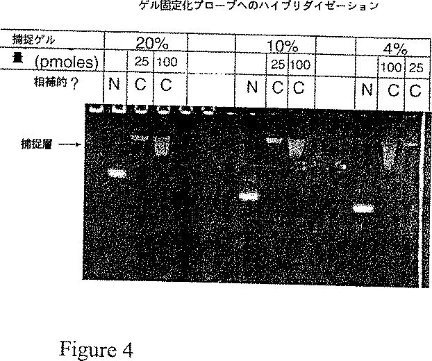
図4はゲル固定化プローブへの核酸のハイブリダイゼーションを表す写真である。
発明の詳細な説明
本発明は、捕捉プローブが電気泳動マトリックスに共有結合的に付着(例えばそのマトリックス中に固定化)されている、特異的結合反応を分析するための電気泳動方法に関する。捕捉プローブは、試験試料中に存在する標的分子に特異的に結合もしくはハイブリダイズする核酸または核酸アナログである。試験試料を電気泳動マトリックスに導入し、捕捉プローブへの標的分子の特異的結合に適した条件で電場をかける。固定化プローブは、電気泳動マトリックスの全体にわたってその内部に付着され、結合はそのマトリックス内で起こる。図1は電気泳動捕捉分析の原理を図示した略図である。
電気泳動マトリックス
いかなる電気泳動に適したマトリックスをも本発明の方法に使用できる。好適なマトリックスとしては、アクリルアミドおよびアガロースが挙げられ、どちらも核酸電気泳動に広く使用されている。しかしながら、他の物質を使用してもよい。例示としては、化学修飾されたアクリルアミド、スターチ、デキストラン、セルロース系ポリマーがある。例示としては、修飾アクリルアミドおよびアクリル酸エステル(例えばPolysciences社のポリマー&モノマー カタログ、1996〜1997、ペンシルヴェニア州ウォリントン、を参照のこと)、スターチ(Smithies,Biochem.J.,71:585(1959);製品番号S5651,Sigma Chemical Co.社(ミズーリ州セントルイスを参照のこと)、デキストラン(例えばPolysciences,Inc.のポリマー&モノマー カタログ、1996〜1997,ペンシルヴェニア州ウォリントンを参照のこと)、セルロース−べースポリマー(例えば、Quesda,Current Opin.in Biotechnology,8:82〜93(1997)を参照のこと)が挙げられる。上に列挙されたこれらのポリマーは、本発明で使用される捕捉プローブの特異的付着が可能なように、いずれも化学修飾できる。
特に、核酸オブ(of)核酸アナログの捕捉プローブとしての使用は、本発明に包含される。核酸含有ゲルを制作するために核酸をカップリングする方法は当業者に知られている。核酸と核酸アナログとは、アガロース、デキストラン、セルロース、スターチポリマーに、臭化シアン活性化または塩化シアヌル活性化によってカップリングできる。カルボキシル基を含有するポリマーは、一級アミン基を持つ合成捕捉プローブに、カルボジイミドカップリングを用いてカップリングできる。一級アミンを持つポリマーは、アミン含有プローブに、グルタルアルデヒドまたは塩化シアヌルを使ってカップリングできる。多くのポリマーは、チオール含有合成プローブにカップリングできるチオール反応性基で修飾できる。他の多くの好適な方法が文献で知られている。(総論としては、Wong,“Chemistry of Protein Conjugation and Cross−linking,CRC Press,フロリダ州ボカラトン,1993を参照のこと)。
本明細書記載の捕捉プローブを重合性化学基に共有結合的に付着する方法も開発されている。重合性モノマー化合物の適当な混合物と共重合すると、高濃度の固定化核酸を含むマトリックスを製造できる。重合性化学基に核酸を共有結合的に付着する方法の実例は、「核酸含有重合性複合体」と題する米国特許出願第08/812,105号明細書に見出され、その教示は参照によりそのまま本明細書に組み込まれる。
一部の方法については、2以上のマトリックス形成材料の混合物を含む複合マトリックスを使用することが有効だろう。その一例は、複合アクリルアミド−アガロースゲルである。これらのゲルは、通常、2〜5%のアクリルアミドと0.5%〜1%のアガロースを含有する。これらのゲルでは、アクリルアミドが主たるふるい機能を与えるが、このように低濃度のアクリルアミドゲルは、アガロースなしでは取り扱いに好都合な機械的強度を欠く。アガロースは、アクリルアミドのふるい特性を有意に変化させることなく機械的支持を与える。このような場合は、成分が液相核酸標的と最も密に接触するので、ゲルのふるい機能を付与する成分に核酸を結合できる。
アガロース、架橋ポリアクリルアミドゲルなどのゲル形成マトリックスは、多くのアプリケーションに好ましいだろう。しかしながら、キャピラリー電気泳動(CE)用には可溶性ポリマーを電気泳動マトリックスとして使用すると便利であり、再現性が得られうる。CE分析に有用であることがわかっている可溶性ポリマーの例示は、Quesada(Current Opinion in Biotechnology、第8巻、82〜93頁、1997)に記載しているように、ポリアクリルアミド、ポリ(N,N−ジメチルアクリルアミド)、ポリ(ヒドロキシエチルセルロース)、ポリ(エチレンオキシド)およびポリ(ビニルアルコール)の線状ポリマーである。これらの可溶性マトリックスは、本発明の方法を実施するためにも使用できる。固定化捕捉プローブを含む可溶性ポリマーマトリックスの調製には、「核酸含有重合性複合体」と題する米国特許出願第08/812,105号明細書に見出される方法を使用することが、特に便利である。ポリマー形成中にエチレン含有捕捉プローブの共重合に関与する、本ストラテジーの詳細な例示を下記実施例5に記載する。予め形成させたポリアクリルアミドゲルにオリゴヌクレオチドプローブを付着するもう一つの方法(Timfeevら,Nucleic Acids Res.24,3142〜3148,1996)も、前もって重合した可溶性線状ポリアクリルアミドに捕捉プローブを結合するために使用できる。
電気泳動マトリックスに組み込まれうる粒子に核酸を結合してもよい。前記粒子は、本来、肉眼的、顕微的またはコロイド性でありうる。(Polysciences Inc.の1995〜1996パーティクルカタログ、ペンシルヴァニア州ウォリントンを参照のこと)。Cantorらの米国特許第5,482,863号明細書には、懸濁液または粒子を含む電気泳動ゲルをキャストする方法が記載されている。前記粒子は、上述した方法に類似する方法で核酸に連結され、ゲル形成化合物と混合され、懸濁液として所望のマトリックス型にキャストされる。
ハイブリダイゼーション結合反応を分析するための固定化プローブ
本発明の方法において、様々な捕捉プローブを使用できる。通常、本発明の捕捉プローブは、捕捉プローブにハイブリダイズする標的分子に実質的に相補的なヌクレオチド配列を持つ核酸を含有する。核酸捕捉プローブの相補性は、標的分子を特異的に結合し、かつ標的分子の存在または非存在を明示するのに十分でありさえすればよい。本発明での使用に適したプローブには、RNA、DNA、核酸アナログおよび核酸ともう一つの有機成分(例えばペプチド核酸)とを含有してなる混合型のキメラプローブを含有する。捕捉プローブは、一本鎖または二本鎖核酸でありうる。
本明細書で定義する「核酸」という用語は、DNAまたはRNAを包含する。本明細書で「単離された」核酸は、それらの起源(例えばそれは細胞中またはライブラリーなどの核酸混合物中に存在する)の成分から分離された核酸であり、それはさらなるプロセッシングを経ていてもよい。単離された核酸としては、当業者に公知の方法によって得られた核酸が挙げられる。これらの単離された核酸としては、実質的に純粋な核酸、化学合成によって製造された核酸、生物学的方法と化学的方法との併用により、製造された核酸および単離された組換え核酸が挙げられる。
本明細書で用いられる「核酸アナログ」には、修飾糖基、リン酸基または修飾塩基を含む核酸が含まれる。修飾塩基を持つ核酸の例示としては、例えば、アセチル化、カルボキシル化またはメチル化された塩基(例えば、4−アセチルシチジン、5−カルボキシメチルアミノメチルウリジン、1−メチルイノシン、ノルバリンまたはアロインロイシン)などがある。そのような核酸アナログは、当業者に公知である。
本明細書に定義するように、「実質的に相補」とは、捕捉プローブのヌクレオチド配列が標的分子の正確なヌクレオチド配列を反映する必要はないが、配列の同一性が、指定された条件で標的分子とハイブリダイズするに足りる程度に類似していなければならないことを意味する。例えば、配列がハイブリダイズするに足りる相補的塩基を持つものならば、非相補的な塩基または余分なヌクレオチドを配列中に散在させることができる。
ハイブリダイゼーションの指定条件は、当業者により経験的に決定できる。例えば、非特異的ハイブリダイゼーション反応を著しく減少させるストリンジェンシーの条件を選択すべきである。核酸ハイブリダイゼーションに関するストリンジェンシー条件は、例えばCurrent Protocols in Molecular Biology、Ausubel,F.M.ら編、第1巻、Suppl.26,1991に説明されており、その教示は、参照により、そのまま本明細書に組み込まれる。プローブの長さ、塩基組成、ハイブリダイズする配列間のミスマッチ率、温度、イオン強度などの因子は、核酸ハイブリッドの安定性に影響する。ストリンジェントな条件、例えば、中等度のまたは高いストリンジェンシーは、プローブと標的分子の特徴にも依存して、経験的に決定できる。
通常、捕捉プローブの長さは、少なくとも5ヌクレオチド長、より一般的には5〜50ヌクレオチドであろうし、数千塩基長もの長さでもよい。
修飾ヌクレオチドを含むプローブも有用である。例えば、デアザグアニン塩基とウラシル塩基とを含むヌクレオチドを、ハイブリダイズしたプローブの熱的安定性を低下させるために、グアニンおよびチミン含有ヌクレオチドの代わりに使用しうる(Wetmur,Critical reviews in Biochemistry and Molecular Biology、第26巻、227〜259頁、1991)。熱的安定性の増したハイブリッドが望ましい場合は、シトシンの代わりに5−メチルシトシンを使用できる(Wetmur,Critical reviews in Biochemistry and Molecular Biology、第26巻、227〜259頁、1991)。2’−O−メチル基の付加などのリボース糖基への修飾は、固定化されたRNAプローブのヌクレアーゼ感受性を減少させうる(Wagner,Nature,vol.372,333〜335頁,1994)。ホスホジエステル骨格から負電荷を除去する修飾により、ハイブリッドの熱安定性を増加させることができる(Moodyら,Nucleic Acids Res.,vol.17,4769〜4782頁,1989;Iyerら,J.Biol.Chem.,vol.270,14712〜14717頁,1995)。
核酸アナログも固定化プローブとして役立ちうる。有用な核酸アナログの一例は、反復するN−(2−アミノエチル)グリシン単位からなる修飾ペプチド骨格に標準的DNA塩基が結合しているペプチド核酸(PNA)である(Nielsenら,Science 第254巻、1497〜1500頁、1991)。前記ペプチド骨格は、標準的なDNAおよびRNA一本鎖との塩基対形成に適した距離に塩基を保持できる。PNA−DNAハイブリッド二本鎖は、おそらくPNA鎖には負に荷電したホスホジエステル結合がないという事実ゆえに、DNA−DNA二本鎖の同等物よりもはるかに強い。また、その特殊な構造ゆえに、PNAは、ヌクレアーゼ分解に対して極めて耐性である。これらの理由から、PNA核酸アナログは固定化プローブアッセイに有用である。固定化プローブ試験に有用な特性を持つであろう他の核酸アナログを構築するにも同様の設計方針を使用できることは、当業者には明らかだろう。
一本鎖および二本鎖標的分子
本発明の1態様では、一本鎖標的分子と一本鎖固定化プローブとを使用する。この態様は、RNA標的の分析にとりわけ有用である。これは、標的の再生が迅速でない場合に、複雑な試料からの特定標的の捕捉にも有用である。PCR産物などの高濃縮標的は、迅速な再生ゆえに電気泳動直前の変性を必要とするであろう。例えば、100〜250塩基対長のPCR産物を分析するには、試料を75%ホルムアミド(体積/体積)にして、電気泳動直前に5分間60℃で加熱するとよい。
本発明のもう1つの態様では、二本鎖標的を一本鎖固定化プローブで捕捉する。例えば二本鎖核酸と会合して三本鎖構造を形成するプローブを設計できる。第三の鎖は二本鎖の主溝に位置し、二本鎖の塩基とフーグスティーン型塩基対相互作用を形成する(HoganおよびKesslerの米国特許第5,176,966号明細書ならびにCantorらの米国特許第5,482,836号明細書)。したがってプローブの設計は、それらの化学的相互作用を支配する制約を受ける。しかし、天然に存在する核酸における三本鎖構造を形成できる配列の頻度は十分に高いので、多くの標的はこのプローブ設計方針を使って特異的に捕捉できる。
別法として、置換ループ構造の形成によって二本鎖核酸と会合するプローブを設計できる。そのようなプローブは二本鎖核酸の一方の鎖にのみ結合し、二本鎖のプローブ相同二本鎖を部分的に置換する。この置換はプローブ−標的鎖相互作用が標的鎖間の相互作用よりはるかに有利である場合にのみ達成できる。そのようなプローブは、Wetmur、Critical Reviews in Biochemistry and Molecular Biology、Vol.26,227〜259頁(1991)に記載の修飾塩基と技術、骨格修飾(Moodyら、Nucleic Acids Res.、第17巻、4769〜4782頁(1989))および核酸アナログ(Nielsonら,Science,254:1497〜1500頁(1991)を使って製造できる。天然に存在する核酸と異例に強固かつ特異的に塩基対形成するペプチド核酸(PNA)プローブの使用はこの態様でとりわけ有用だろう。
核酸結合タンパク質を分析するための固定化捕捉プローブと標的
本発明の方法は核酸結合タンパク質の分析にも有用である。これらの場合は、選択される核酸が、何らかの方法で、そのタンパク質の天然の結合基質を模倣する。
配列特異的核酸結合タンパク質と非配列特異的核酸結合タンパク質の両方を分析できる。配列特異的結合タンパク質を分析するには、その標的結合タンパク質によって認識される配列を含有するように捕捉プローブを設計する。非特異的相互作用を分析するには、観察される結合が特定の核酸配列に依存しないことを保証するために、捕捉プローブの混合物を使用できる。
電気泳動分析は、タンパク質がその未変性構造を保てるような、したがってそのタンパク質が電気泳動中に捕捉プローブに結合できるような条件下で行われる。電気泳動後、固定化捕捉プローブを含有するゲル領域内の該タンパク質の存在は、有色または蛍光色素による染色、オートラジオグラフィー(試料が放射活性標識されている場合)、銀染色、およびタンパク質電気泳動分野の当業者に周知の他の様々な標準的方法によって検出できる。
転写調節に重要な配列−特異的DNA結合タンパク質の検出と分析には、二本鎖捕捉プローブを利用することがとりわけ有用である。この実施では、タンパク質標的によって認識されることが知られている(またはそのように推測される)配列を含有する二本鎖捕捉プローブを使用する。試験試料はその捕捉プローブを含有する領域を通して電気泳動される。電気泳動後に、ゲル内でのタンパク質の位置を決定する。捕捉プローブを含有するゲル領域にタンパク質が存在することは、その試料中にDNA結合タンパク質が存在することを示す。興味ある特定配列を持たないDNA捕捉プローブでは結合が起こらないことを示す対照実験を用いて、その結合の配列特異性を実証できる。
一本鎖捕捉プローブも役立ちうる。例えば一本鎖RNA捕捉プローブは、特定のRNA配列に結合するタンパク質の検出と精製に使用できる。一本鎖DNAプローブは、一本鎖DNAゲノムを含有するウイルスの調節タンパク質や、複製起点内の一本鎖DNA区間に特異的に結合するタンパク質を検出するのに役立ちうる。
アプタマー捕捉プローブ
いくつかのグループが、えり抜きの望ましい結合特性または触媒能を示す分子を求めて核酸のランダムライブラリーをスクリーニングする方法を開発している(EllingtonとSzostak,Nature,346:818〜822(1990);Joyce,Gene,82:83〜87(1989);TuerkとGold,Science,249:505〜501(1990))。多くの応用例で、これらのライブラリーは、標準的な市販の核酸合成装置で作製されたオリゴヌクレオチド(RNAまたはDNA)のランダムプールからなる。25塩基長の最大1015の個々の配列からなるライブラリーを日常的に構築することができる(KlugとFamulok,Molec.Biol.Reports,20:97〜107,(1994))。次に、所望の標的への機能的結合について、このプールをスクリーニングする。標的を結合できるオリゴヌクレオチドは、カラムクロマトグラフィー、フィルター結合または他の適当なプローブ/標的結合複合体精製法によって分離される。結合したオリゴヌクレオチドを精製し、通常は、無作為配列の領域に隣接する特定の配列を認識するプライマーを用いて、その結合プールをポリメラーゼ連鎖反応により増幅する。高い親和力で結合するオリゴヌクレオチドプローブを濃縮するために、必要に応じて標的結合と再増幅のサイクルをさらに繰り返すことができる。最終プールの構成要素を組換えDNA技術でクローン化し、配列決定し、標的結合を担う配列要素を同定するための分析を行う。
アプタマーと呼ばれるこのタイプの核酸結合プローブは、事実上任意の標的分子に対して選択できる。現在までに、特定のタンパク質(Bartelら,Cell,67:529〜536(1991);Giverら,Gene,137:19〜24(1993);Leclercら,Nature Struct.Biol.,1:293〜299;Bockら,Nature,355:564〜566(1992))、アミノ酸(Famulok,J.Am.Chem.Soc.,116:1698〜1706(1994))、小分子薬物(Jenisonら,Science,263:1425〜1429,1994))、ビタミン類(LorschとSzostak,Biochemistry,33:973〜982(1994))、ならびにヌクレオチドコファクター(SassanfarとSzostak,Nature,364:550〜553(1993))と特異的で強固な結合複合体を形成できるアプタマーが選択されている。
本発明は調製および分析用途でアプタマーを使用するための便利なプラットフォーム技術を提供する。いったん適切なアプタマーが選択されたら、捕捉プローブとして使用するために、それを適切な電気泳動媒体に結合することができる。その捕捉ゾーンを通して試験試料を電気泳動した後、その捕捉ゾーンを標的の存在について分析する。調製用途の場合は、非標的試料成分を媒体外へ移動させた後で、標的分子をその捕捉ゾーンから溶出させることができる。アプタマーは事実上任意の標的に対して選択できるので、アプタマー捕捉プローブを使用することにより、本発明の方法は多種多様な標的分子の電気泳動分析および精製に利用できる。どの分子でも、その分子が、適用した電場の影響を受けて移動するように電気泳動に使用する条件で帯電する(例えば標的分子が適当な電気泳動媒体中に置かれたときに検出可能な移動度を持つ)限り、本発明の方法での分析に適することに注目することが重要である。
標的分子分析用の均一に修飾された電気泳動媒体
本発明のこの態様では、図2Aに模式的に示すように、媒体の実質的にすべてが1または複数の捕捉プローブで修飾される。捕捉プローブと電気泳動条件の選択は、捕捉プローブと標的分子の間の結合が一時的で、電気泳動分析の時間スケールで迅速に可逆的であるように行われる。これらの条件では、標的分子が電気泳動中に捕捉プローブへの結合、放出および再結合というサイクルを数多く起こす。この可逆的結合は、捕捉プローブの非存在下での移動度と比較して測定される標的の電気泳動移動度を減少させるという効果を持つ。捕捉プローブへの結合が強い場合、標的の移動度は大きく減少する。捕捉プローブへの結合が弱い場合、標的の移動度はわずかにしか減少しない。このようにして、捕捉プローブの非存在下では類似する電気泳動移動度を持つ構造的に関連する標的を、特定の捕捉プローブに対するそれらの親和力に基づいて区別することができる。この方法は後述するように核酸配列変異の分析にとりわけ有用である。
標的分子を分析するための一次元配列
本発明のこの態様では、図2Bに模式的に示すように、一連の不連続なマトリックス層であって、各層が少なくとも一つの捕捉プローブを含有するものを通して、標的分子を含有する試料を電気泳動する。例えばハイブリダイゼーション結合反応では、捕捉プローブに相補的な標的核酸が捕捉プローブにハイブリダイズし、そのゲル層に保持される。相補的でない試料核酸はその捕捉層を通過する。捕捉プローブと相補的な試料核酸とのハイブリッドの存在は、本明細書に記載の適当な標識法によって捕捉層内で検出される。
この一次元形式には重要な利点がいくつかある。第一に、試料のすべてが捕捉層を通過し、それゆえ試料のすべてがハイブリダイゼーションに利用されうる。これは他の大半の固相ハイブリダイゼーション法と比較して大きな利点である。高濃度の固定化プローブを使用することにより、小さなゲルバンド中にハイブリダイズ可能な試料核酸鎖のすべてを捕捉することが可能である。
第二に、この方法による分析には、別々の電気泳動移動度を持つ無傷の核酸種が必要でない。ハイブリダイゼーションと検出には短い配列相同性しか必要ないので、部分的に分解した核酸でもなおシグナルを与えるだろう。すべての標的核酸は、それらが分解されるかどうかにかかわらずマトリックス中の特定の位置に濃縮されるので、この属性は検出感度も増加させる。従来のゾーン電気泳動では、検出のためには、すべての試料核酸が別々のバンドとして移動しなければならない。
第三に、試料の体積は重要でない。本発明では、たとえ大量の試料を使用したとしても、すべての試料核酸が捕捉層を通過する。これは、検出の感度と分解能を最大にするには試料の体積が可能な限り小さい必要がある従来のゾーン電気泳動と比較して、有意な利点である。
この態様では、捕捉層が単一または複数の捕捉プローブを含有しうる。単一層に複数の捕捉プローブを使用することは、多数の異なる生物のうちのいずれか1つを検出する必要があるアッセイに有用である。例えば輸血に使用される血液試料にはどのような細菌の存在も望ましくない。したがって任意の細菌に関する一般的試験には、保存された細菌遺伝子配列の収集物を捕捉プローブとして使用しうる。特定の細菌の同定は重要でないので、そのプローブ収集物は、任意のそして全ての細菌が同じ捕捉層で検出されることを保証するのを助ける広域スペクトルの複数プローブからなりうる。
この態様では複数の捕捉層も使用できる。多重ハイブリダイゼーションアッセイを作製するには、同じゲル装置に複数の捕捉層を逐次的にキャストすることが簡単明瞭である。アッセイ中は、それらの層の全てを通して標的試料を電気泳動し、相補的な試料核酸を各層で捕捉する。各層中のハイブリッドの量は使用した捕捉プローブに関する試料組成を直接反映する。
適正にハイブリダイズした核酸だけが各層に保持されることを保証するように条件を割り出すことができる。20塩基対程度の長さの捕捉プローブとの電気泳動ハイブリダイゼーションは、従来の非変性ゲルと緩衝液系を用いて室温で行うことができる。このサイズの完全に相補的なハイブリッドは何時間も安定なようである。しかし、尿素やホルムアミドなどの変性剤をゲルに添加するか、ゲルを高温で泳動することにより、さらなるストリンジェンシーを達成することができる。
二次元プローブ配列
一次元プローブ配列は限られた数の捕捉プローブを使用する分析に使用できる。より多数の配列を分析するには、固定化プローブの二次元配列を使用できる。この配列は多くの方法で形成させることができる。単純な二次元配列は例えば整列した複数の垂直なスペーサーを使って従来のスラブゲル装置にキャストでき、事実上、一次元配列の配列が生成する。この配置の一例を図2Cに示すが、ここでは、1つの試料が試料ウェルに充填され、その試料の一部が6つの別々の捕捉ゾーンを通過することになる。
より複雑な二次元配列は次の二段階で作製できる。すなわち、まず一つのプレート上で捕捉プローブ領域をマトリックス(例えばポリアクリルアミドゲル)ドットの配列として重合させ、次にその配列上に上部ゲルプレートを置いて、プローブドット間の空間を非修飾ゲルで満たすことにより、各ドットを「サンドイッチ状」にする。
いずれの場合も、試料はマトリックス上部の全長にわたって一つのバンドとして充填される。この態様では、試験試料全体が捕捉プローブのすべてと接触するわけではない。しかし、ライブラリースクリーニングや遺伝子発現分析など、二次元分析が望ましい用途ではほとんどの場合、試料核酸が高コピー数で存在するので、この問題は有意な障害にはならない。
三次元プローブ配列
本明細書に記載するハイブリダイゼーション法には、例えば高速処理および/または対費用効果などのための多重並列アッセイにとりわけ有用でありうるような三次元配列も包含されうる。このようなアッセイは、図2Dに模式的に示すような三次元固体の形式で提供でき、ここでは複数の試料を表面または面に適用し、次にその固体の容積中を、捕捉プローブの1以上の領域に出会うように移動させる。配列は各試料がその配列を通って移動する間に同じ配列の捕捉プローブに出会うように制作してもよいし、あるいは異なる試料混合物を分析するために、もしくは1以上の試料混合物内の異なる成分セットを分析するために、異なる配列の捕捉プローブを配置してもよい。
固定化プローブとのハイブリダイゼーションによる試料の精製/濃縮
本明細書に記載する電気泳動法は、粗混合物から特定標的分子を選択的に精製するのにとりわけ有用である。例えば、固定化捕捉プローブを含有するゲル上に粗混合物または細胞溶解液を置く。その混合物をそのゲルを通して電気泳動する。標的分子は捕捉プローブを含有する層に固定化される。標的と同じ電荷を持つ非標的分子は反対の極性を持つ電極(これをここでは「誘引電極」と呼ぶことにする)にひきつけられ、捕捉プローブ層を通過し、ついにはゲル外に電気泳動される。反対電荷の非−標的分子は、非−誘因電極に向かって試料ウェル外に移動する。帯電していない試料分子は試料ウェル中に残り、ゲルには入らない。全ての帯電非標的分子がゲルから除去されうる十分な電気泳動時間後に、捕捉された標的分子を次の方法の一つによりゲルから溶出させる:
1)変性条件での継続的電気泳動(例えばマトリックスの温度を上げるか、電気泳動電圧を増加させることによる)。捕捉プローブからの標的の放出は、例えば媒体の温度を、標的/捕捉プローブが変性するに足りる温度まで上げることによって達成される。
2)捕捉プローブとマトリックスの間の化学結合の化学的または光化学的切断後の継続的電気泳動。
3)標的と捕捉プローブの間の結合の除去。ここで、標的はプローブから放出される。例えば化合物または化学物質は標的に、またはプローブから標的を放出する捕捉プローブに競争的に結合できる;あるいは標的とプローブの間の結合が除去されるか十分に低下して標的が放出されるように媒体の条件を変化させる。例えば標的がタンパク質であり、固定化された捕捉プローブが核酸である場合は、固定化された捕捉プローブに相補的な可溶性非固定化核酸を媒体に導入するか、媒体と接触させることができる。その可溶性核酸は固定化プローブに対してより高い親和力を持ち、標的とプローブの間の結合が除去され、標的が放出される。プローブからの標的の放出を保証するために、通例、競争用化学物質は過剰に添加される。他の帯電または非帯電化合物も同様に働きうる。例えばホルムアミドをゲル媒体に吸収させることができ、それは標的の放出をもたらす。一態様として、化学物質を電気泳動によって媒体に導入する。例えば試料を媒体を通して溶出させ、標的が捕捉プローブに結合した後、次いで競争用化学物質をその媒体を通して電気泳動する。
溶出した標的分子は、いくつかの方法によって濃縮し、誘引電極槽から回収できる。例えば、非標識試料分子をゲル外に電気泳動した後、誘引電極槽内の非標的分子を適当な洗浄液で洗い流すことができ、標的分子を元の誘引電極槽中に直接溶出させることができる。別法として、一つが非標的成分を試料から除去するために使用され、もう一つが標的分子を溶出させるために使用されるように、電気泳動装置が2つの誘引電極を持ってもよい。別法として、非標的成分を除去した後、標的溶出を行うために誘引電極槽を新しいものと交換できるように、交換可能な誘引電極槽を持つ電気泳動装置を構築することができる。
この態様は、ハイブリダイゼーション法によって粗生体試料から特定の核酸を精製するのにとりわけよく適している。第一に、これらの方法は、所望の配列を持つ試料核酸の実質上全ての捕捉をもたらす。なぜなら全試験試料が捕捉ゾーンを通過しなければならないからであり、また、捕捉プローブの濃度は任意の高さにすることができ、それが捕捉の成功を保証するからである。
第二に、これらの方法は一段階で標的核酸の実質的精製をもたらす。なぜなら、帯電した試料夾雑物は電気泳動中に除去され、帯電していない夾雑物はマトリックス中に進入できないので除去されるからである。
第三に、極めて大きい試料を使用できる。核酸はポリマーマトリックス中と事実上同じ様式で自由な溶液状態で電気泳動を受ける。したがって大きい試料体積を使用できる。マトリックス層は高度に選択的なフィルターのように作用して、試料から所望の核酸のみを選択する。
第四に、本明細書に記載する方法を使用することで、極めて希薄な大量の試料を定量的に濃縮できる。
試料移動速度による変異の検出
この態様では、試料と固定化プローブが高親和力のゆっくりと解離する複合体を形成しない。そうではなくて試料とプローブは電気泳動中に一時的で比較的迅速に解離する会合を形成する。捕捉プローブはマトリックス全体に均一な濃度で配置される。試料核酸の電気泳動移動度は固定化プローブとの相補性の程度によって左右される。プローブに対して完全な相補性を持つ試料は、相補性の低い試料よりゆっくりとマトリックス中を移動する。標的試料と捕捉プローブの間の相互作用が一時的であることを保証するためにマトリックス中に変性剤を使用してもよい。
試料は、ハイブリダイゼーション分析に先立って、標識ヌクレオチド三リン酸を使ったPCR増幅により、簡便に調製、標識できる。
この態様は増幅可能な遺伝子断片内の一連の特定の変異部位を型別するのにとりわけ有用である。多くの物理的に分離された垂直のレーンを持つマトリックスが使用される。各レーンは異なる固定化プローブを含有する。各プローブは試験しようとする特定遺伝子領域の野生型配列を含有する。具体的一態様として、そのマトリックスで試験しようとする遺伝子の全領域を単一の断片として増幅できる。
例えば、この分析を実施するには、標識された一本鎖核酸産物を試験しようとする試料から、フルオレセイン標識ヌクレオチドを使った非対称PCR法で調製する。そのフルオレセイン産物を非変異(野生型または正常)配列を持つローダミン標識内部標準PCR産物と混合し、ゲルの上部に均等に充填する。電気泳動後、ゲルのカラーイメージを記録し、試料と内部標準を同定する(試験試料:黄緑色,内部標準:赤色)。所定のレーンで試験試料が内部標準より速く移動する場合、その試験試料は、その固定化プローブがカバーする配列内のどこかの位置での変異体である。大きな遺伝子の場合、すべての易変配列位置をカバーするには、一組になった数枚のゲルが必要だろう。(図3を参照のこと)。
検出方法
特異的結合反応の検出、例えば捕捉プローブに結合した固定化標的分子の検出は、多くの異なる方法で達成できる。例えば、結合反応に先立って試験分子を検出可能に標識することができる。直接標的標識に適した標識は、強い吸収性(例えば明るく色付く)、放射活性、蛍光、リン光、化学発光または触媒作用を持ちうる。修飾ヌクレオチドを用いた核酸試料の直接標的標識は、当業者に周知の数多くの酵素法によって達成できる(Sambrookら,「Molecular Cloning:A Laboratory Manual」第二版(Cold Spring Harbor Press、ニューヨーク州コールドスプリングハーバー、1989)に概説されている)。
別法として、それ自体が標識されているか検出可能なシグナルを生成できる第二の特異的結合体によって認識され得るリガンドを用いて、標的分子を間接的にも標識できる。そのような間接系の一例は、ビオチニル化ヌクレオチドを使った標識である。この系では、標準的な核酸標識技術とビオチニル化ヌクレオチドを使用して、試料を酵素的に標識する。得られたビオチン修飾核酸は、ストレプトアビジンまたはアビジンタンパク質分子のビオチン特異的結合によって検出できる。ストレプトアビジンまたはアビジン分子は、フルオレセインなどの蛍光標識や、適当な基質で化学発光シグナルまたは比色法的シグナルを生成するために使用できるアルカリ性ホスファターゼやセイヨウワサビペルオキシダーゼなどのリポーター酵素に結合できる(概観するには、KellerとManak,「DNA Probes」第二版,Macmillian Publishers,ロンドン,1993;Pershingら編「Diagnostic Molecular Microbiology:Principles and Applications」アメリカ微生物学会,ワシントンD.C.,1993を参照のこと)。もう一つの有用な検出系はアルカリフォスファターゼに結合した抗ジゴキシゲニン抗体を使用するジゴキシゲニン系であり、これは核酸に組み込まれているジゴキシゲニン−dUTPを認識する。(CURRENT PROTOCOLS IN MOLECULAR BIOLOGY,Ausubel,F.M.編,vol.1,§§3.18.1〜3.19.6,1995)。
検出可能に標識されたハイブリダイゼーションプローブは、間接的標的標識としても使用できる。例えば電気泳動に先立って、検出可能に標識されたプローブ(以下、「サンドイッチ」プローブと呼ぶ)とのハイブリダイゼーションにより、標的核酸を間接的に標識することができる。このサンドイッチプローブは、捕捉プローブによって認識される領域とは重ならない標的の領域とハイブリダイズするように設計される。サンドイッチプローブは、電気泳動中に標的と会合したままであるように設計され、捕捉プローブには直接的には結合できない。
サンドイッチプローブは電気泳動捕捉後に標的分子を標識するためにも使用できる。この標識法では、非標識標的を電気泳動し、まず捕捉プローブにハイブリダイズさせる。次にサンドイッチプローブをその捕捉層を通して電気泳動する。捕捉された標識はここでは、事実上、サンドイッチプローブに対して新しい「捕捉」プローブとして作用する。捕捉された標的サンドイッチプローブ複合体は、サンドイッチプローブ標識を介して検出できるようになる。
ブロッティング技術を標的に結合した捕捉プローブの検出に適合させることもできる。例えば検出表面を、結合した試料成分を持つ分離媒体に近接させ、次に試料成分はその検出表面に移動し、任意に例えば溶媒または試薬交換などの化学的手段により補助し、移行した試料成分を例えば挿入した色素の光学的検出などの既知手段によって、またはハイブリダイズした放射活性種からの放射活性の検出によって、または他の既知手段によって検出する。
捕捉プローブに結合した試料成分の存在を検出するには、種々の光学的技術を使用できる。例えば捕捉プローブを直線配列に配置した場合、各シグナルの位置と強度は検出可能なシグナルの配列に沿って単一の検出器を機械的または光学的に走査することによって測定できる。別法として、検出可能なシグナルの直線配列は直線配列検出器によって、例えば配列検出器を検出可能なシグナルの配列に近接させること、またはシグナル配列の全部または一部を配列検出器に光学的に撮像することなどによって、検出できる。
検出可能なシグナルを持つ捕捉プローブが二次元配列として配置される場合、多くの検出法を使用できる。各点でのシグナルを測定するには、機械的または光学的走査または任意の組み合わせにより、単一の検出器を使用できる。別法として、並置または光学的撮像によって一組のシグナルを検出するには直線的光学検出配列を使用でき、複数組のそのようなシグナルはシグナル配列または検出器を機械的または光学的に走査することによって検出できる。別法として、捕捉プローブの二次元配列は、その全体または一部を二次元光学領域検出器により、固定化された捕捉プローブからの光学的シグナルの配列に近接させることによって、またはその光学的撮像によって、任意に検出してもよい。
捕捉プローブが三次元配列として配置される場合、個々のシグナルの検出は上述の方法により、任意に、配列の1以上の小区分をまず物理的に採取するという補助を行って、検出できる。別法として、共焦点顕微鏡技術などの光学的方法を使用してもよく、ここでは1または多数の検出可能なシグナルを他からの干渉を最小にして撮像および検出し、次に光学的調節後に他のシグナルを検出する。
マトリックスの形式とマトリックスの作製法
マトリックスは様々な形で形成させうる。例えば線状ゲルは溝や管などの線状支持体内での形成、この場合、ゲルはその支持体内での重合によって形成される、を含む技術によって、あるいは、他方、分割やチャンネル内での形成を含む、二次元ゲルを多数のストリップに細分することによって形成させることができる。溝、ストリップまたはチャンネル形式では、重合したゲル内に1以上の捕捉プローブを与えるためにゲル重合前またはゲル重合中に、多量の1以上の共重合性捕捉プローブをゲル材料に、任意に例えば定位的滴下技術により、空間的に規定された位置に添加できる。管形式では、一連のゲルモノマーおよびゲルモノマーと重合性捕捉プローブの混合物を、空間的に区別された一組の成分と濃度を与えるように、逐次、管に導入し、次いで、それをインサイチュで重合させて各成分の空間的関係を保つことができる。重合によって誘発される収縮中にゲルの完全性を保つために、ゲルの収縮中に横方向に収縮する弾性材料で管壁を作ることができる。別法として、収縮を補うように他方の端に新たな液体材料を添加しながら、管の一端から漸進的な重合を誘発してもよい。そのような漸進的重合は、重合触媒剤の拡散を含む手段によって、または、例えば放射線源の移動または放射線源への漸進的ばく露等、管の一端から他端への重合誘発性電磁気または他の放射線の漸進的適用によって誘導しうる。別法として、直線型ゲルは、二次元ゲルから直線状の切片を採取することにより、または後述のように作製される三次元ゲルから直線状の芯を採取することによって作製しうる。
二次元ゲルは、支持体の表面での形成または2つの支持体表面間での形成を含む技術によって形成させうる。ゲルモノマーの層を利用し、重合前または重合中に多量の共重合性捕捉プローブを、任意に、空間的に意味のある様式でその層に添加し、そのゲルにおけるそれらの空間的位置を保たれるように、それらをインサイチュで重合する。多量の重合性捕捉プローブの添加は、位置的にプログラム可能な滴下技術を含む既知の手段によって達成できる。重合中のゲル収縮は、支持体表面間の間隙の収縮を許し、補填のために側面から添加されるさらなる材料で横方向の収縮を許すなどといった手段によって調節できる。二次元ゲルは、ゲル形成の前、途中または後に仕切りを使用することによって、またはチャンネル中での形成によって、もしくは形成後により細い切片にスライスすることによって、多数のストリップに細分できる。
三次元ゲルは多くの技術によって形成させうる。それぞれが任意に局在化された捕捉プローブを含有してよい多数の線状ストリップまたは二次元層を上述のように反復して構築しうる。各ストリップまたは層は三次元の体積が生じるように下層の上に重合される。別法として、任意にその場に局在化された捕捉プローブを有してよい、多数の二次元ゲルを上述のように形成させ、三次元構造を与えるようにそれらを組み合わせることができる。
製造装置
本明細書に記載するマトリックスの作製には様々な装置を使用できる。例えばある装置は漸進的充填/収縮装置のアス(us)により中心部にマトリックスを作製したり、または固体からマトリックスを抜き取ることができる。マトリックスは、固体表面で、プレートの間に、または物理的チャンネルを作るために成形プレートを使うことによって、形成させることができる。
本発明を以下の実施例によってさらに例証するが、決してこれら実施例を限定と解釈してはならない。
実施例1:一次元電気泳動ハイブリダイゼーション
ゲルを三区画に分けて重合した。最下層は0.5×TBE(45mMトリス−ホウ酸pH8.3,1mM EDTA)に20%のアクリルアミド(重量比29:1のアクリルアミド:ビスアクリルアミド)を含有した。下部ゲルは厚さ0.15mm、高さ約5cm、幅8cmだった。いずれの場合も、重合はゲル溶液1ミリリットルにつき2μlのTEMEDと7μlの10%(重量/体積、水中)過硫酸アンモニウムを添加することによって達成した。最下層が重合した後、高さ0.5cm、幅2cm、厚さ0.15mmの3つの捕捉層を、20、10または4%アクリルアミド(すべて20:1のモノマー:ビス)を使って、10μMの一本鎖合成オリゴヌクレオチド捕捉プローブ(5’−ttt ttt ttt acg cag cga cga gca cga gag−3’)(配列番号:1)および0.5×TBE緩衝液の存在下に重合させた。捕捉プローブの5’−リン酸末端をN−(6−アミノヘキシル)メタクリルアミド基で共有結合的に修飾した。そのヘキシルメタクリルアミド基は、ヘキシルメタクリアミドホスホルアミダイト(Glen Research社、バージニア州スターリング)を使って自動DNA合成中に付加された。この結合法を用いると、プローブは、5’末端メタクリルアミド基が存在するので、捕捉層の重合中にゲルマトリックスと共重合する。捕捉層の側面の境界は、厚さ0.15cmの薄いスペーサーをゲルプレートサンドイッチの上部からそれらが下部の20%ゲルの上表面に接触するまで挿入することによって形成させた。捕捉層の重合後、ゲルの最上層を10%アクリルアミド、0.5×TBEを使って注ぎ、試料ウェルを形成させるためにコームを挿入した。
電気泳動ハイブリダイゼーションは、相補的フルオレセイン標識一本鎖オリゴヌクレオチド(5’−フルオレセイン−ct ctc gtg ctc gtc gct gcg t−3’)(配列番号:2)の25および100ピコモル試料(図4参照のこと)を充填して行った。各捕捉層を通して電気泳動した(図3のゲルの上部にある「相補的?」の表記、「C」レーンを参照のこと)。対照として、非相補的フルオレセイン標識オリゴヌクレオチド(5’−フルオレセイン−at tac gtt gat att gct gat ta−3)(配列番号:3)の100ピコモル試料も各捕捉ゾーンを通して電気泳動した。電気泳動は室温にて7V/cmで2時間行い、その後、ゲルをUV照明下で写真撮影した。
図3に示す結果は、相補的試料(「C」レーン)が捕捉層に完全に固定化され、一方、同じ長さの非相補的DNA(「N」レーン)は捕捉層に保持されなかったことを示している。これは、相補的試料と捕捉プローブの間の相補的塩基対形成が捕捉層におけるフルオレセイン標識相補試料の固定化の原因であることを示唆している。
実施例2:固定化核酸プローブ含有多糖類ベース電気泳動媒体の塩化シアヌル製法
プロトコル1:活性化担体アプローチ
下記実施例は、ビアジオーニ(Biagioni)ら(Anal.Biochem.,第89巻、616〜619頁、1978)の担体活性化法とスミス(Smith)およびレンホッフ(Lenhoff)(Anal.Biochem.,第61巻、392〜415頁)の担体活性化法とを用いる。セルロース支持体の使用が開発されたが、前記方法は、いかなる水酸基含有不溶性支持体に対しても一般的に適用可能である。本方法のための支持体の選択肢としては、スターチ、アガロース、デキストランおよびセルロースが挙げられる。
電気泳動用のほとんどのアガロース(例えば、Sea PlaqueまたはSea Kem、FMC Bioproducts)とスターチ(カタログ番号S5651およびS4501、Sigma Chemical)標品とが、水性溶媒および有機溶媒に不溶性な粉末として供給される。前記粉末を、ブフナーロト上で蒸留水により、よく洗浄する。洗浄された粉末を、15分間室温で3M 水酸化ナトリウムに懸濁し、その後、溶液を濾過により除去する。膨潤アルカリ性粉末を、攪拌しながら、ジオキサンとキシレンとの1:1(体積/体積)混合物に溶解させた5%塩化シアヌル溶液に添加する。担体1g(乾燥重量)あたり20ミリリットルの5%塩化シアヌル溶液を用いる。室温で30分間攪拌したのち、前記担体を下記溶媒:ジオキサン、酢酸/ジオキサン/水(1:2:1、w/w/w)、およびアセトンのそれぞれでよく洗浄する。前記担体を真空下に乾燥し、乾燥保存する。所望の1または複数のプローブを含む0.1M ホウ酸ナトリウム緩衝液pH8.3に前記活性化担体を再懸濁することにより、5’または3’一級アミン末端を含むオリゴヌクレオチドプローブを付着させる。好ましくは、前記プローブ濃度は、1グラムの乾燥活性化担体あたり500ナノモルを超えるアミン末端オリゴヌクレオチドである。よく攪拌または振盪しながら、室温で12時間付着反応を行なう。付着させた後、エタノールアミン−HCl(pH8.3)を終濃度1Mまで添加し、担体をさらに12時間振盪した。修飾担体を緩衝液でよく洗浄し、懸濁液として、好ましくは、電気泳動に用いられる緩衝液に保存する。
アガロースゲルおよびスターチゲルにおいて捕捉層をキャストするため、ザンブルーク(Sambrook)ら、「Molecular Cloning:A Laboratory Manual」、第2版、Cold Spring Harbor Press、Cold Spring Harbor NY、1989)などの標準の参考文献に記載の非修飾担体と同様の方法を用いて、プローブ−修飾粉末化担体をゲル中にキャストすることができる。すなわち、プローブ−修飾粉末化担体をゲル緩衝液に懸濁し、加熱により融解し、ゲル型に注ぎ、冷却させる。多くの場合、前記ゲル型は、非誘導体化担体から調製されたゲル中にスロットがあるか、あるいは穴を切り込むであろう。本方法は、捕捉層との分離境界を提供し、本実験で用いられるべき修飾ゲルの体積を減少させる。
プロトコル2:活性化プローブアプローチ
本プロトコルは、粉末化担体と、ヒドロキシエチルセルロースなどの可溶性多糖類ポリマーとに有益である。原則として、本アプローチを用いることにより、水酸基または一級アミン基を有する、可溶性または不溶性のいかなる分離媒体をも修飾しうる。可溶性ポリマーは、クエサダ(Quesada)(Current Opinion in Biotechnology、第8巻、82〜93頁、1997)に記載のキャピラリー電気泳動アプリケーション用の置換可能な分離媒体としてますます重要になっている。
プローブ活性化プロトコルは、バン ネス(Van Ness)ら(Nucleic Acids Res.、第19巻、3345〜3350頁、1991)から改良される。反応物は、200μmの5’−または3’−アミノ−末端プローブ、0.1M ホウ酸ナトリウム緩衝液(pH8.3)(SBB)、1mM 塩化シアヌル(Aldrich)、10% アセトニトリル(v/v)(Aldrich)を含む。室温で1〜2時間、よく振盪しながら反応を行なう。未反応の塩化シアヌルを、Microcon 3(3000ダルトンカットオフ、Amicon)を用いて、遠心限外濾過と0.1M SBBへの再懸濁との3サイクルにより除去する。活性化プローブは、4℃で保存され、2か月までは検出可能な活性の損失なく用いられうる。
粉末化媒体を蒸留水でよく洗浄し、ついで0.1M SBB pH8.3で洗浄することにより、スターチおよびアガロースなどの不溶性(粉末化)媒体への付着を行なう。前記媒体を0.1M SBBに懸濁し、塩化シアヌル−活性化プローブとともに室温で一晩振盪させる。好ましくは、活性化プローブの濃度は、ポリマー1gあたり500ナノモルを超える。反応後、修飾媒体を0.1M SBBでよく洗浄し、ついで電気泳動緩衝液で洗浄した。前述のプロトコルに記載のように、ゲル捕捉層をキャストする。
修飾セルロースなどの可溶性ポリマー媒体を水に溶解し、0.1M SBBに対して透析する。透析後、前記ポリマーを活性化プローブと混合し、室温で一晩、よく振盪させる。好ましくは、活性化プローブの量はポリマー1gあたり500ナノモルを超える。付着反応の後、未反応プローブを透析または遠心限外濾過(CentriconまたはMicrocon 50フィルター、50,000ダルトン分子量を超える媒体用、Amicon)により除去する。一方、未結合のプローブを試料の負荷前に媒体をプレ電気泳動することにより除去する。
実施例3:特定のタンパク質(ヒトトロンビン)の存在を決定するためのDNAアプタマーゲル
本実施例は、特定のタンパク質、この場合、血液凝固カスケードに重要なプロテアーゼであるヒトトロンビンの存在について、試料を分析するための核酸アプタマーの使用を説明する。ゲルは、1)全ての層における全ゲル濃度が5%に下げられること[重量比29:1のモノマー:ビスアシルアミド(bisacylamide)]、および2)ゲルが、20mM Tris−酢酸、pH8.0、140mM NaCl、5mM KCl、1mM MgCl2、1mM CaCl2中にキャストされ泳動されることを除き、実施例1に記載のように3セクションでキャストされる。捕捉層は、ブロック(Block)ら、[Nature 355:564〜566(1992)]により同定された、下記:5’-ヘキシルアクリルアミド(Spacer9)6-GGGTTGGTGTGGTTGG-3’(配列番号:4)のように、ポリエチレングリコールスペーサー基(「Spacer9、Spacer Phosphoramidite 9、カタログ10−1909−90、Glen Research、Sterling、VA)を介したヘキシルアクリルアミド基(Glen Research、Sterling、VA)を付着したトロンビン−結合DNAアプタマーを含む。
前記アプタマーを10μM〜100μMの濃度(鎖の濃度)で捕捉層に固定化する。緩衝液区分間の緩衝液再循環を伴う冷却された装置内で電気泳動を行なう。ヒト血清の未変性試料をゲルに負荷し、ゲル温度を25℃〜30℃の間に保ちながら、2〜5V/cmで陽電極に対して電気泳動する。電気泳動は、全ての非トロンビンタンパク質が捕捉層を通過するに十分な長さで行なわれる。電気泳動後(T\Following)、呈色試薬(クマシーブルーまたは銀染色、製品161−0499および161−0400、それぞれBio Rad Laboratories、Richmond、CA)または蛍光試薬(SYPRO オレンジダイ、製品S−6650、Molecular Probes、Eugen、OR)を用いてタンパク質の検出のために染色を行なう。捕捉層におけるタンパク質の存在は、試料中のトロンビンの存在を示す。
実施例4:遺伝子調節配列に結合しうるタンパク質をアッセイするための捕捉ゲル
本サンプルは、遺伝子調節配列、この場合、ラクトースオペレーター配列[ギルバート(Gilbert)およびマキサム(Maxam)、Proc.Natl.Acad.Sci.USA、70:3581〜3584(1973)]に特異的に結合するであろうタンパク質の存在について試料を解析するための二本鎖DNA捕捉プローブの使用を説明する。ゲルは、1)全ての層における全ゲル濃度が5%に下げられること(重量分配(ration)29:1のモノマー:ビスアクリルアミド)、および2)ゲルが、45mM Tris−ホウ酸、pH8.3、1.5mM EDTA中でキャストされ泳動されることを除き、実施例1に記載のように3セクションでキャストされる。
捕捉層は、下記2つの合成一本鎖オリゴヌクレオチド:
5’-ヘキシルアクリルアミド-(Spacer9)6-
二本鎖捕捉プローブは、10〜100μMの濃度(二本鎖の濃度)で捕捉層に存在する。電気泳動は、緩衝液区分間の緩衝液循環を伴う冷却された装置中で行なう。できる限り、lacリプレッサータンパク質含有未変性試料をゲルに負荷し、ゲル温度を25℃〜30℃の間に保ちながら、2〜5V/cmで陰電極に対して電気泳動する。電気泳動は、捕捉されることが望まれないものを除く全てのタンパク質が捕捉層を通過するに十分な長さで行なう。電気泳動後、呈色試薬(クマシーブルーまたは銀染色、製品161−0499および161−0400、それぞれ、Bio−Rad Laboratories、Richmond、CA)または蛍光試薬(SYPRO オレンジ ダイ、製品S−6650、Molecular Probes、Eugen、OR)を用いてタンパク質の検出のためにゲルを染色する。捕捉層におけるタンパク質の存在は、試料中のlacオペレーター結合タンパク質の存在を示す。
結合反応の特異性は、捕捉層がlacオペレーターとは配列が関連しない捕捉プローブを含むものである他のゲルレーンで2連の試料を泳動することにより決定されうる。試料は、lacオペレータープローブへの結合を示すが、関連しないプローブへの結合を示さない場合、そのとき、結合活性は、lacオペレータープローブに特異的である。
実施例5:キャピラリー電気泳動実験における使用に好適な捕捉プローブ含有可溶性ポリマーマトリックスの調製
全ての手順を室温(約22℃)で行なう。10μM 5’−ヘキシルアクリルアミド合成オリゴヌクレオチド捕捉プローブ、6%(重量/体積)モノマーアクリルアミド、および0.5×TBE緩衝液(45mM Tris−ホウ酸 pH8.3、1mM EDTA)を含む3ミリリットル溶液を調製する。実施例1に記載されたヘキシルアクリルアミド誘導体化捕捉プローブを用いる(5’−ヘキシルアクリルアミド-ttt ttt ttt acg cag cga cga gca cga gag−3’)(配列番号:1)。窒素ガスを溶液に30分間通してバブリングさせ、溶解した酸素を除去する。窒素ガス大気中、プラスチックグローブバッグを用いて、脱酸素化およびつづく重合を行なう。3〜10μlの新しく調製した10%過硫酸アンモニウムと1〜3μlのTEMEDとを添加することにより重合を開始する。混合物を触媒添加および重合中、マグネチックスターラーで、ゆっくりと攪拌する。触媒添加および重合をさらに1時間続けた30分後、溶液は、顕著に粘性を帯びるようになるであろう。ついで、ポリマー溶液を透析バッグ(分子量カットオフ:50,000ダルトン)に移し、水平型アガロースゲル電気泳動装置で0.5×TBE緩衝液中、1V/cmで12時間電気泳動し、アクリルアミドポリマー中に共重合しなかった捕捉プローブを除く。
均等物
当業者は、本明細書に記載された発明の具体的態様の多くの均等物を認識するであろうし、あるいは単なる日常の実験を用いて確かめることができるであろう。かかる均等物は、下記請求項により包含されるものとする。Related applications
This application claims priority to provisional application 60 / 046,708 filed on May 16, 1997, which is a prior application of US patent application Ser. No. 08 / 971,845 filed on August 8, 1997. This is a continuation-in-part application, the teachings of which are incorporated herein by reference in their entirety.
Background of the Invention
Nucleobase pairing is a specific interaction with very high affinity. Therefore, nucleic acid hybridization assays have been devised for various diagnostic applications.
Under laboratory conditions, hybridization assays are extremely sensitive and can detect femtogram quantities of specific molecules. However, the widespread use of hybridization analysis in commercial diagnostic techniques has been hampered by several technical constraints.
First, the use of highly active hybridization probes requires rigorous treatment to separate the hybridized probe from the unhybridized (or improperly hybridized) probe. This separation can be facilitated by the use of a solid phase hybridization format in which a sample nucleic acid or a probe complementary to the target of interest is immobilized on the solid phase. In the latter strategy, the immobilized probe, hereinafter the “capture” probe, is usually not labeled and the hybridization is a second hybridization probe that binds the sample at a position away from the position recognized by the capture probe. Detected by. Hybridized molecular species and non-hybridized molecular species can be separated by washing the support.
A second limitation of hybridization assays is that efficient incubation of samples containing low concentrations of target nucleic acids often requires long incubations under carefully controlled conditions (up to 7 hours). Unfortunately, the use of solid-phase assays exacerbates this problem because immobilized nucleic acids hybridize with slower kinetics than non-immobilized nucleic acids substantially always.
For these reasons, many researchers have sought methods for solid-phase hybridization with better kinetics and efficiency. Several groups have found that inclusion of high molecular weight polymers such as dextran sulfate and polyethylene glycol improves (albeit modestly) the performance of solid-phase assays (Wieder and Wetmur, Biopolymers, 20: 1537). (1981); Wetmur, Biopolymers, 14: 2517 (1975); Yokota and Oishi, Proc. Natl. Acad. Sci. USA, 87: 6398 (1990)). Several groups have developed chromatographic solid phase hybridization methods that show improvements. In general, flowing liquid phase nucleic acid over (or in) a solid support containing immobilized strands improves both the kinetics and efficiency of hybridization. MacMahon and Gordon, US Pat. No. 5,310,650, describe an immobilized target molecule on a nitrocellulose filter and a labeled probe that flows through the immobilized target region by capillary action. As a similar experiment, Reinhartz et al. (Gene, 136: 221-226 (1993)) immobilized a capture probe on filter paper, and also using capillary action, a labeled single-stranded PCR product in the capture probe region. Shed. Others have shown an improved hybridization assay by passing the sample through an HPLC column containing silica particles covalently modified with a capture probe (Tsurui et al., Gene, 88: 233-239 (1990). )).
However, despite these advances, there remains a need for hybridization assays that are not only accurate, but also rapid, efficient, and easy to use.
Summary of invention
The present invention relates to a target molecule capable of covalently attaching (immobilizing) a nucleic acid and a nucleic acid analog to an electrophoretic medium, and specifically binding (eg, associating) with the immobilized nucleic acid or nucleic acid analog. It relates to the discovery that electrophoresis can be used to separate, purify or analyze target molecules that are specifically bound by immobilized nucleic acids or nucleic acid analogs. In the present specification, the immobilized nucleic acid or nucleic acid analog is referred to as a capture probe. These immobilized capture probes can be used for analysis of various molecules. One of the specific binding reactions encompassed by the present invention is hybridization. In the case of hybridization, the capture probe is usually a nucleic acid containing a nucleotide sequence that is substantially complementary to the nucleotide sequence of the target nucleic acid so that specific hybridization occurs. Nucleic acid analogs such as peptide nucleic acids (PNA) can also be covalently attached to an electrophoretic medium for use as a capture probe. When the capture probe is immobilized in the electrophoresis separation medium, the target nucleic acid that specifically hybridizes with the capture probe is also immobilized in the matrix. The term “matrix” as used herein refers to an immobilized polymer component of an electrophoretic medium that provides the molecular sieving properties of the medium and also provides a means for immobilizing the capture probe. Examples of suitable matrix materials include gel-forming polymers such as cross-linked polyacrylamide, agarose, starch. Non-gel-forming polymers such as linear polyacrylamide, poly (N, N-dimethylacrylamide), poly (hydroxyethylcellulose), poly (ethylene oxide) and poly (vinyl alcohol), as widely used in capillary electrophoresis applications Also serves the purpose of a suitable matrix.
The present invention implements, in particular, the analytical methods described herein that use electrophoresis to move liquid phase target molecules into contact with capture probes immobilized on a suitable electrophoresis matrix. And a method and apparatus for the same.
The methods of the invention can be electrophoresed (eg, charged molecules with mobility that can be detected when placed in an electrophoretic field) and any chemical entity that binds to or is bound by a nucleic acid. It can be applied to the analysis of Such entities (eg, targets) include, for example, DNA or RNA samples, nucleic acid binding proteins, aptamer binding partners (aptamers are peptides, proteins, drugs, carbohydrates, polysaccharides and, for example, theophylline and caffeine small A nucleic acid selected to bind to a specific binding partner such as an organic molecule; Jenison et al., Science, 263: 1425-1429 (1994)). For example, the methods described herein can be used for analysis and purification of target nucleic acids using immobilized capture probes, where specific binding is performed between sample nucleic acid and capture probe, such as nucleic acid hybridization. With base-pairing interactions between Since synthetic nucleic acids having a defined sequence can be immobilized on a matrix widely used for protein electrophoresis, the methods described herein are also useful for purification of sequence-specific nucleic acid binding proteins.
The test sample can be obtained from any source and can include any molecule that can form a binding complex with the capture probe. In particular, samples or biological fluids (eg blood, plasma, urine, semen, sweat) derived from biological sources including cells obtained by known techniques from body tissues (eg skin, hair, viscera) Is included. Other sources of samples suitable for analysis by the methods of the present invention are microbiological samples such as viruses, yeasts, bacteria, plasmids, isolated nucleic acids, and agricultural sources such as recombinant plants.
The test sample is processed in a manner known to those skilled in the art so that the target molecules contained in the test sample are available for binding. For example, if the target molecule is a nucleic acid present in the cell, preparing a cell lysate and analyzing the crude cell lysate (including the target nucleic acid as well as other cellular components such as proteins, lipids) it can. On the other hand, prior to analysis, the target nucleic acid can be isolated (so that the target nucleic acid is substantially free of other cellular components). Isolation can be accomplished using known experimental techniques. The target nucleic acid can also be amplified prior to analysis (eg, by polymerase chain reaction techniques or ligase chain reaction techniques).
The test sample is then introduced into a suitable electrophoresis medium. The capture probe is immobilized in the electrophoretic matrix by direct attachment to its medium or by attachment to particles suspended and trapped in the matrix. In either case, the capture probe is immobilized. That is, they do not move under the influence of the applied electric field.
The target molecule-containing test sample can be labeled for detection before, during or after the electrophoresis step. Detection of the presence of a target molecule / capture probe complex immobilized in a matrix indicates the presence of a target molecule that specifically binds to or is bound by the capture probe.
Once the test sample is introduced into the electrophoresis medium, when a target molecule is present in the test sample, it binds to one or more capture probes to produce a target molecule / capture probe complex immobilized in the matrix. The test sample is electrophoretically moved through the matrix by exposure to an electric field for conditions and time sufficient. Typical voltage gradients used in nucleic acid electrophoresis techniques range from about 1 V / cm to 100 V / cm. Other field strengths may be useful for certain highly specialized applications.
Target immobilization can be transient or stable over a significant period of time, depending on the strength and lifetime of the target / capture probe binding complex. In one aspect of the invention, the target molecule is temporarily bound or associated with one or more capture probes immobilized in a matrix. In this aspect, the target molecule can be bound and released multiple times while traveling through another region of the electrophoresis matrix containing the immobilized capture probe. The movement guided by electrophoresis is thereby hindered and delayed, so that the movement speed is slower than if no such binding occurs and the time required to move through the region of interest and the matrix is increased. The migration rate of the target molecule within the electrophoresis matrix can be measured and compared to the migration rate of the target molecule in a reference (eg, control) experiment. The control experiment used herein is an equivalent experiment with substantially similar or equivalent materials and conditions, except that the capture probe is not immobilized on the matrix. Alternatively, a control experiment can be a similar experiment corrected for differences from exact equivalence of the test experiment, or a sufficiently similar experiment that does not require such correction to obtain analytical results.
If it is found that the movement speed of a molecule in a particular experiment is slower than the movement speed of that molecule in a reference experiment, the delayed movement is a target molecule that associates with one or more capture probes contained in the matrix. It shows that there is. In addition, the degree of reduction or reduction in target molecule transfer rate is as follows: affinity of target molecule binding to one or more capture probes; concentration of capture probe immobilized within the matrix; and through the matrix The extent of the one or more capture probe regions that are traversed when moving.
On the other hand, the movement speed of the target molecule may be compared with the movement speed of a control molecule having a known movement speed under the experimental conditions. The relative migration time of a target molecule traveling through a matrix having one or more immobilized capture probes associated with the target molecule and the migration rate of its control molecule under substantially similar or equivalent conditions are determined experimentally. Or can be calculated from their molecular properties.
If, under experimental conditions, the relative transfer rates of the same or putative target (eg test) molecule and control molecule are substantially the same (eg in the correct or expected relationship) this is The test molecule suggests that it is actually the target molecule of interest and that the matrix contains one or more capture probes that associate with the target molecule. On the other hand, the same suggestion can also be obtained from the relative distance traveled for the target and test molecules achieved in the matrix containing the capture probe of interest after a similar travel time under substantially similar or experimental conditions.
A typical analytical situation is that the migration rate of the control molecule does not contain an immobilized capture probe but is otherwise substantially the same or equivalent to the migration rate of the target molecule in a similar matrix. A control molecule and a test molecule will achieve similar migration rates and migration times if the test molecule is not the desired target molecule associated with one or more capture probes. However, if the test molecule is the target molecule, the control molecule will achieve a faster migration speed and shorter migration time than the test molecule moving through the region of the matrix containing the immobilized capture probe. Most typically, such an analysis can be accomplished in parallel, in which both control and test molecules are electrophoresed substantially simultaneously through one or more regions of the same matrix and their relative migration rates or The travel time is continuously monitored or measured, or measured after one or more fixed travels or periods.
Electrophoretic matrices useful for the methods described herein can be provided in many different formats. For example, the matrix can be provided in a form in which its physical length significantly exceeds its width or depth, for example in a tube or elongated strip. Alternatively, the matrix can be provided in a form whose length and width significantly exceed the depth, for example, as a relatively thin layer on a surface or in a flat form. Alternatively, the matrix can basically be provided as a solid of the same length, width and depth, for example, in practice or nearly as a cuboid, polygon, sphere, ellipsoid or similar physical shape.
The positional arrangement of immobilized capture probes useful in the methods described herein can also be provided in many different formats. For example, the matrix can include one or more capture probes that are uniformly distributed throughout the matrix or in one or more regions of the matrix. Also, two or more regions of similar or different immobilized capture probes or probe combinations can be positioned such that when moving through the matrix, the sample moves through a series of capture probe regions. Alternatively, a plurality of immobilized capture probe regions can be arranged such that two or more movement paths are formed, each of which passes through one or a series of immobilized capture probe regions.
Thus, as a result of the studies described herein, there is now a rapid, efficient and accurate electrophoretic analysis of target molecules using an immobilized capture probe that specifically binds to the target molecule. Now available.
[Brief description of the drawings]
FIG. 1 illustrates the principle of electrophoretic analysis using an immobilized capture probe.
FIG. 2A is a schematic showing a gel uniformly modified with a capture probe.
FIG. 2B is a schematic showing a gel containing a one-dimensional array of three capture probes arranged in three layers.
FIG. 2C is a schematic diagram showing a gel comprising a two-dimensional array of six capture probes.
FIG. 2D is a schematic showing a gel comprising a three-dimensional array of eight capture probes.
3A and B illustrate the principle of electrophoretic analysis of mutated nucleic acid sequences. FIG. 3A represents a gel containing an immobilized probe complementary to the normal sequence. FIG. 3B represents a gel containing an immobilized probe complementary to the mutant sequence.
FIG. 4 is a photograph showing hybridization of a nucleic acid to a gel-immobilized probe.
Detailed Description of the Invention
The present invention relates to an electrophoresis method for analyzing a specific binding reaction in which a capture probe is covalently attached to (eg, immobilized in) an electrophoresis matrix. A capture probe is a nucleic acid or nucleic acid analog that specifically binds or hybridizes to a target molecule present in a test sample. A test sample is introduced into an electrophoresis matrix and an electric field is applied under conditions suitable for specific binding of the target molecule to the capture probe. The immobilized probe is attached to the interior of the entire electrophoresis matrix and binding occurs within the matrix. FIG. 1 is a schematic diagram illustrating the principle of electrophoresis capture analysis.
Electrophoresis matrix
Any matrix suitable for electrophoresis can be used in the method of the present invention. Suitable matrices include acrylamide and agarose, both of which are widely used for nucleic acid electrophoresis. However, other materials may be used. Examples include chemically modified acrylamide, starch, dextran, cellulosic polymers. Examples include modified acrylamides and acrylate esters (see, for example, Polysciences Polymer & Monomer Catalog, 1996-1997, Warrington, Pa.), Starch (Smithies, Biochem. J., 71: 585 (1959). ); Product number S5651, Sigma Chemical Co. (see St. Louis, MO), dextran (see, for example, Polysciences, Inc. Polymer & Monomer Catalog, 1996-1997, Warrington, PA), cellulose Base polymers (see, for example, Questa, Current Opin. In Biotechnology, 8: 82-93 (1997)). That. These polymers listed above, as capable of specific attachment of capture probes used in the present invention, any can be chemically modified.
In particular, the use of nucleic acid of (of) nucleic acid analogs as capture probes is encompassed by the present invention. Methods for coupling nucleic acids to produce nucleic acid-containing gels are known to those skilled in the art. Nucleic acids and nucleic acid analogs can be coupled to agarose, dextran, cellulose, starch polymers by cyanogen bromide activation or cyanuric chloride activation. Polymers containing carboxyl groups can be coupled to synthetic capture probes having primary amine groups using carbodiimide coupling. Polymers with primary amines can be coupled to amine-containing probes using glutaraldehyde or cyanuric chloride. Many polymers can be modified with thiol-reactive groups that can be coupled to thiol-containing synthetic probes. Many other suitable methods are known in the literature. (For a general review, see Wong, “Chemistry of Protein Conjugation and Cross-linking, CRC Press, Boca Raton, Florida, 1993).
Methods have also been developed for covalently attaching capture probes described herein to polymerizable chemical groups. When copolymerized with a suitable mixture of polymerizable monomer compounds, a matrix containing a high concentration of immobilized nucleic acid can be produced. An example of a method for covalently attaching a nucleic acid to a polymerizable chemical group is found in US patent application Ser. No. 08 / 812,105 entitled “Nucleic Acid-Containing Polymeric Complex”, the teachings of which are incorporated by reference. It is incorporated in this specification as it is.
For some methods, it may be advantageous to use a composite matrix that includes a mixture of two or more matrix-forming materials. One example is a composite acrylamide-agarose gel. These gels usually contain 2-5% acrylamide and 0.5% -1% agarose. In these gels, acrylamide provides the primary sieving function, but thus low concentrations of acrylamide gel lack mechanical strength that is convenient to handle without agarose. Agarose provides mechanical support without significantly changing the sieving properties of acrylamide. In such a case, the component is most in contact with the liquid phase nucleic acid target, so that the nucleic acid can be bound to the component that imparts the sieving function of the gel.
Gel forming matrices such as agarose, cross-linked polyacrylamide gels may be preferred for many applications. However, for capillary electrophoresis (CE) it is convenient to use a soluble polymer as the electrophoresis matrix and reproducibility can be obtained. Examples of soluble polymers that have been found useful for CE analysis include polyacrylamide, poly (N, N, N), as described in Quesada (Current Opinion in Biotechnology, Vol. 8, pages 82-93, 1997). N-dimethylacrylamide), poly (hydroxyethylcellulose), poly (ethylene oxide) and poly (vinyl alcohol) linear polymers. These soluble matrices can also be used to carry out the method of the invention. For the preparation of a soluble polymer matrix comprising an immobilized capture probe, it is particularly convenient to use the method found in US patent application Ser. No. 08 / 812,105 entitled “Nucleic Acid-Containing Polymeric Complex”. . A detailed illustration of this strategy involving the copolymerization of ethylene-containing capture probes during polymer formation is described in Example 5 below. Another method of attaching oligonucleotide probes to preformed polyacrylamide gels (Timfeev et al., Nucleic Acids Res. 24, 3142-3148, 1996) also binds capture probes to prepolymerized soluble linear polyacrylamide. Can be used for.
Nucleic acids may be bound to particles that can be incorporated into the electrophoresis matrix. The particles can be macroscopic, microscopic or colloidal in nature. (See Polysciences Inc., 1995-1996 particle catalog, Warrington, PA). US Pat. No. 5,482,863 to Cantor et al. Describes a method for casting an electrophoretic gel containing a suspension or particles. The particles are linked to nucleic acids in a manner similar to that described above, mixed with the gel-forming compound, and cast as a desired matrix type as a suspension.
Immobilized probe for analyzing hybridization binding reactions
Various capture probes can be used in the methods of the invention. Typically, the capture probes of the present invention contain a nucleic acid having a nucleotide sequence that is substantially complementary to a target molecule that hybridizes to the capture probe. The complementarity of the nucleic acid capture probe need only be sufficient to specifically bind the target molecule and demonstrate the presence or absence of the target molecule. Probes suitable for use in the present invention include mixed chimeric probes comprising RNA, DNA, nucleic acid analogs and nucleic acids and another organic component (eg, peptide nucleic acid). The capture probe can be a single-stranded or double-stranded nucleic acid.
The term “nucleic acid” as defined herein encompasses DNA or RNA. As used herein, an “isolated” nucleic acid is a nucleic acid that has been separated from components of their origin (eg, present in a cell or nucleic acid mixture such as a library), even though it has undergone further processing. Good. Isolated nucleic acids include nucleic acids obtained by methods known to those skilled in the art. These isolated nucleic acids include substantially pure nucleic acids, nucleic acids produced by chemical synthesis, nucleic acids produced by the combined use of biological and chemical methods, and isolated recombinant nucleic acids. Is mentioned.
As used herein, “nucleic acid analog” includes nucleic acids containing modified sugar groups, phosphate groups or modified bases. Examples of nucleic acids having modified bases include, for example, acetylated, carboxylated or methylated bases (eg, 4-acetylcytidine, 5-carboxymethylaminomethyluridine, 1-methylinosine, norvaline or aloin leucine) There is. Such nucleic acid analogs are known to those skilled in the art.
As defined herein, “substantially complementary” means that the nucleotide sequence of the capture probe need not reflect the exact nucleotide sequence of the target molecule, but the sequence identity is determined under the specified conditions. It means that it must be similar to a degree sufficient to hybridize with the target molecule. For example, if the sequence has complementary bases sufficient to hybridize, non-complementary bases or extra nucleotides can be interspersed in the sequence.
The designated conditions for hybridization can be determined empirically by those skilled in the art. For example, stringency conditions should be selected that significantly reduce non-specific hybridization reactions. Stringency conditions for nucleic acid hybridization are described, for example, in Current Protocols in Molecular Biology, Ausubel, F .; M.M. Et al., Volume 1, Suppl. 26, 1991, the teachings of which are hereby incorporated by reference in their entirety. Factors such as probe length, base composition, mismatch rate between hybridizing sequences, temperature, and ionic strength affect the stability of the nucleic acid hybrid. Stringent conditions, such as moderate or high stringency, can be determined empirically, depending also on the characteristics of the probe and target molecule.
Usually, the length of the capture probe will be at least 5 nucleotides long, more typically 5-50 nucleotides, and may be as long as several thousand bases long.
Probes containing modified nucleotides are also useful. For example, nucleotides containing deazaguanine bases and uracil bases can be used in place of guanine and thymine containing nucleotides to reduce the thermal stability of the hybridized probes (Wetmur, Critical reviews in Biochemistry and Molecular Biology, 26, 227-259, 1991). If a hybrid with increased thermal stability is desired, 5-methylcytosine can be used instead of cytosine (Wetmur, Critical reviews in Biochemistry and Molecular Biology, 26, 227-259, 1991). Modifications to the ribose sugar group, such as the addition of a 2′-O-methyl group, can reduce the nuclease sensitivity of the immobilized RNA probe (Wagner, Nature, vol. 372, 333-335, 1994). Modifications that remove negative charges from the phosphodiester backbone can increase the thermal stability of the hybrid (Moody et al., Nucleic Acids Res., Vol. 17, pages 4769-4782, 1989; Iyer et al., J. Biol. Chem., Vol. 270, 14712-14717, 1995).
Nucleic acid analogs can also serve as immobilized probes. An example of a useful nucleic acid analog is a peptide nucleic acid (PNA) in which standard DNA bases are attached to a modified peptide backbone consisting of repeating N- (2-aminoethyl) glycine units (Nielsen et al., Science Vol. 254). 1497-1500, 1991). The peptide backbone can hold bases at a distance suitable for base pairing with standard DNA and RNA single strands. PNA-DNA hybrid duplexes are much stronger than their DNA-DNA duplex counterparts, probably due to the fact that the PNA strands do not have negatively charged phosphodiester bonds. Also, due to its special structure, PNA is extremely resistant to nuclease degradation. For these reasons, PNA nucleic acid analogs are useful for immobilized probe assays. It will be apparent to those skilled in the art that similar design strategies can be used to construct other nucleic acid analogs that would have properties useful for immobilized probe testing.
Single- and double-stranded target molecules
In one embodiment of the invention, a single stranded target molecule and a single stranded immobilized probe are used. This embodiment is particularly useful for analysis of RNA targets. This is also useful for capturing specific targets from complex samples where target regeneration is not rapid. Highly concentrated targets such as PCR products will require denaturation just prior to electrophoresis due to rapid regeneration. For example, to analyze a 100-250 base pair long PCR product, the sample may be 75% formamide (volume / volume) and heated at 60 ° C. for 5 minutes just prior to electrophoresis.
In another aspect of the invention, a double stranded target is captured with a single stranded immobilized probe. For example, a probe that associates with a double-stranded nucleic acid to form a triple-stranded structure can be designed. The third strand is located in the main groove of the double strand and forms a Hoogsteen-type base-pairing interaction with the double-stranded base (Hogan and Kessler, US Pat. No. 5,176,966 and Cantor). U.S. Pat. No. 5,482,836). Probe designs are therefore constrained to dominate their chemical interactions. However, the frequency of sequences that can form triple-stranded structures in naturally occurring nucleic acids is high enough that many targets can be specifically captured using this probe design strategy.
Alternatively, probes can be designed that associate with double stranded nucleic acids by formation of substituted loop structures. Such probes bind only to one strand of the double stranded nucleic acid and partially replace the double stranded probe homologous duplex. This substitution can only be achieved if the probe-target strand interaction is much more advantageous than the target strand interaction. Such probes are described in Wetmur, Critical Reviews in Biochemistry and Molecular Biology, Vol. 26, pages 227 to 259 (1991), backbone modification (Moody et al., Nucleic Acids Res., Vol. 17, pages 4769 to 4882 (1989)) and nucleic acid analogs (Nielson et al., Science, 254). : 1497-1500 (1991) The use of peptide nucleic acid (PNA) probes that unusually and specifically base pair with naturally occurring nucleic acids would be particularly useful in this embodiment.
Immobilized capture probes and targets for analysis of nucleic acid binding proteins
The method of the present invention is also useful for analysis of nucleic acid binding proteins. In these cases, the selected nucleic acid mimics the protein's natural binding substrate in some way.
Both sequence-specific and non-sequence-specific nucleic acid binding proteins can be analyzed. To analyze a sequence specific binding protein, a capture probe is designed to contain a sequence recognized by its target binding protein. To analyze non-specific interactions, a mixture of capture probes can be used to ensure that the observed binding is independent of the specific nucleic acid sequence.
Electrophoretic analysis is performed under conditions such that the protein can retain its native structure, and thus the protein can bind to the capture probe during electrophoresis. After electrophoresis, the presence of the protein in the gel region containing the immobilized capture probe is confirmed by staining with colored or fluorescent dyes, autoradiography (if the sample is radioactively labeled), silver staining, and protein electrophoresis. It can be detected by various other standard methods well known to those skilled in the art.
It is particularly useful to utilize double-stranded capture probes for the detection and analysis of sequence-specific DNA binding proteins important for transcriptional regulation. In this implementation, a double stranded capture probe is used that contains a sequence known (or suspected to be recognized) by the protein target. The test sample is electrophoresed through the region containing the capture probe. After electrophoresis, the position of the protein in the gel is determined. The presence of protein in the gel region containing the capture probe indicates the presence of DNA binding protein in the sample. Control experiments showing that binding does not occur with DNA capture probes that do not have the particular sequence of interest can be used to demonstrate the sequence specificity of the binding.
Single stranded capture probes can also help. For example, single-stranded RNA capture probes can be used for detection and purification of proteins that bind to specific RNA sequences. Single-stranded DNA probes can be useful for detecting viral regulatory proteins containing single-stranded DNA genomes and proteins that specifically bind to single-stranded DNA segments within the origin of replication.
Aptamer capture probe
Several groups have developed methods to screen random libraries of nucleic acids for molecules that exhibit a favorable binding property or catalytic ability (Ellington and Szostak, Nature, 346: 818-822 (1990); Joyce, Gene, 82: 83-87 (1989); Tuerk and Gold, Science, 249: 505-501 (1990)). For many applications, these libraries consist of a random pool of oligonucleotides (RNA or DNA) made with standard commercial nucleic acid synthesizers. Up to 10 with 25 base length 15 Can be routinely constructed (Klug and Famulok, Molec. Biol. Reports, 20: 97-107, (1994)). The pool is then screened for functional binding to the desired target. Oligonucleotides capable of binding the target are separated by column chromatography, filter binding or other suitable probe / target binding complex purification methods. Bound oligonucleotide is purified and the binding pool is amplified by polymerase chain reaction, usually using primers that recognize specific sequences adjacent to a region of random sequence. To enrich for oligonucleotide probes that bind with high affinity, the cycle of target binding and reamplification can be repeated as necessary. The final pool components are cloned by recombinant DNA technology, sequenced, and analyzed to identify the sequence elements responsible for target binding.
This type of nucleic acid binding probe, called an aptamer, can be selected for virtually any target molecule. To date, certain proteins (Bartel et al., Cell, 67: 529-536 (1991); Giver et al., Gene, 137: 19-24 (1993); Leclerc et al., Nature Struct. Biol., 1: 293-299. Bock et al., Nature, 355: 564-566 (1992)), amino acids (Famulok, J. Am. Chem. Soc., 116: 1698-1706 (1994)), small molecule drugs (Jenison et al., Science, 263: 1425-1429, 1994)), vitamins (Lorsch and Szostak, Biochemistry, 33: 973-982 (1994)), and nucleotide cofactors (Sassanfar and Szostak, Nature, 364). : 550-553 (1993)) and aptamers capable of forming a specific and strong binding complex have been selected.
The present invention provides a convenient platform technology for using aptamers in preparation and analytical applications. Once a suitable aptamer has been selected, it can be bound to a suitable electrophoretic medium for use as a capture probe. After electrophoresis of the test sample through the capture zone, the capture zone is analyzed for the presence of the target. For preparative applications, the target molecule can be eluted from its capture zone after moving non-target sample components out of the medium. Since aptamers can be selected for virtually any target, by using aptamer capture probes, the methods of the invention can be utilized for electrophoretic analysis and purification of a wide variety of target molecules. Any molecule is charged under the conditions used for electrophoresis so that the molecule moves under the influence of the applied electric field (eg movement detectable when the target molecule is placed in a suitable electrophoresis medium) It is important to note that it is suitable for analysis with the method of the present invention as long as
Uniformly modified electrophoretic medium for target molecule analysis
In this aspect of the invention, substantially all of the media is modified with one or more capture probes, as schematically illustrated in FIG. 2A. Selection of the capture probe and electrophoresis conditions is done so that the binding between the capture probe and the target molecule is temporary and rapidly reversible on the time scale of the electrophoretic analysis. Under these conditions, the target molecule undergoes many cycles of binding, releasing and rebinding to the capture probe during electrophoresis. This reversible binding has the effect of reducing the measured electrophoretic mobility of the target compared to the mobility in the absence of the capture probe. If the binding to the capture probe is strong, the mobility of the target is greatly reduced. If the binding to the capture probe is weak, the mobility of the target is reduced only slightly. In this way, structurally related targets that have similar electrophoretic mobility in the absence of a capture probe can be distinguished based on their affinity for a particular capture probe. This method is particularly useful for analysis of nucleic acid sequence variations, as described below.
One-dimensional array for analyzing target molecules
In this aspect of the invention, a sample containing the target molecule is electrophoresed through a series of discontinuous matrix layers, each layer containing at least one capture probe, as schematically shown in FIG. 2B. To do. For example, in a hybridization binding reaction, a target nucleic acid complementary to a capture probe hybridizes to the capture probe and is retained on the gel layer. Non-complementary sample nucleic acids pass through the capture layer. The presence of a hybrid between the capture probe and the complementary sample nucleic acid is detected in the capture layer by a suitable labeling method as described herein.
This one-dimensional format has several important advantages. First, all of the sample passes through the capture layer and therefore all of the sample can be utilized for hybridization. This is a significant advantage compared to most other solid-phase hybridization methods. By using a high concentration of immobilized probe, it is possible to capture all of the sample nucleic acid strands that can hybridize in a small gel band.
Second, analysis by this method does not require intact nucleic acid species with separate electrophoretic mobilities. Since only a short sequence homology is required for hybridization and detection, a partially degraded nucleic acid will still give a signal. This attribute also increases detection sensitivity because all target nucleic acids are concentrated at a specific location in the matrix regardless of whether they are degraded. In conventional zone electrophoresis, all sample nucleic acids must migrate as separate bands for detection.
Third, the sample volume is not critical. In the present invention, even if a large amount of sample is used, all the sample nucleic acid passes through the capture layer. This is a significant advantage compared to conventional zone electrophoresis where the sample volume needs to be as small as possible to maximize detection sensitivity and resolution.
In this aspect, the capture layer may contain single or multiple capture probes. Using multiple capture probes in a single layer is useful for assays that need to detect any one of a number of different organisms. For example, the presence of any bacteria in a blood sample used for transfusion is undesirable. Thus, for general tests on any bacterium, a collection of conserved bacterial gene sequences can be used as a capture probe. Since the identification of a particular bacterium is not important, the probe collection can consist of a broad spectrum of multiple probes that help ensure that any and all bacteria are detected in the same capture layer.
In this embodiment, multiple acquisition layers can also be used. To create a multiplex hybridization assay, it is straightforward to cast multiple capture layers sequentially on the same gel device. During the assay, the target sample is electrophoresed through all of these layers, and complementary sample nucleic acids are captured in each layer. The amount of hybrid in each layer directly reflects the sample composition for the capture probe used.
Conditions can be determined to ensure that only properly hybridized nucleic acids are retained in each layer. Electrophoretic hybridization with a capture probe having a length of about 20 base pairs can be performed at room temperature using a conventional non-denaturing gel and a buffer system. A fully complementary hybrid of this size seems stable for hours. However, additional stringency can be achieved by adding denaturing agents such as urea and formamide to the gel or by running the gel at high temperatures.
2D probe array
One-dimensional probe arrays can be used for analysis using a limited number of capture probes. To analyze a larger number of sequences, a two-dimensional array of immobilized probes can be used. This array can be formed in a number of ways. A simple two-dimensional array can be cast into a conventional slab gel device using, for example, a plurality of aligned vertical spacers, effectively creating a one-dimensional array of arrays. An example of this arrangement is shown in FIG. 2C, where one sample is filled into the sample well and a portion of that sample will pass through six separate capture zones.
More complex two-dimensional arrays can be created in the following two steps. That is, the capture probe region is first polymerized on one plate as an array of matrix (eg, polyacrylamide gel) dots, and then an upper gel plate is placed on the array to fill the space between the probe dots with the unmodified gel. As a result, each dot is made “sandwich”.
In either case, the sample is packed as a single band over the entire length of the top of the matrix. In this embodiment, the entire test sample is not in contact with all of the capture probes. However, in most applications where two-dimensional analysis is desirable, such as library screening and gene expression analysis, this problem is not a significant obstacle because sample nucleic acids are present at high copy numbers.
3D probe array
The hybridization methods described herein can also include three-dimensional arrays that can be particularly useful in multiplex parallel assays, such as for high speed processing and / or cost effectiveness. Such an assay can be provided in the form of a three-dimensional solid, as schematically shown in FIG. 2D, where multiple samples are applied to a surface or surface, and then the volume of the solid is passed through one of the capture probes. Move to meet the above areas. The array may be made so that each sample encounters the same array of capture probes as it travels through the array, or to analyze different sample mixtures, or different components within one or more sample mixtures Different sequences of capture probes may be placed to analyze the set.
Sample purification / concentration by hybridization with immobilized probe
The electrophoresis methods described herein are particularly useful for selectively purifying specific target molecules from a crude mixture. For example, a crude mixture or cell lysate is placed on a gel containing an immobilized capture probe. The mixture is electrophoresed through the gel. The target molecule is immobilized on the layer containing the capture probe. A non-target molecule with the same charge as the target is attracted to an electrode of opposite polarity (referred to herein as an “attracting electrode”), passes through the capture probe layer, and is finally electrophoresed out of the gel. The Oppositely charged non-target molecules migrate out of the sample well toward the non-inducing electrode. Uncharged sample molecules remain in the sample well and do not enter the gel. After a sufficient electrophoresis time that all charged non-target molecules can be removed from the gel, the captured target molecules are eluted from the gel by one of the following methods:
1) Continuous electrophoresis under denaturing conditions (eg by raising the temperature of the matrix or increasing the electrophoresis voltage). Release of the target from the capture probe is accomplished, for example, by raising the temperature of the medium to a temperature sufficient to denature the target / capture probe.
2) Continuous electrophoresis after chemical or photochemical cleavage of the chemical bond between the capture probe and the matrix.
3) Removal of binding between target and capture probe. Here, the target is released from the probe. For example, a compound or chemical can be competitively bound to a target or to a capture probe that releases the target from the probe; or the binding between the target and probe is removed or sufficiently reduced so that the target is released. Change media conditions. For example, when the target is a protein and the immobilized capture probe is a nucleic acid, a soluble non-immobilized nucleic acid complementary to the immobilized capture probe can be introduced into the medium or contacted with the medium. The soluble nucleic acid has a higher affinity for the immobilized probe, the bond between the target and probe is removed, and the target is released. To ensure target release from the probe, competitive chemicals are typically added in excess. Other charged or uncharged compounds can work as well. For example, formamide can be absorbed into the gel medium, which results in the release of the target. In one embodiment, the chemical substance is introduced into the medium by electrophoresis. For example, the sample is eluted through the medium, and after the target binds to the capture probe, the competitive chemical is then electrophoresed through the medium.
The eluted target molecules can be concentrated and recovered from the attracting electrode bath by several methods. For example, after electrophoresis of unlabeled sample molecules outside the gel, the non-target molecules in the attracting electrode tank can be washed away with an appropriate washing solution, and the target molecules can be eluted directly into the original attracting electrode tank. Alternatively, the electrophoretic device may have two attracting electrodes so that one is used to remove non-target components from the sample and the other is used to elute the target molecules. Alternatively, an electrophoretic device with a replaceable attracting electrode reservoir can be constructed so that after removal of non-target components, the attracting electrode reservoir can be replaced with a new one for target elution.
This embodiment is particularly well suited for purifying specific nucleic acids from crude biological samples by hybridization methods. First, these methods result in substantially all capture of sample nucleic acid having the desired sequence. This is because the entire test sample must pass through the capture zone and the concentration of the capture probe can be arbitrarily high, which ensures successful capture.
Second, these methods result in substantial purification of the target nucleic acid in one step. This is because charged sample contaminants are removed during electrophoresis, and uncharged contaminants are removed because they cannot enter the matrix.
Third, very large samples can be used. The nucleic acid undergoes electrophoresis in free solution in virtually the same manner as in the polymer matrix. Large sample volumes can therefore be used. The matrix layer acts like a highly selective filter and selects only the desired nucleic acids from the sample.
Fourth, by using the method described herein, a very dilute large sample can be quantitatively concentrated.
Mutation detection by sample moving speed
In this embodiment, the sample and the immobilized probe do not form a high affinity slowly dissociating complex. Rather, the sample and probe form a temporary, relatively rapidly dissociating association during electrophoresis. Capture probes are placed at a uniform concentration throughout the matrix. The electrophoretic mobility of the sample nucleic acid depends on the degree of complementarity with the immobilized probe. Samples with perfect complementarity to the probe move through the matrix more slowly than samples with low complementarity. A denaturant may be used in the matrix to ensure that the interaction between the target sample and the capture probe is temporary.
Samples can be easily prepared and labeled by PCR amplification using labeled nucleotide triphosphates prior to hybridization analysis.
This embodiment is particularly useful for typing a series of specific mutation sites within an amplifiable gene fragment. A matrix with many physically separated vertical lanes is used. Each lane contains a different immobilized probe. Each probe contains the wild type sequence of the particular gene region to be tested. As a specific embodiment, the entire region of the gene to be tested in the matrix can be amplified as a single fragment.
For example, to perform this analysis, a labeled single-stranded nucleic acid product is prepared from a sample to be tested by an asymmetric PCR method using fluorescein-labeled nucleotides. The fluorescein product is mixed with a rhodamine labeled internal standard PCR product having a non-mutated (wild type or normal) sequence and evenly packed on top of the gel. After electrophoresis, the gel color image is recorded and the sample and internal standard are identified (test sample: yellow green, internal standard: red). If a test sample moves faster than the internal standard in a given lane, the test sample is a variant at some position within the sequence covered by the immobilized probe. For large genes, several pairs of gels may be required to cover all the variable sequence positions. (See FIG. 3).
Detection method
Detection of specific binding reactions, such as detection of immobilized target molecules bound to capture probes, can be accomplished in many different ways. For example, the test molecule can be detectably labeled prior to the binding reaction. A label suitable for direct target labeling can have strong absorbency (eg, brightly colored), radioactivity, fluorescence, phosphorescence, chemiluminescence or catalysis. Direct target labeling of nucleic acid samples using modified nucleotides can be accomplished by numerous enzymatic methods well known to those skilled in the art (Sambrook et al., “Molecular Cloning: A Laboratory Manual” second edition (Cold Spring Harbor Press, Cold Spring, NY). Harbor, 1989).
Alternatively, the target molecule can be indirectly labeled with a ligand that can itself be labeled or can be recognized by a second specific conjugate capable of producing a detectable signal. An example of such an indirect system is labeling using biotinylated nucleotides. In this system, the sample is enzymatically labeled using standard nucleic acid labeling techniques and biotinylated nucleotides. The resulting biotin modified nucleic acid can be detected by biotin specific binding of streptavidin or avidin protein molecules. Streptavidin or avidin molecules can bind to fluorescent labels such as fluorescein and reporter enzymes such as alkaline phosphatase and horseradish peroxidase that can be used to generate chemiluminescent or colorimetric signals with appropriate substrates (see overview Keller and Manak, “DNA Probes”, 2nd edition, Macmillian Publishers, London, 1993; edited by Pershing et al. . Another useful detection system is the digoxigenin system that uses an anti-digoxigenin antibody conjugated to alkaline phosphatase, which recognizes digoxigenin-dUTP incorporated into the nucleic acid. (CURRENT PROTOCOLS IN MOLECULAR BIOLOGY, Ausubel, FM, Vol. 1, §§ 3.18.1 to 3.19.6, 1995).
A detectably labeled hybridization probe can also be used as an indirect target label. For example, prior to electrophoresis, the target nucleic acid can be indirectly labeled by hybridization with a detectably labeled probe (hereinafter referred to as a “sandwich” probe). This sandwich probe is designed to hybridize to a region of the target that does not overlap the region recognized by the capture probe. Sandwich probes are designed to remain associated with the target during electrophoresis and cannot bind directly to the capture probe.
Sandwich probes can also be used to label target molecules after electrophoretic capture. In this labeling method, an unlabeled target is electrophoresed and first hybridized to a capture probe. The sandwich probe is then electrophoresed through its capture layer. The captured label here effectively acts as a new “capture” probe for the sandwich probe. The captured target sandwich probe complex becomes detectable through the sandwich probe label.
Blotting techniques can also be adapted to detect capture probes bound to the target. For example, the detection surface is brought in close proximity to a separation medium with bound sample components, and then the sample components are moved to the detection surface and optionally assisted by chemical means such as solvent or reagent exchange to displace the transferred sample components. For example, by known means such as optical detection of the inserted dye, or by detection of radioactivity from the hybridized radioactive species, or by other known means.
Various optical techniques can be used to detect the presence of sample components bound to the capture probe. For example, when the capture probes are arranged in a linear array, the position and intensity of each signal can be measured by mechanically or optically scanning a single detector along the array of detectable signals. Alternatively, the linear array of detectable signals can be detected by a linear array detector, for example by bringing the array detector in close proximity to the array of detectable signals, or optically or in part of the signal array to the array detector. It can be detected by taking an image.
Many detection methods can be used when capture probes with detectable signals are arranged in a two-dimensional array. To measure the signal at each point, a single detector can be used by mechanical or optical scanning or any combination. Alternatively, a linear optical detection array can be used to detect a set of signals by juxtaposition or optical imaging, and multiple sets of such signals mechanically or optically scan the signal array or detector. Can be detected. Alternatively, a two-dimensional array of capture probes can be entirely or partly brought close to an array of optical signals from an immobilized capture probe by a two-dimensional optical region detector or optical imaging thereof. May be detected arbitrarily.
If the capture probes are arranged as a three-dimensional array, the detection of individual signals can be detected by the method described above, optionally with the aid of first physically collecting one or more subsections of the array. Alternatively, optical methods such as confocal microscopy techniques may be used, in which one or many detectable signals are imaged and detected with minimal interference from the other, and then after optical adjustment Detect other signals.
Matrix format and matrix fabrication method
The matrix can be formed in various forms. For example, a linear gel is formed in a linear support such as a groove or tube, in which case the gel is formed by polymerization in the support, or on the other hand, in a split or channel Can be formed by subdividing a two-dimensional gel into multiple strips. In the groove, strip or channel format, a large amount of one or more copolymerizable capture probes are optionally placed in the gel material prior to or during gel polymerization to provide one or more capture probes within the polymerized gel. It can be added at a spatially defined position by a mechanical dropping technique. In the tube format, a series of gel monomers and a mixture of gel monomers and polymerizable capture probes are introduced sequentially into the tube to give a spatially distinct set of components and concentrations, which are then introduced in situ. It can be polymerized to maintain the spatial relationship of each component. In order to preserve the integrity of the gel during polymerization induced shrinkage, the tube wall can be made of an elastic material that shrinks laterally during gel shrinkage. Alternatively, gradual polymerization may be induced from one end of the tube while fresh liquid material is added at the other end to compensate for shrinkage. Such gradual polymerization is accomplished by means including diffusion of the polymerization catalyst agent, or by polymerization-induced electromagnetic or other from one end of the tube to the other, such as, for example, movement of the radiation source or gradual exposure to the radiation source. Can be induced by progressive application of radiation. Alternatively, linear gels can be made by collecting linear sections from two-dimensional gels or by collecting linear cores from three-dimensional gels prepared as described below.
Two-dimensional gels can be formed by techniques including formation on the surface of a support or formation between two support surfaces. Utilizing a layer of gel monomer, a large amount of copolymerizable capture probes are optionally added to the layer in a spatially meaningful manner before or during polymerization to maintain their spatial position in the gel. They are polymerized in situ as they sag. The addition of large amounts of polymerizable capture probe can be accomplished by known means including positionally programmable dropping techniques. Gel shrinkage during polymerization can be controlled by means such as allowing shrinkage of the gaps between the support surfaces and lateral shrinkage with additional material added from the side for filling. Two-dimensional gels can be subdivided into multiple strips by using partitions before, during or after gel formation, or by formation in channels or by slicing into smaller sections after formation.
Three-dimensional gels can be formed by a number of techniques. Multiple linear strips or two-dimensional layers, each of which may contain optionally localized capture probes, can be constructed iteratively as described above. Each strip or layer is polymerized onto the lower layer so that a three-dimensional volume is created. Alternatively, multiple two-dimensional gels, optionally having capture probes localized in situ, can be formed as described above and combined to give a three-dimensional structure.
Manufacturing equipment
A variety of devices can be used to make the matrices described herein. For example, one device can create a matrix in the center with a progressive fill / shrink device us or extract the matrix from a solid. The matrix can be formed on a solid surface, between the plates, or by using a shaped plate to create physical channels.
The invention is further illustrated by the following examples, which should in no way be construed as limiting.
Example 1: One-dimensional electrophoresis hybridization
The gel was polymerized in three compartments. The bottom layer contained 0.5% TBE (45 mM Tris-Boric acid pH 8.3, 1 mM EDTA) with 20% acrylamide (29: 1 weight ratio acrylamide: bisacrylamide). The lower gel was 0.15 mm thick, about 5 cm high and 8 cm wide. In either case, polymerization was achieved by adding 2 μl of TEMED and 7 μl of 10% (weight / volume in water) ammonium persulfate per milliliter of gel solution. After the bottom layer has polymerized, three capture layers 0.5 cm high, 2 cm wide and 0.15 mm thick are added to 10 μM using 20, 10 or 4% acrylamide (all 20: 1 monomer: bis). Single stranded synthetic oligonucleotide capture probe (5′-ttt ttt ttt acg cag cga cga gca cga gag-3 ′) (SEQ ID NO: 1) and polymerized in the presence of 0.5 × TBE buffer. The 5′-phosphate end of the capture probe was covalently modified with an N- (6-aminohexyl) methacrylamide group. The hexyl methacrylamide group was added during automated DNA synthesis using hexyl methacrylamido phosphoramidite (Glen Research, Stirling, VA). Using this binding method, the probe will copolymerize with the gel matrix during the polymerization of the capture layer since there is a 5 ′ terminal methacrylamide group. The side boundary of the acquisition layer was formed by inserting a thin spacer of 0.15 cm thickness from the top of the gel plate sandwich until they contacted the upper surface of the lower 20% gel. After polymerization of the capture layer, the top layer of the gel was poured using 10% acrylamide, 0.5 × TBE, and a comb was inserted to form a sample well.
Electrophoretic hybridization was performed using 25 and 100 pmol samples of complementary fluorescein labeled single stranded oligonucleotides (5'-fluorescein-ct ctct gtc gtc gtc gct gcgt 3 ') (SEQ ID NO: 2) ) Was filled. Electrophoresis was performed through each capture layer (see “Complementary?” Notation at the top of the gel in FIG. 3, “C” lane). As a control, a 100 pmol sample of non-complementary fluorescein labeled oligonucleotide (5′-fluorescein-at tac gtt gat gtt gat ta-3) (SEQ ID NO: 3) was also electrophoresed through each capture zone. Electrophoresis was performed at room temperature at 7 V / cm for 2 hours, after which the gel was photographed under UV illumination.
The results shown in FIG. 3 show that the complementary sample (“C” lane) was fully immobilized on the capture layer, while non-complementary DNA of the same length (“N” lane) was not retained on the capture layer. It is shown that. This suggests that complementary base pairing between the complementary sample and the capture probe is responsible for immobilization of the fluorescein labeled complementary sample in the capture layer.
Example 2: Cyanuric chloride production method for immobilized nucleic acid probe-containing polysaccharide-based electrophoresis medium
Protocol 1: Activated carrier approach
The following examples are based on the methods of carrier activation and Smith and Lenhoff (Anal. Biochem., 61) of Biagioni et al. (Anal. Biochem., 89, 616-619, 1978). Vol., Pages 392-415). Although the use of a cellulose support has been developed, the method is generally applicable to any hydroxyl-containing insoluble support. Support options for the method include starch, agarose, dextran and cellulose.
Most agarose for electrophoresis (eg Sea Plaque or Sea Kem, FMC Bioproducts) and starch (catalog numbers S5651 and S4501, Sigma Chemical) preparations are supplied as powders that are insoluble in aqueous and organic solvents. The powder is washed thoroughly with distilled water on a Buchner lot. The washed powder is suspended in 3M sodium hydroxide for 15 minutes at room temperature, after which the solution is removed by filtration. The swollen alkaline powder is added with stirring to a 5% cyanuric chloride solution dissolved in a 1: 1 (volume / volume) mixture of dioxane and xylene. 20 ml of 5% cyanuric chloride solution is used per 1 g of carrier (dry weight). After stirring for 30 minutes at room temperature, the carrier is thoroughly washed with each of the following solvents: dioxane, acetic acid / dioxane / water (1: 2: 1, w / w / w), and acetone. The carrier is dried under vacuum and stored dry. Oligonucleotide probes containing 5 ′ or 3 ′ primary amine termini are attached by resuspending the activated support in 0.1 M sodium borate buffer pH 8.3 containing the desired probe or probes. Preferably, the probe concentration is greater than 500 nanomolar amine-terminated oligonucleotides per gram of dry activated support. The adhesion reaction is carried out at room temperature for 12 hours with good stirring or shaking. After attachment, ethanolamine-HCl (pH 8.3) was added to a final concentration of 1M and the carrier was shaken for an additional 12 hours. The modified carrier is thoroughly washed with a buffer solution and stored as a suspension, preferably in a buffer solution used for electrophoresis.
To cast the capture layer in agarose and starch gels, reference to Sambrook et al., “Molecular Cloning: A Laboratory Manual”, 2nd Edition, Cold Spring Harbor Press, Cold Spring Harbor NY, 1989). Probe-modified powdered carriers can be cast into gels using methods similar to the unmodified carriers described. That is, the probe-modified powdered carrier is suspended in a gel buffer, melted by heating, poured into a gel mold, and cooled. In many cases, the gel mold will have slots or cut holes in the gel prepared from the non-derivatized support. The method provides a separation boundary with the acquisition layer and reduces the volume of the modified gel to be used in this experiment.
Protocol 2: Activated probe approach
This protocol is useful for powdered carriers and soluble polysaccharide polymers such as hydroxyethylcellulose. In principle, any soluble or insoluble separation medium with hydroxyl or primary amine groups can be modified by using this approach. Soluble polymers are becoming increasingly important as displaceable separation media for capillary electrophoresis applications as described in Quesada (Current Opinion in Biotechnology, Vol. 8, pages 82-93, 1997).
The probe activation protocol is modified from Van Ness et al. (Nucleic Acids Res. 19: 3345-3350, 1991). Reactions were 200 μm 5′- or 3′-amino-terminal probe, 0.1 M sodium borate buffer (pH 8.3) (SBB), 1 mM cyanuric chloride (Aldrich), 10% acetonitrile (v / v) (Aldrich). The reaction is carried out at room temperature for 1-2 hours with good shaking. Unreacted cyanuric chloride is removed by three cycles of centrifugal ultrafiltration and resuspension in 0.1M SBB using a Microcon 3 (3000 Dalton cutoff, Amicon). The activated probe can be stored at 4 ° C. and used up to 2 months without detectable loss of activity.
The powdered medium is thoroughly washed with distilled water and then washed with 0.1 M SBB pH 8.3 to adhere to insoluble (powdered) media such as starch and agarose. The medium is suspended in 0.1 M SBB and shaken overnight at room temperature with cyanuric chloride-activated probe. Preferably, the concentration of activated probe is greater than 500 nanomoles per gram of polymer. After the reaction, the modification medium was washed thoroughly with 0.1M SBB and then with electrophoresis buffer. Cast the gel capture layer as described in the protocol above.
A soluble polymer medium such as modified cellulose is dissolved in water and dialyzed against 0.1M SBB. After dialysis, the polymer is mixed with the activated probe and shaken well overnight at room temperature. Preferably, the amount of activated probe is greater than 500 nanomoles per gram of polymer. After the attachment reaction, unreacted probe is removed by dialysis or centrifugal ultrafiltration (Centricon or Microcon 50 filter, for media above 50,000 Dalton molecular weight, Amicon). On the other hand, unbound probes are removed by pre-electrophoresis of the medium before sample loading.
Example 3: DNA aptamer gel for determining the presence of a specific protein (human thrombin)
This example illustrates the use of nucleic acid aptamers to analyze samples for the presence of specific proteins, in this case human thrombin, a protease important in the blood coagulation cascade. The gel is 1) the total gel concentration in all layers is reduced to 5% [weight ratio 29: 1 monomer: bisacylamide], and 2) the gel is 20 mM Tris-acetic acid, pH 8.0. 140 mM NaCl, 5 mM KCl, 1 mM MgCl 2 1 mM CaCl 2 Cast in 3 sections as described in Example 1 except that it is cast and migrated into. The acquisition layer was identified by Block et al., [Nature 355: 564-566 (1992)], the following: 5′-hexylacrylamide (Spacer9) 6 A hexylacrylamide group (Glen Research) via a polyethylene glycol spacer group (“Spacer9, Spacer Phosphoramidite 9, Catalog 10-1909-90, Glen Research, Sterling, VA) as in -GGGTTGGTGTGGTTGG-3 ′ (SEQ ID NO: 4) , Sterling, VA) with thrombin-binding DNA aptamers attached.
The aptamer is immobilized on the capture layer at a concentration of 10 μM to 100 μM (strand concentration). Electrophoresis is performed in a chilled apparatus with buffer recirculation between buffer sections. A native sample of human serum is loaded onto the gel and electrophoresed to the positive electrode at 2-5 V / cm while maintaining the gel temperature between 25 ° C and 30 ° C. Electrophoresis is performed long enough for all non-thrombin proteins to pass through the capture layer. After electrophoresis (T \ Following), color reagent (Coomassie blue or silver stain, products 161-0499 and 161-0400, Bio Rad Laboratories, Richmond, CA, respectively) or fluorescent reagent (SYPRO Orange dye, product S-6650, Staining is performed for protein detection using Molecular Probes, Eugen, OR). The presence of protein in the capture layer indicates the presence of thrombin in the sample.
Example 4: Capture gel for assaying proteins capable of binding to gene regulatory sequences
The sample contains gene regulatory sequences, in this case lactose operator sequences [Gilbert and Maxam, Proc. Natl. Acad. Sci. USA, 70: 3581-3584 (1973)] describes the use of a double-stranded DNA capture probe to analyze a sample for the presence of a protein that will specifically bind. The gel is 1) the total gel concentration in all layers is reduced to 5% (weight ratio 29: 1 monomer: bisacrylamide), and 2) the gel is 45 mM Tris-boric acid, pH 8.3. , Cast in 3 sections as described in Example 1 except that it is cast and run in 1.5 mM EDTA.
The capture layer consists of the following two synthetic single-stranded oligonucleotides:
5'-Hexylacrylamide- (Spacer9) 6 -
Double-stranded capture probes are present in the capture layer at a concentration of 10-100 μM (double-stranded concentration). Electrophoresis is performed in a cooled apparatus with buffer circulation between buffer sections. As much as possible, the lac repressor protein-containing native sample is loaded onto the gel and electrophoresed to the negative electrode at 2-5 V / cm while keeping the gel temperature between 25 ° C and 30 ° C. Electrophoresis is performed long enough for all proteins except those that are not desired to be captured to pass through the capture layer. After electrophoresis, a color reagent (Coomassie blue or silver stain, products 161-0499 and 161-0400, Bio-Rad Laboratories, Richmond, CA, respectively) or a fluorescent reagent (SYPRO Orange dye, product S-6650, Molecular Probes, Eugen, OR) is used to stain the gel for protein detection. The presence of protein in the capture layer indicates the presence of lac operator binding protein in the sample.
The specificity of the binding reaction can be determined by running duplicate samples in other gel lanes where the capture layer contains a capture probe whose sequence is not related to the lac operator. If the sample shows binding to the lac operator probe but does not show binding to an unrelated probe, then the binding activity is specific for the lac operator probe.
Example 5: Preparation of a soluble polymer matrix containing capture probes suitable for use in capillary electrophoresis experiments
All procedures are performed at room temperature (about 22 ° C.). Prepare 3 ml solution containing 10 μM 5′-hexylacrylamide synthetic oligonucleotide capture probe, 6% (weight / volume) monomer acrylamide, and 0.5 × TBE buffer (45 mM Tris-borate pH 8.3, 1 mM EDTA) To do. The hexylacrylamide derivatized capture probe described in Example 1 is used (5′-hexylacrylamide-ttt ttt ttt acg cag cga cga gca cga gag-3 ′) (SEQ ID NO: 1). Nitrogen gas is bubbled through the solution for 30 minutes to remove dissolved oxygen. Deoxygenation and subsequent polymerization are performed in a nitrogen gas atmosphere using a plastic glove bag. Polymerization is initiated by adding 3-10 μl of freshly prepared 10% ammonium persulfate and 1-3 μl of TEMED. The mixture is slowly stirred with a magnetic stirrer during catalyst addition and polymerization. After 30 minutes of continued catalyst addition and polymerization for an additional hour, the solution will become noticeably viscous. Next, the polymer solution was transferred to a dialysis bag (molecular weight cut-off: 50,000 daltons), and electrophoresed in a horizontal agarose gel electrophoresis apparatus in 0.5 × TBE buffer solution at 1 V / cm for 12 hours. Exclude capture probes that did not copolymerize with
Equivalent
Those skilled in the art will recognize many equivalents to the specific embodiments of the invention described herein or will be able to ascertain using no more than routine experimentation. Such equivalents are intended to be encompassed by the following claims.
Claims (27)
a)電気泳動に適するマトリックスを形成し得る材料と1以上の捕捉プローブを共重合させ、電気泳動媒体と共有結合した固定化捕捉プローブを含む電気泳動媒体を形成する工程、ここで前記1以上の捕捉プローブは、5’アクリルアミド部分で修飾され、前記1以上の捕捉プローブは、電気泳動媒体内の2次元配列または3次元配列中に固定化される;
b)前記試験試料を電気泳動媒体に導入する工程;
c)前記標的分子が固定化プローブに結合するのに適する条件下、固定化捕捉プローブを含有する電気泳動媒体の1または複数の領域へと、前記試験試料の電気泳動移動をもたらす電場に、電気泳動媒体を供する工程;ならびに
d)前記媒体中で固定された標的分子または標的分子/捕捉プローブ複合体の存在を検出する工程
を含む、試験試料中の1以上の標的分子の存在または非存在を検出する方法。A method for detecting the presence or absence of one or more target molecules in a test sample, wherein when the electrophoretic medium is exposed to an electric field, the target molecule has a specific mobility in the appropriate electrophoretic medium. And the target molecule can form a binding complex with the capture probe,
The material and one or more capture probes capable of forming a matrix suitable for a) electrophoresis are copolymerized, to form an electrophoretic medium containing immobilized capture probes covalently bound to the electrophoretic medium, wherein the one or more The capture probe is modified with a 5 ′ acrylamide moiety, and the one or more capture probes are immobilized in a two-dimensional array or a three-dimensional array in an electrophoresis medium ;
b) introducing the test sample into an electrophoresis medium;
c) an electric field that causes an electrophoretic transfer of the test sample to one or more regions of the electrophoretic medium containing the immobilized capture probe under conditions suitable for the target molecule to bind to the immobilized probe; Providing an electrophoresis medium; and d) detecting the presence or absence of one or more target molecules in the test sample comprising detecting the presence of a target molecule or target molecule / capture probe complex immobilized in the medium. How to detect.
a)電気泳動に適するマトリックスを形成し得る材料と5’アクリルアミド部分で修飾された1以上の捕捉プローブを共重合させ、電気泳動媒体と共有結合した固定化捕捉プローブを含む電気泳動媒体を形成する工程、ここで、前記1以上の捕捉プローブは、電気泳動媒体内の2次元配列または3次元配列中に固定化される;
b)前記試験試料を電気泳動媒体に導入する工程;
c)前記試験試料中の標的分子が捕捉プローブに特異的に結合し、その結果、標的分子/捕捉プローブ複合体が形成され、かつ前記試料の非特異的な成分が媒体を通過し、溶出するのに充分な条件および時間の下で、前記媒体を通して試験試料の電気泳動移動をもたらす電場に電気泳動媒体を供する工程、その際、標的分子のみが捕捉プローブに結合し、前記媒体中に固定され、それによって試験試料から標的分子を取り去る;
d)下記:
i)1以上の捕捉プローブ、
ii)標的分子、または
iii)標的/捕捉プローブ結合複合体
の少なくとも1つを放出するよう電気泳動媒体を処理する工程;および
e)次いで、工程d)において前記媒体から放出された標的分子または標的/捕捉プローブ複合体を溶出する工程、
その結果、放出および溶出された標的分子が、実質的に純粋である、複合試験試料から標的分子を精製または濃縮する方法。A method for purifying or concentrating a target molecule from a composite test sample, comprising:
a) Copolymerizing a material capable of forming a matrix suitable for electrophoresis and one or more capture probes modified with a 5 ′ acrylamide moiety to form an electrophoretic medium comprising an immobilized capture probe covalently bonded to the electrophoretic medium. A step , wherein the one or more capture probes are immobilized in a two-dimensional array or a three-dimensional array in an electrophoresis medium ;
b) introducing the test sample into an electrophoresis medium;
c) a target molecule in the test sample specifically binds to the capture probe, resulting in the formation of a target molecule / capture probe complex and non-specific components of the sample passing through the medium and eluting Subjecting the electrophoretic medium to an electric field that results in electrophoretic movement of the test sample through the medium under conditions and time sufficient to permit only target molecules to bind to the capture probe and be immobilized in the medium. , Thereby removing the target molecule from the test sample ;
d) The following:
i) one or more capture probes,
ii) target molecule, or
iii) treating the electrophoretic medium to release at least one of the target / capture probe binding complex; and e) then the target molecule or target / capture probe complex released from said medium in step d). Elution step,
As a result, a method for purifying or concentrating target molecules from a composite test sample, wherein the released and eluted target molecules are substantially pure.
a)1以上の捕捉プローブを電気泳動に適するマトリックスを形成し得る材料と共重合させる工程、ここで前記共重合が電気泳動媒体に共有結合された固定化捕捉プローブを含む電気泳動媒体を形成し、前記1以上の捕捉プローブが電気泳動に適するマトリックスを形成し得るモノマーで修飾され、1以上の捕捉プローブが電気泳動媒体内で2次元配列に固定化される;
b)前記試験試料を電気泳動媒体に導入する工程;
c)前記標的分子が固定化プローブに結合するのに適する条件下、固定化捕捉プローブを含有する電気泳動媒体の1または複数の別々の領域へと、前記試験試料の電気泳動移動をもたらす電場に、電気泳動媒体を供する工程;ならびに
d)前記媒体中で固定された標的分子/捕捉プローブ複合体の存在を検出し、それによって試験試料中の1以上の標的分子の存在または非存在を検出する工程
を含む、試験試料中の1以上の標的分子の存在または非存在を検出する方法。A method for detecting the presence or absence of one or more target molecules in a test sample , wherein when the electrophoresis medium is exposed to an electric field, the one or more target molecules have a specific mobility in the electrophoresis medium. The one or more target molecules can form a binding complex with the capture probe,
a) copolymerizing one or more capture probes with a material capable of forming a matrix suitable for electrophoresis , wherein said copolymerization forms an electrophoresis medium comprising an immobilized capture probe covalently bound to the electrophoresis medium; The one or more capture probes are modified with a monomer capable of forming a matrix suitable for electrophoresis and the one or more capture probes are immobilized in a two-dimensional array in the electrophoresis medium ;
b ) introducing the test sample into an electrophoresis medium;
c ) an electric field that results in electrophoretic transfer of the test sample to one or more separate regions of the electrophoretic medium containing the immobilized capture probe under conditions suitable for binding of the target molecule to the immobilized probe. Providing an electrophoretic medium; and
d ) detecting the presence of a target molecule / capture probe complex immobilized in the medium , thereby detecting the presence or absence of one or more target molecules in the test sample. A method for detecting the presence or absence of one or more target molecules in a sample.
b)試験試料を電気泳動媒体に導入する工程;ならびに
c)前記試験試料中の汚染標的分子が捕捉プローブに特異的に結合し、その結果、標的分子/捕捉プローブ複合体が形成され、かつ前記試料の非標的分子が媒体を通過し、溶出するのに充分な条件および時間の下で、前記媒体を通して試験試料の電気泳動移動をもたらす電場に電気泳動媒体を供する工程、その際、標的分子のみが捕捉プローブに結合し、媒体中に固定され、その結果、試料調製物から汚染標的分子を取り去る、
を含む、複合試験試料中の分子を精製するための方法。a ) copolymerizing one or more capture probes with a material suitable for electrophoresis , wherein the copolymerization forms an electrophoretic medium comprising an immobilized capture probe covalently bonded to the electrophoretic medium; The capture probes are modified with monomers that can form a matrix suitable for electrophoresis, and one or more capture probes are immobilized in a two-dimensional array or a three-dimensional array in an electrophoresis medium ;
b) introducing a test sample into the electrophoresis medium ; and c) contaminating target molecules in the test sample specifically bind to the capture probe, resulting in the formation of a target molecule / capture probe complex, and Subjecting the electrophoretic medium to an electric field that results in electrophoretic transfer of the test sample through the medium under conditions and time sufficient for the non-target molecules of the sample to pass through and elute through the medium, with only the target molecule There bound to the capture probe is fixed in the medium, as a result, remove the contamination target molecule from a specimen preparation,
A method for purifying molecules in a composite test sample.
Applications Claiming Priority (5)
| Application Number | Priority Date | Filing Date | Title |
|---|---|---|---|
| US4670897P | 1997-05-16 | 1997-05-16 | |
| US97184597A | 1997-08-08 | 1997-08-08 | |
| US60/046,708 | 1997-08-08 | ||
| US08/971,845 | 1997-08-08 | ||
| PCT/US1998/009952 WO1998051823A1 (en) | 1997-05-16 | 1998-05-15 | Electrophoretic analysis of molecules using immobilized probes |
Publications (2)
| Publication Number | Publication Date |
|---|---|
| JP2001525676A JP2001525676A (en) | 2001-12-11 |
| JP4276301B2 true JP4276301B2 (en) | 2009-06-10 |
Family
ID=26724216
Family Applications (1)
| Application Number | Title | Priority Date | Filing Date |
|---|---|---|---|
| JP54957698A Expired - Fee Related JP4276301B2 (en) | 1997-05-16 | 1998-05-15 | Electrophoretic analysis of molecules using immobilized probes |
Country Status (8)
| Country | Link |
|---|---|
| US (2) | US7452668B2 (en) |
| EP (1) | EP0981647B1 (en) |
| JP (1) | JP4276301B2 (en) |
| AT (1) | ATE335845T1 (en) |
| AU (1) | AU730491B2 (en) |
| CA (1) | CA2289925A1 (en) |
| DE (1) | DE69835516D1 (en) |
| WO (1) | WO1998051823A1 (en) |
Families Citing this family (55)
| Publication number | Priority date | Publication date | Assignee | Title |
|---|---|---|---|---|
| US6071699A (en) | 1996-06-07 | 2000-06-06 | California Institute Of Technology | Nucleic acid mediated electron transfer |
| US7393645B2 (en) | 1996-11-05 | 2008-07-01 | Clinical Micro Sensors, Inc. | Compositions for the electronic detection of analytes utilizing monolayers |
| US7160678B1 (en) | 1996-11-05 | 2007-01-09 | Clinical Micro Sensors, Inc. | Compositions for the electronic detection of analytes utilizing monolayers |
| US7381525B1 (en) | 1997-03-07 | 2008-06-03 | Clinical Micro Sensors, Inc. | AC/DC voltage apparatus for detection of nucleic acids |
| WO2000060118A2 (en) * | 1999-04-02 | 2000-10-12 | Mosaic Technologies, Inc. | Electrophoretic analysis of target molecules using adapter molecules |
| JP4276301B2 (en) | 1997-05-16 | 2009-06-10 | エグザクト サイエンシーズ コーポレイション | Electrophoretic analysis of molecules using immobilized probes |
| EP1051517A2 (en) | 1998-01-27 | 2000-11-15 | Clinical Micro Sensors, Inc. | Amplification of nucleic acids with electronic detection |
| JP2002518025A (en) | 1998-06-18 | 2002-06-25 | モザイク テクノロジーズ | Denaturing gradient affinity electrophoresis and methods of use |
| US6761816B1 (en) | 1998-06-23 | 2004-07-13 | Clinical Micro Systems, Inc. | Printed circuit boards with monolayers and capture ligands |
| US7087148B1 (en) | 1998-06-23 | 2006-08-08 | Clinical Micro Sensors, Inc. | Binding acceleration techniques for the detection of analytes |
| US6740518B1 (en) | 1998-09-17 | 2004-05-25 | Clinical Micro Sensors, Inc. | Signal detection techniques for the detection of analytes |
| US6238927B1 (en) | 1998-10-05 | 2001-05-29 | Mosaic Technologies, Incorporated | Reverse displacement assay for detection of nucleic acid sequences |
| US6833267B1 (en) | 1998-12-30 | 2004-12-21 | Clinical Micro Sensors, Inc. | Tissue collection devices containing biosensors |
| CA2371732A1 (en) * | 1999-04-02 | 2000-10-12 | Mosaic Technologies | Methods of detecting microorganism using immobilized probes |
| US7312087B2 (en) | 2000-01-11 | 2007-12-25 | Clinical Micro Sensors, Inc. | Devices and methods for biochip multiplexing |
| US20020177135A1 (en) | 1999-07-27 | 2002-11-28 | Doung Hau H. | Devices and methods for biochip multiplexing |
| DE1055925T1 (en) * | 1999-05-28 | 2001-06-07 | Yokogawa Electric Corporation, Musashino | Biochip reader and electrophoresis system |
| ES2319380T3 (en) | 1999-07-26 | 2009-05-07 | Clinical Micro Sensors, Inc. | DETERMINATION OF NUCLEIC ACID SEQUENCES USING ELECTRONIC DETECTION. |
| US6875619B2 (en) | 1999-11-12 | 2005-04-05 | Motorola, Inc. | Microfluidic devices comprising biochannels |
| US6361958B1 (en) | 1999-11-12 | 2002-03-26 | Motorola, Inc. | Biochannel assay for hybridization with biomaterial |
| ES2225264T3 (en) * | 1999-11-12 | 2005-03-16 | Clinical Micro Sensors, Inc. | UNION ACCELERATION TECHNIQUES FOR THE DETECTION OF ANALYTES. |
| US6602400B1 (en) | 2000-06-15 | 2003-08-05 | Motorola, Inc. | Method for enhanced bio-conjugation events |
| EP1186886A3 (en) * | 2000-09-04 | 2004-01-07 | Fuji Photo Film Co., Ltd. | Biochemical analyzing method and biochemical analysis apparatus used therefor |
| CA2474530A1 (en) * | 2002-02-07 | 2003-08-14 | Eastern Virginia Medical School Of The Medical College Of Hampton Roads | Diagnostic microarray and method of use thereof |
| WO2004065543A2 (en) * | 2003-01-16 | 2004-08-05 | Exact Sciences Corporation | Devices and method for recovery of a target molecule from a sample |
| WO2005016115A2 (en) | 2003-01-23 | 2005-02-24 | Montana State University | Biosensors utilizing dendrimer-immobilized ligands and their use thereof |
| JP2007512509A (en) | 2003-11-05 | 2007-05-17 | エグザクト サイエンシーズ コーポレイション | Iterative affinity separation and its use |
| US8518228B2 (en) | 2011-05-20 | 2013-08-27 | The University Of British Columbia | Systems and methods for enhanced SCODA |
| US8529744B2 (en) | 2004-02-02 | 2013-09-10 | Boreal Genomics Corp. | Enrichment of nucleic acid targets |
| US10337054B2 (en) | 2004-02-02 | 2019-07-02 | Quantum-Si Incorporated | Enrichment of nucleic acid targets |
| EP1720636A4 (en) | 2004-02-02 | 2012-06-20 | Univ British Columbia | SCODAPHORESIS, METHODS AND APPARATUS FOR DISPLACING AND CONCENTRATING PARTICLES |
| EP3527976A1 (en) * | 2004-07-19 | 2019-08-21 | ProteinSimple | Method and device for analyte detection |
| KR100695134B1 (en) * | 2004-11-25 | 2007-03-14 | 삼성전자주식회사 | Microarray using laminar flow and manufacturing method thereof |
| CA2496294A1 (en) | 2005-02-07 | 2006-08-07 | The University Of British Columbia | Apparatus and methods for concentrating and separating particles such as molecules |
| US20100056388A1 (en) * | 2006-08-21 | 2010-03-04 | Cnvgenes, Inc. | Nucleic acid array having fixed nucleic acid anti-probes and complementary free nucleic acid probes |
| AU2008298957A1 (en) * | 2007-09-10 | 2009-03-19 | Lyzer Diagnostics, Inc. | Apparatus, compositions and methods for rapid competitive homogeneous assay |
| CA2713313A1 (en) | 2008-02-01 | 2009-08-06 | The University Of British Columbia | Methods and apparatus for particle introduction and recovery |
| WO2010042247A1 (en) * | 2008-04-21 | 2010-04-15 | University Of Louisville Research Foundation, Inc | Microfluidic devices and methods of using the same |
| CA2742460A1 (en) * | 2008-11-10 | 2010-05-14 | The University Of British Columbia | Systems and methods for enhanced scoda |
| EP2389585A2 (en) * | 2009-01-22 | 2011-11-30 | Li-Cor, Inc. | Single molecule proteomics with dynamic probes |
| US8846314B2 (en) * | 2009-03-03 | 2014-09-30 | The Board Of Trustees Of The Leland Stanford Junior University | Isotachophoretic focusing of nucleic acids |
| US8877028B2 (en) | 2009-04-21 | 2014-11-04 | The University Of British Columbia | System and methods for detection of particles |
| US10787701B2 (en) | 2010-04-05 | 2020-09-29 | Prognosys Biosciences, Inc. | Spatially encoded biological assays |
| JP2012194015A (en) * | 2011-03-16 | 2012-10-11 | Graduate School For The Creation Of New Photonics Industries | Electrophoretic chip and analysis method |
| WO2013104994A2 (en) | 2012-01-13 | 2013-07-18 | The University Of British Columbia | Multiple arm apparatus and methods for separation of particles |
| WO2013111953A2 (en) * | 2012-01-27 | 2013-08-01 | 경희대학교 산학협력단 | Method for detecting and quantifying target proteins or target cells using aptamer chips |
| WO2013166444A2 (en) | 2012-05-04 | 2013-11-07 | Boreal Genomics Corp. | Biomarker analysis using scodaphoresis |
| KR101440821B1 (en) * | 2012-12-13 | 2014-09-23 | 광주과학기술원 | Separation apparatus and method for fibrous matter |
| US9340835B2 (en) | 2013-03-15 | 2016-05-17 | Boreal Genomics Corp. | Method for separating homoduplexed and heteroduplexed nucleic acids |
| US11130986B2 (en) | 2015-05-20 | 2021-09-28 | Quantum-Si Incorporated | Method for isolating target nucleic acid using heteroduplex binding proteins |
| WO2017132630A1 (en) | 2016-01-29 | 2017-08-03 | Purigen Biosystems, Inc. | Isotachophoresis for purification of nucleic acids |
| CN111432919B (en) | 2017-08-02 | 2022-08-02 | 普瑞珍生物系统公司 | Systems, devices and methods for isotachophoresis |
| CN120138815A (en) * | 2019-03-14 | 2025-06-13 | 英西特罗股份有限公司 | Methods and systems for processing or analyzing oligonucleotide-encoded molecules |
| WO2021086985A1 (en) | 2019-10-29 | 2021-05-06 | Quantum-Si Incorporated | Peristaltic pumping of fluids and associated methods, systems, and devices |
| AU2021288090A1 (en) | 2020-06-10 | 2023-01-19 | 10X Genomics, Inc. | Fluid delivery methods |
Family Cites Families (57)
| Publication number | Priority date | Publication date | Assignee | Title |
|---|---|---|---|---|
| US176966A (en) * | 1876-05-02 | Improvement in seed-cups for grain-drills | ||
| US4034428A (en) | 1975-01-27 | 1977-07-12 | Jacuzzi Research, Inc. | Adjustable drain control assembly |
| US4829098A (en) | 1986-06-19 | 1989-05-09 | Washington Research Foundation | Immobilized biomolecules and method of making same |
| US5310650A (en) | 1986-09-29 | 1994-05-10 | Abbott Laboratoires | Method and device for improved reaction kinetics in nucleic acid hybridizations |
| US5006473A (en) | 1988-08-09 | 1991-04-09 | Abbott Laboratories | Electrophoresis method using vesicles |
| ATE133714T1 (en) | 1988-09-29 | 1996-02-15 | Chiron Corp | POLYNUCLEOTIDE DETERMINATION WITH SELECTABLE CLEAVAGE SITES |
| US5237016A (en) * | 1989-01-05 | 1993-08-17 | Siska Diagnostics, Inc. | End-attachment of oligonucleotides to polyacrylamide solid supports for capture and detection of nucleic acids |
| JPH033A (en) * | 1989-05-26 | 1991-01-07 | Kimie Mitsunaga | Shoe having recessed sole |
| JPH0347097A (en) * | 1989-07-13 | 1991-02-28 | Hitachi Ltd | Hybridization method, gene mutation detection using same and apparatus therefor |
| US5215882A (en) | 1989-11-30 | 1993-06-01 | Ortho Diagnostic Systems, Inc. | Method of immobilizing nucleic acid on a solid surface for use in nucleic acid hybridization assays |
| WO1991008307A1 (en) | 1989-12-04 | 1991-06-13 | Microprobe Corporation | Enhanced capture of target nucleic acid by the use of oligonucleotides covalently attached to polymers |
| CA2076853A1 (en) | 1990-02-26 | 1991-08-27 | Stephen H. Hughes | Method for the fluorescent detection of a dna sequence in real time |
| US5176966A (en) | 1990-11-19 | 1993-01-05 | Ballard Power Systems Inc. | Fuel cell membrane electrode and seal assembly |
| JPH06508022A (en) | 1991-02-21 | 1994-09-14 | ギリアド サイエンシズ,インコーポレイテッド | Biomolecule-specific aptamers and production methods |
| US5762876A (en) | 1991-03-05 | 1998-06-09 | Molecular Tool, Inc. | Automatic genotype determination |
| US6004744A (en) | 1991-03-05 | 1999-12-21 | Molecular Tool, Inc. | Method for determining nucleotide identity through extension of immobilized primer |
| US5518900A (en) | 1993-01-15 | 1996-05-21 | Molecular Tool, Inc. | Method for generating single-stranded DNA molecules |
| US5639603A (en) | 1991-09-18 | 1997-06-17 | Affymax Technologies N.V. | Synthesizing and screening molecular diversity |
| US5756291A (en) * | 1992-08-21 | 1998-05-26 | Gilead Sciences, Inc. | Aptamers specific for biomolecules and methods of making |
| DE69305947T2 (en) | 1992-09-18 | 1997-03-13 | Amersham Int Plc | Device and method for affinity separation |
| US5795714A (en) | 1992-11-06 | 1998-08-18 | Trustees Of Boston University | Method for replicating an array of nucleic acid probes |
| US5482836A (en) | 1993-01-14 | 1996-01-09 | The Regents Of The University Of California | DNA purification by triplex-affinity capture and affinity capture electrophoresis |
| DE69430665T2 (en) | 1993-01-15 | 2002-11-21 | The Public Health Research Institute Of The City Of New York, Inc. | SENSITIVE NUCLEIC ACID SANDWICH HYBRIDIZATION ASSAY AND KITS |
| EP0688426A1 (en) | 1993-03-08 | 1995-12-27 | WAINWRIGHT, Norman | Aligned fiber diagnostic chromatography |
| JP3201867B2 (en) | 1993-04-12 | 2001-08-27 | 株式会社日立製作所 | Nucleic acid analysis method |
| FR2707010B1 (en) | 1993-06-25 | 1995-09-29 | Bio Merieux | |
| DK0644426T3 (en) | 1993-09-17 | 2004-09-06 | Hoffmann La Roche | Analytical equipment having a particle suspension device and a method for performing the suspension |
| US6129828A (en) | 1996-09-06 | 2000-10-10 | Nanogen, Inc. | Apparatus and methods for active biological sample preparation |
| US5610287A (en) | 1993-12-06 | 1997-03-11 | Molecular Tool, Inc. | Method for immobilizing nucleic acid molecules |
| WO1995021271A1 (en) | 1994-02-07 | 1995-08-10 | Molecular Tool, Inc. | Ligase/polymerase-mediated genetic bit analysistm of single nucleotide polymorphisms and its use in genetic analysis |
| DE69422604T2 (en) | 1994-03-08 | 2000-06-08 | Zeptosens Ag, Witterwil | Apparatus and method that combine bioaffinity assay and electrophoretic separation |
| US5679773A (en) | 1995-01-17 | 1997-10-21 | Affymax Technologies N.V | Reagants and methods for immobilized polymer synthesis and display |
| US6060288A (en) | 1994-08-03 | 2000-05-09 | Mosaic Technologies | Method for performing amplification of nucleic acid on supports |
| US5641658A (en) | 1994-08-03 | 1997-06-24 | Mosaic Technologies, Inc. | Method for performing amplification of nucleic acid with two primers bound to a single solid support |
| US5560811A (en) | 1995-03-21 | 1996-10-01 | Seurat Analytical Systems Incorporated | Capillary electrophoresis apparatus and method |
| CN1159452C (en) * | 1996-01-23 | 2004-07-28 | 佳根基因组学公司 | Methods and compositions for detecting ligand pair binding using non-fluorescent labels |
| US6027890A (en) | 1996-01-23 | 2000-02-22 | Rapigene, Inc. | Methods and compositions for enhancing sensitivity in the analysis of biological-based assays |
| WO1997035033A1 (en) | 1996-03-19 | 1997-09-25 | Molecular Tool, Inc. | Method for determining the nucleotide sequence of a polynucleotide |
| US5955268A (en) | 1996-04-26 | 1999-09-21 | Abbott Laboratories | Method and reagent for detecting multiple nucleic acid sequences in a test sample |
| DE69729726T2 (en) | 1996-05-24 | 2005-07-07 | Zeptosens Ag | METHOD FOR SEPARATING ACTIVE MIXTURES WITH CAPILLARAFFINITY GEL ELECTROPHORESIS |
| US6203978B1 (en) | 1996-05-31 | 2001-03-20 | Du Vergier Ltd. | Capture of single stranded nucleic acids |
| US5932711A (en) | 1997-03-05 | 1999-08-03 | Mosaic Technologies, Inc. | Nucleic acid-containing polymerizable complex |
| WO2000060118A2 (en) | 1999-04-02 | 2000-10-12 | Mosaic Technologies, Inc. | Electrophoretic analysis of target molecules using adapter molecules |
| US20020197614A1 (en) | 1997-05-16 | 2002-12-26 | Mosaic Technologies, Inc. | Electrophoretic analysis of target molecules using adapter molecules |
| JP4276301B2 (en) | 1997-05-16 | 2009-06-10 | エグザクト サイエンシーズ コーポレイション | Electrophoretic analysis of molecules using immobilized probes |
| WO1999026724A2 (en) | 1997-11-25 | 1999-06-03 | Mosaic Technologies | Devices and methods for detecting target molecules in biological samples |
| WO1999030145A1 (en) | 1997-12-05 | 1999-06-17 | Mosaic Technologies | Slotted electrophoresis gel composition and methods of use thereof |
| WO1999045374A2 (en) | 1998-03-03 | 1999-09-10 | Mosaic Technologies | Purification and detection processes using reversible affinity electrophoresis |
| GB9804986D0 (en) * | 1998-03-09 | 1998-05-06 | Rhone Poulenc Agrochimie | New herbicidal compositions |
| JP2002518025A (en) | 1998-06-18 | 2002-06-25 | モザイク テクノロジーズ | Denaturing gradient affinity electrophoresis and methods of use |
| EP1088105A4 (en) | 1998-06-19 | 2003-06-25 | Exact Sciences Corp | Detection of non-viral organisms with srp rna |
| US6238927B1 (en) | 1998-10-05 | 2001-05-29 | Mosaic Technologies, Incorporated | Reverse displacement assay for detection of nucleic acid sequences |
| ES2221841T3 (en) | 1999-02-26 | 2005-01-16 | Exact Sciences Corporation | BIOCHEMICAL PURIFICATION DEVICES WITH IMMOBILIZED CAPTURE PROBES AND ITS USE. |
| WO2000050644A2 (en) | 1999-02-26 | 2000-08-31 | Mosaic Technologies | Methods for purifying dna using immobilized capture probes |
| CA2371732A1 (en) | 1999-04-02 | 2000-10-12 | Mosaic Technologies | Methods of detecting microorganism using immobilized probes |
| JP2003520961A (en) | 2000-01-19 | 2003-07-08 | エムティー テクノロジー,インコーポレイテッド | Methods and devices for separating and detecting nucleic acids |
| US20030138774A1 (en) | 2000-01-19 | 2003-07-24 | Jones Chris L. | Methods and apparatus for separating and detecting nucleic acid |
-
1998
- 1998-05-15 JP JP54957698A patent/JP4276301B2/en not_active Expired - Fee Related
- 1998-05-15 WO PCT/US1998/009952 patent/WO1998051823A1/en not_active Ceased
- 1998-05-15 AT AT98922318T patent/ATE335845T1/en not_active IP Right Cessation
- 1998-05-15 EP EP98922318A patent/EP0981647B1/en not_active Expired - Lifetime
- 1998-05-15 DE DE69835516T patent/DE69835516D1/en not_active Expired - Lifetime
- 1998-05-15 AU AU74894/98A patent/AU730491B2/en not_active Ceased
- 1998-05-15 CA CA002289925A patent/CA2289925A1/en not_active Abandoned
-
2004
- 2004-07-09 US US10/888,767 patent/US7452668B2/en not_active Expired - Fee Related
-
2008
- 2008-10-14 US US12/251,190 patent/US20090152116A1/en not_active Abandoned
Also Published As
| Publication number | Publication date |
|---|---|
| ATE335845T1 (en) | 2006-09-15 |
| AU7489498A (en) | 1998-12-08 |
| EP0981647B1 (en) | 2006-08-09 |
| US20090152116A1 (en) | 2009-06-18 |
| AU730491B2 (en) | 2001-03-08 |
| EP0981647A1 (en) | 2000-03-01 |
| DE69835516D1 (en) | 2006-09-21 |
| CA2289925A1 (en) | 1998-11-19 |
| US20050079519A1 (en) | 2005-04-14 |
| US7452668B2 (en) | 2008-11-18 |
| WO1998051823A1 (en) | 1998-11-19 |
| JP2001525676A (en) | 2001-12-11 |
Similar Documents
| Publication | Publication Date | Title |
|---|---|---|
| JP4276301B2 (en) | Electrophoretic analysis of molecules using immobilized probes | |
| US20020197614A1 (en) | Electrophoretic analysis of target molecules using adapter molecules | |
| US6238927B1 (en) | Reverse displacement assay for detection of nucleic acid sequences | |
| AU760236B2 (en) | Denaturing gradient affinity electrophoresis and methods of use thereof | |
| EP1157264B1 (en) | Biochemical purification devices with immobilized capture probes and their uses | |
| JP2002506204A (en) | Purification and detection process using reversible affinity electrophoresis | |
| US7767468B2 (en) | Repetitive affinity separation and uses therefor | |
| EP1411340A2 (en) | Biochemical purification devices with immobilized capture probes and their uses | |
| US20020022275A1 (en) | Cloning and copying on surfaces | |
| JP2003503011A (en) | Methods for detecting microorganisms using immobilized probes | |
| EP1080222A1 (en) | Dynamic hybridization system | |
| EP1235930A2 (en) | Electrophoretic analysis of target molecules using adapter molecules | |
| JP2003520961A (en) | Methods and devices for separating and detecting nucleic acids | |
| AU3708300A (en) | Methods for purifying dna using immobilized capture probes |
Legal Events
| Date | Code | Title | Description |
|---|---|---|---|
| A621 | Written request for application examination |
Free format text: JAPANESE INTERMEDIATE CODE: A621 Effective date: 20050513 |
|
| A131 | Notification of reasons for refusal |
Free format text: JAPANESE INTERMEDIATE CODE: A131 Effective date: 20071023 |
|
| A601 | Written request for extension of time |
Free format text: JAPANESE INTERMEDIATE CODE: A601 Effective date: 20080123 |
|
| A602 | Written permission of extension of time |
Free format text: JAPANESE INTERMEDIATE CODE: A602 Effective date: 20080303 |
|
| A601 | Written request for extension of time |
Free format text: JAPANESE INTERMEDIATE CODE: A601 Effective date: 20080220 |
|
| A602 | Written permission of extension of time |
Free format text: JAPANESE INTERMEDIATE CODE: A602 Effective date: 20080331 |
|
| A601 | Written request for extension of time |
Free format text: JAPANESE INTERMEDIATE CODE: A601 Effective date: 20080319 |
|
| A602 | Written permission of extension of time |
Free format text: JAPANESE INTERMEDIATE CODE: A602 Effective date: 20080512 |
|
| A521 | Request for written amendment filed |
Free format text: JAPANESE INTERMEDIATE CODE: A523 Effective date: 20080423 |
|
| A131 | Notification of reasons for refusal |
Free format text: JAPANESE INTERMEDIATE CODE: A131 Effective date: 20080617 |
|
| A601 | Written request for extension of time |
Free format text: JAPANESE INTERMEDIATE CODE: A601 Effective date: 20080908 |
|
| A602 | Written permission of extension of time |
Free format text: JAPANESE INTERMEDIATE CODE: A602 Effective date: 20081020 |
|
| A601 | Written request for extension of time |
Free format text: JAPANESE INTERMEDIATE CODE: A601 Effective date: 20081015 |
|
| A602 | Written permission of extension of time |
Free format text: JAPANESE INTERMEDIATE CODE: A602 Effective date: 20081121 |
|
| A601 | Written request for extension of time |
Free format text: JAPANESE INTERMEDIATE CODE: A601 Effective date: 20081113 |
|
| A602 | Written permission of extension of time |
Free format text: JAPANESE INTERMEDIATE CODE: A602 Effective date: 20081222 |
|
| TRDD | Decision of grant or rejection written | ||
| A01 | Written decision to grant a patent or to grant a registration (utility model) |
Free format text: JAPANESE INTERMEDIATE CODE: A01 Effective date: 20090217 |
|
| A01 | Written decision to grant a patent or to grant a registration (utility model) |
Free format text: JAPANESE INTERMEDIATE CODE: A01 |
|
| A61 | First payment of annual fees (during grant procedure) |
Free format text: JAPANESE INTERMEDIATE CODE: A61 Effective date: 20090306 |
|
| R150 | Certificate of patent or registration of utility model |
Free format text: JAPANESE INTERMEDIATE CODE: R150 |
|
| FPAY | Renewal fee payment (event date is renewal date of database) |
Free format text: PAYMENT UNTIL: 20120313 Year of fee payment: 3 |
|
| LAPS | Cancellation because of no payment of annual fees |