JP4018479B2 - DNA expression system - Google Patents
DNA expression system Download PDFInfo
- Publication number
- JP4018479B2 JP4018479B2 JP2002237915A JP2002237915A JP4018479B2 JP 4018479 B2 JP4018479 B2 JP 4018479B2 JP 2002237915 A JP2002237915 A JP 2002237915A JP 2002237915 A JP2002237915 A JP 2002237915A JP 4018479 B2 JP4018479 B2 JP 4018479B2
- Authority
- JP
- Japan
- Prior art keywords
- rna
- protein
- cdna
- sfv
- alphavirus
- Prior art date
- Legal status (The legal status is an assumption and is not a legal conclusion. Google has not performed a legal analysis and makes no representation as to the accuracy of the status listed.)
- Expired - Fee Related
Links
- 230000014509 gene expression Effects 0.000 title claims abstract description 55
- 108020004414 DNA Proteins 0.000 title claims description 54
- 108090000623 proteins and genes Proteins 0.000 claims abstract description 69
- 102000004169 proteins and genes Human genes 0.000 claims abstract description 54
- 241000710929 Alphavirus Species 0.000 claims abstract description 41
- 230000000890 antigenic effect Effects 0.000 claims abstract description 12
- 108091032973 (ribonucleotides)n+m Proteins 0.000 claims description 144
- 241000710961 Semliki Forest virus Species 0.000 claims description 109
- 241000700605 Viruses Species 0.000 claims description 98
- 239000002299 complementary DNA Substances 0.000 claims description 60
- 239000002245 particle Substances 0.000 claims description 53
- 239000013598 vector Substances 0.000 claims description 52
- 238000000338 in vitro Methods 0.000 claims description 27
- 230000010076 replication Effects 0.000 claims description 27
- 101800001821 Precursor of protein E3/E2 Proteins 0.000 claims description 24
- 102100020814 Sequestosome-1 Human genes 0.000 claims description 24
- 101710172711 Structural protein Proteins 0.000 claims description 24
- 101800002664 p62 Proteins 0.000 claims description 24
- 108090000765 processed proteins & peptides Proteins 0.000 claims description 22
- 230000003612 virological effect Effects 0.000 claims description 20
- 108091028043 Nucleic acid sequence Proteins 0.000 claims description 18
- 230000035772 mutation Effects 0.000 claims description 18
- 239000002773 nucleotide Substances 0.000 claims description 17
- 125000003729 nucleotide group Chemical group 0.000 claims description 17
- 238000013518 transcription Methods 0.000 claims description 15
- 230000035897 transcription Effects 0.000 claims description 15
- 241001465754 Metazoa Species 0.000 claims description 13
- 238000006243 chemical reaction Methods 0.000 claims description 13
- 102000004196 processed proteins & peptides Human genes 0.000 claims description 13
- 229920001184 polypeptide Polymers 0.000 claims description 12
- 102000040650 (ribonucleotides)n+m Human genes 0.000 claims description 11
- 230000000694 effects Effects 0.000 claims description 11
- 238000004806 packaging method and process Methods 0.000 claims description 11
- 108090001074 Nucleocapsid Proteins Proteins 0.000 claims description 10
- 108091027544 Subgenomic mRNA Proteins 0.000 claims description 10
- 238000003776 cleavage reaction Methods 0.000 claims description 10
- 230000007017 scission Effects 0.000 claims description 10
- 241000725303 Human immunodeficiency virus Species 0.000 claims description 9
- 238000012217 deletion Methods 0.000 claims description 9
- 230000037430 deletion Effects 0.000 claims description 9
- 231100000518 lethal Toxicity 0.000 claims description 8
- 230000001665 lethal effect Effects 0.000 claims description 8
- 108010067390 Viral Proteins Proteins 0.000 claims description 7
- 108020000999 Viral RNA Proteins 0.000 claims description 7
- 108091026890 Coding region Proteins 0.000 claims description 6
- 108700039691 Genetic Promoter Regions Proteins 0.000 claims description 6
- 108091005804 Peptidases Proteins 0.000 claims description 6
- 108091008146 restriction endonucleases Proteins 0.000 claims description 6
- 239000004365 Protease Substances 0.000 claims description 5
- ODKSFYDXXFIFQN-UHFFFAOYSA-N arginine Natural products OC(=O)C(N)CCCNC(N)=N ODKSFYDXXFIFQN-UHFFFAOYSA-N 0.000 claims description 5
- 230000000295 complement effect Effects 0.000 claims description 5
- 239000004475 Arginine Substances 0.000 claims description 4
- ROHFNLRQFUQHCH-YFKPBYRVSA-N L-leucine Chemical compound CC(C)C[C@H](N)C(O)=O ROHFNLRQFUQHCH-YFKPBYRVSA-N 0.000 claims description 4
- ROHFNLRQFUQHCH-UHFFFAOYSA-N Leucine Natural products CC(C)CC(N)C(O)=O ROHFNLRQFUQHCH-UHFFFAOYSA-N 0.000 claims description 4
- 101710185720 Putative ethidium bromide resistance protein Proteins 0.000 claims description 4
- 108010065868 RNA polymerase SP6 Proteins 0.000 claims description 4
- 108090000631 Trypsin Proteins 0.000 claims description 4
- 102000004142 Trypsin Human genes 0.000 claims description 4
- 230000007547 defect Effects 0.000 claims description 4
- 238000012545 processing Methods 0.000 claims description 4
- 239000012588 trypsin Substances 0.000 claims description 4
- 101800001643 6K protein Proteins 0.000 claims description 3
- 206010061598 Immunodeficiency Diseases 0.000 claims description 3
- 208000029462 Immunodeficiency disease Diseases 0.000 claims description 3
- 125000000637 arginyl group Chemical class N[C@@H](CCCNC(N)=N)C(=O)* 0.000 claims description 3
- 230000007813 immunodeficiency Effects 0.000 claims description 3
- 230000002797 proteolythic effect Effects 0.000 claims description 3
- 238000005520 cutting process Methods 0.000 claims description 2
- 230000003362 replicative effect Effects 0.000 claims description 2
- 238000006467 substitution reaction Methods 0.000 claims description 2
- 230000004709 cell invasion Effects 0.000 claims 3
- 102100037486 Reverse transcriptase/ribonuclease H Human genes 0.000 claims 2
- 125000003275 alpha amino acid group Chemical group 0.000 claims 2
- 108091033319 polynucleotide Proteins 0.000 claims 2
- 102000040430 polynucleotide Human genes 0.000 claims 2
- 239000002157 polynucleotide Substances 0.000 claims 2
- 230000003213 activating effect Effects 0.000 claims 1
- 230000006337 proteolytic cleavage Effects 0.000 claims 1
- 229960005486 vaccine Drugs 0.000 abstract description 27
- 210000004102 animal cell Anatomy 0.000 abstract description 13
- 210000004027 cell Anatomy 0.000 description 130
- 208000015181 infectious disease Diseases 0.000 description 54
- 235000018102 proteins Nutrition 0.000 description 40
- 238000004519 manufacturing process Methods 0.000 description 34
- 230000002458 infectious effect Effects 0.000 description 30
- 238000001890 transfection Methods 0.000 description 27
- 238000000034 method Methods 0.000 description 26
- 239000012634 fragment Substances 0.000 description 25
- 239000013612 plasmid Substances 0.000 description 21
- 239000000427 antigen Substances 0.000 description 16
- 108091007433 antigens Proteins 0.000 description 16
- 102000036639 antigens Human genes 0.000 description 16
- 238000004520 electroporation Methods 0.000 description 16
- 230000006870 function Effects 0.000 description 16
- 239000002609 medium Substances 0.000 description 16
- OKKJLVBELUTLKV-UHFFFAOYSA-N Methanol Chemical compound OC OKKJLVBELUTLKV-UHFFFAOYSA-N 0.000 description 15
- 230000015572 biosynthetic process Effects 0.000 description 15
- 108091034117 Oligonucleotide Proteins 0.000 description 13
- 102100032316 Transcription factor Sp6 Human genes 0.000 description 13
- 239000013604 expression vector Substances 0.000 description 13
- 238000003786 synthesis reaction Methods 0.000 description 13
- 150000001413 amino acids Chemical group 0.000 description 11
- LOKCTEFSRHRXRJ-UHFFFAOYSA-I dipotassium trisodium dihydrogen phosphate hydrogen phosphate dichloride Chemical compound P(=O)(O)(O)[O-].[K+].P(=O)(O)([O-])[O-].[Na+].[Na+].[Cl-].[K+].[Cl-].[Na+] LOKCTEFSRHRXRJ-UHFFFAOYSA-I 0.000 description 11
- 239000002953 phosphate buffered saline Substances 0.000 description 11
- 241000710960 Sindbis virus Species 0.000 description 10
- 210000000234 capsid Anatomy 0.000 description 10
- 239000000499 gel Substances 0.000 description 10
- 239000012528 membrane Substances 0.000 description 10
- 238000012163 sequencing technique Methods 0.000 description 10
- 108091029865 Exogenous DNA Proteins 0.000 description 9
- 238000001727 in vivo Methods 0.000 description 9
- 230000014616 translation Effects 0.000 description 9
- 108091003079 Bovine Serum Albumin Proteins 0.000 description 8
- 101150074155 DHFR gene Proteins 0.000 description 8
- ZDXPYRJPNDTMRX-UHFFFAOYSA-N Glutamine Chemical compound OC(=O)C(N)CCC(N)=O ZDXPYRJPNDTMRX-UHFFFAOYSA-N 0.000 description 8
- 238000002474 experimental method Methods 0.000 description 8
- 241000271566 Aves Species 0.000 description 7
- 102100038132 Endogenous retrovirus group K member 6 Pro protein Human genes 0.000 description 7
- 229940098773 bovine serum albumin Drugs 0.000 description 7
- 238000010367 cloning Methods 0.000 description 7
- 238000010276 construction Methods 0.000 description 7
- 238000011161 development Methods 0.000 description 7
- 230000018109 developmental process Effects 0.000 description 7
- 239000000047 product Substances 0.000 description 7
- 230000009466 transformation Effects 0.000 description 7
- 241000238631 Hexapoda Species 0.000 description 6
- 241000713772 Human immunodeficiency virus 1 Species 0.000 description 6
- 102000007238 Transferrin Receptors Human genes 0.000 description 6
- JLCPHMBAVCMARE-UHFFFAOYSA-N [3-[[3-[[3-[[3-[[3-[[3-[[3-[[3-[[3-[[3-[[3-[[5-(2-amino-6-oxo-1H-purin-9-yl)-3-[[3-[[3-[[3-[[3-[[3-[[5-(2-amino-6-oxo-1H-purin-9-yl)-3-[[5-(2-amino-6-oxo-1H-purin-9-yl)-3-hydroxyoxolan-2-yl]methoxy-hydroxyphosphoryl]oxyoxolan-2-yl]methoxy-hydroxyphosphoryl]oxy-5-(5-methyl-2,4-dioxopyrimidin-1-yl)oxolan-2-yl]methoxy-hydroxyphosphoryl]oxy-5-(6-aminopurin-9-yl)oxolan-2-yl]methoxy-hydroxyphosphoryl]oxy-5-(6-aminopurin-9-yl)oxolan-2-yl]methoxy-hydroxyphosphoryl]oxy-5-(6-aminopurin-9-yl)oxolan-2-yl]methoxy-hydroxyphosphoryl]oxy-5-(6-aminopurin-9-yl)oxolan-2-yl]methoxy-hydroxyphosphoryl]oxyoxolan-2-yl]methoxy-hydroxyphosphoryl]oxy-5-(5-methyl-2,4-dioxopyrimidin-1-yl)oxolan-2-yl]methoxy-hydroxyphosphoryl]oxy-5-(4-amino-2-oxopyrimidin-1-yl)oxolan-2-yl]methoxy-hydroxyphosphoryl]oxy-5-(5-methyl-2,4-dioxopyrimidin-1-yl)oxolan-2-yl]methoxy-hydroxyphosphoryl]oxy-5-(5-methyl-2,4-dioxopyrimidin-1-yl)oxolan-2-yl]methoxy-hydroxyphosphoryl]oxy-5-(6-aminopurin-9-yl)oxolan-2-yl]methoxy-hydroxyphosphoryl]oxy-5-(6-aminopurin-9-yl)oxolan-2-yl]methoxy-hydroxyphosphoryl]oxy-5-(4-amino-2-oxopyrimidin-1-yl)oxolan-2-yl]methoxy-hydroxyphosphoryl]oxy-5-(4-amino-2-oxopyrimidin-1-yl)oxolan-2-yl]methoxy-hydroxyphosphoryl]oxy-5-(4-amino-2-oxopyrimidin-1-yl)oxolan-2-yl]methoxy-hydroxyphosphoryl]oxy-5-(6-aminopurin-9-yl)oxolan-2-yl]methoxy-hydroxyphosphoryl]oxy-5-(4-amino-2-oxopyrimidin-1-yl)oxolan-2-yl]methyl [5-(6-aminopurin-9-yl)-2-(hydroxymethyl)oxolan-3-yl] hydrogen phosphate Polymers Cc1cn(C2CC(OP(O)(=O)OCC3OC(CC3OP(O)(=O)OCC3OC(CC3O)n3cnc4c3nc(N)[nH]c4=O)n3cnc4c3nc(N)[nH]c4=O)C(COP(O)(=O)OC3CC(OC3COP(O)(=O)OC3CC(OC3COP(O)(=O)OC3CC(OC3COP(O)(=O)OC3CC(OC3COP(O)(=O)OC3CC(OC3COP(O)(=O)OC3CC(OC3COP(O)(=O)OC3CC(OC3COP(O)(=O)OC3CC(OC3COP(O)(=O)OC3CC(OC3COP(O)(=O)OC3CC(OC3COP(O)(=O)OC3CC(OC3COP(O)(=O)OC3CC(OC3COP(O)(=O)OC3CC(OC3COP(O)(=O)OC3CC(OC3COP(O)(=O)OC3CC(OC3COP(O)(=O)OC3CC(OC3COP(O)(=O)OC3CC(OC3CO)n3cnc4c(N)ncnc34)n3ccc(N)nc3=O)n3cnc4c(N)ncnc34)n3ccc(N)nc3=O)n3ccc(N)nc3=O)n3ccc(N)nc3=O)n3cnc4c(N)ncnc34)n3cnc4c(N)ncnc34)n3cc(C)c(=O)[nH]c3=O)n3cc(C)c(=O)[nH]c3=O)n3ccc(N)nc3=O)n3cc(C)c(=O)[nH]c3=O)n3cnc4c3nc(N)[nH]c4=O)n3cnc4c(N)ncnc34)n3cnc4c(N)ncnc34)n3cnc4c(N)ncnc34)n3cnc4c(N)ncnc34)O2)c(=O)[nH]c1=O JLCPHMBAVCMARE-UHFFFAOYSA-N 0.000 description 6
- 235000001014 amino acid Nutrition 0.000 description 6
- 238000004458 analytical method Methods 0.000 description 6
- 239000000872 buffer Substances 0.000 description 6
- 230000002068 genetic effect Effects 0.000 description 6
- 239000006166 lysate Substances 0.000 description 6
- 230000008569 process Effects 0.000 description 6
- 238000013519 translation Methods 0.000 description 6
- JKMHFZQWWAIEOD-UHFFFAOYSA-N 2-[4-(2-hydroxyethyl)piperazin-1-yl]ethanesulfonic acid Chemical compound OCC[NH+]1CCN(CCS([O-])(=O)=O)CC1 JKMHFZQWWAIEOD-UHFFFAOYSA-N 0.000 description 5
- 108090000565 Capsid Proteins Proteins 0.000 description 5
- 102100023321 Ceruloplasmin Human genes 0.000 description 5
- 101000666405 Homo sapiens General transcription factor IIH subunit 1 Proteins 0.000 description 5
- 101000599779 Homo sapiens Insulin-like growth factor 2 mRNA-binding protein 2 Proteins 0.000 description 5
- 102100037919 Insulin-like growth factor 2 mRNA-binding protein 2 Human genes 0.000 description 5
- 229940096437 Protein S Drugs 0.000 description 5
- 101710198474 Spike protein Proteins 0.000 description 5
- 230000004913 activation Effects 0.000 description 5
- 230000008901 benefit Effects 0.000 description 5
- 210000000805 cytoplasm Anatomy 0.000 description 5
- 238000001114 immunoprecipitation Methods 0.000 description 5
- 238000003780 insertion Methods 0.000 description 5
- 230000037431 insertion Effects 0.000 description 5
- 239000000203 mixture Substances 0.000 description 5
- YBYRMVIVWMBXKQ-UHFFFAOYSA-N phenylmethanesulfonyl fluoride Chemical compound FS(=O)(=O)CC1=CC=CC=C1 YBYRMVIVWMBXKQ-UHFFFAOYSA-N 0.000 description 5
- 238000011160 research Methods 0.000 description 5
- 108020004705 Codon Proteins 0.000 description 4
- 102000004594 DNA Polymerase I Human genes 0.000 description 4
- 101710091045 Envelope protein Proteins 0.000 description 4
- 241000287828 Gallus gallus Species 0.000 description 4
- 239000007995 HEPES buffer Substances 0.000 description 4
- FFEARJCKVFRZRR-BYPYZUCNSA-N L-methionine Chemical compound CSCC[C@H](N)C(O)=O FFEARJCKVFRZRR-BYPYZUCNSA-N 0.000 description 4
- 239000000232 Lipid Bilayer Substances 0.000 description 4
- 108010014251 Muramidase Proteins 0.000 description 4
- 108010062010 N-Acetylmuramoyl-L-alanine Amidase Proteins 0.000 description 4
- ISWSIDIOOBJBQZ-UHFFFAOYSA-N Phenol Chemical compound OC1=CC=CC=C1 ISWSIDIOOBJBQZ-UHFFFAOYSA-N 0.000 description 4
- 108010076039 Polyproteins Proteins 0.000 description 4
- 101710188315 Protein X Proteins 0.000 description 4
- 108010033576 Transferrin Receptors Proteins 0.000 description 4
- 108010087302 Viral Structural Proteins Proteins 0.000 description 4
- 229940024606 amino acid Drugs 0.000 description 4
- 230000001580 bacterial effect Effects 0.000 description 4
- 238000010804 cDNA synthesis Methods 0.000 description 4
- 210000000170 cell membrane Anatomy 0.000 description 4
- 238000005119 centrifugation Methods 0.000 description 4
- 241001493065 dsRNA viruses Species 0.000 description 4
- 210000001163 endosome Anatomy 0.000 description 4
- 230000003053 immunization Effects 0.000 description 4
- 238000002649 immunization Methods 0.000 description 4
- 229960000274 lysozyme Drugs 0.000 description 4
- 239000004325 lysozyme Substances 0.000 description 4
- 108010026228 mRNA guanylyltransferase Proteins 0.000 description 4
- 239000000463 material Substances 0.000 description 4
- 108020004999 messenger RNA Proteins 0.000 description 4
- 229930182817 methionine Natural products 0.000 description 4
- 238000002360 preparation method Methods 0.000 description 4
- 238000000746 purification Methods 0.000 description 4
- 239000000243 solution Substances 0.000 description 4
- 230000005945 translocation Effects 0.000 description 4
- 230000032258 transport Effects 0.000 description 4
- QKNYBSVHEMOAJP-UHFFFAOYSA-N 2-amino-2-(hydroxymethyl)propane-1,3-diol;hydron;chloride Chemical compound Cl.OCC(N)(CO)CO QKNYBSVHEMOAJP-UHFFFAOYSA-N 0.000 description 3
- XUJNEKJLAYXESH-UHFFFAOYSA-N Cysteine Chemical compound SCC(N)C(O)=O XUJNEKJLAYXESH-UHFFFAOYSA-N 0.000 description 3
- 102000053602 DNA Human genes 0.000 description 3
- 108010017826 DNA Polymerase I Proteins 0.000 description 3
- 229920002307 Dextran Polymers 0.000 description 3
- KCXVZYZYPLLWCC-UHFFFAOYSA-N EDTA Chemical compound OC(=O)CN(CC(O)=O)CCN(CC(O)=O)CC(O)=O KCXVZYZYPLLWCC-UHFFFAOYSA-N 0.000 description 3
- WHUUTDBJXJRKMK-UHFFFAOYSA-N Glutamic acid Natural products OC(=O)C(N)CCC(O)=O WHUUTDBJXJRKMK-UHFFFAOYSA-N 0.000 description 3
- DHMQDGOQFOQNFH-UHFFFAOYSA-N Glycine Chemical compound NCC(O)=O DHMQDGOQFOQNFH-UHFFFAOYSA-N 0.000 description 3
- HNDVDQJCIGZPNO-UHFFFAOYSA-N Histidine Chemical compound OC(=O)C(N)CC1=CN=CN1 HNDVDQJCIGZPNO-UHFFFAOYSA-N 0.000 description 3
- 241000282412 Homo Species 0.000 description 3
- 101000766306 Homo sapiens Serotransferrin Proteins 0.000 description 3
- 241000270322 Lepidosauria Species 0.000 description 3
- 102000016943 Muramidase Human genes 0.000 description 3
- ONIBWKKTOPOVIA-UHFFFAOYSA-N Proline Chemical compound OC(=O)C1CCCN1 ONIBWKKTOPOVIA-UHFFFAOYSA-N 0.000 description 3
- 108010076504 Protein Sorting Signals Proteins 0.000 description 3
- AYFVYJQAPQTCCC-UHFFFAOYSA-N THREONINE Chemical compound CC(O)C(N)C(O)=O AYFVYJQAPQTCCC-UHFFFAOYSA-N 0.000 description 3
- ISAKRJDGNUQOIC-UHFFFAOYSA-N Uracil Chemical compound O=C1C=CNC(=O)N1 ISAKRJDGNUQOIC-UHFFFAOYSA-N 0.000 description 3
- 108020005202 Viral DNA Proteins 0.000 description 3
- 230000002378 acidificating effect Effects 0.000 description 3
- 235000003704 aspartic acid Nutrition 0.000 description 3
- 108010058966 bacteriophage T7 induced DNA polymerase Proteins 0.000 description 3
- OQFSQFPPLPISGP-UHFFFAOYSA-N beta-carboxyaspartic acid Natural products OC(=O)C(N)C(C(O)=O)C(O)=O OQFSQFPPLPISGP-UHFFFAOYSA-N 0.000 description 3
- 239000013553 cell monolayer Substances 0.000 description 3
- 238000012258 culturing Methods 0.000 description 3
- 238000010494 dissociation reaction Methods 0.000 description 3
- 230000005593 dissociations Effects 0.000 description 3
- VHJLVAABSRFDPM-QWWZWVQMSA-N dithiothreitol Chemical compound SC[C@@H](O)[C@H](O)CS VHJLVAABSRFDPM-QWWZWVQMSA-N 0.000 description 3
- 235000013922 glutamic acid Nutrition 0.000 description 3
- 239000004220 glutamic acid Substances 0.000 description 3
- 230000012010 growth Effects 0.000 description 3
- 239000000833 heterodimer Substances 0.000 description 3
- 239000012139 lysis buffer Substances 0.000 description 3
- 235000010335 lysozyme Nutrition 0.000 description 3
- 210000004962 mammalian cell Anatomy 0.000 description 3
- 238000002703 mutagenesis Methods 0.000 description 3
- 231100000350 mutagenesis Toxicity 0.000 description 3
- 244000052769 pathogen Species 0.000 description 3
- 230000037361 pathway Effects 0.000 description 3
- COLNVLDHVKWLRT-UHFFFAOYSA-N phenylalanine Chemical compound OC(=O)C(N)CC1=CC=CC=C1 COLNVLDHVKWLRT-UHFFFAOYSA-N 0.000 description 3
- 238000001556 precipitation Methods 0.000 description 3
- 235000019419 proteases Nutrition 0.000 description 3
- XNSAINXGIQZQOO-SRVKXCTJSA-N protirelin Chemical compound NC(=O)[C@@H]1CCCN1C(=O)[C@@H](NC(=O)[C@H]1NC(=O)CC1)CC1=CN=CN1 XNSAINXGIQZQOO-SRVKXCTJSA-N 0.000 description 3
- 210000003705 ribosome Anatomy 0.000 description 3
- 238000012216 screening Methods 0.000 description 3
- MTCFGRXMJLQNBG-UHFFFAOYSA-N serine Chemical compound OCC(N)C(O)=O MTCFGRXMJLQNBG-UHFFFAOYSA-N 0.000 description 3
- 238000002741 site-directed mutagenesis Methods 0.000 description 3
- 108010087967 type I signal peptidase Proteins 0.000 description 3
- 238000011144 upstream manufacturing Methods 0.000 description 3
- 210000002845 virion Anatomy 0.000 description 3
- OZDAOHVKBFBBMZ-UHFFFAOYSA-N 2-aminopentanedioic acid;hydrate Chemical compound O.OC(=O)C(N)CCC(O)=O OZDAOHVKBFBBMZ-UHFFFAOYSA-N 0.000 description 2
- 208000030507 AIDS Diseases 0.000 description 2
- HRPVXLWXLXDGHG-UHFFFAOYSA-N Acrylamide Chemical compound NC(=O)C=C HRPVXLWXLXDGHG-UHFFFAOYSA-N 0.000 description 2
- 229920000936 Agarose Polymers 0.000 description 2
- CKLJMWTZIZZHCS-UHFFFAOYSA-N Aspartic acid Chemical compound OC(=O)C(N)CC(O)=O CKLJMWTZIZZHCS-UHFFFAOYSA-N 0.000 description 2
- 101710117545 C protein Proteins 0.000 description 2
- 102000014914 Carrier Proteins Human genes 0.000 description 2
- 108010078791 Carrier Proteins Proteins 0.000 description 2
- 241000282693 Cercopithecidae Species 0.000 description 2
- 241000255925 Diptera Species 0.000 description 2
- 102000004190 Enzymes Human genes 0.000 description 2
- 108090000790 Enzymes Proteins 0.000 description 2
- 108090000288 Glycoproteins Proteins 0.000 description 2
- 102000003886 Glycoproteins Human genes 0.000 description 2
- 101001024703 Homo sapiens Nck-associated protein 5 Proteins 0.000 description 2
- QNAYBMKLOCPYGJ-REOHCLBHSA-N L-alanine Chemical compound C[C@H](N)C(O)=O QNAYBMKLOCPYGJ-REOHCLBHSA-N 0.000 description 2
- AGPKZVBTJJNPAG-WHFBIAKZSA-N L-isoleucine Chemical compound CC[C@H](C)[C@H](N)C(O)=O AGPKZVBTJJNPAG-WHFBIAKZSA-N 0.000 description 2
- QIVBCDIJIAJPQS-VIFPVBQESA-N L-tryptophane Chemical compound C1=CC=C2C(C[C@H](N)C(O)=O)=CNC2=C1 QIVBCDIJIAJPQS-VIFPVBQESA-N 0.000 description 2
- OUYCCCASQSFEME-QMMMGPOBSA-N L-tyrosine Chemical compound OC(=O)[C@@H](N)CC1=CC=C(O)C=C1 OUYCCCASQSFEME-QMMMGPOBSA-N 0.000 description 2
- KZSNJWFQEVHDMF-BYPYZUCNSA-N L-valine Chemical compound CC(C)[C@H](N)C(O)=O KZSNJWFQEVHDMF-BYPYZUCNSA-N 0.000 description 2
- KDXKERNSBIXSRK-UHFFFAOYSA-N Lysine Natural products NCCCCC(N)C(O)=O KDXKERNSBIXSRK-UHFFFAOYSA-N 0.000 description 2
- 239000004472 Lysine Substances 0.000 description 2
- 108010052285 Membrane Proteins Proteins 0.000 description 2
- DRBBFCLWYRJSJZ-UHFFFAOYSA-N N-phosphocreatine Chemical compound OC(=O)CN(C)C(=N)NP(O)(O)=O DRBBFCLWYRJSJZ-UHFFFAOYSA-N 0.000 description 2
- 102100036946 Nck-associated protein 5 Human genes 0.000 description 2
- 101710152005 Non-structural polyprotein Proteins 0.000 description 2
- 241000283973 Oryctolagus cuniculus Species 0.000 description 2
- 101710114167 Polyprotein P1234 Proteins 0.000 description 2
- 101710124590 Polyprotein nsP1234 Proteins 0.000 description 2
- 101800001758 RNA-directed RNA polymerase nsP4 Proteins 0.000 description 2
- 108020004511 Recombinant DNA Proteins 0.000 description 2
- FAPWRFPIFSIZLT-UHFFFAOYSA-M Sodium chloride Chemical compound [Na+].[Cl-] FAPWRFPIFSIZLT-UHFFFAOYSA-M 0.000 description 2
- 101710132906 Structural polyprotein Proteins 0.000 description 2
- 239000004473 Threonine Substances 0.000 description 2
- 108010022394 Threonine synthase Proteins 0.000 description 2
- QIVBCDIJIAJPQS-UHFFFAOYSA-N Tryptophan Natural products C1=CC=C2C(CC(N)C(O)=O)=CNC2=C1 QIVBCDIJIAJPQS-UHFFFAOYSA-N 0.000 description 2
- DRTQHJPVMGBUCF-XVFCMESISA-N Uridine Chemical compound O[C@@H]1[C@H](O)[C@@H](CO)O[C@H]1N1C(=O)NC(=O)C=C1 DRTQHJPVMGBUCF-XVFCMESISA-N 0.000 description 2
- KZSNJWFQEVHDMF-UHFFFAOYSA-N Valine Natural products CC(C)C(N)C(O)=O KZSNJWFQEVHDMF-UHFFFAOYSA-N 0.000 description 2
- FHHZHGZBHYYWTG-INFSMZHSSA-N [(2r,3s,4r,5r)-5-(2-amino-7-methyl-6-oxo-3h-purin-9-ium-9-yl)-3,4-dihydroxyoxolan-2-yl]methyl [[[(2r,3s,4r,5r)-5-(2-amino-6-oxo-3h-purin-9-yl)-3,4-dihydroxyoxolan-2-yl]methoxy-hydroxyphosphoryl]oxy-hydroxyphosphoryl] phosphate Chemical compound N1C(N)=NC(=O)C2=C1[N+]([C@H]1[C@@H]([C@H](O)[C@@H](COP([O-])(=O)OP(O)(=O)OP(O)(=O)OC[C@@H]3[C@H]([C@@H](O)[C@@H](O3)N3C4=C(C(N=C(N)N4)=O)N=C3)O)O1)O)=CN2C FHHZHGZBHYYWTG-INFSMZHSSA-N 0.000 description 2
- 239000011543 agarose gel Substances 0.000 description 2
- 235000004279 alanine Nutrition 0.000 description 2
- 230000002238 attenuated effect Effects 0.000 description 2
- 238000000376 autoradiography Methods 0.000 description 2
- 230000033228 biological regulation Effects 0.000 description 2
- 230000000903 blocking effect Effects 0.000 description 2
- AIYUHDOJVYHVIT-UHFFFAOYSA-M caesium chloride Chemical compound [Cl-].[Cs+] AIYUHDOJVYHVIT-UHFFFAOYSA-M 0.000 description 2
- 101150055766 cat gene Proteins 0.000 description 2
- 239000013592 cell lysate Substances 0.000 description 2
- 230000008859 change Effects 0.000 description 2
- 238000004587 chromatography analysis Methods 0.000 description 2
- 239000006059 cover glass Substances 0.000 description 2
- 235000018417 cysteine Nutrition 0.000 description 2
- 230000000120 cytopathologic effect Effects 0.000 description 2
- OPTASPLRGRRNAP-UHFFFAOYSA-N cytosine Chemical compound NC=1C=CNC(=O)N=1 OPTASPLRGRRNAP-UHFFFAOYSA-N 0.000 description 2
- 210000000172 cytosol Anatomy 0.000 description 2
- 230000001086 cytosolic effect Effects 0.000 description 2
- FFYPMLJYZAEMQB-UHFFFAOYSA-N diethyl pyrocarbonate Chemical compound CCOC(=O)OC(=O)OCC FFYPMLJYZAEMQB-UHFFFAOYSA-N 0.000 description 2
- 102000004419 dihydrofolate reductase Human genes 0.000 description 2
- 201000010099 disease Diseases 0.000 description 2
- 208000037265 diseases, disorders, signs and symptoms Diseases 0.000 description 2
- 210000002472 endoplasmic reticulum Anatomy 0.000 description 2
- 229940088598 enzyme Drugs 0.000 description 2
- 210000003527 eukaryotic cell Anatomy 0.000 description 2
- 238000009472 formulation Methods 0.000 description 2
- 230000004927 fusion Effects 0.000 description 2
- 238000001502 gel electrophoresis Methods 0.000 description 2
- 239000001963 growth medium Substances 0.000 description 2
- UYTPUPDQBNUYGX-UHFFFAOYSA-N guanine Chemical compound O=C1NC(N)=NC2=C1N=CN2 UYTPUPDQBNUYGX-UHFFFAOYSA-N 0.000 description 2
- 238000009396 hybridization Methods 0.000 description 2
- 210000000987 immune system Anatomy 0.000 description 2
- 238000011534 incubation Methods 0.000 description 2
- 230000003993 interaction Effects 0.000 description 2
- 229960000310 isoleucine Drugs 0.000 description 2
- AGPKZVBTJJNPAG-UHFFFAOYSA-N isoleucine Natural products CCC(C)C(N)C(O)=O AGPKZVBTJJNPAG-UHFFFAOYSA-N 0.000 description 2
- 238000002372 labelling Methods 0.000 description 2
- 238000006386 neutralization reaction Methods 0.000 description 2
- 230000003472 neutralizing effect Effects 0.000 description 2
- 230000001717 pathogenic effect Effects 0.000 description 2
- 239000002243 precursor Substances 0.000 description 2
- 102000005962 receptors Human genes 0.000 description 2
- 108020003175 receptors Proteins 0.000 description 2
- 239000011734 sodium Substances 0.000 description 2
- 238000002415 sodium dodecyl sulfate polyacrylamide gel electrophoresis Methods 0.000 description 2
- ATHGHQPFGPMSJY-UHFFFAOYSA-N spermidine Chemical compound NCCCCNCCCN ATHGHQPFGPMSJY-UHFFFAOYSA-N 0.000 description 2
- 238000010186 staining Methods 0.000 description 2
- UCSJYZPVAKXKNQ-HZYVHMACSA-N streptomycin Chemical compound CN[C@H]1[C@H](O)[C@@H](O)[C@H](CO)O[C@H]1O[C@@H]1[C@](C=O)(O)[C@H](C)O[C@H]1O[C@@H]1[C@@H](NC(N)=N)[C@H](O)[C@@H](NC(N)=N)[C@H](O)[C@H]1O UCSJYZPVAKXKNQ-HZYVHMACSA-N 0.000 description 2
- 125000001424 substituent group Chemical group 0.000 description 2
- RWQNBRDOKXIBIV-UHFFFAOYSA-N thymine Chemical compound CC1=CNC(=O)NC1=O RWQNBRDOKXIBIV-UHFFFAOYSA-N 0.000 description 2
- 210000001519 tissue Anatomy 0.000 description 2
- IMNIMPAHZVJRPE-UHFFFAOYSA-N triethylenediamine Chemical compound C1CN2CCN1CC2 IMNIMPAHZVJRPE-UHFFFAOYSA-N 0.000 description 2
- OUYCCCASQSFEME-UHFFFAOYSA-N tyrosine Natural products OC(=O)C(N)CC1=CC=C(O)C=C1 OUYCCCASQSFEME-UHFFFAOYSA-N 0.000 description 2
- 238000002255 vaccination Methods 0.000 description 2
- 239000004474 valine Substances 0.000 description 2
- 230000007502 viral entry Effects 0.000 description 2
- 239000013603 viral vector Substances 0.000 description 2
- 238000005406 washing Methods 0.000 description 2
- MTCFGRXMJLQNBG-REOHCLBHSA-N (2S)-2-Amino-3-hydroxypropansäure Chemical compound OC[C@H](N)C(O)=O MTCFGRXMJLQNBG-REOHCLBHSA-N 0.000 description 1
- FFEARJCKVFRZRR-FOEKBKJKSA-N 3654-96-4 Chemical compound C[35S]CC[C@H](N)C(O)=O FFEARJCKVFRZRR-FOEKBKJKSA-N 0.000 description 1
- 241000251468 Actinopterygii Species 0.000 description 1
- 229930024421 Adenine Natural products 0.000 description 1
- GFFGJBXGBJISGV-UHFFFAOYSA-N Adenine Chemical compound NC1=NC=NC2=C1N=CN2 GFFGJBXGBJISGV-UHFFFAOYSA-N 0.000 description 1
- 241000269350 Anura Species 0.000 description 1
- 241000238421 Arthropoda Species 0.000 description 1
- 206010003445 Ascites Diseases 0.000 description 1
- 238000012935 Averaging Methods 0.000 description 1
- 241000283690 Bos taurus Species 0.000 description 1
- 101150111062 C gene Proteins 0.000 description 1
- UXXWCYCBIWTDQB-UHFFFAOYSA-N CC1C=CC2C1C2 Chemical compound CC1C=CC2C1C2 UXXWCYCBIWTDQB-UHFFFAOYSA-N 0.000 description 1
- 241001440741 CHER virus Species 0.000 description 1
- 108010035563 Chloramphenicol O-acetyltransferase Proteins 0.000 description 1
- 208000035473 Communicable disease Diseases 0.000 description 1
- 108020004635 Complementary DNA Proteins 0.000 description 1
- 102000004420 Creatine Kinase Human genes 0.000 description 1
- 108010042126 Creatine kinase Proteins 0.000 description 1
- 241000699800 Cricetinae Species 0.000 description 1
- 102000012410 DNA Ligases Human genes 0.000 description 1
- 108010061982 DNA Ligases Proteins 0.000 description 1
- 108010076804 DNA Restriction Enzymes Proteins 0.000 description 1
- 108010014303 DNA-directed DNA polymerase Proteins 0.000 description 1
- 102000016928 DNA-directed DNA polymerase Human genes 0.000 description 1
- 108090000626 DNA-directed RNA polymerases Proteins 0.000 description 1
- 102000004163 DNA-directed RNA polymerases Human genes 0.000 description 1
- 108010053770 Deoxyribonucleases Proteins 0.000 description 1
- 102000016911 Deoxyribonucleases Human genes 0.000 description 1
- 241000255581 Drosophila <fruit fly, genus> Species 0.000 description 1
- 108010067770 Endopeptidase K Proteins 0.000 description 1
- 241000991587 Enterovirus C Species 0.000 description 1
- 206010066919 Epidemic polyarthritis Diseases 0.000 description 1
- 241000620209 Escherichia coli DH5[alpha] Species 0.000 description 1
- 230000005526 G1 to G0 transition Effects 0.000 description 1
- 108010010803 Gelatin Proteins 0.000 description 1
- 239000004471 Glycine Substances 0.000 description 1
- 208000031886 HIV Infections Diseases 0.000 description 1
- 241000282414 Homo sapiens Species 0.000 description 1
- DGAQECJNVWCQMB-PUAWFVPOSA-M Ilexoside XXIX Chemical compound C[C@@H]1CC[C@@]2(CC[C@@]3(C(=CC[C@H]4[C@]3(CC[C@@H]5[C@@]4(CC[C@@H](C5(C)C)OS(=O)(=O)[O-])C)C)[C@@H]2[C@]1(C)O)C)C(=O)O[C@H]6[C@@H]([C@H]([C@@H]([C@H](O6)CO)O)O)O.[Na+] DGAQECJNVWCQMB-PUAWFVPOSA-M 0.000 description 1
- 101710125507 Integrase/recombinase Proteins 0.000 description 1
- XUJNEKJLAYXESH-REOHCLBHSA-N L-Cysteine Chemical compound SC[C@H](N)C(O)=O XUJNEKJLAYXESH-REOHCLBHSA-N 0.000 description 1
- ONIBWKKTOPOVIA-BYPYZUCNSA-N L-Proline Chemical compound OC(=O)[C@@H]1CCCN1 ONIBWKKTOPOVIA-BYPYZUCNSA-N 0.000 description 1
- ODKSFYDXXFIFQN-BYPYZUCNSA-P L-argininium(2+) Chemical compound NC(=[NH2+])NCCC[C@H]([NH3+])C(O)=O ODKSFYDXXFIFQN-BYPYZUCNSA-P 0.000 description 1
- CKLJMWTZIZZHCS-REOHCLBHSA-N L-aspartic acid Chemical compound OC(=O)[C@@H](N)CC(O)=O CKLJMWTZIZZHCS-REOHCLBHSA-N 0.000 description 1
- WHUUTDBJXJRKMK-VKHMYHEASA-N L-glutamic acid Chemical compound OC(=O)[C@@H](N)CCC(O)=O WHUUTDBJXJRKMK-VKHMYHEASA-N 0.000 description 1
- ZDXPYRJPNDTMRX-VKHMYHEASA-N L-glutamine Chemical compound OC(=O)[C@@H](N)CCC(N)=O ZDXPYRJPNDTMRX-VKHMYHEASA-N 0.000 description 1
- HNDVDQJCIGZPNO-YFKPBYRVSA-N L-histidine Chemical compound OC(=O)[C@@H](N)CC1=CN=CN1 HNDVDQJCIGZPNO-YFKPBYRVSA-N 0.000 description 1
- COLNVLDHVKWLRT-QMMMGPOBSA-N L-phenylalanine Chemical compound OC(=O)[C@@H](N)CC1=CC=CC=C1 COLNVLDHVKWLRT-QMMMGPOBSA-N 0.000 description 1
- AYFVYJQAPQTCCC-GBXIJSLDSA-N L-threonine Chemical compound C[C@@H](O)[C@H](N)C(O)=O AYFVYJQAPQTCCC-GBXIJSLDSA-N 0.000 description 1
- 102000018697 Membrane Proteins Human genes 0.000 description 1
- 108060004795 Methyltransferase Proteins 0.000 description 1
- 241000699666 Mus <mouse, genus> Species 0.000 description 1
- 101000966481 Mus musculus Dihydrofolate reductase Proteins 0.000 description 1
- 101100301239 Myxococcus xanthus recA1 gene Proteins 0.000 description 1
- 230000004988 N-glycosylation Effects 0.000 description 1
- 101800000515 Non-structural protein 3 Proteins 0.000 description 1
- 108700026244 Open Reading Frames Proteins 0.000 description 1
- 229910019142 PO4 Inorganic materials 0.000 description 1
- 108020002230 Pancreatic Ribonuclease Proteins 0.000 description 1
- 102000005891 Pancreatic ribonuclease Human genes 0.000 description 1
- 241001494479 Pecora Species 0.000 description 1
- 229930182555 Penicillin Natural products 0.000 description 1
- JGSARLDLIJGVTE-MBNYWOFBSA-N Penicillin G Chemical compound N([C@H]1[C@H]2SC([C@@H](N2C1=O)C(O)=O)(C)C)C(=O)CC1=CC=CC=C1 JGSARLDLIJGVTE-MBNYWOFBSA-N 0.000 description 1
- 102000035195 Peptidases Human genes 0.000 description 1
- 102000005877 Peptide Initiation Factors Human genes 0.000 description 1
- 108010044843 Peptide Initiation Factors Proteins 0.000 description 1
- 102000045595 Phosphoprotein Phosphatases Human genes 0.000 description 1
- 108700019535 Phosphoprotein Phosphatases Proteins 0.000 description 1
- 108010089430 Phosphoproteins Proteins 0.000 description 1
- 102000007982 Phosphoproteins Human genes 0.000 description 1
- 108010021757 Polynucleotide 5'-Hydroxyl-Kinase Proteins 0.000 description 1
- 102000008422 Polynucleotide 5'-hydroxyl-kinase Human genes 0.000 description 1
- 101800000980 Protease nsP2 Proteins 0.000 description 1
- 108700040121 Protein Methyltransferases Proteins 0.000 description 1
- 102000055027 Protein Methyltransferases Human genes 0.000 description 1
- 206010037660 Pyrexia Diseases 0.000 description 1
- 108091028733 RNTP Proteins 0.000 description 1
- 108091028664 Ribonucleotide Proteins 0.000 description 1
- 241000283984 Rodentia Species 0.000 description 1
- 241000710942 Ross River virus Species 0.000 description 1
- 241000270295 Serpentes Species 0.000 description 1
- 108010051611 Signal Recognition Particle Proteins 0.000 description 1
- 102000013598 Signal recognition particle Human genes 0.000 description 1
- 108020004682 Single-Stranded DNA Proteins 0.000 description 1
- 108091081024 Start codon Proteins 0.000 description 1
- 241000282898 Sus scrofa Species 0.000 description 1
- 210000001744 T-lymphocyte Anatomy 0.000 description 1
- 108020005038 Terminator Codon Proteins 0.000 description 1
- 108700009124 Transcription Initiation Site Proteins 0.000 description 1
- 108091023040 Transcription factor Proteins 0.000 description 1
- 102000040945 Transcription factor Human genes 0.000 description 1
- 101150003481 UNG1 gene Proteins 0.000 description 1
- 241000700618 Vaccinia virus Species 0.000 description 1
- 206010046865 Vaccinia virus infection Diseases 0.000 description 1
- 108010003533 Viral Envelope Proteins Proteins 0.000 description 1
- 208000036142 Viral infection Diseases 0.000 description 1
- 108020002494 acetyltransferase Proteins 0.000 description 1
- 102000005421 acetyltransferase Human genes 0.000 description 1
- 239000002253 acid Substances 0.000 description 1
- 229960000643 adenine Drugs 0.000 description 1
- 230000002411 adverse Effects 0.000 description 1
- 230000003321 amplification Effects 0.000 description 1
- 230000019552 anatomical structure morphogenesis Effects 0.000 description 1
- 230000036436 anti-hiv Effects 0.000 description 1
- 230000001130 anti-lysozyme effect Effects 0.000 description 1
- 230000037429 base substitution Effects 0.000 description 1
- DRTQHJPVMGBUCF-PSQAKQOGSA-N beta-L-uridine Natural products O[C@H]1[C@@H](O)[C@H](CO)O[C@@H]1N1C(=O)NC(=O)C=C1 DRTQHJPVMGBUCF-PSQAKQOGSA-N 0.000 description 1
- 230000005540 biological transmission Effects 0.000 description 1
- 230000001851 biosynthetic effect Effects 0.000 description 1
- 125000001246 bromo group Chemical group Br* 0.000 description 1
- 239000001110 calcium chloride Substances 0.000 description 1
- 229910001628 calcium chloride Inorganic materials 0.000 description 1
- 239000001506 calcium phosphate Substances 0.000 description 1
- 229910000389 calcium phosphate Inorganic materials 0.000 description 1
- 235000011010 calcium phosphates Nutrition 0.000 description 1
- 238000004364 calculation method Methods 0.000 description 1
- 239000000969 carrier Substances 0.000 description 1
- 238000006555 catalytic reaction Methods 0.000 description 1
- 230000034303 cell budding Effects 0.000 description 1
- 238000004113 cell culture Methods 0.000 description 1
- 230000010307 cell transformation Effects 0.000 description 1
- 239000001913 cellulose Substances 0.000 description 1
- 229920002678 cellulose Polymers 0.000 description 1
- 238000012512 characterization method Methods 0.000 description 1
- 210000002314 coated vesicle Anatomy 0.000 description 1
- 230000001276 controlling effect Effects 0.000 description 1
- 238000012937 correction Methods 0.000 description 1
- 229940104302 cytosine Drugs 0.000 description 1
- 239000005547 deoxyribonucleotide Substances 0.000 description 1
- 125000002637 deoxyribonucleotide group Chemical group 0.000 description 1
- 230000001419 dependent effect Effects 0.000 description 1
- 239000012973 diazabicyclooctane Substances 0.000 description 1
- 238000007865 diluting Methods 0.000 description 1
- 239000000539 dimer Substances 0.000 description 1
- 229940079593 drug Drugs 0.000 description 1
- 239000003814 drug Substances 0.000 description 1
- 238000001035 drying Methods 0.000 description 1
- 238000001493 electron microscopy Methods 0.000 description 1
- 230000012202 endocytosis Effects 0.000 description 1
- 230000001159 endocytotic effect Effects 0.000 description 1
- 238000005516 engineering process Methods 0.000 description 1
- ZMMJGEGLRURXTF-UHFFFAOYSA-N ethidium bromide Chemical compound [Br-].C12=CC(N)=CC=C2C2=CC=C(N)C=C2[N+](CC)=C1C1=CC=CC=C1 ZMMJGEGLRURXTF-UHFFFAOYSA-N 0.000 description 1
- 229960005542 ethidium bromide Drugs 0.000 description 1
- 238000000605 extraction Methods 0.000 description 1
- 239000012091 fetal bovine serum Substances 0.000 description 1
- 210000002950 fibroblast Anatomy 0.000 description 1
- 239000012530 fluid Substances 0.000 description 1
- 230000008014 freezing Effects 0.000 description 1
- 238000007710 freezing Methods 0.000 description 1
- 238000007499 fusion processing Methods 0.000 description 1
- 108020001507 fusion proteins Proteins 0.000 description 1
- 102000037865 fusion proteins Human genes 0.000 description 1
- 239000008273 gelatin Substances 0.000 description 1
- 229920000159 gelatin Polymers 0.000 description 1
- 235000019322 gelatine Nutrition 0.000 description 1
- 235000011852 gelatine desserts Nutrition 0.000 description 1
- 238000002523 gelfiltration Methods 0.000 description 1
- 238000007429 general method Methods 0.000 description 1
- 230000007614 genetic variation Effects 0.000 description 1
- 239000011521 glass Substances 0.000 description 1
- 210000002288 golgi apparatus Anatomy 0.000 description 1
- 239000011544 gradient gel Substances 0.000 description 1
- 230000036541 health Effects 0.000 description 1
- 238000004128 high performance liquid chromatography Methods 0.000 description 1
- 239000012145 high-salt buffer Substances 0.000 description 1
- 229940088597 hormone Drugs 0.000 description 1
- 239000005556 hormone Substances 0.000 description 1
- 230000006658 host protein synthesis Effects 0.000 description 1
- 210000004408 hybridoma Anatomy 0.000 description 1
- 230000005764 inhibitory process Effects 0.000 description 1
- 230000000977 initiatory effect Effects 0.000 description 1
- 230000000968 intestinal effect Effects 0.000 description 1
- 238000002955 isolation Methods 0.000 description 1
- 210000003734 kidney Anatomy 0.000 description 1
- 238000011031 large-scale manufacturing process Methods 0.000 description 1
- 239000012160 loading buffer Substances 0.000 description 1
- 125000003588 lysine group Chemical group [H]N([H])C([H])([H])C([H])([H])C([H])([H])C([H])([H])C([H])(N([H])[H])C(*)=O 0.000 description 1
- TWRXJAOTZQYOKJ-UHFFFAOYSA-L magnesium chloride Substances [Mg+2].[Cl-].[Cl-] TWRXJAOTZQYOKJ-UHFFFAOYSA-L 0.000 description 1
- 229910001629 magnesium chloride Inorganic materials 0.000 description 1
- 210000001161 mammalian embryo Anatomy 0.000 description 1
- 230000001404 mediated effect Effects 0.000 description 1
- 238000002844 melting Methods 0.000 description 1
- 230000008018 melting Effects 0.000 description 1
- 210000004779 membrane envelope Anatomy 0.000 description 1
- 230000034217 membrane fusion Effects 0.000 description 1
- 238000001466 metabolic labeling Methods 0.000 description 1
- 230000000394 mitotic effect Effects 0.000 description 1
- 230000009456 molecular mechanism Effects 0.000 description 1
- 230000004660 morphological change Effects 0.000 description 1
- LCNBIHVSOPXFMR-UHFFFAOYSA-N n'-(3-aminopropyl)butane-1,4-diamine;hydron;trichloride Chemical compound Cl.Cl.Cl.NCCCCNCCCN LCNBIHVSOPXFMR-UHFFFAOYSA-N 0.000 description 1
- PGSADBUBUOPOJS-UHFFFAOYSA-N neutral red Chemical compound Cl.C1=C(C)C(N)=CC2=NC3=CC(N(C)C)=CC=C3N=C21 PGSADBUBUOPOJS-UHFFFAOYSA-N 0.000 description 1
- 238000003199 nucleic acid amplification method Methods 0.000 description 1
- 102000039446 nucleic acids Human genes 0.000 description 1
- 108020004707 nucleic acids Proteins 0.000 description 1
- 150000007523 nucleic acids Chemical class 0.000 description 1
- 239000002777 nucleoside Substances 0.000 description 1
- 125000003835 nucleoside group Chemical group 0.000 description 1
- 238000006384 oligomerization reaction Methods 0.000 description 1
- 210000003463 organelle Anatomy 0.000 description 1
- 230000007918 pathogenicity Effects 0.000 description 1
- 229940049954 penicillin Drugs 0.000 description 1
- 238000002205 phenol-chloroform extraction Methods 0.000 description 1
- NBIIXXVUZAFLBC-UHFFFAOYSA-K phosphate Chemical compound [O-]P([O-])([O-])=O NBIIXXVUZAFLBC-UHFFFAOYSA-K 0.000 description 1
- 239000010452 phosphate Substances 0.000 description 1
- 230000001124 posttranscriptional effect Effects 0.000 description 1
- 230000001323 posttranslational effect Effects 0.000 description 1
- 239000002244 precipitate Substances 0.000 description 1
- 210000001236 prokaryotic cell Anatomy 0.000 description 1
- 235000019833 protease Nutrition 0.000 description 1
- 230000004853 protein function Effects 0.000 description 1
- 238000000730 protein immunoprecipitation Methods 0.000 description 1
- 238000011002 quantification Methods 0.000 description 1
- 101150079601 recA gene Proteins 0.000 description 1
- 238000011084 recovery Methods 0.000 description 1
- 230000009467 reduction Effects 0.000 description 1
- 230000008929 regeneration Effects 0.000 description 1
- 238000011069 regeneration method Methods 0.000 description 1
- 230000001105 regulatory effect Effects 0.000 description 1
- 239000003161 ribonuclease inhibitor Substances 0.000 description 1
- 239000002336 ribonucleotide Substances 0.000 description 1
- 125000002652 ribonucleotide group Chemical group 0.000 description 1
- 210000003935 rough endoplasmic reticulum Anatomy 0.000 description 1
- 150000003839 salts Chemical class 0.000 description 1
- 238000000926 separation method Methods 0.000 description 1
- 230000000405 serological effect Effects 0.000 description 1
- 229910052708 sodium Inorganic materials 0.000 description 1
- 239000011780 sodium chloride Substances 0.000 description 1
- 229940063673 spermidine Drugs 0.000 description 1
- 230000007480 spreading Effects 0.000 description 1
- 238000003892 spreading Methods 0.000 description 1
- 229960005322 streptomycin Drugs 0.000 description 1
- 208000011580 syndromic disease Diseases 0.000 description 1
- 229940113082 thymine Drugs 0.000 description 1
- 238000003151 transfection method Methods 0.000 description 1
- 238000012546 transfer Methods 0.000 description 1
- 238000011426 transformation method Methods 0.000 description 1
- QORWJWZARLRLPR-UHFFFAOYSA-H tricalcium bis(phosphate) Chemical compound [Ca+2].[Ca+2].[Ca+2].[O-]P([O-])([O-])=O.[O-]P([O-])([O-])=O QORWJWZARLRLPR-UHFFFAOYSA-H 0.000 description 1
- YNJBWRMUSHSURL-UHFFFAOYSA-N trichloroacetic acid Chemical compound OC(=O)C(Cl)(Cl)Cl YNJBWRMUSHSURL-UHFFFAOYSA-N 0.000 description 1
- 241000701447 unidentified baculovirus Species 0.000 description 1
- 241001430294 unidentified retrovirus Species 0.000 description 1
- 229940035893 uracil Drugs 0.000 description 1
- DRTQHJPVMGBUCF-UHFFFAOYSA-N uracil arabinoside Natural products OC1C(O)C(CO)OC1N1C(=O)NC(=O)C=C1 DRTQHJPVMGBUCF-UHFFFAOYSA-N 0.000 description 1
- 229940045145 uridine Drugs 0.000 description 1
- 229940125575 vaccine candidate Drugs 0.000 description 1
- 208000007089 vaccinia Diseases 0.000 description 1
- 230000007486 viral budding Effects 0.000 description 1
- 230000009385 viral infection Effects 0.000 description 1
- XLYOFNOQVPJJNP-UHFFFAOYSA-N water Substances O XLYOFNOQVPJJNP-UHFFFAOYSA-N 0.000 description 1
- 239000012130 whole-cell lysate Substances 0.000 description 1
Images


Classifications
-
- C—CHEMISTRY; METALLURGY
- C07—ORGANIC CHEMISTRY
- C07K—PEPTIDES
- C07K14/00—Peptides having more than 20 amino acids; Gastrins; Somatostatins; Melanotropins; Derivatives thereof
- C07K14/005—Peptides having more than 20 amino acids; Gastrins; Somatostatins; Melanotropins; Derivatives thereof from viruses
-
- C—CHEMISTRY; METALLURGY
- C12—BIOCHEMISTRY; BEER; SPIRITS; WINE; VINEGAR; MICROBIOLOGY; ENZYMOLOGY; MUTATION OR GENETIC ENGINEERING
- C12N—MICROORGANISMS OR ENZYMES; COMPOSITIONS THEREOF; PROPAGATING, PRESERVING, OR MAINTAINING MICROORGANISMS; MUTATION OR GENETIC ENGINEERING; CULTURE MEDIA
- C12N15/00—Mutation or genetic engineering; DNA or RNA concerning genetic engineering, vectors, e.g. plasmids, or their isolation, preparation or purification; Use of hosts therefor
- C12N15/09—Recombinant DNA-technology
- C12N15/63—Introduction of foreign genetic material using vectors; Vectors; Use of hosts therefor; Regulation of expression
- C12N15/79—Vectors or expression systems specially adapted for eukaryotic hosts
- C12N15/85—Vectors or expression systems specially adapted for eukaryotic hosts for animal cells
- C12N15/86—Viral vectors
-
- A—HUMAN NECESSITIES
- A61—MEDICAL OR VETERINARY SCIENCE; HYGIENE
- A61K—PREPARATIONS FOR MEDICAL, DENTAL OR TOILETRY PURPOSES
- A61K39/00—Medicinal preparations containing antigens or antibodies
-
- C—CHEMISTRY; METALLURGY
- C12—BIOCHEMISTRY; BEER; SPIRITS; WINE; VINEGAR; MICROBIOLOGY; ENZYMOLOGY; MUTATION OR GENETIC ENGINEERING
- C12N—MICROORGANISMS OR ENZYMES; COMPOSITIONS THEREOF; PROPAGATING, PRESERVING, OR MAINTAINING MICROORGANISMS; MUTATION OR GENETIC ENGINEERING; CULTURE MEDIA
- C12N2740/00—Reverse transcribing RNA viruses
- C12N2740/00011—Details
- C12N2740/10011—Retroviridae
- C12N2740/16011—Human Immunodeficiency Virus, HIV
- C12N2740/16111—Human Immunodeficiency Virus, HIV concerning HIV env
- C12N2740/16122—New viral proteins or individual genes, new structural or functional aspects of known viral proteins or genes
-
- C—CHEMISTRY; METALLURGY
- C12—BIOCHEMISTRY; BEER; SPIRITS; WINE; VINEGAR; MICROBIOLOGY; ENZYMOLOGY; MUTATION OR GENETIC ENGINEERING
- C12N—MICROORGANISMS OR ENZYMES; COMPOSITIONS THEREOF; PROPAGATING, PRESERVING, OR MAINTAINING MICROORGANISMS; MUTATION OR GENETIC ENGINEERING; CULTURE MEDIA
- C12N2770/00—MICROORGANISMS OR ENZYMES; COMPOSITIONS THEREOF; PROPAGATING, PRESERVING, OR MAINTAINING MICROORGANISMS; MUTATION OR GENETIC ENGINEERING; CULTURE MEDIA ssRNA viruses positive-sense
- C12N2770/00011—Details
- C12N2770/36011—Togaviridae
- C12N2770/36111—Alphavirus, e.g. Sindbis virus, VEE, EEE, WEE, Semliki
- C12N2770/36141—Use of virus, viral particle or viral elements as a vector
- C12N2770/36143—Use of virus, viral particle or viral elements as a vector viral genome or elements thereof as genetic vector
Landscapes
- Life Sciences & Earth Sciences (AREA)
- Health & Medical Sciences (AREA)
- Genetics & Genomics (AREA)
- Chemical & Material Sciences (AREA)
- Organic Chemistry (AREA)
- Engineering & Computer Science (AREA)
- Wood Science & Technology (AREA)
- Biochemistry (AREA)
- Biotechnology (AREA)
- General Engineering & Computer Science (AREA)
- Molecular Biology (AREA)
- Virology (AREA)
- Bioinformatics & Cheminformatics (AREA)
- General Health & Medical Sciences (AREA)
- Biomedical Technology (AREA)
- Zoology (AREA)
- Biophysics (AREA)
- Physics & Mathematics (AREA)
- Microbiology (AREA)
- Plant Pathology (AREA)
- Gastroenterology & Hepatology (AREA)
- Medicinal Chemistry (AREA)
- Proteomics, Peptides & Aminoacids (AREA)
- Micro-Organisms Or Cultivation Processes Thereof (AREA)
- Medicines Containing Antibodies Or Antigens For Use As Internal Diagnostic Agents (AREA)
- Preparation Of Compounds By Using Micro-Organisms (AREA)
- Saccharide Compounds (AREA)
- Medicines Containing Material From Animals Or Micro-Organisms (AREA)
- Peptides Or Proteins (AREA)
Abstract
Description
【0001】
【発明の属する技術分野】
本発明はアルファウィルスに基づくDNA発現系に関し、その系はタンパク質及びワクチンなどの所望の生成物を高収率で製造するのに用いるための動物細胞の形質転換に用いることができる。
【0002】
【従来の技術】
バイオテクノロジーの急速な発展は、組み替えDNA法の導入に負う所が大きく、それは細胞の分子機構を明らかにする新しい道を開くことにより、細胞生物学的及び医学的研究に革命を起こした。cDNAクローニングの方法を用いて毎年多数の興味深いタンパク質分子が特性化されている。従って今日多くの研究活動は、これらの分子の構造と機能の関係を明らかにすることに向けられている。結局この知識が人及び動物において健康を維持し病気を防除する我々の可能性を増すであろう。実際今日では、すでに薬剤又は診断薬として用いられている新規“クローニング”タンパク質生成物の数が増加しつつある。
【0003】
生物学的問題を研究するための組み替えDNA法において、DNA発現系は決定的な要素である。従って用いるのに簡単で安全で、所望の生成物を高収率で与え、多様な宿主細胞、特に哺乳類細胞でも用いることができる有効なDNA発現系が非常に求められている。
【0004】
これらの要求を満たすDNA発現系の開発に、多くの試みが成されてきた。多くの場合ウィルスがそのような系の供給源として用いられてきた。しかし今日まで、存在するウィルス発現系のいずれもこれらの要求のすべてを満足に満たさなかった。例えばcDNAのためのバキュロウィルス(Baculovirus)発現系は非常に有効であるが、昆虫細胞でしか用いることができない(引用文献のリストの参照文献1を参照;簡単のために以下においては引用文献を該リスト中の番号によってのみ示す)。哺乳類起源の細胞の場合は多くの重要な分子を製造し、プロセシングしてそれらを活性としなければならないので、この系はそのような場合に用いることはできない。さらに、タンパク質の構造と機能の関係の分析は一般に突然変異株の全系列の分析を含むので、バキュロウィルスcDNA発現系はその実行が簡単ではない。表現型分析のための1個のバキュロ組み替えウィルスを構築するのに、現在約6−8週間を要する。この後者の問題は、さらに有効なワクシニア組み替えウィルス及び他の現代の組み替えウィルスcDNA発現系の場合も真実である(2,3)。安定に形質転換された細胞系の確立法も非常に苦心のいる方法であり、さらにタンパク質発現のレベルが非常に低いことが多い。 これまでウィルスDNA発現系の開発のほとんどの試みは、DNAゲノムを有するウィルス又はレトロウィルスに基づいており、後者の複製可能中間体は二重鎖DNAである。
【0005】
しかし近年、RNAゲノムを含むウィルスもDNA発現系の開発に用いられてきた。
【0006】
EP 0 194 809において、(+)鎖RNAウィルスから誘導されたRNA形質転換ベクターが開示され、それは該ウィルスRNAゲノムの複製に非−必須な領域への外因性RNAの挿入により修正したキャップ構造のウィルスRNAを含む。これらのベクターはそれを用いて形質転換された細胞における該外因性RNAの機能の発現のために用いられる。RNAは溶液で、又はキャプシド中にパッケージ(package)して用いることができる。さらにこのRNAは新しい機能、すなわちタンパク質発現を有する新規細胞の生成に用いることができる。該参照文献の発明は宿主細胞、(+)鎖RNAウィルスなどに関して一般にクレイムされている。それにもかかわらず植物細胞のみが形質転換され、さらに植物ウィルスであるブロモモザイクウィルス(Bromo Mosaic virus)のみが形質転換ベクターとして用いられたことは、そこに示されている実験的支持から明らかである。
【0007】
該参照文献中で、参照文献に記載されている原理を用い、いずれのRNAウィルス−細胞系の、外因性DNAのための有用な発現系への変換も当該技術における熟練者には容易に明らかになると述べられているが、少なくとも動物細胞RNAウィルスの場合はそれが真実であることは証明されていない。この理由はいくつかあると思われる。それには:
1)試験管内転写されたRNAを用いた動物細胞の形質転換の無効力;2)見掛け上複製可能な(コンピテント)(competent)RNA転写物が通常用いられるトランスフェクション法の後でRNA複製を開始することの無効力;
3)ヘルパーウィルスを含まない組み替えウィルスの高力価株(stock)の製造の不可能;
4)外因性RNAの機能を発現する形質転換細胞の安定な特性の確立の不可能
が含まれる。
【0008】
Proc.Natl.Acad.Sci.USA,Vol 84,1987,pp4811−4815において、アルファウィルス属の一員、すなわちシンドビスウィルスに基づく遺伝子発現系が開示されており、それがチキン胚繊維芽細胞などのトリの細胞中で細菌のCAT(クロラムフェニコールアセチルトランスフェラーゼ)を発現するのに用いられている。
【0009】
Xiong et al.,Science,Vol 243,1989,1188−1191もシンドビスウィルスに基づく遺伝子発現系を開示している。この系は広範囲の動物細胞で有効であると言われている。昆虫、トリ及びヒトを含む哺乳類細胞における細菌のCATの発現がそこに開示されている。
【0010】
アルファウィルス属の一員、シンドビスウィルスが挿入に耐え、少なくとも1つの外部遺伝子、細菌のクロラムフェノコールアセチルトランスフェラーゼ(VAT)遺伝子の発現に向かうことができることが先行技術から知られているが、上記の両系共、外因性遺伝子発現に関して無効力であり、用いるのが非常にやっかいであることが記載の結果から証明されている。従って今日、動物細胞におけるDNA発現の分野で有用な系は見いだされていない。
【0011】
シンドビスウィルスの欠陥干渉(DI)ウィルス変異体のcDNAコピーの最初の例は、CAT遺伝子を運ぶのに用いられた。RNAは試験管内で転写され、トリ細胞のトランスフェクションに用いられ、細胞に野生型シンドビスウィルスを感染させた後、いくらかのCATタンパク質の製造が示された。後者のウィルスはCAT構築物の発現のためのウィルスレプリカーゼを備えていた。この系の無効力は、1)初期DI−CAT RNAトランスフェクションの低レベル(細胞の0.05−0.5%)及び2)不自然で準最適タンパク質翻訳開始シグナルの故の、タンパク質翻訳へのDI−CAT RNAの利用の無効力のためである。同系は組み替えDI−CATゲノムのいくらかの、ウィルス粒子内へのパッケージング(packaging)という結果も与える。しかしこれは非常な大過剰の野生型シンドビスウィルスの製造と同時に起こる。従ってこの混合ウィルス株をCAT発現に用いることは、そのような株に感染した細胞のほとんどの複製及び翻訳活性が野生型にかかわり、組み替え遺伝子発現にかかわっていないという事実により非常に妨げられるであろう。
【0012】
同様の問題の多くは、記載の他のシンドビス発現系に固有である。この場合、RNA複製可能シンドビスDNAベクターがCAT遺伝子を運ぶのに用いられる。試験管内で製造されたRNAは動物細胞中で複製されることが示され、CAT活性が見られる。しかし非常に少数の細胞がトランスフェクトされるのみなので、全体的CAT製造はやはり低い。これに対する考えられる他の説明は、用いられたシンドビス構築物が複製に最適でないことである。野生型シンドビスウィルスを用いて組み替えゲノムを過剰の野生型ゲノムと共に粒子中に保護することができ、その後この混合株を感染を介してCATタンパク質の発現に用いることができる。しかしこの株は組み替えDI系に関する上記と同様の問題を有する。後者の文献は、数過程を介してウィルスを増幅すると、力価の向上した組み替えウィルス粒子を得ることができることも示している。しかし野生型ウィルスの力価も対応して向上し、野生型ウィルスの主製造という最初の問題は残ることを記憶していなければならない。混合ウィルス株の製造に数過程を用いた時に起こり得る問題もいくつかある。組み替えゲノムに保存に対する選択的強制がないので、これらは容易に1)転移を起こし、2)効力の低い複製及び/又はパッケージング性の結果として野生型ゲノムにより数で圧倒される。
【0013】
ウィルスDNA発現ベクターの他の重要な特徴は、非関連病原体の抗原の発現へのその利用であり、従ってそれらをそのような病原体に対するワクチンとして用いることができる。
【0014】
ウィルス性の病気に対する安全で有効なワクチンの開発は、非常に困難な仕事であることが証明されてきた。多くの現存するワクチンは、多くの感染症の世界的流行との戦いを助けてきたが、有効なワクチンのない感染主がまだ多数ある。ワクチン製造の現在の方法はいくつかの問題を提供する:(1)十分な大量の抗原材料の製造が多くの場合困難である;(2)多くの場合、ワクチン調剤が殺されていない、又は十分に弱毒されていない危険が伴う;(3)免疫学的に活性な形態で抗原エピトープを与えるのが非常に困難なので、有効なワクチンの製造が多くの場合困難である:(4)多くのワクチンの場合、抗原成分の遺伝的変異により新しい血清学的特異性を有する新しい株が発生し、新しいワクチンの開発の必要が再び生まれる。
【0015】
ワクチン製造におけるこれらの問題の多くの克服のために2種類のウィルスDNAベクターが開発されてきた。これらは組み替えウィルス又はキメラウィルスのいずれかを与える。組み替えウィルスは組み替えゲノムの回りに野生型ウィルスパッケージを含む。これらの粒子を用いて細胞を感染させ、それがその後組み替えゲノムから抗原タンパク質を生産する。キメラウィルスも組み替えゲノムを含むが、通常これは正常なウィルス構造タンパク質の一部として抗原の生産を指定し、それが子孫粒子中に包まれ、例えばウィルススパイク(spike)タンパク質の表面上に露出される。ワクチンとして用いる目的のこれらの種類のウィルス調剤の主要な利点は、1)それらを大規模に生産することができ、2)それらが生物の免疫系に自然な形態で抗原を与えることである。組み替えウィルスに感染した細胞は、外因性抗原生成物を合成し、それをペプチドにプロセシングし、その後それが正常な方法でT細胞にそれを与える。キメラウィルスの場合、さらにウィルス粒子自身のサブユニットの意味において抗原が露出される。従ってキメラウィルスはエピトープキャリヤーとも呼ばれる。
【0016】
これらの種類のワクチン調剤の場合の主要な困難は、宿主において安全で限度のある副作用のない粒子の複製をいかに保証するかということである。これまで組み替えウィルス法の例としてワクシニアウィルスを用いて(69)、及びキメラ粒子の例としてポリオウィルスを用いて(70−72)いくつかの成功を得てきた。両ウィルス変異体共、通常用いられるワクチン株に基づいているので、これらはそれぞれ組み替え体及びキメラ粒子として有用なワクチン候補であると主張され得る。しかし両ウィルスワクチン共副作用の危険と組み合わされ、重度の副作用の危険さえあり、さらにこれらのウィルス株はすでに多くの国で人口の大部分にワクチンとして使用されている。
【0017】
前記の議論から明らかな通り、種々の動物細胞において重要なタンパク質又はポリペプチドを高収率で容易に製造するために、及び種々の病原体に対する安全で有効なワクチンとして使用するための組み替えウィルス又はキメラウィルスの製造のために、改良DNA発現系の開発が非常に必要とされている。
【0018】
【発明が解決しようとする課題】
従って、本発明の目的は、タンパク質及びポリペプチドの製造のために、及び組み替えウィルス又はキメラウィルスとして用いることができ、先行技術より多くの利点を与える、ウィルスベクターに基づいた改良DNA発現系の提供である。
【0019】
【課題を解決するための手段】
この目的のために、本発明に従えば、アルファウィルスRNAゲノムから誘導され、動物の宿主細胞に有効に感染させることができるRNA分子を提供し、そのRNA分子は該アルファウィルスRNAの複製に必須である完全アルファウィルスRNAゲノム領域を含み、さらに該宿主細胞中でその機能を発現できる外因性RNA配列を含み、該外因性RNA配列はRNA分子中のその複製に必須でない領域に挿入される。
【0020】
アルファウィルスは、正の極性を有する一重鎖RNAゲノムがウィルススパイクタンパク質を含むエンベロープにより囲まれたヌクレオキャプシド中に封入されたトガウィルスの科に属する属である。
【0021】
アルファウィルス属は中でもシンドビスウィルス、セムリキ森林熱ウィルス(SFV)及びロスリバーウィルス(Ross River virus)を含み、これらはすべて密接に関連している。本発明の好ましい具体化に従い、セムリキ森林熱ウィルス(SFV)をDNA発現系の基礎として用いる。
【0022】
外因性RNA配列は、ウィルス又は宿主細胞に与えるべき所望の遺伝的特性をコードし、通常該配列は該遺伝的特性をコードするDNA又はcDNA配列と相補的である。該DNA配列は細菌又は哺乳類遺伝子などの単離された天然の遺伝子を含むことができ、又は所望の遺伝的特性、すなわち酵素、ホルモンなどの所望の生成物の発現、又は外因性抗原エピトープあるいは決定基を指定するペプチド配列の発現をコードする合成DNA配列を構成していることもできる。
【0023】
外因性RNA配列がタンパク質又はポリペプチドなどの生成物をコードする場合、それをウィルスRNAゲノムに挿入し、その欠失した構造タンパク質コード領域に置換されるが、ウィルスエピトープをコードするRNA配列は基本的に欠失を含まないか又はわずかなヌクレオシドしか欠失していないウィルスRNAゲノムの構造タンパク質コード領域に挿入することができる。
【0024】
RNA分子はそれ自体溶液中で、例えばDEAE−デキストラン法又はリン酸カルシウム沈澱法などの従来のトランスフェクションにより動物細胞の形質転換に用いることができる。しかし細胞を感染性ウィルス粒子に感染させることにより形質転換すると、細胞の形質転換の速度、従って発現の速度を実質的に増すことがてきると思われる。従って本発明の適した具体化は、感染性粒子中にパッケージされた発明のRNA分子を含むRNAウィルス発現ベクターに関し、感染性粒子はアルファウィルススパイクタンパク質を含む膜により囲まれた該RNAをアルファウィルスヌクレオキャプシド中に含む。
【0025】
本発明のRNA分子はそのような粒子中に自由にパッケージすることができ、但しその全体の寸法は野生型アルファウィルスRNAゲノムに対応するか又は該感染性粒子中に該RNAをパッケージするのに適合する程度で変動する。
【0026】
パッケージされた組み替えゲノムを含み、純粋で高力価の組み替えウィルス株を製造するこれらの感染性粒子は、正常なウィルス粒子の感染により外因性遺伝子又はDNA配列を発現する手段となり、形質転換の程度に関してRNAトランスフェクションよりずっと有効である。
【0027】
本発明の適した具体化に従い動物宿主細胞を、構造ウィルスタンパク質をコードする領域の一部又は全部の欠失した本発明のRNAと、SP6プロモーター領域、RNA複製に必要なシス作用シグナル(cis acting signal)をコードするアルファウィルスcDNAの5’及び3’領域、及びウィルス構造タンパク質をコードする領域を含むが基本的にヌクレオキャプシド粒子内へのRNAのパッケージングのためのRNAシグナルをコードする配列を含む非構造ウィルスタンパク質コード領域をすべて欠失したヘルパーDNAベクターから試験管内で転写されたヘルパーRNA分子と共にコトランスフェクション(cotransfection)し、宿主細胞を培養することによりそのような感染性粒子を製造する。
【0028】
本発明の他の特徴に従い、エレクトロポレーション(electoporation)により動物宿主細胞への本発明のRNAの有効な導入を行う。例えばベビーハムスター腎臓(Baby Hamster Kidney(BHK)細胞の場合、本発明のSFV cDNAから誘導されたRNA転写物の導入に関してほとんど100%という形質転換の程度が得られた。これにより、抗体沈降(antibody precipitation)によりあらかじめ濃縮する必要なくタンパク質を全細胞ライセート中で追跡できる程の多量の外因性タンパク質製造を各細胞で達成することが可能になる。
【0029】
エレクトロポレーションにより、本発明のパッケージされたRNAを含む感染性粒子の製造のために、上記の方法で高度のコトランスフェクションを行うことも可能である。基本的にすべての動物細胞は本発明のRNA分子及びヘルパーRNA分子の両方を含み、非常に有効なトランス相補性及び感染性粒子の形成に導く。最高109−1010個の感染粒子を含む純粋な組み替えウィルス株を、5x10⁶個のコトランスフェクションされた細胞からわずか24時間の培養の後に得ることができる。さらにそのようにして得られたウィルス株は、所望の組み替えゲノムのみを含み、宿主細胞に感染することはできるが新しい子孫ウィルスを製造できないウィルスから成るので、非常に安全に用いることができる。
【0030】
コトランスフェクションされた細胞中で組み替えウィルスを製造する時、野生型ウィルスゲノムの再生が理論的に起こり得る。しかし条件付致死突然変異をヘルパーゲノムの構造部分に挿入することにより、そのようなウィルスの拡散の可能性は除去することができる。そのような突然変異は本出願の実験部分で記載する。従ってそのようなヘルパーを用いて製造されたウィルスは試験管内で特殊な条件下で処理しないと非感染性である。
【0031】
エレクトロポレーションの方法はバイオテクノロジーの分野で周知であり、当該技術における熟練者により最適条件を確立することができる。例えばそのような方法を行うのにBiorad Gene pulser装置(Biorad,Richmond,CA,USA)を用いることができる。
【0032】
本発明のRNA分子は、最初アルファウィルスRNAから製造され、所望の遺伝的特性をコードする外因性DNAフラグメントを挿入されたcDNAクローンの生体内又は試験管内転写によって誘導する。
【0033】
従って本発明は、アルファウィルスRNA又はその部分と相補的でSP6 RNAポリメラーゼプロモーターのすぐ下流に位置する全長又は部分的cDNAを含み、5’ATGG、5’GATGGあるいは他の5’末端及びTTTCCA69ACTAGTあるいは他の3’末端を有するDNA発現ベクターにも関する。
【0034】
本発明の1つの特徴に従うと、ウィルスcDNAの一部は欠失しており、欠失はウィルス構造タンパク質をコードする全又は部分的領域を含み、さらにベクターは組み込まれたポリリンカー領域を含み、それはBamHI−SmaI−XmaIに対応し、異種ポリペプチド又はタンパク質をコードする外因性DNAフラグメントがその後の動物宿主細胞における発現のためにベクターcDNAに挿入できるような位置に挿入されている。
【0035】
本発明の他の特徴に従い、ベクターは全長cDNAを含み、その場合異種エピトープペプチド配列をコードする外因性DNAフラグメントはウィルス構造タンパク質をコードする領域に挿入することができる。
【0036】
その外因性DNA挿入物を含むこのcDNAクローンは、トランスフェクションにより動物細胞に導入された後、非常に有効に複製されることが認められている。
【0037】
本発明の非常に重要な特徴は、広範囲の動物起源の宿主細胞に適用できることである。これらの宿主細胞はトリ、哺乳類、爬虫類、両生類、昆虫及び魚類細胞から選ばれることができる。哺乳類細胞の例は、ヒト、サル、ハムスター、マウス及びブタ細胞である。適したトリ細胞はチキン細胞であり、爬虫類細胞としてはヘビ細胞を用いることができる。かえる及び蚊ならびにハエ(ドロソフィラ(Drosophila)からの細胞はそれぞれ両生類及び昆虫細胞の例である。本発明の非常に有効なウィルスベクター/宿主細胞系は、SFV/BHK細胞に基づき、下記にてさらに詳細に議論する。
【0038】
しかし、本発明のDNA発現ベクターの非常に重要な利点はそれが多様な動物細胞において有効なことであるが、それは他の真核細胞及び原核細胞でも用いることができる。
【0039】
本発明は又、本発明のRNA分子を用いた、あるいはcDNAを含み、外因性DNAフラグメントを運ぶ本発明の転写ベクターを用いた細胞のトランスフェクションを含む、形質転換された動物宿主細胞の製造法に関する。本発明の適した具体化に従い、トランスフェクションは上記のエレクトロポレーション法により行われ、非常に高いトランスフェクション率が得られる。
【0040】
さらに適した形質転換法は、本発明のRNA分子を含む上記の感染性ウィルス粒子による動物宿主細胞の感染に基づく。
【0041】
本発明の形質転換細胞は種々の目的に用いることができる。
【0042】
本発明の1つの重要な特徴は、形質転換細胞を培養して外因性RNAを発現させ、続いて該発現により形成された生成物を単離精製することにより、ポリペプチド又はタンパク質を生産するための本発明の形質転換細胞の利用に関する。形質転換細胞は、上記のポリペプチド又はタンパク質をコードする外因性RNAを含む本発明のウィルス粒子による感染により、又はcDNAを含み、ポリペプチド又はタンパク質をコードする外因性DNAフラグメントを運ぶ本発明のDNAベクターの試験管内転写により得られるRNA転写物を用いたトランスフェクションにより製造することができる。
【0043】
本発明の他の重要な特徴は、ワクチンにおける免疫化成分として用いるための、又は抗血清製造のための免疫化成分の生体内製造のための免疫化目的のキメラウィルス粒子を含む抗原の生産のための、本発明の形質転換細胞の利用に関する。
【0044】
従って本発明は、その構造タンパク質内に挿入された外因性エピトープペプチド配列を有するキメラアルファウィルスを含む抗原にも関する。
【0045】
キメラアルファウィルスはSFVから誘導するのが好ましい。
【0046】
適した具体化に従い、外因性エピトープペプチド配列はヒト免疫不全ウィルス型を含む免疫不全ウィルス種に属するウィルスの構造タンパク質から誘導されたエピトープペプチド配列を含む。
【0047】
本発明の他の特徴は、該抗原を免疫化成分として含むワクチン調剤に関する。
【0048】
該ワクチンの場合、前記の条件付致死SFV−突然変異、アンバー(停止コドン)又は温度感受性突然変異などの突然変異をそのゲノム中に含むことによりキメラアルファウィルスが適当に弱毒される。
【0049】
例えば前記の条件付致死突然変異(形態形成の間に宿主細胞においてある種のタンパク質分解的分裂を行うことが欠失している)をその構造タンパク質中に含むキメラウィルス粒子をワクチンとして用いる場合、これは生物に与えられる前に限定タンパク質分解的処理により最初に活性化され、受容細胞に感染できるようにする。新しいキメラ粒子が活性化ウィルスに感染した細胞中で形成されるが、これらは再び致死表現型であり、感染がさらに拡散することはできない。
【0050】
本発明はまた、
a)異種エピトープペプチド配列をコードする外因性DNAフラグメントを運ぶ本発明のDNAベクターのcDNAの試験管内転写、及び製造されたRNA転写物を用いた動物宿主細胞のトランスフェクション又はb)上記段階a)の該cDNAを用いた動物宿主細胞のトランスフェクション、
トランスフェクションされた細胞の培養及びキメラアルファウィルス抗原の回収を含む、本発明の抗原の製造のための方法に関する。トランスフェクションはエレクトロポレーションにより行うのが好ましい。
【0051】
本発明のさらに別の特徴は、ポリペプチド抗原をコードする外因性RNAを含む組み替えウィルスの、予防接種の目的又は抗血清の生産のための利用である。この場合組み替えウィルス又はその条件付致死突然変異体を用いて生体内で細胞を感染させ、感染細胞中で抗原の製造を起こし、免疫系への抗原の提示に用いる。
【0052】
本発明の他の具体化に従い、
外因性エピトープペプチド配列をコードする外因性RNAを含む本発明の感染性粒子を生体内感染に用いることにより本発明の抗原を生物内で製造する。
【0053】
以下において、代表的アルファウィルスであるセムリキ森林熱ウィルス(SFV)と関連して本発明をより詳細に説明する。この説明は添付図面と関連させてより十分に理解することができる。図面中:
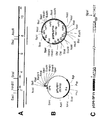
図1はセムリキ森林熱ウィルスの生活環に含まれる主要集合(main assembly)及び解体(disassembly)の略図であり、p62分裂及びpHによるSFV侵入機能(entry function)の活性化の調節も示す。
【0054】
図2はSFVの構造タンパク質の合成の間の転移シグナルの利用を示し;上図は26SサブゲノムRNAの遺伝子地図であり;中図はP62、6K及びE1タンパク質の膜転移の過程であり;内腔の側上の小さい矢印はシグナルペプチダーゼ分裂を示し;下図では3種類のシグナルペプチドの特性を挙げている。
【0055】
図3は発現ベクターとしてSFVが特に選ばれる特徴を示す。
【0056】
図4A−CはSFVの全長感染性クローンの構築を示し;図4AはSFVゲノムの図解制限地図を示し;cDNA合成の開始に用いられるプライマーを矢印で示し、最終クローンの組み立てに用いられるcDNA挿入物を棒で示し;図4BはプラスミドpPLH211、すなわちSFVの全長感染性クローンのためのキャリヤーとして用いられるSP6発現ベクター及び得られるプラスミドpSP6−SFV4を示し;図4CはSFVクローンのSP6プロモーター領域の構造を示し;点描の棒はSP6プロモーター配列を示し、転写される第1のヌクレオチドを星印により記し;下線の領域は真性SFV配列を示す。
【0057】
図5はDNA(U=T)としてのpSP6−SFV4 RNA転写物の完全ヌクレオチド配列、及びDNA配列の下に非−構造ポリプロテイン及び構造ポリプロテインのアミノ酸配列を示す。
【0058】
図6は試験管内製造RNAの細胞中へのトランスフェクションの後のウィルス製造のためのSFV cDNA発現系を示す。
【0059】
図7はSFV発現ベクターpSFV1−3及びヘルパー1の構築を示す。
【0060】
図8はSFVベクタープラスミドpSFV1−3のポリリンカー領域を示し;サブゲノム26S RNAのためのプロモーターの位置は四角で囲んであり、転写するべき最初のヌクレオチドを星印で記す。
【0061】
図9はヘルパートランス相補性を用いた感染性粒子中へのpSFV1−dhfr RNAの生体内パッケージングの略図である(dhfrはジヒドロ葉酸レダクターゼを意味する);
図10はp62からE2及びE3への分裂によるp62−含有非感染性ウィルス粒子の感染性粒子への変換のためのトリプシンの利用を示す。
【0062】
図11はエレクトロポレーションによりRNAトランスフェクションした場合のBHK細胞における異種タンパク質の発現を示す。
【0063】
図12は上図にてBamHI制限エンドヌクレアーゼ部位を生ずるSFVの主要抗原部位及び試験管内製造置換基を含む配列、HIV gp120タンパク質の主要中和ドメインに及ぶ配列、及びSFVキャリヤータンパク質E2にBamHIオリゴヌクレオチドとして挿入されたHIVドメインを示し;下図は野生型又はキメラ形態におけるドメイン246−251のブローアップ(blow−ups)を有するSFVスパイク構造の略図である。
【0064】
アルファウィルス セムリキ森林熱ウィルス(下文でSFVと省略する)は、ウィルス学及び生物学の両方において膜の生合成、膜の構造及び膜の機能、ならびにタンパク質−RNA相互作用の研究のためのモデル系として約20年間用いられてきた(4,5)。そのようなモデルとしてSFVを用いる主な理由はその単純な構造及び有効な複製にある。
【0065】
図1−3を参照し、以下においてSFV及びその複製をより詳細に説明する。基本的部分において本発明はシンドビスウィルスなどの他のアルファウィルスについても当て嵌まり、これに関連して引用されている多くの参照文献は実際にシンドビスウィルスに向けられている。SFVはRNA−含有ヌクレオキャプシド及び脂質二重層とタンパク質を含む回りの膜を含み、Cタンパク質と呼ばれるタンパク質の20面体殻が規則的に配置されてキャプシドを形成し、その内側にゲノムRNAが包まれている。キャプシドはE1、E2及びE3と呼ばれる3種類のタンパク質を含む脂質二重層により囲まれている。これらのいわゆるエンベロープタンパク質は糖タンパク質であり、そのグリコシル化部分が脂質二重層の外側にあり、これらのタンパク質の複合体が“スパイク”を形成し、それは電子顕微鏡によりウィルスの表面から外側に突き出ているのが見られる。
【0066】
SFVゲノムは、11422ヌクレオチドの一重鎖5’−キャップド及び3’−ポリアデニル化RNA分子である(6,7)。それは正の極性を有し、すなわちmRNAとして機能し、むきだしのRNAは細胞の細胞質中に導入されると感染を開始することができる。ウィルスが宿主細胞の形質膜上のタンパク質レセプターに結合すると感染が開始され、それによりウィルスが形質膜の表面上の“コーテッドピット(coated pits)”内に選択的に挿入され、それが陥入して被覆小胞を細胞中に形成し、その後該小胞を有するエンドサイトーシスされたビリオンが急速にエンドソームと呼ばれるオルガネラと融合する。ウィルスはエンドソームから細胞の細胞質ゾルにむきだしのヌクレオキャプシドとして逃げ、ウィルスのエンベロープがエンドソームに残る。その後ヌクレオキャプシドは“脱外被”され、従ってゲノムRNAが放出される。ここで図1を参照すると、ゲノムの5’の3分の2のポリプロテインへの翻訳と共に感染が進行し、ポリプロテインが自己−分裂によるプロセシングにより4個の非構造タンパク質nsP1−4となる(8)。タンパク質nsP1はメチルトランスフェラーゼをコードし、それはウィルス−特異的キャッピング活性ならびに負ストランド合成を担い(9,10);nsP2はプロテアーゼであり、これはポリプロテインをその4個の下部成分に切断し(11,12);nsP3はリンタンパク質であり、まだ機能は未知であり、nsP4はSFV RNAポリメラーゼ活性を含む(15,16)。nsPタンパク質が合成されるとそれらは正ストランドゲノム(42S)の全長負ストランドへの複製を担う。これらの分子はその後新しい42SゲノムRNAの製造の鋳型として働く。それらは又、サブゲノム(26S)RNAの合成の鋳型としても働く。この4073ヌクレオチド長のRNAはゲノムの最後の3分の1と共直線状であり、その合成は42S負ストランド上の26Sプロモーターにて内部的に開始される。
【0067】
キャプシド及びエンベロープタンパク質は異なる区画で合成され、それらは細胞質を通る別々の経路に従い、すなわちエンベロープタンパク質は粗面小胞体に結合した膜−結合リボソームにより合成され、キャプシドタンパク質は細胞質ゾル中の遊離のリボソームにより合成される。しかし26S RNAはウィルスのすべての構造タンパク質をコードし、これらはポリプロテイン前駆体としてC−E3−E2−6K−E1の順で合成される(19)。キャプシド(C)タンパク質が合成されるとそれは折り畳まれ、自己自身を新生鎖に切断するプロテアーゼとして働く(20,21)。合成されたCタンパク質は前に複製されたゲノムRNAに結合して細胞の細胞質中で新しいヌクレオキャプシド構造を形成する。
【0068】
該切断により新生鎖中のN−末端シグナル配列が明らかになり、それはシグナル認識粒子により認識され、新生鎖−リボソーム複合体を小胞体膜に向け(22,23)、そこでそれは共翻訳的に転移し、シグナルペプチダーゼにより分裂して3個の膜構造タンパク質p62(E3/E2の前駆体の形態)、6K及びE1となる(24,25)。構造タンパク質の合成の間に用いられる転移シグナルを図2に示す。膜タンパク質は細胞の生合成輸送経路内で広範囲の転写後プロセシングを受ける。p62タンパク質は小胞体においてそのE3ドメインを介し、E1とヘテロダイマーを形成する(26)。このダイマーは形質に輸送され、そこでスパイクヌクレオキャプシド相互作用を通してウィルス出芽が起こる。輸送の非常に後の段階(ゴルジ体−後)でp62タンパク質は成熟ビリオンで見られる形態であるE3及びE2に分裂する(27)。この分裂はビリオンの宿主細胞結合機能及びE1の膜融合能を活性化する。後者の活性は、ウィルスが新しい宿主細胞のエンドソームに入った後の第2の低−pH活性化段階に発現し、ウィルスヌクレオキャプシドの細胞の細胞質中への放出を担う(28−32)。成熟ウィルス粒子は、キャプシドタンパク質のT=3対称の180個のコピー内に包まれた1個のRNAゲノムのコピーを含み、T=4対称の3個の群として配置されたE1+E2+E3を含むスパイクトリマータンパク質の240個のコピーを有する脂質二重層により囲まれている。
【0069】
SFV侵入機能はp62分裂及びpHにより活性化され、調節される。さらに特定するとERで形成されたp62−E1ヘテロダイマーは耐酸性である。これらのヘテロダイマーがゴルジ複合体を介して形質膜に輸送されると、E1融合の活性化は複合体の解離を必要とするので、弱酸性環境にもかかわらずE1融合は活性化できない。図1に示す通り、放出されたウィルス粒子はE2E1複合体を含む。E2とE1の解離は酸性pHに敏感なので、エンドサイトーシスを介した宿主細胞へのウィルスの侵入の間にエンドソームの酸性環境がスパイク複合体(E1 E2 E3)の解離を始めさせ、遊離のE1を生ずる。後者は上記の通り感染過程におけるウィルス及びエンドソーム膜の間の融合過程の触媒に関して活性化されることができる。
【0070】
本発明の前部分で示した通りアルファウィルス系、特にSFV系はいくつかの独特の特徴を有し、それはDNA発現系において有利である。図3に関連して下記にまとめる。
【0071】
1.正の極性のゲノム。SFV RNAゲノムは正の極性であり、すなわちそれは直接mRNAとして機能し、従って感染性RNA分子はゲノムの全長cDNAコピーからの転写により得ることができる。
【0072】
2.有効な複製。感染性RNA分子はそれ自身のRNAレプリカーゼをコードし、それが今度は有効なRNA複製を推進する。実際SFVは知られている最も有効な複製ウィルスの1つである。数時間内に正−RNAの最高200.000ものコピーが1個の細胞中で形成される。これらの分子が豊富なために、感染細胞の実際すべてのリボソームはウィルスにコードされたタンパク質の合成に巻き込まれ、宿主タンパク質の合成を圧倒し、感染細胞のパルス標識法はほとんどウィルスタンパク質のみを標識する結果となる。正常な感染の間に105個の新しいウィルス粒子が1個の細胞から製造され、これは少なくとも108個のタンパク質分子がウィルスゲノムによりコードされた計算になる(5)。
【0073】
3.細胞質複製。SFV複製は細胞の細胞質中で起こり、そこでウィルスレプリカーゼは構造タンパク質の製造のためのサブゲノムを転写し、キャップする(19)。この特徴をcDNA発現系に含むのは明らかに非常に有用であり、mRNAのスプライシング、転写因子の限度、キャッピング効率及びmRNA輸送に関する問題などの、従来の“核”DNA発現系において遭遇する多くの問題が除去される。
【0074】
4.細胞変性効果の遅い開始。感染細胞の細胞変性効果は、感染におけるいくらか遅い時期に現れる。従って感染後約4時間から最高感染後24時間という広い時間枠があり、その間、構造タンパク質の非常に高い発現レベルと無視し得る程度の形態学的変化が組み合わされている。
【0075】
5.広い宿主域。この現象はおそらく、自然界における節足動物ベクターから野生囓歯類(rodent)及び鳥に至る伝達を含む正常な生活環の結果である。実験室条件下でSFVは培養された哺乳類、トリ、爬虫類及び昆虫の細胞に感染する(35)(Xiong,et al,loc.cit)。
【0076】
6.自然には、SFVは人に対して病原性が非常に低い。さらに組織培養細胞中で製造されたウィルス株は明らかに非病原性である。特異的突然変異を用い、ウィルス株の大量生産が必要な場合に安全性を支えるのに非常に有用な特徴であるSFV条件付致死突然変異体の創造が可能である。
【0077】
本明細書では、ヌクレオチド及びアミノ酸配列において以下の略字を用いた:Ala、アラニン;Ile、イソロイシン;leu、ロイシン;Met、メチオニン;Phe、フェニルアラニン;Pro、プロリン;Trp、トリプトファン;Val、バリン;Asn、アスパラギン;Cys、システイン;Gln、グルタミン;Gly、グリシン;Ser、セリン;Thr、トレオニン;Tys、チロシン;Arg、アルギニン;His、ヒスチジン;Lys、リシン;Asp、アスパラギン酸;Glu、グルタミン酸;A、アデニン;C、シトシン;G、グアニン;T、チミン;U、ウラシル。
【0078】
以下の実施例において用いた材料及び一般的方法を下記に記載する。
【0079】
1.材料。ほとんどの制限酵素、DNAポリメラーゼI、クレノウフラグメント、ウシ腸ホスファターゼ、T4 DNAリガーゼ及びT4ポリヌクレオチドキナーゼはBoehringer(Mannheim,FRG)から得た。SphI、StuI及びKpnIならびにRNアーゼインヒビター(RNアシン)及びSP6ポリメラーゼはPromega Biotec(Madison,WI)から得た。シークエナーゼ(修正T7ポリメラーゼ)はUnited States Biochemical(Cleveland,Ohio)から得た。プロテイナーゼKはMerck(Darmstadt,FRG)から得た。リボヌクレオチド、デオキシリボヌクレオチド、ジデオキシリボヌクレオチド及びキャップ類似体m7G(5’)ppp(5’)GはPharmacia(Sweden)から得た。オリゴヌクレオチドはApplied Biosystemsの合成機380Bを用いて製造し、その後HPLC及びNAP−5(Pharmacia)精製を行った。スペルミジン、フェニルメチルスルホニルフルオリド(PMSF)、ジエチルピロカーボネート(DEPC)、ウシ血清アルブミン(BSA)、クレアチンホスフェート及びクレアチンホスホキナーゼはSigma(St.Loues,Mo)から得た。パンソルビンはCalBiochem(La Jolla,CA)かに得た。アガロースはFMC BioProducts(Rockland,Maine)から、アクリルアミドはBioRad(Richmond,CA)から購入した。L−[35S]−メチオニン及びα−[35S]−dATP−α−SはAmershamから得た。
【0080】
2.ウィルスの成育及び精製:BHK−21細胞は、5%のウシ胎児血清、10%のトリプトースホスフェートブロス、10mMのHEPES(N−2−ヒドロキシエチルピペラジン−N’−2−エタンスルホン酸)及び2mMのグルタミンを補ったBHK培地(Gibco Life Technologis,Inc.,New York)中で成育した。90%の集密的細胞単層をPBSで1回洗浄し、0.2%ウシ血清アルブミン(BSA)、10mMのHEPES及び2mMのグルタミンを含むMEM中のSFVに0.1の多重度で感染させた。感染後(p.i.)24時間で培地を集め、4℃にて8,000xgの遠心を20分間行うことにより細胞破片を除去した。4℃のSW28ローター中、26,000rpmで1.5時間遠心することによりウィルスを培地からペレット化した。ウィルスは0.5mMのEDTAを含むTN中に再懸濁した。
【0081】
3.代謝標識(metabolic labeling)及び免疫沈降。10mMのHEPES、2mMのグルタミン、0.2%のBSA、100IU/モルのペニシリン及び100μg/mlのストレプトマイシンを補ったMEM中で成育したBHK細胞の集密的細胞単層に37℃にて50の多重度で感染させた。1時間p.i.後、培地を新しい培地と交換し、3.5時間成育を続けた。培地を除去し、細胞をPBSで1回洗浄し、10mMのHEPES及び2mMのグルタミンを含むメチオニン−非含有MEMを上に載せた。37℃にて30分後、培地を100μCi/mlの[35S]メチオニン(Amersham)を含む同培地と交換し、プレートを37℃にて10分間培養した。10倍過剰のメチオニンを含む標識培地で細胞を2回洗浄し、その後同培地中で種々の時間の間培養した。プレートを氷上に置き、細胞を氷冷PBSで1回洗浄し、最後に10μg/mlのPMSF(フェニルメチルスルホニルフルオリド)を含むライシス(lysis)緩衝液(1%NP−40−50mMトリス−HCl、pH7.6−150mM NaCl−2mMEDTA)を加えた。細胞をプレートから砕き、Eppendorf遠心機にて4℃、6,000rpmで5分間遠心することにより核を除去した。記載されている通りにタンパク質の免疫沈降を行った(31)。簡単に記載すると、ライセートに抗体を加え、混合物を氷上に30分間保った。氷上で30分間パンソルビンに結合することにより複合体を回収した。複合体を低塩緩衝液(low salt buffer)で1回、高塩緩衝液で1回、及び10mMのトリス−HClで1回洗浄してからゲル負荷緩衝液と共に加熱した。dhfrを沈降させるために、SDSを0.1%まで加え、混合物を95℃に2分間加熱し、その後10体積のライシス緩衝液を加えた。抗−E1[8.139]、抗−E2[5.1]及び抗C[12/2](37)単クローン性抗体は記載されている。腹水液中の単クローン性抗体OKT−9を用いてヒトトランスフェリンレセプターを沈降させた。この調剤は、ATCC(American TypCulture Collection)No CRL 8021から得た対応するハイブリドーマ細胞系を用いて我々の実験室においてThomas Ebelにより与えられた。多クローン性うさぎ抗マウスdhfrは、E.Hurt(European Molecular Biology Laboratory,Heidelberg,FRG)からの親切な贈り物であり、うさぎ抗−リゾチームは記載されている(38)。
【0082】
4.蛍光抗体法。間接蛍光抗体法を行うために、ガラスのカバーグラス上の感染細胞単層を、リン酸塩緩衝食塩水(PBS)で2回濯ぎ、−20℃のメタノール中で6分間固定した。固定後、メタノールを除去し、カバーグラスをPBSで3回洗浄した。0.5%のゼラチン及び0.25%のBSAを含むPBSを用いて室温で培養することにより、非特異的抗体結合を阻止した。阻止緩衝液を除去し、一次抗体を含む同緩衝液と交換した。室温で30分後、PBSで3回洗浄することにより反応を止めた。一次抗体の場合と同様にして二次抗体(FITC−複合羊抗−マウス[BioSys,Compiegne,France])の結合を行った。PBSで3回洗浄し、水で1回濯いだ後、カバーガラスを乾燥させてから2.5%のDABCO(1,4−ジアザビシクロ−[2.2.2]−オクタン)を含むMoviol 4−88(Hoechst,Frandfurt am Main,FRG)中に固定した。
【0083】
5.DNA法。プラスミドをエシェリキア コリ(Escherochia coli)DH5α(Bethesda Research Laboratories)[recA endA1 gyrA96 thil hsdR17 supE44 relA1 Δ(lacZYA−argF)U169 φ80dlacZΔ(M15)]中で成育した。すべての基礎的DNA法は、基本的に記載の通りに行った(39)。収率と純度を増すために凍結段階の間に3体積のフェノールを含んだ凍結−解凍法(40)によりDNAフラグメントをアガロースゲルから単離した。フラグメントを、ベンゾイル−ナフトイル−DEAE(BND)セルロース(Serva Fein−Biochemica,Heidelberg,FRG)クロマトグラフィーにより精製した(41)。感染RNAの製造に用いたプラスミドは、1MのNaClを通した沈降及びその後CsCl中でバンド形成することにより精製した(39)。ある場合には、Quigenクロマトグラフィー(Diagen Gmbh,Duesseldorf,FRG)によりプラスミドを精製した。
【0084】
6.特定部位のオリゴヌクレオチド突然変異誘発。オリゴヌクレオチド突然変異誘発のために、SFV cDNAクローンの適したフラグメントをM13mp18又はmp19中にサブクローニングし(42)、DH5αFIQ[endA1 hsdR1 supE44 thil recA1 gyrA96 relA1 φ80dlacΔ(M15) Δ(lacZYA−argF)U169/F’proAB laclq lacZΔ(M15) Tn 5](Bethesda Research Laboratories)中に形質転換した(43)。これらの構築物からのRF DNAをRZ1032[Hfr KL16 dut1 ung1 thi1 relA1 supE44 zbd279:Tn10.]中に形質転換し(44)、ウリジンの存在下でウィルスを成育し、ウラシル残基をウィルスゲノム中に挿入した。一重鎖DNAをフェノール抽出によりPEG沈降ファージから単離した。Applied Biosystems 380B合成機上でオリゴヌクレオチドを合成し、NAP−5カラム(Pharmacia)上のゲル濾過により精製した。オリゴヌクレオチド 5’−CGGCCAGTGAATTCTGATTGGATCCCGGGTAATTAATTGAATTACATCCCTACGCAAACG、5’−GCGCACTATTATAGCACCGGCTCCCGGGTAATTATTGACGCAAACGTTTTACGGCCGCCGG及び5’−GCGCACTATTATAGCACCATGGATCCGGGTAATTAATTGACGTTTTACGGCCGCCGGTGGCGを用いて新しいリンカー部位[BamHI−SmaI−XmaI]をSFV cDNAクローンに挿入した。オリゴヌクレオチド5’−CGGCGGTCCTAGATTGGTGCG及び5’−CGCGGGCGCCACCGGCGGCCGをシークエンシングプライマー(sequencing primer)(SP1及びSP2)としてポリリンカー部位の上流及び下流で用いた。リン酸化オリゴヌクレオチドを前に述べられた通りシークエナーゼ(Sequenase)(united States Biochemicals,Cleveland,Ohio)と共に突然変異誘発で用いた。試験管内製造RF形態をDH5αF’IQ中に形質転換し、得られたファージ単離物を、シークエナーゼの利用に関するUSBの案に従ってジデオキシシークエンシング(dideoxy sequencing)により正しい突然変異の存在に関して分析した。最後に突然変異フラグメントを全長SFV cDNAクローン中に再挿入した。この場合も適した突然変異の存在をプラスミドDNAからのシークエンシングにより確認した。6K領域の欠失は他に記載されている。
【0085】
7.試験管内転写。SpeI直線化プラスミドDNAを試験管内転写の鋳型として用いた。40mMのトリス−HCl(pH7.6)、6mMのスペルミジン−HCl、5mMのジチオトレイトール(DTT)、100μg/mlのヌクレアーゼ非含有BSA、それぞれ1mMのATP、CTP及びUTP、500μMのGTP、1単位/μlのRNアシン及び100−500単位/mlのSP6 RNAポリメラーゼを含む10−50μlの反応において37℃にて1時間RNAを合成した。キャップド転写物(46)の製造の場合、類似体m7G(5’)ppp (5’)G又はm7G(5’)ppp(5’)Aを1mMにて反応中に含んだ。RNA製造の定量のために微量の[α−32S]−UTP(Amersham)を反応中に含み、挿入を三塩化酢酸沈澱から測定した。必要な場合はDNアーゼ1又はRNアーゼAをそれぞれ10単位/μg鋳型あるいは20μg/mlで加えることにより、DNA又はRNAを37℃にて10分間消化した。
【0086】
8.RNAトランスフェクション。DEAE−デキストラン法によるBHK単層細胞のトランスフェクションを前に記載の通りに行った(47)。エレクトロポレーションによるトランスフェクションの場合、RNAは試験管内転写反応から直接、又は5mMのDTT及び1単位/μlのRNアシンを含む緩衝液で転写物を希釈して加えた。細胞をトリプシン処理し、完全BHK−細胞培地で1回、及び氷冷PBS(MgCl2及びCaCl2を含まない)で1回洗浄し、最後にPBSに再懸濁して107細胞/mlとした。細胞は直接使用するか又は氷上で終夜保存(BHK培地中)した。エレクトロポレーションの場合、0.5mlの細胞を0.2cmのキューベット(cuvette)(BioRad)に移し、10−50μlのRNA溶液を加え、キューベットを逆さにすることにより溶液を混合した。パルス制御装置を最大抵抗に設定したBioRad Gene Pulser装置を用い、1.5kV/25μFの2回の連続パルスにより、室温でエレクトロポレーションを行った。10分間培養した後、細胞を完全BHK−細胞培地中に1:20で希釈し、組織培養皿に移した。プラーク分析のために、エレクトロポレートした細胞を1ml当たり約3x105個の新しい細胞と共にプレート化し、37℃で2時間培養し、その後完全BHK−細胞培地中の1.8%の低融点アガロースを上に載せた。37℃で48時間培養した後、ニュートラルレッドで染色することによりプラークを視覚化した。
【0087】
9.ゲル電気泳動。ナトリウムドデシルサルフェート−ポリアクリルアミド ゲル電気泳動(SDS−PAGE)の試料を調製し、前に記載されている通りに(48)5%の堆積ゲル(stacking gel)を含む12%の分離ゲル(separating gel)上で実験した。6Kペプチドの分離の場合、アクリルアミドの10%−20%直線勾配ゲルを用いた。Kodak XAR−5フィルムに露光する前に、ゲルを10%酢酸−30%メタノール中で30分間固定した。ゲルをフルオログラフィー(49)用に調製した場合、それを固定後に30%メタノール中で30分間洗浄し、その後1Mのサリチル酸ナトリウム−30%のメタノール中に30分間浸漬してから乾燥した。核酸は、50mMのトリス−ボレート−2.5mMのNa2EDTAを緩衝液として用いたアガロースゲル上で実験した。染色の場合、実験中に0.2μg/mlのエチジウムブロミドを緩衝液及びゲルに含んだ。
【0088】
【実施例】
実施例1
この実施例では全長SFV cDNAクローンを調製し、SP6 RNAポリメラーゼプロモーターを含むプラスミド中に置き、全長及び感染性転写物の試験管内転写をさせる。pSP6−SFV4と称されるこのプラスミドを、1991年11月28日にPHLS Centre for Applied Microbiology & Research European Collection of Animal Cell Cultures,Porton Down,Salisbury,Wiltshire,U.K:に供託し、暫定受け入れ番号91112826を与えられた。
【0089】
図4A−Cに示す通り、SFVクローン構築の戦略は、SFV RNA分子の既知のヌクレオチド配列によって指定される適した制限エンドヌクレアーゼ部位の下流の、鋳型RNAに沿った数箇所でcDNA合成を開始することである。ウィルスRNAを精製ウィルス(中でもYale University,NewHaven,USAのArbovirus collectionから入手可能)からフェノール−クロロホルム抽出により単離し、前に記載されている通り(50)cDNA合成の鋳型として用いた。第1のストランド合成は、5’−TTTCTCGTAGTTCTCCTCGTCをプライマー−1として(SFVは2042−2062に相当する)及び5’−GTTATCCCAGTGGTTGTTCTCGTAATAをプライマー−2として(SFVは3323−3349に相当する)ならびにオリゴ−dT12-18をプライマー−3として(SFVの3’−末端)用いて3位置で開始した。
【0090】
第2のストランド合成の前に、第1のストランドcDNAへのオリゴヌクレオチド5’−ATGGCGGATGTGTGACATACACGACGCC(SFVのゲノム配列の28個の最初の塩基と同一)のハイブリッド化を行った。第2のストランド合成の完了後、cDNAを裁断し(trimmed)、プライマー−1の反応の場合を除いたすべての場合に二重鎖アダプター5’−AATTCAAGCTTGCGGCCGCACTAGT/GTTCGAACGCCGGCGTGATCA−3’(5’−付着−EcoRI−HindIII−NotI−XmaIII−SpeI−ブラント−3’)を加え、記載されている通りに(51)cDNaをEcoRI切断pTZ18R(Pharmacia,Sweden)中にクローニングした。5’末端領域のクローニングは、異なる方法で行った。SFVはHindIII部位を1947位に含むので、プライマー−1を用いて開始したcDNAはこの領域を含まねばならず、従ってHindIIIはそのcDNAの3’末端の指定に用いることができる。SFVの正に5’末端で制限部位を得るために、cDNAをSmaI−HindIII切断pGEM1(Promega Biotec.,Madison,Wl)中にクローニングした。SFVゲノムは配列5’−ATGGで始まるので、SmaI部位のブラントCCC−3’末端へのこの連結反応は、NcoI部位C’CATGGを作る。SFV配列は3個のNcoI部位を含むが、これらのいずれもHindIII部位の前の領域内になく、従ってこれらの5’末端クローンはさらにこの目的のために特に設計されたベクター中にNcoI−HindIIIフラグメントとしてクローニングしなければならない(下記参照)。pGEM1中の最初のcDNAクローンを制限分析によりスクリーニングし、1500bp以上の大きさの挿入物を含むものすべてを、挿入物の両端にSP6又はT7シークエンシングプライマーを用いてプラスミドから直接シークエンシングすることによりさらに特性化するために選んだ。pTZ18R中のSFV5’−末端クローンは、lacシークエンシングプライマーを用いてシークエンシングした。AFV RNAの試験管内合成を推進するためにSP6プロモーターを用いた。あまり多くの異種ヌクレオチドを加えずにこのプロモーターの前のSFV5’末端をクローニングするために、pGEM1の誘導体を構築する必要があった。従ってpGEM1をEcoRIで開け、Bal31欠失を作り、DNAをT4 DNAポリメラーゼでブラント化し、Ncolオリゴヌクレオチド(5’−GCCATGGC)を加えた。得られたクローンを、SP6プロモーターの転写開始部位(下線のG)に直接NcoI配列を有する変異体を拾い上げる(適した緊縮度で)ために設計されたオリゴヌクレオチド5’−GGTGACACTATAGCCATGGCを用いたコロニーハイブリッド化(39)によりスクリーニングした。Bal31欠失により最初のプラスミドの多重クローニング部位(multicloning site)のすべての制限部位が除去されたので、これらは新しい変異体からのPvuI−NcoIフラグメントを、ポリリンカーのHindIII位で挿入されたNcoI部位を有する別のpGEM1の変異体(pDH101)にクローニングすることにより復活(restored)した。これによりプラスミドpDH201が作られた。最後にSFV cDNAのクローニングに用いられたアダプターをpDH201中のEcoRI及びPvuII部位の間に挿入し、プラスミドpPLH211を作った(図4B)。このプラスミドを、全長クローンの組み立て物におけるSFV cDNAフラグメントの受容体として、これらの部位を用いて独立した重複サブクローンを結び付けることにより用いた。全長クローン、pSP6−SFV4の組み立てに用いたフラグメント及び適した制限部位を図4Aに示す。5’−末端の場合、選ばれたフラグメントは適したSFV配列5’−ATGGを含み、前に追加のG−残基を1個有する。このG−残基を除去すると、SP6からの転写効率が低下するが試験管内製造RNAの感染性には影響しない。従ってこの後のすべての研究で用いるクローンは5’末端にG−残基を含む。クローンの3’−末端の場合、69個のA−残基を含むcDNAフラグメントが選ばれた。cDNAの3’−末端に独特のSpeI部位を含むことにより、プラスミドは直線化することができ、試験管内でランオフ転写(runoff transcription)ができ、70個のA−残基を有するRNAを与える。図4CはSFV cDNAクローンの5’及び3’境界配列を示す。全長SFV RNAの感染性をいかにして得、示すかの一般的な概略を図6に示す。pSP6−SFV4 SP6転写物の完全ヌクレオチド配列を非構造及び構造タンパク質のアミノ酸配列と共に図5に示す。
【0091】
典型的に100ngの鋳型当たり約5μgのRNAが10単位のポリメラーゼを用いて得られたが、より多量の酵素を用いることによって収率の有意な向上は得られなかった。条件はアルファウィルスの感染性転写物の製造に関して以前に報告された(52)(47)条件と少し異なる。1mMのrNTP濃度でRNAの最大の製造が得られた。しかし感染性は5’キャップ構造の存在にも依存するので、転写反応におけるGTP濃度を半分にした時に最適感染性が得られた。この低下は製造されるRNAの量には限界的影響しか与えないが、特異的感染性を3倍に上げた(データは示していない)。
【0092】
図5に示すcDNA配列を以下の実施例で用いた。しかし図5中の最初の5’−Gヌクレオチドを欠いたSFV cDNA配列の場合に上記で示した通り有効性は低いとしても、1個又は数個のヌクレオチドが図5に示した配列と異なっている配列もベクターとして有用である。
【0093】
実施例2
この実施例ではSFV DNA発現ベクターの構築を開示する。
【0094】
実施例1で得たSFVの完全ゲノムをコードするcDNAクローンを用い、異種挿入物のための道を作るために26S構造遺伝子のコード領域を欠失することによってSFV DNA発現ベクターを構築する。しかしnsP1−4レプリカーゼ複合体の製造に必要な非構造コード領域は保存する。RNA複製は短い5’(nt 1−247)配列要素(53,54,55)及び3’(nt 11423−11441)配列要素(56,57)に依存し、従ってこれらもC遺伝子のすぐ上流の26Sプロモーター(17,18)と同様にベクター構築物中に含まれねばならない。
【0095】
図7に示す通り、最初に実施例1のSFV cDNAクローンからのXbaI(6640)−NsiI(8927)フラグメントをpGEM7Zf(+)(Promega Corp.,Wl,USA)中にクローニングした(段階A)。得られたプラスミド、pGEM7Zf(+)−SFVから、EcoRIフラグメント(SFVは7391及び88746に相当する)をM13mp19中にクローニングし、特定部位の突然変異誘発を用いて26Sプロモーター部位からすぐ下流にBamHI−XmaI−SmaIポリリンカー配列を挿入した(段階B)。M13 ssDNA(一重鎖)からのシークエンシングにより正しい突然変異体が確認されたら、EcoRIフラグメントをpGEM7Zf(+)−SFV中に再挿入し(段階C)、pSP6−SFV4中にXbaI−Nsλフラグメントとして逆クローニングした(段階D)。SFVの構造タンパク質をコードするcDNA領域の主要部分を欠失するために、これらのプラスミドをAsuII(7783)及びNdeI(11033)を用いて切断し、4種類すべてのヌクレオチドの存在下でクレノウフラグメントを用いてブラント化し、連結し、それぞれpSFV1、pSFV2及びpSFV3と呼ばれる最終的ベクターを作った(段階E)。ベクターは26SサブゲノムRNAのプロモーター領域及びE1タンパク質の最後の49アミノ酸、ならびにSFVゲノムの完全非コード3’末端を保存している。
【0096】
ベクターにおいてサブゲノム(26S)部分をコードする部分をポリリンカー配列で置換し、26Sプロモーター下で異種cDNA配列を挿入クローニングさせた。図8に示す通り、これらの3種類のベクターは同一の塩基カセットを26Sプロモーターの下流に挿入して含み、すなわち3つのすべての読み枠においてポリリンカー(BamHI−SmaI−XmaI)に翻訳停止−コドンが続く。ベクターはポリリンカーカセットが挿入されている位置が異なる。pSFV1の場合、カセットは26S翻訳開始部位の31塩基下流に位置している。キャプシド遺伝子翻訳の開始作因(motive)は認められた配列(58)と同一である。従ってこの作因はpSFV2にも備わっており、その場合それはキャプシド遺伝子の作因のすぐ後に位置している。最後にpSFV3ではカセットがキャプシド遺伝子の開始コドン(AUG)のすぐ後に位置する。挿入の両端を調べるのに必要なシークエンシングプライマー(SP)は、26Sプロモーター領域(SP1)又は停止コドンカセットの後の領域(SP2)のいずれかとハイブリッドするように設計されている。
【0097】
26SプロモーターはnsP4コード領域の3’−末端と重複することに注意する。pSFV2の場合、クローニング部位はSFVキャプシド遺伝子の翻訳開始部位のすぐ後に位置する。pSFV3の場合、クローニング部位はさらに3ヌクレオチド下流に位置し、すなわちSFVキャプシド遺伝子の開始AUGコドンに直接続いている。ポリリンカーに続く3個の翻訳停止コドンを四角で囲む。下流シークエンシングプライマー(SP1)は26Sプロモーターと重複し、上流シークエンシングプライマー(Sp2)はXmaIII部位と重複する。
【0098】
実施例3
この実施例ではヘルパーウィルスベクター構築物を含む生体内パッケージング系を調製する。
【0099】
系は、構造タクパク質機能の欠失したSFV変異体、又は実施例2で得た発現ベクター構築物から誘導された組み替えRNAを感染性ウィルス粒子内にパッケージすることを可能にする。従ってこの系は正常な感染により組み替えRNAを細胞中に導入できるようにする。実施例1で得たpSP6−SFV4の制限エンドヌクレアーゼ部位AccI(308)及びAccI(6399)の間の領域を、図7の段階Fで示す通りに切断及び連結することにより欠失させ、pSFV−Helper1と呼ばれるヘルパーベクターを構築する。ベクターはRNA複製に必要な5’及び3’シグナルを保存している。Helperベクターのほとんど完全なnsp領域が欠失しているので、この構築物から製造されるRNAは機能的レプリカーゼ複合体の欠乏のために細胞中で複製されない。図9に示す通り、pSFV1−組み替え及びヘルパーcDNAの試験管内転写の後、ヘルパーRNAはpSFV1−組み替え誘導体と共にコトランスフェクションされ、ヘルパー構築物が新しいウィルス粒子の組み立てに必要な構造タンパク質を与え、組み替え構築物はRNA複製に必要な非構造タンパク質を与え、組み替えゲノムを含むSFV粒子が製造される。コトランスフェクションは実施例6で開示した通りエレクトロポレーションにより行うのが好ましく、BHK細胞を宿主細胞として用いるのが好ましい。
【0100】
RNAのパッケージのために、キャプシドタンパク質を結合することが示されている(57,59)領域であるnsP1の末端の領域が必要である。ヘルパーはこの領域を欠いているので、このベクターから誘導されたRNAはパッケージされず、従って組み替え及びヘルパーを用いたトランスフェクションは組み替え−誘導RNAを有するウィルス粒子のみを製造する。これは、これらのウィルスがさらに継代することはできず、従って1段階ウィルス株を与えることにつながる。利点は、これらの粒子による感染がウィルスタンパク質を製造しないことである。
【0101】
実施例4
この実施例は実施例1からの全長SFV cDNAクローンの変異体の構築を説明し、それにより異種エピトープをコードする異種DNA配列の挿入、及び該異種エピトープをP62、E2又はE1スパイクタンパク質の不可欠の部分として有する組み替え(キメラ)ウィルスの製造が可能になる。
【0102】
この目的のために、E2及びE1エンベロープタンパク質の機能、形態学及び抗原構造に関するすべての知識が欠かせない。アルファウィルスの病原性に関する以前の研究は、E2に対する抗体が種類−特異的であり、優れた中和活性を有するが、E1に対する抗体は群−特異的であり、非中和性であることを示した(5)。しかし、密接に関連したアルファウィルスであるSFV、シンドビス及びロスリバーの抗原部位がマッピングされ、アミノ酸配列のレベル(60,61,62,63)と関連づけられたのは最近である。これらの研究は、問題の主要部位のほとんどがSFV E2スパイクタンパク質のアミノ酸位置216、234及び246−251にあることを示した。興味深いことに、これらの3つの部位は、コンピューター分析により予測されたものと正確に同一の部位である。この実施例ではドメイン246−251がアルファウィルスの群内で高度に保存された構造、及びハイドロパシ−プロファイル(hydropathy profile)を有するのでこの領域を用いた。pSP6−SFV4 P62タンパク質の246−251領域に異種エピトープをコードする遺伝子を挿入すると、各ヘテロダイマー、すなわち240コピー上に1個の新しいエピトープを有する粒子を与える。
【0103】
SFVゲノムのE2部分内への異種エピトープの特異的挿入を可能にする独特の制限エンドヌクレアーゼ部位を作るために、オリゴヌクレオチド5’−GATCGGCCTAGGAGCCGAGAGCCCを用いた特定部位の突然変異誘発によりBamHI部位を挿入した。
【0104】
実施例5
この実施例では、SFVの条件付致死変異体を、実施例1で得たSFV cDNAから構築し、その変異体はP62タンパク質に突然変異を有し、該タンパク質の非分裂形態を生じ、その結果この変異体は外因的に加えられたプロテアーゼを用いて最初に切断しないとそのまま新しい宿主細胞に感染することはできない。
【0105】
図10に示す通り、この形態のウィルスはトランスフェクトされた細胞における野生型の能力を用いて組み立てられるが新しい宿主細胞に侵入することはできないので、この構築物は異種エピトープのためのワクチンキャリヤーとして有利に用いることができる。阻止は、不活性ウィルス粒子のトリプシン処理により克服することができる。これは粒子を、このウィルス変異株の増幅に用いることができる十分に侵入−可能な形態に変換する。
【0106】
活性化されるとSFV変異体は、通常はエンドサイトーシス経路により細胞に侵入し、感染を開始する。形質膜でウィルスタンパク質が作られ、出芽が起こる。しかし製造されたウィルス粒子はすべて不活性な形態であり、従って複製が一巡した後感染は終わる。感染の徹底が阻止される理由は、p62の分裂部位に特定部位の突然変異誘発により導入された突然変異である。このアルギニンからロイシンへの置換(p62タンパク質のE3部分のアミノ酸位置66における)は、分裂部位の認められた特徴を変化させ、細胞表面への輸送に間に通常p62タンパク質をE2及びE3ポリペプチドに切断する宿主細胞のプロテイナーゼによって認識されない。代わりに外因的に加えられたトリプシンがこの切断を行うことができ、この場合最初の分裂部位の直前のアルギニン残基65で起こる。この切断が、侵入スパイクサブユニットの結合を制御することによりウィルスの侵入機能力の活性化を調節するので、非分裂p62のみを有するウィルス粒子が新しい宿主細胞に侵入することは完全に不可能である。
【0107】
分裂欠失変異E2の創造は以前に記載されている(29)。この領域に及ぶAsull−Nsλフラグメントをその後単離し、全長cDNAクローンpSP6−SFV4中にクローニングした。
【0108】
実施例6
この実施例では、外因性DNAを含む実施例1からの全長cDNAから試験管内で転写されたSFV RNA分子又はその変異体、あるいは実施例2からのSFVベクターを用いたBHK細胞のトランスフェクションを開示する。トランスフェクションは最適条件下で非常に有効であることが示されているエレクトロポレーションにより行う。
【0109】
BHK細胞を上記のSFV RNA分子を用いたエレクトロポレーションによりトランスフェクトし、温度、電圧、キャパシタンス及びパルス数などのパラメーターを変化させることにより最適条件を決定した。25μFにおける1.5kVの2回の連続パルスにより最適トランスフェクションが得られ、その条件下で死亡する細胞の数は無視し得るものであった。方法全体を通じて細胞を0℃より室温に保つのが良いことが見いだされた。エレクトロポレーションによるトランスフェクションを、投入RNAの関数としても測定した。予想通り、トランスフェクションの頻度の増加はRNA濃度に直線的に依存しておらず、100%トランスフェクションを得るために約2μgのcRNAが必要であった。
【0110】
従来のトランスフェクションと比較すると、これは大きな進歩である。例えばDEAE−デキストラントランスフェクションの場合、最適で細胞のわずか0.2%がトランスフェクトされたのみであった。
【0111】
実施例7
この実施例は、21kDの細胞質マウスジヒドロ葉酸レダクターゼ(dhfr)、90kDの膜タンパク質ヒトトランスフェリンレセプター(TR)、及び最後に14kDの分泌タンパク質チキンリゾチームをコードする遺伝子の場合の、SFVベクターである実施例2からのpSFV1により推進される異種遺伝子発現を示す。dhfr遺伝子はpGEM2−dhfrから(64)BamHI−HinIIIフラグメントとして単離し、クレノウフラグメントを用いてブラント化し、SmaI−切断pSFV1に挿入された。トランスフェリンレセプター遺伝子は、最初にpGEM1−TRからXbaI−EcoRIフラグメントとしてpGEM7ZF(+)中にクローニングされ(64,65)、続いてそこからBamHIフラグメントとしてpSFV1にクローニングされた。最後にリゾチーム遺伝子(21)を有するpGEM2からのBamHIフラグメントをpSFV1にクローニングした。
【0112】
異種遺伝子の発現の研究のために、dhfrの試験管内製造RNA及びTR構築物をBHK細胞中にエレクトロポレートした。野生型SFVのRNAを標準として用いた。エレクトロポレーション後(p.e.)の異なる時点で細胞を10分間パルス−標識し、続いて10分間追跡し、その後ライセートをゲル電気泳動及びオートラジオグラフィーにより分析した。BHK細胞は、野生型SFVのRNA、pSFV1−dhfr及びpSFV1−TRを用いてトランフェクトし、3、6、9、12、15及び24時間p.e.でパルス−標識した。等量のライセートにつき12%ゲル上で実験した。9時間の試料はSFV、dhfr及びトランスフェリンレセプタータンパク質の免疫沈降(IP)にも用いた。pSFV1−リゾチームを用いてトランスフェクトした細胞を9時間p.e.でパルス−標識し、その後示された時間(時間)追跡した。等しい割合のライセート又は培地を13.5%ゲル上に負荷した。IPは1時間追跡ライセート試料からの免疫沈降を示す。U−列は標識されたがトランスフェクトされていない細胞のライセートである。侵入RNAは負のストランド合成から出発し、これは約4−5時間p.e.までピークに達しないので(5)、3時間p.e.で外因性タンパク質はほとんど製造されなかった。この時点でほとんどすべての標識タンパク質は宿主起源であった。対照的に6時間p.e.にて、外因性タンパク質が高い効率で合成され、宿主タンパク質合成の重度の阻害が明らかであった。これは9時間p.e.でさらに衝撃的であり、最大停止(maximum shut down)に達した。異種タンパク質の有効な製造は最高24時間p.e.まで続き、その後製造は速度が落ち(データは示していない)、細胞が定常期に入ったことを示した。
【0113】
チキンリゾチームは分泌タンパク質なので、その発現は細胞ライセートと成育培地の両方から分析した。細胞を9時間p.e.でパルス−標識し、その後8時間追跡した。結果を図11に示す。リゾチームはゆっくり分泌されるがほとんどすべての標識物質は追跡の間に培地に分泌された。
【0114】
実施例8
この実施例は本発明の生体内パッケージング系を示す。
【0115】
pSFV1−TRの試験管内製造RNAを異なる比率でヘルパーRNAと混合し、これらの混合物をBHK細胞中にトランスフェクトした。細胞を24時間成育し、その後培地を集め、遠心によりウィルス粒子をペレット化した。蛍光抗体法により感染性単位(i.u.)の数を決定した。ヘルパーと組み替え体の1:1の比率が最も有効に感染性粒子を製造することが見いだされ、平均5x10⁶個の細胞が2.5x10⁹i.u.を与えた。異なる感染の多重度(m.o.i.)でBHK細胞に感染させることにより、このウィルス株の感染性を調べた。pSFV1−TR組み替えRNAを有するそのような生体内パッケージ粒子に感染した後のBHK細胞におけるヒトトランスフェリンレセプターの発現に関する結果を図11の右下に示す。MEM(0.5%のBAS及び2mMのグルタミンを含む)中に希釈した200μlのウィルスを細胞上に載せ、5−0.005の範囲のm.o.i.値を与えた。37℃にて1時間後、完全BHK培地を加え、9時間成育を続け、その時点で10分間のパルス(100μCi35S−メチオニン/ml)及び10分間の追跡を行い、細胞をライシス緩衝液に溶解した。300μl(30,000細胞に相当する)のライセートからの10μlにつき10%ゲル上で実験し、乾燥ゲルを−70℃にて2時間露光した。発現レベルが高いので、終夜露光を用いたオートラジオグラフィーで明白なバンドを得るためにわずか3,000細胞しか必要でなかった。
【0116】
従って1細胞当たり約1i.u.にて有効なタンパク質製造及びそれに伴う宿主細胞停止が起こることが見いだされた。1個のSFV感染細胞が平均108個のキャプシドタンパク質分子を製造するので、1回のエレクトロポレーションから製造されたウィルス株を用いて約50mgのタンパク質に相当する1017個のタンパク質分子を製造できることになる。
【0117】
前記の実験結果から本発明は、これまでに存在した発現系のいくつかの欠点を含まない非常に有用で有効な発現系に関することが明らかである。本発明の系の主な利点を簡単にまとめると以下のようになる:
(1)1回のトランスフェクション実験により1日で高力価の組み替えウィルス株を製造することができる。選択/スクリーニング、プラーク精製及び増幅段階の必要がない。組み替えウィルスの容易な製造は、多くの系列の突然変異体の表現型を特性化しなければならない実験において特に重要である。
(2)組み替えゲノムのみがパッケージングシグナルを含み、ヘルパーゲノムは含まないので、組み替えウィルス株はヘルパーウィルスを含まない。
(3)組み替えウィルスは、昆虫及びより高級な真核細胞型を含む多様な細胞中に“自然”で漏れのない方法で組み替えゲノムを感染させるのに用いることができる。そのような広い宿主域は、特に発現したタンパク質の活性のために細胞−型−特異的翻訳後修正反応が必要な場合、発現系にとって非常に有用である。
(4)得られるタンパク質発現のレベルが非常に高く、そのレベルは感染の間のウィルスタンパク質の量に相当する。宿主細胞タンパク質の停止も起こり、それにより抗体媒介による抗原濃縮を必要とせずに細胞ライセート中の異種タンパク質を明白に追跡することが可能になる。これは細胞生物学におけるDNA発現実験をかなり容易にするであろう。さらに内在する対になるものによる発現タンパク質に対する抵触の問題(すなわちホモ−オリゴマー化反応)を避けることができる。
【0118】
実施例9
この実施例はエピトープキャリヤーを示す。
【0119】
ワクチンの開発が最も重要とされている非常に重要な例は、ヒト免疫不全ウィルスHIV−1によって起こる後天性免疫不全症候群(AIDS)に関する(66,67)。これまでHIV−1に対する有効なワクチンを製造する試みはすべて失敗してきたが、破裂SIV−1(猿免疫不全ウィルス)を用いた予防接種がある程度ウィルスの感染に対する保護を与えるという極く最近の報告がある(68)。しかしHIV−1に対する安全で有効なワクチンの開発は、ウィルスの生物学的性質の故に非常に困難であろう。この実施例ではHIV−1の1個のエピトープをSFVのE2タンパク質の抗原性ドメイン中に挿入した。用いられたエピトープはHIV−1の糖タンパク質gp120に位置し、アミノ酸309−325に及ぶ。これはHIV−1の可変ループを形成し、N−グリコシル化部位の直後に位置する。
【0120】
HIVエピトープをコードする既製のオリゴヌクレオチドのカセット挿入を用い、HIVの309−325エピトープがBamHI部位に挿入されたキメラを構築した。BamHI部位における必要な塩基置換によりベクター中のアミノ酸変化は起こらなかったが、2個のアミノ酸(Asp及びGluが位置を変えた。試験管内製造ベクターRNAは野生型の能力を用いて細胞感染を起こすので、この変化は悪影響を及ぼさない。図12はエピトープキャリヤー中の問題の領域の配列を示す。予備実験でキメラタンパク質が製造されることが示された。タンパク質を抗−HIV抗体を用いて免疫沈降させることができる。これらはHIVに対するワクチン調剤に用いることができるキメラウィルス粒子の製造にも用いることができると思われる。そのような粒子を図12の下部に示す。
【0121】
【表1】

【表2】
【表3】
【表4】
【表5】
【表6】
【表7】
【表8】
【表9】
【表10】
【表11】
【表12】
【図面の簡単な説明】
【図1】図1はセムリキ森林熱ウィルスの生活環に含まれる主要集合(main assembly)及び解体(disassembly)の略図であり、p62分裂及びpHによるSFV侵入機能(entry function)の活性化の調節も示す。
【図2】図2はSFVの構造タンパク質の合成の間の転移シグナルの利用を示し;上図は26SサブゲノムRNAの遺伝子地図であり;中図はP62、6K及びE1タンパク質の膜転移の過程であり;内腔の側上の小さい矢印はシグナルペプチダーゼ分裂を示し;下図では3種類のシグナルペプチドの特性を挙げている。
【図3】図3は発現ベクターとしてSFVが特に選ばれる特徴を示す。
【図4】図4はSFVの全長感染性クローンの構築を示し;図4AはSFVゲノムの図解制限地図を示し;cDNA合成の開始に用いられるプライマーを矢印で示し、最終クローンの組み立てに用いられるcDNA挿入物を棒で示し;図4BはプラスミドpPLH211、すなわちSFVの全長感染性クローンのためのキャリヤーとして用いられるSP6発現ベクター及び得られるプラスミドpSP6−SFV4を示し;図4CはSFVクローンのSP6プロモーター領域の構造を示し;点描の棒はSP6プロモーター配列を示し、転写される第1のヌクレオチドを星印により記し;下線の領域は真性SFV配列を示す。
【図5】図5はDNA(U=T)としてのpSP6−SFV4 RNA転写物の完全ヌクレオチド配列、及びDNA配列の下に非−構造ポリプロテイン及び構造ポリプロテインのアミノ酸配列を示す。
【図6】図6は試験管内製造RNAの細胞中へのトランスフェクションの後のウィルス製造のためのSFV cDNA発現系を示す。
【図7】図7はSFV発現ベクターpSFV1−3及びヘルパー1の構築を示す。
【図8】図8はSFVベクタープラスミドpSFV1−3のポリリンカー領域を示し;サブゲノム26S RNAのためのプロモーターの位置は四角で囲んであり、転写するべき最初のヌクレオチドを星印で記す。
【図9】図9はヘルパートランス相補性を用いた感染性粒子中へのpSFV1−dhfr RNAの生体内パッケージングの略図である(dhfrはジヒドロ葉酸レダクターゼを意味する)。
【図10】図10はp62からE2及びE3への分裂によるp62−含有非感染性ウィルス粒子の感染性粒子への変換のためのトリプシンの利用を示す。
【図11】図11はエレクトロポレーションによりRNAトランスフェクションした場合のBHK細胞における異種タンパク質の発現を示す。
【図12】図12は上図にてBamHI制限エンドヌクレアーゼ部位を生ずるSFVの主要抗原部位及び試験管内製造置換基を含む配列、HIV gp120タンパク質の主要中和ドメインに及ぶ配列、及びSFVキャリヤータンパク質E2にBamHIオリゴヌクレオチドとして挿入されたHIVドメインを示し;下図は野生型又はキメラ形態におけるドメイン246−251のブローアップ(blow−ups)を有するSFVスパイク構造の略図である。[0001]
BACKGROUND OF THE INVENTION
The present invention relates to an alphavirus-based DNA expression system that can be used to transform animal cells for use in producing high yields of desired products such as proteins and vaccines.
[0002]
[Prior art]
The rapid development of biotechnology has largely depended on the introduction of recombinant DNA methods, which has revolutionized cell biology and medical research by opening new avenues that reveal the molecular mechanisms of cells. A number of interesting protein molecules are characterized each year using the method of cDNA cloning. Therefore, many research activities today are aimed at clarifying the relationship between the structure and function of these molecules. Eventually this knowledge will increase our potential to maintain health and control disease in humans and animals. Indeed, today there is an increasing number of new “cloning” protein products that are already used as drugs or diagnostics.
[0003]
In recombinant DNA methods for studying biological problems, the DNA expression system is a critical element. Accordingly, there is a great need for an effective DNA expression system that is simple and safe to use, provides the desired product in high yield, and can be used in a variety of host cells, particularly mammalian cells.
[0004]
Many attempts have been made to develop a DNA expression system that satisfies these requirements. In many cases viruses have been used as the source of such systems. To date, however, none of the existing viral expression systems has satisfied all of these requirements. For example for cDNABaculovirusAlthough the expression system is very effective, it can only be used in insect cells (see
[0005]
Recently, however, viruses containing RNA genomes have also been used in the development of DNA expression systems.
[0006]
EP 0 194 809 discloses an RNA transformation vector derived from a (+) strand RNA virus, which has a cap structure modified by insertion of exogenous RNA into a region non-essential for replication of the viral RNA genome. Contains viral RNA. These vectors are used for expression of the function of the exogenous RNA in cells transformed with it. RNA can be used in solution or in a capsid package. Furthermore, this RNA can be used to generate new cells with a new function, ie protein expression. The invention of the reference is generally claimed for host cells, (+) strand RNA viruses and the like. Nevertheless, it is clear from the experimental support shown there that only plant cells were transformed and only the plant virus Bromo Mosaic virus was used as the transformation vector. .
[0007]
In the reference, conversion of any RNA virus-cell system into a useful expression system for exogenous DNA using the principles described in the reference is readily apparent to those skilled in the art. However, at least in the case of animal cell RNA viruses, this has not proven to be true. There may be several reasons for this. To do that:
1) Ineffectiveness of transformation of animal cells with RNA transcribed in vitro; 2) RNA replication after transfection methods where apparently competent RNA transcripts are commonly used Ineffectiveness of starting;
3) Inability to produce recombinant virus high titer stock without helper virus;
4) Inability to establish stable characteristics of transformed cells expressing the function of exogenous RNA
Is included.
[0008]
Proc. Natl. Acad. Sci. USA, Vol 84, 1987, pp 4811-4815, discloses a gene expression system based on a member of the genus Alphavirus, the Sindbis virus, which is a bacterial CAT in avian cells such as chicken embryo fibroblasts. It has been used to express (chloramphenicol acetyltransferase).
[0009]
Xiong et al. , Science, Vol 243, 1989, 1188-1191 also discloses a gene expression system based on Sindbis virus. This system is said to be effective on a wide range of animal cells. Bacterial CAT expression in mammalian cells including insects, birds and humans is disclosed therein.
[0010]
It is known from the prior art that the Sindbis virus, a member of the Alphavirus genus, can withstand insertion and be directed to the expression of at least one external gene, the bacterial chloramphenocol acetyltransferase (VAT) gene, Both of these systems are ineffective with respect to exogenous gene expression and proved to be very cumbersome to use. Therefore, today no useful system has been found in the field of DNA expression in animal cells.
[0011]
The first example of a cDNA copy of a Sindbis virus defect interference (DI) virus variant was used to carry the CAT gene. The RNA was transcribed in vitro and used for avian cell transfection, indicating the production of some CAT protein after infecting cells with wild type Sindbis virus. The latter virus was equipped with a viral replicase for expression of the CAT construct. The ineffectiveness of this system is: 1) low levels of early DI-CAT RNA transfection (0.05-0.5% of cells) and 2) protein translation due to unnatural and suboptimal protein translation initiation signals. This is because of the ineffectiveness of using DI-CAT RNA. The cognate also gives the result of packaging some of the recombinant DI-CAT genome into viral particles. However, this coincides with the production of a very large excess of wild type Sindbis virus. Therefore, the use of this mixed virus strain for CAT expression is greatly hampered by the fact that most of the replication and translational activities of cells infected with such strains are involved in wild type and not in recombinant gene expression. Let's go.
[0012]
Many of the similar problems are inherent in the other Sindbis expression systems described. In this case, an RNA replicable Sindbis DNA vector is used to carry the CAT gene. RNA produced in vitro is shown to replicate in animal cells and CAT activity is seen. However, overall CAT production is still low because only a very small number of cells are transfected. Another possible explanation for this is that the Sindbis construct used is not optimal for replication. Wild-type Sindbis virus can be used to protect the recombinant genome in particles along with excess wild-type genome, and then this mixed strain can be used for expression of CAT protein via infection. However, this strain has similar problems as described above for the recombinant DI system. The latter document also shows that recombinant virus particles with improved titer can be obtained when the virus is amplified through several processes. However, it must be remembered that the titer of wild-type virus is correspondingly improved and that the first problem of main production of wild-type virus remains. There are also several problems that can occur when using several processes in the production of mixed virus strains. Since the recombinant genomes do not have selective enforcement for conservation, they are easily 1) transposed and 2) overwhelmed in number by the wild-type genome as a result of poor replication and / or packaging properties.
[0013]
Another important feature of viral DNA expression vectors is their use for the expression of antigens of unrelated pathogens, so they can be used as vaccines against such pathogens.
[0014]
The development of a safe and effective vaccine against viral diseases has proven to be a very difficult task. Many existing vaccines have helped fight the global epidemic of many infectious diseases, but there are still many infections without effective vaccines. Current methods of vaccine production offer several problems: (1) the production of sufficient quantities of antigenic material is often difficult; (2) in many cases, vaccine preparations have not been killed, or With the risk of not being sufficiently attenuated; (3) production of effective vaccines is often difficult because it is very difficult to give antigenic epitopes in immunologically active form: (4) many In the case of vaccines, genetic variations in antigenic components generate new strains with new serological specificity, and again necessitate the development of new vaccines.
[0015]
Two viral DNA vectors have been developed to overcome many of these problems in vaccine production. These give either recombinant viruses or chimeric viruses. Recombinant virus contains a wild-type virus package around the recombinant genome. These particles are used to infect cells, which then produce antigenic proteins from the recombinant genome. Chimeric viruses also contain a recombinant genome, which usually specifies antigen production as part of the normal viral structural protein, which is encased in progeny particles, eg exposed on the surface of the virus spike protein. The The major advantages of these types of viral preparations intended for use as vaccines are 1) they can be produced on a large scale, and 2) they provide the antigen in a natural form to the organism's immune system. Cells infected with the recombinant virus synthesize an exogenous antigen product, process it into a peptide, which then gives it to T cells in the normal way. In the case of a chimeric virus, the antigen is further exposed in the sense of the subunit of the virus particle itself. Chimeric viruses are therefore also called epitope carriers.
[0016]
The major difficulty with these types of vaccine preparations is how to ensure safe and limitless particle replication in the host. So far, several successes have been achieved using vaccinia virus as an example of the recombinant virus method (69) and poliovirus as an example of chimeric particles (70-72). Since both virus variants are based on commonly used vaccine strains, they can be claimed to be useful vaccine candidates as recombinant and chimeric particles, respectively. However, both virus vaccines are combined with the risk of side effects, even with the risk of severe side effects, and these virus strains are already used as vaccines in the majority of the population in many countries.
[0017]
As is clear from the above discussion, recombinant viruses or chimeras for easy production of important proteins or polypeptides in various animal cells in high yield and for use as safe and effective vaccines against various pathogens There is a great need for the development of improved DNA expression systems for the production of viruses.
[0018]
[Problems to be solved by the invention]
Accordingly, it is an object of the present invention to provide an improved DNA expression system based on viral vectors that can be used for the production of proteins and polypeptides, and as recombinant or chimeric viruses, and provides many advantages over the prior art. It is.
[0019]
[Means for Solving the Problems]
To this end, according to the present invention, there is provided an RNA molecule derived from an alphavirus RNA genome and capable of effectively infecting an animal host cell, the RNA molecule being essential for replication of the alphavirus RNA. And an exogenous RNA sequence that can express its function in the host cell, and the exogenous RNA sequence is inserted into a region of the RNA molecule that is not essential for its replication.
[0020]
Alphaviruses are a genus belonging to the family of Togaviruses in which a single-stranded RNA genome with positive polarity is encapsulated in a nucleocapsid surrounded by an envelope containing a virus spike protein.
[0021]
The alphavirus genus includes, among others, Sindbis virus, Semliki Forest virus (SFV) and Ross River virus, all of which are closely related. In accordance with a preferred embodiment of the present invention, Semliki Forest virus (SFV) is used as the basis for the DNA expression system.
[0022]
An exogenous RNA sequence encodes a desired genetic characteristic to be imparted to a virus or host cell, and usually the sequence is complementary to a DNA or cDNA sequence encoding the genetic characteristic. The DNA sequence can include isolated natural genes such as bacterial or mammalian genes, or desired genetic characteristics, ie expression of desired products such as enzymes, hormones, or exogenous antigen epitopes or determinations A synthetic DNA sequence encoding the expression of a peptide sequence specifying the group can also be constructed.
[0023]
If the exogenous RNA sequence encodes a product such as a protein or polypeptide, it is inserted into the viral RNA genome and replaced with its deleted structural protein coding region, but the RNA sequence encoding the viral epitope is fundamental Can be inserted into the structural protein coding region of the viral RNA genome, which does not contain any deletions or has only a few nucleosides deleted.
[0024]
The RNA molecule can itself be used in transformation of animal cells in solution, for example by conventional transfection such as the DEAE-dextran method or the calcium phosphate precipitation method. However, transformation by infecting cells with infectious viral particles would substantially increase the rate of cell transformation and thus the rate of expression. Accordingly, a suitable embodiment of the present invention relates to an RNA virus expression vector comprising an RNA molecule of the invention packaged in an infectious particle, wherein the infectious particle comprises said RNA surrounded by a membrane comprising an alphavirus spike protein. Contains in nucleocapsid.
[0025]
The RNA molecules of the present invention can be freely packaged in such particles, provided that their overall dimensions correspond to the wild-type alphavirus RNA genome or to package the RNA in the infectious particle. Fluctuates to the extent that it fits.
[0026]
These infectious particles that contain packaged recombinant genomes and produce pure, high-titer recombinant virus strains provide a means to express exogenous genes or DNA sequences upon infection with normal virus particles, and the extent of transformation Is much more effective than RNA transfection.
[0027]
In accordance with a suitable embodiment of the present invention, an animal host cell is divided into an RNA of the present invention that lacks part or all of the region encoding the structural viral protein, the SP6 promoter region, a cis acting signal required for RNA replication. sequence comprising the 5 'and 3' regions of the alphavirus cDNA encoding signal and the region encoding the viral structural protein but essentially encoding the RNA signal for packaging of RNA into nucleocapsid particles. Producing such infectious particles by cotransfection with helper RNA molecules transcribed in vitro from helper DNA vectors lacking all of the nonstructural viral protein coding regions containing and culturing host cells .
[0028]
In accordance with another aspect of the present invention, efficient introduction of the RNA of the present invention into animal host cells is performed by electroporation. For example, in the case of Baby Hamster Kidney (BHK) cells, a degree of transformation of almost 100% was obtained for the introduction of RNA transcripts derived from the SFV cDNA of the present invention. (precipitation) makes it possible to achieve production of exogenous protein in each cell so large that the protein can be traced in the whole cell lysate without having to be pre-enriched.
[0029]
It is also possible to carry out a high degree of cotransfection in the manner described above for the production of infectious particles comprising the packaged RNA of the invention by electroporation. Essentially all animal cells contain both the RNA molecules and helper RNA molecules of the present invention, leading to the formation of highly effective transcomplementary and infectious particles. Up to 109-10TenA pure recombinant virus strain containing 5 infectious particles6It can be obtained after only 24 hours of culture from individual co-transfected cells. Furthermore, the virus strain thus obtained contains only the desired recombinant genome and consists of viruses that can infect host cells but cannot produce new progeny viruses and can therefore be used very safely.
[0030]
When producing recombinant viruses in co-transfected cells, the regeneration of the wild-type viral genome can theoretically occur. However, the possibility of spreading such viruses can be eliminated by inserting conditional lethal mutations into the structural parts of the helper genome. Such mutations are described in the experimental part of this application. Thus, viruses produced using such helpers are non-infectious unless treated in vitro under special conditions.
[0031]
Electroporation methods are well known in the field of biotechnology, and optimum conditions can be established by those skilled in the art. For example, a Biorad Gene pulser device (Biorad, Richmond, CA, USA) can be used to perform such methods.
[0032]
The RNA molecules of the present invention are first produced from alphavirus RNA and are derived by in vivo or in vitro transcription of an inserted cDNA clone into which an exogenous DNA fragment encoding the desired genetic characteristics is inserted.
[0033]
Accordingly, the present invention includes full-length or partial cDNAs complementary to alphavirus RNA or portions thereof and located immediately downstream of the SP6 RNA polymerase promoter, including 5 'ATGG, 5' GATGG or other 5 'ends and TTTCCA69It also relates to ACTAGT or other DNA expression vectors having a 3 'end.
[0034]
According to one aspect of the present invention, a portion of the viral cDNA is deleted, the deletion includes all or a partial region encoding a viral structural protein, and the vector further includes an integrated polylinker region; It corresponds to BamHI-SmaI-XmaI and is inserted in a position such that an exogenous DNA fragment encoding a heterologous polypeptide or protein can be inserted into the vector cDNA for subsequent expression in animal host cells.
[0035]
According to another feature of the invention, the vector comprises a full length cDNA, in which case the exogenous DNA fragment encoding the heterologous epitope peptide sequence can be inserted into the region encoding the viral structural protein.
[0036]
This cDNA clone containing the exogenous DNA insert has been found to replicate very effectively after being introduced into animal cells by transfection.
[0037]
A very important feature of the present invention is that it can be applied to a wide range of host cells of animal origin. These host cells can be selected from avian, mammalian, reptile, amphibian, insect and fish cells. Examples of mammalian cells are human, monkey, hamster, mouse and pig cells. Suitable avian cells are chicken cells, and reptile cells can be snake cells. Cells from frogs and mosquitoes and flies (Drosophila are examples of amphibians and insect cells, respectively. The highly effective viral vector / host cell system of the present invention is based on SFV / BHK cells and is further described below. Discuss in detail.
[0038]
However, a very important advantage of the DNA expression vector of the present invention is that it is effective in a variety of animal cells, but it can also be used in other eukaryotic and prokaryotic cells.
[0039]
The present invention also provides a method for producing a transformed animal host cell using the RNA molecule of the present invention or comprising transfection of a cell using the transcription vector of the present invention containing cDNA and carrying an exogenous DNA fragment. About. In accordance with a suitable embodiment of the present invention, transfection is performed by the electroporation method described above and very high transfection rates are obtained.
[0040]
Further suitable transformation methods are based on the infection of animal host cells with the infectious viral particles described above comprising the RNA molecule of the present invention.
[0041]
The transformed cell of the present invention can be used for various purposes.
[0042]
One important feature of the present invention is to produce a polypeptide or protein by culturing transformed cells to express exogenous RNA, followed by isolation and purification of the product formed by the expression. To the use of the transformed cell of the present invention. A transformed cell is a DNA of the present invention carrying an exogenous DNA fragment encoding a polypeptide or protein, either by infection with a viral particle of the present invention containing an exogenous RNA encoding the polypeptide or protein described above. It can be produced by transfection using an RNA transcript obtained by in vitro transcription of a vector.
[0043]
Another important feature of the present invention is the production of antigens comprising chimeric virus particles for immunization purposes for use as immunization components in vaccines or for in vivo production of immunization components for antiserum production. For use of the transformed cells of the present invention.
[0044]
The invention therefore also relates to an antigen comprising a chimeric alphavirus having an exogenous epitope peptide sequence inserted within its structural protein.
[0045]
The chimeric alphavirus is preferably derived from SFV.
[0046]
According to a suitable embodiment, the exogenous epitope peptide sequence comprises an epitope peptide sequence derived from a structural protein of a virus belonging to an immunodeficiency virus species, including human immunodeficiency virus types.
[0047]
Another aspect of the invention relates to a vaccine formulation comprising the antigen as an immunization component.
[0048]
In the case of the vaccine, chimeric alphaviruses are suitably attenuated by including mutations in their genome such as the conditional lethal SFV-mutation, amber (stop codon) or temperature sensitive mutation.
[0049]
For example, when using as a vaccine a chimeric virus particle that contains the conditional lethal mutation (which lacks some proteolytic division in the host cell during morphogenesis) in its structural protein, It is first activated by limited proteolytic processing before being given to the organism, allowing it to infect recipient cells. New chimeric particles are formed in cells infected with activated virus, but these are again lethal phenotypes and the infection cannot spread further.
[0050]
The present invention also provides
a) In vitro transcription of cDNA of the DNA vector of the present invention carrying an exogenous DNA fragment encoding a heterologous epitope peptide sequence, and transfection of an animal host cell with the produced RNA transcript or b) step a) above Transfection of animal host cells with the cDNA of
It relates to a method for the production of an antigen according to the invention comprising the culture of transfected cells and the recovery of a chimeric alphavirus antigen. Transfection is preferably performed by electroporation.
[0051]
Yet another feature of the present invention is the use of a recombinant virus comprising exogenous RNA encoding a polypeptide antigen for vaccination purposes or for the production of antisera. In this case, the recombinant virus or a conditional lethal mutant thereof is used to infect cells in a living body, antigens are produced in the infected cells, and used to present the antigens to the immune system.
[0052]
In accordance with other embodiments of the invention,
The antigen of the present invention is produced in vivo by using the infectious particle of the present invention containing exogenous RNA encoding an exogenous epitope peptide sequence for in vivo infection.
[0053]
In the following, the present invention will be described in more detail in connection with the representative alphavirus Semliki Forest Fever Virus (SFV). This description can be better understood in conjunction with the accompanying drawings. In drawing:
FIG. 1 is a schematic representation of the main assembly and disassembly included in the Semliki Forest virus life cycle, and also shows the regulation of SFV entry function activation by p62 splitting and pH.
[0054]
Figure 2 shows the use of translocation signals during the synthesis of the structural protein of SFV; the top is a genetic map of 26S subgenomic RNA; the middle is the process of membrane translocation of P62, 6K and E1 proteins; The small arrow on the side of indicates the signal peptidase cleavage; the figure below lists the characteristics of the three signal peptides.
[0055]
FIG. 3 shows the feature that SFV is particularly selected as an expression vector.
[0056]
4A-C shows the construction of a full-length infectious clone of SFV; FIG. 4A shows a schematic restriction map of the SFV genome; the primers used to initiate cDNA synthesis are indicated by arrows and the cDNA insert used to assemble the final clone FIG. 4B shows the plasmid pPLH211, the SP6 expression vector used as a carrier for the full-length infectious clone of SFV and the resulting plasmid pSP6-SFV4; FIG. 4C shows the structure of the SP6 promoter region of the SFV clone Stippled bars indicate the SP6 promoter sequence, the first nucleotide to be transcribed is marked by an asterisk; the underlined region indicates the true SFV sequence.
[0057]
FIG. 5 shows the complete nucleotide sequence of the pSP6-SFV4 RNA transcript as DNA (U = T) and the amino acid sequences of the non-structural and structural polyproteins below the DNA sequence.
[0058]
FIG. 6 shows the SFV cDNA expression system for virus production after transfection of in vitro produced RNA into cells.
[0059]
FIG. 7 shows the construction of SFV expression vector pSFV1-3 and
[0060]
FIG. 8 shows the polylinker region of the SFV vector plasmid pSFV1-3; the position of the promoter for the
[0061]
FIG. 9 is a schematic representation of in vivo packaging of pSFV1-dhfr RNA into infectious particles using helper trans complementation (dhfr means dihydrofolate reductase);
FIG. 10 shows the use of trypsin for the conversion of p62-containing non-infectious virus particles into infectious particles by splitting p62 to E2 and E3.
[0062]
FIG. 11 shows the expression of heterologous proteins in BHK cells when transfected with RNA by electroporation.
[0063]
FIG. 12 shows in the above figure the sequence containing the major antigenic site of SFV and the in vitro manufactured substituents that generate the BamHI restriction endonuclease site, the sequence spanning the major neutralization domain of HIV gp120 protein, and the BamHI oligonucleotide on SFV carrier protein E2. Shows the inserted HIV domain as; the figure below is a schematic representation of the SFV spike structure with the blow-up of domain 246-251 in wild-type or chimeric form.
[0064]
Alphavirus Semliki Forest virus (abbreviated as SFV below) is a model system for studying membrane biosynthesis, membrane structure and function, and protein-RNA interactions in both virology and biology. Has been used for about 20 years (4, 5). The main reason for using SFV as such a model is its simple structure and effective replication.
[0065]
With reference to FIGS. 1-3, SFV and its replication are described in more detail below. In a fundamental part, the present invention also applies to other alphaviruses such as Sindbis virus, and many references cited in this context are actually directed to Sindbis virus. SFV includes an RNA-containing nucleocapsid and a lipid bilayer and a surrounding membrane containing a protein, and a protein icosahedral shell called C protein is regularly arranged to form a capsid, inside which genomic RNA is encapsulated. ing. The capsid is surrounded by a lipid bilayer containing three proteins called E1, E2 and E3. These so-called envelope proteins are glycoproteins, the glycosylated part of which is outside the lipid bilayer, and the complex of these proteins forms a “spike” that protrudes outward from the surface of the virus by electron microscopy. Is seen.
[0066]
The SFV genome is a single-stranded 5'-capped and 3'-polyadenylated RNA molecule of 11422 nucleotides (6, 7). It has a positive polarity, i.e. functions as mRNA, and when naked RNA is introduced into the cytoplasm of a cell, it can initiate infection. Infection is initiated when the virus binds to a protein receptor on the plasma membrane of the host cell, which causes the virus to be selectively inserted into “coated pits” on the surface of the plasma membrane, which invades it. Coated vesicles are formed in the cell, and then the endocytosed virion with the vesicle rapidly fuses with an organelle called endosome. The virus escapes from the endosome into the cell cytosol as a bare nucleocapsid, leaving the viral envelope in the endosome. The nucleocapsid is then “uncladed”, thus releasing the genomic RNA. Referring now to FIG. 1, infection proceeds with translation of 5 ′ to two thirds of the polyprotein of the genome, and the polyprotein becomes four nonstructural proteins nsP1-4 by auto-mitotic processing ( 8). The protein nsP1 encodes a methyltransferase, which is responsible for virus-specific capping activity as well as negative strand synthesis (9,10); nsP2 is a protease, which cleaves the polyprotein into its four lower components (11 12); nsP3 is a phosphoprotein and its function is still unknown, and nsP4 contains SFV RNA polymerase activity (15, 16). When nsP proteins are synthesized, they are responsible for the replication of the positive strand genome (42S) to the full length negative strand. These molecules then serve as templates for the production of new 42S genomic RNA. They also serve as templates for the synthesis of subgenomic (26S) RNA. This 4073 nucleotide long RNA is collinear with the last third of the genome and its synthesis is initiated internally at the 26S promoter on the 42S negative strand.
[0067]
Capsid and envelope proteins are synthesized in different compartments, which follow separate pathways through the cytoplasm, ie, envelope proteins are synthesized by membrane-bound ribosomes bound to rough endoplasmic reticulum, and capsid proteins are free ribosomes in the cytosol. Is synthesized. However, 26S RNA encodes all structural proteins of the virus, which are synthesized in the order C-E3-E2-6K-E1 as polyprotein precursors (19). When the capsid (C) protein is synthesized, it folds and acts as a protease that cleaves itself into a nascent chain (20, 21). The synthesized C protein binds to the previously replicated genomic RNA to form a new nucleocapsid structure in the cell cytoplasm.
[0068]
The cleavage reveals an N-terminal signal sequence in the nascent chain that is recognized by the signal recognition particle and directs the nascent chain-ribosome complex to the endoplasmic reticulum membrane (22, 23) where it translocates co-translationally. Then, it is cleaved by signal peptidase to become three membrane structure proteins p62 (form of E3 / E2 precursor), 6K and E1 (24, 25). The transfer signal used during the synthesis of structural proteins is shown in FIG. Membrane proteins undergo extensive post-transcriptional processing within the cell's biosynthetic transport pathway. The p62 protein forms a heterodimer with E1 through its E3 domain in the endoplasmic reticulum (26). This dimer is transported into the trait where viral budding occurs through spike nucleocapsid interactions. At a very late stage of transport (Golgi-post), the p62 protein splits into E3 and E2, the forms found in mature virions (27). This division activates the host cell binding function of virions and the membrane fusion ability of E1. The latter activity is expressed in a second low-pH activation stage after the virus enters the endosome of the new host cell and is responsible for the release of the viral nucleocapsid into the cell cytoplasm (28-32). A mature virus particle contains a copy of one RNA genome wrapped in 180 copies of T = 3 symmetry of the capsid protein, and a spike trimmer comprising E1 + E2 + E3 arranged as three groups of T = 4 symmetry Surrounded by a lipid bilayer with 240 copies of the protein.
[0069]
The SFV entry function is activated and regulated by p62 division and pH. More specifically, the p62-E1 heterodimer formed by ER is acid resistant. When these heterodimers are transported through the Golgi complex to the plasma membrane, E1 fusion cannot be activated despite the weakly acidic environment, since activation of the E1 fusion requires dissociation of the complex. As shown in FIG. 1, the released virus particles contain the E2E1 complex. Since the dissociation of E2 and E1 is sensitive to acidic pH, the endosomal acidic environment initiates the dissociation of the spike complex (E1 E2 E3) during viral entry into the host cell via endocytosis and free E1 Is produced. The latter can be activated with respect to the catalysis of the fusion process between the virus and the endosomal membrane in the infection process as described above.
[0070]
As indicated in the previous part of the present invention, alphavirus systems, particularly the SFV system, have several unique features that are advantageous in DNA expression systems. The following is summarized in relation to FIG.
[0071]
1. Positive polarity genome. The SFV RNA genome is positively polar, i.e. it functions directly as mRNA, so infectious RNA molecules can be obtained by transcription from a full-length cDNA copy of the genome.
[0072]
2. Valid replication. Infectious RNA molecules encode their own RNA replicase, which in turn drives effective RNA replication. In fact, SFV is one of the most effective replicating viruses known. Within a few hours, up to 200.000 copies of positive-RNA are formed in one cell. Due to the abundance of these molecules, all the ribosomes in the infected cell are involved in the synthesis of the virus-encoded protein, overwhelming the synthesis of the host protein, and the pulse labeling of infected cells labels only the viral protein. Result. 10 during normal infectionFiveNew virus particles are produced from one cell, which is at least 108This results in a calculation where individual protein molecules are encoded by the viral genome (5).
[0073]
3. Cytoplasmic replication. SFV replication occurs in the cytoplasm of the cell where viral replicase transcribes and caps the subgenome for the production of structural proteins (19). It is clearly very useful to include this feature in cDNA expression systems, and many of those encountered in traditional “nuclear” DNA expression systems, such as mRNA splicing, transcription factor limitations, capping efficiency and mRNA transport problems, etc. The problem is removed.
[0074]
4). Slow onset of cytopathic effect. The cytopathic effect of infected cells appears at a later time in infection. Therefore, there is a wide time frame from about 4 hours after infection to 24 hours after maximum infection, during which very high expression levels of structural proteins are combined with negligible morphological changes.
[0075]
5. Wide host range. This phenomenon is probably the result of a normal life cycle involving transmission from arthropod vectors in nature to wild rodents and birds. Under laboratory conditions, SFV infects cultured mammalian, avian, reptile and insect cells (35) (Xiong, et al, loc. Cit).
[0076]
6). Naturally, SFV is very pathogenic to humans. Furthermore, virus strains produced in tissue culture cells are clearly non-pathogenic. Using specific mutations, it is possible to create SFV conditional lethal mutants that are very useful features to support safety when large-scale production of virus strains is required.
[0077]
The following abbreviations were used herein in the nucleotide and amino acid sequences: Ala, alanine; Ile, isoleucine; leu, leucine; Met, methionine; Phe, phenylalanine; Pro, proline; Trp, tryptophan; Val, valine; Cys, cysteine; Gln, glutamine; Gly, glycine; Ser, serine; Thr, threonine; Tys, tyrosine; Arg, arginine; His, histidine; Lys, lysine; Asp, aspartic acid; Glu, glutamic acid; Adenine; C, cytosine; G, guanine; T, thymine; U, uracil.
[0078]
The materials and general methods used in the following examples are described below.
[0079]
1. material. Most restriction enzymes, DNA polymerase I, Klenow fragment, bovine intestinal phosphatase, T4 DNA ligase and T4 polynucleotide kinase were obtained from Boehringer (Mannheim, FRG). SphI, StuI and KpnI and RNase inhibitor (RNacin) and SP6 polymerase were obtained from Promega Biotec (Madison, WI). Sequenase (modified T7 polymerase) was obtained from United States Biochemical (Cleveland, Ohio). Proteinase K was obtained from Merck (Darmstadt, FRG). Ribonucleotide, deoxyribonucleotide, dideoxyribonucleotide and cap analog m7G (5 ') ppp (5') G was obtained from Pharmacia (Sweden). Oligonucleotides were produced using an Applied Biosystems synthesizer 380B, followed by HPLC and NAP-5 (Pharmacia) purification. Spermidine, phenylmethylsulfonyl fluoride (PMSF), diethyl pyrocarbonate (DEPC), bovine serum albumin (BSA), creatine phosphate and creatine phosphokinase were obtained from Sigma (St. Louis, Mo). Pansorbine was obtained from CalBiochem (La Jolla, Calif.). Agarose was purchased from FMC BioProducts (Rockland, Maine) and acrylamide was purchased from BioRad (Richmond, Calif.). L- [35S] -methionine and α- [35S] -dATP-α-S was obtained from Amersham.
[0080]
2. Viral growth and purification: BHK-21 cells are 5% fetal bovine serum, 10% tryptoose phosphate broth, 10 mM HEPES (N-2-hydroxyethylpiperazine-N′-2-ethanesulfonic acid) and 2 mM. In BHK medium supplemented with glutamine (Gibco Life Technologies, Inc., New York). 90% confluent cell monolayer was washed once with PBS and infected with SFV in MEM containing 0.2% bovine serum albumin (BSA), 10 mM HEPES and 2 mM glutamine at a multiplicity of 0.1 I let you. Media was collected 24 hours after infection (pi) and cell debris was removed by centrifugation at 8,000 xg for 20 minutes at 4 ° C. Virus was pelleted from the culture medium by centrifugation at 26,000 rpm for 1.5 hours in a 4 ° C. SW28 rotor. The virus was resuspended in TN containing 0.5 mM EDTA.
[0081]
3. Metabolic labeling and immunoprecipitation. Confluent cell monolayers of BHK cells grown in MEM supplemented with 10 mM HEPES, 2 mM glutamine, 0.2% BSA, 100 IU / mol penicillin and 100 μg / ml streptomycin at 37 ° C. Infected with multiplicity. 1 hour p. i. Thereafter, the medium was replaced with a new medium, and the growth was continued for 3.5 hours. The medium was removed, the cells were washed once with PBS and methionine-free MEM containing 10 mM HEPES and 2 mM glutamine was placed on top. After 30 minutes at 37 ° C., the medium was washed with 100 μCi / ml [35The medium was replaced with the same medium containing S] methionine (Amersham), and the plate was incubated at 37 ° C. for 10 minutes. Cells were washed twice with labeled medium containing a 10-fold excess of methionine and then cultured for various times in the same medium. Plates are placed on ice, cells are washed once with ice-cold PBS and finally lysis buffer (1% NP-40-50 mM Tris-HCl containing 10 μg / ml PMSF (phenylmethylsulfonyl fluoride). PH 7.6-150 mM NaCl-2 mM EDTA) was added. Cells were crushed from the plate and the nuclei removed by centrifugation in an Eppendorf centrifuge at 4 ° C. and 6,000 rpm for 5 minutes. Protein immunoprecipitation was performed as described (31). Briefly, antibody was added to the lysate and the mixture was kept on ice for 30 minutes. The complex was recovered by binding to pansorbin for 30 minutes on ice. The complex was washed once with low salt buffer, once with high salt buffer, and once with 10 mM Tris-HCl and then heated with gel loading buffer. To precipitate dhfr, SDS was added to 0.1% and the mixture was heated to 95 ° C. for 2 minutes, after which 10 volumes of lysis buffer were added. Anti-E1 [8.139], anti-E2 [5.1] and anti-C [12/2] (37) monoclonal antibodies have been described. Human transferrin receptor was precipitated using monoclonal antibody OKT-9 in ascites fluid. This formulation was given by Thomas Ebel in our laboratory using the corresponding hybridoma cell line obtained from ATCC (American Type Culture Collection) No CRL 8021. The polyclonal rabbit anti-mouse dhfr A kind gift from Hurt (European Molecular Biology Laboratory, Heidelberg, FRG), a rabbit anti-lysozyme has been described (38).
[0082]
4). Fluorescent antibody method. To perform the indirect fluorescent antibody method, infected cell monolayers on glass coverslips were rinsed twice with phosphate buffered saline (PBS) and fixed in methanol at -20 ° C for 6 minutes. After fixation, methanol was removed and the cover glass was washed 3 times with PBS. Nonspecific antibody binding was blocked by incubation at room temperature with PBS containing 0.5% gelatin and 0.25% BSA. The blocking buffer was removed and replaced with the same buffer containing the primary antibody. After 30 minutes at room temperature, the reaction was stopped by washing 3 times with PBS. Binding of the secondary antibody (FITC-complex sheep anti-mouse [BioSys, Compiegne, France]) was performed in the same manner as the primary antibody. After washing 3 times with PBS and rinsing once with water, the cover glass is dried and then Moviol 4 containing 2.5% DABCO (1,4-diazabicyclo- [2.2.2] -octane). Fixed in -88 (Hoechst, Flandfurt am Main, FRG).
[0083]
5. DNA method. The plasmid was grown in Escherichia coli DH5α (Bethesda Research Laboratories) [recA endA1 gyrA96 thil hsdR17 supE44 relA1Δ (lacZYA-argF) 15c9d9 (groZYA-argF) U16A16A (lacZYA-argF) All basic DNA methods were performed essentially as described (39). DNA fragments were isolated from agarose gels by a freeze-thaw method (40) containing 3 volumes of phenol during the freezing step to increase yield and purity. The fragment was purified by benzoyl-naphthoyl-DEAE (BND) cellulose (Serva Fein-Biochemical, Heidelberg, FRG) chromatography (41). The plasmid used for the production of infectious RNA was purified by precipitation through 1M NaCl and subsequent banding in CsCl (39). In some cases, plasmids were purified by Quigen chromatography (Diagen GmbH, Duesseldorf, FRG).
[0084]
6). Specific site oligonucleotide mutagenesis. For oligonucleotide mutagenesis, a suitable fragment of the SFV cDNA clone was subcloned into M13mp18 or mp19 (42) and DH5αFIQ [endA1 hsdR1 supE44 thil recA1 gyrA96 relA1 φ80dlacΔ (M15) Δ (lacZYA16-g9 / g9) 'proAB laclq Transformed into lacZΔ (M15) Tn 5] (Bethesda Research Laboratories) (43). The RF DNA from these constructs was RZ1032 [Hfr KL16 dutl ung1 thi relA1 supE44 zbd279: Tn10. ] (44), the virus was grown in the presence of uridine, and uracil residues were inserted into the viral genome. Single stranded DNA was isolated from PEG precipitated phage by phenol extraction. Oligonucleotides were synthesized on an Applied Biosystems 380B synthesizer and purified by gel filtration on a NAP-5 column (Pharmacia). Oligonucleotides 5'-CGGCCAGTGAATTCTGATTGGATCCCGGGTAATTAATTGAATTACATCCCTACGCAAACG, 5'-GCGCACTATTATAGCACCGGCTCCCGGGTAATTATTGACGCAAACGTTTTACGGCCGCCGG and new linker site using 5'-GCGCACTATTATAGCACCATGGATCCGGGTAATTAATTGACGTTTTACGGCCGCCGGTGGCG [BamHI-SmaI-XmaI] was inserted into the SFV cDNA clone. Oligonucleotides 5'-CGGGCGTCCTAGATTGGTGCG and 5'-CGCGGGGCCCACCGGGCGCCG were used as sequencing primers (SP1 and SP2) upstream and downstream of the polylinker site. Phosphorylated oligonucleotides were used in mutagenesis with Sequenase (united States Biochemicals, Cleveland, Ohio) as previously described. In vitro produced RF forms were transformed into DH5αF'IQ and the resulting phage isolates were analyzed for the presence of the correct mutation by dideoxy sequencing according to USB's proposal for the use of sequenase. Finally, the mutated fragment was reinserted into the full length SFV cDNA clone. Again, the presence of suitable mutations was confirmed by sequencing from plasmid DNA. Deletions of the 6K region have been described elsewhere.
[0085]
7). In vitro transcription. SpeI linearized plasmid DNA was used as a template for in vitro transcription. 40 mM Tris-HCl (pH 7.6), 6 mM spermidine-HCl, 5 mM dithiothreitol (DTT), 100 μg / ml nuclease-free BSA, 1 mM ATP, CTP and UTP, 500 μM GTP, 1 unit, respectively RNA was synthesized for 1 hour at 37 ° C. in a 10-50 μl reaction containing / μl RNacin and 100-500 units / ml SP6 RNA polymerase. In the case of the production of the capped transcript (46), the analogue m7G (5 ') ppp (5') G or m7G (5 ') ppp (5') A was included in the reaction at 1 mM. Trace amounts of [α- for quantification of RNA production32S] -UTP (Amersham) was included in the reaction and insertion was measured from the trichloroacetic acid precipitate. When necessary, DNA or RNA was digested at 37 ° C. for 10 minutes by adding
[0086]
8). RNA transfection. Transfection of BHK monolayers by DEAE-dextran method was performed as previously described (47). In the case of transfection by electroporation, RNA was added directly from the in vitro transcription reaction or by diluting the transcript with a buffer containing 5 mM DTT and 1 unit / μl RNacin. Cells are trypsinized, once with complete BHK-cell medium, and ice-cold PBS (MgCl2And CaCl21) and finally resuspended in PBS to 107Cells / ml. Cells were used directly or stored overnight (in BHK medium) on ice. For electroporation, 0.5 ml cells were transferred to a 0.2 cm cuvette (BioRad), 10-50 μl of RNA solution was added, and the solution was mixed by inverting the cuvette. Electroporation was performed at room temperature with two continuous pulses of 1.5 kV / 25 μF using a BioRad Gene Pulser device with the pulse controller set to maximum resistance. After culturing for 10 minutes, the cells were diluted 1:20 in complete BHK-cell medium and transferred to tissue culture dishes. For plaque analysis, electroporated cells were about 3 × 10 6 per ml.FivePlated with new cells and incubated for 2 hours at 37 ° C., then topped with 1.8% low melting point agarose in complete BHK-cell medium. After incubation at 37 ° C. for 48 hours, plaques were visualized by staining with neutral red.
[0087]
9. Gel electrophoresis. Samples of sodium dodecyl sulfate-polyacrylamide gel electrophoresis (SDS-PAGE) were prepared and (48) 12% separating gel containing 5% stacking gel as previously described. ) Experiment above. For separation of 6K peptides, a 10% -20% linear gradient gel of acrylamide was used. Prior to exposure to Kodak XAR-5 film, the gel was fixed in 10% acetic acid-30% methanol for 30 minutes. When the gel was prepared for fluorography (49), it was washed for 30 minutes in 30% methanol after fixation and then immersed in 1M sodium salicylate-30% methanol for 30 minutes before drying. The nucleic acid is 50 mM Tris-borate-2.5 mM Na2Experiments were performed on an agarose gel using EDTA as a buffer. For staining, 0.2 μg / ml ethidium bromide was included in the buffer and gel during the experiment.
[0088]
【Example】
Example 1
In this example, a full length SFV cDNA clone is prepared and placed in a plasmid containing the SP6 RNA polymerase promoter to allow in vitro transcription of the full length and infectious transcripts. This plasmid, designated pSP6-SFV4, was obtained on November 28, 1991 on PHLS Center for Applied Microbiology & Research European Collection of Animal Cell Culture, Portland, Salisbury. Deposited with K: and given provisional accession number 91112826.
[0089]
As shown in FIGS. 4A-C, the strategy of SFV clone construction initiates cDNA synthesis at several locations along the template RNA, downstream of a suitable restriction endonuclease site specified by the known nucleotide sequence of the SFV RNA molecule. That is. Viral RNA was isolated by phenol-chloroform extraction from purified viruses (among others available from Arbovirus collection of Yale University, New Haven, USA) and used as a template for cDNA synthesis as previously described (50). The first strand synthesis consists of 5'-TTTCTCTAGTAGTCTCCCTGTC as primer-1 (SFV corresponds to 2042-2062) and 5'-GTTATCCCAGTGGTTTTCTGTAATA as primer-2 (SFV corresponds to 3323-3349) and oligo-dT12-18Was used as primer-3 (SFV 3'-end) and started at
[0090]
Prior to second strand synthesis, hybridization of oligonucleotide 5'-ATGGCGGATGTGTGACATACACGACGCC (identical to the 28 first bases of the SFV genomic sequence) to the first strand cDNA was performed. After completion of the second strand synthesis, the cDNA was trimmed and in all cases except for the primer-1 reaction, the duplex adapter 5'-AATTCAAGCTTGCGGCCCCATTAGGT / GTTCGAACGCCCGGCGTGATCA-3 '(5'-attached- EcoRI-HindIII-NotI-XmaIII-SpeI-Brant-3 ′) was added and (51) cDNa was cloned into EcoRI-cut pTZ18R (Pharmacia, Sweden) as described. Cloning of the 5 'end region was performed in different ways. Since SFV contains a HindIII site at
[0091]
Typically, about 5 μg RNA per 100 ng template was obtained using 10 units of polymerase, but using a larger amount of enzyme did not yield a significant increase in yield. The conditions are slightly different from the previously reported (52) (47) conditions for the production of infectious transcripts of alphaviruses. Maximum production of RNA was obtained at 1 mM rNTP concentration. However, since infectivity also depends on the presence of the 5 'cap structure, optimal infectivity was obtained when the GTP concentration in the transcription reaction was halved. This reduction had a marginal effect on the amount of RNA produced, but increased specific infectivity by a factor of 3 (data not shown).
[0092]
The cDNA sequence shown in FIG. 5 was used in the following examples. However, in the case of the SFV cDNA sequence lacking the first 5′-G nucleotide in FIG. 5, one or several nucleotides differ from the sequence shown in FIG. The sequences are also useful as vectors.
[0093]
Example 2
This example discloses the construction of an SFV DNA expression vector.
[0094]
Using the cDNA clone encoding the complete genome of SFV obtained in Example 1, an SFV DNA expression vector is constructed by deleting the coding region of the 26S structural gene to make way for the heterologous insert. However, the non-structural coding region necessary for the production of the nsP1-4 replicase complex is preserved. RNA replication depends on short 5 ′ (nt 1-247) sequence elements (53, 54, 55) and 3 ′ (nt 11423-11441) sequence elements (56, 57), so these are also immediately upstream of the C gene. It must be included in the vector construct as well as the 26S promoter (17,18).
[0095]
As shown in FIG. 7, the XbaI (6640) -NsiI (8927) fragment from the SFV cDNA clone of Example 1 was first cloned into pGEM7Zf (+) (Promega Corp., Wl, USA) (Step A). From the resulting plasmid, pGEM7Zf (+)-SFV, an EcoRI fragment (SFV corresponds to 7391 and 88746) was cloned into M13mp19 and BamHI − immediately downstream from the 26S promoter site using site-directed mutagenesis. An XmaI-SmaI polylinker sequence was inserted (step B). Once the correct mutant was confirmed by sequencing from M13 ssDNA (single strand), the EcoRI fragment was reinserted into pGEM7Zf (+)-SFV (step C) and reversed as an XbaI-Nsλ fragment into pSP6-SFV4. Cloned (step D). These plasmids were cut with AsuII (7783) and NdeI (11033) in order to delete the major part of the cDNA region encoding the structural protein of SFV and Klenow fragment in the presence of all four nucleotides. Were ligated and ligated to produce final vectors called pSFV1, pSFV2 and pSFV3, respectively (Step E). The vector preserves the promoter region of the 26S subgenomic RNA and the last 49 amino acids of the E1 protein, as well as the complete non-coding 3 'end of the SFV genome.
[0096]
A part encoding the subgenomic (26S) part in the vector was replaced with a polylinker sequence, and a heterologous cDNA sequence was inserted and cloned under the 26S promoter. As shown in FIG. 8, these three types of vectors contain the same base cassette inserted downstream of the 26S promoter, ie, translation stop-codon in polylinker (BamHI-SmaI-XmaI) in all three reading frames. Followed. The vector differs in the position where the polylinker cassette is inserted. In the case of pSFV1, the cassette is located 31 bases downstream of the 26S translation start site. The initiation factor for capsid gene translation is identical to the observed sequence (58). This cause is therefore also present in pSFV2, in which case it is located immediately after the cause of the capsid gene. Finally, in pSFV3, the cassette is located immediately after the start codon (AUG) of the capsid gene. The sequencing primer (SP) required to examine both ends of the insert is designed to hybridize with either the 26S promoter region (SP1) or the region after the stop codon cassette (SP2).
[0097]
Note that the 26S promoter overlaps the 3'-end of the nsP4 coding region. In the case of pSFV2, the cloning site is located immediately after the translation start site of the SFV capsid gene. In the case of pSFV3, the cloning site is further 3 nucleotides downstream, ie directly following the start AUG codon of the SFV capsid gene. The three translation stop codons following the polylinker are boxed. The downstream sequencing primer (SP1) overlaps with the 26S promoter and the upstream sequencing primer (Sp2) overlaps with the XmaIII site.
[0098]
Example 3
In this example, an in vivo packaging system containing a helper virus vector construct is prepared.
[0099]
The system allows packaging of recombinant RNA derived from SFV mutants lacking structural protein function, or the expression vector construct obtained in Example 2, into infectious viral particles. This system thus allows recombinant RNA to be introduced into cells by normal infection. The region between the restriction endonuclease sites AccI (308) and AccI (6399) of pSP6-SFV4 obtained in Example 1 was deleted by cutting and ligating as shown in Step F of FIG. A helper vector called Helper1 is constructed. The vector preserves the 5 'and 3' signals necessary for RNA replication. Since the almost complete nsp region of the Helper vector is deleted, the RNA produced from this construct is not replicated in the cell due to the lack of a functional replicase complex. As shown in FIG. 9, after in vitro transcription of pSFV1-recombinant and helper cDNA, the helper RNA is co-transfected with the pSFV1-recombinant derivative, and the helper construct provides the structural proteins necessary for the assembly of new virus particles, and the recombinant construct Gives non-structural proteins necessary for RNA replication, and SFV particles containing the recombinant genome are produced. Cotransfection is preferably performed by electroporation as disclosed in Example 6, and BHK cells are preferably used as host cells.
[0100]
For RNA packaging, a region at the end of nsP1, a (57,59) region that has been shown to bind capsid proteins, is required. Since the helper lacks this region, RNA derived from this vector is not packaged, so transfection with recombinant and helper produces only viral particles with recombinant-derived RNA. This cannot lead to further passage of these viruses, thus leading to a one-stage virus strain. The advantage is that infection with these particles does not produce viral proteins.
[0101]
Example 4
This example illustrates the construction of a variant of the full length SFV cDNA clone from Example 1, thereby inserting a heterologous DNA sequence encoding a heterologous epitope, and making the heterologous epitope an integral part of the P62, E2 or E1 spike protein. Recombinant (chimeric) viruses can be produced as a part.
[0102]
For this purpose, all knowledge about the function, morphology and antigenic structure of E2 and E1 envelope proteins is essential. Previous studies on alphavirus pathogenicity have shown that antibodies to E2 are type-specific and have excellent neutralizing activity, whereas antibodies to E1 are group-specific and non-neutralizing. Shown (5). However, the antigenic sites of the closely related alphaviruses SFV, Sindbis and Ross River have recently been mapped and associated with amino acid sequence levels (60, 61, 62, 63). These studies indicated that most of the major sites in question are at amino acid positions 216, 234 and 246-251 of the SFV E2 spike protein. Interestingly, these three sites are exactly the same as predicted by computer analysis. In this example, domain 246-251 was used because it has a highly conserved structure within the alphavirus group and a hydropathic profile. Insertion of a gene encoding a heterologous epitope into the 246-251 region of the pSP6-SFV4 P62 protein gives each heterodimer, a particle with one new epitope on 240 copies.
[0103]
A BamHI site was inserted by site-directed mutagenesis with oligonucleotide 5'-GATCGGCCTAGGAGCCGGAGCCC to create a unique restriction endonuclease site that allows specific insertion of heterologous epitopes within the E2 portion of the SFV genome .
[0104]
Example 5
In this example, a conditional lethal mutant of SFV was constructed from the SFV cDNA obtained in Example 1, which mutant had a mutation in the P62 protein, resulting in a non-dividing form of the protein, resulting in This mutant cannot infect new host cells without first being cleaved with an exogenously added protease.
[0105]
As shown in FIG. 10, this construct is advantageous as a vaccine carrier for heterologous epitopes because this form of virus is assembled using wild-type capabilities in transfected cells but cannot enter new host cells. Can be used. Blocking can be overcome by trypsinization of inactive virus particles. This converts the particles into a fully invatable form that can be used to amplify this virus variant.
[0106]
Once activated, the SFV variant normally enters the cell via the endocytotic pathway and initiates infection. Viral proteins are made at the plasma membrane and budding occurs. However, all produced virus particles are in an inactive form, so the infection ends after the replication is complete. The reason that thorough infection is prevented is the mutation introduced by site-directed mutagenesis at the p62 division site. This substitution of arginine with leucine (at amino acid position 66 of the E3 portion of the p62 protein) changes the observed feature of the division site, usually converting the p62 protein into E2 and E3 polypeptides during transport to the cell surface. Not recognized by the host cell proteinase to cleave. Alternatively, exogenously added trypsin can perform this cleavage, in this case occurring at
[0107]
The creation of the mitotic deletion mutation E2 has been described previously (29). An Asull-Nsλ fragment spanning this region was then isolated and cloned into the full-length cDNA clone pSP6-SFV4.
[0108]
Example 6
This example discloses transfection of BHK cells using the SFV RNA molecule transcribed in vitro from the full-length cDNA from Example 1 containing exogenous DNA or the SFV vector from Example 2. To do. Transfection is performed by electroporation which has been shown to be very effective under optimal conditions.
[0109]
BHK cells were transfected by electroporation with the SFV RNA molecules described above, and optimal conditions were determined by changing parameters such as temperature, voltage, capacitance and number of pulses. Optimal transfection was obtained with two consecutive pulses of 1.5 kV at 25 μF, and the number of cells that died under that condition was negligible. It has been found that it is better to keep the cells at room temperature than 0 ° C. throughout the method. Transfection by electroporation was also measured as a function of input RNA. As expected, the increase in transfection frequency was not linearly dependent on RNA concentration and approximately 2 μg of cRNA was required to obtain 100% transfection.
[0110]
This is a major advance compared to conventional transfection. For example, in the case of DEAE-dextran transfection, optimally only 0.2% of the cells were transfected.
[0111]
Example 7
This example is an SFV vector in the case of a gene encoding a 21 kD cytoplasmic mouse dihydrofolate reductase (dhfr), a 90 kD membrane protein human transferrin receptor (TR), and finally a 14 kD secreted protein chicken lysozyme. 2 shows heterologous gene expression driven by pSFV1 from 2. The dhfr gene was isolated from pGEM2-dhfr as a (64) BamHI-HinIII fragment, blunted with Klenow fragment, and inserted into SmaI-cut pSFV1. The transferrin receptor gene was first cloned from pGEM1-TR as an XbaI-EcoRI fragment into pGEM7ZF (+) (64, 65) and subsequently from there as a BamHI fragment into pSFV1. Finally, the BamHI fragment from pGEM2 with the lysozyme gene (21) was cloned into pSFV1.
[0112]
To study heterologous gene expression, dhfr in vitro produced RNA and TR constructs were electroporated into BHK cells. Wild type SFV RNA was used as a standard. Cells were pulse-labeled for 10 minutes at different time points after electroporation (pe), followed by 10 minutes, after which lysates were analyzed by gel electrophoresis and autoradiography. BHK cells were transfected with wild type SFV RNA, pSFV1-dhfr and pSFV1-TR, and were 3, 6, 9, 12, 15 and 24 hours p. e. Pulse-labeled with. An equal volume of lysate was run on a 12% gel. The 9 hour sample was also used for immunoprecipitation (IP) of SFV, dhfr and transferrin receptor protein. Cells transfected with pSFV1-lysozyme were p. e. Pulse-labeled with and then followed for the indicated time (hours). An equal proportion of lysate or medium was loaded on the 13.5% gel. IP indicates immunoprecipitation from a 1 hour chase lysate sample. The U-row is a lysate of labeled but untransfected cells. Invading RNA starts with negative strand synthesis, which is about 4-5 hours p. e. (5) 3 hours p. e. Almost no exogenous protein was produced. At this point almost all of the labeled protein was of host origin. In contrast, 6 hours p. e. The exogenous protein was synthesized with high efficiency, and severe inhibition of host protein synthesis was evident. This is 9 hours p. e. It was even more shocking and reached a maximum shut down. Effective production of heterologous proteins can be as high as 24 hours p. e. Production then slowed down (data not shown), indicating that the cells were in stationary phase.
[0113]
Since chicken lysozyme is a secreted protein, its expression was analyzed from both cell lysate and growth medium. Cells are allowed to p. e. Pulse-labeled with and followed for 8 hours. The results are shown in FIG. Lysozyme was secreted slowly, but almost all labeling material was secreted into the medium during the chase.
[0114]
Example 8
This example illustrates the in vivo packaging system of the present invention.
[0115]
In vitro produced RNA of pSFV1-TR was mixed with helper RNA in different ratios and these mixtures were transfected into BHK cells. Cells were grown for 24 hours, after which the media was collected and the virus particles were pelleted by centrifugation. The number of infectious units (iu) was determined by the fluorescent antibody method. A 1: 1 ratio of helper and recombinant was found to produce infectious particles most effectively, averaging 5 × 1062.5x10 cells9i. u. Gave. The infectivity of this virus strain was examined by infecting BHK cells with different multiplicity of infection (moi). The results for the expression of the human transferrin receptor in BHK cells after infection with such in vivo package particles with pSFV1-TR recombinant RNA are shown in the lower right of FIG. 200 μl of virus diluted in MEM (containing 0.5% BAS and 2 mM glutamine) is loaded onto the cells and an m.p. o. i. A value was given. After 1 hour at 37 ° C., complete BHK medium was added and growth continued for 9 hours, at which point a 10 minute pulse (100 μCi35S-methionine / ml) and 10 minutes of follow-up, and the cells were lysed in lysis buffer. Experiments were performed on a 10% gel per 10 μl from 300 μl (corresponding to 30,000 cells) and the dried gel was exposed at −70 ° C. for 2 hours. Due to the high expression level, only 3,000 cells were required to obtain a clear band by autoradiography with overnight exposure.
[0116]
Therefore, about 1 i. u. It has been found that effective protein production and concomitant host cell arrest occurs. An average of 10 SFV-infected cells8Since one capsid protein molecule is produced, 10 equivalent to about 50 mg protein using a virus strain produced from a single electroporation.17Individual protein molecules can be produced.
[0117]
From the above experimental results, it is clear that the present invention relates to a very useful and effective expression system that does not include some of the disadvantages of the expression systems that have existed so far. A brief summary of the main advantages of the system of the present invention is as follows:
(1) A high-titer recombinant virus strain can be produced in one day by one transfection experiment. There is no need for selection / screening, plaque purification and amplification steps. The easy production of recombinant viruses is particularly important in experiments where the phenotype of many series of mutants must be characterized.
(2) Since only the recombinant genome contains a packaging signal and no helper genome, the recombinant virus strain does not contain a helper virus.
(3) Recombinant viruses can be used to infect recombinant genomes in a variety of cells, including insects and higher eukaryotic cell types, in a “natural” and leak-free manner. Such a broad host range is very useful for expression systems, especially when cell-type-specific post-translational correction reactions are required for the activity of the expressed protein.
(4) The level of protein expression obtained is very high, which level corresponds to the amount of viral protein during infection. Host cell protein arrest also occurs, allowing unequivocal tracking of heterologous proteins in cell lysates without the need for antibody-mediated antigen enrichment. This will greatly facilitate DNA expression experiments in cell biology. Furthermore, conflict problems with the expressed protein by the underlying pair (ie homo-oligomerization reaction) can be avoided.
[0118]
Example 9
This example shows an epitope carrier.
[0119]
A very important example where vaccine development is considered most important relates to the acquired immune deficiency syndrome (AIDS) caused by the human immunodeficiency virus HIV-1 (66, 67). Although all attempts to produce an effective vaccine against HIV-1 have so far failed, very recent reports that vaccination with ruptured SIV-1 (monkey immunodeficiency virus) provides some protection against viral infections (68). However, the development of a safe and effective vaccine against HIV-1 will be very difficult due to the biological nature of the virus. In this example, an epitope of HIV-1 was inserted into the antigenic domain of the SFV E2 protein. The epitope used is located in the HIV-1 glycoprotein gp120 and spans amino acids 309-325. This forms the variable loop of HIV-1 and is located immediately after the N-glycosylation site.
[0120]
A chimera in which the 309-325 epitope of HIV was inserted into the BamHI site was constructed using cassette inserts of ready-made oligonucleotides encoding the HIV epitope. A necessary base substitution at the BamHI site did not cause an amino acid change in the vector, but two amino acids (Asp and Glu were repositioned. In vitro produced vector RNA caused cell infection using wild-type ability. Thus, this change does not adversely affect Figure 12. The sequence of the region of interest in the epitope carrier is shown, preliminary experiments have shown that a chimeric protein is produced, and the protein is immunized with anti-HIV antibodies. These could be used to produce chimeric virus particles that can be used in vaccine preparations against HIV, such particles are shown at the bottom of FIG.
[0121]
[Table 1]
[Table 2]
[Table 3]
[Table 4]
[Table 5]
[Table 6]
[Table 7]
[Table 8]
[Table 9]
[Table 10]
[Table 11]
[Table 12]
[Brief description of the drawings]
FIG. 1 is a schematic representation of the main assembly and disassembly included in the life cycle of Semliki Forest virus, and regulation of SFV entry function activation by p62 division and pH. Also shown.
FIG. 2 shows the utilization of translocation signals during the synthesis of SFV structural proteins; the top is a genetic map of 26S subgenomic RNA; the middle is the process of membrane translocation of P62, 6K and E1 proteins. Yes; a small arrow on the side of the lumen indicates signal peptidase splitting; the figure below lists the characteristics of the three signal peptides.
FIG. 3 shows the feature that SFV is particularly selected as an expression vector.
FIG. 4 shows the construction of a full-length infectious clone of SFV; FIG. 4A shows an illustrative restriction map of the SFV genome; the primers used to initiate cDNA synthesis are indicated by arrows and used to assemble the final clone FIG. 4B shows the plasmid pPLH211, the SP6 expression vector used as a carrier for the full-length infectious clone of SFV and the resulting plasmid pSP6-SFV4; FIG. 4C shows the SP6 promoter region of the SFV clone The stippled bar indicates the SP6 promoter sequence, the first nucleotide to be transcribed is marked by an asterisk; the underlined region indicates the intrinsic SFV sequence.
FIG. 5 shows the complete nucleotide sequence of the pSP6-SFV4 RNA transcript as DNA (U = T) and the non-structural and structural polyprotein amino acid sequences below the DNA sequence.
FIG. 6 shows the SFV cDNA expression system for virus production after transfection of in vitro produced RNA into cells.
FIG. 7 shows the construction of SFV expression vector pSFV1-3 and
FIG. 8 shows the polylinker region of the SFV vector plasmid pSFV1-3; the position of the promoter for the
FIG. 9 is a schematic representation of in vivo packaging of pSFV1-dhfr RNA into infectious particles using helper trans complementation (dhfr means dihydrofolate reductase).
FIG. 10 shows the use of trypsin for the conversion of p62-containing non-infectious virus particles to infectious particles by splitting p62 to E2 and E3.
FIG. 11 shows the expression of heterologous proteins in BHK cells when RNA transfected by electroporation.
FIG. 12 is a sequence comprising the major antigenic sites of SFV and in vitro produced substituents that generate a BamHI restriction endonuclease site, sequences spanning the major neutralization domain of HIV gp120 protein, and SFV carrier protein E2 Shows the HIV domain inserted as a BamHI oligonucleotide; the figure below is a schematic representation of the SFV spike structure with the blow-up of domain 246-251 in wild-type or chimeric form.
Claims (22)
Applications Claiming Priority (2)
| Application Number | Priority Date | Filing Date | Title |
|---|---|---|---|
| SE9003978-5 | 1990-12-13 | ||
| SE9003978A SE9003978D0 (en) | 1990-12-13 | 1990-12-13 | DNA EXPRESSION SYSTEM BASED ON A VIRUS REPLICATION |
Related Parent Applications (1)
| Application Number | Title | Priority Date | Filing Date |
|---|---|---|---|
| JP50179192A Division JP3435511B2 (en) | 1990-12-13 | 1991-12-12 | DNA expression system |
Publications (2)
| Publication Number | Publication Date |
|---|---|
| JP2003159085A JP2003159085A (en) | 2003-06-03 |
| JP4018479B2 true JP4018479B2 (en) | 2007-12-05 |
Family
ID=20381182
Family Applications (2)
| Application Number | Title | Priority Date | Filing Date |
|---|---|---|---|
| JP50179192A Expired - Fee Related JP3435511B2 (en) | 1990-12-13 | 1991-12-12 | DNA expression system |
| JP2002237915A Expired - Fee Related JP4018479B2 (en) | 1990-12-13 | 2002-08-19 | DNA expression system |
Family Applications Before (1)
| Application Number | Title | Priority Date | Filing Date |
|---|---|---|---|
| JP50179192A Expired - Fee Related JP3435511B2 (en) | 1990-12-13 | 1991-12-12 | DNA expression system |
Country Status (12)
| Country | Link |
|---|---|
| US (3) | US5739026A (en) |
| EP (1) | EP0561890B1 (en) |
| JP (2) | JP3435511B2 (en) |
| AT (1) | ATE236261T1 (en) |
| AU (1) | AU656545B2 (en) |
| CA (2) | CA2098292A1 (en) |
| DE (1) | DE69133228T2 (en) |
| DK (1) | DK0561890T3 (en) |
| ES (1) | ES2195998T3 (en) |
| FI (1) | FI932678A (en) |
| SE (1) | SE9003978D0 (en) |
| WO (1) | WO1992010578A1 (en) |
Families Citing this family (122)
| Publication number | Priority date | Publication date | Assignee | Title |
|---|---|---|---|---|
| US6770283B1 (en) * | 1990-12-13 | 2004-08-03 | Bioption Ab | DNA expression systems based on alphaviruses |
| US6015686A (en) | 1993-09-15 | 2000-01-18 | Chiron Viagene, Inc. | Eukaryotic layered vector initiation systems |
| WO1995007994A2 (en) * | 1993-09-15 | 1995-03-23 | Viagene, Inc. | Recombinant alphavirus vectors |
| SE9401091D0 (en) * | 1994-03-31 | 1994-03-31 | Bioption Ab | Alphavirus cDNA vectors |
| SE9401709D0 (en) * | 1994-05-18 | 1994-05-18 | Mathilda Sjoeberg | Improved alphavirus vectors for expression of heterologous DNA |
| US5505947A (en) * | 1994-05-27 | 1996-04-09 | The University Of North Carolina At Chapel Hill | Attenuating mutations in Venezuelan Equine Encephalitis virus |
| DE19512142A1 (en) * | 1995-03-31 | 1996-10-02 | Immuno Ag | Infectious tick-borne encephalitis (TBE) virus cDNA clone, recombinant vaccine derived therefrom and production thereof, and a pharmaceutical product which contains a replicable nucleic acid |
| US5639650A (en) * | 1995-05-23 | 1997-06-17 | The University Of North Carolina At Chapel Hill | cDNA clone for South African Arbovirus No. 86 |
| US5792462A (en) | 1995-05-23 | 1998-08-11 | University Of North Carolina At Chapel Hill | Alphavirus RNA replicon systems |
| US6458560B1 (en) | 1996-04-05 | 2002-10-01 | Chiron Corporation | Recombinant alphavirus-based vectors with reduced inhibition of cellular macromolecular synthesis |
| US6451592B1 (en) * | 1996-04-05 | 2002-09-17 | Chiron Corporation | Recombinant alphavirus-based vectors with reduced inhibition of cellular macromolecular synthesis |
| US5939262A (en) * | 1996-07-03 | 1999-08-17 | Ambion, Inc. | Ribonuclease resistant RNA preparation and utilization |
| US5677124A (en) * | 1996-07-03 | 1997-10-14 | Ambion, Inc. | Ribonuclease resistant viral RNA standards |
| US6489167B1 (en) | 1996-09-25 | 2002-12-03 | The Government Of The United States As Represented By The Secretary Of The Department Of Human Services | Retroviral packaging cassettes amplified in the cytoplasm by autocatalytic Togavirus vectors |
| JP2001501483A (en) * | 1996-10-10 | 2001-02-06 | ガロフ,ヘンリク | Alphavirus-retrovirus vector |
| US6544523B1 (en) | 1996-11-13 | 2003-04-08 | Chiron Corporation | Mutant forms of Fas ligand and uses thereof |
| US5811407A (en) * | 1997-02-19 | 1998-09-22 | The University Of North Carolina At Chapel Hill | System for the in vivo delivery and expression of heterologous genes in the bone marrow |
| US6432699B1 (en) * | 1997-03-28 | 2002-08-13 | New York University | Viral vectors having chimeric envelope proteins containing the IgG-binding domain of protein A |
| EP2386629A1 (en) | 1997-10-14 | 2011-11-16 | Darwin Molecular Corporation | Thymidine kinase mutants and fusion proteins having thymidine kinase and guanylate kinase activities |
| WO1999024578A2 (en) | 1997-11-06 | 1999-05-20 | Chiron S.P.A. | Neisserial antigens |
| AU749345B2 (en) | 1997-11-14 | 2002-06-27 | Connaught Laboratories Limited | Alphavirus vectors for paramyxovirus vaccines |
| WO1999036544A2 (en) | 1998-01-14 | 1999-07-22 | Chiron S.P.A. | Neisseria meningitidis antigens |
| US6844188B1 (en) | 1998-04-08 | 2005-01-18 | University Of North Carolina At Chapel Hill | Methods and modified cells for the treatment of cancer |
| ATE457176T1 (en) | 1998-04-08 | 2010-02-15 | Univ North Carolina | CANCER VACCINE CONTAINING ALPHAVIRUS REPLICON PARTICLES |
| WO1999057280A2 (en) | 1998-05-01 | 1999-11-11 | Chiron Corporation | Neisseria meningitidis antigens and compositions |
| CA2327353C (en) * | 1998-05-08 | 2010-09-21 | Akzo Nobel Nv | Structural proteins of fish pancreatic disease virus and uses thereof |
| JP2002527066A (en) | 1998-10-15 | 2002-08-27 | カイロン コーポレイション | Metastatic breast and colon cancer regulatory genes |
| CN100534529C (en) * | 1998-11-30 | 2009-09-02 | 希托斯生物技术股份公司 | Ordered molecular presentation of antigens, method of preparation and use |
| EP1961813B1 (en) | 1998-12-16 | 2011-08-10 | Novartis Vaccines and Diagnostics, Inc. | Human cyclin-dependent kinase (hPNQALRE) |
| US8647864B2 (en) | 1999-04-14 | 2014-02-11 | Novartis Ag | Compositions and methods for generating an immune response utilizing alphavirus-based vector systems |
| EP1175497B1 (en) * | 1999-04-14 | 2010-04-07 | Novartis Vaccines and Diagnostics, Inc. | Compositions and methods for generating an immune response utilizing alphavirus-based vector systems |
| BR0010130A (en) | 1999-04-30 | 2002-06-04 | Chiron Spa | Preserved neisseria antigens |
| GB9911683D0 (en) | 1999-05-19 | 1999-07-21 | Chiron Spa | Antigenic peptides |
| US6297013B1 (en) | 1999-06-24 | 2001-10-02 | Dnab Diagnostics Inc. | Compositions and methods for determining the activity of DNA-binding proteins and of initiation of transcription |
| GB9916529D0 (en) | 1999-07-14 | 1999-09-15 | Chiron Spa | Antigenic peptides |
| ATE391726T1 (en) | 1999-10-22 | 2008-04-15 | Sanofi Pasteur Ltd | MODIFIED GP100 AND ITS USE |
| CA2387921A1 (en) * | 1999-10-26 | 2001-05-03 | International Aids Vaccine Initiative | Invasive bacterial vectors for expressing alphavirus replicons |
| PT2275554E (en) | 1999-10-29 | 2015-06-26 | Novartis Vaccines & Diagnostic | Neisserial antigenic peptides |
| WO2001036640A2 (en) | 1999-11-18 | 2001-05-25 | Chiron Corporation | Human fgf-21 gene and gene expression products |
| RU2279889C2 (en) | 2000-01-17 | 2006-07-20 | Чирон С.Р.Л. | OUTER MEMBRANE VESICLE VACCINE (OMV) CONTAINING PROTEINS OF SEROGROUP B N.Meningitis OUTER MEMBRANE |
| WO2001066595A2 (en) | 2000-03-08 | 2001-09-13 | Chiron Corporation | Human fgf-23 gene and gene expression products |
| WO2001085208A2 (en) | 2000-05-05 | 2001-11-15 | Cytos Biotechnology Ag | Molecular antigen arrays and vaccines |
| DE60124899T2 (en) | 2000-05-10 | 2007-08-16 | Sanofi Pasteur Ltd., Toronto | BY MAGE MINIGENE, IMMUNOGENIC POLYPEPTIDES AND THEIR USES COODED |
| CA2410948C (en) * | 2000-05-31 | 2012-07-17 | Chiron Corporation | Method for the purification of alphavirus replicon particles |
| US6783939B2 (en) | 2000-07-07 | 2004-08-31 | Alphavax, Inc. | Alphavirus vectors and virosomes with modified HIV genes for use in vaccines |
| WO2002020721A2 (en) | 2000-09-07 | 2002-03-14 | University Of North Carolina At Chapel Hill | Vectors derived from south african arbovirus no. 86 |
| AU1412702A (en) | 2000-10-27 | 2002-05-06 | Chiron Spa | Nucleic acids and proteins from streptococcus groups a and b |
| JP4394878B2 (en) | 2000-12-08 | 2010-01-06 | ライフ テクノロジーズ コーポレーション | Methods and compositions for nucleic acid molecule synthesis using multiple recognition sites |
| US7264810B2 (en) * | 2001-01-19 | 2007-09-04 | Cytos Biotechnology Ag | Molecular antigen array |
| US7094409B2 (en) * | 2001-01-19 | 2006-08-22 | Cytos Biotechnology Ag | Antigen arrays for treatment of allergic eosinophilic diseases |
| US7128911B2 (en) | 2001-01-19 | 2006-10-31 | Cytos Biotechnology Ag | Antigen arrays for treatment of bone disease |
| GB0107658D0 (en) | 2001-03-27 | 2001-05-16 | Chiron Spa | Streptococcus pneumoniae |
| GB0107661D0 (en) | 2001-03-27 | 2001-05-16 | Chiron Spa | Staphylococcus aureus |
| ATE485059T1 (en) * | 2001-03-27 | 2010-11-15 | Univ New York | TUMOR THERAPY WITH SINDBIS VIRUS-BASED VECTORS |
| AU2002303261A1 (en) | 2001-04-06 | 2002-10-21 | Georgetown University | Gene brcc2 and diagnostic and therapeutic uses thereof |
| AU2002305151A1 (en) | 2001-04-06 | 2002-10-21 | Georgetown University | Gene scc-112 and diagnostic and therapeutic uses thereof |
| WO2002081642A2 (en) | 2001-04-06 | 2002-10-17 | Georgetown University | Gene brcc-3 and diagnostic and therapeutic uses thereof |
| ES2340532T3 (en) | 2001-06-05 | 2010-06-04 | Curevac Gmbh | MRNA WITH AN INCREASED G / C CONTENT THAT CODIFIES FOR A BACTERIAL ANTIGEN AND USING THE SAME. |
| CA2460269C (en) | 2001-09-06 | 2013-01-15 | Alphavax, Inc. | Alphavirus replicon vector systems |
| US7115266B2 (en) * | 2001-10-05 | 2006-10-03 | Cytos Biotechnology Ag | Angiotensin peptide-carrier conjugates and uses thereof |
| BR0213117A (en) | 2001-10-05 | 2004-09-21 | Cytos Biotechnology Ag | Angiotensin-vehicle peptide conjugates and their uses |
| WO2003085087A2 (en) | 2002-04-09 | 2003-10-16 | Aventis Pasteur, Limited | Modified cea nucleic acid and expression vectors |
| JP4413617B2 (en) | 2001-12-12 | 2010-02-10 | カイロン ソチエタ ア レスポンサビリタ リミタータ | Immunization against CHLAMYDIATRACHOMATIS |
| US7877273B2 (en) * | 2002-01-08 | 2011-01-25 | Fredric David Abramson | System and method for evaluating and providing nutrigenomic data, information and advice |
| US20030219459A1 (en) * | 2002-01-18 | 2003-11-27 | Cytos Biotechnology Ag | Prion protein carrier-conjugates |
| AU2003218162B2 (en) | 2002-03-15 | 2009-10-29 | The Curators Of The University Of Missouri | Mutants of the P4 protein of nontypable haemophilus influenzae with reduced enzymatic activity |
| US7244565B2 (en) | 2002-04-10 | 2007-07-17 | Georgetown University | Gene shinc-3 and diagnostic and therapeutic uses thereof |
| US7138512B2 (en) | 2002-04-10 | 2006-11-21 | Georgetown University | Gene SHINC-2 and diagnostic and therapeutic uses thereof |
| US20040005338A1 (en) * | 2002-06-20 | 2004-01-08 | Cytos Biotechnology Ag | Packaged virus-like particles for use as adjuvants: method of preparation and use |
| NZ538083A (en) | 2002-07-17 | 2006-08-31 | Cytos Biotechnology Ag | Molecular antigen arrays using a virus like particle derived from the AP205 coat protein |
| JP5084103B2 (en) * | 2002-07-18 | 2012-11-28 | サイトス バイオテクノロジー アーゲー | Hapten carrier conjugates and uses thereof |
| RU2450827C2 (en) | 2002-07-19 | 2012-05-20 | Цитос Биотехнологи Аг | Vaccine compositions containing beta 1-6 amyloid antigen array |
| AU2003284239B2 (en) | 2002-10-21 | 2008-08-21 | Eisai Inc. | Compositions and methods for treating human papillomavirus-mediated disease |
| PT2279746E (en) | 2002-11-15 | 2013-12-09 | Novartis Vaccines & Diagnostic | Surface proteins in neisseria meningitidis |
| NZ540657A (en) | 2002-12-13 | 2006-09-29 | Alphavax Inc | High-yielding, GMP-compatible, commercially feasible process for producing highly purified alphavirus replicon particle (ARP) preparations for use in human and veterinary medicine |
| US20040208848A1 (en) * | 2002-12-13 | 2004-10-21 | Smith Jonathan F. | Multi-antigenic alphavirus replicon particles and methods |
| AU2004204456A1 (en) * | 2003-01-09 | 2004-07-29 | Invitrogen Corporation | Cellular delivery and activation polypeptide-nucleic acid complexes |
| WO2004085660A2 (en) | 2003-03-20 | 2004-10-07 | Alphavax, Inc. | Improved alphavirus replicons and helper constructs |
| GB0308198D0 (en) | 2003-04-09 | 2003-05-14 | Chiron Srl | ADP-ribosylating bacterial toxin |
| EP1623229A2 (en) * | 2003-05-15 | 2006-02-08 | Cytos Biotechnology AG | Selection of b cells with specificity of interest: method of preparation and use |
| ES2327643T3 (en) | 2003-07-11 | 2009-11-02 | Alphavax, Inc. | VACCINES AGAINST CITOMEGALOVIRUS BASED ON ALFAVIRUS. |
| US7910093B2 (en) | 2003-08-19 | 2011-03-22 | New York University | Method for detecting cancer cells and monitoring cancer therapy |
| KR101162970B1 (en) * | 2003-10-08 | 2012-07-12 | 테리온 바이오로직스, 인크. | Modified CEA/B7 vector |
| RU2006130006A (en) * | 2004-01-20 | 2008-02-27 | Цитос Биотехнологи Аг (Ch) | GRAELIN CARRIER CONJUGATES |
| WO2005093064A1 (en) | 2004-03-29 | 2005-10-06 | Galpharma Co., Ltd. | Novel galectin 9 modification protein and use thereof |
| WO2005113782A1 (en) | 2004-05-18 | 2005-12-01 | Alphavax, Inc. | Tc-83-derived alphavirus vectors, particles and methods |
| ES2292271B1 (en) * | 2004-05-20 | 2009-02-16 | Proyecto De Biomedicina Cima, S.L. | AN ADENOVIRUS-ALFAVIRUS HYBRID VECTOR FOR THE EFFECTIVE ADMINISTRATION AND EXPRESSION OF THERAPEUTIC GENES IN TUMOR CELLS. |
| BRPI0511751A (en) * | 2004-06-02 | 2008-01-02 | Cytos Biotechnology Ag | Medical Uses of Non-Human TNF Vehicle Peptide Conjugates |
| US7189540B2 (en) * | 2004-06-21 | 2007-03-13 | Quattromed As | Optimized recognition site of the alphavirus non-structural protease for tag removal and specific processing of recombinant proteins |
| AU2005327198B2 (en) | 2004-07-09 | 2011-03-31 | University Of North Carolina At Chapel Hill | Viral adjuvants |
| US7998733B2 (en) | 2004-10-05 | 2011-08-16 | Merial Limited | Chimeric vectors |
| AU2006302794B2 (en) | 2005-02-15 | 2011-08-04 | Children's Hospital, Inc. | New live virus vaccines |
| ZA200709003B (en) | 2005-03-23 | 2009-05-27 | Genmab As | Antibodies against CD38 for treatment of multiple myeloma |
| US7303898B2 (en) * | 2005-03-29 | 2007-12-04 | New York University | Defective sindbis viral vectors |
| PT2054431E (en) | 2006-06-09 | 2011-11-03 | Novartis Ag | Conformers of bacterial adhesins |
| US8263092B1 (en) | 2006-09-12 | 2012-09-11 | Alphavax, Inc. | Alphavirus replicon particles as immunological adjuvants |
| NZ575901A (en) | 2006-09-12 | 2012-04-27 | Alphavax Inc | Alphavirus replicon particles matched to protein antigens as immunological adjuvants |
| ES2673823T3 (en) | 2006-11-03 | 2018-06-25 | Alphavax, Inc. | Alphavirus and alphavirus replicon particle formulations and related methods |
| WO2008116116A2 (en) | 2007-03-20 | 2008-09-25 | Harold Brem | Gm-csf cosmeceutical compositions and methods of use thereof |
| US8460913B2 (en) | 2007-06-21 | 2013-06-11 | Alpha Vax, Inc. | Promoterless cassettes for expression of alpha virus structural proteins |
| ES2399088T3 (en) | 2007-11-01 | 2013-03-25 | Perseid Therapeutics Llc | Immunosuppressive polypeptides and nucleic acids |
| ES2525707T3 (en) | 2008-12-01 | 2014-12-29 | Alphavax, Inc. | Use of microRNAs for the control of virus-collaborating nucleic acids |
| US20120128714A1 (en) | 2009-04-08 | 2012-05-24 | Wolchok Jedd D | Alphavirus Replicon Particles Expressing TRP2 |
| US8563261B2 (en) | 2009-09-28 | 2013-10-22 | The Trustees Of The University Of Pennsylvania | Method of diagnosing and treating interstitial cystitis |
| EE05711B1 (en) | 2009-10-07 | 2014-04-15 | Tartu Ülikool | Method and composition for the generation of conditionally lethal viral mutants and elimination of eukaryotic cell viability |
| WO2011064437A2 (en) | 2009-11-26 | 2011-06-03 | Proyecto De Biomedicina Cima, S.L. | Viral vectors and methods used in the preparation of gdnf |
| EP3187585A1 (en) | 2010-03-25 | 2017-07-05 | Oregon Health&Science University | Cmv glycoproteins and recombinant vectors |
| EP2698635A3 (en) | 2010-12-01 | 2014-05-28 | The University Of North Carolina At Chapel Hill | Methods and compositions for targeting sites of neovascular growth |
| WO2012101509A2 (en) | 2011-01-28 | 2012-08-02 | Sanofi Pasteur Sa | Immunological compositions against hiv |
| CA2832109C (en) | 2011-06-10 | 2021-07-06 | Oregon Health & Science University | Cmv glycoproteins and recombinant vectors |
| AU2012216792A1 (en) | 2011-09-12 | 2013-03-28 | International Aids Vaccine Initiative | Immunoselection of recombinant vesicular stomatitis virus expressing HIV-1 proteins by broadly neutralizing antibodies |
| US9402894B2 (en) | 2011-10-27 | 2016-08-02 | International Aids Vaccine Initiative | Viral particles derived from an enveloped virus |
| ES2631608T3 (en) | 2012-06-27 | 2017-09-01 | International Aids Vaccine Initiative | Env-glycoprotein variant of HIV-1 |
| WO2014140938A2 (en) | 2013-03-14 | 2014-09-18 | Centre Hospitalier Universitaire Vaudois | Immunological methods |
| EP2848937A1 (en) | 2013-09-05 | 2015-03-18 | International Aids Vaccine Initiative | Methods of identifying novel HIV-1 immunogens |
| EP2873423B1 (en) | 2013-10-07 | 2017-05-31 | International Aids Vaccine Initiative | Soluble hiv-1 envelope glycoprotein trimers |
| US10174292B2 (en) | 2015-03-20 | 2019-01-08 | International Aids Vaccine Initiative | Soluble HIV-1 envelope glycoprotein trimers |
| EP3072901A1 (en) | 2015-03-23 | 2016-09-28 | International Aids Vaccine Initiative | Soluble hiv-1 envelope glycoprotein trimers |
| US20190282679A1 (en) * | 2016-04-18 | 2019-09-19 | Rima McLeod | Toxoplasma gondii vaccines and their use |
| HUE059594T2 (en) * | 2017-02-27 | 2022-11-28 | Translate Bio Inc | Methods for purification of messenger rna |
| JP2022543423A (en) | 2019-08-09 | 2022-10-12 | ナットクラッカー セラピューティクス, インコーポレイテッド | Manufacturing methods and devices for removing materials from therapeutic compositions |
| WO2024149832A1 (en) | 2023-01-12 | 2024-07-18 | Bavarian Nordic A/S | RECOMBINANT MODIFIED saRNA (VRP) FOR CANCER VACCINE |
Family Cites Families (5)
| Publication number | Priority date | Publication date | Assignee | Title |
|---|---|---|---|---|
| CA1288073C (en) * | 1985-03-07 | 1991-08-27 | Paul G. Ahlquist | Rna transformation vector |
| AU3775789A (en) * | 1988-06-10 | 1990-01-05 | Applied Biotechnology, Inc. | A method of evaluating recombinant vaccines against immunodeficiency virus |
| US4943628A (en) * | 1988-06-13 | 1990-07-24 | Ortho Pharmaceutical Corporation | HIV peptide-inducted T cell stimulation |
| US5217879A (en) | 1989-01-12 | 1993-06-08 | Washington University | Infectious Sindbis virus vectors |
| US5792462A (en) | 1995-05-23 | 1998-08-11 | University Of North Carolina At Chapel Hill | Alphavirus RNA replicon systems |
-
1990
- 1990-12-13 SE SE9003978A patent/SE9003978D0/en unknown
-
1991
- 1991-12-12 US US07/920,281 patent/US5739026A/en not_active Expired - Fee Related
- 1991-12-12 EP EP92900560A patent/EP0561890B1/en not_active Revoked
- 1991-12-12 AT AT92900560T patent/ATE236261T1/en not_active IP Right Cessation
- 1991-12-12 DK DK92900560T patent/DK0561890T3/en active
- 1991-12-12 CA CA002098292A patent/CA2098292A1/en not_active Abandoned
- 1991-12-12 WO PCT/SE1991/000855 patent/WO1992010578A1/en active IP Right Grant
- 1991-12-12 DE DE69133228T patent/DE69133228T2/en not_active Expired - Fee Related
- 1991-12-12 AU AU90787/91A patent/AU656545B2/en not_active Ceased
- 1991-12-12 JP JP50179192A patent/JP3435511B2/en not_active Expired - Fee Related
- 1991-12-12 ES ES92900560T patent/ES2195998T3/en not_active Expired - Lifetime
- 1991-12-12 CA CA002614365A patent/CA2614365A1/en not_active Abandoned
-
1993
- 1993-06-11 FI FI932678A patent/FI932678A/en unknown
-
1995
- 1995-06-06 US US08/466,277 patent/US6190666B1/en not_active Expired - Fee Related
-
2001
- 2001-07-10 US US09/901,106 patent/US20020151067A1/en not_active Abandoned
-
2002
- 2002-08-19 JP JP2002237915A patent/JP4018479B2/en not_active Expired - Fee Related
Also Published As
| Publication number | Publication date |
|---|---|
| EP0561890B1 (en) | 2003-04-02 |
| FI932678A (en) | 1993-07-27 |
| JPH06504198A (en) | 1994-05-19 |
| JP2003159085A (en) | 2003-06-03 |
| SE9003978D0 (en) | 1990-12-13 |
| ES2195998T3 (en) | 2003-12-16 |
| CA2614365A1 (en) | 1992-06-25 |
| WO1992010578A1 (en) | 1992-06-25 |
| ATE236261T1 (en) | 2003-04-15 |
| JP3435511B2 (en) | 2003-08-11 |
| AU656545B2 (en) | 1995-02-09 |
| AU9078791A (en) | 1992-07-08 |
| US5739026A (en) | 1998-04-14 |
| FI932678A0 (en) | 1993-06-11 |
| DE69133228D1 (en) | 2003-05-08 |
| US20020151067A1 (en) | 2002-10-17 |
| DK0561890T3 (en) | 2003-07-21 |
| EP0561890A1 (en) | 1993-09-29 |
| US6190666B1 (en) | 2001-02-20 |
| CA2098292A1 (en) | 1992-06-14 |
| DE69133228T2 (en) | 2003-10-30 |
Similar Documents
| Publication | Publication Date | Title |
|---|---|---|
| JP4018479B2 (en) | DNA expression system | |
| US6224879B1 (en) | Alphavirus expression vector | |
| Liljeström et al. | A new generation of animal cell expression vectors based on the Semliki Forest virus replicon | |
| US10570416B2 (en) | TC-83-derived alphavirus vectors, particles and methods | |
| Liljeström et al. | In vitro mutagenesis of a full-length cDNA clone of Semliki Forest virus: the small 6,000-molecular-weight membrane protein modulates virus release | |
| Frolov et al. | Alphavirus-based expression vectors: strategies and applications. | |
| McKnight et al. | Capsid coding sequence is required for efficient replication of human rhinovirus 14 RNA | |
| Naeve et al. | Fatty acids on the A/Japan/305/57 influenza virus hemagglutinin have a role in membrane fusion. | |
| Schlesinger | Alphavirus vectors: development and potential therapeutic applications | |
| JP3816126B2 (en) | Recombinant infectious non-segmented negative-strand RNA virus | |
| Garcia-Sastre et al. | Use of a mammalian internal ribosomal entry site element for expression of a foreign protein by a transfectant influenza virus | |
| JP2001521369A (en) | Alphavirus vector with reduced inhibition of cell macromolecule synthesis | |
| US6770283B1 (en) | DNA expression systems based on alphaviruses | |
| Yu et al. | Sendai virus‐based expression of HIV‐1 gp120: reinforcement by the V (−) version | |
| Hardwick et al. | Sindbis virus vector system for functional analysis of apoptosis regulators | |
| US9611300B2 (en) | cDNA construct of Salmonidae alphavirus | |
| Hobman | Targeting of viral glycoproteins to the Golgi complex | |
| EP1029069B1 (en) | Alphavirus vectors | |
| McKnight | Construction and characterization of human rhinovirus type 14 RNA replicons | |
| Bennett | Protein Expression in Mammalian Cells Using Sindbis Virus | |
| Polo et al. | Sindbis virus-based vectors for the study of class I antigen presentation in vitro and in vivo | |
| Liang | Molecular characterization of rubella virus nonstructural proteins and viral RNA synthesis | |
| Lund | Phenotypic mapping of the rubella virus genome |
Legal Events
| Date | Code | Title | Description |
|---|---|---|---|
| A601 | Written request for extension of time |
Free format text: JAPANESE INTERMEDIATE CODE: A601 Effective date: 20031216 |
|
| A602 | Written permission of extension of time |
Free format text: JAPANESE INTERMEDIATE CODE: A602 Effective date: 20040106 |
|
| A521 | Request for written amendment filed |
Free format text: JAPANESE INTERMEDIATE CODE: A523 Effective date: 20040316 |
|
| A131 | Notification of reasons for refusal |
Free format text: JAPANESE INTERMEDIATE CODE: A131 Effective date: 20060221 |
|
| A601 | Written request for extension of time |
Free format text: JAPANESE INTERMEDIATE CODE: A601 Effective date: 20060519 |
|
| A602 | Written permission of extension of time |
Free format text: JAPANESE INTERMEDIATE CODE: A602 Effective date: 20060524 |
|
| A521 | Request for written amendment filed |
Free format text: JAPANESE INTERMEDIATE CODE: A523 Effective date: 20060811 |
|
| A521 | Request for written amendment filed |
Free format text: JAPANESE INTERMEDIATE CODE: A821 Effective date: 20060811 |
|
| A02 | Decision of refusal |
Free format text: JAPANESE INTERMEDIATE CODE: A02 Effective date: 20070410 |
|
| A521 | Request for written amendment filed |
Free format text: JAPANESE INTERMEDIATE CODE: A523 Effective date: 20070709 |
|
| A911 | Transfer to examiner for re-examination before appeal (zenchi) |
Free format text: JAPANESE INTERMEDIATE CODE: A911 Effective date: 20070809 |
|
| TRDD | Decision of grant or rejection written | ||
| A01 | Written decision to grant a patent or to grant a registration (utility model) |
Free format text: JAPANESE INTERMEDIATE CODE: A01 Effective date: 20070911 |
|
| A61 | First payment of annual fees (during grant procedure) |
Free format text: JAPANESE INTERMEDIATE CODE: A61 Effective date: 20070920 |
|
| R150 | Certificate of patent or registration of utility model |
Free format text: JAPANESE INTERMEDIATE CODE: R150 |
|
| FPAY | Renewal fee payment (event date is renewal date of database) |
Free format text: PAYMENT UNTIL: 20100928 Year of fee payment: 3 |
|
| LAPS | Cancellation because of no payment of annual fees |