JP3694303B2 - Methods and compositions for the detection of soluble β-amyloid peptides - Google Patents
Methods and compositions for the detection of soluble β-amyloid peptides Download PDFInfo
- Publication number
- JP3694303B2 JP3694303B2 JP2003344850A JP2003344850A JP3694303B2 JP 3694303 B2 JP3694303 B2 JP 3694303B2 JP 2003344850 A JP2003344850 A JP 2003344850A JP 2003344850 A JP2003344850 A JP 2003344850A JP 3694303 B2 JP3694303 B2 JP 3694303B2
- Authority
- JP
- Japan
- Prior art keywords
- βap
- app
- cells
- antibody
- peptide
- Prior art date
- Legal status (The legal status is an assumption and is not a legal conclusion. Google has not performed a legal analysis and makes no representation as to the accuracy of the status listed.)
- Expired - Lifetime
Links
Images


Classifications
-
- C—CHEMISTRY; METALLURGY
- C07—ORGANIC CHEMISTRY
- C07K—PEPTIDES
- C07K16/00—Immunoglobulins [IGs], e.g. monoclonal or polyclonal antibodies
- C07K16/18—Immunoglobulins [IGs], e.g. monoclonal or polyclonal antibodies against material from animals or humans
-
- A—HUMAN NECESSITIES
- A61—MEDICAL OR VETERINARY SCIENCE; HYGIENE
- A61P—SPECIFIC THERAPEUTIC ACTIVITY OF CHEMICAL COMPOUNDS OR MEDICINAL PREPARATIONS
- A61P25/00—Drugs for disorders of the nervous system
- A61P25/28—Drugs for disorders of the nervous system for treating neurodegenerative disorders of the central nervous system, e.g. nootropic agents, cognition enhancers, drugs for treating Alzheimer's disease or other forms of dementia
-
- C—CHEMISTRY; METALLURGY
- C07—ORGANIC CHEMISTRY
- C07K—PEPTIDES
- C07K14/00—Peptides having more than 20 amino acids; Gastrins; Somatostatins; Melanotropins; Derivatives thereof
- C07K14/435—Peptides having more than 20 amino acids; Gastrins; Somatostatins; Melanotropins; Derivatives thereof from animals; from humans
- C07K14/46—Peptides having more than 20 amino acids; Gastrins; Somatostatins; Melanotropins; Derivatives thereof from animals; from humans from vertebrates
- C07K14/47—Peptides having more than 20 amino acids; Gastrins; Somatostatins; Melanotropins; Derivatives thereof from animals; from humans from vertebrates from mammals
- C07K14/4701—Peptides having more than 20 amino acids; Gastrins; Somatostatins; Melanotropins; Derivatives thereof from animals; from humans from vertebrates from mammals not used
- C07K14/4711—Alzheimer's disease; Amyloid plaque core protein
-
- G—PHYSICS
- G01—MEASURING; TESTING
- G01N—INVESTIGATING OR ANALYSING MATERIALS BY DETERMINING THEIR CHEMICAL OR PHYSICAL PROPERTIES
- G01N33/00—Investigating or analysing materials by specific methods not covered by groups G01N1/00 - G01N31/00
- G01N33/48—Biological material, e.g. blood, urine; Haemocytometers
- G01N33/50—Chemical analysis of biological material, e.g. blood, urine; Testing involving biospecific ligand binding methods; Immunological testing
- G01N33/68—Chemical analysis of biological material, e.g. blood, urine; Testing involving biospecific ligand binding methods; Immunological testing involving proteins, peptides or amino acids
- G01N33/6893—Chemical analysis of biological material, e.g. blood, urine; Testing involving biospecific ligand binding methods; Immunological testing involving proteins, peptides or amino acids related to diseases not provided for elsewhere
- G01N33/6896—Neurological disorders, e.g. Alzheimer's disease
-
- A—HUMAN NECESSITIES
- A61—MEDICAL OR VETERINARY SCIENCE; HYGIENE
- A61K—PREPARATIONS FOR MEDICAL, DENTAL OR TOILETRY PURPOSES
- A61K38/00—Medicinal preparations containing peptides
-
- G—PHYSICS
- G01—MEASURING; TESTING
- G01N—INVESTIGATING OR ANALYSING MATERIALS BY DETERMINING THEIR CHEMICAL OR PHYSICAL PROPERTIES
- G01N2333/00—Assays involving biological materials from specific organisms or of a specific nature
- G01N2333/435—Assays involving biological materials from specific organisms or of a specific nature from animals; from humans
- G01N2333/46—Assays involving biological materials from specific organisms or of a specific nature from animals; from humans from vertebrates
- G01N2333/47—Assays involving proteins of known structure or function as defined in the subgroups
- G01N2333/4701—Details
- G01N2333/4709—Amyloid plaque core protein
-
- G—PHYSICS
- G01—MEASURING; TESTING
- G01N—INVESTIGATING OR ANALYSING MATERIALS BY DETERMINING THEIR CHEMICAL OR PHYSICAL PROPERTIES
- G01N2500/00—Screening for compounds of potential therapeutic value
- G01N2500/10—Screening for compounds of potential therapeutic value involving cells
-
- G—PHYSICS
- G01—MEASURING; TESTING
- G01N—INVESTIGATING OR ANALYSING MATERIALS BY DETERMINING THEIR CHEMICAL OR PHYSICAL PROPERTIES
- G01N2800/00—Detection or diagnosis of diseases
- G01N2800/28—Neurological disorders
- G01N2800/2814—Dementia; Cognitive disorders
- G01N2800/2821—Alzheimer
Landscapes
- Health & Medical Sciences (AREA)
- Life Sciences & Earth Sciences (AREA)
- Chemical & Material Sciences (AREA)
- Engineering & Computer Science (AREA)
- Biomedical Technology (AREA)
- Organic Chemistry (AREA)
- Medicinal Chemistry (AREA)
- Molecular Biology (AREA)
- General Health & Medical Sciences (AREA)
- Neurology (AREA)
- Proteomics, Peptides & Aminoacids (AREA)
- Biochemistry (AREA)
- Immunology (AREA)
- Neurosurgery (AREA)
- Genetics & Genomics (AREA)
- Hematology (AREA)
- Biophysics (AREA)
- Bioinformatics & Cheminformatics (AREA)
- Urology & Nephrology (AREA)
- Microbiology (AREA)
- Chemical Kinetics & Catalysis (AREA)
- Biotechnology (AREA)
- Gastroenterology & Hepatology (AREA)
- Zoology (AREA)
- Food Science & Technology (AREA)
- Physics & Mathematics (AREA)
- Analytical Chemistry (AREA)
- General Physics & Mathematics (AREA)
- Pathology (AREA)
- Toxicology (AREA)
- Hospice & Palliative Care (AREA)
- Psychiatry (AREA)
- Cell Biology (AREA)
- General Chemical & Material Sciences (AREA)
- Nuclear Medicine, Radiotherapy & Molecular Imaging (AREA)
- Pharmacology & Pharmacy (AREA)
- Animal Behavior & Ethology (AREA)
- Public Health (AREA)
- Veterinary Medicine (AREA)
- Peptides Or Proteins (AREA)
Abstract
Description
本発明は、一般的に液体サンプル中の可溶性β- アミロイド・ペプチド( βAP) の検出のための方法及び組成物に関する。さらに特に、本発明は、βAPがインビトロ又はインビボにおいて検出される場合のβAP生産の阻害を同定するためのスクリーニング方法に、及びβAPが患者サンプル中で検出される場合の診断方法に関する。 The present invention relates generally to methods and compositions for the detection of soluble β-amyloid peptide (βAP) in liquid samples. More particularly, the present invention relates to screening methods for identifying inhibition of βAP production when βAP is detected in vitro or in vivo, and diagnostic methods when βAP is detected in a patient sample.
アルツハイマー病(AD)は、記憶力、認識力、推理力、判断力及び感情の安定性の進行性損失を臨床的に特徴とする変性脳疾患であって、段々に深遠な精神破壊及び究極的には死に至る疾患である。ADは、高齢者における進行性の精神欠陥( 痴呆) の非常に一般的な原因であり、そして米国における死の第四番目に多い医療原因を提示すると信じられている。ADは、全世界の人種及び民族において観察されており、そして主要な現在及び未来の公の健康問題を提示する。本疾患は、現在米国内だけでも約2 〜3 百万人が患っていると推定されている。ADは、現時点においては、治療不可能である。ADを有効に防ぎ又はその徴候及び経過を反転させる治療は、現在全く知られていない。 Alzheimer's disease (AD) is a degenerative brain disorder clinically characterized by progressive loss of memory, cognitive power, reasoning power, judgment and emotional stability, with progressively deeper mental destruction and ultimately Is a fatal disease. AD is a very common cause of progressive mental deficits (dementia) in the elderly and is believed to represent the fourth most common medical cause of death in the United States. AD has been observed in races and ethnicities around the world and presents major current and future public health problems. The disease is currently estimated to affect approximately 2-3 million people in the United States alone. AD is currently untreatable. There are currently no known treatments that effectively prevent AD or reverse its signs and course.
ADを患う患者の脳は、老人( 又はアミロイド) 斑と言われる特徴的な病変、アミロイド脈管障害( 血管内でのアミロイド沈着) 及び神経細繊維のもつれ、を示す。多数のこれらの病変、特にアミロイド斑(amyloid plaques) 及び神経細繊維のもつれ(neurofibrillary tangles) は、一般的に、ADを患う患者における記憶及び認識のために重要なヒト脳の幾つかの領域において発見されている。より抑制された解剖学的分布におけるより少ないこれらの病変が、臨床的ADをもたないかなり高齢のヒトの脳においても発見されている。 The brain of a patient suffering from AD exhibits characteristic lesions referred to as senile (or amyloid) plaques, amyloid vasculopathy (amyloid deposition in blood vessels) and tangles of nerve fibers. A number of these lesions, particularly amyloid plaques and neurofibrillary tangles, are commonly found in several areas of the human brain that are important for memory and recognition in patients with AD. Has been discovered. Less of these lesions in a more suppressed anatomical distribution have also been found in fairly old human brains without clinical AD.
アミロイド斑及びアミロイド血管障害は、Trisomy 21( Down's Syndrome)及びHereditary Cerebral Hemorrhage with Amyloidosis of the Dutch-Type(HCHWA-D)を患う患者の脳にも特徴的である。現在、ADの確認的診断は、その疾患により死んだ患者の脳組織内で又は、稀には侵入的神経外科的手順の間に採取された脳組織の少しの生検サンプル内で上述の病変を観察することを必要とする。 Amyloid plaques and amyloid angiopathy are also characteristic of the brains of patients suffering from Trisomy 21 (Down's Syndrome) and Hereditary Cerebral Hemorrhage with Amyloidosis of the Dutch-Type (HCHWA-D). Currently, confirmatory diagnosis of AD involves the above mentioned lesions in the brain tissue of a patient who died from the disease or, rarely, in a small biopsy sample of brain tissue taken during an invasive neurosurgical procedure. It is necessary to observe.
AD及び先に述べた他の失調に特徴的なアミロイド斑及びアミロイド沈着( アミロイド血管障害) の基本的化学構成成分は、β- アミロイド・ペプチド( βAP) 又は時々A β、A βP 又はβ/A4 と命名される約39-43 アミノ酸の約4.2 キロダルトン(kD)タンパク質である。βAPは、最初に精製され、そしてGlenner and Wong (1984) Biochem. Biophys. Res. Commun. 120:885-890中に報告された部分的アミノ酸配列であった。最初の28アミノ酸のための単離手順及び配列データは、米国特許第4,666,829 号中に記載されている。 The basic chemical component of amyloid plaques and amyloid deposits (amyloid angiopathy), characteristic of AD and other previously described disorders, is β-amyloid peptide (βAP) or sometimes A β, A βP or β / A4 Is a protein of about 4.2 kilodaltons (kD) of about 39-43 amino acids. βAP was a partial amino acid sequence that was first purified and reported in Glenner and Wong (1984) Biochem. Biophys. Res. Commun. 120: 885-890. Isolation procedures and sequence data for the first 28 amino acids are described in US Pat. No. 4,666,829.
ここ6 年間にわたり行われた分子生物学的及びタンパク質化学的分析は、βAPが、正常にはヒトを含む様々な動物の多くの組織中の細胞により生産されるβ- アミロイド前駆体タンパク質(APP) といわれるより大きな前駆体タンパク質の小さな断片であることを示した。APP をコードしている遺伝子の構造の知識は、βAPが、未だ知られていない酵素( プロテアーゼ) によりAPP から解裂されるペプチド断片として生じることを証明した。βAP断片がAPP から解裂され、そしてその後脳組織内並びに脳及び髄膜の血管壁内でアミロイド斑として沈着するという正確な生物化学的機構は、現在知られていない。 Molecular biology and protein chemistry analysis performed over the last six years has shown that βAP is normally produced by cells in many tissues of various animals, including humans, β-amyloid precursor protein (APP) It was shown to be a small fragment of a larger precursor protein. Knowledge of the structure of the gene encoding APP proved that βAP occurs as a peptide fragment that is cleaved from APP by an unknown enzyme (protease). The exact biochemical mechanism by which βAP fragments are cleaved from APP and subsequently deposited as amyloid plaques in brain tissue and in the vascular walls of the brain and meninges is currently unknown.
幾つかの筋の証拠が、βAPの進行性脳沈着がADの病因において根本的な役割を演じ、そして数年又は数十年程認識力の徴候を進行させることができるということを示している( 文献として、Selkoe (1991) Neuron 6:487を参照のこと。) 。1 つの最も重要な筋の証拠は、1991年における発見であって、APP の770-アミノ酸イソ形態のアミノ酸717 におけるミスセンスDNA 突然変異が、ADの遺伝的に決定された( 家族性の) 形態を患ったメンバー内に在ることができるが幾つかのファミリーの影響を受けないメンバー内にはなく(Goate et al. (1991) Nature 349:704-706; Chartier Harlan et al. (1991) Nature 353:844-846;及びMurrell et al. (1991) Science 254:97-99)、そしてSwedish 変異体と言われるいという発見である。 Several muscle evidences indicate that progressive brain deposition of βAP plays a fundamental role in the pathogenesis of AD and can advance cognitive symptoms for years or decades (For literature, see Selkoe (1991) Neuron 6: 487). One of the most important muscle evidences was the discovery in 1991, when a missense DNA mutation in amino acid 717 of the 770-amino acid isoform of APP identified a genetically determined (familial) form of AD. It can be in affected members but not in members not affected by some families (Goate et al. (1991) Nature 349: 704-706; Chartier Harlan et al. (1991) Nature 353 : 844-846; and Murrell et al. (1991) Science 254: 97-99), and the discovery of being a Swedish variant.
(695イソ形態を参照して)Swedishファミリーにおいて発見されたリシン595 - メチオニン596 をアスパラギン595 - ロイシン596 に変化させる二重突然変異が1992年に報告された(Mullan et al. (1992) Nature Genet 1:345-347)。遺伝的関係の分析は、これらの突然変異、並びにAPP 遺伝子における特定の他の突然変異が、このようなファミリーの患ったメンバーにおけるADの特定の分子的原因であることを証明した。 A double mutation was reported in 1992 (see Mullan et al. (1992) Nature Genet) that changed lysine 595 -methionine 596 , found in the Swedish family (see 695 isoform), to asparagine 595 -leucine 596 . 1: 345-347). Analysis of genetic relationships has demonstrated that these mutations, as well as certain other mutations in the APP gene, are specific molecular causes of AD in affected members of such families.
さらに、APP の770-アミノ酸イソ形態のアミノ酸693 における突然変異は、βAP沈着疾患、HCHWA-D の原因として同定され、そしてアミノ酸692 におけるアラニンからグリシンへの変更は、幾つかの患者においてADに似た表現型を生じさせるが他においてはHCHWA-D を生じさせる。ADの遺伝子に基く原因におけるAPP 内のこれらの及び他の突然変異の発見は、APP の変更及びその後のそのβAP断片の沈着がADを引き起こすことができることを証明した。 Furthermore, a mutation at amino acid 693 of the 770-amino acid isoform of APP was identified as the cause of the βAP deposition disease, HCHWA-D, and the alanine to glycine change at amino acid 692 resembles AD in some patients. The other phenotypes, but otherwise HCHWA-D. The discovery of these and other mutations within APP in causes based on the gene of AD demonstrated that alteration of APP and subsequent deposition of its βAP fragment can cause AD.
AD及び他のβAP- 関連疾患の重要な機構の理解においてなされた進歩にもかかわらず、これらの疾患の診断及び治療のための方法及び組成物を開発する必要性が未だ存在する。治療方法は、有利には、インビボにおけるβAPの生成を阻害することができる薬物に基くことができよう。このような薬物を同定するために、インビボ及びインビトロにおけるモデルにおいてβAP生成を阻害することができる有効な薬物についてのスクリーニング検定を提供することが望ましいであろう。さらに、その診断が患者の液体サンプル中のβAPの検出に基づく場合には、βAP- 関連症状の診断のための方法及び組成物を提供することが望ましいであろう。βAP検出のための特定の検定法は、非常に低い濃度において液体サンプル中のβAPを検出し、並びにβAPとそのサンプル中に存在することができる他のAPP の断片との間を区別することができなければならない。 Despite progress made in understanding the key mechanisms of AD and other βAP-related diseases, there remains a need to develop methods and compositions for the diagnosis and treatment of these diseases. The treatment method could advantageously be based on a drug that can inhibit the production of βAP in vivo. In order to identify such drugs, it would be desirable to provide screening assays for effective drugs that can inhibit βAP production in in vivo and in vitro models. Furthermore, if the diagnosis is based on the detection of βAP in a patient fluid sample, it would be desirable to provide methods and compositions for the diagnosis of βAP-related symptoms. Certain assays for βAP detection detect βAP in liquid samples at very low concentrations, as well as distinguish between βAP and other APP fragments that may be present in that sample. It must be possible.
背景技術の説明
Glenner and Wong (1984) Biochem. Biophys. Res. Commun. 120:885-890及び米国特許第4,666,829 号は、先に討議されている。この'829号特許は、患者サンプル中の" アルツハイマー・アミロイド・ポリペプチド" を検出し、そしてADを診断するための28アミノ酸βAP断片に対する抗体の使用について示唆している。検出又は診断を証明するデータは全く提示されていない。
Background art description
Glenner and Wong (1984) Biochem. Biophys. Res. Commun. 120: 885-890 and US Pat. No. 4,666,829 have been discussed previously. The '829 patent suggests the use of antibodies to a 28 amino acid βAP fragment to detect “Alzheimer amyloid polypeptide” in patient samples and to diagnose AD. No data to prove detection or diagnosis is presented.
多くの生物化学的な電子顕微鏡による及び免疫化学的な研究が、βAPが正常pHにおける生理学的溶液中で高く不溶性であることを報告した。例えば、Glenner and Wong (1984) Biochem. Biophys. Res. Commun. 122:1131-1135; Masters et al. (1985) Proc. Natl. Acad. Sci. USA 82:4245-4249; Selkoe et al. (1986) J. Neurochem. 46:1820-1834; Joachim et al. (1988) Brain Reseach 474:100-111; Hilbich et al. (1991) J. Mol. Biol. 218:149-163; Barrow and Zagorski (1991) Science 253:179-182;及びBurdick et al. (1992) J. Biol. Chem. 267:546-554を参照のこと。 Many biochemical electron microscopic and immunochemical studies have reported that βAP is highly insoluble in physiological solutions at normal pH. For example, Glenner and Wong (1984) Biochem. Biophys. Res. Commun. 122: 1131-1135; Masters et al. (1985) Proc. Natl. Acad. Sci. USA 82: 4245-4249; Selkoe et al. (1986 ) J. Neurochem. 46: 1820-1834; Joachim et al. (1988) Brain Reseach 474: 100-111; Hilbich et al. (1991) J. Mol. Biol. 218: 149-163; Barrow and Zagorski (1991) ) Science 253: 179-182; and Burdick et al. (1992) J. Biol. Chem. 267: 546-554.
さらに、この不溶性は、細胞の脂質膜内の親タンパク質(APP) を繋ぎ留める領域の部分を構成する疎水性アミノ酸のストレッチを含むβAPのアミノ酸配列により予想され且つそれと矛盾しなかった。疎水性の、脂質- 繋ぎ留めタンパク質、例えば、βAPは、細胞膜又は膜断片と会合して残存し、そしてそれ故に生理学的細胞外液体中に存在しないと予想される。上述の研究及び多くの他のものは、AD脳アミロイド沈着物から精製された生来のβAP又はβAP配列を含む合成ペプチドの生理学的溶液中での不溶性について報告した。脳アミロイド沈着物からのβAPの抽出及びそれに続く可溶化は、強い、非生理学的溶媒及び変性剤の使用を必要とする。ヒト組織の細胞外液体を真似る生理学的緩衝液化塩溶液は、等しくβAPの可溶化に失敗した。 Furthermore, this insolubility was predicted by and consistent with the amino acid sequence of βAP, which contains a stretch of hydrophobic amino acids that constitute part of the region that anchors the parent protein (APP) within the lipid membrane of the cell. Hydrophobic, lipid-tethered proteins, such as βAP, remain associated with cell membranes or membrane fragments and are therefore expected not to be present in physiological extracellular fluids. The above studies and many others reported the insolubility of physiological peptides purified from AD brain amyloid deposits, including native βAP or βAP sequences, in physiological solutions. Extraction and subsequent solubilization of βAP from brain amyloid deposits requires the use of strong, non-physiological solvents and denaturing agents. Physiological buffered salt solutions that mimic the extracellular fluid of human tissue equally failed to solubilize βAP.
血漿又はCSF 中のAPP 又はその断片を検出するための別個の試みも企てられた。無傷のβAP領域を含まないAPP の大きな分泌断片がヒト脳脊髄液(Palmert et al. (1989) Proc. Natl. Acad. Sci. USA 86:6338-6342; Weidemann et al. (1989) Cell 57:115-126; Henriksson et al. (1991) J. Neurochem. 56:1037-1042; 及びPalmert et al. (1990) Neurology 40:1028-1034);並びに血漿(Podlisnyet al. (1990) Biochem. Biophys. Res. Commun. 167:1094-1101)中に発見された。血漿中のAPP のカルボキシ- 末端部分の断片の検出も報告されたが(Rumble et al. (1989) N. Engl. J. Med 320:1446-1452)、同様にこのような断片を検出することに失敗した(Schlossmacher et al. (1992) Neurobiol. Aging 13:421-434) 。 Separate attempts were also made to detect APP or fragments thereof in plasma or CSF. A large secreted fragment of APP that does not contain an intact βAP region is human cerebrospinal fluid (Palmert et al. (1989) Proc. Natl. Acad. Sci. USA 86: 6338-6342; Weidemann et al. (1989) Cell 57: 115-126; Henriksson et al. (1991) J. Neurochem. 56: 1037-1042; and Palmert et al. (1990) Neurology 40: 1028-1034); and plasma (Podlisnyet al. (1990) Biochem. Biophys. Commun. 167: 1094-1101). Detection of a fragment of the carboxy-terminal part of APP in plasma has also been reported (Rumble et al. (1989) N. Engl. J. Med 320: 1446-1452). (Schlossmacher et al. (1992) Neurobiol. Aging 13: 421-434).
生来及び合成βAPの明らかな不溶性にもかかわらず、βAPが体液、例えば、脳脊髄液(CSF) 又は血漿中で生じることができるであろうということが推定されている(Wong et al. (1985) Proc. Natl. Acad. Sci. USA 92:8729-8732; Selkoe (1986) Neurobiol. Aging7:425-432; Pardridge et al. (1987) Biochem. Biophys. Res. Commun. 145:241-248; Joachim et al. (1989) Nature 341:226-230; Selkoe et al. (1989) Neurobiol. Aging 10:387-395) 。 Despite the apparent insolubility of native and synthetic βAP, it has been estimated that βAP could occur in body fluids such as cerebrospinal fluid (CSF) or plasma (Wong et al. (1985 ) Proc. Natl. Acad. Sci. USA 92: 8729-8732; Selkoe (1986) Neurobiol. Aging7: 425-432; Pardridge et al. (1987) Biochem. Biophys. Res. Commun. 145: 241-248; Joachim et al. (1989) Nature 341: 226-230; Selkoe et al. (1989) Neurobiol. Aging 10: 387-395).
CSF 及び血漿中のβAPを測定するための幾つかの試みが、ラジオイムノアッセイ法(Pardridge et al. (1987) Biochem. Biophys. Res. Commun.,前記、及び1990年11月1 日に公開されたWO90/12870)とサンドイッチELISAs(Wisniewski in Alzheimer's Disease, eds. Bullock et al., Academic Press, Boston pg. 106;及び1990年11月1 日に公開されたWO90/12871) の両方により報告されている。これらの報告は体液中の非常に低いレベルのβAPの免疫反応性を検出したけれども、直接的に精製し、そしてさらにこの免疫反応性を特徴付けし、そしてそれがβAPを提示するかどうかを決定するための試みは、続けられず、そしてそれらの努力は放棄された。 Several attempts to measure βAP in CSF and plasma were published in radioimmunoassay (Pardridge et al. (1987) Biochem. Biophys. Res. Commun., Supra, and November 1, 1990. WO90 / 12870) and sandwich ELISAs (Wisniewski in Alzheimer's Disease, eds. Bullock et al., Academic Press, Boston pg. 106; and WO90 / 12871 published on November 1, 1990). . Although these reports detected very low levels of βAP immunoreactivity in body fluids, they were purified directly and further characterized this immunoreactivity and determined whether it presents βAP Attempts to do so could not continue, and those efforts were abandoned.
培養された細胞によるβAP生産の可能性は、考慮も、そして証明もされなかった。振り返ってみれば、体液中のβAPを容易に検出することができないことは、測定を妨害する重複領域をもつアミロイド前駆体断片又はβAPの断片の存在を、並びに無傷のβAPに完全に特異的な抗体の欠如を原因とするようであった。事実、Pardridge et al.及びKim et al.の両方による先の発見は、本発明において示されるレベルよりも4 〜5 倍低いβAPのレベルを報告していた。これは、おそらく、両グループにより使用された抗体がCSF 中に存在することが知られているβAPの部分を含む他のAPP 断片と交差反応し、それにより存在するならば無傷のβAPの測定を妨害するであろうからである。 The possibility of βAP production by cultured cells has not been considered or proven. In retrospect, the inability to easily detect βAP in body fluids indicates the presence of amyloid precursor fragments or βAP fragments with overlapping regions that interfere with the measurement, as well as completely specific to intact βAP. It seemed to be due to the lack of antibody. In fact, previous findings by both Pardridge et al. And Kim et al. Reported levels of βAP that were 4-5 times lower than the levels shown in the present invention. This probably means that the antibody used by both groups cross-reacts with other APP fragments containing the portion of βAP known to be present in the CSF, thereby allowing measurement of intact βAP if present. For it will interfere.
EP444,856 は、" アルツハイマー病関連タンパク質"(ADAP) に対するサンドイッチ・イムノアッセイを使用したアルツハイマ−病の診断手段を提供している。ADAPは、元々Wolozin et al. (1986) Science 232:648-650 により記載されたAlz50 といわれるモノクロナール抗体と反応性の物質として定義される。Alz50 は、タウのリン酸化形態と特異的に反応することが最近示された(Ksiezak-Reder et al. (1988) J. Biol. Chem. 263:7943-7947; Ueda et al. (1990) J. Neuroscience 10:3295-3304; Lee et al. (1991) Science251:675-678)。これ故、ADSPs は、タウのリン酸化形態を提示し、そして本発明中に記載するβAPのアミロイド前駆体タンパク質とは無関係である。 EP444,856 provides a diagnostic tool for Alzheimer's disease using a sandwich immunoassay for "Alzheimer's disease related protein" (ADAP). ADAP is defined as a substance reactive with a monoclonal antibody called Alz50, originally described by Wolozin et al. (1986) Science 232: 648-650. Alz50 has recently been shown to react specifically with phosphorylated forms of tau (Ksiezak-Reder et al. (1988) J. Biol. Chem. 263: 7943-7947; Ueda et al. (1990) J Neuroscience 10: 3295-3304; Lee et al. (1991) Science 251: 675-678). Hence, ADSPs present a phosphorylated form of tau and are independent of the amyloid precursor protein of βAP described in the present invention.
本発明は、β- アミロイド・ペプチド( βAP) 生産阻害剤の同定並びに患者におけるβAP- 関連症状の診断及びモニタリングに有用な方法及び組成物であって、液体サンプル中の可溶性βAP及び/ 又はβAP断片の特異的な検出に頼る方法及び組成物を提供する。βAP生産阻害剤の同定のために、テスト化合物をインビトロ又はインビボにおけるβAP生成モデルに導入し、そしてそのモデルにより生成された可溶性βAP又はβAP断片の量に対するそのテスト化合物の効果を観察する。インビトロにおけるモデルとして特に有用なのは、βAPを過剰生産するAPP 変異体を発現する細胞系である。 The present invention relates to methods and compositions useful for the identification of β-amyloid peptide (βAP) production inhibitors and the diagnosis and monitoring of βAP-related symptoms in patients, comprising soluble βAP and / or βAP fragments in a liquid sample Methods and compositions that rely on the specific detection of are provided. For identification of βAP production inhibitors, a test compound is introduced into a βAP production model in vitro or in vivo, and the effect of the test compound on the amount of soluble βAP or βAP fragment produced by the model is observed. Particularly useful as an in vitro model are cell lines that express APP variants that overproduce βAP.
普通には生産された量を減少させることにより、βAP及び/ 又はβAP断片の生産に影響を与えるテスト物質は、βAP- 関連症状、特にアルツハイマー病の治療における治療薬としての使用のためのさらなるテストのためのおそらく候補であると考えられる。βAP- 関連症状の診断及びモニタリングのために、患者サンプル、例えば、血液、脳脊髄液(CSF) 、尿、又は腹膜液中のβAP及び/ 又はβAP断片の量を、測定し、そして所定の対照値、例えば、正常値( 診断の場合) 又は前の患者の値( モニタリングの場合) と比較する。 Test substances that affect the production of βAP and / or βAP fragments, usually by reducing the amount produced, can be further tested for use as therapeutics in the treatment of βAP-related symptoms, particularly Alzheimer's disease. Probably a candidate for. For the diagnosis and monitoring of βAP-related symptoms, the amount of βAP and / or βAP fragments in a patient sample, such as blood, cerebrospinal fluid (CSF), urine, or peritoneal fluid is measured and given control Compare to values, eg normal values (for diagnosis) or previous patient values (for monitoring).
特定の態様においては、本発明は、液体サンプル中のβAP濃度の測定に有用であり、そして直前に記載した薬物スクリーニングと診断及びモニタリング方法の両方において使用されることができる特異的結合検定法を提供する。本発明に係る特異的結合検定法は、患者の液体及びならし培養基に特徴的である非常に低い濃度において可溶性βAPを検出することができ、典型的には、約1ng/ml〜10ng/ml のレンジ内の限界濃度、あるいはより低い濃度を測定することができる。 In a particular embodiment, the present invention provides a specific binding assay that is useful for measuring βAP concentration in a liquid sample and that can be used in both the drug screening and diagnostic and monitoring methods just described. provide. Specific binding assays according to the present invention can detect soluble βAP at very low concentrations characteristic of patient fluids and conditioned media, typically from about 1 ng / ml to 10 ng / ml. It is possible to measure the limit concentration or lower concentration within the range.
本発明に係る特異的結合検定は、βAP分子上のエピトープ又は一般的にβ- アミロイド前駆体タンパク質(APP) の他の断片又は変性生成物上にはない決定部位に特異的な少なくとも1 の結合性物質を使用する。特に有用なのは、典型的にはアミノ酸残基13及び28にわたるLys 16とLeu 17との間のAPP の正常なタンパク質分解性解裂部位の付近に位置するβAP内の接合領域(Esch et al. (1990) Science 248:492-495 及びAnderson et al. (1991) Neuro. Science Lett. 128:126-128) を認識する抗体である。
The specific binding assay according to the present invention comprises at least one binding specific for a determinant site not present on an epitope on a βAP molecule or generally on other fragments or denatured products of β-amyloid precursor protein (APP). Use sexual substances. Particularly useful is the junction region within βAP, which is located near the normal proteolytic cleavage site of APP, typically between Lys 16 and Leu 17 , spanning
例示的な特異的な結合検定法は、その捕獲抗体が直前に記載したようなβAPの接合領域に特異的であり、そして標識付けされた第二抗体がその捕獲抗体により認識されるエピトープ以外のエピトープに特異的であるような2-部位( サンドイッチ) 検定法を含む。特に有用なのは、典型的にはアミノ酸残基1-16内のエピトープを認識する、βAPのアミノ- 末端に結合する第二抗体である。 An exemplary specific binding assay is one in which the capture antibody is specific for the junction region of βAP as just described, and the labeled second antibody is other than the epitope recognized by the capture antibody. Includes a two-site (sandwich) assay that is specific for the epitope. Particularly useful are second antibodies that bind to the amino-terminus of βAP, which typically recognizes an epitope within amino acid residues 1-16.
本発明は、可溶性β- アミロイド・ペプチド( βAP) 及びβAP断片の検出可能な量が多種多様な哺乳類細胞により低い濃度において連続的に生成されるという発見から生じている。特に、このようなβAPペプチドは、培養された哺乳類細胞によりインビトロにおいて生成され、そして多数の哺乳類細胞系のならし培養基中で測定されることができるということが発見されている。さらに、βAPペプチドが様々な哺乳類宿主の体液中に存在し、そしてβAPペプチドの上昇レベルがβAP- 関連症状、例えば、アルツハイマー病及びダウン症に関連することが発見された。 The present invention stems from the discovery that detectable amounts of soluble β-amyloid peptide (βAP) and βAP fragments are continuously produced at low concentrations by a wide variety of mammalian cells. In particular, it has been discovered that such βAP peptides are produced in vitro by cultured mammalian cells and can be measured in conditioned culture media of a number of mammalian cell lines. Furthermore, it has been discovered that βAP peptides are present in body fluids of various mammalian hosts, and elevated levels of βAP peptides are associated with βAP-related conditions such as Alzheimer's disease and Down's syndrome.
この発見に基づき、本発明は、有効なβAP生成阻害剤を同定するための薬物スクリーニングのための方法並びにβAP- 関連症状を診断し且つモニタリングするための方法の両方を提供する。両方法は、典型的には0.1 ng/ml 〜10ng/ml のレンジ内の液体サンプル中の非常に低いβAP濃度の測定に頼り、本発明は、さらにこのような測定を行うための高感度の且つ特異的な方法を提供する。特に、本発明の検出方法は、0.1 ng/ml 以下の限界濃度におけるβAPの測定を提供し、そしてβAP領域を含んで成る39-43 アミノ酸に加えて前駆体アミノ酸を含むβ- アミロイド前駆体タンパク質(APP) の他の断片からβAPを区別するのに十分に特異的である。 Based on this discovery, the present invention provides both a method for drug screening to identify effective βAP production inhibitors and a method for diagnosing and monitoring βAP-related symptoms. Both methods rely on the measurement of very low βAP concentrations in liquid samples typically in the range of 0.1 ng / ml to 10 ng / ml, and the present invention further provides a sensitive And providing a specific method. In particular, the detection method of the present invention provides a measurement of βAP at a limiting concentration of 0.1 ng / ml or less, and a β-amyloid precursor protein comprising a precursor amino acid in addition to 39-43 amino acids comprising the βAP region Specific enough to distinguish βAP from other fragments of (APP).
βAP及びβAP断片の生成の機構は、現在理解されていない。無傷の又は完全長のβAPが細胞内で生産され、そしてその後に細胞外液、すなわち、インビボにおいては体液中に、そしてインビトロにおいてはならし細胞培養基中に放出又は分泌されることができる。あるいは、完全APP 又はβAP領域を含むその部分であることができる前駆体タンパク質又は断片が哺乳類細胞から分泌又は放出され、そして細胞源の外側で加工されることができる。この特別な機構にもかかわらず、本発明は、以下により詳細に討議するように、ならし培養基及び体液を含む細胞外液体中のβAP及びβAP断片の濃度又は量の検出及び測定に頼っている。 The mechanism of βAP and βAP fragment generation is currently not understood. Intact or full-length βAP can be produced intracellularly and then released or secreted into the extracellular fluid, ie, bodily fluids in vivo and conditioned cell culture media in vitro. Alternatively, a precursor protein or fragment, which can be a complete APP or portion thereof containing a βAP region, can be secreted or released from mammalian cells and processed outside the cell source. Despite this particular mechanism, the present invention relies on the detection and measurement of the concentration or amount of βAP and βAP fragments in extracellular fluids including conditioned media and body fluids, as discussed in more detail below. .
本明細書中で使用するとき用語" β- アミロイド・ペプチド( βAP) とは、AD、ダウン症、HCHWA-D 及び幾つかの正常な高齢患者の脳中で、老人( アミロイド) 斑及び小さな脳及び骨髄血管におけるアミロイド沈着( アミロイド血管障害) を含んで成るアミロイド・フィラメントのサブユニットを形成する約4.2kD タンパク質をいう。βAPは、その形態においてCongo red による検出可能な複屈折染色が生じないような、組織内の非- フィラメント形態("プレアミロイド" 又は" 非晶質" 又は" 拡散(diffuse)"沈着) においても生じることができる。 As used herein, the term “β-amyloid peptide (βAP) is used in the brains of AD, Down's syndrome, HCHWA-D, and some normal elderly patients, with senile (amyloid) plaques and small brain and An approximately 4.2 kD protein that forms a subunit of amyloid filaments that comprises amyloid deposits in the bone marrow vessels (amyloid angiopathy), βAP does not produce detectable birefringence staining with Congo red in its form It can also occur in non-filamentous forms ("pre-amyloid" or "amorphous" or "diffuse" deposition) within the tissue.
骨髄血管から得られる不溶性形態におけるこのタンパク質の部分は、米国特許第4,666,829 号中に記載されている。本発明との関連で使用されるとき、βAPは、特に、Glenner et al.の特許中に記載された方法により生産されるタンパク質の形態に実質的に相同である約39-43 アミノ酸ペプチドをいうが、本発明に従えば、インビトロにおいて成長した培養細胞の細胞外の液体( 培地)又は正常な個体及びβAP- 関連症状を患う個体の両方を含むヒト及び他の哺乳動物の体液の中に在り且つそれらから精製されることができる。 A portion of this protein in an insoluble form obtained from bone marrow vessels is described in US Pat. No. 4,666,829. When used in the context of the present invention, βAP refers specifically to an approximately 39-43 amino acid peptide that is substantially homologous to the form of the protein produced by the method described in the Glenner et al. Patent. However, according to the present invention, the extracellular fluid (medium) of cultured cells grown in vitro or in the body fluids of humans and other mammals, including both normal individuals and individuals suffering from βAP-related symptoms. And can be purified from them.
従って、βAPは、その正常な遺伝子のβAP領域内での突然変異から生じた関連βAP配列をもいう。いずれの形態においても、βAPは、ヒト染色体21の長腕上の遺伝子によりコードされているβ- アミロイド前駆体タンパク質(APP) といわれる大きな膜- スパニング糖タンパク質の約39-43 アミノ酸断片である。βAPは、さらに、SDS-ポリアクリルアミド・ゲル電気泳動又は高速液体クロマトグラフィー(HPLC)におけるその相対的な易動度を特徴とする。その43- アミノ酸配列は: Thus, βAP also refers to the related βAP sequence resulting from a mutation within the βAP region of the normal gene. In either form, βAP is an approximately 39-43 amino acid fragment of a large membrane-spanning glycoprotein referred to as β-amyloid precursor protein (APP) encoded by a gene on the long arm of human chromosome 21. βAP is further characterized by its relative mobility in SDS-polyacrylamide gel electrophoresis or high performance liquid chromatography (HPLC). Its 43-amino acid sequence is:
用語" βAPペプチド" とは、本明細書中で使用するとき、哺乳類細胞により低濃度において生成される無傷又は完全長のβAP並びにβAPの断片及び変性生成物をいう。特定のβAP断片は、約3kD の分子量をもち、そしてβAPのアミノ酸残基11-40 及び17-40 から成ると現在信じられている。 The term “βAP peptide” as used herein refers to intact or full-length βAP and fragments and denatured products of βAP produced by mammalian cells at low concentrations. Certain βAP fragments have a molecular weight of approximately 3 kD and are currently believed to consist of amino acid residues 11-40 and 17-40 of βAP.
用語" βAP接合領域" とは、本明細書中で使用するとき、APP の正常なタンパク質分解プロセシングのための標的であるアミノ酸残基16〜17(Lys16とLeu 17) の間の部位に中心をもつβAPの領域をいう。このような正常なプロセシングは、本発明に係る方法において同定されるべき無傷のβAP分子及びβAPの断片と潜在的に免疫学的に交差反応性である様々なAPP 断片をもたらす。必要な特異性を表示することが発見されているアミノ酸残基13-28 から成る合成ペプチドに対して生じた抗体との、接合領域は、10〜35のアミノ酸にわたり、好ましくはアミノ酸残基15〜30にわたるであろう。 The term “βAP junction region” as used herein is centered on the site between amino acid residues 16-17 (Lys 16 and Leu 17 ) that are targets for normal proteolytic processing of APP. The region of βAP with Such normal processing results in various APP fragments that are potentially immunologically cross-reactive with intact βAP molecules and fragments of βAP to be identified in the methods of the invention. The junction region with an antibody raised against a synthetic peptide consisting of amino acid residues 13-28 that has been found to display the required specificity spans 10 to 35 amino acids, preferably 15 to 15 amino acid residues. Will be over 30.
用語" β- アミロイド前駆体タンパク質"(APP)は、本明細書中で使用するとき、染色体21の長腕上のヒトにおいて局在化する同一名称の遺伝子によりコードされており、そしてその第三カルボキシル内にβAPを含むポリペプチドとして定義される。APP は、多くの哺乳類の組織内の多種多様な細胞内で発現された糖添加された単一- 膜- スパニング・タンパク質である。 The term “β-amyloid precursor protein” (APP), as used herein, is encoded by the gene of the same name that is localized in humans on the long arm of chromosome 21 and its third Defined as a polypeptide containing βAP within the carboxyl. APP is a glycosylated single-membrane-spanning protein expressed in a wide variety of cells in many mammalian tissues.
ヒトにおいて存在することが最近知られたAPP の特定のイソタイプの例は、" 正常"APPとして命名されるKang et al. (1987) Nature 325:733-736 により記載された695-アミノ酸ポリペプチド; Ponte et al. (1988) Nature 331:525-527 (1988) 及びTanzi et al. (1988) Nature 331:528-530により記載された751-アミノ酸ポリペプチド; 及びKitaguchi et al. (1988) Nature 331:530-532により記載された770-アミノ酸ポリペプチドである。APP の特定の変異体の例は、位置と表現型の両方において異なることができる点突然変異を含む( 公知の変異体突然変異のための文献については、Hardy (1992) Nature Genet. 1:233-234を参照のこと。) 。 An example of a specific isotype of APP recently known to exist in humans is the 695-amino acid polypeptide described by Kang et al. (1987) Nature 325: 733-736, designated as “normal” APP; 751-amino acid polypeptide described by Ponte et al. (1988) Nature 331: 525-527 (1988) and Tanzi et al. (1988) Nature 331: 528-530; and Kitaguchi et al. (1988) Nature 331 : 770-amino acid polypeptide described by 530-532. Examples of specific variants of APP include point mutations that can differ in both position and phenotype (for literature for known variant mutations, see Hardy (1992) Nature Genet. 1: 233 (See -234.)
用語"APP断片" とは、本明細書中で使用するとき、βAPの全体又はβAP断片から成るもの以外のAPP の断片をいう。すなわち、APP断片は、無傷のβAP又はβAPの断片を形成するものに加えてAPP のアミノ酸配列を含むであろう。
用語" βAP- 関連症状" は、本明細書中で使用するとき、( 家族性のアルツハイマー病を含む) アルツハイマー病、ダウン症、HCHWA-D 、及び脳の進行老化を含むように定義される。
The term “APP fragment” as used herein refers to fragments of APP other than those consisting entirely of βAP or βAP fragments. That is, the APP fragment will contain the amino acid sequence of APP in addition to those that form intact βAP or a fragment of βAP.
The term “βAP-related symptoms” as used herein is defined to include Alzheimer's disease (including familial Alzheimer's disease), Down's syndrome, HCHWA-D, and progressive aging of the brain.
用語" ならし培養基(conditioned culture medium)" 及び" 培養基" とは、本明細書中で使用するとき、( インビトロにおける) 組織培養において成長する細胞を取り囲み、そして他の構成成分の中に、その細胞により分泌されたタンパク質及びペプチドを含む水性の細胞外の液体をいう。
用語" 体液" とは、本明細書中で使用するとき、βAP及びβAP断片の測定可能量を含むもとが期待されるであろう哺乳宿主の液体であって、特に血液、脳脊髄液(CSF) 、尿、及び腹膜液を含むものをいう。用語" 血液" とは、全血、並びに血液血漿及び血清をいう。
The terms “conditioned culture medium” and “culture medium” as used herein encompass cells that grow in tissue culture (in vitro) and, among other components, An aqueous extracellular fluid containing proteins and peptides secreted by cells.
The term “body fluid” as used herein refers to a mammalian host fluid that would be expected to contain measurable amounts of βAP and βAP fragments, particularly blood, cerebrospinal fluid ( CSF), urine, and peritoneal fluid. The term “blood” refers to whole blood and blood plasma and serum.
本発明に従って、βAP及びβAP断片が、インビトロにおけるサンプル、例えば、トランスフェクトされた細胞系及び内生細胞系を含む培養細胞からのならし培地、及びインビボにおける患者サンプル、典型的には体液を含む様々な生物学的及び生理学的サンプル中で、検出及び/ 又は測定されることができる。βAPペプチドの検出及び測定は、サンプル中にあることができるかもしれない他のAPP 断片からβAP及びβAP断片を区別することができるいずれかの技術により達成されることができる。 In accordance with the present invention, βAP and βAP fragments comprise in vitro samples, eg, conditioned media from cultured cells including transfected and endogenous cell lines, and in vivo patient samples, typically bodily fluids. It can be detected and / or measured in various biological and physiological samples. Detection and measurement of βAP peptide can be accomplished by any technique that can distinguish βAP and βAP fragments from other APP fragments that may be in the sample.
便利には、免疫学的検出技術が、βAPの特異的な結合性物質、例えば、抗体、抗体断片、組換え体抗体、等であってβAPに特異的及び感度良く結合するものを使用して、使用されることができる。特に、βAPの接合領域にモノ特異的である抗体が他のAPP 断片からβAPを区別することができるということが発見された。βAPの接合領域(junction region) は、典型的にはアミノ酸残基13-28 にわたるアミノ酸16及び17においてその中心をもち、そしてこのような接合- 特異的抗体は、免疫原としてその配列をもつ合成ペプチドを使用して調製されることができる。特に好適な検出技術は、ELISA 、ウェスタン・ブロッティング、ラジオイムノアッセイ、等を含む。 Conveniently, the immunological detection technique uses a specific binding agent for βAP, such as an antibody, antibody fragment, recombinant antibody, etc. that binds to βAP specifically and sensitively. Can be used. In particular, it has been discovered that antibodies that are monospecific in the junction region of βAP can distinguish βAP from other APP fragments. The junction region of βAP typically has its center at amino acids 16 and 17 spanning amino acid residues 13-28, and such a junction-specific antibody is synthesized with its sequence as an immunogen. It can be prepared using peptides. Particularly suitable detection techniques include ELISA, Western blotting, radioimmunoassay, and the like.
好ましいイムノアッセイ技術は、( 固相に結合された) 捕獲抗体としての接合- 特異的抗体並びにその捕獲抗体により結合されるエピトープ以外のエピトープに結合する第二標識抗体を使用する2-部位又は" サンドイッチ" 検定法である。この第二標識抗体は、好ましくはβAPのアミノ末端を認識し、そして便利には、βAPのアミノ酸残基1-16から本質的に成る合成ペプチドに対して生じることができる。このような抗体を調製し、そして例示的ELISA においてこのような抗体を使用する特定の方法を、以降の実験セクション中に述べる。 A preferred immunoassay technique is a two-site or “sandwich” using a conjugate-specific antibody as a capture antibody (bound to a solid phase) and a second labeled antibody that binds to an epitope other than the epitope bound by the capture antibody. "It is a test method. This second labeled antibody preferably recognizes the amino terminus of βAP and can conveniently be raised against a synthetic peptide consisting essentially of amino acid residues 1-16 of βAP. Specific methods of preparing such antibodies and using such antibodies in an exemplary ELISA are described in the experimental section that follows.
βAP特異的抗体の使用を必要としないβAP及びβAP断片を検出するための他の非免疫学的技術も使用することができる。例えば、2-次元ゲル電気泳動を、液体サンプル中に存在する密に関連するタンパク質を分離するために使用することができる。βAPを含むAPP の多くの断片と交差反応性である抗体を、βAPの存在をそのゲル状のその正確な位置に基づき同定しながら、そのゲルをプローブするために使用することができる。培養細胞の場合においては、細胞タンパク質を代謝標識付けし、そして場合により最初の分離段階として免疫沈降を使用して、SDS-ポリアクリルアミド・ゲル電気泳動により分離することができる。後者のアプローチの特定の例を、以降の実験セクション中に記載する。 Other non-immunological techniques for detecting βAP and βAP fragments that do not require the use of βAP-specific antibodies can also be used. For example, 2-dimensional gel electrophoresis can be used to separate closely related proteins present in a liquid sample. Antibodies that are cross-reactive with many fragments of APP, including βAP, can be used to probe the gel while identifying the presence of βAP based on its exact location in the gel. In the case of cultured cells, cellular proteins can be metabolically labeled and optionally separated by SDS-polyacrylamide gel electrophoresis, using immunoprecipitation as an initial separation step. Specific examples of the latter approach are described in the experimental section that follows.
βAPに特異的な抗体を、所望の標的エピトープ、例えば、アミノ酸残基13-28 から成る接合領域及びアミノ酸残基1-16から成るアミノ酸末端を含んで成る好適な抗原又はハプテンに対して調製することができる。便利には、合成ペプチドを、好適な免疫原に結合された慣用の固相技術により調製し、そして慣用技術により抗血清又はモノクロナール抗体を調製するために使用することができる。好適なペプチド・ハプテンは、普通には、βAP内に少なくとも5 の連続残基を含んで成るであろうし、そして6 残基を超えるものを含んでもよい。 Antibodies specific for βAP are prepared against a desired target epitope, for example a suitable antigen or hapten comprising a junction region consisting of amino acid residues 13-28 and an amino acid terminus consisting of amino acid residues 1-16 be able to. Conveniently, synthetic peptides can be prepared by conventional solid phase techniques coupled to a suitable immunogen and used to prepare antisera or monoclonal antibodies by conventional techniques. Suitable peptide haptens will normally comprise at least 5 consecutive residues in βAP and may include more than 6 residues.
合成ポリペプチド・ハプテンは、アミノ酸が成長鎖に順番に付加されるようなよく知られたMerrifield固相合成技術により製造されることができる(Merrifield (1963) J. Am. Chem. Soc. 85:2149-2156)。アミノ酸配列は、先に述べたβAPの配列に基くことができる。 Synthetic polypeptide haptens can be produced by the well-known Merrifield solid phase synthesis technique in which amino acids are added sequentially to the growing chain (Merrifield (1963) J. Am. Chem. Soc. 85: 2149-2156). The amino acid sequence can be based on the previously described βAP sequence.
一旦、十分な量のポリペプチド・ハプテンが得られると、それは、Hudson and Hay, Practical Immunology, Blackwell Scientific Publications, Oxford, Chapter 1.3, 1980( この開示を、引用により本明細書中に取り込む。) 中に一般的に記載されているように、好適な免疫原性担体、血清アルブミン、Keyhole limpetヘモシアニン、又は他の好適なタンパク質担体に結合されることができる。以下に提供する実施例中で使用する例示的な免疫原性担体は、α-CD3ε抗体(Boehringer-Mannheim, Clone No. 145-2C11) である。 Once a sufficient amount of a polypeptide hapten is obtained, it is in Hudson and Hay, Practical Immunology, Blackwell Scientific Publications, Oxford, Chapter 1.3, 1980 (the disclosure of which is incorporated herein by reference). Can be bound to a suitable immunogenic carrier, serum albumin, Keyhole limpet hemocyanin, or other suitable protein carrier as generally described in US Pat. An exemplary immunogenic carrier used in the examples provided below is the α-CD3ε antibody (Boehringer-Mannheim, Clone No. 145-2C11).
一旦、十分な量の免疫原が得られると、所望のエピトープに特異的な抗体をインビトロ又はインビボにおける技術により作り出すことができる。インビトロにおける技術は、その免疫原にリンパ球を晒すことを含み、一方、インビボにおける技術は、好適な脊椎動物宿主への免疫原の注射を必要とする。好適な脊椎動物宿主は、マウス、ラット、ウサギ、ヒツジ、ヤギ、等を含む非ヒトである。免疫原を所定のスケジュールに従ってその動物に注射し、そしてその動物を定期的に出血させ、連続的な出血が改良されたタイター及び特異性を有する。この注射を筋中、腹膜中、皮下、等に行うことができ、そしてアジュバント、例えば、不完全Freund'sアジュバントを使用することができる。 Once a sufficient amount of immunogen is obtained, antibodies specific for the desired epitope can be generated by in vitro or in vivo techniques. In vitro techniques involve exposing lymphocytes to the immunogen, while in vivo techniques require injection of the immunogen into a suitable vertebrate host. Suitable vertebrate hosts are non-humans including mice, rats, rabbits, sheep, goats and the like. The immunogen is injected into the animal according to a predetermined schedule and the animal is periodically bleed, with continuous bleeding having improved titer and specificity. This injection can be made intramuscularly, peritoneally, subcutaneously, etc., and adjuvants such as incomplete Freund's adjuvant can be used.
所望により、モノクロナール抗体を、所望の特異性をもつ抗体を作り出すことができる不死化細胞系を調製することにより獲得することができる。このような不死化細胞系は、様々な方法で作られることができる。便利には、小さい脊椎動物、例えば、マウスを直前に記載した方法により所望の免疫原により超免疫感作させる。次にその脊椎動物を、普通には最後の免疫感作の数日後に殺し、その脾臓細胞を取り出し、そしてその脾臓細胞を不死化させる。不死化のやり方は、決定的ではない。 If desired, monoclonal antibodies can be obtained by preparing immortalized cell lines that can produce antibodies with the desired specificity. Such immortalized cell lines can be created in a variety of ways. Conveniently, a small vertebrate, such as a mouse, is hyperimmunized with the desired immunogen by the method just described. The vertebrate is then killed, usually a few days after the last immunization, the spleen cells are removed and the spleen cells are immortalized. The way of immortalization is not definitive.
現在、最も一般的な技術は、Kohler and Milstein (1975) Nature 256:495-497 により最初に記載されたような骨髄腫細胞融合パートナーとの融合である。他の技術は、EBV 形質転換、はだかDNA 、例えば、オンコジーン、レトロウイルス、等による形質転換、又はその細胞系の好適な維持及びモノクロナール抗体の生産を提供するいずれかの他の方法を含む。モノクロナール抗体を調製する特定の技術は、Antibodies: A Laboratory Manual, Harlow and Lane, eds., Cold Spring Harbor Laboratory, 1988 ( この開示のすべてを、引用により本明細書中に取り込む。) 中に記載されている。 Currently, the most common technique is fusion with a myeloma cell fusion partner as first described by Kohler and Milstein (1975) Nature 256: 495-497. Other techniques include EBV transformation, transformation with bare DNA, eg, oncogene, retrovirus, etc., or any other method that provides suitable maintenance of the cell line and production of monoclonal antibodies. Specific techniques for preparing monoclonal antibodies are described in Antibodies: A Laboratory Manual, Harlow and Lane, eds., Cold Spring Harbor Laboratory, 1988, the entire disclosure of which is incorporated herein by reference. Has been.
モノクロナール抗体及びポリクロナール抗体( 抗血清) の加えて、本発明に係る検出技術は、抗体断片、例えば、F(ab) 、Fv、V L 、V H 、及び他の断片を使用することもできるであろう。しかしながら、ポリクロナール抗体の使用においては、モノ特異的抗体集団を作り出すために標的エピトープに対してその抗- 血清を吸着させることが必要であることができる。本特許及び科学的文献中に現在よく記載されているように組換えにより作られた抗体( 免疫グロブリン) 及びそれらの変異体を使用することもできるであろう。 In addition to monoclonal antibodies and polyclonal antibodies (antisera), the detection technique according to the present invention can also use antibody fragments such as F (ab), Fv, V L , V H , and other fragments. Will. However, in the use of polyclonal antibodies, it may be necessary to adsorb their anti-sera to the target epitope to create a monospecific antibody population. It may also be possible to use recombinantly produced antibodies (immunoglobulins) and variants thereof as currently well described in this patent and scientific literature.
例えば、EPO 8430268.0; EPO 85102665.8; EPO 85305604.2; PCT/GB 85/00392; EPO 85115311.4; PCT/US86/002269;及び日本国出願第85239543号( これたの開示を、引用により本明細書中に取り込む。) を参照のこと。直前に記載したように調製した抗体の結合特異性を真似るであろう他の組換えタンパク質を調製することもできるであろう。 For example, EPO 8430268.0; EPO 85102665.8; EPO 85305604.2; PCT / GB 85/00392; EPO 85115311.4; PCT / US86 / 002269; and Japanese Patent Application No. 85239543 (the disclosures of which are incorporated herein by reference). ) checking. Other recombinant proteins could be prepared that would mimic the binding specificity of antibodies prepared as described immediately above.
患者サンプル中のβAPのインビボにおける検出を、アルツハイマー病及び他のβAP- 関連症状、例えば、ダウン症及びHCHWA-D の診断及びモニタリングのために使用することができる。好適な患者サンプルは、体液、例えば、血液、CSF 、尿、及び腹膜液を含む。βAP- 関連症状の存在は、正常な個体、すなわちアルツハイマ−病又はいずれかの他のβAP- 関連症状を患っていない個体における値と比べたとき、その液体中のβAPの上昇したレベルに、一般的に関連するであろう。血液中のβAPの診断濃度は、0.1 ng/ml 〜10 ng/ml以上、より一般的には0.1 ng/ml 〜3 ng/ml のレンジ内にある。CSF 中のβAPの診断濃度は、0.1 ng/ml 〜25 ng/ml以上、より一般的には0.1 ng/ml 〜5 ng/ml のレンジ内にある。 In vivo detection of βAP in patient samples can be used for diagnosis and monitoring of Alzheimer's disease and other βAP-related conditions such as Down's syndrome and HCHWA-D. Suitable patient samples include body fluids such as blood, CSF, urine, and peritoneal fluid. The presence of βAP-related symptoms is generally related to elevated levels of βAP in the fluid when compared to values in normal individuals, i.e. individuals not suffering from Alzheimer's disease or any other βAP-related symptoms. Will be related. The diagnostic concentration of βAP in the blood is in the range of 0.1 ng / ml to 10 ng / ml or more, more typically 0.1 ng / ml to 3 ng / ml. The diagnostic concentration of βAP in CSF is in the range of 0.1 ng / ml to 25 ng / ml or more, more typically 0.1 ng / ml to 5 ng / ml.
βAP- 関連症状の初期診断に加えて、βAPの測定濃度を、その疾患の進行を追跡し、そして( このような治療が利用可能になるとき) 治療のの有効性を潜在的に追跡するために、モニターすることができる。βAPのレベルが有効な治療養生法により減少されるであろうことを予測することができるであろう。 In addition to the initial diagnosis of βAP-related symptoms, the measured concentration of βAP, to track the progression of the disease and to potentially track the effectiveness of the treatment (when such treatment becomes available) Can be monitored. It would be possible to predict that the level of βAP would be reduced by an effective treatment regimen.
好適な細胞培養からのならし培養基中のβAPレベルのインビトロにおけるモニタリングを、薬物スクリーニングのために使用することができる。このならし培養基中のβAPの蓄積をもたらす条件下で細胞を成長させ、そしてその培養細胞をテスト化合物に晒すことにより、βAP生産に対するこれらの化合物の効果を観察することができる。βAP蓄積の量を減少させることができるテスト化合物がβAP生成の阻害剤としてのテストについての候補であるであろうということが予想されであろう。好適な細胞系は、ヒト及び動物の細胞系、例えば、293 ヒト腎臓細胞系、ヒト神経芽細胞系、ヒトHeLa細胞、一次ヒト内皮細胞( 例えば、HUVEC 細胞) 、一次ヒト繊維芽細胞又はリンパ芽細胞、一次ヒト混合脳細胞( ニューロン、星状膠細胞、及び神経膠細胞を含む) 、チャイニーズ・ハムスター卵巣(CHO) 細胞、等を含む。 In vitro monitoring of βAP levels in conditioned media from suitable cell cultures can be used for drug screening. By growing the cells under conditions that result in the accumulation of βAP in the conditioned medium and exposing the cultured cells to the test compound, the effect of these compounds on βAP production can be observed. It would be expected that test compounds that can reduce the amount of βAP accumulation would be candidates for testing as inhibitors of βAP production. Suitable cell lines are human and animal cell lines such as 293 human kidney cell lines, human neuroblast cell lines, human HeLa cells, primary human endothelial cells (eg HUVEC cells), primary human fibroblasts or lymphoblasts. Cells, primary mixed human brain cells (including neurons, astrocytes, and glial cells), Chinese hamster ovary (CHO) cells, and the like.
本発明に係る薬物スクリーニング方法における使用に好ましいのは、βAPを過剰生産するAPP 変異体を発現することができる細胞系である。" 過剰生産" は、その変異体APP から生産されるβAPの量が正常なAPP イソ形態、例えば、695 、751 、及び770 アミノ酸イソ形態であって先に記載したもの、のいずれか又はすべてから生産されるβAPの量よりも大きいことを意味する。特に好ましいのは、そのβAP解裂部位のアミノ- 末端に直接的な1 又は幾つかのアミノ酸置換をもつAPP 変異体である。例えば、本明細書中実験セクション中に示すように、Swedish FAD ファミリーにおいて発見された2重突然変異(Lys595 ->Asn 595 及びMet 596 ->Leu 596 ) を担持するAPP DNA を発現するK293細胞は、正常のAPP を発現する細胞より約6 〜8 倍多くのβAPを生産する。残基596 における突然変異は、原理的にその増加の原因であるようである。
Preferred for use in the drug screening method of the present invention is a cell line capable of expressing an APP variant that overproduces βAP. “Overproduction” refers to the amount of βAP produced from the mutant APP from the normal APP isoform, eg, any one or all of the 695, 751, and 770 amino acid isoforms described above. Means greater than the amount of βAP produced. Particularly preferred are APP variants with one or several amino acid substitutions directly at the amino-terminus of the βAP cleavage site. For example, as shown herein in the experimental section,
同様に、動物モデル、例えば、WO 91/19810 中に開示されたマウス動物モデル( この開示を、引用により本明細書中に取り込む。)、及び他のAPP イソタイプ及び/ 又は変異体を発現する動物モデルにおけるβAPのインビボにおけるモニタリングは、( 普通には、インビトロにけるスクリーン、例えば、先に記載したインビトロにおけるスクリーンにより前もって同定された化合物のテストについての) 治療的効果について化合物をスクリーンするために使用されることもできる。このテスト化合物( 単数又は複数) は、上記動物に投与され、そして体液中のβAP及びβAP断片のレベルが観察される。特定の体液中のβAPのレベルを減少させるテスト化合物を、さらなる評価のための候補として考慮される。 Similarly, animal models such as the mouse animal model disclosed in WO 91/19810, the disclosure of which is incorporated herein by reference, and animals expressing other APP isotypes and / or variants. In vivo monitoring of βAP in the model is typically used to screen compounds for therapeutic effects (for testing compounds previously identified by in vitro screens, such as the in vitro screens previously described). Can also be done. The test compound (s) is administered to the animal and the levels of βAP and βAP fragments in the body fluid are observed. Test compounds that reduce the level of βAP in a particular body fluid are considered as candidates for further evaluation.
このテスト化合物は、細胞活性を実質的に妨害せずにその細胞カルチャーに添加することができるいずれかの分子、化合物、又は他の物質であることができる。好適なテスト化合物は、小さな分子、生物学的ポリマー、例えば、ポリペプチド、多糖類、ポリヌクレオチド、等であることができる。このテスト化合物は、典型的には、約 1nM〜1 mMの、普通には約10μM 〜1 mMのレンジ内の濃度においてその培養基に投与することができる。βAPの生成、蓄積、又は分泌を阻害することができるテスト化合物は、細胞及び/ 又は動物におけるβAP生産を減少させる能力のさらなる測定のための候補として考慮される。 The test compound can be any molecule, compound, or other substance that can be added to the cell culture without substantially interfering with cellular activity. Suitable test compounds can be small molecules, biological polymers such as polypeptides, polysaccharides, polynucleotides, and the like. The test compound can typically be administered to the culture medium at a concentration in the range of about 1 nM to 1 mM, usually about 10 μM to 1 mM. Test compounds that can inhibit the production, accumulation, or secretion of βAP are considered candidates for further measurement of the ability to reduce βAP production in cells and / or animals.
本発明は、さらに、細胞内でのβ- アミロイド生産を阻害するための方法であって、先に記載した方法により、分泌された細胞化合物を投与することを含む方法を含んで成る。これらの化合物を、その培養細胞によりβAP生産を阻害するために細胞カルチャーに投与することもできる。これらの化合物を、アルツハイマー及び他のβAP- 関連疾患に関連するアミロイド斑の沈着を阻害するために患者に投与することもできる。 The present invention further comprises a method for inhibiting β-amyloid production in a cell comprising administering a secreted cellular compound according to the method described above. These compounds can also be administered to cell culture to inhibit βAP production by the cultured cells. These compounds can also be administered to patients to inhibit the deposition of amyloid plaques associated with Alzheimer and other βAP-related diseases.
本発明は、さらに先に記載された方法により選択された化合物を取り込み、そして医薬として許容される担体を含む医薬組成物を含んで成る。このような医薬組成物は、本発明に係る方法により同定される少なくとも1 の化合物の治療的又は予防的な量を含まなければならない。この医薬として許容される担体は、意図された宿主にそれらの化合物を配達するのに好適な、いずれかの相溶性の非毒性物質であることができる。 The present invention further comprises a pharmaceutical composition that incorporates a compound selected by the methods described above and includes a pharmaceutically acceptable carrier. Such a pharmaceutical composition must contain a therapeutic or prophylactic amount of at least one compound identified by the method according to the invention. The pharmaceutically acceptable carrier can be any compatible non-toxic material suitable for delivering the compound to the intended host.
滅菌水、アルコール、脂肪、ワックス、及び不活性固体を、上記担体として使用することができる。医薬として許容されるアジュバント、バッファー剤、分散剤、等も、上記医薬組成物に取り込むことができる。活性剤を取り込んむ医薬条件の調製は、医学及び科学の文献中によく記載されている。例えば、Remington's Pharmaceutical Science, Merck Publishing Company, Easton, Pennsyllvania, 16th Ed., 1982( この開示を、引用により本明細書中に取り込む。) を参照のこと。 Sterile water, alcohol, fats, waxes, and inert solids can be used as the carrier. Pharmaceutically acceptable adjuvants, buffer agents, dispersants, and the like can also be incorporated into the pharmaceutical composition. The preparation of pharmaceutical conditions that incorporate active agents is well described in the medical and scientific literature. See, for example, Remington's Pharmaceutical Sciences, Merck Publishing Company, Easton, Pennsyllvania, 16th Ed., 1982, the disclosure of which is incorporated herein by reference.
直前に記載した医薬組成物は、上記宿主への全身的投与に好適であり、非経口的、表在局所的、及び経口投与を含む。この医薬組成物は、非経口的に、すなわち、皮下的に、筋中に、又は静脈内に投与されることができる。従って、本発明は、宿主への投与のための組成物であって、先に記載したような許容される担体中の上記化合物の医薬として許容される溶液を含んで成るようなものを提供する。 The pharmaceutical compositions described immediately above are suitable for systemic administration to the host and include parenteral, superficial topical and oral administration. The pharmaceutical composition can be administered parenterally, ie subcutaneously, intramuscularly or intravenously. Accordingly, the present invention provides a composition for administration to a host comprising a pharmaceutically acceptable solution of the above compound in an acceptable carrier as described above. .
しばしば、脳に直接又は間接的に上記医薬組成物を導入することが望ましく又は必要であろう。直接的な技術は、普通には、血液脳関門をバイパスさせるために宿主の脳室系内に薬物デリバリー・カテーテルを配置することを含む。一般的に好ましい間接的な技術は、親水性薬物を脂溶性薬物に変換することによる薬物の潜伏化(latentiation)を提供するためにその組成物を配合することを含む。潜伏化は、その薬物をより脂溶性にし、そして血液脳関門を横切って運搬され易くするために、一般的にはその薬物上に存在するヒドロキシル、カルボキシル、及び第一アミン基のブロッキングを通して達成される。あるいは、親水性薬物のデリバリーは、血液脳関門を過渡的に開けることができる高張性溶液の動脈内輸注により強化されることができる。 Often it will be desirable or necessary to introduce the pharmaceutical composition directly or indirectly into the brain. Direct techniques usually involve placing a drug delivery catheter within the host ventricular system to bypass the blood brain barrier. A generally preferred indirect technique involves formulating the composition to provide drug latency by converting a hydrophilic drug to a fat-soluble drug. Latency is generally achieved through blocking of the hydroxyl, carboxyl, and primary amine groups present on the drug to make the drug more liposoluble and easier to transport across the blood brain barrier. The Alternatively, delivery of hydrophilic drugs can be enhanced by intraarterial infusion of a hypertonic solution that can transiently open the blood brain barrier.
医薬担体中の化合物の濃度は、広く、すなわち、その医薬組成物の約0.1 重量% 未満から約20重量% 以上まで、変化することができる。筋中注射のための典型的な医薬組成物は、例えば、1 〜4ml の滅菌バッファー水及び1 μg 〜1mg の本発明に係る方法により同定される化合物を含むように調製されるであろう。静脈内輸注のために典型的な組成物は、100 〜500ml の滅菌Ringer's溶液及び約1 〜100mg の上記化合物を含むように調製されることができるであろう。 The concentration of the compound in the pharmaceutical carrier can vary widely, ie, from less than about 0.1% to about 20% or more by weight of the pharmaceutical composition. A typical pharmaceutical composition for intramuscular injection will be prepared to contain, for example, 1 to 4 ml of sterile buffered water and 1 μg to 1 mg of a compound identified by the method of the invention. A typical composition for intravenous infusion could be prepared to contain 100-500 ml of sterile Ringer's solution and about 1-100 mg of the above compound.
本発明に係る医薬組成物は、βAPの沈着に関連する疾患、例えば、アルツハイマー病、ダウン症、及び脳の進行老化の予防的及び/ 又は治療的な処置のために投与されることができる。治療用途においては、医薬組成物を、上記疾患を既に患った宿主に投与する。これらの医薬組成物は、βAP斑のさらなる沈着を阻害するのに十分な量において投与されるであろう。これを達成するために適当な量は、" 治療的有効投与量" として定義される。このような有効投与量は、その疾患の程度、その宿主のサイズ、等に依存するであろうが、一般的にはその宿主の体重1kg 当たり約0.01μg 〜10mg化合物のレンジ内にあり、より一般的には0.1 μg 〜1mg/kgの投与量が使用されるであろう。 The pharmaceutical composition according to the present invention can be administered for prophylactic and / or therapeutic treatment of diseases associated with the deposition of βAP, such as Alzheimer's disease, Down's syndrome, and progressive aging of the brain. For therapeutic use, the pharmaceutical composition is administered to a host already suffering from the above-mentioned disease. These pharmaceutical compositions will be administered in an amount sufficient to inhibit further deposition of βAP plaques. An amount adequate to accomplish this is defined as "therapeutically effective dose". Such effective dosage will depend on the extent of the disease, the size of the host, etc., but is generally in the range of about 0.01 μg to 10 mg compound per kg body weight of the host, and more Generally a dosage of 0.1 μg to 1 mg / kg will be used.
予防用途においては、本発明に係る医薬組成物は、既にβAP- 関連疾患を患っていないが、このような疾患にかかりやすい宿主に投与される。このような宿主は、医学文献( 例えば、Goate (1991) Nature 349:704-706)中に記載されているような遺伝子スクリーニング及び臨床的分析により同定されることができる。医薬組成物は、徴候的に初期の段階においてβAP斑の沈着を阻害又は防止することができるであろうし、好ましくはβ- アミロイド疾患の初期の段階さえも防止するであろう。予防的有効投与量といわれるこのような予防的処置に必要な上記化合物の量は、一般的に治療的処置のために先に記載したものと同じである。 In prophylactic use, the pharmaceutical composition according to the present invention is administered to a host who is not already suffering from βAP-related diseases but is susceptible to such diseases. Such hosts can be identified by genetic screening and clinical analysis as described in the medical literature (eg Goate (1991) Nature 349: 704-706). The pharmaceutical composition will be able to inhibit or prevent the deposition of βAP plaques in the symptomatic early stage, and preferably will even prevent the early stages of β-amyloid disease. The amount of the compound required for such prophylactic treatment, referred to as a prophylactically effective dose, is generally the same as described above for therapeutic treatment.
以下の実施例を、非限定的に、説明のために提供する。
実験
材料及び方法
1. 抗体調製
a. βAP接合領域に対するモノクロナール抗体
βAPの接合領域に対するモノクロナール抗体を、アミノ酸残基13-28 にわたる合成ペプチド( βAP13-28 ) を使用して調製した。βAP13-28 を、製造者(Pierce)の指示に従ってm-マレイミドベンゾイル-N- ヒドロキシスクシンイミド・エステル(MBS) を使用して免疫原( α-CD3ε抗体; Clone No. 145-2C11, Boehringer-Mannheim)に結合させた。
The following examples are provided for purposes of illustration and not limitation.
Experimental materials and methods
1. Antibody preparation
a. Monoclonal Antibody Against βAP Junction Region Monoclonal antibody against βAP junction region was prepared using a synthetic peptide spanning amino acid residues 13-28 (βAP 13-28 ). βAP 13-28 was prepared using the immunogen (α-CD3ε antibody; Clone No. 145-2C11, Boehringer-Mannheim) using m-maleimidobenzoyl-N-hydroxysuccinimide ester (MBS) according to the manufacturer's instructions (Pierce). ).
A/J マウスを、完全Freund'sアジュバントと混合したβAP結合体により最初に腹膜内に(IP)免疫感作させた。14日後、マウスを、14日間隔においてリン酸塩バッファー生理食塩水(PBS) と混合したβAP結合体によりIPブーストした。6 の全ブーストの後、マウスを、最後に、PBS と混合したβAP結合体により静脈内ブーストし、そして 3日後に融合させた。P3.653骨髄腫細胞との脾臓細胞の融合を、Oi and Herzenberg, Selective Methods in Cellular Immunology, Mishell and Shigii, Eds., W.H. Freeman and Company, San Francisco, Chapter 17 (1980) 中に記載されるように行った。血清のタイター及び最初のスクリーンを先に記載したRIA 法により行った。幾つかのクローンを24ウェル・プレートに増加させ、そしてさらに以下に記載するようにさらなる分析に供した。問題のクローンはマウス腹水中で作られた。 A / J mice were first immunized intraperitoneally (IP) with βAP conjugate mixed with complete Freund's adjuvant. After 14 days, mice were IP boosted with βAP conjugate mixed with phosphate buffered saline (PBS) at 14 day intervals. After 6 total boosts, the mice were finally boosted intravenously with βAP conjugate mixed with PBS and fused after 3 days. Spleen cell fusion with P3.653 myeloma cells as described in Oi and Herzenberg, Selective Methods in Cellular Immunology, Mishell and Shigii, Eds., WH Freeman and Company, San Francisco, Chapter 17 (1980) Went to. Serum titers and initial screens were performed by the RIA method described above. Some clones were expanded to 24-well plates and further subjected to further analysis as described below. The clone in question was made in mouse ascites.
血清採血物及び融合ハイブリドーマ上清をスクリーンするために使用されるRIA 法は、Wang et al. (1977) J. Immunol. Methods 18:157-164 により開発された方法に基づいていた。簡単に言えば、その上清( 又は血清) を、125 I-標識βAP1-28及びヒツジ抗- マウスIgG が臭化シアノゲンを解して結合されたSepharose(商標)4Bビーズと共にローター上で室温において一夜インキュベートした。それぞれのウェルからのビーズを、細胞収穫装置を備えたガラス繊維フィルター皿上に収穫し、そしてPBS により数回洗浄した。次にこのフィルター皿を、ガンマ管に移し、そしてその結合放射能をガンマ・カウンター内でカウントした。 The RIA method used to screen serum bleeds and fusion hybridoma supernatants was based on the method developed by Wang et al. (1977) J. Immunol. Methods 18: 157-164. Briefly, the supernatant (or serum) is incubated at room temperature on a rotor with Sepharose ™ 4B beads 125 I-labeled βAP 1-28 and sheep anti-mouse IgG bound to cyanogen bromide. Incubated overnight at Beads from each well were harvested onto a glass fiber filter dish equipped with a cell harvester and washed several times with PBS. The filter dish was then transferred to a gamma tube and its bound radioactivity was counted in a gamma counter.
すべてのハイブリドーマを、その最初のスクリーンにおいて先に記載した方法を使用してβAP1-28への結合についてテストし、そして次に3 日後に再テストした。βAP1-28陽性クローンをさらに、先に記載したRIA 法を使用して125 I-標識βAP1-16への反応性について特徴付けした。クローンのいずれも、βAP1-16に結合しないことが判明した。ペプチド捕獲ELISA において、すべてのクローンが、βAP13-28 と反応することが見つかり、一方、クローンのいずれもβAP17-28 と反応しなかった。それ故、すべてのクローンがアミノ酸16と17にわたる接合領域内にエピトープをもつと決定された。 All hybridomas were tested for binding to βAP 1-28 using the method described above in their first screen and then retested after 3 days. βAP 1-28 positive clones were further characterized for reactivity to 125 I-labeled βAP 1-16 using the RIA method described above. None of the clones were found to bind to βAP 1-16 . In the peptide capture ELISA, all clones were found to react with βAP 13-28 , while none of the clones reacted with βAP 17-28 . Therefore, all clones were determined to have an epitope within the junction region spanning amino acids 16 and 17.
先に検定結果に基づき、幾つかのクローンを24ウェル・プレートに増やした。これらのクローンをさらに、飽和分析により特徴付けした。( 先に記載したRIA 法により測定されるような)50%タイター点における上清を、Sepharose(商標)- ヒツジ抗- マウスIgG ビーズ、一定量の125 I-標識βAP1-28、及び変化量の非標識付けβAP13-28 又はβAP17-28 を含むウェルに添加した。50% 阻害のための冷ペプチドの濃度を各抗体について測定した。 Based on previous assay results, several clones were expanded to 24-well plates. These clones were further characterized by saturation analysis. Supernatants at 50% titer points (as measured by the RIA method described above) were separated by Sepharose ™ -sheep anti-mouse IgG beads, a certain amount of 125 I-labeled βAP 1-28 , and the amount of change Of unlabeled βAP 13-28 or βAP 17-28 . The concentration of cold peptide for 50% inhibition was determined for each antibody.
βAP17-28 について、阻害は、いずれのクローンについても100 ng/ ウェルにおいて全く見られなかった。βAP13-28 についての50% 阻害点は。10-80 ng/ ウェルのレンジであった。また、クローンをウェスタン・ブロットにおける反応性の基づいて特徴付けした。タイター点、( その50% 阻害点により測定されるような) 感度、及びウェスタン・ブロット上での反応性に基づき、幾つかのクローンが腹水中で作られた。 For βAP 17-28 , no inhibition was seen at 100 ng / well for any clone. What is the 50% inhibition point for βAP 13-28 ? The range was 10-80 ng / well. Clones were also characterized on the basis of reactivity in Western blots. Several clones were made in ascites based on titer score, sensitivity (as measured by its 50% inhibition point), and reactivity on Western blots.
067 、266 、297 、及び361 と名付けられたハイブリドーマからの抗体を、以下に記載する検定における捕獲抗体としての使用のために選択した。 Antibodies from hybridomas named 067, 266, 297, and 361 were selected for use as capture antibodies in the assays described below.
b. βAPのN-末端領域に対するモノクロナール抗体
βAPのN-末端領域に対するモノクロナール抗体を、アミノ酸残基1-28( βAP1-28) にわたる合成ペプチドを使用して調製した。βAP1-28を、1 mM DSSを使用して、21℃において一夜、50 mM リン酸ナトリウム, pH 7.0, 150 mM NaCl 中の20:1モル比のペプチド対タンパク質を使用して、ウサギ血清アルブミン(RSA) に、ジスクシミジル・スベレート(DSS) を使用して化学的に結合させた。
b. Monoclonal antibody against the N-terminal region of βAP A monoclonal antibody against the N-terminal region of βAP was prepared using a synthetic peptide spanning amino acid residues 1-28 (βAP 1-28 ). βAP 1-28 was added to rabbit serum albumin using a 20: 1 molar ratio of peptide to protein in 50 mM sodium phosphate, pH 7.0, 150 mM NaCl overnight at 21 ° C. using 1 mM DSS. (RSA) was chemically conjugated using disuccimidyl suberate (DSS).
抗体10D5と6C6 を、マウスが100 μg/mlにおいてDSS を介してRSA に結合されたβAP1-28の5 回の注射を受けた場合に、融合物から獲得した。最初の注射は、完全Freund'sアジュバント(CFA) 中にあり、その後の 2番目及びその後の注射は、10-14 日毎に不完全Freund'sアジュバント(IFA) 中あった。融合の3 日前に、βAP1-28に対するELISA により測定されるような1/70,000のタイターをもつマウス4 は、最終ブーストとして腹膜内にPBS 中100 μg のβAP1-28RSA を受けた。スクリーニングを、ELISA により、そしてパラフィン- 固定されたAD脳切片上で行った。βAP1-28のコーティング濃度は、1 μg/ウェルであった。10D5及び6C6 は、ELISA 及びAD脳組織切片により陽性であった。
Antibodies 10D5 and 6C6 were obtained from the fusion when mice received 5 injections of βAP 1-28 conjugated to RSA via DSS at 100 μg / ml. The first injection was in complete Freund's adjuvant (CFA), and subsequent second and subsequent injections were in incomplete Freund's adjuvant (IFA) every 10-14 days. Three days before the fusion,
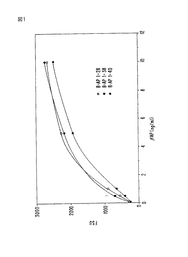
c.ポリクロナール抗体
ポリクロナール抗体を、合成ペプチドβAP1-38、βAP1-40、及びβAP1-42に対して作り、そして抗- βAP1-38( 抗血清Y)、抗- βAP1-40( 抗血清1280) 及び抗- βAP1-42( 抗血清HM) と命名した。ウサギを、皮膚内に完全Freund'sアジュバント中の( 非結合の) これらのペプチドの中の1 の0.5-3.0mg により免疫感作させた。ウサギは、高いタイターの抗- ペプチド反応性がそのウサギの血清のサンプル中に検出されることができるまで、一次免疫感作の3 週間後及びその後約2-4 週間間隔で0.1-0.5mg のペプチドのブースター注射を受けた。これらの抗血清を次に、1:300 〜1:1,500 のレンジにある希釈において免疫学的検定において使用した。
c. Polyclonal antibodies Polyclonal antibodies were made against the synthetic peptides βAP 1-38 , βAP 1-40 , and βAP 1-42 , and anti-βAP 1-38 (antiserum Y), anti-βAP 1-40 ( Antiserum 1280) and anti-βAP 1-42 (antiserum HM). Rabbits were immunized intradermally with 0.5-3.0 mg of one of these peptides (unbound) in complete Freund's adjuvant. Rabbits were administered 0.1-0.5
2. ELISA 検定
a. マイクロタイター・ウェルへの捕獲抗体の結合
モノクロナール抗体266 を、NaH 2 PO4 ・7 H 2 O , 26.2 g/L;NaN 3 , 1g/L; pH 8.3を含むバッファー中で、5 μl/mlまで、そしてモノクロナール抗体067 を、10μl/mlまで、希釈した。この溶液の100 μl/ウェルを次に、96ウェル・ポリスチレン透明COSTARプレート内に小分けし、そして室温において一夜インキュベートした。コーティング後、残った溶液を、吸引し、そして非- 特異的抗体結合部位を、NaH 2 PO4 ・H 2 O , 1g/L; NaH 2 PO4 ・7H 2 O , 10.8 g/L; NaN 3 , 0.5 g/L及びシュクロース, 25 g/L; pH 7.4を含むバッファー中に溶解した0.25% ヒト血清アルブミン(HSA) によりブロックした。これらのコート/ ブロックされたプレートを、直ちに使用し又はデシケータ−内で乾燥させ、そして最大5 日間、4 ℃において乾燥容器内で保存した。
2. ELISA assay
Binding of capture antibody to microtiter wells Monoclonal antibody 266 in 5 μl in buffer containing NaH 2 PO 4 .7 H 2 O, 26.2 g / L; NaN 3 , 1 g / L; pH 8.3 Monoclonal antibody 067 was diluted to 10 μl / ml. 100 μl / well of this solution was then aliquoted into 96-well polystyrene clear COSTAR plates and incubated overnight at room temperature. After coating, the remaining solution is aspirated and the non-specific antibody binding sites are removed from NaH 2 PO 4 .H 2 O, 1 g / L; NaH 2 PO 4 .7H 2 O, 10.8 g / L; NaN 3 0.5 g / L and sucrose, 25 g / L; blocked with 0.25% human serum albumin (HSA) dissolved in buffer containing pH 7.4. These coated / blocked plates were used immediately or dried in a desiccator and stored in a drying container at 4 ° C. for up to 5 days.
b. 検定プロトコール
既知量のβAPを含む換算物及び様々な体内又は体外液からのサンプルを、次に100 μl/ウェルにおいてそのプレートに添加した。これらのサンプルを、非希釈で又はNaH 2 PO4 ・H 2 O , 0.2 g/L; NaH 2 PO 4 ・7H 2 O , 2.16 g/L; NaN 3 , 0.5 g/L; BSA(グロブリン不含) 6 g/L; Triton X-405, 0.5 mL/L; NaCl, 8.5 g/L; pH 7.4を含むバッファー中に希釈して添加した。サンプル及び換算物を、室温において1 時間上記ウェエル内でインキュベートし、その後吸引し、そしてそれらのウェルを、NaCl, 80 g/L; KCl, 3.8 g/L; Tris塩基, 5.85 g/L; Tris HCl, 31.75 g/L;及び0.05% Tween(商標)20;pH 7.5(TBS) を含む300 μl/ウェルの溶液により洗浄した。
b. Assay Protocol The equivalents containing known amounts of βAP and samples from various in vivo or extracorporeal fluids were then added to the plate at 100 μl / well. These samples, non-diluted or NaH 2 PO 4 · H 2 O , 0.2 g / L; NaH 2
NHS-ビオチン(15mg)を0.25mlのジメチルスルホキシド中に溶解し、そして10μl のこの溶液を、1ml の炭酸ナトリウム溶液, 50mM, pH 8.5中に懸濁させた1mg の10D5又は6C6 抗体に添加した。この混合物を、暗所において1 1/2 時間室温においてインキュベートし、そして次にリン酸塩バッファー生理食塩水, pH 7.4に対して、48時間4 ℃において透析し、ビオチン化リポータ−抗体を作った。 NHS-biotin (15 mg) was dissolved in 0.25 ml dimethyl sulfoxide and 10 μl of this solution was added to 1 mg 10D5 or 6C6 antibody suspended in 1 ml sodium carbonate solution, 50 mM, pH 8.5. This mixture was incubated in the dark for 1 1/2 hours at room temperature and then dialyzed against phosphate buffered saline, pH 7.4 for 48 hours at 4 ° C. to make a biotinylated reporter antibody .
3 μg/mlに希釈した100 μl/ウェルのビオチン化リポーター抗体を、次にそれぞれのウェルに添加し、そして室温においてさらに1 時間インキュベートした。この抗体希釈剤は、Trizma塩基, 1.21 g/L; NaCl, 29.22 g/L; NaN 3 , 1.5 g/L; Triton X 405, 0.5 ml/L; PEG (Mw 3350), 40 g/L; MgCl2 ・6 H 2 O, 0.095 g/L; ZnCl2 , 0.014 g/L;ウシ胎児血清 100ml/L; 及びBSA 2.5 g/L, pH 7.4 から成っていた。
100 μl / well of biotinylated reporter antibody diluted to 3 μg / ml was then added to each well and incubated for an additional hour at room temperature. This antibody diluent is Trizma base, 1.21 g / L; NaCl, 29.22 g / L; NaN 3 , 1.5 g / L; Triton X 405, 0.5 ml / L; PEG (Mw 3350), 40 g / L; MgCl 2 · 6 H 2 O, 0.095 g / L; ZnCl 2 , 0.014 g / L; fetal
上記リポーター抗体(10D5 又は6C6)と共に室温において1 時間インキュベートした後、上清を吸引し、そして300 μl/ウェルのTBSにより3 回洗浄した。ストレプトアビジン・アルカリ性ホスファターゼ( 結合体希釈バッファー中で1:2000に希釈した100 μl/ウェル) を、添加し、そしてさらに1 時間室温においてインキュベートした。次にこの上清を吸引し、そして300 μl/ウェルのTBS により3回洗浄した。蛍光基質(2- アミノ-2- メチル・プロプラノーロル・バッファー; pH 9.5) (100μl/ウェル) 中の4-メチル- ウンベリフェリル・ホスフェートを添加し、そして蛍光を、360/40励起フィルター及び460/40放射フィルターを備えたMillipore からのCytofluor 2300を使用して15分後に読み、そして相対蛍光単位(FSU) として表した。 After 1 hour incubation at room temperature with the reporter antibody (10D5 or 6C6), the supernatant was aspirated and washed 3 times with 300 μl / well TBS. Streptavidin alkaline phosphatase (100 μl / well diluted 1: 2000 in conjugate dilution buffer) was added and incubated for an additional hour at room temperature. The supernatant was then aspirated and washed 3 times with 300 μl / well TBS. 4-Methyl-umbelliferyl phosphate in a fluorescent substrate (2-amino-2-methylpropranolol buffer; pH 9.5) (100 μl / well) was added, and fluorescence was measured using a 360/40 excitation filter and 460 / Read after 15 minutes using a Cytofluor 2300 from Millipore equipped with a 40 emission filter and expressed as relative fluorescence units (FSU).
3. 培養細胞
ヒト細胞( 及び他の哺乳動物からの細胞) を、プラスチック皿又はマルチ- ウェル・マイクロタイター・プレート内で標準的な細胞培養条件下で培養した。特に、ヒト胎児性腎臓癌腫293 細胞( 以下、K293細胞という。) を、10% ウシ胎児血清及び抗生物質を含むDulbecco's修飾Eagle's 培地(DMEM)中で増殖させた。β- アミロイド前駆体タンパク質(APP) の完全コーディング領域を含む組換えDNA 構築物により前もってトランスフェクトされたK293細胞を、非トランスフェクトK293細胞に加えて使用した(Selkoe et al. (1988) Proc. Acad. Sci. USA 85:7341-7345; 及びOltersdorf et al. (1990) J. Biol. Chem. 265:4492-4497)。トランスフェクトされた細胞は、K293細胞に特徴的な内生APP の普通の背景レベルに比べて高いレベルのAPP タンパク質を発現した。
3. Cultured cells Human cells (and cells from other mammals) were cultured under standard cell culture conditions in plastic dishes or multi-well microtiter plates. In particular, human fetal kidney carcinoma 293 cells (hereinafter referred to as K293 cells) were grown in Dulbecco's modified Eagle's medium (DMEM) containing 10% fetal bovine serum and antibiotics. K293 cells previously transfected with a recombinant DNA construct containing the complete coding region of β-amyloid precursor protein (APP) were used in addition to non-transfected K293 cells (Selkoe et al. (1988) Proc. Acad Sci. USA 85: 7341-7345; and Oltersdorf et al. (1990) J. Biol. Chem. 265: 4492-4497). The transfected cells expressed high levels of APP protein compared to the normal background level of endogenous APP characteristic of K293 cells.
ヒト臍静脈内皮細胞(HUVEC); DAMI と名付けられたヒト巨核球様白血球; チャイニーズ・ハムスター卵巣(CHO) 細胞、一次ヒト繊維芽細胞、及びヒト又はげっ歯類の脳から樹立された一次混合脳細胞カルチャー( ニューロン、星状膠細胞、及び小グリア細胞) を含む様々な他の細胞タイプをも培養した。 Human umbilical vein endothelial cells (HUVEC); human megakaryocyte-like leukocytes named DAMI; Chinese hamster ovary (CHO) cells, primary human fibroblasts, and primary mixed brain established from human or rodent brain Various other cell types were also cultured, including cell cultures (neurons, astrocytes, and microglia).
これらの様々な細胞系を、95% の酸素及び5%の二酸化炭素を含む組織培養インキュベーター内で37℃において増殖させた。細胞を規則的な間隔で新たな培養基を提供することにより日常的に継代した。細胞の周囲の細胞外液( ならし培地) を、標準的な休止条件又は細胞の以下の様々な生物化学的処理のいずれかの下で増殖した細胞から収穫した。すべての培養細胞及びそれらの誘導培地サンプルを、無菌条件下で取り扱った。 These various cell lines were grown at 37 ° C. in a tissue culture incubator containing 95% oxygen and 5% carbon dioxide. Cells were routinely passaged by providing new media at regular intervals. Extracellular fluid surrounding the cells (conditioned medium) was harvested from cells grown under either standard resting conditions or the following various biochemical treatments of the cells. All cultured cells and their induction media samples were handled under aseptic conditions.
免疫アフィニティー・クロマトグラフィー試験における使用のためのヒト- 混合脳細胞のカルチャーを以下のように調製した。胎児神経組織試料を12-14 週齢の胎児死体から得た。大脳皮質のサンプルを。Hank's Balanced Saline溶液(HBSS)により2 回濯いだ。皮質組織(2-3グラム) を10mlの冷HBSS中に入れ、これに1mg のDNase(Sigma Chemical Co., St. Louis, MO D3427)を添加した。粉砕懸濁液を、Pulliam et al. (1984) J. Virol. Met. 9:301により記載されているように、210 μm のその後130 μm のNitex ナイロン・スクリーンを通して濾過した。 A culture of human-mixed brain cells for use in immunoaffinity chromatography studies was prepared as follows. Fetal neural tissue samples were obtained from 12-14 week old fetal cadaver. A sample of the cerebral cortex. Rinse twice with Hank's Balanced Saline solution (HBSS). Cortical tissue (2-3 grams) was placed in 10 ml cold HBSS, to which 1 mg DNase (Sigma Chemical Co., St. Louis, MO D3427) was added. The milled suspension was filtered through 210 μm followed by 130 μm Nitex nylon screen as described by Pulliam et al. (1984) J. Virol. Met. 9: 301.
細胞を遠心分離により収穫し、そしてニューロン培地(10%ウシ胎児血清, 1%グルコース, 1mM Naピルベート, 1mM グルタミン, 20mM KClにより強化したMEM)中に再懸濁させた。ポリエチレンイミンによりコートされた100mm 皿に、8ml のニューロン培地中の1.5 x 10⁷ 細胞を植え付けた。この培地を収穫し、そして新たな培地を1 週間に2 回添加した。この細胞からのならし培地(HFBC-CM) を使用まで冷凍した。 Cells were harvested by centrifugation and resuspended in neuronal medium (MEM enriched with 10% fetal calf serum, 1% glucose, 1 mM Na pyruvate, 1 mM glutamine, 20 mM KCl). A 100 mm dish coated with polyethyleneimine was seeded with 1.5 × 10 7 cells in 8 ml of neuron medium. This medium was harvested and fresh medium was added twice a week. Conditioned medium from this cell (HFBC-CM) was frozen until use.
4. βAPのついての免疫沈降/ オートラジオグラフィー検定
a. 代謝標識付け及び免疫沈降
標準的な培養条件の下で増殖したK293細胞は、その培養基に35S-標識メチオニンを添加することにより新たに合成されたタンパク質の代謝的標識付けを経験した。この段階にわたり、培地は、非標識("コールド")メチオニンを全く含まないが、その他は、K293を培養するために使用された標準的な培地と同じであった。放射性メチオニンの量は、この標識付け実験において使用された培地1ml 当たり50-300μCiに変動した。その後、細胞から放出されたいずれかの放射標識付けされたタンパク質を含む培地を採取した。
4. Immunoprecipitation / autoradiography assay for βAP
a. Metabolic labeling and immunoprecipitation K293 cells grown under standard culture conditions experienced metabolic labeling of newly synthesized proteins by adding 35 S-labeled methionine to the culture medium. Over this stage, the medium did not contain any unlabeled ("cold") methionine, but was otherwise the same as the standard medium used to culture K293. The amount of radioactive methionine varied from 50-300 μCi per ml of medium used in this labeling experiment. Thereafter, media containing any radiolabeled protein released from the cells was harvested.
アミノ酸Asp-1 からVal-40までを含んで成る合成βAPペプチド( βAP1-40) に対して作られたポリクロナール抗体を、そのならし培地に添加し、そして2-10時間に変動する期間にわたりインキュベートした。これは、抗原- 抗体複合体がその培養基中に存在する抗- βAP抗体といずれかのβAPペプチドとの間に形成することを許容させた。その後、免疫グロブリン( 抗体) に結合することができるプロテインA-Sepharose(商標)試薬を、添加し、そしてこの混合物をさらに2-10時間の変動期間にわたりインキュベートした。このインキュベーションは、このプロテインA-Sepharose(商標)ビーズが抗- βAP抗体に結合し、これが次にβAPペプチドに結合することを可能にした。次にこのならし培地を12,000 xg において10分間遠心分離し、抗原- 抗体- プロテインA-Sepharose(商標)ビーズの複合体をペレット化した。 A polyclonal antibody raised against a synthetic βAP peptide (βAP 1-40 ) comprising amino acids Asp-1 to Val-40 is added to the conditioned medium and over a period varying from 2-10 hours Incubated. This allowed the antigen-antibody complex to form between the anti-βAP antibody present in the culture medium and any βAP peptide. Thereafter, Protein A-Sepharose ™ reagent capable of binding to immunoglobulin (antibody) was added and the mixture was incubated for an additional 2-10 hour variation period. This incubation allowed the protein A-Sepharose ™ beads to bind to the anti-βAP antibody, which in turn binds to the βAP peptide. The conditioned medium was then centrifuged at 12,000 xg for 10 minutes to pellet the antigen-antibody-protein A-Sepharose ™ bead complex.
b. 免疫沈降物のSDS-ポリアクリルアミド・ゲル電気泳動(PAGE)
代謝的に標識付けされた細胞からの培地の免疫沈降物を、低分子量のタンパク質( 例えば、βAP) をよく分離する利点をもつ、10-20% Tris-トリシン・ゲル上で電気泳動した。これらのゲルを次に、乾燥させ、そしてX-線フィルムに露出させて、オートラジオグラム又はフルオログラムを作った。露出時間は変動したが、普通には2-7 日間のレンジ内にあった。X-線フィルムの顕出後、抗- βAP抗体によりその細胞培地から沈降されたいずれかの放射標識付けされたタンパク質を、適当な分子量( すなわち、4kD)において暗バンドとして可視化した。
b. SDS-polyacrylamide gel electrophoresis (PAGE) of immunoprecipitates
Medium immunoprecipitates from metabolically labeled cells were electrophoresed on 10-20% Tris-Tricine gels with the advantage of well separating low molecular weight proteins (eg, βAP). These gels were then dried and exposed to X-ray film to make autoradiograms or fluorograms. Exposure times varied but were usually in the 2-7 day range. After the X-ray film was revealed, any radiolabeled protein precipitated from the cell culture medium with anti-βAP antibody was visualized as a dark band at the appropriate molecular weight (ie, 4 kD).
5. 266 樹脂の調製
抗体266(0.85 mg/mlにおいて15ml) を10mM Na アセテート, 15mM NaCl, pH 5.5 に対して透析し、そして次に約5ml の樹脂を使用して、製造者のプロトコールに従ってAffi-Gel(商標)Hz Hydrazide(Bio-Rad, Richmond, CA) に結合させた。この1ml の樹脂を、4 リッターのならし培地からβAPを精製するために 1 x 10 cmカラムに充填した。
5. Preparation of 266 resin Antibody 266 (15 ml at 0.85 mg / ml) is dialyzed against 10 mM Na acetate, 15 mM NaCl, pH 5.5, and then approximately 5 ml of resin is used according to the manufacturer's protocol. -Gel ™ Hz Hydrazide (Bio-Rad, Richmond, CA). This 1 ml of resin was loaded onto a 1 × 10 cm column to purify βAP from 4 liters of conditioned medium.
6. ウェスタン・ブロッティング
サンプルを、10%-20% Tricine ゲル(Novex) 上のSDS-PAGEに供し、そしてTowbin, et al. (1979) Proc. Natl. Acad. Sci. USA 76:4350-4354により記載されているバッファー系中で一夜40ボルトにおいてPVDF膜(Pro-blot, Applied Biosystems)に転移させた。免疫反応性タンパク質の可視化は、上記AMPPD 支持体についての製造者の指示に従ってTROPIX化学発光系を、使用した。使用した一次抗体は、5 μg/mlの濃度において10D5であった。
6. Western blotting samples were subjected to SDS-PAGE on a 10% -20% Tricine gel (Novex) and by Towbin, et al. (1979) Proc. Natl. Acad. Sci. USA 76: 4350-4354 Transfer to PVDF membrane (Pro-blot, Applied Biosystems) at 40 volts overnight in the described buffer system. Visualization of the immunoreactive protein used the TROPIX chemiluminescence system according to the manufacturer's instructions for the AMPPD support. The primary antibody used was 10D5 at a concentration of 5 μg / ml.
7. 'Swedish' FAD突然変異の構築及び分析
Swedish 突然変異は、2 つの隣接する以下の塩基対変換: ヌクレオチド1785のG からT 及びヌクレオチド1786のA からC であって以下のアミノ酸の変更: Lys->Asn595 及びMet->Leu596 ( 全番号はAPP 695 に基く。) を導くものを、含む。
7. Construction and analysis of 'Swedish' FAD mutation
The Swedish mutation consists of two adjacent base pair changes: nucleotides 1785 G to T and nucleotides 1786 A to C with the following amino acid changes: Lys-> Asn 595 and Met-> Leu 596 (all Numbers are based on APP 695. )
インビトロにおけるAPP の代謝に対するこの突然変異の生物化学的効果を分析するために、それは、(Selkoe et al., 1988前記中に記載されている)APP分子の真核生物発現のための発現ベクターにおけるインビトロ突然変異誘発により導入された。この場合において、APP の695 及び751 アミノ酸形態を担持するベクターの両形態が使用された。突然変異誘発は、APP 配列から誘導されたオリゴヌクレオチド・プライマー及びポリメラーゼ連鎖反応(PCR) の使用により行われた。プライマー1(センス) は、30-merであり、そして配列GAG GAG ATC TCT GAA GTG AAT CTG GAT GCA をもつ。 In order to analyze the biochemical effect of this mutation on APP metabolism in vitro, it is expressed in an expression vector for eukaryotic expression of APP molecules (described in the above). Introduced by in vitro mutagenesis. In this case, both forms of the vector carrying the 695 and 751 amino acid forms of APP were used. Mutagenesis was performed by the use of oligonucleotide primers and polymerase chain reaction (PCR) derived from the APP sequence. Primer 1 (sense) is 30-mer and has the sequence GAG GAG ATC TCT GAA GTG AAT CTG GAT GCA.
このプライマーは、APP の1770位内のBglII 部位に対応するBglII 制限エンドヌクレアーゼ部位(AGA TCT) を含み、そしてそのプライマーの24及び25位に2 つの先に記載したヌクレオチド交換を含む。プライマー2(アンチ- センス) は、配列AAT CTA TTC ATG CAC TAG TTT GAT ACA GCをもつ29-merである。このプライマーは、APP の2360位に対応するSpeI制限エンドヌクレアーゼ部位(ACT AGT) を含む。テンプレートのための正常APP cDNAを使用して、この2 つのプライマーは、標準的なPCR(Perkin Elmerからの試薬及びプロトコール) による長さ約600 塩基対のDNA 断片の創出を許容する。得られた断片並びにその正常APP cDNAを含む発現ベクターは、制限エンドヌクレアーゼBglII 及びSpeIにより解裂された。 This primer contains a BglII restriction endonuclease site (AGA TCT) corresponding to the BglII site within position 1770 of APP and contains the two previously described nucleotide exchanges at positions 24 and 25 of the primer. Primer 2 (anti-sense) is a 29-mer with the sequence AAT CTA TTC ATG CAC TAG TTT GAT ACA GC. This primer contains a SpeI restriction endonuclease site (ACT AGT) corresponding to position 2360 of APP. Using normal APP cDNA for the template, the two primers allow the creation of a DNA fragment approximately 600 base pairs in length by standard PCR (reagents and protocols from Perkin Elmer). The resulting fragment and the expression vector containing its normal APP cDNA were cleaved with the restriction endonucleases BglII and SpeI.
BglII 及びSpeIが選ばれた。なぜなら、それらが、両方とも、このベクター内の単一切断部位であり、そしてそれ故にPCR により創出される断片に対応する非- 突然変異制限断片の単一除去を許容するからである。従って、約600 塩基対のベクター断片が、標準的な技術によりその突然変異を担持する等しい長さのPCR 生成断片により置換された。組換え体バクテリア・クローンのDNA は標準的な方法により得られ、これを、Mbo II制限エンドヌクレーゼ部位の非存在についてスクリーンした。次にそのDNA 配列を、上記PCR 反応を経験した完全領域の配列決定により確認した。 BglII and SpeI were chosen. This is because they are both single cleavage sites within this vector and thus allow single removal of non-mutated restriction fragments corresponding to the fragments created by PCR. Thus, an approximately 600 base pair vector fragment was replaced by an equal length PCR-generated fragment carrying the mutation by standard techniques. Recombinant bacterial clone DNA was obtained by standard methods and screened for the absence of the Mbo II restriction endonuclease site. The DNA sequence was then confirmed by sequencing the complete region that experienced the PCR reaction.
この二重突然変異の構築に加えて、上記2 つの突然変異のそれぞれの効果を、別々に検査した。適当なプライマー又は上記595 Lys->Asn置換又は596 Met->Leu置換のいずれかに特異的なオリゴヌクレオチドを使用して、DNA 構築物を調製し、そして先に記載したようなK293細胞をトランスフェクトするために使用した。 In addition to constructing this double mutation, the effect of each of the two mutations was examined separately. DNA constructs were prepared using appropriate primers or oligonucleotides specific for either the 595 Lys-> Asn substitution or the 596 Met-> Leu substitution described above, and transfected with K293 cells as described above. Used to do.
この突然変異の効果の分析を、293 細胞における得られた突然変異クローンの過渡的発現により行った。DOTAP 試薬トランスフェクション法を、製造者の指示(Boehringer Mannheim, Indianapolis,IN) に従って使用した。ならし培地を、トランスフェクションの48時間後に収穫した。トランスフェクション効率を、APP 444-592 のバクテリア融合タンパク質に対するアフィニティー精製ポリクロナール抗体B5( ビオチン化) 及びAPP 20-304のバクテリア融合タンパク質に対する捕獲抗体B3による、ならし培地中のAPP についてのサンドイッチELISA により検定した。βAPレベルを先の実施例2 中に記載したようなELISA により測定した。以下に記載するように、APP のサンドイッチ変異体を発現する過渡的トランスフェクトされたK293細胞のならし培地中のβAPの測定は、βAPの生産における6-7倍の増加を示している。 Analysis of the effect of this mutation was performed by transient expression of the resulting mutant clone in 293 cells. The DOTAP reagent transfection method was used according to the manufacturer's instructions (Boehringer Mannheim, Indianapolis, IN). Conditioned media was harvested 48 hours after transfection. The transfection efficiency depends on the capture antibody B3 on bacteria fusion protein affinity-purified polyclonal antibodies B5 (biotinylated) and APP 20-304 against bacterial fusion protein of APP 444-592, leveling assayed by sandwich ELISA for APP in the medium did. βAP levels were measured by ELISA as described in Example 2 above. As described below, the measurement of βAP in the conditioned medium of transiently transfected K293 cells expressing a sandwich variant of APP shows a 6-7 fold increase in βAP production.
結果
βAP ELISA検定を、合成βAPペプチドβAP1-38及びβAP1-40の既知量を検出するために使用した。266 捕獲抗体及び10D5レセプタ抗体を使用する検定は、0.1 ng/ml においてそれらのペプチドを検出することができた。図1 を参照のこと。その上、この266/10D5検定は、完全長APP 、分泌APP 695 又は751 、組換え体APP 断片構築物15又は6 、βAP断片15-20 、11-18 、13-18 、又は13-20 あるいはフィブリノーゲンと有意に交差反応しないことが判明した( 表1)。
Results The βAP ELISA assay was used to detect known amounts of the synthetic βAP peptides βAP 1-38 and βAP 1-40 . Assays using 266 capture antibody and 10D5 receptor antibody were able to detect these peptides at 0.1 ng / ml. See Figure 1. In addition, this 266 / 10D5 assay can be used for full length APP, secreted
ELISA 検定を、ヒト、イヌ、モルモット、及びラットからの血液及びCSF サンプルをスクリーンするために使用した。βAPの検出可能な量を、表II中に述べるレンジ内の濃度により、見いだした。 An ELISA assay was used to screen blood and CSF samples from humans, dogs, guinea pigs, and rats. The detectable amount of βAP was found by the concentration within the range described in Table II.
比較試験を、血漿及びCSF 中の両方において、正常個体及びAD患者の群において行った。サンプルを、ELISA 検定を使用してスクリーンし、血漿についての結果を図2のA中に示し、そしてCSF についての結果を図2のB中に示す。正常個体(C) 、AD患者、脳血管事故(CVA)、及びパーキンソン病(PD)についてのCSF 中のβAPレベルを、図3中に示す。約1000個体のCSF プーは、図3 、C 列の中丸で囲んだようにCSF 中2.5 ng/ml の平均値をもっていた。対照個体の残りは、様々な非-AD ニューロン変性疾患をもっていた。ADの平均値は、対照プール値を十分に上回っていた。 A comparative study was performed in groups of normal individuals and AD patients, both in plasma and CSF. Samples were screened using an ELISA assay, the results for plasma are shown in FIG. 2A and the results for CSF are shown in FIG. 2B. The βAP levels in the CSF for normal individuals (C), AD patients, cerebrovascular accidents (CVA), and Parkinson's disease (PD) are shown in FIG. Approximately 1000 CSF pooh had an average value of 2.5 ng / ml in CSF as circled in Figure 3, column C. The rest of the control individuals had various non-AD neuronal degenerative diseases. The mean value for AD was well above the control pool value.
トランスフェクトされた及び非- トランスフェクトされた培養細胞の数を、ELISA 検定を使用してβAPの放出についてテストした。すべての細胞が、その培養基中にβAPを放出することが判明し、APP トランスフェクト細胞は、表III 中に示すように非トランスフェクト培養細胞から放出されるよりも高い濃度を放出している。 The number of transfected and non-transfected cultured cells was tested for βAP release using an ELISA assay. All cells were found to release βAP in the culture medium, and APP transfected cells released higher concentrations than were released from non-transfected cultured cells as shown in Table III.
実施例4 中に先に記載した免疫沈降/ オートラジオグラフ検定を使用して、ポリクロナール抗体1280が、放射標識付けされた合成βAP1-40ペプチドの標準サンプルにより正確に同時移動したK293細胞培地からの4 kDタンパク質を免疫沈降させたことが示された。さらに3 kDタンパク質が同時に沈降した。これは、最初の10又は16アミノ酸を欠いたβAPの断片であるようである。この1280抗体が合成βAP1-40ペプチドにより前吸収されてその活性をブロックするとき、3 kD又は4 kDバンドのいずれもK293細胞培地からは沈降されなかった。 Using the immunoprecipitation / autoradiographic assay described above in Example 4, polyclonal antibody 1280 was extracted from K293 cell medium accurately co-transferred with a standard sample of radiolabeled synthetic βAP 1-40 peptide. Of 4 kD protein was immunoprecipitated. In addition, 3 kD protein was precipitated simultaneously. This appears to be a fragment of βAP lacking the first 10 or 16 amino acids. When this 1280 antibody was preabsorbed by the synthetic βAP 1-40 peptide to block its activity, neither the 3 kD nor the 4 kD band was precipitated from the K293 cell medium.
APP を過剰発現するK293細胞のならし培地が100,000 xgにおいて2 時間遠心分離され( 不溶性タンパク質材料のいずれをもペレット化させ) るとき、その1280抗体による免疫沈降/ オートラジオグラフは、その3 kD又は4 kDタンパク質の実質的にすべてが、その上清中に残ることを示した( 図6 を参照のこと。) 。この実験は、培養基中にあるβAPが、死後のヒトの脳組織中のβAPについての先の報告( 例えば、Glenner and Wong (1984),前記を参照のこと。) に反して、可溶性分子であることを証明している。 When conditioned medium of K293 cells overexpressing APP is centrifuged at 100,000 xg for 2 hours (with any insoluble protein material pelleted), its 1280 antibody immunoprecipitation / autoradiograph shows its 3 kD Or showed that substantially all of the 4 kD protein remained in the supernatant (see Figure 6). This experiment shows that βAP in the culture medium is a soluble molecule, contrary to previous reports on βAP in postmortem human brain tissue (see, eg, Glenner and Wong (1984), supra). Prove that.
βAPP cDNAによりトランスフェクトされたK293細胞の培地からの4 kD βAP同時移動ペプチドの沈降は、追加のβAP抗体を使用することにより確認された。合成βAP1-38ペプチドに対する抗体は、抗体1280と同一のやり方で4 kDタンパク質を沈降させた。また、合成βAP1-42に対する抗体HMは、4 kDタンパク質を沈降させた。対照として、これらの抗体のそれぞれをその合成ペプチド抗原により前吸収させ、それによりその活性を中和させた。その後、これらの抗体は、もはや、ならし培地から 4 kD βAPペプチドを免疫沈降させなかった。追加の対照として、免疫前血清( すなわち、実際の免疫感作に先立ってポリクロナール抗体を作り出すために使用されたそれぞれのウサギから採取した正常血清のサンプル) は、その培地から4 kDペプチドを沈降させなかった。 Precipitation of 4 kD βAP co-migrating peptide from the medium of K293 cells transfected with βAPP cDNA was confirmed by using an additional βAP antibody. Antibodies against the synthetic βAP 1-38 peptide precipitated the 4 kD protein in the same manner as antibody 1280. In addition, antibody HM against synthetic βAP 1-42 precipitated 4 kD protein. As a control, each of these antibodies was preabsorbed with its synthetic peptide antigen, thereby neutralizing its activity. Thereafter, these antibodies no longer immunoprecipitated 4 kD βAP peptide from the conditioned medium. As an additional control, preimmune serum (i.e., a sample of normal serum taken from each rabbit used to generate polyclonal antibodies prior to actual immunization) precipitates 4 kD peptide from the medium. There wasn't.
培地から沈降されたペプチドと同時移動する4 kD βAPの免疫化学的特異性を確認するために、そのβAP領域に隣接するAPP の領域に対する他の抗体を先の免疫沈降/ オートラジオグラフ検定において使用した。例えば、APP のβAP領域の開始のアミノ末端の20アミノ酸領域に対する抗体は、培地から4 kDペプチドを沈降させることに失敗した。同様に、( βAP領域から60アミノ酸先の)APPのカルボキシル末端における最後の20アミノ酸に対する抗体も、その4 kDペプチドを沈降させることに失敗した。 To confirm the immunochemical specificity of 4 kD βAP migrating with peptide precipitated from the medium, other antibodies to the APP region adjacent to its βAP region were used in previous immunoprecipitation / autoradiographic assays. did. For example, an antibody against the amino-terminal 20 amino acid region at the start of the βAP region of APP failed to precipitate a 4 kD peptide from the medium. Similarly, an antibody against the last 20 amino acids at the carboxyl terminus of APP (60 amino acids beyond the βAP region) also failed to precipitate the 4 kD peptide.
反対に、βAPの最初の15残基に対する抗体は、首尾よく4 kDペプチドを沈降させた。同様に、βAPの中央部分に対する抗体もその4 kDペプチドを沈降させたが、先に討議した3 kDペプチドを沈降させなかった。これらの様々な抗体は、培養細胞( 例えば、K293細胞) の培地中に存在する4 kDがβAPに特徴的な特異的な免疫化学的反応性を示すことを証明している。培地中の3 kDペプチドは、最初の10又は16残基を欠いているβAPの特異的免疫化学的反応性を示している。 In contrast, antibodies against the first 15 residues of βAP successfully precipitated the 4 kD peptide. Similarly, the antibody against the central part of βAP also precipitated its 4 kD peptide, but not the previously discussed 3 kD peptide. These various antibodies demonstrate that 4 kD present in the culture medium of cultured cells (eg, K293 cells) exhibits specific immunochemical reactivity characteristic of βAP. The 3 kD peptide in the medium shows the specific immunochemical reactivity of βAP lacking the first 10 or 16 residues.
それぞれの反応において抗体が活性であったという証拠は、沈降反応がAPP のその領域に対する抗体により行われるときにはいつでも("可溶性APP"又は"APPS " といわれる)APPの正常分泌断片の同一培地から同時- 沈降したことにより提供された。この大きな可溶性APP 断片は、APP を発現する培養細胞の培地中に正常に存在することが知られている。これ故、抗- βAP抗体によるその沈降は、本検定において使用された抗体の無傷の活性を証明する陽性対照反応を提示している。 Evidence that the antibody was active in each reaction is that from the same medium of normal secreted fragments of APP (referred to as “soluble APP” or “APP S ”) whenever a precipitation reaction is performed with antibodies against that region of APP. Provided by co-sedimentation. This large soluble APP fragment is known to be normally present in the culture medium of cultured cells expressing APP. Therefore, its precipitation with anti-βAP antibody presents a positive control reaction that demonstrates the intact activity of the antibody used in this assay.
合成βAPと同時移動する免疫沈降した4 kDタンパク質が実際に本物のβAPを現しているということを証明するための追加の対照反応として、APP cDNAによりトランスフェクトされ( そしてそれ故にAPP を過剰発現する) 細胞の培地を、非トランスフェクト細胞の培地と比較した。免疫沈降/ オートラジオグラフィーは、APP 前駆体分子のそれらの増加した生産から予想されるように、トランスフェクト対非トランスフェクト細胞の培地中の沈降可能な4 kDタンパク質の増加した量を示した。図5 を参照のこと。同様に、トランスフェクトされた細胞の培地は、非トランスフェクト細胞の培地よりも多くのAPP のAPP S 可溶性断片を示し; この陽性対照反応は、その4kDタンパク質を含む免疫沈降物中に同時に観察された。 As an additional control reaction to demonstrate that the immunoprecipitated 4 kD protein that co-migrates with synthetic βAP actually represents authentic βAP, it was transfected with APP cDNA (and therefore overexpressed APP) ) Cell culture medium was compared to non-transfected cell culture medium. Immunoprecipitation / autoradiography showed an increased amount of sedimentable 4 kD protein in the media of transfected versus non-transfected cells, as expected from their increased production of APP precursor molecules. See Figure 5. Similarly, the transfected cell medium shows more APP S soluble fragments of APP than the non-transfected cell medium; this positive control reaction is observed simultaneously in the immunoprecipitate containing its 4 kD protein. It was.
異なる細胞タイプ、チャイニーズ・ハムスター卵巣(CHO) 細胞が使用されたときに上記と同一の結果が得られた。非トランスフェクト又はAPP cDNAによりトランスフェクトされたかのいずれかのCHO細胞の比較は、後者の細胞の培地中の4 kD 1280-沈降性ペプチドの増加したレベルを示した。これらの結果は、その細胞培地中の4 kDタンパク質が真のβAPであるさらなる証拠を提供した。 Identical results were obtained when different cell types, Chinese hamster ovary (CHO) cells, were used. Comparison of CHO cells, either untransfected or transfected with APP cDNA, showed increased levels of 4 kD 1280-precipitating peptide in the media of the latter cells. These results provided further evidence that the 4 kD protein in the cell culture medium is a true βAP.
本物で生来のβAPは、ADにより死んだ患者の検死ヒト大脳皮質から抽出された。AD脳組織からのA βのこのサンプルは、SDS-ポリアクリルアミド・ゲル電気泳動により分析されるとき培養細胞培地から4 kD 1280-沈降性ペプチドと同時移動した。この同時移動は、βAPとしてのこの4 kDペプチドの同定のためのさらなる支持を提供した。 Genuine and native βAP was extracted from autopsy human cortex of patients who died from AD. This sample of Aβ from AD brain tissue co-migrated with 4 kD 1280-precipitating peptide from cultured cell medium when analyzed by SDS-polyacrylamide gel electrophoresis. This co-migration provided further support for the identification of this 4 kD peptide as βAP.
4 リッターのヒト胎児脳培養- ならし培地(HFBC-CM) を、解凍し、そして0.45μm 濾過フラスコを通して濾過した。イソプロパノール中の35 mg/ml保存液から採取したロイペプチン(1μg/ml) 及びPMSF(35 μg/ml) を、アフィニティー・クロマトグラフィーの直前にこのHFBC-CM に添加した。この材料を、4 ℃において約2 ml/ 分の流速において266-アフィニティー・カラムを通して走らせた。次にこのカラムを500ml のPBS により洗浄した。その樹脂に特異的に結合した材料の溶出を、0.2 M グリシン pH 2.0 により達成した。全部で9ml を使用した。 Four liters of human fetal brain culture-conditioned medium (HFBC-CM) was thawed and filtered through a 0.45 μm filter flask. Leupeptin (1 μg / ml) and PMSF (35 μg / ml) taken from a 35 mg / ml stock in isopropanol were added to the HFBC-CM just prior to affinity chromatography. This material was run through a 266-affinity column at a flow rate of about 2 ml / min at 4 ° C. The column was then washed with 500 ml PBS. Elution of the material specifically bound to the resin was achieved with 0.2 M glycine pH 2.0. A total of 9 ml was used.
溶出した材料を、Vydac C4[0.21 x 15 cm]逆相カラム及びバッファー"A" 中に0.1% TFA及びバッファー"B" 中に0.1% TFA/80%アセトニトリルを含む溶媒系を使用する逆相液体クロマトグラフィーの2段階に供した。上記アフィニティー精製された材料を、200 μl/分において上記逆相カラム上に供給し、そして次に200 μl/分において60分間、そして50μl/分において42分間、80% バッファー(A) 及び20% バッファー"B" により洗浄して、そのカラムを平衡化し、そしてそのベースラインを安定化させた。 Reversed phase liquid using a Vydac C4 [0.21 x 15 cm] reverse phase column and a solvent system containing 0.1% TFA in buffer “A” and 0.1% TFA / 80% acetonitrile in buffer “B”. Subjected to two stages of chromatography. The affinity purified material is fed onto the reverse phase column at 200 μl / min and then for 60 minutes at 200 μl / min and 42 minutes at 50 μl / min, 80% buffer (A) and 20% Wash with buffer “B” to equilibrate the column and stabilize the baseline.
20% から70% までの"B" のグラジエントを50μl / 分において50分間にわたり行い、そしてその溶出液をOD220 においてモニターした。100 μl の画分を集め、そしてウェスタン・ブロット及びELISA の両方により検定した。これらの結果に基づき、画分11及び12をプールし、そしてほとんど同じ条件下で再クロマトグラフィーにかけた。但し、10から40% までの"B" の150 分間のグラジエントを、その後の40から100%までの"B"の20分間のグラジエントを、使用した。
A gradient of “B” from 20% to 70% was performed at 50 μl / min for 50 minutes and the eluate was monitored at OD 220 . 100 μl fractions were collected and assayed by both Western blot and ELISA. Based on these results,
第二逆相クロマトグラフィー段階からの画分77は、ウェスタン・ブロット( 図4)及びELISA の両方により上記10D5抗体に対して反応性であることが見つかった。
画分77中の上記材料のアリコットは、マイクロ配列決定され、そしてAsp に始まるβAPのN-末端配列をもつことが示された。マイクロ配列決定を、マイクロスケール反応カートリッジ及びApplied Biosystems's MICFST プログラム・サイクロを使用したApplied Biosystems Model 477タンパク質シーケンサー上で行った。
Fraction 77 from the second reverse phase chromatography step was found to be reactive against the 10D5 antibody by both Western blot (Figure 4) and ELISA.
An aliquot of the above material in fraction 77 was microsequenced and shown to have an N-terminal sequence of βAP starting at Asp. Microsequencing was performed on an Applied Biosystems Model 477 protein sequencer using a microscale reaction cartridge and an Applied Biosystems's MICFST program cyclo.
第二アリコットは、M-SCAN, Inc.において行われた電子スプレー・イオン化質量分析に供され、βAP 1-40 の予想質量(4330.4 の理論分子量) に一致するであろう4329.81 (+/- 1.27 標準誤差) における質量ピークを現した。A 220 ピークの幾つかのN-末端配列決定は、βAPについて陽性であった。ELISA は、画分77中の主要ピークに加え画分65、70、75中にβAP配列が存在することを現した。画分65及び70は、βAP残基11(Glu) に始まる先に記載していないβAP断片の追加の配列を含んでいた。画分65中、βAP残基33を通して配列決定するのに十分な材料が存在していた。
The second aliquot was subjected to electrospray ionization mass spectrometry performed at M-SCAN, Inc. and would correspond to the expected mass of βAP 1-40 (theoretical molecular weight of 4330.4) 439.881 (+/- 1.27 The mass peak at (standard error). Some N-terminal sequencing of the A 220 peak was positive for βAP. ELISA revealed the presence of βAP sequences in
HFBC-CM からの266-アフィニティー精製材料のRPLC画分のウェスタン・ブロット( 図4)。3 μl の同一画分を、15μl のSDS-PAGEサンプル・バッファーにより希釈し、そして先に記載したような煮沸及びプロセシング前に1 μl の1 M NaOHにより中和した。それぞれのレーン内にロードされた材料は以下のものであった: Western blot of RPLC fraction of 266-affinity purified material from HFBC-CM (Figure 4). 3 μl of the same fraction was diluted with 15 μl of SDS-PAGE sample buffer and neutralized with 1 μl of 1 M NaOH before boiling and processing as described above. The material loaded in each lane was:
レーン12はバッファーだけであり; レーン13は20 ng のβAP1-38を含み; レーン14は100 ngのβAP1-38を含み; レーン15は低分子量のRainbow(商標)標準(Amersham)を含んでいた。レーン6 中の約 4 kDバンドがレーン14中のβAP標準と同時移動することに注目のこと。
HFBC-CM のために記載されたものに並行な実験を、この源からのβAPの免疫反応性を構造的に特徴付けるために4 リッターのヒトCSF を使用して行った。配列決定データは、βAP残基1(Asp)及びβAP残基11(Glu) に始まるN-末端配列の存在を確信させた。
Experiments parallel to those described for HFBC-CM were performed using 4 liters of human CSF to structurally characterize the immunoreactivity of βAP from this source. Sequencing data confirmed the presence of an N-terminal sequence beginning at βAP residue 1 (Asp) and βAP residue 11 (Glu).
Swedish トランスフェクト体の培地中のβAPレベル中の増加を、モノクロナール抗体266 及び6C6 によるβAP- 特異的ELISA を使用して定量した。APP S についてのサンドイッチELISA は、ポリクロナール抗体B5及びB3を使用した。それぞれのELISA のために、精製された合成βAP1-40又はそのAPP 695 イソ形態のためのcDNAによりトランスフェクトされたK293細胞のならし培地からの精製APP S の増加量を、標準曲線を構築するために使用した。過渡的にトランスフェクトされたK293細胞におけるならし培地中のβAP( 左パネル)及びAPP S ( 右パネル) の定量を、図7 中に示す。それぞれのカラムは、3 つの培養に基く擬似カラムを除き、4 つのトランスフェクションの平均を表す。誤差バーは、その標準偏差を示している。誤差バーのないカラムについては、その標準偏差は、0.01単位未満であった。
The increase in βAP levels in the Swedish transfectant medium was quantified using a βAP-specific ELISA with monoclonal antibodies 266 and 6C6. The sandwich ELISA for APP S used polyclonal antibodies B5 and B3. For each ELISA, construct an increased amount of purified APP S from conditioned medium of K293 cells transfected with purified synthetic βAP 1-40 or cDNA for its
図7 中に示すように、Swedish 変異体APP 695 構築物を発現する細胞は、正常APP(695 イソ形態) を発現する同一のトランスフェクトされた細胞よりもそれらの培地中で6-7 倍多くのβAPを生産した。その上、同様の7-8 倍の増加が、APP 751 イソ形態におけるSwedish 突然変異を発現するカルチャーにおいて観察された( 図7)。βAPレベルにおける同様の増加が、第二の定量法: 1280免疫沈降物のゲル中の4 kD βAPバンドの燐光体画像分析(phosphor imager analysis)を使用して証明された。この方法は、3 kD断片がSwedish トランスフェクト体の培地中で数倍減少することをさらに証明した。CHO 及びK293細胞はcDNAにより安定してトランスフェクトされ、APP751 イソ形態におけるSwedish 突然変異も、それらの培地中のβAPのレベルにおける顕著な増加を示した。
As shown in Figure 7, cells expressing the
増加したβAP生産に原因である機構を研究するために、APP 695イソ形態におけるそれぞれの単一突然変異(Lys->Asn 595 及びMet->Leu596 ) の効果を、別々に調査した。図8 は、DNA なし( レーン1); 正常APP(レーン2)、Swedish 突然変異APP 695 KM-NL(レーン3); 変異体APP 695 K-N(レーン4); 変異体APP 695 M-L(レーン5)、APP ΔC (APP細胞質ドメイン欠失、レーン6)及びAPP ΔC KM-NL(Swedish 突然変異及び細胞質ドメイン欠失、レーン7)により過渡的にトランスフェクトされた、そして1280により免疫沈降された放射標識付けK293細胞のならし培地を示している。このβAP及び3 kDバンドは、矢印により示されている。
To study the mechanism responsible for increased βAP production, the effect of each single mutation (Lys-> Asn 595 and Met-> Leu 596 ) in the
125 I-標識合成βAP(1-40)を、同一ゲル上のサイズ・マーカーとして走らせた( レーン8)。Met->leu置換を発現する細胞は、それらの培地中でβAPのレベルを増加させ、一方、Lys->Asn置換を発現する細胞は、正常のトランスフェクト体と同様のレベルをもっていた( 図8 、レーン2-5)。この発見は、596 突然変異がその正常のMet-Asp 結合におけるよりもそのLeu-Aspペプチド結合においてAPP のよりタンパク質分解性の解裂をもたらすということを示唆している。595 におけるLys->Asnスイッチは596 におけるMet->Leu置換と結合するときに上記の解裂をさらに強化することができるという可能性がある。 125 I-labeled synthetic βAP (1-40) was run as a size marker on the same gel (lane 8). Cells expressing Met-> leu substitutions increased βAP levels in their medium, while cells expressing Lys-> Asn substitutions had levels similar to normal transfectants (Figure 8). Lanes 2-5). This finding suggests that the 596 mutation results in a more proteolytic cleavage of APP at its Leu-Asp peptide bond than at its normal Met-Asp bond. It is possible that the Lys-> Asn switch at 595 can further enhance the above cleavage when combined with the Met-> Leu substitution at 596.
APP 欠失の細胞質ドメインの欠失と一緒に上記Swedish 突然変異を含み、それ故にAsn-Pro-X-Tyr リソゾーム標的コンセンサス配列を除去されている細胞は、未だ、正常な形質転換体よりもそれらの培地中に実質的により多くのβAPを生産するが、3 kDペプチドの増加したレベルを示した( 図8 、レーン6 及び7)。この結果は、Swedish 突然変異の効果が無傷の細胞質ドメインを必要とせず、そしてAPP の生成が後期エンドソーム/ リソゾーム内でのAPP のプロセシングを必要とするようであることを、示している。
Cells that contain the above-mentioned Swedish mutation along with the deletion of the cytoplasmic domain of APP deletion and therefore have the Asn-Pro-X-Tyr lysosomal targeting consensus sequence removed are still more likely than normal transformants. Produced substantially more βAP in this medium but showed increased levels of 3 kD peptide (Figure 8,
これらの発見は、FAD 族内にあるAPP 遺伝子における点突然変異が直接的にβAPの増加した生成をもたらすことができるという実験的な証拠を提供する。Swedish 突然変異の分析は、ADの家族性形態における加速されたβ- アミロイド症の機構を明らかにするためだけでなく本発明に係る方法におけるAPP 変異体の使用のための経路として、特定の突然変異( 例えば、βAP領域直後の残基717 における突然変異及びβAP領域内のもの) を担持するAPP からのインビトロにおけるβAP生産の使用を証明している。 These findings provide experimental evidence that point mutations in the APP gene within the FAD family can directly lead to increased production of βAP. The analysis of Swedish mutations has identified specific sudden as a pathway for the use of APP variants in the methods of the present invention as well as to elucidate the mechanism of accelerated β-amyloidosis in familial forms of AD. It demonstrates the use of in vitro βAP production from APP carrying a mutation (eg, a mutation at residue 717 immediately after the βAP region and that in the βAP region).
Swedish 突然変異をプロセシングする細胞系におけるβAP生産を阻害することができるテスト化合物のためのスクリーニング検定を、以下のように行った。2 つの細胞系( ヒト腎臓細胞系K293及びチャイニーズ・ハムスター卵巣細胞系(CHO))を、Citron et al. (1992) Nature 360:672-674 中に記載されている方法を使用して、一般的にSwedish 突然変異といわれるLys-651-Met-652 からAsn-651-Leu-652 への2 重突然変異(APP-751番号付け) を含むAPP751のための遺伝子により安定してトランスフェクトされた。 Screening assays for test compounds capable of inhibiting βAP production in cell lines that process Swedish mutations were performed as follows. Two cell lines (human kidney cell line K293 and Chinese hamster ovary cell line (CHO)) are commonly used using the method described in Citron et al. (1992) Nature 360: 672-674. It was stably transfected with a gene for APP751 containing a double mutation (APP-751 numbering) from Lys-651-Met-652 to Asn-651-Leu-652, said to be a Swedish mutation.
このトランスフェクトされた細胞系は、293 751 SWE 及びCHO 751 SWE と命名され、そしてそれぞれ、10% ウシ胎児血清を添加されたDulbecco's最小必須培地中にウェル当たり2.5 x 10⁴ 細胞においてCorning 96ウェル・プレート内にプレートされた。10% 二酸化炭素(CO 2 ) により平衡化されたインキュベーター内で37℃における一夜のインキュベーションの後、その培地を取り出し、そしてウェル当たり200 μl の、テスト・プロテアーゼ阻害剤を含む培地により置き換えた。2 時間の前処理期間の後、培地を再び取り出し、そして同一のプロテアーゼ阻害剤を含む新たな培地により置き換え、そして細胞を、さらに2 時間にわたりインキュベートした。
The transfected cell lines are designated 293 751 SWE and
プロテアーゼ阻害剤保存液を、その処理において使用した最終濃度において、DMSOの濃度が0.5%を超えないようにDMSO中で調製した。処理後、プレートを室温において5 分間1200rpm においてBeckmanGPR 内で遠心分離してそのならし培地から細胞死骸をペレット化した。それぞれのウェルから、100 μl のならし培地を、先に記載したようにβAP-13-28に対する抗体266 により前コートしたELISA プレート内に移し、そして4 ℃において一夜保存した。( βAP-1-16に対する) 標識抗体6C6 を使用するELISA 検定を、生産されたβAPの量を測定するために次の日に走らせた。 Protease inhibitor stock solutions were prepared in DMSO such that the concentration of DMSO did not exceed 0.5% at the final concentration used in the treatment. After treatment, the plates were centrifuged in a Beckman GPR at 1200 rpm for 5 minutes at room temperature to pellet cell dead bodies from the conditioned medium. From each well, 100 μl of conditioned medium was transferred into an ELISA plate pre-coated with antibody 266 against βAP-13-28 as described above and stored at 4 ° C. overnight. An ELISA assay using labeled antibody 6C6 (to βAP-1-16) was run the next day to measure the amount of βAP produced.
その化合物の細胞毒性効果を、Hansen et al. (1989) J. Immun. Meth. 119:203-210の方法の修正により測定した。その組織培養プレート内に残る細胞に、1mg/mlの最終濃度まで25μl の3,(4,5- ジメチルチアゾール-2- イル)2,5- ジフェニルテトラゾリウム・ブロマイド(MTT) 保存溶液(5 mg/ml) を添加した。細胞を37℃において1 時間インキュベートし、そして細胞活動を、等量のMTT 溶解バッファー(50%DMF 中の20w/v%ドデシル硫酸ナトリウム, pH 4.7) の添加により終了させた。OD562nm 及びOD650nm における差異を、その細胞活性の指標としてMolecular Devices UVmax マイクロプレート・リーダー内で測定した。 The cytotoxic effect of the compound was measured by a modification of the method of Hansen et al. (1989) J. Immun. Meth. 119: 203-210. Cells remaining in the tissue culture plate were added to 25 μl of 3, (4,5-dimethylthiazol-2-yl) 2,5-diphenyltetrazolium bromide (MTT) stock solution (5 mg / ml) to a final concentration of 1 mg / ml. ml) was added. Cells were incubated for 1 hour at 37 ° C. and cell activity was terminated by the addition of an equal volume of MTT lysis buffer (20 w / v% sodium dodecyl sulfate in 50% DMF, pH 4.7). The difference at OD 562nm and OD 650nm was measured in a Molecular Devices UV max microplate reader as an indicator of its cellular activity.
βAP ELISAの結果を、標準曲線に合わせ、そして ng/mlβAPペプチドとして表した。細胞毒性について正常化するために、これらのβAP結果を、MTT 結果により除して、そして薬物不含対照からの結果のパーセンテージとして表した。
テスト化合物を、この検定を使用して細胞におけるβAP生産阻害活性について検定した。以下の表IV及びV 中に示す結果は、少なくとも6 連の検定の平均及び標準偏差である。
βAP ELISA results were fitted to a standard curve and expressed as ng / ml βAP peptide. In order to normalize for cytotoxicity, these βAP results were divided by MTT results and expressed as a percentage of results from drug-free controls.
Test compounds were assayed for βAP production inhibitory activity in cells using this assay. The results shown in Tables IV and V below are the mean and standard deviation of at least 6 tests.
これまで本発明を理解の明確さの目的のために詳細に記載してきたけれども、特定の修正を添付クレームの範囲内で行うことができることは自明であろう。 While the invention has been described in detail for purposes of clarity of understanding, it will be apparent that certain modifications can be made within the scope of the appended claims.
Claims (8)
テスト化合物を、APP変異体を発現するヒト以外の哺乳類トランスジェニック宿主に投与し; そして
そのテスト化合物が体液中に存在する可溶性βAPペプチドの量に影響を及ぼすか否かを決定する、
ことを含んで成る方法。 A method for identifying β-amyloid (βAP) production inhibitors comprising:
Administering a test compound to a non-human mammalian transgenic host expressing the APP variant ; and determining whether the test compound affects the amount of soluble βAP peptide present in the body fluid,
A method comprising that.
Applications Claiming Priority (1)
| Application Number | Priority Date | Filing Date | Title |
|---|---|---|---|
| US96597292A | 1992-10-26 | 1992-10-26 |
Related Parent Applications (1)
| Application Number | Title | Priority Date | Filing Date |
|---|---|---|---|
| JP51104194A Division JP3553592B2 (en) | 1992-10-26 | 1993-09-01 | Methods and compositions for the detection of soluble β-amyloid peptide |
Publications (2)
| Publication Number | Publication Date |
|---|---|
| JP2004121251A JP2004121251A (en) | 2004-04-22 |
| JP3694303B2 true JP3694303B2 (en) | 2005-09-14 |
Family
ID=25510753
Family Applications (2)
| Application Number | Title | Priority Date | Filing Date |
|---|---|---|---|
| JP51104194A Expired - Lifetime JP3553592B2 (en) | 1992-10-26 | 1993-09-01 | Methods and compositions for the detection of soluble β-amyloid peptide |
| JP2003344850A Expired - Lifetime JP3694303B2 (en) | 1992-10-26 | 2003-10-02 | Methods and compositions for the detection of soluble β-amyloid peptides |
Family Applications Before (1)
| Application Number | Title | Priority Date | Filing Date |
|---|---|---|---|
| JP51104194A Expired - Lifetime JP3553592B2 (en) | 1992-10-26 | 1993-09-01 | Methods and compositions for the detection of soluble β-amyloid peptide |
Country Status (9)
| Country | Link |
|---|---|
| EP (2) | EP1298436B1 (en) |
| JP (2) | JP3553592B2 (en) |
| AT (2) | ATE474224T1 (en) |
| CA (1) | CA2105903C (en) |
| DE (3) | DE69334337D1 (en) |
| DK (2) | DK0667959T3 (en) |
| ES (2) | ES2203620T3 (en) |
| PT (2) | PT667959E (en) |
| WO (1) | WO1994010569A1 (en) |
Families Citing this family (57)
| Publication number | Priority date | Publication date | Assignee | Title |
|---|---|---|---|---|
| AU671093B2 (en) * | 1992-01-07 | 1996-08-15 | Elan Pharmaceuticals, Inc. | Transgenic animal models for alzheimer's disease |
| US6610493B1 (en) | 1993-06-17 | 2003-08-26 | Brigham And Women's Hospital | Screening compounds for the ability to alter the production of amyloid-β peptide |
| ATE198622T1 (en) | 1993-10-27 | 2001-01-15 | Elan Pharm Inc | TRANSGENIC ANIMALS HOSTING APP ALLELES WITH THE SWEDISH MUTATION |
| US7427392B1 (en) | 1994-11-14 | 2008-09-23 | Elan Pharmaceuticals, Inc. | Methods for aiding in the diagnosis of alzheimer's disease by measuring amyloid-β peptide (x-≧41) and tau |
| US5484940A (en) * | 1994-11-28 | 1996-01-16 | Grant; Francine S. | Substituted 3-indolyl-5-pyrazolone compounds |
| US5986054A (en) * | 1995-04-28 | 1999-11-16 | The Hospital For Sick Children, Hsc Research And Development Limited Partnership | Genetic sequences and proteins related to alzheimer's disease |
| US5783434A (en) * | 1995-06-06 | 1998-07-21 | Tung; Jay S. | Cathepsin and methods and compositions for inhibition thereof |
| US5849711A (en) * | 1995-06-06 | 1998-12-15 | Athena Neurosciences, Inc. | Cathepsin and methods and compositions for inhibition thereof |
| US6717031B2 (en) | 1995-06-07 | 2004-04-06 | Kate Dora Games | Method for selecting a transgenic mouse model of alzheimer's disease |
| WO1997027296A1 (en) * | 1996-01-26 | 1997-07-31 | Hsc Research And Development Limited Partnership | Nucleic acids and proteins related to alzheimer's disease, and uses therefor |
| DE19641180A1 (en) * | 1996-09-24 | 1998-03-26 | Schering Ag | A method of displaying APP secretase modulation and its use as an agent for the treatment of Alzheimer's disease |
| US6642261B2 (en) | 1997-11-21 | 2003-11-04 | Athena Neurosciences, Inc. | N-(aryl/heteroarylacety) amino acid esters, pharmaceutical compositions comprising same, and methods for inhibiting β-amyloid peptide release and/or its synthesis by use of such compounds |
| US6117901A (en) | 1996-11-22 | 2000-09-12 | Athena Neurosciences, Inc. | N-(aryl/heteroarylacetyl) amino acid esters, pharmaceutical compositions comprising same, and methods for use |
| US6096782A (en) | 1996-11-22 | 2000-08-01 | Athena Neurosciences, Inc. | N-(aryl/heteroaryl) amino acid derivatives pharmaceutical compositions comprising same and methods for inhibiting β-amyloid peptide release and/or its synthesis by use of such compounds |
| US6683075B1 (en) | 1996-12-23 | 2004-01-27 | Athena Neurosciences, Inc. | Cycloalkyl, lactam, lactone and related compounds, pharmaceutical compositions comprising same, and methods for inhibiting β-amyloid peptide release and/or its synthesis by use |
| US6635632B1 (en) | 1996-12-23 | 2003-10-21 | Athena Neurosciences, Inc. | Cycloalkyl, lactam, lactone and related compounds, pharmaceutical compositions comprising same, and methods for inhibiting β-amyloid peptide release and/or its synthesis by use of such compounds |
| US6218506B1 (en) | 1997-02-05 | 2001-04-17 | Northwestern University | Amyloid β protein (globular assembly and uses thereof) |
| TWI239847B (en) | 1997-12-02 | 2005-09-21 | Elan Pharm Inc | N-terminal fragment of Abeta peptide and an adjuvant for preventing and treating amyloidogenic disease |
| US7588766B1 (en) | 2000-05-26 | 2009-09-15 | Elan Pharma International Limited | Treatment of amyloidogenic disease |
| US7964192B1 (en) | 1997-12-02 | 2011-06-21 | Janssen Alzheimer Immunotherapy | Prevention and treatment of amyloidgenic disease |
| US7790856B2 (en) | 1998-04-07 | 2010-09-07 | Janssen Alzheimer Immunotherapy | Humanized antibodies that recognize beta amyloid peptide |
| US6761888B1 (en) | 2000-05-26 | 2004-07-13 | Neuralab Limited | Passive immunization treatment of Alzheimer's disease |
| US20080050367A1 (en) | 1998-04-07 | 2008-02-28 | Guriq Basi | Humanized antibodies that recognize beta amyloid peptide |
| CA2307221A1 (en) * | 1997-12-22 | 1999-07-01 | Elan Pharmaceuticals, Inc. | Polycyclic .alpha.-amino-e-caprolactams and related compounds |
| US6552013B1 (en) | 1998-06-22 | 2003-04-22 | Elan Pharmaceuticals, Inc. | Deoxyamino acid compounds, pharmaceutical compositions comprising same, and methods for inhibiting β-amyloid peptide release and/or its synthesis by use of such compounds |
| US6774125B2 (en) | 1998-06-22 | 2004-08-10 | Elan Pharmaceuticals, Inc. | Deoxyamino acid compounds, pharmaceutical compositions comprising same, and methods for inhibiting β-amyloid peptide release and/or its synthesis by use of such compounds |
| US6528505B1 (en) | 1998-06-22 | 2003-03-04 | Elan Pharmaceuticals, Inc. | Cyclic amino acid compounds pharmaceutical compositions comprising same and methods for inhibiting β-amyloid peptide release and/or its synthesis by use of such compounds |
| US6509331B1 (en) | 1998-06-22 | 2003-01-21 | Elan Pharmaceuticals, Inc. | Deoxyamino acid compounds, pharmaceutical compositions comprising same, and methods for inhibiting β-amyloid peptide release and/or its synthesis by use of such compounds |
| US6569851B1 (en) | 1998-06-22 | 2003-05-27 | Elan Pharmaceutials, Inc. | Cycloalkyl, lactam, lactone and related compounds, pharmaceutical compositions comprising same, and methods for inhibiting β-amyloid peptide release and/or its synthesis by use of such compounds |
| US6958330B1 (en) | 1998-06-22 | 2005-10-25 | Elan Pharmaceuticals, Inc. | Polycyclic α-amino-ε-caprolactams and related compounds |
| WO2000043791A2 (en) * | 1999-01-25 | 2000-07-27 | Minerva Biotechnologies Corporation | Rapid and sensitive detection of aberrant protein aggregation in neurodegenerative diseases |
| UA81216C2 (en) * | 1999-06-01 | 2007-12-25 | Prevention and treatment of amyloid disease | |
| DE1257584T1 (en) * | 2000-02-24 | 2003-05-28 | Lilly Co Eli | HUMANIZED ANTIBODIES THAT DETECT AMYLOID BETA PEPTID |
| PE20020574A1 (en) | 2000-12-06 | 2002-07-02 | Wyeth Corp | HUMANIZED ANTIBODIES THAT RECOGNIZE THE AMYLOID PEPTIDE BETA |
| US7700751B2 (en) | 2000-12-06 | 2010-04-20 | Janssen Alzheimer Immunotherapy | Humanized antibodies that recognize β-amyloid peptide |
| CA2467922A1 (en) * | 2001-08-10 | 2003-02-20 | Roskamp Research, Llc | Modulation of angiogenesis by a-beta peptides |
| DK1416965T3 (en) * | 2001-08-17 | 2008-05-05 | Univ Washington | Assay Procedure for Alzheimer's Disease |
| MY139983A (en) | 2002-03-12 | 2009-11-30 | Janssen Alzheimer Immunotherap | Humanized antibodies that recognize beta amyloid peptide |
| PE20050627A1 (en) | 2003-05-30 | 2005-08-10 | Wyeth Corp | HUMANIZED ANTIBODIES THAT RECOGNIZE THE BETA AMYLOID PEPTIDE |
| WO2005009344A2 (en) | 2003-06-05 | 2005-02-03 | Elan Pharmaceuticals, Inc. | Acylated amino acid amidyl pyrazoles and related compounds |
| US20090087916A1 (en) | 2004-03-31 | 2009-04-02 | Angesmg, Inc. | Assay method for identifying drug candidate |
| JP4778906B2 (en) | 2004-10-01 | 2011-09-21 | 武田薬品工業株式会社 | Screening method for transmembrane enzyme inhibitors |
| WO2006053251A2 (en) * | 2004-11-12 | 2006-05-18 | Pfizer, Inc. | Method of measuring amyloid-beta peptides |
| US7625560B2 (en) | 2004-12-15 | 2009-12-01 | Janssen Alzheimer Immunotherapy | Humanized antibodies that recognize beta amyloid peptide |
| PE20061329A1 (en) | 2004-12-15 | 2006-12-08 | Neuralab Ltd | HUMANIZED AB ANTIBODIES TO IMPROVE COGNITION |
| US8067372B2 (en) | 2005-11-10 | 2011-11-29 | Alzheimer's Institute Of America, Inc. | Modulation of angiogenesis by A-beta peptide fragments |
| US8784810B2 (en) | 2006-04-18 | 2014-07-22 | Janssen Alzheimer Immunotherapy | Treatment of amyloidogenic diseases |
| US8003097B2 (en) | 2007-04-18 | 2011-08-23 | Janssen Alzheimer Immunotherapy | Treatment of cerebral amyloid angiopathy |
| SI2182983T1 (en) | 2007-07-27 | 2014-09-30 | Janssen Alzheimer Immunotherapy | Treatment of amyloidogenic diseases with humanised anti-abeta antibodies |
| JO3076B1 (en) | 2007-10-17 | 2017-03-15 | Janssen Alzheimer Immunotherap | Immunotherapy regimes dependent on apoe status |
| US9067981B1 (en) | 2008-10-30 | 2015-06-30 | Janssen Sciences Ireland Uc | Hybrid amyloid-beta antibodies |
| WO2011014648A2 (en) * | 2009-07-31 | 2011-02-03 | The General Hospital Corporation | METHODS AND SYSTEM FOR DETECTING SOLUBLE AMYLOID-β |
| WO2012142301A2 (en) | 2011-04-12 | 2012-10-18 | Quanterix Corporation | Methods of determining a treatment protocol for and/or a prognosis of a patients recovery from a brain injury |
| CN102955014B (en) * | 2012-10-19 | 2014-12-31 | 上海吉尔多肽有限公司 | Method for testing purity of Abeta (Amyloid beta)-islet amyloid polypeptide 1-42 |
| ES2890073T3 (en) * | 2015-08-25 | 2022-01-17 | Prothena Biosciences Ltd | Methods to detect phosphorylated alpha-synuclein |
| CN105153273A (en) * | 2015-08-25 | 2015-12-16 | 苏州强耀生物科技有限公司 | Method for preparing pure amyloid beta 1-40 peptide product |
| CN109696550B (en) * | 2017-10-20 | 2022-08-26 | 成都蓝瑙生物技术有限公司 | Aqueous solution for stabilizing luminescent ELISA body fluid sample for cerebral apoplexy |
Family Cites Families (7)
| Publication number | Priority date | Publication date | Assignee | Title |
|---|---|---|---|---|
| US5262332A (en) * | 1989-04-05 | 1993-11-16 | Brigham And Women's Hospital | Diagnostic method for Alzheimer's disease: examination of non-neural tissue |
| US5234814A (en) * | 1989-06-01 | 1993-08-10 | Du Pont Merck Pharmaceutical Company | Diagnostic assay for alzheimer's disease |
| US5221607A (en) * | 1989-09-18 | 1993-06-22 | Scios Nova Inc. | Assays and reagents for amyloid deposition |
| ATE260974T1 (en) * | 1990-06-15 | 2004-03-15 | Scios Inc | TRANSGENIC NON-HUMAN MAMMAL EXHIBITING THE AMYLOID-FORMING PATHOLOGY OF ALZHEIMER'S DISEASE |
| AU8215391A (en) * | 1990-06-29 | 1992-01-23 | Case Western Reserve University | Diagnostic and prognostic methods based on soluble derivatives of the beta amyloid protein precursor |
| BE1003316A5 (en) * | 1990-11-27 | 1992-02-25 | Will L F & Cie Sa | MONOCLONAL ANTIBODY USEFUL FOR THE DIAGNOSIS OF ALZHEIMER'S DISEASE, SECRETARY HYBRIDOMA OF SUCH A MONOCLONAL ANTIBODY AND PROCESS FOR PREPARING THE SAME. |
| US5441870A (en) * | 1992-04-15 | 1995-08-15 | Athena Neurosciences, Inc. | Methods for monitoring cellular processing of β-amyloid precursor protein |
-
1993
- 1993-09-01 JP JP51104194A patent/JP3553592B2/en not_active Expired - Lifetime
- 1993-09-01 DE DE69334337T patent/DE69334337D1/en not_active Expired - Lifetime
- 1993-09-01 PT PT93921300T patent/PT667959E/en unknown
- 1993-09-01 ES ES93921300T patent/ES2203620T3/en not_active Expired - Lifetime
- 1993-09-01 PT PT02079139T patent/PT1298436E/en unknown
- 1993-09-01 DK DK93921300T patent/DK0667959T3/en active
- 1993-09-01 AT AT02079139T patent/ATE474224T1/en active
- 1993-09-01 EP EP02079139A patent/EP1298436B1/en not_active Expired - Lifetime
- 1993-09-01 EP EP93921300A patent/EP0667959B1/en not_active Revoked
- 1993-09-01 AT AT93921300T patent/ATE247282T1/en not_active IP Right Cessation
- 1993-09-01 DK DK02079139.8T patent/DK1298436T3/en active
- 1993-09-01 DE DE0667959T patent/DE667959T1/en active Pending
- 1993-09-01 DE DE69333144T patent/DE69333144T2/en not_active Revoked
- 1993-09-01 ES ES02079139T patent/ES2346852T3/en not_active Expired - Lifetime
- 1993-09-01 WO PCT/US1993/008264 patent/WO1994010569A1/en not_active Ceased
- 1993-09-10 CA CA2105903A patent/CA2105903C/en not_active Expired - Lifetime
-
2003
- 2003-10-02 JP JP2003344850A patent/JP3694303B2/en not_active Expired - Lifetime
Also Published As
| Publication number | Publication date |
|---|---|
| AU687747B2 (en) | 1998-03-05 |
| EP1298436A3 (en) | 2003-07-09 |
| EP1298436B1 (en) | 2010-07-14 |
| DK0667959T3 (en) | 2003-12-08 |
| DE69333144D1 (en) | 2003-09-18 |
| PT667959E (en) | 2003-12-31 |
| JP2004121251A (en) | 2004-04-22 |
| EP0667959A4 (en) | 1998-07-08 |
| DE667959T1 (en) | 2000-04-20 |
| ES2203620T3 (en) | 2004-04-16 |
| DK1298436T3 (en) | 2010-10-25 |
| DE69334337D1 (en) | 2010-08-26 |
| EP1298436A2 (en) | 2003-04-02 |
| ES2346852T3 (en) | 2010-10-21 |
| ATE247282T1 (en) | 2003-08-15 |
| WO1994010569A1 (en) | 1994-05-11 |
| JPH08502587A (en) | 1996-03-19 |
| ATE474224T1 (en) | 2010-07-15 |
| JP3553592B2 (en) | 2004-08-11 |
| EP0667959A1 (en) | 1995-08-23 |
| CA2105903A1 (en) | 1994-04-27 |
| PT1298436E (en) | 2010-10-21 |
| DE69333144T2 (en) | 2004-05-19 |
| EP0667959B1 (en) | 2003-08-13 |
| CA2105903C (en) | 2011-11-01 |
| AU4844493A (en) | 1994-05-24 |
Similar Documents
| Publication | Publication Date | Title |
|---|---|---|
| JP3694303B2 (en) | Methods and compositions for the detection of soluble β-amyloid peptides | |
| US6284221B1 (en) | Method for identifying β-amyloid peptide production inhibitors | |
| US5766846A (en) | Methods of screening for compounds which inhibit soluble β-amyloid peptide production | |
| US5441870A (en) | Methods for monitoring cellular processing of β-amyloid precursor protein | |
| US5605811A (en) | Methods and compositions for monitoring cellular processing of beta-amyloid precursor protein | |
| JP3634375B2 (en) | Method for assisting diagnosis of Alzheimer's disease by measuring amyloid β peptide (X− ≧ 41) and tau | |
| JP3694033B2 (en) | Screening of compounds for the ability to alter the production of amyloid-β peptide (x- ≧ 41) | |
| AU687747C (en) | Methods and compositions for the detection of soluble beta-amyloid peptide | |
| AU2008200489B2 (en) | Methods and compositions for the detection of soluble beta-amyloid peptide | |
| AU722044B2 (en) | Methods and compositions for the detection of soluble beta-amyloid peptide | |
| AU6662800A (en) | Methods and compositions for the detection of soluble beta-amyloid peptide |
Legal Events
| Date | Code | Title | Description |
|---|---|---|---|
| A131 | Notification of reasons for refusal |
Free format text: JAPANESE INTERMEDIATE CODE: A131 Effective date: 20040608 |
|
| A601 | Written request for extension of time |
Free format text: JAPANESE INTERMEDIATE CODE: A601 Effective date: 20040907 |
|
| A602 | Written permission of extension of time |
Free format text: JAPANESE INTERMEDIATE CODE: A602 Effective date: 20040922 |
|
| A521 | Request for written amendment filed |
Free format text: JAPANESE INTERMEDIATE CODE: A523 Effective date: 20041208 |
|
| TRDD | Decision of grant or rejection written | ||
| A01 | Written decision to grant a patent or to grant a registration (utility model) |
Free format text: JAPANESE INTERMEDIATE CODE: A01 Effective date: 20050524 |
|
| A61 | First payment of annual fees (during grant procedure) |
Free format text: JAPANESE INTERMEDIATE CODE: A61 Effective date: 20050623 |
|
| R150 | Certificate of patent or registration of utility model |
Free format text: JAPANESE INTERMEDIATE CODE: R150 |
|
| FPAY | Renewal fee payment (event date is renewal date of database) |
Free format text: PAYMENT UNTIL: 20080701 Year of fee payment: 3 |
|
| FPAY | Renewal fee payment (event date is renewal date of database) |
Free format text: PAYMENT UNTIL: 20090701 Year of fee payment: 4 |
|
| FPAY | Renewal fee payment (event date is renewal date of database) |
Free format text: PAYMENT UNTIL: 20100701 Year of fee payment: 5 |
|
| FPAY | Renewal fee payment (event date is renewal date of database) |
Free format text: PAYMENT UNTIL: 20110701 Year of fee payment: 6 |
|
| FPAY | Renewal fee payment (event date is renewal date of database) |
Free format text: PAYMENT UNTIL: 20110701 Year of fee payment: 6 |
|
| FPAY | Renewal fee payment (event date is renewal date of database) |
Free format text: PAYMENT UNTIL: 20120701 Year of fee payment: 7 |
|
| FPAY | Renewal fee payment (event date is renewal date of database) |
Free format text: PAYMENT UNTIL: 20120701 Year of fee payment: 7 |
|
| FPAY | Renewal fee payment (event date is renewal date of database) |
Free format text: PAYMENT UNTIL: 20130701 Year of fee payment: 8 |
|
| R250 | Receipt of annual fees |
Free format text: JAPANESE INTERMEDIATE CODE: R250 |