JP2024536881A - Anti-LAG3 Antibodies, Pharmaceutical Compositions, and Uses - Google Patents
Anti-LAG3 Antibodies, Pharmaceutical Compositions, and Uses Download PDFInfo
- Publication number
- JP2024536881A JP2024536881A JP2024519329A JP2024519329A JP2024536881A JP 2024536881 A JP2024536881 A JP 2024536881A JP 2024519329 A JP2024519329 A JP 2024519329A JP 2024519329 A JP2024519329 A JP 2024519329A JP 2024536881 A JP2024536881 A JP 2024536881A
- Authority
- JP
- Japan
- Prior art keywords
- antibody
- cancer
- antigen
- binding fragment
- seq
- Prior art date
- Legal status (The legal status is an assumption and is not a legal conclusion. Google has not performed a legal analysis and makes no representation as to the accuracy of the status listed.)
- Pending
Links
Classifications
-
- C—CHEMISTRY; METALLURGY
- C07—ORGANIC CHEMISTRY
- C07K—PEPTIDES
- C07K16/00—Immunoglobulins [IGs], e.g. monoclonal or polyclonal antibodies
- C07K16/46—Hybrid immunoglobulins
- C07K16/468—Immunoglobulins having two or more different antigen binding sites, e.g. multifunctional antibodies
-
- A—HUMAN NECESSITIES
- A61—MEDICAL OR VETERINARY SCIENCE; HYGIENE
- A61K—PREPARATIONS FOR MEDICAL, DENTAL OR TOILETRY PURPOSES
- A61K39/00—Medicinal preparations containing antigens or antibodies
- A61K39/395—Antibodies; Immunoglobulins; Immune serum, e.g. antilymphocytic serum
-
- A—HUMAN NECESSITIES
- A61—MEDICAL OR VETERINARY SCIENCE; HYGIENE
- A61K—PREPARATIONS FOR MEDICAL, DENTAL OR TOILETRY PURPOSES
- A61K45/00—Medicinal preparations containing active ingredients not provided for in groups A61K31/00 - A61K41/00
- A61K45/06—Mixtures of active ingredients without chemical characterisation, e.g. antiphlogistics and cardiaca
-
- A—HUMAN NECESSITIES
- A61—MEDICAL OR VETERINARY SCIENCE; HYGIENE
- A61K—PREPARATIONS FOR MEDICAL, DENTAL OR TOILETRY PURPOSES
- A61K47/00—Medicinal preparations characterised by the non-active ingredients used, e.g. carriers or inert additives; Targeting or modifying agents chemically bound to the active ingredient
- A61K47/50—Medicinal preparations characterised by the non-active ingredients used, e.g. carriers or inert additives; Targeting or modifying agents chemically bound to the active ingredient the non-active ingredient being chemically bound to the active ingredient, e.g. polymer-drug conjugates
- A61K47/51—Medicinal preparations characterised by the non-active ingredients used, e.g. carriers or inert additives; Targeting or modifying agents chemically bound to the active ingredient the non-active ingredient being chemically bound to the active ingredient, e.g. polymer-drug conjugates the non-active ingredient being a modifying agent
- A61K47/68—Medicinal preparations characterised by the non-active ingredients used, e.g. carriers or inert additives; Targeting or modifying agents chemically bound to the active ingredient the non-active ingredient being chemically bound to the active ingredient, e.g. polymer-drug conjugates the non-active ingredient being a modifying agent the modifying agent being an antibody, an immunoglobulin or a fragment thereof, e.g. an Fc-fragment
- A61K47/6801—Drug-antibody or immunoglobulin conjugates defined by the pharmacologically or therapeutically active agent
- A61K47/6803—Drugs conjugated to an antibody or immunoglobulin, e.g. cisplatin-antibody conjugates
-
- A—HUMAN NECESSITIES
- A61—MEDICAL OR VETERINARY SCIENCE; HYGIENE
- A61K—PREPARATIONS FOR MEDICAL, DENTAL OR TOILETRY PURPOSES
- A61K47/00—Medicinal preparations characterised by the non-active ingredients used, e.g. carriers or inert additives; Targeting or modifying agents chemically bound to the active ingredient
- A61K47/50—Medicinal preparations characterised by the non-active ingredients used, e.g. carriers or inert additives; Targeting or modifying agents chemically bound to the active ingredient the non-active ingredient being chemically bound to the active ingredient, e.g. polymer-drug conjugates
- A61K47/51—Medicinal preparations characterised by the non-active ingredients used, e.g. carriers or inert additives; Targeting or modifying agents chemically bound to the active ingredient the non-active ingredient being chemically bound to the active ingredient, e.g. polymer-drug conjugates the non-active ingredient being a modifying agent
- A61K47/68—Medicinal preparations characterised by the non-active ingredients used, e.g. carriers or inert additives; Targeting or modifying agents chemically bound to the active ingredient the non-active ingredient being chemically bound to the active ingredient, e.g. polymer-drug conjugates the non-active ingredient being a modifying agent the modifying agent being an antibody, an immunoglobulin or a fragment thereof, e.g. an Fc-fragment
- A61K47/6835—Medicinal preparations characterised by the non-active ingredients used, e.g. carriers or inert additives; Targeting or modifying agents chemically bound to the active ingredient the non-active ingredient being chemically bound to the active ingredient, e.g. polymer-drug conjugates the non-active ingredient being a modifying agent the modifying agent being an antibody, an immunoglobulin or a fragment thereof, e.g. an Fc-fragment the modifying agent being an antibody or an immunoglobulin bearing at least one antigen-binding site
- A61K47/6849—Medicinal preparations characterised by the non-active ingredients used, e.g. carriers or inert additives; Targeting or modifying agents chemically bound to the active ingredient the non-active ingredient being chemically bound to the active ingredient, e.g. polymer-drug conjugates the non-active ingredient being a modifying agent the modifying agent being an antibody, an immunoglobulin or a fragment thereof, e.g. an Fc-fragment the modifying agent being an antibody or an immunoglobulin bearing at least one antigen-binding site the antibody targeting a receptor, a cell surface antigen or a cell surface determinant
-
- A—HUMAN NECESSITIES
- A61—MEDICAL OR VETERINARY SCIENCE; HYGIENE
- A61P—SPECIFIC THERAPEUTIC ACTIVITY OF CHEMICAL COMPOUNDS OR MEDICINAL PREPARATIONS
- A61P35/00—Antineoplastic agents
-
- A—HUMAN NECESSITIES
- A61—MEDICAL OR VETERINARY SCIENCE; HYGIENE
- A61P—SPECIFIC THERAPEUTIC ACTIVITY OF CHEMICAL COMPOUNDS OR MEDICINAL PREPARATIONS
- A61P35/00—Antineoplastic agents
- A61P35/02—Antineoplastic agents specific for leukemia
-
- A—HUMAN NECESSITIES
- A61—MEDICAL OR VETERINARY SCIENCE; HYGIENE
- A61P—SPECIFIC THERAPEUTIC ACTIVITY OF CHEMICAL COMPOUNDS OR MEDICINAL PREPARATIONS
- A61P7/00—Drugs for disorders of the blood or the extracellular fluid
- A61P7/06—Antianaemics
-
- C—CHEMISTRY; METALLURGY
- C07—ORGANIC CHEMISTRY
- C07K—PEPTIDES
- C07K16/00—Immunoglobulins [IGs], e.g. monoclonal or polyclonal antibodies
-
- C—CHEMISTRY; METALLURGY
- C07—ORGANIC CHEMISTRY
- C07K—PEPTIDES
- C07K16/00—Immunoglobulins [IGs], e.g. monoclonal or polyclonal antibodies
- C07K16/18—Immunoglobulins [IGs], e.g. monoclonal or polyclonal antibodies against material from animals or humans
- C07K16/28—Immunoglobulins [IGs], e.g. monoclonal or polyclonal antibodies against material from animals or humans against receptors, cell surface antigens or cell surface determinants
- C07K16/2803—Immunoglobulins [IGs], e.g. monoclonal or polyclonal antibodies against material from animals or humans against receptors, cell surface antigens or cell surface determinants against the immunoglobulin superfamily
- C07K16/2818—Immunoglobulins [IGs], e.g. monoclonal or polyclonal antibodies against material from animals or humans against receptors, cell surface antigens or cell surface determinants against the immunoglobulin superfamily against CD28 or CD152
-
- C—CHEMISTRY; METALLURGY
- C07—ORGANIC CHEMISTRY
- C07K—PEPTIDES
- C07K16/00—Immunoglobulins [IGs], e.g. monoclonal or polyclonal antibodies
- C07K16/46—Hybrid immunoglobulins
-
- C—CHEMISTRY; METALLURGY
- C12—BIOCHEMISTRY; BEER; SPIRITS; WINE; VINEGAR; MICROBIOLOGY; ENZYMOLOGY; MUTATION OR GENETIC ENGINEERING
- C12N—MICROORGANISMS OR ENZYMES; COMPOSITIONS THEREOF; PROPAGATING, PRESERVING, OR MAINTAINING MICROORGANISMS; MUTATION OR GENETIC ENGINEERING; CULTURE MEDIA
- C12N15/00—Mutation or genetic engineering; DNA or RNA concerning genetic engineering, vectors, e.g. plasmids, or their isolation, preparation or purification; Use of hosts therefor
- C12N15/09—Recombinant DNA-technology
- C12N15/63—Introduction of foreign genetic material using vectors; Vectors; Use of hosts therefor; Regulation of expression
- C12N15/79—Vectors or expression systems specially adapted for eukaryotic hosts
- C12N15/85—Vectors or expression systems specially adapted for eukaryotic hosts for animal cells
-
- A—HUMAN NECESSITIES
- A61—MEDICAL OR VETERINARY SCIENCE; HYGIENE
- A61K—PREPARATIONS FOR MEDICAL, DENTAL OR TOILETRY PURPOSES
- A61K39/00—Medicinal preparations containing antigens or antibodies
- A61K2039/505—Medicinal preparations containing antigens or antibodies comprising antibodies
-
- A—HUMAN NECESSITIES
- A61—MEDICAL OR VETERINARY SCIENCE; HYGIENE
- A61K—PREPARATIONS FOR MEDICAL, DENTAL OR TOILETRY PURPOSES
- A61K38/00—Medicinal preparations containing peptides
-
- C—CHEMISTRY; METALLURGY
- C07—ORGANIC CHEMISTRY
- C07K—PEPTIDES
- C07K16/00—Immunoglobulins [IGs], e.g. monoclonal or polyclonal antibodies
- C07K16/40—Immunoglobulins [IGs], e.g. monoclonal or polyclonal antibodies against enzymes
-
- C—CHEMISTRY; METALLURGY
- C07—ORGANIC CHEMISTRY
- C07K—PEPTIDES
- C07K2317/00—Immunoglobulins specific features
- C07K2317/20—Immunoglobulins specific features characterized by taxonomic origin
- C07K2317/21—Immunoglobulins specific features characterized by taxonomic origin from primates, e.g. man
-
- C—CHEMISTRY; METALLURGY
- C07—ORGANIC CHEMISTRY
- C07K—PEPTIDES
- C07K2317/00—Immunoglobulins specific features
- C07K2317/20—Immunoglobulins specific features characterized by taxonomic origin
- C07K2317/24—Immunoglobulins specific features characterized by taxonomic origin containing regions, domains or residues from different species, e.g. chimeric, humanized or veneered
-
- C—CHEMISTRY; METALLURGY
- C07—ORGANIC CHEMISTRY
- C07K—PEPTIDES
- C07K2317/00—Immunoglobulins specific features
- C07K2317/30—Immunoglobulins specific features characterized by aspects of specificity or valency
- C07K2317/31—Immunoglobulins specific features characterized by aspects of specificity or valency multispecific
-
- C—CHEMISTRY; METALLURGY
- C07—ORGANIC CHEMISTRY
- C07K—PEPTIDES
- C07K2317/00—Immunoglobulins specific features
- C07K2317/50—Immunoglobulins specific features characterized by immunoglobulin fragments
- C07K2317/52—Constant or Fc region; Isotype
-
- C—CHEMISTRY; METALLURGY
- C07—ORGANIC CHEMISTRY
- C07K—PEPTIDES
- C07K2317/00—Immunoglobulins specific features
- C07K2317/50—Immunoglobulins specific features characterized by immunoglobulin fragments
- C07K2317/56—Immunoglobulins specific features characterized by immunoglobulin fragments variable (Fv) region, i.e. VH and/or VL
- C07K2317/565—Complementarity determining region [CDR]
-
- C—CHEMISTRY; METALLURGY
- C07—ORGANIC CHEMISTRY
- C07K—PEPTIDES
- C07K2317/00—Immunoglobulins specific features
- C07K2317/60—Immunoglobulins specific features characterized by non-natural combinations of immunoglobulin fragments
- C07K2317/62—Immunoglobulins specific features characterized by non-natural combinations of immunoglobulin fragments comprising only variable region components
- C07K2317/622—Single chain antibody (scFv)
-
- C—CHEMISTRY; METALLURGY
- C07—ORGANIC CHEMISTRY
- C07K—PEPTIDES
- C07K2317/00—Immunoglobulins specific features
- C07K2317/70—Immunoglobulins specific features characterized by effect upon binding to a cell or to an antigen
- C07K2317/73—Inducing cell death, e.g. apoptosis, necrosis or inhibition of cell proliferation
- C07K2317/732—Antibody-dependent cellular cytotoxicity [ADCC]
-
- C—CHEMISTRY; METALLURGY
- C07—ORGANIC CHEMISTRY
- C07K—PEPTIDES
- C07K2317/00—Immunoglobulins specific features
- C07K2317/70—Immunoglobulins specific features characterized by effect upon binding to a cell or to an antigen
- C07K2317/73—Inducing cell death, e.g. apoptosis, necrosis or inhibition of cell proliferation
- C07K2317/734—Complement-dependent cytotoxicity [CDC]
-
- C—CHEMISTRY; METALLURGY
- C07—ORGANIC CHEMISTRY
- C07K—PEPTIDES
- C07K2317/00—Immunoglobulins specific features
- C07K2317/70—Immunoglobulins specific features characterized by effect upon binding to a cell or to an antigen
- C07K2317/76—Antagonist effect on antigen, e.g. neutralization or inhibition of binding
-
- C—CHEMISTRY; METALLURGY
- C07—ORGANIC CHEMISTRY
- C07K—PEPTIDES
- C07K2317/00—Immunoglobulins specific features
- C07K2317/90—Immunoglobulins specific features characterized by (pharmaco)kinetic aspects or by stability of the immunoglobulin
- C07K2317/92—Affinity (KD), association rate (Ka), dissociation rate (Kd) or EC50 value
-
- C—CHEMISTRY; METALLURGY
- C12—BIOCHEMISTRY; BEER; SPIRITS; WINE; VINEGAR; MICROBIOLOGY; ENZYMOLOGY; MUTATION OR GENETIC ENGINEERING
- C12N—MICROORGANISMS OR ENZYMES; COMPOSITIONS THEREOF; PROPAGATING, PRESERVING, OR MAINTAINING MICROORGANISMS; MUTATION OR GENETIC ENGINEERING; CULTURE MEDIA
- C12N2740/00—Reverse transcribing RNA viruses
- C12N2740/00011—Details
- C12N2740/10011—Retroviridae
- C12N2740/16011—Human Immunodeficiency Virus, HIV
- C12N2740/16041—Use of virus, viral particle or viral elements as a vector
- C12N2740/16043—Use of virus, viral particle or viral elements as a vector viral genome or elements thereof as genetic vector
Landscapes
- Health & Medical Sciences (AREA)
- Chemical & Material Sciences (AREA)
- Life Sciences & Earth Sciences (AREA)
- Organic Chemistry (AREA)
- Immunology (AREA)
- General Health & Medical Sciences (AREA)
- Medicinal Chemistry (AREA)
- Genetics & Genomics (AREA)
- Animal Behavior & Ethology (AREA)
- Veterinary Medicine (AREA)
- Public Health (AREA)
- Pharmacology & Pharmacy (AREA)
- Engineering & Computer Science (AREA)
- Bioinformatics & Cheminformatics (AREA)
- Biochemistry (AREA)
- Molecular Biology (AREA)
- Biophysics (AREA)
- Proteomics, Peptides & Aminoacids (AREA)
- Chemical Kinetics & Catalysis (AREA)
- General Chemical & Material Sciences (AREA)
- Nuclear Medicine, Radiotherapy & Molecular Imaging (AREA)
- Hematology (AREA)
- Epidemiology (AREA)
- Zoology (AREA)
- Wood Science & Technology (AREA)
- Biomedical Technology (AREA)
- Biotechnology (AREA)
- General Engineering & Computer Science (AREA)
- Oncology (AREA)
- Microbiology (AREA)
- Diabetes (AREA)
- Plant Pathology (AREA)
- Physics & Mathematics (AREA)
- Cell Biology (AREA)
- Mycology (AREA)
- Peptides Or Proteins (AREA)
- Medicines Containing Antibodies Or Antigens For Use As Internal Diagnostic Agents (AREA)
- Micro-Organisms Or Cultivation Processes Thereof (AREA)
- Preparation Of Compounds By Using Micro-Organisms (AREA)
- Medicinal Preparation (AREA)
Abstract
本発明は生物医学の分野に属し、抗LAG3抗体、それを含む医薬組成物、及びその使用に関する。具体的には、本発明は、抗LAG3抗体又はその抗原結合性断片であって、前記抗体が、重鎖可変領域及び軽鎖可変領域を含み、重鎖可変領域が、配列番号9~11に示されるアミノ酸配列をそれぞれ有するHCDR1~HCDR3を含み、軽鎖可変領域が、配列番号12~14に示されるアミノ酸配列をそれぞれ有するLCDR1~LCDR3を含む、抗LAG3抗体又はその抗原結合性断片に関する。本発明の抗LAG3抗体は優れた親和性と特異性を有し、応用性に優れている。
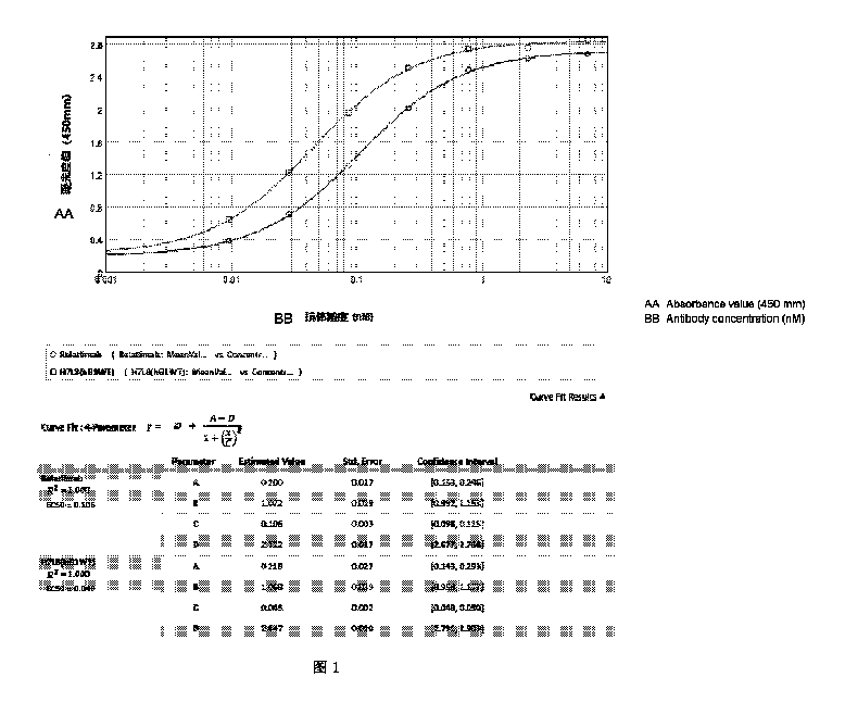
【選択図】 図1
The present invention belongs to the field of biomedicine and relates to an anti-LAG3 antibody, a pharmaceutical composition containing the same, and uses thereof. Specifically, the present invention relates to an anti-LAG3 antibody or an antigen-binding fragment thereof, the antibody comprising a heavy chain variable region and a light chain variable region, the heavy chain variable region comprising HCDR1 to HCDR3 having the amino acid sequences shown in SEQ ID NOs: 9 to 11, respectively, and the light chain variable region comprising LCDR1 to LCDR3 having the amino acid sequences shown in SEQ ID NOs: 12 to 14, respectively. The anti-LAG3 antibody of the present invention has excellent affinity and specificity, and is highly applicable.
[Selected Figure] Figure 1
Description
本発明は生物医学の分野に属し、抗LAG3抗体、抗LAG3抗体を含む医薬組成物、及びそれらの使用に関する。 The present invention belongs to the field of biomedicine and relates to anti-LAG3 antibodies, pharmaceutical compositions containing anti-LAG3 antibodies, and uses thereof.
腫瘍、特に悪性腫瘍は、今日では、健康を脅かす世界的に深刻な疾患であり、各種疾患の中でも死因の第2位となっている。近年、これら疾患の罹患率が著しく増加している。悪性腫瘍は、治療反応性が悪く、後期の転移率が高く、また、予後が不良であることが特徴である。現在臨床的に採用されている従来の治療方法(例えば、放射線療法、化学療法、及び外科治療など)は、疼痛を大幅に緩和し、生存時間を延長するものであるが、それらの方法には大きな制約があり、それらの有効性をさらに向上させるのは困難である。 Tumors, especially malignant tumors, are nowadays a serious health threat worldwide and the second leading cause of death among various diseases. In recent years, the incidence of these diseases has increased significantly. Malignant tumors are characterized by poor response to treatment, high rate of late metastasis, and poor prognosis. Conventional treatment methods currently used clinically (e.g., radiation therapy, chemotherapy, and surgery) can significantly relieve pain and extend survival time, but these methods have significant limitations and it is difficult to further improve their effectiveness.
リンパ球活性化遺伝子3(LAG3)、すなわちCD223は、498アミノ酸から構成されるI型膜貫通タンパク質であり、免疫グロブリンスーパーファミリー(IgSF)のメンバーである。LAG3は主に、活性化CD4+ T細胞及びCD8+ T細胞で発現している。また、ナチュラルキラー(NK)細胞、B細胞、調節性T細胞(Tregs)、及び形質細胞様樹状細胞(pDCs)などの細胞においてもLAG3が発現している(Ruffo Elisa, Wu Richard C, Bruno Tullia C et al., Lymphocyte-activation gene 3 (LAG3): The next immune checkpoint receptor. [J].Semin Immunol, 2019, 42: 101305.)。 Lymphocyte activation gene 3 (LAG3), or CD223, is a type I transmembrane protein of 498 amino acids and a member of the immunoglobulin superfamily (IgSF). LAG3 is primarily expressed in activated CD4 + and CD8 + T cells. LAG3 is also expressed in cells such as natural killer (NK) cells, B cells, regulatory T cells (Tregs), and plasmacytoid dendritic cells (pDCs) (Ruffo Elisa, Wu Richard C, Bruno Tullia C et al., Lymphocyte-activation gene 3 (LAG3): The next immune checkpoint receptor. [J]. Semin Immunol, 2019, 42: 101305.).
LAG3分子遺伝子はヒト第12染色体(20p13.3)上でCD4分子遺伝子に隣接して位置し、両者とも同じエクソンとイントロンを有する。LAG3分子とCD4分子は構造的に類似性が高いが、両者間のアミノ酸配列相同性は20%程度しかない。主要組織適合遺伝子複合体クラスII(MHC II)分子、肝類洞内皮細胞レクチン(LSECtin)分子、及びガレクチン-3分子はLAG3分子に対する関連リガンドである。MHCクラスII分子はLAG3に対する主要リガンドである。MHCクラスII分子に対するLAG3分子の親和性(Kd:60nmol・L-1)はCD4分子のものと比較して100倍であり、このことは、LAG3分子が、MHCクラスII分子への結合に関してCD4分子と効果的に競合し、T細胞活性化を阻害しうるということを示している。 The LAG3 molecule gene is located adjacent to the CD4 molecule gene on human chromosome 12 (20p13.3), and both molecules have the same exons and introns. Although the LAG3 molecule and the CD4 molecule are highly structurally similar, the amino acid sequence homology between them is only about 20%. Major histocompatibility complex class II (MHC II) molecules, liver sinusoidal endothelial cell lectin (LSECtin) molecules, and galectin-3 molecules are relevant ligands for the LAG3 molecule. MHC class II molecules are the major ligand for LAG3. The affinity of the LAG3 molecule for MHC class II molecules (Kd: 60 nmol·L −1 ) is 100-fold higher than that of the CD4 molecule, indicating that the LAG3 molecule can effectively compete with the CD4 molecule for binding to the MHC class II molecule and inhibit T cell activation.
腫瘍微小環境内においては、T細胞活性化の24時間後に免疫抑制分子LAG3の発現を検出することができ、これがひいてはT細胞の機能不全又はアポトーシスをもたらす。LAG3分子は、そのD1ドメイン(プロリンに富む1個のループ構造を有する)を介して二量体を形成し、CD4+ T細胞活性化の第1シグナル伝達軸(first signaling axis)である「CD3-TCR-MHCII」においてMHCクラスII分子に特異的に結合する。それにより、一方ではT細胞活性化のためのシグナル伝達経路がブロックされ、他方ではLAG3分子の細胞内セグメント(KIEELEモチーフ)が免疫抑制シグナルを生成して、CD4+ T細胞の活性を下方制御する。LAG3分子はTreg細胞の分化を促進し、シグナル伝達兼転写活性化因子5の下流シグナル伝達に関与し、それによってTreg細胞の阻害効果を増強することができる。これは、腫瘍が免疫系による殺傷を回避するメカニズムの1つである(Andrews Lawrence P, Marciscano Ariel E, Drake Charles G, et al., LAG3 (CD223) as a cancer immunotherapy target. [J]. Immunol Rev, 2017, 276: 80-96.)。 In the tumor microenvironment, the expression of the immunosuppressive molecule LAG3 can be detected 24 hours after T cell activation, which in turn leads to T cell dysfunction or apoptosis. The LAG3 molecule forms a dimer via its D1 domain (having one proline-rich loop structure) and specifically binds to MHC class II molecules in the "CD3-TCR-MHCII" pathway, which is the first signaling axis of CD4 + T cell activation. On the one hand, this blocks the signaling pathway for T cell activation, and on the other hand, the intracellular segment of the LAG3 molecule (KIEELE motif) generates an immunosuppressive signal to downregulate the activity of CD4 + T cells. The LAG3 molecule can promote the differentiation of Treg cells and participate in downstream signaling of signal transducer and activator of transcription 5, thereby enhancing the inhibitory effect of Treg cells. This is one of the mechanisms by which tumors avoid killing by the immune system (Andrews Lawrence P, Marciscano Ariel E, Drake Charles G, et al., LAG3 (CD223) as a cancer immunotherapy target. [J]. Immunol Rev, 2017, 276: 80-96.).
各種悪性腫瘍の腫瘍浸潤CD8+ T細胞においてLAG3が過剰発現していることが、複数の研究によって示されている。例えば、卵巣がんにおいては、ニューヨーク食道扁平上皮がん1(NY-ESO-1)抗原特異的腫瘍浸潤CD8+ T細胞が、PD-1及びLAG3を高度に発現し、IFN-γ及びTNF-αを産生する能力が減少しており、そのためリンパ球が不活性化されている。ガレクチン-3及びLSECtinは、主にLAG3と相互作用し、CD8+ T細胞の活性化と機能を調節する。また、転移性メラノーマの患者から単離されたメラノーマ抗原特異的T細胞は、LAG3並びに他の免疫チェックポイント分子CTLA-4及びTIM-3の発現の有意な上方制御を示す(Liu Hao, Li Xinying, Luo Longlong, et al., Research advances in biological function of lymphocyte activation gene-3 (LAG-3) molecule and clinical application of antibody drugs targeting LAG-3 [J]. Chinese Journal of Pharmacology and Toxicology, 2019, 33(01): 70-78.)。 Several studies have shown that LAG3 is overexpressed in tumor-infiltrating CD8 + T cells of various malignancies. For example, in ovarian cancer, New York esophageal squamous cell carcinoma 1 (NY-ESO-1) antigen-specific tumor-infiltrating CD8 + T cells highly express PD-1 and LAG3 and have a reduced ability to produce IFN-γ and TNF-α, resulting in lymphocyte inactivation. Galectin-3 and LSECtin primarily interact with LAG3 to regulate the activation and function of CD8 + T cells. In addition, melanoma antigen-specific T cells isolated from patients with metastatic melanoma show significant upregulation of the expression of LAG3 as well as other immune checkpoint molecules CTLA-4 and TIM-3 (Liu Hao, Li Xinying, Luo Longlong, et al., Research advances in biological function of lymphocyte activation gene-3 (LAG-3) molecule and clinical application of antibody drugs targeting LAG-3 [J]. Chinese Journal of Pharmacology and Toxicology, 2019, 33(01): 70-78. ).
現在、複数のLAG3抗体薬が臨床研究段階に入っており、それらのうち、Bristol Myers Squibb社のレラトリマブは10件の臨床研究が進行中であり、最も進展が見られる。これらの研究の大部分は、血液悪性腫瘍、メラノーマ、神経膠芽腫、腎細胞がん、非小細胞肺がん、及び同種のものなどの腫瘍の治療に用いられる、レラトリマブとニボルマブの併用療法を伴う。 Currently, several LAG3 antibody drugs are in clinical research, of which Bristol Myers Squibb's leratolimab is the most advanced with 10 clinical studies ongoing. Most of these studies involve the combination of leratolimab and nivolumab for the treatment of tumors such as hematological malignancies, melanoma, glioblastoma, renal cell carcinoma, non-small cell lung cancer, and the like.
現在、新規抗LAG3抗体薬の開発が必要とされている。 Currently, there is a need to develop new anti-LAG3 antibody drugs.
本願発明者らは鋭意研究を重ね、創造的な努力を行うことによって、抗LAG3抗体を得た。本願発明者らは驚くべきことに、本発明の抗LAG3抗体(以下、前記抗体又は本発明の抗体と略す場合がある)が優れた親和性及び/又は特異性を有し、陽性対照抗体(例えば、レラトリマブ)と比べても1つ以上の点においてさらに優れたものであることを見出した。本発明を以下に詳細に説明する。 The present inventors have obtained an anti-LAG3 antibody through intensive research and creative efforts. The present inventors have surprisingly found that the anti-LAG3 antibody of the present invention (hereinafter sometimes abbreviated as the antibody or the antibody of the present invention) has excellent affinity and/or specificity, and is superior in one or more respects to a positive control antibody (e.g., leratolimab). The present invention is described in detail below.
本発明の一態様は、重鎖可変領域及び軽鎖可変領域を含む、抗LAG3抗体又はその抗原結合性断片(an antigen-binding fragment)であって、
前記重鎖可変領域が、配列番号9~11に記載のアミノ酸配列をそれぞれ有するHCDR1~HCDR3を含み、前記軽鎖可変領域が、配列番号12~14に記載のアミノ酸配列をそれぞれ有するLCDR1~LCDR3を含む;
前記重鎖可変領域が、配列番号9~11に記載のアミノ酸配列をそれぞれ有するHCDR1~HCDR3を含み、前記軽鎖可変領域が、配列番号12、配列番号15、及び配列番号16に記載のアミノ酸配列をそれぞれ有するLCDR1~LCDR3を含む;又は、
前記重鎖可変領域が、配列番号9~11に記載のアミノ酸配列をそれぞれ有するHCDR1~HCDR3を含み、前記軽鎖可変領域が、配列番号17、配列番号15、及び配列番号14に記載のアミノ酸配列をそれぞれ有するLCDR1~LCDR3を含む、抗LAG3抗体又はその抗原結合性断片に関する。
One aspect of the present invention is an anti-LAG3 antibody or an antigen-binding fragment thereof, comprising a heavy chain variable region and a light chain variable region,
the heavy chain variable region comprises HCDR1 to HCDR3 having the amino acid sequences set forth in SEQ ID NOs:9 to 11, respectively, and the light chain variable region comprises LCDR1 to LCDR3 having the amino acid sequences set forth in SEQ ID NOs:12 to 14, respectively;
the heavy chain variable region comprises HCDR1-HCDR3 having the amino acid sequences set forth in SEQ ID NOs:9-11, respectively, and the light chain variable region comprises LCDR1-LCDR3 having the amino acid sequences set forth in SEQ ID NOs:12, 15, and 16, respectively; or
The present invention relates to an anti-LAG3 antibody or an antigen-binding fragment thereof, wherein the heavy chain variable region comprises HCDR1 to HCDR3 having the amino acid sequences set forth in SEQ ID NOs:9 to 11, respectively, and the light chain variable region comprises LCDR1 to LCDR3 having the amino acid sequences set forth in SEQ ID NOs:17, 15, and 14, respectively.
本発明のいくつかの実施形態では、
前記抗体の前記重鎖可変領域が、配列番号2に記載のアミノ酸配列を有し、前記抗体の前記軽鎖可変領域が、配列番号4に記載のアミノ酸配列を有する;
前記抗体の前記重鎖可変領域が、配列番号2に記載のアミノ酸配列を有し、前記抗体の前記軽鎖可変領域が、配列番号6に記載のアミノ酸配列を有する;又は、
前記抗体の前記重鎖可変領域が、配列番号2に記載のアミノ酸配列を有し、前記抗体の前記軽鎖可変領域が、配列番号8に記載のアミノ酸配列を有する、
前記抗体又はその抗原結合性断片が提供される。
In some embodiments of the present invention,
the heavy chain variable region of the antibody has the amino acid sequence set forth in SEQ ID NO:2 and the light chain variable region of the antibody has the amino acid sequence set forth in SEQ ID NO:4;
the heavy chain variable region of the antibody has the amino acid sequence set forth in SEQ ID NO:2 and the light chain variable region of the antibody has the amino acid sequence set forth in SEQ ID NO:6; or
the heavy chain variable region of the antibody has the amino acid sequence set forth in SEQ ID NO:2, and the light chain variable region of the antibody has the amino acid sequence set forth in SEQ ID NO:8;
The antibody or antigen-binding fragment thereof is provided.
本発明のいくつかの実施形態では、Fab、Fab’、F(ab’)2、Fd、Fv、dAb、相補性決定領域断片、単鎖可変領域断片(a single chain fragment variable)、ヒト化抗体、又はキメラ抗体から選択される、前記抗体又はその抗原結合性断片が提供される。 In some embodiments of the invention, the antibody or antigen-binding fragment thereof is provided as being selected from a Fab, a Fab', a F(ab') 2 , a Fd, a Fv, a dAb, a complementarity determining region fragment, a single chain fragment variable, a humanized antibody, or a chimeric antibody.
本発明のいくつかの実施形態では、前記抗体が、ヒトLAG3-mFcに対して、0.2nM未満、例えば、0.15nM未満、0.1nM未満、0.08nM未満、0.06nM未満、もしくは0.05nM未満、又はそれ以下のEC50値で結合し、好ましくは前記EC50値が間接ELISAによって測定される、前記抗体又はその抗原結合性断片が提供される。 In some embodiments of the invention there is provided an antibody or antigen-binding fragment thereof which binds to human LAG3-mFc with an EC 50 value of less than 0.2 nM, e.g., less than 0.15 nM, less than 0.1 nM, less than 0.08 nM, less than 0.06 nM, or less than 0.05 nM, or less, preferably wherein the EC 50 value is measured by indirect ELISA.
本発明のいくつかの実施形態では、前記抗体が、マウス以外の種に由来する、例えば、ヒト抗体に由来する非CDR領域を含む、前記抗体又はその抗原結合性断片が提供される。 In some embodiments of the invention, the antibody or antigen-binding fragment thereof is provided, wherein the antibody is derived from a species other than mouse, e.g., the antibody comprises a non-CDR region derived from a human antibody.
本発明のいくつかの実施形態では、
前記抗体がヒト抗体に由来する定常領域を含み、
好ましくは、前記抗体の定常領域がヒトIgG1、IgG2、IgG3、又はIgG4の定常領域から選択される、
前記抗体又はその抗原結合性断片が提供される。
In some embodiments of the present invention,
the antibody comprises a constant region derived from a human antibody,
Preferably, the constant region of the antibody is selected from the constant region of human IgG1, IgG2, IgG3, or IgG4.
The antibody or antigen-binding fragment thereof is provided.
本発明のいくつかの実施形態では、前記抗LAG3抗体の重鎖定常領域が、Igガンマ-1鎖C領域(例えば、配列番号18に記載されている)又はIgガンマ-4鎖C領域(例えば、配列番号20に記載されている)であり、前記抗LAG3抗体の軽鎖定常領域が、Igカッパ鎖C領域(例えば、配列番号19に記載されている)である、抗体又はその抗原結合性断片が提供される。 In some embodiments of the present invention, an antibody or antigen-binding fragment thereof is provided in which the heavy chain constant region of the anti-LAG3 antibody is an Ig gamma-1 chain C region (e.g., as set forth in SEQ ID NO: 18) or an Ig gamma-4 chain C region (e.g., as set forth in SEQ ID NO: 20), and the light chain constant region of the anti-LAG3 antibody is an Ig kappa chain C region (e.g., as set forth in SEQ ID NO: 19).
本発明のいくつかの実施形態では、前記抗LAG3抗体はモノクローナル抗体である。 In some embodiments of the invention, the anti-LAG3 antibody is a monoclonal antibody.
本発明のいくつかの実施形態では、前記抗LAG3抗体は免疫グロブリン形態である。 In some embodiments of the invention, the anti-LAG3 antibody is in the form of an immunoglobulin.
本発明のいくつかの実施形態では、前記抗LAG3抗体は単鎖可変領域断片である。 In some embodiments of the invention, the anti-LAG3 antibody is a single chain variable region fragment.
本発明の他の態様は、抗体又はその抗原結合性断片、及び小分子薬を含む、抗体薬物複合体(ADC)であって、前記抗体又はその抗原結合性断片が、本発明のいずれかの実施形態の抗LAG3抗体又はその抗原結合性断片であり;好ましくは前記小分子薬が小分子細胞毒性薬であり;より好ましくは前記小分子薬が抗腫瘍化学療法薬である、抗体薬物複合体に関する。 Another aspect of the invention relates to an antibody-drug conjugate (ADC) comprising an antibody or antigen-binding fragment thereof and a small molecule drug, wherein the antibody or antigen-binding fragment thereof is an anti-LAG3 antibody or antigen-binding fragment thereof of any of the embodiments of the invention; preferably, the small molecule drug is a small molecule cytotoxic drug; more preferably, the small molecule drug is an anti-tumor chemotherapeutic drug.
前記化学療法薬は、従来の抗腫瘍化学療法薬、例えば、アルキル化剤、代謝拮抗剤、抗腫瘍抗生物質、植物系抗がん剤、ホルモン、及び免疫剤などであってよい。 The chemotherapeutic agent may be a conventional antitumor chemotherapeutic agent, such as an alkylating agent, antimetabolite, antitumor antibiotic, plant-based anticancer agent, hormone, and immunological agent.
本発明の1つ又は複数の実施形態では、前記抗体薬物複合体が提供され、前記抗体又はその抗原結合性断片は、リンカーを介して小分子薬に連結されており、前記リンカーは当業者に公知のもの、例えば、ヒドラゾン結合、ジスルフィド結合、又はペプチド結合であってよい。 In one or more embodiments of the present invention, the antibody-drug conjugate is provided, in which the antibody or antigen-binding fragment thereof is linked to a small molecule drug via a linker, which may be any linker known to those of skill in the art, such as a hydrazone bond, a disulfide bond, or a peptide bond.
本発明の1つ又は複数の実施形態では、前記抗体又はその抗原結合性断片の前記小分子薬に対するモル比が、1:(2~4)、例えば、1:2、1:3、又は1:4である、前記抗体薬物複合体が提供される。 In one or more embodiments of the present invention, the antibody-drug conjugate is provided in which the molar ratio of the antibody or antigen-binding fragment thereof to the small molecule drug is 1:(2-4), e.g., 1:2, 1:3, or 1:4.
本発明のさらなる他の一態様は、本発明のいずれかの実施形態の抗LAG3抗体をコードする、単離核酸分子に関する。 Yet another aspect of the present invention relates to an isolated nucleic acid molecule encoding an anti-LAG3 antibody of any of the embodiments of the present invention.
本発明のさらなる他の一態様は、本発明の単離核酸分子を含む組換えベクターに関する。 Yet another aspect of the present invention relates to a recombinant vector comprising the isolated nucleic acid molecule of the present invention.
本発明のさらなる他の一態様は、本発明の単離核酸分子又は本発明の組換えベクターを含む、宿主細胞に関する。 Yet another aspect of the present invention relates to a host cell comprising an isolated nucleic acid molecule of the present invention or a recombinant vector of the present invention.
本発明のさらなる他の一態様は、本発明のいずれかの実施形態の抗体又はその抗原結合性断片を調製する方法であって、本発明の宿主細胞を適した条件下で培養すること、及び細胞培養物から前記抗体又はその抗原結合性断片を単離することを含む、方法に関する。 Yet another aspect of the present invention relates to a method for preparing an antibody or antigen-binding fragment thereof of any of the embodiments of the present invention, comprising culturing a host cell of the present invention under suitable conditions and isolating the antibody or antigen-binding fragment thereof from the cell culture.
本発明のさらなる他の一態様は、本発明のいずれかの実施形態の抗体もしくはその抗原結合性断片、又は本発明のいずれかの実施形態の抗体薬物複合体、を含む医薬組成物であって、前記医薬組成物が任意に、薬理学的に許容される副原料をさらに含む、医薬組成物に関する。 Yet another aspect of the present invention relates to a pharmaceutical composition comprising an antibody or antigen-binding fragment thereof according to any of the embodiments of the present invention, or an antibody-drug conjugate according to any of the embodiments of the present invention, the pharmaceutical composition optionally further comprising a pharmacologically acceptable auxiliary ingredient.
本発明のさらなる他の一態様は、腫瘍又は貧血を治療及び/又は予防するための薬剤の調製における、本発明のいずれかの実施形態の抗体もしくはその抗原結合性断片、又は本発明のいずれかの実施形態の抗体薬物複合体の使用であって、
好ましくは前記腫瘍が、卵巣がん、食道がん、メラノーマ、血液悪性腫瘍、膠芽腫、腎細胞がん、肺がん、前立腺がん、膀胱がん、結腸がん、直腸がん、肝臓がん、消化器がん、乳がん、脳腫瘍、膵臓がん、甲状腺がん、頭頸部がん、及び腎臓がんの1つ又は複数から選択され;
好ましくは、前記肺がんが非小細胞肺がんであり;
好ましくは、前記血液悪性腫瘍が白血病であり;
好ましくは、前記食道がんが食道扁平上皮がんである、抗体もしくはその抗原結合性断片又は抗体薬物複合体の使用に関する。
Yet another aspect of the present invention is the use of an antibody or antigen-binding fragment thereof of any of the embodiments of the present invention, or an antibody-drug conjugate of any of the embodiments of the present invention, in the preparation of a medicament for treating and/or preventing tumors or anemia, comprising:
Preferably, the tumor is selected from one or more of ovarian cancer, esophageal cancer, melanoma, hematological malignancies, glioblastoma, renal cell carcinoma, lung cancer, prostate cancer, bladder cancer, colon cancer, rectal cancer, liver cancer, gastrointestinal cancer, breast cancer, brain cancer, pancreatic cancer, thyroid cancer, head and neck cancer, and kidney cancer;
Preferably, the lung cancer is non-small cell lung cancer;
Preferably, the hematological malignancy is leukemia;
Preferably, the present invention relates to the use of an antibody or an antigen-binding fragment thereof, or an antibody-drug conjugate, wherein the esophageal cancer is esophageal squamous cell carcinoma.
前記の本発明のいずれかの実施形態の抗体もしくはその抗原結合性断片、又は本発明のいずれかの実施形態の抗体薬物複合体は、腫瘍又は貧血の治療及び/又は予防において使用するためのものであって、
好ましくは前記腫瘍が、卵巣がん、食道がん、メラノーマ、血液悪性腫瘍、膠芽腫、腎細胞がん、肺がん、前立腺がん、膀胱がん、結腸がん、直腸がん、肝臓がん、消化器がん、乳がん、脳腫瘍、膵臓がん、甲状腺がん、頭頸部がん、及び腎臓がんの1つ又は複数から選択され;
好ましくは、前記肺がんは非小細胞肺がんであり;
好ましくは、前記血液悪性腫瘍は白血病であり;
好ましくは、前記食道がんは食道扁平上皮がんである。
The antibody or antigen-binding fragment thereof according to any of the embodiments of the present invention, or the antibody-drug conjugate according to any of the embodiments of the present invention, is for use in the treatment and/or prevention of tumor or anemia, comprising:
Preferably, the tumor is selected from one or more of ovarian cancer, esophageal cancer, melanoma, hematological malignancies, glioblastoma, renal cell carcinoma, lung cancer, prostate cancer, bladder cancer, colon cancer, rectal cancer, liver cancer, gastrointestinal cancer, breast cancer, brain cancer, pancreatic cancer, thyroid cancer, head and neck cancer, and kidney cancer;
Preferably, the lung cancer is non-small cell lung cancer;
Preferably, the hematological malignancy is leukemia;
Preferably, the esophageal cancer is esophageal squamous cell carcinoma.
本発明のさらなる他の一態様は、腫瘍又は貧血を治療及び/又は予防する方法であって、本発明のいずれかの実施形態の抗体もしくはその抗原結合性断片、又は本発明のいずれかの実施形態の抗体薬物複合体の有効量を、それを必要とする対象に投与する工程を含み、
好ましくは前記腫瘍が、卵巣がん、食道がん、メラノーマ、血液悪性腫瘍、膠芽腫、腎細胞がん、肺がん、前立腺がん、膀胱がん、結腸がん、直腸がん、肝臓がん、消化器がん、乳がん、脳腫瘍、膵臓がん、甲状腺がん、頭頸部がん、及び腎臓がんの1つ又は複数から選択され;
好ましくは、前記肺がんが非小細胞肺がんであり;
好ましくは、前記血液悪性腫瘍が白血病であり;
好ましくは、前記食道がんが食道扁平上皮がんである、方法に関する。
Yet another aspect of the present invention is a method for treating and/or preventing tumors or anemia, comprising administering to a subject in need thereof an effective amount of the antibody or antigen-binding fragment thereof of any of the embodiments of the present invention, or the antibody-drug conjugate of any of the embodiments of the present invention;
Preferably, the tumor is selected from one or more of ovarian cancer, esophageal cancer, melanoma, hematological malignancies, glioblastoma, renal cell carcinoma, lung cancer, prostate cancer, bladder cancer, colon cancer, rectal cancer, liver cancer, gastrointestinal cancer, breast cancer, brain cancer, pancreatic cancer, thyroid cancer, head and neck cancer, and kidney cancer;
Preferably, the lung cancer is non-small cell lung cancer;
Preferably, the hematological malignancy is leukemia;
Preferably, the esophageal cancer is esophageal squamous cell carcinoma.
本発明において、他に定義されない限り、本明細書内で使用される科学及び技術用語は、当業者が一般的に理解する意味を有する。また、本明細書で使用される細胞培養、分子遺伝学、核酸化学、及び免疫学の実験室操作は、対応する分野で広く使用されるルーチン手順である。一方、本発明の理解を促すために、関連のある語の定義及び説明を以下に提供する。 In the present invention, unless otherwise defined, scientific and technical terms used herein have the meanings commonly understood by those skilled in the art. Also, the laboratory procedures of cell culture, molecular genetics, nucleic acid chemistry, and immunology used herein are routine procedures widely used in the corresponding fields. Meanwhile, in order to facilitate understanding of the present invention, definitions and explanations of relevant terms are provided below.
本明細書で使用されるEC50という語は、最大効果の50%に対する濃度、すなわち、最大効果の50%を引き起こしうる濃度のことを指す。 The term EC50 as used herein refers to the concentration for 50% of the maximum effect, i.e., the concentration capable of producing 50% of the maximum effect.
本明細書で使用される「抗体」という語は、一般に2対のポリペプチド鎖からなる(それぞれの対が1本の「軽」(L)鎖と1本の「重」(H)鎖を有する)免疫グロブリン分子のことを指す。抗体軽鎖はκ及びλ軽鎖へと分類される。重鎖はμ、δ、γ、α、又はεへと分類される。抗体のアイソタイプはIgM、IgD、IgG、IgA、及びIgEとして定義される。軽鎖及び重鎖において、可変領域と定常領域は、約12以上のアミノ酸の「J」領域によって連結しており、重鎖はさらに、約3以上のアミノ酸の「D」領域を含む。各重鎖は、重鎖可変領域(VH)及び重鎖定常領域(CH)からなる。重鎖定常領域は、3つのドメイン(CH1、CH2、及びCH3)からなる。各軽鎖は、軽鎖可変領域(VL)及び軽鎖定常領域(CL)からなる。軽鎖定常領域は1つのドメイン、CLからなる。抗体の定常領域は、免疫系の各種細胞(例えば、エフェクター細胞)の古典的な補体系の第1成分(C1q)への結合を含む、免疫グロブリンの宿主組織又は因子への結合を媒介することができる。VH及びVL領域は、超可変領域(相補性決定領域(CDR)と呼ばれる)にさらに細かく分けることができ、超可変領域間にはフレームワーク領域(FR)と呼ばれる保存的な領域が分布している。各VH及びVLは、アミノ末端からカルボキシル末端に向けて次の順番で並んだ、3つのCDRと4つのFRからなる:FR1、CDR1、FR2、CDR2、FR3、CDR3、及びFR4。各重鎖/軽鎖ペアの可変領域(VH及びVL)は、抗体結合部位を形成する。領域又はドメインに対するアミノ酸の割り当ては、Bethesda M.d., Kabat Sequences of Proteins of Immunological Interest (National Institutes of Health, (1987及び1991))もしくはChothia & Lesk J. Mol. Biol., 1987; 196:901-917;Chothia et al., Nature, 1989; 342:878-883、又はIMGT番号付けシステムの定義(Ehrenmann F, Kaas Q, Lefranc M P., IMGT/3Dstructure-DB and IMGT/DomainGapAlign: a database and a tool for immunoglobulins or antibodies, T cell receptors, MHC, IgSF and MhcSF[J]., Nucleic acids research, 2009; 38(suppl_1):D301-D307における定義を参照)に基づいて行われる。 As used herein, the term "antibody" refers to an immunoglobulin molecule that generally consists of two pairs of polypeptide chains, each pair having one "light" (L) chain and one "heavy" (H) chain. Antibody light chains are classified as kappa and lambda light chains. Heavy chains are classified as mu, delta, gamma, alpha, or epsilon. Antibody isotypes are defined as IgM, IgD, IgG, IgA, and IgE. In the light and heavy chains, the variable and constant regions are connected by a "J" region of about 12 or more amino acids, and the heavy chains further include a "D" region of about 3 or more amino acids. Each heavy chain consists of a heavy chain variable region (VH) and a heavy chain constant region (CH). The heavy chain constant region consists of three domains, CH1, CH2, and CH3. Each light chain consists of a light chain variable region (VL) and a light chain constant region (CL). The light chain constant region consists of one domain, CL. The constant regions of antibodies can mediate the binding of immunoglobulins to host tissues or factors, including the binding of various cells of the immune system (e.g., effector cells) to the first component (C1q) of the classical complement system. The VH and VL regions can be further divided into hypervariable regions (called complementarity determining regions (CDRs)), with conserved regions called framework regions (FRs) distributed between the hypervariable regions. Each VH and VL consists of three CDRs and four FRs, arranged from the amino terminus to the carboxyl terminus in the following order: FR1, CDR1, FR2, CDR2, FR3, CDR3, and FR4. The variable regions (VH and VL) of each heavy/light chain pair form the antibody binding site. The assignment of amino acids to regions or domains is described in Bethesda M. d., et al. , Kabat Sequences of Proteins of Immunological Interest (National Institutes of Health, (1987 and 1991)) or Chothia & Lesk J. Mol. Biol. , 1987; 196:901-917; Chothia et al. , Nature, 1989; 342:878-883, or the definition of the IMGT numbering system (Ehrenmann F, Kaas Q, Lefranc M P., IMGT/3Dstructure-DB and IMGT/DomainGapAlign: a database and a tool for immunoglobulins or antibodies, T cell receptors, MHC, IgSF and MhcSF[J]., Nucleic acids research, 2009; 38 (suppl_1): See definition in D301-D307.
「抗体」という語は、抗体を製造するためのいかなる特定の方法によっても限定されない。例えば、抗体には、組換え抗体、モノクローナル抗体、及びポリクローナル抗体が含まれる。抗体は、IgG(例えば、サブタイプIgG1、IgG2、IgG3、もしくはIgG4)、IgA1、IgA2、IgD、IgE、又はIgMなどの、各種アイソタイプの抗体であってよい。 The term "antibody" is not limited by any particular method for producing the antibody. For example, antibodies include recombinant antibodies, monoclonal antibodies, and polyclonal antibodies. Antibodies can be of various isotypes, such as IgG (e.g., subtypes IgG1, IgG2, IgG3, or IgG4), IgA1, IgA2, IgD, IgE, or IgM.
本明細書で使用される「mAb」及び「モノクローナル抗体」という語は、高度に相同な抗体のグループに由来する、すなわち、自然に生じうる天然の変異を例外として同一の抗体分子のグループに由来する、抗体又は抗体の断片のことを指す。モノクローナル抗体は、ある抗原上の単一のエピトープに対して高度に特異的である。モノクローナル抗体と異なり、ポリクローナル抗体は、通常、ある抗原上の異なるエピトープを一般に認識する少なくとも2つ以上の異なる抗体を含む。モノクローナル抗体は一般に、Kohler et al.(Kohler G, Milstein C. Continuous cultures of fused cells secreting antibody of predefined specificity [J]. Nature, 1975; 256(5517): 495)によって最初に報告されたハイブリドーマ技術を用いて取得することができるが、組換えDNA技術を用いても取得することができる(米国特許第4816567号明細書などを参照)。 As used herein, the terms "mAb" and "monoclonal antibody" refer to an antibody or antibody fragment that is derived from a group of highly homologous antibodies, i.e., from a group of identical antibody molecules except for natural mutations that may occur naturally. Monoclonal antibodies are highly specific for a single epitope on an antigen. Unlike monoclonal antibodies, polyclonal antibodies usually contain at least two or more different antibodies that generally recognize different epitopes on an antigen. Monoclonal antibodies are generally classified as monoclonal antibodies as described by Kohler et al. It can be obtained using the hybridoma technique first reported by (Kohler G, Milstein C. Continuous cultures of fused cells secreting antibodies of predefined specificity [J]. Nature, 1975; 256(5517): 495), but it can also be obtained using recombinant DNA techniques (see U.S. Pat. No. 4,816,567, etc.).
本明細書で使用される「ヒト化抗体」という語は、ヒト免疫グロブリン(レセプター抗体)のCDRの全体又は部分を非ヒト抗体(ドナー抗体)のCDRで置き換えた場合に得られる抗体又は抗体断片のことを指し、ドナー抗体は、期待される特異性、親和性、又は反応性を有する非ヒト(例えば、マウス、ラット、もしくはウサギ)抗体であってよい。さらに、レセプター抗体のフレームワーク領域(FR)におけるいくつかのアミノ酸残基も、対応する非ヒト抗体のアミノ酸残基又は他の抗体のアミノ酸残基に置換し、抗体の性能をさらに改善又は最適化することができる。ヒト化抗体のさらなる詳細については、例えば、Jones et al., Nature, 1986; 321:522-525、Reichmann et al., Nature, 1988; 332:323-329、Presta, Curr. Op. Struct. Biol., 1992; 2:593-596;及びClark, Immunol. Today, 2000; 21:397-402を参照のこと。 The term "humanized antibody" as used herein refers to an antibody or antibody fragment obtained when the whole or part of the CDRs of a human immunoglobulin (receptor antibody) are replaced with the CDRs of a non-human antibody (donor antibody), which may be a non-human (e.g., mouse, rat, or rabbit) antibody having the desired specificity, affinity, or reactivity. In addition, some amino acid residues in the framework region (FR) of the receptor antibody may also be replaced with the corresponding amino acid residues of the non-human antibody or amino acid residues of other antibodies to further improve or optimize the performance of the antibody. For further details on humanized antibodies, see, for example, Jones et al., Nature, 1986; 321:522-525; Reichmann et al., Nature, 1988; 332:323-329; Presta, Curr. Op. Struct. Biol. , 1992; 2:593-596; and Clark, Immunol. Today, 2000; 21:397-402.
本明細書で使用される「単離される」という語は、天然状態から人為手段によって取得されることを指す。特定の「単離される」物質又は成分が天然に存在する場合に、その天然環境において変化が生ずることであってもよく、天然環境から単離されることであってもよく、その両者であってもよい。例えば、単離されていない特定のポリヌクレオチド又はポリペプチドが特定の動物生体中に天然に存在し、このような天然状態から高純度の同一のポリヌクレオチド又はポリペプチドが単離された場合、それを単離ポリヌクレオチド又はポリペプチドと呼ぶ。「単離される」という語は、前記物質の活性に影響を及ぼさない、人工もしくは合成物質、又は他の不純物の存在を除外するものではない。 As used herein, the term "isolated" refers to being obtained by artificial means from a natural state. If a particular "isolated" substance or component occurs in nature, it may be that a change occurs in its natural environment, or that it is isolated from its natural environment, or both. For example, if a particular non-isolated polynucleotide or polypeptide naturally occurs in a particular animal organism, and the same polynucleotide or polypeptide is isolated in high purity from such a natural state, it is referred to as an isolated polynucleotide or polypeptide. The term "isolated" does not exclude the presence of artificial or synthetic materials or other impurities that do not affect the activity of the substance.
本明細書で使用される「ベクター」という語は、ポリヌクレオチドを挿入することができる核酸ビヒクルのことを指す。ベクターが、挿入されたポリヌクレオチドによりコードされるタンパク質の発現を可能にする場合、そのベクターは、発現ベクターと呼ばれる。ベクターは、ベクターによって運ばれる遺伝物質エレメントが宿主細胞で発現できるように、形質転換、形質導入、又はトランスフェクションによって宿主細胞に導入することができる。ベクターは当業者に周知であり、例として、プラスミド;ファージミド;コスミド;人工染色体、例えば、酵母人工染色体(YAC)、細菌人工染色体(BAC)、又はP1由来人工染色体(PAC)など;ファージ、例えば、ラムダファージ又はM13ファージなど;及び動物ウイルス、が挙げられるが、これらに限定されない。ベクターとして使用しうる動物ウイルスとしては、レトロウイルス(レンチウイルスを含む)、アデノウイルス、アデノ随伴ウイルス、ヘルペスウイルス(例えば、単純ヘルペスウイルスなど)、ポックスウイルス、バキュロウイルス、パピローマウイルス、及びパポバウイルス(例えば、SV40)が挙げられるが、これらに限定されない。ベクターは、発現を調節するための各種エレメントを含んでいてよく、それらの例として、プロモーター配列、転写開始配列、エンハンサー配列、選択エレメント、及びレポーター遺伝子が挙げられるが、これらに限定されない。さらに、ベクターは、複製開始部位を含んでいてもよい。 The term "vector" as used herein refers to a nucleic acid vehicle into which a polynucleotide can be inserted. If the vector allows for the expression of a protein encoded by the inserted polynucleotide, the vector is called an expression vector. The vector can be introduced into a host cell by transformation, transduction, or transfection so that the genetic material elements carried by the vector can be expressed in the host cell. Vectors are well known to those skilled in the art and include, but are not limited to, plasmids; phagemids; cosmids; artificial chromosomes, such as yeast artificial chromosomes (YACs), bacterial artificial chromosomes (BACs), or P1-derived artificial chromosomes (PACs); phages, such as lambda phage or M13 phage; and animal viruses. Animal viruses that can be used as vectors include, but are not limited to, retroviruses (including lentiviruses), adenoviruses, adeno-associated viruses, herpes viruses (such as herpes simplex viruses), poxviruses, baculoviruses, papilloma viruses, and papova viruses (such as SV40). The vector may contain various elements for regulating expression, examples of which include, but are not limited to, a promoter sequence, a transcription initiation sequence, an enhancer sequence, a selection element, and a reporter gene. In addition, the vector may contain a replication origin.
本明細書で使用される「宿主細胞」という語は、そこにベクターを導入できる細胞のことを指し、例として、原核細胞、例えば、E. coliもしくはBacillus subtilisなど;真菌細胞、例えば、酵母細胞もしくはAspergillusなど;昆虫細胞、例えば、S2 Drosophila細胞もしくはSf9など;又は動物細胞、例えば、線維芽細胞、CHO細胞、GS細胞、COS細胞、NSO細胞、HeLa細胞、BHK細胞、HEK293細胞、もしくはヒト細胞など、が挙げられるが、これらに限定されない。 As used herein, the term "host cell" refers to a cell into which a vector can be introduced, including, but not limited to, a prokaryotic cell, such as E. coli or Bacillus subtilis; a fungal cell, such as a yeast cell or Aspergillus; an insect cell, such as an S2 Drosophila cell or Sf9; or an animal cell, such as a fibroblast, CHO cell, GS cell, COS cell, NSO cell, HeLa cell, BHK cell, HEK293 cell, or human cell.
本明細書で使用される「特異的結合」という語は、2つの分子間の非無作為的な結合反応、例えば、抗体とその標的となる抗原との間の反応などのことを指す。いくつかの実施形態において、ある抗原に特異的に結合する抗体(又は、ある抗原に対して特異的な抗体)とは、抗体が約10-5M未満、例えば、約10-6M、10-7M、10-8M、10-9M、もしくは10-10M未満、又はそれ以下の親和性(KD)で抗原に結合することを意味する。 The term "specific binding" as used herein refers to a non-random binding reaction between two molecules, such as the reaction between an antibody and its target antigen. In some embodiments, an antibody that specifically binds to an antigen (or an antibody specific for an antigen) means that the antibody binds to the antigen with an affinity (K D ) of less than about 10 −5 M, e.g., less than about 10 −6 M, 10 −7 M, 10 −8 M, 10 −9 M, or 10 −10 M, or less.
本明細書で使用される「KD」という語は、特異的抗体-抗原相互作用の解離平衡定数のことを指し、抗体と抗原との間の結合親和性を説明するために使用される。解離平衡定数が小さいことは、抗体-抗原結合が強く、抗体と抗原との間の親和性が高いことを表す。一般に、抗体は、約10-5M未満、例えば、約10-6M、10-7M、10-8M、10-9M、もしくは10-10M未満、又はそれ以下の解離平衡定数(KD)で抗原(PD-1タンパク質など)に結合する。KDは、当業者に公知の方法、例えば、Fortebio分子相互作用機器の使用を用いて決定することができる。 The term "K D " as used herein refers to the dissociation equilibrium constant of a specific antibody-antigen interaction and is used to describe the binding affinity between an antibody and an antigen. A small dissociation equilibrium constant indicates strong antibody-antigen binding and high affinity between the antibody and the antigen. Generally, an antibody binds to an antigen (such as a PD-1 protein) with a dissociation equilibrium constant (K D ) of less than about 10 −5 M, e.g., less than about 10 −6 M, 10 −7 M, 10 −8 M, 10 −9 M, or 10 −10 M, or even lower. K D can be determined using methods known to those of skill in the art, for example, using a Fortebio molecular interaction instrument.
本明細書で使用される「モノクローナル抗体」という語と「mAb」という語は同一の意味を有し、互換的に用いることができる。「ポリクローナル抗体」という語と「pAb」という語は同一の意味を有し、互換的に用いることができる。また、本明細書において、アミノ酸は通常、当該分野で公知の1文字及び3文字略号を使用して表される。例えば、アラニンはA又はAlaで表すことができる。 As used herein, the terms "monoclonal antibody" and "mAb" have the same meaning and may be used interchangeably. The terms "polyclonal antibody" and "pAb" have the same meaning and may be used interchangeably. Also, herein, amino acids are typically represented using one-letter and three-letter abbreviations known in the art. For example, alanine may be represented as A or Ala.
本明細書で使用される「薬理学的に許容される担体及び/又は賦形剤」という語は、対象及び活性成分に薬理学的及び/又は生理学的に適合した担体及び/又は賦形剤のことを指す。このような担体及び/又は賦形剤は当該分野で周知であり(例えば、Remington’s Pharmaceutical Sciences, edited by Gennaro AR, 19th Ed., Pennsylvania, Mack Publishing Company, 1995を参照)、例として、pH調整剤、界面活性剤、アジュバント、及びイオン強度増強剤が挙げられるが、これらに限定されない。例えば、pH調整剤の限定されない例として、リン酸緩衝液が挙げられ、界面活性剤の限定されない例として、陽イオン、陰イオン、又は非イオン界面活性剤、例えば、Tween-80などが挙げられ、イオン強度増強剤の限定されない例として、塩化ナトリウムが挙げられる。 As used herein, the term "pharmacologically acceptable carriers and/or excipients" refers to carriers and/or excipients that are pharmacologically and/or physiologically compatible with the subject and active ingredient. Such carriers and/or excipients are well known in the art (see, e.g., Remington's Pharmaceutical Sciences, edited by Gennaro AR, 19th Ed., Pennsylvania, Mack Publishing Company, 1995), and examples include, but are not limited to, pH adjusters, surfactants, adjuvants, and ionic strength enhancers. For example, a non-limiting example of a pH adjuster is a phosphate buffer, a non-limiting example of a surfactant is a cationic, anionic, or non-ionic surfactant such as Tween-80, and a non-limiting example of an ionic strength enhancer is sodium chloride.
本明細書で使用される「有効量」という語は、所望の効果を得るのに、又は少なくとも部分的に得るのに、充分な量のことを指す。例えば、疾患(腫瘍など)に対する予防的有効量とは、その疾患(腫瘍など)の発症を予防、抑止、又は遅延するのに充分な量のことを指し、治療有効量とは、その疾患に罹患している患者において、疾患又はその合併症を治癒又は少なくとも部分的に抑止するのに充分な量のことを指す。そのような有効量を決定することは、疑いもなく当業者の能力の範囲内である。例えば、治療目的における有効量は、治療しようとする疾患の重症度;患者自身の免疫系の全体的な状態;年齢、体重、及び性別などの患者の一般的な条件;投与経路;並びに同時に行われる他の治療;などによって決まる。 As used herein, the term "effective amount" refers to an amount sufficient to obtain, or at least partially obtain, a desired effect. For example, a prophylactically effective amount for a disease (such as a tumor) refers to an amount sufficient to prevent, inhibit, or delay the onset of the disease (such as a tumor), and a therapeutically effective amount refers to an amount sufficient to cure or at least partially inhibit the disease or its complications in a patient suffering from the disease. Determining such effective amounts is undoubtedly within the capabilities of a person skilled in the art. For example, an effective amount for therapeutic purposes depends on the severity of the disease to be treated; the overall state of the patient's own immune system; the general condition of the patient, such as age, weight, and sex; the route of administration; and other concurrent treatments.
本明細書において、リンパ球活性化遺伝子3(LAG3)のアミノ酸配列といった場合、それは全長LAG3タンパク質、もしくはLAG3の細胞外断片であるLAG3 ECD、もしくはLAG3 ECDを含む断片のことも含んでおり、また、全長LAG3タンパク質の融合タンパク質もしくはLAG3 ECDの融合タンパク質、例えば、マウスもしくはヒトIgGのFcタンパク質断片(mFcもしくはhFc)などと融合した断片のことも包含する。しかし、LAG3タンパク質のアミノ酸配列において、(置換、欠失、及び/又は付加を含むがそれらに限定されない)突然変異又は変異が、その生物学的機能に影響を与えることなく天然に生じうること、又は人工的に導入されうることを当業者は理解するであろう。したがって、本発明において、「LAG3タンパク質」という語は、それらの天然又は人工的なバリアントを含むすべての配列を含むものとする。また、LAG3タンパク質の配列断片について記述する場合は、それらの天然又は人工的なバリアント内の対応する配列断片のことも含んでいる。 In the present specification, the amino acid sequence of lymphocyte activation gene 3 (LAG3) includes the full-length LAG3 protein, or the extracellular fragment of LAG3, LAG3 ECD, or a fragment containing LAG3 ECD, and also includes a fusion protein of the full-length LAG3 protein or a fusion protein of LAG3 ECD, such as a fragment fused with an Fc protein fragment (mFc or hFc) of mouse or human IgG. However, those skilled in the art will understand that mutations or variations (including but not limited to substitutions, deletions, and/or additions) in the amino acid sequence of the LAG3 protein can occur naturally or can be artificially introduced without affecting its biological function. Thus, in the present invention, the term "LAG3 protein" includes all sequences, including natural or artificial variants thereof. Also, when a sequence fragment of the LAG3 protein is described, the corresponding sequence fragment in the natural or artificial variant is also included.
本発明の有益な効果
本発明は、以下の効果のいずれか1つ以上を達成する:
(1)本発明の抗LAG3抗体が、優れた親和性と特異性を有すること;そして、
(2)本発明の抗LAG3抗体が、LAG3とMHC-IIの間の相互作用を効果的にブロックし、生物におけるLAG3の免疫抑制を特異的に緩和できること。
Beneficial Effects of the Invention The present invention achieves any one or more of the following advantages:
(1) the anti-LAG3 antibody of the present invention has excellent affinity and specificity; and
(2) The anti-LAG3 antibody of the present invention can effectively block the interaction between LAG3 and MHC-II and specifically alleviate the immunosuppression of LAG3 in an organism.
詳細な説明
本発明の実施形態を、実施例を参照しながら以下に詳細に説明する。当業者は、以下の実施例が本発明の単なる例証のためのものであり、本発明の範囲を限定するものと解釈すべきではないと理解するであろう。具体的な技術又は条件が記載されていない実施例は、当該分野の文献に記載された技術又は条件(例えば、Molecular Cloning: A Laboratory Manual, authored by J. Sambrook et al., and translated by Huang Peitang et al., third edition, Science Pressを参照)に従い、又は添付の使用説明書に従い、行われる。使用する試薬又は機器の製造者が記載されていない場合、それらは市販の従来の製品である。
Detailed Description The embodiments of the present invention will be described in detail below with reference to examples. Those skilled in the art will understand that the following examples are merely illustrative of the present invention and should not be interpreted as limiting the scope of the present invention. Examples in which specific techniques or conditions are not described are carried out according to techniques or conditions described in the literature in the field (see, for example, Molecular Cloning: A Laboratory Manual, authored by J. Sambrook et al., and translated by Huang Peitang et al., third edition, Science Press) or according to the accompanying instructions. If the manufacturers of the reagents or instruments used are not described, they are commercially available conventional products.
陽性対照抗体であるレラトリマブは、米国特許出願公開第20160326248(A1)号明細書に示される配列を有し、そのうち重鎖アミノ酸配列はこの特許文献の配列番号1に示されており、軽鎖アミノ酸配列はこの特許文献の配列番号2に示されている。レラトリマブは抗LAG-3抗体である。 The positive control antibody, leratolimab, has the sequence set forth in U.S. Patent Application Publication No. 20160326248(A1), with the heavy chain amino acid sequence set forth in SEQ ID NO:1 of the patent and the light chain amino acid sequence set forth in SEQ ID NO:2 of the patent. leratolimab is an anti-LAG-3 antibody.
レラトリマブの重鎖アミノ酸配列:
QVQLQQWGAGLLKPSETLSLTCAVYGGSFSDYYWNWIRQPPGKGLEWIGEINHRGSTNSNPSLKSRVTLSLDTSKNQFSLKLRSVTAADTAVYYCAFGYSDYEYNWFDPWGQGTLVTVSSASTKGPSVFPLAPCSRSTSESTAALGCLVKDYFPEPVTVSWNSGALTSGVHTFPAVLQSSGLYSLSSVVTVPSSSLGTKTYTCNVDHKPSNTKVDKRVESKYGPPCPPCPAPEFLGGPSVFLFPPKPKDTLMISRTPEVTCVVVDVSQEDPEVQFNWYVDGVEVHNAKTKPREEQFNSTYRVVSVLTVLHQDWLNGKEYKCKVSNKGLPSSIEKTISKAKGQPREPQVYTLPPSQEEMTKNQVSLTCLVKGFYPSDIAVEWESNGQPENNYKTTPPVLDSDGSFFLYSRLTVDKSRWQEGNVFSCSVMHEALHNHYTQKSLSLSLGK(配列番号23)
Leratolimab heavy chain amino acid sequence:
QVQLQQWGAGLLKPSETLSLTCAVYGGSFSDYYWNWIRQPPGKGLEWIGEINHRGSTNSNPSLKSRVTLSLDTSKNQFSLKLRSVTAADTAVYYCAFGYSDYEYNWFDPWGQGTLVTVSSA STKGPSVFPLAPCSRSTSESTAALGCLVKDYFPEPVTVSWNSGALTSGVHTFPAVLQSSGLYSLSSVVTVPSSSLGTKTYTCNVDHKPSNTKVDKRVESKYGPPCP PCPAPEFLGGPSVFLFPPKPKDTLMISRTPEVTCVVVDVSQEDPEVQFNWYVDGVEVHNAKTKPREEQFNSTYRVVSVLTVLHQDWLNGKEYKCKVSNKGLPSSIEKTISKAKGQPREPQVYTLPPSQEEMTKNQVSLTCLVKGFYPSDIAVEWESNGQPENNYKTTPPVLDSDGSFFLYSRLTVDKSRWQEGNVFSCSVMHEALHNHYTQKSLSLSLGK (SEQ ID NO: 23)
レラトリマブの軽鎖アミノ酸配列:
EIVLTQSPATLSLSPGERATLSCRASQSISSYLAWYQQKPGQAPRLLIYDASNRATGIPARFSGSGSGTDFTLTISSLEPEDFAVYYCQQRSNWPLTFGQGTNLEIKRTVAAPSVFIFPPSDEQLKSGTASVVCLLNNFYPREAKVQWKVDNALQSGNSQESVTEQDSKDSTYSLSSTLTLSKADYEKHKVYACEVTHQGLSSPVTKSFNRGEC(配列番号24)
Leratolimab light chain amino acid sequence:
EIVLTQSPATLSLSPGERATLSCRASQSISSYLAWYQQKPGQAPRLLIYDASNRATGIPARFSGSGSGTDFTLTISSLEPEDFAVYYCQQRSNWPLTFGQGTNLEIKRTVAAPSVFIFPPS DEQLKSGTASVVCLLNNFYPREAKVQWKVDNALQSGNSQESVTEQDSKDSTYSLSSTLTLSKADYEKHKVYACEVTHQGLSSPVTKSFNRGEC (SEQ ID NO: 24)
対照抗体14C12H1L1(hG1TM)は、Akeso Biopharma社によって構築された、ロット番号B105Y2080601の抗PD-1抗体であった。 The control antibody 14C12H1L1 (hG1TM) was an anti-PD-1 antibody constructed by Akeso Biopharma, lot number B105Y2080601.
14C12H1L1(hG1TM)の重鎖アミノ酸配列:
EVQLVESGGGLVQPGGSLRLSCAASGFAFSSYDMSWVRQAPGKGLDWVATISGGGRYTYYPDSVKGRFTISRDNSKNNLYLQMNSLRAEDTALYYCANRYGEAWFAYWGQGTLVTVSSASTKGPSVFPLAPSSKSTSGGTAALGCLVKDYFPEPVTVSWNSGALTSGVHTFPAVLQSSGLYSLSSVVTVPSSSLGTQTYICNVNHKPSNTKVDKKVEPKSCDKTHTCPPCPAPEAAGAPSVFLFPPKPKDTLMISRTPEVTCVVVDVSHEDPEVKFNWYVDGVEVHNAKTKPREEQYNSTYRVVSVLTVLHQDWLNGKEYKCKVSNKALPAPIEKTISKAKGQPREPQVYTLPPSRDELTKNQVSLTCLVKGFYPSDIAVEWESNGQPENNYKTTPPVLDSDGSFFLYSKLTVDKSRWQQGNVFSCSVMHEALHNHYTQKSLSLSPGK(配列番号21)
Heavy chain amino acid sequence of 14C12H1L1 (hG1TM):
EVQLVESGGGLVQPGGSLRLSCAASGFAFSSYDMSWVRQAPGKGLDWVATISGGGRYTYYPDSVKGRFTISRDNSKNNLYLQMNSLRAEDTALYYCANRYGEAWFAYWGQGTLVTVSSAST KGPSVFPLAPSSKSTSGGTAALGCLVKDYFPEPVTVSWNSGALTSGVHTFPAVLQSSGLYSLSSVVTVPSSSLGTQTYICNVNHKPSNTKVDKKVEPKSCDKTHTCP PCPAPEAAGAPSVFLFPPKPKDTLMISRTPEVTCVVVDVSHEDPEVKFNWYVDGVEVHNAKTKPREEQYNSTYRVVSVLTVLHQDWLNGKEYKCKVSNKALPAPIEKTISKAKGQPREPQV YTLPPSRDELTKNQVSLTCLVKGFYPSDIAVEWESNGQPENNYKTTPPVLDSDGSFFLYSKLTVDKSRWQQGNVFSCSVMHEALHNHYTQKSLSLSPGK (Sequence number 21)
14C12H1L1(hG1TM)の軽鎖アミノ酸配列:
DIQMTQSPSSMSASVGDRVTFTCRASQDINTYLSWFQQKPGKSPKTLIYRANRLVSGVPSRFSGSGSGQDYTLTISSLQPEDMATYYCLQYDEFPLTFGAGTKLELKRTVAAPSVFIFPPSDEQLKSGTASVVCLLNNFYPREAKVQWKVDNALQSGNSQESVTEQDSKDSTYSLSSTLTLSKADYEKHKVYACEVTHQGLSSPVTKSFNRGEC(配列番号22)
Light chain amino acid sequence of 14C12H1L1 (hG1TM):
DIQMTQSPSSMSASVGDRVTFTCRASQDINTYLSWFQQKPGKSPKTLIYRANRLVSGVPSRFSGSGSGQDYTLISSLQPEDMATYYCLQYDEFPLTFGAGTKLELKRTVAAPSVFIFPPS DEQLKSGTASVVCLLNNFYPREAKVQWKVDNALQSGNSQESVTEQDSKDSTYSLSSTLTLSKADYEKHKVYACEVTHQGLSSPVTKSFNRGEC (SEQ ID NO: 22)
細胞株293T-LAG3は、Akeso Biopharma社によって構築された。細胞株293T-LAG3は、第3世代レンチウイルス系(例えば、A Third Generation Lentivirus Vector with a Conditional Packaging System. Dull T, Zufferey R, Kelly M, Mandel RJ, Nguyen M, Trono D, and Naldini L., J Virol., 1998. 72(11): 8463-8471を参照)を用いて、HEK293T細胞のウイルス感染によって調製された。ここで使用されたレンチウイルス発現ベクターは、plenti6.3/V5-huLAG3FL-BSD(LAG3、Genebank ID:NM_002277.4;ベクターplenti6.3/V5-BSD、Invitrogen社より購入、カタログ番号K5315-20)であった。 The cell line 293T-LAG3 was constructed by Akeso Biopharma. The cell line 293T-LAG3 was prepared by viral infection of HEK293T cells using a third generation lentivirus system (see, e.g., A Third Generation Lentivirus Vector with a Conditional Packaging System. Dull T, Zufferey R, Kelly M, Mandel RJ, Nguyen M, Trono D, and Naldini L., J Virol., 1998. 72(11): 8463-8471). The lentiviral expression vector used here was plenty6.3/V5-huLAG3FL-BSD (LAG3, Genebank ID: NM_002277.4; vector plenty6.3/V5-BSD, purchased from Invitrogen, catalog number K5315-20).
細胞株Raji-PDL1は、Akeso Biopharma社によって構築された。細胞株Raji-PDL1は、第3世代レンチウイルス系(例えば、A Third Generation Lentivirus Vector with a Conditional Packaging System. Dull T, Zufferey R, Kelly M, Mandel RJ, Nguyen M, Trono D, and Naldini L., J Virol., 1998. 72(11): 8463-8471を参照)を用いて、Raji細胞のウイルス感染によって製造された。ここで使用されたレンチウイルス発現ベクターは、plenti6.3/V5-PDL1(PDL1、Genebank ID:NP_054862.1;ベクターplenti6.3/V5、Invitrogen社より購入、カタログ番号K5315-20)であった。 The cell line Raji-PDL1 was constructed by Akeso Biopharma. The cell line Raji-PDL1 was produced by viral infection of Raji cells using a third generation lentivirus system (see, e.g., A Third Generation Lentivirus Vector with a Conditional Packaging System. Dull T, Zufferey R, Kelly M, Mandel RJ, Nguyen M, Trono D, and Naldini L., J Virol., 1998. 72(11): 8463-8471). The lentiviral expression vector used here was plenty6.3/V5-PDL1 (PDL1, Genebank ID: NP_054862.1; vector plenty6.3/V5, purchased from Invitrogen, catalog number K5315-20).
細胞株Jurkat-NFAT-PD1-LAG3は、Akeso Biopharma社によって構築された。細胞株Jurkat-NFAT-PD1-LAG3は、第3世代レンチウイルス系(例えば、A Third Generation Lentivirus Vector with a Conditional Packaging System. Dull T, Zufferey R, Kelly M, Mandel RJ, Nguyen M, Trono D, and Naldini L., J Virol., 1998. 72(11): 8463-8471を参照)を用いて、PD-1エフェクター細胞(CPM、製造者:Promega社、カタログ番号J112A)のウイルス感染によって製造された。ここで使用されたレンチウイルス発現ベクターは、pCDH-huLAG3FL-RFP-NEO(LAG3、Genebank ID:NM_002277.4;ベクターpCDH-CMV-MCS-EF1-RFP+Neo、Youbio社より購入、カタログ番号VT9005)であった。 The cell line Jurkat-NFAT-PD1-LAG3 was constructed by Akeso Biopharma. The cell line Jurkat-NFAT-PD1-LAG3 was generated by viral infection of PD-1 effector cells (CPM, manufacturer: Promega, Cat. No. J112A) using a third generation lentivirus system (see, e.g., A Third Generation Lentivirus Vector with a Conditional Packaging System. Dull T, Zuffery R, Kelly M, Mandel RJ, Nguyen M, Trono D, and Naldini L., J Virol., 1998. 72(11): 8463-8471). The lentiviral expression vector used here was pCDH-huLAG3FL-RFP-NEO (LAG3, Genebank ID: NM_002277.4; vector pCDH-CMV-MCS-EF1-RFP+Neo, purchased from Youbio, catalog number VT9005).
調製例1:抗LAG3抗体の設計及び調製
1.抗体の設計
本願発明者らは、創意により、公知のLAG3タンパク質配列(NCBI参照配列:NP_002277.4)及びその三次元結晶構造などに基づき、一連の抗体配列を設計した。広範なスクリーニングと試験により、LAG3に特異的に結合するヒト化モノクローナル抗体を最終的に取得し、それぞれH7L8、H7L9、及びH7L10と命名した。モノクローナル抗体の重鎖及び軽鎖可変領域のアミノ酸配列、及びそれらのコード配列は、次の通りである。
Preparation Example 1: Design and Preparation of Anti-LAG3 Antibody 1. Antibody Design The inventors of the present application inventively designed a series of antibody sequences based on the known LAG3 protein sequence (NCBI reference sequence: NP_002277.4) and its three-dimensional crystal structure. Through extensive screening and testing, humanized monoclonal antibodies that specifically bind to LAG3 were finally obtained, and named H7L8, H7L9, and H7L10, respectively. The amino acid sequences of the heavy and light chain variable regions of the monoclonal antibodies and their coding sequences are as follows:
H7L8の重鎖可変領域H7vのヌクレオチド配列(360bp):
CAGGTGCAGCTGCAGCAGTGGGGAGCTGGACTGCTGAAACCTAGCGAGACACTGAGCCTGACCTGTGCTGTGTACGGCGGATCTATCAGCGATTACTACTGGAACTGGATCAGGCAGCCCCCTGGAAAGGGACTGGAATGGATCGGAGAGATCAACCACAGGGGCACCACCAACTCCAATCCCTCTCTGAAGAGCAGGGTGACACTGAGCCTCGACACAAGCAAGAATCAGTTCAGCCTGAAGCTGAGGTCCGTGACCGCTGCTGATACAGCTGTGTACTACTGTGCCTTCGGCTACAGCGATTACGAGTACGATTGGTTCGACCCTTGGGGCCAGGGAACACTGGTTACAGTGAGCTCC(配列番号1)
Nucleotide sequence of the heavy chain variable region H7v of H7L8 (360 bp):
CAGGTGCAGCTGCAGCAGTGGGGAGCTGGACTGCTGAAACCTAGCGAGACACTGAGCCTGACCTGTGCTGTGTACGGCGGATCTATCAGCGATTACTACTGGAACTGGATCAGGCAGCCCC CTGGAAAGGGACTGGAATGGATCGGAGAGATCAACCACAGGGGCACCACCAACTCCAATCCC TCTCTGAAGAGCAGGGTGACACTGAGCCTCGACACAAGCAAGAATCAGTTCAGCCTGAAGCTGAGGTCCGTGACCGCTGCTGATACAGCTGTGTACAGCTGTGTACTACTGTGCCTTCGGCTACAGCGATTACGAGTACGATTGGTTCGACCCTTGGGGCCAGGGAACACTGGTTACAGTGAGCTCC (SEQ ID NO: 1)
H7L8の重鎖可変領域H7vのアミノ酸配列(120aa):
QVQLQQWGAGLLKPSETLSLTCAVYGGSISDYYWNWIRQPPGKGLEWIGEINHRGTTNSNPSLKSRVTLSLDTSKNQFSLKLRSVTAADTAVYYCAFGYSDYEYDWFDPWGQGTLVTVSS(配列番号2)
Amino acid sequence of the heavy chain variable region H7v of H7L8 (120 aa):
QVQLQQWGAGLLKPSETLSLTCAVYGGSISDYYWNWIRQPPGKGLEIGEINHRGTTNSNPSLKSRVTLSLDTSKNQFSLKLRSVTAADTAVYYCAFGYSDYEYDWFDPWGQGTLVTVSS (SEQ ID NO: 2)
H7L8の軽鎖可変領域L8vのヌクレオチド配列(321bp):
GAGATCGTTCTGACCCAGAGCCCAGCTACACTGAGCCTGTCTCCTGGAGAGAGGGCTACACTGTCCTGCAGAGCTAGCCAGACCATCAGCAGCTACCTGGCTTGGTACCAGCAGAAGCCTGGCCAAGCTCCAAGGCTGCTGATCTACGACGCCTCTAATAGGGCCACCGGCATCCCTGCTAGATTCTCTGGAAGCGGCAGCGGAACCGACTTTACACTGACAATCAGCTCCCTGGAGCCCGAGGATTTCGCTGTTTACTACTGTCAGCAGCGCAGCAACTGGCCCATCACATTCGGACAGGGCACAAATCTGGAGATCAAG(配列番号3)
Nucleotide sequence of the light chain variable region L8v of H7L8 (321 bp):
GAGATCGTTCTGACCCAGAGCCCAGCTACACTGAGCCTGTCTCCTGGAGAGAGGGCTACACTGTCCTGCAGAGCTAGCCAGACCATCAGCAGCTACCTGGCTTGGTACCAGCAGAAGCCTG GCCAAGCTCCAAGGCTGCTGATCTACGACGCCTCTAATAGGGC CACCGGCATCCCTGCTAGATTCTCTGGAAGCGGCAGCGGAAACCGACTTTACACTGACAATCAGCTCCCTGGAGCCCGAGGATTTCGCTGTTTACTACTGTCAGCAGCGCAGCACTGGCCCATCACATTCGGACAGGGGCACAAATCTGGAGATCAAG (SEQ ID NO: 3)
H7L8の軽鎖可変領域L8vのアミノ酸配列(107aa):
EIVLTQSPATLSLSPGERATLSCRASQTISSYLAWYQQKPGQAPRLLIYDASNRATGIPARFSGSGSGTDFTLTISSLEPEDFAVYYCQQRSNWPITFGQGTNLEIK(配列番号4)
Amino acid sequence of the light chain variable region L8v of H7L8 (107 aa):
EIVLTQSPATLSLSPGERATLSCRASQTISSYLAWYQQKPGQAPRLLIYDASNRATGIPARFSGSGSGTDFTLTISSLEPEDFAVYYCQQRSNWPITFGQGTNLEIK (SEQ ID NO: 4)
H7L9の重鎖可変領域H7vのヌクレオチド配列は、配列番号1に記載のH7L8の重鎖可変領域H7vのヌクレオチド配列と同一である。 The nucleotide sequence of the heavy chain variable region H7v of H7L9 is identical to the nucleotide sequence of the heavy chain variable region H7v of H7L8 set forth in SEQ ID NO:1.
H7L9の重鎖可変領域H7vのアミノ酸配列は、配列番号2に記載のH7L8の重鎖可変領域H7vのアミノ酸配列と同一である。 The amino acid sequence of the heavy chain variable region H7v of H7L9 is identical to the amino acid sequence of the heavy chain variable region H7v of H7L8 described in SEQ ID NO:2.
H7L9の軽鎖可変領域L9vのヌクレオチド配列(321bp):
GAGATCGTTCTGACCCAGAGCCCAGCTACACTGAGCCTGTCTCCTGGAGAGAGGGCTACACTGTCCTGCAGAGCTAGCCAGACCATCAGCAGCTACCTGGCTTGGTACCAGCAGAAGCCTGGCCAAGCTCCAAGGCTGCTGATCTACGACGGCTCTAATAGGGCCACCGGCATCCCTGCTAGATTCTCTGGAAGCGGCAGCGGAACCGACTTTACACTGACAATCAGCTCCCTGGAGCCCGAGGATTTCGCTGTTTACTACTGTCAGCAGCGCAGCAACTGGCCCCTCACATTCGGACAGGGCACAAATCTGGAGATCAAG(配列番号5)
Nucleotide sequence of the light chain variable region L9v of H7L9 (321 bp):
GAGATCGTTCTGACCCAGAGCCCAGCTACACTGAGCCTGTCTCCTGGAGAGAGGGCTACACTGTCCTGCAGAGCTAGCCAGACCATCAGCAGCTACCTGGCTTGGTACCAGCAGAAGCCTG GCCAAGCTCCAAGGCTGCTGATCTACGACGGCTCTAATAGGGC CACCGGCATCCCTGCTAGATTCTCTGGAAGCGGCAGCGGAAACCGACTTTACACTGACAATCAGCTCCCTGGAGCCCGAGGATTTCGCTGTTTACTACTGTCAGCAGCGCAGCACTGGCCCCTCACATTCGGACAGGGGCACAAATCTGGAGATCAAG (SEQ ID NO: 5)
H7L9の軽鎖可変領域L9vのアミノ酸配列(107bp):
EIVLTQSPATLSLSPGERATLSCRASQTISSYLAWYQQKPGQAPRLLIYDGSNRATGIPARFSGSGSGTDFTLTISSLEPEDFAVYYCQQRSNWPLTFGQGTNLEIK(配列番号6)
Amino acid sequence of the light chain variable region L9v of H7L9 (107 bp):
EIVLTQSPATLSLSPGERATLSCRASQTISSYLAWYQQKPGQAPRLLIYDGSNRATGIPARFSGSGSGTDFTLTISSLEPEDFAVYYCQQRSNWPLTFGQGTNLEIK (SEQ ID NO: 6)
H7L10の重鎖可変領域H7vのヌクレオチド配列は、配列番号1に記載のH7L8の重鎖可変領域H7vのヌクレオチド配列と同一である。 The nucleotide sequence of the heavy chain variable region H7v of H7L10 is identical to the nucleotide sequence of the heavy chain variable region H7v of H7L8 set forth in SEQ ID NO:1.
H7L10の重鎖可変領域H7vのアミノ酸配列は、配列番号2に記載のH7L8の重鎖可変領域H7vのアミノ酸配列と同一である。 The amino acid sequence of the heavy chain variable region H7v of H7L10 is identical to the amino acid sequence of the heavy chain variable region H7v of H7L8 described in SEQ ID NO:2.
H7L10の軽鎖可変領域L10vのヌクレオチド配列(321bp):
GAGATCGTTCTGACCCAGAGCCCAGCTACACTGAGCCTGTCTCCTGGAGAGAGGGCTACACTGTCCTGCAGAGCTAGCCAGTCCATCAGCAGCTACCTGGCTTGGTACCAGCAGAAGCCTGGCCAAGCTCCAAGGCTGCTGATCTACGACGGCTCTAATAGGGCCACCGGCATCCCTGCTAGATTCTCTGGAAGCGGCAGCGGAACCGACTTTACACTGACAATCAGCTCCCTGGAGCCCGAGGATTTCGCTGTTTACTACTGTCAGCAGCGCAGCAACTGGCCCATCACATTCGGACAGGGCACAAATCTGGAGATCAAG(配列番号7)
Nucleotide sequence of the light chain variable region L10v of H7L10 (321 bp):
GAGATCGTTCTGACCCAGAGCCCAGCTACACTGAGCCTGTCTCCTGGAGAGAGGGCTACACTGTCCTGCAGAGCTAGCCAGTCCATCAGCAGCTACCTGGCTTGGTACCAGCAGAAGCCTG GCCAAGCTCCAAGGCTGCTGATCTACGACGGCTCTAATAGGGC CACCGGCATCCCTGCTAGATTCTCTGGAAGCGGCAGCGGAAACCGACTTTACACTGACAATCAGCTCCCTGGAGCCCGAGGATTTCGCTGTTTACTACTGTCAGCAGCGCAGCACTGGCCCATCACATTCGGACAGGGGCACAAATCTGGAGATCAAG (SEQ ID NO: 7)
H7L10の軽鎖可変領域L10vのアミノ酸配列(107bp):
EIVLTQSPATLSLSPGERATLSCRASQSISSYLAWYQQKPGQAPRLLIYDGSNRATGIPARFSGSGSGTDFTLTISSLEPEDFAVYYCQQRSNWPITFGQGTNLEIK(配列番号8)
Amino acid sequence of the light chain variable region L10v of H7L10 (107 bp):
EIVLTQSPATLSLSPGERATLSCRASQSISSYLAWYQQKPGQAPRLLIYDGSNRATGIPARFSGSGSGTDFTLTISSLEPEDFAVYYCQQRSNWPITFGQGTNLEIK (SEQ ID NO: 8)
抗体H7L8のCDRのアミノ酸配列は次の通りである(IMGT番号付けシステムによる):
HCDR1: GGSISDYY(配列番号9);
HCDR2: INHRGTT(配列番号10);
HCDR3: AFGYSDYEYDWFDP(配列番号11);
LCDR1: QTISSY(配列番号12);
LCDR2: DAS(配列番号13);及び
LCDR3: QQRSNWPIT(配列番号14)。
The amino acid sequences of the CDRs of antibody H7L8 are as follows (according to the IMGT numbering system):
HCDR1: GGSISDYY (SEQ ID NO: 9);
HCDR2: INHRGTT (SEQ ID NO: 10);
HCDR3: AFGYSDYEYDWFDP (SEQ ID NO: 11);
LCDR1: QTISSY (SEQ ID NO: 12);
LCDR2: DAS (SEQ ID NO: 13); and LCDR3: QQRSNWPIT (SEQ ID NO: 14).
抗体H7L9のCDRのアミノ酸配列は次の通りである(IMGT番号付けシステムによる):
HCDR1: GGSISDYY(配列番号9);
HCDR2: INHRGTT(配列番号10);
HCDR3: AFGYSDYEYDWFDP(配列番号11);
LCDR1: QTISSY(配列番号12);
LCDR2: DGS(配列番号15);及び
LCDR3: QQRSNWPLT(配列番号16)。
The amino acid sequences of the CDRs of antibody H7L9 are as follows (according to the IMGT numbering system):
HCDR1: GGSISDYY (SEQ ID NO: 9);
HCDR2: INHRGTT (SEQ ID NO: 10);
HCDR3: AFGYSDYEYDWFDP (SEQ ID NO: 11);
LCDR1: QTISSY (SEQ ID NO: 12);
LCDR2: DGS (SEQ ID NO: 15); and LCDR3: QQRSNWPLT (SEQ ID NO: 16).
抗体H7L10のCDRのアミノ酸配列は次の通りである(IMGT番号付けシステムによる):
HCDR1: GGSISDYY(配列番号9);
HCDR2: INHRGTT(配列番号10);
HCDR3: AFGYSDYEYDWFDP(配列番号11);
LCDR1: QSISSY(配列番号17);
LCDR2: DGS(配列番号15);及び
LCDR3: QQRSNWPIT(配列番号14)。
The amino acid sequences of the CDRs of antibody H7L10 are as follows (according to the IMGT numbering system):
HCDR1: GGSISDYY (SEQ ID NO: 9);
HCDR2: INHRGTT (SEQ ID NO: 10);
HCDR3: AFGYSDYEYDWFDP (SEQ ID NO: 11);
LCDR1: QSISSY (SEQ ID NO: 17);
LCDR2: DGS (SEQ ID NO: 15); and LCDR3: QQRSNWPIT (SEQ ID NO: 14).
2.ヒト化抗体H7L8(hG1WT)の発現及び精製
H7L8(hG1WT)の重鎖cDNA配列(可変領域のコード配列を配列番号1に示した;定常領域はIgガンマ-1鎖C領域であった)及び軽鎖cDNA配列(可変領域のコード配列を配列番号3に示した;定常領域はヒトIgカッパ鎖C領域であった)を別々にpUC57simpleベクター(GenScript社製)にクローニングし、プラスミドpUC57simple-H7及びpUC57simple-L8をそれぞれ得た。プラスミドpUC57simple-H7及びpUC57simple-L8をそれぞれ消化した(HindIII及びEcoRI)。重鎖及び軽鎖を電気泳動で単離し、別々にpcDNA3.1ベクターにサブクローニングし、組換えプラスミドを抽出して293F細胞にコトランスフェクトした。細胞培養7日後に培地を高速遠心分離で分離し、上清を濃縮して、HiTrap MabSelect SuReカラム上にロードした。タンパク質を、溶出緩衝液を用いてワンステップで溶出した。標的試料を単離し、緩衝液をPBSへと交換した。
2. Expression and purification of humanized antibody H7L8 (hG1WT) The heavy chain cDNA sequence (the coding sequence of the variable region is shown in SEQ ID NO:1; the constant region was the Ig gamma-1 chain C region) and the light chain cDNA sequence (the coding sequence of the variable region is shown in SEQ ID NO:3; the constant region was the human Ig kappa chain C region) of H7L8 (hG1WT) were separately cloned into pUC57simple vector (GenScript) to obtain plasmids pUC57simple-H7 and pUC57simple-L8, respectively. The plasmids pUC57simple-H7 and pUC57simple-L8 were digested (HindIII and EcoRI), respectively. The heavy and light chains were isolated by electrophoresis and separately subcloned into pcDNA3.1 vector, and the recombinant plasmids were extracted and co-transfected into 293F cells. After 7 days of cell culture, the medium was separated by high-speed centrifugation, and the supernatant was concentrated and loaded onto a HiTrap MabSelect SuRe column. Proteins were eluted in one step with elution buffer. Target samples were isolated and buffer exchanged into PBS.
H7L8(hG1WT)の重鎖定常領域のアミノ酸配列
ASTKGPSVFPLAPSSKSTSGGTAALGCLVKDYFPEPVTVSWNSGALTSGVHTFPAVLQSSGLYSLSSVVTVPSSSLGTQTYICNVNHKPSNTKVDKKVEPKSCDKTHTCPPCPAPELLGGPSVFLFPPKPKDTLMISRTPEVTCVVVDVSHEDPEVKFNWYVDGVEVHNAKTKPREEQYNSTYRVVSVLTVLHQDWLNGKEYKCKVSNKALPAPIEKTISKAKGQPREPQVYTLPPSRDELTKNQVSLTCLVKGFYPSDIAVEWESNGQPENNYKTTPPVLDSDGSFFLYSKLTVDKSRWQQGNVFSCSVMHEALHNHYTQKSLSLSPGK(配列番号18)
Amino acid sequence of the heavy chain constant region of H7L8 (hG1WT) ASTKGPSVFPLAPSSKSTSGGTAALGCLVKDYFPEPVTVSWNSGALTSGVHTFPAVLQSSGLYSLSSVVTVPSSSLGTQTYICNVNHKPSNTKVDKKVEPKSCDKTHTCPPCPAPELLGGPSVFLFPPKPKDTLMISRTPEVTCVVVDVSHEDPEV KFNWYVDGVEVHNAKTKPREEQYNSTYRVVSVLTVLHQDWLNGKEYKCKVSNKALPAPIEKTISKAKGQPREPQVYTLPPSRDELTKNQVSLTCLVKGFYPSDIAVEWESNGQPENNYKTTPPVLDSDGSFFLYSKLTVDKSRWQQGNVFSCSVMHEALHNHYTQKSLSLSPGK (SEQ ID NO: 18)
H7L8(hG1WT)の軽鎖定常領域のアミノ酸配列
RTVAAPSVFIFPPSDEQLKSGTASVVCLLNNFYPREAKVQWKVDNALQSGNSQESVTEQDSKDSTYSLSSTLTLSKADYEKHKVYACEVTHQGLSSPVTKSFNRGEC(配列番号19)
The amino acid sequence of the light chain constant region of H7L8 (hG1WT) is RTVAAPSVFIFPPSDEQLKSGTASVVCLLNNFYPREAKVQWKVDNALQSGNSQESVTEQDSKDSTYSLSSTLTLSKADYEKHKVYACEVTHQGLSSPVTKSFNRGEC (SEQ ID NO: 19).
3.ヒト化抗体H7L8(hG4WT)、H7L9(hG4WT)、及びH7L10(hG4WT)の発現及び精製
H7L8(hG4WT)、H7L9(hG4WT)、及びH7L10(hG4WT)の重鎖cDNA配列(可変領域のコード配列を配列番号1に示した;定常領域はIgガンマ-4鎖C領域であった)、H7L8(hG4WT)の軽鎖cDNA配列(可変領域のコード配列を配列番号3に示した;定常領域はヒトIgカッパ鎖C領域であった)、H7L9(hG4WT)の軽鎖cDNA配列(可変領域のコード配列を配列番号5に示した;定常領域はヒトIgカッパ鎖C領域であった)、並びにH7L10(hG4WT)の軽鎖cDNA配列(可変領域のコード配列を配列番号7に示した;定常領域はヒトIgカッパ鎖C領域であった)を別々にpUC57simpleベクター(GenScript社製)にクローニングし、プラスミドpUC57simple-H7、pUC57simple-L8、pUC57simple-L9、及びpUC57simple-L10をそれぞれ得た。プラスミドpUC57simple-H7、pUC57simple-L8、pUC57simple-L9、及びpUC57simple-L10をそれぞれ消化した(HindIII及びEcoRI)。重鎖及び軽鎖を電気泳動で単離し、別々にpcDNA3.1ベクターにサブクローニングし、組換えプラスミドを抽出して293F細胞にコトランスフェクトした。細胞培養7日後に培地を高速遠心分離で分離し、上清を濃縮して、HiTrap MabSelect SuReカラム上にロードした。タンパク質を、溶出緩衝液を用いてワンステップで溶出した。標的試料を単離し、緩衝液をPBSへと交換した。
3. Expression and purification of humanized antibodies H7L8 (hG4WT), H7L9 (hG4WT), and H7L10 (hG4WT) The heavy chain cDNA sequences of H7L8 (hG4WT), H7L9 (hG4WT), and H7L10 (hG4WT) (the coding sequence of the variable region is shown in SEQ ID NO: 1; the constant region was an Ig gamma-4 chain C region), the light chain cDNA sequence of H7L8 (hG4WT) (the coding sequence of the variable region is shown in SEQ ID NO: 3; the constant region was a human Ig kappa chain C region), the light chain cDNA sequence of H7L9 (hG4WT) (the coding sequence of the variable region is shown in SEQ ID NO: 5; the constant region was an Ig kappa chain C region), The light chain cDNA sequence of H7L10 (hG4WT) (the coding sequence of the variable region is shown in SEQ ID NO:7; the constant region was human Ig kappa chain C region) and the light chain cDNA sequence of H7L10 (hG4WT) (the coding sequence of the variable region is shown in SEQ ID NO:7; the constant region was human Ig kappa chain C region) were separately cloned into pUC57simple vector (GenScript) to obtain plasmids pUC57simple-H7, pUC57simple-L8, pUC57simple-L9, and pUC57simple-L10, respectively. Plasmids pUC57simple-H7, pUC57simple-L8, pUC57simple-L9, and pUC57simple-L10 were each digested (HindIII and EcoRI). Heavy and light chains were isolated by electrophoresis and subcloned separately into pcDNA3.1 vector, and the recombinant plasmids were extracted and co-transfected into 293F cells. After 7 days of cell culture, the medium was separated by high-speed centrifugation, and the supernatant was concentrated and loaded onto a HiTrap MabSelect SuRe column. Proteins were eluted in one step with elution buffer. Target samples were isolated and buffer exchanged into PBS.
H7L8(hG4WT)、H7L9(hG4WT)、又はH7L10(hG4WT)の重鎖定常領域のアミノ酸配列
ASTKGPSVFPLAPCSRSTSESTAALGCLVKDYFPEPVTVSWNSGALTSGVHTFPAVLQSSGLYSLSSVVTVPSSSLGTKTYTCNVDHKPSNTKVDKRVESKYGPPCPPCPAPEFLGGPSVFLFPPKPKDTLMISRTPEVTCVVVDVSQEDPEVQFNWYVDGVEVHNAKTKPREEQFNSTYRVVSVLTVLHQDWLNGKEYKCKVSNKGLPSSIEKTISKAKGQPREPQVYTLPPSQEEMTKNQVSLTCLVKGFYPSDIAVEWESNGQPENNYKTTPPVLDSDGSFFLYSRLTVDKSRWQEGNVFSCSVMHEALHNHYTQKSLSLSLGK(配列番号20)
Amino acid sequence of the heavy chain constant region of H7L8 (hG4WT), H7L9 (hG4WT), or H7L10 (hG4WT) ASTKGPSVFPLAPCCSRSTSESTAALGCLVKDYFPEPVTVSWNSGALTSGVHTFPAVLQSSGLYSLSSVVTVPSSSLGTKTYTCNVDHKPSNTKVDKRVESKYGPPCPPAPEFLGGPSVFLFPPKPKDTLMISRTPEVTTC VVVDVSQEDPEVQFNWYVDGVEVHNAKTKPREEQFNSTYRVVSVLTVLHQDWLNGKEYKCKVSNKGLPSSIEKTISKAKGQPREPQVYTLPPSQEEMTKNQVSLTCLVKGFYPSDIAVEWESNGQPENNYKTTPPVLDSDGSFFLYSRLTVDKSRWQEGNVFSCSVMHEALHNHYTQKSLSLSLGK (SEQ ID NO: 20)
H7L8(hG4WT)、H7L9(hG4WT)、又はH7L10(hG4WT)の軽鎖定常領域のアミノ酸配列
RTVAAPSVFIFPPSDEQLKSGTASVVCLLNNFYPREAKVQWKVDNALQSGNSQESVTEQDSKDSTYSLSSTLTLSKADYEKHKVYACEVTHQGLSSPVTKSFNRGEC(配列番号19)
The amino acid sequence of the light chain constant region of H7L8 (hG4WT), H7L9 (hG4WT), or H7L10 (hG4WT) RTVAAPSVFIFPPSDEQLKSGTASVVCLLNNFYPREAKVQWKVDNALQSGNSQESVTEQDSKDSTYSLSSTLTLSKADYEKHKVYACEVTHQGLSSPVTKSFNRGEC (SEQ ID NO: 19)
調製例2:ヒト抗鶏卵リゾチーム抗体の調製
ヒト抗鶏卵リゾチームIgG(抗HEL、又はヒトIgG;hIgGと略記する)抗体の配列を、“Affinity maturation increases the stability and plasticity of the Fv domain of anti-protein antibodies”と題したAcierno et al.による報告の研究(Acierno et al., J Mol Biol., 2007; 374(1): 130-46)における、Fab F10.6.6配列の可変領域配列から得た。調製法は次のとおりであった:
Preparation Example 2: Preparation of human anti-hen egg lysozyme antibody The sequence of the human anti-hen egg lysozyme IgG (anti-HEL, or human IgG; abbreviated as hIgG) antibody was obtained from the variable region sequence of the Fab F10.6.6 sequence in the study reported by Acierno et al. entitled "Affinity maturation increases the stability and plasticity of the Fv domain of anti-protein antibodies" (Acierno et al., J Mol Biol., 2007; 374(1): 130-46). The preparation method was as follows:
Nanjing Genscript Biology社への委託により、ヒトIgG抗体の重鎖及び軽鎖(完全配列又は可変領域)遺伝子のアミノ酸のコドン最適化及び遺伝子合成を行った。“Guide to Molecular Cloning Experiments (Third Edition)”に紹介されている標準的な技術を参照することにより、また、標準的な分子クローニング技術、例えば、PCR、酵素消化、DNAゲル抽出、ライゲーション形質転換、コロニーPCR、又は酵素消化識別などを使用することにより、重鎖及び軽鎖遺伝子を、哺乳類発現系の抗体重鎖発現ベクター及び抗体軽鎖発現ベクターにそれぞれサブクローニングした。組換え発現ベクターの重鎖及び軽鎖遺伝子をさらにシーケンシングして分析を行った。配列が正確であることを確認した後、エンドトキシンフリーの発現プラスミドを大量又は中程度の量、調製した。組換え抗体を発現させるために、重鎖及び軽鎖発現プラスミドをHEK293細胞に対して一過性トランスフェクションした。7日間の培養後、細胞培養培地を回収し、rProtein Aカラム(GE)を用いて親和性精製行った。得られた抗体試料の品質を、SDS-PAGE及びSEC-HPLCの標準的な分析技術を用いて測定した。 Nanjing Genscript Biology was commissioned to carry out amino acid codon optimization and gene synthesis of the heavy and light chain (full sequence or variable region) genes of human IgG antibody. By referring to the standard techniques introduced in "Guide to Molecular Cloning Experiments (Third Edition)" and by using standard molecular cloning techniques such as PCR, enzyme digestion, DNA gel extraction, ligation transformation, colony PCR, or enzyme digestion identification, the heavy and light chain genes were subcloned into antibody heavy chain expression vectors and antibody light chain expression vectors of a mammalian expression system, respectively. The heavy and light chain genes of the recombinant expression vectors were further sequenced and analyzed. After confirming that the sequences were accurate, large or medium amounts of endotoxin-free expression plasmids were prepared. To express the recombinant antibodies, the heavy and light chain expression plasmids were transiently transfected into HEK293 cells. After 7 days of culture, the cell culture medium was collected and affinity purified using an rProtein A column (GE). The quality of the obtained antibody samples was determined using standard analytical techniques of SDS-PAGE and SEC-HPLC.
実験例1:抗LAG3抗体の抗原に対する結合活性のELISAによる分析
ELISAプレートを0.5μg/mLのヒトLAG3-mFc(Akeso Biopharma社による構築;ロット番号20200417)でコートし、4℃で一晩インキュベートした。その後、抗原でコートされたELISAプレートをPBSTで1回洗浄し、ブロッキング溶液として1% BSAを含むPBS溶液を用いて37℃にて2時間ブロッキングした。ブロッキング後、ELISAプレートをPBSTで3回洗浄した。PBST溶液で段階希釈した抗体(抗体の希釈勾配を表1に示す)を加えた。試験抗体を含むELISAプレートを37℃で30分間インキュベートし、その後、PBSTで3回洗浄した。洗浄後、1:5000の割合で希釈したHRP標識ヤギ抗ヒトIgG FC(H+L)(Jackson社、カタログ番号109-035-098)二次抗体のワーキング溶液を加え、次いでプレートを37℃で30分間インキュベートした。インキュベート後、プレートをPBSTで4回洗浄し、TMB(Neogen社、308177)を暗所で加えて5分間発色させた後、停止溶液を加えて発色反応を停止させた。ELISAプレートを直ちにELISAプレートリーダーに入れ、ELISAプレート中の各ウェルのOD値を450nmで読み取った。SoftMax Pro 6.2.1により、データの分析と加工を行った。
Experimental Example 1: Analysis of the binding activity of anti-LAG3 antibodies to antigens by ELISA An ELISA plate was coated with 0.5 μg/mL human LAG3-mFc (constructed by Akeso Biopharma; Lot No. 20200417) and incubated overnight at 4° C. Then, the antigen-coated ELISA plate was washed once with PBST and blocked for 2 hours at 37° C. using a PBS solution containing 1% BSA as a blocking solution. After blocking, the ELISA plate was washed three times with PBST. Antibodies serially diluted with PBST solution (antibody dilution gradient is shown in Table 1) were added. The ELISA plate containing the test antibodies was incubated at 37° C. for 30 minutes and then washed three times with PBST. After washing, a working solution of HRP-labeled goat anti-human IgG FC (H+L) (Jackson, Cat. No. 109-035-098) secondary antibody diluted at a ratio of 1:5000 was added, and the plate was then incubated at 37°C for 30 minutes. After incubation, the plate was washed 4 times with PBST, and TMB (Neogen, 308177) was added in the dark to develop the color for 5 minutes, and then a stop solution was added to stop the color reaction. The ELISA plate was immediately placed in an ELISA plate reader, and the OD value of each well in the ELISA plate was read at 450 nm. Data analysis and processing were performed with SoftMax Pro 6.2.1.
分析結果を表1及び図1に示す。
図1から明らかなように、レラトリマブ及びH7L8(hG1WT)は、抗原ヒトLAG3-mFcに対して用量依存的に効果的に結合することができた。各用量に対する吸光度を表1に示す。結合した抗体の吸光度の定量分析に基づき、カーブフィッティング計算によって得られた抗体レラトリマブ(陽性対照)及びH7L8(hG1WT)の結合効率EC50値は、それぞれ0.106nM及び0.045nMであった。 As can be seen from Figure 1, relatlimab and H7L8 (hG1WT) could effectively bind to the antigen human LAG3-mFc in a dose-dependent manner. The absorbance for each dose is shown in Table 1. Based on the quantitative analysis of the absorbance of the bound antibody, the binding efficiency EC50 values of the antibodies relatlimab (positive control) and H7L8 (hG1WT) obtained by curve fitting calculation were 0.106 nM and 0.045 nM, respectively.
上記実験結果は、同一の実験条件下で、H7L8(hG1WT)がヒトLAG3-mFcに効果的に結合する活性を有したこと、及びH7L8(hG1WT)のヒトLAG3-mFcへの結合活性が、陽性薬物レラトリマブの同じ標的に対する結合活性よりも強かったことを示す。 The above experimental results show that under the same experimental conditions, H7L8(hG1WT) had the activity of effectively binding to human LAG3-mFc, and that the binding activity of H7L8(hG1WT) to human LAG3-mFc was stronger than the binding activity of the positive drug relatolimab to the same target.
実施例2:抗LAG3抗体の抗原に対する結合活性のELISAによる分析
ELISAプレートを2μg/mLのヒトLAG3-mFcでコートし、4℃で一晩インキュベートした。その後、抗原でコートされたELISAプレートをPBSTで1回洗浄し、ブロッキング溶液として1% BSAを含むPBS溶液を用いて37℃にて2時間ブロッキングした。ブロッキング後、ELISAプレートをPBSTで3回洗浄した。PBST溶液で段階希釈した抗体(抗体の希釈勾配を表1に示す)を加えた。試験抗体を含むELISAプレートを37℃で30分間インキュベートし、その後、PBSTで3回洗浄した。洗浄後、1:5000の割合で希釈したHRP標識ヤギ抗ヒトIgG(H+L)(Jackson社、カタログ番号109-035-088)二次抗体のワーキング溶液を加え、次いでプレートを37℃で30分間インキュベートした。インキュベート後、プレートをPBSTで4回洗浄し、TMB(Neogen社、308177)を暗所で加えて5分間発色させた後、停止溶液を加えて発色反応を停止させた。ELISAプレートを直ちにELISAプレートリーダーに入れ、ELISAプレート中の各ウェルのOD値を450nmで読み取った。SoftMax Pro 6.2.1により、データの分析と加工を行った。
Example 2: Analysis of the binding activity of anti-LAG3 antibodies to antigens by ELISA An ELISA plate was coated with 2 μg/mL human LAG3-mFc and incubated overnight at 4° C. The antigen-coated ELISA plate was then washed once with PBST and blocked for 2 hours at 37° C. using a PBS solution containing 1% BSA as a blocking solution. After blocking, the ELISA plate was washed three times with PBST. Antibodies serially diluted with PBST solution (antibody dilution gradient is shown in Table 1) were added. The ELISA plate containing the test antibodies was incubated at 37° C. for 30 minutes and then washed three times with PBST. After washing, a working solution of HRP-labeled goat anti-human IgG (H+L) (Jackson, Cat. No. 109-035-088) secondary antibody diluted at a ratio of 1:5000 was added, and the plate was then incubated at 37° C. for 30 minutes. After incubation, the plate was washed 4 times with PBST, and TMB (Neogen, 308177) was added in the dark to develop color for 5 minutes, and then the stop solution was added to stop the color reaction. The ELISA plate was immediately placed in an ELISA plate reader, and the OD value of each well in the ELISA plate was read at 450 nm. Data analysis and processing were performed using SoftMax Pro 6.2.1.
分析結果を表2及び図2に示す。
結果は、抗体H7L8(hG4WT)、H7L9(hG4WT)、及びH7L10(hG4WT)が、抗原ヒトLAG3-mFcに対して用量依存的に効果的に結合できたこと、及びそれらが陽性対照抗体レラトリマブに匹敵する結合活性を有していたことを示す。 The results show that antibodies H7L8 (hG4WT), H7L9 (hG4WT), and H7L10 (hG4WT) were able to effectively bind to the antigen human LAG3-mFc in a dose-dependent manner, and that they had binding activity comparable to that of the positive control antibody relatlimab.
実施例3:抗LAG3抗体の細胞表面上の抗原LAG3への結合活性の、フローサイトメトリーによる分析
1.抗原LAG3を発現する293T宿主細胞の構築
手順は以下の通りであった:
Example 3: Analysis of the binding activity of anti-LAG3 antibodies to antigen LAG3 on the cell surface by flow cytometry 1. Construction of 293T host cells expressing antigen LAG3 The procedure was as follows:
抗原LAG3を発現する293T宿主細胞の構築:リポフェクタミントランスフェクションキット(Invitrogen社より購入)の使用説明書に従い、LAG3を含むベクターpLenti6.3/V5-huLAG3FL-BSD(ベクターpLenti6.3はInvitrogen社より購入)を293T細胞にトランスフェクトし、スクリーニングによってLAG3を安定に発現するクローングループ293T-LAG3を得た。 Construction of 293T host cells expressing the antigen LAG3: According to the instruction manual of the Lipofectamine transfection kit (purchased from Invitrogen), the vector pLenti6.3/V5-huLAG3FL-BSD containing LAG3 (vector pLenti6.3 was purchased from Invitrogen) was transfected into 293T cells, and a clone group 293T-LAG3 stably expressing LAG3 was obtained by screening.
2.293T-LAG3細胞表面上の抗原への抗体の結合
抗体標識及びフローサイトメーター検出:先の工程で得た、抗原LAG3を発現する293T-LAG3宿主細胞を、従来のパンクレアチンで消化し、各回収チューブ内の細胞数を3×10⁵にした。LAG3抗体希釈物を、1% PBSA(1% BSAを含むPBS)を用いて、それぞれ0.0123nM、0.123nM、1.23nM、3.7nM、11.1nM、33.3nM、100nM、及び300nMの最終濃度で調製し、それぞれを、LAG3を発現する293T細胞とともに氷上で1時間インキュベートした。遠心分離を行い、1% PBSAを用いて数回の洗浄を行った後、各チューブに100μLのFITCヤギ抗ヒトIgG(Jackson社より購入;カタログ番号109-095-098)(1:500の割合で希釈)を加え、混合物を暗所にて氷上で40分間インキュベートした。1% PBSAで1回洗浄した後、200μLの1% PBSAを加えて細胞を再懸濁した。蛍光シグナルをフローサイトメーター上のFITCチャネルで検出した。
2. Binding of antibodies to antigens on the surface of 293T-LAG3 cells Antibody labeling and flow cytometer detection: The 293T-LAG3 host cells expressing the antigen LAG3 obtained in the previous step were digested with conventional pancreatin to bring the cell number in each collection tube to 3 x 10 5. LAG3 antibody dilutions were prepared with 1% PBSA (PBS containing 1% BSA) at final concentrations of 0.0123 nM, 0.123 nM, 1.23 nM, 3.7 nM, 11.1 nM, 33.3 nM, 100 nM, and 300 nM, respectively, and each was incubated with 293T cells expressing LAG3 on ice for 1 hour. After centrifugation and several washes with 1% PBSA, 100 μL of FITC goat anti-human IgG (purchased from Jackson; Cat. No. 109-095-098) (diluted 1:500) was added to each tube, and the mixture was incubated on ice in the dark for 40 minutes. After one wash with 1% PBSA, 200 μL of 1% PBSA was added to resuspend the cells. Fluorescent signals were detected in the FITC channel on the flow cytometer.
ヒト化抗LAG3抗体の、293T-LAG3細胞への結合の結果を図3に示す。抗LAG3抗体の、293T-LAG3細胞表面上の抗原への結合効率EC50値を、表3に示す。
図3から明らかなように、抗LAG3抗体は、293T-LAG3宿主細胞表面上の標的LAG3タンパク質に効果的に結合し、抗LAG3抗体H7L8(hG4WT)、H7L9(hG4WT)、及びH7L10(hG4WT)の、293T-LAG3細胞表面上の抗原への結合活性は、陽性対照抗体レラトリマブの結合活性に匹敵するものであった。 As is clear from Figure 3, the anti-LAG3 antibodies effectively bound to the target LAG3 protein on the surface of 293T-LAG3 host cells, and the binding activity of the anti-LAG3 antibodies H7L8 (hG4WT), H7L9 (hG4WT), and H7L10 (hG4WT) to the antigen on the surface of 293T-LAG3 cells was comparable to that of the positive control antibody leratolimab.
実施例4:細胞膜表面上の抗原MHC IIへの結合に関してLAG3-mFcと競合する抗LAG3抗体の競合結合活性の、競合フローサイトメトリーによる分析
Raji細胞(培地:1640+10% FBS)(Cell Resource Center, Shanghai Institutes for Biological Sciences, Chinese Academy of Sciences;カタログ番号TCHu 44)を、EPチューブに試料あたり300,000細胞で加えた。1000μLの1% PBSA(1% BSAを含むPBS)を加えた。混合物を600×gで5分間遠心分離し、上清を捨てた。実験設計に従い、最終濃度300nMのhIgG1(Akeso Biopharma社による構築;ロット番号20190410)を各チューブに100μL加え、混合物を氷上で1時間インキュベートした。200μLの1% PBSAをインキュベート後のRaji細胞に加え、混合物を600×gで5分間遠心分離し、その後、上清を除去した。一方、実験設計に従い、対応する希釈を行った抗体(それぞれ300nM、100nM、33.3nM、11.1nM、3.7nM、1.23nM、0.123nM、及び0.0123nMの濃度)を別の新しいEPチューブに60μL/チューブで加え、また、ブランク群(PBSA+細胞)をデザインした。その後、60μLのLAG3-mFc(Akeso Biopharma社による構築;ロット番号20190508)(最終濃度3nM)を、対応する各抗体チューブに加えた。混合物をよく混ぜ、氷上で30分間プレインキュベートした。抗体とタンパク質のプレインキュベート済み混合物の100μLを、試料に加えた。混合物をよく混ぜ、暗所にて氷上で1時間インキュベートした。200μLの1% PBSAを加え、混合物を600×gで5分間遠心分離した後に上清を除去し、次いで2回洗浄を行った。100μLのAPC抗マウス抗体(Biolegend社より購入;カタログ番号405308)(1:400の割合で希釈)を加え、混合物をよく混ぜて、暗所にて氷上で40分間インキュベートした。200μLの1% PBSAを加え、混合物を600×gで5分間遠心分離した後に上清を除去した。200μLの洗浄緩衝液を各チューブに加えて細胞を再懸濁し、その後、懸濁物をフローサイトメーター上の試験用サンプルローディングチューブに移した。
Example 4: Analysis of competitive binding activity of anti-LAG3 antibodies competing with LAG3-mFc for binding to antigen MHC II on cell membrane surface by competitive flow cytometry Raji cells (medium: 1640 + 10% FBS) (Cell Resource Center, Shanghai Institutes for Biological Sciences, Chinese Academy of Sciences; Catalog No. TCHu 44) were added to EP tubes at 300,000 cells per sample. 1000 μL of 1% PBSA (PBS with 1% BSA) was added. The mixture was centrifuged at 600×g for 5 minutes and the supernatant was discarded. According to the experimental design, 100 μL of hIgG1 (constructed by Akeso Biopharma; Lot No. 20190410) with a final concentration of 300 nM was added to each tube, and the mixture was incubated on ice for 1 hour. 200 μL of 1% PBSA was added to the incubated Raji cells, and the mixture was centrifuged at 600×g for 5 minutes, and then the supernatant was removed. Meanwhile, according to the experimental design, the corresponding diluted antibodies (at concentrations of 300 nM, 100 nM, 33.3 nM, 11.1 nM, 3.7 nM, 1.23 nM, 0.123 nM, and 0.0123 nM, respectively) were added to another new EP tube at 60 μL/tube, and a blank group (PBSA+cells) was also designed. Then, 60 μL of LAG3-mFc (constructed by Akeso Biopharma; Lot No. 20190508) (final concentration 3 nM) was added to each corresponding antibody tube. The mixture was mixed well and pre-incubated on ice for 30 minutes. 100 μL of the pre-incubated mixture of antibody and protein was added to the sample. The mixture was mixed well and incubated on ice in the dark for 1 hour. 200 μL of 1% PBSA was added, the mixture was centrifuged at 600×g for 5 minutes, the supernatant was removed, and then two washes were performed. 100 μL of APC anti-mouse antibody (purchased from Biolegend; Catalog No. 405308) (diluted 1:400) was added, the mixture was mixed well and incubated on ice in the dark for 40 minutes. 200 μL of 1% PBSA was added, the mixture was centrifuged at 600×g for 5 minutes, and the supernatant was removed. 200 μL of wash buffer was added to each tube to resuspend the cells, and then the suspension was transferred to a sample loading tube for testing on the flow cytometer.
結果を図4及び表4に示す。蛍光定量分析及びカーブフィッティングにより、抗体レラトリマブ、H7L8(hG4WT)、H7L9(hG4WT)、及びH7L10(hG4WT)の競合結合EC50値は、それぞれ、1.153nM、1.342nM、1.317nM、及び1.267nMと算出された。
結果は、抗体H7L8(hG4WT)、H7L9(hG4WT)、及びH7L10(hG4WT)が、Raji宿主細胞の表面上のMHC IIへのLAG-3の結合を用量依存的に効果的にブロックできたこと、及びそれらが陽性対照抗体レラトリマブに匹敵する活性を有していたことを示す。 The results show that antibodies H7L8 (hG4WT), H7L9 (hG4WT), and H7L10 (hG4WT) were able to effectively block LAG-3 binding to MHC II on the surface of Raji host cells in a dose-dependent manner and had activity comparable to the positive control antibody leratolimab.
実験例5:IFN-γ及びIL-2分泌の促進における抗LAG3抗体の生物学的活性の、混合リンパ球反応(MLR)による分析
1.Raji-PDL1混合リンパ球反応系での、IFN-γ分泌の促進における抗LAG3抗体の生物学的活性の分析
Raji-PDL1細胞を従来通り継代培養した。PBMCを融解して10mLの1640完全培地中で培養し、0.5μg/mLのSEB(ブドウ球菌エンテロトキシンB)(Dianotech社;カタログ番号S010201)で2日間刺激した。前記Raji-PDL1細胞を25μg/mLのMMC(Stressmarq社;カタログ番号SIH-246-10MG)で処理し、5% CO2インキュベーター内で、37℃で1時間インキュベートした。SEBで2日間刺激したPBMC及びMMCで1時間処理したRaji-PDL1細胞を回収し、PBSで2回洗浄し、完全培地(すなわちRPMI1640+10% FBS)中に再懸濁して、計数した。前記PBMC及びRaji-PDL1細胞をU字型96ウェルプレート(Corning社;モデル番号3799)に10×104細胞/ウェルでそれぞれ加え、同時培養を行った。実験設計に基づき、抗体(単独又は組合せで使用の場合の各抗体の最終濃度は、300nM、30nM、及び3nMであった)を加えてインキュベーター中で3日間細胞を同時培養した。3日後、細胞を1200rpmで5分間遠心分離し、細胞培養上清を回収して、IFN-γをELISAにより分析した。
Experimental Example 5: Analysis of the biological activity of anti-LAG3 antibody in promoting IFN-γ and IL-2 secretion by mixed lymphocyte reaction (MLR) 1. Analysis of the biological activity of anti-LAG3 antibody in promoting IFN-γ secretion in Raji-PDL1 mixed lymphocyte reaction system Raji-PDL1 cells were subcultured as usual. PBMCs were thawed and cultured in 10 mL of 1640 complete medium and stimulated with 0.5 μg/mL SEB (Staphylococcal enterotoxin B) (Dianotech; Catalog No. S010201) for 2 days. The Raji-PDL1 cells were treated with 25 μg/mL MMC (Stressmarq; Catalog No. SIH-246-10MG) and incubated at 37° C. for 1 hour in a 5% CO 2 incubator. PBMC stimulated with SEB for 2 days and Raji-PDL1 cells treated with MMC for 1 hour were harvested, washed twice with PBS, resuspended in complete medium (i.e., RPMI1640 + 10% FBS) and counted. The PBMC and Raji-PDL1 cells were added to a U-shaped 96-well plate (Corning; model no. 3799) at 10 x 104 cells/well, respectively, for co-culture. Based on the experimental design, the cells were co-cultured with antibodies (final concentrations of each antibody when used alone or in combination were 300 nM, 30 nM, and 3 nM) in an incubator for 3 days. After 3 days, the cells were centrifuged at 1200 rpm for 5 minutes, and the cell culture supernatant was collected and IFN-γ was analyzed by ELISA.
図5に示すように、ヒトPBMCとRaji-PDL1細胞の混合培養により、PBMCにおけるIFN-γの分泌が促進され、混合培養系への抗体の添加により、IFN-γのさらなる分泌をPBMCにおいて有意に誘導することができた。IFN-γ分泌を促進する活性の程度については、抗LAG3抗体H7L8(hG4WT)、H7L9(hG4WT)、及びH7L10(hG4WT)をそれぞれ14C12H1L1(hG1TM)と組合せた場合においても、陽性対照抗体レラトリマブを14C12H1L1(hG1TM)と組合せた場合においても、同程度の活性でIFN-γ分泌が促進することができた。 As shown in Figure 5, co-culture of human PBMCs and Raji-PDL1 cells promoted IFN-γ secretion in PBMCs, and the addition of antibodies to the co-culture system significantly induced further IFN-γ secretion in PBMCs. Regarding the degree of activity in promoting IFN-γ secretion, the anti-LAG3 antibodies H7L8 (hG4WT), H7L9 (hG4WT), and H7L10 (hG4WT) each promoted IFN-γ secretion with the same degree of activity when combined with 14C12H1L1 (hG1TM), and the positive control antibody leratolimab promoted IFN-γ secretion with 14C12H1L1 (hG1TM).
2.Raji-PDL1混合リンパ球反応系での、IL-2分泌の促進における抗LAG抗体の生物学的活性の分析
Raji-PDL1細胞を従来通り継代培養した。PBMCを融解して10mLの1640完全培地中で培養し、0.5μg/mLのSEB(ブドウ球菌エンテロトキシンB;Dianotech社より購入;カタログ番号S010201)で2日間刺激した。前記Raji-PDL1細胞を25μg/mLのMMC(Stressmarq社;カタログ番号SIH-246-10MG)で処理し、5% CO2インキュベーター内で、37℃で1時間インキュベートした。SEBで2日間刺激したPBMC及びMMCで1時間処理したRaji-PDL1細胞を回収し、PBSで2回洗浄し、完全培地(すなわちRPMI1640+10% FBS)中に再懸濁して、計数した。前記PBMC及びRaji-PDL1細胞をU字型96ウェルプレート(Corning社;モデル番号3799)に10×104細胞/ウェルでそれぞれ加え、同時培養を行った。実験設計に基づき、抗体(単独又は組合せで使用の場合の各抗体の最終濃度は、300nM、30nM、及び3nMであった)を加えて3日間細胞を同時培養した。3日後、細胞を1200rpmで5分間遠心分離し、細胞培養上清を回収して、IL-2をELISAにより分析した。
2. Analysis of the biological activity of anti-LAG antibody in promoting IL-2 secretion in Raji-PDL1 mixed lymphocyte reaction system Raji-PDL1 cells were subcultured as usual. PBMCs were thawed and cultured in 10 mL of 1640 complete medium and stimulated with 0.5 μg/mL SEB (Staphylococcal enterotoxin B; purchased from Dianotech; catalog number S010201) for 2 days. The Raji-PDL1 cells were treated with 25 μg/mL MMC (Stressmarq; catalog number SIH-246-10MG) and incubated at 37° C. for 1 hour in a 5% CO 2 incubator. PBMCs stimulated with SEB for 2 days and Raji-PDL1 cells treated with MMC for 1 hour were harvested, washed twice with PBS, resuspended in complete medium (i.e., RPMI1640 + 10% FBS) and counted. The PBMCs and Raji-PDL1 cells were added to U-shaped 96-well plates (Corning; model no. 3799) at 10 x 104 cells/well, respectively, for co-culture. Based on the experimental design, the antibodies (final concentrations of each antibody when used alone or in combination were 300 nM, 30 nM, and 3 nM) were added and the cells were co-cultured for 3 days. After 3 days, the cells were centrifuged at 1200 rpm for 5 minutes, and the cell culture supernatants were collected and IL-2 was analyzed by ELISA.
図6に示すように、ヒトPBMC(健常ドナー由来)とRaji-PDL1細胞の混合培養により、PBMCにおけるIL-2の分泌がある程度促進された。また、混合培養系への抗体の添加により、IL-2のさらなる分泌をPBMCにおいて有意に誘導することができ、それは用量依存的な関係を示した。IL-2分泌を促進する活性の程度については、抗LAG3抗体H7L8(hG4WT)、H7L9(hG4WT)、及びH7L10(hG4WT)をそれぞれ14C12H1L1(hG1TM)と組合せた場合においても、陽性対照抗体レラトリマブを14C12H1L1(hG1TM)と組合せた場合においても、同程度の活性でIL-2分泌が促進することができた。 As shown in Figure 6, co-culture of human PBMCs (derived from healthy donors) with Raji-PDL1 cells promoted IL-2 secretion in PBMCs to some extent. In addition, the addition of antibodies to the co-culture system significantly induced further IL-2 secretion in PBMCs, which showed a dose-dependent relationship. Regarding the degree of activity in promoting IL-2 secretion, the anti-LAG3 antibodies H7L8 (hG4WT), H7L9 (hG4WT), and H7L10 (hG4WT) each combined with 14C12H1L1 (hG1TM), and the positive control antibody relatolimab combined with 14C12H1L1 (hG1TM) promoted IL-2 secretion with the same degree of activity.
実験例6:LAG-3とMHC-IIの間の相互作用のブロックにおける、抗LAG抗体の生物学的活性の分析(レポーター遺伝子法)
Jurkat-NFAT-PD1-LAG3細胞及びRaji細胞を、レポーター遺伝子系として使用した。スーパー抗原SEEを加えた後、TCR-NFATシグナル伝達経路を活性化させて、ルシフェラーゼの発現を誘導した。Jurkat細胞上のLAG-3を、NFATシグナル伝達経路が阻害され、ルシフェラーゼの発現が下方制御されるように、Raji細胞上のMHC-IIに結合した。抗体は、LAG-3に特異的に結合することによってルシフェラーゼの発現の阻害を軽減し、上方制御をもたらした。
Experimental Example 6: Analysis of the biological activity of anti-LAG antibodies in blocking the interaction between LAG-3 and MHC-II (reporter gene method)
Jurkat-NFAT-PD1-LAG3 cells and Raji cells were used as a reporter gene system. After adding the superantigen SEE, the TCR-NFAT signaling pathway was activated to induce the expression of luciferase. LAG-3 on Jurkat cells was bound to MHC-II on Raji cells such that the NFAT signaling pathway was inhibited and luciferase expression was downregulated. Antibodies relieved the inhibition of luciferase expression by specifically binding to LAG-3, resulting in upregulation.
Jurkat-NFAT-PD1-LAG3及びRaji細胞(Cell Resource Center, Shanghai Institutes for Biological Sciences, Chinese Academy of Sciencesより購入;カタログ番号TCHu 44)を回収し、110×gで5分間遠心分離し、次いで上清を除去した。その後、細胞を1640+10% FBS培地に再懸濁し、計数した。Jurkat-NFAT-PD1-LAG3細胞を黒底96ウェルプレート(Corning社;モデル番号3916)中に105細胞/ウェル(30μL/ウェル)で播種した。実験設計に従い、抗体(それぞれ0.3nM、3nM、及び300nMの最終濃度)を10μL/ウェル加え、混合物を5% CO2インキュベーター内で37℃で30分間プレインキュベートした。一方、SEE(ブドウ球菌エンテロトキシンE;Toxin Technology社より購入;カタログ番号ET404)(最終濃度0.05ng/mL)をRaji細胞に加え、混合物を5% CO2インキュベーター内で37℃で30分間インキュベートした。30分後、SEE処理済みRaji細胞を、上記Jurkat-NFAT-PD1-LAG3細胞を含む96ウェルプレートに2×10⁴細胞/ウェル(40μL/ウェル)加え、各ウェルの最終容量を80μLとした。混合物をよく混ぜ、5% CO2インキュベーター内で37℃で6時間インキュベートした。インキュベート後、培養プレートを取り出し、室温に戻した。Bright-Glo(商標)Luciferase Assay System(Promega社より購入;カタログ番号E2650)を80μL/ウェル加え、混合物を暗所で2分間インキュベートした。その後、RLU値を読み取った。アイソタイプコントロールhIgG1DMが、Akeso Biopharma社によってロット番号20181107にて構築された。アイソタイプコントロールhG4WTが、Akeso Biopharma社によってロット番号20190910にて構築された。 Jurkat-NFAT-PD1-LAG3 and Raji cells (purchased from Cell Resource Center, Shanghai Institutes for Biological Sciences, Chinese Academy of Sciences; Catalog No. TCHu 44) were harvested and centrifuged at 110×g for 5 min, and the supernatant was then removed. The cells were then resuspended in 1640+10% FBS medium and counted. Jurkat-NFAT-PD1-LAG3 cells were seeded at 105 cells/well (30 μL/well) in black-bottom 96-well plates (Corning; Model No. 3916). According to the experimental design, antibodies (final concentrations of 0.3 nM, 3 nM, and 300 nM, respectively) were added at 10 μL/well, and the mixture was preincubated at 37° C. in a 5% CO 2 incubator for 30 min. Meanwhile, SEE (Staphylococcal Enterotoxin E; purchased from Toxin Technology; Catalog No. ET404) (final concentration 0.05 ng/mL) was added to the Raji cells, and the mixture was incubated at 37° C. in a 5% CO 2 incubator for 30 min. After 30 min, the SEE-treated Raji cells were added at 2×10 4 cells/well (40 μL/well) to the 96-well plate containing the Jurkat-NFAT-PD1-LAG3 cells, with a final volume of 80 μL in each well. The mixture was mixed well and incubated at 37° C. in a 5% CO 2 incubator for 6 h. After incubation, the culture plate was removed and returned to room temperature. 80 μL/well of Bright-Glo™ Luciferase Assay System (purchased from Promega; Catalog No. E2650) was added, and the mixture was incubated in the dark for 2 minutes. The RLU value was then read. Isotype control hIgG1DM was constructed by Akeso Biopharma with lot number 20181107. Isotype control hG4WT was constructed by Akeso Biopharma with lot number 20190910.
図7に示すように、抗LAG抗体H7L8(hG4WT)、H7L9(hG4WT)、及びH7L10(hG4WT)、並びに陽性対照抗体レラトリマブは、LAG-3とMHC-IIの間の相互作用をブロックし、ルシフェラーゼの発現を上方制御することができた。また、抗LAG抗体H7L8(hG4WT)、H7L9(hG4WT)、及びH7L10(hG4WT)の活性は、いずれも対照抗体レラトリマブよりも優れたものであった。 As shown in Figure 7, the anti-LAG antibodies H7L8 (hG4WT), H7L9 (hG4WT), and H7L10 (hG4WT), as well as the positive control antibody relatolimab, were able to block the interaction between LAG-3 and MHC-II and upregulate luciferase expression. In addition, the activities of the anti-LAG antibodies H7L8 (hG4WT), H7L9 (hG4WT), and H7L10 (hG4WT) were all superior to the control antibody relatolimab.
本発明の具体的な実施形態を詳細に記載したが、当業者は、開示されたあらゆる教示に従い、それらの詳細に対して各種改変及び置換を行いうること、そして、これらの変更が全て本発明の保護範囲内に含まれることを理解するであろう。本発明の完全な範囲が、添付の請求の範囲及びその任意の等価物によって提供される。 Although specific embodiments of the present invention have been described in detail, those skilled in the art will understand that various modifications and substitutions can be made to these details according to any teachings disclosed, and all such modifications are within the scope of protection of the present invention. The full scope of the present invention is provided by the appended claims and any equivalents thereof.
Claims (18)
前記重鎖可変領域が、配列番号9~11に記載のアミノ酸配列をそれぞれ有するHCDR1~HCDR3を含み、前記軽鎖可変領域が、配列番号12~14に記載のアミノ酸配列をそれぞれ有するLCDR1~LCDR3を含む;
前記重鎖可変領域が、配列番号9~11に記載のアミノ酸配列をそれぞれ有するHCDR1~HCDR3を含み、前記軽鎖可変領域が、配列番号12、配列番号15、及び配列番号16に記載のアミノ酸配列をそれぞれ有するLCDR1~LCDR3を含む;又は、
前記重鎖可変領域が、配列番号9~11に記載のアミノ酸配列をそれぞれ有するHCDR1~HCDR3を含み、前記軽鎖可変領域が、配列番号17、配列番号15、及び配列番号14に記載のアミノ酸配列をそれぞれ有するLCDR1~LCDR3を含む、抗LAG3抗体又はその抗原結合性断片。 An anti-LAG3 antibody or antigen-binding fragment thereof, comprising a heavy chain variable region and a light chain variable region,
the heavy chain variable region comprises HCDR1 to HCDR3 having the amino acid sequences set forth in SEQ ID NOs:9 to 11, respectively, and the light chain variable region comprises LCDR1 to LCDR3 having the amino acid sequences set forth in SEQ ID NOs:12 to 14, respectively;
the heavy chain variable region comprises HCDR1-HCDR3 having the amino acid sequences set forth in SEQ ID NOs:9-11, respectively, and the light chain variable region comprises LCDR1-LCDR3 having the amino acid sequences set forth in SEQ ID NOs:12, 15, and 16, respectively; or
An anti-LAG3 antibody or an antigen-binding fragment thereof, wherein the heavy chain variable region comprises HCDR1 to HCDR3 having the amino acid sequences set forth in SEQ ID NOs:9 to 11, respectively, and the light chain variable region comprises LCDR1 to LCDR3 having the amino acid sequences set forth in SEQ ID NOs:17, 15, and 14, respectively.
前記抗体の前記重鎖可変領域が、配列番号2に記載のアミノ酸配列を有し、前記抗体の前記軽鎖可変領域が、配列番号4に記載のアミノ酸配列を有する;
前記抗体の前記重鎖可変領域が、配列番号2に記載のアミノ酸配列を有し、前記抗体の前記軽鎖可変領域が、配列番号6に記載のアミノ酸配列を有する;又は、
前記抗体の前記重鎖可変領域が、配列番号2に記載のアミノ酸配列を有し、前記抗体の前記軽鎖可変領域が、配列番号8に記載のアミノ酸配列を有する、抗体又はその抗原結合性断片。 The antibody or antigen-binding fragment thereof according to claim 1,
the heavy chain variable region of the antibody has the amino acid sequence set forth in SEQ ID NO:2 and the light chain variable region of the antibody has the amino acid sequence set forth in SEQ ID NO:4;
the heavy chain variable region of the antibody has the amino acid sequence set forth in SEQ ID NO:2 and the light chain variable region of the antibody has the amino acid sequence set forth in SEQ ID NO:6; or
An antibody or an antigen-binding fragment thereof, wherein the heavy chain variable region of the antibody has the amino acid sequence set forth in SEQ ID NO: 2, and the light chain variable region of the antibody has the amino acid sequence set forth in SEQ ID NO: 8.
好ましくは、前記抗体の前記定常領域がヒトIgG1、IgG2、IgG3、又はIgG4の定常領域から選択される、
請求項1~5のいずれか1項に記載の抗体又はその抗原結合性断片。 the antibody comprises a constant region derived from a human antibody;
Preferably, the constant region of the antibody is selected from a human IgG1, IgG2, IgG3, or IgG4 constant region.
The antibody or antigen-binding fragment thereof according to any one of claims 1 to 5.
好ましくは前記腫瘍が、卵巣がん、食道がん、メラノーマ、血液悪性腫瘍、膠芽腫、腎細胞がん、肺がん、前立腺がん、膀胱がん、結腸がん、直腸がん、肝臓がん、消化器がん、乳がん、脳腫瘍、膵臓がん、甲状腺がん、頭頸部がん、及び腎臓がんの1つ又は複数から選択され;
好ましくは、前記肺がんが非小細胞肺がんであり;
好ましくは、前記血液悪性腫瘍が白血病であり;
好ましくは、前記食道がんが食道扁平上皮がんである、抗体もしくはその抗原結合性断片又は抗体薬物複合体の使用。 Use of the antibody or antigen-binding fragment thereof according to any one of claims 1 to 7, or the antibody-drug conjugate according to any one of claims 8 to 10, in the preparation of a medicament for treating and/or preventing tumors or anemia, comprising:
Preferably, the tumor is selected from one or more of ovarian cancer, esophageal cancer, melanoma, hematological malignancies, glioblastoma, renal cell carcinoma, lung cancer, prostate cancer, bladder cancer, colon cancer, rectal cancer, liver cancer, gastrointestinal cancer, breast cancer, brain cancer, pancreatic cancer, thyroid cancer, head and neck cancer, and kidney cancer;
Preferably, the lung cancer is non-small cell lung cancer;
Preferably, the hematological malignancy is leukemia;
Preferably, the use of an antibody or an antigen-binding fragment thereof, or an antibody-drug conjugate, wherein the esophageal cancer is esophageal squamous cell carcinoma.
好ましくは前記腫瘍が、卵巣がん、食道がん、メラノーマ、血液悪性腫瘍、膠芽腫、腎細胞がん、肺がん、前立腺がん、膀胱がん、結腸がん、直腸がん、肝臓がん、消化器がん、乳がん、脳腫瘍、膵臓がん、甲状腺がん、頭頸部がん、及び腎臓がんの1つ又は複数から選択され;
好ましくは、前記肺がんが非小細胞肺がんであり;
好ましくは、前記血液悪性腫瘍が白血病であり;
好ましくは、前記食道がんが食道扁平上皮がんである、抗体もしくはその抗原結合性断片又は抗体薬物複合体。 The antibody or antigen-binding fragment thereof according to any one of claims 1 to 7, or the antibody-drug conjugate according to any one of claims 8 to 10, for use in the treatment and/or prevention of tumors or anemia,
Preferably, the tumor is selected from one or more of ovarian cancer, esophageal cancer, melanoma, hematological malignancies, glioblastoma, renal cell carcinoma, lung cancer, prostate cancer, bladder cancer, colon cancer, rectal cancer, liver cancer, gastrointestinal cancer, breast cancer, brain cancer, pancreatic cancer, thyroid cancer, head and neck cancer, and kidney cancer;
Preferably, the lung cancer is non-small cell lung cancer;
Preferably, the hematological malignancy is leukemia;
Preferably, the antibody or antigen-binding fragment thereof, or antibody-drug conjugate, wherein the esophageal cancer is esophageal squamous cell carcinoma.
好ましくは前記腫瘍が、卵巣がん、食道がん、メラノーマ、血液悪性腫瘍、膠芽腫、腎細胞がん、肺がん、前立腺がん、膀胱がん、結腸がん、直腸がん、肝臓がん、消化器がん、乳がん、脳腫瘍、膵臓がん、甲状腺がん、頭頸部がん、及び腎臓がんの1つ又は複数から選択され;
好ましくは、前記肺がんが非小細胞肺がんであり;
好ましくは、前記血液悪性腫瘍が白血病であり;
好ましくは、前記食道がんが食道扁平上皮がんである、方法。 A method for treating and/or preventing tumors or anemia, the method comprising administering to a subject in need thereof an effective amount of the antibody or antigen-binding fragment thereof according to any one of claims 1 to 7, or the antibody-drug conjugate according to any one of claims 8 to 10,
Preferably, the tumor is selected from one or more of ovarian cancer, esophageal cancer, melanoma, hematological malignancies, glioblastoma, renal cell carcinoma, lung cancer, prostate cancer, bladder cancer, colon cancer, rectal cancer, liver cancer, gastrointestinal cancer, breast cancer, brain cancer, pancreatic cancer, thyroid cancer, head and neck cancer, and kidney cancer;
Preferably, the lung cancer is non-small cell lung cancer;
Preferably, the hematological malignancy is leukemia;
Preferably, the esophageal cancer is esophageal squamous cell carcinoma.
Applications Claiming Priority (3)
| Application Number | Priority Date | Filing Date | Title |
|---|---|---|---|
| CN202111149114 | 2021-09-29 | ||
| CN202111149114.9 | 2021-09-29 | ||
| PCT/CN2022/122185 WO2023051621A1 (en) | 2021-09-29 | 2022-09-28 | Anti-lag3 antibody, pharmaceutical composition and use |
Publications (2)
| Publication Number | Publication Date |
|---|---|
| JP2024536881A true JP2024536881A (en) | 2024-10-08 |
| JPWO2023051621A5 JPWO2023051621A5 (en) | 2025-10-03 |
Family
ID=85770153
Family Applications (2)
| Application Number | Title | Priority Date | Filing Date |
|---|---|---|---|
| JP2024519329A Pending JP2024536881A (en) | 2021-09-29 | 2022-09-28 | Anti-LAG3 Antibodies, Pharmaceutical Compositions, and Uses |
| JP2024520079A Pending JP2024538669A (en) | 2021-09-29 | 2022-09-29 | Anti-LAG3 Bispecific Antibodies, Pharmaceutical Compositions, and Uses |
Family Applications After (1)
| Application Number | Title | Priority Date | Filing Date |
|---|---|---|---|
| JP2024520079A Pending JP2024538669A (en) | 2021-09-29 | 2022-09-29 | Anti-LAG3 Bispecific Antibodies, Pharmaceutical Compositions, and Uses |
Country Status (11)
| Country | Link |
|---|---|
| US (2) | US20250042995A1 (en) |
| EP (2) | EP4410835A4 (en) |
| JP (2) | JP2024536881A (en) |
| KR (2) | KR20240083095A (en) |
| CN (2) | CN115873116A (en) |
| AU (2) | AU2022355381A1 (en) |
| CA (2) | CA3233205A1 (en) |
| IL (2) | IL311738A (en) |
| MX (2) | MX2024003936A (en) |
| WO (2) | WO2023051621A1 (en) |
| ZA (2) | ZA202403273B (en) |
Families Citing this family (6)
| Publication number | Priority date | Publication date | Assignee | Title |
|---|---|---|---|---|
| AU2020324185A1 (en) * | 2019-08-02 | 2022-03-03 | CTTQ-Akeso (ShangHai) Biomed. Tech. Co., Ltd. | Anti-PD-1 antibody and medical use thereof |
| US20250042995A1 (en) * | 2021-09-29 | 2025-02-06 | Akeso Biopharma, Inc. | Anti-lag3 antibody, pharmaceutical composition and use |
| CN119971023A (en) * | 2023-11-02 | 2025-05-13 | 正大天晴(广州)医药有限公司 | Pharmaceutical composition containing anti-LAG-3 antibody and use thereof |
| CN120714022A (en) * | 2024-03-27 | 2025-09-30 | 康方汇科(上海)生物有限公司 | Drug combinations and uses |
| CN120919303A (en) * | 2024-05-10 | 2025-11-11 | 康方汇科(上海)生物有限公司 | Pharmaceutical composition and use |
| CN120518768B (en) * | 2025-05-30 | 2025-12-30 | 中国人民解放军军事科学院军事医学研究院 | Monoclonal antibodies against LAG-3, bispecific antibodies against PD-1 and LAG-3, preparation methods and applications |
Family Cites Families (27)
| Publication number | Priority date | Publication date | Assignee | Title |
|---|---|---|---|---|
| US4816567A (en) | 1983-04-08 | 1989-03-28 | Genentech, Inc. | Recombinant immunoglobin preparations |
| UY34887A (en) * | 2012-07-02 | 2013-12-31 | Bristol Myers Squibb Company Una Corporacion Del Estado De Delaware | OPTIMIZATION OF ANTIBODIES THAT FIX THE LYMPHOCYTE ACTIVATION GEN 3 (LAG-3) AND ITS USES |
| PT3508502T (en) * | 2013-09-20 | 2023-06-22 | Bristol Myers Squibb Co | Combination of anti-lag-3 antibodies and anti-pd-1 antibodies to treat tumors |
| KR20160106762A (en) | 2014-01-28 | 2016-09-12 | 브리스톨-마이어스 스큅 컴퍼니 | Anti-lag-3 antibodies to treat hematological malignancies |
| JO3663B1 (en) * | 2014-08-19 | 2020-08-27 | Merck Sharp & Dohme | Anti-lag3 antibodies and antigen-binding fragments |
| CN106632674B (en) | 2015-10-30 | 2018-11-16 | 泽达生物医药有限公司 | A kind of anti-PD-1 monoclonal antibody, its medical composition and its use |
| BR112018012352A2 (en) * | 2015-12-16 | 2018-12-11 | Merck Sharp & Dohme Corp. | anti-lag3 antibodies and antigen binding fragments |
| MA45255A (en) * | 2016-06-14 | 2019-04-17 | Xencor Inc | BISPECIFIC CONTROL POINT INHIBITORS ANTIBODIES |
| CA3027302A1 (en) * | 2016-06-20 | 2017-12-28 | F-Star Beta Limited | Binding members(1) |
| CN106977602B (en) | 2016-08-23 | 2018-09-25 | 中山康方生物医药有限公司 | A kind of anti-PD1 monoclonal antibodies, its medical composition and its use |
| CN106967172B (en) | 2016-08-23 | 2019-01-08 | 康方药业有限公司 | The anti-PD-1 bifunctional antibody of anti-CTLA 4-, its medical composition and its use |
| EP3606955B1 (en) * | 2017-04-05 | 2024-11-06 | F. Hoffmann-La Roche AG | Bispecific antibodies specifically binding to pd1 and lag3 |
| US20200079850A1 (en) * | 2017-05-24 | 2020-03-12 | Sutro Biopharma, Inc. | Pd-1/lag3 bi-specific antibodies, compositions thereof, and methods of making and using the same |
| IL322104A (en) * | 2017-05-30 | 2025-09-01 | Bristol Myers Squibb Co | Treatment of tumors that express LAG–3 |
| US20220031842A1 (en) * | 2017-12-22 | 2022-02-03 | Jiangsu Hengrui Medicine Co., Ltd. | Lag-3 antibody pharmaceutical composition and use thereof |
| WO2019148412A1 (en) * | 2018-02-01 | 2019-08-08 | Merck Sharp & Dohme Corp. | Anti-pd-1/lag3 bispecific antibodies |
| CN110615840A (en) * | 2018-06-19 | 2019-12-27 | 信达生物制药(苏州)有限公司 | Fully human anti-LAG-3 antibodies and uses thereof |
| AU2019326635B2 (en) * | 2018-08-21 | 2024-05-23 | Abl Bio Inc. | Anti-PD-L1/anti-LAG3 bispecific antibodies and uses thereof |
| CN112010972B (en) * | 2019-05-31 | 2023-01-10 | 瑞阳(苏州)生物科技有限公司 | Antibody binding to human LAG-3 protein, its coding gene and application |
| CN112079925B (en) * | 2019-06-13 | 2025-04-25 | 上海健信生物医药科技有限公司 | Antibodies and bispecific antibodies targeting LAG-3 and their uses |
| JP2022550243A (en) * | 2019-09-30 | 2022-12-01 | シチュアン ケルン-バイオテック バイオファーマシューティカル カンパニー リミテッド | Anti-PD-1 antibody and use thereof |
| KR20220104033A (en) * | 2019-11-25 | 2022-07-25 | 아케소 바이오파마, 인크. | Anti-PD-1-anti-VEGFA bispecific antibody, pharmaceutical composition and use thereof |
| IL294207A (en) * | 2019-12-23 | 2022-08-01 | Macrogenics Inc | Cancer treatment |
| WO2021136392A1 (en) * | 2019-12-30 | 2021-07-08 | 上海海路生物技术有限公司 | Lag-3 antibody and medical use thereof |
| EP4095159A4 (en) * | 2020-01-21 | 2024-03-20 | Shanghai Henlius Biotech, Inc. | Anti-lag3 monoclonal antibody, and preparation method therefor and use thereof |
| CN118530342A (en) * | 2021-03-12 | 2024-08-23 | 中山康方生物医药有限公司 | Method for reducing the level of IL-8 and/or IL-6 secreted by immune cells |
| US20250042995A1 (en) * | 2021-09-29 | 2025-02-06 | Akeso Biopharma, Inc. | Anti-lag3 antibody, pharmaceutical composition and use |
-
2022
- 2022-09-28 US US18/696,559 patent/US20250042995A1/en active Pending
- 2022-09-28 MX MX2024003936A patent/MX2024003936A/en unknown
- 2022-09-28 WO PCT/CN2022/122185 patent/WO2023051621A1/en not_active Ceased
- 2022-09-28 JP JP2024519329A patent/JP2024536881A/en active Pending
- 2022-09-28 AU AU2022355381A patent/AU2022355381A1/en active Pending
- 2022-09-28 IL IL311738A patent/IL311738A/en unknown
- 2022-09-28 CN CN202211192782.4A patent/CN115873116A/en active Pending
- 2022-09-28 EP EP22875004.8A patent/EP4410835A4/en active Pending
- 2022-09-28 KR KR1020247012890A patent/KR20240083095A/en active Pending
- 2022-09-28 CA CA3233205A patent/CA3233205A1/en active Pending
- 2022-09-29 EP EP22875065.9A patent/EP4410837A4/en active Pending
- 2022-09-29 WO PCT/CN2022/122556 patent/WO2023051683A1/en not_active Ceased
- 2022-09-29 CN CN202211199026.4A patent/CN115873123A/en active Pending
- 2022-09-29 JP JP2024520079A patent/JP2024538669A/en active Pending
- 2022-09-29 CA CA3233192A patent/CA3233192A1/en active Pending
- 2022-09-29 MX MX2024003935A patent/MX2024003935A/en unknown
- 2022-09-29 KR KR1020247014429A patent/KR20240083096A/en active Pending
- 2022-09-29 AU AU2022355986A patent/AU2022355986A1/en active Pending
- 2022-09-29 IL IL311692A patent/IL311692A/en unknown
- 2022-09-29 US US18/696,546 patent/US20240392040A1/en active Pending
-
2024
- 2024-04-26 ZA ZA2024/03273A patent/ZA202403273B/en unknown
- 2024-04-26 ZA ZA2024/03275A patent/ZA202403275B/en unknown
Also Published As
| Publication number | Publication date |
|---|---|
| MX2024003935A (en) | 2024-06-28 |
| WO2023051683A1 (en) | 2023-04-06 |
| US20250042995A1 (en) | 2025-02-06 |
| ZA202403275B (en) | 2025-02-26 |
| CN115873123A (en) | 2023-03-31 |
| CN115873116A (en) | 2023-03-31 |
| EP4410837A1 (en) | 2024-08-07 |
| US20240392040A1 (en) | 2024-11-28 |
| JP2024538669A (en) | 2024-10-23 |
| IL311738A (en) | 2024-05-01 |
| WO2023051621A1 (en) | 2023-04-06 |
| EP4410835A1 (en) | 2024-08-07 |
| IL311692A (en) | 2024-05-01 |
| CA3233205A1 (en) | 2023-04-06 |
| AU2022355381A1 (en) | 2024-05-02 |
| KR20240083095A (en) | 2024-06-11 |
| MX2024003936A (en) | 2024-06-28 |
| ZA202403273B (en) | 2025-01-29 |
| EP4410835A4 (en) | 2025-09-24 |
| CA3233192A1 (en) | 2023-04-06 |
| KR20240083096A (en) | 2024-06-11 |
| AU2022355986A1 (en) | 2024-04-18 |
| EP4410837A4 (en) | 2025-10-08 |
Similar Documents
| Publication | Publication Date | Title |
|---|---|---|
| JP7374995B2 (en) | Anti-PD-1/anti-VEGFA bifunctional antibodies, pharmaceutical compositions thereof and uses thereof | |
| JP2022130393A (en) | Anti-CTLA4-anti-PD-1 bifunctional antibodies, pharmaceutical compositions and uses thereof | |
| US20200331999A1 (en) | Anti-tigit antibodies and their use as therapeutics and diagnostics | |
| US20250042995A1 (en) | Anti-lag3 antibody, pharmaceutical composition and use | |
| KR20190050994A (en) | Anti-PD1 monoclonal antibodies, pharmaceutical compositions thereof and uses thereof | |
| JP7780521B2 (en) | Anti-TIGIT antibody, pharmaceutical composition thereof and use thereof | |
| US20250368735A1 (en) | Bispecific antibody containing anti-cldn18.2 antibody, and pharmaceutical composition and use thereof | |
| HK40111300A (en) | Anti-lag3 antibody, pharmaceutical composition and use | |
| EP4692120A1 (en) | Anti-lag3 antibody, pharmaceutical composition, and use | |
| HK40115876A (en) | Anti-lag3 antibody, pharmaceutical composition and use | |
| HK40113679A (en) | Anti-lag3 bispecific antibody, pharmaceutical composition and use | |
| HK40116731A (en) | Bispecific antibody, pharmaceutical composition and use | |
| HK40118237A (en) | Anti-pd-1/vegfa bifunctional antibody, pharmaceutical composition thereof and use thereof | |
| EA049135B1 (en) | BIFUNCTIONAL ANTI-PD-1 AND ANTI-VEGFA ANTIBODY, ITS PHARMACEUTICAL COMPOSITION AND ITS APPLICATION |
Legal Events
| Date | Code | Title | Description |
|---|---|---|---|
| A521 | Request for written amendment filed |
Free format text: JAPANESE INTERMEDIATE CODE: A523 Effective date: 20250919 |
|
| A621 | Written request for application examination |
Free format text: JAPANESE INTERMEDIATE CODE: A621 Effective date: 20250919 |