JP2024521839A - Systems and methods for using nanomembrane electronics - Patents.com - Google Patents
Systems and methods for using nanomembrane electronics - Patents.com Download PDFInfo
- Publication number
- JP2024521839A JP2024521839A JP2023573299A JP2023573299A JP2024521839A JP 2024521839 A JP2024521839 A JP 2024521839A JP 2023573299 A JP2023573299 A JP 2023573299A JP 2023573299 A JP2023573299 A JP 2023573299A JP 2024521839 A JP2024521839 A JP 2024521839A
- Authority
- JP
- Japan
- Prior art keywords
- sensor
- skin
- subject
- wearable
- muscle
- Prior art date
- Legal status (The legal status is an assumption and is not a legal conclusion. Google has not performed a legal analysis and makes no representation as to the accuracy of the status listed.)
- Pending
Links
Images


Classifications
-
- A—HUMAN NECESSITIES
- A61—MEDICAL OR VETERINARY SCIENCE; HYGIENE
- A61B—DIAGNOSIS; SURGERY; IDENTIFICATION
- A61B5/00—Measuring for diagnostic purposes; Identification of persons
- A61B5/45—For evaluating or diagnosing the musculoskeletal system or teeth
- A61B5/4519—Muscles
-
- A—HUMAN NECESSITIES
- A61—MEDICAL OR VETERINARY SCIENCE; HYGIENE
- A61B—DIAGNOSIS; SURGERY; IDENTIFICATION
- A61B5/00—Measuring for diagnostic purposes; Identification of persons
- A61B5/24—Detecting, measuring or recording bioelectric or biomagnetic signals of the body or parts thereof
- A61B5/25—Bioelectric electrodes therefor
- A61B5/263—Bioelectric electrodes therefor characterised by the electrode materials
- A61B5/27—Conductive fabrics or textiles
-
- A—HUMAN NECESSITIES
- A61—MEDICAL OR VETERINARY SCIENCE; HYGIENE
- A61B—DIAGNOSIS; SURGERY; IDENTIFICATION
- A61B5/00—Measuring for diagnostic purposes; Identification of persons
- A61B5/24—Detecting, measuring or recording bioelectric or biomagnetic signals of the body or parts thereof
- A61B5/25—Bioelectric electrodes therefor
- A61B5/251—Means for maintaining electrode contact with the body
- A61B5/257—Means for maintaining electrode contact with the body using adhesive means, e.g. adhesive pads or tapes
-
- A—HUMAN NECESSITIES
- A61—MEDICAL OR VETERINARY SCIENCE; HYGIENE
- A61B—DIAGNOSIS; SURGERY; IDENTIFICATION
- A61B5/00—Measuring for diagnostic purposes; Identification of persons
- A61B5/24—Detecting, measuring or recording bioelectric or biomagnetic signals of the body or parts thereof
- A61B5/25—Bioelectric electrodes therefor
- A61B5/263—Bioelectric electrodes therefor characterised by the electrode materials
-
- A—HUMAN NECESSITIES
- A61—MEDICAL OR VETERINARY SCIENCE; HYGIENE
- A61B—DIAGNOSIS; SURGERY; IDENTIFICATION
- A61B5/00—Measuring for diagnostic purposes; Identification of persons
- A61B5/24—Detecting, measuring or recording bioelectric or biomagnetic signals of the body or parts thereof
- A61B5/25—Bioelectric electrodes therefor
- A61B5/279—Bioelectric electrodes therefor specially adapted for particular uses
- A61B5/28—Bioelectric electrodes therefor specially adapted for particular uses for electrocardiography [ECG]
-
- A—HUMAN NECESSITIES
- A61—MEDICAL OR VETERINARY SCIENCE; HYGIENE
- A61B—DIAGNOSIS; SURGERY; IDENTIFICATION
- A61B5/00—Measuring for diagnostic purposes; Identification of persons
- A61B5/24—Detecting, measuring or recording bioelectric or biomagnetic signals of the body or parts thereof
- A61B5/25—Bioelectric electrodes therefor
- A61B5/279—Bioelectric electrodes therefor specially adapted for particular uses
- A61B5/291—Bioelectric electrodes therefor specially adapted for particular uses for electroencephalography [EEG]
-
- A—HUMAN NECESSITIES
- A61—MEDICAL OR VETERINARY SCIENCE; HYGIENE
- A61B—DIAGNOSIS; SURGERY; IDENTIFICATION
- A61B5/00—Measuring for diagnostic purposes; Identification of persons
- A61B5/24—Detecting, measuring or recording bioelectric or biomagnetic signals of the body or parts thereof
- A61B5/25—Bioelectric electrodes therefor
- A61B5/279—Bioelectric electrodes therefor specially adapted for particular uses
- A61B5/296—Bioelectric electrodes therefor specially adapted for particular uses for electromyography [EMG]
-
- A—HUMAN NECESSITIES
- A61—MEDICAL OR VETERINARY SCIENCE; HYGIENE
- A61B—DIAGNOSIS; SURGERY; IDENTIFICATION
- A61B5/00—Measuring for diagnostic purposes; Identification of persons
- A61B5/48—Other medical applications
- A61B5/4842—Monitoring progression or stage of a disease
-
- A—HUMAN NECESSITIES
- A61—MEDICAL OR VETERINARY SCIENCE; HYGIENE
- A61B—DIAGNOSIS; SURGERY; IDENTIFICATION
- A61B5/00—Measuring for diagnostic purposes; Identification of persons
- A61B5/48—Other medical applications
- A61B5/4848—Monitoring or testing the effects of treatment, e.g. of medication
-
- A—HUMAN NECESSITIES
- A61—MEDICAL OR VETERINARY SCIENCE; HYGIENE
- A61B—DIAGNOSIS; SURGERY; IDENTIFICATION
- A61B5/00—Measuring for diagnostic purposes; Identification of persons
- A61B5/68—Arrangements of detecting, measuring or recording means, e.g. sensors, in relation to patient
- A61B5/6801—Arrangements of detecting, measuring or recording means, e.g. sensors, in relation to patient specially adapted to be attached to or worn on the body surface
- A61B5/683—Means for maintaining contact with the body
- A61B5/6832—Means for maintaining contact with the body using adhesives
- A61B5/6833—Adhesive patches
-
- A—HUMAN NECESSITIES
- A61—MEDICAL OR VETERINARY SCIENCE; HYGIENE
- A61B—DIAGNOSIS; SURGERY; IDENTIFICATION
- A61B2503/00—Evaluating a particular growth phase or type of persons or animals
- A61B2503/40—Animals
-
- A—HUMAN NECESSITIES
- A61—MEDICAL OR VETERINARY SCIENCE; HYGIENE
- A61B—DIAGNOSIS; SURGERY; IDENTIFICATION
- A61B2503/00—Evaluating a particular growth phase or type of persons or animals
- A61B2503/42—Evaluating a particular growth phase or type of persons or animals for laboratory research
-
- A—HUMAN NECESSITIES
- A61—MEDICAL OR VETERINARY SCIENCE; HYGIENE
- A61B—DIAGNOSIS; SURGERY; IDENTIFICATION
- A61B2560/00—Constructional details of operational features of apparatus; Accessories for medical measuring apparatus
- A61B2560/04—Constructional details of apparatus
- A61B2560/0462—Apparatus with built-in sensors
- A61B2560/0468—Built-in electrodes
-
- A—HUMAN NECESSITIES
- A61—MEDICAL OR VETERINARY SCIENCE; HYGIENE
- A61B—DIAGNOSIS; SURGERY; IDENTIFICATION
- A61B2562/00—Details of sensors; Constructional details of sensor housings or probes; Accessories for sensors
- A61B2562/02—Details of sensors specially adapted for in-vivo measurements
- A61B2562/0219—Inertial sensors, e.g. accelerometers, gyroscopes, tilt switches
-
- A—HUMAN NECESSITIES
- A61—MEDICAL OR VETERINARY SCIENCE; HYGIENE
- A61B—DIAGNOSIS; SURGERY; IDENTIFICATION
- A61B2562/00—Details of sensors; Constructional details of sensor housings or probes; Accessories for sensors
- A61B2562/02—Details of sensors specially adapted for in-vivo measurements
- A61B2562/028—Microscale sensors, e.g. electromechanical sensors [MEMS]
-
- A—HUMAN NECESSITIES
- A61—MEDICAL OR VETERINARY SCIENCE; HYGIENE
- A61B—DIAGNOSIS; SURGERY; IDENTIFICATION
- A61B2562/00—Details of sensors; Constructional details of sensor housings or probes; Accessories for sensors
- A61B2562/02—Details of sensors specially adapted for in-vivo measurements
- A61B2562/0285—Nanoscale sensors
-
- A—HUMAN NECESSITIES
- A61—MEDICAL OR VETERINARY SCIENCE; HYGIENE
- A61B—DIAGNOSIS; SURGERY; IDENTIFICATION
- A61B2562/00—Details of sensors; Constructional details of sensor housings or probes; Accessories for sensors
- A61B2562/12—Manufacturing methods specially adapted for producing sensors for in-vivo measurements
- A61B2562/125—Manufacturing methods specially adapted for producing sensors for in-vivo measurements characterised by the manufacture of electrodes
-
- A—HUMAN NECESSITIES
- A61—MEDICAL OR VETERINARY SCIENCE; HYGIENE
- A61B—DIAGNOSIS; SURGERY; IDENTIFICATION
- A61B2562/00—Details of sensors; Constructional details of sensor housings or probes; Accessories for sensors
- A61B2562/16—Details of sensor housings or probes; Details of structural supports for sensors
- A61B2562/164—Details of sensor housings or probes; Details of structural supports for sensors the sensor is mounted in or on a conformable substrate or carrier
-
- A—HUMAN NECESSITIES
- A61—MEDICAL OR VETERINARY SCIENCE; HYGIENE
- A61B—DIAGNOSIS; SURGERY; IDENTIFICATION
- A61B2562/00—Details of sensors; Constructional details of sensor housings or probes; Accessories for sensors
- A61B2562/16—Details of sensor housings or probes; Details of structural supports for sensors
- A61B2562/166—Details of sensor housings or probes; Details of structural supports for sensors the sensor is mounted on a specially adapted printed circuit board
Landscapes
- Health & Medical Sciences (AREA)
- Life Sciences & Earth Sciences (AREA)
- Surgery (AREA)
- Biophysics (AREA)
- Pathology (AREA)
- Engineering & Computer Science (AREA)
- Biomedical Technology (AREA)
- Heart & Thoracic Surgery (AREA)
- Medical Informatics (AREA)
- Molecular Biology (AREA)
- Physics & Mathematics (AREA)
- Animal Behavior & Ethology (AREA)
- General Health & Medical Sciences (AREA)
- Public Health (AREA)
- Veterinary Medicine (AREA)
- Cardiology (AREA)
- Dentistry (AREA)
- Oral & Maxillofacial Surgery (AREA)
- Orthopedic Medicine & Surgery (AREA)
- Rheumatology (AREA)
- Medicines That Contain Protein Lipid Enzymes And Other Medicines (AREA)
- Measurement Of The Respiration, Hearing Ability, Form, And Blood Characteristics Of Living Organisms (AREA)
- Measurement And Recording Of Electrical Phenomena And Electrical Characteristics Of The Living Body (AREA)
Abstract
本明細書に記載されるものは、皮膚装着可能な印刷されたセンサ及び電子機器を統合する無線ナノ膜非侵襲的システム、並びに対象の電気生理学的パラメータを監視するためか、又は治療剤を識別するために使用することができる方法である。このシステムは、皮膚装着可能な印刷されたセンサを含む装着可能なデバイスと、対象の電気生理学的パラメータのリアルタイムの継続的な監視のための電子機器と、を含むことができる。Described herein is a wireless nanomembrane non-invasive system that integrates skin-wearable printed sensors and electronics, and methods that can be used to monitor electrophysiological parameters of a subject or to identify therapeutic agents. The system can include a wearable device that includes a skin-wearable printed sensor and electronics for real-time continuous monitoring of the electrophysiological parameters of a subject.
Description
関連出願の相互参照
本出願は、2021年5月27日に出願された米国仮特許出願第63/194,113号の利益を主張し、その全体が参照により本明細書に組み込まれる。
CROSS-REFERENCE TO RELATED APPLICATIONS This application claims the benefit of U.S. Provisional Patent Application No. 63/194,113, filed May 27, 2021, which is incorporated by reference herein in its entirety.
連邦政府支援の研究又は開発に関する声明
本発明は、国立衛生研究所によって付与された助成金番号R01AR071397の下で政府の支援を受けてなされた。政府は、本発明においてある特定の権利を有する。
STATEMENT REGARDING FEDERALLY SPONSORED RESEARCH OR DEVELOPMENT This invention was made with Government support under Grant No. R01AR071397 awarded by the National Institutes of Health. The Government has certain rights in this invention.
動物モデルは、科学的及び医学的知識の追求、並びに新しい薬物及び治療の開発、具体的にはそのような研究及び開発の臨床前検査において、非常に貴重な情報を提供することができる。動物モデルは、それがヒトの中に見出される生物学的プロセス又は疾患の態様を模倣することができるため、生物医学及び薬物の研究において使用される非ヒト種である。動物モデル(例えば、マウス、ラット、ゼブラフィッシュ、及び他のもの)は、それらの解剖学的構造、生理学、又は病原体に対する応答においてヒトとの類似性を共有し、これは、研究者が動物モデル研究の結果を外挿して、ヒトの生理学及び疾患をよりよく理解することを可能にする。動物モデルを使用することによって、研究者は、ヒトでは実用的でないか、又は倫理的に禁止されている実験を行うことができる。 Animal models can provide invaluable information in the pursuit of scientific and medical knowledge and the development of new drugs and treatments, specifically in the preclinical testing of such research and development. An animal model is a non-human species used in biomedical and drug research because it can mimic aspects of biological processes or diseases found in humans. Animal models (e.g., mice, rats, zebrafish, and others) share similarities with humans in their anatomy, physiology, or response to pathogens, which allows researchers to extrapolate the results of animal model studies to better understand human physiology and disease. By using animal models, researchers can perform experiments that are not practical or ethically prohibited in humans.
ほとんどの臨床前試験では、動物モデルは、関心対象の薬剤若しくは治療と共に導入されるか、又は定期的に導入される。その後、動物モデルは、事前に定義された期間後に安楽死されて、動物モデルの生理学的応答を評価することになる。 In most preclinical studies, the animal model is introduced with or periodically introduced to the drug or treatment of interest. The animal model is then euthanized after a predefined period of time to evaluate the physiological response of the animal model.
非侵襲的な様式で動物モデルから他の臨床上の関連する情報を取り出すことには、利点がある。 There are advantages to extracting other clinically relevant information from animal models in a non-invasive manner.
本明細書に記載されるのは、皮膚装着可能な印刷されたセンサ及び電子機器を統合する無線ナノ膜非侵襲的システム、並びに対象の電気生理学的パラメータを監視するためか、又は治療剤を識別するために使用することができる方法である。システムは、皮膚装着可能な印刷されたセンサを含む装着可能なデバイスと、対象の電気生理学的パラメータのリアルタイムの継続的な監視のための電子機器と、を含むことができる。 Described herein is a wireless nanomembrane non-invasive system that integrates a skin-wearable printed sensor and electronics, and a method that can be used to monitor electrophysiological parameters of a subject or to identify therapeutic agents. The system can include a wearable device that includes a skin-wearable printed sensor and electronics for real-time continuous monitoring of an electrophysiological parameter of a subject.
記載される方法は、対象の電気生理学的パラメータを監視することができる。方法は、本明細書に記載される装着可能なデバイスから信号を取得することと、取得された信号を使用して、対象の疾患進行、対象の損傷、又はそれらの任意の組み合わせを評価して、対象の電気生理学的パラメータのリアルタイムの継続的な監視を提供することと、を含むことができる。 The described methods can monitor electrophysiological parameters of a subject. The methods can include acquiring a signal from a wearable device described herein and using the acquired signal to assess disease progression in the subject, injury in the subject, or any combination thereof to provide real-time continuous monitoring of the electrophysiological parameters of the subject.
記載される方法は、治療剤を識別することができ、方法は、皮膚装着可能な印刷されたセンサを含む装着可能なデバイスを対象の皮膚と接触させることと、対象の皮膚上の装着可能なデバイスから信号を取得することと、対象に関心対象の薬剤を投与することと、関心対象の薬剤の投与後に、対象の皮膚上の装着可能なデバイスから信号を取得することと、関心対象の薬剤の投与の前後に、対象の信号を比較することと、比較ステップからの結果を分析して、対象の生理学的パラメータを評価することと、を含むことができ、生理学的パラメータは、関心対象の薬剤が治療剤であるという指標を提供する。 The described method can identify a therapeutic agent, the method can include contacting a wearable device including a skin-wearable printed sensor with the skin of a subject, acquiring a signal from the wearable device on the skin of the subject, administering a drug of interest to the subject, acquiring a signal from the wearable device on the skin of the subject after administration of the drug of interest, comparing the signals of the subject before and after administration of the drug of interest, and analyzing results from the comparing step to evaluate a physiological parameter of the subject, the physiological parameter providing an indication that the drug of interest is a therapeutic agent.
記載される方法は、診断用薬剤を識別することができ、方法は、皮膚装着可能な印刷されたセンサを備える装着可能なデバイスを対象の皮膚と接触させることと、対象の皮膚上の装着可能なデバイスから信号を取得することと、対象に関心対象の薬剤を投与することと、関心対象の薬剤の、対象の皮膚の次の投与時に、装着可能なデバイスから信号を取得することと、関心対象の薬剤の投与前と投与後の対象の信号を比較することと、比較ステップからの結果を分析して、対象の生理学的パラメータを評価することと、を含むことができ、生理学的パラメータは、関心対象の薬剤が診断用薬剤であるという指標を提供する。いくつかの実施形態では、装着可能なデバイスは、本明細書に記載されるシステムを含むことができる。 The described method can identify a diagnostic agent, and the method can include contacting a wearable device comprising a skin-wearable printed sensor with the skin of a subject, acquiring a signal from the wearable device on the skin of the subject, administering the agent of interest to the subject, acquiring a signal from the wearable device upon subsequent administration of the agent of interest to the skin of the subject, comparing the subject's signals before and after administration of the agent of interest, and analyzing the results from the comparison step to evaluate a physiological parameter of the subject, the physiological parameter providing an indication that the agent of interest is a diagnostic agent. In some embodiments, the wearable device can include a system described herein.
いくつかの実施形態では、対象は、本明細書にまた記載される動物モデルとすることもできる。動物モデルは、動物対象の皮膚上に、本明細書に記載される装着可能なデバイスを備える動物対象を含むことができ、動物対象は、関心対象の薬剤を投与されたか、損傷に供されたか、又は任意のそれらの組み合わせに供された。いくつかの実施形態では、動物対象は、関心対象の薬剤を投与された。いくつかの実施形態では、動物対象は、損傷に供された。いくつかの実施形態では、動物対象は、関心対象の薬剤を投与され、かつ損傷に供された。 In some embodiments, the subject can be an animal model also described herein. The animal model can include an animal subject with a wearable device described herein on the skin of the animal subject, where the animal subject has been administered an agent of interest, has been subjected to injury, or any combination thereof. In some embodiments, the animal subject has been administered an agent of interest. In some embodiments, the animal subject has been subjected to injury. In some embodiments, the animal subject has been administered an agent of interest and has been subjected to injury.
いくつかの実施形態では、損傷は、誘発性咬筋損傷を含むことができる。いくつかの実施形態では、動物モデルは、頭蓋顔面VMLモデルとすることができる。いくつかの実施形態では、頭蓋顔面VMLモデルは、生検パンチ誘発の咬筋損傷に供されている動物対象を含むことができる。いくつかの実施形態では、動物モデルは、筋肉再生の障害、及び筋肉常在幹細胞活動の平衡失調を提示する。 In some embodiments, the injury can include induced masseter muscle injury. In some embodiments, the animal model can be a craniofacial VML model. In some embodiments, the craniofacial VML model can include an animal subject that has been subjected to a biopsy punch induced masseter muscle injury. In some embodiments, the animal model exhibits impaired muscle regeneration and imbalance of muscle resident stem cell activity.
様々な特許、特許出願、及び刊行物を含み得るいくつかの参考文献は、参考文献リストに引用され、本明細書に提供される開示の中で考察される。そのような参考文献の引用及び/又は考察は、本開示の説明を単にはっきりさせるために提供されるものであり、いずれのそのような文献も、本明細書で説明される本開示の任意の態様に対する「先行技術」であることを容認するものではない。表記に関しては、「[n]」は、リスト内の第n番目の参考文献に対応する。本明細書において引用及び考察される全ての参考文献は、各参考文献が参照により別々に組み込まれたかのように、それらの全体が参照により、かつ同じ範囲まで本明細書に組み込まれる。 Several references, which may include various patents, patent applications, and publications, are cited in the reference list and discussed in the disclosure provided herein. Citation and/or discussion of such references is provided merely to clarify the description of the present disclosure and is not an admission that any such reference is "prior art" to any aspect of the present disclosure described herein. For purposes of notation, "[n]" corresponds to the nth reference in the list. All references cited and discussed herein are incorporated herein by reference in their entirety and to the same extent as if each reference was separately incorporated by reference.
様々な特許、特許出願、及び刊行物を含み得るいくつかの参考文献は、参考文献リストに引用され、本明細書に提供される開示の中で考察される。そのような参考文献の引用及び/又は考察は、開示された技術の説明を単にはっきりさせるために提供されるものであり、いずれのそのような参考文献も、本明細書で説明される開示された技術の任意の態様に対する「先行技術」であることを容認するものではない。表記に関しては、「[n]」は、リスト内の第n番目の参考文献に対応する。例えば、[1]は、リスト内の第1番目の参考文献を指す。本明細書において引用及び考察される全ての参考文献は、各参考文献が参照により別々に組み込まれたかのように、それらの全体が参照により、かつ同じ範囲まで本明細書に組み込まれる。 Several references, which may include various patents, patent applications, and publications, are cited in the reference list and discussed in the disclosure provided herein. Citation and/or discussion of such references is provided merely to clarify the description of the disclosed technology, and is not an admission that any such reference is "prior art" to any aspect of the disclosed technology described herein. For purposes of notation, "[n]" corresponds to the nth reference in the list. For example, [1] refers to the first reference in the list. All references cited and discussed herein are incorporated herein by reference in their entirety and to the same extent as if each reference was separately incorporated by reference.
定義
本明細書、及び添付の特許請求の範囲で使用される場合、単数形「a」、「an」、及び「the」は、文脈が明示的に別様に示さない限り、複数の参照物を含むことにまた留意する必要がある。本明細書では、範囲は、「約」若しくは「およそ5」というある特定の値から、かつ/又は「約」若しくは「およそ」という別の特定の値までのように表現され得る。そのような範囲が表現される場合、他の例示的な実施形態は、一方の特定の値及び/又は他方の特定の値を含む。
Definitions It should also be noted that, as used in this specification and the appended claims, the singular forms "a,""an," and "the" include plural references unless the context clearly dictates otherwise. Ranges may be expressed herein as from "about" or "approximately 5" one particular value, and/or to "about" or "approximately" another particular value. When such a range is expressed, other exemplary embodiments include the one particular value and/or the other particular value.
「備える(comprising)」又は「含有する(containing)」又は「含む(including)」は、少なくとも、名前の化合物、要素、粒子、又は方法ステップが、組成物又は物品又は方法に存在することを意味するが、他のそのような化合物、材料、粒子、方法ステップが、名前の付いたものと同じ機能を有する場合であっても、他の化合物、材料、粒子、方法ステップの存在を排除するものではない。 "Comprising" or "containing" or "including" means that at least the named compound, element, particle, or method step is present in the composition or article or method, but does not exclude the presence of other compounds, materials, particles, or method steps, even if such other compounds, materials, particles, or method steps have the same function as the one named.
本明細書で使用される場合、「~し得る」、「任意選択的に」、及び「任意選択的に~し得る」という用語は、区別なく使用され、条件が生じる場合、並びに条件が生じない場合を含むことを意味する。したがって、例えば、製剤が「賦形剤を含み得る」という記述は、製剤が賦形剤を含む場合、並びに製剤が賦形剤を含まない場合を含むことを意味する。 As used herein, the terms "may," "optionally," and "optionally may" are used interchangeably and are meant to include cases where the condition occurs as well as cases where the condition does not occur. Thus, for example, a statement that a formulation "may include an excipient" is meant to include cases where the formulation includes an excipient as well as cases where the formulation does not include an excipient.
要素の組み合わせ、サブセット、グループなど(例えば、組成物中の成分の組み合わせ、又は方法中のステップの組み合わせ)が開示されるとき、これらの要素の様々な別々の組み合わせ、及び集合的な組み合わせ、並びに並べ換えの各々の特定の参照は、明示的に開示されていない場合があり、各々は、本明細書で具体的に想定及び説明されることが理解される。 When combinations, subsets, groups, etc. of elements (e.g., combinations of components in a composition or combinations of steps in a method) are disclosed, it is understood that specific reference to each of the various separate and collective combinations and permutations of these elements may not be expressly disclosed, and each is specifically contemplated and described herein.
「約」という用語は、本明細書で使用される場合、およそ、その領域内、おおまかに、又はその周辺であることを意味する。「約」という用語が数値範囲と共に使用される場合、それは、記載された数値の上及び下の境界を拡張することによってその範囲を修正する。一般に、「約」という用語は、本明細書では、数値を、記述された値を上及び下に10%の変動で修正するために使用される。一態様では、「約」という用語は、その用語が使用される数の数値の±10%を意味する。したがって、約50%は、45%~55%の範囲内であることを意味する。端点によって本明細書で列挙された数値範囲は、その範囲内に包含される全ての数及び端数を含む(例えば、1~5は、1、1.5、2、2.75、3、3.90、4、4.24、及び5を含む)。 The term "about" as used herein means approximately, in the region of, roughly, or around. When the term "about" is used in conjunction with a numerical range, it modifies that range by extending the boundaries above and below the stated numerical values. In general, the term "about" is used herein to modify a numerical value above and below the stated value by a variance of 10%. In one aspect, the term "about" means ±10% of the numerical value of the number with which the term is used. Thus, about 50% means within the range of 45% to 55%. Numerical ranges recited herein by endpoints include all numbers and fractions subsumed within that range (e.g., 1 to 5 includes 1, 1.5, 2, 2.75, 3, 3.90, 4, 4.24, and 5).
同様に、端点によって本明細書で列挙された数値範囲は、その範囲内に包含される部分範囲を含む(例えば、1~5は、1~1.5、1.5~2、2~2.75、2.75~3、3~3.90、3.90~4、4~4.24、4.24~5、2~5、3~5、1~4、及び2~4を含む)。また、全ての数及びその端数は、「約」という用語によって修正されると推定されることを理解されたい。 Similarly, numerical ranges recited herein by endpoints include subranges subsumed within that range (e.g., 1 to 5 includes 1 to 1.5, 1.5 to 2, 2 to 2.75, 2.75 to 3, 3 to 3.90, 3.90 to 4, 4 to 4.24, 4.24 to 5, 2 to 5, 3 to 5, 1 to 4, and 2 to 4). It is also to be understood that all numbers and fractions thereof are presumed to be modified by the term "about."
本明細書で考察されるように、「対象」は、任意の適用可能な動物、若しくは他の生物(生体若しくは死体)、又は他の生物学的若しくは分子構造若しくは化学的環境であり得、対象の特定の構成要素、例えば、対象の特定の組織若しくは体液(例えば、生きている対象の身体の特定のエリア内のヒト組織)に関連し得、これは、本明細書では「関心対象エリア」又は「関心対象領域」と称される、対象の特定の場所にあり得る。 As discussed herein, a "subject" may be any applicable animal or other organism (living or dead), or other biological or molecular structure or chemical environment, and may relate to a particular component of the subject, such as a particular tissue or bodily fluid of the subject (e.g., human tissue within a particular area of the body of a living subject), which may be in a particular location of the subject, referred to herein as an "area of interest" or "region of interest."
動物は、哺乳動物、獣医動物、家畜動物、又はペットタイプの動物などが挙げられるが、これらに限定されない、様々な任意の適用可能なタイプであり得ることを理解されたい。一例として、動物は、ヒトと同様のある特定の特性を有するように具体的に選択された実験動物(例えば、ラット、イヌ、ブタ、サル)等であり得る。 It should be understood that the animal may be of any of a variety of applicable types, including, but not limited to, a mammal, a veterinary animal, a livestock animal, or a pet-type animal. By way of example, the animal may be a laboratory animal (e.g., rats, dogs, pigs, monkeys) specifically selected to have certain characteristics similar to humans, and the like.
対象への「投与」は、対象に薬剤を導入又は送達する任意の経路を含む。投与は、経口、局所、経皮(transcutaneous)、経皮(transdermal)、関節内、細動脈内、皮膚内、脳室内、病巣内、鼻腔内、直腸、膣、吸入による、移植リザーバー経由、非経口(例えば、皮下、静脈内、筋肉内、関節内、滑液内、胸骨内、髄腔内、腹腔内、肝臓内、病巣内、及び頭蓋内の注射又は注入技術)などを含む、任意の好適な経路によって実行することができる。本明細書で使用される場合、「並行投与」、「混合投与」、「同時投与」、又は「同時に投与される」は、化合物が、時間的に同じ時点で投与されるか、又は本質的に互いの直後に投与されることを意味する。後者の場合、2つの化合物は、観察された結果が、それらの化合物が時間的に同じ時点で投与されたときに達成される結果と区別できないような十分近い時間で投与される。「全身投与」は、対象の身体の広範囲の領域(例えば、身体の50%超)に薬剤を導入又は送達する経路を介して、例えば、循環系又はリンパ系への入り口を通じて、対象に薬剤を導入又は送達することを指す。対照的に、「局所投与」は、投与の領域、又は投与の箇所に直接隣接する領域に薬剤を導入又は送達し、治療上有意な量で薬剤を全身的に導入しない経路を介して、対象に薬剤を導入又は送達することを指す。例えば、局所投与される薬剤は、投与の箇所の局所的な近傍で容易に検出可能であるが、対象の身体の遠位部分では検出不能であるか、又は無視できる量で検出可能である。投与は、自己投与及び他者による投与を含む。 "Administration" to a subject includes any route of introducing or delivering an agent to a subject. Administration can be performed by any suitable route, including oral, topical, transcutaneous, transdermal, intra-articular, intra-arteriolar, intradermal, intraventricular, intralesional, intranasal, rectal, vaginal, by inhalation, via an implanted reservoir, parenteral (e.g., subcutaneous, intravenous, intramuscular, intra-articular, intra-synovial, intrasternal, intrathecal, intraperitoneal, intrahepatic, intralesional, and intracranial injection or infusion techniques), and the like. As used herein, "concurrent administration," "mixed administration," "co-administration," or "administered simultaneously" means that the compounds are administered at the same point in time or essentially immediately after each other. In the latter case, the two compounds are administered close enough in time that the results observed are indistinguishable from the results achieved when the compounds are administered at the same point in time. "Systemic administration" refers to the introduction or delivery of an agent to a subject via a route that introduces or delivers the agent to a widespread area of the subject's body (e.g., more than 50% of the body), for example, through an entrance into the circulatory or lymphatic system. In contrast, "local administration" refers to the introduction or delivery of an agent to a subject via a route that introduces or delivers the agent to the area of administration or an area immediately adjacent to the point of administration, and does not introduce the agent systemically in therapeutically significant amounts. For example, a locally administered agent is readily detectable in the local vicinity of the point of administration, but is undetectable or detectable in negligible amounts in distal portions of the subject's body. Administration includes self-administration and administration by another.
本明細書で使用される場合、「関心対象の薬剤」は、治療剤、診断剤、又は予防剤であり得る。薬剤は、有機分子(例えば、治療剤、薬物)、無機分子、核酸、タンパク質、アミノ酸、ペプチド、ポリペプチド、ポリヌクレオチド、標的剤、同位体標識有機分子又は無機分子、ワクチン、免疫剤などであり得る。いくつかの実施形態では、「関心対象の薬剤」という用語は、本明細書では、有益な生物学的効果を有し得る化学的合成物又は組成物を指すために使用される。有益な生物学的効果としては、治療効果、すなわち、障害又は他の望ましくない生理学的状態の治療、及び予防効果、すなわち、障害又は他の望ましくない生理学的状態の予防が挙げられる。これらの用語はまた、本明細書に具体的に言及される有益な薬剤の医薬的に許容される、薬理学的に活性な誘導体も包含し、塩、エステル、アミド、プロドラッグ、活性代謝産物、異性体、断片、類似体などが挙げられるが、これらに限定されない。「有益な薬剤」若しくは「活性剤」という用語が使用される場合、次いで、又は特定の薬剤が具体的に識別される場合、この用語は、薬剤自体、並びに医薬的に許容される薬理学的に活性な塩、エステル、アミド、プロドラッグ、複合体、活性代謝産物、異性体、断片、類似体などを含むことを理解されたい。いくつかの実施形態では、関心対象の薬剤は、損傷を改善するための好適な治療剤であると判定された薬剤とすることができる。 As used herein, an "agent of interest" may be a therapeutic agent, a diagnostic agent, or a prophylactic agent. An agent may be an organic molecule (e.g., a therapeutic agent, a drug), an inorganic molecule, a nucleic acid, a protein, an amino acid, a peptide, a polypeptide, a polynucleotide, a targeting agent, an isotopically labeled organic or inorganic molecule, a vaccine, an immunological agent, and the like. In some embodiments, the term "agent of interest" is used herein to refer to a chemical compound or composition that may have a beneficial biological effect. Beneficial biological effects include therapeutic effects, i.e., treatment of a disorder or other undesirable physiological condition, and prophylactic effects, i.e., prevention of a disorder or other undesirable physiological condition. These terms also encompass pharma- ceutically acceptable, pharmacologically active derivatives of the beneficial agents specifically mentioned herein, including, but not limited to, salts, esters, amides, prodrugs, active metabolites, isomers, fragments, analogs, and the like. When the term "beneficial agent" or "active agent" is used, then, or when a particular agent is specifically identified, it should be understood that the term includes the agent itself, as well as pharma- ceutically acceptable, pharmacologically active salts, esters, amides, prodrugs, complexes, active metabolites, isomers, fragments, analogs, and the like. In some embodiments, an agent of interest can be an agent that has been determined to be a suitable therapeutic agent for ameliorating injury.
本明細書で使用される場合、「有益な薬剤」及び「活性剤」という用語は、本明細書では区別なく使用されて、有益な生物学的効果を有する化学的合成物又は組成物を指す。有益な生物学的効果としては、治療効果、すなわち、障害又は他の望ましくない生理学的状態の治療、及び予防効果、すなわち、障害又は他の望ましくない生理学的状態の予防が挙げられる。これらの用語はまた、本明細書に具体的に言及される有益な薬剤の医薬的に許容される、薬理学的に活性な誘導体も包含し、塩、エステル、アミド、プロドラッグ、活性代謝産物、異性体、断片、類似体などが挙げられるが、これらに限定されない。「有益な薬剤」若しくは「活性剤」という用語が使用される場合、次いで、又は特定の薬剤が具体的に識別される場合、この用語は、薬剤自体、並びに医薬的に許容される、薬理学的に活性な塩、エステル、アミド、プロドラッグ、複合体、活性代謝産物、異性体、断片、類似体などを含むことを理解されたい。 As used herein, the terms "beneficial agent" and "active agent" are used interchangeably herein to refer to a chemical composition or composition that has a beneficial biological effect. Beneficial biological effects include therapeutic effects, i.e., treatment of a disorder or other undesirable physiological condition, and prophylactic effects, i.e., prevention of a disorder or other undesirable physiological condition. These terms also encompass pharma- ceutically acceptable, pharmacologically active derivatives of the beneficial agents specifically mentioned herein, including, but not limited to, salts, esters, amides, prodrugs, active metabolites, isomers, fragments, analogs, and the like. When the term "beneficial agent" or "active agent" is used, then, or when a particular agent is specifically identified, it should be understood that the term includes the agent itself, as well as pharma-ceutically acceptable, pharmacologically active salts, esters, amides, prodrugs, complexes, active metabolites, isomers, fragments, analogs, and the like.
「減少」は、より少ない量の症状、疾患、組成物、状態、又は活性をもたらす任意の変化を指し得る。ある物質はまた、その物質を含む遺伝子産物の遺伝的出力が、その物質を含まない遺伝子産物の出力に対して少ない場合に、遺伝子の遺伝的出力を減少させると理解される。また、例えば、減少は、症状が以前に観察されたよりも少ないような、障害の症状の変化であり得る。減少は、統計的に有意な量の状態、症状、活性、又は組成物における任意の個々の、中央の、又は平均の減少であり得る。したがって、減少は、その減少が統計的に有意である限り、1、2、3、4、5、6、7、8、9、10、15、20、25、30、35、40、45、50、55、60、65、70、75、80、85、90、95、又は100%の減少であり得る。 "Reduction" can refer to any change that results in a lower amount of a symptom, disease, composition, condition, or activity. A substance is also understood to reduce the genetic output of a gene when the genetic output of a gene product containing the substance is less relative to the output of the gene product without the substance. A reduction can also be, for example, a change in the symptoms of a disorder, such that the symptoms are less than previously observed. A reduction can be any individual, median, or average reduction in a statistically significant amount of a condition, symptom, activity, or composition. Thus, a reduction can be a 1, 2, 3, 4, 5, 6, 7, 8, 9, 10, 15, 20, 25, 30, 35, 40, 45, 50, 55, 60, 65, 70, 75, 80, 85, 90, 95, or 100% reduction, so long as the reduction is statistically significant.
「阻害する」、「阻害すること」、及び「阻害」は、活性、応答、状態、疾患、又は他の生物学的パラメータを減少させることを意味する。これは、活性、応答、状態、又は疾患の完全な除去を含むことができるが、これらに限定されない。これはまた、例えば、天然レベル又は対照レベルと比較して、活性、応答、状態、又は疾患の10%の低減も含み得る。したがって、低減は、天然レベル又は対照レベルと比較して、10、20、30、40、50、60、70、80、90、100%、又はその間の任意の量の低減であり得る。 "Inhibit," "inhibiting," and "inhibition" mean to decrease an activity, response, condition, disease, or other biological parameter. This can include, but is not limited to, the complete elimination of the activity, response, condition, or disease. It can also include, for example, a 10% reduction in the activity, response, condition, or disease compared to the native or control level. Thus, the reduction can be a 10, 20, 30, 40, 50, 60, 70, 80, 90, 100% reduction, or any amount in between, compared to the native or control level.
「不活性化する」、「不活性化すること」、及び「不活性化」は、配位子とその生物学的標的との間の化学的(共有結合形成)に起因する活性、応答、状態、疾患、又は他の生物学的パラメータを減少又は除去することを意味する。 "Inactivate," "inactivating," and "inactivation" mean to reduce or eliminate an activity, response, condition, disease, or other biological parameter resulting from a chemical (covalent bond formation) between a ligand and its biological target.
「低減する」、又は「低減すること」若しくは「低減」などのこの用語の他の形態は、事象又は特徴(例えば、腫瘍成長)の低下を意味する。これは典型的には、いくつかの標準値又は期待値に関連しており、換言すれば、それは相対的であるが、標準値又は相対値を参照することが必ずしも必要ではないことが理解される。例えば、「腫瘍成長を低減する」は、標準又は対照と比較して、腫瘍の成長速度を低減することを意味する。 "Reduce" or other forms of this term such as "reducing" or "reduction" means a decrease in an event or characteristic (e.g., tumor growth). This is typically relative to some standard or expected value, in other words, it is relative, but it is understood that it is not necessarily to reference a standard or relative value. For example, "reducing tumor growth" means reducing the rate of growth of a tumor compared to a standard or control.
本明細書で使用される場合、対象の「治療する」又は「治療」という用語は、疾患若しくは障害、又は疾患若しくは障害の症状を予防する、治癒する、緩和する、軽減する、救済する、変質させる、是正する、向上させる、改善する、安定させる、若しくは影響を与えることを目的として、対象に薬物を投与することを含む。「治療すること」及び「治療」という用語はまた、症状の重症度及び/又は頻度の低減、症状及び/又は根底にある原因の除去、症状及び/又はそれらの根底にある原因の発生の防止、並びに損傷の改善又は是正を指し得る。 As used herein, the term "treating" or "treatment" of a subject includes administering a drug to a subject for the purpose of preventing, curing, alleviating, relieving, relieving, altering, correcting, enhancing, ameliorating, stabilizing, or affecting a disease or disorder, or a symptom of a disease or disorder. The terms "treating" and "treatment" can also refer to reducing the severity and/or frequency of symptoms, eliminating symptoms and/or their underlying causes, preventing the occurrence of symptoms and/or their underlying causes, and ameliorating or correcting damage.
「予防する」、又は「予防すること」若しくは「予防」などの単語の他の形態は、特定の事象又は特性を停止すること、特定の事象又は特性の発達又は進行を安定化させるか、又は遅らせること、あるいは特定の事象又は特性が発生する可能性のある機会を最小化することを意味する。予防は、典型的には、例えば、低減よりも絶対的であるため、対照との比較を必要としない。本明細書で使用される場合、何かを低減することができるが、予防することができない場合があるが、低減される何かを予防することができる場合もある。同様に、何かを予防することができるが、低減することができない場合があるが、予防される何かを低減することができる場合もある。低減又は予防が使用される場合、別様に具体的に指定されない限り、他の単語の使用も明示的に開示されることを理解されたい。例えば、「予防する」又は「抑制する」という用語は、疾患又は状態の発症を阻止するか若しくは遅らせるか、又は疾患若しくは状態の重症度を低減する治療を指す場合がある。したがって、治療が、疾患の症状を有する対象における疾患を治療することができる場合、それはまた、症状の一部又は全てにまだ苦しんでいない対象におけるその疾患を予防又は抑制することができる。本明細書で使用される場合、対象における障害又は望ましくない生理学的事象を「予防する」という用語は、具体的には、症状及び/又はその根本原因の発生の予防を指し、対象は、障害又は事象に対する感受性の増大を提示し得るか、又は提示しない場合がある。 "Prevent" or other forms of the word, such as "preventing" or "prevention," means to stop a particular event or characteristic, stabilize or slow the development or progression of a particular event or characteristic, or minimize the chance that a particular event or characteristic may occur. Prevention is typically more absolute than, for example, reduction, and therefore does not require a comparison to a control. As used herein, something may be reduced, but not prevented, but something that is reduced may be prevented. Similarly, something may be prevented, but not reduced, but something that is prevented may be reduced. When reduction or prevention is used, it is understood that the use of other words is also expressly disclosed unless specifically specified otherwise. For example, the terms "prevent" or "inhibit" may refer to a treatment that arrests or delays the onset of a disease or condition or reduces the severity of a disease or condition. Thus, if a treatment can treat a disease in a subject who has symptoms of the disease, it can also prevent or inhibit that disease in a subject who does not yet suffer from some or all of the symptoms. As used herein, the term "preventing" a disorder or undesirable physiological event in a subject specifically refers to preventing the occurrence of the symptoms and/or their underlying causes, and the subject may or may not exhibit an increased susceptibility to the disorder or event.
治療薬の「有効量」という用語は、非毒性であるが、所望の効果をもたらすために十分な量の有益な薬剤を意味する。「有効」である有益な薬剤の量は、対象の年齢及び全身状態、特定の有益な薬剤又は複数の薬剤に応じて、対象によって異なるであろう。したがって、正確な「有効量」を指定することは必ずしも可能ではない。しかしながら、任意の対象の場合における適切な「有効量」は、日常的な実験を使用して当業者によって判定され得る。また、本明細書で使用される場合、特に別様に明記しない限り、有益な薬剤の「有効量」は、治療有効量及び予防有効量の両方をカバーする量を指し得る。 The term "effective amount" of a therapeutic agent means a non-toxic but sufficient amount of the beneficial agent to produce the desired effect. The amount of the beneficial agent that is "effective" will vary from subject to subject, depending on the age and general condition of the subject, and the particular beneficial agent or agents. Thus, it is not always possible to specify an exact "effective amount." However, an appropriate "effective amount" in the case of any subject can be determined by one of ordinary skill in the art using routine experimentation. Also, as used herein, unless otherwise specified, an "effective amount" of a beneficial agent may refer to an amount that covers both a therapeutically effective amount and a prophylactically effective amount.
治療効果を達成するのに必要な薬剤の「有効量」は、対象の年齢、性別、及び体重などの要因に従って変化し得る。投薬計画は、最適な治療応答を提供するように調整することができる。例えば、いくつかの分割用量が、毎日投与され得るか、又は、その用量は、治療状況の緊急性によって示されるように、比例して減らすことができる。 The "effective amount" of a drug required to achieve a therapeutic effect may vary according to factors such as the age, sex, and weight of the subject. Dosage regimens may be adjusted to provide the optimum therapeutic response. For example, several divided doses may be administered daily or the dose may be proportionally reduced as indicated by the exigencies of the therapeutic situation.
本明細書で使用される場合、治療薬の「治療有効量」は、所望の治療結果を達成するのに有効である量を指し、治療薬の「予防有効量」は、望ましくない生理学的状態を予防するのに有効である量を指す。所与の治療剤の治療有効量及び予防有効量は、典型的には、治療される障害又は疾患のタイプ及び重症度、並びに対象の年齢、性別、及び体重などの要因に関して異なるであろう。「治療有効量」という用語はまた、所望の治療効果を促進するのに有効な、治療剤の量、又は治療剤の送達の速度(例えば、経時的な量)を指し得る。正確な所望の治療効果は、治療される状態、対象の耐容性、投与される薬剤及び/又は薬剤製剤(例えば、治療剤(薬物)の効力、製剤中の薬物の濃度など)、並びに当業者によって理解される様々な他の要因に従って異なるであろう。 As used herein, a "therapeutically effective amount" of a therapeutic agent refers to an amount that is effective to achieve a desired therapeutic outcome, and a "prophylactically effective amount" of a therapeutic agent refers to an amount that is effective to prevent an undesirable physiological condition. The therapeutically and prophylactically effective amounts of a given therapeutic agent will typically vary with respect to factors such as the type and severity of the disorder or disease being treated, as well as the age, sex, and weight of the subject. The term "therapeutically effective amount" may also refer to the amount of therapeutic agent, or the rate of delivery of the therapeutic agent (e.g., amount over time), effective to promote a desired therapeutic effect. The exact desired therapeutic effect will vary according to the condition being treated, the subject's tolerance, the agent and/or agent formulation being administered (e.g., potency of the therapeutic agent (drug), concentration of the drug in the formulation, etc.), as well as a variety of other factors understood by those skilled in the art.
本明細書で使用される場合、「医薬的に許容される」成分は、生物学的又は他の望ましくないものではない成分、すなわち、いかなる有意な望ましくない生物学的効果を引き起こすことなく、又はそれが含有される製剤の他の成分のいずれかと有害な様式で相互作用することなく、本発明の医薬製剤に組み込んで、本明細書に記載されるように、対象に投与することができる成分をし得る。「医薬的に許容される」という用語が賦形剤を指すために使用される場合、この用語は、成分が毒性試験及び製造試験の必要な基準を満たしているか、又はそれが米国食品医薬品局によって用意された不活性成分ガイドに含まれていることを一般的に意味する。 As used herein, a "pharmaceutical acceptable" ingredient may be an ingredient that is not biologically or otherwise undesirable, i.e., an ingredient that can be incorporated into the pharmaceutical formulations of the present invention and administered to a subject as described herein, without causing any significant undesirable biological effects or interacting in a deleterious manner with any of the other ingredients of the formulation in which it is contained. When the term "pharmaceutical acceptable" is used to refer to an excipient, the term generally means that the ingredient has met the necessary standards of toxicological and manufacturing testing or that it is included in the Inactive Ingredients Guide prepared by the U.S. Food and Drug Administration.
「医薬的に許容される担体」(「担体」と称されることがある)は、一般に安全かつ無毒である医薬的又は治療的組成物の調製において有用である担体又は賦形剤を意味し、獣医学的、及び/又はヒトの医薬的若しくは治療的使用が許容される担体を含む。「担体」又は「医薬的に許容される担体」という用語は、リン酸緩衝食塩水、水、エマルジョン(油/水若しくは水/油エマルジョンなど)、及び/又は様々なタイプの湿潤剤を含むことができるが、これらに限定されない。本明細書で使用される場合、「担体」という用語は、任意の賦形剤、希釈剤、充填剤、塩、緩衝液、安定剤、可溶化剤、脂質、安定剤、又は医薬製剤で使用するために当技術分野で周知の他の材料、及び本明細書に更に記載される材料を包含するが、これらに限定されない。 "Pharmaceutically acceptable carrier" (sometimes referred to as "carrier") means a carrier or excipient that is generally safe and non-toxic and useful in the preparation of a pharmaceutical or therapeutic composition, including carriers that are acceptable for veterinary and/or human pharmaceutical or therapeutic use. The term "carrier" or "pharmaceutically acceptable carrier" can include, but is not limited to, phosphate buffered saline, water, emulsions (such as oil/water or water/oil emulsions), and/or various types of wetting agents. As used herein, the term "carrier" includes, but is not limited to, any excipient, diluent, filler, salt, buffer, stabilizer, solubilizer, lipid, stabilizer, or other material known in the art for use in pharmaceutical formulations, and materials further described herein.
本明細書で使用される場合、「医薬的に許容される塩」は、その無機及び有機の、非毒性の、酸又は塩基付加塩を作製することによって親化合物が修飾される、開示される化合物の誘導体である。本化合物の塩は、従来の化学的方法によって塩基性又は酸性部分を含有する親化合物から合成することができる。一般に、このような塩は、これらの化合物の遊離酸形態を化学量論的量の適切な塩基(Na、Ca、Mg、若しくはK水酸化物、炭酸塩、重炭酸塩など)と反応させることによって、又はこれらの化合物の遊離塩基形態を化学量論的量の適切な酸と反応させることによって調製することができる。このような反応は、典型的には、水若しくは有機溶媒、又は2つの混合物中で実施される。一般に、実行可能な場合、エーテル、酢酸エチル、エタノール、イソプロパノール、又はアセトニトリルなどの非水溶性媒質が典型的である。本化合物の塩は、化合物の溶媒化合物、及び化合物塩の溶媒化合物を更に含む。 As used herein, "pharmaceutical acceptable salts" are derivatives of the disclosed compounds in which the parent compound is modified by making inorganic and organic, non-toxic, acid or base addition salts thereof. The salts of the compounds can be synthesized from parent compounds containing a basic or acidic moiety by conventional chemical methods. In general, such salts can be prepared by reacting the free acid forms of these compounds with a stoichiometric amount of a suitable base (such as Na, Ca, Mg, or K hydroxides, carbonates, bicarbonates, etc.) or by reacting the free base forms of these compounds with a stoichiometric amount of a suitable acid. Such reactions are typically carried out in water or an organic solvent, or a mixture of the two. Generally, non-aqueous media such as ether, ethyl acetate, ethanol, isopropanol, or acetonitrile are typical, where feasible. Salts of the compounds further include solvates of the compounds and solvates of the compound salts.
医薬的に許容される塩の例としては、アミンなどの塩基性残基の鉱酸塩又は有機酸塩、カルボン酸などの酸性残基のアルカリ塩又は有機塩等が挙げられるが、これらに限定されない。医薬的に許容される塩としては、例えば、非毒性無機又は有機酸から形成される親化合物の従来の非毒性塩、及び四級アンモニウム塩が挙げられる。例えば、従来の非毒性酸塩としては、塩酸、臭化水素酸、硫酸、スルファミン酸、リン酸、硝酸などの無機酸に由来する塩、並びに酢酸、プロピオン酸、コハク酸、グリコール酸、ステアリン酸、乳酸、リンゴ酸、酒石酸、クエン酸、アスコルビン酸、パモイン酸、マレイン酸、ヒドロキシマレイン酸、フェニル酢酸、グルタミン酸、安息香酸、サリチル酸、メシル酸、エシル酸、ベシル酸、スルファニル酸、2-アセトキシ安息香酸、フマル酸、トルエンスルホン酸、メタンスルホン酸、エタンジスルホン酸、シュウ酸、イセチオン酸、HOOC-(CH2)n-COOH(式中、nは、0~4である)などの有機酸から調製される塩が挙げられ、又は同じ対イオンを生成する異なる酸を使用する。追加の好適な塩の列挙は、例えば、Remington’s Pharmaceutical Sciences,17th Edition,Mack Publishing Company,Easton,Pa.,p1418(1985)に見出すことができる。 Examples of pharma- ceutically acceptable salts include, but are not limited to, mineral or organic acid salts of basic residues such as amines, alkali or organic salts of acidic residues such as carboxylic acids, etc. Pharmaceutically acceptable salts include the conventional non-toxic salts of the parent compound formed, for example, from non-toxic inorganic or organic acids, and the quaternary ammonium salts. For example, conventional non-toxic acid salts include salts derived from inorganic acids such as hydrochloric acid, hydrobromic acid, sulfuric acid, sulfamic acid, phosphoric acid, nitric acid, and the like, as well as salts prepared from organic acids such as acetic acid, propionic acid, succinic acid, glycolic acid, stearic acid, lactic acid, malic acid, tartaric acid, citric acid, ascorbic acid, pamoic acid, maleic acid, hydroxymaleic acid, phenylacetic acid, glutamic acid, benzoic acid, salicylic acid, mesylic acid, esylic acid, besylic acid, sulfanilic acid, 2-acetoxybenzoic acid, fumaric acid, toluenesulfonic acid, methanesulfonic acid, ethanedisulfonic acid, oxalic acid, isethionic acid, HOOC-(CH 2 ) n -COOH (where n is 0-4), or using different acids which produce the same counterion. Lists of additional suitable salts can be found, for example, in Remington's Pharmaceutical Sciences, 17th Edition, Mack Publishing Company, Easton, Pa., p 1418 (1985).
また、本明細書で使用される場合、「薬理学的に活性な」(又は単純に「活性な」)は、「薬理学的に活性な」誘導体又は類似体におけるように、親化合物と同じタイプの薬理学的活性を有し、かつ程度がほぼ同等な誘導体又は類似体(例えば、塩、エステル、アミド、複合体、代謝産物、異性体、断片など)を指し得る。 Also, as used herein, "pharmacologically active" (or simply "active") can refer to derivatives or analogs (e.g., salts, esters, amides, complexes, metabolites, isomers, fragments, etc.) that have the same type and approximately the same degree of pharmacological activity as the parent compound, as in "pharmacologically active" derivatives or analogs.
「対照」は、比較目的のための実験で使用される代替の対象又は試料である。対照は、「陽性」又は「陰性」であり得る。 A "control" is a substitute subject or sample used in an experiment for comparison purposes. Controls can be "positive" or "negative."
例示的な実施形態を説明する際に、専門用語が、明確さのために用いられるであろう。各用語は、当業者によって理解されるように、その用語の最も広い意味を想定し、同様の目的を達成するために同様の様式で動作する全ての技術的均等物を含むことが企図されている。また、方法の1つ以上のステップの言及は、明示的に識別されたそれらのステップの間に、追加の方法ステップ、又は介在する方法ステップの存在を排除するものではないことも理解されたい。方法のステップは、本開示の範囲から逸脱することなく、本明細書に記載される順序と異なる順序で行われ得る。同様に、デバイス又はシステム内の1つ以上のコンポーネントの言及は、明示的に識別されたそれらのコンポーネント間の追加のコンポーネント又は介在するコンポーネントの存在を排除するものではないことも理解されたい。 In describing the exemplary embodiments, terminology will be used for clarity. Each term is intended to assume the broadest meaning of that term as understood by one of ordinary skill in the art and to include all technical equivalents that operate in a similar manner to accomplish a similar purpose. It should also be understood that the reference to one or more steps of a method does not preclude the presence of additional or intervening method steps between those steps explicitly identified. Method steps may be performed in a different order than described herein without departing from the scope of the present disclosure. Similarly, it should also be understood that the reference to one or more components in a device or system does not preclude the presence of additional or intervening components between those components explicitly identified.
例示的なシステム
図1は、例解的な実施形態による、動物モデルのリアルタイムの継続的な監視を行うように構成された例示的なシステム100を示している。システム100は、動物モデルの生理学的パラメータ又は測定基準を監視するために使用されて、関心対象の薬剤又は外科的治療の継続的な評価を可能にすることができる。システム100は、動物モデルを介した治療薬又は外科的に誘発された手順の評価のために、現在の実験的評価パラダイム及び手順を強化するか又は置き換えることができる。システム100は、長期間にわたる携帯型リアルタイム監視のための動物モデルと統合するように小型化されている。
Exemplary System FIG. 1 shows an
図1Aに示される例では、システム100は、無線の非侵襲的で皮膚装着可能なセンサシステム104に結合されているか、又は積層されている動物モデル102を含む。センサシステム104は、動物モデル102から1つ以上の生物物理信号108を取得するための1つ以上のセンサ106のセット(106aとして示される)を含む。センサ106は、慣性、加速度、配向、温度、又は音などの時系列データ又はそれらのチャネルを取得することができる。センサ106の取得された時系列データはまた、筋電図(EMG)、心電図(ECG)、脳電図(EEG)、心音図、電位、インピーダンス、及び音響を含むことができる。センサ106は、時系列アレイデータ、例えば、時系列データ又は画像を取得するためのセンサアレイとして構成され得る。
In the example shown in FIG. 1A, the
図1に示される例では、皮膚装着可能なセンサシステム104は、例えば、伸縮可能な膜電極110aとして構成された伸縮可能なセンサ110を備える主センサセット、例えば、慣性測定センサ112を備える補助センサセット112、更に取得電子機器114、コントローラ116、エネルギー貯蔵モジュール118、及びネットワークインターフェース120を含む。伸縮可能な膜センサ110、又はセンサアセンブリは、例えば、ソフトウェア膜下層を介して、皮膚装着可能なセンサシステム104の下側に取着することができる。いくつかの実施形態では、伸縮可能な膜センサ110は、動物モデル102の別の領域に取着され得、かつ相互接続124を介してセンサシステム104に接続され得る、外部センサ(図1の122として示される)として構成することができる。伸縮可能な膜センサ110、又はセンサアセンブリは、動物モデル102から生体物理信号108を直接取得するための伸縮可能な電極を有するか、又は統合センサ回路に結合することができる伸縮可能な接触パッドを有するように構成することができる。
In the example shown in FIG. 1, the skin-
例示的なシステム100は、データ処理のための生体適合性薄膜軟質回路(例えば、グラフェン)に基づいた超薄型、薄型、軽量、及び伸縮可能な膜センサを含むことができ、生きている動物モデル(例えば、マウス又はラット)の皮膚に、それらの自然な挙動を妨げることなく、シームレスな取着を提供することができる。加えて、軟質エラストマープラットフォーム上のコンパクトなデバイス統合は、煩雑なワイヤ及び剛性系によって引き起こされる運動アーチファクトなく、快適な装着性を提供することができる。非侵襲的で人間工学に基づいた監視システム/デバイスの使用は、測定中の移動を可能にし、したがって、システムが自然な外来環境で生理学的応答を監視することを可能にする。
The
皮膚装着可能なセンサシステム104は、短距離通信チャネル126を介して、ネットワークインターフェース130、データストレージ132、並びに監視及び制御モジュール134(コントローラ134とも称される)で構成されたデータ取得システム128と通信するように構成されている。取得されたデータは、その後、分析システム又は動作136で分析することができる。データ取得システム128は、カスタマイズされたデータストレージシステム、内部データ取得ハードウェアで構成された標準コンピューティングデバイス、又は外部データ取得ハードウェアで構成された標準コンピューティングデバイスであり得る。
The skin-
上述したように、皮膚装着可能なセンサシステム104は、動き又は活動に関連する慣性、加速度、及び配向情報を提供することができる、慣性測定センサ112などの補助センサを含む。慣性測定センサ112の慣性信号(例えば、慣性、加速度、配向情報)を使用して、伸縮可能センサ110から雑音又は動きアーチファクトを除去することができる。
As described above, the skin-
取得電子機器128は、アナログデジタルコンバータ又は静電容量デジタルコンバータ、トランスインピーダンス増幅器若しくは他の増幅器回路、適切なフィルタ(例えば、ローパスフィルタ及び/又はハイパスフィルタ)、並びに電圧調整及びクロックのための対応する回路を含むことができる。
The
コントローラ134は、処理ユニットを含み、この処理ユニットは、コンピューティングデバイスの動作のために必要な四則演算及び論理演算を行う標準的なプログラム可能プロセッサであり得る。本明細書で使用される場合、処理ユニット及びプロセッサは、入力に関する関数を実施し、かつ出力を作成するための符号化された命令を実行する物理的ハードウェアデバイスを指し、例えば、マイクロプロセッサ(MCU)、マイクロコントローラ、画像処理ユニット(GPU)、及び特定用途向け集積回路(ASIC)を含むが、これらに限定されない。したがって、命令は、プロセッサによって実行されるように考察され得るが、命令は、同時に、順次に実行され得るか、又は別様に1つ以上のプロセッサによって実行され得る。コンピューティングデバイスはまた、コンピューティングデバイスの様々なコンポーネント間で情報を通信するためのバス又は他の通信メカニズムを含むこともできる。複数のプロセッサが、コントローラ134によって用いられ得る。
The
データ取得システム128は、カスタマイズされたデータストレージシステム、内部データ取得ハードウェアで構成された標準コンピューティングデバイス、又は外部データ取得ハードウェアで構成された標準コンピューティングデバイスであり得る。データ取得システム128は、動物モデルからの1つ以上の生物物理信号108を提示するためのインターフェース及びディスプレイを含み得る。データ取得システム128は、データを取得及び提示し、複数の皮膚装着可能なセンサシステム104からの、並びに他の計装からのデータを記録することができる。いくつかの実施形態では、監視及び制御モジュール134は、ウェブサービスモジュールによってホストされるウェブポータルを介して、1つ以上の生物物理信号108をキュレート又は提示することができるウェブサービスモジュールで構成される。いくつかの実施形態では、監視及び制御モジュール134は、クラウドインフラストラクチャとインターフェース接続して、取得された生物物理データ及び計装データをクラウドベースの分析システムに提供するか、又はクラウドベースのストレージインフラストラクチャを介してサーバを削除するように構成されている。
The
生物物理信号108の例が、図1に、伸縮可能な膜センサ110を介して取得された心電図108a、並びに慣性測定センサ112を介して取得された加速度計及び角加速度の測定値108b、108cとして示されている。
Examples of
ネットワークインターフェース130は、皮膚装着可能なセンサシステム104とデータ取得システム128との間で通信するように構成されている。ネットワークインターフェース(例えば、120、130)は、ユニバーサルシリアルバス(USB)インターフェース、シリアルインターフェース、無線ローカルエリアネットワーク(WLAN)、又はBluetooth、無線USB、若しくは他の短距離通信プロトコルなどの無線トランシーバのための低電力チップセットを含むことができる。
The
エネルギー貯蔵モジュール118は、皮膚装着可能なセンサシステム104及びセンサにエネルギーを提供するように構成されている。エネルギー貯蔵モジュール118は、誘導充電動作を介して無線で充電することができる充電可能回路及び充電可能バッテリ(例えば、リチウム又はニッケルカドミウム)を含むことができる。いくつかの実施形態では、エネルギー貯蔵モジュール118は、電力コンバータを含み、ワイヤ接続を介して電源に接続する。
The
データストレージ132は、コンピューティングデバイスのローカルデータストアであり得る。いくつかの実施形態では、データストレージ132は、クラウドベースのデータストアである。データストアは、データの収集を一貫して保存及び管理するためのリポジトリである。いくつかの実施形態では、データストアは、階層型データベース内にファイルを維持することができる。
分析システム又は動作136は、取得された生物物理信号108の統計分析を含むことができる。いくつかの実施形態では、分析システム又は動作136は、例えば、運動アーチファクトを除去するために、信号108をきれいにし、フィルタリングするように構成される。いくつかの実施形態では、統計分析は、機械学習ベースの分析を含むことができる。
The analysis system or
いくつかの実施形態では、分析システムは、ML機能からモデルを生成し、教師あり又は教師なしの機械学習動作でML機能を用いて、関心対象の薬剤又は治療薬の効果の尤度についての推定値(例えば、スコア)を生成するように構成されている。上記の機械学習機能に加えて、分析システムは、1つ以上の人工知能及び機械学習動作を使用して実装することができる。「人工知能」という用語は、1つ以上のコンピューティングデバイス又はコンピングシステム(すなわち、機械)が人間の知性を模倣することを可能にする任意の技術を含み得る。人工知能(AI)は、知識ベース、機械学習、表現学習、及び深層学習を含むが、これらに限定されない。「機械学習」という用語は、本明細書では、機械が生データからパターンを抽出することによって知識を取得することを可能にするAIのサブセットであると定義される。機械学習技術には、ロジスティック回帰、サポートベクトルマシン(SVM)、決定木、ネイブベイズ分類子、及び人工ニューラルネットワークが挙げられるが、これらに限定されない。「表現学習」という用語は、本明細書では、機械が生データから特徴検出、予測、又は分類に必要な表現を自動的に発見することを可能にする機械学習のサブセットであると定義される。表現学習技術としては、オートエンコーダ及び埋め込みが挙げられるが、これらに限定されない。「深層学習」という用語は、本明細書では、機械が処理の層を使用して、特徴検出、予測、分類などに必要な表現を自動的に発見することを可能にする機械学習のサブセットであると定義される。深層学習技術としては、人工ニューラルネットワーク又は多層パーセプトロン(MLP)が挙げられるが、これらに限定されない。 In some embodiments, the analytical system is configured to generate models from the ML functions and use the ML functions in supervised or unsupervised machine learning operations to generate estimates (e.g., scores) for the likelihood of an effect of a drug or therapeutic of interest. In addition to the machine learning functions described above, the analytical system can be implemented using one or more artificial intelligence and machine learning operations. The term "artificial intelligence" may include any technology that allows one or more computing devices or computing systems (i.e., machines) to mimic human intelligence. Artificial intelligence (AI) includes, but is not limited to, knowledge-based, machine learning, representation learning, and deep learning. The term "machine learning" is defined herein to be a subset of AI that allows machines to acquire knowledge by extracting patterns from raw data. Machine learning techniques include, but are not limited to, logistic regression, support vector machines (SVMs), decision trees, naive Bayes classifiers, and artificial neural networks. The term "representation learning" is defined herein to be a subset of machine learning that allows machines to automatically discover the representations required for feature detection, prediction, or classification from raw data. Representation learning techniques include, but are not limited to, autoencoders and embeddings. The term "deep learning" is defined herein as a subset of machine learning that enables machines to use layers of processing to automatically discover representations needed for feature detection, prediction, classification, etc. Deep learning techniques include, but are not limited to, artificial neural networks or multi-layer perceptrons (MLPs).
機械学習モデルは、教師あり、半教師あり、及び教師なしの学習モデルを含む。教師あり学習モデルでは、モデルは、ラベル付けされたデータセット(又はデータセット)を用いた訓練中に、入力(特徴(1つ又は複数)としても既知)を出力(標的としても既知)にマッピングする関数を学習する。教師なし学習モデルでは、モデルは、ラベル付けされていない又はラベル付けされたデータセット内のパターン(例えば、構造、分布など)を発見する。半教師モデルでは、モデルは、ラベル付けされたデータとラベル付けされていないデータの両方を用いた訓練中に、入力(特徴(1つ又は複数)としても既知)を出力(標的としても既知)にマッピングする関数を学習する。 Machine learning models include supervised, semi-supervised, and unsupervised learning models. In a supervised learning model, the model learns a function that maps inputs (also known as feature(s)) to outputs (also known as targets) during training with a labeled data set (or data sets). In an unsupervised learning model, the model discovers patterns (e.g., structure, distribution, etc.) in unlabeled or labeled data sets. In a semi-supervised model, the model learns a function that maps inputs (also known as feature(s)) to outputs (also known as targets) during training with both labeled and unlabeled data.
ニューラルネットワーク。人工ニューラルネットワーク(ANN)は、複数の相互接続されたニューロン(例えば、「ノード」とも称される)を含むコンピューティングシステムである。本開示は、ノードがコンピューティングデバイス(例えば、本明細書に記載の処理ユニット及びメモリ)を使用して実装され得ることを企図している。ノードは、入力層、出力層、及び任意選択的に異なる活性化関数を有する1つ以上の隠れ層などの複数の層に配置することができる。隠れ層を有するANNは、深層ニューラルネットワーク又は多層パーセプトロン(MLP)と称することができる。各ノードは、ANN内の1つ以上の他のノードに接続される。例えば、各レイヤは、複数のノードで構成され、各ノードは、前の層内の全てのノードに接続される。所与の層内のノードは、互いに相互接続されておらず、すなわち、所与の層内のノードは、互いに独立して機能する。本明細書で使用される場合、入力層内のノードは、ANNの外側からデータを受信し、隠れ層内のノードは、入力層と出力層との間でデータを修正し、出力層内のノードは、その結果を提供する。各ノードは、入力を受信し、アクティベーション関数(例えば、バイナリステップ、線形、S字形、双曲線正接、又は整流線形ユニット(ReLU)関数)を実装し、アクティベーション関数に従って出力を提供するように構成される。加えて、各ノードは、それぞれの重みと関連付けられる。ANNは、データセットを用いて訓練されて、目的関数を最大化又は最小化する。いくつかの実施態様では、目的関数は、訓練中のANNの性能(例えば、L1又はL2の損失などの誤差)の尺度であるコスト関数であり、訓練アルゴリズムは、ノードの重み及び/又はバイアスを調整して、コスト関数を最小化する。本開示は、目的関数の最大又は最小を見つける任意のアルゴリズムが、ANNを訓練するために使用され得ることを企図している。ANNのための訓練アルゴリズムとしては、逆伝播が挙げられるが、これに限定されない。人工ニューラルネットワークは、例示的な機械学習モデルとしてのみ提供されることを理解されたい。本開示は、機械学習モデルが、任意の教師あり学習モデル、半教師あり学習モデル、又は教師なし学習モデルとすることができることを企図している。任意選択的に、機械学習モデルは、深層学習モデルである。機械学習モデルは、当技術分野で既知であり、したがって、本明細書では更に詳細には説明しない。 Neural Network. An artificial neural network (ANN) is a computing system that includes multiple interconnected neurons (e.g., also referred to as "nodes"). The present disclosure contemplates that the nodes may be implemented using a computing device (e.g., a processing unit and memory as described herein). The nodes may be arranged in multiple layers, such as an input layer, an output layer, and one or more hidden layers, optionally with different activation functions. An ANN with hidden layers may be referred to as a deep neural network or multi-layer perceptron (MLP). Each node is connected to one or more other nodes in the ANN. For example, each layer is composed of multiple nodes, and each node is connected to all nodes in the previous layer. The nodes in a given layer are not interconnected with each other, i.e., the nodes in a given layer function independently of each other. As used herein, the nodes in the input layer receive data from outside the ANN, the nodes in the hidden layer modify the data between the input layer and the output layer, and the nodes in the output layer provide the results. Each node is configured to receive an input, implement an activation function (e.g., a binary step, linear, sigmoidal, hyperbolic tangent, or rectified linear unit (ReLU) function), and provide an output according to the activation function. Additionally, each node is associated with a respective weight. The ANN is trained with a data set to maximize or minimize an objective function. In some implementations, the objective function is a cost function that is a measure of the performance of the ANN during training (e.g., an error such as L1 or L2 loss), and the training algorithm adjusts the weights and/or biases of the nodes to minimize the cost function. The present disclosure contemplates that any algorithm that finds a maximum or minimum of an objective function may be used to train the ANN. Training algorithms for ANNs include, but are not limited to, backpropagation. It should be understood that the artificial neural network is provided only as an exemplary machine learning model. The present disclosure contemplates that the machine learning model can be any supervised, semi-supervised, or unsupervised learning model. Optionally, the machine learning model is a deep learning model. Machine learning models are known in the art and therefore will not be described in further detail herein.
畳み込みニューラルネットワーク(CNN)は、例えば、画像分析アプリケーションに適用されるタイプの深層ニューラルネットワークである。従来のニューラルネットワークとは異なり、CNNの各層は、三次元(幅、高さ、深さ)に配置された複数のノードを有する。CNNは、異なるタイプの層、例えば、畳み込み層、プーリング層、及び完全に接続された(本明細書では、「緻密」とも称される)層を含むことができる。畳み込み層は、フィルタのセットを含み、計算の大部分を実行する。プーリング層は、任意選択的に畳み込み層の間に挿入されて、計算能力を低減し、かつ/又は過剰適合を制御する(例えば、ダウンサンプリングによって)。完全に接続された層は、ニューロンを含み、各ニューロンは、前の層の全てのニューロンに接続されている。層は、従来のニューラルネットワークと同様に積み重ねられている。GCNNは、グラフなどの構造化データセット上で動作するように適合されたCNNである。 A convolutional neural network (CNN) is a type of deep neural network that is applied, for example, in image analysis applications. Unlike traditional neural networks, each layer of a CNN has multiple nodes arranged in three dimensions (width, height, and depth). A CNN can include different types of layers, for example, convolutional layers, pooling layers, and fully connected (also referred to herein as "dense") layers. Convolutional layers include a set of filters and perform the majority of the computations. Pooling layers are optionally inserted between convolutional layers to reduce computational power and/or control overfitting (e.g., by downsampling). Fully connected layers include neurons, each of which is connected to all neurons in the previous layer. Layers are stacked similarly to traditional neural networks. A GCNN is a CNN adapted to operate on structured datasets, such as graphs.
他の教師あり学習モデル。ロジスティック回帰(LR)分類子は、ロジスティック関数を使用して標的の確率を予測する教師あり分類モデルであり、そのモデルは、分類に使用することができる。LR分類子は、訓練中に、目的関数、例えば、LR分類子の性能の尺度(例えば、L1又はL2損失などの誤差)を最大化又は最小化するために、データセット(本明細書では「データセット」とも称される)で訓練される。本開示は、コスト関数の最小値を見つける任意のアルゴリズムを使用することができることを企図している。LR分類子は、当技術分野において既知であり、したがって、本明細書において更に詳細には説明しない。 Other Supervised Learning Models. A logistic regression (LR) classifier is a supervised classification model that uses a logistic function to predict the probability of a target, which can be used for classification. The LR classifier is trained on a data set (also referred to herein as a "data set") to maximize or minimize an objective function, e.g., a measure of the performance of the LR classifier (e.g., an error such as an L1 or L2 loss), during training. The present disclosure contemplates that any algorithm that finds a minimum of a cost function can be used. LR classifiers are known in the art and therefore will not be described in further detail herein.
例示的な方法
図2A及び2Bは、例解的な実施形態による、対象の疾患、損傷、又は状態の進行を監視することによって(例えば、治療剤又はその有効な投与量を識別又は確認するために)、関心対象の薬剤又は治療を評価するための、装着可能なセンサシステム104を動作させる例示的な方法200をまとめて示している。方法200は、装着可能な印刷されたセンサ(例えば、110)を含む装着可能なデバイス(例えば、104)を、動物モデル(例えば、102)上、例えば、皮膚上に配置すること(202)を含む。次いで、方法200は、試験剤を投与すること(204)、又は試験刺激を動物モデル(例えば、102)に導入することを含む。
2A and 2B collectively show an
別の実施形態では、治療剤は、対象における損傷を改善することができる。 In another embodiment, the therapeutic agent can ameliorate damage in a subject.
いくつかの実施形態では、対象は、関心対象の薬剤を投与された。いくつかの実施形態では、対象は、損傷に供された。いくつかの実施形態では、対象は、関心対象の薬剤を投与され、かつ損傷に供された。 In some embodiments, the subject is administered the agent of interest. In some embodiments, the subject is subjected to injury. In some embodiments, the subject is administered the agent of interest and subjected to injury.
「損傷」という用語は、外力によって引き起こされる、対象の身体への損傷を指すことを理解されたい。損傷には、創傷、頭部損傷、貫通性頭部損傷、閉鎖性頭部損傷、筋肉損傷、咬筋損傷、脳損傷、後天性脳損傷、直撃対側損傷、びまん性軸索損傷、前頭葉損傷、神経損傷、脊髄損傷、腕神経叢損傷、坐骨神経損傷、腋窩神経損傷、軟部組織損傷、気管気管支損傷、急性腎損傷、前十字靭帯損傷、筋骨格損傷、関節軟骨損傷、急性肺損傷、膵臓損傷、胸大動脈損傷、胆管損傷、膝損傷、内側膝損傷、手損傷、胸部損傷が挙げられ得るが、これらに限定されない。いくつかの実施形態では、損傷は、誘発性咬筋損傷を含むことができる。 It should be understood that the term "injury" refers to damage to a subject's body caused by an external force. Injuries may include, but are not limited to, wounds, head injuries, penetrating head injuries, closed head injuries, muscle injuries, masseter injuries, brain injuries, acquired brain injuries, direct hit contralateral injuries, diffuse axonal injuries, frontal lobe injuries, nerve injuries, spinal cord injuries, brachial plexus injuries, sciatic nerve injuries, axillary nerve injuries, soft tissue injuries, tracheobronchial injuries, acute kidney injuries, anterior cruciate ligament injuries, musculoskeletal injuries, articular cartilage injuries, acute lung injuries, pancreatic injuries, thoracic aorta injuries, bile duct injuries, knee injuries, medial knee injuries, hand injuries, and chest injuries. In some embodiments, the injury may include induced masseter injuries.
創傷は、皮膚が引き裂かれる、切断される、若しくは穿刺される損傷(開いた創傷)か、又は鈍力による外傷が打撲を引き起こす損傷(閉じた創傷)である。創傷は、流体が生じる又は生じない場合がある、組織の任意の損傷した領域として定義することができる。加えて、創傷又は潰瘍は、消化管、腎臓、尿道、若しくは尿管上皮などの上皮層の外傷性若しくは病原性の破壊、又は血管若しくは心臓内皮などの内皮層の破壊によって生じ得る。そのような創傷の例としては、手術、外傷、胸骨切開、筋膜切開、若しくは他の状態、切り離された創傷、急性創傷、慢性創傷、亜急性及び裂開創傷、外傷性創傷、血管創傷(例えば、静脈潰瘍、動脈潰瘍)、皮弁及び皮膚移植片、手術創傷、裂傷、擦り傷、打撲、血腫、火傷、糖尿病性潰瘍、圧迫潰瘍、ストーマ、美容創傷、外傷性潰瘍、神経障害性潰瘍、静脈潰瘍、動脈潰瘍、慢性創傷、非治癒性創傷、又はそれらの任意の組み合わせの結果として、腹部創傷又は他の大きな若しくは切開創傷が挙げられるが、これらに限定されない。創傷は、容易にアクセス可能である、またはアクセス困難な創傷、露出及び隠された創傷、大小の創傷、規則的及び不規則的な形状の創傷、並びに平面及び地形的に不規則的、不均一、又は複雑な創傷を含み得る。創傷は、かかと、仙骨、軸、鼠径部、肩、首、脚、足、指、膝、腋窩、腕、及び前腕、肘、手、又はそれらの任意の組み合わせなどの胴体、四肢、及び末端から選択される部位に存在し得る。いくつかの実施形態では、創傷は、血管創傷であり得る。いくつかの実施形態では、創傷は、手術創傷であり得る。いくつかの実施形態では、創傷は、静脈潰瘍であり得る。いくつかの実施形態では、創傷は、動脈潰瘍であり得る。いくつかの実施形態では、創傷は、四肢及び末端上に存在し得る。いくつかの実施形態では、創傷は、非治癒創傷であり得る。進行性創傷は、通常、4週間~3か月(例えば、1か月~2か月、2か月~3か月、又は1.5か月~2.5か月)の期間内に、適切なタイミングで治癒が進行しない創傷を指す。いくつかの実施形態では、創傷は、遅延治癒を示し得る。例えば、創傷は、適切なタイミング、通常、4週間~3か月(例えば、1か月~2か月、2か月~3か月、又は1.5か月~2.5か月)の時間枠内で、治癒が進行しない。 A wound is an injury in which the skin is torn, cut, or punctured (open wound) or where blunt trauma causes bruising (closed wound). A wound can be defined as any damaged area of tissue that may or may not produce fluid. In addition, wounds or ulcers can result from traumatic or pathogenic disruption of epithelial layers, such as the gastrointestinal, renal, urethral, or ureteral epithelium, or disruption of endothelial layers, such as the vascular or cardiac endothelium. Examples of such wounds include, but are not limited to, abdominal wounds or other large or incisional wounds as a result of surgery, trauma, sternotomy, fasciotomy, or other conditions, disconnected wounds, acute wounds, chronic wounds, subacute and dehiscence wounds, traumatic wounds, vascular wounds (e.g., venous ulcers, arterial ulcers), skin flaps and skin grafts, surgical wounds, lacerations, abrasions, contusions, hematomas, burns, diabetic ulcers, pressure ulcers, stomas, cosmetic wounds, traumatic ulcers, neuropathic ulcers, venous ulcers, arterial ulcers, chronic wounds, non-healing wounds, or any combination thereof. Wounds may include easily accessible or difficult to access wounds, exposed and hidden wounds, large and small wounds, regular and irregular shaped wounds, and planar and topographically irregular, non-uniform, or complex wounds. The wound may be present at a site selected from the trunk, extremities, and extremities, such as the heel, sacrum, shaft, groin, shoulder, neck, leg, foot, finger, knee, armpit, arm, and forearm, elbow, hand, or any combination thereof. In some embodiments, the wound may be a vascular wound. In some embodiments, the wound may be a surgical wound. In some embodiments, the wound may be a venous ulcer. In some embodiments, the wound may be an arterial ulcer. In some embodiments, the wound may be present on the extremities and extremities. In some embodiments, the wound may be a non-healing wound. A progressive wound refers to a wound that does not progress in healing in a timely manner, usually within a period of 4 weeks to 3 months (e.g., 1 month to 2 months, 2 months to 3 months, or 1.5 months to 2.5 months). In some embodiments, the wound may exhibit delayed healing. For example, the wound does not progress in healing in a timely manner, typically within a time frame of 4 weeks to 3 months (e.g., 1 month to 2 months, 2 months to 3 months, or 1.5 months to 2.5 months).
いくつかの実施形態では、方法は、体積筋損失(VML)を監視する方法であり得、方法は、対象の標的筋肉の上に配置された装着可能なデバイスからEMG信号を取得することと、取得されたEMG信号を使用してVML損傷した咬筋を評価し、VMLのリアルタイムの継続的な監視を提供することと、を含むことができる。 In some embodiments, the method may be a method of monitoring volumetric muscle loss (VML), the method may include acquiring EMG signals from a wearable device placed over a target muscle of a subject, and using the acquired EMG signals to assess VML-injured masseter muscles and provide real-time continuous monitoring of the VML.
次いで、方法200は、対象の皮膚上に皮膚装着可能な印刷されたセンサを備える装着可能なデバイス(例えば、104)から継続的に(212)生物物理信号(例えば、108)を取得すること(206)を含む。方法200は、生物物理信号を(例えば、データ取得システム128を介して)記憶すること(208)を含む。
The
次に、方法200は、対象の電気生理学的パラメータのリアルタイムの継続的監視を提供するように、取得された信号を使用して、対象の疾患進行、対象の損傷、又はそれらの任意の組み合わせを評価することによって、取得された信号を分析すること(210)を含む。
Next, the
いくつかの実施形態では、動物モデル(例えば、102)への試験剤又は試験刺激の投与(204)は、1回実施され得るか、又は試験の過程にわたって複数回実施され得る。図2Bの例では、動物モデル(例えば、102)への試験剤又は試験刺激の投与204(204aとして示される)は、1回実施されることが示されている。次いで、装着可能なデバイス(例えば、104)、例えば、皮膚装着可能な印刷されたセンサは、継続的に生物物理信号(例えば、108)を取得することができる(212)。いくつかの実施形態では、データ取得の期間は、1週間又は1か月を超える。 In some embodiments, administration of a test agent or test stimulus (204) to an animal model (e.g., 102) may be performed once or multiple times over the course of a study. In the example of FIG. 2B, administration of a test agent or test stimulus 204 (shown as 204a) to an animal model (e.g., 102) is shown to be performed once. A wearable device (e.g., 104), e.g., a skin-wearable printed sensor, can then continuously acquire (212) the biophysical signal (e.g., 108). In some embodiments, the period of data acquisition is greater than one week or month.
図2Bの別の例では、動物モデル(例えば、102)への試験剤又は試験刺激の投与204(204a、204b、204c、204dとして示される)は、試験の過程にわたって複数回実施されることが示されている。装着可能なデバイス(例えば、104)は、継続的に(212)、生物物理信号(例えば、108)を取得することができる(206)。いくつかの実施形態では、データ取得の期間は、1週間又は1か月を超える。 In another example of FIG. 2B, administration 204 (shown as 204a, 204b, 204c, 204d) of a test agent or test stimulus to an animal model (e.g., 102) is shown to occur multiple times over the course of a study. The wearable device (e.g., 104) can continuously (212) acquire (206) a biophysical signal (e.g., 108). In some embodiments, the period of data acquisition is greater than one week or month.
皮膚装着可能な印刷されたセンサは、例えば、電気センサ、インピーダンスセンサ、赤外線センサ、又はそれらの任意の組み合わせの一部として、例えば、心電図(ECG)センサ、脳電図(EEC)センサ、筋電図(EMG)センサ、又はそれらの任意の組み合わせを取得するように構成された、1つ以上の伸縮可能なグラフェンセンサを含むことができる。皮膚装着可能な印刷されたセンサは、少なくとも2つの電極、導電性可撓性フィルム、及びエラストマー基板を含み得る。いくつかの実施形態では、ポリマー層は、ポリイミド(PI)を含むことができる。 The skin-wearable printed sensor may include one or more stretchable graphene sensors configured to obtain, for example, an electrocardiogram (ECG) sensor, an electroencephalogram (EEC) sensor, an electromyogram (EMG) sensor, or any combination thereof, as part of, for example, an electrical sensor, an impedance sensor, an infrared sensor, or any combination thereof. The skin-wearable printed sensor may include at least two electrodes, a conductive flexible film, and an elastomeric substrate. In some embodiments, the polymer layer may include polyimide (PI).
図3A、3B、3C、及び3Dは、例解的な実施形態による、図1の例示的な皮膚装着可能なセンサシステム104(104aとして示される)を各々示している。 Figures 3A, 3B, 3C, and 3D each show an exemplary skin-wearable sensor system 104 (shown as 104a) of Figure 1, according to an illustrative embodiment.
図3Aの例では、センサシステム104aは、底部層302cの下側に製作された電極アレイ110(110bとして示される)を有する多層可撓性回路302(302a、302b、302cとして示される)を含む。いくつかの実施形態では、PI-Cu-PI-Cu-PI多層302は、ポリジメチルシロキサン(PDMS)コーティングされた4インチウェハ(図示せず-図3を参照)上に積層することができる。製作された回路及び電極は、担体基板から取り出され、軟質シリコーンエラストマー304に移され得る。機能性マイクロチップ306(例えば、とりわけ、プロセッサ、アナログデジタルコンバータ、ネットワーキングチップセットなど)は、回路上の露出パッドにはんだ付けされ得、エラストマー308で覆われ得る。充電式リチウムバッテリ(図示せず)は、回路に取り付けることができ、電極及び回路は、可撓性導電膜で連結することができる。
In the example of FIG. 3A, the
図3Bの例では、センサシステム104Bは、図3Aの多層可撓性回路302(302a、302b、302cとして示される)を含む。多層可撓性回路302は、底部層302cの下側に製作された伸縮可能なパッド110(110cとして示される)を含む。センサシステム104bは、伸縮可能なパッド110cに取り付けられたLED及びフォトダイオード回路310、又は本明細書に記載の他の統合されたセンサを更に含む。
In the example of FIG. 3B, the sensor system 104B includes the multi-layer flexible circuit 302 (shown as 302a, 302b, 302c) of FIG. 3A. The multi-layer
図3C及び3Dの例では、センサシステム104(それぞれ104c及び104dとして示される)は、図3Aの多層可撓性回路302(302a、302b、302cとして示される)を備える主回路312を含む。主回路312は、外部センサ122(それぞれ122a、122bとして示される)を含み、コネクタ316を通じて可撓性回路314を介してそれらの外部センサに接続されている。図3Cに示される例では、外部センサ122aは、電極アレイ(例えば、伸縮可能な電極アレイ)を含む。図3Dに示される例では、外部センサ122bは、可撓性回路膜314(314aとして示される)の底部層の下側に製作された伸縮可能なパッド110(110dとして示される)を含む。センサシステム104bは、LED及びフォトダイオード回路310、又は本明細書に記載の他の統合されたセンサを更に含む。
In the examples of Figures 3C and 3D, the sensor system 104 (shown as 104c and 104d, respectively) includes a
例示的な回路
図4は、センサシステム(例えば、104、104a、104b、104c、104d)の例示的な回路及びレイアウトを示している。図4に示される例では、センサシステムは、軟質エラストマー基板上に製作された印刷された伸縮可能な電極を含む。この回路は、小さな寸法(6cm2)、及び厚さ(2mm未満)を有し、軽量(1.63g)である。充電式バッテリ(40mAh容量)をスライドスイッチと統合した後に、回路の総重量は、3.17gになる。小型バッテリにより、能動型無線システムが6時間にわたって複数の信号を継続的に記録することが可能になる。
Exemplary Circuit FIG. 4 shows an exemplary circuit and layout of a sensor system (e.g., 104, 104a, 104b, 104c, 104d). In the example shown in FIG. 4, the sensor system includes printed stretchable electrodes fabricated on a soft elastomeric substrate. The circuit has small dimensions ( 6 cm2), thickness (less than 2 mm), and is lightweight (1.63 g). After integrating a rechargeable battery (40 mAh capacity) with a slide switch, the total weight of the circuit is 3.17 g. The small battery allows the active wireless system to continuously record multiple signals for 6 hours.
回路設計400は、測定されたセンサ信号及び運動信号を携帯型デバイスに配信することができるBluetoothマイクロプロセッサを含む、統合された機能性コンポーネントを含む。受信信号強度表示(RSSI)は、1104バイト/秒の維持されたデータ伝送速度で、5mの無線通信距離を提供することができる。送信されたセンサ信号及び運動信号は、カスタマイズされたアプリを備えた携帯型デバイスに表示及び記憶することができる。軟質で可撓性のある回路は、繰り返し負荷(100サイクル)中に完全な折りたたみ(曲率半径1.5mmで180度)下であっても機械的信頼性を示す。
The
図4の例では、回路400は、センサIC402、アンテナネットワーク404、Bluetooth及びマイクロプロセッサ406、電圧レギュレータ408、バッテリ充電IC410、及運動センサIC412を含む。表414に、特定のコンポーネントの説明が提供されている。コンポーネントの例示的な値としては、0.1μF(c1、c2、c10、c12、c14、c19、c22、c26)、0.1nF(c20)、1nF(c8、c9)、1μF(c7、c18、c23)、10uF(c3、c4、c5、c6、c11、c13)、1pF(c27)、15pF(c15)、4.7μF(c16、c17、c21)、12pF(c24、c25、c28、c29)、30kΩ(R1、R2)、1MΩ(R3、R4)、2kΩ(R5)、100kΩ(R6)、2.2uH(L1)、2.7nH(L2)、及び3.9nH(L3)が挙げられる。
In the example of Figure 4, the
例示的な外部センサは、ナノ製造プロセスを使用して製作された皮膚装着可能な電極を含む。例えば、センサは、伸縮性を提供するために、蛇行形状のエアロゾルジェットプリント(AJP)によって製造された2つ以上の電極を含み得る。導電性可撓性フィルムを用いて、センサと軟質回路との間を接続することができる。印刷されたグラフェン膜の幅は、0.55mmであり得る。印刷されたグラフェン及びPI膜は、エラストマー上に積層され得、グラフェンシートは、強化された導電性のために境界のない完全なフィルムを形成することができる。薄型グラフェン膜及びPI層がグラフェンの下に挿入されると、電極は、製作及び測定プロセス中に機械的変形に耐えることができる。図5A及び5Bは、センサ電極のための例示的な製作プロセスを示している。 An exemplary external sensor includes a skin-mountable electrode fabricated using a nanofabrication process. For example, the sensor may include two or more electrodes fabricated by aerosol jet printing (AJP) in a serpentine shape to provide stretchability. A conductive flexible film may be used to connect between the sensor and the flexible circuitry. The width of the printed graphene film may be 0.55 mm. The printed graphene and PI films may be laminated onto an elastomer, and the graphene sheets may form a complete film without boundaries for enhanced electrical conductivity. With a thin graphene film and PI layer inserted under the graphene, the electrode can withstand mechanical deformation during the fabrication and measurement process. Figures 5A and 5B show an exemplary fabrication process for a sensor electrode.
装着可能な電子機器システムの製作の詳細
グラフェンインク調製。電気化学的剥離のために、硫酸アンモニウム((NH4)2SO4、Sigma-Aldrich)の電解質溶液中のグラファイト(Alfa Aesar)とPtホイルとの間に10Vを加えることができる。剥離されたグラフェンは、脱イオン水(DI水)を使用して精製され、湿った粉末を真空下で更に濾過して残渣を除去することができる。グラフェンの濾過された湿った粉末は、DI水中に分散させ、15%で濃縮することができる。
Wearable Electronics System Fabrication Details Graphene ink preparation. For electrochemical exfoliation, 10 V can be applied between graphite (Alfa Aesar) and Pt foil in an electrolyte solution of ammonium sulfate ((NH 4 ) 2 SO 4 , Sigma-Aldrich). The exfoliated graphene can be purified using deionized water (DI water) and the wet powder can be further filtered under vacuum to remove residues. The filtered wet powder of graphene can be dispersed in DI water and concentrated at 15%.
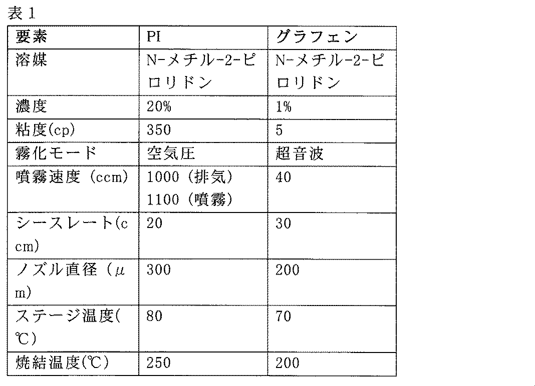
表1は、PI及びグラフェンの例示的なインク及び印刷パラメータを示している。印刷されたグラフェンの抵抗率及び皮膚接触インピーダンスは、それぞれ、約2×10⁻³Ωcm及び210.5kΩであり得る。
グラフェン電極印刷
図5Bは、グラフェン電極のための印刷プロセス500を示している。図5Bに示される例では、プロセス500は、1000RPMで30秒間、ガラス上にPMMA(例えば、950PMMA、Kayaku Advanced Materials)をスピンコーティング(502)し、200℃で2分間、ベークすることを含む。次いで、プロセス500は、エアロゾルジェットプリンタの空気噴霧器で4:1の比率でN-メチル-2-ピロリドン(NMP、Sigma-Aldrich)に溶解したポリイミド(PI)インク(PI-2545、MicroSystems)を噴霧(504)し、直径300μmのノズルを使用して堆積させ、次いで250℃で1時間硬化させることを含む。次いで、プロセス500は、直径200μmのノズルを用いて、NMPに溶解したグラフェンインクの1%を印刷すること(506)を含む。次いで、プロセス500は、印刷された電極をアセトンに溶解することを含む。次に、プロセス500は、PMMA/スライドガラスから水溶性テープ(ASWT-2、Aquasol)で、印刷されたグラフェン層を剥離(508)し、それをシリコーンエラストマー(1mmの厚さ、Ecoflex00-30とゲルの1:2の混合物、Smooth-On)上に置くことを含む。脱イオン水でテープを洗浄する。
Graphene Electrode Printing Figure 5B shows a
回路の製作。
図5Bはまた、薄膜ベースの回路のための微細加工プロセス510を示している。図5Bに示される例では、プロセス510は、30秒間4000RPMでSiウェハ上にPDMS(4:1の塩基硬化剤比)をスピンコーティングすること(512)を含む。第1のPI層(PI-2610、MicroSystems)を2000RPMで60秒間スピンコーティングした後、100℃で5分間ソフトベークし、250℃で1時間ハードベークすることができる。次いで、プロセス510は、スパッタリングによって0.5μmの厚さのCuを堆積すること(514)を含む。
Making of circuits.
Figure 5B also illustrates a
次いで、プロセス510は、フォトレジスト(PR、MicropositSC1813、MicroChem)を3000RPMで30秒間スピンコーティングすること(516)を含む。次いで、この被加工物をフォトマスクと整列させ、UV光に露出させ、現像剤を用いて現像することができる。
The
次いで、プロセス510は、Cuエッチング剤(APS-100、Transene)を用いてエッチングすること(518)を含む。第2のPI層(PI-2545)は、2000RPMで60秒間スピンコーティングされ、100℃で5分間ソフトベークすることができる。次いで、真空オーブンで240℃、1時間ハードベークすることができる。
The
次いで、プロセス510は、PR(AZ P4620、Integrated Micro Materials)を2000RPMで30秒間スピンコーティングして(520)、90℃で4分間ソフトベークすることを含む。フォトリソグラフィを実施して、15mJ/cm2の強度のUV光を100秒間露光することができる。次いで、この被加工物は、DI水で希釈した(AZ-400K:DI水=1:4)現像剤(AZ-400K、Integrated Micro Materials)で現像することができる。ビアホールは、反応性イオンエッチャ(RIE)を用いてエッチングすることができる。2μmの厚さの第2のCu層を、スパッタリングによって堆積させることができる。次いで、プロセスは、PR(AZ P4620)を1500RPMで30秒間スピンコートし、90℃で4分間ソフトベークすることができる。フォトリソグラフィを実施して、15mJ/cm2の強度を有するUV光を120秒間露光し、現像することができる。プロセスは、Cuエッチング剤を用いて、露出したCuをエッチングすることができる。
The
次いで、プロセス510は、3000RPMで60秒間、第3のPI層(PI-2610)をスピンコーティング(522)し、100℃で5分間ソフトベークし、240℃で1時間、真空オーブンでハードベークすることを含む。プロセスは、30秒間900RPMでPR(AZP4620)をスピンコートし、4分間90℃でソフトベークすることができる。フォトリソグラフィを実施して、UV光を露光し、PRを現像することができる。プロセスは、RIEを用いて、露出されたPIをエッチングすることができる。
The
プロセス510は、次いで、PDMS/Siウェハから水溶性テープを用いて微細加工された回路を剥離することによって、回路をエラストマーに移すこと(526)を含む。プロセス510は、次いで、スクリーン印刷低温はんだペーストを用いて、マイクロチップコンポーネントを取着すること(528)を含む。プロセス510は、マイクロチップコンポーネントをエラストマーでカプセル化することを含む。
実施例
The
Example
生体マウスモデルを用いて発症した頭蓋顔面体積筋肉損失(VML)と、VMLのリアルタイムの継続的な監視のために皮膚装着可能な印刷されたセンサ及び電子機器を統合する、無線ナノ膜非侵襲的システムと、を含む、例示的なシステム及び方法が開示される。生検パンチ誘発の咬筋損傷を使用した頭蓋顔面VMLモデルは、筋肉の再生の障害、及び筋肉常在幹細胞の活動の不均衡を示す。咀嚼中の活性なマウスの小さくて丸い咬筋の電気生理学を測定するために、標的筋肉上の皮膚に積層することができる、伸縮可能なグラフェンセンサを備える装着可能なナノ膜システムが利用される。 Exemplary systems and methods are disclosed, including a craniofacial volumetric muscle loss (VML) developed using a live mouse model and a wireless nanomembrane non-invasive system that integrates skin-wearable printed sensors and electronics for real-time continuous monitoring of VML. A craniofacial VML model using biopsy punch-induced masseter muscle injury shows impaired muscle regeneration and imbalance in muscle-resident stem cell activity. A wearable nanomembrane system with stretchable graphene sensors that can be layered on the skin over the target muscle is utilized to measure the electrophysiology of active mouse small round masseter muscle during chewing.
薄膜電子機器は、筋肉活動の長距離無線記録を提供しながら、その自然な挙動を可能にするように、マウスの背面にシームレスに取着することができる。非侵襲的システムは、VML損傷の有無にかかわらず、咬筋に対して高感度の筋電図検出を提供する。更に、無線センサは、頭蓋顔面VMLの移植手術後の回復を監視することができることが実証されている。手術後の機能回復は、頭蓋顔面VMLを治療するための四肢と咬筋移植との間で同等であることが示されている。全体的に、幹細胞生物学、ナノ製造、電気生理学、及び信号処理の提示された広範囲の研究は、頭蓋顔面VMLのメカニズム研究及び治療開発の両方のための、本発明者らの咬筋VML損傷モデル及び装着可能な電子的評価ツールの非常に大きい可能性を示している。 The thin-film electronics can be seamlessly attached to the back of the mouse to allow its natural behavior while providing long-range wireless recording of muscle activity. The non-invasive system provides highly sensitive electromyographic detection for the masseter muscle with or without VML injury. Furthermore, it is demonstrated that the wireless sensor can monitor the recovery after implantation surgery of craniofacial VML. Post-operative functional recovery has been shown to be comparable between limb and masseter muscle transplants for treating craniofacial VML. Overall, the presented extensive studies of stem cell biology, nanofabrication, electrophysiology, and signal processing show the enormous potential of our masseter VML injury model and wearable electronic assessment tools for both mechanistic studies and therapeutic development of craniofacial VML.
実施例1:無線ナノ膜電子機器を使用したマウスにおける頭蓋顔面VMLのリアルタイム機能評価
図6A~6Eは、無線ナノ膜電子機器を使用したマウスにおける頭蓋顔面VMLのリアルタイム機能評価の概要を示している。
Example 1: Real-time functional assessment of craniofacial VML in mice using wireless nanomembrane electronics. Figures 6A-6E show an overview of real-time functional assessment of craniofacial VML in mice using wireless nanomembrane electronics.
具体的には、図6Aは、咬筋の頭蓋顔面VMLの機能的定量化のための、マウスの皮膚上の無線電子システムの概略例解図を示す。マウスの頬領域のEMGは、携帯型デバイス上で継続的に監視される。挿入図は、咬筋のVMLを治療するための、咬筋及び移植のパンチ誘発VMLのプロセスを示している。咬筋を、頭蓋顔面VMLのモデルとして、生検パンチによって損傷した。VML損傷した咬筋を、前脛骨筋(TA)又は咬筋の生検片で移植して、損傷領域を充填する。このデバイスは、マウスの頬の皮膚に直接適用するための非侵襲的で伸縮可能な電極と、マウスの背面に取り付けるための小型化された無線の軟質回路と、を含む。咀嚼中、EMGデータが、マウスの頬の皮膚上の電極によって測定され、データ処理のために回路内のマイクロプロセッサに送達される。次いで、信号は、リアルタイムの継続的なデータ監視及び保存のために、外部の携帯型デバイスに無線で送信される。咬筋にVMLを確立するために、正常な咬筋は、直径3mmの生検パンチによって損傷される。損傷した筋肉領域を、3mmの生検前脛骨筋(TA)又は咬筋(図6Aの挿絵)で移植する。 Specifically, FIG. 6A shows a schematic illustration of a wireless electronic system on mouse skin for functional quantification of the craniofacial VML of the masseter muscle. The EMG of the mouse cheek region is continuously monitored on a portable device. The inset shows the process of punch-induced VML of the masseter muscle and transplantation to treat VML of the masseter muscle. The masseter muscle was injured by a biopsy punch as a model of craniofacial VML. The VML-injured masseter muscle was transplanted with a biopsy piece of the tibialis anterior (TA) or masseter muscle to fill the damaged area. The device includes non-invasive, stretchable electrodes for direct application to the mouse cheek skin and a miniaturized, wireless, soft circuit for attachment to the back of the mouse. During chewing, EMG data is measured by the electrodes on the mouse cheek skin and delivered to a microprocessor in the circuit for data processing. The signal is then wirelessly transmitted to an external portable device for real-time continuous data monitoring and storage. To establish a VML in the masseter muscle, the normal masseter muscle is injured with a 3 mm diameter biopsy punch. The injured muscle area is grafted with a 3 mm biopsy of tibialis anterior (TA) or masseter muscle (Figure 6A illustration).
図6Bは、マウス皮膚上での共形積層を可能にする、軟質エラストマー基板上の軟質回路(左)及び伸縮可能なセンサ(右)の多層構造の概略例解図を示している。 Figure 6B shows a schematic illustration of a multilayer structure of flexible circuits (left) and stretchable sensors (right) on a soft elastomeric substrate, enabling conformal deposition on mouse skin.
図6Cは、咬筋にVMLを生成するための筋肉生検を示している。咬筋の中央は、3mmの生検パンチによって生検されている。矢印は、生検からの筋肉片を示す。図6Dは、咀嚼中のデバイスを有する活性マウスの光学画像を示す。図6Cは、咬筋VMLを生成するために使用される筋肉生検を示しており、一方、図6Dは、監視中に動きが妨げられないままであることを示している。 Figure 6C shows a muscle biopsy to generate a VML in the masseter muscle. The center of the masseter muscle is biopsied with a 3 mm biopsy punch. The arrow indicates the muscle piece from the biopsy. Figure 6D shows an optical image of an active mouse with the device during chewing. Figure 6C shows the muscle biopsy used to generate the masseter VML, while Figure 6D shows that the movement remains unimpeded during monitoring.
図6Eは、筋肉機能を分析する定量的測定項目を取り込むフローチャートを示す。図6Eに示されるフローチャートは、無線EMG測定及び筋肉機能分析のための定量的測定項目を示す。この装着可能な電子システムは、本発明者らのげっ歯類モデルにおける3つの異なる頭蓋顔面筋状態(正常、VML、移植によって処置されたVML)の各々におけるEMG活性を監視することができる。また、運動センサパッケージは、咀嚼運動を区別するためにマウスの動きを識別する感度の高い加速度計及びジャイロスコープを使用することができる。携帯型デバイスは、リアルタイムの信号表示及びデータ保存を提供して筋肉機能を分析するための、カスタム設計されたアプリが組み込まれる。 Figure 6E shows a flow chart for capturing quantitative measurements to analyze muscle function. The flow chart shown in Figure 6E shows the quantitative measurements for wireless EMG measurement and muscle function analysis. This wearable electronic system can monitor EMG activity in each of the three different craniofacial muscle conditions (normal, VML, VML treated by implantation) in our rodent model. The motion sensor package can also use sensitive accelerometers and gyroscopes to distinguish mouse movements to differentiate chewing movements. The handheld device is embedded with a custom designed app to provide real-time signal display and data storage to analyze muscle function.
実施例2 グラフェン膜電極及び軟質無線回路の特徴付け
我々の頭蓋顔面VMLモデルの筋肉機能を定量化するために、例えば、図4に関連して説明されるように、EMG信号を、装着可能な無線システムを用いて監視した。動物試験における従来のEMGシステム[35、38、39]は、標的筋肉に貫通する、かさばって侵襲的な針タイプの有線電極を使用する。これらのシステムに伴う主な問題は、それらの電極が、活動的な動きを有する小さいマウスでの使用には好適でないことである。対照的に、この試験は、自然に移動するマウスと共に筋肉機能を無線による高い忠実度で記録することを提供するための、非侵襲的な、小型化された、軟質電子システムを開発した。
Example 2 Characterization of Graphene Film Electrodes and Soft Wireless Circuits To quantify muscle function in our craniofacial VML model, EMG signals were monitored using a wearable wireless system, for example as described in relation to FIG. 4. Conventional EMG systems in animal studies [35, 38, 39] use bulky and invasive needle-type wired electrodes that penetrate the target muscles. The main problem with these systems is that they are not suitable for use in small mice with active movements. In contrast, this study developed a non-invasive, miniaturized, soft electronic system to provide wireless high-fidelity recording of muscle function with naturally moving mice.
組織に取着されたセンサの生体適合性は、副作用を伴う安全で継続的な使用を提供するために使用することができる特徴である。[40、41]加えて、電極の細胞毒性は、筋肉活動を測定するときにVML損傷領域の皮膚細胞を損傷する可能性がある。印刷されたグラフェン電極の生体適合性の特徴付けを、ヒトのケラチノサイト細胞を用いて実施した。グラフェン及び対照(ポリスチレン細胞培養皿)上の生細胞の数を、蛍光強度によって判定した。 The biocompatibility of the tissue-attached sensor is a feature that can be used to provide safe and continuous use with no side effects. [40, 41] In addition, the cytotoxicity of the electrodes may damage skin cells in the VML-damaged area when measuring muscle activity. Characterization of the biocompatibility of the printed graphene electrodes was performed with human keratinocyte cells. The number of live cells on the graphene and control (polystyrene cell culture dish) was determined by fluorescence intensity.
対照(ポリスチレンペトリ皿、左)及びエラストマー上に統合されたグラフェン(右)を含む、2つのタイプの基板上に培養されたケラチノサイト細胞の蛍光画像を撮影した。37℃で5%のCO2で培養器内に培養したヒト一次ケラチノサイト細胞を使用して、試験を行った。培養器では、材料試料を24個ウェルプレートに配置し、5000個ケラチノサイト/cm2を蒔種した。培養器で7日後、ケラチノサイト細胞をリン酸緩衝生理食塩水(FisherChemical)で洗浄し、0.9mlの培養培地において0.1mlのカルセインブルーAM(Thermo Fisher)で染色した。ケラチノサイト及び試薬は、37℃の培養器に10分間追加で保管した。次いで、更なる生体適合性のために、上澄み液を96個ウェルプレートに等分した。 Fluorescence images were taken of keratinocyte cells cultured on two types of substrates, including a control (polystyrene petri dish, left) and graphene integrated on an elastomer (right). Tests were performed using human primary keratinocyte cells cultured in an incubator at 37°C and 5% CO2 . In the incubator, material samples were placed in 24-well plates and seeded at 5000 keratinocytes/ cm2 . After 7 days in the incubator, keratinocyte cells were washed with phosphate-buffered saline (Fisher Chemical) and stained with 0.1 ml of Calcein Blue AM (Thermo Fisher) in 0.9 ml of culture medium. Keratinocytes and reagents were kept in the incubator at 37°C for an additional 10 minutes. The supernatant was then aliquoted into 96-well plates for further biocompatibility.
図7A~7Jは、グラフェン膜電極及び無線軟質回路の特徴付けの結果を示している。具体的には、図7Aは、エラストマー膜上の伸縮可能な印刷された電極の写真を示している。図7Bは、多層電極のSEM画像の断面図(左)及び上面図(右)を示している。図7Cは、印刷されたグラフェンの表面粗さを示すAFM画像を示している。平均RMSは、72.9nmであり、これは、印刷されたグラフェン層における均一性を確保する。図7Dは、対照及びグラフェン上の培養細胞の細胞吸光度(左)と蛍光(右)との比較を示している。データは、一元配置分散分析(ns=統計的に有意ではない)で分析されている。誤差バーは、標準偏差を表す。図7Eは、100サイクルにわたる引張ひずみ変化(下)の60%に従う相対抵抗変動(上)を表すグラフを示している。図7Dの培養細胞の吸光度(左のグラフ)及び蛍光(右のグラフ)は、印刷されたグラフェン電極が細胞生存率に対して無視できる影響を有することを示していることを観察することができる。図7Eは、100サイクルにわたる引張ひずみ変化(下)の60%に従う電極(上)の相対抵抗変動を表し、ひずみ下での信頼性の高い機械的性能を示している。 Figures 7A-7J show the results of characterization of the graphene membrane electrodes and wireless flexible circuits. Specifically, Figure 7A shows a photograph of the stretchable printed electrodes on an elastomeric membrane. Figure 7B shows cross-sectional (left) and top-view (right) SEM images of the multilayer electrodes. Figure 7C shows an AFM image showing the surface roughness of the printed graphene. The average RMS is 72.9 nm, which ensures uniformity in the printed graphene layers. Figure 7D shows a comparison of cell absorbance (left) and fluorescence (right) of control and cultured cells on graphene. Data are analyzed with one-way ANOVA (ns = not statistically significant). Error bars represent standard deviation. Figure 7E shows a graph representing the relative resistance variation (top) following 60% of the tensile strain change (bottom) over 100 cycles. It can be observed that the absorbance (left graph) and fluorescence (right graph) of cultured cells in Figure 7D indicate that the printed graphene electrodes have negligible effect on cell viability. Figure 7E shows the relative resistance variation of the electrode (top) following 60% of the tensile strain change (bottom) over 100 cycles, demonstrating reliable mechanical performance under strain.
図7Fは、維持されたデータ伝送速度1104バイト/秒を示す、Bluetooth通信距離に従ったRSSI応答を示している。図7Gは、装着可能なセンサシステムからのリアルタイムの継続的な運動(加速度計及びジャイロスコープ)、並びにEMG信号を表示する携帯型デバイスアプリケーションインターフェースを示している。図7Hは、繰り返し負荷時の可撓性回路の抵抗変化(1.5mmの曲率半径で180°曲げて100回)を示し、抵抗のわずかな変化を示している。 Figure 7F shows the RSSI response over Bluetooth communication distance, indicating a sustained data transmission rate of 1104 bytes/sec. Figure 7G shows the mobile device application interface displaying real-time continuous motion (accelerometer and gyroscope) and EMG signals from the wearable sensor system. Figure 7H shows the resistance change of the flexible circuit upon cyclic loading (100 180° bends with a 1.5 mm radius of curvature), showing a small change in resistance.
受信信号強度表示(RSSI)の分析は、1104バイト/秒の維持されたデータ伝送速度で5mの無線通信距離の成功を示している(図7F)。送信されたEMG信号及び運動信号は、カスタマイズされたアプリ(図7G)を有する携帯型デバイスに表示及び記憶される。軟質で可撓性のある回路は、繰り返し負荷(100サイクル、図7H)中の完全な折り畳み(1.5mmの曲率半径を有する180度)下であっても機械的信頼性を実証しており、計算モデル化データ(図11A及び11B)からの結果と一致する。図11A~11Bは、計算機械シミュレーションの結果を示している。具体的には、図11Aは、60%の1軸伸張(右)の前(左)及び後(右)の印刷された電極の機械的適合性を示す有限要素分析(FEA)結果を示している。図11Bは、180°において曲げ可能な回路のFEA結果を示し、機械的破壊を示していない。 Analysis of the received signal strength indication (RSSI) indicates successful wireless communication range of 5 m with a sustained data transmission rate of 1104 bytes/sec (Fig. 7F). The transmitted EMG and kinematic signals are displayed and stored on the portable device with a customized app (Fig. 7G). The soft and flexible circuit demonstrates mechanical reliability even under complete folding (180 degrees with a radius of curvature of 1.5 mm) during cyclic loading (100 cycles, Fig. 7H), consistent with results from computational modeling data (Figs. 11A and 11B). Figs. 11A-11B show the results of computational mechanical simulation. Specifically, Fig. 11A shows finite element analysis (FEA) results showing the mechanical compliance of the printed electrodes before (left) and after (right) 60% uniaxial stretch (right). Fig. 11B shows the FEA results of the bendable circuit at 180 degrees, showing no mechanical failure.
実施例3 頭蓋顔面VMLの確立及び欠陥咬筋再生
頭蓋顔面VMLを確立するために、咬筋が咀嚼のための重要な筋肉であるため、下顎を上方に引っ張ることによって、咬筋を選択した。咬筋は、浅い咬筋と深い咬筋で構成されている。浅い咬筋は、厚くて腱のような部分であり、かつ頬骨につながっており、一方、深い咬筋は、より小さくて下顎につながっている[42]。図8A~8Fは、頭蓋顔面VMLを確立すること及び欠陥咬筋再生の試験を示している。
Example 3 Establishment of craniofacial VML and defective masseter regeneration To establish the craniofacial VML, the masseter was selected by pulling the mandible upwards because it is the key muscle for mastication. The masseter is composed of the superficial and deep masseter. The superficial masseter is a thick, tendon-like part and connects to the zygoma, while the deep masseter is smaller and connects to the mandible [42]. Figures 8A-8F show the establishment of the craniofacial VML and the test of defective masseter regeneration.
具体的には、図8Aは、マウスの咬筋の頭蓋顔面VMLの例解図を示している。図8Bは、3mmの生検パンチが、6か月齢の雄と雌との間で異なる程度の筋肉損失を誘発することを示している。性別ごとに、n=4。損傷は、3mmの生検パンチ(図8A)を使用して浅い咬筋に適用され、これは、6か月齢の雄及び雌マウスからそれぞれ8.5±2.3グラム及び7.9±1.2グラムの筋肉量を失うことによって、咬筋組織の約8.5%及び15%の損失を生成することができる(図8B)。雌マウスに対して3mm生検を使用した咬筋VML損傷は、マウス四肢VML損傷と比較して、同様の重大な損傷サイズ(15%の喪失)体積を呈し[16]、雄マウスについてはそうではなかったが、雄マウスの咬筋に対する3mm生検は、機能障害を誘発する可能性があり、残りの実験については、雌マウスを使用した。頭蓋顔面VMLモデルにおいて、筋肉再生が3mmの筋肉生検誘発性損傷後に生じるかどうかを検証するために、咬筋を、VML損傷後の7日及び28日で切片化した。 Specifically, Figure 8A shows an illustration of the craniofacial VML of the masseter muscle in mice. Figure 8B shows that a 3 mm biopsy punch induces different degrees of muscle loss between 6-month-old males and females. n=4 per gender. Injuries were applied to the superficial masseter muscle using a 3 mm biopsy punch (Figure 8A), which can generate approximately 8.5% and 15% loss of masseter muscle tissue by losing 8.5±2.3 grams and 7.9±1.2 grams of muscle mass from 6-month-old male and female mice, respectively (Figure 8B). Masseter VML injuries using 3 mm biopsies for female mice exhibited similar critical injury size (15% loss) volume compared to mouse limb VML injuries [16], but not for male mice, suggesting that 3 mm biopsies on the masseter muscle of male mice could induce functional impairment, and female mice were used for the remaining experiments. To examine whether muscle regeneration occurs after a 3 mm muscle biopsy-induced injury in a craniofacial VML model, masseter muscles were sectioned at 7 and 28 days after VML injury.
図8Cは、VML損傷後の7日(左)及び28日(右)後の咬筋の組織像を示している。筋肉切片をヘマトキシリン及びエオシンで染色して、核(紫色)及び細胞質(赤色)を可視化した。点線領域は、非筋肉領域を示している。小さな正方形の画像は、下部パネルにおいて拡大され、筋肉組織の細胞要素を示している。下部パネルの矢印は、筋線維を再生する特徴である、筋線維含有中心核を示している。図8Dは、損傷後の7日及び28日において非筋肉領域が咬筋の大部分を占めていることを示している。誤差バーは、平均値の標準誤差(SEM)を表している。図8Cは、ヘマトキシリン及びエオシン染色を介して着色された咬筋のVML損傷の中点の組織像データを示しており、これは、非筋肉領域(図8Cの点線)が、免疫細胞、及び線維症であり得る、非筋肉細胞で満たされていることを示している。非筋肉領域は、損傷後7日(dpi)において、損傷した咬筋の約40%を占め、28dpiでそのままであった(図8D)。再生筋の特徴である、中央の核を含む筋線維の断面積を測定することによって、損傷領域の縁で限定的な筋肉再生が観察された(図8Cの底部画像の矢印)。
Figure 8C shows histology of the
図8Eは、いくつかの再生筋線維が、損傷後7日及び28日で非常に低かったことを示している。データは、スチューデントt検定によって分析されている。ns=統計的に有意ではない。図8Fは、VML損傷後7日(左)及び28日(右)の咬筋の線維化の結果を示している。筋肉切片をマッソンのトリクロム染色で染色し、筋肉組織(褐色)から線維化領域(青色)を可視化した。再生された筋線維数は、7dpi及び28dpiでの咬筋中の全筋線維の約2%であった(図8E)。加えて、7日後及び28日後の咬筋の非筋肉領域における線維化VML損傷は、マッソンのトリクロム染色によって検出された(図8F)。VML後の欠陥筋肉再生は、VML(図12)を有する咬筋の減少した衛星細胞によって支持され、その衛生細胞は、凍結誘発性咬筋損傷のものと比較して、損傷時の筋肉再生に不可欠な筋肉幹細胞であった[7]。図12A~12Dは、VML損傷後のマウスのフィルタリングされたEMG信号の結果を示している。図12A(非摂食)と図12B(摂食)との間には、咬筋損失による咀嚼挙動の信号変動は存在しない。30日後の損傷したVML後(図12C)、及び損傷していない咬筋(図12D)を有する3匹のマウスの咀嚼EMG信号である。 Figure 8E shows that few regenerated muscle fibers were very low at 7 and 28 days after injury. Data were analyzed by Student's t-test. ns = not statistically significant. Figure 8F shows the results of fibrosis in the masseter muscle at 7 days (left) and 28 days (right) after VML injury. Muscle sections were stained with Masson's trichrome stain to visualize fibrotic areas (blue) from muscle tissue (brown). The number of regenerated muscle fibers was about 2% of the total muscle fibers in the masseter muscle at 7 dpi and 28 dpi (Figure 8E). In addition, fibrotic VML injury in the non-muscle area of the masseter muscle after 7 and 28 days was detected by Masson's trichrome stain (Figure 8F). Defective muscle regeneration after VML was supported by reduced satellite cells in the masseter muscle with VML (Figure 12), whose satellite cells were essential muscle stem cells for muscle regeneration upon injury, compared with those of freeze-induced masseter injury [7]. Figures 12A-12D show the results of filtered EMG signals of mice after VML injury. There is no signal variation in chewing behavior due to masseter muscle loss between Figure 12A (non-fed) and Figure 12B (fed). Chewing EMG signals of three mice with injured VML (Figure 12C) and intact masseter muscle (Figure 12D) after 30 days.
加えて、慢性筋肉損傷における線維症及び脂肪沈着を媒介する筋肉間葉系幹細胞である線維性脂肪前駆細胞[43、44]は、凍結誘発性咬筋損傷よりもVML咬筋において非常に増加しており(図13)、これは、VML咬筋における線維症を誘発する可能性がある。これらの結果は、3mm生検損傷によって生成された頭蓋顔面VMLが、同じ方法を使用した四肢VML実験で見られるものと同様に、欠陥筋肉再生をもたらすことを証明している。[16] In addition, fibrotic preadipocytes, muscle mesenchymal stem cells that mediate fibrosis and fat deposition in chronic muscle injury [43, 44], were significantly increased in VML masseter than in freeze-induced masseter injury (Figure 13), which may induce fibrosis in VML masseter. These results demonstrate that craniofacial VMLs generated by 3 mm biopsy injury result in defective muscle regeneration, similar to that seen in limb VML experiments using the same method. [16]
図13A~13Eは、VML損傷後3日の咬筋における幹細胞の調節不全の結果を示している。具体的には、図13Aは、実験のスキームを示している。タモキシフェンをPax7cre/ERT;tdTomatoマウスに5日間注射して、衛星細胞におけるtdTomato蛍光発現を誘発させた。VML又は凍結損傷は、タモキシフェン注射の10日後に行われた。単核細胞を、損傷の3日後にフローサイトメトリー分析のために単離した。図13Bは、フローサイトメトリーを使用して、赤色蛍光タンパク質(tdTomato)によってゲートされる衛星細胞の代表的なドットプロットを示している。図13Cは、VML損傷した咬筋からのいくつかの衛星細胞が、損傷していない咬筋のうちの1つと同等であることを示している。衛星細胞数は、損傷していない筋肉の平均衛星細胞数で正規化されている。凍結損傷を受けた筋肉は、陽性対照として提供される。誤差バーは、平均値の標準偏差(SEM)を表している。図13Dは、フローサイトメトリーを使用して、表面マーカー(Cd31-、CD45-、Sca1+)によって定義される、線維性脂肪細胞(FAP)の代表的なヒストグラムを示している。図13Eは、VML損傷した咬筋において、衛星細胞に対するFAPの比率が増加していることを示している。データは、事後の比較のために、一元配置分散分析及びKruskal-Wallis法で分析されている。*p<0.05。
13A-13E show the results of stem cell dysregulation in
実施例4 VML損傷した咬筋の機能を評価するための、無線の装着可能なEMGシステムのデモンストレーション
図9A~9Gは、VML損傷した咬筋の機能を評価するための、無線の装着可能なEMGシステムの動作を示している。具体的には、図9Aは、例えば、マウスにおけるVMLの非侵襲的診断のための、薄膜医療用パッチ上の完全に統合された無線の装着可能な電子機器を示している。
Example 4 Demonstration of a Wireless Wearable EMG System for Assessing Function of VML-Injured Masseter Muscle Figures 9A-9G show the operation of a wireless wearable EMG system for assessing function of VML-injured masseter muscle. Specifically, Figure 9A shows a fully integrated wireless wearable electronics on a thin medical patch for non-invasive diagnosis of VML in mice, for example.
図9Bは、ヌードマウスの頬(左)に取り付けられた超薄型の伸縮可能なEMGセンサ、及び背面(右)上の軟質回路を示す写真を示している。デバイスは、標的マウスの皮膚に容易に取着するための薄膜パッチで完全に覆われている。伸縮可能なEMG電極は、マウスの頬に直接取り付けられ(図9Bの左画像)、一方、軟質無線回路は、身体の背面上に取着されている(図9Bの右画像)。 Figure 9B shows a photograph showing the ultra-thin stretchable EMG sensor attached to the cheek (left) of a nude mouse, and the flexible circuit on the back (right). The device is completely covered with a thin film patch for easy attachment to the skin of the target mouse. The stretchable EMG electrode is attached directly to the mouse's cheek (left image of Figure 9B), while the flexible wireless circuit is attached on the back of the body (right image of Figure 9B).
図9Cは、ケージ内にマウスを入れた実験装置の写真を示している。リアルタイムの継続的な運動及びEMGデータは、組み込まれたアプリケーションを有する携帯型デバイスによって監視及び記録される。ケージ内での摂食及び歩き回りを含む、マウスの自然な活動の間、リアルタイムの継続的な筋肉機能が、装着可能なデバイス及びタブレットによって監視される。 Figure 9C shows a photograph of the experimental setup with a mouse in a cage. Real-time continuous movement and EMG data are monitored and recorded by a handheld device with an embedded application. Real-time continuous muscle function is monitored by the wearable device and tablet during the mouse's natural activities, including feeding and walking around in the cage.
図9Dは、静止状態(上)及び咀嚼期(下)中の、非損傷マウスで測定されたリアルタイムのEMG信号の比較を示しており、明確な信号差を示している。図9Dは、静止状態(上)及び咀嚼期(下)中の、正常なマウスから記録されたリアルタイムのEMGデータを取り込んでおり、これは、増加したピークツーピーク電圧を表示している。咀嚼EMG信号の振幅は、マウスによって測定された典型的な間欠運動アーチファクトと比較してはるかに小さい(図14)。図14は、摂食中の非損傷マウスのフィルタリングされたEMG信号を示している。運動アーチファクトは、咀嚼信号と比較して、有意な、かつ区別可能な振幅を生み出している。 Figure 9D shows a comparison of real-time EMG signals measured in an uninjured mouse during resting state (top) and chewing phase (bottom), showing clear signal differences. Figure 9D captures real-time EMG data recorded from a normal mouse during resting state (top) and chewing phase (bottom), displaying increased peak-to-peak voltage. The amplitude of the chewing EMG signal is much smaller compared to the typical intermittent movement artifacts measured by the mouse (Figure 14). Figure 14 shows the filtered EMG signal of an uninjured mouse during feeding. The movement artifacts produce significant and distinguishable amplitudes compared to the chewing signal.
VML損傷後の筋肉の機能を定量化するために、咬筋のEMG活動を、VML損傷後30日で監視した。図9Eは、30日後の、非損傷マウス(上)及びVML損傷後のマウス(下)の写真を示している。矢印は、VML損傷領域の位置を示している。図9Fは、図9Eの2つの症例に対応する、咀嚼中の代表的なRMSのEMG信号を示している。加えて、平均二乗根(RMS)EMG信号(図9F)及びそれらの信号対雑音比(SNR)値(図9G)の両方が、2つの群の間の大きい有意性(p値<0.01)を示し、VML損傷した筋肉が損傷から1か月後においても回復していないことを示唆しており、これは、図8の本発明者らの組織学的研究結果と一致する。非損傷の症例(上)と、VML損傷した咬筋(下)との間で、損傷後30日において、明確な信号差が観察されている。図9Gは、咀嚼中の、非損傷咬筋とVML損傷後の咬筋との間の、要約されたEMGのSNRデータを示している。データは、対応のない両側スチューデントt検定によって分析された。**p<0.01。 To quantify muscle function after VML injury, the EMG activity of the masseter muscle was monitored 30 days after VML injury. Figure 9E shows photographs of an uninjured mouse (top) and a mouse after VML injury (bottom) after 30 days. The arrows indicate the location of the VML injury area. Figure 9F shows representative RMS EMG signals during chewing corresponding to the two cases in Figure 9E. In addition, both the root mean square (RMS) EMG signals (Figure 9F) and their signal-to-noise ratio (SNR) values (Figure 9G) showed high significance (p-value < 0.01) between the two groups, suggesting that the VML-injured muscle had not recovered even one month after injury, which is consistent with our histological findings in Figure 8. A clear signal difference is observed between the uninjured case (top) and the VML-injured masseter muscle (bottom) at 30 days after injury. FIG. 9G shows the summarized EMG SNR data between the uninjured and VML-injured masseter muscles during chewing. Data were analyzed by unpaired two-tailed Student's t-test. **p<0.01.
実施例5 VML損傷した咬筋の移植後の機能回復監視
図10A~10Gは、移植後のVML損傷した咬筋の機能回復を監視するための動作を示している。具体的には、図10Aは、野生型マウスのTA(青色)又は咬筋(赤色)からの生検片で充填された、免疫不全マウス(NRG)の咬筋の生検パンチ誘発のVML領域を示す実験スキームを示している。EMG測定及び線維化分析は、VML損傷/移植の30日後に実施されている。頭蓋顔面VMLは、診療所において、自己四肢筋肉移植を用いて処置された[45-47]。頭蓋顔面VML処置のための移植された筋肉の元を評価するために、咬筋又は前脛骨筋(TA)筋肉の移植を、咬筋のVML領域において行った。
Example 5 Monitoring functional recovery after transplantation of VML-injured masseter muscle Figures 10A-10G show the operation for monitoring the functional recovery of VML-injured masseter muscle after transplantation. Specifically, Figure 10A shows the experimental scheme showing biopsy punch-induced VML region of masseter muscle of immunodeficient mice (NRG) filled with biopsy pieces from TA (blue) or masseter muscle (red) of wild-type mice. EMG measurements and fibrosis analysis are performed 30 days after VML injury/transplantation. Craniofacial VML was treated in the clinic with autologous limb muscle transplantation [45-47]. To evaluate the origin of transplanted muscles for craniofacial VML treatment, transplantation of masseter muscle or tibialis anterior (TA) muscle was performed in the VML region of masseter muscle.
図10Bは、線維症を測定するために、TA及び咬筋移植を伴うVML損傷した咬筋を切片化して、Col VI抗体(赤)で標識付けすることを示している。DAPI染色が、筋肉切片の核を標識付けするために使用される。3mmの生検パンチを使用して、野生型マウスから咬筋又はTA筋肉を生検し、重度の免疫不全マウスの咬筋の1:1の体積一致(図10B)を確実にするために、VML(3mmの生検領域)に移植した。 Figure 10B shows that VML injured masseter muscles with TA and masseter muscle grafts are sectioned and labeled with Col VI antibody (red) to measure fibrosis. DAPI staining is used to label nuclei in muscle sections. Masseter or TA muscles were biopsied from wild-type mice using a 3 mm biopsy punch and grafted into the VML (3 mm biopsy area) to ensure a 1:1 volumetric match of the masseter muscles of severely immunodeficient mice (Figure 10B).
図10Cは、Col VI染色の平均強度が、TA又は咬筋の移植が移植されたVML損傷した咬筋において同等レベルの線維化を生じさせたことを示すことを示しており、各グループに対して、n=3であり、誤差バーは、平均値の標準誤差を表している。データは、二元配置分散分析を用いて分析される。p**<0.01。これらのマウスは、ホストの免疫系からの移植拒絶反応の可能性が最小限であるため、この移植試験で使用されている。[48]適切な筋肉収縮性を確保するために、提供側筋肉と受容側筋肉との間の繊維を整列させるために、全生検咬筋又は全TA筋肉を配置した。移植された咬筋のEMG活動を、術後30日間測定した。線維症を測定するために、TA及び咬筋移植を伴う非損傷筋肉及びVML損傷した咬筋からの筋肉切片を、抗コラーゲンVI(Col VI)抗体(赤)で標識付けした(図10C)。 Figure 10C shows that the mean intensity of Col VI staining indicates that transplantation of TA or masseter muscle produced comparable levels of fibrosis in the transplanted VML-injured masseter muscle, n=3 for each group, and error bars represent standard error of the mean. Data are analyzed using two-way ANOVA. p**<0.01. These mice are used in this transplantation study because they have minimal chance of transplant rejection from the host's immune system. [48] To ensure proper muscle contractility, the whole biopsied masseter muscle or the whole TA muscle was positioned to align the fibers between the donor and recipient muscles. EMG activity of the transplanted masseter muscle was measured for 30 days after surgery. To measure fibrosis, muscle sections from uninjured and VML-injured muscles with TA and masseter muscle transplants were labeled with anti-collagen VI (Col VI) antibody (red) (Figure 10C).
図10Dは、TA筋肉(中央)又は咬筋(下)の移植を伴う、非損傷筋肉(上)及びVML損傷した咬筋のフィルタリングされたEMGの比較を示している。Col VI信号の強度を測定して、移植を伴う非損傷筋肉(対側)及びVML損傷した咬筋における線維症の程度を推定した。 Figure 10D shows a comparison of filtered EMG of uninjured muscles (top) and VML-injured masseter muscles with transplantation of TA muscles (middle) or masseter muscles (bottom). The intensity of the Col VI signal was measured to estimate the degree of fibrosis in uninjured muscles (contralateral) and VML-injured masseter muscles with transplantation.
図10Eは、TA筋肉(左)及び咬筋移植された咬筋(右)から測定されたRMS-EMG信号を示している。線維化レベルは、移植された咬筋を有するVMLでは、非損傷対側咬筋と比較して有意に高い。しかしながら、TA又は咬筋の移植は、移植されたVML損傷した咬筋において異なるレベルの線維化を生成しない。また、EMG活動は、装着可能な膜電子機器で監視されて、異なる筋肉移植後の咬筋の機能回復を識別する(図19)。咀嚼中に対側筋肉と比較して、両方の移植された筋肉において、EMG応答の減少が見られた(図10E)。 Figure 10E shows the RMS-EMG signals measured from the TA muscle (left) and the masseter-transplanted masseter muscle (right). Fibrosis levels are significantly higher in the VML with the transplanted masseter muscle compared to the uninjured contralateral masseter muscle. However, transplantation of the TA or masseter muscle does not produce different levels of fibrosis in the transplanted VML-injured masseter muscle. EMG activity was also monitored with wearable membrane electronics to identify functional recovery of the masseter muscle after different muscle transplants (Figure 19). A decrease in EMG response was seen in both transplanted muscles compared to the contralateral muscle during mastication (Figure 10E).
図10Fは、SNR値が、非損傷筋肉と比較して、移植筋肉群と共にVMLにおいて減少していることを示している(n=3)。TA又は咬筋移植は、VML損傷した咬筋と同様のレベルの機能回復をもたらす。両方の移植された筋肉の信号振幅は、同等であるように見える(図10F)。図10Gの要約されたSNR値は、両方のVML/移植された咬筋の筋肉機能が、非損傷の対側咬筋と比較して部分的に回復していることを示している。まとめると、TA又は咬筋移植は、VML損傷した咬筋と同様のレベルの線維化及び機能回復をもたらす。 Figure 10F shows that SNR values are decreased in the VML along with the transplanted muscle group compared to the uninjured muscle (n=3). TA or masseter transplantation results in a similar level of functional recovery to the VML-injured masseter. The signal amplitudes of both transplanted muscles appear to be comparable (Figure 10F). The summarized SNR values in Figure 10G show that muscle function of both VML/transplanted masseter muscles is partially restored compared to the uninjured contralateral masseter. In summary, TA or masseter transplantation results in a similar level of fibrosis and functional recovery to the VML-injured masseter.
実験
試験で使用されたマウス:C57BL/6Jマウス(Jax000664) (雌n=6及び雄n=4)、Pax7CreERT2/CreERT2マウス(Jax017763)、RosatdTomato/tdTomato (tdTomato) (Jax007909)、NU/J (Jax002019) (雌n=7)、NRG (NOD.Cg-Rag1tm1MomIl2rgtm1Wjl/SzJ; Jax007799) (雌n=6)をJackson Laboratories(Bar Harbor,ME;www.jax.org)から購入した。図の凡例に記載されているように、5~6か月齢のマウスを使用した。ホモ接合性Pax7CreERT2/CreERT2雄マウスをホモ接合性RosatdTomato/tdTomato(tdTomato)と交配して、Pax7CreERT2/+;RosatdTomato/+(Pax7CreERT2-tdTomato)マウス(雌n=12)を得た。赤色蛍光(tdTomato)で衛星細胞を標識付けするために、10g体重当たり1mg(Sigma-Aldrich、St.Louis、MO)のタモキシフェンを1日1回5日間腹腔内に注射した。実験は、エモリー大学の施設内動物管理使用委員会からの承認されたガイドライン及び倫理的承認に従って、並びに国立衛生研究所に準拠して実施された。
Experimental Mice used in the study: C57BL/6J mice (Jax000664) (n=6 females and n=4 males), Pax7 CreERT2/CreERT2 mice (Jax017763), Rosa tdTomato/tdTomato (tdTomato) (Jax007909), NU/J (Jax002019) (n=7 females), NRG (NOD.Cg-Rag1 tm1Mom Il2rg tm1Wjl /SzJ; Jax007799) (n=6 females) were purchased from Jackson Laboratories (Bar Harbor, ME; www.jax.org). Mice were 5-6 months old as described in the figure legends. Homozygous Pax7 CreERT2/CreERT2 male mice were crossed with homozygous Rosa tdTomato/tdTomato (tdTomato) to obtain Pax7 CreERT2/+ ; Rosa tdTomato/+ (Pax7Cre ERT2 -tdTomato) mice (female n=12). To label satellite cells with red fluorescence (tdTomato), tamoxifen was injected intraperitoneally once a day for 5 days at 1 mg (Sigma-Aldrich, St. Louis, MO) per 10 g body weight. Experiments were performed according to the approved guidelines and ethical approval from the Institutional Animal Care and Use Committee of Emory University and in compliance with the National Institutes of Health.
筋肉組織の損傷、及び組織学的分析のための調製:マウスを、ノーズコーンを使用して、2.5%イソフルラン吸入によって麻酔した。鎮痛のために、マウスに、筋肉損傷の前に、0.1mg/kgのブプレノルフィンSR(3日間持続放出)を皮下注射した。標的損傷領域は、浅い咬筋の上部である。VML損傷について、咬筋を、3mmの筋生検パンチによってパンチし、そのパンチは、下顎骨と接触するまで生検パンチを押し下げることによって、マウスの大腿四頭筋に臨界サイズのVMLを生成するために使用された[16]。出血を避けるために、損傷領域は、外頸動脈又は顔面後静脈を切断することを避けるように選択され、その両方は、それぞれ、深部及び浅部の咬筋の上部及び下部を取り囲む。凍結損傷のために、ドライアイス冷却された4mmの金属プローブを、前述のように、5秒間咬筋の上に置いた。[8]移植手術では、NRGマウス(受容側)の咬筋を3mmの筋肉生検パンチによってパンチした。次いで、生検領域を、C57BL/6マウス(提供側)からのTA又は咬筋の生検部分で満たした。TA及びマスター筋肉の生検した切片の塊は同等であった。損傷又は手術後に、吸収性縫合糸を使用して皮膚を閉じた。示された時点におけるイソフルランの過剰投与によって、動物を安楽死させた。浅い咬筋組織を解剖し、組織凍結培地(Triangle Biomedical Sciences)で凍結し、-80℃で保存した。LeicaCM1850クライオスタットを使用して、200μmごとに10μmの厚さの組織断面を収集した。筋組織を観察するために、製造者の指示に従って筋肉切片をヘマトキシリン及びエオシン(H&E)で染色し、EchoRevolve広視野顕微鏡で撮像し、ImageJを使用して分析した。筋肉切片の線維化を検出するために、スライドを、マッシオンのトリクロム染色キット(Thermo Scientific)を使用して、製造元の指示(高度なマイクロ波染色プロトコル)に従って染色した。染色前にPBS中でスライドを再水和させ、EchoRevolve広視野顕微鏡を使用して画像化した。筋肉組織における線維症を測定するために、筋肉切片を、抗コラーゲンVI抗体(Fitzgerald Industries International、70R-CR009X、1:300)、及び可視化されるAF594コンジュゲートされたロバ抗ウサギ抗体を用いて、免疫染色した。核染色のために、4’,6-ジアミジノ-2-フェニルインドール(DAPI)を使用した。 Muscle tissue injury and preparation for histological analysis: Mice were anesthetized by 2.5% isoflurane inhalation using a nose cone. For analgesia, mice were injected subcutaneously with 0.1 mg/kg buprenorphine SR (3-day sustained release) prior to muscle injury. The target injury area was the upper part of the superficial masseter muscle. For VML injury, the masseter muscle was punched with a 3 mm muscle biopsy punch, which was used to generate a critical size VML in the quadriceps muscle of mice by depressing the biopsy punch until it contacted the mandible [16]. To avoid bleeding, the injury area was selected to avoid cutting the external carotid artery or retrofacial vein, both of which surround the upper and lower parts of the deep and superficial masseter muscles, respectively. For freeze injury, a dry ice-cooled 4 mm metal probe was placed on the masseter muscle for 5 seconds, as previously described [8]. For transplantation surgery, the masseter muscle of NRG mice (recipient) was punched with a 3 mm muscle biopsy punch. The biopsy area was then filled with biopsied sections of TA or masseter muscle from a C57BL/6 mouse (donor). The masses of the biopsied sections of TA and master muscle were comparable. After injury or surgery, the skin was closed using absorbable sutures. Animals were euthanized by overdose of isoflurane at the indicated time points. Shallow masseter muscle tissue was dissected, frozen in tissue freezing medium (Triangle Biomedical Sciences) and stored at -80°C. 10 μm thick tissue cross sections were collected every 200 μm using a Leica CM1850 cryostat. To observe muscle tissue, muscle sections were stained with hematoxylin and eosin (H&E) according to the manufacturer's instructions, imaged with an EchoRevolve widefield microscope, and analyzed using ImageJ. To detect fibrosis in muscle sections, slides were stained using Massion's Trichrome Stain Kit (Thermo Scientific) according to the manufacturer's instructions (Advanced Microwave Staining Protocol). Slides were rehydrated in PBS before staining and imaged using an EchoRevolve widefield microscope. To measure fibrosis in muscle tissue, muscle sections were immunostained with anti-collagen VI antibody (Fitzgerald Industries International, 70R-CR009X, 1:300) and AF594-conjugated donkey anti-rabbit antibody for visualization. 4',6-diamidino-2-phenylindole (DAPI) was used for nuclear staining.
細胞分析のためのフローサイトメトリー:損傷した筋肉中の衛星細胞及び線維脂肪前駆細胞(FAP)の数を分析するために、前述したように、筋肉を解剖し、ディスパーゼII及びコラゲナーゼIIを用いて消化させた。[53]単離された単核細胞を、以下の抗体で免疫染色した。1:400CD45-PE(クローン30-F11;BD Biosciences)、1:4000Sca-1-PE-Cy7(クローンD7、BD Biosciences)、1:400CD31-PE(クローン390;eBiosciences)。線維脂肪前駆細胞を、以下の基準を使用してカウントした。CD31-/CD45-Sca1+及び衛星細胞を、BD LSRIIサイトメトリーアナライザを使用してtdTomato+によってカウントし、FCS発現6フローソフトウェア6.01を使用して解析する。
Flow cytometry for cell analysis: To analyze the number of satellite cells and fibroadipose precursor cells (FAP) in injured muscles, muscles were dissected and digested with dispase II and collagenase II as previously described. [53] Isolated mononuclear cells were immunostained with the following antibodies: 1:400 CD45-PE (clone 30-F11; BD Biosciences), 1:4000 Sca-1-PE-Cy7 (clone D7, BD Biosciences), 1:400 CD31-PE (clone 390; eBiosciences). Fibroadipose precursor cells were counted using the following criteria: CD31 - /CD45 - Sca1 + and satellite cells were counted by tdTomato + using a BD LSRII cytometric analyzer and analyzed using
ナノ膜電子システムの製作:軟質プラットフォームと微細加工技術との統合により、一体型の無線携帯型電子機器が可能になった。デバイス製造は、グラフェン膜電極のための高解像度印刷プロセス[52、54]及び従来のフォトリソグラフィ、薄膜ベースの回路のための金属化プロセスを含む、複数のナノ製作技術を利用した。[55、56]電極製作のために、PI及びグラフェン膜を、ポリメチルメタクリレート(PMMA)コーティングされたガラススライド上のAJP(AerosolJet200、Optomec)を介して、蛇行パターンの形状として順次印刷した。回路構築のために、PI-Cu-PI-Cu-PI多層をポリジメチルシロキサン(PDMS)コーティングされた4インチウェハ上に積層した。製作された回路及び電極を担体基板から回収し、軟質シリコーンエラストマー(Ecoflex00-30及びGels、Smooth-Onの1:1混合物)に移した。機能性マイクロチップを、回路上の露出したCuパッド上にはんだ付けし、エラストマーで覆った。充電式LiPoバッテリ(40mAh、Adafruit)を回路に組み込んだ。電極及び回路は、可撓性導電フィルムで連結された。医療用フィルム(Tegaderm、3M)を利用してデバイスを覆い、マウスの皮膚にそれを固定するだけでなく、外部損傷を防止した。製作プロセスの例示的な説明は、図5A及び図5Bに関連して提供される。
Fabrication of nanomembrane electronic systems: Integration of the flexible platform with microfabrication techniques enabled integrated wireless portable electronic devices. Device fabrication utilized multiple nanofabrication techniques, including high-resolution printing processes for graphene membrane electrodes [52, 54] and conventional photolithography, metallization processes for thin-film-based circuits [55, 56]. For electrode fabrication, PI and graphene films were printed sequentially as a meandering pattern geometry via AJP (
装着可能な電子機器を使用するためのマウスの準備:デバイスを装着することにマウスを順応させるために、マウスに、実験の1日前、ダミー回路を背中に2~4時間載せた。食物摂取を刺激するために、実験の前に食物及び水を18時間除外した。実験日に、マウスを、ノーズコーンを使用して、2.5%のイソフルラン吸入で麻酔した。必要に応じて、頬及び背部領域からの毛髪を除毛ローションで除去し、アルコールパッドで拭いて、膜センサ又はデバイスを、皮膚と確実に整列させた。センサ及びデバイスを装着した後、マウスは、加熱パッド上で回復させた。マウスが活動しているときに、3つの食物ペレットを提供した。摂食活動を、EMG信号に対する基準として記録した。 Preparing mice for use with wearable electronics: To acclimate the mice to wearing the devices, mice were placed with a dummy circuit on their backs for 2-4 hours one day prior to the experiment. Food and water were withheld for 18 hours prior to the experiment to stimulate food intake. On the day of the experiment, mice were anesthetized with 2.5% isoflurane inhalation using a nose cone. If necessary, hair from the cheek and dorsal regions was removed with hair removal lotion and wiped with an alcohol pad to ensure that the membrane sensor or device was aligned with the skin. After wearing the sensors and devices, mice were allowed to recover on a heating pad. Mice were offered three food pellets when they were active. Feeding activity was recorded as a reference for the EMG signal.
信号処理及びEMG信号定量化:咀嚼中の咬筋EMG活動を、分析のために選択した。高い運動信号を有するEMG活動を運動アーチファクトから除外した。生のEMG信号を、10~30Hzのカットオフ周波数で2次バターワースバンドパスフィルタによってフィルタリングした。フィルタリングされたEMGをRMS信号に変換して、ピーク振幅及び雑音を判定した。SNRは、以下のように計算した。[52、57]
式中、Asignalは、咀嚼時のRMS EMGの振幅であり、Anoiseは、非摂食中の振幅である。SNRを5回収集し、分析のために平均化した。 where A signal is the RMS EMG amplitude during chewing and A noise is the amplitude during non-eating. SNRs were collected five times and averaged for analysis.
統計分析:統計分析は、Prism8.0を使用して行った。結果は、平均±SEMとして表される。異なる数の繰り返しが凡例に記載されていない限り、実験を少なくとも3回繰り返した。図の凡例に記載しているように、2つの群を比較した場合は対応のないt検定(ウェルチのt検定)、2つを超える群を比較した場合は事後比較のために一元配置分散分析及びKruskal-Wallis法、又は2つの独立変数を有する試料を比較した場合は二元配置分散分析を使用して、統計学的検定を実施した。p<0.05が、統計学的に有意とみなされた。統計方法、p値、及び試料数は、図の凡例に示されている。動物実験の検出力分析を行った(表2)。

考察
頭蓋顔面領域は、眼の動き、食物の取り込み、呼吸、及び顔面の表情を含む、日常生活の機能に不可欠な約60個の筋肉を含む。[1、2]頭部及び四肢の筋肉は、収縮器官に匹敵するが、頭部の筋肉は、独特の胚起源[1、3-5]及び異なるタイプの筋ジストロフィーに対する差異的感受性を含む、四肢の筋肉と比較していくつかの独自の特徴を持っている。[6]骨格筋は、衛星細胞と呼ばれる筋肉特異的幹細胞の活性化を介して、損傷した筋肉を再生することができる場合であっても、[7]再生能力は筋肉によって異なる。例えば、咀嚼に重要な咬筋は、前脛骨筋(TA)筋よりも再生能力が低く[8]、なぜなら、咬筋は、四肢筋の衛星細胞と比較して遅延分化を示す衛星細胞が少ないからである。[9]対照的に、眼外筋などの他の頭蓋顔面筋の衛星細胞は、四肢衛星細胞と比較して再生能力の増加を示す。[10、11]したがって、特定の筋肉の独自の特徴を理解することは、筋肉損傷の治療のための標的化された治療アプローチの開発につながる可能性がある。
Discussion The craniofacial region contains approximately 60 muscles that are essential for daily life functions, including eye movement, food intake, respiration, and facial expressions.[1, 2] Although the muscles of the head and limbs are comparable contractile organs, the muscles of the head have several unique features compared to the muscles of the limbs, including a unique embryonic origin[1, 3-5] and differential susceptibility to different types of muscular dystrophies.[6] Even though skeletal muscles can regenerate damaged muscles through the activation of muscle-specific stem cells called satellite cells,[7] regenerative capacity varies among muscles. For example, the masseter muscle, which is important for chewing, has a lower regenerative capacity than the tibialis anterior (TA) muscle[8] because the masseter muscle contains fewer satellite cells that show delayed differentiation compared to satellite cells of the limb muscles.[9] In contrast, satellite cells of other craniofacial muscles, such as extraocular muscles, show increased regenerative capacity compared to satellite cells of the limb muscles.[10, 11] Therefore, understanding the unique features of specific muscles may lead to the development of targeted therapeutic approaches for the treatment of muscle injuries.
体積筋損失(VML)は、骨格筋組織の外傷性又は外科的損失を指し、慢性的な筋力低下、及び筋肉機能の障害を引き起こす。[12]VMLは、多くの場合、民間の車両事故又は銃撃による損傷だけでなく、軍人の死傷とも関連している。VMLは、有意な提供側部位の罹患率を引き起こす外科的自己筋肉移植を必要とするため、臨床的に困難な問題である。[13]したがって、多くの研究グループは、動物の四肢VMLモデルにおける筋原性細胞療法及び細胞外マトリックスの発達を使用した筋肉再生に焦点を当ててきた。[14-17]損傷により引き起こされるVMLの中で、軟質組織貫通を伴う頭蓋顔面損傷は、戦場損傷[18]及び民間外傷損傷の大きな部分を占める。[19]頭蓋顔面VMLは、筋肉機能の喪失、及び重度の美容変形を引き起こし、これは、社会的孤立及び心理的抑制につながる可能性がある。[19、20]いくつかの研究では、ラット腹筋[21-23]及びラット広背筋を含む、薄い幹筋を使用して、頭蓋顔面筋の構造に似たシート状の筋肉のVMLを調査している。[20、24]羊の頬骨筋などの大型動物の頭蓋顔面筋についてVMLに関する研究が行われており、四肢と頭蓋顔面VMLとの間の病態生理学的違いが強調されている。[25]しかしながら、実際の頭蓋顔面筋を使用した頭蓋顔面VMLマウスモデルは、マウスの頭蓋顔面筋のサイズが小さいため、まだ報告されていない。頭蓋顔面VMLマウスモデルの開発における潜在的な課題は、非侵襲的な様式において、活動しているマウスにおける損傷した頭蓋顔面筋の再生及び回復を監視することができる機能的評価ツールがないことである。現在の既存の筋電図(EMG)システムは、侵襲的金属センサ、ワイヤ、及び複数の電子部品を必要とするかさばるシステムのために、マウスモデルを使用した縦断的な研究に制限を有する。[26-28]装着可能な電子機器の最近の進歩により、皮膚上で測定することができる様々な生理学的信号の無線監視が可能になった。[29-31]軟質エラストマープラットフォーム上のコンパクトなデバイス統合は、煩雑なワイヤ及び剛性システムによって引き起こされる運動アーチファクトなしで、快適な装着性を提供することができる。[32、33]監視システム/デバイスで非侵襲的及び人間工学的要因を使用することにより、測定中の動きの制限が防止され、したがって、自然な外来環境での生理学的応答を監視することができる。 Volumetric muscle loss (VML) refers to the traumatic or surgical loss of skeletal muscle tissue, leading to chronic muscle weakness and impaired muscle function. [12] VML is often associated with civilian vehicle accident or gunfire injuries as well as military casualties. VML is a clinically challenging problem because it requires surgical autologous muscle transplantation, which causes significant donor site morbidity. [13] Therefore, many research groups have focused on muscle regeneration using myogenic cell therapy and extracellular matrix development in animal limb VML models. [14-17] Among injury-induced VML, craniofacial injuries with soft tissue penetration account for a large proportion of battlefield injuries [18] and civilian trauma injuries. [19] Craniofacial VML causes loss of muscle function and severe cosmetic deformity, which can lead to social isolation and psychological inhibition. [19, 20] Several studies have investigated VML in sheet-like muscles, similar to the structure of craniofacial muscles, using thin trunk muscles, including rat abdominal muscles [21-23] and rat latissimus dorsi. [20, 24] Studies on VML have been performed on craniofacial muscles of larger animals, such as sheep zygomaticus, highlighting the pathophysiological differences between limb and craniofacial VML. [25] However, a craniofacial VML mouse model using actual craniofacial muscles has not yet been reported due to the small size of mouse craniofacial muscles. A potential challenge in developing a craniofacial VML mouse model is the lack of functional assessment tools that can monitor the regeneration and recovery of damaged craniofacial muscles in active mice in a non-invasive manner. Currently existing electromyography (EMG) systems have limitations in longitudinal studies using mouse models due to the bulky systems that require invasive metal sensors, wires, and multiple electronic components. [26-28] Recent advances in wearable electronics have enabled wireless monitoring of various physiological signals that can be measured on the skin. [29-31] Compact device integration on a soft elastomeric platform can provide comfortable wearability without the movement artifacts caused by cumbersome wires and rigid systems. [32, 33] The use of non-invasive and ergonomic factors in the monitoring system/device prevents movement restrictions during measurements, thus allowing monitoring of physiological responses in a natural ambulatory environment.
ここでは、本報告は、生検パンチ誘発VMLの有無にかかわらず、マウス咬筋の皮膚上でリアルタイムの筋肉筋電図を測定するためのナノ膜電子機器を導入する。咬筋VMLモデルが筋肉傷害の再生を示すことが確認された。活性マウスにおけるVML損傷した咬筋の機能を測定するために、無線及び装着可能な電子システムを使用して、リアルタイムのEMG監視を提供した。このシステムは、生体適合性グラフェンに基づく超薄型、薄型、軽量、及び伸縮可能な膜センサと、マウスの皮膚に自然な挙動を妨げることなくシームレスな取着を提供する、データ処理用の薄膜軟質回路と、を含む。マウスの咬筋でのEMG記録の生体内デモンストレーションは、頭蓋顔面VMLを有するマウスと、有しないマウスとの間の信号差を明確に区別することができる装着可能なシステムの機能性を検証している。活性及び移動するマウスにおける、無線による非侵襲性の軟質EMGシステムの使用について説明する。表3は、電極タイプ、測定タイプ、記録システム、標的筋肉、及びデータ記録状態の観点から、以前の報告と比較して、本明細書に記載される作業を取り上げる。[26、27、34-38]加えて、このシステムは、VMLを治療するための移植手術後の機能回復を監視する。提供側の筋肉の供給源に関係なく、移植後のVML損傷した筋肉の線維化の増加、及びEMG活性の低下があることが示された。
追加考察
今回の試験は、マウスで意味のある継続的なEMG監視を達成したが、運動アーチファクトが信号分析に影響を与えた。薄型で軟質の膜電極にもかかわらず、デバイスのサイズは、標的筋肉よりもわずかに大きかった。また、試験マウスが時折、デバイスを傷つけようとすることが観察されたが、これは、第2世代デバイスの回路及びセンサを更に小型化することによって解決することができる[49、50]。加えて、運動センサを導入して、咀嚼中にEMG活性を収集するために、高い運動活動でEMG信号を除外した。機械学習に基づくアルゴリズムが、更なる研究のために、自動化された信号識別及び行動分類を提供することができる[51、52]。電極が取り付けられた領域に応じてEMG信号がわずかに異なる可能性があるため、正確な結果を得るためには、試料の数の増加に伴う一定の局所化が必要であった。それにもかかわらず、新たに開発された無線EMGシステムは、咬筋の機能を判定するために十分な感度を示した。移植実験を、EMGセンサを使用して、自己四肢筋肉移植片[45-47]を用いた頭蓋顔面再構成手術の有効性を検証するために行った。咬筋のVMLが元の筋肉タイプ(咬筋のI型)、並びに常在幹細胞、静脈、及び神経の回復に起因して咬筋で移植された場合に、よりよい転帰(より高いEMGなど)が得られる可能性があるが、咬筋のVMLが四肢筋肉(TA、筋肉の大部分は、IIa/b型)で移植された場合の結果は、同等である。しかしながら、皮膚切開領域の移植された筋肉組織又はセルロース組織の周りの線維化組織は、正確なEMG測定の集まりである。この結果は、より正確な結果を得るためには、正確なセンサの局所化、及び試料数の増加が必要であることを再び強調した。全体として、デバイスは、正常、VML損傷、及び移植されたVML損傷咬筋の間を区別するために、統計的に異なる信号を生成した。
Additional Discussion The present study achieved meaningful continuous EMG monitoring in mice, but movement artifacts affected signal analysis. Despite the thin and soft membrane electrodes, the size of the device was slightly larger than the target muscle. Also, it was observed that the test mice occasionally tried to scratch the device, which can be resolved by further miniaturizing the circuitry and sensors in the second generation device [49, 50]. In addition, a movement sensor was introduced to filter out EMG signals with high motor activity in order to collect EMG activity during chewing. Machine learning-based algorithms can provide automated signal identification and behavior classification for further study [51, 52]. Because EMG signals may differ slightly depending on the area where the electrodes were attached, certain localization with an increased number of samples was necessary to obtain accurate results. Nevertheless, the newly developed wireless EMG system showed sufficient sensitivity to determine the function of the masseter muscle. Implantation experiments were performed to verify the effectiveness of craniofacial reconstruction surgery with autologous limb muscle grafts [45-47] using the EMG sensor. Although better outcomes (such as higher EMG) may be obtained when the masseter VML is transplanted in the masseter due to the original muscle type (masseter type I) and the recovery of resident stem cells, veins, and nerves, the results are comparable when the masseter VML is transplanted in the appendicular muscle (TA, most of the muscle is type IIa/b). However, the fibrotic tissue around the transplanted muscle tissue or cellulose tissue in the skin incision area is a collection of accurate EMG measurements. This result again emphasizes the need for accurate sensor localization and increased sample number to obtain more accurate results. Overall, the device generated statistically different signals to distinguish between normal, VML-injured, and transplanted VML-injured masseter muscles.
本開示の例示的な実施形態は、いくつかの例で、本明細書に詳細に説明されているが、他の実施形態が企図されることを理解されたい。したがって、本開示は、以下の説明に記載されているか、又は図面に例解されている構成要素の構造及び配置の詳細に対するその範囲に限定されることを意図されない。本開示は、他の実施形態が可能であり、様々な方式で実践又は実行することが可能である。 Although exemplary embodiments of the present disclosure have been described in detail herein in some examples, it should be understood that other embodiments are contemplated. Thus, the present disclosure is not intended to be limited in scope to the details of construction and the arrangement of components set forth in the following description or illustrated in the drawings. The present disclosure is capable of other embodiments and of being practiced or carried out in various ways.
以下に列挙される、また本明細書全体にわたるような以下の特許、出願、及び刊行物は、本明細書によって、それらの全体が参照により本明細書に組み込まれる。
[1].D.M.Noden,P.Francis-West,The differentiation and morphogenesis of craniofacial muscles.Dev.Dyn.235,1194-1218(2006).
[2].F.Wachtler,M.Jacob,Origin and development of the cranial skeletal muscles.Bibl.Anat.,24-46(1986).
[3].P.Bailey,T.Holowacz,A.B.Lassar,The origin of skeletal muscle stem cells in the embryo and the adult.Curr.Opin.Cell Biol.13,679-689(2001).
[4].R.C.Mootoosamy,S.Dietrich,Distinct regulatory cascades for head and trunk myogenesis.Development 129,573-583(2002).
[5].B.Christ,C.P.Ordahl,Early stages of chick somite development.Anat.Embryol.(Berl) 191,381-396(1995).
[6].A.E.Emery,The muscular dystrophies.BMJ 317,991-995(1998).
[7].C.Lepper,T.A.Partridge,C.M.Fan,An absolute requirement for Pax7-positive satellite cells in acute injury-induced skeletal muscle regeneration.Development 138,3639-3646(2011).
[8].G.K.Pavlath,D.Thaloor,T.A.Rando,M.Cheong,A.W.English,B.Zheng,Heterogeneity among muscle precursor cells in adult skeletal muscles with differing regenerative capacities.Dev.Dyn.212,495-508(1998).
[9].Y.Ono,L.Boldrin,P.Knopp,J.E.Morgan,P.S.Zammit,Muscle satellite cells are a functionally heterogeneous population in both somite-derived and branchiomeric muscles.Dev.Biol.337,29-41(2010).
[10].L.K.McLoon,K.M.Thorstenson,A.Solomon,M.P.Lewis,Myogenic precursor cells in craniofacial muscles.Oral.Dis.13,134-140(2007).
[11].P.Stuelsatz,A.Shearer,Y.Li,L.A.Muir,N.Ieronimakis,Q.W.Shen,I.Kirillova,Z.Yablonka-Reuveni,Extraocular muscle satellite cells are high performance myo-engines retaining efficient regenerative capacity in dystrophin deficiency.Dev.Biol.397,31-44(2015).
[12].B.F.Grogan,J.R.Hsu,C.Skeletal Trauma Research,Volumetric muscle loss.J.Am.Acad.Orthop.Surg.19 Suppl 1,S35-37(2011).
[13].C.H.Lin,Y.T.Lin,J.T.Yeh,C.T.Chen,Free functioning muscle transfer for lower extremity posttraumatic composite structure and functional defect.Plast.Reconstr.Surg.119,2118-2126(2007).
[14].X.Wu,B.T.Corona,X.Chen,T.J.Walters,A standardized rat model of volumetric muscle loss injury for the development of tissue engineering therapies.Biores.Open Access 1,280-290(2012).
[15].K.Garg,C.L.Ward,B.J.Hurtgen,J.M.Wilken,D.J.Stinner,J.C.Wenke,J.G.Owens,B.T.Corona,Volumetric muscle loss:persistent functional deficits beyond frank loss of tissue.J.Orthop.Res.33,40-46(2015).
[16].S.E.Anderson,W.M.Han,V.Srinivasa,M.Mohiuddin,M.A.Ruehle,J.Y.Moon,E.Shin,C.L.San Emeterio,M.E.Ogle,E.A.Botchwey,N.J.Willett,Y.C.Jang,Determination of a Critical Size Threshold for Volumetric Muscle Loss in the Mouse Quadriceps.Tissue Eng.Part C Methods 25,59-70(2019).
[17].B.M.Sicari,V.Agrawal,B.F.Siu,C.J.Medberry,C.L.Dearth,N.J.Turner,S.F.Badylak,A murine model of volumetric muscle loss and a regenerative medicine approach for tissue replacement.Tissue Eng.Part A 18,1941-1948(2012).
[18].T.A.Lew,J.A.Walker,J.C.Wenke,L.H.Blackbourne,R.G.Hale,Characterization of craniomaxillofacial battle injuries sustained by United States service members in the current conflicts of Iraq and Afghanistan.J.Oral.Maxillofac.Surg.68,3-7(2010).
[19].R.Gassner,T.Tuli,O.Hachl,A.Rudisch,H.Ulmer,Cranio-maxillofacial trauma:a 10 year review of 9,543 cases with 21,067 injuries.J.Craniomaxillofac.Surg.31,51-61(2003).
[20].A.De Sousa,Psychological issues in oral and maxillofacial reconstructive surgery.Br.J.Oral Maxillofac.Surg.46,661-664(2008).
[21].M.T.Conconi,P.De Coppi,S.Bellini,G.Zara,M.Sabatti,M.Marzaro,G.F.Zanon,P.G.Gamba,P.P.Parnigotto,G.G.Nussdorfer,Homologous muscle acellular matrix seeded with autologous myoblasts as a tissue-engineering approach to abdominal wall-defect repair.Biomaterials 26,2567-2574(2005).
[22].P.De Coppi,S.Bellini,M.T.Conconi,M.Sabatti,E.Simonato,P.G.Gamba,G.G.Nussdorfer,P.P.Parnigotto,Myoblast-acellular skeletal muscle matrix constructs guarantee a long-term repair of experimental full-thickness abdominal wall defects.Tissue Eng 12,1929-1936(2006).
[23].C.L.Dearth,P.F.Slivka,S.A.Stewart,T.J.Keane,J.K.Tay,R.Londono,Q.Goh,F.X.Pizza,S.F.Badylak,Inhibition of COX1/2 alters the host response and reduces ECM scaffold mediated constructive tissue remodeling in a rodent model of skeletal muscle injury.Acta Biomater 31,50-60(2016).
[24].X.K.Chen,T.J.Walters,Muscle-derived decellularised extracellular matrix improves functional recovery in a rat latissimus dorsi muscle defect model.J.Plast.Reconstr.Aesthet.Surg.66,1750-1758(2013).
[25].B.L.Rodriguez,E.E.Vega-Soto,C.S.Kennedy,M.H.Nguyen,P.S.Cederna,L.M.Larkin,A tissue engineering approach for repairing craniofacial volumetric muscle loss in a sheep following a 2,4,and 6-month recovery.PLoS One 15,e0239152(2020).
[26].D.S.Freedman,J.B.Schroeder,G.I.Telian,Z.Zhang,S.Sunil,J.T.Ritt,OptoZIF Drive:a 3D printed implant and assembly tool package for neural recording and optical stimulation in freely moving mice.J.Neural Eng.13,066013(2016).
[27].K.Kompotis,J.Hubbard,Y.Emmenegger,A.Perrault,M.Muehlethaler,S.Schwartz,L.Bayer,P.Franken,Rocking promotes sleep in mice through rhythmic stimulation of the vestibular system.Curr.Biol.29,392-401.e394(2019).
[28].B.M.Sicari,J.P.Rubin,C.L.Dearth,M.T.Wolf,F.Ambrosio,M.Boninger,N.J.Turner,D.J.Weber,T.W.Simpson,A.Wyse,E.H.Brown,J.L.Dziki,L.E.Fisher,S.Brown,S.F.Badylak,An acellular biologic scaffold promotes skeletal muscle formation in mice and humans with volumetric muscle loss.Sci Transl Med 6,234ra258(2014).
[29].R.Herbert,J.-W.Jeong,W.-H.Yeo,Soft Material-Enabled Electronics for Medicine,Healthcare,and Human-Machine Interfaces.Materials 13,(2020).
[30].H.R.Lim,H.S.Kim,R.Qazi,Y.T.Kwon,J.W.Jeong,W.H.Yeo,Advanced soft materials,sensor integrations,and applications of wearable flexible hybrid electronics in healthcare,energy,and environment.Adv.Mater.32,1901924(2020).
[31].Y.Liu,M.Pharr,G.A.Salvatore,Lab-on-skin:a review of flexible and stretchable electronics for wearable health monitoring.ACS nano 11,9614-9635(2017).
[32].H.Kim,Y.S.Kim,M.Mahmood,S.Kwon,N.Zavanelli,H.S.Kim,Y.S.Rim,F.Epps,W.H.Yeo,Fully Integrated,Stretchable,Wireless Skin-Conformal Bioelectronics for Continuous Stress Monitoring in Daily Life.Adv.Sci.7,2000810(2020).
[33].S.Kwon,Y.-T.Kwon,Y.-S.Kim,H.-R.Lim,M.Mahmood,W.-H.Yeo,Skin-conformal,soft material-enabled bioelectronic system with minimized motion artifacts for reliable health and performance monitoring of athletes.Biosens.Bioelectron.151,111981(2020).
[34].B.M.Sicari,J.P.Rubin,C.L.Dearth,M.T.Wolf,F.Ambrosio,M.Boninger,N.J.Turner,D.J.Weber,T.W.Simpson,A.Wyse,An acellular biologic scaffold promotes skeletal muscle formation in mice and humans with volumetric muscle loss.Sci.Transl.Med.6,234ra258-234ra258(2014).
[35].Z.Ahmed,Effects of cathodal trans-spinal direct current stimulation on lower urinary tract function in normal and spinal cord injury mice with overactive bladder.J.Neural Eng.14,056002(2017).
[36].D.P.Burns,K.H.Murphy,E.F.Lucking,K.D.O’Halloran,Inspiratory pressure-generating capacity is preserved during ventilatory and non-ventilatory behaviours in young dystrophic mdx mice despite profound diaphragm muscle weakness.J.Physiol.(Lond.) 597,831-848(2019).
[37].A.Silvani,R.Ferri,V.Lo Martire,S.Bastianini,C.Berteotti,A.Salvade,G.Plazzi,M.Zucconi,L.Ferini-Strambi,C.L.Bassetti,Muscle activity during sleep in human subjects,rats,and mice:towards translational models of REM sleep without atonia.Sleep 40,zsx029(2017).
[38].M.Hadzipasic,W.Ni,M.Nagy,N.Steenrod,M.J.McGinley,A.Kaushal,E.Thomas,D.A.McCormick,A.L.Horwich,Reduced high-frequency motor neuron firing,EMG fractionation,and gait variability in awake walking ALS mice.Proc.Natl.Acad.Sci.U.S.A.113,E7600-E7609(2016).
[39].C.-W.Wu,G.W.Randolph,I.-C.Lu,P.-Y.Chang,Y.-T.Chen,P.-C.Hun,Y.-C.Lin,G.Dionigi,F.-Y.Chiang,Intraoperative neural monitoring in thyroid surgery:lessons learned from animal studies.Gland Surg.5,473(2016).
[40].D.McShan,P.C.Ray,H.Yu,Molecular toxicity mechanism of nanosilver.J.Food Drug Anal.22,116-127(2014).
[41].S.Choi,S.I.Han,D.Jung,H.J.Hwang,C.Lim,S.Bae,O.K.Park,C.M.Tschabrunn,M.Lee,S.Y.Bae,Highly conductive,stretchable and biocompatible Ag-Au core-sheath nanowire composite for wearable and implantable bioelectronics.Nat.Nanotechnol.13,1048-1056(2018).
[42].P.G.Cox,N.Jeffery,Reviewing the morphology of the jaw-closing musculature in squirrels,rats,and guinea pigs with contrast-enhanced microCT.Anat.Rec.(Hoboken) 294,915-928(2011).
[43].A.W.Joe,L.Yi,A.Natarajan,F.Le Grand,L.So,J.Wang,M.A.Rudnicki,F.M.Rossi,Muscle injury activates resident fibro/adipogenic progenitors that facilitate myogenesis.Nat.Cell.Biol.12,153-163(2010).
[44].A.Uezumi,S.Fukada,N.Yamamoto,M.Ikemoto-Uezumi,M.Nakatani,M.Morita,A.Yamaguchi,H.Yamada,I.Nishino,Y.Hamada,K.Tsuchida,Identification and characterization of PDGFRalpha+ mesenchymal progenitors in human skeletal muscle.Cell Death Dis.5,e1186(2014).
[45].B.Del Frari,T.Schoeller,G.Wechselberger,Reconstruction of large head and neck deformities:experience with free gracilis muscle and myocutaneous flaps.Microsurgery 30,192-198(2010).
[46].G.M.Huemer,T.Bauer,G.Wechselberger,T.Schoeller,Gracilis muscle flap for aesthetic reconstruction in the head and neck region.Microsurgery 25,196-202(2005).
[47].B.Del Frari,T.Schoeller,G.Wechselberger,Free gracilis muscle flap for treatment of a large temporoparietal defect.J Plast Surg Hand Surg 46,204-206(2012).
[48].T.Pearson,L.D.Shultz,D.Miller,M.King,J.Laning,W.Fodor,A.Cuthbert,L.Burzenski,B.Gott,B.Lyons,Non-obese diabetic-recombination activating gene-1(NOD-Rag 1 null) interleukin(IL) -2 receptor common gamma chain(IL 2 rγnull) null mice:a radioresistant model for human lymphohaematopoietic engraftment.Clin.Exp.Immunol.154,270-284(2008).
[49].H.Wu,D.Kong,Z.Ruan,P.-C.Hsu,S.Wang,Z.Yu,T.J.Carney,L.Hu,S.Fan,Y.Cui,A transparent electrode based on a metal nanotrough network.Nat.Nanotechnol.8,421-425(2013).
[50].S.Lee,D.Sasaki,D.Kim,M.Mori,T.Yokota,H.Lee,S.Park,K.Fukuda,M.Sekino,K.Matsuura,Ultrasoft electronics to monitor dynamically pulsing cardiomyocytes.Nat.Nanotechnol.14,156-160(2019).
[51].Y.S.Kim,M.Mahmood,Y.Lee,N.K.Kim,S.Kwon,R.Herbert,D.Kim,H.C.Cho,W.H.Yeo,All-in-One,Wireless,Stretchable Hybrid Electronics for Smart,Connected,and Ambulatory Physiological Monitoring.Adv.Sci.6,1900939(2019).
[52].Y.-T.Kwon,H.Kim,M.Mahmood,Y.-S.Kim,C.Demolder,W.-H.Yeo,Printed,Wireless,Soft Bioelectronics and Deep Learning Algorithm for Smart Human-Machine Interfaces.ACS Appl.Mater.Interfaces 12,49398-49406(2020).
[53].H.J.Choo,A.Cutler,F.Rother,M.Bader,G.K.Pavlath,Karyopherin Alpha 1 Regulates Satellite Cell Proliferation and Survival by Modulating Nuclear Import.Stem Cells 34,2784-2797(2016).
[54].Y.-T.Kwon,Y.-S.Kim,S.Kwon,M.Mahmood,H.-R.Lim,S.-W.Park,S.-O.Kang,J.J.Choi,R.Herbert,Y.C.Jang,All-printed nanomembrane wireless bioelectronics using a biocompatible solderable graphene for multimodal human-machine interfaces.Nat.Commun.11,1-11(2020).
[55].H.Kim,Y.-S.Kim,M.Mahmood,S.Kwon,F.Epps,Y.S.Rim,W.-H.Yeo,Wireless,continuous monitoring of daily stress and management practice via soft bioelectronics.Biosens.Bioelectron.173,112764(2020).
[56].Y.T.Kwon,Y.Lee,G.K.Berkmen,H.R.Lim,H.A.Jinnah,W.H.Yeo,Soft Material-Enabled,Active Wireless,Thin-Film Bioelectronics for Quantitative Diagnostics of Cervical Dystonia.Adv.Mater.Technol.4,1900458(2019).
[57].Y.-T.Kwon,J.J.Norton,A.Cutrone,H.-R.Lim,S.Kwon,J.J.Choi,H.S.Kim,Y.C.Jang,J.R.Wolpaw,W.-H.Yeo,Breathable,large-area epidermal electronic systems for recording electromyographic activity during operant conditioning of H-reflex.Biosens.Bioelectron.165,112404(2020).
The following patents, applications, and publications, listed below and throughout the specification, are hereby incorporated by reference in their entireties.
[1] D. M. Noden, P. Francis-West, The differentiation and morphogenesis of craniofacial muscles. Dev. Dyn. 235, 1194-1218 (2006).
[2] F. Wachtler, M. Jacob, Origin and development of the cranial skeletal muscles. Bibl. Anat. , 24-46 (1986).
[3] P. Bailey, T. Holowacz, A. B. Lassar, The origin of skeletal muscle stem cells in the embryo and the adult. Curr. Opin. Cell Biol. 13, 679-689 (2001).
[4] R. C. Mootoosamy, S. Dietrich, distinct regulatory cascades for head and trunk myogenesis. Development 129, 573-583 (2002).
[5] B. Christ, C. P. Ordahl, Early stages of chick somite development. Anat. Embryol. (Berl) 191, 381-396 (1995).
[6] A. E. Emery, The muscular dystrophies. BMJ 317, 991-995 (1998).
[7] C. Lepper, T. A. Partridge, C. M. Fan, An absolute requirement for Pax7-positive satellite cells in acute injury-induced skeletal muscle regeneration. Development 138, 3639-3646 (2011).
[8] G. K. Pavlath, D. Thaloor, T. A. Rando, M. Cheong, A. W. English, B. Zheng, Heterogeneity among muscle precursor cells in adult skeletal muscles with differing regenerative capacities. Dev. Dyn. 212, 495-508 (1998).
[9] Y. Ono, L. Boldrin, P. Knopp, J. E. Morgan, P. S. Zammit, muscle satellite cells are a functionally heterogeneous population in both somite-derived and branchiomeric muscles. Dev. Biol. 337, 29-41 (2010).
[10] L. K. McLoon, K. M. Thorstenson, A. Solomon, M. P. Lewis, Myogenic precursor cells in craniofacial muscles. Oral. Dis. 13, 134-140 (2007).
[11] P. Stuelsatz, A. Shearer, Y. Li, L. A. Muir, N. Ieronimakis, Q. W. Shen, I. Kirillova, Z. Yablonka-Reuveni, Extraocular muscle satellite cells are high performing myo-engines retaining efficient regenerative capacity in dystrophin deficiency. Dev. Biol. 397, 31-44 (2015).
[12] B. F. Grogan, J. R. Hsu, C. Skeletal Trauma Research, Volumetric muscle loss. J. Am. Acad. Orthop. Surg. 19
[13] C. H. Lin, Y. T. Lin, J. T. Yeh, C. T. Chen, Free functioning muscle transfer for lower extreme posttraumatic composite structure and functional defect. Plast. Reconstr. Surg. 119, 2118-2126 (2007).
[14] X. Wu, B. T. Corona, X. Chen, T. J. Walters, A standardized rat model of volumetric muscle loss injury for the development of tissue engineering therapies. Biores.
[15]. K. Garg, C. L. Ward, B. J. Hurtgen, J. M. Wilken, D. J. Stinner, J. C. Wenke, J. G. Owens, B. T. Corona, Volumetric muscle loss: persistent functional defects beyond frank loss of tissue. J. Orthop. Res. 33, 40-46 (2015).
[16] S. E. Anderson, W. M. Han, V. Srinivasa, M. Mohiuddin, M. A. Ruehle, J. Y. Moon, E. Shin, C. L. San Emetrio, M. E. Ogle, E. A. Botchwey, N. J. Willett, Y. C. Jang, Determination of a Critical Size Threshold for Volumetric Muscle Loss in the Mouse Quadriceps. Tissue Eng. Part C Methods 25, 59-70 (2019).
[17] B. M. Sicari, V. Agrawal, B. F. Siu, C. J. Medberry, C. L. Dearth, N. J. Turner, S. F. Badylak, A marine model of volumetric muscle loss and a regenerative medicine approach for tissue replacement. Tissue Eng.
[18] T. A. Lew, J. A. Walker, J. C. Wenke, L. H. Blackbourne, R. G. Hale, Characterization of craniomaxillofacial battle injuries sustained by United States service members in the current conflicts of Iraq and Afghanistan. J. Oral. Maxillofac. Surg. 68, 3-7 (2010).
[19] R. Gassner, T. Tuli, O. Hachl, A. Rudisch, H. Ulmer, Cranio-maxillofacial trauma: a 10 year review of 9,543 cases with 21,067 injuries. J. Craniomaxillofac. Surg. 31, 51-61 (2003).
[20] A. De Sousa, Psychological issues in oral and maximofacial reconstructive surgery. Br. J. Oral Maxillofac. Surg. 46, 661-664 (2008).
[21] M. T. Conconi, P. De Coppi, S. Bellini, G. Zara, M. Sabatti, M. Marzaro, G. F. Zanon, P. G. Gamba, P. P. Parnigotto, G. G. Nussdorfer, Homologous muscle acellular matrix seeded with autologous myoblasts as a tissue-engineering approach to abdominal wall-defect repair. Biomaterials 26, 2567-2574 (2005).
[22]. P. De Coppi, S. Bellini, M. T. Conconi, M. Sabatti, E. Simonato, P. G. Gamba, G. G. Nussdorfer, P. P. Parnigotto, Myoblast-acellular skeletal muscle matrix constructs guarantee a long-term repair of experimental full-thickness abdominal wall defects.
[23] C. L. Dearth, P. F. Slivka, S. A. Stewart, T. J. Keane, J. K. Tay, R. Londono, Q. Goh, F. X. Pizza, S. F. Badylak, Inhibition of COX1/2 alters the host response and reduces ECM scaffold mediated constructive tissue remodeling in a rodent model of skeletal muscle injury.
[24] X. K. Chen, T. J. Walters, Muscle-derived decylularised extracellular matrix improves functional recovery in a rat latissimus dorsi muscle defect model. J. Plast. Reconstr. Aestthet. Surg. 66, 1750-1758 (2013).
[25] B. L. Rodriguez, E. E. Vega-Soto, C. S. Kennedy, M. H. Nguyen, P. S. Cedernae, L. M. Larkin, A tissue engineering approach for repairing craniofacial volumetric muscle loss in a sheep following a 2, 4, and 6-month recovery.
[26] D. S. Freedman, J. B. Schroeder, G. I. Telián, Z. Zhang, S. Sunil, J. T. Ritt, OptoZIF Drive: a 3D printed implant and assembly tool package for neural recording and optical stimulation in freely moving mice. J. Neural Eng. 13, 066013 (2016).
[27]. K. Kompotis, J. Hubbard, Y. Emmenegger, A. Perrault, M. Muehlethaler, S. Schwartz, L. Bayer, P. Franken, Rocking promotes sleep in mice through rhythmic stimulation of the vestibular system. Curr. Biol. 29, 392-401. e394 (2019).
[28] B. M. Sicari, J. P. Rubin, C. L. Dearth, M. T. Wolf, F. Ambrosio, M. Boninger, N. J. Turner, D. J. Weber, T. W. Simpson, A. Wyse, E. H. Brown, J. L. Dziki, L. E. Fisher, S. Brown, S. F. Badylak, A cellular biologic scaffold promotes skeletal muscle formation in mice and humans with volumetric muscle loss.
[29] R. Herbert, J. -W. Jeong, W. -H. Yeo, Soft Material-Enabled Electronics for Medicine, Healthcare, and Human-Machine Interfaces. Materials 13, (2020).
[30] H. R. Lim, H. S. Kim, R. Qazi, Y. T. Kwon, J. W. Jeong, W. H. Yeo, Advanced soft materials, sensor integrations, and applications of wearable flexible hybrid electronics in healthcare, energy, and environment. Adv. Mater. 32, 1901924 (2020).
[31] Y. Liu, M. Pharr, G. A. Salvatore, Lab-on-skin: a review of flexible and stretchable electronics for wearable health monitoring. ACS nano 11, 9614-9635 (2017).
[32] H. Kim, Y. S. Kim, M. Mahmood, S. Kwon, N. Zavanelli, H. S. Kim, Y. S. Rim, F. Epps, W. H. Yeo, Fully Integrated, Stretchable, Wireless Skin-Conformal Bioelectronics for Continuous Stress Monitoring in Daily Life. Adv. Sci. 7, 2000810 (2020).
[33] S. Kwon, Y. -T. Kwon, Y. -S. Kim, H. -R. Lim, M. Mahmood, W. -H. Yeo, Skin-conformal, soft material-enabled bioelectronic system with minimized motion artifacts for reliable health and performance monitoring of athletes. Biosens. Bioelectron. 151, 111981 (2020).
[34] B. M. Sicari, J. P. Rubin, C. L. Dearth, M. T. Wolf, F. Ambrosio, M. Boninger, N. J. Turner, D. J. Weber, T. W. Simpson, A. Wyse, An acellular biologic scaffold promotes skeletal muscle formation in mice and humans with volumetric muscle loss. Sci. Transl. Med. 6, 234ra258-234ra258 (2014).
[35] Z. Ahmed, Effects of cathodal trans-spinal direct current stimulation on lower urinary tract function in normal and spinal cord injury mice with overactive bladder. J. Neural Eng. 14, 056002 (2017).
[36] D. P. Burns, K. H. Murphy, E. F. Lucking, K. D. O'Halloran, Inspiratory pressure-generating capacity is preserved during ventilatory and nonventilatory behaviors in young dystrophic MDX mice desert severe diaphragm muscle weakness. J. Physiol. (Lond.) 597, 831-848 (2019).
[37]. A. Silvani, R. Ferri, V. Lo Martire, S. Bastianini, C. Berteotti, A. Salvade, G. Plazzi, M. Zucconi, L. Ferini-Strambi, C. L. Bassetti, Muscle activity during sleep in human subjects, rats, and mice: towards translational models of REM sleep without atoria.
[38] M. Hadzipasic, W. Ni, M. Nagy, N. Steenrod, M. J. McGinley, A. Kaushal, E. Thomas, D. A. McCormick, A. L. Horwich, Reduced high-frequency motor neuron firing, EMG fractionation, and gait variability in awake walking ALS mice. Proc. Natl. Acad. Sci. U. S. A. 113, E7600-E7609 (2016).
[39] C. -W. Wu, G. W. Randolph, I. -C. Lu, P. -Y. Chang, Y. -T. Chen, P. -C. Hunn, Y. -C. Lin, G. Dionigi, F. -Y. Chiang, Intraoperative neural monitoring in thyroid surgery: lessons learned from animal studies. Grand Surg. 5,473 (2016).
[40] D. McShan, P. C. Ray, H. Yu, Molecular toxicity mechanism of nanosilver. J. Food Drug Anal. 22, 116-127 (2014).
[41] S. Choi, S. I. Han, D. Jung, H. J. Hwang, C. Lim, S. Bae, O. K. Park, C. M. Tschabrunn, M. Lee, S. Y. Bae, Highly conductive, stretchable and biocompatible Ag-Au core-sheath nanowire composite for wearable and implantable bioelectronics. Nat. Nanotechnol. 13, 1048-1056 (2018).
[42] P. G. Cox, N. Jeffery, Reviewing the morphology of the jaw-closing muscle in squirrels, rats, and guinea pigs with contrast-enhanced microCT. Anat. Rec. (Hoboken) 294, 915-928 (2011).
[43] A. W. Joe, L. Yi, A. Natarajan, F. Le Grand, L. So, J. Wang, M. A. Rudnicki, F. M. Rossi, Muscle injury activates resident fibro/adipogenic progenitors that facilitate myogenesis. Nat. Cell. Biol. 12, 153-163 (2010).
[44]. A. Uezumi, S. Fukada, N. Yamamoto, M. Ikemoto-Uezumi, M. Nakatani, M. Morita, A. Yamaguchi, H. Yamada, I. Nishino, Y. Hamada, K. Tsuchida, Identification and characterization of PDGFRalpha+ mesenchymal progenitors in human skeletal muscle. Cell Death Dis. 5, e1186 (2014).
[45] B. Del Frari, T. Schoeller, G. Wechselberger, Reconstruction of large head and neck deformations: experience with free gracilis muscle and myocutaneous flaps.
[46] G. M. Huemer, T. Bauer, G. Wechselberger, T. Schoeller, Gracilis muscle flap for aesthetic reconstruction in the head and neck region. Microsurgery 25, 196-202 (2005).
[47] B. Del Frari, T. Schoeller, G. Wechselberger, Free gracilis muscle flap for treatment of a large thermal defect. J Plast Surg Hand Surg 46, 204-206 (2012).
[48] T. Pearson, L. D. Shultz, D. Miller, M. King, J. Laning, W. Fodor, A. Cuthbert, L. Burzenski, B. Gott, B. Lyons, Non-obese diabetic-recombination activating gene-1 (NOD-
[49] H. Wu, D. Kong, Z. Ruan, P. -C. Hsu, S. Wang, Z. Yu, T. J. Carney, L. Hu, S. Fan, Y. Cui, A transparent electrode based on a metal nanotrough network. Nat. Nanotechnol. 8, 421-425 (2013).
[50] S. Lee, D. Sasaki, D. Kim, M. Mori, T. Yokota, H. Lee, S. Park, K. Fukuda, M. Sekino, K. Matsuura, Ultrasoft electronics to monitor dynamically pulsing cardiomyocytes. Nat. Nanotechnol. 14, 156-160 (2019).
[51] Y. S. Kim, M. Mahmood, Y. Lee, N. K. Kim, S. Kwon, R. Herbert, D. Kim, H. C. Cho, W. H. Yeo, All-in-One, Wireless, Stretchable Hybrid Electronics for Smart, Connected, and Ambulatory Physical Monitoring. Adv. Sci. 6, 1900939 (2019).
[52] Y. -T. Kwon, H. Kim, M. Mahmood, Y. -S. Kim, C. Demolder, W. -H. Yeo, Printed, Wireless, Soft Bioelectronics and Deep Learning Algorithm for Smart Human-Machine Interfaces. ACS Appl. Mater.
[53] H. J. Choo, A. Cutler, F. Rother, M. Bader, G. K. Pavlath,
[54] Y. -T. Kwon, Y. -S. Kim, S. Kwon, M. Mahmood, H. -R. Lim, S. -W. Park, S. -O. Kang, J. J. Choi, R. Herbert, Y. C. Jang, All-printed nanomembrane wireless bioelectronics using a biocompatible solderable graphene for multimodal human-machine interfaces. Nat. Commun. 11, 1-11 (2020).
[55] H. Kim, Y. -S. Kim, M. Mahmood, S. Kwon, F. Epps, Y. S. Rim, W. -H. Yeo, Wireless, Continuous Monitoring of Daily Stress and Management Practice via Soft Bioelectronics. Biosens. Bioelectron. 173, 112764 (2020).
[56] Y. T. Kwon, Y. Lee, G. K. Berkmen, H. R. Lim, H. A. Jinnah, W. H. Yeo, Soft Material-Enabled, Active Wireless, Thin-Film Bioelectronics for Quantitative Diagnostics of Cervical Dystonia. Adv. Mater. Technol. 4, 1900458 (2019).
[57] Y. -T. Kwon, J. J. Norton, A. Cutrone, H. -R. Lim, S. Kwon, J. J. Choi, H. S. Kim, Y. C. Jang, J. R. Wolpaw, W. -H. Yeo, Breathable, large-area epidermal electronic systems for recording electromyographic activity during operational conditioning of H-reflex. Biosens. Bioelectron. 165, 112404 (2020).
Claims (64)
皮膚装着可能な印刷されたセンサと、
前記皮膚装着可能な印刷されたセンサに結合された電子機器と、を備える、システム。 1. A system comprising:
a skin-mountable printed sensor;
and electronics coupled to the skin-wearable printed sensor.
動物対象であって、前記動物対象の皮膚上に、皮膚装着可能な印刷されたセンサを備える装着可能なデバイスを備える、動物対象を含み、前記動物対象は、関心対象の薬剤を投与されたか、損傷に供されたか、又はそれらの任意の組み合わせに供された、動物モデル。 An animal model comprising:
An animal model comprising an animal subject comprising a wearable device comprising a skin-mountable printed sensor on the skin of the animal subject, the animal subject being administered a drug of interest, subjected to injury, or any combination thereof.
対象の皮膚上に皮膚装着可能な印刷されたセンサを備える装着可能なデバイスから信号を取得することと、
前記取得された信号を使用して、前記対象の疾患進行、前記対象の損傷、又はそれらの組み合わせを評価して、対象の前記電気生理学的パラメータのリアルタイムの継続的な監視を提供することと、を含む、方法。 1. A method for monitoring an electrophysiological parameter of a subject, the method comprising:
acquiring a signal from a wearable device comprising a skin-mountable printed sensor on the skin of a subject;
and using the acquired signals to assess disease progression in the subject, injury to the subject, or a combination thereof, to provide real-time, continuous monitoring of the electrophysiological parameter of the subject.
皮膚装着可能な印刷されたセンサを備える装着可能なデバイスを対象の皮膚と接触させることと、
前記対象の皮膚上の前記装着可能なデバイスから信号を取得することと、
前記対象に関心対象の薬剤を投与することと、
前記関心対象の薬剤の投与後に、前記対象の皮膚上の前記装着可能なデバイスから信号を取得することと、
前記関心対象の薬剤の投与前と投与後の前記対象の前記信号を比較することと、
前記比較ステップからの結果を分析して、前記対象の生理学的パラメータを評価することと、を含み、
前記生理学的パラメータは、前記関心対象の薬剤が治療剤であるという指標を提供する、方法。 1. A method for identifying a therapeutic agent, the method comprising:
contacting a wearable device comprising a skin-wearable printed sensor with the skin of a subject;
obtaining a signal from the wearable device on the skin of the subject;
administering to said subject a pharmaceutical agent of interest;
obtaining a signal from the wearable device on the skin of the subject following administration of the agent of interest;
comparing the signal in the subject before and after administration of the agent of interest;
and analyzing results from the comparing step to evaluate a physiological parameter of the subject;
The physiological parameter provides an indication that the agent of interest is a therapeutic agent.
皮膚装着可能な印刷されたセンサであって、
少なくとも2つの電極と、
導電性可撓性フィルムと、
エラストマー基板と、を備える、皮膚装着可能な印刷されたセンサを備える、装着可能なデバイス。 1. A wearable device comprising:
1. A skin-wearable printed sensor comprising:
At least two electrodes;
A conductive flexible film;
A wearable device comprising a skin-mountable printed sensor comprising an elastomeric substrate.
皮膚装着可能な印刷されたEMGセンサと、
前記皮膚装着可能な印刷されたセンサに結合された電子機器と、を備える、システム。 1. A system comprising:
a skin-mountable printed EMG sensor;
and electronics coupled to the skin-wearable printed sensor.
生検パンチ誘発性咬筋損傷に供されている動物対象であって、前記動物対象は、皮膚装着可能な印刷されたEMGセンサを計装されている動物対象、を含む、頭蓋顔面VMLモデル。 1. A craniofacial VML model, comprising:
A craniofacial VML model comprising an animal subject that has been subjected to a biopsy punch-induced masseter muscle injury, said animal subject being instrumented with a skin-mountable printed EMG sensor.
対象の標的筋肉上に配置された装着可能なデバイスからEMG信号を取得することと、
前記取得されたEMG信号を使用して、VML損傷した咬筋を評価し、VMLのリアルタイムの継続的な監視を提供することと、を含む、方法。 1. A method for monitoring a VML, the method comprising:
acquiring EMG signals from a wearable device placed on a target muscle of a subject;
and using the acquired EMG signals to assess a VML injured masseter muscle and provide real-time continuous monitoring of the VML.
Applications Claiming Priority (3)
| Application Number | Priority Date | Filing Date | Title |
|---|---|---|---|
| US202163194113P | 2021-05-27 | 2021-05-27 | |
| US63/194,113 | 2021-05-27 | ||
| PCT/US2022/031339 WO2022251631A2 (en) | 2021-05-27 | 2022-05-27 | Systems and methods of using nanomembrane electronics |
Publications (2)
| Publication Number | Publication Date |
|---|---|
| JP2024521839A true JP2024521839A (en) | 2024-06-04 |
| JPWO2022251631A5 JPWO2022251631A5 (en) | 2025-05-22 |
Family
ID=84230387
Family Applications (1)
| Application Number | Title | Priority Date | Filing Date |
|---|---|---|---|
| JP2023573299A Pending JP2024521839A (en) | 2021-05-27 | 2022-05-27 | Systems and methods for using nanomembrane electronics - Patents.com |
Country Status (5)
| Country | Link |
|---|---|
| US (1) | US20240252102A1 (en) |
| EP (1) | EP4346599A4 (en) |
| JP (1) | JP2024521839A (en) |
| KR (1) | KR20240069671A (en) |
| WO (1) | WO2022251631A2 (en) |
Families Citing this family (2)
| Publication number | Priority date | Publication date | Assignee | Title |
|---|---|---|---|---|
| EP4449422A1 (en) * | 2021-12-16 | 2024-10-23 | PGI Drug Discovery LLC | Computer-based systems for acquiring and analyzing observational subject data |
| JP2025097246A (en) * | 2023-12-18 | 2025-06-30 | 国立医薬品食品衛生研究所長 | A method for precise real-time measurement of biosignals |
Family Cites Families (12)
| Publication number | Priority date | Publication date | Assignee | Title |
|---|---|---|---|---|
| US9075910B2 (en) * | 2010-03-11 | 2015-07-07 | Philometron, Inc. | Physiological monitor system for determining medication delivery and outcome |
| US9808196B2 (en) * | 2010-11-17 | 2017-11-07 | Smart Solutions Technologies, S.L. | Sensors |
| US9414758B1 (en) * | 2011-01-12 | 2016-08-16 | Vivaquant Llc | Apparatus, system and methods for sensing and processing physiological signals |
| US20130211208A1 (en) * | 2011-03-08 | 2013-08-15 | Vijay K. Varadan | Smart materials, dry textile sensors, and electronics integration in clothing, bed sheets, and pillow cases for neurological, cardiac and/or pulmonary monitoring |
| US10024743B2 (en) * | 2011-10-27 | 2018-07-17 | Reebok International Limited | Body mounted monitoring system and method |
| US10462898B2 (en) * | 2012-09-11 | 2019-10-29 | L.I.F.E. Corporation S.A. | Physiological monitoring garments |
| WO2016089307A1 (en) * | 2014-12-02 | 2016-06-09 | Agency for Science,Technology and Research | Sensor patch and sensing device having the same |
| WO2017033058A1 (en) * | 2015-08-24 | 2017-03-02 | L.I.F.E. Corporation S.A. | Physiological monitoring garments with enhanced sensor stabilization |
| WO2017117305A1 (en) * | 2015-12-30 | 2017-07-06 | Syncardon, Llc | Apparatus and method for promoting wound healing |
| US10932721B2 (en) * | 2017-05-31 | 2021-03-02 | Iowa State University Research Foundation, Inc. | High-resolution patterning and transferring of functional nanomaterials toward massive production of flexible, conformal, and wearable sensors of many kinds on adhesive tapes |
| WO2019072531A1 (en) * | 2017-09-28 | 2019-04-18 | Smith & Nephew Plc | Neurostimulation and monitoring using sensor enabled wound monitoring and therapy apparatus |
| US20200359942A1 (en) * | 2019-05-16 | 2020-11-19 | California Institute Of Technology | Laser-enabled lab on skin |
-
2022
- 2022-05-27 JP JP2023573299A patent/JP2024521839A/en active Pending
- 2022-05-27 WO PCT/US2022/031339 patent/WO2022251631A2/en not_active Ceased
- 2022-05-27 US US18/564,391 patent/US20240252102A1/en active Pending
- 2022-05-27 EP EP22812255.2A patent/EP4346599A4/en active Pending
- 2022-05-27 KR KR1020237044938A patent/KR20240069671A/en active Pending
Also Published As
| Publication number | Publication date |
|---|---|
| WO2022251631A2 (en) | 2022-12-01 |
| EP4346599A4 (en) | 2025-04-30 |
| EP4346599A2 (en) | 2024-04-10 |
| WO2022251631A3 (en) | 2022-12-29 |
| KR20240069671A (en) | 2024-05-20 |
| US20240252102A1 (en) | 2024-08-01 |
Similar Documents
| Publication | Publication Date | Title |
|---|---|---|
| Rochford et al. | Functional neurological restoration of amputated peripheral nerve using biohybrid regenerative bioelectronics | |
| Degenhart et al. | Histological evaluation of a chronically-implanted electrocorticographic electrode grid in a non-human primate | |
| Nolta et al. | BBB leakage, astrogliosis, and tissue loss correlate with silicon microelectrode array recording performance | |
| Garg et al. | Latest advances on MXenes in biomedical research and health care | |
| Dong et al. | Stretchable, self-rolled, microfluidic electronics enable conformable neural interfaces of brain and vagus neuromodulation | |
| US20240252102A1 (en) | Systems and methods of using nanomembrane electronics | |
| Kim et al. | Real‐time functional assay of volumetric muscle loss injured mouse masseter muscles via nanomembrane electronics | |
| Gomes Ataide et al. | Comparison of deep learning algorithms for semantic segmentation of ultrasound thyroid nodules | |
| Ernst et al. | Pulse arrival time-a sensitive vital parameter for the detection of mental stress | |
| Li et al. | Stem cell niches and endogenous electric fields in tissue repair | |
| Wu et al. | Electric eel-inspired bioelectromechanical bandage with biochemical-photothermal-piezoelectric synergy for promoting postoperative recovery in diabetes | |
| Peck et al. | Stability and Biocompatibility Tests of Alginate as Cochlear Implant Coating Material | |
| Klimaschewski | Axonal regeneration in the peripheral nervous system | |
| Abdoul et al. | Investigation of auditory neuronal survival and outgrowth using a cochlear implant in an artificial cochlear model | |
| Sowislok et al. | The In Situ Human Dental Implantome: A First Appraisal | |
| US20250058117A1 (en) | Implantable bioelectronic device and method of using same | |
| Martens et al. | Microchip Transfer Process for Implantable Flexible Bioelectronic Devices | |
| Zhang et al. | Peripheral neural interface | |
| Capadona et al. | Bridging the Gap in Neuroelectronic Interfaces | |
| Kitzig et al. | Optimization and evaluation of motion sequences of an averaged human motion model by using kinematic techniques and new evaluation methods | |
| Eisenhardt et al. | Development of an app-controlled simple, wearable teeth grinding sensing device | |
| Nolta | Studies of intracortical microelectrode array performance and foreign body response in young and aged rats | |
| Kuehn | Kainic acid-induced lumbar spinal cord damage leads to coordination deficits for the examination of cellular replacement therapies | |
| Ratna Tuladhar et al. | Cell colonization potential of thermoplastic silicone-based polyurethane nonwovens for novel heart valve prosthesis | |
| Chen | Hippocampal neuron ensemble activities in wildtype and Alzheimer’s disease mice |
Legal Events
| Date | Code | Title | Description |
|---|---|---|---|
| A521 | Request for written amendment filed |
Free format text: JAPANESE INTERMEDIATE CODE: A523 Effective date: 20240131 |
|
| A521 | Request for written amendment filed |
Free format text: JAPANESE INTERMEDIATE CODE: A523 Effective date: 20250514 |
|
| A621 | Written request for application examination |
Free format text: JAPANESE INTERMEDIATE CODE: A621 Effective date: 20250514 |
|
| A977 | Report on retrieval |
Free format text: JAPANESE INTERMEDIATE CODE: A971007 Effective date: 20260121 |
|
| A131 | Notification of reasons for refusal |
Free format text: JAPANESE INTERMEDIATE CODE: A131 Effective date: 20260127 |