JP2023520658A - Anti-TN-MUC1 chimeric antigen receptor - Google Patents
Anti-TN-MUC1 chimeric antigen receptor Download PDFInfo
- Publication number
- JP2023520658A JP2023520658A JP2022557830A JP2022557830A JP2023520658A JP 2023520658 A JP2023520658 A JP 2023520658A JP 2022557830 A JP2022557830 A JP 2022557830A JP 2022557830 A JP2022557830 A JP 2022557830A JP 2023520658 A JP2023520658 A JP 2023520658A
- Authority
- JP
- Japan
- Prior art keywords
- cells
- car
- cell
- cancer
- vector
- Prior art date
- Legal status (The legal status is an assumption and is not a legal conclusion. Google has not performed a legal analysis and makes no representation as to the accuracy of the status listed.)
- Pending
Links
- 108010019670 Chimeric Antigen Receptors Proteins 0.000 title claims abstract description 268
- 210000004027 cell Anatomy 0.000 claims abstract description 840
- 206010028980 Neoplasm Diseases 0.000 claims abstract description 130
- 102100034256 Mucin-1 Human genes 0.000 claims abstract description 111
- 102000040430 polynucleotide Human genes 0.000 claims abstract description 69
- 108091033319 polynucleotide Proteins 0.000 claims abstract description 69
- 239000002157 polynucleotide Substances 0.000 claims abstract description 69
- 238000000034 method Methods 0.000 claims abstract description 66
- 239000013604 expression vector Substances 0.000 claims abstract description 18
- 101001133056 Homo sapiens Mucin-1 Proteins 0.000 claims abstract 12
- 230000027455 binding Effects 0.000 claims description 209
- 108090000765 processed proteins & peptides Proteins 0.000 claims description 199
- 210000001744 T-lymphocyte Anatomy 0.000 claims description 193
- 108090000623 proteins and genes Proteins 0.000 claims description 190
- 102000004169 proteins and genes Human genes 0.000 claims description 154
- 239000013598 vector Substances 0.000 claims description 124
- 239000000427 antigen Substances 0.000 claims description 122
- 108091007433 antigens Proteins 0.000 claims description 122
- 102000036639 antigens Human genes 0.000 claims description 122
- 102000004196 processed proteins & peptides Human genes 0.000 claims description 103
- 239000012642 immune effector Substances 0.000 claims description 93
- 229940121354 immunomodulator Drugs 0.000 claims description 93
- 201000011510 cancer Diseases 0.000 claims description 76
- 229920001184 polypeptide Polymers 0.000 claims description 69
- 238000010494 dissociation reaction Methods 0.000 claims description 39
- 230000005593 dissociations Effects 0.000 claims description 39
- 230000004068 intracellular signaling Effects 0.000 claims description 33
- 239000008194 pharmaceutical composition Substances 0.000 claims description 33
- 241000700605 Viruses Species 0.000 claims description 29
- 239000013603 viral vector Substances 0.000 claims description 29
- 230000003013 cytotoxicity Effects 0.000 claims description 28
- 231100000135 cytotoxicity Toxicity 0.000 claims description 28
- 230000001177 retroviral effect Effects 0.000 claims description 27
- 238000011282 treatment Methods 0.000 claims description 24
- 102100034922 T-cell surface glycoprotein CD8 alpha chain Human genes 0.000 claims description 20
- 239000012634 fragment Substances 0.000 claims description 20
- 102100040678 Programmed cell death protein 1 Human genes 0.000 claims description 18
- 238000004519 manufacturing process Methods 0.000 claims description 17
- -1 CD86 Proteins 0.000 claims description 15
- 108010067060 Immunoglobulin Variable Region Proteins 0.000 claims description 12
- 102000017727 Immunoglobulin Variable Region Human genes 0.000 claims description 12
- 239000003814 drug Substances 0.000 claims description 12
- 241000725303 Human immunodeficiency virus Species 0.000 claims description 9
- 230000035755 proliferation Effects 0.000 claims description 9
- 102100022153 Tumor necrosis factor receptor superfamily member 4 Human genes 0.000 claims description 8
- 239000007788 liquid Substances 0.000 claims description 8
- 238000002560 therapeutic procedure Methods 0.000 claims description 8
- 101000914514 Homo sapiens T-cell-specific surface glycoprotein CD28 Proteins 0.000 claims description 7
- 102100027213 T-cell-specific surface glycoprotein CD28 Human genes 0.000 claims description 7
- 108010003723 Single-Domain Antibodies Proteins 0.000 claims description 6
- 210000001151 cytotoxic T lymphocyte Anatomy 0.000 claims description 6
- 206010006187 Breast cancer Diseases 0.000 claims description 5
- 208000026310 Breast neoplasm Diseases 0.000 claims description 5
- 102000017420 CD3 protein, epsilon/gamma/delta subunit Human genes 0.000 claims description 5
- 108050005493 CD3 protein, epsilon/gamma/delta subunit Proteins 0.000 claims description 5
- 241000713800 Feline immunodeficiency virus Species 0.000 claims description 5
- 230000002708 enhancing effect Effects 0.000 claims description 5
- 239000000546 pharmaceutical excipient Substances 0.000 claims description 5
- 239000007787 solid Substances 0.000 claims description 5
- 208000024893 Acute lymphoblastic leukemia Diseases 0.000 claims description 4
- 208000014697 Acute lymphocytic leukaemia Diseases 0.000 claims description 4
- 102100038080 B-cell receptor CD22 Human genes 0.000 claims description 4
- 241000713704 Bovine immunodeficiency virus Species 0.000 claims description 4
- 102100027207 CD27 antigen Human genes 0.000 claims description 4
- 206010009944 Colon cancer Diseases 0.000 claims description 4
- 101000884305 Homo sapiens B-cell receptor CD22 Proteins 0.000 claims description 4
- 101000914511 Homo sapiens CD27 antigen Proteins 0.000 claims description 4
- 101000946860 Homo sapiens T-cell surface glycoprotein CD3 epsilon chain Proteins 0.000 claims description 4
- 241000713340 Human immunodeficiency virus 2 Species 0.000 claims description 4
- 206010035226 Plasma cell myeloma Diseases 0.000 claims description 4
- 208000006664 Precursor Cell Lymphoblastic Leukemia-Lymphoma Diseases 0.000 claims description 4
- 102100037422 Receptor-type tyrosine-protein phosphatase C Human genes 0.000 claims description 4
- 241000713311 Simian immunodeficiency virus Species 0.000 claims description 4
- 208000005718 Stomach Neoplasms Diseases 0.000 claims description 4
- 102100035794 T-cell surface glycoprotein CD3 epsilon chain Human genes 0.000 claims description 4
- 208000006990 cholangiocarcinoma Diseases 0.000 claims description 4
- 206010017758 gastric cancer Diseases 0.000 claims description 4
- 201000009277 hairy cell leukemia Diseases 0.000 claims description 4
- 239000002773 nucleotide Substances 0.000 claims description 4
- 125000003729 nucleotide group Chemical group 0.000 claims description 4
- 230000004936 stimulating effect Effects 0.000 claims description 4
- 201000011549 stomach cancer Diseases 0.000 claims description 4
- 208000031261 Acute myeloid leukaemia Diseases 0.000 claims description 3
- 206010005003 Bladder cancer Diseases 0.000 claims description 3
- 101150013553 CD40 gene Proteins 0.000 claims description 3
- 241000283707 Capra Species 0.000 claims description 3
- 206010008342 Cervix carcinoma Diseases 0.000 claims description 3
- 241000713730 Equine infectious anemia virus Species 0.000 claims description 3
- 208000000461 Esophageal Neoplasms Diseases 0.000 claims description 3
- 102000001706 Immunoglobulin Fab Fragments Human genes 0.000 claims description 3
- 108010054477 Immunoglobulin Fab Fragments Proteins 0.000 claims description 3
- 208000008839 Kidney Neoplasms Diseases 0.000 claims description 3
- 206010058467 Lung neoplasm malignant Diseases 0.000 claims description 3
- 208000034578 Multiple myelomas Diseases 0.000 claims description 3
- 206010030155 Oesophageal carcinoma Diseases 0.000 claims description 3
- 206010033128 Ovarian cancer Diseases 0.000 claims description 3
- 206010061535 Ovarian neoplasm Diseases 0.000 claims description 3
- 206010061902 Pancreatic neoplasm Diseases 0.000 claims description 3
- 206010038389 Renal cancer Diseases 0.000 claims description 3
- 108091008874 T cell receptors Proteins 0.000 claims description 3
- 102000016266 T-Cell Antigen Receptors Human genes 0.000 claims description 3
- 101710165473 Tumor necrosis factor receptor superfamily member 4 Proteins 0.000 claims description 3
- 102100040245 Tumor necrosis factor receptor superfamily member 5 Human genes 0.000 claims description 3
- 208000007097 Urinary Bladder Neoplasms Diseases 0.000 claims description 3
- 208000006105 Uterine Cervical Neoplasms Diseases 0.000 claims description 3
- 201000010881 cervical cancer Diseases 0.000 claims description 3
- 208000029742 colonic neoplasm Diseases 0.000 claims description 3
- 201000004101 esophageal cancer Diseases 0.000 claims description 3
- 201000010982 kidney cancer Diseases 0.000 claims description 3
- 201000005202 lung cancer Diseases 0.000 claims description 3
- 208000020816 lung neoplasm Diseases 0.000 claims description 3
- 208000015486 malignant pancreatic neoplasm Diseases 0.000 claims description 3
- 201000002528 pancreatic cancer Diseases 0.000 claims description 3
- 208000008443 pancreatic carcinoma Diseases 0.000 claims description 3
- 201000005112 urinary bladder cancer Diseases 0.000 claims description 3
- 208000010839 B-cell chronic lymphocytic leukemia Diseases 0.000 claims description 2
- 102100035793 CD83 antigen Human genes 0.000 claims description 2
- 102100037904 CD9 antigen Human genes 0.000 claims description 2
- 241000713756 Caprine arthritis encephalitis virus Species 0.000 claims description 2
- 102100025466 Carcinoembryonic antigen-related cell adhesion molecule 3 Human genes 0.000 claims description 2
- 201000009030 Carcinoma Diseases 0.000 claims description 2
- 102100024965 Caspase recruitment domain-containing protein 11 Human genes 0.000 claims description 2
- BWGNESOTFCXPMA-UHFFFAOYSA-N Dihydrogen disulfide Chemical compound SS BWGNESOTFCXPMA-UHFFFAOYSA-N 0.000 claims description 2
- 102100029360 Hematopoietic cell signal transducer Human genes 0.000 claims description 2
- 102100026122 High affinity immunoglobulin gamma Fc receptor I Human genes 0.000 claims description 2
- 101000946856 Homo sapiens CD83 antigen Proteins 0.000 claims description 2
- 101000738354 Homo sapiens CD9 antigen Proteins 0.000 claims description 2
- 101000914337 Homo sapiens Carcinoembryonic antigen-related cell adhesion molecule 3 Proteins 0.000 claims description 2
- 101000761179 Homo sapiens Caspase recruitment domain-containing protein 11 Proteins 0.000 claims description 2
- 101000990188 Homo sapiens Hematopoietic cell signal transducer Proteins 0.000 claims description 2
- 101000913074 Homo sapiens High affinity immunoglobulin gamma Fc receptor I Proteins 0.000 claims description 2
- 101000599852 Homo sapiens Intercellular adhesion molecule 1 Proteins 0.000 claims description 2
- 101000777628 Homo sapiens Leukocyte antigen CD37 Proteins 0.000 claims description 2
- 101000917858 Homo sapiens Low affinity immunoglobulin gamma Fc region receptor III-A Proteins 0.000 claims description 2
- 101000917839 Homo sapiens Low affinity immunoglobulin gamma Fc region receptor III-B Proteins 0.000 claims description 2
- 101000934338 Homo sapiens Myeloid cell surface antigen CD33 Proteins 0.000 claims description 2
- 101000738771 Homo sapiens Receptor-type tyrosine-protein phosphatase C Proteins 0.000 claims description 2
- 101000914496 Homo sapiens T-cell antigen CD7 Proteins 0.000 claims description 2
- 101000934346 Homo sapiens T-cell surface antigen CD2 Proteins 0.000 claims description 2
- 101000914484 Homo sapiens T-lymphocyte activation antigen CD80 Proteins 0.000 claims description 2
- 101000763579 Homo sapiens Toll-like receptor 1 Proteins 0.000 claims description 2
- 101000763537 Homo sapiens Toll-like receptor 10 Proteins 0.000 claims description 2
- 101000831567 Homo sapiens Toll-like receptor 2 Proteins 0.000 claims description 2
- 101000831496 Homo sapiens Toll-like receptor 3 Proteins 0.000 claims description 2
- 101000669447 Homo sapiens Toll-like receptor 4 Proteins 0.000 claims description 2
- 101000669460 Homo sapiens Toll-like receptor 5 Proteins 0.000 claims description 2
- 101000669406 Homo sapiens Toll-like receptor 6 Proteins 0.000 claims description 2
- 101000669402 Homo sapiens Toll-like receptor 7 Proteins 0.000 claims description 2
- 101000800483 Homo sapiens Toll-like receptor 8 Proteins 0.000 claims description 2
- 101000851376 Homo sapiens Tumor necrosis factor receptor superfamily member 8 Proteins 0.000 claims description 2
- 101000818543 Homo sapiens Tyrosine-protein kinase ZAP-70 Proteins 0.000 claims description 2
- 102100037877 Intercellular adhesion molecule 1 Human genes 0.000 claims description 2
- 102100031586 Leukocyte antigen CD37 Human genes 0.000 claims description 2
- 102100029185 Low affinity immunoglobulin gamma Fc region receptor III-B Human genes 0.000 claims description 2
- 102100034709 Lymphocyte cytosolic protein 2 Human genes 0.000 claims description 2
- 101710195102 Lymphocyte cytosolic protein 2 Proteins 0.000 claims description 2
- 102100025243 Myeloid cell surface antigen CD33 Human genes 0.000 claims description 2
- 102100027208 T-cell antigen CD7 Human genes 0.000 claims description 2
- 102100025237 T-cell surface antigen CD2 Human genes 0.000 claims description 2
- 102100027222 T-lymphocyte activation antigen CD80 Human genes 0.000 claims description 2
- 108010060818 Toll-Like Receptor 9 Proteins 0.000 claims description 2
- 102100027010 Toll-like receptor 1 Human genes 0.000 claims description 2
- 102100027009 Toll-like receptor 10 Human genes 0.000 claims description 2
- 102100024333 Toll-like receptor 2 Human genes 0.000 claims description 2
- 102100024324 Toll-like receptor 3 Human genes 0.000 claims description 2
- 102100039360 Toll-like receptor 4 Human genes 0.000 claims description 2
- 102100039357 Toll-like receptor 5 Human genes 0.000 claims description 2
- 102100039387 Toll-like receptor 6 Human genes 0.000 claims description 2
- 102100039390 Toll-like receptor 7 Human genes 0.000 claims description 2
- 102100033110 Toll-like receptor 8 Human genes 0.000 claims description 2
- 102000011408 Tripartite Motif Proteins Human genes 0.000 claims description 2
- 108010023649 Tripartite Motif Proteins Proteins 0.000 claims description 2
- 102100036857 Tumor necrosis factor receptor superfamily member 8 Human genes 0.000 claims description 2
- 102100021125 Tyrosine-protein kinase ZAP-70 Human genes 0.000 claims description 2
- 208000010094 Visna Diseases 0.000 claims description 2
- 206010014599 encephalitis Diseases 0.000 claims description 2
- 210000002540 macrophage Anatomy 0.000 claims description 2
- DAZSWUUAFHBCGE-KRWDZBQOSA-N n-[(2s)-3-methyl-1-oxo-1-pyrrolidin-1-ylbutan-2-yl]-3-phenylpropanamide Chemical compound N([C@@H](C(C)C)C(=O)N1CCCC1)C(=O)CCC1=CC=CC=C1 DAZSWUUAFHBCGE-KRWDZBQOSA-N 0.000 claims description 2
- 208000002250 Hematologic Neoplasms Diseases 0.000 claims 2
- 208000033776 Myeloid Acute Leukemia Diseases 0.000 claims 2
- 230000009033 hematopoietic malignancy Effects 0.000 claims 2
- 206010004593 Bile duct cancer Diseases 0.000 claims 1
- 210000001239 CD8-positive, alpha-beta cytotoxic T lymphocyte Anatomy 0.000 claims 1
- 102100033117 Toll-like receptor 9 Human genes 0.000 claims 1
- 125000003275 alpha amino acid group Chemical group 0.000 claims 1
- 230000002917 arthritic effect Effects 0.000 claims 1
- 208000026900 bile duct neoplasm Diseases 0.000 claims 1
- 230000001684 chronic effect Effects 0.000 claims 1
- 208000025113 myeloid leukemia Diseases 0.000 claims 1
- 239000000203 mixture Substances 0.000 abstract description 47
- 208000037265 diseases, disorders, signs and symptoms Diseases 0.000 abstract description 21
- 201000010099 disease Diseases 0.000 abstract description 17
- 230000001976 improved effect Effects 0.000 abstract description 8
- 230000001413 cellular effect Effects 0.000 abstract description 7
- 238000009169 immunotherapy Methods 0.000 abstract description 6
- 230000001594 aberrant effect Effects 0.000 abstract description 3
- 230000002519 immonomodulatory effect Effects 0.000 abstract 2
- 210000000805 cytoplasm Anatomy 0.000 abstract 1
- 125000002791 glucosyl group Chemical group C1([C@H](O)[C@@H](O)[C@H](O)[C@H](O1)CO)* 0.000 abstract 1
- 108010008707 Mucin-1 Proteins 0.000 description 99
- 230000014509 gene expression Effects 0.000 description 83
- 241000282414 Homo sapiens Species 0.000 description 77
- 238000003556 assay Methods 0.000 description 59
- 239000006228 supernatant Substances 0.000 description 52
- 102100028251 Phosphoglycerate kinase 1 Human genes 0.000 description 46
- 101710139464 Phosphoglycerate kinase 1 Proteins 0.000 description 46
- 238000002474 experimental method Methods 0.000 description 45
- 230000011664 signaling Effects 0.000 description 45
- 230000004913 activation Effects 0.000 description 42
- 150000001413 amino acids Chemical group 0.000 description 41
- 102000004127 Cytokines Human genes 0.000 description 39
- 108090000695 Cytokines Proteins 0.000 description 39
- 238000010361 transduction Methods 0.000 description 38
- 230000002147 killing effect Effects 0.000 description 37
- 230000026683 transduction Effects 0.000 description 37
- 210000004881 tumor cell Anatomy 0.000 description 33
- 230000004044 response Effects 0.000 description 31
- 238000004458 analytical method Methods 0.000 description 29
- 108091007741 Chimeric antigen receptor T cells Proteins 0.000 description 28
- 239000012636 effector Substances 0.000 description 28
- 238000003501 co-culture Methods 0.000 description 27
- 208000016253 exhaustion Diseases 0.000 description 27
- 238000004806 packaging method and process Methods 0.000 description 27
- 239000000872 buffer Substances 0.000 description 26
- 241000699666 Mus <mouse, genus> Species 0.000 description 25
- 102000010292 Peptide Elongation Factor 1 Human genes 0.000 description 25
- 108010077524 Peptide Elongation Factor 1 Proteins 0.000 description 25
- 239000002609 medium Substances 0.000 description 24
- BGFTWECWAICPDG-UHFFFAOYSA-N 2-[bis(4-chlorophenyl)methyl]-4-n-[3-[bis(4-chlorophenyl)methyl]-4-(dimethylamino)phenyl]-1-n,1-n-dimethylbenzene-1,4-diamine Chemical compound C1=C(C(C=2C=CC(Cl)=CC=2)C=2C=CC(Cl)=CC=2)C(N(C)C)=CC=C1NC(C=1)=CC=C(N(C)C)C=1C(C=1C=CC(Cl)=CC=1)C1=CC=C(Cl)C=C1 BGFTWECWAICPDG-UHFFFAOYSA-N 0.000 description 23
- 108010008014 B-Cell Maturation Antigen Proteins 0.000 description 23
- 102000006942 B-Cell Maturation Antigen Human genes 0.000 description 23
- 230000009870 specific binding Effects 0.000 description 23
- 210000001519 tissue Anatomy 0.000 description 23
- 230000003612 virological effect Effects 0.000 description 22
- 108010047041 Complementarity Determining Regions Proteins 0.000 description 21
- 239000000523 sample Substances 0.000 description 21
- 230000000694 effects Effects 0.000 description 20
- 230000006870 function Effects 0.000 description 20
- 108020004414 DNA Proteins 0.000 description 19
- 239000006143 cell culture medium Substances 0.000 description 19
- 102000039446 nucleic acids Human genes 0.000 description 19
- 108020004707 nucleic acids Proteins 0.000 description 19
- 150000007523 nucleic acids Chemical class 0.000 description 19
- YBJHBAHKTGYVGT-ZKWXMUAHSA-N (+)-Biotin Chemical compound N1C(=O)N[C@@H]2[C@H](CCCCC(=O)O)SC[C@@H]21 YBJHBAHKTGYVGT-ZKWXMUAHSA-N 0.000 description 18
- 239000003623 enhancer Substances 0.000 description 17
- 239000013612 plasmid Substances 0.000 description 17
- 230000035899 viability Effects 0.000 description 17
- 239000003153 chemical reaction reagent Substances 0.000 description 16
- 238000001514 detection method Methods 0.000 description 16
- 239000003085 diluting agent Substances 0.000 description 16
- 238000000338 in vitro Methods 0.000 description 16
- 238000002415 sodium dodecyl sulfate polyacrylamide gel electrophoresis Methods 0.000 description 16
- 101710117290 Aldo-keto reductase family 1 member C4 Proteins 0.000 description 15
- 102100021705 C1GALT1-specific chaperone 1 Human genes 0.000 description 15
- 101000896591 Homo sapiens C1GALT1-specific chaperone 1 Proteins 0.000 description 15
- 108060003951 Immunoglobulin Proteins 0.000 description 15
- 108010052285 Membrane Proteins Proteins 0.000 description 15
- 241001529936 Murinae Species 0.000 description 15
- 102100024952 Protein CBFA2T1 Human genes 0.000 description 15
- 229910002091 carbon monoxide Inorganic materials 0.000 description 15
- 102000018358 immunoglobulin Human genes 0.000 description 15
- 230000001965 increasing effect Effects 0.000 description 15
- 230000003993 interaction Effects 0.000 description 15
- 239000002245 particle Substances 0.000 description 15
- 239000008188 pellet Substances 0.000 description 15
- 238000001890 transfection Methods 0.000 description 15
- 102100034458 Hepatitis A virus cellular receptor 2 Human genes 0.000 description 14
- FAPWRFPIFSIZLT-UHFFFAOYSA-M Sodium chloride Chemical compound [Na+].[Cl-] FAPWRFPIFSIZLT-UHFFFAOYSA-M 0.000 description 14
- 238000001727 in vivo Methods 0.000 description 14
- 108091032973 (ribonucleotides)n+m Proteins 0.000 description 13
- 108010002350 Interleukin-2 Proteins 0.000 description 13
- 102000000588 Interleukin-2 Human genes 0.000 description 13
- 210000000170 cell membrane Anatomy 0.000 description 13
- 238000000684 flow cytometry Methods 0.000 description 13
- 238000001943 fluorescence-activated cell sorting Methods 0.000 description 13
- 208000015181 infectious disease Diseases 0.000 description 13
- 238000003498 protein array Methods 0.000 description 13
- 230000010076 replication Effects 0.000 description 13
- 230000001256 tonic effect Effects 0.000 description 13
- 239000006285 cell suspension Substances 0.000 description 12
- 230000000875 corresponding effect Effects 0.000 description 12
- 230000001419 dependent effect Effects 0.000 description 12
- 238000002360 preparation method Methods 0.000 description 12
- 230000002829 reductive effect Effects 0.000 description 12
- 238000012360 testing method Methods 0.000 description 12
- 241001430294 unidentified retrovirus Species 0.000 description 12
- 238000007792 addition Methods 0.000 description 11
- 239000011324 bead Substances 0.000 description 11
- 238000004113 cell culture Methods 0.000 description 11
- 238000007405 data analysis Methods 0.000 description 11
- 239000000499 gel Substances 0.000 description 11
- 239000003446 ligand Substances 0.000 description 11
- 230000001404 mediated effect Effects 0.000 description 11
- 210000003819 peripheral blood mononuclear cell Anatomy 0.000 description 11
- 239000002953 phosphate buffered saline Substances 0.000 description 11
- 239000000243 solution Substances 0.000 description 11
- 238000013518 transcription Methods 0.000 description 11
- 230000035897 transcription Effects 0.000 description 11
- 238000012546 transfer Methods 0.000 description 11
- 108010074328 Interferon-gamma Proteins 0.000 description 10
- 241000713666 Lentivirus Species 0.000 description 10
- 230000028327 secretion Effects 0.000 description 10
- 210000000130 stem cell Anatomy 0.000 description 10
- 208000024891 symptom Diseases 0.000 description 10
- XLYOFNOQVPJJNP-UHFFFAOYSA-N water Chemical compound O XLYOFNOQVPJJNP-UHFFFAOYSA-N 0.000 description 10
- 239000012591 Dulbecco’s Phosphate Buffered Saline Substances 0.000 description 9
- 101000834898 Homo sapiens Alpha-synuclein Proteins 0.000 description 9
- 101000611936 Homo sapiens Programmed cell death protein 1 Proteins 0.000 description 9
- 101000652359 Homo sapiens Spermatogenesis-associated protein 2 Proteins 0.000 description 9
- 102000017578 LAG3 Human genes 0.000 description 9
- 239000004698 Polyethylene Substances 0.000 description 9
- 229960002685 biotin Drugs 0.000 description 9
- 235000020958 biotin Nutrition 0.000 description 9
- 239000011616 biotin Substances 0.000 description 9
- RAXXELZNTBOGNW-UHFFFAOYSA-N imidazole Natural products C1=CNC=N1 RAXXELZNTBOGNW-UHFFFAOYSA-N 0.000 description 9
- 239000013642 negative control Substances 0.000 description 9
- 238000001543 one-way ANOVA Methods 0.000 description 9
- 230000001105 regulatory effect Effects 0.000 description 9
- 101001068133 Homo sapiens Hepatitis A virus cellular receptor 2 Proteins 0.000 description 8
- 102100037850 Interferon gamma Human genes 0.000 description 8
- 102000018697 Membrane Proteins Human genes 0.000 description 8
- 241001465754 Metazoa Species 0.000 description 8
- 101710089372 Programmed cell death protein 1 Proteins 0.000 description 8
- 230000006044 T cell activation Effects 0.000 description 8
- 108060008682 Tumor Necrosis Factor Proteins 0.000 description 8
- 102000000852 Tumor Necrosis Factor-alpha Human genes 0.000 description 8
- 230000022534 cell killing Effects 0.000 description 8
- 230000000139 costimulatory effect Effects 0.000 description 8
- 238000012217 deletion Methods 0.000 description 8
- 230000037430 deletion Effects 0.000 description 8
- 238000010790 dilution Methods 0.000 description 8
- 239000012895 dilution Substances 0.000 description 8
- 230000001939 inductive effect Effects 0.000 description 8
- 239000003550 marker Substances 0.000 description 8
- 238000005259 measurement Methods 0.000 description 8
- 238000012216 screening Methods 0.000 description 8
- 239000011780 sodium chloride Substances 0.000 description 8
- 125000006850 spacer group Chemical group 0.000 description 8
- NFGXHKASABOEEW-UHFFFAOYSA-N 1-methylethyl 11-methoxy-3,7,11-trimethyl-2,4-dodecadienoate Chemical compound COC(C)(C)CCCC(C)CC=CC(C)=CC(=O)OC(C)C NFGXHKASABOEEW-UHFFFAOYSA-N 0.000 description 7
- KCXVZYZYPLLWCC-UHFFFAOYSA-N EDTA Chemical compound OC(=O)CN(CC(O)=O)CCN(CC(O)=O)CC(O)=O KCXVZYZYPLLWCC-UHFFFAOYSA-N 0.000 description 7
- 102100038132 Endogenous retrovirus group K member 6 Pro protein Human genes 0.000 description 7
- 108010052419 NF-KappaB Inhibitor alpha Proteins 0.000 description 7
- 102100039337 NF-kappa-B inhibitor alpha Human genes 0.000 description 7
- 239000012491 analyte Substances 0.000 description 7
- 210000004436 artificial bacterial chromosome Anatomy 0.000 description 7
- 102000006602 glyceraldehyde-3-phosphate dehydrogenase Human genes 0.000 description 7
- 108020004445 glyceraldehyde-3-phosphate dehydrogenase Proteins 0.000 description 7
- 239000013641 positive control Substances 0.000 description 7
- 238000000746 purification Methods 0.000 description 7
- 230000008929 regeneration Effects 0.000 description 7
- 238000011069 regeneration method Methods 0.000 description 7
- 239000012146 running buffer Substances 0.000 description 7
- 241000894007 species Species 0.000 description 7
- 238000010186 staining Methods 0.000 description 7
- 238000006467 substitution reaction Methods 0.000 description 7
- 238000004114 suspension culture Methods 0.000 description 7
- 230000001988 toxicity Effects 0.000 description 7
- 231100000419 toxicity Toxicity 0.000 description 7
- 101710091045 Envelope protein Proteins 0.000 description 6
- 101710083479 Hepatitis A virus cellular receptor 2 homolog Proteins 0.000 description 6
- 101001137987 Homo sapiens Lymphocyte activation gene 3 protein Proteins 0.000 description 6
- 241000713869 Moloney murine leukemia virus Species 0.000 description 6
- RMINQIRDFIBNLE-NNRWGFCXSA-N O-[N-acetyl-alpha-neuraminyl-(2->6)-N-acetyl-alpha-D-galactosaminyl]-L-serine Chemical compound O1[C@H](OC[C@H](N)C(O)=O)[C@H](NC(=O)C)[C@@H](O)[C@@H](O)[C@H]1CO[C@@]1(C(O)=O)O[C@@H]([C@H](O)[C@H](O)CO)[C@H](NC(C)=O)[C@@H](O)C1 RMINQIRDFIBNLE-NNRWGFCXSA-N 0.000 description 6
- 101710188315 Protein X Proteins 0.000 description 6
- 241000700584 Simplexvirus Species 0.000 description 6
- 229940126547 T-cell immunoglobulin mucin-3 Drugs 0.000 description 6
- 230000000735 allogeneic effect Effects 0.000 description 6
- 210000004899 c-terminal region Anatomy 0.000 description 6
- 230000010261 cell growth Effects 0.000 description 6
- 239000003795 chemical substances by application Substances 0.000 description 6
- 238000011156 evaluation Methods 0.000 description 6
- 210000004602 germ cell Anatomy 0.000 description 6
- 210000002443 helper t lymphocyte Anatomy 0.000 description 6
- 230000002458 infectious effect Effects 0.000 description 6
- 238000002347 injection Methods 0.000 description 6
- 239000007924 injection Substances 0.000 description 6
- 230000010354 integration Effects 0.000 description 6
- 208000032839 leukemia Diseases 0.000 description 6
- 230000004048 modification Effects 0.000 description 6
- 238000012986 modification Methods 0.000 description 6
- 210000005259 peripheral blood Anatomy 0.000 description 6
- 239000011886 peripheral blood Substances 0.000 description 6
- 230000009467 reduction Effects 0.000 description 6
- 239000012723 sample buffer Substances 0.000 description 6
- 230000001225 therapeutic effect Effects 0.000 description 6
- 238000005406 washing Methods 0.000 description 6
- MTCFGRXMJLQNBG-REOHCLBHSA-N (2S)-2-Amino-3-hydroxypropansäure Chemical compound OC[C@H](N)C(O)=O MTCFGRXMJLQNBG-REOHCLBHSA-N 0.000 description 5
- OBMZMSLWNNWEJA-XNCRXQDQSA-N C1=CC=2C(C[C@@H]3NC(=O)[C@@H](NC(=O)[C@H](NC(=O)N(CC#CCN(CCCC[C@H](NC(=O)[C@@H](CC4=CC=CC=C4)NC3=O)C(=O)N)CC=C)NC(=O)[C@@H](N)C)CC3=CNC4=C3C=CC=C4)C)=CNC=2C=C1 Chemical compound C1=CC=2C(C[C@@H]3NC(=O)[C@@H](NC(=O)[C@H](NC(=O)N(CC#CCN(CCCC[C@H](NC(=O)[C@@H](CC4=CC=CC=C4)NC3=O)C(=O)N)CC=C)NC(=O)[C@@H](N)C)CC3=CNC4=C3C=CC=C4)C)=CNC=2C=C1 OBMZMSLWNNWEJA-XNCRXQDQSA-N 0.000 description 5
- 241000701022 Cytomegalovirus Species 0.000 description 5
- 102000001398 Granzyme Human genes 0.000 description 5
- 108060005986 Granzyme Proteins 0.000 description 5
- 241000282412 Homo Species 0.000 description 5
- 241000829100 Macaca mulatta polyomavirus 1 Species 0.000 description 5
- 101710176384 Peptide 1 Proteins 0.000 description 5
- 108010076504 Protein Sorting Signals Proteins 0.000 description 5
- 241000714474 Rous sarcoma virus Species 0.000 description 5
- MTCFGRXMJLQNBG-UHFFFAOYSA-N Serine Natural products OCC(N)C(O)=O MTCFGRXMJLQNBG-UHFFFAOYSA-N 0.000 description 5
- AYFVYJQAPQTCCC-UHFFFAOYSA-N Threonine Natural products CC(O)C(N)C(O)=O AYFVYJQAPQTCCC-UHFFFAOYSA-N 0.000 description 5
- 239000004473 Threonine Substances 0.000 description 5
- 102000006601 Thymidine Kinase Human genes 0.000 description 5
- 108020004440 Thymidine kinase Proteins 0.000 description 5
- SCJNCDSAIRBRIA-DOFZRALJSA-N arachidonyl-2'-chloroethylamide Chemical compound CCCCC\C=C/C\C=C/C\C=C/C\C=C/CCCC(=O)NCCCl SCJNCDSAIRBRIA-DOFZRALJSA-N 0.000 description 5
- 238000002659 cell therapy Methods 0.000 description 5
- 238000005119 centrifugation Methods 0.000 description 5
- 238000012512 characterization method Methods 0.000 description 5
- 230000009089 cytolysis Effects 0.000 description 5
- 230000001472 cytotoxic effect Effects 0.000 description 5
- 238000002784 cytotoxicity assay Methods 0.000 description 5
- 231100000263 cytotoxicity test Toxicity 0.000 description 5
- 230000007423 decrease Effects 0.000 description 5
- 230000004927 fusion Effects 0.000 description 5
- 238000001415 gene therapy Methods 0.000 description 5
- 230000005847 immunogenicity Effects 0.000 description 5
- 238000003780 insertion Methods 0.000 description 5
- 230000037431 insertion Effects 0.000 description 5
- 230000003834 intracellular effect Effects 0.000 description 5
- 238000011068 loading method Methods 0.000 description 5
- 239000006166 lysate Substances 0.000 description 5
- 210000004962 mammalian cell Anatomy 0.000 description 5
- 210000003071 memory t lymphocyte Anatomy 0.000 description 5
- 238000010606 normalization Methods 0.000 description 5
- 239000000047 product Substances 0.000 description 5
- 238000000159 protein binding assay Methods 0.000 description 5
- 238000001742 protein purification Methods 0.000 description 5
- 230000000717 retained effect Effects 0.000 description 5
- 235000000346 sugar Nutrition 0.000 description 5
- 239000000725 suspension Substances 0.000 description 5
- 230000008685 targeting Effects 0.000 description 5
- 238000010257 thawing Methods 0.000 description 5
- 238000003146 transient transfection Methods 0.000 description 5
- 230000029812 viral genome replication Effects 0.000 description 5
- JKMHFZQWWAIEOD-UHFFFAOYSA-N 2-[4-(2-hydroxyethyl)piperazin-1-yl]ethanesulfonic acid Chemical compound OCC[NH+]1CCN(CCS([O-])(=O)=O)CC1 JKMHFZQWWAIEOD-UHFFFAOYSA-N 0.000 description 4
- 108020004635 Complementary DNA Proteins 0.000 description 4
- 108010014066 DCC Receptor Proteins 0.000 description 4
- RWSOTUBLDIXVET-UHFFFAOYSA-N Dihydrogen sulfide Chemical compound S RWSOTUBLDIXVET-UHFFFAOYSA-N 0.000 description 4
- 108010087819 Fc receptors Proteins 0.000 description 4
- 102000009109 Fc receptors Human genes 0.000 description 4
- 101000699762 Homo sapiens RNA 3'-terminal phosphate cyclase Proteins 0.000 description 4
- 101150008942 J gene Proteins 0.000 description 4
- 102100021153 Netrin receptor DCC Human genes 0.000 description 4
- 108091028043 Nucleic acid sequence Proteins 0.000 description 4
- 102100029143 RNA 3'-terminal phosphate cyclase Human genes 0.000 description 4
- IWUCXVSUMQZMFG-AFCXAGJDSA-N Ribavirin Chemical compound N1=C(C(=O)N)N=CN1[C@H]1[C@H](O)[C@H](O)[C@@H](CO)O1 IWUCXVSUMQZMFG-AFCXAGJDSA-N 0.000 description 4
- 210000000662 T-lymphocyte subset Anatomy 0.000 description 4
- 101150117115 V gene Proteins 0.000 description 4
- 230000002159 abnormal effect Effects 0.000 description 4
- 125000000539 amino acid group Chemical group 0.000 description 4
- 210000004507 artificial chromosome Anatomy 0.000 description 4
- 210000001106 artificial yeast chromosome Anatomy 0.000 description 4
- 230000006420 basal activation Effects 0.000 description 4
- 239000011230 binding agent Substances 0.000 description 4
- 238000010804 cDNA synthesis Methods 0.000 description 4
- 239000013592 cell lysate Substances 0.000 description 4
- 230000003833 cell viability Effects 0.000 description 4
- 239000002299 complementary DNA Substances 0.000 description 4
- 239000012228 culture supernatant Substances 0.000 description 4
- 230000001086 cytosolic effect Effects 0.000 description 4
- 231100000433 cytotoxic Toxicity 0.000 description 4
- 230000002950 deficient Effects 0.000 description 4
- 208000035475 disorder Diseases 0.000 description 4
- VHJLVAABSRFDPM-QWWZWVQMSA-N dithiothreitol Chemical compound SC[C@@H](O)[C@H](O)CS VHJLVAABSRFDPM-QWWZWVQMSA-N 0.000 description 4
- 238000010828 elution Methods 0.000 description 4
- 108020001507 fusion proteins Proteins 0.000 description 4
- 102000037865 fusion proteins Human genes 0.000 description 4
- BRZYSWJRSDMWLG-CAXSIQPQSA-N geneticin Chemical compound O1C[C@@](O)(C)[C@H](NC)[C@@H](O)[C@H]1O[C@@H]1[C@@H](O)[C@H](O[C@@H]2[C@@H]([C@@H](O)[C@H](O)[C@@H](C(C)O)O2)N)[C@@H](N)C[C@H]1N BRZYSWJRSDMWLG-CAXSIQPQSA-N 0.000 description 4
- 230000012010 growth Effects 0.000 description 4
- 210000003958 hematopoietic stem cell Anatomy 0.000 description 4
- 229940072221 immunoglobulins Drugs 0.000 description 4
- 238000002955 isolation Methods 0.000 description 4
- 230000035772 mutation Effects 0.000 description 4
- 210000000581 natural killer T-cell Anatomy 0.000 description 4
- 238000010899 nucleation Methods 0.000 description 4
- 238000000955 peptide mass fingerprinting Methods 0.000 description 4
- 230000002688 persistence Effects 0.000 description 4
- 230000008488 polyadenylation Effects 0.000 description 4
- 229920000642 polymer Polymers 0.000 description 4
- 230000035945 sensitivity Effects 0.000 description 4
- 238000013207 serial dilution Methods 0.000 description 4
- 239000000126 substance Substances 0.000 description 4
- 229940124597 therapeutic agent Drugs 0.000 description 4
- 238000011179 visual inspection Methods 0.000 description 4
- 239000011534 wash buffer Substances 0.000 description 4
- MZOFCQQQCNRIBI-VMXHOPILSA-N (3s)-4-[[(2s)-1-[[(2s)-1-[[(1s)-1-carboxy-2-hydroxyethyl]amino]-4-methyl-1-oxopentan-2-yl]amino]-5-(diaminomethylideneamino)-1-oxopentan-2-yl]amino]-3-[[2-[[(2s)-2,6-diaminohexanoyl]amino]acetyl]amino]-4-oxobutanoic acid Chemical compound OC[C@@H](C(O)=O)NC(=O)[C@H](CC(C)C)NC(=O)[C@H](CCCN=C(N)N)NC(=O)[C@H](CC(O)=O)NC(=O)CNC(=O)[C@@H](N)CCCCN MZOFCQQQCNRIBI-VMXHOPILSA-N 0.000 description 3
- 239000012114 Alexa Fluor 647 Substances 0.000 description 3
- WVDDGKGOMKODPV-UHFFFAOYSA-N Benzyl alcohol Chemical compound OCC1=CC=CC=C1 WVDDGKGOMKODPV-UHFFFAOYSA-N 0.000 description 3
- 108091003079 Bovine Serum Albumin Proteins 0.000 description 3
- 102100036301 C-C chemokine receptor type 7 Human genes 0.000 description 3
- 210000001266 CD8-positive T-lymphocyte Anatomy 0.000 description 3
- CURLTUGMZLYLDI-UHFFFAOYSA-N Carbon dioxide Chemical compound O=C=O CURLTUGMZLYLDI-UHFFFAOYSA-N 0.000 description 3
- 229920002307 Dextran Polymers 0.000 description 3
- IAZDPXIOMUYVGZ-UHFFFAOYSA-N Dimethylsulphoxide Chemical compound CS(C)=O IAZDPXIOMUYVGZ-UHFFFAOYSA-N 0.000 description 3
- WQZGKKKJIJFFOK-GASJEMHNSA-N Glucose Natural products OC[C@H]1OC(O)[C@H](O)[C@@H](O)[C@@H]1O WQZGKKKJIJFFOK-GASJEMHNSA-N 0.000 description 3
- DHMQDGOQFOQNFH-UHFFFAOYSA-N Glycine Chemical compound NCC(O)=O DHMQDGOQFOQNFH-UHFFFAOYSA-N 0.000 description 3
- 102000003886 Glycoproteins Human genes 0.000 description 3
- 108090000288 Glycoproteins Proteins 0.000 description 3
- 101000716065 Homo sapiens C-C chemokine receptor type 7 Proteins 0.000 description 3
- 101001009603 Homo sapiens Granzyme B Proteins 0.000 description 3
- GRRNUXAQVGOGFE-UHFFFAOYSA-N Hygromycin-B Natural products OC1C(NC)CC(N)C(O)C1OC1C2OC3(C(C(O)C(O)C(C(N)CO)O3)O)OC2C(O)C(CO)O1 GRRNUXAQVGOGFE-UHFFFAOYSA-N 0.000 description 3
- 102000008394 Immunoglobulin Fragments Human genes 0.000 description 3
- 108010021625 Immunoglobulin Fragments Proteins 0.000 description 3
- 102000006496 Immunoglobulin Heavy Chains Human genes 0.000 description 3
- 108010019476 Immunoglobulin Heavy Chains Proteins 0.000 description 3
- 101150030213 Lag3 gene Proteins 0.000 description 3
- 206010064912 Malignant transformation Diseases 0.000 description 3
- 206010027476 Metastases Diseases 0.000 description 3
- DNIAPMSPPWPWGF-UHFFFAOYSA-N Propylene glycol Chemical compound CC(O)CO DNIAPMSPPWPWGF-UHFFFAOYSA-N 0.000 description 3
- 239000012979 RPMI medium Substances 0.000 description 3
- 239000011542 SDS running buffer Substances 0.000 description 3
- 206010039491 Sarcoma Diseases 0.000 description 3
- HEMHJVSKTPXQMS-UHFFFAOYSA-M Sodium hydroxide Chemical compound [OH-].[Na+] HEMHJVSKTPXQMS-UHFFFAOYSA-M 0.000 description 3
- 108010022394 Threonine synthase Proteins 0.000 description 3
- 238000001042 affinity chromatography Methods 0.000 description 3
- 230000001668 ameliorated effect Effects 0.000 description 3
- 238000012436 analytical size exclusion chromatography Methods 0.000 description 3
- 229940121363 anti-inflammatory agent Drugs 0.000 description 3
- 239000002260 anti-inflammatory agent Substances 0.000 description 3
- 102000025171 antigen binding proteins Human genes 0.000 description 3
- 108091000831 antigen binding proteins Proteins 0.000 description 3
- 230000000890 antigenic effect Effects 0.000 description 3
- 210000003719 b-lymphocyte Anatomy 0.000 description 3
- OWMVSZAMULFTJU-UHFFFAOYSA-N bis-tris Chemical compound OCCN(CCO)C(CO)(CO)CO OWMVSZAMULFTJU-UHFFFAOYSA-N 0.000 description 3
- 210000004369 blood Anatomy 0.000 description 3
- 239000008280 blood Substances 0.000 description 3
- 210000000481 breast Anatomy 0.000 description 3
- 150000001720 carbohydrates Chemical class 0.000 description 3
- 235000014633 carbohydrates Nutrition 0.000 description 3
- 235000011089 carbon dioxide Nutrition 0.000 description 3
- 230000003915 cell function Effects 0.000 description 3
- 230000006037 cell lysis Effects 0.000 description 3
- 210000001072 colon Anatomy 0.000 description 3
- 150000001875 compounds Chemical class 0.000 description 3
- 238000012790 confirmation Methods 0.000 description 3
- 238000007796 conventional method Methods 0.000 description 3
- 238000012937 correction Methods 0.000 description 3
- 230000009260 cross reactivity Effects 0.000 description 3
- 238000013461 design Methods 0.000 description 3
- 230000004069 differentiation Effects 0.000 description 3
- 102000004419 dihydrofolate reductase Human genes 0.000 description 3
- 230000007783 downstream signaling Effects 0.000 description 3
- 229940079593 drug Drugs 0.000 description 3
- 239000000975 dye Substances 0.000 description 3
- 238000005516 engineering process Methods 0.000 description 3
- 239000012091 fetal bovine serum Substances 0.000 description 3
- 238000001390 forced Rayleigh scattering spectroscopy Methods 0.000 description 3
- 239000012595 freezing medium Substances 0.000 description 3
- 229960002963 ganciclovir Drugs 0.000 description 3
- IRSCQMHQWWYFCW-UHFFFAOYSA-N ganciclovir Chemical compound O=C1NC(N)=NC2=C1N=CN2COC(CO)CO IRSCQMHQWWYFCW-UHFFFAOYSA-N 0.000 description 3
- 230000002496 gastric effect Effects 0.000 description 3
- 238000012239 gene modification Methods 0.000 description 3
- 230000002068 genetic effect Effects 0.000 description 3
- 230000005017 genetic modification Effects 0.000 description 3
- 235000013617 genetically modified food Nutrition 0.000 description 3
- 239000008103 glucose Substances 0.000 description 3
- 230000013595 glycosylation Effects 0.000 description 3
- 238000006206 glycosylation reaction Methods 0.000 description 3
- GRRNUXAQVGOGFE-NZSRVPFOSA-N hygromycin B Chemical compound O[C@@H]1[C@@H](NC)C[C@@H](N)[C@H](O)[C@H]1O[C@H]1[C@H]2O[C@@]3([C@@H]([C@@H](O)[C@@H](O)[C@@H](C(N)CO)O3)O)O[C@H]2[C@@H](O)[C@@H](CO)O1 GRRNUXAQVGOGFE-NZSRVPFOSA-N 0.000 description 3
- 229940097277 hygromycin b Drugs 0.000 description 3
- 230000028993 immune response Effects 0.000 description 3
- 230000001771 impaired effect Effects 0.000 description 3
- 239000000411 inducer Substances 0.000 description 3
- 230000006698 induction Effects 0.000 description 3
- 238000001990 intravenous administration Methods 0.000 description 3
- 230000036212 malign transformation Effects 0.000 description 3
- 230000003211 malignant effect Effects 0.000 description 3
- 238000004949 mass spectrometry Methods 0.000 description 3
- 239000011159 matrix material Substances 0.000 description 3
- 230000007246 mechanism Effects 0.000 description 3
- 210000004379 membrane Anatomy 0.000 description 3
- 239000012528 membrane Substances 0.000 description 3
- 230000009401 metastasis Effects 0.000 description 3
- 238000010369 molecular cloning Methods 0.000 description 3
- 210000005087 mononuclear cell Anatomy 0.000 description 3
- 210000000822 natural killer cell Anatomy 0.000 description 3
- 210000000056 organ Anatomy 0.000 description 3
- 230000006461 physiological response Effects 0.000 description 3
- 230000002265 prevention Effects 0.000 description 3
- 102000005962 receptors Human genes 0.000 description 3
- 108020003175 receptors Proteins 0.000 description 3
- 108091008146 restriction endonucleases Proteins 0.000 description 3
- 230000002441 reversible effect Effects 0.000 description 3
- AIDBEARHLBRLMO-UHFFFAOYSA-M sodium;dodecyl sulfate;2-morpholin-4-ylethanesulfonic acid Chemical compound [Na+].OS(=O)(=O)CCN1CCOCC1.CCCCCCCCCCCCOS([O-])(=O)=O AIDBEARHLBRLMO-UHFFFAOYSA-M 0.000 description 3
- 238000010561 standard procedure Methods 0.000 description 3
- 238000003860 storage Methods 0.000 description 3
- 210000002845 virion Anatomy 0.000 description 3
- DQJCDTNMLBYVAY-ZXXIYAEKSA-N (2S,5R,10R,13R)-16-{[(2R,3S,4R,5R)-3-{[(2S,3R,4R,5S,6R)-3-acetamido-4,5-dihydroxy-6-(hydroxymethyl)oxan-2-yl]oxy}-5-(ethylamino)-6-hydroxy-2-(hydroxymethyl)oxan-4-yl]oxy}-5-(4-aminobutyl)-10-carbamoyl-2,13-dimethyl-4,7,12,15-tetraoxo-3,6,11,14-tetraazaheptadecan-1-oic acid Chemical compound NCCCC[C@H](C(=O)N[C@@H](C)C(O)=O)NC(=O)CC[C@H](C(N)=O)NC(=O)[C@@H](C)NC(=O)C(C)O[C@@H]1[C@@H](NCC)C(O)O[C@H](CO)[C@H]1O[C@H]1[C@H](NC(C)=O)[C@@H](O)[C@H](O)[C@@H](CO)O1 DQJCDTNMLBYVAY-ZXXIYAEKSA-N 0.000 description 2
- FWBHETKCLVMNFS-UHFFFAOYSA-N 4',6-Diamino-2-phenylindol Chemical compound C1=CC(C(=N)N)=CC=C1C1=CC2=CC=C(C(N)=N)C=C2N1 FWBHETKCLVMNFS-UHFFFAOYSA-N 0.000 description 2
- 108010024223 Adenine phosphoribosyltransferase Proteins 0.000 description 2
- 102100029457 Adenine phosphoribosyltransferase Human genes 0.000 description 2
- 102100036664 Adenosine deaminase Human genes 0.000 description 2
- 102000006306 Antigen Receptors Human genes 0.000 description 2
- CIWBSHSKHKDKBQ-JLAZNSOCSA-N Ascorbic acid Chemical compound OC[C@H](O)[C@H]1OC(=O)C(O)=C1O CIWBSHSKHKDKBQ-JLAZNSOCSA-N 0.000 description 2
- 238000011357 CAR T-cell therapy Methods 0.000 description 2
- OYPRJOBELJOOCE-UHFFFAOYSA-N Calcium Chemical compound [Ca] OYPRJOBELJOOCE-UHFFFAOYSA-N 0.000 description 2
- 102100026550 Caspase-9 Human genes 0.000 description 2
- 108090000566 Caspase-9 Proteins 0.000 description 2
- 108091026890 Coding region Proteins 0.000 description 2
- 102000000311 Cytosine Deaminase Human genes 0.000 description 2
- 108010080611 Cytosine Deaminase Proteins 0.000 description 2
- WQZGKKKJIJFFOK-QTVWNMPRSA-N D-mannopyranose Chemical compound OC[C@H]1OC(O)[C@@H](O)[C@@H](O)[C@@H]1O WQZGKKKJIJFFOK-QTVWNMPRSA-N 0.000 description 2
- 102000004163 DNA-directed RNA polymerases Human genes 0.000 description 2
- 108090000626 DNA-directed RNA polymerases Proteins 0.000 description 2
- 102000009058 Death Domain Receptors Human genes 0.000 description 2
- 108010049207 Death Domain Receptors Proteins 0.000 description 2
- 238000002965 ELISA Methods 0.000 description 2
- 102000004190 Enzymes Human genes 0.000 description 2
- 108090000790 Enzymes Proteins 0.000 description 2
- 241000701959 Escherichia virus Lambda Species 0.000 description 2
- 241001524679 Escherichia virus M13 Species 0.000 description 2
- 102100039950 Eukaryotic initiation factor 4A-I Human genes 0.000 description 2
- 241000714165 Feline leukemia virus Species 0.000 description 2
- 101710099785 Ferritin, heavy subunit Proteins 0.000 description 2
- 208000021309 Germ cell tumor Diseases 0.000 description 2
- BCCRXDTUTZHDEU-VKHMYHEASA-N Gly-Ser Chemical compound NCC(=O)N[C@@H](CO)C(O)=O BCCRXDTUTZHDEU-VKHMYHEASA-N 0.000 description 2
- PEDCQBHIVMGVHV-UHFFFAOYSA-N Glycerine Chemical compound OCC(O)CO PEDCQBHIVMGVHV-UHFFFAOYSA-N 0.000 description 2
- 108010015899 Glycopeptides Proteins 0.000 description 2
- 102000002068 Glycopeptides Human genes 0.000 description 2
- 239000007995 HEPES buffer Substances 0.000 description 2
- 102000018932 HSP70 Heat-Shock Proteins Human genes 0.000 description 2
- 108010027992 HSP70 Heat-Shock Proteins Proteins 0.000 description 2
- 241000713858 Harvey murine sarcoma virus Species 0.000 description 2
- 101000690301 Homo sapiens Aldo-keto reductase family 1 member C4 Proteins 0.000 description 2
- 101000959666 Homo sapiens Eukaryotic initiation factor 4A-I Proteins 0.000 description 2
- 101001019455 Homo sapiens ICOS ligand Proteins 0.000 description 2
- 101001037147 Homo sapiens Immunoglobulin heavy variable 1-69 Proteins 0.000 description 2
- 101000604674 Homo sapiens Immunoglobulin kappa variable 4-1 Proteins 0.000 description 2
- 101000599940 Homo sapiens Interferon gamma Proteins 0.000 description 2
- 101001002657 Homo sapiens Interleukin-2 Proteins 0.000 description 2
- 101000884270 Homo sapiens Natural killer cell receptor 2B4 Proteins 0.000 description 2
- 101000586618 Homo sapiens Poliovirus receptor Proteins 0.000 description 2
- 101001116548 Homo sapiens Protein CBFA2T1 Proteins 0.000 description 2
- 101000764263 Homo sapiens Tumor necrosis factor ligand superfamily member 4 Proteins 0.000 description 2
- 101000801254 Homo sapiens Tumor necrosis factor receptor superfamily member 16 Proteins 0.000 description 2
- 108010091358 Hypoxanthine Phosphoribosyltransferase Proteins 0.000 description 2
- 102000018251 Hypoxanthine Phosphoribosyltransferase Human genes 0.000 description 2
- 102100034980 ICOS ligand Human genes 0.000 description 2
- 102100040232 Immunoglobulin heavy variable 1-69 Human genes 0.000 description 2
- 102100029567 Immunoglobulin kappa light chain Human genes 0.000 description 2
- 101710189008 Immunoglobulin kappa light chain Proteins 0.000 description 2
- 102100038198 Immunoglobulin kappa variable 4-1 Human genes 0.000 description 2
- 102000008070 Interferon-gamma Human genes 0.000 description 2
- 108090000978 Interleukin-4 Proteins 0.000 description 2
- 102000004388 Interleukin-4 Human genes 0.000 description 2
- 101001022947 Lithobates catesbeianus Ferritin, lower subunit Proteins 0.000 description 2
- 206010025323 Lymphomas Diseases 0.000 description 2
- FYYHWMGAXLPEAU-UHFFFAOYSA-N Magnesium Chemical compound [Mg] FYYHWMGAXLPEAU-UHFFFAOYSA-N 0.000 description 2
- 241000713862 Moloney murine sarcoma virus Species 0.000 description 2
- 241000713333 Mouse mammary tumor virus Species 0.000 description 2
- 208000003445 Mouth Neoplasms Diseases 0.000 description 2
- 241000699670 Mus sp. Species 0.000 description 2
- 208000033833 Myelomonocytic Chronic Leukemia Diseases 0.000 description 2
- 102100038082 Natural killer cell receptor 2B4 Human genes 0.000 description 2
- 208000034176 Neoplasms, Germ Cell and Embryonal Diseases 0.000 description 2
- 206010029260 Neuroblastoma Diseases 0.000 description 2
- 102100034925 P-selectin glycoprotein ligand 1 Human genes 0.000 description 2
- 101710137390 P-selectin glycoprotein ligand 1 Proteins 0.000 description 2
- 102100029740 Poliovirus receptor Human genes 0.000 description 2
- 229920001213 Polysorbate 20 Polymers 0.000 description 2
- 102100037935 Polyubiquitin-C Human genes 0.000 description 2
- 239000012083 RIPA buffer Substances 0.000 description 2
- 101710138747 Receptor-type tyrosine-protein phosphatase C Proteins 0.000 description 2
- 108020004511 Recombinant DNA Proteins 0.000 description 2
- 108010090804 Streptavidin Proteins 0.000 description 2
- 102100021669 Stromal cell-derived factor 1 Human genes 0.000 description 2
- 101710088580 Stromal cell-derived factor 1 Proteins 0.000 description 2
- 101150057615 Syn gene Proteins 0.000 description 2
- 230000005867 T cell response Effects 0.000 description 2
- 102100026890 Tumor necrosis factor ligand superfamily member 4 Human genes 0.000 description 2
- 108020000999 Viral RNA Proteins 0.000 description 2
- 230000003213 activating effect Effects 0.000 description 2
- 230000001464 adherent effect Effects 0.000 description 2
- 239000002671 adjuvant Substances 0.000 description 2
- 239000002246 antineoplastic agent Substances 0.000 description 2
- 230000006907 apoptotic process Effects 0.000 description 2
- 238000002820 assay format Methods 0.000 description 2
- 238000003149 assay kit Methods 0.000 description 2
- 238000002869 basic local alignment search tool Methods 0.000 description 2
- 230000009286 beneficial effect Effects 0.000 description 2
- 230000008901 benefit Effects 0.000 description 2
- WQZGKKKJIJFFOK-VFUOTHLCSA-N beta-D-glucose Chemical compound OC[C@H]1O[C@@H](O)[C@H](O)[C@@H](O)[C@@H]1O WQZGKKKJIJFFOK-VFUOTHLCSA-N 0.000 description 2
- 239000000090 biomarker Substances 0.000 description 2
- 230000000903 blocking effect Effects 0.000 description 2
- 230000037396 body weight Effects 0.000 description 2
- 229910052791 calcium Inorganic materials 0.000 description 2
- 239000011575 calcium Substances 0.000 description 2
- 238000004422 calculation algorithm Methods 0.000 description 2
- 238000004364 calculation method Methods 0.000 description 2
- 125000003178 carboxy group Chemical group [H]OC(*)=O 0.000 description 2
- 239000000969 carrier Substances 0.000 description 2
- 230000036755 cellular response Effects 0.000 description 2
- 210000003169 central nervous system Anatomy 0.000 description 2
- 239000002738 chelating agent Substances 0.000 description 2
- 210000001612 chondrocyte Anatomy 0.000 description 2
- 201000010902 chronic myelomonocytic leukemia Diseases 0.000 description 2
- 238000010367 cloning Methods 0.000 description 2
- 230000000295 complement effect Effects 0.000 description 2
- 230000001143 conditioned effect Effects 0.000 description 2
- 230000002596 correlated effect Effects 0.000 description 2
- 230000016396 cytokine production Effects 0.000 description 2
- 229940127089 cytotoxic agent Drugs 0.000 description 2
- 238000007865 diluting Methods 0.000 description 2
- 239000003937 drug carrier Substances 0.000 description 2
- 210000003162 effector t lymphocyte Anatomy 0.000 description 2
- 210000002919 epithelial cell Anatomy 0.000 description 2
- 210000000981 epithelium Anatomy 0.000 description 2
- 125000001495 ethyl group Chemical group [H]C([H])([H])C([H])([H])* 0.000 description 2
- 230000001747 exhibiting effect Effects 0.000 description 2
- 150000004676 glycans Chemical class 0.000 description 2
- 230000002710 gonadal effect Effects 0.000 description 2
- 238000003306 harvesting Methods 0.000 description 2
- 210000003128 head Anatomy 0.000 description 2
- 230000036541 health Effects 0.000 description 2
- 201000005787 hematologic cancer Diseases 0.000 description 2
- 102000052073 human NGFR Human genes 0.000 description 2
- 102000054751 human RUNX1T1 Human genes 0.000 description 2
- 210000005260 human cell Anatomy 0.000 description 2
- 238000010191 image analysis Methods 0.000 description 2
- 210000002865 immune cell Anatomy 0.000 description 2
- 238000002847 impedance measurement Methods 0.000 description 2
- 238000000126 in silico method Methods 0.000 description 2
- 238000011534 incubation Methods 0.000 description 2
- 230000002757 inflammatory effect Effects 0.000 description 2
- 238000001802 infusion Methods 0.000 description 2
- 229960003130 interferon gamma Drugs 0.000 description 2
- 238000007918 intramuscular administration Methods 0.000 description 2
- 238000007912 intraperitoneal administration Methods 0.000 description 2
- 230000009545 invasion Effects 0.000 description 2
- 230000021633 leukocyte mediated immunity Effects 0.000 description 2
- 208000012987 lip and oral cavity carcinoma Diseases 0.000 description 2
- 150000002632 lipids Chemical class 0.000 description 2
- 230000005923 long-lasting effect Effects 0.000 description 2
- 230000007774 longterm Effects 0.000 description 2
- 210000005265 lung cell Anatomy 0.000 description 2
- 210000004698 lymphocyte Anatomy 0.000 description 2
- 239000012139 lysis buffer Substances 0.000 description 2
- 229910052749 magnesium Inorganic materials 0.000 description 2
- 239000011777 magnesium Substances 0.000 description 2
- 108020004999 messenger RNA Proteins 0.000 description 2
- LXCFILQKKLGQFO-UHFFFAOYSA-N methylparaben Chemical compound COC(=O)C1=CC=C(O)C=C1 LXCFILQKKLGQFO-UHFFFAOYSA-N 0.000 description 2
- 230000036457 multidrug resistance Effects 0.000 description 2
- 210000000663 muscle cell Anatomy 0.000 description 2
- 229920002113 octoxynol Polymers 0.000 description 2
- 238000011275 oncology therapy Methods 0.000 description 2
- 210000004409 osteocyte Anatomy 0.000 description 2
- 230000002611 ovarian Effects 0.000 description 2
- 230000002018 overexpression Effects 0.000 description 2
- 230000001575 pathological effect Effects 0.000 description 2
- LUYQYZLEHLTPBH-UHFFFAOYSA-N perfluorobutanesulfonyl fluoride Chemical compound FC(F)(F)C(F)(F)C(F)(F)C(F)(F)S(F)(=O)=O LUYQYZLEHLTPBH-UHFFFAOYSA-N 0.000 description 2
- 210000001428 peripheral nervous system Anatomy 0.000 description 2
- 230000002085 persistent effect Effects 0.000 description 2
- 229920001983 poloxamer Polymers 0.000 description 2
- 239000000256 polyoxyethylene sorbitan monolaurate Substances 0.000 description 2
- 235000010486 polyoxyethylene sorbitan monolaurate Nutrition 0.000 description 2
- 229920001282 polysaccharide Polymers 0.000 description 2
- 239000005017 polysaccharide Substances 0.000 description 2
- 229920000136 polysorbate Polymers 0.000 description 2
- 229940068977 polysorbate 20 Drugs 0.000 description 2
- 230000003389 potentiating effect Effects 0.000 description 2
- 230000008569 process Effects 0.000 description 2
- 210000005267 prostate cell Anatomy 0.000 description 2
- 238000012514 protein characterization Methods 0.000 description 2
- RXWNCPJZOCPEPQ-NVWDDTSBSA-N puromycin Chemical compound C1=CC(OC)=CC=C1C[C@H](N)C(=O)N[C@H]1[C@@H](O)[C@H](N2C3=NC=NC(=C3N=C2)N(C)C)O[C@@H]1CO RXWNCPJZOCPEPQ-NVWDDTSBSA-N 0.000 description 2
- 238000011002 quantification Methods 0.000 description 2
- 230000009257 reactivity Effects 0.000 description 2
- 230000006798 recombination Effects 0.000 description 2
- 238000005215 recombination Methods 0.000 description 2
- 238000011084 recovery Methods 0.000 description 2
- 230000003362 replicative effect Effects 0.000 description 2
- 238000011160 research Methods 0.000 description 2
- 238000010839 reverse transcription Methods 0.000 description 2
- 230000019491 signal transduction Effects 0.000 description 2
- DAEPDZWVDSPTHF-UHFFFAOYSA-M sodium pyruvate Chemical compound [Na+].CC(=O)C([O-])=O DAEPDZWVDSPTHF-UHFFFAOYSA-M 0.000 description 2
- 239000002904 solvent Substances 0.000 description 2
- 238000007619 statistical method Methods 0.000 description 2
- 230000002459 sustained effect Effects 0.000 description 2
- 239000012096 transfection reagent Substances 0.000 description 2
- 230000001131 transforming effect Effects 0.000 description 2
- 238000013519 translation Methods 0.000 description 2
- 230000032258 transport Effects 0.000 description 2
- 210000003171 tumor-infiltrating lymphocyte Anatomy 0.000 description 2
- 241001515965 unidentified phage Species 0.000 description 2
- 238000011144 upstream manufacturing Methods 0.000 description 2
- 230000006490 viral transcription Effects 0.000 description 2
- 230000003442 weekly effect Effects 0.000 description 2
- BRZYSWJRSDMWLG-DJWUNRQOSA-N (2r,3r,4r,5r)-2-[(1s,2s,3r,4s,6r)-4,6-diamino-3-[(2s,3r,4r,5s,6r)-3-amino-4,5-dihydroxy-6-[(1r)-1-hydroxyethyl]oxan-2-yl]oxy-2-hydroxycyclohexyl]oxy-5-methyl-4-(methylamino)oxane-3,5-diol Chemical compound O1C[C@@](O)(C)[C@H](NC)[C@@H](O)[C@H]1O[C@@H]1[C@@H](O)[C@H](O[C@@H]2[C@@H]([C@@H](O)[C@H](O)[C@@H]([C@@H](C)O)O2)N)[C@@H](N)C[C@H]1N BRZYSWJRSDMWLG-DJWUNRQOSA-N 0.000 description 1
- KUHSEZKIEJYEHN-BXRBKJIMSA-N (2s)-2-amino-3-hydroxypropanoic acid;(2s)-2-aminopropanoic acid Chemical compound C[C@H](N)C(O)=O.OC[C@H](N)C(O)=O KUHSEZKIEJYEHN-BXRBKJIMSA-N 0.000 description 1
- 101710169336 5'-deoxyadenosine deaminase Proteins 0.000 description 1
- 102100022900 Actin, cytoplasmic 1 Human genes 0.000 description 1
- 108010085238 Actins Proteins 0.000 description 1
- HJCMDXDYPOUFDY-WHFBIAKZSA-N Ala-Gln Chemical compound C[C@H](N)C(=O)N[C@H](C(O)=O)CCC(N)=O HJCMDXDYPOUFDY-WHFBIAKZSA-N 0.000 description 1
- 206010061424 Anal cancer Diseases 0.000 description 1
- 108010083359 Antigen Receptors Proteins 0.000 description 1
- 208000006820 Arthralgia Diseases 0.000 description 1
- 201000008271 Atypical teratoid rhabdoid tumor Diseases 0.000 description 1
- 102100024222 B-lymphocyte antigen CD19 Human genes 0.000 description 1
- 208000032791 BCR-ABL1 positive chronic myelogenous leukemia Diseases 0.000 description 1
- 206010004146 Basal cell carcinoma Diseases 0.000 description 1
- 208000010392 Bone Fractures Diseases 0.000 description 1
- 206010005949 Bone cancer Diseases 0.000 description 1
- 208000018084 Bone neoplasm Diseases 0.000 description 1
- 206010006002 Bone pain Diseases 0.000 description 1
- 208000003174 Brain Neoplasms Diseases 0.000 description 1
- 108700012434 CCL3 Proteins 0.000 description 1
- 241000282472 Canis lupus familiaris Species 0.000 description 1
- 208000005623 Carcinogenesis Diseases 0.000 description 1
- 206010007279 Carcinoid tumour of the gastrointestinal tract Diseases 0.000 description 1
- 206010007282 Carcinoid tumour pulmonary Diseases 0.000 description 1
- 102100026548 Caspase-8 Human genes 0.000 description 1
- 108090000538 Caspase-8 Proteins 0.000 description 1
- 241000700198 Cavia Species 0.000 description 1
- 102000000013 Chemokine CCL3 Human genes 0.000 description 1
- 102000001327 Chemokine CCL5 Human genes 0.000 description 1
- 108010055166 Chemokine CCL5 Proteins 0.000 description 1
- 102000019034 Chemokines Human genes 0.000 description 1
- 108010012236 Chemokines Proteins 0.000 description 1
- 208000005243 Chondrosarcoma Diseases 0.000 description 1
- 201000009047 Chordoma Diseases 0.000 description 1
- 108020004705 Codon Proteins 0.000 description 1
- 208000001333 Colorectal Neoplasms Diseases 0.000 description 1
- 208000034656 Contusions Diseases 0.000 description 1
- 208000009798 Craniopharyngioma Diseases 0.000 description 1
- FBPFZTCFMRRESA-KVTDHHQDSA-N D-Mannitol Chemical compound OC[C@@H](O)[C@@H](O)[C@H](O)[C@H](O)CO FBPFZTCFMRRESA-KVTDHHQDSA-N 0.000 description 1
- 102000053602 DNA Human genes 0.000 description 1
- 102000012410 DNA Ligases Human genes 0.000 description 1
- 108010061982 DNA Ligases Proteins 0.000 description 1
- 241000702421 Dependoparvovirus Species 0.000 description 1
- 208000000059 Dyspnea Diseases 0.000 description 1
- 206010013975 Dyspnoeas Diseases 0.000 description 1
- 206010014733 Endometrial cancer Diseases 0.000 description 1
- 206010014759 Endometrial neoplasm Diseases 0.000 description 1
- 101710121417 Envelope glycoprotein Proteins 0.000 description 1
- 206010014967 Ependymoma Diseases 0.000 description 1
- YQYJSBFKSSDGFO-UHFFFAOYSA-N Epihygromycin Natural products OC1C(O)C(C(=O)C)OC1OC(C(=C1)O)=CC=C1C=C(C)C(=O)NC1C(O)C(O)C2OCOC2C1O YQYJSBFKSSDGFO-UHFFFAOYSA-N 0.000 description 1
- 208000031637 Erythroblastic Acute Leukemia Diseases 0.000 description 1
- 208000036566 Erythroleukaemia Diseases 0.000 description 1
- 241000588724 Escherichia coli Species 0.000 description 1
- 101001091269 Escherichia coli Hygromycin-B 4-O-kinase Proteins 0.000 description 1
- 208000006168 Ewing Sarcoma Diseases 0.000 description 1
- 108010037362 Extracellular Matrix Proteins Proteins 0.000 description 1
- 102000010834 Extracellular Matrix Proteins Human genes 0.000 description 1
- 201000001342 Fallopian tube cancer Diseases 0.000 description 1
- 208000013452 Fallopian tube neoplasm Diseases 0.000 description 1
- 108010039471 Fas Ligand Protein Proteins 0.000 description 1
- 241000282326 Felis catus Species 0.000 description 1
- 201000008808 Fibrosarcoma Diseases 0.000 description 1
- 206010053717 Fibrous histiocytoma Diseases 0.000 description 1
- 241000714188 Friend murine leukemia virus Species 0.000 description 1
- 229930091371 Fructose Natural products 0.000 description 1
- 239000005715 Fructose Substances 0.000 description 1
- RFSUNEUAIZKAJO-ARQDHWQXSA-N Fructose Chemical compound OC[C@H]1O[C@](O)(CO)[C@@H](O)[C@@H]1O RFSUNEUAIZKAJO-ARQDHWQXSA-N 0.000 description 1
- 108700042658 GAP-43 Proteins 0.000 description 1
- 208000022072 Gallbladder Neoplasms Diseases 0.000 description 1
- 101000834253 Gallus gallus Actin, cytoplasmic 1 Proteins 0.000 description 1
- 201000003741 Gastrointestinal carcinoma Diseases 0.000 description 1
- 108700028146 Genetic Enhancer Elements Proteins 0.000 description 1
- 241000713813 Gibbon ape leukemia virus Species 0.000 description 1
- 208000032612 Glial tumor Diseases 0.000 description 1
- 206010018338 Glioma Diseases 0.000 description 1
- 102000003676 Glucocorticoid Receptors Human genes 0.000 description 1
- 108090000079 Glucocorticoid Receptors Proteins 0.000 description 1
- 239000004471 Glycine Substances 0.000 description 1
- 102000004457 Granulocyte-Macrophage Colony-Stimulating Factor Human genes 0.000 description 1
- 108010017213 Granulocyte-Macrophage Colony-Stimulating Factor Proteins 0.000 description 1
- 208000031886 HIV Infections Diseases 0.000 description 1
- 101710113864 Heat shock protein 90 Proteins 0.000 description 1
- 102100031573 Hematopoietic progenitor cell antigen CD34 Human genes 0.000 description 1
- 208000032843 Hemorrhage Diseases 0.000 description 1
- 108010093488 His-His-His-His-His-His Proteins 0.000 description 1
- 101000980825 Homo sapiens B-lymphocyte antigen CD19 Proteins 0.000 description 1
- 101000746373 Homo sapiens Granulocyte-macrophage colony-stimulating factor Proteins 0.000 description 1
- 101000777663 Homo sapiens Hematopoietic progenitor cell antigen CD34 Proteins 0.000 description 1
- 101000622137 Homo sapiens P-selectin Proteins 0.000 description 1
- 101000600434 Homo sapiens Putative uncharacterized protein encoded by MIR7-3HG Proteins 0.000 description 1
- 108090000144 Human Proteins Proteins 0.000 description 1
- 102000003839 Human Proteins Human genes 0.000 description 1
- 241000713772 Human immunodeficiency virus 1 Species 0.000 description 1
- 206010061598 Immunodeficiency Diseases 0.000 description 1
- 102000013463 Immunoglobulin Light Chains Human genes 0.000 description 1
- 108010065825 Immunoglobulin Light Chains Proteins 0.000 description 1
- 102000016844 Immunoglobulin-like domains Human genes 0.000 description 1
- 108050006430 Immunoglobulin-like domains Proteins 0.000 description 1
- 238000012404 In vitro experiment Methods 0.000 description 1
- 102100034353 Integrase Human genes 0.000 description 1
- 108010050904 Interferons Proteins 0.000 description 1
- 102000014150 Interferons Human genes 0.000 description 1
- 108010002352 Interleukin-1 Proteins 0.000 description 1
- 102000000589 Interleukin-1 Human genes 0.000 description 1
- 108090000174 Interleukin-10 Proteins 0.000 description 1
- 102000003814 Interleukin-10 Human genes 0.000 description 1
- 102000003816 Interleukin-13 Human genes 0.000 description 1
- 108090000176 Interleukin-13 Proteins 0.000 description 1
- 102000004889 Interleukin-6 Human genes 0.000 description 1
- 108090001005 Interleukin-6 Proteins 0.000 description 1
- 206010061252 Intraocular melanoma Diseases 0.000 description 1
- 108091092195 Intron Proteins 0.000 description 1
- 208000009164 Islet Cell Adenoma Diseases 0.000 description 1
- 108010025815 Kanamycin Kinase Proteins 0.000 description 1
- 208000007766 Kaposi sarcoma Diseases 0.000 description 1
- ROHFNLRQFUQHCH-YFKPBYRVSA-N L-leucine Chemical compound CC(C)C[C@H](N)C(O)=O ROHFNLRQFUQHCH-YFKPBYRVSA-N 0.000 description 1
- 125000002842 L-seryl group Chemical group O=C([*])[C@](N([H])[H])([H])C([H])([H])O[H] 0.000 description 1
- OUYCCCASQSFEME-QMMMGPOBSA-N L-tyrosine Chemical compound OC(=O)[C@@H](N)CC1=CC=C(O)C=C1 OUYCCCASQSFEME-QMMMGPOBSA-N 0.000 description 1
- KZSNJWFQEVHDMF-BYPYZUCNSA-N L-valine Chemical compound CC(C)[C@H](N)C(O)=O KZSNJWFQEVHDMF-BYPYZUCNSA-N 0.000 description 1
- 206010023825 Laryngeal cancer Diseases 0.000 description 1
- 108091026898 Leader sequence (mRNA) Proteins 0.000 description 1
- 208000018142 Leiomyosarcoma Diseases 0.000 description 1
- ROHFNLRQFUQHCH-UHFFFAOYSA-N Leucine Natural products CC(C)CC(N)C(O)=O ROHFNLRQFUQHCH-UHFFFAOYSA-N 0.000 description 1
- 102000003960 Ligases Human genes 0.000 description 1
- 108090000364 Ligases Proteins 0.000 description 1
- 206010062038 Lip neoplasm Diseases 0.000 description 1
- 239000006142 Luria-Bertani Agar Substances 0.000 description 1
- 208000008771 Lymphadenopathy Diseases 0.000 description 1
- 102000008072 Lymphokines Human genes 0.000 description 1
- 108010074338 Lymphokines Proteins 0.000 description 1
- 102000043131 MHC class II family Human genes 0.000 description 1
- 108091054438 MHC class II family Proteins 0.000 description 1
- 229930195725 Mannitol Natural products 0.000 description 1
- 208000007054 Medullary Carcinoma Diseases 0.000 description 1
- 208000000172 Medulloblastoma Diseases 0.000 description 1
- 208000002030 Merkel cell carcinoma Diseases 0.000 description 1
- 239000012901 Milli-Q water Substances 0.000 description 1
- 102000015728 Mucins Human genes 0.000 description 1
- 108010063954 Mucins Proteins 0.000 description 1
- 101000858032 Mus musculus Coxsackievirus and adenovirus receptor homolog Proteins 0.000 description 1
- 101001124276 Mus musculus Nuclear receptor subfamily 1 group I member 3 Proteins 0.000 description 1
- 201000003793 Myelodysplastic syndrome Diseases 0.000 description 1
- 201000007224 Myeloproliferative neoplasm Diseases 0.000 description 1
- 208000001894 Nasopharyngeal Neoplasms Diseases 0.000 description 1
- 206010061306 Nasopharyngeal cancer Diseases 0.000 description 1
- 229930193140 Neomycin Natural products 0.000 description 1
- 206010061309 Neoplasm progression Diseases 0.000 description 1
- 206010029266 Neuroendocrine carcinoma of the skin Diseases 0.000 description 1
- 208000010505 Nose Neoplasms Diseases 0.000 description 1
- KUIFHYPNNRVEKZ-VIJRYAKMSA-N O-(N-acetyl-alpha-D-galactosaminyl)-L-threonine Chemical compound OC(=O)[C@@H](N)[C@@H](C)O[C@H]1O[C@H](CO)[C@H](O)[C@H](O)[C@H]1NC(C)=O KUIFHYPNNRVEKZ-VIJRYAKMSA-N 0.000 description 1
- 230000004989 O-glycosylation Effects 0.000 description 1
- 201000010133 Oligodendroglioma Diseases 0.000 description 1
- 108010038807 Oligopeptides Proteins 0.000 description 1
- 102000015636 Oligopeptides Human genes 0.000 description 1
- 206010031096 Oropharyngeal cancer Diseases 0.000 description 1
- 206010057444 Oropharyngeal neoplasm Diseases 0.000 description 1
- 241000283973 Oryctolagus cuniculus Species 0.000 description 1
- 102100023472 P-selectin Human genes 0.000 description 1
- 229910019142 PO4 Inorganic materials 0.000 description 1
- 241001631646 Papillomaviridae Species 0.000 description 1
- 206010061332 Paraganglion neoplasm Diseases 0.000 description 1
- 208000000821 Parathyroid Neoplasms Diseases 0.000 description 1
- 206010057249 Phagocytosis Diseases 0.000 description 1
- 208000009565 Pharyngeal Neoplasms Diseases 0.000 description 1
- 206010034811 Pharyngeal cancer Diseases 0.000 description 1
- 208000007641 Pinealoma Diseases 0.000 description 1
- 208000007913 Pituitary Neoplasms Diseases 0.000 description 1
- 201000008199 Pleuropulmonary blastoma Diseases 0.000 description 1
- 239000004793 Polystyrene Substances 0.000 description 1
- 208000026149 Primary peritoneal carcinoma Diseases 0.000 description 1
- 241000288906 Primates Species 0.000 description 1
- 206010060862 Prostate cancer Diseases 0.000 description 1
- 208000000236 Prostatic Neoplasms Diseases 0.000 description 1
- 102100037401 Putative uncharacterized protein encoded by MIR7-3HG Human genes 0.000 description 1
- 108091034057 RNA (poly(A)) Proteins 0.000 description 1
- 102000009572 RNA Polymerase II Human genes 0.000 description 1
- 108010009460 RNA Polymerase II Proteins 0.000 description 1
- 241000700159 Rattus Species 0.000 description 1
- 208000015634 Rectal Neoplasms Diseases 0.000 description 1
- 208000006265 Renal cell carcinoma Diseases 0.000 description 1
- 241001068263 Replication competent viruses Species 0.000 description 1
- 201000000582 Retinoblastoma Diseases 0.000 description 1
- 235000011449 Rosa Nutrition 0.000 description 1
- 239000006146 Roswell Park Memorial Institute medium Substances 0.000 description 1
- 206010061934 Salivary gland cancer Diseases 0.000 description 1
- 108090000184 Selectins Proteins 0.000 description 1
- 102000003800 Selectins Human genes 0.000 description 1
- 208000000453 Skin Neoplasms Diseases 0.000 description 1
- 206010041067 Small cell lung cancer Diseases 0.000 description 1
- 208000021712 Soft tissue sarcoma Diseases 0.000 description 1
- 241000713675 Spumavirus Species 0.000 description 1
- 101001091268 Streptomyces hygroscopicus Hygromycin-B 7''-O-kinase Proteins 0.000 description 1
- 101710172711 Structural protein Proteins 0.000 description 1
- 229930006000 Sucrose Natural products 0.000 description 1
- CZMRCDWAGMRECN-UGDNZRGBSA-N Sucrose Chemical compound O[C@H]1[C@H](O)[C@@H](CO)O[C@@]1(CO)O[C@@H]1[C@H](O)[C@@H](O)[C@H](O)[C@@H](CO)O1 CZMRCDWAGMRECN-UGDNZRGBSA-N 0.000 description 1
- 239000012505 Superdex™ Substances 0.000 description 1
- 108010008038 Synthetic Vaccines Proteins 0.000 description 1
- 230000006052 T cell proliferation Effects 0.000 description 1
- 108010092262 T-Cell Antigen Receptors Proteins 0.000 description 1
- 102100033130 T-box transcription factor T Human genes 0.000 description 1
- 101710086566 T-box transcription factor T Proteins 0.000 description 1
- 208000029052 T-cell acute lymphoblastic leukemia Diseases 0.000 description 1
- 208000024313 Testicular Neoplasms Diseases 0.000 description 1
- 206010057644 Testis cancer Diseases 0.000 description 1
- 239000004098 Tetracycline Substances 0.000 description 1
- 108091036066 Three prime untranslated region Proteins 0.000 description 1
- 102000008235 Toll-Like Receptor 9 Human genes 0.000 description 1
- 102100023935 Transmembrane glycoprotein NMB Human genes 0.000 description 1
- 101800001690 Transmembrane protein gp41 Proteins 0.000 description 1
- GLNADSQYFUSGOU-GPTZEZBUSA-J Trypan blue Chemical compound [Na+].[Na+].[Na+].[Na+].C1=C(S([O-])(=O)=O)C=C2C=C(S([O-])(=O)=O)C(/N=N/C3=CC=C(C=C3C)C=3C=C(C(=CC=3)\N=N\C=3C(=CC4=CC(=CC(N)=C4C=3O)S([O-])(=O)=O)S([O-])(=O)=O)C)=C(O)C2=C1N GLNADSQYFUSGOU-GPTZEZBUSA-J 0.000 description 1
- 238000010162 Tukey test Methods 0.000 description 1
- 102100031988 Tumor necrosis factor ligand superfamily member 6 Human genes 0.000 description 1
- 108010056354 Ubiquitin C Proteins 0.000 description 1
- 208000023915 Ureteral Neoplasms Diseases 0.000 description 1
- 206010046392 Ureteric cancer Diseases 0.000 description 1
- 201000005969 Uveal melanoma Diseases 0.000 description 1
- 241000700618 Vaccinia virus Species 0.000 description 1
- KZSNJWFQEVHDMF-UHFFFAOYSA-N Valine Natural products CC(C)C(N)C(O)=O KZSNJWFQEVHDMF-UHFFFAOYSA-N 0.000 description 1
- 241000711975 Vesicular stomatitis virus Species 0.000 description 1
- 108010003533 Viral Envelope Proteins Proteins 0.000 description 1
- 108010087302 Viral Structural Proteins Proteins 0.000 description 1
- 208000036142 Viral infection Diseases 0.000 description 1
- 208000004354 Vulvar Neoplasms Diseases 0.000 description 1
- 208000008383 Wilms tumor Diseases 0.000 description 1
- 108010084455 Zeocin Proteins 0.000 description 1
- 210000001015 abdomen Anatomy 0.000 description 1
- 230000003187 abdominal effect Effects 0.000 description 1
- 238000002835 absorbance Methods 0.000 description 1
- 238000009825 accumulation Methods 0.000 description 1
- 150000001242 acetic acid derivatives Chemical class 0.000 description 1
- 230000021736 acetylation Effects 0.000 description 1
- 238000006640 acetylation reaction Methods 0.000 description 1
- 239000002253 acid Substances 0.000 description 1
- 150000007513 acids Chemical class 0.000 description 1
- 208000017733 acquired polycythemia vera Diseases 0.000 description 1
- 239000013543 active substance Substances 0.000 description 1
- 208000021841 acute erythroid leukemia Diseases 0.000 description 1
- 230000002730 additional effect Effects 0.000 description 1
- 208000009956 adenocarcinoma Diseases 0.000 description 1
- 238000004115 adherent culture Methods 0.000 description 1
- 210000001789 adipocyte Anatomy 0.000 description 1
- 238000011467 adoptive cell therapy Methods 0.000 description 1
- 201000005179 adrenal carcinoma Diseases 0.000 description 1
- 208000020990 adrenal cortex carcinoma Diseases 0.000 description 1
- 201000005188 adrenal gland cancer Diseases 0.000 description 1
- 208000007128 adrenocortical carcinoma Diseases 0.000 description 1
- 230000002411 adverse Effects 0.000 description 1
- 238000004220 aggregation Methods 0.000 description 1
- 230000004075 alteration Effects 0.000 description 1
- WNROFYMDJYEPJX-UHFFFAOYSA-K aluminium hydroxide Chemical compound [OH-].[OH-].[OH-].[Al+3] WNROFYMDJYEPJX-UHFFFAOYSA-K 0.000 description 1
- 102000006646 aminoglycoside phosphotransferase Human genes 0.000 description 1
- 230000003321 amplification Effects 0.000 description 1
- 201000007538 anal carcinoma Diseases 0.000 description 1
- 238000010171 animal model Methods 0.000 description 1
- 239000003242 anti bacterial agent Substances 0.000 description 1
- 230000002424 anti-apoptotic effect Effects 0.000 description 1
- 229940124599 anti-inflammatory drug Drugs 0.000 description 1
- 229940088710 antibiotic agent Drugs 0.000 description 1
- 238000009175 antibody therapy Methods 0.000 description 1
- 230000010056 antibody-dependent cellular cytotoxicity Effects 0.000 description 1
- 239000004599 antimicrobial Substances 0.000 description 1
- 239000003963 antioxidant agent Substances 0.000 description 1
- 230000003078 antioxidant effect Effects 0.000 description 1
- 235000006708 antioxidants Nutrition 0.000 description 1
- 201000007580 appendix carcinoma Diseases 0.000 description 1
- 230000004596 appetite loss Effects 0.000 description 1
- 238000013459 approach Methods 0.000 description 1
- 238000003491 array Methods 0.000 description 1
- 206010003246 arthritis Diseases 0.000 description 1
- 235000010323 ascorbic acid Nutrition 0.000 description 1
- 229960005070 ascorbic acid Drugs 0.000 description 1
- 239000011668 ascorbic acid Substances 0.000 description 1
- 210000001130 astrocyte Anatomy 0.000 description 1
- 230000003416 augmentation Effects 0.000 description 1
- 238000011130 autologous cell therapy Methods 0.000 description 1
- 230000001580 bacterial effect Effects 0.000 description 1
- 208000001119 benign fibrous histiocytoma Diseases 0.000 description 1
- 235000019445 benzyl alcohol Nutrition 0.000 description 1
- 210000000013 bile duct Anatomy 0.000 description 1
- 230000015572 biosynthetic process Effects 0.000 description 1
- 210000003443 bladder cell Anatomy 0.000 description 1
- 239000012496 blank sample Substances 0.000 description 1
- 229930189065 blasticidin Natural products 0.000 description 1
- 230000000740 bleeding effect Effects 0.000 description 1
- 239000002981 blocking agent Substances 0.000 description 1
- 210000000988 bone and bone Anatomy 0.000 description 1
- 210000001185 bone marrow Anatomy 0.000 description 1
- 210000002798 bone marrow cell Anatomy 0.000 description 1
- 239000007975 buffered saline Substances 0.000 description 1
- 239000006172 buffering agent Substances 0.000 description 1
- 239000001506 calcium phosphate Substances 0.000 description 1
- 229910000389 calcium phosphate Inorganic materials 0.000 description 1
- 235000011010 calcium phosphates Nutrition 0.000 description 1
- 230000036952 cancer formation Effects 0.000 description 1
- 208000035269 cancer or benign tumor Diseases 0.000 description 1
- 210000000234 capsid Anatomy 0.000 description 1
- FPPNZSSZRUTDAP-UWFZAAFLSA-N carbenicillin Chemical compound N([C@H]1[C@H]2SC([C@@H](N2C1=O)C(O)=O)(C)C)C(=O)C(C(O)=O)C1=CC=CC=C1 FPPNZSSZRUTDAP-UWFZAAFLSA-N 0.000 description 1
- 229960003669 carbenicillin Drugs 0.000 description 1
- 231100000357 carcinogen Toxicity 0.000 description 1
- 231100000504 carcinogenesis Toxicity 0.000 description 1
- 239000003183 carcinogenic agent Substances 0.000 description 1
- 208000025188 carcinoma of pharynx Diseases 0.000 description 1
- 230000020411 cell activation Effects 0.000 description 1
- 230000030833 cell death Effects 0.000 description 1
- 230000011712 cell development Effects 0.000 description 1
- 239000013553 cell monolayer Substances 0.000 description 1
- 230000007969 cellular immunity Effects 0.000 description 1
- 210000003850 cellular structure Anatomy 0.000 description 1
- 201000007455 central nervous system cancer Diseases 0.000 description 1
- 230000008859 change Effects 0.000 description 1
- 238000002512 chemotherapy Methods 0.000 description 1
- 210000004978 chinese hamster ovary cell Anatomy 0.000 description 1
- 239000013611 chromosomal DNA Substances 0.000 description 1
- 150000001860 citric acid derivatives Chemical class 0.000 description 1
- 230000004186 co-expression Effects 0.000 description 1
- 239000011248 coating agent Substances 0.000 description 1
- 238000000576 coating method Methods 0.000 description 1
- 230000002860 competitive effect Effects 0.000 description 1
- 230000009918 complex formation Effects 0.000 description 1
- 238000002591 computed tomography Methods 0.000 description 1
- 239000000470 constituent Substances 0.000 description 1
- 230000001276 controlling effect Effects 0.000 description 1
- 238000004132 cross linking Methods 0.000 description 1
- 238000005138 cryopreservation Methods 0.000 description 1
- 230000002338 cryopreservative effect Effects 0.000 description 1
- 238000012258 culturing Methods 0.000 description 1
- 208000017763 cutaneous neuroendocrine carcinoma Diseases 0.000 description 1
- 230000001461 cytolytic effect Effects 0.000 description 1
- 239000003145 cytotoxic factor Substances 0.000 description 1
- 230000007402 cytotoxic response Effects 0.000 description 1
- 230000006378 damage Effects 0.000 description 1
- 238000000280 densification Methods 0.000 description 1
- 230000001627 detrimental effect Effects 0.000 description 1
- 238000011161 development Methods 0.000 description 1
- 230000018109 developmental process Effects 0.000 description 1
- 238000006471 dimerization reaction Methods 0.000 description 1
- LOKCTEFSRHRXRJ-UHFFFAOYSA-I dipotassium trisodium dihydrogen phosphate hydrogen phosphate dichloride Chemical compound P(=O)(O)(O)[O-].[K+].P(=O)(O)([O-])[O-].[Na+].[Na+].[Cl-].[K+].[Cl-].[Na+] LOKCTEFSRHRXRJ-UHFFFAOYSA-I 0.000 description 1
- 230000008034 disappearance Effects 0.000 description 1
- 230000006806 disease prevention Effects 0.000 description 1
- 238000006073 displacement reaction Methods 0.000 description 1
- 238000009826 distribution Methods 0.000 description 1
- 241001493065 dsRNA viruses Species 0.000 description 1
- 208000028715 ductal breast carcinoma in situ Diseases 0.000 description 1
- 230000005014 ectopic expression Effects 0.000 description 1
- 238000004520 electroporation Methods 0.000 description 1
- 210000002889 endothelial cell Anatomy 0.000 description 1
- 210000001842 enterocyte Anatomy 0.000 description 1
- 108010078428 env Gene Products Proteins 0.000 description 1
- 108700004025 env Genes Proteins 0.000 description 1
- 101150030339 env gene Proteins 0.000 description 1
- 230000007613 environmental effect Effects 0.000 description 1
- 230000001973 epigenetic effect Effects 0.000 description 1
- 230000008029 eradication Effects 0.000 description 1
- 102000015694 estrogen receptors Human genes 0.000 description 1
- 108010038795 estrogen receptors Proteins 0.000 description 1
- 230000007717 exclusion Effects 0.000 description 1
- 238000010195 expression analysis Methods 0.000 description 1
- 210000002744 extracellular matrix Anatomy 0.000 description 1
- 208000024519 eye neoplasm Diseases 0.000 description 1
- 206010016256 fatigue Diseases 0.000 description 1
- 230000002349 favourable effect Effects 0.000 description 1
- 210000004700 fetal blood Anatomy 0.000 description 1
- 238000000799 fluorescence microscopy Methods 0.000 description 1
- 230000003325 follicular Effects 0.000 description 1
- 125000000524 functional group Chemical group 0.000 description 1
- 210000000232 gallbladder Anatomy 0.000 description 1
- 201000010175 gallbladder cancer Diseases 0.000 description 1
- 201000011243 gastrointestinal stromal tumor Diseases 0.000 description 1
- 238000001476 gene delivery Methods 0.000 description 1
- 230000030279 gene silencing Effects 0.000 description 1
- 238000010353 genetic engineering Methods 0.000 description 1
- 239000011521 glass Substances 0.000 description 1
- 208000005017 glioblastoma Diseases 0.000 description 1
- 239000003862 glucocorticoid Substances 0.000 description 1
- RWSXRVCMGQZWBV-WDSKDSINSA-N glutathione Chemical compound OC(=O)[C@@H](N)CCC(=O)N[C@@H](CS)C(=O)NCC(O)=O RWSXRVCMGQZWBV-WDSKDSINSA-N 0.000 description 1
- 235000011187 glycerol Nutrition 0.000 description 1
- 230000034659 glycolysis Effects 0.000 description 1
- 210000002175 goblet cell Anatomy 0.000 description 1
- 239000008187 granular material Substances 0.000 description 1
- 239000003102 growth factor Substances 0.000 description 1
- 239000001963 growth medium Substances 0.000 description 1
- 229960004198 guanidine Drugs 0.000 description 1
- PJJJBBJSCAKJQF-UHFFFAOYSA-N guanidinium chloride Chemical compound [Cl-].NC(N)=[NH2+] PJJJBBJSCAKJQF-UHFFFAOYSA-N 0.000 description 1
- 201000010536 head and neck cancer Diseases 0.000 description 1
- 208000014829 head and neck neoplasm Diseases 0.000 description 1
- 201000010235 heart cancer Diseases 0.000 description 1
- 208000024348 heart neoplasm Diseases 0.000 description 1
- 238000010438 heat treatment Methods 0.000 description 1
- 229910001385 heavy metal Inorganic materials 0.000 description 1
- 201000002222 hemangioblastoma Diseases 0.000 description 1
- 206010073071 hepatocellular carcinoma Diseases 0.000 description 1
- 231100000844 hepatocellular carcinoma Toxicity 0.000 description 1
- 238000010562 histological examination Methods 0.000 description 1
- 239000005556 hormone Substances 0.000 description 1
- 229940088597 hormone Drugs 0.000 description 1
- 238000001794 hormone therapy Methods 0.000 description 1
- 102000046157 human CSF2 Human genes 0.000 description 1
- 230000028996 humoral immune response Effects 0.000 description 1
- 230000004727 humoral immunity Effects 0.000 description 1
- 125000001165 hydrophobic group Chemical group 0.000 description 1
- 108010002685 hygromycin-B kinase Proteins 0.000 description 1
- 208000013010 hypopharyngeal carcinoma Diseases 0.000 description 1
- 238000003384 imaging method Methods 0.000 description 1
- 230000005965 immune activity Effects 0.000 description 1
- 230000001900 immune effect Effects 0.000 description 1
- 210000000987 immune system Anatomy 0.000 description 1
- 230000002163 immunogen Effects 0.000 description 1
- 230000009851 immunogenic response Effects 0.000 description 1
- 230000016784 immunoglobulin production Effects 0.000 description 1
- 230000001506 immunosuppresive effect Effects 0.000 description 1
- 230000001024 immunotherapeutic effect Effects 0.000 description 1
- 238000011065 in-situ storage Methods 0.000 description 1
- 229910052738 indium Inorganic materials 0.000 description 1
- 210000004969 inflammatory cell Anatomy 0.000 description 1
- 239000003112 inhibitor Substances 0.000 description 1
- 230000002401 inhibitory effect Effects 0.000 description 1
- 108091008042 inhibitory receptors Proteins 0.000 description 1
- 230000000977 initiatory effect Effects 0.000 description 1
- 230000002452 interceptive effect Effects 0.000 description 1
- 229940079322 interferon Drugs 0.000 description 1
- 201000002313 intestinal cancer Diseases 0.000 description 1
- 238000001361 intraarterial administration Methods 0.000 description 1
- 238000010212 intracellular staining Methods 0.000 description 1
- 210000004153 islets of langerhan Anatomy 0.000 description 1
- FZWBNHMXJMCXLU-BLAUPYHCSA-N isomaltotriose Chemical compound O[C@@H]1[C@@H](O)[C@H](O)[C@@H](CO)O[C@@H]1OC[C@@H]1[C@@H](O)[C@H](O)[C@@H](O)[C@@H](OC[C@@H](O)[C@@H](O)[C@H](O)[C@@H](O)C=O)O1 FZWBNHMXJMCXLU-BLAUPYHCSA-N 0.000 description 1
- 238000005304 joining Methods 0.000 description 1
- 210000003292 kidney cell Anatomy 0.000 description 1
- 230000003907 kidney function Effects 0.000 description 1
- 101150066555 lacZ gene Proteins 0.000 description 1
- 206010023841 laryngeal neoplasm Diseases 0.000 description 1
- 230000003902 lesion Effects 0.000 description 1
- 231100000225 lethality Toxicity 0.000 description 1
- 238000012417 linear regression Methods 0.000 description 1
- 201000006721 lip cancer Diseases 0.000 description 1
- 238000001638 lipofection Methods 0.000 description 1
- 206010024627 liposarcoma Diseases 0.000 description 1
- 238000004811 liquid chromatography Methods 0.000 description 1
- 201000007270 liver cancer Diseases 0.000 description 1
- 210000005229 liver cell Anatomy 0.000 description 1
- 208000014018 liver neoplasm Diseases 0.000 description 1
- 244000144972 livestock Species 0.000 description 1
- 235000021266 loss of appetite Nutrition 0.000 description 1
- 208000019017 loss of appetite Diseases 0.000 description 1
- 208000030208 low-grade fever Diseases 0.000 description 1
- 208000026807 lung carcinoid tumor Diseases 0.000 description 1
- 210000002751 lymph Anatomy 0.000 description 1
- 238000010801 machine learning Methods 0.000 description 1
- 238000012423 maintenance Methods 0.000 description 1
- 230000036210 malignancy Effects 0.000 description 1
- 208000006178 malignant mesothelioma Diseases 0.000 description 1
- 239000000594 mannitol Substances 0.000 description 1
- 235000010355 mannitol Nutrition 0.000 description 1
- 238000013507 mapping Methods 0.000 description 1
- 239000000463 material Substances 0.000 description 1
- 208000023356 medullary thyroid gland carcinoma Diseases 0.000 description 1
- 201000001441 melanoma Diseases 0.000 description 1
- 206010027191 meningioma Diseases 0.000 description 1
- 208000037819 metastatic cancer Diseases 0.000 description 1
- 230000006510 metastatic growth Effects 0.000 description 1
- 239000004292 methyl p-hydroxybenzoate Substances 0.000 description 1
- 235000010270 methyl p-hydroxybenzoate Nutrition 0.000 description 1
- 229960002216 methylparaben Drugs 0.000 description 1
- 238000002493 microarray Methods 0.000 description 1
- 230000000116 mitigating effect Effects 0.000 description 1
- 239000003226 mitogen Substances 0.000 description 1
- 230000000394 mitotic effect Effects 0.000 description 1
- 238000002156 mixing Methods 0.000 description 1
- 230000001483 mobilizing effect Effects 0.000 description 1
- 238000001823 molecular biology technique Methods 0.000 description 1
- 238000003032 molecular docking Methods 0.000 description 1
- 210000001616 monocyte Anatomy 0.000 description 1
- 210000004877 mucosa Anatomy 0.000 description 1
- 210000003097 mucus Anatomy 0.000 description 1
- 238000007837 multiplex assay Methods 0.000 description 1
- 210000000066 myeloid cell Anatomy 0.000 description 1
- 201000000050 myeloid neoplasm Diseases 0.000 description 1
- 210000003098 myoblast Anatomy 0.000 description 1
- 208000001611 myxosarcoma Diseases 0.000 description 1
- 208000037830 nasal cancer Diseases 0.000 description 1
- 108700004028 nef Genes Proteins 0.000 description 1
- 229960004927 neomycin Drugs 0.000 description 1
- 201000008026 nephroblastoma Diseases 0.000 description 1
- 210000004498 neuroglial cell Anatomy 0.000 description 1
- 210000002569 neuron Anatomy 0.000 description 1
- 230000007935 neutral effect Effects 0.000 description 1
- 210000000440 neutrophil Anatomy 0.000 description 1
- 230000036565 night sweats Effects 0.000 description 1
- 206010029410 night sweats Diseases 0.000 description 1
- 208000002154 non-small cell lung carcinoma Diseases 0.000 description 1
- 231100000065 noncytotoxic Toxicity 0.000 description 1
- 230000009871 nonspecific binding Effects 0.000 description 1
- 239000000346 nonvolatile oil Substances 0.000 description 1
- 238000003199 nucleic acid amplification method Methods 0.000 description 1
- 201000008106 ocular cancer Diseases 0.000 description 1
- 201000002575 ocular melanoma Diseases 0.000 description 1
- 230000003287 optical effect Effects 0.000 description 1
- 238000005457 optimization Methods 0.000 description 1
- 201000005443 oral cavity cancer Diseases 0.000 description 1
- 201000006958 oropharynx cancer Diseases 0.000 description 1
- 210000000963 osteoblast Anatomy 0.000 description 1
- 201000008968 osteosarcoma Diseases 0.000 description 1
- 238000002559 palpation Methods 0.000 description 1
- 210000000277 pancreatic duct Anatomy 0.000 description 1
- 201000010198 papillary carcinoma Diseases 0.000 description 1
- 208000007312 paraganglioma Diseases 0.000 description 1
- 201000003913 parathyroid carcinoma Diseases 0.000 description 1
- 208000017954 parathyroid gland carcinoma Diseases 0.000 description 1
- 210000004738 parenchymal cell Anatomy 0.000 description 1
- 238000007911 parenteral administration Methods 0.000 description 1
- 230000001936 parietal effect Effects 0.000 description 1
- 244000052769 pathogen Species 0.000 description 1
- 230000001717 pathogenic effect Effects 0.000 description 1
- 230000007170 pathology Effects 0.000 description 1
- 208000030940 penile carcinoma Diseases 0.000 description 1
- 201000008174 penis carcinoma Diseases 0.000 description 1
- 201000002524 peritoneal carcinoma Diseases 0.000 description 1
- 150000002978 peroxides Chemical class 0.000 description 1
- 230000008782 phagocytosis Effects 0.000 description 1
- 208000028591 pheochromocytoma Diseases 0.000 description 1
- CWCMIVBLVUHDHK-ZSNHEYEWSA-N phleomycin D1 Chemical compound N([C@H](C(=O)N[C@H](C)[C@@H](O)[C@H](C)C(=O)N[C@@H]([C@H](O)C)C(=O)NCCC=1SC[C@@H](N=1)C=1SC=C(N=1)C(=O)NCCCCNC(N)=N)[C@@H](O[C@H]1[C@H]([C@@H](O)[C@H](O)[C@H](CO)O1)O[C@@H]1[C@H]([C@@H](OC(N)=O)[C@H](O)[C@@H](CO)O1)O)C=1N=CNC=1)C(=O)C1=NC([C@H](CC(N)=O)NC[C@H](N)C(N)=O)=NC(N)=C1C CWCMIVBLVUHDHK-ZSNHEYEWSA-N 0.000 description 1
- 235000021317 phosphate Nutrition 0.000 description 1
- 150000003013 phosphoric acid derivatives Chemical class 0.000 description 1
- 230000026731 phosphorylation Effects 0.000 description 1
- 238000006366 phosphorylation reaction Methods 0.000 description 1
- 238000002428 photodynamic therapy Methods 0.000 description 1
- 230000004962 physiological condition Effects 0.000 description 1
- 210000004694 pigment cell Anatomy 0.000 description 1
- 208000024724 pineal body neoplasm Diseases 0.000 description 1
- 201000004123 pineal gland cancer Diseases 0.000 description 1
- 208000010916 pituitary tumor Diseases 0.000 description 1
- 239000004033 plastic Substances 0.000 description 1
- 229920003023 plastic Polymers 0.000 description 1
- 208000037244 polycythemia vera Diseases 0.000 description 1
- 229920001223 polyethylene glycol Polymers 0.000 description 1
- 229920002223 polystyrene Polymers 0.000 description 1
- 238000010837 poor prognosis Methods 0.000 description 1
- 230000004481 post-translational protein modification Effects 0.000 description 1
- 230000002028 premature Effects 0.000 description 1
- 239000003755 preservative agent Substances 0.000 description 1
- 230000002335 preservative effect Effects 0.000 description 1
- 238000009117 preventive therapy Methods 0.000 description 1
- 230000001566 pro-viral effect Effects 0.000 description 1
- 230000009696 proliferative response Effects 0.000 description 1
- AAEVYOVXGOFMJO-UHFFFAOYSA-N prometryn Chemical compound CSC1=NC(NC(C)C)=NC(NC(C)C)=N1 AAEVYOVXGOFMJO-UHFFFAOYSA-N 0.000 description 1
- 230000000644 propagated effect Effects 0.000 description 1
- 230000000069 prophylactic effect Effects 0.000 description 1
- 239000011241 protective layer Substances 0.000 description 1
- 230000012846 protein folding Effects 0.000 description 1
- 239000012474 protein marker Substances 0.000 description 1
- 230000004850 protein–protein interaction Effects 0.000 description 1
- 239000012521 purified sample Substances 0.000 description 1
- 229950010131 puromycin Drugs 0.000 description 1
- 239000000700 radioactive tracer Substances 0.000 description 1
- 230000003439 radiotherapeutic effect Effects 0.000 description 1
- 238000001959 radiotherapy Methods 0.000 description 1
- 230000007115 recruitment Effects 0.000 description 1
- 206010038038 rectal cancer Diseases 0.000 description 1
- 201000001275 rectum cancer Diseases 0.000 description 1
- 239000001044 red dye Substances 0.000 description 1
- 230000022532 regulation of transcription, DNA-dependent Effects 0.000 description 1
- 210000003289 regulatory T cell Anatomy 0.000 description 1
- 238000009877 rendering Methods 0.000 description 1
- 230000022120 response to tumor cell Effects 0.000 description 1
- 230000000284 resting effect Effects 0.000 description 1
- 201000009410 rhabdomyosarcoma Diseases 0.000 description 1
- 201000003804 salivary gland carcinoma Diseases 0.000 description 1
- 238000007480 sanger sequencing Methods 0.000 description 1
- 201000008407 sebaceous adenocarcinoma Diseases 0.000 description 1
- 238000000926 separation method Methods 0.000 description 1
- 210000002966 serum Anatomy 0.000 description 1
- 239000012679 serum free medium Substances 0.000 description 1
- 208000013220 shortness of breath Diseases 0.000 description 1
- 208000037968 sinus cancer Diseases 0.000 description 1
- 210000002363 skeletal muscle cell Anatomy 0.000 description 1
- 201000000849 skin cancer Diseases 0.000 description 1
- 210000004927 skin cell Anatomy 0.000 description 1
- 208000000587 small cell lung carcinoma Diseases 0.000 description 1
- 210000000329 smooth muscle myocyte Anatomy 0.000 description 1
- 229940054269 sodium pyruvate Drugs 0.000 description 1
- 210000004988 splenocyte Anatomy 0.000 description 1
- 206010041823 squamous cell carcinoma Diseases 0.000 description 1
- 230000006641 stabilisation Effects 0.000 description 1
- 238000011105 stabilization Methods 0.000 description 1
- 150000003431 steroids Chemical class 0.000 description 1
- 230000000638 stimulation Effects 0.000 description 1
- 238000007920 subcutaneous administration Methods 0.000 description 1
- 239000005720 sucrose Substances 0.000 description 1
- 238000002198 surface plasmon resonance spectroscopy Methods 0.000 description 1
- 230000004083 survival effect Effects 0.000 description 1
- 201000010965 sweat gland carcinoma Diseases 0.000 description 1
- 230000008961 swelling Effects 0.000 description 1
- 201000008753 synovium neoplasm Diseases 0.000 description 1
- 230000002381 testicular Effects 0.000 description 1
- 201000003120 testicular cancer Diseases 0.000 description 1
- 229960002180 tetracycline Drugs 0.000 description 1
- 229930101283 tetracycline Natural products 0.000 description 1
- 235000019364 tetracycline Nutrition 0.000 description 1
- 150000003522 tetracyclines Chemical class 0.000 description 1
- 230000002992 thymic effect Effects 0.000 description 1
- 210000001685 thyroid gland Anatomy 0.000 description 1
- 231100000331 toxic Toxicity 0.000 description 1
- 230000002588 toxic effect Effects 0.000 description 1
- 230000002103 transcriptional effect Effects 0.000 description 1
- 230000002463 transducing effect Effects 0.000 description 1
- 230000009466 transformation Effects 0.000 description 1
- 230000014621 translational initiation Effects 0.000 description 1
- 230000005945 translocation Effects 0.000 description 1
- 108091007466 transmembrane glycoproteins Proteins 0.000 description 1
- 238000002054 transplantation Methods 0.000 description 1
- QORWJWZARLRLPR-UHFFFAOYSA-H tricalcium bis(phosphate) Chemical compound [Ca+2].[Ca+2].[Ca+2].[O-]P([O-])([O-])=O.[O-]P([O-])([O-])=O QORWJWZARLRLPR-UHFFFAOYSA-H 0.000 description 1
- 239000012137 tryptone Substances 0.000 description 1
- 230000004614 tumor growth Effects 0.000 description 1
- 230000005751 tumor progression Effects 0.000 description 1
- 208000029729 tumor suppressor gene on chromosome 11 Diseases 0.000 description 1
- OUYCCCASQSFEME-UHFFFAOYSA-N tyrosine Natural products OC(=O)C(N)CC1=CC=C(O)C=C1 OUYCCCASQSFEME-UHFFFAOYSA-N 0.000 description 1
- 238000002604 ultrasonography Methods 0.000 description 1
- 241000701161 unidentified adenovirus Species 0.000 description 1
- 241000701447 unidentified baculovirus Species 0.000 description 1
- 241001529453 unidentified herpesvirus Species 0.000 description 1
- 230000003827 upregulation Effects 0.000 description 1
- 208000037965 uterine sarcoma Diseases 0.000 description 1
- 206010046885 vaginal cancer Diseases 0.000 description 1
- 208000013139 vaginal neoplasm Diseases 0.000 description 1
- 238000010200 validation analysis Methods 0.000 description 1
- 239000004474 valine Substances 0.000 description 1
- 230000002792 vascular Effects 0.000 description 1
- 210000003556 vascular endothelial cell Anatomy 0.000 description 1
- 206010055031 vascular neoplasm Diseases 0.000 description 1
- 239000003981 vehicle Substances 0.000 description 1
- 230000002861 ventricular Effects 0.000 description 1
- 210000003501 vero cell Anatomy 0.000 description 1
- 108700001624 vesicular stomatitis virus G Proteins 0.000 description 1
- 230000009385 viral infection Effects 0.000 description 1
- 210000000605 viral structure Anatomy 0.000 description 1
- 230000000007 visual effect Effects 0.000 description 1
- 201000005102 vulva cancer Diseases 0.000 description 1
- 239000003643 water by type Substances 0.000 description 1
- 239000008215 water for injection Substances 0.000 description 1
- 230000003313 weakening effect Effects 0.000 description 1
- 230000004580 weight loss Effects 0.000 description 1
- 208000016261 weight loss Diseases 0.000 description 1
- 238000001262 western blot Methods 0.000 description 1
Images


Classifications
-
- C—CHEMISTRY; METALLURGY
- C07—ORGANIC CHEMISTRY
- C07K—PEPTIDES
- C07K16/00—Immunoglobulins [IGs], e.g. monoclonal or polyclonal antibodies
- C07K16/18—Immunoglobulins [IGs], e.g. monoclonal or polyclonal antibodies against material from animals or humans
- C07K16/28—Immunoglobulins [IGs], e.g. monoclonal or polyclonal antibodies against material from animals or humans against receptors, cell surface antigens or cell surface determinants
- C07K16/30—Immunoglobulins [IGs], e.g. monoclonal or polyclonal antibodies against material from animals or humans against receptors, cell surface antigens or cell surface determinants from tumour cells
- C07K16/3076—Immunoglobulins [IGs], e.g. monoclonal or polyclonal antibodies against material from animals or humans against receptors, cell surface antigens or cell surface determinants from tumour cells against structure-related tumour-associated moieties
- C07K16/3092—Immunoglobulins [IGs], e.g. monoclonal or polyclonal antibodies against material from animals or humans against receptors, cell surface antigens or cell surface determinants from tumour cells against structure-related tumour-associated moieties against tumour-associated mucins
-
- A—HUMAN NECESSITIES
- A61—MEDICAL OR VETERINARY SCIENCE; HYGIENE
- A61K—PREPARATIONS FOR MEDICAL, DENTAL OR TOILETRY PURPOSES
- A61K40/00—Cellular immunotherapy
- A61K40/10—Cellular immunotherapy characterised by the cell type used
- A61K40/11—T-cells, e.g. tumour infiltrating lymphocytes [TIL] or regulatory T [Treg] cells; Lymphokine-activated killer [LAK] cells
-
- A—HUMAN NECESSITIES
- A61—MEDICAL OR VETERINARY SCIENCE; HYGIENE
- A61K—PREPARATIONS FOR MEDICAL, DENTAL OR TOILETRY PURPOSES
- A61K40/00—Cellular immunotherapy
- A61K40/30—Cellular immunotherapy characterised by the recombinant expression of specific molecules in the cells of the immune system
- A61K40/31—Chimeric antigen receptors [CAR]
-
- A—HUMAN NECESSITIES
- A61—MEDICAL OR VETERINARY SCIENCE; HYGIENE
- A61K—PREPARATIONS FOR MEDICAL, DENTAL OR TOILETRY PURPOSES
- A61K40/00—Cellular immunotherapy
- A61K40/40—Cellular immunotherapy characterised by antigens that are targeted or presented by cells of the immune system
- A61K40/41—Vertebrate antigens
- A61K40/42—Cancer antigens
- A61K40/4256—Tumor associated carbohydrates
- A61K40/4257—Mucins, e.g. MUC-1
-
- A—HUMAN NECESSITIES
- A61—MEDICAL OR VETERINARY SCIENCE; HYGIENE
- A61P—SPECIFIC THERAPEUTIC ACTIVITY OF CHEMICAL COMPOUNDS OR MEDICINAL PREPARATIONS
- A61P35/00—Antineoplastic agents
-
- C—CHEMISTRY; METALLURGY
- C07—ORGANIC CHEMISTRY
- C07K—PEPTIDES
- C07K14/00—Peptides having more than 20 amino acids; Gastrins; Somatostatins; Melanotropins; Derivatives thereof
- C07K14/435—Peptides having more than 20 amino acids; Gastrins; Somatostatins; Melanotropins; Derivatives thereof from animals; from humans
- C07K14/705—Receptors; Cell surface antigens; Cell surface determinants
- C07K14/70503—Immunoglobulin superfamily
- C07K14/7051—T-cell receptor (TcR)-CD3 complex
-
- C—CHEMISTRY; METALLURGY
- C07—ORGANIC CHEMISTRY
- C07K—PEPTIDES
- C07K16/00—Immunoglobulins [IGs], e.g. monoclonal or polyclonal antibodies
- C07K16/18—Immunoglobulins [IGs], e.g. monoclonal or polyclonal antibodies against material from animals or humans
- C07K16/28—Immunoglobulins [IGs], e.g. monoclonal or polyclonal antibodies against material from animals or humans against receptors, cell surface antigens or cell surface determinants
- C07K16/2878—Immunoglobulins [IGs], e.g. monoclonal or polyclonal antibodies against material from animals or humans against receptors, cell surface antigens or cell surface determinants against the NGF-receptor/TNF-receptor superfamily, e.g. CD27, CD30, CD40, CD95
-
- C—CHEMISTRY; METALLURGY
- C12—BIOCHEMISTRY; BEER; SPIRITS; WINE; VINEGAR; MICROBIOLOGY; ENZYMOLOGY; MUTATION OR GENETIC ENGINEERING
- C12N—MICROORGANISMS OR ENZYMES; COMPOSITIONS THEREOF; PROPAGATING, PRESERVING, OR MAINTAINING MICROORGANISMS; MUTATION OR GENETIC ENGINEERING; CULTURE MEDIA
- C12N5/00—Undifferentiated human, animal or plant cells, e.g. cell lines; Tissues; Cultivation or maintenance thereof; Culture media therefor
- C12N5/06—Animal cells or tissues; Human cells or tissues
- C12N5/0602—Vertebrate cells
- C12N5/0634—Cells from the blood or the immune system
- C12N5/0636—T lymphocytes
-
- A—HUMAN NECESSITIES
- A61—MEDICAL OR VETERINARY SCIENCE; HYGIENE
- A61K—PREPARATIONS FOR MEDICAL, DENTAL OR TOILETRY PURPOSES
- A61K2239/00—Indexing codes associated with cellular immunotherapy of group A61K40/00
- A61K2239/10—Indexing codes associated with cellular immunotherapy of group A61K40/00 characterized by the structure of the chimeric antigen receptor [CAR]
- A61K2239/11—Antigen recognition domain
- A61K2239/13—Antibody-based
-
- A—HUMAN NECESSITIES
- A61—MEDICAL OR VETERINARY SCIENCE; HYGIENE
- A61K—PREPARATIONS FOR MEDICAL, DENTAL OR TOILETRY PURPOSES
- A61K2239/00—Indexing codes associated with cellular immunotherapy of group A61K40/00
- A61K2239/10—Indexing codes associated with cellular immunotherapy of group A61K40/00 characterized by the structure of the chimeric antigen receptor [CAR]
- A61K2239/22—Intracellular domain
-
- A—HUMAN NECESSITIES
- A61—MEDICAL OR VETERINARY SCIENCE; HYGIENE
- A61K—PREPARATIONS FOR MEDICAL, DENTAL OR TOILETRY PURPOSES
- A61K2239/00—Indexing codes associated with cellular immunotherapy of group A61K40/00
- A61K2239/31—Indexing codes associated with cellular immunotherapy of group A61K40/00 characterized by the route of administration
-
- A—HUMAN NECESSITIES
- A61—MEDICAL OR VETERINARY SCIENCE; HYGIENE
- A61K—PREPARATIONS FOR MEDICAL, DENTAL OR TOILETRY PURPOSES
- A61K2239/00—Indexing codes associated with cellular immunotherapy of group A61K40/00
- A61K2239/38—Indexing codes associated with cellular immunotherapy of group A61K40/00 characterised by the dose, timing or administration schedule
-
- A—HUMAN NECESSITIES
- A61—MEDICAL OR VETERINARY SCIENCE; HYGIENE
- A61K—PREPARATIONS FOR MEDICAL, DENTAL OR TOILETRY PURPOSES
- A61K2239/00—Indexing codes associated with cellular immunotherapy of group A61K40/00
- A61K2239/46—Indexing codes associated with cellular immunotherapy of group A61K40/00 characterised by the cancer treated
- A61K2239/58—Prostate
-
- C—CHEMISTRY; METALLURGY
- C07—ORGANIC CHEMISTRY
- C07K—PEPTIDES
- C07K2317/00—Immunoglobulins specific features
- C07K2317/20—Immunoglobulins specific features characterized by taxonomic origin
- C07K2317/24—Immunoglobulins specific features characterized by taxonomic origin containing regions, domains or residues from different species, e.g. chimeric, humanized or veneered
-
- C—CHEMISTRY; METALLURGY
- C07—ORGANIC CHEMISTRY
- C07K—PEPTIDES
- C07K2317/00—Immunoglobulins specific features
- C07K2317/50—Immunoglobulins specific features characterized by immunoglobulin fragments
- C07K2317/56—Immunoglobulins specific features characterized by immunoglobulin fragments variable (Fv) region, i.e. VH and/or VL
- C07K2317/567—Framework region [FR]
-
- C—CHEMISTRY; METALLURGY
- C07—ORGANIC CHEMISTRY
- C07K—PEPTIDES
- C07K2317/00—Immunoglobulins specific features
- C07K2317/60—Immunoglobulins specific features characterized by non-natural combinations of immunoglobulin fragments
- C07K2317/62—Immunoglobulins specific features characterized by non-natural combinations of immunoglobulin fragments comprising only variable region components
- C07K2317/622—Single chain antibody (scFv)
-
- C—CHEMISTRY; METALLURGY
- C07—ORGANIC CHEMISTRY
- C07K—PEPTIDES
- C07K2317/00—Immunoglobulins specific features
- C07K2317/70—Immunoglobulins specific features characterized by effect upon binding to a cell or to an antigen
- C07K2317/73—Inducing cell death, e.g. apoptosis, necrosis or inhibition of cell proliferation
-
- C—CHEMISTRY; METALLURGY
- C07—ORGANIC CHEMISTRY
- C07K—PEPTIDES
- C07K2317/00—Immunoglobulins specific features
- C07K2317/90—Immunoglobulins specific features characterized by (pharmaco)kinetic aspects or by stability of the immunoglobulin
- C07K2317/92—Affinity (KD), association rate (Ka), dissociation rate (Kd) or EC50 value
-
- C—CHEMISTRY; METALLURGY
- C07—ORGANIC CHEMISTRY
- C07K—PEPTIDES
- C07K2319/00—Fusion polypeptide
- C07K2319/01—Fusion polypeptide containing a localisation/targetting motif
- C07K2319/03—Fusion polypeptide containing a localisation/targetting motif containing a transmembrane segment
-
- C—CHEMISTRY; METALLURGY
- C07—ORGANIC CHEMISTRY
- C07K—PEPTIDES
- C07K2319/00—Fusion polypeptide
- C07K2319/33—Fusion polypeptide fusions for targeting to specific cell types, e.g. tissue specific targeting, targeting of a bacterial subspecies
-
- C—CHEMISTRY; METALLURGY
- C12—BIOCHEMISTRY; BEER; SPIRITS; WINE; VINEGAR; MICROBIOLOGY; ENZYMOLOGY; MUTATION OR GENETIC ENGINEERING
- C12N—MICROORGANISMS OR ENZYMES; COMPOSITIONS THEREOF; PROPAGATING, PRESERVING, OR MAINTAINING MICROORGANISMS; MUTATION OR GENETIC ENGINEERING; CULTURE MEDIA
- C12N2510/00—Genetically modified cells
Landscapes
- Health & Medical Sciences (AREA)
- Life Sciences & Earth Sciences (AREA)
- Chemical & Material Sciences (AREA)
- Immunology (AREA)
- Organic Chemistry (AREA)
- General Health & Medical Sciences (AREA)
- Genetics & Genomics (AREA)
- Engineering & Computer Science (AREA)
- Public Health (AREA)
- Veterinary Medicine (AREA)
- Animal Behavior & Ethology (AREA)
- Biochemistry (AREA)
- Medicinal Chemistry (AREA)
- Zoology (AREA)
- Cell Biology (AREA)
- Biomedical Technology (AREA)
- Epidemiology (AREA)
- Proteomics, Peptides & Aminoacids (AREA)
- Molecular Biology (AREA)
- Biophysics (AREA)
- Bioinformatics & Cheminformatics (AREA)
- Wood Science & Technology (AREA)
- Biotechnology (AREA)
- Microbiology (AREA)
- General Chemical & Material Sciences (AREA)
- Nuclear Medicine, Radiotherapy & Molecular Imaging (AREA)
- Pharmacology & Pharmacy (AREA)
- Chemical Kinetics & Catalysis (AREA)
- General Engineering & Computer Science (AREA)
- Hematology (AREA)
- Toxicology (AREA)
- Gastroenterology & Hepatology (AREA)
- Micro-Organisms Or Cultivation Processes Thereof (AREA)
- Medicines Containing Material From Animals Or Micro-Organisms (AREA)
- Peptides Or Proteins (AREA)
- Preparation Of Compounds By Using Micro-Organisms (AREA)
- Medicinal Preparation (AREA)
- Medicines Containing Antibodies Or Antigens For Use As Internal Diagnostic Agents (AREA)
Abstract
本発明は、細胞免疫療法を提供することにより、異常なグルコシル化のMUC1タンパク質を発現する癌などの疾患を治療するための改善された組成物および方法に関し、この細胞免疫療法は、異常なグルコシル化のMUC1タンパク質に結合するキメラ抗原受容体(CAR)を発現する免疫調節細胞である。本発明はさらに、ポリヌクレオチド、発現ベクター、および免疫調節細胞を含む免疫療法ならびに関連の方法に関する。 The present invention relates to improved compositions and methods for treating diseases such as cancers that express an aberrantly glycosylated MUC1 protein by providing cellular immunotherapy, which comprises aberrant glucosyl Immunomodulatory cells that express a chimeric antigen receptor (CAR) that binds to the MUC1 protein of the cytoplasm. The invention further relates to immunotherapy and related methods involving polynucleotides, expression vectors, and immunomodulatory cells.
Description
関連出願の相互参照
本願は、2020年3月26日に出願されたGB特許出願第2004371.7号の優先権を主張するものであり、その内容は参照により本明細書の一部とされる。
CROSS REFERENCE TO RELATED APPLICATIONS This application claims priority from GB Patent Application No. 2004371.7 filed March 26, 2020, the contents of which are incorporated herein by reference. .
電子提出された配列表
本願は、ASCII形式で電子提出された配列表を含んでおり、その全内容が参照により本明細書の一部とされる。2021年3月2日に作成された当該ASCIIコピーは、「PB66802配列表」の名称で、サイズは107KBである。
Electronically Submitted Sequence Listing This application contains an electronically submitted Sequence Listing in ASCII format, the entire contents of which are hereby incorporated by reference. The ASCII copy, created on March 2, 2021, is named "PB66802 Sequence Listing" and is 107 KB in size.
発明の分野
本発明は、キメラ抗原受容体(CAR)分子、そのようなCAR分子を含んでなるポリペプチド、そのようなCAR分子をコードするポリヌクレオチド、およびそのようなポリヌクレオチドを含んでなるベクターに関する。本発明はまた、そのようなCAR分子、ポリペプチド、ポリヌクレオチド、およびベクターを含んでなる免疫エフェクター細胞に関する。本発明はまた、そのような免疫エフェクター細胞を含んでなる医薬組成物に関する。本発明はまた、グリコシル化異常のMUC1タンパク質を発言する細胞を含んでなる癌の治療のためのそのようなCAR分子、ポリペプチド、ポリヌクレオチド、ベクター、免疫エフェクター細胞、および医薬組成物の使用に関する。
FIELD OF THE INVENTION The present invention relates to chimeric antigen receptor (CAR) molecules, polypeptides comprising such CAR molecules, polynucleotides encoding such CAR molecules, and vectors comprising such polynucleotides. Regarding. The invention also relates to immune effector cells comprising such CAR molecules, polypeptides, polynucleotides and vectors. The invention also relates to pharmaceutical compositions comprising such immune effector cells. The invention also relates to the use of such CAR molecules, polypeptides, polynucleotides, vectors, immune effector cells, and pharmaceutical compositions for the treatment of cancers comprising cells expressing aberrantly glycosylated MUC1 proteins. .
発明の背景
養子T細胞療法は、癌患者の治癒力を備えた画期的な治療法である。これらの強力な自家細胞療法を患者に向けて操作するために、末梢血を用いてT細胞を採取し、次にそれらを遺伝的に改変する。これらの細胞にキメラ抗原受容体(CAR)を導入することで、選択した抗原に特異的に結合することができるようになる。これらの改変細胞を、一致する抗原を発現する癌細胞に送り込み、次いで殺傷する目的で、ex vivoで増殖させ、患者に再注入する(Yeku et al., Am Soc Clin Oncol Educ Book. 2017; 37: 193-204; McBride et al., Wiley Interdiscip Rev Nanomed Nanobiotechnol. 2019; 11(5): e1557)。
BACKGROUND OF THE INVENTION Adoptive T-cell therapy is a breakthrough therapy with curative potential for cancer patients. To engineer these potent autologous cell therapies into patients, peripheral blood is used to harvest T cells, which are then genetically modified. Introduction of a chimeric antigen receptor (CAR) into these cells allows them to specifically bind to an antigen of choice. These modified cells are delivered to cancer cells expressing the matching antigen, then expanded ex vivo and re-infused into the patient with the intention of killing them (Yeku et al., Am Soc Clin Oncol Educ Book. 2017; 37 : 193-204; McBride et al., Wiley Interdiscip Rev Nanomed Nanobiotechnol. 2019; 11(5): e1557).
CARは、T細胞の特異性、機能および持続性をリプログラミングする合成抗原受容体である。CARは、抗体一本鎖可変フラグメント(scFv)の抗原特異的領域とT細胞活性化ドメイン、例えば、CD3複合体のζ鎖)および共刺激ドメイン、例えば、CD28または4-1BBの融合物からなる。この構成により、ヒト初代T細胞の抗原特異的な活性化が促進され、増殖および抗アポトーシス機能が高まる。 CAR is a synthetic antigen receptor that reprograms T cell specificity, function and persistence. CARs consist of fusions of the antigen-specific region of an antibody single-chain variable fragment (scFv) with a T-cell activation domain (e.g., the ζ chain of the CD3 complex) and a co-stimulatory domain, e.g., CD28 or 4-1BB. . This construct promotes antigen-specific activation of primary human T cells, enhancing their proliferation and anti-apoptotic functions.
CAR-T細胞は、ある範囲の液性腫瘍であるB細胞悪性腫瘍に対して顕著な有効性を示し、初期の臨床試験の結果では、多発性骨髄腫における活性が示唆されている(Sadelain et al, Nature. 2017; 545(7655): 423-31; June et al., N Engl J Med. 2018; 379(1): 64-73; Brudno et al., Nat Rev Clin Oncol. 2017; 15(1): 31-46)。2017年にリンパ腫および白血病に対するCD19 CAR-TのFDA承認は、固形腫瘍に対するCAR-T細胞の開発に集中的な取り組みを再燃させた。しかしながら、CAR-T細胞療法は、これまで固形腫瘍において示された有効性は限られたものであった。 CAR-T cells have shown remarkable efficacy against a range of liquid tumors, B-cell malignancies, and early clinical trial results suggest activity in multiple myeloma (Sadelain et al. al, Nature. 2017; 545(7655): 423-31; June et al., N Engl J Med. 2018; 379(1): 64-73; Brudno et al., Nat Rev Clin Oncol. 1):31-46). FDA approval of CD19 CAR-T for lymphoma and leukemia in 2017 reignited a focus on developing CAR-T cells for solid tumors. However, CAR-T cell therapy has so far shown limited efficacy in solid tumors.
固形癌に対する安全で有効なT細胞療法を実現するためには、T細胞は作製中、高く有効な殺傷能力を保持し、腫瘍に送り込むことができ、免疫抑制性の腫瘍微小環境(TME)に打ち勝つ必要がある。大きな成功要因の1つは、抗原標的の選択である。抗原は、癌細胞表面に十分な量が発現し、健康な細胞との低い交差反応(オフターゲット効果とオンターゲット効果の両方)を保証するために正常組織での発現は低く維持されている必要がある。固形癌におけるCAR免疫療法は、主として発現が悪性組織に限定された適切な表面抗原がないために、依然として困難である。抗原標的の腫瘍外発現は、罹患した臓器組織によって重症度が異なるオンターゲット毒性を引き起こす可能性がある(Watanabe et al. 2018; 9: 2486; Park et al, Sci Rep. 2017; 7(1): 14366)。 To achieve safe and effective T-cell therapy against solid tumors, T-cells must retain a high and effective killing potential during generation, can be delivered to tumors, and interact with the immunosuppressive tumor microenvironment (TME). need to overcome. One of the major success factors is the choice of antigen target. The antigen must be expressed in sufficient amounts on the cancer cell surface and maintained at low expression in normal tissues to ensure low cross-reactivity (both off-target and on-target effects) with healthy cells. There is CAR immunotherapy in solid tumors remains challenging, mainly due to the lack of suitable surface antigens whose expression is restricted to malignant tissues. Extratumoral expression of antigenic targets can cause on-target toxicity that varies in severity depending on the organ tissue affected (Watanabe et al. 2018; 9: 2486; Park et al, Sci Rep. 2017; 7(1) : 14366).
細胞表面関連ムチン1(MUC1)は、O-グリカンを持つタンデム反復配列を有する大型のタンパク質であり、正常細胞の頂膜表面に発現する。癌における末端切断型糖型を有するグリコシル化異常型のMUC1(図1)は、ほとんどの腺癌で過剰に発現している。癌に最も多く見られる異常糖型は、TnおよびシアリルTn(sTn)糖型である。突然変異またはCOSMCのエピジェネティックサイレンシングによるT合成酵素活性の喪失、およびGalNAc-Ts23の異所性発現を含むいくつかのメカニズムがTn糖型の蓄積をもたらし得る。健康な組織では、Tn抗原はほとんど発現しておらず、ヒトは天然の抗Tn IgM抗体を持っている。さらに、MUC1が頂膜表面に限局している正常上皮とは異なり、腫瘍細胞膜上ではMUC-1が異常に過剰発現するようになる(図1)。しかしながら、異常なグリコシル化は主として変異した癌細胞の細胞表面に存在することを示すデータにもかかわらず、MUC1タンパク質を発現する健康な細胞または炎症細胞に低レベルのグリコシル化異常のMUC1が存在することは完全に排除できない(Cascio et al., Biomolecules. 2016; 6(4): E39)。最近、TnMUC1を標的とする高親和性糖ペプチド特異的抗体が開発された。マウス5E5 mAbは、補体媒介性および抗体依存性の細胞傷害性を介して乳癌細胞を溶解させることができる。5E5 mAbの可変ドメインを含んでなるTnMUC1特異的CAR-T細胞は、異種移植モデルにおいて膵臓および白血病を排除し、オリジナルの抗体と同様に、正常組織に対する反応性は無視できるものであって、癌特異性を示し得る。 Cell surface-associated mucin 1 (MUC1) is a large protein with tandem repeats with O-glycans and is expressed on the apical surface of normal cells. Aberrantly glycosylated MUC1 with truncated glycoforms in cancer (Fig. 1) is overexpressed in most adenocarcinomas. The most common aberrant glycoforms in cancer are the Tn and sialyl-Tn (sTn) glycoforms. Several mechanisms can lead to accumulation of Tn glycoforms, including loss of T synthase activity by mutation or epigenetic silencing of COSMCs, and ectopic expression of GalNAc-Ts23. In healthy tissues, the Tn antigen is rarely expressed and humans have natural anti-Tn IgM antibodies. Furthermore, MUC-1 becomes aberrantly overexpressed on tumor cell membranes, unlike normal epithelium where MUC1 is localized to the apical surface (FIG. 1). However, despite data showing that aberrant glycosylation is primarily present on the cell surface of mutated cancer cells, there is low levels of aberrantly glycosylated MUC1 in healthy or inflammatory cells that express the MUC1 protein. cannot be completely ruled out (Cascio et al., Biomolecules. 2016; 6(4): E39). Recently, high-affinity glycopeptide-specific antibodies targeting TnMUC1 have been developed. Mouse 5E5 mAb can lyse breast cancer cells through complement-mediated and antibody-dependent cytotoxicity. TnMUC1-specific CAR-T cells comprising the variable domain of the 5E5 mAb eliminated pancreatic and leukemia in xenograft models, and, like the original antibody, showed negligible reactivity to normal tissues and cancer. It can show specificity.
本発明者らは、Tn-ムチン1(TnMUC1)抗原をCAR-T開発のターゲットとして特定し、Tn-MUC1抗原を特異的に標的とする第2世代CARを設計した。TnMUC1抗原を標的とするCAR-Tは、TnMUC1抗原に対する高い特異性、低い基底活性化レベル、TnMUC1陽性腫瘍細胞の高い殺傷力を有する。 We identified the Tn-mucin 1 (TnMUC1) antigen as a target for CAR-T development and designed a second generation CAR that specifically targets the Tn-MUC1 antigen. CAR-T, which targets the TnMUC1 antigen, has high specificity for the TnMUC1 antigen, low basal activation level, and high killing power of TnMUC1-positive tumor cells.
本発明は、第1の側面において、
a)グリコシル化異常のMUCIタンパク質上の1以上のエピトープに結合するヒト化抗体またはその抗原結合ドメインを含んでなる細胞外ドメインであって、その抗体または抗原結合ドメインが、
配列番号28と少なくとも90%同一のCDRL1配列;
配列番号29と少なくとも90%同一のCDRL2配列;
配列番号30と少なくとも90%同一のCDRL3配列;
配列番号31と少なくとも90%同一のCDRH1配列;
配列番号32と少なくとも90%同一のCDRH2配列;および
配列番号33と少なくとも90%同一のCDRH3配列
を含んでなり、前記抗体またはその抗原結合フラグメントは、非ヒト化型の前記抗体またはその抗原結合ドメインに比べて、より速い解離速度定数(kd)で前記エピトープに結合する、細胞外ドメイン;
b)膜貫通ドメイン;
c)1以上の共刺激ドメイン;および
d)1以上の細胞内シグナル伝達ドメイン
を含んでなるキメラ抗原受容体(CAR)を提供する。
In a first aspect of the present invention,
a) an extracellular domain comprising a humanized antibody or antigen-binding domain thereof that binds to one or more epitopes on a dysglycosylated MUCI protein, wherein the antibody or antigen-binding domain comprises
a CDRL1 sequence that is at least 90% identical to SEQ ID NO:28;
a CDRL2 sequence that is at least 90% identical to SEQ ID NO:29;
a CDRL3 sequence that is at least 90% identical to SEQ ID NO:30;
a CDRH1 sequence that is at least 90% identical to SEQ ID NO:31;
a CDRH2 sequence that is at least 90% identical to SEQ ID NO:32; and a CDRH3 sequence that is at least 90% identical to SEQ ID NO:33, wherein said antibody or antigen-binding fragment thereof is a non-humanized form of said antibody or antigen-binding domain thereof. an extracellular domain that binds said epitope with a faster dissociation rate constant (k d ) compared to
b) a transmembrane domain;
c) one or more co-stimulatory domains; and d) one or more intracellular signaling domains.
本発明はまた、第2の側面において、本発明の第1の側面に記載のCARのアミノ酸配列を含んでなるポリペプチドを提供する。 The present invention also provides, in a second aspect, a polypeptide comprising the amino acid sequence of CAR according to the first aspect of the invention.
本発明はまた、第3の側面において、本発明の第1の側面に記載のCARをコードするポリヌクレオチドを提供する。 The present invention also provides, in a third aspect, a polynucleotide encoding the CAR according to the first aspect of the invention.
本発明はまた、第4の側面において、本発明の第3の側面に記載のポリヌクレオチドを含んでなるベクターを提供する。 The invention also provides, in a fourth aspect, a vector comprising a polynucleotide according to the third aspect of the invention.
本発明はまた、第5の側面において、本発明の第3の側面に記載のポリヌクレオチドおよび/または本発明の第5の側面に記載のベクターを含んでなるベクター産生細胞を提供する。 The invention also provides, in a fifth aspect, a vector-producing cell comprising a polynucleotide according to the third aspect of the invention and/or a vector according to the fifth aspect of the invention.
本発明はまた、第6の側面において、本発明の第1の側面に記載のCAR、本発明の第2の側面に記載のポリペプチド、本発明の第3の側面に記載のポリヌクレオチド、または本発明の第4の側面に記載のベクターを含んでなる免疫エフェクター細胞を提供する。 The invention also relates, in a sixth aspect, to a CAR according to the first aspect of the invention, a polypeptide according to the second aspect of the invention, a polynucleotide according to the third aspect of the invention, or There is provided an immune effector cell comprising the vector according to the fourth aspect of the invention.
本発明はまた、第7の側面において、本発明の第6の側面に記載の免疫エフェクター細胞および薬学上許容可能な賦形剤を含んでなる医薬組成物を提供する。 The invention also provides, in a seventh aspect, a pharmaceutical composition comprising immune effector cells according to the sixth aspect of the invention and a pharmaceutically acceptable excipient.
本発明はまた、第8の側面において、本発明の第1の側面に記載のCARを含んでなる免疫エフェクター細胞を作製する方法であって、免疫エフェクター細胞に本発明の第4の側面に記載のベクターを導入することを含んでなる方法を提供する。 The invention also provides, in an eighth aspect, a method of producing an immune effector cell comprising a CAR according to the first aspect of the invention, wherein the immune effector cell comprises a CAR according to the fourth aspect of the invention. A method comprising introducing a vector of
本発明はまた、第9の側面において、グリコシル化異常のMUC1タンパク質を発現する細胞を含んでなる癌の治療において使用するための、本発明の第1の側面に記載のCAR、本発明の第2の側面に記載のポリペプチド、本発明の第3の側面に記載のポリヌクレオチド、本発明の第4の側面に記載のベクター、本発明の第6の側面に記載の免疫エフェクター細胞、または本発明の第7の側面に記載の医薬組成物を提供する。
The present invention also provides, in a ninth aspect, a CAR according to the first aspect of the present invention, a A polypeptide according to
本発明はまた、第10の側面において、グリコシル化異常のMUC1タンパク質を発現する細胞を含んでなる癌の治療のための薬剤の製造における、本発明の第1の側面に記載のCAR、本発明の第2の側面に記載のポリペプチド、本発明の第3の側面に記載のポリヌクレオチド、本発明の第4の側面に記載のベクター、本発明の第6の側面に記載の免疫エフェクター細胞、または本発明の第7の側面に記載の医薬組成物の使用を提供する。 The present invention also provides, in a tenth aspect, the CAR according to the first aspect of the present invention, in the manufacture of a medicament for the treatment of cancer comprising cells expressing a glycosylated MUC1 protein. a polypeptide according to the second aspect of the invention, a polynucleotide according to the third aspect of the invention, a vector according to the fourth aspect of the invention, an immune effector cell according to the sixth aspect of the invention, or use of the pharmaceutical composition according to the seventh aspect of the invention.
本発明はまた、第11の側面において、必要とする対象における癌の治療のための方法であって、前記対象に治療上有効な量の本発明の第1の側面に記載のCAR、本発明の第2の側面に記載のポリペプチド、本発明の第3の側面に記載のポリヌクレオチド、本発明の第4の側面に記載のベクター、本発明の第6の側面に記載の免疫エフェクター細胞、または本発明の第7の側面に記載の医薬組成物を投与することを含んでなり、前記癌がグリコシル化異常のMUC1タンパク質を発現する細胞を含んでなる方法を提供する。 The present invention also provides, in an eleventh aspect, a method for the treatment of cancer in a subject in need thereof, comprising a therapeutically effective amount for said subject of CAR according to the first aspect of the present invention, the present invention a polypeptide according to the second aspect of the invention, a polynucleotide according to the third aspect of the invention, a vector according to the fourth aspect of the invention, an immune effector cell according to the sixth aspect of the invention, or administering a pharmaceutical composition according to the seventh aspect of the invention, wherein said cancer comprises cells expressing a glycosylated MUC1 protein.
本発明はまた、第12の側面において、癌を有する対象においてグリコシル化異常のMUC1タンパク質を発現する癌細胞で細胞傷害性を増強するための方法であって、前記対象に本発明の第1の側面に記載のCAR、本発明の第2の側面に記載のポリペプチド、本発明の第3の側面に記載のポリヌクレオチド、本発明の第4の側面に記載のベクター、本発明の第6の側面に記載の免疫エフェクター細胞、または本発明の第7の側面に記載の医薬組成物を、投与前のグリコシル化異常のMUC1タンパク質を発現する癌細胞の細胞傷害性に比べて、グリコシル化異常のMUC1を発現する癌細胞における細胞傷害性を増強するのに十分な量で投与することを含んでなる方法を提供する。 The present invention also provides, in a twelfth aspect, a method for enhancing cytotoxicity in cancer cells expressing a glycosylated MUC1 protein in a subject with cancer, comprising: The CAR according to the aspect, the polypeptide according to the second aspect of the invention, the polynucleotide according to the third aspect of the invention, the vector according to the fourth aspect of the invention, the sixth aspect of the invention. Immune effector cells according to the aspect, or the pharmaceutical composition according to the seventh aspect of the invention, compared to the cytotoxicity of cancer cells expressing the aberrantly glycosylated MUC1 protein prior to administration. A method is provided comprising administering an amount sufficient to enhance cytotoxicity in cancer cells expressing MUC1.
本発明はまた、第13の側面において、癌を有する対象においてグリコシル化異常のMUC1タンパク質を発現する癌細胞の数を減少させるための方法であって、前記対象に治療上有効な量の本発明の第1の側面に記載のCAR、本発明の第2の側面に記載のポリペプチド、本発明の第3の側面に記載のポリヌクレオチド、本発明の第4の側面に記載のベクター、本発明の第6の側面に記載の免疫エフェクター細胞、または本発明の第7の側面に記載の医薬組成物を投与することを含んでなり、前記治療上有効な量が、投与前のグリコシル化異常のMUC1タンパク質を発現する癌細胞の数に比べて、グリコシル化異常のMUC1タンパク質を発現する癌細胞の数を減少させるのに十分なものである方法を提供する。 The present invention also provides, in a thirteenth aspect, a method for reducing the number of cancer cells expressing a glycosylated MUC1 protein in a subject with cancer, comprising a therapeutically effective amount of the present invention in said subject. CAR according to the first aspect of the invention, the polypeptide according to the second aspect of the invention, the polynucleotide according to the third aspect of the invention, the vector according to the fourth aspect of the invention, the invention or the pharmaceutical composition according to the seventh aspect of the invention, wherein said therapeutically effective amount comprises: Methods are provided that are sufficient to reduce the number of cancer cells expressing a glycosylated MUC1 protein relative to the number of cancer cells expressing the MUC1 protein.
本発明はまた、第14の側面において、療法において使用するための、本発明の第1の側面に記載のCAR、本発明の第2の側面に記載のポリペプチド、本発明の第3の側面に記載のポリヌクレオチド、本発明の第4の側面に記載のベクター、本発明の第6の側面に記載の免疫エフェクター細胞、または本発明の第7の側面に記載の医薬組成物を提供する。 The invention also relates, in a fourteenth aspect, to a CAR according to the first aspect of the invention, a polypeptide according to the second aspect of the invention, a third aspect of the invention for use in therapy. a vector according to the fourth aspect of the invention; an immune effector cell according to the sixth aspect of the invention; or a pharmaceutical composition according to the seventh aspect of the invention.
概要
MUC1は、正常細胞の頂膜表面に発現する膜貫通型糖タンパク質であり、広範なO-グリコシル化によって特徴付けられる(図1、左)。 MUC1は、他のムチンファミリーのメンバーとともに、病原体および環境の攻撃に対する保護層である粘液を形成している(Hattrup and Gendler, Annu Rev Physiol. 2008; 70: 431-57)。癌では、MUC1はしばしば腫瘍細胞によって過剰発現され、その極性を失い、重要なことに、MUC1は末端切断型グリカンで異常にグリコシル化されている(図1、右)。癌に最も多く見られる末端切断型グリカンは、Thomsen-nouveau(Tn)とシアリル-Tn(STn)糖型である(Springer, 1984)。「グリコシル化異常のMUC1タンパク質」および「AG-MUC1」という用語は、本明細書で使用する場合、総てのグリコシル化異常のMUC1タンパク質またはグリコシル化異常のMUC1タンパク質の個々の変異体、例えば、TnMUC1、STnMUC1などを総称する場合がある。
Overview MUC1 is a transmembrane glycoprotein expressed on the apical surface of normal cells and characterized by extensive O-glycosylation (Fig. 1, left). MUC1, along with other mucin family members, form mucus, a protective layer against pathogen and environmental attack (Hattrup and Gendler, Annu Rev Physiol. 2008; 70: 431-57). In cancer, MUC1 is often overexpressed by tumor cells and loses its polarity, and importantly, MUC1 is aberrantly glycosylated on truncated glycans (Fig. 1, right). The most common truncated glycans in cancer are the Thomsen-nouveau (Tn) and sialyl-Tn (STn) glycoforms (Springer, 1984). The terms "abnormally glycosylated MUC1 protein" and "AG-MUC1", as used herein, refer to all aberrantly glycosylated MUC1 proteins or individual variants of aberrantly glycosylated MUC1 proteins, e.g. TnMUC1, STnMUC1, etc. may be collectively referred to.
AG-MUC1レベルの上昇は、予後不良および高い死亡率と相関する癌の進行および転移と関連している(Cascio and Finn, 2016; Finn et al, 2011)。健康な組織と腫瘍組織の発現量およびグリコシル化状態の顕著な違いから、グリコシル化異常のTn/STn-MUC1はCAR T細胞療法の魅力的な標的となる。 Elevated AG-MUC1 levels are associated with cancer progression and metastasis, which correlates with poor prognosis and high mortality (Cascio and Finn, 2016; Finn et al, 2011). The striking difference in expression levels and glycosylation status between healthy and tumor tissues makes dysglycosylated Tn/STn-MUC1 an attractive target for CAR T-cell therapy.
本発明は、一般に、これらのAG-MUC1タンパク質に標的化された改良型キメラ抗原受容体(CAR)に関する。本発明の改良型CAR分子は、AG-MUC1タンパク質を発現する癌を予防または治療するための組成および方法において使用され、それにより、AG-MUC1タンパク質発現癌に関連する少なくとも1つの症状を予防、治療、または改善することが意図される。特定の実施形態において、本発明は、遺伝的に改変された免疫エフェクター細胞を用いた、AG-MUC1タンパク質を発現する癌の改善された細胞療法に関する。 The present invention generally relates to improved chimeric antigen receptors (CARs) targeted to these AG-MUC1 proteins. The improved CAR molecules of the invention are used in compositions and methods for preventing or treating cancers that express AG-MUC1 protein, thereby preventing at least one condition associated with AG-MUC1 protein-expressing cancers; Intended to treat or ameliorate. In certain embodiments, the invention relates to improved cell therapy of cancers expressing AG-MUC1 protein using genetically modified immune effector cells.
本明細書で意図する養子細胞療法の改良された組成物および方法は、容易に拡大することができ、in vivoで長期持続性を示し、AG-MUC1を発現する細胞に対して抗原依存性の細胞傷害性を示し得る遺伝子的に改変された免疫エフェクター細胞を提供する。 The improved compositions and methods of adoptive cell therapy contemplated herein are readily scalable, exhibit long-lasting in vivo, and antigen-dependent cytotoxicity to cells expressing AG-MUC1. Genetically modified immune effector cells capable of exhibiting cytotoxicity are provided.
特に、CAR-T細胞の特に速い解離速度および低い親和性と高い効力は、他の遺伝子操作T細胞療法でこれまで観察されていたオフターゲット・腫瘍外毒性の可能性を最小限とし、解離速度の遅いCARのリスクとされてきた注入後のT細胞の疲弊を防止する可能性がある。 In particular, the particularly fast dissociation rate and low affinity and high potency of CAR-T cells minimizes the potential for off-target and extratumoral toxicity previously observed with other genetically engineered T cell therapies, reducing the dissociation rate. It may prevent post-infusion T-cell exhaustion, which has been a risk of slow CAR in humans.
CAR分子の重要な要件の1つは、腫瘍組織と健康な組織を識別する能力である。以前、高親和性を有するCARは、健康な組織の付随的な標的化によるオン/オフターゲット、腫瘍外毒性をもたらすことが示されている(Johnson et al., 2015; Park et al., 2017; Watanabe et al., 2018)。従って、本発明者らは、TnMUC1陽性細胞と陰性細胞を識別する能力について、解離速度の遅いCAR-Tと解離速度の速いCAR-Tを比較した。解離速度の速いCAR-Tと解離速度の遅いCAR-Tはともに、TnMUC1陽性細胞株に応答してIFNγを分泌したが、解離速度の遅いCAR-TはMUC1タンパク質を欠く腫瘍細胞に応答してIFNγを生産し、解離速度の速いCAR-Tはそうではなかった。これらのデータは、解離速度の速いCAR-T細胞がTnMUC1標的を特異的に認識し、患者におけるオフターゲット:オフ腫瘍毒性のリスクを最小化することを示唆している。 One of the key requirements of CAR molecules is the ability to discriminate between tumor and healthy tissue. Previously, CARs with high affinity have been shown to result in on/off-target, extratumoral toxicity with concomitant targeting of healthy tissues (Johnson et al., 2015; Park et al., 2017). ; Watanabe et al., 2018). Therefore, we compared the slow-dissociating CAR-T with the fast-dissociating CAR-T for their ability to discriminate between TnMUC1-positive and -negative cells. Both fast-dissociating and slow-dissociating CAR-T secreted IFNγ in response to TnMUC1-positive cell lines, whereas slow-dissociating CAR-T responded to tumor cells lacking MUC1 protein. CAR-T, which produces IFNγ and has a fast dissociation rate, did not. These data suggest that fast-dissociating CAR-T cells specifically recognize TnMUC1 targets, minimizing the risk of off-target:off-tumor toxicity in patients.
キメラ抗原受容体(CAR)
様々な実施形態において、免疫エフェクター細胞をAG-MUC1タンパク質を発現する癌細胞にリダイレクトする遺伝子操作受容体が提供される。これらの遺伝子操作受容体は、本明細書ではキメラ抗原受容体(CAR)と呼ばれ、所望の抗原(例えば、AG-MUC1)に対する抗体に基づく特異性とT細胞受容体活性化細胞内ドメインを組み合わせて、特定の抗AG-MUC1細胞免疫活性を示すキメラタンパク質を生成する分子である。「キメラ」とは、異なる起源のタンパク質またはDNA部分から構成されることを表す。
chimeric antigen receptor (CAR)
In various embodiments, genetically engineered receptors are provided that redirect immune effector cells to cancer cells that express the AG-MUC1 protein. These engineered receptors, referred to herein as chimeric antigen receptors (CARs), combine antibody-based specificity for a desired antigen (eg, AG-MUC1) with a T-cell receptor-activating intracellular domain. Molecules that combine to produce chimeric proteins that exhibit specific anti-AG-MUC1 cellular immune activity. "Chimeric" means composed of portions of protein or DNA of different origin.
特定の実施形態において、CARは、AG-MUC1タンパク質に結合する細胞外ドメイン(結合ドメイン、抗原結合ドメイン、または抗原特異的結合ドメインとも呼ばれる)、膜貫通ドメイン、1以上の共刺激ドメインおよび1以上の細胞内シグナル伝達ドメインを含んでなる。CARの抗AG-MUC1抗原結合ドメインが標的細胞表面の抗原に結合すると、CARはクラスター化し、CARを含む細胞に活性化刺激を誘導する。CARの主な特徴は、モノクローナル抗体、可溶性リガンドまたは細胞特異的共受容体の細胞特異的標的能力を利用して、主要組織適合性(MHC)に依存しない方法で、免疫エフェクター細胞の特異性を転換し、それにより、増殖、サイトカイン産生、貪食作用および/または標的抗原発現細胞の細胞死を媒介し得る分子の産生を誘発する能力である。 In certain embodiments, the CAR comprises an extracellular domain (also called binding domain, antigen binding domain, or antigen-specific binding domain) that binds the AG-MUC1 protein, a transmembrane domain, one or more co-stimulatory domains, and one or more comprising the intracellular signaling domain of When the anti-AG-MUC1 antigen-binding domain of CAR binds to antigens on the surface of target cells, CAR clusters and induces activating stimuli in CAR-containing cells. A major feature of CARs is that they exploit the cell-specific targeting ability of monoclonal antibodies, soluble ligands or cell-specific co-receptors to target the specificity of immune effector cells in a major histocompatibility (MHC)-independent manner. It is the ability to transform and thereby induce the production of molecules capable of mediating proliferation, cytokine production, phagocytosis and/or cell death of target antigen-expressing cells.
細胞外ドメイン
様々な実施形態において、CARは、抗AG-MUC1抗原結合ドメイン;膜貫通ドメイン;1以上の共刺激シグナル伝達ドメイン;および1以上の細胞内シグナル伝達ドメインを含んでなる細胞外結合ドメインを含んでなる。
Extracellular Domain In various embodiments, the CAR is an extracellular binding domain comprising an anti-AG-MUC1 antigen binding domain; a transmembrane domain; one or more co-stimulatory signaling domains; and one or more intracellular signaling domains. comprising
特定の実施形態において、CARは、抗原結合ドメインと細胞内シグナル伝達ドメインの間にヒンジドメインをさらに含んでなる。CARはまた、細胞外結合ドメイン、膜貫通ドメイン、共刺激ドメインおよび/または細胞内シグナル伝達ドメインのいずれかの間にヒンジドメインまたはスペーサードメインも含んでなり得る。 In certain embodiments, the CAR further comprises a hinge domain between the antigen binding domain and the intracellular signaling domain. A CAR may also comprise a hinge or spacer domain between any of the extracellular binding domains, transmembrane domains, co-stimulatory domains and/or intracellular signaling domains.
特定の実施形態において、CARは、標的細胞、例えば、癌細胞上で発現するAG-MUC1タンパク質と特異的に結合する抗AG-MUC1抗原結合ドメインを含んでなる細胞外結合ドメインを含んでなる。本明細書で使用される場合、「結合ドメイン」、「細胞外ドメイン」、「細胞外結合ドメイン」、「抗原結合ドメイン」、「抗原特異的結合ドメイン」および「細胞外抗原特異的結合ドメイン」という用語は互換的に使用され、対象とする標的抗原、例えば、AG-MUC1と特異的に結合する能力を有するCARを提供する。結合ドメインは、天然、合成、半合成、または組換え供給源のいずれかに由来し得る。 In certain embodiments, the CAR comprises an extracellular binding domain comprising an anti-AG-MUC1 antigen binding domain that specifically binds to AG-MUC1 protein expressed on target cells, eg, cancer cells. As used herein, "binding domain", "extracellular domain", "extracellular binding domain", "antigen binding domain", "antigen-specific binding domain" and "extracellular antigen-specific binding domain" The terms are used interchangeably to provide a CAR that has the ability to specifically bind a target antigen of interest, eg, AG-MUC1. Binding domains may be of either natural, synthetic, semi-synthetic, or recombinant origin.
特定の実施形態において、抗AG-MUC1抗原結合ドメインは、抗体またはその抗原結合ドメインである。 In certain embodiments, the anti-AG-MUC1 antigen binding domain is an antibody or antigen binding domain thereof.
「特異的結合親和性」または「特異的に結合する」または「特異的に結合する」または「特異的結合」または「特異的に標的化する」という用語は、本明細書で使用される場合、抗AG-MUC1抗原結合ドメイン(またはそれを含んでなるCAR)の、バックグラウンド結合よりも大きい結合親和性でのAG-MUC1への結合を表す。結合ドメイン(または結合ドメインまたは結合ドメインを含有する融合タンパク質を含んでなるCAR)がAG-MUC1と例えば約105M-1以上の親和性またはKaすなわち、I/Mを単位とする特定の結合相互作用の平衡会合定数)で結合する場合、それはAG-MUC1タンパク質と「特異的に結合する」。特定の実施形態では、結合ドメイン(またはその融合タンパク質)は、約106M-1、107M-1、108M-1、109M-1、1010M-1、1011M-1、または1012M-1以上のKaで標的に結合する。「高親和性」結合ドメイン(またはその一本鎖融合タンパク質)は、少なくとも107M-1、または少なくとも108M-1、または少なくとも109M-1、または少なくとも1010M-1、または少なくとも1011M-1、または少なくとも1012M-1またはそれより大きいKaを有する結合ドメインを指す。 The term "specific binding affinity" or "specifically binds" or "specifically binds" or "specific binding" or "specifically targets" as used herein , represents the binding of an anti-AG-MUC1 antigen-binding domain (or a CAR comprising it) to AG-MUC1 with binding affinity greater than background binding. A binding domain (or a binding domain or a CAR comprising a fusion protein containing a binding domain) with AG-MUC1 has an affinity or Ka, ie, I/M, of greater than or equal to about 10 5 M −1 or a specific binding It "binds specifically" to an AG-MUC1 protein if it binds at the equilibrium association constant of the interaction). In certain embodiments, the binding domain (or fusion protein thereof) is about 10 6 M −1 , 10 7 M −1 , 10 8 M −1 , 10 9 M −1 , 10 10 M −1 , 10 11 M -1 , or with a Ka of 10 12 M -1 or greater. A "high affinity" binding domain (or a single-chain fusion protein thereof) has at least 10 7 M −1 , or at least 10 8 M −1 , or at least 10 9 M −1 , or at least 10 10 M −1 , or It refers to a binding domain with a Ka of at least 10 11 M −1 , or at least 10 12 M −1 or greater.
あるいは、親和性は、Mを単位とする特定の結合相互作用の平衡解離定数(KD)(例えば、10-5M~10-13M以下)として表すこともできる。本開示による結合ドメインポリペプチドおよびCARタンパク質の親和性は、従来の技術を使用して、例えば、競合的ELISA法(酵素結合免疫吸着アッセイ)によって、または標識リガンドを用いた結合会合もしくは解離アッセイによって、またはBiacore,Inc.、ピスカタウェイ、NJから入手可能なBiacore T 100などの表面プラズモン共鳴装置、またはそれぞれCorningおよびPerkin Elmerから入手可能なEPICシステムもしくはEnSpireなどの光学バイオセンサー技術(例えば、Scatchard, Ann NY Acad Sci. 1949; 51(4): 660;および米国特許第5,283,173号;またはその同等物も参照)を使用して容易に決定することができる。
Alternatively, affinity can be expressed as the equilibrium dissociation constant (K D ) for a particular binding interaction in units of M (eg, from 10 −5 M to 10 −13 M or less). Affinities of binding domain polypeptides and CAR proteins according to the present disclosure can be determined using conventional techniques, e.g., by competitive ELISA (enzyme-linked immunosorbent assay) or by binding association or dissociation assays using labeled ligands. , or Biacore, Inc. surface plasmon resonance devices such as the
K D /K A /k d /k a
ある実施形態において、CAR:AG-MUC1相互作用の平衡解離定数(KD)は、500nM以下、200nM以下、100nM以下、10nM以下、2nM以下または1nM以下である。あるいは、KDは、5nM~10nM;または1nM~2nMであり得る。KDは、1pM~500pM;または500pM~1nMであり得る。当業者は、KD数値は小さいほど結合が強いことを認識するであろう。KDの逆数(すなわち、1/KD)は、M-1を単位とする平衡会合定数(KA)である。当業者は、KA数値が大きいほど結合が強いことを認識するであろう。
K D /K A /k d /k a
In certain embodiments, the equilibrium dissociation constant (K D ) of the CAR:AG-MUC1 interaction is 500 nM or less, 200 nM or less, 100 nM or less, 10 nM or less, 2 nM or less, or 1 nM or less. Alternatively, the K D can be from 5 nM to 10 nM; or from 1 nM to 2 nM. K D can be from 1 pM to 500 pM; or from 500 pM to 1 nM. Those skilled in the art will recognize that the lower the KD number, the stronger the binding. The reciprocal of K D (ie, 1/K D ) is the equilibrium association constant (K A ) in units of M −1 . Those skilled in the art will recognize that the higher the KA number, the stronger the binding.
解離速度定数(kd)または「解離速度」は、CAR:AG-MUC1複合体の安定性、すなわち、毎秒減衰する複合体の画分を表す。例えば、kd 0.01秒-1は、毎秒減衰する複合体が1%であることに相当する。ある実施形態において、解離速度定数(kd)は、1×10⁻²秒-1以下、1×10⁻³秒-1以下、1×10⁻⁴秒-1以下、1×10⁻⁵秒-1以下、または1×10⁻⁶秒-1以下である。kdは、1×10⁻⁵秒-1~1×10⁻⁴秒-1;1×10⁻⁴秒-1~1×10⁻³秒-1;または1×10⁻³秒-1~1×10⁻²s-1であり得る。 The dissociation rate constant (k d ) or “dissociation rate” describes the stability of the CAR:AG-MUC1 complex, ie the fraction of the complex that decays per second. For example, k d 0.01 sec −1 corresponds to 1% of the complexes decaying every second. In certain embodiments, the dissociation rate constant (k d ) is 1×10 −2 sec −1 or less, 1×10 −3 sec −1 or less, 1×10 −4 sec −1 or less, 1×10 −5 sec −1 or less, or 1×10 −6 sec −1 or less. k d is 1×10 −5 sec −1 to 1×10 −4 sec −1 ; 1×10 −4 sec −1 to 1×10 −3 sec −1 ; or 1×10 −3 sec −1 to It can be 1×10 −2 s −1 .
結合速度定数(ka)または「結合速度」は、CAR:AG-MUC1複合体形成の速度を表す。ある実施形態において、結合速度定数(ka)は、1×10⁶M-1秒-1以下、1×10⁵M-1秒-1以下、1×10⁴M-1秒-1以下、1×10³M-1秒-1以下、または1×10²M-1秒-1以下である。kaは、1×10⁶M-1秒-1~1×10⁵M-1秒-1;1×10⁴ M-1秒-1~1×10³M-1秒-1;および1×10³M-1秒-1~1×10²M-1秒-1であり得る。 The association rate constant (k a ) or "binding rate" describes the rate of CAR:AG-MUC1 complex formation. In certain embodiments, the binding rate constant (k a ) is 1×10 6 M −1 sec −1 or less, 1×10 5 M −1 sec −1 or less, 1×10 4 M −1 sec −1 or less, 1×10 3 M −1 sec −1 or less, or 1×10 2 M −1 sec −1 or less. ka is 1×10 6 M −1 sec −1 to 1×10 5 M −1 sec −1 ; 1×10 4 M −1 sec −1 to 1×10 3 M −1 sec −1 ; It can be from ×10 3 M −1 sec −1 to 1×10 2 M −1 sec −1 .
1つの実施形態において、特異的結合の親和性は、バックグラウンド結合の約2倍を超え、バックグラウンド結合の約5倍を超え、バックグラウンド結合の約10倍を超え、バックグラウンド結合の約20倍を超え、バックグラウンド結合の約50倍を超え、バックグラウンド結合の約100倍を超え、またはバックグラウンド結合の約1000倍を超え、またはそれを超える。 In one embodiment, the affinity of specific binding is greater than about 2-fold above background binding, greater than about 5-fold above background binding, greater than about 10-fold above background binding, and greater than about 20-fold above background binding. greater than about 50-fold over background binding, greater than about 100-fold over background binding, or greater than about 1000-fold over background binding, or more.
特定の実施形態において、CARの細胞外結合ドメインは、抗体またはその抗原結合ドメインを含んでなる。「抗体」は、免疫細胞により認識されるものなどの抗原決定基を含有する脂質、炭水化物、多糖、糖タンパク質、ペプチド、または核酸などの抗原のエピトープを特異的に認識し結合する軽鎖または重鎖免疫グロブリン可変領域を少なくとも含んでなるポリペプチドである結合剤を指す。 In certain embodiments, the extracellular binding domain of CAR comprises an antibody or antigen binding domain thereof. An "antibody" is a light or heavy chain that specifically recognizes and binds an epitope of an antigen such as a lipid, carbohydrate, polysaccharide, glycoprotein, peptide, or nucleic acid containing an antigenic determinant such as that recognized by immune cells. Refers to a binding agent that is a polypeptide comprising at least a chain immunoglobulin variable region.
「抗原(Ag)」は、動物において抗体の産生またはT細胞応答を刺激することができる化合物、組成物、または物質を指し、動物に注射または吸収される組成物(例えば、癌特異的タンパク質を含むものなど)を含む。例示的抗原としては、限定するものではないが、脂質、炭水化物、多糖、糖タンパク質、ペプチド、または核酸が挙げられる。抗原は、開示される抗原などの異種抗原によって誘導されるものを含め、特異的体液性または細胞性免疫の産物と反応する。特定の実施形態において、標的抗原は、AG-MUC1抗原である。 "Antigen (Ag)" refers to a compound, composition, or substance capable of stimulating the production of antibodies or a T-cell response in an animal, such as a composition injected or absorbed into an animal (e.g., a cancer-specific protein). containing, etc.). Exemplary antigens include, but are not limited to, lipids, carbohydrates, polysaccharides, glycoproteins, peptides, or nucleic acids. Antigens react with the products of specific humoral or cellular immunity, including those induced by heterologous antigens such as the disclosed antigens. In certain embodiments, the target antigen is the AG-MUC1 antigen.
「エピトープ」または「抗原決定基」は、結合剤が結合する抗原の領域を指す。 "Epitope" or "antigenic determinant" refers to the region of an antigen to which a binding agent binds.
抗体には、その抗原結合ドメイン、例えば、Camel Ig、Ig NAR、Fabフラグメント、Fab’フラグメント、F(ab)’2フラグメント、F(ab)’3フラグメント、Fv、一本鎖Fvタンパク質(「scFv」)、bis-scFv、(scFv)2、ミニボディ、ダイアボディ、トリアボディ、テトラボディ、ジスルフィド安定化Fvタンパク質(「dsFv」)、およびシングルドメイン抗体(sdAb、ナノボディ)および抗原結合を担う全長抗体の一部が含まれる。この用語にはまた、キメラ抗体(例えば、ヒト化マウス抗体)、ヘテロコンジュゲート抗体(例えば、二重特異性抗体)およびその抗原結合ドメインなどの遺伝子操作形態も含まれる。Pierce Catalog and Handbook, 1994-1995 (Pierce Chemical Co., Rockford, IL); Kuby, J., Immunology, 第3版, W. H. Freeman & Co., New York, 1997も参照のこと。 Antibodies include antigen-binding domains thereof, such as Camel Ig, Ig NAR, Fab fragments, Fab′ fragments, F(ab)′ 2 fragments, F(ab)′ 3 fragments, Fv, single chain Fv proteins (“scFv ”), bis-scFv, (scFv) 2 , minibodies, diabodies, triabodies, tetrabodies, disulfide-stabilized Fv proteins (“dsFv”), and single domain antibodies (sdAb, nanobodies) and full length responsible for antigen binding. A portion of an antibody is included. The term also includes genetically engineered forms such as chimeric antibodies (eg, humanized murine antibodies), heteroconjugate antibodies (eg, bispecific antibodies) and antigen binding domains thereof. Pierce Catalog and Handbook, 1994-1995 (Pierce Chemical Co., Rockford, IL); see also Kuby, J., Immunology, 3rd ed., WH Freeman & Co., New York, 1997.
当業者に理解され、本明細書に記載されるように、完全な抗体は、2つの重鎖と2つの軽鎖を含んでなる。各重鎖は1つの可変領域と第1、第2、および第3の定常領域からなり、各軽鎖は1つの可変領域と1つの定常領域からなる。哺乳動物重鎖は、α、δ、ε、γおよびυとして分類される。哺乳動物軽鎖は、δまたはκとして分類される。 As understood by those of skill in the art and described herein, an intact antibody comprises two heavy chains and two light chains. Each heavy chain consists of one variable region and first, second and third constant regions, and each light chain consists of one variable region and one constant region. Mammalian heavy chains are classified as α, δ, ε, γ and ν. Mammalian light chains are classified as delta or kappa.
用語「抗体」は、本明細書では、免疫グロブリン様ドメイン(例えば、IgG、IgM、IgA、IgDまたはIgE)を有する分子を指して最も広義で使用され、モノクローナル、組換え、ポリクローナル、キメラ、ヒト、ヒト化、多重特異性抗体(二重特異性抗体を含む)、およびヘテロコンジュゲート抗体;単一可変ドメイン(例えば、ドメイン抗体(DAB))、抗原結合抗体フラグメント、Fab、F(ab’)2、Fv、ジスルフィド結合Fv、一本鎖Fv、ジスルフィド結合scFv、ダイアボディ、TANDABSなど、および以上のいずれかの改変型(別の「抗体」形式の概要については、Holliger and Hudson, Nature Biotechnology, 2005, Vol 23, No. 9, 1126-1136を参照のこと)を含む。
The term "antibody" is used herein in its broadest sense to refer to any molecule with an immunoglobulin-like domain (e.g., IgG, IgM, IgA, IgD or IgE) and may be monoclonal, recombinant, polyclonal, chimeric, human , humanized, multispecific antibodies (including bispecific antibodies), and heteroconjugate antibodies; single variable domains (e.g., domain antibodies (DAB)), antigen-binding antibody fragments, Fab, F(ab') 2 , Fvs, disulfide-bonded Fvs, single-chain Fvs, disulfide-bonded scFvs, diabodies, TANDABS, etc., and modified forms of any of the above (for an overview of alternative "antibody" formats, see Holliger and Hudson, Nature Biotechnology, 2005,
α、δ、ε、γおよびυ重鎖を含んでなる免疫グロブリンは、免疫グロブリンIgA、IgD、IgE、IgG、およびIgMとして分類される。完全な抗体は、「Y」型を形成する。このYのステムは、相互に結合した2つの重鎖の第2および第3の定常領域(IgEおよびIgMの場合には、第4の定常領域)からなり、ヒンジにおいてジスルフィド結合(鎖内)が形成される。重鎖γ、αおよびδは、3つのタンデム(一直線)のIgドメイン、およびフレキシビリティを付加するためのヒンジ領域から構成される定常領域を有し;重鎖υおよびεは、4つの免疫グロブリンドメインから構成される定常領域を有する。第2および第3の定常領域は、それぞれ「CH2ドメイン」および「CH3ドメイン」と呼ばれる。Yの各アームは、単一の軽鎖の可変領域および定常領域に結合された、単一の重鎖の可変領域および第1の定常領域を含む。軽鎖および重鎖の可変領域は、抗原結合を担う。 Immunoglobulins comprising α, δ, ε, γ and ν heavy chains are classified as immunoglobulins IgA, IgD, IgE, IgG and IgM. A complete antibody forms a "Y" shape. The Y stem consists of the second and third constant regions (the fourth constant region in the case of IgE and IgM) of the two heavy chains attached to each other, with a disulfide bond (intrachain) at the hinge. It is formed. The heavy chains γ, α and δ have a constant region composed of three tandem Ig domains and a hinge region for added flexibility; the heavy chains ν and ε have four immunoglobulin It has a constant region composed of domains. The second and third constant regions are called "CH2 domains" and "CH3 domains" respectively. Each arm of Y comprises a single heavy chain variable and first constant region linked to a single light chain variable and constant region. The light and heavy chain variable regions are responsible for antigen binding.
軽鎖および重鎖可変領域は、「相補性決定領域」または「CDR」とも呼ばれる3つの超可変領域が挿入された「フレームワーク」領域を含む。「CDR」は、抗原結合タンパク質の相補性決定領域アミノ酸配列として定義される。これらは免疫グロブリン重鎖および軽鎖の超可変領域である。免疫グロブリンの可変部分には3つの重鎖と3つの軽鎖CDR(またはCDR領域)が存在する。よって、「CDR」は、本明細書で使用される場合、3つ総ての重鎖CDR3つ総ての軽鎖CDR、総ての重鎖および軽鎖CDR、または少なくとも2つのCDRを指す。 Light and heavy chain variable regions contain a “framework” region interspersed with three hypervariable regions, also called “complementarity determining regions” or “CDRs”. A "CDR" is defined as the complementarity determining region amino acid sequence of an antigen binding protein. These are the hypervariable regions of immunoglobulin heavy and light chains. There are three heavy and three light chain CDRs (or CDR regions) in the variable portion of an immunoglobulin. Thus, "CDRs" as used herein refer to all three heavy chain CDRs all three light chain CDRs, all heavy and light chain CDRs, or at least two CDRs.
本明細書において、全長抗原結合配列内、例えば、抗体重鎖配列または抗体軽鎖配列内の可変ドメイン配列および可変ドメイン領域のアミノ酸残基は、Kabatナンバリング規則に従って符番される。同様に、実施例に使用される「CDR」、「CDRL1」、「CDRL2」、「CDRL3」、「CDRH1」、「CDRH2」、「CDRH3」という用語は、Kabatナンバリング規則に従う。さらなる情報については、Kabat et al., Sequences of Proteins of Immunological Interest, 第4版, U.S. Department of Health and Human Services, National Institutes of Health (1987)を参照のこと。 The amino acid residues of variable domain sequences and variable domain regions within full-length antigen binding sequences, eg, antibody heavy chain sequences or antibody light chain sequences, are numbered herein according to the Kabat numbering convention. Similarly, the terms "CDR", "CDRL1", "CDRL2", "CDRL3", "CDRH1", "CDRH2", "CDRH3" used in the examples follow the Kabat numbering convention. For further information, see Kabat et al., Sequences of Proteins of Immunological Interest, 4th ed., U.S. Department of Health and Human Services, National Institutes of Health (1987).
当業者には、可変ドメイン配列および全長抗体配列のアミノ酸残基の別のナンバリング規則も存在することが明らかであろう。例えば、Chothia et al., Nature. 1989; 342(6252): 877-83などのCDR配列の別のナンバリング規則も存在する。抗原結合タンパク質の構造およびタンパク質折りたたみは、他の残基がCDR配列の一部と見なされ、当業者によってそのように理解されることを意味し得る。 It will be apparent to those skilled in the art that alternative numbering conventions exist for amino acid residues in variable domain sequences and full-length antibody sequences. Alternative numbering conventions for CDR sequences also exist, eg, Chothia et al., Nature. 1989; 342(6252): 877-83. The structure and protein folding of antigen binding proteins may imply that other residues are considered part of the CDR sequences and understood as such by those skilled in the art.
当業者に利用可能なCDR配列の他のナンバリング規則として、「AbM」(バース大学)および「contact」(ユニバーシティ・カレッジ・ロンドン)法が含まれる。「最小結合単位」を提供するために、Kabat、Chochia、AbMおよびcontact法のうち少なくとも2つを用いた最小オーバーラッピング領域を決定することができる。最小結合単位は、CDRの下位部分であり得る。 Other numbering conventions for CDR sequences available to those skilled in the art include the "AbM" (University of Bath) and "contact" (University College London) methods. To provide a "minimal binding unit", the minimal overlapping region can be determined using at least two of the Kabat, Chochia, AbM and contact methods. A minimal binding unit can be a sub-portion of a CDR.
下表1は、各CDRまたは結合単位の各ナンバリング規則を用いた1つの定義を表す。表1では、可変ドメインアミノ酸配列を符番するためにKabatナンバリングスキームを使用している。CDR定義は、使用する個々の刊行物によって異なる場合があることに留意されたい。 Table 1 below presents one definition using each numbering convention for each CDR or binding unit. Table 1 uses the Kabat numbering scheme to number the variable domain amino acid sequences. Note that CDR definitions may vary depending on the particular publication used.

よって、以下のCDR:
配列番号31のCDRH1(DHAIH)、(または配列番号13、19、25、37、もしくは43のCDRH1、それらはそれぞれ配列番号13と同一である);
配列番号32のCDRH2(HFSPGNTDIKYNDKFKG)、(または配列番号14、20、26、38、もしくは44のCDRH1、それらはそれぞれ配列番号32と同一である);
配列番号33のCDRH3(STFFFDY)、(または配列番号15、21、27、39、もしくは45のCDRH3、それらはそれぞれ配列番号33と同一である);
配列番号28のCDRL1(KSSQSLLNSGDQKNYLT)、(または配列番号10、16、22、34、もしくは40のCDRL1、それらはそれぞれ配列番号28と同一である);
配列番号29のCDRL2(WASTRES)、(または配列番号11のCDRL2、17、23、35、もしくは41、それらはそれぞれ配列番号29と同一である);
配列番号30のCDRL3(QNDYSYPLT)、(または配列番号12、18、24、36、もしくは42のCDRL3、それらはそれぞれ配列番号30と同一である);あるいは
配列番号53からのCDRH1、CDRH2、CDRH3(または配列番号47、49、51、55、もしくは57からのCDRH1、CDRH2、CDRH3);
配列番号52からのCDRL1、CDRL2、CDRL3(または配列番号48、50、54、もしくは56からのCDRL1、CDRL2、CDRL3)
のうちのいずれか1つまたは組合せを含んでなる抗原結合タンパク質が提供される。
Thus the following CDRs:
CDRH1 of SEQ ID NO: 31 (DHAIH), (or CDRH1 of SEQ ID NO: 13, 19, 25, 37, or 43, each of which is identical to SEQ ID NO: 13);
CDRH2 of SEQ ID NO:32 (HFSPGNTDIKYNDKFKG), (or CDRH1 of SEQ ID NO:14, 20, 26, 38, or 44, each of which is identical to SEQ ID NO:32);
CDRH3 of SEQ ID NO: 33 (STFFFDY), (or CDRH3 of SEQ ID NO: 15, 21, 27, 39, or 45, each of which is identical to SEQ ID NO: 33);
CDRL1 of SEQ ID NO:28 (KSSQSLLNSGDQKNYLT), (or CDRL1 of SEQ ID NOs:10, 16, 22, 34, or 40, each of which is identical to SEQ ID NO:28);
CDRL2 (WASTRES) of SEQ ID NO:29, (or CDRL2, 17, 23, 35, or 41 of SEQ ID NO:11, each of which is identical to SEQ ID NO:29);
CDRL3 of SEQ ID NO: 30 (QNDYSYPLT), (or CDRL3 of SEQ ID NO: 12, 18, 24, 36, or 42, each of which is identical to SEQ ID NO: 30); or CDRH1, CDRH2, CDRH3 from SEQ ID NO: 53 ( or CDRH1, CDRH2, CDRH3 from SEQ ID NOS: 47, 49, 51, 55, or 57);
CDRL1, CDRL2, CDRL3 from SEQ ID NO: 52 (or CDRL1, CDRL2, CDRL3 from SEQ ID NO: 48, 50, 54, or 56)
Antigen binding proteins are provided comprising any one or a combination of
異なる軽鎖または重鎖のフレームワーク領域の配列は、ヒトなどの種内で比較的保存されている。成分である軽鎖および重鎖のフレームワーク領域を合わせたものである抗体のフレームワーク領域は、三次元空間でCDRを配置し、整列させる働きをする。CDRは、主として抗原のエピトープへの結合を担う。 The sequences of different light or heavy chain framework regions are relatively conserved within species such as humans. The framework regions of an antibody, which are the sum of the constituent light and heavy chain framework regions, serve to position and align the CDRs in three-dimensional space. CDRs are primarily responsible for binding epitopes of antigens.
「VL」または「VL」という場合には、抗体、Fv、scFv、dsFv、Fab、または本明細書に開示されているような他の抗体フラグメントのものを含む免疫グロブリン軽鎖の可変領域を指す。本明細書で企図される抗AG-MUC1 CARを構築するために好適な軽鎖可変領域の具体例としては、限定するものではないが、配列番号46、48、50、52、54、および56に示される軽鎖可変領域配列が挙げられる。 The term "VL" or "VL" refers to the variable region of an immunoglobulin light chain, including that of an antibody, Fv, scFv, dsFv, Fab, or other antibody fragment as disclosed herein. . Specific examples of light chain variable regions suitable for constructing anti-AG-MUC1 CARs contemplated herein include, but are not limited to, SEQ ID NOS: 46, 48, 50, 52, 54, and 56 and the light chain variable region sequences shown in .
「VH」または「VH」という場合には、抗体、Fv、scFv、dsFv、Fab、または本明細書に開示されているような他の抗体フラグメントのものを含む免疫グロブリン重鎖の可変領域を指す。本明細書で企図される抗AG-MUC1 CARを構築するために好適な重鎖可変領域の具体例としては、限定するものではないが、配列番号47、49、51、53、55、および57に示される重鎖可変領域配列が挙げられる。 The term "VH" or "VH" refers to the variable region of an immunoglobulin heavy chain, including that of an antibody, Fv, scFv, dsFv, Fab, or other antibody fragment as disclosed herein. . Specific examples of heavy chain variable regions suitable for constructing anti-AG-MUC1 CARs contemplated herein include, but are not limited to, SEQ ID NOS: 47, 49, 51, 53, 55, and 57 and the heavy chain variable region sequences shown in .
「モノクローナル抗体」は、Bリンパ球の単一クローンまたは単一抗体の軽鎖および重鎖遺伝子がトランスフェクトされた細胞によって産生される抗体である。モノクローナル抗体は、当技術分野で、例えば、骨髄腫細胞と免疫脾細胞との融合からハイブリッド抗体形成細胞を作製することによるなど、当業者に公知の方法によって産生される。モノクローナル抗体としては、ヒト化モノクローナル抗体が含まれる。 A "monoclonal antibody" is an antibody produced by a single clone of B lymphocytes or cells into which the light and heavy chain genes of a single antibody have been transfected. Monoclonal antibodies are produced by methods known to those of skill in the art, such as by making hybrid antibody-forming cells from the fusion of myeloma cells and immune splenocytes. Monoclonal antibodies include humanized monoclonal antibodies.
「キメラ抗体」は、ヒトなどのある種に由来するフレームワーク残基と、マウスなどの別の種に由来するCDR(一般に抗原結合を付与する)を有する。特に好ましい実施形態において、CARは、キメラ抗体またはその抗原結合ドメインである抗原特異的結合ドメインを含んでなる。 A "chimeric antibody" has framework residues derived from one species, such as human, and CDRs (which generally confer antigen binding) are derived from another species, such as mouse. In particularly preferred embodiments, the CAR comprises an antigen-specific binding domain that is a chimeric antibody or antigen binding domain thereof.
好ましい実施形態では、抗体は、ヒトAG-MUC1タンパク質と特異的に結合するヒト抗体(例えば、ヒトモノクローナル抗体)またはその抗原結合ドメインである。 In preferred embodiments, the antibody is a human antibody (eg, a human monoclonal antibody) or an antigen-binding domain thereof that specifically binds to human AG-MUC1 protein.
1つの実施形態において、CARは、「ヒト化」抗体またはその抗原結合ドメインを含んでなる。「ヒト化抗体」は、非ヒトドナー免疫グロブリンに由来するそのCDRを有し、分子の残りの免疫グロブリン由来部分は1(または複数の)ヒト免疫グロブリンに由来する操作抗体の一種を指す。さらに、フレームワーク支持残基は、結合親和性を保存するように変更されてもよい(例えば、Queen, et al., Proc. Natl Acad Sci USA. 1989; 86(24): 10029-10032; Hodgson, et al., Biotechnology, 1991; 9(5): 421-5参照)。好適なヒトアクセプター抗体は、従来のデータベース、例えば、KABAT(登録商標)データベース、Los Alamosデータベース、およびSwiss Prpteinデータベースから、ドナー抗体のヌクレオチドおよびアミノ酸配列との相同性によって選択されるものであり得る。ドナー抗体のフレームワーク領域との相同性(アミノ酸に基づく)によって特徴付けられたヒト抗体は、ドナーCDRの挿入のための重鎖定常領域および/または重鎖可変フレームワーク領域を提供するのに好適であり得る。軽鎖定常領域または可変フレームワーク領域を提供することができる好適なアクセプター抗体も同様に選択することができる。アクセプター抗体重鎖と軽鎖は同じアクセプター抗体に由来する必要はないことに留意されたい。このようなヒト化抗体を生産するための方法の非限定例は、EP-A-0239400およびEP-A-054951に詳説されている。 In one embodiment, the CAR comprises a "humanized" antibody or antigen-binding domain thereof. A "humanized antibody" refers to a type of engineered antibody that has its CDRs derived from a non-human donor immunoglobulin and the remaining immunoglobulin-derived portion of the molecule is derived from one (or more) human immunoglobulins. Additionally, framework support residues may be altered to preserve binding affinity (see, eg, Queen, et al., Proc. Natl Acad Sci USA. 1989; 86(24): 10029-10032; Hodgson , et al., Biotechnology, 1991; 9(5): 421-5). Suitable human acceptor antibodies can be selected from conventional databases, such as the KABAT® database, the Los Alamos database, and the Swiss Prptein database, by homology with the nucleotide and amino acid sequences of the donor antibody. . Human antibodies characterized by homology (on an amino acid basis) with the framework regions of the donor antibody are preferred to provide heavy chain constant and/or heavy chain variable framework regions for insertion of the donor CDRs. can be A suitable acceptor antibody capable of donating light chain constant or variable framework regions may also be selected. Note that the acceptor antibody heavy and light chains do not have to come from the same acceptor antibody. Non-limiting examples of methods for producing such humanized antibodies are detailed in EP-A-0239400 and EP-A-054951.
特定の実施形態において、抗AG-MUC1抗体またはその抗原結合ドメインとしては、限定するものではないが、Camel Ig(ラクダ科動物抗体(VHH))、Ig NAR、Fabフラグメント、Fab’フラグメント、F(ab)’2フラグメント、F(ab)’3フラグメント、Fv、一本鎖Fv抗体(「scFv」)、bis-scFv、(scFv)2、ミニボディ、ダイアボディ、トリアボディ、テトラボディ、ジスルフィド安定化Fvタンパク質(「dsFv」)、およびシングルドメイン抗体(sdAb、ナノボディ)が挙げられる。 In certain embodiments, the anti-AG-MUC1 antibody or antigen-binding domain thereof includes, but is not limited to, Camel Ig (camelid antibody (VHH)), Ig NAR, Fab fragment, Fab' fragment, F ( ab) '2 fragment, F(ab)'3 fragment, Fv, single chain Fv antibody ("scFv"), bis-scFv, (scFv)2, minibody, diabodies, triabodies, tetrabodies, disulfide stable conjugated Fv proteins (“dsFv”), and single domain antibodies (sdAbs, nanobodies).
特定の実施形態において、抗AG-MUC1抗体またはその抗原結合ドメインは、scFvである。 In certain embodiments, the anti-AG-MUC1 antibody or antigen binding domain thereof is a scFv.
例示的AG-MUC1特異的結合ドメインは、少なくとも1つのヒトフレームワーク領域を含んでなるAG-MUC1に特異的な免疫グロブリン可変領域である。「ヒトフレームワーク領域」は、ヒト免疫グロブリン可変領域の野生型(すなわち、天然に存在する)フレームワーク領域、その領域内のアミノ酸の約50%未満(例えば、好ましくは、約45%、40%、35%、30%、25%、20%、15%、10%、5%または1%未満)が欠失もしくは置換されている(例えば、対応する位置において非ヒト免疫グロブリンフレームワーク領域の1以上のアミノ酸残基で)ヒト免疫グロブリン可変領域の変更されたフレームワーク領域、またはその領域内のアミノ酸の約50%未満(例えば、好ましくは、約45%、40%、35%、30%、25%、20%、15%、10%、5%または1%未満)が欠失または置換されている(例えば、露出した残基の位置でおよび/または対応する位置においてヒト免疫グロブリンフレームワーク領域の1以上のアミノ酸残基で)非ヒト免疫グロブリン可変領域の変更されたフレームワーク領域を指し、従って、1つの実施形態において、免疫原性が低減されている。 An exemplary AG-MUC1-specific binding domain is an AG-MUC1-specific immunoglobulin variable region comprising at least one human framework region. A "human framework region" is a wild-type (i.e., naturally occurring) framework region of a human immunoglobulin variable region, less than about 50% (e.g., preferably about 45%, 40%) of the amino acids in that region. , 35%, 30%, 25%, 20%, 15%, 10%, 5% or less than 1%) are deleted or substituted (e.g., one of the non-human immunoglobulin framework regions at the corresponding position) Altered framework region of a human immunoglobulin variable region (at or above amino acid residues), or less than about 50% of the amino acids within that region (e.g., preferably about 45%, 40%, 35%, 30%, less than 25%, 20%, 15%, 10%, 5% or 1%) are deleted or substituted (e.g., at exposed residue positions and/or at corresponding positions of human immunoglobulin framework regions) It refers to an altered framework region of a non-human immunoglobulin variable region (at one or more amino acid residues of ) so that, in one embodiment, immunogenicity is reduced.
特定の実施形態では、ヒトフレームワーク領域は、ヒト免疫グロブリン可変領域の野生型フレームワーク領域である。ある特定の他の実施形態において、ヒトフレームワーク領域は、1、2、3、4、5、6、7、8、9、10またはそれを超える位置にアミノ酸欠失または置換を有するヒト免疫グロブリン可変領域の変更されたフレームワーク領域である。他の実施形態において、ヒトフレームワーク領域は、1、2、3、4、5、6、7、8、9、10またはそれを超える位置にアミノ酸欠失または置換を有する非ヒト免疫グロブリン可変領域の変更されたフレームワーク領域である。 In certain embodiments, the human framework regions are wild-type framework regions of human immunoglobulin variable regions. In certain other embodiments, the human framework regions are human immunoglobulins having amino acid deletions or substitutions at 1, 2, 3, 4, 5, 6, 7, 8, 9, 10 or more positions. Altered framework regions of the variable region. In other embodiments, the human framework regions are non-human immunoglobulin variable regions that have amino acid deletions or substitutions at 1, 2, 3, 4, 5, 6, 7, 8, 9, 10 or more positions. is a modified framework area of .
特定の実施形態において、AG-MUC1特異的結合ドメインは、ヒト軽鎖FR1、ヒト重鎖FR1、ヒト軽鎖FR2、ヒト重鎖FR2、ヒト軽鎖FR3、ヒト重鎖FR3、ヒト軽鎖FR4、およびヒト重鎖FR4から選択される少なくとも1つ、2つ、3つ、4つ、5つ、6つ、7つまたは8つのヒトフレームワーク領域(FR)を含んでなる。 In certain embodiments, the AG-MUC1-specific binding domain is human light chain FR1, human heavy chain FR1, human light chain FR2, human heavy chain FR2, human light chain FR3, human heavy chain FR3, human light chain FR4, and at least one, two, three, four, five, six, seven or eight human framework regions (FR) selected from human heavy chain FR4.
AG-MUC1特異的結合ドメインに存在し得るヒトFRSはまた、例示的FRSの1、2、3、4、5、6、7、8、9、10またはそれを超えるアミノ酸が置換または欠失されている、本明細書に提供される例示的FRSの変異体も含む。 Human FRSs that can be present in the AG-MUC1-specific binding domain also have 1, 2, 3, 4, 5, 6, 7, 8, 9, 10 or more amino acids of the exemplary FRS substituted or deleted. Also included are variants of the exemplary FRS provided herein.
特定の実施形態では、AG-MUC1特異的結合ドメインは、(a)ヒト軽鎖FR1、ヒト軽鎖FR2、ヒト軽鎖FR3、およびヒト軽鎖FR4を含んでなるヒト化軽鎖可変領域と、(b)ヒト重鎖FRI、ヒト重鎖FR2、ヒト重鎖FR3、およびヒト重鎖FR4を含んでなるヒト化重鎖可変領域を含んでなる。 In certain embodiments, the AG-MUC1-specific binding domain comprises (a) a humanized light chain variable region comprising human light chain FR1, human light chain FR2, human light chain FR3, and human light chain FR4; (b) a humanized heavy chain variable region comprising human heavy chain FRI, human heavy chain FR2, human heavy chain FR3, and human heavy chain FR4;
本明細書に提供されるAG-MUC1特異的結合ドメインはまた、1つ、2つ、3つ、4つ、5つ、または6つのCDRも含んでなる。このようなCDRは、軽鎖のCDRL1、CDRL2およびCDRL3ならびに重鎖のCDRH1、CDRH2およびCDRH3から選択される非ヒトCDRまたは変更された非ヒトCDRであり得る。特定の実施形態では、AG-MUC1特異的結合ドメインは、(a)軽鎖CDRL1、軽鎖CDRL2、および軽鎖CDRL3を含んでなる軽鎖可変領域と、(b)重鎖CDRH1、重鎖CDRH1、および重鎖CDRH3を含んでなる重鎖可変領域を含んでなる。 AG-MUC1-specific binding domains provided herein also comprise 1, 2, 3, 4, 5, or 6 CDRs. Such CDRs can be non-human CDRs or altered non-human CDRs selected from CDRL1, CDRL2 and CDRL3 of the light chain and CDRH1, CDRH2 and CDRH3 of the heavy chain. In certain embodiments, the AG-MUC1-specific binding domain comprises: (a) a light chain variable region comprising light chain CDRL1, light chain CDRL2, and light chain CDRL3; and (b) heavy chain CDRH1, heavy chain CDRH1 , and a heavy chain variable region comprising the heavy chain CDRH3.
1つの実施形態において、AG-MUC1特異的結合ドメインは、配列番号10~12、16~18、22~24、28~30、34~36、および40~42に示される軽鎖CDR配列を含んでなる。特定の実施形態では、AG-MUC1特異的結合ドメインは、配列番号10~12、16~18、22~24、28~30、34~36、および40~42に示される軽鎖CDR配列と少なくとも85%、86%、87%、88%、89%、90%、91%、92%、93%、94%、95%、96%、97%、98%または99%のアミノ酸同一性を有する軽鎖CDR配列を含んでなる。 In one embodiment, the AG-MUC1-specific binding domain comprises the light chain CDR sequences set forth in SEQ ID NOs: 10-12, 16-18, 22-24, 28-30, 34-36, and 40-42. Become. In certain embodiments, the AG-MUC1-specific binding domain has at least have 85%, 86%, 87%, 88%, 89%, 90%, 91%, 92%, 93%, 94%, 95%, 96%, 97%, 98% or 99% amino acid identity comprising light chain CDR sequences.
1つの実施形態において、AG-MUC1特異的結合ドメインは、配列番号13~15、19~21、25~27、31~33、37~39、および43~45に示される重鎖CDR配列を含んでなる。特定の実施形態では、AG-MUC1特異的結合ドメインは、配列番号13~15、19~21、25~27、31~33、37~39、および43~45に示される重鎖CDR配列と少なくとも85%、86%、87%、88%、89%、90%、91%、92%、93%、94%、95%、96%、97%、98%または99%のアミノ酸同一性を有する軽鎖CDR配列を含んでなる。 In one embodiment, the AG-MUC1-specific binding domain comprises the heavy chain CDR sequences set forth in SEQ ID NOs: 13-15, 19-21, 25-27, 31-33, 37-39, and 43-45. Become. In certain embodiments, the AG-MUC1-specific binding domain has at least have 85%, 86%, 87%, 88%, 89%, 90%, 91%, 92%, 93%, 94%, 95%, 96%, 97%, 98% or 99% amino acid identity comprising light chain CDR sequences.
特定の実施形態において、抗AG-MUC1抗体またはその抗原結合ドメインは、
配列番号52からのCDRL1、CDRL2、およびCDRL3;ならびに
配列番号53からのCDRH1、CDRH2、およびCDRH3
を含んでなる。
In certain embodiments, the anti-AG-MUC1 antibody or antigen-binding domain thereof comprises
CDRL1, CDRL2 and CDRL3 from SEQ ID NO:52; and CDRH1, CDRH2 and CDRH3 from SEQ ID NO:53
comprising
特定の実施形態において、抗AG-MUC1抗体またはその抗原結合ドメインは、
配列番号28と少なくとも90%同一のCDRL1配列;
配列番号29と少なくとも90%同一のCDRL2配列;
配列番号30と少なくとも90%同一のCDRL3配列;
配列番号31と少なくとも90%同一のCDRH1配列;
配列番号32と少なくとも90%同一のCDRH2配列;および
配列番号33と少なくとも90%同一のCDRH3配列
を含んでなる。
In certain embodiments, the anti-AG-MUC1 antibody or antigen-binding domain thereof comprises
a CDRL1 sequence that is at least 90% identical to SEQ ID NO:28;
a CDRL2 sequence that is at least 90% identical to SEQ ID NO:29;
a CDRL3 sequence that is at least 90% identical to SEQ ID NO:30;
a CDRH1 sequence that is at least 90% identical to SEQ ID NO:31;
a CDRH2 sequence at least 90% identical to SEQ ID NO:32; and a CDRH3 sequence at least 90% identical to SEQ ID NO:33.
特定の実施形態において、抗AG-MUC1抗体またはその抗原結合ドメインは、
配列番号28のCDRL1配列;
配列番号29のCDRL2配列;
配列番号30のCDRL3配列;
配列番号31のCDRH1配列;
配列番号32のCDRH2配列;および
配列番号33のCDRH3配列
を含んでなる。
In certain embodiments, the anti-AG-MUC1 antibody or antigen-binding domain thereof comprises
the CDRL1 sequence of SEQ ID NO:28;
the CDRL2 sequence of SEQ ID NO:29;
the CDRL3 sequence of SEQ ID NO:30;
the CDRH1 sequence of SEQ ID NO:31;
CDRH2 sequence of SEQ ID NO:32; and CDRH3 sequence of SEQ ID NO:33.
特定の実施形態において、抗AG-MUC1 CAR抗原結合ドメインは、
(a)配列番号46に示されるVL配列および配列番号47に示されるVH配列;
(b)配列番号48に示されるVL配列および配列番号49に示されるVH配列;
(c)配列番号50に示されるVL配列および配列番号51に示されるVH配列;
(d)配列番号52に示されるVL配列および配列番号53に示されるVH配列;
(e)配列番号54に示されるVL配列および配列番号55に示されるVH配列;または
(f)配列番号56に示されるVL配列および配列番号57に示されるVH配列
を含んでなる。
In certain embodiments, the anti-AG-MUC1 CAR antigen binding domain comprises
(a) the VL sequence shown in SEQ ID NO:46 and the VH sequence shown in SEQ ID NO:47;
(b) the VL sequence shown in SEQ ID NO:48 and the VH sequence shown in SEQ ID NO:49;
(c) the VL sequence shown in SEQ ID NO:50 and the VH sequence shown in SEQ ID NO:51;
(d) the VL sequence shown in SEQ ID NO:52 and the VH sequence shown in SEQ ID NO:53;
(e) the VL sequence shown in SEQ ID NO:54 and the VH sequence shown in SEQ ID NO:55; or (f) the VL sequence shown in SEQ ID NO:56 and the VH sequence shown in SEQ ID NO:57.
いくつかの実施形態では、抗AG-MUC1 CAR抗原結合ドメインは、N末端からC末端へ、VH配列およびVL配列を含んでなるscFvであり、VH配列とVL配列は場合によりリンカー配列により分離される。他の実施形態では、抗AG-MUC1 CAR抗原結合ドメインは、N末端からC末端へ、VL配列およびVH配列を含んでなるscFvであり、VH配列とVL配列は場合によりリンカー配列により分離される。 In some embodiments, the anti-AG-MUC1 CAR antigen-binding domain is a scFv comprising, from N-terminus to C-terminus, a VH and a VL sequence, the VH and VL sequences optionally separated by a linker sequence. be. In other embodiments, the anti-AG-MUC1 CAR antigen binding domain is a scFv comprising, from N-terminus to C-terminus, a VL and a VH sequence, the VH and VL sequences optionally separated by a linker sequence. .
特定の実施形態において、抗AG-MUC1 CAR抗原結合ドメインは、配列番号2、配列番号3、配列番号4、配列番号5、配列番号6、または配列番号7に示される配列と少なくとも90%同一の配列を有するscFvを含んでなる。 In certain embodiments, the anti-AG-MUC1 CAR antigen binding domain is at least 90% identical to the sequence set forth in SEQ ID NO:2, SEQ ID NO:3, SEQ ID NO:4, SEQ ID NO:5, SEQ ID NO:6, or SEQ ID NO:7. comprising a scFv having a sequence.
特定の実施形態において、抗AG-MUC1 CAR抗原結合ドメインは、配列番号2、配列番号3、配列番号4、配列番号5、配列番号6、または配列番号7に示される配列を有するscFvを含んでなる。 In certain embodiments, the anti-AG-MUC1 CAR antigen-binding domain comprises a scFv having a sequence set forth in SEQ ID NO:2, SEQ ID NO:3, SEQ ID NO:4, SEQ ID NO:5, SEQ ID NO:6, or SEQ ID NO:7. Become.
特定の実施形態において、抗AG-MUC1 CAR抗原結合ドメインは、配列番号90、91、92、93、94、および95からなる群から選択される配列と少なくとも90%同一の配列を有するscFvを含んでなる。 In certain embodiments, the anti-AG-MUC1 CAR antigen binding domain comprises a scFv having a sequence that is at least 90% identical to a sequence selected from the group consisting of SEQ ID NOS:90, 91, 92, 93, 94, and 95. Become.
特定の実施形態において、抗AG-MUC1 CAR抗原結合ドメインは、配列番号90、91、92、93、94、および95からなる群から選択される配列を有するscFvを含んでなる。 In certain embodiments, the anti-AG-MUC1 CAR antigen binding domain comprises a scFv having a sequence selected from the group consisting of SEQ ID NOS:90, 91, 92, 93, 94, and 95.
特定の実施形態では、抗AG-MUC1 CAR抗原結合ドメインは、200ナノモル(nM)未満のKDでグリコシル化異常のMUC1タンパク質上の1以上のエピトープに結合する。 In certain embodiments, the anti-AG-MUC1 CAR antigen-binding domain binds to one or more epitopes on the aberrantly glycosylated MUC1 protein with a K D of less than 200 nanomolar (nM).
ある実施形態において、CAR:AG-MUC1相互作用の平衡解離定数(KD)は、200nM以下である。あるいは、CAR:AG-MUC1相互作用のKDは、1nM~200nM、10nM~200nM、20nM~200nM、50nM~200nM、100nM~200nM、150nM~200nM、または170nM~200nMである。 In certain embodiments, the equilibrium dissociation constant (K D ) for the CAR:AG-MUC1 interaction is 200 nM or less. Alternatively, the K D of the CAR:AG-MUC1 interaction is 1 nM-200 nM, 10 nM-200 nM, 20 nM-200 nM, 50 nM-200 nM, 100 nM-200 nM, 150 nM-200 nM, or 170 nM-200 nM.
他の実施形態では、抗AG-MUC1 CAR抗原結合ドメインは、ヒト化抗体またはその抗原結合ドメインであり、グリコシル化異常のMUC1タンパク質上の1以上のエピトープに、非ヒト化抗体またはその抗原結合ドメインの解離速度定数(koff)よりも大きいkoffで結合する。 In other embodiments, the anti-AG-MUC1 CAR antigen-binding domain is a humanized antibody or antigen-binding domain thereof, wherein one or more epitopes on the MUC1 protein that are glycosylated is conjugated to a non-humanized antibody or antigen-binding domain thereof. binds with a k off that is greater than the dissociation rate constant (k off ) of
特定の実施形態において、抗AG-MUC1 CAR相互作用の解離速度定数は、少なくとも1×10⁻³秒-1である。例えば、解離速度定数(koff)は、少なくとも1×10⁻³秒-1、少なくとも2×10⁻³秒-1、少なくとも3×10⁻³秒-1、少なくとも4×10⁻³秒-1、少なくとも5×10⁻³秒-1、少なくとも6×10⁻³秒-1、少なくとも7×10⁻³秒-1、少なくとも8×10⁻³秒-1、または少なくとも9×10⁻³秒-1であり得る。いくつかの実施形態では、解離速度定数(koff)は、1×10⁻³秒-1~1×10⁻²秒-1である。いくつかの実施形態では、解離速度定数(koff)は、1×10⁻³秒-1~1×10⁻²秒-1、2×10⁻³秒-1~1×10⁻²秒-1、3×10⁻³秒-1~1×10⁻²秒-1、4×10⁻³秒-1~1×10⁻²秒-1、5×10⁻³秒-1~1×10⁻²秒-1、または6×10⁻³秒-1~1×10⁻²秒-1である。いくつかの実施形態では、解離速度定数(koff)は、2×10⁻³秒-1~1×10⁻²秒-1である。いくつかの実施形態では、解離速度定数(koff)は、6×10⁻³秒-11×10-2秒-1である。
In certain embodiments, the dissociation rate constant for the anti-AG-MUC1 CAR interaction is at least 1×10 −3 sec −1 . For example, the dissociation rate constant (k off ) is at least 1×10 −3 sec −1 , at least 2×10 −3 sec −1 , at least 3×10 −3 sec −1 , at least 4×10 −3 sec −1 , at least 5×10 −3 sec −1 , at least 6×10 −3 sec −1 , at least 7×10 −3 sec −1 , at least 8×10 −3 sec −1 , or at least 9×10 −3 sec− can be one . In some embodiments, the dissociation rate constant (k off ) is between 1×10 −3 sec −1 and 1×10 −2 sec −1 . In some embodiments, the dissociation rate constant (k off ) is between 1×10 −3 sec −1 and 1×10 −2 sec −1 , 2×10 −3 sec −1 and 1×10 −2 sec −1 . 1 , 3×10 -3 seconds -1 to 1×10 -2 seconds -1 , 4×10 -3 seconds -1 to 1×10 -2 seconds- 1 , 5× 10 -3 seconds -1 to 1×10 −2 sec −1 , or 6×10 −3 sec −1 to 1×10 −2 sec −1 . In some embodiments, the dissociation rate constant (k off ) is between 2×10 −3 sec −1 and 1×10 −2 sec −1 . In some embodiments, the dissociation rate constant (k off ) is 6×10 −3
CAR:AG-MUC1相互作用の解離速度定数(koff)は、Biacoreにより測定することができる。特定の実施形態では、解離速度定数(koff)は、ヒト化scFvタンパク質とTnMUC1ペプチド、例えば、配列番号96のTnMUC1ペプチドの間の相互作用のBiacore測定によって決定される。解離速度定数(koff)は、実施例4に詳細に記載されるようにBiacoreによって測定することができる。例えば、解離速度定数(koff)は、TnMUC1ペプチドをチップ表面に捕捉し、10mM Hepes、pH7.6、150mM NaCl、3mM EDTA、および0.05%ポリソルベート20を含んでなるバッファー中、300秒結合、次いで900秒解離を用いて、ヒト化scFvタンパク質をチップ表面に流すことによって測定することができる。TnMUC1ペプチドは、N末端および/またはC末端において、当技術分野で公知の方法に従い、チップ表面にペプチドの捕捉を可能とするのに適当な官能基で修飾することができる。
The dissociation rate constant (k off ) of the CAR:AG-MUC1 interaction can be measured by Biacore. In certain embodiments, the dissociation rate constant (k off ) is determined by Biacore measurements of the interaction between the humanized scFv protein and the TnMUC1 peptide, eg, the TnMUC1 peptide of SEQ ID NO:96. The dissociation rate constant (k off ) can be measured by Biacore as described in detail in Example 4. For example, the dissociation rate constant (k off ) was determined by capturing the TnMUC1 peptide on the chip surface and binding for 300 seconds in a buffer comprising 10 mM Hepes, pH 7.6, 150 mM NaCl, 3 mM EDTA, and 0.05
リンカー
特定の実施形態では、CARは、様々なドメイン間に、例えば、VHドメインとVLドメインの間に、分子の適当な空間配置および立体配座のために付加される、リンカー残基を含んでなる。特定の実施形態において、リンカーは、可変領域連結配列である。「可変領域連結配列」は、VHドメインとVLドメインを接続し、得られたポリペプチドが同じ軽鎖および重鎖可変領域を含んでなる抗体と同じ標的分子に対する特異的結合親和性を保持するように、2つの下位結合ドメインの相互作用に適合するスペーサー機能を提供するアミノ酸配列である。特定の実施形態において、リンカーは、1以上の重鎖または軽鎖可変ドメイン、ヒンジドメイン、膜貫通ドメイン、共刺激ドメイン、および/または細胞内シグナル伝達ドメインを分離する。CARは、1つ、2つ、3つ、4つ、5つまたはそれを超えるまたはリンカーを含んでなる。特定の実施形態において、リンカーの長さは、約1~約25アミノ酸、約5~約20アミノ酸、または約10~約20アミノ酸、またはその間のいずれかのアミノ酸長である。いくつかの実施形態では、リンカーは、1、2、3、4、5、6、7、8 9、10、11、12、13、14、15、16、17、18、19、20、21、22、23、24、25、またはそれを超えるアミノ酸長である。
Linkers In certain embodiments, the CAR comprises linker residues that are added between the various domains, e.g., between the VH and VL domains, for proper spatial arrangement and conformation of the molecule. Become. In certain embodiments, a linker is a variable region joining sequence. A "variable region linking sequence" connects the VH and VL domains such that the resulting polypeptide retains the same specific binding affinity for a target molecule as an antibody comprising the same light and heavy chain variable regions. is an amino acid sequence that provides a spacer function that accommodates the interaction of two sub-binding domains. In certain embodiments, a linker separates one or more heavy or light chain variable domains, hinge domains, transmembrane domains, co-stimulatory domains, and/or intracellular signaling domains. The CAR comprises 1, 2, 3, 4, 5 or more or linkers. In certain embodiments, the linker length is from about 1 to about 25 amino acids, from about 5 to about 20 amino acids, or from about 10 to about 20 amino acids, or any amino acid length in between. In some embodiments, the linker is , 22, 23, 24, 25 or more amino acids in length.
リンカーの具体例としては、グリシンポリマー(G)n;グリシン-セリンポリマー(G1-5S1-5)n(ここで、nは少なくとも1、2、3、4、または5の整数である);グリシン-アラニンポリマー;アラニン-セリンポリマー;および当技術分野で公知の他のフレキシブルリンカーが挙げられる。 Specific examples of linkers include glycine polymer (G) n ; glycine-serine polymer (G 1-5 S 1-5 )n, where n is an integer of at least 1, 2, 3, 4, or 5 ); glycine-alanine polymers; alanine-serine polymers; and other flexible linkers known in the art.
スペーサードメイン
特定の実施形態において、CARの結合ドメインの後に、適切な細胞/細胞接触、抗原結合および活性化を可能とするために抗原結合ドメインをエフェクター細胞表面から離す領域(Patel et al., Gene Therapy, 1999; 6: 412-419)を指す1以上の「スペーサードメイン」が続く。スペーサードメインは、天然、合成、半合成、または組換え供給源のいずれかに由来し得る。特定の実施形態では、スペーサードメインは、限定するものではないが、1以上の重鎖定常領域、例えば、CH2およびCH3を含む免疫グロブリンの一部である。スペーサードメインは、天然に存在する免疫グロブリンヒンジ領域または変更された免疫グロブリンヒンジ領域のアミノ酸配列を含み得る。
Spacer domain In certain embodiments, the binding domain of the CAR is followed by a region that separates the antigen binding domain from the effector cell surface to allow proper cell/cell contact, antigen binding and activation (Patel et al., Gene Therapy, 1999; 6: 412-419) are followed by one or more "spacer domains". Spacer domains may be of either natural, synthetic, semi-synthetic, or recombinant origin. In certain embodiments, the spacer domain is part of an immunoglobulin that includes, but is not limited to, one or more heavy chain constant regions, eg, CH2 and CH3. The spacer domain can comprise the amino acid sequence of a naturally occurring immunoglobulin hinge region or an altered immunoglobulin hinge region.
1つの実施形態において、スペーサードメインは、IgG1、lgG4、またはlgDのCH2およびCH3ドメインを含んでなる。 In one embodiment, the spacer domain comprises the CH2 and CH3 domains of IgG1, lgG4, or lgD.
ヒンジドメイン
CARの結合ドメインの後に、一般に、適切な細胞/細胞接触、抗原結合および活性化を可能とするために抗原結合ドメインをエフェクター細胞表面から離す役割を果たす1以上の「ヒンジドメイン」が続く。CARは、一般に、結合ドメインと膜貫通ドメイン(TM)の間に1以上のヒンジドメインを含んでなる。ヒンジドメインは、天然、合成、半合成、または組換え供給源のいずれかに由来し得る。ヒンジドメインは、天然に存在する免疫グロブリンヒンジ領域または変更された免疫グロブリンヒンジ領域のアミノ酸配列を含み得る。
The binding domain of the hinge domain CAR is generally followed by one or more "hinge domains" that serve to separate the antigen binding domain from the effector cell surface to allow proper cell/cell contact, antigen binding and activation. . CARs generally comprise one or more hinge domains between the binding domain and the transmembrane domain (TM). Hinge domains may be of either natural, synthetic, semi-synthetic, or recombinant origin. The hinge domain can comprise the amino acid sequence of a naturally occurring immunoglobulin hinge region or an altered immunoglobulin hinge region.
本明細書に記載のCARに使用するために好適な例示的ヒンジドメインとしては、CD8α、およびCD4などのI型膜タンパク質の細胞外領域に由来するヒンジ領域が挙げられ、これらはこれらの分子に由来する野生型ヒンジ領域であってもよいし、または変更されていてもよい。別の実施形態では、ヒンジドメインは、CD8αヒンジ領域を含んでなる。 Exemplary hinge domains suitable for use in the CARs described herein include hinge regions derived from extracellular regions of type I membrane proteins such as CD8α and CD4, which are associated with these molecules. It may be the wild-type hinge region from which it is derived, or it may be altered. In another embodiment, the hinge domain comprises the CD8α hinge region.
膜貫通ドメイン
「膜貫通ドメイン」(TM)は、細胞外結合部分と共刺激ドメイン/細胞内シグナル伝達ドメインを融合し、CARを免疫エフェクター細胞の原形質膜に係留するCARの部分である。TMドメインは、天然、合成、半合成、または組換え供給源のいずれかから単離または誘導され得る。TMドメインは、T細胞受容体のαもしくはB鎖、CD3δ、CD3ε、CD3γ、CD3ζ、CD4、CD5、CD8α CD9、CD16、CD22、CD27、CD28、CD33、CD37、CD45、CD64、CD80、CD86、CD134、CD137(4-1BB)、CD152、CD154、CD278(ICOS)、またはPD1の少なくとも膜貫通領域から単離または誘導され得る(すなわち、含んでなる)。特定の実施形態では、TMドメインは、合成品であり、主としてロイシンおよびバリンなどの疎水性残基を含んでなる。
The transmembrane domain "transmembrane domain" (TM) is the portion of the CAR that fuses the extracellular binding moiety and costimulatory/intracellular signaling domain and anchors the CAR to the plasma membrane of immune effector cells. TM domains can be isolated or derived from either natural, synthetic, semi-synthetic, or recombinant sources. The TM domain is the T cell receptor α or B chain, CD3δ, CD3ε, CD3γ, CD3ζ, CD4, CD5, CD8α CD9, CD16, CD22, CD27, CD28, CD33, CD37, CD45, CD64, CD80, CD86, CD134 , CD137 (4-1BB), CD152, CD154, CD278 (ICOS), or at least the transmembrane region of PD1. In certain embodiments, the TM domain is synthetic and comprises primarily hydrophobic residues such as leucine and valine.
1つの実施形態において、CARは、CD8αから単離または誘導されたTMドメインを含んでなる。別の実施形態では、CARは、CD8αに由来するTMドメインと、TMドメインとCARの共刺激/細胞内シグナル伝達ドメインを連結する短い、好ましくは、1、2、3、4、5、6、7、8、9、または10アミノ酸長のオリゴペプチドまたはポリペプチドリンカーを含んでなる。グリシン-セリンに基づくリンカーは、特に好適なリンカーを提供する。 In one embodiment, the CAR comprises a TM domain isolated or derived from CD8α. In another embodiment, the CAR comprises a TM domain derived from CD8α and a short, preferably 1, 2, 3, 4, 5, 6, It comprises an oligopeptide or polypeptide linker of 7, 8, 9, or 10 amino acids in length. Glycine-serine based linkers provide particularly suitable linkers.
細胞内シグナル伝達ドメイン
特定の実施形態において、CARは、細胞内シグナル伝達ドメインを含んでなる。「細胞内シグナル伝達ドメイン」とは、AG-MUC1タンパク質への有効な抗Ag-MUC1 CAR結合のメッセージを免疫エフェクター細胞の内部に伝達して、エフェクター細胞機能、例えば、活性化、サイトカイン産生、増殖および/または細胞傷害活性、例えば、CAR結合標的細胞への細胞傷害因子の放出、または細胞外CARドメインへの抗原の結合に伴って惹起される他の細胞応答を惹起することに関与するCARの部分を指す。
Intracellular Signaling Domain In certain embodiments, the CAR comprises an intracellular signaling domain. An "intracellular signaling domain" is one that conveys the message of effective anti-Ag-MUC1 CAR binding to the AG-MUC1 protein inside the immune effector cell to allow effector cell function, e.g., activation, cytokine production, proliferation and/or cytotoxic activity, e.g., release of cytotoxic factors to CAR-bound target cells, or other cellular responses elicited following binding of antigen to the extracellular CAR domain. point to the part
「エフェクター機能」という用語は、免疫エフェクター細胞の特殊な機能を指す。T細胞のエフェクター機能、例えば、細胞溶解活性またはヘルパー活性(サイトカインの分泌を含む)であり得る。よって、「細胞内シグナル伝達ドメイン」という用語は、エフェクター機能シグナルを伝達し、細胞に特殊な機能を遂行させるタンパク質の部分を指す。通常、全長細胞内シグナル伝達ドメインが使用可能であるが、多くの場合、全長ドメインを使用する必要はない。細胞内シグナル伝達ドメインの末端切断部分が使用される限り、このような末端切断部分は、それがエフェクター機能シグナルを伝達する限り、全長ドメインの代わりに使用することができる。 The term "effector function" refers to the specialized functions of immune effector cells. T cell effector functions may be, for example, cytolytic or helper activity, including secretion of cytokines. Thus, the term "intracellular signaling domain" refers to the portion of a protein that transmits effector function signals and directs cells to perform specialized functions. Although full-length intracellular signaling domains are usually available, in many cases it is not necessary to use full-length domains. To the extent truncated portions of intracellular signaling domains are used, such truncated portions can be used in place of full-length domains so long as they transmit effector function signals.
細胞内シグナル伝達ドメインという用語は、エフェクター機能シグナルを伝達するために十分な細胞内シグナル伝達ドメインのいずれの末端切断部分も含むことを意味する。 The term intracellular signaling domain is meant to include any truncation of the intracellular signaling domain sufficient to transmit an effector function signal.
TCRだけを介して生じたシグナルでは、T細胞の完全な活性化に不十分であること、および二次または共刺激シグナルも必要とされることが知られている。よって、T細胞の活性化は、2つの異なるクラスのシグナル伝達ドメイン、すなわち、TCR(例えば、TCR/CD3複合体)を介して抗原依存性の一次活性化を誘発する細胞内シグナル伝達ドメインおよび抗原に依存しない方法で二次または共刺激シグナルを提供する働きをする共刺激シグナル伝達ドメインによって媒介されるということができる。好ましい実施形態では、CARは、1以上の「共刺激シグナル伝達ドメイン」および1以上の「細胞内シグナル伝達ドメイン」を含んでなる。 Signals generated through the TCR alone are known to be insufficient for full activation of T cells and that secondary or co-stimulatory signals are also required. Thus, T cell activation involves two distinct classes of signaling domains: intracellular signaling domains that induce antigen-dependent primary activation via the TCR (e.g., the TCR/CD3 complex) and antigen-mediated signaling domains. can be said to be mediated by costimulatory signaling domains that serve to provide secondary or costimulatory signals in a manner that is independent of In preferred embodiments, the CAR comprises one or more "co-stimulatory signaling domains" and one or more "intracellular signaling domains".
細胞内シグナル伝達ドメインは、刺激様式または阻害様式のいずれかでTCR複合体の一次活性化を調節する。刺激様式で働く細胞内シグナル伝達ドメインは、免疫受容体チロシンに基づく活性化モチーフまたはITAMとして知られるシグナル伝達モチーフを含み得る。 Intracellular signaling domains regulate primary activation of the TCR complex in either a stimulatory or inhibitory manner. Intracellular signaling domains that act in a stimulatory manner may contain signaling motifs known as immunoreceptor tyrosine-based activation motifs or ITAMs.
特定の実施形態において使用するのに好適なITAMを含む細胞内シグナル伝達ドメインの具体例としては、FcRγ、FcRβ、CD3γ、CD3ε、CD3δ、CD3ζ、CD22、CD66d、CD79a、またはCD79bから単離または誘導されたものが挙げられる。特に好ましい実施形態では、CARは、CD3ζ細胞内シグナル伝達ドメインおよび1以上の共刺激シグナル伝達ドメインを含んでなる。細胞内シグナル伝達および共刺激シグナル伝達ドメインは、場合により膜貫通ドメインのカルボキシル末端からリンカー領域によって分離された膜貫通ドメインのカルボキシル末端に任意の順序で直列に連結することができる。 Specific examples of ITAM-comprising intracellular signaling domains suitable for use in certain embodiments include those isolated or derived from FcRγ, FcRβ, CD3γ, CD3ε, CD3δ, CD3ζ, CD22, CD66d, CD79a, or CD79b. What was done is mentioned. In particularly preferred embodiments, the CAR comprises a CD3ζ intracellular signaling domain and one or more co-stimulatory signaling domains. The intracellular signaling and co-stimulatory signaling domains can be tandemly linked in any order to the carboxyl terminus of the transmembrane domain optionally separated from the carboxyl terminus of the transmembrane domain by a linker region.
共刺激ドメイン
特定の実施形態において、CARは、CAR受容体を発現するT細胞の有効性および拡大増殖を高めるために1以上の共刺激シグナル伝達ドメインを含んでなる。本明細書で使用される場合、「共刺激シグナル伝達ドメイン」、または「共刺激ドメイン」という用語は、共刺激分子の細胞内シグナル伝達ドメインを指す。共刺激分子は、抗原に結合した際にTリンパ球の効率的な活性化および機能に必要な二次シグナルを提供する、抗原受容体またはFC受容体以外の細胞表面分子である。このような共刺激分子の具体例としては、CARD11、CD2、CD7、CD27、CD28、CD30、CD40、CD54(ICAM)、CD83、CD134(OX40)、CD137(4-1BB)、CD278(ICOS)、DAP10、LAT、NKD2C、SLP76、TLR1、TLR2、TLR3、TLR4、TLR5、TLR6、TLR7、TLR8、TLR9、TLR10、TRIM、およびZAP70が挙げられる。
Costimulatory Domains In certain embodiments, the CAR comprises one or more costimulatory signaling domains to enhance the efficacy and expansion of T cells expressing the CAR receptor. As used herein, the term "costimulatory signaling domain," or "co-stimulatory domain," refers to the intracellular signaling domain of a costimulatory molecule. Costimulatory molecules are cell surface molecules other than antigen receptors or FC receptors that, upon antigen binding, provide the secondary signals necessary for efficient activation and function of T lymphocytes. Specific examples of such co-stimulatory molecules include CARD11, CD2, CD7, CD27, CD28, CD30, CD40, CD54 (ICAM), CD83, CD134 (OX40), CD137 (4-1BB), CD278 (ICOS), DAP10, LAT, NKD2C, SLP76, TLR1, TLR2, TLR3, TLR4, TLR5, TLR6, TLR7, TLR8, TLR9, TLR10, TRIM, and ZAP70.
1つの実施形態において、CARは、CD28、CD134(OX40)、およびCD137(4-1-BB)からなる群から選択される共刺激分子から単離または誘導された1以上の共刺激シグナル伝達ドメインを含んでなる。 In one embodiment, the CAR has one or more co-stimulatory signaling domains isolated or derived from a co-stimulatory molecule selected from the group consisting of CD28, CD134 (OX40), and CD137 (4-1-BB). comprising
別の実施形態では、CARは、CD137(4-1BB)から単離または誘導された共刺激シグナル伝達ドメインおよびCD3ζから単離または誘導された細胞内シグナル伝達ドメインを含んでなる。 In another embodiment, the CAR comprises a co-stimulatory signaling domain isolated or derived from CD137 (4-1BB) and an intracellular signaling domain isolated or derived from CD3zeta.
特定の実施形態において、本明細書で企図される抗AG-MUC1 CARとしては、限定するものではないが、配列番号58、59、60、および61に示されるアミノ酸配列が挙げられる。 In certain embodiments, anti-AG-MUC1 CARs contemplated herein include, but are not limited to, the amino acid sequences set forth in SEQ ID NOs:58, 59, 60, and 61.
1つの実施形態において、抗AG-MUC1 CARは、配列番号58の配列を含んでなる。 In one embodiment, the anti-AG-MUC1 CAR comprises the sequence of SEQ ID NO:58.
1つの実施形態において、抗AG-MUC1 CARは、配列番号59の配列を含んでなる。 In one embodiment, the anti-AG-MUC1 CAR comprises the sequence of SEQ ID NO:59.
1つの実施形態において、抗AG-MUC1 CARは、配列番号60の配列を含んでなる。 In one embodiment, the anti-AG-MUC1 CAR comprises the sequence of SEQ ID NO:60.
1つの実施形態において、抗AG-MUC1 CARは、配列番号61の配列を含んでなる。 In one embodiment, the anti-AG-MUC1 CAR comprises the sequence of SEQ ID NO:61.
いくつかの実施形態では、CARは、
a)グリコシル化異常のMUC1タンパク質上の1以上のエピトープに結合する抗体またはその抗原結合ドメインを含んでなる細胞外ドメイン、前記抗体またはその抗原結合ドメインは、前記エピトープと200ナノモル(nM)未満のKDで結合する;
b)膜貫通ドメイン;
c)1以上の共刺激ドメイン;および
d)1以上の細胞内シグナル伝達ドメイン
を含んでなる。
In some embodiments, CAR is
a) an extracellular domain comprising an antibody or antigen-binding domain thereof that binds to one or more epitopes on a dysglycosylated MUC1 protein, said antibody or antigen-binding domain thereof binding to said epitope and less than 200 nanomolar (nM) bind with K D ;
b) a transmembrane domain;
c) one or more co-stimulatory domains; and d) one or more intracellular signaling domains.
いくつかの実施形態では、CARは、
a)グリコシル化異常のMUC1タンパク質上の1以上のエピトープに結合する抗体またはその抗原結合ドメインを含んでなる細胞外ドメイン、前記抗体またはその抗原結合ドメインは、前記エピトープと少なくとも1×10⁻³秒-1の解離速度定数(koff)で結合する;
b)膜貫通ドメイン;
c)1以上の共刺激ドメイン;および
d)1以上の細胞内シグナル伝達ドメイン
を含んでなる。
In some embodiments, CAR is
a) an extracellular domain comprising an antibody or antigen-binding domain thereof that binds to one or more epitopes on a dysglycosylated MUC1 protein, said antibody or antigen-binding domain thereof binding to said epitope and at least 1×10 −3 sec binds with a dissociation rate constant (k off ) of −1 ;
b) a transmembrane domain;
c) one or more co-stimulatory domains; and d) one or more intracellular signaling domains.
いくつかの実施形態では、CARは、
a)グリコシル化異常のMUC1タンパク質上の1以上のエピトープに結合するヒト化抗体またはその抗原結合ドメインを含んでなる細胞外ドメイン、前記抗体またはその抗原結合ドメインは、
配列番号28と少なくとも90%同一のCDRL1配列、好ましくは、配列番号28のCDRL1配列;
配列番号29と少なくとも90%同一のCDRL2配列、好ましくは、配列番号29のCDRL2配列;
配列番号30と少なくとも90%同一のCDRL3配列、好ましくは、配列番号30のCDRL3配列;
配列番号31と少なくとも90%同一のCDRH1配列、好ましくは、配列番号31のCDRH1配列;
配列番号32と少なくとも90%同一のCDRH2配列、好ましくは、配列番号32のCDRH2配列;および
配列番号33と少なくとも90%同一のCDRH3配列、好ましくは、配列番号33のCDRH3配列
を含んでなり、
前記抗体またはその抗原結合フラグメントは、前記エピトープと、CDRL1、CDRL2、CDRL3、CDRH1、CDRH2、およびCDRH3配列を含んでなる非ヒト化抗体またはその抗原結合ドメインの解離速度定数(koff)よりも大きいkoffで結合する;
b)膜貫通ドメイン;
c)1以上の共刺激ドメイン;および
d)1以上の細胞内シグナル伝達ドメイン
を含んでなる。
In some embodiments, CAR is
a) an extracellular domain comprising a humanized antibody or antigen binding domain thereof that binds to one or more epitopes on a glycosylated MUC1 protein, said antibody or antigen binding domain thereof comprising:
a CDRL1 sequence that is at least 90% identical to SEQ ID NO:28, preferably the CDRL1 sequence of SEQ ID NO:28;
a CDRL2 sequence at least 90% identical to SEQ ID NO:29, preferably the CDRL2 sequence of SEQ ID NO:29;
a CDRL3 sequence at least 90% identical to SEQ ID NO:30, preferably the CDRL3 sequence of SEQ ID NO:30;
a CDRH1 sequence that is at least 90% identical to SEQ ID NO:31, preferably the CDRH1 sequence of SEQ ID NO:31;
a CDRH2 sequence at least 90% identical to SEQ ID NO:32, preferably the CDRH2 sequence of SEQ ID NO:32; and a CDRH3 sequence at least 90% identical to SEQ ID NO:33, preferably the CDRH3 sequence of SEQ ID NO:33,
Said antibody or antigen-binding fragment thereof is greater than the dissociation rate constant (k off ) of said epitope and a non-humanized antibody or antigen-binding domain thereof comprising the CDRL1, CDRL2, CDRL3, CDRH1, CDRH2, and CDRH3 sequences combine with k off ;
b) a transmembrane domain;
c) one or more co-stimulatory domains; and d) one or more intracellular signaling domains.
ある実施形態において、CDRL1、CDRL2、CDRL3、CDRH1、CDRH2、およびCDRH3配列を含んでなる非ヒト化抗体またはその抗原結合ドメインは、マウス抗体またはその抗原結合フラグメントである。 In certain embodiments, the non-humanized antibody or antigen-binding domain thereof comprising the CDRL1, CDRL2, CDRL3, CDRH1, CDRH2, and CDRH3 sequences is a murine antibody or antigen-binding fragment thereof.
ある実施形態において、CARは、
a)グリコシル化異常のMUC1タンパク質上の1以上のエピトープに結合するヒト化一本鎖可変フラグメント(scFv)を含んでなる細胞外ドメイン、前記scFvは、
配列番号28のCDRL1配列;
配列番号29のCDRL2配列;
配列番号30のCDRL3配列;
配列番号31のCDRH1配列;
配列番号32のCDRH2配列;および
配列番号33のCDRH3配列
を含んでなり、
前記抗体またはその抗原結合フラグメントは、前記エピトープと、CDRL1、CDRL2、CDRL3、CDRH1、CDRH2、およびCDRH3配列を含んでなる非ヒト化抗体またはその抗原結合ドメインの解離速度定数(koff)よりも大きいkoffで結合する;
b)CD8αから単離された膜貫通ドメイン;
c)CD137(4-1BB)から単離された共刺激ドメイン;および
d)CD3ζから単離された細胞内シグナル伝達ドメイン
を含んでなる。
In certain embodiments, CAR is
a) an extracellular domain comprising a humanized single-chain variable fragment (scFv) that binds to one or more epitopes on a dysglycosylated MUC1 protein, said scFv comprising
the CDRL1 sequence of SEQ ID NO:28;
the CDRL2 sequence of SEQ ID NO:29;
the CDRL3 sequence of SEQ ID NO:30;
the CDRH1 sequence of SEQ ID NO:31;
a CDRH2 sequence of SEQ ID NO:32; and a CDRH3 sequence of SEQ ID NO:33;
said antibody or antigen-binding fragment thereof is greater than the dissociation rate constant (k off ) of said epitope and a non-humanized antibody or antigen-binding domain thereof comprising the CDRL1, CDRL2, CDRL3, CDRH1, CDRH2, and CDRH3 sequences combine with k off ;
b) a transmembrane domain isolated from CD8α;
c) a co-stimulatory domain isolated from CD137 (4-1BB); and d) an intracellular signaling domain isolated from CD3ζ.
本発明の他の実施形態では、CARは、
(a)グリコシル化異常のMUC1タンパク質上の1以上のエピトープに結合するヒト化抗体またはその抗原結合ドメインを含んでなる細胞外ドメイン、前記抗体またはその抗原結合ドメインは、前記エピトープと、少なくとも1×10⁻³秒-1の解離速度定数(koff)で結合し、かつ、以下の付加的特性:
(i)グリコシル化異常のMUC1タンパク質を発現する細胞に対する特異性の増大;および/または
(ii)T細胞疲弊の低減
のうち少なくとも1つを有する;
(b)膜貫通ドメイン;
(c)1以上の共刺激ドメイン;および
(d)以上の細胞内シグナル伝達ドメイン
を含んでなる。
In another embodiment of the invention, the CAR is
(a) an extracellular domain comprising a humanized antibody or antigen-binding domain thereof that binds to one or more epitopes on a dysglycosylated MUC1 protein, said antibody or antigen-binding domain thereof comprising said epitope and at least 1× It binds with a dissociation rate constant (k off ) of 10 −3 sec −1 and has the following additional properties:
(i) increased specificity for cells expressing aberrantly glycosylated MUC1 protein; and/or (ii) reduced T cell exhaustion;
(b) a transmembrane domain;
(c) one or more co-stimulatory domains; and (d) one or more intracellular signaling domains.
グリコシル化異常のMUC1タンパク質を発現する細胞に対するCARの特異性の増大とは、前記CARを発現するように改変されたT細胞がグリコシル化異常のMUC1タンパク質を発現する細胞とグリコシル化異常のMUC1タンパク質を発現しない細胞を識別する能力が増大していることを意味する。グリコシル化異常のMUC1タンパク質を発現する細胞に対する特異性は、グリコシル化異常のMUC1タンパク質に関して陽性または陰性いずれかの細胞と共培養した際に本明細書に提供されるCARを発現するように改変されたT細胞のサイトカイン放出(例えば、IFN-γ放出)を測定することによるなど、当技術分野で公知の任意の方法を用いて決定することができる。IFN-γ放出などのサイトカイン放出は、T細胞活性化のマーカーである。 Increased specificity of CAR for cells expressing aberrantly glycosylated MUC1 protein means that the T cells modified to express the CAR express aberrantly glycosylated MUC1 protein and cells expressing aberrantly glycosylated MUC1 protein. This means that there is an increased ability to discriminate against cells that do not express The specificity for cells expressing a glycosylated MUC1 protein is engineered to express the CARs provided herein when co-cultured with cells that are either positive or negative for a glycosylated MUC1 protein. can be determined using any method known in the art, such as by measuring cytokine release (eg, IFN-γ release) of T cells. Cytokine release, such as IFN-γ release, is a marker of T cell activation.
解離速度がより速い(すなわち、解離速度定数(koff)がより高い)CARはまた、グリコシル化異常のMUC1タンパク質の存在下でのT細胞疲弊の低減も生じ得る。T細胞疲弊は、本明細書に提供されるCARを発現するように改変されたT細胞の抗原非依存性(基底)シグナル伝達を測定するなど、当技術分野で公知の任意の方法によって決定することができる。CAR-T細胞の基底活性化は、抗原の不在下でPD1、TIM3、LAG3マーカー(疲弊マーカー)およびインターフェロン-γ(IFNγ)分泌(活性化マーカー)のレベルを測定することによって決定することができる。 CARs with faster dissociation rates (ie, higher dissociation rate constants (k off )) may also result in reduced T cell exhaustion in the presence of dysglycosylated MUC1 proteins. T cell exhaustion is determined by any method known in the art, such as measuring antigen-independent (basal) signaling of T cells engineered to express a CAR provided herein. be able to. Basal activation of CAR-T cells can be determined by measuring levels of PD1, TIM3, LAG3 markers (exhaustion markers) and interferon-γ (IFNγ) secretion (activation markers) in the absence of antigen. .
ポリペプチド
本明細書では、限定するものではないが、CARポリペプチドおよびそのフラグメントを含む様々なポリペプチド、それらを含んでなる細胞および組成物、抗体、ならびにポリペプチドを発現するベクターが企図される。好ましい実施形態では、1以上のCARを含んでなるポリペプチドが提供される。特定の実施形態では、CARは、配列番号58、59、60、または61のいずれか1つに示されるようなアミノ酸配列を含んでなる抗AG-MUC1 CARである。
Polypeptides Various polypeptides, including but not limited to CAR polypeptides and fragments thereof, cells and compositions comprising them, antibodies, and vectors expressing the polypeptides are contemplated herein. . In preferred embodiments, polypeptides comprising one or more CARs are provided. In certain embodiments, the CAR is an anti-AG-MUC1 CAR comprising an amino acid sequence as set forth in any one of SEQ ID NOs:58, 59, 60, or 61.
「ポリペプチド」、「ポリペプチドフラグメント」、「ペプチド」および「タンパク質」は、特に指定がない限り、従来の意味に従って、すなわち、アミノ酸の配列として互換的に使用される。ポリペプチドは、合成されるか、または組換え生産され得る。ポリペプチドは、特定の長さに限定されず、例えば、それらは全長タンパク質配列または全長タンパク質のフラグメントを含んでなり得、ポリペプチドの翻訳後修飾、例えば、グリコシル化、アセチル化、リン酸化など、ならびに天然に存在するものおよび天然に存在しないもの両方の当技術分野で公知のその他の修飾を含み得る。様々な実施形態において、CARポリペプチドは、タンパク質のN末端に、そのタンパク質の輸送を翻訳と同時にまたは翻訳後に指示するシグナル(またはリーダー)配列を含んでなる。本明細書で企図されるCARにおいて有用な好適なシグナル配列の具体例としては、限定するものではないが、IgG1重鎖シグナルポリペプチド、CD8αシグナルポリペプチド、またはヒトGM-CSF受容体αシグナルポリペプチドが挙げられる。CARに有用な例示的シグナル配列は、配列番号8に示される。 "Polypeptide", "polypeptide fragment", "peptide" and "protein" are used interchangeably according to their conventional meaning, ie, as sequences of amino acids, unless otherwise specified. Polypeptides can be synthesized or recombinantly produced. Polypeptides are not limited to a particular length, for example, they may comprise full-length protein sequences or fragments of full-length proteins, post-translational modifications of polypeptides, such as glycosylation, acetylation, phosphorylation, and other modifications known in the art, both naturally occurring and non-naturally occurring. In various embodiments, the CAR polypeptide comprises a signal (or leader) sequence at the N-terminus of the protein that co- or post-translationally directs transport of the protein. Specific examples of suitable signal sequences useful in the CARs contemplated herein include, but are not limited to, IgG1 heavy chain signal polypeptide, CD8α signal polypeptide, or human GM-CSF receptor α signal polypeptide. peptides. An exemplary signal sequence useful for CAR is shown in SEQ ID NO:8.
ポリペプチドは、様々な周知の組換えおよび/または合成技術のいずれかを使用して作製することができる。本明細書で企図されるポリペプチドは、特に、本開示のCAR、または本明細書で企図されるようなCARの1以上のアミノ酸からの欠失、付加、および/または置換を有する配列を包含する。 Polypeptides can be produced using any of a variety of well-known recombinant and/or synthetic techniques. Polypeptides contemplated herein specifically include sequences having deletions, additions, and/or substitutions from one or more amino acids of a CAR of the present disclosure, or a CAR as contemplated herein. do.
「単離されたペプチド」または「単離されたポリペプチド」などは、本明細書で使用される場合、それは細胞環境からの、および細胞の他の成分との会合からのペプチドまたはポリペプチド分子のin vitro単離および/または精製を指し、すなわち、それはin vivo物質と有意に会合していない。同様に、「単離された細胞」とは、in vivo組織または器官から得られ、細胞外マトリックスを実質的に含まない細胞を指す。 "Isolated peptide" or "isolated polypeptide" etc. as used herein refers to a peptide or polypeptide molecule from its cellular environment and from its association with other components of the cell. ie, it is not significantly associated with in vivo substances. Similarly, an "isolated cell" refers to a cell obtained from an in vivo tissue or organ and substantially free of extracellular matrix.
ポリペプチドには、「ポリペプチド変異体」が含まれる。ポリペプチド変異体は、天然に存在するポリペプチドとは1以上の置換、欠失、付加および/または挿入で異なり得る。このような変異体は、天然に存在するものであってもよいし、または例えば、上記のポリペプチド配列のうち1以上を改変することによって合成生成してもよい。 Polypeptides include "polypeptide variants." A polypeptide variant may differ from a naturally occurring polypeptide by one or more substitutions, deletions, additions and/or insertions. Such variants may be naturally occurring or synthetically produced, eg, by altering one or more of the polypeptide sequences described above.
ポリヌクレオチド
好ましい実施形態では、1以上のCARポリペプチドをコードするポリヌクレオチドが提供される。本明細書で使用される場合、「ポリヌクレオチド」または「核酸」という用語は、メッセンジャーRNA(mRNA)、RNA、ゲノムRNA(gRNA)、プラス鎖RNA(RNA(+))、マイナス鎖RNA(RNA(-))、ゲノムDNA(gDNA)、相補的DNA(cDNA)または組換えDNAを指す。ポリヌクレオチドとしては、一本鎖および二本鎖ポリヌクレオチドが含まれる。
Polynucleotides In preferred embodiments, polynucleotides encoding one or more CAR polypeptides are provided. As used herein, the term "polynucleotide" or "nucleic acid" refers to messenger RNA (mRNA), RNA, genomic RNA (gRNA), positive-strand RNA (RNA(+)), negative-strand RNA (RNA (-)), refers to genomic DNA (gDNA), complementary DNA (cDNA) or recombinant DNA. Polynucleotides include single- and double-stranded polynucleotides.
様々な例示的実施形態において、ポリヌクレオチドとしては、発現ベクター、ウイルスベクター、およびトランスファープラスミド、ならびにそれらを含んでなる組成物および細胞が含まれる。様々な例示的実施形態において、ポリヌクレオチドは、限定するものではないが配列番号2~7、および10~61に示されるポリペプチド配列を含む、本明細書で企図されるポリペプチドをコードする。 In various exemplary embodiments, polynucleotides include expression vectors, viral vectors, and transfer plasmids, as well as compositions and cells comprising them. In various exemplary embodiments, the polynucleotide encodes a polypeptide contemplated herein, including, but not limited to, the polypeptide sequences set forth in SEQ ID NOs:2-7, and 10-61.
本明細書で使用される場合、「単離されたポリヌクレオチド」とは、天然状態でそれに隣接する配列から精製されたポリヌクレオチド、例えば、フラグメントに通常隣接している配列から取り出されているDNAフラグメントを指す。「単離されたポリヌクレオチド」とはまた、相補的DNA(cDNA)、組換えDNA、または自然界には存在しない、また、人の手で作製されたその他のポリヌクレオチドも指す。 As used herein, an "isolated polynucleotide" is a polynucleotide that has been purified from the sequences that flank it in nature, e.g., DNA that has been removed from the sequences that normally flank it in a fragment. points to a fragment. "Isolated polynucleotide" also refers to complementary DNA (cDNA), recombinant DNA, or other polynucleotides not found in nature and made by man.
ポリヌクレオチドは、当技術分野で既知および利用可能な十分に確立された様々な技術のいずれかを用いて作製、操作および/または発現させることができる。所望のポリペプチドを発現させるために、そのポリペプチドをコードするヌクレオチド配列を適当なベクターに挿入することができる。 Polynucleotides can be made, manipulated and/or expressed using any of a variety of well-established techniques known and available in the art. To express the desired polypeptide, a nucleotide sequence encoding that polypeptide can be inserted into an appropriate vector.
ベクター
本発明は、本明細書に記載されるような1以上のCARポリペプチドをコードするポリヌクレオチドを含んでなるベクターを提供する。
Vectors The invention provides vectors comprising polynucleotides encoding one or more CAR polypeptides as described herein.
「ベクター」という用語は、本明細書では、別の核酸分子を導入または輸送することができる核酸分子を指して使用される。導入される核酸は一般に、ベクター核酸分子に連結、例えば挿入される。ベクターは、細胞において自律的複製を指示する配列を含み得るか、または宿主細胞DNAへの組込みを可能とするのに十分な配列を含み得る。有用なベクターとしては、例えば、プラスミド(例えば、DNAプラスミド)、トランスポゾン、コスミド、細菌人工染色体、およびウイルスベクターが挙げられる。有用なウイルスベクターとしては、例えば、複製欠陥レトロウイルスおよびレンチウイルスが挙げられる。 The term "vector" is used herein to refer to a nucleic acid molecule capable of introducing or transporting another nucleic acid molecule. The introduced nucleic acid is generally ligated, eg, inserted, into a vector nucleic acid molecule. The vector may contain sequences directing autonomous replication in the cell, or sequences sufficient to allow integration into the host cell DNA. Useful vectors include, for example, plasmids (eg, DNA plasmids), transposons, cosmids, bacterial artificial chromosomes, and viral vectors. Useful viral vectors include, for example, replication defective retroviruses and lentiviruses.
特定の実施形態において、ベクターは、発現ベクターである。発現ベクターは、本発明のCARおよびポリペプチドを生産するために使用可能である。さらに、発現ベクターは、ウイルスベクターの生産を可能とする付加的成分を含んでもよく、これは次に本発明のポリヌクレオチドを含んでなる。ウイルスベクターは、対象または対象の細胞への本発明のポリヌクレオチドの送達のために使用することができる。発現ベクターの例としては、限定するものではないが、プラスミド、自立複製配列、および転移要素が挙げられる。さらなる例示的ベクターとしては、限定するものではないが、プラスミド、ファージミド、コスミド、トランスポゾン、人工染色体、例えば、酵母人工染色体(YAC)、細菌人工染色体(BAC)、またはPI由来人工染色体(PAC)、バクテリオファージ、例えば、λファージまたはMl 3ファージ、および動物ウイルスが挙げられる。 In certain embodiments, the vector is an expression vector. Expression vectors can be used to produce the CARs and polypeptides of the invention. Furthermore, the expression vector may contain additional components that enable the production of a viral vector, which in turn comprises a polynucleotide of the invention. Viral vectors can be used for delivery of the polynucleotides of the invention to a subject or cells of a subject. Examples of expression vectors include, but are not limited to, plasmids, self-replicating sequences, and transposable elements. Further exemplary vectors include, but are not limited to, plasmids, phagemids, cosmids, transposons, artificial chromosomes such as yeast artificial chromosomes (YAC), bacterial artificial chromosomes (BAC), or PI-derived artificial chromosomes (PAC); Bacteriophages such as phage λ or phage M13, and animal viruses.
発現ベクターのさらなる例は、哺乳動物細胞での発現のためのpClneoベクター(Promega);レンチウイルスを介した遺伝子導入および哺乳動物細胞での発現のためのpLenti4/V5-DESTTM pLenti6/V5-DESTTMおよびpLenti6.2/V5-GW/lacZ(Invitrogen))である。特定の実施形態において、本明細書に開示されるCARおよびポリペプチドのコード配列は、哺乳動物細胞においてCARおよび/またはポリペプチドの発現のためのこのような発現ベクターに連結することができる。 Further examples of expression vectors are the pClneo vector (Promega) for expression in mammalian cells; pLenti4/V5-DESTTM pLenti6/V5-DESTTM and pLenti6.2/V5-GW/lacZ (Invitrogen)). In certain embodiments, the CAR and polypeptide coding sequences disclosed herein can be ligated into such expression vectors for expression of the CAR and/or polypeptides in mammalian cells.
特定の実施形態において、本発明の発現ベクターは、本発明のポリヌクレオチドを含んでなるBACである。特定の実施形態において、BACは、産生細胞株またはパッケージング細胞株で発現された際にウイルスベクターの生産を可能とするのに必要なタンパク質をコードする1以上のポリヌクレオチドをさらに含んでなる。例として、PCT出願WO2017/089307およびWO2017/089308は、レトロウイルスベクター、特に、レンチウイルスベクターを生産するために使用される発現ベクターを記載している。特定の実施形態では、本発明は、本発明のポリヌクレオチドを含んでなるWO2017/089307およびWO2017/089308に記載されている発現ベクターを含む。 In certain embodiments, an expression vector of the invention is a BAC comprising a polynucleotide of the invention. In certain embodiments, the BAC further comprises one or more polynucleotides encoding proteins necessary to enable production of the viral vector when expressed in a production or packaging cell line. By way of example, PCT applications WO2017/089307 and WO2017/089308 describe expression vectors used to produce retroviral vectors, particularly lentiviral vectors. In certain embodiments, the invention includes expression vectors described in WO2017/089307 and WO2017/089308 comprising the polynucleotides of the invention.
発現ベクター中に存在する「制御要素」または「調節配列」は、宿主細胞タンパク質と相互作用して転写および翻訳を実行するベクターの非翻訳領域、すなわち、複製起点、選択カセット、プロモーター、エンハンサー、翻訳開始シグナル(Shine Dalgarno配列またはKozak配列)、イントロン、ポリアデニル化配列、5’および3’非翻訳領域である。このような要素は、それらの強度および特異性が異なり得る。使用するベクター系および宿主によって、偏在性プロモーターおよび誘導プロモーターを含むいくつの好適な転写および翻訳要素を用いてもよい。 The "control elements" or "regulatory sequences" present in expression vectors are those non-translated regions of the vector that interact with host cellular proteins to carry out transcription and translation: origins of replication, selection cassettes, promoters, enhancers, translation Initiation signals (Shine Dalgarno or Kozak sequences), introns, polyadenylation sequences, 5' and 3' untranslated regions. Such elements can differ in their strength and specificity. Depending on the vector system and host employed, any number of suitable transcription and translation elements may be used, including ubiquitous and inducible promoters.
送達用ベクター
本発明はまた、本発明のポリヌクレオチドを対象および/または対象の細胞へ送達するためのベクターを提供する。このようなベクターの例としては、限定するものではないが、プラスミド、自立複製配列、転移要素、ファージミド、コスミド、人工染色体、例えば、酵母人工染色体(YAC)、細菌人工染色体(BAC)、またはPI由来人工染色体(PAC)、バクテリオファージ、例えば、λファージまたはMl 3ファージ、およびウイルスベクターが挙げられる。
Delivery Vectors The invention also provides vectors for delivering the polynucleotides of the invention to a subject and/or cells of the subject. Examples of such vectors include, but are not limited to, plasmids, autonomously replicating sequences, transposable elements, phagemids, cosmids, artificial chromosomes such as yeast artificial chromosomes (YAC), bacterial artificial chromosomes (BAC), or PIs. derived artificial chromosomes (PACs), bacteriophages such as phage λ or phage M13, and viral vectors.
ウイルスベクターとして有用な動物ウイルスのカテゴリーの例としては、限定するものではないが、レトロウイルス(レンチウイルスを含む)、アデノウイルス、アデノ随伴ウイルス(AAV)、ヘルペスウイルス(例えば、単純ヘルペスウイルス)、ポックスウイルス、バキュロウイルス、乳頭腫ウイルス、およびパポーバウイルス(例えば、SV40)が挙げられる。これらのベクターは、本明細書では「ウイルスベクター」と呼ばれる。 Examples of categories of animal viruses useful as viral vectors include, but are not limited to, retroviruses (including lentiviruses), adenoviruses, adeno-associated viruses (AAV), herpes viruses (e.g., herpes simplex virus), Poxviruses, baculoviruses, papillomaviruses, and papovaviruses (eg, SV40). These vectors are referred to herein as "viral vectors".
当業者が認識しているように、「ウイルスベクター」という用語は、広く、一般に核酸分子の導入または細胞のゲノムへの組込みを補助するウイルス由来核酸要素を含む核酸分子(例えば、導入プラスミド)または核酸導入を媒介するウイルス粒子のいずれかを指して使用される。 As recognized by those of skill in the art, the term "viral vector" broadly refers to a nucleic acid molecule that generally includes viral-derived nucleic acid elements that aid in the introduction or integration of a nucleic acid molecule into the genome of a cell (e.g., transfer plasmids) or Used to refer to any viral particle that mediates nucleic acid transfer.
レトロウイルスは、遺伝子送達のための一般的なツールである(Miller, 2000, Nature. 357: 455-460)。特定の実施形態において、レトロウイルスは、本発明のCARをコードするポリヌクレオチドを細胞に送達するために使用される。本明細書で使用される場合、「レトロウイルス」という用語は、そのゲノムRNAを直鎖二本鎖DNAコピーへと逆転写し、その後、そのゲノムDNAを宿主ゲノムに共有結合的に組み込むRNAウイルスを指す。ウイルスはひと度、宿主ゲノムに組み込まれると、「プロウイルス」と呼ばれる。プロウイルスは、RNAポリメラーゼIIの鋳型として働き、新たなウイルス粒子を生産するために必要な構造タンパク質および酵素をコードするRNA分子の発現を指示する。 Retroviruses are common tools for gene delivery (Miller, 2000, Nature. 357: 455-460). In certain embodiments, retroviruses are used to deliver polynucleotides encoding the CARs of the invention to cells. As used herein, the term "retrovirus" refers to an RNA virus that reverse transcribes its genomic RNA into a linear double-stranded DNA copy and then covalently integrates its genomic DNA into the host genome. Point. Once a virus has integrated into the host genome, it is called a "provirus." The provirus serves as a template for RNA polymerase II and directs the expression of RNA molecules that encode the structural proteins and enzymes necessary to produce new virus particles.
特定の実施形態において使用するために好適な例示的レトロウイルスとしては、限定するものではないが、モロニーマウス白血病ウイルス(M-MuLV)、モロニーマウス肉腫ウイルス(MoMSV)、ハーベイマウス肉腫ウイルス(HaMuSV)、マウス乳癌ウイルス(MuMTV)、テナガザル白血病ウイルス(GaLV)、ネコ白血病ウイルス(FLV)、スプーマウイルス、フレンドマウス白血病ウイルス、マウス幹細胞ウイルス(MSCV)およびラウス肉腫ウイルス(RSV))およびレンチウイルスが挙げられる。 Exemplary retroviruses suitable for use in certain embodiments include, but are not limited to, Moloney murine leukemia virus (M-MuLV), Moloney murine sarcoma virus (MoMSV), Harvey murine sarcoma virus (HaMuSV) , mouse mammary tumor virus (MuMTV), gibbon leukemia virus (GaLV), feline leukemia virus (FLV), spuma virus, friend murine leukemia virus, mouse stem cell virus (MSCV) and Rous sarcoma virus (RSV)) and lentiviruses. .
本明細書で使用される場合、「レンチウイルス」という用語は、複雑型レトロウイルスの群(または属)を指す。例示的レンチウイルスとしては、限定するものではないが、HIV(ヒト免疫不全ウイルス;1型HIV、および2型HIVを含む);ビスナ・マエディウイルス(VMV);ヤギ関節炎脳炎ウイルス(CAEV);ウマ感染性貧血ウイルス(EIAV);ネコ免疫不全ウイルス(FIV);ウシ免疫不全ウイルス(BIV);およびサル免疫不全ウイルス(SIV)が挙げられる。1つの実施形態では、HIVに基づくベクター骨格(すなわち、HIVシス作用配列要素)が好ましい。
As used herein, the term "lentivirus" refers to a group (or genus) of complex retroviruses. Exemplary lentiviruses include, but are not limited to, HIV (human immunodeficiency virus; including
レトロウイルスベクター、より詳しくは、レンチウイルスベクターは、特定の実施形態の実施に使用可能である。よって、「レトロウイルス」または「レトロウイルスベクター」という用語は、本明細書で使用される場合、それぞれ「レンチウイルス」および「レンチウイルスベクター」を含むことを意味する。 Retroviral vectors, and more particularly lentiviral vectors, can be used to practice certain embodiments. Thus, the terms "retrovirus" or "retroviral vector" as used herein are meant to include "lentivirus" and "lentiviral vector" respectively.
ウイルス粒子は一般に、様々なウイルス成分を含み、核酸に加えて宿主細胞成分を含む場合もある。 Viral particles generally contain various viral components and may contain host cell components in addition to nucleic acids.
上述のように、「ウイルスベクター」という用語は、細胞に核酸を導入することができるウイルスもしくはウイルス粒子、または導入された核酸そのもののいずれかを指し得る。ウイルスベクターおよび導入プラスミドは、主としてウイルスに由来する構造および/または機能的遺伝要素を含む。「レトロウイルスベクター」という用語は、主としてレトロウイルスに由来する構造および機能的遺伝要素、またはその一部を含むウイルスベクターまたはプラスミドを指す。「レンチウイルスベクター」という用語は、主としてレンチウイルスに由来するLTRを含め、構造および機能的遺伝要素、またはその一部を含むウイルスベクターまたはプラスミドを指す。「ハイブリッドベクター」という用語は、レトロウイルス、例えば、レンチウイルス配列と非レンチウイルスウイルス配列の両方を含むベクター、LTRまたはその他核酸を指す。1つの実施形態において、ハイブリッドベクターとは、逆転写、複製、組込みおよび/またはパッケージングのためのレトロウイルス、例えば、レンチウイルス配列を含んでなるベクターまたは導入プラスミドを指す。 As noted above, the term "viral vector" can refer to either a virus or virus particle capable of introducing nucleic acid into a cell, or the introduced nucleic acid itself. Viral vectors and transfer plasmids contain structural and/or functional genetic elements primarily derived from viruses. The term "retroviral vector" refers to a viral vector or plasmid containing structural and functional genetic elements, or portions thereof, primarily derived from a retrovirus. The term "lentiviral vector" refers to viral vectors or plasmids containing structural and functional genetic elements, or portions thereof, including LTRs derived primarily from lentiviruses. The term "hybrid vector" refers to a retroviral, eg, vector, LTR or other nucleic acid that contains both lentiviral and non-lentiviral sequences. In one embodiment, a hybrid vector refers to a vector or transfer plasmid comprising retroviral, eg, lentiviral, sequences for reverse transcription, replication, integration and/or packaging.
特定の実施形態において、「レンチウイルスベクター」、および「レンチウイルス発現ベクター」という用語は、レンチウイルス導入プラスミドおよび/または感染性レンチウイルス粒子を指して使用する場合がある。本発明において、クローニング部位、プロモーター、調節要素、異種核酸などの要素に言及する場合、これらの要素の配列はレンチウイルス粒子ではRNAの形態で存在し、DNAプラスミドではDNAの形態で存在すると理解されるべきである。 In certain embodiments, the terms "lentiviral vector" and "lentiviral expression vector" may be used to refer to a lentiviral transfer plasmid and/or an infectious lentiviral particle. In the present invention, when referring to elements such as cloning sites, promoters, regulatory elements, heterologous nucleic acids, it is understood that the sequences of these elements are present in the form of RNA in lentiviral particles and in the form of DNA in DNA plasmids. should.
プロウイルスの各末端には、「長い末端反復配列」または「LTR」と呼ばれる構造がある。「長い末端反復配列(LTR)」という用語は、レトロビルDNAの末端に位置する、天然配列に関して、ダイレクトリピートであり、U3、RおよびU5領域を含む塩基対のドメインを指す。LTRは一般に、レトロウイルス遺伝子の発現に基本的な機能(例えば、遺伝子転写産物の促進、誘導およびポリアデニル化)およびウイルス複製に基本的な機能を提供する。LTRは、転写制御要素、ポリアデニル化シグナル、ならびにウイルスゲノムの複製および組込みに必要な配列を含む多くの調節シグナルを含む。ウイルスLTRは、U3、RおよびUSと呼ばれる3つの領域に分けられる。U3領域は、エンハンサー要素およびプロモーター要素を含む。U5領域は、プライマー結合部位とR領域の間の配列であり、ポリアデニル化配列を含む。R(リピート)領域は、U3およびU5領域が隣接している。LTRは、U3、R、およびU5領域を含んでなり、ウイルスゲノムの5’末端および3’末端の両方に見られる。5’LTRに隣接するのは、ゲノムの逆転写に必要な配列(tRNAプライマー結合部位)およびウイルスRNAの粒子への効率的なパッケージングに必要な配列(Psi部位)である。 At each end of the provirus is a structure called a "long terminal repeat" or "LTR". The term "long terminal repeat (LTR)" refers to a base-paired domain that is direct repeat with respect to the native sequence and includes the U3, R, and U5 regions located at the ends of retroviral DNA. LTRs generally provide functions fundamental to retroviral gene expression (eg, promotion, induction and polyadenylation of gene transcripts) and to viral replication. The LTRs contain many regulatory signals, including transcriptional control elements, polyadenylation signals and sequences necessary for replication and integration of the viral genome. The viral LTR is divided into three regions called U3, R and US. The U3 region contains enhancer and promoter elements. The U5 region is the sequence between the primer binding site and the R region and contains polyadenylation sequences. The R (repeat) region is flanked by the U3 and U5 regions. The LTRs comprise the U3, R, and U5 regions and are found at both the 5' and 3' ends of the viral genome. Flanking the 5' LTR are sequences required for reverse transcription of the genome (tRNA primer binding site) and for efficient packaging of viral RNA into particles (Psi site).
本明細書で使用される場合、「パッケージングシグナル」または「パッケージング配列」という用語は、レトロウイルスゲノム内に位置する、ウイルスRNAのウイルスキャプシドまたは粒子への挿入に必要な配列を指す(例えば、Clever et al., J Virol. 1995; 69(4): 2101-9参照)。いくつかのレトロウイルスベクターは、ウイルスゲノムのキャプシド封入に必要な最小パッケージングシグナル(psi[W]配列とも呼ばれる)を使用する。よって、本明細書で使用される場合、「パッケージング配列」、「パッケージングシグナル」、「psi」という用語および「W」という記号は、ウイルス粒子形成中にレトロウイルスRNA鎖のキャプシド封入に必要な非コード配列を指して使用される。 As used herein, the term "packaging signal" or "packaging sequence" refers to a sequence located within the retroviral genome necessary for the insertion of viral RNA into the viral capsid or particle (e.g. , Clever et al., J Virol. 1995; 69(4): 2101-9). Some retroviral vectors use a minimal packaging signal (also called the psi[W] sequence) required for encapsidation of the viral genome. Thus, as used herein, the terms "packaging sequence", "packaging signal", "psi" and the symbol "W" are necessary for encapsidation of retroviral RNA strands during virion formation. used to point to non-coding arrays.
様々な実施形態において、ベクターは、改変された5’LTRおよび/または3’LTRを含んでなる。LTRの一方または両方が、限定するものではないが、1以上の欠失、挿入、または置換を含む1以上の改変を含んでなり得る。3’LTRの改変は、多くの場合、ウイルスを複製欠陥とすることによってレンチウイルス系またはレトロウイルス系の安全性を向上させるためになされる。本明細書で使用される場合、「複製欠陥」という用語は、感染力のあるビリオンが生産されないように完全な有効複製ができないウイルス(例えば、複製欠陥レンチウイルス後代)を指す。「複製可能な」という用語は、ウイルスのウイルス複製が感染力のあるビリオン(例えば、複製可能なレンチウイルス後代)を生産できるような野生型ウイルスまたは複製可能な変異型ウイルスを指す。 In various embodiments, the vector comprises a modified 5'LTR and/or 3'LTR. One or both of the LTRs may comprise one or more modifications, including but not limited to one or more deletions, insertions, or substitutions. Modifications to the 3'LTR are often made to improve the safety of lentiviral or retroviral systems by rendering the virus replication defective. As used herein, the term "replication defective" refers to a virus (eg, replication defective lentiviral progeny) that is incapable of complete effective replication such that infectious virions are not produced. The term "replication competent" refers to a wild type virus or a replication competent mutant virus such that viral replication of the virus is capable of producing infectious virions (eg, replication competent lentiviral progeny).
「自己不活性化」(SIN)ベクターとは、U3領域として知られる右の(3’)LTRエンハンサー-プロモーター領域が、初回のウイルス複製以降のウイルス転写を妨げるように改変されている(例えば、欠失または置換による)複製欠陥ベクター、例えば、レトロウイルスまたはレンチウイルスベクターを指す。これは、ウイルス複製の際に右の(3’)LTR U3領域が左の(5’)LTR U3領域の鋳型として使用されるために、U3エンハンサー-プロモーターがなければウイルス転写産物が行えないためである。さらなる実施形態において、3’LTRは、U5領域が例えば理想的なポリ(A)配列で置換されるように改変される。3’LTR、5’LTR、または3’および5’LTRの両方に対する改変などのLTRに対する改変も含まれることに留意されたい。 A "self-inactivating" (SIN) vector is one in which the right (3') LTR enhancer-promoter region, known as the U3 region, has been modified to prevent viral transcription after initial viral replication (e.g. It refers to a replication-defective vector, such as a retroviral or lentiviral vector (due to deletion or substitution). This is because the right (3′) LTR U3 region is used as a template for the left (5′) LTR U3 region during viral replication, so that viral transcription cannot occur without the U3 enhancer-promoter. is. In a further embodiment, the 3'LTR is modified such that the U5 region is replaced with, for example, an ideal poly(A) sequence. Note that modifications to the LTR are also included, such as modifications to the 3'LTR, the 5'LTR, or both the 3' and 5'LTR.
ウイルス粒子の生産中にウイルスゲノムの転写を駆動するために5’LTRのU3領域を異種プロモーターに置換することにより、さらなる安全性の強化が提供される。使用可能な異種プロモーターの例としては、例えば、ウイルスシミアンウイルス40(SV40)(例えば、初期または後期)、サイトメガロウイルス(CMV)(例えば、前初期)、モロニーマウス白血病ウイルス(MoMLV)、ラウス肉腫ウイルス(RSV)、および単純ヘルペスウイルス(HSV)(チミジンキナーゼ)プロモーターが挙げられる。典型的なプロモーターは、Tat依存的に高レベルの転写を駆動することができる。この置換は、ウイルス産生系の完全なU3配列がないために、複製可能なウイルスを生成するための組換えの可能性を低減する。特定の実施形態では、異種プロモーターは、ウイルスゲノムが転写される様式を制御する上でさらなる利点を有する。例えば、異種プロモーターは、誘導因子が存在する場合にのみウイルスゲノムの総てまたは一部の転写が起こるように誘導することができる。誘導因子としては、限定するものではないが、1以上の化学化合物または宿主細胞が培養される温度もしくはpHなどの生理学的条件が挙げられる。 Replacing the U3 region of the 5'LTR with a heterologous promoter to drive transcription of the viral genome during viral particle production provides an additional safety enhancement. Examples of heterologous promoters that can be used include, for example, viral simian virus 40 (SV40) (e.g. early or late), cytomegalovirus (CMV) (e.g. immediate early), Moloney murine leukemia virus (MoMLV), Rous sarcoma. viral (RSV), and herpes simplex virus (HSV) (thymidine kinase) promoters. A typical promoter can drive high levels of transcription in a Tat-dependent manner. This substitution reduces the likelihood of recombination to generate replication-competent virus due to the lack of the complete U3 sequence of the virus production system. In certain embodiments, heterologous promoters have the added advantage of controlling the manner in which the viral genome is transcribed. For example, a heterologous promoter can be induced such that transcription of all or part of the viral genome occurs only in the presence of an inducer. Inducers include, but are not limited to, one or more chemical compounds or physiological conditions such as temperature or pH under which the host cell is cultured.
特定の具体的実施形態によれば、ウイルスベクター骨格配列のほとんどまたは総ては、レンチウイルス、例えばHIV-Iに由来する。しかしながら、レトロウイルスおよび/またはレンチウイルス配列の多くの異なる供給源を使用するまたは組み合わせることができ、また、導入ベクターの本明細書に記載の機能を遂行する能力を損なうことなくある種のレンチウイルス配列において多くの置換および変更が適用可能であると理解されるべきである。さらに、様々なレンチウイルスベクターが当技術分野で公知であり、Naldini et al., (Science. 1996; 272(5259): 263-7; Proc Natl Acad Sci USA. 1996; 93(21): 11382-8; Curr Opin Biotechnol. 1998; 9(5): 457-63); Zufferey al., Nat Biotechnol. 1997; 15(9): 871-5; Dull et al., J Virol. 1998; 72(11): 8463-71;米国特許第6,013,516号;および同第5,994,136号参照、その多くがウイルスベクターまたは導入プラスミドを作製するように適合可能である。 According to certain specific embodiments, most or all of the viral vector backbone sequences are derived from a lentivirus, eg, HIV-I. However, many different sources of retroviral and/or lentiviral sequences can be used or combined, and certain lentiviral vectors can be used without compromising the ability of the transfer vector to perform the functions described herein. It should be understood that many permutations and changes in sequences are applicable. Additionally, various lentiviral vectors are known in the art, see Naldini et al., (Science. 1996; 272(5259): 263-7; Proc Natl Acad Sci USA. 1996; 93(21): 11382- 8; Curr Opin Biotechnol. 1998; 9(5): 457-63); Zufferey al., Nat Biotechnol. 1997; 15(9): 871-5; : 8463-71; U.S. Patent Nos. 6,013,516; and 5,994,136, many of which can be adapted to produce viral vectors or transfer plasmids.
様々な実施形態において、ベクターは、CARポリペプチドをコードするポリヌクレオチドに機能的に連結されたプロモーターを含んでなる。 In various embodiments, the vector comprises a promoter operably linked to the polynucleotide encoding the CAR polypeptide.
特定の実施形態において、ベクターは、限定するものではないが、エピソームベクターまたは染色体外で維持されるベクターを含む非組込みベクターである。本明細書で使用される場合、「エピソームの」という用語は、宿主の染色体DNAに組み込まれることなく、かつ、分裂中の宿主細胞から徐々に消失することなく複製することができるベクターを指し、これはまた、前記ベクターが染色体外でまたはエピソームで複製することも意味する。 In certain embodiments, the vector is a non-integrating vector, including but not limited to episomal vectors or vectors that are maintained extrachromosomally. As used herein, the term "episomal" refers to a vector that is capable of replication without integration into the host's chromosomal DNA and gradual disappearance from a dividing host cell; This also means that the vector replicates extrachromosomally or episomally.
制御要素
特定の実施形態において、限定するものではないが、発現ベクターおよびウイルスベクターを含むベクターとしては、プロモーターおよび/またはエンハンサーなどの外因性の、内因性の、または異種の制御配列が含まれる。「内因性」制御配列は、ゲノム中の所与の遺伝子と本来連結されているものである。「外因性」制御配列は、ある遺伝子の転写が連結されているエンハンサー/プロモーターによって指示されるように遺伝子操作(すなわち、分子生物学的技術)によってその遺伝子に並置されたものである。「異種」制御配列は、遺伝子操作される細胞とは異なる種に由来する外因性配列である。
Regulatory elements In certain embodiments, vectors, including but not limited to expression vectors and viral vectors, include exogenous, endogenous or heterologous regulatory sequences such as promoters and/or enhancers. An "endogenous" regulatory sequence is one that is naturally linked with a given gene in the genome. An "exogenous" regulatory sequence is one that has been juxtaposed to a gene by genetic engineering (ie, molecular biology techniques) such that transcription of the gene is directed by the enhancer/promoter to which it is linked. A "heterologous" control sequence is an exogenous sequence that is derived from a different species than the cell being genetically engineered.
「プロモーター」は、本明細書で使用される場合、RNAポリメラーゼが結合するポリヌクレオチド(DNAまたはRNA)の認識部位を指す。RNAポリメラーゼは、プロモーターに機能的に連結されたポリヌクレオチドを誘導および転写する。特定の実施形態において、哺乳動物細胞において作動可能なプロモーターは、転写が開始される部位からおよそ25~30塩基上流に位置するATリッチ領域および/または転写の開始部から70~80塩基上流に見られる別の配列、すなわちCNCAAT領域(ここで、Nはいずれのヌクレオチドであってもよい)を含んでなる。 A "promoter," as used herein, refers to a polynucleotide (DNA or RNA) recognition site to which RNA polymerase binds. RNA polymerase induces and transcribes the polynucleotide operably linked to the promoter. In certain embodiments, the promoter operable in mammalian cells is an AT-rich region located approximately 25-30 bases upstream from the site where transcription is initiated and/or an AT-rich region located 70-80 bases upstream from the start of transcription. comprising another sequence that can be used, namely the CNCAAT region, where N can be any nucleotide.
「エンハンサー」という用語は、転写の増強を提供することができる配列を含むDNAセグメントを指し、いくつかの例では、別の制御配列に対するそれらの配向に依存せずに機能することができる。エンハンサーは、プロモーターおよび/または他のエンハンサー要素と協力的または相加的に機能することができる。プロモーター/エンハンサー」という用語は、プロモーターとエンハンサーの両方の機能を提供することができる配列を含むDNAセグメントを指す。 The term "enhancer" refers to a DNA segment that contains sequences that can provide enhanced transcription and, in some instances, can function independently of their orientation relative to another regulatory sequence. Enhancers can function synergistically or additively with promoters and/or other enhancer elements. The term "promoter/enhancer" refers to a DNA segment containing sequences capable of providing both promoter and enhancer functions.
「機能的に連結された」という用語は、記載された成分が、それらの意図された様式で機能することを可能とする関係にある並置を意味する。1つの実施形態において、この用語は、核酸発現制御配列(例えば、プロモーター、および/またはエンハンサー)と第2のポリヌクレオチド配列、例えば、対象とするポリヌクレオチドとの間の機能的連結を指し、発現制御配列は、第2の配列に対応する核酸の転写を指示する。 The term "operably linked" means juxtaposition in a relationship permitting the components described to function in their intended manner. In one embodiment, the term refers to a functional linkage between a nucleic acid expression control sequence (e.g. promoter, and/or enhancer) and a second polynucleotide sequence, e.g. A control sequence directs transcription of a nucleic acid corresponding to a second sequence.
本明細書で使用される場合、「構成的発現制御配列」という用語は、機能的に連結された配列の転写を継続的または連続的に可能とするプロモーター、エンハンサー、またはプロモーター/エンハンサーを指す。構成的発現制御配列は、多種多様な細胞および組織種での発現を可能とする「遍在性」プロモーター、エンハンサー、もしくはプロモーター/エンハンサーであってもよく、またはそれぞれ限定された様々な細胞および組織種での発現を可能とする「細胞特異的」、「細胞種特異的」、「細胞系統特異的」、もしくは「組織特異的」プロモーター、エンハンサー、もしくはプロモーター/エンハンサーであってもよい。 As used herein, the term "constitutive expression control sequence" refers to promoters, enhancers, or promoter/enhancers that permit continuous or continuous transcription of sequences to which they are operably linked. Constitutive expression control sequences may be "ubiquitous" promoters, enhancers, or promoter/enhancers that allow expression in a wide variety of cell and tissue types, or are restricted to a variety of cells and tissues, respectively. It may be a “cell-specific,” “cell-type-specific,” “cell-lineage-specific,” or “tissue-specific” promoter, enhancer, or promoter/enhancer that allows expression in a species.
特定の実施形態において使用するために好適な例示的遍在性発現制御配列としては、限定するものではないが、サイトメガロウイルス(CMV)前初期プロモーター、ウイルスシミアンウイルス40(SV40)(例えば、初期または後期)、モロニーマウス白血病ウイルス(MoN4LV)LTRプロモーター、ラウス肉腫ウイルス(RSV)LTR、単純ヘルペスウイルス(HSV)(チミジンキナーゼ)プロモーター、H5、P7.5、およびワクシニアウイルス由来P11プロモーター、延長因子1α(EF1a)プロモーター、早期成長応答1(early growth response 1)(EGR1)、フェリチンH(FerH)、フェリチンL(FerL)、グリセルアルデヒド3リン酸デヒドロゲナーゼ(GAPDH)、真核生物翻訳開始因子4A1(EIF4A1)、熱ショック70kDaタンパク質5(HSPA5)、熱ショックタンパク質90kDaβ、メンバー1(HSP90B1)、熱ショックタンパク質70kDa(HSP70)、β-キネシン(βKIN)、ヒトROSA 26遺伝子座(Irions et al., Nature Biotechnology 25, 1477-1482 (2007))、ユビキチンCプロモーター(UBC)、ホスホグリセリン酸キナーゼ-1(PGK)プロモーター、サイトメガロウイルスエンハンサー/ニワトリβ-アクチン(CAG)プロモーター、β-アクチンプロモーターおよび骨髄増殖性肉腫ウイルスエンハンサー、ネガティブコントロール、d1587revプライマー結合部位置換(MND)プロモーター(Challita et al., J Virol. 69(2)・748-55 (1995))が挙げられる。 Exemplary ubiquitous expression control sequences suitable for use in certain embodiments include, but are not limited to, cytomegalovirus (CMV) immediate-early promoter, virus simian virus 40 (SV40) (e.g., early or late), Moloney murine leukemia virus (MoN4LV) LTR promoter, Rous sarcoma virus (RSV) LTR, herpes simplex virus (HSV) (thymidine kinase) promoter, H5, P7.5, and P11 promoter from vaccinia virus, elongation factor 1α (EF1a) promoter, early growth response 1 (EGR1), ferritin H (FerH), ferritin L (FerL), glyceraldehyde-3-phosphate dehydrogenase (GAPDH), eukaryotic translation initiation factor 4A1 ( EIF4A1), heat shock protein 70 kDa protein 5 (HSPA5), heat shock protein 90 kDa β, member 1 (HSP90B1), heat shock protein 70 kDa (HSP70), β-kinesin (βKIN), human ROSA 26 locus (Irions et al., Nature Biotechnology 25, 1477-1482 (2007)), ubiquitin C promoter (UBC), phosphoglycerate kinase-1 (PGK) promoter, cytomegalovirus enhancer/chicken β-actin (CAG) promoter, β-actin promoter and myeloid proliferation sarcoma virus enhancer, negative control, d1587rev primer binding site displacement (MND) promoter (Challita et al., J Virol. 69(2) 748-55 (1995)).
1つの実施形態において、ベクターは、PGKプロモーターを含んでなる。 In one embodiment, the vector comprises the PGK promoter.
特定の実施形態では、T細胞特異的プロモーター由来のCARを含んでなるポリヌクレオチドを発現させることが望ましい場合がある。 In certain embodiments, it may be desirable to express a polynucleotide comprising a CAR from a T-cell specific promoter.
本明細書で使用される場合、「条件付き発現」は、限定するものではないが、誘導性発現;抑制性発現;特定の生理的、生物学的、または疾患状態を有する細胞または組織における発現などを含むいずれのタイプの条件付き発現も指し得る。この定義は、細胞種または組織特異的発現を除外することを意図するものではない。特定の実施形態は、対象とするポリヌクレオチドの条件付き発現を提供し、例えば、発現は、ポリヌクレオチドを発現させる、または対象とするポリヌクレオチドがコードするポリヌクレオチドの発現の増加または減少を引き起こす処理または条件に、細胞、組織、生物などを曝すことにより制御される。 As used herein, "conditional expression" includes, but is not limited to, inducible expression; repressible expression; expression in cells or tissues having a particular physiological, biological, or disease state; Any type of conditional expression may be referred to, including, for example. This definition is not intended to exclude cell-type or tissue-specific expression. Certain embodiments provide conditional expression of a polynucleotide of interest, e.g., expression is a treatment that causes the polynucleotide to be expressed or causes an increase or decrease in expression of a polynucleotide that the polynucleotide of interest encodes. or controlled by exposing a cell, tissue, organism, etc. to a condition.
誘導プロモーター/系の具体例としては、限定するものではないが、グルココルチコイドまたはエストロゲン受容体(対応するホルモンによる処理により誘導可能)、メタロチオネインプロモーター(metallothionine promoter)(様々な重金属による処理により誘導可能)をコードする遺伝子のプロモーターなどのステロイド誘導プロモーター、MX-Iプロモーター(インターフェロンにより誘導可能)、「GeneSwitch」ミフェプリストン調節可能系("GeneSwitch" mifepristone-regulatable system) (Sirin et al., 2003, Gene, 323 :67)、クマリン酸誘導可能遺伝子スイッチ(WO2002/088346)、テトラサイクリン依存性調節系が挙げられる。 Examples of inducible promoters/systems include, but are not limited to, glucocorticoid or estrogen receptors (inducible by treatment with the corresponding hormones), metallothionine promoters (inducible by treatment with various heavy metals). the MX-I promoter (inducible by interferon), the "GeneSwitch" mifepristone-regulatable system (Sirin et al., 2003, Gene , 323:67), coumaric acid-inducible gene switches (WO2002/088346), tetracycline-dependent regulatory systems.
いくつかの実施形態では、あるポリヌクレオチドまたはそのポリヌクレオチドを含んでなる細胞では、直接的毒性および/または制御されない増殖のリスクを軽減するために、誘導性自殺遺伝子を含む自殺遺伝子を利用する。特定の実施形態において、自殺遺伝子は、そのポリヌクレオチドまたは細胞を含んでなる宿主に免疫原性がない。使用可能な自殺遺伝子の特定の例は、カスパーゼ-9またはカスパーゼ-8またはシトシンデアミナーゼである。カスパーゼ-9は、二量体形成の特定の化学インデューサー(CID)を用いて活性化することができる。 In some embodiments, a polynucleotide or cell comprising the polynucleotide utilizes suicide genes, including inducible suicide genes, to reduce the risk of direct toxicity and/or uncontrolled proliferation. In certain embodiments, the suicide gene is non-immunogenic to a host comprising the polynucleotide or cell. Particular examples of suicide genes that can be used are caspase-9 or caspase-8 or cytosine deaminase. Caspase-9 can be activated using specific chemical inducers of dimerization (CID).
特定の実施形態では、ベクターは、免疫エフェクター細胞、例えば、T細胞がin vivoで負の選択を受けやすくする遺伝子セグメントを含んでなる。「負の選択」とは、注入された細胞が、個体のin vivo状態の変化の結果として排除され得ることを意味する。負の選択可能な表現型は、投与された薬剤、例えば、化合物に対する感受性を付与する遺伝子の挿入によるものであってもよい。 In certain embodiments, the vectors comprise gene segments that render immune effector cells, eg, T cells, susceptible to negative selection in vivo. By "negative selection" is meant that injected cells may be eliminated as a result of a change in the in vivo state of the individual. A negative selectable phenotype may be due to the insertion of a gene that confers sensitivity to an administered agent, eg, a compound.
負の選択可能遺伝子は当技術分野で公知であり、とりわけ、以下のものが挙げられる:ガンシクロビル感受性を付与するI型単純ヘルペスウイルスチミジンキナーゼ(HSV-I TK)遺伝子(Wigler et al., Cell I :223, 1977);細胞ヒポキサンチンホスホリボシルトランスフェラーゼ(HPRT)遺伝子、細胞アデニンホスホリボシルトランスフェラーゼ(APRT)遺伝子、および細菌シトシンデアミナーゼ(Mullen et al., Proc. Natl. Acad. Sci. USA 89・33 (1992))。
Negative selectable genes are known in the art and include, among others: the herpes simplex virus type I thymidine kinase (HSV-ITK) gene that confers ganciclovir sensitivity (Wigler et al., Cell I :223, 1977); cellular hypoxanthine phosphoribosyltransferase (HPRT) gene, cellular adenine phosphoribosyltransferase (APRT) gene, and bacterial cytosine deaminase (Mullen et al., Proc. Natl.
いくつかの実施形態では、遺伝的に改変されたT細胞などの免疫エフェクター細胞は、in vitroにおいて負の選択表現型の細胞の選択を可能とする正のマーカーをさらに含んでなるポリヌクレオチドを含んでなる。正の選択マーカーは、宿主細胞に導入されると、その遺伝子を有する細胞の正の選択を可能とする優性表現型を発現する遺伝子であり得る。この種の遺伝子は当技術分野で公知であり、とりわけ、ハイグロマイシンBに対する耐性を付与するハイグロマイシンBホスホトランスフェラーゼ遺伝子(hph)、抗生物質G418に対する耐性をコードするTn5からのアミノ配糖体ホスホトランスフェラーゼ遺伝子(neoまたはaph)、ジヒドロ葉酸レダクターゼ(DHFR)遺伝子、アデノシンデアミナーゼ遺伝子(ADA)および多剤耐性(MDR)遺伝子が挙げられる。 In some embodiments, the immune effector cells, such as genetically modified T cells, comprise a polynucleotide further comprising a positive marker that allows selection of cells with a negative selection phenotype in vitro. Become. A positive selectable marker can be a gene that, when introduced into a host cell, expresses a dominant phenotype that allows positive selection of cells bearing that gene. Genes of this kind are known in the art and include, inter alia, the hygromycin B phosphotransferase gene (hph), which confers resistance to hygromycin B, the aminoglycoside phosphotransferase from Tn5, which encodes resistance to the antibiotic G418. genes (neo or aph), the dihydrofolate reductase (DHFR) gene, the adenosine deaminase gene (ADA) and the multidrug resistance (MDR) gene.
好ましくは、正の選択マーカーと負の選択要素は、負の選択要素の損失が正の選択マーカーの損失も必ず伴うように連結されている。いっそうより好ましくは、正の選択マーカーと負の選択マーカーは、一方の欠損が他方の欠損を必然的にもたらすように融合されている。上記の所望の正と負の選択特徴の両方を付与するポリペプチドを発現産物として得る融合ポリヌクレオチドの一例として、ハイグロマイシンホスホトランスフェラーゼチミジンキナーゼ融合遺伝子(HyTK)がある。この遺伝子を発現させると、in vitroでの正の選択ではハイグロマイシンB耐性を、in vivoでの負の選択ではガンシクロビル感受性を付与するポリペプチドが得られる。Lupton S. D., et al, Mol. And Cell. Biology 11:3374- 3378, 1991を参照のこと。さらに、好ましい実施形態では、CARをコードするポリヌクレオチドは、融合遺伝子を含むレトロウイルスベクター、特に、in vitroでの正の選択ではハイグロマイシンB耐性を、in vivoでの負の選択ではガンシクロビル感受性を付与するもの、例えばLupton, S. D. et al. (1991), 前掲に記載のHyTKレトロウイルスベクター内にある。 Preferably, the positive selectable marker and the negative selectable element are linked such that loss of the negative selectable element necessarily accompanies loss of the positive selectable marker. Even more preferably, the positive and negative selectable markers are fused such that deletion of one entails deletion of the other. An example of a fusion polynucleotide that yields as an expression product a polypeptide conferring both the desired positive and negative selection characteristics described above is the hygromycin phosphotransferase thymidine kinase fusion gene (HyTK). Expression of this gene yields a polypeptide that confers hygromycin B resistance upon in vitro positive selection and ganciclovir sensitivity upon in vivo negative selection. See Lupton S. D., et al, Mol. And Cell. Biology 11:3374-3378, 1991. Furthermore, in a preferred embodiment, the polynucleotide encoding the CAR is a retroviral vector containing the fusion gene, particularly hygromycin B resistance for positive selection in vitro and ganciclovir sensitivity for negative selection in vivo. Conferred, for example, in the HyTK retroviral vector described in Lupton, S. D. et al. (1991), supra.
ベクターの作製
特定の実施形態において、細胞(例えば、免疫エフェクター細胞)に、CARをコードするレトロウイルスベクター、例えば、レンチウイルスベクターが形質導入される。例えば、免疫エフェクター細胞に、本発明のCARをコードするベクターが形質導入される。これらの形質導入細胞は、CAR媒介細胞傷性害応答を惹起し得る。
Generation of Vectors In certain embodiments, cells (eg, immune effector cells) are transduced with a retroviral vector, eg, a lentiviral vector, encoding a CAR. For example, immune effector cells are transduced with a vector encoding the CAR of the invention. These transduced cells can elicit a CAR-mediated cytotoxic response.
「宿主細胞」には、in vivo、ex vivo、またはin vitroでベクターまたはポリヌクレオチドによるエレクトロポレーション、トランスフェクション、感染、または形質導入を受けた細胞が含まれる。宿主細胞には、パッケージング細胞、産生細胞、およびウイルスベクターで形質導入された細胞を含み得る。特定の実施形態では、ウイルスベクターで形質転換された宿主細胞は、治療を必要とする対象に投与される。特定の実施形態では、「標的細胞」という用語は、宿主細胞と互換的に使用され、所望の細胞種のトランスフェクト、感染、または形質転換された細胞を指す。好ましい実施形態では、標的細胞は、T細胞である。 A "host cell" includes cells that have been electroporated, transfected, infected, or transduced with a vector or polynucleotide in vivo, ex vivo, or in vitro. Host cells can include packaging cells, producer cells, and cells transduced with viral vectors. In certain embodiments, host cells transformed with viral vectors are administered to a subject in need of treatment. In certain embodiments, the term "target cell" is used interchangeably with host cell and refers to a cell that has been transfected, infected, or transformed of a desired cell type. In preferred embodiments, the target cells are T cells.
適切なウイルス力価を達成するためには、大規模なウイルスベクター生産がしばしば必要である。ウイルス粒子は、ウイルス構造遺伝子および/またはアクセサリー遺伝子、例えばgag、POI、env、tat、rev、vif、vpr、vpu、vpx、もしくはnef遺伝子または他のレトロウイルス遺伝子を含んでなるパッケージング細胞株に、導入ベクターをトランスフェクトすることにより製造することができる。 Large-scale viral vector production is often necessary to achieve adequate viral titers. Viral particles are delivered to packaging cell lines comprising viral structural and/or accessory genes such as the gag, POI, env, tat, rev, vif, vpr, vpu, vpx, or nef genes or other retroviral genes. , can be produced by transfecting an introduction vector.
本明細書で使用される場合、「パッケージングベクター」という用語は、パッケージングシグナルを欠き、1つ、2つ、3つ、4つまたはそれを超えるウイルス構造遺伝子および/またはアクセサリー遺伝子をコードするポリヌクレオチドを含んでなる発現ベクターまたはウイルスベクターを指す。一般に、パッケージングベクターは、パッケージング細胞に含まれ、トランスフェクション、形質導入または感染を介して細胞に導入される。トランスフェクション、形質導入または感染の方法は、当業者に周知である。特定の実施形態において、レトロウイルス/レンチウイルス導入ベクターがトランスフェクション、形質導入または感染を介してパッケージング細胞株に導入され、産生細胞または細胞株が作出される。特定の実施形態において、パッケージングベクターは、例えば、リン酸カルシウムトランスフェクション、リポフェクションまたはエレクトロポレーションを含む標準的な方法によってヒト細胞または細胞株に導入される。いくつかの実施形態では、パッケージングベクターを、ネオマイシン、ハイグロマイシン、ピューロマイシン、ブラストサイジン、ゼオシン、チミジンキナーゼ、DHFR、GlnシンセターゼまたはADAなどの優性選択マーカーとともに細胞に導入した後、適当な薬物の存在下でクローンの選択および単離を行う。選択マーカー遺伝子は、パッケージングベクター、例えば、IRESまたは自己切断ウイルスペプチドがコードする遺伝子に物理的に連結させることができる。 As used herein, the term "packaging vector" lacks a packaging signal and encodes 1, 2, 3, 4 or more viral structural and/or accessory genes. Refers to an expression vector or viral vector comprising a polynucleotide. Generally, the packaging vector is contained in a packaging cell and introduced into the cell via transfection, transduction or infection. Methods of transfection, transduction or infection are well known to those skilled in the art. In certain embodiments, a retroviral/lentiviral transfer vector is introduced into a packaging cell line via transfection, transduction or infection to create a producer cell or cell line. In certain embodiments, packaging vectors are introduced into human cells or cell lines by standard methods including, for example, calcium phosphate transfection, lipofection or electroporation. In some embodiments, the packaging vector is introduced into cells with a dominant selectable marker such as neomycin, hygromycin, puromycin, blasticidin, zeocin, thymidine kinase, DHFR, Gln synthetase or ADA prior to introduction of the appropriate drug. Selection and isolation of clones is performed in the presence of The selectable marker gene can be physically linked to the packaging vector, eg, the gene encoding the IRES or self-cleaving viral peptide.
本明細書で使用される場合、「パッケージング細胞株」という用語は、パッケージングシグナルを含まないが、ウイルス粒子の適正なパッケージングに必要なウイルス構造タンパク質および複製酵素(例えば、gag、polおよびenv)を安定にまたは一過性に発現する細胞株を指して使用される。いずれの好適な細胞株もパッケージング細胞を作製するために使用可能である。一般に、これらの細胞は、哺乳動物細胞である。特定の実施形態では、パッケージング細胞株を作製するために使用される細胞は、ヒト細胞である。使用可能な好適な細胞株としては、例えば、CHO細胞、BHK細胞、NOCK細胞、C3H IOT1/2細胞、FLY細胞、Psi2細胞、BOSC23細胞、PA317細胞、WEHI細胞、COS細胞、BSC1細胞、BSC40細胞、BMT10細胞、VERO細胞、W138細胞、MRC5細胞、A549細胞、HT1080細胞、HEK293細胞、HEK293T細胞、B-50細胞、3T3細胞、NIH3T3細胞、HepG2細胞、Saos-2細胞、Huh7細胞、HeLa細胞、W163細胞、211細胞、および21IA細胞が挙げられる。好ましい実施形態では、パッケージング細胞は、HEKK293細胞またはHEK293T細胞である。別の好ましい実施形態では、細胞は、HEK293T細胞である。 As used herein, the term "packaging cell line" does not contain the packaging signal, but the viral structural proteins and replication enzymes (e.g., gag, pol and env) is used to refer to cell lines that stably or transiently express. Any suitable cell line can be used to generate packaging cells. Generally, these cells are mammalian cells. In certain embodiments, the cells used to generate packaging cell lines are human cells. Suitable cell lines that can be used include, for example, CHO cells, BHK cells, NOCK cells, C3H IOT1/2 cells, FLY cells, Psi2 cells, BOSC23 cells, PA317 cells, WEHI cells, COS cells, BSC1 cells, BSC40 cells. , BMT10 cells, VERO cells, W138 cells, MRC5 cells, A549 cells, HT1080 cells, HEK293 cells, HEK293T cells, B-50 cells, 3T3 cells, NIH3T3 cells, HepG2 cells, Saos-2 cells, Huh7 cells, HeLa cells, W163, 211, and 21IA cells. In preferred embodiments, the packaging cells are HEKK293 or HEK293T cells. In another preferred embodiment, the cells are HEK293T cells.
本明細書で使用される場合、「産生細胞株」という用語は、組換えレトロウイルス粒子を生産することができる細胞株を指し、パッケージング細胞株とパッケージングシグナルを含む導入ベクター構築物とを含んでなる。感染性ウイルス粒子およびウイルス原液の生産は、従来の技術を使用して行うことができる。産生細胞株は、WO2017/089307およびWO2017/089308に記載されている細胞株を含み、レトロウイルスベクターの生産に必要な総ての要素を、宿主細胞ゲノム内の単一の遺伝子座に含んでなる。 As used herein, the term "producer cell line" refers to a cell line capable of producing recombinant retroviral particles and includes packaging cell lines and transfer vector constructs containing packaging signals. Become. Production of infectious virus particles and virus stocks can be performed using conventional techniques. Production cell lines include cell lines described in WO2017/089307 and WO2017/089308, which contain all the elements necessary for retroviral vector production at a single locus within the host cell genome. .
ウイルス原液を作製する方法は、当技術分野で公知であり、例えば、Y. Soneoka et al. (1995) Nucl. Acids Res. 23:628-633, and N. R. Landau al. (1992) J. Virol. 66:5110-5113により説明されている。感染性ウイルス粒子は、従来の技術を用いてパッケージング細胞から回収することができる。例えば、感染性粒子は、当技術分野で公知のように、細胞溶解または細胞培養の上清の回収によって回収することができる。場合により、回収されたウイルス粒子は、場合より精製することができる。好適な精製技術は当業者に周知である。 Methods for making virus stocks are known in the art, see, for example, Y. Soneoka et al. (1995) Nucl. Acids Res. 23:628-633, and N. R. Landau al. (1992) J. Virol. 66:5110-5113. Infectious viral particles can be recovered from packaging cells using conventional techniques. For example, infectious particles can be recovered by cell lysis or harvesting cell culture supernatants, as is known in the art. Optionally, the harvested viral particles can optionally be purified. Suitable purification techniques are well known to those of skill in the art.
ウイルスエンベロープタンパク質(env)は、細胞株から生成された組換えレトロウイルスにより最終的に感染および形質転換され得る宿主細胞の範囲を決定する。HIV-1、HIV-2、SIV、FIVおよびEIVなどのレンチウイルスの場合、envタンパク質はgp41およびgp120を含む。 Viral envelope proteins (env) determine the range of host cells that can ultimately be infected and transformed by recombinant retroviruses generated from cell lines. For lentiviruses such as HIV-1, HIV-2, SIV, FIV and EIV, env proteins include gp41 and gp120.
「シュードタイプ」または「シュードタイピング」という用語は、本明細書で使用される場合、ウイルスのエンベロープタンパク質が、好ましい特徴を有する別のウイルスのものと置換されたウイルスを指す。例えば、HIVは、水疱性口内炎ウイルスGタンパク質(VSV G)エンベロープタンパク質で偽型化することができ、これにより、HIVエンベロープタンパク質(env遺伝子によりコードされる)は通常、CD4+提示細胞にウイルスを標的化するので、HIVがより広範囲の細胞に感染することを可能とする。好ましい実施形態では、レンチウイルスのエンベロープタンパク質は、VSV-Gで偽型化される。1つの実施形態において、パッケージング細胞は、VSV-Gエンベロープ糖タンパク質を用いて偽型化された組換えレトロウイルス、例えば、レンチウイルスを産生する。 The term "pseudotype" or "pseudotyped" as used herein refers to a virus in which the envelope proteins of the virus have been replaced with those of another virus having favorable characteristics. For example, HIV can be pseudotyped with the vesicular stomatitis virus G protein (VSV G) envelope protein, whereby the HIV envelope protein (encoded by the env gene) normally targets the virus to CD4+ presenting cells. , allowing HIV to infect a wider range of cells. In a preferred embodiment, the lentiviral envelope protein is pseudotyped with VSV-G. In one embodiment, the packaging cells produce a recombinant retrovirus, eg, lentivirus, pseudotyped with the VSV-G envelope glycoprotein.
他の実施形態では、ウイルスベクターは、別のレトロウイルスまたは無関係なウイルスのいずれかからのエンベロープタンパク質で偽型化することができる。当業者は、本発明のウイルスベクターが、いずれの好適なエンベロープタンパク質で偽偽型されてもよいことを理解するであろう。 In other embodiments, viral vectors can be pseudotyped with envelope proteins from either another retrovirus or an unrelated virus. Those skilled in the art will appreciate that the viral vectors of the invention may be pseudo-pseudotyped with any suitable envelope protein.
レトロウイルスまたはレンチウイルスベクターを用い、トランスフェクションではなくウイルス感染による遺伝子または他のポリヌクレオチド配列の送達は「形質導入」と呼ばれる。1つの実施形態において、レトロウイルスベクターは、感染およびプロウイルスの組込みを介して細胞に形質導入される。特定の実施形態では、標的細胞、例えば、T細胞は、ウイルスまたはレトロウイルスベクターを使用する感染によって細胞に送達される遺伝子または他のポリヌクレオチド配列を含んでなる場合に、「形質導入されている」。特定の実施形態において、形質導入細胞は、レトロウイルスまたはレンチウイルスベクターによってその細胞ゲノムに送達された1以上の遺伝子または他のポリヌクレオチド配列を含んでなる。 Using retroviral or lentiviral vectors, delivery of genes or other polynucleotide sequences by viral infection rather than transfection is termed "transduction." In one embodiment, the retroviral vector is transduced into cells via infection and proviral integration. In certain embodiments, a target cell, e.g., a T cell, is "transduced" when it comprises a gene or other polynucleotide sequence that is delivered to the cell by infection with a viral or retroviral vector. ”. In certain embodiments, transduced cells comprise one or more genes or other polynucleotide sequences delivered to the cell's genome by a retroviral or lentiviral vector.
免疫エフェクター細胞
様々な実施形態において、癌の治療において使用するための、本明細書で企図されるCARを発現するように遺伝的に改変された細胞が提供される。本明細書で使用される場合、「遺伝的に操作された」または「遺伝的に改変された」という用語は、細胞内の全遺伝物質へのDNAまたはRNAの形態で余分な遺伝物質の付加を指す。「遺伝的に改変された細胞」、「改変細胞」および「リダイレクト細胞」という用語は、互換的に使用される。本明細書で使用される場合、「遺伝子療法」という用語は、細胞の全遺伝物質へのDNAまたはRNAの形態での、遺伝子の発現を回復させる、矯正する、または改変する、または治療ポリペプチド、例えばCARを発現する目的の余分な遺伝物質の導入を指す。
Immune Effector Cells In various embodiments, cells genetically modified to express a CAR contemplated herein for use in treating cancer are provided. As used herein, the terms "genetically engineered" or "genetically modified" refer to the addition of extra genetic material in the form of DNA or RNA to the total genetic material in a cell. point to The terms "genetically modified cell", "modified cell" and "redirecting cell" are used interchangeably. As used herein, the term "gene therapy" refers to therapeutic polypeptides that restore, correct, or alter the expression of a gene, in the form of DNA or RNA, to the entire genetic material of a cell. , refers to the introduction of extra genetic material for the purpose of expressing, eg, a CAR.
特定の実施形態において、対象とする標的抗原、例えば、AG-MUC1タンパク質に対するそれらの特異性をリダイレクトするために、本発明により企図されるCARを免疫エフェクター細胞に導入し、発現させる。 In certain embodiments, CARs contemplated by the invention are introduced into and expressed in immune effector cells to redirect their specificity for a target antigen of interest, eg, the AG-MUC1 protein.
「免疫エフェクター細胞」は、1以上のエフェクター機能(例えば、細胞傷害性細胞殺傷活性、サイトカインの分泌、ADCCおよび/またはCDCの誘導)を有する免疫系の任意の細胞である。本明細書で企図される例示的免疫エフェクター細胞は、Tリンパ球、特に、細胞傷害性T細胞(CTL;CD8+T細胞)、腫瘍浸潤リンパ球(TIL)、およびヘルパーT細胞(HTL;CD4+T細胞である。1つの実施形態において、免疫エフェクター細胞としては、ナチュラルキラー(NK)細胞が含まれる。1つの実施形態において、免疫エフェクター細胞としては、ナチュラルキラーT細胞が含まれる。1つの実施形態において、免疫エフェクター細胞としては、細胞傷害性Tリンパ球が含まれる。 An "immune effector cell" is any cell of the immune system that has one or more effector functions (eg, cytotoxic cell killing activity, secretion of cytokines, induction of ADCC and/or CDC). Exemplary immune effector cells contemplated herein are T lymphocytes, particularly cytotoxic T cells (CTL; CD8+ T cells), tumor infiltrating lymphocytes (TIL), and helper T cells (HTL; CD4+ T cells). In one embodiment, immune effector cells include natural killer (NK) cells.In one embodiment, immune effector cells include natural killer T cells.In one embodiment, Immune effector cells include cytotoxic T lymphocytes.
免疫エフェクター細胞は、自己(autologous)/同種(「自己(self)」または非自己(「非自己、例えば、同種異系、同系または異種)であり得る。 Immune effector cells can be autologous/allogeneic (“self” or non-autologous (“non-self, eg, allogeneic, syngeneic or xenogeneic).
「自己」とは、本明細書で使用される場合、同じ対象由来の細胞を指す。 "Autologous," as used herein, refers to cells derived from the same subject.
「同種異系」とは、本明細書で使用される場合、比較する細胞とは遺伝学的に異なる同種の細胞を指す。 "Allogeneic," as used herein, refers to cells of the same species that are genetically distinct from the cells to which they are compared.
「同系」は、本明細書で使用される場合、比較する細胞と遺伝学的に同一の異なる対象の細胞を指す。 "Syngeneic," as used herein, refers to cells from a different subject that are genetically identical to the cells being compared.
「異種」とは、本明細書で使用される場合、比較する細胞とは異なる種の細胞を指す。 "Heterologous," as used herein, refers to cells of a different species than the cells to which they are being compared.
特定の実施形態では、細胞は同種異系である。 In certain embodiments, the cells are allogeneic.
特定の実施形態では、細胞は自己である。 In certain embodiments, the cells are autologous.
本明細書で企図されるCARとともに使用される例示的免疫エフェクター細胞には、Tリンパ球が含まれる。「T細胞」または「Tリンパ球」という用語は、技術分野で認識され、胸腺細胞、未熟Tリンパ球、成熟Tリンパ球、休止Tリンパ球、または活性化Tリンパ球を含むものとする。T細胞は、Tヘルパー(Th)細胞、例えば、TヘルパーI(Th1)またはTヘルパー2(Th2)細胞であり得る。T細胞は、ヘルパーT細胞(HTL;CD4+T細胞)CD4+T細胞、細胞傷害性T細胞(CTL;CD8+T細胞)、CD4+CD8+T細胞、CD4-CD8-T細胞、またはT細胞の任意の他のサブセットであり得る。特定の実施形態における使用に好適なT細胞の他の例示的集団としては、ナイーブT細胞およびメモリーT細胞が含まれる。 Exemplary immune effector cells for use with the CARs contemplated herein include T lymphocytes. The terms "T cells" or "T lymphocytes" are art-recognized and are intended to include thymocytes, immature T lymphocytes, mature T lymphocytes, resting T lymphocytes, or activated T lymphocytes. T cells can be T helper (Th) cells, such as T helper I (Th1) or T helper 2 (Th2) cells. T cells can be helper T cells (HTL; CD4 + T cells) CD4 + T cells, cytotoxic T cells (CTL; CD8 + T cells), CD4 + CD8 + T cells, CD4 − CD8 − T cells, or T It can be any other subset of cells. Other exemplary populations of T cells suitable for use in certain embodiments include naive T cells and memory T cells.
当業者に理解されるように、他の細胞も、本明細書に記載されるようなCARを有する免疫エフェクター細胞として使用され得る。特に、免疫エフェクター細胞としてはまた、NK細胞、NKT細胞、好中球、およびマクロファージも含まれる。免疫エフェクター細胞としてはまた、エフェクター細胞の前駆細胞も含まれ、このような前駆細胞は、in vivoまたはin vitroで免疫エフェクター細胞へと分化誘導することができる。よって、特定の実施形態において、免疫エフェクター細胞としては、対象に投与した際に成熟免疫エフェクター細胞へと分化する、または成熟免疫エフェクター細胞へと分化誘導することができる、臍帯血、骨髄または動員性末梢血に由来する細胞のCD34集団内に含まれる造血幹細胞(HSC)などの免疫エフェクター細胞の前駆細胞が含まれる。 As will be appreciated by those of skill in the art, other cells can also be used as immune effector cells with CARs as described herein. In particular, immune effector cells also include NK cells, NKT cells, neutrophils, and macrophages. Immune effector cells also include effector cell progenitor cells, and such progenitor cells can be induced to differentiate into immune effector cells in vivo or in vitro. Thus, in certain embodiments, the immune effector cells include cord blood, bone marrow or mobilizing cells that differentiate into, or can be induced to differentiate into, mature immune effector cells when administered to a subject. Included are the progenitor cells of immune effector cells such as hematopoietic stem cells (HSC) contained within the CD34 population of cells derived from peripheral blood.
本明細書で使用される場合、AG-MUC1特異的CARを含むように遺伝子操作された免疫エフェクター細胞は、「AG特異的リダイレクト免疫エフェクター細胞」と呼ばれる場合がある。 As used herein, immune effector cells genetically engineered to contain an AG-MUC1-specific CAR may be referred to as "AG-specific redirecting immune effector cells."
本発明のCARを発現する免疫エフェクター細胞を作製するための方法が、特定の実施形態において提供される。1つの実施形態において、この方法は、個体から単離された免疫エフェクター細胞を、免疫エフェクター細胞が本発明の1以上のCARを発現するように、トランスフェクトまたは形質導入することを含んでなる。特定の実施形態では、免疫エフェクター細胞は、個体から単離され、in vitroでさらに操作されることなく遺伝的に改変される。次いで、そのような細胞は、個体に直接再投与することができる。さらなる実施形態において、免疫エフェクター細胞は、CARを発現するように遺伝的に改変される前に、まずin vitroで増殖するように活性化および刺激される。この点に関して、免疫エフェクター細胞は、遺伝的に改変される(すなわち、本明細書で企図されるCARを発現するように形質導入またはトランスフェクトされる)前および/または改変された後に培養することができる。 Methods for generating immune effector cells that express the CARs of the invention are provided in certain embodiments. In one embodiment, the method comprises transfecting or transducing immune effector cells isolated from the individual such that the immune effector cells express one or more CARs of the invention. In certain embodiments, immune effector cells are isolated from an individual and genetically modified without further manipulation in vitro. Such cells can then be re-administered directly to the individual. In a further embodiment, immune effector cells are first activated and stimulated to proliferate in vitro before being genetically modified to express a CAR. In this regard, the immune effector cells are genetically modified (i.e., transduced or transfected to express the CAR contemplated herein) before and/or after being cultured. can be done.
特定の実施形態において、本明細書に記載の免疫エフェクター細胞のin vitro操作または遺伝的改変の前に、免疫エフェクター細胞は対象から取得される。特定の実施形態において、CAR改変免疫エフェクター細胞は、T細胞を含んでなる。 In certain embodiments, immune effector cells are obtained from a subject prior to the in vitro manipulation or genetic modification of immune effector cells described herein. In certain embodiments, CAR-modified immune effector cells comprise T cells.
特定の実施形態において、PBMCは、本明細書で企図される方法を用いてCARを発現するように直接遺伝的に改変することができる。特定の実施形態では、PBMCの単離の後にTリンパ球がさらに単離され、特定の実施形態では、細胞傷害性リンパ球およびヘルパーTリンパ球の両方を、遺伝的改変および/または拡大増殖の前または後のいずれかに選別して、ナイーブ、メモリー、およびエフェクターT細胞サブ集団の選別を得ることができる。 In certain embodiments, PBMCs can be directly genetically modified to express CAR using the methods contemplated herein. In certain embodiments, isolation of PBMCs is followed by further isolation of T lymphocytes, and in certain embodiments, both cytotoxic and helper T lymphocytes are genetically modified and/or expanded. Either pre- or post-sorting can be used to obtain sorting for naive, memory, and effector T cell subpopulations.
T細胞などの免疫エフェクター細胞は、既知の方法を用いて単離した後に遺伝的に改変することもできるし、または遺伝的に改変する前に、免疫エフェクター細胞をin vitroで活性化および拡大増殖(または前駆細胞の場合には分化)させることもできる。特定の実施形態では、T細胞などの免疫エフェクター細胞は、本発明のCARで遺伝的に改変され(例えば、CARをコードする核酸を含んでなるウイルスベクターで形質導入され)、その後、in vitroで活性化および拡大増殖される。様々な実施形態において、T細胞は、CARを発現させるための遺伝子的改変の前または後に活性化および拡大増殖させることもできる。 Immune effector cells, such as T cells, can be genetically modified after isolation using known methods, or prior to genetic modification, activation and expansion of the immune effector cells in vitro. (or differentiate in the case of progenitor cells). In certain embodiments, immune effector cells, such as T cells, are genetically modified with a CAR of the invention (e.g., transduced with a viral vector comprising a nucleic acid encoding the CAR) and then in vitro Activated and expanded. In various embodiments, T cells can also be activated and expanded before or after genetic modification to express CAR.
特定の実施形態において、癌の治療のための改変免疫エフェクター細胞の集団は、本明細書に開示されるCARを含んでなる。例えば、改変免疫エフェクター細胞の集団は、癌と診断された患者(自己ドナー)から取得された末梢血単核細胞(PBMC)から調製される。PBMCは、CD4+、CD8+、またはCD4+およびCD8+であり得るTリンパ球の異種集団を形成する。 In certain embodiments, the population of engineered immune effector cells for the treatment of cancer comprises the CARs disclosed herein. For example, populations of engineered immune effector cells are prepared from peripheral blood mononuclear cells (PBMC) obtained from patients diagnosed with cancer (autologous donors). PBMC form a heterogeneous population of T lymphocytes that can be CD4 + , CD8 + , or CD4 + and CD8 + .
PBMCはまた、NK細胞またはNKT細胞などの他の細胞傷害性リンパ球も含み得る。本発明のCARのコード配列を有するベクターは、ヒトドナーT細胞、NK細胞またはNKT細胞の集団に導入することができる。特定の実施形態において、発現ベクターを有する、形質導入に成功したT細胞は、フローサイトメトリーを用いて選別してCD3陽性T細胞を単離し、次いで、抗CD3抗体および/もしくは抗CD28抗体ならびにIL-2または本明細書の他所に記載されているような当技術分野で公知の他のいずれかの方法を用いた細胞の活性化に加えて、これらのCARタンパク質発現T細胞の数を増すためにさらに増殖させることができる。 PBMC may also contain other cytotoxic lymphocytes such as NK cells or NKT cells. A vector containing the coding sequence of the CAR of the invention can be introduced into a population of human donor T cells, NK cells or NKT cells. In certain embodiments, successfully transduced T cells with the expression vector are sorted using flow cytometry to isolate CD3 positive T cells, followed by anti-CD3 and/or anti-CD28 antibodies and IL - to increase the number of these CAR protein-expressing T cells, in addition to activating the cells using 2 or any other method known in the art as described elsewhere herein can be further propagated to
CARタンパク質T細胞を発現するT細胞の凍結保存には、ヒト対象に使用するための保存および/または調製のための標準的な手順が使用される。 Cryopreservation of T cells expressing CAR protein T cells uses standard procedures for storage and/or preparation for use in human subjects.
さらなる実施形態では、免疫エフェクター細胞のドナー集団を遺伝的に改変する際に、例えば、1つ、2つ、3つ、4つ、5つまたはそれを超える異なるベクターの混合物を使用することができ、各ベクターは、本明細書で企図するように異なるキメラ抗原受容体タンパク質をコードする。得られた改変免疫エフェクター細胞は、改変細胞の混合集団を形成し、ある割合の改変細胞が2つ以上の異なるCARタンパク質を発現している。 In further embodiments, a mixture of, for example, 1, 2, 3, 4, 5 or more different vectors can be used in genetically modifying the donor population of immune effector cells. , each vector encodes a different chimeric antigen receptor protein as contemplated herein. The resulting modified immune effector cells form a mixed population of modified cells, with a proportion of modified cells expressing two or more different CAR proteins.
T細胞作製方法
ヒト治療のためのT細胞の作製方法は、当該技術分野において既知である。好ましい実施形態では、本明細書で企図される方法によって作製されたT細胞は、改善された養子免疫療法組成物を提供する。いずれの特定の理論にも拘束されるものではないが、本明細書で企図される特定の実施形態における方法によって作製されたT細胞組成物は、生存率の向上、分化の相対的不在下での拡大増殖、in vivoでの持続性および優れた抗疲弊性を含む優れた特性を付与されると考えられている。抗AG-MUC1 CARを発現するように改変されたT細胞は、AG-MUC1標的へのより低い結合速度を示し、in vivoにおいてAG-MUC1標的の存在下で疲弊する可能性が低い。抗AG-MUC1 CAR-T細胞の疲弊傾向が低いと、エフェクター機能が維持され、抑制性受容体の低い持続的発現がもたらされ、機能的エフェクターまたはメモリーT細胞の転写状態が維持される。疲弊は、感染症および腫瘍の最適な制御を妨げるため、CAR-T療法に望まれる特徴ではない。
Methods of generating T cells Methods of generating T cells for human therapy are known in the art. In preferred embodiments, T cells generated by the methods contemplated herein provide improved adoptive immunotherapeutic compositions. Without being bound by any particular theory, the T cell compositions generated by the methods in certain embodiments contemplated herein exhibit enhanced survival, in the relative absence of differentiation. It is believed to be endowed with superior properties, including extended proliferation of cells, persistence in vivo and superior anti-exhaustion properties. T cells engineered to express the anti-AG-MUC1 CAR exhibit lower binding kinetics to AG-MUC1 targets and are less likely to be exhausted in the presence of AG-MUC1 targets in vivo. The reduced exhaustion propensity of anti-AG-MUC1 CAR-T cells preserves effector function, leads to low and sustained expression of inhibitory receptors, and maintains the transcriptional state of functional effector or memory T cells. Exhaustion is not a desired feature of CAR-T therapy as it prevents optimal control of infections and tumors.
1つの実施形態において、T細胞は、本明細書で企図される抗AG-MUC1 CARを含んでなるウイルスベクターでT細胞を形質転換することによって改変される。抗AG-MUC1 CAR-T細胞は、CAR-T療法の望ましい属性である抗原の不在下での低レベルの基底CAR活性化およびインターフェロン-γ(IFNγ)分泌を示す。CARの抗原非依存性(基底)シグナル伝達の傾向は、抗原非依存性CAR活性化につながる自己凝集を示し、これは次に、治療効力の喪失をもたらす早期CAR疲弊を引き起こす可能性がある(Ajina and Maher, 2018; Long et al., 2015a)。CAR-T細胞の基底活性化は、PD1、TIM3、LAG3マーカーのレベルおよび抗原の不在下でIFNγを分泌するCAR-Tの能力によって決定される。結合速度が低いヒト化抗AG-MUC1 CAR-T細胞は、形質転換されていない適合ドナーT細胞と比較して、低いIFNγ分泌を示し、in vitroで記憶表現型を保持した。 In one embodiment, T cells are modified by transforming them with a viral vector comprising an anti-AG-MUC1 CAR contemplated herein. Anti-AG-MUC1 CAR-T cells exhibit low levels of basal CAR activation and interferon-γ (IFNγ) secretion in the absence of antigen, desirable attributes of CAR-T therapy. The propensity for antigen-independent (basal) signaling of CAR indicates self-aggregation leading to antigen-independent CAR activation, which in turn can cause premature CAR exhaustion leading to loss of therapeutic efficacy ( Ajina and Maher, 2018; Long et al., 2015a). Basal activation of CAR-T cells is determined by the levels of PD1, TIM3, LAG3 markers and the ability of CAR-T to secrete IFNγ in the absence of antigen. Humanized anti-AG-MUC1 CAR-T cells with lower binding kinetics exhibited reduced IFNγ secretion and retained a memory phenotype in vitro compared to untransformed matched donor T cells.
1つの実施形態において、T細胞は、それを「より安全な」CARとする活性化に高い標的閾値を必要とする本明細書で企図される抗AG-Muc1 CARを含んでなるウイルスベクターでT細胞を形質転換することによって改変される。高い親和性を有するCARは、オン/オフターゲット、腫瘍外毒性をもたらす健康な組織の付随的ターゲティングにつながり得ることが示されている(Johnson et al., 2015; Park et al., 2017; Watanabe et al., 2018)。よって、本発明者らは、Tn-MUC1陽性細胞と陰性細胞を識別する能力について、解離速度の遅いCAR Tsと解離速度の払いCAR Tsを比較した。解離速度の速いCAR Tsと遅いCAR TsはともにTn-MUC1陽性細胞株に応答してIFNγを分泌したが、解離速度の遅いCAR TsはMUC1タンパク質を欠く腫瘍細胞に応答してIFNγを生産したが、解離速度の速いCAR Tsはそうではなかった。これらのデータは、解離速度の速い抗AG-Muc1 CAR T細胞がTn-MUC1標的を特異的に認識し、それらの抗疲弊作用に加えて、患者における標的外:腫瘍外毒性のリスクを最小化することを示唆した。 In one embodiment, T cells are transfected with a viral vector comprising the anti-AG-Muc1 CAR contemplated herein, which requires a high target threshold for activation making it a "safer" CAR. Modified by transforming the cell. It has been shown that CARs with high affinity can lead to on/off-target, concomitant targeting of healthy tissue leading to extratumor toxicity (Johnson et al., 2015; Park et al., 2017; Watanabe et al., 2018). Therefore, the inventors compared slow-dissociating CAR Ts with fast-dissociating CAR Ts for their ability to discriminate between Tn-MUC1-positive and -negative cells. Both fast-dissociating and slow-dissociating CAR Ts secreted IFNγ in response to Tn-MUC1-positive cell lines, whereas slow-dissociating CAR Ts produced IFNγ in response to tumor cells lacking MUC1 protein. , but not the fast-dissociating CAR Ts. These data demonstrate that fast-dissociating anti-AG-Muc1 CAR T cells specifically recognize Tn-MUC1 targets and, in addition to their anti-exhaustion effects, minimize the risk of off-target:extratumor toxicity in patients. suggested to do.
医薬組成物
本明細書に記載の免疫エフェクター細胞は、本明細書に記載のヒト疾患の治療に使用するための医薬組成物に組み込むことができる。1つの実施形態において、医薬組成物は、免疫エフェクター細胞を、場合により1以上の薬学上許容可能な担体および/または賦形剤と組み合わせて含んでなる。
Pharmaceutical Compositions The immune effector cells described herein can be incorporated into pharmaceutical compositions for use in treating the human diseases described herein. In one embodiment, the pharmaceutical composition comprises immune effector cells, optionally in combination with one or more pharmaceutically acceptable carriers and/or excipients.
このような組成物は、公知であり、許容可能な薬学的実践によって呼ばれるような薬学上許容可能な担体を含んでなる、例えば、Remington's Pharmaceutical Sciences, 16th edition (1980) Mack Publishing Co.を参照のこと。 Such compositions comprise a pharmaceutically acceptable carrier as termed by known and accepted pharmaceutical practice, see, e.g., Remington's Pharmaceutical Sciences, 16th edition (1980) Mack Publishing Co. matter.
医薬組成物は、注射または持続注入によって投与することができる(例としては、限定するものではないが、静脈内、腹腔内、皮内、皮下、筋肉内、眼内、および門脈内が挙げられる)。1つの実施形態において、組成物は、静脈内投与に好適である。 Pharmaceutical compositions can be administered by injection or continuous infusion (examples include, but are not limited to, intravenous, intraperitoneal, intradermal, subcutaneous, intramuscular, intraocular, and intraportal). be done). In one embodiment, the composition is suitable for intravenous administration.
遺伝的に改変された治療細胞の「治療上有効な量」は、疾患の状態、個人の年齢、性別および体重、ならびに幹細胞および前駆細胞の個人に望ましい応答を惹起する能力などの要因によって変化し得る。治療上有効な量とはまた、ウイルスまたは形質導入された治療用細胞の毒性または有害な影響が、治療上有益な効果に勝るものである。「治療上有効な量」という用語は、対象(例えば、患者)を「治療」するのに有効な量を含む。治療量が示される場合、投与される組成物の正確な量は、年齢、体重、腫瘍の大きさ、感染または転移の程度、および患者(対象)の病態における個人差を考慮して、医師によって決定することができる。一般に、本明細書に記載のT細胞を含んでなる医薬組成物は、102~1010細胞/kg体重、好ましくは105~106細胞/kg体重(それらの範囲内の総ての整数値を含む)の用量で投与され得ると言える。細胞の数は、そこに含まれる細胞の種類と同様に、組成物が意図される最終的な用途に依存する。本明細書で提供される用途のために、細胞は一般に1リットル以下の体積であり、500ml以下、さらには250mlまたは100ml以下であり得る。従って、所望の細胞の密度は、一般には106細胞/mlより大きく、例えば106、107、108または109細胞/mlより大きい。 A "therapeutically effective amount" of genetically modified therapeutic cells will vary according to factors such as the disease state, the age, sex and weight of the individual, and the ability of the stem and progenitor cells to elicit a desired response in the individual. obtain. A therapeutically effective amount is also one in which any toxic or detrimental effects of the virus or transduced therapeutic cells outweigh the therapeutically beneficial effects. The term "therapeutically effective amount" includes an amount effective to "treat" a subject (eg, a patient). When a therapeutic amount is indicated, the precise amount of composition to be administered will be determined by the physician, taking into account individual differences in age, weight, tumor size, degree of infection or metastasis, and patient (subject) condition. can decide. Generally, pharmaceutical compositions comprising the T cells described herein will contain 10 2 to 10 10 cells/kg body weight, preferably 10 5 to 10 6 cells/kg body weight (all integers within those ranges). including numerical values). The number of cells depends on the final use for which the composition is intended, as well as the types of cells contained therein. For the uses provided herein, cells generally have a volume of 1 liter or less, and can be 500 ml or less, even 250 ml or 100 ml or less. Accordingly, the desired cell density is generally greater than 10 6 cells/ml, such as greater than 10 6 , 10 7 , 10 8 or 10 9 cells/ml.
臨床的に関連する数の免疫細胞は、累積して105、106、107、108 109、1010、1011、または1012細胞以上の複数の注入に配分することができる。いくつかの実施形態では、特に、注入された総ての細胞が特定の標的抗原にリダイレクトされ、106/キログラム(患者当たり106~1011)の範囲の、より少ない数の細胞が投与され得る。CAR発現細胞組成物は、これらの範囲内の用量で複数回投与されてもよい。細胞は、治療を受ける患者に対して同種異系、同種同系、異種異系、または自己のものであり得る。所望により、治療は、免疫応答の誘導を増強するために、本明細書に記載されるような分裂促進因子(例えば、PHA)またはリンパカイン、サイトカイン、および/またはケモカイン(例えば、IFNγ、IL-2、IL-12、TNFα、IL-18、およびTNFβ、GM-CSF、IL-4、IL-13、Flt3-L、RANTES、MIP1αなど)の投与も含み得る。 A clinically relevant number of immune cells can be distributed to multiple injections cumulatively of 10 5 , 10 6 , 10 7 , 10 8 10 9 , 10 10 , 10 11 , or 10 12 cells or more. In some embodiments, among other things, all infused cells are redirected to a specific target antigen and fewer cells are administered, ranging from 10 6 /kg (10 6 -10 11 per patient). obtain. The CAR-expressing cell composition may be administered multiple times at doses within these ranges. The cells can be allogeneic, allogeneic, xenogeneic, or autologous to the patient to be treated. Optionally, treatment includes mitogens (eg, PHA) or lymphokines, cytokines, and/or chemokines (eg, IFNγ, IL-1) as described herein to enhance induction of an immune response. 2, IL-12, TNFα, IL-18, and TNFβ, GM-CSF, IL-4, IL-13, Flt3-L, RANTES, MIP1α, etc.).
一般に、本明細書に記載されるように活性化および拡大増殖された細胞を含んでなる組成物は、免疫不全の個体に生じる疾患の治療および予防に利用され得る。特に、本明細書で企図されるCAR改変T細胞を含んでなる組成物は、癌の治療において使用される。特定の実施形態では、CAR改変T細胞は、単独で、または担体、希釈剤、賦形剤、および/またはIL-2もしくは他のサイトカインもしくは細胞集団などの他の成分と組み合わせた医薬組成物として投与され得る。特定の実施形態では、医薬組成物は、1以上の薬学上または生理学上許容可能な担体、希釈剤または賦形剤と組み合わせた量の遺伝的に改変されたT細胞を含んでなる。 In general, compositions comprising cells that have been activated and expanded as described herein can be utilized in the treatment and prevention of diseases that occur in immunocompromised individuals. In particular, compositions comprising CAR-modified T cells contemplated herein are used in the treatment of cancer. In certain embodiments, the CAR-modified T cells are administered alone or as pharmaceutical compositions in combination with carriers, diluents, excipients, and/or other components such as IL-2 or other cytokines or cell populations. can be administered. In certain embodiments, a pharmaceutical composition comprises an amount of genetically modified T cells in combination with one or more pharmaceutically or physiologically acceptable carriers, diluents or excipients.
CAR発現免疫エフェクター細胞集団、例えばT細胞を含んでなる医薬組成物は、バッファー、例えば、中性緩衝生理食塩水、リン酸緩衝生理食塩水;炭水化物、例えば、グルコース、マンノース、スクロースまたはデキストラン、マンニトール;タンパク質;ポリペプチドまたはアミノ酸、例えば、グリシン;抗酸化剤;キレート剤、例えば、EDTAまたはグルタチオン;アジュバント(例えば、水酸化アルミニウム);および保存剤を含んでなり得る。特定の実施形態において、組成物は、好ましくは、非経口投与、例えば、血管内(静脈内または動脈内)、腹腔内または筋肉内投与のために処方される。 A pharmaceutical composition comprising a CAR-expressing immune effector cell population, e.g., T cells, may be combined with a buffer, e.g., neutral buffered saline, phosphate buffered saline; carbohydrates, e.g., glucose, mannose, sucrose or dextran, mannitol; a protein; a polypeptide or amino acid, such as glycine; an antioxidant; a chelating agent, such as EDTA or glutathione; an adjuvant, such as aluminum hydroxide; and a preservative. In certain embodiments, the compositions are preferably formulated for parenteral administration, eg, intravascular (intravenous or intraarterial), intraperitoneal or intramuscular administration.
液体医薬組成物は、それらが溶液、懸濁液または他の同様の形態であるかどうかにかかわらず、以下の1以上を含み得る:注射用水、食塩水、好ましくは生理食塩水、リンゲル液、等張塩化ナトリウム、溶媒または懸濁媒体として機能し得る合成モノまたはジグリセリドなどの固定油、ポリエチレングリコール、グリセリン、プロピレングリコールまたは他の溶媒などの無菌希釈剤;ベンジルアルコールまたはメチルパラベンなどの抗菌剤;アスコルビン酸または重亜硫酸ナトリウムなどの抗酸化剤;エチレンジアミン四酢酸などのキレート剤;酢酸塩、クエン酸塩またはリン酸塩などの緩衝剤;ならびに塩化ナトリウムまたはブドウ糖など張力調整のための薬剤剤を含み得る。非経口製剤は、ガラスまたはプラスチック製のアンプル、使い捨て注射器または多回用量バイアルに封入することができる。注射用医薬組成物は、好ましくは無菌である。 Liquid pharmaceutical compositions, whether they are in solution, suspension or other similar form, may comprise one or more of the following: water for injection, saline, preferably saline, Ringer's solution, etc. fixed oils such as sodium chloride, synthetic mono- or diglycerides, which may serve as solvents or suspending media; sterile diluents such as polyethylene glycols, glycerin, propylene glycol or other solvents; antimicrobial agents such as benzyl alcohol or methylparaben; ascorbic acid. or chelating agents such as ethylenediaminetetraacetic acid; buffering agents such as acetates, citrates or phosphates; and agents for tonicity adjustment such as sodium chloride or glucose. Parenteral preparations can be enclosed in ampoules, disposable syringes or multiple dose vials made of glass or plastic. An injectable pharmaceutical composition is preferably sterile.
1つの実施形態において、本明細書で企図されるT細胞組成物は、薬学上許容可能な細胞培養培地中に配合される。このような組成物は、ヒト対象への投与に好適である。特定の実施形態において、薬学上許容可能な細胞培養培地は、無血清培地である。 In one embodiment, the T cell compositions contemplated herein are formulated in a pharmaceutically acceptable cell culture medium. Such compositions are suitable for administration to human subjects. In certain embodiments, the pharmaceutically acceptable cell culture medium is serum-free medium.
別の好ましい実施形態では、本明細書で企図されるT細胞を含んでなる組成物は、凍結保存媒体を含んでなる溶液中に配合される。例えば、凍結保存剤を含む凍結保存媒体が、解凍後の高い細胞生存率の結果を維持するために使用できる。特定の組成物に使用される凍結保存倍体の具体例としては、限定するものではないが、CryoStor CS10、CryoStor CS5、およびCryoStor CS2が挙げられる。 In another preferred embodiment, compositions comprising T cells contemplated herein are formulated in a solution comprising cryopreservation media. For example, cryopreservation media containing cryopreservatives can be used to maintain high cell viability results after thawing. Specific examples of cryopreserved haploids for use in particular compositions include, but are not limited to, CryoStor CS10, CryoStor CS5, and CryoStor CS2.
特定の実施形態では、組成物は、有効量のCAR発現免疫エフェクター細胞を単独でまたは1以上の治療薬と組み合わせて含んでなる。よって、CAR発現免疫エフェクター細胞組成物は、単独でまたは放射線療法、化学療法、移植、免疫療法、ホルモン療法、光線力学療法などの他の既知の癌治療と組み合わせて投与することができる。これらの組成物はまた、抗生物質と組み合わせて投与することもできる。このような治療薬は、特定の癌などの本明細書に記載されるような特定の疾患のための標準治療として当技術分野で認知されている。企図される例示的治療薬としては、サイトカイン、増殖因子、ステロイド、NSADE、DMARD、抗炎症薬、化学療法薬、放射線療法薬、治療抗体、または他の活性剤および補助剤が含まれる。 In certain embodiments, the composition comprises an effective amount of CAR-expressing immune effector cells alone or in combination with one or more therapeutic agents. Thus, CAR-expressing immune effector cell compositions can be administered alone or in combination with other known cancer therapies such as radiotherapy, chemotherapy, transplantation, immunotherapy, hormone therapy, photodynamic therapy, and the like. These compositions can also be administered in combination with antibiotics. Such therapeutic agents are recognized in the art as the standard of care for certain diseases, such as certain cancers, as described herein. Exemplary therapeutic agents contemplated include cytokines, growth factors, steroids, NSADEs, DMARDs, anti-inflammatory agents, chemotherapeutic agents, radiotherapeutic agents, therapeutic antibodies, or other active agents and adjuvant agents.
特定の実施形態では、本明細書に開示されるCAR発現免疫エフェクター細胞を含んでなる組成物は、当技術分野で公知のいくつの化学療法薬とともに投与してもよい。 In certain embodiments, compositions comprising CAR-expressing immune effector cells disclosed herein may be administered with any number of chemotherapeutic agents known in the art.
様々な他の治療薬が、本明細書に記載の阻害剤とともに使用できる。1つの実施形態において、CAR発現免疫エフェクター細胞を含んでなる組成物が抗炎症剤とともに投与される。抗炎症剤または抗炎症薬は当技術分野で公知である。 A variety of other therapeutic agents can be used with the inhibitors described herein. In one embodiment, the composition comprising CAR-expressing immune effector cells is administered with an anti-inflammatory agent. Anti-inflammatory agents or anti-inflammatory drugs are known in the art.
特定の実施形態では、本明細書に記載の組成物は、サイトカインとともに投与される。「サイトカイン」とは、本明細書で使用される場合、ある細胞集団によって放出される、別の細胞に細胞間メディエーターとして働くタンパク質に対する一般用語を意味する。このようなサイトカインの例は当技術分野で公知である。 In certain embodiments, compositions described herein are administered with cytokines. "Cytokine," as used herein, refers to a generic term for proteins released by one cell population that act as intercellular mediators on another cell. Examples of such cytokines are known in the art.
この医薬組成物は、CAR発現免疫エフェクター細胞を他の薬剤、場合によりおよび/または使用説明書とともに含むパーツキットに含んでもよい。便宜のため、このキットは所定量の試薬を使用説明書とともに含んでなり得る。このキットはまた、医薬組成物の投与のために使用される装置を含んでもよい。 The pharmaceutical composition may comprise a kit of parts containing the CAR-expressing immune effector cells together with other agents, optionally and/or instructions for use. For convenience, the kit may comprise predetermined amounts of reagents together with instructions for use. The kit may also contain a device that is used for administration of the pharmaceutical composition.
「個体」、「対象」および「患者」という用語は、本明細書において互換的に用いられ、本明細書の他所で企図される遺伝子療法ベクター、細胞に基づく治療薬、および方法を用いて治療することができる疾患、障害、または病態の症状を示す任意の動物を指す。好ましい実施形態では、対象は、本明細書の他所で企図される遺伝子療法ベクター、細胞に基づく治療薬、および方法で治療することができる癌に関連する疾患、障害、または病態の症状を示す任意の動物を含む。好適な対象(例えば、患者)には、実験動物(例えば、マウス、ラット、ウサギ、またはモルモット)、農場動物、および家畜またはペット(例えば、ネコまたはイヌ)が含まれる。非ヒト霊長類および好ましくはヒト患者が含まれる。典型的な対象としては、AG-MUC1タンパク質を発現する癌と診断された、またはそのリクスがあるヒト患者が含まれる。 The terms "individual," "subject," and "patient" are used interchangeably herein to treat patients with gene therapy vectors, cell-based therapeutics, and methods contemplated elsewhere herein. refers to any animal that exhibits symptoms of a disease, disorder, or condition that can In preferred embodiments, the subject is any patient exhibiting symptoms of a cancer-related disease, disorder, or condition that can be treated with the gene therapy vectors, cell-based therapeutics, and methods contemplated elsewhere herein. animals. Suitable subjects (eg, patients) include laboratory animals (eg, mice, rats, rabbits, or guinea pigs), farm animals, and livestock or pets (eg, cats or dogs). Non-human primates and preferably human patients are included. Exemplary subjects include human patients diagnosed with, or at risk for, cancers that express the AG-MUC1 protein.
本明細書で使用される場合、「患者」という用語は、本明細書の他所に開示される遺伝子療法ベクター、細胞に基づく治療薬、および方法を用いて治療することができる特定の疾患、障害、または病態を診断された対象を指す。 As used herein, the term "patient" refers to a particular disease, disorder, or disorder that can be treated using the gene therapy vectors, cell-based therapeutics, and methods disclosed elsewhere herein. , or refers to a subject diagnosed with a condition.
本明細書で使用される場合、「治療」、「治療する」という用語およびその派生語は、治療法を意味し、疾患また病態の症状または病理に対する任意の有益なまたは望ましい効果を含み、治療される疾患または病態の1以上の測定可能マーカーにおける最小限の減少さえも含み得る。治療は、場合により、疾患もしくは病態の軽減、または疾患もしくは病態の進行の遅延、例えば、腫瘍の成長の遅延のいずれかを含むことができる。「治療」は、必ずしも疾患もしくは病態、またはその関連の症状の完全な根絶もしくは治癒を示すものではない。特定の病態に関して、治療は、(1)その病態もしくはその病態の生物学的徴候の1以上を改善すること、(2)(a)その病態につながるかその原因となる生物学的カスケードの1以上のポイント、または(b)その病態の生物学的徴候の1以上を妨げること、(3)その病態に関連する症状、影響もしくは副作用の1以上、またはその病態もしくはその治療に関連する症状、影響もしくは副作用の1以上を軽減すること;あるいは(4)その病態もしくはその病態の生物学的発現の1以上の進行を遅らせることを意味する。 As used herein, the terms "treatment," "treating," and derivatives thereof refer to therapy and include any beneficial or desirable effect on the symptoms or pathology of a disease or condition, including treatment. can include even a minimal reduction in one or more measurable markers of the disease or condition being treated. Treatment can optionally include either ameliorating the disease or condition or slowing the progression of the disease or condition, eg, slowing tumor growth. "Treatment" does not necessarily imply complete eradication or cure of a disease or condition, or its associated symptoms. With respect to a particular condition, treatment may include (1) amelioration of the condition or one or more of the biological manifestations of the condition, (2) (a) one of the biological cascades leading to or causative of the condition. (b) interfering with one or more of the biological manifestations of the condition; (3) one or more of the symptoms, effects or side effects associated with the condition, or symptoms associated with the condition or its treatment; or (4) slowing the progression of one or more of the condition or biological manifestations of the condition.
本明細書で使用される場合、「予防」とは、病態もしくはその生物学的発現の可能性もしくは重症度を実質的に軽減するため、またはそのような病態もしくはその生物学的発現の発症を遅らせるための、薬剤などの薬物の予防的投与を意味する。当業者は、「予防」が絶対的な用語でないことを認識するであろう。予防療法は、例えば、対象が癌の強い家族歴を有する場合、または対象が発癌物質に曝露されている場合など、対象が癌を発症するリスクが高いと考えられる場合に適切である。 As used herein, "prevention" means to substantially reduce the likelihood or severity of a condition or biological manifestation thereof, or prevent the onset of such condition or biological manifestation thereof. It refers to prophylactic administration of a drug, such as a drug, to delay. Those skilled in the art will recognize that "prevention" is not an absolute term. Prophylactic therapy is appropriate when a subject is considered to be at high risk of developing cancer, for example, if the subject has a strong family history of cancer or if the subject has been exposed to carcinogens.
癌
本明細書で使用される場合、「癌」、「新生物」、および「腫瘍」という用語は、互換的に使用され、単数形または複数形のいずれにおいても、悪性転換を受けた細胞、または異常なもしくは無秩序な成長もしくは過増殖をもたらす細胞変化を受けた細胞を指す。このような変化または悪性転換は、通常、宿主生物にとってそのような細胞を病的なものにすることから、病的であるかまたは病的になる可能性があり、介入を必要とするかまたは介入から利益を受け得る前癌または前癌細胞も含まれるものとする。原発性癌細胞(すなわち、悪性転換の部位付近から得られた細胞)は、組織学的検査などの十分に確立された技術によって非癌細胞から容易に識別することができる。
Cancer As used herein, the terms "cancer,""neoplasm," and "tumor" are used interchangeably, in either the singular or plural, cells that have undergone malignant transformation, Or refers to cells that have undergone cellular changes that result in abnormal or unregulated growth or hyperproliferation. Such alterations or malignant transformations are or may be pathological because they normally render such cells pathological to the host organism and require intervention or Precancerous or precancerous cells that may benefit from intervention are also intended to be included. Primary cancer cells (ie, cells obtained from near sites of malignant transformation) can be readily distinguished from non-cancerous cells by well-established techniques such as histological examination.
本明細書で使用される場合、癌細胞の定義には、原発性癌細胞だけでなく、癌細胞の祖先に由来するいずれの細胞も含まれる。これには、転移した癌細胞、癌細胞に由来するin vitro培養物および細胞株も含まれる。通常、固形腫瘍として現れる癌の種類に言及する場合、「臨床的に検出可能な」腫瘍は、腫瘍質量に基づいて検出可能腫瘍;例えば、CATスキャン、MRイメージング、X線、超音波または触診などの処置によって、および/または患者から得られる試料中の1以上の癌特異的抗原の発現のために検出可能である腫瘍である。言い換えれば、本明細書の用語は、臨床医が前癌、腫瘍、in situ増殖、ならびに後期転移性増殖と呼ぶものを含む、いずれの段階の細胞、新生物、癌、および腫瘍を含む。 As used herein, the definition of cancer cells includes not only the primary cancer cell, but any cell derived from the cancer cell's ancestors. This also includes metastasized cancer cells, in vitro cultures and cell lines derived from cancer cells. When referring to a type of cancer that usually presents as a solid tumor, a "clinically detectable" tumor is a tumor detectable on the basis of tumor mass; e.g., CAT scan, MR imaging, X-ray, ultrasound or palpation. and/or due to the expression of one or more cancer-specific antigens in a sample obtained from the patient. In other words, the terms herein include cells, neoplasms, cancers, and tumors at any stage, including what clinicians refer to as precancer, tumor, in situ growth, and late metastatic growth.
本明細書で使用される場合、「悪性」という用語は、腫瘍細胞群が、制御されない増殖(すなわち、正常な限界を超えた分裂)、浸潤(すなわち、隣接組織への侵入および破壊)、転移(すなわち、リンパまたは血液を介して体内の他の場所への拡散)の1以上を示す癌を指す。 As used herein, the term "malignant" means that a group of tumor cells undergoes uncontrolled proliferation (ie division beyond normal limits), invasion (ie invasion and destruction of adjacent tissues), metastasis (ie spread to other places in the body through the lymph or blood).
「癌細胞」は、癌性増殖または組織の個々の細胞を指す。癌細胞には、固形癌および液性癌の両方が含まれる。「腫瘍」または「腫瘍細胞」は、一般に、良性、前悪性、または悪性であり得る細胞の異常な増殖によって形成された腫脹または病変を指す。ほとんどの癌は腫瘍を形成するが、白血病などの液性癌は必ずしも腫瘍を形成しない。腫瘍を形成する癌については、癌(細胞)および腫瘍(細胞)という用語は互換的に使用される。個体内の腫瘍の量は「腫瘍量」と呼ばれ、腫瘍の数、体積、重量として測定することができる。 "Cancer cell" refers to an individual cell of a cancerous growth or tissue. Cancer cells include both solid and liquid cancers. A "tumor" or "tumor cell" generally refers to a swelling or lesion formed by abnormal growth of cells, which may be benign, premalignant, or malignant. Most cancers form tumors, but liquid cancers such as leukemia do not necessarily form tumors. For tumor-forming cancers, the terms cancer (cell) and tumor (cell) are used interchangeably. The amount of tumor within an individual is referred to as "tumor burden" and can be measured as number, volume, or weight of tumors.
1つの実施形態において、標的細胞は、抗原、例えば、他の正常な(望ましい)細胞の表面には実質的に見られない標的抗原を発現する。 In one embodiment, the target cell expresses an antigen, eg, a target antigen that is substantially not found on the surface of other normal (desired) cells.
1つの実施形態において、標的細胞は、骨細胞、オステオサイト、骨芽細胞、脂肪細胞、軟骨細胞、軟骨芽細胞、筋肉細胞、骨格筋細胞、筋芽細胞、筋細胞、平滑筋細胞、膀胱細胞、骨髄細胞、中枢神経系(CNS)細胞、末梢神経系(PNS)細胞、グリア細胞、星状細胞、ニューロン、色素細胞、上皮細胞、皮膚細胞、内皮細胞、血管内皮細胞、乳房細胞、結腸細胞、食道細胞、消化管細胞、胃細胞、結腸細胞、頭部細胞、頸部細胞、歯肉細胞、舌細胞、腎臓細胞、肝臓細胞、肺細胞、鼻咽頭細胞、卵巣細胞、卵胞細胞、子宮頸細胞、膣細胞、子宮細胞、膵臓細胞、膵臓実質細胞、膵管細胞、膵島細胞、前立腺細胞、陰茎細胞、性腺細胞、精巣細胞、造血細胞、リンパ細胞、または骨髄細胞である。 In one embodiment, the target cells are osteocytes, osteocytes, osteoblasts, adipocytes, chondrocytes, chondroblasts, muscle cells, skeletal muscle cells, myoblasts, muscle cells, smooth muscle cells, bladder cells. , myeloid cells, central nervous system (CNS) cells, peripheral nervous system (PNS) cells, glial cells, astrocytes, neurons, pigment cells, epithelial cells, skin cells, endothelial cells, vascular endothelial cells, breast cells, colon cells , esophageal cells, gastrointestinal cells, gastric cells, colon cells, head cells, cervical cells, gingival cells, tongue cells, kidney cells, liver cells, lung cells, nasopharyngeal cells, ovarian cells, follicular cells, cervical cells , vaginal cells, uterine cells, pancreatic cells, pancreatic parenchymal cells, pancreatic duct cells, pancreatic islet cells, prostate cells, penile cells, gonadal cells, testicular cells, hematopoietic cells, lymphoid cells, or bone marrow cells.
1つの実施形態において、標的細胞は、AG-MUC1タンパク質を発現する。1つの実施形態において、標的細胞は、造血細胞、食道細胞、肺細胞、卵巣細胞、子宮頸細胞、膵臓細胞、胆嚢または胆管細胞、胃細胞、結腸細胞、乳房細胞、杯細胞、腸細胞、幹細胞、内皮細胞、上皮細胞、またはAG-MUC1タンパク質を発現する任意の細胞である。 In one embodiment, the target cell expresses AG-MUC1 protein. In one embodiment, the target cell is a hematopoietic cell, esophageal cell, lung cell, ovarian cell, cervical cell, pancreatic cell, gallbladder or bile duct cell, gastric cell, colon cell, breast cell, goblet cell, enterocyte, stem cell. , endothelial cells, epithelial cells, or any cell that expresses the AG-MUC1 protein.
1つの実施形態において、標的細胞は、AG-MUC1タンパク質を発現する固形癌細胞である。 In one embodiment, target cells are solid cancer cells that express AG-MUC1 protein.
特定の実施形態において企図される組成物および方法により標的とされ得る細胞の具体例としては、限定するものではないが、以下の固形癌:副腎癌、副腎皮質癌、肛門癌、虫垂癌、星状細胞腫、非定型テラトイド/ラブドイド腫瘍、基底細胞癌、胆管癌、膀胱癌、骨癌、脳/CNS癌、乳癌、気管支腫瘍、心腫瘍、子宮頸癌、胆管癌、軟骨肉腫、脊索腫、結腸癌、結腸直腸癌、頭蓋咽頭腫、非浸潤性乳管癌(DCIS)子宮内膜癌、脳室上衣細胞腫、食道癌、鼻腔神経芽細胞腫、ユーイング肉腫、頭蓋外胚細胞腫瘍、性腺外胚細胞腫瘍、眼癌、卵管癌、線維性組織球腫、線維肉腫、胆嚢癌、胃癌、消化管カルチノイド腫瘍、消化管間質腫瘍(GIST)、生殖細胞腫瘍、神経膠腫、膠芽腫、頭頸部癌、血管芽腫、肝細胞癌、下咽頭癌、眼内黒色、カポジ肉腫、腎臓癌、喉頭癌、平滑筋肉腫、唇癌、脂肪肉腫、肝臓癌、肺癌、非小細胞肺癌、肺カルチノイド腫瘍、悪性中皮腫、髄様癌、髄芽細胞腫、髄膜腫、黒色腫、メルケル細胞癌、正中線癌、口腔癌、粘液肉腫、骨髄異形成症候群、骨髄増殖性新生物、鼻腔および副鼻腔癌、鼻咽頭癌、神経芽腫、乏突起膠腫、口腔癌、口腔癌、口咽頭癌、骨肉腫、卵巣癌、膵臓癌、膵島細胞腫瘍、乳頭癌、傍神経節腫、副甲状腺癌、陰茎癌、咽頭癌、クロム親和性細胞腫、松果体腫、下垂体腫瘍、胸膜肺芽腫、原発性腹膜癌、前立腺癌、直腸癌、網膜芽細胞腫、腎細胞癌、腎盂および尿管 癌、横紋筋肉腫、唾液腺癌、皮脂腺癌、皮膚癌、軟組織肉腫、扁平上皮癌、小細胞肺癌、小腸癌、胃癌、汗腺癌、滑液膜腫、精巣癌、咽頭癌、胸腺癌、甲状腺癌、尿道癌、子宮癌、子宮肉腫、膣癌、血管癌、外陰癌、およびビルムス腫瘍が挙げられる。 Specific examples of cells that can be targeted by the compositions and methods contemplated in certain embodiments include, but are not limited to, the following solid tumors: adrenal carcinoma, adrenocortical carcinoma, anal carcinoma, appendix carcinoma, star atypical teratoid/rhabdoid tumor, basal cell carcinoma, cholangiocarcinoma, bladder cancer, bone cancer, brain/CNS cancer, breast cancer, bronchial tumor, cardiac tumor, cervical cancer, cholangiocarcinoma, chondrosarcoma, chordoma, Colon cancer, colorectal cancer, craniopharyngioma, ductal carcinoma in situ (DCIS) endometrial cancer, ventricular ependymoma, esophageal cancer, nasal neuroblastoma, Ewing's sarcoma, extracranial germ cell tumor, gonadal Ectogerm cell tumor, eye cancer, fallopian tube cancer, fibrous histiocytoma, fibrosarcoma, gallbladder cancer, gastric cancer, gastrointestinal carcinoid tumor, gastrointestinal stromal tumor (GIST), germ cell tumor, glioma, glioblastoma tumor, head and neck cancer, hemangioblastoma, hepatocellular carcinoma, hypopharyngeal carcinoma, intraocular melanoma, Kaposi's sarcoma, kidney cancer, laryngeal cancer, leiomyosarcoma, lip cancer, liposarcoma, liver cancer, lung cancer, non-small cell lung cancer , pulmonary carcinoid tumor, malignant mesothelioma, medullary carcinoma, medulloblastoma, meningioma, melanoma, Merkel cell carcinoma, midline carcinoma, oral cancer, myxosarcoma, myelodysplastic syndrome, myeloproliferative neoplasm , nasal and sinus cancer, nasopharyngeal cancer, neuroblastoma, oligodendroglioma, oral cancer, oral cavity cancer, oropharyngeal cancer, osteosarcoma, ovarian cancer, pancreatic cancer, pancreatic islet cell tumor, papillary carcinoma, paraganglioma , parathyroid carcinoma, penile carcinoma, pharyngeal carcinoma, pheochromocytoma, pinealoma, pituitary tumor, pleuropulmonary blastoma, primary peritoneal carcinoma, prostate cancer, rectal cancer, retinoblastoma, renal cell carcinoma , renal pelvic and ureteral cancer, rhabdomyosarcoma, salivary gland carcinoma, sebaceous gland carcinoma, skin cancer, soft tissue sarcoma, squamous cell carcinoma, small cell lung cancer, small bowel cancer, gastric cancer, sweat gland carcinoma, synovial tumor, testicular cancer, pharyngeal cancer , thymic, thyroid, urethral, uterine, uterine sarcoma, vaginal, vascular, vulvar, and Wilms tumor.
別の実施形態では、細胞は、AG-MUC1タンパク質を発現する固形癌細胞である。これらの組成物で予防、治療、または改善され得る例示的AG-MUC1発現固形癌細胞としては、限定するものではないが、食道癌、肺癌、卵巣癌、子宮頸癌、膵臓癌、胆管癌、胃癌、結腸癌、膀胱癌、腎臓癌、および乳癌が挙げられる。 In another embodiment, the cell is a solid cancer cell that expresses AG-MUC1 protein. Exemplary AG-MUC1-expressing solid cancer cells that can be prevented, treated, or ameliorated with these compositions include, but are not limited to, esophageal cancer, lung cancer, ovarian cancer, cervical cancer, pancreatic cancer, cholangiocarcinoma, Gastric cancer, colon cancer, bladder cancer, kidney cancer, and breast cancer.
特定の実施形態では、標的細胞は、AG-MUC1タンパク質を発現する液性癌または血液癌細胞である。 In certain embodiments, the target cells are liquid or hematologic cancer cells that express AG-MUC1 protein.
特定の実施形態において企図される組成物で予防、治療、または改善され得る液性癌または血液癌の具体例としては、限定するものではないが、白血病、リンパ腫、および多発性骨髄腫が挙げられる。特定の実施形態において企図される抗STN CARにより標的化され得る細胞の具体例としては、限定するものではないが、以下の白血病:急性リンパ球性白血病(ALL)、T細胞急性リンパ芽球性白血病、急性骨髄性白血病(AML)、骨髄芽球性、前骨髄球性、骨髄単球性、単球性、赤白血病、有毛細胞白血病(HCL)、慢性リンパ球性白血病(CLL)、および慢性骨髄性白血病(CML)、慢性骨髄単球性白血病(CNNL)および真性赤血球増加症の細胞が挙げられる。 Specific examples of liquid or hematologic cancers that may be prevented, treated, or ameliorated with compositions contemplated in certain embodiments include, but are not limited to, leukemia, lymphoma, and multiple myeloma. . Specific examples of cells that can be targeted by anti-STN CARs contemplated in certain embodiments include, but are not limited to, the following leukemias: acute lymphocytic leukemia (ALL), T-cell acute lymphoblastic Leukemia, acute myeloid leukemia (AML), myeloblastic, promyelocytic, myelomonocytic, monocytic, erythroleukemia, hairy cell leukemia (HCL), chronic lymphocytic leukemia (CLL), and Cells of chronic myelogenous leukemia (CML), chronic myelomonocytic leukemia (CNNL) and polycythemia vera are included.
細胞傷害性を増強する方法
本明細書で企図される遺伝的に改変された免疫エフェクター細胞は、AG-MUC1タンパク質を発現する癌の予防、治療、および改善に使用するための、またはAG-MUC1タンパク質発現癌に関連する少なくとも1つの症状を予防、治療、もしくは改善するための養子免疫療法の改善された方法を提供する。
Methods of Enhancing Cytotoxicity The genetically modified immune effector cells contemplated herein are for use in the prevention, treatment, and amelioration of cancers expressing AG-MUC1 protein, or AG-MUC1 Provided are improved methods of adoptive immunotherapy to prevent, treat, or ameliorate at least one symptom associated with protein-expressing cancer.
様々な実施形態において、本明細書で企図される遺伝的に改変された免疫エフェクター細胞は、対象におけるAG-MUC1タンパク質を発現する癌細胞での細胞傷害性の増強に使用するための、または対象におけるAG-MUC1を発現する癌細胞の数の低減に使用するための、養子免疫療法の改善された方法を提供する。 In various embodiments, the genetically modified immune effector cells contemplated herein are for use in enhancing cytotoxicity in cancer cells expressing the AG-MUC1 protein in a subject or Provided is an improved method of adoptive immunotherapy for use in reducing the number of cancer cells expressing AG-MUC1 in human.
特定の実施形態において、一次免疫エフェクター細胞の特異性は、一次免疫エフェクター細胞を本明細書で企図されるCARで遺伝的に改変することによって、AG-MUC1タンパク質を発現する細胞、例えば癌細胞にリダイレクトされる。様々な実施形態において、免疫エフェクター細胞を、AG-MUC1に結合する抗AG-MUC1抗体または抗原結合ドメイン;ヒンジドメイン;膜貫通(TM)ドメイン、および1以上の共刺激ドメイン;および1以上の細胞内シグナル伝達ドメインを含んでなるCARをコードする特定のポリヌクレオチドで遺伝的に改変するためにウイルスベクターが使用される。 In certain embodiments, the specificity of the primary immune effector cells is achieved by genetically altering the primary immune effector cells with a CAR contemplated herein to cells expressing the AG-MUC1 protein, such as cancer cells. be redirected. In various embodiments, the immune effector cells are an anti-AG-MUC1 antibody or an antigen-binding domain that binds AG-MUC1; a hinge domain; a transmembrane (TM) domain, and one or more co-stimulatory domains; and one or more cells A viral vector is used to genetically modify a specific polynucleotide encoding a CAR comprising an internal signaling domain.
1つの実施形態において、T細胞が、癌細胞上に発現するAG-MUC1発現タンパク質を標的とするCARを発現するように遺伝的に改変され、CAR T細胞がそれを必要とするレシピエントに注入されるタイプの細胞療法が提供される。注入された細胞は、レシピエントにおいて疾患を引き起こす細胞を殺傷することができる。抗体療法とは異なり、CAR T細胞はin vivoで複製が可能であり、持続的な癌療法をもたらし得る長期持続性を生じる。 In one embodiment, T cells are genetically modified to express a CAR that targets an AG-MUC1 expression protein expressed on cancer cells, and the CAR T cells are infused into a recipient in need thereof. A cell therapy of the type described is provided. Infused cells can kill disease-causing cells in the recipient. Unlike antibody therapy, CAR T cells are capable of replicating in vivo, resulting in long-term persistence that can lead to long-lasting cancer therapy.
1つの実施形態において、CAR T細胞は、堅牢なin vivo T細胞拡大増殖を受けることができ、長時間持続し得る。別の実施形態では、CAR T細胞は、任意の追加の腫瘍形成または増殖を阻害するために再活性化され得る特定のメモリーT細胞へと進化する。 In one embodiment, CAR T cells are capable of undergoing robust in vivo T cell expansion and can be sustained for long periods of time. In another embodiment, CAR T cells evolve into specific memory T cells that can be reactivated to inhibit any additional tumorigenesis or proliferation.
特定の実施形態において、本明細書で企図されるCARを含んでなる免疫エフェクター細胞を含んでなる組成物は、AG-MUC1タンパク質を発現する癌細胞または癌幹細胞に関連する病態の治療において使用される。 In certain embodiments, compositions comprising immune effector cells comprising a CAR contemplated herein are used in the treatment of conditions associated with cancer cells or cancer stem cells that express the AG-MUC1 protein. be.
本明細書で使用される場合、「の少なくとも1つの症状を改善する」という語句は、対象が治療されている疾患または病態の1以上の症状を軽減することを指す。特定の実施形態では、治療される疾患または病態は癌であり、改善される1以上の症状としては、限定すれるものではないが、衰弱、倦怠感、息切れ、打撲および出血しやすい、頻回の感染、リンパ節の腫脹、(腹部臓器の腫脹による)腹部の膨張または疼痛、骨または関節痛、骨折、予定外の体重減少、食欲不振、寝汗、持続する微熱、(腎機能障害による)排尿減少が挙げられる。 As used herein, the phrase "ameliorating at least one symptom of" refers to alleviating one or more symptoms of the disease or condition for which the subject is being treated. In certain embodiments, the disease or condition to be treated is cancer and the one or more symptoms ameliorated include, but are not limited to, weakness, fatigue, shortness of breath, bruising and easy bleeding, frequent swollen lymph nodes, swollen or painful abdomen (due to swollen abdominal organs), bone or joint pain, bone fractures, unplanned weight loss, loss of appetite, night sweats, persistent low-grade fever, urinating (due to impaired kidney function) decrease.
「増強」または「促進」または「増大」または「拡大」とは、対照分子/組成物により引き起こされる応答に比べてより大きな生理学的応答(すなわち、下流効果)を生成、惹起、または引き起こす、本明細書で企図される組成物、例えば、遺伝的に改変されたT細胞またはCARをコードするベクターの能力を指す。測定可能な生理学的応答は、当技術分野および本発明の記載の理解から明らかな、とりわけ、T細胞の拡大増殖、活性化、持続性の増大、および/または癌細胞殺傷能の増大を含み得る。「増大」または「増強」量は一般に、「統計的に有意な」量であり、対照組成物により生成される応答の1.1、1.2、1.5、2、3、4、5、6、7、8、9、10、15、20、30、50、100、200、500倍またはそれを超える倍率の増大を含み得る。 "Enhancement" or "facilitation" or "augmentation" or "amplification" refers to the present It refers to the ability of a composition contemplated herein, such as a vector to encode a genetically modified T cell or CAR. A measurable physiological response may include, among others, T cell expansion, activation, increased persistence, and/or increased ability to kill cancer cells, as is apparent from the understanding of the art and description of the present invention. . An "increase" or "enhancement" amount is generally a "statistically significant" amount that is 1.1, 1.2, 1.5, 2, 3, 4, 5 , 6, 7, 8, 9, 10, 15, 20, 30, 50, 100, 200, 500 fold or more.
「低減」または「低下」または「弱化」または「減少」または「緩和」とは、一般に、対照分子/組成物により引き起こされる応答に比べて生理学的応答(すなわち、下流効果)に比べてより小さな生理学的応答を生成する、惹起する、または引き起こす、本明細書で企図される組成物の能力を指す。「低下」または「減少」量は一般に、「統計的に有意な」量であり、対照組成物により生じる応答(参照応答)、または特定の細胞株における応答の1.1、1.2、1.5、2、3、4、5、6、7、8、9、10、15、20、30、50、100、200、500倍またはそれを超える倍率の減少を含み得る。 "Reduction" or "reduction" or "weakening" or "reduction" or "mitigation" generally refers to a lesser physiological response (i.e., downstream effect) than the response elicited by a control molecule/composition. Refers to the ability of the compositions contemplated herein to generate, provoke, or cause a physiological response. A "reduction" or "reduction" amount is generally a "statistically significant" amount that is 1.1, 1.2, 1 .5, 2, 3, 4, 5, 6, 7, 8, 9, 10, 15, 20, 30, 50, 100, 200, 500 fold or more.
1つの実施形態において、必要とする対象において癌を治療する方法は、本明細書で企図される遺伝的に改変された免疫エフェクター細胞を含んでなる、有効量、例えば治療上有効な量の組成物を投与することを含んでなる。投与の量および頻度は、患者の病態、ならびに患者の疾患の種類および重症度などの因子によって決定されるが、適当な用量は臨床試験によって決定することができる。 In one embodiment, a method of treating cancer in a subject in need thereof comprises an effective amount, e.g., a therapeutically effective amount, of a composition comprising genetically modified immune effector cells contemplated herein. administering the substance. The amount and frequency of administration are determined by factors such as the patient's condition and the type and severity of the patient's disease, but appropriate doses can be determined by clinical trials.
当業者は、所望の療法に影響を与えるために、本明細書で企図される組成物の複数回の投与が必要とされる場合があることを認識するであろう。例えば、組成物は、1週間、2週間、3週間、1か月、2か月、3か月、4か月、5か月、6か月、1年、2年、5年、10年、またはそれを超える期間にわたって1、2、3、4、5、6、7、8、9、もしくは10回またはそれを超える回数投与することができる。 One skilled in the art will recognize that multiple administrations of the compositions contemplated herein may be required to affect the desired therapy. For example, the composition may be administered for 1 week, 2 weeks, 3 weeks, 1 month, 2 months, 3 months, 4 months, 5 months, 6 months, 1 year, 2 years, 5 years, 10 years. , or 1, 2, 3, 4, 5, 6, 7, 8, 9, or 10 or more times over a period of time.
1つの実施形態において、それを必要とする対象に、対象において癌に対する細胞性免疫応答を増大させるために有効量の組成物を投与する。免疫応答は、感染細胞を殺傷することができる細胞傷害性T細胞、制御性T細胞、およびヘルパーT細胞応答によって媒介される細胞性免疫応答を含んでもよい。B細胞を活性化し、抗体産生をもたらすことができるヘルパーT細胞によって主に媒介される体液性免疫応答もまた誘導され得る。組成物によって誘導される免疫応答の種類を分析するために、様々な技術が使用され得、これらは当該技術分野において十分に記載されている;例えば、Current Protocols in Immunology,Edited by: John E. Coligan, Ada M. Kruisbeek, David H. Margulies, Ethan M. Shevach, Warren Strober (2001) John Wiley & sons, NY, N.Y.。 In one embodiment, a subject in need thereof is administered an effective amount of the composition to increase a cell-mediated immune response against cancer in the subject. An immune response may include a cell-mediated immune response mediated by cytotoxic T cell, regulatory T cell, and helper T cell responses capable of killing infected cells. A humoral immune response, mediated primarily by helper T cells that can activate B cells and lead to antibody production, can also be induced. Various techniques can be used to analyze the type of immune response induced by a composition, and these are well described in the art; for example, Current Protocols in Immunology, Edited by: John E. Coligan, Ada M. Kruisbeek, David H. Margulies, Ethan M. Shevach, Warren Strober (2001) John Wiley & sons, NY, N.Y.
T細胞を介した殺傷の場合、CARリガンド結合によりT細胞へのCARシグナル伝達が開始され、その結果、T細胞を様々なメカニズムで標的細胞のアポトーシスを誘導することができるタンパク質を産生または放出するよう誘導する様々なT細胞シグナル伝達経路が活性化される。これらのT細胞介在メカニズムとしては、T細胞から標的細胞への細胞内の細胞傷害性顆粒の移動、標的細胞の殺傷を直接(または他のキラーエフェクター細胞の動員を介して間接的に)誘導することができる炎症性サイトカインのT細胞分泌、および標的細胞上の同族死受容体(例えばFas)との結合後に標的細胞のアポトーシスを誘導するT細胞表面の死受容体リガンド(例えばFasL)の上方調節などが含まれる(ただしこれらに限定されない)。 In the case of T cell-mediated killing, CAR ligand binding initiates CAR signaling to T cells, which in turn produce or release proteins that can induce apoptosis of target cells by various mechanisms. Various T-cell signaling pathways are activated that induce These T cell-mediated mechanisms include the translocation of intracellular cytotoxic granules from T cells to target cells, directly (or indirectly through recruitment of other killer effector cells) inducing killing of target cells. T-cell secretion of inflammatory cytokines that can induce inflammatory cytokines and upregulation of T-cell surface death receptor ligands (e.g. FasL) that induce apoptosis of target cells after binding to their cognate death receptors (e.g. Fas) on target cells and so on (but not limited to).
以上の実施形態は、理解を明確にする目的で、図示および例によってある程度詳細に説明されたが、添付の特許請求の趣旨または範囲から逸脱することなく、特定の変更および修正がそれらになされ得ることは、本明細書に企図される教示に照らして当業者には容易に明らかになるであろう。以下の実施例は、例示のために提供されるにすぎず、限定するものではない。当業者には、本質的に同様の結果を得るために変更または修正することができる様々な重要でないパラメーターを容易に認識するであろう。 Although the above embodiments have been described in some detail by way of illustration and example for purposes of clarity of understanding, certain changes and modifications may be made thereto without departing from the spirit or scope of the appended claims. This will become readily apparent to those skilled in the art in light of the teachings contemplated herein. The following examples are offered by way of illustration only, and not by way of limitation. Those of skill in the art will readily recognize a variety of noncritical parameters that could be changed or modified to yield essentially similar results.
本発明者らは、マウスを起源とするmAb 5E5に由来するVLおよびVH免疫グロブリンドメインをヒト化することによってscFv(CARの標的結合部分)を生成した(Sorensen et al., 2006)。VLおよびVHのヒト化は、scFv形式で行った。ヒト化scFvは、完全マウスscFvと比較して異なる動力学的プロファイルを有するにもかかわらず、BiacoreアッセイにおいてTn/STn-MUC1ペプチドに対する特異性を保持した。ヒト化Tn/STn-MUC1特異的scFvを含むCARを発現する操作されたヒトT細胞を使用して、ヒト化Tn-MUC1 CAR Tのin vitro効力および特異性を示す重要なデータを生成した。 We generated scFv (target binding portion of CAR) by humanizing the VL and VH immunoglobulin domains from mAb 5E5 of murine origin (Sorensen et al., 2006). Humanization of VL and VH was done in scFv format. The humanized scFv retained specificity for the Tn/STn-MUC1 peptide in the Biacore assay despite having a different kinetic profile compared to the full mouse scFv. Using engineered human T cells expressing a CAR containing a humanized Tn/STn-MUC1-specific scFv, we have generated significant data demonstrating the in vitro potency and specificity of the humanized Tn-MUC1 CAR T.
実施例1-結合ドメインの生成
TnMUC1を標的とするように高親和性糖ペプチド特異的抗体を開発した(Sorensen et al., 2006)(Tarp et al., 2007)。マウス5E5 mAbは、1.7nMの親和性で結合し、補体媒介性および抗体依存性の細胞傷害性を介して乳癌細胞を溶解することができる(Lavrsen et al., 2013)。5E5 mAbの可変ドメインを含んでなるTnMUC1特異的CAR-T細胞は、異種移植モデルで膵臓および白血病を排除することができ、元の抗体と同様に、癌特異性を示し、正常組織に対する反応性はごくわずかである(Posey et al., 2016)。
Example 1 Generation of Binding Domains High-affinity glycopeptide-specific antibodies were developed to target TnMUC1 (Sorensen et al., 2006) (Tarp et al., 2007). Mouse 5E5 mAb can bind with an affinity of 1.7 nM and lyse breast cancer cells via complement-mediated and antibody-dependent cytotoxicity (Lavrsen et al., 2013). TnMUC1-specific CAR-T cells comprising the variable domain of 5E5 mAb were able to eliminate pancreatic and leukemia in xenograft models and, like the original antibody, exhibited cancer specificity and reactivity against normal tissues. are negligible (Posey et al., 2016).
in silico法を使用して、マウス5E5 VHおよびVLのヒト化変異体を生成し、後のタンパク質の発現および標的結合の特性評価のために一連のヒト化scFv構築物をクローニングした。さらに、この研究は、フレームワーク鋳型およびヒト化の際に使用されたマウスアミノ酸の復元(復帰突然変異)を考慮することによって、各scFvの免疫原性リスクを予測することを目的とした。 In silico methods were used to generate humanized variants of mouse 5E5 VH and VL, and to clone a series of humanized scFv constructs for subsequent characterization of protein expression and target binding. Furthermore, this study aimed to predict the immunogenicity risk of each scFv by considering the framework template and the restoration (backmutation) of the murine amino acids used during humanization.
方法
マウスscFv発現構築物の設計および分子クローニング
マウス5E5 VHおよびVLのアミノ酸配列(表2-Kabat定義によるCDR残基に下線を施している)をVL-VH配向およびVH-VL配向で組み合わせて、2つのscFv分子のアミノ酸配列を生成した。scFv配列は、VL鎖とVH鎖の間の(G4S)3リンカー、N末端キャンパスシグナルペプチド(MGWSCIILFLVATATGVHS)と、C末端6-Hisタグ(HHHHHH、VL-VH scFv)またはC末端7-Hisタグ(HHHHHHH、VH-VL scFv)のいずれかを含んでなった。
Method
Design and Molecular Cloning of Mouse scFv Expression Constructs Mouse 5E5 VH and VL amino acid sequences (Table 2—CDR residues underlined according to Kabat definition) were combined in VL-VH and VH-VL orientations to generate two Amino acid sequences of scFv molecules were generated. The scFv sequence consists of a (G 4 S) 3 linker between the VL and VH chains, an N-terminal campus signal peptide (MGWSCIILFLVATATGVHS) and a C-terminal 6-His tag (HHHHHH, VL-VH scFv) or a C-terminal 7-His tags (HHHHHHH, VH-VL scFv).

ソフトウエア Leto 1.0.26(Entelechon GmbH)を使用してコドン最適化DNA配列を生成した。次に、これらを、制限エンドヌクレアーゼ部位HindIIIの後にKozak配列を含む5’アダプターと、タンデム終止コドンおよび制限エンドヌクレアーゼ部位EcoRIを含む3’アダプターを含むように改変した。DNA配列は、Integrated DNA Technologies(IDT)Inc.によって二本鎖フラグメント(gBlock)として合成された。 Codon-optimized DNA sequences were generated using the software Leto 1.0.26 (Entelechon GmbH). These were then modified to contain a 5' adapter containing the Kozak sequence after the restriction endonuclease site HindIII and a 3' adapter containing the tandem stop codon and the restriction endonuclease site EcoRI. DNA sequences were obtained from Integrated DNA Technologies (IDT) Inc. It was synthesized as a double-stranded fragment (gBlock) by
多重クローニング部位が改変されたpTT5主鎖およびgBlocksをHindIIIおよびEcoRI制限エンドヌクレアーゼで消化した。T4 DNAリガーゼを使用して、切断されたベクター主鎖と切断されたgBlockインサートを連結した。連結混合物をNEB 5-αコンピテント大腸菌に形質転換させ(サブクローニング効率)、100ug/mlカルベニシリンおよび1%w/vグルコースを添加したLB寒天プレート上で陽性形質転換体を選択した。推定クローンのコロニーを培養し、プラスミドDNAを抽出し、DNAをサンガーシーケンシングに供して、正確なクローンを同定した。 The multiple cloning site modified pTT5 backbone and gBlocks were digested with HindIII and EcoRI restriction endonucleases. The cut vector backbone and the cut gBlock insert were ligated using T4 DNA ligase. The ligation mixture was transformed into NEB 5-α competent E. coli (subcloning efficiency) and positive transformants were selected on LB agar plates supplemented with 100 ug/ml carbenicillin and 1% w/v glucose. Colonies of putative clones were cultured, plasmid DNA was extracted, and the DNA was subjected to Sanger sequencing to identify correct clones.
マウスmAb 5E5に由来するヒト化VH鎖およびVL鎖のアミノ酸配列のin silico生成
マウスmAb 5E5のVLおよびVHドメイン内の3つの相補性決定領域(CDR)を包含するアミノ酸配列を同定した(表2)。これらから、Kabat定義によるCDR(Kabat et al., 1991)をマスキングしたVHおよびVL配列(不特定のアミノ酸を示す一文字アミノ酸記号Xに置き換えられている(表3-マスキングした残基に下線を施している)を生成した。これらの4つの配列をBasic Local Alignment Search Tool(BLAST)アルゴリズム(Altschul et al., 1997)の入力データとして使用して、ヒトV遺伝子(重鎖、κ、λ)生殖細胞系データベースから類似のフレームワークを同定した。加えて、5E5 VHおよびVLからのフレームワークの4アミノ酸配列を使用して、類似のヒトJ遺伝子セグメントを同定した。
In silico generation of amino acid sequences of humanized VH and VL chains derived from murine mAb 5E5 Amino acid sequences encompassing the three complementarity determining regions (CDRs) within the VL and VH domains of murine mAb 5E5 were identified (Table 2). ). From these, the Kabat-defined CDR (Kabat et al., 1991) masked VH and VL sequences (replaced by the single letter amino acid symbol X denoting unspecified amino acids (Table 3—masked residues are underlined) These four sequences were used as input for the Basic Local Alignment Search Tool (BLAST) algorithm (Altschul et al., 1997) to generate human V gene (heavy chain, kappa, lambda) reproduction A similar framework was identified from the cell lineage database.In addition, the 4 amino acid sequences of the framework from 5E5 VH and VL were used to identify a similar human J gene segment.

ヒトVおよびJ遺伝子セグメントは、5E5配列との同一性、V遺伝子の個々の頻度および対合頻度の社内分析および従来のヒト化のための特定の鋳型の使用に関する以前の経験に基づいて、鋳型フレームワークとして選択した。 Human V and J gene segments were identified as templates based on in-house analysis of identity to 5E5 sequences, individual and pairing frequencies of V genes and previous experience with the use of specific templates for conventional humanization. selected as a framework.
選択したヒトV遺伝子フレームワークをそれぞれのマウスVHおよびVL配列と比較して、その位置で対応するマウスアミノ酸への復帰突然変異を受ける可能性のある潜在的な部位を同定した(Kabatナンバリング)。CDR立体配座(および抗原結合親和性)の維持の見込みにおける特定のフレームワーク位置の重要性に関する所内で照合された証拠則を使用して、最も重要であると考えられる復帰突然変異(一次突然変異)と重要性がより低いもの(二次突然変異)を同定した。同定した復帰突然変異の空間的クラスタリングの程度は、ソフトウエアChemical Computing Group(CCG)Molecular Operating Environment(MOE)2016.0802を使用してマウス5E5 Fvドメインの3D相同性モデルを構築することによって調べた。最初のヒト化VHおよびVL配列(鎖H0およびL0)は、選択したヒト生殖細胞系鋳型内にマウスCDRのストレートグラフトを構築することによって生成した。復帰突然変異部位の明らかな空間的クラスタリングを使用して、空間的にクラスタリングした突然変異を同時に導入することによってヒト化鎖を含む復帰突然変異の変異体の潜在的な数を減らした。 Selected human V-gene frameworks were compared to the respective murine VH and VL sequences to identify potential sites that could undergo backmutation to the corresponding murine amino acid at that position (Kabat numbering). Using in-house collated evidence on the importance of certain framework positions in the likelihood of maintenance of CDR conformation (and antigen-binding mutations) and less important ones (secondary mutations) were identified. The degree of spatial clustering of the identified backmutations was examined by building a 3D homology model of the mouse 5E5 Fv domain using the software Chemical Computing Group (CCG) Molecular Operating Environment (MOE) 2016.0802. . Initial humanized VH and VL sequences (chains H0 and L0) were generated by constructing straight grafts of mouse CDRs into a human germline template of choice. The apparent spatial clustering of the backmutation sites was used to reduce the potential number of backmutation variants containing the humanized strand by simultaneously introducing spatially clustered mutations.
全体で、6つのヒト化VHアミノ酸配列(H0-H5)と4つのヒト化VLアミノ酸配列(L0-L3)を生成した。 In total, 6 humanized VH amino acid sequences (H0-H5) and 4 humanized VL amino acid sequences (L0-L3) were generated.
ヒト化scFv発現構築物の設計および分子クローニング
ヒト化VH鎖およびVL鎖のアミノ酸配列をVL-VH配向で体系的に組み合わせて、24のscFv分子のアミノ酸配列を生成した。さらに、VH-VL配向でL3鎖およびH3鎖からなる単一のscFvも構築した。scFv配列は、VL鎖とVH鎖の間の(G4S)4リンカー、N末端キャンパスシグナルペプチド(MGWSCIILFLVATATGVHS)と、C末端6-Hisタグ(AAAHHHHHH、VL-VH scFvs)またはC末端7-Hisタグ(AAAHHHHHHH、H3-L3 scFv)のいずれかを含んでなった。scFvタンパク質配列を逆翻訳し、ソフトウエア Leto 1.0.26(Entelechon GmbH)を使用してコドンを最適化した。
Design and Molecular Cloning of Humanized scFv Expression Constructs The amino acid sequences of humanized VH and VL chains were systematically combined in the VL-VH orientation to generate the amino acid sequences of 24 scFv molecules. In addition, a single scFv was also constructed consisting of L3 and H3 chains in VH-VL orientation. The scFv sequences consist of a (G 4 S) 4 linker between the VL and VH chains, an N-terminal campus signal peptide (MGWSCIILFLVATATGVHS) and a C-terminal 6-His tag (AAAHHHHHH, VL-VH scFvs) or a C-terminal 7-His tags (AAAHHHHHHH, H3-L3 scFv). The scFv protein sequences were back-translated and codon-optimized using the software Leto 1.0.26 (Entelechon GmbH).
さらに、特許出願WO2015/159076に含まれる情報を使用して、2つの対照α-TnMUC1 scFv分子を設計した。ヒト化抗体Ab1およびAb2のVHおよびVLアミノ酸配列(5E5に匹敵するTnMUC1に対する結合を有することが示されている)を、上記と同じ処理に供して、Ab1およびAb2に由来する2つのscFv分子のコドン最適化DNA配列を生成した。 In addition, two control α-TnMUC1 scFv molecules were designed using the information contained in patent application WO2015/159076. The VH and VL amino acid sequences of humanized antibodies Ab1 and Ab2 (shown to have binding to TnMUC1 comparable to 5E5) were subjected to the same treatment as above to generate two scFv molecules derived from Ab1 and Ab2. A codon-optimized DNA sequence was generated.
総てのコドン最適化DNA配列を、5’アダプターおよび3’アダプターを含むように改変し、上記のように合成した。 All codon-optimized DNA sequences were modified to include 5' and 3' adapters and synthesized as described above.
発現構築物の分子クローニングは、連結工程中に、標準的な方法に従って連結反応ごとに1つのインサートを使用するか、または単一連結反応に最大6つのインサートが含まれる多重アプローチを使用したことを除いて、上記のように完了した。個々のインサートとベクターのモル比は約3:1を維持した。 Molecular cloning of the expression constructs except that during the ligation step, either one insert per ligation was used according to standard methods, or a multiplex approach was used with up to 6 inserts in a single ligation. and completed as above. The molar ratio of individual inserts to vector was maintained at approximately 3:1.
免疫原性リスク予測
ヒト化scFv分子およびVL-VHマウスscFv(キャンパスシグナルペプチドおよびC末端Hisタグを除去したもの)のアミノ酸配列を免疫原性リスク予測に使用した。WO2015/159076から得られる2つのヒト化対照scFvsは、この処理から除外した。T細胞エピトープは、複数のMHCクラスIIアロタイプマトリックス(Singh、2001)を使用し、3パーセンタイル閾値で、TEpredict(Antonets、2010)を使用してscFv配列全体で予測した。予測された9量体ペプチドエピトープ配列は、社内パールスクリプトを使用してヒト抗体生殖細胞系配列およびtレジトープ配列のデータベース(De Groot、2008)に対してスキャンし、これらの配列に一致するいかなるペプチドも除去した。免疫原性リスクスコアは、残りのT細胞エピトープの数を合計することによって得た。スコアは、臨床現場で既知の免疫原性応答を有する抗体の試験セットの同様のスコアに対してランク付けした。
Immunogenicity Risk Prediction Amino acid sequences of humanized scFv molecules and VL-VH murine scFv (with campus signal peptide and C-terminal His-tag removed) were used for immunogenicity risk prediction. Two humanized control scFvs from WO2015/159076 were excluded from this treatment. T-cell epitopes were predicted across scFv sequences using TEPredict (Antonets, 2010) at the 3rd percentile threshold using a multiple MHC class II allotype matrix (Singh, 2001). Predicted 9-mer peptide epitope sequences were scanned against a database of human antibody germline and t reitope sequences (De Groot, 2008) using an in-house Perl script to identify any peptides matching these sequences. also removed. An immunogenicity risk score was obtained by summing the number of remaining T cell epitopes. Scores were ranked against similar scores of test sets of antibodies with known immunogenic responses in the clinical setting.
結果
5E5 VHおよびVLのヒト化
6つのヒト化VH鎖(H0、H1、H2、H3、H4およびH5と表記)は、IGHJ6(ヒト生殖細胞系免疫グロブリン重鎖IGHJ6 J遺伝子)に結合したIGHV1-69(ヒト生殖細胞系免疫グロブリン重鎖IGHV1-69 V遺伝子)からなるフレームワークに基づいて生成した(表4)。4つのヒト化VL鎖(L0、L1、L2およびL3と表記)は、IGKJ2(ヒト生殖細胞系免疫グロブリンκ軽鎖IGKJ2 J遺伝子)に結合したIGKV4-1(ヒト生殖細胞系免疫グロブリンκ軽鎖IGKV4-1(B3)V遺伝子)からなるフレームワークに基づいて生成した(表5)。
result
Humanization of 5E5 VH and VL The six humanized VH chains (designated H0, H1, H2, H3, H4 and H5) were linked to IGHJ6 (human germline immunoglobulin heavy chain IGHJ6 J gene) by IGHV1-69 (human germline immunoglobulin heavy chain IGHV1-69 V gene) (Table 4). The four humanized VL chains (designated L0, L1, L2 and L3) are IGKV4-1 (human germline immunoglobulin kappa light chain) bound to IGKJ2 (human germline immunoglobulin kappa light chain IGKJ2 J gene). IGKV4-1 (B3) V gene) (Table 5).


10のヒト化鎖総ては、それぞれの5E5 VH鎖およびVL鎖に見られる完全マウスCDRを保持した。L0およびH0には、復帰突然変異は含まれず、他の総ての鎖には、同定された復帰突然変異クラスターの異なる組合せが含まれた(表6)。 All ten humanized chains retained the full mouse CDRs found in the respective 5E5 VH and VL chains. L0 and H0 contained no backmutations and all other chains contained different combinations of the identified backmutation clusters (Table 6).

発現構築物
両方のマウスscFv構築物(VL-VHおよびVH-VL)をクローニングし、配列を検証した。25の社内ヒト化scFv発現構築物のうち、24をクローニングし、配列を検証した。L0-H0 scFvをコードする構築物はクローニングしなかった。両方の特許出願から得られるヒト化対照scFv構築物をクローニングし、配列を検証した。
Expression Constructs Both murine scFv constructs (VL-VH and VH-VL) were cloned and sequence verified. Of the 25 in-house humanized scFv expression constructs, 24 were cloned and sequence verified. Constructs encoding the L0-H0 scFv were not cloned. Humanized control scFv constructs from both patent applications were cloned and sequence verified.
HEK293 6E細胞における社内ヒト化scFvおよび対照scFvの異種発現、ならびに細胞培養物上清に存在する分泌scFvの、MUC1ペプチドおよびTnMUC1ペプチドに対する結合の特性評価を下に記載する。この研究から、社内ヒト化分子のうち6つを対照分子の1つとともにさらなる研究のために選択した。選択した社内ヒト化scFvのアミノ酸配列を表7に詳述する。 Heterologous expression of in-house humanized scFv and control scFv in HEK293 6E cells and characterization of binding of secreted scFv present in cell culture supernatants to MUC1 and TnMUC1 peptides are described below. From this study, six of the in-house humanized molecules were selected for further study along with one of the control molecules. Amino acid sequences of selected in-house humanized scFv are detailed in Table 7.

実施例2-ヒト化抗TnMUC1 scFv分子(CAR結合剤部分)の発現および標的結合についてのスクリーニング
この研究の目的は、20ml HEK 293 6E懸濁培養物の一過性トランスフェクションを介して26の抗TnMUC1 scFvコード構築物を発現させることおよび上清に存在する分泌scFvの発現および標的結合を特性評価することであった。24の構築物は社内ヒト化scFv構築物をコードした。さらに、2つの対照構築物(10D1および10D2)も調べた。SDS-PAGEおよびOctetアッセイを使用して、上清に存在するscFvタンパク質の相対発現レベルを評価した。さらに、上清中のscFvのビオチン化非グリコシル化MUC1ペプチドおよびグリコシル化TnMUC1ペプチドに対する結合を評価した。
Example 2 - Expression of humanized anti-TnMUC1 scFv molecules (CAR binder moieties) and screening for target binding The purpose was to express the TnMUC1 scFv-encoding construct and to characterize the expression and target binding of the secreted scFv present in the supernatant. 24 constructs encoded in-house humanized scFv constructs. In addition, two control constructs (10D1 and 10D2) were also examined. SDS-PAGE and Octet assay were used to assess the relative expression levels of scFv proteins present in the supernatant. In addition, scFv binding to biotinylated non-glycosylated MUC1 peptide and glycosylated TnMUC1 peptide in the supernatant was assessed.
方法
HEK 293 6E懸濁培養
HEK 293 6E細胞の懸濁培養物は、トランスフェクション前に培養下においたものであった。10%プルロニックおよび1%ジェネティシンを添加したFreestyle 293培地で250ml保存培養物を調製した。培養物は、生存細胞密度5.00×10⁵細胞/mlまで週3回分割した。
Method
HEK 293 6E Suspension Cultures Suspension cultures of HEK 293 6E cells were placed in culture prior to transfection. A 250 ml stock culture was prepared in Freestyle 293 medium supplemented with 10% Pluronics and 1% Geneticin. Cultures were split three times a week to a viable cell density of 5.00×10 5 cells/ml.
Octetアッセイ
ペプチド全体のセリンおよびトレオニンアミノ酸にTn糖残基を含むビオチン化20アミノ酸ペプチド(Tarp、Sorensen et al、2007に記載のとおり)を設計し、Cambridge Research Biosciences(CRB)で注文し、「TnMUC1ペプチド」(ビオチン-PEG2-GV-T(AcNH-a-Gal)-S(AcNH-a-Gal)-APD-T(AcNH-a-Gal)-RPAPGS(AcNH-a-Gal)-T(AcNH-a-Gal)-APPAH-アミド(配列番号76))と名付けた。対照として、同じ配列を有するがセリンおよびトレオニンアミノ酸にTn残基がない非グリコシル化MUC1ペプチドも注文し、「MUC1ペプチド」(ビオチン-[PEG2]-GVTSAPDTRPAPGSTAPPAH-アミド(配列番号77))と名付けた。MUC1ペプチドおよびTnMUC1ペプチドは、75%DMSO/25%PBSで5mg/mlに再懸濁させた。次に、これらのペプチドを等分し、-80℃で保存した。Octetアッセイで試験した上清を表8に詳述する(6D10および6D11を除く)。
A biotinylated 20-amino acid peptide (as described in Tarp, Sorensen et al, 2007) containing Tn sugar residues at the serine and threonine amino acids of the entire Octet assay peptide was designed and ordered from Cambridge Research Biosciences (CRB) and labeled as "TnMUC1 peptide" (Biotin-PEG2-GV-T(AcNH-a-Gal)-S(AcNH-a-Gal)-APD-T(AcNH-a-Gal)-RPAPGS(AcNH-a-Gal)-T(AcNH -a-Gal)-APPAH-amide (SEQ ID NO: 76)). As a control, a non-glycosylated MUC1 peptide with the same sequence but without Tn residues at the serine and threonine amino acids was also ordered and named "MUC1 peptide" (biotin-[PEG2]-GVTSAPDTRPAPGSTAPPAH-amide (SEQ ID NO: 77)). . MUC1 and TnMUC1 peptides were resuspended to 5 mg/ml in 75% DMSO/25% PBS. These peptides were then aliquoted and stored at -80°C. The supernatants tested in the Octet assay are detailed in Table 8 (except 6D10 and 6D11).

scFvコード構築物のHEK 293 6E懸濁培養物への一過性トランスフェクション
一過性トランスフェクションを4つの独立したバッチで行った。各プラスミド構築物の20μgのDNAを、293 Fectinトランスフェクション試薬を使用して、生存細胞密度1.85×10⁶細胞/mlでHEK 293 6E細胞の独立した20ml培養物にトランスフェクトした。非トランスフェクト細胞を含む培養物を陰性対照としてトランスフェクションの1つのバッチで調製した;この培養物には、293 Fectinトランスフェクション試薬を加えたが、プラスミドは加えなかった。これらの培養物を、5%CO2、124rpmで37℃の振盪インキュベーターに入れた。
Transient transfection of scFv-encoding constructs into HEK 293 6E suspension cultures Transient transfections were performed in four independent batches. 20 μg of DNA of each plasmid construct were transfected into separate 20 ml cultures of HEK 293 6E cells at a viable cell density of 1.85×10 6 cells/ml using 293 Fectin transfection reagent. A culture containing untransfected cells was prepared in one batch of transfections as a negative control; 293 Fectin transfection reagent, but no plasmid was added to the culture. These cultures were placed in a shaking incubator at 37° C., 5% CO 2 , 124 rpm.
トランスフェクションの72時間後に、各培養物の生存率(%)および生存細胞密度(細胞/ml)をVi-Cell細胞カウンターおよび生存率分析装置(Beckman Coulter)を使用して測定した。培養物が≦70%生存率に達したため、培養物を4℃、2844×gで20分間の遠心分離により回収し、0.2μm Milliporeフィルターにより濾過した。濾過した上清は、後の分析に必要になるまで4℃で保存した。 Seventy-two hours after transfection, the viability (%) and viable cell density (cells/ml) of each culture was determined using a Vi-Cell cell counter and viability analyzer (Beckman Coulter). As the cultures reached ≦70% viability, the cultures were harvested by centrifugation at 2844×g for 20 minutes at 4° C. and filtered through a 0.2 μm Millipore filter. Filtered supernatants were stored at 4°C until required for later analysis.
SDS PAGEによる上清の分析
各上清の20μlを7μlの4×還元SDS PAGEサンプルバッファーと混合し、サンプルを98℃で5分間加熱した。NuPAGE Novex 4~12%Bis-Tris 12ウェルゲルにウェル当たり15μlのサンプルをロードし、ゲルを1×MES SDSランニングバッファー中、200Vでおよそ40分間泳動した。参照として総てのゲルにSeeBlue Plus2タンパク質マーカーをロードした。分離されたタンパク質バンドをInstantBlueタンパク質染色剤で染色し、ゲルを画像化した。
Analysis of Supernatants by
Octetアッセイによる上清の分析
ヒト化抗TnMUC1 scFvタンパク質の発現レベルの定量
Octet Ni-NTAセンサーは、使用前に1×PBSFバッファーに10分間浸漬した。2×16ウェルヘッドを使用し、Ni-NTAセンサーに対する結合の量を単一時点で捕捉した。上清原液をNi-NTAセンサーにロードし、総ての構築物について一レポートポイントでデータを取得して、上清内の6Hisタグ付きscFvタンパク質の発現レベルを相互に比較した。
Analysis of supernatants by Octet assay
Quantification of Humanized Anti-TnMUC1 scFv Protein Expression Levels Octet Ni-NTA sensors were soaked in 1×PBSF buffer for 10 min before use. A 2×16 well head was used to capture the amount of binding to the Ni-NTA sensor at a single time point. Supernatant stocks were loaded onto the Ni-NTA sensor and data were acquired at one reporting point for all constructs to compare the expression levels of 6His-tagged scFv proteins in the supernatants with each other.
130μlの非トランスフェクト培地およびヒト化抗Tn MUC1 6xHis-scFv上清のそれぞれをアッセイプレートに加え、ブランクのウェルには130μlのバッファーを加えた。 130 μl each of non-transfected medium and humanized anti-Tn MUC1 6xHis-scFv supernatant was added to the assay plate and 130 μl of buffer was added to blank wells.
このアッセイは、ヒト化抗Tn MUC1 scFv構築物の上清の相対定量を可能にした。これは、OctetアッセイでのNi-NTAセンサーに対する結合により異なる構築物の発現が相互にのみ測定可能になったためである。 This assay allowed relative quantification of supernatants of humanized anti-Tn MUC1 scFv constructs. This is because the binding to the Ni-NTA sensor in the Octet assay made the expression of the different constructs measurable only for each other.
Octet結合実験-ビオチン化MUC1ペプチドに対するヒト化抗Tn MUC1 scFv
ビオチン化MUC1ペプチド(7-36-4)およびTnMUC1ペプチド(7-36-2)をOctetランニングバッファー(1×PBSF)で10μg/mlに希釈し、Octet SA(ストレプトアビジン)センサーにロードした。陽性対照タンパク質は、500nMの単一濃度でOctetアッセイにおいて試験した。総てのヒト化抗Tn MUC1 scFv発現上清はそのままアッセイした。
Octet binding experiments - humanized anti-Tn MUC1 scFv against biotinylated MUC1 peptide
Biotinylated MUC1 peptide (7-36-4) and TnMUC1 peptide (7-36-2) were diluted to 10 μg/ml in Octet running buffer (1×PBSF) and loaded onto the Octet SA (streptavidin) sensor. A positive control protein was tested in the Octet assay at a single concentration of 500 nM. All humanized anti-Tn MUC1 scFv-expressing supernatants were assayed as is.
データは、データ収集ソフトウエア forteBIOバージョン8.0.2.5およびデータ分析ソフトウエア forteBIOバージョン8.0.2.3を使用して分析した。 Data were analyzed using data acquisition software forteBIO version 8.0.2.5 and data analysis software forteBIO version 8.0.2.3.
結果および考察
scFvコード構築物のHEK 293 6E懸濁培養物への一過性トランスフェクション
ヒト化抗TnMUC1 scFvをコードする構築物をHEK 293 6E細胞へ一過性にトランスフェクトして、分泌scFvを含む培養上清を生成した。社内ヒト化抗TnMUC1 scFvは、SDS-PAGEにより様々なレベルの発現を示した。Octetアッセイによるさらなる発現分析により、総ての分子が非トランスフェクトサンプルおよびブランクサンプルで見られるレベルを超える顕著な発現を有することが明らかになった。各培養物の生存率(%)および生存細胞密度(細胞/mL)をトランスフェクション時点(0時間)およびトランスフェクションの72時間後に記録した(表9)。
Results and Discussion
Transient transfection of scFv-encoding constructs into HEK 293 6E suspension cultures. generated. In-house humanized anti-TnMUC1 scFv showed varying levels of expression by SDS-PAGE. Further expression analysis by Octet assay revealed that all molecules had significant expression above the levels seen in non-transfected and blank samples. Viability (%) and viable cell density (cells/mL) of each culture were recorded at the time of transfection (0 hours) and 72 hours post-transfection (Table 9).


上清のSDS-PAGE分析
SDS-PAGEによる細胞培養上清におけるscFv発現レベルの評価および後のゲルの目視検査は、レベルが分子間で有意に異なることを示した(データ記載せず)。
SDS-PAGE analysis of supernatants Evaluation of scFv expression levels in cell culture supernatants by SDS-PAGE and subsequent visual inspection of the gels showed that levels differed significantly between the molecules (data not shown).
非トランスフェクト細胞から生成された対照上清は、scFv発現を示さなかった。軽鎖L0(復帰突然変異を含まない;6D2~6D6)を含む総ての社内ヒト化scFv分子は堅牢に発現し、対応する上清に主要なタンパク質産物を含んでなった。重鎖H0(復帰突然変異を含まない;構築物6D7、6D13および6D19)または軽鎖L3(構築物6D19~6D24)の存在は、(目視検査で推定し得る限り)発現の低下と相関しているように見えた。同様に、VH-VL配向の単一分子(H3-L3、構築物6D25)も見かけの発現が低下しているように見えた。両方の対照scFv分子(構築物10D1および10D2でコードされる)は、堅牢な発現レベルを示した。 Control supernatants generated from non-transfected cells showed no scFv expression. All in-house humanized scFv molecules, including light chain L0 (no backmutations; 6D2-6D6), were robustly expressed and comprised the major protein product in the corresponding supernatants. The presence of heavy chain H0 (no backmutations; constructs 6D7, 6D13 and 6D19) or light chain L3 (constructs 6D19-6D24) appears to correlate with reduced expression (as far as can be inferred by visual inspection). looked like Similarly, a single molecule in VH-VL orientation (H3-L3, construct 6D25) also appeared to have reduced apparent expression. Both control scFv molecules (encoded by constructs 10D1 and 10D2) showed robust expression levels.
Octetアッセイ ビオチン化MUC1ペプチドに対するヒト化抗TnMUC1 scFvの結合を含む分析
SDS-PAGEゲルの目視検査によって評価された定性的発現レベルおよびOctetアッセイによって決定された相対的な定量的発現レベルは、十分に相関しているように見えた。
Octet Assay Analytical SDS-PAGE gels, including binding of humanized anti-TnMUC1 scFv to biotinylated MUC1 peptides, qualitative expression levels assessed by visual inspection and relative quantitative expression levels determined by the Octet assay were well documented. appeared to be correlated.
総ての陽性対照は、TnMUC1ペプチド(36-2)と特異的に結合したが、非グリコシル化MUC1ペプチド(36-4)に対する結合は観察されなかった(図2)。試験した対照タンパク質または上清のいずれについてもMUC1ペプチド(36-4)に対する結合は観察されなかった。Tn-MUC1ペプチド(36-2)結合に対する選択性は、ヒト化抗Tn-MUC1 scFv構築物(6D4、6D6、6D12、6D16、6D18および6D24)、ならびに対照構築物10D1の選択について見られた(図3A~3C)。 All positive controls bound specifically to the TnMUC1 peptide (36-2), whereas no binding was observed to the non-glycosylated MUC1 peptide (36-4) (Fig. 2). No binding to the MUC1 peptide (36-4) was observed for any of the control proteins or supernatants tested. Selectivity for Tn-MUC1 peptide (36-2) binding was seen for selection of the humanized anti-Tn-MUC1 scFv constructs (6D4, 6D6, 6D12, 6D16, 6D18 and 6D24), as well as the control construct 10D1 (Fig. 3A). ~3C).
Octet結合アッセイによりTnMUC1ペプチド(36-2)に対する結合を示した社内ヒト化scFvのうち、構築物6D24のトランスフェクションから生じた上清を除いて、総てが、SDS-PAGEにより感知できるクーマシー染色バンドを示した。 Of the in-house humanized scFvs that showed binding to the TnMUC1 peptide (36-2) by Octet binding assay, all but the supernatant resulting from transfection of construct 6D24 had a Coomassie-stained band visible by SDS-PAGE. showed that.
両方の対照(10D1および10D2)は、SDS-PAGE分析およびOctetアッセイにより堅牢な発現を示した。驚くべきことに、10D1から発現されたscFvのみがTnMUC1ペプチド(36-2)に対する結合を示した。これらの構築物でコードされる2つのVL-VH scFvは、マウスmAb 5E5に匹敵するTnMUC1に対する結合を有することが示されている2つのヒト化全抗体(Ab1およびAb2)に由来する。10D2でコードされるscFv(Ab2に由来するscFv)の結合の欠如は、scFvのVLおよびVHドメインの再初期化によって無傷抗体に見られる結合特性がなくなったことにより説明される。 Both controls (10D1 and 10D2) showed robust expression by SDS-PAGE analysis and Octet assay. Surprisingly, only the scFv expressed from 10D1 showed binding to the TnMUC1 peptide (36-2). The two VL-VH scFvs encoded in these constructs are derived from two humanized whole antibodies (Ab1 and Ab2) that have been shown to have binding to TnMUC1 comparable to mouse mAb 5E5. The lack of binding of the 10D2-encoded scFv (Ab2-derived scFv) is explained by the reinitialization of the VL and VH domains of the scFv, which abolished the binding properties seen in the intact antibody.
TnMUC1ペプチド(36-2)に対する発現されたヒト化scFvの特異的結合を調べるOctetアッセイの結果に基づいて、6つの社内ヒト化scFv(構築物6D4、6D6、6D12、6D16、6D18および6D24でコードされる)、ならびにヒト化対照scFv(10D1でコードされる)を大規模発現および精製のために選択した。 Six in-house humanized scFvs (encoded by constructs 6D4, 6D6, 6D12, 6D16, 6D18 and 6D24) were based on the results of the Octet assay examining the specific binding of the expressed humanized scFv to the TnMUC1 peptide (36-2). ), and a humanized control scFv (encoded by 10D1) were selected for large-scale expression and purification.
実施例3-タンパク質精製の特性評価
選択したヒト化scFvを哺乳類細胞(HEK 293 6E細胞)で発現させ、得られた上清からアフィニティークロマトグラフィーによって精製した。
Example 3 - Characterization of Protein Purification Selected humanized scFv were expressed in mammalian cells (HEK 293 6E cells) and purified from the resulting supernatant by affinity chromatography.
実験準備
この研究で使用されるscFv構築物を表10および11に記載する。この研究で使用されるscFv構築物には、アフィニティークロマトグラフィーによる細胞上清からの精製を可能にするために、C末端6X-Hisタグを含めた。これらの構築物は、これまでに発現および試験を行った26の構築物からトリアージされた(上記の実施例2を参照)。HEK 293 6E細胞の懸濁培養物は、トランスフェクション前に培養下においたものであった。0.1%プルロニックおよび500μg/mlジェネティシンを添加したFreestyle 293培地で250ml保存培養物を調製した。培養物は、生存細胞密度5.00×10⁵細胞/mlまで週3回分割した。
Experimental Setup The scFv constructs used in this study are listed in Tables 10 and 11. The scFv constructs used in this study included a C-terminal 6X-His tag to allow purification from cell supernatants by affinity chromatography. These constructs were triaged from 26 constructs expressed and tested so far (see Example 2 above). Suspension cultures of HEK 293 6E cells were placed in culture prior to transfection. A 250 ml stock culture was prepared in Freestyle 293 medium supplemented with 0.1% Pluronics and 500 μg/ml geneticin. Cultures were split three times a week to a viable cell density of 5.00×10 5 cells/ml.


実験プロトコール
scFvコード構築物のHEK 293 6E 懸濁培養物への一過性トランスフェクション
各プラスミド構築物の250μgのDNAを、293 Fectinトランスフェクション試薬を使用して、生存細胞密度1.85×10⁶細胞/mlでHEK 293 6E細胞の独立した250ml培養物にトランスフェクトした。これらの培養物を、5%CO2、124rpmで37℃の振盪インキュベーターに入れた。48時間および72時間時点で、培養物に6.2mlのトリプトン(200g/l)および6.2mlの3Mフルクトースをそれぞれ添加した。
Experimental protocol
Transient transfection of scFv-encoding constructs into HEK 293 6E suspension cultures. Independent 250 ml cultures of HEK 293 6E cells were transfected. These cultures were placed in a shaking incubator at 37° C., 5% CO 2 , 124 rpm. At 48 and 72 hours, cultures were supplemented with 6.2 ml tryptone (200 g/l) and 6.2 ml 3M fructose, respectively.
トランスフェクションの48時間後に、各培養物の生存率(%)および生存細胞密度(細胞/ml)をVi-Cell細胞カウンターおよび生存率分析装置(Beckman Coulter)を使用して24時間ごとに測定した。培養物が≦70%生存率に達したら、培養物を4℃、4415×gで30分間の遠心分離により回収し、0.22μm Milliporeフィルターにより濾過した。濾過した上清は、タンパク質精製に必要になるまで4℃で保存した。 Forty-eight hours after transfection, the viability (%) and viable cell density (cells/ml) of each culture was measured every 24 hours using a Vi-Cell cell counter and viability analyzer (Beckman Coulter). . Once cultures reached <70% viability, cultures were harvested by centrifugation at 4415 xg for 30 minutes at 4°C and filtered through 0.22 μm Millipore filters. Filtered supernatants were stored at 4°C until required for protein purification.
scFv構築物の一段階アフィニティータンパク質精製および後のタンパク質特性評価
一段階アフィニティータンパク質精製
得られた上清からAKTA Express system(AKTA)によってscFvタンパク質を精製した。バッファーA(50mM HEPES pH7.5、400mM NaCl、20mMイミダゾール)で予め平衡化した5mlのHisTrap Excelカラムに上清を5ml/分でロードした。ロードしたら、カラムを2カラム容量のバッファーAで5ml/分で洗浄し、ベースラインに戻した。50%バッファーB(50mM HEPES pH7.5、400mM NaCl、1Mイミダゾール)の段階溶出でタンパク質を溶出した。カラムは、3カラム容量の50%バッファーB中に保持した。この段階溶出中、88Aの精製において0.5ml画分を集め、次に、後の総ての精製において1ml画分を集めた。溶出工程はベースラインに戻るまで続けた後、3カラム容量の100%バッファーBで洗い流し工程を行った。
One-step affinity protein purification of scFv constructs and subsequent protein characterization
One-Step Affinity Protein Purification scFv proteins were purified from the resulting supernatant by the AKTA Express system (AKTA). The supernatant was loaded at 5 ml/min onto a 5 ml HisTrap Excel column pre-equilibrated with Buffer A (50 mM HEPES pH 7.5, 400 mM NaCl, 20 mM imidazole). Once loaded, the column was washed with 2 column volumes of Buffer A at 5 ml/min to return to baseline. Protein was eluted with a step elution of 50% buffer B (50 mM HEPES pH 7.5, 400 mM NaCl, 1 M imidazole). The column was held in 3 column volumes of 50% Buffer B. During this step elution, 0.5 ml fractions were collected in the 88A purification, then 1 ml fractions in all subsequent purifications. The elution step was continued until return to baseline followed by a wash step with 3 column volumes of 100% buffer B.
得られたクロマトグラムでは、280nMにおいて、目的のタンパク質の溶出を示す単一のピークが観察された。このピークに対応する画分をプールし、5000MWカットオフ遠心濃縮器に移した。サンプルをバッファーBから60ml PBSにバッファー交換して、バッファーB中に存在するイミダゾールから精製タンパク質を分離した。サンプルを≦1mlまで濃縮した。濃度をnanodropによって決定し、精製タンパク質をPBSで希釈して、終濃度1mg/mlを得た。最終タンパク質産物を等分し、将来の使用のために-80℃で保存した。特性評価の完了後、精製したタンパク質にScFvタンパク質ID番号を割り当てた(表11)。 A single peak was observed in the resulting chromatogram at 280 nM, indicating the elution of the protein of interest. Fractions corresponding to this peak were pooled and transferred to a 5000 MW cut-off centrifugal concentrator. The sample was buffer exchanged from Buffer B into 60 ml PBS to separate the purified protein from the imidazole present in Buffer B. Samples were concentrated to <1 ml. Concentration was determined by nanodrop and purified protein was diluted with PBS to give a final concentration of 1 mg/ml. The final protein product was aliquoted and stored at -80°C for future use. ScFv protein ID numbers were assigned to the purified proteins after characterization was completed (Table 11).
SDS-PAGE
画分から30μlサンプルを採取して、SDS-PAGEによって可視化した後、それらをプールした。10μlの4×還元SDS-PAGEサンプルバッファーを加え、サンプルを95℃で10分間加熱した。各サンプル20μlをNuPAGE Novex 4~12%Bis-Trisゲルにロードし、泳動した(1×MES SDSランニングバッファー中、200Vで40分間)。ゲルを振盪プラットフォーム上でInstant Blueで一晩染色し、水で脱染し、Syngene gBox(Syngene)によって画像化した。
SDS-PAGE
After 30 μl samples were taken from the fractions and visualized by SDS-PAGE, they were pooled. 10 μl of 4× reducing SDS-PAGE sample buffer was added and the samples were heated at 95° C. for 10 minutes. 20 μl of each sample was loaded on a NuPAGE Novex 4-12% Bis-Tris gel and run (200 V for 40 minutes in 1×MES SDS running buffer). Gels were stained with Instant Blue overnight on a shaking platform, destained with water, and imaged by Syngene gBox (Syngene).
タンパク質のサイズと純度を視覚的に確認するために、最終精製タンパク質をSDS-PAGEで泳動した。 The final purified protein was run on SDS-PAGE for visual confirmation of protein size and purity.
分析用サイズ排除クロマトグラフィー(aSEC)
分析用SECは、2つの異なる機器を使用して行った。精製タンパク質(97A、19A、および77Aの最初の精製)の均一性を、SIL-20ACオートサンプラーとSuperdex S75 10/300カラム(島津製作所製)に接続されたSPD-20A UV/Vis検出器を備えた島津製作所製LC-20AB液体クロマトグラフィーシステムを使用して評価した。1mg/mlの濃度の各タンパク質サンプルについて50μlの注入量を、mqPBSランニングバッファー中0.5ml/分で実施した。LabSolutionsソフトウエアにより、検出されたピークの面積のパーセンテージを計算した。これらの値をMicrosoft Excelで使用して、それらの均一性を計算した。
Analytical size exclusion chromatography (aSEC)
Analytical SEC was performed using two different instruments. Homogeneity of the purified proteins (97A, 19A, and 77A initial purification) was determined using a SIL-20AC autosampler and SPD-20A UV/Vis detector coupled to a
精製タンパク質(88A、82A、89A、94A、および77Aの2回目の精製)の均一性を、TSKgel QC-PAK 300カラム(Agilent)に接続されたAgilent 1260 infinity IIを使用して評価した。1mg/mlの濃度の各タンパク質サンプルについて50μlの注入量を、mqPBSランニングバッファー中0.5ml/分で実施した。Lab Advisorソフトウエアで使用して、検出されたピークの面積のパーセンテージと均一性を計算した。
Homogeneity of purified proteins (88A, 82A, 89A, 94A, and 77A second purification) was assessed using an Agilent 1260 infinity II coupled to a TSKgel QC-
質量分析
各タンパク質の無傷質量を、オープンアクセス質量分析計(QTOF-Ultima、Waters)を使用して測定した。1mg/mlの濃度の各タンパク質サンプルについて0.5μlの注入量を機器にロードした。得られたクロマトグラムを、設備の指示に従ってMasslynxソフトウエアによって分析した。
Mass Spectrometry The intact mass of each protein was determined using an open access mass spectrometer (QTOF-Ultima, Waters). An injection volume of 0.5 μl was loaded onto the instrument for each protein sample at a concentration of 1 mg/ml. The resulting chromatograms were analyzed by Masslynx software according to the instrument's instructions.
ペプチドマスフィンガープリンティング
77Aの無傷質量は、質量分析により検証しなかった。ペプチドマスフィンガープリンティング(PMF)を行って、タンパク質同一性を確認した(表11)。
The intact mass of peptide mass fingerprinting 77A was not verified by mass spectrometry. Peptide mass fingerprinting (PMF) was performed to confirm protein identity (Table 11).
タンパク質サンプルを還元SDS PAGEサンプルバッファーで調製し、10μgのタンパク質をNuPAGE Novex 4~12%Bis-Trisゲルにロードした(1×MES SDSランニングバッファー中、200Vで40分間)。ゲルを振盪プラットフォーム上でInstant Blueで一晩染色し、水で脱染し、画像化した。ゲルは、PMFのためにオープンアクセス設備に送った。 Protein samples were prepared in reducing SDS PAGE sample buffer and 10 μg of protein was loaded on a NuPAGE Novex 4-12% Bis-Tris gel (200V for 40 minutes in 1×MES SDS running buffer). Gels were stained with Instant Blue overnight on a shaking platform, destained with water, and imaged. Gels were sent to an open access facility for PMF.
結果および考察
scFv構築物のHEK 293 6E懸濁培養物への一過性トランスフェクション
各トランスフェクト培養物についての生存率(%)および生存細胞の細胞密度/mlを記録した(表12)。これらの実験の結果、7つのヒト化抗TnMUC1 scFvコード構築物は、対数期で増殖するHEK 293 6E懸濁細胞の250ml培養物へのトランスフェクションに成功した。得られた上清からscFvタンパク質を精製し、特性評価した。
Results and Discussion
Transient transfection of scFv constructs into HEK 293 6E suspension cultures The percent viability and cell density of viable cells/ml were recorded for each transfected culture (Table 12). As a result of these experiments, seven humanized anti-TnMUC1 scFv-encoding constructs were successfully transfected into 250 ml cultures of HEK 293 6E suspension cells growing in log phase. scFv proteins were purified from the resulting supernatant and characterized.

scFv構築物の一段階アフィニティータンパク質精製および後のタンパク質特性評価
6つのヒト化抗TnMUC1 scFv分子と1つの対照は、哺乳類発現を介してHEK 293 6E細胞での発現に成功した。これらのタンパク質は、アフィニティークロマトグラフィーによって、得られた上清(250mLの上清)からの精製に成功した。バッチ中の精製サンプルの5μgアリコートを、SDS PAGEによって還元条件下で実施し、精製タンパク質の純度および正確な分子量を示した(図4)。これらの属性は、aSECおよび質量分析による解析によって裏付けられた(データ記載せず)。精製したscFvタンパク質は、さらなるin vitro実験で使用し、その結果を用いて、CARへの再フォーマット化のためのscFvコード領域を選択した。
One-Step Affinity Protein Purification of scFv Constructs and Subsequent Protein Characterization Six humanized anti-TnMUC1 scFv molecules and one control were successfully expressed in HEK 293 6E cells via mammalian expression. These proteins were successfully purified from the resulting supernatant (250 mL of supernatant) by affinity chromatography. A 5 μg aliquot of the purified sample in batch was run under reducing conditions by SDS PAGE to demonstrate the purity and correct molecular weight of the purified protein (Figure 4). These attributes were supported by analysis by aSEC and mass spectrometry (data not shown). Purified scFv proteins were used in further in vitro experiments and the results were used to select scFv coding regions for reformatting into CARs.
実施例4-ヒト化抗TnMUC1 scFvタンパク質のBiacore分析
精製したヒト化抗TnMUC1 scFvタンパク質のTnMUC1ペプチドおよびsTnMUC1ペプチドに対する結合を、Biacoreアッセイにおいて試験した。scFvが結合するTnMUC1ペプチド内のエピトープも決定した。
Example 4 - Biacore Analysis of Humanized Anti-TnMUC1 scFv Proteins The binding of purified humanized anti-TnMUC1 scFv proteins to TnMUC1 and sTnMUC1 peptides was tested in a Biacore assay. The epitope within the TnMUC1 peptide to which the scFv binds was also determined.
方法
実験準備
scFvタンパク質の生成、ペプチドの調製
ヒト化scFvタンパク質を、上記の実施例3で詳述したように生成した(試験したヒト化scFv分子についての詳細については表13を参照)。
Method
Experiment preparation
Production of scFv Proteins, Preparation of Peptides Humanized scFv proteins were produced as detailed in Example 3 above (see Table 13 for details about the humanized scFv molecules tested).
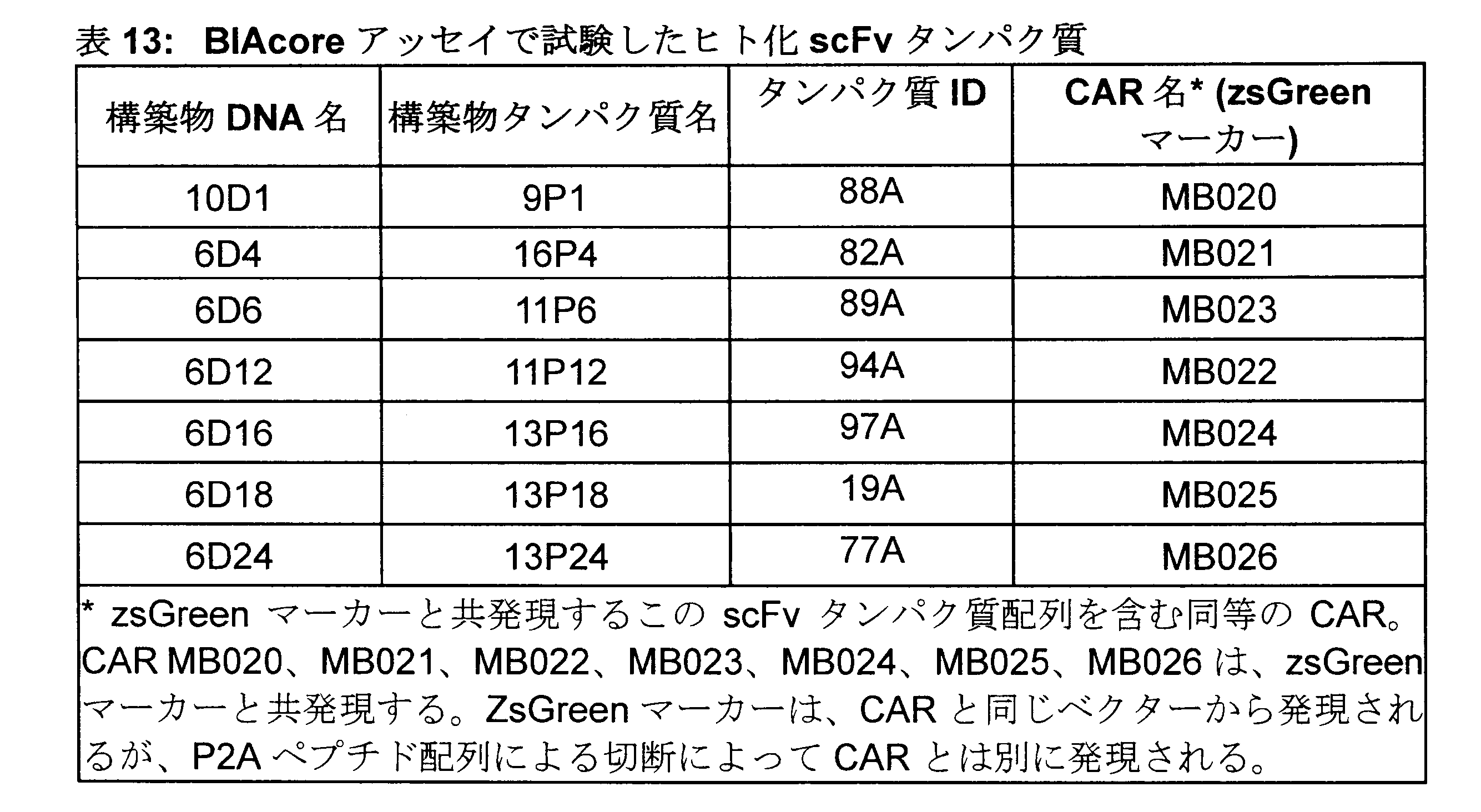
ペプチド全体のセリンおよびトレオニンアミノ酸にTn糖残基を含むビオチン化20アミノ酸ペプチド(Tarp、Sorensen et al、2007に記載のとおり)を設計し、Cambridge Research Biosciences(CRB)で注文した。対照として、同じ配列を有するがセリンおよびトレオニンアミノ酸にTn残基またはSTn残基がない非グリコシル化MUC1ペプチドも注文した。ヒト化抗TnMUC1 scFvタンパク質がTnペプチドに結合するエピトープを決定するために、
さらに2つの差次的グリコシル化ペプチドを注文した。1つは、ペプチドのSer/Thrアミノ酸にTn糖残基を含み、scFvタンパク質が結合する場所であると文献で報告されている。また、1つは、セリンおよびトレオニンアミノ酸にTn糖残基を含むが、報告によると、ここではscFvタンパク質は結合しない(Tarp、Sorensen et al、2007に記載のとおり)。
A biotinylated 20-amino acid peptide containing Tn sugar residues at serine and threonine amino acids throughout the peptide (as described in Tarp, Sorensen et al, 2007) was designed and ordered from Cambridge Research Biosciences (CRB). As a control, a non-glycosylated MUC1 peptide with the same sequence but without Tn or STn residues at the serine and threonine amino acids was also ordered. To determine the epitope to which the humanized anti-TnMUC1 scFv protein binds to the Tn peptide,
Two additional differentially glycosylated peptides were ordered. One contains a Tn sugar residue at the Ser/Thr amino acids of the peptide, which is reported in the literature to be the site of attachment of the scFv protein. Also, one contains Tn sugar residues at serine and threonine amino acids, to which scFv proteins reportedly do not bind (as described in Tarp, Sorensen et al, 2007).
これらのペプチドの配列およびID番号を表14に詳述し、図5に概略的に表す。 The sequences and ID numbers of these peptides are detailed in Table 14 and represented schematically in FIG.

捕捉レベルの決定-CAPtureチップの表面の調製
これらの実験で使用されるアッセイ形式において、ビオチン化ペプチドをCAPチップ(リガンド)上に捕捉して活性表面を生成し、そのチップ表面(アナライト)上をscFvタンパク質に通過させた。動力学的測定のための活性表面は、低い最大アナライト結合能(Rmax)を保証するように設計される。これにより、アナライトの輸送制限が軽減されることにより高速結合速度および解離速度の測定が容易になる。Rmaxに好適な値は、最高性能のBiacoreシステムでタンパク質間相互作用について20~100RU(応答単位)の範囲である。
Determination of Capture Levels—Preparation of the Surface of the CAPture Chip In the assay format used in these experiments, biotinylated peptides were captured on the CAP chip (ligand) to generate an active surface, and on the chip surface (analyte) was passed through the scFv protein. Active surfaces for kinetic measurements are designed to ensure a low maximum analyte binding capacity (Rmax). This facilitates the measurement of fast binding and dissociation rates by reducing transport limitations of the analyte. Suitable values for Rmax range from 20-100 RU (response units) for protein-protein interactions on the highest performance Biacore system.
表面の理論上のアナライト結合能(RU)は、次式で与えられる:
標的捕捉レベル=Rmax×Mwtリガンド/化学量論×Mwtアナライト
これらの実験の場合:
・リガンドMW(ペプチド)=2500Da
・アナライトMW(抗Tn MUC1 scFv)=27000Da*
・化学量論:1
・Rmax=100(再結合イベントを防ぐため)
標的捕捉レベル=Rmax×Mwtリガンド/化学量論×Mwtアナライト
=100×2500/1×27000
=11RU
*この分子量は、scFv分子の平均分子量として、試験した総てのscFvタンパク質に使用した。
The theoretical analyte binding capacity (RU) of a surface is given by:
Target capture level = Rmax x Mwt ligand/stoichiometry x Mwt analyte For these experiments:
・Ligand MW (peptide) = 2500 Da
• Analyte MW (anti-Tn MUC1 scFv) = 27000 Da *
・ Stoichiometry: 1
- Rmax = 100 (to prevent recombination events)
Target capture level = Rmax x Mwt ligand/stoichiometry x Mwt analyte
= 100 x 2500/1 x 27000
= 11 R.U.
* This molecular weight was used for all scFv proteins tested as the average molecular weight of the scFv molecule.
上記の計算により、各ペプチドの理論上の11RUを各サイクルで捕捉する必要がある。 Based on the above calculations, a theoretical 11 RU of each peptide should be captured in each cycle.
実験プロトコール
TnMUC1ペプチドおよびSTnMUC1ペプチドの捕捉レベルの決定
CAPチップ(Biotin CAPtureキットから)をBIAcore T200機器にドッキングして、製造業者の指示に従って一晩水和させた。チップをドッキングするとき(初回または保存後)、センサーチップの表面は、Regeneration Solution(Biotin CAPtureキットから)を1分で3回注入することにより調整する必要がある。Regeneration Solutionは、3部のRegeneration Stock1(8Mグアニジン-HCl)と1部のRegeneration Stock2(1M NaOH)とを混合することにより調製した。センサーチップの参照フローセルおよびアクティブフローセルの両方を、Regeneration Solutionを流速10μl/分にて60秒で3回注入することにより調整した。
Experimental protocol
Determination of Capture Levels of TnMUC1 and STnMUC1 Peptides A CAP chip (from the Biotin CAPture kit) was docked into a BIAcore T200 instrument and allowed to hydrate overnight according to the manufacturer's instructions. When docking the chip (either for the first time or after storage), the surface of the sensor chip should be conditioned by injecting Regeneration Solution (from the Biotin CAPture kit) three times in 1 min. Regeneration Solution was prepared by mixing 3 parts Regeneration Stock 1 (8 M Guanidine-HCl) and 1 part Regeneration Stock 2 (1 M NaOH). Both the reference and active flow cells of the sensor chip were conditioned by three injections of Regeneration Solution at a flow rate of 10 μl/min for 60 seconds.
CAPtureチップ上への最初の捕捉のために、総てのビオチン化ペプチドを、BIAcoreランニングバッファー(1×HBS-EP+=10mM Hepes pH7.6、150mM NaCl、3mM EDTA、0.05%ポリソルベート20)で希釈して、上記の式で詳述されているように、必要な結合レベルを確認した。捕捉レベルの決定実験は、アクティブフローセルのみで実施した。このアッセイ形式での参照フローセルはBiotin CAPture試薬を含み、ビオチン化ペプチドはアクティブフローセルおよび参照フローセルの両方に結合することから、参照フローセルに対する非特異的結合を確認することはできなかった。 For initial capture onto the CAPture chip, all biotinylated peptides were added to BIAcore running buffer (1×HBS-EP + =10 mM Hepes pH 7.6, 150 mM NaCl, 3 mM EDTA, 0.05% polysorbate 20). to confirm the required level of binding, as detailed in the formula above. Capture level determination experiments were performed with active flow cells only. Since the reference flow cell in this assay format contains the Biotin CAPture reagent and the biotinylated peptide binds to both the active flow cell and the reference flow cell, non-specific binding to the reference flow cell could not be confirmed.
Biotin CAPture試薬(Biotin CAPtureキットから)を、流速2μl/分で、300秒の接触時間の後、60秒の安定化期間でアクティブフローセル上に捕捉した。 Biotin CAPture reagent (from the Biotin CAPture kit) was captured on the active flow cell at a flow rate of 2 μl/min with a contact time of 300 seconds followed by a stabilization period of 60 seconds.
流速10μl/分、会合時間120秒の後、解離段階60秒(ここで、BIAcoreランニングバッファーを流速10μl/分で注入した)で、リガンド(ビオチン化ペプチド)にアクティブフローセル上を通過させた。 After a flow rate of 10 μl/min and an association time of 120 seconds, the ligand (biotinylated peptide) was passed over the active flow cell with a dissociation step of 60 seconds (where BIAcore running buffer was injected at a flow rate of 10 μl/min).
Biotin CAPtureチップは、Regeneration Solutionを使用して再生した。このRegeneration Solutionは、前述のように調製した。 Biotin CAPture chips were regenerated using Regeneration Solution. This Regeneration Solution was prepared as previously described.
捕捉実験からの各ペプチドの捕捉について得られたY値を使用して、11RUに近い捕捉レベルを得るために必要な捕捉の長さを計算した。最初に、TnMUC1ペプチド(36-2)およびMUC1ペプチド(36-4)を希釈し、CAPチップ上のBiotin CAPture試薬に捕捉した。この実験で得られたY値を使用して、上記で詳述したように、必要な理論値に近い捕捉レベルを提供するために結合分析アッセイでペプチドを捕捉する必要がある時間を決定した。実際に捕捉される量は、(総てのタンパク質が活性化されているわけではないため)理論値よりも低くなる可能性があるため、11RUに近い捕捉レベルが得られるようにするために、これらの実験では、これらの実験ではより高い捕捉レベルを目指した。 The Y values obtained for each peptide capture from the capture experiments were used to calculate the length of capture required to obtain a capture level close to 11 RU. First, TnMUC1 peptide (36-2) and MUC1 peptide (36-4) were diluted and captured in Biotin CAPture reagent on a CAP chip. The Y value obtained in this experiment was used to determine the time at which peptides needed to be captured in the binding assay to provide capture levels close to the theoretical required values, as detailed above. Since the actual amount captured may be lower than the theoretical value (because not all proteins are activated), in order to obtain capture levels close to 11 RU, In these experiments, we aimed for higher trapping levels.
結合アッセイ
Biotin CAPture試薬を2μl/分で300秒間、アクティブフローセルおよび参照フローセル上に捕捉した。ビオチン化ペプチドをBIAcoreランニングバッファー(上記の通り)で希釈し、流速10μl/分でアクティブフローセル上にのみ捕捉して、上記に詳述した式で計算した捕捉レベルを得た。
Binding Assay Biotin CAPture reagent was captured on the active and reference flow cells at 2 μl/min for 300 seconds. Biotinylated peptides were diluted in BIAcore running buffer (as above) and captured only on the active flow cell at a flow rate of 10 μl/min to obtain capture levels calculated with the formula detailed above.
TnMUC1ペプチドおよびMUC1ペプチドに対する結合について500、125、31.25、7.8、1.9、0nMでヒト化抗TnMUC1 scFvタンパク質に、300秒の会合後、BIAcoreランニングバッファーによる流速30μl/分での900秒の解離で、参照フローセルおよびアクティブフローセル上を通過させた。 Humanized anti-TnMUC1 scFv protein at 500, 125, 31.25, 7.8, 1.9, 0 nM for binding to TnMUC1 peptide and MUC1 peptide, after 300 seconds of association, in BIAcore running buffer at a flow rate of 30 μl/min. A 900 second dissociation was passed over the reference and active flow cells.
STnMUC1ペプチドに対する結合では、STnMUC1ペプチドおよびMUC1ペプチドに対する結合について1000、250、62.5、15.6、3.9、0.98、0nMでヒト化抗TnMUC1 scFvタンパク質の総てに、520秒の会合後、1×HBS-EP+による流速30μl/分での900秒の解離で、参照フローセルおよびアクティブフローセル上を通過させた。TnMUC1ペプチドと比較して、STnMUC1ペプチドに対するヒト化抗TnMUC1 scFvタンパク質の親和性が低いと予想されるため、これらの実験には、より高い最高濃度のscFvsを使用した。センサーグラムのより良い曲率を得るために、これらの実験の会合時間も300秒から520秒に延ばした。900秒の解離時間はこれらの実験には十分であった。 For binding to the STnMUC1 peptide, all humanized anti-TnMUC1 scFv proteins at 1000, 250, 62.5, 15.6, 3.9, 0.98, 0 nM for binding to the STnMUC1 peptide and MUC1 peptide were observed for 520 sec. After association, dissociation with 1×HBS-EP + at a flow rate of 30 μl/min for 900 seconds was passed over the reference and active flow cells. Higher top concentrations of scFvs were used for these experiments due to the expected lower affinity of the humanized anti-TnMUC1 scFv proteins for the STnMUC1 peptide compared to the TnMUC1 peptide. The association time in these experiments was also increased from 300 seconds to 520 seconds to obtain better curvature of the sensorgrams. A dissociation time of 900 seconds was sufficient for these experiments.
どちらの場合も、Biotin CAPtureチップは、上記のように再生した。 In both cases the Biotin CAPture chip was regenerated as above.
総てのTnMUC1ペプチドおよびSTnMUC1ペプチドに対する結合について、各ヒト化scFvタンパク質のN=2のデータを生成した。MUC1ペプチドを総ての実験で陰性対照として使用した。 N=2 data for each humanized scFv protein were generated for binding to all TnMUC1 and STnMUC1 peptides. MUC1 peptide was used as a negative control in all experiments.
BIAcore T200 Evaluationソフトウエアv3.0を使用して、データを分析した。得られたデータは、Rmaxパラメーターをローカルに設定した1:1結合モデルに当てはめた。これは捕捉実験であるため、タンパク質は毎回再捕捉され、理論的には、再捕捉ごとに表面がわずかに異なり、毎回わずかに異なる捕捉が生じる可能性があるため、ローカルRmaxパラメーターは真に反映される。総てのヒト化抗TnMUC1 scFvタンパク質について、動力学的パラメーター(KD、kdおよびka)を計算した。 Data were analyzed using the BIAcore T200 Evaluation software v3.0. The data obtained were fitted to a 1:1 binding model with the Rmax parameter set locally. Since this is a capture experiment, the protein is recaptured each time, and theoretically the surface could be slightly different with each recapture, resulting in slightly different capture each time, so the local Rmax parameter truly reflects be done. Kinetic parameters (K D , k d and k a ) were calculated for all humanized anti-TnMUC1 scFv proteins.
結果および考察
ヒト化抗TnMUC1 scFvタンパク質のTnMUC1ペプチド(36-2/65-1)およびMUC1ペプチド(36-4)に対する結合
7つの精製されたヒト化抗TnMUC1 scFvタンパク質の総てを、TnMUC1ペプチド(36-2)およびMUC1ペプチド(36-4)に対する結合について試験した。完全グリコシル化TnMUC1ペプチドおよびSTnMUC1ペプチドに対するヒト化抗TnMUC1 scFvタンパク質の特異的結合、MUC1ペプチドに対する結合は見られなかった(図7、図9および表15)。総ての実験について再現可能なn=2のデータが得られ、得られた値は平均値である。表15は、TnMUC1ペプチド(36-2)に対する総てのヒト化抗TnMUC1 scFvタンパク質の結合について得られたka値、kd値およびKD値を示す。scFvタンパク質16-P4および13-P16を除くscFvの総てについて同等のka値、kd値およびKD値が得られ、これらは、図7に見られるようなセンサーグラムの形状および表15に詳述した値から、他のscFvタンパク質と比較して、完全グリコシル化TnMUC1ペプチド(36-2)についてより速いkd(解離速度)を有し、次には、より低いKDを有する。
Results and Discussion
Binding of humanized anti-TnMUC1 scFv proteins to TnMUC1 peptide (36-2/65-1) and MUC1 peptide (36-4) ) and MUC1 peptide (36-4). Specific binding of the humanized anti-TnMUC1 scFv protein to the fully glycosylated TnMUC1 and STnMUC1 peptides, but no binding to the MUC1 peptide was observed (Figure 7, Figure 9 and Table 15). Reproducible n=2 data were obtained for all experiments and the values given are mean values. Table 15 shows the ka, kd and KD values obtained for binding of all humanized anti-TnMUC1 scFv proteins to the TnMUC1 peptide (36-2). Equivalent ka, kd and KD values were obtained for all of the scFvs except scFv proteins 16-P4 and 13-P16, which are in the form of sensorgrams as seen in FIG. 7 and detailed in Table 15. From the values obtained, it has a faster k d (dissociation rate) and in turn a lower K D for the fully glycosylated TnMUC1 peptide (36-2) compared to other scFv proteins.

TnMUC1ペプチド内のヒト化抗TnMUC1 scFvタンパク質のエピトープの決定
4つのscFvタンパク質(16-P4、11-P12、13-P16、13-P18)、ならびにCreative Biolabsから入手したマウス5E5 scFvタンパク質(23-P1)を総て、MUC1ペプチド(36-4)および完全グリコシル化TnMUC1ペプチド(65-1)、ならびに差次的グリコシル化TnMUC1ペプチド(96-1および96-2)に対する結合について試験して、TnMUC1ペプチド上のヒト化scFvタンパク質の結合エピトープを決定した。これらの実験は、完全グリコシル化TnMUC1ペプチド(65-1)および差次的グリコシル化TnMUC1ペプチド1(96-1)に対してヒト化抗TnMUC1 scFvタンパク質が同等に結合することを示し、MUC1ペプチド(36-4)に対してもTnMUC1ペプチド2(96-2)に対しても結合は観察されなかった。これらの実験で得られたデータにより、試験したペプチド内のヒト化抗TnMUC1 scFvタンパク質のエピトープが確認された。図8に示したセンサーグラムは、2つのペプチド(65-1および96-1)に対するヒト化抗TnMUC1 scFvタンパク質の会合および解離が同等であることを示している。動力学的パラメーターの値は図6に示した。マウス5E5 scFvタンパク質もまた、図10に見られるように、完全グリコシル化ペプチド(65-1)および差次的グリコシル化ペプチド(96-1)に対する結合が同等であることを示した。完全グリコシル化TnMUC1ペプチドの2つのバッチを試験し(65-1および36-2)、バッチ間でヒト化抗TnMUC1 scFvの同等の結合が見られた。このデータは、図6に要約している。
Determination of Epitopes of Humanized Anti-TnMUC1 scFv Proteins Within TnMUC1 Peptides Four scFv proteins (16-P4, 11-P12, 13-P16, 13-P18) and a mouse 5E5 scFv protein (23-P1) obtained from Creative Biolabs ) were all tested for binding to the MUC1 peptide (36-4) and the fully glycosylated TnMUC1 peptide (65-1), as well as the differentially glycosylated TnMUC1 peptides (96-1 and 96-2), and the TnMUC1 peptide The binding epitopes of the above humanized scFv proteins were determined. These experiments demonstrated equivalent binding of the humanized anti-TnMUC1 scFv protein to the fully glycosylated TnMUC1 peptide (65-1) and the differentially glycosylated TnMUC1 peptide 1 (96-1), indicating that the MUC1 peptide ( 36-4) or TnMUC1 peptide 2 (96-2). The data obtained in these experiments confirmed the epitopes of the humanized anti-TnMUC1 scFv protein within the tested peptides. The sensorgrams shown in Figure 8 demonstrate comparable association and dissociation of the humanized anti-TnMUC1 scFv protein to the two peptides (65-1 and 96-1). The kinetic parameter values are shown in FIG. The murine 5E5 scFv protein also showed comparable binding to the fully glycosylated peptide (65-1) and the differentially glycosylated peptide (96-1), as seen in FIG. Two batches of fully glycosylated TnMUC1 peptides were tested (65-1 and 36-2) and comparable binding of humanized anti-TnMUC1 scFv was seen between the batches. This data is summarized in FIG.
STnMUC1ペプチドおよびMUC1ペプチドに対するヒト化抗TnMUC1 scFvタンパク質の結合
上記のように、4つのscFvタンパク質ならびにマウス5E5 scFvタンパク質を総て、MUC1ペプチドおよびSTnMUC1ペプチド(35-1)に対する結合について試験した。ヒト化scFvタンパク質は、STnペプチド(35-1)と特異的に結合することを示し、MUC1ペプチド(36-4)との結合は見られなかった。しかしながら、それらは、図6および9に見られるように、Creative Biolabsで生成されたマウス5E5 scFvタンパク質と比較して、STnMUC1ペプチドに対しておよそ10倍低い親和性を有することが見出された。TnMUC1結合実験に見られるように、scFvタンパク質16P4および13P16は、図6および9に見られるように、scFvタンパク質11P12および13P18と比較して、STnペプチド(35-1)に対してより速いkd(オフレート)、次には、より低いKDを有することを示した。
Binding of Humanized Anti-TnMUC1 scFv Proteins to STnMUC1 and MUC1 Peptides As described above, all four scFv proteins and the mouse 5E5 scFv protein were tested for binding to the MUC1 and STnMUC1 peptides (35-1). The humanized scFv protein showed specific binding to the STn peptide (35-1) and no binding to the MUC1 peptide (36-4). However, they were found to have approximately 10-fold lower affinity for the STnMUC1 peptide compared to the mouse 5E5 scFv protein produced at Creative Biolabs, as seen in FIGS. As seen in the TnMUC1 binding experiments, scFv proteins 16P4 and 13P16 have faster kd ( off-rate), which in turn has been shown to have a lower KD.
要するに、ヒト化抗TnMUC1 scFvタンパク質のTnMUC1ペプチド(36-2、65-1、96-1)およびSTnMUC1ペプチド(35-1)との特異的結合が観察され、MUC1(36-4)ペプチドとの結合は観察されなかった。このアッセイは、ヒト化scFvタンパク質について得られた2つの異なる結合プロファイルを強調するものであり、16P4および13P16はより速いkd(解離速度)を有し、11P12および13P18は比較的遅いkd(解離速度)を有することが示された。試験した総てのヒト化scFvタンパク質およびマウス5E5 scFvタンパク質は、これらの実験で、TnMUC1ペプチド内の同じエピトープに結合することが示された。 In summary, specific binding of the humanized anti-TnMUC1 scFv protein to TnMUC1 peptides (36-2, 65-1, 96-1) and STnMUC1 peptide (35-1) was observed, and binding to MUC1 (36-4) peptide was observed. No binding was observed. This assay highlights the two different binding profiles obtained for the humanized scFv proteins, with 16P4 and 13P16 having higher kd (dissociation rates) and 11P12 and 13P18 having relatively slow kd (dissociation rates). ). All humanized scFv proteins tested and the murine 5E5 scFv protein were shown in these experiments to bind to the same epitope within the TnMUC1 peptide.
この実験では、CAR分子のscFvフラグメントだけを試験し、最終的なCAR分子は、このアッセイシステムでは試験できない。これらのペプチドと結合する同等のCARのさらなる特性評価は、実施例12に記載するペプチド刺激アッセイで行った。13P16 scFvについての試験した総てのペプチドに対する結合は、図10に要約している。 In this experiment only the scFv fragment of the CAR molecule was tested, the final CAR molecule cannot be tested with this assay system. Further characterization of comparable CAR binding to these peptides was performed in the peptide stimulation assay described in Example 12. Binding to all tested peptides for 13P16 scFv is summarized in FIG.
実施例5-細胞結合方法
精製scFvタンパク質のTnMUC1陽性および陰性腫瘍細胞株に対する結合を、フローサイトメトリーで決定した。この研究の目的は、フローサイトメトリーで決定されたTnMUC1陽性および陰性腫瘍細胞株の力価調整されたscFvタンパク質の相対的な結合有効性に基づいて、CAR構築物のヒト化scFv領域を識別することであった。
Example 5 - Cell Binding Method Binding of purified scFv proteins to TnMUC1 positive and negative tumor cell lines was determined by flow cytometry. The purpose of this study was to identify humanized scFv regions of CAR constructs based on the relative binding efficiencies of titrated scFv proteins in TnMUC1-positive and -negative tumor cell lines as determined by flow cytometry. Met.
実験準備
接着細胞培養
接着MDA-MB-468細胞株およびPC3細胞株は、週2回の分割比1:3で継代培養を行うことによって、培養下で維持した。細胞単層をDPBSで洗浄した後、TrypLE Express(TE)とともに数分間インキュベートした。分離したら、細胞を適当な細胞培養培地に再懸濁させ(最終量は使用したTEの5倍量に等しい)、新しい細胞培養フラスコに播種した。細胞は、必要になるまで、5%CO2、37℃の加湿インキュベーターで保存した。
Experiment preparation
Adherent Cell Cultures Adherent MDA-MB-468 and PC3 cell lines were maintained in culture by subculturing twice weekly at a split ratio of 1:3. Cell monolayers were washed with DPBS and then incubated with TrypLE Express (TE) for several minutes. Once detached, the cells were resuspended in appropriate cell culture medium (final volume equal to 5 times the volume of TE used) and seeded into new cell culture flasks. Cells were stored in a 37° C. humidified incubator with 5% CO 2 until needed.
懸濁細胞培養
Jurkat細胞は、コンフルエント細胞培養物の10%を新しい細胞培養フラスコに移した後、適当な量の細胞成長培地を添加する(最終分割比1:10を得る)ことにより週2回の継代培養を行うことによって、培養下で維持した。細胞は、必要になるまで、5%CO2、37℃の加湿インキュベーターで保存した。
Suspension cell culture Jurkat cells were grown twice weekly by transferring 10% of the confluent cell culture to a new cell culture flask followed by the addition of the appropriate amount of cell growth medium (to obtain a final split ratio of 1:10). was maintained in culture by subculturing . Cells were stored in a 37° C. humidified incubator with 5% CO 2 until needed.
フローサイトメトリーアッセイのための細胞調製
細胞を分離し、細胞を適当な細胞培養培地に再懸濁させるかまたはFalconチューブに移した。次に、各細胞懸濁液のサンプルをDPBSで1:5希釈し(最終量0.5mLを得た)、各細胞懸濁液について(トリパンブルー排除に基づく)1mL当たりの生存細胞数を、Vi-Cell XR細胞生存率分析装置(デフォルト細胞種設定を使用)を使用して決定した。次に、細胞懸濁液を適当な細胞培養培地で希釈し、新しい細胞培養フラスコに1cm2当たり6.25×10⁴生存細胞の密度で播種した。細胞は、5%CO2、37℃の加湿インキュベーターで48時間(または必要になるまで)インキュベートした。アッセイ当日、細胞をフラスコから分離し、適当な細胞培養培地に再懸濁させ(必要に応じて)、前述のように、1mL当たりの生存細胞数を計数した。細胞懸濁液を500×gで5分間遠心分離した後、細胞ペレットをFACSバッファーに1mL当たり4×10⁶生存細胞の密度で再懸濁させた。
Cell preparation for flow cytometry assay Cells were detached and cells resuspended in appropriate cell culture medium or transferred to Falcon tubes. A sample of each cell suspension was then diluted 1:5 with DPBS (yielding a final volume of 0.5 mL) and the number of viable cells per mL (based on trypan blue exclusion) for each cell suspension was determined by Determined using a Vi-Cell XR cell viability analyzer (using default cell type settings). The cell suspension was then diluted with the appropriate cell culture medium and seeded into new cell culture flasks at a density of 6.25×10 4 viable cells per cm 2 . Cells were incubated in a 5% CO2 , 37°C humidified incubator for 48 hours (or until required). On the day of assay, cells were detached from flasks, resuspended in appropriate cell culture medium (if necessary), and viable cells per mL were counted as previously described. After centrifuging the cell suspension at 500×g for 5 minutes, the cell pellet was resuspended in FACS buffer at a density of 4×10 6 viable cells per mL.
実験プロトコール
細胞結合アッセイ
scFvタンパク質をDPBSで62.5nMに希釈し、96ウェルV底プレートのA列に分注した後、そのプレートをFACSバッファーで1:4連続希釈を行った、ウェル当たり25mL(最終量)を得た(H列のみFACSバッファー)。抗Hist-PE検出抗体をDPBSで1:5希釈し、ウェル当たり25mLを、連続希釈したscFvタンパク質を含むウェルに分注し、その後、光から保護したプレートを4℃で1時間インキュベートし、scFvタンパク質を抗Hist-PE検出抗体と事前に複合体化した。次に、前述のように調製した細胞懸濁液をプレートに分注した(ウェル当たり50mL)後、光から保護したプレートを4℃でさらに1時間インキュベートした。細胞は、ウェル当たり200mL(最大量)のFACSバッファーに細胞を再懸濁させ、500×gで5分間細胞を遠心分離した後、上清を除去し、洗浄工程を2回以上繰り返して細胞を洗浄することによって洗浄した。細胞ペレットは、ウェル当たり200mLのFACSバッファー(1mL当たり1mg(最終)のDAPIおよび2mM(最終)のEDTAを含む)に再懸濁させた後、CytoFLEX Sフローサイトメーターを使用してデータを取得した。
Experimental protocol
The cell-binding assay scFv protein was diluted to 62.5 nM in DPBS and aliquoted into row A of a 96-well V-bottom plate, after which the plate was serially diluted 1:4 in FACS buffer, 25 mL per well (final volume ) was obtained (only column H is FACS buffer). The anti-Hist-PE detection antibody was diluted 1:5 in DPBS and 25 mL per well was dispensed into wells containing serially diluted scFv proteins, after which the light-protected plate was incubated at 4° C. for 1 hour and the scFv Proteins were pre-conjugated with an anti-Hist-PE detection antibody. Cell suspensions prepared as described above were then dispensed onto plates (50 mL per well) and the plates were incubated for an additional hour at 4° C., protected from light. Cells were washed by resuspending the cells in 200 mL (maximum volume) of FACS buffer per well, centrifuging the cells at 500 xg for 5 minutes, then removing the supernatant and repeating the washing step two more times. Washed by washing. Cell pellets were resuspended in 200 mL per well of FACS buffer (containing 1 mg (final) DAPI and 2 mM (final) EDTA per mL) prior to data acquisition using a CytoFLEX S flow cytometer. .
標的発現アッセイ
細胞懸濁液(前述のように調製した)を96ウェルプレートV底プレートに分注し(ウェル当たり25mL)、FACSバッファーでウェル当たり200mLの量にした後、細胞を500×gで5分間遠心分離した。細胞を、FACSバッファーで1mL当たり5μgに希釈した一次抗体(または必要に応じて同濃度のアイソタイプ抗体/FACSバッファーのみ)、ウェル当たり100mLに再懸濁させた。細胞を4℃で45分間インキュベートした。次に、前述のように細胞をFACSバッファーで3回洗浄した後、細胞ペレットをFACSバッファーで1:1000希釈したヤギ抗マウスPE結合二次抗体、1mL当たり100mLに再懸濁させ、細胞を4℃でさらに45分間インキュベートした。次に、細胞を洗浄し、DAPIおよびEDTAを添加したFACSバッファーに再懸濁させ、前述のようにデータを取得した。
Target Expression Assay Cell suspensions (prepared as described above) were dispensed (25 mL per well) into 96-well V-bottom plates, brought to a volume of 200 mL per well with FACS buffer, and then cells were run at 500 xg. Centrifuge for 5 minutes. Cells were resuspended in 100 mL per well of primary antibody diluted to 5 μg per mL in FACS buffer (or the same concentration of isotype antibody/FACS buffer alone, as appropriate). Cells were incubated at 4°C for 45 minutes. Cells were then washed three times with FACS buffer as described above, after which the cell pellet was resuspended in 100 mL per mL of goat anti-mouse PE-conjugated secondary antibody diluted 1:1000 in FACS buffer and the cells were °C for an additional 45 minutes. Cells were then washed, resuspended in FACS buffer supplemented with DAPI and EDTA, and data acquired as previously described.
試験したヒト化scFvタンパク質は:9P1(88A)、16P4(82A)、11P6(89A)、11P12(94A)、13P16(97A)、13P18(19A)、および13P24(7A)(上記表13参照)であった。 The humanized scFv proteins tested were: 9P1 (88A), 16P4 (82A), 11P6 (89A), 11P12 (94A), 13P16 (97A), 13P18 (19A), and 13P24 (7A) (see Table 13 above). there were.
データ分析
細胞結合アッセイデータ
生データを、FlowJoを使用して分析した。ウェルサンプルを細胞株によって分類し、各細胞株について細胞のみのウェルサンプルを使用して生存単一陽性細胞を区別するためにゲートを設定し、その後、同じ群のウェルサンプルに対してゲートを適用した。蛍光強度(MFI)値の中央値をPrismにエクスポートし、対数スケール(すなわち、X=Log(X))に変換した後、可変傾斜4パラメーター方程式:Y=下位+(最高-最低)/(1+10^((-pEC50-X)*ヒル勾配))を使用してデータを当てはめた。Jurkat細胞においてヒト化scFvタンパク質の最高濃度でフック効果の証拠が観察されたMFI値は、曲線フィッティングの前に除外した。標準誤差値はPrismにより計算された。このレポートのために生成されたデータは、リード分子の選択に役立つものではないため、このデータに対して統計分析方法は実行されなかった。
data analysis
Raw cell binding assay data was analyzed using FlowJo. Sort the well samples by cell line and set a gate to distinguish viable single positive cells using the cell-only well sample for each cell line, then apply the gate for the same group of well samples. bottom. Median fluorescence intensity (MFI) values were exported to Prism and converted to a logarithmic scale (i.e., X = Log(X)), followed by a variable slope 4-parameter equation: Y = lower + (highest - lowest) / (1 + 10 Data were fitted using ^((-pEC50-X) * Hill slope)). MFI values where evidence of hook effect was observed at the highest concentration of humanized scFv protein in Jurkat cells were excluded prior to curve fitting. Standard error values were calculated by Prism. Statistical analysis methods were not performed on this data as the data generated for this report are not conducive to the selection of lead molecules.
標的発現アッセイデータ
生データを、前述のようにFlowJoを使用して分析した。各細胞株の一次抗体ウェルからアイソタイプ対照ウェルにおけるPE陽性百分率値を差し引き、Prismを使用してプロットした。
Target expression assay data raw data were analyzed using FlowJo as previously described. PE positive percentage values in isotype control wells were subtracted from primary antibody wells for each cell line and plotted using Prism.
結果および考察
データは、ヒト化scFvタンパク質の総てが、scFv濃度でTnMUC1発現に依存する様式で、TnMUC1発現腫瘍細胞(TnMUC1陽性MDA-MB-468腫瘍細胞)と選択的に結合することを実証している。図11の代表的なデータを参照。総てのヒト化scFvタンパク質について11P6(89A)および11P12(94A)を除いて、EC50平均値およびpEC50平均値はn=2の実験に基づいて計算し、平均EC50濃度は13P18(19A)の1.68nM~28P24(77A)の84.38nMの範囲であり;11P6(89A)および11P12(94A)のEC50濃度は、それぞれ4.968および34.2nMであった(n=1の実験データに基づく)。表16参照。
Results and Discussion Data demonstrate that all of the humanized scFv proteins selectively bind to TnMUC1-expressing tumor cells (TnMUC1-positive MDA-MB-468 tumor cells) in a manner that is dependent on TnMUC1 expression at scFv concentration. are doing. See representative data in FIG. Mean EC50 and pEC50 values were calculated based on n=2 experiments for all humanized scFv proteins, except 11P6 (89A) and 11P12 (94A), with mean EC50 concentrations of 13P18 (19A) EC50 concentrations for 11P6 (89A) and 11P12 (94A) were 4.968 and 34.2 nM, respectively (based on n=1 experimental data). ). See Table 16.
13P16(97A)ヒト化scFvタンパク質は、9P1(88A)、16P4(82A)、11P6(89A)および13P18(19A)と比較して結合効力の低下を示し、EC50値はおよそ11nMであるのに対し、最も強力なヒト化scFvタンパク質のEC50値は5nM以下であった。 The 13P16(97A) humanized scFv protein showed reduced binding potency compared to 9P1(88A), 16P4(82A), 11P6(89A) and 13P18(19A), with an EC50 value of approximately 11 nM. , the EC50 values of the most potent humanized scFv proteins were below 5 nM.

TnMUC1陰性対照PC3細胞における9P1(88A)、11P6(89A)、11P12(94A)、13P18(19A)ヒト化scFvタンパク質の結合について、PrismによりEC50値を生成した(データ記載せず)。図12の代表的なデータは、最高のscFvタンパク質濃度でフック効果のいくつかの証拠も示しているが、付随する負のヒルスロープ値(ヒルスロープ値記載せず)に沿った図12のデータは、PC3細胞におけるヒト化scFvタンパク質結合の証拠がないことを示唆している。 EC50 values were generated by Prism for binding of 9P1 (88A), 11P6 (89A), 11P12 (94A), 13P18 (19A) humanized scFv proteins in TnMUC1 negative control PC3 cells (data not shown). The representative data in Figure 12 also show some evidence of a hook effect at the highest scFv protein concentrations, but the data in Figure 12 along with the accompanying negative Hillslope values (Hillslope values not shown) suggest that there is no evidence of humanized scFv protein binding in PC3 cells.
図13の代表的なフィットMFIデータは、MDA-MB-468細胞と比較して陽性度の高いTnMUC1陽性対照Jurkat細胞における総てのヒト化scFvタンパク質についての結合の増加を示しており(図11)、Jurkat、MDA-MB-468およびPC3細胞の代表的なTnMUC1膜発現データに対応している(図15)。 Representative fit MFI data in Figure 13 show increased binding for all humanized scFv proteins in highly positive TnMUC1 positive control Jurkat cells compared to MDA-MB-468 cells (Figure 11 ), corresponding to representative TnMUC1 membrane expression data for Jurkat, MDA-MB-468 and PC3 cells (FIG. 15).
図14のn=2の実験からのフィット曲線は、TnMUC陽性MDA-MB-468およびTnMUC陽性度が高いJurkat細胞に対する13P16(97A)の選択的結合を具体的に示しており、この場合も、Jurkat、MDA-MB-468およびPC3細胞の代表的なTnMUC1膜発現データに対応している(図15)。フック効果の証拠は、Jurkat細胞における二反復実験で16P4(82A)および13P18(19A)の最高濃度で観察され、また2回の繰り返し実験のうち1回のみで9P1(88A)および11P6(89A)でも観察された(MFI値は、曲線フィッティングの前に除外した)。 The fitted curves from n=2 experiments in FIG. 14 demonstrate selective binding of 13P16(97A) to TnMUC-positive MDA-MB-468 and highly TnMUC-positive Jurkat cells, again Corresponding representative TnMUC1 membrane expression data for Jurkat, MDA-MB-468 and PC3 cells (FIG. 15). Evidence of a hook effect was observed at the highest concentrations of 16P4 (82A) and 13P18 (19A) in duplicate experiments in Jurkat cells, and 9P1 (88A) and 11P6 (89A) in only one of two replicate experiments. (MFI values were excluded before curve fitting).
実施例6-前臨床の安全性
これらの研究の目的は、原形質膜タンパク質アレイを使用して、TnMUC1 CAR-T細胞の標的外結合を評価することであった。
Example 6 - Preclinical Safety The purpose of these studies was to assess off-target binding of TnMUC1 CAR-T cells using plasma membrane protein arrays.
方法
実験プロトコール
パイロット研究を支援するCAR-T細胞の生成
末梢血単球(PBMC)は、Histopaque(Sigma、カタログ番号10771)およびAccuspinチューブ(Sigma、カタログ番号A7054)を製造者の説明書に従って使用して、ヒト血液から単離した。細胞をTEXMacs培地に再懸濁させた。IL-2(100IU/ml)およびTransAct T細胞活性化ビーズ(1:100希釈)をPBMCに加え、細胞を、加湿インキュベーターにて、5%CO2、37℃で2日間インキュベートした。PBMCをMOI 2.75のBCMAベクター、BCMA-030で形質導入し、5%CO2、37℃で2日間インキュベートした。培養期間中、細胞は、TEXMacs培地および100IU/mlのIL-2で維持した。形質導入の13日後に細胞を回収し、1×108細胞/mlで凍結した。陰性対照として非形質導入細胞を生成した。T細胞のバッチは3名のドナーから生成した。
Method
Experimental protocol
Generation of CAR-T cells in support of pilot studies Peripheral blood monocytes (PBMCs) were isolated in humans using Histopaque (Sigma, Cat. No. 10771) and Accuspin tubes (Sigma, Cat. No. A7054) according to the manufacturer's instructions. Isolated from blood. Cells were resuspended in TEXMacs medium. IL-2 (100 IU/ml) and TransAct T cell activation beads (1:100 dilution) were added to the PBMCs and the cells were incubated for 2 days at 37° C. in 5% CO 2 in a humidified incubator. PBMC were transduced with a BCMA vector, BCMA-030, at an MOI of 2.75 and incubated at 37° C. in 5% CO 2 for 2 days. During the culture period, cells were maintained in TEXMacs medium and 100 IU/ml IL-2. Cells were harvested 13 days after transduction and frozen at 1×10 8 cells/ml. Non-transduced cells were generated as a negative control. Batches of T cells were generated from 3 donors.
形質導入効率は、フローサイトメトリー(MACSQuant Analyser 10)およびAPCチャネルを使用して、Alexa Fluor 647結合BCMA-FcでCAR発現を検出することにより決定した。データはFlowJo v10.1を使用して分析した。 Transduction efficiency was determined by detecting CAR expression with Alexa Fluor 647-conjugated BCMA-Fc using flow cytometry (MACSQuant Analyzer 10) and APC channel. Data were analyzed using FlowJo v10.1.
プレスクリーニング、一次スクリーニングおよび確認スクリーニングを支援するCAR-T細胞の生成
PBMCは、Histopaque(Sigma、カタログ番号10771)およびAccuspinチューブ(Sigma、カタログ番号A7054)を製造者の説明書に従って使用して、ヒト血液から単離した。細胞をTEXMacs培地に再懸濁させた。IL-2(100IU/ml)およびTransAct T細胞活性化ビーズ(1:100希釈)をPBMC加え、細胞を、加湿インキュベーターにて、5%CO2、37℃で2日間インキュベートした。PBMCをMOI 2.4のBCMAベクター、BCMA-030で、またはMOI 5のTnMUC1ベクター、MB-037で形質導入し、5%CO2、37℃で2日間インキュベートした。培養期間中、細胞は、TEXMacs培地および100IU/mlのIL-2で維持した。形質導入の12日後に細胞を回収し、CryStor CS5凍結培地中1×10⁸細胞/mlで凍結した。陰性対照として非形質導入T細胞を生成した。T細胞は1ドナーから生成した。
Generation of CAR-T cells to support prescreening, primary screening and confirmatory screening PBMC were isolated in humans using Histopaque (Sigma, Cat. #10771) and Accuspin tubes (Sigma, Cat. #A7054) according to the manufacturer's instructions. Isolated from blood. Cells were resuspended in TEXMacs medium. IL-2 (100 IU/ml) and TransAct T cell activation beads (1:100 dilution) were added to PBMCs and cells were incubated for 2 days at 37° C. in 5% CO 2 in a humidified incubator. PBMC were transduced with the BCMA vector, BCMA-030, at MOI 2.4 or with the TnMUC1 vector, MB-037, at
BCMA CAR-T細胞の形質導入効率は、フローサイトメトリー(MACSQuant Analyser 10)を使用して、BCMA-AF647に対する結合を測定することにより決定した。TnMUC1 CAR-T細胞の形質導入効率は、PE結合抗LNGFR Abおよびフローサイトメトリー(MACSQuant Analyser 10)を使用して、LNGFR発現を測定することにより決定した。データはFlowJo v10.1を使用して分析した。 Transduction efficiency of BCMA CAR-T cells was determined by measuring binding to BCMA-AF647 using flow cytometry (MACSQuant Analyzer 10). Transduction efficiency of TnMUC1 CAR-T cells was determined by measuring LNGFR expression using PE-conjugated anti-LNGFR Ab and flow cytometry (MACSQuant Analyzer 10). Data were analyzed using FlowJo v10.1.
原形質膜タンパク質アレイ
パイロット研究
ドナーT細胞を、固定した非トランスフェクトHEK293細胞と、BCMA、既知のT細胞相互作用因子、および対照タンパク質を過剰発現するHEK293細胞のスライドに追加した。バックグラウンドに対するBCMA特異的CAR-T細胞の結合レベルにより、予備審査スクリーニング、一次スクリーニング、および確認スクリーニングに好適なドナーの選択が可能であった、
plasma membrane protein array
Pilot study donor T cells were added to slides of fixed non-transfected HEK293 cells and HEK293 cells overexpressing BCMA, known T cell interactors, and control proteins. The level of binding of BCMA-specific CAR-T cells to background allowed the selection of suitable donors for preliminary, primary, and confirmatory screening.
陽性対照TnMUC1ペプチドを、純ペプチドから始める連続希釈でスライド上にスポットした。ペプチドをマウス抗ヒトMUC1 mAb、クローン5E5で検出し、続いて、Alexa Fluor 647結合抗マウスIgG(H+L)抗体での検出を行った。 A positive control TnMUC1 peptide was spotted onto slides in serial dilutions starting with pure peptide. Peptides were detected with mouse anti-human MUC1 mAb, clone 5E5, followed by Alexa Fluor 647-conjugated anti-mouse IgG (H+L) antibody.

プレスクリーニング研究
非形質導入T細胞およびCAR形質導入T細胞を、固定した非トランスフェクトHEK293細胞と、BCMA、既知のT細胞相互作用因子、および対照タンパク質を過剰発現するHEK293細胞のスライドに追加した。
Prescreening Studies Untransduced and CAR-transduced T cells were added to slides of fixed untransfected HEK293 cells and HEK293 cells overexpressing BCMA, known T cell interactors, and control proteins.
一次スクリーニング
一次スクリーニングでは、全長ヒト原形質膜タンパク質をコードする4070のタンパク質、リバーストランスフェクションを使用して、ヒトHEK293細胞で個別に発現させた。細胞は、13のマイクロアレイスライド全体に二反復で並べ、固定した。発現ベクター、pIRES-hEGFR-IRES-ZsGreen1は、トランスフェクション効率の最小閾値が達成または超過されたことを確認するために、各スライド上に四反復でスポットした。バックグラウンドに対する平均pIRES-hEGFR-IRES-ZsGreen1シグナルとして最小閾値1.5が従前に決定された。
Primary Screen In the primary screen, 4070 proteins, encoding full-length human plasma membrane proteins, were expressed individually in human HEK293 cells using reverse transfection. Cells were arrayed and fixed in duplicate across 13 microarray slides. The expression vector, pIRES-hEGFR-IRES-ZsGreen1, was spotted in quadruplicate on each slide to confirm that the minimum threshold of transfection efficiency was achieved or exceeded. A minimum threshold of 1.5 was previously determined as mean pIRES-hEGFR-IRES-ZsGreen1 signal over background.
非形質導入T細胞およびCAR形質導入T細胞をセルトレーサーレッド色素で標識し、事前に最適化した比率のT細胞とHEK293細胞で原形質膜タンパク質アレイに使用した。 Non-transduced and CAR-transduced T cells were labeled with cell tracer red dye and used for plasma membrane protein arrays with pre-optimized ratios of T cells and HEK293 cells.
確認スクリーニング
一次スクリーニングで特定されたヒットをコードするベクターを二反復でスポットし、ヒトHEK293細胞をリバーストランスフェクトするために使用した。細胞を固定し、続いて、陽性および陰性MUC1ペプチドでスポットした。二反復用のスライドを用意した。ドナー90928からの非形質導入T細胞および形質導入T細胞(スライド当たり3.2×10⁷細胞)、または抗ヒトMUC1 Ab(10μg/ml)を原形質膜タンパク質アレイに適用した。抗ヒトMUC1結合をalexafluor 647結合抗マウスIgG(H+L)抗体で検出した。
Confirmatory Screening Vectors encoding hits identified in the primary screen were spotted in duplicate and used to reverse transfect human HEK293 cells. Cells were fixed and subsequently spotted with positive and negative MUC1 peptides. Duplicate slides were prepared. Untransduced and transduced T cells (3.2×10 7 cells per slide) from
データ分析
フローサイトメトリー:データは、FlowJo v10.1を使用して分析した。
Data Analysis Flow Cytometry: Data were analyzed using FlowJo v10.1.
原形質膜タンパク質アレイ:結合は、蛍光撮像法によって評価し、ImageQuantソフトウエア(GE)を使用して形質導入効率を定量した。バックグラウンド結合のレベルは、非トランスフェクトHEK293細胞の領域を使用して決定した。 Plasma Membrane Protein Array: Binding was assessed by fluorescence imaging and ImageQuant software (GE) was used to quantify transduction efficiency. Levels of background binding were determined using areas of non-transfected HEK293 cells.
タンパク質の「ヒット」は、バックグラウンドレベルと比較して上昇したシグナルを示す二反復のスポットとして定義された。これは、ImageQuantソフトウエアでグリッド化された画像を使用した目視検査によって達成された。ヒットは、二反復のスポットの強度に応じて「強い、中程度、弱い、または非常に弱い」に分類した。 Protein "hits" were defined as duplicate spots showing elevated signal compared to background levels. This was accomplished by visual inspection using images gridded with ImageQuant software. Hits were classified as "strong, moderate, weak, or very weak" according to the intensity of the duplicate spots.
結果および考察
パイロット研究を支援するCAR-T細胞の生成
BCMA-AF647を使用して、BCMA CAR-T細胞の形質導入効率を決定した。形質導入および拡大培養過程の7日目に、ドナー12021、30865および90928の形質導入効率は、それぞれ85.5%、80.3%および69.6%であった(表17)。形質導入および拡大培養過程の14日目に、ドナー12021、30865および90928の形質導入効率は、それぞれ62.2%、50.6%および55.8%であった。
Results and Discussion
Generation of CAR-T cells in support of pilot studies BCMA-AF647 was used to determine transduction efficiency of BCMA CAR-T cells. At

一次スクリーニングおよび確認スクリーニングを支援するCAR-T細胞の生成
形質導入効率は、形質導入の12日後に決定した。BCMA CAR-T細胞の形質導入効率は63.1%であった(表18)。形質導入T細胞に適用したものと同じゲート設定戦略を使用し、非形質導入T細胞の0.8%が陽性ゲートの範囲内にあった。抗LNGFR Abを使用すると、TnMUC1、MB-037、CAR-T細胞の形質導入効率は29.2%であった。形質導入T細胞に適用した同じゲート設定戦略を使用して、非形質導入T細胞の1.4%が陽性ゲートの範囲内にあった。
Generation of CAR-T cells to support primary and confirmatory screening The transduction efficiency was determined 12 days after transduction. The transduction efficiency of BCMA CAR-T cells was 63.1% (Table 18). Using the same gating strategy as applied to transduced T cells, 0.8% of non-transduced T cells fell within the positive gate. Using the anti-LNGFR Ab, the transduction efficiency of TnMUC1, MB-037, CAR-T cells was 29.2%. Using the same gating strategy applied to transduced T cells, 1.4% of non-transduced T cells fell within the positive gate.
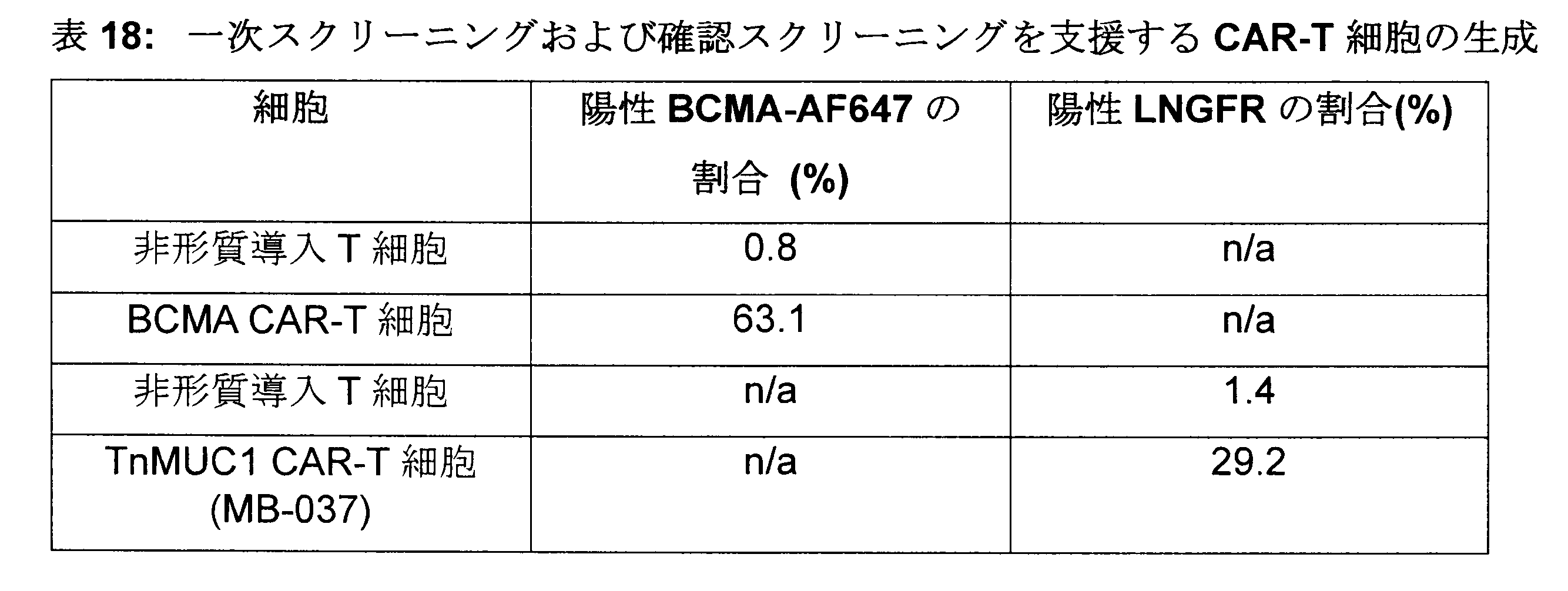
原形質膜タンパク質アレイ:パイロット研究
HEK形質導入細胞のスポッティングパターンを図16Aに示している。非形質導入T細胞では、既知のT細胞相互作用因子(PVR、CD244、TNFSF4、ICOSLG、CD86)に対して結合が観察された(図16B~D)。BCMA形質導入T細胞では、BCMAトランスフェクトHEK293細胞に対して結合が観察された(図17)。結合の強度は3ドナー間で同等であった。
Plasma Membrane Protein Array: The spotting pattern of pilot study HEK-transduced cells is shown in FIG. 16A. In untransduced T cells, binding was observed to known T cell interactors (PVR, CD244, TNFSF4, ICOSLG, CD86) (Fig. 16B-D). Binding was observed in BCMA-transduced T cells to BCMA-transfected HEK293 cells (Fig. 17). The strength of binding was comparable among the 3 donors.
原形質膜タンパク質アレイ:プレスクリーニング
ドナー90928を一次スクリーニングに選択した。
Plasma Membrane Protein Array:
HEK形質導入細胞のスポッティングパターンを図18A~18Dに示している。非形質導入T細胞では、既知のT細胞相互作用因子(PVR、CD244、TNFSF4、ICOSLG、CD86)に対して結合が観察された。BCMA形質導入T細胞では、BCMAトランスフェクトHEK293細胞に対して結合が観察された。TnMUC1形質導入T細胞では、TnMUC1ペプチドまたはMUC1ペプチドとの結合は起こらなかった。 Spotting patterns of HEK-transduced cells are shown in Figures 18A-18D. In untransduced T cells, binding was observed to known T cell interactors (PVR, CD244, TNFSF4, ICOSLG, CD86). Binding was observed in BCMA-transduced T cells to BCMA-transfected HEK293 cells. No binding to the TnMUC1 or MUC1 peptide occurred in TnMUC1-transduced T cells.
原形質膜タンパク質アレイ:一次スクリーニング
ImageQuantで蛍光を分析することによって、合計28のヒットが特定された。染色の強度は非常に弱いものから強いものまで様々であった。
Plasma Membrane Protein Array: A total of 28 hits were identified by analyzing fluorescence with the primary screen ImageQuant. The intensity of staining varied from very weak to strong.
原形質膜タンパク質アレイ:確認スクリーニング
28のヒットのスポッティングパターンを図19A~19Dに示している。非形質導入T細胞では、既知のT細胞相互作用因子に対して結合が観察された。BCMA CAR-T細胞について、強い強度で1つの特異的相互作用が確認された。TnMUC1 CAR-T細胞について、2つのCAR特異的相互作用が確認された。これらは、DCCネトリン1受容体(DCC)(中強度)およびセレクチンPリガンド(SELPLG)(非常に弱い/弱い強度)であった。
Plasma Membrane Protein Array: The spotting patterns of the 28 hits from the confirmation screen are shown in Figures 19A-19D. Binding was observed in non-transduced T cells to known T cell interactors. One specific interaction was identified with high intensity for BCMA CAR-T cells. Two CAR-specific interactions were identified for TnMUC1 CAR-T cells. These were
要約すると、4070のヒトタンパク質の完全ライブラリーを過剰発現するヒトHEK293に対する結合についてCAR-T細胞をスクリーニングした後、非形質導入T細胞は、多くの既知のT細胞相互作用因子に対して結合を示した。陽性対照として使用したBCMA CAR-T細胞は、強い強度でBCMAとの単一の特異的相互作用を示した。TnMUC1 CAR-T細胞は、中強度でDCCネトリン1受容体と、非常に弱い/弱い強度でセレクチンPリガンドとの結合を示した。TnMUC1 CAR-T細胞とTnMUC1またはMUC1ペプチドとの結合は観察されなかった。DCCネトリン1受容体との結合は中強度の結合が観察されたためさらに評価を行った(実施例16参照)。セレクチンPリガンドとの結合は非常に弱く/弱く、信頼性の低い結合剤と見なされるため、このタンパク質にはこれ以上の評価は行わなかった。 In summary, after screening CAR-T cells for binding to human HEK293 overexpressing a complete library of 4070 human proteins, untransduced T cells showed binding to a number of known T cell interactors. Indicated. BCMA CAR-T cells, used as a positive control, showed a single specific interaction with BCMA with high intensity. TnMUC1 CAR-T cells exhibited moderate-strength binding to the DCC netrin-1 receptor and very weak/weak-strength binding to selectin P-ligands. No binding of TnMUC1 CAR-T cells to TnMUC1 or MUC1 peptide was observed. Binding to the DCC netrin-1 receptor was further evaluated as moderate-strength binding was observed (see Example 16). This protein was not evaluated further as it binds very weakly/weakly to selectin P ligands and is considered an unreliable binder.
実施例7-T細胞表現型に対するCAR発現の影響
この研究の目的は、12色の表現型パネルを使用して、形質導入の14日後に基礎状態の(障害のない)7つのヒト化TnMUC1 CAR-T細胞の表現型の状態を、非形質導入T(UT)およびマウス型T細胞と比較することによって特性評価することであった。試験したヒト化TnMUC1 CAR T細胞にはHuCAR020-26が含まれ、マウスCAR T細胞にはMB004(マウス型陽性TnMUC1 CAR T)およびMB007(シグナル伝達ドメイン4-1BBおよびCD3zを欠くマウス型陰性TnMUC1 CART)が含まれた。設計した12色の表現型パネルには、系統(CD3およびCD8)、活性化/疲弊チェックポイント(LAG3、TIM3およびPD1)、メモリーサブセット(CD45RAおよびCCR7)およびCAR形質導入(zsGreen)バイオマーカーが含まれる。計算データ分析アルゴリズムを備えた機械学習プラットフォームCytobankを使用して、生成された高次元フローデータを分析した。FlowLogicを使用して並列データ分析を行った。
Example 7 - Effect of CAR Expression on T Cell Phenotype The purpose of this study was to examine the basal (non-impaired) 7 humanized
方法
実験準備
ドナー番号91860、91462および92091の先行実験から精製T細胞を調製した(形質導入CAR T細胞は総て構築物でzsGreen遺伝子発現を示す)。簡単に述べれば、T細胞(形質導入後14日)を、TexMACS培地と100UのIL-2を含む6ウェル細胞培養プレートで培養した。一晩インキュベートした後、障害のないT細胞を回収し、染色の前に比較目的のため、各サンプルをUT T細胞を使用して32%の同じ形質導入効率(TE)に正規化した。試験したT細胞には、非形質導入T(UT)、MB004(マウス型陽性TnMUC1 CAR T)、MB007(シグナル伝達ドメイン4-1BBおよびCD3zを欠くマウス型陰性TnMUC1 CART)およびMB020-26(ヒト化型陽性TnMUC1 CAR T)が含まれた。
Method
Purified T cells were prepared from previous experiments with experimental preparation donor numbers 91860, 91462 and 92091 (all transduced CAR T cells show zsGreen gene expression with the construct). Briefly, T cells (14 days post-transduction) were cultured in 6-well cell culture plates containing TexMACS medium and 100 U IL-2. After overnight incubation, intact T cells were harvested and each sample was normalized to the same transduction efficiency (TE) of 32% using UT T cells for comparison purposes prior to staining. T cells tested included non-transduced T (UT), MB004 (mouse positive TnMUC1 CAR T), MB007 (mouse negative TnMUC1 CAR T lacking signaling domains 4-1BB and CD3z) and MB020-26 (humanized type-positive TnMUC1 CAR T) were included.
実験プロトコール
Fcのブロックおよび細胞表面染色
ストック濃度5mg/mLのヒトIgGをFcブロック薬として使用した(1:50希釈)。T細胞をDPBSで2回洗浄した後、BD Brilliant染色バッファーで1:50希釈した過剰な無関係なヒトIgGアイソタイプ対照40μLを加えてFc受容体をブロックし、室温で15分間インキュベートした。次に、BD Brilliant染色バッファーで希釈した総ての抗体-蛍光色素コンジュゲートを含む2倍濃縮抗体カクテル40μLで細胞を染色した。細胞を抗体カクテルとともに室温、暗所で30分間インキュベートした。次に、細胞をDPBSで2回洗浄した後、DPBSで1:2000希釈した生死判定色素Zombie NIR 100μLを加えた。細胞を生死判定色素とともに室温、暗所で15分間インキュベートした後、細胞をDBPSでさらに2回洗浄した後、BD LSRII フローサイトメーターで読み取った。
Experimental protocol
Fc Blocking and Cell Surface Staining Human IgG at a stock concentration of 5 mg/mL was used as the Fc blocking agent (1:50 dilution). After washing the T cells twice with DPBS, 40 μL excess irrelevant human IgG isotype control diluted 1:50 in BD Brilliant staining buffer was added to block Fc receptors and incubated for 15 minutes at room temperature. Cells were then stained with 40 μL of 2× concentrated antibody cocktail containing all antibody-fluorochrome conjugates diluted in BD Brilliant staining buffer. Cells were incubated with antibody cocktail for 30 minutes at room temperature in the dark. Cells were then washed twice with DPBS before adding 100 μL of viability dye Zombie NIR diluted 1:2000 in DPBS. After incubating the cells with the viability dye for 15 minutes at room temperature in the dark, the cells were washed two more times with DBPS before reading on a BD LSRII flow cytometer.
サイトメーターのセットアップと補正
CS&Tビーズを毎日使用して、サイトメーターの性能を評価し、各時点でのデータ取得を調整するために正確なアプリケーション設定を通知した。FACSDivaでサンプルデータを取得する前に、各抗体蛍光色素コンジュゲートで染色したInvitrogen Ultra Comp eBeadsを使用して補正を計算した。
Cytometer setup and calibration CS&T beads were used daily to assess cytometer performance and inform the correct application settings to coordinate data acquisition at each time point. Corrections were calculated using Invitrogen Ultra Comp eBeads stained with each antibody-fluorochrome conjugate before acquiring sample data on the FACSDiva.
データ分析
フローサイトメトリーデータは、Flowlogic 7.2.1ソフトウエアおよびCytobank 7.0プラットフォームを使用して分析して、一次メトリクスおよびプロットを作成した。活性化状態は、CD69+41BB+共発現T細胞と定義した。疲弊状態は、LAG3+TIM3+PD1+共発現T細胞と定義した。メモリーサブセットは、幹細胞メモリー/ナイーブ(Tscm/ナイーブ:CD45RA+、CCR7+)、エフェクターメモリー(Tem:CD45RA-、CCR7-)、セントラルメモリー(Tcm:CD45RA+、CCR7-)および最終分化エフェクターメモリー(Temra:CD45RA-、CCR7+)と定義した。データは、各ドナー内の総ての条件をMB007(非シグナル伝達CAR対照)と比較することにより、ドナー間変動を説明するために正規化した。GraphPad Prism 7を使用して、3名のドナーの結果を表すメトリクスをグラフ化した。GraphPad Prism内で、一元配置ANOVAおよびダネットの多重比較検定を使用して統計分析を適用した。
Data Analysis Flow cytometry data were analyzed using Flowlogic 7.2.1 software and the Cytobank 7.0 platform to generate primary metrics and plots. Activation status was defined as CD69+41BB+ co-expressing T cells. The exhausted state was defined as LAG3 + TIM3 + PD1 + co-expressed T cells. Memory subsets are stem cell memory/naive (Tscm/naive: CD45RA+, CCR7+), effector memory (Tem: CD45RA-, CCR7-), central memory (Tcm: CD45RA+, CCR7-) and terminal differentiation effector memory (Temra: CD45RA- , CCR7+). Data were normalized to account for donor-to-donor variability by comparing all conditions within each donor to MB007 (non-signaling CAR control).
結果および考察
形質導入効率(TE)およびCD4+/CD8+比
zsGreen陽性細胞のパーセンテージに基づいて、UT T細胞を使用してCAR-Tサンプルを正規化して、総てのドナー(n=3)で全体TEが約32%となった(図20A)。しかしながら、CD4+T細胞サブセットは、ドナー間変動があるCD8+T細胞サブセットのTE(約20~30%)と比較して、より高いTE(約30~50%)を有する。これらの形質導入T細胞の陽性度も、特にCD4+T細胞サブセットでは、ドナーに大きく依存する。一般に、CD4/CD8比もドナーに依存し、UTおよびMB007 CAR T細胞は、他の総てCAR-Tサンプルと比較して、より多くのCD8+サブセットを有する(図20B)。
Results and Discussion
Based on transduction efficiency (TE) and percentage of CD4+/CD8+ ratio zsGreen positive cells, UT T cells were used to normalize CAR-T samples to give an overall TE of approx. 32% (Fig. 20A). However, the CD4+ T cell subset has a higher TE (approximately 30-50%) compared to the TE of the CD8+ T cell subset (approximately 20-30%), which is subject to donor-to-donor variability. The positivity of these transduced T cells is also highly donor dependent, especially for the CD4+ T cell subset. In general, the CD4/CD8 ratio is also donor-dependent, with UT and MB007 CAR T cells having more CD8+ subsets compared to all other CAR-T samples (FIG. 20B).
T細胞の活性化および疲弊状態の分析
この研究では、12色のフローサイトメトリーパネルを使用して、3名のドナーに関する10の産物、すなわち、7つのヒト化TnMUC1 CART構築物(MB020-26)、マウスTnMUC1 CAR-T(MB004)、非特異的CAR-T対照(MB007)および非形質導入T細胞(UT)の基礎状態の表現型を評価した。活性化状態は、CD69および41BBを共発現する細胞により定義され、疲弊状態は、PD1、LAG3およびTIM3を共発現する細胞により定義される。全体的に見れば、基礎状態のレベルでは、MB004およびMB026を除く総てのサンプルで活性化状態も疲弊状態も低い(図20C)。全体的な傾向では、基礎状態のレベルで、MB004およびMB026 T細胞が相対的に高い活性化状態(CD69+41BB+)および疲弊状態(PD-1+LAG-3+TIM-3+)にあるように見えた。MB023 T細胞は、他の総てのサンプルと比較して、中程度に高い状態を示し、高い自己活性化/早期バーンアウト傾向を示している可能性がある。
Analysis of T Cell Activation and Exhaustion State In this study, 10 products for 3 donors were analyzed using a 12-color flow cytometry panel, namely 7 humanized TnMUC1 CART constructs (MB020-26), Basal phenotypes of mouse TnMUC1 CAR-T (MB004), non-specific CAR-T control (MB007) and untransduced T cells (UT) were assessed. The activated state is defined by cells co-expressing CD69 and 41BB and the exhausted state is defined by cells co-expressing PD1, LAG3 and TIM3. Overall, at basal levels, both activation and exhaustion were low in all samples except MB004 and MB026 (Fig. 20C). The overall trend appeared to be that at basal levels, MB004 and MB026 T cells were in a relatively highly activated (CD69+41BB+) and exhausted state (PD-1+LAG-3+TIM-3+). MB023 T cells showed a moderately elevated status compared to all other samples, possibly indicating a high self-activation/early burnout propensity.
T細胞のメモリーサブセットの分析
基礎状態のレベルでは、Tメモリーサブセットにわずかなドナー間変動がある。これは、蛍光マイナスワン(fluorescence minus ones)(FMO)を使用した手動ゲーティングによる従来のフローサイトメトリーゲーティング分析によって明らかにすることができる。パーセンテージに関しては、ドナー92091で、総ての非形質導入サンプルおよび形質導入サンプルにTemおよびTcmサブセットがほとんどなかったことを除いて、総てのサンプルで非形質導入T細胞および形質導入T細胞のメモリーサブセットに大きな違いはない。3ドナー総てが、ヒト化CAR MB004およびMB026 T細胞でTscmサブセット/ナイーブ(およびTcm)サブセットのパーセンテージがわずかに高いことを示した(図21)。
Analysis of T Cell Memory Subsets At the basal level, there is little donor-to-donor variability in T memory subsets. This can be revealed by conventional flow cytometric gating analysis with manual gating using fluorescence minus ones (FMO). In terms of percentages, memory of non-transduced and transduced T-cells in all samples, except that in
ドナー間変動が高いにもかかわらず、MB004およびMB026は、一般に、他の総てのヒト化CAR T細胞と比較して、相対的に高いレベルの活性化(CD69+41BB+)を示すことが観察された。それらはまた、有意に高いレベルの疲弊(PD-1+LAG-3+TIM-3+)を示し、これは、これらのCART構築物の高い自己活性化/早期疲弊状況を示している可能性がある。 Despite high donor-to-donor variability, MB004 and MB026 were generally observed to exhibit relatively high levels of activation (CD69+41BB+) compared to all other humanized CAR T cells. . They also showed significantly higher levels of exhaustion (PD-1+LAG-3+TIM-3+), which may indicate a high self-activation/early exhaustion status of these CART constructs.
実施例8-ヒト化TnMUC-1 CAR-T細胞の持続性シグナル伝達(抗原非依存性シグナル伝達)
この研究の目的は、in-vitroでのヒト化TnMUC-1 CAR-T細胞の持続性シグナル伝達(抗原非依存性シグナル伝達)の効果を評価することであった。持続性シグナル伝達を示すCAR-T細胞は、in vitroでのT細胞機能の障害および疲弊およびin vivoでの有効性の低下につながる。持続性シグナル伝達は、CAR構造、リンカーまたはヒンジ、シグナル伝達ドメイン、表面発現の位置およびレベルの特徴の組合せによって影響を受ける。この実施例で研究したヒト化TnMUC-1-BBζ(PGK)CAR-T細胞は、ヒト化5E5 ScFv、PGKプロモーターおよびLNGFR検出モチーフを有するレンチベクター形質導入を使用して生成されたIgG1 CH2-CH3リンカーを含まない4-1BBζ細胞質ドメインから構成される。
Example 8 - Persistent Signaling of Humanized TnMUC-1 CAR-T Cells (Antigen-Independent Signaling)
The purpose of this study was to assess the effects of tonic signaling (antigen-independent signaling) of humanized TnMUC-1 CAR-T cells in-vitro. CAR-T cells that exhibit tonic signaling lead to impaired and exhausted T cell function in vitro and reduced efficacy in vivo. Tonic signaling is affected by a combination of features of CAR structure, linker or hinge, signaling domain, location and level of surface expression. The humanized TnMUC-1-BBζ (PGK) CAR-T cells studied in this example are IgG1 CH2-CH3 cells generated using lentivector transduction with the humanized 5E5 ScFv, the PGK promoter and the LNGFR detection motif. Consists of the 4-1BBζ cytoplasmic domain without a linker.
方法
実験準備
CAR-T細胞の融解および培養
低温凍結CAR-T細胞を使用した場合、細胞を37℃に設定した水浴で半融解し、安全キャビネット内、無菌条件下で、1mLの冷TEXMACS培地を用いて、ペレットが完全に融解するまで再懸濁させた。細胞懸濁液量を15mLのFALCONチューブで冷TEXMACS培地を使用して総量10mLに調整し、室温で5分間、300×gで遠心分離した。上清を除去し、保持された細胞ペレットを10mLのTEXMACS培地に2回再懸濁させた。
Method
Experiment preparation
Thawing and Culture of CAR-T Cells If cryo-frozen CAR-T cells were used, the cells were semi-thawed in a water bath set at 37° C. and lysed with 1 mL of cold TEXMACS medium in a safety cabinet under sterile conditions. The pellet was resuspended until completely thawed. The cell suspension volume was adjusted to a total volume of 10 mL using cold TEXMACS medium in a 15 mL FALCON tube and centrifuged at 300 xg for 5 minutes at room temperature. The supernatant was removed and the retained cell pellet was resuspended twice in 10 mL TEXMACS medium.
融解した細胞または新鮮な細胞の再懸濁液および培養のために、洗浄後に得られた細胞ペレットを室温で2mL TEXMACS培地に再懸濁させ、細胞カウンターを使用して細胞密度を得た。細胞を再懸濁させ、100U/mL IL-2を含むTEXMACS培地で密度を2×10⁶細胞/mLに調整した。細胞再懸濁物7mLを24ディープウェルG-Rexプレートの各ウェルに移し、5%CO2、37℃で24時間、加湿インキュベーター内に置いた後、LNGFR濃縮を行った。 For resuspension and culture of thawed or fresh cells, the cell pellet obtained after washing was resuspended in 2 mL TEXMACS medium at room temperature and cell density was obtained using a cell counter. Cells were resuspended and adjusted to a density of 2×10 6 cells/mL with TEXMACS medium containing 100 U/mL IL-2. 7 mL of cell resuspension was transferred to each well of a 24 deep-well G-Rex plate and placed in a humidified incubator at 37° C. with 5% CO 2 for 24 hours prior to LNGFR enrichment.
CAR-T細胞LNGFRの濃縮
EASYSEP Human CD271 Positive Selection KitおよびEASYSEP Dextran RAPIDSPHERESを使用して、LNGFRを発現するCAR-Tのウェルを確実に選択した。CAR-T細胞を15mLのFalconチューブに回収し、室温で5分間、300×gで遠心分離し、上清を除去した。細胞ペレットを、5μLのEASYSEP Human FcR Blockerおよび10μLのEASYSEP Human CD271 Positive Selection Cocktail(アッセイキットで提供される)を添加したTEXMACS培地200μLに10~20×106細胞の密度で再懸濁させ、組織培養処理しないU底96ウェルプレートに移し、室温で15分間インキュベートした。細胞懸濁液を含む各ウェルに10μLのEASYSEP Dextran RAPIDSPHERESを加え、室温で15分間インキュベートした。洗浄バッファー(2%ウシ胎仔血清(FBS)および2mM EDTAを含有するdPBS(カルシウムおよびマグネシウム不含))60μLを加えることによって、細胞懸濁液を再懸濁させ、プレートをEASYPLATE EASYSEP Magnet上に置き、10分間インキュベートした。マルチチャネルピペットを使用して、細胞ペレットを乱さずに注意深く細胞上清を除去し、200μLの洗浄バッファーを使用して洗浄サイクルを4回繰り返した。最終洗浄の後、細胞を、10U/mLのヒトIL-2を添加したTEXMACS培地200μLに再懸濁させ、10U/mLのヒトIL-2を添加したTEXMACS培地6.5mLを含むG-Rexプレートの各ウェルに移し、5%CO2、37℃で24時間、加湿インキュベーター内に置いた後、後のアッセイを行った。
Enrichment of CAR-T Cells LNGFR EASYSEP Human CD271 Positive Selection Kit and EASYSEP Dextran RAPIDSPHERES were used to ensure selection of CAR-T wells expressing LNGFR. CAR-T cells were harvested into 15 mL Falcon tubes, centrifuged at 300×g for 5 minutes at room temperature, and the supernatant was removed. The cell pellet was resuspended in 200 μL TEXMACS medium supplemented with 5 μL EASYSEP Human FcR Blocker and 10 μL EASYSEP Human CD271 Positive Selection Cocktail (provided in the assay kit) at a density of 10-20×10 6 cells and tissue Transferred to non-culture treated U-bottom 96-well plates and incubated at room temperature for 15 minutes. 10 μL of EASYSEP Dextran RAPIDSPHERES was added to each well containing cell suspension and incubated for 15 minutes at room temperature. The cell suspension was resuspended by adding 60 μL of wash buffer (dPBS containing 2% fetal bovine serum (FBS) and 2 mM EDTA (without calcium and magnesium)) and the plate was placed on the EASYPLATE EASYSEP Magnet. , incubated for 10 minutes. Using a multichannel pipette, the cell supernatant was carefully removed without disturbing the cell pellet, and the wash cycle was repeated four times using 200 μL wash buffer. After the final wash, cells were resuspended in 200 μL TEXMACS medium supplemented with 10 U/mL human IL-2 and plated on G-Rex plates containing 6.5 mL TEXMACS medium supplemented with 10 U/mL human IL-2. and placed in a humidified incubator at 37° C. with 5% CO 2 for 24 hours prior to subsequent assays.
溶解物の生成およびタンパク質の定量
溶解バッファーは、1mLの冷RIPAバッファーに1錠のcOmplete(商標)StopおよびPhosSTOP(商標)を溶解して調製し、氷上で保存した。
Lysate Generation and Protein Quantitation Lysis buffer was prepared by dissolving 1 tablet of cOmplete™Stop and PhosSTOP™ in 1 mL of cold RIPA buffer and stored on ice.
CAR-T細胞および非形質導入T細胞を、培養物から15mLのFalcon(商標)チューブに回収し、細胞カウンターを使用して細胞密度を取得した。2×10⁶細胞に相当する量を別の15mLのFalcon(商標)チューブに移した。細胞を室温で5分間、300×gで遠心分離し、上清を除去した。細胞ペレットを1mLの冷dPBS(カルシウムおよびマグネシウム含有)に再懸濁させ、1.5mLのEppendorf(商標)チューブに移した。Eppendorf(商標)マイクロフュージを使用して、細胞を4℃で5分間、2000RPMで遠心分離し、上清を除去した。残りの上清は、100μLのピペットを使用して除去した。得られた細胞ペレットを、70μLの冷溶解バッファーを1時間にわたって20分間隔でピペット操作を繰り返すことにより溶解した。溶解物を4℃で5分間、13,500RPMで遠心分離し、20μLのアリコートをドライアイスで瞬間凍結した。アリコートは、長期貯蔵のために-80℃で保存することができる。 CAR-T cells and non-transduced T cells were harvested from cultures into 15 mL Falcon™ tubes and cell densities obtained using a cell counter. A volume equivalent to 2×10 6 cells was transferred to another 15 mL Falcon™ tube. Cells were centrifuged at 300 xg for 5 minutes at room temperature and the supernatant was removed. The cell pellet was resuspended in 1 mL cold dPBS (containing calcium and magnesium) and transferred to a 1.5 mL Eppendorf™ tube. Cells were centrifuged at 2000 RPM for 5 minutes at 4° C. using an Eppendorf™ microfuge and the supernatant was removed. The remaining supernatant was removed using a 100 μL pipette. The resulting cell pellet was lysed by repeated pipetting at 20 minute intervals with 70 μL of cold lysis buffer for 1 hour. Lysates were centrifuged at 13,500 RPM for 5 minutes at 4° C. and 20 μL aliquots were flash frozen on dry ice. Aliquots can be stored at −80° C. for long-term storage.
溶解物のタンパク質レベルは、BSAアッセイを使用して定量し、BSAを次の濃度で力価調整した:0、25、125、250、500、750、1000、1500、および2000μg/mL。20μLのBSA力価調整品を二反復で入れ、細胞溶解物の単一サンプルを96平底ウェルポリスチレンNUNCプレートに入れた。使用試薬は、200μLのBCA試薬Bを10mLの試薬A希釈剤で希釈することによって調製した。200μLの使用試薬を、BSAまたは細胞溶解物のいずれかを含む各ウェルに加えた。マルチドロッププレート振盪オプションを使用して、プレートを30秒間振盪し、室温で2時間インキュベートした後、CLARIOSTARプレートリーダーで読み取った。 Lysate protein levels were quantified using the BSA assay, and BSA was titrated at the following concentrations: 0, 25, 125, 250, 500, 750, 1000, 1500, and 2000 μg/mL. 20 μL of BSA titer was plated in duplicate and a single sample of cell lysate was placed in a 96 flat-bottom well polystyrene NUNC plate. Working reagents were prepared by diluting 200 μL of BCA Reagent B with 10 mL of Reagent A diluent. 200 μL of working reagent was added to each well containing either BSA or cell lysate. Plates were shaken for 30 seconds using the multidrop plate shake option and incubated at room temperature for 2 hours before reading on the CLARIOSTAR plate reader.
実験プロトコール
CAR-T細胞上清におけるサイトカイン放出の決定
CS&Tビーズを使用して、サイトメーターの性能を評価した。FACS Divaでサンプルデータを取得する前に、各抗体蛍光色素コンジュゲートで染色したInvitrogen Ultra Comp eBeadsを使用して補正を計算した。CAR-T細胞および非形質導入T細胞を、培養物から15mLのFalconチューブに回収し、室温で5分間、300×gで遠心分離した。各サンプルの上清50μLを96ウェルV底アッセイプレートに分注した。
Experimental protocol
Determination of Cytokine Release in CAR-T Cell Supernatants CS&T beads were used to assess the performance of the cytometer. Corrections were calculated using Invitrogen Ultra Comp eBeads stained with each antibody-fluorochrome conjugate before acquiring sample data on the FACS Diva. CAR-T cells and non-transduced T cells were harvested from cultures into 15 mL Falcon tubes and centrifuged at 300×g for 5 minutes at room temperature. 50 μL of each sample supernatant was dispensed into a 96-well V-bottom assay plate.
BD(商標)CBAヒトTh1/Th2サイトカインキットIIおよびBD(商標)CBAヒトグランザイムBフレックスセットD7を組み合わせて1回のマルチプレックスアッセイを行い、IL2、IL4、IL6、IL10、IFNγ、TNFαおよびグランザイム-Bを決定するために使用した。アッセイ標準凍結乾燥品を1mLの細胞培養培地で再構成し、再構成したサンプル100μLを使用して、1:2、11ポイント連続希釈を行い、較正プロットを作成した。50μLの細胞培養培地を使用して、バックグラウンドサイトカインレベルを定義した。 BD™ CBA Human Th1/Th2 Cytokine Kit II and BD™ CBA Human Granzyme B Flex Set D7 were combined in a single multiplex assay to detect IL2, IL4, IL6, IL10, IFNγ, TNFα and Granzyme- used to determine B. The assay standard lyophilisate was reconstituted in 1 mL of cell culture medium and 100 μL of the reconstituted sample was used to perform 1:2, 11 point serial dilutions to generate a calibration plot. 50 μL of cell culture medium was used to define background cytokine levels.
各試験サンプルウェル用の、CBAヒトTh1/Th2サイトカインキットII捕捉ビーズそれぞれ4μLとCBAヒトグランザイムBフレックスセットD7捕捉ビーズ0.5μLからなるビーズミックスを、2mLの微小遠心管で調製し、ボルテックスで攪拌した。同様に、PE検出試薬マスターミックスを、CBAヒトグランザイムBフレックスセットD7 PE検出試薬0.5μLをCBAヒトTh1/Th2サイトカインキットII PE検出試薬25μLに加えることによって、別の2mLの微小遠心管で各試験サンプルウェル用に調製した。25μLの捕捉ビーズマスターミックスおよび25μLのPE検出試薬マスターミックスを、50μLの上清と50μLのアッセイ標準品それぞれに加えた。アッセイプレートを密閉し、毎分600回転(RPM)に設定したプレートシェーカー上で、暗所にて室温で3時間インキュベートした。 For each test sample well, prepare a bead mix consisting of 4 μL each of CBA Human Th1/Th2 Cytokine Kit II capture beads and 0.5 μL CBA Human Granzyme B Flexset D7 capture beads in a 2 mL microcentrifuge tube and vortex. bottom. Similarly, the PE detection reagent master mix was prepared in separate 2 mL microcentrifuge tubes by adding 0.5 μL of CBA Human Granzyme B Flex Set D7 PE Detection Reagent to 25 μL of CBA Human Th1/Th2 Cytokine Kit II PE Detection Reagent. Prepared for test sample wells. 25 μL of capture bead master mix and 25 μL of PE detection reagent master mix were added to each of 50 μL of supernatant and 50 μL of assay standard. The assay plate was sealed and incubated for 3 hours at room temperature in the dark on a plate shaker set at 600 revolutions per minute (RPM).
アッセイプレートを300×gで5分間遠心分離し、上清を除去した。ビーズを100μLのCBAヒトTh1/Th2サイトカインキットII洗浄バッファーで2回洗浄し、60μLの洗浄バッファーに再懸濁させた後、BD LSRIIサイトメーターでサンプルを取得した。 The assay plate was centrifuged at 300 xg for 5 minutes and the supernatant was removed. Beads were washed twice with 100 μL of CBA Human Th1/Th2 Cytokine Kit II wash buffer and resuspended in 60 μL of wash buffer prior to sample acquisition on a BD LSRII cytometer.
CAR-T細胞表現型の決定
CAR-T細胞および非形質導入T細胞を、培養物から15mLのFalconチューブに回収し、細胞カウンターを使用して細胞密度を取得した。2×10⁵細胞を採取し、室温で5分間、300×gで遠心分離し、上清を除去し、細胞をdPBSで洗浄した。遠心分離と上清の除去を繰り返した後、BD Brilliant Stain Bufferで1:50希釈した無関係な抗ヒトIgGアイソタイプ対照を過剰に加えて、Fc受容体をブロックした。Fc受容体のブロックは、室温で15分間、40μLで実施した。続いて、BD Brilliant Stain Bufferで希釈した総ての抗体-蛍光色素コンジュゲートを含む2倍濃縮抗体カクテル40μLで細胞を染色した。細胞を抗体カクテルとともに暗所にて室温で30分間インキュベートした。次に、細胞をDPBSで2回洗浄し、100μLの生死判定色素Zombie Aqua(DPBSで1:2000希釈)中で暗所にて室温で15分間インキュベートした。細胞をDBPSでさらに2回洗浄した後、BD LSRIIフローサイトメーターで読み取った。
Determination of CAR-T cell phenotype CAR-T cells and untransduced T cells were harvested from cultures into 15 mL Falcon tubes and cell densities were obtained using a cell counter. 2×10 5 cells were harvested, centrifuged at 300×g for 5 minutes at room temperature, the supernatant was removed and the cells were washed with dPBS. After repeated centrifugation and supernatant removal, an excess of an irrelevant anti-human IgG isotype control diluted 1:50 in BD Brilliant Stain Buffer was added to block Fc receptors. Blocking of Fc receptors was performed with 40 μL for 15 minutes at room temperature. Cells were subsequently stained with 40 μL of a 2× concentrated antibody cocktail containing all antibody-fluorochrome conjugates diluted in BD Brilliant Stain Buffer. Cells were incubated with antibody cocktail for 30 minutes at room temperature in the dark. Cells were then washed twice with DPBS and incubated in 100 μL of viability dye Zombie Aqua (diluted 1:2000 in DPBS) for 15 minutes at room temperature in the dark. Cells were washed two more times with DBPS before reading on a BD LSRII flow cytometer.
CS&Tビーズを使用して、サイトメーターの性能を評価した。FACS Divaでサンプルデータを取得する前に、各抗体蛍光色素コンジュゲートで染色したInvitrogen Ultra Comp eBeadsを使用して補正を計算した。 CS&T beads were used to assess the performance of the cytometer. Corrections were calculated using Invitrogen Ultra Comp eBeads stained with each antibody-fluorochrome conjugate before acquiring sample data on the FACS Diva.
下流シグナル伝達の決定(Peggy-Sue(商標))
アッセイ試薬およびラダーは、ジチオトレイトール(DTT)(アッセイキットで提供される)を40μLのミリQ水で再構成することによって調製した。ビオチン化ラダーを16μLのミリQ水、2μLのサンプルバッファー、2μLの再構成DTTで希釈し、16μLを250μLのPCRチューブに移した。蛍光マーカーは、20μLのDTTおよび20μLのサンプルバッファーで希釈した。3μLアリコートの蛍光マーカーを250μLのPCRチューブに分注した。
Determination of Downstream Signaling (Peggy-Sue™)
Assay reagents and ladders were prepared by reconstituting dithiothreitol (DTT) (provided in the assay kit) with 40 μL of milli-Q water. The biotinylated ladder was diluted with 16 μL MilliQ water, 2 μL sample buffer, 2 μL reconstituted DTT and 16 μL transferred to a 250 μL PCR tube. Fluorescent markers were diluted in 20 μL DTT and 20 μL sample buffer. A 3 μL aliquot of the fluorescent marker was dispensed into a 250 μL PCR tube.
細胞溶解物を、RIPAバッファーを使用して370μg/mLに希釈し、12μLアリコートを、3μLの蛍光マーカーを含むPCRチューブに移した。蛍光溶解物混合物およびビオチン化ラダーをボルテックスで攪拌した後、Eppendorf(商標)マイクロフュージを使用して2000RPMで遠心分離し、95℃に設定したサーモサイクラーの加熱ブロック上に5分間置いた。サンプルを氷上で冷却し、10μLのビオチン化ラダーおよび蛍光細胞溶解物を、プレートマップによって定義された384ウェルPeggy-Sue(商標)アッセイプレートの各ウェルに分注した。 Cell lysates were diluted to 370 μg/mL using RIPA buffer and 12 μL aliquots were transferred to PCR tubes containing 3 μL of fluorescent markers. The fluorescent lysate mixture and biotinylated ladder were vortexed, then centrifuged at 2000 RPM using an Eppendorf™ microfuge and placed on the heating block of a thermocycler set at 95° C. for 5 minutes. Samples were chilled on ice and 10 μL of biotinylated ladder and fluorescent cell lysates were dispensed into each well of a 384-well Peggy-Sue™ assay plate defined by the plate map.
15μLのpCD3ζ、pZAP70および総IκBα、3μLの総CD3ζ、pERK1/2および0.3μLのGAPDH抗体を、サンプルバッファーを使用して、最終量300μLに希釈した。150μLのルミノール(商標)を150μLの過酸化物に希釈した。 15 μL pCD3ζ, pZAP70 and total IκBα, 3 μL total CD3ζ, pERK1/2 and 0.3 μL GAPDH antibody were diluted to a final volume of 300 μL using sample buffer. 150 μL of Luminol™ was diluted in 150 μL of peroxide.
20μLの希釈抗体、ルミノール(商標)-過酸化物ミックス、ミリQ水、分離マトリックス、スタッキングマトリックス、HRP結合ストレプトアビジンおよび抗ウサギ二次抗体を、プレートマップによって定義された各ウェルに分注した。384ウェルアッセイプレートを1000×gで1分間遠心分離して、気泡を除去した後、Peggy-Sue(商標)機にロードした。 20 μL of diluted antibody, Luminol™-peroxide mix, MilliQ water, separation matrix, stacking matrix, HRP-conjugated streptavidin and anti-rabbit secondary antibody were dispensed into each well defined by the plate map. The 384-well assay plate was centrifuged at 1000×g for 1 minute to remove air bubbles before loading into the Peggy-Sue™ machine.
試薬および材料
CAR-T細胞の生成に使用したPBMCドナー
CAR-T細胞および非形質導入T細胞は、6健康ヒトドナー(#92205、90774、92084、90244、92190および92192)からの末梢血由来単核細胞(PBMC)に由来した。アッセイにおいて試験したCAR-T細胞を表19に詳述する。
Reagents and Materials
PBMC donor CAR-T cells and non-transduced T cells used to generate CAR- T cells were peripheral blood-derived mononuclear cells (PBMC ). CAR-T cells tested in the assay are detailed in Table 19.

データ分析
フローサイトメトリーのデータ分析は、Flowlogic 7.2.1ソフトウエアを使用して行い、一次メトリクスおよびプロットを作成した。CD4+およびCD8+T細胞サブセットを、活性化および疲弊マーカーCD69、TIM3およびPD1の発現および共発現について比較した。
Data Analysis Flow cytometry data analysis was performed using Flowlogic 7.2.1 software to generate primary metrics and plots. CD4+ and CD8+ T cell subsets were compared for expression and co-expression of the activation and exhaustion markers CD69, TIM3 and PD1.
CBAのデータ分析は、Flowlogic 7.2.1ソフトウエアを使用して行い、一次メトリクス(蛍光強度値の中央値)を作成した。GraphPad Prism 7を使用し、二次多項式(Y=A+B*X+C*X^2)非線形曲線フィットモデルを使用して標準曲線を計算した。標準曲線からの補間を使用して、上清サイトカインの濃度をpg/mLで決定した。
Data analysis of CBA was performed using Flowlogic 7.2.1 software to generate the primary metric (median fluorescence intensity value). Standard curves were calculated using
抗原非依存性シグナル伝達のデータ分析は、SWソフトウエア用コンパス(Peggy-Sue(商標))を使用して行い、一次メトリクスを作成した。この場合、それぞれの染色のピーク下面積(AuP)を、ソフトウエアを使用して決定し、GAPDH(総タンパク質負荷)レベルのAuPに基づいて応答を正規化した。 Data analysis of antigen-independent signaling was performed using Compass for SW software (Peggy-Sue™) to generate primary metrics. In this case, the area under the peak (AuP) of each stain was determined using software and responses were normalized based on the AuP of GAPDH (total protein load) levels.
リン酸化CD3ζの場合、正規化には以下が含まれる:

最大6ドナーからなる2つの独立した実験のデータを集約した。ボンフェローニ一元配置ANOVA分析を行って、試験CAR-T細胞および対照CAR-T細胞の間の統計的差異を示した。GraphPad Prism 7を使用して、最大6ドナーの結果を表す総てのデータをグラフ化した。
Data from two independent experiments consisting of up to 6 donors were pooled. A Bonferroni one-way ANOVA analysis was performed to show statistical differences between test and control CAR-T cells.
結果および考察
LNGFR高密度化および24時間の回復培養の後、6名のドナーからの総てのCAR-T細胞で形質導入効率80%を達成した。TnMUC-1-BBζ(PGK)CAR-T細胞の持続性シグナル伝達(抗原非依存性シグナル伝達)を評価し、ここで、応答は、CD19-BBζ(PGK)(MB049)およびGD2-28ζ(EF1a)(MB062)CAR-T細胞と比較して評価した。持続性シグナル伝達のレベルは、以下を含む一連のアッセイによって決定した:上清のサイトカイン(IFNγ、TNFαおよびグランザイムB)の基礎状態のレベル、活性化マーカー(CD69)および疲弊マーカー(PD-1およびLAG-3)の測定による連続T細胞表現型の分化、ならびに増大した抗原非依存性シグナル伝達(pCD3ζ、pZAP70、pERKおよびIκBα)の測定。
Results and Discussion After LNGFR densification and recovery culture for 24 hours, we achieved 80% transduction efficiency in all CAR-T cells from 6 donors. We evaluated tonic signaling (antigen-independent signaling) of TnMUC-1-BBζ (PGK) CAR-T cells, where the response was CD19-BBζ (PGK) (MB049) and GD2-28ζ (EF1a ) (MB062) compared to CAR-T cells. Levels of tonic signaling were determined by a series of assays including: basal levels of supernatant cytokines (IFNγ, TNFα and granzyme B), activation markers (CD69) and exhaustion markers (PD-1 and Continuous T cell phenotypic differentiation by measuring LAG-3) and increased antigen-independent signaling (pCD3ζ, pZAP70, pERK and IκBα).
T細胞でのCAR発現のレベルは、総CD3ζ染色から推定した。EF1aプロモーターを用いたレンチベクター形質導入を使用して発現させたGD2 CARは、PGKプロモーターを使用して発現させたCARと比較して、より高いレベルの染色を与えた。GD2-28ζ(PGK)(MB060)CAR-T細胞では、EF1aプロモーターでの同じ構築物と比較して、活性化および疲弊表現型におけるリン酸化CD3ζ、サイトカイン放出および分化のレベルが低いことが観察された。これにより、ベクターの効率がCARの過剰発現によって持続性シグナル伝達を誘導し得ることが再確認される(Gomes-Silva et al., 2017)。GD2-BBζ(PGKおよびEFla)(MB059およびMB061)CAR-T細胞は、CD19-BBζ(PGK)(MB049)CAR-T細胞と比較して、持続性シグナル伝達の増加を示さなかった。しかしながら、GD2-28ζ(PGKおよびEF1a)(MB060およびMB062)CAR-T細胞は、GD2-BBζ(PGKおよびEF1a)(MB059およびMB061)CAR-T細胞と比較して、持続性シグナル伝達の増加を示した。持続性シグナル伝達の効果は、GD2-28ζ(EF1a)(MB062)CAR-T細胞でさらに増強された。このデータにより、4-1BBζ細胞質ドメインが、使用したレンチベクター形質導入プロモーターとは無関係に、CD28ζ膜貫通および細胞質ドメインと比較して、持続性シグナル伝達のレベルを低下させたことが再確認される(Long et al., 2015)。しかしながら、GD2-28ζ CAR-T細胞は、GD2-BBζ CAR-T細胞上には存在しないIgG1 CH2-CH3細胞外リンカーを含み、観察される持続性シグナル伝達のレベルにも寄与する可能性がある(Frigault et al., 2015)(Mamonkin et al., 2016)。 Levels of CAR expression on T cells were estimated from total CD3ζ staining. GD2 CAR expressed using lentivector transduction with the EF1a promoter gave higher levels of staining compared to CAR expressed using the PGK promoter. Lower levels of phosphorylated CD3ζ, cytokine release and differentiation in activated and exhausted phenotypes were observed in GD2-28ζ (PGK) (MB060) CAR-T cells compared to the same constructs with the EF1a promoter. . This reconfirms that the efficiency of the vector can induce tonic signaling through CAR overexpression (Gomes-Silva et al., 2017). GD2-BBζ (PGK and EFla) (MB059 and MB061) CAR-T cells did not show increased tonic signaling compared to CD19-BBζ (PGK) (MB049) CAR-T cells. However, GD2-28ζ (PGK and EF1a) (MB060 and MB062) CAR-T cells exhibited increased tonic signaling compared to GD2-BBζ (PGK and EF1a) (MB059 and MB061) CAR-T cells. Indicated. The effect of tonic signaling was further enhanced in GD2-28ζ(EF1a)(MB062) CAR-T cells. This data reconfirms that the 4-1BBζ cytoplasmic domain reduced the level of tonic signaling compared to the CD28ζ transmembrane and cytoplasmic domains, independent of the lentivector transduction promoter used. (Long et al., 2015). However, GD2-28ζ CAR-T cells contain an IgG1 CH2-CH3 extracellular linker that is absent on GD2-BBζ CAR-T cells, which may also contribute to the level of tonic signaling observed. (Frigault et al., 2015) (Mamonkin et al., 2016).
一般に、このアッセイにおいてサンプル採取した3名のドナー全体で、CD8+T細胞と比較して、CD4+T細胞がわずかに多く存在した(図22A)。CD4+T細胞は、CD8+T細胞と比較して、活性化表現型(CD69)および疲弊表現型(PD-1、TIM-3)の組合せの発現がより高いことを示し、この特徴は、総てのCAR-T細胞で一貫していた。三重陽性(CD69、PD-1およびTIM-3)、二重陽性疲弊のみ(PD-1、TIM-3)またはPD-1のみのプロファイルを調査した場合、活性化表現型および疲弊表現型の傾向が保持された(図22B)。 In general, there were slightly more CD4+ T cells compared to CD8+ T cells across the three donors sampled in this assay (Figure 22A). CD4+ T cells showed higher expression of a combination of activated (CD69) and exhausted (PD-1, TIM-3) phenotypes compared to CD8+ T cells, a feature that is consistent with all CAR - Consistent with T cells. Trends for activation and exhaustion phenotypes when examining triple-positive (CD69, PD-1 and TIM-3), double-positive exhaustion-only (PD-1, TIM-3) or PD-1-only profiles was retained (Fig. 22B).
試験TnMUC-1-BBζ(PGK)CAR-T細胞についてのサイトカイン放出(IFNγ、TNFαおよびグランザイムB)ならびに活性化(CD69)および疲弊(TIM-3、PD-1)は、持続性シグナル伝達のないCD19-BBζ(PGK)(MB049)およびGD2-BBζ(PGKおよびEF1a)CAR-T(MB059、MB061)細胞について観察されたものと類似のプロファイルを示す。TnMUC-1-BBζ(PGK)およびGD2-28ζ(PGK)(MB060)CAR-T細胞についてのサイトカイン放出、活性化および疲弊表現型のわずかな増加が観察された。しかしながら、このサイトカイン放出、活性化および疲弊表現型は、GD2-28ζ(EF1a)(MB062)について観察されたものよりも有意に低かった。このデータは、4つのヒト化TnMUC-1-BBζ(PGK)CAR-T細胞総てについて観察された持続性シグナル伝達の最小レベルを示している。 Cytokine release (IFNγ, TNFα and granzyme B) and activation (CD69) and exhaustion (TIM-3, PD-1) for test TnMUC-1-BBζ (PGK) CAR-T cells were Profiles similar to those observed for CD19-BBζ (PGK) (MB049) and GD2-BBζ (PGK and EF1a) CAR-T (MB059, MB061) cells are shown. A slight increase in cytokine release, activation and exhaustion phenotypes for TnMUC-1-BBζ (PGK) and GD2-28ζ (PGK) (MB060) CAR-T cells was observed. However, this cytokine release, activation and exhaustion phenotype was significantly lower than that observed for GD2-28ζ (EF1a) (MB062). This data demonstrates the minimal level of tonic signaling observed for all four humanized TnMUC-1-BBζ (PGK) CAR-T cells.
総てのTnMUC-1-BBζ(PGK)CAR-T細胞(MB037~MB041)が、GD2-28ζ(EF1a)CAR-T細胞(MB062)と比較して、より低い活性化および疲弊表現型を付与した。TnMUC-1-BBζ(PGK)CAR-T細胞およびGD2-28ζ(PGK)CAR-T細胞(MB060)について観察された活性化および疲弊表現型は同等であった。しかしながら、ヒト化TnMUC-1-BBζ(PGK)についての活性化および疲弊表現型は、非形質導入T細胞、CD19-BBζ(PGK)(MB049)またはGD2-BBζ(PGKおよびEF1a)CAR-T細胞(MB059、MB061)と比較して、わずかな増加を示した(図22B)。 All TnMUC-1-BBζ (PGK) CAR-T cells (MB037-MB041) confer a lower activation and exhaustion phenotype compared to GD2-28ζ (EF1a) CAR-T cells (MB062) bottom. Equivalent activation and exhaustion phenotypes were observed for TnMUC-1-BBζ (PGK) CAR-T cells and GD2-28ζ (PGK) CAR-T cells (MB060). However, the activation and exhaustion phenotypes for humanized TnMUC-1-BBζ (PGK) were similar to those of non-transduced T cells, CD19-BBζ (PGK) (MB049) or GD2-BBζ (PGK and EF1a) CAR-T cells. showed a slight increase compared to (MB059, MB061) (Fig. 22B).
基底状態のサイトカイン放出プロファイルから、TnMUC-1-BBζ(PGK)およびGD2-28ζ(PGK)(MB060)CAR-T細胞は、GD2-28ζ(EF1a)(MB062)と比較して、有意に低いIFNγ、グランザイムBおよびTNFαのレベルを与えた(ボンフェローニ一元配置ANOVA)。CD19-BBζ(MB049)およびGD2-BBζ(PGKおよびEF1a)(MB059、MB061)CAR-T細胞は、基底状態のサイトカイン放出の最小の検出を与えた(図22C)。 Basal cytokine release profiles showed that TnMUC-1-BBζ (PGK) and GD2-28ζ (PGK) (MB060) CAR-T cells had significantly lower IFNγ compared to GD2-28ζ (EF1a) (MB062). , gave the levels of granzyme B and TNFα (Bonferroni one-way ANOVA). CD19-BBζ (MB049) and GD2-BBζ (PGK and EF1a) (MB059, MB061) CAR-T cells gave minimal detection of basal cytokine release (FIG. 22C).
下流T細胞シグナル伝達検出の最適化は、T細胞受容体を架橋し、pCD3z、pZAP70、pERKの動力学的増加および最初の数分でピークに達する総IκBαの減少を測定することによって行った。GD2-28ζ(EF1a)CAR-T細胞(MB062)の下流シグナル伝達分析により、TnMUC-1-BBζ(PGK)、CD19-BBζ(PGK)(MB049)、GD2-BBζ(PGKおよびEF1a)およびGD2-28ζ(PGK)CAR-T細胞と比較して、pCD3ζおよびpZAP70、pERKの増加ならびにIκBαの減少がもたらされた(図23Aおよび図24A。Peggy-Sue(商標)ウエスタンアッセイで検出されたシグナル伝達プロファイルから、ヒト化Tn-MUC-1-BBζ(PGK)およびGD2-28ζ(PGK)(MB060)CAR-T細胞は、GD2-28ζ(EF1a)(MB062)と比較して、有意に低いレベルのpCD3ζを与えた。pCD3ζレベルは、陰性対照CD19-BBζ(PGK)(MB049)と一致して、総てのヒト化TnMUC-1-BBζ(PGK)CAR-T細胞で同様であった。このデータにより、TnMUC-1 CAR-T細胞がin-vitroでCAR-T細胞機能に悪影響を与えるには持続性シグナル伝達のレベルが不十分であることが再確認される。 Optimization of downstream T-cell signaling detection was performed by cross-linking the T-cell receptor and measuring the kinetic increase in pCD3z, pZAP70, pERK and the decrease in total IκBα that peaks in the first minutes. Downstream signaling analysis of GD2-28ζ (EF1a) CAR-T cells (MB062) revealed TnMUC-1-BBζ (PGK), CD19-BBζ (PGK) (MB049), GD2-BBζ (PGK and EF1a) and GD2-BBζ (PGK and EF1a). Compared to 28ζ (PGK) CAR-T cells, pCD3ζ and pZAP70, pERK were increased and IκBα was reduced (FIGS. 23A and 24A. Signaling detected by Peggy-Sue™ Western assay). From the profile, humanized Tn-MUC-1-BBζ (PGK) and GD2-28ζ (PGK) (MB060) CAR-T cells showed significantly lower levels of pCD3ζ levels were similar in all humanized TnMUC-1-BBζ (PGK) CAR-T cells, consistent with the negative control CD19-BBζ (PGK) (MB049).This data reaffirms that the level of tonic signaling is insufficient for TnMUC-1 CAR-T cells to adversely affect CAR-T cell function in-vitro.
溶解物は、2×10⁶CAR-T細胞から得、ローディングの前に濃度を370μg/mLに対して正規化した。タンパク質レベルは、Peggy-Sue(商標)ハイスループットキャピラリーウエスタンテクノロジーを使用して評価した。pCD3ζの正規化レベルは、最大6名のドナーからの総CD3ζとGAPDHローディング対照に基づいて計算した。結果の分析から、ヒト化TnMUC-1-BBζ(PGK)CAR-T細胞(MB037~MB041)は、CD19-BBζ(PGK)(MB049)CAR-T細胞と比較して、pCD3ζまたはpZAP70のレベルにおいて有意差を与えなかった。しかしながら、GD2-28ζ(EF1a)では、TnMUC-1-BBζ(PGK)CAR-T細胞と比較して、有意に高いレベルのpCD3ζが検出された。GD2-BBζ(PGKまたはEF1a)(MB059およびMB061)CAR-T細胞では、低いpCD3ζシグナルが観察された(図23A)。pZAP70についての同様の分析により、GD2-28ζ CAR(EF1a)(MB062)で最高レベルのシグナルが確認され、TnMUC-1-BBζ(PGK)、CD19-BBζ(PGK)(MB049)またはGD2-BBζ(PGKまたはEF1a)CAR-T細胞間に有意差はなかった(図23B)。 Lysates were obtained from 2×10 6 CAR-T cells and concentrations were normalized to 370 μg/mL prior to loading. Protein levels were assessed using Peggy-Sue™ high-throughput capillary western technology. Normalized levels of pCD3ζ were calculated based on total CD3ζ from up to 6 donors and GAPDH loading controls. Analysis of the results showed that humanized TnMUC-1-BBζ (PGK) CAR-T cells (MB037-MB041) had higher levels of pCD3ζ or pZAP70 compared to CD19-BBζ (PGK) (MB049) CAR-T cells. did not give a significant difference. However, significantly higher levels of pCD3ζ were detected in GD2-28ζ (EF1a) compared to TnMUC-1-BBζ (PGK) CAR-T cells. A low pCD3ζ signal was observed in GD2-BBζ (PGK or EF1a) (MB059 and MB061) CAR-T cells (FIG. 23A). A similar analysis for pZAP70 confirmed the highest level of signal with the GD2-28ζ CAR (EF1a) (MB062), TnMUC-1-BBζ (PGK), CD19-BBζ (PGK) (MB049) or GD2-BBζ (MB049). There was no significant difference between PGK or EF1a) CAR-T cells (FIG. 23B).
TnMUC-1-BBζ CAR(MB040)、CD19-BBz CAR(MB049)およびGD2-28ζ(EF1aプロモーター)CAR(MB062)CAR-T細胞について、TCRシグナル伝達経路への下流シグナル伝達マッピングを決定した。この場合、ドナー90244から得られたCAR-T細胞について、CAR特異的総CD3ζ、pCD3ζ、pZAP70、pERK1/2、総IκBαのレベルを確立した。ウエスタンブロットから示されるように(図24、A)、GD2-28ζ CAR(MB062)は、CD19-BBζ(MB049)およびTnMUC-1-BBζ(MB040)CAR-T細胞と比較して、総CD3ζ、pCD3ζ、pZAP70およびpERK1/2の有意な増加と総IκBαのレベルの低下を示した。 Downstream signaling mapping to the TCR signaling pathway was determined for TnMUC-1-BBζ CAR (MB040), CD19-BBz CAR (MB049) and GD2-28ζ (EF1a promoter) CAR (MB062) CAR-T cells. In this case, CAR-specific total CD3ζ, pCD3ζ, pZAP70, pERK1/2, total IκBα levels were established for CAR-T cells obtained from donor 90244. As shown from Western blots (Fig. 24, A), GD2-28ζ CAR (MB062) reduced total CD3ζ, It showed a significant increase in pCD3ζ, pZAP70 and pERK1/2 and a decrease in the level of total IκBα.
実施例9-TnMUC1腫瘍細胞株に対するヒト化TnMUC1 CAR-Tの細胞傷害性
この研究の目的は、陽性および陰性TnMUC1腫瘍細胞株と共培養した場合のキリングアッセイにおけるTnMUC1 CAR-Tの細胞傷害性および特異性を評価することであった。具体的には、CAR-T細胞の細胞傷害性をxCELLigenceアッセイにおいて72時間にわたってリアルタイムで測定した。さらに、CAR-T細胞の活性化を、腫瘍細胞と24時間共培養した後にMSDアッセイを使用してIFNγ放出を測定することによって評価した。
Example 9 Cytotoxicity of Humanized TnMUC1 CAR-T Against TnMUC1 Tumor Cell Lines The purpose of this study was to demonstrate the cytotoxicity and cytotoxicity of TnMUC1 CAR-T in a killing assay when co-cultured with positive and negative TnMUC1 tumor cell lines. The purpose was to assess specificity. Specifically, CAR-T cell cytotoxicity was measured in real-time over 72 hours in the xCELLigence assay. In addition, activation of CAR-T cells was assessed by measuring IFNγ release using the MSD assay after 24 hours of co-culture with tumor cells.
方法
TnMUC1 CAR-Tの機能性は、TnMUC1 CAR-Tを腫瘍細胞株と共培養することによって評価した。形質導入後14日目にCAR-T細胞を使用した。直接の細胞傷害性は、xCELLigenceアッセイを用いてリアルタイムで測定し、T細胞を加える20時間前に、腫瘍細胞を播種した。共培養は、2つの陽性TnMUC1細胞株、MDA-MB-468およびMCF7 WTを使用して、xCELLigenceで72時間実施した。さらに、CAR-Tの有効性は、K562細胞株、MCF7 WT細胞株およびMCF7 MUC1 KO細胞株を用いた培養後24時間目に、分泌されたIFNγのレベルを測定することによって評価した。実験は、2つの独立した実験セットで3名のドナーを用いて設定した。
Methods The functionality of TnMUC1 CAR-T was assessed by co-culturing TnMUC1 CAR-T with tumor cell lines. CAR-T cells were used 14 days after transduction. Direct cytotoxicity was measured in real time using the xCELLigence assay and tumor cells were seeded 20 hours prior to addition of T cells. Co-cultures were performed in xCELLigence for 72 hours using two positive TnMUC1 cell lines, MDA-MB-468 and MCF7 WT. In addition, CAR-T efficacy was assessed by measuring levels of secreted
実験準備
T細胞の準備
T細胞は、形質導入後14日目に、24ウェルG-REXプレートから回収した。T細胞を計数し、完全RPMI培地で1×10^6細胞/mlに希釈した。T細胞を300×gで5分間遠心分離し、10mlの完全RPMI培地で洗浄し、再び300×gで5分間遠心分離した。T細胞を1×106細胞/mlで再懸濁させた。CAR-Tは、ドナー全体で最低の形質導入効率に対して正規化した。形質導入効率は、フローサイトメトリー分析からの陽性ZsGreen集団の割合%に基づいた。正規化は、非形質導入(UT)T細胞で一定数および体積のT細胞まで希釈することによって行った。
Experiment preparation
Preparation of T cells T cells were harvested from 24-well G-
実験プロトコール
xCELLigence細胞傷害性アッセイ
このインピーダンス実験には、xCELLigence RTCA装置(ACEA Biosciences)を適用した。標的細胞を加える前にバックグラウンドインピーダンスが測定できるように、96ウェルEプレート(ACEA Biosciences)の各ウェルに50μlの標的細胞培養培地を充填した。標的細胞MDA-MB-468およびMCF7 WTを分離し、20,000細胞/ウェルの密度で播種した(MDA-MB-468およびMCF-7 WT)。各細胞株50μlを96ウェルEプレートの適当なウェルに加えた。標的細胞を播種した後、E-プレートを室温で15~30分間放置して、標的細胞をウェルに接着させた。次に、EプレートをRTCA装置(細胞培養インキュベーター内)に移し、データの記録を実験時間中1時間間隔ですぐに開始した。播種後およそ20時間で、データ取得を一時停止し、エフェクター細胞を、MDA-MB-468細胞株およびMCF-7 WT細胞株についてはCAR-T対標的比0.2:1で加えた。含めた対照は、標的細胞のみ、エフェクター細胞のみ、および標的と100%溶解(0.5%Triton X)のウェルであった。次に、Eプレートを装置に戻し、実験を再開した。サイトカイン分析のため、追加の共培養を同じ条件で設定した。
Experimental protocol
xCELLigence Cytotoxicity Assay The xCELLigence RTCA instrument (ACEA Biosciences) was applied for this impedance experiment. Each well of a 96-well E-plate (ACEA Biosciences) was filled with 50 μl of target cell culture medium so that background impedance could be measured prior to addition of target cells. Target cells MDA-MB-468 and MCF7 WT were isolated and plated at a density of 20,000 cells/well (MDA-MB-468 and MCF-7 WT). 50 μl of each cell line was added to appropriate wells of a 96-well E-plate. After seeding the target cells, the E-plate was left at room temperature for 15-30 minutes to allow the target cells to adhere to the wells. The E-plate was then transferred to the RTCA apparatus (in the cell culture incubator) and data recording began immediately at 1 hour intervals for the duration of the experiment. Approximately 20 hours after seeding, data acquisition was paused and effector cells were added at a CAR-T to target ratio of 0.2:1 for MDA-MB-468 and MCF-7 WT cell lines. Controls included were target cells only, effector cells only, and target plus 100% lysis (0.5% Triton X) wells. The E-plate was then returned to the apparatus and the experiment resumed. Additional co-cultures were set up in the same conditions for cytokine analysis.

ヒトIFNγサイトカインアッセイ
K562、MCF7 WTおよびMCF7 KO細胞株を分離し、計数した。細胞をRPMI培地で洗浄し、300×gで遠心分離した。細胞を1×106細胞/mlで再懸濁させた。次に、100μlで、5×104個の細胞を96ウェルプレートに加えた。次に、正規化したT細胞100μlをE:T比2.5:1(エフェクターは形質導入T細胞に基づく)でプレートに加え、5%CO2、37℃で24時間共培養した。24時間後、プレートを遠心分離し、上清を集め、-80度で保存した。MSD分析では、サンプルを室温で融解した。サンプルと較正物質を希釈剤1で希釈した。具体的には、10μlのストック較正物質を990μlの希釈剤1で希釈した。1:4連続希釈を使用して、6つの追加の較正物質希釈液を調製した。最終希釈では希釈剤1単独を使用した。サンプルを希釈剤1で1:10希釈し、各サンプル25μlをMSDプレートに加えた。較正物質は、プレートの最初の2つの列に二反復で加えた。プレートを密閉し、振盪しながら室温で2時間インキュベートした。プレートを、プレート洗浄機を使用して0.5%Tween(Sodexo)を含有するPBSで3回洗浄した。検出抗体を希釈剤100で希釈した(MSDプレート当たり60μlのAb+2.94mLの希釈剤100)。25μlの検出抗体を加えた後、プレートを密閉し、振盪しながら室温で2時間インキュベートした。プレートを前と同様に洗浄した。次に、150μlの2×リードバッファーを各ウェルに加えた後、MSD Sector 600 Imagerで読み取った。データは、excelおよびprismソフトウエアを使用して分析した。簡単に述べれば、共培養から得られたIFN-γの放出量に対応する実験値。
Human IFNγ Cytokine Assay K562, MCF7 WT and MCF7 KO cell lines were isolated and counted. Cells were washed with RPMI medium and centrifuged at 300×g. Cells were resuspended at 1×10 6 cells/ml. 5×10 4 cells were then added to a 96-well plate in 100 μl. 100 μl of normalized T cells were then added to the plate at an E:T ratio of 2.5:1 (effector based on transduced T cells) and co-cultured for 24 hours at 37° C., 5% CO 2 . After 24 hours the plates were centrifuged and the supernatant was collected and stored at -80 degrees. For MSD analysis, samples were thawed at room temperature. Samples and calibrators were diluted with
データ分析
xCELLigence
インピーダンスの変化は、Cell Index(CI)として記録された。Cell Indexは、エフェクター細胞を追加した時点に対して正規化された(Normalised Cell Index(NCI)として知られている)。下式を使用して、Microsoft製Excelで標的細胞成長に対してデータを正規化することによって%生存細胞率および%細胞溶解率を計算した。
data analysis
xCELLigence
Changes in impedance were recorded as the Cell Index (CI). The Cell Index was normalized to the time of addition of effector cells (known as the Normalized Cell Index (NCI)). % Viable Cells and % Cell Lysis were calculated by normalizing data to target cell growth in Microsoft Excel using the following formulas:
標的(溶解)に対する正規化=(((共培養物-エフェクター)-100%溶解)/(標的-100%溶解))*100 Normalization to target(lysis) = (((co-culture-effector)-100% lysis)/(target-100% lysis)) * 100
MSD
データは、excelおよびprismソフトウエアを使用して分析した。簡単に述べれば、共培養から得られたIFNγの放出量に対応する実験値は、線形補間により算出された(吸光度値を補間して濃度を算出)。回帰曲線は、以下の漸増濃度(pg/mL)を使用した:IFNγ(最高値:10.000pg/mL)。
MSD
Data were analyzed using excel and prism software. Briefly, experimental values corresponding to the amount of IFNγ released from co-cultures were calculated by linear interpolation (interpolating absorbance values to calculate concentrations). The regression curve used the following increasing concentrations (pg/mL): IFNγ (highest value: 10.000 pg/mL).
結果および考察
細胞株の解糖系および細胞株のバイオマーカーの違いは、細胞株間での殺傷の差異をもたらす可能性がある。MUC1は、TnまたはsTnによって差次的にグリコシル化される可能性があり、これは異なる細胞株間での殺傷の速度と量に影響する。xCELLigenceアッセイは、MDA-MB-468細胞株とMCF7細胞株の間で殺傷の速度と全体的パーセンテージに差異が認められることを示した。xCELLigence細胞傷害性により、ドナー内のCAR-T間に大きな差異は認められない。MB024は、MB004マウスおよびMB020ヒト陽性対照CAR-Tの両方に匹敵するレベルで、TnMUC1陽性細胞を効率的に標的とすることができた。
Results and Discussion Differences in cell line glycolysis and cell line biomarkers can lead to differences in killing between cell lines. MUC1 may be differentially glycosylated by Tn or sTn, which affects the rate and amount of killing between different cell lines. The xCELLigence assay showed differences in the rate and overall percentage of killing between the MDA-MB-468 and MCF7 cell lines. No significant differences between CAR-T within donors are observed by xCELLigence cytotoxicity. MB024 was able to efficiently target TnMUC1 positive cells at levels comparable to both MB004 mouse and MB020 human positive control CAR-T.
TnMUC1腫瘍細胞株に対するMB024の細胞傷害性の評価
6つのヒト化TnMUC1 CAR-Tを、細胞傷害性アッセイにおける有効性および特異性について評価した。TnMUC1 CAR-Tの細胞傷害性の検証は、xCELLigenceアッセイによりリアルタイムで測定した。この場合、細胞成長はインピーダンス測定を使用して経時的に追跡される。2つのTnMUC1発現乳房細胞株(MCF7およびMDA-MB-468)を6つのTnMUC1 CAR-Tの標的とし、細胞傷害性を1時間ごとに合計72時間測定した。この場合、インピーダンスの欠如は腫瘍細胞殺傷と相関した。対照標的細胞の過剰集密により、細胞傷害性分析は35時間までのみ有効となった。ドナー0277は、UT細胞の異常な交差反応を示したため、このドナーを分析から除外し、合計5ドナーとなった。MB024の72時間時点での生存細胞のパーセンテージはドナー間で最大20%の差異があったが、ドナー内のTnMUC1 CAR-T細胞間で細胞傷害性について有意差は認められなかった。
Evaluation of MB024 Cytotoxicity Against the TnMUC1 Tumor Cell Line Six humanized TnMUC1 CAR-Ts were evaluated for efficacy and specificity in the cytotoxicity assay. Validation of TnMUC1 CAR-T cytotoxicity was measured in real time by the xCELLigence assay. In this case, cell growth is tracked over time using impedance measurements. Two TnMUC1-expressing breast cell lines (MCF7 and MDA-MB-468) were targeted with six TnMUC1 CAR-Ts and cytotoxicity was measured hourly for a total of 72 hours. In this case, the lack of impedance correlated with tumor cell killing. Cytotoxicity assays were valid only up to 35 hours due to over-confluence of control target cells. Donor 0277 showed an abnormal cross-reactivity of UT cells and was excluded from the analysis, making a total of 5 donors. Although the percentage of viable cells at 72 hours for MB024 differed by up to 20% between donors, there was no significant difference in cytotoxicity between TnMUC1 CAR-T cells within donors.
MB024は、MDA-MB-468細胞において試験した他のCAR-Tと同等のKT50値を示した。MCF7細胞における3ドナー(91031、0277、91666)の結果は、MCF7細胞株が好適な密度に成長せず、アッセイ基準に合格しなかったため、省略し、分析から除外し、MCF7細胞におけるドナー92091、91860および91462のみを分析のために考慮した(図31、B)。MB024は、MDA-MB-468と比較して、MCF7細胞におけるKT50が遅かったが、MDA-MB-468細胞における殺傷と比較した場合、MCF7細胞株でのドナー間変動は少なかった。
MB024 exhibited comparable KT50 values to other CAR-Ts tested in MDA-MB-468 cells. Results for 3 donors (91031, 0277, 91666) in MCF7 cells were omitted and excluded from analysis because the MCF7 cell line did not grow to a suitable density and did not pass the assay criteria,
50%殺傷に達するまでのTnMUC1 CAR-Tの動態測定時間(KT50)の要約の表を以下の表20に示し、MDA-MB-468細胞株におけるKT50平均値(a)、およびMCF7 WT細胞におけるKT50平均値(b)を示す。 A summary table of TnMUC1 CAR-T kinetic measurement times (KT50) to reach 50% killing is shown below in Table 20, showing mean KT50 values in the MDA-MB-468 cell line (a) and in MCF7 WT cells. KT50 mean values (b) are shown.

IFNγサイトカインの評価によるT細胞活性化の評価
xCELLigenceで見られたデータと同様に、結果は、MB024 TnMUC1 CAR-Tは、TnMUC1陽性細胞と共培養したときにIFNγを分泌することを示しており、CAR-Tと標的抗原との相互作用の結果としてT細胞が活性化されることを示している。TnMUC1を標的とすることによるMB024の活性化は、K562細胞株、MCF7 WT細胞株およびMCF7 MUC1 KO細胞株との共培養後のIFNγの放出によって測定した。MB024は、TnMUC1陽性細胞株との相互作用により、UTおよび抗CD19 CAR-T対照と比較して、T細胞の活性化を示した。MB024は、MCF7 MUC1 KO細胞株においてMUC1の不在下で活性化されたMB022、MB023、MB025およびMB026とは異なり、MCF7 KO細胞株で活性化のレベルを示さなかった。さらに、IFNγ放出の基底状態のレベルを見ると、MB024は、IFNγの分泌を示さなかったが、MB026は、活性化の基底状態のレベルが高かった。
Evaluation of T cell activation by evaluation of IFNγ cytokines Similar to the data seen in xCELLigence, the results show that MB024 TnMUC1 CAR-T secretes IFNγ when co-cultured with TnMUC1 positive cells, We show that T cells are activated as a result of the interaction of CAR-T with target antigens. Activation of MB024 by targeting TnMUC1 was measured by the release of IFNγ after co-culture with K562, MCF7 WT and MCF7 MUC1 KO cell lines. MB024 showed activation of T cells compared to UT and anti-CD19 CAR-T controls upon interaction with TnMUC1 positive cell lines. MB024 showed no level of activation in the MCF7 KO cell line unlike MB022, MB023, MB025 and MB026 which were activated in the absence of MUC1 in the MCF7 MUC1 KO cell line. Furthermore, looking at the basal level of IFNγ release, MB024 showed no secretion of IFNγ, whereas MB026 had a high basal level of activation.
従って、MCF7細胞株におけるCAR-TのIFNγ放出は、MB024が試験した他のCAR-Tよりも優れていることを示す。MCF7 MUC1 KO細胞株で培養すると、MB021およびMB024を除き、6ドナーの総てのCAR-Tは活性化を示す。MB024はまた、標的細胞株の不在下では基底状態のIFNγ放出レベルを示さなかった。TnMUC1 CAR-Tの潜在的な交差反応タンパク質を考慮すると、総てのCAR-T分子は、MUC1タンパク質の不在下でさえ細胞株に存在する他の糖タンパク質上のTnおよびsTnグリカンを認識する可能性がある。Tn-グリカンが腫瘍でのみ発現することを考えれば、その腫瘍特異性から懸念はあまり生じないが、sTn-グリカンは、正常組織において低レベルで発現し、CAR-Tの交差反応の可能性は好ましくない安全性プロファイルを示すことになる。まとめると、MB024は、TnMUC1腫瘍細胞株を標的とし、それを効率的に殺傷することができ、TnMUC1陽性細胞株に特異的である。 Thus, CAR-T IFNγ release in the MCF7 cell line indicates that MB024 is superior to other CAR-Ts tested. When cultured in the MCF7 MUC1 KO cell line, all 6 donor CAR-Ts show activation, except MB021 and MB024. MB024 also showed no basal IFNγ release levels in the absence of the target cell line. Considering the potential cross-reacting protein of TnMUC1 CAR-T, all CAR-T molecules can recognize Tn and sTn glycans on other glycoproteins present in cell lines even in the absence of MUC1 protein. have a nature. Given that Tn-glycans are only expressed in tumors, their tumor specificity raises little concern, but sTn-glycans are expressed at low levels in normal tissues, and the possibility of CAR-T cross-reactivity is unlikely. It would exhibit an unfavorable safety profile. Taken together, MB024 can target and efficiently kill the TnMUC1 tumor cell line and is specific for TnMUC1 positive cell lines.
実施例10-標的密度の影響
本研究の目的は、xCELLigenceとMSDアッセイでTnMUC1 CAR-Tの細胞傷害性および特異性を評価することであった。異なるレベルのTnMUC1を発現するリコンビナント細胞株を用いて、細胞傷害性を介した殺傷の差に基づきCAR-Tを分化させた。細胞傷害性は、72時間にわたるxCELLigenceアッセイにてリアルタイムで、また24時間の共培養からのMSDによりIFNγ放出を測定することで測定した。
Example 10 Effect of Target Density The purpose of this study was to evaluate the cytotoxicity and specificity of TnMUC1 CAR-T in the xCELLigence and MSD assays. Recombinant cell lines expressing different levels of TnMUC1 were used to differentiate CAR-T based on differential cytotoxicity-mediated killing. Cytotoxicity was measured in real-time in the xCELLigence assay over 72 hours and by measuring IFNγ release by MSD from co-cultures for 24 hours.
方法
TnMUC1 CAR-Tの機能性研究は、TnMUC1 CAR-TとTnMUC1陽性および陰性腫瘍細胞株との共培養を確立することにより行った。解凍後、CAR-T細胞は腫瘍細胞株に添加する前に24時間回復させた。同じ細胞株とE:T比を用いて、MSDプレートとxCELLigenceプレートを並行して設定した。実験は、2つの別個の実験セットで4名のドナーを用いて設定した。
Methods Functional studies of TnMUC1 CAR-T were performed by establishing co-cultures of TnMUC1 CAR-T with TnMUC1 positive and negative tumor cell lines. After thawing, CAR-T cells were allowed to recover for 24 hours before addition to tumor cell lines. MSD and xCELLigence plates were set up in parallel using the same cell line and E:T ratio. Experiments were set up with 4 donors in two separate experimental sets.
実験準備
T細胞の解凍および回復
冷凍したT細胞の1mLアリコートを室温で半解凍した後、15mlの氷冷細胞培養培地に移した。T細胞を300×gにて室温で10分間遠心分離し、15mlの冷細胞培養培地に再懸濁させた。遠心分離を繰り返した後、T細胞を2mLの室温細胞培養培地に再懸濁させた。T細胞を計数し、細胞濃度を2×10⁶細胞/mLに調整した。T細胞は、24ウェル平底細胞懸濁プレートにて、IL-2を100U/mLで含有する培地中、加湿インキュベーターにて、37℃、5%CO2で24時間培養した。
Experiment preparation
Thawing and Recovery of
xCELLigenceで懸濁細胞株を試験するためのCD40抗体によるEプレートのコーティング
この係留溶液を組織培養グレードの水で10倍に希釈した。次に、この係留溶液を用いて、抗CD40抗体を500μg/mLから終濃度4μg/mLに希釈した(125倍希釈)。Eプレート(ACEA Biosciences)の適切なウェルに抗CD-40検量線用溶液を50μlコーティングし、室温で3時間インキュベートし、PBS(-/-)で2回洗浄し、50μlの標的細胞培養培地とともに1時間培養した後、バックグラウンド測定を行った。
Coating E-Plates with CD40 Antibody for Testing Suspension Cell Lines in xCELLigence This tethering solution was diluted 10-fold in tissue culture grade water. This tethering solution was then used to dilute the anti-CD40 antibody from 500 μg/mL to a final concentration of 4 μg/mL (125-fold dilution). Appropriate wells of E-plates (ACEA Biosciences) were coated with 50 μl of anti-CD-40 standard curve solution, incubated for 3 hours at room temperature, washed twice with PBS (-/-), and treated with 50 μl of target cell culture medium. After 1 hour of incubation, background measurements were taken.
実験プロトコール
xCELLigence細胞傷害性アッセイ
本インピーダンス測定には、xCELLigence RTCA装置(ACEA Biosciences)を適用した。96ウェルEプレート(ACEA Biosciences)の各ウェルに50μlの標的細胞培養培地を充填し、従って、標的細胞添加前のバックグラウンドインピーダンスを測定することができる。接着性標的細胞を解離させ、懸濁細胞株とともに20,000(全PC3細胞株およびARH 77 WT)または40,000(MCF-7 WTおよびMCF-7 KO)細胞/ウェルの密度で播種した。96ウェルEプレートの適切なウェルに、50μlの各細胞株を添加した。標的細胞を播種した後、Eプレートを室温で15~30分放置して標的細胞をウェルに接着させた。その後、EプレートをRTCA装置(細胞培養インキュベーター内)に移し、そのまま10分間隔で6時間、その後、残りの実験は1時間間隔でデータ記録を開始した。播種後約20時間、データ取得を一時停止し、PC3細胞株とARH77 WTについてはエフェクター:標的比1:1で、MCF-7 WTとMCF-7 KOについてはエフェクター:標的比0.5:1で、エフェクター細胞を添加した。存在する対照は、標的細胞のみ、エフェクター細胞のみ、標的+100%溶解(0.5%トリトンX)ウェルであった。その後、Eプレートを装置に戻し、実験を再開した。追加の共培養を、サイトカイン分析のために同じ条件で設定した。xCELLigenceデータは、実施例9で上記したように分析した。
Experimental protocol
xCELLigence Cytotoxicity Assay An xCELLigence RTCA apparatus (ACEA Biosciences) was applied for this impedance measurement. Each well of a 96-well E-plate (ACEA Biosciences) is filled with 50 μl of target cell culture medium so that background impedance can be measured prior to addition of target cells. Adherent target cells were dissociated and seeded with suspension cell lines at a density of 20,000 (all PC3 cell lines and ARH 77 WT) or 40,000 (MCF-7 WT and MCF-7 KO) cells/well. 50 μl of each cell line was added to appropriate wells of a 96-well E-plate. After seeding the target cells, the E-plate was left at room temperature for 15-30 minutes to allow the target cells to adhere to the wells. After that, the E-plate was transferred to the RTCA apparatus (inside the cell culture incubator), where data recording was started at 10-minute intervals for 6 hours, and then at 1-hour intervals for the rest of the experiment. Approximately 20 hours after seeding, data acquisition was paused at an effector:target ratio of 1:1 for PC3 cell lines and ARH77 WT, and an effector:target ratio of 0.5:1 for MCF-7 WT and MCF-7 KO. At , effector cells were added. Present controls were target cells only, effector cells only, target + 100% lysis (0.5% Triton X) wells. The E-plate was then returned to the apparatus and the experiment resumed. Additional co-cultures were set up with the same conditions for cytokine analysis. The xCELLigence data were analyzed as described above in Example 9.
ヒトIFNγサイトカインアッセイ
xCELLigence共培養の設定中、平底96ウェルプレートに2反復のプレートを設定し、24時間インキュベートした。プレートの遠心分離の後、共培養24時間後に上清を回収し、MSDのために室温で解凍するまで-80℃で保存した。
In the human IFNγ cytokine assay xCELLigence co-culture setup, duplicate plates were set up in flat-bottom 96-well plates and incubated for 24 hours. After centrifugation of the plates, supernatants were harvested after 24 hours of co-cultivation and stored at −80° C. until thawed at room temperature for MSD.
サンプルおよび較正物質を希釈液1で希釈した。10μlの原液較正物質を990μlの希釈液1で希釈した。1:4の連続希釈を使用して、6種類のさらなる較正物質希釈液を調製した。希釈液1のみが最終希釈で使用された。サンプルは希釈液1で1:10希釈し、25μlの各サンプルをMSDプレートに添加した。較正物質は、プレートの反対側に2反復で添加した。プレートを密封し、室温で2時間振盪しながらインキュベートした。プレート洗浄機を用いて、PBS+0.5%Tween(Sodexo)でプレートを3回洗浄した。検出抗体を希釈液100で希釈した(MSDプレート当たり60μlのAb+2.94mLの希釈液100)。25μlの検出抗体を添加した後、プレートを密封し、室温で2時間振盪しながらインキュベートした。プレートを従前のように洗浄した。150μlの2倍リードバッファーを各ウェルに加えた後、MSD Sector 600イメージャーで読み取った。MSDデータは、実施例9で上記したように分析した。
Samples and calibrators were diluted with
統計学的計算
24時間、48時間および72時間のそれぞれについて、細胞株によるCARの相互作用項と、週、プレートおよびドナーのランダム切片項を用いた線形混合効果モデルを使用した。これらのモデルを用いて、下式:
生存率%の差=(細胞株におけるCAR-T応答-細胞株におけるUT応答)-(対照細胞株におけるCAR応答-対照細胞株におけるUT応答)
を用い、「陰性対照細胞株に対する細胞生存率%の差」の線形対比を算出した。
Statistical calculations A linear mixed effects model with interaction terms for CAR by cell line and random intercept terms for week, plate and donor was used for 24, 48 and 72 hours, respectively. Using these models, the formula:
% viability difference = (CAR-T response in cell line - UT response in cell line) - (CAR response in control cell line - UT response in control cell line)
was used to calculate the linear contrast of the "difference in % cell viability relative to the negative control cell line".
結果および考察
TnMUC1腫瘍細胞株に対するTnMUC1細胞傷害性の機能的評価
TnMUC1細胞表面での発現レベルは、内因的に発現している細胞株では劇的に変化することがある。実験間で発現レベルを一定に保つ組換え細胞株を用いることで、CAR-T供給のために異なるドナーを用いる場合、正確な実験の繰り返しが可能となる。様々な標的レベルを用いることで、CAR-Tは動態およびIFNγ測定により識別できる可能性がある。
Results and Discussion
Functional Assessment of TnMUC1 Cytotoxicity Against TnMUC1 Tumor Cell Lines TnMUC1 cell surface expression levels can vary dramatically in endogenously expressing cell lines. Using a recombinant cell line that keeps expression levels constant between experiments allows for accurate repeatability of experiments when using different donors for CAR-T supply. Using different target levels, CAR-T may be distinguishable by kinetics and IFNγ measurements.
TnMUC1 CAR-T細胞が、異なるレベルでTnMUC1を発現する腫瘍細胞を差次的に標的として殺傷する能力を調べるために、xCELLigence細胞傷害アッセイを使用した。T細胞添加後1時間ごとに、72時間にわたってリアルタイムで細胞傷害性を測定した。このアッセイでは、TnMUC1を高(100%)、中(60%)および低(5%)陽性率で発現する3つの組換えPC3腫瘍細胞株を、PC3 WT陰性対照とともに試験した。MCF7 WTおよびMCF7 MUC1 KO細胞株も実験対照として使用した。TnMUC1 PC3 5F5細胞株は、T細胞添加2時間以内に標的細胞傷害性を示し、24時間の時点でMB022のみについて有意な殺傷が見られることが示された。他の細胞では、対照細胞株と比較して、24時間の時点では殺傷が見られない。 To examine the ability of TnMUC1 CAR-T cells to differentially target and kill tumor cells expressing TnMUC1 at different levels, the xCELLigence cytotoxicity assay was used. Cytotoxicity was measured in real time every hour after T cell addition for 72 hours. In this assay, three recombinant PC3 tumor cell lines expressing TnMUC1 at high (100%), moderate (60%) and low (5%) positivity rates were tested along with a PC3 WT negative control. MCF7 WT and MCF7 MUC1 KO cell lines were also used as experimental controls. The TnMUC1 PC3 5F5 cell line demonstrated target cytotoxicity within 2 hours of T cell addition, with significant killing seen for MB022 alone at 24 hours. Other cells show no killing at 24 hours compared to control cell lines.
48時間の時点で、総てのCAR-Tは、MCF7 WT、PC3 2B6およびPC3 5F5に対して、ある程度の殺傷効果を示す。しかしながら、MB021はPC3 2B6に対して顕著な殺傷力を有する唯一のCAR-Tである。一方、MB022およびMB025は、この48時間の時点でMCF7 WTに対して顕著な殺傷を示し、MB021およびMB024は示さない。MCF7細胞とPC3細胞の間のE:T標的比は異なる(それぞれ0.5:1と1:1)ものの、72時間の終りまでに総てのCAR-TはMCF7 WT、PC3 2B6およびPC3 5F5に対して100%の殺傷を示し、PC3 4C11に対しては殺傷が見られない。MB021でPC3 4C11に対していくらかの殺傷効果が見られるが、これは統計的に有意ではない(表21)。全体として、MCF7 WTとPC3 2B6は、PC3 5F5よりもTnMUC1発現が少ないとしても、これらの細胞株では最も顕著な殺傷が見られる。 At 48 hours, all CAR-Ts show some killing effect against MCF7 WT, PC3 2B6 and PC3 5F5. However, MB021 is the only CAR-T with significant lethality against PC3 2B6. On the other hand, MB022 and MB025 show significant killing against MCF7 WT at this 48 hour time point, while MB021 and MB024 do not. Although the E:T target ratios between MCF7 and PC3 cells differed (0.5:1 and 1:1, respectively), by the end of 72 hours all CAR-Ts were MCF7 WT, PC3 2B6 and PC3 5F5. shows 100% kill against PC3 4C11 and no kill against PC3 4C11. Some killing effect is seen with MB021 against PC3 4C11, but this is not statistically significant (Table 21). Overall, MCF7 WT and PC3 2B6 show the most significant killing of these cell lines, even though they express less TnMUC1 than PC3 5F5.

IFNγサイトカイン放出によるT細胞活性化の評価
MB022 CAR T細胞およびMB024 CAR T細胞は、高陽性のTnMUC1腫瘍細胞株と共培養した際に、高レベルのIFNγを分泌した。IFNγの放出は、PC3 COSMC KO細胞株2B6で最も高く、MB021およびMB024で約8000pg/mlが検出されたが、MB022およびMB025は同等のレベル10000pg/mlに達した(図26)。PC3 5F5細胞株でも同様の傾向が見られたが、IFNγのレベルは低かった(3000pg/ml~5000pg/ml)。MB021およびMB024は、PC3 4C11またはPC3 WT細胞株では活性化を示さなかった。MB024は、MB025と比較して全体的にIFNγ放出が2分の1であるが、陽性細胞と陰性細胞の発現レベルに大きな差があり、PC3 WTまたは基底発現では活性化は見られない(図26)。
Evaluation of T cell activation by IFNγ cytokine release MB022 and MB024 CAR T cells secreted high levels of IFNγ when co-cultured with the highly positive TnMUC1 tumor cell line. The release of IFNγ was highest in the PC3 COSMC KO cell line 2B6, approximately 8000 pg/ml was detected in MB021 and MB024, while MB022 and MB025 reached comparable levels of 10000 pg/ml (Figure 26). A similar trend was seen in the PC3 5F5 cell line, but with lower levels of IFNγ (3000-5000 pg/ml). MB021 and MB024 showed no activation in PC3 4C11 or PC3 WT cell lines. MB024 has two-fold lower overall IFNγ release compared to MB025, but there is a large difference in expression levels in positive and negative cells, and no activation in PC3 WT or basal expression (Fig. 26).
この結果は、TnMUC1 CAR-T細胞がそれらの標的細胞株と特異的に会合し、殺傷するために必要な発現閾値が存在することを示している。CAR-T細胞は、xCELLigenceとMSDの両方で、TnMUC1を5%しか発現していないPC3 4C11細胞株との共培養で細胞傷害効果を示さなかった。PC3 5F5(高発現)およびPC3 2B6(中発現)細胞株は、MB024によって完全に排除することができ、PC3 2B6では、PC3 5F5から放出されるIFNγレベルが低いにもかかわらず、より遅い細胞溶解速度が観察された。 This result indicates that there is an expression threshold required for TnMUC1 CAR-T cells to specifically associate with and kill their target cell lines. CAR-T cells showed no cytotoxic effect in both xCELLigence and MSD in co-culture with the PC3 4C11 cell line expressing only 5% TnMUC1. PC3 5F5 (high expressing) and PC3 2B6 (medium expressing) cell lines could be completely eliminated by MB024, with slower cell lysis in PC3 2B6 despite lower levels of IFNγ released from PC3 5F5. Velocity was observed.
実施例11-TnMUC-1発現PC3細胞株の機能的CAR-T細胞殺傷
この研究の目的は、TnMUC-1発現標的細胞を特異的に殺傷するその性質に基づいてTnMUC-1 CAR-T細胞を選択することであった。
Example 11 Functional CAR-T Cell Killing of TnMUC-1 Expressing PC3 Cell Lines The purpose of this study was to target TnMUC-1 CAR-T cells based on their ability to specifically kill TnMUC-1 expressing target cells. was to choose.
方法
実験プロトコール
CAR-T細胞の解凍および培養
CAR-T細胞および非形質導入(UT)T細胞は、3名の健康なヒトドナー(#92159、92160および2764)の末梢血由来単核細胞(PBMC)から得られたものであった。凍結保存したCAR-T細胞を使用する場合、37℃に設定したウォーターバスで半解凍し、ペレットが完全に解凍されるまで1mLの冷TexMACS(商標)培地で再懸濁させた。冷TexMACS(商標)培地を用いて細胞懸濁液を15mLファルコン(登録商標)チューブで全容量10mLに調整し、300×g、室温で5分間遠心分離した。上清を除去し、残った細胞ペレットを10mLのTexMACS(商標)培地に2回再懸濁させた。
Method
Experimental protocol
Thawing and Culture of CAR-T Cells CAR-T cells and untransduced (UT) T cells were obtained from peripheral blood-derived mononuclear cells (PBMC) of 3 healthy human donors (#92159, 92160 and 2764). It was something. If cryopreserved CAR-T cells were used, they were semi-thawed in a water bath set at 37° C. and resuspended in 1 mL of cold TexMACS™ medium until the pellet was completely thawed. The cell suspension was adjusted to a total volume of 10 mL in a 15 mL Falcon® tube using cold TexMACS™ medium and centrifuged at 300×g for 5 minutes at room temperature. The supernatant was removed and the remaining cell pellet was resuspended twice in 10 mL TexMACS™ medium.
解凍または新鮮な細胞の再懸濁および培養のために、洗浄後に得られる細胞ペレットを2mL TexMACS(商標)培地室温で再懸濁させ、セルカウンターを用いて細胞密度を求めた。細胞を再懸濁させ、100U/mLのIL-2を含むTexMACS(商標)培地で密度を2×106細胞/mLに密度を調整した。7mLの再懸濁細胞を24穴ディープウェルG-Rexプレートの各ウェルに移し、共培養を開始する前に37℃、5%CO2の加湿インキュベーターに24時間入れた。 For resuspension and culture of thawed or fresh cells, the cell pellet obtained after washing was resuspended in 2 mL TexMACS™ medium at room temperature and cell density was determined using a cell counter. Cells were resuspended and density adjusted to 2×10 6 cells/mL with TexMACS™ medium containing 100 U/mL IL-2. 7 mL of resuspended cells were transferred to each well of a 24-well deep-well G-Rex plate and placed in a humidified incubator at 37° C., 5% CO 2 for 24 hours before starting co-culture.
このアッセイで試験したCAR-T細胞を以下に詳細に示す。 The CAR-T cells tested in this assay are detailed below.

IncuCyte S3(商標)での細胞殺傷アッセイのための共培養の開始
3つの細胞株:PC3 COSMC KOクローン5F5(PC3.5F5)、PC3 COSMC KOクローン4C11(PC3.4C11)、およびPC3 WT(PC3.wt)を試験した。標的細胞(PC3.wt、PC3.5F5およびPC3.4C11)を、加湿インキュベーターにて、37℃で5分間、5mLのTryplE(商標)で細胞表面を処理することにより、T175フラスコから剥離した。これらのフラスコを軽くたたいて細胞を取り出し、TryplE(商標)を15mLの予温細胞培養培地(RPMI+10%熱失活FBS+1%ピルビン酸ナトリウム+1%MEM-NEAA+1%GlutaMAX)で中和した。細胞懸濁液を50mL Falcon(商標)チューブに回収し、300×gで10分間遠心分離した。上清を除去し、得られた細胞ペレットを10mLの細胞培養培地に再懸濁させた。セルカウンターで細胞密度を求め、細胞を細胞培養培地中0.4×10⁶細胞/mLをなるように再懸濁させた。100μLの細胞を、アッセイプレートマップに示されているようにアッセイプレートの各ウェルに移し、CAR-T細胞との共培養の前に36.5℃/5%CO2の加湿IncuCyte(商標)S3に24時間移した。
Initiation of co-cultures for cell killing assays on IncuCyte S3™ Three cell lines: PC3 COSMC KO clone 5F5 (PC3.5F5), PC3 COSMC KO clone 4C11 (PC3.4C11), and PC3 WT (PC3.4C11). wt) were tested. Target cells (PC3.wt, PC3.5F5 and PC3.4C11) were detached from T175 flasks by treating the cell surface with 5 mL TryplE™ for 5 minutes at 37° C. in a humidified incubator. The flasks were tapped to remove the cells and the TryplE™ neutralized with 15 mL of prewarmed cell culture medium (RPMI + 10% heat-inactivated FBS + 1% sodium pyruvate + 1% MEM-NEAA + 1% GlutaMAX). Cell suspensions were collected in 50 mL Falcon™ tubes and centrifuged at 300×g for 10 minutes. The supernatant was removed and the resulting cell pellet resuspended in 10 mL cell culture medium. Cell density was determined with a cell counter and cells were resuspended to 0.4×10 6 cells/mL in cell culture medium.
細胞上清を各アッセイウェルから除去し、500nM Cytotox(商標)Red試薬を含有する100μLの新鮮細胞培養培に置き換え、エフェクター細胞を添加する前に、これらのプレートを36.5℃/5%CO2の加湿IncuCyte(商標)S3に移した。 Cell supernatant was removed from each assay well and replaced with 100 μL of fresh cell culture medium containing 500 nM Cytotox™ Red reagent and these plates were placed at 36.5° C./5% CO before adding effector cells. 2 humidified IncuCyte™ S3.
CAR-T細胞および非形質導入T細胞を15mL Falcon(商標)チューブに採取し、セルカウンターを用いて細胞密度を求めた。細胞を300×gで室温にて5分間遠心分離し、上清を除去した。細胞ペレットをCAR-T細胞および非形質導入T細胞が0.45×10⁶細胞/mLおよび100μLの密度となるように細胞培養培地に再懸濁させ、アッセイプレートマップにより示されるように各ウェルに移した。アッセイプレートを37℃/5%CO2の加湿IncuCyte(商標)S3に入れた。6日間にわたって2時間間隔で画像採取を計画した。 CAR-T cells and non-transduced T cells were harvested into 15 mL Falcon™ tubes and cell densities were determined using a cell counter. Cells were centrifuged at 300 xg for 5 minutes at room temperature and the supernatant was removed. The cell pellet was resuspended in cell culture medium to a density of 0.45×10 6 cells/mL and 100 μL of CAR-T cells and non-transduced T cells and each well as indicated by the assay plate map. moved to Assay plates were placed in a humidified IncuCyte™ S3 at 37°C/5% CO2 . Image acquisition was scheduled at 2 hour intervals for 6 days.
データ分析
画像解析
以下に詳細に示す条件に相当する画像収集物を画像マスクの設定のために得た。
・2時間および96時間のCAR-T細胞と標的細胞の共培養
・2時間および96時間の非形質導入T細胞と標的細胞の共培養
・2時間および96時間の標的細胞のみ
・2時間および96時間のCAR-T細胞のみ
・2時間の非形質導入細胞のみ
data analysis
Image Analysis Image collections corresponding to the conditions detailed below were obtained for setting the image mask.
2 hours and 96 hours co-culture of CAR-T cells with
画像解析を行って、標的細胞殺傷を表す赤色の総面積の増加を明確に可視化し、赤色の総面積マスクを生成して総面積(μm2/画像)を決定した。
Image analysis was performed to clearly visualize the increase in red total area representing target cell killing and to generate a red total area mask to determine total area (
画像データの正規化および分析
各CAR-T細胞および非形質導入T細胞の総面積をWindows Excelにエクスポートした。各CAR-T細胞および非形質導入T細胞の生データおよび正規化生細胞%を、総クラスター面積(μm2/画像)の増加を表すために、GraphPadソフトウエアとしてのWindows用GraphPad Prismバージョン6.02にて時間の関数としてプロットした。正規化は以下に詳細に示すように計算した。
Normalization and Analysis of Image Data The total area of each CAR-T cell and non-transduced T cells was exported to Windows Excel. Raw data and normalized % live cells for each CAR-T cell and non-transduced T cells were analyzed using GraphPad Prism for Windows version 6.0 as GraphPad software to represent the increase in total cluster area (μm 2 /image). 02 as a function of time. Normalization was calculated as detailed below.
各CAR-T細胞の細胞殺傷効果は、以下に挙げる式を用いて計算した。
T=1およびT=n番目の読み取りにおけるその実験での最良のCAR-T殺傷効果に依存する正規化された応答、ここで使用される式は、以下の通りである。
a.共培養バックグラウンド応答(CBG)の決定
=共培養応答-(エフェクターのみ+標的のみ応答)。
b.生細胞率の決定={[平均(最良のCAR-Tの最大CBG-CBGtn)]/[平均(最良のCAR-Tの最大CBG-CBGt1)]}×100
The cell-killing effect of each CAR-T cell was calculated using the formula given below.
The normalized response dependent on the best CAR-T killing effect in that experiment at the T=1 and T=nth reads, the formula used here is:
a. Determination of co-culture background response (CBG) = co-culture response - (effector only + target only response).
b. Viability determination = {[mean (best CAR-T max CBG-CBG tn )]/[mean (best CAR-T max CBG-CBG t1 )]} x 100
3つのPC3 COSMCノックアウト細胞株(PC3.5F5-28時間、PC3.4C11-44時間、PC3.wt-60時間)の間で試験した、3名のT細胞ドナー(n=9データ点)の各リピートのそれぞれのエンドポイントでの生細胞率を得た。データはWindows GraphPad Prismバージョン6.02、GraphPad Softwareを使用してプロットした。ボンフェローニ一元配置ANOVA検定を実施し、異なる細胞株における試験CAR-T細胞の生細胞応答率%間の有意差を判定した。 Each of the three T cell donors (n=9 data points) tested among the three PC3 COSMC knockout cell lines (PC3.5F5-28 hours, PC3.4C11-44 hours, PC3.wt-60 hours). Percent viability at each endpoint of the repeat was obtained. Data were plotted using Windows GraphPad Prism version 6.02, GraphPad Software. A Bonferroni one-way ANOVA test was performed to determine significant differences between % live cell response of test CAR-T cells in different cell lines.
結果および考察
この研究の目的は、標的細胞株のCAR-T特異的殺傷が検出可能な標的発現の閾値を確立することであった。CARの設計における主要な課題は、健康な組織を残して腫瘍細胞を確実に検出および除去することである。そのため、本研究では、TnMUC-1標的発現が検出されないPC3.wt細胞で観察される非特異的殺傷効果の検出も可能になるはずである。
Results and Discussion The purpose of this study was to establish the target expression threshold at which CAR-T-specific killing of target cell lines can be detected. A major challenge in the design of CARs is to reliably detect and eliminate tumor cells while sparing healthy tissue. Therefore, in this study, PC3. It should also allow detection of non-specific killing effects observed in wt cells.
本研究では、CD8膜貫通領域と4-1BBζ細胞質領域を有するヒト化5E5 ScFvからなる検出タグを持たないCAR-T細胞を、末梢血由来単核細胞(PBMC)へのレンチウイルス導入により作製し、凍結保存した。この細胞を新鮮な培養物に再導入し、CAR-T細胞の生存率と導入効率を再確認した。ここでは、総てのCAR-T細胞は80%を超えるCAR導入効率で、80%を超える生存率を示した。 In this study, tagless CAR-T cells consisting of a humanized 5E5 ScFv with a CD8 transmembrane domain and a 4-1BBζ cytoplasmic domain were generated by lentiviral transduction into peripheral blood-derived mononuclear cells (PBMC). , were stored frozen. The cells were reintroduced into fresh cultures to reconfirm CAR-T cell viability and transduction efficiency. Here, all CAR-T cells exhibited over 80% viability with over 80% CAR transduction efficiency.
正規化された殺細胞応答から、非導入T細胞はいずれの標的細胞株に対しても特異的な殺傷を示さなかった。MB052およびMB054はPC3.wt細胞株に対し、60時間のエンドポイントである程度の殺傷を与えた。MB051およびMB053では、同じ時点で殺傷効果は認められなかった。試験CAR-T細胞に関して、60時間のエンドポイントでボンフェローニ一元配置分散分析を行ったところ、MB052およびMB054 CAR-T細胞によるPC3.wt標的細胞株の殺傷に有意差が認められた。60時間のエンドポイントでは、MB052およびMB054の平均生細胞パーセンテージは、MB051、MB053および非導入T細胞と比較して低かった(図27および28、AおよびB)。しかしながら、MB052およびMB054で観察された殺傷レベルは、60時間のエンドポイントで標的細胞株の50%を殺傷する時間(Kt50)を予測するには不充分であった。 From normalized cell killing responses, non-transduced T cells showed no specific killing of any target cell line. MB052 and MB054 are PC3. Some killing was given to the wt cell line at the 60 hour endpoint. MB051 and MB053 had no killing effect at the same time points. A Bonferroni one-way analysis of variance was performed on the test CAR-T cells at the 60-hour endpoint and PC3. There was a significant difference in killing of wt target cell lines. At the 60 hour endpoint, MB052 and MB054 had lower average viable cell percentages compared to MB051, MB053 and non-transduced T cells (Figures 27 and 28, A and B). However, the levels of killing observed with MB052 and MB054 were insufficient to predict the time to kill 50% of target cell lines (Kt50) at the 60 hour endpoint.
同様に,MB052およびMB054も,PC3.4C11細胞株に対して,44時間のエンドポイントで、MB051およびMB053に比べて殺傷レベルの上昇を示した。ボンフェローニ一元配置ANOVAにより,生細胞率%レベルに有意差が確認された(図28C)。Kt50解析では,MB052およびMB054は、Kt50が36~38時間の範囲にあったが、MB051およびMB053のKt50は45~49時間の範囲に示された。
Similarly, MB052 and MB054 also showed increased levels of killing compared to MB051 and MB053 against the PC3.4C11 cell line at the 44 hour endpoint. A Bonferroni one-way ANOVA confirmed a significant difference in the % viability level (Fig. 28C).
高レベルのTnMUC-1を付与するPC3.5F5細胞株でCAR-T細胞を試験した結果は、28時間の時点で完全殺傷エンドポイントが達成され、殺傷力が有意に向上したことを示した。このエンドポイントに対するボンフェローニ一元配置ANOVA分析では、MB051、MB052およびMB053の生細胞率%にほとんど差は見られなかった。唯一、MB051とMB054の間に有意差が認められた。殺傷応答が高まるため、TnMUC-1発現量の少ないPC3.4C11細胞株で試験した場合と比較して、Kt50が大幅に短縮された。ここでは、計算されたKt50は11~14時間の範囲にあり、4つのCAR-T細胞のいずれにも有意差はなかった(図28A~28D)。
Results from testing CAR-T cells in the PC3.5F5 cell line, which confers high levels of TnMUC-1, showed that complete killing endpoint was achieved at 28 hours, indicating significantly enhanced killing power. Bonferroni one-way ANOVA analysis for this endpoint showed little difference in % viability for MB051, MB052 and MB053. Only one significant difference was observed between MB051 and MB054. Due to the enhanced killing response, the Kt50 was significantly shortened compared to when tested in the PC3.4C11 cell line, which expresses less TnMUC-1. Here, the
PC3.wtおよびCOSMCノックアウト細胞株が漸増レベルのTnMUC-1を発現する実験から得られた結果から、TnMUC-1 CAR-T細胞の殺傷活性の代表的な上昇が確認された。TnMUC1のレベルの変化を用いることで、TnMUC1 CAR-T細胞が標的細胞株と特異的に結合し、殺傷するために必要な発量の閾値が存在することを実証した。TnMUC-1を発現していないPC3.wt細胞株(well line)との共培養では、CAR-T細胞は細胞毒性効果を示さないか、または低いことが示された。MB051およびMB053は、PC3.wt細胞株の60時間のエンドポイントにおける殺傷効果に有意差はなかった。しかしながら、MB052およびMB054は、TnMUC-1陰性細胞株(PC3.wt)に対して、非導入T細胞またはMB051およびMB054で観察される殺傷力に比べて有意に高いレベルを示した。この所見は遅い解離速度によって駆動されるメカニズムに関連している可能性があり、MB052およびMB054のScFvはMB051およびMB053に比べて遅い解離速度と高い親和性を付与した。 PC3. Results from experiments in which wt and COSMC knockout cell lines expressed increasing levels of TnMUC-1 confirmed the representative increase in killing activity of TnMUC-1 CAR-T cells. Using changes in TnMUC1 levels, we demonstrated that there is a threshold dose required for TnMUC1 CAR-T cells to specifically bind and kill target cell lines. PC3.not expressing TnMUC-1. Co-culture with a wt cell line (well line) showed that CAR-T cells had no or low cytotoxic effects. MB051 and MB053 are PC3. There was no significant difference in killing efficacy at the 60 hour endpoint for wt cell lines. However, MB052 and MB054 exhibited significantly higher levels of killing power against the TnMUC-1 negative cell line (PC3.wt) compared to those observed with non-transduced T cells or MB051 and MB054. This observation may be related to a mechanism driven by slow off-rates, with MB052 and MB054 ScFv conferring slower off-rates and higher affinities compared to MB051 and MB053.
PC3.4C11細胞株でTnMUC-1発現が5%増加すると、CAR-T細胞が結合して殺傷することを可能にするのに十分なターゲットが存在する。PC3.4C11細胞株上でのTnMUC-1発現レベルが低いため、PC3 5F5(高発現)と比較して観察される殺傷の最大閾値は39~50%の範囲である。このレベルの殺傷は、44時間の時点で達成される。MB051およびMB053は、MB052およびMB054に比べ、高い生細胞率%閾値、長いKt50を与え、PC3.4C11細胞では殺傷速度が遅いことを示す(表22)。その結果、TnMUC-1高発現細胞株(PC3.5F5)では、28時間で標的細胞のほぼ100%の殺傷が達成される。CAR-T細胞は、12~14時間の範囲で平均Kt50を与え、試験したいずれのクローンにおいても殺傷率に有意差はなかった(表23)。 A 5% increase in TnMUC-1 expression in the PC3.4C11 cell line presents sufficient target to allow CAR-T cells to bind and kill. Due to the low level of TnMUC-1 expression on the PC3.4C11 cell line, the maximum threshold of killing observed compared to PC3 5F5 (high expression) ranges from 39-50%. This level of kill is achieved at 44 hours. MB051 and MB053 gave higher % viability thresholds, longer Kt50 compared to MB052 and MB054, indicating a slower killing rate on PC3.4C11 cells (Table 22). As a result, the TnMUC-1 high-expressing cell line (PC3.5F5) achieves nearly 100% killing of target cells in 28 hours. CAR-T cells gave a mean Kt50 over the 12-14 hour range and there was no significant difference in killing rates in any of the clones tested (Table 23).
このデータから、特異的な腫瘍細胞殺傷を引き起こすためには、標的密度、CAR-T細胞発現、およびCAR親和性の間の相互作用を理解することが重要であることが確認される。ここで、MB051およびMB053は、MB052およびMB054に比べてより良い安全性を提供し、TnMUC-1発現レベルに応じて標的細胞の殺傷能力に差があることが示された。 This data confirms the importance of understanding the interplay between target density, CAR-T cell expression, and CAR affinity to cause specific tumor cell killing. Here, MB051 and MB053 provided better safety compared to MB052 and MB054, demonstrating differences in their ability to kill target cells depending on TnMUC-1 expression levels.


実施例12-ペプチド刺激アッセイ
本研究の目的は、(i)MB024の開発に使用したscFvがTnおよびSTn MUC1の両方に結合できることを示すBIAcoreのこれまでの知見を検証すること、ならびに(ii)MB024 CAR-T細胞がTn MUC1ペプチドと報告されたエピトープでのみ結合することを証明することであった。
Example 12 - Peptide Stimulation Assay The purpose of this study was to (i) validate previous BIAcore findings showing that the scFv used to develop MB024 can bind to both Tn and STn MUC1, and (ii) The aim was to demonstrate that MB024 CAR-T cells bind only at the epitope reported to the Tn MUC1 peptide.
方法
試験的調製
scFvタンパク質の生成、ペプチドの調製
ヒト化scFvタンパク質は、従前に詳細に示されたように生成した。
Method
trial preparation
Production of scFv Proteins, Preparation of Peptides Humanized scFv proteins were produced as detailed previously.
ペプチドのセリンおよびトレオニンアミノ酸にTn糖残基を含むビオチン化20マーアミノ酸ペプチドを設計し、Cambridge Research Biosciences(CRB)に発注した。対照として、同じ配列であるが、セリンおよびトレオニンアミノ酸に糖残基を持たない非糖化MUC1ペプチドも発注した。ヒト化抗TnMUC1 scFvタンパク質がTnペプチドと結合するエピトープを決定するために、さらに2つの異なるグリコシル化ペプチドも発注し、1つはscFvタンパク質が結合すると文献で報告されているペプチドのSer/Thrアミノ酸上にTn糖残基を含み、もう一つは報告によればscFvタンパク質が結合しないセリンおよびスレオニンアミノ酸上にTn糖残基を含むものであった。 Biotinylated 20-mer amino acid peptides containing Tn sugar residues at the serine and threonine amino acids of the peptide were designed and ordered from Cambridge Research Biosciences (CRB). As a control, a non-glycosylated MUC1 peptide of the same sequence but without sugar residues on the serine and threonine amino acids was also ordered. To determine the epitope to which the humanized anti-TnMUC1 scFv protein binds the Tn peptide, we also ordered two different glycosylated peptides, one for the Ser/Thr amino acids of the peptide that the scFv protein is reported to bind to in the literature. The top contained Tn sugar residues and the other reportedly contained Tn sugar residues on serine and threonine amino acids to which scFv proteins do not bind.
文献で報告されているエピトープのセリンおよびトレオニン残基にSTn糖残基を含むビオチン化20マーアミノ酸ペプチドもまた設計し、CRBに発注した。 A biotinylated 20-mer amino acid peptide containing STn sugar residues at the serine and threonine residues of the epitope reported in the literature was also designed and ordered from CRB.
これらのペプチドの配列を以下に詳細に示し、図5に表す。 The sequences of these peptides are detailed below and depicted in FIG.
まず、TnMUC1(36-2)およびMUC1(36-4)のみのペプチドを発注した。水溶液中でのそれらの溶解度が不明であったため、これらの最初のペプチドは75%DMSO/25%PBSで5mg/mlに再構成した。MUC1およびTnMUC1ペプチドを用いた最初の実験から、アッセイに使用する前にバッファーで大幅な希釈を行っていたため、DMSOは有害ではなかった。従って、安定性を高めるために、残りの総てのペプチド、STnMUC1(35-1)およびグルコシル化の異なるTnMUC1ペプチド(96-1、96-2)を後日発注し、100%DMSOで再構成して無菌条件下で分注した。この再構成溶液の違いは、アッセイ中のペプチドに影響を与えなかった。総てのペプチドのアリコートは、実験に使用するまで-80℃で保存した。 First, TnMUC1(36-2) and MUC1(36-4) only peptides were ordered. These initial peptides were reconstituted to 5 mg/ml in 75% DMSO/25% PBS as their solubility in aqueous solution was unknown. From initial experiments with MUC1 and TnMUC1 peptides, DMSO was not harmful as extensive dilutions were made in buffer prior to use in assays. Therefore, to enhance stability, all remaining peptides, STnMUC1 (35-1) and TnMUC1 peptides with different glycosylation (96-1, 96-2), were ordered at a later date and reconstituted in 100% DMSO. were dispensed under sterile conditions. This difference in reconstitution solution did not affect the peptides in the assay. Aliquots of all peptides were stored at −80° C. until use in experiments.
T細胞の調製
凍結したT細胞のバイアルを室温で半解凍し、10mlの氷冷細胞培養培地に移した。T細胞を300×g、10分間、室温で遠心分離し、15mlの冷PBSに再懸濁させた。遠心分離を繰り返した後、T細胞を2mLの室温細胞培養培地に再懸濁させた。T細胞を計数し、細胞濃度を1×10⁶細胞/mlに調整した。その後、T細胞をペプチドコーティングしたプレートに加えた。
Preparation of T cells A vial of frozen T cells was semi-thawed at room temperature and transferred to 10 ml of ice-cold cell culture medium. T cells were centrifuged at 300×g for 10 minutes at room temperature and resuspended in 15 ml cold PBS. After repeated centrifugation, T cells were resuspended in 2 mL of room temperature cell culture medium. T cells were counted and cell concentration was adjusted to 1×10 6 cells/ml. T cells were then added to the peptide-coated plates.
実験プロトコール
T細胞表面でのCAR検出
T細胞上のCAR発現を確認するために、T細胞をフローサイトメトリーによりZsGreen発現に関して分析した。簡単に述べれば、100μlのT細胞サンプルを96ウェルV底プレートに移し、MACS Quant 10(Miltenyi)フローサイトメーターで分析した。
Experimental protocol
CAR detection on the T cell surface To confirm CAR expression on T cells, T cells were analyzed for ZsGreen expression by flow cytometry. Briefly, 100 μl of T cell samples were transferred to 96-well V-bottom plates and analyzed on a MACS Quant 10 (Miltenyi) flow cytometer.
ペプチドアッセイ
アッセイ1日目に、ストレプトアビジン高結合96ウェルプレートを室温に平衡化し、150μlのPBS/ウェルで3回洗浄した。ペプチドは、アッセイ用にPBSで2μMの原液濃度で調製し、その後0.5~0.0078μMの濃度範囲をカバーする1:2連続希釈を行った。次に、ビオチン化MUC1ペプチドをアッセイプレートに添加し(100μl/ウェル)、室温で1.5時間インキュベートした。インキュベーションの後、プレートを上記のように3回洗浄した。この時点で、TnMUC1 CAR T細胞を、100μl中、ウェル当たり1×10⁵細胞でプレートに添加した。その後、プレートを37℃、5%CO2で、24時間インキュベートした。
Peptide Assay On
2日目に、プレートを300gで5分間回転させた。上清を回収し、その後のMSDによるIFN解析のために-80℃で保存した。 On the second day, the plates were spun at 300g for 5 minutes. Supernatants were harvested and stored at −80° C. for subsequent IFN analysis by MSD.
MSDによるIFNγ産生分析
保存していた上清を室温で解凍した。サンプルおよび較正物質を製造者の説明書に従って調製し、各サンプル25μlをMSDプレートに添加した。較正物質は、プレートの反対側に二連で添加した。プレートを密封し、振盪しながら室温でインキュベートした。プレート洗浄機を用いて、PBS+0.5%Tween(Sodexo)で3回洗浄した。検出抗体を希釈液100で希釈した。25μlの検出抗体を添加した後、プレートを密封し、室温で1.5時間振盪しながらインキュベートした。プレートを従前の通りに洗浄した。150μlの2倍リードバッファーを各ウェルに添加した後、MSD Sector 600イメージャーで読み取った。
Assay of IFNγ production by MSD Stored supernatants were thawed at room temperature. Samples and calibrators were prepared according to the manufacturer's instructions and 25 μl of each sample was added to the MSD plate. Calibrators were added in duplicate on opposite sides of the plate. Plates were sealed and incubated at room temperature with shaking. Washed 3 times with PBS + 0.5% Tween (Sodexo) using a plate washer. The detection antibody was diluted in 100 diluent. After adding 25 μl of detection antibody, the plate was sealed and incubated with shaking for 1.5 hours at room temperature. Plates were washed as before. 150 μl of 2x read buffer was added to each well before reading on an
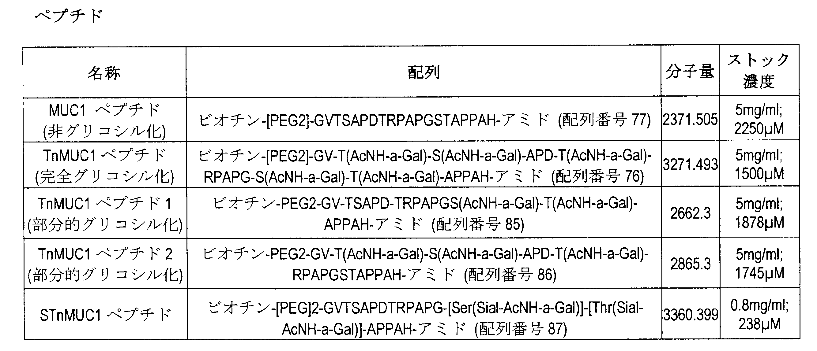

データ解析
フローサイトメトリーデータ解析
データは、MACS Quantify 2.8.1618.16380ソフトウエアで解析した。
data analysis
Flow Cytometry Data Analysis Data were analyzed with MACS Quantify 2.8.1618.16380 software.
IFNγデータ解析
IFNγの初期定量計算は、MSD Discovery Workbench 4.0.12.12(MSD)で行った。その後、プロット解析のためにデータをGraphPad Prism 8.0.2(GraphPad Software,Inc)に転送した。
IFNγ Data Analysis Initial quantitative calculations of IFNγ were performed with MSD Discovery Workbench 4.0.12.12 (MSD). Data were then transferred to GraphPad Prism 8.0.2 (GraphPad Software, Inc) for plot analysis.
統計解析はRバージョン3.5.1で行った。混合モデルはlme4パッケージを使用して適合させ、周辺平均と線形コントラストはemmeansパッケージを使用して計算した。ランダム切片は、ドナー、プレート、および実験日に関して含まれる。濃度、処理、およびペプチドについては、固定相互作用効果が適合された。異種分散性を減らすために、濃度は逆ハイパーボリックサイン変換(ゼロを処理するため)を使用して変換され、IFN放出はモデル化の前にlog10変換した。これらは、解釈可能な結果を表示するために逆変換した。逆ハイパーボリックサイン変換した濃度は、自由度4(AICを使用して決定)の自然スプラインを使用してモデル化した。各ペプチド対の間の、非形質転換T細胞(UT)とMB024との間のIFN放出における差(変化倍率として)の用量反応曲線間の線形コントラストを、元の実験において使用した濃度で計算し、FDR補正したp値とともに変化倍率として示した。 Statistical analysis was performed with R version 3.5.1. Mixture models were fitted using the lme4 package and marginal means and linear contrasts were calculated using the emmeans package. Random sections are included for donor, plate, and experimental day. Fixed interaction effects were fitted for concentration, treatment, and peptide. To reduce heterogeneity, concentrations were transformed using the inverse hyperbolic sine transformation (to handle zeros) and IFN release was log10 transformed prior to modeling. These were reverse transformed to display interpretable results. The inverse hyperbolic sine-transformed concentration was modeled using a natural spline with 4 degrees of freedom (determined using AIC). Linear contrast between dose-response curves of the difference (as fold change) in IFN release between untransformed T cells (UT) and MB024 between each peptide pair was calculated at the concentrations used in the original experiment. , expressed as fold change together with FDR-corrected p-values.
結果
ZsGreen形質導入効率の分析
MB024およびCD19 CAR T細胞をZsGreenタグとともに発現させ、これをT細胞でのCAR発現の分析に使用する。ドナー90774、92187および92205は、25~30%の範囲の形質導入レベルを示した。ドナー92804の形質導入はより高く、MB024では55%、CD19 CAR-Tでは58%を示す。
result
Analysis of ZsGreen Transduction Efficiency MB024 and CD19 CAR T cells are expressed with a ZsGreen tag, which is used for analysis of CAR expression in T cells. Donors 90774, 92187 and 92205 showed transduction levels ranging from 25-30%. Donor 92804 shows higher transduction, 55% for MB024 and 58% for CD19 CAR-T.
ドナー90774からのCD19 CARは、極めて低い形質導入率(3%)であったことに留意されたい。これは、形質導入時またはフローアッセイ時の技術的なエラーによるものである可能性がある。 Note that CD19 CAR from donor 90774 had a very low transduction rate (3%). This may be due to technical error during transduction or flow assay.
ペプチド刺激IFN-gの活性化
図29は、完全グリコシル化TnMUC1ペプチド(65-1)、グリコシル化の異なるTnMUC1ペプチド1(96-1)およびSTnペプチド(35-1)の存在下でのMB024 CAR T細胞による特定のIFNγ放出を示す。このアッセイでは、ドナー間のばらつきが観察された。各ドナーについて用量反応曲線を観察した。検出された最大IFNγは、4名の異なるドナーで1000~4000pg/mLの範囲であった。
Activation of peptide-stimulated IFN-g Figure 29 shows MB024 CAR in the presence of fully glycosylated TnMUC1 peptide (65-1), differentially glycosylated TnMUC1 peptide 1 (96-1) and STn peptide (35-1). Specific IFNγ release by T cells is shown. Donor-to-donor variability was observed in this assay. A dose-response curve was observed for each donor. Maximum IFNγ detected ranged from 1000 to 4000 pg/mL in four different donors.
TnMUC1 CAR T細胞は、非グリコシル化MUC1ペプチド(36-4)と、scFvタンパク質が結合しないとされる(Tarp, Sorensen et al, 2007)Tn糖が存在する、グリコシル化の異なるTnMUC1ペプチド2(96-2)と共培養してもIFNγの放出を示さなかった。 TnMUC1 CAR T cells were isolated from non-glycosylated MUC1 peptide (36-4) and the differentially glycosylated TnMUC1 peptide 2 (96 -2) did not show any release of IFNγ.
さらに、本発明者らは、総てのドナーで、メタ分析を行った(表24および表25)。各T細胞群(非形質導入されたMB024およびCAR19)内の総てのペプチドを比較したところ、STnMUC1(35-1)の存在下とTnMUC1(65-1)およびTnMUC1ペプチド1(96-1)の存在下でT細胞によるIFNγ放出にわずかな違いが示された。この違いは、0.015および0.03μMのペプチド濃度で、P値<0.0001で有意であった(表25)。 In addition, we performed a meta-analysis on all donors (Tables 24 and 25). All peptides within each T cell population (non-transduced MB024 and CAR19) were compared in the presence of STnMUC1 (35-1) versus TnMUC1 (65-1) and TnMUC1 peptide 1 (96-1). showed slight differences in IFNγ release by T cells in the presence of This difference was significant at P-value <0.0001 at peptide concentrations of 0.015 and 0.03 μM (Table 25).
また、P値を計算した場合も、Tn MUC1ペプチド2(96-2)、完全グリコシル化)TnMUC1ペプチド(65-1)およびSTn MUC1(35-1)の間で有意差が示され(P値>0.0001);および非グリコシル化MUC1(36-4)とは差はなく(表24)、TnMUC1ペプチド2はCAR T細胞を活性化できないことが統計学的に確認された。
P-values were also calculated and showed a significant difference between TnMUC1 peptide 2 (96-2), fully glycosylated) TnMUC1 peptide (65-1) and STn MUC1 (35-1) (P-value >0.0001); and no difference from non-glycosylated MUC1(36-4) (Table 24), statistically confirming that


考察
ペプチド刺激アッセイは、(i)TnおよびSTn MUC1ペプチドの存在下でのT細胞活性化の違い、(ii)TnMUC1 CARがランダムなTn陽性配列を認識する能力を分析する目的で設計した。
Discussion Peptide stimulation assays were designed to analyze (i) differences in T cell activation in the presence of Tn and STn MUC1 peptides, (ii) the ability of the TnMUC1 CAR to recognize random Tn-positive sequences.
本研究のデータは、STnMUC1(35-1)とTnMUC1(65-1)の0.015~0.03μMという極めて低い濃度でMB024 CAR T細胞によるIFNγ放出に有意差があることを示している。これは、MB024の生成に使用されたscFvはSTnMUC1に結合できるが、TnMUC1に対するよりも親和性が低いことを示すBIACoreアッセイのデータを裏付けるものである。しかしながら、0.03μMより高濃度ではこの差はなくなり、ある閾値を超えるとMB024はTnまたはSTn MUC1の存在下で等しく活性化されることを示す。 Our data show that there is a significant difference in IFNγ release by MB024 CAR T cells at very low concentrations of 0.015-0.03 μM of STnMUC1(35-1) and TnMUC1(65-1). This supports the BIACore assay data showing that the scFv used to generate MB024 can bind to STnMUC1, but with lower affinity than to TnMUC1. However, at concentrations higher than 0.03 μM this difference disappears, indicating that above a certain threshold MB024 is equally activated in the presence of Tn or STn MUC1.
CARの標的に対する特異性を理解することは、T細胞免疫療法の安全性のために不可欠である。ペプチド刺激アッセイの結果、MB024 CAR T細胞は、TnMUC1ペプチド2(96-2)と共培養しても活性化されなかったことを示している。このペプチドはエピトープではなく、ランダムな配列でTn糖を含んでいる。次のステップは、Tn/STn MUC1に対するTnMUC1 CARの特異性を完全に確認するために、Tnグリコシル化が陽性の相同配列を評価することである。 Understanding the target specificity of CARs is essential for the safety of T-cell immunotherapy. Peptide stimulation assay results show that MB024 CAR T cells were not activated by co-culture with TnMUC1 peptide 2(96-2). This peptide is not an epitope but contains Tn sugars in a random sequence. The next step is to evaluate homologous sequences positive for Tn glycosylation in order to fully confirm the specificity of the TnMUC1 CAR for Tn/STn MUC1.
要約すると、本研究はBIACoreデータを裏付け、低濃度のペプチドでは、STn MUC1よりもTnの存在下でCARがより多くのIFNγを放出することを明らかにした。さらに、ランダムなTn陽性配列は、MB024を活性化することができなかった。 In summary, the present study corroborates BIACore data and reveals that at low concentrations of peptide, CAR releases more IFNγ in the presence of Tn than STn MUC1. Furthermore, random Tn positive sequences were unable to activate MB024.
実施例13-Tn/STnMUC-1陽性および陰性標的細胞株との共培養におけるヒト化5E5 CAR T細胞候補のTn/STnMUC-1特異性の評価
本研究の目的は、T細胞白血病細胞株JurkatクローンE6-1およびCOSMCを導入したJurkatと共培養した際のCAR T細胞の活性化を評価することにより、4つのヒト化5E5 CAR T細胞候補(MB021、MB022、MB024およびMB025)の細胞表面Tn/STnMUC-1の標的特異性を確認することであった。
Example 13 - Evaluation of Tn/STnMUC-1 specificity of humanized 5E5 CAR T cell candidates in co-culture with Tn/STnMUC-1 positive and negative target cell lines Cell surface Tn/ The purpose was to confirm the target specificity of STnMUC-1.
方法
試験的作製
ヒト化5E5 CAR T細胞の作製
CAR-T細胞は前述のように作製した。
Method
experimental production
Generation of Humanized 5E5 CAR T Cells CAR-T cells were generated as previously described.
細胞株の作製
JurkatクローンE6-1細胞に、pG3.PGK.COSMC.IZWレンチウイルスベクターを4種類のMOI(1、3、5、10)で導入し、COSMC遺伝子を発現するように改変した。フローサイトメトリーを用い、形質導入効率とTn/STnMUC-1の細胞表面発現を評価した。MOI 10でCOSMCを形質導入したJurkatのプール集団を選択し、次いでTn/STnMUC1陰性細胞株としてCAR T細胞との共培養に使用した。
Generation of cell lines Jurkat clone E6-1 cells were infected with pG3. PGK. COSMC. The IZW lentiviral vector was introduced at four MOIs (1, 3, 5, 10) and modified to express the COSMC gene. Flow cytometry was used to assess transduction efficiency and cell surface expression of Tn/STnMUC-1. A pooled population of Jurkat transduced COSMCs at
T細胞の解凍および回復
凍結したT細胞の1mLアリコートを室温で半解凍し、次いで15mLの冷細胞培養培地に移した。T細胞を300×g、10分間、室温で遠心分離し、15mLの冷細胞培養培地に再懸濁させた。遠心分離を繰り返した後、T細胞を2mLの温(37℃)細胞培養培地に再懸濁させた。T細胞を計数し、細胞濃度を2×10⁶細胞/mLに調整した。T細胞は、6ウェル平底細胞懸濁プレートにてIL-2を100U/mLで含有する培地で、加湿インキュベーター中、37℃、5%CO2で24時間培養した。
Thawing and Recovery of
フローサイトメトリー分析のための細胞表面Tn/STnMUC-1、MUC-1、STnおよびTn染色
Jurkat WTおよびJurkat COSMC+細胞株をFACSバッファー(DPBS+2%FBS+0.05%アジ化ナトリウム)で2回洗浄した。1×10⁵細胞をTn/STnMUC-1、MUC-1、総STn、および総Tnに関してそれぞれ染色した。Tn/STnMUC-1、MUC-1およびSTn間接的染色;染色はFACSバッファーで希釈した50μLの一次抗体で行い、45分間、室温、暗所でインキュベートした。次に、細胞をFACSバッファーで2回洗浄した後、FACSバッファーで希釈した50μLのブリリアントバイオレット421(商標)ヤギ抗マウス二次抗体を加え、さらに45分間、室温、暗所でインキュベートした。細胞をさらに2回DPBSで洗浄し、次いで、1:2000に希釈したSytox Advanced生死判定色素を含有する100μLのDPBSに再懸濁させた。直接染色;染色は、FACSバッファーで希釈した50μLの抗体で行い、45分間、室温、暗所でインキュベートした。次に、細胞をFACSバッファーで2回洗浄し、次いで1:2000に希釈したSytox Orange Advanced生死判定色素を含有する100μLのDPBSに再懸濁させた。サンプルをLSRIIサイトメーターにかけた。BD CSおよびTビーズを用いてサイトメーターの性能を評価した後にデータを採取した。
Cell Surface Tn/STnMUC-1, MUC-1, STn and Tn Staining for Flow Cytometry Analysis Jurkat WT and Jurkat COSMC+ cell lines were washed twice with FACS buffer (DPBS+2% FBS+0.05% sodium azide). 1×10 5 cells were stained for Tn/STnMUC-1, MUC-1, total STn, and total Tn, respectively. Tn/STnMUC-1, MUC-1 and STn indirect staining; staining was performed with 50 μL of primary antibody diluted in FACS buffer and incubated for 45 minutes at room temperature in the dark. Cells were then washed twice with FACS buffer before adding 50 μL of Brilliant Violet 421™ goat anti-mouse secondary antibody diluted in FACS buffer and incubated for an additional 45 minutes at room temperature in the dark. Cells were washed two more times with DPBS and then resuspended in 100 μL of DPBS containing Sytox Advanced viability dye diluted 1:2000. Direct staining; staining was performed with 50 μL antibody diluted in FACS buffer and incubated for 45 minutes at room temperature in the dark. Cells were then washed twice with FACS buffer and then resuspended in 100 μL of DPBS containing Sytox Orange Advanced viability dye diluted 1:2000. Samples were run on the LSRII cytometer. Data were collected after evaluating the performance of the cytometer using BD CS and T-beads.
実験プロトコール
共培養の準備
共培養は、平底48ウェルプレートにて1:1 CAR-T:標的細胞比で2×10⁵のJurkat WTまたは2×10⁵のJurkat COSMC+腫瘍細胞株のいずれかに必要数のCAR T細胞を加えることにより準備した。CAR-T:標的細胞比は、異なるCAR T細胞間で形質導入効率を正規化したCAR-T細胞の形質入効率パーセントに基づいて算出した(実験準備のヒト化5E5 CAR T細胞生成を参照)。共培養物は、加湿インキュベーター中、37℃、5%CO2で24時間インキュベートした。24時間の時点で200μLの上清を回収し、その後のサイトカイン測定のために-80℃で冷凍した。
Experimental protocol
Preparation of co-cultures Co-cultures were prepared in flat-bottom 48-well plates with the required number of either 2 x 10 5 Jurkat WT or 2 x 10 5 Jurkat COSMC+ tumor cell lines at a 1:1 CAR-T:target cell ratio. Prepared by adding CAR T cells. CAR-T:target cell ratios were calculated based on percent transduction efficiency of CAR-T cells normalizing transduction efficiencies among different CAR T cells (see Humanized 5E5 CAR T cell generation in Experimental Preparation). . Co-cultures were incubated for 24 hours at 37° C., 5% CO 2 in a humidified incubator. At 24 hours, 200 μL of supernatant was harvested and frozen at −80° C. for subsequent cytokine measurements.
上清サイトカイン検出-サイトカインビーズアレイ(CBA)アッセイ
BD(商標)CBAヒトTh1/Th2サイトカインキットIIおよびBD(商標)CBAヒトグランザイムBフレックスセットD7を併せて1つのマルチプレックスアッセイとし、これを用いてIL2、IL4、IL6、IL10、IFNγ、TNFαおよびグランザイムBの上清サイトカインレベルを分析した。各キットに提供される各サイトカインの凍結乾燥タンパク質を組み合わせ、1mLの細胞培養培地(アッセイ希釈液として使用)で再構成し、アッセイ用最大標準を作製した。最大標準を細胞培養培地で11回、連続1:2希釈し、10,000~0pg/mLの範囲のアッセイ標準を作製した。細胞培養培地のみをアッセイバックグラウンドとして使用した。上清を、細胞培養培地を用いて1:2希釈した。各試験サンプルに関して、CBA Human Th1/Th2サイトカインキットII捕捉ビーズ(全6サイトカインの捕捉ビーズを含む)各4μLを、CBAヒトグランザイムBフレックスセットD7捕捉ビーズ(グランザイムB捕捉ビーズを1:50希釈)と組み合わせることにより、捕捉ビーズのマスターミックスを調製した。また、各テストサンプルに対して、25μLのCBAヒトTh1/Th2サイトカインキットII PE検出試薬と0.5μLのCBAヒトグランザイムBフレックスセットD7 PE検出試薬(グランザイムB PE検出試薬1:50希釈)を組み合わせることにより、PE検出試薬マスターミックスを調製した。25μLの捕捉ビーズマスターミックスと25μL のPE検出試薬マスターミックスを上清50μLとアッセイ標準50μLに同時に添加した。プレートを密封し、室温、暗所で3時間振盪しながら(600rpm)インキュベートした。100μLのCBAヒトTh1/Th2サイトカインキットII洗浄バッファーを加えてプレートを洗浄し、300×gで5分間遠心分離した。洗浄を繰り返した後、60μLの洗浄バッファーに再懸濁させ、LSRIIサイトメーターで測定した。BD CS&Tビーズを用いてサイトメーターの性能を評価した後にデータを取得した。
Supernatant Cytokine Detection - Cytokine Bead Array (CBA) Assay BD™ CBA Human Th1/Th2 Cytokine Kit II and BD™ CBA Human Granzyme B Flex Set D7 were combined into one multiplex assay using Supernatant cytokine levels of IL2, IL4, IL6, IL10, IFNγ, TNFα and granzyme B were analyzed. The lyophilized protein of each cytokine provided in each kit was combined and reconstituted with 1 mL of cell culture medium (used as assay diluent) to make the maximum standard for the assay. The maximum standard was serially diluted 1:2 eleven times in cell culture medium to generate assay standards ranging from 10,000 to 0 pg/mL. Only cell culture medium was used as assay background. The supernatant was diluted 1:2 with cell culture medium. For each test sample, 4 μL each of CBA Human Th1/Th2 Cytokine Kit II Capture Beads (includes capture beads for all 6 cytokines) with CBA Human Granzyme B Flex Set D7 Capture Beads (1:50 dilution of Granzyme B Capture Beads). A master mix of capture beads was prepared by combining. Also, for each test sample, combine 25 μL of CBA Human Th1/Th2 Cytokine Kit II PE Detection Reagent and 0.5 μL of CBA Human Granzyme B Flex Set D7 PE Detection Reagent (1:50 dilution of Granzyme B PE Detection Reagent). A PE detection reagent master mix was thus prepared. 25 μL of capture bead master mix and 25 μL of PE detection reagent master mix were added simultaneously to 50 μL of supernatant and 50 μL of assay standard. The plate was sealed and incubated at room temperature in the dark with shaking (600 rpm) for 3 hours. Plates were washed by adding 100 μL of CBA Human Th1/Th2 Cytokine Kit II wash buffer and centrifuged at 300×g for 5 minutes. After repeated washing, cells were resuspended in 60 μL of wash buffer and measured with an LSRII cytometer. Data were acquired after evaluating the performance of the cytometer using BD CS&T beads.

データ分析
フローサイトメトリーデータは、Flowlogic 7.2.1ソフトウエアを使用して解析し、プライマリーメトリクスとプロットを作成した。サイトカインビーズアレイアッセイからのメトリクスは、Microsoft ExcelとGraphPad Prism 7で解析した。結果はGraphPad Prism 7を用いてグラフ化した。
Data Analysis Flow cytometry data were analyzed using Flowlogic 7.2.1 software to generate primary metrics and plots. Metrics from the cytokine bead array assay were analyzed with Microsoft Excel and
GraphPad Prism 7を用いて、多項式:2次(Y=A+B*X+C*X^2)非線形曲線フィットモデルを使用して標準曲線を算出した。標準曲線からの補間を用い、上清のサイトカイン濃度をpg/mLで決定した。
Standard curves were calculated using
結果
腫瘍細胞株の特性決定
Jurkat WT(COSMC-)細胞は、Jurkat COSMC+細胞での1%発現に比べて細胞表面Tn/STnMUC-1発現が100%陽性であった。HMFG2抗体により検出されるようにMUC-1発現は、Jurkat WT細胞株およびJurkat COSMC+細胞株の両方で低く、それぞれおよそ10%および5%であった。
result
Characterization of Tumor Cell Lines Jurkat WT (COSMC−) cells were 100% positive for cell surface Tn/STnMUC-1 expression compared to 1% expression in Jurkat COSMC+ cells. MUC-1 expression as detected by HMFG2 antibody was low in both Jurkat WT and Jurkat COSMC+ cell lines, approximately 10% and 5%, respectively.
Tn/STnMUC-1陽性腫瘍細胞に対するサイトカイン応答
4種類のヒト化CAR-T細胞は総て、Tn/STnMUC-1が陽性のJurkat WT(COSMC-)腫瘍細胞との共培養に応答してサイトカインおよびグランザイムBを分泌した(図30)。MB022およびMB025 CAR-T細胞は、24時間の共培養の後に上清中に検出されるサイトカインおよびグランザイムBレベルがより高いMB021およびMB024 CAR T細胞に比べて大きい応答を示した。MB021およびMB024 CAR T細胞は、サイトカインおよびグランザイムBの平均放出レベルに同等のプロファイルを示す。MB022およびMB025は、若干の類似性を示したが、一般にMB025はMB022と比較してサイトカインおよびグランザイムBの放出が多く、全4種類のCAR構築物の中で最も高い。統計解析は行っていない。
Cytokine responses to Tn/STnMUC-1-positive tumor cells All four humanized CAR-T cells responded to co-culture with Tn/STnMUC-1-positive Jurkat WT (COSMC-) tumor cells by Granzyme B was secreted (Figure 30). MB022 and MB025 CAR-T cells showed greater responses than MB021 and MB024 CAR T cells with higher cytokine and granzyme B levels detected in the supernatant after 24 hours of co-culture. MB021 and MB024 CAR T cells show comparable profiles of cytokine and granzyme B mean release levels. MB022 and MB025 showed some similarity, but in general MB025 showed more cytokine and granzyme B release compared to MB022, the highest of all four CAR constructs. No statistical analysis was performed.
Tn/STnMUC-1陰性腫瘍細胞に対するサイトカイン応答
4種類のヒト化CAR T細胞は総て、Tn/STnMUC-1が陰性のJurkat COSMC+腫瘍細胞との共培養に応答してサイトカイン放出もグランザイムB放出を示さなかった。Jurkat COSMC+との共培養で検出されたサイトカインおよびグランザイムBの両レベルは、CAR-T細胞単独培養の場合と同等であった。CAR構築物間で差は見られなかった。統計解析は行っていない。
Cytokine response to Tn/STnMUC-1-negative tumor cells All four humanized CAR T cells exhibited both cytokine and granzyme B release in response to co-culture with Tn/STnMUC-1-negative Jurkat COSMC+ tumor cells. did not show. Both cytokine and granzyme B levels detected in co-cultures with Jurkat COSMC+ were comparable to CAR-T cell cultures alone. No differences were seen between the CAR constructs. No statistical analysis was performed.
考察
4つのヒト化5E5 CAR T細胞は総て、Tn/STnMUC-1を発現する腫瘍細胞に対して抗原特異的な反応性を示す。評価したサイトカインに基づけば、4つのCAR T細胞は総て、高レベルのIFNγ、TNFα、IL-2ならびにランザイムBの放出を伴った強いTh1腫瘍媒介応答を示す。試験した4つのCAR構築物のうち、MB025は、抗原刺激に対して全体的に最も大きな応答を示し、特に多量のグランザイムBを産生した。
Discussion All four humanized 5E5 CAR T cells show antigen-specific reactivity against tumor cells expressing Tn/STnMUC-1. Based on the cytokines evaluated, all four CAR T cells display strong Th1 tumor-mediated responses with high levels of IFNγ, TNFα, IL-2 and Ranzyme B release. Of the four CAR constructs tested, MB025 showed the greatest overall response to antigenic stimulation, producing particularly large amounts of granzyme B.
抗原特異性を示すことに加え、4種類のヒト化5E5 CAR T細胞は、Tn/STnMUC-1陰性細胞に対して標的外反応を示さず、サイトカインおよびグランザイムBのレベルは単独培養のCAR-T細胞と同等であった。このことを考え合わせると、4種類のヒト化5E5 CAR-T細胞候補は総て、このシステムにおいて有効かつ安全であることが証明される。 In addition to demonstrating antigen specificity, the four humanized 5E5 CAR T cells showed no off-target responses to Tn/STnMUC-1-negative cells, and cytokine and granzyme B levels were higher than those of CAR-T in monocultures. equivalent to cells. Taken together, all four humanized 5E5 CAR-T cell candidates prove efficacious and safe in this system.
COSMCは、T合成酵素機能に必要なT合成酵素シャペロンタンパク質である。T合成酵素は、Tn抗原を正常細胞特有の複雑なO-グリカン構造に伸長させる触媒となる。COSMCを導入すると、Jurkat細胞における内因性のTn/STnMUC1発現が消失した。これは、正常細胞のグリコシル化を回復させた結果としてのTnおよびSTn発現の消失を介するものであった。Tn/STn糖型の消失とそれに続くTn/STnMUC-1発現の消失は、4種類のCAR-T細胞総ての活性化を抑制し、このことは、これらのCARの特異性が、部分的にはTnおよび/またはSTnの認識によって駆動されていることを示している。興味深いことに、これらのCAR-T細胞をMCF7 MUC1 KO細胞(またTn/STnMUC1-)で刺激すると、MB022およびMB025は非特異的応答を示したが、MB021もMB024も24時間の共培養後に高レベルのIFNγおよびグランザイムBを産生していないことが示された。MCF7細胞におけるMUC-1のノックアウトは、MCF7 WT細胞に通常発現しているTn/STnMUC-1の認識が、MUC-1の発現消失により根絶されるというCAR特異性を研究するためのモデルを提供する。しかしながら、MUC-1が存在しないため、グリコシル化の異常ならびに他のタンパク質(他のムチンなど)上のTnおよびSTnの発現の可能性には対処できない。従って、MCF7 MUC-1 KO細胞に対するMB022およびMB025の反応性は、MUC-1以外の他のタンパク質上のTnまたはSTnを認識した結果であり得る。このことは、MB022およびMB025がJurkat WT細胞に対して、MB021およびMB024に比べて高いサイトカインおよびグランザイムB応答を示したことを説明し得る。 COSMC is a T synthase chaperone protein required for T synthase function. T synthase catalyzes the extension of the Tn antigen into complex O-glycan structures characteristic of normal cells. Introduction of COSMCs abolished endogenous Tn/STnMUC1 expression in Jurkat cells. This was mediated by loss of Tn and STn expression as a result of restoring normal cell glycosylation. Loss of Tn/STn glycoforms and subsequent loss of Tn/STnMUC-1 expression suppressed the activation of all four CAR-T cells, indicating that the specificity of these CARs was partially is driven by recognition of Tn and/or STn. Interestingly, when these CAR-T cells were stimulated with MCF7 MUC1 KO cells (also Tn/STnMUC1-), MB022 and MB025 showed non-specific responses, whereas neither MB021 nor MB024 showed high response after 24 h of co-culture. It was shown not to produce levels of IFNγ and granzyme B. Knockout of MUC-1 in MCF7 cells provides a model for studying CAR specificity, in which recognition of Tn/STn MUC-1 normally expressed in MCF7 WT cells is abolished by loss of MUC-1 expression. do. However, the absence of MUC-1 does not address the possibility of abnormal glycosylation and expression of Tn and STn on other proteins (such as other mucins). Therefore, the reactivity of MB022 and MB025 to MCF7 MUC-1 KO cells may be the result of recognizing Tn or STn on proteins other than MUC-1. This may explain that MB022 and MB025 showed higher cytokine and granzyme B responses to Jurkat WT cells compared to MB021 and MB024.
Jurkat細胞におけるTn/STnMUC-1発現の特性決定に加えて、総MUC-1発現も測定した。総MUC-1発現は、Jurkat WT(COSMC-)およびJurkat COSMC+細胞の両方において、Tn/STnMUC-1発現よりも実質的に低いことが示された。総MUC-1は、Tn/STnMUC-1と同等以上のレベルで発現していると予想された。しかしながら、これはJurkats細胞で一貫して観察されていたことである。MUC-1発現を決定するために使用されるモノクローナル抗体であるHMFG2は、MUC-1ペプチドDTRモチーフの認識を介して特異性を駆動し、一方、5E5はMUC-1ペプチド上のTn/STnグリカンに特異的なグリコペプチドエピトープを認識している。総MUC-1とTn/STnMUC-1の間の相違は、HMFG2と5E5の、MUC-1に対する異なる結合エピトープに関係している可能性が高い。他の細胞株ではHMFG2の使用がより予測しやすかったため、HMFG2結合の欠如はJurkatsに内在するものである可能性もある。MUC-1のクラスター化、立体配座またはペプチド配列は総てが要因である可能性がある。また、HMFG2の結合はMUC1のグリコシル化状態によって影響を受けることも示されている。このことにも関わらず、MUC-1上のTn/STnに対する5E5の特異性に基づけば、MUC-1はJurkat細胞上で発現し、Tn/STn糖型で異常にグリコシル化されていると確信できる。 In addition to characterizing Tn/STnMUC-1 expression in Jurkat cells, total MUC-1 expression was also measured. Total MUC-1 expression was shown to be substantially lower than Tn/STnMUC-1 expression in both Jurkat WT (COSMC-) and Jurkat COSMC+ cells. Total MUC-1 was expected to be expressed at levels equal to or greater than Tn/STnMUC-1. However, this was consistently observed in Jurkats cells. HMFG2, the monoclonal antibody used to determine MUC-1 expression, drives specificity through recognition of the MUC-1 peptide DTR motif, while 5E5 drives the Tn/STn glycans on the MUC-1 peptide. recognizes a glycopeptide epitope specific to The differences between total MUC-1 and Tn/STnMUC-1 are likely related to different binding epitopes of HMFG2 and 5E5 for MUC-1. The lack of HMFG2 binding may also be intrinsic to Jurkats, as HMFG2 usage was more predictable in other cell lines. MUC-1 clustering, conformation or peptide sequence may all be factors. It has also been shown that HMFG2 binding is affected by the glycosylation status of MUC1. Despite this, based on the specificity of 5E5 for Tn/STn on MUC-1, we believe that MUC-1 is expressed on Jurkat cells and is aberrantly glycosylated on the Tn/STn glycoform. can.
これらのことを考え合わせ、本発明者らは、4種類のヒト化5E5 CAR T細胞は、Tn/STnMUC-1陽性の腫瘍細胞に対する強いTh1応答を証拠として、極めて強力であることを示した。さらに、総てが、細胞は正常組織に固有の複雑なグリカンを腫瘍特異的な末端切断型のTn/STn糖型から識別する。 Taken together, we have shown that the four humanized 5E5 CAR T cells are extremely potent as evidenced by strong Th1 responses against Tn/STnMUC-1 positive tumor cells. In addition, all cells distinguish complex glycans inherent in normal tissues from tumor-specific truncated Tn/STn glycoforms.
実施例14-ヒト化TnMUC1 CAR T産物の多機能性評価
本研究の目的は、抗原刺激に応答して、インターロイキン-2(IL2)、タイプIIインターフェロン(IFNγ)、腫瘍壊死因子α(TNFα)およびグランザイムB(Grz B)を含む一連の機能的サイトカインを分泌するTnMUC1 CAR T細胞の能力を評価することである。ヒト化CAR産物の完全なサイトカインプロファイルを構築するために、細胞表面、細胞内染色(ICS)、上清サイトカイン測定を一緒に行う精密なプロトコールを設計した。ICSを使用して、陽性細胞株MDA-MB-468(陰性細胞株MCF7-KOと比較)との共培養24時間後のTnMUC1 CAR T細胞が分泌し得るエフェクター(GrzB、IFNγ、TNFα)および刺激(IL-2)サイトカイン発現レベルを調べた。サイトメトリック・ビーズ・アレイ(CBA)およびLuminexアッセイを使用して、MDA-MB-468との共培養24時間後の上清中のGrzB、IFNγ、TNFα、IL-2(およびCBAアッセイ用の追加サイトカインとしてIL-4、6、10)の実際の分泌サイトカイン量を相関させた(MCF7-KOと比較)。
Example 14 - Multifunctional Evaluation of Humanized TnMUC1 CAR T Products and granzyme B (Grz B) to assess the ability of TnMUC1 CAR T cells to secrete a range of functional cytokines. A precise protocol was designed to combine cell surface, intracellular staining (ICS), and supernatant cytokine measurements to construct a complete cytokine profile of the humanized CAR product. Effectors (GrzB, IFNγ, TNFα) and stimulation that TnMUC1 CAR T cells can secrete after 24 hours of co-culture with the positive cell line MDA-MB-468 (compared to the negative cell line MCF7-KO) using ICS. (IL-2) cytokine expression levels were examined. Using cytometric bead array (CBA) and Luminex assays, GrzB, IFNγ, TNFα, IL-2 (and additional for CBA assay) in supernatants after 24 h co-culture with MDA-MB-468 IL-4, 6, 10) as cytokines were correlated with actual secreted cytokine amounts (compared to MCF7-KO).
方法
実験準備
T細胞の解凍および回復
供試T細胞は、形質導入後12日目の非形質未導入T(UT)およびHuCAR021、022、024、025(構築物でzsGreen遺伝子を発現するヒト化型のTnMUC1 5E5 CAR T)細胞である。3名のドナー(n=3)から得た凍結精製ヒト化CAR T細胞の1mLアリコートを室温で半解凍し、次いで15mLの冷細胞培養培地に移した。T細胞を300×gで10分間、室温で遠心分離し、15mLの冷細胞培養培地に再懸濁させた。遠心分離を繰り返した後、T細胞を2mLの温(37℃)細胞培養培地に再懸濁させた。T細胞を計数し、細胞密度を2×10⁶細胞/mLに調整した。T細胞は、6ウェル平底細胞懸濁プレートにて、IL-2を100U/mLで含有する培地中、加湿インキュベーターにて、37℃、5%CO2で24時間培養した。
Method
Experiment preparation
Thawing and Recovery of T Cells Test T cells were isolated from non-transduced T (UT) and HuCAR021, 022, 024, 025 (a humanized version of the TnMUC1 5E5 CAR expressing the zsGreen gene in
腫瘍細胞の播種および共培養の準備
ヒト乳癌MDA-MB-468細胞およびMCF7 MUC1 KO細胞を採取し、温細胞培養培地に再懸濁させ、MDA-MB-468細胞およびMCF7 MUC1 KO細胞を、ヒト化CAR T細胞を加える前に6ウェルプレートにウェル当たり1×10⁶細胞として播種し、一晩置いた。共培養のために、腫瘍細胞を含む6ウェルプレートに、エフェクター:標的比1:1として、非形質導入T細胞または形質導入huCAR T細胞を加えることでプレートを準備した。T細胞の正確な量は、各CAR T細胞の形質導入効率のパーセンテージに基づいて計算し、この場合、形質導入効率は、UT細胞を加えることによって、4種類のヒト化CAR-T構築物総ての最低パーセンテージに対して正規化した。形質導入効率は、フローサイトメトリー分析によって決定されたzsGreen陽性細胞の%として示す。共培養は、加湿インキュベーターにて、37℃、5%CO2で24時間インキュベートした。翌日、500μLの上清を回収し、次いでその後のサイトカイン解析のために-80℃で冷凍した。次に、細胞内タンパク質輸送阻害剤ブレフェルジンA(BFA)をT細胞単独および共培養サンプルに1:2000希釈で添加し、細胞内サイトカイン染色を可能にした。さらに、37℃、5%CO2でさらに4時間インキュベーションを行った。その後、各サンプル中の全細胞を回収し、細胞表面マーカーおよび細胞内サイトカイン染色を行った。
Tumor Cell Seeding and Co-Culture Preparation Human breast cancer MDA-MB-468 and MCF7 MUC1 KO cells were harvested and resuspended in warm cell culture medium and MDA-MB-468 and MCF7 MUC1 KO cells were transfected into human cells. 6-well plates were seeded at 1×10 6 cells per well and left overnight before adding modified CAR T cells. For co-culture, plates were prepared by adding untransduced T cells or transduced huCAR T cells at an effector:target ratio of 1:1 to 6-well plates containing tumor cells. The exact amount of T cells was calculated based on the percentage transduction efficiency of each CAR T cell, where the transduction efficiency was calculated by adding UT cells to all four humanized CAR-T constructs. normalized to the lowest percentage of Transduction efficiency is shown as % zsGreen positive cells determined by flow cytometric analysis. Co-cultures were incubated for 24 hours at 37° C., 5% CO 2 in a humidified incubator. The next day, 500 μL of supernatant was harvested and then frozen at −80° C. for subsequent cytokine analysis. The intracellular protein transport inhibitor Brefeldin A (BFA) was then added to the T cell alone and co-culture samples at a 1:2000 dilution to allow intracellular cytokine staining. Further incubation was performed at 37° C., 5% CO 2 for an additional 4 hours. All cells in each sample were then harvested and subjected to cell surface marker and intracellular cytokine staining.
染色した同日にMDA-MB-468細胞およびMCF7 MUC1 KO細胞のさらなるウェルに播種し、播種24時間後の細胞表面Tn/STnMUC1およびMUC1発現を評価した。また、MDA-MB-468細胞およびMCF7 MUC1 KO細胞は、播種時のTn/STnMUC1およびMUC1発現についても特性を評価した。 Additional wells of MDA-MB-468 cells and MCF7 MUC1 KO cells were seeded on the same day of staining and cell surface Tn/STnMUC1 and MUC1 expression was assessed 24 hours after seeding. MDA-MB-468 cells and MCF7 MUC1 KO cells were also characterized for Tn/STnMUC1 and MUC1 expression upon plating.
細胞表面Tn/STnMUC1およびMUC1発現
MDA-MB-468細胞およびMCF7 MUC1 KO細胞を培養フラスコから剥離し、次いでDPBSで2回洗浄した。次に1×10⁵細胞を、FACSバッファーで希釈した一次抗体50μLを加えて細胞表面のTn/STnMUC1およびMUC1をそれぞれ染色し、室温、暗所で45分間インキュベートした。その後、細胞をFACSバッファーで2回洗浄した後、FACSバッファーで希釈したブリリアントバイオレット421(商標)ヤギ抗マウス二次抗体50μLを加え、さらに45分間、室温、暗所でインキュベートした。細胞をDPBSでさらに2回洗浄し、次いで1:2000に希釈したSytox Advanced生死判定色素を含有する100μLのDPBSに再懸濁させた後、5-レーザーLSR IIサイトメーターで測定した。
Cell surface Tn/STnMUC1 and MUC1 expressing MDA-MB-468 cells and MCF7 MUC1 KO cells were detached from culture flasks and then washed twice with DPBS. 1×10 5 cells were then stained for Tn/STnMUC1 and MUC1 on the cell surface by adding 50 μL of primary antibody diluted in FACS buffer and incubated for 45 minutes at room temperature in the dark. Cells were then washed twice with FACS buffer before adding 50 μL of Brilliant Violet 421™ goat anti-mouse secondary antibody diluted in FACS buffer and incubated for an additional 45 minutes at room temperature in the dark. Cells were washed two more times with DPBS and then resuspended in 100 μL DPBS containing Sytox Advanced viability dye diluted 1:2000 before reading on a 5-laser LSR II cytometer.
サイトメーターの設定および補正
CS&Tビーズを使用して、毎日、サイトメーターの性能を評価し、各時点における調整されたデータ取得のための正確なアプリケーション設定を通知した。補正は、各抗体の蛍光色素コンジュゲートで個別に染色したInvitrogen Ultra Comp eBeadsを使用して、FACSDivaでサンプルデータを取得する前に計算した。
Cytometer settings and calibrated CS&T beads were used to assess cytometer performance daily to inform the correct application settings for coordinated data acquisition at each time point. Corrections were calculated prior to sample data acquisition on the FACSDiva using Invitrogen Ultra Comp eBeads individually stained with fluorochrome conjugates for each antibody.
実験プロトコール
Fcのブロッキングおよび細胞表面染色
細胞を回収した後、総てのサンプルをDPBSで2回洗浄した後、DPBSで1:2000希釈した100μLの生死判定色素Zombie Aquaで染色し、室温、暗所で15分間インキュベートした。染色後、CAR-T細胞をDPBSで2回洗浄した。次に、Fc受容体のブロックは、1:50希釈した過剰量の無関係なヒトIgGアイソタイプ対照(原液濃度:5mg/mL)を含む40μLのFACSバッファーに細胞を再懸濁させ、室温、暗所で15分間インキュベーションすることにより行った。次に、FACSバッファーに希釈したCD3およびCD8抗体-蛍光色素コンジュゲートを含む2倍濃縮抗体カクテル40μLを添加して、暗所、室温で30分間、CAR-T細胞を染色した。
Experimental protocol
Fc blocking and cell surface staining After harvesting cells, all samples were washed twice with DPBS before staining with 100 μL of the viability dye Zombie Aqua diluted 1:2000 in DPBS and incubated for 15 minutes at room temperature in the dark. Incubate for 1 minute. After staining, CAR-T cells were washed twice with DPBS. Blocking of Fc receptors was then performed by resuspending cells in 40 μL of FACS buffer containing a 1:50 dilution of an excess of an irrelevant human IgG isotype control (stock concentration: 5 mg/mL) at room temperature in the dark. was performed by incubating for 15 minutes at . Next, 40 μL of a 2× concentrated antibody cocktail containing CD3 and CD8 antibody-fluorochrome conjugates diluted in FACS buffer was added to stain CAR-T cells for 30 minutes at room temperature in the dark.
細胞内サイトカイン染色
細胞表面染色インキュベーションの後、CAR T細胞を200μLのFACSバッファーで2回洗浄した後、100μLのBD Fix/Permバッファー中、4℃、暗所で20分間固定した。その後、CAR T細胞を100μLのDPBSで2回洗浄し、200μLのBD Perm/Washバッファーで洗浄して透過処理を施した。次に、CAR T細胞を、BD Perm/Washバッファーで希釈した総ての細胞内サイトカイン(IL-2、IFNγ、TNFα、GmzB)抗体-蛍光色素コンジュゲートを含む50μLの抗体カクテルで染色し、暗所、室温で45分間インキュベートした。CAR T細胞は、200μLのBD Perm/Washバッファーでさらに2回洗浄した後、100μLのDPBSに再懸濁させた。サンプルはLSRIIサイトメーターで分析した。
Intracellular Cytokine Staining After cell surface staining incubation, CAR T cells were washed twice with 200 μL FACS buffer and then fixed in 100 μL BD Fix/Perm buffer for 20 minutes at 4° C. in the dark. CAR T cells were then washed twice with 100 μL DPBS and permeabilized by washing with 200 μL BD Perm/Wash buffer. CAR T cells were then stained with 50 μL of an antibody cocktail containing all intracellular cytokine (IL-2, IFNγ, TNFα, GmzB) antibody-fluorochrome conjugates diluted in BD Perm/Wash buffer and washed in the dark. Incubate at room temperature for 45 minutes. CAR T cells were washed two more times with 200 μL BD Perm/Wash buffer and then resuspended in 100 μL DPBS. Samples were analyzed on an LSRII cytometer.
サイトカインビーズアレイ(CBA)アッセイを用いた上清サイトカインの検出
BD(商標)CBAヒトTh1/Th2サイトカインキットIIとBD(商標)CBAヒトグランザイムBフレックスセットD7を組み合わせて1つのマルチプレックスアッセイとし、IL2、IL4、IL6、IL10、IFNγ、TNFαおよびグランザイムBの上清サイトカインレベルを分析するために使用した。凍結上清を室温で解凍した。アッセイ最大標準は、各キットに提供される各サイトカインの凍結乾燥タンパク質を組み合わせ、1mLの細胞培養培地で再構成することにより調製した(アッセイ希釈液として使用)。アッセイ最大標準を細胞培養培地で連続希釈してアッセイ標準を作製した。50μLの上清およびアッセイ標準をアッセイのために96ウェル、V底プレートに移した。各試験サンプルに関して、CBAヒトTh1/Th2サイトカインキットII捕捉ビーズ各4μLおよび0.5μLのCBAヒトグランザイムBフレックスセットD7捕捉ビーズ(グランザイムB捕捉ビーズは1:50希釈)を合わせることにより、7つ総てのサイトカインの捕捉ビーズを含む捕捉ビーズマスターミックスを作製した。また、各試験サンプルに関して、25μLのCBAヒトTh1/Th2サイトカインキットII PE検出試薬および0.5μLのCBAヒトグランザイムBフレックスセットD7 PE検出試薬(グランザイムB PE検出試薬は1:50希釈)を合わせることでPE検出試薬マスターミックスを作製した。25μLの捕捉ビーズマスターミックスおよび25μLのPE検出試薬マスターミックスを上清およびアッセイ標準に同時に添加した。プレートを密封し、室温、暗所で3時間振盪(600rpm)しながらインキュベートした。100μLのCBAヒトTh1/Th2サイトカインキットII洗浄バッファーを加え、300×gで5分間遠心することでプレートを洗浄した。洗浄を繰り返した後、60μLの洗浄バッファーに再懸濁させた後、LSRIIサイトメーターで測定した。
Detection of Supernatant Cytokines Using a Cytokine Bead Array (CBA) Assay BD™ CBA Human Th1/Th2 Cytokine Kit II and BD™ CBA Human Granzyme B Flex Set D7 were combined into one multiplex assay, IL2 , IL4, IL10, IFNγ, TNFα and granzyme B were used to analyze supernatant cytokine levels. Frozen supernatants were thawed at room temperature. Assay maximum standards were prepared by combining the lyophilized proteins of each cytokine provided in each kit and reconstituting with 1 mL of cell culture medium (used as assay diluent). Assay standards were made by serially diluting the assay maximum standard in cell culture medium. 50 μL of supernatant and assay standards were transferred to a 96-well, V-bottom plate for assay. For each test sample, a total of seven total A capture bead master mix was made containing capture beads for all cytokines. Also, for each test sample, combine 25 μL CBA Human Th1/Th2 Cytokine Kit II PE Detection Reagent and 0.5 μL CBA Human Granzyme B FlexSet D7 PE Detection Reagent (Granzyme B PE Detection Reagent diluted 1:50). made a PE detection reagent master mix. 25 μL of capture bead master mix and 25 μL of PE detection reagent master mix were added simultaneously to the supernatant and assay standards. Plates were sealed and incubated at room temperature in the dark for 3 hours with shaking (600 rpm). Plates were washed by adding 100 μL of CBA Human Th1/Th2 Cytokine Kit II wash buffer and centrifuging at 300×g for 5 minutes. After repeated washing, cells were resuspended in 60 μL of wash buffer and measured with an LSRII cytometer.
Luminexアッセイを用いた上清サイトカインの検出
抗原標準セットは、アッセイキットに提供されているPCR 8チューブストリップを用いて4倍連続希釈で再構成した。96ウェルプレートをハンドヘルドマグネティックプレートウォッシャーに取り付けた。磁性ビーズ溶液をまず30秒間ボルテックスで撹拌し、50μLのビーズ溶液をプレートに加え、150μLの洗浄バッファー(1倍)で1回洗浄した。プレートをハンドヘルドマグネティックプレートウォッシャーから取り出し、各ウェルに50μLの標準、対照またはサンプルを加え、密封し、室温で120分間振盪しながらインキュベートした。インキュベーション後、96ウェルプレートをハンドヘルドマグネティックプレートウォッシャーに戻し、150μLの洗浄バッファー(1倍)で2回洗浄した。次に、25μLの検出抗体混合物を加え、密封し、プレートシェーカー上、室温、500rpmで30分間インキュベートした。プレートをハンドヘルドマグネティックプレートウォッシャーに戻し、150μLの洗浄バッファー(1倍)で2回洗浄した。50μLのストレプトアビジン-PE溶液を加え、密封し、プレートシェーカー上、室温、500rpmで30分間インキュベートした。最後に、プレートをハンドヘルドマグネティックプレートウォッシャーに戻し、150μLの洗浄バッファー(1倍)で2回洗浄し、次いで120μLのリーディングバッファーを各ウェルに加え、密封し、室温で5分間振盪した後、データ取得のためにLuminex(商標)装置「Bio-Plex 3D Suspension Array System」に置いた。
Detection of Supernatant Cytokines Using the Luminex Assay The antigen standard set was reconstituted in 4-fold serial dilutions using the
データ解析
フローサイトメトリーデータは、Flowlogic 7.2.1ソフトウエアを用いて解析し、プライマリーメトリクスおよびプロットを作成した。メトリクスは、Microsoft ExcelおよびGraphPad Prism 7で解析した。ゲートの設定:CD4+およびCD8+T細胞サブセットは、形質導入された(ZsGreen+)T細胞集団と形質導入されていない(ZsGreen-)T細胞集団とで比較した。CAR T細胞については、形質導入された集団のみのデータが示されている。蛍光強度のパーセンテージと中央値は、主要なメトリクスとしてエクスポートし。GraphPad Prism 7を使用して、4名のドナーの結果を表すメトリクスをグラフ化した。
Data Analysis Flow cytometry data were analyzed using Flowlogic 7.2.1 software to generate primary metrics and plots. Metrics were analyzed with Microsoft Excel and
CBAアッセイの場合:GraphPad Prism v7ソフトウエアを使用し、多項式:2次(Y=A+B*X+C*X^2)非線形曲線フィットモデルを用いて標準曲線を算出した。 For the CBA assay: GraphPad Prism v7 software was used to calculate the standard curve using a polynomial: quadratic (Y=A+B * X+C * X^2) non-linear curve fit model.
標準曲線からの補間を用いて、サイトカイン濃度をpg/mLとして決定した。 Cytokine concentrations were determined as pg/mL using interpolation from standard curves.
Luminexアッセイの場合:Luminex xPonent v4.2ソフトウエアを使用してマシンのデータ取得し、BioPlex Manager v6.1を使用して標準曲線の作成と標準曲線からの補間によるサンプル濃度(pg/mL)の算出を含むデータを解析した。グラフ化は、サンプル比較用のGraphPad Prism行った。 For the Luminex assay: machine data acquisition using Luminex xPonent v4.2 software, standard curve generation and sample concentration (pg/mL) determination by interpolation from the standard curve using BioPlex Manager v6.1. Data including calculations were analyzed. Graphing was performed with GraphPad Prism for sample comparison.
結果
形質導入効率(TE)およびCD4+/CD8+比
両方の腫瘍細胞株(MDA-MB-468およびMCF7 MUC1 KO)と24時間共培養した後の4種類のHuCARs総てについて、単一培養サンプルと比較してTE(zsGreen陽性細胞率%に基づく)にわずかな減少がある(図31、A左および中央のチャート)。そして、両方の腫瘍細胞株と24時間共培養した後の4種類のHuCARs総てについて、単一培養サンプルと比較して、形質導入されたCD4+/CD8+比について明らかな変化はない。しかしながら、非形質導入T細胞サンプルでは、より多くのCD8+細胞が存在し(約70%)、一方、総ての形質導入サンプルでは大部分の細胞がCD4+細胞である(図31、A右のチャート)。
result
Both transduction efficiency (TE) and CD4+/CD8+ ratios for all four HuCARs after 24 h co-culture with tumor cell lines (MDA-MB-468 and MCF7 MUC1 KO) were compared to monoculture samples. There is a slight decrease in TE (based on zsGreen positive cell % %) at 100% (Fig. 31, A left and middle charts). And for all four HuCARs after 24 h co-culture with both tumor cell lines, there is no apparent change in the transduced CD4+/CD8+ ratio compared to monoculture samples. However, in non-transduced T cell samples, more CD8+ cells are present (approximately 70%), while in all transduced samples the majority of cells are CD4+ cells (Figure 31, A right chart). ).
細胞内サイトカイン分析
サイトカインの発現レベルを評価するために、T細胞を単独培養またはTnMUC1陽性細胞株MDA-MB-468または陰性細胞株MCF7-KOのいずれかと共培養した後4時間の時点で細胞内染色を行った。発現レベルは、中程度の蛍光強度(MFI)の観点から、細胞の陽性率%および陽性度により評価した(図31、BおよびC)。全体として、4種類のhuCARは総て、MBA-MB-468によるTnMUC1特異的抗原刺激に反応して、試験した総てのサイトカインについて同様の発現パターンを示す。腫瘍細胞の不在下(すなわち、T細胞単独)での、MB022およびMB025からの応答は、MB021およびMB024よりわずかに高い。特に、IL-2はCD8+サブセットと比較して、CD4+サブセットで約2倍のTnMUC1誘導特異的発現量%を示す。IFNγは、CD4+サブセットと比較して、CD8+サブセットで約2倍のTnMUC1特異的発現量を示す。TNFαは、CD4+サブセットとCD8+サブセットの両方で同様の傾向を示す。グランザイムBは、予想通り総てのCD8+サブセットで主要に発現し、1名のドナー74は、刺激がない場合でもグランザイムBの高い構成的レベルを示した。MCF7-KO株に対する応答は、MB021およびMB024についてはMDA-MB-468と同等であったが、MB022およびMB025による応答は、CD4+細胞およびCD8+T細胞ともに総てのサイトカインおよびグランザイムBでMCF7-KO株に対しておよそ2倍高かった。MFIでは、グループ間で同様の傾向が見られたが、発現率%として示されたデータと比較すると、効果はより緩やかで変動があった。
Intracellular Cytokine Assay To assess the expression levels of cytokines, intracellular cytokines were analyzed 4 hours after T cells were cultured alone or co-cultured with either the TnMUC1-positive cell line MDA-MB-468 or the negative cell line MCF7-KO. dyed. Expression levels were assessed by % positivity and positivity of cells in terms of medium fluorescence intensity (MFI) (FIGS. 31, B and C). Overall, all four huCARs show similar expression patterns for all cytokines tested in response to TnMUC1-specific stimulation with MBA-MB-468. In the absence of tumor cells (ie, T cells alone), responses from MB022 and MB025 are slightly higher than MB021 and MB024. In particular, IL-2 exhibits approximately twice the TnMUC1-induced specific % expression in the CD4+ subset compared to the CD8+ subset. IFNγ exhibits approximately 2-fold TnMUC1-specific expression in the CD8+ subset compared to the CD4+ subset. TNFα shows similar trends in both the CD4+ and CD8+ subsets. Granzyme B was predominantly expressed in all CD8+ subsets, as expected, with one donor, 74, showing high constitutive levels of granzyme B even in the absence of stimulation. Responses to the MCF7-KO line were comparable to MDA-MB-468 for MB021 and MB024, whereas responses with MB022 and MB025 were similar to those of the MCF7-KO line with all cytokines and granzyme B for both CD4+ and CD8+ T cells. was about twice as high as For MFI, similar trends were seen between groups, although effects were more modest and variable when compared to data presented as % incidence.
上清サイトカイン分析
T細胞が上清中に分泌した実際のサイトカイン分泌量を評価するために、本発明者らは2つの技術、すなわちサイトカインビーズアレイ(CBA)アッセイおよびLuminexアッセイを用いた(図32A~32B)。全体として、両アッセイとも、腫瘍細胞の不在下で培養したhuCARと比較して、このTnMUC1陽性細胞株と共培養した後、MDA-MB-468によるTnMUC1特異的抗原刺激に応答して、4種類のhuCAR総てがサイトカインを分泌したことを示した。しかしながら、MB022およびMB025は、TnMUC1陰性細胞株MCF7-KOと共培養した場合、MB021およびMB024よりも比較的高い応答を示した。
Supernatant Cytokine Analysis To assess the actual amount of cytokines secreted by T cells into the supernatant, we used two techniques, the cytokine bead array (CBA) assay and the Luminex assay (Fig. 32A). ~32B). Overall, both assays compared huCAR cultured in the absence of tumor cells, after co-culture with this TnMUC1-positive cell line, in response to TnMUC1-specific antigenic stimulation with MDA-MB-468, four huCAR all secreted cytokines. However, MB022 and MB025 showed relatively higher responses than MB021 and MB024 when co-cultured with the TnMUC1-negative cell line MCF7-KO.
IL-2、IFNγ、TNFαおよびグランザイムBの分泌レベルは、MDA-MB-468と共培養したTnMUC1特異的活性化後のCAR-T細胞間で同等であり、これはUTおよびT細胞単独サンプルよりも有意に高いものである。特に、MDA-MB-468腫瘍細胞と共培養した4種類のHuCARでは、他のどのサンプル条件よりも有意に高いIL-6の分泌が見られる。CBAアッセイおよびLuminexアッセイ系の両方を使用しても同様の結果が見られた。 Secreted levels of IL-2, IFNγ, TNFα and granzyme B were comparable between CAR-T cells after TnMUC1-specific activation co-cultured with MDA-MB-468 compared to UT and T cell alone samples. is also significantly higher. In particular, four HuCARs co-cultured with MDA-MB-468 tumor cells show significantly higher secretion of IL-6 than any other sample condition. Similar results were seen using both the CBA assay and the Luminex assay system.
腫瘍細胞株の特徴
本アッセイに用いた細胞株の特性決定を行うために、本発明者らはまた、ICSおよび上清採取の前に、5E5-CB抗体によるTnMUC1発現レベルおよびHMFG2抗体によるMUC1発現レベルの評価も行った。フローサイトメトリー分析の結果は、MDA-MB-468細胞は約10%のTnMUC1陽性細胞を含み、MCF7-KO細胞はMUC1およびTnMUC1発現ともに陰性であることを示した(図33)。
Characterization of Tumor Cell Lines To characterize the cell lines used in this assay, we also determined TnMUC1 expression levels by the 5E5-CB antibody and MUC1 expression by the HMFG2 antibody prior to ICS and supernatant collection. We also evaluated the level. Flow cytometry analysis results showed that MDA-MB-468 cells contained approximately 10% TnMUC1 positive cells, while MCF7-KO cells were negative for both MUC1 and TnMUC1 expression (Figure 33).
考察
CAR T療法の成功は、一定範囲のエフェクター細胞およびメカニズムが関与する幅広い免疫反応に依存している。CAR T細胞療法の範疇で極めて多機能なT細胞は臨床応答と有意に関連することが報告されている。最近の研究では、多機能で疲弊していないT細胞、特に幹細胞メモリー細胞が、治療の長期持続に不可欠であることが示された。本発明者らは、ヒト化TnMUC1 CAR T細胞の、刺激性サイトカイン-IL-2、エフェクターサイトカイン-IFNγ、TNFαおよび細胞障害性グランザイム-GrBを含む4つの機能性サイトカインを分泌する能力を評価した。本発明者らは、TnMUC1陽性MDA-MB-468細胞株または陰性MCF7-KO細胞株のいずれかと共培養することにより、抗原刺激に応答したそれらの分泌レベルを比較した。単一培養サンプルを基底レベルの比較として使用した。本発明者らは、細胞表面、細胞内染色(ICS)および上清サイトカイン測定を組み合わせて、本発明者らのヒト化CAR産物の完全なサイトカインプロファイルを構築した。ICSを使用して、本発明者らのT細胞産物が分泌し得るIL-2、GrzB、IFNγおよびTNFαを、陽性および陰性細胞株との共培養後24時間で調べた。サイトメトリックビーズアレイ(CBA)およびLuminexアッセイを用いて、MDA-MB-468およびMCF7-KO細胞との共培養後24時間の上清中のこれらのサイトカインの実際のサイトカイン分泌量を相関させた。MDA-MB-468腫瘍細胞と共培養した4種類のHuCAR総てで、他のどのサンプル条件よりも炎症性サイトカインIL-6の分泌が明らかに多かったことから、もしそれがT細胞により分泌されるのであれば、その抗腫瘍の役割を示している可能性がある。しかし、MDA-MB-468腫瘍細胞自体により分泌されている可能性もある。IL-6は多機能サイトカインであり、その幅広い免疫および造血活性と急性期応答を誘導するその強力な能力のために、宿主防御において中心的な役割を果たす。多くの証拠が、IL-6は活発な免疫応答中のリンパ球の活性化、増殖および生存に重要な役割を果たすものであることが確立されている。
Discussion Successful CAR T therapy relies on a broad immune response involving a range of effector cells and mechanisms. Within the scope of CAR T cell therapy, highly multifunctional T cells have been reported to be significantly associated with clinical response. Recent studies have shown that multifunctional, non-fatigued T cells, especially stem cell memory cells, are essential for the long-term persistence of therapy. We evaluated the ability of humanized TnMUC1 CAR T cells to secrete four functional cytokines, including stimulatory cytokine-IL-2, effector cytokine-IFNγ, TNFα and cytotoxic granzyme-GrB. We compared their secretion levels in response to antigen stimulation by co-culturing with either the TnMUC1-positive MDA-MB-468 cell line or the negative MCF7-KO cell line. A single culture sample was used as a baseline comparison. We combined cell surface, intracellular staining (ICS) and supernatant cytokine measurements to build a complete cytokine profile of our humanized CAR product. Using ICS, we examined IL-2, GrzB, IFNγ and TNFα that our T cell products could secrete 24 hours after co-culture with positive and negative cell lines. Cytometric bead array (CBA) and Luminex assays were used to correlate actual cytokine secretion levels of these cytokines in
試験した4種類のHuCARに対するT細胞多機能性アッセイの一般的な要約を、以下の表26に示す。この表は、細胞が細胞内で分泌できるものと上清中の実際の分泌物を考慮した上で、検討した4種類のサイトカインの総発現レベルを示したものである。全般的な一貫した傾向は、細胞の分泌能力が高いほど、上清中の実際の分泌量が多くなることである。本発明者らは、MB022およびMB025は、MUC1およびTnMUC1の発現が見られない陰性細胞株MCF7-KOに対して極めて高い非特異性を有していることを認めた。これに対して、CAR-T細胞をMUC1陰性細胞株であるMCF7-KOと共培養した場合に分泌されるサイトカインレベルが有意に低くなることから明らかなように、MB021およびMB024はTnMUC1に対して比較的高い特異性を示した。 A general summary of the T cell multifunctional assays for the four HuCARs tested is shown in Table 26 below. This table shows the total expression levels of the four cytokines studied, taking into account what the cells can secrete intracellularly and what is actually secreted in the supernatant. A general and consistent trend is that the higher the secretory capacity of the cells, the higher the actual secretion in the supernatant. The inventors observed that MB022 and MB025 have extremely high non-specificity to the negative cell line MCF7-KO, which lacks expression of MUC1 and TnMUC1. In contrast, when CAR-T cells were co-cultured with the MUC1-negative cell line, MCF7-KO, the level of secreted cytokines was significantly reduced, indicating that MB021 and MB024 were resistant to TnMUC1. It showed relatively high specificity.

細胞表面の表現型バイオマーカーを分析するとともに重要なサイトカインの複雑な多機能性を理解することは、CAR T療法における患者の応答とそれらの関係を特定するのに役立つ。例えば、IL-2はin vivoでCAR T細胞を補助することができ、前臨床試験および多くの臨床試験で試験されているが、実際にTregを優先的に活性化し、その増殖を誘導する可能性があることが報告されている。これらのデータは、CAR T療法におけるIL-2の複雑な役割を強調している。多機能性をT細胞亜集団と相関させる他の研究では、多機能性CD4+T細胞(IFNγ+/IL2+/TNFα+)が膀胱癌患者の無再発生存と有意に関連することが示された。さらに、高多機能性T細胞は、それらの増殖能とアポトーシス率に相関するはずである。例えば、高多機能性メモリーT細胞は、おそらく、多くのサイトカイン(IFNγ、TNFαおよびIL-2など)を同時に産生することから、細胞溶解能を持ち、旺盛に増殖し得ることが報告されている。また、これらの細胞はかなりの生存能力を持ち、抗原がなくても長期維持される。 Understanding the complex multifunctionality of key cytokines along with analyzing cell surface phenotypic biomarkers will help identify patient responses and their relationships to CAR T therapy. For example, IL-2 can assist CAR T cells in vivo and has been tested in preclinical and many clinical trials, but it is possible that it actually preferentially activates Tregs and induces their proliferation. reported to have sex. These data highlight the complex role of IL-2 in CAR T therapy. Other studies correlating multifunctionality with T cell subpopulations showed that multifunctional CD4+ T cells (IFNγ+/IL2+/TNFα+) were significantly associated with recurrence-free survival in bladder cancer patients. Moreover, highly multifunctional T cells should correlate with their proliferative capacity and apoptotic rate. For example, it has been reported that highly multifunctional memory T cells have cytolytic capacity and can proliferate vigorously, presumably because they simultaneously produce many cytokines (such as IFNγ, TNFα and IL-2). . Also, these cells have considerable viability and are maintained for long periods in the absence of antigen.
さらに、多機能性強度指数のような測定値を用いることで、高次元の単細胞タンパク質分泌データを、検討したサンプルの全体的な活性を表す単一の指標に統合することができる。これらの測定値は、最終的に主選択を強化する、CAR T細胞サブセットと治療機序との潜在的な相関関係を明らかにする。 Furthermore, using measures such as the multifunctional strength index, high-dimensional single-cell protein secretion data can be integrated into a single index representing the overall activity of the sample studied. These measurements reveal potential correlations between CAR T-cell subsets and therapeutic mechanisms that ultimately reinforce primary selection.
実施例15:腫瘍細胞株に応答したTn-MUC1 CAR-Tのin vitro増殖
CAR T療法に対する臨床応答は、注入後の操作T細胞がin vivoで堅牢な拡大増殖および持続に部分的に寄与し得る。このため、in vitroでのCAR T細胞の潜在的な治療効力を測定するために、増殖を利用することができる。本研究の目的は、Tn/STn-MUC1抗原(「Tn-MUC1」)を発現する腫瘍細胞株に対するTnMUC1 CAR-T細胞の増殖能を決定することであった。
Example 15: Clinical Responses of Tn-MUC1 CAR-T to In Vitro Expansion CAR T Therapy in Response to Tumor Cell Lines Post-injection Engineered T Cells Partially Contribute to Robust Expansion and Sustainability in Vivo obtain. Therefore, proliferation can be used to measure the potential therapeutic efficacy of CAR T cells in vitro. The purpose of this study was to determine the proliferative potential of TnMUC1 CAR-T cells against tumor cell lines expressing the Tn/STn-MUC1 antigen (“Tn-MUC1”).
方法
実験準備
CAR T細胞の作製および調製
CAR-T細胞は、従前に記載されているように作製した。
Method
Experiment preparation
Generation and preparation of CAR T cells CAR-T cells were generated as previously described.
フローサイトメトリーによる細胞表面Tn-MUC1、MUC1およびBCMA発現
細胞株PC3(COSMC KO)5F5クローン(PC3 5F5)、PC3 WTおよびARH77クローン10B5(ARH77 BCMA)を、TrypLE(商標)Expressで培養フラスコから剥離し、FACSバッファー(DPBS+2%FBS+0.5%アジ化ナトリウム)で洗浄した。次に、細胞を300×gで5分間遠心分離し、染色準備のためにFACSバッファー中に1×10⁶細胞/mLとなるように再懸濁させた。1×105細胞をTn-MUC1、MUC1(HMFG2)およびBCMAに関してそれぞれ染色した。Tn-MUC1、MUC1の間接染色:染色はFACSバッファーに希釈した50μLの一次抗体で行い、4℃、暗所で45分間インキュベートした。次に、細胞をFACSバッファーで2回洗浄した後、FACSバッファーに希釈した50μLのブリリアントバイオレット421(商標)ヤギ抗マウス二次抗体を加え、4℃、暗所でさらに45分間インキュベートした。細胞をDPBSでさらに2回洗浄した後、1:2000希釈したSytox orange生死判定色素を含有する100μLのDPBSに再懸濁させた。BCMAの直接染色:染色はFACSバッファーに希釈した50μLの抗体で行い、4℃、暗所で45分間インキュベートした。次に、細胞をFACSバッファーで2回洗浄した後、1:2000希釈したSytoxオレンジ生死判定色素を含有する100μLのDPBSに再懸濁させた。サンプルをCytoFLEXサイトメーターで測定した。CytoFLEX QCビーズを用いてサイトメーターの性能を評価した後データを取得した。
Cell surface Tn-MUC1, MUC1 and BCMA expressing cell lines PC3 (COSMC KO) 5F5 clone (PC3 5F5), PC3 WT and ARH77 clone 10B5 (ARH77 BCMA) were detached from culture flasks with TrypLE™ Express by flow cytometry. and washed with FACS buffer (DPBS+2% FBS+0.5% sodium azide). Cells were then centrifuged at 300×g for 5 minutes and resuspended at 1×10 6 cells/mL in FACS buffer in preparation for staining. 1×10 5 cells were stained for Tn-MUC1, MUC1 (HMFG2) and BCMA respectively. Tn-MUC1, indirect staining of MUC1 : Staining was performed with 50 μL of primary antibody diluted in FACS buffer and incubated at 4° C. in the dark for 45 minutes. Cells were then washed twice with FACS buffer before adding 50 μL of Brilliant Violet 421™ goat anti-mouse secondary antibody diluted in FACS buffer and incubated at 4° C. in the dark for an additional 45 minutes. Cells were washed two more times with DPBS and then resuspended in 100 μL of DPBS containing Sytox orange viability dye diluted 1:2000. Direct staining of BCMA : Staining was performed with 50 μL antibody diluted in FACS buffer and incubated at 4° C. in the dark for 45 minutes. Cells were then washed twice with FACS buffer before being resuspended in 100 μL of DPBS containing Sytox orange viability dye diluted 1:2000. Samples were measured on a CytoFLEX cytometer. Data were acquired after evaluating the performance of the cytometer using CytoFLEX QC beads.
実験プロトコール
CAR T/UT T細胞のVPD450増殖色素標識
T細胞は、低IL-2で48時間培養後、15mLファルコンに採取した。次に、T細胞を10mLダルベッコのリン酸緩衝生理食塩水バッファー(DPBS)で2回洗浄し、計数し、DPBSで10×106細胞/mLの濃度に再懸濁させた。DPBSに希釈した4μM(2倍)濃度のVPD450増殖色素を、最初の細胞懸濁液の2倍の量になるようにT細胞に加えた(VPD450の終反応濃度は2μM)。非標識のT細胞対照は、DPBSのみでインキュベートした。T細胞は、VPD450とともに、37℃で15分間、遮光してインキュベートした。インキュベーション後、T細胞を10mLのDPBSで1回、次いで、10mLの室温共培養培地で洗浄した。T細胞を共培養培地で15分間インキュベートしてVPD450色素の酢酸加水分解を行った後、標的細胞との共培養として播種した。
Experimental protocol
CAR T/UT T cells VPD450 proliferation dye-labeled T cells were harvested in 15 mL falcons after 48 hours culture with low IL-2. T cells were then washed twice with 10 mL Dulbecco's Phosphate Buffered Saline Buffer (DPBS), counted, and resuspended in DPBS to a concentration of 10×10 6 cells/mL. A 4 μM (2×) concentration of VPD450 growth dye diluted in DPBS was added to the T cells at twice the volume of the initial cell suspension (final reaction concentration of VPD450 is 2 μM). Unlabeled T cell controls were incubated with DPBS only. T cells were incubated with VPD450 for 15 min at 37° C., protected from light. After incubation, T cells were washed once with 10 mL DPBS and then with 10 mL room temperature co-culture medium. T cells were incubated in co-culture medium for 15 minutes to allow acetic acid hydrolysis of the VPD450 dye before seeding as a co-culture with target cells.
共培養の準備
共培養は、平底48ウェル細胞培養処理プレートにて、1:1のC:T比で行った。C:T比は形質転換効率%に基づいて計算し、形質転換効率は異なるドナーおよび構築物間で正規化された。CAR T/UT T細胞添加の24時間前に、抗原発現レベルが高い(PC3 5F5)または低い(PC3 WTおよびARH77 BCMA)3種類のTn-MUC1陽性癌細胞株から、1ウェル当たり3.33×10⁵の腫瘍細胞を播種した。MB053 TnMUC1 CAR T細胞、BCMA CAR T細胞またはUT T細胞を0時点で腫瘍細胞株に添加し、加湿インキュベーターにて37℃、5%CO2で96時間共培養した。非特異的CD3/CD28刺激T細胞対照では、T細胞TransACTビーズをCAR T/UT T細胞に1:50の終アッセイ希釈で添加した。
Co-culture preparation Co-cultures were performed in flat bottom 48-well cell culture treated plates at a C:T ratio of 1:1. C:T ratios were calculated based on % transformation efficiency and transformation efficiency was normalized between different donors and constructs. 3.33× per well from three Tn-MUC1 positive cancer cell lines with high (PC3 5F5) or low (PC3 WT and ARH77 BCMA) antigen expression levels 24 h before addition of CAR T/UT T cells. 10 5 tumor cells were seeded. MB053 TnMUC1 CAR T cells, BCMA CAR T cells or UT T cells were added to tumor cell lines at
T細胞の表現型分析-フローサイトメトリー
CAR T/UT T細胞を共培養から15mLファルコンチューブに採取し、セルカウンターを用いて細胞密度求めた。96ウェルV底プレートに各条件につき2×10⁵細胞を播種し、300×g、室温で5分間遠心分離し、上清を除去し、細胞をDPBSで洗浄した。洗浄を繰り返した後、FACSバッファーで1:50希釈した40μLのTrustain FcX中、室温で15分間、ヒトFc受容体ブロックを行うことでFc受容体をブロックした。続いて、FACSバッファーで希釈した2倍濃度の抗Fab(2)40μLを加えて細胞を染色した。細胞を抗fab(2)とともに暗所、室温で30分間インキュベートした。その後、細胞をDPBSで2回洗浄し、抗Fab(2)検出のためにCD3、CD8およびBCMA形質転換マーカーを含有する40μLの抗体カクテルとストレプトアビジンPE二次抗体で染色した。細胞を暗所、室温で30分間インキュベートした。細胞をDPBSでさらに2回洗浄し、1:2000希釈したSytoxグリーンを含有する100μLのDPBSに再懸濁させた。T細胞は、BD X-20フローサイトメーターで読み取る前に、Sytoxグリーンとともに15分間、室温、暗所で15分間ンインキュベートした。サイトメーターセットアップおよびトラッキング(CS&T)ビーズを使用してサイトメーターの性能を評価した。補正は、各抗体の蛍光色素コンジュゲートで染色したInvitrogen ultra Comp eBeadsを用いて、FACS Divaでサンプルデータを取得する前に計算した。フローサイトメトリーデータは、Flowlogic 7.2.1ソフトウエアを用いて分析した。
Phenotypic Analysis of T Cells--Flow Cytometry CAR T/UT T cells were harvested from co-cultures into 15 mL falcon tubes and cell densities were determined using a cell counter. 2×10 5 cells were seeded for each condition in a 96-well V-bottom plate, centrifuged at 300×g for 5 minutes at room temperature, the supernatant was removed and the cells were washed with DPBS. After repeated washing, Fc receptors were blocked with human Fc receptor block in 40 μL of Trusttain FcX diluted 1:50 in FACS buffer for 15 minutes at room temperature. Cells were then stained by adding 40 μL of 2× concentration anti-Fab(2) diluted in FACS buffer. Cells were incubated with anti-fab(2) for 30 minutes at room temperature in the dark. Cells were then washed twice with DPBS and stained with 40 μL of antibody cocktail containing CD3, CD8 and BCMA transformation markers and streptavidin-PE secondary antibody for anti-Fab(2) detection. Cells were incubated in the dark at room temperature for 30 minutes. Cells were washed two more times with DPBS and resuspended in 100 μL DPBS containing Sytox green diluted 1:2000. T cells were incubated with Sytox green for 15 minutes at room temperature in the dark before reading on a BD X-20 flow cytometer. Cytometer performance was evaluated using Cytometer Setup and Tracking (CS&T) beads. Corrections were calculated prior to acquisition of sample data on the FACS Diva using Invitrogen ultra Comp eBeads stained with fluorochrome conjugates of each antibody. Flow cytometry data were analyzed using Flowlogic 7.2.1 software.
結果および考察
抗原刺激に応答したTn-MUC1 CAR T細胞増殖
MB053 CAR-T細胞は、Tn-MUC1発現癌細胞株と共培養した際に一貫して堅牢な増殖を示し、MB053 CAR-T細胞の増殖レベルは、癌細胞上のTn-MUC1発現の程度によって定義した。
Results and Discussion
Tn-MUC1 CAR T Cell Proliferation in Response to Antigen Stimulation MB053 CAR-T cells consistently showed robust proliferation when co-cultured with Tn-MUC1-expressing cancer cell lines, and the proliferation level of MB053 CAR-T cells was , defined by the degree of Tn-MUC1 expression on cancer cells.
COSMCのノックアウトにより、低Tn-MUC1発現細胞株(PC3 WT)から高Tn-MUC1発現細胞株(PC3 5F5)を作製した。COSMCは、正常細胞においてTn抗原の伸長に関与する重要な酵素であるT合成酵素の分子シャペロンである。その結果、T合成酵素の機能を欠くPC3 5F5細胞は、細胞表面に高レベルのTn-MUC1を発現する。PC3 5F5、PC3 WT、およびARH77 BCMA癌細胞株は総て、様々な程度でTn-MUC1を細胞表面に発現していた。PC3 5F5細胞の96.0%がTn-MUC1に対して陽性であった。比較すると、ARH77 BCMAおよびPC3 WT細胞は、それぞれ2.97%および1.89%がTn-MUC1陽性であるにすぎなかった。Tn-MUC1の相対発現量も、PC3 5F5ではPC3 WTに比べて有意に高かった。BCMAは、ARH77 BCMA細胞では98.3%に発現していたが、PC3 5F5およびPC3 WT細胞ではBCMAが陰性であった(バックグラウンドと同程度)。3種類の細胞株は総てMUC1を発現していた。PC3 WTおよびPC3 5F5は、それぞれ81.8%および94.5%の細胞がMUC1陽性で、同程度の発現量であった。ARH77 BCMA細胞は、65.9%で低いレベルのMUC1を発現していた。 A high Tn-MUC1 expressing cell line (PC3 5F5) was generated from a low Tn-MUC1 expressing cell line (PC3 WT) by knockout of COSMC. COSMC is a molecular chaperone for T synthase, a key enzyme involved in elongation of the Tn antigen in normal cells. As a result, PC3 5F5 cells, which lack T synthase function, express high levels of Tn-MUC1 on the cell surface. PC3 5F5, PC3 WT, and ARH77 BCMA cancer cell lines all expressed Tn-MUC1 on the cell surface to varying degrees. 96.0% of PC3 5F5 cells were positive for Tn-MUC1. By comparison, ARH77 BCMA and PC3 WT cells were only 2.97% and 1.89% Tn-MUC1 positive, respectively. The relative expression level of Tn-MUC1 was also significantly higher in PC3 5F5 compared to PC3 WT. BCMA was expressed in 98.3% of ARH77 BCMA cells, whereas PC3 5F5 and PC3 WT cells were negative for BCMA (comparable to background). All three cell lines expressed MUC1. PC3 WT and PC3 5F5 had similar levels of expression with 81.8% and 94.5% of cells being MUC1 positive, respectively. ARH77 BCMA cells expressed low levels of MUC1 at 65.9%.
Tn-MUC1陽性細胞株PC3 5F5での刺激に応答して、MB053 CAR T細胞の平均92%が増殖した(図34)。これに対し、対照BCMA CAR T細胞およびUT T細胞は、PC3 5F5との共培養では増殖せず(それぞれ7%および2%の増殖)、単独培養した場合と同等であった。Tn-MUC1抗原に応答したMB053 CAR-T細胞の増殖は、TransACTによるTCRを介した刺激と同等であることも示された(約95%)。CD3/C28を介した刺激に対する増殖応答は、MB053 CAR-T細胞、BCMA CAR TおよびUT T細胞で同等であった。 An average of 92% of MB053 CAR T cells proliferated in response to stimulation with the Tn-MUC1 positive cell line PC3 5F5 (Figure 34). In contrast, control BCMA CAR T cells and UT T cells did not proliferate in co-culture with PC3 5F5 (7% and 2% proliferation, respectively), comparable to when cultured alone. It was also shown that proliferation of MB053 CAR-T cells in response to Tn-MUC1 antigen was comparable to TCR-mediated stimulation by TransACT (approximately 95%). Proliferative responses to CD3/C28-mediated stimulation were comparable in MB053 CAR-T cells, BCMA CAR T and UT T cells.
MB053 CAR-T細胞の増殖の程度をさらに理解するために、分裂指数(DI)、増殖指数(PI)および拡大倍率を算出した。DIは、分裂細胞と非分裂細胞の集団全体の平均分裂回数を算出したものである。MB053 CAR T細胞はPC3 5F5で刺激した際に平均2回の細胞分裂を起こしたのに対し、BCMA CAR T細胞およびUT T細胞は同じ細胞株で刺激した際にそれぞれ0.04および0.02回の細胞分裂を起こした。PC3 5F5に応答したMB053 CAR-T細胞のDIは、BCMA陽性ARH77 BCMA細胞株またはCD3/CD28刺激T細胞で刺激したBCMA CAR TのDIよりも高かった(それぞれDI 1.5および1.6)。DIは、MB053 CAR-T細胞がPC3 5F5に応答して4.8倍の拡大を示したのに対し、TransACTで刺激した場合は4.2倍の拡大であり、全体的な拡大にも同様に反映された。Tn-MUC1に応答してCD4 CAR T細胞とCD8 MB053 CAR T細胞の間には有意な増殖差はなく、CD4/CD8比は、CAR T培養単独またはTransACTで刺激した場合に比べて大きな違いはなかった。 To further understand the extent of proliferation of MB053 CAR-T cells, mitotic index (DI), proliferation index (PI) and fold expansion were calculated. DI is the average number of divisions calculated for the entire population of dividing and non-dividing cells. MB053 CAR T cells underwent an average of 2 cell divisions when stimulated with PC3 5F5, whereas BCMA CAR T cells and UT T cells were 0.04 and 0.02, respectively, when stimulated with the same cell line. undergoes cell division. The DI of MB053 CAR-T cells in response to PC3 5F5 was higher than that of BCMA CAR T stimulated with the BCMA-positive ARH77 BCMA cell line or CD3/CD28-stimulated T cells (DI 1.5 and 1.6, respectively). . DI showed MB053 CAR-T cells showed 4.8-fold expansion in response to PC3 5F5 compared to 4.2-fold expansion when stimulated with TransACT, similar to global expansion. reflected in There were no significant proliferative differences between CD4 and CD8 MB053 CAR T cells in response to Tn-MUC1, and CD4/CD8 ratios were not significantly different compared to CAR T cultures alone or stimulated with TransACT. I didn't.
また、MB053 CAR-T細胞は、ARH77 BCMA細胞およびPC3 WT細胞(両方とも低レベルのTn-MUC1を発現する)との共培養でも部分的に増殖することが示された(それぞれ2.97%および1.89%)。PC3 5F5での刺激に対するMB053 CAR-T細胞の増殖率は92%であるのに対し、PC3 WTに応答した増殖率はわずか20%であり、ARH77 BCMA細胞での刺激に対する増殖率は80%であった。しかし、PC3 WT細胞に応答して増殖するMB053 CAR Tの割合が少ないため、単独で培養したMB053 CAR T細胞と比較して、DI(p=0.101)にも全体的な拡大(p=0.417)にも有意な増加は起こらなかった。DIはシステム全体(応答性および非応答性CAR T細胞)の増殖を測定するため、DIの低さは、応答性のCAR T細胞で観察される増殖の程度(反応性細胞のみを考慮する増殖指数で明らか)が低いこと、ならびに応答可能なCAR T細胞の割合が低いことを反映している。 MB053 CAR-T cells were also shown to partially proliferate (2.97% each) in co-culture with ARH77 BCMA and PC3 WT cells, both of which express low levels of Tn-MUC1. and 1.89%). The proliferation rate of MB053 CAR-T cells upon stimulation with PC3 5F5 was 92%, compared to only 20% in response to PC3 WT and 80% upon stimulation with ARH77 BCMA cells. there were. However, due to the low proportion of MB053 CAR T proliferating in response to PC3 WT cells, there was also an overall expansion (p=0.101) in the DI (p=0.101) compared to MB053 CAR T cells cultured alone. 0.417) also did not significantly increase. Since the DI measures proliferation of the entire system (responsive and non-responsive CAR T cells), a low DI reflects the degree of proliferation observed in responsive CAR T cells (proliferation considering only responsive cells). index), as well as the low percentage of responsive CAR T cells.
このことは考え合わせると、MB053 CAR-T細胞はPC3 WTに対して全体的に弱い増殖応答を示す。しかしながら、ARH77 BCMA細胞による刺激は、MB053 CAR-T細胞の2倍の増殖を誘導し、PC3 5F5刺激と比較して低いものの、DIに有意な増加が見られた(PC3 5F5 DI=2対ARH77 BCMA DI=0.6)。興味深いことに、ARH77 BCMA細胞は、PC3 WTまたはPC3 5F5のいずれの共培養でも観察されなかったUT T細胞増殖を誘導することができた(図34)。T細胞増殖に対するARH77 BCMA細胞のバックグラウンド効果を考慮すると、ARH77 BCMA刺激に対するMB053 CAR-T細胞増殖は、依然としてPC3 WTの場合よりも有意に大きかった(図34)。 Taken together, MB053 CAR-T cells show an overall weak proliferative response to PC3 WT. However, stimulation with ARH77 BCMA cells induced a 2-fold proliferation of MB053 CAR-T cells with a lower but significant increase in DI compared to PC3 5F5 stimulation (PC3 5F5 DI = 2 vs. ARH77 BCMA DI=0.6). Interestingly, ARH77 BCMA cells were able to induce UT T-cell proliferation that was not observed with either PC3 WT or PC3 5F5 co-culture (Fig. 34). Considering the background effect of ARH77 BCMA cells on T cell proliferation, MB053 CAR-T cell proliferation upon ARH77 BCMA stimulation was still significantly greater than that of PC3 WT (Fig. 34).
ARH77 BCMA細胞に応答したMB053 CAR-T細胞の増殖は、Tn-MUC1発現レベルが同等であるにもかかわらず、PC3 WT細胞の場合と大きく異なっていた(それぞれ2.97%対1.89%)。PC3 WT細胞とARH77 BCMA細胞に応答して見られたMB053 CAR-T細胞の増殖の違いは、Tn-MUC1抗原とは無関係に、腫瘍とT細胞の相互作用に影響を与える細胞株の特性に関連していると思われる。 The proliferation of MB053 CAR-T cells in response to ARH77 BCMA cells was significantly different from that of PC3 WT cells (2.97% vs. 1.89%, respectively) despite comparable Tn-MUC1 expression levels. ). The differences in proliferation of MB053 CAR-T cells seen in response to PC3 WT and ARH77 BCMA cells were independent of the Tn-MUC1 antigen and attributed to cell line characteristics that influence tumor-T cell interactions. seem to be related.
これらのことを考え合わせると、MB053 CAR-T細胞は、Tn-MUC1抗原による刺激に応答して抗原依存的に増殖を示し、これはTransACTによる抗原非依存的TCRを介した刺激と同等であった。 Taken together, MB053 CAR-T cells exhibited antigen-dependent proliferation in response to stimulation with Tn-MUC1 antigen, comparable to antigen-independent TCR-mediated stimulation by TransACT. rice field.
抗原刺激TnMUC1 CAR-T細胞におけるCAR発現およびCD4/CD8比
MB053 CAR-T細胞増殖の評価に加えて、T細胞上のCAR発現を、標的細胞株との共培養の前後にモニタリングした(図35A~35C)。
In addition to assessing CAR expression and CD4/CD8 ratio MB053 CAR-T cell proliferation in antigen-stimulated TnMUC1 CAR-T cells , CAR expression on T cells was monitored before and after co-culture with target cell lines (Fig. 35A). ~35C).
MB053 CAR T細胞上のCARの発現は、抗原刺激の強さに影響を受けた。強い抗原刺激では、CARの下方調節が誘導されたのに対し、中程度~低い抗原刺激では4日間の刺激の後にMB053 CAR T細胞のT細胞表面におけるCAR発現が上方調節された。PC3 WTで刺激したMB053 CAR-T細胞またはTransACTで刺激したMB053 CAR-T細胞の間では、MB053 CAR-T細胞を単独培養した場合のCARレベルと比較して、CAR陽性細胞の頻度に有意差は認められなかった。 CAR expression on MB053 CAR T cells was affected by the strength of antigen stimulation. Strong antigen stimulation induced CAR downregulation, whereas moderate to low antigen stimulation upregulated CAR expression on the T cell surface of MB053 CAR T cells after 4 days of stimulation. Significant difference in the frequency of CAR-positive cells between PC3 WT-stimulated MB053 CAR-T cells or TransACT-stimulated MB053 CAR-T cells compared to CAR levels when MB053 CAR-T cells were cultured alone was not accepted.
抗BCMA CAR T細胞をBCMA陽性ARH77 BCMA細胞で刺激した場合、BCMA CAR陽性細胞の頻度は約60%から約15%へと有意に低下した。しかしながら、BCMA CAR陽性細胞の頻度は、BCMA陰性のPC3 WTまたはPC3 5F5との共培養においてもCD3/CD28刺激への応答においても影響を受けなかった。 When anti-BCMA CAR T cells were stimulated with BCMA-positive ARH77 BCMA cells, the frequency of BCMA CAR-positive cells was significantly reduced from approximately 60% to approximately 15%. However, the frequency of BCMA CAR-positive cells was not affected in co-culture with BCMA-negative PC3 WT or PC3 5F5 nor in response to CD3/CD28 stimulation.
全体的に見れば、MB053 CAR T細胞上のCAR発現は、異なる抗原刺激レベル(強い抗原刺激ではCAR発現の消失が起こり、中程度の低刺激ではCAR発現が増加する)により影響を受けることが示された。TransACTを用いた内因性TCRによるMB053 CAR T細胞の活性化は一般にCARの発現を増強した。 Overall, CAR expression on MB053 CAR T cells can be affected by different levels of antigen stimulation, with strong antigen stimulation resulting in loss of CAR expression and moderately low stimulation resulting in increased CAR expression. shown. Activation of MB053 CAR T cells by endogenous TCR using TransACT generally enhanced CAR expression.
また、MB053 CAR T細胞のCD4/CD8比も評価した。4日後のMB053 CAR T細胞のCAR媒介性または内因性のいずれのTCR媒介活性化でも、単独培養したMB053 CAR T細胞に比べてCD4/CD8比に有意な変化は見られなかった(図35C)。 We also evaluated the CD4/CD8 ratio of MB053 CAR T cells. Neither CAR-mediated nor endogenous TCR-mediated activation of MB053 CAR T cells after 4 days did not significantly alter the CD4/CD8 ratio compared to MB053 CAR T cells cultured alone (Fig. 35C). .
同様に、BCMA CARが強い抗原刺激で下方調節されることも示された。抗原刺激時の一過性のCAR下方調節は、文献で広く報告されている。CARの下方調節は、おそらくARH77 BCMAまたはPC3 WT細胞に比べて、抗原発現の高いPC3 5F5細胞の存在下でCAR刺激に応答した内部移行が大きいことによると考えられる。しかしながら、ARH77 BCMAまたはPC3 WTに応答したCAR発現の増加は、抗原がないことよるCAR内部移行の減少、一方、CAR T細胞の全般的活性化によるCAR発現の増加の組合せによるものと思われる。同様に、CD3/CD28刺激によるCAR発現の増加も、間接的にCAR合成および/または表面発現を増加させるCAR T細胞における活性の増加によるものであると考えられる。 Similarly, BCMA CAR was also shown to be downregulated upon strong antigenic stimulation. Transient CAR downregulation upon antigen stimulation has been widely reported in the literature. CAR down-regulation is probably due to greater internalization in response to CAR stimulation in the presence of high antigen-expressing PC3 5F5 cells compared to ARH77 BCMA or PC3 WT cells. However, increased CAR expression in response to ARH77 BCMA or PC3 WT appears to be due to a combination of decreased CAR internalization due to the absence of antigen, while increased CAR expression due to generalized activation of CAR T cells. Similarly, increased CAR expression upon CD3/CD28 stimulation is also thought to be due to increased activity in CAR T cells that indirectly increase CAR synthesis and/or surface expression.
これらのことを考え合わせると、抗原刺激時のCAR発現の動的な調節は、他のCAR T細胞で従前に公開された知見と同様であった。MB053 CAR-T細胞は、Tn-MUC1依存的に強力な増殖能を示した。強固な増殖は、CAR T細胞療法の奏功の特徴であり、従って、MB053 CAR-T細胞のCAR T細胞療法としての有効性を示す。 Taken together, the dynamic regulation of CAR expression upon antigen stimulation was similar to previously published findings in other CAR T cells. MB053 CAR-T cells exhibited strong proliferation ability in a Tn-MUC1-dependent manner. Robust proliferation is a hallmark of successful CAR T cell therapy, thus demonstrating the efficacy of MB053 CAR-T cells as a CAR T cell therapy.
実施例16:DCCネトリン1受容体を過剰発現する細胞株へのCAR-Tの結合
この研究は、MB037 CAR-T細胞(LNGFRタグ付きレンチウイルスベクターで形質導入)がDCCネトリン1受容体に中程度の強度で結合することが判明している上記実施例6の結果の検証を目的に、TnMUC1 CAR-T細胞集団のDCCネトリン1受容体への結合を評価するために行った。この研究では、タグレスレンチウイルスベクター(すなわち、LNGFRタグを欠くレンチウイルスベクター)を形質導入したCAR-T細胞を、DCCを過剰発現している細胞株と共培養した。特に、DCCを過剰発現する細胞株と共培養した場合のMB051 CAR-T細胞(MB037のLNGFRタグレス変異体)およびMB053 CAR-T細胞の結合を評価した。
Example 16 Binding of CAR-T to Cell Lines Overexpressing DCC Netrin-1 Receptor For the purpose of verifying the results of Example 6 above, which have been found to bind with moderate strength, binding of the TnMUC1 CAR-T cell population to the DCC netrin-1 receptor was evaluated. In this study, CAR-T cells transduced with a tagless lentiviral vector (ie, a lentiviral vector lacking the LNGFR tag) were co-cultured with a DCC-overexpressing cell line. In particular, binding of MB051 CAR-T cells (LNGFR tagless mutant of MB037) and MB053 CAR-T cells when co-cultured with cell lines overexpressing DCC was assessed.
方法
実験準備
T細胞の形質導入および拡大培養
T細胞は、基本的に従前に記載したように形質導入し、拡大培養した。簡単に述べると、T細胞をPBMCから採取し、D4 Tn-MUC1レンチウイルスベクター(MB051 CAR-T細胞を作製するため)またはD16 Tn-MUC1レンチウイルスベクター(MB053 CAR-T細胞を作製するため)をMOI 3で形質導入し、32℃、800×gで2時間T細胞をスピノキュレートした後、加湿インキュベーターにて37℃、5%CO2で2日間インキュベートした。形質導入2日後に、T細胞を100IU/mLのIL-2を添加したTEXMACs培地で拡大培養した。3日後、100IU/mLのIL-2を各ウェルに添加した。さらに2日後、6.5mLのTEXMACs培地を除去し、100IU/mLのIL-2を添加した6.5mLの新鮮なTEXMACs培地に置き換えることにより培地を交換した。さらに2日後、100IU/mLのIL-2を各ウェルに添加した。T細胞はさらに3日間インキュベートした後、共培養アッセイに使用するために採取、計数および冷凍した。形質導入後12日目に、T細胞集団を採取し、T細胞サンプルを取り出し、MACSQuant Analyser 10フローサイトメーターにて、抗Fab’(2)-ビオチン抗体(Ab)(Jackson ImmunoResearch、#115-066-072)およびPE-ストレプトアビジン二次Ab(Miltenyi Biotec、#130-106-789)を用いて蛍光活性化細胞選別(FACS)によって形質導入効率の分析を行った。
Method
Experiment preparation
Transduction and Expansion of T Cells T cells were transduced and expanded essentially as previously described. Briefly, T cells were harvested from PBMC and transfected with the D4 Tn-MUC1 lentiviral vector (to generate MB051 CAR-T cells) or the D16 Tn-MUC1 lentiviral vector (to generate MB053 CAR-T cells). were transduced at an MOI of 3 and T cells were spinoculated at 800 x g at 32 °C for 2 h, followed by incubation for 2 days at 37 °C, 5% CO2 in a humidified incubator. Two days after transduction, T cells were expanded in TEXMACs medium supplemented with 100 IU/mL IL-2. After 3 days, 100 IU/mL IL-2 was added to each well. After an additional 2 days, the medium was changed by removing 6.5 mL of TEXMACs medium and replacing with 6.5 mL of fresh TEXMACs medium supplemented with 100 IU/mL IL-2. After an additional two days, 100 IU/mL IL-2 was added to each well. T cells were incubated for an additional 3 days before being harvested, counted and frozen for use in co-culture assays. Twelve days post-transduction, T cell populations were harvested and T cell samples were removed and analyzed with anti-Fab'(2)-biotin antibody (Ab) (Jackson ImmunoResearch, #115-066) on a
DCCプラスミドによる細胞株のトランスフェクション
HEK293Tsa細胞を培養容器から採取し、遠心分離し、HEK293Tsa培地に再懸濁させ、ViCell XRで計数した。細胞を4E6細胞/mLで再懸濁させ、2E6細胞を96ウェルディープウェルプレートに播種した。トランスフェクション試薬293Fectinを用いて、8μgのプラスミドで細胞をトランスフェクトした。細胞を37℃、5%CO2で72時間インキュベートした。トランスフェクションの後、細胞株をフローサイトメトリーで分析し、DCCネトリン1受容体およびトランスフェクションのマーカーとして使用したZsGreenの発現を決定した。細胞溶液のサンプルをV底96ウェルポリプロピレンプレートに播種し、FACsバッファーで洗浄し、室温にて5分間、50μLの40μg/mLヒトIgG溶液でブロックした。細胞をFACsバッファーで洗浄し、細胞ペレットを室温にて30分間、50μLの10μg/mLマウス抗DCC一次Ab(BD Pharmingen、#554223)または50μLの10μg/mLマウスIgG1アイソタイプ対照Abで染色した。細胞をFACsバッファーで2回洗浄し、室温で30分間、50μLの5μg/mL APC抗マウスIgG1二次Ab(BioLegend、#406610)で染色した。細胞をFACSバッファーで2回洗浄し、100μLの1μg/mL DAPI溶液で染色した後、CytoFLEX SフローサイトメーターまたはMACSQuant Analyser 10フローサイトメーターのいずれかで分析した。
Transfection of Cell Lines with DCC Plasmid HEK293Tsa cells were harvested from culture vessels, centrifuged, resuspended in HEK293Tsa medium and counted on ViCell XR. Cells were resuspended at 4E6 cells/mL and 2E6 cells were seeded in 96-well deep well plates. Cells were transfected with 8 μg of plasmid using the transfection reagent 293Fectin. Cells were incubated at 37°C, 5% CO2 for 72 hours. After transfection, cell lines were analyzed by flow cytometry to determine expression of the DCC netrin-1 receptor and ZsGreen, which was used as a marker for transfection. Samples of cell solution were plated in V-bottom 96-well polypropylene plates, washed with FACs buffer and blocked with 50 μL of 40 μg/mL human IgG solution for 5 minutes at room temperature. Cells were washed with FACs buffer and cell pellets were stained with 50 μL of 10 μg/mL mouse anti-DCC primary Ab (BD Pharmingen, #554223) or 50 μL of 10 μg/mL mouse IgG1 isotype control Ab for 30 minutes at room temperature. Cells were washed twice with FACs buffer and stained with 50 μL of 5 μg/mL APC anti-mouse IgG1 secondary Ab (BioLegend, #406610) for 30 minutes at room temperature. Cells were washed twice with FACS buffer and stained with 100 μL of 1 μg/mL DAPI solution before analysis on either a CytoFLEX S flow cytometer or a
DCC発現の検出-qPCR
DCC mRNAの発現を相対的に定量するために、SYBRグリーンに基づくリアルタイムqPCRを使用した。RNAはRNeasy Plus Miniキット(Qiagen)を用いて抽出した。その後、製造者のガイドラインに従って、Superscript(商標)IV First-Strand Synthesis Systemを用いて、全RNAをcDNAに逆転写した。導入遺伝子由来のDCCを定量し、参照対照として用いた内因性ACTBに対して正規化した。
Detection of DCC expression - qPCR
SYBR green-based real-time qPCR was used for relative quantification of DCC mRNA expression. RNA was extracted using the RNeasy Plus Mini kit (Qiagen). Total RNA was then reverse transcribed into cDNA using the Superscript™ IV First-Strand Synthesis System according to the manufacturer's guidelines. Transgene-derived DCC was quantified and normalized to endogenous ACTB, which was used as a reference control.
DCC発現の検出-ウエスタンブロット
細胞ペレットを氷上5で0μLのプロテアーゼ阻害剤含有RIPAバッファーに溶解させ、5分間のインキュベーションを3回行い、各インキュベーション間に30秒のボルテックス攪拌を行った。細胞溶解液を13,000×g、4℃で15分間遠心分離した後、細胞残渣が移らないように溶解液を新しいチューブに移した。総タンパク質の定量化のために、製造者のプロトコールに従ってPierce BCAアッセイを実施した。NuPAGE SDS電気泳動を製造者のプロトコールに従って実施した。膜、濾紙およびブロッティングパッドを転写バッファーに30分間浸し、SDSゲルを転写バッファー中で3分間インキュベートした。Xcell IIトランスファーモジュールにトランスファーサンドイッチを設置し、30V、170mAで1時間ゲルを転写した。これらの膜をPBSで洗浄し、Odysseyブロッキングバッファーでブロックした後、一次抗体で染色し、次いで二次抗体で染色した後、Licor OdysseyイメージャーおよびLite Studioソフトウエアで画像化した。
Detection of DCC Expression—Western Blot Cell pellets were lysed in 0 μL RIPA buffer containing protease inhibitors on
HEK細胞上でのTn-MUC1およびMUC1発現の分析
細胞溶液のサンプルをV底ポリプロピレン96ウェルプレートに播種し、FACsバッファーで洗浄後、50μLの40μg/mLヒトIgG溶液で室温にて5分間ブロックした。細胞を洗浄し、細胞ペレットを50μLの1μg/mL 5E5 Ab、50μLの2μg/mL HMFG2 Ab(Absolute Antibody、Ab.00712-1.1)または50μLの1μg/mLマウスIgG1アイソタイプ対照Abで4℃にて45分間染色した。細胞をFACsバッファーで2回洗浄し、50μLの2μg/mL BV421ヤギ抗マウスIgG二次抗体(BioLegend、405317)で4℃にて45分間染色した。細胞をFACsバッファーで2回洗浄し、100μLのSytox AADvanced(2000分の1希釈)で染色し、室温で10分間インキュベートした後、CytoFLEX Sフローサイトメーターで分析した。
Analysis of Tn-MUC1 and MUC1 Expression on HEK Cells Samples of cell solutions were plated in V-bottom polypropylene 96-well plates, washed with FACs buffer and blocked with 50 μL of 40 μg/mL human IgG solution for 5 minutes at room temperature. . Cells were washed and cell pellets were incubated with 50 μL of 1 μg/mL 5E5 Ab, 50 μL of 2 μg/mL HMFG2 Ab (Absolute Antibody, Ab.00712-1.1) or 50 μL of 1 μg/mL mouse IgG1 isotype control Ab at 4°C. and stained for 45 minutes. Cells were washed twice with FACs buffer and stained with 50 μL of 2 μg/mL BV421 goat anti-mouse IgG secondary antibody (BioLegend, 405317) for 45 minutes at 4°C. Cells were washed twice with FACs buffer, stained with 100 μL of Sytox AADvanced (1:2000 dilution) and incubated for 10 minutes at room temperature before analysis on a CytoFLEX S flow cytometer.
CAR-T細胞および細胞株の共培養
トランスフェクション48時間後、HEK細胞(非トランスフェクト、ZsGreenトランスフェクトおよびDCC-ZsGreenトランスフェクト)、WT Jurkat細胞およびCOSMC Jurkat細胞を採取し、計数し、PBSで洗浄した後、RPMI培地で2E6細胞/mLの細胞密度になるように再懸濁させた。CAR T細胞を解凍し、洗浄し、TEXMACs培地に1E6細胞/mLの密度となるように再懸濁させた。IL-2(終濃度100IU/mL)を細胞溶液に添加した。1E6細胞/ウェルを加えた平底24ウェル細胞培養プレートに細胞を播種し、加湿インキュベーターにて、37℃、5%CO2で24時間インキュベートした。24時間の回復期間の後、24ウェルプレートからT細胞を採取し、再計数し、各細胞集団から6E6細胞を15mLチューブに移した。細胞をPBSで2回洗浄し、細胞密度が2E6細胞/mLとなるようにRPMI培地に再懸濁させた。
HEK cells (untransfected, ZsGreen transfected and DCC-ZsGreen transfected), WT Jurkat cells and COSMC Jurkat cells were harvested, counted and plated with PBS 48 hours after co-culture transfection of CAR-T cells and cell lines. After washing, they were resuspended in RPMI medium to a cell density of 2E6 cells/mL. CAR T cells were thawed, washed and resuspended in TEXMACs medium to a density of 1E6 cells/mL. IL-2 (100 IU/mL final concentration) was added to the cell solution. Cells were seeded into flat-bottom 24-well cell culture plates with 1E6 cells/well and incubated for 24 hours at 37° C., 5% CO 2 in a humidified incubator. After a 24-hour recovery period, T cells were harvested from 24-well plates, re-counted, and 6E6 cells from each cell population were transferred to 15 mL tubes. Cells were washed twice with PBS and resuspended in RPMI medium to a cell density of 2E6 cells/mL.
100μL(2E5細胞/ウェル)の細胞株および100μL(2E5細胞/ウェル)のT細胞を96ウェル平底プレートに、4名ドナーを4つのプレートに分けて播種した。共培養実験内において設定した条件は以下の通りであった:T細胞単独;非トランスフェクトHEK細胞+T細胞;ZsGreenトランスフェクトHEK細胞+T細胞;DCCトランスフェクトHEK細胞+T細胞;COSMC Jurkat細胞+T細胞;WT Jurkat細胞+T細胞;および単独培養した全ての細胞株。プレートは、加湿チャンバーにて、37℃、5%CO2で48時間インキュベートした。 100 μL (2E5 cells/well) of cell lines and 100 μL (2E5 cells/well) of T cells were seeded in 96-well flat-bottomed plates with 4 donors divided into 4 plates. The conditions set within the co-culture experiments were as follows: T cells alone; untransfected HEK cells + T cells; ZsGreen transfected HEK cells + T cells; DCC transfected HEK cells + T cells; WT Jurkat cells + T cells; and all cell lines cultured alone. Plates were incubated for 48 hours at 37° C., 5% CO 2 in a humidified chamber.
CAR T細胞のサンプルは、CAR発現、フローサイトメトリーによるDCCおよびZsGreen発現、フローサイトメトリーによるTn-MUC1およびMUC1発現、qPCRによるDCC遺伝子発現、およびウェスタンブロットによるDCCタンパク質発現に関して分析した。48時間後、プレートをインキュベーターから取り出し、300×gで5分間遠心分離し、各ウェルから100μLの上清を取り出し、96ウェルV底プレートに移した。プレートを密封し、MSD解析を行うまで上清を-80℃で冷凍した。 Samples of CAR T cells were analyzed for CAR expression, DCC and ZsGreen expression by flow cytometry, Tn-MUC1 and MUC1 expression by flow cytometry, DCC gene expression by qPCR, and DCC protein expression by Western blot. After 48 hours, plates were removed from the incubator, centrifuged at 300×g for 5 minutes, and 100 μL of supernatant was removed from each well and transferred to a 96-well V-bottom plate. Plates were sealed and supernatants were frozen at −80° C. until MSD analysis was performed.
上清のMSD分析
保存していた上清を室温で解凍した。サンプルおよび較正物質を製造者の説明書に従って調製し、25μlの各サンプルをMSDプレートに添加した。プレートを密封し、室温で振盪しながらインキュベートした。プレート洗浄機を用いて、PBS+0.5%Tween(Sodexo)でプレートを3回洗浄した。検出抗体を希釈液100で希釈した。25μlの検出抗体を添加した後、プレートを密封し、室温で1.5時間振盪しながらインキュベートした。プレートは前期と同様に洗浄した。150μlの2倍リードバッファーを各ウェルに添加した後、MSD Sector 600イメージャーで読み取りを行った。
MSD Analysis of Supernatants Stored supernatants were thawed at room temperature. Samples and calibrators were prepared according to the manufacturer's instructions and 25 μl of each sample was added to the MSD plate. Plates were sealed and incubated at room temperature with shaking. Plates were washed 3 times with PBS + 0.5% Tween (Sodexo) using a plate washer. The detection antibody was diluted in 100 diluent. After adding 25 μl of detection antibody, the plate was sealed and incubated with shaking for 1.5 hours at room temperature. Plates were washed as before. After adding 150 μl of 2x read buffer to each well, readings were taken on an
結果および考察
Tn-MUC1およびMUC1発現分析
DCC結合とは無関係にTn-MUC1 CAR T細胞の活性化を引き起こす可能性のあるTn-MUC1発現のバックグラウンドレベルを決定するために、野生型(WT)Jurkat細胞、COSMC Jurkat細胞、WT HEK細胞およびA673細胞を含む細胞株についてフローサイトメトリーによるTn-MUC1およびMUC1発現を分析した。WT Jurkat細胞はTn-MUC1に関して100%陽性であり、これは、WT Jurkat細胞がTn-MUC1の陽性対照であることから予想されたことである。COSMC Jurkat細胞集団ではTn-MUC1の発現が13%あったが、単一細胞集団のBV421の蛍光強度(MFI)の中央値は低く、これはおそらくTn-MUC1陰性COSMC Jurkat細胞集団の極めて低レベルのバックグラウンド染色にすぎないことを示す。また、A673およびHEK細胞集団のTn-MUC1発現細胞の頻度は低く、MFIはCOSMC Jurkat細胞の4倍であった。これはバックグラウンド染色であるかもしれないし、これらの細胞株における極めて低レベルのTn-MUC1発現を示している可能性がある。
Results and Discussion
Tn-MUC1 and MUC1 expression analysis Wild-type (WT) Jurkat cells, Cell lines including COSMC Jurkat, WT HEK and A673 cells were analyzed for Tn-MUC1 and MUC1 expression by flow cytometry. WT Jurkat cells were 100% positive for Tn-MUC1, which was expected since WT Jurkat cells were a positive control for Tn-MUC1. Although there was 13% expression of Tn-MUC1 in the COSMC Jurkat cell population, the median fluorescence intensity (MFI) of BV421 in the single cell population was low, probably due to the extremely low levels in the Tn-MUC1 negative COSMC Jurkat cell population. background staining. Also, the frequency of Tn-MUC1-expressing cells in A673 and HEK cell populations was low, and the MFI was 4-fold higher than in COSMC Jurkat cells. This may be background staining and may indicate very low levels of Tn-MUC1 expression in these cell lines.
同様に、検出された低いMFIと低頻度のTn-MUC1発現A673細胞およびHEK細胞は、バックグラウンド染色であると判断された。この染色分析の結果により、DCC-ZsGreenトランスフェクトHEK細胞集団でTn-MUC1発現が増加する危険性はないと判断された。 Similarly, the low MFI and low frequencies of Tn-MUC1-expressing A673 and HEK cells detected were considered background staining. The results of this staining analysis determined that there was no risk of increased Tn-MUC1 expression in the DCC-ZsGreen transfected HEK cell population.
共培養のためのDCCプラスミドのトランスフェクション
最初に行った作業は、共培養アッセイのセットアップを可能にするために、DCCプラスミドを用いた細胞株のトランスフェクションを最適化することであった。ヌクレオフェクション、Lipofectamine 3000、PEIproおよび293Fectinなどのトランスフェクション試薬の使用を含む、いくつかのトランスフェクション法を試みた。使用した最終的なトランスフェクション法は、293Fectinトランスフェクション試薬と8μgのプラスミドの使用であった。この結果、ZsGreen発現の検出に基づき、HEK細胞におけるトランスフェクション効率は34%となった。これはZsGreen発現頻度としては低いが、qPCRおよびウェスタンブロットの結果から、DCC遺伝子とDCCタンパク質の両方の高い発現が示された。抗DCC AbはDCC陽性細胞株に特異的に結合しないため、トランスフェクトHEK細胞の表面でのDCCタンパク質の発現を実証することはできなかった。しかしながら、qPCR、ウエスタン、MSDの結果に基づき、表面発現が推定された。
Transfection of DCC Plasmids for Co-Cultures The first task was to optimize the transfection of cell lines with DCC plasmids in order to allow the setup of co-culture assays. Several transfection methods were tried, including nucleofection, the use of transfection reagents such as
特に、トランスフェクトHEK細胞のPCR解析では、後3日目のDCC-ZsGreenqでトランスフェクトしたHEK細胞では、ハウスキーピング遺伝子ACTBと比較して、DCC遺伝子の高発現を示した。DCC遺伝子の発現は、トランスフェクション後5日目までに減少したが、内因的に発現するDCC陽性細胞株A673のDCC遺伝子の発現よりなお高い発現であった。ウエスタン解析は、DCC-ZsGreenでトランスフェクトしたHEK細胞では、トランスフェクション後3日目と5日目にDCCタンパク質が発現し、Becton Dickinsonによる予測された分子量範囲(168~175kDa)にバンドがあったことを示した。
In particular, PCR analysis of transfected HEK cells showed higher expression of the DCC gene compared to the housekeeping gene ACTB in HEK cells transfected with DCC-ZsGreenq at 3 days post. The expression of the DCC gene decreased by
共培養およびMSD
無関係のCARと特性決定されていない細胞株との未知の相互作用が、無関係のCAR T細胞集団を予想外に活性化し得るリスクを低減するために、対照として使用される非形質導入T細胞を用い共培養アッセイで、一アミノ酸だけが異なるMB051 CAR-T細胞およびMB053 CAR-T細胞を用いた。共培養実験の結果、DCC発現HEK細胞と共培養したMB051 CAR T細胞およびMB053 CAR T細胞によるIFN-γの高い産生を示し、Tn-MUC1陽性対照である野生型Jurkat細胞と共培養した場合のIFN-γ産生量の2倍を超えるレベルであった。総てのドナーについて、MB051 CAR T細胞をDCC発現HEK細胞と共培養した場合に産生されるIFN-γの量が多く、これは、上記実施例6で最初にスクリーニングされたMB051 CAR T細胞とDCCとの間のより強い相互作用を示す可能性がある。DCC発現T細胞と共培養したMB053 CAR T細胞は、野生型Jurkat細胞と共培養したMB053 CAR T細胞のほぼ2倍量のIFN-γを産生したが、MB051 CAR T細胞をDCC発現HEK細胞と共培養すると野生型Jurkat細胞の4~5倍のIFN-γを産生した。
Co-culture and MSD
To reduce the risk that an unknown interaction of an unrelated CAR with an uncharacterized cell line could unexpectedly activate an unrelated CAR T cell population, non-transduced T cells used as controls were MB051 CAR-T cells and MB053 CAR-T cells differing by only one amino acid were used in the co-culture assay. Co-culture experiments showed high IFN-γ production by MB051 CAR T cells and MB053 CAR T cells co-cultured with DCC-expressing HEK cells, and showed higher production of IFN-γ by co-culture with Tn-MUC1 positive control wild-type Jurkat cells. The level was more than double the amount of IFN-γ produced. For all donors, co-culture of MB051 CAR T cells with DCC-expressing HEK cells produced higher amounts of IFN-γ, which was comparable to the MB051 CAR T cells originally screened in Example 6 above. It may indicate a stronger interaction with DCC. Although MB053 CAR T cells co-cultured with DCC-expressing T cells produced nearly twice as much IFN-γ as MB053 CAR T cells co-cultured with wild-type Jurkat cells, MB051 CAR T cells were significantly different from DCC-expressing HEK cells. Co-culture produced 4-5 times more IFN-γ than wild-type Jurkat cells.
図36に示すように、非トランスフェクトT細胞を細胞株と共培養した場合、バックグラウンドレベルのIFN-γ産生が見られた。MB051 CAR T細胞およびMB053 CAR T細胞を、非トランスフェクトHEK細胞およびZsGreenでトランスフェクトしたHEK細胞と培養した場合、IFN-γ産生のバックグラウンドレベルの増加が見られた。IFN-γ産生の最も高いピークは、MB051 T細胞およびMB053 CAR T細胞をDCCトランスフェクトHEK細胞と共培養した場合に見られ、MB051 CAR T細胞およびMB053 CAR T細胞では、IFN-γ産生が非トランスフェクトT細胞のそれぞれ5473倍および3239倍の増加であった。MB051 CAR T細胞およびMB053 CAR T細胞をTn-MUC1陽性対照のWT Jurkat細胞と共培養した場合、IFN-γ産生の倍率変化は低く、MB051 CAR T細胞およびMB053 CAR T細胞ではそれぞれ非形質導入T細胞の1937倍および1280倍の増加であった。 As shown in Figure 36, background levels of IFN-γ production were seen when non-transfected T cells were co-cultured with the cell line. Increased background levels of IFN-γ production were seen when MB051 CAR T cells and MB053 CAR T cells were cultured with non-transfected HEK cells and HEK cells transfected with ZsGreen. The highest peak of IFN-γ production was seen when MB051 and MB053 CAR T cells were co-cultured with DCC-transfected HEK cells, and MB051 and MB053 CAR T cells showed non-IFN-γ production. 5473-fold and 3239-fold increases in transfected T cells, respectively. When MB051 CAR T cells and MB053 CAR T cells were co-cultured with the Tn-MUC1 positive control WT Jurkat cells, the fold change in IFN-γ production was lower than that of non-transduced T cells in MB051 CAR T cells and MB053 CAR T cells, respectively. There was a 1937-fold and a 1280-fold increase in cells.
MB051 CAR T細胞およびMB053 CAR T細胞の両方を、非トランスフェクトHEK細胞およびZsGreenトランスフェクトHEK細胞と共培養した場合、IFN-γのバックグラウンドレベルにいくらかの増加が見られたが、これはHEK細胞株上のTn-MUC1発現レベルが低いことを示唆している可能性がある。ドナーPR19X128768およびPR19W128773はまた、T細胞単独集団内のIFN-γのバックグラウンド産生の増加を示したが、これはこれらのドナーの低レベルの基底活性化を示している可能性がある。 When both MB051 CAR T cells and MB053 CAR T cells were co-cultured with non-transfected HEK cells and ZsGreen transfected HEK cells, some increase in the background level of IFN-γ was observed, although HEK It may suggest that the Tn-MUC1 expression level on the cell line is low. Donors PR19X128768 and PR19W128773 also showed increased background production of IFN-γ within the T cell-only population, which may indicate low levels of basal activation of these donors.
全体的に見れば、この結果は、上記実施例6の結果を検証するものであり、MB051 CAR T細胞のDCCへの結合が確認された。これらの実験では過剰なレベルのDCCを示したことから、生理的レベルのDCCでのTn-MUC1 CAR T細胞の結合を調べるために、さらなる研究を実施しているところである。 Overall, this result validates the results of Example 6 above and confirms the binding of MB051 CAR T cells to DCC. Since these experiments showed excessive levels of DCC, further studies are underway to examine the binding of Tn-MUC1 CAR T cells to physiological levels of DCC.
実施例17:CDX NSGマウスモデルにおけるTn-MUC1 CAR-Tのin vivo有効性
本研究の目的は、in vivoでTn-MUC1 CAR-T細胞(MB053)の、Tn-MUC1発現前立腺癌細胞株PC3 5F5の標的化殺傷を誘導する能力を評価することであった。Tn-MUC1陽性細胞株(PC3 5F5)を細胞由来異種移植(CDX)として用い、NSG(NOD酸γマウス)マウスモデルに移植した。BCMA CAR-T細胞を陰性対照として使用した。
Example 17: In Vivo Efficacy of Tn-MUC1 CAR-T in the CDX NSG Mouse Model The aim was to assess the ability of 5F5 to induce targeted killing. A Tn-MUC1 positive cell line (PC3 5F5) was used as a cell-derived xenograft (CDX) and transplanted into the NSG (NOD acid γ mouse) mouse model. BCMA CAR-T cells were used as a negative control.
方法
腫瘍細胞の接種およびT細胞の投与
PC3 5F5前立腺細胞株を用いて、2×10⁶個の腫瘍細胞をマウスに皮下注射することにより、腫瘍異種移植モデルを確立した。動物は、チャンバー内でイソフルラン-酸素混合物により短時間麻酔し、フェイスコーンに移した。右側腹部を剃毛した後、アルコールで拭いた。細胞はPBSに再懸濁させた後、氷上でマトリゲルとよく混合した(1:1 PBS/細胞:マトリゲル)。マウス1匹当たり、細胞を含むマトリゲルおよびPBS溶液の合計100μLをs/c注射した。
Method
Tumor Cell Inoculation and T Cell Administration A tumor xenograft model was established by subcutaneously injecting mice with 2×10 6 tumor cells using the PC3 5F5 prostate cell line. Animals were briefly anesthetized with an isoflurane-oxygen mixture in the chamber and transferred to a face cone. The right flank was shaved and wiped with alcohol. Cells were resuspended in PBS and mixed well with Matrigel on ice (1:1 PBS/cell: Matrigel). A total of 100 μL of Matrigel and PBS solution containing cells was injected s/c per mouse.
腫瘍が触知可能(約100mm3)になった際に、CAR T細胞をマウス1匹当たり1×10⁷細胞の用量で尾静脈注射により静脈内投与した。腫瘍移植17日後に、キャリパーで測定した平均腫瘍体積が約100mm3に達した際に、マウスにMB053 Tn-MUC1 CAR-T細胞、PBSまたはBCMA CAR T細胞(無関係の特異性のCAR T)を1×10⁷細胞/マウスで尾静脈注射により静脈内接種した。 When tumors became palpable (approximately 100 mm 3 ), CAR T cells were administered intravenously via tail vein injection at a dose of 1×10 7 cells per mouse. Seventeen days after tumor implantation, mice were challenged with MB053 Tn-MUC1 CAR-T cells, PBS or BCMA CAR T cells (CAR Ts of irrelevant specificity) when the mean tumor volume, measured by caliper, reached approximately 100 mm 3 . Inoculated intravenously by tail vein injection with 1×10 7 cells/mouse.
腫瘍測定
総てのマウスの腫瘍の大きさを週3回キャリパーにより測定して記録し、その後、週2回体重を記録した。腫瘍体積は、以下に示すようにエクセルを用いて計算した。
腫瘍体積=腫瘍長*(腫瘍幅^2)*0.5
Tumor Measurements Tumor sizes of all mice were measured and recorded by caliper three times a week, and body weights were recorded twice a week thereafter. Tumor volumes were calculated using Excel as shown below.
Tumor volume = tumor length * (tumor width^2) * 0.5
腫瘍体積のようなエンドポイント基準のため、個々のエンドポイントでマウスを選択し、組織を採取した。PBS対照群およびMB053 CAR-T細胞群については、腫瘍および必須臓器(腎臓、心臓、脾臓、脳および肺)を採取し、病理組織学的検査のために10%緩衝ホルマリン食塩水(BFS)で最大48時間固定した。BCMA群のマウスからは、腫瘍および臓器は採取しなかった。 For endpoint criteria such as tumor volume, mice were selected and tissue harvested at individual endpoints. For the PBS control group and the MB053 CAR-T cell group, tumors and essential organs (kidney, heart, spleen, brain and lung) were harvested and placed in 10% buffered formalin saline (BFS) for histopathological examination. Fixed for up to 48 hours. Tumors and organs were not harvested from mice in the BCMA group.
採血
CAR T細胞が放出する血中サイトカインのレベルを評価するため、腫瘍接種6日後(ベースラインとして使用)とCAR T細胞投与7日後に、試験中の総てのマウスから血液サンプルを2回採取した。
To assess levels of circulating cytokines released by CAR T cells, two blood samples were collected from all mice in the study, 6 days after tumor inoculation (used as baseline) and 7 days after CAR T cell administration. bottom.
血清サイトカインアッセイMSD
MB053 TnMUC1 CAR T細胞およびBCMA CAR T細胞が分泌するサイトカインレベルを評価するために、総てのマウスから血液サンプル(約100μl)を2回採取した。1回目の採血は、CAR T細胞投与7日前に行い、ベースライン(0日目)とした。2回目の採血は、CAR T細胞投与7日目後(7日目)に行った。採取した全血は、室温で最低30分間凝固させた。血液が凝固したところで、12500rpmで3分間遠心分離して血清を得、これをアッセイで使用するまで-80℃で冷凍した。採取した総てのマウス血清サンプルを室温で解凍し、IL-2、IFN-γおよびTNF-αに対する検出抗体(検出抗体混合物)を用いて、製造者の説明書に従って分析した。
serum cytokine assay MSD
Duplicate blood samples (approximately 100 μl) were taken from all mice to assess cytokine levels secreted by MB053 TnMUC1 CAR T cells and BCMA CAR T cells. The first blood collection was performed 7 days before administration of CAR T cells and was used as a baseline (day 0). The second blood collection was performed 7 days after CAR T cell administration (Day 7). Collected whole blood was allowed to clot at room temperature for a minimum of 30 minutes. Once the blood had clotted, serum was obtained by centrifugation at 12500 rpm for 3 minutes, which was frozen at -80°C until used in the assay. All collected mouse serum samples were thawed at room temperature and analyzed using detection antibodies against IL-2, IFN-γ and TNF-α (Detection Antibody Mix) according to the manufacturer's instructions.
プレートはMSD Sector 600イメージャーで読み取り、データ解析はRバージョン3.6.1のbrmsパッケージを用いて行った。血清サイトカイン濃度変化の定量は、MSDの外挿値を用いて分析した。この定量は、2つの技術的反復、すなわち2つの別個のプレートで繰り返した。対照群の多くの観測は、与えられたマウス/時間/サイトカイン濃度について定量限界(<LLoQ)を下回るほどの低値であった。
Plates were read on an
結果および考察
CDX腫瘍担持マウスにおける腫瘍成長に及ぼすMB053 TnMUC1 CAR-T細胞の影響
形質導入効率は、MB053 TnMUC1 CAR T細胞とBCMA CAR T細胞について、抗Fab検出試薬とBCMA Abを用いたフローサイトメトリーを用い、投与1日前にそれぞれ測定した。また、基本的なT細胞表現型解析を実施し、CD3+/CD4+/CD8+の集団を測定した。両CAR T細胞サンプルは高い細胞生存率(98~99%)を示し、形質導入効率はMB053 CAR-T細胞で45.92%、BCMA CAR T細胞で74.22%と決定された。導入効率は、MB053 CAR-T細胞とBCMA CAR-T細胞の間のUT細胞の場合の50%に対して正規化した。
Results and Discussion
Effect of MB053 TnMUC1 CAR-T Cells on Tumor Growth in CDX Tumor-Bearing Mice Measurements were taken one day before administration. Basic T cell phenotyping was also performed to measure CD3+/CD4+/CD8+ populations. Both CAR T cell samples showed high cell viability (98-99%) and transduction efficiency was determined to be 45.92% for MB053 CAR-T cells and 74.22% for BCMA CAR T cells. Transduction efficiency was normalized to 50% for UT cells between MB053 CAR-T cells and BCMA CAR-T cells.
MB053 CAR-T細胞は、MB053 CAR-T細胞で処置した腫瘍担持CDXマウスでは腫瘍消失に至る腫瘍体積の劇的な減少から明らかなように、強力な抗腫瘍効果を示した。これに対し、PBSまたはBCMA CAR T細胞で処置したCDX腫瘍担持マウスでは、腫瘍体積の減少または腫瘍の消失は見られなかった。実際、MB053 CAR-T細胞で処置したCDX腫瘍担持マウスは、試験エンドポイント前の早期の時点(試験31日目)でも、腫瘍体積に統計的に有意な減少を示した(図37)。病理組織学的検査により、腫瘍は、PBSまたはBCMA CAR T細胞を投与した腫瘍担持CDXマウスにおいてのみ存在することが確認された。対照的に、MB053 CAR-T細胞で処置した腫瘍担持CDXマウスでは、腫瘍部位は、コラーゲン、脂肪組織および骨格筋の可変混合物のみから構成され、腫瘍細胞は全く存在しなかった。なお、BCMA CAR-Tを投与したCDX腫瘍担持マウスをPBS対照群と比較したところ、腫瘍の体積に差は見られなかった。 MB053 CAR-T cells exhibited potent anti-tumor effects, as evidenced by a dramatic reduction in tumor volume leading to tumor disappearance in tumor-bearing CDX mice treated with MB053 CAR-T cells. In contrast, no reduction in tumor volume or tumor disappearance was seen in CDX tumor-bearing mice treated with PBS or BCMA CAR T cells. Indeed, CDX tumor-bearing mice treated with MB053 CAR-T cells showed a statistically significant reduction in tumor volume even at an early time point (study day 31) prior to the study endpoint (Figure 37). Histopathological examination confirmed that tumors were present only in tumor-bearing CDX mice that received PBS or BCMA CAR T cells. In contrast, in tumor-bearing CDX mice treated with MB053 CAR-T cells, the tumor site consisted only of a variable mixture of collagen, adipose tissue and skeletal muscle, with no tumor cells present. When CDX tumor-bearing mice administered BCMA CAR-T were compared with the PBS control group, no difference in tumor volume was observed.
全体的に見れば、CDX腫瘍担持マウスにMB053 CAR T細胞をin vivoで投与したところ、腫瘍が縮小した。腫瘍担持マウスにMB053 CAR T細胞を注入すると、腫瘍のサイズが急速に縮小し、完全な腫瘍消失に至った。これに対し、PBSまたは対照BCMA CAR T(無関係の特異性のCAR)を腫瘍担持マウスに注入しても、腫瘍体積の縮小は生じなかった。MB053 CAR T細胞は、キャリパー測定で見られたように、腫瘍成長を抑制した。興味深いことに、試験エンドポイント(試験30日目)前でも、MB053 CAR T細胞を投与した腫瘍担持CDXマウスでは、BCMA CAR T細胞を投与した腫瘍担持CDXマウスまたはPBS対照群に比べて腫瘍体積の有意な減少が認められた。さらに、病理組織学的検査では、MB053 CAR T細胞で処置した腫瘍担持CDXマウスの腫瘍部位に腫瘍細胞が存在しないことが確認された。 Overall, in vivo administration of MB053 CAR T cells to CDX tumor-bearing mice resulted in tumor shrinkage. Injection of MB053 CAR T cells into tumor-bearing mice resulted in a rapid reduction in tumor size, leading to complete tumor disappearance. In contrast, injection of PBS or control BCMA CAR T (CAR of irrelevant specificity) into tumor-bearing mice did not result in tumor volume reduction. MB053 CAR T cells suppressed tumor growth as seen by caliper measurements. Interestingly, even before the study endpoint (study day 30), tumor-bearing CDX mice treated with MB053 CAR T cells showed significantly higher tumor volume compared to tumor-bearing CDX mice treated with BCMA CAR T cells or the PBS control group. A significant decrease was observed. In addition, histopathological examination confirmed the absence of tumor cells at the tumor site of tumor-bearing CDX mice treated with MB053 CAR T cells.
T細胞活性化を評価するための血清サイトカイン放出
Tn-MUC1陽性腫瘍に対するMB053 CAR-T細胞の機能的応答を確認するために、血清中のTh1サイトカイン(IFN-γ、IL-2およびTNF-α)のレベルを測定した。CAR T細胞によるTh1サイトカインの分泌は、標的との会合およびCAR T細胞の機能的活性化を示している。MB053CAR-T細胞を投与したCDX腫瘍担持マウスは、0日目の処置前サイトカインレベルと比較して、3つのサイトカインの総てについてより高レベルを示したことが見出された(図38A)。IFN-γのレベルは、PBS群の血清中のサイトカインレベルと比較して、MB053 CAR-T細胞群の血清中で有意に高かった(図38B)。IL-2の分泌は、処置後のPBS群およびBCMA群と比較して、MB053 CAR-T細胞群で増加傾向が見られたが、分泌レベルはむしろ低く、生物学的な関連はなかった。全体として、総てのサイトカインの中で、IFN-γの分泌レベルは、MB053 CAR-T細胞群において、治療後に最も顕著な増加を示した。
Serum Cytokine Release to Assess T Cell Activation ) was measured. Th1 cytokine secretion by CAR T cells indicates target engagement and functional activation of CAR T cells. It was found that CDX tumor-bearing mice that received MB053CAR-T cells exhibited higher levels of all three cytokines compared to pre-treatment cytokine levels on day 0 (Figure 38A). Levels of IFN-γ were significantly higher in serum of MB053 CAR-T cell group compared to cytokine levels in serum of PBS group (FIG. 38B). IL-2 secretion tended to increase in the MB053 CAR-T cell group compared to the PBS group and BCMA group after treatment, but the secretion level was rather low and had no biological relevance. Overall, among all cytokines, IFN-γ secretion levels showed the most significant increase after treatment in the MB053 CAR-T cell group.
これらの結果は、MB053 CAR T細胞を注射したマウスのT細胞投与後7日目のIFN-γおよびTNF-αの血清レベルの上昇によって、腫瘍体積の減少結果が裏付けられたことを示している。IFN-γおよびTNF-αの血清レベルの上昇は、MB053 CAR T細胞が標的とうまく会合合し、その後の活性化によってCAR T細胞によるサイトカイン分泌がもたらされることを示唆している。従って、MB053 CAR T細胞は、NSGマウスに移植したCDX腫瘍に対して高い細胞傷害性効果を示した。
These results show that tumor volume reduction results were supported by increased serum levels of IFN-γ and TNF-
CD3 IHCによる腫瘍およびマウス組織におけるMB053 CAR-T細胞の分布
MB053 CAR-T細胞で処置したCDX腫瘍担持マウスの腫瘍部位に存在するヒトTリンパ球の数は少なかった。これらは、一般に、部位全体に見かけ上ランダムに散在していた。PBSを投与したCDX腫瘍担持マウスの腫瘍には、ヒトTリンパ球は存在していなかった。また、病理組織学的に評価されたマウス組織におけるMB053 CAR-T細胞に関連する毒性の証拠がなかったことは言及に値する。さらに、PBS群およびBCMA群においても、特筆すべき顕微鏡的な変化はなかった。
Distribution of MB053 CAR-T cells in tumor and mouse tissues by CD3 IHC The number of human T lymphocytes present at the tumor site of CDX tumor-bearing mice treated with MB053 CAR-T cells was low. These were generally scattered seemingly randomly throughout the site. Human T lymphocytes were absent from tumors of CDX tumor-bearing mice that received PBS. It is also worth mentioning that there was no evidence of toxicity associated with MB053 CAR-T cells in mouse tissues evaluated histopathologically. Furthermore, there were no notable microscopic changes in the PBS and BCMA groups.
実施例18:低濃度のTn-MUC1陽性腫瘍細胞に応答したTn-MUC1 CAR Tの評価
本研究の目的は、Tn-MUC1陽性および陰性の同系統の細胞株と24時間共培養した後の上清中のインターフェロンγ(IFNγ)分泌量の評価を通して、低レベルのTn-MUC1陽性細胞に応答したMB053 CAR T細胞の機能活性を測定することであった。
Example 18: Evaluation of Tn-MUC1 CAR T in Response to Low Concentrations of Tn-MUC1-Positive Tumor Cells It was to measure the functional activity of MB053 CAR T cells in response to low levels of Tn-MUC1 positive cells through assessment of interferon gamma (IFNγ) secretion in the serum.
方法
実験準備
CAR T細胞の作製
3名のドナーからのTnMUC1 CAR T細胞およびUT T細胞を拡大培養し、形質導入効率(TE)を確認し、冷凍した。
Method
Experiment preparation
Generation of CAR T cells TnMUC1 CAR T cells and UT T cells from 3 donors were expanded, checked for transduction efficiency (TE) and frozen.
腫瘍細胞株の作製
PC-3 5F5およびJurkat 6Eは90%を超えるTn-MUC1陽性細胞を含み、陽性対照(100%Tn-MUC1)として使用した。PC-3 WTおよびJurkat COSMC KIのTn-MUC1陽性細胞は1%未満であり、陰性対照として使用した。同系統PC-3細胞株(PC-3 5F5およびPC-3 WT)は3×10⁵細胞/mLに希釈し、同系統Jurkat細胞株(Jurkat 6EおよびJurkat COSMC KI)は5×10⁵細胞/mLに希釈した。
Generation of tumor cell lines PC-3 5F5 and Jurkat 6E contained over 90% Tn-MUC1 positive cells and were used as positive controls (100% Tn-MUC1). PC-3 WT and Jurkat COSMC KI had less than 1% Tn-MUC1 positive cells and were used as negative controls. Isogenic PC-3 cell lines (PC-3 5F5 and PC-3 WT) were diluted to 3×10 5 cells/mL and isogenic Jurkat cell lines (Jurkat 6E and Jurkat COSMC KI) were diluted to 5×10 5 cells/mL. Diluted to mL.
20%の陽性Tn-MUC1細胞と80%のTn-MUC1陰性細胞の混合からなる総容量10mLに、20%のTn-MUC1陽性集団を作製した。続いて、20%Tn MUC-1陽性細胞集団を2分の1に4点連続希釈し、5mLの10%、5%、2.5%および1.25%Tn-MUC1陽性条件を得た。次に、生成された総ての細胞株についてフローサイトメトリーにより表面のTn-MUC1発現を確認した。 A 20% Tn-MUC1 positive population was made in a total volume of 10 mL consisting of a mixture of 20% positive Tn-MUC1 cells and 80% Tn-MUC1 negative cells. The 20% Tn-MUC-1 positive cell population was subsequently serially diluted four-point in two to give 5 mL of 10%, 5%, 2.5% and 1.25% Tn-MUC1 positive conditions. Surface Tn-MUC1 expression was then confirmed by flow cytometry for all generated cell lines.
実験プロトコール
T細胞フローサイトメトリーのためのプロトコール
T細胞(2×10⁵細胞/条件)を50μL抗Fab(10.4μg/mL)とともに4℃で45分間インキュベートした。細胞をFACSバッファー(DPBS、2%FBS、0.05%アジ化ナトリウム)で2回洗浄した。次に、T細胞を50μLのstrep-PE二次抗体(1μg/mL)とともに4℃で45分間インキュベートした。細胞をFACSバッファー(DPBS、2%FBS、0.05%アジ化ナトリウム)で2回洗浄した。細胞を100μLのDAPI生死判定色素(1μg/mL)に再懸濁させた。データは、MACSQuant Analyserフローサイトメーターで取得した。分析はFlowJoを使用し、ゲートの設定に非染色対照を用いて行った。サイトメーターのQC性能は、MACSQuant較正ビーズを用いて毎日評価した。
Experimental protocol
Protocol for T cell flow cytometry T cells (2×10 5 cells/condition) were incubated with 50 μL anti-Fab (10.4 μg/mL) at 4° C. for 45 minutes. Cells were washed twice with FACS buffer (DPBS, 2% FBS, 0.05% sodium azide). T cells were then incubated with 50 μL of strep-PE secondary antibody (1 μg/mL) at 4° C. for 45 minutes. Cells were washed twice with FACS buffer (DPBS, 2% FBS, 0.05% sodium azide). Cells were resuspended in 100 μL of DAPI viability dye (1 μg/mL). Data were acquired on a MACSQuant Analyzer flow cytometer. Analysis was performed using FlowJo with unstained controls for gating. Cytometer QC performance was assessed daily using MACSQuant calibration beads.
共培養設定のためのプロトコール
100μLの5×10⁴ Jurkat細胞/ウェル、および3×104 PC-3細胞/ウェルを、96ウェル組織培養プレートに播種した。100μLの細胞培養培地をプレートの残りの総てのウェルに添加した。培養プレートを加湿インキュベーターにて37℃、5%C02で24時間インキュベートした後、T細胞を加えた。
Protocol for co-culture setting 100 μL of 5×10 4 Jurkat cells/well and 3×10 4 PC-3 cells/well were seeded in 96-well tissue culture plates. 100 μL of cell culture medium was added to all remaining wells of the plate. Culture plates were incubated at 37° C., 5% CO 2 in a humidified incubator for 24 hours before adding T cells.
MB053 CAR T細胞およびUT T細胞を、播種した標的細胞に1:1 CAR-T:標的比で添加し、エフェクター数はMB053 CAR T細胞の形質導入効率50%に基づいた。UT T細胞の数は、添加したMB053 CAR T細胞の総数と同じであった。96ウェル細胞培養プレートに、MB053 CAR T細胞、UT T細胞または培地100μLを添加した。これらの培養プレートを加湿インキュベーターにて37℃、5%CO2で24時間培養した後、IFNγ検出を行った。 MB053 CAR T cells and UT T cells were added to seeded target cells at a 1:1 CAR-T:target ratio and effector numbers were based on 50% transduction efficiency of MB053 CAR T cells. The number of UT T cells was the same as the total number of MB053 CAR T cells added. 100 μL of MB053 CAR T cells, UT T cells or media were added to a 96-well cell culture plate. These culture plates were incubated in a humidified incubator at 37° C., 5% CO 2 for 24 hours before IFNγ detection.
MSDアッセイ(IFNγ検出)のためのプロトコール
共培養物を含有するプレートから50μLの上清を採取し、96ウェルV底ポリプロピレンプレートに移し、300gで5分間遠心分離した。上清を希釈液2(MSD)に30分の1希釈した。IFNγ較正物質を希釈液2で原液1230pg/mLとなるように調製し、室温で30分間平衡化させた。200μLの較正物質を96ウェルポリプロピレンV底プレートに移した。その後、希釈液2で4分の1の8点連続希釈を行った。
Protocol for MSD assay (IFNγ detection) 50 μL of supernatant was harvested from plates containing co-cultures, transferred to 96-well V-bottom polypropylene plates and centrifuged at 300 g for 5 minutes. The supernatant was diluted 1/30 in diluent 2 (MSD). IFNγ calibrators were prepared in
MSDアッセイプレートを150μLの洗浄バッファー(PBS、0.05%Tween)で3回洗浄し、30分間乾燥させた。25μLの希釈上清と校正物質の用量反応曲線を各MSDプレートに移した。プレートを密封し、プレートシェーカー(600rpm)上、600回転/分(rpm)で室温にて2時間インキュベートした。インキュベーション後、プレートを150μLの洗浄バッファーで3回洗浄した。検出抗体を希釈液3(MSD)で希釈し、25μLの抗体をMSDアッセイプレートに移した。プレートを密封し、600rpmのプレートシェーカーで室温にて2時間インキュベートした。インキュベーション後、プレートを3回洗浄した。150μLの2倍リードバッファーを各ウェルに加えた後、MSD Sector 600イメージャーで読み取った。データはMSD Discovery Workbench(登録商標)ソフトウエアを用いて解析し、IFNγの濃度を推定した。
The MSD assay plate was washed three times with 150 μL wash buffer (PBS, 0.05% Tween) and dried for 30 minutes. Dose-response curves of 25 μL of diluted supernatant and calibrators were transferred to each MSD plate. The plate was sealed and incubated on a plate shaker (600 rpm) at 600 revolutions per minute (rpm) for 2 hours at room temperature. After incubation, plates were washed 3 times with 150 μL wash buffer. The detection antibody was diluted in diluent 3 (MSD) and 25 μL of antibody was transferred to the MSD assay plate. Plates were sealed and incubated for 2 hours at room temperature on a plate shaker at 600 rpm. After incubation, plates were washed three times. 150 μL of 2× read buffer was added to each well before reading on an
結果および考察
Tn-MUC1陽性細胞のパーセンテージはIFNγ分泌のレベルを定義する
Tn-MUC1陽性細胞の比率の引き下げは、Tn-MUC1陽性細胞株であるJurkat 6EまたはPC-3 5F5を、Tn-MUC1陰性細胞株であるJurkat COSMC KIまたはPC-3 WTでそれぞれ希釈することによって達成された。その結果、Jurkat細胞(Tn-MUC1陽性細胞:82.7%、19.3%、9.8%、4.8%、2.4%、1.4%、0.05%)およびPC-3細胞(Tn-MUC1陽性細胞:93%、15%、8%、4%、2.4%、1.6%、0.77%)で、異なるレベルのTn-MUC1陽性細胞を含有する一連の混合同系統系細胞株を確立した(図2)。PC-3 WT細胞株(0.77%)およびJurkat COSMC KI細胞株(0.05%)には、低パーセンテージのTn-MUC1陽性細胞が存在した。
Results and Discussion
The percentage of Tn-MUC1 positive cells defines the level of IFNγ secretion. This was achieved by diluting with some Jurkat COSMC KI or PC-3 WT, respectively. As a result, Jurkat cells (Tn-MUC1 positive cells: 82.7%, 19.3%, 9.8%, 4.8%, 2.4%, 1.4%, 0.05%) and PC- 3 cells (Tn-MUC1 positive cells: 93%, 15%, 8%, 4%, 2.4%, 1.6%, 0.77%), a series containing different levels of Tn-MUC1 positive cells. A mixed syngeneic cell line was established (Fig. 2). A low percentage of Tn-MUC1 positive cells was present in the PC-3 WT cell line (0.77%) and the Jurkat COSMC KI cell line (0.05%).
MB053 CAR T細胞と一連の混合同系統細胞株との共培養により、Tn-MUC1陽性に依存したIFNγの分泌が示された。これに対し、これらの混合同系統細胞株をUT T細胞と共培養した場合、明白なIFNγの分泌は検出されなかった(図39A) Co-culture of MB053 CAR T cells with a series of mixed syngeneic cell lines demonstrated IFNγ secretion dependent on Tn-MUC1 positivity. In contrast, no overt IFNγ secretion was detected when these mixed syngeneic cell lines were co-cultured with UT T cells (Fig. 39A).
IFNγ産生T細胞の濃度を増加倍率応答に変換した。すなわち、各混合細胞集団の共培養について、MB053 CAR T細胞が産生するIFNγの量を、そのそれぞれのUT T細胞対照が産生するIFNγの量で除算した。IFNγの増加倍率を、Jurkat細胞(0.05%Tn-MUC1陽性細胞)およびPC-3細胞(0.77%Tn-MUC1陽性細胞)の最低パーセンテージTn-MUC1陽性条件と比較した。Jurkat由来の細胞集団について、単独培養したMB053 CAR T細胞(エフェクターのみ)と比較して、最低Tn-MUC1陽性細胞集団間に有意な増加は見られなかった。これに対し、PC-3由来の細胞集団では、エフェクターのみの応答と比較して、最低Tn-MUC1陽性細胞集団間に有意な増加が見られた。その後のTn-MUC1陽性の上昇は総て、JurkatおよびPC-3に由来する総ての細胞集団にIFNγレベルの有意な上昇をもたらした。 Concentrations of IFNγ-producing T cells were converted to fold-increase responses. That is, for each mixed cell population co-culture, the amount of IFNγ produced by MB053 CAR T cells was divided by the amount of IFNγ produced by its respective UT T cell control. The fold increase in IFNγ was compared to the lowest percentage Tn-MUC1 positive condition of Jurkat cells (0.05% Tn-MUC1 positive cells) and PC-3 cells (0.77% Tn-MUC1 positive cells). For Jurkat-derived cell populations, no significant increase was found among the minimal Tn-MUC1 positive cell populations compared to MB053 CAR T cells cultured alone (effector only). In contrast, the PC-3-derived cell population showed a significant increase among the lowest Tn-MUC1 positive cell populations compared to effector-only responses. All subsequent increases in Tn-MUC1 positivity resulted in significant increases in IFNγ levels in all cell populations derived from Jurkat and PC-3.
本研究のデータから、MB053 CAR-T細胞が、極めて低レベルのTn-MUC1陽性細胞の存在下で、IFNγ分泌によって定義される機能的応答を認識し、発揮できることが確認された。MB053 CAR-T細胞は、IFNγ分泌の用量依存的応答がTn-MUC1陽性のレベルと関連しており、これは混合同系統細胞集団のTn-MUC1陽性細胞の数と相関していることが示された。Tn-MUC1陽性細胞の数が少ない混合細胞集団(Jurkat 1.4%;PC-3 1.6%)とMB053 CAR-T細胞を共培養すると、同じ混合細胞集団と共培養したUT T細胞で見られたものと比較して、IFNγ放出が統計的に有意な増加に至ることが示された(図39B)。MB053 CAR T細胞は、Tn-MUC1陰性Jurkat KI細胞株に応答して活性化を示さなかったが、MB053 CAR T細胞は、Tn-MUC1陽性細胞がわずか0.77%のPC-3細胞株との共培養でIFNγ放出において統計的に有意な増加が見られた。 Our data confirm that MB053 CAR-T cells can recognize and exert functional responses defined by IFNγ secretion in the presence of very low levels of Tn-MUC1 positive cells. MB053 CAR-T cells showed that a dose-dependent response of IFNγ secretion was associated with the level of Tn-MUC1 positivity, which correlated with the number of Tn-MUC1-positive cells in a mixed syngeneic cell population. was done. When MB053 CAR-T cells were co-cultured with a mixed cell population with a low number of Tn-MUC1-positive cells (Jurkat 1.4%; PC-3 1.6%), UT T cells co-cultured with the same mixed cell population A statistically significant increase in IFNγ release was shown compared to that seen (FIG. 39B). MB053 CAR T cells showed no activation in response to the Tn-MUC1-negative Jurkat KI cell line, whereas MB053 CAR T cells were significantly more sensitive to the PC-3 and PC-3 cell lines with only 0.77% Tn-MUC1-positive cells. A statistically significant increase in IFNγ release was seen with co-culture of
配列表sequence listing


Claims (45)
配列番号28と少なくとも90%同一のCDRL1配列;
配列番号29と少なくとも90%同一のCDRL2配列;
配列番号30と少なくとも90%同一のCDRL3配列;
配列番号31と少なくとも90%同一のCDRH1配列;
配列番号32と少なくとも90%同一のCDRH2配列;および
配列番号33と少なくとも90%同一のCDRH3配列
を含んでなり、前記抗体またはその抗原結合フラグメントは、非ヒト化型の前記抗体またはその抗原結合ドメインに比べて、より速い解離速度定数(kd)で前記エピトープに結合する、細胞外ドメイン;
b)膜貫通ドメイン;
c)1以上の共刺激ドメイン;および
d)1以上の細胞内シグナル伝達ドメイン
を含んでなる、キメラ抗原受容体(CAR)。 a) a humanized antibody that binds to one or more epitopes on a dysglycosylated MUCI protein or an extracellular domain thereof comprising an antigen-binding domain thereof, wherein the antigen-binding domain antibody comprises:
a CDRL1 sequence that is at least 90% identical to SEQ ID NO:28;
a CDRL2 sequence that is at least 90% identical to SEQ ID NO:29;
a CDRL3 sequence that is at least 90% identical to SEQ ID NO:30;
a CDRH1 sequence that is at least 90% identical to SEQ ID NO:31;
a CDRH2 sequence that is at least 90% identical to SEQ ID NO:32; and a CDRH3 sequence that is at least 90% identical to SEQ ID NO:33, wherein said antibody or antigen-binding fragment thereof is a non-humanized form of said antibody or antigen-binding domain thereof. an extracellular domain that binds said epitope with a faster dissociation rate constant (k d ) compared to
b) a transmembrane domain;
c) one or more co-stimulatory domains; and d) one or more intracellular signaling domains. A chimeric antigen receptor (CAR).
配列番号28のCDRL1配列;
配列番号29のCDRL2配列;
配列番号30のCDRL3配列;
配列番号31のCDRH1配列;
配列番号32のCDRH2配列;および
配列番号33のCDRH3配列
を含んでなる、請求項1に記載のCAR。 said antibody or antigen-binding domain thereof comprising:
the CDRL1 sequence of SEQ ID NO:28;
the CDRL2 sequence of SEQ ID NO:29;
the CDRL3 sequence of SEQ ID NO:30;
the CDRH1 sequence of SEQ ID NO:31;
3. The CAR of claim 1, comprising the CDRH2 sequence of SEQ ID NO:32; and the CDRH3 sequence of SEQ ID NO:33.
Applications Claiming Priority (3)
| Application Number | Priority Date | Filing Date | Title |
|---|---|---|---|
| GB2004371.7 | 2020-03-26 | ||
| GBGB2004371.7A GB202004371D0 (en) | 2020-03-26 | 2020-03-26 | CAR constructs |
| PCT/IB2021/052447 WO2021191819A1 (en) | 2020-03-26 | 2021-03-24 | Anti-tn-muc1 chimeric antigen receptors |
Publications (1)
| Publication Number | Publication Date |
|---|---|
| JP2023520658A true JP2023520658A (en) | 2023-05-18 |
Family
ID=70553383
Family Applications (1)
| Application Number | Title | Priority Date | Filing Date |
|---|---|---|---|
| JP2022557830A Pending JP2023520658A (en) | 2020-03-26 | 2021-03-24 | Anti-TN-MUC1 chimeric antigen receptor |
Country Status (10)
| Country | Link |
|---|---|
| US (1) | US20230374151A1 (en) |
| EP (1) | EP4126963A1 (en) |
| JP (1) | JP2023520658A (en) |
| CN (1) | CN115397862A (en) |
| AR (1) | AR121641A1 (en) |
| BR (1) | BR112022019138A2 (en) |
| CA (1) | CA3170730A1 (en) |
| GB (1) | GB202004371D0 (en) |
| TW (1) | TW202204419A (en) |
| WO (1) | WO2021191819A1 (en) |
Families Citing this family (2)
| Publication number | Priority date | Publication date | Assignee | Title |
|---|---|---|---|---|
| CA3189938A1 (en) * | 2020-07-31 | 2022-02-03 | R.P. Scherer Technologies, Llc | Antibody specific for mucin-1 and methods of use thereof |
| WO2025114381A1 (en) * | 2023-11-27 | 2025-06-05 | Deutsches Krebsforschungszentrum Stiftung des öffentlichen Rechts | Muc1 antibodies and uses thereof |
Family Cites Families (15)
| Publication number | Priority date | Publication date | Assignee | Title |
|---|---|---|---|---|
| US136A (en) | 1837-03-03 | Mode of molding candles | ||
| US5994A (en) | 1849-01-02 | Combined beading-tool and circular shears | ||
| JPS57106673A (en) | 1980-12-24 | 1982-07-02 | Chugai Pharmaceut Co Ltd | Dibenzo(b,f)(1,4)oxazepin derivative |
| GB8607679D0 (en) | 1986-03-27 | 1986-04-30 | Winter G P | Recombinant dna product |
| US5283173A (en) | 1990-01-24 | 1994-02-01 | The Research Foundation Of State University Of New York | System to detect protein-protein interactions |
| US6013516A (en) | 1995-10-06 | 2000-01-11 | The Salk Institute For Biological Studies | Vector and method of use for nucleic acid delivery to non-dividing cells |
| WO2002088346A2 (en) | 2001-05-01 | 2002-11-07 | National Research Council Of Canada | A system for inducible expression in eukaryotic cells |
| GB201406767D0 (en) | 2014-04-15 | 2014-05-28 | Cancer Rec Tech Ltd | Humanized anti-Tn-MUC1 antibodies anf their conjugates |
| JP7110096B2 (en) | 2015-11-24 | 2022-08-01 | グラクソスミスクライン、インテレクチュアル、プロパティー、ディベロップメント、リミテッド | Transient transfection method for retrovirus production |
| IT201600117326A1 (en) | 2015-11-24 | 2018-05-21 | Glaxosmithkline Ip Dev Ltd | Stable cell lines for retroviral production. |
| KR20250072712A (en) * | 2016-12-02 | 2025-05-26 | 앤젤레스 테라퓨틱스, 인코포레이티드 | Synthetic immune receptors and methods of use thereof |
| US11541076B2 (en) * | 2017-01-13 | 2023-01-03 | Celdara Medical, Llc | Chimeric antigen receptors targeting TIM-1 |
| BR112020005938A2 (en) * | 2017-09-27 | 2020-11-17 | University Of Southern California | immune cell or immune cell population of the same, immune effector cell or stem cell, cell presenting antigen, recombinant polynucleotide, vector, composition comprising an immune effector cell or a stem cell, methods for producing an immune effector cell that expresses unnaturally occurring immune receptor, to generate a population of cells manipulated by rna, to provide anti-disease immunity in an individual, to treat or prevent a disease associated with the expression of an antigen, and, use |
| CN112088008B (en) * | 2018-01-11 | 2024-01-02 | 浙江煦顼技术有限公司 | Expansion of modified cells and their uses |
| WO2020198413A1 (en) * | 2019-03-27 | 2020-10-01 | The Trustees Of The University Of Pennsylvania | Tn-muc1 chimeric antigen receptor (car) t cell therapy |
-
2020
- 2020-03-26 GB GBGB2004371.7A patent/GB202004371D0/en not_active Ceased
-
2021
- 2021-03-23 AR ARP210100718A patent/AR121641A1/en not_active Application Discontinuation
- 2021-03-24 WO PCT/IB2021/052447 patent/WO2021191819A1/en not_active Ceased
- 2021-03-24 EP EP21714969.9A patent/EP4126963A1/en not_active Withdrawn
- 2021-03-24 CN CN202180024835.9A patent/CN115397862A/en active Pending
- 2021-03-24 JP JP2022557830A patent/JP2023520658A/en active Pending
- 2021-03-24 CA CA3170730A patent/CA3170730A1/en active Pending
- 2021-03-24 BR BR112022019138A patent/BR112022019138A2/en not_active Application Discontinuation
- 2021-03-24 TW TW110110617A patent/TW202204419A/en unknown
- 2021-03-24 US US17/913,072 patent/US20230374151A1/en active Pending
Also Published As
| Publication number | Publication date |
|---|---|
| CN115397862A (en) | 2022-11-25 |
| BR112022019138A2 (en) | 2022-11-08 |
| WO2021191819A1 (en) | 2021-09-30 |
| CA3170730A1 (en) | 2021-09-30 |
| EP4126963A1 (en) | 2023-02-08 |
| GB202004371D0 (en) | 2020-05-13 |
| AR121641A1 (en) | 2022-06-22 |
| US20230374151A1 (en) | 2023-11-23 |
| TW202204419A (en) | 2022-02-01 |
Similar Documents
| Publication | Publication Date | Title |
|---|---|---|
| CN108884164B (en) | Modified cells for immunotherapy | |
| JP7510246B2 (en) | Anti-glypican 3 antibodies and uses thereof | |
| CN108373504B (en) | CD24-specific antibodies and anti-CD24-CAR-T cells | |
| CA3074526A1 (en) | Chimeric antigen receptor (car) binding to bcma and application thereof | |
| CN110177808A (en) | Chimeric antigen receptors for AXL or ROR2 and methods of using the same | |
| CN111848809A (en) | CAR molecule targeting Claudin18.2, its modified immune cells and use | |
| EP4442701A1 (en) | Combination of molecular switch regulation type chimeric antigen receptor cell and antibody, and use thereof | |
| EP4445912A1 (en) | P329g antibody targeting bcma, combination of same with chimeric antigen receptor cell, and use thereof | |
| WO2022247795A1 (en) | Nanobody targeting claudin18.2 and use thereof | |
| CN114391021A (en) | TGF-beta receptors and methods of use | |
| CN115989033A (en) | Compositions and methods for TCR reprogramming using CD70-specific fusion proteins | |
| CA3122010A1 (en) | Anti-dclk1 antibodies and chimeric antigen receptors, and compositions and methods of use thereof | |
| CN113039209A (en) | Compositions and methods for TCR reprogramming using fusion proteins | |
| CN116096739A (en) | chimeric antigen receptor spacer | |
| JP7802282B2 (en) | Anti-TSPAN8-anti-CD3 bispecific antibody and anti-TSPAN8 antibody | |
| JP2023509765A (en) | Engineered T cells, their preparation and applications | |
| KR20220143057A (en) | Chimeric Antigen Receptor with CD2 Activation | |
| WO2024114404A1 (en) | Chimeric antigen receptor specifically binding to gpc3 and use thereof | |
| JP2023520658A (en) | Anti-TN-MUC1 chimeric antigen receptor | |
| CN109970859B (en) | Glypican-3 specific antibody and its specific CAR-T cells | |
| CN110078830A (en) | A kind of Chimeric antigen receptor T cell carrying repetition of activation motif on costimulation structural domain | |
| JP2025512797A (en) | Improved Chimeric Receptors | |
| KR20230160307A (en) | Chimeric Antigen Receptors Targeting Claudin-3 and Methods for Treating Cancer | |
| EP4491636A1 (en) | Antibody or antigen-binding fragment thereof that targets cotinine, chimeric antigen receptor comprising same, and uses thereof | |
| KR20250143301A (en) | Genetically engineered cells having anti-nectin4 chimeric antigen receptors and uses thereof |