JP2023508277A - Novel DDR1 antibodies and uses thereof - Google Patents
Novel DDR1 antibodies and uses thereof Download PDFInfo
- Publication number
- JP2023508277A JP2023508277A JP2022537312A JP2022537312A JP2023508277A JP 2023508277 A JP2023508277 A JP 2023508277A JP 2022537312 A JP2022537312 A JP 2022537312A JP 2022537312 A JP2022537312 A JP 2022537312A JP 2023508277 A JP2023508277 A JP 2023508277A
- Authority
- JP
- Japan
- Prior art keywords
- amino acid
- ddr1
- antibody
- seq
- acid sequence
- Prior art date
- Legal status (The legal status is an assumption and is not a legal conclusion. Google has not performed a legal analysis and makes no representation as to the accuracy of the status listed.)
- Pending
Links
Images


Classifications
-
- C—CHEMISTRY; METALLURGY
- C07—ORGANIC CHEMISTRY
- C07K—PEPTIDES
- C07K16/00—Immunoglobulins [IGs], e.g. monoclonal or polyclonal antibodies
- C07K16/18—Immunoglobulins [IGs], e.g. monoclonal or polyclonal antibodies against material from animals or humans
- C07K16/28—Immunoglobulins [IGs], e.g. monoclonal or polyclonal antibodies against material from animals or humans against receptors, cell surface antigens or cell surface determinants
- C07K16/2851—Immunoglobulins [IGs], e.g. monoclonal or polyclonal antibodies against material from animals or humans against receptors, cell surface antigens or cell surface determinants against the lectin superfamily, e.g. CD23, CD72
-
- C—CHEMISTRY; METALLURGY
- C07—ORGANIC CHEMISTRY
- C07K—PEPTIDES
- C07K16/00—Immunoglobulins [IGs], e.g. monoclonal or polyclonal antibodies
- C07K16/18—Immunoglobulins [IGs], e.g. monoclonal or polyclonal antibodies against material from animals or humans
- C07K16/28—Immunoglobulins [IGs], e.g. monoclonal or polyclonal antibodies against material from animals or humans against receptors, cell surface antigens or cell surface determinants
- C07K16/2896—Immunoglobulins [IGs], e.g. monoclonal or polyclonal antibodies against material from animals or humans against receptors, cell surface antigens or cell surface determinants against molecules with a "CD"-designation, not provided for elsewhere
-
- A—HUMAN NECESSITIES
- A61—MEDICAL OR VETERINARY SCIENCE; HYGIENE
- A61P—SPECIFIC THERAPEUTIC ACTIVITY OF CHEMICAL COMPOUNDS OR MEDICINAL PREPARATIONS
- A61P35/00—Antineoplastic agents
-
- C—CHEMISTRY; METALLURGY
- C12—BIOCHEMISTRY; BEER; SPIRITS; WINE; VINEGAR; MICROBIOLOGY; ENZYMOLOGY; MUTATION OR GENETIC ENGINEERING
- C12N—MICROORGANISMS OR ENZYMES; COMPOSITIONS THEREOF; PROPAGATING, PRESERVING, OR MAINTAINING MICROORGANISMS; MUTATION OR GENETIC ENGINEERING; CULTURE MEDIA
- C12N15/00—Mutation or genetic engineering; DNA or RNA concerning genetic engineering, vectors, e.g. plasmids, or their isolation, preparation or purification; Use of hosts therefor
- C12N15/09—Recombinant DNA-technology
- C12N15/63—Introduction of foreign genetic material using vectors; Vectors; Use of hosts therefor; Regulation of expression
-
- A—HUMAN NECESSITIES
- A61—MEDICAL OR VETERINARY SCIENCE; HYGIENE
- A61K—PREPARATIONS FOR MEDICAL, DENTAL OR TOILETRY PURPOSES
- A61K39/00—Medicinal preparations containing antigens or antibodies
- A61K2039/505—Medicinal preparations containing antigens or antibodies comprising antibodies
-
- C—CHEMISTRY; METALLURGY
- C07—ORGANIC CHEMISTRY
- C07K—PEPTIDES
- C07K2317/00—Immunoglobulins specific features
- C07K2317/20—Immunoglobulins specific features characterized by taxonomic origin
-
- C—CHEMISTRY; METALLURGY
- C07—ORGANIC CHEMISTRY
- C07K—PEPTIDES
- C07K2317/00—Immunoglobulins specific features
- C07K2317/50—Immunoglobulins specific features characterized by immunoglobulin fragments
- C07K2317/56—Immunoglobulins specific features characterized by immunoglobulin fragments variable (Fv) region, i.e. VH and/or VL
- C07K2317/565—Complementarity determining region [CDR]
-
- C—CHEMISTRY; METALLURGY
- C07—ORGANIC CHEMISTRY
- C07K—PEPTIDES
- C07K2317/00—Immunoglobulins specific features
- C07K2317/70—Immunoglobulins specific features characterized by effect upon binding to a cell or to an antigen
- C07K2317/73—Inducing cell death, e.g. apoptosis, necrosis or inhibition of cell proliferation
-
- C—CHEMISTRY; METALLURGY
- C07—ORGANIC CHEMISTRY
- C07K—PEPTIDES
- C07K2317/00—Immunoglobulins specific features
- C07K2317/70—Immunoglobulins specific features characterized by effect upon binding to a cell or to an antigen
- C07K2317/76—Antagonist effect on antigen, e.g. neutralization or inhibition of binding
Landscapes
- Health & Medical Sciences (AREA)
- Chemical & Material Sciences (AREA)
- Organic Chemistry (AREA)
- Genetics & Genomics (AREA)
- Life Sciences & Earth Sciences (AREA)
- Immunology (AREA)
- General Health & Medical Sciences (AREA)
- Medicinal Chemistry (AREA)
- Molecular Biology (AREA)
- Biophysics (AREA)
- Biochemistry (AREA)
- Engineering & Computer Science (AREA)
- Proteomics, Peptides & Aminoacids (AREA)
- Bioinformatics & Cheminformatics (AREA)
- Wood Science & Technology (AREA)
- Biotechnology (AREA)
- General Engineering & Computer Science (AREA)
- Biomedical Technology (AREA)
- Zoology (AREA)
- Nuclear Medicine, Radiotherapy & Molecular Imaging (AREA)
- Veterinary Medicine (AREA)
- Physics & Mathematics (AREA)
- Chemical Kinetics & Catalysis (AREA)
- General Chemical & Material Sciences (AREA)
- Plant Pathology (AREA)
- Pharmacology & Pharmacy (AREA)
- Animal Behavior & Ethology (AREA)
- Public Health (AREA)
- Microbiology (AREA)
- Peptides Or Proteins (AREA)
- Medicines Containing Antibodies Or Antigens For Use As Internal Diagnostic Agents (AREA)
- Preparation Of Compounds By Using Micro-Organisms (AREA)
- Micro-Organisms Or Cultivation Processes Thereof (AREA)
- Medicinal Preparation (AREA)
- Medicines That Contain Protein Lipid Enzymes And Other Medicines (AREA)
- Medicines Containing Material From Animals Or Micro-Organisms (AREA)
Abstract
本開示は、腫瘍ジスコイジンドメイン受容体1(DDR1)に結合する抗体、ならびにがんの検出および治療における抗体の使用に関する。したがって、一態様では、本開示は、DDR1に特異的に結合する単離されたモノクローナル抗体またはその抗原結合断片を提供する。ある特定の実施形態では、抗体または抗原結合断片は、DDR1に結合すると、DDR1の活性を調節する、すなわち、DDR1を抑制する。TIFF2023508277000030.tif51154The present disclosure relates to antibodies that bind to tumor discoidin domain receptor 1 (DDR1) and uses of antibodies in cancer detection and therapy. Accordingly, in one aspect, the disclosure provides an isolated monoclonal antibody or antigen-binding fragment thereof that specifically binds to DDR1. In certain embodiments, the antibody or antigen-binding fragment modulates the activity of DDR1, i.e., suppresses DDR1, upon binding to DDR1. TIFF2023508277000030.tif51154
Description
関連出願の相互参照
本出願は、2019年12月17日に出願された米国仮特許出願第62/949,300号の優先権の利益を主張し、その全内容は、参照により本明細書に組み込まれる。
CROSS-REFERENCE TO RELATED APPLICATIONS This application claims the benefit of priority to U.S. Provisional Patent Application No. 62/949,300, filed December 17, 2019, the entire contents of which are incorporated herein by reference. incorporated.
政府主催の研究に関する声明
本発明は、米国国立衛生研究所によって授与された助成金番号CA220578に基づく政府の支援を受けて行われた。米国政府は、本発明において一定の権利を有する。
STATEMENT REGARDING GOVERNMENT SPONSORED RESEARCH This invention was made with government support under Grant No. CA220578 awarded by the National Institutes of Health. The United States Government has certain rights in this invention.
配列表の参照
配列表は、明細書と同時にASCII形式のテキストファイルとして提出され、ファイル名はP0362WO_ST25.txt、作成日は2020年12月17日、サイズは127キロバイトである。配列表の電子形式の情報は、明細書の一部であり、参照によりその全体が本明細書に組み込まれる。
REFERENCE TO SEQUENCE LISTING The sequence listing is submitted together with the specification as a text file in ASCII format under the file name P0362WO_ST25. txt, date of creation is 17 December 2020, size is 127 kilobytes. The information in electronic form of the Sequence Listing is part of the specification and is hereby incorporated by reference in its entirety.
1.分野
本開示は、概して、医学、腫瘍学、および免疫学の分野に関する。より具体的には、本開示は、DDR1に結合し、乳がんを含むがんを治療することができる抗体に関する。
1. FIELD The present disclosure relates generally to the fields of medicine, oncology, and immunology. More specifically, the disclosure relates to antibodies that bind to DDR1 and are capable of treating cancer, including breast cancer.
2.関連技術の記載
腫瘍ジスコイジンドメイン受容体1(DDR1)および2(DDR2)タンパク質の異常な発現は、乳がんを含む複数の固形がんタイプの進行と関連付けられる(Valiathan et al.,2012、Rammal et al.,2016、Gao et al.,2016、Bayer et al.,2019)。公開されたデータは、腫瘍の進行および転移可能性を促進するためのDDRタンパク質の役割を支持する(Gao et al.,2016、Bayer et al.,2019、Hidalgo-Carcedo et al.,2011、Marcotte et al.,2012)。しかしながら、MMTV-PyMTマウスにおけるDdr1の遺伝子切除は、自発的腫瘍形成を促進し(Takai et al.,2018)、がんの発生および進行における段階依存性腫瘍DDR1機能を示唆する。
2. Description of the Related Art Aberrant expression of tumor discoidin domain receptor 1 (DDR1) and 2 (DDR2) proteins is associated with the progression of multiple solid cancer types, including breast cancer (Valiathan et al., 2012, Rammal et al., 2012). al., 2016, Gao et al., 2016, Bayer et al., 2019). Published data support a role for DDR proteins to promote tumor progression and metastatic potential (Gao et al., 2016; Bayer et al., 2019; Hidalgo-Carcedo et al., 2011; Marcotte et al., 2011; et al., 2012). However, genetic ablation of Ddr1 in MMTV-PyMT mice promoted spontaneous tumorigenesis (Takai et al., 2018), suggesting a stage-dependent tumor DDR1 function in cancer development and progression.
概要
したがって、一態様では、本開示は、DDR1に特異的に結合する単離されたモノクローナル抗体またはその抗原結合断片を提供する。ある特定の実施形態では、抗体または抗原結合断片は、DDR1に結合すると、DDR1の活性を調節する、すなわち、DDR1を抑制する。ある特定の実施形態では、抗体または抗原結合断片は、DDR1に結合すると、DDR1に対するリガンドの結合を特異的に遮断する。
Overview Accordingly, in one aspect, the present disclosure provides an isolated monoclonal antibody or antigen-binding fragment thereof that specifically binds to DDR1. In certain embodiments, the antibody or antigen-binding fragment modulates the activity of DDR1, i.e., suppresses DDR1, upon binding to DDR1. In certain embodiments, the antibody or antigen-binding fragment specifically blocks binding of a ligand to DDR1 upon binding to DDR1.
一態様では、単離されたモノクローナル抗体またはその抗原結合断片は、それぞれ、表1および表2に記載のクローン対のCDRアミノ酸配列を含む軽鎖(LC)可変領域(VL)および重鎖(HC)可変領域(VH)、ならびにそれらのバリアントであって、LC-CDRの1つ以上および/もしくはHC-CDRの1つ以上が1つ、2つ、もしくは3つのアミノ酸置換、付加、欠失、もしくはそれらの組み合わせを有するバリアントを含む。いくつかの実施形態では、単離されたモノクローナル抗体またはその抗原結合断片は、マウス抗体、げっ歯類抗体、ウサギ抗体、キメラ抗体、ヒト化抗体、またはヒト抗体である。いくつかの実施形態では、単離されたモノクローナル抗体またはその抗原結合断片は、それぞれ、表3および表4のクローン対の配列と少なくとも90%または95%同一のアミノ酸配列を有するVLおよびVH鎖を有し得る。いくつかの実施形態では、単離されたモノクローナル抗体またはその抗原結合断片は、それぞれ、表8および表9のクローン対の配列と少なくとも80%または90%同一の核酸配列によってコードされているVLおよびVH鎖を有し得る。いくつかの実施形態では、単離されたモノクローナル抗体またはその抗原結合断片は、それぞれ、表3および表4のクローン対の配列と同一のアミノ酸配列を有するVLおよびVH鎖を有する。いくつかの実施形態では、単離されたモノクローナル抗体またはその抗原結合断片は、それぞれ、表8および表9のクローン対の配列と同一の核酸配列によってコードされているVLおよびVH鎖を有し得る。 In one aspect, the isolated monoclonal antibody or antigen-binding fragment thereof comprises a light chain (LC) variable region (VL) and a heavy chain (HC) comprising the CDR amino acid sequences of the clone pair set forth in Tables 1 and 2, respectively. ) variable regions (VH), and variants thereof, wherein one or more of the LC-CDRs and/or one or more of the HC-CDRs have one, two, or three amino acid substitutions, additions, deletions; or variants with combinations thereof. In some embodiments, the isolated monoclonal antibody or antigen-binding fragment thereof is a murine, rodent, rabbit, chimeric, humanized, or human antibody. In some embodiments, the isolated monoclonal antibody or antigen-binding fragment thereof comprises VL and VH chains having amino acid sequences that are at least 90% or 95% identical to the sequences of the clone pairs of Tables 3 and 4, respectively. can have In some embodiments, the isolated monoclonal antibody or antigen-binding fragment thereof is VL and VL encoded by nucleic acid sequences that are at least 80% or 90% identical to the sequences of the clone pairs of Tables 8 and 9, respectively. It can have a VH chain. In some embodiments, the isolated monoclonal antibody or antigen-binding fragment thereof has VL and VH chains that have amino acid sequences identical to the sequences of the pair of clones in Tables 3 and 4, respectively. In some embodiments, the isolated monoclonal antibody or antigen-binding fragment thereof can have VL and VH chains encoded by nucleic acid sequences identical to the clone pair sequences of Tables 8 and 9, respectively. .
バリアントは、HC-CDRまたはLC-CDRのうちの1つ以上が1つ、2つ、または3つのアミノ酸の置換、付加、欠失、またはそれらの組み合わせを有するバリアントであってもよい。ある特定の実施形態では、各CDRは、Kabat定義、Chothia定義、IMGT定義、Kabat定義とChothia定義の組み合わせ、AbM定義、またはCDRの接触定義に従って定義される。 Variants may be variants in which one or more of the HC-CDRs or LC-CDRs have one, two, or three amino acid substitutions, additions, deletions, or combinations thereof. In certain embodiments, each CDR is defined according to the Kabat definition, the Chothia definition, the IMGT definition, a combination of the Kabat and Chothia definitions, the AbM definition, or the contact definition of a CDR.
別の態様では、本開示は、それぞれ表1および表2からのクローン対の軽鎖および重鎖CDRアミノ酸配列を有する抗体と同じエピトープについて競合する、単離されたモノクローナル抗体またはその抗原結合断片を提供する。 In another aspect, the present disclosure provides an isolated monoclonal antibody or antigen-binding fragment thereof that competes for the same epitope as an antibody having light and heavy chain CDR amino acid sequences of clone pairs from Tables 1 and 2, respectively. offer.
ある特定の実施形態では、本明細書に記載の単離されたモノクローナル抗体は、キメラ抗体、ヒト化抗体、またはヒト抗体である。ある特定の実施形態では、本明細書に記載の単離されたモノクローナル抗体は、IgG1、IgG2、IgG3またはIgG4型である。ある特定の実施形態では、本明細書に記載の抗原結合断片は、組換えScFv(単鎖可変領域断片)抗体、Fab断片、F(ab’)2断片、またはFv断片である。 In certain embodiments, the isolated monoclonal antibodies described herein are chimeric, humanized, or human antibodies. In certain embodiments, the isolated monoclonal antibodies described herein are of the IgG1, IgG2, IgG3 or IgG4 type. In certain embodiments, the antigen-binding fragment described herein is a recombinant ScFv (single-chain variable region fragment) antibody, Fab fragment, F(ab') 2 fragment, or Fv fragment.
別の態様では、本明細書に提供される単離されたモノクローナル抗体またはその抗原結合断片と、少なくとも1つの薬学的に許容される担体と、を含む、薬学的組成物が提供される。 In another aspect, pharmaceutical compositions are provided comprising an isolated monoclonal antibody or antigen-binding fragment thereof provided herein and at least one pharmaceutically acceptable carrier.
別の態様では、本明細書に提供される単離されたモノクローナル抗体またはその抗原結合断片をコードする、単離された核酸が提供される。 In another aspect, an isolated nucleic acid is provided that encodes an isolated monoclonal antibody or antigen-binding fragment thereof provided herein.
別の態様では、本明細書に提供される単離された核酸を含む、ベクターが提供される。 In another aspect, vectors are provided comprising the isolated nucleic acids provided herein.
別の態様では、本明細書に提供されるベクターを含む、宿主細胞が提供される。宿主細胞は、哺乳動物細胞であり得る。宿主細胞は、CHO細胞であり得る。 In another aspect, host cells are provided comprising the vectors provided herein. Host cells can be mammalian cells. Host cells can be CHO cells.
別の態様では、本明細書に提供される単離されたモノクローナル抗体をコードまたは産生する、ハイブリドーマが提供される。 In another aspect, a hybridoma is provided that encodes or produces an isolated monoclonal antibody provided herein.
別の態様では、抗体を産生する方法が提供される。本方法は、抗体を発現させ、抗体を回収するのに好適な条件下で本明細書に提供される宿主細胞を培養することを含み得る。 In another aspect, a method of producing an antibody is provided. The method can comprise culturing a host cell provided herein under conditions suitable for expressing the antibody and recovering the antibody.
別の態様では、本明細書に提供される抗原結合断片を含む、キメラ抗原受容体(CAR)タンパク質が提供される。 In another aspect, chimeric antigen receptor (CAR) proteins are provided comprising an antigen-binding fragment provided herein.
別の態様では、本明細書に提供されるCARタンパク質をコードする、単離された核酸が提供される。 In another aspect, isolated nucleic acids are provided that encode the CAR proteins provided herein.
別の態様では、本明細書に提供される単離された核酸を含む、操作された細胞が提供される。ある特定の実施形態では、細胞は、T細胞、NK細胞、または骨髄細胞である。別の態様では、対象におけるがんの影響を治療または緩和する方法、または線維症(例えば、臓器線維症)を治療または緩和する方法であって、対象に、治療有効量の本明細書に定義される抗体またはその抗原結合断片を投与する工程を含む、方法が提供される。がんの治療では、本方法は、対象における腫瘍負荷を減少させるかまたは根絶することができ、腫瘍細胞の数を減少させることができ、腫瘍サイズを減少させることができ、対象における腫瘍を根絶することができる。いくつかの実施形態では、治療されるがんは、乳がんである。 In another aspect, engineered cells are provided that contain the isolated nucleic acids provided herein. In certain embodiments, the cells are T cells, NK cells, or myeloid cells. In another aspect, a method of treating or ameliorating the effects of cancer, or treating or ameliorating fibrosis (e.g., organ fibrosis) in a subject, wherein the subject comprises a therapeutically effective amount of A method is provided comprising administering an antibody or antigen-binding fragment thereof. In treating cancer, the method can reduce or eradicate tumor burden in a subject, can reduce the number of tumor cells, can reduce tumor size, can eradicate a tumor in a subject. can do. In some embodiments, the cancer to be treated is breast cancer.
抗体またはその抗原結合断片は、局所的に、静脈内に、動脈内に、腫瘍内に、または皮下に投与され得る。いくつかの実施形態では、本方法は、対象に、トポイソメラーゼ阻害剤、アントラサイクリントポイソメラーゼ阻害剤、アントラサイクリン、ダウノルビシン、ヌクレオシド代謝阻害剤、シタラビン、低メチル化剤、低用量シタラビン(LDAC)、ダウノルビシンおよびシタラビンの組み合わせ、注射用のダウノルビシンおよびシタラビンリポソーム、Vyxeos(登録商標)、アザシチジン、Vidaza(登録商標)、デシタビン、オールトランスレチノイン酸(ATRA)、ヒ素、三酸化ヒ素、ヒスタミン二塩酸塩、Ceplene(登録商標)、インターロイキン-2、アルデスロイキン、Proleukin(登録商標)、ゲツムズマブ オゾガマイシン、Mylotarg(登録商標)、FLT-3阻害剤、ミドスタウリン、Rydapt(登録商標)、クロファラビン、ファルネシルトランスフェラーゼ阻害剤、デシタビン、IDH1阻害剤、イボシデニブ、Tibsovo(登録商標)、IDH2阻害剤、エナシデニブ、Idhifa(登録商標)、スムーズンド(smoothened)(SMO)阻害剤、グラスデギブ、アルギナーゼ阻害剤、IDO阻害剤、エパカドスタット、BCL-2阻害剤、ベネトクラクス、Venclexta(登録商標)、白金複合体誘導体、オキサリプラチン、キナーゼ阻害剤、チロシンキナーゼ阻害剤、PI3キナーゼ阻害剤、BTK阻害剤、イブルチニブ、IMBRUVICA(登録商標)、アカラブルチニブ、CALQUENCE(登録商標)、ザヌブルチニブ、PD-1抗体、PD-L1抗体、CTLA-4抗体、LAG3抗体、ICOS抗体、TIGIT抗体、TIM3抗体、CD40抗体、4-1BB抗体、CD47抗体、SIRP1α抗体または融合タンパク質、E-セレクチン拮抗剤、腫瘍抗原に結合する抗体、T細胞表面マーカーに結合する抗体、骨髄細胞またはNK細胞表面マーカーに結合する抗体、アルキル化剤、ニトロソウレア剤、代謝拮抗剤、抗腫瘍抗生物質、植物由来のアルカロイド、ホルモン療法薬、ホルモン拮抗剤、アロマターゼ阻害剤、およびP糖タンパク質阻害剤からなる群から選択される1つ以上の薬物を投与する工程をさらに含む。 The antibody or antigen-binding fragment thereof can be administered locally, intravenously, intraarterially, intratumorally, or subcutaneously. In some embodiments, the method comprises administering to the subject a topoisomerase inhibitor, an anthracycline topoisomerase inhibitor, an anthracycline, daunorubicin, a nucleoside metabolism inhibitor, cytarabine, a hypomethylating agent, low-dose cytarabine (LDAC), daunorubicin and Cytarabine combination, daunorubicin and cytarabine liposomes for injection, Vyxeos®, azacitidine, Vidaza®, decitabine, all-trans-retinoic acid (ATRA), arsenic, arsenic trioxide, histamine dihydrochloride, Ceplene® Trademark), Interleukin-2, Aldesleukin, Proleukin®, Getumuzumab Ozogamicin, Mylotarg®, FLT-3 Inhibitor, Midostaurin, Rydapt®, Clofarabine, Farnesyltransferase Inhibitor, Decitabine, IDH1 Inhibitor, Ivosidenib, Tibsovo®, IDH2 Inhibitor, Enacidenib, Idhifa®, Smoothened (SMO) Inhibitor, Grasdegib, Arginase Inhibitor, IDO Inhibitor, Epacadostat, BCL-2 Inhibitor drug, venetoclax, Venclexta®, platinum complex derivatives, oxaliplatin, kinase inhibitors, tyrosine kinase inhibitors, PI3 kinase inhibitors, BTK inhibitors, ibrutinib, IMBRUVICA®, acalabrutinib, CALQUENCE® ), zanubrutinib, PD-1 antibody, PD-L1 antibody, CTLA-4 antibody, LAG3 antibody, ICOS antibody, TIGIT antibody, TIM3 antibody, CD40 antibody, 4-1BB antibody, CD47 antibody, SIRP1α antibody or fusion protein, E- Selectin antagonists, antibodies that bind to tumor antigens, antibodies that bind to T cell surface markers, antibodies that bind to myeloid cells or NK cell surface markers, alkylating agents, nitrosourea agents, antimetabolites, antitumor antibiotics, plants The step of administering one or more drugs selected from the group consisting of alkaloids derived from papillae, hormone therapy agents, hormone antagonists, aromatase inhibitors, and P-glycoprotein inhibitors.
いくつかの実施形態では、単離されたモノクローナル抗体またはその抗原結合断片は、それに連結された抗腫瘍薬物を含み得る。抗腫瘍薬物は、光不安定性リンカーを介して、前述の抗体に連結され得る。抗腫瘍薬物は、酵素的に切断可能なリンカーを介して、前述の抗体に連結され得る。抗腫瘍薬物は、毒素、放射性同位体、サイトカイン、または酵素であり得る。 In some embodiments, an isolated monoclonal antibody or antigen-binding fragment thereof can include an anti-tumor drug linked thereto. Anti-tumor drugs can be linked to the aforementioned antibodies via photolabile linkers. Anti-tumor drugs can be linked to the aforementioned antibodies via enzymatically cleavable linkers. Antitumor drugs can be toxins, radioisotopes, cytokines, or enzymes.
別の実施形態では、試料または対象におけるがん細胞または線維組織を検出する方法であって、(a)対象または対象からの試料を、本明細書に定義される抗体またはその抗原結合断片と接触させることと、(b)前述の対象または試料におけるがん細胞または線維組織に対する前述の抗体の結合を検出することと、を含む方法が提供される。試料は、体液もしくは生検、または血液、骨髄、痰、涙、唾液、粘膜、血清、尿もしくは糞便であり得る。検出は、免疫組織化学、フローサイトメトリー、FACS、ELISA、RIA、またはウエスタンブロットを含み得る。いくつかの実施形態では、ステップ(a)および(b)を再度実行することと、1回目と比較したときの検出レベルの変化を決定することと、をさらに含み得る。単離されたモノクローナル抗体またはその抗原結合断片は、ペプチドタグ、酵素、磁性粒子、発色団、蛍光分子、化学発光分子、または染料などの標識をさらに含んでもよい。単離されたモノクローナル抗体またはそれらの抗原結合断片は、リポソームもしくはナノ粒子にコンジュゲートされ得る。 In another embodiment, a method of detecting cancer cells or fibrous tissue in a sample or subject, comprising: (a) contacting the subject or sample from the subject with an antibody or antigen-binding fragment thereof as defined herein; and (b) detecting binding of said antibody to cancer cells or fibrous tissue in said subject or sample. A sample can be a body fluid or biopsy, or blood, bone marrow, sputum, tears, saliva, mucous membranes, serum, urine or feces. Detection can include immunohistochemistry, flow cytometry, FACS, ELISA, RIA, or Western blot. Some embodiments may further include performing steps (a) and (b) again and determining the change in detection level when compared to the first time. An isolated monoclonal antibody or antigen-binding fragment thereof can further comprise a label such as a peptide tag, enzyme, magnetic particle, chromophore, fluorescent molecule, chemiluminescent molecule, or dye. Isolated monoclonal antibodies, or antigen-binding fragments thereof, can be conjugated to liposomes or nanoparticles.
「a」または「an」という単語の使用は、特許請求の範囲および/または明細書において「含む」という用語とともに使用される場合、「1つ」を意味してもよいが、「1つ以上」、「少なくとも1つ」、および「1または1を超える」の意味とも一致してもよい。「約」という単語は、記載された数値のプラスまたはマイナス5%を意味する。
The use of the word "a" or "an" when used with the term "comprising" in the claims and/or specification may mean "one", but "one or more , "at least one," and "one or more than one." The word "about" means plus or
本明細書に記載される任意の方法または組成物が、本明細書に記載される任意の他の方法または組成物に関して実装され得ることが企図される。本開示の他の目的、特徴および利点は、以下の詳細な説明から明らかとなるであろう。しかしながら、本開示の趣旨および範囲内の種々の変更および修正は、この詳細な記載から当業者には明らかになるであろうため、本発明の特定の実施形態を示すが、詳細な記載および特定の実施例は、例示としてのみ与えられることを理解されたい。 It is contemplated that any method or composition described herein can be implemented with respect to any other method or composition described herein. Other objects, features and advantages of the present disclosure will become apparent from the detailed description below. However, since various changes and modifications within the spirit and scope of the disclosure will become apparent to those skilled in the art from this detailed description, although specific embodiments of the invention are shown, It should be understood that the examples of are given as illustrations only.
図面は、本明細書の一部を形成し、本発明の特定の態様をさらに実証するために含まれる。本発明は、本明細書に提示される特定の実施形態の詳細な説明と組み合わせて、これらの図面のうちの1つ以上を参照することによってよりよく理解され得る。
例示的な実施形態の説明
本発明者らは、腫瘍ジスコイジンドメイン受容体1(DDR1)が宿主免疫の制御に重要な役割を果たすと判断した。DDR1は、チロシンキナーゼ活性を有するコラーゲン受容体である。本発明者らは、DDR1が、宿主免疫細胞が腫瘍組織に浸潤し、キナーゼ非依存的な方法で腫瘍自体を攻撃するのを防ぐ腫瘍防御を誘導することを見出した。本発明者らは、DDR1タンパク質を認識する新規のモノクローナル抗体のパネルを単離しており、これはがんの治療に使用することができる。抗ヒトDDR1抗体は、DDR1機能の拮抗剤であり、DDR1の腫瘍防御の誘導を防止する。
DESCRIPTION OF ILLUSTRATIVE EMBODIMENTS The inventors have determined that the tumor discoidin domain receptor 1 (DDR1) plays an important role in regulating host immunity. DDR1 is a collagen receptor with tyrosine kinase activity. We have found that DDR1 induces tumor defense that prevents host immune cells from infiltrating tumor tissue and attacking the tumor itself in a kinase-independent manner. The inventors have isolated a panel of novel monoclonal antibodies that recognize the DDR1 protein, which can be used to treat cancer. Anti-human DDR1 antibodies are antagonists of DDR1 function and prevent DDR1 from inducing tumor protection.
本開示の以下の説明は、単に本開示の様々な実施形態を例示することを意図しているにすぎない。したがって、考察される特定の修正は、本開示の範囲に対する限定として解釈されるべきではない。本開示の範囲から逸脱することなく種々の等価物、変更、および修正が行われ得ることは当業者には明らかであろう。そのような等価な実施形態は本明細書に含まれることが理解される。刊行物、特許、および特許出願を含む本明細書で引用されるすべての参照文献は、それらの全体が参照により本明細書に組み込まれる。 The following description of the disclosure is merely intended to exemplify various embodiments of the disclosure. Therefore, the specific modifications discussed should not be construed as limitations on the scope of the disclosure. It will be apparent to those skilled in the art that various equivalents, changes and modifications can be made without departing from the scope of this disclosure. It is understood that such equivalent embodiments are included herein. All references cited herein, including publications, patents, and patent applications, are hereby incorporated by reference in their entirety.
I.定義
前述の一般的な説明および以下の詳細な説明の両方は、例示的および説明的なものにすぎず、特許請求される本発明を限定するものではないことを理解されたい。本出願では、特に断りのない限り、単数形の使用は、複数形を含む。本出願では、「または」の使用は、別段明記されない限り、「および/または」を意味する。さらに、「含むこと(including)」という用語、ならびに「含む(includes)」および「含んだ(included)」などの他の形態の使用は、限定的ではない。また、「要素(element)」または「構成要素(component)」などの用語は、別段明記されない限り、1つのユニットを含む要素および構成要素、ならびに1つを超えるサブユニットを含む要素および構成要素の両方を包含する。また、「部分」という用語の使用は、部分の一部または部分全体を含むことができる。
I. DEFINITIONS It is to be understood that both the foregoing general description and the following detailed description are exemplary and explanatory only and are not restrictive of the invention as claimed. In this application, the use of the singular includes the plural unless specifically stated otherwise. In this application, the use of "or" means "and/or" unless stated otherwise. Furthermore, use of the term "including" as well as other forms such as "includes" and "included" is not limiting. Also, terms such as “element” or “component” refer to elements and components comprising one unit and elements and components comprising more than one subunit, unless otherwise specified. includes both. Also, use of the term "portion" can include a portion of the portion or the entire portion.
本明細書で使用される場合、単数形「a」、「an」および「the」は、文脈により明確に他のことが指示されない限り、複数への参照を含む。 As used herein, the singular forms "a," "an," and "the" include plural references unless the context clearly dictates otherwise.
量、時間的持続時間などの測定可能な値を指す場合、本明細書で使用される「約」という用語は、指定された値からの最大±10%の変動を包含することを意味する。特に指示しない限り、本明細書および特許請求の範囲で使用される成分、分子量などの特性、反応条件などの量を表すすべての数字は、すべての場合で、「約」という用語によって修飾されていると理解されるべきである。したがって、反対のことが示されない限り、以下の明細書および添付の特許請求の範囲に記載される数値パラメータは、開示される主題によって得られることが求められる所望の特性に応じて変化し得る近似値である。少なくとも、かつ、等価物の見解の適用を特許請求の範囲に限定しようとするものではなく、各数値パラメータは、少なくとも報告された有意な桁の数に照らし合わせて、かつ通常の四捨五入技法を適用することによって解釈されるべきである。本発明の広い範囲を記載する数値範囲およびパラメータは、近似値であるにもかかわらず、特定の例において記載される数値は、できる限り正確に報告される。しかしながら、あらゆる数値は、それらのそれぞれの試験測定値に見られる標準偏差から必然的に得られる特定の誤差を本質的に含有する。 The term "about," as used herein, when referring to a measurable value such as amount, duration over time, etc., is meant to encompass variations of up to ±10% from the specified value. Unless otherwise indicated, all numbers expressing quantities of components, properties such as molecular weight, reaction conditions, etc. used in the specification and claims are in all instances modified by the term "about." It should be understood that Accordingly, unless indicated to the contrary, the numerical parameters set forth in the following specification and attached claims are approximations that may vary depending on the desired properties sought to be obtained by the disclosed subject matter. value. At least, and without intending to limit the application of the notion of equivalents to the scope of the claims, each numerical parameter should be at least measured against the reported number of significant digits and applying conventional rounding techniques. should be interpreted by Notwithstanding that the numerical ranges and parameters setting forth the broad scope of the invention are approximations, the numerical values set forth in the specific examples are reported as precisely as possible. Any numerical value, however, inherently contains certain errors necessarily resulting from the standard deviation found in their respective testing measurements.
「抗体」という用語は、任意のアイソタイプの無傷の免疫グロブリン、または標的抗原への特異的結合について無傷の抗体と競合することができるそれらの断片を指し、例えば、キメラ、ヒト化、完全ヒト、および二重特異性抗体を含む。「抗体」は、抗原結合タンパク質の類である。無傷の抗体は、概して、少なくとも2本の全長重鎖および2本の全長軽鎖を含むが、場合によっては、重鎖のみを含むことができるラクダ科に天然に存在する抗体などのように、より少ない鎖を含むことができる。抗体は、単一の供給源のみに由来することができるか、または「キメラ」であることができ、すなわち、抗体の異なる部分は、以下でさらに説明するように、2つの異なる抗体に由来することができる。抗原結合タンパク質、抗体、または結合断片は、ハイブリドーマでは、組換えDNA技法によって、または無傷な抗体の酵素的もしくは化学的切断によって産生され得る。別途示されない限り、「抗体」という用語は、2本の全長重鎖および2本の全長軽鎖を含む抗体に加えて、それらの誘導体、バリアント、断片およびムテインを含み、それらの例は以下に記載される。さらに、明示的に除外されない限り、抗体には、それぞれ、モノクローナル抗体、二重特異性抗体、ミニボディ、ドメイン抗体、合成抗体(本明細書では「抗体模倣物」と呼ばれることがある)、キメラ抗体、ヒト化抗体、ヒト抗体、抗体融合体(本明細書では「抗体コンジュゲート」と呼ばれることがある)、およびそれらの断片が含まれる。いくつかの実施形態では、この用語はまた、ペプチボディを包含する。 The term "antibody" refers to intact immunoglobulins of any isotype, or fragments thereof, which are capable of competing with intact antibodies for specific binding to a target antigen, e.g., chimeric, humanized, fully human, and bispecific antibodies. An "antibody" is a class of antigen binding protein. An intact antibody generally comprises at least two full-length heavy chains and two full-length light chains, but in some cases, such as antibodies naturally occurring in Camelidae, which can contain only heavy chains, It can contain fewer chains. Antibodies can be derived from a single source only, or can be "chimeric," i.e., different portions of the antibody are derived from two different antibodies, as further described below. be able to. Antigen binding proteins, antibodies, or binding fragments can be produced in hybridomas, by recombinant DNA techniques, or by enzymatic or chemical cleavage of intact antibodies. Unless otherwise indicated, the term "antibody" includes antibodies comprising two full-length heavy chains and two full-length light chains, as well as derivatives, variants, fragments and muteins thereof, examples of which are given below. be written. Further, unless expressly excluded, antibodies include, respectively, monoclonal antibodies, bispecific antibodies, minibodies, domain antibodies, synthetic antibodies (sometimes referred to herein as "antibody mimetics"), chimeric antibodies, Antibodies, humanized antibodies, human antibodies, antibody fusions (sometimes referred to herein as "antibody conjugates"), and fragments thereof are included. In some embodiments, the term also includes peptibodies.
天然に存在する抗体構造単位は、典型的には四量体を含む。このような四量体の各々は、典型的には、2つの同一のポリペプチド鎖対からなり、各対は、1本の全長「軽」鎖(特定の実施形態では、約25kDa)と、1本の全長「重」鎖(特定の実施形態では、約50~70kDa)とを有する。各鎖のアミノ末端部分は、典型的には、抗原認識に関与する約100~110またはそれ以上のアミノ酸の可変領域を含む。各鎖のカルボキシ末端部分は、典型的には、エフェクター機能に関与し得る定常領域を定義する。ヒト軽鎖は、典型的には、カッパ軽鎖およびラムダ軽鎖として分類される。重鎖は、典型的には、ミュー、デルタ、ガンマ、アルファ、またはイプシロンとして分類され、抗体のアイソタイプをそれぞれ、IgM、IgD、IgG、IgAおよびIgEとして定義する。IgGは、IgG1、IgG2、IgG3、およびIgG4を含むがこれらに限定されない、いくつかのサブクラスを有する。IgMは、IgM1およびIgM2を含むがこれらに限定されないサブクラスを有する。IgAは同様に、IgA1およびIgA2を含むがこれらに限定されないサブクラスに細分化される。全長の軽鎖および重鎖内では、典型的には、可変領域および定常領域は、約12個以上のアミノ酸の「J」領域によって連結され、重鎖は、約10個以上のアミノ酸の「D」領域も含む。例えば、Fundamental Immunology,Ch.7(Paul,W.,ed.,2nd ed.Raven Press,N.Y.(1989)を参照されたい(参照によりそのすべてがすべての目的のためのに組み込まれる)。各軽鎖/重鎖対の可変領域は、典型的には、抗原結合部位を形成する。 Naturally occurring antibody structural units typically comprise a tetramer. Each such tetramer is typically composed of two identical pairs of polypeptide chains, each pair comprising one full-length "light" chain (about 25 kDa in certain embodiments) and It has one full-length “heavy” chain (about 50-70 kDa in certain embodiments). The amino-terminal portion of each chain typically includes a variable region of about 100-110 or more amino acids involved in antigen recognition. The carboxy-terminal portion of each chain typically defines a constant region that may be responsible for effector function. Human light chains are typically classified as kappa and lambda light chains. Heavy chains are typically classified as mu, delta, gamma, alpha, or epsilon, and define the antibody's isotype as IgM, IgD, IgG, IgA and IgE, respectively. IgG has several subclasses including, but not limited to, IgG1, IgG2, IgG3, and IgG4. IgM has subclasses including, but not limited to, IgM1 and IgM2. IgA is similarly subdivided into subclasses including, but not limited to, IgA1 and IgA2. Within full-length light and heavy chains, the variable and constant regions are typically joined by a "J" region of about 12 or more amino acids, and the heavy chain is typically joined by a "D" region of about 10 or more amino acids. ” area. See, for example, Fundamental Immunology, Ch. 7 (Paul, W., ed., 2nd ed. Raven Press, NY (1989), which is incorporated by reference in its entirety for all purposes). The paired variable regions typically form the antigen-binding site.
「可変領域」または「可変ドメイン」という用語は、抗体の軽鎖および/または重鎖の一部分を指し、典型的には、重鎖中のアミノ末端およそ120~130のアミノ酸および軽鎖中の約100~110のアミノ末端のアミノ酸を含む。ある特定の実施形態では、異なる抗体の可変領域は、同じ種の抗体間でもアミノ酸配列が広範囲に異なる。抗体の可変領域は、典型的には、その標的に対する特定の抗体の特異性を決定する。 The terms "variable region" or "variable domain" refer to portions of the light and/or heavy chains of an antibody, typically about the amino-terminal 120-130 amino acids in the heavy chain and about Contains the 100-110 amino terminal amino acids. In certain embodiments, the variable regions of different antibodies differ extensively in amino acid sequence even among antibodies of the same species. The variable region of an antibody typically determines the specificity of a particular antibody for its target.
可変領域は、典型的には、相補性決定領域またはCDRとも呼ばれる、3つの超可変領域によって結合された比較的保存されたフレームワーク領域(FR)の同じ一般的な構造を示す。各対の2つの鎖からのCDRは、典型的には、フレームワーク領域によって整列され、これにより、特定のエピトープへの結合を可能にすることができる。N末端からC末端へ、軽鎖および重鎖の両方の可変領域は、典型的には、FR1、CDR1、FR2、CDR2、FR3、CDR3、およびFR4のドメインを含む。各ドメインへのアミノ酸の割り当ては、典型的には、Kabat Sequences of Proteins of Immunological Interest(National Institutes of Health,Bethesda,Md.(1987 and 1991)),Chothia&Lesk,J.Mol.Biol.,196:901-917(1987)またはChothia et al.,Nature,342:878-883(1989)の定義に従う。 The variable regions typically exhibit the same general structure of relatively conserved framework regions (FR) joined by three hypervariable regions, also called complementarity determining regions or CDRs. The CDRs from the two chains of each pair are typically aligned by framework regions, which can allow binding to a specific epitope. From N-terminus to C-terminus, both light and heavy chain variable regions typically include the domains FR1, CDR1, FR2, CDR2, FR3, CDR3, and FR4. Amino acid assignments to each domain are typically made according to Kabat Sequences of Proteins of Immunological Interest (National Institutes of Health, Bethesda, Md. (1987 and 1991)), Chothia & Lesk, J. Am. Mol. Biol. , 196:901-917 (1987) or Chothia et al. , Nature, 342:878-883 (1989).
ある特定の実施形態では、抗体重鎖は、抗体軽鎖の不在下で抗原に結合する。ある特定の実施形態では、抗体軽鎖は、抗体重鎖の不在下で抗原に結合する。ある特定の実施形態では、抗体結合領域は、抗体軽鎖の不在下で抗原に結合する。ある特定の実施形態では、抗体結合領域は、抗体重鎖の不在下で抗原に結合する。ある特定の実施形態では、個々の可変領域は、他の可変領域の不在下で抗原に特異的に結合する。 In certain embodiments, antibody heavy chains bind antigen in the absence of antibody light chains. In certain embodiments, antibody light chains bind antigen in the absence of antibody heavy chains. In certain embodiments, an antibody binding region binds an antigen in the absence of an antibody light chain. In certain embodiments, an antibody binding region binds an antigen in the absence of an antibody heavy chain. In certain embodiments, individual variable regions specifically bind antigen in the absence of other variable regions.
ある特定の実施形態では、CDRの確定的な説明および抗体の結合部位を含む残基の同定は、抗体の構造を解明することおよび/または抗体-リガンド複合体の構造を解明することによって達成される。ある特定の実施形態では、これは、X線結晶学などの、当業者に既知の様々な技術のうちのいずれかによって達成され得る。ある特定の実施形態では、様々な分析方法を用いて、CDR領域を特定または概算することができる。そのような方法の例には、Kabat定義、Chothia定義、AbM定義、IMGT定義、および接触の定義が含まれるが、これらに限定されない。 In certain embodiments, definitive description of the CDRs and identification of residues comprising the binding site of the antibody is accomplished by solving the structure of the antibody and/or solving the structure of the antibody-ligand complex. be. In certain embodiments, this can be accomplished by any of a variety of techniques known to those skilled in the art, such as X-ray crystallography. In certain embodiments, CDR regions can be identified or approximated using various analytical methods. Examples of such methods include, but are not limited to, Kabat definition, Chothia definition, AbM definition, IMGT definition, and contact definition.
Kabat定義は、抗体中の残基を番号付けするための標準であり、典型的には、CDR領域を特定するために使用される。例えば、Johnson&Wu,Nucleic Acids Res.,28:214-8(2000)を参照されたい。Chothia定義は、Kabat定義と類似しているが、Chothia定義は、特定の構造的ループ領域の位置を考慮している。例えば、Chothia et al.,J.Mol.Biol.,196:901-17(1986)、Chothia et al.,Nature,342:877-83(1989)を参照されたい。AbMの定義は、抗体構造をモデル化する、Oxford Molecular Groupによって生成された統合された一揃いのコンピュータプログラムを使用する。例えば、Martin et al.,Proc Natl Acad Sci(USA),86:9268-9272(1989)、“AbM(商標),A Computer Program for Modeling Variable Regions of Antibodies,”Oxford,UK、Oxford Molecular,Ltd.The AbM definition models the tertiary structure of an antibody from primary sequence using a combination of knowledge databases and ab initio methods,such as those described by Samudrala et al.,“Ab Initio Protein Structure Prediction Using a Combined Hierarchical Approach,”in PROTEINS,Structure,Function and Genetics Suppl.,3:194-198(1999)を参照されたい。接触の定義は、利用可能な複雑な結晶構造の分析に基づく。例えば、MacCallum et al.,J.Mol.Biol.,5:732-45(1996)を参照されたい。IMGT定義は、Lefranc M-P et al.,“IMGT unique numbering for immunoglobulin and T cell receptor variable domains and Ig superfamily V-like domains,”Dev Comp Immunol 27:55-77(2003)によって記載されているように、フレームワーク(FR)およびCDR領域の定義、X線回折研究からの構造データ、および超可変ループの特徴付けを組み合わせる、独特の番号付けシステムを利用する。好ましい一実施形態では、CDR配列は、IMGT定義に基づく。 The Kabat definition is a standard for numbering residues in antibodies and is typically used to identify CDR regions. See, eg, Johnson & Wu, Nucleic Acids Res. , 28:214-8 (2000). The Chothia definition is similar to the Kabat definition, but the Chothia definition allows for the location of specific structural loop regions. For example, Chothia et al. , J. Mol. Biol. , 196:901-17 (1986), Chothia et al. , Nature, 342:877-83 (1989). The AbM definition uses an integrated suite of computer programs produced by the Oxford Molecular Group that models antibody structure. For example, Martin et al. , Proc Natl Acad Sci (USA), 86:9268-9272 (1989), "AbM™, A Computer Program for Modeling Variable Regions of Antibodies," Oxford, UK, Oxford Molecular, Ltd. The AbM definition models the tertiary structure of an antibody from primary sequence using a combination of knowledge databases and ab initio methods,such as those described by Samudrala et al. , "Ab Initio Protein Structure Prediction Using a Combined Hierarchical Approach," in PROTEINS, Structure, Function and Genetics Suppl. , 3:194-198 (1999). The contact definition is based on an analysis of available complex crystal structures. For example, MacCallum et al. , J. Mol. Biol. , 5:732-45 (1996). The IMGT definition is given by Lefranc MP et al. , "IMGT unique numbering for immunoglobulin and T cell receptor variable domains and Ig superfamily V-like domains," Dev Comp Immunol 27:55-77 (2003), CDRFR, and framework. A unique numbering system is utilized that combines definitions, structural data from X-ray diffraction studies, and characterization of hypervariable loops. In one preferred embodiment, the CDR sequences are based on the IMGT definition.
慣例により、重鎖中のCDR領域は、典型的には、H1、H2、およびH3と称され、アミノ末端からカルボキシ末端までの方向に順次番号付けされる。軽鎖中のCDR領域は、典型的には、L1、L2、およびL3と称され、アミノ末端からカルボキシ末端までの方向に順次番号付けされる。本開示では、軽鎖可変領域のCDR領域は、LC-CDR1、LC-CDR2、およびLC-CDR3としても示され、重鎖可変領域のCDR領域は、HC-CDR1、HC-CDR2、およびHC-CDR3として示される。 By convention, the CDR regions in the heavy chain are typically designated H1, H2, and H3 and are numbered sequentially from the amino terminus to the carboxy terminus. The CDR regions in the light chain are typically designated L1, L2, and L3 and are numbered sequentially from the amino terminus to the carboxy terminus. In this disclosure, the CDR regions of the light chain variable region are also denoted as LC-CDR1, LC-CDR2, and LC-CDR3, and the CDR regions of the heavy chain variable region are HC-CDR1, HC-CDR2, and HC-CDR2. Designated as CDR3.
「軽鎖」という用語は、結合特異性を付与するのに十分な可変領域配列を有する全長軽鎖およびその断片を含む。全長軽鎖は、可変領域ドメイン、VL、および定常領域ドメイン、CLを含む。軽鎖の可変領域ドメインは、ポリペプチドのアミノ末端にある。軽鎖としては、カッパ鎖およびラムダ鎖が挙げられる。 The term "light chain" includes full-length light chains and fragments thereof having sufficient variable region sequence to confer binding specificity. A full-length light chain comprises a variable region domain, VL, and a constant region domain, CL. The light chain variable region domain is at the amino terminus of the polypeptide. Light chains include kappa and lambda chains.
「重鎖」という用語は、結合特異性を付与するのに十分な可変領域配列を有する全長重鎖およびその断片を含む。全長重鎖は、可変領域ドメイン、VH、および3つの定常領域ドメイン、CH1、CH2、およびCH3を含む。VHドメインはポリペプチドのアミノ末端にあり、CHドメインはカルボキシル末端にあり、CH3はポリペプチドのカルボキシ末端に最も近い。重鎖は、IgG(IgG1、IgG2、IgG3、およびIgG4サブタイプを含む)、IgA(IgA1、およびIgA2サブタイプを含む)、IgM、およびIgEを含む任意のアイソタイプであり得る。 The term "heavy chain" includes full-length heavy chains and fragments thereof having sufficient variable region sequence to confer binding specificity. A full-length heavy chain comprises a variable region domain, VH, and three constant region domains, CH1, CH2, and CH3. The VH domain is at the amino terminus of the polypeptide, the CH domain is at the carboxyl terminus, and CH3 is closest to the carboxy terminus of the polypeptide. Heavy chains can be of any isotype, including IgG (including IgG1, IgG2, IgG3, and IgG4 subtypes), IgA (including IgA1 and IgA2 subtypes), IgM, and IgE.
二重特異性または二機能性抗体は、典型的には、2つの異なる重鎖/軽鎖対および2つの異なる結合部位を有する人工ハイブリッド抗体である。二重特異性抗体は、ハイブリドーマの融合またはFab’断片の結合を含むがこれらに限定されない様々な方法によって産生され得る。例えば、Songsivilai et al.,Clin.Exp.Immunol.,79:315-321(1990)、Kostelny et al.,J.Immunol.,148:1547-1553(1992)を参照されたい。 A bispecific or bifunctional antibody is typically an artificial hybrid antibody having two different heavy/light chain pairs and two different binding sites. Bispecific antibodies can be produced by a variety of methods including, but not limited to, fusion of hybridomas or linking of Fab' fragments. For example, Songsivilai et al. , Clin. Exp. Immunol. , 79:315-321 (1990), Kostelny et al. , J. Immunol. , 148:1547-1553 (1992).
「抗原」という用語は、適応免疫応答を誘導することができる物質を指す。具体的には、抗原とは、適応免疫応答の受容体の標的となる物質である。典型的には、抗原は、抗原特異的受容体に結合するが、それ自体で体内で免疫応答を誘導することができない分子である。抗原は通常、タンパク質や多糖であるが、頻度はより低いが、脂質であることもある。好適な抗原としては、細菌の一部(コート、カプセル、細胞壁、鞭毛、線毛、および毒素)、ウイルス、および他の微生物が挙げられるが、これらに限定されない。抗原には、腫瘍抗原、例えば、腫瘍における突然変異によって生成される抗原も含まれる。本明細書で使用される場合、抗原はまた、免疫原およびハプテンを含む。 The term "antigen" refers to a substance capable of inducing an adaptive immune response. Specifically, an antigen is a substance that is targeted by a receptor of an adaptive immune response. Typically, antigens are molecules that bind to antigen-specific receptors but are not themselves capable of inducing an immune response in the body. Antigens are usually proteins or polysaccharides, but less frequently they can also be lipids. Suitable antigens include, but are not limited to, bacterial parts (coat, capsule, cell wall, flagella, fimbriae, and toxins), viruses, and other microorganisms. Antigens also include tumor antigens, eg, antigens produced by mutations in tumors. As used herein, antigen also includes immunogens and haptens.
「抗原結合タンパク質」(「ABP」)という用語は、本明細書で使用される場合、特定の標的抗原に結合する任意のタンパク質を意味する。本出願では、特定の標的抗原は、DDR1タンパク質またはその断片である。「抗原結合タンパク質」は、限定されないが、抗体およびその抗原結合断片を含む。ペプチボディは、抗原結合タンパク質の別の例である。 The term "antigen binding protein" ("ABP"), as used herein, means any protein that binds to a specific target antigen. In the present application, a particular target antigen is the DDR1 protein or fragment thereof. "Antigen-binding protein" includes, but is not limited to, antibodies and antigen-binding fragments thereof. Peptibodies are another example of antigen binding proteins.
「抗原結合断片」という用語は、本明細書で使用される場合、抗原に特異的に結合することができるタンパク質の一部を指す。ある特定の実施形態では、抗原結合断片は、1つ以上のCDRを含む抗体、または抗原に結合するが、無傷の天然抗体構造を含まない任意の他の抗体断片に由来する。ある特定の実施形態では、抗原結合断片は、抗体に由来するものではなく、むしろ受容体に由来するものである。抗原結合断片の例としては、ダイアボディ、Fab、Fab’、F(ab’)2、Fv断片、ジスルフィド安定化Fv断片(dsFv)、(dsFv)2、二重特異性dsFv(dsFv-dsFv’)、ジスルフィド安定化ダイアボディ(dsダイアボディ)、単鎖抗体分子(scFv)、scFv二量体(二価ダイアボディ)、多重特異性抗体、単一ドメイン抗体(sdAb)、ラクダ抗体、またはナノボディ、ドメイン抗体、および二価ドメイン抗体が挙げられるが、これらに限定されない。ある特定の実施形態では、抗原結合断片は、親抗体が結合するのと同じ抗原に結合することができる。ある特定の実施形態では、抗原結合断片は、1つ以上の異なるヒト抗体からフレームワーク領域にグラフトされた特定のヒト抗体からの1つ以上のCDRを含み得る。ある特定の実施形態では、抗原結合断片は、受容体に由来し、1つ以上の突然変異を含有する。ある特定の実施形態では、抗原結合断片は、抗原結合断片が由来する受容体の天然リガンドには結合しない。 The term "antigen-binding fragment" as used herein refers to a portion of a protein capable of specifically binding to an antigen. In certain embodiments, an antigen-binding fragment is derived from an antibody that contains one or more CDRs, or any other antibody fragment that binds antigen but does not contain the intact native antibody structure. In certain embodiments, an antigen-binding fragment is not derived from an antibody, but rather from a receptor. Examples of antigen binding fragments include diabodies, Fab, Fab', F(ab') 2 , Fv fragments, disulfide stabilized Fv fragments (dsFv), (dsFv) 2 , bispecific dsFv (dsFv-dsFv' ), disulfide stabilized diabodies (ds diabodies), single chain antibody molecules (scFv), scFv dimers (bivalent diabodies), multispecific antibodies, single domain antibodies (sdAb), camelid antibodies, or nanobodies , domain antibodies, and bivalent domain antibodies. In certain embodiments, an antigen-binding fragment can bind the same antigen as the parent antibody binds. In certain embodiments, an antigen-binding fragment may comprise one or more CDRs from a particular human antibody grafted onto framework regions from one or more different human antibodies. In certain embodiments, an antigen-binding fragment is derived from a receptor and contains one or more mutations. In certain embodiments, an antigen-binding fragment does not bind the natural ligand of the receptor from which the antigen-binding fragment was derived.
「Fab断片」という用語は、1つの軽鎖と、1つの重鎖のCH1および可変領域とを含む。Fab分子の重鎖は、別の重鎖分子とジスルフィド結合を形成することができない。 The term "Fab fragment" comprises one light chain and the CH1 and variable regions of one heavy chain. The heavy chain of a Fab molecule cannot form disulfide bonds with another heavy chain molecule.
「Fab’断片」という用語は、1つの軽鎖と、VHドメインおよびCH1ドメイン、ならびにCH1ドメインとCH2ドメインとの間の領域を含有する1つの重鎖の一部とを含み、これにより、2つのFab’断片の2つの重鎖の間に鎖間ジスルフィド結合が形成されて、F(ab’)2分子を形成することができる。 The term "Fab'fragment" comprises one light chain and part of one heavy chain containing the VH and CH1 domains and the region between the CH1 and CH2 domains, whereby two An interchain disulfide bond can be formed between the two heavy chains of the two Fab' fragments to form an F(ab') 2 molecule.
「F(ab’)2断片」は、2つの重鎖の間に鎖間ジスルフィド結合が形成されるように、2つの軽鎖と、CH1ドメインとCH2ドメインとの間の定常領域の一部を含有する2つの重鎖と、を含有する。したがって、F(ab’)2断片は、2つの重鎖の間のジスルフィド結合によって一緒に保持される2つのFab’断片からなる。 An “F(ab′) 2 fragment” includes a portion of the constant region between the two light chains and the CH1 and CH2 domains such that an interchain disulfide bond is formed between the two heavy chains. containing two heavy chains; An F(ab') 2 fragment thus consists of two Fab' fragments held together by a disulfide bond between the two heavy chains.
「Fc」領域は、抗体のCH1およびCH2ドメインを含む2つの重鎖断片を含む。2つの重鎖断片は、2つ以上のジスルフィド結合およびCH3ドメインの疎水性相互作用によって一緒に保持される。 An "Fc" region comprises two heavy chain fragments comprising the CH1 and CH2 domains of an antibody. The two heavy chain fragments are held together by two or more disulfide bonds and hydrophobic interactions of the CH3 domains.
「Fv領域」は、重鎖および軽鎖の両方からの可変領域を含むが、定常領域を欠く。 An "Fv region" includes the variable regions from both heavy and light chains, but lacks constant regions.
「単鎖抗体」は、重鎖および軽鎖可変領域が可撓性リンカーによって連結されて、抗原結合領域を形成する単一のポリペプチド鎖を形成するFv分子である。単鎖抗体は、国際特許出願公開第88/01649号、ならびに米国特許第4,946,778号および同第5,260,203号に詳細に記載されており、これらの開示は、参照により組み入れられる。 A "single-chain antibody" is an Fv molecule in which heavy and light chain variable regions are linked by a flexible linker to form a single polypeptide chain that forms the antigen-binding region. Single chain antibodies are described in detail in International Patent Application Publication No. 88/01649, and U.S. Pat. Nos. 4,946,778 and 5,260,203, the disclosures of which are incorporated by reference. be done.
「ドメイン抗体」は、重鎖の可変領域または軽鎖の可変領域のみを含む免疫機能性免疫グロブリン断片である。いくつかの例では、2つ以上のVH領域は、ペプチドリンカーと共有結合して、二価ドメイン抗体を作製する。二価ドメイン抗体の2つのVH領域は、同じまたは異なる抗原を標的にすることができる。 A "domain antibody" is an immunofunctional immunoglobulin fragment that contains only the variable region of the heavy chain or the variable region of the light chain. In some examples, two or more VH regions are covalently joined with peptide linkers to create bivalent domain antibodies. The two VH regions of a bivalent domain antibody can target the same or different antigens.
「二価抗原結合タンパク質」または「二価抗体」は、2つの抗原結合部位を含む。いくつかの場合では、2つの結合部位は、同じ抗原特異性を有する。二価抗原結合タンパク質および二価抗体は、二重特異性であり得る(下記参照)。ある特定の実施形態では、「多重特異性」または「多機能性」抗体以外の二価抗体は、典型的には、その結合部位のそれぞれが同一であると理解される。 A "bivalent antigen binding protein" or "bivalent antibody" contains two antigen binding sites. In some cases, the two binding sites have the same antigen specificity. Bivalent antigen binding proteins and bivalent antibodies can be bispecific (see below). In certain embodiments, a bivalent antibody other than a "multispecific" or "multifunctional" antibody is typically understood to have each of its binding sites identical.
「多重特異性抗原結合タンパク質」または「多重特異性抗体」は、複数の抗原またはエピトープを標的とするものである。 A "multispecific antigen binding protein" or "multispecific antibody" is one that targets multiple antigens or epitopes.
「二重特異性(bispecific)」、「二重特異性(dual-specific)」または「二機能性」抗原結合タンパク質または抗体は、それぞれ、2つの異なる抗原結合部位を有するハイブリッド抗原結合タンパク質または抗体である。二重特異性抗原結合タンパク質および抗体は、多重特異性抗原結合タンパク質抗体の類であり、ハイブリドーマの融合またはFab’断片の結合を含むがこれらに限定されない様々な方法によって産生され得る。例えば、Songsivilai and Lachmann,1990,Clin.Exp.Immunol.79:315-321、Kostelny et al.,1992,J.Immunol.148:1547-1553を参照されたい。二重特異性抗原結合タンパク質または抗体の2つの結合部位は、同じまたは異なるタンパク質標的上に存在し得る2つの異なるエピトープに結合する。 A "bispecific," "dual-specific," or "bifunctional" antigen binding protein or antibody, respectively, is a hybrid antigen binding protein or antibody having two different antigen binding sites. is. Bispecific antigen binding proteins and antibodies are a class of multispecific antigen binding protein antibodies and can be produced by a variety of methods including, but not limited to, fusion of hybridomas or linking of Fab' fragments. See, for example, Songsivilai and Lachmann, 1990, Clin. Exp. Immunol. 79:315-321, Kostelny et al. , 1992, J.P. Immunol. 148:1547-1553. The two binding sites of a bispecific antigen binding protein or antibody bind to two different epitopes which can be present on the same or different protein targets.
「結合親和性」は、概して、分子(例えば、抗体)の単一結合部位とその結合パートナー(例えば、抗原)との間の非共有相互作用の合計の強さを指す。特に示されない限り、本明細書で使用される場合、「結合親和性」は、結合ペアのメンバー(例えば、抗体および抗原)間の1:1の相互作用を反映する固有の結合親和性を指す。分子XのそのパートナーYについての親和性は、一般に、解離定数(Kd)によって表すことができる。親和性は、本明細書に記載されるものを含む、当該技術分野で知られる一般的な方法によって測定することができる。低親和性抗体は、一般に、抗原にゆっくりと結合し、容易に解離する傾向があるが、高親和性抗体は、一般に、抗原により速く結合し、より長く結合したままである傾向がある。結合親和性を測定する様々な方法が当該技術分野で既知であり、それらのいずれも本発明の目的のために使用することができる。結合親和性を測定するための特定の説明的(illustrative)かつ例示的(exemplary)な実施形態は、以下に記載される。 "Binding affinity" generally refers to the total strength of non-covalent interactions between a single binding site of a molecule (eg, antibody) and its binding partner (eg, antigen). Unless otherwise indicated, "binding affinity" as used herein refers to the intrinsic binding affinity that reflects a 1:1 interaction between members of a binding pair (e.g., antibody and antigen). . The affinity of molecule X for its partner Y can generally be expressed by the dissociation constant (Kd). Affinity can be measured by common methods known in the art, including those described herein. Low-affinity antibodies generally bind antigen slowly and tend to dissociate easily, whereas high-affinity antibodies generally bind antigen faster and tend to remain bound longer. Various methods of measuring binding affinity are known in the art, any of which can be used for the purposes of the present invention. Certain illustrative and exemplary embodiments for measuring binding affinity are described below.
特定のポリペプチドまたは特定のポリペプチド上のエピトープに「特異的に結合する」または「それに特異的である」抗体は、任意の他のポリペプチドまたはポリペプチドエピトープに実質的に結合することなく特定のポリペプチドまたは特定のポリペプチド上のエピトープに結合する抗体である。例えば、本発明のDDR1特異的抗体は、DDR1に特異的である。いくつかの実施形態では、DDR1に結合する抗体は、100nM以下、10nM以下、1nM以下、0.1nM以下、0.01nM以下、または0.001nM以下(例えば、10-8M以下、例えば、10-8M~10-13M、例えば、10-9M~10-13M)の解離定数(Kd)を有する。 An antibody that "specifically binds" or is "specific for" a particular polypeptide or an epitope on a particular polypeptide is defined without substantially binding to any other polypeptide or polypeptide epitope. or an epitope on a specific polypeptide. For example, the DDR1-specific antibodies of the invention are specific for DDR1. In some embodiments, the antibody that binds DDR1 is 100 nM or less, 10 nM or less, 1 nM or less, 0.1 nM or less, 0.01 nM or less, or 0.001 nM or less (eg, 10 −8 M or less, such as 10 −8 M to 10 −13 M, such as 10 −9 M to 10 −13 M).
同じエピトープについて競合する抗原結合タンパク質(例えば、抗体またはその抗原結合断片)の文脈で使用される場合、「競合する」という用語は、試験される抗原結合タンパク質(例えば、抗体またはその抗原結合断片)が共通の抗原(例えば、DDR1またはその断片)に対する参照抗原結合タンパク質(例えば、リガンドまたは参照抗体)の特異的結合を防止または阻害する(例えば、減少させる)アッセイによって決定される抗原結合タンパク質間の競合を意味する。様々な種類の競合的結合アッセイを用いて、1つの抗原結合タンパク質が他に競合しているかどうかを決定することができ、例えば、固相直接または間接放射免疫測定法(RIA)、固相直接または間接酵素免疫測定法(EIA)、サンドイッチ競合アッセイ(例えば、Stahli et al.,1983,Methods in Enzymology 9:242-253を参照されたい)、固相直接ビオチン-アビジンEIA(例えば、Kirkland et al.,1986,J.Immunol.137:3614-3619を参照されたい)固相直接標識アッセイ、固相直接標識サンドイッチアッセイ(例えば、Harlow and Lane,1988,Antibodies,A Laboratory Manual,Cold Spring Harbor Pressを参照されたい)、1-125標識を使用する固相直接標識RIA(例えば、Morel et al.,1988,Molec.Immunol.25:7-15を参照されたい)、固相直接ビオチン-アビジンEIA(例えば、Cheung,et al.,1990,Virology 176:546-552を参照されたい)、および直接標識RIA(Moldenhauer et al.,1990,Scand.J.Immunol.32:77-82)が挙げられる。典型的には、このようなアッセイは、これら、非標識試験抗原結合タンパク質および標識参照抗原結合タンパク質のいずれかを有する固体表面または細胞に結合した精製された抗原の使用を伴う。競合阻害は、試験抗原結合タンパク質の存在下で固体表面または細胞に結合した標識の量を決定することによって測定される。通常、試験抗原結合タンパク質は、過剰に存在する。競合アッセイによって同定される抗原結合タンパク質(競合抗原結合タンパク質)は、参照抗原結合タンパク質と同じエピトープに結合する抗原結合タンパク質、および立体障害が生じるために参照抗原結合タンパク質によって結合されるエピトープに十分に近接した隣接エピトープに結合する抗原結合タンパク質を含む。競合結合を決定する方法に関するさらなる詳細が、本明細書の実施例に提供される。通常、競合抗原結合タンパク質が過剰に存在する場合、それは、参照抗原結合タンパク質の共通抗原への特異的結合を少なくとも40~45%、45~50%、50~55%、55~60%、60~65%、65~70%、70~75%、または75%以上阻害する(例えば、減少させる)。場合によっては、結合は少なくとも80~85%、85~90%、90~95%、95~97%、または97%以上阻害される。 When used in the context of antigen binding proteins (e.g., antibodies or antigen-binding fragments thereof) that compete for the same epitope, the term "competing" refers to the antigen binding protein (e.g., antibody or antigen-binding fragment thereof) being tested. between antigen binding proteins as determined by an assay that prevents or inhibits (e.g., reduces) specific binding of a reference antigen binding protein (e.g., ligand or reference antibody) to a common antigen (e.g., DDR1 or fragment thereof) means competition. Various types of competitive binding assays can be used to determine whether one antigen binding protein competes with another, e.g., solid phase direct or indirect radioimmunoassay (RIA), solid phase direct or indirect enzyme immunoassay (EIA), sandwich competition assay (see, e.g., Stahli et al., 1983, Methods in Enzymology 9:242-253), solid-phase direct biotin-avidin EIA (e.g., Kirkland et al.). , 1986, J. Immunol. ), solid-phase direct labeling RIA using 1-125 label (see, e.g., Morel et al., 1988, Molec. Immunol. 25:7-15), solid-phase direct biotin-avidin EIA (see Cheung, et al., 1990, Virology 176:546-552), and direct labeling RIAs (Moldenhauer et al., 1990, Scand. J. Immunol. 32:77-82). Typically, such assays involve the use of purified antigen bound to a solid surface or cells with either unlabeled test antigen binding protein and labeled reference antigen binding protein. Competitive inhibition is measured by determining the amount of label bound to the solid surface or cells in the presence of the test antigen binding protein. Usually the test antigen binding protein is present in excess. An antigen binding protein identified by a competition assay (competitive antigen binding protein) binds to the same epitope as the reference antigen binding protein and to the epitope bound by the reference antigen binding protein due to steric hindrance. Contains antigen binding proteins that bind to adjacent epitopes. Further details regarding how to determine competitive binding are provided in the Examples herein. Usually, when a competing antigen binding protein is present in excess, it reduces the specific binding of the reference antigen binding protein to the common antigen by at least 40-45%, 45-50%, 50-55%, 55-60%, 60%. Inhibit (eg, decrease) by ˜65%, 65-70%, 70-75%, or 75% or more. In some cases, binding is inhibited by at least 80-85%, 85-90%, 90-95%, 95-97%, or 97% or more.
「エピトープ」という用語は、本明細書で使用される場合、抗体が結合する抗原上の原子またはアミノ酸の特定の基を指す。エピトープは、直鎖状エピトープまたは立体構造エピトープのいずれかであり得る。直鎖状エピトープは、抗原由来のアミノ酸の連続的な配列によって形成され、その一次構造に基づいて抗体と相互作用する。一方、立体構造エピトープは、抗原のアミノ酸配列の不連続な断片から構成され、抗原の3D構造に基づいて抗体と相互作用する。一般に、エピトープは、およそ5または6アミノ酸長である。2つの抗体は、抗原に対して競合的な結合を示す場合、抗原内の同じエピトープに結合し得る。 The term "epitope" as used herein refers to the specific group of atoms or amino acids on an antigen to which an antibody binds. Epitopes can be either linear or conformational epitopes. A linear epitope is formed by a contiguous sequence of amino acids from an antigen and interacts with an antibody based on its primary structure. Conformational epitopes, on the other hand, are composed of discontinuous fragments of the amino acid sequence of an antigen and interact with antibodies based on the 3D structure of the antigen. Generally, epitopes are approximately 5 or 6 amino acids long. Two antibodies may bind to the same epitope within an antigen if they exhibit competitive binding for the antigen.
「細胞」は、本明細書で使用される場合、原核生物であっても真核生物であってもよい。原核細胞は、例えば、細菌を含む。真核細胞は、例えば、真菌、植物細胞、および動物細胞を含む。動物細胞(例えば、哺乳動物細胞またはヒト細胞)の型としては、例えば、循環系/免疫系または器官からの細胞、例えば、B細胞、T細胞(細胞傷害性T細胞、ナチュラルキラーT細胞、制御性T細胞、ヘルパーT細胞)、ナチュラルキラー細胞、顆粒球(例えば、好塩基球、好酸球、好中球、および過分葉した好中球)、単球またはマクロファージ、赤血球(例えば、網赤血球)、肥満細胞、血小板または巨核球、および樹状細胞;内分泌系または臓器からの細胞、例えば、甲状腺細胞(例えば、甲状腺上皮細胞、傍濾胞細胞)、副甲状腺細胞(例えば、上皮小体主細胞、好酸性細胞)、副腎細胞(例えば、クロム親和性細胞)、および松果体の細胞(例えば、松果体細胞);神経系または臓器からの細胞、例えば、グリオブラスト(例えば、アストロサイトおよびオリゴデンドロサイト)、ミクログリア、大細胞神経内分泌細胞、星状細胞、ベッチェル細胞、および下垂体細胞(例えば、性腺刺激ホルモン分泌細胞、副腎皮質刺激ホルモン分泌細胞、甲状腺刺激ホルモン分泌細胞、成長ホルモン分泌細胞、および乳腺刺激ホルモン分泌細胞);呼吸器系または臓器からの細胞、例えば、肺細胞(I型肺細胞およびII型肺細胞)、クララ細胞、ゴブレット細胞、ならびに肺胞マクロファージ;循環系または臓器からの細胞(例えば、心筋細胞および周辺細胞);消化器系または臓器からの細胞、例えば、胃主細胞、壁細胞、ゴブレット細胞、パネート細胞、G細胞、D細胞、ECL細胞、I細胞、K細胞、S細胞、腸内分泌細胞、腸クロム親和性細胞、APUD細胞、および肝細胞(例えば、肝細胞、およびクッパ-細胞);外皮系または臓器からの細胞、例えば、骨細胞(例えば、骨芽細胞、骨細胞、および破骨細胞)、歯の細胞(例えば、セメント芽細胞、およびエナメル芽細胞)、軟骨細胞(例えば、軟骨芽細胞および軟骨細胞)、皮膚/髪の細胞(例えば、毛母細胞、ケラチノサイト、およびメラノサイト(母斑細胞)、筋肉の細胞(例えば、筋細胞)、脂肪細胞、線維芽細胞、および腱細胞;泌尿器系または臓器からの細胞(例えば、有足細胞、傍糸球体細胞、糸球体内メサンギウム細胞、糸球体外メサンギウム細胞、腎臓近位尿細管刷子縁細胞、および緻密斑細胞)、ならびに生殖系または臓器からの細胞(例えば、精子、セルトリ細胞、ライディッヒ細胞、卵子、卵母細胞)が挙げられる。細胞は、正常で健康な細胞、または罹患した細胞もしくは不健康な細胞(例えば、がん細胞)であってもよい。細胞は、哺乳動物接合細胞、または胚性幹細胞、胎児幹細胞、誘導多能性幹細胞、および成体幹細胞を含む幹細胞をさらに含む。幹細胞は、未分化状態を維持しながら細胞分裂のサイクルを経て、特殊な細胞型に分化することができる細胞である。幹細胞は、全能性幹細胞、多能性(pluripotent)幹細胞、多能性(multipotent)幹細胞、寡能性幹細胞、および単能性幹細胞であってもよく、いずれも体細胞から誘導され得る。幹細胞はまた、がん幹細胞を含み得る。哺乳動物細胞は、げっ歯類細胞、例えば、マウス、ラット、ハムスター細胞であってもよい。哺乳動物細胞は、ウサギ目の細胞、例えば、ウサギ細胞であり得る。哺乳動物細胞はまた、霊長類の細胞、例えば、ヒト細胞であり得る。 A "cell," as used herein, may be prokaryotic or eukaryotic. Prokaryotic cells include, for example, bacteria. Eukaryotic cells include, for example, fungi, plant cells, and animal cells. Types of animal cells (e.g., mammalian cells or human cells) include, for example, cells from the circulatory/immune system or organs, such as B cells, T cells (cytotoxic T cells, natural killer T cells, regulatory T cells, helper T cells), natural killer cells, granulocytes (e.g., basophils, eosinophils, neutrophils, and hypersegmented neutrophils), monocytes or macrophages, red blood cells (e.g., reticulocytes) ), mast cells, platelets or megakaryocytes, and dendritic cells; cells from the endocrine system or organs, such as thyroid cells (e.g., thyroid epithelial cells, parafollicular cells), parathyroid cells (e.g., parathyroid principal cells). , eosinophilic cells), adrenal cells (e.g., chromaffin cells), and pineal gland cells (e.g., pinealocytes); cells from the nervous system or organs, such as glioblasts (e.g., astrocytes and oligodendrocytes), microglia, magnocellular neuroendocrine cells, astrocytes, Bechel cells, and pituitary cells (e.g., gonadotropin-secreting cells, adrenocorticotropic hormone-secreting cells, thyrotropin-secreting cells, growth hormone-secreting cells) cells from the respiratory system or organs, such as lung cells (type I lung cells and type II lung cells), Clara cells, goblet cells, and alveolar macrophages; from the circulatory system or organs (e.g., cardiomyocytes and peripheral cells); cells from the digestive system or organs, such as gastric chief cells, parietal cells, goblet cells, Paneth cells, G cells, D cells, ECL cells, I cells, K cells , S cells, enteroendocrine cells, enterochromaffin cells, APUD cells, and hepatocytes (e.g., hepatocytes and Kupffer cells); cells from the integumentary system or organs, e.g., bone cells (e.g., osteoblasts) , osteocytes, and osteoclasts), tooth cells (e.g., cementoblasts, and ameloblasts), chondrocytes (e.g., chondroblasts and chondrocytes), skin/hair cells (e.g., hair matrix cells) , keratinocytes, and melanocytes (nevus cells), muscle cells (e.g., muscle cells), adipocytes, fibroblasts, and tendon cells; cells from the urinary system or organs (e.g., podocytes, juxtaglomerular cells) , intraglomerular mesangial cells, extraglomerular mesangial cells, renal proximal tubular brush border cells, and macula densa cells), and cells from the reproductive system or organs (e.g., sperm, Sertoli cells, Leydig cells, ova, eggs mother cells). They may be normal, healthy cells, or diseased or unhealthy cells (eg, cancer cells). Cells further include mammalian zygotic cells or stem cells, including embryonic stem cells, fetal stem cells, induced pluripotent stem cells, and adult stem cells. Stem cells are cells that can differentiate into specialized cell types through cycles of cell division while maintaining an undifferentiated state. Stem cells can be totipotent, pluripotent, multipotent, oligopotent, and unipotent stem cells, all of which can be derived from somatic cells. Stem cells can also include cancer stem cells. Mammalian cells may be rodent cells, eg mouse, rat, hamster cells. Mammalian cells can be Lagomorpha cells, eg, rabbit cells. Mammalian cells can also be primate cells, such as human cells.
本明細書で使用される「キメラ抗原受容体」または「CAR」という用語は、免疫細胞、例えば、T細胞またはNK細胞を活性化するドメインまたはシグナル伝達、例えば、T細胞シグナル伝達またはT細胞活性化ドメインに結合した抗体の抗原結合部位(例えば、単鎖可変領域断片(scFv))を含有する人工的に構築されたハイブリッドタンパク質またはポリペプチドを指す(例えば、Kershaw et al.,上記、Eshhar et al.,Proc.Natl.Acad.Sci.USA,90(2):720-724(1993)、およびSadelain et al.,Curr.Opin.Immunol.21(2):215-223(2009)を参照されたい)。CARは、モノクローナル抗体の抗原結合特性を利用して、MHC拘束性ではない様式で、免疫細胞特異性および反応性を選択された標的にリダイレクトすることができる。MHC拘束性ではない抗原認識は、CARを発現する免疫細胞に、抗原処理に依存することなく抗原を認識する能力を付与し、したがって、腫瘍エスケープの主要なメカニズムを迂回する。加えて、T細胞で発現される場合、CARは、有利には、内因性T細胞受容体(TCR)アルファおよびベータ鎖と二量体化しない。 As used herein, the term "chimeric antigen receptor" or "CAR" refers to a domain or signaling, e.g., T cell signaling or activation, that activates immune cells, such as T cells or NK cells. refers to an artificially constructed hybrid protein or polypeptide that contains an antibody antigen-binding site (e.g., a single-chain variable region fragment (scFv)) bound to a binding domain (e.g., Kershaw et al., supra, Eshhar et al.). USA, 90(2):720-724 (1993), and Sadelain et al., Curr.Opin.Immunol.21(2):215-223 (2009). want to be). CARs can take advantage of the antigen-binding properties of monoclonal antibodies to redirect immune cell specificity and reactivity to selected targets in a manner that is not MHC-restricted. Non-MHC-restricted antigen recognition confers CAR-expressing immune cells the ability to recognize antigen independently of antigen processing, thus bypassing a major mechanism of tumor escape. In addition, when expressed in T cells, CARs advantageously do not dimerize with endogenous T cell receptor (TCR) alpha and beta chains.
本明細書で使用される場合、特定の成分に関して「本質的に含有していない」は、特定の成分のいずれもが、組成物に意図的に製剤化されていないこと、かつ/または汚染物質としてのみまたは微量で存在することを意味するために本明細書で使用される。したがって、組成物の任意の意図しない汚染から生じる特定の成分の総量は、0.05%をはるかに下回り、好ましくは0.01%を下回る。最も好ましいのは、標準的な分析方法を用いて特定の成分の存在を検出することができない組成物である。 As used herein, "essentially free" with respect to a specified ingredient means that none of the specified ingredients are intentionally formulated into the composition and/or contain contaminants. It is used herein to mean present only as or in trace amounts. Therefore, the total amount of specific ingredients resulting from any unintentional contamination of the composition is well below 0.05%, preferably below 0.01%. Most preferred are compositions in which the presence of particular ingredients cannot be detected using standard analytical methods.
「宿主細胞」という用語は、核酸配列で形質転換したか、または形質転換することが可能であり、それによって目的の遺伝子を発現する細胞を意味する。この用語は、目的の遺伝子が存在する限り、子孫が元の親細胞と形態的または遺伝的構成において同一であるか否かにかかわらず、親細胞の子孫を含む。 The term "host cell" refers to a cell that has been or is capable of being transformed with a nucleic acid sequence and thereby expresses a gene of interest. The term includes the progeny of a parent cell, regardless of whether the progeny are identical in morphology or genetic makeup to the original parent cell, so long as the gene of interest is present.
「同一性」という用語は、配列を整列および比較することによって決定される、2つ以上のポリペプチド分子または2つ以上の核酸分子の配列の間の関係を指す。「同一性パーセント」とは、比較される分子中のアミノ酸またはヌクレオチド間の同一の残基のパーセントを意味し、比較される分子のうちの最小のサイズに基づいて計算される。これらの計算のために、アラインメントのギャップ(ある場合)は、好ましくは、特定の数学モデルまたはコンピュータプログラム(すなわち、「アルゴリズム」)によってアドレス指定される。整列された核酸またはポリペプチドの同一性を計算するために使用することができる方法には、Computational Molecular Biology,(Lesk,A.M.,ed.),1988,New York:Oxford University Press、Biocomputing Informatics and Genome Projects,(Smith,D.W.,ed.),1993,New York:Academic Press、Computer Analysis of Sequence Data,Part I,(Griffin,A.M.,and Griffin,H.G.,eds.),1994,New Jersey:Humana Press、von Heinje,G.,1987,Sequence Analysis in Molecular Biology,New York:Academic Press、Sequence Analysis Primer,(Gribskov,M.and Devereux,J.,eds.),1991,New York:M.Stockton Press、およびCarillo et al.,1988,SIAM J.Applied Math.48:1073に記載されたものを含む。 The term "identity" refers to a relationship between the sequences of two or more polypeptide molecules or two or more nucleic acid molecules, determined by aligning and comparing the sequences. "Percent identity" means the percentage of identical residues between amino acids or nucleotides in the molecules being compared and is calculated based on the size of the smallest of the molecules being compared. For these calculations, alignment gaps (if any) are preferably addressed by a specific mathematical model or computer program (ie, an "algorithm"). Methods that can be used to calculate the identity of aligned nucleic acids or polypeptides include Computational Molecular Biology, (Lesk, AM, ed.), 1988, New York: Oxford University Press, Biocomputing. Informatics and Genome Projects, (Smith, D.W., ed.), 1993, New York: Academic Press, Computer Analysis of Sequence Data, Part I, (Griffin, A.M., and Griffin, H.G., eds.), 1994, New Jersey: Humana Press, von Heinje, G.J. , 1987, Sequence Analysis in Molecular Biology, New York: Academic Press, Sequence Analysis Primer, (Gribskov, M. and Devereux, J., eds.), 1991, New York: M.; Stockton Press, and Carillo et al. , 1988, SIAMJ. Applied Math. 48:1073.
同一性パーセントを計算する際、比較される配列は、典型的には、配列間で最大の一致をもたらす方法で整列される。同一性パーセントを決定するために使用することができるコンピュータプログラムの1つの例は、GAPを含むGCGプログラムパッケージである(Devereux et al.,1984,Nucl.Acid Res.12:387、Genetics Computer Group,University of Wisconsin,Madison,Wis.)。コンピュータアルゴリズムのGAPは、配列同一性パーセントが決定される2つのポリペプチドまたはポリヌクレオチドを整列させるために使用される。配列は、それぞれのアミノ酸またはヌクレオチドの最適なマッチングのために整列される(アルゴリズムによって決定される「マッチングされたスパン」)。ギャップ開始ペナルティ(平均対角線の3倍として計算され、ここで、「平均対角線」は、使用される比較行列の対角線の平均であり、「対角線」は、特定の比較行列によって完全なアミノ酸一致の各々に割り当てられたスコアまたは数値である)、およびギャップ伸長ペナルティ(通常、ギャップ開始ペナルティの1/10倍である)、ならびにPAM250またはBLOSUM62などの比較行列が、アルゴリズムと併用される。ある特定の実施形態では、標準的な比較行列もアルゴリズムによって使用される(Dayhoff et al.,1978,Atlas of Protein Sequence and Structure 5:345-352 for the PAM 250 comparison matrix、Henikoff et al.,1992,Proc.Natl.Acad.Sci.U.S.A.89:10915-10919 for the BLOSUM 62 comparison matrixを参照されたい)。 In calculating percent identity, the sequences being compared are typically aligned in a manner that gives the largest match between the sequences. One example of a computer program that can be used to determine percent identity is the GCG program package, which includes GAP (Devereux et al., 1984, Nucl. Acid Res. 12:387, Genetics Computer Group, University of Wisconsin, Madison, Wis.). The computer algorithm GAP is used to align two polypeptides or polynucleotides for which percent sequence identity is to be determined. The sequences are aligned for the best match of each amino acid or nucleotide (the "matched span" determined by the algorithm). Gap opening penalty (calculated as three times the average diagonal, where "average diagonal" is the average of the diagonals of the comparison matrix used, and "diagonal" is the average of each of the perfect amino acid matches by the particular comparison matrix). ) and gap extension penalties (usually 1/10 times the gap opening penalty), and comparison matrices such as PAM250 or BLOSUM62 are used in conjunction with the algorithm. In certain embodiments, a standard comparison matrix is also used by the algorithm (Dayhoff et al., 1978, Atlas of Protein Sequence and Structure 5:345-352 for the PAM 250 comparison matrix, Henikoff et al., 1992 , Proc. Natl. Acad. Sci. USA 89:10915-10919 for the BLOSUM 62 comparison matrix).
GAPプログラムを使用してポリペプチドまたはヌクレオチド配列の同一性パーセントを決定するために用いることができるパラメータの例は、Needleman et al.,1970,J.Mol.Biol.48:443-453に見出すことができる。 Examples of parameters that can be used to determine percent identity of a polypeptide or nucleotide sequence using the GAP program are described in Needleman et al. , 1970, J.P. Mol. Biol. 48:443-453.
2つのアミノ酸配列を整列するためのある特定のアラインメントスキームは、2つの配列の短い領域のみのマッチングをもたらす可能性があり、この整列された小さな領域は、2つの全長配列間に有意な関係がないにもかかわらず、非常に高い配列同一性を有し得る。したがって、選択されたアラインメント方法(GAPプログラム)は、そのようにして、標的ポリペプチドの少なくとも50個または他の数の連続アミノ酸にわたるアラインメントをもたらすように所望される場合に調整することができる。 Certain alignment schemes for aligning two amino acid sequences may result in matching only a short region of the two sequences, a small region of which there is no significant relationship between the two full-length sequences. can have very high sequence identities even though they are not. Accordingly, the alignment method chosen (GAP program) can be adjusted as desired to produce alignments over at least 50 or other number of contiguous amino acids of the target polypeptide in such manner.
本明細書で使用される「結合」という用語は、分子内相互作用、例えば、共有結合、金属結合、および/またはイオン結合、または分子間相互作用、例えば、水素結合または非共有結合を介した会合を指す。 The term "binding" as used herein refers to intramolecular interactions such as covalent, metallic and/or ionic bonds, or intermolecular interactions such as hydrogen or non-covalent bonds. Point to the meeting.
「動作可能に連結された」という用語は、そのように記述された構成要素がそれらの通常の機能を実行するように構成される要素の配置を指す。したがって、ポリペプチドに動作可能に連結された所与のシグナルペプチドは、細胞からのポリペプチドの分泌を指示する。プロモーターの場合、コード配列に動作可能に連結されたプロモーターは、コード配列の発現を指示する。プロモーターまたは他の制御エレメントは、それらがその発現を指示するように機能する限り、コード配列と隣接している必要はない。例えば、介在する未翻訳であるが転写された配列が、プロモーター配列とコード配列との間に存在することができ、プロモーター配列は依然としてコード配列に「動作可能に連結されている」とみなされ得る。 The term "operably linked" refers to an arrangement of elements such that the components so described are configured to perform their normal function. Thus, a given signal peptide operably linked to a polypeptide directs secretion of the polypeptide from the cell. In the case of promoters, a promoter operably linked to a coding sequence directs the expression of the coding sequence. Promoters or other control elements need not be contiguous with the coding sequence, so long as they function to direct its expression. For example, intervening untranslated but transcribed sequences can be present between a promoter sequence and a coding sequence and the promoter sequence can still be considered "operably linked" to the coding sequence. .
特許請求の範囲における「または」という用語の使用は、代替案のみを指すように明示的に示されない限り、または代替案が相互に排他的である場合を除き、「および/または」を意味するように使用されるが、本開示は、代替案および「および/または」のみを指す定義を支持する。本明細書で使用される場合、「別の」は、少なくとも第2のもの、またはそれ以上を意味し得る。 The use of the term "or" in a claim means "and/or" unless explicitly indicated to refer only to the alternative or the alternatives are mutually exclusive. , this disclosure supports definitions that refer only to alternatives and "and/or." As used herein, "another" may mean at least a second, or more.
「ポリヌクレオチド」または「核酸」という用語は、単鎖および二本鎖ヌクレオチドポリマーの両方を含む。ポリヌクレオチドを含むヌクレオチドは、リボヌクレオチドまたはデオキシリボヌクレオチド、またはいずれかの種類のヌクレオチドの修飾形態であり得る。前述の修飾には、ブロモウリジンおよびイノシン誘導体等の塩基修飾、2’,3’-ジデオキシリボース等のリボース修飾、ならびにホスホロチオエート、ホスホロジチオエート、ホスホロセレノエート、ホスホロジセレノエート、ホスホロアニロチオエート、ホスホラニラデートおよびホスホロアミデート等のヌクレオチド間連結修飾が含まれる。 The term "polynucleotide" or "nucleic acid" includes both single- and double-stranded nucleotide polymers. The nucleotides, comprising a polynucleotide, can be ribonucleotides or deoxyribonucleotides, or modified forms of either type of nucleotide. Such modifications include base modifications such as bromouridine and inosine derivatives, ribose modifications such as 2′,3′-dideoxyribose, and phosphorothioates, phosphorodithioates, phosphoroselenoates, phosphorodiselenoates, phosphoroanilo Internucleotide linkage modifications such as thioates, phosphoraniladates and phosphoramidates are included.
「ポリペプチド」または「タンパク質」という用語は、天然タンパク質のアミノ酸配列を有する高分子、すなわち、天然に存在する非組換え細胞によって産生されるタンパク質を意味し、またはそれは、遺伝子操作された細胞もしくは組換え細胞によって産生され、天然タンパク質のアミノ酸配列を有する分子、または天然配列の1つ以上のアミノ酸の欠失、付加、および/もしくは置換を有する分子を含む。この用語はまた、1つ以上のアミノ酸が対応する天然に存在するアミノ酸およびポリマーの化学的類似体であるアミノ酸ポリマーも含む。「ポリペプチド」および「タンパク質」という用語は、抗原結合タンパク質の1つ以上のアミノ酸の欠失、付加、および/または置換を有するDDR1抗原結合タンパク質、抗体、または配列を具体的に包含する。「ポリペプチド断片」という用語は、全長天然タンパク質と比較して、アミノ末端欠失、カルボキシル末端欠失、および/または内部欠失を有するポリペプチドを指す。かかる断片はまた、天然タンパク質と比較して、修飾されたアミノ酸を含有することができる。ある特定の実施形態では、断片は、約5~500アミノ酸長である。例えば、断片は、少なくとも5、6、8、10、14、20、50、70、100、110、150、200、250、300、350、400、または450アミノ酸長であり得る。有用なポリペプチド断片は、結合ドメインを含む抗体の免疫機能性断片を含む。DDR1結合抗体の場合、有用な断片としては、CDR領域、重鎖および/または軽鎖の可変ドメイン、抗体鎖の一部分、または2つのCDRを含むその可変領域だけなどが挙げられるが、これらに限定されない。 The term "polypeptide" or "protein" refers to a macromolecule having the amino acid sequence of a native protein, i.e., a protein produced by naturally occurring non-recombinant cells, or it may be produced by genetically engineered cells or Included are molecules produced by recombinant cells and having the amino acid sequence of a native protein, or molecules having one or more amino acid deletions, additions and/or substitutions of the native sequence. The term also includes amino acid polymers in which one or more amino acids are chemical analogues of corresponding naturally occurring amino acids and polymers. The terms "polypeptide" and "protein" specifically encompass DDR1 antigen binding proteins, antibodies or sequences having one or more amino acid deletions, additions and/or substitutions of the antigen binding protein. The term "polypeptide fragment" refers to polypeptides having amino-terminal deletions, carboxyl-terminal deletions, and/or internal deletions as compared to the full-length native protein. Such fragments may also contain modified amino acids as compared to the native protein. In certain embodiments, fragments are about 5-500 amino acids long. For example, fragments can be at least 5, 6, 8, 10, 14, 20, 50, 70, 100, 110, 150, 200, 250, 300, 350, 400, or 450 amino acids long. Useful polypeptide fragments include immunologically functional fragments of antibodies, including binding domains. For DDR1 binding antibodies, useful fragments include, but are not limited to, CDR regions, heavy and/or light chain variable domains, a portion of an antibody chain, or just the variable region thereof comprising two CDRs. not.
本発明において有用な薬学的に許容される担体は、従来のものである。Remington’s Pharmaceutical Sciences,by E.W.Martin,Mack Publishing Co.,Easton,PA,15th Edition(1975)は、本明細書に開示される融合タンパク質の薬学的送達に好適な組成物および製剤を記載している。概して、担体の性質は、使用される特定の投与様式に依存することになる。例えば、非経口製剤は、通常、ビヒクルとして、水、生理食塩水、平衡塩溶液、水性デキストロース、グリセロールなどの薬学的および生理学的に許容される流体を含む注射可能な流体を含む。固形組成物(例えば、粉末、丸剤、錠剤、またはカプセル形態)については、従来の非毒性固体担体には、例えば、医薬品グレードのマンニトール、ラクトース、デンプン、またはステアリン酸マグネシウムが含まれ得る。生物学的に中性の担体に加えて、投与される薬学的組成物は、少量の非毒性補助物質、例えば、湿潤剤または乳化剤、防腐剤、およびpH緩衝剤など、例えば、酢酸ナトリウムまたはソルビタンモノラウレートを含有することができる。 Pharmaceutically acceptable carriers useful in the present invention are conventional. Remington's Pharmaceutical Sciences, by E.M. W. Martin, Mack Publishing Co.; , Easton, PA, 15th Edition (1975) describes compositions and formulations suitable for pharmaceutical delivery of the fusion proteins disclosed herein. Generally, the nature of the carrier will depend on the particular mode of administration used. For example, parenteral formulations typically include as vehicles injectable fluids, including pharmaceutically and physiologically acceptable fluids such as water, saline, balanced salt solutions, aqueous dextrose, glycerol and the like. For solid compositions such as powder, pill, tablet, or capsule forms, conventional non-toxic solid carriers can include, for example, pharmaceutical grades of mannitol, lactose, starch, or magnesium stearate. In addition to biologically neutral carriers, pharmaceutical compositions to be administered may contain minor amounts of nontoxic auxiliary substances such as wetting or emulsifying agents, preservatives, and pH buffering agents such as sodium acetate or sorbitan. It can contain monolaurate.
本明細書で使用される場合、「対象」という用語は、ヒトまたは任意の非ヒト動物(例えば、マウス、ラット、ウサギ、イヌ、ネコ、ウシ、ブタ、ヒツジ、ウマ、または霊長類)を指す。ヒトは、出生前および出生後の形態を含む。多くの実施形態では、対象は、ヒトである。対象は、疾患の診断または治療のために医療サービス提供者に提示されたヒトを指す患者であり得る。「対象」という用語は、本明細書では、「個体」または「患者」と同義で使用される。対象は、疾患もしくは障害に罹患しているか、または疾患もしくは障害に感受性があり得るが、それらの症状を示してもよいし、示さなくてもよい。 As used herein, the term "subject" refers to a human or any non-human animal such as a mouse, rat, rabbit, dog, cat, cow, pig, sheep, horse, or primate. . Human includes prenatal and postnatal forms. In many embodiments, the subject is human. A subject can be a patient, which refers to a human being presented to a health care provider for diagnosis or treatment of disease. The term "subject" is used interchangeably herein with "individual" or "patient." A subject may be suffering from or susceptible to a disease or disorder and may or may not exhibit symptoms thereof.
本明細書で使用されるとき、「治療有効量」または「有効用量」という用語は、疾患または状態を治療するのに有効な薬物の投薬量または濃度を指す。例えば、がんを治療するための本明細書に開示されるモノクローナル抗体またはその抗原結合断片の使用に関して、治療有効量とは、腫瘍体積を減少させるか、腫瘍のすべてもしくは一部を根絶するか、腫瘍増殖もしくは他の臓器へのがん細胞浸潤を阻害するかもしくは遅延させるか、がん状態を媒介する細胞の成長もしくは増殖を阻害するか、腫瘍細胞転移を阻害するかもしくは遅延させるか、腫瘍もしくはがん状態に関連する任意の症状もしくはマーカーを改善するか、腫瘍もしくはがん状態の発症を予防するかもしくは遅延させることができるか、またはそれらのいくつかの組み合わせを可能とする、モノクローナル抗体またはその抗原結合断片の投薬量または濃度である。 As used herein, the terms "therapeutically effective amount" or "effective dose" refer to a dosage or concentration of drug effective to treat the disease or condition. For example, with respect to the use of a monoclonal antibody or antigen-binding fragment thereof disclosed herein to treat cancer, a therapeutically effective amount refers to reducing tumor volume or eradicating all or part of a tumor. , inhibit or slow tumor growth or cancer cell invasion to other organs, inhibit the growth or proliferation of cells that mediate cancerous conditions, inhibit or slow tumor cell metastasis, A monoclonal capable of ameliorating any symptom or marker associated with a tumor or cancer condition, preventing or delaying the onset of a tumor or cancer condition, or some combination thereof It is the dosage or concentration of the antibody or antigen-binding fragment thereof.
本明細書で使用される状態の「治療」または「処置」は、状態を予防もしくは緩和すること、状態の発症もしくは発症速度を緩和すること、状態を発症する危険性を減少させること、状態に関連する症状の発症を予防するかもしくは遅延させること、状態に関連する症状を減少させるかもしくは終了すること、状態の完全または部分的な退縮を生み出すこと、状態を治癒すること、またはそれらのいくつかの組み合わせを含む。 As used herein, "treatment" or "treatment" of a condition includes preventing or alleviating the condition, alleviating the onset or rate of onset of the condition, reducing the risk of developing the condition, treating the condition prevent or delay the onset of associated symptoms, reduce or terminate symptoms associated with the condition, produce complete or partial regression of the condition, cure the condition, or any number thereof including any combination of
本明細書で使用される場合、「ベクター」は、宿主細胞に導入され、それによって形質転換された宿主細胞を産生する核酸分子を指す。ベクターは、複製起点などの、宿主細胞での複製を可能にする核酸配列を含むことができる。ベクターはまた、1つ以上の治療遺伝子および/または選択可能なマーカー遺伝子および当該技術分野で知られている他の遺伝子要素を含み得る。ベクターは、細胞を形質導入、形質転換、または感染させることができ、それにより、細胞は、細胞に天然のもの以外の核酸および/またはタンパク質を発現させる。ベクターは、任意選択で、ウイルス粒子、リポソーム、タンパク質コーティング等の、細胞内への核酸の侵入の達成を補助する材料を含む。 As used herein, a "vector" refers to a nucleic acid molecule that is introduced into a host cell, thereby producing a transformed host cell. A vector can include nucleic acid sequences that permit it to replicate in a host cell, such as an origin of replication. A vector can also include one or more therapeutic genes and/or selectable marker genes and other genetic elements known in the art. A vector can transduce, transform, or infect a cell, thereby causing the cell to express nucleic acids and/or proteins other than those naturally occurring in the cell. Vectors optionally include materials that aid in achieving entry of the nucleic acid into the cell, such as viral particles, liposomes, protein coatings, and the like.
II.DDR1およびDDR1抗体
A.DDR1
受容体チロシンキナーゼ(RTK)は、細胞とその微小環境とのコミュニケーションにおいて重要な役割を果たす。これらの分子は、細胞成長、分化、および代謝の調節に関与している。DDR1遺伝子によってコードされるDDR1タンパク質は、正常上皮細胞および形質転換された上皮細胞において広範に発現され、様々なタイプのコラーゲンによって活性化されるRTKである。DDR1タンパク質は、その細胞外ドメイン内にキイロタマホコリカビ(Dictyostelium discoideum)のタンパク質ジスコイジンIに対して相同な領域を有するチロシンキナーゼ受容体のサブファミリーに属する。その自己リン酸化は、これまでに試験されたすべてのコラーゲン(I型~VI型)によって達成される。密接に関連するファミリーメンバーは、DDR2タンパク質である。In situ研究およびノーザンブロット分析は、DDR1にコードされたタンパク質の発現が、上皮細胞、特に、腎臓、肺、消化管、および脳において限定されることを示した。加えて、DDR1タンパク質は、乳房、卵巣、食道、および小児の脳からのいくつかのヒト腫瘍において有意に過剰発現される。この遺伝子は、いくつかのHLAクラスI遺伝子に近接して染色体6p21.3上に位置する。この遺伝子の代替的スプライシングは、複数の転写バリアントをもたらす。DDR1の代表的なmRNA配列は、NM_001202521(配列番号:1)であり、代表的なアミノ酸配列は、NP_001189450(配列番号:2)である。
II. DDR1 and DDR1 antibody A. DDR1
Receptor tyrosine kinases (RTKs) play an important role in communication between cells and their microenvironment. These molecules are involved in regulating cell growth, differentiation and metabolism. The DDR1 protein, encoded by the DDR1 gene, is an RTK that is widely expressed in normal and transformed epithelial cells and activated by various types of collagen. The DDR1 protein belongs to a subfamily of tyrosine kinase receptors that has a region of homology to the Dictyostelium discoideum protein discoidin I within its extracellular domain. Its autophosphorylation is achieved by all collagens tested so far (types I to VI). A closely related family member is the DDR2 protein. In situ studies and Northern blot analysis showed that expression of the DDR1-encoded protein was restricted in epithelial cells, particularly kidney, lung, gut, and brain. In addition, DDR1 protein is significantly overexpressed in several human tumors from breast, ovary, esophagus, and pediatric brain. This gene is located on chromosome 6p21.3 in close proximity to several HLA class I genes. Alternative splicing of this gene results in multiple transcriptional variants. A representative mRNA sequence of DDR1 is NM_001202521 (SEQ ID NO: 1) and a representative amino acid sequence is NP_001189450 (SEQ ID NO: 2).
B.DDR1タンパク質に対する抗体
本開示による抗体またはその抗原結合断片は、まず、この場合では、DDR1に対するそれらの結合特異性によって定義され得る。当業者は、当業者に周知の技術を使用して所与の抗体の結合特異性/親和性を評価することによって、かかる抗体が本特許請求の範囲内に含まれるかどうかを決定することができる。
B. Antibodies to DDR1 Proteins Antibodies or antigen-binding fragments thereof according to the present disclosure can be defined, in this case, primarily by their binding specificity for DDR1. One skilled in the art can determine whether such antibodies fall within the scope of the claims by assessing the binding specificity/affinity of a given antibody using techniques well known to those skilled in the art. can.
一態様では、DDR1に特異的に結合する抗体および抗原結合断片が提供される。いくつかの実施形態では、DDR1に結合した場合、そのような抗体は、DDR1の活性化を調節する。ある特定の実施形態では、抗体または抗原結合断片は、DDR1に結合すると、DDR1を活性化する。ある特定の実施形態では、抗体または抗原結合断片は、DDR1に結合すると、DDR1の活性化を抑制する。ある特定の実施形態では、抗体または抗原結合断片は、DDR1に結合すると、DDR1とその結合パートナーとの間の相互作用を特異的に妨害するか、遮断するか、または減少させることができる。ある特定の実施形態では、本明細書で提供する抗体または抗原結合断片は、ヒトDDR1に特異的または選択的に結合する。 In one aspect, antibodies and antigen-binding fragments that specifically bind to DDR1 are provided. In some embodiments, when bound to DDR1, such antibodies modulate DDR1 activation. In certain embodiments, the antibody or antigen-binding fragment activates DDR1 upon binding to DDR1. In certain embodiments, the antibody or antigen-binding fragment inhibits activation of DDR1 upon binding to DDR1. In certain embodiments, the antibody or antigen-binding fragment is capable of specifically interfering, blocking, or reducing the interaction between DDR1 and its binding partner upon binding to DDR1. In certain embodiments, the antibodies or antigen-binding fragments provided herein specifically or selectively bind to human DDR1.
いくつかの実施形態では、抗体または抗原結合断片は、ヒトDDR1に特異的に結合し、および/またはヒトDDR1のその結合パートナーへの結合を少なくとも約20%~40%、40~60%、60~80%、80~85%、またはそれ以上(例えば、実施例に開示されるアッセイによって)実質的に阻害する。いくつかの実施形態では、抗体または抗原結合断片は、10-7、10-8、10-9、10-10、10-11、10-12、10-13Mよりも少ない(より緊密に結合する)Kdを有する。 In some embodiments, the antibody or antigen-binding fragment specifically binds human DDR1 and/or reduces binding of human DDR1 to its binding partner by at least about 20%-40%, 40-60%, 60% ~80%, 80-85%, or more substantial inhibition (eg, by assays disclosed in the Examples). In some embodiments, the antibody or antigen-binding fragment is less than 10 −7 , 10 −8 , 10 −9 , 10 −10 , 10 −11 , 10 −12 , 10 −13 M (more tightly bound do) have a Kd.
本開示の抗体は、IgGとして生成されたが、定常領域を修飾してそれらの機能を変化させることが有用であり得る。抗体の定常領域は、典型的には、免疫系の様々な細胞(例えば、エフェクター細胞)および古典的な補体系の第1の成分(Clq)を含む、抗体の宿主組織または因子への結合を媒介する。したがって、「抗体」という用語は、IgA、IgG、IgE、IgD、IgMの種類の無傷の免疫グロブリン(ならびにそれらのサブタイプ)を含み、免疫グロブリンの軽鎖は、カッパまたはラムダの類であり得る。軽鎖および重鎖内では、可変領域および定常領域は、約12個以上のアミノ酸の「J」領域によって連結され、重鎖は、約10個以上のアミノ酸の「D」領域も含む。一般的には、Fundamental Immunology Ch.7(Paul,W.,ed.,2nd ed.Raven Press,N.Y.(1989)を参照されたい。 Although the antibodies of this disclosure were produced as IgG, it may be useful to modify the constant regions to alter their function. The constant region of an antibody typically directs binding of the antibody to host tissues or factors, including various cells of the immune system (e.g., effector cells) and the first component (C1q) of the classical complement system. Mediate. Thus, the term "antibody" includes intact immunoglobulins (as well as subtypes thereof) of the IgA, IgG, IgE, IgD, IgM types, where the immunoglobulin light chain can be of the kappa or lambda type. . Within light and heavy chains, the variable and constant regions are joined by a "J" region of about 12 or more amino acids, and heavy chains also include a "D" region of about 10 or more amino acids. Generally, Fundamental Immunology Ch. 7 (Paul, W., ed., 2nd ed. Raven Press, N.Y. (1989).
いくつかの実施形態では、本開示は、DDR1タンパク質に特異的に結合する抗体またはその抗原結合断片を提供し、抗体は、表3に示される軽鎖可変領域配列中のLC-CDR1、LC-CDR2、およびLC-CDR3、ならびに表4に示される重鎖可変領域配列中のHC-CDR1、HC-CDR2、およびHC-CDR3を含むか、またはそれらのバリアントであって、LC-CDRおよび/もしくはHC-CDRのうちの1つ以上が1つ、2つ、もしくは3つのアミノ酸置換、付加、欠失、もしくは組み合わせを有するバリアントを含む。 In some embodiments, the disclosure provides an antibody or antigen-binding fragment thereof that specifically binds to a DDR1 protein, wherein the antibody comprises LC-CDR1, LC- CDR2, and LC-CDR3, and HC-CDR1, HC-CDR2, and HC-CDR3 in the heavy chain variable region sequences shown in Table 4, or variants thereof, wherein the LC-CDR and/or Variants in which one or more of the HC-CDRs have one, two, or three amino acid substitutions, additions, deletions, or combinations are included.
いくつかの実施形態では、単離された抗体またはその抗原結合断片は、表3に示される軽鎖可変領域配列中のLC-CDR1、LC-CDR2、およびLC-CDR3、ならびに表4に示される重鎖可変領域配列中のHC-CDR1、HC-CDR2、およびHC-CDR3を含むか、またはそれらのバリアントであって、LC-CDRおよび/もしくはHC-CDRのうちの1つ以上が1つ、2つ、もしくは3つのアミノ酸置換、付加、欠失、もしくは組み合わせを有するバリアントを含み、ここで、軽鎖可変領域配列および重鎖可変領域配列は、それぞれ、表3および表4に示される(例えば、同じmAb指定の)クローン対の軽鎖可変領域配列および重鎖可変領域配列である。軽鎖可変領域配列の「クローン対の」LC-CDR1、LC-CDR2、およびLC-CDR3、ならびに重鎖可変領域配列のHC-CDR1、HC-CDR2、およびHC-CDR3の例示的な実施形態は、それぞれ、mAb DDR1-1の軽鎖可変領域配列および重鎖可変領域配列のLC-CDRおよびHC-CDRである。 In some embodiments, the isolated antibody or antigen-binding fragment thereof is LC-CDR1, LC-CDR2, and LC-CDR3 in the light chain variable region sequences shown in Table 3 and HC-CDR1, HC-CDR2, and HC-CDR3 in the heavy chain variable region sequence, or variants thereof, wherein one or more of the LC-CDRs and/or HC-CDRs are one; Includes variants having two or three amino acid substitutions, additions, deletions, or combinations, wherein the light and heavy chain variable region sequences are shown in Tables 3 and 4, respectively (e.g. , same mAb designation) are the light and heavy chain variable region sequences of clone pairs. Exemplary embodiments of "clonal pairs" LC-CDR1, LC-CDR2, and LC-CDR3 of light chain variable region sequences and HC-CDR1, HC-CDR2, and HC-CDR3 of heavy chain variable region sequences are , LC-CDRs and HC-CDRs of the light and heavy chain variable region sequences of mAb DDR1-1, respectively.
いくつかの実施形態では、単離された抗体またはその抗原結合断片は、表1に示される各mAbについてのLC-CDR1、LC-CDR2、およびLC-CDR3配列から選択されるLC-CDR1、LC-CDR2、およびLC-CDR3を有する軽鎖可変領域、ならびに表2に示される各mAbについてのHC-CDR1、HC-CDR2、およびHC-CDR3配列から選択されるHC-CDR1、HC-CDR2、およびHC-CDR3を有する重鎖可変領域を含むか、またはそれらのバリアントであって、LC-CDRおよび/もしくはHC-CDRのうちの1つ以上が1つ、2つ、もしくは3つのアミノ酸置換、付加、欠失、もしくは組み合わせを有するバリアントを含む。いくつかの実施形態では、単離された抗体またはその抗原結合断片は、それぞれ表1および表2に示されるクローン対のLC-CDRおよびHC-CDRの、LC-CDR1、LC-CDR2、およびLC-CDR3を有する軽鎖可変領域、ならびにHC-CDR1、HC-CDR2、およびHC-CDR3を有する重鎖可変領域を含むか、またはそれらのバリアントであって、LC-CDRおよび/もしくはHC-CDRのうちの1つ以上が1つ、2つ、もしくは3つのアミノ酸置換、付加、欠失、もしくは組み合わせを有するバリアントを含む。 In some embodiments, the isolated antibody or antigen-binding fragment thereof has a LC-CDR1, LC - HC-CDR1, HC-CDR2, and HC-CDR1, HC-CDR2, and selected from the light chain variable region with CDR2, and LC-CDR3, and the HC-CDR1, HC-CDR2, and HC-CDR3 sequences for each mAb shown in Table 2 a heavy chain variable region having HC-CDR3, or a variant thereof, wherein one or more of the LC-CDRs and/or HC-CDRs have one, two, or three amino acid substitutions, additions; , deletions, or combinations. In some embodiments, the isolated antibody or antigen-binding fragment thereof is LC-CDR1, LC-CDR2, and LC-CDR1, LC-CDR2, and LC-CDR of the LC-CDR and HC-CDR of the clone pairs shown in Tables 1 and 2, respectively. - a light chain variable region with CDR3 and a heavy chain variable region with HC-CDR1, HC-CDR2, and HC-CDR3, or variants thereof, wherein the LC-CDR and/or HC-CDR Variants in which one or more of them have one, two, or three amino acid substitutions, additions, deletions, or combinations are included.
(表1)DDR1抗体の軽鎖アミノ酸可変領域配列のCDR
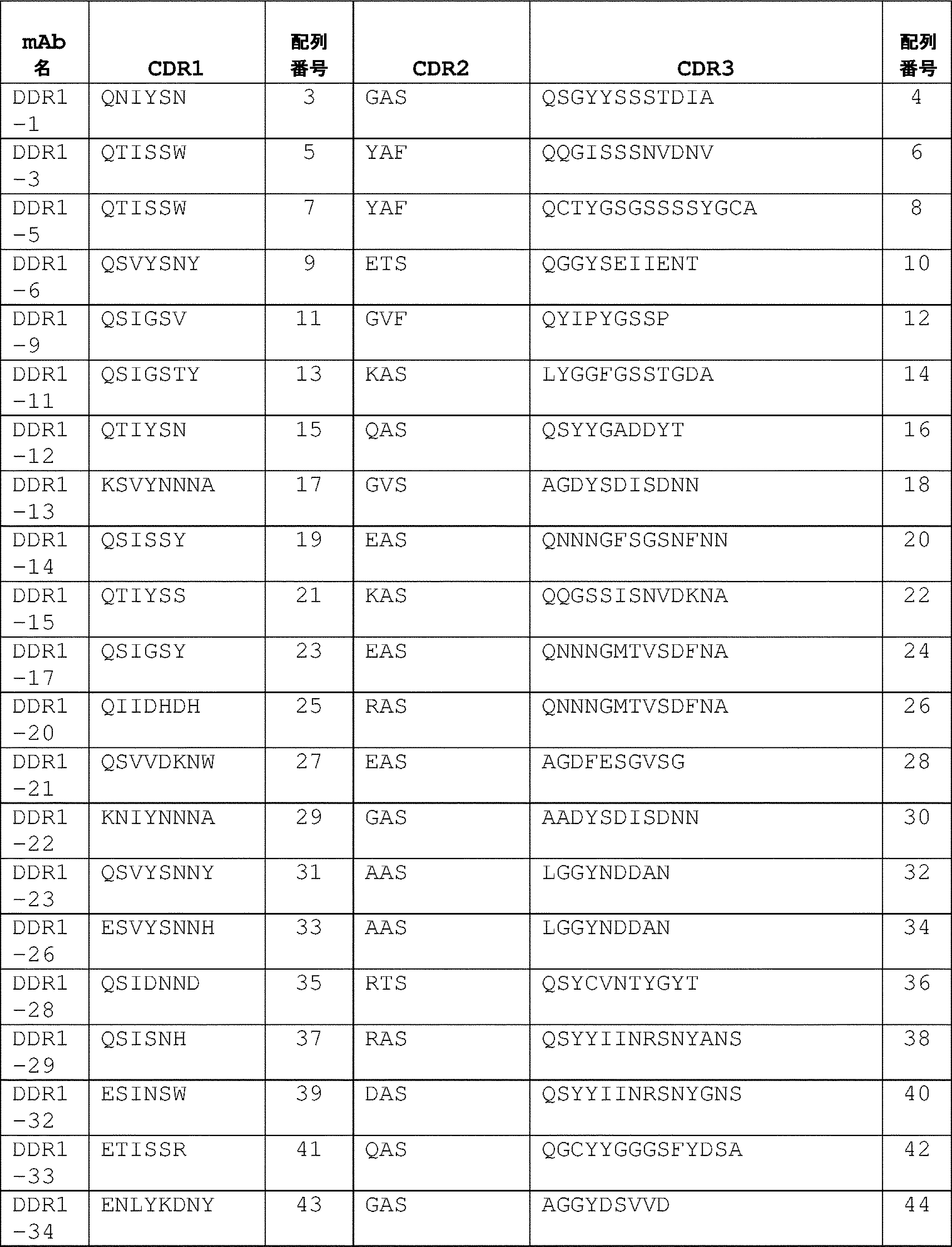
(表2)DDR1抗体の重鎖アミノ酸可変領域配列のCDR
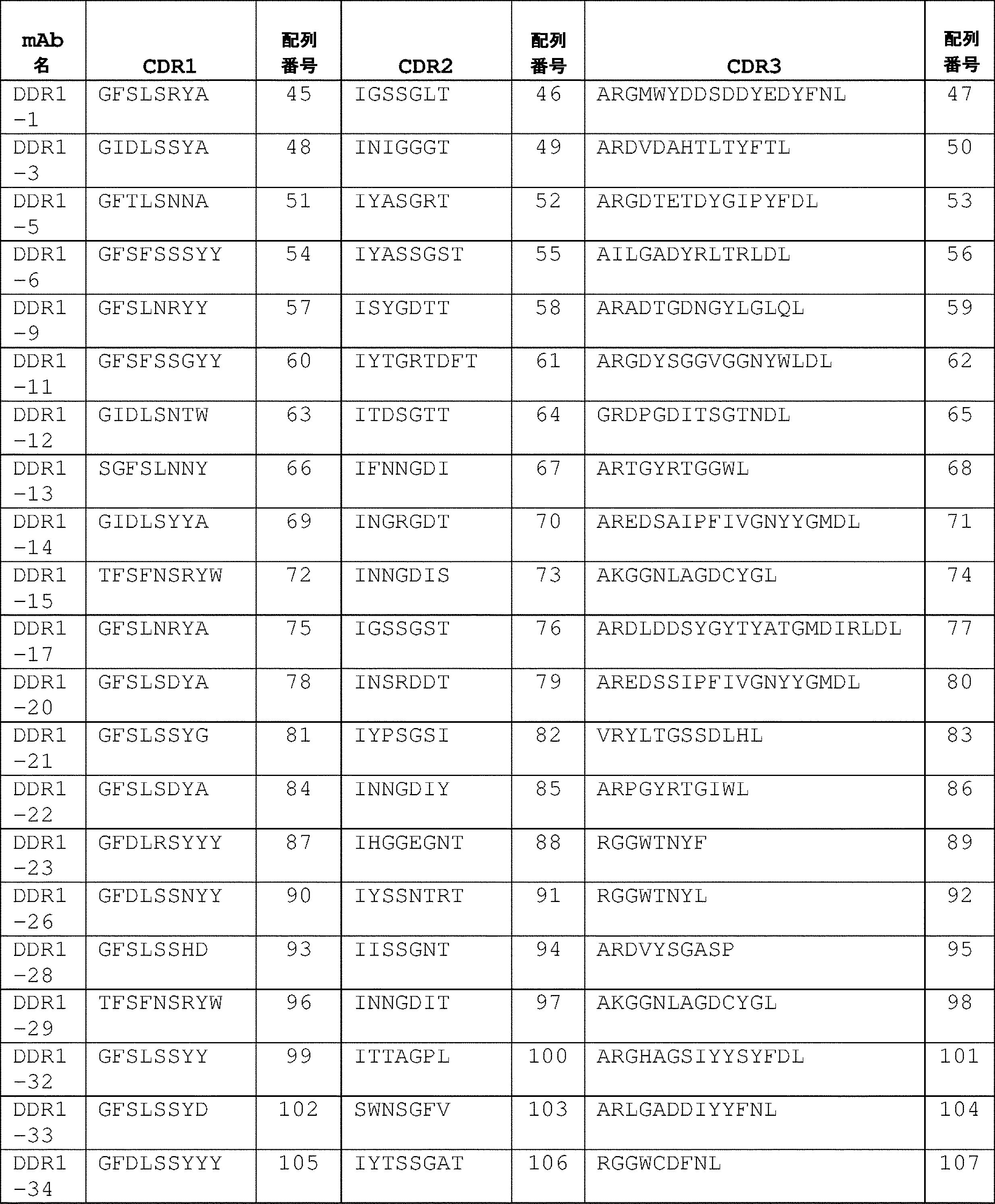
いくつかの実施形態では、単離された抗体またはその抗原結合断片は、アミノ酸配列QNIYSN(配列番号:3)を含むLC-CDR1、アミノ酸配列GASを含むLC-CDR2、およびアミノ酸配列QSGYYSSSTDIA(配列番号:4)を含むLC-CDR3を有する軽鎖可変領域、ならびにアミノ酸配列GFSLSRYA(配列番号:45)を含むHC-CDR1、アミノ酸配列IGSSGLT(配列番号:46)を含むHC-CDR2、およびアミノ酸配列ARGMWYDDSDDYEDYFNL(配列番号:47)を含むHC-CDR3を有する重鎖可変領域を含むか(DDR1-1)、またはそれらのバリアントであって、LC-CDRおよび/もしくはHC-CDRのうちの1つ以上が1つ、2つ、もしくは3つのアミノ酸置換、付加、欠失、もしくはそれらの組み合わせを有するバリアントを含む。 In some embodiments, the isolated antibody or antigen-binding fragment thereof comprises LC-CDR1 comprising the amino acid sequence QNIYSN (SEQ ID NO:3), LC-CDR2 comprising the amino acid sequence GAS, and QSGYYSSSTDIA (SEQ ID NO:3). :4), and HC-CDR1 with the amino acid sequence GFSLSRYA (SEQ ID NO:45), HC-CDR2 with the amino acid sequence IGSSGLT (SEQ ID NO:46), and the amino acid sequence ARGMWYDDSDDYEDYFNL. (DDR1-1), or variants thereof, wherein one or more of the LC-CDRs and/or HC-CDRs are Variants with one, two, or three amino acid substitutions, additions, deletions, or combinations thereof are included.
いくつかの実施形態では、単離された抗体またはその抗原結合断片は、アミノ酸配列QTISSW(配列番号:5)を含むLC-CDR1、アミノ酸配列YAFを含むLC-CDR2、およびアミノ酸配列QQGISSSNVDNV(配列番号:6)を含むLC-CDR3を有する軽鎖可変領域、ならびにアミノ酸配列GIDLSSYA(配列番号:48)を含むHC-CDR1、アミノ酸配列INIGGGT(配列番号:49)を含むHC-CDR2、およびアミノ酸配列ARDVDAHTLTYFTL(配列番号:50)を含むHC-CDR3を有する重鎖可変領域を含むか(DDR1-3)、またはそれらのバリアントであって、LC-CDRおよび/もしくはHC-CDRのうちの1つ以上が1つ、2つ、もしくは3つのアミノ酸置換、付加、欠失、もしくはそれらの組み合わせを有するバリアントを含む。 In some embodiments, the isolated antibody or antigen-binding fragment thereof comprises LC-CDR1 comprising the amino acid sequence QTISSW (SEQ ID NO:5), LC-CDR2 comprising the amino acid sequence YAF, and QQGISSSNVDNV (SEQ ID NO:5). :6), and HC-CDR1 with the amino acid sequence GIDLSSYA (SEQ ID NO:48), HC-CDR2 with the amino acid sequence INIGGGT (SEQ ID NO:49), and the amino acid sequence ARDVDAHTLTYFTL. (DDR1-3), or variants thereof, wherein one or more of the LC-CDRs and/or HC-CDRs are Variants with one, two, or three amino acid substitutions, additions, deletions, or combinations thereof are included.
いくつかの実施形態では、単離された抗体またはその抗原結合断片は、アミノ酸配列QTISSW(配列番号:7)を含むLC-CDR1、アミノ酸配列YAFを含むLC-CDR2、およびアミノ酸配列QCTYGSGSSSSYGCA(配列番号:8)を含むLC-CDR3を有する軽鎖可変領域、ならびにアミノ酸配列GFTLSNNA(配列番号:51)を含むHC-CDR1、アミノ酸配列IYASGRT(配列番号:52)を含むHC-CDR2、およびアミノ酸配列ARGDTETDYGIPYFDL(配列番号:53)を含むHC-CDR3を有する重鎖可変領域を含むか(DDR1-5)、またはそれらのバリアントであって、LC-CDRおよび/もしくはHC-CDRのうちの1つ以上が1つ、2つ、もしくは3つのアミノ酸置換、付加、欠失、もしくはそれらの組み合わせを有するバリアントを含む。 In some embodiments, the isolated antibody or antigen-binding fragment thereof comprises LC-CDR1 comprising the amino acid sequence QTISSW (SEQ ID NO: 7), LC-CDR2 comprising the amino acid sequence YAF, and QCTYGSGSSSSYGCA (SEQ ID NO: 7). :8), and HC-CDR1 with the amino acid sequence GFTLSNNA (SEQ ID NO:51), HC-CDR2 with the amino acid sequence IYASGRT (SEQ ID NO:52), and the amino acid sequence ARGDTETDYGIPYFDL. (DDR1-5), or variants thereof, wherein one or more of the LC-CDRs and/or HC-CDRs are Variants with one, two, or three amino acid substitutions, additions, deletions, or combinations thereof are included.
いくつかの実施形態では、単離された抗体またはその抗原結合断片は、アミノ酸配列QSVYSNY(配列番号:9)を含むLC-CDR1、アミノ酸配列ETSを含むLC-CDR2、およびアミノ酸配列QGGYSEIIENT(配列番号:10)を含むLC-CDR3を有する軽鎖可変領域、ならびにアミノ酸配列GFSFSSSYY(配列番号:54)を含むHC-CDR1、アミノ酸配列IYASSGST(配列番号:55)を含むHC-CDR2、およびアミノ酸配列AILGADYRLTRLDL(配列番号:56)を含むHC-CDR3を有する重鎖可変領域を含むか(DDR1-6)、またはそれらのバリアントであって、LC-CDRおよび/もしくはHC-CDRのうちの1つ以上が1つ、2つ、もしくは3つのアミノ酸置換、付加、欠失、もしくはそれらの組み合わせを有するバリアントを含む。 In some embodiments, the isolated antibody or antigen-binding fragment thereof comprises LC-CDR1 comprising the amino acid sequence QSVYSNY (SEQ ID NO:9), LC-CDR2 comprising the amino acid sequence ETS, and QGGYSEIIENT (SEQ ID NO:9). :10), and HC-CDR1 with the amino acid sequence GFSFSSSSYY (SEQ ID NO:54), HC-CDR2 with the amino acid sequence IYASSGST (SEQ ID NO:55), and the amino acid sequence AILGADYRLTRLDL. (DDR1-6), or variants thereof, wherein one or more of the LC-CDRs and/or HC-CDRs are Variants with one, two, or three amino acid substitutions, additions, deletions, or combinations thereof are included.
いくつかの実施形態では、単離された抗体またはその抗原結合断片は、アミノ酸配列QSIGSV(配列番号:11)を含むLC-CDR1、アミノ酸配列GVFを含むLC-CDR2、およびアミノ酸配列QYIPYGSSP(配列番号:12)を含むLC-CDR3を有する軽鎖可変領域、ならびにアミノ酸配列GFSLNRYY(配列番号:57)を含むHC-CDR1、アミノ酸配列ISYGDTT(配列番号:58)を含むHC-CDR2、およびアミノ酸配列ARADTGDNGYLGLQL(配列番号:59)を含むHC-CDR3を有する重鎖可変領域を含むか(DDR1-9)、またはそれらのバリアントであって、LC-CDRおよび/もしくはHC-CDRのうちの1つ以上が1つ、2つ、もしくは3つのアミノ酸置換、付加、欠失、もしくは組み合わせを有するバリアントを含む。 In some embodiments, the isolated antibody or antigen-binding fragment thereof comprises LC-CDR1 comprising the amino acid sequence QSIGSV (SEQ ID NO: 11), LC-CDR2 comprising the amino acid sequence GVF, and QYIPYGSSP (SEQ ID NO: 11). :12), and HC-CDR1 with the amino acid sequence GFSLNRYY (SEQ ID NO:57), HC-CDR2 with the amino acid sequence ISYGDTT (SEQ ID NO:58), and the amino acid sequence ARADTGDNGYLGLQL. (DDR1-9), or variants thereof, wherein one or more of the LC-CDRs and/or HC-CDRs are Variants with one, two, or three amino acid substitutions, additions, deletions, or combinations are included.
いくつかの実施形態では、単離された抗体またはその抗原結合断片は、アミノ酸配列QSIGSTY(配列番号:13)を含むLC-CDR1、アミノ酸配列KASを含むLC-CDR2、およびアミノ酸配列LYGGFGSSTGDA(配列番号:14)を含むLC-CDR3を有する軽鎖可変領域、ならびにアミノ酸配列GFSFSSGYY(配列番号:60)を含むHC-CDR1、アミノ酸配列IYTGRTDFT(配列番号:61)を含むHC-CDR2、およびアミノ酸配列ARGDYSGGVGGNYWLDL(配列番号:62)を含むHC-CDR3を有する重鎖可変領域を含むか(DDR1-11)、またはそれらのバリアントであって、LC-CDRおよび/もしくはHC-CDRのうちの1つ以上が1つ、2つ、もしくは3つのアミノ酸置換、付加、欠失、もしくはそれらの組み合わせを有するバリアントを含む。 In some embodiments, the isolated antibody or antigen-binding fragment thereof comprises LC-CDR1 comprising the amino acid sequence QSIGSTY (SEQ ID NO: 13), LC-CDR2 comprising the amino acid sequence KAS, and LYGGFGSSTGDA (SEQ ID NO: 13). :14), and HC-CDR1 with the amino acid sequence GFSFSSGYY (SEQ ID NO:60), HC-CDR2 with the amino acid sequence IYTGRTDFT (SEQ ID NO:61), and the amino acid sequence ARGDYSGGVGGNYWLDL (DDR1-11), or variants thereof, wherein one or more of the LC-CDRs and/or HC-CDRs are Variants with one, two, or three amino acid substitutions, additions, deletions, or combinations thereof are included.
いくつかの実施形態では、単離された抗体またはその抗原結合断片は、アミノ酸配列QTIYSN(配列番号:15)を含むLC-CDR1、アミノ酸配列QASを含むLC-CDR2、およびアミノ酸配列QSYYGADDYT(配列番号:16)を含むLC-CDR3を有する軽鎖可変領域、ならびにアミノ酸配列GIDLSNTW(配列番号:63)を含むHC-CDR1、アミノ酸配列ITDSGTT(配列番号:64)を含むHC-CDR2、およびアミノ酸配列GRDPGDITSGTNDL(配列番号:65)を含むHC-CDR3を有する重鎖可変領域を含むか(DDR1-12)、またはそれらのバリアントであって、LC-CDRおよび/もしくはHC-CDRのうちの1つ以上が1つ、2つ、もしくは3つのアミノ酸置換、付加、欠失、もしくはそれらの組み合わせを有するバリアントを含む。 In some embodiments, the isolated antibody or antigen-binding fragment thereof comprises LC-CDR1 comprising the amino acid sequence QTIYSN (SEQ ID NO: 15), LC-CDR2 comprising the amino acid sequence QAS, and QSYYGADDYT (SEQ ID NO: 15). :16), and HC-CDR1 with the amino acid sequence GIDLSNTW (SEQ ID NO:63), HC-CDR2 with the amino acid sequence ITDSGTT (SEQ ID NO:64), and the amino acid sequence GRDPGDITSGTNDL. (DDR1-12), or variants thereof, wherein one or more of the LC-CDRs and/or HC-CDRs are Variants with one, two, or three amino acid substitutions, additions, deletions, or combinations thereof are included.
いくつかの実施形態では、単離された抗体またはその抗原結合断片は、アミノ酸配列KSVYNNNA(配列番号:17)を含むLC-CDR1、アミノ酸配列GVSを含むLC-CDR2、およびアミノ酸配列AGDYSDISDNN(配列番号:18)を含むLC-CDR3を有する軽鎖可変領域、ならびにアミノ酸配列SGFSLNNY(配列番号:66)を含むHC-CDR1、アミノ酸配列IFNNGDI(配列番号:67)を含むHC-CDR2、およびアミノ酸配列ARTGYRTGGWL(配列番号:68)を含むHC-CDR3を有する重鎖可変領域を含むか(DDR1-13)、またはそれらのバリアントであって、LC-CDRおよび/もしくはHC-CDRのうちの1つ以上が1つ、2つ、もしくは3つのアミノ酸置換、付加、欠失、もしくはそれらの組み合わせを有するバリアントを含む。 In some embodiments, the isolated antibody or antigen-binding fragment thereof comprises LC-CDR1 comprising the amino acid sequence KSVYNNNA (SEQ ID NO: 17), LC-CDR2 comprising the amino acid sequence GVS, and AGDYSDISDNN (SEQ ID NO: 17). :18), and HC-CDR1 with the amino acid sequence SGFSLNNY (SEQ ID NO:66), HC-CDR2 with the amino acid sequence IFNNGDI (SEQ ID NO:67), and the amino acid sequence ARTGYRTGGWL. (DDR1-13), or variants thereof, wherein one or more of the LC-CDRs and/or HC-CDRs are Variants with one, two, or three amino acid substitutions, additions, deletions, or combinations thereof are included.
いくつかの実施形態では、単離された抗体またはその抗原結合断片は、アミノ酸配列QSISSY(配列番号:19)を含むLC-CDR1、アミノ酸配列EASを含むLC-CDR2、およびアミノ酸配列QNNNGFSGSNFNN(配列番号:20)を含むLC-CDR3を有する軽鎖可変領域、ならびにアミノ酸配列GIDLSYYA(配列番号:69)を含むHC-CDR1、アミノ酸配列INGRGDT(配列番号:70)を含むHC-CDR2、およびアミノ酸配列AREDSAIPFIVGNYYGMDL(配列番号:71)を含むHC-CDR3を有する重鎖可変領域を含むか(DDR1-14)、またはそれらのバリアントであって、LC-CDRおよび/もしくはHC-CDRのうちの1つ以上が1つ、2つ、もしくは3つのアミノ酸置換、付加、欠失、もしくはそれらの組み合わせを有するバリアントを含む。 In some embodiments, the isolated antibody or antigen-binding fragment thereof comprises LC-CDR1 comprising the amino acid sequence QSISSY (SEQ ID NO: 19), LC-CDR2 comprising the amino acid sequence EAS, and QNNNGFSGSNFNN (SEQ ID NO: 19). :20), and HC-CDR1 with the amino acid sequence GIDLSYYA (SEQ ID NO:69), HC-CDR2 with the amino acid sequence INGRGDT (SEQ ID NO:70), and the amino acid sequence AREDSAIPFIVGNYYGMDL. (DDR1-14), or variants thereof, wherein one or more of the LC-CDRs and/or HC-CDRs are Variants with one, two, or three amino acid substitutions, additions, deletions, or combinations thereof are included.
いくつかの実施形態では、単離された抗体またはその抗原結合断片は、アミノ酸配列QTIYSS(配列番号:21)を含むLC-CDR1、アミノ酸配列KASを含むLC-CDR2、およびアミノ酸配列QQGSSISNVDKNA(配列番号:22)を含むLC-CDR3を有する軽鎖可変領域、ならびにアミノ酸配列TFSFNSRYW(配列番号:72)を含むHC-CDR1、アミノ酸配列INNGDIS(配列番号:73)を含むHC-CDR2、およびアミノ酸配列AKGGNLAGDCYGL(配列番号:74)を含むHC-CDR3を有する重鎖可変領域を含むか(DDR1-15)、またはそれらのバリアントであって、LC-CDRおよび/もしくはHC-CDRのうちの1つ以上が1つ、2つ、もしくは3つのアミノ酸置換、付加、欠失、もしくはそれらの組み合わせを有するバリアントを含む。 In some embodiments, the isolated antibody or antigen-binding fragment thereof comprises LC-CDR1 comprising the amino acid sequence QTIYSS (SEQ ID NO:21), LC-CDR2 comprising the amino acid sequence KAS, and QQGSSISNVDKNA (SEQ ID NO:21). :22), and HC-CDR1 with the amino acid sequence TFSFNSRYW (SEQ ID NO:72), HC-CDR2 with the amino acid sequence INNGDIS (SEQ ID NO:73), and the amino acid sequence AKGGNLAGDCYGL. (DDR1-15), or variants thereof, wherein one or more of the LC-CDRs and/or HC-CDRs are Variants with one, two, or three amino acid substitutions, additions, deletions, or combinations thereof are included.
いくつかの実施形態では、単離された抗体またはその抗原結合断片は、アミノ酸配列QSIGSY(配列番号:23)を含むLC-CDR1、アミノ酸配列EASを含むLC-CDR2、およびアミノ酸配列QNNNGMTVSDFNA(配列番号:24)を含むLC-CDR3を有する軽鎖可変領域、ならびにアミノ酸配列GFSLNRYA(配列番号:75)を含むHC-CDR1、アミノ酸配列IGSSGST(配列番号:76)を含むHC-CDR2、およびアミノ酸配列ARDLDDSYGYTYATGMDIRLDL(配列番号:77)を含むHC-CDR3を有する重鎖可変領域を含むか(DDR1-17)、またはそれらのバリアントであって、LC-CDRおよび/もしくはHC-CDRのうちの1つ以上が1つ、2つ、もしくは3つのアミノ酸置換、付加、欠失、もしくはそれらの組み合わせを有するバリアントを含む。 In some embodiments, the isolated antibody or antigen-binding fragment thereof comprises LC-CDR1 comprising the amino acid sequence QSIGSY (SEQ ID NO:23), LC-CDR2 comprising the amino acid sequence EAS, and QNNNGMTVSDFNA (SEQ ID NO:23). :24), and HC-CDR1 with the amino acid sequence GFSLNRYA (SEQ ID NO:75), HC-CDR2 with the amino acid sequence IGSSGST (SEQ ID NO:76), and the amino acid sequence ARDLDDSYGYTYATGMDIRLDL. (DDR1-17), or variants thereof, wherein one or more of the LC-CDRs and/or HC-CDRs are Variants with one, two, or three amino acid substitutions, additions, deletions, or combinations thereof are included.
いくつかの実施形態では、単離された抗体またはその抗原結合断片は、アミノ酸配列QIIDHDH(配列番号:25)を含むLC-CDR1、アミノ酸配列RASを含むLC-CDR2、およびアミノ酸配列QNNNGMTVSDFNA(配列番号:26)を含むLC-CDR3を有する軽鎖可変領域、ならびにアミノ酸配列GFSLSDYA(配列番号:78)を含むHC-CDR1、アミノ酸配列INSRDDT(配列番号:79)を含むHC-CDR2、およびアミノ酸配列AREDSSIPFIVGNYYGMDL(配列番号:80)を含むHC-CDR3を有する重鎖可変領域を含むか(DDR1-20)、またはそれらのバリアントであって、LC-CDRおよび/もしくはHC-CDRのうちの1つ以上が1つ、2つ、もしくは3つのアミノ酸置換、付加、欠失、もしくはそれらの組み合わせを有するバリアントを含む。 In some embodiments, the isolated antibody or antigen-binding fragment thereof comprises LC-CDR1 comprising the amino acid sequence QIIDHDH (SEQ ID NO:25), LC-CDR2 comprising the amino acid sequence RAS, and QNNNGMTVSDFNA (SEQ ID NO:25). :26), and HC-CDR1 with the amino acid sequence GFSLSDYA (SEQ ID NO:78), HC-CDR2 with the amino acid sequence INSRDDT (SEQ ID NO:79), and the amino acid sequence AREDSSIPFIVGNYYGMDL. (DDR1-20), or variants thereof, wherein one or more of the LC-CDRs and/or HC-CDRs are Variants with one, two, or three amino acid substitutions, additions, deletions, or combinations thereof are included.
いくつかの実施形態では、単離された抗体またはその抗原結合断片は、アミノ酸配列QSVVDKNW(配列番号:27)を含むLC-CDR1、アミノ酸配列EASを含むLC-CDR2、およびアミノ酸配列AGDFESGVSG(配列番号:28)を含むLC-CDR3を有する軽鎖可変領域、ならびにアミノ酸配列GFSLSSYG(配列番号:81)を含むHC-CDR1、アミノ酸配列IYPSGSI(配列番号:82)を含むHC-CDR2、およびアミノ酸配列VRYLTGSSDLHL(配列番号:83)を含むHC-CDR3を有する重鎖可変領域を含むか(DDR1-21)、またはそれらのバリアントであって、LC-CDRおよび/もしくはHC-CDRのうちの1つ以上が1つ、2つ、もしくは3つのアミノ酸置換、付加、欠失、もしくはそれらの組み合わせを有するバリアントを含む。 In some embodiments, the isolated antibody or antigen-binding fragment thereof comprises LC-CDR1 comprising the amino acid sequence QSVVDKNW (SEQ ID NO:27), LC-CDR2 comprising the amino acid sequence EAS, and AGDFESGVSG (SEQ ID NO:27). :28), and HC-CDR1 with the amino acid sequence GFSLSSYG (SEQ ID NO:81), HC-CDR2 with the amino acid sequence IYPSGSI (SEQ ID NO:82), and the amino acid sequence VRYLTGSSDLHL. (DDR1-21), or variants thereof, wherein one or more of the LC-CDRs and/or HC-CDRs are Variants with one, two, or three amino acid substitutions, additions, deletions, or combinations thereof are included.
いくつかの実施形態では、単離された抗体またはその抗原結合断片は、アミノ酸配列KNIYNNNA(配列番号:29)を含むLC-CDR1、アミノ酸配列GASを含むLC-CDR2、およびアミノ酸配列AADYSDISDNN(配列番号:30)を含むLC-CDR3を有する軽鎖可変領域、ならびにアミノ酸配列GFSLSDYA(配列番号:84)を含むHC-CDR1、アミノ酸配列INNGDIY(配列番号:85)を含むHC-CDR2、およびアミノ酸配列ARPGYRTGIWL(配列番号:86)を含むHC-CDR3を有する重鎖可変領域を含むか(DDR1-22)、またはそれらのバリアントであって、LC-CDRおよび/もしくはHC-CDRのうちの1つ以上が1つ、2つ、もしくは3つのアミノ酸置換、付加、欠失、もしくはそれらの組み合わせを有するバリアントを含む。 In some embodiments, the isolated antibody or antigen-binding fragment thereof comprises LC-CDR1 comprising the amino acid sequence KNIYNNNA (SEQ ID NO:29), LC-CDR2 comprising the amino acid sequence GAS, and the amino acid sequence AADYSDISDNN (SEQ ID NO:29). :30), and HC-CDR1 with the amino acid sequence GFSLSDYA (SEQ ID NO:84), HC-CDR2 with the amino acid sequence INNGDIY (SEQ ID NO:85), and the amino acid sequence ARPGYRTGIWL. (DDR1-22), or variants thereof, wherein one or more of the LC-CDRs and/or HC-CDRs are Variants with one, two, or three amino acid substitutions, additions, deletions, or combinations thereof are included.
いくつかの実施形態では、単離された抗体またはその抗原結合断片は、アミノ酸配列QSVYSNNY(配列番号:31)を含むLC-CDR1、アミノ酸配列AASを含むLC-CDR2、およびアミノ酸配列LGGYNDDAN(配列番号:32)を含むLC-CDR3を有する軽鎖可変領域、ならびにアミノ酸配列GFDLRSYYY(配列番号:87)を含むHC-CDR1、アミノ酸配列IHGGEGNT(配列番号:88)を含むHC-CDR2、およびアミノ酸配列RGGWTNYF(配列番号:89)を含むHC-CDR3を有する重鎖可変領域を含むか(DDR1-23)、またはそれらのバリアントであって、LC-CDRおよび/もしくはHC-CDRのうちの1つ以上が1つ、2つ、もしくは3つのアミノ酸置換、付加、欠失、もしくはそれらの組み合わせを有するバリアントを含む。 In some embodiments, the isolated antibody or antigen-binding fragment thereof comprises LC-CDR1 comprising the amino acid sequence QSVYSNNY (SEQ ID NO:31), LC-CDR2 comprising the amino acid sequence AAS, and LGGYNDDAN (SEQ ID NO:31). :32), and HC-CDR1 with the amino acid sequence GFDLRSYYY (SEQ ID NO:87), HC-CDR2 with the amino acid sequence IHGGEGNT (SEQ ID NO:88), and the amino acid sequence RGGWTNYF. (DDR1-23), or variants thereof, wherein one or more of the LC-CDRs and/or HC-CDRs are Variants with one, two, or three amino acid substitutions, additions, deletions, or combinations thereof are included.
いくつかの実施形態では、単離された抗体またはその抗原結合断片は、アミノ酸配列ESVYSNNH(配列番号:33)を含むLC-CDR1、アミノ酸配列AASを含むLC-CDR2、およびアミノ酸配列LGGYNDDAN(配列番号:34)を含むLC-CDR3を有する軽鎖可変領域、ならびにアミノ酸配列GFDLSSNYY(配列番号:90)を含むHC-CDR1、アミノ酸配列IYSSNTRT(配列番号:91)を含むHC-CDR2、およびアミノ酸配列RGGWTNYL(配列番号:92)を含むHC-CDR3を有する重鎖可変領域を含むか(DDR1-26)、またはそれらのバリアントであって、LC-CDRおよび/もしくはHC-CDRのうちの1つ以上が1つ、2つ、もしくは3つのアミノ酸置換、付加、欠失、もしくはそれらの組み合わせを有するバリアントを含む。 In some embodiments, the isolated antibody or antigen-binding fragment thereof comprises LC-CDR1 comprising the amino acid sequence ESVYSNNH (SEQ ID NO:33), LC-CDR2 comprising the amino acid sequence AAS, and LGGYNDDAN (SEQ ID NO:33). :34), and HC-CDR1 with the amino acid sequence GFDLSSNYY (SEQ ID NO:90), HC-CDR2 with the amino acid sequence IYSSNTRT (SEQ ID NO:91), and the amino acid sequence RGGWTNYL. (DDR1-26), or variants thereof, wherein one or more of the LC-CDRs and/or HC-CDRs are Variants with one, two, or three amino acid substitutions, additions, deletions, or combinations thereof are included.
いくつかの実施形態では、単離された抗体またはその抗原結合断片は、アミノ酸配列QSIDNND(配列番号:35)を含むLC-CDR1、アミノ酸配列RTSを含むLC-CDR2、およびアミノ酸配列QSYCVNTYGYT(配列番号:36)を含むLC-CDR3を有する軽鎖可変領域、ならびにアミノ酸配列GFSLSSHD(配列番号:93)を含むHC-CDR1、アミノ酸配列IISSGNT(配列番号:94)を含むHC-CDR2、およびアミノ酸配列ARDVYSGASP(配列番号:95)を含むHC-CDR3を有する重鎖可変領域を含むか(DDR1-28)、またはそれらのバリアントであって、LC-CDRおよび/もしくはHC-CDRのうちの1つ以上が1つ、2つ、もしくは3つのアミノ酸置換、付加、欠失、もしくはそれらの組み合わせを有するバリアントを含む。 In some embodiments, the isolated antibody or antigen-binding fragment thereof comprises LC-CDR1 comprising the amino acid sequence QSIDNND (SEQ ID NO:35), LC-CDR2 comprising the amino acid sequence RTS, and QSYCVNTYGYT (SEQ ID NO:35). :36), and HC-CDR1 with the amino acid sequence GFSLSSHD (SEQ ID NO:93), HC-CDR2 with the amino acid sequence IISSGNT (SEQ ID NO:94), and the amino acid sequence ARDVYSGASP. (DDR1-28), or variants thereof, wherein one or more of the LC-CDRs and/or HC-CDRs are Variants with one, two, or three amino acid substitutions, additions, deletions, or combinations thereof are included.
いくつかの実施形態では、単離された抗体またはその抗原結合断片は、アミノ酸配列QSISNH(配列番号:37)を含むLC-CDR1、アミノ酸配列RASを含むLC-CDR2、およびアミノ酸配列QSYYIINRSNYANS(配列番号:38)を含むLC-CDR3を有する軽鎖可変領域、ならびにアミノ酸配列TFSFNSRYW(配列番号:96)を含むHC-CDR1、アミノ酸配列INNGDIT(配列番号:97)を含むHC-CDR2、およびアミノ酸配列AKGGNLAGDCYGL(配列番号:98)を含むHC-CDR3を有する重鎖可変領域を含むか(DDR1-29)、またはそれらのバリアントであって、LC-CDRおよび/もしくはHC-CDRのうちの1つ以上が1つ、2つ、もしくは3つのアミノ酸置換、付加、欠失、もしくはそれらの組み合わせを有するバリアントを含む。 In some embodiments, the isolated antibody or antigen-binding fragment thereof comprises LC-CDR1 comprising the amino acid sequence QSISNH (SEQ ID NO:37), LC-CDR2 comprising the amino acid sequence RAS, and QSYYIINRSNYANS (SEQ ID NO:37). :38), and HC-CDR1 with the amino acid sequence TFSFNSRYW (SEQ ID NO:96), HC-CDR2 with the amino acid sequence INNGDIT (SEQ ID NO:97), and the amino acid sequence AKGGNLAGDCYGL (DDR1-29), or variants thereof, wherein one or more of the LC-CDRs and/or HC-CDRs are Variants with one, two, or three amino acid substitutions, additions, deletions, or combinations thereof are included.
いくつかの実施形態では、単離された抗体またはその抗原結合断片は、アミノ酸配列ESINSW(配列番号:39)を含むLC-CDR1、アミノ酸配列DASを含むLC-CDR2、およびアミノ酸配列QSYYIINRSNYGNS(配列番号:40)を含むLC-CDR3を有する軽鎖可変領域、ならびにアミノ酸配列GFSLSSYY(配列番号:99)を含むHC-CDR1、アミノ酸配列ITTAGPL(配列番号:100)を含むHC-CDR2、およびアミノ酸配列ARGHAGSIYYSYFDL(配列番号:101)を含むHC-CDR3を有する重鎖可変領域を含むか(DDR1-32)、またはそれらのバリアントであって、LC-CDRおよび/もしくはHC-CDRのうちの1つ以上が1つ、2つ、もしくは3つのアミノ酸置換、付加、欠失、もしくはそれらの組み合わせを有するバリアントを含む。 In some embodiments, the isolated antibody or antigen-binding fragment thereof comprises LC-CDR1 comprising the amino acid sequence ESINSW (SEQ ID NO:39), LC-CDR2 comprising the amino acid sequence DAS, and the amino acid sequence QSYYIINRSNYGNS (SEQ ID NO:39). :40), and HC-CDR1 with the amino acid sequence GFSLSSYY (SEQ ID NO:99), HC-CDR2 with the amino acid sequence ITTAGPL (SEQ ID NO:100), and the amino acid sequence ARGHAGSIYYSYFDL. (DDR1-32), or variants thereof, wherein one or more of the LC-CDRs and/or HC-CDRs are Variants with one, two, or three amino acid substitutions, additions, deletions, or combinations thereof are included.
いくつかの実施形態では、単離された抗体またはその抗原結合断片は、アミノ酸配列ETISSR(配列番号:41)を含むLC-CDR1、アミノ酸配列QASを含むLC-CDR2、およびアミノ酸配列QGCYYGGGSFYDSA(配列番号:42)を含むLC-CDR3を有する軽鎖可変領域、ならびにアミノ酸配列GFSLSSYD(配列番号:102)を含むHC-CDR1、アミノ酸配列SWNSGFV(配列番号:103)を含むHC-CDR2、およびアミノ酸配列ARLGADDIYYFNL(配列番号:104)を含むHC-CDR3を有する重鎖可変領域を含むか(DDR1-33)、またはそれらのバリアントであって、LC-CDRおよび/もしくはHC-CDRのうちの1つ以上が1つ、2つ、もしくは3つのアミノ酸置換、付加、欠失、もしくはそれらの組み合わせを有するバリアントを含む。 In some embodiments, the isolated antibody or antigen-binding fragment thereof comprises LC-CDR1 comprising the amino acid sequence ETISSR (SEQ ID NO:41), LC-CDR2 comprising the amino acid sequence QAS, and QGCYYGGGSFYDSA (SEQ ID NO:41). :42), and HC-CDR1 with the amino acid sequence GFSLSSYD (SEQ ID NO:102), HC-CDR2 with the amino acid sequence SWNSGFV (SEQ ID NO:103), and the amino acid sequence ARLGADDYYFNL. (DDR1-33), or variants thereof, wherein one or more of the LC-CDRs and/or HC-CDRs are Variants with one, two, or three amino acid substitutions, additions, deletions, or combinations thereof are included.
いくつかの実施形態では、単離された抗体またはその抗原結合断片は、アミノ酸配列ETISSR(配列番号:41)を含むLC-CDR1、アミノ酸配列QASを含むLC-CDR2、およびアミノ酸配列QGCYYGGGSFYDSA(配列番号:42)を含むLC-CDR3を有する軽鎖可変領域、ならびにアミノ酸配列GFSLSSYD(配列番号:102)を含むHC-CDR1、アミノ酸配列SWNSGFV(配列番号:103)を含むHC-CDR2、およびアミノ酸配列ARLGADDIYYFNL(配列番号:104)を含むHC-CDR3を有する重鎖可変領域を含むか(DDR1-33)、またはそれらのバリアントであって、LC-CDRおよび/もしくはHC-CDRのうちの1つ以上が1つ、2つ、もしくは3つのアミノ酸置換、付加、欠失、もしくはそれらの組み合わせを有するバリアントを含む。 In some embodiments, the isolated antibody or antigen-binding fragment thereof comprises LC-CDR1 comprising the amino acid sequence ETISSR (SEQ ID NO:41), LC-CDR2 comprising the amino acid sequence QAS, and QGCYYGGGSFYDSA (SEQ ID NO:41). :42), and HC-CDR1 with the amino acid sequence GFSLSSYD (SEQ ID NO:102), HC-CDR2 with the amino acid sequence SWNSGFV (SEQ ID NO:103), and the amino acid sequence ARLGADDYYFNL. (DDR1-33), or variants thereof, wherein one or more of the LC-CDRs and/or HC-CDRs are Variants with one, two, or three amino acid substitutions, additions, deletions, or combinations thereof are included.
いくつかの実施形態では、単離された抗体またはその抗原結合断片は、アミノ酸配列ENLYKDNY(配列番号:43)を含むLC-CDR1、アミノ酸配列GASを含むLC-CDR2、およびアミノ酸配列AGGYDSVVD(配列番号:44)を含むLC-CDR3を有する軽鎖可変領域、ならびにアミノ酸配列GFDLSSYYY(配列番号:105)を含むHC-CDR1、アミノ酸配列SIYTSSGAT(配列番号:106)を含むHC-CDR2、およびアミノ酸配列RGGWCDFNL(配列番号:107)を含むHC-CDR3を有する重鎖可変領域を含むか(DDR1-34)、またはそれらのバリアントであって、LC-CDRおよび/もしくはHC-CDRのうちの1つ以上が1つ、2つ、もしくは3つのアミノ酸置換、付加、欠失、もしくはそれらの組み合わせを有するバリアントを含む。 In some embodiments, the isolated antibody or antigen-binding fragment thereof comprises LC-CDR1 comprising the amino acid sequence ENLYKDNY (SEQ ID NO:43), LC-CDR2 comprising the amino acid sequence GAS, and AGGYDSVVD (SEQ ID NO:43). :44), and HC-CDR1 with the amino acid sequence GFDLSSYYY (SEQ ID NO:105), HC-CDR2 with the amino acid sequence SIYTSSGAT (SEQ ID NO:106), and the amino acid sequence RGGWCDFNL. (DDR1-34), or variants thereof, wherein one or more of the LC-CDRs and/or HC-CDRs are Variants with one, two, or three amino acid substitutions, additions, deletions, or combinations thereof are included.
いくつかの実施形態では、表1のLC-CDRおよび表2のHC-CDRは、以下でさらに考察されるように、それぞれ表6および表7のポリヌクレオチドによってコードされる。 In some embodiments, the LC-CDRs of Table 1 and the HC-CDRs of Table 2 are encoded by the polynucleotides of Tables 6 and 7, respectively, as discussed further below.
いくつかの実施形態では、LC-CDRおよびHC-CDRは、フレームワーク領域(FR)配列の好適な文脈に配置され、抗体の結合特異性を定義する軽鎖可変領域および重鎖可変領域を形成する。いくつかの実施形態では、同定されたLC-CDRおよびHC-CDRを有する抗体のフレームワーク領域配列は、元の抗体単離物のフレームワーク配列である。いくつかの実施形態では、LC-CDRおよびHC-CDRは、異なる哺乳動物の種、例えば、霊長類に由来するフレームワーク領域配列とともに使用される。いくつかの実施形態では、フレームワーク配列は、DDR1タンパク質に特異的に結合する抗体を形成するためのヒト化またはヒトフレームワーク配列である。いくつかの実施形態では、軽鎖可変領域は、例えば、FRLC1、FRLC2、FRLC3、およびFRLC4として指定される4つの軽鎖フレームワーク領域と、LC-CDRとを含み、NH2からCOOHの方向に、以下の構成に従う:FRLC1--LC-CDR1--FRLC2-LC-CDR2-FRLC3,-LC-CDR3-FRLC4。いくつかの実施形態では、重鎖可変領域は、例えば、FRHC1、FRHC2、FRHC3、およびFRHC4として指定される4つの重鎖フレームワーク領域と、HC-CDRとを含み、NH2からCOOHの方向に、以下の構成に従う:FRHC1--HC-CDR1--FRHC2-HC-CDR2-FRHC3,-HC-CDR3-FRHC4。いくつかの実施形態では、親軽鎖可変領域のフレームワーク領域は、ヒト軽鎖可変領域のフレームワーク領域と置き換えられて、ヒト化軽鎖可変領域を形成する。いくつかの実施形態では、親重鎖可変領域のフレームワーク領域は、ヒト重鎖可変領域のフレームワーク領域と置き換えられて、ヒト化重鎖可変領域を形成する。
In some embodiments, the LC-CDRs and HC-CDRs are placed in the appropriate context of framework region (FR) sequences to form the light and heavy chain variable regions that define the binding specificity of the antibody. do. In some embodiments, the antibody framework region sequences with the identified LC-CDRs and HC-CDRs are those of the original antibody isolate. In some embodiments, the LC-CDRs and HC-CDRs are used with framework region sequences from different mammalian species, eg, primates. In some embodiments, the framework sequences are humanized or human framework sequences for generating antibodies that specifically bind to DDR1 proteins. In some embodiments, the light chain variable region comprises four light chain framework regions designated, for example, as
いくつかの実施形態では、DDR1タンパク質に特異的に結合する単離された抗体またはその抗原結合断片は、表3に示される配列、すなわち、配列番号:108~128から選択される軽鎖可変領域アミノ酸配列を有する軽鎖可変領域を含む。いくつかの実施形態では、DDR1タンパク質に特異的に結合する単離された抗体またはその抗原結合断片は、表4に示される配列、すなわち、配列番号:129~149から選択される重鎖可変領域アミノ酸配列を有する重鎖可変領域を含む。 In some embodiments, the isolated antibody or antigen-binding fragment thereof that specifically binds to a DDR1 protein has a light chain variable region selected from the sequences shown in Table 3, i.e., SEQ ID NOS: 108-128. It includes a light chain variable region having the amino acid sequence. In some embodiments, the isolated antibody or antigen-binding fragment thereof that specifically binds to a DDR1 protein has a heavy chain variable region selected from the sequences shown in Table 4, i.e., SEQ ID NOS: 129-149. A heavy chain variable region having an amino acid sequence is included.
いくつかの実施形態では、単離された抗体またはその抗原結合断片は、配列番号:108~128から選択される軽鎖可変領域アミノ酸配列を有する軽鎖可変領域と、配列番号:129~149から選択される重鎖可変領域アミノ酸配列を有する重鎖可変領域とを含む。様々な実施形態では、配列番号:108~128に対応する可変軽鎖アミノ酸配列のいずれか1つは、配列番号:129~149に対応する可変重鎖アミノ酸配列のいずれか1つとともに使用することができる。 In some embodiments, the isolated antibody or antigen-binding fragment thereof has a light chain variable region having a light chain variable region amino acid sequence selected from SEQ ID NOS: 108-128 and a and a heavy chain variable region having a selected heavy chain variable region amino acid sequence. In various embodiments, any one of the variable light chain amino acid sequences corresponding to SEQ ID NOs: 108-128 is used with any one of the variable heavy chain amino acid sequences corresponding to SEQ ID NOs: 129-149. can be done.
いくつかの実施形態では、単離された抗体またはその抗原結合断片は、配列番号:108~128から選択される軽鎖可変領域アミノ酸配列を有する軽鎖可変領域と、配列番号:129~149から選択される重鎖可変領域アミノ酸配列を有する重鎖可変領域とを含む。様々な実施形態では、配列番号:108~128に対応する可変軽鎖アミノ酸配列のいずれか1つは、配列番号:129~149に対応する可変重鎖アミノ酸配列のいずれか1つとともに使用することができる。いくつかの実施形態では、DDR1タンパク質に特異的に結合する抗体またはその抗原結合断片は、それぞれ、表3および表4に示されるクローン対の軽鎖可変領域アミノ酸配列および重鎖可変領域アミノ酸配列の軽鎖可変領域および重鎖可変領域を含む。 In some embodiments, the isolated antibody or antigen-binding fragment thereof has a light chain variable region having a light chain variable region amino acid sequence selected from SEQ ID NOS: 108-128 and a and a heavy chain variable region having a selected heavy chain variable region amino acid sequence. In various embodiments, any one of the variable light chain amino acid sequences corresponding to SEQ ID NOs: 108-128 is used with any one of the variable heavy chain amino acid sequences corresponding to SEQ ID NOs: 129-149. can be done. In some embodiments, the antibody or antigen-binding fragment thereof that specifically binds to a DDR1 protein has the light chain variable region amino acid sequence and the heavy chain variable region amino acid sequence of the clone pair shown in Tables 3 and 4, respectively. It includes a light chain variable region and a heavy chain variable region.
(表3)抗DDR1抗体の軽鎖可変領域アミノ酸配列


(表4)抗DDR1抗体の重鎖可変領域アミノ酸配列
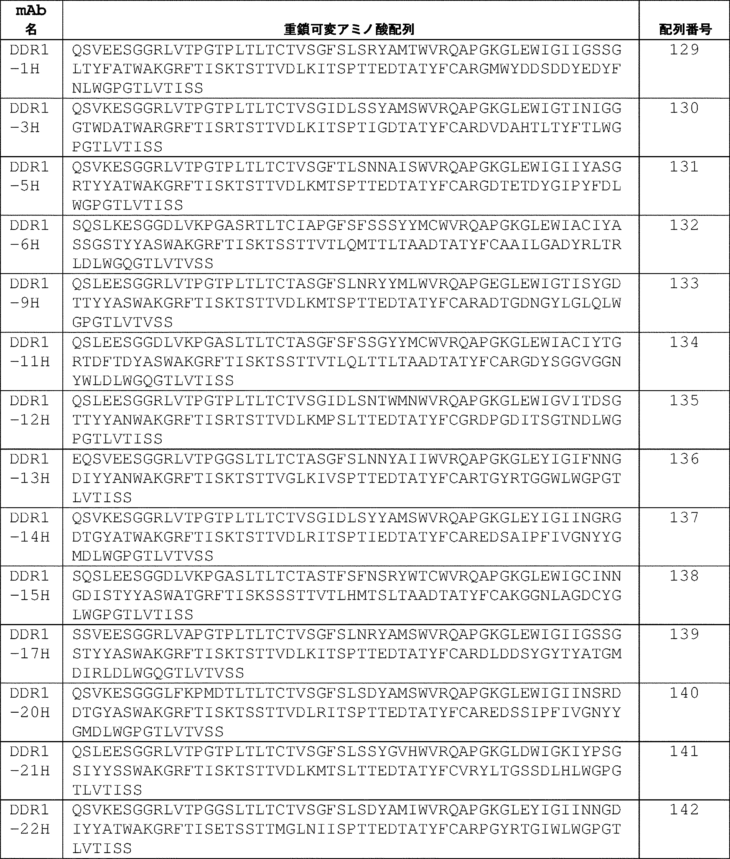

いくつかの実施形態では、抗体またはその抗原結合断片は、配列番号:108のアミノ酸配列を有する軽鎖可変領域(DDR1-1K)と、配列番号:129のアミノ酸配列を有する重鎖可変領域(DDR1-1H)とを含む。 In some embodiments, the antibody or antigen-binding fragment thereof has a light chain variable region (DDR1-1K) having the amino acid sequence of SEQ ID NO: 108 and a heavy chain variable region (DDR1 -1H).
いくつかの実施形態では、抗体またはその抗原結合断片は、配列番号:109のアミノ酸配列を有する軽鎖可変領域(DDR1-3K)と、配列番号:130のアミノ酸配列を有する重鎖可変領域(DDR1-3H)とを含む。 In some embodiments, the antibody or antigen-binding fragment thereof has a light chain variable region (DDR1-3K) having the amino acid sequence of SEQ ID NO: 109 and a heavy chain variable region (DDR1 -3H).
いくつかの実施形態では、抗体またはその抗原結合断片は、配列番号:110のアミノ酸配列を有する軽鎖可変領域(DDR1-5K)と、配列番号:131のアミノ酸配列を有する重鎖可変領域(DDR1-5H)とを含む。 In some embodiments, the antibody or antigen-binding fragment thereof has a light chain variable region (DDR1-5K) having the amino acid sequence of SEQ ID NO:110 and a heavy chain variable region (DDR1 -5H).
いくつかの実施形態では、抗体またはその抗原結合断片は、配列番号:111のアミノ酸配列を有する軽鎖可変領域(DDR1-6K)と、配列番号:132のアミノ酸配列を有する重鎖可変領域(DDR1-6H)とを含む。 In some embodiments, the antibody or antigen-binding fragment thereof has a light chain variable region (DDR1-6K) having the amino acid sequence of SEQ ID NO:111 and a heavy chain variable region (DDR1 -6H).
いくつかの実施形態では、抗体またはその抗原結合断片は、配列番号:112のアミノ酸配列を有する軽鎖可変領域(DDR1-9K)と、配列番号:133のアミノ酸配列を有する重鎖可変領域(DDR1-9H)とを含む。 In some embodiments, the antibody or antigen-binding fragment thereof has a light chain variable region (DDR1-9K) having the amino acid sequence of SEQ ID NO:112 and a heavy chain variable region (DDR1 -9H).
いくつかの実施形態では、抗体またはその抗原結合断片は、配列番号:113のアミノ酸配列を有する軽鎖可変領域(DDR1-11K)と、配列番号:134のアミノ酸配列を有する重鎖可変領域(DDR1-11H)とを含む。 In some embodiments, the antibody or antigen-binding fragment thereof has a light chain variable region (DDR1-11K) having the amino acid sequence of SEQ ID NO:113 and a heavy chain variable region (DDR1 -11H).
いくつかの実施形態では、抗体またはその抗原結合断片は、配列番号:114のアミノ酸配列を有する軽鎖可変領域(DDR1-12K)と、配列番号:135のアミノ酸配列を有する重鎖可変領域(DDR1-12H)とを含む。 In some embodiments, the antibody or antigen-binding fragment thereof has a light chain variable region (DDR1-12K) having the amino acid sequence of SEQ ID NO:114 and a heavy chain variable region (DDR1 -12H).
いくつかの実施形態では、抗体またはその抗原結合断片は、配列番号:115のアミノ酸配列を有する軽鎖可変領域(DDR1-13K)と、配列番号:136のアミノ酸配列を有する重鎖可変領域(DDR1-13H)とを含む。 In some embodiments, the antibody or antigen-binding fragment thereof has a light chain variable region (DDR1-13K) having the amino acid sequence of SEQ ID NO:115 and a heavy chain variable region (DDR1 -13H).
いくつかの実施形態では、抗体またはその抗原結合断片は、配列番号:116のアミノ酸配列を有する軽鎖可変領域(DDR1-14K)と、配列番号:137のアミノ酸配列を有する重鎖可変領域(DDR1-14H)とを含む。 In some embodiments, the antibody or antigen-binding fragment thereof has a light chain variable region (DDR1-14K) having the amino acid sequence of SEQ ID NO:116 and a heavy chain variable region (DDR1 -14H).
いくつかの実施形態では、抗体またはその抗原結合断片は、配列番号:117のアミノ酸配列を有する軽鎖可変領域(DDR1-15K)と、配列番号:138のアミノ酸配列を有する重鎖可変領域(DDR1-15H)とを含む。 In some embodiments, the antibody or antigen-binding fragment thereof has a light chain variable region (DDR1-15K) having the amino acid sequence of SEQ ID NO:117 and a heavy chain variable region (DDR1 -15H).
いくつかの実施形態では、抗体またはその抗原結合断片は、配列番号:118のアミノ酸配列を有する軽鎖可変領域(DDR1-17K)と、配列番号:139のアミノ酸配列を有する重鎖可変領域(DDR1-17H)とを含む。 In some embodiments, the antibody or antigen-binding fragment thereof has a light chain variable region (DDR1-17K) having the amino acid sequence of SEQ ID NO:118 and a heavy chain variable region (DDR1 -17H).
いくつかの実施形態では、抗体またはその抗原結合断片は、配列番号:119のアミノ酸配列を有する軽鎖可変領域(DDR1-20K)と、配列番号:140のアミノ酸配列を有する重鎖可変領域(DDR1-20H)とを含む。 In some embodiments, the antibody or antigen-binding fragment thereof has a light chain variable region (DDR1-20K) having the amino acid sequence of SEQ ID NO:119 and a heavy chain variable region (DDR1 -20H).
いくつかの実施形態では、抗体またはその抗原結合断片は、配列番号:120のアミノ酸配列を有する軽鎖可変領域(DDR1-21K)と、配列番号:141のアミノ酸配列を有する重鎖可変領域(DDR1-21H)とを含む。 In some embodiments, the antibody or antigen-binding fragment thereof has a light chain variable region (DDR1-21K) having the amino acid sequence of SEQ ID NO:120 and a heavy chain variable region (DDR1 -21H).
いくつかの実施形態では、抗体またはその抗原結合断片は、配列番号:121のアミノ酸配列を有する軽鎖可変領域(DDR1-22K)と、配列番号:142のアミノ酸配列を有する重鎖可変領域(DDR1-22H)とを含む。 In some embodiments, the antibody or antigen-binding fragment thereof has a light chain variable region (DDR1-22K) having the amino acid sequence of SEQ ID NO:121 and a heavy chain variable region (DDR1 -22H).
いくつかの実施形態では、抗体またはその抗原結合断片は、配列番号:122のアミノ酸配列を有する軽鎖可変領域(DDR1-23K)と、配列番号:143のアミノ酸配列を有する重鎖可変領域(DDR1-23H)とを含む。 In some embodiments, the antibody or antigen-binding fragment thereof has a light chain variable region (DDR1-23K) having the amino acid sequence of SEQ ID NO:122 and a heavy chain variable region (DDR1 -23H).
いくつかの実施形態では、抗体またはその抗原結合断片は、配列番号:123のアミノ酸配列を有する軽鎖可変領域(DDR1-26K)と、配列番号:144のアミノ酸配列を有する重鎖可変領域(DDR1-26H)とを含む。 In some embodiments, the antibody or antigen-binding fragment thereof has a light chain variable region (DDR1-26K) having the amino acid sequence of SEQ ID NO:123 and a heavy chain variable region (DDR1 -26H).
いくつかの実施形態では、抗体またはその抗原結合断片は、配列番号:124のアミノ酸配列を有する軽鎖可変領域(DDR1-28K)と、配列番号:145のアミノ酸配列を有する重鎖可変領域(DDR1-28H)とを含む。 In some embodiments, the antibody or antigen-binding fragment thereof has a light chain variable region (DDR1-28K) having the amino acid sequence of SEQ ID NO:124 and a heavy chain variable region (DDR1 -28H).
いくつかの実施形態では、抗体またはその抗原結合断片は、配列番号:125のアミノ酸配列を有する軽鎖可変領域(DDR1-29K)と、配列番号:146のアミノ酸配列を有する重鎖可変領域(DDR1-29H)とを含む。 In some embodiments, the antibody or antigen-binding fragment thereof has a light chain variable region (DDR1-29K) having the amino acid sequence of SEQ ID NO: 125 and a heavy chain variable region (DDR1 -29H).
いくつかの実施形態では、抗体またはその抗原結合断片は、配列番号:126のアミノ酸配列を有する軽鎖可変領域(DDR1-32K)と、配列番号:147のアミノ酸配列を有する重鎖可変領域(DDR1-32H)とを含む。 In some embodiments, the antibody or antigen-binding fragment thereof has a light chain variable region (DDR1-32K) having the amino acid sequence of SEQ ID NO:126 and a heavy chain variable region (DDR1 -32H).
いくつかの実施形態では、抗体またはその抗原結合断片は、配列番号:127のアミノ酸配列を有する軽鎖可変領域(DDR1-33K)と、配列番号:148のアミノ酸配列を有する重鎖可変領域(DDR1-33H)とを含む。 In some embodiments, the antibody or antigen-binding fragment thereof has a light chain variable region (DDR1-33K) having the amino acid sequence of SEQ ID NO: 127 and a heavy chain variable region (DDR1 -33H).
いくつかの実施形態では、抗体またはその抗原結合断片は、配列番号:128のアミノ酸配列を有する軽鎖可変領域(DDR1-34K)と、配列番号:149のアミノ酸配列を有する重鎖可変領域(DDR1-34H)とを含む。 In some embodiments, the antibody or antigen-binding fragment thereof has a light chain variable region (DDR1-34K) having the amino acid sequence of SEQ ID NO:128 and a heavy chain variable region (DDR1 -34H).
いくつかの実施形態では、抗体またはその抗原結合断片は、実施例に記載される親抗体DDR1-9のヒト化抗体である。軽鎖可変領域および重鎖可変領域を表5に示す。ヒト化可変領域をコードするポリヌクレオチド配列を以下の表10に示す。 In some embodiments, the antibody or antigen-binding fragment thereof is a humanized antibody of the parent antibody DDR1-9 described in the Examples. The light chain variable regions and heavy chain variable regions are shown in Table 5. The polynucleotide sequences encoding the humanized variable regions are shown in Table 10 below.
(表5)ヒト化DDR1-9hu抗体アミノ酸配列

いくつかの実施形態では、抗体またはその抗原結合断片は、配列番号:150(DDR1-9hu_Lv1)の軽鎖可変領域アミノ酸配列内にLC-CDR1、LC-CDR2、およびLC-CDR3を有する軽鎖可変領域を含む。いくつかの実施形態では、抗体またはその抗原結合断片は、配列番号:151(DDR1-9hu_Lc2)の軽鎖可変領域アミノ酸配列内にLC-CDR1、LC-CDR2、およびLC-CDR3を有する軽鎖可変領域を含む。いくつかの実施形態では、抗体またはその抗原結合断片は、配列番号:151(DDR1-9hu_Lc2)の重鎖可変領域アミノ酸配列内にHC-CDR1、HC-CDR2、およびHC-CDR3を有する重鎖可変領域を含む。いくつかの実施形態では、抗体またはその抗原結合断片は、配列番号:150(DDR1-9hu_Lv1)または配列番号:151(DDR1-9hu_Lc2)の軽鎖可変領域アミノ酸配列内にLC-CDR1、LC-CDR2、およびLC-CDR3を有する軽鎖可変領域と、配列番号:151(DDR1-9hu_Lc2)の重鎖可変領域アミノ酸配列内にHC-CDR1、HC-CDR2、およびHC-CDR3を有する重鎖可変領域とを含む。 In some embodiments, the antibody or antigen-binding fragment thereof has a light chain variable having LC-CDR1, LC-CDR2, and LC-CDR3 within the light chain variable region amino acid sequence of SEQ ID NO: 150 (DDR1-9hu_Lv1) Including area. In some embodiments, the antibody or antigen-binding fragment thereof has a light chain variable having LC-CDR1, LC-CDR2, and LC-CDR3 within the light chain variable region amino acid sequence of SEQ ID NO: 151 (DDR1-9hu_Lc2) Including area. In some embodiments, the antibody or antigen-binding fragment thereof has a heavy chain variable having HC-CDR1, HC-CDR2, and HC-CDR3 within the heavy chain variable region amino acid sequence of SEQ ID NO: 151 (DDR1-9hu_Lc2) Including area. In some embodiments, the antibody or antigen-binding fragment thereof has LC-CDR1, LC-CDR2 , and LC-CDR3, and a heavy chain variable region having HC-CDR1, HC-CDR2, and HC-CDR3 within the heavy chain variable region amino acid sequence of SEQ ID NO: 151 (DDR1-9hu_Lc2). including.
いくつかの実施形態では、抗体またはその抗原結合断片は、配列番号:150(DDR1-9hu_Lv1)または配列番号:151(DDR1-9hu_Lc2)のアミノ酸配列を有する軽鎖可変領域と、配列番号:152(DDR1-9hu_Hv)のアミノ酸配列を有する重鎖可変領域とを含む。いくつかの実施形態では、抗体またはその抗原結合断片は、配列番号:150(DDR1-9hu_Lv1)のアミノ酸配列を有する軽鎖可変領域と、配列番号:152(DDR1-9hu_Hv)のアミノ酸配列を有する重鎖可変領域とを含む。いくつかの実施形態では、抗体またはその抗原結合断片は、配列番号:151(DDR1-9hu_Lc2)のアミノ酸配列を有する軽鎖可変領域と、配列番号:152(DDR1-9hu_Hv)のアミノ酸配列を有する重鎖可変領域とを含む。 In some embodiments, the antibody or antigen-binding fragment thereof comprises a light chain variable region having the amino acid sequence of SEQ ID NO: 150 (DDR1-9hu_Lv1) or SEQ ID NO: 151 (DDR1-9hu_Lc2) and SEQ ID NO: 152 ( and a heavy chain variable region having the amino acid sequence of DDR1-9hu_Hv). In some embodiments, the antibody or antigen-binding fragment thereof has a light chain variable region having the amino acid sequence of SEQ ID NO: 150 (DDR1-9hu_Lv1) and a heavy chain region having the amino acid sequence of SEQ ID NO: 152 (DDR1-9hu_Hv). and chain variable regions. In some embodiments, the antibody or antigen-binding fragment thereof has a light chain variable region having the amino acid sequence of SEQ ID NO: 151 (DDR1-9hu_Lc2) and a heavy chain region having the amino acid sequence of SEQ ID NO: 152 (DDR1-9hu_Hv). and chain variable regions.
いくつかの実施形態では、抗体またはその抗原結合断片は、バリアントであり、バリアントの軽鎖可変領域配列および/または重鎖可変領域配列は、親軽鎖可変領域配列または重鎖可変領域配列と比較して、1、2、3、4、5、6、7、8、9、10個またはそれよりも多くのアミノ酸置換、付加、欠失、またはそれらの組み合わせを有し、バリアントは、DDR1タンパク質に対する結合特異性および/または他の機能特性を保持する。いくつかの実施形態では、バリアントの軽鎖可変領域配列および/または重鎖可変領域配列は、1、2、3、4、5、6、7、8、9、10個またはそれよりも多くの保存的または非保存的アミノ酸置換を有する。いくつかの実施形態では、バリアントは、親LC-CDRまたはHC-CDRと比較して、バリアント軽鎖可変領域またはバリアント重鎖可変領域の1つ以上のLC-CDRおよび/またはHC-CDRにおいて1、2、または3個のアミノ酸置換、付加、欠失、またはそれらの組み合わせを有する。いくつかの実施形態では、バリアント抗体またはその抗原結合断片は、親軽鎖可変領域配列または重鎖可変領域配列と比較して、軽鎖可変領域および/または重鎖可変領域のフレームワーク領域配列において1、2、3、4、5、6、7、8、9、または10個のアミノ酸置換、付加、欠失、またはそれらの組み合わせを有する。いくつかの実施形態では、抗体またはその抗原結合断片は、軽鎖可変領域および/または重鎖可変領域のフレームワーク領域配列では、1、2、3、4、5、6、7、8、9、10個またはそれ以上の保存的または非保存的アミノ酸置換を有する。上述の変形形態は、表3および表5の軽鎖可変領域のそれぞれ、ならびに表4および表5の重鎖可変領域のそれぞれに適用される。
In some embodiments, the antibody or antigen-binding fragment thereof is a variant, and the light and/or heavy chain variable region sequences of the variant are compared to the parental light or heavy chain variable region sequences. and having 1, 2, 3, 4, 5, 6, 7, 8, 9, 10 or more amino acid substitutions, additions, deletions, or combinations thereof, a variant is a DDR1 protein retains binding specificity for and/or other functional properties. In some embodiments, the variant light and/or heavy chain variable region sequences comprise 1, 2, 3, 4, 5, 6, 7, 8, 9, 10 or more It has conservative or non-conservative amino acid substitutions. In some embodiments, the variant has 1 in one or more LC-CDRs and/or HC-CDRs of a variant light chain variable region or variant heavy chain variable region compared to the parent LC-CDRs or HC-CDRs. , 2, or 3 amino acid substitutions, additions, deletions, or combinations thereof. In some embodiments, a variant antibody or antigen-binding fragment thereof has a
いくつかの実施形態では、抗体またはその抗原結合断片は、表3の配列番号:108~128、および表5の配列番号:150および151から選択される軽鎖可変領域アミノ酸配列のうちのいずれか1つに対して80%、85%、90%、91%、92%、93%、94%、95%、96%、97%、98%または99%の配列同一性を有する軽鎖可変領域アミノ酸配列を含む。いくつかの実施形態では、抗体またはその抗原結合断片は、表4の配列番号:129~149、および表5の配列番号:152から選択される重鎖可変領域アミノ酸配列のうちのいずれか1つに対して80%、85%、90%、91%、92%、93%、94%、95%、96%、97%、98%または99%の配列同一性を有する重鎖可変領域アミノ酸配列を含む。いくつかの実施形態では、抗体またはその抗原結合断片は、表3の配列番号:108~128、および表5の配列番号:150および151から選択される軽鎖可変領域アミノ酸配列のうちのいずれか1つに対して80%、85%、90%、91%、92%、93%、94%、95%、96%、97%、98%、または99%の配列同一性を有する軽鎖可変領域アミノ酸配列と、表4の配列番号:129~149、および表5の配列番号:152から選択される重鎖可変領域アミノ酸配列のうちのいずれか1つに対して80%、85%、90%、91%、92%、93%、94%、95%、96%、97%、98%、または99%の配列同一性を有する重鎖可変領域アミノ酸配列と、を含む。 In some embodiments, the antibody or antigen-binding fragment thereof is any of the light chain variable region amino acid sequences selected from SEQ ID NOs: 108-128 of Table 3 and SEQ ID NOs: 150 and 151 of Table 5 light chain variable regions having 80%, 85%, 90%, 91%, 92%, 93%, 94%, 95%, 96%, 97%, 98% or 99% sequence identity to one Contains amino acid sequences. In some embodiments, the antibody or antigen-binding fragment thereof is any one of the heavy chain variable region amino acid sequences selected from SEQ ID NOS: 129-149 of Table 4 and SEQ ID NO: 152 of Table 5 heavy chain variable region amino acid sequence having 80%, 85%, 90%, 91%, 92%, 93%, 94%, 95%, 96%, 97%, 98% or 99% sequence identity to including. In some embodiments, the antibody or antigen-binding fragment thereof is any of the light chain variable region amino acid sequences selected from SEQ ID NOs: 108-128 of Table 3 and SEQ ID NOs: 150 and 151 of Table 5 Light chain variables having 80%, 85%, 90%, 91%, 92%, 93%, 94%, 95%, 96%, 97%, 98%, or 99% sequence identity to one 80%, 85%, 90% of the region amino acid sequence and any one of the heavy chain variable region amino acid sequences selected from SEQ ID NO: 129-149 of Table 4 and SEQ ID NO: 152 of Table 5 %, 91%, 92%, 93%, 94%, 95%, 96%, 97%, 98%, or 99% sequence identity.
いくつかの実施形態では、抗体またはその抗原結合断片は、配列番号:112の軽鎖可変領域アミノ酸配列と80%、85%、90%、91%、92%、93%、94%、95%、96%、97%、98%、または99%の配列同一性を有する軽鎖可変領域アミノ酸配列を含む。いくつかの実施形態では、抗体またはその抗原結合断片は、配列番号:133の重鎖可変領域アミノ酸配列と80%、85%、90%、91%、92%、93%、94%、95%、96%、97%、98%、または99%の配列同一性を有する重鎖可変領域アミノ酸配列を含む。いくつかの実施形態では、抗体またはその抗原結合断片は、配列番号:112の軽鎖可変領域アミノ酸配列と80%、85%、90%、91%、92%、93%、94%、95%、96%、97%、98%、または99%の配列同一性を有する軽鎖可変領域アミノ酸配列と、配列番号:133の重鎖可変領域アミノ酸配列と80%、85%、90%、91%、92%、93%、94%、95%、96%、97%、98%、または99%の配列同一性を有する重鎖可変領域アミノ酸配列と、を含む。 In some embodiments, the antibody or antigen-binding fragment thereof is 80%, 85%, 90%, 91%, 92%, 93%, 94%, 95% the light chain variable region amino acid sequence of SEQ ID NO: 112 , 96%, 97%, 98%, or 99% sequence identity. In some embodiments, the antibody or antigen-binding fragment thereof is 80%, 85%, 90%, 91%, 92%, 93%, 94%, 95% the heavy chain variable region amino acid sequence of SEQ ID NO: 133 , 96%, 97%, 98%, or 99% sequence identity. In some embodiments, the antibody or antigen-binding fragment thereof is 80%, 85%, 90%, 91%, 92%, 93%, 94%, 95% the light chain variable region amino acid sequence of SEQ ID NO: 112 , 96%, 97%, 98% or 99% sequence identity with a light chain variable region amino acid sequence having 80%, 85%, 90%, 91% with the heavy chain variable region amino acid sequence of SEQ ID NO: 133 , and heavy chain variable region amino acid sequences having 92%, 93%, 94%, 95%, 96%, 97%, 98%, or 99% sequence identity.
いくつかの実施形態では、抗体またはその抗原結合断片は、配列番号:127の軽鎖可変領域アミノ酸配列と80%、85%、90%、91%、92%、93%、94%、95%、96%、97%、98%、または99%の配列同一性を有する軽鎖可変領域アミノ酸配列を含む。いくつかの実施形態では、抗体またはその抗原結合断片は、配列番号:148の重鎖可変領域アミノ酸配列と80%、85%、90%、91%、92%、93%、94%、95%、96%、97%、98%、または99%の配列同一性を有する重鎖可変領域アミノ酸配列を含む。いくつかの実施形態では、抗体またはその抗原結合断片は、配列番号:127の軽鎖可変領域アミノ酸配列と80%、85%、90%、91%、92%、93%、94%、95%、96%、97%、98%、または99%の配列同一性を有する軽鎖可変領域アミノ酸配列と、配列番号:148の重鎖可変領域アミノ酸配列と80%、85%、90%、91%、92%、93%、94%、95%、96%、97%、98%、または99%の配列同一性を有する重鎖可変領域アミノ酸配列と、を含む。 In some embodiments, the antibody or antigen-binding fragment thereof is 80%, 85%, 90%, 91%, 92%, 93%, 94%, 95% the light chain variable region amino acid sequence of SEQ ID NO: 127 , 96%, 97%, 98%, or 99% sequence identity. In some embodiments, the antibody or antigen-binding fragment thereof is 80%, 85%, 90%, 91%, 92%, 93%, 94%, 95% the heavy chain variable region amino acid sequence of SEQ ID NO: 148 , 96%, 97%, 98%, or 99% sequence identity. In some embodiments, the antibody or antigen-binding fragment thereof is 80%, 85%, 90%, 91%, 92%, 93%, 94%, 95% the light chain variable region amino acid sequence of SEQ ID NO: 127 , 96%, 97%, 98% or 99% sequence identity with a light chain variable region amino acid sequence having 80%, 85%, 90%, 91% with the heavy chain variable region amino acid sequence of SEQ ID NO: 148 , and heavy chain variable region amino acid sequences having 92%, 93%, 94%, 95%, 96%, 97%, 98%, or 99% sequence identity.
いくつかの実施形態では、抗体またはその抗原結合断片は、配列番号:150もしくは151の軽鎖可変領域アミノ酸配列と80%、85%、90%、91%、92%、93%、94%、95%、96%、97%、98%、または99%の同一性を有する軽鎖可変領域アミノ酸配列を含む。いくつかの実施形態では、抗体またはその抗原結合断片は、配列番号:152の重鎖可変領域アミノ酸配列と80%、85%、90%、91%、92%、93%、94%、95%、96%、97%、98%、または99%の配列同一性を有する重鎖可変領域アミノ酸配列を含む。いくつかの実施形態では、抗体またはその抗原結合断片は、配列番号:150もしくは151の軽鎖可変領域アミノ酸配列と80%、85%、90%、91%、92%、93%、94%、95%、96%、97%、98%、または99%の配列同一性を有する軽鎖可変領域アミノ酸配列と、配列番号:152の重鎖可変領域アミノ酸配列と80%、85%、90%、91%、92%、93%、94%、95%、96%、97%、98%、または99%の配列同一性を有する重鎖可変領域アミノ酸配列と、を含む。 In some embodiments, the antibody or antigen-binding fragment thereof comprises the light chain variable region amino acid sequence of SEQ ID NO: 150 or 151 and 80%, 85%, 90%, 91%, 92%, 93%, 94%, Include light chain variable region amino acid sequences with 95%, 96%, 97%, 98%, or 99% identity. In some embodiments, the antibody or antigen-binding fragment thereof is 80%, 85%, 90%, 91%, 92%, 93%, 94%, 95% the heavy chain variable region amino acid sequence of SEQ ID NO: 152 , 96%, 97%, 98%, or 99% sequence identity. In some embodiments, the antibody or antigen-binding fragment thereof comprises the light chain variable region amino acid sequence of SEQ ID NO: 150 or 151 and 80%, 85%, 90%, 91%, 92%, 93%, 94%, a light chain variable region amino acid sequence having 95%, 96%, 97%, 98% or 99% sequence identity with the heavy chain variable region amino acid sequence of SEQ ID NO: 152 at 80%, 85%, 90%; and heavy chain variable region amino acid sequences having 91%, 92%, 93%, 94%, 95%, 96%, 97%, 98%, or 99% sequence identity.
いくつかの実施形態では、本明細書に記載(例えば、表1)のLC-CDR1、LC-CDR2、およびLC-CDR3、または特定の軽鎖可変領域(例えば、表3および表5)を含み、および抗体またはその抗原結合断片に存在する軽鎖可変領域は、軽鎖定常領域のすべてまたは一部に付加または連結して、抗体またはその抗原結合断片の軽鎖を形成する。いくつかの実施形態では、軽鎖定常領域は、抗体が単離された種のもの、例えば、ウサギ軽鎖定常領域のものである。いくつかの実施形態では、軽鎖可変領域に付加または連結された軽鎖定常領域は、本明細書に記載のカッパ(κ)またはラムダ(λ)定常領域である。いくつかの実施形態では、軽鎖定常領域は、存在する場合、既知のλサブタイプのいずれか1つ、例えば、λ1、λ2、λ3、またはλ4であり得る。いくつかの実施形態では、軽鎖定常領域は、ラムダ軽鎖定常領域配列である。いくつかの実施形態では、軽鎖定常領域は、カッパ軽鎖定常領域配列である。好ましい一実施形態では、ラムダまたはカッパ定常領域は、ヒトラムダまたはヒトカッパ定常領域配列のものである。 In some embodiments, the LC-CDR1, LC-CDR2, and LC-CDR3 described herein (e.g., Table 1) or specific light chain variable regions (e.g., Tables 3 and 5) are included. , and the light chain variable region present in the antibody or antigen-binding fragment thereof, is added or linked to all or part of the light chain constant region to form the light chain of the antibody or antigen-binding fragment thereof. In some embodiments, the light chain constant region is that of the species from which the antibody was isolated, eg, a rabbit light chain constant region. In some embodiments, the light chain constant region attached to or linked to the light chain variable region is a kappa (κ) or lambda (λ) constant region as described herein. In some embodiments, the light chain constant region, if present, can be of any one of the known λ subtypes, eg, λ 1 , λ 2 , λ 3 , or λ 4 . In some embodiments, the light chain constant region is a lambda light chain constant region sequence. In some embodiments, the light chain constant region is a kappa light chain constant region sequence. In one preferred embodiment, the lambda or kappa constant region is that of a human lambda or human kappa constant region sequence.
いくつかの実施形態では、本明細書に記載(例えば、表2)のHC-CDR1、HC-CDR2、およびHC-CDR3、または特定の重鎖可変領域のそれぞれ(例えば、表4および表5)を含み、および抗体またはその抗原結合断片に存在する重鎖可変領域のそれぞれは、重鎖定常領域のすべてまたは一部に付加または連結して、抗体またはその抗原結合断片の重鎖を形成する。いくつかの実施形態では、重鎖定常ドメインは、げっ歯類、霊長類、または他の哺乳動物の重鎖定常領域である。いくつかの実施形態では、重鎖可変領域に連結または付加された重鎖定常領域は、ヒト重鎖定常領域である。いくつかの実施形態では、ヒト重鎖定常領域は、以下、ヒトCH1、ヒトヒンジ、ヒトCH2、およびヒトCH3ドメインのうちの少なくとも1つまたはすべてを含む。いくつかの実施形態では、重鎖定常領域は、Fc部分を含み、Fc部分は、ヒトIgG1、IgG2、IgG3、IgG4、またはIgMアイソタイプである。いくつかの実施形態では、ヒト重鎖定常領域は、以下でさらに説明するように、Fc定常領域の特性、例えば、安定性、グリコシル化、およびFc受容体結合を改変するための1つ以上の変異を有し得る。いくつかの実施形態では、いくつかの実施形態では、抗DDR1抗体は、非修飾の抗体と比較して少なくとも1つの定常領域媒介性生物学的エフェクター機能を減少させるように修飾されてもよく、例えば、FcγRI、FcγRIIA、FcγRIIB、FcγRIIIA、および/またはFcγRIIIBなどのFc受容体(FcγR)のうちの1つ以上への結合が減少される。FcγR結合は、FcγR相互作用に必要な特定の領域で抗体の免疫グロブリン定常領域セグメントを変異させることによって減少させることができる(例えば、Canfield and Morrison,1991,J.Exp.Med.173:1483-1491、およびLund et al.,1991,J.Immunol.147:2657-2662)を参照されたい)。抗体のFcγR結合能の低下はまた、FcγR相互作用に依存する他のエフェクター機能、例えば、オプソニン化、食作用、および抗原依存性細胞傷害(「ADCC」)を減少させることができる。 In some embodiments, the HC-CDR1, HC-CDR2, and HC-CDR3 described herein (e.g., Table 2) or each of the specific heavy chain variable regions (e.g., Tables 4 and 5) and each of the heavy chain variable regions present in the antibody or antigen-binding fragment thereof is added or linked to all or part of the heavy chain constant region to form the heavy chain of the antibody or antigen-binding fragment thereof. In some embodiments, the heavy chain constant domain is a rodent, primate, or other mammalian heavy chain constant region. In some embodiments, the heavy chain constant region linked or appended to the heavy chain variable region is a human heavy chain constant region. In some embodiments, the human heavy chain constant region comprises at least one or all of the following human CH1, human hinge, human CH2, and human CH3 domains. In some embodiments, the heavy chain constant region comprises an Fc portion, and the Fc portion is of human IgG1, IgG2, IgG3, IgG4, or IgM isotype. In some embodiments, the human heavy chain constant region comprises one or more glycoproteins to modify Fc constant region properties, e.g., stability, glycosylation, and Fc receptor binding, as further described below. May have mutations. In some embodiments, the anti-DDR1 antibody may be modified to reduce at least one constant region-mediated biological effector function compared to the unmodified antibody; For example, binding to one or more of the Fc receptors (FcγR), such as FcγRI, FcγRIIA, FcγRIIB, FcγRIIIA, and/or FcγRIIIB, is reduced. FcγR binding can be reduced by mutating the immunoglobulin constant region segments of the antibody at specific regions required for FcγR interaction (see, eg, Canfield and Morrison, 1991, J. Exp. Med. 173:1483- 1491, and Lund et al., 1991, J. Immunol. 147:2657-2662). Decreasing the FcγR binding ability of an antibody can also reduce other effector functions that depend on FcγR interactions, such as opsonization, phagocytosis, and antigen-dependent cellular cytotoxicity (“ADCC”).
いくつかの実施形態では、本明細書に記載の抗体またはその抗原結合断片は、未修飾の抗体に対して少なくとも1つの定常領域媒介性生物学的エフェクター機能を獲得または改善するように、例えば、FcγR相互作用を増強するように修飾された抗体を含む(例えば、米国公開第2006/0134709号を参照されたい)。例えば、本開示の抗体は、対応する野生型定常領域よりも高い親和性でFcγRI、FcγRIIA、FcγRIIB、FcγRIIIA、および/またはFcγRIIIBに結合する定常領域を有することができる。 In some embodiments, the antibodies or antigen-binding fragments thereof described herein acquire or improve at least one constant region-mediated biological effector function relative to the unmodified antibody, e.g. Includes antibodies modified to enhance FcγR interaction (see, eg, US Publication No. 2006/0134709). For example, an antibody of the disclosure can have a constant region that binds FcγRI, FcγRIIA, FcγRIIB, FcγRIIIA, and/or FcγRIIIB with greater affinity than the corresponding wild-type constant region.
したがって、本開示の抗体は、オプソニン化、食作用、またはADCCの増加または減少をもたらす生物学的活性の変化を有し得る。例えば、ADCC活性を低下させる抗体における修飾は、米国特許第5,834,597号に記載されている。例示的なADCC低下バリアントは、米国特許第5,834,597号における「変異体3」(「M3」としても知られている)に対応し、残基234および237(EU番号付けを使用する)は、アラニンで置換されている。変異体3(「M3」としても知られる)の変形形態は、多数の抗体アイソタイプ、例えば、IgG2において使用することができる。FcγR結合および/またはADCCエフェクター機能を修飾することができる追加の置換としては、Fc領域におけるK322A置換またはL234AおよびL235A二重置換が挙げられる(例えば、Hezareh,et al.J.Virol.,2001,75(24):12161-12168を参照されたい)。いくつかの実施形態では、本開示の抗体は、低レベルのフコースを有するか、または欠如している。フコースを欠く抗体は、特に低用量の抗体で、増強されたADCC活性と相関している(例えば、Shields et al.,J.Biol.Chem.,2002,277:26733-26740、Shinkawa et al.,J.Biol.Chem.,2003,278:3466-73.Methods of preparing fucose-less antibodies include growth in rat myeloma YB2/0を参照されたい)。 Thus, antibodies of the present disclosure may have altered biological activity that results in increased or decreased opsonization, phagocytosis, or ADCC. For example, modifications in antibodies that reduce ADCC activity are described in US Pat. No. 5,834,597. An exemplary ADCC-lowering variant corresponds to "mutant 3" (also known as "M3") in U.S. Pat. No. 5,834,597, residues 234 and 237 (using EU numbering). ) is replaced with alanine. Variants of Mutant 3 (also known as "M3") can be used in multiple antibody isotypes, eg, IgG2 . Additional substitutions that can modify FcγR binding and/or ADCC effector function include the K322A substitution or the L234A and L235A double substitution in the Fc region (e.g., Hezareh, et al. J. Virol., 2001, 75(24):12161-12168). In some embodiments, the antibodies of this disclosure have low levels of or lack fucose. Antibodies lacking fucose correlate with enhanced ADCC activity, especially at low doses of antibody (eg, Shields et al., J. Biol. Chem., 2002, 277:26733-26740, Shinkawa et al. Chem., 2003, 278:3466-73 (see Methods of preparing fucose-less antibodies include growth in rat myeloma YB2/0).
いくつかの実施形態では、本開示の抗体は、対応する野生型CH2またはFc領域の結合と比較して、FcγRIIBへの結合を増加させるか、および/またはFcγRIIIAへの結合を減少させるアミノ酸置換を含む修飾された(またはバリアント)CH2ドメインまたはFcドメイン全体を含むことができる。バリアントCH2またはバリアントFcドメインは、米国特許公開第2014/0377253号に記載されており、これは、その全体が本明細書に組み込まれる。バリアントCH2またはバリアントFcドメインは、典型的には、263位、266位、273位、および305位に1つ以上の置換を含み、Fcドメイン内の残基の番号付けは、Kabatと同様のEUインデックスのものである。いくつかの実施形態では、抗DDR1抗体は、野生型CH2ドメインに対して、V263L、V266L、V273C、V273E、V273F、V273L、V273M、V273S、V273Y、V305K、およびV305Wから選択される1つ以上の置換を含む。特定の実施形態では、CH2ドメインの1つ以上の置換は、ヒトIgG1のCH2ドメインに対して、V263L、V273E、V273F、V273M、V273S、およびV273Yから選択される。例えば、CH2ドメインの1つ以上の置換は、V273Eであり得る。別の特定の実施形態では、本開示の抗DDR1抗体は、アミノ酸置換V263Lを含むバリアントCH2ドメインを含む。対応する野生型CH2またはFc領域の結合と比較して、FcγRIIBへの結合の増加および/またはFcγRIIIAへの結合の減少をもたらすことができるバリアントCH2またはバリアントFcドメインの他の例としては、Vonderheide,et al.Clin.Cancer Res.,19(5),1035-1043(2013)において見出されるもの、例えば、ヒトIgG1におけるS267EまたはS267E/L328Fを含む。 In some embodiments, the antibodies of the present disclosure have amino acid substitutions that increase binding to FcγRIIB and/or decrease binding to FcγRIIIA compared to binding of the corresponding wild-type CH2 or Fc region. It can include a modified (or variant) CH2 domain containing a modified (or variant) or the entire Fc domain. Variant CH2 or variant Fc domains are described in US Patent Publication No. 2014/0377253, which is incorporated herein in its entirety. A variant CH2 or variant Fc domain typically contains one or more substitutions at positions 263, 266, 273 and 305, and residue numbering within the Fc domain is EU as in Kabat. is of the index. In some embodiments, the anti-DDR1 antibody is directed against the wild-type CH2 domain by one or more Including substitutions. In certain embodiments, one or more substitutions of the CH2 domain are selected from V263L, V273E, V273F, V273M, V273S, and V273Y relative to the CH2 domain of human IgG1 . For example, one or more substitutions in the CH2 domain can be V273E. In another specific embodiment, an anti-DDR1 antibody of this disclosure comprises a variant CH2 domain comprising the amino acid substitution V263L. Other examples of variant CH2 or variant Fc domains that can provide increased binding to FcγRIIB and/or decreased binding to FcγRIIIA compared to the binding of the corresponding wild-type CH2 or Fc region include Vonderheide, et al. Clin. Cancer Res. , 19(5), 1035-1043 (2013), such as S267E or S267E/L328F in human IgG1 .
いくつかの実施形態では、抗DDR1抗体は、例えば、FcRn相互作用に関与する特定の領域で免疫グロブリン定常領域セグメントを変異させることによって、胎児Fc受容体、FcRnに対するそれらの結合親和性を増加または減少させる修飾を含む(例えば、WO2005/123780を参照されたい)。特定の実施形態では、IgGクラスの抗DDR1抗体は、重鎖定常領域のアミノ酸残基250、314、および428のうちの少なくとも1つが単独で、またはそれらの任意の組み合わせで置換されるように、例えば、250位および428位で、または250位および314位で、または314位および428位で、または250位、314位、および428位で、ならびに250位および428位の特定の組み合わせで置換されるように、変異される。250位について、置換アミノ酸残基は、スレオニン以外の任意のアミノ酸残基であってもよく、アラニン、システイン、アスパラギン酸、グルタミン酸、フェニルアラニン、グリシン、ヒスチジン、イソロイシン、リジン、ロイシン、メチオニン、アスパラギン、プロリン、グルタミン、アルギニン、セリン、バリン、トリプトファン、またはチロシンを含むが、これらに限定されない。314位について、置換アミノ酸残基は、ロイシン以外の任意のアミノ酸残基であってもよく、アラニン、システイン、アスパラギン酸、グルタミン酸、フェニルアラニン、グリシン、ヒスチジン、イソロイシン、リジン、メチオニン、アスパラギン、プロリン、グルタミン、アルギニン、セリン、スレオニン、バリン、トリプトファン、またはチロシンを含むが、これらに限定されない。428位について、置換アミノ酸残基は、メチオニン以外の任意のアミノ酸残基であってもよく、アラニン、システイン、アスパラギン酸、グルタミン酸、フェニルアラニン、グリシン、ヒスチジン、イソロイシン、リジン、ロイシン、アスパラギン、プロリン、グルタミン、アルギニン、セリン、スレオニン、バリン、トリプトファン、またはチロシンを含むが、これらに限定されない。Fcエフェクター機能を修飾することが知られている例示的な置換は、Fc置換T250Qと併せて生じ得るFc置換M428Lである。好適なアミノ酸置換の特定の組み合わせは、米国特許第7,217,797号の表1で特定され、これは、参照により本明細書に組み込まれる。かかる変異は、抗体を分解から保護し、その半減期を増加させる、FcRnへの結合を増加させる。 In some embodiments, the anti-DDR1 antibodies have their binding affinity for the fetal Fc receptor, FcRn increased or including reducing modifications (see, eg, WO2005/123780). In certain embodiments, the IgG class anti-DDR1 antibody is such that at least one of amino acid residues 250, 314, and 428 of the heavy chain constant region is substituted singly or in any combination thereof, For example, substituted at positions 250 and 428; Like, mutated. For position 250, the replacement amino acid residue may be any amino acid residue other than threonine, alanine, cysteine, aspartic acid, glutamic acid, phenylalanine, glycine, histidine, isoleucine, lysine, leucine, methionine, asparagine, proline. , glutamine, arginine, serine, valine, tryptophan, or tyrosine. For position 314, the replacement amino acid residue may be any amino acid residue other than leucine, alanine, cysteine, aspartic acid, glutamic acid, phenylalanine, glycine, histidine, isoleucine, lysine, methionine, asparagine, proline, glutamine. , arginine, serine, threonine, valine, tryptophan, or tyrosine. For position 428, the substituted amino acid residue may be any amino acid residue other than methionine, alanine, cysteine, aspartic acid, glutamic acid, phenylalanine, glycine, histidine, isoleucine, lysine, leucine, asparagine, proline, glutamine. , arginine, serine, threonine, valine, tryptophan, or tyrosine. An exemplary substitution known to modify Fc effector function is the Fc substitution M428L, which can occur in conjunction with the Fc substitution T250Q. Particular combinations of suitable amino acid substitutions are identified in Table 1 of US Pat. No. 7,217,797, which is incorporated herein by reference. Such mutations increase binding to FcRn, protecting the antibody from degradation and increasing its half-life.
いくつかの実施形態では、抗体またはその抗原結合断片は、本明細書に開示されるLC-CDRおよびHC-CDRを含む単鎖抗体である。いくつかの実施形態では、抗体またはその抗原結合断片は、本明細書に開示される軽鎖可変領域および重鎖可変領域を含む単鎖抗体である。特定の実施形態では、単鎖抗体は、本明細書に開示されるクローン対の軽鎖可変領域および重鎖可変領域を含む。 In some embodiments, the antibody or antigen-binding fragment thereof is a single chain antibody comprising the LC-CDRs and HC-CDRs disclosed herein. In some embodiments, the antibody or antigen-binding fragment thereof is a single chain antibody comprising a light chain variable region and a heavy chain variable region disclosed herein. In certain embodiments, a single chain antibody comprises the light and heavy chain variable regions of a clone pair disclosed herein.
いくつかの実施形態では、本開示のLC-CDRおよびHC-CDR、または軽鎖可変領域および重鎖可変領域を含む抗原結合断片は、本明細書に記載されるように、ダイアボディ、Fab、Fab’、F(ab’)2、Fv断片、ジスルフィド安定化Fv断片(dsFv)、(dsFv)2、二重特異性dsFv(dsFv-dsFv’)、ジスルフィド安定化ダイアボディ(dsダイアボディ)、単鎖抗体分子(scFv)、scFv二量体(二価ダイアボディ)、多重特異性抗体、単一ドメイン抗体(sdAb)、ラクダ抗体もしくはナノボディ、ドメイン抗体、または二価ドメイン抗体である。 In some embodiments, antigen-binding fragments comprising LC-CDRs and HC-CDRs or light and heavy chain variable regions of the present disclosure are diabodies, Fabs, Fab′, F(ab′) 2 , Fv fragments, disulfide stabilized Fv fragments (dsFv), (dsFv) 2 , bispecific dsFv (dsFv-dsFv′), disulfide stabilized diabodies (ds diabodies), Single chain antibody molecules (scFv), scFv dimers (bivalent diabodies), multispecific antibodies, single domain antibodies (sdAb), camelid antibodies or nanobodies, domain antibodies or bivalent domain antibodies.
いくつかの実施形態では、抗体またはその抗原結合断片は、本明細書に開示されるLC-CDRおよびHC-CDR、または軽鎖可変領域および重鎖可変領域を含むキメラ抗体であり、ここで、Fc重鎖定常領域は、本明細書に開示されるLC-CDRおよびHC-CDR、または軽鎖可変領域および重鎖可変領域の起源とは異なる種に由来する。いくつかの実施形態では、キメラ抗体は、ヒトFc領域を含む。 In some embodiments, the antibody or antigen-binding fragment thereof is a chimeric antibody comprising the LC-CDRs and HC-CDRs disclosed herein, or the light and heavy chain variable regions, wherein The Fc heavy chain constant regions are derived from a species different from the origin of the LC-CDRs and HC-CDRs, or the light and heavy chain variable regions disclosed herein. In some embodiments, a chimeric antibody comprises a human Fc region.
いくつかの実施形態では、抗体またはその抗原結合断片は、ヒト化抗体またはその抗原結合断片であり、本開示のLC-CDRを含む軽鎖可変領域およびHC-CDRを含む重鎖可変領域のフレームワーク領域は、ヒトフレームワーク配列で置き換えられる。いくつかの実施形態では、抗体またはその抗原結合断片は、ヒト化抗体またはその抗原結合断片であり、配列番号:108~128ならびに配列番号:150および151から選択される軽鎖可変領域のフレームワーク領域は、ヒト軽鎖可変領域フレームワーク配列で置き換えられる。いくつかの実施形態では、抗体またはその抗原結合断片は、ヒト化抗体またはその抗原結合断片であり、配列番号:129~140および配列番号:152から選択される重鎖可変領域のフレームワーク領域は、ヒト重鎖可変領域フレームワーク配列で置き換えられる。いくつかの実施形態では、抗体またはその抗原結合断片は、ヒト化抗体またはその抗原結合断片であり、配列番号:108~128ならびに配列番号:150および151から選択される軽鎖可変領域のフレームワーク領域は、ヒト軽鎖可変領域フレームワーク配列で置き換えられ、配列番号:129~140および配列番号:152から選択される重鎖可変領域のフレームワーク領域は、ヒト重鎖可変領域フレームワーク配列で置き換えられる。 In some embodiments, the antibody or antigen-binding fragment thereof is a humanized antibody or antigen-binding fragment thereof, in frame with the light chain variable region comprising the LC-CDRs and the heavy chain variable region comprising the HC-CDRs of the disclosure The work regions are replaced with human framework sequences. In some embodiments, the antibody or antigen-binding fragment thereof is a humanized antibody or antigen-binding fragment thereof and has a light chain variable region framework selected from SEQ ID NOs: 108-128 and SEQ ID NOs: 150 and 151 The region is replaced with a human light chain variable region framework sequence. In some embodiments, the antibody or antigen-binding fragment thereof is a humanized antibody or antigen-binding fragment thereof, wherein the heavy chain variable region framework regions selected from SEQ ID NO: 129-140 and SEQ ID NO: 152 are , is replaced with the human heavy chain variable region framework sequence. In some embodiments, the antibody or antigen-binding fragment thereof is a humanized antibody or antigen-binding fragment thereof and has a light chain variable region framework selected from SEQ ID NOs: 108-128 and SEQ ID NOs: 150 and 151 Regions are replaced with human light chain variable region framework sequences and heavy chain variable region framework regions selected from SEQ ID NO: 129-140 and SEQ ID NO: 152 are replaced with human heavy chain variable region framework sequences. be done.
C.例示的なエピトープおよび競合する抗原結合タンパク質
別の態様では、本開示は、抗DDR1抗体が結合するエピトープを提供する。いくつかの実施形態では、本明細書に記載の抗体によって結合されるエピトープが有用である。ある特定の実施形態では、本明細書で提供されるエピトープを利用して、DDR1に結合する抗体または抗原結合タンパク質を単離することができる。ある特定の実施形態では、本明細書で提供されるエピトープを利用して、DDR1に結合する抗体または抗原結合タンパク質を生成することができる。ある特定の実施形態では、本明細書で提供されるエピトープまたはエピトープを含む配列は、DDR1に結合する抗体または抗原結合タンパク質を生成するための免疫原として利用することができる。ある特定の実施形態では、本明細書に記載のエピトープまたは本明細書に記載のエピトープを含む配列を利用して、DDR1の生物学的活性を妨害することができる。
C. Exemplary Epitopes and Competing Antigen Binding Proteins In another aspect, the disclosure provides epitopes to which anti-DDR1 antibodies bind. In some embodiments, epitopes bound by the antibodies described herein are useful. In certain embodiments, the epitopes provided herein can be used to isolate antibodies or antigen binding proteins that bind to DDR1. In certain embodiments, the epitopes provided herein can be used to generate antibodies or antigen binding proteins that bind to DDR1. In certain embodiments, epitopes or sequences containing epitopes provided herein can be utilized as immunogens to generate antibodies or antigen binding proteins that bind to DDR1. In certain embodiments, epitopes described herein or sequences containing epitopes described herein can be utilized to interfere with the biological activity of DDR1.
いくつかの実施形態では、エピトープのいずれかに結合する抗体またはその抗原結合断片が特に有用である。いくつかの実施形態では、本明細書で提供されるエピトープは、抗体によって結合されるとき、DDR1の生物学的活性を妨げるかまたは阻害する。いくつかの実施形態では、本明細書で提供するエピトープは、抗体によって結合されるとき、DDR1とその結合パートナーとの間の相互作用を遮断する。 Antibodies or antigen-binding fragments thereof that bind to any of the epitopes are particularly useful in some embodiments. In some embodiments, the epitopes provided herein prevent or inhibit a biological activity of DDR1 when bound by an antibody. In some embodiments, epitopes provided herein block interaction between DDR1 and its binding partner when bound by an antibody.
いくつかの実施形態では、抗体と接触するか、または抗体によって埋設される残基を含むドメイン/領域は、DDR1内の特定の残基を変異させ、抗体が変異したDDR1タンパク質に結合できるかどうかを決定することによって識別することができる。多数の個々の変異を作製することによって、結合に直接的な役割を果たす残基か、または変異が抗体と抗原との間の結合に影響を及ぼすことができるように、抗体に十分に近接している残基を特定することができる。これらのアミノ酸の知識から、抗原結合タンパク質と接触する残基か、または抗体によって覆われる残基を含有する抗原のドメインまたは領域を解明することができる。そのようなドメインは、抗原結合タンパク質の結合エピトープを含み得る。 In some embodiments, domains/regions containing residues that contact or are buried by the antibody mutate specific residues within DDR1 to determine whether the antibody can bind to the mutated DDR1 protein. can be identified by determining By making multiple individual mutations, residues that play a direct role in binding or are sufficiently close to the antibody so that the mutations can affect binding between antibody and antigen. residues can be identified. From knowledge of these amino acids, domains or regions of the antigen that contain residues that contact the antigen binding protein or that are covered by the antibody can be elucidated. Such domains may contain binding epitopes of antigen binding proteins.
別の態様では、本開示は、DDR1への特異的結合について本明細書に記載されるエピトープに結合する例示される抗体または抗原結合断片のうちの1つと競合する抗原結合タンパク質を提供する。そのような抗原結合タンパク質は、本明細書に例示される抗体もしくは抗原結合断片、または重複したエピトープのうちの1つと同じエピトープに結合することもできる。例示される抗体と同じエピトープと競合または結合する抗原結合タンパク質は、同様の機能的特性を示すと予想される。例示される抗体には、表1および表2にそれぞれ示される軽鎖および重鎖可変領域CDR、表3および表4に示される軽鎖および重鎖可変領域、ならびに表8および表9に示される軽鎖および重鎖コード領域を有する抗体を含む、上述の抗体が含まれる。 In another aspect, the disclosure provides an antigen binding protein that competes for specific binding to DDR1 with one of the exemplary antibodies or antigen-binding fragments that bind to an epitope described herein. Such antigen binding proteins may also bind to the same epitope as one of the antibodies or antigen binding fragments or overlapping epitopes exemplified herein. Antigen binding proteins that compete with or bind to the same epitope as the exemplified antibodies are expected to exhibit similar functional properties. Exemplified antibodies include the light and heavy chain variable region CDRs shown in Tables 1 and 2, respectively, the light and heavy chain variable regions shown in Tables 3 and 4, and the light and heavy chain variable regions shown in Tables 8 and 9. Included are the antibodies described above, including antibodies having light and heavy chain coding regions.
III.ポリヌクレオチド、ベクター、および宿主細胞
別の態様では、本開示は、本明細書に開示されるDDR1タンパク質に特異的に結合する抗体およびその抗原結合断片をコードするポリヌクレオチド、ポリヌクレオチドを含む発現ベクターを含むベクター、ならびに、例えば、抗体またはその抗原結合断片の発現のためのポリヌクレオチドを含む宿主細胞、を提供する。
III. Polynucleotides, Vectors, and Host Cells In another aspect, the present disclosure provides polynucleotides encoding antibodies and antigen-binding fragments thereof that specifically bind to DDR1 proteins disclosed herein, expression vectors comprising polynucleotides. and host cells containing the polynucleotide for expression of, eg, the antibody or antigen-binding fragment thereof.
特に、ポリヌクレオチドは、単離されたポリヌクレオチドである。ポリヌクレオチドは、遺伝子発現を制御して、目的のポリペプチドを発現することが可能な組換えポリヌクレオチドを作製する1つ以上の異種制御配列に動作可能に連結され得る。関連するポリペプチドまたはタンパク質をコードする異種ポリヌクレオチドを含有する発現構築物を適切な宿主細胞に導入して、対応するポリペプチドを発現させることができる。 In particular, the polynucleotide is an isolated polynucleotide. A polynucleotide can be operably linked to one or more heterologous regulatory sequences that control gene expression to produce a recombinant polynucleotide capable of expressing the polypeptide of interest. An expression construct containing a heterologous polynucleotide encoding a polypeptide or protein of interest can be introduced into a suitable host cell to express the corresponding polypeptide.
当業者に明らかであるべきであるように、タンパク質配列の知識は、様々なアミノ酸に対応するすべての可能なコドンの知識のために、主題のタンパク質配列をコードすることができるすべてのポリヌクレオチドの説明を提供する。前述のポリペプチドをコードする極めて多数の核酸は、考えられるコドン選択に基づいて組み合わせを選択することによって作製することができ、すべてのそのような変形は、本明細書に記載されるポリペプチドのいずれかおよびすべてに対して具体的に開示されると考えられる。 As should be apparent to those skilled in the art, knowledge of the protein sequence is useful for all polynucleotides that can encode the subject protein sequence, due to knowledge of all possible codons corresponding to the various amino acids. Provide an explanation. A large number of nucleic acids encoding the aforementioned polypeptides can be made by selecting combinations based on possible codon choices, all such variations of the polypeptides described herein. Any and all would be specifically disclosed.
いくつかの実施形態では、ポリヌクレオチドは、表1に記載されるLC-CDRを含む、本明細書に開示される軽鎖可変領域のLC-CDR1、LC-CDR2、および/またはLC-CDR3をコードする。いくつかの実施形態では、ポリヌクレオチドは、表1に記載されるLC-CDRを含む、本明細書に開示される軽鎖可変領域のLC-CDR1、LC-CDR2、および/またはLC-CDR3のバリアントをコードし、バリアントのLC-CDRのうちの1つ以上は、親LC-CDRと比較して、1つ、2つ、または3つのアミノ酸置換、付加、欠失、またはそれらの組み合わせを有する。 In some embodiments, the polynucleotide comprises the LC-CDR1, LC-CDR2, and/or LC-CDR3 of the light chain variable region disclosed herein, comprising the LC-CDRs listed in Table 1. code. In some embodiments, the polynucleotide comprises the LC-CDR1, LC-CDR2, and/or LC-CDR3 of the light chain variable region disclosed herein, comprising the LC-CDRs listed in Table 1. encoding a variant, one or more of the LC-CDRs of the variant having 1, 2, or 3 amino acid substitutions, additions, deletions, or a combination thereof compared to the parent LC-CDR .
いくつかの実施形態では、ポリヌクレオチドは、表2に記載されるHC-CDRを含む、本明細書に開示される重鎖可変領域のHC-CDR1、HC-CDR2、および/またはHC-CDR3をコードする。いくつかの実施形態では、ポリヌクレオチドは、表2に記載されるLC-CDRを含む、本明細書に開示される重鎖可変領域のHC-CDR1、HC-CDR2、および/またはHC-CDR3のバリアントをコードし、バリアントのHC-CDRのうちの1つ以上は、親HC-CDRと比較して、1つ、2つ、または3つのアミノ酸置換、付加、欠失、またはそれらの組み合わせを有する。 In some embodiments, the polynucleotide comprises the HC-CDR1, HC-CDR2, and/or HC-CDR3 of the heavy chain variable region disclosed herein, comprising the HC-CDRs listed in Table 2. code. In some embodiments, the polynucleotide comprises the HC-CDR1, HC-CDR2, and/or HC-CDR3 of the heavy chain variable region disclosed herein comprising the LC-CDRs listed in Table 2. encoding a variant, one or more of the HC-CDRs of the variant having 1, 2, or 3 amino acid substitutions, additions, deletions, or a combination thereof compared to the parent HC-CDR .
いくつかの実施形態では、軽鎖可変領域のLC-CDR1、LC-CDR2、および/またはLC-CDR3をコードするポリヌクレオチドは、表6に示されるポリヌクレオチドから選択される。 In some embodiments, the polynucleotides encoding the LC-CDR1, LC-CDR2, and/or LC-CDR3 of the light chain variable region are selected from the polynucleotides shown in Table 6.
(表6)DDR1抗体の軽鎖可変領域のCDRをコードするDNA配列
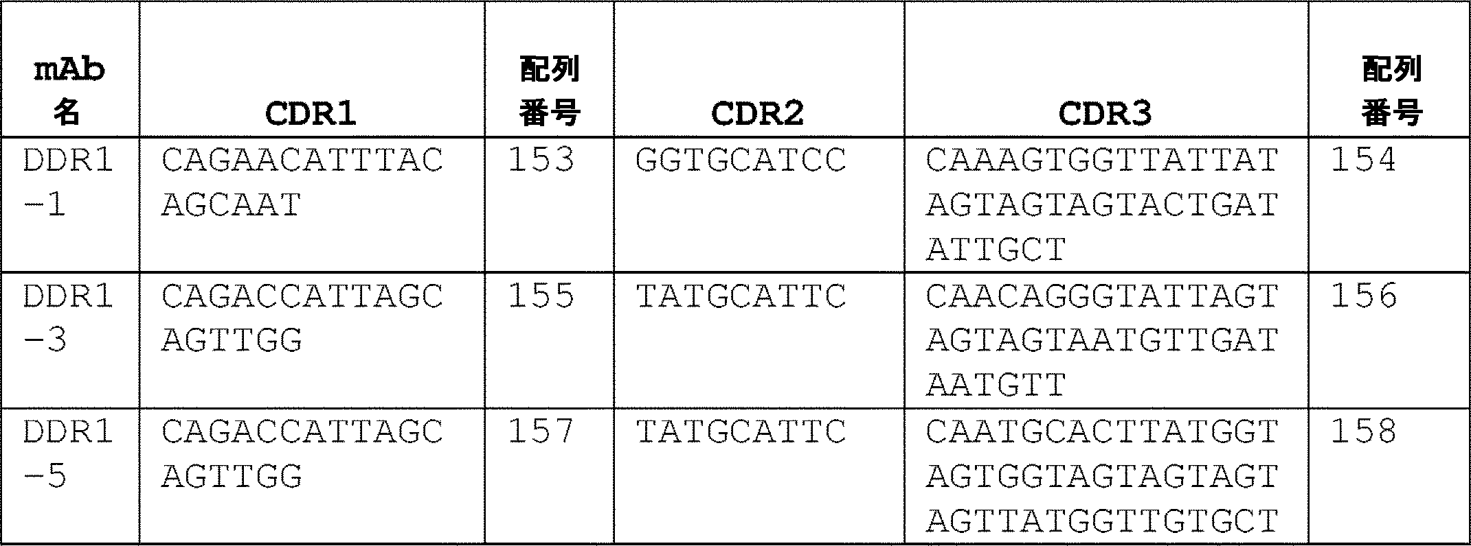

いくつかの実施形態では、重鎖可変領域のHC-CDR1、HC-CDR2、および/またはHC-CDR3をコードするポリヌクレオチドは、表7に示されるポリヌクレオチドから選択される。 In some embodiments, the polynucleotides encoding the heavy chain variable region HC-CDR1, HC-CDR2, and/or HC-CDR3 are selected from the polynucleotides shown in Table 7.
(表7)DDR1抗体の重鎖可変領域のCDRをコードするDNA配列


いくつかの実施形態では、ポリヌクレオチドは、配列番号:108~128または配列番号:150もしくは151のアミノ酸配列の軽鎖可変領域内の少なくとも1、2、または3個のLC-CDRをコードする。いくつかの実施形態では、ポリヌクレオチドは、配列番号:129~149または配列番号:153のアミノ酸配列の重鎖可変領域内の少なくとも1、2、または3個のHC-CDRをコードする。 In some embodiments, the polynucleotide encodes at least 1, 2, or 3 LC-CDRs within the light chain variable region of the amino acid sequences of SEQ ID NOs:108-128 or SEQ ID NOs:150 or 151. In some embodiments, the polynucleotide encodes at least 1, 2, or 3 HC-CDRs within the heavy chain variable region of the amino acid sequences of SEQ ID NOS:129-149 or SEQ ID NO:153.
いくつかの実施形態では、ポリヌクレオチドは、配列番号:108~128または配列番号:150もしくは151のアミノ酸配列の軽鎖可変領域内の少なくとも1、2、または3個のLC-CDRと、配列番号:129~149または配列番号:153のアミノ酸配列の重鎖可変領域内の少なくとも1、2、または3個のHC-CDRと、をコードする。いくつかの実施形態では、選択されるLC-CDRおよびHC-CDRは、クローン対のLC-CDRおよびHC-CDRのものである。 In some embodiments, the polynucleotide comprises at least 1, 2, or 3 LC-CDRs within the light chain variable region of the amino acid sequences of SEQ ID NOs: 108-128 or SEQ ID NOs: 150 or 151 and and at least 1, 2, or 3 HC-CDRs within the heavy chain variable region of the amino acid sequence of SEQ ID NO:129-149 or SEQ ID NO:153. In some embodiments, the LC-CDRs and HC-CDRs selected are of a clonal pair of LC-CDRs and HC-CDRs.
いくつかの実施形態では、ポリヌクレオチドは、表6に示される各mAbのLC-CDRのポリヌクレオチド配列の少なくとも1、2、または3個を含む。 In some embodiments, the polynucleotides comprise at least 1, 2, or 3 of the polynucleotide sequences of the LC-CDRs of each mAb shown in Table 6.
いくつかの実施形態では、ポリヌクレオチドは、表7に示される各mAbのHC-CDRのポリヌクレオチド配列の少なくとも1、2、または3個を含む。 In some embodiments, the polynucleotides comprise at least 1, 2, or 3 of the polynucleotide sequences of the HC-CDRs of each mAb shown in Table 7.
いくつかの実施形態では、ポリヌクレオチドは、表6に示される各mAbのLC-CDRのポリヌクレオチド配列のうちの少なくとも1、2、または3個、表7に示される各mAbのHC-CDRのポリヌクレオチド配列のうちの少なくとも1、2、または3個を含み、選択されるLC-CDRおよびHC-CDRは、それぞれ、表6および表7に示されるクローン対のLC-CDRおよびHC-CDRのものである。 In some embodiments, the polynucleotide comprises at least 1, 2, or 3 of the polynucleotide sequences of the LC-CDRs of each mAb shown in Table 6, the HC-CDRs of each mAb shown in Table 7, Selected LC-CDRs and HC-CDRs comprising at least 1, 2, or 3 of the polynucleotide sequences are the LC-CDRs and HC-CDRs of the clone pairs shown in Tables 6 and 7, respectively. It is a thing.
いくつかの実施形態では、ポリヌクレオチドは、表3の配列番号:108~128および表5の配列番号:150~151から選択される軽鎖可変領域アミノ酸配列と少なくとも80%、85%、90%、91%、92%、93%、94%、95%、96%、97%、98%、99%またはそれ以上の配列同一性を有する軽鎖可変領域をコードする。 In some embodiments, the polynucleotide is at least 80%, 85%, 90% a light chain variable region amino acid sequence selected from SEQ ID NOs: 108-128 of Table 3 and SEQ ID NOs: 150-151 of Table 5. , 91%, 92%, 93%, 94%, 95%, 96%, 97%, 98%, 99% or more sequence identity.
いくつかの実施形態では、ポリヌクレオチドは、表4の配列番号:129~149および表5の配列番号:153から選択される重鎖可変領域アミノ酸配列と少なくとも80%、85%、90%、91%、92%、93%、94%、95%、96%、97%、98%、99%またはそれ以上の配列同一性を有する重鎖可変領域をコードする。 In some embodiments, the polynucleotide comprises a heavy chain variable region amino acid sequence selected from SEQ ID NOS: 129-149 of Table 4 and SEQ ID NO: 153 of Table 5 and at least 80%, 85%, 90%, 91% %, 92%, 93%, 94%, 95%, 96%, 97%, 98%, 99% or more sequence identity.
いくつかの実施形態では、ポリヌクレオチドは、表3の配列番号:108~128または表5の配列番号:150および151から選択される軽鎖可変領域アミノ酸配列に対して少なくとも80%、85%、90%、91%、92%、93%、94%、95%、96%、97%、98%、99%、またはそれ以上の配列同一性を有する軽鎖可変領域と、表4の配列番号:129~149および表5の配列番号:153から選択される重鎖可変領域アミノ酸配列に対して少なくとも80%、85%、90%、91%、92%、93%、94%、96%、97%、98%、99%、またはそれ以上の配列同一性を有する重鎖可変領域と、をコードする。 In some embodiments, the polynucleotide has at least 80%, 85% of the light chain variable region amino acid sequence selected from SEQ ID NOs: 108-128 of Table 3 or SEQ ID NOs: 150 and 151 of Table 5 A light chain variable region having 90%, 91%, 92%, 93%, 94%, 95%, 96%, 97%, 98%, 99% or more sequence identity with a SEQ ID NO in Table 4 at least 80%, 85%, 90%, 91%, 92%, 93%, 94%, 96%, relative to a heavy chain variable region amino acid sequence selected from: 129-149 and SEQ ID NO: 153 of Table 5; and a heavy chain variable region with 97%, 98%, 99% or more sequence identity.
いくつかの実施形態では、ポリヌクレオチドは、以下から選択される軽鎖可変領域および重鎖可変領域を含む抗体または抗原結合断片をコードする:
配列番号:108のアミノ酸配列を有する軽鎖可変領域(DDR1-1K)、および配列番号:129のアミノ酸配列を有する重鎖可変領域(DDR1-1H)、
配列番号:109のアミノ酸配列を有する軽鎖可変領域(DDR1-3K)、および配列番号:130のアミノ酸配列を有する重鎖可変領域(DDR1-3H)、
配列番号:110のアミノ酸配列を有する軽鎖可変領域(DDR1-5K)、および配列番号:131のアミノ酸配列を有する重鎖可変領域(DDR1-5H)、
配列番号:111のアミノ酸配列を有する軽鎖可変領域(DDR1-6K)、および配列番号:132のアミノ酸配列を有する重鎖可変領域(DDR1-6H)、
配列番号:112のアミノ酸配列を有する軽鎖可変領域(DDR1-9K)、および配列番号:133のアミノ酸配列を有する重鎖可変領域(DDR1-9H)、
配列番号:113のアミノ酸配列を有する軽鎖可変領域(DDR1-11K)、および配列番号:134のアミノ酸配列を有する重鎖可変領域(DDR1-11H)、
配列番号:114のアミノ酸配列を有する軽鎖可変領域(DDR1-12K)、および配列番号:135のアミノ酸配列を有する重鎖可変領域(DDR1-12H)、
配列番号:115のアミノ酸配列を有する軽鎖可変領域(DDR1-13K)、および配列番号:136のアミノ酸配列を有する重鎖可変領域(DDR1-13H)、
配列番号:116のアミノ酸配列を有する軽鎖可変領域(DDR1-14K)、および配列番号:137のアミノ酸配列を有する重鎖可変領域(DDR1-14H)、
配列番号:117のアミノ酸配列を有する軽鎖可変領域(DDR1-15K)、および配列番号:138のアミノ酸配列を有する重鎖可変領域(DDR1-15H)、
配列番号:118のアミノ酸配列を有する軽鎖可変領域(DDR1-17K)、および配列番号:139のアミノ酸配列を有する重鎖可変領域(DDR1-17H)、
配列番号:119のアミノ酸配列を有する軽鎖可変領域(DDR1-20K)、および配列番号:140のアミノ酸配列を有する重鎖可変領域(DDR1-20H)、
配列番号:120のアミノ酸配列を有する軽鎖可変領域(DDR1-21K)、および配列番号:141のアミノ酸配列を有する重鎖可変領域(DDR1-21H)、
配列番号:121のアミノ酸配列を有する軽鎖可変領域(DDR1-22K)、および配列番号:142のアミノ酸配列を有する重鎖可変領域(DDR1-22H)、
配列番号:122のアミノ酸配列を有する軽鎖可変領域(DDR1-23K)、および配列番号:143のアミノ酸配列を有する重鎖可変領域(DDR1-23H)、
配列番号:123のアミノ酸配列を有する軽鎖可変領域(DDR1-26K)、および配列番号:144のアミノ酸配列を有する重鎖可変領域(DDR1-26H)、
配列番号:124のアミノ酸配列を有する軽鎖可変領域(DDR1-28K)、および配列番号:145のアミノ酸配列を有する重鎖可変領域(DDR1-28H)、
配列番号:125のアミノ酸配列を有する軽鎖可変領域(DDR1-29K)、および配列番号:146のアミノ酸配列を有する重鎖可変領域(DDR1-29H)、
配列番号:126のアミノ酸配列を有する軽鎖可変領域(DDR1-32K)、および配列番号147のアミノ酸配列を有する重鎖可変領域(DDR1-32H)、
配列番号:127のアミノ酸配列を有する軽鎖可変領域(DDR1-33K)、および配列番号:148のアミノ酸配列を有する重鎖可変領域(DDR1-33H)、
配列番号:128のアミノ酸配列を有する軽鎖可変領域(DDR1-34K)、および配列番号:149のアミノ酸配列を有する重鎖可変領域(DDR1-34H)、
配列番号:150のアミノ酸配列を有する軽鎖可変領域(DDR1-9hu_Lv)、および配列番号:153のアミノ酸配列を有する重鎖可変領域(DDR1-9hu_Hv)、ならびに
配列番号:151のアミノ酸配列を有する軽鎖可変領域(DDR1-9hu_Lc2)、および配列番号:153のアミノ酸配列を有する重鎖可変領域(DDR1-9hu_Hv)。
In some embodiments, the polynucleotide encodes an antibody or antigen-binding fragment comprising a light chain variable region and a heavy chain variable region selected from:
a light chain variable region (DDR1-1K) having the amino acid sequence of SEQ ID NO:108 and a heavy chain variable region (DDR1-1H) having the amino acid sequence of SEQ ID NO:129;
a light chain variable region (DDR1-3K) having the amino acid sequence of SEQ ID NO: 109 and a heavy chain variable region (DDR1-3H) having the amino acid sequence of SEQ ID NO: 130;
a light chain variable region (DDR1-5K) having the amino acid sequence of SEQ ID NO: 110 and a heavy chain variable region (DDR1-5H) having the amino acid sequence of SEQ ID NO: 131;
a light chain variable region (DDR1-6K) having the amino acid sequence of SEQ ID NO: 111 and a heavy chain variable region (DDR1-6H) having the amino acid sequence of SEQ ID NO: 132;
a light chain variable region (DDR1-9K) having the amino acid sequence of SEQ ID NO: 112 and a heavy chain variable region (DDR1-9H) having the amino acid sequence of SEQ ID NO: 133;
a light chain variable region (DDR1-11K) having the amino acid sequence of SEQ ID NO:113 and a heavy chain variable region (DDR1-11H) having the amino acid sequence of SEQ ID NO:134;
a light chain variable region (DDR1-12K) having the amino acid sequence of SEQ ID NO:114 and a heavy chain variable region (DDR1-12H) having the amino acid sequence of SEQ ID NO:135;
a light chain variable region (DDR1-13K) having the amino acid sequence of SEQ ID NO:115 and a heavy chain variable region (DDR1-13H) having the amino acid sequence of SEQ ID NO:136;
a light chain variable region (DDR1-14K) having the amino acid sequence of SEQ ID NO: 116 and a heavy chain variable region (DDR1-14H) having the amino acid sequence of SEQ ID NO: 137;
a light chain variable region (DDR1-15K) having the amino acid sequence of SEQ ID NO: 117 and a heavy chain variable region (DDR1-15H) having the amino acid sequence of SEQ ID NO: 138;
a light chain variable region (DDR1-17K) having the amino acid sequence of SEQ ID NO:118 and a heavy chain variable region (DDR1-17H) having the amino acid sequence of SEQ ID NO:139;
a light chain variable region (DDR1-20K) having the amino acid sequence of SEQ ID NO:119 and a heavy chain variable region (DDR1-20H) having the amino acid sequence of SEQ ID NO:140;
a light chain variable region (DDR1-21K) having the amino acid sequence of SEQ ID NO: 120 and a heavy chain variable region (DDR1-21H) having the amino acid sequence of SEQ ID NO: 141;
a light chain variable region (DDR1-22K) having the amino acid sequence of SEQ ID NO: 121 and a heavy chain variable region (DDR1-22H) having the amino acid sequence of SEQ ID NO: 142;
a light chain variable region (DDR1-23K) having the amino acid sequence of SEQ ID NO: 122 and a heavy chain variable region (DDR1-23H) having the amino acid sequence of SEQ ID NO: 143;
a light chain variable region (DDR1-26K) having the amino acid sequence of SEQ ID NO: 123 and a heavy chain variable region (DDR1-26H) having the amino acid sequence of SEQ ID NO: 144;
a light chain variable region (DDR1-28K) having the amino acid sequence of SEQ ID NO: 124 and a heavy chain variable region (DDR1-28H) having the amino acid sequence of SEQ ID NO: 145;
a light chain variable region (DDR1-29K) having the amino acid sequence of SEQ ID NO: 125 and a heavy chain variable region (DDR1-29H) having the amino acid sequence of SEQ ID NO: 146;
a light chain variable region (DDR1-32K) having the amino acid sequence of SEQ ID NO: 126 and a heavy chain variable region (DDR1-32H) having the amino acid sequence of SEQ ID NO: 147;
a light chain variable region (DDR1-33K) having the amino acid sequence of SEQ ID NO: 127 and a heavy chain variable region (DDR1-33H) having the amino acid sequence of SEQ ID NO: 148;
a light chain variable region (DDR1-34K) having the amino acid sequence of SEQ ID NO: 128 and a heavy chain variable region (DDR1-34H) having the amino acid sequence of SEQ ID NO: 149;
a light chain variable region (DDR1-9hu_Lv) having the amino acid sequence of SEQ ID NO: 150, a heavy chain variable region (DDR1-9hu_Hv) having the amino acid sequence of SEQ ID NO: 153, and a light chain variable region having the amino acid sequence of SEQ ID NO: 151 a chain variable region (DDR1-9hu_Lc2), and a heavy chain variable region (DDR1-9hu_Hv) having the amino acid sequence of SEQ ID NO:153.
いくつかの実施形態では、軽鎖可変領域をコードするポリヌクレオチドは、表8に示されるポリヌクレオチドから選択される。 In some embodiments, the polynucleotides encoding the light chain variable regions are selected from the polynucleotides shown in Table 8.
(表8)抗DDR1抗体の軽鎖可変領域をコードするDNA配列

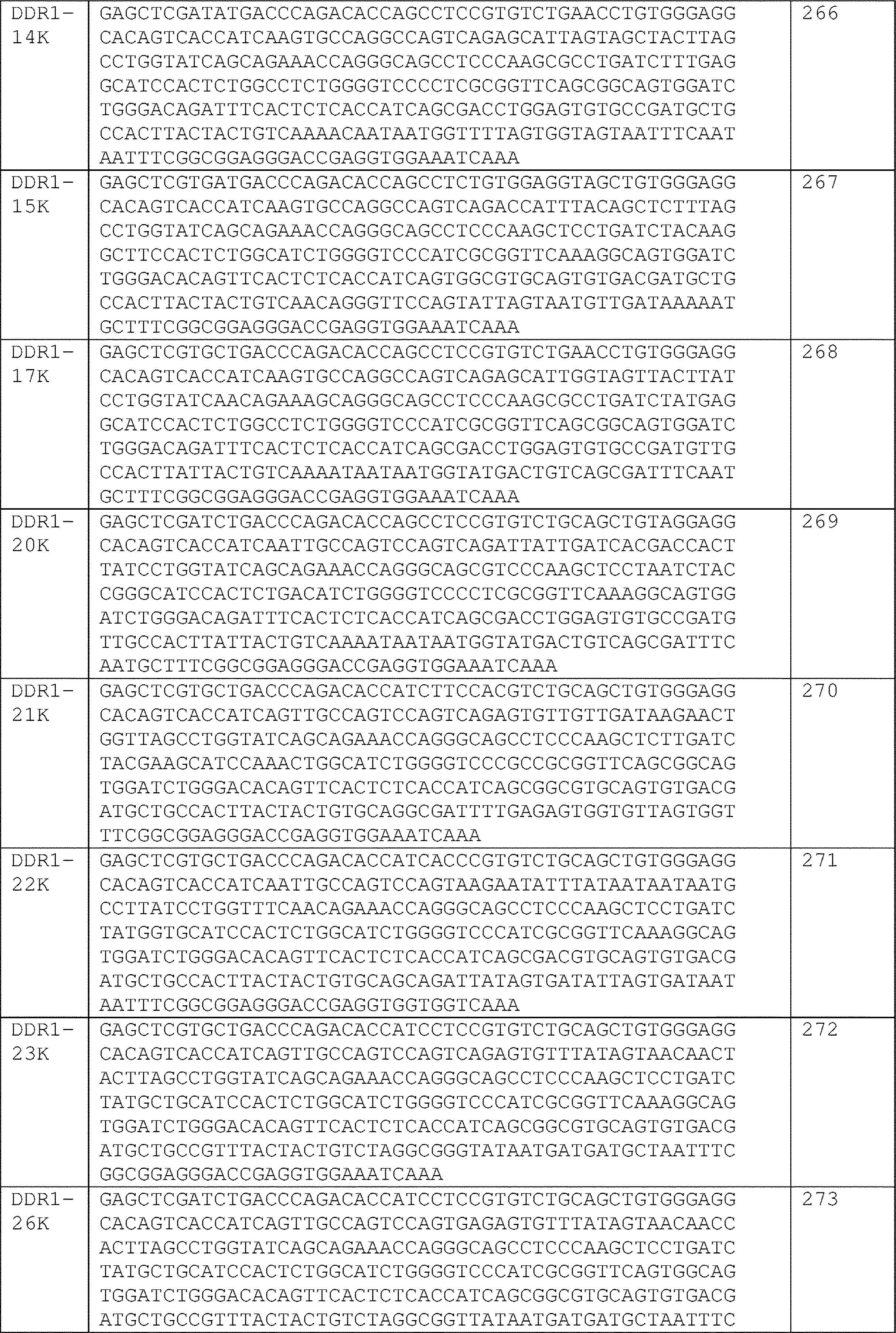
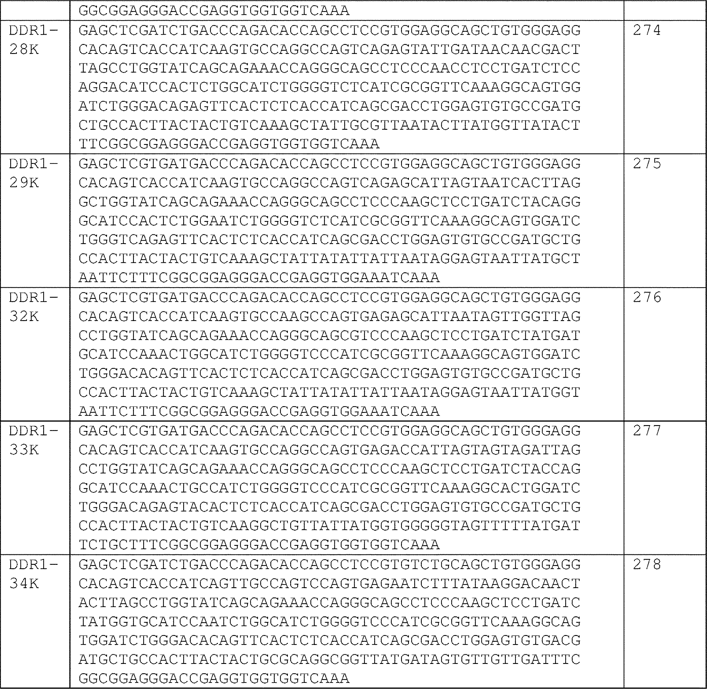
いくつかの実施形態では、重鎖可変領域をコードするポリヌクレオチドは、表9に示されるポリヌクレオチドから選択される。 In some embodiments, the polynucleotides encoding the heavy chain variable regions are selected from the polynucleotides shown in Table 9.
(表9)抗DDR1抗体の重鎖可変領域をコードするDNA配列

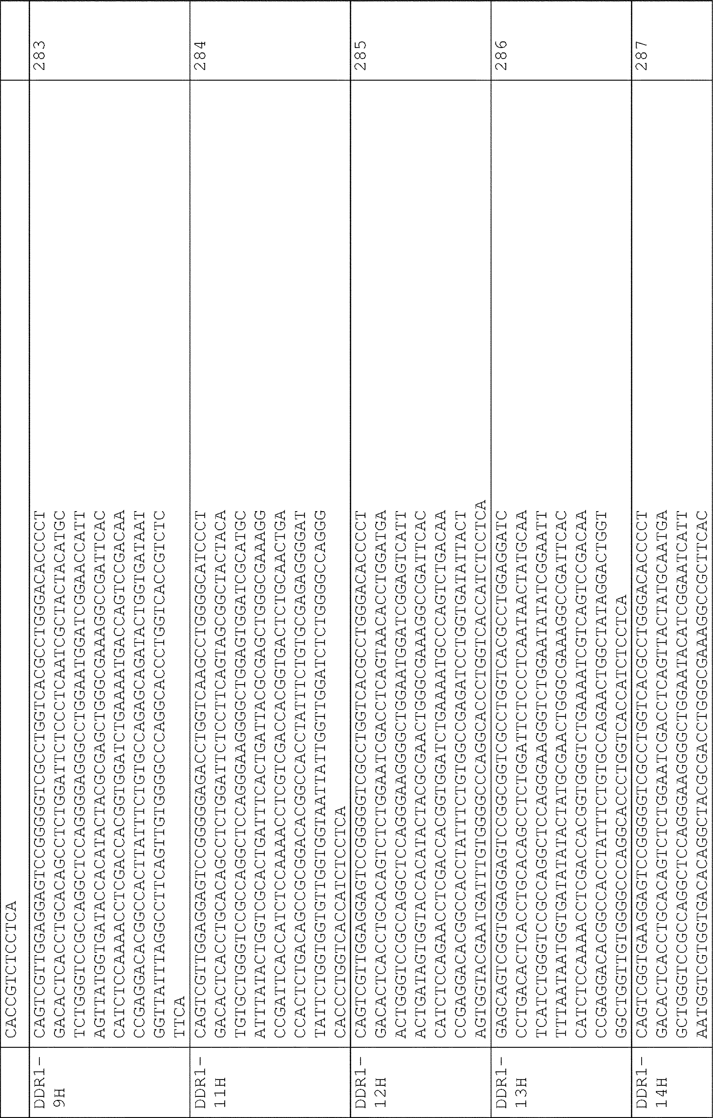
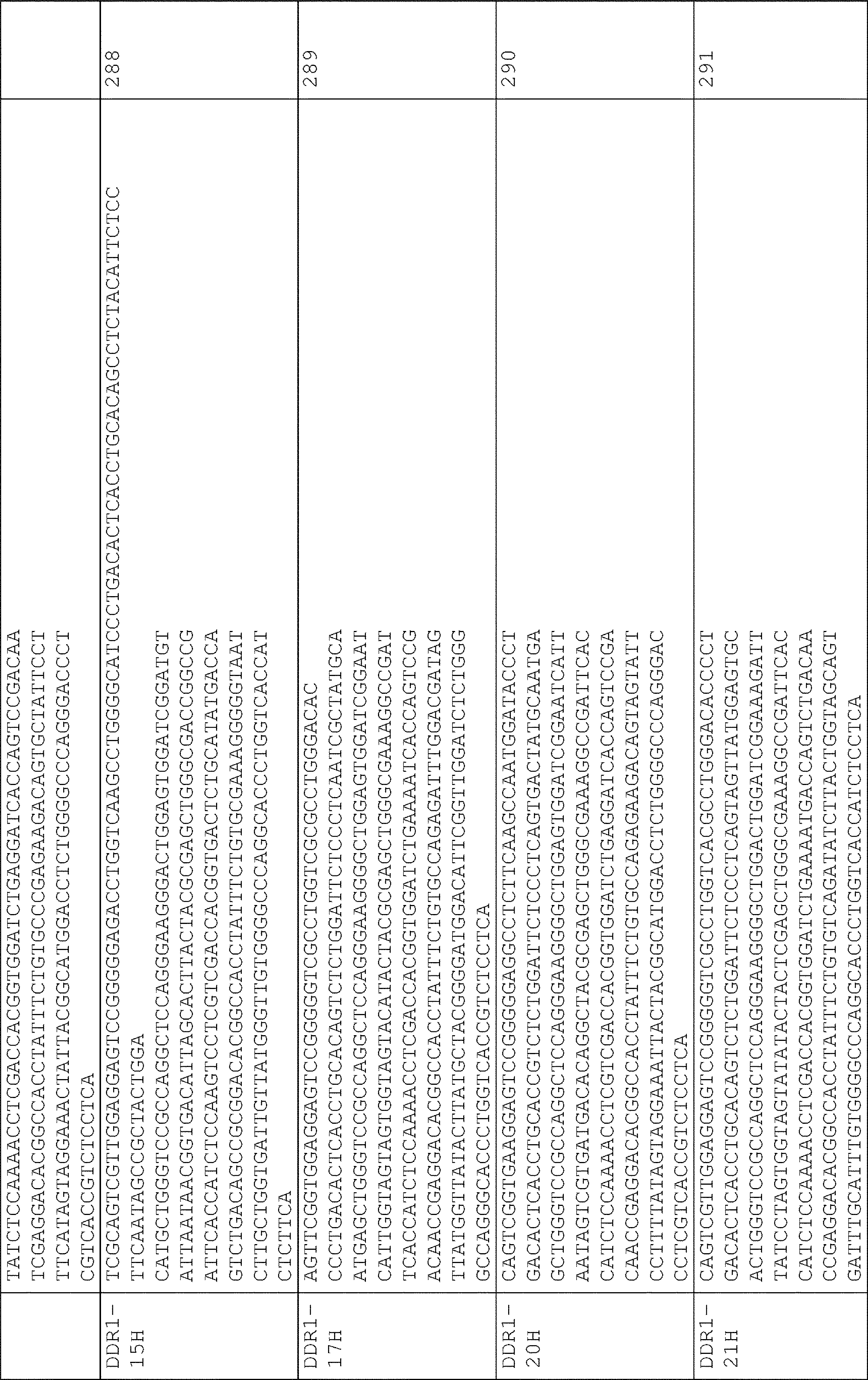
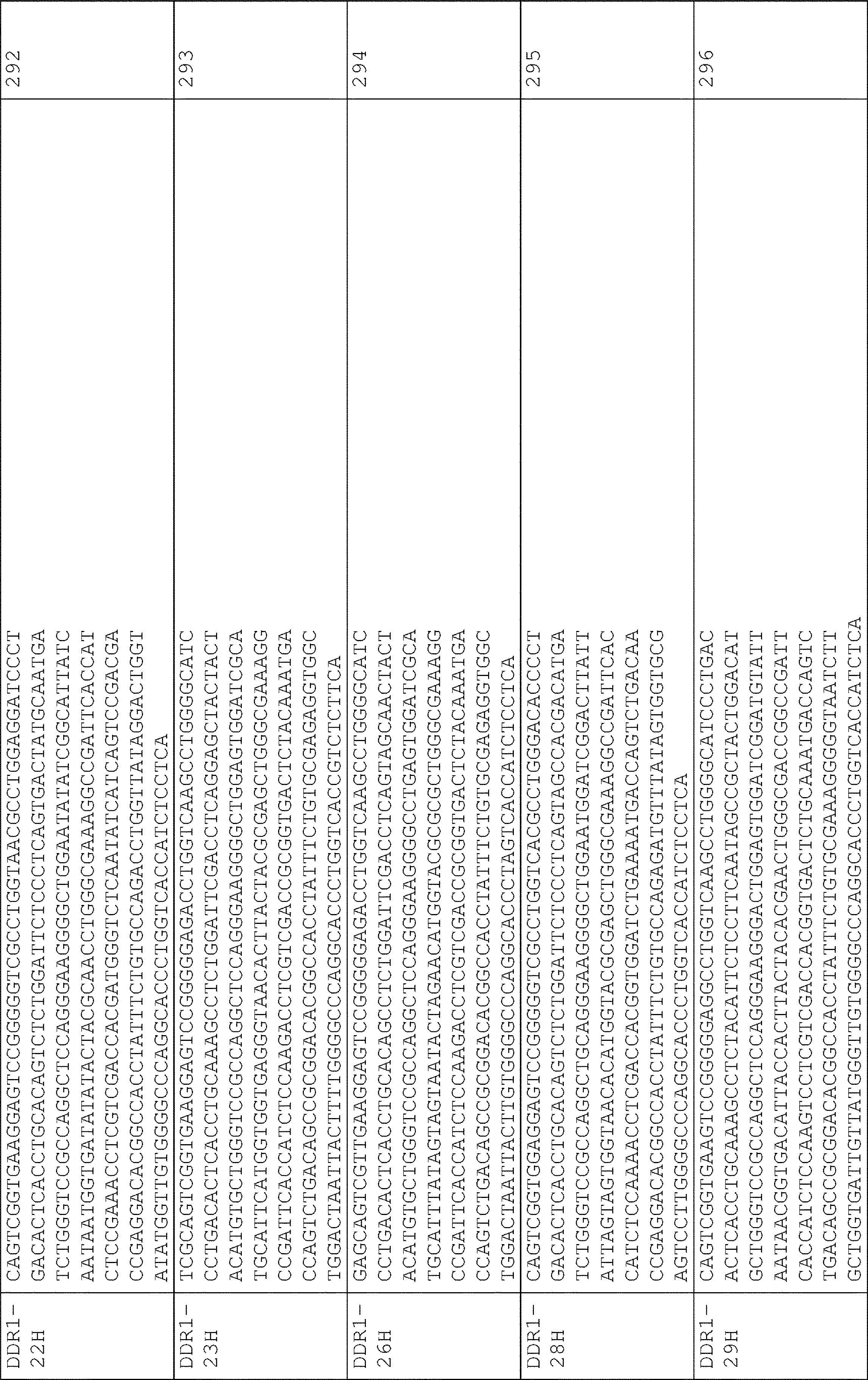

いくつかの実施形態では、抗体DDR1-9(DDR1-9hu)のヒト化軽鎖可変領域および/またはヒト化重鎖可変領域をコードするポリヌクレオチドは、表10に示されるポリヌクレオチドから選択される。 In some embodiments, the polynucleotides encoding the humanized light chain variable region and/or the humanized heavy chain variable region of antibody DDR1-9 (DDR1-9hu) are selected from the polynucleotides shown in Table 10 .
(表10)ヒト化DDR1-9hu抗体配列-核酸
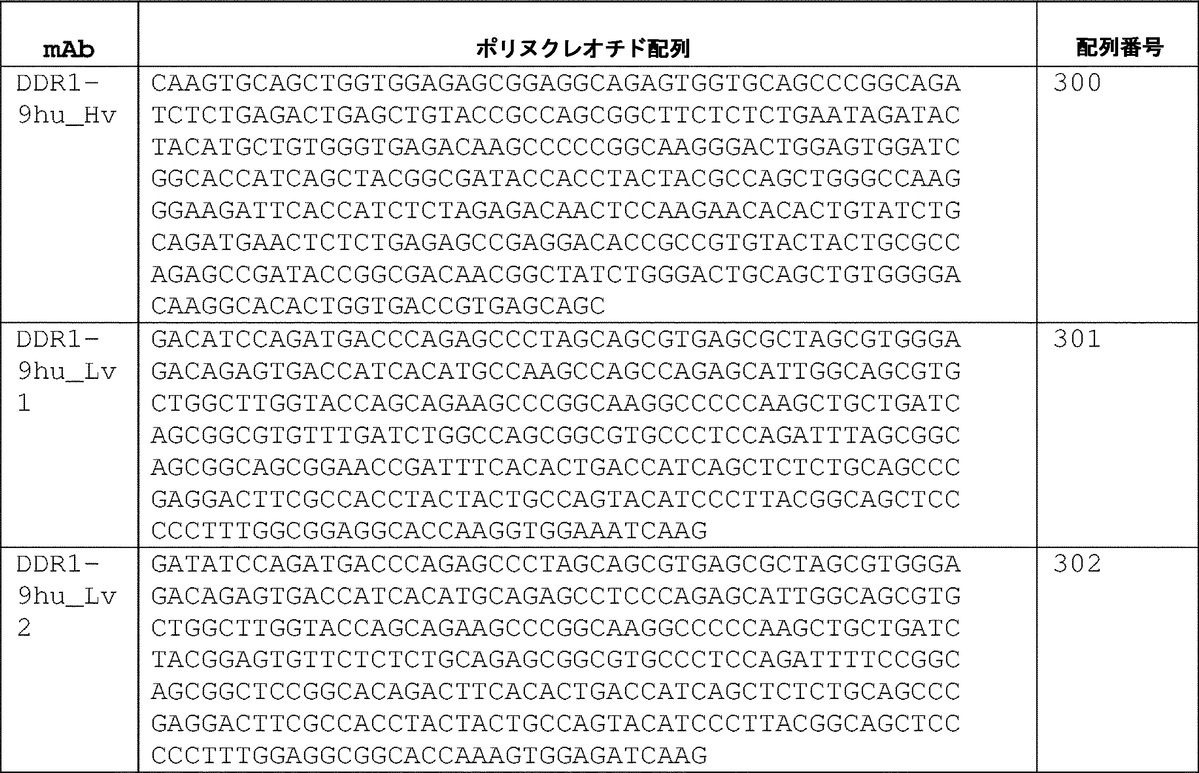
いくつかの実施形態では、ポリヌクレオチドは、配列番号2:58~278および配列番号:301~302から選択される参照ポリヌクレオチド配列に対して、ヌクレオチドレベルで少なくとも約80%、85%、86%、87%、88%、89%、90%、91%、92%、93%、94%、95%、96%、97%、98%、または99%以上の配列同一性を有し、配列番号:108~128または配列番号:150~151から選択される対応する軽鎖可変領域をコードする。 In some embodiments, the polynucleotide is at least about 80%, 85%, 86% at the nucleotide level relative to a reference polynucleotide sequence selected from SEQ ID NOs:2:58-278 and SEQ ID NOs:301-302. , 87%, 88%, 89%, 90%, 91%, 92%, 93%, 94%, 95%, 96%, 97%, 98%, or 99% or greater sequence identity, and sequence Numbers: 108-128 or encodes the corresponding light chain variable region selected from SEQ ID NOs: 150-151.
いくつかの実施形態では、ポリヌクレオチドは、配列番号:279~299および配列番号:300から選択される参照ポリヌクレオチド配列に対して、ヌクレオチドレベルで少なくとも約80%、85%、86%、87%、88%、89%、90%、91%、92%、93%、94%、95%、96%、97%、98%、または99%以上の配列同一性を有し、配列番号:129~149または配列番号:152から選択される対応する軽鎖可変領域をコードする。 In some embodiments, the polynucleotide is at least about 80%, 85%, 86%, 87% at the nucleotide level relative to a reference polynucleotide sequence selected from SEQ ID NO:279-299 and SEQ ID NO:300 , 88%, 89%, 90%, 91%, 92%, 93%, 94%, 95%, 96%, 97%, 98%, or 99% or greater sequence identity and SEQ ID NO: 129 -149 or encodes the corresponding light chain variable region selected from SEQ ID NO:152.
いくつかの実施形態では、ポリヌクレオチドは、配列番号:262または276の参照ポリヌクレオチド配列に対して、ヌクレオチドレベルで少なくとも約80%、85%、86%、87%、88%、89%、90%、91%、92%、93%、94%、95%、96%、97%、98%、または99%以上の配列同一性を有し、配列番号:112または配列番号:126の対応する軽鎖可変領域をコードする。 In some embodiments, the polynucleotide is at least about 80%, 85%, 86%, 87%, 88%, 89%, 90% at the nucleotide level relative to the reference polynucleotide sequence of SEQ ID NO:262 or 276. %, 91%, 92%, 93%, 94%, 95%, 96%, 97%, 98%, or 99% or more sequence identity and corresponding to SEQ ID NO: 112 or SEQ ID NO: 126 Encodes the light chain variable region.
いくつかの実施形態では、ポリヌクレオチドは、配列番号:283または297の参照ポリヌクレオチド配列に対して、ヌクレオチドレベルで少なくとも約80%、85%、86%、87%、88%、89%、90%、91%、92%、93%、94%、95%、96%、97%、98%、または99%以上の配列同一性を有し、配列番号:133または147の対応する軽鎖可変領域をコードする。 In some embodiments, the polynucleotide is at least about 80%, 85%, 86%, 87%, 88%, 89%, 90% at the nucleotide level relative to the reference polynucleotide sequence of SEQ ID NO: 283 or 297. %, 91%, 92%, 93%, 94%, 95%, 96%, 97%, 98%, or 99% or more sequence identity and the corresponding light chain variable of SEQ ID NO: 133 or 147 Code the region.
いくつかの実施形態では、ポリヌクレオチドは、配列番号:301または302の参照ポリヌクレオチド配列に対して、ヌクレオチドレベルで少なくとも約80%、85%、86%、87%、88%、89%、90%、91%、92%、93%、94%、95%、96%、97%、98%、または99%以上の配列同一性を有し、配列番号:150または配列番号:151の対応する軽鎖可変領域をコードする。 In some embodiments, the polynucleotide is at least about 80%, 85%, 86%, 87%, 88%, 89%, 90% at the nucleotide level relative to the reference polynucleotide sequence of SEQ ID NO:301 or 302. %, 91%, 92%, 93%, 94%, 95%, 96%, 97%, 98%, or 99% or more sequence identity and corresponding to SEQ ID NO: 150 or SEQ ID NO: 151 Encodes the light chain variable region.
いくつかの実施形態では、ポリヌクレオチドは、配列番号:300の参照ポリヌクレオチド配列に対して、ヌクレオチドレベルで少なくとも約80%、85%、86%、87%、88%、89%、90%、91%、92%、93%、94%、95%、96%、97%、98%、または99%以上の配列同一性を有し、配列番号:152の対応する軽鎖可変領域をコードする。 In some embodiments, the polynucleotide has at least about 80%, 85%, 86%, 87%, 88%, 89%, 90%, 91%, 92%, 93%, 94%, 95%, 96%, 97%, 98%, or 99% or more sequence identity and encodes the corresponding light chain variable region of SEQ ID NO: 152 .
さらなる実施形態では、本開示の核酸は、本明細書に開示される抗体をコードするポリヌクレオチドにハイブリダイズするポリヌクレオチドを含む。いくつかの実施形態では、ポリヌクレオチドは、配列番号:258~278および配列番号:301~302から選択されるポリヌクレオチドにハイブリダイズし、DDR1タンパク質に特異的に結合する抗体の軽鎖可変領域をコードする。いくつかの実施形態では、ポリヌクレオチドは、配列番号:279~299および配列番号:300から選択されるポリヌクレオチドにハイブリダイズし、DDR1タンパク質に特異的に結合する抗体の重鎖可変領域をコードする。 In further embodiments, a nucleic acid of the present disclosure comprises a polynucleotide that hybridizes to a polynucleotide encoding an antibody disclosed herein. In some embodiments, the polynucleotide hybridizes to a polynucleotide selected from SEQ ID NOs: 258-278 and SEQ ID NOs: 301-302 and comprises the light chain variable region of an antibody that specifically binds to a DDR1 protein. code. In some embodiments, the polynucleotide hybridizes to a polynucleotide selected from SEQ ID NOS:279-299 and SEQ ID NO:300 and encodes a heavy chain variable region of an antibody that specifically binds to a DDR1 protein .
一般に、核酸は、本明細書に開示される抗体をコードし、DDR1タンパク質に特異的に結合する能力を維持する抗体もコードする核酸と、中程度または高いストリンジェンシー条件下でハイブリダイズする。第1の核酸分子は、第1の核酸分子の単鎖形態が、温度および溶液イオン強度の適切な条件下で第2の核酸分子にアニーリングすることができる場合、第2の核酸分子に「ハイブリダイズ可能」である(Sambrook et al.,MOLECULAR CLONING:A LABORATORY MANUAL,3rd ed.,Cold Spring Harbor Press,Cold Spring Harbor,N.Y.2001)を参照されたい)。温度およびイオン強度の条件は、ハイブリダイゼーションの「ストリンジェンシー」を決定する。典型的な中程度のストリンジェンシーのハイブリダイゼーション条件は、42℃での、5×または6×SSCおよび0.1%のSDSを伴う40%のホルムアミドである。高ストリンジェンシーなハイブリダイゼーション条件は、50%のホルムアミド、5×または6×SSC(0.15MのNaC1および0.015MのNa-クエン酸塩)、42℃、または任意選択で、より高い温度(例えば、57℃、59℃、60℃、62℃、63℃、65℃、または68℃)である。ハイブリダイゼーションは、2つの核酸が相補的配列を含むことを必要とするが、ハイブリダイゼーションのストリンジェンシーに応じて、塩基間のミスマッチも可能である。核酸をハイブリダイゼーションするための適切なストリンジェンシーは、当該技術分野において周知の変数である、核酸の長さおよび相補性の程度に依存する。2つのヌクレオチド配列間の類似性または相同性の度合いが大きいほど、核酸がハイブリダイゼーションし得るストリンジェンシーがより高くなる。長さが100ヌクレオチドを超えるハイブリッドについては、融解温度を計算するための方程式が導出されている(上記のSambrook et al.を参照されたい)。より短い核酸、例えば、オリゴヌクレオチドとのハイブリダイゼーションについては、ミスマッチの位置がより重要になり、オリゴヌクレオチドの長さがその特異性を決定する(上記のSambrook et al.を参照されたい)。 In general, nucleic acids will hybridize under moderate or high stringency conditions to nucleic acids that encode antibodies disclosed herein and also encode antibodies that retain the ability to specifically bind to DDR1 protein. A first nucleic acid molecule "hybridizes" to a second nucleic acid molecule if the single-stranded form of the first nucleic acid molecule can anneal to the second nucleic acid molecule under the appropriate conditions of temperature and solution ionic strength. (See Sambrook et al., MOLECULAR CLONING: A LABORATORY MANUAL, 3rd ed., Cold Spring Harbor Press, Cold Spring Harbor, N.Y. 2001). The conditions of temperature and ionic strength determine the "stringency" of the hybridization. Typical moderate stringency hybridization conditions are 40% formamide with 5x or 6x SSC and 0.1% SDS at 42°C. High stringency hybridization conditions are 50% formamide, 5× or 6×SSC (0.15 M NaCl and 0.015 M Na-citrate), 42° C., or optionally higher temperature ( For example, 57°C, 59°C, 60°C, 62°C, 63°C, 65°C, or 68°C). Hybridization requires that the two nucleic acids contain complementary sequences, although mismatches between bases are possible, depending on the stringency of the hybridization. The appropriate stringency for hybridizing nucleic acids depends on the length of the nucleic acids and the degree of complementation, variables well known in the art. The greater the degree of similarity or homology between two nucleotide sequences, the higher the stringency at which nucleic acids can hybridize. For hybrids greater than 100 nucleotides in length, an equation has been derived for calculating the melting temperature (see Sambrook et al., supra). For hybridization with shorter nucleic acids, eg, oligonucleotides, the position of the mismatch becomes more critical and the length of the oligonucleotide determines its specificity (see Sambrook et al., supra).
いくつかの実施形態では、本明細書に開示されるポリヌクレオチドは、本明細書に開示される軽鎖可変領域または重鎖可変領域などのコードされたポリペプチドの発現を提供するための様々な方法で操作され得る。いくつかの実施形態では、ポリヌクレオチドは、ポリヌクレオチドおよび/または対応するポリペプチドの発現のための制御配列、とりわけ、転写プロモーター、リーダー配列、転写エンハンサー、リボソーム結合またはエントリー部位、終結配列、およびポリアデニル化配列を含む制御配列に動作可能に連結される。ベクターへの挿入前の、単離されたポリヌクレオチドの操作は、発現ベクターに応じて、望ましいか、または必要であり得る。組換えDNA法を利用してポリヌクレオチドおよび核酸配列を修飾するための技術は、当技術分野で既知である。ガイダンスは、Sambrook et al.,Molecular Cloning:A Laboratory Manual,3rd Ed.,Cold Spring Harbor Laboratory Press,New York(2001)、およびCurrent Protocols in Molecular Biology,Ausubel.F.ed.,Greene Pub.Associates(1998),updates to 2020にて提供される。 In some embodiments, the polynucleotides disclosed herein are used in a variety of ways to provide expression of the encoded polypeptides, such as the light or heavy chain variable regions disclosed herein. can be manipulated in a manner. In some embodiments, the polynucleotide comprises regulatory sequences for expression of the polynucleotide and/or the corresponding polypeptide, among others transcriptional promoters, leader sequences, transcriptional enhancers, ribosome binding or entry sites, termination sequences, and polyadenylation. is operably linked to a control sequence that includes a modification sequence. Manipulation of the isolated polynucleotide prior to its insertion into a vector may be desirable or necessary depending on the expression vector. Techniques for modifying polynucleotide and nucleic acid sequences using recombinant DNA methods are known in the art. Guidance can be found in Sambrook et al. , Molecular Cloning: A Laboratory Manual, 3rd Ed. , Cold Spring Harbor Laboratory Press, New York (2001), and Current Protocols in Molecular Biology, Ausubel. F. ed. , Greene Pub. Associates (1998), updates to 2020.
いくつかの実施形態では、ポリヌクレオチドは、発現ベクターの一部であってもよく、ここで、ベクターおよびポリヌクレオチドは、ポリヌクレオチドの発現および/またはコードされたポリペプチドの発現を制御するための1つ以上の動作可能に連結された制御配列を含む。組換え発現ベクターは、任意のベクター(例えば、プラスミドまたはウイルス)であってもよく、これは、組換えDNA手順に好都合に供することができ、ポリヌクレオチド配列の発現をもたらすことができる。ベクターの選択は、典型的には、ベクターが導入される宿主細胞とベクターとの適合性に依存する。ベクターは、線状または閉環状プラスミドであってもよい。例示的な発現ベクターとしては、とりわけ、T7またはT7lacプロモーターに基づくベクター(pACY:Novagen;pET)、バキュロウイルスプロモーターに基づくベクター(例えば、pBAC)、Ef1-αおよびHTLVプロモーターに基づくベクター(例えば、pFUSE2;Invitrogen、CA,USA)、CMVエンハンサーおよびヒトフェリチン軽鎖遺伝子プロモーターに基づくベクター(例えば、pFUSE:Invitrogen、CA,USA)、CMVプロモーターに基づくベクター(例えば、pFLAG:Sigma、USA)、ならびにジヒドロ葉酸還元酵素プロモーターに基づくベクター(例えば、pEASE:Amgen、USA)が挙げられる。様々なベクターは、目的のポリペプチドの一過性または安定した発現のために使用することができる。 In some embodiments, the polynucleotide may be part of an expression vector, wherein the vector and polynucleotide are for controlling expression of the polynucleotide and/or expression of the encoded polypeptide. It contains one or more operably linked control sequences. A recombinant expression vector may be any vector (eg, plasmid or viral), which is conveniently amenable to recombinant DNA procedures and which can result in expression of a polynucleotide sequence. The choice of vector typically depends on the compatibility of the vector with the host cell into which it will be introduced. Vectors may be linear or closed circular plasmids. Exemplary expression vectors include, among others, T7 or T7lac promoter-based vectors (pACY: Novagen; pET), baculovirus promoter-based vectors (eg, pBAC), Ef1-α and HTLV promoter-based vectors (eg, pFUSE2 Invitrogen, Calif., USA), CMV enhancer and human ferritin light chain gene promoter-based vectors (e.g., pFUSE: Invitrogen, Calif., USA), CMV promoter-based vectors (e.g., pFLAG: Sigma, USA), and dihydrofolic acid. vectors based on reductase promoters (eg pEASE: Amgen, USA). A variety of vectors can be used for transient or stable expression of the polypeptide of interest.
別の態様では、ポリペプチドをコードするポリヌクレオチドは、宿主細胞でのポリペプチドの発現のために1つ以上の制御配列に動作可能に連結される。ポリペプチドの発現に使用する宿主細胞は当該技術分野で周知であり、大腸菌、酵母細胞などの細菌細胞、ショウジョウバエS2およびSpodoptera Sf9細胞などの昆虫細胞、チャイニーズハムスター卵巣(CHO)、アフリカミドリザル腎臓(COS)、ベビーハムスター腎臓(BHK)、マウス骨髄腫(例えば、NS0およびSp2/0)、およびヒト胚腎臓(HEK)などの動物細胞、ならびに植物細胞が挙げられるが、これらに限定されない。上述の宿主細胞のための適切な培養培地および増殖条件は、当該技術分野で既知である。いくつかの実施形態では、宿主細胞および発現ベクターは、目的のポリペプチドを発現するために使用される。 In another aspect, a polynucleotide encoding a polypeptide is operably linked to one or more regulatory sequences for expression of the polypeptide in a host cell. Host cells used for expression of polypeptides are well known in the art and include bacterial cells such as E. coli, yeast cells, insect cells such as Drosophila S2 and Spodoptera Sf9 cells, Chinese Hamster Ovary (CHO), African Green Monkey Kidney (COS). ), baby hamster kidney (BHK), mouse myeloma (eg, NS0 and Sp2/0), and human embryonic kidney (HEK), and plant cells. Appropriate culture media and growth conditions for the host cells described above are known in the art. In some embodiments, host cells and expression vectors are used to express the polypeptide of interest.
いくつかの実施形態では、本明細書に記載の発現ベクターおよびポリヌクレオチドを含む宿主細胞を、好適な培地中およびコードされたポリペプチド、例えば、配列番号:108~128、配列番号:150および151、配列番号:129~149、および配列番号:152から選択されるアミノ酸配列を含むポリペプチドの発現に好適な培養条件下で培養する。いくつかの実施形態では、インビトロ発現系は、ポリペプチドを発現するために発現ベクターとともに使用することができる。インビトロ発現系には、大腸菌、ウサギ網赤血球、小麦胚芽、昆虫細胞、およびヒト細胞に基づくものが挙げられる。宿主細胞で発現されるか、またはインビトロで発現されるかにかかわらず、発現されるポリペプチドは、本明細書にさらに記載されるように、単離または精製され得る。 In some embodiments, host cells containing expression vectors and polynucleotides described herein are incubated in a suitable culture medium and encoded polypeptides, eg, SEQ ID NOs: 108-128, SEQ ID NOs: 150 and 151 , SEQ ID NOS:129-149, and SEQ ID NO:152. In some embodiments, in vitro expression systems can be used with expression vectors to express the polypeptides. In vitro expression systems include those based on E. coli, rabbit reticulocytes, wheat germ, insect cells, and human cells. The expressed polypeptide, whether expressed in a host cell or in vitro, can be isolated or purified as further described herein.
IV.抗体の調製方法およびその修飾方法
いくつかの実施形態では、本明細書に記載のモノクローナル抗体は、標準的な方法を使用して調製することができ、続いてスクリーニング、特徴付け、および機能評価が行われる。例えば、可変領域を配列決定し、次いで、ヒト発現ベクターにサブクローニングして、キメラ抗体遺伝子を生成し、次いで、それを発現および精製することができる。これらのキメラ抗体は、抗原結合、シグナル伝達の遮断、および異種移植片実験において試験することができる。
IV. Methods of Preparing and Modifying Antibodies In some embodiments, the monoclonal antibodies described herein can be prepared using standard methods, followed by screening, characterization, and functional evaluation. done. For example, the variable regions can be sequenced and then subcloned into human expression vectors to generate chimeric antibody genes, which are then expressed and purified. These chimeric antibodies can be tested for antigen binding, blocking signaling, and in xenograft experiments.
A.一般的な方法
DDR1に結合するモノクローナル抗体は、いくつかの用途を有することが理解されるであろう。これらには、がんの検出および診断、ならびにがん療法に使用するための診断キットの製造が含まれる。これらの文脈では、かかる抗体を診断剤または治療剤に連結し、それらを競合アッセイにおいて捕捉剤または競合剤として使用し、または加の薬剤が付着することなくそれらを個別に使用してもよい。抗体は、以下でさらに考察されるように、変異されてもよく、または修飾されてもよい。抗体を調製および特徴付けるための方法は当該技術分野において周知である(例えば、Antibodies:A Laboratory Manual,Cold Spring Harbor Laboratory,1988;米国特許第4,196,265号を参照されたい)。
A. General Methods It will be appreciated that monoclonal antibodies that bind to DDR1 have several uses. These include cancer detection and diagnosis, and the manufacture of diagnostic kits for use in cancer therapy. In these contexts, such antibodies may be linked to diagnostic or therapeutic agents, used as capture or competitor agents in competitive assays, or used individually without additional agents attached. Antibodies may be mutated or modified, as discussed further below. Methods for preparing and characterizing antibodies are well known in the art (see, eg, Antibodies: A Laboratory Manual, Cold Spring Harbor Laboratory, 1988; US Pat. No. 4,196,265).
モノクローナル抗体(MAb)を生成するための方法は、概して、ポリクローナル抗体の調製方法と同じラインに沿って開始する。これらの両方の方法の最初のステップは、適切な宿主の免疫化である。当該技術分野で周知であるように、免疫化のための所与の組成物は、その免疫原性において変化し得る。したがって、ペプチドまたはポリペプチド免疫原を担体に結合することによって達成され得るように、宿主免疫系を増強することがしばしば必要である。例示的かつ好ましい担体は、キーホールリムペットヘモシアニン(KLH)およびウシ血清アルブミン(BSA)である。他のアルブミン、例えば、卵白アルブミン、マウス血清アルブミン、またはウサギ血清アルブミンも担体として使用することができる。ポリペプチドを担体タンパク質にコンジュゲートするための手段は、当技術分野で既知であり、グルタルアルデヒド、m-マレイミドベンコイル-N-ヒドロキシスクシンイミドエステル、カルボジイミド、およびビス-ビアゾ化ベンジジンを含む。当該技術分野においても周知であるように、特定の免疫原組成物の免疫原性は、アジュバントとして知られる免疫応答の非特異的刺激剤の使用によって増強することができる。例示的かつ好ましいアジュバントとしては、完全フロインドアジュバント(死滅した結核菌を含有する免疫応答の非特異的刺激剤)、不完全フロインドアジュバント、および水酸化アルミニウムアジュバントが挙げられる。 Methods for producing monoclonal antibodies (MAbs) generally begin along the same lines as methods for preparing polyclonal antibodies. The first step in both of these methods is immunization of a suitable host. As is well known in the art, a given composition for immunization can vary in its immunogenicity. Therefore, it is often necessary to boost the host immune system as can be achieved by conjugating a peptide or polypeptide immunogen to a carrier. Exemplary and preferred carriers are keyhole limpet hemocyanin (KLH) and bovine serum albumin (BSA). Other albumins such as ovalbumin, mouse serum albumin, or rabbit serum albumin can also be used as carriers. Means for conjugating polypeptides to carrier proteins are known in the art and include glutaraldehyde, m-maleimidobenchyl-N-hydroxysuccinimide ester, carbodiimides, and bis-biazotized benzidine. As is also well known in the art, the immunogenicity of a particular immunogenic composition can be enhanced through the use of non-specific stimulators of the immune response known as adjuvants. Exemplary and preferred adjuvants include complete Freund's adjuvant (a non-specific stimulator of the immune response containing killed Mycobacterium tuberculosis), incomplete Freund's adjuvant, and aluminum hydroxide adjuvant.
ポリクローナル抗体の産生に使用される免疫原組成物の量は、免疫原の性質、および免疫化に使用される動物によって変化する。様々な経路を使用して、免疫原を(皮下、筋肉内、皮内、静脈内、および腹腔内)投与することができる。ポリクローナル抗体の産生は、免疫化後の様々な時点で免疫化された動物の血液を採取することによって監視され得る。また、第二のブースター注入を行う場合もある。好適な力価が得られるまで、ブーストおよび力価測定のプロセスを繰り返す。所望のレベルの免疫原性が得られると、免疫化された動物を出血させ、血清を単離して保存することができ、かつ/または動物を使用してMAbを生成することができる。 The amount of immunogenic composition used to produce polyclonal antibodies will vary depending on the nature of the immunogen and the animal used for immunization. Various routes can be used to administer the immunogen (subcutaneous, intramuscular, intradermal, intravenous, and intraperitoneal). Production of polyclonal antibodies can be monitored by bleeding the immunized animal at various times after immunization. A second booster injection may also be performed. The boosting and titration process is repeated until a suitable titer is obtained. Once the desired level of immunogenicity is obtained, the immunized animal can be bled, the serum isolated and stored, and/or the animal can be used to produce MAbs.
免疫化後、抗体、具体的にはBリンパ球(B細胞)を産生する可能性のある体細胞を、MAb生成プロトコルでの使用のために選択する。これらの細胞は、生検された脾臓またはリンパ節から、または循環血液から得ることができる。次に、免疫化された動物由来の抗体産生Bリンパ球を、不死性骨髄腫細胞の細胞、概して、免疫化された動物と同じ種またはヒトもしくはヒト/マウスのキメラ細胞の1つと融合させる。ハイブリドーマ産生融合手順での使用に適した骨髄腫細胞株は、好ましくは、抗体産生はせず、高い融合効率を有し、次いで、所望の融合細胞(ハイブリドーマ)の成長のみを支持する特定の選択的培地で成長することができなくなる酵素欠損を有する。当業者に既知であるように、多数の骨髄腫細胞のうちの任意の1つを使用してもよい(Goding,pp.65-66,1986、Campbell,pp.75-83,1984)。抗体産生脾臓またはリンパ節細胞と骨髄腫細胞とのハイブリッドを生成する方法は、通常、細胞膜の融合を促進する物質(化学的または電気的)の存在下で、体細胞と骨髄腫細胞を2:1の割合で混合することを含むが、その割合は、それぞれ約20:1~約1:1で変化し得る。センダイウイルスを用いた融合方法は、Kohler and Milstein(1975;1976)によって記載されており、ポリエチレングリコール(PEG)、例えば37%(v/v)PEGなどを用いたものは、Gefter et al.(1977)によって記載されている。電気的に誘導された融合法の使用も適切である(Goding,pp.71-74,1986)。融合手順は、通常、1×10⁻⁶~1×10⁻⁸の低頻度で生存可能なハイブリッドを生成する。しかしながら、これは、生存している融合したハイブリッドは、選択的培地中で培養することによって、親の注入された細胞(特に、通常は無限に分裂し続けるであろう注入された骨髄腫細胞)から分化するため、問題を提起しない。選択培地は、一般に、組織培養培地中のヌクレオチドのデノボ合成を遮断する薬剤を含有するものである。例示的かつ好ましい薬剤は、アミノプテリン、メトトレキサート、およびアザセリンである。アミノプテリンおよびメトトレキサートは、プリンおよびピリミジンの両方の新規合成をブロックするが、アザゼリンはプリン合成のみをブロックする。アミノプテリンまたはメトトレキサートが使用される場合、培地は、ヌクレオチドの供給源としてヒポキサンチンおよびチミジンで補充される(HAT培地)。アザセリンが使用される場合、培地は、ヒポキサンチンで補充される。骨髄腫に融合していないEBV形質転換細胞株を排除するために、B細胞源がエプスタイン・バーウイルス(EBV)形質転換ヒトB細胞株である場合、ウアバインが追加される。 After immunization, somatic cells that have the potential to produce antibodies, specifically B lymphocytes (B cells), are selected for use in MAb generation protocols. These cells can be obtained from biopsied spleens or lymph nodes, or from circulating blood. Antibody-producing B lymphocytes from the immunized animal are then fused with cells of an immortal myeloma cell, generally the same species as the immunized animal or one of human or human/mouse chimeric cells. Myeloma cell lines suitable for use in hybridoma-producing fusion procedures preferably do not produce antibodies, have high fusion efficiencies, and are then specifically selected to support only the growth of the desired fused cells (hybridomas). It has an enzyme deficiency that renders it incapable of growing on organic media. Any one of a number of myeloma cells may be used, as known to those skilled in the art (Goding, pp. 65-66, 1986; Campbell, pp. 75-83, 1984). Methods for producing hybrids of antibody-producing spleen or lymph node cells and myeloma cells usually involve combining somatic cells and myeloma cells in the presence of a substance (chemical or electrical) that promotes fusion of the cell membranes. 1 ratio, but the ratio can vary from about 20:1 to about 1:1, respectively. Fusion methods using Sendai virus are described by Kohler and Milstein (1975; 1976), and those using polyethylene glycol (PEG), such as 37% (v/v) PEG, are described by Gefter et al. (1977). The use of electrically induced fusion methods is also suitable (Goding, pp. 71-74, 1986). Fusion procedures usually produce viable hybrids at low frequencies of 1×10 −6 to 1×10 −8 . However, this suggests that surviving fused hybrids can be isolated from parental injected cells (particularly injected myeloma cells, which would normally continue to divide indefinitely) by culturing in a selective medium. does not pose a problem because it differentiates from Selective media generally contain agents that block de novo synthesis of nucleotides in tissue culture media. Exemplary and preferred agents are aminopterin, methotrexate, and azaserine. Aminopterin and methotrexate block de novo synthesis of both purines and pyrimidines, whereas azazeline blocks only purine synthesis. When aminopterin or methotrexate is used, the medium is supplemented with hypoxanthine and thymidine as sources of nucleotides (HAT medium). When azaserine is used, the medium is supplemented with hypoxanthine. Ouabain is added when the B-cell source is an Epstein-Barr virus (EBV)-transformed human B-cell line to eliminate EBV-transformed cell lines that have not fused to myeloma.
いくつかの実施形態では、好ましい選択媒体は、HAT、またはウアバインを有するHATである。HAT培地で生存できるのは、ヌクレオチドサルベージ経路を操作できる細胞のみである。骨髄腫細胞は、サルベージ経路の主要な酵素、例えば、ヒポキサンチンホスホリボシルトランスフェラーゼ(HPRT)に欠陥があり、それらは生存することができない。B細胞はこの経路を操作することができるが、それらは培養において限られた寿命を有し、一般的に約2週間以内に死亡する。したがって、選択培地中で生存できる細胞は、骨髄腫およびB細胞から形成されるハイブリッドのみである。融合に使用されるB細胞の源が、EBV形質転換B細胞の系統である場合、ここでのように、EBV形質転換B細胞は薬物死の影響を受けやすいが、使用される骨髄腫パートナーは、EBV形質転換B細胞耐性であることを選択するために使用されるため、ウアバインは、ハイブリッドの薬物選択にも使用される。 In some embodiments, the preferred selection medium is HAT, or HAT with ouabain. Only cells capable of operating the nucleotide salvage pathway can survive in HAT medium. Myeloma cells are defective in key enzymes of the salvage pathway, such as hypoxanthine phosphoribosyltransferase (HPRT), and they cannot survive. B cells can operate this pathway, but they have a limited lifespan in culture and generally die within about two weeks. Therefore, the only cells that can survive in selective media are hybrids formed from myeloma and B cells. If the source of the B cells used for fusion is a lineage of EBV-transformed B cells, as here, the EBV-transformed B cells are susceptible to drug death, whereas the myeloma partner used is , ouabain is also used for drug selection of hybrids, since it is used to select for resistance to EBV-transformed B cells.
培養は、特定のハイブリドーマが選択されるハイブリドーマの集団を提供する。典型的には、ハイブリドーマの選択は、マイクロタイタープレート中のシングルクローン希釈によって細胞を培養し、続いて(約2~3週間後に)個々のクローン上清を所望の反応性について試験することによって行われる。アッセイは、感受性が高く、単純かつ迅速でなければならず、例えば、放射免疫測定法、酵素免疫測定法、細胞毒性測定法、プラークアッセイ、ドット免疫結合測定法等である。次いで、選択されたハイブリドーマを、フローサイトメトリー選別によって連続希釈または単一細胞選別し、個々の抗体産生細胞株にクローニングし、次いで、このクローンを無制限に増殖させて、mAbを提供することができる。細胞株は、2つの基本的な方法でMAb産生のために利用され得る。ハイブリドーマの試料は、動物(例えば、マウス)に(しばしば腹腔内に)注入され得る。任意選択で、動物は、注射前に炭化水素、特にプリスタン(テトラメチルペンタデカン)などの油でプライミングされる。この方法でヒトハイブリドーマを使用する場合、腫瘍拒絶を防止するために、SCIDマウス等の免疫不全マウスに注射することが最適である。注射された動物は、融合した細胞ハイブリッドによって産生された特異的モノクローナル抗体を分泌する腫瘍を発症する。次に、血清または腹水などの動物の体液をタップして、高濃度のMAbを提供することができる。個々の細胞株はまた、インビトロで培養することができ、MAbは高濃度で容易に得られる培養培地に天然分泌される。あるいは、ヒトハイブリドーマ細胞株をインビトロで使用して、細胞上清中に免疫グロブリンを産生することができる。細胞株は、高純度のヒトモノクローナル免疫グロブリンを回収する能力を最適化するために、無血清培地中での成長に適合させることができる。 Culturing provides a population of hybridomas from which specific hybridomas are selected. Typically, selection of hybridomas is performed by culturing the cells by single clonal dilution in microtiter plates, followed by testing (about 2-3 weeks later) of individual clonal supernatants for the desired reactivity. will be The assay should be sensitive, simple and rapid, eg radioimmunoassay, enzyme immunoassay, cytotoxicity assay, plaque assay, dot immunobinding assay, and the like. Selected hybridomas can then be serially diluted or single cell sorted by flow cytometric sorting, cloned into individual antibody-producing cell lines, and the clones then grown indefinitely to provide mAbs. . Cell lines can be utilized for MAb production in two basic ways. A sample of the hybridoma can be injected (often intraperitoneally) into an animal (eg, a mouse). Optionally, animals are primed with a hydrocarbon, especially an oil such as pristane (tetramethylpentadecane) prior to injection. When using human hybridomas in this manner, they are best injected into immunodeficient mice, such as SCID mice, to prevent tumor rejection. Injected animals develop tumors that secrete specific monoclonal antibodies produced by the fused cell hybrids. Animal fluids such as serum or ascites can then be tapped to provide high concentrations of MAb. Individual cell lines can also be cultured in vitro and the MAbs are naturally secreted into the culture medium at high concentrations and readily available. Alternatively, human hybridoma cell lines can be used in vitro to produce immunoglobulins in cell supernatants. Cell lines can be adapted for growth in serum-free medium to optimize their ability to recover highly pure human monoclonal immunoglobulins.
いずれかの手段によって産生されるMAbは、所望の場合、濾過、遠心分離、およびFPLCまたはアフィニティークロマトグラフィーなどの種々のクロマトグラフィー方法を使用して、さらに精製され得る。本開示のモノクローナル抗体の断片は、ペプシンまたはパパインなどの酵素による消化を含む方法によって、かつ/または化学還元によるジスルフィド結合の切断によって、精製されたモノクローナル抗体から得ることができる。あるいは、本開示によって包含されるモノクローナル抗体断片は、自動ペプチドシンセサイザーを使用して合成され得る。 MAbs produced by either means can be further purified, if desired, using filtration, centrifugation, and various chromatographic methods such as FPLC or affinity chromatography. Fragments of monoclonal antibodies of the present disclosure can be obtained from purified monoclonal antibodies by methods including digestion with enzymes such as pepsin or papain, and/or by cleavage of disulfide bonds by chemical reduction. Alternatively, monoclonal antibody fragments encompassed by this disclosure can be synthesized using an automated peptide synthesizer.
また、分子クローニングアプローチを使用してモノクローナルを生成することができると考えられる。このため、RNAをハイブリドーマ株から単離し、RT-PCRによって抗体遺伝子を得、免疫グロブリン発現ベクターにクローニングすることができる。あるいは、細胞株から単離されたRNAから組み合わせ免疫グロブリンファージミドライブラリーを調製し、適切な抗体を発現するファージミドを、ウイルス抗原を使用してパンニングすることによって選択する。従来のハイブリドーマ技法に勝るこのアプローチの利点は、およそ104倍もの抗体が単一のラウンドで産生およびスクリーニングされ得ること、ならびにHおよびL鎖の組み合わせによって新しい特異性が生成されることであり、これは、適切な抗体を見つける可能性をさらに増加させる。 It is also believed that molecular cloning approaches can be used to generate monoclonals. Thus, RNA can be isolated from the hybridoma strain, the antibody genes obtained by RT-PCR, and cloned into an immunoglobulin expression vector. Alternatively, a combinatorial immunoglobulin phagemid library is prepared from RNA isolated from cell lines, and phagemids expressing appropriate antibodies are selected by panning using viral antigens. The advantage of this approach over conventional hybridoma technology is that approximately 104 times more antibodies can be produced and screened in a single round and that combinations of H and L chains generate new specificities, This further increases the chances of finding suitable antibodies.
本開示において有用な抗体の産生を教示する他の米国特許(それぞれ参照により本明細書に組み込まれる)には、組み合わせアプローチを使用したキメラ抗体の産生を記載する米国特許第5,565,332号、組換え免疫グロブリン調製を記載する米国特許第4,816,567号、および抗体治療剤コンジュゲートを記載する米国特許第4,867,973号が含まれる。 Other US patents teaching the production of antibodies useful in this disclosure (each incorporated herein by reference) include US Pat. No. 5,565,332, which describes the production of chimeric antibodies using a combinatorial approach. , US Pat. No. 4,816,567, which describes recombinant immunoglobulin preparations, and US Pat. No. 4,867,973, which describes antibody therapeutic agent conjugates.
B.抗体配列の操作
様々な実施形態では、発現の改善、交差反応性の改善、またはオフターゲット結合の減少などの様々な理由で、同定された抗体の配列を操作することを選択してもよい。以下は、抗体操作に関する関連技術の一般的な説明である。
B. Manipulation of Antibody Sequences In various embodiments, one may choose to manipulate the sequence of an identified antibody for various reasons such as improved expression, improved cross-reactivity, or reduced off-target binding. The following is a general description of the relevant techniques for antibody engineering.
ハイブリドーマを培養し、次いで細胞を溶解し、総RNAを抽出してもよい。ランダム六量体をRTとともに使用して、RNAのcDNAコピーを生成し、次いで、すべてのヒト可変遺伝子配列を増幅すると予想されるPCRプライマーのマルチプレックス混合物を使用してPCRを行ってもよい。PCR産物は、pGEM-T Easyベクターにクローニングされ、次いで、標準的なベクタープライマーを使用して、自動化されたDNA配列決定によって配列決定され得る。結合および中和のアッセイは、ハイブリドーマ上清から収集した抗体を使用して、プロテインGカラムを使用して、FPLCによって精製して行ってもよい。組換え全長IgG抗体は、重鎖および軽鎖Fv DNAをクローニングベクターからIgGプラスミドベクターにサブクローニングし、293Freestyle細胞またはCHO細胞にトランスフェクトし、抗体を293またはCHO細胞上清から精製して収集することによって生成され得る。 The hybridomas may be cultured, then the cells lysed and total RNA extracted. Random hexamers may be used with RT to generate cDNA copies of the RNA, followed by PCR using a multiplexed mixture of PCR primers expected to amplify all human variable gene sequences. PCR products can be cloned into the pGEM-T Easy vector and then sequenced by automated DNA sequencing using standard vector primers. Binding and neutralization assays may be performed using antibodies collected from hybridoma supernatants and purified by FPLC using a protein G column. Recombinant full-length IgG antibodies are collected by subcloning heavy and light chain Fv DNA from cloning vectors into IgG plasmid vectors, transfecting 293Freestyle or CHO cells, and purifying and collecting antibodies from 293 or CHO cell supernatants. can be generated by
最終的なcGMP製造プロセスと同じ宿主細胞および細胞培養プロセスにおいて産生される抗体の迅速な利用可能性は、プロセス開発プログラムの持続時間を短縮する可能性を有する。Lonzaは、CDACF培地中で成長したプールされたトランスフェクタントを使用して、CHO細胞における少量(最大50g)の抗体の迅速な産生のための一般的な方法を開発した。真の一過性系よりも若干遅いが、利点は、生成物濃度がより高く、生成細胞株と同じ宿主およびプロセスを使用することを含む。使い捨てバイオリアクターにおける、すなわち、流加培養モードで操作した使い捨てバッグバイオリアクター培養物(5Lの作業容積)における、モデル抗体を発現するGS-CHOプールの成長および生産性の例では、トランスフェクションから9週間以内に2g/Lの収穫時抗体濃度を達成した。 The rapid availability of antibodies produced in the same host cells and cell culture processes as the final cGMP manufacturing process has the potential to shorten the duration of process development programs. Lonza developed a general method for rapid production of small amounts (up to 50 g) of antibody in CHO cells using pooled transfectants grown in CDACF medium. Although somewhat slower than a true transient system, advantages include higher product concentrations and use of the same host and process as the production cell line. An example of the growth and productivity of a GS-CHO pool expressing a model antibody in a disposable bioreactor, i.e., in a disposable bag bioreactor culture (5 L working volume) operated in fed-batch mode, was 9 A harvest antibody concentration of 2 g/L was achieved within a week.
抗体分子は、例えば、mAbのタンパク質分解切断によって産生される断片(例えば、F(ab’)、F(ab’)2)、または例えば、組換え手段を介して産生可能な単鎖免疫グロブリンを含み得る。かかる抗体誘導体は、一価である。一実施形態では、そのような断片は、互いに、または他の抗体断片もしくは受容体リガンドと組み合わせて、「キメラ」結合分子を形成することができる。重要なことに、そのようなキメラ分子は、同じ分子の異なるエピトープに結合することができる置換基を含有し得る。 Antibody molecules are e.g. fragments produced by proteolytic cleavage of mAbs (e.g. F(ab'), F(ab') 2 ) or single chain immunoglobulins e.g. which can be produced via recombinant means. can contain. Such antibody derivatives are monovalent. In one embodiment, such fragments can be combined with each other or with other antibody fragments or receptor ligands to form "chimeric" binding molecules. Importantly, such chimeric molecules may contain substituents capable of binding to different epitopes on the same molecule.
1.抗原結合修飾
関連する実施形態では、抗体は、開示した抗体の誘導体、例えば、開示した抗体中のものと同一のCDR配列を含む抗体(例えば、キメラまたはCDR移植抗体)である。あるいは、抗体分子に保存的変化を導入するなどの修飾を行うことを望んでもよい。そのような変更を行う際、アミノ酸の疎水性親水性指標が考慮される場合がある。タンパク質に相互作用的生物学的機能を付与する際の疎水性親水性アミノ酸指標の重要性は、当該技術分野で一般に理解されている(Kyte and Doolittle,1982)。アミノ酸の相対的な疎水性親水性特性が、結果として生じたタンパク質の二次構造に寄与し、次いで、それが、タンパク質と、他の分子、例えば、酵素、基質、受容体、DNA、抗体、抗原等との相互作用を定義することが認められている。
1. Antigen Binding Modifications In a related embodiment, the antibodies are derivatives of the disclosed antibodies, eg, antibodies comprising CDR sequences identical to those in the disclosed antibodies (eg, chimeric or CDR-grafted antibodies). Alternatively, one may wish to make modifications, such as introducing conservative changes into the antibody molecule. In making such changes, the hydropathic index of amino acids may be considered. The importance of the hydropathic amino acid index in conferring interactive biological function on proteins is generally understood in the art (Kyte and Doolittle, 1982). The relative hydrophobic and hydrophilic properties of amino acids contribute to the secondary structure of the resulting protein, which in turn allows proteins and other molecules such as enzymes, substrates, receptors, DNA, antibodies, It is accepted to define interactions with antigens and the like.
同様なアミノ酸の置換が、親水性に基づいて有効になされ得ることもまた、当該技術分野で理解されている。参照により本明細書に組み込まれる米国特許第4,554,101号は、その隣接アミノ酸の親水性によって管理される、タンパク質の最大の局所平均親水性が、タンパク質の生物学的特性と相関することを述べている。米国特許第4,554,101号において詳細に記載されているとおり、以下の親水性の値がアミノ酸残基に割り当てられている:塩基性アミノ酸:アルギニン(+3.0)、リジン(+3.0)、およびヒスチジン(-0.5);酸性アミノ酸:アスパラギン酸(+3.0±1)、グルタミン酸(+3.0±1)、アスパラギン(+0.2)、およびグルタミン(+0.2);親水性の非イオン性アミノ酸:セリン(+0.3)、アスパラギン(+0.2)、グルタミン(+0.2)、およびスレオニン(-0.4)、硫黄含有アミノ酸:システイン(-1.0)およびメチオニン(-1.3);疎水性の非芳香族アミノ酸:バリン(-1.5)、ロイシン(-1.8)、イソロイシン(-1.8)、プロリン(-0.5±1)、アラニン(-0.5)、およびグリシン(0);疎水性の芳香族アミノ酸:トリプファン(-3.4)、フェニルアラニン(-2.5)、およびチロシン(-2.3)。 It is also understood in the art that similar amino acid substitutions can be effectively made on the basis of hydrophilicity. U.S. Pat. No. 4,554,101, incorporated herein by reference, states that the maximum local average hydrophilicity of a protein, governed by the hydrophilicity of its neighboring amino acids, correlates with the biological properties of the protein. says. As detailed in U.S. Pat. No. 4,554,101, the following hydrophilicity values have been assigned to amino acid residues: basic amino acids: arginine (+3.0), lysine (+3.0) ), and histidine (−0.5); acidic amino acids: aspartic acid (+3.0±1), glutamic acid (+3.0±1), asparagine (+0.2), and glutamine (+0.2); hydrophilic of non-ionic amino acids: serine (+0.3), asparagine (+0.2), glutamine (+0.2), and threonine (-0.4), sulfur-containing amino acids: cysteine (-1.0) and methionine ( −1.3); hydrophobic non-aromatic amino acids: valine (−1.5), leucine (−1.8), isoleucine (−1.8), proline (−0.5±1), alanine ( -0.5), and glycine (0); hydrophobic aromatic amino acids: trypphan (-3.4), phenylalanine (-2.5), and tyrosine (-2.3).
アミノ酸を、同様の親水性を有し、生物学的または免疫学的に修飾されたタンパク質を産生する別のアミノ酸で置換することができることを理解されたい。かかる変更では、親水性の値が±2以内であるアミノ酸の置換が好ましく、±1以内であるものが特に好ましく、±0.5以内であるものがさらに特に好ましい。 It is understood that an amino acid can be substituted with another amino acid that has similar hydrophilicity and produces a biologically or immunologically modified protein. In such alterations, the substitution of amino acids whose hydrophilicity values are within ±2 is preferred, those within ±1 are particularly preferred, and those within ±0.5 are even more particularly preferred.
上で概説されるように、アミノ酸置換は、一般に、アミノ酸側鎖置換基の相対的類似性、例えば、それらの疎水性、親水性、電荷、サイズ等に基づいている。前述の様々な特性を考慮した例示的な置換が当業者に周知であり、これらには、アルギニンおよびリジン、グルタミン酸およびアスパラギン酸、セリンおよびスレオニン、グルタミンおよびアスパラギン、ならびにバリン、ロイシンおよびイソロイシンが含まれる。 As outlined above, amino acid substitutions are generally based on the relative similarity of the amino acid side-chain substituents, eg, their hydrophobicity, hydrophilicity, charge, size, and the like. Exemplary substitutions that take into account the various properties described above are well known to those of skill in the art and include arginine and lysine, glutamic and aspartic acid, serine and threonine, glutamine and asparagine, and valine, leucine and isoleucine. .
本開示はまた、アイソタイプ修飾を企図する。異なるアイソタイプを有するようにFc領域を修飾することにより、異なる官能性を達成することができる。例えば、IgG1への変更は、抗体依存性細胞傷害性を増加させることができ、クラスAへの切り替えは、組織分布を改善することができ、クラスMへの切り替えは、価数を改善することができる。 This disclosure also contemplates isotype modifications. Different functionalities can be achieved by modifying the Fc region to have different isotypes. For example, switching to IgG 1 can increase antibody-dependent cytotoxicity, switching to class A can improve tissue distribution, and switching to class M improves valency. be able to.
修飾された抗体は、標準的な分子生物学的技法による発現、またはポリペプチドの化学的合成を含む、当業者に既知の任意の技法によって作製することができる。組換え発現のための方法は、この文書の別の箇所で扱われている。 The modified antibodies can be made by any technique known to those of skill in the art, including expression by standard molecular biology techniques, or chemical synthesis of the polypeptide. Methods for recombinant expression are covered elsewhere in this document.
2.Fc領域修飾
上述のように、本明細書に開示される抗体はまた、Fc領域内に修飾を含むように、典型的には、抗体の1つ以上の機能特性、例えば、血清半減期、補体結合、Fc受容体結合、および/またはエフェクター機能(例えば、抗原依存性細胞傷害)を改変するように操作され得る。さらに、本明細書に開示される抗体は、化学修飾され得る(例えば、1つ以上の化学的部分が抗体に結合し得る)か、またはそのグリコシル化を改変するように修飾されて、この場合も同様に抗体の1つ以上の機能特性を改変することができる。これらの実施形態のそれぞれについて、以下にさらに詳細に記載される。Fc領域内の残基の番号付けは、KabatのEUインデックスの番号付けである。本明細書に開示される抗体は、改変されたエフェクター機能を提供するために修飾された(またはブロックされた)Fc領域を有する抗体も含む。例えば、米国特許第5,624,821号、WO2003/086310、WO2005/120571、WO2006/0057702を参照されたい。そのような修飾を使用して、診断および療法における可能性のある有益な効果を伴う、免疫系の様々な反応を増強または抑制することができる。Fc領域の改変は、アミノ酸変化(置換、欠失、および挿入)、グリコシル化、または脱グリコシル化、および複数のFcの添加を含む。Fcに対する変更はまた、治療用抗体における抗体の半減期を変化させることができ、より少ない頻度の投薬を可能にし、したがって、利便性の増加および材料の使用の減少を可能にする。この変異は、ヒンジ領域における重鎖間ジスルフィド架橋の不均一性を廃止することが報告されている。
2. Fc Region Modifications As noted above, the antibodies disclosed herein typically also include modifications in one or more functional properties of the antibody, e.g. It can be engineered to alter body binding, Fc receptor binding, and/or effector function (eg, antigen-dependent cytotoxicity). Additionally, the antibodies disclosed herein can be chemically modified (e.g., one or more chemical moieties can be attached to the antibody) or modified to alter its glycosylation, in which case can also alter one or more functional properties of an antibody. Each of these embodiments is described in further detail below. The numbering of residues in the Fc region is that of the Kabat EU index. The antibodies disclosed herein also include antibodies with Fc regions that have been modified (or blocked) to provide altered effector functions. See for example US Pat. No. 5,624,821, WO2003/086310, WO2005/120571, WO2006/0057702. Such modifications can be used to enhance or suppress various responses of the immune system, with possible beneficial effects in diagnosis and therapy. Modifications of the Fc region include amino acid changes (substitutions, deletions and insertions), glycosylation or deglycosylation, and addition of multiple Fc. Alterations to the Fc can also alter the antibody half-life in therapeutic antibodies, allowing for less frequent dosing, thus increasing convenience and reducing material use. This mutation has been reported to abolish the heterogeneity of inter-heavy chain disulfide bridges in the hinge region.
いくつかの実施形態では、CH1のヒンジ領域は、ヒンジ領域内のシステイン残基の数が増加または減少するように修飾される。例示的なアプローチは、米国特許第5,677,425号にさらに記載される。CH1のヒンジ領域内のシステイン残基の数は、例えば、軽鎖および重鎖のアセンブリを容易にするように、または抗体の安定性を増加もしくは減少させるように改変される。別の実施形態では、本抗体は、その生物学的半減期を増加させるように修飾される。種々のアプローチが可能である。例えば、米国特許第6,277,375号に記載されるように、以下の変異のうちの1つ以上が導入され得る:T252L、T254S、T256F。あるいは、生物学的半減期を増加するために、本抗体は、米国特許第5,869,046号および同第6,121,022号に記載されるように、IgGのFc領域のCH2ドメインの2つのループから得られるサルベージ受容体結合エピトープを含むようにCH1またはCL領域内で改変され得る。さらに他の実施形態では、抗体のエフェクター機能を改変するために、少なくとも1つのアミノ酸残基を異なるアミノ酸残基で置き換えることによりFc領域が改変される。例えば、アミノ酸残基234、235、236、237、297、318、320、および322から選択される1つ以上のアミノ酸は、抗体がエフェクターリガンドに対して改変された親和性を有するが、親抗体の抗原結合能力は保持されるように、異なるアミノ酸残基で置き換えることができる。親和性が改変するエフェクターリガンドは、例えば、Fc受容体または補体のC1成分であり得る。このアプローチについては、米国特許第5,624,821号および同第5,648,260号にさらに詳細に記載される。 In some embodiments, the hinge region of CH1 is modified to increase or decrease the number of cysteine residues within the hinge region. An exemplary approach is further described in US Pat. No. 5,677,425. The number of cysteine residues in the hinge region of CH1 is modified, for example, to facilitate light and heavy chain assembly or to increase or decrease antibody stability. In another embodiment, the antibody is modified to increase its biological half-life. Various approaches are possible. For example, one or more of the following mutations can be introduced: T252L, T254S, T256F, as described in US Pat. No. 6,277,375. Alternatively, to increase biological half-life, the antibody may be added to the CH2 domain of the IgG Fc region, as described in US Pat. Nos. 5,869,046 and 6,121,022. Modifications may be made within the CH1 or CL regions to include salvage receptor binding epitopes derived from two loops. In still other embodiments, the Fc region is modified by replacing at least one amino acid residue with a different amino acid residue in order to modify the effector functions of the antibody. For example, one or more amino acids selected from amino acid residues 234, 235, 236, 237, 297, 318, 320, and 322, the antibody has an altered affinity for the effector ligand, but the parent antibody can be replaced with a different amino acid residue such that its antigen-binding ability is retained. Affinity-modifying effector ligands can be, for example, Fc receptors or the C1 component of complement. This approach is described in further detail in US Pat. Nos. 5,624,821 and 5,648,260.
別の例では、アミノ酸231位および239位内の1つ以上のアミノ酸残基が改変され、それにより抗体の補体を固定する能力が改変される。このアプローチについては、PCT公開第94/29351号にさらに記載される。さらに別の例では、Fc領域は、以下の位置で1つ以上のアミノ酸を修飾することによって、抗体依存性細胞傷害(ADCC)を媒介する抗体の能力を増加もしくは減少させるように、かつ/またはFcγ受容体に対する抗体の親和性を増加もしくは減少させるように修飾される:238、239、243、248、249、252、254、255、256、258、264、265、267、268、269、270、272、276、278、280、283、285、286、289、290、292、293、294、295、296、298、301、303、305、307、309、312、315、320、322、324、326、327、329、330、331、333、334、335、337、338、340、360、373、376、378、382、388、389、398、414、416、419、430、434、435、437、438、または439。このアプローチについては、PCT公開第00/42072号にさらに記載される。さらに、FcγR1、FcγRII、FcγRIIIおよびFcRnに対するヒトIgG1上の結合部位がマッピングされており、結合が改善されたバリアントが記載されている。256、290、298、333、334、および339位での特異的変異が、FcγRIIIへの結合を改善することが示された。加えて、以下のバリアントの組み合わせが、FcγRIII結合を改善することが示された:T256A/S298A、S298A/E333A、S298A/K224A、およびS298A/E333A/K334A。 In another example, one or more amino acid residues within amino acid positions 231 and 239 are altered, thereby altering the ability of the antibody to fix complement. This approach is further described in PCT Publication No. 94/29351. In yet another example, the Fc region is modified to increase or decrease the antibody's ability to mediate antibody-dependent cellular cytotoxicity (ADCC) by modifying one or more amino acids at the following positions and/or modified to increase or decrease the affinity of the antibody for Fcγ receptors: 238, 239, 243, 248, 249, 252, 254, 255, 256, 258, 264, 265, 267, 268, 269, 270 , 272, 276, 278, 280, 283, 285, 286, 289, 290, 292, 293, 294, 295, 296, 298, 301, 303, 305, 307, 309, 312, 315, 320, 322, 324 , 326, 327, 329, 330, 331, 333, 334, 335, 337, 338, 340, 360, 373, 376, 378, 382, 388, 389, 398, 414, 416, 419, 430, 434, 435 , 437, 438, or 439. This approach is further described in PCT Publication No. 00/42072. In addition, the binding sites on human IgG1 for FcγR1, FcγRII, FcγRIII and FcRn have been mapped and variants with improved binding have been described. Specific mutations at positions 256, 290, 298, 333, 334 and 339 were shown to improve binding to FcγRIII. In addition, the following variant combinations were shown to improve FcγRIII binding: T256A/S298A, S298A/E333A, S298A/K224A, and S298A/E333A/K334A.
一実施形態では、Fc領域は、エフェクター機能を媒介する抗体の能力を低下させるように、かつ/または残基243および264を修飾することによって抗炎症特性を増加させるように、修飾される。一実施形態では、抗体のFc領域は、243位および264位の残基をアラニンに変化させることによって修飾される。一実施形態では、Fc領域は、エフェクター機能を媒介する抗体の能力を低下させるように、かつ/または残基243、264、267および328を修飾することによって抗炎症特性を増加させるように、修飾される。さらに別の実施形態では、本抗体は、特定のグリコシル化パターンを含む。例えば、非グリコシル化抗体(すなわち、抗体がグリコシル化を欠いている)が作製され得る。抗体のグリコシル化パターンは、例えば、抗原に対する本抗体の親和性またはアビディティを増加させるように改変され得る。かかる修飾は、例えば、抗体配列内の1つ以上のグリコシル化部位を改変することによって達成され得る。例えば、可変領域フレームワークグリコシル化部位のうちの1つ以上の除去をもたらし、それにより、その部位でのグリコシル化を排除する1つ以上のアミノ酸置換が行われ得る。かかるアグリコシル化は、抗原に対する抗体の親和性またはアビディティを増加させ得る。例えば、米国特許第5,714,350号および同第6,350,861号を参照されたい。 In one embodiment, the Fc region is modified to reduce the ability of the antibody to mediate effector functions and/or to increase anti-inflammatory properties by modifying residues 243 and 264. In one embodiment, the Fc region of the antibody is modified by changing residues 243 and 264 to alanines. In one embodiment, the Fc region is modified to reduce the ability of the antibody to mediate effector functions and/or to increase anti-inflammatory properties by modifying residues 243, 264, 267 and 328. be done. In yet another embodiment, the antibody comprises a specific glycosylation pattern. For example, non-glycosylated antibodies (ie, antibodies lacking glycosylation) can be made. The glycosylation pattern of the antibody can be altered to, for example, increase the affinity or avidity of the antibody for antigen. Such modifications can be accomplished, for example, by altering one or more glycosylation sites within the antibody sequence. For example, one or more amino acid substitutions can be made that result in the removal of one or more of the variable region framework glycosylation sites, thereby eliminating glycosylation at that site. Such aglycosylation can increase the affinity or avidity of the antibody for antigen. See, for example, US Pat. Nos. 5,714,350 and 6,350,861.
グリコシル化パターンが、低フコシル化または非フコシル化グリカンを含む抗体も作製されてもよく、例えば、低フコシル化抗体または非フコシル化抗体は、グリカン上のフコシル残基の量を減少させる。抗体はまた、増加した量の二分GlcNac構造を有するグリカンを含んでもよい。かかるグリコシル化パターンの改変は、抗体のADCC能力を増加させることが実証されている。かかる修飾は、例えば、グリコシル化経路が特定のグリコシル化パターンを有する糖タンパク質を産生するように遺伝子操作された宿主細胞で抗体を発現させることによって達成され得る。これらの細胞については、当該技術分野で説明されており、本発明の組換え抗体を発現させ、それによりグリコシル化が改変された抗体を産生する宿主細胞として使用され得る。例えば、細胞株Ms704、Ms705、およびMs709は、フコシルトランスフェラーゼ遺伝子、FUT8(α(1,6)-フコシルトランスフェラーゼ)を欠き、これにより、Ms704、Ms705、およびMs709細胞株で発現される抗体が、それらの炭水化物上でフコースを欠く。Ms704、Ms705、およびMs709 FUT8-/-細胞株は、2つの組換えベクターを用いて、CHO/DG44細胞におけるFUT8遺伝子の標的化破壊によって作製される(例えば、米国特許公開第2004/0110704号を参照されたい)。別の例として、EP1176195は、フコシルトランスフェラーゼをコードする機能的に破壊されたFUT8遺伝子を有する細胞株について記載しており、かかる細胞株で発現された抗体は、α-1,6結合関連酵素を減少させるかまたは排除することによって低フコシル化を呈する。EP1176195は、抗体のFc領域と結合するN-アセチルグルコサミンにフコースを追加する低い酵素活性を有する、または酵素活性を有さない細胞株、例えば、ラット骨髄腫細胞株YB2/0(ATCC CRL 1662)についても記載している。PCT公開第03/035835号は、フコースをAsn(297)結合炭水化物に結合させる能力が減少されており、その宿主細胞で発現された抗体の低フコシル化ももたらすバリアントCHO細胞株、Lec13細胞について記載している。修飾されたグリコシル化プロファイルを有する抗体は、PCT公開第06/089231号に記載されるように、鶏卵においても産生することができる。あるいは、修飾されたグリコシル化プロファイルを有する抗体は、Lemna(米国特許第7,632,983号)などの植物細胞において産生され得る。植物系における抗体の産生方法は、米国特許第6,998,267号および同第7,388,081号に開示されている。PCT公開第99/54342号は、操作された細胞株で発現された抗体が、本抗体のADCC活性の増加をもたらす二分GlcNac構造の増加を呈するように、糖タンパク質修飾グリコシルトランスフェラーゼ(例えば、β(1,4)-N-アセチルグルコサミニルトランスフェラーゼIII(GnTIII))を発現させるように操作された細胞株について記載している。
Antibodies whose glycosylation pattern comprises hypofucosylated or non-fucosylated glycans may also be generated, eg, hypofucosylated or non-fucosylated antibodies reduce the amount of fucosyl residues on the glycan. The antibody may also contain increased amounts of glycans with a bisected GlcNac structure. Such alterations in glycosylation patterns have been demonstrated to increase the ADCC ability of antibodies. Such modifications can be accomplished, for example, by expressing the antibody in a host cell whose glycosylation pathway is genetically engineered to produce glycoproteins with particular glycosylation patterns. These cells are described in the art and can be used as host cells to express the recombinant antibodies of the invention, thereby producing antibodies with altered glycosylation. For example, cell lines Ms704, Ms705, and Ms709 lack the fucosyltransferase gene, FUT8 (α(1,6)-fucosyltransferase), which allows antibodies expressed in Ms704, Ms705, and Ms709 cell lines to lacks fucose on carbohydrates. Ms704, Ms705, and Ms709 FUT8-/- cell lines are generated by targeted disruption of the FUT8 gene in CHO/DG44 cells using two recombinant vectors (see, eg, US Patent Publication No. 2004/0110704). see). As another example,
あるいは、フコシダーゼ酵素を使用して、抗体のフコース残基を切断することができ、例えば、フコシダーゼα-L-フコシダーゼは、フコシル残基を抗体から除去する。本明細書に開示される抗体には、下位真核生物宿主細胞、特に、酵母および糸状菌などの真菌宿主細胞で産生されるものがさらに含まれ、哺乳動物またはヒト様のグリコシル化パターンを有する糖タンパク質を産生するように遺伝子操作されている。これらの遺伝子修飾された宿主細胞の、現在使用されている哺乳動物細胞株に勝る特定の利点は、特定のN-グリカン構造が優勢である糖タンパク質の組成物を産生することができるように、細胞内で産生される糖タンパク質のグリコシル化プロファイルを制御する能力である(例えば、米国特許第7,029,872号および同第7,449,308号を参照されたい)。これらの遺伝子修飾された宿主細胞は、主に特定のN-グリカン構造を有する抗体を産生するために使用されている。 Alternatively, a fucosidase enzyme can be used to cleave fucose residues in antibodies, eg, the fucosidase α-L-fucosidase removes fucosyl residues from antibodies. The antibodies disclosed herein further include those produced in lower eukaryotic host cells, particularly fungal host cells such as yeast and filamentous fungi, and have mammalian or human-like glycosylation patterns. Genetically engineered to produce glycoproteins. A particular advantage of these genetically modified host cells over currently used mammalian cell lines is that they can produce compositions of glycoproteins in which particular N-glycan structures predominate. It is the ability to control the glycosylation profile of glycoproteins produced within cells (see, eg, US Pat. Nos. 7,029,872 and 7,449,308). These genetically modified host cells are primarily used to produce antibodies with specific N-glycan structures.
加えて、酵母または糸状菌等の真菌は、フコシル化糖タンパク質を産生する能力を欠くため、かかる細胞内で産生される抗体は、細胞が、フコシル化糖タンパク質を産生するための酵素経路をさらに含むように改変されない限り、フコースを欠くことになる(例えば、PCT国際公開第2008/112092号を参照されたい)。特定の実施形態では、本明細書に開示される抗体は、下位真核宿主細胞において産生され、フコシル化および非フコシル化ハイブリッドならびに複合体N-グリカンを含む抗体をさらに含み、これは、GlcNAc(1-4)Man3GlcNAc2;Gal(1-4)GlcNAc(1-4)Man3GlcNAc2;NANA(1-4)Gal(1-4)GlcNAc(1-4)Man3GlcNAc2などのN-グリカンを含むが、これらに限定されない二分および多分岐の種を含む。特定の実施形態では、本明細書で提供する抗体組成物は、GlcNAcMan5GlcNAc2;GalGlcNAcMan5GlcNAc2;およびNANAGalGlcNAcMan5GlcNAc2からなる群から選択される少なくとも1つのハイブリッドN-グリカンを有する抗体を含み得る。特定の態様では、ハイブリッドN-グリカンは、組成物中で優勢なN-グリカン種である。さらなる態様では、ハイブリッドN-グリカンは、組成物中に約30%、40%、50%、60%、70%、80%、90%、95%、97%、98%、99%、または100%のハイブリッドN-グリカンを含む特定のN-グリカン種である。 In addition, since fungi such as yeast or filamentous fungi lack the ability to produce fucosylated glycoproteins, antibodies produced in such cells may help the cells further down enzymatic pathways to produce fucosylated glycoproteins. It will lack fucose unless modified to include it (see, eg, PCT Publication No. WO2008/112092). In certain embodiments, the antibodies disclosed herein further include antibodies produced in lower eukaryotic host cells and comprising fucosylated and non-fucosylated hybrid and complex N-glycans, which include GlcNAc ( 1-4) N-glycans such as Man3GlcNAc2; Gal(1-4)GlcNAc(1-4)Man3GlcNAc2; NANA(1-4)Gal(1-4)GlcNAc(1-4)Man3GlcNAc2, including but not limited to: Includes, but is not limited to, bipartite and multiantennary species. In certain embodiments, the antibody compositions provided herein can comprise antibodies having at least one hybrid N-glycan selected from the group consisting of GlcNAcMan5GlcNAc2; GalGlcNAcMan5GlcNAc2; and NANAGalGlcNAcMan5GlcNAc2. In certain aspects, hybrid N-glycans are the predominant N-glycan species in the composition. In a further aspect, the hybrid N-glycans are about 30%, 40%, 50%, 60%, 70%, 80%, 90%, 95%, 97%, 98%, 99%, or 100% in the composition. % of hybrid N-glycans are specific N-glycan species.
特定の実施形態では、本明細書で提供する抗体組成物は、GlcNAcMan3GlcNAc2;GalGlcNAcMan3GlcNAc2;NANAGalGlcNAcMan3GlcNAc2;GlcNAc2Man3GlcNAc2;GalGlcNAc2Man3GlcNAc2;Gal2GlcNAc2Man3GlcNAc2;NANAGal2GlcNAc2Man3GlcNAc2;およびNANA2Gal2GlcNAc2Man3GlcNAc2からなる群から選択される少なくとも1つの複合体N-グリカンを有する抗体を含む。特定の態様では、複合体N-グリカンは、組成物中で優勢なN-グリカン種である。さらなる態様では、複合体N-グリカンは、組成物中に約30%、40%、50%、60%、70%、80%、90%、95%、97%、98%、99%、または100%の複合体N-グリカンを含む特定のN-グリカン種である。特定の実施形態では、N-グリカンは、フコシル化される。一般に、フコースは、N-グリカンの還元末端のGlcNAcとのα1,3-結合、N-グリカンの還元末端のGlcNAcとのα1,6-結合、N-グリカンの非還元末端のGalとのα1,2-結合、N-グリカンの非還元末端のGlcNacとのα1,3-結合、またはN-グリカンの非還元末端のGlcNAcとのα1,4-結合にある。 特定の実施形態では、本明細書で提供する抗体組成物は、GlcNAcMan3GlcNAc2;GalGlcNAcMan3GlcNAc2;NANAGalGlcNAcMan3GlcNAc2;GlcNAc2Man3GlcNAc2;GalGlcNAc2Man3GlcNAc2;Gal2GlcNAc2Man3GlcNAc2;NANAGal2GlcNAc2Man3GlcNAc2;およびNANA2Gal2GlcNAc2Man3GlcNAc2からなる群から選択される少なくとも1つの複合体N-グリカンを有するContains antibodies. In certain aspects, the complex N-glycan is the predominant N-glycan species in the composition. In a further aspect, the complex N-glycans are about 30%, 40%, 50%, 60%, 70%, 80%, 90%, 95%, 97%, 98%, 99%, or A specific N-glycan species containing 100% complex N-glycans. In certain embodiments, the N-glycans are fucosylated. In general, fucose is α1,3-linked with GlcNAc at the reducing end of N-glycan, α1,6-linked with GlcNAc at the reducing end of N-glycan, and α1, with Gal at the non-reducing end of N-glycan. 2-linkage, α1,3-linkage with GlcNac at the non-reducing end of the N-glycan, or α1,4-linkage with GlcNAc at the non-reducing end of the N-glycan.
したがって、上記糖タンパク質組成物の特定の態様では、糖形態は、Man5GlcNAc2(Fuc)、GlcNAcMan5GlcNAc2(Fuc)、Man3GlcNAc2(Fuc)、GlcNAcMan3GlcNAc2(Fuc)、GlcNAc2Man3GlcNAc2(Fuc)、GalGlcNAc2Man3GlcNAc2(Fuc)、Gal2GlcNAc2Man3GlcNAc2(Fuc)、NANAGal2GlcNAc2Man3GlcNAc2(Fuc)、およびNANA2Gal2GlcNAc2Man3GlcNAc2(Fuc)からなる群から選択される糖形態を生成するために、α1,3結合もしくはα1,6結合フコースにあるか、GlcNAc(Fuc)Man5GlcNAc2、GlcNAc(Fuc)Man3GlcNAc2、GlcNAc2(Fuc1-2)Man3GlcNAc2、GalGlcNAc2(Fuc1-2)Man3GlcNAc2、Gal2GlcNAc2(Fuc1-2)Man3GlcNAc2、NANAGal2GlcNAc2(Fuc1-2)Man3GlcNAc2、およびNANA2Gal2GlcNAc2(Fuc1-2)Man3GlcNAc2からなる群から選択される糖形態を生成するために、α1,3結合もしくはα1,4結合フコースにあるか、またはGal(Fuc)GlcNAc2Man3GlcNAc2、Gal2(Fuc1-2)GlcNAc2Man3GlcNAc2、NANAGal2(Fuc1-2)GlcNAc2Man3GlcNAc2、およびNANA2Gal2(Fuc1-2)GlcNAc2Man3GlcNAc2からなる群から選択される糖形態を生成するためにα1,2結合フコースにある。 したがって、上記糖タンパク質組成物の特定の態様では、糖形態は、Man5GlcNAc2(Fuc)、GlcNAcMan5GlcNAc2(Fuc)、Man3GlcNAc2(Fuc)、GlcNAcMan3GlcNAc2(Fuc)、GlcNAc2Man3GlcNAc2(Fuc)、GalGlcNAc2Man3GlcNAc2(Fuc)、Gal2GlcNAc2Man3GlcNAc2(Fuc ), NANAGal2GlcNAc2Man3GlcNAc2(Fuc), and NANA2Gal2GlcNAc2Man3GlcNAc2(Fuc), on α1,3-linked or α1,6-linked fucose, or GlcNAc(Fuc)Man5GlcNAc2, GlcNAc(Fuc )Man3GlcNAc2、GlcNAc2(Fuc1-2)Man3GlcNAc2、GalGlcNAc2(Fuc1-2)Man3GlcNAc2、Gal2GlcNAc2(Fuc1-2)Man3GlcNAc2、NANAGal2GlcNAc2(Fuc1-2)Man3GlcNAc2、およびNANA2Gal2GlcNAc2(Fuc1-2)Man3GlcNAc2からなる群から選択されるTo generate the glycoforms, either α1,3-linked or α1,4-linked fucose, or Gal(Fuc)GlcNAc2Man3GlcNAc2, Gal2(Fuc1-2)GlcNAc2Man3GlcNAc2, NANAGal2(Fuc1-2)GlcNAc2Man3GlcNAc2, and NANA2Gal2(Fuc1- 2) at α1,2-linked fucose to produce a glycoform selected from the group consisting of GlcNAc2Man3GlcNAc2;
さらなる態様では、抗体は、限定するものではないが、Man8GlcNAc2、Man7GlcNAc2、Man6GlcNAc2、Man5GlcNAc2、Man4GlcNAc2を含む高マンノースN-グリカン、またはMan3GlcNAc2N-グリカン構造からなるN-グリカンを含む。上記のさらなる態様では、複合体N-グリカンは、フコシル化ならびに非フコシル化の二分ならびに多分岐の種をさらに含む。本明細書で使用される場合、用語「N-グリカン」および「糖形態」は交換可能に使用され、例えば、ポリペプチドのアスパラギン残基に対するアスパラギン-N-アセチルグルコサミン結合によって結合されるN結合オリゴ糖を指す。N結合糖タンパク質は、タンパク質中のアスパラギン残基のアミド窒素と結合したN-アセチルグルコサミン残基を含有する。 In a further aspect, the antibody comprises high mannose N-glycans including, but not limited to, Man8GlcNAc2, Man7GlcNAc2, Man6GlcNAc2, Man5GlcNAc2, Man4GlcNAc2, or N-glycans consisting of Man3GlcNAc2 N-glycan structures. In further aspects of the above, the complex N-glycans further comprise fucosylated as well as non-fucosylated biantennary and multiantennary species. As used herein, the terms "N-glycan" and "glycoform" are used interchangeably, e.g., N-linked oligos linked by asparagine-N-acetylglucosamine linkages to asparagine residues of a polypeptide. points to sugar. N-linked glycoproteins contain an N-acetylglucosamine residue linked to the amide nitrogen of asparagine residues in the protein.
C.単鎖抗体
単鎖可変断片(scFv)は、短い(通常はセリン、グリシン)リンカーと一緒に連結された、免疫グロブリンの重鎖および軽鎖の可変領域の融合物である。このキメラ分子は、定常領域の除去およびリンカーペプチドの導入にもかかわらず、元の免疫グロブリンの特異性を保持している。この修飾は通常、特異性を経変することなく保持する。これらの分子は、抗原結合ドメインを単一のペプチドとして発現することが非常に便利であるファージディスプレイを容易にするために歴史的に作成された。あるいは、scFvは、ハイブリドーマに由来するサブクローニングされた重鎖および軽鎖から直接作成することができる。単鎖可変断片は、完全抗体分子に見られる定常Fc領域を欠き、したがって、抗体を精製するために使用される共通結合部位(例えば、タンパク質A/G)を欠く。これらの断片は、プロテインLがカッパ軽鎖の可変領域と相互作用するので、プロテインLを使用して精製/固定化することができることが多い。
C. Single Chain Antibodies Single chain variable fragments (scFv) are fusions of immunoglobulin heavy and light chain variable regions linked together with a short (usually serine, glycine) linker. This chimeric molecule retains the specificity of the original immunoglobulin despite removal of the constant region and introduction of a linker peptide. This modification usually retains the specificity without altering it. These molecules were historically created to facilitate phage display where it is very convenient to express the antigen binding domain as a single peptide. Alternatively, scFv can be made directly from subcloned heavy and light chains derived from a hybridoma. Single-chain variable fragments lack the constant Fc region found in intact antibody molecules and thus lack common binding sites (eg, proteins A/G) used to purify antibodies. These fragments can often be purified/immobilized using Protein L, as Protein L interacts with the variable region of the kappa light chain.
可撓性リンカーは、一般に、アラニン、セリン、およびグリシンなどの螺旋および旋回促進アミノ酸残基からなる。しかしながら、他の残基も同様に機能し得る。Tang et al.(1996)は、タンパク質リンカーライブラリーから単鎖抗体(scFv)のための調整されたリンカーを迅速に選択する手段としてファージディスプレイを使用した。重鎖および軽鎖可変ドメインの遺伝子が、可変組成物の18アミノ酸ポリペプチドをコードするセグメントによって連結されたランダムリンカーライブラリーを構築した。scFvレパートリー(およそ5×10⁶個の異なるメンバー)を糸状ファージ上に表示し、ハプテンを用いて親和性選択に供した。選択されたバリアントの集団は、結合活性の有意な増加を示したが、かなりの配列多様性を保持した。続いて、1054個のバリアントをスクリーニングして、可溶性形態で効率的に作製された触媒活性のあるscFvを得た。配列解析により、選択されたテザーの唯一の共通の特徴として、VHC末端の後の2つの残基のリンカー内の保存されたプロリンと、他の位置での多量のアルギニンおよびプロリンが明らかになった。 Flexible linkers generally consist of helix- and turn-promoting amino acid residues such as alanine, serine, and glycine. However, other residues may function as well. Tang et al. (1996) used phage display as a means of rapidly selecting tailored linkers for single-chain antibodies (scFv) from protein linker libraries. A random linker library was constructed in which the genes for the heavy and light chain variable domains were joined by segments encoding 18 amino acid polypeptides of variable composition. The scFv repertoire (approximately 5×10 6 different members) was displayed on filamentous phage and subjected to affinity selection using haptens. A population of selected variants showed a significant increase in binding activity, but retained considerable sequence diversity. Subsequently, 1054 variants were screened to obtain catalytically active scFv efficiently produced in soluble form. Sequence analysis reveals a conserved proline in the linker two residues after the VH C-terminus and abundant arginines and prolines at other positions as the only common feature of the selected tethers. became.
本開示の組換え抗体は、受容体の二量体化または多量体化を可能にする配列または部分も含み得る。かかる配列には、J鎖と併せて多量体の形成を可能にする、IgAに由来する配列が含まれる。別の多量体化ドメインは、Gal4二量体化ドメインである。他の実施形態では、鎖は、2つの抗体の組み合わせを可能にするビオチン/アビジンなどの薬剤で修飾され得る。 Recombinant antibodies of the present disclosure may also contain sequences or portions that allow receptor dimerization or multimerization. Such sequences include sequences derived from IgA that allow multimer formation in conjunction with the J chain. Another multimerization domain is the Gal4 dimerization domain. In other embodiments, the chains can be modified with agents such as biotin/avidin that allow for the combination of two antibodies.
別の実施形態では、単鎖抗体は、非ペプチドリンカーまたは化学単位を使用して受容体軽鎖および重鎖を結合することによって作製することができる。一般に、軽鎖および重鎖は、異なる細胞内で産生され、精製され、その後、適切な様式で一緒に連結される(すなわち、重鎖のN末端が、適切な化学架橋を介して軽鎖のC末端に結合される)。 In another embodiment, single chain antibodies can be made by joining receptor light and heavy chains using non-peptide linkers or chemical units. Generally, light and heavy chains are produced in different cells, purified, and then ligated together in a suitable manner (i.e., the N-terminus of the heavy chain is attached to the light chain via a suitable chemical cross-linking). attached to the C-terminus).
架橋試薬は、2つの異なる分子、例えば、安定化剤および凝固剤の官能基を結び付ける分子ブリッジを形成するために使用される。しかしながら、同じ類似体の二量体もしくは多量体または異なる類似体からなるヘテロマー複合体を作製することができることも企図される。2つの異なる化合物を段階的に連結するために、不要なホモポリマー形成を排除するヘテロ二官能性架橋剤を使用することができる。 A cross-linking reagent is used to form a molecular bridge that connects the functional groups of two different molecules, eg, a stabilizer and a coagulant. However, it is also contemplated that dimers or multimers of the same analogue or heteromeric complexes consisting of different analogues can be made. Heterobifunctional cross-linkers that eliminate unwanted homopolymer formation can be used to link two different compounds in a stepwise manner.
例示的なヘテロ二官能性架橋剤は、2つの反応性基を含有する。1つは、第一級アミン基(例えば、N-ヒドロキシスクシンイミド)と反応し、もう1つは、チオール基(例えば、ピリジルジスルフィド、マレイミド、ハロゲンなど)と反応する。第一級アミン反応性基を介して、架橋剤は、1つのタンパク質(例えば、選択された抗体または断片)のリジン残基と反応し、チオール反応性基を介して、すでに第1のタンパク質に結合している架橋剤は、他のタンパク質(例えば、選択剤)のシステイン残基(遊離スルフヒドリル基)と反応し得る。 Exemplary heterobifunctional cross-linkers contain two reactive groups. One reacts with primary amine groups (eg, N-hydroxysuccinimide) and the other with thiol groups (eg, pyridyl disulfides, maleimides, halogens, etc.). Via primary amine-reactive groups, crosslinkers react with lysine residues of one protein (e.g., a selected antibody or fragment) and via thiol-reactive groups, already on a first protein. The attached cross-linker can react with cysteine residues (free sulfhydryl groups) of other proteins (eg, selective agents).
血液中で合理的な安定性を有する架橋剤が用いられることが好ましい。多数の種類のジスルフィド結合含有リンカーが知られており、これらは、標的化および治療/予防用薬剤をコンジュゲートするために首尾よく用いることができる。立体障害されるジスルフィド結合を含有するリンカーは、インビボでより大きな安定性をもたらし、作用部位に到達する前に標的化ペプチドの放出を防止することが証明され得る。したがって、これらのリンカーは、連結剤の1つの基である。 Preferably, a cross-linking agent with reasonable stability in blood is used. A large variety of disulfide bond-containing linkers are known and can be used successfully to conjugate targeting and therapeutic/prophylactic agents. Linkers containing sterically hindered disulfide bonds may prove to provide greater stability in vivo and prevent release of the targeting peptide prior to reaching the site of action. These linkers are thus one group of linking agents.
別の架橋試薬はSMPTであり、これは、隣接するベンゼン環およびメチル基によって「立体障害される」ジスルフィド結合を含有する二官能性架橋剤である。ジスルフィド結合の立体障害は、組織および血液中に存在することができるグルタチオン等のチオレートアニオンによる攻撃から結合を保護する機能を果たし、それによって結合した薬剤の標的部位への送達前のコンジュゲートの脱離を防止するのに役立つと考えられる。 Another cross-linking reagent is SMPT, a bifunctional cross-linker containing a disulfide bond that is "sterically hindered" by adjacent benzene rings and methyl groups. Steric hindrance of the disulfide bond serves to protect the bond from attack by thiolate anions such as glutathione, which may be present in tissues and blood, thereby preserving the integrity of the conjugate prior to delivery of the bound drug to the target site. It is believed to help prevent detachment.
SMPT架橋試薬は、他の多くの既知の架橋試薬と同様に、システインのSHまたは一級アミン(例えば、リジンのイプシロンアミノ基)などの官能基を架橋する能力を付与する。架橋剤の別の可能なタイプとしては、スルホスクシンイミジル-2-(p-アジドサリチルアミド)エチル-1,3’-ジチオプロピオン酸塩などの切断可能なジスルフィド結合を含有するヘテロ二官能性光反応性フェニルアジドが挙げられる。N-ヒドロキシ-スクシンイミジル基は、第一級アミノ基と反応し、フェニルアジド(光分解時)は、任意のアミノ酸残基と非選択的に反応する。 The SMPT cross-linking reagent, like many other known cross-linking reagents, confers the ability to cross-link functional groups such as the SH of cysteine or primary amines (eg, the epsilon amino group of lysine). Another possible type of cross-linker is a heterobifunctional cross-linker containing a cleavable disulfide bond such as sulfosuccinimidyl-2-(p-azidosalicylamido)ethyl-1,3′-dithiopropionate. and photoreactive phenylazides. N-hydroxy-succinimidyl groups react with primary amino groups and phenylazides (on photolysis) react non-selectively with any amino acid residue.
障害された架橋剤に加えて、障害されていないリンカーもまた、本明細書に従って使用され得る。保護されたジスルフィドを含有または生成するとは考えられない他の有用な架橋剤としては、SATA、SPDP、および2-イミノチオランが挙げられる(Wawrzynczak&Thorpe,1987)。そのような架橋剤の使用は、当技術分野で十分に理解されている。別の実施形態は、可撓性リンカーの使用を含む。 In addition to hindered cross-linkers, unhindered linkers can also be used according to the present specification. Other useful crosslinkers that are not believed to contain or generate protected disulfides include SATA, SPDP, and 2-iminothiolane (Wawrzynczak & Thorpe, 1987). The use of such cross-linking agents is well understood in the art. Another embodiment involves the use of flexible linkers.
米国特許第4,680,338号は、アミン含有ポリマーおよび/またはタンパク質とのリガンドのコンジュゲートを産生するのに有用な、特に、キレート剤、薬物、酵素、検出可能な標識等との抗体コンジュゲートを形成するのに有用である、二官能性リンカーを記載している。米国特許第5,141,648号および同第5,563,250号は、様々な穏やかな条件下で切断可能な不安定な結合を含有する切断可能なコンジュゲートを開示する。このリンカーは、目的とする薬剤がリンカーに直接結合し、切断により活性薬剤が放出され得る点で特に有用である。特定の用途は、遊離アミノまたは遊離スルフヒドリル基を、抗体または薬物などのタンパク質に添加することを含む。 U.S. Pat. No. 4,680,338 discloses antibody conjugates, particularly antibody conjugates with chelating agents, drugs, enzymes, detectable labels, etc. useful for producing conjugates of ligands with amine-containing polymers and/or proteins. Bifunctional linkers are described that are useful for forming gates. US Pat. Nos. 5,141,648 and 5,563,250 disclose cleavable conjugates containing labile bonds that are cleavable under a variety of mild conditions. This linker is particularly useful in that the drug of interest can be attached directly to the linker and cleavage can release the active drug. Particular uses involve adding free amino or free sulfhydryl groups to proteins such as antibodies or drugs.
米国特許第5,856,456号は、融合タンパク質、例えば、単鎖抗体を作製するためにポリペプチド成分を接続する際に使用するためのペプチドリンカーを提供する。リンカーは、最大約50アミノ酸長であり、荷電アミノ酸(好ましくは、アルギニンまたはリジン)、続いてプロリンの少なくとも1つの存在を含有し、より高い安定性および減少した凝集を特徴とする。米国特許第5,880,270号は、様々な免疫診断および分離技法に有用なアミノオキシ含有リンカーを開示している。 US Pat. No. 5,856,456 provides peptide linkers for use in connecting polypeptide components to make fusion proteins, eg, single chain antibodies. Linkers are up to about 50 amino acids in length, contain at least one presence of a charged amino acid (preferably arginine or lysine) followed by proline, and are characterized by greater stability and reduced aggregation. US Pat. No. 5,880,270 discloses aminooxy-containing linkers useful in various immunodiagnostic and separation techniques.
D.精製
ある特定の実施形態では、本開示の抗体は、精製され得る。本明細書で使用される場合、「精製される」という用語は、他の構成要素から単離可能な組成物を指すことが意図され、タンパク質は、その天然に達成可能な状態と比較して任意の程度まで精製される。したがって、精製されたタンパク質は、それが天然に存在し得る環境から遊離したタンパク質も指す。「実質的に精製される」という用語が使用される場合、この指定は、タンパク質またはペプチドが、組成物中のタンパク質の約50%、約60%、約70%、約80%、約90%、約95%またはそれ以上を構成するなどの組成物の主要成分を形成する組成物を指す。
D. Purification In certain embodiments, the antibodies of this disclosure can be purified. As used herein, the term "purified" is intended to refer to a composition that is isolatable from other components, wherein the protein is in a state comparable to its naturally attainable state. Refined to any degree. A purified protein therefore also refers to a protein free from the environment in which it may naturally occur. When the term "substantially purified" is used, this designation means that the protein or peptide is about 50%, about 60%, about 70%, about 80%, about 90% of the protein in the composition. , refers to the composition forming the major component of the composition, such as comprising about 95% or more.
タンパク質精製技術は、当業者によく知られている。これらの技術は、あるレベルで、ポリペプチドおよび非ポリペプチド画分への細胞環境の粗分画を伴う。ポリペプチドを他のタンパク質から分離した後、目的のポリペプチドをクロマトグラフィーおよび電気泳動技術を使用してさらに精製して、部分的または完全な精製(または均質性への精製)を達成し得る。純粋なペプチドの調製に特に好適な分析方法は、イオン交換クロマトグラフィー、排除クロマトグラフィー、ポリアクリルアミドゲル電気泳動、等電点集束である。タンパク質精製のための他の方法には、硫酸アンモニウムによる沈殿、PEG、抗体等による、または熱変性、続いて遠心分離、ゲル濾過、逆相、ヒドロキシルアパタイトおよび親和性クロマトグラフィー、ならびにそのような技法および他の技法の組み合わせが含まれる。 Protein purification techniques are well known to those of skill in the art. These techniques involve, at some level, crude fractionation of the cellular environment into polypeptide and non-polypeptide fractions. After separating the polypeptide from other proteins, the polypeptide of interest may be further purified using chromatographic and electrophoretic techniques to achieve partial or complete purification (or purification to homogeneity). Analytical methods particularly suitable for the preparation of pure peptides are ion exchange chromatography, exclusion chromatography, polyacrylamide gel electrophoresis, isoelectric focusing. Other methods for protein purification include precipitation with ammonium sulfate, PEG, antibodies, etc., or heat denaturation followed by centrifugation, gel filtration, reverse phase, hydroxylapatite and affinity chromatography, and such techniques and Combinations of other techniques are included.
本開示の抗体を精製する際には、原核生物または真核生物の発現系においてポリペプチドを発現させ、変性条件を用いてタンパク質を抽出することが望ましい場合がある。ポリペプチドは、ポリペプチドのタグ付けされた部分に結合する親和性カラムを使用して、他の細胞成分から精製され得る。当該技術分野で一般的に知られているように、種々の精製ステップを行う順序が変更されてもよく、または特定のステップが省略されてもよいと考えられ、依然として実質的に精製されたタンパク質またはペプチドの調製に好適な方法がもたらされる。 When purifying antibodies of the present disclosure, it may be desirable to express the polypeptide in a prokaryotic or eukaryotic expression system and extract the protein using denaturing conditions. Polypeptides can be purified from other cellular components using an affinity column that binds the tagged portion of the polypeptide. It is contemplated that the order in which the various purification steps are performed may be altered, or certain steps may be omitted, and still yield a substantially purified protein, as is generally known in the art. Alternatively, a suitable method for the preparation of peptides is provided.
一般に、完全抗体は、抗体のFc部分に結合する物質(すなわち、プロテインA)を利用して分画される。あるいは、抗原を使用して、適切な抗体を同時に精製および選択することができる。かかる方法は、多くの場合、カラム、フィルタまたはビーズ等の支持体に結合した選択物質を利用する。抗体は、支持体に結合し、汚染物質を除去し(例えば、洗い流す)、抗体は、条件(塩、熱など)を適用することによって放出される。 In general, whole antibodies are fractionated using a substance that binds to the Fc portion of the antibody (ie, protein A). Alternatively, antigen can be used to simultaneously purify and select appropriate antibodies. Such methods often utilize a selective agent attached to a support such as a column, filter or bead. Antibodies are bound to the support, contaminants are removed (eg, washed away), and the antibodies are released by applying conditions (salt, heat, etc.).
タンパク質またはペプチドの精製度を定量するための様々な方法は、本開示に照らして当業者に知られているであろう。これらには、例えば、活性画分の特異的活性を決定すること、またはSDS/PAGE分析によって画分内のポリペプチドの量を評価することが含まれる。画分の純度を評価する別の方法は、画分の比活性を計算し、初期抽出物の比活性と比較して、そのように純度の程度を計算することである。活性の量を表すために使用される実際の単位は、もちろん、精製を行うときに選択された特定のアッセイ技術、および発現したタンパク質またはペプチドが検出可能な活性を示すか否かに依存するであろう。 Various methods for quantifying protein or peptide purity will be known to those of skill in the art in light of the present disclosure. These include, for example, determining the specific activity of active fractions or assessing the amount of polypeptides within fractions by SDS/PAGE analysis. Another way of assessing the purity of a fraction is to calculate the specific activity of the fraction and compare it to the specific activity of the initial extract, so calculating the degree of purity. The actual units used to express the amount of activity will, of course, depend on the particular assay technique chosen in carrying out the purification and whether the expressed protein or peptide exhibits detectable activity. be.
ポリペプチドの移動は、SDS/PAGEの異なる条件により、時に有意に変化し得ることが知られている(Capaldi et al.,1977)。したがって、異なる電気泳動条件下では、精製または部分的に精製された発現産物の見かけの分子量が変化し得ることが理解されるであろう。 It is known that migration of polypeptides can sometimes be significantly altered by different conditions of SDS/PAGE (Capaldi et al., 1977). Thus, it will be appreciated that the apparent molecular weight of purified or partially purified expression products may vary under different electrophoretic conditions.
V.抗DDR1抗体の使用および組成物
A.治療的方法および使用
1.がんの治療
過剰増殖性疾患は、細胞が制御不能に増殖し始める任意の疾患と関連付けることができるが、典型的な例は、がんである。がんの重要な要素の1つは、細胞の正常なアポトーシスサイクルが妨害されることであり、したがって、細胞の成長を妨害する薬剤は、これらの疾患を治療するための治療剤として重要である。本開示では、本明細書に記載される抗DDR1抗体またはその抗原結合断片を使用して、がん細胞数を減少させることができ、したがって、様々な種類のがん細胞株を治療するために潜在的に使用することができる。いくつかの態様では、本開示の抗体またはその抗原結合断片は、固形腫瘍を含むがん、特に、DDR1タンパク質またはその一部(DDR1細胞外ドメインなど)を分泌するがん、および/またはがん細胞もしくはがん幹細胞の表面に存在するDDR1を有するがんを治療するために使用され得る。いくつかの実施形態では、DDR1タンパク質を過剰発現するがんまたはがん細胞は、本開示の抗体による治療のために選択される。
V. Uses and Compositions of Anti-DDR1 AntibodiesA. Therapeutic Methods and Uses1. Treatment of Cancer A hyperproliferative disease can be associated with any disease in which cells begin to grow out of control, but cancer is a classic example. One of the key components of cancer is the disruption of the cell's normal apoptotic cycle, therefore agents that interfere with cell growth are important therapeutic agents for treating these diseases. . In the present disclosure, the anti-DDR1 antibodies or antigen-binding fragments thereof described herein can be used to reduce the number of cancer cells and thus to treat various types of cancer cell lines. can potentially be used. In some aspects, the antibodies or antigen-binding fragments thereof of the present disclosure are used to treat cancers, including solid tumors, particularly cancers that secrete DDR1 protein or portions thereof (such as DDR1 extracellular domains) and/or cancers It can be used to treat cancers that have DDR1 present on the surface of cells or cancer stem cells. In some embodiments, cancers or cancer cells that overexpress DDR1 protein are selected for treatment with the antibodies of the present disclosure.
いくつかの実施形態では、本開示に従って治療することができるがんおよびがん細胞の種類としては、膀胱、血液、骨、骨髄、脳、乳房、結腸、食道、胃腸、歯肉、頭部、腎臓、肝臓、肺、鼻咽頭、首、卵巣、前立腺、皮膚、胃、膵臓、精巣、舌、頸部、または子宮のがんまたはがん細胞が挙げられるが、これに限定されるものではない。加えて、がんは、具体的には、限定されるものではないが、以下の組織学的な種類であってもよい:悪性新生物、悪性腫瘍、がん種;未分化型のがん種;巨細胞がん、および紡錘細胞がん;小細胞がん;乳頭がん;扁平上皮がん;リンパ上皮がん;基底細胞がん;毛母細胞がん;移行細胞がん;乳頭状移行細胞がん;腺がん;悪性ガストリノーマ;胆管がん;肝細胞がん;肝細胞がんと胆管がんの複合がん;索状腺腫;腺様嚢胞がん;腺腫性ポリープの腺がん;家族性腺腫性ポリポーシス;固形がん;悪性カルチノイド腫瘍;細気管支肺胞腺がん;乳頭腺がん;嫌色素性がん;好酸球がん;好親和性腺がん;好塩基球がん;明細胞腺がん;顆粒細胞がん;濾胞腺がん;乳頭・濾胞腺がん;非被包性硬化性がん;副腎皮質がん;子宮内膜がん;皮膚付属器がん;アポクリン腺がん;脂腺がん;耳下腺がん;粘表皮がん;嚢胞腺がん;乳頭状嚢胞腺がん;乳頭状漿液性嚢胞腺がん;粘液性嚢胞腺がん;粘液性腺がん;印環細胞がん;浸潤性乳管がん;髄様がん;小葉がん;炎症性がん;乳腺パジェット病;腺房細胞がん;腺扁平上皮がん;腺扁平上皮化生;悪性胸腺腫;悪性卵巣間質腫瘍;悪性莢膜腫瘍;悪性顆粒膜細胞腫瘍;悪性アンドロブラストーマ;セルトリ細胞がん;悪性ライディッヒ細胞腫瘍;悪性脂質細胞腫瘍;悪性傍神経節腫;悪性乳房外傍神経節腫;褐色細胞腫;グロマンギオ肉腫;悪性黒色腫;無色素性黒色腫;表在拡大型黒色腫;巨大色素性母斑の悪性黒色腫;類上皮細胞型黒色腫;悪性青色母斑;肉腫;線維肉腫;悪性線維性組織球腫;粘液肉腫;脂肪肉腫;平滑筋肉腫;横紋筋肉腫;胚性横紋筋肉腫;胞巣状横紋筋肉腫;間質肉腫;悪性混合腫瘍;混合ミュラー腫瘍;腎芽腫;肝芽腫;がん肉腫;悪性間葉系腫瘍;悪性ブレナー腫瘍;悪性フィロデス腫瘍;滑膜肉腫;悪性中皮腫;異形成腫瘍;胚細胞性がん;悪性奇形腫;悪性卵巣甲状腺腫;絨毛がん;悪性中腎腫;血管肉腫;悪性血管内皮腫;カポジ肉腫;悪性血管外皮腫;リンパ管肉腫;骨肉腫;傍骨性骨肉腫;軟骨肉腫;悪性軟骨芽細胞腫;間葉系軟骨肉腫;骨巨細胞腫;ユーイング肉腫;悪性歯原性腫瘍;エナメル上皮歯肉腫;悪性エナメル状脾腫;エナメル上皮線維肉腫;悪性松果体腫瘍;脊索腫;悪性神経膠腫;上衣腫;星細胞腫;原形星細胞腫;線維性星細胞腫;星芽腫;神経膠腫;乏突起膠腫;乏突起神経膠芽細胞腫;原始神経外胚葉腫;小脳肉腫;神経節細胞芽腫;神経芽細胞腫;網膜芽細胞腫;嗅覚神経原性腫瘍;悪性髄膜腫;神経線維肉腫;悪性神経鞘腫;悪性顆粒細胞腫;悪性リンパ腫;ホジキン病;側肉芽腫;悪性小リンパ球性リンパ腫;悪性大細胞びまん性リンパ腫;悪性毛包性リンパ腫;菌状息肉腫;その他の特定非ホジキンリンパ腫;悪性組織球増殖症;多発性骨髄腫;肥満細胞肉腫;免疫増殖性小腸疾患;白血病;リンパ性白血病;形質細胞白血病;赤血白血病;リンパ肉腫細胞白血病;骨髄性白血病;好塩基球性白血病;好酸球性白血病;単球性白血病;肥満細胞白血病;巨核球性白血病;骨髄肉腫;および有毛細胞性白血病。ある特定の態様では、腫瘍は、骨肉腫、血管肉腫、横紋筋肉腫、平滑筋肉腫、ユーイング肉腫、膠芽腫、神経芽腫、または白血病を含み得る。 In some embodiments, cancers and cancer cell types that can be treated according to the present disclosure include bladder, blood, bone, bone marrow, brain, breast, colon, esophagus, gastrointestinal, gum, head, kidney. , liver, lung, nasopharynx, neck, ovary, prostate, skin, stomach, pancreas, testis, tongue, neck, or uterine cancer or cancer cells. In addition, the cancer may be specifically, but not limited to, the following histological types: malignant neoplasm, malignant tumor, carcinoma; undifferentiated cancer Species; giant and spindle cell carcinoma; small cell carcinoma; papillary carcinoma; squamous cell carcinoma; lymphoepithelial carcinoma; basal cell carcinoma; malignant gastrinoma; cholangiocarcinoma; hepatocellular carcinoma; combined hepatocellular carcinoma and cholangiocarcinoma; malignant carcinoid tumor; bronchioloalveolar adenocarcinoma; papillary adenocarcinoma; chromophobe carcinoma; Cancer; Clear cell adenocarcinoma; Granular cell carcinoma; Follicular adenocarcinoma; Papillary/follicular adenocarcinoma; apocrine adenocarcinoma; sebaceous carcinoma; parotid carcinoma; mucoepidermoid carcinoma; cystadenocarcinoma; papillary cystadenocarcinoma; mucinous adenocarcinoma; signet ring cell carcinoma; invasive ductal carcinoma; medullary carcinoma; lobular carcinoma; inflammatory carcinoma; malignant ovarian stromal tumor; malignant capsular tumor; malignant granulosa cell tumor; malignant androblastoma; Sertoli cell carcinoma; malignant extramammary paraganglioma; pheochromocytoma; glomangiosarcoma; malignant melanoma; Malignant Blue Nevus; Sarcoma; Fibrosarcoma; Malignant Fibrous Histiocytoma; Myxosarcoma; malignant mixed tumor; mixed Müller's tumor; nephroblastoma; hepatoblastoma; carcinosarcoma; malignant mesenchymal tumor; malignant teratoma; malignant ovarian goiter; choriocarcinoma; malignant mesonephroma; angiosarcoma; chondrosarcoma; malignant chondrosarcoma; mesenchymal chondrosarcoma; mesenchymal chondrosarcoma; giant cell tumor of bone; chordoma; malignant glioma; ependymoma; astrocytoma; protoastrocytoma; fibrous astrocytoma; Ectoderm Loboma; Cerebellar sarcoma; Ganglionoblastoma; Neuroblastoma; Retinoblastoma; Hodgkin's disease; lateral granuloma; malignant small lymphocytic lymphoma; malignant large cell diffuse lymphoma; malignant follicular lymphoma; leukemia; lymphocytic leukemia; plasma cell leukemia; erythroleukemia; lymphosarcoma cell leukemia; myelogenous leukemia; Mast cell leukemia; megakaryocytic leukemia; myeloma; and hairy cell leukemia. In certain aspects, the tumor may comprise osteosarcoma, angiosarcoma, rhabdomyosarcoma, leiomyosarcoma, Ewing's sarcoma, glioblastoma, neuroblastoma, or leukemia.
いくつかの実施形態では、抗体または抗原結合断片は、膵臓がん、小細胞肺がんおよび非小細胞肺がんを含む肺がん、結腸および結腸直腸がん、頭頸部がん、胃(胃部)がん、卵巣がん、乳がん、腎臓がん、肝臓がん、前立腺がん、子宮頸がん、脳がん、黒色腫を含む皮膚がん、および骨がんを治療するために使用される。いくつかの実施形態では、治療のために選択されるがんは、以下でさらに論じるように、種々の乳がんサブタイプを含む乳がんである。 In some embodiments, the antibody or antigen-binding fragment is used to treat lung cancer, including pancreatic cancer, small cell lung cancer and non-small cell lung cancer, colon and colorectal cancer, head and neck cancer, gastric (stomach) cancer, It is used to treat ovarian cancer, breast cancer, kidney cancer, liver cancer, prostate cancer, cervical cancer, brain cancer, skin cancer including melanoma, and bone cancer. In some embodiments, the cancer selected for treatment is breast cancer, including various breast cancer subtypes, as discussed further below.
いくつかの実施形態では、抗体またはその抗原結合断片での治療のために選択される対象は、DDR1タンパク質またはその一部を分泌するがんを有し、かつ/またはがん細胞の細胞表面に存在するDDR1タンパク質を有する。いくつかの実施形態では、がんの治療方法は、治療のためのがんが、DDR1タンパク質を分泌するかどうか、および/またはがん細胞の表面上に発現されたDDR1タンパク質を有するかどうかを決定するステップを含む。 In some embodiments, the subject selected for treatment with an antibody or antigen-binding fragment thereof has a cancer that secretes DDR1 protein or a portion thereof and/or has with the DDR1 protein present. In some embodiments, methods of treating cancer determine whether the cancer to be treated secretes DDR1 protein and/or has DDR1 protein expressed on the surface of cancer cells. including the step of determining.
いくつかの実施形態では、抗体またはその抗原結合断片は、悪性リンパ腫、ホジキン病、側肉芽腫、悪性小リンパ球性リンパ腫、悪性大細胞びまん性リンパ腫、悪性毛包性リンパ腫、菌状息肉腫、他の特定の非ホジキンリンパ腫、悪性組織球増殖症、多発性骨髄腫、肥満細胞肉腫、免疫増殖性小腸疾患、白血病、リンパ性白血病、形質細胞白血病、赤血白血病、リンパ肉腫細胞白血病、骨髄性白血病、好塩基球性白血病、好酸球性白血病、単球性白血病、肥満細胞白血病、巨核球性白血病、骨髄肉腫、および白血病を含むが、これらに限定されない血液学的または血液のがんの治療に使用される。 In some embodiments, the antibody or antigen-binding fragment thereof is used to treat malignant lymphoma, Hodgkin's disease, lateral granuloma, malignant small lymphocytic lymphoma, malignant large cell diffuse lymphoma, malignant follicular lymphoma, mycosis fungoides, Certain other non-Hodgkin's lymphomas, malignant histiocytosis, multiple myeloma, mast cell sarcoma, immunoproliferative small bowel disease, leukemia, lymphocytic leukemia, plasma cell leukemia, erythroleukemia, lymphosarcoma cell leukemia, myelogenous Hematologic or hematological cancers including, but not limited to, leukemia, basophilic leukemia, eosinophilic leukemia, monocytic leukemia, mast cell leukemia, megakaryocytic leukemia, myelosarcoma, and leukemia used in therapy.
いくつかの実施形態では、抗体またはその抗原結合断片での治療のために選択される対象は、DDR1タンパク質またはその一部を分泌するがんを有し、かつ/またはがん細胞の細胞表面に存在するDDR1タンパク質を有する。いくつかの実施形態では、がんの治療方法は、治療のためのがんが、DDR1タンパク質を分泌するかどうか、および/またはがん細胞の表面上に発現されたDDR1タンパク質を有するかどうかを決定するステップを含む。したがって、いくつかの実施形態では、対象においてがんを治療する方法は、対象において分泌されたDDR1タンパク質の存在、またはがん細胞の表面上にDDR1タンパク質の存在を決定することを含み、がん細胞が、分泌されたDDR1タンパク質を有するか、またはがん細胞の表面上に存在するDDR1を有すると決定された場合、本明細書に開示される抗体または抗原結合断片の治療有効量を投与することによって、対象においてがんを治療することを含む。 In some embodiments, the subject selected for treatment with an antibody or antigen-binding fragment thereof has a cancer that secretes DDR1 protein or a portion thereof and/or has with the DDR1 protein present. In some embodiments, methods of treating cancer determine whether the cancer to be treated secretes DDR1 protein and/or has DDR1 protein expressed on the surface of cancer cells. including the step of determining. Thus, in some embodiments, a method of treating cancer in a subject comprises determining the presence of secreted DDR1 protein or the presence of DDR1 protein on the surface of a cancer cell in the subject, If the cell is determined to have secreted DDR1 protein or to have DDR1 present on the surface of the cancer cell, administer a therapeutically effective amount of an antibody or antigen-binding fragment disclosed herein By treating cancer in a subject.
2.乳がんの治療
いくつかの実施形態では、治療のためのがんは、乳がんであり、乳がんは、典型的には、乳房、通常は、乳管または乳葉の内層に由来する。乳がんには様々なタイプがあり、ステージ(広がり)、攻撃性、遺伝的構成が異なる。最良の治療では、10年間の無病生存率は98%~10%まで変化する。治療は、手術、薬物(化学療法)、および放射線から選択される。米国では、2004年に216,000例の侵襲性乳がんの症例があり、40,000人が死亡した。世界的に、乳がんは、肺がんに次いで2番目に多いがんであり(男女を問わず、全がん発生率の10.4%)、がんの死因としては5番目に多い。2004年、乳がんは世界で519,000人の死者を出した(がんによる死者の7%、全死亡者のほぼ1%)。乳がんは女性の頻度が男性の約100倍であるが、生存率は男女とも等しい。
2. Treatment of Breast Cancer In some embodiments, the cancer for treatment is breast cancer, which typically originates from the lining of the breast, usually the ducts or lobes. There are different types of breast cancer, differing in stage (spread), aggressiveness, and genetic makeup. With best treatment, 10-year disease-free survival varies from 98% to 10%. Treatments are selected from surgery, drugs (chemotherapy), and radiation. In the United States, there were 216,000 cases of invasive breast cancer in 2004, with 40,000 deaths. Worldwide, breast cancer is the second most common cancer after lung cancer (10.4% of all cancer incidence in both men and women) and the fifth leading cause of cancer death. In 2004, breast cancer caused 519,000 deaths worldwide (7% of cancer deaths, nearly 1% of all deaths). Breast cancer is about 100 times more common in women than in men, but survival rates are equal for both sexes.
乳がんの最初の症状、または主観的徴候は、典型的には、周囲の乳房組織とは異なる違和感を生じるしこりである。メルクのマニュアルによると、乳がんの症例の80%以上が、女性がしこりを感じたときに発見される。米国がん学会によると、医師が検出した乳がんの最初の医学的徴候、または客観的徴候は、マンモグラフィによって発見される。脇の下に位置するリンパ節に見られるしこりは、乳がんを示すこともある。しこり以外の乳がんの適応症としては、乳房の大きさまたは形状の変化、皮膚の陥没、乳頭の反転、または片方の乳頭からの自発的な分泌が挙げられ得る。疼痛(「乳房痛」)は、乳がんの有無を決定するためには信頼性の低いツールであるが、他の乳房の健康問題を示す可能性がある。 The first symptom, or subjective sign, of breast cancer is typically an uncomfortable lump that differs from the surrounding breast tissue. According to the Merck Manual, more than 80% of breast cancer cases are discovered when a woman feels a lump. According to the American Cancer Society, the first medical, or objective, physician-detected signs of breast cancer are detected by mammography. A lump found in a lymph node located in the armpit may indicate breast cancer. Indications for breast cancer other than a lump may include changes in breast size or shape, sunken skin, inverted nipple, or spontaneous discharge from one nipple. Pain (“mastalgia”) is an unreliable tool for determining the presence or absence of breast cancer, but can indicate other breast health problems.
乳がん細胞が乳房の皮膚の皮膚リンパ管、小リンパ管に侵入するとき、その様相は、皮膚の炎症に類似している可能性もあるので、炎症性乳がん(IBC)として知られている。炎症性乳がんの症状は、乳房全体にわたる疼痛、腫脹、熱感、および赤み、ならびに「peau d’orange」と称されるオレンジ色の皮の質感を含む。乳がんのもう一つの報告されている複合的な症状は、乳房のパジェット病である。この症候群は、乳頭部分の皮膚の赤みや軽度の剥離などの湿疹様の皮膚の変化として現れる。パジェットが進行するにつれて、症状は、ピリピリ感、かゆみ、感受性の増加、灼熱感、および疼痛を含む場合がある。乳頭からの分泌物がある場合もある。パジェット病と診断された女性のおよそ半数も乳房にしこりがある。 When breast cancer cells invade the cutaneous lymphatics and small lymphatics of the skin of the breast, the appearance can resemble inflammation of the skin and is known as inflammatory breast cancer (IBC). Symptoms of inflammatory breast cancer include pain, swelling, warmth, and redness throughout the breast and an orange peel texture called "peau d'orange." Another reported complication of breast cancer is Paget's disease of the breast. The syndrome manifests as eczema-like skin changes, such as redness and mild peeling of the skin on the nipple area. As Paget progresses, symptoms may include tingling, itching, increased sensitivity, burning, and pain. There may also be discharge from the nipple. About half of women diagnosed with Paget's disease also have breast lumps.
時に、乳がんは、転移性疾患、すなわち、元の臓器を超えて広がったがんとして現れることがある。転移性乳がんは、転移の場所に依存する症状を引き起こすことになる。転移の一般的な部位には、骨、肝臓、肺、および脳が含まれる。原因不明の体重減少は、時に、発熱や悪寒の症状と同様に、潜伏乳がんを予感させる可能性がある。骨または関節の疼痛は、黄疸または神経症状と同様に、転移性乳がんの症状であることがある。これらの症状は、「非特異的」であり、多くの他の疾患の症状でもあり得ることを意味する。 Sometimes breast cancer presents as metastatic disease, cancer that has spread beyond the organ of origin. Metastatic breast cancer will cause symptoms that depend on the location of the metastasis. Common sites of metastasis include bone, liver, lung, and brain. Unexplained weight loss can sometimes presage occult breast cancer, as can symptoms of fever and chills. Bone or joint pain may be a symptom of metastatic breast cancer, as may jaundice or neurologic symptoms. These symptoms are "non-specific", meaning they can also be symptoms of many other diseases.
同定されている主な危険因子は、性別、年齢、出産、ホルモン、高脂肪食、アルコール摂取、肥満、ならびにタバコの利用、放射線およびシフトワークなどの環境因子である。乳がんの症例の95%については病因が知られていないが、新規の乳がんのおよそ5%は遺伝性症候群に起因する。特に、乳がん感受性遺伝子、BRCA1およびBRCA2のキャリアは、変異が発生するタンパク質の部分に応じて、乳がんおよび卵巣がんのリスクが30~40%増加する。専門家は、遺伝性乳がんの95%は、これら2つの遺伝子の1つに遡ることができると考えている。遺伝性乳がんは、部位特異的遺伝性乳がん、乳房のみに影響を及ぼすがん、または乳房-卵巣および他のがん症候群の形態をとることができる。乳がんは、女性および男性の両方の親族から遺伝することができる。 The major risk factors that have been identified are sex, age, childbirth, hormones, high-fat diet, alcohol consumption, obesity, and environmental factors such as tobacco use, radiation and shift work. Although the etiology is unknown for 95% of breast cancer cases, approximately 5% of new breast cancers result from genetic syndromes. Specifically, carriers of the breast cancer susceptibility genes, BRCA1 and BRCA2, have a 30-40% increased risk of breast and ovarian cancer, depending on the portion of the protein in which the mutation occurs. Experts believe that 95% of hereditary breast cancers can be traced to one of these two genes. Hereditary breast cancer can take the form of site-specific hereditary breast cancer, cancer affecting only the breast, or breast-ovarian and other cancer syndromes. Breast cancer can be inherited from both female and male relatives.
乳がんサブタイプは、典型的には、免疫組織化学的に分類される。サブタイプの定義は一般的に次のとおりである:
・正常(ER+、PR+、HER2+、サイトケラチン5/6+、およびHER1+)
・ルミナルA(ER+および/またはPR+、HER2-)
・ルミナルB(ER+および/またはPR+、HER2+)
・トリプルネガティブ(ER-、PR-、HER2-)
・HER2+/ER-(ER-、PR-、およびHER2+)
・未分類(ER-、PR-、HER2-、サイトケラチン5/6-、およびHER1-)
Breast cancer subtypes are typically classified immunohistochemically. Subtype definitions are generally as follows:
- Normal (ER+, PR+, HER2+,
- Luminal A (ER+ and/or PR+, HER2-)
- Luminal B (ER+ and/or PR+, HER2+)
・Triple negative (ER-, PR-, HER2-)
- HER2+/ER- (ER-, PR-, and HER2+)
- Unclassified (ER-, PR-, HER2-,
トリプルネガティブ乳がん細胞の場合、がんの成長は、エストロゲンまたはプロゲステロン、またはHER2タンパク質から得られる成長シグナルによって駆動されない。同様に、そのようながん細胞は、タモキシフェンまたはアロマターゼ阻害剤などのホルモン療法、またはHER2受容体を標的とする療法、例えば、ハーセプチン(登録商標)に応答しない。乳がんの約10~20%はトリプルネガティブであることが判明している。成功しそうにない治療法の費用と毒性の影響を回避することができるように、これらの種類のがんを同定して、トリプルネガティブ乳がんを治療するために使用することができる治療に焦点を当てることが重要である。他の形態の乳がんと同様に、トリプルネガティブ乳がんは、手術、放射線療法、および/または化学療法で治療することができる。特に有望なアプローチの1つは、手術の前に化学療法および/または放射線療法が提供される「ネオアジュバント」療法である。別の薬物療法は、ポリ(ADP-リボース)ポリメラーゼ、またはPARP阻害剤の使用である。 In the case of triple-negative breast cancer cells, cancer growth is not driven by estrogen or progesterone, or growth signals derived from the HER2 protein. Similarly, such cancer cells do not respond to hormonal therapies such as tamoxifen or aromatase inhibitors, or therapies that target the HER2 receptor, such as Herceptin®. About 10-20% of breast cancers are found to be triple negative. Identify these types of cancers and focus on treatments that can be used to treat triple-negative breast cancer so that the cost and toxic effects of unlikely therapies can be avoided. This is very important. Like other forms of breast cancer, triple-negative breast cancer can be treated with surgery, radiation therapy, and/or chemotherapy. One particularly promising approach is "neoadjuvant" therapy, in which chemotherapy and/or radiotherapy are provided prior to surgery. Another drug therapy is the use of poly(ADP-ribose) polymerase, or PARP inhibitors.
上述のスクリーニング技術は、がんの可能性を決定するのに有用であるが、単純嚢胞などの良性代替物とは対照的に、スクリーニングで検出されたしこりががんであるかどうかを確認するために、さらなる試験が必要である。臨床現場では、乳がんは、一般的に、臨床乳房検査(熟練の医師による乳房検査)、マンモグラフィ、および細針吸引細胞診の「トリプルテスト」を使用して診断される。スクリーニングにも使用されるマンモグラフィおよび臨床乳房検査の両方は、しこりががんであることのおよその可能性を示すことができ、任意の他の病変も同定することができる。局所麻酔薬を使用して外来処置として実施される細針吸引および細胞診(FNAC)では、しこりから少量の液体を抽出する試みを含む。透明な液体では、しこりががん性である可能性が非常に低くなるが、血液が混じった液は、がん性細胞の顕微鏡検査のために輸送されることがある。これら3つのツールを組み合わせて用いることで、乳がんを高い精度で診断することができる。生検の他のオプションには、乳房のしこりの一部を除去するコア生検、およびしこり全体が除去される切除生検が含まれる。 Although the screening techniques described above are useful in determining the likelihood of cancer, in contrast to benign alternatives such as simple cysts, it is important to confirm whether a lump detected in the screen is cancerous. need further testing. In clinical practice, breast cancer is commonly diagnosed using the "triple test" of clinical breast examination (breast examination by a trained physician), mammography, and fine needle aspiration cytology. Both mammography, which is also used for screening, and clinical breast examination can show the approximate likelihood that a lump is cancerous, and can identify any other lesions. Fine needle aspiration and cytology (FNAC), performed as an outpatient procedure using a local anesthetic, involves attempting to extract a small amount of fluid from the lump. Clear fluid makes the lump less likely to be cancerous, but blood-stained fluid may be transported for microscopic examination for cancerous cells. By using these three tools in combination, breast cancer can be diagnosed with high accuracy. Other options for biopsy include core biopsy, in which part of the breast lump is removed, and excisional biopsy, in which the entire breast lump is removed.
乳がんスクリーニングは、そうでなければ健康な個体においてがんを見つける試みである。女性の最も一般的なスクリーニング方法は、X線マンモグラフィと臨床乳房検査の組み合わせである。強いがんの家族歴を有する女性など、通常よりもリスクが高い女性では、追加のツールとして、遺伝子検査または乳房磁気共鳴画像診断が含まれる場合がある。 Breast cancer screening is an attempt to find cancer in otherwise healthy individuals. The most common screening method for women is a combination of X-ray mammography and clinical breast examination. For women at higher than normal risk, such as those with a strong family history of cancer, additional tools may include genetic testing or breast magnetic resonance imaging.
乳房の自己検診は、過去に強く提唱されたスクリーニングの一形態であったが、いくつかの大規模な研究が、それが女性にとって生存の利益をもたらさず、かなりの不安を引き起こすことも多いことを示したために、その後、失敗に終わった。これは、検出可能ながんがすでに比較的進行している傾向があったのに対し、他の方法では治癒的治療がより頻繁に可能な早期にがんを同定することが推奨されているためと考えられる。 Breast self-examination has been a strongly advocated form of screening in the past, but several large studies show that it offers no survival benefit for women and often causes considerable anxiety. , which subsequently failed. This was because detectable cancers tended to be relatively advanced already, whereas other methods recommend identifying cancers earlier, when curative treatment is more often possible. It is considered to be for
X線マンモグラフィは、X線を使用して、特徴のない腫瘤またはしこりがないか乳房を検査する。いくつかの国では、定期的なマンモグラフィは、スクリーニングツールとして特定の年齢以上の女性に推奨されている。 X-ray mammography uses x-rays to examine the breast for uncharacteristic masses or lumps. In some countries, routine mammography is recommended for women over a certain age as a screening tool.
乳がんの遺伝子検査は、典型的には、BRCA遺伝子の変異に関する検査を伴う。これは、乳がんのリスクが高いものを除いては、一般的に推奨されない手法である。 Genetic testing for breast cancer typically involves testing for mutations in the BRCA gene. This is generally not a recommended procedure, except for those with high breast cancer risk.
腫瘍の場所が特定されている場合には、乳がん治療の主体は、手術であり、アジュバントホルモン療法(タモキシフェンまたはアロマターゼ阻害剤を使用)、化学療法、および/または放射線療法を用いる可能性もある。現在、術後の推奨治療法(アジュバント療法)はパターンを踏襲している。臨床的基準(年齢、がんの種類、サイズ、転移)に応じて、患者は大まかに高リスク症例と低リスク症例に分けられ、各リスクカテゴリーは異なる治療ルールに従う。治療の可能性としては、放射線療法、化学療法、ホルモン療法、および免疫療法が挙げられる。 If the tumor is localized, the mainstay of breast cancer treatment is surgery, possibly with adjuvant hormone therapy (with tamoxifen or an aromatase inhibitor), chemotherapy, and/or radiation therapy. Currently, postoperative treatment recommendations (adjuvant therapy) follow the pattern. According to clinical criteria (age, cancer type, size, metastasis), patients are broadly divided into high-risk and low-risk cases, with each risk category following different treatment rules. Treatment possibilities include radiation therapy, chemotherapy, hormone therapy, and immunotherapy.
標的がん療法は、がん細胞が迅速または異常な方法で成長することを可能にするタンパク質などの、がん細胞の特定の特徴を標的とする治療である。標的療法は、一般に、正常で健康な細胞を傷つける可能性が化学療法よりも低い。いくつかの標的療法は、自分の免疫系によって自然に作製される抗体のように働く抗体である。これらのタイプの標的療法は、免疫標的療法と呼ばれることがある。 Targeted cancer therapies are treatments that target specific characteristics of cancer cells, such as proteins that allow cancer cells to grow in rapid or abnormal ways. Targeted therapies are generally less likely than chemotherapy to harm normal, healthy cells. Some targeted therapies are antibodies that act like antibodies naturally produced by one's own immune system. These types of targeted therapies are sometimes referred to as immune targeted therapies.
現在、医師が乳がんの治療に用いる標的療法は3つある。ハーセプチン(登録商標)(トラスツズマブ)は、細胞が成長するように指示する化学シグナルを受け取るがん細胞の能力を遮断することによって、HER2陽性乳がんに対して作用する。タイケルブ(登録商標)(ラパチニブ)は、無制御の細胞成長を引き起こす可能性のある特定のタンパク質を遮断することにより、HER2陽性乳がんに対して作用する。アバスチン(登録商標)(ベバシズマブ)は、がん細胞が成長および機能するために依存する新しい血管の成長を遮断することによって機能する。 There are currently three targeted therapies that doctors use to treat breast cancer. Herceptin® (trastuzumab) works against HER2-positive breast cancer by blocking cancer cells' ability to receive chemical signals that tell them to grow. Tykerb® (lapatinib) works against HER2-positive breast cancer by blocking specific proteins that can cause uncontrolled cell growth. Avastin® (bevacizumab) works by blocking the growth of new blood vessels that cancer cells depend on to grow and function.
ホルモン(抗エストロゲン)療法は、2つの方法でホルモン受容体陽性乳がんに効果がある。第一に、体内のホルモンエストロゲンの量を減少させること、第二に、体内のエストロゲンの作用を遮断することである。女性の体内のエストロゲンのほとんどは卵巣によって作られている。エストロゲンはホルモン受容体陽性乳がんを成長させる。したがって、エストロゲンの量を減少させるか、またはその作用を遮断することは、ホルモン受容体陽性乳がんを縮小させ、ホルモン受容体陽性乳がんが舞い戻る(再発する)リスクを減少させるのに役立つ。ホルモン療法薬はホルモン受容体陰性乳がんに対しては有効ではない。 Hormone (antiestrogen) therapy works in two ways for hormone receptor-positive breast cancer. First, it reduces the amount of the hormone estrogen in the body, and second, it blocks the action of estrogen in the body. Most of the estrogen in a woman's body is made by the ovaries. Estrogen makes hormone receptor-positive breast cancer grow. Therefore, reducing the amount of estrogen or blocking its action helps shrink hormone receptor-positive breast cancer and reduces the risk of hormone receptor-positive breast cancer returning (come back). Hormone therapy drugs are not effective against hormone receptor-negative breast cancer.
ホルモン療法薬には、アロマターゼ阻害剤、選択的エストロゲン受容体調節剤、およびエストロゲン受容体下方調節剤を含むいくつかの種類がある。ある場合には、ホルモン受容体陽性乳がんを治療するために、または乳がんになるリスクが非常に高い女性の予防措置として、卵巣および卵管が、外科的に除去され得る。卵巣は、薬剤を使用して一時的に閉鎖される場合もある。 There are several classes of hormone therapy drugs, including aromatase inhibitors, selective estrogen receptor modulators, and estrogen receptor down-modulators. In some cases, the ovaries and fallopian tubes may be surgically removed to treat hormone receptor-positive breast cancer or as a preventive measure for women at very high risk of developing breast cancer. The ovaries may also be temporarily closed using drugs.
治療計画においては、Oncotype DXのようなPCR検査や、遺伝子発現に基づいて乳がん再発リスクを予測するマイクロアレイ検査を使用することもできる。2007年2月、第1回乳がん予知テストが米国食品医薬品局から正式に承認された。これは、早期乳がんを有する女性が5年後または10年後に再発するかどうかを予測するのに役立つ新しい遺伝子検査であり、初期腫瘍がどれだけ積極的に治療されるかに影響を与える可能性がある。 PCR tests such as Oncotype DX and microarray tests that predict breast cancer recurrence risk based on gene expression can also be used in treatment planning. In February 2007, the first breast cancer prediction test was officially approved by the US Food and Drug Administration. This is a new genetic test that can help predict whether women with early-stage breast cancer will have a recurrence in five or ten years, potentially influencing how aggressively early-stage tumors are treated. There is
放射線療法は、術後に残存し得るがん細胞の破壊を助けるためにも使用される。放射線は、正しい用量で投与されると、再発の危険性を50~66%低下させることができる。 Radiation therapy is also used to help destroy cancer cells that may remain after surgery. Radiation can reduce the risk of recurrence by 50-66% when administered at the correct dose.
したがって、いくつかの実施形態では、抗体またはその抗原結合断片は、上述の乳がんの各サブタイプを含む乳がんを治療するために使用される。いくつかの実施形態では、本開示の抗体またはその抗原結合断片は、第2の治療剤と組み合わせて使用され、第2の治療剤は、本明細書に記載される1つ以上の薬剤(例えば、放射線、化学療法剤、ホルモン、および免疫療法剤)を含む。いくつかの実施形態では、細胞表面上にDDRタンパク質を有するか、またはDDR1タンパク質を分泌すると判定される乳がんまたはそのサブタイプは、抗体または抗原結合断片で治療される。 Accordingly, in some embodiments, the antibodies or antigen-binding fragments thereof are used to treat breast cancer, including each of the breast cancer subtypes described above. In some embodiments, the antibodies or antigen-binding fragments thereof of the disclosure are used in combination with a second therapeutic agent, which is one or more agents described herein (e.g., , radiation, chemotherapeutic agents, hormones, and immunotherapeutic agents). In some embodiments, a breast cancer or subtype thereof determined to have DDR protein on the cell surface or secrete DDR1 protein is treated with an antibody or antigen-binding fragment.
3.線維性障害の治療
がんにおける発現に加えて、DDR1タンパク質は、他の臓器のうち、腎臓、肺、消化管、皮膚、および脳においても発現され、皮膚、肺、および肝臓の線維症に関与している(Moll,et al.2019、参照により本明細書に組み込まれる)。したがって、いくつかの実施形態では、本開示の抗DDR1抗体またはその抗原結合断片を、単独でまたは他の治療法と組み合わせて使用して、線維性障害を治療することができる。いくつかの実施形態では、線維性障害は、臓器線維症である。いくつかの実施形態では、線維性障害は、皮膚、腎臓、肝臓、肺、または心臓の線維症である。いくつかの実施形態では、治療される線維性障害は、皮膚肥厚性瘢痕、特発性肺線維症、肝硬変および腎線維症であるが、これに限定されない。
3. Treatment of Fibrotic Disorders In addition to being expressed in cancer, DDR1 protein is also expressed in kidney, lung, gastrointestinal tract, skin, and brain, among other organs, and has been implicated in skin, lung, and liver fibrosis. (Moll, et al. 2019, incorporated herein by reference). Accordingly, in some embodiments, anti-DDR1 antibodies or antigen-binding fragments thereof of the disclosure can be used alone or in combination with other therapeutics to treat fibrotic disorders. In some embodiments, the fibrotic disorder is organ fibrosis. In some embodiments, the fibrotic disorder is fibrosis of the skin, kidney, liver, lung, or heart. In some embodiments, the fibrotic disorders treated include, but are not limited to, hypertrophic cutaneous scarring, idiopathic pulmonary fibrosis, liver cirrhosis, and renal fibrosis.
いくつかの実施形態では、治療のための線維性障害は、肺の線維症である。いくつかの実施形態では、治療される対象は、間質性肺疾患を有する。いくつかの実施形態では、治療される対象は、特発性肺線維症(IPF)または肺の瘢痕を有する。 In some embodiments, the fibrotic disorder for treatment is pulmonary fibrosis. In some embodiments, the subject being treated has interstitial lung disease. In some embodiments, the subject to be treated has idiopathic pulmonary fibrosis (IPF) or lung scarring.
B.製剤および投与
別の態様では、本開示は、抗DDR1抗体およびそれを生成するための抗原を含む薬学的組成物を提供する。そのような組成物は、予防的または治療的に有効な量の抗体またはその断片、および薬学的に許容される担体を含む。特定の実施形態では、「薬学的に許容される」という用語は、米国連邦政府または州政府の規制当局によって承認されるか、または動物、特にヒトにおいて使用するために米国薬局方または他の一般に認められた薬局方に記載されることを意味する。「担体」という用語は、治療剤とともに投与される希釈剤、賦形剤、またはビヒクルを指す。そのような薬学的担体は、無菌液体、例えば、水および油であってもよく、石油、動物、植物または合成由来のもの、例えば、ピーナッツ油、大豆油、鉱油、ゴマ油などを含む。薬学的組成物が、静脈内投与される場合、水は、特定の担体である。生理食塩水溶液およびデキストロース水溶液およびグリセロール水溶液も、液体担体として、特に注射用溶液に用いることができる。他の好適な薬学的賦形剤としては、デンプン、グルコース、ラクトース、スクロース、ゼラチン、麦芽、米、小麦粉、チョーク、シリカゲル、ステアリン酸ナトリウム、モノステアリン酸グリセロール、タルク、塩化ナトリウム、乾燥スキムミルク、グリセロール、プロピレン、グリコール、水、エタノールなどが挙げられる。
B. Formulations and Administration In another aspect, the present disclosure provides pharmaceutical compositions comprising anti-DDR1 antibodies and antigens for generating them. Such compositions comprise a prophylactically or therapeutically effective amount of the antibody or fragment thereof, and a pharmaceutically acceptable carrier. In certain embodiments, the term "pharmaceutically acceptable" means approved by US federal or state regulatory agencies, or approved by US Pharmacopoeia or other commonly used pharmaceuticals for use in animals, particularly humans. It is meant to be listed in a recognized pharmacopoeia. The term "carrier" refers to a diluent, excipient, or vehicle with which the therapeutic is administered. Such pharmaceutical carriers can be sterile liquids, such as water and oils, and include those of petroleum, animal, vegetable, or synthetic origin, such as peanut oil, soybean oil, mineral oil, sesame oil, and the like. Water is a particular carrier when the pharmaceutical composition is administered intravenously. Saline solutions and aqueous dextrose and glycerol solutions can also be employed as liquid carriers, particularly for injectable solutions. Other suitable pharmaceutical excipients include starch, glucose, lactose, sucrose, gelatin, malt, rice, flour, chalk, silica gel, sodium stearate, glycerol monostearate, talc, sodium chloride, dried skim milk, glycerol. , propylene, glycol, water, ethanol, and the like.
所望の場合、組成物はまた、少量の湿潤剤もしくは乳化剤、またはpH緩衝剤を含有することができる。これらの組成物は、溶液、懸濁液、乳濁液、錠剤、丸薬、カプセル、粉末、徐放性製剤等の形態をとることができる。経口製剤は、マンニトール、ラクトース、デンプン、ステアリン酸マグネシウム、サッカリンナトリウム、セルロース、炭酸マグネシウムなどの医薬品グレードの標準的な担体を含むことができる。好適な医薬品の例は、「Remington’s Pharmaceutical Sciences」に記載されている。そのような組成物は、患者に適切な投与のための形態を提供するように、適切な量の担体とともに、好ましくは精製された形態で、予防的有効量または治療有効量の抗体またはその断片を含有する。製剤は、経口、静脈内、動脈内、口腔内、鼻腔内、噴霧、気管支吸入、または人工呼吸器によって送達され得る、投与様式に適合しなければならない。 The composition, if desired, can also contain minor amounts of wetting or emulsifying agents, or pH buffering agents. These compositions can take the form of solutions, suspensions, emulsions, tablets, pills, capsules, powders, sustained release formulations, and the like. Oral formulations can include standard pharmaceutical grade carriers such as mannitol, lactose, starch, magnesium stearate, sodium saccharin, cellulose, magnesium carbonate, and the like. Examples of suitable pharmaceutical agents are described in "Remington's Pharmaceutical Sciences". Such compositions will comprise a prophylactically or therapeutically effective amount of the antibody or fragment thereof, preferably in purified form, together with a suitable amount of carrier so as to provide the form for proper administration to the patient. contains The formulation should suit the mode of administration, which may be delivered orally, intravenously, intraarterially, buccally, intranasally, by nebulization, bronchial inhalation, or by mechanical ventilation.
本明細書に記載の本開示の抗体は、非経口投与用に製剤化することができ、例えば、皮内、静脈内、筋肉内、皮下、腫瘍内、またはさらには腹腔内経路を介した注射用に製剤化することができる。あるいは、抗体は、粘膜への直接的な局所的経路によって、例えば、点鼻、吸入、またはネブライザーによって投与することができる。薬学的に許容される塩には、酸性塩、および無機酸、例えば、塩酸もしくはリン酸、または有機酸、例えば、酢酸、シュウ酸、酒石酸、マンデル酸などで形成されるものが含まれる。遊離カルボキシル基で形成される塩は、例えば、ナトリウム、カリウム、アンモニウム、カルシウム、または水酸化第二鉄などの無機塩基、ならびにイソプロピルアミン、トリメチルアミン、2-エチルアミノエタノール、ヒスチジン、プロカインなどの有機塩基に由来してもよい。 The antibodies of the disclosure described herein can be formulated for parenteral administration, e.g., by injection via intradermal, intravenous, intramuscular, subcutaneous, intratumoral, or even intraperitoneal routes. can be formulated for Alternatively, antibodies can be administered by a topical route directly to a mucous membrane, eg, by nasal drops, inhalation, or by a nebulizer. Pharmaceutically acceptable salts include acid salts and those formed with inorganic acids, such as hydrochloric or phosphoric acid, or organic acids, such as acetic, oxalic, tartaric, mandelic, and the like. Salts formed with free carboxyl groups are, for example, inorganic bases such as sodium, potassium, ammonium, calcium, or ferric hydroxide, and organic bases such as isopropylamine, trimethylamine, 2-ethylaminoethanol, histidine, procaine. may be derived from
一般に、本開示の組成物の成分は、別々に供給されるか、または単位剤形で一緒に混合されて、例えば、活性剤の量を示すアンプルまたはサシェなどの密閉密封された容器内の乾燥した凍結乾燥粉末または水を含まない濃縮物として供給される。組成物が注入によって投与される場合、それは、無菌の医薬品グレードの水または生理食塩水を収容する注入ボトルで分配することができる。組成物が注射によって投与される場合、投与前に成分を混合することができるように、注射用滅菌水または生理食塩水のアンプルを提供することができる。 Generally, the components of the compositions of this disclosure are supplied separately or mixed together in unit dosage form and dried in, for example, a hermetically sealed container such as an ampoule or sachet indicating the quantity of active agent. supplied as a lyophilized powder or water-free concentrate. Where the composition is to be administered by infusion, it can be dispensed with an infusion bottle containing sterile pharmaceutical grade water or saline. Where the composition is administered by injection, an ampoule of sterile water for injection or saline can be provided so that the ingredients can be mixed prior to administration.
いくつかの実施形態では、本開示の組成物は、中性形態または塩形態として製剤化することができる。薬学的に許容される塩には、塩酸、リン酸、酢酸、シュウ酸、酒石酸等に由来するもの等の陰イオンで形成されるもの、およびナトリウム、カリウム、アンモニウム、カルシウム、水酸化第二鉄、イソプロピルアミン、トリエチルアミン、2-エチルアミノエタノール、ヒスチジン、プロカイン等に由来するもの等の陽イオンで形成されるものが含まれる。 In some embodiments, the compositions of this disclosure can be formulated as neutral or salt forms. Pharmaceutically acceptable salts include those formed with anions such as those derived from hydrochloric, phosphoric, acetic, oxalic, tartaric acids, etc., and sodium, potassium, ammonium, calcium, ferric hydroxides. , isopropylamine, triethylamine, 2-ethylaminoethanol, histidine, procaine, and the like.
いくつかの実施形態では、抗体またはその抗原結合断片は、疾患または障害、特に、乳がんなどのがんを治療するために有効量で投与される。投与される抗DDR1抗体の量は、治療されるがんの特定のタイプ、治療されるがんの病期、投与様式、投与頻度、所望の治療効果、ならびに患者の年齢、体重、および他の特徴などの他のパラメータなどを含むがこれらに限定されない、様々な要因に依存するであろう。 In some embodiments, the antibody or antigen-binding fragment thereof is administered in an effective amount to treat a disease or disorder, particularly cancer such as breast cancer. The amount of anti-DDR1 antibody administered will depend on the particular type of cancer being treated, the stage of the cancer being treated, the mode of administration, the frequency of administration, the desired therapeutic effect, and the patient's age, weight, and other factors. It will depend on various factors, including but not limited to other parameters such as characteristics.
治療的利益を提供するのに有効な投薬量は、インビボ動物モデルまたは臨床試験から最初に推定され得る。様々な疾患に対する好適な動物モデルは、当該技術分野において既知であり、特定のモードおよび投与頻度に対する治療的利益を提供するのに有効な用量の決定は、当業者の能力の範囲内である。 Dosages effective to provide therapeutic benefit can be estimated initially from in vivo animal models or clinical trials. Suitable animal models for various diseases are known in the art, and determination of effective doses to provide therapeutic benefit for a particular mode and frequency of administration is within the capabilities of those skilled in the art.
いくつかの実施形態では、抗DDR1抗体またはその抗原結合断片は、0.0001~100mg/体重1kgの範囲で投与され得る。利用可能な抗体組成物の多様性および様々な投与経路の異なる効率性を鑑みると、投与される投与量の幅広い変動が、予想される。これらの投与量レベルの変動は、当該技術分野で十分に理解されているように、最適化のための標準的な経験的ルーチンを使用して調整することができる。いくつかの実施形態では、本明細書に記載の適応症の治療のために、本開示の抗体の有効用量は、約0.001~約75mg/kg体重、0.005mg/kg~約50mg/kg体重、約0.01mg/kg~約30mg/kg体重、または約0.01~5mg/体重1kgの範囲であり得る。 In some embodiments, the anti-DDR1 antibody or antigen-binding fragment thereof may be administered in the range of 0.0001-100 mg/kg body weight. Wide variations in the dosages administered are to be expected in view of the variety of antibody compositions available and the differing efficiencies of various routes of administration. Variations in these dosage levels can be adjusted using standard empirical routines for optimization, as is well understood in the art. In some embodiments, for the treatment of indications described herein, an effective dose of an antibody of the disclosure is about 0.001 to about 75 mg/kg body weight, 0.005 mg/kg to about 50 mg/kg body weight, kg body weight, about 0.01 mg/kg to about 30 mg/kg body weight, or about 0.01-5 mg/kg body weight.
C.細胞療法
別の態様では、本開示は、キメラ抗原受容体(CAR)を発現する免疫細胞を提供する。いくつかの実施形態では、CARは、本明細書に提供される抗原結合断片を含む。いくつかの実施形態では、CARタンパク質は、N末端からC末端までに、リーダーペプチド、抗DDR1重鎖可変ドメイン、リンカードメイン、抗DDR1軽鎖可変ドメイン、ヒトIgG1-CH2-CH3ドメイン、スペーサー領域、CD28膜貫通ドメイン、抗DDR1細胞内共刺激シグナル伝達およびCD3ζ細胞内T細胞シグナル伝達ドメインを含む。
C. Cell Therapy In another aspect, the disclosure provides immune cells that express a chimeric antigen receptor (CAR). In some embodiments, the CAR comprises an antigen-binding fragment provided herein. In some embodiments, the CAR protein comprises, from N-terminus to C-terminus, a leader peptide, an anti-DDR1 heavy chain variable domain, a linker domain, an anti-DDR1 light chain variable domain, a human IgG1-CH2-CH3 domain, a spacer region, It contains a CD28 transmembrane domain, an anti-DDR1 intracellular co-stimulatory signaling and a CD3ζ intracellular T-cell signaling domain.
有効量の本開示の免疫細胞を投与する工程を含む、免疫療法のための方法も提供される。一実施形態では、医学的疾患または障害は、免疫応答を誘発する免疫細胞集団の移入によって治療される。本開示のある特定の実施形態では、がんまたは感染は、免疫応答を誘発する免疫細胞集団の移入によって治療される。個体におけるがんの治療または進行の遅延のための方法であって、個体に有効量の抗原特異的細胞療法を投与する工程を含む方法が本明細書に提供される。 Also provided are methods for immunotherapy comprising administering an effective amount of the immune cells of the present disclosure. In one embodiment, a medical disease or disorder is treated by the transfer of immune cell populations that elicit an immune response. In certain embodiments of the disclosure, cancer or infection is treated by the transfer of immune cell populations that elicit an immune response. Provided herein are methods for the treatment or delay of progression of cancer in an individual comprising administering to the individual an effective amount of an antigen-specific cell therapy.
免疫細胞は、T細胞(例えば、制御性T細胞、CD4+T細胞、CD8+T細胞、またはガンマデルタT細胞)、NK細胞、インバリアントNK細胞、NKT細胞、またはマクロファージであり得る。本明細書では、免疫細胞の産生および操作方法、ならびに養子細胞療法のために細胞を使用および投与する方法も提供され、この場合、細胞は、自家性または同種異系であり得る。したがって、免疫細胞は、例えば、がん細胞を標的とするためなどの免疫療法として使用され得る。 Immune cells can be T cells (eg, regulatory T cells, CD4+ T cells, CD8+ T cells, or gammadelta T cells), NK cells, invariant NK cells, NKT cells, or macrophages. Also provided herein are methods of producing and manipulating immune cells, and methods of using and administering the cells for adoptive cell therapy, where the cells can be autologous or allogeneic. Thus, immune cells can be used, for example, as immunotherapy, such as to target cancer cells.
免疫細胞は、対象、特にヒト対象から単離され得る。免疫細胞は、健康なヒト対象、健康なボランティア、または健康なドナーから得ることができる。免疫細胞は、目的とする対象、例えば、特定の疾患もしくは状態を有することが疑われる対象、特定の疾患もしくは状態に対する素因を有することが疑われる対象、または特定の疾患もしくは状態に対する治療を受けている対象から得ることができる。免疫細胞は、血液、臍帯血、脾臓、胸腺、リンパ節、および骨髄を含むがこれらに限定されない、対象中の存在する任意の場所から収集され得る。単離された免疫細胞は、直接使用され得るか、または凍結などによって一定期間保存され得る。 Immune cells can be isolated from a subject, particularly a human subject. Immune cells can be obtained from healthy human subjects, healthy volunteers, or healthy donors. Immune cells are administered to a subject of interest, e.g., a subject suspected of having a particular disease or condition, a subject suspected of having a predisposition to a particular disease or condition, or undergoing treatment for a particular disease or condition. can be obtained from any target. Immune cells can be collected from any location present in a subject including, but not limited to, blood, cord blood, spleen, thymus, lymph nodes, and bone marrow. Isolated immune cells can be used directly or stored for a period of time, such as by freezing.
免疫細胞は、血液(血液バンクもしくは臍帯血バンクによって収集される血液を含む)、脾臓、骨髄、外科的処置中に除去および/または曝露される組織、ならびに生検処置を介して得られる組織を含む、それらが存在する任意の組織から濃縮/精製され得る。免疫細胞が濃縮、単離、および/または精製される組織/臓器は、生存対象および非生存対象の両方から単離され得、非生存対象は、臓器ドナーである。特定の実施形態では、免疫細胞は、末梢血または臍帯血等の血液から単離される。いくつかの態様では、臍帯血から単離された免疫細胞は、CD4またはCD8陽性T細胞抑制によって測定されるような、増強された免疫調節能力を有する。特定の態様では、免疫細胞は、増強された免疫調節能力のために、プールされた血液、特にプールされた脊髄血から単離される。プールされた血液は、3、4、5、6、7、8、9、10個以上の供給源(例えば、ドナー対象)などの2個以上の供給源からであり得る。 Immune cells are responsible for blood (including blood collected by blood or cord blood banks), spleen, bone marrow, tissues removed and/or exposed during surgical procedures, and tissues obtained through biopsy procedures. They can be enriched/purified from any tissue in which they are present, including. Tissues/organs from which immune cells are enriched, isolated, and/or purified can be isolated from both living and non-living subjects, non-living subjects being organ donors. In certain embodiments, immune cells are isolated from blood, such as peripheral blood or cord blood. In some aspects, immune cells isolated from cord blood have enhanced immunomodulatory capacity, as measured by CD4 or CD8 positive T cell suppression. In certain aspects, immune cells are isolated from pooled blood, particularly pooled spinal cord blood, for enhanced immunomodulatory capacity. The pooled blood can be from two or more sources, such as 3, 4, 5, 6, 7, 8, 9, 10 or more sources (eg, donor subjects).
免疫細胞の集団は、治療を必要とする対象、または免疫細胞活性の低下に関連する疾患に罹患している対象から得られ得る。したがって、細胞は、治療を必要とする対象にとって自家性である。あるいは、免疫細胞の集団は、ドナー、好ましくは組織適合性適合ドナーから得ることができる。免疫細胞集団は、末梢血、臍帯血、骨髄、脾臓、または免疫細胞が前述の対象もしくはドナーに存在する任意の他の臓器/組織から採取することができる。免疫細胞は、対象および/またはドナーのプールから、例えば、プールされた脊髄血から単離され得る。 A population of immune cells can be obtained from a subject in need of treatment or suffering from a disease associated with decreased immune cell activity. The cells are therefore autologous to the subject in need of treatment. Alternatively, the population of immune cells can be obtained from a donor, preferably a histocompatibility matched donor. Immune cell populations can be harvested from peripheral blood, cord blood, bone marrow, spleen, or any other organ/tissue in which immune cells are present in said subject or donor. Immune cells can be isolated from pools of subjects and/or donors, eg, pooled spinal cord blood.
免疫細胞集団が対象とは異なるドナーから得られる場合、ドナーは好ましくは同種異系であるが、得られる細胞が対象に導入され得るという点で対象適合性である。同種異系ドナー細胞は、ヒト白血球抗原(HLA)適合であってもなくてもよい。対象適合性を付与するために、同種異系細胞を処理して、免疫原性を低下させることができる。 If the immune cell population is obtained from a donor different from the subject, the donor is preferably allogeneic, but subject-matched in that the resulting cells can be introduced into the subject. Allogeneic donor cells may or may not be human leukocyte antigen (HLA) matched. To confer subject compatibility, allogeneic cells can be treated to reduce immunogenicity.
免疫細胞は、操作されたTCRおよび/またはキメラ抗原受容体(CAR)などの抗原受容体を発現するように遺伝子操作されることができる。例えば、宿主細胞(例えば、自家性または同種異系T細胞)は、がん抗原に対する抗原特異性を有するT細胞受容体(TCR)を発現するように修飾される。特定の実施形態では、NK細胞は、TCRを発現するように操作される。NK細胞は、CARを発現するようにさらに操作され得る。異なる抗原に対するなどの複数のCARおよび/またはTCRは、T細胞またはNK細胞などの単一細胞型に添加され得る。 Immune cells can be genetically engineered to express antigen receptors such as engineered TCRs and/or chimeric antigen receptors (CARs). For example, host cells (eg, autologous or allogeneic T cells) are modified to express a T cell receptor (TCR) with antigen specificity for cancer antigens. In certain embodiments, NK cells are engineered to express a TCR. NK cells can be further engineered to express CAR. Multiple CARs and/or TCRs, such as against different antigens, can be added to a single cell type such as T cells or NK cells.
好適な修飾方法は、当該技術分野で既知である。例えば、Sambrook et al.,上記、およびAusubel et al.,CURRENT PROTOCOLS IN MOLECULAR BIOLOGY,Greene Publishing Associates and John Wiley&Sons,NY,1994を参照されたい。例えば、細胞は、Heemskerk et al.(2008)およびJohnson et al.(2009)に記載の形質導入技術を使用して、がん抗原に対する抗原特異性を有するT細胞受容体(TCR)を発現するように形質導入され得る。 Suitable modification methods are known in the art. For example, Sambrook et al. , supra, and Ausubel et al. , CURRENT PROTOCOLS IN MOLECULAR BIOLOGY, Greene Publishing Associates and John Wiley & Sons, NY, 1994. For example, cells are described in Heemskerk et al. (2008) and Johnson et al. (2009) can be used to transduce to express T-cell receptors (TCRs) with antigen specificity for cancer antigens.
いくつかの実施形態では、細胞は、1つ以上の抗原受容体をコードする遺伝子操作を介して導入される1つ以上の核酸、およびそのような核酸の遺伝子操作された産物を含む。いくつかの実施形態では、核酸は、異種であり、すなわち、細胞から得られた細胞または試料、例えば、別の生物または細胞から得られたものには通常存在せず、例えば、操作される細胞および/またはそのような細胞が由来する生物には通常見られない。いくつかの実施形態では、核酸は、天然で見出されない核酸(例えば、キメラ)など、天然には存在しない。 In some embodiments, the cell comprises one or more nucleic acids introduced via genetic engineering encoding one or more antigen receptors, and genetically engineered products of such nucleic acids. In some embodiments, the nucleic acid is heterologous, i.e., not normally present in a cell or sample obtained from a cell, e.g., that obtained from another organism or cell, e.g. and/or are not normally found in the organism from which such cells are derived. In some embodiments, nucleic acids are non-naturally occurring, such as nucleic acids not found in nature (eg, chimeras).
D.併用療法
追加の抗がん療法と併せて、本開示の抗体を使用した併用治療を提供することも望ましい場合がある。これらの療法は、1つ以上の疾患パラメータの減少を達成するのに有効な組み合わせ量で提供される。このプロセスは、例えば、両方の薬剤を含む単一の組成物または薬理学的製剤を使用することによって、または同時に2つの異なる組成物または製剤(1つの組成物は抗体を含み、もう1つの組成物はもう1つの薬剤を含む)を細胞/対象に接触させることによって、細胞/対象を両方の薬剤/療法と同時に接触させることを含み得る。
D. Combination Therapy It may also be desirable to provide combination therapy using the antibodies of this disclosure in conjunction with additional anti-cancer therapy. These therapies are provided in combined amounts effective to achieve reduction in one or more disease parameters. This process can be accomplished, for example, by using a single composition or pharmacological formulation containing both agents, or two different compositions or formulations at the same time (one composition containing the antibody and the other composition contacting the cell/subject with both agents/therapies at the same time.
あるいは、抗体は、数分から数週間の範囲の間隔で他の治療に先行または後行することができる。一般的に、各送達時間の間に相当な期間が経過しないようにして、その結果、治療は、依然として細胞/対象に対して有利に組み合わされた効果を発揮することができる。そのような場合、互いに約12~24時間以内、互いに約6~12時間以内、またはわずか約12時間だけ遅らせて、両方のモダリティで細胞を接触させることが企図される。いくつかの状況では、治療期間を大幅に延長することが望ましい場合があるが、それぞれの投与の間に、数10日(2、3、4、5、6、または7)から数週間(1、2、3、4、5、6、7、または8)が経過する。 Alternatively, the antibody may precede or follow the other treatment by intervals ranging from minutes to weeks. Generally, no significant period of time elapses between each delivery time so that the treatment can still exert a beneficial combined effect on the cell/subject. In such cases, it is contemplated to contact the cells by both modalities within about 12-24 hours of each other, within about 6-12 hours of each other, or with a delay of only about 12 hours. In some situations, it may be desirable to significantly extend the duration of treatment, with tens of days (2, 3, 4, 5, 6, or 7) to several weeks (1 , 2, 3, 4, 5, 6, 7, or 8) elapses.
抗DDR1抗体または他の療法のいずれかの1つを超える投与が所望されることも考えられる。様々な組み合わせを用いることができ、ここで、抗体を「A」、他の療法を「B」として、以下に、例を示す:
A/B/A B/A/B B/B/A A/A/B B/A/A A/B/B B/B/B/A B/B/A/B
A/A/B/B A/B/A/B A/B/B/A B/B/A/A B/A/B/A B/A/A/B B/B/B/A
A/A/A/B B/A/A/A A/B/A/A A/A/B/A A/B/B/B B/A/B/B B/B/A/B
It is also possible that more than one administration of either anti-DDR1 antibody or other therapy may be desired. Various combinations can be used, where the antibody is designated as "A" and the other therapy as "B", examples are shown below:
A/B/A B/A/B B/B/A A/A/B B/A/A A/B/B B/B/B/A B/B/A/B
A/A/B/B A/B/A/B A/B/B/A B/B/A/A B/A/B/A B/A/A/B B/B/B/A
A/A/A/B B/A/A/A A/B/A/A A/A/B/A A/B/B/B B/A/B/B B/B/A/B
他の組み合わせが企図される。本発明の方法および組成物を使用して、細胞を死滅させる、細胞成長を阻害する、転移を阻害する、血管新生を阻害する、またはそれ以外の方法で腫瘍細胞の悪性表現型を逆転または減少させるために、標的細胞または部位を抗体および少なくとも1つの他の治療法と接触させることができる。これらの療法は、がん細胞の死滅または増殖を阻害するのに有効な組み合わせ量で提供される。このプロセスは、細胞/部位/対象を、薬剤/療法と同時に接触させることを含み得る。 Other combinations are contemplated. The methods and compositions of the invention are used to kill cells, inhibit cell growth, inhibit metastasis, inhibit angiogenesis, or otherwise reverse or reduce the malignant phenotype of tumor cells. The target cell or site can be contacted with the antibody and at least one other therapeutic modality in order to effect. These therapies are provided in combined amounts effective to kill or inhibit the growth of cancer cells. This process may involve contacting the cells/sites/subjects with the agent/therapy at the same time.
本開示の抗体との併用療法が企図される特定の薬剤としては、化学療法および造血幹細胞移植が挙げられる。化学療法としては、シタラビン(ara-C)およびアントラサイクリン(最も多くの場合、ダウノルビシン)、高用量シタラビン単独、誘導化学療法に加えての、通常はアントラサイクリンに加えての、オールトランスレチノイン酸(ATRA)、強化療法の完了後のヒスタミン二塩酸塩(Ceplene)とインターロイキン2(Proleukin)、大量化学療法に適さない60歳以上の再発AML患者に対するゲムツズマブ・オゾガマイシン(Mylotarg)、クロファラビン、ならびに標的治療、例えば、キナーゼ阻害剤、ファルネシルトランスフェラーゼ阻害剤、デシタビン、およびMDR1(多剤耐性タンパク質)阻害剤、または三酸化ヒ素もしくは再発急性前骨髄球性白血病(APL)が挙げられ得る。 Particular agents contemplated for combination therapy with the antibodies of this disclosure include chemotherapy and hematopoietic stem cell transplantation. Chemotherapy includes cytarabine (ara-C) and anthracyclines (most often daunorubicin), high-dose cytarabine alone, induction chemotherapy plus, usually anthracyclines, all-trans retinoic acid ( ATRA), histamine dihydrochloride (Ceplene) and interleukin 2 (Proleukin) after completion of intensive therapy, gemtuzumab ozogamicin (Mylotarg), clofarabine, and targeted therapy for relapsed AML patients aged 60 years and older who are not suitable for high-dose chemotherapy. For example, kinase inhibitors, farnesyltransferase inhibitors, decitabine, and MDR1 (multidrug resistance protein) inhibitors, or arsenic trioxide or relapsed acute promyelocytic leukemia (APL).
ある特定の実施形態では、併用療法のための薬剤は、トポイソメラーゼ阻害剤、アントラサイクリントポイソメラーゼ阻害剤、アントラサイクリン、ダウノルビシン、ヌクレオシド代謝阻害剤、シタラビン、低メチル化剤、低用量シタラビン(LDAC)、ダウノルビシンおよびシタラビンの組み合わせ、注射用のダウノルビシンおよびシタラビンリポソーム、Vyxeos(登録商標)、アザシチジン、Vidaza(登録商標)、デシタビン、オールトランスレチノイン酸(ATRA)、ヒ素、三酸化ヒ素、ヒスタミン二塩酸塩、Ceplene(登録商標)、インターロイキン-2、アルデスロイキン、Proleukin(登録商標)、ゲツムズマブ オゾガマイシン、Mylotarg(登録商標)、FLT-3阻害剤、ミドスタウリン、Rydapt(登録商標)、クロファラビン、ファルネシルトランスフェラーゼ阻害剤、デシタビン、IDH1阻害剤、イボシデニブ、Tibsovo(登録商標)、IDH2阻害剤、エナシデニブ、Idhifa(登録商標)、スムーズンド(smoothened)(SMO)阻害剤、グラスデギブ、アルギナーゼ阻害剤、IDO阻害剤、エパカドスタット、BCL-2阻害剤、ベネトクラクス、Venclexta(登録商標)、白金複合体誘導体、オキサリプラチン、キナーゼ阻害剤、チロシンキナーゼ阻害剤、PI3キナーゼ阻害剤、BTK阻害剤、イブルチニブ、IMBRUVICA(登録商標)、アカラブルチニブ、CALQUENCE(登録商標)、ザヌブルチニブ、PD-1抗体、PD-L1抗体、CTLA-4抗体、LAG3抗体、ICOS抗体、TIGIT抗体、TIM3抗体、CD40抗体、4-1BB抗体、CD47抗体、SIRP1α抗体または融合タンパク質、E-セレクチン拮抗剤、腫瘍抗原に結合する抗体、T細胞表面マーカーに結合する抗体、骨髄細胞またはNK細胞表面マーカーに結合する抗体、アルキル化剤、ニトロソウレア剤、代謝拮抗剤、抗腫瘍抗生物質、植物由来のアルカロイド、ホルモン療法薬、ホルモン拮抗剤、アロマターゼ阻害剤、およびP糖タンパク質阻害剤からなる群から選択される1つ以上の薬物である。いくつかの実施形態では、併用療法で使用される薬剤は、特定の種類のがんなどの特定の適応症の療法として以前に使用された薬剤である。いくつかの実施形態では、特定の適応症は乳がんであり、抗体またはその抗原結合剤と併用される薬剤は、乳がんの治療として許容される薬剤である。 In certain embodiments, agents for combination therapy are topoisomerase inhibitors, anthracycline topoisomerase inhibitors, anthracyclines, daunorubicin, nucleoside metabolism inhibitors, cytarabine, hypomethylating agents, low-dose cytarabine (LDAC), daunorubicin and cytarabine, daunorubicin and cytarabine liposomes for injection, Vyxeos®, azacytidine, Vidaza®, decitabine, all-trans-retinoic acid (ATRA), arsenic, arsenic trioxide, histamine dihydrochloride, Ceplene ( ®), interleukin-2, aldesleukin, Proleukin®, getumuzumab ozogamicin, Mylotarg®, FLT-3 inhibitor, midostaurin, Rydapt®, clofarabine, farnesyl transferase inhibitor, decitabine, IDH1 inhibitor, ivosidenib, Tibsovo®, IDH2 inhibitor, enasidenib, Idhifa®, smoothened (SMO) inhibitor, glasdegib, arginase inhibitor, IDO inhibitor, epacadostat, BCL-2 inhibitors, venetoclax, Venclexta®, platinum complex derivatives, oxaliplatin, kinase inhibitors, tyrosine kinase inhibitors, PI3 kinase inhibitors, BTK inhibitors, ibrutinib, IMBRUVICA®, acalabrutinib, CALQUENCE® trademark), zanubrutinib, PD-1 antibody, PD-L1 antibody, CTLA-4 antibody, LAG3 antibody, ICOS antibody, TIGIT antibody, TIM3 antibody, CD40 antibody, 4-1BB antibody, CD47 antibody, SIRP1α antibody or fusion protein, E - selectin antagonists, antibodies that bind to tumor antigens, antibodies that bind to T cell surface markers, antibodies that bind to myeloid cells or NK cell surface markers, alkylating agents, nitrosourea agents, antimetabolites, antitumor antibiotics, One or more drugs selected from the group consisting of plant-derived alkaloids, hormone therapy agents, hormone antagonists, aromatase inhibitors, and P-glycoprotein inhibitors. In some embodiments, agents used in combination therapy are agents previously used as therapy for a particular indication, such as a particular type of cancer. In some embodiments, the particular indication is breast cancer, and the agent used in combination with the antibody or antigen-binding agent thereof is an acceptable agent for treating breast cancer.
VI.抗体コンジュゲート
本開示の抗体は、抗体コンジュゲートを形成するために少なくとも1つの薬剤に連結され得る。診断剤または治療剤としての抗体分子の有効性を増加させるために、少なくとも1つの所望の分子または部分を連結または共有結合または複合体化することが従来の方法である。かかる分子または部分は、少なくとも1つのエフェクターまたはレポーター分子であり得るが、これらに限定されない。エフェクター分子は、所望の活性、例えば、細胞傷害活性を有する分子を含む。抗体に結合されているエフェクター分子の非限定的な例としては、毒素、抗腫瘍剤、治療用酵素、放射性核種、抗ウイルス剤、キレート剤、サイトカイン、成長因子、およびオリゴまたはポリヌクレオチドが挙げられる。対照的に、レポーター分子は、アッセイを使用して検出され得る任意の部分として定義される。抗体にコンジュゲートされているレポーター分子の非限定的な例には、酵素、放射性標識、ハプテン、蛍光標識、リン光分子、化学発光分子、発色団、光親和性分子、着色粒子、またはビオチン等のリガンドが含まれる。
VI. Antibody Conjugates Antibodies of the present disclosure can be linked to at least one agent to form an antibody conjugate. In order to increase the efficacy of an antibody molecule as a diagnostic or therapeutic agent, it is conventional practice to link or covalently bind or conjugate at least one desired molecule or moiety. Such molecules or moieties can be, but are not limited to, at least one effector or reporter molecule. Effector molecules include molecules that have a desired activity, eg, cytotoxic activity. Non-limiting examples of effector molecules that have been conjugated to antibodies include toxins, antineoplastic agents, therapeutic enzymes, radionuclides, antiviral agents, chelating agents, cytokines, growth factors, and oligo- or polynucleotides. . In contrast, a reporter molecule is defined as any moiety that can be detected using an assay. Non-limiting examples of reporter molecules conjugated to antibodies include enzymes, radiolabels, haptens, fluorescent labels, phosphorescent molecules, chemiluminescent molecules, chromophores, photoaffinity molecules, colored particles, or biotin. of ligands are included.
抗体-薬物コンジュゲートは、がん治療薬の開発にとって画期的なアプローチとして現れた。抗体-薬物コンジュゲート(ADC)は、細胞死滅薬物に共有結合的に連結されるモノクローナル抗体(MAb)を含む。このアプローチは、抗原標的に対するMAbの高い特異性と、強力な細胞傷害性薬物とを組み合わせ、抗原のレベルが濃縮された腫瘍細胞にペイロード(薬物)を送達する「武装した(armed)」MAbをもたらす。薬物の標的送達はまた、正常組織におけるその曝露を最小限に抑え、毒性の低下および治療指数の改善をもたらす。2011年にADCETRIS(アドセトリス)(登録商標)(ブレンツキシマブベドチン)、2013年にKADCYLA(カドサイラ)(登録商標)(トラスツズマブエムタンシンまたはT-DM1)の2つのADC医薬品がFDAによって承認されたことにより、このアプローチが確証された。現在、がん治療のために、臨床試験の様々な段階に30を超えるADC薬剤候補がある(Leal et al.,2014)。抗体工学およびリンカー-ペイロード最適化がますます成熟するにつれて、新しいADCの発見および開発は、このアプローチおよび標的化MAbの生成に適した新しい標的の同定および検証にますます依存している。ADC標的についての2つの基準は、腫瘍細胞における上方制御/高レベルの発現および堅牢な内在化である。 Antibody-drug conjugates have emerged as a breakthrough approach for the development of cancer therapeutics. An antibody-drug conjugate (ADC) comprises a monoclonal antibody (MAb) covalently linked to a cell-killing drug. This approach combines the high specificity of MAbs for their antigenic targets with potent cytotoxic drugs to create “armed” MAbs that deliver payloads (drugs) to tumor cells enriched in antigen levels. Bring. Targeted delivery of a drug also minimizes its exposure in normal tissues, resulting in reduced toxicity and improved therapeutic index. Two ADC drugs were approved by the FDA: ADCETRIS® (brentuximab vedotin) in 2011 and KADCYLA® (trastuzumab emtansine or T-DM1) in 2013 confirmed this approach. There are currently over 30 ADC drug candidates in various stages of clinical trials for cancer therapy (Leal et al., 2014). As antibody engineering and linker-payload optimization become more and more mature, the discovery and development of new ADCs is increasingly dependent on the identification and validation of new targets suitable for this approach and the generation of targeted MAbs. Two criteria for ADC targets are upregulation/high level expression and robust internalization in tumor cells.
抗体コンジュゲートはまた、診断剤として使用することも好ましい。抗体診断は、概して、様々な免疫アッセイ等のインビトロ診断における使用のためのものと、一般に「抗体-指向性画像診断」として知られるインビボ診断プロトコルにおける使用のためのものとの2つのクラスに分類される。多くの適切な撮像剤、ならびに抗体へのそれらの付着方法が当該技術分野で既知である(例えば、米国特許第5,021,23号、同第4,938,948号、および同第4,472,509号を参照されたい)。使用される撮像部位は、常磁性イオン、放射性同位体、蛍光色素、NMR検出可能物質、およびX線撮像剤であり得る。 Antibody conjugates are also preferred for use as diagnostic agents. Antibody diagnostics generally fall into two classes: those for use in in vitro diagnostics, such as various immunoassays, and those for use in in vivo diagnostic protocols, commonly known as "antibody-directed imaging." be done. Many suitable imaging agents and methods of attaching them to antibodies are known in the art (e.g., U.S. Pat. Nos. 5,021,23; 4,938,948; 472,509). The imaging moieties used can be paramagnetic ions, radioisotopes, fluorescent dyes, NMR detectable substances, and X-ray imaging agents.
いくつかの実施形態では、抗体またはその抗原結合断片に結合する部分は、常磁性イオンであり、とりわけ、クロム(III)、マンガン(II)、鉄(III)、鉄(II)、コバルト(II)、ニッケル(II)、銅(II)、ネオジム(III)、サマリウム(III)、イッテルビウム(III)、ガドリニウム(III)、バナジウム(II)、テルビウム(III)、ジスプロシウム(III)、ホルミウム(III)、および/またはエルビウム(III)から選択することができ、ガドリニウムが特に好ましい。X線撮像などの他の文脈で有用なイオンとして、限定されないが、ランタン(III)、金(III)、鉛(II)、および特にビスマス(III)が挙げられる。 In some embodiments, the portion that binds the antibody or antigen-binding fragment thereof is a paramagnetic ion, among others chromium(III), manganese(II), iron(III), iron(II), cobalt(II) ), nickel (II), copper (II), neodymium (III), samarium (III), ytterbium (III), gadolinium (III), vanadium (II), terbium (III), dysprosium (III), holmium (III ), and/or erbium(III), with gadolinium being particularly preferred. Ions useful in other contexts such as X-ray imaging include, but are not limited to, lanthanum(III), gold(III), lead(II), and especially bismuth(III).
治療および/または診断用途のための放射性同位体の場合、同位体は、アスタチン211、14炭素、51クロム、36塩素、57コバルト、58コバルト、銅67、152Eu、ガリウム67、3水素、ヨウ素123、ヨウ素125、ヨウ素131、インジウム111、59鉄、32リン、レニウム186、レニウム188、75セレン、35硫黄、テクネチウム99mおよび/またはイットリウム90から選択され得る。125Iは、ある特定の実施形態において使用するために好ましいことが多く、テクネチウム99mおよび/またはインジウム111も、それらの低エネルギーおよび長距離検出に対する好適性から好ましいことが多い。本開示の放射性標識されたモノクローナル抗体は、当該技術分野で周知の方法に従って生成され得る。例えば、モノクローナル抗体は、ヨウ化ナトリウムおよび/またはヨウ化カリウム、ならびに次亜塩素酸ナトリウムなどの化学的酸化剤、またはラクトペルオキシダーゼなどの酵素的酸化剤との接触によってヨウ素化され得る。本開示によるモノクローナル抗体は、リガンド交換プロセスによって、例えば、第一スズ溶液で過テクネートを還元し、還元されたテクネチウムをSephadexカラム上でキレート化し、抗体をこのカラムに適用することによって、テクネチウム99mで標識され得る。あるいは、例えば、過テクネート、SNCl2などの還元剤、フタル酸ナトリウム-カリウム溶液などの緩衝液、および抗体をインキュベートすることによって、直接標識技術を使用してもよい。金属イオンとして存在する放射性同位体を抗体に結合するためにしばしば使用される中間官能基は、ジエチレントリアミン五酢酸(DTPA)またはエチレンジアミン四酢酸(EDTA)である。 In the case of radioisotopes for therapeutic and/or diagnostic uses, the isotopes are astatine 211 , 14 carbon, 51 chromium, 36 chlorine, 57 cobalt, 58 cobalt, copper 67 , 152 Eu, gallium 67 , 3 hydrogen, iodine. 123 , iodine- 125 , iodine -131 , indium- 111 , 59 iron, 32 phosphorus, rhenium -186 , rhenium -188 , 75 selenium, 35 sulfur, technetium -99m and/or yttrium -90 . 125 I is often preferred for use in certain embodiments, and technetium- 99m and/or indium- 111 are also often preferred due to their suitability for low energy and long-range detection. Radiolabeled monoclonal antibodies of the present disclosure can be produced according to methods well known in the art. For example, monoclonal antibodies can be iodinated by contact with sodium and/or potassium iodide and chemical oxidizing agents such as sodium hypochlorite or enzymatic oxidizing agents such as lactoperoxidase. Monoclonal antibodies according to the present disclosure can be synthesized with technetium- 99m by a ligand exchange process, e.g., by reducing pertechnate with a stannous solution, chelating the reduced technetium on a Sephadex column, and applying the antibody to this column. can be labeled. Alternatively, direct labeling techniques may be used, eg, by incubating pertechnate, a reducing agent such as SNCl2 , a buffer such as sodium-potassium phthalate solution, and the antibody. An intermediate functional group often used to attach radioisotopes present as metal ions to antibodies is diethylenetriaminepentaacetic acid (DTPA) or ethylenediaminetetraacetic acid (EDTA).
コンジュゲートとして使用することが企図される蛍光標識には、Alexa350、Alexa430、AMCA、BODIPY630/650、BODIPY650/665、BODIPY-FL、BODIPY-R6G、BODIPY-TMR、BODIPY-TRX、カスケードブルー(Cascade Blue)、Cy3、Cy5,6-FAM、イソチオシアン酸フルオレセイン、HEX、6-JOE、オレゴングリーン(Oregon Green)488、オレゴングリーン500、オレゴングリーン514、パシフィックブルー(Pacific Blue)、REG、ローダミングリーン(Rhodamine Green)、ローダミンレッド(Rhodamine Red)、レノグラフィン(Renographin)、ROX、TAMRA、TET、テトラメチルローダミン、および/またはテキサスレッド(Texas Red)が含まれる。
Fluorescent labels contemplated for use as conjugates include Alexa350, Alexa430, AMCA, BODIPY630/650, BODIPY650/665, BODIPY-FL, BODIPY-R6G, BODIPY-TMR, BODIPY-TRX, Cascade Blue ), Cy3, Cy5,6-FAM, Fluorescein isothiocyanate, HEX, 6-JOE, Oregon Green 488,
本開示で企図される別の種類の抗体コンジュゲートは、主にインビトロでの使用を意図したものであり、抗体は、発色基質との接触時に着色生成物を生成する二次結合リガンドおよび/または酵素(酵素タグ)に連結される。好適な酵素の例には、ウレアーゼ、アルカリ性ホスファターゼ、(ホースラディッシュ)水素ペルオキシダーゼ、またはグルコースオキシダーゼが挙げられる。好ましい二次結合リガンドは、ビオチンおよびアビジンおよびストレプトアビジン化合物である。かかる標識の使用は、当業者によく知られており、例えば、米国特許第3,817,837号、同第3,850,752号、同第3,939,350号、同第3,996,345号、同第4,277,437号、同第4,275,149号、および同第4,366,241号に記載されている。 Another type of antibody conjugate contemplated by this disclosure is intended primarily for in vitro use, wherein the antibody comprises a secondary binding ligand and/or a secondary binding ligand that produces a colored product upon contact with a chromogenic substrate. Linked to an enzyme (enzyme tag). Examples of suitable enzymes include urease, alkaline phosphatase, (horseradish) hydrogen peroxidase, or glucose oxidase. Preferred secondary binding ligands are biotin and avidin and streptavidin compounds. The use of such labels is well known to those of skill in the art, see, for example, U.S. Pat. Nos. 3,817,837; , 345, 4,277,437, 4,275,149, and 4,366,241.
さらに別の、抗体への分子の部位特異的結合の既知の方法は、抗体とハプテンベースの親和性標識との反応を含む。本質的に、ハプテンベースの親和性標識は、抗原結合部位のアミノ酸と反応し、それによってこの部位を破壊し、特異的抗原反応を遮断する。しかし、これは、抗体コンジュゲートによる抗原結合の喪失をもたらすため、有利ではない場合がある。 Yet another known method of site-specific conjugation of molecules to antibodies involves reaction of antibodies with hapten-based affinity labels. Essentially, hapten-based affinity labels react with amino acids in the antigen binding site, thereby destroying this site and blocking specific antigen reaction. However, this may not be advantageous as it results in loss of antigen binding by the antibody conjugate.
アジド基を含有する分子は、低強度紫外線によって生成される反応性ニトレン中間体を介してタンパク質への共有結合を形成するためにも使用され得る(Potter and Haley,1983)。特に、プリンヌクレオチドの2-および8-アジド類似体を、部位指向性の光プローブとして使用して、粗細胞抽出物中のヌクレオチド結合タンパク質が同定された(Owens&Haley,1987、Atherton et al.,1985)。2-および8-アジドヌクレオチドはまた、精製されたタンパク質のヌクレオチド結合ドメインをマッピングするために使用されており(Khatoon et al.,1989、King et al.,1989、Dholakia et al.,1989)、抗体結合剤として使用され得る。 Molecules containing azide groups can also be used to form covalent bonds to proteins through reactive nitrene intermediates generated by low intensity ultraviolet light (Potter and Haley, 1983). In particular, 2- and 8-azido analogs of purine nucleotides have been used as site-directed optical probes to identify nucleotide-binding proteins in crude cell extracts (Owens & Haley, 1987; Atherton et al., 1985). ). 2- and 8-azidonucleotides have also been used to map the nucleotide binding domains of purified proteins (Khatoon et al., 1989, King et al., 1989, Dholakia et al., 1989), It can be used as an antibody binding agent.
抗体のそのコンジュゲート部分への結合またはコンジュゲートのためのいくつかの方法が当該技術分野で既知である。いくつかの付着方法は、例えば、抗体に付着したジエチレントリアミン五酢酸無水物(DTPA)、エチレントリアミン四酢酸、N-クロロ-p-トルエンスルホンアミド、および/またはテトラクロロ-3α-6α-ジフェニルグリコウリル-3(米国特許第4,472,509号および同第4,938,948号)等の有機キレート剤を用いる金属キレート複合体の使用を伴う。モノクローナル抗体はまた、グルタルアルデヒドまたは過ヨウ素酸塩などのカップリング剤の存在下で酵素と反応させてもよい。フルオレセインマーカーとのコンジュゲートは、これらのカップリング剤の存在下で、またはイソチオシアネートとの反応によって調製される。米国特許第4,938,948号では、モノクローナル抗体を使用して乳房腫瘍の撮像を達成し、検出可能な撮像部分を、メチル-p-ヒドロキシベンズイミデートまたはN-スクシンイミジル-3-(4-ヒドロキシフェニル)プロピオネートなどのリンカーを使用して、抗体に結合させる。 Several methods are known in the art for binding or conjugating antibodies to their conjugate moieties. Some attachment methods include, for example, antibody-attached diethylenetriaminepentaacetic anhydride (DTPA), ethylenetriaminetetraacetic acid, N-chloro-p-toluenesulfonamide, and/or tetrachloro-3α-6α-diphenylglycouril. -3 (US Pat. Nos. 4,472,509 and 4,938,948) involves the use of metal chelate complexes with organic chelating agents. Monoclonal antibodies may also be reacted with enzymes in the presence of coupling agents such as glutaraldehyde or periodate. Conjugates with fluorescein markers are prepared in the presence of these coupling agents or by reaction with isothiocyanates. In US Pat. No. 4,938,948, monoclonal antibodies are used to achieve imaging of breast tumors, the detectable imaging moieties being methyl-p-hydroxybenzimidate or N-succinimidyl-3-(4- A linker such as hydroxyphenyl)propionate is used to attach to the antibody.
他の実施形態では、抗体結合部位を変化させない反応条件を使用して、免疫グロブリンのFc領域にスルフヒドリル基を選択的に導入することによる免疫グロブリンの誘導体化が企図される。この方法に従って生成された抗体コンジュゲートは、改善された寿命、特異性および感受性を示すことが開示されている(参照により本明細書に組み込まれる米国特許第5,196,066号)。レポーターまたはエフェクター分子が、Fc領域内の炭水化物残基にコンジュゲートされている、エフェクターまたはレポーター分子の部位特異的結合もまた、文献に開示されている(O’Shannessy et al.,1987)。このアプローチは、現在臨床評価中の診断的および治療的に有望な抗体を産生することが報告されている。 In other embodiments, immunoglobulin derivatization is contemplated by selectively introducing sulfhydryl groups into the Fc region of the immunoglobulin using reaction conditions that do not alter antibody combining sites. Antibody conjugates produced according to this method are disclosed to exhibit improved longevity, specificity and sensitivity (US Pat. No. 5,196,066, incorporated herein by reference). Site-specific binding of effector or reporter molecules, where the reporter or effector molecule is conjugated to carbohydrate residues within the Fc region, has also been disclosed in the literature (O'Shannessy et al., 1987). This approach has been reported to produce diagnostically and therapeutically promising antibodies that are currently in clinical evaluation.
VII.免疫検出方法
なおさらなる実施形態では、本開示は、結合、精製、除去、定量化、およびそれ以外の場合一般的にDDR1関連がんを検出するための免疫検出方法に関する。かかる方法は、従来の意味で適用することができるが、別の使用は、ワクチンおよび他のウイルスストックの品質管理および監視であり、ここで、本開示による抗体を使用して、ウイルス中のH1抗原の量または完全性(すなわち、長期安定性)を評価することができる。代替的に、方法を使用して、適切な/所望の反応性プロファイルについて様々な抗体をスクリーニングすることができる。
VII. Immunodetection Methods In still further embodiments, the present disclosure relates to immunodetection methods for binding, purifying, removing, quantifying, and otherwise generally detecting DDR1-associated cancers. Although such methods can be applied in the conventional sense, another use is the quality control and monitoring of vaccines and other viral stocks, where antibodies according to the present disclosure are used to detect H1 in viruses. Antigen quantity or integrity (ie, long-term stability) can be assessed. Alternatively, the method can be used to screen different antibodies for appropriate/desired reactivity profiles.
いくつかの免疫検出方法には、いくつか例を挙げると、酵素結合免疫吸着アッセイ(ELISA)、放射免疫測定法(RIA)、免疫放射線測定アッセイ、蛍光免疫アッセイ、化学発光アッセイ、生物発光アッセイ、およびウエスタンブロットなどが含まれる。特に、DDR1の検出および定量化のための競合アッセイも提供される。種々の有用な免疫検出方法のステップは、例えば、Doolittle and Ben-Zeev(1999)、Gulbis and Galand(1993)、De Jager et al.(1993)、およびNakamura et al.(1987)などの科学文献に記載されている。一般に、免疫結合方法は、DDR1関連がんを含有する疑いのある試料を得ることと、場合によっては、免疫複合体の形成を可能にするのに有効な条件下で、本開示に従う第1の抗体とその試料を接触させることと、を含む。 Some immunodetection methods include enzyme-linked immunosorbent assay (ELISA), radioimmunoassay (RIA), immunoradiometric assay, fluorescence immunoassay, chemiluminescence assay, bioluminescence assay, to name a few. and Western blot. In particular, competitive assays for detection and quantification of DDR1 are also provided. Various useful immunodetection method steps are described, for example, in Doolittle and Ben-Zeev (1999), Gulbis and Galand (1993), De Jager et al. (1993), and Nakamura et al. (1987) and other scientific literature. In general, the immunobinding method comprises obtaining a sample suspected of containing a DDR1-associated cancer and, optionally, under conditions effective to allow the formation of an immune complex, a first immunoconjugate according to the present disclosure. and contacting the antibody with the sample.
これらの方法は、試料からDDR1またはDDR1関連がん細胞を検出または精製するための方法を含む。抗体は、好ましくは、カラムマトリックスの形態などの固体支持体に連結され、DDR1関連がん細胞を含有する疑いのある試料は、固定化抗体に適用される。DDR1発現細胞を固定化抗体に免疫複合体化したまま、不必要な成分はカラムから洗い流され、次いで、生物または抗原をカラムから除去することによって収集される。 These methods include methods for detecting or purifying DDR1 or DDR1-associated cancer cells from a sample. The antibody is preferably attached to a solid support, such as in the form of a column matrix, and a sample suspected of containing DDR1-associated cancer cells is applied to the immobilized antibody. Unwanted components are washed from the column, leaving the DDR1-expressing cells immunocomplexed to the immobilized antibody, and then collected by removing the organism or antigen from the column.
免疫結合方法はまた、試料中のDDR1関連がん細胞または関連成分の量の検出および定量化、ならびに結合プロセス中に形成された任意の免疫複合体の検出および定量化のための方法を含む。本明細書では、DDR1関連がん細胞を含有する疑いのある試料を得て、試料を、DDR1に結合する抗体またはその構成要素と接触させ、続いて、特定の条件下で形成される免疫複合体の量を検出および定量する。抗原検出に関して、分析される生体試料は、組織切片もしくは標本、均質化組織抽出物、血液および血清を含む生体液、または糞便もしくは尿等の分泌物等のDDR1関連がんを含有すると疑われる任意の試料であってもよい。 Immunobinding methods also include methods for detecting and quantifying the amount of DDR1-associated cancer cells or related components in a sample, as well as detecting and quantifying any immune complexes formed during the binding process. Herein, a sample suspected of containing DDR1-associated cancer cells is obtained, the sample is contacted with an antibody or component thereof that binds to DDR1, followed by immune complexes formed under certain conditions. Detect and quantify body mass. For antigen detection, the biological sample analyzed can be any suspected to contain a DDR1-associated cancer, such as tissue sections or specimens, homogenized tissue extracts, biological fluids including blood and serum, or secretions such as feces or urine. may be a sample of
選択された生体試料を、有効な条件下で、免疫複合体(一次免疫複合体)の形成を可能にするのに十分な期間にわたって抗体と接触させることは、一般的には、単に、抗体組成物を試料に追加し、抗体が免疫複合体を形成するのに十分な期間、すなわち、DDR1に結合するのに十分な期間にわたって、混合物をインキュベートすることである。この時間の後、組織切片、ELISAプレート、ドットブロットまたはウエスタンブロットなどの試料-抗体組成物は一般に洗浄されて、任意の非特異的に結合した抗体種を除去し、一次免疫複合体内で特異的に結合した抗体のみが検出されることを可能にする。 Contacting a selected biological sample with antibodies under effective conditions for a period of time sufficient to allow the formation of immune complexes (primary immune complexes) is generally simply a matter of antibody composition substance to the sample and incubating the mixture for a period of time sufficient for the antibodies to form immune complexes, ie bind to DDR1. After this time, the sample-antibody composition, such as tissue sections, ELISA plates, dot blots or Western blots, is generally washed to remove any non-specifically bound antibody species and to remove specific within primary immune complexes. Allows only antibodies that bind to to be detected.
一般に、免疫複合体形成の検出は、当該技術分野で周知であり、多数のアプローチの適用によって達成され得る。これらの方法は、概して、放射性、蛍光、生物学的、および酵素的タグのいずれかなどの標識またはマーカーの検出に基づいている。かかる標識の使用に関する特許としては、米国特許第3,817,837号、同第3,850,752号、同第3,939,350号、同第3,996,345号、同第4,277,437号、同第4,275,149号、および同第4,366,241号が挙げられる。もちろん、当該技術分野で既知であるように、第2の抗体および/またはビオチン/アビジンリガンド結合配置等の二次結合リガンドの使用を通じて、さらなる利点を見出すことができる。 In general, detection of immune complex formation is well known in the art and can be accomplished by applying a number of approaches. These methods are generally based on the detection of labels or markers such as any of radioactive, fluorescent, biological and enzymatic tags. U.S. Pat. Nos. 3,817,837; 3,850,752; 3,939,350; 3,996,345; 277,437, 4,275,149, and 4,366,241. Of course, further advantages can be found through the use of secondary binding ligands such as a second antibody and/or biotin/avidin ligand binding arrangement, as is known in the art.
検出に用いられる抗体は、それ自体が、検出可能な標識に連結されてもよく、次いで、この標識を単純に検出し、それによって、組成物中の一次免疫複合体の量を決定することを可能にする。あるいは、一次免疫複合体内で結合するようになる第1の抗体は、抗体に対する結合親和性を有する第2の結合リガンドによって検出され得る。これらの場合では、第2の結合リガンドは、検出可能な標識に連結され得る。第2の結合リガンド自体が、したがって、「二次」抗体と称され得る抗体であることが多い。一次免疫複合体を、二次免疫複合体の形成を可能にするのに十分な期間、有効な条件下で、標識された二次結合リガンドまたは抗体と接触させる。次に、二次免疫複合体を一般的に洗浄して、任意の非特異的に結合した標識された二次抗体またはリガンドを除去し、次いで、二次免疫複合体中の残存する標識を検出する。 The antibody used for detection may itself be linked to a detectable label, and then simply detect this label to thereby determine the amount of primary immune complexes in the composition. enable. Alternatively, a first antibody that becomes bound within a primary immune complex can be detected by a second binding ligand that has binding affinity for the antibody. In these cases, the second binding ligand can be linked to a detectable label. The second binding ligand is itself often an antibody that can therefore be referred to as a "secondary" antibody. A primary immune complex is contacted with a labeled secondary binding ligand or antibody under effective conditions for a period of time sufficient to permit the formation of a secondary immune complex. The secondary immune complexes are then generally washed to remove any non-specifically bound labeled secondary antibody or ligand, and residual label in the secondary immune complexes is then detected. do.
さらなる方法としては、2段階のアプローチによる一次免疫複合体の検出が挙げられる。上記のように、第2の結合リガンド、例えば、抗体に対する結合親和性を有する抗体が、二次免疫複合体を形成するために使用される。洗浄後、二次免疫複合体を、免疫複合体(三次免疫複合体)の形成を可能にするのに十分な期間、再び有効な条件下で、第2の抗体に対する結合親和性を有する第3の結合リガンドまたは抗体と接触させる。第3のリガンドまたは抗体は、検出可能な標識に連結され、このようにして形成される三次免疫複合体の検出を可能にする。このシステムは、望ましい場合、シグナル増幅を提供し得る。 Additional methods include detection of primary immune complexes by a two-step approach. As described above, a second binding ligand, eg, an antibody that has binding affinity for the antibody, is used to form secondary immune complexes. After washing, the secondary immune complexes are treated with a third antibody having binding affinity for the second antibody again under effective conditions for a period of time sufficient to allow the formation of immune complexes (tertiary immune complexes). of the binding ligand or antibody. A third ligand or antibody is linked to a detectable label, allowing detection of the tertiary immune complexes thus formed. This system can provide signal amplification if desired.
1つの免疫検出方法は、2つの異なる抗体を使用する。第1のビオチン化抗体は、標的抗原を検出するために使用され、次いで、第2の抗体は、複合体化ビオチンに結合されたビオチンを検出するために使用される。その方法では、試験される試料は、最初に、第1のステップの抗体を含有する溶液中でインキュベートされる。標的抗原が存在する場合、抗体のいくつかは抗原に結合して、ビオチン化抗体/抗原複合体を形成する。次いで、抗体/抗原複合体は、ストレプトアビジン(またはアビジン)、ビオチン化DNA、および/または相補的ビオチン化DNAの連続溶液中でのインキュベーションによって増幅され、各ステップは、抗体/抗原複合体にさらなるビオチン部位を付加する。増幅ステップは、好適なレベルの増幅が達成されるまで繰り返され、その時点で、試料は、ビオチンに対する第2のステップの抗体を含有する溶液中でインキュベートされる。この第2のステップ抗体を、例えば、クロモゲン基質を使用する組織酵素学によって抗体/抗原複合体の存在を検出するために使用することができる酵素で標識する。好適な増幅により、巨視的に見えるコンジュゲートを産生することができる。 One immunodetection method uses two different antibodies. A first biotinylated antibody is used to detect the target antigen and then a second antibody is used to detect biotin bound to the conjugated biotin. In that method, the sample to be tested is first incubated in a solution containing the antibodies of the first step. When a target antigen is present, some of the antibodies bind to the antigen to form biotinylated antibody/antigen complexes. The antibody/antigen complex is then amplified by incubation in successive solutions of streptavidin (or avidin), biotinylated DNA, and/or complementary biotinylated DNA, each step adding additional Add a biotin site. The amplification step is repeated until a suitable level of amplification is achieved, at which point the sample is incubated in a solution containing the second step antibody against biotin. This second step antibody is labeled with an enzyme that can be used, for example, to detect the presence of the antibody/antigen complex by histoenzymology using a chromogenic substrate. Suitable amplification can produce macroscopically visible conjugates.
免疫検出の別の既知の方法は、免疫-PCR(ポリメラーゼ鎖反応)方法論を利用する。PCR法は、ビオチン化DNAでのインキュベーションまではCantor法と同様であるが、複数ラウンドのストレプトアビジンおよびビオチン化DNAのインキュベーションを使用する代わりに、DNA/ビオチン/ストレプトアビジン/抗体複合体を、抗体を放出する低pHまたは高塩緩衝液で洗浄する。次いで、得られた洗浄液を使用して、適切な対照を有する好適なプライマーでPCR反応を実施する。少なくとも理論的には、PCRの膨大な増幅能力および特異性を利用して、単一の抗原分子を検出することができる。 Another known method of immunodetection utilizes immuno-PCR (polymerase chain reaction) methodology. The PCR method is similar to the Cantor method up to the incubation with biotinylated DNA, but instead of using multiple rounds of streptavidin and biotinylated DNA incubations, the DNA/biotin/streptavidin/antibody complex is combined with the antibody. Wash with a low pH or high salt buffer that releases the The resulting wash solution is then used to perform a PCR reaction with appropriate primers with appropriate controls. At least in theory, the vast amplification capacity and specificity of PCR can be exploited to detect single antigen molecules.
A.ELISA
免疫アッセイは、その最も単純かつ直接的な意味で、結合アッセイである。ある特定の好ましい免疫測定法は、当該技術において既知である様々な種類の酵素結合免疫吸着アッセイ(ELISA)および放射免疫アッセイ(RIA)である。組織切片を用いた免疫組織化学的検出も特に有用である。しかしながら、検出はそのような技術に限定されず、ウエスタンブロッティング、ドットブロッティング、FACS分析なども使用され得ることは容易に理解されるであろう。
A. ELISA
Immunoassays, in their simplest and most direct sense, are binding assays. Certain preferred immunoassays are the various types of enzyme-linked immunosorbent assays (ELISA) and radioimmunoassays (RIA) known in the art. Immunohistochemical detection using tissue sections is also particularly useful. However, it will be readily appreciated that detection is not limited to such techniques and Western blotting, dot blotting, FACS analysis, etc. may also be used.
1つの例示的なELISAでは、本開示の抗体は、ポリスチレンマイクロタイタープレート中のウェル等の、タンパク質親和性を示す選択された表面上に固定される。次に、DDR1関連がん細胞を含有する疑いのある試験組成物をウェルに添加する。非特異的に結合した免疫複合体を除去するために結合および洗浄した後、結合した抗原が検出され得る。検出は、検出可能な標識に結合される別の抗-DDR1抗体を添加することによって達成され得る。この種類のELISAは、単純な「サンドイッチELISA」である。検出はまた、第2の抗-DDR1抗体の添加、続いて、第2の抗体に対する結合親和性を有する第3の抗体の添加によって達成され得、第3の抗体は、検出可能な標識に連結される。 In one exemplary ELISA, antibodies of the disclosure are immobilized on selected surfaces that exhibit protein affinity, such as wells in polystyrene microtiter plates. A test composition suspected of containing DDR1-associated cancer cells is then added to the wells. After binding and washing to remove non-specifically bound immune complexes, bound antigen can be detected. Detection can be accomplished by adding another anti-DDR1 antibody conjugated to a detectable label. This type of ELISA is a simple "sandwich ELISA". Detection can also be accomplished by the addition of a second anti-DDR1 antibody, followed by the addition of a third antibody that has binding affinity to the second antibody, the third antibody linked to a detectable label. be done.
別の例示的なELISAでは、DDR1関連がん細胞を含有すると疑われる試料をウェル表面に固定し、次いで、本開示の抗DDR1抗体と接触させる。非特異的に結合した免疫複合体を除去するために結合および洗浄した後、結合した抗DDR1抗体が検出される。初期抗DDR1抗体が検出可能な標識に連結される場合、免疫複合体は、直接検出され得る。再び、免疫複合体は、第1の抗-DDR1抗体に対する結合親和性を有する第2の抗体を使用して検出されてもよく、第2の抗体は検出可能な標識に連結されている。 In another exemplary ELISA, samples suspected of containing DDR1-associated cancer cells are immobilized on the well surface and then contacted with anti-DDR1 antibodies of the present disclosure. After binding and washing to remove non-specifically bound immune complexes, bound anti-DDR1 antibodies are detected. Immune complexes can be detected directly when the initial anti-DDR1 antibody is linked to a detectable label. Again, immune complexes may be detected using a second antibody that has binding affinity for the first anti-DDR1 antibody, the second antibody being linked to a detectable label.
採用する形式にかかわらず、ELISAは、コーティング、インキュベート、および結合、非特異的に結合した種を除去するための洗浄、ならびに結合した免疫複合体の検出などの共通するある種の特徴を有する。これらについては、以下に記載される。 Regardless of the format employed, ELISAs have certain characteristics in common such as coating, incubation and binding, washing to remove non-specifically bound species, and detection of bound immune complexes. These are described below.
プレートを抗原または抗体のいずれかでコーティングする際には、概して、プレートのウェルを、抗原または抗体の溶液とともに、一晩または特定の時間、インキュベートすることになる。次いで、プレートのウェルを洗浄して、吸着が不完全な材料を除去する。次に、ウェルの任意の残りの利用可能な表面を、試験抗血清に関して抗原的に中性である非特異的タンパク質で「コーティング」する。これらには、ウシ血清アルブミン(BSA)、カゼイン、またはミルクパウダーの溶液が含まれる。コーティングにより、固定化表面上の非特異的吸着部位の遮断が可能になるため、表面上への抗血清の非特異的結合によって引き起こされるバックグラウンドが減少される。 Coating a plate with either antigen or antibody generally involves incubating the wells of the plate with a solution of the antigen or antibody overnight or for a specified period of time. The wells of the plate are then washed to remove incompletely adsorbed material. Any remaining available surface of the well is then "coated" with a non-specific protein that is antigenically neutral with respect to the test antiserum. These include solutions of bovine serum albumin (BSA), casein, or milk powder. The coating allows blocking of non-specific adsorption sites on the immobilizing surface, thus reducing the background caused by non-specific binding of antisera onto the surface.
ELISAにおいては、直接的な処置ではなく、二次的または三次的な検出手段を使用する方が慣例的である。したがって、タンパク質または抗体をウェルに結合させ、非反応性材料でコーティングしてバックグラウンドを減少させ、非結合性材料を除去するために洗浄した後、固定化された表面を、免疫複合体(抗原/抗体)形成を可能にするのに有効な条件下で、試験される生体試料と接触させる。次に、免疫複合体の検出には、標識された二次結合リガンドまたは抗体、および標識された三次抗体または第3の結合リガンドと併せた二次結合リガンドまたは抗体が必要である。 In ELISA, it is more customary to use secondary or tertiary means of detection rather than direct procedures. Thus, proteins or antibodies are bound to the wells, coated with non-reactive material to reduce background, washed to remove unbound material, and the immobilized surface is then exposed to immune complexes (antigens). /antibody) is contacted with the biological sample to be tested under conditions effective to allow formation. Detection of immune complexes then requires a labeled secondary binding ligand or antibody and a secondary binding ligand or antibody in conjunction with a labeled tertiary antibody or third binding ligand.
「免疫複合体(抗原/抗体)の形成を可能にするのに有効な条件下で」とは、条件が、好ましくは、BSA、ウシガンマグロブリン(BGG)またはリン酸緩衝生理食塩水(PBS)/Tweenなどの溶液で抗原および/または抗体を希釈することを含むことを意味する。これらの添加剤はまた、非特異的なバックグラウンドの減少を補助する傾向がある。 "Under conditions effective to allow the formation of an immune complex (antigen/antibody)" means that the conditions are preferably BSA, bovine gamma globulin (BGG) or phosphate buffered saline (PBS) /Tween or the like to dilute the antigen and/or antibody. These additives also tend to help reduce non-specific background.
「適切な」条件はまた、インキュベーションが有効な結合を可能にするのに十分な温度または期間にあることを意味する。インキュベーションステップは、典型的には、約1~2~4時間程度、好ましくは25℃~27℃程度の温度であるか、または約4℃程度で一晩であってもよい。 "Appropriate" conditions also mean that the incubation is at a temperature or for a period of time sufficient to allow effective binding. The incubation step is typically about 1 to 2 to 4 hours, preferably at a temperature of about 25°C to 27°C, or may be about 4°C overnight.
ELISA中のすべてのインキュベーションステップに続いて、接触した表面を洗浄して、複合体化されていない材料を除去する。好ましい洗浄手順は、PBS/Tween、またはホウ酸緩衝液などの溶液で洗浄することを含む。試験試料と最初に結合した物質との間で特異的な免疫複合体が形成され、その後に洗浄された後、微量の免疫複合体の発生が決定され得る。 Following all incubation steps during an ELISA, the contacted surfaces are washed to remove uncomplexed material. Preferred washing procedures include washing with solutions such as PBS/Tween, or borate buffer. After formation of specific immune complexes between the test sample and the initially bound material, followed by washing, the occurrence of minute immune complexes can be determined.
検出手段を提供するために、第2または第3の抗体は、検出を可能にするための関連する標識を有する。好ましくは、これは、適切な発色基質とインキュベートする際に発色を生成する酵素である。したがって、例えば、第1および第2の免疫複合体を、ウレアーゼ、グルコースオキシダーゼ、アルカリホスファターゼまたは水素ペルオキシダーゼコンジュゲート抗体と一定期間、およびさらなる免疫複合体形成の発達に有利な条件下で、接触またはインキュベートする(例えば、PBS-TweenなどのPBS含有溶液中で室温で2時間インキュベート)ことを望むであろう。 To provide a means of detection, the second or third antibody has an associated label to allow detection. Preferably, it is an enzyme that produces color upon incubation with a suitable chromogenic substrate. Thus, for example, the first and second immune complexes are contacted or incubated with a urease, glucose oxidase, alkaline phosphatase or hydrogen peroxidase conjugated antibody for a period of time and under conditions that favor the development of further immune complex formation. (eg, incubate in a PBS-containing solution such as PBS-Tween for 2 hours at room temperature).
標識された抗体とのインキュベーション、および非結合物質を除去するための洗浄の後、例えば、酵素標識としてのペルオキシダーゼを用いた場合、尿素、またはブロモクレゾールパープル、または2,2’-アジノ-ジ(3-エチル-ベンズチアゾリン-6-スルホン酸(ABTS)、またはH2O2などの発色性基質を用いてインキュベーションすることにより、標識の量が定量化される。次いで、例えば、可視スペクトル分光光度計を使用して生成される色の程度を測定することによって、定量化を達成する。 After incubation with the labeled antibody and washing to remove unbound material, for example, when using peroxidase as the enzymatic label, urea, or bromocresol purple, or 2,2'-azino-di( The amount of label is quantified by incubation with a chromogenic substrate such as 3-ethyl-benzthiazoline-6-sulfonic acid (ABTS), or H 2 O 2. Then, for example, visible spectrum spectrophotometry. Quantification is achieved by measuring the degree of color produced using a meter.
B.ウエスタンブロット
ウエスタンブロット(代替的に、タンパク質免疫ブロット)は、所与の組織ホモジネートまたは抽出物の試料中の特定のタンパク質を検出するために使用される分析技術である。それは、ゲル電気泳動を使用して、ポリペプチドの長さ(変性条件)またはタンパク質の3D構造(天然/非変性条件)によって天然または変性タンパク質を分離する。次いで、タンパク質をメンブレン(典型的には、ニトロセルロースまたはPVDF)に移し、そこで標的タンパク質に特異的な抗体を使用してプローブ(検出)する。
B. Western Blot Western blot (alternatively protein immunoblot) is an analytical technique used to detect specific proteins in samples of a given tissue homogenate or extract. It uses gel electrophoresis to separate native or denatured proteins by polypeptide length (denaturing conditions) or protein 3D structure (native/non-denaturing conditions). The proteins are then transferred to a membrane (typically nitrocellulose or PVDF) where they are probed (detected) using antibodies specific for the target protein.
試料は、組織全体から、または細胞培養物から採取され得る。多くの場合、固体組織は、最初にブレンダー(より大きな試料体積の場合)を使用して、ホモジナイザー(より小さい体積)を使用して、または超音波処理によって、機械的に分解される。細胞は、上記の機械的方法の1つによって開放されてもよい。しかしながら、細菌、ウイルス、または環境試料は、タンパク質の供給源であり得、したがって、ウエスタンブロッティングは、細胞研究のみに限定されないことに留意すべきである。様々な洗剤、塩、および緩衝液を用いて、細胞の溶解を促し、タンパク質を可溶化できる。プロテアーゼおよびホスファターゼ阻害剤が、しばしば添加されて、それ自体の酵素による試料の消化を防止する。組織調製は、多くの場合、タンパク質の変性を回避するために低温で行われる。 Samples can be taken from whole tissue or from cell culture. Often solid tissues are first mechanically disaggregated using a blender (for larger sample volumes), using a homogenizer (smaller volumes), or by sonication. Cells may be released by one of the mechanical methods described above. However, it should be noted that bacteria, viruses, or environmental samples can be sources of protein, and thus Western blotting is not limited to cellular studies only. Various detergents, salts, and buffers can be used to promote cell lysis and solubilize proteins. Protease and phosphatase inhibitors are often added to prevent digestion of the sample by its own enzymes. Tissue preparations are often performed at low temperatures to avoid protein denaturation.
試料のタンパク質は、ゲル電気泳動を使用して分離される。タンパク質の分離は、等電点(pI)、分子量、電荷、またはこれらの因子の組み合わせによってであり得る。分離の性質は、試料の処理およびゲルの性質に依存する。これは、タンパク質を決定するために非常に有用な方法である。また、単一の試料からタンパク質を2次元に広げる2次元(2D)ゲルを使用することも可能である。タンパク質は、第1の次元では等電点(中性正味電荷を有するpH)に従って、および第2の次元では分子量に従って分離される。 Proteins of a sample are separated using gel electrophoresis. Protein separation can be by isoelectric point (pI), molecular weight, charge, or a combination of these factors. The nature of the separation depends on sample processing and gel properties. This is a very useful method for protein determination. It is also possible to use two-dimensional (2D) gels, which spread proteins from a single sample in two dimensions. Proteins are separated according to isoelectric point (pH with neutral net charge) in the first dimension and according to molecular weight in the second dimension.
抗体検出にアクセス可能なタンパク質を作製するために、それらをゲル内からニトロセルロースまたはポリフッ化ビニリデン(PVDF)で作製されたメンブレン上に移動させる。メンブレンはゲルの上に置かれ、その上にろ紙が積み重ねて置かれる。スタック全体を緩衝液中に入れ、キャピラリー作用によって紙まで移動させ、タンパク質はそれと一緒に運ばれる。タンパク質を移動させるための別の方法は、電気ブロッティングと呼ばれ、電流を使用して、タンパク質をゲルからPVDFまたはニトロセルロースメンブレンに引き込む。タンパク質は、ゲル内の構成を維持しながら、ゲル内からメンブレン上に移動する。このブロッティングプロセスの結果として、タンパク質は、検出のために薄い表面層上に露出される(以下を参照のこと)。両方の種類のメンブレンは、その非特異的なタンパク質結合特性(すなわち、すべてのタンパク質を等しく良好に結合する)のために選択される。タンパク質結合は、疎水性相互作用、ならびにメンブレンとタンパク質との間の荷電相互作用に基づいている。ニトロセルロースメンブレンは、PVDFよりも安価であるが、はるかに壊れやすく、反復されるプロービングに対して耐性がない。ゲルからメンブレンへのタンパク質の移動の均一性および全体的な有効性は、メンブレンをクマシーブリリアントブルーまたはポンソーS染料で染色することによって確認することができる。転写されると、タンパク質は、標識された一次抗体、または非標識の一次抗体を使用して検出され、続いて、一次抗体のFc領域に結合する標識されたプロテインAまたは二次標識された抗体を使用して間接的に検出される。 To make the proteins accessible for antibody detection, they are transferred from within the gel onto a membrane made of nitrocellulose or polyvinylidene fluoride (PVDF). The membrane is placed on top of the gel and a stack of filter papers is placed on top. The entire stack is placed in a buffer and transferred to the paper by capillary action, the protein being carried along with it. Another method for transferring proteins is called electroblotting, which uses an electric current to pull proteins from a gel onto a PVDF or nitrocellulose membrane. Proteins migrate from within the gel onto the membrane while maintaining the organization within the gel. As a result of this blotting process, proteins are exposed on a thin surface layer for detection (see below). Both types of membranes are chosen for their non-specific protein binding properties (ie they bind all proteins equally well). Protein binding is based on hydrophobic interactions as well as charged interactions between membranes and proteins. Nitrocellulose membranes are less expensive than PVDF, but are much more fragile and less resistant to repeated probing. Uniformity and overall efficiency of protein transfer from the gel to the membrane can be confirmed by staining the membrane with Coomassie Brilliant Blue or Ponceau S dye. Once transferred, the protein is detected using either a labeled primary antibody or an unlabeled primary antibody, followed by a labeled Protein A or secondary labeled antibody that binds to the Fc region of the primary antibody. detected indirectly using
C.免疫組織化学
いくつかの実施形態では、本開示の抗体は、免疫組織化学(IHC)による研究のために調製された新鮮凍結および/またはホルマリン固定パラフィン包埋組織ブロックの両方と併せて使用されてもよい。これらの微粒子標本から組織ブロックを調製する方法は、様々な予後因子の以前のIHC研究で首尾よく使用されており、当業者に周知である(Brown et al.,1990、Abbondanzo et al.,1990、Allred et al.,1990)。
C. Immunohistochemistry In some embodiments, antibodies of the disclosure are used in conjunction with both fresh frozen and/or formalin-fixed paraffin-embedded tissue blocks prepared for study by immunohistochemistry (IHC). good too. Methods for preparing tissue blocks from these microparticle preparations have been successfully used in previous IHC studies of various prognostic factors and are well known to those skilled in the art (Brown et al., 1990; Abbondanzo et al., 1990 , Allred et al., 1990).
簡潔に述べると、凍結切片は、50ngの凍結された「粉砕された」組織を室温でリン酸緩衝生理食塩水(PBS)中に再水和すること、遠心分離によって粒子をペレット化すること、粘性包埋培地(OCT)中に粒子を再懸濁させること、遠心分離によってカプセルおよび/もしくはペレットを再度反転させること、-70℃のイソペンタン中で急速凍結すること、プラスチックカプセルの切断および/もしくは凍結した組織シリンダーの取り出すこと、組織シリンダーをクライオスタットミクロトームチャックへ固定すること、ならびに/またはカプセルから25~50個の連続切片を切り出すこと、によって調整され得る。あるいは、全凍結組織試料が、連続切片を切り出すために使用され得る。 Briefly, cryosectioning was performed by rehydrating 50 ng of frozen "ground" tissue at room temperature in phosphate-buffered saline (PBS), pelleting the particles by centrifugation, Resuspending the particles in a viscous embedding medium (OCT), re-inverting the capsules and/or pellets by centrifugation, quick freezing in isopentane at −70° C., cutting the plastic capsules and/or It can be prepared by removing a frozen tissue cylinder, securing the tissue cylinder to a cryostat microtome chuck, and/or cutting 25-50 serial sections from the capsule. Alternatively, whole frozen tissue samples can be used to cut serial sections.
永久切片は、プラスチックマイクロ遠心チューブ中の50mgの試料を再水和すること、ペレット化、10%のホルマリン中に再懸濁して4時間固定化すること、洗浄/ペレット化、温かい2.5%の寒天中で再懸濁させること、ペレット化、寒天を硬化させるために氷水中で冷却すること、チューブから組織/寒天ブロックを除去すること、ブロックをパラフィン中に浸潤および/もしくは包埋すること、ならびに/または最大50個の連続永久切片を切り出すことを含む同様の方法によって調製され得る。再び、組織試料全体が置換され得る。 Permanent sections were made by rehydrating 50 mg samples in plastic microcentrifuge tubes, pelleting, resuspending in 10% formalin and fixing for 4 hours, washing/pelleting, warm 2.5% cooling in ice water to harden the agar; removing the tissue/agar block from the tube; infiltrating and/or embedding the block in paraffin. and/or by similar methods involving cutting up to 50 serial permanent sections. Again, the entire tissue sample can be replaced.
D.免疫検出キット
なおさらなる実施形態では、本開示は、上述の免疫検出方法とともに使用するための免疫検出キットも企図する。抗体は、DDR1関連がん細胞を検出するために使用され得るので、抗体は、キットに含まれ得る。したがって、免疫検出キットは、適切な容器手段中に、DDR1に結合する第1の抗体、および任意選択で免疫検出試薬を含む。
D. Immunodetection Kits In yet further embodiments, the present disclosure also contemplates immunodetection kits for use with the immunodetection methods described above. Antibodies can be included in the kit, as they can be used to detect DDR1-associated cancer cells. Accordingly, an immunodetection kit comprises, in suitable container means, a first antibody that binds to DDR1, and optionally an immunodetection reagent.
ある特定の実施形態では、抗体は、カラムマトリックスおよび/またはマイクロタイタープレートのウェルなどの固体支持体に事前に結合され得る。キットの免疫検出試薬は、所与の抗体に関連付けられているか、または結合している検出可能な標識を含む、様々な形態のうちのいずれか1つを採用することができる。二次結合リガンドに関連するか、または二次結合リガンドに結合する検出可能な標識も企図される。例示的な二次リガンドは、第1の抗体に対して結合親和性を有する二次抗体である。 In certain embodiments, antibodies may be pre-bound to a solid support such as a column matrix and/or wells of a microtiter plate. The immunodetection reagents of the kit can take any one of a variety of forms, including a detectable label associated with or attached to a given antibody. A detectable label associated with or attached to a secondary binding ligand is also contemplated. An exemplary secondary ligand is a secondary antibody that has binding affinity for the first antibody.
本キットで使用するためのさらなる好適な免疫検出試薬としては、第1の抗体に対する結合親和性を有する二次抗体を含む二成分の試薬と、第2の抗体に対する結合親和性を有する第3の抗体が挙げられ、第3の抗体は検出可能な標識に連結されている。上述のように、いくつかの例示的な標識は当該技術分野で既知であり、すべてのそのような標識は、本開示に関連して用いられ得る。 Additional suitable immunodetection reagents for use in the present kits include a binary reagent comprising a secondary antibody having binding affinity for the first antibody and a third antibody having binding affinity for the second antibody. An antibody is included and a third antibody is linked to a detectable label. As noted above, several exemplary labels are known in the art, and all such labels can be used in connection with the present disclosure.
検出アッセイの標準曲線を作成するために使用できるように、標識または非標識にかかわらず、このキットは、DDR1の適切に等分された組成物をさらに含み得る。キットは、完全にコンジュゲートされた形態、中間体の形態、またはキットの使用者によってコンジュゲートされる別個の部分のいずれかで、抗体標識コンジュゲートを含有し得る。キットの構成要素は、水性媒体または凍結乾燥された形態のいずれかで包装され得る。 The kit may further comprise an appropriately aliquoted composition of DDR1, whether labeled or unlabeled, so that it can be used to generate standard curves for detection assays. Kits may contain antibody label conjugates either in fully conjugated form, in intermediate form, or as separate moieties that are conjugated by the user of the kit. The components of the kit can be packaged either in aqueous medium or in lyophilized form.
キットの容器手段は、一般に、抗体を入れることができるか、または好ましくは分注することができる、少なくとも1つのバイアル、試験管、フラスコ、ボトル、シリンジまたは他の容器手段を含む。本開示のキットはまた、典型的には、抗体、抗原、および任意の他の試薬容器を商業的販売のために厳重に封じ込めて含有するための手段を含む。そのような容器は、所望のバイアルが保持される射出成形またはブロー成形のプラスチック容器を含み得る。 The container means of the kit generally comprises at least one vial, test tube, flask, bottle, syringe or other container means into which the antibody can be placed or preferably dispensed. Kits of the present disclosure also typically include means for containing antibodies, antigens, and any other reagent containers in tight confinement for commercial sale. Such containers may include injection molded or blow molded plastic containers in which the desired vials are held.
E.フローサイトメトリーおよびFACS
本開示の抗体は、フローサイトメトリーまたはFACSでも使用することができる。フローサイトメトリーは、細胞計数、細胞選別、バイオマーカー検出およびタンパク質工学を含む多くの検出アッセイで使用されるレーザーまたはインピーダンスに基づく技術である。この技術は、細胞を流体の流れに懸濁させ、それらを電子検出装置に通過させることで、毎秒最大数千個の粒子の物理的および化学的特性の同時多パラメトリック的分析を可能にする。フローサイトメトリーは、障害、特に血液がんの診断において日常的に使用されるが、基礎研究、臨床実践および臨床試験においても多くの他の用途を有する。
E. Flow cytometry and FACS
The antibodies of this disclosure can also be used in flow cytometry or FACS. Flow cytometry is a laser or impedance-based technique used in many detection assays, including cell counting, cell sorting, biomarker detection and protein engineering. This technique suspends cells in a fluid stream and passes them through an electronic detector, allowing simultaneous multi-parametric analysis of the physical and chemical properties of up to thousands of particles per second. Flow cytometry is routinely used in the diagnosis of disorders, particularly hematological cancers, but also has many other uses in basic research, clinical practice and clinical trials.
蛍光活性化セルソーティング(FACS)は、サイトメトリーの専門的なタイプである。各細胞の特定の光散乱および蛍光特性に基づいて、一度に1つの細胞ずつ、生体細胞の異種混合物を2つ以上の容器に選別するための方法を提供する。一般に、本技術は、急速に流れる液体の狭い流れの中心に巻き込まれる細胞懸濁液を含む。フローは、その直径に対して細胞間に大きな分離があるように配置される。振動機構は、細胞の流れを個々の液滴に分解させる。流れが液滴に分解する直前に、フローは、各細胞の蛍光が測定される蛍光測定ステーションを通過する。電気帯電リングは、流れが液滴に分解する地点に配置されている。電荷は、蛍光強度が測定される直前に、リング上に置かれ、反対の電荷は、流れから崩壊する際に、液滴上に捕捉される。次いで、帯電した液滴は、その電荷に基づいて液滴を容器に分流させる静電偏向システムを通って落下する。 Fluorescence-activated cell sorting (FACS) is a specialized type of cytometry. Methods are provided for sorting heterogeneous mixtures of biological cells into two or more vessels, one cell at a time, based on the specific light scattering and fluorescence properties of each cell. In general, the technique involves a cell suspension entrained in the center of a narrow stream of rapidly flowing liquid. The flow is arranged so that there is a large separation between cells relative to their diameter. A vibrating mechanism causes the stream of cells to break up into individual droplets. Just before the stream breaks up into droplets, it passes through a fluorescence measurement station where the fluorescence of each cell is measured. An electrically charged ring is placed at the point where the stream breaks up into droplets. A charge is placed on the ring just before the fluorescence intensity is measured and an opposite charge is captured on the droplet as it breaks out of the stream. The charged droplets then fall through an electrostatic deflection system that diverts the droplets into receptacles based on their charge.
特定の実施形態では、フローサイトメトリーまたはFACSで使用されるために、本開示の抗体は、フルオロフォアで標識され、次いで、フローサイトメトリーで分析されるか、またはFACS機器によって選別される目的の細胞に結合させる。 In certain embodiments, for use in flow cytometry or FACS, the antibodies of the disclosure are labeled with a fluorophore and then analyzed by flow cytometry or sorted by a FACS instrument. Bind to cells.
VIII.実施例
以下の実施例は、本発明の好ましい実施形態を実証するために含まれる。当業者であれば、以下の実施例に開示される技術が、発明者が本発明の実施において良好に機能することを発見した技術を表しており、したがって、その実施のための好ましいモードを構成しているとみなすことができることを理解すべきである。しかしながら、当業者は、本開示に照らして、開示される具体的な実施形態において多数の変更を行うことが可能であり、本発明の趣旨および範囲から逸脱することなく、なおも同様または類似の結果が得られることを理解されたい。
VIII. Examples The following examples are included to demonstrate preferred embodiments of the invention. It will be appreciated by those skilled in the art that the techniques disclosed in the examples which follow represent techniques found by the inventors to function well in the practice of the invention, and thus constitute preferred modes for its practice. It should be understood that the However, one skilled in the art may, in light of the present disclosure, make numerous changes in the specific embodiments disclosed and still make similar or similar modifications without departing from the spirit and scope of the invention. It should be appreciated that results are obtained.
実施例1-材料および方法
CRISPR KO。DDR1 sgRNA CRISPR/Cas9 All-in-Oneレンチベクターセット(ABM;カタログ番号K4331005)を製造元の指示に従って使用して、DDR1をM-WNT、AT-3、およびE0771においてノックアウトした。簡潔に述べると、レンチウイルスパッケージングは、HEK293T細胞を、Lipofectamine2000(Life Technologies、カタログ番号11668027)により、DDR1 KOベクターおよび2つのヘルパーベクター(psPAX2およびpMD2.G)を用いて同時トランスフェクションすることによって、実施した。2日後、レンチウイルス含有上清を回収し、標的腫瘍細胞に感染させるために使用した。単一クローンを採取し、抗生物質選択後に拡大した。すべての選択されたKOクローンのゲノムDNAを抽出し、配列決定して、所望の変異を検証した。sgRNA配列は、以下のとおりである:sgRNA1、AAGCAGTGATGGAGATG(配列番号:303);sgRNA2、TGTGTTCCCCAAAGAAG(配列番号:304);sgRNA3、GACCATGCAGTTATCTG(配列番号:305)。スクランブルsgRNA配列を対照として使用した。
Example 1 - Materials and Methods CRISPR KO. DDR1 was knocked out in M-WNT, AT-3, and E0771 using the DDR1 sgRNA CRISPR/Cas9 All-in-One Lentivector Set (ABM; catalog number K4331005) according to the manufacturer's instructions. Briefly, lentiviral packaging was performed by co-transfecting HEK293T cells with Lipofectamine2000 (Life Technologies, Catalog No. 11668027) with the DDR1 KO vector and two helper vectors (psPAX2 and pMD2.G). ,carried out. Two days later, lentivirus-containing supernatant was harvested and used to infect target tumor cells. Single clones were picked and expanded after antibiotic selection. Genomic DNA of all selected KO clones was extracted and sequenced to verify the desired mutations. The sgRNA sequences are as follows: sgRNA1, AAGCAGTGATGGAGATG (SEQ ID NO:303); sgRNA2, TGTGTTCCCAAAGAAG (SEQ ID NO:304); sgRNA3, GACCATGCAGTTATCTG (SEQ ID NO:305). A scrambled sgRNA sequence was used as a control.
ウエスタンブロッティング。細胞溶解物調製のため、細胞をLaemmli緩衝液で溶解した。タンパク質濃度を、BCAタンパク質アッセイキット(Pierce、23225)によって評価した。次いで、タンパク質をSDS-PAGEによって実行し、確立されたプロトコルに従ってメンブレンに移した。一次抗体は、抗DDR1(希釈:1:1000;CST、5583S)および抗GAPDH(希釈:1:5000;CST、2118S)である。 Western blotting. For cell lysate preparation, cells were lysed with Laemmli buffer. Protein concentration was assessed by the BCA protein assay kit (Pierce, 23225). Proteins were then run by SDS-PAGE and transferred to membranes according to established protocols. Primary antibodies are anti-DDR1 (dilution: 1:1000; CST, 5583S) and anti-GAPDH (dilution: 1:5000; CST, 2118S).
調節した培地中でタンパク質を回収するために、培地を回収し、6,000rpmで遠心分離した後、0.45umの細孔サイズを有するろ紙に通過させ、任意の細胞破片を除去した。培地にSDS-PAGEを実施し、続いて、抗DDR1 ECD抗体(希釈:1:1000;R&D、AF2396)で免疫ブロットした。 For protein recovery in conditioned medium, the medium was collected, centrifuged at 6,000 rpm, and then passed through a filter paper with a pore size of 0.45 um to remove any cellular debris. Media were subjected to SDS-PAGE and subsequently immunoblotted with anti-DDR1 ECD antibody (dilution: 1:1000; R&D, AF2396).
qRT-PCR。RNA抽出およびRT-qPCRを、以前記載されているように行った(Sun et al.,2018)。簡潔に述べると、ImProm-II逆転写システム(Promega、A3800)を使用してRNAを逆転写し、Luminaris Color HiGreen qPCRマスターミックス(Thermo Fisher Scientific、K0364)を使用して、リアルタイムPCRを設定した。関連するプライマーは、Primer Premierソフトウェアを通じて作製した。プライマー配列は以下のとおりである: qRT-PCR. RNA extraction and RT-qPCR were performed as previously described (Sun et al., 2018). Briefly, RNA was reverse transcribed using the ImProm-II reverse transcription system (Promega, A3800) and real-time PCR was set up using Luminaris Color HiGreen qPCR master mix (Thermo Fisher Scientific, K0364). Relevant primers were generated through the Primer Premier software. Primer sequences are as follows:
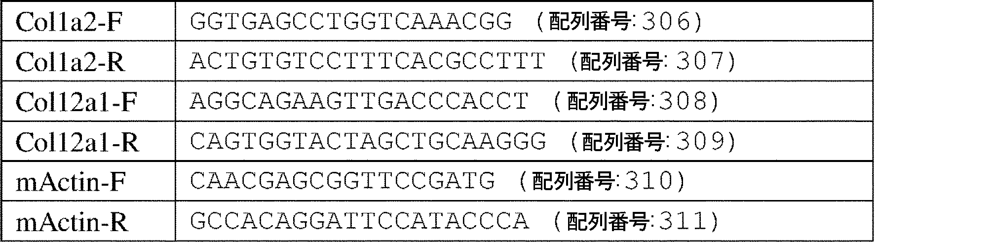
MTT。腫瘍細胞を96ウェルプレートに播種し、分析する前に、指定された期間培養した。回収日に、MTT溶液(3mg/ml)を個々のウェルに加え、プレートを1時間インキュベートした。その後、培地を除去し、紫色の沈殿物を100μlのDMSOによって溶解させた。570nmにおける吸光度を個々のウェルについて測定した。 MTT. Tumor cells were seeded in 96-well plates and cultured for the indicated time periods before analysis. On the day of harvest, MTT solution (3 mg/ml) was added to individual wells and the plates were incubated for 1 hour. After that, the medium was removed and the purple precipitate was dissolved with 100 μl of DMSO. Absorbance at 570 nm was measured for individual wells.
腫瘍細胞の遊走および浸潤。細胞遊走のために、腫瘍細胞を血清なしの培養培地に懸濁させ、次いでトランスウェルチャンバの上に播種した。10%のFBSを有する培地をチャンバの下部に配置した。細胞を12時間培養した後、分析した。 Tumor cell migration and invasion. For cell migration, tumor cells were suspended in serum-free culture medium and then seeded onto transwell chambers. Medium with 10% FBS was placed at the bottom of the chamber. Cells were cultured for 12 hours before analysis.
細胞湿潤のために、マトリゲルマトリックス(Corning、354483)を製造業者の指示に従ってインサートに充填し、37℃で30分間インキュベートした。腫瘍細胞を、下部チャンバ内の10%のFBS含有培地を用いて、インサートの上部チャンバに播種した。次いで、細胞を37℃で20時間インキュベートした。上部チャンバの上側の細胞を穏やかに除去し、上部チャンバの下側の細胞をクリスタルバイオレットで染色し、6つのランダムフィールドを標準顕微鏡で計数した。 For cell wetting, Matrigel matrix (Corning, 354483) was filled into the inserts according to the manufacturer's instructions and incubated at 37°C for 30 minutes. Tumor cells were seeded into the upper chamber of the insert using 10% FBS-containing medium in the lower chamber. Cells were then incubated at 37° C. for 20 hours. Cells above the upper chamber were gently removed, cells below the upper chamber were stained with crystal violet, and 6 random fields were counted with a standard microscope.
マウス処置および腫瘍研究。すべての動物実験は、ジョージ・ワシントン大学の動物愛護および使用機関委員会(the Institutional Animal Care and Use Committee at the George Washington University)によって承認された。8週齢のWT C57BL/6(Jackson Lab、000664)、Rag1-/-(Jackson Lab、002216)またはヌード(Jackson Lab、002019)マウスを、腫瘍研究に使用した。E0771、AT-3およびM-Wnt細胞を、それぞれ接種当たり5×10⁵、2×10⁵、および2×10⁵細胞の用量で、マウス乳腺脂肪パッド中に100μlの体積で注射した。腫瘍体積(0.5×長さ×幅2)を、指示された日にキャリパーを用いて測定した。腫瘍回収時に、腫瘍を計量し、試料を免疫表現型分類およびIHCに使用した。 Mouse treatments and tumor studies. All animal experiments were approved by the Institutional Animal Care and Use Committee at the George Washington University. Eight week old WT C57BL/6 (Jackson Lab, 000664), Rag1 −/− (Jackson Lab, 002216) or nude (Jackson Lab, 002019) mice were used for tumor studies. E0771, AT-3 and M-Wnt cells were injected in a volume of 100 μl into mouse mammary fat pads at doses of 5×10 5 , 2×10 5 and 2×10 5 cells per inoculum, respectively. Tumor volumes (0.5 x length x width2 ) were measured using calipers on the indicated days. At the time of tumor harvest, tumors were weighed and samples were used for immunophenotyping and IHC.
腫瘍移植アッセイのために、まず、腫瘍細胞をRag1-/-マウスに接種した。腫瘍体積がおよそ200~300mm3に達したとき(通常、接種後20日)、60mgの腫瘍オルガノイドをWT C57BL/6マウスに移植した。12日目に、免疫染色のために腫瘍試料を収集した。
For the tumor engraftment assay, tumor cells were first inoculated into Rag1 −/− mice. When tumor volumes reached approximately 200-300 mm 3 (usually 20 days post-inoculation), 60 mg of tumor organoids were implanted into WT C57BL/6 mice. On
腫瘍再チャレンジ実験のために、WT C57BL/6マウスに、まず、鼠径部乳腺脂肪パッドの片側に、50万個のDDR1 KO E0771またはPBS単独を接種した。30日後、同じマウスに乳腺脂肪パッドの両側に50万個のDDR1 WT E0771腫瘍細胞を接種した。腫瘍体積を上述のように測定した。 For tumor rechallenge experiments, WT C57BL/6 mice were first inoculated unilaterally in the inguinal mammary fat pad with 0.5 million DDR1 KO E0771 or PBS alone. Thirty days later, the same mice were inoculated bilaterally in the mammary fat pad with 500,000 DDR1 WT E0771 tumor cells. Tumor volumes were measured as described above.
DDR1抗体をインビボで処置するために、サイズが100mm3より大きくなった後、実験の終了まで、10mg/kgで隔日で、自家製対照IgGおよび抗huDDR1 ECD抗体の両方を腫瘍に局所的に注入した。 To treat DDR1 antibodies in vivo, tumors were injected locally with both home-made control IgG and anti-huDDR1 ECD antibody at 10 mg/kg every other day after they had grown larger than 100 mm 3 in size until the end of the experiment. .
脱細胞化。E0771細胞を、5μmの孔径(Costar、Corning Inc.、3422)を有する挿入物中に、挿入物当たり2,000個の細胞で播種し、DMEM+10%のFBS+1%のPS培地中で2日間培養した。得られたDDR1 WTまたはKO細胞からのECMをPBSで洗浄し、0.5%のTritonX-100および20mMのNH4OHを含有するPBS中で37℃で5分間インキュベーションすることによって脱細胞化した。脱細胞化されたECMをPBSで3回洗浄し、続いて、蒸留水で3回すすぎ、直ちにT細胞遊走実験で使用した。 Decellularization. E0771 cells were seeded at 2,000 cells per insert into inserts with 5 μm pore size (Costar, Corning Inc., 3422) and cultured in DMEM+10% FBS+1% PS medium for 2 days. . The resulting ECM from DDR1 WT or KO cells were washed with PBS and decellularized by incubation in PBS containing 0.5% Triton X-100 and 20 mM NH 4 OH at 37° C. for 5 minutes. . The decellularized ECM was washed three times with PBS followed by three rinses with distilled water and used immediately in T cell migration experiments.
インビトロCD8+T細胞単離および遊走アッセイ。EasySep(商標)マウスCD8+ネガティブ単離キット(Stemcell、19853)を製造者マニュアルに従って使用して、CD8+T細胞を、C57BL/6ナイーブマウスの脾細胞から単離した。CD8+T細胞遊走アッセイを、6.5mmのポリカーボネート膜および5μm孔径のインサート(Costar、Corning Inc.、3422)を用いて行った。50万個の精製されたCD8+T細胞を上部チャンバに添加し、組換えCCL21(100ng/ml、R&Dシステム、4576C025CF)の存在下で、底部チャンバ内の腫瘍条件付け培地とともに37℃で2時間遊走させた。底部チャンバに遊走したCD8+T細胞をフローサイトメトリーによって定量した。huDDR1抗体中和のために、抗体を最初に37℃で条件付け培地と1時間共インキュベートし、次いで、上記の手順に従った。 In vitro CD8 + T cell isolation and migration assay. CD8 + T cells were isolated from splenocytes of C57BL/6 naïve mice using the EasySep™ Mouse CD8 + Negative Isolation Kit (Stemcell, 19853) according to the manufacturer's manual. CD8 + T cell migration assays were performed using 6.5 mm polycarbonate membranes and 5 μm pore size inserts (Costar, Corning Inc., 3422). 500,000 purified CD8 + T cells were added to the top chamber and migrated with tumor-conditioned medium in the bottom chamber for 2 hours at 37° C. in the presence of recombinant CCL21 (100 ng/ml, R&D Systems, 4576C025CF). let me CD8 + T cells that migrated to the bottom chamber were quantified by flow cytometry. For huDDR1 antibody neutralization, antibodies were first co-incubated with conditioned medium at 37° C. for 1 hour, then following the procedure described above.
第二次高調波発生および免疫蛍光。マウス乳房腫瘍組織を埋め込み、-80℃で最適切断温度(OCT)化合物中で保存した。切断前に、試料を少なくとも2時間-20℃とし、20μm厚の切片をクライオスタットを使用して切断した。スライドを解凍し、37℃で30分間インキュベートし、次いで、沸騰抗原マスキング解除溶液(Vector Labs、H-3300)に10分間移した。試料を、CD3e(BD、553057)一次抗体およびAlexa-488(Life Technologies)二次抗体とともにインキュベートした。各腫瘍切片に蛍光G培地(VWR)を加え、顕微鏡のカバースリップ(No1.5)に取り付けた。 Second harmonic generation and immunofluorescence. Mouse mammary tumor tissue was embedded and stored in optimum cutting temperature (OCT) compound at -80°C. Prior to sectioning, samples were brought to −20° C. for at least 2 hours and 20 μm thick sections were cut using a cryostat. Slides were thawed, incubated at 37° C. for 30 minutes, then transferred to boiling antigen unmasking solution (Vector Labs, H-3300) for 10 minutes. Samples were incubated with CD3e (BD, 553057) primary antibody and Alexa-488 (Life Technologies) secondary antibody. Fluorescent G medium (VWR) was added to each tumor section and mounted on a microscope coverslip (No 1.5).
すべての試料を、Leica TCS SP8多光子共焦点顕微鏡を使用して画像化し、20倍、HC PL Apo、NA0.7油浸対物レンズを、実験全体を通して使用した。 All samples were imaged using a Leica TCS SP8 multiphoton confocal microscope and a 20x, HC PL Apo, NA 0.7 oil immersion objective was used throughout the experiment.
840nmまでの励起波長(Erikson et al.,2007)を調整し、420±5nmの狭帯域通過放出フィルタを用いて、コラーゲンのSHGシグナルを検出した。SHG信号は、2つの入射光の光子がコラーゲン繊維の非中心対称構造と相互作用するときに生成され、結果として、入射光子の波長の半分の波長を持つ光子が得られる。LAS Xソフトウェアを使用して、1024×1024ピクセルの画像を取得した。コラーゲン測定は、CT Fireソフトウェア(loci.wisc.edu/software/ctfireで自由に入手可能)を使用して行った。腫瘍縁部分析からのコラーゲンについては、腫瘍の境界から60μmの面積をとった。 The excitation wavelength (Erikson et al., 2007) was adjusted to 840 nm and a narrow bandpass emission filter of 420±5 nm was used to detect collagen SHG signal. The SHG signal is produced when two incident light photons interact with the non-centrosymmetric structure of collagen fibers, resulting in photons with half the wavelength of the incident photons. Images of 1024×1024 pixels were acquired using LAS X software. Collagen measurements were performed using CT Fire software (freely available at loci.wisc.edu/software/ctfire). For collagen from tumor margin analysis, a 60 μm area was taken from the tumor border.
免疫組織化学染色(IHC)。マウス乳房腫瘍組織を10%の緩衝ホルマリン(Fisher Scientific、23-427098)で4℃で一晩固定した。固定腫瘍試料をパラフィンに埋め込み、染色のために4μmの切片に切断した。試料を脱パラフィン化し、PBS中で再水和した。切片を抗原マスキング解除溶液(Vector labs、H-3300)で20分間煮沸し、次いで、PBS中10%の正常ヤギ血清で1時間ブロッキングした。CD8(Biorbyt、orb10325)およびCD4(Sino Biological Inc.、50134-R001)一次抗体を4℃で一晩インキュベートした。一次抗体の検出には、ABCペルオキシダーゼ検出システム(Vector labs、PK-6105)を製造業者の指示に従って使用し、DAB(Vector labs、SK-4105)を基質として使用した。 Immunohistochemical staining (IHC). Mouse mammary tumor tissue was fixed in 10% buffered formalin (Fisher Scientific, 23-427098) overnight at 4°C. Fixed tumor samples were embedded in paraffin and cut into 4 μm sections for staining. Samples were deparaffinized and rehydrated in PBS. Sections were boiled in antigen unmasking solution (Vector labs, H-3300) for 20 minutes and then blocked with 10% normal goat serum in PBS for 1 hour. CD8 (Biorbyt, orb10325) and CD4 (Sino Biological Inc., 50134-R001) primary antibodies were incubated overnight at 4°C. For primary antibody detection, the ABC peroxidase detection system (Vector labs, PK-6105) was used according to the manufacturer's instructions and DAB (Vector labs, SK-4105) was used as substrate.
CD8+T細胞枯渇および養子移入。CD8+T細胞枯渇に関して、C57BL/6マウスに、腫瘍接種の2日前に、200μg/マウスの抗マウスCD8(クローン2.43、BioxCell、BE0061)またはIgG2bアイソタイプ対照(クローンLTF-2、BioxCell、BE0090)を腹腔内投与し、次いで、週2回投与した。 CD8 + T cell depletion and adoptive transfer. For CD8 + T cell depletion, C57BL/6 mice received 200 μg/mouse anti-mouse CD8 (clone 2.43, BioxCell, BE0061) or IgG2b isotype control (clone LTF-2, BioxCell, BE0090) 2 days prior to tumor inoculation. ) was administered intraperitoneally and then twice weekly.
CD8+T細胞の養子移入については、腫瘍接種の17日後、精製されたCD8+T細胞(>90%)を、5×10⁶細胞/マウスの濃度で、E0771乳腺腫瘍を有するRag1-/-マウスに移した。 For adoptive transfer of CD8 + T cells, 17 days after tumor inoculation, purified CD8 + T cells (>90%) were added to E0771 mammary tumor-bearing Rag1 −/− mice at a concentration of 5×10 6 cells/mouse. Moved to mouse.
ピクロシリウスレッド染色。固定された乳房腫瘍試料を調製し、すでに記載されているように切片化した(Sun et al.,2018)。簡潔に述べると、パラフィン包埋された腫瘍組織を4μmのスライドに切断し、ピクロシリウスレッド染色キット(Abcam,カタログ番号番号ab150681)で染色した。切片を脱パラフィン化し、蒸留水中で水和し、ピクロシリウスレッド溶液をスライドに1時間塗布した。次いで、スライドを酢酸溶液中ですすぎ、絶対アルコール中で脱水した。次いで、取り付けられたスライドを標準顕微鏡下で調べ、陽性コラーゲンファイバーシグナルをImageJソフトウェアによって定量化した。 Picrosirius red staining. Fixed breast tumor samples were prepared and sectioned as previously described (Sun et al., 2018). Briefly, paraffin-embedded tumor tissue was cut into 4 μm slides and stained with a picrosirius red staining kit (Abcam, cat#ab150681). Sections were deparaffinized, hydrated in distilled water, and picrosirius red solution was applied to the slides for 1 hour. Slides were then rinsed in acetic acid solution and dehydrated in absolute alcohol. The mounted slides were then examined under a standard microscope and positive collagen fiber signals were quantified by ImageJ software.
ELISA。I型コラーゲンをPBS中で50μg/mlの濃度に希釈し、96ウェルマイクロタイタープレート(50μl/ウェル)に添加した。プレートを密封し、室温で一晩インキュベートし、洗浄緩衝液(R&D、WA126)で3回洗浄し、次いで、200μlの試薬希釈剤(R&D、DY995)で1時間ブロッキングした。3回洗浄した後、100μlの条件付き培地または組換えECD(標準として使用、Sino Biological、10730-H08H)をプレートに室温で2時間添加した。続いて、3回洗浄し、100μlの希釈抗DDR1 N末端抗体(1:500、R&D、AF2396)を添加し、2時間インキュベートした。ビオチン結合抗体との1時間の反応後、1:2000(R&D、893975)の希釈でストレプトアビジン-HRPを各ウェルに添加し、暗所で20分間インキュベートした。100μlの基質溶液(R&D、DY999)を添加し、さらに20分間インキュベートした。50μlのストップ溶液(R&D、DY994)を添加した後、プレートをELISAリーダーにて450nmにて分析した。 ELISA. Collagen type I was diluted in PBS to a concentration of 50 μg/ml and added to 96-well microtiter plates (50 μl/well). Plates were sealed, incubated overnight at room temperature, washed three times with wash buffer (R&D, WA126), then blocked with 200 μl of reagent diluent (R&D, DY995) for 1 hour. After washing three times, 100 μl of conditioned medium or recombinant ECD (used as standard, Sino Biological, 10730-H08H) was added to the plates for 2 hours at room temperature. Subsequently, it was washed three times and 100 μl of diluted anti-DDR1 N-terminal antibody (1:500, R&D, AF2396) was added and incubated for 2 hours. After 1 hour reaction with biotin-conjugated antibody, streptavidin-HRP was added to each well at a dilution of 1:2000 (R&D, 893975) and incubated for 20 minutes in the dark. 100 μl of substrate solution (R&D, DY999) was added and incubated for an additional 20 minutes. After adding 50 μl of stop solution (R&D, DY994), plates were analyzed in an ELISA reader at 450 nm.
フローサイトメトリー。細胞を、PBS中で1:1000の希釈で、暗所で4℃で20分間、Ghost Dye(商標)Violet450(Tonbo Biosciences、13-0863-T100)を使用して生存率について染色し、続いて、PBSで洗浄した。試料を抗CD16/32で1:100の希釈でブロッッキングした(クローン2.4G2、Tonbo Biosciences、70-0161-U100)。抗体を暗所で4℃で30分間インキュベートした。以下の市販抗体を使用した:CD45-BV645(Invitrogen、64-0451-82)、CD3-eflour660(eBiosciences、50-0032-82)、CD4-FITC(eBiosciences、35-0042-U500)、CD8-APC-Cy(商標)7(BD、557654)、CD44-BV786(Biolegend、103059)、CD62L-Pacific Blue(Biolegend、104424)。データをBD FACSCelestaフローサイトメーター上で取得し、FACSDivaまたはFlowJoソフトウェア(BD)によって分析した。 flow cytometry. Cells were stained for viability using Ghost Dye™ Violet 450 (Tonbo Biosciences, 13-0863-T100) at a dilution of 1:1000 in PBS for 20 minutes at 4° C. in the dark followed by , and washed with PBS. Samples were blocked with anti-CD16/32 at a dilution of 1:100 (clone 2.4G2, Tonbo Biosciences, 70-0161-U100). Antibodies were incubated for 30 minutes at 4°C in the dark. The following commercial antibodies were used: CD45-BV645 (Invitrogen, 64-0451-82), CD3-efour660 (eBiosciences, 50-0032-82), CD4-FITC (eBiosciences, 35-0042-U500), CD8-APC. - Cy™7 (BD, 557654), CD44-BV786 (Biolegend, 103059), CD62L-Pacific Blue (Biolegend, 104424). Data were acquired on a BD FACSCelesta flow cytometer and analyzed by FACSDiva or FlowJo software (BD).
走査電子顕微鏡法。WTおよびDDR1 KOの両方のE0771細胞を、0.1×10⁶の密度で播種し、DMEM(10%のFBS)中で2日間培養して、ガラスカバースリップの表面に均等に接着させた。次に、細胞を2.5%のグルタルアルデヒドおよび1%のパラホルムアルデヒド溶液で固定し、続いて、OsO4および水ですすいだ。その後、試料をアルコール勾配で順次脱水し、臨界点で乾燥させた。SEMスタブに取り付けると、カバースリップをイリジウムでコーティングし、ETD検出器を備えた走査型電子顕微鏡(Model FEI)下で、10msの滞留時間および12,000の倍率で観察した。 Scanning electron microscopy. Both WT and DDR1 KO E0771 cells were seeded at a density of 0.1×10 6 and cultured in DMEM (10% FBS) for 2 days to allow even adherence to the surface of glass coverslips. Cells were then fixed with 2.5% glutaraldehyde and 1% paraformaldehyde solution, followed by rinsing with OsO4 and water. The samples were then dehydrated sequentially through an alcohol gradient and dried at the critical point. Once mounted on an SEM stub, coverslips were coated with iridium and viewed under a scanning electron microscope (Model FEI) equipped with an ETD detector at a dwell time of 10 ms and magnification of 12,000.
抗DDR1モノクローナル抗体(mAb)のスクリーニングおよび生成。ヒトDDR1細胞外ドメイン(ECD)タンパク質(Sino Biological、10730-H08H)を使用して、ウサギを免疫化し、前述の方法を使用して抗huDDR1モノクローナル抗体を生成した(Gui et al.,2019b)。簡単に述べると、ニュージーランド白ウサギに、3週間の間隔でプライミングおよびプライミング免疫化後の一連の3~4ブースターのために、0.5mgの組換えヒトDDR1 ECDタンパク質を腹腔内(ip)注射することによって投与した。PBMCからメモリーB細胞を単離し、抗体産生のために、10~14日間、96ウェル細胞培養プレート中で単一B細胞を培養した。ELISAを用いて細胞培養上清をDDR1結合について分析し、抗体遺伝子クローニングおよび配列解析のために陽性ヒットを選択した。 Screening and generation of anti-DDR1 monoclonal antibodies (mAbs). Human DDR1 extracellular domain (ECD) protein (Sino Biological, 10730-H08H) was used to immunize rabbits and anti-huDDR1 monoclonal antibodies were generated using previously described methods (Gui et al., 2019b). Briefly, New Zealand White rabbits are injected intraperitoneally (ip) with 0.5 mg of recombinant human DDR1 ECD protein for priming and a series of 3-4 boosters after the priming immunization at 3-week intervals. It was administered by Memory B cells were isolated from PBMC and single B cells were cultured in 96-well cell culture plates for 10-14 days for antibody production. Cell culture supernatants were analyzed for DDR1 binding using ELISA and positive hits were selected for antibody gene cloning and sequencing.
B細胞培養ウェルからの陽性細胞を溶解し、全RNAを単離し、cDNAを、製造業者の提案に従ってSuperScript逆転写酵素II(Invitrogen)を用いて合成した。重鎖および軽鎖の両方からの抗体可変領域のDNA配列を、設計されたプライマーのセットを使用してポリメラーゼ連鎖反応(PCR)によって増幅し、各抗体の可変領域を配列決定するためのベクターにクローニングした。重鎖および軽鎖の両方のクローニングされた抗体可変配列を、ヒト胚性腎臓(HEK)293(HEK293F)細胞(Life science Technologies)における全長組換え抗体発現のために、それぞれ、IgG1重鎖およびカッパ軽鎖の定常領域と融合して、哺乳動物発現ベクターに構築した。モノクローナル抗体は、以前に記載された方法(O’Donnell et al.,2019)を使用して、プロテイン-A親和性樹脂を使用して95%超の純度までHEK293細胞培養培地から精製された。精製された抗体を、細胞培養アッセイにおける中和機能およびマウス腫瘍モデルにおける抗腫瘍活性についてスクリーニングした。 Positive cells from B-cell culture wells were lysed, total RNA was isolated, and cDNA was synthesized using SuperScript Reverse Transcriptase II (Invitrogen) according to the manufacturer's suggestions. The antibody variable region DNA sequences from both the heavy and light chains are amplified by the polymerase chain reaction (PCR) using a set of designed primers into vectors for sequencing the variable regions of each antibody. cloned. Cloned antibody variable sequences of both heavy and light chains were transformed into IgG1 heavy and kappa chains, respectively, for full-length recombinant antibody expression in human embryonic kidney (HEK) 293 (HEK293F) cells (Life science Technologies). It was fused to the constant region of the light chain and constructed into a mammalian expression vector. Monoclonal antibodies were purified from HEK293 cell culture media to greater than 95% purity using protein-A affinity resin using a previously described method (O'Donnell et al., 2019). Purified antibodies were screened for neutralizing function in cell culture assays and anti-tumor activity in mouse tumor models.
統計。スチューデントt検定を使用して、2群間の平均差を比較した。一元配置分散分析(ANOVA)および事後多重比較を使用して、複数の群間の平均差を比較した。生存曲線をログランク(Mantel-Cox)分析によって解析した。ピアソン相関分析およびその他のすべての統計は、Graphpad Prismで実施した。P<0.05を有意とみなした。データは、平均±SEMとして提示される。 statistics. Student's t-test was used to compare mean differences between two groups. One-way analysis of variance (ANOVA) and post hoc multiple comparisons were used to compare mean differences between multiple groups. Survival curves were analyzed by log-rank (Mantel-Cox) analysis. Pearson correlation analysis and all other statistics were performed in Graphpad Prism. P<0.05 was considered significant. Data are presented as mean±SEM.
実施例2-結果
本発明者らは、基底様/TNBCの特徴を有する複数のマウス乳房腫瘍細胞において、Ddr1を特異的に欠失させた(E0771、AT-3、およびM-Wnt;図1a、図5b~d)。ノックアウト(KO)腫瘍細胞は、インビトロでの細胞増殖、遊走、または浸潤では、いずれの顕著な欠陥も示さなかった(図1b~d、図5e~f)。加えて、免疫不全宿主におけるDdr1-KO腫瘍は、野生型(WT)対照マウスと同じ速度で増殖した(図1eおよび図5g)。全く対照的に、免疫能のある宿主(C57BL/6)におけるDdr1-KO腫瘍は、試験した3つの乳房腫瘍モデル(図1f~h)のすべてで、接種後2週間で完全に退縮した。免疫能のある宿主におけるKO腫瘍のこの成長欠失は、増加した数のKO腫瘍細胞の移植(1接種当たり0.5~20×106個の細胞、図5h)によっては、実質的に緩和されず、また、免疫不全宿主(Rag1-/-)における初期成長およびその後の免疫能のあるナイーブマウスへの再移植によっても、成長不全が救済されなかった(図1i~j)。さらに、同数の親WTおよびKO腫瘍細胞の混合物を使用した同時移植は、免疫能を有する宿主における堅牢な腫瘍増殖を生じさせ(図5i~k)、腫瘍DDR1の優性な作用を示した。WT腫瘍細胞を、同じ乳腺または対側の乳腺のいずれかで、以前にKO腫瘍細胞でチャレンジした免疫能のあるマウスに注射したところ、再チャレンジした親腫瘍の顕著な増殖は観察されなかった(図1k~m)。これは、KO腫瘍細胞がWT腫瘍に対して宿主にワクチン接種可能であることを示す。まとめると、これらのデータは、腫瘍DDR1が免疫能のある宿主における腫瘍増殖において特徴的な役割を果たすことを強く示唆する。
Example 2 - Results We specifically deleted Ddr1 in multiple mouse mammary tumor cells with basal-like/TNBC characteristics (E0771, AT-3, and M-Wnt; Fig. 1a , Figures 5b-d). Knockout (KO) tumor cells did not show any significant defects in cell proliferation, migration, or invasion in vitro (FIGS. 1b-d, 5e-f). In addition, Ddr1-KO tumors in immunodeficient hosts grew at the same rate as wild-type (WT) control mice (FIGS. 1e and 5g). In stark contrast, Ddr1-KO tumors in immunocompetent hosts (C57BL/6) completely regressed two weeks after inoculation in all three breast tumor models tested (FIG. 1f-h). This growth defect of KO tumors in immunocompetent hosts was substantially alleviated by transplantation of increased numbers of KO tumor cells (0.5-20×10 6 cells per inoculum, FIG. 5h). Neither did initial growth in an immunodeficient host (Rag1 −/− ) followed by reimplantation into immunocompetent naïve mice rescue growth failure (FIGS. 1i-j). Moreover, co-implantation using a mixture of equal numbers of parental WT and KO tumor cells resulted in robust tumor growth in immunocompetent hosts (Fig. 5i-k), indicating a dominant effect of tumor DDR1. When WT tumor cells were injected in either the same mammary gland or the contralateral mammary gland into immunocompetent mice previously challenged with KO tumor cells, no significant growth of re-challenged parental tumors was observed ( 1k-m). This indicates that KO tumor cells can vaccinate the host against WT tumors. Taken together, these data strongly suggest that tumor DDR1 plays a characteristic role in tumor growth in immunocompetent hosts.
免疫組織化学(IHC)は、CD4+およびCD8+T細胞が、免疫不全宿主から免疫能のある宿主に再移植した後、親の腫瘍の末梢領域に限定されることを示した(図2a)。対照的に、これらのリンパ球は、Ddr1-KO腫瘍の縁部および中心部の両方に豊富に存在した(図2a)。裏付けとして、フローサイトメトリーは、腫瘍浸潤CD8+およびCD4+T細胞の総数(腫瘍重量で正規化)が、KO腫瘍群では、親対照と比較して実質的に上昇したことを示した(図2b~c)。インターフェロン(IFN)-γ-産生CD8+およびCD4+細胞はまた、WT対応物よりもKO腫瘍でより豊富であった(図2d~e)。さらに、エフェクターおよびヘルパーT細胞は、それらの親対照と比較して、KO腫瘍においてより強力に活性化された(CD44高CD62L低)(図2f~g)。しかしながら、対応する総T細胞数で正規化した場合、KOおよび親対照は、Ki67(図6a~b)、IFNγ、またはGzmb(図2c~d)に対して陽性であるCD4+またはCD8+T細胞のパーセンテージに差異を示さなかった。これは、腫瘍DDR1が、T細胞の増殖または抗腫瘍活性自体を減衰させることなく、T細胞の腫瘍除外を付与する可能性が高いことを示す。 Immunohistochemistry (IHC) showed that CD4 + and CD8 + T cells were restricted to peripheral regions of the parental tumor after reimplantation from immunocompromised to immunocompetent hosts (Fig. 2a). . In contrast, these lymphocytes were abundant in both the margins and the center of Ddr1-KO tumors (Fig. 2a). In support, flow cytometry showed that the total number of tumor-infiltrating CD8 + and CD4 + T cells (normalized by tumor weight) was substantially elevated in the KO tumor group compared to parental controls (Fig. 2b-c). Interferon (IFN)-γ-producing CD8 + and CD4 + cells were also more abundant in KO tumors than in WT counterparts (Fig. 2d-e). Moreover, effector and helper T cells were more strongly activated (CD44 high CD62L low ) in KO tumors compared to their parental controls (Fig. 2f-g). However, when normalized by the corresponding total T cell numbers, KO and parental controls showed CD4 + or CD8 + T positive for Ki67 (Fig. 6a-b), IFNγ, or Gzmb (Fig. 2c-d). It showed no difference in the percentage of cells. This indicates that tumor DDR1 likely confers tumor exclusion of T cells without attenuating T cell proliferation or anti-tumor activity per se.
腫瘍DDR1が抗腫瘍免疫に拮抗する役割を確かめるために、本発明者らは、抗体中和によって免疫能のあるマウスのCD8+T細胞を枯渇させた。Ddr1-KO腫瘍は、免疫不全宿主(図1e)における発明者の知見と同様に、CD8+細胞枯渇宿主(図2h~j、図2e~f)におけるアイソジェニックWT対照と同様に堅牢に成長した。相互実験では、本発明者らは、精製されたCD8+T細胞を免疫不全マウスに養子移入し、親およびKO腫瘍の成長を比較した。偽処置した免疫不全宿主とは異なり、移植されたCD8+T細胞を有するマウスは、親腫瘍よりも有意に小さいKO腫瘍を産生した(図2k~m、図6g)。集合的に、これらの結果は、乳房腫瘍の固有の成長のために必ずしも必要ではないが、腫瘍DDR1がT細胞浸潤の抑止において重要な役割を果たすという概念を裏付けている。 To confirm the role of tumor DDR1 in antagonizing anti-tumor immunity, we depleted CD8 + T cells in immunocompetent mice by antibody neutralization. Ddr1-KO tumors grew as robustly as isogenic WT controls in CD8 + cell-depleted hosts (Fig. 2h-j, Fig. 2e-f), similar to our findings in immunocompromised hosts (Fig. 1e). . In a reciprocal experiment, we adoptively transferred purified CD8 + T cells into immunodeficient mice and compared the growth of parental and KO tumors. Unlike sham-operated immunocompromised hosts, mice with engrafted CD8 + T cells produced KO tumors that were significantly smaller than parental tumors (Fig. 2k-m, Fig. 6g). Collectively, these results support the notion that tumor DDR1 plays an important role in suppressing T-cell infiltration, although it is not necessary for the intrinsic growth of breast tumors.
T細胞の腫瘍内輸送は、血管を通る血管外遊出、腫瘍誘発性化学遊走、およびECMに基づく物理バリアを通過することを含む、非常に動的なマルチステッププロセスである(Slaney et al.,2014、Sackstein et al.,2017、Ager et al.,2016)。CD31に基づく組織学的解析では、WT腫瘍とKO腫瘍との間の顕著な免疫血管変化は明らかにされず(データは示さず)、またRNA-seqでは、Ddr1-KO群における増強された免疫浸潤を説明できるT細胞ホーミング遺伝子またはケモカインコーディング遺伝子のmRNAレベルの差異も示されなかった(図6h~i)。コラーゲンに対するDDR1の親和性を考慮すると、本発明者らは、腫瘍DDR1-コラーゲン相互作用により、腫瘍が免疫細胞によって浸透しにくくなる代替モデルを試験しようとした。この目的のために、本発明者らはまず、Ddr1-KO腫瘍細胞に以下の構築物を導入することによって、DDR1の免疫調節機能がそのキナーゼ活性に依存するかどうかを決定した(図3a):(1)空のベクター(EV)、(2)全長マウスDDR1(FL)、(3)ΔKD、細胞内キナーゼドメインKDを欠くが、その膜貫通(TM)ドメインを保持する切断型DDR1、および(4)ECDのみ。免疫能のある宿主におけるKO腫瘍の成長欠損は、異所的に発現したFL DDR1および2つの切断バリアント、ΔKDおよびECDによって同様の程度に救済された(図3a)。さらなる欠失分析は、コラーゲン結合に関与するN末端ジスコイジンホモロジードメイン(DS、図3b)が、DDR1 KO細胞の腫瘍成長を完全に救済したことを示す(図7a~b)。総合すると、これらのデータは、腫瘍DDR1がキナーゼ非依存的な方法で抗腫瘍免疫を抑制することを明確に示す。 Intratumoral trafficking of T cells is a highly dynamic, multi-step process involving extravasation through blood vessels, tumor-induced chemomigration, and crossing ECM-based physical barriers (Slaney et al., 2014, Sackstein et al., 2017, Ager et al., 2016). CD31-based histological analysis revealed no significant immunovascular changes between WT and KO tumors (data not shown), and RNA-seq revealed enhanced immunity in the Ddr1-KO group. No differences in mRNA levels of T-cell homing genes or chemokine-encoding genes that could explain invasion were also shown (Fig. 6h-i). Given the affinity of DDR1 for collagen, we sought to test an alternative model in which tumor DDR1-collagen interactions render tumors less permeable to immune cells. To this end, we first determined whether the immunoregulatory function of DDR1 depends on its kinase activity by introducing the following constructs into Ddr1-KO tumor cells (Fig. 3a): (1) empty vector (EV), (2) full-length mouse DDR1 (FL), (3) ΔKD, a truncated DDR1 that lacks the intracellular kinase domain KD but retains its transmembrane (TM) domain, and ( 4) ECD only. The growth defect of KO tumors in immunocompetent hosts was rescued to a similar extent by ectopically expressed FL DDR1 and two truncation variants, ΔKD and ECD (Fig. 3a). Further deletion analysis shows that the N-terminal discoidin homology domain (DS, Fig. 3b) involved in collagen binding completely rescued tumor growth of DDR1 KO cells (Fig. 7a-b). Taken together, these data clearly demonstrate that tumor DDR1 suppresses anti-tumor immunity in a kinase-independent manner.
コラーゲン-DSドメイン結晶構造(Letinger,2014、Carafoli et al.,2012、Carafoli&Hohenester,2013)に基づいて、本発明者らは、ヒトDDR1におけるW53、T57、D70、K112、E113およびS175に対応するマウスDDR1における多数の主要なコラーゲン結合アミノ酸残基(W54、T58、D71、K113、E114およびS176、図3b)を変異させた。本発明者らはまた、コラーゲン結合ポケットの遠位にあり、DDR1膜貫通シグナル伝達を担う残基であるR33を変異させた。WTおよび変異ECDのコラーゲン結合親和性を、ELISAおよび共IPによってインビトロで検証し(図3c、図7c)、それらのKO腫瘍成長を救出する能力をインビボで評価した(図3d~k)。予想通り、ECDのコラーゲン結合領域の外側に位置するR33Aは、コラーゲンに結合し、腫瘍成長を支持する能力を保持した(WT ECDについては10/10腫瘍、およびR33Aについては6/6、図3c~e)。対照的に、W54Aは、インビトロでのコラーゲン結合および腫瘍成長を支持する能力を完全に失った(0/6部位、図3f)。変異D71A、K113A、E114A、およびS176Aは、WT ECDの中程度のコラーゲン結合活性を保持し(図3c)、一部の宿主において腫瘍成長をもたらした(それぞれ、2/6、1/6、3/6、および1/6腫瘍、図3h~k)。一方で、T58Aは、WTコラーゲン結合親和性のおよそ50%(図3c)および100%の腫瘍発生率(6/6)を保持したが、WTよりも遅い腫瘍成長率を示した(図3g)。本発明者らは、ECD変異体のコラーゲン結合親和性および腫瘍救出能力の強い相関関係に照らして、ECDが抗腫瘍免疫を阻害するためには、コラーゲン結合が必要であると結論づける。 Based on the collagen-DS domain crystal structure (Letinger, 2014, Carafoli et al., 2012, Carafoli & Hohenester, 2013), we found mouse A number of key collagen-binding amino acid residues in DDR1 (W54, T58, D71, K113, E114 and S176, Fig. 3b) were mutated. We also mutated R33, a residue distal to the collagen binding pocket and responsible for DDR1 transmembrane signaling. Collagen-binding affinities of WT and mutant ECDs were verified in vitro by ELISA and co-IP (Fig. 3c, Fig. 7c), and their ability to rescue KO tumor growth was evaluated in vivo (Fig. 3d-k). As expected, R33A located outside the collagen binding region of the ECD retained the ability to bind collagen and support tumor growth (10/10 tumors for WT ECD and 6/6 for R33A, Fig. 3c ~e). In contrast, W54A completely lost the ability to support collagen binding and tumor growth in vitro (0/6 sites, Fig. 3f). Mutations D71A, K113A, E114A, and S176A retained moderate collagen-binding activity of WT ECDs (Fig. 3c) and resulted in tumor growth in some hosts (2/6, 1/6, 3, respectively). /6, and 1/6 tumors, Fig. 3h-k). On the other hand, T58A retained approximately 50% of WT collagen binding affinity (Fig. 3c) and 100% tumor incidence (6/6), but showed slower tumor growth than WT (Fig. 3g). . In light of the strong correlation between collagen binding affinity and tumor rescue capacity of ECD mutants, we conclude that collagen binding is required for ECD to inhibit anti-tumor immunity.
異所性DDR1 ECD単独で免疫能のある宿主の腫瘍成長を支持するのに十分であるという事実は、完全長DDR1からのECD脱落の以前の報告を彷彿とさせる(Vogel,2020、Flynn et al.,2010、Shitomi et al.,2015)が、脱落したECDの生物学的有意性は知られていなかった。裏付けとして、発明者らは、親マウス乳房腫瘍細胞およびヒト乳がん細胞株のサブセットで条件付けられた培地中のECDを検出した(図7d)。インビトロ生化学研究で発表された研究では、組換えDDR1 ECDがコラーゲン繊維構造をリモデリングすることができることが示されている(Flynn et al.,2010、Agarwal et al.,2007)。裏付けとして、走査電子顕微鏡法(SEM)は、インビトロで培養されたDDR1-WT腫瘍細胞が、それらのKO対応物と比較して、より顕著なECMネットワークと関連していることを示した(図3l)。腫瘍細胞関連ECMの機能性を決定するために、本発明者らは、トランスウェルアッセイでは、親およびKO腫瘍細胞を脱細胞化し(Chen et al.,2007)(Chen et al.,2007)、脱細胞化ECMを通して精製されたCD8+T細胞の浸透度を評価した(図3m)。親腫瘍由来ECMは、Ddr1-KO腫瘍から脱細胞化されたECMにおいて軽減されたT細胞の遊走を有意に減少させた(図3m)。T細胞妨害効果は、ECD発現KO腫瘍に由来するECMの親レベルまで回復した(図3m)。インビトロ所見を裏付けるために、本発明者らは、免疫不全宿主から免疫能のある宿主に再移植された親およびDdr1-KO腫瘍におけるコラーゲン繊維を直接可視化するために、第2の高調波発生(SHG)顕微鏡を使用した。親腫瘍の縁部にあるコラーゲン繊維は、腫瘍縁部に平行に配向する傾向があり(ブロック矢印で示される、図3nの上部パネル)、侵入する腫瘍細胞に対する防御ラインを想起させる。予想通り、同じ親腫瘍からのCD3+T細胞は、腫瘍縁部に限定された(図3n)。全く対照的に、KO腫瘍中のコラーゲン繊維は比較的短く、無秩序であった(図3n)。一貫して、免疫細胞は、WT腫瘍と比較して、KO腫瘍の中により深く浸透した(図3n)。繊維アラインメントの測定値として、コラーゲン繊維の角度の変動係数は、親腫瘍に対して、KO腫瘍の方が有意に大きかった(図3o)。加えて、KO腫瘍における平均コラーゲン繊維長は、それらの親対応物よりも実質的に短かった(図3p)。DDR1依存性コラーゲン繊維リモデリングを支持するために、初代Ddr1-WTおよびKO腫瘍からのRNA-seqデータの経路分析は、細胞外マトリックスおよびコラーゲン原線維組織に関与する遺伝子が最も影響を受けることを示す(p<1×10-12、図7e)。併せて、これらのデータは、DDR1 ECDが、抗腫瘍免疫細胞の腫瘍微小環境への浸透を妨げるコラーゲンに基づく物理的バリアを強化するのに役立つことを強く示唆する。 The fact that ectopic DDR1 ECD alone is sufficient to support tumor growth in immunocompetent hosts is reminiscent of previous reports of ECD shedding from full-length DDR1 (Vogel, 2020, Flynn et al. ., 2010, Shitomi et al., 2015), but the biological significance of dropped ECD was unknown. In support, we detected ECD in media conditioned on parental mouse breast tumor cells and a subset of human breast cancer cell lines (Fig. 7d). Studies published in in vitro biochemical studies have shown that recombinant DDR1 ECD can remodel collagen fiber architecture (Flynn et al., 2010; Agarwal et al., 2007). In support, scanning electron microscopy (SEM) showed that in vitro cultured DDR1-WT tumor cells were associated with a more prominent ECM network compared to their KO counterparts (Fig. 3l). To determine the functionality of tumor cell-associated ECM, we decellularized parental and KO tumor cells (Chen et al., 2007) (Chen et al., 2007), Penetrance of CD8 + T cells purified through decellularized ECM was assessed (Fig. 3m). Parental tumor-derived ECM significantly reduced T cell migration that was attenuated in ECM decellularized from Ddr1-KO tumors (Fig. 3m). The T-cell blocking effect was restored to parental levels in ECM derived from ECD-expressing KO tumors (Fig. 3m). To corroborate the in vitro findings, we used second harmonic generation ( SHG) microscope was used. Collagen fibers at the parental tumor margin tend to be oriented parallel to the tumor margin (indicated by block arrows, Fig. 3n upper panel), reminiscent of a line of defense against invading tumor cells. As expected, CD3 + T cells from the same parental tumor were restricted to the tumor margin (Fig. 3n). In stark contrast, collagen fibers in KO tumors were relatively short and disordered (Fig. 3n). Consistently, immune cells penetrated more deeply into KO tumors compared to WT tumors (Fig. 3n). As a measure of fiber alignment, the angular coefficient of variation of collagen fibers was significantly greater in KO tumors relative to parental tumors (Fig. 3o). In addition, the average collagen fiber length in KO tumors was substantially shorter than their parental counterparts (Fig. 3p). To support DDR1-dependent collagen fibrillar remodeling, pathway analysis of RNA-seq data from primary Ddr1-WT and KO tumors showed that genes involved in extracellular matrix and collagen fibrillar organization were most affected. (p<1×10 −12 , FIG. 7e). Together, these data strongly suggest that the DDR1 ECD helps strengthen the collagen-based physical barrier that prevents the penetration of anti-tumor immune cells into the tumor microenvironment.
DDR1のすべての現在の小分子阻害剤は、その治療電位が細胞内キナーゼドメインを標的とするかどうかを調べた(Kothiwale et al.,2015、Li et al.,2015)。免疫細胞浸潤を遮断するキナーゼ非依存性ECD活性を効果的に中和するために、本発明者らは、以前に記載されているように、組換えヒトDDR1(huDDR1)ECDおよびその後の抗体記憶B細胞単離によるウサギの免疫化によって、一連のモノクローナル抗体クローンを作製した(Meng et al.,2015、Gui et al.,2019a)。huDDR1特異的中和抗体をスクリーニングするために、本発明者らは、マウスDdr1-KO乳房腫瘍細胞においてhuDDR1を異所的に発現した(図4a)。huDDR1は、免疫能のある宿主におけるKOマウス腫瘍の成長欠損を完全に救出した(図4b、c)。T細胞遊走に対する腫瘍細胞の効果を評価するインビトロ共培養アッセイ(図8a)を使用して、本発明者らは、T細胞遊走のhuDDR1依存性干渉を効果的に中和することができる抗huDDR1抗体をスクリーニングした(図8b)。いくつかの上位の中和抗体を、その腫瘍阻害ポテンシャルについてインビボでさらに調べた。IgGアイソタイプ対照と比較して、抗huDDR1抗体の投与は、huDDR1発現腫瘍の成長を著しく遅くし、腫瘍の発生率を低下させ、宿主の体重に明らかな影響を全く与えることなく宿主の生存期間を延長させた(図4d~f、図8c~d)。注目すべきことに、免疫不全宿主における同じ抗体投与は、任意の明らかな腫瘍阻害をもたらさず(図8e)、治療が主にDDR1の免疫除外機能を中和することを示唆した。SHG顕微鏡検査は、免疫能のある宿主における抗huDDR1抗体処置腫瘍が、アラインメントのより低い、より短いコラーゲン繊維、および有意に増強された抗腫瘍免疫浸潤と関連していたことを示す(図4g~i、図8f)。総合すると、本発明者らの知見は、潜在的な抗腫瘍療法としての抗hDDR1抗体アプローチの原理の証明を提供する。 All current small-molecule inhibitors of DDR1 were investigated whether their therapeutic potential targets the intracellular kinase domain (Kothiwale et al., 2015, Li et al., 2015). To effectively neutralize kinase-independent ECD activity that blocks immune cell infiltration, we used recombinant human DDR1 (huDDR1) ECD and subsequent antibody memory, as previously described. A series of monoclonal antibody clones were generated by immunization of rabbits with B cell isolation (Meng et al., 2015, Gui et al., 2019a). To screen for huDDR1-specific neutralizing antibodies, we ectopically expressed huDDR1 in mouse Ddr1-KO breast tumor cells (Fig. 4a). huDDR1 completely rescued the growth defect of KO mouse tumors in immunocompetent hosts (Fig. 4b,c). Using an in vitro co-culture assay (Fig. 8a) that evaluates the effect of tumor cells on T-cell migration, we found that anti-huDDR1, which can effectively neutralize huDDR1-dependent interference of T-cell migration Antibodies were screened (Fig. 8b). Several top neutralizing antibodies were further investigated in vivo for their tumor inhibitory potential. Compared to IgG isotype controls, administration of anti-huDDR1 antibodies significantly slowed the growth of huDDR1-expressing tumors, reduced tumor incidence, and increased host survival without any appreciable effect on host body weight. lengthened (Figs. 4d-f, Figs. 8c-d). Remarkably, administration of the same antibody in immunodeficient hosts did not result in any apparent tumor inhibition (Fig. 8e), suggesting that treatment primarily neutralized the immune-exclusion function of DDR1. SHG microscopy shows that anti-huDDR1 antibody-treated tumors in immunocompetent hosts were associated with less aligned, shorter collagen fibers and significantly enhanced anti-tumor immune infiltration (Fig. 4g- i, Fig. 8f). Taken together, our findings provide proof of principle for the anti-hDDR1 antibody approach as a potential anti-tumor therapy.
腫瘍-免疫相関の臨床的影響を推定するための公開のバイオインフォマティクスリソースであるTCGA RNA-seqデータセットおよびTIMER(Li et al.,2018)を使用して、本発明者らは、乳がんが、DDR1 mRNAレベルと、CD8、IFNG、GZMB、およびPRF1を含む様々な抗腫瘍免疫シグネチャー遺伝子との間で逆相関を示すことを見出した(図4j、図8g~i)。注目すべきことに、DDR1について観察される負の相関の程度は、免疫調節機能を有する他の最近特定された腫瘍関連遺伝子と同等である(Pan et al.,2018)。免疫除外表現型に基づいてTNBCを層別化するために使用された、治療未経験のTNBC患者(n=37、図4k~m)からの試料の別個のコホートにおいて、同じ相関が、観察された(Grusso et al.,2019)。まとめると、これらの臨床的相関は、高いDDR1発現が、CD8+T細胞の低い腫瘍浸潤および低い細胞傷害活性と関連することを強く示唆する。本研究は、免疫除外のための防御ラインを誘発することにおける、以前に探索されていない、キナーゼ非依存性のDDR1機能を明らかにすることにより(図4n)、ECMリモデリングおよび抗腫瘍免疫の遮断におけるDDR1活性を標的とする新規の単独治療薬の開発を提供する。本発明者らはまた、乳がんおよび膵臓がん等の他の線維性がん型に対する現在の免疫療法の臨床転帰および有効性の改善に役立つことができることを想定している。 Using the TCGA RNA-seq dataset and TIMER (Li et al., 2018), a public bioinformatics resource for estimating the clinical impact of tumor-immune correlations, we found that breast cancer We found an inverse correlation between DDR1 mRNA levels and various anti-tumor immune signature genes, including CD8, IFNG, GZMB, and PRF1 (Fig. 4j, Fig. 8g-i). Notably, the degree of negative correlation observed for DDR1 is comparable to other recently identified tumor-associated genes with immunomodulatory functions (Pan et al., 2018). The same correlation was observed in a separate cohort of samples from treatment-naïve TNBC patients (n=37, Figures 4k-m) used to stratify TNBC based on immune exclusion phenotype. (Grusso et al., 2019). Taken together, these clinical correlations strongly suggest that high DDR1 expression is associated with low tumor infiltration and low cytotoxic activity of CD8 + T cells. The present study reveals a previously unexplored, kinase-independent DDR1 function in triggering a line of defense for immune exclusion (Fig. 4n), thus enhancing ECM remodeling and anti-tumor immunity. We provide the development of novel monotherapeutic agents that target DDR1 activity in blockade. We also envision that we can help improve the clinical outcomes and efficacy of current immunotherapies against other fibrotic cancer types such as breast and pancreatic cancer.
ヒトDDR1を標的とするモノクローナル抗体の生成およびクローニング。DDR1に対するモノクローナル抗体(mAb)は、ウサギの免疫化および単一B細胞からの抗体の単離によって生成された。ヒトDDR1タンパク質を抗体生成に使用し、HEK293細胞で発現させた。タンパク質は6XHISタグを有し、Ni-NTA樹脂(Sino Biologicals)を使用して95%を超える純度になるまで精製される。ウサギ(NZW、Charles River)を、プライミング一次免疫化後の3回のブースト注射による標準免疫化手順を使用して、組換え産生されたDDR1で免疫化した。抗DDR1血清の力価を、96ウェルプレート(max-sorbプレート、Nunc)上にコーティングされたDDR1タンパク質に結合するためのELISA中の血清の一連の希釈によって決定した。血清価が>106に達し、蛍光補助細胞選別(FACS)器具(BD FACSAria(商標)III、BD Biosciences)を使用して、新たに調製した末梢血単核細胞(PBMC)からのB細胞単離のために、免疫化されたウサギから末梢血試料を収集した。選別した単一B細胞を96ウェル細胞培養プレート(Fisher Scientific)に収集し、5%のCO2および95%の湿度を有する細胞培養インキュベーターで、10%のFBSおよびサイトカインを添加したRPMI培地中で、7~10日間培養した。培養上清中の抗体をDDR1結合についてアッセイした。培養ウェルからの陽性細胞を溶解し、全RNAを単離し、cDNAを、製造業者の提案に従ってSuperScript逆転写酵素II(Invitrogen)を用いて合成した。重鎖および軽鎖の両方からの抗体可変領域のDNA配列を、設計されたプライマーのセットを使用してポリメラーゼ連鎖反応(PCR)によって増幅し、各抗体の可変領域を配列決定するためのベクターにクローニングした。抗体可変領域のアミノ酸およびDNA配列は、それぞれ表3および4、ならびに表8および9に列挙されている。抗DDR1モノクローナル抗体の軽鎖および重鎖CDRのアミノ酸配列は、それぞれ表1および2に列挙されている。
Generation and Cloning of Monoclonal Antibodies Targeting Human DDR1. Monoclonal antibodies (mAbs) against DDR1 were generated by immunization of rabbits and isolation of antibodies from single B cells. Human DDR1 protein was used for antibody generation and expressed in HEK293 cells. The protein has a 6XHIS tag and is purified to greater than 95% purity using Ni-NTA resin (Sino Biologicals). Rabbits (Charles River, NZW) were immunized with recombinantly produced DDR1 using a standard immunization procedure with three boost injections after a priming primary immunization. Anti-DDR1 serum titers were determined by serial dilutions of sera in an ELISA for binding to DDR1 protein coated on 96-well plates (max-sorb plates, Nunc). B cell mononuclear cells from freshly prepared peripheral blood mononuclear cells (PBMC) with serum titers reaching >10 6 were isolated using a fluorescence-assisted cell sorting (FACS) instrument (BD FACSAria™ III, BD Biosciences). Peripheral blood samples were collected from immunized rabbits for isolation. Sorted single B cells were collected in 96-well cell culture plates (Fisher Scientific) in RPMI medium supplemented with 10% FBS and cytokines in a cell culture incubator with 5
選択されたDDR1結合ヒットを、ヒト胚腎臓(HEK293)細胞(Invitrogen)における哺乳動物発現ベクター系を使用して、完全長ヒトIgGまたはウサギ/ヒトキメラIgGとして発現させた。抗体を、高速タンパク質液体クロマトグラフィー(FPLC)によって、プロテイン-A親和性樹脂を使用して精製した。精製されたDDR1結合抗体を、それらの生物学的特性について特徴付けた。 Selected DDR1 binding hits were expressed as full-length human IgG or rabbit/human chimeric IgG using a mammalian expression vector system in human embryonic kidney (HEK293) cells (Invitrogen). Antibodies were purified by fast protein liquid chromatography (FPLC) using protein-A affinity resin. Purified DDR1-binding antibodies were characterized for their biological properties.
バイオレイヤー干渉法(BLI)センサーに基づくOctet器具を使用して決定された抗DDR1モノクローナル抗体の結合親和性。抗体親和性測定のために、抗体(30μg/mL)をプロテインAバイオセンサーに4分間充填した。動態緩衝液中での短いベースラインに引き続き、充填されたバイオセンサーを、0.1~200nMの一連の組換えDDR1タンパク質に曝露し、バックグラウンドを減算して、センサーのドリフトを補正した。すべての実験は、1,000rpmで振盪しながら行った。バックグラウンドの波長シフトを、抗体のみを充填した参照バイオセンサーから測定した。各抗体の動態センサーグラムを図10A~Bに示す。ForteBioのデータ分析ソフトウェアを使用して、会合速度および解離速度を抽出するために、データを1:1の結合モデルに適合させた。KDは、表13中のDDR1 mAbについて、kオフ/kオンの比およびKDの推定値を使用して計算した。 Binding affinities of anti-DDR1 monoclonal antibodies determined using the Biolayer Interferometry (BLI) sensor-based Octet instrument. For antibody affinity measurements, antibody (30 μg/mL) was loaded onto the protein A biosensor for 4 minutes. Following a short baseline in kinetic buffer, the loaded biosensor was exposed to a series of recombinant DDR1 proteins from 0.1 to 200 nM and background subtracted to correct for sensor drift. All experiments were performed with shaking at 1,000 rpm. Background wavelength shift was measured from a reference biosensor loaded with antibody only. Kinetic sensorgrams for each antibody are shown in Figures 10A-B. Data were fitted to a 1:1 binding model to extract association and dissociation rates using ForteBio's data analysis software. The KD was calculated for the DDR1 mAbs in Table 13 using the ratio of koff / kon and an estimate of the KD .
DDR1 mAbのエピトープビニング。抗DDR1 mAb間の対結合競合を使用して、Octet器具およびプロテインAバイオセンサーを使用して、各mAbの結合エピトープを決定した。エピトープビンを、表14に要約する。 Epitope binning of DDR1 mAbs. Paired binding competition between anti-DDR1 mAbs was used to determine the binding epitope of each mAb using the Octet instrument and protein A biosensor. Epitope bins are summarized in Table 14.
抗hECD抗体は、自発的な腫瘍成長を阻害する:様々な段階での乳がん発生に対する抗DDR1抗体治療の効果を実証するために、MMTV-PyMTマウス(C57BL/6系統のバックグラウンド)を、平均腫瘍サイズが100mm3に達したときに(「腫瘍後」)、対照IgGまたは抗DDR1抗体で2週間処置した。DDR1#9ヒト化抗体は、マウスおよびヒトECDの両方に対するその高い親和性のために、本研究のために選択された(データ図示せず)。抗体処理は、宿主の体重に影響を及ぼさなかった。しかしながら、それは、腫瘍体積(図12a)および腫瘍発生率(図12b)を有意に減少させた。一貫して、抗体処置は、コラーゲンのアラインメントを著しく破壊し、大規模な免疫細胞浸潤を促進した(図12c)。異なるバックグラウンドの株(FVB)のMMTV-PyMTマウスにおいても、様々な段階での抗DDR1抗体処置および乳房腫瘍形成を用いて、同様の結果を得た。
Anti-hECD Antibodies Inhibit Spontaneous Tumor Growth: To demonstrate the effect of anti-DDR1 antibody treatment on breast cancer development at various stages, MMTV-PyMT mice (C57BL/6 strain background) were When tumor size reached 100 mm 3 (“post-tumor”), they were treated with control IgG or anti-DDR1 antibody for 2 weeks. The
抗hECD抗体およびDDR1キナーゼ阻害剤の比較:抗hECD抗体が顕著な腫瘍阻害活性を示した同じ乳房腫瘍モデルでは、以前に発表された小分子DDR1キナーゼ阻害剤、7rhは、腫瘍におけるDDR1のチロシンキナーゼ活性を阻害したが、腫瘍成長を低下させなかった。これは、抗腫瘍免疫のDDR1依存的排除がそのキナーゼ活性から独立しているという主張と一致する(図13a~e)。 Comparison of anti-hECD antibodies and DDR1 kinase inhibitors: In the same breast tumor model in which anti-hECD antibodies exhibited significant tumor inhibitory activity, a previously published small-molecule DDR1 kinase inhibitor, 7rh, inhibited the tyrosine kinase of DDR1 in tumors. inhibited activity but did not reduce tumor growth. This is consistent with the claim that DDR1-dependent elimination of anti-tumor immunity is independent of its kinase activity (Fig. 13a-e).
DDR1は、いくつかの腫瘍タイプにおける成長に必要である:腫瘍Ddr1のCRISPRに基づく遺伝子切除を用いて、ID8aggの卵巣腫瘍を有する免疫担当宿主の生存を有意に増加させた。しかし、B16黒色腫またはMC38結腸直腸腫瘍におけるDdr1 KOは、同系免疫担当宿主における腫瘍増殖に影響を及ぼさなかった(図14b~cを参照されたい)。本研究は、乳房腫瘍で得られた知見と組み合わせて、DDR1を標的とすることにより、複数のがんタイプにおいて腫瘍阻害をもたらすことができることを示す。 DDR1 is required for growth in several tumor types: CRISPR-based gene ablation of tumor Ddr1 was used to significantly increase survival of immunocompetent hosts bearing ID8agg ovarian tumors. However, Ddr1 KO in B16 melanoma or MC38 colorectal tumors had no effect on tumor growth in syngeneic immunocompetent hosts (see Figures 14b-c). This study, combined with findings in breast tumors, shows that targeting DDR1 can result in tumor inhibition in multiple cancer types.
DDR1および抗腫瘍免疫マーカーの相関:TCGAトランスクリプトームデータセットをマイニングすることにより、対応する正常組織と比較して、複数のヒトがんにおけるDDR1の異常に高い発現が示された(データ図示せず)。さらに、複数のがんにおけるDDR1 mRNAレベルは、グランザイムB(GZMB、図15aを参照されたい)などの細胞傷害性免疫マーカーと負の相関関係にあり、これは、DDR1が複数のがんタイプにおいて抗腫瘍免疫を拮抗し得ることを示唆する。さらに、TCGAプロテオームデータセットの分析は、DDR1タンパク質とCD8との間(図15b)、ならびにDDR1と抗腫瘍免疫の細胞溶解性エフェクター経路に関与するタンパク質との間(図15c)に負の相関をもたらした。したがって、mRNAおよびタンパク質レベルの両方では、DDR1の高発現は、低レベルの抗腫瘍免疫マーカー発現と相関する。DDR1は、抗腫瘍免疫の拮抗剤として機能するように見えるので、例えば、DDR1抗体を使用した、この拮抗剤の阻害は、複数のがんタイプにおける抗腫瘍免疫を増加させる可能性がある。 Correlation of DDR1 and anti-tumor immune markers: Mining the TCGA transcriptome dataset showed abnormally high expression of DDR1 in multiple human cancers compared to matched normal tissues (data not shown). figure). Furthermore, DDR1 mRNA levels in multiple cancers were negatively correlated with cytotoxic immune markers such as granzyme B (GZMB, see Figure 15a), indicating that DDR1 It suggests that it can antagonize anti-tumor immunity. Furthermore, analysis of the TCGA proteome dataset revealed a negative correlation between DDR1 proteins and CD8 (Fig. 15b), as well as between DDR1 and proteins involved in the cytolytic effector pathway of anti-tumor immunity (Fig. 15c). Brought. Thus, at both mRNA and protein levels, high expression of DDR1 correlates with low levels of anti-tumor immune marker expression. Since DDR1 appears to function as an antagonist of anti-tumor immunity, inhibition of this antagonist, for example using DDR1 antibodies, may increase anti-tumor immunity in multiple cancer types.
高腫瘍DDR1タンパク質は、免疫除外と相関する。図16に示されるように、多重IHC画像において治療未経験のトリプルメガティブ乳がん(TNBC)コホートを使用して、腫瘍内および腫瘍縁部に存在するCD8+T細胞のパーセンテージがより高いDDR1低腫瘍と比較すると、DDR1高腫瘍は、腫瘍内に存在するCD8+T細胞のパーセンテージがより低いこと、および腫瘍縁部に存在するCD8+T細胞のパーセンテージがより高いことが、実証された。このような臨床的相関は、腫瘍DDR1が、CD8+T細胞による細胞傷害活性のレベルの低下およびそのような細胞による低い腫瘍浸潤と関連していることを強く示唆する。 High tumor DDR1 protein correlates with immune exclusion. As shown in FIG. 16, using a treatment-naïve triple megative breast cancer (TNBC) cohort in multiplexed IHC images, DDR1 low tumors and higher percentages of CD8 + T cells present in the tumor and at the tumor margin were associated with DDR1 low tumors. By comparison, DDR1- high tumors demonstrated a lower percentage of CD8 + T cells within the tumor and a higher percentage of CD8 + T cells at the tumor margin. Such clinical correlations strongly suggest that tumor DDR1 is associated with reduced levels of cytotoxic activity by CD8 + T cells and low tumor infiltration by such cells.
抗体結合ドメインの判定DDR1-9(図18B)およびDDR1-14(図18C)、およびヒト化DDR1-9hu-Ab1のECDおよびDSまたはDSLドメインへの結合を決定するために、ELISA法を使用した(図18A)。組換えDDR1細胞外(ECD)タンパク質およびドメイン欠失タンパク質を、2ug/mlの濃度で高結合96ウェルプレート上にコーティングした。DDR1抗体滴定を使用して、図18に示される結合曲線およびEC50を決定した ECDおよびドメインタンパク質をHEK293細胞で発現させ、NTA樹脂を使用して85%超の純度で精製した。組換え発現ドメインタンパク質は、2ug/mlの濃度で、高結合96ウェルプレート(Fisher Scientific)上にコーティングした。一連の抗体滴定を使用して、結合EC50を決定した。DDR1 ECDのDSLドメインの欠失は、DDR1-9およびDDR1-14抗体の両方の結合を損なった。DSドメインの欠失を伴う断片は、ECDタンパク質と同様またはより良好な結合を示した。DSL欠失を有する断片は、DDR1-9、DDR1-14、およびDDR1-9Hu結合を完全に廃止した。 Determination of Antibody Binding Domains ELISA was used to determine the binding of DDR1-9 (FIG. 18B) and DDR1-14 (FIG. 18C), and humanized DDR1-9hu-Abl to the ECD and DS or DSL domains. (Fig. 18A). Recombinant DDR1 extracellular (ECD) proteins and domain deleted proteins were coated onto high binding 96-well plates at a concentration of 2 ug/ml. A DDR1 antibody titration was used to determine the binding curves and EC50 shown in Figure 18. The ECD and domain proteins were expressed in HEK293 cells and purified to greater than 85% purity using NTA resin. Recombinant expression domain proteins were coated onto high binding 96-well plates (Fisher Scientific) at a concentration of 2ug/ml. A series of antibody titrations were used to determine the binding EC50. Deletion of the DSL domain of DDR1 ECD impaired binding of both DDR1-9 and DDR1-14 antibodies. Fragments with deletion of the DS domain showed similar or better binding to the ECD protein. Fragments with DSL deletion completely abolished DDR1-9, DDR1-14, and DDR1-9 Hu binding.
DDR1 ECDのDSドメインの欠失は、DDR1-9およびDDR1-14抗体の結合には影響しなかったが、ヒト化DDR1-9hu抗体による結合の低下をもたらした。DSドメイン単独の欠失は、168ng/ml対83ng/mlのEC50を伴って、結合を減少させ、一方で、DSLドメイン単独の欠失は、3つのDDR1抗体すべての結合を完全に廃止した。 Deletion of the DS domain of the DDR1 ECD did not affect binding of the DDR1-9 and DDR1-14 antibodies, but resulted in reduced binding by the humanized DDR1-9hu antibody. Deletion of the DS domain alone reduced binding with an EC50 of 168 ng/ml vs. 83 ng/ml, while deletion of the DSL domain alone completely abolished binding of all three DDR1 antibodies. .
(表11)BLIに基づくOctet法を使用して決定された結合親和性

(表12)抗DDR1モノクローナル抗体のEC50
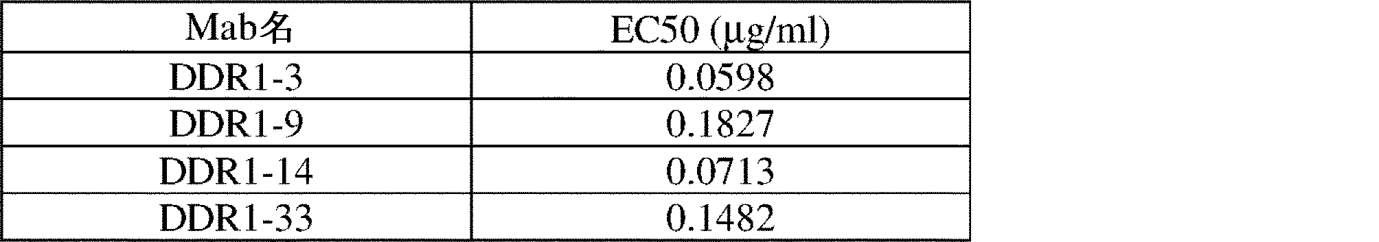
(表13)Octet(96-Red)器具を使用して決定したヒトDDR1への結合親和性

(表14)DDR1 mAbのエピトープ基

ヒトDDR2 ECDに対する抗ヒトDDR1モノクローナル抗体のパネルの交差反応性を決定するために、ELISA法を使用した。DDR1 ECDまたはDDR2 ECDタンパク質を高結合プレートにコーティングし、モノクローナル抗体の各々を1ug/ml濃度で適用し、結合を、HRPコンジュゲート抗ウサギ抗体(Jackson ImmuneResearch、PA)を使用して検出した。結果を図19に示す。モノクローナル抗体DDR1-3、DDR1-9、DDR1-10、DDR1-14およびDDR1-33は、DDR1エピトープに特異的であったが、クローンDDR1-13、DDR1-15、DDR1-21、DDR1-22およびDDR1-34は、DDR1およびDDR2 ECDの両方に存在するエピトープを認識するようである。 An ELISA method was used to determine the cross-reactivity of a panel of anti-human DDR1 monoclonal antibodies to human DDR2 ECD. DDR1 ECD or DDR2 ECD proteins were coated on high binding plates, each of the monoclonal antibodies was applied at 1 ug/ml concentration and binding was detected using HRP-conjugated anti-rabbit antibody (Jackson Immune Research, PA). The results are shown in FIG. Monoclonal antibodies DDR1-3, DDR1-9, DDR1-10, DDR1-14 and DDR1-33 were specific for the DDR1 epitope, whereas clones DDR1-13, DDR1-15, DDR1-21, DDR1-22 and DDR1-34 appears to recognize an epitope present in both DDR1 and DDR2 ECDs.
本明細書に開示され特許請求されるすべての方法は、本開示に照らして過度の実験を行うことなく作製され、実行され得る。本発明の組成物および方法は、好ましい実施形態の観点から記載されているが、本発明の概念、趣旨および範囲から逸脱することなく、本明細書に記載の方法およびステップまたは一連のステップに変形が適用され得ることは、当業者には明らかであろう。より具体的には、化学的および生理学的の両方で関連する特定の薬剤を、本明細書に記載の薬剤に置き換えても、同じまたは同様の結果が達成されることは、明らかであろう。当業者に明らかなそのような類似の置換および修正はすべて、添付の特許請求の範囲によって定義される本発明の趣旨、範囲および概念内であるとみなされる。 All of the methods disclosed and claimed herein can be made and executed without undue experimentation in light of the present disclosure. Although the compositions and methods of the present invention have been described in terms of preferred embodiments, variations in the method and steps or sequences of steps described herein can be made without departing from the concept, spirit and scope of the present invention. can be applied. More specifically, it will be apparent that certain agents that are both chemically and physiologically related can be substituted with the agents described herein to achieve the same or similar results. All such similar substitutes and modifications apparent to those skilled in the art are deemed to be within the spirit, scope and concept of the invention as defined by the appended claims.
参考文献
以下の参考文献は、本明細書に記載されるものを補足する、例示的な手順的な詳細または他の詳細を提供する範囲で、参照により本明細書に具体的に組み込まれる。


Claims (60)
配列番号:108~128から選択される軽鎖可変領域内にLC-LC-CDR1、LC-CDR2、およびLC-CDR3を有する軽鎖可変領域、ならびに
配列番号:129~149から選択される重鎖可変領域内にHC-CDR1、HC-CDR2、およびHC-CDR3を有する重鎖可変領域
を含むか、または
それらのバリアントであって、前記LC-CDRおよび/もしくは前記HC-CDRのうちの1つ以上が1つ、2つ、もしくは3つのアミノ酸置換、付加、欠失、もしくはそれらの組み合わせを有する、前記バリアント
を含む、
前記単離されたモノクローナル抗体またはその抗原結合断片。 An isolated monoclonal antibody or antigen-binding fragment thereof that specifically binds to a discoidin domain receptor 1 (DDR1) protein, comprising:
a light chain variable region having LC-LC-CDR1, LC-CDR2, and LC-CDR3 within the light chain variable region selected from SEQ ID NOS: 108-128 and a heavy chain selected from SEQ ID NOS: 129-149 a heavy chain variable region having HC-CDR1, HC-CDR2, and HC-CDR3 within the variable region, or variants thereof, wherein one of said LC-CDRs and/or said HC-CDRs the above having one, two, or three amino acid substitutions, additions, deletions, or combinations thereof;
The isolated monoclonal antibody or antigen-binding fragment thereof.
表2から選択されるHC-CDR1、HC-CDR2、およびHC-CDR3
を含むか、または
それらのバリアントであって、前記LC-CDRおよび/もしくは前記HC-CDRのうちの1つ以上が1つ、2つ、もしくは3つのアミノ酸置換、付加、欠失、もしくはそれらの組み合わせを有する、前記バリアント
を含む、
請求項1に記載の単離されたモノクローナル抗体またはその抗原結合断片。 LC-LC-CDR1, LC-CDR2 and LC-CDR3 of clone pairs selected from Table 1 and HC-CDR1, HC-CDR2 and HC-CDR3 selected from Table 2
or variants thereof, wherein one or more of said LC-CDRs and/or said HC-CDRs have one, two, or three amino acid substitutions, additions, deletions, or including variants of the above, with a combination of
2. The isolated monoclonal antibody or antigen-binding fragment thereof of claim 1.
アミノ酸配列GFSLNRYY(配列番号:57)を含むHC-CDR1、アミノ酸配列ISYGDTT(配列番号:58)を含むHC-CDR2、およびアミノ酸配列ARADTGDNGYLGLQL(配列番号:59)を含むHC-CDR3を有する重鎖可変領域
を含むか(DDR1-9)、または
それらのバリアントであって、前記LC-CDRおよび/もしくは前記HC-CDRのうちの1つ以上が1つ、2つ、もしくは3つのアミノ酸置換、付加、欠失、もしくはそれらの組み合わせを有する、前記バリアント
を含む、
請求項1に記載の単離されたモノクローナル抗体またはその抗原結合断片。 a light chain variable region having an LC-CDR1 comprising the amino acid sequence QSIGSV (SEQ ID NO: 11), an LC-CDR2 comprising the amino acid sequence GVF, and an LC-CDR3 comprising the amino acid sequence QYIPYGSSP (SEQ ID NO: 12), and the amino acid sequence GFSLNRYY (SEQ ID NO:57), HC-CDR2 containing the amino acid sequence ISYGDTT (SEQ ID NO:58), and HC-CDR3 containing the amino acid sequence ARADTGDNGYLGLQL (SEQ ID NO:59). (DDR1-9), or variants thereof, wherein one or more of said LC-CDRs and/or said HC-CDRs have 1, 2 or 3 amino acid substitutions, additions, deletions, or combinations thereof, including
2. The isolated monoclonal antibody or antigen-binding fragment thereof of claim 1.
アミノ酸配列GFSLSSYY(配列番号:102)を含むHC-CDR1、アミノ酸配列ITTAGPL(配列番号:103)を含むHC-CDR2、およびアミノ酸配列ARGHAGSIYYSYFDL(配列番号:104)を含むHC-CDR3を有する重鎖可変領域
を含むか(DDR1-32)、または
それらのバリアントであって、前記LC-CDRおよび/もしくは前記HC-CDRのうちの1つ以上が1つ、2つ、もしくは3つのアミノ酸置換、付加、欠失、もしくはそれらの組み合わせを有する、前記バリアント
を含む、
請求項1に記載の単離されたモノクローナル抗体またはその抗原結合断片。 a light chain variable region having LC-CDR1 comprising the amino acid sequence ESINSW (SEQ ID NO:41), LC-CDR2 comprising the amino acid sequence DAS, and LC-CDR3 comprising the amino acid sequence QSYYIINRSNYGNS (SEQ ID NO:42), and the amino acid sequence GFSLSSYY (SEQ ID NO: 102), HC-CDR2 containing the amino acid sequence ITTAGPL (SEQ ID NO: 103), and HC-CDR3 containing the amino acid sequence ARGHAGSIYYSYFDL (SEQ ID NO: 104). (DDR1-32), or variants thereof, wherein one or more of said LC-CDRs and/or said HC-CDRs have 1, 2 or 3 amino acid substitutions, additions, deletions, or combinations thereof, including said variants,
2. The isolated monoclonal antibody or antigen-binding fragment thereof of claim 1.
を含む、請求項1に記載の単離されたモノクローナル抗体またはその抗原結合断片。 2. The isolated isolated of claim 1, comprising a light chain variable region having an amino acid sequence selected from SEQ ID NOS: 108-128 and a heavy chain variable region having an amino acid sequence selected from SEQ ID NOS: 129-149 A monoclonal antibody or antigen-binding fragment thereof.
(b)配列番号:109のアミノ酸配列を有する軽鎖可変領域(DDR1-3K)、および配列番号:130のアミノ酸配列を有する重鎖可変領域(DDR1-3H)、
(c)配列番号:110のアミノ酸配列を有する軽鎖可変領域(DDR1-5K)、および配列番号:131のアミノ酸配列を有する重鎖可変領域(DDR1-5H)、
(d)配列番号:111のアミノ酸配列を有する軽鎖可変領域(DDR1-6K)、および配列番号:132のアミノ酸配列を有する重鎖可変領域(DDR1-6H)、
(e)配列番号:112のアミノ酸配列を有する軽鎖可変領域(DDR1-9K)、および配列番号:133のアミノ酸配列を有する重鎖可変領域(DDR1-9H)、
(f)配列番号:113のアミノ酸配列を有する軽鎖可変領域(DDR1-11K)、および配列番号:134のアミノ酸配列を有する重鎖可変領域(DDR1-11H)、
(g)配列番号:114のアミノ酸配列を有する軽鎖可変領域(DDR1-12K)、および配列番号:135のアミノ酸配列を有する重鎖可変領域(DDR1-12H)、
(h)配列番号:115のアミノ酸配列を有する軽鎖可変領域(DDR1-13K)、および配列番号:136のアミノ酸配列を有する重鎖可変領域(DDR1-13H)、
(i)配列番号:116のアミノ酸配列を有する軽鎖可変領域(DDR1-14K)、および配列番号:137のアミノ酸配列を有する重鎖可変領域(DDR1-14H)、
(j)配列番号:117のアミノ酸配列を有する軽鎖可変領域(DDR1-15K)、および配列番号:138のアミノ酸配列を有する重鎖可変領域(DDR1-15H)、
(k)配列番号:118のアミノ酸配列を有する軽鎖可変領域(DDR1-17K)、および配列番号:1039のアミノ酸配列を有する重鎖可変領域(DDR1-17H)、
(l)配列番号:119のアミノ酸配列を有する軽鎖可変領域(DDR1-20K)、および配列番号:140のアミノ酸配列を有する重鎖可変領域(DDR1-20H)、
(m)配列番号:120のアミノ酸配列を有する軽鎖可変領域(DDR1-21K)、および配列番号:141のアミノ酸配列を有する重鎖可変領域(DDR1-21H)、
(n)配列番号:121のアミノ酸配列を有する軽鎖可変領域(DDR1-22K)、および配列番号:142のアミノ酸配列を有する重鎖可変領域(DDR1-22H)、
(o)配列番号:122のアミノ酸配列を有する軽鎖可変領域(DDR1-23K)、および配列番号:143のアミノ酸配列を有する重鎖可変領域(DDR1-23H)、
(p)配列番号:123のアミノ酸配列を有する軽鎖可変領域(DDR1-26K)、および配列番号:144のアミノ酸配列を有する重鎖可変領域(DDR1-26H)、
(q)配列番号:124のアミノ酸配列を有する軽鎖可変領域(DDR1-28K)、および配列番号:145のアミノ酸配列を有する重鎖可変領域(DDR1-28H)、
(r)配列番号:125のアミノ酸配列を有する軽鎖可変領域(DDR1-29K)、および配列番号:146のアミノ酸配列を有する重鎖可変領域(DDR1-29H)、
(s)配列番号:126のアミノ酸配列を有する軽鎖可変領域(DDR1-32K)、および配列番号:147のアミノ酸配列を有する重鎖可変領域(DDR1-32H)、
(t)配列番号:127のアミノ酸配列を有する軽鎖可変領域(DDR1-33K)、および配列番号:148のアミノ酸配列を有する重鎖可変領域(DDR1-33H)、もしくは
(u)配列番号:128のアミノ酸配列を有する軽鎖可変領域(DDR1-34K)、および配列番号:149のアミノ酸配列を有する重鎖可変領域(DDR1-34H)
を含むか、または
それらのバリアントであって、前記軽鎖可変領域が前記親軽鎖可変領域のアミノ酸配列に対して80%以上のアミノ酸配列同一性を有し、かつ前記重鎖可変領域が前記親重鎖可変領域のアミノ酸配列に対して80%以上の配列同一性を有する、DDR1タンパク質に特異的に結合する前記バリアント
を含む、
請求項1に記載の単離されたモノクローナル抗体またはその抗原結合断片。 (a) a light chain variable region (DDR1-1K) having the amino acid sequence of SEQ ID NO: 108 and a heavy chain variable region (DDR1-1H) having the amino acid sequence of SEQ ID NO:129;
(b) a light chain variable region (DDR1-3K) having the amino acid sequence of SEQ ID NO: 109 and a heavy chain variable region (DDR1-3H) having the amino acid sequence of SEQ ID NO: 130;
(c) a light chain variable region (DDR1-5K) having the amino acid sequence of SEQ ID NO:110 and a heavy chain variable region (DDR1-5H) having the amino acid sequence of SEQ ID NO:131;
(d) a light chain variable region (DDR1-6K) having the amino acid sequence of SEQ ID NO:111 and a heavy chain variable region (DDR1-6H) having the amino acid sequence of SEQ ID NO:132;
(e) a light chain variable region (DDR1-9K) having the amino acid sequence of SEQ ID NO: 112 and a heavy chain variable region (DDR1-9H) having the amino acid sequence of SEQ ID NO: 133;
(f) a light chain variable region (DDR1-11K) having the amino acid sequence of SEQ ID NO:113 and a heavy chain variable region (DDR1-11H) having the amino acid sequence of SEQ ID NO:134;
(g) a light chain variable region (DDR1-12K) having the amino acid sequence of SEQ ID NO: 114 and a heavy chain variable region (DDR1-12H) having the amino acid sequence of SEQ ID NO: 135;
(h) a light chain variable region (DDR1-13K) having the amino acid sequence of SEQ ID NO: 115 and a heavy chain variable region (DDR1-13H) having the amino acid sequence of SEQ ID NO: 136;
(i) a light chain variable region (DDR1-14K) having the amino acid sequence of SEQ ID NO:116 and a heavy chain variable region (DDR1-14H) having the amino acid sequence of SEQ ID NO:137;
(j) a light chain variable region (DDR1-15K) having the amino acid sequence of SEQ ID NO: 117 and a heavy chain variable region (DDR1-15H) having the amino acid sequence of SEQ ID NO: 138;
(k) a light chain variable region (DDR1-17K) having the amino acid sequence of SEQ ID NO: 118 and a heavy chain variable region (DDR1-17H) having the amino acid sequence of SEQ ID NO: 1039;
(l) a light chain variable region (DDR1-20K) having the amino acid sequence of SEQ ID NO: 119 and a heavy chain variable region (DDR1-20H) having the amino acid sequence of SEQ ID NO: 140;
(m) a light chain variable region (DDR1-21K) having the amino acid sequence of SEQ ID NO:120 and a heavy chain variable region (DDR1-21H) having the amino acid sequence of SEQ ID NO:141;
(n) a light chain variable region (DDR1-22K) having the amino acid sequence of SEQ ID NO: 121 and a heavy chain variable region (DDR1-22H) having the amino acid sequence of SEQ ID NO: 142;
(o) a light chain variable region (DDR1-23K) having the amino acid sequence of SEQ ID NO: 122 and a heavy chain variable region (DDR1-23H) having the amino acid sequence of SEQ ID NO: 143;
(p) a light chain variable region (DDR1-26K) having the amino acid sequence of SEQ ID NO: 123 and a heavy chain variable region (DDR1-26H) having the amino acid sequence of SEQ ID NO: 144;
(q) a light chain variable region (DDR1-28K) having the amino acid sequence of SEQ ID NO: 124 and a heavy chain variable region (DDR1-28H) having the amino acid sequence of SEQ ID NO: 145;
(r) a light chain variable region (DDR1-29K) having the amino acid sequence of SEQ ID NO: 125 and a heavy chain variable region (DDR1-29H) having the amino acid sequence of SEQ ID NO: 146;
(s) a light chain variable region (DDR1-32K) having the amino acid sequence of SEQ ID NO: 126 and a heavy chain variable region (DDR1-32H) having the amino acid sequence of SEQ ID NO: 147;
(t) a light chain variable region (DDR1-33K) having the amino acid sequence of SEQ ID NO:127 and a heavy chain variable region (DDR1-33H) having the amino acid sequence of SEQ ID NO:148; or (u) SEQ ID NO:128 and a heavy chain variable region (DDR1-34H) having the amino acid sequence of SEQ ID NO: 149.
or a variant thereof, wherein said light chain variable region has 80% or more amino acid sequence identity to the amino acid sequence of said parental light chain variable region, and said heavy chain variable region comprises said said variant that specifically binds to a DDR1 protein having 80% or more sequence identity to the amino acid sequence of the parent heavy chain variable region;
2. The isolated monoclonal antibody or antigen-binding fragment thereof of claim 1.
を含む、請求項1に記載の単離されたモノクローナル抗体またはその抗原結合断片。 a light chain variable region (DDR1-9K) having the amino acid sequence of SEQ ID NO:112 and a heavy chain variable region (DDR1-9H) having the amino acid sequence of SEQ ID NO:133
2. The isolated monoclonal antibody or antigen-binding fragment thereof of claim 1, comprising:
を含む、請求項1に記載の単離されたモノクローナル抗体またはその抗原結合断片。 a light chain variable region (DDR1-32K) having the amino acid sequence of SEQ ID NO:126 and a heavy chain variable region (DDR1-32H) having the amino acid sequence of SEQ ID NO:147
2. The isolated monoclonal antibody or antigen-binding fragment thereof of claim 1, comprising:
配列番号:152のアミノ酸配列を有する重鎖可変領域(DDR1-9hu_Hv)
を含むか、または
それらのバリアントであって、前記軽鎖可変領域が前記親軽鎖可変領域のアミノ酸配列に対して80%以上のアミノ酸配列同一性を有し、かつ前記重鎖可変領域が前記親重鎖可変領域のアミノ酸配列に対して80%以上の配列同一性を有する、DDR1タンパク質に特異的に結合する前記バリアント
を含む、請求項1に記載の単離されたモノクローナル抗体またはその抗原結合断片。 A light chain variable region (DDR1-9hu_Lv1 or DDR1-9hu_Lc2) having the amino acid sequence of SEQ ID NO: 150 or 151, and a heavy chain variable region (DDR1-9hu_Hv) having the amino acid sequence of SEQ ID NO: 152
or a variant thereof, wherein said light chain variable region has 80% or more amino acid sequence identity to the amino acid sequence of said parent light chain variable region, and said heavy chain variable region comprises said 2. The isolated monoclonal antibody of claim 1, or antigen-binding thereof, comprising said variant that specifically binds to a DDR1 protein having 80% or more sequence identity to the amino acid sequence of the parent heavy chain variable region. piece.
を培養する工程を含む、抗体を産生する方法であって、
前記抗体またはその抗原結合断片を発現するのに好適な条件下で前記宿主細胞を培養する工程、および前記抗体またはその抗原結合断片を単離する工程を含む、前記方法。 A method of producing an antibody comprising culturing the host cell of any one of claims 24-26 capable of expressing said monoclonal antibody or antigenic fragment thereof, said method comprising:
culturing said host cell under conditions suitable to express said antibody or antigen-binding fragment thereof; and isolating said antibody or antigen-binding fragment thereof.
を含む、対象におけるがんの影響を治療または緩和する方法。 administering to the subject a therapeutically effective amount of the antibody or antigen-binding fragment thereof of any one of claims 1 to 14, or the engineered cell of claim 33 or 34. A method of treating or lessening the effects of cancer.
を含む、対象における線維症の影響を治療または緩和する方法。 administering to said subject a therapeutically effective amount of the antibody or antigen-binding fragment thereof of any one of claims 1-14, or the engineered cell of claim 33 or 34. A method of treating or alleviating the effects of fibrosis.
(a)対象または対象からの試料を、請求項1~14のいずれか一項に記載の抗体またはその抗原結合断片と接触させる工程、および
(b)前記対象または前記試料におけるがん細胞またはがん幹細胞に対する前記抗体の結合を検出する工程
を含む、前記方法。 A method of detecting cancer cells or cancer stem cells in a sample or subject, comprising:
(a) contacting a subject or a sample from a subject with the antibody or antigen-binding fragment thereof of any one of claims 1-14; and (b) cancer cells or cancer cells in said subject or sample detecting binding of said antibody to cancer stem cells.
1回目と比較したときの検出レベルの変化を決定する工程
をさらに含む、請求項53に記載の方法。 54. The method of claim 53, further comprising performing steps (a) and (b) again, and determining the change in detection level when compared to the first time.
Priority Applications (1)
| Application Number | Priority Date | Filing Date | Title |
|---|---|---|---|
| JP2024135167A JP2024164067A (en) | 2019-12-17 | 2024-08-14 | Novel DDR1 antibodies and uses thereof |
Applications Claiming Priority (3)
| Application Number | Priority Date | Filing Date | Title |
|---|---|---|---|
| US201962949300P | 2019-12-17 | 2019-12-17 | |
| US62/949,300 | 2019-12-17 | ||
| PCT/US2020/065618 WO2021127185A1 (en) | 2019-12-17 | 2020-12-17 | Novel ddr1 antibodies and uses thereof |
Related Child Applications (1)
| Application Number | Title | Priority Date | Filing Date |
|---|---|---|---|
| JP2024135167A Division JP2024164067A (en) | 2019-12-17 | 2024-08-14 | Novel DDR1 antibodies and uses thereof |
Publications (2)
| Publication Number | Publication Date |
|---|---|
| JP2023508277A true JP2023508277A (en) | 2023-03-02 |
| JPWO2021127185A5 JPWO2021127185A5 (en) | 2023-03-22 |
Family
ID=76478553
Family Applications (2)
| Application Number | Title | Priority Date | Filing Date |
|---|---|---|---|
| JP2022537312A Pending JP2023508277A (en) | 2019-12-17 | 2020-12-17 | Novel DDR1 antibodies and uses thereof |
| JP2024135167A Pending JP2024164067A (en) | 2019-12-17 | 2024-08-14 | Novel DDR1 antibodies and uses thereof |
Family Applications After (1)
| Application Number | Title | Priority Date | Filing Date |
|---|---|---|---|
| JP2024135167A Pending JP2024164067A (en) | 2019-12-17 | 2024-08-14 | Novel DDR1 antibodies and uses thereof |
Country Status (10)
| Country | Link |
|---|---|
| US (2) | US20230130372A1 (en) |
| EP (1) | EP4076526A4 (en) |
| JP (2) | JP2023508277A (en) |
| CN (1) | CN115443150A (en) |
| AU (1) | AU2020407089B2 (en) |
| BR (1) | BR112022011037A2 (en) |
| CA (1) | CA3160181A1 (en) |
| IL (1) | IL294019A (en) |
| MX (1) | MX2022007353A (en) |
| WO (1) | WO2021127185A1 (en) |
Families Citing this family (5)
| Publication number | Priority date | Publication date | Assignee | Title |
|---|---|---|---|---|
| JP2023508277A (en) * | 2019-12-17 | 2023-03-02 | ザ ボード オブ リージェンツ オブ ザ ユニバーシティー オブ テキサス システム | Novel DDR1 antibodies and uses thereof |
| CN116023497B (en) * | 2022-11-09 | 2024-06-21 | 四川大学华西医院 | Anti-DDR1 antibodies and uses thereof |
| AU2023379587A1 (en) * | 2022-11-16 | 2025-06-05 | Incendia Therapeutics, Inc. | Methods of treating cancer using anti-ddr1 antibodies and immune checkpoint inhibitors |
| KR20250115399A (en) * | 2022-11-16 | 2025-07-30 | 인센디아 테라퓨틱스 인코포레이티드 | Cancer treatment using anti-DDR1 antibodies |
| CN118909125B (en) * | 2024-10-10 | 2025-01-03 | 杭州美赛生物医药科技有限公司 | Anti-DDR1 antibodies and their applications |
Citations (1)
| Publication number | Priority date | Publication date | Assignee | Title |
|---|---|---|---|---|
| WO2013027802A1 (en) * | 2011-08-23 | 2013-02-28 | 中外製薬株式会社 | Novel anti-ddr1 antibody having anti-tumor activity |
Family Cites Families (68)
| Publication number | Priority date | Publication date | Assignee | Title |
|---|---|---|---|---|
| US6051397A (en) | 1993-11-16 | 2000-04-18 | Max Planck Gesellschaft Zur Forderung Der Wissenschaften | DNA encoding MCK-10, a novel receptor tyrosine kinase |
| US20020115173A1 (en) | 2000-12-11 | 2002-08-22 | Children's Medical Center Corporation | Short peptides from the 'A-region' of protein kinases which selectively modulate protein kinase activity |
| AU2002361653A1 (en) | 2002-03-12 | 2003-10-13 | The Government Of The United States Of America As Represented By The Secretary Of The Department Of | Use of discoidin domain receptor 1 (ddr1) and agents that affect the ddr1/collagen pathway |
| US20070025997A1 (en) | 2002-04-03 | 2007-02-01 | Usha Nagavarapu | Use of biomolecular targets in the treatment and visualization of brain tumors |
| EP1495130A4 (en) | 2002-04-03 | 2006-07-05 | Agy Therapeutics Inc | USE OF BIOMOLECULAR TARGETS IN THE TREATMENT AND VISUALIZATION OF BRAIN TUMORS |
| US20100166731A1 (en) | 2005-09-30 | 2010-07-01 | Bartz Steven R | Methods and Compositions for Treating Cancer |
| EP1992347A1 (en) | 2007-05-18 | 2008-11-19 | Cellzome Ag | Treatment of DDR1 positive cancer using imatinib |
| US8652843B2 (en) | 2008-08-12 | 2014-02-18 | Oncomed Pharmaceuticals, Inc. | DDR1-binding agents and methods of use thereof |
| JP6007420B2 (en) | 2009-11-04 | 2016-10-12 | ファブラス エルエルシー | Antibody optimization method based on affinity maturation |
| AU2011295968B2 (en) | 2010-09-01 | 2017-01-05 | Biogen Ma Inc. | Rapid generation of anti-idiotypic antibodies |
| EP2659269B1 (en) | 2010-12-23 | 2016-10-26 | Roche Diagniostics GmbH | Detection of a posttranslationally modified polypeptide by a bi-valent binding agent |
| EP2694553B1 (en) | 2011-04-01 | 2017-10-11 | Memorial Sloan-Kettering Cancer Center | T cell receptor-like antibodies specific for a wt1 peptide presented by hla-a2 |
| GB201115529D0 (en) * | 2011-09-08 | 2011-10-26 | Imp Innovations Ltd | Antibodies, uses and methods |
| EP2809682B1 (en) | 2012-02-03 | 2020-04-08 | F.Hoffmann-La Roche Ag | Bispecific antibody molecules with antigen-transfected t-cells and their use in medicine |
| EP2819697A4 (en) | 2012-02-29 | 2016-03-09 | Gilead Biologics Inc | Antibodies to matrix metalloproteinase 9 |
| US11873281B2 (en) | 2012-07-12 | 2024-01-16 | Hangzhou Dac Biotech Co., Ltd. | Conjugates of cell binding molecules with cytotoxic agents |
| US20140086913A1 (en) | 2012-09-24 | 2014-03-27 | Gilead Sciences, Inc. | Anti-ddr1 antibodies |
| UY35044A (en) * | 2012-09-24 | 2014-04-30 | Gilead Sciences Inc | ANTI-dDr1 ANTIBODIES |
| WO2014140330A1 (en) | 2013-03-15 | 2014-09-18 | Oryzon Genomics, S.A. | Anti-ddr1 internalizing antibodies and their medical use |
| KR102113310B1 (en) | 2013-09-03 | 2020-05-20 | 아주대학교 산학협력단 | Composition and kit for cancer cell, and method for separating cancer cell using the same |
| PT3049439T (en) | 2013-09-26 | 2020-03-31 | Ablynx Nv | Bispecific nanobodies |
| AU2014354769A1 (en) | 2013-11-26 | 2016-05-26 | Gilead Sciences, Inc. | Therapies for treating myeloproliferative disorders |
| WO2015116569A2 (en) * | 2014-01-30 | 2015-08-06 | Anaptysbio, Inc. | Antibodies directed against discoidin domain receptor family, member 1 (ddr1) |
| EP3217970A4 (en) | 2014-11-10 | 2018-07-18 | Del Mar Pharmaceuticals | Dianhydrogalactitol together with radiation to treat non-small-cell carcinoma of the lung and glioblastoma multiforme |
| US9694084B2 (en) | 2014-12-23 | 2017-07-04 | Dana-Farber Cancer Institute, Inc. | Methods to induce targeted protein degradation through bifunctional molecules |
| US10702527B2 (en) | 2015-06-12 | 2020-07-07 | Dana-Farber Cancer Institute, Inc. | Combination therapy of transcription inhibitors and kinase inhibitors |
| JP2019510752A (en) | 2016-03-04 | 2019-04-18 | ギリアード サイエンシーズ, インコーポレイテッド | Compositions and combinations of autotaxin inhibitors |
| KR20180007924A (en) | 2016-07-14 | 2018-01-24 | 주식회사 코리아하이텍 | Heat radiation device for beam projector using vehicle |
| MX2019006866A (en) * | 2016-12-16 | 2019-08-22 | Merck Patent Gmbh | Methods for the use of galectin 3 binding protein detected in the urine for monitoring the severity and progression of lupus nephritis. |
| CN110381998B (en) * | 2017-01-02 | 2023-08-29 | 德克萨斯大学体系董事会 | Anti-LAIR1 antibodies and uses thereof |
| CN110382002B (en) * | 2017-03-09 | 2025-08-15 | Mab发现股份有限公司 | Antibodies that specifically bind human IL-1R7 |
| US11913075B2 (en) | 2017-04-01 | 2024-02-27 | The Broad Institute, Inc. | Methods and compositions for detecting and modulating an immunotherapy resistance gene signature in cancer |
| SG11201908721TA (en) | 2017-04-06 | 2019-10-30 | Hangzhou Dac Biotech Co Ltd | Conjugation of a cytotoxic drug with bis-linkage |
| KR20250008984A (en) | 2017-05-18 | 2025-01-16 | 리제너론 파마슈티칼스 인코포레이티드 | Cyclodextrin protein drug conjugates |
| CA3067835A1 (en) * | 2017-06-22 | 2018-12-27 | Apexigen, Inc. | Anti-vista antibodies and methods of use |
| US20200158716A1 (en) | 2017-07-17 | 2020-05-21 | Massachusetts Institute Of Technology | Cell atlas of healthy and diseased barrier tissues |
| MX2020004691A (en) | 2017-11-07 | 2020-08-20 | Regeneron Pharma | Hydrophilic linkers for antibody drug conjugates. |
| KR102641565B1 (en) | 2017-12-31 | 2024-02-27 | 항저우 디에이씨 바이오테크 씨오, 엘티디 | A conjugate of a tubulysin analog with branched linkers |
| US20210244737A1 (en) | 2018-06-20 | 2021-08-12 | INSERM (Institut National de la Santé et de la Recherche Médicale) | Compositions for treating melanoma |
| AU2018430758B2 (en) | 2018-07-05 | 2022-01-27 | Hangzhou Dac Biotech Co., Ltd | Cross-linked pyrrolobenzodiazepine dimer (PBD) derivative and its conjugates |
| US20210318318A1 (en) | 2018-08-31 | 2021-10-14 | Institute For Cancer Research D/B/A The Research Institute Of Fox Chase Cancer Center | Kinase Activity In Tumors |
| AU2018445278B2 (en) | 2018-10-12 | 2022-07-28 | Hangzhou Dac Biotech Co., Ltd | Conjugation linkers containing 2,3-diaminosuccinyl group |
| CN113453679B (en) | 2018-12-20 | 2025-07-08 | C4医药公司 | Targeted protein degradation |
| MX2021009147A (en) | 2019-01-31 | 2021-09-10 | Hangzhou Dac Biotech Co Ltd | A conjugate of an amanita toxin with branched linkers. |
| SG11202113022UA (en) | 2019-06-24 | 2021-12-30 | Hangzhou Dac Biotech Co Ltd | A formulation of a conjugate of a tubulysin analog to a cell-binding molecule |
| EA202290091A1 (en) | 2019-06-24 | 2022-03-24 | Ханчжоу Дэк Биотек Ко., Лтд | CONJUGATE OF A CYTOTOXIC AGENT WITH A CELL-BINDING MOLECULE WITH BRANCHED LINKERS |
| WO2021011634A1 (en) | 2019-07-15 | 2021-01-21 | Kymera Therapeutics, Inc. | Protein degraders and uses thereof |
| EP4053153A4 (en) | 2019-11-01 | 2023-12-20 | Cellemedy Co., Ltd | Protein fused with molecule capable of binding to immune checkpoint molecule and use of same |
| WO2021086159A1 (en) | 2019-11-01 | 2021-05-06 | (주)셀레메디 | Protein fused with molecule capable of binding to immune checkpoint molecule and use of same |
| JP2023508277A (en) * | 2019-12-17 | 2023-03-02 | ザ ボード オブ リージェンツ オブ ザ ユニバーシティー オブ テキサス システム | Novel DDR1 antibodies and uses thereof |
| CA3173268A1 (en) | 2020-02-27 | 2021-09-02 | Janssen Biotech, Inc. | Materials and methods for modulating an immune response |
| US20210284731A1 (en) | 2020-03-13 | 2021-09-16 | Janssen Biotech, Inc. | Methods and materials for modulating an immune response |
| KR20230034957A (en) | 2020-06-19 | 2023-03-10 | 항저우 디에이씨 바이오테크 씨오, 엘티디 | Conjugates of cell-associated molecules with camptothecin analogues |
| CN116723839A (en) | 2020-10-14 | 2023-09-08 | C4医药公司 | Tricyclic heterobifunctional compounds for degrading target proteins |
| US20230391893A1 (en) | 2020-10-30 | 2023-12-07 | Cellemedy Co.,Ltd | Antibody-like protein and use thereof |
| WO2022135332A1 (en) | 2020-12-21 | 2022-06-30 | 映恩生物制药(苏州)有限公司 | Steroid conjugate |
| CN114807042B (en) | 2021-01-22 | 2024-09-03 | 南京助天中科科技发展有限公司 | A chimeric antigen receptor modified NK cell and its preparation method and application |
| WO2022171101A1 (en) | 2021-02-10 | 2022-08-18 | 映恩生物制药(苏州)有限公司 | Steroid conjugate |
| NL2028075B1 (en) | 2021-04-26 | 2022-11-03 | Alphathera Ag | Targeting system for cancer comprising a radioisotope |
| US20240216517A1 (en) | 2021-05-03 | 2024-07-04 | Cellemedy Co., Ltd | Pharmaceutical composition for prevention or treatment of lung cancer |
| EP4363443A4 (en) | 2021-07-02 | 2025-08-06 | Laekna Therapeutics Shanghai Co Ltd | Depletion of activated hepatic stellate cells (HSCs) and uses thereof |
| CN120463762A (en) | 2021-08-26 | 2025-08-12 | 映恩生物科技(上海)有限公司 | A steroid compound and its conjugate |
| WO2023040793A1 (en) | 2021-09-14 | 2023-03-23 | 映恩生物制药(苏州)有限公司 | Anti-inflammatory compound and use thereof |
| JP2024536840A (en) | 2021-09-26 | 2024-10-08 | ウーシー バイオロジクス アイルランド リミテッド | IL-2 variants and fusion proteins thereof |
| WO2023061478A1 (en) | 2021-10-15 | 2023-04-20 | 先声再明医药有限公司 | Tricyclic compound |
| AU2021362997A1 (en) | 2021-11-03 | 2024-05-16 | Hangzhou Dac Biotech Co., Ltd. | Specific conjugation of an antibody |
| WO2023163873A1 (en) | 2022-02-23 | 2023-08-31 | Mayo Foundation For Medical Education And Research | Synergistic drug combinations to treat cancer |
| KR20240135665A (en) | 2022-02-28 | 2024-09-11 | 우시 바이올로직스 아일랜드 리미티드 | Heterodimeric polypeptide complex comprising IL-15 variant and uses thereof |
-
2020
- 2020-12-17 JP JP2022537312A patent/JP2023508277A/en active Pending
- 2020-12-17 IL IL294019A patent/IL294019A/en unknown
- 2020-12-17 WO PCT/US2020/065618 patent/WO2021127185A1/en not_active Ceased
- 2020-12-17 EP EP20903632.6A patent/EP4076526A4/en active Pending
- 2020-12-17 CA CA3160181A patent/CA3160181A1/en active Pending
- 2020-12-17 MX MX2022007353A patent/MX2022007353A/en unknown
- 2020-12-17 US US17/757,510 patent/US20230130372A1/en active Pending
- 2020-12-17 CN CN202080094867.1A patent/CN115443150A/en active Pending
- 2020-12-17 AU AU2020407089A patent/AU2020407089B2/en active Active
- 2020-12-17 BR BR112022011037A patent/BR112022011037A2/en unknown
-
2023
- 2023-02-28 US US18/176,072 patent/US12331130B2/en active Active
-
2024
- 2024-08-14 JP JP2024135167A patent/JP2024164067A/en active Pending
Patent Citations (1)
| Publication number | Priority date | Publication date | Assignee | Title |
|---|---|---|---|---|
| WO2013027802A1 (en) * | 2011-08-23 | 2013-02-28 | 中外製薬株式会社 | Novel anti-ddr1 antibody having anti-tumor activity |
Also Published As
| Publication number | Publication date |
|---|---|
| AU2020407089B2 (en) | 2023-06-01 |
| AU2020407089A1 (en) | 2022-06-30 |
| IL294019A (en) | 2022-08-01 |
| EP4076526A4 (en) | 2024-07-03 |
| EP4076526A1 (en) | 2022-10-26 |
| JP2024164067A (en) | 2024-11-26 |
| WO2021127185A1 (en) | 2021-06-24 |
| US20230235078A1 (en) | 2023-07-27 |
| CN115443150A (en) | 2022-12-06 |
| MX2022007353A (en) | 2022-09-19 |
| US12331130B2 (en) | 2025-06-17 |
| US20230130372A1 (en) | 2023-04-27 |
| BR112022011037A2 (en) | 2022-09-13 |
| CA3160181A1 (en) | 2021-06-24 |
Similar Documents
| Publication | Publication Date | Title |
|---|---|---|
| US11498963B2 (en) | Anti-LILRB antibodies and their use in detecting and treating cancer | |
| US20250250336A1 (en) | Novel lilrb4 antibodies and uses thereof | |
| US20230068663A1 (en) | Novel lilrb2 antibodies and uses thereof | |
| US12331130B2 (en) | DDR1 antibodies and uses thereof | |
| US20160200815A1 (en) | Antibodies that inhibit tim-3:lilrb2 interactions and uses thereof | |
| JP7022067B2 (en) | T-cell receptor-like antibody specific for FOXP3-derived peptide | |
| JP7152404B2 (en) | ANTI-LAIR1 ANTIBODY AND USES THEREOF | |
| JP7688579B2 (en) | Methods of Use of Anti-TREM2 Antibodies | |
| CN104185681A (en) | C1ORF32 antibody and its use for treating cancer | |
| CN115916347A (en) | Antibodies against chitinase 3-like 1 and methods of use thereof | |
| US20230399384A1 (en) | Antibodies to coronavirus spike protein and methods of use thereof | |
| EA048483B1 (en) | NEW DDR1 ANTIBODIES AND THEIR APPLICATIONS | |
| HK40009967B (en) | Anti-lair1 antibodies and their uses |
Legal Events
| Date | Code | Title | Description |
|---|---|---|---|
| A521 | Request for written amendment filed |
Free format text: JAPANESE INTERMEDIATE CODE: A523 Effective date: 20230216 |
|
| A521 | Request for written amendment filed |
Free format text: JAPANESE INTERMEDIATE CODE: A523 Effective date: 20230313 |
|
| A621 | Written request for application examination |
Free format text: JAPANESE INTERMEDIATE CODE: A621 Effective date: 20230313 |
|
| RD04 | Notification of resignation of power of attorney |
Free format text: JAPANESE INTERMEDIATE CODE: A7424 Effective date: 20230808 |
|
| A131 | Notification of reasons for refusal |
Free format text: JAPANESE INTERMEDIATE CODE: A131 Effective date: 20240215 |
|
| A601 | Written request for extension of time |
Free format text: JAPANESE INTERMEDIATE CODE: A601 Effective date: 20240513 |
|
| A02 | Decision of refusal |
Free format text: JAPANESE INTERMEDIATE CODE: A02 Effective date: 20241007 |