JP2022537048A - Signal encoding methods for analytes in samples - Google Patents
Signal encoding methods for analytes in samples Download PDFInfo
- Publication number
- JP2022537048A JP2022537048A JP2021575925A JP2021575925A JP2022537048A JP 2022537048 A JP2022537048 A JP 2022537048A JP 2021575925 A JP2021575925 A JP 2021575925A JP 2021575925 A JP2021575925 A JP 2021575925A JP 2022537048 A JP2022537048 A JP 2022537048A
- Authority
- JP
- Japan
- Prior art keywords
- analyte
- signal
- oligonucleotides
- decoding
- specific
- Prior art date
- Legal status (The legal status is an assumption and is not a legal conclusion. Google has not performed a legal analysis and makes no representation as to the accuracy of the status listed.)
- Granted
Links
Images


Classifications
-
- C—CHEMISTRY; METALLURGY
- C12—BIOCHEMISTRY; BEER; SPIRITS; WINE; VINEGAR; MICROBIOLOGY; ENZYMOLOGY; MUTATION OR GENETIC ENGINEERING
- C12Q—MEASURING OR TESTING PROCESSES INVOLVING ENZYMES, NUCLEIC ACIDS OR MICROORGANISMS; COMPOSITIONS OR TEST PAPERS THEREFOR; PROCESSES OF PREPARING SUCH COMPOSITIONS; CONDITION-RESPONSIVE CONTROL IN MICROBIOLOGICAL OR ENZYMOLOGICAL PROCESSES
- C12Q1/00—Measuring or testing processes involving enzymes, nucleic acids or microorganisms; Compositions therefor; Processes of preparing such compositions
- C12Q1/68—Measuring or testing processes involving enzymes, nucleic acids or microorganisms; Compositions therefor; Processes of preparing such compositions involving nucleic acids
- C12Q1/6813—Hybridisation assays
- C12Q1/6816—Hybridisation assays characterised by the detection means
-
- C—CHEMISTRY; METALLURGY
- C12—BIOCHEMISTRY; BEER; SPIRITS; WINE; VINEGAR; MICROBIOLOGY; ENZYMOLOGY; MUTATION OR GENETIC ENGINEERING
- C12Q—MEASURING OR TESTING PROCESSES INVOLVING ENZYMES, NUCLEIC ACIDS OR MICROORGANISMS; COMPOSITIONS OR TEST PAPERS THEREFOR; PROCESSES OF PREPARING SUCH COMPOSITIONS; CONDITION-RESPONSIVE CONTROL IN MICROBIOLOGICAL OR ENZYMOLOGICAL PROCESSES
- C12Q1/00—Measuring or testing processes involving enzymes, nucleic acids or microorganisms; Compositions therefor; Processes of preparing such compositions
- C12Q1/68—Measuring or testing processes involving enzymes, nucleic acids or microorganisms; Compositions therefor; Processes of preparing such compositions involving nucleic acids
- C12Q1/6804—Nucleic acid analysis using immunogens
-
- C—CHEMISTRY; METALLURGY
- C12—BIOCHEMISTRY; BEER; SPIRITS; WINE; VINEGAR; MICROBIOLOGY; ENZYMOLOGY; MUTATION OR GENETIC ENGINEERING
- C12Q—MEASURING OR TESTING PROCESSES INVOLVING ENZYMES, NUCLEIC ACIDS OR MICROORGANISMS; COMPOSITIONS OR TEST PAPERS THEREFOR; PROCESSES OF PREPARING SUCH COMPOSITIONS; CONDITION-RESPONSIVE CONTROL IN MICROBIOLOGICAL OR ENZYMOLOGICAL PROCESSES
- C12Q1/00—Measuring or testing processes involving enzymes, nucleic acids or microorganisms; Compositions therefor; Processes of preparing such compositions
- C12Q1/68—Measuring or testing processes involving enzymes, nucleic acids or microorganisms; Compositions therefor; Processes of preparing such compositions involving nucleic acids
- C12Q1/6876—Nucleic acid products used in the analysis of nucleic acids, e.g. primers or probes
-
- C—CHEMISTRY; METALLURGY
- C12—BIOCHEMISTRY; BEER; SPIRITS; WINE; VINEGAR; MICROBIOLOGY; ENZYMOLOGY; MUTATION OR GENETIC ENGINEERING
- C12Q—MEASURING OR TESTING PROCESSES INVOLVING ENZYMES, NUCLEIC ACIDS OR MICROORGANISMS; COMPOSITIONS OR TEST PAPERS THEREFOR; PROCESSES OF PREPARING SUCH COMPOSITIONS; CONDITION-RESPONSIVE CONTROL IN MICROBIOLOGICAL OR ENZYMOLOGICAL PROCESSES
- C12Q2563/00—Nucleic acid detection characterized by the use of physical, structural and functional properties
- C12Q2563/179—Nucleic acid detection characterized by the use of physical, structural and functional properties the label being a nucleic acid
Landscapes
- Chemical & Material Sciences (AREA)
- Life Sciences & Earth Sciences (AREA)
- Organic Chemistry (AREA)
- Health & Medical Sciences (AREA)
- Proteomics, Peptides & Aminoacids (AREA)
- Zoology (AREA)
- Wood Science & Technology (AREA)
- Engineering & Computer Science (AREA)
- Analytical Chemistry (AREA)
- Immunology (AREA)
- Physics & Mathematics (AREA)
- Microbiology (AREA)
- Molecular Biology (AREA)
- Biotechnology (AREA)
- Biophysics (AREA)
- Biochemistry (AREA)
- Bioinformatics & Cheminformatics (AREA)
- General Engineering & Computer Science (AREA)
- General Health & Medical Sciences (AREA)
- Genetics & Genomics (AREA)
- Pathology (AREA)
- Measuring Or Testing Involving Enzymes Or Micro-Organisms (AREA)
Abstract
【解決手段】 本発明は、試料中の分析物を逐次的にシグナルコードする方法、試料中の分析物を逐次的にシグナルコードするための解読オリゴヌクレオチドのセットの使用、及び試料中の分析物を逐次的にシグナルコードするためのキットに関する。【選択図】なしKind Code: A1 The present invention provides a method for sequentially signal-encoding analytes in a sample, the use of a set of decoding oligonucleotides for sequentially signal-encoding analytes in a sample, and methods for sequentially signal-encoding analytes in a sample. to a kit for sequentially signal-encoding. [Selection figure] None
Description
本発明は、試料中の分析物を逐次的にシグナルコードする方法、試料中の分析物を逐次的にシグナルコードするための解読オリゴヌクレオチドのセットの使用、及び試料中の分析物を逐次的にシグナルコードするためのキットに関する。 The present invention provides a method of sequentially signal-encoding analytes in a sample, the use of a set of decoding oligonucleotides to sequentially signal-encode analytes in a sample, and sequentially signal-encoding analytes in a sample. It relates to a kit for signal coding.
本発明は、分子生物学の分野、より具体的には、試料中の分析物の検出、好ましくは、生物学的試料中の核酸分子及び/又はタンパク質などの生体分子の検出に関する。 The present invention relates to the field of molecular biology, more specifically to the detection of analytes in samples, preferably biomolecules such as nucleic acid molecules and/or proteins in biological samples.
生物学的及び非生物学的試料中の少量の分析物の分析及び検出は、臨床及び分析環境において日常的な業務となっている。この目的のために多数の分析方法が確立されている。それらのいくつかは、特定の第2の分析物に割り当てられたコードとは異なる特定の第1の分析物に特定の可読コードを割り当てるコード化技術を使用する。 Analysis and detection of low-abundance analytes in biological and non-biological samples has become routine practice in clinical and analytical settings. Numerous analytical methods have been established for this purpose. Some of them use coding techniques that assign specific readable codes to specific first analytes that are different from codes assigned to specific second analytes.
この分野における従来技術の1つは、試料中のmRNA分子を検出するために本質的に開発された、いわゆる「単一分子蛍光インサイチュハイブリダイゼーション」(smFISH)である。Lubeck et al.(2014),Single-cell in situ RNA profiling by sequential hybridization,Nat.Methods11(4),p.360-361では、目的のmRNAは、特異的な直接標識プローブセットを介して検出される。1ラウンドのハイブリダイゼーション及び検出の後、mRNA特異的プローブのセットをmRNAから溶出し、他の(又は同じ)蛍光標識を有するプローブの同じセットを次のラウンドのハイブリダイゼーション及びイメージングで使用して、遺伝子特異的カラーコードスキームを数ラウンドにわたって生成する。この技術は、転写物ごとにいくつかの異なるタグ付きプローブセットを必要とし、各検出ラウンドごとにこれらのプローブセットを変性させる必要がある。 One of the prior art techniques in this field is the so-called "single molecule fluorescence in situ hybridization" (smFISH), which was developed essentially for the detection of mRNA molecules in samples. Lubeck et al. (2014), Single-cell in situ RNA profiling by sequential hybridization, Nat. Methods 11(4), p. At 360-361, the mRNA of interest is detected via a specific direct-labeled probe set. After one round of hybridization and detection, a set of mRNA-specific probes are eluted from the mRNA and the same set of probes with other (or the same) fluorescent labels are used in the next round of hybridization and imaging, A gene-specific color-coding scheme is generated over several rounds. This technique requires several different tagged probe sets for each transcript and denaturation of these probe sets for each detection round.
この技術の更なる発展は、直接標識されたプローブセットを使用しない。代わりに、プローブセットのオリゴヌクレオチドは、シグナル増幅を可能にする技術であるハイブリダイゼーション連鎖反応(HCR)の開始剤として機能する核酸配列を提供し、Shah et al.(2016),In situ transcription profiling of single cells reveals spatial organization of cells in the mouse hippocampus,Neuron 92(2),p.342-357を参照されたい。 A further development of this technology does not use directly labeled probe sets. Instead, the oligonucleotides of the probe set provide nucleic acid sequences that function as initiators for the hybridization chain reaction (HCR), a technique that allows signal amplification, Shah et al. (2016), In situ transcription profiling of single cells reveals spatial organization of cells in the mouse hippocampus, Neuron 92(2), p. 342-357.
「多重化エラーロバスト蛍光インサイチュハイブリダイゼーション」(merFISH)と呼ばれる別の技術は、Chen et al.(2015),RNA imaging.Spatially resolved,highly multiplexed RNA profiling in single cells,Science 348(6233):aaa6090に記載されている。そこで、蛍光標識されたオリゴヌクレオチドのその後の特異的ハイブリダイゼーションのための追加の配列要素を提供する特異的プローブセットを介して、目的のmRNAが検出される。各プローブセットは、合計16個の配列要素のうちの4つの異なる配列要素を提供する。特異的プローブセットと目的のmRNAとのハイブリダイゼーション後、いわゆる読み出しハイブリダイゼーションが行われる。各読み出しハイブリダイゼーションにおいて、配列要素の1つに相補的な16個の蛍光標識オリゴヌクレオチドの1つがハイブリダイズする。全ての読み出しオリゴヌクレオチドは、同じ蛍光色を使用する。画像化後、蛍光シグナルは照射によって破壊され、次のラウンドの読み出しハイブリダイゼーションは変性ステップなしで行われる。結果として、各mRNA種についてバイナリコードが生成される。特異的プローブセットを目的のmRNAに結合させるための単一のハイブリダイゼーションラウンドのみを使用し、続いて単一の蛍光色で標識された読み出しオリゴヌクレオチドの16ラウンドのハイブリダイゼーションを使用して、16ラウンドの4つのシグナルの固有のシグナルシグネチャが作成される。 Another technique called "multiplexed error-robust fluorescence in situ hybridization" (merFISH) is described by Chen et al. (2015), RNA imaging. Spatially resolved, highly multiplexed RNA profiling in single cells, Science 348(6233): aaa6090. There, the mRNA of interest is detected via specific probe sets that provide additional sequence elements for subsequent specific hybridization of fluorescently labeled oligonucleotides. Each probeset provides 4 different sequence elements out of a total of 16 sequence elements. After hybridization of the specific probe set with the mRNA of interest, a so-called readout hybridization is performed. In each readout hybridization, one of 16 fluorescently labeled oligonucleotides complementary to one of the sequence elements hybridizes. All readout oligonucleotides use the same fluorescent color. After imaging, the fluorescent signal is destroyed by illumination and the next round of readout hybridization is performed without a denaturation step. As a result, a binary code is generated for each mRNA species. Using only a single hybridization round to bind the specific probe set to the mRNA of interest, followed by 16 rounds of hybridization of readout oligonucleotides labeled with a single fluorescent color, 16 A unique signal signature of the four signals of the round is created.
この技術の更なる開発は、2つの異なる蛍光色を使用することによってスループットを改善し、読み出しオリゴヌクレオチドと蛍光標識との間のジスルフィド切断と代替ハイブリダイゼーション緩衝液を介してシグナルを除去し、Moffitt et al.(2016),High-throughput single-cell gene-expression profiling with multiplexed error-robust fluorescence in situ hybridization,Proc.Natl.Acad.Sci.U S A.113(39),p.11046-11051を参照されたい。 Further development of this technique improves throughput by using two different fluorescent colors, removing signal via disulfide cleavage between the readout oligonucleotide and fluorescent label and alternative hybridization buffers, Moffitt et al. et al. (2016), High-throughput single-cell gene-expression profiling with multiplexed error-robust fluorescence in situ hybridization, Proc. Natl. Acad. Sci. USA 113(39), p. See 11046-11051.
「イントロンseqFISH」と称される技術は、Shah et al.(2018),Dynamics and spatial genomics of the nascent transcriptome by intron seqFISH,Cell 117(2),p.363-376に記載されている。そこで、蛍光標識されたオリゴヌクレオチドのその後の特異的ハイブリダイゼーションのための追加の配列要素を提供する特異的プローブセットを介して、目的のmRNAが検出される。各プローブセットは、色分けラウンドごとに12個の可能な配列要素(使用される12個の「擬似カラー」を表す)の1つを提供する。各色分けラウンドは、4つの連続ハイブリダイゼーションからなる。これらの連続ハイブリダイゼーションの各々において、各々が異なるフルオロフォアで標識された3つの読み出しプローブは、mRNA特異的プローブセットの対応する要素にハイブリダイズする。画像化後、55%ホルムアミド緩衝液によって読み出しプローブを除去し、次のハイブリダイゼーションが続く。各々4回の連続ハイブリダイゼーションによる5ラウンドの色分けの後、色分けが完了する。 A technique termed "intron seqFISH" is described by Shah et al. (2018), Dynamics and spatial genomics of the nascent transcriptome by intron seq FISH, Cell 117(2), p. 363-376. There, the mRNA of interest is detected via specific probe sets that provide additional sequence elements for subsequent specific hybridization of fluorescently labeled oligonucleotides. Each probeset provides one of 12 possible sequence elements (representing the 12 "pseudocolors" used) per coloring round. Each color-coding round consists of four sequential hybridizations. In each of these sequential hybridizations, three readout probes, each labeled with a different fluorophore, hybridize to corresponding members of the mRNA-specific probe set. After imaging, the readout probe is removed by 55% formamide buffer followed by subsequent hybridizations. After 5 rounds of color coding with 4 sequential hybridizations each, the color coding is complete.
欧州特許第0 611 828号は、分析物に特異的に結合するプローブにシグナル発生要素を補充するための架橋要素の使用を開示している。より具体的な記述は、架橋核酸分子を補充する特異的プローブを介した核酸の検出について記載する。この架橋核酸は、最終的にシグナル生成核酸を補充する。この文献はまた、分岐DNAのようなシグナル増幅のためのシグナル発生要素のための2つ以上の結合部位を有する架橋要素の使用を記載している。
Player et al.(2001),Single-copy gene detection using branched DNA (bDNA) in situ hybridization,J.Histochem.Cytochem.49(5),p.603-611は、目的の核酸が、追加の配列要素を提供する特異的プローブセットを介して検出される方法を記載している。第2のステップでは、プレ増幅器オリゴヌクレオチドは、この配列要素にハイブリダイズする。このプレ増幅器オリゴヌクレオチドは、後続のステップでハイブリダイズする増幅器オリゴヌクレオチドのための複数の結合部位を含む。これらの増幅器オリゴヌクレオチドは、標識オリゴヌクレオチドの複数の配列要素を提供する。このようにして、シグナルの増幅をもたらす分岐オリゴヌクレオチドツリーが構築される。 Player et al. (2001), Single-copy gene detection using branched DNA (bDNA) in situ hybridization, J. Am. Histochem. Cytochem. 49(5), p. 603-611 describe methods in which the nucleic acid of interest is detected via specific probe sets that provide additional sequence elements. In a second step, a preamplifier oligonucleotide hybridizes to this sequence element. This pre-amplifier oligonucleotide contains multiple binding sites for the amplifier oligonucleotides to be hybridized in subsequent steps. These amplifier oligonucleotides provide multiple sequence elements of labeled oligonucleotides. In this way a branched oligonucleotide tree is constructed that leads to signal amplification.
この方法の更なる開発は、Wang et al.(2012),RNAscope:a novel in situ RNA analysis platform for formalin-fixed,paraffin-embedded tissues,J.Mol.Diagn.14(1),p.22-29に記載され、これは、mRNA特異的プローブの別の設計を使用する。ここで、mRNA特異的オリゴヌクレオチドのうちの2つは、プレ増幅器オリゴヌクレオチドを補充することができる配列を提供するために、近接してハイブリダイズする必要がある。この方法では、偽陽性シグナルの数を低減することによって、方法の特異性が高められる。 Further development of this method can be found in Wang et al. (2012), RNAscope: a novel in situ RNA analysis platform for formalin-fixed, paraffin-embedded tissues, J. Am. Mol. Diagn. 14(1), p. 22-29, which uses an alternative design of mRNA-specific probes. Here, two of the mRNA-specific oligonucleotides need to hybridize in close proximity to provide a sequence that can recruit the pre-amplifier oligonucleotide. This method increases the specificity of the method by reducing the number of false positive signals.
Choi et al.(2010),Programmable in situ amplification for multiplexed imaging of mRNA expression,Nat.Biotechnol.28(11),p.1208-1212は、「HCRハイブリダイゼーション連鎖反応」として知られている方法を開示している。目的のmRNAは、追加の配列要素を提供する特異的プローブセットを介して検出される。追加の配列要素は、ハイブリダイゼーション連鎖反応を開始するための開始剤配列である。基本的に、ハイブリダイゼーション連鎖反応は、第1のヘアピンが開始剤配列を介して開かれた後に、ポリマーに自己集合する準安定性オリゴヌクレオチドヘアピンに基づく。 Choi et al. (2010), Programmable in situ amplification for multiplexed imaging of mRNA expression, Nat. Biotechnol. 28(11), p. 1208-1212 disclose a method known as the "HCR hybridization chain reaction". The mRNA of interest is detected via specific probe sets that provide additional sequence elements. Additional sequence elements are initiator sequences for initiating the hybridization chain reaction. Basically, the hybridization chain reaction is based on metastable oligonucleotide hairpins that self-assemble into a polymer after the first hairpin is opened via an initiator sequence.
本技術の更なる発展は、RNAscope技術と同様に、HCRの開始剤配列を形成するために近接してハイブリダイズしなければならない、いわゆる分割開始剤プローブを使用し、これにより偽陽性シグナルの数を減少させ、Choi et al.(2018),Third-generation in situ hybridization chain reaction:multiplexed,quantitative,sensitive,versatile,robust.Development 145(12)を参照されたい。 A further development of this technique uses so-called split initiator probes, which, like the RNAscope technique, must hybridize in close proximity to form the initiator sequence of the HCR, thereby increasing the number of false positive signals. , Choi et al. (2018), Third-generation in situ hybridization chain reaction: multiplexed, quantitative, sensitive, versatile, robust. Development 145(12).
Mateo et al.(2019),Visualizing DNA folding and RNA in embryos at single-cell resolution,Nature Vol,568,p.49ff.は、クロマチン構造の光学的再構築(ORCA)と呼ばれる方法を開示している。この方法は、染色体ラインを可視化することを意図している。 Mateo et al. (2019), Visualizing DNA folding and RNA in embryos at single-cell resolution, Nature Vol, 568, p. 49ff. disclose a method called optical reconstruction of chromatin structure (ORCA). This method is intended to visualize chromosomal lines.
しかし、当該技術分野で公知の方法は、多数の欠点を有する。特に、それらは柔軟性がなく、高価で、複雑で、時間がかかり、不正確な結果をもたらすことが非常に多い。特に、既存の方法のコード化能力は低く、現代の分子生物学及び医学の要件を満たしていない。 However, methods known in the art have a number of drawbacks. In particular, they are inflexible, expensive, complex, time consuming and very often produce inaccurate results. In particular, existing methods have poor coding capabilities and do not meet the requirements of modern molecular biology and medicine.
この背景に対して、本発明の根底にある目的は、従来技術の方法の欠点を低減又は回避することができる方法を提供することである。 Against this background, an underlying object of the present invention is to provide a method that can reduce or avoid the drawbacks of prior art methods.
本発明は、これら及び他の必要性を満たす。 The present invention meets these and other needs.
本発明は、試料中の分析物を逐次的にシグナルコードする方法を提供し、方法は、以下のステップ、
(1)分析物特異的プローブのセットを提供するステップであって、各分析物特異的プローブが、
-コード化される分析物のうちの1つと特異的に相互作用する結合要素(S)と、
-分析物特異的プローブのセットに固有のヌクレオチド配列(固有の識別子配列)を含む識別子要素(T)と、を含むステップと、
(2)分析物特異的プローブのセットを試料とインキュベートし、それによってコード化される分析物への分析物特異的プローブの特異的結合を可能にするステップと、
(3)試料から非結合プローブを除去するステップと、
(4)解読オリゴヌクレオチドのセットを提供するステップであって、各解読オリゴヌクレオチドが、
-固有の識別子配列の少なくとも一部に本質的に相補的であるヌクレオチド配列を含む第1のコネクタ要素(t)と、
-シグナルオリゴヌクレオチドの特異的ハイブリダイゼーションを可能にするヌクレオチド配列を含むトランスレータ要素(c)と、を含むステップと、
(5)解読オリゴヌクレオチドのセットを試料とインキュベートし、それによって固有の識別子配列への解読オリゴヌクレオチドの特異的ハイブリダイゼーションを可能にするステップと、
(6)試料から非結合解読オリゴヌクレオチドを除去するステップと、
(7)シグナルオリゴヌクレオチドのセットを提供するステップであって、各シグナルオリゴヌクレオチドが、
-トランスレータ要素(c)のヌクレオチド配列の少なくとも一部に本質的に相補的であるヌクレオチド配列を含む第2のコネクタ要素(C)と、
-シグナル要素と、を含むステップと、
(8)シグナルオリゴヌクレオチドのセットを試料とインキュベートし、それによってトランスレータ要素(c)へのシグナルオリゴヌクレオチドの特異的ハイブリダイゼーションを可能にするステップと、を含む。
The present invention provides a method of sequentially signal-encoding analytes in a sample, the method comprising the steps of:
(1) providing a set of analyte-specific probes, each analyte-specific probe comprising:
- a binding member (S) that specifically interacts with one of the encoded analytes;
- an identifier element (T) comprising a unique nucleotide sequence (unique identifier sequence) for the set of analyte-specific probes;
(2) incubating the set of analyte-specific probes with the sample, thereby allowing specific binding of the analyte-specific probes to the encoded analytes;
(3) removing unbound probe from the sample;
(4) providing a set of decoding oligonucleotides, each decoding oligonucleotide comprising:
- a first connector element (t) comprising a nucleotide sequence essentially complementary to at least part of the unique identifier sequence;
- a translator element (c) comprising a nucleotide sequence allowing the specific hybridization of the signal oligonucleotide;
(5) incubating the set of decoding oligonucleotides with the sample, thereby allowing specific hybridization of the decoding oligonucleotides to the unique identifier sequences;
(6) removing unbound decoding oligonucleotides from the sample;
(7) providing a set of signal oligonucleotides, each signal oligonucleotide comprising:
- a second connector element (C) comprising a nucleotide sequence essentially complementary to at least part of the nucleotide sequence of the translator element (c);
- a step comprising a signaling element;
(8) incubating the set of signal oligonucleotides with the sample, thereby allowing specific hybridization of the signal oligonucleotides to the translator element (c).
本発明者らは、この新規の方法が、特異的ハイブリダイゼーションを介して試料中の異なる分析物又は異なる単一分析物分子の特異的な定量的及び/又は空間的検出又は計数を並行して可能にするプロセスを設定するのに必要な必須のステップを提供することを認識した。技術は、利用可能な異なるシグナルよりも多数の分析物を区別することを可能にする。他の最新技術の方法とは対照的に、検出可能なシグナルを提供するオリゴヌクレオチドは、試料特異的核酸配列と直接相互作用しないが、いわゆる「解読オリゴヌクレオチド」によって媒介される。この機構は、分析物特異的オリゴヌクレオチドとシグナルオリゴヌクレオチドとの間の依存性を切り離し、したがって、コード化能力の劇的な増加をもたらす。 The inventors have found that this novel method enables the specific quantitative and/or spatial detection or counting of different analytes or different single analyte molecules in a sample in parallel via specific hybridization. Recognized to provide the essential steps necessary to set up the enabling process. The technology makes it possible to distinguish between a greater number of analytes than the different signals available. In contrast to other state-of-the-art methods, the oligonucleotides providing the detectable signal do not interact directly with sample-specific nucleic acid sequences, but are mediated by so-called "decoding oligonucleotides." This mechanism decouples the dependency between analyte-specific and signal oligonucleotides, thus providing a dramatic increase in encoding capacity.
本発明の別の主題は、試料中の分析物を逐次的にシグナルコードするための解読オリゴヌクレオチドのセットの使用であって、各解読オリゴヌクレオチドは、
-分析物特異的プローブのセットに固有であるヌクレオチド配列(固有の識別子配列)の少なくとも一部に本質的に相補的であるヌクレオチド配列を含む第1のコネクタ要素(t)と、
-シグナルオリゴヌクレオチドの特異的ハイブリダイゼーションを可能にするヌクレオチド配列を含む、トランスレータ要素(c)と、を含む。
Another subject of the invention is the use of a set of decoding oligonucleotides for sequentially signal-encoding analytes in a sample, each decoding oligonucleotide comprising:
- a first connector element (t) comprising a nucleotide sequence that is essentially complementary to at least part of a nucleotide sequence that is unique to the set of analyte-specific probes (unique identifier sequence);
- a translator element (c), containing a nucleotide sequence that allows the specific hybridization of the signal oligonucleotide.
本発明の更なる主題は、試料中の分析物を逐次的にシグナルコードするためのキットであって、
-分析物特異的プローブのセットであって、各分析物特異的プローブが、
-コード化される分析物のうちの1つと特異的に相互作用する結合要素(S)と、
-分析物特異的プローブのセットに固有のヌクレオチド配列(固有の識別子配列)を含む識別子要素(T)と、を含むセットと、及び
-解読オリゴヌクレオチドのセットであって、各解読オリゴヌクレオチドが、
-固有の識別子配列の少なくとも一部に本質的に相補的であるヌクレオチド配列を含む第1のコネクタ要素(t)と、
-シグナルオリゴヌクレオチドの特異的ハイブリダイゼーションを可能にするヌクレオチド配列を含む、トランスレータ要素(c)と、を含むセットと、好ましくは、
-シグナルオリゴヌクレオチドのセットであって、各シグナルオリゴヌクレオチドが、
-トランスレータ要素(c)のヌクレオチド配列の少なくとも一部に本質的に相補的であるヌクレオチド配列を含む第2のコネクタ要素(C)と、
-シグナル要素と、を含むセットと、を含む。
A further subject of the invention is a kit for the sequential signal encoding of analytes in a sample, comprising:
- a set of analyte-specific probes, each analyte-specific probe comprising:
- a binding member (S) that specifically interacts with one of the encoded analytes;
- an identifier element (T) comprising a unique nucleotide sequence (unique identifier sequence) for the set of analyte-specific probes; and - a set of decoding oligonucleotides, each decoding oligonucleotide comprising:
- a first connector element (t) comprising a nucleotide sequence essentially complementary to at least part of the unique identifier sequence;
- a translator element (c), comprising a nucleotide sequence allowing the specific hybridization of a signal oligonucleotide, and preferably,
- a set of signal oligonucleotides, each signal oligonucleotide comprising
- a second connector element (C) comprising a nucleotide sequence essentially complementary to at least part of the nucleotide sequence of the translator element (c);
- a set containing a signal element;
解読オリゴヌクレオチドの使用は、はるかに高い柔軟性を可能にしながら、必要とされる異なるシグナルオリゴヌクレオチドの数を劇的に減少させ、それにより、達成されるコード化能力を増加させる。解読オリゴヌクレオチドの利用は、他の方法よりも柔軟で、安価で、単純で、高速で、及び/又はより正確な逐次的にシグナルコードする技術をもたらす。特に、本発明は、従来技術の方法と比較して、コード化能力の有意な増加をもたらす。 The use of decoding oligonucleotides dramatically reduces the number of different signal oligonucleotides required while allowing much greater flexibility, thereby increasing the coding capacity achieved. The use of decoding oligonucleotides provides a technique for sequential signal encoding that is more flexible, cheaper, simpler, faster, and/or more accurate than other methods. In particular, the present invention provides a significant increase in coding power compared to prior art methods.
解読オリゴヌクレオチドの使用は、標的特異的プローブとシグナルオリゴヌクレオチドとの間の依存性を破壊する。最新技術の方法のように標的特異的プローブ及びシグナル生成を分離することなく、2つの異なる分子タグを使用する場合、2つの異なるシグナルを特定の標的についてのみ生成することができる。これらの分子タグの各々は、1回のみ使用することができる。同じ分子タグの複数回の読み出しは、標的に関する情報を増加させない。コード化スキームを作成するために、各ラウンド後の標的特異的プローブセットの変更が必要であるか(SeqFISH)又は複数の分子タグが同じプローブセットに存在しなければならない(merFISH、イントロンSeqFISHなど)。当技術分野におけるこれらの制限は非常に関連性があり、柔軟性、コード化能力、精度、再現性を低下させ、実験のコストを増加させる。 The use of decoding oligonucleotides breaks the dependency between target-specific probes and signal oligonucleotides. Without the separation of target-specific probes and signal generation as in state-of-the-art methods, two different signals can be generated only for a specific target when using two different molecular tags. Each of these molecular tags can only be used once. Multiple readouts of the same molecular tag do not increase information about the target. Either target-specific probe set changes after each round are required to create an encoding scheme (SeqFISH) or multiple molecular tags must be present in the same probe set (merFISH, intron SeqFISH, etc.) . These limitations in the art are highly pertinent and reduce flexibility, coding ability, precision, reproducibility, and increase the cost of experimentation.
本発明によれば、「分析物」は、試料中に存在する又は存在しないとして特異的に検出され、それが存在する場合、コード化する対象である。分析物は、目的のタンパク質又は核酸分子(RNA又はDNA)を含む任意の種類の実体であり得る。分析物は、分析物特異的プローブとの特異的結合のための少なくとも1つの部位を提供する。本明細書では、「分析物」という用語は、「標的」に置き換えることがある。本発明による「分析物」は、対象の複合体、例えば、少なくとも2つの個々の核酸、タンパク質、又はペプチド分子を含む。本発明の一実施形態では、「分析物」は、染色体を除外する。本発明の別の実施形態では、「分析物」は、DNAを除外する。 According to the present invention, an "analyte" is specifically detected as present or absent in a sample and, if present, what is coded. An analyte can be any kind of entity containing a protein or nucleic acid molecule (RNA or DNA) of interest. The analyte provides at least one site for specific binding with an analyte-specific probe. As used herein, the term "analyte" may be replaced with "target." An "analyte" according to the present invention includes a complex of interest, eg, at least two individual nucleic acid, protein, or peptide molecules. In one embodiment of the invention, "analyte" excludes chromosomes. In another embodiment of the invention, "analyte" excludes DNA.
本明細書で言及される「試料」は、コード化される分析物を含むことが疑われる液体又は固体形態の組成物である。 A "sample" as referred to herein is a composition in liquid or solid form suspected of containing an encoded analyte.
本明細書で使用される「オリゴヌクレオチド」は、DNA、PNA、LNA、又はRNAなどの短い核酸分子を指す。オリゴヌクレオチドの長さは、連続する配列要素の数に応じて、4~200ヌクレオチド(nt)、好ましくは6~80ヌクレオチド、より好ましくは8~60ヌクレオチド、より好ましくは10~50ヌクレオチド、より好ましくは12~35の範囲内である。核酸分子は、完全に又は部分的に一本鎖であり得る。オリゴヌクレオチドは、直鎖であってもよく、又はヘアピン若しくはループ構造を含んでもよい。オリゴヌクレオチドは、ビオチン、標識部分、ブロッキング部分、又は他の修飾などの修飾を含んでもよい。 As used herein, "oligonucleotide" refers to short nucleic acid molecules such as DNA, PNA, LNA, or RNA. The length of the oligonucleotide is 4-200 nucleotides (nt), preferably 6-80 nucleotides, more preferably 8-60 nucleotides, more preferably 10-50 nucleotides, more preferably 10-50 nucleotides, depending on the number of contiguous sequence elements. is in the range of 12-35. A nucleic acid molecule can be fully or partially single stranded. Oligonucleotides may be linear or may contain hairpin or loop structures. Oligonucleotides may include modifications such as biotin, labeling moieties, blocking moieties, or other modifications.
「分析物特異的プローブ」は、少なくとも2つの要素、すなわち、分析物のうちの1つと特異的に相互作用するいわゆる結合要素(S)、及び「固有の識別子配列」を含むいわゆる識別子要素(T)からなる。結合要素(S)は、ハイブリダイゼーション配列又はアプタマーなどの核酸、又は抗体などのペプチド構造であってもよい。分析物特異的プローブに含まれる「固有の識別子配列」は、他の固有の識別子と比較して、その配列が固有である。この文脈における「固有の」は、それがサイクリンA、サイクリンD、サイクリンEなどの1つの分析物のみを特異的に同定すること、又は代替的に、分析物の群が遺伝子ファミリーを含むか否かとは無関係に、分析物の群のみを特異的に同定することを意味する。したがって、この固有の識別子によってコード化される分析物又は分析物の群は、識別子要素(T)の固有の識別子配列に基づいてコード化される他の全ての分析物又は分析物の群と区別することができる。又は言い換えれば、特定の分析物又は分析物の群に対してただ1つの「固有の識別子配列」が存在するが、1つ以下、すなわち2つでさえない。固有の識別子配列の固有性により、識別子要素(T)は、正確に1種類の解読オリゴヌクレオチドにハイブリダイズする。固有の識別子配列の長さは、並行してコードされる分析物の数及び必要とされる相互作用の安定性に応じて、8~60nt、好ましくは12~40nt、より好ましくは14~20ntの範囲内である。固有の識別子は、直接又はリンカー、共有結合若しくは高親和性結合様式、例えば抗体-抗原相互作用、ストレプトアビジン-ビオチン相互作用などによって結合された、分析物特異的プローブの配列要素であってもよい。「分析物特異的プローブ」という用語は、各プローブが同じ分析物に結合するが、その異なる部分、例えばコードされる核酸分子に含まれるヌクレオチド配列の異なる(例えば、隣接する)又は重複部分に結合するように、それらの結合要素(S)が異なってもよい複数のプローブを含むことが理解される。しかし、複数のプローブの各々は、同じ識別子要素(T)を含む。 An "analyte-specific probe" comprises at least two elements: a so-called binding element (S) that interacts specifically with one of the analytes, and a so-called identifier element (T ). The binding member (S) may be a nucleic acid such as a hybridization sequence or an aptamer, or a peptide structure such as an antibody. A "unique identifier sequence" contained in an analyte-specific probe is unique in that sequence compared to other unique identifiers. "Unique" in this context means that it specifically identifies only one analyte, such as cyclin A, cyclin D, cyclin E, or alternatively whether the group of analytes comprises a gene family. means to specifically identify only a group of analytes, regardless of whether Thus, the analyte or group of analytes encoded by this unique identifier is distinct from all other analytes or groups of analytes encoded on the basis of the unique identifier sequence of the identifier element (T). can do. Or in other words, there is only one "unique identifier sequence" for a particular analyte or group of analytes, but no more than one or even two. Due to the uniqueness of the unique identifier sequence, an identifier element (T) will hybridize to exactly one type of decoding oligonucleotide. The length of the unique identifier sequence is in the range of 8-60 nt, preferably 12-40 nt, more preferably 14-20 nt, depending on the number of analytes encoded in parallel and the stability of the interaction required. is. A unique identifier may be a sequence element of an analyte-specific probe, either directly or joined by a linker, covalent bond or high affinity binding modality such as antibody-antigen interaction, streptavidin-biotin interaction, etc. . The term "analyte-specific probes" means that each probe binds to the same analyte but to different portions thereof, e.g., different (e.g., adjacent) or overlapping portions of the nucleotide sequence contained in the encoded nucleic acid molecule. As such, it is understood to include multiple probes whose binding members (S) may be different. However, each of the multiple probes contains the same identifier element (T).
「解読オリゴヌクレオチド」は、少なくとも2つの配列要素からなる。固有の識別子配列に特異的に結合することができる1つの配列要素は、「第1のコネクタ要素」(t)と呼ばれ、シグナルオリゴヌクレオチドに特異的に結合する第2の配列要素は、「トランスレータ要素」(c)と呼ばれる。配列要素の長さは、並行してコード化される分析物の数、必要とされる相互作用の安定性及び使用される異なるシグナルオリゴヌクレオチドの数に応じて、8~60nt、好ましくは12~40nt、より好ましくは14~20ntの範囲内である。2つの配列要素の長さは、同じであっても、そうでなくてもよい。 A "decoding oligonucleotide" consists of at least two sequence elements. One sequence element capable of specifically binding to a unique identifier sequence is called the "first connector element" (t) and the second sequence element that specifically binds to the signal oligonucleotide is called " "Translator element" (c). The length of the sequence elements is 8-60 nt, preferably 12-40 nt, depending on the number of analytes to be encoded in parallel, the stability of the interactions required and the number of different signal oligonucleotides used, More preferably, it is in the range of 14-20nt. The lengths of the two array elements may or may not be the same.
本明細書で使用される「シグナルオリゴヌクレオチド」は、2つの要素、すなわち、解読オリゴヌクレオチドのトランスレータ要素(c)のヌクレオチド配列の少なくとも一部に特異的にハイブリダイズ可能なヌクレオチド配列を有するいわゆる「第2のコネクタ要素」(C)、及び検出可能なシグナルを提供する「シグナル要素」を含む。この要素は、検出可能なシグナルを能動的に生成するか、又は操作、例えば蛍光励起を介してそのようなシグナルを提供することができる。典型的なシグナル要素は、例えば、検出可能な反応を触媒する酵素、フルオロフォア、放射性元素、又色素である。 As used herein, a "signal oligonucleotide" is a so-called " a second connector element (C), and a "signal element" that provides a detectable signal. This element can actively generate a detectable signal or can provide such a signal through manipulation, such as fluorescence excitation. Typical signaling elements are eg enzymes, fluorophores, radioactive elements or dyes that catalyze a detectable reaction.
「セット」は、複数の部分又は対象、例えば、分析物特異的プローブ又は解読オリゴヌクレオチドを指し、複数のメンバーが互いに同一であるか異なるかを問わない。本発明の一実施形態では、単一のセットは、複数のオリゴヌクレオチドを指す。 A "set" refers to a plurality of moieties or objects, eg, analyte-specific probes or decoding oligonucleotides, whether the members are identical or different from each other. In one embodiment of the invention a single set refers to a plurality of oligonucleotides.
「分析物特異的プローブセット」は、複数の部分又は対象、例えば、互いに異なり、分析物の独立した領域に結合する分析物特異的プローブを指す。単一の分析物特異的プローブセットは、同じ固有の識別子によって更に特徴付けられる。 An "analyte-specific probe set" refers to a plurality of moieties or targets, eg, analyte-specific probes that are different from each other and bind to independent regions of an analyte. A single analyte-specific probe set is further characterized by the same unique identifier.
「解読オリゴヌクレオチドセット」は、コードワードの長さとは無関係にコード化を実現するために必要な特定の固有の識別子に特異的な複数の解読オリゴヌクレオチドを指す。「解読オリゴヌクレオチドセット」に含まれる解読オリゴヌクレオチドの各々及び全ては、分析物特異的プローブの同じ固有の識別子要素(T)に結合する。 A "decoding oligonucleotide set" refers to a plurality of decoding oligonucleotides specific to a particular unique identifier required to achieve encoding regardless of codeword length. Each and all of the decoding oligonucleotides included in the "decoding oligonucleotide set" bind to the same unique identifier element (T) of the analyte-specific probe.
「本質的に相補的」とは、2つのヌクレオチド配列を指す場合、両方の配列がストリンジェントな条件下で互いに特異的にハイブリダイズし、それによって、水素結合(ワトソン-クリック塩基対)を介して互いに連結されたセンス及びアンチセンス鎖を有するハイブリッド核酸分子を形成することができることを意味する。「本質的に相補的」は、鎖全体に沿った完全な塩基対形成、すなわち完全な相補的配列だけでなく、不完全な相補的配列も含むが、ストリンジェントな条件下で互いにハイブリダイズする能力を依然として有する。専門家の中では、「本質的に相補的な」配列は、完全に又は完璧に相補的な配列に対して少なくとも88%の配列同一性を有することが十分に認められている。 "Essentially complementary" when referring to two nucleotide sequences, means that both sequences will specifically hybridize to each other under stringent conditions, thereby through hydrogen bonding (Watson-Crick base pairing). This means that a hybrid nucleic acid molecule can be formed having sense and antisense strands linked together by means of "Essentially complementary" includes perfect base pairing along the entire strand, i.e., not only perfectly complementary sequences, but also imperfectly complementary sequences that hybridize to each other under stringent conditions. still have the ability. It is well accepted among experts that a "essentially complementary"sequence has at least 88% sequence identity to the completely or perfectly complementary sequence.
次に、「配列同一性パーセント」又は「同一性パーセント」は、比較される配列(「比較配列」)と、記載又は特許請求される配列(「参照配列」)とのアライメント後に、配列が特許請求又は記載される配列と比較されることを意味する。次いで、以下の式に従って同一性パーセントを決定し、同一性パーセント=100[1-(C/R)]
式中、Cは、参照配列と比較配列との間のアライメントの長さにわたる参照配列と比較配列との間の差異の数であり、
(i)比較配列中の対応する整列された塩基又はアミノ酸を有さない参照配列中の各塩基又はアミノ酸、及び
(ii)参照配列内の各ギャップ、及び
(iii)比較配列中の整列された塩基又はアミノ酸とは異なる参照配列中の各アライメントされた塩基又はアミノ酸は、差違を構成し、(iiii)アライメントは、整列された配列の1位で開始する必要があり、
Rは、比較配列とのアライメントの長さにわたる参照配列中の塩基又はアミノ酸の数であり、参照配列中に作成された任意のギャップも塩基又はアミノ酸としてカウントされる。
A "percent sequence identity" or "percent identity", in turn, is the determination of the identity of a sequence after alignment of the sequence being compared (a "comparison sequence") with the sequence described or claimed (a "reference sequence"). It is meant to be compared to the claimed or described sequence. Percent identity is then determined according to the following formula, percent identity = 100 [1 - (C/R)]
where C is the number of differences between the reference and comparison sequences over the length of the alignment between the reference and comparison sequences;
(i) each base or amino acid in the reference sequence that does not have a corresponding aligned base or amino acid in the comparison sequence; and (ii) each gap in the reference sequence; and (iii) an aligned base or amino acid in the comparison sequence. each aligned base or amino acid in the reference sequence that differs from the base or amino acid constitutes a difference, (iii) the alignment must start at
R is the number of bases or amino acids in the reference sequence over the length of the alignment with the comparison sequence; any gaps created in the reference sequence are counted as bases or amino acids.
上記で計算された同一性パーセントがおおよそ特定の最小同一性パーセント以上である比較配列と参照配列との間にアライメントが存在する場合、本明細書で上記で計算された同一性パーセントが特定の同一性パーセント未満であるアライメントが存在し得るとしても、比較配列は、参照配列に対して特定の最小同一性パーセントを有する。 If an alignment exists between the comparison sequence and the reference sequence in which the percent identity calculated above is approximately equal to or greater than the specified minimum percent identity, then the percent identity calculated above is A comparison sequence has a specified minimum percent identity to a reference sequence, even though there may be alignments with less than percent identity.
本明細書で理解される「インキュベーション」ステップでは、プローブ又はオリゴヌクレオチドなどの各々の部分又は対象を、特異的結合又はハイブリダイゼーション反応を可能にする当業者に公知の条件下、例えば、pH、温度、塩条件などで互いに接触させる。したがって、そのようなステップは、好ましくは、当技術分野で公知の緩衝系などの液体環境で行われてもよい。 An "incubation" step, as understood herein, involves exposing each moiety or target, such as a probe or oligonucleotide, under conditions known to those of skill in the art to permit specific binding or hybridization reactions, e.g., pH, temperature , salt conditions, etc. Accordingly, such steps may preferably be performed in a liquid environment, such as buffer systems known in the art.
本発明による「除去」ステップは、当技術分野で公知のように、特定の条件、例えば、pH、温度、塩条件などによって、プローブ又はオリゴヌクレオチドなどの除去される部分又は対象の洗浄を含んでもよい。 A "removing" step according to the present invention may include washing the portion or object to be removed, such as a probe or oligonucleotide, under certain conditions, e.g., pH, temperature, salt conditions, etc., as known in the art. good.
本発明による方法の一実施形態では、複数の分析物を並行してコード化することができることが理解される。これは、ステップ(1)における異なるセットの分析物特異的プローブの使用を必要とする。特定のセットの分析物特異的プローブは、別のセットの分析物特異的プローブとは異なる。これは、セット1の分析物特異的プローブが分析物1に結合し、セット2の分析物特異的プローブは分析物2に結合し、セット3の分析物特異的プローブは分析物3に結合することなどを意味する。この実施形態では、異なるセットの解読オリゴヌクレオチドの使用もステップ(4)で必要とされる。特定のセットの解読オリゴヌクレオチドは、別のセットの解読オリゴヌクレオチドとは異なる。これは、セット1の解読オリゴヌクレオチドが上記の分析物特異的プローブのセット1の分析物特異的プローブに結合し、セット2の解読オリゴヌクレオチドが上記の分析物特異的プローブのセット2の分析物特異的プローブに結合し、セット3の解読オリゴヌクレオチドが上記の分析物特異的プローブのセット3の分析物特異的プローブに結合することなどを意味する。複数の分析物が並行してコード化されるこの実施形態では、分析物特異的プローブの異なるセットが、分析物特異的プローブの異なるセットの予備混合物としてステップ(1)で提供されてもよく、及び/又は解読オリゴヌクレオチドの異なるセットが解読オリゴヌクレオチドの異なるセットの予備混合物としてステップ(4)で提供されてもよい。各混合物は、単一のバイアルに含まれてもよい。或いは、分析物特異的プローブの異なるセット及び/又は解読オリゴヌクレオチドの異なるセットは、ステップ(1)及び/又は(4)において単独で提供されてもよい。
It will be appreciated that in one embodiment of the method according to the invention, multiple analytes can be encoded in parallel. This requires the use of different sets of analyte-specific probes in step (1). A particular set of analyte-specific probes is different from another set of analyte-specific probes. This means that
「キット」は、本発明の使用及び/又は方法を実行するのに有用な個々の要素の組み合わせであり、要素は、方法で一緒に使用するために最適化されている。キットはまた、本発明による方法を実行するのに有用であってもよい追加の試薬、化学物質、緩衝液、反応バイアルなどを含んでもよい。そのようなキットは、本発明による方法を実施するために必要な全ての本質的な要素を統合し、したがって、エラーのリスクを最小限に抑える。したがって、そのようなキットはまた、半熟練の実験スタッフが本発明による方法を実施することを可能にする。 A "kit" is a combination of individual elements useful for practicing the uses and/or methods of the invention, the elements optimized for use together in the methods. Kits may also contain additional reagents, chemicals, buffers, reaction vials, etc. that may be useful in carrying out methods according to the invention. Such kits integrate all the essential elements required to carry out the method according to the invention, thus minimizing the risk of error. Such kits therefore also enable semi-skilled laboratory staff to carry out the method according to the invention.
本明細書で特定される特徴、特性、利点、及び実施形態は、具体的に示されていなくても、本発明による方法、使用、及びキットに適用される。 The features, properties, advantages and embodiments identified herein apply to the methods, uses and kits according to the invention even if not specifically indicated.
本発明の一実施形態では、試料は、生物学的試料であり、好ましくは生物学的組織を含み、更に好ましくは生物学的細胞を含む。生物学的試料は、器官、オルガノイド、細胞培養物、幹細胞、細胞懸濁液、初代細胞、ウイルス、細菌又は真菌に感染した試料、真核生物又は原核生物試料、塗抹、疾患試料、組織切片に由来してもよい。 In one embodiment of the invention, the sample is a biological sample, preferably comprising biological tissue, more preferably comprising biological cells. Biological samples include organs, organoids, cell cultures, stem cells, cell suspensions, primary cells, viral, bacterial or fungal infected samples, eukaryotic or prokaryotic samples, smears, disease samples, tissue sections. may originate.
この方法は、生物学的試料、すなわち、当該分析物として核酸又はタンパク質を含む試料中の分析物又は単一の分析物分子をコード化、同定、検出、計数又は定量するのに特に適している。生物学的試料は、その天然の環境にあるような形態(すなわち、液体、半液体、固体など)であってもよいか、又は例えば、方法が実施される前に再液化されてもよい装置の表面上の乾燥フィルムとして処理されてもよいことが理解される。 The method is particularly suitable for encoding, identifying, detecting, counting or quantifying an analyte or single analyte molecules in a biological sample, i.e. a sample containing nucleic acids or proteins as said analyte. . The biological sample may be in the form as it is in its natural environment (i.e. liquid, semi-liquid, solid, etc.) or may be re-liquefied, e.g. It is understood that it may be treated as a dry film on the surface of the
本発明の別の実施形態では、ステップ(2)の前に、生物学的組織及び/又は生物学的細胞が固定されている。 In another embodiment of the invention, the biological tissue and/or biological cells are fixed prior to step (2).
この測定は、コード化される分析物、例えば、核酸又はタンパク質が固定化され、逃げることができないという利点を有する。そうすることで、分析物は、本発明による方法によってより良好な検出又はコード化のために調製される。試料の固定は、例えば、ホルマリン、エタノール、メタノール、又は当業者に公知の他の成分によって実行することができる。 This measurement has the advantage that the encoded analyte, eg nucleic acid or protein, is immobilized and cannot escape. In doing so, the analyte is prepared for better detection or encoding by the method according to the invention. Sample fixation can be performed, for example, with formalin, ethanol, methanol, or other ingredients known to those skilled in the art.
なお更なる実施形態では、分析物特異的プローブのセット内で、個々の分析物特異的プローブは、コード化される分析物の1つの異なるサブ構造と特異的に相互作用する結合要素(S1、S2、S3、S4、S5)を含む。 In still further embodiments, within the set of analyte-specific probes, each analyte-specific probe is a binding member (S1, S2, S3, S4, S5).
この手段により、方法又はサイクルの終わりに得られるシグナル強度がそれぞれ増加するため、方法は更により堅牢で信頼性が高くなる。セットの個々のプローブは、同じ分析物に結合するが、分析物における又は分析物上のそれらの結合位置又は結合部位が異なることが理解される。したがって、第1、第2、第3、第4、第5などの分析物特異的プローブの結合要素S1、S2、S3、S4、S5などは、異なる位置に結合するが、重複してもしなくてもよい。 By this means, the signal strength obtained at the end of the method or cycle, respectively, is increased, thus making the method even more robust and reliable. It is understood that the individual probes of the set bind to the same analyte but differ in their binding position or binding site in or on the analyte. Thus, the binding members S1, S2, S3, S4, S5, etc. of the first, second, third, fourth, fifth, etc. analyte-specific probe bind to different positions but do not overlap. may
本発明による方法の別の実施形態では、以下の追加のステップ、
(9)当該試料から非結合シグナルオリゴヌクレオチドを除去するステップと、
(10)シグナルを検出するステップと、を含む。
In another embodiment of the method according to the invention, the additional steps of
(9) removing unbound signal oligonucleotides from the sample;
(10) detecting the signal.
この手段により、方法は、シグナル要素を可視化するように適合された任意の手段によって、コード化された分析物を検出することができる程度まで更に開発される。検出可能な物理的特徴の例としては、例えば、光、化学反応、分子量、放射能などが挙げられる。 By this means the method is further developed to the extent that the encoded analyte can be detected by any means adapted to visualize the signaling element. Examples of detectable physical characteristics include, eg, light, chemical reactions, molecular weight, radioactivity, and the like.
本発明による方法の更なる実施形態では、以下の追加のステップ、
(11)試料から解読オリゴヌクレオチド及びシグナルオリゴヌクレオチドを選択的に除去し、それによってコード化される分析物への分析物特異的プローブの特異的結合を本質的に維持するステップが実行される。
In a further embodiment of the method according to the invention, the following additional steps:
(11) selectively removing the decoding oligonucleotide and the signal oligonucleotide from the sample, thereby essentially maintaining specific binding of the analyte-specific probe to the encoded analyte.
この手段により、同じ分析物特異的プローブへの更なる解読オリゴヌクレオチドを結合させる別のラウンドの必要性が確立され、したがって最終的に2つ以上のシグナルを含むコード又はコード化スキームが得られる。このステップは、当業者に公知の条件及び因子、例えば、pH、温度、塩条件、オリゴヌクレオチド濃度、ポリマーなどを適用することによって実現される。 By this means, the need for another round of attachment of further decoding oligonucleotides to the same analyte-specific probe is established, thus ultimately resulting in a code or coding scheme containing two or more signals. This step is accomplished by applying conditions and factors known to those skilled in the art, such as pH, temperature, salt conditions, oligonucleotide concentrations, polymers, and the like.
本発明の別の実施形態では、方法は、以下のステップ、
(12)ステップ(4)~(11)を少なくとも1回[(42)~(112)]繰り返して、少なくとも2つのシグナルからなるコード化スキームを生成するステップを含む。
In another embodiment of the invention, the method comprises the steps of:
(12) repeating steps (4)-(11) at least once [(4 2 )-(11 2 )] to generate a coding scheme of at least two signals;
この手段により、2つ以上のシグナルのコードが設定され、すなわち、ユーザによって実行される2つの[(42)-(112)]、3つの[(43)-(113)]、4つの[(44)-(114)]、5つの[(45)-(115)]など[(4n)-(11n)]ラウンドの場合、2つ、3つ、4つ、5つなどのシグナルのコードが設定され、ここで「n」はラウンドの数を表す整数である。本発明による方法のコード化能力は、分析物の性質及びオペレータの必要性に応じて増加する。 By this means, codes for two or more signals are set, namely two [(4 2 )-(11 2 )] executed by the user, three [(4 3 )-(11 3 )], 2 , 3 , 4 _ Signal codes are set as 1, 5, etc., where 'n' is an integer representing the number of rounds. The encoding capacity of the method according to the invention increases with the nature of the analyte and the needs of the operator.
本発明の一実施形態では、当該コード化スキームは、予め決定され、コード化される分析物に割り当てられる。 In one embodiment of the invention, the encoding scheme is predetermined and assigned to the analyte to be encoded.
この手段は、使用される解読オリゴヌクレオチド及びシグナルオリゴヌクレオチドの適切な連続的順序を提供することによって正確な実験設定を可能にし、したがって、それぞれのコード化スキームへの特定の分析物の正しい割り当てを可能にする。 This approach allows for a precise experimental set-up by providing the proper sequential ordering of the decoding and signal oligonucleotides used, thus correct assignment of specific analytes to their respective coding schemes. to enable.
反復ステップ(42)~(112)で使用される解読オリゴヌクレオチドは、前のステップ(4)~(11)で使用された解読オリゴヌクレオチドのトランスレータ要素(c1)と同一であるトランスレータ要素(c2)を含んでもよい。本発明の別の実施形態では、解読オリゴヌクレオチドは、前のステップ(4)~(11)で使用された解読オリゴヌクレオチドのトランスレータ要素(c1)とは異なるトランスレータ要素(c2)を含む反復ステップ(42)~(112)で使用される。 The decoding oligonucleotides used in iterative steps (4 2 )-(11 2 ) are translator elements (c1) that are identical to the translator elements (c1) of the decoding oligonucleotides used in the previous steps (4)-(11) c2). In another embodiment of the present invention, the decoding oligonucleotide comprises a translator element (c2) different from the translator element (c1) of the decoding oligonucleotide used in the previous steps (4)-(11) ( 4 2 ) to (11 2 ).
解読要素は、ラウンドごとに、すなわち、トランスレータ要素c2を含む第2のラウンド(42)~(112)、トランスレータ要素c3を含む第3のラウンド(43)~(113)、トランスレータ要素c4を含む第4のラウンド(44)~(114)、トランスレータ要素c5を含む第5のラウンド、トランスレータ要素cnを含む「n」ラウンド(4n)~(11n)などで変更されてもされなくてもよく、「n」はラウンドの数を表す整数であることを理解されたい。 The decoding elements are divided round by round: a second round (4 2 )-(11 2 ) containing translator element c2, a third round (4 3 )-(11 3 ) containing translator element c3, a translator element modified in the fourth round (4 4 )-(11 4 ) containing c4, the fifth round containing translator element c5, the 'n' round (4 n )-(11 n ) containing translator element cn, etc. It should be understood that this may or may not be the case and "n" is an integer representing the number of rounds.
反復ステップ(42)~(112)で使用されるシグナルオリゴヌクレオチドは、前のステップ(4)~(11)で使用された解読オリゴヌクレオチドのシグナル要素と同一であるシグナル要素を含んでもよい。本発明の更なる実施形態では、シグナルオリゴヌクレオチドは、前のステップ(4)~(11)で使用された解読オリゴヌクレオチドのシグナル要素とは異なるシグナル要素を含む反復ステップ(42)~(112)で使用される。 The signal oligonucleotide used in iterative steps (4 2 )-(11 2 ) may contain a signal element that is identical to the signal element of the decoding oligonucleotide used in the previous steps (4)-(11). . In a further embodiment of the invention, the signal oligonucleotide comprises a signal element different from the signal element of the decoding oligonucleotide used in the previous steps ( 4 )-(11). 2 ).
この手段により、同一又は異なるシグナルが各ラウンドに提供され、多数の異なるシグナルからなるシグナル配列を特徴とするコード化スキームが得られる。この手段は、コード化スキームの他の全てのコードワードとは異なる固有のコード又はコードワードの作成を可能にする。 By this means, the same or different signals are provided each round, resulting in a coding scheme featuring signal sequences composed of multiple different signals. This measure allows the creation of a unique code or codeword that is different from all other codewords of the encoding scheme.
本発明の別の実施形態では、分析物特異的プローブの結合要素(S)は、コード化される分析物への特異的結合、好ましくはコード化される分析物への特異的ハイブリダイゼーションを可能にするヌクレオチド配列を含む核酸を含む。 In another embodiment of the invention the binding member (S) of the analyte-specific probe is capable of specific binding to the encoded analyte, preferably specific hybridization to the encoded analyte. A nucleic acid comprising a nucleotide sequence that provides
この手段は、特定のDNA分子、例えばゲノムDNA、核DNA、ミトコンドリアDNA、ウイルスDNA、細菌DNA、細胞外若しくは細胞内DNAなど、又は特定のmRNA分子、例えばhnRNA、miRNA、ウイルスRNA、細菌RNA、細胞外若しくは細胞内RNAなど、核酸分析物をコードするための条件を作り出す。 This means may involve specific DNA molecules such as genomic DNA, nuclear DNA, mitochondrial DNA, viral DNA, bacterial DNA, extracellular or intracellular DNA, or specific mRNA molecules such as hnRNA, miRNA, viral RNA, bacterial RNA, Conditions are created for encoding a nucleic acid analyte, such as extracellular or intracellular RNA.
本発明の代替実施形態では、分析物特異的プローブの結合要素(S)は、コード化される分析物への特異的結合を可能にするアミノ酸配列を含み、好ましくは結合要素は抗体である。 In an alternative embodiment of the invention the binding member (S) of the analyte-specific probe comprises an amino acid sequence enabling specific binding to the encoded analyte, preferably the binding member is an antibody.
この手段は、mRNA、例えば特定のタンパク質をコードするmRNAなどの核酸分析物をコードするための条件を作り出す。 This means creates conditions for encoding a nucleic acid analyte such as an mRNA, eg an mRNA encoding a particular protein.
別の実施形態では、コード化又は検出される分析物は、核酸、好ましくはDNA若しくはRNA、更に好ましくはmRNAであり、及び/又は代替的に、解読される分析物は、ペプチド又はタンパク質である。 In another embodiment, the analyte to be encoded or detected is a nucleic acid, preferably DNA or RNA, more preferably mRNA, and/or alternatively the analyte to be decoded is a peptide or protein. .
この手段により、本発明は、臨床ルーチン又は生物学的問題の焦点において最も重要であるこのような種類の分析物の検出に適合される。 By this means, the invention is adapted to the detection of those types of analytes that are most important in clinical routine or in the focus of biological problems.
前述の特徴及び以下で言及される特徴は、それぞれの場合に示される組み合わせで使用することができるだけでなく、本発明の目的から逸脱することなく、他の組み合わせ又は分離された方法でも使用することができることを理解されたい。 The features mentioned above and those mentioned below can not only be used in the combination indicated in each case, but also in other combinations or in isolation without departing from the purpose of the invention. It should be understood that
ここで、本発明を、本発明の更なる特徴、特性及び利点をもたらす実施形態によって更に説明する。実施形態は、純粋な例示的な性質のものであり、本発明の目的又は範囲を限定するものではない。特定の実施形態で言及される特徴は、特定の実施形態に適用可能であるだけでなく、本発明の任意の実施形態の文脈において、分離された方法でも適用可能な本発明の一般的な特徴である。 The invention will now be further described by embodiments that provide additional features, properties and advantages of the invention. The embodiments are of purely illustrative nature and are not intended to limit the purpose or scope of the invention. Features referred to in a particular embodiment are not only applicable to that particular embodiment, but are general features of the invention that are also applicable in a separate manner in the context of any embodiment of the invention. is.
以下の非限定的な例及び図面を参照することによって、本発明を更に詳細に記述及び説明する。
例
1.序論
本明細書に開示される方法は、多くの異なる分析物を並行して特異的に検出するために使用される。技術は、異なるシグナルが利用可能であるよりも多数の分析物を区別することを可能にする。プロセスは、好ましくは、少なくとも2回の連続するラウンドの特異的結合、シグナル検出及び選択的変性(次のラウンドが必要な場合)を含み、最終的にシグナルコードを生成する。分析物特異的結合と検出可能なシグナルを提供するオリゴヌクレオチドとの間の依存性を分離するために、いわゆる「解読」オリゴヌクレオチドが導入される。解読オリゴヌクレオチドは、分析物特異的プローブセットの情報をシグナルオリゴヌクレオチドに転写する。
Example 1. Introduction The methods disclosed herein are used to specifically detect many different analytes in parallel. The technology makes it possible to distinguish between a greater number of analytes than different signals are available. The process preferably includes at least two successive rounds of specific binding, signal detection and selective denaturation (if subsequent rounds are required) to ultimately generate a signal code. To separate the dependence between analyte-specific binding and oligonucleotides that provide a detectable signal, so-called "decoding" oligonucleotides are introduced. The decoding oligonucleotide transcribes the analyte-specific probe set information to the signal oligonucleotide.
第1の適用変形例では、分析物又は標的は、核酸、例えばDNA又はRNAであり、プローブセットは、検出される核酸配列の配列全体又は部分配列に部分的又は完全に相補的なオリゴヌクレオチドを含む(図1)。検出される標的核酸配列に特異的にハイブリダイズする結合要素(S)、及び当該分析物特異的プローブのセットに固有のヌクレオチド配列(固有の識別子配列)を含む識別子要素(T)を含む分析物特異的プローブ(1)を含む核酸配列特異的オリゴヌクレオチドプローブセット。 In a first application variant, the analyte or target is a nucleic acid, such as DNA or RNA, and the probe set comprises oligonucleotides partially or completely complementary to the entire sequence or subsequences of the nucleic acid sequence to be detected. (Fig. 1). an analyte comprising a binding element (S) that specifically hybridizes to the target nucleic acid sequence to be detected and an identifier element (T) comprising a nucleotide sequence unique to the set of analyte-specific probes (unique identifier sequence) A nucleic acid sequence-specific oligonucleotide probe set comprising a specific probe (1).
第2の適用変形例では、分析物又は標的は、タンパク質であり、プローブセットは1つ又は複数のタンパク質、例えば抗体を含む(図2)。検出される標的タンパク質と特異的に相互作用する抗体の(超)可変領域などの結合要素(T)及び識別子要素(T)を含む分析物特異的プローブ(1)を含むタンパク質特異的プローブセット。 In a second application variant, the analyte or target is a protein and the probe set comprises one or more proteins, eg antibodies (Fig. 2). A protein-specific probe set comprising an analyte-specific probe (1) comprising a binding element (T), such as a (hyper)variable region of an antibody that specifically interacts with the target protein to be detected, and an identifier element (T).
第3の適用変形例では、少なくとも1つの分析物は、核酸であり、少なくとも第2の分析物は、タンパク質であり、少なくとも第1のプローブセットは、核酸配列に結合し、少なくとも第2のプローブセットは、タンパク質分析物に特異的に結合する。他の組み合わせも可能である。 In a third application variant, at least one analyte is a nucleic acid, at least a second analyte is a protein, at least a first probe set binds to a nucleic acid sequence, and at least a second probe set A set specifically binds to a protein analyte. Other combinations are also possible.
2.本発明による一般的な方法
ワークフローをより深く解明するために、以下のワークフローが第1の適用変形例で制限される。熟練者は、例示的なワークフローを他の適用に容易に適合させることができる。方法ステップは、図3のフローチャートに示されている。
2. General Method According to the Invention In order to elucidate the workflow more deeply, the following workflow is restricted in the first application variant. A skilled person can easily adapt the exemplary workflow to other applications. The method steps are illustrated in the flow chart of FIG.
ステップ1:分析物又は標的特異的プローブセットの適用。標的核酸配列を、標的核酸に相補的な配列を有するオリゴヌクレオチドからなるプローブセットと共にインキュベートする。この例では、5つの異なるプローブのプローブセットが示され、各々は、標的核酸配列(S1~S5)の個々の部分配列に相補的な配列要素を含む。この例では、領域は重複しない。同じ核酸配列を標的とするオリゴヌクレオチドの各々は、それぞれ、識別子要素又は固有の識別子配列(T)を含む。 Step 1: Application of analyte or target specific probe sets. A target nucleic acid sequence is incubated with a probe set consisting of oligonucleotides having sequences complementary to the target nucleic acid. In this example, a probe set of five different probes is shown, each containing sequence elements complementary to individual subsequences of the target nucleic acid sequence (S1-S5). In this example, the regions do not overlap. Each of the oligonucleotides targeting the same nucleic acid sequence each contain an identifier element or unique identifier sequence (T).
ステップ2:プローブセットのハイブリダイゼーション。プローブセットは、特異的ハイブリダイゼーションを可能にする条件下で標的核酸配列にハイブリダイズする。インキュベーション後、プローブをそれらの対応する標的配列にハイブリダイズさせ、次のステップの識別子要素(T)を提供する。 Step 2: Hybridization of probe sets. A probe set hybridizes to a target nucleic acid sequence under conditions that allow specific hybridization. After incubation, the probes are hybridized to their corresponding target sequences to provide identifier elements (T) for the next step.
ステップ3:非結合プローブの除去。ハイブリダイゼーション後、例えば、洗浄ステップによって、非結合オリゴヌクレオチドを除去する。 Step 3: Removal of unbound probe. After hybridization, unbound oligonucleotides are removed, eg, by a washing step.
ステップ4:解読オリゴヌクレオチドの適用。少なくとも2つの配列要素(t)及び(c)からなる解読オリゴヌクレオチドを適用する。配列要素(t)は、固有の識別子配列(T)に相補的であるが、配列要素(c)は、シグナルオリゴヌクレオチド(トランスレータ要素)のその後のハイブリダイゼーションのための領域を提供する。 Step 4: Application of decoding oligonucleotides. A decoding oligonucleotide consisting of at least two sequence elements (t) and (c) is applied. Sequence element (t) is complementary to the unique identifier sequence (T), while sequence element (c) provides a region for subsequent hybridization of signal oligonucleotides (translator elements).
ステップ5:解読オリゴヌクレオチドのハイブリダイゼーション。解読オリゴヌクレオチドは、それらの相補的な第1の配列要素(t)を介してプローブ(T)の固有の識別子配列とハイブリダイズする。インキュベーション後、解読オリゴヌクレオチドは、その後のハイブリダイゼーションステップのために、トランスレータ配列要素(c)を提供する。 Step 5: Hybridization of decoding oligonucleotides. The decoding oligonucleotides hybridize to the unique identifier sequence of the probe (T) through their complementary first sequence element (t). After incubation, the decoding oligonucleotide provides the translator sequence element (c) for subsequent hybridization steps.
ステップ6:過剰な解読オリゴヌクレオチドの除去。ハイブリダイゼーション後、例えば、洗浄ステップによって、非結合解読オリゴヌクレオチドを除去する。 Step 6: Removal of excess decoding oligonucleotides. After hybridization, unbound decoding oligonucleotides are removed, eg, by a washing step.
ステップ7:シグナルオリゴヌクレオチドの適用。シグナルオリゴヌクレオチドを適用する。シグナルオリゴヌクレオチドは、トランスレータ配列要素(c)と本質的に相補的である少なくとも1つの第2のコネクタ要素(C)と、検出可能なシグナル(F)を提供する少なくとも1つのシグナル要素とを含む。 Step 7: Application of signal oligonucleotides. Apply a signal oligonucleotide. The signal oligonucleotide comprises at least one second connector element (C) that is essentially complementary to the translator sequence element (c) and at least one signal element that provides a detectable signal (F). .
ステップ8:シグナルオリゴヌクレオチドのハイブリダイゼーション。シグナルオリゴヌクレオチドは、相補的配列コネクタ要素(C)を介して、解読オリゴヌクレオチドのトランスレータ要素(c)にハイブリダイズする。インキュベーション後、シグナルオリゴヌクレオチドをそれらの対応する解読オリゴヌクレオチドにハイブリダイズさせ、検出することができるシグナル(F)を提供する。 Step 8: Hybridization of Signal Oligonucleotides. The signal oligonucleotide hybridizes to the translator element (c) of the decoding oligonucleotide via a complementary sequence connector element (C). After incubation, the signal oligonucleotides hybridize to their corresponding decoding oligonucleotides to provide a signal (F) that can be detected.
ステップ9:過剰なシグナルオリゴヌクレオチドの除去。ハイブリダイゼーション後、例えば、洗浄ステップによって、非結合シグナルオリゴヌクレオチドを除去する。 Step 9: Removal of excess signal oligonucleotide. After hybridization, unbound signal oligonucleotides are removed, eg, by a washing step.
ステップ10:シグナル検出。シグナルオリゴヌクレオチドによって提供されるシグナルが検出される。 Step 10: Signal detection. A signal provided by the signal oligonucleotide is detected.
最後の検出ラウンドでは、以下のステップ(ステップ11及び12)は不要である。
In the final detection round, the following steps (
ステップ11:選択的変性。固有の識別子配列(T)と解読オリゴヌクレオチドの第1の配列要素(t)との間のハイブリダイゼーションが溶解される。不安定化は、例えば、温度の上昇、変性剤などの当業者に周知の異なる機構を介して達成することができる。標的又は分析物特異的プローブは、このステップの影響を受けない。 Step 11: Selective Denaturation. Hybridization between the unique identifier sequence (T) and the first sequence element (t) of the decoding oligonucleotide is dissolved. Destabilization can be achieved through different mechanisms well known to those skilled in the art, such as temperature elevation, denaturants, and the like. Target or analyte specific probes are unaffected by this step.
ステップ12:変性された解読オリゴヌクレオチドの除去。変性された解読オリゴヌクレオチド及びシグナルオリゴヌクレオチドを除去し(例えば、洗浄ステップによって)、遊離の固有の識別子配列を有する特異的プローブセットを残し、ハイブリダイゼーション及び検出の次のラウンドで再利用可能にする(ステップ4~10)。この検出サイクル(ステップ4~12)は、計画されたコード化スキームが完了するまで「n」回繰り返される。 Step 12: Removal of denatured decoding oligonucleotides. Denatured decoding and signal oligonucleotides are removed (e.g., by a washing step), leaving specific probe sets with free unique identifier sequences, reusable for the next round of hybridization and detection. (Steps 4-10). This detection cycle (steps 4-12) is repeated 'n' times until the planned encoding scheme is completed.
検出の各ラウンドにおいて、特定の固有の識別子によって提供されるシグナルのタイプは、特定の解読オリゴヌクレオチドの使用によって制御されることに留意されたい。結果として、検出サイクルに適用された解読オリゴヌクレオチドの配列は、プローブセットの結合特異性を固有のシグナル配列に転写する。 Note that in each round of detection, the type of signal provided by a particular unique identifier is controlled by the use of a particular decoding oligonucleotide. As a result, the sequence of the decoding oligonucleotide applied to the detection cycle transcribes the binding specificity of the probe set into a unique signal sequence.
3.解読及びシグナルオリゴヌクレオチドを適用するための代替選択肢
解読オリゴヌクレオチドハイブリダイゼーション(ステップ4~6)及びシグナルオリゴヌクレオチドハイブリダイゼーション(ステップ7~9)のステップはまた、図4に示すように、2つの代替的な方法で組み合わせることもできる。
3. Alternative Options for Applying Decoding and Signaling Oligonucleotides The decoding oligonucleotide hybridization (steps 4-6) and signal oligonucleotide hybridization (steps 7-9) steps are also divided into two alternatives, as shown in FIG. You can also combine them in a similar way.
選択肢1:同時ハイブリダイゼーション。図3のステップ4~9の代わりに、解読オリゴヌクレオチド及びシグナルオリゴヌクレオチドの特異的ハイブリダイゼーションも同時に行うことができ、過剰な解読及びシグナルオリゴヌクレオチドを除去した後、図3のステップ9に示されるものと同じ結果をもたらすことができる。
Option 1: Simultaneous hybridization. As an alternative to steps 4-9 of FIG. 3, specific hybridization of decoding and signal oligonucleotides can also be performed simultaneously, shown in
選択肢2:プレインキュベーション。図3の選択肢1に加えて、既に結合した特異的プローブセット有する標的核酸に適用する前に、解読及びシグナルオリゴヌクレオチドを別個の反応でプレインキュベートすることができる。
Option 2: Preincubation. In addition to
4.2つの異なるシグナルタイプ及び3つの検出ラウンドによる3つの異なる核酸配列のシグナルコード化の例
図3は、解読オリゴヌクレオチドによって媒介される特異的シグナルの生成及び検出の一般的な概念を示す。この手順によって達成することができるコード化の一般的な概念は示されていない。コード化スキームの生成について図3に示すプロセスの使用を説明するために、図5は、3つの異なる核酸配列を用いた複数のラウンドコード化実験の一般的な例を示す。この例では、コード化スキームは、エラー検出を含む。
4. Example of Signal Encoding of Three Different Nucleic Acid Sequences with Two Different Signal Types and Three Rounds of Detection Figure 3 illustrates the general concept of specific signal generation and detection mediated by decoding oligonucleotides. A general concept of the encoding that can be achieved by this procedure is not given. To illustrate the use of the process shown in Figure 3 for the generation of encoding schemes, Figure 5 shows a general example of multiple round encoding experiments using three different nucleic acid sequences. In this example, the encoding scheme includes error detection.
ステップ1:標的核酸。この例では、3つの異なる標的核酸(A)、(B)及び(C)は、2つの異なるタイプのシグナルのみを使用することによって検出及び区別される必要がある。実験を開始する前に、特定のコード化スキームが設定される。この例では、3つの異なる核酸配列は、エラー検出を可能にするために、2つの異なるシグナル(1)及び(2)並びに得られるハミング距離2を用いた3回ラウンドの検出によってコードされる。計画されたコードワードは以下のとおりである。
配列A:(1)-(2)-(2)、
配列B:(1)-(1)-(1)、
配列C:(2)-(1)-(2)。
Step 1: Target Nucleic Acid. In this example, three different target nucleic acids (A), (B) and (C) need to be detected and distinguished using only two different types of signals. A specific coding scheme is set before the experiment begins. In this example, three different nucleic acid sequences are encoded by three rounds of detection with two different signals (1) and (2) and a resulting Hamming distance of 2 to allow error detection. The planned codewords are:
Sequence A: (1)-(2)-(2),
Array B: (1)-(1)-(1),
Sequence C: (2)-(1)-(2).
ステップ2:プローブセットのハイブリダイゼーション。各標的核酸について、独自のプローブセットが適用され、特異的に対象となる核酸配列にハイブリダイズする。各プローブセットは、固有の識別子配列(T1)、(T2)又は(T3)を提供する。このようにして、各異なる標的核酸が固有に標識される。この例では、配列(T)は(T1)、配列(B)は(T2)、配列(C)は(T3)で標識される。図は、図3のステップ1~3を要約する。 Step 2: Hybridization of probe sets. For each target nucleic acid, a unique set of probes is applied to hybridize specifically to the nucleic acid sequence of interest. Each probe set provides a unique identifier sequence (T1), (T2) or (T3). In this way each different target nucleic acid is uniquely labeled. In this example, sequence (T) is labeled (T1), sequence (B) is labeled (T2), and sequence (C) is labeled (T3). The figure summarizes steps 1-3 of FIG.
ステップ3:解読オリゴヌクレオチドのハイブリダイゼーション。存在する各固有の識別子について、特定の解読オリゴヌクレオチドが、その第1の配列要素(ここで、(t1)~(T1)、(t2)~(T2)、(t3)~(T3))によって対応する固有の識別子配列に特異的にハイブリダイズするように適用される。解読オリゴヌクレオチドの各々は、シグナルオリゴヌクレオチドのハイブリダイゼーション後に生成されるシグナルを定義するトランスレータ要素を提供する。ここで、核酸配列(A)及び(B)は、トランスレータ要素(c1)で標識され、配列(C)は、(c2)で標識される。図は、図3のステップ4~6を要約する。 Step 3: Hybridization of decoding oligonucleotides. For each unique identifier present, a particular decoding oligonucleotide is identified by its first sequence element (where (t1)-(T1), (t2)-(T2), (t3)-(T3)). Applied to specifically hybridize to the corresponding unique identifier sequence. Each decoding oligonucleotide provides a translator element that defines the signal produced after hybridization of the signal oligonucleotide. Here, nucleic acid sequences (A) and (B) are labeled with translator element (c1) and sequence (C) is labeled with (c2). The figure summarizes steps 4-6 of FIG.
ステップ4:シグナルオリゴヌクレオチドのハイブリダイゼーション。トランスレータ要素の各タイプについて、他のシグナルオリゴヌクレオチドのシグナルと区別可能な特定のシグナル(2)を有するシグナルオリゴヌクレオチドを適用する。このシグナルオリゴヌクレオチドは、対応するトランスレータ要素に特異的にハイブリダイズすることができる。図は、図3のステップ7~9を要約する。 Step 4: Hybridization of Signal Oligonucleotides. For each type of translator element, a signal oligonucleotide is applied that has a specific signal (2) that is distinguishable from the signals of other signal oligonucleotides. This signal oligonucleotide is capable of specifically hybridizing to the corresponding translator element. The figure summarizes steps 7-9 of FIG.
ステップ5:コード化スキームのシグナル検出。異なるシグナルが検出される。この例では、核酸配列(C)は、それが提供する固有のシグナル(2)によって他の配列と区別することができるが、配列(A)及び(B)は同じ種類のシグナル(1)を提供し、最初の検出サイクル後には区別することができないことに留意されたい。これは、検出される異なる核酸配列の数が利用可能な異なるシグナルの数を超えるという事実に起因する。図は、図3のステップ10に対応する。 Step 5: Signal detection for coding schemes. Different signals are detected. In this example, nucleic acid sequence (C) can be distinguished from other sequences by the unique signal (2) it provides, while sequences (A) and (B) have the same type of signal (1). provided, and cannot be distinguished after the first detection cycle. This is due to the fact that the number of different nucleic acid sequences detected exceeds the number of different signals available. The figure corresponds to step 10 in FIG.
ステップ6:選択的変性。検出される全ての核酸配列の解読(及びシグナル)オリゴヌクレオチドは、図3のステップ11及び12に記載されるように、選択的に変性され、除去される。その後、異なるプローブセットの固有の識別子配列を、次のラウンドのハイブリダイゼーション及び検出のために使用することができる。
Step 6: Selective Denaturation. All nucleic acid sequence decoding (and signal) oligonucleotides to be detected are selectively denatured and removed as described in
ステップ7:第2ラウンドの検出。ハイブリダイゼーション及び検出の次のラウンドは、ステップ3~5に記載されているように行われる。この新しいラウンドでは、異なる解読オリゴヌクレオチドの混合が変更されることに留意されたい。例えば、第1ラウンドで使用される核酸配列(A)の解読オリゴヌクレオチドは配列要素(t1)及び(c1)からなり、新しい解読オリゴヌクレオチドは配列要素(t1)及び(c2)からなる。ここで、第1及び第2ラウンドのシグナルとの固有の組み合わせにより、3つの配列全てを明確に区別できることに留意されたい。 Step 7: Second round of detection. The next round of hybridization and detection is performed as described in steps 3-5. Note that in this new round the mix of different decoding oligonucleotides is changed. For example, the decoding oligonucleotide of nucleic acid sequence (A) used in the first round consists of sequence elements (t1) and (c1) and the new decoding oligonucleotide consists of sequence elements (t1) and (c2). Note that the unique combination of first and second round signals allows all three sequences to be clearly distinguished.
ステップ8:第3ラウンドの検出。再び、解読オリゴヌクレオチドの新しい組み合わせが使用され、新しいシグナルの組み合わせがもたらされる。シグナル検出後、3つの異なる核酸配列の結果として得られたコードワードは、固有であり、識別可能であるだけでなく、他のコードワードに対して2のハミング距離を含む。ハミング距離に起因して、シグナルの検出(シグナル交換)のエラーは、有効なコードワードをもたらさず、したがって検出することができる。このようにして、3つの異なる核酸を2つの異なるシグナルで3回の検出ラウンドで区別することができ、エラー検出が可能になる。 Step 8: Third round of detection. Again, new combinations of decoding oligonucleotides are used, resulting in new signal combinations. After signal detection, the resulting codewords for the three different nucleic acid sequences are not only unique and distinguishable, but also contain a Hamming distance of 2 relative to the other codewords. Due to the Hamming distance, errors in signal detection (signal exchange) do not result in valid codewords and can therefore be detected. In this way, three different nucleic acids can be distinguished with two different signals in three detection rounds, allowing error detection.
5.従来技術に対する利点
コード化戦略
最新技術の方法と比較して、本発明による方法の1つの特定の利点は、標的特異的プローブとシグナルオリゴヌクレオチドとの間の依存性を破壊する解読オリゴヌクレオチドの使用である。
5. ADVANTAGES OVER THE PRIOR ART Encoding Strategy One particular advantage of the method according to the present invention compared to state-of-the-art methods is the use of decoding oligonucleotides that break the dependency between target-specific probes and signal oligonucleotides. is.
標的特異的プローブ及びシグナル生成を分離することなく、2つの異なる分子タグを使用する場合、2つの異なるシグナルを特定の標的についてのみ生成することができる。これらの分子タグの各々は、1回のみ使用することができる。同じ分子タグの複数回の読み出しは、標的に関する情報を増加させない。コード化スキームを作成するために、各ラウンド後の標的特異的プローブセットの変更が必要であるか(SeqFISH)又は複数の分子タグが同じプローブセットに存在しなければならない(merFISH、イントロンSeqFISHなど)。 Without separating target-specific probes and signal generation, two different signals can be generated only for a specific target when using two different molecular tags. Each of these molecular tags can only be used once. Multiple readouts of the same molecular tag do not increase information about the target. Either target-specific probe set changes after each round are required to create an encoding scheme (SeqFISH) or multiple molecular tags must be present in the same probe set (merFISH, intron SeqFISH, etc.) .
本発明による方法に続いて、異なるシグナルは、同じ固有の識別子(分子タグ)及び少数の異なる、ほとんどコスト集約的なシグナルオリゴヌクレオチドを再利用する解読オリゴヌクレオチドを使用することによって達成される。これは、他の方法とは対照的に、いくつかの利点をもたらす。
(1)コード化スキームは、従来技術の他の全ての方法の場合のように、標的特異的プローブセットによって定義されない。ここで、コード化スキームは、解読オリゴヌクレオチドによって転写される。これにより、ラウンド数に関するはるかに高い柔軟性及びコードワードのシグナル選択の自由度がもたらされる。先行技術の方法(例えば、merFISH又はイントロンSeqFISH)を見ると、全ての標的配列に対するコード化スキーム(検出可能なシグナルの数、タイプ及び配列)は、特異的プローブセット上の異なるタグ配列の存在によって事前に定義される(merFISHの場合はプローブセットあたり16個の異なるタグのうち4個、イントロンFISHの場合は60個の異なるタグのうち5個)。プローブセットあたり十分な数の異なるタグを作製するために、方法は、1つの標的特異的オリゴヌクレオチド上にいくつかのタグが存在するかなり複雑なオリゴヌクレオチド設計を使用する。特定の標的核酸のコード化スキームを変更するために、特異的プローブセットを交換する必要がある。本発明による方法は、新しい情報を生成するために検出ラウンドごとに再利用することができるので、分析物ごとに単一の固有のタグ配列(固有の識別子)の使用を記載する。コード化スキームは、検出ラウンドで使用される解読オリゴヌクレオチドの順序によって定義される。したがって、コード化スキームは、特異的プローブ(又は固有のタグ配列)によって事前に定義されないが、実験中でも異なる必要性に合わせて調整することができる。これは、検出ラウンドで使用される解読オリゴヌクレオチドを単に変更するか、又は追加の検出ラウンドを追加することによって達成される。
(2)異なるシグナルオリゴヌクレオチドの数は、従来技術の方法(merFISHの場合は16、イントロンSeqFISHの場合は60)で異なるタグ配列の数と一致しなければならない。本発明による方法を使用すると、異なるシグナルオリゴヌクレオチドの数は、使用される異なるシグナルの数と一致する。これにより、シグナルオリゴヌクレオチドの数は、本明細書に記載の方法について一定のままであり、異なるシグナルの数を超えないが、従来技術の方法におけるコード化スキームの複雑さと共に増加する(より多くの検出ラウンドが、より多くの異なるシグナルオリゴヌクレオチドを必要とする)。結果として、本明細書に記載の方法は、非常に低い複雑さ(環境とのシグナルオリゴヌクレオチドの相互作用又は互いに)をもたらし、主要なコスト要因がシグナルオリゴヌクレオチドであるため、アッセイのコストを劇的に低減する。
(3)先行技術の方法では、標的特異的プローブセットによって生成される異なるシグナルの数は、プローブセットが提供することができる異なるタグ配列の数によって制限される。各追加のタグ配列は標的特異的プローブの総サイズを増加させるため、単一のプローブが提供することができる異なるタグの数に制限がある。この制限は、いくつかの問題(意図しない分子間及び分子内相互作用、コスト、拡散速度、安定性、合成中の誤差など)のサイズ依存性の増加によって与えられる。更に、特定の分析物に適用することができる標的特異的プローブの総数の制限がある。核酸の場合、この制限は、標的配列の長さ及び適切な結合部位の割合によって与えられる。これらの要因は、プローブセットが提供することができる異なるシグナルの数に深刻な制限をもたらす(merFISHの場合は4つのシグナル、イントロンSeqFISHの場合は5つのシグナル)。この制限は、特定の数の検出ラウンドで生成することができる異なるコードワードの数に実質的に影響を及ぼす。本発明の手法では、1つのタグのみが必要であり、全ての検出ラウンドで自由に再利用することができる。これは、オリゴヌクレオチドの複雑度/長さを低くすると同時に、可能な最大コード効率(色の数、ラウンドの数)をもたらす。他の方法と比較した本発明者らの方法のコード化能力の膨大な差を図1及び5に示す。これにより、本発明の手法では、同じ量の情報を生成するために必要な検出ラウンドの数がはるかに少なくなる。検出ラウンドの数が少ないほど、コストが低く、実験時間が短く、複雑さが低く、安定性及び成功率が高く、収集及び分析されるデータの量が少なく、結果の精度が高いことにつながる。
Following the method according to the invention, different signals are achieved by using the same unique identifier (molecular tag) and decoding oligonucleotides that reuse a small number of different, mostly cost-intensive, signal oligonucleotides. This offers several advantages over other methods.
(1) The encoding scheme is not defined by a target-specific probe set as in all other methods of the prior art. Here the coding scheme is transcribed by the decoding oligonucleotide. This provides much more flexibility regarding the number of rounds and freedom of codeword signal selection. Looking at prior art methods (e.g. merFISH or Intron SeqFISH), the coding scheme (number, type and sequence of detectable signals) for every target sequence is determined by the presence of different tag sequences on specific probe sets. Predefined (4 out of 16 different tags per probe set for merFISH, 5 out of 60 different tags for intron FISH). In order to generate a sufficient number of different tags per probe set, the method uses fairly complex oligonucleotide designs with several tags on one target-specific oligonucleotide. In order to change the encoding scheme of a particular target nucleic acid, it is necessary to exchange specific probe sets. The method according to the invention describes the use of a single unique tag sequence (unique identifier) per analyte, as it can be reused for each detection round to generate new information. The encoding scheme is defined by the order of the decoding oligonucleotides used in the detection rounds. Thus, the coding scheme is not predefined by specific probes (or unique tag sequences), but can be tailored to different needs during experiments. This is accomplished by simply changing the decoding oligonucleotides used in the detection rounds or adding additional detection rounds.
(2) The number of different signal oligonucleotides must match the number of different tag sequences in prior art methods (16 for merFISH, 60 for intron-SeqFISH). Using the method according to the invention, the number of different signal oligonucleotides corresponds to the number of different signals used. Thereby, the number of signal oligonucleotides remains constant for the methods described herein and does not exceed the number of different signals, but increases with the complexity of the coding scheme in prior art methods (more detection rounds require more different signal oligonucleotides). As a result, the methods described herein result in very low complexity (interaction of signal oligonucleotides with the environment or with each other) and the cost of the assay can be dramatically reduced as the major cost factor is the signal oligonucleotides. decrease significantly.
(3) In prior art methods, the number of different signals generated by a target-specific probe set is limited by the number of different tag sequences that the probe set can provide. Since each additional tag sequence increases the overall size of the target-specific probe, there is a limit to the number of different tags that a single probe can provide. This limitation is given by the increased size dependence of several issues (unintended intermolecular and intramolecular interactions, cost, diffusion rate, stability, errors during synthesis, etc.). Additionally, there is a limit to the total number of target-specific probes that can be applied to a particular analyte. For nucleic acids, this limit is given by the length of the target sequence and the proportion of suitable binding sites. These factors severely limit the number of different signals that a probe set can provide (4 signals for merFISH and 5 signals for intron SeqFISH). This limit effectively affects the number of different codewords that can be generated in a given number of detection rounds. In our approach, only one tag is required and can be freely reused for all detection rounds. This results in the highest possible coding efficiency (number of colors, number of rounds ) while keeping the complexity/length of the oligonucleotide low. The enormous difference in the coding ability of our method compared to other methods is shown in FIGS. 1 and 5. FIG. This allows our approach to require far fewer detection rounds to generate the same amount of information. Fewer detection rounds lead to lower costs, shorter experimental times, lower complexity, higher stability and success rates, lower amounts of data collected and analyzed, and higher accuracy of results.
コード化能力
以下の表1で比較した3つの方法は全て、異なる検出ラウンド間で変性しない特異的プローブセットを使用する。イントロンSeqFISHの場合、1つのコード化ラウンドの疑似色を生成するために必要な4つの検出ラウンドがあり、したがって、データは、ラウンド4、8、12、16及び20についてのみ与えられる。merFISH法は、一定数の4シグナルを使用し、したがって、データは、可能な限り少ないラウンド数で始まる。8つの検出ラウンドの後、本発明者らの方法は、20ラウンドのmerFISHで到達した最大コード化能力(1つのアスタリスクで示す)を超え、12ラウンドの検出の後、イントロンFISHの最大コード化能力(2つのアスタリスクで示す)を超える。本発明による方法では、(イントロンSeqFISHと同様に)3つの異なるシグナルの使用が想定される。

図6に示すように、merFISHのコードワードの数は、検出サイクル数と共に指数関数的に増加しないが、各ラウンドを追加すると効果が低下する。対照的に、本発明による方法におけるイントロンSeqFISHのコードワードの数は、指数関数的に増加する。提案された方法の曲線の傾きは、イントロンFISHの傾きよりもはるかに高く、20ラウンドの検出後に使用可能な10,000倍を超えるコードワードをもたらす。 As shown in FIG. 6, the number of codewords for merFISH does not increase exponentially with the number of detection cycles, but the effectiveness decreases with each additional round. In contrast, the number of codewords for Intron SeqFISH in the method according to the invention increases exponentially. The slope of the curve of the proposed method is much higher than that of intronic FISH, resulting in over 10,000 times more usable codewords after 20 rounds of detection.
このコード化能力の最大効率は、seqFISHの場合にも達成され、この場合、特異的プローブが検出ラウンド毎に変性され、新しいプローブセットが検出ラウンド毎に標的配列に特異的にハイブリダイズされることに留意されたい。しかし、この方法は、それらのコード化スキーム(他の全ての方法)のためにただ1つの特異的ハイブリダイゼーションを使用する技術に対する大きな欠点を有する。
(1)特異的プローブの効率的な変性のためには、むしろ粗条件(高温、高濃度の変性剤、長いインキュベーション時間)を使用しなければならず、分析物の損失又は損傷の可能性がはるかに高くなる。
(2)検出の各ラウンドについて、全ての標的核酸配列に対して独自のプローブセットを使用しなければならない。したがって、実験に必要な特異的プローブの数は、コード化スキームに必要な異なるシグナルの数に比例する。これは、アッセイの複雑さ及びコストを劇的に増加させる。
(3)全ての標的核酸分子のハイブリダイゼーション効率は、いくつかの確率的効果を受けるため、異なる検出ラウンド間のシグナル強度の変動は、1つの特異的ハイブリダイゼーション事象のみを使用する方法よりもはるかに高く、完全なコードの割合を減少させる。
(4)特異的ハイブリダイゼーションに必要な時間は、シグナルオリゴヌクレオチド又は解読オリゴヌクレオチドのハイブリダイゼーション(イントロンSeqFISH、merFISH及びseqFISH刊行物の方法部分に見ることができるように)よりもはるかに長く、これは実験を完了するのに必要な時間を劇的に増加させる。
Maximum efficiency of this encoding capacity is also achieved in the case of seqFISH, where specific probes are denatured with each detection round and a new set of probes is specifically hybridized to the target sequence with each detection round. Please note. However, this method has a major drawback to techniques that use only one specific hybridization for their coding scheme (all other methods).
(1) Rather harsh conditions (high temperatures, high concentrations of denaturants, long incubation times) must be used for efficient denaturation of specific probes, with possible loss or damage of the analyte. much higher.
(2) A unique set of probes must be used for every target nucleic acid sequence for each round of detection. Therefore, the number of specific probes required for an experiment is proportional to the number of different signals required for the coding scheme. This dramatically increases the complexity and cost of the assay.
(3) Since the hybridization efficiency of all target nucleic acid molecules is subject to some stochastic effects, the variation in signal intensity between different detection rounds is much greater than in methods using only one specific hybridization event. to reduce the percentage of high and complete code.
(4) The time required for specific hybridization is much longer than the hybridization of signal or decoding oligonucleotides (as can be seen in the methods section of the intron SeqFISH, merFISH and seqFISH publications), which dramatically increases the time required to complete the experiment.
これらの理由から、全ての他の方法は、単一の特異的ハイブリダイゼーション事象を使用し、より低いコード複雑性の主な欠点、したがってより多くの検出ラウンドの必要性及びより高いオリゴヌクレオチド設計の複雑性を受け入れる。 For these reasons, all other methods use a single specific hybridization event and have the main drawback of lower code complexity, hence the need for more detection rounds and higher oligonucleotide design efficiency. Embrace complexity.
本発明による方法は、このような方法の主要な問題を排除しながら、seqFISHの利点(主に、コード化スキームに関する完全な自由度)を、ただ1つの特異的ハイブリダイゼーション事象を使用する方法の全ての利点と組み合わせる。 The method according to the invention combines the advantages of seqFISH (mainly the complete freedom with respect to the coding scheme) with a method that uses only one specific hybridization event, while eliminating the major problems of such methods. Combine with all advantages.
20ラウンド後に生成された多数のコードワードを使用して、異なるコードワード間により高いハミング距離(差)を導入することもでき、1、2、又は更により多くのエラー、更にはエラー補正のエラー検出を可能にすることに留意されたい。したがって、非常に高いコード化能力であっても、依然として実際的に適切である。 A larger number of codewords generated after 20 rounds can also be used to introduce higher Hamming distances (differences) between different codewords, resulting in 1, 2, or even more errors, or even errors in error correction. Note that it allows detection. Therefore, even very high coding power is still practically relevant.
6. 選択的変性、オリゴヌクレオチドアセンブリ、及び固有の識別子の再利用は驚くほど効率的である。
本発明による方法の重要な要素は、解読オリゴヌクレオチド結合、シグナルオリゴヌクレオチド結合、シグナル検出及び選択的変性の連続的なプロセスである。コード化スキームを生成するために、このプロセスを(コードワードの長さに応じて)数回繰り返す必要がある。同じ固有の識別子が全ての検出サイクルで再使用されるため、最初の検出サイクルから最後の検出サイクルまでの全てのイベントは互いに依存している。更に、選択的変性は、2つの異なるイベントに依存する。解読オリゴヌクレオチドは、最も高い効率を有する固有の識別子から溶解されなければならないが、特異的プローブは、最高効率でハイブリダイズされたままでなければならない。
6. Selective denaturation, oligonucleotide assembly, and reuse of unique identifiers are surprisingly efficient.
A key element of the method according to the invention is the sequential process of decoding oligonucleotide binding, signal oligonucleotide binding, signal detection and selective denaturation. This process has to be repeated several times (depending on the length of the codeword) to generate the encoding scheme. All events from the first detection cycle to the last detection cycle are dependent on each other because the same unique identifier is reused in every detection cycle. Moreover, selective denaturation depends on two different events. The decoding oligonucleotide should be dissolved from the unique identifier with the highest efficiency, while the specific probe should remain hybridized with the highest efficiency.
これにより、コード化プロセス全体の効率Eは、以下の式によって記述することができる。
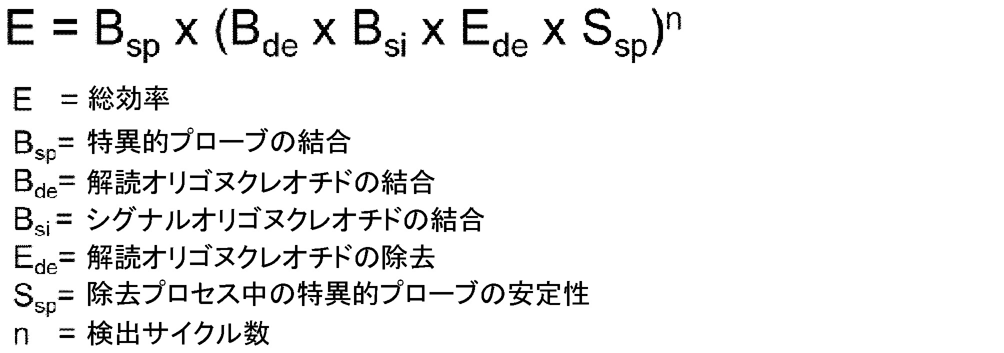
この式に基づいて、各単一ステップの効率は、方法の所与の総効率について推定することができる。計算は、各プロセスが同じ効率を有することを仮定することに基づく。総効率は、存在する全シグナルのうち解読に成功可能なシグナルの部分を表す。 Based on this formula, the efficiency of each single step can be estimated for a given total efficiency of the method. Calculations are based on the assumption that each process has the same efficiency. Total efficiency represents the fraction of signals that can be successfully decoded out of the total signals present.
方法の総効率は、式によって記述される異なる因子の各単一ステップの効率に依存する。均等に分布した効率の仮定の下で、図7に示すように、総効率を単一ステップ効率に対してプロットすることができる。見て分かるように、5回の検出サイクルを有する符コード化スキームの実際に適切な総効率は、明らかに90%を超える単一ステップ効率でのみ達成することができる。例えば、50%の総効率を達成するために、97.8%の各単一ステップ内の平均効率が必要である。これらの計算は、100%シグナル検出及び分析効率の仮定に基づいている。様々な配列のオリゴヌクレオチドの広いDNA融解曲線のために、本発明者らは、実験前に、選択的変性は、解読オリゴヌクレオチドの変性に対してあまり効率的ではなく、配列特異的結合プローブは十分に安定ではないと仮定した。この仮定とは対照的に、本発明者らは、全てのステップの驚くべき有効性及び選択的変性中の配列特異的プローブの高い安定性を見出した。 The total efficiency of the method depends on the efficiency of each single step of different factors described by the equation. Under the assumption of evenly distributed efficiencies, total efficiency can be plotted against single step efficiency as shown in FIG. As can be seen, a practically adequate overall efficiency for a code-encoding scheme with 5 detection cycles can only be achieved with a single-step efficiency clearly above 90%. For example, to achieve an overall efficiency of 50%, an average efficiency within each single step of 97.8% is required. These calculations are based on the assumption of 100% signal detection and analysis efficiency. Due to the broad DNA melting curves of oligonucleotides of varying sequence, we found, prior to experiments, that selective denaturation was less efficient for denaturation of decoding oligonucleotides and that sequence-specific binding probes were assumed not stable enough. In contrast to this assumption, we found a surprising efficiency of all steps and high stability of sequence-specific probes during selective denaturation.
実験的に、本発明者らは、5回の検出サイクルに基づいて、約30%~65%の総解読効率を達成した。上記の式による各単一ステップ(Bsp、Bde、Bsi、Ede、Ssp)の効率の計算は、約94.4%~98%の平均効率を明らかにした。これらの高い効率は非常に驚くべきことであり、この分野における熟練者は容易に予想することができない。 Experimentally, we achieved a total decoding efficiency of about 30%-65% based on 5 detection cycles. Calculation of the efficiency of each single step (Bsp, Bde, Bsi, Ede, Ssp) according to the above formula revealed an average efficiency of about 94.4% to 98%. These high efficiencies are quite surprising and cannot be easily predicted by those skilled in the art.
7.実験データ
背景
この実験は、単一分子分割と並行して10~50の異なるmRNA種の特異的検出を示す。これは、5回の検出サイクル、3つの異なる蛍光シグナル、及びシグナルギャップのないコード化スキーム、及び2のハミング距離(エラー検出)に基づく。実験は、本発明による方法の有効性及び機能性を証明している。
7. Experimental Data Background This experiment demonstrates the specific detection of 10-50 different mRNA species in parallel with single molecule splitting. This is based on 5 detection cycles, 3 different fluorescence signals and a coding scheme without signal gaps and a Hamming distance of 2 (error detection). Experiments prove the effectiveness and functionality of the method according to the invention.
オリゴヌクレオチド及びそれらの配列
実験で使用される全てのオリゴヌクレオチド配列(標的特異的プローブ、解読オリゴヌクレオチド、シグナルオリゴヌクレオチド)は、付録の配列表に列挙されている。シグナルオリゴヌクレオチドR:ST05*O_Atto594は、bioers.net GmbHに注文した。他の全てのオリゴヌクレオチドは、Integrated DNA Technologiesに注文した。オリゴヌクレオチドを水に溶解した。ストック溶液(100μM)を-20℃で保存した。
Oligonucleotides and Their Sequences All oligonucleotide sequences (target-specific probes, decoding oligonucleotides, signal oligonucleotides) used in the experiments are listed in the Sequence Listing in the Appendix. Signal oligonucleotide R: ST05*O_Atto594 was obtained from bioers. I ordered from net GmbH. All other oligonucleotides were ordered from Integrated DNA Technologies. Oligonucleotides were dissolved in water. Stock solutions (100 μM) were stored at -20°C.
実験の概要
50の異なる標的特異的プローブセットを5つの群に分ける。検出される転写物の名称及び標的特異的プローブセットの名称は同じである(www.ensemble.orgの転写物バリアント名)。「新規」という用語は、修正されたプローブ設計を示す。プローブセットの全てのオリゴヌクレオチド配列は、配列表に見出すことができる。表には、プローブセットの固有の識別子名、並びに異なる検出サイクルで使用される解読オリゴヌクレオチドの名称が列挙されている。得られたコードは、5回の検出サイクル中に生成された蛍光シグナルの配列を示す(G(reen)=Alexa Fluor 488、O(range)=Atto 594、Y(ellow)=Alexa Fluor 546)。

実験の変形
実験のいくつかの変形を行った。実験1~4は、並行して検出された転写物の数が主に異なる。標的特異的プローブセットとして列挙された群は、表6を参照する。実験5~8は、解読されたシグナルと比較するための単一ラウンドの単一の標的対照である。
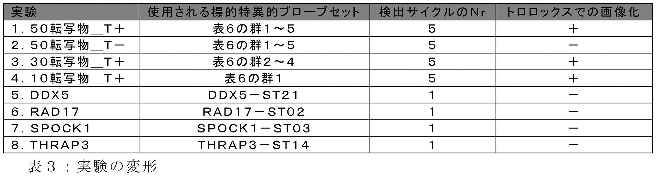
A.細胞の播種及び培養
HeLa細胞をHeLa細胞培養培地でほぼ100%コンフルエントに増殖させた。HeLa細胞培養培地は、10%FCS(Biochrom、カタログ番号S0415)、1%ペニシリン-ストレプトマイシン(Sigma-Adrich、カタログ番号P0781)及び1%MEM非必須アミノ酸溶液(Thermo Fisher、カタログ番号11140035)を含むDMEM(Thermo Fisher、カタログ番号31885)を含む。細胞培養培地を吸引した後、細胞を、PBS(水中1,424g/lのNa2HPO4*2H2O、0,276g/lのNaH2PO4*2H2O、8,19g/lのNaCl、pH7,4)による洗浄ステップの後、トリプシンEDTA溶液(Sigma-Aldrich、カタログ番号T3924)での5分間の37℃でのインキュベーションによってトリプシン処理した。次いで、μ-Slide 8 Well ibidiTreat(Ibidi、カタログ番号80826)のウェルに細胞を播種した。ウェルあたりの細胞の数を、細胞の接着後に約50%のコンフルエントに達するように調整した。細胞を、ウェルあたり200μlのHeLa細胞培養培地で一晩インキュベートした。
B.細胞の固定
細胞培養培地の吸引及びウェルあたり200μlの37℃加温PBSでの2回の洗浄ステップの後、細胞を200μlの予冷メタノール(-20℃、Roth、カタログ番号0082.1)で-20℃で10分間固定した。
C.スダンブラックによる対比染色
メタノールを吸引し、70%エタノール中で希釈した150μlの0.2%スダンブラック溶液を各ウェルに添加した。ウェルを暗所で5分間、室温でインキュベートした。インキュベーション後、細胞を、ウェルあたり400μlの70%エタノールで3回洗浄して、過剰のスダンブラック溶液を除去した。
D.分析物/標的特異的プローブのハイブリダイゼーション
ハイブリダイゼーション前に、細胞を200μlのsm洗浄緩衝液で平衡化した。sm洗浄緩衝液は、30mMクエン酸Na3、300mM NaCl、pH7、10%ホルムアミド(Roth、カタログ番号号P040.1)及び5mMリボヌクレオシドバナジル複合体(NEB、カタログ番号S1402S)を含む。各標的特異的プローブセットについて、1μlの100μMオリゴヌクレオチドストック溶液を混合物に添加した。オリゴヌクレオチドストック溶液は、対応する標的特異的プローブセットの全ての標的特異的オリゴヌクレオチドの等モル量を含む。混合物の総体積を水で100μlに調整し、100μlの2x濃縮ハイブリダイゼーション緩衝液と混合した。2x濃縮ハイブリダイゼーション緩衝液は、120mM クエン酸Na3、1200mM NaCl、pH7、20%ホルムアミド及び20mMリボヌクレオシドバナジル複合体を含む。得られた200μlのハイブリダイゼーション混合物を対応するウェルに添加し、37℃で2時間インキュベートした。その後、細胞をウェルあたり200μlで10分間、標的プローブ洗浄緩衝液で37℃で3回洗浄した。標的プローブ洗浄緩衝液は、30mM クエン酸Na3、300mM NaCl、pH7、20%ホルムアミド及び5mMリボヌクレオシドバナジル複合体を含む。
E.解読オリゴヌクレオチドのハイブリダイゼーション
ハイブリダイゼーション前に、細胞を200μlのsm洗浄緩衝液で平衡化した。各解読オリゴヌクレオチドについて、1,5μlの5μMストック溶液を混合物に添加した。混合物の総体積を水で75μlに調整し、75μlの2x濃縮ハイブリダイゼーション緩衝液と混合した。得られた150μlの解読オリゴヌクレオチドハイブリダイゼーション混合物を対応するウェルに添加し、室温で45分間インキュベートした。その後、細胞をウェルあたり200μlで2分間、sm洗浄緩衝液で室温で3回洗浄した。
F.シグナルオリゴヌクレオチドのハイブリダイゼーション
ハイブリダイゼーション前に、細胞を200μlのsm洗浄緩衝液で平衡化した。シグナルオリゴヌクレオチドハイブリダイゼーション混合物は、実験1~4の全てのラウンドについて同じであり、1×濃縮ハイブリダイゼーション緩衝液中に0、3μMの各シグナルオリゴヌクレオチド(表A3参照)を含んでいた。各ラウンドにおいて、ウェルあたり150μlのこの溶液を添加し、室温で45分間インキュベートした。各シグナルオリゴヌクレオチドの最終濃度が0、15μMであったことを除いて、手順は実験5~8と同じであった。その後、細胞をウェルあたり200μlで3回、室温のsm洗浄緩衝液で2分間洗浄した。
G.蛍光及び白色光画像化
細胞を、室温でウェルあたり200μlの画像化緩衝液で1回洗浄した。トロロックスを用いない実験(表7、最後の列を参照)では、画像化緩衝液は、30mMクエン酸Na3、300mM NaCl、pH7及び5mMリボヌクレオシドバナジル複合体を含む。トロロックスを用いた実験では、画像化緩衝液は、10%VectaCell Trolox Antifade Reagent(Vector laboratories、カタログ番号CB-1000)を更に含み、10mMの最終トロロックス濃度をもたらす。
開口数1.4の63×液浸油対物レンズ(Zeiss、apochromat)、pco.edge 4.2 CMOSカメラ(PCO AG)、及びLED光源(Zeiss、colibri 7)を備えたZeiss Axiovert 200M顕微鏡を、領域の画像化のために使用した。フィルタセット及びLED波長を、使用されるフルオロフォアの異なる最適条件に調整した。画像あたりの照射時間は、Alexa Fluor 546及びAtto 594では1000ms、Alexa Fluor 488では400msであった。
各実験では、画像化のために3つの領域をランダムに選択した。各領域について、32個の画像のzスタックを、350nmのzステップサイズで検出した。また、各領域から白色光画像を1枚撮影した。2つ以上の検出サイクルを用いた実験では、最初の検出ラウンドの領域が再び見出され、その後のラウンドごとに画像化された。
H.選択的変性
選択的変性のために、各ウェルを200μlのsm洗浄緩衝液と共に42℃で6分間インキュベートした。この手順を6回繰り返した。
Variations of the experiment Several variations of the experiment were performed. Experiments 1-4 differ primarily in the number of transcripts detected in parallel. See Table 6 for groups listed as target-specific probe sets. Experiments 5-8 are single round single target controls for comparison with decoded signals.
B. Cell Fixation After aspiration of the cell culture medium and two washing steps with 200 μl per well of 37° C. warmed PBS, cells were fixed to −20° C. with 200 μl pre-chilled methanol (−20° C., Roth, Cat. No. 0082.1). °C for 10 minutes.
C. Counterstaining with Sudan Black Methanol was aspirated and 150 μl of 0.2% Sudan black solution diluted in 70% ethanol was added to each well. Wells were incubated in the dark for 5 minutes at room temperature. After incubation, cells were washed three times with 400 μl of 70% ethanol per well to remove excess Sudan black solution.
D. Hybridization of Analyte/Target Specific Probes Cells were equilibrated with 200 μl of sm wash buffer prior to hybridization. The sm wash buffer contains 30 mM Na 3 Citrate, 300 mM NaCl,
E. Hybridization of Decoding Oligonucleotides Cells were equilibrated with 200 μl of sm wash buffer prior to hybridization. For each decoding oligonucleotide, 1.5 μl of 5 μM stock solution was added to the mixture. The total volume of the mixture was adjusted to 75 μl with water and mixed with 75 μl of 2x concentrated hybridization buffer. 150 μl of the resulting decoding oligonucleotide hybridization mixture was added to corresponding wells and incubated at room temperature for 45 minutes. Cells were then washed 3 times with 200 μl per well for 2 minutes with sm wash buffer at room temperature.
F. Hybridization of Signal Oligonucleotides Cells were equilibrated with 200 μl of sm wash buffer prior to hybridization. The signal oligonucleotide hybridization mixture was the same for all rounds of experiments 1-4, containing 0, 3 μM of each signal oligonucleotide (see Table A3) in 1× concentrated hybridization buffer. In each round, 150 μl of this solution was added per well and incubated for 45 minutes at room temperature. The procedure was the same as experiments 5-8, except that the final concentration of each signal oligonucleotide was 0, 15 μM. Cells were then washed 3 times with 200 μl/well of room temperature sm wash buffer for 2 minutes.
G. Fluorescence and White Light Imaging Cells were washed once with 200 μl imaging buffer per well at room temperature. In experiments without Trolox (see Table 7, last column), the imaging buffer contained 30 mM Na3 citrate, 300 mM NaCl,
A 63× immersion oil objective with a numerical aperture of 1.4 (Zeiss, apochromat), pco. A Zeiss Axiovert 200M microscope equipped with an edge 4.2 CMOS camera (PCO AG) and an LED light source (Zeiss, colibri 7) was used for field imaging. Filter sets and LED wavelengths were adjusted to different optima of the fluorophores used. The exposure time per image was 1000 ms for Alexa Fluor 546 and Atto 594 and 400 ms for Alexa Fluor 488.
In each experiment, three areas were randomly selected for imaging. For each region, z-stacks of 32 images were detected with a z-step size of 350 nm. Also, one white light image was taken from each region. In experiments with more than one detection cycle, the area of the first detection round was found again and imaged in each subsequent round.
H. Selective Denaturation For selective denaturation, each well was incubated with 200 μl of sm wash buffer for 6 minutes at 42°C. This procedure was repeated 6 times.
実験1~4では、ステップ(E)~(H)を5回繰り返した。5回目の検出サイクルではステップ(H)を省略した。
I.分析
カスタムImageJプラグインに基づいて、バックグラウンドから特定の蛍光シグナルを区別するために生データの半自動分析を行った。3つの蛍光チャネル全ての得られた3D点群を、カスタムVBAスクリプトとインシリコで組み合わせた。5つの検出サイクルの結果として得られた結合された3D点群を、VBAスクリプトに基づいて互いに整列させた。得られたアライメントは、検出された各固有のシグナルのコードワードを明らかにした。解読に成功したシグナルを、カスタムVBAスクリプト及びImageJ-プラグインに基づく実験の定量的及び空間的分析に使用した。
結果
1.解読シグナルの絶対数
全ての転写物についての解読に成功したシグナルの絶対数を、各実験の各領域について以下の表4に列挙する。要約すると、正しいコードの合計は、対応する実験で検出可能な転写物に割り当てられた解読されたシグナルの総数を示し、誤ったコードの合計は、対応する実験で検出できない解読されたシグナルの総数を示す。信号の総数は、解読に成功したシグナル及び解読に失敗したシグナルを含む。
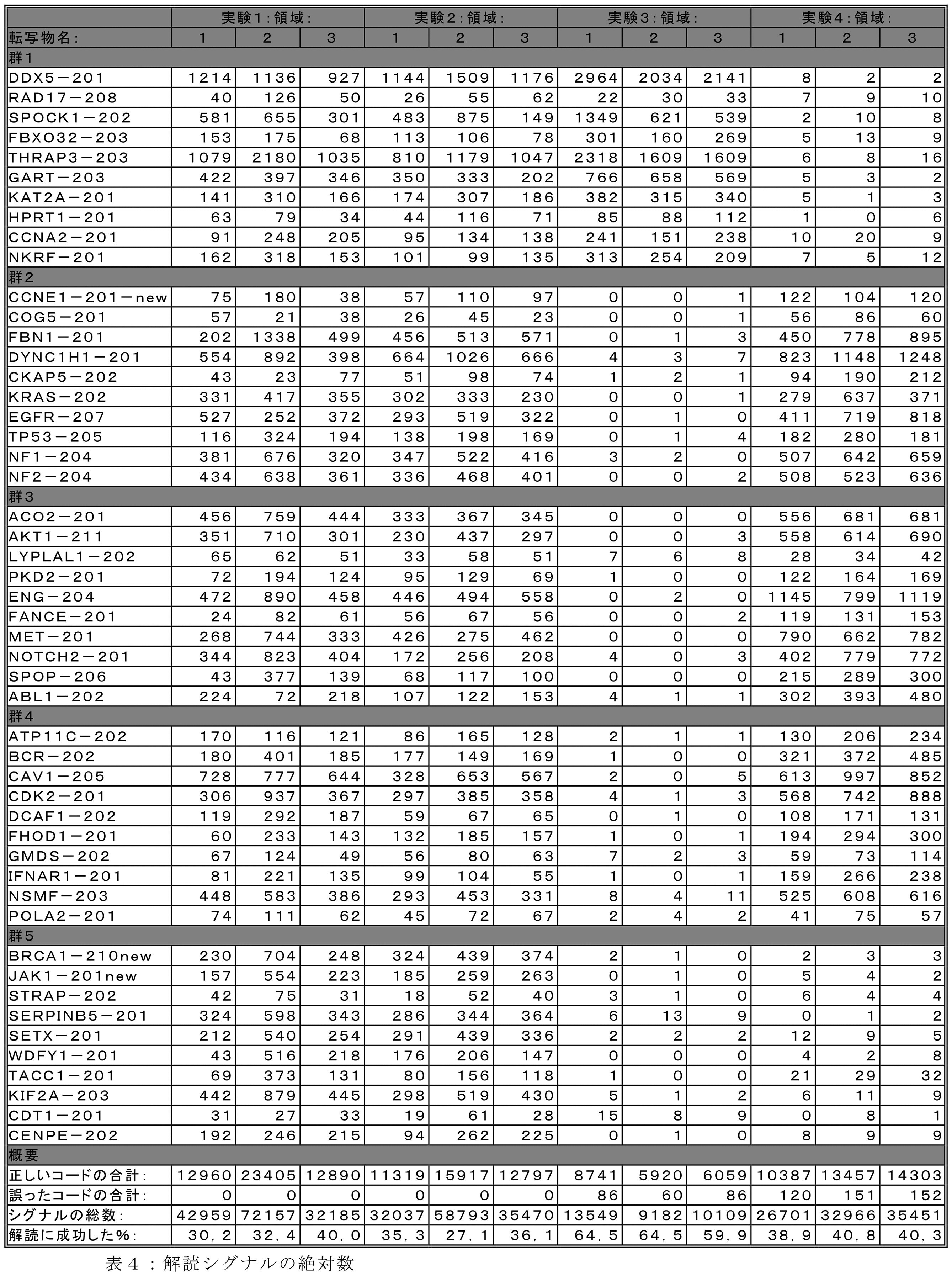
I. Analysis Based on a custom ImageJ plugin, semi-automated analysis of raw data was performed to distinguish specific fluorescent signals from background. The resulting 3D point clouds of all three fluorescence channels were combined in silico with a custom VBA script. The combined 3D point clouds resulting from five detection cycles were aligned with each other based on a VBA script. The resulting alignment revealed a codeword for each unique signal detected. Successfully decoded signals were used for quantitative and spatial analysis of experiments based on custom VBA scripts and ImageJ-plugins.
表4は、正しく解読されたシグナルの数と比較して非常に少ない数の誤って解読されたシグナルを示す。特定の転写物の解読されたシグナルの絶対値は、1つの実験の異なる領域間で非常に類似している。解読に成功することができるシグナルの総数の割合は、27.1%~64.5%である。この割合は、それぞれの領域/実験に存在する転写物の数及び/又はシグナルの総数に依存する。 Table 4 shows a very small number of incorrectly decoded signals compared to the number of correctly decoded signals. The absolute value of the decoded signal for a particular transcript is very similar between different regions in one experiment. The percentage of the total number of signals that can be successfully decoded is between 27.1% and 64.5%. This proportion depends on the number of transcripts and/or the total number of signals present in each region/experiment.
結論
本発明による方法は、誤って割り当てられたコードワードを少量しか生成せず、したがって特定的であると見なすことができる。領域当たりのシグナルの数が非常に多く、並行して検出される転写物の数が非常に多い場合であっても、解読に成功可能なシグナルの割合は非常に高い。割り当て可能なシグナルの割合が高く、特異性が高いため、この方法は実用的に有用である。
2.異なる実験間の相対転写物存在量の比較
両方の比較(A及びB)について図8に示すように、実験間で検出された転写物の重複を分析に使用する。各バーは、実験の3つ全ての領域の平均存在量を表す。これらの領域間の標準偏差も示されている。
3.異なる実験間の相対転写物存在量の相関
図9に見ることができるように、実験1の転写物の平均相対存在量は、実験3、4及び2の重複転写物の存在量と相関している。相関関係及び線形回帰の式は、各相関について示される。
図8は、低い標準偏差を示しており、1つの実験の異なる領域間の相対存在量の低い変動を示している。異なる実験からの転写物間の相対存在量の差も非常に低い。これは、実験1、2、及び3で検出された群1(図8A)からの転写物の比較の場合である。また、実験1、2、及び4の間で重複した2、3、及び4群からの転写物の比較についても同様である。これらの存在量の非常に高い相関は、図9にも見ることができる。実験1からの転写物の存在量は、他のマルチラウンド実験の存在量と非常に良好に相関する。相関係数は、0.88~0.91であり、一方、線形回帰の勾配は、0.97~1.05である。
CONCLUSION The method according to the invention produces only a small amount of incorrectly assigned codewords and can therefore be considered specific. Even with a very large number of signals per region and a very large number of transcripts detected in parallel, the percentage of signals that can be successfully decoded is very high. The high percentage of assignable signals and high specificity make this method practically useful.
2. Comparison of Relative Transcript Abundance Between Different Experiments The overlap of transcripts detected between experiments is used in the analysis, as shown in FIG. 8 for both comparisons (A and B). Each bar represents the mean abundance of all three regions of the experiment. Standard deviations between these regions are also shown.
3. Correlation of Relative Transcript Abundance Between Different Experiments As can be seen in FIG. there is Correlation and linear regression equations are shown for each correlation.
Figure 8 shows low standard deviations, indicating low variation in relative abundance between different regions of one experiment. Relative abundance differences between transcripts from different experiments are also very low. This is the case for comparison of transcripts from group 1 (FIG. 8A) detected in
結論
転写物の相対存在量は、1つの実験の異なる領域間だけでなく、異なる実験間でも非常によく相関する。これは、図3及び図4の比較によって明確に見ることができる。実験間の主な違いは、異なる標的の数であり、したがって検出されたシグナルの総数である。したがって、検出された転写物の数並びにシグナルの数及び密度は、転写物の数を正確に定量化する方法の能力を妨げない。非常に良好な相関は、非常に多数のシグナルであっても、方法の特異性及び安定性を更に支持する。
4.シグナルの細胞間分布の比較
図10には、画像スタックの最大投影が示されている。A:実験7の領域1(SPOCK1を検出する単一ラウンド単一転写実験)、B:実験1から選択された全てのシグナルの2D投影、SPOCK1に割り当てられた領域1、C:実験8の領域1(THRAP3を検出する単一ラウンド単一転写実験)、D:実験1から選択された全てのシグナルの2D投影、THRAP3に割り当てられた領域1。
5.シグナルの細胞内分布の比較
図11には、画像スタックの最大投影が示されている。対応する領域の拡大されたサブ領域が示されている。A:実験8の領域1(THRAP3を検出する単一ラウンド単一転写実験)、B:実験1から選択されたシグナルの2D投影、THRAP3に割り当てられた領域1、C:実験5の領域1(DDX5を検出する単一ラウンド単一転写実験)、D:実験1から選択された全てのシグナルの2D投影、DDX5に割り当てられた領域1。
図10は、異なる転写物間の細胞間分布の大きな差を示す。SPOCK1は、一部の細胞では非常に豊富であるが、他の細胞ではほとんど存在しないようである(図10A)。THRAP3は、領域の全ての細胞にわたってより均一な分布を示す(図10C)。これらの空間分布パターンはまた、実験1からの対応する転写物に割り当てられた点群(図10B及びD)で明確に観察することができる。
図11は、異なる転写物間の細胞内分布の大きな差を示す。THRAP3は主に細胞の周辺部(細胞質)で観察することができ(図11A)、DDX5は細胞の中心(核)でより高い存在量を示す(図11C)。これらの細胞内分布は、THRAP3及びDDX5に割り当てられた実験1の点群でも観察することができる(図11B及びD)。
Conclusions The relative abundance of transcripts correlates very well not only between different regions in one experiment, but also between different experiments. This can be clearly seen by comparing FIGS. 3 and 4. FIG. The main difference between experiments is the number of different targets and thus the total number of signals detected. Therefore, the number of transcripts detected and the number and density of signals do not interfere with the method's ability to accurately quantify the number of transcripts. A very good correlation further supports the specificity and stability of the method even with a very large number of signals.
4. Comparison of cell-to-cell distributions of signal Fig. 10 shows the maximum projection of the image stack. A:
5. Comparison of Subcellular Distributions of Signals FIG. 11 shows the maximum projection of the image stack. A magnified subregion of the corresponding region is shown. A:
FIG. 10 shows large differences in cell-to-cell distribution between different transcripts. SPOCK1 appears to be highly abundant in some cells but almost absent in others (Fig. 10A). THRAP3 shows a more even distribution across all cells in the area (Fig. 10C). These spatial distribution patterns can also be clearly observed in the point clouds assigned to the corresponding transcripts from experiment 1 (FIGS. 10B and D).
Figure 11 shows the large differences in subcellular distribution between different transcripts. THRAP3 can be observed mainly in the cell periphery (cytoplasm) (Fig. 11A), while DDX5 shows higher abundance in the cell center (nucleus) (Fig. 11C). These subcellular distributions can also be observed in the point cloud of
結論
定量の信頼性に次いで、マルチラウンド実験の点群も、転写物の同じ細胞内及び細胞間分布パターンを示す。これは、1つの特徴的なmRNA種のみを検出する単一ラウンド実験からのシグナルと割り当てられた点群との直接比較によって明確に証明される。
6.異なる細胞サイクル依存転写物の分布パターン
図12の全ての画像は、実験1の領域1を示す。各画像には、特定の転写物A:CCNA2、B:CENPE、C:CCNE1、D:全ての転写物に割り当てられた点群が示されている。図12は、3つの異なる細胞周期依存タンパク質の転写物を示す。CENPE(図12B)は、セントロメアタンパク質Eとしても知られており、G2期の間に蓄積する。紡錘体伸長及び染色体移動を担うことが提案されている。それは相間中には存在しない。CCNA2(図12A)はサイクリンA2としても知られている。それは、G2からM期への移行中にCDK1と相互作用することによって細胞周期進行を調節する。興味深いことに、両方のmRNA種の明らかな共局在が存在する。それらは、主に領域1の3つの中央細胞に存在する。CCNE1(図12C)はサイクリンE1としても知られている。このサイクリンはCDK2と相互作用し、G1相からS相への移行に関与する。図12は、この遺伝子の転写物が3つの中央細胞に存在しないが、他の細胞にわたって非常に均等に分布していないことを明確に示す。したがって、他の2つの転写物に対する抗局在化を示す。対応する点群のデータは、非常に多数の点及び非常に高い点密度を有する点群から導出される(図12Dは印象を与える)。
Conclusions Next to the reliability of the quantification, the point clouds of the multi-round experiments also show the same intra- and intercellular distribution patterns of the transcripts. This is clearly evidenced by direct comparison of the assigned point cloud with the signal from a single round experiment that detects only one characteristic mRNA species.
6. Different Cell Cycle Dependent Transcript Distribution Patterns All images in FIG. Each image shows point clouds assigned to specific transcripts A: CCNA2, B: CENPE, C: CCNE1, D: all transcripts. Figure 12 shows the transcripts of three different cell cycle dependent proteins. CENPE (Fig. 12B), also known as centromere protein E, accumulates during the G2 phase. It has been proposed to be responsible for spindle elongation and chromosome migration. It does not exist in the interphase. CCNA2 (Figure 12A) is also known as cyclin A2. It regulates cell cycle progression by interacting with CDK1 during the transition from G2 to M phase. Interestingly, there is a clear co-localization of both mRNA species. They are mainly present in the three central cells of
結論
図12に示される細胞周期依存性タンパク質の3つの解読された点群は、それらの対応する関数によって説明することができる分布パターンを示す。これらのデータは、本発明者らの方法が、細胞あたりのシグナル数が少なく(図12C)、シグナル密度が非常に高い(図12D)場合でも、生物学的に関連するデータを確実に生成することを強く示唆している。
Conclusions The three decoded point clouds of cell cycle dependent proteins shown in Figure 12 exhibit distribution patterns that can be explained by their corresponding functions. These data ensure that our method produces biologically relevant data even when the number of signals per cell is low (Fig. 12C) and the signal density is very high (Fig. 12D). strongly suggests that
配列表
添付の配列表において、配列番号1~1247は、例示的な標的特異的オリゴヌクレオチドのヌクレオチド配列を指す。列挙されたオリゴヌクレオチドは、標的特異的結合部位(5’末端)、スペーサー/リンカー配列(gtaac及びtagac)及び1つのプローブセットの全てのオリゴヌクレオチドについて同じである固有の識別子配列からなる。
SEQUENCE LISTING In the accompanying sequence listing, SEQ ID NOs: 1-1247 refer to the nucleotide sequences of exemplary target-specific oligonucleotides. The oligonucleotides listed consist of a target-specific binding site (5' end), spacer/linker sequences (gtaac and tagac) and a unique identifier sequence that is the same for all oligonucleotides of one probe set.
添付の配列表において、配列番号1248~1397は、例示的な解読オリゴヌクレオチドのヌクレオチド配列を指す。 In the attached sequence listing, SEQ ID NOS: 1248-1397 refer to the nucleotide sequences of exemplary decoding oligonucleotides.
添付の配列表において、配列番号1398~1400は、例示的なシグナルオリゴヌクレオチドのヌクレオチド配列を指す。各シグナルオリゴヌクレオチドについて、対応するフルオロフォアは2回存在する。1つのフルオロフォアは、5’末端に共有結合しており、1つのフルオロフォアは、3’末端に共有結合している。配列番号1398は、その5’末端に「5Alex488N」、及びその3’末端に「3AlexF488N」を含む。配列番号1399は、その5’末端に「5Alex546」、及びその3’末端に3Alex546Nを含む。配列番号1400は、その5’末端及びその3’末端に「Atto594」を含む。 In the attached sequence listing, SEQ ID NOS: 1398-1400 refer to the nucleotide sequences of exemplary signal oligonucleotides. For each signal oligonucleotide the corresponding fluorophore is present twice. One fluorophore is covalently attached to the 5' end and one fluorophore is covalently attached to the 3' end. SEQ ID NO: 1398 contains "5Alex488N" at its 5' end and "3AlexF488N" at its 3' end. SEQ ID NO: 1399 contains "5Alex546" at its 5' end and 3Alex546N at its 3' end. SEQ ID NO: 1400 contains "Atto594" at its 5' end and its 3' end.
Claims (15)
(1)分析物特異的プローブのセットを提供するステップであって、各分析物特異的プローブが、
-コード化される前記分析物のうちの1つと特異的に相互作用する結合要素(S)と、
-前記分析物特異的プローブのセットに固有のヌクレオチド配列(固有の識別子配列)を含む識別子要素(T)と、を含むステップと、
(2)前記分析物特異的プローブのセットを前記試料とインキュベートし、それによってコード化される前記分析物への前記分析物特異的プローブの特異的結合を可能にするステップと、
(3)前記試料から非結合プローブを除去するステップと、
(4)解読オリゴヌクレオチドのセットを提供するステップであって、各解読オリゴヌクレオチドが、
-固有の識別子配列の少なくとも一部に本質的に相補的であるヌクレオチド配列を含む第1のコネクタ要素(t)と、
-シグナルオリゴヌクレオチドの特異的ハイブリダイゼーションを可能にするヌクレオチド配列を含むトランスレータ要素(c)と、を含むステップと、
(5)前記解読オリゴヌクレオチドのセットを前記試料とインキュベートし、それによって前記固有の識別子配列への前記解読オリゴヌクレオチドの特異的ハイブリダイゼーションを可能にするステップと、
(6)前記試料から非結合解読オリゴヌクレオチドを除去するステップと、
(7)シグナルオリゴヌクレオチドのセットを提供するステップであって、各シグナルオリゴヌクレオチドが、
-前記トランスレータ要素(c)の前記ヌクレオチド配列の少なくとも一部に本質的に相補的であるヌクレオチド配列を含む第2のコネクタ要素(C)と、
-シグナル要素と、を含むステップと、
(8)前記シグナルオリゴヌクレオチドのセットを前記試料とインキュベートし、それによって前記トランスレータ要素(c)への前記シグナルオリゴヌクレオチドの特異的ハイブリダイゼーションを可能にするステップと、を含む、方法。 A method of sequentially signal-encoding analytes in a sample, comprising the steps of:
(1) providing a set of analyte-specific probes, each analyte-specific probe comprising:
- a binding member (S) that specifically interacts with one of said encoded analytes;
- an identifier element (T) comprising a nucleotide sequence unique to said set of analyte-specific probes (unique identifier sequence);
(2) incubating the set of analyte-specific probes with the sample, thereby allowing specific binding of the analyte-specific probes to the encoded analytes;
(3) removing unbound probe from the sample;
(4) providing a set of decoding oligonucleotides, each decoding oligonucleotide comprising:
- a first connector element (t) comprising a nucleotide sequence essentially complementary to at least part of the unique identifier sequence;
- a translator element (c) comprising a nucleotide sequence allowing the specific hybridization of the signal oligonucleotide;
(5) incubating the set of decoding oligonucleotides with the sample, thereby allowing specific hybridization of the decoding oligonucleotides to the unique identifier sequence;
(6) removing unbound decoding oligonucleotides from said sample;
(7) providing a set of signal oligonucleotides, each signal oligonucleotide comprising:
- a second connector element (C) comprising a nucleotide sequence essentially complementary to at least part of said nucleotide sequence of said translator element (c);
- a step comprising a signaling element;
(8) incubating said set of signal oligonucleotides with said sample, thereby allowing specific hybridization of said signal oligonucleotides to said translator element (c).
(10)前記シグナルを検出するステップと、を更に含み、任意選択で、
(11)前記試料から前記解読オリゴヌクレオチド及びシグナルオリゴヌクレオチドを選択的に除去し、それによってコード化される前記分析物への前記分析物特異的プローブの前記特異的結合を本質的に維持するステップと、を更に含み、任意選択で、
(12)ステップ(4)~(11)を少なくとも1回[(42)~(112)]繰り返して、少なくとも2つのシグナルからなるコード化スキームを生成するステップと、を更に含む、請求項1~4のいずれか一項に記載の方法。 (9) removing unbound signal oligonucleotides from said sample;
(10) detecting said signal, optionally
(11) selectively removing said decoding oligonucleotides and signal oligonucleotides from said sample, thereby essentially maintaining said specific binding of said analyte-specific probes to said encoded analytes; and, optionally,
(12) repeating steps (4)-(11) at least once [(4 2 )-(11 2 )] to generate a coding scheme consisting of at least two signals, The method according to any one of 1-4.
-分析物特異的プローブのセットに固有であるヌクレオチド配列(固有の識別子配列)の少なくとも一部に本質的に相補的であるヌクレオチド配列を含む第1のコネクタ要素(t)と、
-シグナルオリゴヌクレオチドの特異的ハイブリダイゼーションを可能にするヌクレオチド配列を含む、トランスレータ要素(c)と、を含む、使用。 Use of a set of decoding oligonucleotides to sequentially signal-encode analytes in a sample, each decoding oligonucleotide comprising:
- a first connector element (t) comprising a nucleotide sequence that is essentially complementary to at least part of a nucleotide sequence that is unique to the set of analyte-specific probes (unique identifier sequence);
- a translator element (c) comprising a nucleotide sequence allowing specific hybridization of a signal oligonucleotide.
-分析物特異的プローブのセットであって、各分析物特異的プローブが、
-コード化される前記分析物のうちの1つと特異的に相互作用する結合要素(S)と、
-前記分析物特異的プローブのセットに固有のヌクレオチド配列(固有の識別子配列)を含む識別子要素(T)と、を含むセットと、
及び
-解読オリゴヌクレオチドのセットであって、各解読オリゴヌクレオチドが、
-前記固有の識別子配列の少なくとも一部に本質的に相補的であるヌクレオチド配列を含む第1のコネクタ要素(t)と、
-シグナルオリゴヌクレオチドの特異的ハイブリダイゼーションを可能にするヌクレオチド配列を含む、トランスレータ要素(c)と、を含むセットと、を含む、キット。 A kit for sequentially signal-encoding analytes in a sample, comprising:
- a set of analyte-specific probes, each analyte-specific probe comprising:
- a binding member (S) that specifically interacts with one of said encoded analytes;
- an identifier element (T) comprising a nucleotide sequence unique to said set of analyte-specific probes (unique identifier sequence);
and - a set of decoding oligonucleotides, each decoding oligonucleotide comprising:
- a first connector element (t) comprising a nucleotide sequence essentially complementary to at least part of said unique identifier sequence;
- a set comprising a translator element (c) comprising a nucleotide sequence allowing specific hybridization of a signal oligonucleotide.
-前記トランスレータ要素(c)の前記ヌクレオチド配列の少なくとも一部に本質的に相補的であるヌクレオチド配列を含む第2のコネクタ要素(C)と、
-シグナル要素と、を含む、セットを更に含む、請求項14に記載のキット。
- a set of signal oligonucleotides, each signal oligonucleotide comprising
- a second connector element (C) comprising a nucleotide sequence essentially complementary to at least part of said nucleotide sequence of said translator element (c);
- a signaling element, and a kit according to claim 14, further comprising a set.
Applications Claiming Priority (3)
| Application Number | Priority Date | Filing Date | Title |
|---|---|---|---|
| EP19181051.4A EP3754028A1 (en) | 2019-06-18 | 2019-06-18 | Method of signal encoding of analytes in a sample |
| EP19181051.4 | 2019-06-18 | ||
| PCT/EP2020/067010 WO2020254519A1 (en) | 2019-06-18 | 2020-06-18 | Method of signal encoding of analytes in a sample |
Publications (2)
| Publication Number | Publication Date |
|---|---|
| JP2022537048A true JP2022537048A (en) | 2022-08-23 |
| JP7804465B2 JP7804465B2 (en) | 2026-01-22 |
Family
ID=66998234
Family Applications (1)
| Application Number | Title | Priority Date | Filing Date |
|---|---|---|---|
| JP2021575925A Active JP7804465B2 (en) | 2019-06-18 | 2020-06-18 | Method for signal encoding of analytes in a sample |
Country Status (7)
| Country | Link |
|---|---|
| US (1) | US20220235402A1 (en) |
| EP (2) | EP3754028A1 (en) |
| JP (1) | JP7804465B2 (en) |
| KR (1) | KR20220044486A (en) |
| AU (1) | AU2020298038B2 (en) |
| CA (1) | CA3141255A1 (en) |
| WO (1) | WO2020254519A1 (en) |
Families Citing this family (89)
| Publication number | Priority date | Publication date | Assignee | Title |
|---|---|---|---|---|
| WO2017222453A1 (en) | 2016-06-21 | 2017-12-28 | Hauling Thomas | Nucleic acid sequencing |
| CN111263819B (en) | 2017-10-06 | 2025-04-15 | 10X基因组学有限公司 | RNA-templated ligation |
| US11519033B2 (en) | 2018-08-28 | 2022-12-06 | 10X Genomics, Inc. | Method for transposase-mediated spatial tagging and analyzing genomic DNA in a biological sample |
| US12529094B2 (en) | 2018-12-10 | 2026-01-20 | 10X Genomics, Inc. | Imaging system hardware |
| WO2020123309A1 (en) | 2018-12-10 | 2020-06-18 | 10X Genomics, Inc. | Resolving spatial arrays by proximity-based deconvolution |
| US11649485B2 (en) | 2019-01-06 | 2023-05-16 | 10X Genomics, Inc. | Generating capture probes for spatial analysis |
| US11926867B2 (en) | 2019-01-06 | 2024-03-12 | 10X Genomics, Inc. | Generating capture probes for spatial analysis |
| EP3976820A1 (en) | 2019-05-30 | 2022-04-06 | 10X Genomics, Inc. | Methods of detecting spatial heterogeneity of a biological sample |
| WO2020240025A1 (en) | 2019-05-31 | 2020-12-03 | Cartana Ab | Method of detecting target nucleic acid molecules |
| WO2021092433A2 (en) | 2019-11-08 | 2021-05-14 | 10X Genomics, Inc. | Enhancing specificity of analyte binding |
| GB201919032D0 (en) | 2019-12-20 | 2020-02-05 | Cartana Ab | Method of detecting an analyte |
| WO2021133842A1 (en) | 2019-12-23 | 2021-07-01 | 10X Genomics, Inc. | Compositions and methods for using fixed biological samples in partition-based assays |
| EP3891300B1 (en) | 2019-12-23 | 2023-03-29 | 10X Genomics, Inc. | Methods for spatial analysis using rna-templated ligation |
| US12365942B2 (en) | 2020-01-13 | 2025-07-22 | 10X Genomics, Inc. | Methods of decreasing background on a spatial array |
| US12405264B2 (en) | 2020-01-17 | 2025-09-02 | 10X Genomics, Inc. | Electrophoretic system and method for analyte capture |
| US11702693B2 (en) | 2020-01-21 | 2023-07-18 | 10X Genomics, Inc. | Methods for printing cells and generating arrays of barcoded cells |
| US11732299B2 (en) | 2020-01-21 | 2023-08-22 | 10X Genomics, Inc. | Spatial assays with perturbed cells |
| US20210230681A1 (en) | 2020-01-24 | 2021-07-29 | 10X Genomics, Inc. | Methods for spatial analysis using proximity ligation |
| US12076701B2 (en) | 2020-01-31 | 2024-09-03 | 10X Genomics, Inc. | Capturing oligonucleotides in spatial transcriptomics |
| US12110541B2 (en) | 2020-02-03 | 2024-10-08 | 10X Genomics, Inc. | Methods for preparing high-resolution spatial arrays |
| US11898205B2 (en) | 2020-02-03 | 2024-02-13 | 10X Genomics, Inc. | Increasing capture efficiency of spatial assays |
| US12110548B2 (en) | 2020-02-03 | 2024-10-08 | 10X Genomics, Inc. | Bi-directional in situ analysis |
| US11732300B2 (en) | 2020-02-05 | 2023-08-22 | 10X Genomics, Inc. | Increasing efficiency of spatial analysis in a biological sample |
| US12129516B2 (en) | 2020-02-07 | 2024-10-29 | 10X Genomics, Inc. | Quantitative and automated permeabilization performance evaluation for spatial transcriptomics |
| US12281357B1 (en) | 2020-02-14 | 2025-04-22 | 10X Genomics, Inc. | In situ spatial barcoding |
| EP4107284A1 (en) | 2020-02-17 | 2022-12-28 | 10X Genomics, Inc. | In situ analysis of chromatin interaction |
| US12173360B2 (en) | 2020-02-21 | 2024-12-24 | 10X Genomics, Inc. | Methods and compositions for integrated in situ spatial assay |
| US11891654B2 (en) | 2020-02-24 | 2024-02-06 | 10X Genomics, Inc. | Methods of making gene expression libraries |
| US12188085B2 (en) | 2020-03-05 | 2025-01-07 | 10X Genomics, Inc. | Three-dimensional spatial transcriptomics with sequencing readout |
| WO2021216708A1 (en) | 2020-04-22 | 2021-10-28 | 10X Genomics, Inc. | Methods for spatial analysis using targeted rna depletion |
| WO2021236929A1 (en) | 2020-05-22 | 2021-11-25 | 10X Genomics, Inc. | Simultaneous spatio-temporal measurement of gene expression and cellular activity |
| WO2021237087A1 (en) | 2020-05-22 | 2021-11-25 | 10X Genomics, Inc. | Spatial analysis to detect sequence variants |
| US12031177B1 (en) | 2020-06-04 | 2024-07-09 | 10X Genomics, Inc. | Methods of enhancing spatial resolution of transcripts |
| ES2981265T3 (en) | 2020-06-08 | 2024-10-08 | 10X Genomics Inc | Methods for determining a surgical margin and methods of using it |
| US12209273B2 (en) | 2020-06-12 | 2025-01-28 | 10X Genomics, Inc. | Nucleic acid assays using click chemistry bioconjugation |
| EP4450639B1 (en) | 2020-06-25 | 2025-10-15 | 10X Genomics, Inc. | Spatial analysis of dna methylation |
| US11761038B1 (en) | 2020-07-06 | 2023-09-19 | 10X Genomics, Inc. | Methods for identifying a location of an RNA in a biological sample |
| US12209280B1 (en) | 2020-07-06 | 2025-01-28 | 10X Genomics, Inc. | Methods of identifying abundance and location of an analyte in a biological sample using second strand synthesis |
| US11981960B1 (en) | 2020-07-06 | 2024-05-14 | 10X Genomics, Inc. | Spatial analysis utilizing degradable hydrogels |
| US12297499B2 (en) | 2020-08-17 | 2025-05-13 | 10X Genomics, Inc. | Multicomponent nucleic acid probes for sample analysis |
| US11981958B1 (en) | 2020-08-20 | 2024-05-14 | 10X Genomics, Inc. | Methods for spatial analysis using DNA capture |
| US11926822B1 (en) | 2020-09-23 | 2024-03-12 | 10X Genomics, Inc. | Three-dimensional spatial analysis |
| US12071667B2 (en) | 2020-11-04 | 2024-08-27 | 10X Genomics, Inc. | Sequence analysis using meta-stable nucleic acid molecules |
| US11827935B1 (en) | 2020-11-19 | 2023-11-28 | 10X Genomics, Inc. | Methods for spatial analysis using rolling circle amplification and detection probes |
| EP4012046A1 (en) | 2020-12-11 | 2022-06-15 | 10X Genomics, Inc. | Methods and compositions for multimodal in situ analysis |
| AU2021409136A1 (en) | 2020-12-21 | 2023-06-29 | 10X Genomics, Inc. | Methods, compositions, and systems for capturing probes and/or barcodes |
| US12060603B2 (en) | 2021-01-19 | 2024-08-13 | 10X Genomics, Inc. | Methods for internally controlled in situ assays using padlock probes |
| US12275984B2 (en) | 2021-03-02 | 2025-04-15 | 10X Genomics, Inc. | Sequential hybridization and quenching |
| EP4428246B1 (en) | 2021-04-14 | 2025-12-24 | 10X Genomics, Inc. | Methods of measuring mislocalization of an analyte |
| EP4320271B1 (en) | 2021-05-06 | 2025-03-19 | 10X Genomics, Inc. | Methods for increasing resolution of spatial analysis |
| EP4582555A3 (en) | 2021-06-03 | 2025-10-22 | 10X Genomics, Inc. | Methods, compositions, kits, and systems for enhancing analyte capture for spatial analysis |
| ES2988400T3 (en) | 2021-07-30 | 2024-11-20 | 10X Genomics Inc | Methods and compositions for synchronizing reactions in situ |
| US12139751B2 (en) | 2021-07-30 | 2024-11-12 | 10X Genomics, Inc. | Circularizable probes for in situ analysis |
| US12391984B2 (en) | 2021-08-03 | 2025-08-19 | 10X Genomics, Inc. | Compositions and methods for rolling circle amplification |
| US12460251B2 (en) | 2021-08-03 | 2025-11-04 | 10X Genomics, Inc. | Stabilization and/or compaction of nucleic acid molecules |
| US12529096B2 (en) | 2021-08-03 | 2026-01-20 | 10X Genomics, Inc. | Stabilization and/or compaction of nucleic acid structures |
| EP4381096B1 (en) * | 2021-08-06 | 2025-05-28 | Resolve BioSciences GmbH | Method for detecting an analyte in a pathogen-comprising sample |
| EP4446426A3 (en) | 2021-08-16 | 2024-11-13 | 10x Genomics, Inc. | Probes comprising a split barcode region and methods of use |
| WO2023034489A1 (en) | 2021-09-01 | 2023-03-09 | 10X Genomics, Inc. | Methods, compositions, and kits for blocking a capture probe on a spatial array |
| EP4413155A4 (en) * | 2021-10-04 | 2025-09-10 | The Regents Of Univ Of California | Nucleic acid implementation of multilayer perceptrons |
| US20240295474A1 (en) | 2021-10-25 | 2024-09-05 | Resolve Biosciences Gmbh | Method of mounting a biological sample on a surface |
| EP4419707A1 (en) | 2021-11-10 | 2024-08-28 | 10X Genomics, Inc. | Methods, compositions, and kits for determining the location of an analyte in a biological sample |
| EP4305195A2 (en) | 2021-12-01 | 2024-01-17 | 10X Genomics, Inc. | Methods, compositions, and systems for improved in situ detection of analytes and spatial analysis |
| EP4466376A1 (en) | 2022-01-21 | 2024-11-27 | 10X Genomics, Inc. | Multiple readout signals for analyzing a sample |
| WO2023172915A1 (en) | 2022-03-08 | 2023-09-14 | 10X Genomics, Inc. | In situ code design methods for minimizing optical crowding |
| CN119032271A (en) | 2022-04-01 | 2024-11-26 | 10X基因组学有限公司 | Compositions and methods for targeted masking of autofluorescence |
| JP2025516589A (en) | 2022-05-11 | 2025-05-30 | 10エックス ジェノミクス インコーポレイテッド | Compositions and methods for in situ sequencing |
| WO2023245190A1 (en) | 2022-06-17 | 2023-12-21 | 10X Genomics, Inc. | Catalytic de-crosslinking of samples for in situ analysis |
| WO2024002726A1 (en) * | 2022-06-30 | 2024-01-04 | Resolve Biosciences Gmbh | High resolution multiplex method for detecting at least two targets with a distance of beyond the diffraction limit in a sample |
| CN119677873A (en) | 2022-08-16 | 2025-03-21 | 10X基因组学有限公司 | AP50 polymerase and its use |
| DE102022131447A1 (en) | 2022-11-28 | 2024-05-29 | Carl Zeiss Microscopy Gmbh | Method and apparatus for processing data to identify analytes |
| DE102022131444A1 (en) | 2022-11-28 | 2024-05-29 | Carl Zeiss Microscopy Gmbh | Method for identifying analytes in an image sequence |
| DE102022131448A1 (en) | 2022-11-28 | 2024-05-29 | Carl Zeiss Microscopy Gmbh | Method and apparatus for processing data to identify analytes |
| DE102022131445A1 (en) | 2022-11-28 | 2024-05-29 | Carl Zeiss Microscopy Gmbh | Method and apparatus for processing data to identify analytes |
| DE102022131446A1 (en) | 2022-11-28 | 2024-05-29 | Carl Zeiss Microscopy Gmbh | Method and apparatus for processing data to identify analytes |
| DE102022131441A1 (en) | 2022-11-28 | 2024-05-29 | Carl Zeiss Microscopy Gmbh | Method and apparatus for processing data to identify analytes |
| DE102022131449A1 (en) | 2022-11-28 | 2024-05-29 | Carl Zeiss Microscopy Gmbh | Method and apparatus for processing data to identify analytes |
| DE102022131450A1 (en) | 2022-11-28 | 2024-05-29 | Carl Zeiss Microscopy Gmbh | Method and apparatus for processing data to identify analytes |
| DE102022131442A1 (en) | 2022-11-28 | 2024-05-29 | Carl Zeiss Microscopy Gmbh | Method and device for assigning image areas of an image sequence to result classes using an analyte data evaluation system with a processing model |
| DE102022131451A1 (en) | 2022-11-28 | 2024-05-29 | Carl Zeiss Microscopy Gmbh | Method and device for determining a signal composition of signal sequences of an image sequence |
| WO2024256715A1 (en) * | 2023-06-16 | 2024-12-19 | Resolve Biosciences Gmbh | Reagent for in situ hybridization quality control |
| US12319956B2 (en) | 2023-07-31 | 2025-06-03 | 10X Genomics, Inc. | Methods and systems for targeted RNA cleavage and target RNA-primed rolling circle amplification |
| DE102023122213A1 (en) | 2023-08-18 | 2025-02-20 | Carl Zeiss Microscopy Gmbh | Method for providing source data for training or validating a processing model for processing analyte image sequences |
| WO2025093690A1 (en) | 2023-11-02 | 2025-05-08 | Resolve Biosciences Gmbh | Method for preparing tissue sections |
| WO2025099313A1 (en) | 2023-11-10 | 2025-05-15 | Resolve Biosciences Gmbh | Reducing background fluorescence in single molecule fluorescence in situ hybridization applications |
| WO2025125442A1 (en) * | 2023-12-13 | 2025-06-19 | Resolve Biosciences Gmbh | High resolution multiplex method for detecting at least two targets |
| WO2025132546A1 (en) | 2023-12-19 | 2025-06-26 | Resolve Biosciences Gmbh | Method for reducing nonspecific signals in in-situ hybridization assays |
| WO2025145004A1 (en) * | 2023-12-29 | 2025-07-03 | Pleno, Inc. | Methods, systems, compositions and kits for target detection |
| US20250283822A1 (en) * | 2024-03-11 | 2025-09-11 | Vizgen, Inc. | Compositions and methods for improved multiplexed error robust fluorescence in situ hybridization |
Citations (3)
| Publication number | Priority date | Publication date | Assignee | Title |
|---|---|---|---|---|
| JP2000515367A (en) * | 1996-07-12 | 2000-11-21 | ティーエム テクノロジーズ,インク. | Signal amplification method |
| WO2017189525A1 (en) * | 2016-04-25 | 2017-11-02 | President And Fellows Of Harvard College | Hybridization chain reaction methods for in situ molecular detection |
| WO2018094385A1 (en) * | 2016-11-21 | 2018-05-24 | Nanostring Technologies, Inc. | Chemical compositions and methods of using same |
Family Cites Families (2)
| Publication number | Priority date | Publication date | Assignee | Title |
|---|---|---|---|---|
| CA1228811A (en) | 1983-05-05 | 1987-11-03 | Robert G. Pergolizzi | Assay method utilizing polynucleotide sequences |
| EP1190100B1 (en) * | 1999-05-20 | 2012-07-25 | Illumina, Inc. | Combinatorial decoding of random nucleic acid arrays |
-
2019
- 2019-06-18 EP EP19181051.4A patent/EP3754028A1/en active Pending
-
2020
- 2020-06-18 JP JP2021575925A patent/JP7804465B2/en active Active
- 2020-06-18 US US17/617,183 patent/US20220235402A1/en active Pending
- 2020-06-18 CA CA3141255A patent/CA3141255A1/en active Pending
- 2020-06-18 KR KR1020227001292A patent/KR20220044486A/en active Pending
- 2020-06-18 AU AU2020298038A patent/AU2020298038B2/en active Active
- 2020-06-18 EP EP20732632.3A patent/EP3987057A1/en active Pending
- 2020-06-18 WO PCT/EP2020/067010 patent/WO2020254519A1/en not_active Ceased
Patent Citations (3)
| Publication number | Priority date | Publication date | Assignee | Title |
|---|---|---|---|---|
| JP2000515367A (en) * | 1996-07-12 | 2000-11-21 | ティーエム テクノロジーズ,インク. | Signal amplification method |
| WO2017189525A1 (en) * | 2016-04-25 | 2017-11-02 | President And Fellows Of Harvard College | Hybridization chain reaction methods for in situ molecular detection |
| WO2018094385A1 (en) * | 2016-11-21 | 2018-05-24 | Nanostring Technologies, Inc. | Chemical compositions and methods of using same |
Non-Patent Citations (2)
| Title |
|---|
| ENG, CHEE-HUAT LINUS ET AL., NATURE, vol. 568, JPN6024025833, 25 March 2019 (2019-03-25), pages 235 - 239, ISSN: 0005562669 * |
| XIA, C. ET AL., SCIENTIFIC REPORTS, vol. 9, no. 7721, JPN7024002771, 22 May 2019 (2019-05-22), ISSN: 0005562668 * |
Also Published As
| Publication number | Publication date |
|---|---|
| KR20220044486A (en) | 2022-04-08 |
| AU2020298038B2 (en) | 2025-12-18 |
| JP7804465B2 (en) | 2026-01-22 |
| EP3987057A1 (en) | 2022-04-27 |
| WO2020254519A1 (en) | 2020-12-24 |
| CA3141255A1 (en) | 2020-12-24 |
| AU2020298038A1 (en) | 2022-01-27 |
| US20220235402A1 (en) | 2022-07-28 |
| EP3754028A1 (en) | 2020-12-23 |
Similar Documents
| Publication | Publication Date | Title |
|---|---|---|
| JP7804465B2 (en) | Method for signal encoding of analytes in a sample | |
| US12492427B2 (en) | Multiplex method for detecting different analytes in a sample | |
| US20230032082A1 (en) | Spatial barcoding | |
| JP7372927B6 (en) | Biomolecular probes and detection methods for detecting gene and protein expression | |
| JP2021010388A (en) | Method for identifying a plurality of epitopes in selected partial mass of cells | |
| CN116829733A (en) | Compositions and methods for binding analytes to capture probes | |
| US20250376718A1 (en) | High resolution multiplex method for detecting at least two targets with a distance of beyond the diffraction limit in a sample | |
| US20240158836A1 (en) | High resolution multiplex method for detecting at least two targets with a distance of beyond the diffraction limit in a sample | |
| CA3226471A1 (en) | Method for detecting an analyte in a pathogen-comprising sample | |
| US20240417781A1 (en) | High resolution multiplex method for detecting at least two targets with a distance of beyond the diffraction limit in a sample | |
| WO2025125442A1 (en) | High resolution multiplex method for detecting at least two targets |
Legal Events
| Date | Code | Title | Description |
|---|---|---|---|
| RD04 | Notification of resignation of power of attorney |
Free format text: JAPANESE INTERMEDIATE CODE: A7424 Effective date: 20220418 |
|
| A621 | Written request for application examination |
Free format text: JAPANESE INTERMEDIATE CODE: A621 Effective date: 20230613 |
|
| RD03 | Notification of appointment of power of attorney |
Free format text: JAPANESE INTERMEDIATE CODE: A7423 Effective date: 20230613 |
|
| A977 | Report on retrieval |
Free format text: JAPANESE INTERMEDIATE CODE: A971007 Effective date: 20240625 |
|
| A131 | Notification of reasons for refusal |
Free format text: JAPANESE INTERMEDIATE CODE: A131 Effective date: 20240702 |
|
| A601 | Written request for extension of time |
Free format text: JAPANESE INTERMEDIATE CODE: A601 Effective date: 20240930 |
|
| A601 | Written request for extension of time |
Free format text: JAPANESE INTERMEDIATE CODE: A601 Effective date: 20241129 |
|
| A521 | Request for written amendment filed |
Free format text: JAPANESE INTERMEDIATE CODE: A523 Effective date: 20241225 |
|
| A131 | Notification of reasons for refusal |
Free format text: JAPANESE INTERMEDIATE CODE: A131 Effective date: 20250401 |
|
| A601 | Written request for extension of time |
Free format text: JAPANESE INTERMEDIATE CODE: A601 Effective date: 20250627 |
|
| A521 | Request for written amendment filed |
Free format text: JAPANESE INTERMEDIATE CODE: A523 Effective date: 20250901 |
|
| TRDD | Decision of grant or rejection written | ||
| A01 | Written decision to grant a patent or to grant a registration (utility model) |
Free format text: JAPANESE INTERMEDIATE CODE: A01 Effective date: 20251216 |
|
| A61 | First payment of annual fees (during grant procedure) |
Free format text: JAPANESE INTERMEDIATE CODE: A61 Effective date: 20260109 |
|
| R150 | Certificate of patent or registration of utility model |
Ref document number: 7804465 Country of ref document: JP Free format text: JAPANESE INTERMEDIATE CODE: R150 |