JP2022516140A - Leukocyte immunoglobulin-like receptor 2 neutralizing antibody - Google Patents
Leukocyte immunoglobulin-like receptor 2 neutralizing antibody Download PDFInfo
- Publication number
- JP2022516140A JP2022516140A JP2021538424A JP2021538424A JP2022516140A JP 2022516140 A JP2022516140 A JP 2022516140A JP 2021538424 A JP2021538424 A JP 2021538424A JP 2021538424 A JP2021538424 A JP 2021538424A JP 2022516140 A JP2022516140 A JP 2022516140A
- Authority
- JP
- Japan
- Prior art keywords
- antibody
- ilt2
- cells
- seq
- amino acid
- Prior art date
- Legal status (The legal status is an assumption and is not a legal conclusion. Google has not performed a legal analysis and makes no representation as to the accuracy of the status listed.)
- Pending
Links
Images


Classifications
-
- C—CHEMISTRY; METALLURGY
- C07—ORGANIC CHEMISTRY
- C07K—PEPTIDES
- C07K16/00—Immunoglobulins [IGs], e.g. monoclonal or polyclonal antibodies
- C07K16/18—Immunoglobulins [IGs], e.g. monoclonal or polyclonal antibodies against material from animals or humans
- C07K16/28—Immunoglobulins [IGs], e.g. monoclonal or polyclonal antibodies against material from animals or humans against receptors, cell surface antigens or cell surface determinants
- C07K16/2863—Immunoglobulins [IGs], e.g. monoclonal or polyclonal antibodies against material from animals or humans against receptors, cell surface antigens or cell surface determinants against receptors for growth factors, growth regulators
-
- A—HUMAN NECESSITIES
- A61—MEDICAL OR VETERINARY SCIENCE; HYGIENE
- A61P—SPECIFIC THERAPEUTIC ACTIVITY OF CHEMICAL COMPOUNDS OR MEDICINAL PREPARATIONS
- A61P35/00—Antineoplastic agents
-
- C—CHEMISTRY; METALLURGY
- C07—ORGANIC CHEMISTRY
- C07K—PEPTIDES
- C07K16/00—Immunoglobulins [IGs], e.g. monoclonal or polyclonal antibodies
- C07K16/18—Immunoglobulins [IGs], e.g. monoclonal or polyclonal antibodies against material from animals or humans
- C07K16/28—Immunoglobulins [IGs], e.g. monoclonal or polyclonal antibodies against material from animals or humans against receptors, cell surface antigens or cell surface determinants
- C07K16/2803—Immunoglobulins [IGs], e.g. monoclonal or polyclonal antibodies against material from animals or humans against receptors, cell surface antigens or cell surface determinants against the immunoglobulin superfamily
-
- C—CHEMISTRY; METALLURGY
- C07—ORGANIC CHEMISTRY
- C07K—PEPTIDES
- C07K16/00—Immunoglobulins [IGs], e.g. monoclonal or polyclonal antibodies
- C07K16/18—Immunoglobulins [IGs], e.g. monoclonal or polyclonal antibodies against material from animals or humans
- C07K16/28—Immunoglobulins [IGs], e.g. monoclonal or polyclonal antibodies against material from animals or humans against receptors, cell surface antigens or cell surface determinants
- C07K16/2887—Immunoglobulins [IGs], e.g. monoclonal or polyclonal antibodies against material from animals or humans against receptors, cell surface antigens or cell surface determinants against CD20
-
- C—CHEMISTRY; METALLURGY
- C07—ORGANIC CHEMISTRY
- C07K—PEPTIDES
- C07K16/00—Immunoglobulins [IGs], e.g. monoclonal or polyclonal antibodies
- C07K16/18—Immunoglobulins [IGs], e.g. monoclonal or polyclonal antibodies against material from animals or humans
- C07K16/28—Immunoglobulins [IGs], e.g. monoclonal or polyclonal antibodies against material from animals or humans against receptors, cell surface antigens or cell surface determinants
- C07K16/30—Immunoglobulins [IGs], e.g. monoclonal or polyclonal antibodies against material from animals or humans against receptors, cell surface antigens or cell surface determinants from tumour cells
-
- A—HUMAN NECESSITIES
- A61—MEDICAL OR VETERINARY SCIENCE; HYGIENE
- A61K—PREPARATIONS FOR MEDICAL, DENTAL OR TOILETRY PURPOSES
- A61K39/00—Medicinal preparations containing antigens or antibodies
- A61K2039/505—Medicinal preparations containing antigens or antibodies comprising antibodies
-
- A—HUMAN NECESSITIES
- A61—MEDICAL OR VETERINARY SCIENCE; HYGIENE
- A61K—PREPARATIONS FOR MEDICAL, DENTAL OR TOILETRY PURPOSES
- A61K39/00—Medicinal preparations containing antigens or antibodies
- A61K2039/505—Medicinal preparations containing antigens or antibodies comprising antibodies
- A61K2039/507—Comprising a combination of two or more separate antibodies
-
- C—CHEMISTRY; METALLURGY
- C07—ORGANIC CHEMISTRY
- C07K—PEPTIDES
- C07K2317/00—Immunoglobulins specific features
- C07K2317/20—Immunoglobulins specific features characterized by taxonomic origin
- C07K2317/21—Immunoglobulins specific features characterized by taxonomic origin from primates, e.g. man
-
- C—CHEMISTRY; METALLURGY
- C07—ORGANIC CHEMISTRY
- C07K—PEPTIDES
- C07K2317/00—Immunoglobulins specific features
- C07K2317/20—Immunoglobulins specific features characterized by taxonomic origin
- C07K2317/24—Immunoglobulins specific features characterized by taxonomic origin containing regions, domains or residues from different species, e.g. chimeric, humanized or veneered
-
- C—CHEMISTRY; METALLURGY
- C07—ORGANIC CHEMISTRY
- C07K—PEPTIDES
- C07K2317/00—Immunoglobulins specific features
- C07K2317/30—Immunoglobulins specific features characterized by aspects of specificity or valency
- C07K2317/34—Identification of a linear epitope shorter than 20 amino acid residues or of a conformational epitope defined by amino acid residues
-
- C—CHEMISTRY; METALLURGY
- C07—ORGANIC CHEMISTRY
- C07K—PEPTIDES
- C07K2317/00—Immunoglobulins specific features
- C07K2317/40—Immunoglobulins specific features characterized by post-translational modification
- C07K2317/41—Glycosylation, sialylation, or fucosylation
-
- C—CHEMISTRY; METALLURGY
- C07—ORGANIC CHEMISTRY
- C07K—PEPTIDES
- C07K2317/00—Immunoglobulins specific features
- C07K2317/50—Immunoglobulins specific features characterized by immunoglobulin fragments
- C07K2317/52—Constant or Fc region; Isotype
-
- C—CHEMISTRY; METALLURGY
- C07—ORGANIC CHEMISTRY
- C07K—PEPTIDES
- C07K2317/00—Immunoglobulins specific features
- C07K2317/50—Immunoglobulins specific features characterized by immunoglobulin fragments
- C07K2317/56—Immunoglobulins specific features characterized by immunoglobulin fragments variable (Fv) region, i.e. VH and/or VL
- C07K2317/565—Complementarity determining region [CDR]
-
- C—CHEMISTRY; METALLURGY
- C07—ORGANIC CHEMISTRY
- C07K—PEPTIDES
- C07K2317/00—Immunoglobulins specific features
- C07K2317/70—Immunoglobulins specific features characterized by effect upon binding to a cell or to an antigen
- C07K2317/71—Decreased effector function due to an Fc-modification
-
- C—CHEMISTRY; METALLURGY
- C07—ORGANIC CHEMISTRY
- C07K—PEPTIDES
- C07K2317/00—Immunoglobulins specific features
- C07K2317/70—Immunoglobulins specific features characterized by effect upon binding to a cell or to an antigen
- C07K2317/73—Inducing cell death, e.g. apoptosis, necrosis or inhibition of cell proliferation
- C07K2317/732—Antibody-dependent cellular cytotoxicity [ADCC]
-
- C—CHEMISTRY; METALLURGY
- C07—ORGANIC CHEMISTRY
- C07K—PEPTIDES
- C07K2317/00—Immunoglobulins specific features
- C07K2317/70—Immunoglobulins specific features characterized by effect upon binding to a cell or to an antigen
- C07K2317/76—Antagonist effect on antigen, e.g. neutralization or inhibition of binding
-
- C—CHEMISTRY; METALLURGY
- C07—ORGANIC CHEMISTRY
- C07K—PEPTIDES
- C07K2317/00—Immunoglobulins specific features
- C07K2317/90—Immunoglobulins specific features characterized by (pharmaco)kinetic aspects or by stability of the immunoglobulin
- C07K2317/92—Affinity (KD), association rate (Ka), dissociation rate (Kd) or EC50 value
Landscapes
- Health & Medical Sciences (AREA)
- Chemical & Material Sciences (AREA)
- Organic Chemistry (AREA)
- Immunology (AREA)
- Life Sciences & Earth Sciences (AREA)
- General Health & Medical Sciences (AREA)
- Medicinal Chemistry (AREA)
- Biochemistry (AREA)
- Biophysics (AREA)
- Genetics & Genomics (AREA)
- Molecular Biology (AREA)
- Proteomics, Peptides & Aminoacids (AREA)
- Chemical Kinetics & Catalysis (AREA)
- General Chemical & Material Sciences (AREA)
- Nuclear Medicine, Radiotherapy & Molecular Imaging (AREA)
- Pharmacology & Pharmacy (AREA)
- Animal Behavior & Ethology (AREA)
- Public Health (AREA)
- Veterinary Medicine (AREA)
- Cell Biology (AREA)
- Medicines Containing Antibodies Or Antigens For Use As Internal Diagnostic Agents (AREA)
- Peptides Or Proteins (AREA)
- Medicines That Contain Protein Lipid Enzymes And Other Medicines (AREA)
- Preparation Of Compounds By Using Micro-Organisms (AREA)
- Micro-Organisms Or Cultivation Processes Thereof (AREA)
Abstract
本発明は、NK細胞、T細胞、及び/又はその他の免疫細胞において阻害活性を有するヒトILT2タンパク質と結合し、及びその阻害活性を中和する薬剤に関する。そのような薬剤は、がん又は感染性疾患の処置に使用可能である。 The present invention relates to agents that bind to and neutralize the inhibitory activity of human ILT2 proteins that have inhibitory activity in NK cells, T cells, and / or other immune cells. Such agents can be used in the treatment of cancer or infectious diseases.
Description
関連出願の相互参照
本出願は、あらゆる図面を含め、参考としてそのまま本明細書に組み込まれている、2018年12月26日出願の米国仮特許出願第62/784,862号の利益を主張するものである。
Cross-reference to related applications This application claims the benefit of US Provisional Patent Application No. 62 / 784,862 filed December 26, 2018, which is incorporated herein by reference in its entirety, including all drawings. be.
配列リストの参照
本出願は、電子フォーマットによる配列リストと共に出願される。2019年12月20日付け作成の「LILRB1_ST25」と題するファイル(サイズ178KB)として、配列リストが提供される。配列リストの電子フォーマットでの情報は、参考として本明細書にそのまま組み込まれている。
Sequence List Reference This application is filed with a sequence list in electronic format. An array list is provided as a file (size 178KB) entitled "LILRB1_ST25" created on December 20, 2019. The information in electronic format of the sequence list is incorporated herein by reference.
本発明は、NK細胞、T細胞、単球、マクロファージ、及び/又はその他の免疫細胞において阻害活性を有するヒトILT2タンパク質と結合し、並びにそのようなILT2タンパク質の阻害活性を中和する薬剤に関する。そのような薬剤は、がん又は感染性疾患の処置に使用可能である。 The present invention relates to agents that bind to and / or neutralize the inhibitory activity of human ILT2 proteins that have inhibitory activity in NK cells, T cells, monocytes, macrophages, and / or other immune cells. Such agents can be used in the treatment of cancer or infectious diseases.
Ig様転写物(ILT)は、リンパ球阻害受容体、又はCD85に対応する白血球免疫グロブリン(Ig)様受容体(LIR/LILR)とも呼ばれる。この、タンパク質のファミリーは、19q13.4染色体に位置する10を超える遺伝子によりコードされ、また活性化メンバー及び阻害性メンバーの両方を含む。阻害性LILRは、その長い細胞質尾部を通じてシグナルを伝達し、リン酸化の際に、様々な細胞内シグナル経路の阻害を媒介するSHP-1及びSHP-2ホスファターゼを動員する2つ~4つの免疫受容体チロシンベース阻害ドメイン(ITIM)を含有する。ILT-2は、クラスI MHC抗原に対する受容体であり、また広範囲のHLA-A、HLA-B、HLA-C、及びHLA-G対立遺伝子を認識する。ILT-2 (LILRB1)は、H301/UL18(ヒトサイトメガロウイルスのクラスI MHCホモログ)に対する受容体でもある。リガンドが結合すると阻害シグナルを引き起こし、そして免疫応答を下方制御する。 Ig-like transcripts (ILTs) are also referred to as lymphocyte inhibitory receptors, or leukocyte immunoglobulin (Ig) -like receptors (LIR / LILR) corresponding to CD85. This family of proteins is encoded by more than 10 genes located on chromosome 19q13.4 and also contains both active and inhibitory members. Inhibitive LILRs transmit signals through their long cytoplasmic tail and mobilize two to four immune receptors that mediate inhibition of various intracellular signaling pathways during phosphorylation, SHP-1 and SHP-2 phosphatases. Contains the body tyrosine-based inhibitory domain (ITIM). ILT-2 is a receptor for class I MHC antigens and recognizes a wide range of HLA-A, HLA-B, HLA-C, and HLA-G alleles. ILT-2 (LILRB1) is also a receptor for H301 / UL18 (class I MHC homologue of human cytomegalovirus). When the ligand binds, it triggers an inhibitory signal and downregulates the immune response.
樹状細胞(DC)上での発現に加えて、ILT2タンパク質は、NK細胞において発現されることも報告されている。NK細胞は、骨髄においてリンパ球前駆細胞から発達する単核球であり、そして形態学的特徴及び生物学的特性には、クラスター決定因子(CD)であるCD16、CD56、及び/又はCD57の発現;細胞表面上におけるα/β又はγ/δ TCR複合体の不在;「自己」主要組織適合複合体(MHC)/ヒト白血球抗原(HLA)タンパク質を発現できない標的細胞に結合し及びそれを殺傷する能力;並びにNK受容体を活性化させるためのリガンドを発現する腫瘍細胞又はその他の罹患細胞を殺傷する能力が一般的に含まれる。NK細胞は、事前に免疫化又は活性化を必要とすることなく、数種類の腫瘍細胞系と結合し及びそれを殺傷するその能力により特徴づけられる。NK細胞は、免疫系に対して制御効果を発揮する可溶性タンパク質及びサイトカインも放出することができ、そして複数ラウンドの細胞分裂を行うことができ、また親細胞と類似した生物学的特性を有する娘細胞を生成することもできる。正常な健常細胞は、NK細胞による溶解から保護されている。 In addition to expression on dendritic cells (DCs), the ILT2 protein has also been reported to be expressed in NK cells. NK cells are mononuclear cells that develop from lymphocyte precursor cells in the bone marrow, and for morphological and biological characteristics, expression of the cluster determinants (CDs) CD16, CD56, and / or CD57. Absence of α / β or γ / δ TCR complex on cell surface; binds to and kills target cells that cannot express the “self” major histocompatibility complex (MHC) / human lymphocyte antigen (HLA) protein Ability; as well as the ability to kill tumor cells or other affected cells that express ligands to activate NK receptors are generally included. NK cells are characterized by their ability to bind to and kill several types of tumor cell lines without the need for prior immunization or activation. NK cells can also release soluble proteins and cytokines that exert regulatory effects on the immune system, and can undergo multiple rounds of cell division, as well as daughters with biological properties similar to those of parent cells. It can also generate cells. Normal healthy cells are protected from lysis by NK cells.
その生物学的特性に基づき、NK細胞の調節に立脚する様々な治療戦略が当技術分野において提案されている。しかしながら、NK細胞活性は、刺激性シグナルと阻害性シグナルの両方を伴う複雑な機構により制御される。手短に述べると、NK細胞の溶菌活性は、標的細胞上のリガンドと相互作用した際に、正又は負の細胞内シグナルを伝達する様々な細胞表面受容体により制御される。これらの受容体により伝えられた正のシグナルと負のシグナルとの間のバランスが、NK細胞による標的細胞の溶解(殺傷)の成否を決定する。NK細胞の刺激性シグナルは、天然の細胞傷害性受容体(NCR)、例えばNKp30、NKp44、及びNKp46等;並びにNKG2C受容体、NKG2D受容体、特定の活性化キラーIg様受容体(KIR)、及びその他の活性化NK受容体により媒介され得る(Lanier、Annual Review of Immunology 2005; 23巻: 225~74頁)。 Based on its biological properties, various therapeutic strategies based on the regulation of NK cells have been proposed in the art. However, NK cell activity is regulated by a complex mechanism involving both stimulatory and inhibitory signals. Briefly, the lytic activity of NK cells is regulated by various cell surface receptors that transmit positive or negative intracellular signals when interacting with ligands on target cells. The balance between positive and negative signals transmitted by these receptors determines the success or failure of NK cell lysis (killing) of target cells. Stimulating signals for NK cells include naturally occurring cytotoxic receptors (NCRs) such as NKp30, NKp44, and NKp46; as well as NKG2C receptors, NKG2D receptors, specific activated killer Ig-like receptors (KIR), And other activated NK receptors can be mediated (Lanier, Annual Review of Immunology 2005; Vol. 23: pp. 225-74).
その生物学的特性に基づき、ILTファミリーメンバーの調節、特に樹状細胞における、ILTによって媒介される寛容を緩和するためのILTの阻害剤を含むワクチン接種戦略に立脚する様々な戦略が当技術分野において提案されている。HLA-Gを免疫細胞、例えばNK細胞等の免疫細胞の阻害と関連付ける報告を考慮すれば、ILTファミリー及びそのリガンドも興味深い。Wanら(Cell Physiol Biochem 2017; 44巻: 1828~1841頁)は、HLA-G(ILT2、ILT4、及びKIR2DL4を含むいくつかの免疫受容体の天然リガンド)は、細胞表面発現型の受容体に結合することにより、多くの免疫細胞の機能を阻害し得ることを報告した。 Based on its biological properties, various strategies based on the regulation of ILT family members, especially vaccination strategies including ILT inhibitors to alleviate ILT-mediated tolerance in dendritic cells, are in the art. Proposed in. The ILT family and their ligands are also interesting given the reports relating HLA-G to the inhibition of immune cells, such as NK cells. Wan et al. (Cell Physiol Biochem 2017; Vol. 44: pp. 1828-1841) found that HLA-G (a natural ligand for several immune receptors including ILT2, ILT4, and KIR2DL4) is a cell surface-expressed receptor. It was reported that by binding, the function of many immune cells could be inhibited.
HLAクラスI分子とILTタンパク質との相互作用は複雑である。HLA-Gは、ILT2だけでなく、ILT4及びその他の受容体(例えば、KIRファミリーの受容体)とも結合する。更に、HLA-Gには多くのアイソフォームが存在し、β-2-ミクログロブリンと会合する形態HLA-G1(及びその可溶性の形態/分泌された形態HLA-G7)のみがILT2と結合する一方、すべての形態HLA-G1、-G2、-G3、-G4、-G5、-G6、及び-G7がILT4と会合する。同様に、ILT2及びILT4はHLA-Gだけでなく、その他のMHCクラスI分子とも結合する。ILT2及びILT4は、MHC分子のα3ドメイン及びβ2mサブユニットを認識するのに、その2つの膜遠位ドメイン(D1及びD2) (いずれも古典的及び非古典的MHCクラスI分子において保存されている)を使用する。Kirwan及びBurshtyn (J Immunol 2005; 175巻: 5006~5015頁)は、ILT2は、ILT2を過剰発現させたNK細胞系に対して阻害的役割を有することが判明したが、正常な(一次) NK細胞上のILT2の量は、ほとんどのMHC-I対立遺伝子の直接認識を可能にする閾値未満に維持されることを報告した。したがって、著者らは、正常なNK細胞においては、ILT2それ自体活性ではないが、しかし阻害性のKIR受容体と協働して、KIRのHLA-C分子との相互作用の機能的範囲を増加させ得ることを提案する。より最近では、Heidenreichらは、2012年に(Clinical and Developmental Immunology. Volume 2012, Article ID 652130))、ILT2単独では、ミエローマに対するNK細胞媒介性の細胞傷害に直接影響しないと結論付けた。 The interaction between HLA class I molecules and ILT proteins is complex. HLA-G binds not only to ILT2, but also to ILT4 and other receptors (eg, receptors of the KIR family). In addition, there are many isoforms of HLA-G, while only the form HLA-G1 (and its soluble / secreted form HLA-G7) associated with β-2-microglobulin binds to ILT2. , All forms HLA-G1, -G2, -G3, -G4, -G5, -G6, and -G7 associate with ILT4. Similarly, ILT2 and ILT4 bind not only to HLA-G, but also to other MHC class I molecules. ILT2 and ILT4 are conserved in the two membrane distal domains (D1 and D2) (both classical and non-classical MHC class I molecules) to recognize the α3 and β2m subunits of MHC molecules. ) Is used. Kirwan and Burshtyn (J Immunol 2005; Vol. 175: pp. 5006-5015) found that ILT2 has an inhibitory role in the NK cell line overexpressing ILT2, but is normal (primary) NK. We reported that the amount of ILT2 on cells was maintained below the threshold that allowed direct recognition of most MHC-I alleles. Therefore, the authors increase the functional range of KIR interaction with HLA-C molecules in normal NK cells, although ILT2 itself is not active, but in cooperation with the inhibitory KIR receptor. I propose to let you do it. More recently, Heidenreich et al. Concluded in 2012 (Clinical and Developmental Immunology. Volume 2012, Article ID 652130) that ILT2 alone does not directly affect NK cell-mediated cytotoxicity to myeloma.
HLA-Gと結合し又はそれを標的とする抗体又はその他の薬剤を使用し、これによりHLA-Gによって媒介される免疫抑制を除去し、そしてすべてのILT及びHLA-Gと相互作用するその他の受容体、例えばILT2、ILT4、KIR2DL4等をブロックすることにより、がんを処置することを様々な研究グループが提案した(例えば、国際公開第2018/091580号を参照)。しかしながら、HLA-Gを標的としたとしても、それは、ILT2とILTタンパク質のその他のHLAクラスIリガンドとの相互作用(もしあれば)を阻害しない。腫瘍エスケープにおいてHLA-Gが有する役割(案)と関連するILT受容体について興味が尽きないものの、ILT2の阻害をもたらす治療剤について臨床的発展は認められない。 Antibodies or other agents that bind to or target HLA-G are used, thereby eliminating the immunosuppression mediated by HLA-G and interacting with all ILTs and HLA-G. Various research groups have proposed treating cancer by blocking receptors such as ILT2, ILT4, KIR2DL4, etc. (see, eg, WO 2018/091580). However, even if HLA-G is targeted, it does not inhibit the interaction (if any) of ILT2 with other HLA class I ligands of the ILT protein. Although there is constant interest in the ILT receptor associated with the role (draft) of HLA-G in tumor escape, no clinical development has been observed for therapeutic agents that result in inhibition of ILT2.
免疫療法剤の使用を通じて実現した最近の進展にもかかわらず、がんの処置において有意な改善に対する未だ満たされない大きな必要性が存在する。腎細胞癌(RCC)は1つの具体例である。RCCは、成人において最も一般的な種類の腎がんであり、腎がんのうちのおよそ90~95%の症例の原因である。腎細胞癌は、一般的に近位曲尿細管の内層に起源を有する。多くのその他のがんとは異なり、腎細胞癌は一つの実体ではなく、むしろネフロン(例えば上皮及び/又は尿細管等)の異なる部分に由来する異なる細胞及び腫瘍型から構成され、そのそれぞれは、異なる遺伝子型、遺伝子発現プロファイル、組織学的特徴、及び臨床表現型を有する。死亡率は、およそ40%であり、また転移性の腎細胞癌を有する患者の5年生存率は10%未満である。本疾患はすべての泌尿器悪性腫瘍のなかでも致死率が最も高いまま留まっており、局所がん及び転移がんの処置における顕著な改善に対する未だ満たされない大きな必要性が存在する。 Despite the recent developments achieved through the use of immunotherapeutic agents, there is still a great unmet need for significant improvement in the treatment of cancer. Renal cell carcinoma (RCC) is one example. RCC is the most common type of kidney cancer in adults and is responsible for approximately 90-95% of cases of kidney cancer. Renal cell carcinoma generally originates in the lining of the proximal tubule. Unlike many other cancers, renal cell carcinoma is not a single entity, but rather consists of different cells and tumor types derived from different parts of the nephron (eg, epithelium and / or renal tubules), each of which is composed of different cells and tumor types. Has different genotypes, gene expression profiles, histological features, and clinical phenotypes. The mortality rate is approximately 40%, and the 5-year survival rate for patients with metastatic renal cell carcinoma is less than 10%. The disease remains the most lethal of all urinary malignancies, and there is still a great unmet need for significant improvement in the treatment of local and metastatic cancers.
ILT2は、すべての単球及びB細胞上で発現しているが、しかしCD4T細胞及びCD16陰性NK細胞では全く又は非常に低レベルでしか発現しない。細胞傷害性リンパ球のNK細胞及びCD16を発現するCD8 T細胞(CD16+細胞)では、ILT2は発現しているが、しかし健常ドナー及びがん患者のいずれにおいても、単球及びB細胞において発現しているレベルよりもかなり低いレベルである(実施例1及び2を参照)。しかしながら、興味深いことに、実施例2及び図2に示すように、循環性NK及びCD8 T細胞上でのILT2発現は、頭頸部の扁平上皮癌(HNSCC)、肺がん(例えば、NSCLC)、腎細胞がん(RCC)、及び卵巣がんにおいて特に増加している。そのようながんは、特にILT2が役割を演ずる免疫抑制の対象となり得る。したがって、本開示は、1つの態様では、そのようながんを処置するのに有利に使用することができる抗体(ILT2の阻害活性を中和する)を提示する。本明細書に示すように、ILT2の阻害活性を中和する抗体は、移行上皮細胞癌(TCC)としても知られている尿路上皮癌を有するヒトドナーから得られた細胞において有効性を示す。 ILT2 is expressed on all monocytes and B cells, but only at all or very low levels in CD4T cells and CD16-negative NK cells. ILT2 is expressed in cytotoxic lymphocyte NK cells and CD8 T cells (CD16 + cells) that express CD16, but is expressed in monocytes and B cells in both healthy donors and cancer patients. It is a level much lower than the level (see Examples 1 and 2). Interestingly, however, as shown in Example 2 and FIG. 2, ILT2 expression on circulating NK and CD8 T cells is associated with squamous cell carcinoma of the head and neck (HNSCC), lung cancer (eg, NSCLC), and renal cells. It is especially increasing in cancer (RCC) and ovarian cancer. Such cancers can be particularly subject to immunosuppression in which ILT2 plays a role. Accordingly, the present disclosure presents, in one embodiment, an antibody (neutralizing the inhibitory activity of ILT2) that can be advantageously used to treat such cancers. As shown herein, antibodies that neutralize the inhibitory activity of ILT2 show efficacy in cells obtained from human donors with urothelial carcinoma, also known as transitional cell carcinoma (TCC).
別の態様では、本開示は、ヒトILT2をブロックし、そして一次NK細胞における腫瘍細胞に対するNK細胞の細胞傷害性を増強する抗体及び抗原結合ドメインを提示する(NK細胞は、単球、B細胞、又は一般的にILT2を発現するように工学操作された細胞と比較して、比較的低レベルでILT2を発現する)。抗体及び抗原結合ドメインは、がん並びに/或いはNK及び/又はCD8T細胞(例えば、循環性又は腫瘍浸潤性のNK又はT細胞)上でILT2の発現が増加していないがんを有する個人を含め、広範囲のがんを対象とする処置において特に有利であり得る。抗体及び抗原結合ドメインは、更に、HLA-G(及び/又はその他のILT2リガンド、例えばHLA-A2等)を発現する腫瘍細胞により特徴づけられる広範囲のがんの処置において特に有用であり得る。試験された抗体は、その他のNK細胞の細胞傷害性受容体のいずれかについて複合的調節(例えば、阻害性KIR受容体と個別に結合し及び/若しくはそれをブロックする、又は活性化受容体CD16を惹起する薬剤の使用)の必要もなく、一次NK細胞によるHLA-G発現腫瘍標的細胞の溶解を引き起こすことが可能であった。特に、抗体は、CD16(並びにその他のFcγ受容体)との結合性を無効化し又は減少させるように改変されたヒトFcドメインを有する純粋な遮断抗体として、腫瘍細胞に対するNK細胞の細胞傷害性を誘発した。 In another aspect, the disclosure presents antibodies and antigen-binding domains that block human ILT2 and enhance the cytotoxicity of NK cells to tumor cells in primary NK cells (NK cells are monospheres, B cells). , Or generally express ILT2 at relatively low levels compared to cells engineered to express ILT2). Antibody and antigen binding domains include cancers and / or individuals with cancers that do not have increased expression of ILT2 on NK and / or CD8 T cells (eg, circulating or tumor infiltrating NK or T cells). , May be particularly advantageous in treatments targeting a wide range of cancers. Antibody and antigen binding domains may also be particularly useful in the treatment of a wide range of cancers characterized by tumor cells expressing HLA-G (and / or other ILT2 ligands such as HLA-A2, etc.). The antibodies tested are complex regulators of any of the other cytotoxic receptors on NK cells (eg, individually bind to and / or block the inhibitory KIR receptor, or activate receptor CD16. It was possible to induce lysis of HLA-G-expressing tumor target cells by primary NK cells without the need for (using agents that induce). In particular, the antibody exhibits the cytotoxicity of NK cells to tumor cells as a pure blocker antibody with a human Fc domain modified to nullify or reduce its binding to CD16 (as well as other Fcγ receptors). Triggered.
更に、抗ILT2抗体は、一次NK細胞によるHLA-G発現腫瘍標的細胞(HLA-E(NK及びCD8T細胞の細胞傷害性を阻害するHLAクラスI分子、但しILT2のリガンドではない)もやはり発現している)の溶解を引き起こすことが可能であった。抗体は、したがって、HLA-Gに付加してHLA-Eも発現する腫瘍細胞により特徴づけられるがんにおいても有用であり得る。 In addition, anti-ILT2 antibodies also express HLA-G-expressing tumor target cells by primary NK cells (HLA-E (HLA class I molecules that inhibit cytotoxicity of NK and CD8 T cells, but not ligands for ILT2). It was possible to cause the dissolution of). Antibodies can therefore also be useful in cancers characterized by tumor cells that are added to HLA-G and also express HLA-E.
HLA-Gは、ILT2のほかにその他の受容体とも結合し、またこれまでに入手可能な遮断性の抗ILT2抗体は、その他のILT2ファミリーメンバー(ILT-1、-4、-5、-6、及びその組合せ)とも一般的に結合するという事実にもかかわらず、本発明者らが生み出したILT2-HLA-G相互作用遮断抗体では、一部の抗体はILT2のみと結合すること、及び特定のモデル設定、例えば高レベルでILT2を発現させた、高度にソーティング又は工学操作されたNK細胞系等に限り、ILT2の中和(又はNKによって媒介される細胞障害活性の誘発)において有効性が認められた多くの抗体とは異なり、本抗体は、ILT2がより低レベルで発現している一次ヒトNK細胞(例えば、ドナー由来のNK細胞)において、NKによって媒介される細胞障害活性を誘発する能力を有することが判明した。選択された抗体は、いずれもILT2に対して同程度の強い親和性を有したので、効力(一次NK細胞に作用するとき)の差異は、結合親和性と関係しなかった。一次NK細胞を増強するための最強抗体は、とりわけILT2上にもっぱら存在する(及び、例えば、ILT-1、-4、-5、又は-6上には存在しない)特定のエピトープに結合した抗体の群のものであった。したがって、ILT受容体のなかでもILT2に固有のタンパク質表面上に、ブロックされたときに一次NK細胞の強い増強をもたらすいくつかの領域が存在する。理論に拘泥するつもりもないが、ILT6は、HLAクラスI分子と結合する可溶性タンパク質として天然に存在し、これによりNK及び/又はT細胞の表面上で阻害受容体の天然の阻害剤(ILT2を除く)として作用するので、ILT6とは結合しないでILT2と結合するということは、NK及び/又はCD8 T細胞活性のより強い増強をもたらすという利点を有し得る。 HLA-G binds to ILT2 as well as other receptors, and previously available blocking anti-ILT2 antibodies are other ILT2 family members (ILT-1, -4, -5, -6). , And combinations thereof), in spite of the fact that the ILT2-HLA-G interaction-blocking antibodies produced by the present inventors, some antibodies bind only to ILT2, and are specified. Is effective in neutralizing ILT2 (or inducing NK-mediated cytotoxic activity) only in model settings such as highly sorted or engineered NK cell lines that express ILT2 at high levels. Unlike many of the antibodies found, this antibody induces NK-mediated cytotoxic activity in primary human NK cells expressing lower levels of ILT2 (eg, donor-derived NK cells). It turned out to have the ability. Differences in potency (when acting on primary NK cells) were not associated with binding affinity, as all selected antibodies had similar strong affinities for ILT2. The strongest antibody for enhancing primary NK cells is specifically present exclusively on ILT2 (and not, for example, on ILT-1, -4, -5, or -6), an antibody bound to a particular epitope. It was from a group of. Therefore, there are several regions of the ILT receptor on the ILT2-specific protein surface that result in a strong enhancement of primary NK cells when blocked. Without going to the theory, ILT6 is naturally present as a soluble protein that binds to HLA class I molecules, thereby producing a natural inhibitor of inhibitory receptors (ILT2) on the surface of NK and / or T cells. (Excluding), binding to ILT2 without binding to ILT6 may have the advantage of resulting in a stronger enhancement of NK and / or CD8 T cell activity.
HLA-クラスI分子並びにβ2MとILTタンパク質との複雑な相互作用を考慮して、正確に構成されたタンパク質が取得される機会を最大化するために、ILT2ドメイン断片を複数組み合わせて作製されたタンパク質を採用する、ILT2上の結合領域を同定するための戦略が開発された。結果より、一次ヒトNK細胞において細胞傷害性の特に良好な増強を示した抗ILT2抗体は、2つの異なる群に該当することが明らかとなった。1つのセットは、野生型ILT2ポリペプチド(及び一連のILT2ドメインタンパク質)に結合したが、但しD1ドメイン部分を欠いている改変されたILT2タンパク質に対する結合性は失われた。第2のセットは、野生型ILT2ポリペプチド(及び一連のILT2ドメインタンパク質)に結合したが、但しD4ドメイン部分を欠いている改変されたILT2タンパク質に対する結合性は失われた。ドメイン断片タンパク質により同定されたドメイン内でさらなる点突然変異試験を行い、上記結果を確認した。 A protein made by combining multiple ILT2 domain fragments to maximize the chances of obtaining an accurately constructed protein, taking into account the complex interactions between HLA-class I molecules and β2M and ILT proteins. A strategy has been developed to identify binding regions on ILT2. The results revealed that the anti-ILT2 antibody, which showed particularly good cytotoxicity enhancement in primary human NK cells, falls into two different groups. One set bound to the wild-type ILT2 polypeptide (and set of ILT2 domain proteins), but lost its binding to the modified ILT2 protein lacking the D1 domain portion. The second set bound to the wild-type ILT2 polypeptide (and set of ILT2 domain proteins), but lost its binding to the modified ILT2 protein lacking the D4 domain portion. Further point mutation tests were performed within the domain identified by the domain fragment protein to confirm the above results.
本開示の抗体又は抗原結合ドメインは、1つの態様では、腫瘍細胞の、エフェクター細胞によって媒介される溶解を強化することができる。抗体は、単球、マクロファージ、DC、及び/又はB細胞において、ILT2の阻害シグナル伝達を更に中和することができる。抗体は、単球、マクロファージ、DC、及び/又はその他の細胞と比較して、その細胞表面で低レベルの阻害性ILTタンパク質を発現するヒト個人及び/又は細胞(例えば、NK及び/又はT細胞集団)において更に有用であり得る。ILT2を中和する薬剤は、細胞傷害性NKリンパ球の活性を増強すること、並びに骨髄細胞(DC)におけるILTを中和することにより、特にCD8 T細胞の細胞傷害性のCD8 T細胞への分化及び/又は増殖により、適応性抗腫瘍免疫応答の発現を促進することの両方について有利となり得る。更に、同程度の結合親和性ですべての機能的阻害性ILT-2アイソフォームに結合することにより、例えば、処置に先立ち、個人毎にどのILT-2対立遺伝子が発現しているか決定する診断工程を要せずに、抗体はヒト個人の集団全般にわたり更に使用可能である。 The antibodies or antigen binding domains of the present disclosure can, in one embodiment, enhance the lysis of tumor cells mediated by effector cells. Antibodies can further neutralize ILT2 inhibitory signaling in monocytes, macrophages, DCs, and / or B cells. Antibodies are human individuals and / or cells (eg, NK and / or T cells) that express low levels of inhibitory ILT proteins on their cell surface compared to monocytes, macrophages, DCs, and / or other cells. Can be more useful in populations). Drugs that neutralize ILT2 enhance the activity of cytotoxic NK lymphocytes and neutralize ILT in bone marrow cells (DCs), especially to cytotoxic CD8 T cells. Differentiation and / or proliferation can be advantageous both in promoting the development of adaptive antitumor immune responses. Furthermore, by binding to all functionally inhibitory ILT-2 isoforms with comparable binding affinity, for example, a diagnostic step to determine which ILT-2 allele is expressed in each individual prior to treatment, for example. Antibodies can be further used throughout a population of human individuals without the need for.
1つの実施形態では、抗体、例えば抗体又は抗体断片は、ヒトILT2タンパク質に特異的に結合する、免疫グロブリン抗原結合ドメイン、任意選択で高度可変領域を含む。タンパク質は、ILT2タンパク質の阻害性シグナル伝達を中和する。任意の実施形態では、抗原結合ドメイン(又はそのようなドメインを含む抗体若しくはその他のタンパク質)は、ヒトILT1タンパク質に結合しないものとして特定され得る。任意の実施形態では、抗原結合ドメイン(又はそのようなドメインを含む抗体若しくはその他のタンパク質)は、ヒトILT4タンパク質に結合しないものとして特定され得る。任意の実施形態では、抗原結合ドメイン(又はそのようなドメインを含む抗体若しくはその他のタンパク質)は、ヒトILT5タンパク質に結合しないものとして特定され得る。任意の実施形態では、抗原結合ドメイン(又はそのようなドメインを含む抗体若しくはその他のタンパク質)は、ヒトILT6タンパク質に結合しないものとして特定され得る。1つの実施形態では、抗体は、可溶性ヒトILT6タンパク質と結合しない。1つの実施形態では、抗体は、HLAクラスI分子に対する可溶性ヒトILT6タンパク質の結合を阻害しない。任意の実施形態では、抗原結合ドメイン(又はそのようなドメインを含む抗体若しくはその他のタンパク質)は、ILT-1、ILT-3、ILT-5、ILT-6、ILT-7、ILT-8、ILT-9、ILT-10、及び/又はILT-11タンパク質のうちの1つ又は複数に結合しない(例えば、そのそれぞれとの結合性を欠いている)ものとして特定され得る; 1つの実施形態では、抗原結合ドメイン(又はそのようなドメインを含む抗体若しくはその他のタンパク質)は、ヒトILT-1、-4、-5、又は-6タンパク質(例えば、野生型タンパク質、配列番号3、5、6、及び7のアミノ酸配列をそれぞれ有するタンパク質)のいずれとも結合しない。 In one embodiment, the antibody, eg, an antibody or antibody fragment, comprises an immunoglobulin antigen binding domain that specifically binds to a human ILT2 protein, optionally a highly variable region. The protein neutralizes the inhibitory signaling of the ILT2 protein. In any embodiment, the antigen binding domain (or antibody or other protein containing such domain) can be identified as not binding to the human ILT1 protein. In any embodiment, the antigen binding domain (or antibody or other protein containing such domain) can be identified as not binding to the human ILT4 protein. In any embodiment, the antigen binding domain (or antibody or other protein containing such domain) can be identified as not binding to the human ILT5 protein. In any embodiment, the antigen binding domain (or antibody or other protein containing such domain) can be identified as not binding to the human ILT6 protein. In one embodiment, the antibody does not bind to the soluble human ILT6 protein. In one embodiment, the antibody does not inhibit the binding of soluble human ILT6 protein to HLA class I molecules. In any embodiment, the antigen binding domain (or antibody or other protein containing such domain) is ILT-1, ILT-3, ILT-5, ILT-6, ILT-7, ILT-8, ILT. Can be identified as not binding to one or more of the -9, ILT-10, and / or ILT-11 proteins (eg, lacking binding to each of them); in one embodiment, Antigen-binding domains (or antibodies or other proteins containing such domains) are human ILT-1, -4, -5, or -6 proteins (eg, wild-type proteins, SEQ ID NOs: 3, 5, 6, and. It does not bind to any of the proteins) having each of the 7 amino acid sequences.
本明細書における任意の実施形態では、任意のILTタンパク質(例えば、ILT-2)が、細胞(例えば、一次細胞又はドナー細胞、NK細胞、T細胞、DC、マクロファージ、単球、タンパク質を発現させた組換え宿主細胞)の表面において発現されるタンパク質として特定され得る。本明細書の別の実施形態では、任意のILTタンパク質(例えば、ILT-2)が、単離された組換え及び/又は膜結合型タンパク質として特定され得る。 In any embodiment herein, any ILT protein (eg, ILT-2) expresses a cell (eg, primary or donor cell, NK cell, T cell, DC, macrophages, monosphere, protein). Can be identified as a protein expressed on the surface of a recombinant host cell). In another embodiment herein, any ILT protein (eg, ILT-2) can be identified as an isolated recombinant and / or membrane-bound protein.
任意選択で、抗体は、ヒトILT2ポリペプチドに特異的に結合し、またILT2ポリペプチドの阻害活性を中和する抗体断片、完全長抗体、多重特異性又は二重特異性抗体として特定され得る。任意選択で、ILT2ポリペプチドは、細胞、任意選択でエフェクターリンパ球、NK細胞、T細胞、例えば一次NK細胞、ヒト個人に由来し、それから取得、精製、又は単離されたNK細胞又はNK細胞集団の表面において発現される(例えば、細胞のさらなる改変を伴わずに)。 Optionally, the antibody can be identified as an antibody fragment, a full-length antibody, a multispecific or bispecific antibody that specifically binds to the human ILT2 polypeptide and neutralizes the inhibitory activity of the ILT2 polypeptide. Optionally, the ILT2 polypeptide is derived from cells, optionally effector lymphocytes, NK cells, T cells such as primary NK cells, human individuals, from which NK cells or NK cells obtained, purified or isolated. It is expressed on the surface of the population (eg, without further modification of the cell).
1つの態様では、ヒトILT2に特異的に結合する抗体は、その表面にILT2のリガンドを有する標的細胞(例えば、天然リガンド; HLAクラスIタンパク質、任意選択でHLA-Aタンパク質、HLA-Bタンパク質、HLA-Fタンパク質、HLA-Gタンパク質)に対するNK細胞の細胞障害活性を強化する(例えば、NK細胞の細胞傷害性のマーカーを評価することにより決定される)。1つの実施形態では、NK細胞は一次NK細胞である。任意選択で、標的細胞は、その表面においてHLA-Eタンパク質を更に有する。高レベルのILT2を発現する細胞(例えば、単球、マクロファージ、若しくはILT2トランスフェクト細胞)、及び/又はその細胞表面において高レベルのILT2を発現する、若しくは発現させたその他の細胞若しくは細胞系(例えば、NK細胞系、T細胞系)においてのみ細胞傷害性を強化することができる抗体とは異なり、本明細書に記載される抗体は、低レベルのILT2を発現する細胞、例えばヒト個人内の(又はヒトドナー由来の)NK細胞等においてさえも機能的であり得る。そのようなILT2低発現型のNK細胞の細胞傷害性を強化することができれば、標的細胞、例えば腫瘍細胞、ウイルス感染細胞、及び/又は細菌細胞に対して、この細胞の集団を更に動員することができるという利点となる。 In one embodiment, the antibody that specifically binds to human ILT2 is a target cell that has a ligand for ILT2 on its surface (eg, a natural killer; HLA class I protein, optionally HLA-A protein, HLA-B protein, etc. Enhances the cytotoxic activity of NK cells against HLA-F protein, HLA-G protein) (eg, determined by evaluating markers of cytotoxicity of NK cells). In one embodiment, the NK cell is a primary NK cell. Optionally, the target cell further carries the HLA-E protein on its surface. Cells expressing high levels of ILT2 (eg, monospheres, macrophages, or ILT2 transfected cells) and / or other cells or cell lines expressing or expressing high levels of ILT2 on their cell surface (eg,). Unlike antibodies that can enhance cytotoxicity only in (NK cell line, T cell line), the antibodies described herein are in cells expressing low levels of ILT2, eg, within a human individual (, NK cell line, T cell line). Or even in NK cells (or from human donors) etc. can be functional. If the cytotoxicity of such ILT2 low-expressing NK cells can be enhanced, further recruitment of this cell population to target cells such as tumor cells, virus-infected cells, and / or bacterial cells. It is an advantage that it can be done.
1つの実施形態では、ヒトILT2に特異的に結合し、並びに標準的な4時間in vitro細胞傷害性アッセイ(ILT2を発現するNK細胞が、ILT2のリガンド(例えば、天然リガンド; HLAタンパク質、HLA-Gタンパク質)を発現する標的細胞と共にインキュベートされる)において、NK細胞(一次NK細胞)の細胞傷害性を強化及び/又は回復させる抗体又は抗体断片(又はそのような断片を含むタンパク質)が提示される。標準的なNK細胞の細胞傷害性アッセイは周知されている。1つの実施形態では、NK細胞の添加前に、標的細胞が51Crで標識され、次に殺傷(細胞傷害性)が、細胞から媒体への51Crの放出量に比例するものとして推定される。1つの実施形態では、抗体又は抗体断片は、ILT2を発現するNK細胞の細胞傷害性を、少なくともILT2を発現しないNK細胞について観察されるレベルまで、回復させる能力を有する(例えば、本明細書の実施例の方法に基づき決定される)。1つの実施形態では、標的細胞は、HLA-Gを発現させたK562細胞、任意選択で、更にHLA-G及びHLA-Eの両方を発現させたK562細胞である。 In one embodiment, a standard 4-hour in vitro cytotoxicity assay that specifically binds to human ILT2, as well as NK cells expressing ILT2, is a ligand for ILT2 (eg, a natural killer; HLA protein, HLA- Incubated with target cells expressing (G protein)), an antibody or antibody fragment (or protein containing such fragment) that enhances and / or restores cytotoxicity of NK cells (primary NK cells) is presented. To. Standard NK cell cytotoxicity assays are well known. In one embodiment, the target cells are labeled with 51 Cr prior to the addition of NK cells, and then the killing (cytotoxicity) is estimated to be proportional to the amount of 51 Cr released from the cells to the vehicle. .. In one embodiment, the antibody or antibody fragment is capable of restoring the cytotoxicity of ILT2-expressing NK cells to at least the levels observed for ILT2-non-expressing NK cells (eg, herein). Determined based on the method of the embodiment). In one embodiment, the target cells are K562 cells expressing HLA-G, optionally K562 cells expressing both HLA-G and HLA-E.
本明細書における任意の態様では、NK細胞(例えば、一次NK細胞)は、ドナーから精製され、任意選択で、使用前に、37℃、オーバーナイトにてインキュベートされた新鮮なNK細胞として特定され得る。本明細書における任意の態様では、NK細胞又は一次NK細胞は、ILT2を発現するものとして特定され得るが、例えばアッセイで使用するために、細胞はフローサイトメトリーによりILT2についてゲート化され得る。 In any aspect herein, NK cells (eg, primary NK cells) are identified as fresh NK cells that have been purified from the donor and optionally incubated overnight at 37 ° C. prior to use. obtain. In any aspect herein, NK cells or primary NK cells can be identified as expressing ILT2, but for use in assays, for example, the cells can be gated for ILT2 by flow cytometry.
別の実施形態では、ヒトILT2に特異的に結合し、またヒトマクロファージにおけるILT2ポリペプチドの阻害活性を中和する抗体又は抗体断片(又はそのような断片を含むタンパク質)が提供される。1つの実施形態では、抗体はマクロファージによって媒介されるADCCを増加させる。1つの実施形態では、抗体は、ヒトマクロファージにおける活性化又はシグナル伝達を増加させる。1つの実施形態では、抗体は、ILT2の天然リガンド(例えば、HLAタンパク質)を有する細胞の存在下で、ILT2ポリペプチドの阻害活性を中和する。 In another embodiment, an antibody or antibody fragment (or protein containing such fragment) that specifically binds to human ILT2 and neutralizes the inhibitory activity of the ILT2 polypeptide in human macrophages is provided. In one embodiment, the antibody increases ADCC mediated by macrophages. In one embodiment, the antibody increases activation or signal transduction in human macrophages. In one embodiment, the antibody neutralizes the inhibitory activity of the ILT2 polypeptide in the presence of cells carrying a natural ligand for ILT2 (eg, HLA protein).
別の態様では、本発明は、ILT-2アイソフォーム1~6ポリペプチドのそれぞれと、同程度の親和性で結合する機能中和性抗ILT薬剤(例えば、抗体)を提供する。そのような薬剤は、有利な薬理学的特性を有する。薬剤は、ヒト集団全体にわたり、即ち異なるILT-2アイソフォームを発現する複数の個人において、同一の投与レジメン(投与様式、用量、及び頻度)で使用可能である。 In another aspect, the invention provides a function neutralizing anti-ILT agent (eg, an antibody) that binds to each of the ILT-2 isoforms 1-6 polypeptides with comparable affinity. Such agents have favorable pharmacological properties. The agent can be used in the same dosing regimen (dosage mode, dose, and frequency) throughout the human population, ie, in multiple individuals expressing different ILT-2 isoforms.
本明細書における任意の実施形態の別の態様では、ILT2と結合する抗体は、ILT2とそのHLAクラスIリガンド、特にHLA-A、HLA-B、HLA-F、及び/又はHLA-Gタンパク質との間の相互作用を阻害する(減少させる)能力を有するものとして特徴づけられ得る。1つの実施形態では、ILT2と結合する抗体は、ILT2とILT-2のHLAリガンド、特にHLA-A、HLA-B、及び/又はHLA-Gタンパク質を発現する標的細胞(例えば、腫瘍細胞)との間の相互作用を阻害する(減少させる)能力を有するものとして特徴づけられ得る。 In another aspect of any embodiment herein, the antibody that binds to ILT2 is with ILT2 and its HLA class I ligands, in particular HLA-A, HLA-B, HLA-F, and / or HLA-G proteins. It can be characterized as having the ability to inhibit (reduce) the interaction between. In one embodiment, the antibody that binds ILT2 is with a target cell (eg, a tumor cell) that expresses HLA ligands for ILT2 and ILT-2, in particular HLA-A, HLA-B, and / or HLA-G proteins. It can be characterized as having the ability to inhibit (reduce) the interaction between.
本明細書における任意の実施形態では、抗体は、ヒトILT2ポリペプチドに対する結合性について、1×10⁻⁸M未満、任意選択で1×10⁻⁹M未満、又は約1×10⁻⁸M~約1×10⁻¹⁰M、又は約1×10⁻⁹M~約1×10-11Mの結合親和性に対応するKDにより特徴づけられ得る。1つの実施形態では、親和性は一価の結合親和性である。1つの実施形態では、親和性は二価の結合親和性である。 In any embodiment herein, the antibody is less than 1 × 10 -8 M, optionally less than 1 × 10 -9 M, or about 1 × 10 -8 M to the binding to the human ILT2 polypeptide. It can be characterized by a KD corresponding to a binding affinity of about 1 × 10 -10 M, or about 1 × 10 -9 M to about 1 × 10 -11 M. In one embodiment, the affinity is a monovalent binding affinity. In one embodiment, the affinity is a divalent binding affinity.
本明細書における任意の実施形態では、抗体は、2nM未満、任意選択で1nM未満の結合親和性に対応する一価のKDにより特徴づけられ得る。 In any embodiment herein, the antibody can be characterized by a monovalent KD corresponding to a binding affinity of less than 2 nM and optionally less than 1 nM.
本明細書における任意の実施形態では、抗体は、SPRにより決定される1:1結合フィットにより特徴づけられ得る。本明細書における任意の実施形態では、抗体は、約1E-2未満、任意選択で約1E-3未満の解離速度又はオフレート(kd (1/s))により特徴づけられ得る。 In any embodiment herein, the antibody can be characterized by a 1: 1 binding fit determined by SPR. In any embodiment herein, the antibody can be characterized by a dissociation rate or off-rate (kd (1 / s)) of less than about 1E-2 and optionally less than about 1E-3.
本明細書における任意の実施形態では、結合親和性は、表面プラズモン共鳴(SPR)スクリーニングにより決定される(例えば、BIAcore(商標) SPR分析デバイスを用いた分析等により)一価の結合として特定され得る。本明細書における任意の実施形態では、1μg/mLの抗ILT2抗体がプロテインAチップ上に捕捉され、そして捕捉された抗体上に組換えヒトILT2タンパク質(例えば、四量体ILT2タンパク質)が注入されるとき、結合親和性はSPRにより決定されたものとして特定され得る。 In any embodiment herein, binding affinity is identified as monovalent binding as determined by surface plasmon resonance (SPR) screening (eg, by analysis using a BIAcore ™ SPR analysis device, etc.). obtain. In any embodiment herein, 1 μg / mL anti-ILT2 antibody is captured on a protein A chip, and recombinant human ILT2 protein (eg, tetrameric ILT2 protein) is injected onto the captured antibody. Then, the binding affinity can be identified as determined by SPR.
1つの実施形態では、抗体は、更に、ヒトILT-1、ILT-3、ILT-4、ILT-5、ILT-6、ILT-7、ILT-8、ILT-9、ILT-10、及び/又はILT-11タンパク質、例えば表4に示すアミノ酸配列のいずれとも実質的に結合しない。 In one embodiment, the antibody further comprises human ILT-1, ILT-3, ILT-4, ILT-5, ILT-6, ILT-7, ILT-8, ILT-9, ILT-10, and /. Alternatively, it does not substantially bind to any of the ILT-11 proteins, eg, the amino acid sequences shown in Table 4.
1つの実施形態では、抗体は、野生型ヒトILT2タンパク質と比較して、残基34、36、76、82、及び84においてアミノ酸置換(置換E34A、R36A、Y76I、A82S、R84L)を有するヒトILT2突然変異体ポリペプチドを発現する細胞との結合性の減少、ヒトILT-6ポリペプチドに対する結合性の欠損、並びにSPR一価結合親和性アッセイにおいて決定される1:1結合フィット及び/又は約1E-2未満、任意選択で約1E-3未満の解離速度若しくはオフレート(kd (1/s))により特徴づけられる。 In one embodiment, the antibody has human ILT2 with amino acid substitutions (substituted E34A, R36A, Y76I, A82S, R84L) at residues 34, 36, 76, 82, and 84 as compared to the wild-type human ILT2 protein. Reduced binding to cells expressing the mutant polypeptide, lack of binding to human ILT-6 polypeptide, and 1: 1 binding fit and / or about 1E as determined in the SPR monovalent binding affinity assay. Characterized by a dissociation rate or off-rate (kd (1 / s)) of less than -2 and optionally less than about 1E-3.
1つの実施形態では、抗体は、野生型ヒトILT2タンパク質と比較して、残基F299、Y300、D301、W328、Q378、及びK381 (置換F299I、Y300R、D301A、W328G、Q378A、K381N)において、残基W328、Q330、R347、T349、Y350、及びY355 (置換W328G、Q330H、R347A、T349A、Y350S、Y355A)において、並びに/又は残基D341、D342、W344、R345、及びR347 (置換D341A、D342S、W344L、R345A、R347A)においてアミノ酸置換を有するヒトILT2突然変異体ポリペプチドを発現する細胞に対する結合性の減少、ヒトILT-6ポリペプチドに対する結合性の欠損、並びにSPR一価結合親和性アッセイにおいて決定される1:1結合フィット及び/又は約1E-2未満、任意選択で約1E-3未満の解離速度若しくはオフレート(kd (1/s))により特徴づけられる。 In one embodiment, the antibody remains at residues F299, Y300, D301, W328, Q378, and K381 (substitutes F299I, Y300R, D301A, W328G, Q378A, K381N) as compared to wild-type human ILT2 protein. In groups W328, Q330, R347, T349, Y350, and Y355 (substitutions W328G, Q330H, R347A, T349A, Y350S, Y355A), and / or residues D341, D342, W344, R345, and R347 (substitutions D341A, D342S, Decreased binding to cells expressing human ILT2 mutant polypeptide with amino acid substitutions in W344L, R345A, R347A), lack of binding to human ILT-6 polypeptide, and determined in SPR monovalent binding affinity assay It is characterized by a 1: 1 binding fit and / or dissociation rate or off rate (kd (1 / s)) of less than about 1E-2 and optionally less than about 1E-3.
親和性は、1μg/mLの抗ILT2抗体がプロテインAチップ上に捕捉され、そして捕捉された抗体上に組換えヒトILT2タンパク質が5μg/mLで注入されたとき、SPRにより決定されるものとして特定され得る。本明細書における実施形態のいずれにおいても、抗ILT抗体は、細胞(例えば、NK細胞、ILT2を発現させた細胞、例えば、実施例に示すように、その表面においてILT2を発現させた組換えCHO宿主細胞)の表面上で発現するポリペプチドへの結合により特徴づけられ得るが、また任意選択で、更に、抗体は、フローサイトメトリーにより決定される、高い親和性で結合する。例えば、抗体は、一次NK細胞(例えば、ヒト個人又はドナーに由来する生体サンプルから精製されたNK細胞)、任意選択でCD56dim NK細胞に対する結合性について、フローサイトメトリーにより決定される、5μg/ml以下、任意選択で1μg/ml以下、0.5μg/ml以下、0.2μg/ml以下、又は0.1μg/ml以下のEC50により特徴づけられ得る。EC50は、例えば、試験される4例又はそれより多くの健康なヒトドナーを使用して決定可能、染色はBD FACS Canto II上で取得可能及びFlowJoソフトウェアを使用して分析可能であり、並びにEC50は4パラメーターロジスティック近似を使用して計算可能である。 Affinity is identified as determined by SPR when 1 μg / mL anti-ILT2 antibody is captured on a protein A chip and recombinant human ILT2 protein is injected onto the captured antibody at 5 μg / mL. Can be done. In any of the embodiments herein, the anti-ILT antibody is a cell (eg, a NK cell, a cell expressing ILT2, eg, a recombinant CHO expressing ILT2 on its surface, as shown in the Examples. It can be characterized by binding to a polypeptide expressed on the surface of a host cell), but optionally, and further, the antibody binds with high affinity, as determined by flow cytometry. For example, the antibody is 5 μg / g, determined by flow cytometry for binding to primary NK cells (eg, NK cells purified from biological samples from individual humans or donors), optionally CD56 dim NK cells. It can be characterized by EC 50 of ml or less, optionally 1 μg / ml or less, 0.5 μg / ml or less, 0.2 μg / ml or less, or 0.1 μg / ml or less. EC 50 can be determined, for example, using 4 or more healthy human donors tested, staining can be obtained on BD FACS Canto II and analyzed using FlowJo software, and EC. 50 can be calculated using a 4-parameter logistic approximation.
別の態様では、本開示は、ヒトILT2ポリペプチドに特異的に結合し、そして免疫細胞においてそのようなILTの阻害活性を中和する能力を有し、そしてそのようなILTポリペプチドとそのHLAリガンドとの相互作用をブロックする能力を有する抗体又は抗体断片(例えば、抗原結合ドメイン又はそのようなドメインを含むタンパク質)を提示する。1つの実施形態では、リガンドは、HLA-A、HLA-B、HLA-F、及びHLA-Gタンパク質からなる群から選択される。1つの実施形態では、抗体又は抗体断片は、ヒトILT2ポリペプチドと結合し、そしてヒト免疫細胞(例えば、NK細胞、ヒト一次NK細胞; CD56dim NK細胞)において、ヒト単球において、ヒト樹状細胞において、ヒトマクロファージ及び/又はCD8 T細胞において、そのようなILTの阻害活性を中和する能力を有する。 In another aspect, the disclosure has the ability to specifically bind to a human ILT2 polypeptide and neutralize the inhibitory activity of such ILT in immune cells, and such ILT polypeptide and its HLA. An antibody or antibody fragment capable of blocking interaction with a ligand (eg, an antigen-binding domain or a protein containing such a domain) is presented. In one embodiment, the ligand is selected from the group consisting of HLA-A, HLA-B, HLA-F, and HLA-G proteins. In one embodiment, the antibody or antibody fragment binds to a human ILT2 polypeptide and in human immune cells (eg, NK cells, human primary NK cells; CD56 dim NK cells), in human monospheres, in human dendritic cells. In cells, it has the ability to neutralize the inhibitory activity of such ILTs in human macrophages and / or CD8 T cells.
そのような抗体の断片及び誘導体も提示される。1つの実施形態では、抗体は、ヒトILT2ポリペプチドと結合する能力を有する抗原結合ドメイン(例えば、単一抗原結合ドメイン、重鎖及び軽鎖可変ドメイン等からなるドメイン)である。1つの実施形態では、抗原結合ドメインは、ヒトILT2ポリペプチドと結合する。1つの実施形態では、そのような抗原結合ドメイン(例えば、抗体、さらなる非免疫グロブリンドメインを含む融合タンパク質、Fc融合タンパク質、細胞表面受容体部分を更に含む融合タンパク質、多量体又は単量体タンパク質、二重特異性タンパク質、及び/又は多重特異性タンパク質)を含むタンパク質、又は上記タンパク質のいずれかをその表面において発現する単離された細胞が提示される。1つの実施形態では、そのような抗原結合ドメインをコードする核酸が提示される。 Fragments and derivatives of such antibodies are also presented. In one embodiment, the antibody is an antigen binding domain capable of binding a human ILT2 polypeptide (eg, a domain consisting of a single antigen binding domain, a heavy chain, a light chain variable domain, etc.). In one embodiment, the antigen binding domain binds to a human ILT2 polypeptide. In one embodiment, such antigen-binding domains (eg, antibodies, fusion proteins comprising additional non-immunoglobulin domains, Fc fusion proteins, fusion proteins further comprising a cell surface receptor moiety, multimer or monomeric proteins, etc. An isolated cell expressing a protein containing a bispecific protein and / or a multispecific protein) or any of the above proteins on its surface is presented. In one embodiment, a nucleic acid encoding such an antigen binding domain is presented.
1つの実施形態では、本開示の中和性抗ILT抗体は、免疫細胞においてILT2により惹起された阻害活性を緩和し、ILT2の天然リガンドを発現するがん細胞を効率的に認識及び/又は除去するリンパ球の能力が強化される。抗体(又は抗体断片)は、1つ又はその他の種類のリガンドの発現に起因して溶解を逃れるがん細胞の能力を低下させ、したがって該抗体(又は抗体断片)は免疫系による腫瘍監視を強化する。1つの実施形態では、ヒトILT2に特異的に結合し、そしてヒトNK細胞(例えば、ヒト一次NK細胞; CD56dim NK細胞)において、ILT2により惹起された阻害活性を緩和し、ILT2の天然リガンド(例えば、1つ又は複数のHLAタンパク質)を発現するがん細胞を効率的に認識及び/又は除去するNK細胞の能力を強化する抗体又は抗体断片が提示される。 In one embodiment, the neutralizing anti-ILT antibody of the present disclosure alleviates the inhibitory activity induced by ILT2 in immune cells and efficiently recognizes and / or eliminates cancer cells expressing the natural ligand of ILT2. The ability of lymphocytes to do is strengthened. An antibody (or antibody fragment) reduces the ability of cancer cells to escape lysis due to the expression of one or another type of ligand, thus the antibody (or antibody fragment) enhances tumor monitoring by the immune system. do. In one embodiment, it specifically binds to human ILT2 and, in human NK cells (eg, human primary NK cells; CD56 dim NK cells), alleviates the inhibitory activity elicited by ILT2 and is a natural ligand for ILT2 (eg, human primary NK cells; CD56 dim NK cells). For example, an antibody or antibody fragment is presented that enhances the ability of NK cells to efficiently recognize and / or eliminate cancer cells expressing one or more HLA proteins).
1つの実施形態では、抗体は、標準的なin vitro細胞傷害性アッセイ(ILT2を発現するNK細胞がヒトドナーから精製され、そしてILT2のHLAリガンドを発現する標的細胞と共にインキュベートされる)において評価した場合、NK細胞の細胞傷害性を増加させる。1つの実施形態では、細胞傷害性の活性化又はその阻害中和の増加は、細胞傷害性/細胞傷害能のマーカー、例えば、CD107及び/又はCD137発現(動員)の増加により評価される。1つの実施形態では、細胞傷害性の活性化又はその阻害中和の増加は、51Cr放出アッセイにおける増加により評価される。 In one embodiment, the antibody is evaluated in a standard in vitro cytotoxicity assay, where NK cells expressing ILT2 are purified from human donors and incubated with target cells expressing the HLA ligand for ILT2. , Increases the cytotoxicity of NK cells. In one embodiment, increased cytotoxic activation or inhibition neutralization thereof is assessed by increased expression (mobilization) of cytotoxic / cytotoxic potency markers such as CD107 and / or CD137. In one embodiment, an increase in cytotoxic activation or inhibition neutralization thereof is assessed by an increase in the 51 Cr release assay.
1つの実施形態では、ヒトILT2ポリペプチドと結合し、及び配列番号1又は2のアミノ酸配列を含むILT2ポリペプチドの阻害活性を中和する能力を有する抗体又は抗体断片(そのような断片を含むタンパク質に組み込まれる場合もある)が提示される。1つの実施形態では、抗体又は抗体断片(又はそのような断片を含むタンパク質)は、そのようなILT2ポリペプチドを発現する一次NK細胞において、前記ILT2ポリペプチドの阻害活性を中和する能力を有する。1つの実施形態では、抗体は、標準的なin vitro細胞傷害性アッセイ(特定のILT2を発現するNK細胞がヒトドナーから精製され、そしてILT2タンパク質の天然リガンドを発現する標的細胞と共にインキュベートされる)において評価した場合、NK細胞の細胞傷害性を増加させる。 In one embodiment, an antibody or antibody fragment (protein comprising such fragment) capable of binding to a human ILT2 polypeptide and neutralizing the inhibitory activity of the ILT2 polypeptide comprising the amino acid sequence of SEQ ID NO: 1 or 2. May be incorporated into) is presented. In one embodiment, the antibody or antibody fragment (or protein containing such fragment) has the ability to neutralize the inhibitory activity of said ILT2 polypeptide in primary NK cells expressing such ILT2 polypeptide. .. In one embodiment, the antibody is in a standard in vitro cytotoxic assay, where NK cells expressing a particular ILT2 are purified from a human donor and incubated with target cells expressing the natural ligand for the ILT2 protein. When evaluated, it increases the cytotoxicity of NK cells.
本明細書における実施形態のいずれかを問わず、その1つの態様では、抗体は、ILT2の細胞外ドメイン上に存在するエピトープと二価の様式で結合する四量体(例えば、完全長、F(ab)'2断片)抗体又は抗体断片である。例えば、ILTと二価の様式で結合する抗体又は抗体断片は、2つの抗原結合ドメイン(それぞれがILT2ポリペプチドに結合する能力を有する)を含み得る。本明細書における実施形態のいずれかを問わず、その別の態様では、抗体は、ILT2と一価の様式で結合する。1つの実施形態では、ILT2と一価の様式で結合する抗体は、Fab断片である。 Regardless of any of the embodiments herein, in one embodiment, the antibody is a tetramer that binds in a divalent manner to an epitope present on the extracellular domain of ILT2 (eg, full length, F). (ab) '2 fragment) An antibody or antibody fragment. For example, an antibody or antibody fragment that binds to ILT in a divalent manner may contain two antigen binding domains, each capable of binding to an ILT2 polypeptide. In any other embodiment thereof herein, the antibody binds to ILT2 in a monovalent manner. In one embodiment, the antibody that binds ILT2 in a monovalent manner is a Fab fragment.
本明細書における実施形態のいずれかにおいて、ILT2と結合する抗体は、ILT2発現細胞に対して非枯渇的である。 In any of the embodiments herein, the antibody that binds to ILT2 is non-depleting to ILT2-expressing cells.
本明細書における実施形態のいずれかを問わず、その1つの態様では、抗体は、ヒト新生児型Fc受容体(FcRn)と結合する能力を有するが、しかしヒトFcγR (例えば、CD16、任意選択でヒトCD16A、CD16B、CD32A、CD32B、及び/又はCD64ポリペプチドのうちの1つ若しくは複数、又はそのそれぞれ)に対するそのFcドメインを介した結合性が減少、又は実質的に欠損している(例えば、天然型のヒトIgG1と比較して)Fcドメインを含む。任意選択で、抗体は、ヒトCD16A、CD16B、CD32A、CD32B、及び/又はCD64ポリペプチドのうちの1つ若しくは複数、又はそのそれぞれに対する抗体の結合親和性を減少させるアミノ酸修飾(例えば、1つ又は複数の置換)を含むヒトIgG1、IgG2、IgG3、又はIgG4アイソタイプのFcドメインを含む。 In one embodiment thereof, regardless of any of the embodiments herein, the antibody has the ability to bind to a human neonatal Fc receptor (FcRn), but human FcγR (eg, CD16, optionally). Its Fc domain-mediated binding to one or more of the human CD16A, CD16B, CD32A, CD32B, and / or CD64 polypeptides, or each of them) is diminished or substantially defective (eg,). Contains Fc domains (compared to native human IgG1). Optionally, the antibody is an amino acid modification (eg, one or more) that reduces the binding affinity of the antibody for one or more of the human CD16A, CD16B, CD32A, CD32B, and / or CD64 polypeptides, or each of them. Includes Fc domains of human IgG1, IgG2, IgG3, or IgG4 isotypes containing (multiple substitutions).
例えば、モノクローナル抗体又は抗体断片は、ヒトILT2タンパク質と結合し、そしてその阻害活性を中和する能力を有し得るが、その場合、該抗体は、HLAクラスI分子に対する可溶性ヒトILT6タンパク質の結合性は阻害せず、また該抗体又は抗体断片はFcドメインを欠いており、ヒトIgG4ドメインを含み、又はヒトCD16ポリペプチドに対する結合性が除去されるように改変されたヒトFcドメインを含み、任意選択で、更に、ヒトFcドメインは、ヒトCD16A、CD16B、CD32A、CD32B、及びCD64ポリペプチドに対する結合性が低下するように改変されている。 For example, a monoclonal antibody or antibody fragment may have the ability to bind to a human ILT2 protein and neutralize its inhibitory activity, in which case the antibody binds the soluble human ILT6 protein to HLA class I molecules. And the antibody or antibody fragment lacks the Fc domain and contains the human IgG4 domain or the human Fc domain modified to eliminate binding to the human CD16 polypeptide, optionally. In addition, the human Fc domain has been modified to reduce binding to human CD16A, CD16B, CD32A, CD32B, and CD64 polypeptides.
本明細書における実施形態のいずれかにおいて、ヒトリンパ球上のILT2に結合すると、モノクローナル抗体は、標的細胞表面上にILT2のHLAタンパク質リガンドを有する標的ヒト細胞の溶解を強化又は再構成する能力を有し、及び/又は、前記標的細胞が、前記リンパ球、例えばエフェクターリンパ球、ヒト個人から得られたNK又はCD8+ T細胞、例えばCD56dim NK細胞と接触するようになると、リンパ球の活性化を増加させる能力(例えば、リンパ球上でのCD107及び/又はCD137発現増加により決定される)を有する。 In any of the embodiments herein, upon binding to ILT2 on human lymphocytes, the monoclonal antibody has the ability to enhance or reconstitute the lysis of the target human cell carrying the HLA protein ligand for ILT2 on the surface of the target cell. And / or when the target cells come into contact with the lymphocytes, such as effector lymphocytes, NK or CD8 + T cells obtained from individual humans, such as CD56 dim NK cells, lymphocyte activation. Has the ability to increase (eg, determined by increased expression of CD107 and / or CD137 on lymphocytes).
本明細書における実施形態のいずれかにおいて、HLAリガンドは、天然リガンド、例えばHLA-A、HLA-B、HLA-F、又はHLA-Gタンパク質である。 In any of the embodiments herein, the HLA ligand is a natural ligand, such as an HLA-A, HLA-B, HLA-F, or HLA-G protein.
本明細書における実施形態のいずれかにおいて、ヒトリンパ球(例えば、一次NK細胞)上でILT2と結合すると、モノクローナル抗体は、標的細胞が前記リンパ球と接触したときに、該標的細胞表面上にILT2のHLAリガンドを有する標的ヒト細胞の溶解を再構成する能力を有する。 In any of the embodiments herein, when bound to ILT2 on human lymphocytes (eg, primary NK cells), the monoclonal antibody will produce ILT2 on the surface of the target cells when the target cells come into contact with the lymphocytes. Has the ability to reconstitute lysis of target human cells with HLA ligands.
1つの態様では、抗体は、ヒトILT2ポリペプチドのD1ドメインと結合する。ヒトILT2ポリペプチドのドメインD1は、配列番号1のアミノ酸残基24~121に対応する。1つの態様では、抗体は、細胞膜結合型D1ドメインポリペプチド、任意選択で膜アンカー及び1つのD1ドメインからなるポリペプチド、例えば配列番号46のアミノ酸配列からなるポリペプチドと結合する。1つの態様では、抗体は、配列番号1のILT2ポリペプチドの残基24~121に対応するセグメント内に位置する1、2、3、4、5、6、7個、又はそれより多くの残基(又はすべての残基)において突然変異を有するILT2ポリペプチドに対して結合性が低下している。 In one embodiment, the antibody binds to the D1 domain of a human ILT2 polypeptide. Domain D1 of the human ILT2 polypeptide corresponds to amino acid residues 24-121 of SEQ ID NO: 1. In one embodiment, the antibody binds to a cell membrane-bound D1 domain polypeptide, optionally a membrane anchor and a polypeptide consisting of one D1 domain, eg, a polypeptide consisting of the amino acid sequence of SEQ ID NO: 46. In one embodiment, the antibody is 1, 2, 3, 4, 5, 6, 7 or more residues located within the segment corresponding to residues 24-121 of the ILT2 polypeptide of SEQ ID NO: 1. It has reduced binding to the ILT2 polypeptide that has a mutation in the group (or all residues).
1つの態様では、抗体は、アミノ酸配列が配列番号46に示される配列から構成される膜係留型D1ドメインILT2タンパク質と結合するが、しかしアミノ酸配列が配列番号47、48、又は49に示される配列から構成される膜係留型ドメインILT2タンパク質のいずれとも結合しない。 In one embodiment, the antibody binds to a membrane-tethered D1 domain ILT2 protein whose amino acid sequence consists of the sequence set forth in SEQ ID NO: 46, but the amino acid sequence is set forth in SEQ ID NO: 47, 48, or 49. It does not bind to any of the membrane-tethered domain ILT2 proteins composed of.
1つの態様では、抗ILT2抗体は、野生型ILT2ポリペプチド(例えば、細胞の表面において発現される)に結合するが、しかし配列番号1のILT2ポリペプチド(例えば、細胞の表面において発現される)の残基24~121に対応するセグメントが欠損しているILT2ポリペプチドとの結合性は失われている。 In one embodiment, the anti-ILT2 antibody binds to a wild-type ILT2 polypeptide (eg, expressed on the surface of a cell), but the ILT2 polypeptide of SEQ ID NO: 1 (eg, expressed on the surface of a cell). The binding to the ILT2 polypeptide lacking the segment corresponding to residues 24-121 of is lost.
1つの態様では、抗体は、ヒトILT2ポリペプチドのD4ドメインと結合する。ヒトILT2ポリペプチドのドメインD4は、配列番号1のアミノ酸残基322~458に対応する。1つの態様では、抗体は、細胞膜結合型D4ドメインポリペプチド、任意選択で膜アンカー及び1つのD4ドメインからなるポリペプチド、例えば配列番号49のアミノ酸配列からなるポリペプチドと結合する。1つの態様では、抗体は、配列番号1のILT2ポリペプチドの残基322~458に対応するセグメント内に位置する1、2、3、4、5、6、7個、又はそれより多くの残基(又はすべての残基)において突然変異を有するILT2ポリペプチドに対する結合性が低下している。 In one embodiment, the antibody binds to the D4 domain of a human ILT2 polypeptide. Domain D4 of the human ILT2 polypeptide corresponds to amino acid residues 322-458 of SEQ ID NO: 1. In one embodiment, the antibody binds to a cell membrane-bound D4 domain polypeptide, optionally a membrane anchor and a polypeptide consisting of one D4 domain, eg, a polypeptide consisting of the amino acid sequence of SEQ ID NO: 49. In one embodiment, the antibody is 1, 2, 3, 4, 5, 6, 7 or more residues located within the segment corresponding to residues 322-458 of the ILT2 polypeptide of SEQ ID NO: 1. The binding to the ILT2 polypeptide with a mutation in the group (or all residues) is reduced.
1つの態様では、抗体は、アミノ酸配列が配列番号49に示される配列から構成される膜係留型D4ドメインILT2タンパク質と結合するが、しかしアミノ酸配列が配列番号46、47、又は48に示される配列から構成される膜係留型D4ドメインILT2タンパク質のいずれとも結合しない。 In one embodiment, the antibody binds to a membrane-tethered D4 domain ILT2 protein whose amino acid sequence consists of the sequence set forth in SEQ ID NO: 49, but the amino acid sequence is set forth in SEQ ID NO: 46, 47, or 48. It does not bind to any of the membrane-tethered D4 domain ILT2 proteins composed of.
1つの態様では、抗ILT2抗体は、野生型ILT2ポリペプチド(例えば、細胞の表面において発現される)に結合するが、しかし配列番号1のILT2ポリペプチドの残基322~458に対応するセグメント(例えば、細胞の表面において発現される)が欠損しているILT2ポリペプチドとの結合性が失われている。 In one embodiment, the anti-ILT2 antibody binds to a wild-type ILT2 polypeptide (eg, expressed on the surface of a cell), but the segment corresponding to residues 322-458 of the ILT2 polypeptide of SEQ ID NO: 1 (eg, expressed on the surface of the cell). For example, the binding to the ILT2 polypeptide lacking (expressed on the surface of the cell) is lost.
1つの態様では、抗ILT2抗体は、配列番号1のILT2ポリペプチドの残基322~458に対応するセグメントに位置する残基に突然変異を有するILT2ポリペプチドに対する結合性が低下している。いずれの場合にも、結合性の低下は、各配列番号1の野生型ILT2ポリペプチドと比較される。 In one embodiment, the anti-ILT2 antibody has reduced binding to the ILT2 polypeptide having a mutation in the residue located in the segment corresponding to residues 322-458 of the ILT2 polypeptide of SEQ ID NO: 1. In each case, the reduced binding is compared to the wild-type ILT2 polypeptide of each SEQ ID NO: 1.
本発明は、上記特性のいずれかを有するヒト又はヒト化の抗体又は抗体断片をコードする核酸(又は一連の核酸)、そのような核酸を含むベクター、そのようなベクターを含む細胞、及び抗ILT抗体の発現に適する条件下でそのような細胞を培養する工程を含む、ヒト抗ILT抗体を生成する方法も提示する。本発明は、組成物、例えば薬学的に許容される組成物等、並びにそのようなタンパク質、核酸、ベクター、及び/又は細胞、並びに組成物の製剤化、送達、安定性、又はその他の特性を促進する活性な構成成分若しくは不活性な構成成分であり得る、一般的に1つ又は複数の追加の構成成分(例えば、様々な担体)を含むキットとも関連する。本発明は、例えばILT2によって媒介される生物学的活性の調節等において、例えばそれと関連する疾患、特にがん及び感染性疾患の処置において、そのような抗体、核酸、ベクター、細胞、生物、及び/又は組成物を作製及び使用する様々な新規且つ有用な方法と更に関連する。 The present invention relates to nucleic acids (or series of nucleic acids) encoding human or humanized antibodies or antibody fragments having any of the above properties, vectors containing such nucleic acids, cells containing such vectors, and anti-ILTs. Also presented is a method of producing a human anti-ILT antibody, comprising culturing such cells under conditions suitable for antibody expression. The present invention relates to compositions such as pharmaceutically acceptable compositions, as well as such proteins, nucleic acids, vectors and / or cells, and the formulation, delivery, stability, or other properties of the composition. It is also associated with a kit that generally contains one or more additional components (eg, various carriers) that can be active or inactive components that promote. The present invention relates to such antibodies, nucleic acids, vectors, cells, organisms, and, for example, in the regulation of biological activity mediated by ILT2, eg, in the treatment of related diseases, particularly cancer and infectious diseases. / Or further related to various novel and useful methods of making and using compositions.
1つの実施形態では、個人におけるがん(例えば、尿路上皮癌、HNSCC、卵巣がん、腎臓がん、肺がん、NSCLC)の処置において使用される、ILT2に結合し、及びヒトILT2の阻害活性を中和する抗体が提示される。任意選択で、抗体は、更に、本明細書に記載される抗体の特性のいずれかにより特徴づけられる。 In one embodiment, it binds to ILT2 and has inhibitory activity on human ILT2, used in the treatment of cancers in individuals (eg, urothelial cancer, HNSCC, ovarian cancer, kidney cancer, lung cancer, NSCLC). Antibodies are presented to neutralize. Optionally, the antibody is further characterized by any of the properties of the antibody described herein.
本発明は、それを必要としている対象において、免疫細胞(例えば、NK細胞、CD8+ T細胞、単球、マクロファージ、DC)の活性を増強及び/又は調節する方法、例えばCD56dim NK細胞(主要な細胞傷害性サブセット)を調節することにより、NK細胞活性を増強する方法も提示し、同方法は、有効量の上記抗ILT2抗体組成物のいずれかを対象に投与する工程を含む。 The present invention is a method of enhancing and / or regulating the activity of immune cells (eg, NK cells, CD8 + T cells, monocytes, macrophages, DCs) in subjects in need thereof, such as CD56 dim NK cells (major). Also presented is a method of enhancing NK cell activity by regulating a cytotoxic subset), which method comprises administering to the subject an effective amount of any of the above anti-ILT2 antibody compositions.
抗体は、がん、例えば、HL-G発現腫瘍細胞により特徴づけられるがん、任意選択でHLA-G発現腫瘍細胞及びHLA-E発現腫瘍細胞により特徴づけられるがん、任意選択で、更にHLA-G及びHLA-Eの両方を発現する腫瘍細胞により特徴づけられるがんに罹患した患者を処置するのに使用可能である。例えば、患者は、頭頸部の扁平上皮癌(HNSCC)、肺がん、任意選択でNSCLC、腎細胞癌(例えば、腎明細胞癌、CCRCC)、結腸直腸癌、又は卵巣がんに罹患している可能性がある。別の実施形態では、対象は、感染性疾患、例えばウイルス性感染症に罹患している患者である。 Antibodies are cancers, eg, cancers characterized by HL-G-expressing tumor cells, optionally HLA-G-expressing tumor cells and optionally HLA-E-expressing tumor cells, further HLA. -Can be used to treat patients with cancer characterized by tumor cells expressing both G and HLA-E. For example, the patient may have squamous cell carcinoma of the head and neck (HNSCC), lung cancer, optionally NSCLC, renal cell carcinoma (eg, clear cell carcinoma of the kidney, CCRCC), colorectal cancer, or ovarian cancer. There is sex. In another embodiment, the subject is a patient suffering from an infectious disease, such as a viral infection.
抗体は、単剤療法として又はその他の治療剤と組み合わせて使用するのに有利であり得る。抗体は、その他の免疫調節剤を用いた処置にもかかわらず、個人の抗免疫応答が抑制される、又は抑制されたままである状況で使用するのに有利であり得る。1つの実施形態では、PD-1の阻害活性を中和する薬剤を用いた処置に対する応答が不十分である(例えば、進行性である、完全に応答又は消失していない、非応答性である)がんを有する個人において、がんを処置し、及び/又はCD8+腫瘍浸潤性T細胞を活性化させる方法が提示され、該方法は、治療上活性な量の抗ILT2抗体を個人に投与する工程を含む。 Antibodies may be advantageous for use as monotherapy or in combination with other therapeutic agents. Antibodies may be advantageous for use in situations where an individual's anti-immune response is suppressed or remains suppressed despite treatment with other immunomodulators. In one embodiment, the response to treatment with agents that neutralize the inhibitory activity of PD-1 is inadequate (eg, progressive, not completely responsive or absent, non-responsive). ) In an individual with cancer, a method of treating the cancer and / or activating CD8 + tumor-infiltrating T cells is presented, which method administers a therapeutically active amount of anti-ILT2 antibody to the individual. Including the process.
これらの態様は、本明細書に提示される本発明の説明においてより完全に記載され、また追加の態様、特徴、及び長所は、その説明から明白であろう。 These aspects are described more fully in the description of the invention presented herein, and additional aspects, features, and advantages will be apparent from the description.
定義
本明細書で使用される場合、「a」又は「an」は、1つ又は複数を意味し得る。
Definitions As used herein, "a" or "an" may mean one or more.
「~を含むこと(comprising)」が使用される場合、これは、任意選択で「~から実質的になる」又は「~からなる」と交換可能である。 When "comprising" is used, it is optionally interchangeable with "consisting of" or "consisting of".
ヒトILT2は、リンパ球阻害受容体又は白血球免疫グロブリン(Ig)様受容体(LIR/LILR)ファミリーのメンバーである。ILT-2には6つのアイソフォームが含まれる。Uniprot識別番号Q8NHL6 (その全開示は参考として本明細書に組み込まれている)は、カノニカル配列と呼ばれ、650個のアミノ酸を含み、また下記のアミノ酸配列(残基1~23のシグナル配列を含む)を有する:
MTPILTVLIC LGLSLGPRTH VQAGHLPKPT LWAEPGSVIT QGSPVTLRCQ GGQETQEYRL
YREKKTALWI TRIPQELVKK GQFPIPSITW EHAGRYRCYY GSDTAGRSES SDPLELVVTG
AYIKPTLSAQ PSPVVNSGGN VILQCDSQVA FDGFSLCKEG EDEHPQCLNS QPHARGSSRA
IFSVGPVSPS RRWWYRCYAY DSNSPYEWSL PSDLLELLVL GVSKKPSLSV QPGPIVAPEE
TLTLQCGSDA GYNRFVLYKD GERDFLQLAG AQPQAGLSQA NFTLGPVSRS YGGQYRCYGA
HNLSSEWSAP SDPLDILIAG QFYDRVSLSV QPGPTVASGE NVTLLCQSQG WMQTFLLTKE
GAADDPWRLR STYQSQKYQA EFPMGPVTSA HAGTYRCYGS QSSKPYLLTH PSDPLELVVS
GPSGGPSSPT TGPTSTSGPE DQPLTPTGSD PQSGLGRHLG VVIGILVAVI LLLLLLLLLF
LILRHRRQGK HWTSTQRKAD FQHPAGAVGP EPTDRGLQWR SSPAADAQEE NLYAAVKHTQ
PEDGVEMDTR SPHDEDPQAV TYAEVKHSRP RREMASPPSP LSGEFLDTKD RQAEEDRQMD
TEAAASEAPQ DVTYAQLHSL TLRREATEPP PSQEGPSPAV PSIYATLAIH
(配列番号1)。
リーダー配列を含まないILT2アミノ酸配列を下記に示す:
GHLPKPTLWA EPGSVITQGS PVTLRCQGGQ ETQEYRLYRE KKTALWITRI PQELVKK
GQFPIPSITW EHAGRYRCYY GSDTAGRSES SDPLELVVTG AYIKPTLSAQ PSPVVNSGGN
VILQCDSQVA FDGFSLCKEG EDEHPQCLNS QPHARGSSRA IFSVGPVSPS RRWWYRCYAY
DSNSPYEWSL PSDLLELLVL GVSKKPSLSV QPGPIVAPEE TLTLQCGSDA GYNRFVLYKD
GERDFLQLAG AQPQAGLSQA NFTLGPVSRS YGGQYRCYGA HNLSSEWSAP SDPLDILIAG
QFYDRVSLSV QPGPTVASGE NVTLLCQSQG WMQTFLLTKE GAADDPWRLR STYQSQKYQA
EFPMGPVTSA HAGTYRCYGS QSSKPYLLTH PSDPLELVVS GPSGGPSSPT TGPTSTSGPE
DQPLTPTGSD PQSGLGRHLG VVIGILVAVI LLLLLLLLLF LILRHRRQGK HWTSTQRKAD
FQHPAGAVGP EPTDRGLQWR SSPAADAQEE NLYAAVKHTQ PEDGVEMDTR SPHDEDPQAV
TYAEVKHSRP RREMASPPSP LSGEFLDTKD RQAEEDRQMD TEAAASEAPQ DVTYAQLHSL
TLRREATEPP PSQEGPSPAV PSIYATLAIH
(配列番号2)。
Human ILT2 is a member of the lymphocyte inhibitory receptor or leukocyte immunoglobulin (Ig) -like receptor (LIR / LILR) family. ILT-2 contains 6 isoforms. Uniprot identification number Q8NHL6 (whose disclosure is incorporated herein by reference) is called the canonical sequence, contains 650 amino acids, and contains the following amino acid sequence (the signal sequence of residues 1-23): Including):
MTPILTVLIC LGLSLGPRTH VQAGHLPKPT LWAEPGSVIT QGSPVTLRCQ GGQETQEYRL
YREKKTALWI TRIPQELVKK GQFPIPSITW EHAGRYRCYY GSDTAGRSES SDPLELVVTG
AYIKPTLSAQ PSPVVNSGGN VILQCDSQVA FDGFSLCKEG EDEHPQCLNS QPHARGSSRA
IFSVGPVSPS RRWWYRCYAY DSNSPYEWSL PSDLLELLVL GVSKKPSLSV QPGPIVAPEE
TLTLQCGSDA GYNRFVLYKD GERDFLQLAG AQPQAGLSQA NFTLGPVSRS YGGQYRCYGA
HNLSSEWSAP SDPLDILIAG QFYDRVSLSV QPGPTVASGE NVTLLCQSQG WMQTFLLTKE
GAADDPWRLR STYQSQKYQA EFPMGPVTSA HAGTYRCYGS QSSKPYLLTH PSDPLELVVS
GPSGGPSSPT TGPTSTSGPE DQPLTPTGSD PQSGLGRHLG VVIGILVAVI LLLLLLLLLF
LILRHRRQGK HWTSTQRKAD FQHPAGAVGP EPTDRGLQWR SSPAADAQEE NLYAAVKHTQ
PEDGVEMDTR SPHDEDPQAV TYAEVKHSRP RREMASPPSP LSGEFLDTKD RQAEEDRQMD
TEAAASEAPQ DVTYAQLHSL TLRREATEPP PSQEGPSPAV PSIYATLAIH
(SEQ ID NO: 1).
The ILT2 amino acid sequence without the leader sequence is shown below:
GHLPKPTLWA EPGSVITQGS PVTLRCQGGQ ETQEYRLYRE KKTALWITRI PQELVKK
GQFPIPSITW EHAGRYRCYY GSDTAGRSES SDPLELVVTG AYIKPTLSAQ PSPVVNSGGN
VILQCDSQVA FDGFSLCKEG EDEHPQCLNS QPHARGSSRA IFSVGPVSPS RRWWYRCYAY
DSNSPYEWSL PSDLLELLVL GVSKKPSLSV QPGPIVAPEE TLTLQCGSDA GYNRFVLYKD
GERDFLQLAG AQPQAGLSQA NFTLGPVSRS YGGQYRCYGA HNLSSEWSAP SDPLDILIAG
QFYDRVSLSV QPGPTVASGE NVTLLCQSQG WMQTFLLTKE GAADDPWRLR STYQSQKYQA
EFPMGPVTSA HAGTYRCYGS QSSKPYLLTH PSDPLELVVS GPSGGPSSPT TGPTSTSGPE
DQPLTPTGSD PQSGLGRHLG VVIGILVAVI LLLLLLLLLF LILRHRRQGK HWTSTQRKAD
FQHPAGAVGP EPTDRGLQWR SSPAADAQEE NLYAAVKHTQ PEDGVEMDTR SPHDEDPQAV
TYAEVKHSRP RREMASPPSP LSGEFLDTKD RQAEEDRQMD TEAAASEAPQ DVTYAQLHSL
TLRREATEPP PSQEGPSPAV PSIYATLAIH
(SEQ ID NO: 2).
本発明の文脈において、「~を中和する」又は「ILT2の阻害活性を中和する」とは、ILT2タンパク質が、免疫細胞応答(例えば、細胞傷害応答)を引き起こす細胞内プロセスに負の影響を及ぼすその能力において阻害されるプロセスを指す。例えば、ILT-2の中和は、例えば、標準的なNK細胞又はT細胞に基づく細胞傷害性アッセイ(ILT陽性リンパ球によるHLA陽性細胞の殺傷を刺激する治療用化合物の能力が測定される)において測定可能である。1つの実施形態では、抗体調製物は、ILT2制限リンパ球の細胞傷害性において少なくとも10%の増強、任意選択でリンパ球細胞傷害性において少なくとも40%若しくは50%の増強、又は任意選択でNK細胞傷害性において少なくとも70%の増強を引き起こす(記載される細胞傷害性アッセイを参照)。1つの実施形態では、抗体調製物は、ILT2制限リンパ球によるサイトカイン放出において少なくとも10%の増強、任意選択でサイトカイン放出において少なくとも40%若しくは50%の増強、又は任意選択でサイトカイン放出において少なくとも70%の増強を引き起こす(記載される細胞傷害性アッセイを参照)。1つの実施形態では、抗体調製物は、ILT2制限リンパ球による細胞傷害性のマーカー(例えば、CD107及び/又はCD137)の細胞表面発現において少なくとも10%の増強、任意選択で少なくとも40%若しくは50%の増強、又は任意選択で細胞傷害性のマーカー(例えば、CD107及び/又はCD137)の細胞表面発現において少なくとも70%の増強を引き起こす。 In the context of the present invention, "neutralizing" or "neutralizing the inhibitory activity of ILT2" means that the ILT2 protein has a negative effect on the intracellular process that triggers an immune cell response (eg, a cytotoxic response). Refers to a process that is impaired in its ability to exert. For example, ILT-2 neutralization is, for example, a standard NK cell or T cell-based cytotoxic assay (measuring the ability of therapeutic compounds to stimulate the killing of HLA-positive cells by ILT-positive lymphocytes). It is measurable in. In one embodiment, the antibody preparation is at least 10% enhanced in ILT2-restricted lymphocyte cytotoxicity, optionally at least 40% or 50% enhanced in lymphocyte cytotoxicity, or optionally NK cells. Causes at least 70% enhancement in injury (see cytotoxicity assay described). In one embodiment, the antibody preparation is at least 10% enhanced in cytokine release by ILT2-restricted lymphocytes, at least 40% or 50% enhanced in cytokine release optionally, or at least 70% in cytokine release optionally. (See Cytotoxicity Assays Described). In one embodiment, the antibody preparation enhances cell surface expression of cytotoxic markers (eg, CD107 and / or CD137) by ILT2-restricted lymphocytes by at least 10%, optionally at least 40% or 50%. Or optionally causes at least 70% enhancement in cell surface expression of cytotoxic markers (eg, CD107 and / or CD137).
ILT2分子のその天然リガンド(例えば、HLA分子)に対する結合を「ブロックする」又は「阻害する」抗ILT2抗体の能力とは、抗体は、可溶性又は細胞表面会合性ILT2及び天然リガンド(例えば、HLA分子、例えばHLA-A、HLA-B、HLA-F、HLA-G)を使用するアッセイにおいて、リガンド(例えば、HLA分子)に対するILT2分子の結合性を、用量依存性の様式で、検出可能に低下させることができることを意味し、その場合、ILT2分子は、抗体が存在しなければ、リガンド(例えば、HLA分子)と検出可能に結合する。 The ability of an anti-ILT2 antibody to "block" or "inhibit" the binding of an ILT2 molecule to its natural ligand (eg, an HLA molecule) is that the antibody is a soluble or cell surface associated ILT2 and a natural ligand (eg, an HLA molecule). In an assay using, eg, HLA-A, HLA-B, HLA-F, HLA-G), the binding of the ILT2 molecule to a ligand (eg, the HLA molecule) is detectively reduced in a dose-dependent manner. Means that the ILT2 molecule can be detectively bound to a ligand (eg, an HLA molecule) in the absence of an antibody.
本明細書全体において、抗ILT2結合性薬剤(例えば、抗体)を参照しながら「がんの処置」等が記載される場合は常に、(a)がんを処置する方法であって、抗ILT2結合性薬剤(好ましくは、薬学的に許容される担体材料内の)を、そのような処置を必要とする個人、哺乳動物、特にヒトに対して、がんの処置を可能にする用量(治療有効量)で、好ましくは本明細書で定義するような用量(量)で投与する工程を含む(少なくとも1つの処置について)方法; (b)がんを処置するための抗ILT2結合性薬剤の使用、又は前記処置で(特にヒトにおいて)使用される抗ILT2結合性の薬剤; (c)がん処置用の医薬製剤を製造するための抗ILT2結合性薬剤の使用、がん処置用の医薬製剤を製造するために抗ILT2結合性薬剤を使用する方法であって、抗ILT2結合性薬剤を薬学的に許容される担体と共に混合する工程を含む方法、又は有効用量の抗ILT2結合性薬剤(がんの処置に適する)を含む医薬製剤;或いは(d)本出願が申請される国において特許取得を可能にする、本発明の主題に基づくa)、b)、及びc)の任意の組合せを意味する。 In the entire specification, whenever "treatment of cancer" or the like is described with reference to an anti-ILT2 binding agent (eg, antibody), (a) a method of treating cancer, anti-ILT2. Dose (treatment) of a binding agent (preferably in a pharmaceutically acceptable carrier material) that allows treatment of cancer for individuals, mammals, especially humans, in need of such treatment. (Effective amount), preferably a method (for at least one treatment) comprising the step of administering at a dose (amount) as defined herein; (b) of an anti-ILT2-binding agent for treating cancer. Anti-ILT2-binding agents used or used in the treatment (especially in humans); (c) Use of anti-ILT2-binding agents to produce pharmaceutical formulations for cancer treatment, pharmaceuticals for cancer treatment A method of using an anti-ILT2 binding agent to produce a formulation, comprising mixing the anti-ILT2 binding agent with a pharmaceutically acceptable carrier, or an effective dose of the anti-ILT2 binding agent ( Pharmaceutical formulations comprising)) suitable for the treatment of cancer; or (d) any combination of a), b), and c) based on the subject matter of the invention that allows patenting in the country in which this application is filed. Means.
本明細書で使用される場合、用語「抗原結合ドメイン」とは、エピトープと免疫特異的に結合する能力を有する三次元構造を含むドメインを指す。したがって、1つの実施形態では、前記ドメインは、高度可変領域、任意選択で抗体鎖のVHドメイン及び/又はVLドメイン、任意選択で少なくともVHドメインを含み得る。別の実施形態では、結合ドメインは、抗体鎖の少なくとも1つの相補性決定領域(CDR)を含み得る。別の実施形態では、結合ドメインは、非免疫グロブリンスキャフォールドに由来するポリペプチドドメインを含み得る。 As used herein, the term "antigen binding domain" refers to a domain that contains three-dimensional structure that has the ability to immunospecifically bind to an epitope. Thus, in one embodiment, the domain may comprise a highly variable region, optionally the VH domain and / or VL domain of the antibody chain, and optionally at least the VH domain. In another embodiment, the binding domain may comprise at least one complementarity determining region (CDR) of the antibody chain. In another embodiment, the binding domain may comprise a polypeptide domain derived from a non-immunoglobulin scaffold.
用語「抗体」又は「免疫グロブリン」は、本明細書において交換可能に使用されるように、抗体全体及び任意の抗原結合断片又はその単鎖を含む。代表的な抗体は、ジスルフィド結合により相互連結した少なくとも2つの重鎖(H)及び2つの軽鎖(L)を含む。各重鎖は、重鎖可変領域(VH)及び重鎖定常領域から構成される。重鎖定常領域は、3つのドメイン、CH1、CH2、及びCH3から構成される。各軽鎖は、軽鎖可変領域(VL)及び軽鎖定常領域から構成される。軽鎖定常領域は、1つのドメイン、CLから構成される。代表的な免疫グロブリン(抗体)の構造単位は、四量体を含む。各四量体は、2つの同一の対のポリペプチド鎖から構成され、各対は、1つの「軽」鎖(約25kDa)及び1つの「重」鎖(約50~70kDa)を有する。各鎖のN末端が、抗原認識に主に関与する、約100~110個又はそれより多くのアミノ酸からなる可変領域を定義する。可変軽鎖(VL)及び可変重鎖(VH)という用語は、これらの軽鎖及び重鎖をそれぞれ指す。異なるクラスの免疫グロブリンに対応する重鎖定常ドメインは、「α」、「δ」、「ε」、「γ」、及び「μ」とそれぞれ呼ばれる。これらのうちのいくつかは、サブクラス又はアイソタイプ、例えばIgG1、IgG2、IgG3、IgG4等に更に分割される。免疫グロブリンの異なるクラスのサブユニット構造及び三次元構造は周知されている。IgGは、生理学的状況において最も一般的な抗体であり、また検査室の現場において最も容易に作製されるので、本明細書で採用される抗体の代表的なクラスである。任意選択で、抗体はモノクローナル抗体である。抗体の具体例は、ヒト化抗体、キメラ抗体、ヒト抗体、さもなければヒトに適する抗体である。「抗体」には、本明細書に記載の抗体のいずれかを問わず、その任意の断片又は誘導体も含まれる。 The term "antibody" or "immunoglobulin", as used interchangeably herein, comprises the entire antibody and any antigen-binding fragment or single chain thereof. Representative antibodies include at least two heavy chains (H) and two light chains (L) interconnected by disulfide bonds. Each heavy chain is composed of a heavy chain variable region ( VH ) and a heavy chain constant region. The heavy chain constant region is composed of three domains, CH1, CH2, and CH3. Each light chain is composed of a light chain variable region ( VL ) and a light chain constant region. The light chain constant region is composed of one domain, CL. A typical immunoglobulin (antibody) structural unit comprises a tetramer. Each tetramer is composed of two identical pairs of polypeptide chains, each pair having one "light" chain (about 25 kDa) and one "heavy" chain (about 50-70 kDa). The N-terminus of each chain defines a variable region consisting of about 100-110 or more amino acids that is primarily involved in antigen recognition. The terms variable light chain (V L ) and variable heavy chain (V H ) refer to these light chains and heavy chains, respectively. Heavy chain constant domains corresponding to different classes of immunoglobulins are referred to as "α", "δ", "ε", "γ", and "μ", respectively. Some of these are further subdivided into subclasses or isotypes such as IgG1, IgG2, IgG3, IgG4 and the like. Different classes of subunit and tertiary structure of immunoglobulins are well known. IgG is a representative class of antibodies used herein because it is the most common antibody in the physiological context and is most easily produced in the laboratory. Optionally, the antibody is a monoclonal antibody. Specific examples of antibodies are humanized antibodies, chimeric antibodies, human antibodies, or antibodies suitable for humans. "Antibody" also includes any fragment or derivative thereof, regardless of any of the antibodies described herein.
用語「~と特異的に結合する」とは、抗体は、組換え形態のタンパク質、その中のエピトープ、又は単離された標的細胞の表面上に存在する天然型のタンパク質を使用して評価した場合、好ましくは競合結合アッセイにおいて、結合パートナー、例えばILT2と結合することができることを意味する。特異的結合を確認するための競合結合アッセイ及びその他の方法は、以下に更に記載されるが、また当技術分野において周知されている。 The term "specifically binds to" means that an antibody was evaluated using a recombinant form of protein, an epitope within it, or a naturally occurring protein present on the surface of an isolated target cell. If, preferably, it means that it can bind to a binding partner, such as ILT2, in a competitive binding assay. Competitive binding assays and other methods for confirming specific binding are further described below, but are also well known in the art.
抗体が特定のモノクローナル抗体「と競合する」というとき、それは、抗体は、組換えILT2分子又は表面に発現されたILT2分子を使用する結合アッセイにおいて、モノクローナル抗体と競合することを意味する。例えば、試験抗体が、結合アッセイにおいて、ILT2ポリペプチド又はILT2発現細胞に対する参照抗体の結合性を低下させる場合、抗体は参照抗体とそれぞれ「競合する」といえる。 When an antibody "competes" with a particular monoclonal antibody, it means that the antibody competes with a monoclonal antibody in a binding assay using a recombinant ILT2 molecule or an ILT2 molecule expressed on the surface. For example, if the test antibody reduces the binding of the reference antibody to the ILT2 polypeptide or ILT2-expressing cells in the binding assay, the antibody can be said to "competition" with the reference antibody, respectively.
用語「親和性」とは、本明細書で使用される場合、エピトープに対する抗体の結合性の強度を意味する。抗体の親和性は、[Ab]×[Ag] / [Ab-Ag]として定義される解離定数Kdにより与えられ、式中、[Ab-Ag]は抗体-抗原複合体のモル濃度であり、[Ab]は未結合抗体のモル濃度であり、及び[Ag]は未結合抗原のモル濃度である。親和定数Kaは、1/Kdにより定義される。mAbの親和性を決定する方法は、Harlowら, Antibodies: A Laboratory Manual, Cold Spring Harbor Laboratory Press, Cold Spring Harbor, N.Y., (1988)、Coliganら編., Current Protocols in Immunology, Greene Publishing Assoc and Wiley Interscience, N.Y., (1992, 1993),及びMuller, Meth. Enzymol. 92巻: 589~601頁(1983)に見出すことができ、それら参考資料は、参考として本明細書にそのまま組み込まれている。mAbの親和性を決定するための当技術分野において周知の1標準法は、表面プラズモン共鳴(SPR)スクリーニング法(例えばBIAcore(商標) SPR分析デバイスを用いた分析等による)の使用である。 As used herein, the term "affinity" means the strength of antibody binding to an epitope. The affinity of an antibody is given by the dissociation constant Kd defined as [Ab] × [Ag] / [Ab-Ag], where [Ab-Ag] is the molar concentration of the antibody-antigen complex. [Ab] is the molar concentration of the unbound antibody, and [Ag] is the molar concentration of the unbound antigen. The affinity constant K a is defined by 1 / K d. How to determine the affinity of mAbs is described by Harlow et al., Antibodies: A Laboratory Manual, Cold Spring Harbor Laboratory Press, Cold Spring Harbor, NY, (1988), Cold Spring et al.., Current Protocols in Immunology, Greene Publishing Assoc and Wiley. It can be found in Interscience, NY, (1992, 1993), and Muller, Meth. Enzymol. Vol. 92: 589-601 (1983), and these references are incorporated herein by reference. One well-known standard method in the art for determining the affinity of mAbs is the use of surface plasmon resonance (SPR) screening methods (eg, by analysis using a BIAcore ™ SPR analysis device, etc.).
本明細書の文脈において、「決定因子」とは、ポリペプチド上の相互作用部位又は結合部位を指す。 In the context of this specification, "determinant" refers to an interaction or binding site on a polypeptide.
用語「エピトープ」とは抗原決定基を指し、抗体が結合する抗原上のエリア又は領域である。タンパク質エピトープは、結合に直接関与するアミノ酸残基、並びに特異的抗原結合抗体又はペプチドにより有効にブロックされるアミノ酸残基、即ち抗体の「フットプリント」内のアミノ酸残基を含み得る。それは、例えば抗体又は受容体と結合することができる複雑な抗原分子上の最も単純な形態又は最小の構造的エリアである。エピトープは、直鎖状又は高次構造的/構造的であり得る。用語「直鎖状エピトープ」は、アミノ酸の直鎖状配列(一次構造)上の連続するアミノ酸残基から構成されるエピトープとして定義される。用語「高次構造的又は構造的エピトープ」は、すべて連続的ではなく、したがって分子のフォールディングにより互いに近接するようになったアミノ酸の直鎖状配列の分離した部分(二次、三次、及び/又は四次構造)を占めるアミノ酸残基から構成されるエピトープとして定義される。高次構造エピトープは3次元構造に立脚する。用語「高次構造的」は、したがって「構造的」と多くの場合交換可能に使用される。 The term "epitope" refers to an antigenic determinant and is an area or region on an antigen to which an antibody binds. A protein epitope can include amino acid residues that are directly involved in binding, as well as amino acid residues that are effectively blocked by a specific antigen-binding antibody or peptide, ie, amino acid residues within the "footprint" of the antibody. It is the simplest form or smallest structural area on a complex antigen molecule that can bind, for example, an antibody or receptor. Epitopes can be linear or higher structural / structural. The term "linear epitope" is defined as an epitope composed of successive amino acid residues on a linear sequence (primary structure) of amino acids. The term "higher-order structural or structural epitope" is a separate portion of a linear sequence of amino acids (secondary, tertiary, and / or) that is not all continuous and thus has become closer to each other due to folding of the molecule. It is defined as an epitope composed of amino acid residues that occupy (quaternary structure). Higher-order structural epitopes are based on three-dimensional structures. The term "higher-order structural" is therefore often used interchangeably with "structural".
ILT2発現細胞に関して、用語「枯渇させる」又は「枯渇させること」とは、サンプル中又は対象内に存在するそのようなILT2発現細胞の数に負の影響を及ぼすようにするために、殺傷、除去、溶解、又はそのような殺傷、除去、若しくは溶解の誘発を引き起こすプロセス、方法、又は化合物を意味する。「非枯渇性」とは、プロセス、方法、又は化合物を引用する場合、プロセス、方法、又は化合物が枯渇性ではないことを意味する。 With respect to ILT2-expressing cells, the term "depleting" or "depleting" means killing, removing, to have a negative effect on the number of such ILT2-expressing cells present in the sample or in the subject. , Dissolution, or a process, method, or compound that causes such killing, removal, or induction of dissolution. "Non-depleting" means that, when citing a process, method, or compound, the process, method, or compound is not depleting.
用語「薬剤」は、化学物質、化学物質の混合物、生物学的マクロ分子、又は生体物質から作製される抽出物を表すために、本明細書において使用される。用語「治療剤」とは、生物学的活性を有する薬剤を指す。 The term "drug" is used herein to refer to a chemical, a mixture of chemicals, a biological macromolecule, or an extract made from a biological material. The term "therapeutic agent" refers to a drug having biological activity.
本明細書の目的に照らし、「ヒト化」又は「ヒト」抗体とは、1つ又は複数のヒト免疫グロブリンの定常及び可変フレームワーク領域が、動物免疫グロブリンの結合領域、例えばCDRと融合している抗体を指す。そのような抗体は、結合領域の起源となるヒト以外の抗体の結合特異性を維持しつつも、しかしヒト以外の抗体に対する免疫反応を回避するように設計される。そのような抗体は、抗原投与に応答して特異的ヒト抗体を生成するように「工学操作された」遺伝子導入マウス又はその他の動物から取得可能である(例えば、その全教示が本明細書において参考として組み込まれている、Greenら、(1994) Nature Genet 7巻: 13頁; Lonbergら、(1994) Nature 368巻:856頁; Taylorら、(1994) Int Immun 6巻:579頁を参照)。完全ヒト抗体も、遺伝子又は染色体トランスフェクション法、並びにファージディスプレイ技術により構築可能であり、そのすべては当技術分野において公知である(例えば、McCaffertyら、(1990) Nature 348巻:552~553頁を参照)。ヒト抗体は、in vitroでの活性化B細胞によっても生み出され得る(例えば、米国特許第5,567,610号及び同第5,229,275号を参照、参考としてそのまま組み込まれている)。 For the purposes herein, a "humanized" or "human" antibody is one or more human immunoglobulin constant and variable framework regions fused with an animal immunoglobulin binding region, such as CDR. Refers to an antibody that is present. Such antibodies are designed to maintain the binding specificity of the non-human antibody from which the binding region originates, but to avoid an immune response to the non-human antibody. Such antibodies can be obtained from transgenic mice or other animals that have been "engineered" to produce specific human antibodies in response to antigen administration (eg, the entire teaching thereof herein. See Green et al., (1994) Nature Genet Vol. 7: p. 13; Lonberg et al., (1994) Nature Vol. 368: p. 856; Taylor et al., (1994) Int Immun Vol. 6: p. 579) incorporated for reference. .. Fully human antibodies can also be constructed by gene or chromosomal transfection methods, as well as phage display techniques, all of which are known in the art (eg, McCafferty et al., (1990) Nature 348: 552-553). reference). Human antibodies can also be produced by activated B cells in vitro (see, eg, US Pat. Nos. 5,567,610 and 5,229,275, incorporated as is for reference).
「キメラ抗体」は、(a)抗原結合部位(可変領域)が異なる若しくは相違したクラス、エフェクター機能、及び/若しくは種の定常領域とリンクするように、定常領域又はその一部分が相違、置換、若しくは交換している抗体、又はキメラ抗体に新たな特性を付与する完全に異なる分子、例えば酵素、毒素、ホルモン、増殖因子、薬物等である;或いは(b)可変領域又はその一部分が、異なる又は相違した抗原特異性を有する可変領域に変化し、それと置換、又は交換している抗体分子である。 A "chimeric antibody" is defined as (a) a constant region or a portion thereof that is different, substituted, or linked to a constant region of a different or different class, effector function, and / or species of antigen binding site (variable region). A completely different molecule that imparts new properties to the antibody being exchanged, or chimeric antibody, such as an enzyme, toxin, hormone, growth factor, drug, etc; or (b) the variable region or part thereof is different or different. It is an antibody molecule that has changed into a variable region having antigen specificity and has been replaced or exchanged with it.
用語「高度可変領域」とは、本明細書で使用されるとき、抗原結合に関与する抗体のアミノ酸残基を指す。高度可変領域は、「相補性決定領域」又は「CDR」(例えば、軽鎖可変ドメイン内の残基24~34 (L1)、50~56 (L2)、及び89~97 (L3)、並びに重鎖可変ドメイン内の31~35 (H1)、50~65 (H2)、及び95~102 (H3); Kabatら、1991)に由来するアミノ酸残基、及び/又は「高頻度可変性ループ」(例えば、軽鎖可変ドメイン内の残基26~32 (L1)、50~52 (L2)、及び91~96 (L3)、並びに重鎖可変ドメイン内の26~32 (H1)、53~55 (H2)、及び96~101 (H3); Chothia及びLesk、J. Mol. Biol 1987年; 196巻:901~917頁)に由来するアミノ酸残基、又は抗原結合に関与する不可欠なアミノ酸を決定するための類似したシステムを一般的に含む。一般的に、この領域内のアミノ酸残基のナンバリングは、Kabatら(前出)に記載の方法により実施される。本明細書における慣用句、例えば「Kabatの位置」、「Kabatに因む可変ドメイン残基ナンバリング」、及び「Kabatに基づく」等は、重鎖可変ドメイン又は軽鎖可変ドメインに対するこのナンバリングシステムを指す。Kabatナンバリングシステムを使用しつつ、ペプチドの実際の直鎖状アミノ酸配列は、可変ドメインのFR又はCDRにおいて、その短縮、それへの挿入に対応するより少ない又は追加のアミノ酸を含有し得る。例えば、重鎖可変ドメインは、CDR H2の残基52の後に単一のアミノ酸の挿入(Kabatに基づく残基52a)、及び重鎖FR残基82の後に挿入残基(例えば、Kabatに基づく残基82a、82b、及び82c等)を含有し得る。残基のKabatナンバリングは、抗体の配列について、「標準的」Kabatナンバリングがなされた配列と相同性を有する領域においてアライメントすることにより、所定の抗体について決定され得る。 As used herein, the term "highly variable region" refers to an amino acid residue of an antibody involved in antigen binding. Highly variable regions are "complementarity determining regions" or "CDRs" (eg, residues 24-34 (L1), 50-56 (L2), and 89-97 (L3), as well as heavy in the light chain variable domain. Amino acid residues from 31-35 (H1), 50-65 (H2), and 95-102 (H3); Kabat et al., 1991) within chain variable domains, and / or "highly variable loops" ( For example, residues 26-32 (L1), 50-52 (L2), and 91-96 (L3) in the light chain variable domain, and 26-32 (H1), 53-55 (in the heavy chain variable domain). Determine amino acid residues from H2), and 96-101 (H3); Chothia and Lesk, J. Mol. Biol 1987; 196: 901-917), or essential amino acids involved in antigen binding. Generally includes similar systems for. Generally, the numbering of amino acid residues in this region is carried out by the method described in Kabat et al. (Supra). The idioms herein, such as "Kabat's location," "Kabat-related variable domain residue numbering," and "based on Kabat," refer to this numbering system for heavy or light chain variable domains. While using the Kabat numbering system, the actual linear amino acid sequence of the peptide may contain less or additional amino acids corresponding to its shortening, insertion into it, in the FR or CDR of the variable domain. For example, a heavy chain variable domain has a single amino acid insertion after residue 52 of CDR H2 (Kabat-based residue 52a), and an insertion residue after heavy chain FR residue 82 (eg, Kabat-based residue). It may contain groups 82a, 82b, 82c, etc.). Kabat numbering of residues can be determined for a given antibody by aligning the antibody sequence in a region that has homology to the "standard" Kabat numbered sequence.
「フレームワーク」又は「FR」残基とは、本明細書で使用される場合、CDRとして定義される領域を除いた抗体可変ドメインの領域を意味する。各抗体可変ドメインフレームワークは、CDR (FR1、FR2、FR3、及びFR4)により分離された連続領域に更に細分化され得る。 As used herein, "framework" or "FR" residue means the region of the antibody variable domain excluding the region defined as CDR. Each antibody variable domain framework can be further subdivided into contiguous regions separated by CDRs (FR1, FR2, FR3, and FR4).
用語「Fcドメイン」、「Fc部分」、及び「Fc領域」とは、抗体重鎖のC末端断片、例えば、ヒトγ(ガンマ)重鎖のアミノ酸(aa)約230~aa約450、又は別の種類の抗体重鎖(例えば、ヒト抗体のα、δ、ε、及びμ)内のそのカウンターパート配列、又は天然に存在するそのアロタイプを指す。別途規定しない限り、免疫グロブリンに対する一般的に受け入れられているKabatアミノ酸ナンバリングが、本開示全体を通じて使用される(Kabatら、(1991) Sequences of Protein of Immunological Interest, 5th ed., United States Public Health Service, National Institute of Health, Bethesda, MDを参照)。 The terms "Fc domain", "Fc moiety", and "Fc region" are C-terminal fragments of antibody heavy chains, such as amino acids (aa) from about 230 to aa about 450 in human γ (gamma) heavy chains, or another. Refers to its counterpart sequence within the antibody heavy chain of the type (eg, α, δ, ε, and μ of human antibodies), or its allotype that is naturally occurring. Unless otherwise specified, the generally accepted Kabat amino acid numbering for immunoglobulins is used throughout this disclosure (Kabat et al., (1991) Sequences of Protein of Immunological Interest, 5th ed., United States Public Health Service. , National Institute of Health, Bethesda, MD).
用語「単離された」、「精製された」、又は「生物学的に純粋な」とは、その天然の状態において見出されるような、通常それに随伴する成分を実質的又は本質的に含まない物質を指す。純度及び均一性は、分析化学技術、例えばポリアクリルアミドゲル電気泳動又は高性能液体クロマトグラフィー等を使用して一般的に決定される。調製物中に存在する主要な種であるタンパク質は実質的に精製される。 The term "isolated," "purified," or "biologically pure" is substantially or essentially free of the components normally associated with it, such as those found in its natural state. Refers to a substance. Purity and uniformity are generally determined using analytical chemistry techniques such as polyacrylamide gel electrophoresis or high performance liquid chromatography. The protein, the major species present in the preparation, is substantially purified.
用語「ポリペプチド」、「ペプチド」、及び「タンパク質」は、本明細書では交換可能に使用され、アミノ酸残基のポリマーを指す。該用語は、1つ又は複数のアミノ酸残基が、対応する天然に存在するアミノ酸、並びに天然に存在するアミノ酸ポリマー及び天然に存在しないアミノ酸ポリマーの人工的な化学的模倣体であるアミノ酸ポリマーに適用される。 The terms "polypeptide", "peptide", and "protein" are used interchangeably herein to refer to a polymer of amino acid residues. The term applies to amino acid polymers in which one or more amino acid residues are the corresponding naturally occurring amino acids, as well as artificial chemical mimics of naturally occurring and non-naturally occurring amino acid polymers. Will be done.
用語「組換え」は、例えば細胞、又は核酸、タンパク質、又はベクターを参照しながら使用されるとき、細胞、核酸、タンパク質、又はベクターは、異種の核酸若しくはタンパク質の導入、又は天然型の核酸若しくはタンパク質の変更により改変されていること、或いは該細胞がそのように改変された細胞に由来することを表す。したがって、例えば、組換え細胞は、細胞の天然型(非組換え)の形態においては見出されない遺伝子を発現する、又はそうでなければ、発現状態又は完全に非発現状態において異常に発現される天然型の遺伝子を発現する。 When the term "recombinant" is used, eg, with reference to a cell, or nucleic acid, protein, or vector, the cell, nucleic acid, protein, or vector is the introduction of a heterologous nucleic acid or protein, or a naturally occurring nucleic acid or Indicates that the cell has been modified by a modification of the protein, or that the cell is derived from such modified cell. Thus, for example, recombinant cells express genes that are not found in the natural (non-recombinant) form of the cell, or are otherwise abnormally expressed in the expressed or completely non-expressed state. Expresses a naturally occurring gene.
本明細書の文脈において、ポリペプチド又はエピトープに「結合する」抗体という用語は、特異性及び/又は親和性を伴い前記決定因子に結合する抗体を指す。 In the context of the present specification, the term antibody that "binds" to a polypeptide or epitope refers to an antibody that binds to said determinant with specificity and / or affinity.
用語「同一性」又は「同一の」とは、2つ又はそれより多くのポリペプチドの配列間の関連性において使用されるとき、2つ又はそれより多くのアミノ酸残基からなる複数のストリング間の一致数により決定される、複数のポリペプチド間の配列関連性の程度を指す。「同一性」は、2つ又はそれより多くの配列のうち、より小さい方にギャップアライメント(もしあれば)を施して(特定の数学的モデル又はコンピュータープログラム(即ち、「アルゴリズム」)によりなされる)、それらの間の一致の割合(%)を測定する。関連するポリペプチドの同一性は、公知の方法により容易に計算され得る。そのような方法として、Computational Molecular Biology, Lesk, A. M., ed., Oxford University Press, New York, 1988; Biocomputing: Informatics and Genome Projects, Smith, D. W., ed., Academic Press, New York, 1993; Computer Analysis of Sequence Data, Part 1, Griffin, A. M., and Griffin, H. G., eds., Humana Press, New Jersey, 1994; Sequence Analysis in Molecular Biology, von Heinje, G., Academic Press, 1987; Sequence Analysis Primer, Gribskov, M. and Devereux, J., eds., M. Stockton Press, New York, 1991 ; and Carillo et al., SIAM J. Applied Math. 48, 1073 (1988)に記載されている方法が挙げられるが、但しこれらに限定されない。
The term "identity" or "identical", when used in the association between sequences of two or more polypeptides, is between multiple strings consisting of two or more amino acid residues. Refers to the degree of sequence association between multiple polypeptides, as determined by the number of matches. "Identity" is made by gap alignment (if any) on the smaller of the two or more sequences (a specific mathematical model or computer program (ie, "algorithm")). ), Measure the percentage of matches between them. The identity of the relevant polypeptide can be easily calculated by known methods. As such, Computational Molecular Biology, Lesk, AM, ed., Oxford University Press, New York, 1988; Biocomputing: Informatics and Genome Projects, Smith, DW, ed., Academic Press, New York, 1993; Computer Analysis of Sequence Data,
同一性を決定するための方法は、試験される配列間の一致が最大となるように設計される。同一性を決定する方法は、公的に利用可能なコンピュータープログラムに記載されている。2つの配列間の同一性を決定するためコンピュータープログラム法には、GAP (Devereuxら、Nucl. Acid. Res. 12巻, 387頁(1984); Genetics Computer Group, University of Wisconsin, Madison, Wis.)、BLASTP、BLASTN、及びFASTA (Altschulら、J. Mol. Biol. 215巻, 403~410頁(1990)を含む、GCGプログラムパッケージが含まれる。BLASTXプログラムは、国立生物工学情報センター(NCBI)、及びその他のソース(BLASTマニュアル、Altschulら、NCB/NLM/NIH Bethesda、Md. 20894; Altschulら、前出)より公的に利用可能である。周知のSmith Watermanアルゴリズムも、同一性を決定するのに使用され得る。 The method for determining identity is designed to maximize the match between the sequences tested. Methods for determining identity are described in publicly available computer programs. To determine the identity between two sequences, GAP (Devereux et al., Nucl. Acid. Res. Vol. 12, p. 387 (1984); Genetics Computer Group, University of Wisconsin, Madison, Wis.) , BLASTP, BLASTN, and FASTA (Altschul et al., J. Mol. Biol. Vol. 215, pp. 403-410 (1990), including the GCG program package. The BLASTX program is the National Center for Bioengineering Information (NCBI), And other sources (BLAST Manual, Altschul et al., NCB / NLM / NIH Bethesda, Md. 20894; Altschul et al., Supra) are publicly available. Well-known Smith Waterman algorithms also determine identity. Can be used for.
抗体の生成
疾患(例えば、がん、感染性疾患)の処置に有用な抗ILT2薬剤は、ヒトILT2タンパク質の細胞外部分に結合するが、任意選択でその他のILTファミリーメンバー(例えば、活性化ILT及び/又はその他の阻害性ILT)とは有意又は高い親和性の結合性を有さず、またILT2陽性免疫細胞の表面上で発現されるヒトILT2の阻害活性を低下させる。1つの実施形態では、薬剤は、β2-ミクログロブリン(B2M)と複合体形成したHLAクラスI分子、例えばHLA-G及び/又はHLA-A2が、骨髄細胞、樹状細胞、マクロファージにおいて、並びに/或いはリンパ系細胞、任意選択でNK細胞、B細胞、及び/又はCD8+ T細胞において、ILT2による阻害性シグナル伝達を引き起こす能力を阻害する。
Antibodies Generation Anti-ILT2 agents useful in the treatment of diseases (eg, cancer, infectious diseases) bind to the extracellular portion of the human ILT2 protein, but optionally other ILT family members (eg, activated ILT). And / or other inhibitory ILTs) do not have significant or high affinity binding and reduce the inhibitory activity of human ILT2 expressed on the surface of ILT2-positive immune cells. In one embodiment, the agent is an HLA class I molecule complexed with β 2 -microglobulin (B2M), such as HLA-G and / or HLA-A2, in bone marrow cells, dendritic cells, macrophages, and. / Or inhibits the ability of ILT2 to induce inhibitory signaling in lymphoid cells, optionally NK cells, B cells, and / or CD8 + T cells.
1つの実施形態では、本明細書に記載される抗ILT2薬剤は、ILT2のリガンドを有する標的細胞(例えば、がん細胞、K562細胞、WIL2-NS細胞、FaDu細胞、Cal27細胞))に対する、ヒトの中に存在する又はヒトドナーから得られたNK細胞又はCD8 T細胞の細胞傷害性を増加させるのに使用可能である。抗体は、NK細胞及び/又はCD8 T細胞の細胞傷害性を強化して、例えば、実質的に、その表面においてILT2タンパク質を発現しないNK細胞又はT細胞において観察されるレベルまで、細胞傷害性のレベルを回復させるのに使用可能である。 In one embodiment, the anti-ILT2 agents described herein are for humans against target cells carrying ILT2 ligands (eg, cancer cells, K562 cells, WIL2-NS cells, FaDu cells, Cal27 cells). It can be used to increase the cytotoxicity of NK cells or CD8 T cells present in or obtained from human donors. The antibody enhances the cytotoxicity of NK cells and / or CD8 T cells, eg, to the level observed in NK cells or T cells that do not substantially express the ILT2 protein on their surface. Can be used to restore levels.
1つの実施形態では、薬剤は、ILT2分子に結合する際に、クラスI HLA分子と競合する、即ち、薬剤は、ILT2とそのHLAクラスIリガンドとの間の相互作用をブロックする(例えば、HLA-G及び/又はHLA-A2は、いずれの場合にも、β2-ミクログロブリン(B2M)と複合体形成した)。 In one embodiment, the drug competes with a class I HLA molecule in binding to the ILT2 molecule, i.e. the drug blocks the interaction between ILT2 and its HLA class I ligand (eg, HLA). -G and / or HLA-A2 complexed with β 2 -microglobulin (B2M) in each case).
本発明の1つの態様では、薬剤は、完全長抗体、抗体断片、及び合成又は半合成抗体由来の分子から選択される抗体である。 In one aspect of the invention, the agent is an antibody selected from full-length antibodies, antibody fragments, and molecules derived from synthetic or semi-synthetic antibodies.
本発明の1つの態様では、薬剤は、完全ヒト抗体、ヒト化抗体、及びキメラ抗体から選択される抗体である。 In one aspect of the invention, the agent is an antibody selected from fully human antibodies, humanized antibodies, and chimeric antibodies.
本発明の1つの態様では、薬剤は、IgA、IgD、IgG、IgE、及びIgM抗体から選択される抗体の断片である。 In one aspect of the invention, the agent is a fragment of an antibody selected from IgA, IgD, IgG, IgE, and IgM antibodies.
本発明の1つの態様では、薬剤は、IgG1、IgG2、IgG3、及びIgG4から選択される定常ドメインを含む抗体の断片である。 In one aspect of the invention, the agent is a fragment of an antibody comprising a constant domain selected from IgG1, IgG2, IgG3, and IgG4.
本発明の1つの態様では、薬剤は、Fab断片、Fab'断片、Fab'-SH断片、F(ab)2断片、F(ab')2断片、Fv断片、重鎖Ig (ラマ又はラクダのIg)、VHH断片、単一ドメインFV、及び単鎖抗体断片から選択される抗体断片である。 In one aspect of the invention, the agent is a Fab fragment, Fab'fragment, Fab'-SH fragment, F (ab) 2 fragment, F (ab') 2 fragment, Fv fragment, heavy chain Ig (llama or camel). An antibody fragment selected from Ig), V HH fragments, single domain FVs, and single chain antibody fragments.
本発明の1つの態様では、薬剤は、scFV、dsFV、ミニボディ、ダイアボディ、トリアボディ、κボディ、IgNAR、及び多重特異性抗体から選択される合成又は半合成抗体由来の分子である。 In one aspect of the invention, the agent is a molecule derived from a synthetic or semi-synthetic antibody selected from scFV, dsFV, minibody, diabodies, triabodies, κbodies, IgNARs, and multispecific antibodies.
1つの態様では、本開示の抗体又は抗原結合ドメインは、さらなるヒトILT、例えばILT-1、ILT-3、ILT-4、ILT-5、ILT-6、ILT-7、ILT-8、ILT-9、ILT-10、及び/又はILT-11と結合する場合と比較して、1/100以下、任意選択で1/1000又は1/10000以下の結合親和性(例えば、KD)を有してILT2に結合するものとして特徴づけられ得る。親和性は、組換えILTポリペプチドに対する結合性について、例えば表面プラズモン共鳴により決定可能である(例えば、本明細書の実施例の方法に基づき)。 In one embodiment, the antibody or antigen binding domain of the present disclosure comprises additional human ILTs such as ILT-1, ILT-3, ILT-4, ILT-5, ILT-6, ILT-7, ILT-8, ILT-. 9. Has a binding affinity of 1/100 or less, optionally 1/1000 or 1/10000 or less (eg, KD) compared to binding to ILT-10 and / or ILT-11. It can be characterized as binding to ILT2. Affinity can be determined for binding to recombinant ILT polypeptides, eg, by surface plasmon resonance (eg, based on the methods of the examples herein).
本発明の1つの態様では、抗体は、精製された状態、又は少なくとも部分的に精製された形態である。本発明の1つの態様では、抗体は実質的に単離された形態である。 In one aspect of the invention, the antibody is in a purified state, or at least in a partially purified form. In one aspect of the invention, the antibody is in substantially isolated form.
抗体は、当技術分野において公知の様々な技術により生成され得る。一般的に、抗体は、ILT2ポリペプチド、好ましくはヒトILT2ポリペプチド、任意選択で配列番号1又は2のアミノ酸配列を含む又はそれから実質的になるポリペプチドを含む免疫原を用いて、ヒト以外の動物、好ましくはマウスを免疫化することにより生成される。ILT2ポリペプチドは、ヒトILT2ポリペプチドの完全長配列又はその断片若しくは誘導体、一般的に免疫原性断片、即ちILT2ポリペプチドを発現する細胞の表面上に露出したエピトープを含むポリペプチドの一部分を含み得る。そのような断片は、成熟したポリペプチド配列の少なくとも約7個の連続したアミノ酸、なおもより好ましくは、その少なくとも約10個の連続したアミノ酸を一般的に含有する。断片は、一般的には、受容体の細胞外ドメインに実質的に由来する。1つの実施形態では、免疫原は、脂質膜内、一般的に細胞の表面にある野生型ヒトILT2ポリペプチドを含む。特別な実施形態では、免疫原は、無処理細胞、特に無処理ヒト細胞を含み、任意選択で処理又は溶解される。別の実施形態では、ポリペプチドは組換えILT2ポリペプチドである。 Antibodies can be produced by a variety of techniques known in the art. In general, the antibody is a non-human immunogen with an ILT2 polypeptide, preferably a human ILT2 polypeptide, optionally comprising a polypeptide comprising or substantially consisting of the amino acid sequence of SEQ ID NO: 1 or 2. It is produced by immunizing an animal, preferably a mouse. An ILT2 polypeptide comprises a full-length sequence of a human ILT2 polypeptide or a fragment or derivative thereof, generally an immunogenic fragment, a portion of a polypeptide containing an epitope exposed on the surface of a cell expressing the ILT2 polypeptide. obtain. Such fragments generally contain at least about 7 contiguous amino acids of the mature polypeptide sequence, and even more preferably at least about 10 contiguous amino acids thereof. Fragments are generally substantially derived from the extracellular domain of the receptor. In one embodiment, the immunogen comprises a wild-type human ILT2 polypeptide within the lipid membrane, generally on the surface of the cell. In a particular embodiment, the immunogen comprises untreated cells, particularly untreated human cells, which are optionally treated or lysed. In another embodiment, the polypeptide is a recombinant ILT2 polypeptide.
抗原を用いてヒト以外の哺乳動物を免疫化する工程は、マウスにおいて抗体産生を刺激するための当技術分野において周知の任意の方式で実施され得る(例えば、E. Harlow及びD. Lane、Antibodies: A Laboratory Manual., Cold Spring Harbor Laboratory Press, Cold Spring Harbor, NY (1988)を参照。その全開示は、本明細書において参考として組み込まれている)。免疫原は、任意選択でアジュバント、例えば完全又は不完全フロインドアジュバント等と共に、バッファーに懸濁又は溶解される。免疫原の量、バッファーの種類、及びアジュバントの量を決定するための方法は、当業者に周知されており、またいかなる場合においても制限的ではない。これらのパラメーターは、免疫原が異なれば異なり得るが、しかし容易に明らかとなる。 The step of immunizing a non-human mammal with an antigen can be performed in any manner well known in the art for stimulating antibody production in mice (eg, E. Harlow and D. Lane, Antibodies). : A Laboratory Manual., Cold Spring Harbor Laboratory Press, Cold Spring Harbor, NY (1988), the entire disclosure of which is incorporated herein by reference). The immunogen is optionally suspended or dissolved in buffer with an adjuvant, such as a complete or incomplete Freund's adjuvant. Methods for determining the amount of immunogen, the type of buffer, and the amount of adjuvant are well known to those of skill in the art and are not limiting in any case. These parameters can differ for different immunogens, but are readily apparent.
同様に、抗体産生を刺激するのに十分な免疫化の場所と頻度も、当技術分野において周知されている。代表的な免疫化プロトコールでは、ヒト以外の動物に、抗原が1日目及び約1週間後に再び、腹腔内に注射される。これには、任意選択でアジュバント、例えば不完全フロインドアジュバント等を伴う、抗原のリコール注射が20日目頃に後続する。リコール注射は静脈内に実施され、そして連続する数日間反復され得る。これには、一般的にアジュバントを伴わない、静脈内又は腹腔内ブースター注射が40日目に後続する。このプロトコールにより、約40日間後に、抗原特異的抗体産生B細胞が生み出される。免疫化で使用された抗原を標的とする抗体を発現するB細胞が生成する結果となる限り、その他のプロトコールも使用され得る。
Similarly, sufficient locations and frequencies of immunization to stimulate antibody production are well known in the art. In a typical immunization protocol, non-human animals are injected with the antigen again intraperitoneally on
代替的実施形態では、免疫されていないヒト以外の哺乳動物に由来するリンパ球を単離し、in vitroで増殖させ、次に細胞培養において免疫原に曝露する。リンパ球が次に採取され、そして以下に記載する融合工程が実施される。 In an alternative embodiment, lymphocytes from non-immunized non-human mammals are isolated, grown in vitro, and then exposed to the immunogen in cell culture. Lymphocytes are then harvested and the fusion process described below is performed.
モノクローナル抗体の場合、次工程は、免疫化されたヒト以外の哺乳動物から得られた脾臓細胞の単離、及び抗体産生ハイブリドーマを形成するための、後続する脾臓細胞と不死化細胞との融合である。ヒト以外の哺乳動物に由来する脾臓細胞の単離は当技術分野において周知されており、また一般的には、麻酔されたヒト以外の哺乳動物から脾臓を取り出す工程、細切れに切断する工程、及び単一細胞浮遊液が生成するように、セルストレーナーのナイロンメッシュを通じて脾膜から脾臓細胞を適するバッファー中に搾り出す工程を伴う。細胞を洗浄、遠心分離し、そして赤血球を残らず溶解するバッファー中に再懸濁する。溶液を再度遠心分離し、そしてペレット内の残存リンパ球を新鮮なバッファー内で最終的に再懸濁する。 For monoclonal antibodies, the next step is the isolation of spleen cells obtained from immunized non-human mammals and the subsequent fusion of spleen cells with immortalized cells to form antibody-producing hybridomas. be. Isolation of spleen cells from non-human mammals is well known in the art and is generally a step of removing the spleen from anesthetized non-human mammals, cutting into small pieces, and It involves the step of squeezing the spleen cells from the splenic membrane into a suitable buffer through the nylon mesh of the cell strainer so that a single cell suspension is produced. The cells are washed, centrifuged, and resuspended in buffer that lyses all red blood cells. The solution is centrifuged again and the residual lymphocytes in the pellet are finally resuspended in fresh buffer.
単離され、そして単一細胞浮遊液中に存在することとなったら、リンパ球は不死細胞系と融合させることができる。これは、一般的にはマウスミエローマ細胞系であるが、但しハイブリドーマを作り出すのに有用な多くのその他の不死細胞系が当技術分野において公知である。マウスミエローマ系として、Salk Institute cells Distribution Center、San Diego、米国、X63 Ag8653から入手可能なMOPC-21及びMPC-11マウス腫瘍、並びにAmerican Type Culture Collection、Rockville、Maryland、米国から入手可能なSP-2細胞に由来するものが挙げられるが、但しこれらに限定されない。融合は、ポリエチレングリコール等を使用することで有効となる。次に、得られたハイブリドーマを、非融合の親ミエローマ細胞の増殖又は生存率を阻害する1つ又は複数の物質を含有する選択培地内で増殖させる。例えば、親ミエローマ細胞が酵素ヒポキサンチングアニンホスホリボシルトランスフェラーゼ(HGPRT又はHPRT)を欠く場合、ハイブリドーマ用の培養培地は、ヒポキサンチン、アミノプテリン、及びチミジン(HAT媒体)を一般的に含み、該物質はHGPRT欠乏細胞の増殖を阻止する。 Once isolated and present in a single cell suspension, lymphocytes can be fused to an immortalized cell line. This is generally a mouse myeloma cell line, but many other immortalized cell lines useful in producing hybridomas are known in the art. MOPC-21 and MPC-11 mouse tumors available from the Salk Institute cells Distribution Center, San Diego, USA, X63 Ag8653 as mouse myeloma, and SP-2 available from the American Type Culture Collection, Rockville, Maryland, USA. Examples include, but are not limited to, those derived from cells. Fusion is effective by using polyethylene glycol or the like. The resulting hybridoma is then grown in selective medium containing one or more substances that inhibit the growth or viability of unfused parental myeloma cells. For example, if the parent myeloma cells lack the enzyme hypoxanthine annin phosphoribosyl transferase (HGPRT or HPRT), the culture medium for hybridomas generally contains hypoxanthine, aminopterin, and thymidine (HAT medium), which substances are Inhibits the proliferation of HGPRT-deficient cells.
ハイブリドーマは、マクロファージのフィーダー層上で一般的に増殖する。マクロファージは、好ましくは脾臓細胞を単離するのに使用されたヒト以外の哺乳動物の同腹子に由来し、また一般的に、ハイブリドーマを播種する前の数日間、不完全フロインドアジュバント等を用いて予備刺激される。融合法は、Goding、“Monoclonal Antibodies: Principles and Practice," 59~103頁 (Academic Press, 1986)に記載されており、その開示は、本明細書において参考として組み込まれている。 Hybridomas generally grow on the feeder layer of macrophages. Macrophages are preferably derived from the littermates of non-human mammals used to isolate spleen cells and are generally used with an incomplete Freund's adjuvant or the like for several days prior to seeding the hybridoma. Pre-stimulated. The fusion method is described in Goding, "Monoclonal Antibodies: Principles and Practice," pp. 59-103 (Academic Press, 1986), the disclosure of which is incorporated herein by reference.
細胞は、コロニー形成及び抗体産生に十分な時間、選択培地内で増殖させる。これは、通常約7日~約14日である。 Cells are grown in selective medium for sufficient time for colonization and antibody production. This is usually about 7 to 14 days.
ハイブリドーマコロニーを、次にILT2ポリペプチド遺伝子産物に特異的に結合する抗体の産生についてアッセイする。アッセイは、一般的に比色式ELISA型のアッセイであるが、但しハイブリドーマを増殖させるウェルに適合可能な任意のアッセイが採用され得る。その他のアッセイとして、ラジオイムノアッセイ又は蛍光活性化セルソーティング法が挙げられる。所望の抗体産生について陽性のウェルを、1つ又は複数の異なるコロニーの存否を確認するために検査する。1より多くのコロニーが存在する場合には、細胞を再クローン化し、増殖させて、単一細胞のみから所望の抗体を産生するコロニーが生ずることを確実にする。一般的に、抗体は、ILT2ポリペプチド、例えばILT2発現細胞に結合する能力についても試験される。 Hybridoma colonies are then assayed for the production of antibodies that specifically bind to the ILT2 polypeptide gene product. The assay is generally a colorimetric ELISA type assay, provided that any assay adaptable to the well in which the hybridoma grows can be employed. Other assays include radioimmunoassays or fluorescence activated cell sorting methods. Wells that are positive for the desired antibody production are tested to confirm the presence or absence of one or more different colonies. If more than one colony is present, the cells are recloned and proliferated to ensure that only a single cell yields a colony that produces the desired antibody. In general, antibodies are also tested for their ability to bind to ILT2 polypeptides, such as ILT2-expressing cells.
モノクローナル抗体を産生することが確実であるハイブリドーマは、適する媒体、例えばDMEM又はRPMI-1640等においてより大量に増殖させることができる。或いは、ハイブリドーマ細胞は、動物において腹水腫瘍としてin vivoで増殖し得る。 Hybridomas that are certain to produce monoclonal antibodies can be grown in larger quantities in suitable media such as DMEM or RPMI-1640. Alternatively, hybridoma cells can grow in vivo as ascites tumors in animals.
所望のモノクローナル抗体を産生させるために十分増殖させた後、モノクローナル抗体(又は腹水)を含有する増殖培地は細胞から分離され、そしてその中に存在するモノクローナル抗体が精製される。精製は、ゲル電気泳動、透析、プロテインA若しくはプロテインG-セファロースを使用するクロマトグラフィー、又は固体支持体、例えばアガロース若しくはセファロースビーズ等とリンクした抗マウスIg (すべて、例えばAntibody Purification Handbook, Biosciences, publication No. 18-1037-46, Edition ACに記載されている。その開示は、本明細書により参考として援用されている)により一般的に達成される。結合した抗体は、低pHバッファー(pH3.0又はそれより低いグリシン又はアセテートバッファー)を使用することで(但し、抗体含有分画を速やかに中和しながら)、プロテインA/プロテインGカラムから一般的に溶出する。これらの分画は、必要に応じてプール、透析、及び濃縮される。 After sufficient growth to produce the desired monoclonal antibody, the growth medium containing the monoclonal antibody (or ascites) is separated from the cells and the monoclonal antibody present therein is purified. Purification can be performed by gel electrophoresis, dialysis, chromatography using protein A or protein G-sepharose, or anti-mouse Ig linked to solid supports such as agarose or cepharose beads (all, eg, Antibody Purification Handbook, Biosciences, publication). No. 18-1037-46, Edition AC, the disclosure of which is incorporated herein by reference). Bound antibodies are generally available from protein A / protein G columns by using a low pH buffer (pH 3.0 or lower glycine or acetate buffer) (but while rapidly neutralizing the antibody-containing fraction). Elute. These fractions are pooled, dialysis, and concentrated as needed.
単一の明確なコロニーが認められる陽性ウェルは、1つのモノクローナル抗体のみが検出及び産生されることを保証するために、一般的に再クローン化及び再アッセイされる。 Positive wells with a single distinct colony are generally recloned and reassayed to ensure that only one monoclonal antibody is detected and produced.
抗体は、例えば(Wardら、Nature, 341巻(1989) 544頁。その全開示は、本明細書において参考として組み込まれている)において開示されるように、免疫グロブリンのコンビナトリアルライブラリーの選択によっても生成され得る。 Antibodies are described, for example, in (Ward et al., Nature, 341 (1989), p. 544, the entire disclosure of which is incorporated herein by reference) by selection of a combinatorial library of immunoglobulins. Can also be generated.
ILT2ポリペプチドとの最大結合を実現するのに必要とされる濃度を求めるために、抗体はILT2タンパク質に対してタイトレーションされ得る。ILT2ポリペプチド(又はそのようなポリペプチドを発現する細胞)に対する結合性に関する「EC50」とは、ILT2ポリペプチド(又はそのようなポリペプチドを発現する細胞)に対する結合性について、その最大応答又は効果の50%を生み出す抗ILT2抗体の有効濃度を指す。 Antibodies can be titrated to the ILT2 protein to determine the concentration required to achieve maximum binding to the ILT2 polypeptide. The "EC50" for binding to an ILT2 polypeptide (or a cell expressing such a polypeptide) is its maximum response or effect with respect to its binding to an ILT2 polypeptide (or a cell expressing such a polypeptide). Refers to the effective concentration of anti-ILT2 antibody that produces 50% of.
ひとたびILT2に結合する能力を有し、及び/又はその他の所望の特性を有する抗体が同定されると、該抗体は、その他のILT2ポリペプチド及び/又は無関係のポリペプチドを含む、その他のポリペプチドと結合するその能力について、本明細書に記載される方法を含む標準的な方法を使用してやはり一般的に評価される。理想的には、抗体は、ILT2に対してのみ、実質的親和性で結合し、そして無関係のポリペプチド又はその他のILTタンパク質、特にILT-1、-3、-4、-5、-6、-7、及び/又は-8とは有意なレベルで結合しない。しかしながら、ILT2に対する親和性(例えば、SPRにより決定されるようなKD)が、その他のILT及び/又はその他の無関係のポリペプチドの親和性よりも実質的に大きい(例えば、10×、100×、1000×、10,000×、又はそれ超)限り、抗体は本方法における使用に適するものと認識される。 Once an antibody capable of binding to ILT2 and / or having other desired properties is identified, the antibody is another polypeptide, including other ILT2 polypeptides and / or unrelated polypeptides. Its ability to bind to is also generally assessed using standard methods, including those described herein. Ideally, the antibody binds only to ILT2 with a substantial affinity, and is an unrelated polypeptide or other ILT protein, especially ILT-1, -3, -4, -5, -6, Does not bind to -7 and / or -8 at a significant level. However, the affinity for ILT2 (eg, KD as determined by SPR) is substantially greater than the affinity for other ILTs and / or other unrelated polypeptides (eg, 10x, 100x, etc.). Only 1000x, 10,000x, or more) antibodies are recognized as suitable for use in this method.
抗ILT2抗体は、ヒトFcγ受容体に対する特異的結合性が低下し、又はそれを実質的に欠くように、非枯渇抗体として調製され得る。そのような抗体は、CD16及び任意選択でさらなるその他のFcγ受容体とは結合しない、又はそれに対する結合親和性が低いことが公知である様々な重鎖の定常領域を含み得る。1つのそのような例は、CD16との結合性が低下しているが、しかしその他の受容体、例えばCD64等との有意な結合性を保持するヒトIgG4定常領域である。或いは、改変されたFcドメインを有する抗体又は定常領域を含まない抗体断片、例えばFab又はF(ab')2断片等が、Fc受容体との結合を回避するのに使用可能である。Fc受容体結合は、例えば、BIACOREアッセイにおける、Fc受容体タンパク質に対する抗体の結合性の試験を含む、当技術分野において公知の方法に基づき評価可能である。Fc部分がFc受容体との結合性を最低限に抑える、又は取り除くように改変されている任意の抗体アイソタイプが使用可能である(例えば、国際公開第03101485号を参照。その本開示は、本明細書において参考として組み込まれている)。Fc受容体との結合性を評価するためのアッセイ、例えば細胞に基づくアッセイ等が、当技術分野において周知されており、また、例えば国際公開第03101485号に記載されている。 The anti-ILT2 antibody can be prepared as a non-depleted antibody such that the specific binding to the human Fcγ receptor is reduced or substantially lacking. Such antibodies may contain constant regions of various heavy chains known to not bind to or optionally bind to CD16 and further other Fcγ receptors with low binding affinity. One such example is the human IgG4 constant region, which has reduced binding to CD16 but retains significant binding to other receptors such as CD64. Alternatively, an antibody having a modified Fc domain or an antibody fragment containing no constant region, such as Fab or F (ab') 2 fragment, can be used to avoid binding to the Fc receptor. Fc receptor binding can be evaluated based on methods known in the art, including, for example, testing the binding of antibodies to Fc receptor proteins in the BIACORE assay. Any antibody isotype is available in which the Fc moiety has been modified to minimize or eliminate binding to the Fc receptor (see, eg, WO 03101485; the present disclosure thereof is in the book. Incorporated as a reference in the specification). Assays for assessing Fc receptor binding, such as cell-based assays, are well known in the art and are described, for example, in WO 03101485.
ILT2ポリペプチド上に存在するエピトープに結合する抗体をコードするDNAがハイブリドーマから単離され、そして適する宿主へのトランスフェクト用として適する発現ベクター内に配置される。宿主は、次に抗体又はそのバリアント、例えばモノクローナル抗体のヒト化バージョン、抗体の活性断片、抗体の抗原認識部分を含むキメラ抗体、又は検出可能な部分を含むバージョン等を組換え産生させるために使用される。 DNA encoding an antibody that binds to an epitope present on the ILT2 polypeptide is isolated from the hybridoma and placed in an expression vector suitable for transfection into a suitable host. The host is then used to recombinantly produce an antibody or variant thereof, such as a humanized version of a monoclonal antibody, an active fragment of the antibody, a chimeric antibody containing an antigen recognition portion of the antibody, or a version containing a detectable portion. Will be done.
モノクローナル抗体をコードするDNAは容易に単離され、また従来手順を使用して配列決定され得る(例えば、マウス抗体の重鎖及び軽鎖をコードする遺伝子と特異的に結合する能力を有するオリゴヌクレオチドプローブを使用することにより)。ひとたび単離されると、DNAは発現ベクター中に配置され得るが、次にそれは、組換え宿主細胞においてモノクローナル抗体の合成を実現するために、宿主細胞、例えば大腸菌(E. coli)細胞、類人猿COS細胞、チャイニーズハムスター卵巣(CHO)細胞、又は免疫グロブリンタンパク質を別途生成しないミエローマ細胞等にトランスフェクトされる。本明細書において別途記載されるように、そのようなDNA配列は、多数ある目的のうちのいずれかのために、例えば、抗体をヒト化させ、断片若しくは誘導体を生成させるため、又は例えば抗体の結合特異性が最適化するように、抗原結合部位において、抗体の配列を改変するために改変され得る。抗体をコードするDNAの細菌内組換え発現は、当技術分野において周知されている(例えば、Skerraら、Curr. Opinion in Immunol., 5巻, 256頁(1993);及びPluckthun、Immunol. 130巻, 151頁(1992)を参照)。 The DNA encoding the monoclonal antibody is readily isolated and can be sequenced using conventional procedures (eg, oligonucleotides capable of specifically binding to genes encoding the heavy and light chains of mouse antibodies). By using a probe). Once isolated, the DNA can be placed in an expression vector, which in turn allows host cells such as E. coli cells, ape COS to achieve monoclonal antibody synthesis in recombinant host cells. It is transfected into cells, Chinese hamster ovary (CHO) cells, or myeloma cells that do not separately produce immunoglobulin proteins. As described separately herein, such DNA sequences can be used for any of a number of purposes, eg, to humanize an antibody to produce a fragment or derivative, or, for example, an antibody. It can be modified to alter the sequence of the antibody at the antigen binding site so that the binding specificity is optimized. Intrabacterial recombinant expression of DNA encoding an antibody is well known in the art (eg, Skerra et al., Curr. Opinion in Immunol., Vol. 5, p. 256 (1993); and Pluckthun, Immunol. Vol. 130. , See page 151 (1992)).
ILT2ポリペプチドと結合する1つ又は複数の抗体の同定は、抗体の競合が評価可能である様々な免疫学的スクリーニングアッセイのうちの任意の1つを使用して容易に決定され得る。多くのそのようなアッセイはルーチン的に実践され、また当技術分野において周知されている(例えば、参考として本明細書に組み込まれている米国特許第5,660,827号を参照)。本明細書に記載される抗体が結合するエピトープを実際に決定する場合、本明細書に記載されるモノクローナル抗体と同一又は実質的に同一のエピトープと結合する抗体の同定はいずれにせよ不要であるものと理解される。 Identification of one or more antibodies that bind to the ILT2 polypeptide can be readily determined using any one of a variety of immunological screening assays for which antibody competition can be assessed. Many such assays are routinely practiced and well known in the art (see, eg, US Pat. No. 5,660,827, incorporated herein for reference). When actually determining an epitope to which an antibody described herein binds, identification of an antibody that binds to the same or substantially the same epitope as the monoclonal antibody described herein is not necessary anyway. It is understood as a thing.
試験抗体がヒトILT2に対するHLAクラスIリガンドの結合性に影響を及ぼすか、その影響を評価するためにも、クロスブロッキングアッセイが使用可能である。例えば、抗ILT2抗体調製物がHLAクラスI分子とのILT2相互作用を低下させ又はブロックするか判定するために、下記の試験が実施可能である:ある用量範囲の抗ヒトILT2 Fabを、室温で30分、用量固定式でヒトILT2-Fcと共に同時インキュベートし、次にHLAクラスl-リガンド発現細胞系上に添加し1時間おく。染色バッファー中で細胞を2回洗浄した後、PEカップリングしたヤギ抗マウスIgG Fc断片二次抗体を染色バッファー内で希釈し、それを細胞に添加し、そしてプレートを、追加の30分間、4℃でインキュベートする。細胞を2回洗浄し、そしてHTFCプレートリーダーを備えるAccury C6フローサイトメーター上で分析する。試験抗体が存在しない場合、ILT2-Fcは細胞と結合する。HLAクラスIに対するILT2の結合をブロックするILT2-Fcと共に事前インキュベートした抗体調製物の存在下では、細胞に対するILT2-Fcの結合性低下が認められる。 A cross-blocking assay can also be used to assess whether the test antibody affects the binding of HLA class I ligands to human ILT2. For example, the following tests can be performed to determine if an anti-ILT2 antibody preparation reduces or blocks ILT2 interaction with HLA class I molecules: a dose range of anti-human ILT2 Fab at room temperature. Simultaneously incubate with human ILT2-Fc for 30 minutes at a fixed dose, then add to HLA class l-ligand expressing cell line and leave for 1 hour. After washing the cells twice in the staining buffer, the PE-coupled goat anti-mouse IgG Fc fragment secondary antibody was diluted in the staining buffer, added to the cells, and the plate was added for an additional 30 minutes, 4 Incubate at ° C. Cells are washed twice and analyzed on an Accury C6 flow cytometer equipped with an HTFC plate reader. In the absence of test antibody, ILT2-Fc binds to cells. In the presence of an antibody preparation pre-incubated with ILT2-Fc that blocks ILT2 binding to HLA class I, reduced binding of ILT2-Fc to cells is observed.
1つの態様では、抗体は、D1ドメインを欠くように改変されたILT2タンパク質に対して結合性を欠く。1つの態様では、抗体は、完全長野生型ILT2ポリペプチドに結合するが、しかし配列番号1のアミノ酸配列の残基24~121のセグメントを欠くように改変されたILT2タンパク質に対して結合性を欠く。別の態様では、抗体は、完全長野生型ILT2ポリペプチドと結合するが、しかしD4ドメインを欠くように改変されたILT2タンパク質に対する結合性は低下している。1つの態様では、抗体は、完全長野生型ILT2ポリペプチドと結合するが、しかし配列番号1のアミノ酸配列の残基322~458のセグメントを欠くように改変されたILT2タンパク質に対して結合性を欠く。 In one embodiment, the antibody lacks binding to the ILT2 protein modified to lack the D1 domain. In one embodiment, the antibody binds to a full-length wild-type ILT2 polypeptide, but binds to an ILT2 protein modified to lack the segments 24-121 of the amino acid sequence of SEQ ID NO: 1. Lack. In another aspect, the antibody binds to a full-length wild-type ILT2 polypeptide, but has reduced binding to the ILT2 protein modified to lack the D4 domain. In one embodiment, the antibody binds to a full-length wild-type ILT2 polypeptide, but binds to an ILT2 protein modified to lack the segments 322-458 of the amino acid sequence of SEQ ID NO: 1. Lack.
ILT2突然変異体を発現するようにトランスフェクトされた細胞に対する抗ILT2抗体の結合は測定可能であり、また抗ILT2抗体が野生型ILT2ポリペプチド(例えば、配列番号1)を発現する細胞と結合する能力と比較することができる。抗ILT2抗体と突然変異体ILT2ポリペプチドとの間の結合性低下は、結合親和性の低下(例えば、公知の方法、例えば特定の突然変異体を発現する細胞を試験するFACS等により、又は突然変異体ポリペプチドに対する結合性を試験するBiacore(商標) (SPR)により測定される)、及び/又は抗ILT抗体の全結合能力の低下(例えば、ポリペプチド濃度に対する抗ILT2抗体濃度のプロットにおいてBmaxの減少により証明される)が認められることを意味する。結合における有意な低下は、突然変異した残基は、抗ILT2抗体に対する結合に直接関与していること、又は抗ILT2抗体がILT2に結合するときに、結合タンパク質のすぐ近傍にあることを示唆する。 Binding of the anti-ILT2 antibody to cells transfected to express the ILT2 mutant is measurable, and the anti-ILT2 antibody binds to cells expressing the wild-type ILT2 polypeptide (eg, SEQ ID NO: 1). Can be compared to ability. Decreased binding between an anti-ILT2 antibody and a mutant ILT2 polypeptide can be achieved by reduced binding affinity (eg, by known methods, such as FACS testing cells expressing a particular mutant, or suddenly. (Measured by Biacore ™ (SPR), which tests binding to mutant polypeptides), and / or reduced total binding capacity of anti-ILT antibodies (eg, Bmax in plots of anti-ILT2 antibody concentrations relative to polypeptide concentrations). (Proven by the decrease in) is recognized. Significant reduction in binding suggests that the mutated residue is directly involved in binding to the anti-ILT2 antibody, or is in the immediate vicinity of the binding protein when the anti-ILT2 antibody binds to ILT2. ..
いくつかの実施形態では、結合性の有意な低下とは、抗ILT2抗体と突然変異体ILT2ポリペプチドとの間の結合親和性及び/又は能力が、抗体と野生型ILT2ポリペプチドとの間の結合性と比較して、それより40%を上回り、50%を上回り、55%を上回り、60%を上回り、65%を上回り、70%を上回り、75%を上回り、80%を上回り、85%を上回り、90%を上回り、又は95%を上回り低下していることを意味する。特定の実施形態では、結合性は検出限界未満まで低下する。いくつかの実施形態では、突然変異体ILT2ポリペプチドに対する抗ILT2抗体の結合性が、抗ILT2抗体と野生型ILT2ポリペプチドとの間で観察される結合性の50%未満(例えば、45%、40%、35%、30%、25%、20%、15%、又は10%未満)であるとき、結合性の有意な低下が証明される。 In some embodiments, a significant reduction in binding means that the binding affinity and / or ability between the anti-ILT2 antibody and the mutant ILT2 polypeptide is between the antibody and the wild-type ILT2 polypeptide. Compared to binding, it is more than 40%, more than 50%, more than 55%, more than 60%, more than 65%, more than 70%, more than 75%, more than 80%, 85. It means that it is above%, above 90%, or above 95% and down. In certain embodiments, the binding is reduced below the detection limit. In some embodiments, the binding of the anti-ILT2 antibody to the mutant ILT2 polypeptide is less than 50% (eg, 45%) of the binding observed between the anti-ILT2 antibody and the wild-type ILT2 polypeptide. When it is 40%, 35%, 30%, 25%, 20%, 15%, or less than 10%), a significant reduction in binding is demonstrated.
ひとたびILT2に対して所望の結合性を有する抗原結合化合物が得られたら、ILT2を阻害するその能力について評価され得る。例えば、抗ILT2抗体が、HLAリガンド(例えば、細胞上に存在するような)により誘発されたILT2活性化を低下させ、又はブロックする場合には、ILT2制限リンパ球の細胞傷害性を増加させることができる。これは代表的な細胞傷害性アッセイにより評価可能であり、その例は以下に記載されている。 Once an antigen-binding compound with the desired binding to ILT2 is obtained, its ability to inhibit ILT2 can be evaluated. For example, if an anti-ILT2 antibody reduces or blocks HLA ligand-induced ILT2 activation (eg, as present on cells), it increases the cytotoxicity of ILT2-restricted lymphocytes. Can be done. This can be evaluated by a representative cytotoxicity assay, examples of which are described below.
ILT2によって媒介されるシグナル伝達を低下させる抗体の能力は、例えばILT2を発現するNK細胞及びILT2のHLAリガンドを発現する標的細胞を使用して、標準的な4時間in vitro細胞傷害性アッセイにおいて試験可能である。ILT2がHLAリガンドを認識し、リンパ球によって媒介される細胞溶解を阻止する阻害性シグナル伝達の開始及び伝播を引き起こすので、そのようなNK細胞はリガンドを発現する標的を有効に殺傷しない。そのようなアッセイは、一次NK細胞、例えばドナーから精製され、使用前に37℃、オーバーナイトでインキュベートされた新鮮なNK細胞を使用して実施可能である。そのようなin vitro細胞傷害性アッセイは、例えば、Coliganら編、Current Protocols In Immunology, Greene Publishing Assoc and Wiley Interscience, N.Y., (1992, 1993)に記載されているように、当技術分野において周知されている標準法により実施可能である。標的細胞は、NK細胞添加前に51Crで標識され、次に殺傷の結果として、細胞から媒体に51Crが放出され、その放出量に比例して殺傷が推定される。ILT2タンパク質がHLAクラスIリガンド(例えば、HLA-G)に結合するのを阻止する抗体を添加すると、その結果、ILT2タンパク質を経由する阻害性シグナル伝達の開始及び伝播が阻止される。したがって、そのような薬剤を添加すると、その結果、標的細胞の、リンパ球によって媒介される殺傷増加が引き起こされる。これにより、この工程は、例えば、リガンド結合をブロックすることによってILT2によって媒介される負のシグナル伝達を阻止する薬剤を特定する。具体的な51Cr放出細胞傷害性アッセイでは、ILT2発現NKエフェクター細胞は、HLAリガンド陰性標的細胞を殺傷することができるが、しかしHLAリガンド発現コントロール細胞の殺傷はそれほど十分でない。つまり、ILT2を経由するHLA誘発性の阻害性シグナル伝達に起因して、NKエフェクター細胞はHLAリガンド陽性細胞をそれほど有効には殺傷しない。そのような51Cr放出細胞傷害性アッセイにおいて、遮断性の抗ILT2抗体を用いてNK細胞を事前インキュベートすれば、HLAリガンド発現細胞は、抗体濃度に依存する様式でより有効に殺傷される。 The ability of antibodies to reduce ILT2-mediated signaling is tested in a standard 4-hour in vitro cytotoxicity assay using, for example, ILT2-expressing NK cells and target cells expressing ILT2 HLA ligands. It is possible. Such NK cells do not effectively kill the ligand-expressing target because ILT2 recognizes HLA ligands and triggers initiation and transmission of inhibitory signaling that blocks lymphocyte-mediated cytolysis. Such an assay can be performed using primary NK cells, eg, fresh NK cells purified from a donor and incubated overnight at 37 ° C. prior to use. Such in vitro cytotoxicity assays are well known in the art, as described, for example, in Colligan et al., Current Protocols In Immunology, Greene Publishing Assoc and Wiley Interscience, NY, (1992, 1993). It can be carried out by the standard method. Target cells are labeled with 51 Cr prior to NK cell addition, and then as a result of killing, 51 Cr is released from the cells into the medium, and killing is estimated in proportion to the amount released. Addition of an antibody that blocks the binding of the ILT2 protein to an HLA class I ligand (eg, HLA-G) results in blocking initiation and transmission of inhibitory signaling via the ILT2 protein. Therefore, the addition of such agents results in increased lymphocyte-mediated killing of target cells. Thereby, this step identifies agents that block negative signaling mediated by ILT2, for example by blocking ligand binding. In a specific 51 Cr-releasing cytotoxic assay, ILT2-expressing NK effector cells can kill HLA ligand-negative target cells, but not so well as HLA ligand-expressing control cells. That is, NK effector cells do not kill HLA ligand-positive cells less effectively due to HLA-induced inhibitory signaling via ILT2. In such a 51 Cr-releasing cytotoxic assay, preincubation of NK cells with a blocking anti-ILT2 antibody results in more effective killing of HLA ligand-expressing cells in an antibody concentration-dependent manner.
抗体の阻害活性(即ち、細胞傷害性強化能力)は、例えばSivoriら、J. Exp. Med. 1997 ; 186巻: 1129~1136頁(その開示は本明細書において参考として組み込まれている)に記載されているように、例えばその細胞内遊離カルシウムに対する効果により、又はNK細胞の細胞傷害性活性化のマーカー、例えば脱顆粒マーカーCD107若しくはCD13の発現等に対する効果により、いくつかのその他の方法のいずれかにおいてやはり評価され得る。NK又はCD8T細胞の活性は、標的細胞、例えばP815、K562細胞等、又はSivoriら、J. Exp. Med. 1997;186巻: 1129~1136頁; Vitaleら、J. Exp. Med. 1998; 187巻:2065~2072頁; Pessinoら、J. Exp. Med. 1998; 188巻:953~960頁; Neriら、Clin. Diag. Lab. Immun. 2001 ;8巻: 1131~1135; Pendeら、J. Exp. Med. 1999; 190巻:1505~1516頁(そのそれぞれの全開示は参照により本明細書に組み込まれている)に開示されるような、適当な腫瘍細胞を殺傷するようにNK細胞を刺激する抗体の能力を評価するために、例えば任意のその他のパラメーターを測定する、任意の細胞に基づく細胞傷害性アッセイを使用してやはり評価され得る。
The inhibitory activity of the antibody (ie, the ability to enhance cytotoxicity) is described, for example, in Sivori et al., J. Exp. Med. 1997; Vol. 186: pp. 1129 to 1136 (the disclosure of which is incorporated herein by reference). As described, of some other methods, eg, due to their effect on intracellular free calcium, or on the expression of markers of cytotoxic activation of NK cells, such as the degranulation marker CD107 or CD13. It can also be evaluated in either case. The activity of NK or CD8 T cells is determined by target cells such as P815, K562 cells, etc., or Sivori et al., J. Exp. Med. 1997; Vol. 186: 1129 to 1136; Vitale et al., J. Exp. Med. 1998; 187. Volumes: 2065-2072; Pessino et al., J. Exp. Med. 1998; 188: 953-960; Neri et al., Clin. Diag. Lab. Immun. 2001; Volume 8: 1131-1135; Pende et al., J. Exp. Med. 1999; NK cells to kill suitable tumor cells, as disclosed in
1つの実施形態では、抗体調製物は、ILT2制限リンパ球の細胞傷害性において少なくとも10%の増強、好ましくはNK細胞傷害性において少なくとも30%、40%、若しくは50%の増強、又はより好ましくはNK細胞傷害性において少なくとも60%若しくは70%の増強を引き起こす。 In one embodiment, the antibody preparation is at least 10% enhanced in cytotoxicity of ILT2-restricted lymphocytes, preferably at least 30%, 40%, or 50% enhanced in NK cytotoxicity, or more preferably. Causes at least 60% or 70% enhancement in NK cytotoxicity.
細胞傷害性リンパ球の活性は、サイトカイン放出アッセイを使用して対処することもでき、その場合、NK細胞は、NK細胞のサイトカイン産生(例えば、IFN-y及びTNF-α産生)を刺激するために抗体と共にインキュベートされる。代表的プロトコールでは、PBMCからのIFN-y産生は、培養4日後に細胞表面及び細胞質内染色及びフローサイトメトリーによる分析により評価される。手短に述べると、Brefeldin A (Sigma Aldrich社)が、培養の最後の4時間において、最終濃度5μg/mlで添加される。次に、細胞を抗CD3及び抗CD56 mAbと共にインキュベートした後、透過処理(IntraPrep(商標); Beckman Coulter社)、及びPE-抗IFN-y又はPE-IgG1 (Pharmingen社)を用いた染色が行われる。ポリクロナール活性化NK細胞からのGM-CSF及びIFN-y産生が、ELISA (GM-CSF: DuoSet Elisa、R&D Systems社、Minneapolis、MN、IFN-y: OptEIAセット、Pharmingen社)を使用して上清において測定される。
The activity of cytotoxic lymphocytes can also be addressed using a cytokine release assay, in which case NK cells stimulate cytokine production of NK cells (eg, IFN-y and TNF-α production). Incubates with the antibody. In a representative protocol, IFN-y production from PBMCs is assessed by cell surface and cytoplasmic staining and analysis by
抗体は、例えば細胞の表面において発現されるような、ヒトILT2との結合、ヒトILT1、ILT4、ILT5、又はILT6タンパク質との非結合に基づき評価及び/又は選択され得る。1つの態様では、抗体は、細胞表面において発現されるヒトILT2上に存在する抗原決定基に結合する。1つの実施形態では、決定因子は、例えば、細胞の表面において発現されるようなヒトILT6タンパク質上に存在しない;任意選択で、決定因子は、例えば、細胞の表面において発現されるようなヒトILT1、ILT4、ILT5、又はILT6タンパク質のいずれの上にも存在しない。決定因子は、可溶性ILT6タンパク質、任意選択で可溶性ILT-6断片又は可溶性ILT-6融合タンパク質、例えばリンキングペプチドを介してヒトIgG1 Pro100-Lys330断片(R&D Systems, Inc.社から入手可能)と融合した表4のアミノ酸配列を有するILT-6等上に存在しない。 Antibodies can be evaluated and / or selected based on binding to human ILT2, non-binding to human ILT1, ILT4, ILT5, or ILT6 proteins, such as expressed on the surface of cells. In one embodiment, the antibody binds to an antigenic determinant present on human ILT2 expressed on the cell surface. In one embodiment, the determinant is not present on the human ILT6 protein, eg, as expressed on the surface of the cell; optionally, the determinant is, for example, the human ILT1 as expressed on the surface of the cell. , ILT4, ILT5, or ILT6 protein is not present. The determinant was fused to a soluble ILT6 protein, optionally a soluble ILT-6 fragment or a soluble ILT-6 fusion protein, eg, a human IgG1 Pro100-Lys330 fragment (available from R & D Systems, Inc.) via a linking peptide. It does not exist on ILT-6 etc. having the amino acid sequence shown in Table 4.
1つの実施形態では、抗ILT2抗体は、ILT-2アイソフォーム1、-2、-3、-4、-5、及び/又は-6タンパク質のそれぞれと結合する(及びその阻害活性を中和する)。
In one embodiment, the anti-ILT2 antibody binds to (and neutralizes its inhibitory activity) each of the ILT-2
1つの態様では、ILT2の阻害活性を中和する抗体を生成する方法であって、
(a) ILT2タンパク質と結合する複数の抗体を提供する工程と、
(b) (i)ヒトILT-1、-4、-5、及び-6ポリペプチドからなる群から選択されるILTタンパク質のうちの1つ又は複数(又はそのすべて)に対する該抗体の結合性、(ii) HLA-GとILT2との間の相互作用を妨害する該抗体の能力、及び/又はILT2ポリペプチドの阻害活性を中和する該抗体の能力、並びに(iii) ILT-2のリガンド、例えばHLA-G、任意選択で、更にHLA-Eを発現する標的細胞に対する一次NK細胞の細胞障害活性を強化する該抗体の能力を評価する工程と、
(c) (i) ILT2ポリペプチドに結合し、(ii) HLA-GとILT2との間の相互作用を妨害し、及び/又はILT2ポリペプチドの阻害活性を中和し、且つ(iii)標的細胞に対する一次NK細胞の細胞障害活性を強化する抗体(例えば、工程(b)で評価したものから)を選択する工程と
を含む方法が提供される。任意選択で、方法は、ILT2ポリペプチド上の部位に対する抗体の結合性を評価する工程、及びILT2ポリペプチドのドメイン1と結合する抗体を選択する工程を更に含み得る。抗体を生成する上記方法のいずれにおいても、方法は、ILT2ポリペプチド上の部位に対する抗体の結合性を評価する工程、及びILT2ポリペプチドのドメイン4と結合する抗体を選択する工程を更に含み得る。抗体を生成する上記方法のいずれにおいても、方法は、ILT2ポリペプチドに対する抗体の結合親和性を評価する工程、及びSPR一価結合親和性アッセイにおいて決定される1:1結合フィット及び/又は約1E-2未満、任意選択で約1E-3未満の解離速度若しくはオフレート(kd (1/s))を示す抗体を選択する工程を更に含み得る。
In one embodiment, a method of producing an antibody that neutralizes the inhibitory activity of ILT2.
(a) A step of providing a plurality of antibodies that bind to the ILT2 protein, and
(b) (i) Binding of the antibody to one or more (or all) of the ILT proteins selected from the group consisting of human ILT-1, -4, -5, and -6 polypeptides. (ii) the ability of the antibody to interfere with the interaction between HLA-G and ILT2 and / or the ability of the antibody to neutralize the inhibitory activity of the ILT2 polypeptide, and (iii) the ligand of ILT-2, For example, HLA-G, optionally, a step of assessing the ability of the antibody to enhance the cytotoxic activity of primary NK cells against target cells expressing HLA-E.
(c) (i) bind to the ILT2 polypeptide, (ii) interfere with the interaction between HLA-G and ILT2, and / or neutralize the inhibitory activity of the ILT2 polypeptide, and (iii) target. A method comprising selecting an antibody (eg, from those evaluated in step (b)) that enhances the cytotoxic activity of primary NK cells against cells is provided. Optionally, the method may further comprise assessing the binding of the antibody to a site on the ILT2 polypeptide and selecting an antibody that binds to
1つの態様では、ILT2の阻害活性を中和する抗体を生成する方法であって、
(a) ILT2タンパク質と結合する複数の抗体を提供する工程と、
(b) (i)ヒトILT-1、-4、-5、及び/又は-6ポリペプチドからなる群から選択されるILTタンパク質のうちの1つ又は複数(又はそのすべて)に対する該抗体の結合性、(ii) ILT-2のリガンド、例えばHLA-G、任意選択で、更にHLA-Eを発現する標的細胞に対する一次NK細胞の細胞障害活性を強化する該抗体の能力を評価する工程と、
(c) (i)ヒトILT-1、-4、-5、及び/又は-6ポリペプチドとは結合せずに、ILT2ポリペプチドに結合し、且つ(ii)標的細胞に対する一次NK細胞の細胞障害活性を強化する抗体を選択する(例えば、工程(b)で評価したものから)工程と
を含む方法が提供される。
In one embodiment, a method of producing an antibody that neutralizes the inhibitory activity of ILT2.
(a) A step of providing a plurality of antibodies that bind to the ILT2 protein, and
(b) (i) Binding of the antibody to one or more (or all) of the ILT proteins selected from the group consisting of human ILT-1, -4, -5, and / or -6 polypeptides. Sex, (ii) ILT-2 ligands such as HLA-G, optionally further, the step of assessing the antibody's ability to enhance the cytotoxic activity of primary NK cells against target cells expressing HLA-E.
(c) (i) cells of primary NK cells that bind to the ILT2 polypeptide without binding to human ILT-1, -4, -5, and / or -6 polypeptides and (ii) to target cells. A method comprising selecting an antibody that enhances the damaging activity (eg, from those evaluated in step (b)) is provided.
1つの例では、抗体スクリーニングは、抗体の選択を特徴づけ及び/又は方向づけるための突然変異体ILT2ポリペプチドの使用を含み得る。例えば、ILT2と結合し、及びそれを中和する抗体を生成又は試験する方法は、
(a) ILT2ポリペプチドと結合する複数の抗体を提供する工程と、
(b)前記抗体のそれぞれを、E34、R36、Y76、A82、及びR84 (配列番号2を参照)からなる群から選択される1、2、3、4、若しくは5個、又はそれより多くの残基において突然変異を含む突然変異体ILT2ポリペプチドと接触させる工程、及び前記抗体と配列番号2のアミノ酸配列を含む野生型ILT2ポリペプチドとの間の結合性と比較して、前記抗体と突然変異体ILT2ポリペプチドとの間の結合性を評価する工程と、
(c)前記抗体と配列番号2のアミノ酸配列を含む野生型ILT2ポリペプチドとの間の結合性と比較して、突然変異体ILT2ポリペプチドに対する結合性が低下している抗体を選択する(例えば、更に評価するため、更に処理し、ある量を生成するため、処置で使用するために)工程と
を含み得る。方法は、任意選択で、ILT-2のリガンドを発現する標的細胞に対するNK細胞の細胞障害活性を強化する抗体の能力を評価する工程、及び標的細胞に対するNK細胞の細胞障害活性を強化する抗体を選択する工程を含む、工程(d)を更に含むことができる。
In one example, antibody screening may include the use of mutant ILT2 polypeptides to characterize and / or direct antibody selection. For example, a method of producing or testing an antibody that binds to and neutralizes ILT2
(a) A step of providing a plurality of antibodies that bind to an ILT2 polypeptide, and
(b) 1, 2, 3, 4, or 5 or more of each of the antibodies selected from the group consisting of E34, R36, Y76, A82, and R84 (see SEQ ID NO: 2). Suddenly compared to the step of contacting the mutant ILT2 polypeptide containing the mutation at the residue and the binding between the antibody and the wild-type ILT2 polypeptide containing the amino acid sequence of SEQ ID NO: 2. The step of evaluating the binding property with the mutant ILT2 polypeptide and
(c) Select an antibody having reduced binding to the mutant ILT2 polypeptide as compared to the binding between the antibody and the wild-type ILT2 polypeptide containing the amino acid sequence of SEQ ID NO: 2 (eg,). , To be further evaluated, to be further processed, to produce a certain amount, to be used in the procedure) and. The method is optionally a step of assessing the ability of the antibody to enhance the cytotoxic activity of NK cells against target cells expressing the ligand of ILT-2, and an antibody that enhances the cytotoxic activity of NK cells against target cells. Step (d) can be further included, including the step of selection.
1つの例では、抗体スクリーニングは、抗体の選択を特徴づけ及び/又は方向づけるための突然変異体ILT2ポリペプチドの使用を含み得る。例えば、ILT2と結合し、及びそれを中和する抗体を生成又は試験する方法は、
(a) ILT2ポリペプチドと結合する複数の抗体を提供する工程と、
(b)前記抗体のそれぞれを、299、300、301、328、330、347、349、350、355、378、及び381 (配列番号2を参照)からなる群から選択される1、2、3、4、5、6、7個、又はそれより多くの残基において突然変異を含む突然変異体ILT2ポリペプチドと接触させる工程、及び前記抗体と配列番号2のアミノ酸配列を含む野生型ILT2ポリペプチドとの間の結合性と比較して、前記抗体と突然変異体ILT2ポリペプチドとの間の結合性を評価する工程と、
(c)前記抗体と配列番号2のアミノ酸配列を含む野生型ILT2ポリペプチドとの間の結合性と比較して、突然変異体ILT2ポリペプチドに対する結合性が低下している抗体を選択する(例えば、更に評価するため、更に処理し、ある量を生成するため、処置で使用するために)工程と
を含み得る。
In one example, antibody screening may include the use of mutant ILT2 polypeptides to characterize and / or direct antibody selection. For example, a method of producing or testing an antibody that binds to and neutralizes ILT2
(a) A step of providing a plurality of antibodies that bind to an ILT2 polypeptide, and
(b) 1, 2, 3 of each of the antibodies selected from the group consisting of 299, 300, 301, 328, 330, 347, 349, 350, 355, 378, and 381 (see SEQ ID NO: 2). , 4, 5, 6, 7 or more residues to contact with the mutant ILT2 polypeptide containing the mutation, and the wild-type ILT2 polypeptide comprising the antibody and the amino acid sequence of SEQ ID NO: 2. A step of evaluating the binding property between the antibody and the mutant ILT2 polypeptide as compared with the binding property between the antibody and the mutant ILT2 polypeptide.
(c) Select an antibody having reduced binding to the mutant ILT2 polypeptide as compared to the binding between the antibody and the wild-type ILT2 polypeptide containing the amino acid sequence of SEQ ID NO: 2 (eg,). , To be further evaluated, to be further processed, to produce a certain amount, to be used in the procedure) and.
方法は、任意選択で、ILT-2のリガンドを発現する標的細胞に対するNK細胞の細胞障害活性を強化する抗体の能力を評価する工程、及び標的細胞に対するNK細胞の細胞障害活性を強化する抗体を選択する工程を含む、工程(d)を更に含むことができる。1つの実施形態では、工程(b)は、前記抗体のそれぞれを、299、300、301、328、378、及び381からなる群から選択される1、2、3、4、5、又は6個の残基において突然変異を含む突然変異体ILT2ポリペプチドと接触させる工程を含む。1つの実施形態では、工程(b)は、前記抗体のそれぞれを、328、330、347、349、350、及び355からなる群から選択される1、2、3、4、5、又は6個の残基において突然変異を含む突然変異体ILT2ポリペプチドと接触させる工程を含む。 The method is optionally a step of assessing the ability of the antibody to enhance the cytotoxic activity of NK cells against target cells expressing the ligand of ILT-2, and an antibody that enhances the cytotoxic activity of NK cells against target cells. Step (d) can be further included, including the step of selection. In one embodiment, step (b) contains 1, 2, 3, 4, 5, or 6 of each of the antibodies selected from the group consisting of 299, 300, 301, 328, 378, and 381. Includes contact with the mutant ILT2 polypeptide containing the mutation at the residue of. In one embodiment, step (b) contains 1, 2, 3, 4, 5, or 6 of each of the antibodies selected from the group consisting of 328, 330, 347, 349, 350, and 355. Includes contact with the mutant ILT2 polypeptide containing the mutation at the residue of.
抗体を生成する上記方法のいずれにおいても、方法は、ILT2ポリペプチドに対する抗体の結合親和性を評価する工程、及びSPRによる結合アッセイにおいて決定される、約1E-2未満の解離速度若しくはオフレート(kd (1/s))により特徴づけられる抗体を選択する工程を更に含み得る。選択された抗体は、次に更に生成可能(例えば、組換え宿主細胞において)、生物学的活性(例えば、免疫細胞、一次NK細胞の活性を増強する能力等)について更に評価可能、及び/又は疾患(例えば、がん)の処置で使用するように指定可能、若しくは使用可能である。 In any of the above methods of producing an antibody, the method is determined by the step of assessing the binding affinity of the antibody for the ILT2 polypeptide and in the binding assay by SPR, with a dissociation rate or off rate of less than about 1E-2 ( It may further include the step of selecting an antibody characterized by kd (1 / s)). The selected antibody can then be further evaluated for further production (eg, in recombinant host cells), biological activity (eg, ability to enhance the activity of immune cells, primary NK cells, etc.), and / or. Can be specified or can be used in the treatment of a disease (eg, cancer).
有利には、抗体は、本明細書に記載される抗体、例えば12D12、26D8、18E1、2A8A、2A9、2C4、2C8、2D8、2E2B、2E2C、2E8、2E11、2H2A、2H2B、2H12、1A10D、1E4B、3E5、3E7A、3E7B、3E9B、3F5、4C11B、4E3A、4E3B、4H3、5D9、6C6、又は48F12のいずれかと同一である、ILT2ポリペプチドの表面上の領域又はエピトープに対する結合性(例えば、エピトープ又は結合領域に狙いを定めたスクリーニング)に基づき、任意選択で同定及び選択され得る。1つの態様では、抗体は、抗体12D12、26D8、18E1、2A8A、2A9、2C4、2C8、2D8、2E2B、2E2C、2E8、2E11、2H2A、2H2B、2H12、1A10D、1E4B、3E5、3E7A、3E7B、3E9B、3F5、4C11B、4E3A、4E3B、4H3、5D9、6C6、又は48F12のうちのいずれかと実質的に同一のエピトープに結合する。1つの実施形態では、抗体は、抗体12D12、26D8、18E1、2A8A、2A9、2C4、2C8、2D8、2E2B、2E2C、2E8、2E11、2H2A、2H2B、2H12、1A10D、1E4B、3E5、3E7A、3E7B、3E9B、3F5、4C11B、4E3A、4E3B、4H3、5D9、6C6、又は48F12と結合するエピトープと少なくとも部分的に重複し、又はそのエピトープ内の少なくとも1つの残基を含む、ILT2のエピトープと結合する。抗体と結合する残基は、ILT2ポリペプチドの表面上、例えば細胞表面上で発現するILT2ポリペプチド抗上に存在するものとして特定され得る。 Advantageously, the antibody is an antibody described herein, eg, 12D12, 26D8, 18E1, 2A8A, 2A9, 2C4, 2C8, 2D8, 2E2B, 2E2C, 2E8, 2E11, 2H2A, 2H2B, 2H12, 1A10D, 1E4B. , 3E5, 3E7A, 3E7B, 3E9B, 3F5, 4C11B, 4E3A, 4E3B, 4H3, 5D9, 6C6, or 48F12. It can be identified and selected arbitrarily based on screening) aimed at the binding region. In one embodiment, the antibody is antibody 12D12, 26D8, 18E1, 2A8A, 2A9, 2C4, 2C8, 2D8, 2E2B, 2E2C, 2E8, 2E11, 2H2A, 2H2B, 2H12, 1A10D, 1E4B, 3E5, 3E7A, 3E7B. , 3F5, 4C11B, 4E3A, 4E3B, 4H3, 5D9, 6C6, or 48F12 binds to a substantially identical epitope. In one embodiment, the antibodies are antibodies 12D12, 26D8, 18E1, 2A8A, 2A9, 2C4, 2C8, 2D8, 2E2B, 2E2C, 2E8, 2E11, 2H2A, 2H2B, 2H12, 1A10D, 1E4B, 3E5, 3E7A, 3E. It binds to an epitope of ILT2 that at least partially overlaps with or contains at least one residue within the epitope that binds to 3E9B, 3F5, 4C11B, 4E3A, 4E3B, 4H3, 5D9, 6C6, or 48F12. Residues that bind to the antibody can be identified as being present on the surface of the ILT2 polypeptide, eg, on the anti-ILT2 polypeptide expressed on the cell surface.
ILT2上の特定の部位に対する抗ILT2抗体の結合性は、野生型ILT2ポリペプチド(例えば、配列番号1)と結合する抗ILT2抗体の能力と比較して、ILT2突然変異体でトランスフェクトされた細胞に対する抗ILT2抗体の結合を測定することにより評価可能である。抗ILT2抗体と突然変異体ILT2ポリペプチド(例えば、表6の突然変異体)との間の結合性の低下は、結合親和性の低下(例えば、特定の突然変異体を発現する細胞を試験するFACSのような公知の方法により、又は突然変異体ポリペプチドに対する結合性を試験するBiacoreにより測定される)、及び/又は抗ILT2抗体の全結合能力の低下(例えば、ポリペプチド濃度に対する抗ILT2抗体濃度のプロットにおけるBmaxの減少により証明される)が存在することを意味する。結合性の有意な低下は、突然変異した残基は抗ILT2抗体に対する結合に直接関与していること、又は抗ILT2抗体がILT2と結合するときに、結合タンパク質のすぐ近傍にあることを示唆する。 The binding of the anti-ILT2 antibody to a specific site on ILT2 is the ability of the anti-ILT2 antibody to bind to the wild-type ILT2 polypeptide (eg, SEQ ID NO: 1) in cells transfected with the ILT2 mutant. It can be evaluated by measuring the binding of anti-ILT2 antibody to. Decreased binding between an anti-ILT2 antibody and a mutant ILT2 polypeptide (eg, mutants in Table 6) tests cells expressing reduced binding affinity (eg, specific mutants). Measured by known methods such as FACS or by Biacore to test binding to mutant polypeptides) and / or reduced total binding capacity of the anti-ILT2 antibody (eg, anti-ILT2 antibody to polypeptide concentration). It means that there is a decrease in Bmax in the concentration plot). A significant decrease in binding suggests that the mutated residue is directly involved in binding to the anti-ILT2 antibody, or is in the immediate vicinity of the binding protein when the anti-ILT2 antibody binds to ILT2. ..
いくつかの実施形態では、結合性の有意な低下は、抗ILT2抗体と突然変異体ILT2ポリペプチドとの間の結合親和性及び/又は結合能力が、抗体と野生型ILT2ポリペプチドとの間の結合性と比較して、40%を上回り、50%を上回り、55%を上回り、60%を上回り、65%を上回り、70%を上回り、75%を上回り、80%を上回り、85%を上回り、90%を上回り、又は95%を上回り低下していることを意味する。特定の実施形態では、結合性は検出限界未満まで低下する。いくつかの実施形態では、結合性の有意な低下は、突然変異体ILT2ポリペプチドに対する抗ILT2抗体の結合性が、抗ILT2抗体と野生型ILT2ポリペプチドとの間で観察された結合性の50%未満(例えば、45%、40%、35%、30%、25%、20%、15%、又は10%未満)であるときに証明される。 In some embodiments, a significant reduction in binding is due to the binding affinity and / or binding capacity between the anti-ILT2 antibody and the mutant ILT2 polypeptide between the antibody and the wild-type ILT2 polypeptide. Compared to binding, it is above 40%, above 50%, above 55%, above 60%, above 65%, above 70%, above 75%, above 80%, and above 85%. It means that it is above, above 90%, or above 95% and down. In certain embodiments, the binding is reduced below the detection limit. In some embodiments, a significant reduction in binding is that the binding of the anti-ILT2 antibody to the mutant ILT2 polypeptide is 50 of the binding observed between the anti-ILT2 antibody and the wild-type ILT2 polypeptide. Proven when less than% (eg, less than 45%, 40%, 35%, 30%, 25%, 20%, 15%, or less than 10%).
いくつかの実施形態では、抗体12D12、26D8、18E1、2A8A、2A9、2C4、2C8、2D8、2E2B、2E2C、2E8、2E11、2H2A、2H2B、2H12、1A10D、1E4B、3E5、3E7A、3E7B、3E9B、3F5、4C11B、4E3A、4E3B、4H3、5D9、6C6、又は48F12が結合するアミノ酸残基を含むセグメント内の残基が異なるアミノ酸と置換している突然変異体ILT2ポリペプチドに対して、そのような置換を含まない野生型ILT2ポリペプチド(例えば、配列番号1のポリペプチド)に対する結合性と比較して、有意に低下した結合性を示す抗ILT2抗体が提供される。 In some embodiments, the antibodies 12D12, 26D8, 18E1, 2A8A, 2A9, 2C4, 2C8, 2D8, 2E2B, 2E2C, 2E8, 2E11, 2H2A, 2H2B, 2H12, 1A10D, 1E4B, 3E5, 3E7A, 3E7B Such for mutant ILT2 polypeptides in which residues within a segment containing amino acid residues to which 3F5, 4C11B, 4E3A, 4E3B, 4H3, 5D9, 6C6, or 48F12 bind are replaced with different amino acids. An anti-ILT2 antibody is provided that exhibits significantly reduced binding as compared to binding to a substitution-free wild-type ILT2 polypeptide (eg, the polypeptide of SEQ ID NO: 1).
いくつかの実施形態では、抗体12D12、26D8、又は18E1が結合するILT2上のエピトープに結合する抗ILT2抗体(例えば、12D12、26D8、又は18E1以外)が提供される。 In some embodiments, an anti-ILT2 antibody (eg, other than 12D12, 26D8, or 18E1) that binds to an epitope on ILT2 to which antibody 12D12, 26D8, or 18E1 binds is provided.
本明細書における任意の実施形態では、抗体は、GHI/75、292319、HP-F1、586326、及び292305 (又はそのCDRを共有する抗体)以外の抗体として特徴づけられ得る。 In any embodiment herein, the antibody can be characterized as an antibody other than GHI / 75, 292319, HP-F1, 586326, and 292305 (or an antibody that shares its CDRs).
1つの態様では、抗ILT2抗体は、ヒトILT2タンパク質のD1ドメイン(ドメイン1)上、又はその中に配置されるエピトープに結合する。1つの態様では、抗ILT2抗体は、ヒトILT2タンパク質のD1ドメイン(ドメイン1)上のエピトープに対する結合について、抗体12D12と競合する。 In one embodiment, the anti-ILT2 antibody binds to an epitope located on or within the D1 domain (domain 1) of the human ILT2 protein. In one embodiment, the anti-ILT2 antibody competes with antibody 12D12 for binding to an epitope on the D1 domain (domain 1) of the human ILT2 protein.
D1ドメインは、
GHLPKPTLWAEPGSVITQGSPVTLRCQGGQETQEYRLYREKKTALWITRIPQELVK KGQFPIPSITWEHAGRYRCYYGSDTAGRSESSDPLELVVTGA(配列番号55)
のアミノ酸配列に対応する、又はそれを有するものとして定義され得る。
D1 domain is
GHLPKPTLWAEPGSVITQGSPVTLRCQGGQETQEYRLYREKKTALWITRIPQELVK KGQFPIPSITWEHAGRYRCYYGSDTAGRSESSDPLELVVTGA (SEQ ID NO: 55)
Can be defined as corresponding to or having the amino acid sequence of.
1つの態様では、抗ILT2抗体は、E34、R36、Y76、A82、及びR84 (配列番号2を参照)からなる群から選択される残基に突然変異を有するILT2ポリペプチドに対する結合性が低下しており、任意選択で結合性が失われている;任意選択で、突然変異体ILT2ポリペプチドは、突然変異E34A、R36A、Y76I、A82S、R84Lを有する。1つの実施形態では、抗体は更に、G29、Q30、Q33、T32、及びD80 (配列番号2を参照)からなる群から選択される1つ又は複数(又はそのすべて)の残基に突然変異を含む突然変異体ILT2ポリペプチドに対する結合性が低下しており、任意選択で、突然変異体ILT2ポリペプチドは、突然変異G29S、Q30L、Q33A、T32A、D80Hを有する。1つの態様では、抗ILT2抗体は、突然変異G29S、Q30L、Q33A、T32A、E34A、R36A、Y76I、A82S、D80H、及びR84Lを有するILT2ポリペプチドに対する結合性が低下しており、任意選択で結合性が失われている。いずれの場合にも、結合性の減少又は喪失は、抗体と配列番号2のアミノ酸配列を含む野生型ILT2ポリペプチドとの間の結合性と比較すれば特定され得る。 In one embodiment, the anti-ILT2 antibody has reduced binding to an ILT2 polypeptide having a mutation in a residue selected from the group consisting of E34, R36, Y76, A82, and R84 (see SEQ ID NO: 2). And, at the option, the binding is lost; at the option, the mutant ILT2 polypeptide has the mutations E34A, R36A, Y76I, A82S, R84L. In one embodiment, the antibody is further mutated to one or more (or all) residues selected from the group consisting of G29, Q30, Q33, T32, and D80 (see SEQ ID NO: 2). The binding to the including mutant ILT2 polypeptide is reduced and, optionally, the mutant ILT2 polypeptide has the mutations G29S, Q30L, Q33A, T32A, D80H. In one embodiment, the anti-ILT2 antibody has reduced binding to the ILT2 polypeptide having the mutants G29S, Q30L, Q33A, T32A, E34A, R36A, Y76I, A82S, D80H, and R84L and optionally binds. Sex is lost. In either case, a decrease or loss of binding can be identified by comparison with the binding between the antibody and the wild-type ILT2 polypeptide containing the amino acid sequence of SEQ ID NO: 2.
1つの態様では、抗ILT2抗体は、E34、R36、Y76、A82、及びR84 (配列番号2を参照)からなる群から選択されるアミノ酸残基(例えば、それらの残基のうちの1、2、3、4、又は5個)を含む、ILT2上のエピトープに結合する。1つの態様では、抗ILT2抗体は、G29、Q30、Q33、T32、及びD80 (配列番号2を参照)からなる群から選択されるアミノ酸残基(例えば、それらの残基のうちの1、2、3、4、又は5個)を含むILT2上のエピトープに結合する。1つの態様では、抗ILT2抗体は、(i) E34、R36、Y76、A82、及びR84からなる群から選択されるアミノ酸残基(例えば、それらの残基のうちの1、2、3、4、又は5個)、及び(ii) G29、Q30、Q33、T32、及びD80からなる群から選択されるアミノ酸残基(例えば、それらの残基のうちの1、2、3、4、又は5個)を含むILT2上のエピトープに結合する。1つの態様では、抗ILT2抗体は、G29、Q30、Q33、T32、E34、R36、Y76、A82、D80、及びR84からなる群から選択されるアミノ酸残基(例えば、それらの残基のうちの1、2、3、4、又は5個)を含むILT2上のエピトープに結合する。 In one embodiment, the anti-ILT2 antibody is an amino acid residue selected from the group consisting of E34, R36, Y76, A82, and R84 (see SEQ ID NO: 2) (eg, one or two of those residues). , 3, 4, or 5) to bind to epitopes on ILT2. In one embodiment, the anti-ILT2 antibody is an amino acid residue selected from the group consisting of G29, Q30, Q33, T32, and D80 (see SEQ ID NO: 2) (eg, one or two of those residues). , 3, 4, or 5) to bind to an epitope on ILT2. In one embodiment, the anti-ILT2 antibody is (i) an amino acid residue selected from the group consisting of E34, R36, Y76, A82, and R84 (eg, 1, 2, 3, 4 of those residues). , Or 5), and (ii) amino acid residues selected from the group consisting of G29, Q30, Q33, T32, and D80 (eg, 1, 2, 3, 4, or 5 of those residues). Binds to an epitope on ILT2 containing). In one embodiment, the anti-ILT2 antibody is an amino acid residue selected from the group consisting of G29, Q30, Q33, T32, E34, R36, Y76, A82, D80, and R84 (eg, of those residues). Binds to epitopes on ILT2, including 1, 2, 3, 4, or 5).
1つの態様では、抗ILT2抗体は、ヒトILT2タンパク質のD4ドメイン(ドメイン4)上又はその中に配置されるエピトープに結合する。1つの態様では、抗ILT2抗体は、ヒトILT2タンパク質のD4ドメイン(ドメイン4)上のエピトープに対する結合について、抗体26D8及び/又は18E1と競合する。 In one embodiment, the anti-ILT2 antibody binds to an epitope located on or within the D4 domain (domain 4) of the human ILT2 protein. In one embodiment, the anti-ILT2 antibody competes with antibodies 26D8 and / or 18E1 for binding of the human ILT2 protein to an epitope on the D4 domain (domain 4).
D4ドメインは、
FYDRVSLSVQPGPTVASGENVTLLCQSQGWMQTFLLTKEGAADDPWRLRSTYQSQKYQA EFPMGPVTSAHAGTYRCYGSQSSKPYLLTHPSDPLELVVSGPSGGPSSPTTGPTSTSGPE DQPLTPTGSDPQSGLGRH(配列番号56)
のアミノ酸配列に対応する又はそれを有するものとして定義され得る。
D4 domain is
FYDRVSLSVQPGPTVASGENVTLLCQSQGWMQTFLLTKEGAADDPWRLRSTYQSQKYQA EFPMGPVTSAHAGTYRCYGSQSSKPYLLTHPSDPLELVVSGPSGGPSSPTTGPTSTSGPE DQPLTPTGSDPQSGLGRH (SEQ ID NO: 56)
Can be defined as corresponding to or having the amino acid sequence of.
1つの態様では、抗ILT2抗体は、F299、Y300、D301、W328、Q378、及びK381 (配列番号2を参照)からなる群から選択される残基に突然変異を有するILT2ポリペプチドに対する結合性が低下しており、任意選択で結合性が失われている;任意選択で、突然変異体ILT2ポリペプチドは、突然変異F299I、Y300R、D301A、W328G、Q378A、K381Nを有する。1つの実施形態では、抗体は更に、W328、Q330、R347、T349、Y350、及びY355 (配列番号2を参照)からなる群から選択される1つ又は複数(又はそのすべて)の残基に突然変異を含む突然変異体ILT2ポリペプチドに対する結合性が低下しており、任意選択で、突然変異体ILT2ポリペプチドは、突然変異W328G、Q330H、R347A、T349A、Y350S、Y355Aを有する。1つの実施形態では、抗体は更に、D341、D342、W344、R345、及びR347 (配列番号2を参照)からなる群から選択される1つ又は複数(又はそのすべて)の残基に突然変異を含む突然変異体ILT2ポリペプチドに対する結合性が低下しており、任意選択で、突然変異体ILT2ポリペプチドは、突然変異D341A、D342S、W344L、R345A、R347Aを有する。1つの実施形態では、抗体は、突然変異F299I、Y300R、D301A、W328G、Q330H、R347A、T349A、Y350S、Y355A、Q378A、及びK381Nを有する突然変異体ILT2ポリペプチドに対する結合性が低下している。1つの実施形態では、抗体は、突然変異F299I、Y300R、D301A、W328G、D341、D342、W344、R345、R347、Q378A、及びK381Nを有する突然変異体ILT2ポリペプチドに対する結合性が低下している。1つの実施形態では、抗体は、突然変異F299I、Y300R、D301A、W328G、Q330H、D341A、D342S、W344L、R345A、R347A、T349A、Y350S、Y355A、Q378A、及びK381Nを有する突然変異体ILT2ポリペプチドに対する結合性が低下している。いずれの場合にも、結合性の減少又は喪失は、抗体と配列番号2のアミノ酸配列を含む野生型ILT2ポリペプチドとの間の結合性を比較すれば特定され得る。 In one embodiment, the anti-ILT2 antibody is binding to an ILT2 polypeptide having a mutation in a residue selected from the group consisting of F299, Y300, D301, W328, Q378, and K381 (see SEQ ID NO: 2). Decreased and optionally lost binding; optionally, the mutant ILT2 polypeptide has mutations F299I, Y300R, D301A, W328G, Q378A, K381N. In one embodiment, the antibody further suddenly becomes one or more (or all) residues selected from the group consisting of W328, Q330, R347, T349, Y350, and Y355 (see SEQ ID NO: 2). The binding to the mutant ILT2 polypeptide containing the mutation is reduced, and optionally the mutant ILT2 polypeptide has the mutations W328G, Q330H, R347A, T349A, Y350S, Y355A. In one embodiment, the antibody is further mutated to one or more (or all) residues selected from the group consisting of D341, D342, W344, R345, and R347 (see SEQ ID NO: 2). The binding to the including mutant ILT2 polypeptide is reduced and, optionally, the mutant ILT2 polypeptide has the mutations D341A, D342S, W344L, R345A, R347A. In one embodiment, the antibody has reduced binding to the mutant ILT2 polypeptide having the mutants F299I, Y300R, D301A, W328G, Q330H, R347A, T349A, Y350S, Y355A, Q378A, and K381N. In one embodiment, the antibody has reduced binding to the mutant ILT2 polypeptide carrying the mutants F299I, Y300R, D301A, W328G, D341, D342, W344, R345, R347, Q378A, and K381N. In one embodiment, the antibody against the mutant ILT2 polypeptide having the mutants F299I, Y300R, D301A, W328G, Q330H, D341A, D342S, W344L, R345A, R347A, T349A, Y350S, Y355A, Q378A, and K381N. The binding property is reduced. In either case, a decrease or loss of binding can be identified by comparing the binding between the antibody and the wild-type ILT2 polypeptide containing the amino acid sequence of SEQ ID NO: 2.
1つの態様では、抗ILT2抗体は、F299、Y300、D301、W328、Q378、及びK381 (配列番号2を参照)からなる群から選択されるアミノ酸残基(例えば、それらの残基のうちの1、2、3、4、又は5個)を含むILT2上のエピトープに結合する。1つの態様では、抗ILT2抗体は、W328、Q330、R347、T349、Y350、及びY355 (配列番号2を参照)からなる群から選択されるアミノ酸残基(例えば、それらの残基のうちの1、2、3、4、又は5個)を含むILT2上のエピトープに結合する。1つの態様では、抗ILT2抗体は、D341、D342、W344、R345、及びR347 (配列番号2を参照)からなる群から選択されるアミノ酸残基(例えば、それらの残基のうちの1、2、3、4、又は5個)を含むILT2上のエピトープに結合する。 In one embodiment, the anti-ILT2 antibody is an amino acid residue selected from the group consisting of F299, Y300, D301, W328, Q378, and K381 (see SEQ ID NO: 2) (eg, one of those residues). , 2, 3, 4, or 5) to bind to an epitope on ILT2. In one embodiment, the anti-ILT2 antibody is an amino acid residue selected from the group consisting of W328, Q330, R347, T349, Y350, and Y355 (see SEQ ID NO: 2) (eg, one of those residues). , 2, 3, 4, or 5) to bind to an epitope on ILT2. In one embodiment, the anti-ILT2 antibody is an amino acid residue selected from the group consisting of D341, D342, W344, R345, and R347 (see SEQ ID NO: 2) (eg, one or two of those residues). , 3, 4, or 5) to bind to an epitope on ILT2.
1つの態様では、抗ILT2抗体は、F299、Y300、D301、W328、Q330、D341、D342、W344、R345、R347、T349、Y350、Y355、Q378、及びK381からなる群から選択されるアミノ酸残基(例えば、それらの残基のうちの1、2、3、4、又は5個)を含むILT2上のエピトープに結合する。 In one embodiment, the anti-ILT2 antibody is an amino acid residue selected from the group consisting of F299, Y300, D301, W328, Q330, D341, D342, W344, R345, R347, T349, Y350, Y355, Q378, and K381. It binds to an epitope on ILT2 containing (eg, 1, 2, 3, 4, or 5 of those residues).
1つの態様では、抗ILT2抗体は、(i) F299、Y300、D301、W328、Q378、及びK381からなる群から選択されるアミノ酸残基(例えば、それらの残基のうちの1、2、3、4、又は5個)、及び(ii) Q330、R347、T349、Y350、及びY355からなる群から選択されるアミノ酸残基(例えば、それらの残基のうちの1、2、3、4、又は5個)を含むILT2上のエピトープに結合する。1つの態様では、抗ILT2抗体は、(i) F299、Y300、D301、W328、Q378、及びK381からなる群から選択されるアミノ酸残基(例えば、それらの残基のうちの1、2、3、4、又は5個)、(ii) Q330、R347、T349、Y350、及びY355からなる群から選択されるアミノ酸残基(例えば、それらの残基のうちの1、2、3、4、又は5個)、並びに(iii) D341、D342、W344、R345、及びR347からなる群から選択されるアミノ酸残基(例えば、それらの残基のうちの1、2、3、4、又は5個)を含むILT2上のエピトープと結合する。 In one embodiment, the anti-ILT2 antibody is (i) an amino acid residue selected from the group consisting of F299, Y300, D301, W328, Q378, and K381 (eg, 1, 2, 3 of those residues). , 4, or 5), and (ii) amino acid residues selected from the group consisting of Q330, R347, T349, Y350, and Y355 (eg, 1, 2, 3, 4, of those residues, Or it binds to an epitope on ILT2 containing 5). In one embodiment, the anti-ILT2 antibody is (i) an amino acid residue selected from the group consisting of F299, Y300, D301, W328, Q378, and K381 (eg, 1, 2, 3 of those residues). , 4, or 5), (ii) Amino acid residues selected from the group consisting of Q330, R347, T349, Y350, and Y355 (eg, 1, 2, 3, 4, or one of those residues). 5), and (iii) amino acid residues selected from the group consisting of D341, D342, W344, R345, and R347 (eg, 1, 2, 3, 4, or 5 of those residues). Binds to an epitope on ILT2, including.
抗体CDR配列
抗体26D8の重鎖可変領域のアミノ酸配列は、配列番号12(Table A(表1)も参照)として表示しており、軽鎖可変領域のアミノ酸配列は配列番号13(Table A(表1)も参照)として表示している。特定の実施形態では、モノクローナル抗体26D8と本質的に同じエピトープ又は決定基に結合する抗体が提供される。任意選択で、抗体は抗体26D8の超可変領域を含む。本明細書の実施形態のいずれでも、抗体26D8はアミノ酸配列及び/又はそれをコードする核酸の配列によって特徴づけられ得る。一実施形態では、モノクローナル抗体は26D8のFab又はF(ab')2部分を含む。26D8の重鎖可変領域を含む抗体又は抗体断片も提供される。一実施形態によれば、抗体又は抗体断片は、26D8の重鎖可変領域の3つのCDRを含む。26D8の可変軽鎖可変領域又は26D8の軽鎖可変領域のCDRの1つ、2つ、又は3つを更に含む抗体又は抗体断片も提供される。HCDR1、HCDR2、HCDR3、及びLCDR1、LCDR2、LCDR3の配列は、任意選択ですべてが(又はそれぞれが独立して)Kabat番号付けシステムの配列、Chotia番号付けシステムの配列、IMGT番号付けの配列、又はその他の任意の好適な番号付けシステムの配列であると特定することができる。任意選択で前記軽鎖又は重鎖のCDRの任意の1つ又は複数は、1つ、2つ、3つ、4つ、若しくは5つ、又はそれ以上のアミノ酸の改変(例えば置換、挿入、又は欠失)を含んでよい。
The amino acid sequences of the heavy chain variable regions of the antibody CDR sequences are shown as SEQ ID NO: 12 (see also Table A (Table 1)), and the amino acid sequences of the light chain variable regions are shown as SEQ ID NO: 13 (Table A (Table A)). See also 1)). In certain embodiments, an antibody that binds to essentially the same epitope or determinant as the monoclonal antibody 26D8 is provided. Optionally, the antibody comprises the hypervariable region of antibody 26D8. In any of the embodiments herein, antibody 26D8 can be characterized by an amino acid sequence and / or a sequence of nucleic acids encoding it. In one embodiment, the monoclonal antibody comprises a Fab or F (ab') 2 portion of 26D8. Antibodies or antibody fragments containing the heavy chain variable region of 26D8 are also provided. According to one embodiment, the antibody or antibody fragment comprises three CDRs of the heavy chain variable region of 26D8. Also provided is an antibody or antibody fragment comprising one, two, or three CDRs of the variable light chain variable region of 26D8 or the light chain variable region of 26D8. The HCDR1, HCDR2, HCDR3, and LCDR1, LCDR2, LCDR3 sequences are all (or independently) Kabat numbering system sequences, Shotia numbering system sequences, IMGT numbering sequences, or optionally. It can be identified as an array of any other suitable numbering system. Optionally, any one or more of the light chain or heavy chain CDRs may be one, two, three, four, or five or more amino acid modifications (eg, substitutions, insertions, or more). Deletion) may be included.
別の態様では抗体が提供され、抗体又は抗体断片は、アミノ酸配列EHTIH(配列番号14)又はその少なくとも3個、4個、若しくは5個の連続するアミノ酸の配列を含み、任意選択でこれらのアミノ酸の1つ若しくは複数が異なるアミノ酸で置換されていてもよい26D8のHCDR1領域、アミノ酸配列WFYPGSGSMKYNEKFKD(配列番号15)又はその少なくとも4個、5個、6個、7個、8個、9個、若しくは10個の連続するアミノ酸の配列を含み、任意選択でこれらのアミノ酸の1つ若しくは複数が異なるアミノ酸で置換されていてもよい26D8のHCDR2領域、アミノ酸配列HTNWDFDY(配列番号16)又はその少なくとも4個、5個、6個、7個、8個、9個、若しくは10個の連続するアミノ酸の配列を含み、任意選択でこれらのアミノ酸の1つ若しくは複数が異なるアミノ酸で置換されていてもよい26D8のHCDR3領域、アミノ酸配列KASQSVDYGGDSYMN(配列番号17)又はその少なくとも4個、5個、6個、7個、8個、9個、若しくは10個の連続するアミノ酸の配列を含み、任意選択でこれらのアミノ酸の1つ若しくは複数が異なるアミノ酸で置換されていてもよい26D8のLCDR1領域、アミノ酸配列AASNLES(配列番号18)又はその少なくとも4個、5個、若しくは6個の連続するアミノ酸の配列を含み、任意選択でこれらのアミノ酸の1つ若しくは複数が異なるアミノ酸で置換されていてもよい26D8のLCDR2領域、アミノ酸配列QQSNEEPWT(配列番号19)又はその少なくとも4個、5個、6個、7個、若しくは8個の連続するアミノ酸の配列を含み、任意選択でこれらのアミノ酸の1つ若しくは複数が欠失し又は異なるアミノ酸で置換されていてもよい26D8のLCDR3領域を含む。 In another embodiment, an antibody is provided, wherein the antibody or antibody fragment comprises the amino acid sequence EHTIH (SEQ ID NO: 14) or a sequence of at least 3, 4, or 5 contiguous amino acids thereof, optionally these amino acids. The HCDR1 region of 26D8, where one or more of the amino acids may be substituted with different amino acids, the amino acid sequence WFYPGSGSMKYNEKFKD (SEQ ID NO: 15) or at least 4, 5, 6, 7, 8, 9, or The HCDR2 region of 26D8, which comprises a sequence of 10 contiguous amino acids and optionally has one or more of these amino acids substituted with different amino acids, the amino acid sequence HTNWDFDY (SEQ ID NO: 16) or at least 4 thereof. , 5, 6, 7, 8, 9, or 10 consecutive amino acid sequences, optionally one or more of these amino acids may be substituted with different amino acids 26D8 HCDR3 region, amino acid sequence KASQSVDYGGDSYMN (SEQ ID NO: 17) or at least 4, 5, 6, 7, 8, 9, or 10 consecutive amino acid sequences thereof, optionally these Contains the LCDR1 region of 26D8, where one or more of the amino acids may be substituted with different amino acids, the amino acid sequence AAS NLES (SEQ ID NO: 18) or at least 4, 5, or 6 consecutive amino acid sequences thereof. The LCDR2 region of 26D8, where one or more of these amino acids may optionally be replaced with different amino acids, the amino acid sequence QQSNEEPWT (SEQ ID NO: 19) or at least 4, 5, 6, 7, or at least 4, 5, 6, 7, or It contains a sequence of eight contiguous amino acids and optionally contains the LCDR3 region of 26D8 in which one or more of these amino acids may be deleted or replaced with different amino acids.
抗体18E1の重鎖可変領域のアミノ酸配列は配列番号20(Table A(表1)も参照)として表示しており、軽鎖可変領域のアミノ酸配列は配列番号21(Table A(表1)も参照)として表示している。特定の実施形態では、モノクローナル抗体18E1と本質的に同じエピトープ又は決定基に結合する抗体が提供される。任意選択で、抗体は抗体18E1の超可変領域を含む。本明細書の実施形態のいずれでも、抗体18E1はアミノ酸配列及び/又はそれをコードする核酸の配列によって特徴づけられ得る。一実施形態では、モノクローナル抗体は18E1のFab又はF(ab')2部分を含む。18E1の重鎖可変領域を含む抗体又は抗体断片も提供される。一実施形態によれば、抗体又は抗体断片は、18E1の重鎖可変領域の3つのCDRを含む。18E1の可変軽鎖可変領域又は18E1の軽鎖可変領域のCDRの1つ、2つ、又は3つを更に含む抗体又は抗体断片も提供される。HCDR1、HCDR2、HCDR3、及びLCDR1、LCDR2、LCDR3の配列は、任意選択ですべてが(又はそれぞれが独立して)Kabat番号付けシステムの配列、Chotia番号付けシステムの配列、IMGT番号付けの配列、又はその他の任意の好適な番号付けシステムの配列であると特定することができる。任意選択で前記軽鎖又は重鎖のCDRの任意の1つ又は複数は、1つ、2つ、3つ、4つ、若しくは5つ、又はそれ以上のアミノ酸の改変(例えば置換、挿入、又は欠失)を含んでよい。 The amino acid sequence of the heavy chain variable region of antibody 18E1 is shown as SEQ ID NO: 20 (see also Table A (Table 1)), and the amino acid sequence of the light chain variable region is shown as SEQ ID NO: 21 (see also Table A (Table 1)). ) Is displayed. In certain embodiments, an antibody that binds to essentially the same epitope or determinant as the monoclonal antibody 18E1 is provided. Optionally, the antibody comprises the hypervariable region of antibody 18E1. In any of the embodiments herein, antibody 18E1 can be characterized by an amino acid sequence and / or a sequence of nucleic acids encoding it. In one embodiment, the monoclonal antibody comprises a Fab or F (ab') 2 portion of 18E1. Antibodies or antibody fragments containing the heavy chain variable region of 18E1 are also provided. According to one embodiment, the antibody or antibody fragment comprises three CDRs of the heavy chain variable region of 18E1. Also provided is an antibody or antibody fragment comprising one, two, or three CDRs of the variable light chain variable region of 18E1 or the light chain variable region of 18E1. The HCDR1, HCDR2, HCDR3, and LCDR1, LCDR2, LCDR3 sequences are all (or independently) Kabat numbering system sequences, Shotia numbering system sequences, IMGT numbering sequences, or optionally. It can be identified as an array of any other suitable numbering system. Optionally, any one or more of the light chain or heavy chain CDRs may be one, two, three, four, or five or more amino acid modifications (eg, substitutions, insertions, or more). Deletion) may be included.
別の態様では抗体が提供され、抗体又は抗体断片は、アミノ酸配列AHTIH(配列番号22)又はその少なくとも3個若しくは4個の連続するアミノ酸の配列を含み、任意選択でこれらのアミノ酸の1つ若しくは複数が異なるアミノ酸で置換されていてもよい18E1のHCDR1領域、アミノ酸配列WLYPGSGSIKYNEKFKD (配列番号23)又はその少なくとも4個、5個、6個、7個、8個、9個、若しくは10個の連続するアミノ酸の配列を含み、任意選択でこれらのアミノ酸の1つ若しくは複数が異なるアミノ酸で置換されていてもよい18E1のHCDR2領域、アミノ酸配列HTNWDFDY (配列番号24)又はその少なくとも4個、5個、6個、7個、8個、9個、若しくは10個の連続するアミノ酸の配列を含み、任意選択でこれらのアミノ酸の1つ若しくは複数が異なるアミノ酸で置換されていてもよい18E1のHCDR3領域、アミノ酸配列KASQSVDYGGASYMN (配列番号25)又はその少なくとも4個、5個、6個、7個、8個、9個、若しくは10個の連続するアミノ酸の配列を含み、任意選択でこれらのアミノ酸の1つ若しくは複数が異なるアミノ酸で置換されていてもよい18E1のLCDR1領域、アミノ酸配列AASNLES (配列番号26)又はその少なくとも4個、5個、若しくは6 10個の連続するアミノ酸の配列を含み、任意選択でこれらのアミノ酸の1つ若しくは複数が異なるアミノ酸で置換されていてもよい18E1のLCDR2領域、アミノ酸配列QQSNEEPWT (配列番号27)又はその少なくとも4個、5個、6個、若しくは7個の連続するアミノ酸の配列を含み、任意選択でこれらのアミノ酸の1つ若しくは複数が欠失し又は異なるアミノ酸で置換されていてもよい18E1のLCDR3領域を含む。 In another embodiment, an antibody is provided, wherein the antibody or antibody fragment comprises the amino acid sequence AHTIH (SEQ ID NO: 22) or a sequence of at least 3 or 4 contiguous amino acids thereof and optionally one or one of these amino acids. HCDR1 region of 18E1, where multiple may be substituted with different amino acids, amino acid sequence WLYPGSGSIKYNEKFKD (SEQ ID NO: 23) or at least 4, 5, 6, 7, 8, 9, or 10 consecutive amino acids thereof. 18E1 HCDR2 region, amino acid sequence HTNWDFDY (SEQ ID NO: 24) or at least 4, 5, of which may optionally have one or more of these amino acids substituted with different amino acids. The HCDR3 region of 18E1, which comprises a sequence of 6, 7, 8, 9, or 10 contiguous amino acids and optionally has one or more of these amino acids substituted with different amino acids. Amino Acid Sequence KASQSVDYGGASYMN (SEQ ID NO: 25) or at least 4, 5, 6, 7, 8, 9, or 10 consecutive sequences of amino acids, optionally one of these amino acids. Alternatively, it comprises the LCDR1 region of 18E1 in which a plurality of amino acids may be substituted with different amino acids, the amino acid sequence AAS NLES (SEQ ID NO: 26) or a sequence of at least 4, 5, or 6 10 consecutive amino acids thereof, and optionally. The LCDR2 region of 18E1, where one or more of these amino acids may be substituted with different amino acids, the amino acid sequence QQSNEEPWT (SEQ ID NO: 27) or at least 4, 5, 6, or 7 consecutive amino acids thereof. Includes the LCDR3 region of 18E1 in which one or more of these amino acids may optionally be deleted or replaced with a different amino acid.
抗体12D12の重鎖可変領域のアミノ酸配列は配列番号28(Table A(表1)も参照)として表示しており、軽鎖可変領域のアミノ酸配列は配列番号29(Table A(表1)も参照)として表示している。特定の実施形態では、モノクローナル抗体12D12と本質的に同じエピトープ又は決定基に結合する抗体が提供される。任意選択で、抗体は抗体12D12の超可変領域を含む。本明細書の実施形態のいずれでも、抗体12D12はアミノ酸配列及び/又はそれをコードする核酸の配列によって特徴づけられ得る。一実施形態では、モノクローナル抗体は12D12のFab又はF(ab')2部分を含む。12D12の重鎖可変領域を含む抗体又は抗体断片も提供される。一実施形態によれば、抗体又は抗体断片は、12D12の重鎖可変領域の3つのCDRを含む。12D12の可変軽鎖可変領域又は12D12の軽鎖可変領域のCDRの1つ、2つ、又は3つを更に含む抗体又は抗体断片も提供される。HCDR1、HCDR2、HCDR3、及びLCDR1、LCDR2、LCDR3の配列は、任意選択ですべてが(又はそれぞれが独立して)Kabat番号付けシステムの配列、Chotia番号付けシステムの配列、IMGT番号付けの配列、又はその他の任意の好適な番号付けシステムの配列であると特定することができる。任意選択で前記軽鎖又は重鎖のCDRの任意の1つ又は複数は、1つ、2つ、3つ、4つ、若しくは5つ、又はそれ以上のアミノ酸の改変(例えば置換、挿入、又は欠失)を含んでよい。 The amino acid sequence of the heavy chain variable region of antibody 12D12 is shown as SEQ ID NO: 28 (see also Table A (Table 1)), and the amino acid sequence of the light chain variable region is shown as SEQ ID NO: 29 (see also Table A (Table 1)). ) Is displayed. In certain embodiments, an antibody that binds to essentially the same epitope or determinant as the monoclonal antibody 12D12 is provided. Optionally, the antibody comprises the hypervariable region of antibody 12D12. In any of the embodiments herein, antibody 12D12 can be characterized by an amino acid sequence and / or a sequence of nucleic acids encoding it. In one embodiment, the monoclonal antibody comprises a Fab or F (ab') 2 portion of 12D12. Antibodies or antibody fragments containing the heavy chain variable region of 12D12 are also provided. According to one embodiment, the antibody or antibody fragment comprises three CDRs of the heavy chain variable region of 12D12. Also provided is an antibody or antibody fragment comprising one, two, or three CDRs of the variable light chain variable region of 12D12 or the light chain variable region of 12D12. The HCDR1, HCDR2, HCDR3, and LCDR1, LCDR2, LCDR3 sequences are all (or independently) Kabat numbering system sequences, Shotia numbering system sequences, IMGT numbering sequences, or optionally. It can be identified as an array of any other suitable numbering system. Optionally, any one or more of the light chain or heavy chain CDRs may be one, two, three, four, or five or more amino acid modifications (eg, substitutions, insertions, or more). Deletion) may be included.
別の態様では抗体又は抗体断片が提供され、抗体又は抗体断片は、アミノ酸配列SYWVH (配列番号30)又はその少なくとも3個若しくは4個の連続するアミノ酸の配列を含み、任意選択でこれらのアミノ酸の1つ若しくは複数が異なるアミノ酸で置換されていてもよい12D12のHCDR1領域、アミノ酸配列VIDPSDSYTSYNQNFKG (配列番号31)又はその少なくとも4個、5個、6個、7個、8個、9個、若しくは10個の連続するアミノ酸の配列を含み、任意選択でこれらのアミノ酸の1つ若しくは複数が異なるアミノ酸で置換されていてもよい12D12のHCDR2領域、アミノ酸配列GERYDGDYFAMDY (配列番号32)又はその少なくとも4個、5個、6個、7個、8個、9個、若しくは10個の連続するアミノ酸の配列を含み、任意選択でこれらのアミノ酸の1つ若しくは複数が異なるアミノ酸で置換されていてもよい12D12のHCDR3領域、アミノ酸配列RASENIYSNLA (配列番号33)又はその少なくとも4個、5個、6個、7個、8個、9個、若しくは10個の連続するアミノ酸の配列を含み、任意選択でこれらのアミノ酸の1つ若しくは複数が異なるアミノ酸で置換されていてもよい12D12のLCDR1領域、アミノ酸配列AATNLAD (配列番号34)又はその少なくとも4個、5個、若しくは6個の連続するアミノ酸の配列を含み、任意選択でこれらのアミノ酸の1つ若しくは複数が異なるアミノ酸で置換されていてもよい12D12のLCDR2領域、アミノ酸配列QHFWNTPRT (配列番号35)又はその少なくとも4個、5個、6個、若しくは7個の連続するアミノ酸の配列を含み、任意選択でこれらのアミノ酸の1つ若しくは複数が欠失し又は異なるアミノ酸で置換されていてもよい12D12のLCDR3領域を含む。 In another embodiment, an antibody or antibody fragment is provided, wherein the antibody or antibody fragment comprises the amino acid sequence SYWVH (SEQ ID NO: 30) or a sequence of at least 3 or 4 contiguous amino acids thereof, optionally of these amino acids. The HCDR1 region of 12D12, where one or more may be substituted with different amino acids, the amino acid sequence VIDPSDSYTSYNQNFKG (SEQ ID NO: 31) or at least 4, 5, 6, 7, 8, 9, or 10 thereof. The HCDR2 region of 12D12, which comprises a sequence of contiguous amino acids and optionally one or more of these amino acids may be substituted with different amino acids, the amino acid sequence GERYDGDYFAMDY (SEQ ID NO: 32) or at least 4 thereof. 12D12 containing a sequence of 5, 6, 7, 8, 9, or 10 consecutive amino acids, optionally one or more of these amino acids being substituted with different amino acids. Contains the HCDR3 region, the amino acid sequence RASENIYSNLA (SEQ ID NO: 33) or a sequence of at least 4, 5, 6, 7, 8, 9, or 10 contiguous amino acids thereof, optionally these amino acids. Contains the LCDR1 region of 12D12, where one or more of the amino acids may be substituted with different amino acids, the amino acid sequence AATNLAD (SEQ ID NO: 34) or at least 4, 5, or 6 consecutive amino acid sequences thereof, optionally. The LCDR2 region of 12D12, where one or more of these amino acids may optionally be substituted with different amino acids, the amino acid sequence QHFWNTPRT (SEQ ID NO: 35) or at least 4, 5, 6, or 7 contiguous series thereof. Includes a sequence of amino acids, optionally including the LCDR3 region of 12D12, where one or more of these amino acids may be deleted or replaced with different amino acids.
それぞれのVH及びVL並びに抗体3H5、27C10、及び27H5は、それぞれ配列番号36~37、38~39、及び40~41で示す。抗体のHCDR1、HCDR2、HCDR3、及びLCDR1、LCDR2、LCDR3の配列は、任意選択ですべてが(又はそれぞれが独立して)Kabat番号付けシステムの配列、Chotia番号付けシステムの配列、IMGT番号付けの配列、又はその他の任意の好適な番号付けシステムの配列であると特定することができる。 The respective VH and VL and antibodies 3H5, 27C10, and 27H5 are shown in SEQ ID NOs: 36-37, 38-39, and 40-41, respectively. The HCDR1, HCDR2, HCDR3, and LCDR1, LCDR2, LCDR3 sequences of the antibodies are all (or independently) Kabat numbering system sequences, Shotia numbering system sequences, IMGT numbering sequences, optionally. , Or any other suitable numbering system sequence.
本明細書の実施形態のいずれかの別の態様では、重鎖CDR(例えばCDR1、2、及び/又は3)は、マウスIGHV1(例えばIGHV1-66若しくはIGHV-1-66*01、又はIGHV1-84若しくはIGHV1-84*01)遺伝子によって、又はこれに対応する若しくはこれと少なくとも80%、90%、95%、98%、若しくは99%同一のラット、非ヒト霊長類、若しくはヒトの遺伝子によって、コードされ、又はそれに由来すると特徴づけられ得る。本明細書の実施形態のいずれかの別の態様では、軽鎖CDR(例えばCDR1、2、及び/又は3)は、マウスIGKV3遺伝子(例えばIGKV3-4若しくはIGKV3-4*01、又はIGKV3-5若しくはIGKV3-5*01遺伝子)によって、又はこれに対応する若しくはこれと少なくとも80%、90%、95%、98%、若しくは99%同一のラット、非ヒト霊長類、若しくはヒトの遺伝子によってコードされ、又はそれに由来すると特徴づけられ得る。 In another aspect of any of the embodiments herein, the heavy chain CDR (eg CDR1, 2, and / or 3) is a mouse IGHV1 (eg IGHV1-66 or IGHV-1-66 * 01, or IGHV1-). 84 or IGHV1-84 * 01) by gene, or by a corresponding or at least 80%, 90%, 95%, 98%, or 99% identical rat, non-human primate, or human gene. It can be characterized as being encoded or derived from it. In any other aspect of the embodiments herein, the light chain CDR (eg CDR1, 2, and / or 3) is the mouse IGKV3 gene (eg IGKV3-4 or IGKV3-4 * 01, or IGKV3-5). Or by the IGKV3-5 * 01 gene), or by the corresponding or at least 80%, 90%, 95%, 98%, or 99% identical rat, non-human primate, or human gene. , Or derived from it.
本明細書の実施形態のいずれかの別の態様では、重鎖CDR(例えばCDR1、2、及び/又は3)は、マウスIGHV2(例えばIGHV1-3若しくはIGHV1-3*01)遺伝子によって、又はこれに対応する若しくはこれと少なくとも80%、90%、95%、98%、若しくは99%同一のラット、非ヒト霊長類、若しくはヒトの遺伝子によって、コードされ、又はそれに由来すると特徴づけられ得る。本明細書の実施形態のいずれかの別の態様では、軽鎖CDR(例えばCDR1、2、及び/又は3)は、マウスIGKV10遺伝子(例えばIGKV10-96若しくはIGKV10-96*02遺伝子)によって、又はこれに対応する若しくはこれと少なくとも80%、90%、95%、98%、若しくは99%同一のラット、非ヒト霊長類、若しくはヒトの遺伝子によって、コードされ、又はそれに由来すると特徴づけられ得る。 In any other aspect of the embodiments herein, heavy chain CDRs (eg, CDR1, 2, and / or 3) are delivered by or from the mouse IGHV2 (eg, IGHV1-3 or IGHV1-3 * 01) gene. Corresponds to or at least 80%, 90%, 95%, 98%, or 99% identical to a rat, non-human primate, or can be characterized as being encoded by or derived from a human gene. In any other aspect of the embodiments herein, the light chain CDRs (eg CDR1, 2, and / or 3) are either by the mouse IGKV10 gene (eg IGKV10-96 or IGKV10-96 * 02 gene) or. It can be encoded by or characterized by a corresponding or at least 80%, 90%, 95%, 98%, or 99% identical rat, non-human primate, or human gene.
本明細書の実施形態のいずれかの別の態様では、重鎖CDR(例えばCDR1、2、及び/又は3)は、マウスIGHV1又はIGHV1-84遺伝子(例えばIGHV1-84*01遺伝子)によってコードされると特徴づけられ得る。本明細書の実施形態のいずれかの別の態様では、軽鎖CDR(例えばCDR1、2、及び/又は3)は、マウスIGKV3又はIGKV3-5遺伝子(例えばIGKV3-5*01)によってコードされると特徴づけられ得る。 In any other aspect of the embodiments herein, the heavy chain CDRs (eg CDR1, 2, and / or 3) are encoded by the mouse IGHV1 or IGHV1-84 gene (eg IGHV1-84 * 01 gene). Can be characterized. In any other aspect of the embodiments herein, the light chain CDRs (eg CDR1, 2, and / or 3) are encoded by the mouse IGKV3 or IGKV3-5 gene (eg IGKV3-5 * 01). Can be characterized as.
本明細書の実施形態のいずれかの別の態様では、12D12、26D8、18E1、2A8A、2A9、2C4、2C8、2D8、2E2B、2E2C、2E8、2E11、2H2A、2H2B、2H12、1A10D、1E4B、3E5、3E7A、3E7B、3E9B、3F5、4C11B、4E3A、4E3B、4H3、5D9、6C6、又は48F12の重鎖及び軽鎖のCDR1、2、及び3のいずれかは、その少なくとも4個、5個、6個、7個、8個、9個、又は10個の連続するアミノ酸の配列によって、及び/又は対応する配列番号に列挙された特定のCDR又はCDRの組と少なくとも50%、60%、70%、80%、85%、90%、又は95%の配列同一性を共有するアミノ酸配列を有するとして、特徴づけられ得る。
In another aspect of any of the embodiments herein, 12D12, 26D8, 18E1, 2A8A, 2A9, 2C4, 2C8, 2D8, 2E2B, 2E2C, 2E8, 2E11, 2H2A, 2H2B, 2H12, 1A10D, 1E4B, 3E5. , 3E7A, 3E7B, 3E9B, 3F5, 4C11B, 4E3A, 4E3B, 4H3, 5D9, 6C6, or 48F12 heavy and
任意選択で、いずれかの実施形態では、12D12、26D8、18E1、2A8A、2A9、2C4、2C8、2D8、2E2B、2E2C、2E8、2E11、2H2A、2H2B、2H12、1A10D、1E4B、3E5、3E7A、3E7B、3E9B、3F5、4C11B、4E3A、4E3B、4H3、5D9、6C6、又は48F12抗体は、任意選択でエフェクター機能(ヒトFcγ受容体への結合)を低下させるためのアミノ酸置換を更に含むヒトIgG型、任意選択でヒトIgG1、IgG2、IgG3、又はIgG4アイソタイプの免疫グロブリン重鎖定常領域に融合したそれぞれの抗体の抗原結合領域(例えば重鎖CDR1、2、及び3)の一部又は全部を含む重鎖を有するとして特定することができる。任意選択で、いずれかの実施形態では、12D12、26D8、18E1、2A8A、2A9、2C4、2C8、2D8、2E2B、2E2C、2E8、2E11、2H2A、2H2B、2H12、1A10D、1E4B、3E5、3E7A、3E7B、3E9B、3F5、4C11B、4E3A、4E3B、4H3、5D9、6C6、又は48F12抗体は、ヒトカッパ型の免疫グロブリン軽鎖定常領域に融合したそれぞれの抗体の抗原結合領域(例えば軽鎖CDR1、2、及び3)の一部又は全部を含む軽鎖を有するとして特定することができる。 Optionally, in any of the embodiments, 12D12, 26D8, 18E1, 2A8A, 2A9, 2C4, 2C8, 2D8, 2E2B, 2E2C, 2E8, 2E11, 2H2A, 2H2B, 2H12, 1A10D, 1E4B, 3E5, 3E7A, 3E , 3E9B, 3F5, 4C11B, 4E3A, 4E3B, 4H3, 5D9, 6C6, or 48F12 antibody is a human IgG type, which optionally further contains an amino acid substitution to reduce effector function (binding to human Fcγ receptor). A heavy chain containing part or all of the antigen-binding region (eg, heavy chains CDR1, 2, and 3) of each antibody fused to the immunoglobulin heavy chain constant region of the human IgG1, IgG2, IgG3, or IgG4 isotype at the option. Can be specified as having. Optionally, in any of the embodiments, 12D12, 26D8, 18E1, 2A8A, 2A9, 2C4, 2C8, 2D8, 2E2B, 2E2C, 2E8, 2E11, 2H2A, 2H2B, 2H12, 1A10D, 1E4B, 3E5, 3E7A, 3E , 3E9B, 3F5, 4C11B, 4E3A, 4E3B, 4H3, 5D9, 6C6, or 48F12 antibodies are antigen-binding regions of each antibody fused to the human kappa-type immunoglobulin light chain constant region (eg, light chain CDR1, 2, and). It can be specified as having a light chain containing a part or all of 3).
抗体2A8A、2A9、2C4、2C8、2D8、2E2B、2E2C、2E8、2E11、2H2A、2H2B、2H12、1A10D、1E4B、3E5、3E7A、3E7B、3E9B、3F5、4C11B、4E3A、4E3B、4H3、5D9、6C6、及び48F12のそれぞれの重鎖及び軽鎖の可変領域のアミノ酸配列をTable A(表1)に列挙する。特定の実施形態では、モノクローナル抗体2A8A、2A9、2C4、2C8、2D8、2E2B、2E2C、2E8、2E11、2H2A、2H2B、2H12、1A10D、1E4B、3E5、3E7A、3E7B、3E9B、3F5、4C11B、4E3A、4E3B、4H3、5D9、6C6、又は48F12と本質的に同じエピトープ又は決定基に結合する抗体が提供される。任意選択で抗体は抗体2A8A、2A9、2C4、2C8、2D8、2E2B、2E2C、2E8、2E11、2H2A、2H2B、2H12、1A10D、1E4B、3E5、3E7A、3E7B、3E9B、3F5、4C11B、4E3A、4E3B、4H3、5D9、6C6、又は48F12の超可変領域を含む。本明細書の実施形態のいずれかでは、抗体26D8はアミノ酸配列及び/又はそれをコードする核酸配列によって特徴づけられ得る。一実施形態では、モノクローナル抗体は2A8A、2A9、2C4、2C8、2D8、2E2B、2E2C、2E8、2E11、2H2A、2H2B、2H12、1A10D、1E4B、3E5、3E7A、3E7B、3E9B、3F5、4C11B、4E3A、4E3B、4H3、5D9、6C6、又は48F12のFab又はF(ab')2部分を含む。2A8A、2A9、2C4、2C8、2D8、2E2B、2E2C、2E8、2E11、2H2A、2H2B、2H12、1A10D、1E4B、3E5、3E7A、3E7B、3E9B、3F5、4C11B、4E3A、4E3B、4H3、5D9、6C6、又は48F12の重鎖可変領域を含む抗体又は抗体断片も提供される。一実施形態によれば、抗体又は抗体断片は、2A8A、2A9、2C4、2C8、2D8、2E2B、2E2C、2E8、2E11、2H2A、2H2B、2H12、1A10D、1E4B、3E5、3E7A、3E7B、3E9B、3F5、4C11B、4E3A、4E3B、4H3、5D9、6C6、又は48F12の重鎖可変領域の3つのCDRを含む。2A8A、2A9、2C4、2C8、2D8、2E2B、2E2C、2E8、2E11、2H2A、2H2B、2H12、1A10D、1E4B、3E5、3E7A、3E7B、3E9B、3F5、4C11B、4E3A、4E3B、4H3、5D9、6C6、若しくは48F12、又は2A8A、2A9、2C4、2C8、2D8、2E2B、2E2C、2E8、2E11、2H2A、2H2B、2H12、1A10D、1E4B、3E5、3E7A、3E7B、3E9B、3F5、4C11B、4E3A、4E3B、4H3、5D9、6C6、若しくは48F12の軽鎖可変領域のCDRの1つ、2つ、若しくは3つを更に含む抗体又は抗体断片も提供される。HCDR1、2、3、及びLCDR1、2、3の配列は任意選択ですべてが(又はそれぞれが独立して)Kabat番号付けシステムの配列、Chotia番号付けシステムの配列、IMGT番号付けの配列、又はその他の任意の好適な番号付けシステムの配列であると特定することができる。任意選択で前記軽鎖又は重鎖のCDRの任意の1つ若しくは複数は、1つ、2つ、3つ、4つ、若しくは5つ、又はそれ以上のアミノ酸の改変(例えば置換、挿入、又は欠失)を含んでよい。 Antibodies 2A8A, 2A9, 2C4, 2C8, 2D8, 2E2B, 2E2C, 2E8, 2E11, 2H2A, 2H2B, 2H12, 1A10D, 1E4B, 3E5, 3E7A, 3E7B, 3E9B, 3F5, 4C11B, 3E9B, 3F5, 4C11B, 4E3 , And 48F12, respectively, the amino acid sequences of the variable regions of the heavy and light chains are listed in Table A (Table 1). In certain embodiments, the monoclonal antibodies 2A8A, 2A9, 2C4, 2C8, 2D8, 2E2B, 2E2C, 2E8, 2E11, 2H2A, 2H2B, 2H12, 1A10D, 1E4B, 3E5, 3E7A, 3E7B, 3E9B, 3F5, 4C Antibodies that bind to essentially the same epitope or determinant as 4E3B, 4H3, 5D9, 6C6, or 48F12 are provided. Optional antibodies are antibodies 2A8A, 2A9, 2C4, 2C8, 2D8, 2E2B, 2E2C, 2E8, 2E11, 2H2A, 2H2B, 2H12, 1A10D, 1E4B, 3E5, 3E7A, 3E7B, 3E9B, 3F5, 4C11B Includes hypervariable regions of 4H3, 5D9, 6C6, or 48F12. In any of the embodiments herein, antibody 26D8 can be characterized by an amino acid sequence and / or a nucleic acid sequence encoding it. In one embodiment, the monoclonal antibodies are 2A8A, 2A9, 2C4, 2C8, 2D8, 2E2B, 2E2C, 2E8, 2E11, 2H2A, 2H2B, 2H12, 1A10D, 1E4B, 3E5, 3E7A, 3E7B, 3E9B, 3F5, 4C11. Includes Fab or F (ab') 2 moieties of 4E3B, 4H3, 5D9, 6C6, or 48F12. 2A8A, 2A9, 2C4, 2C8, 2D8, 2E2B, 2E2C, 2E8, 2E11, 2H2A, 2H2B, 2H12, 1A10D, 1E4B, 3E5, 3E7A, 3E7B, 3E9B, 3F5, 4C11B, 3E9B, 3F5, 4C11B, 4E3A Alternatively, an antibody or antibody fragment containing a heavy chain variable region of 48F12 is also provided. According to one embodiment, the antibody or antibody fragment is 2A8A, 2A9, 2C4, 2C8, 2D8, 2E2B, 2E2C, 2E8, 2E11, 2H2A, 2H2B, 2H12, 1A10D, 1E4B, 3E5, 3E7A, 3E7B, 3E9B, 3F. , 4C11B, 4E3A, 4E3B, 4H3, 5D9, 6C6, or 48F12 heavy chain variable region containing three CDRs. 2A8A, 2A9, 2C4, 2C8, 2D8, 2E2B, 2E2C, 2E8, 2E11, 2H2A, 2H2B, 2H12, 1A10D, 1E4B, 3E5, 3E7A, 3E7B, 3E9B, 3F5, 4C11B, 3E9B, 3F5, 4C11B, 4E3A Or 48F12, or 2A8A, 2A9, 2C4, 2C8, 2D8, 2E2B, 2E2C, 2E8, 2E11, 2H2A, 2H2B, 2H12, 1A10D, 1E4B, 3E5, 3E7A, 3E7B, 3E9B, 3F5, 4C11 Antibodies or antibody fragments further comprising one, two, or three CDRs of the light chain variable region of 5D9, 6C6, or 48F12 are also provided. The HCDR1, 2, 3, and LCDR1, 2, 3 sequences are all optional (or independently) Kabat numbering system sequences, Chotia numbering system sequences, IMGT numbering sequences, or others. Can be identified as an array of any suitable numbering system of. Optionally, any one or more of the light chain or heavy chain CDRs may be one, two, three, four, or five or more amino acid modifications (eg, substitutions, insertions, or more). Deletion) may be included.
別の態様では、
アミノ酸配列NYYMQ(配列番号139)又はその少なくとも3個、4個、若しくは5個の連続するアミノ酸の配列を含み、任意選択でこれらのアミノ酸の1つ若しくは複数が異なるアミノ酸で置換されていてもよく、任意選択でHCDR1(又はVH)がKabat位置32、33、34、及び/又は35においてアミノ酸置換を含み、任意選択でHCDR1(又はVH)がKabat位置32、33、34、及び/又は35において少なくとも2つの芳香族残基(例えばY、H、又はF)を含み、任意選択でHCDR1(又はVH)がKabat位置32において芳香族残基及び/又は35において芳香族残基、N、又はQを含む、2H2BのHCDR1領域(Kabat位置31~35)、
アミノ酸配列WIFPGSGESSYNEKFKG (配列番号140)若しくはWIFPGSGESNYNEKFKG (配列番号161)又はその少なくとも4、5、6、7、8、9、若しくは10個の連続するアミノ酸の配列を含み、任意選択でこれらのアミノ酸の1つ若しくは複数が異なるアミノ酸で置換されていてもよく、任意選択でこれらのアミノ酸の1つ若しくは複数が異なるアミノ酸で置換されていてもよく、任意選択でHCDR2(又はVH)がKabat位置52A、54、55、56、57、58、60、及び/又は65においてアミノ酸置換を含み、任意選択で52Aにおける残基がP又はLであり、任意選択で54における残基がG、S、N、又はTであり、任意選択で55における残基がG、N、又はYであり、任意選択で56における残基がE又はDであり、任意選択で57における残基がS又はTであり、任意選択で58における残基がS、K、又はNであり、任意選択で60における残基がN又はSであり、任意選択で65における残基がG又はVである、2H2BのHCDR2領域(Kabat位置50~65)、
アミノ酸配列TWNYDARWGY (配列番号141)又はその少なくとも4、5、6、7、8、9、若しくは10個の連続するアミノ酸の配列を含み、任意選択でこれらのアミノ酸の1つ若しくは複数が異なるアミノ酸で置換されていてもよく、任意選択でHCDR3(又はVH)がKabat位置95においてアミノ酸置換を含み、任意選択で95における残基がT又はSであり、任意選択でHCDR3(VH)がKabat位置101においてアミノ酸置換を含み、任意選択で101における残基がG又はVである、2H2BのHCDR3領域(Kabat位置95~102)、
アミノ酸配列IPSESIDSYGISFMH (配列番号142)又はその少なくとも4、5、6、7、8、9、若しくは10個の連続するアミノ酸の配列を含み、任意選択でこれらのアミノ酸の1つ若しくは複数が異なるアミノ酸で置換されていてもよく、任意選択でLCDR1(又はVL)がKabat位置24、25、26、27、27A、28、33、及び/又は34においてアミノ酸置換を含み、任意選択で24における残基がI又はRであり、任意選択で25における残基がA、P、又はVであり、任意選択で26における残基がS又はNであり、任意選択で27における残基がE又はDであり、任意選択で27Aにおける残基がS、G、T、I、又はNであり、任意選択で28における残基がY又はFであり、任意選択で33における残基がM、I、又はLであり、任意選択で34における残基がH又はSであり、任意選択でLCDR1(又はVL)がKabat位置29、30、31、及び/又は32においてアミノ酸欠失を含む、2H2BのKabat LCDR1領域(Kabat位置34~34)、
アミノ酸配列RASNLES (配列番号143)又はその少なくとも4、5、若しくは6個の連続するアミノ酸の配列を含み、任意選択でこれらのアミノ酸の1つ若しくは複数が異なるアミノ酸で置換されていてもよく、任意選択でこれらのアミノ酸の1つ若しくは複数が異なるアミノ酸で置換されていてもよく、任意選択でLCDR2(又はVL)がKabat位置50、53、及び/又は55においてアミノ酸置換を含み、任意選択で50における残基がR又はGであり、任意選択で53における残基がN、T、又はIであり、任意選択で54における残基がD、E、又はVである、2H2BのKabat LCDR2領域(Kabat位置50~56)、
アミノ酸配列QQSNEDPFT (配列番号144)又はその少なくとも4、5、6、7、若しくは8個の連続するアミノ酸の配列を含み、任意選択でこれらのアミノ酸の1つ若しくは複数が欠失し又は異なるアミノ酸で置換されていてもよく、任意選択でLCDR3(又はVL)がKabat位置91、94、及び/又は96においてアミノ酸置換を含み、任意選択で91における残基がS又はTであり、任意選択で94における残基がD又はAであり、任意選択で96における残基がF又はWである、2H2BのKabat LCDR3領域(Kabat位置89~97)
を含む、抗体又は抗体断片(又はそのそれぞれのVH若しくはVLドメイン)が提供される。
In another aspect
Amino acid sequence NYYMQ (SEQ ID NO: 139) or a sequence of at least 3, 4, or 5 contiguous amino acids thereof may optionally be substituted with one or more of these amino acids with different amino acids. , Optional HCDR1 (or VH) at Kabat positions 32, 33, 34, and / or 35, and optionally HCDR1 (or VH) at Kabat positions 32, 33, 34, and / or 35. Contains at least two aromatic residues (eg Y, H, or F), optionally HCDR1 (or VH) is an aromatic residue at Kabat position 32 and / or an aromatic residue at 35, N, or Q. HCDR1 region of 2H2B (Kabat positions 31-35), including
Amino Acid Sequence WIFPGSGESSYNEKFKG (SEQ ID NO: 140) or WIFPGSGESNYNEKFKG (SEQ ID NO: 161) or at least 4, 5, 6, 7, 8, 9, or 10 consecutive amino acid sequences, optionally one of these amino acids. One or more may be substituted with different amino acids, one or more of these amino acids may be optionally substituted with different amino acids, and HCDR2 (or VH) may optionally be substituted at Kabat positions 52A, 54. , 55, 56, 57, 58, 60, and / or 65 contains amino acid substitutions, optionally the residue at 52A is P or L, and optionally the residue at 54 is G, S, N, or T, optionally the residue at 55 is G, N, or Y, optionally the residue at 56 is E or D, and optionally the residue at 57 is S or T, optionally. The HCDR2 region of 2H2B (Kabat) where the residue in 58 is S, K, or N in the option, the residue in 60 is N or S in the option, and the residue in 65 is G or V in the option. Positions 50-65),
Amino acid sequence TWNYDARWGY (SEQ ID NO: 141) or at least 4, 5, 6, 7, 8, 9, or 10 consecutive amino acid sequences thereof, optionally with one or more of these amino acids being different amino acids. It may be substituted, optionally HCDR3 (or VH) contains an amino acid substitution at Kabat position 95, optionally the residue at 95 is T or S, and optionally HCDR3 (VH) is at
Amino Acid Sequence IPSESIDSYGISFMH (SEQ ID NO: 142) or at least 4, 5, 6, 7, 8, 9, or 10 consecutive amino acid sequences thereof, optionally with one or more of these amino acids being different amino acids. May be substituted, LCDR1 (or VL) optionally contains amino acid substitutions at Kabat positions 24, 25, 26, 27, 27A, 28, 33, and / or 34, and optionally the residue at 24 I or R, optional 25 residue is A, P, or V, optional 26 residue is S or N, optional 27 residue is E or D. , The residue at 27A is S, G, T, I, or N at the option, the residue at 28 is Y or F at the option, and the residue at 33 at the option is M, I, or L. 2H2B Kabat LCDR1 region, where the residue at 34 is optionally H or S and optionally LCDR1 (or VL) contains an amino acid deletion at Kabat positions 29, 30, 31, and / or 32. (Kabat positions 34-34),
The amino acid sequence RASNLES (SEQ ID NO: 143) or a sequence of at least 4, 5, or 6 consecutive amino acids thereof may be optionally substituted with one or more of these amino acids with different amino acids. One or more of these amino acids may be optionally substituted with different amino acids, optionally LCDR2 (or VL) containing amino acid substitutions at Kabat positions 50, 53, and / or 55, optionally 50. Kabat LCD R2 region of 2H2B, where the residue in is R or G, optionally the residue in 53 is N, T, or I, and optionally the residue in 54 is D, E, or V. Kabat position 50-56),
Amino acid sequence QQSNEDPFT (SEQ ID NO: 144) or a sequence of at least 4, 5, 6, 7, or 8 contiguous amino acids thereof, optionally with one or more of these amino acids deleted or different amino acids. It may be substituted, optionally LCDR3 (or VL) contains amino acid substitutions at Kabat positions 91, 94, and / or 96, optionally the residue at 91 is S or T, optionally 94. Kabat LCDR3 region of 2H2B (Kabat positions 89-97) where the residue in is D or A and optionally the residue in 96 is F or W.
An antibody or antibody fragment thereof (or their respective VH or VL domains) comprising.
別の態様では、アミノ酸配列NFYIH(配列番号145)を含む2A8AのHCDR1領域、アミノ酸配列WIFPGSGETKFNEKFKV(配列番号146)を含む2A8AのHCDR2領域、アミノ酸配列SWNYDARWGY(配列番号147)を含む2A8AのHCDR3領域、アミノ酸配列RASESI DSYGISFLH(配列番号148)を含む2A8AのLCDR1領域、アミノ酸配列RASNLES(配列番号149)を含む2A8AのLCDR2領域、アミノ酸配列QQSNEDPFT(配列番号150)を含む2A8AのLCDR3領域を含む、抗体又は抗体断片が提供される。任意選択で、任意のCDR配列は、列挙した配列の少なくとも4、5、6、又は7個の連続するアミノ酸の配列として特徴づけられ得、任意選択でこれらのアミノ酸の1つ又は複数は欠失し又は異なるアミノ酸で置換されていてもよい。 In another embodiment, the HCDR1 region of 2A8A comprising the amino acid sequence NFYIH (SEQ ID NO: 145), the HCDR2 region of 2A8A comprising the amino acid sequence WIFPGSGETKFNEKFKV (SEQ ID NO: 146), the HCDR3 region of 2A8A comprising the amino acid sequence SWNYDARWGY (SEQ ID NO: 147), An antibody or antibody containing the LCDR1 region of 2A8A containing the amino acid sequence RASESI DSYGISFLH (SEQ ID NO: 148), the LCDR2 region of 2A8A containing the amino acid sequence RANSLES (SEQ ID NO: 149), and the LCDR3 region of 2A8A containing the amino acid sequence QQSNEDPFT (SEQ ID NO: 150). Amino acid fragments are provided. Optionally, any CDR sequence can be characterized as a sequence of at least 4, 5, 6, or 7 contiguous amino acids in the enumerated sequence, and optionally one or more of these amino acids is deleted. It may be substituted with a different amino acid.
別の態様では、アミノ酸配列NYYVQ (配列番号151)を含む2C4のHCDR1領域、アミノ酸配列WIFPGSGETNYNEKFKA (配列番号152)を含む2C4のHCDR2領域、アミノ酸配列TWNYDARWGY (配列番号141)を含む2C4のHCDR3領域、アミノ酸配列RPSENIDSYGISFMH (配列番号181)を含む2C4のLCDR1領域、アミノ酸配列RASNLES(配列番号149)を含む2C4のLCDR2領域、アミノ酸配列QQTNEDPFT (配列番号153)を含む2C4のLCDR3領域を含む、抗体又は抗体断片が提供される。任意選択で、任意のCDR配列は、列挙した配列の少なくとも4、5、6、又は7個の連続するアミノ酸の配列として特徴づけられ得、任意選択でこれらのアミノ酸の1つ又は複数は欠失し又は異なるアミノ酸で置換されていてもよい。 In another embodiment, the HCDR1 region of 2C4 containing the amino acid sequence NYYVQ (SEQ ID NO: 151), the HCDR2 region of 2C4 containing the amino acid sequence WIFPGSGETNYNEKFKA (SEQ ID NO: 152), the HCDR3 region of 2C4 containing the amino acid sequence TWNYDARWGY (SEQ ID NO: 141), An antibody or antibody comprising the LCDR1 region of 2C4 containing the amino acid sequence RPSENIDSYGISFMH (SEQ ID NO: 181), the LCDR2 region of 2C4 containing the amino acid sequence RANSLES (SEQ ID NO: 149), and the LCDR3 region of 2C4 containing the amino acid sequence QQTNEDPFT (SEQ ID NO: 153). Fragments are provided. Optionally, any CDR sequence can be characterized as a sequence of at least 4, 5, 6, or 7 contiguous amino acids in the enumerated sequence, and optionally one or more of these amino acids is deleted. It may be substituted with a different amino acid.
別の態様では、アミノ酸配列NYYMQ (配列番号154)を含む2E2BのHCDR1領域、アミノ酸配列WIFPGGGESNYNEKFKG (配列番号155)を含む2E2BのHCDR2領域、アミノ酸配列TWNYDARWGY (配列番号141)を含む2E2BのHCDR3領域、アミノ酸配列IPSESIDSYGISFMH (配列番号156)を含む2E2BのLCDR1領域、アミノ酸配列RASNLES(配列番号149)を含む2E2BのLCDR2領域、アミノ酸配列QQSNEDPFT (配列番号150)を含む2E2BのLCDR3領域を含む、抗体又は抗体断片が提供される。任意選択で、任意のCDR配列は、列挙した配列の少なくとも4、5、6、又は7個の連続するアミノ酸の配列として特徴づけられ得、任意選択でこれらのアミノ酸の1つ又は複数は欠失し又は異なるアミノ酸で置換されていてもよい。 In another embodiment, the HCDR1 region of 2E2B comprising the amino acid sequence NYYMQ (SEQ ID NO: 154), the HCDR2 region of 2E2B comprising the amino acid sequence WIFPGGGESNYNEKFKG (SEQ ID NO: 155), the HCDR3 region of 2E2B comprising the amino acid sequence TWNYDARWGY (SEQ ID NO: 141), An antibody or antibody comprising the LCDR1 region of 2E2B containing the amino acid sequence IPSESIDSYGISFMH (SEQ ID NO: 156), the LCDR2 region of 2E2B containing the amino acid sequence RANSLES (SEQ ID NO: 149), and the LCDR3 region of 2E2B containing the amino acid sequence QQSNEDPFT (SEQ ID NO: 150). Fragments are provided. Optionally, any CDR sequence can be characterized as a sequence of at least 4, 5, 6, or 7 contiguous amino acids in the enumerated sequence, and optionally one or more of these amino acids is deleted. It may be substituted with a different amino acid.
別の態様では、アミノ酸配列NYYIQ (配列番号157)を含む2C8のHCDR1領域、アミノ酸配列WIFPGNGETNYNEKFKG (配列番号158)を含む2C8のHCDR2領域、アミノ酸配列TWNYDARWGY (配列番号141)を含む2C8のHCDR3領域、アミノ酸配列RANESIDSYGISFMH (配列番号159)を含む2C8のLCDR1領域、アミノ酸配列RASNLDS (配列番号160)を含む2C8のLCDR2領域、アミノ酸配列QQSNEDPFT (配列番号150)を含む2C8のLCDR3領域を含む、抗体又は抗体断片が提供される。任意選択で、任意のCDR配列は、列挙した配列の少なくとも4、5、6、又は7個の連続するアミノ酸の配列として特徴づけられ得、任意選択でこれらのアミノ酸の1つ又は複数は欠失し又は異なるアミノ酸で置換されていてもよい。 In another embodiment, the HCDR1 region of 2C8 comprising the amino acid sequence NYYIQ (SEQ ID NO: 157), the HCDR2 region of 2C8 comprising the amino acid sequence WIFPGNGETNYNEKFKG (SEQ ID NO: 158), the HCDR3 region of 2C8 comprising the amino acid sequence TWNYDARWGY (SEQ ID NO: 141), An antibody or antibody comprising the LCDR1 region of 2C8 containing the amino acid sequence RANESIDSYGISFMH (SEQ ID NO: 159), the LCDR2 region of 2C8 containing the amino acid sequence RASNLDS (SEQ ID NO: 160), and the LCDR3 region of 2C8 containing the amino acid sequence QQSNEDPFT (SEQ ID NO: 150). Fragments are provided. Optionally, any CDR sequence can be characterized as a sequence of at least 4, 5, 6, or 7 contiguous amino acids in the enumerated sequence, and optionally one or more of these amino acids is deleted. It may be substituted with a different amino acid.
別の態様では、アミノ酸配列NYYMQ (配列番号154)を含む2E2CのHCDR1領域、アミノ酸配列WIFPGSGESNYNEKFKG (配列番号161)を含む2E2CのHCDR2領域、アミノ酸配列TWNYDARWGY (配列番号141)を含む2E2CのHCDR3領域、アミノ酸配列IPSESIDSYGISFMH (配列番号162)を含む2E2CのLCDR1領域、アミノ酸配列RASNLES (配列番号149)を含む2E2CのLCDR2領域、アミノ酸配列QQSNEDPFT (配列番号150)を含む2E2CのLCDR3領域を含む、抗体又は抗体断片が提供される。任意選択で、任意のCDR配列は、列挙した配列の少なくとも4、5、6、又は7個の連続するアミノ酸の配列として特徴づけられ得、任意選択でこれらのアミノ酸の1つ又は複数は欠失し又は異なるアミノ酸で置換されていてもよい。 In another embodiment, the HCDR1 region of 2E2C comprising the amino acid sequence NYYMQ (SEQ ID NO: 154), the HCDR2 region of 2E2C comprising the amino acid sequence WIFPGSGESNYNEKFKG (SEQ ID NO: 161), the HCDR3 region of 2E2C comprising the amino acid sequence TWNYDARWGY (SEQ ID NO: 141), An antibody or antibody comprising the 2E2C LCDR1 region containing the amino acid sequence IPSESIDSYGISFMH (SEQ ID NO: 162), the 2E2C LCDR2 region containing the amino acid sequence RANSLES (SEQ ID NO: 149), and the 2E2C LCDR3 region containing the amino acid sequence QQSNEDPFT (SEQ ID NO: 150). Fragments are provided. Optionally, any CDR sequence can be characterized as a sequence of at least 4, 5, 6, or 7 contiguous amino acids in the enumerated sequence, and optionally one or more of these amino acids is deleted. It may be substituted with a different amino acid.
別の態様では、アミノ酸配列NYYIH (配列番号163)を含む2A9のHCDR1領域、アミノ酸配列WIFPGSGETNYNEKFKV (配列番号164)を含む2A9のHCDR2領域、アミノ酸配列TWNYDARWGY (配列番号141)を含む2A9のHCDR3領域、アミノ酸配列RASESIDSYGISFMH (配列番号165)を含む2A9のLCDR1領域、アミノ酸配列RASNLES (配列番号149)を含む2A9のLCDR2領域、アミノ酸配列QQSNEDPFT (配列番号150)を含む2A9のLCDR3領域を含む、抗体又は抗体断片が提供される。任意選択で、任意のCDR配列は、列挙した配列の少なくとも4、5、6、又は7個の連続するアミノ酸の配列として特徴づけられ得、任意選択でこれらのアミノ酸の1つ又は複数は欠失し又は異なるアミノ酸で置換されていてもよい。 In another embodiment, the HCDR1 region of 2A9 comprising the amino acid sequence NYYIH (SEQ ID NO: 163), the HCDR2 region of 2A9 comprising the amino acid sequence WIFPGSGETNYNEKFKV (SEQ ID NO: 164), the HCDR3 region of 2A9 comprising the amino acid sequence TWNYDARWGY (SEQ ID NO: 141), An antibody or antibody comprising the LCDR1 region of 2A9 containing the amino acid sequence RASESIDSYGISFMH (SEQ ID NO: 165), the LCDR2 region of 2A9 containing the amino acid sequence RANSLES (SEQ ID NO: 149), and the LCDR3 region of 2A9 containing the amino acid sequence QQSNEDPFT (SEQ ID NO: 150). Fragments are provided. Optionally, any CDR sequence can be characterized as a sequence of at least 4, 5, 6, or 7 contiguous amino acids in the enumerated sequence, and optionally one or more of these amino acids is deleted. It may be substituted with a different amino acid.
別の態様では、アミノ酸配列NYYIH (配列番号163)を含む2E11のHCDR1領域、アミノ酸配列WIFPGSGDTNYNEKFKG (配列番号166)を含む2E11のHCDR2領域、アミノ酸配列TWNYDARWGY (配列番号141)を含む2E11のHCDR3領域、アミノ酸配列RVSESIDSYGISFMH (配列番号167)を含む2E11のLCDR1領域、アミノ酸配列RASTLES (配列番号168)を含む2E11のLCDR2領域、アミノ酸配列QQSNEDPFT (配列番号150)を含む2E11のLCDR3領域を含む、抗体又は抗体断片が提供される。任意選択で、任意のCDR配列は、列挙した配列の少なくとも4、5、6、又は7個の連続するアミノ酸の配列として特徴づけられ得、任意選択でこれらのアミノ酸の1つ又は複数は欠失し又は異なるアミノ酸で置換されていてもよい。 In another embodiment, the HCDR1 region of 2E11 comprising the amino acid sequence NYYIH (SEQ ID NO: 163), the HCDR2 region of 2E11 comprising the amino acid sequence WIFPGSGDTNYNEKFKG (SEQ ID NO: 166), the HCDR3 region of 2E11 comprising the amino acid sequence TWNYDARWGY (SEQ ID NO: 141), An antibody or antibody comprising the LCDR1 region of 2E11 containing the amino acid sequence RVSESIDSYGISFMH (SEQ ID NO: 167), the LCDR2 region of 2E11 containing the amino acid sequence RASTLES (SEQ ID NO: 168), and the LCDR3 region of 2E11 containing the amino acid sequence QQSNEDPFT (SEQ ID NO: 150). Fragments are provided. Optionally, any CDR sequence can be characterized as a sequence of at least 4, 5, 6, or 7 contiguous amino acids in the enumerated sequence, and optionally one or more of these amino acids is deleted. It may be substituted with a different amino acid.
別の態様では、アミノ酸配列NFYIH (配列番号145)を含む2E8のHCDR1領域、アミノ酸配列WIFPGNGETNYSEKFKG (配列番号169)を含む2E8のHCDR2領域、アミノ酸配列TWNYDARWVY (配列番号170)を含む2E8のHCDR3領域、アミノ酸配列RASDGI DSYGISFMH (配列番号171)を含む2E8のLCDR1領域、アミノ酸配列RASILES (配列番号172)を含む2E8のLCDR2領域、アミノ酸配列QQTNEDPFT (配列番号153)を含む2E8のLCDR3領域を含む、抗体又は抗体断片が提供される。任意選択で、任意のCDR配列は、列挙した配列の少なくとも4、5、6、又は7個の連続するアミノ酸の配列として特徴づけられ得、任意選択でこれらのアミノ酸の1つ又は複数は欠失し又は異なるアミノ酸で置換されていてもよい。 In another embodiment, the HCDR1 region of 2E8 comprising the amino acid sequence NFYIH (SEQ ID NO: 145), the HCDR2 region of 2E8 comprising the amino acid sequence WIFPGNGETNYSEKFKG (SEQ ID NO: 169), the HCDR3 region of 2E8 comprising the amino acid sequence TWNYDARWVY (SEQ ID NO: 170), An antibody or antibody containing the LCDR1 region of 2E8 containing the amino acid sequence RASDGI DSYGISFMH (SEQ ID NO: 171), the LCDR2 region of 2E8 containing the amino acid sequence RASILES (SEQ ID NO: 172), and the LCDR3 region of 2E8 containing the amino acid sequence QQTNEDPFT (SEQ ID NO: 153). Amino acid fragments are provided. Optionally, any CDR sequence can be characterized as a sequence of at least 4, 5, 6, or 7 contiguous amino acids in the enumerated sequence, and optionally one or more of these amino acids is deleted. It may be substituted with a different amino acid.
別の態様では、アミノ酸配列NFYIH (配列番号145)を含む2H12のHCDR1領域、アミノ酸配列WIFPGNGETNYSEKFKG (配列番号173)を含む2H12のHCDR2領域、アミノ酸配列TWNYDARWGY (配列番号141)を含む2H12のHCDR3領域、アミノ酸配列RASDGIDSYGISFMH (配列番号174)を含む2H12のLCDR1領域、アミノ酸配列RASTLES (配列番号168)を含む2H12のLCDR2領域、アミノ酸配列QQTNEAPFT (配列番号175)を含む2H12のLCDR3領域を含む、抗体又は抗体断片が提供される。任意選択で、任意のCDR配列は、列挙した配列の少なくとも4、5、6、又は7個の連続するアミノ酸の配列として特徴づけられ得、任意選択でこれらのアミノ酸の1つ又は複数は欠失し又は異なるアミノ酸で置換されていてもよい。 In another embodiment, the HCDR1 region of 2H12 comprising the amino acid sequence NFYIH (SEQ ID NO: 145), the HCDR2 region of 2H12 comprising the amino acid sequence WIFPGNGETNYSEKFKG (SEQ ID NO: 173), the HCDR3 region of 2H12 comprising the amino acid sequence TWNYDARWGY (SEQ ID NO: 141), An antibody or antibody comprising the LCDR1 region of 2H12 containing the amino acid sequence RASDGIDSYGISFMH (SEQ ID NO: 174), the LCDR2 region of 2H12 containing the amino acid sequence RASTLES (SEQ ID NO: 168), and the LCDR3 region of 2H12 containing the amino acid sequence QQTNEAPFT (SEQ ID NO: 175). Fragments are provided. Optionally, any CDR sequence can be characterized as a sequence of at least 4, 5, 6, or 7 contiguous amino acids in the enumerated sequence, and optionally one or more of these amino acids is deleted. It may be substituted with a different amino acid.
別の態様では、アミノ酸配列NYYIN (配列番号176)を含む1E4BのHCDR1領域、アミノ酸配列WIFPGNGDTNYNEKFKG (配列番号177)を含む1E4BのHCDR2領域、アミノ酸配列TWNYDARWGY (配列番号141)を含む1E4BのHCDR3領域、アミノ酸配列RASESIDSYMS (配列番号178)を含む1E4BのLCDR1領域、アミノ酸配列GASNLES (配列番号179)を含む1E4BのLCDR2領域、アミノ酸配列QQSNEDPWT (配列番号180)を含む1E4BのLCDR3領域を含む、抗体又は抗体断片が提供される。任意選択で、任意のCDR配列は、列挙した配列の少なくとも4、5、6、又は7個の連続するアミノ酸の配列として特徴づけられ得、任意選択でこれらのアミノ酸の1つ又は複数は欠失し又は異なるアミノ酸で置換されていてもよい。 In another embodiment, the HCDR1 region of 1E4B comprising the amino acid sequence NYYIN (SEQ ID NO: 176), the HCDR2 region of 1E4B comprising the amino acid sequence WIFPGNGDTNYNEKFKG (SEQ ID NO: 177), the HCDR3 region of 1E4B comprising the amino acid sequence TWNYDARWGY (SEQ ID NO: 141), An antibody or antibody comprising the LCDR1 region of 1E4B containing the amino acid sequence RASESIDSYMS (SEQ ID NO: 178), the LCDR2 region of 1E4B containing the amino acid sequence GASNLES (SEQ ID NO: 179), and the LCDR3 region of 1E4B containing the amino acid sequence QQSNEDPWT (SEQ ID NO: 180). Fragments are provided. Optionally, any CDR sequence can be characterized as a sequence of at least 4, 5, 6, or 7 contiguous amino acids in the enumerated sequence, and optionally one or more of these amino acids is deleted. It may be substituted with a different amino acid.
別の態様では、アミノ酸配列NFYIH (配列番号145)を含む3E5のHCDR1領域、アミノ酸配列WIFPGTGETNFNEKFKV (配列番号182)を含む3E5のHCDR2領域、アミノ酸配列SWNYDARWGY (配列番号183)を含む3E5のHCDR3領域、アミノ酸配列RASESIDSFGISFMH (配列番号184)を含む3E5のLCDR1領域、アミノ酸配列RASNLES (配列番号149)を含む3E5のLCDR2領域、アミノ酸配列QQSNEAPFT (配列番号185)を含む3E5のLCDR3領域を含む、抗体又は抗体断片が提供される。任意選択で、任意のCDR配列は、列挙した配列の少なくとも4、5、6、又は7個の連続するアミノ酸の配列として特徴づけられ得、任意選択でこれらのアミノ酸の1つ又は複数は欠失し又は異なるアミノ酸で置換されていてもよい。 In another embodiment, the HCDR1 region of 3E5 containing the amino acid sequence NFYIH (SEQ ID NO: 145), the HCDR2 region of 3E5 containing the amino acid sequence WIFPGTGETNFNEKFKV (SEQ ID NO: 182), the HCDR3 region of 3E5 containing the amino acid sequence SWNYDARWGY (SEQ ID NO: 183), An antibody or antibody comprising the LCDR1 region of 3E5 containing the amino acid sequence RASESIDSFGISFMH (SEQ ID NO: 184), the LCDR2 region of 3E5 containing the amino acid sequence RANSLES (SEQ ID NO: 149), and the LCDR3 region of 3E5 containing the amino acid sequence QQSNEAPFT (SEQ ID NO: 185). Fragments are provided. Optionally, any CDR sequence can be characterized as a sequence of at least 4, 5, 6, or 7 contiguous amino acids in the enumerated sequence, and optionally one or more of these amino acids is deleted. It may be substituted with a different amino acid.
別の態様では、アミノ酸配列NYYIH (配列番号163)を含む3E7AのHCDR1領域、アミノ酸配列WIFPGSGETNFNEKFKG (配列番号186)を含む3E7AのHCDR2領域、アミノ酸配列TWNYDARWGY (配列番号141)を含む3E7AのHCDR3領域、アミノ酸配列RASESIDSYGISFMH (配列番号187)を含む3E7AのLCDR1領域、アミノ酸配列RASNLES (配列番号149)を含む3E7AのLCDR2領域、アミノ酸配列QQSNEDPFT (配列番号150)を含む3E7AのLCDR3領域を含む、抗体又は抗体断片が提供される。任意選択で、任意のCDR配列は、列挙した配列の少なくとも4、5、6、又は7個の連続するアミノ酸の配列として特徴づけられ得、任意選択でこれらのアミノ酸の1つ又は複数は欠失し又は異なるアミノ酸で置換されていてもよい。 In another embodiment, the HCDR1 region of 3E7A comprising the amino acid sequence NYYIH (SEQ ID NO: 163), the HCDR2 region of 3E7A comprising the amino acid sequence WIFPGSGETNFNEKFKG (SEQ ID NO: 186), the HCDR3 region of 3E7A comprising the amino acid sequence TWNYDARWGY (SEQ ID NO: 141), An antibody or antibody comprising the LCDR1 region of 3E7A containing the amino acid sequence RASESIDSYGISFMH (SEQ ID NO: 187), the LCDR2 region of 3E7A containing the amino acid sequence RANSLES (SEQ ID NO: 149), and the LCDR3 region of 3E7A containing the amino acid sequence QQSNEDPFT (SEQ ID NO: 150). Fragments are provided. Optionally, any CDR sequence can be characterized as a sequence of at least 4, 5, 6, or 7 contiguous amino acids in the enumerated sequence, and optionally one or more of these amino acids is deleted. It may be substituted with a different amino acid.
別の態様では、アミノ酸配列NYYIH (配列番号163)を含む3E7A又は3E7BのHCDR1領域、アミノ酸配列WIFPGSGETNFNEKFKG (配列番号188)を含む3E7A又は3E7BのHCDR2領域、アミノ酸配列TWNYDARWGY (配列番号141)を含む3E7A又は3E7BのHCDR3領域、アミノ酸配列RASESIDSYGISFMH (配列番号189)を含む3E7A又は3E7BのLCDR1領域、アミノ酸配列RASNLES (配列番号149)又はRASNLVS(配列番号190)を含む3E7A又は3E7BのLCDR2領域、アミノ酸配列QQSNEDPFT (配列番号150)を含む3E7A又は3E7BのLCDR3領域を含む、抗体又は抗体断片が提供される。任意選択で、任意のCDR配列は、列挙した配列の少なくとも4、5、6、又は7個の連続するアミノ酸の配列として特徴づけられ得、任意選択でこれらのアミノ酸の1つ又は複数は欠失し又は異なるアミノ酸で置換されていてもよい。 In another embodiment, the HCDR1 region of 3E7A or 3E7B comprising the amino acid sequence NYYIH (SEQ ID NO: 163), the HCDR2 region of the 3E7A or 3E7B comprising the amino acid sequence WIFPGSGETNFNEKFKG (SEQ ID NO: 188), and the 3E7A comprising the amino acid sequence TWNYDARWGY (SEQ ID NO: 141). Or HCDR3 region of 3E7B, LCDR1 region of 3E7A or 3E7B containing amino acid sequence RASESIDSYGISFMH (SEQ ID NO: 189), LCDR2 region of 3E7A or 3E7B containing amino acid sequence RASNLES (SEQ ID NO: 149) or RASNLVS (SEQ ID NO: 190), amino acid sequence QQSNEDPFT An antibody or antibody fragment comprising the LCDR3 region of 3E7A or 3E7B comprising (SEQ ID NO: 150) is provided. Optionally, any CDR sequence can be characterized as a sequence of at least 4, 5, 6, or 7 contiguous amino acids in the enumerated sequence, and optionally one or more of these amino acids is deleted. It may be substituted with a different amino acid.
別の態様では、アミノ酸配列NYYIH (配列番号163)を含む3E9BのHCDR1領域、アミノ酸配列WIFPGSGETNYNEKFKG (配列番号191)を含む3E9BのHCDR2領域、アミノ酸配列TWNYDARWGY (配列番号141)を含む3E9BのHCDR3領域、アミノ酸配列RASETIDSYGISFMH (配列番号192)を含む3E9BのLCDR1領域、アミノ酸配列RASNLES (配列番号149)を含む3E9BのLCDR2領域、アミノ酸配列QQSNEDPFT (配列番号150)を含む3E9BのLCDR3領域を含む、抗体又は抗体断片が提供される。任意選択で、任意のCDR配列は、列挙した配列の少なくとも4、5、6、又は7個の連続するアミノ酸の配列として特徴づけられ得、任意選択でこれらのアミノ酸の1つ又は複数は欠失し又は異なるアミノ酸で置換されていてもよい。 In another embodiment, the HCDR1 region of 3E9B comprising the amino acid sequence NYYIH (SEQ ID NO: 163), the HCDR2 region of 3E9B comprising the amino acid sequence WIFPGSGETNYNEKFKG (SEQ ID NO: 191), the HCDR3 region of 3E9B comprising the amino acid sequence TWNYDARWGY (SEQ ID NO: 141), An antibody or antibody comprising the LCDR1 region of 3E9B containing the amino acid sequence RASETIDSYGISFMH (SEQ ID NO: 192), the LCDR2 region of 3E9B containing the amino acid sequence RANSLES (SEQ ID NO: 149), and the LCDR3 region of 3E9B containing the amino acid sequence QQSNEDPFT (SEQ ID NO: 150). Fragments are provided. Optionally, any CDR sequence can be characterized as a sequence of at least 4, 5, 6, or 7 contiguous amino acids in the enumerated sequence, and optionally one or more of these amino acids is deleted. It may be substituted with a different amino acid.
別の態様では、アミノ酸配列NYYIQ (配列番号157)を含む3F5のHCDR1領域、アミノ酸配列WIFPGNNETNYNEKFKG (配列番号193)を含む3F5のHCDR2領域、アミノ酸配列SWNYDARWGY (配列番号147)を含む3F5のHCDR3領域、アミノ酸配列RASEIIDSYGISFMH (配列番号194)を含む3F5のLCDR1領域、アミノ酸配列RASNLES (配列番号149)を含む3F5のLCDR2領域、アミノ酸配列QQSNEDPFT (配列番号150)を含む3F5のLCDR3領域を含む、抗体又は抗体断片が提供される。任意選択で、任意のCDR配列は、列挙した配列の少なくとも4、5、6、又は7個の連続するアミノ酸の配列として特徴づけられ得、任意選択でこれらのアミノ酸の1つ又は複数は欠失し又は異なるアミノ酸で置換されていてもよい。 In another embodiment, the HCDR1 region of 3F5 containing the amino acid sequence NYYIQ (SEQ ID NO: 157), the HCDR2 region of 3F5 containing the amino acid sequence WIFPGNNETNYNEKFKG (SEQ ID NO: 193), the HCDR3 region of 3F5 containing the amino acid sequence SWNYDARWGY (SEQ ID NO: 147), An antibody or antibody comprising the LCDR1 region of 3F5 containing the amino acid sequence RASEIIDSYGISFMH (SEQ ID NO: 194), the LCDR2 region of 3F5 containing the amino acid sequence RANSLES (SEQ ID NO: 149), and the LCDR3 region of 3F5 containing the amino acid sequence QQSNEDPFT (SEQ ID NO: 150). Fragments are provided. Optionally, any CDR sequence can be characterized as a sequence of at least 4, 5, 6, or 7 contiguous amino acids in the enumerated sequence, and optionally one or more of these amino acids is deleted. It may be substituted with a different amino acid.
別の態様では、アミノ酸配列NYYIH (配列番号163)を含む4C11BのHCDR1領域、アミノ酸配列WIFPGSGETNYSEKFKG (配列番号195)を含む4C11BのHCDR2領域、アミノ酸配列SWNYDARWGY (配列番号147)を含む4C11BのHCDR3領域、アミノ酸配列RASESIDSYGISFMH (配列番号196)を含む4C11BのLCDR1領域、アミノ酸配列RASNLES (配列番号149)を含む4C11BのLCDR2領域、アミノ酸配列QQSNEDPFT (配列番号150)を含む4C11BのLCDR3領域を含む、抗体又は抗体断片が提供される。任意選択で、任意のCDR配列は、列挙した配列の少なくとも4、5、6、又は7個の連続するアミノ酸の配列として特徴づけられ得、任意選択でこれらのアミノ酸の1つ又は複数は欠失し又は異なるアミノ酸で置換されていてもよい。 In another embodiment, the HCDR1 region of 4C11B comprising the amino acid sequence NYYIH (SEQ ID NO: 163), the HCDR2 region of 4C11B comprising the amino acid sequence WIFPGSGETNYSEKFKG (SEQ ID NO: 195), the HCDR3 region of 4C11B comprising the amino acid sequence SWNYDARWGY (SEQ ID NO: 147), An antibody or antibody comprising the LCDR1 region of 4C11B containing the amino acid sequence RASESIDSYGISFMH (SEQ ID NO: 196), the LCDR2 region of 4C11B containing the amino acid sequence RANSLES (SEQ ID NO: 149), and the LCDR3 region of 4C11B containing the amino acid sequence QQSNEDPFT (SEQ ID NO: 150). Fragments are provided. Optionally, any CDR sequence can be characterized as a sequence of at least 4, 5, 6, or 7 contiguous amino acids in the enumerated sequence, and optionally one or more of these amino acids is deleted. It may be substituted with a different amino acid.
別の態様では、アミノ酸配列NYYIQ (配列番号157)を含む4E3A又は4E3BのHCDR1領域、アミノ酸配列WIFPGSGETNYNENFKA (配列番号197)又はWIFPGSGETNYNENFRA (配列番号198)を含む4E3A又は4E3BのHCDR2領域、アミノ酸配列TWNYDARWGY (配列番号141)を含む4E3A又は4E3BのHCDR3領域、アミノ酸配列RPSENIDSYGISFMH (配列番号199)を含む4E3A又は4E3BのLCDR1領域、アミノ酸配列RASNLES (配列番号149)を含む4E3A又は4E3BのLCDR2領域、アミノ酸配列QQSNEDPFT (配列番号150)を含む4E3A又は4E3BのLCDR3領域を含む、抗体又は抗体断片が提供される。任意選択で、任意のCDR配列は、列挙した配列の少なくとも4、5、6、又は7個の連続するアミノ酸の配列として特徴づけられ得、任意選択でこれらのアミノ酸の1つ又は複数は欠失し又は異なるアミノ酸で置換されていてもよい。 In another embodiment, the HCDR1 region of 4E3A or 4E3B comprising the amino acid sequence NYYIQ (SEQ ID NO: 157), the HCDR2 region of the 4E3A or 4E3B comprising the amino acid sequence WIFPGSGETNYNENFKA (SEQ ID NO: 197) or WIFPGSGETNYNENFRA (SEQ ID NO: 198), the amino acid sequence TWNYDARWGY ( HCDR3 region of 4E3A or 4E3B containing SEQ ID NO: 141), LCDR1 region of 4E3A or 4E3B containing amino acid sequence RPSENIDSYGISFMH (SEQ ID NO: 199), LCDR2 region of 4E3A or 4E3B containing amino acid sequence RANSLES (SEQ ID NO: 149), amino acid sequence QQSNEDPFT An antibody or antibody fragment comprising the LCDR3 region of 4E3A or 4E3B comprising (SEQ ID NO: 150) is provided. Optionally, any CDR sequence can be characterized as a sequence of at least 4, 5, 6, or 7 contiguous amino acids in the enumerated sequence, and optionally one or more of these amino acids is deleted. It may be substituted with a different amino acid.
別の態様では、アミノ酸配列NYYIH (配列番号163)を含む4H3のHCDR1領域、アミノ酸配列WIFPGSGDTNYNEKFKG (配列番号200)を含む4H3のHCDR2領域、アミノ酸配列TWNYDARWGY (配列番号141)を含む4H3のHCDR3領域、アミノ酸配列RVSESIDSYGISFMH (配列番号201)を含む4H3のLCDR1領域、アミノ酸配列RASTLES (配列番号168)を含む4H3のLCDR2領域、アミノ酸配列QQSNEDPFT (配列番号150)を含む4H3のLCDR3領域を含む、抗体又は抗体断片が提供される。任意選択で、任意のCDR配列は、列挙した配列の少なくとも4、5、6、又は7個の連続するアミノ酸の配列として特徴づけられ得、任意選択でこれらのアミノ酸の1つ又は複数は欠失し又は異なるアミノ酸で置換されていてもよい。 In another embodiment, the HCDR1 region of 4H3 comprising the amino acid sequence NYYIH (SEQ ID NO: 163), the HCDR2 region of 4H3 comprising the amino acid sequence WIFPGSGDTNYNEKFKG (SEQ ID NO: 200), the HCDR3 region of 4H3 comprising the amino acid sequence TWNYDARWGY (SEQ ID NO: 141), An antibody or antibody comprising the LCDR1 region of 4H3 containing the amino acid sequence RVSESIDSYGISFMH (SEQ ID NO: 201), the LCDR2 region of 4H3 containing the amino acid sequence RASTLES (SEQ ID NO: 168), and the LCDR3 region of 4H3 containing the amino acid sequence QQSNEDPFT (SEQ ID NO: 150). Fragments are provided. Optionally, any CDR sequence can be characterized as a sequence of at least 4, 5, 6, or 7 contiguous amino acids in the enumerated sequence, and optionally one or more of these amino acids is deleted. It may be substituted with a different amino acid.
別の態様では、アミノ酸配列NYYIH (配列番号163)を含む5D9のHCDR1領域、アミノ酸配列WIFLGSGETNYNEKFKG (配列番号202)を含む5D9のHCDR2領域、アミノ酸配列SWNYDARWGY (配列番号147)を含む5D9のHCDR3領域、アミノ酸配列RASESIDSYGISFIH (配列番号203)を含む5D9のLCDR1領域、アミノ酸配列RASNLES (配列番号149)を含む5D9のLCDR2領域、アミノ酸配列QQSNEDPFT (配列番号150)を含む5D9のLCDR3領域を含む、抗体又は抗体断片が提供される。任意選択で、任意のCDR配列は、列挙した配列の少なくとも4、5、6、又は7個の連続するアミノ酸の配列として特徴づけられ得、任意選択でこれらのアミノ酸の1つ又は複数は欠失し又は異なるアミノ酸で置換されていてもよい。 In another embodiment, the HCDR1 region of 5D9 comprising the amino acid sequence NYYIH (SEQ ID NO: 163), the HCDR2 region of 5D9 comprising the amino acid sequence WIFLGSGETNYNEKFKG (SEQ ID NO: 202), the HCDR3 region of 5D9 comprising the amino acid sequence SWNYDARWGY (SEQ ID NO: 147), An antibody or antibody comprising the LCDR1 region of 5D9 containing the amino acid sequence RASESIDSYGISFIH (SEQ ID NO: 203), the LCDR2 region of 5D9 containing the amino acid sequence RANSLES (SEQ ID NO: 149), and the LCDR3 region of 5D9 containing the amino acid sequence QQSNEDPFT (SEQ ID NO: 150). Fragments are provided. Optionally, any CDR sequence can be characterized as a sequence of at least 4, 5, 6, or 7 contiguous amino acids in the enumerated sequence, and optionally one or more of these amino acids is deleted. It may be substituted with a different amino acid.
別の態様では、アミノ酸配列NFYIH (配列番号145)を含む6C6のHCDR1領域、アミノ酸配列WIFPGSGETNYNERFKG (配列番号204)を含む6C6のHCDR2領域、アミノ酸配列SWNYDARWGY (配列番号147)を含む6C6のHCDR3領域、アミノ酸配列RASESIDSYGISFMH (配列番号205)を含む6C6のLCDR1領域、アミノ酸配列RASNLES (配列番号149)を含む6C6のLCDR2領域、アミノ酸配列QQSNEDPFT (配列番号150)を含む6C6のLCDR3領域を含む、抗体又は抗体断片が提供される。任意選択で、任意のCDR配列は、列挙した配列の少なくとも4、5、6、又は7個の連続するアミノ酸の配列として特徴づけられ得、任意選択でこれらのアミノ酸の1つ又は複数は欠失し又は異なるアミノ酸で置換されていてもよい。 In another embodiment, the HCDR1 region of 6C6 containing the amino acid sequence NFYIH (SEQ ID NO: 145), the HCDR2 region of 6C6 containing the amino acid sequence WIFPGSGETNYNERFKG (SEQ ID NO: 204), the HCDR3 region of 6C6 containing the amino acid sequence SWNYDARWGY (SEQ ID NO: 147), An antibody or antibody comprising the LCDR1 region of 6C6 containing the amino acid sequence RASESIDSYGISFMH (SEQ ID NO: 205), the LCDR2 region of 6C6 containing the amino acid sequence RANSLES (SEQ ID NO: 149), and the LCDR3 region of 6C6 containing the amino acid sequence QQSNEDPFT (SEQ ID NO: 150). Fragments are provided. Optionally, any CDR sequence can be characterized as a sequence of at least 4, 5, 6, or 7 contiguous amino acids in the enumerated sequence, and optionally one or more of these amino acids is deleted. It may be substituted with a different amino acid.
別の態様では、アミノ酸配列NFYIH (配列番号145)を含む2D8のHCDR1領域、アミノ酸配列WIFPGSGETNFNEKFKV (配列番号206)を含む2D8のHCDR2領域、アミノ酸配列SWNYDARWGY (配列番号147)を含む2D8のHCDR3領域、アミノ酸配列RASESVDSYGISFMH (配列番号207)を含む2D8のLCDR1領域、アミノ酸配列RASILES (配列番号172)を含む2D8のLCDR2領域、アミノ酸配列QQSNEDPFT (配列番号150)を含む2D8のLCDR3領域を含む、抗体又は抗体断片が提供される。任意選択で、任意のCDR配列は、列挙した配列の少なくとも4、5、6、又は7個の連続するアミノ酸の配列として特徴づけられ得、任意選択でこれらのアミノ酸の1つ又は複数は欠失し又は異なるアミノ酸で置換されていてもよい。 In another embodiment, the HCDR1 region of 2D8 comprising the amino acid sequence NFYIH (SEQ ID NO: 145), the HCDR2 region of 2D8 comprising the amino acid sequence WIFPGSGETNFNEKFKV (SEQ ID NO: 206), the HCDR3 region of 2D8 comprising the amino acid sequence SWNYDARWGY (SEQ ID NO: 147), An antibody or antibody comprising the LCDR1 region of 2D8 containing the amino acid sequence RASESVDSYGISFMH (SEQ ID NO: 207), the LCDR2 region of 2D8 containing the amino acid sequence RASILES (SEQ ID NO: 172), and the LCDR3 region of 2D8 containing the amino acid sequence QQSNEDPFT (SEQ ID NO: 150). Fragments are provided. Optionally, any CDR sequence can be characterized as a sequence of at least 4, 5, 6, or 7 contiguous amino acids in the enumerated sequence, and optionally one or more of these amino acids is deleted. It may be substituted with a different amino acid.
別の態様では、アミノ酸配列SYGVS (配列番号208)を含む48F12のHCDR1領域、アミノ酸配列IIWGDGSTNYHSALVS (配列番号209)を含む48F12のHCDR2領域、アミノ酸配列PNWDYYAMDY (配列番号210)を含む48F12のHCDR3領域、アミノ酸配列RASQDISNYLN (配列番号211)を含む48F12のLCDR1領域、アミノ酸配列YTSRLHS (配列番号212)を含む48F12のLCDR2領域、アミノ酸配列QQGITLPLT (配列番号213)を含む48F12のLCDR3領域を含む、抗体又は抗体断片が提供される。任意選択で、任意のCDR配列は、列挙した配列の少なくとも4、5、6、又は7個の連続するアミノ酸の配列として特徴づけられ得、任意選択でこれらのアミノ酸の1つ又は複数は欠失し又は異なるアミノ酸で置換されていてもよい。 In another embodiment, the HCDR1 region of 48F12 comprising the amino acid sequence SYGVS (SEQ ID NO: 208), the HCDR2 region of 48F12 comprising the amino acid sequence IIWGDGSTNYHSALVS (SEQ ID NO: 209), the HCDR3 region of 48F12 comprising the amino acid sequence PNWDYYAMDY (SEQ ID NO: 210), An antibody or antibody comprising the LCDR1 region of 48F12 containing the amino acid sequence RASQDISNYLN (SEQ ID NO: 211), the LCDR2 region of 48F12 containing the amino acid sequence YTSRLHS (SEQ ID NO: 212), and the LCDR3 region of 48F12 containing the amino acid sequence QQGITLPLT (SEQ ID NO: 213). Fragments are provided. Optionally, any CDR sequence can be characterized as a sequence of at least 4, 5, 6, or 7 contiguous amino acids in the enumerated sequence, and optionally one or more of these amino acids is deleted. It may be substituted with a different amino acid.
抗体、例えば12D12、26D8、18E1、27C10、2A8A、2A9、2C4、2C8、2D8、2E2B、2E2C、2E8、2E11、2H2A、2H2B、2H12、1A10D、1E4B、3E5、3E7A、3E7B、3E9B、3F5、4C11B、4E3A、4E3B、4H3、5D9、6C6、又は48F12のいずれかにおいて、特定した可変領域及びCDR配列は、配列の改変、例えば置換(1、2、3、4、5、6、7、8、又はそれ以上の置換)を含み得る。一実施形態では、重鎖及び軽鎖のCDR1、2、及び/又は3のいずれか1つ又は複数(又はすべて)は、1つ、2つ、3つ、又はそれ以上のアミノ酸置換を含み、任意選択で、置換された残基はヒト起源の配列に存在する残基である。一実施形態では、置換は保存的改変である。保存的配列改変は、そのアミノ酸配列を含む抗体の結合特性に顕著に影響し又はこれを変更しないアミノ酸の改変を指す。そのような保存的改変には、アミノ酸の置換、付加、及び欠失が含まれる。改変は当技術で既知の標準的な手法、例えば部位指向的突然変異生成及びPCR媒介突然変異生成によって抗体に導入することができる。保存的アミノ酸置換は、典型的にはアミノ酸残基が同様の物理化学的特性を有する側鎖を有するアミノ酸残基で置き換えられる置換である。特定した可変領域及びCDR配列は、1つ、2つ、3つ、4つ、又はそれ以上のアミノ酸の挿入、欠失、又は置換を含み得る。置換が行われる場合には、好ましい置換は保存的改変ということになる。同様の側鎖を有するアミノ酸残基のファミリーは、当技術で定義されている。これらのファミリーには、塩基性側鎖(例えばリジン、アルギニン、ヒスチジン)、酸性側鎖(例えばアスパラギン酸、グルタミン酸)、非荷電極性側鎖(例えばグリシン、アスパラギン、グルタミン、セリン、スレオニン、チロシン、システイン、トリプトファン)、非極性側鎖(例えばアラニン、バリン、ロイシン、イソロイシン、プロリン、フェニルアラニン、メチオニン)、ベータ分枝側鎖(例えばスレオニン、バリン、イソロイシン)、及び芳香族側鎖(例えばチロシン、フェニルアラニン、トリプトファン、ヒスチジン)を有するアミノ酸が含まれる。したがって、抗体のCDR領域の中の1つ又は複数のアミノ酸残基は、同じ側鎖ファミリーの他のアミノ酸残基で置き換えることができ、変更された抗体は本明細書に記載したアッセイを用いて、保持された機能(即ち、本明細書で説明した特性)について試験することができる。
Antibodies such as 12D12, 26D8, 18E1, 27C10, 2A8A, 2A9, 2C4, 2C8, 2D8, 2E2B, 2E2C, 2E8, 2E11, 2H2A, 2H2B, 2H12, 1A10D, 1E4B, 3E5, 3E7A, 3E7B , 4E3A, 4E3B, 4H3, 5D9, 6C6, or 48F12, the identified variable regions and CDR sequences are sequence modifications such as substitutions (1, 2, 3, 4, 5, 6, 7, 8, Or more substitutions). In one embodiment, any one or more (or all) of
任意選択で、いずれの実施形態においても、VHはKabat位置32、33、34、及び/又は35においてアミノ酸置換を含み得る。VHは、Kabat位置52A、54、55、56、57、58、60、及び/又は65においてアミノ酸置換を含み得る。いずれの実施形態においても、VHはKabat位置95及び/又は101においてアミノ酸置換を含み得る。いずれの実施形態においても、VLはKabat位置24、25、26、27、27A、28、33、及び/若しくは34においてアミノ酸置換を、並びに/又はKabat位置29、30、31、及び/若しくは32においてアミノ酸欠失を含み得る。いずれの実施形態においても、VLはKabat位置50、52、及び/又は55においてアミノ酸置換を含み得る。いずれの実施形態においても、VLはKabat位置91、94、及び/又は96においてアミノ酸置換を含み得る。 Optionally, in any embodiment, the VH may contain amino acid substitutions at Kabat positions 32, 33, 34, and / or 35. VHs may contain amino acid substitutions at Kabat positions 52A, 54, 55, 56, 57, 58, 60, and / or 65. In any embodiment, VH may contain amino acid substitutions at Kabat positions 95 and / or 101. In any embodiment, the VL undergoes an amino acid substitution at Kabat positions 24, 25, 26, 27, 27A, 28, 33, and / or 34, and / or at Kabat positions 29, 30, 31, and / or 32. May contain amino acid deletions. In any embodiment, the VL may contain amino acid substitutions at Kabat positions 50, 52, and / or 55. In any embodiment, the VL may contain amino acid substitutions at Kabat positions 91, 94, and / or 96.
任意選択で、本明細書におけるいずれの実施形態においても、抗ILT2抗体は、本明細書に記載した抗体、重鎖及び/若しくは軽鎖、CDR、又はそれらの可変領域のいずれかの機能保存的バリアントであるとして特徴づけられ得る。「機能保存的バリアント」は、タンパク質又は抗体の中の所与のアミノ酸残基が、同様の特性(例えば極性、水素結合能力、酸性、塩基性、疎水性、芳香族性、その他)を有するアミノ酸によるアミノ酸の置換えを含むがこれらに限定されない、ポリペプチドの全体のコンフォメーション及び機能を変更せずに変化したバリアントである。保存されていると示されたアミノ酸以外のアミノ酸はタンパク質において異なり、したがって同様の機能を有する任意の2つのタンパク質の間のタンパク質又はアミノ酸の配列の類似性パーセントは変化し、例えば類似性がMEGALIGNアルゴリズムに基づくCluster Method等のアラインメントスキームに従って決定して、例えば70%~99%であってよい。「機能保存的バリアント」は、BLAST又はFASTAアルゴリズムによって決定して、少なくとも60%、好ましくは少なくとも75%、より好ましくは少なくとも85%、更に好ましくは少なくとも90%、更により好ましくは少なくとも95%のアミノ酸同一性を有し、それが比較される天然の又は親のタンパク質(例えばその重鎖若しくは軽鎖、又はCDR若しくは可変領域)と同じ又は実質的に同様の特性又は機能を有するポリペプチドも含む。一実施形態では、抗体は、抗体2H2B、48F12、3F5、12D12、26D8、又は18E1の重鎖可変領域の機能保存的バリアントである重鎖可変領域、及びそれぞれの2H2B、48F12、3F5、12D12、26D8、又は18E1抗体の軽鎖可変領域の機能保存的バリアントである軽鎖可変領域を含む。一実施形態では、抗体は、本明細書で開示したヒト重鎖定常領域、任意選択でヒトIgG4定常領域、任意選択で改変されたIgG(例えばIgG1)定常領域、例えば配列番号42~45のいずれかの定常領域に融合した抗体2H2B、48F12、3F5、12D12、26D8、又は18E1の重鎖可変領域の機能保存的バリアントである重鎖、及びヒトCカッパ軽鎖定常領域に融合したそれぞれの2H2B、48F12、3F5、12D12、26D8、又は18E1の軽鎖可変領域の機能保存的バリアントである軽鎖を含む。 Optionally, in any embodiment herein, the anti-ILT2 antibody is functionally conservative of any of the antibodies, heavy and / or light chains, CDRs, or variable regions thereof described herein. Can be characterized as a variant. A "functional conservative variant" is an amino acid in which a given amino acid residue in a protein or antibody has similar properties (eg, polarity, hydrogen binding capacity, acidity, basicity, hydrophobicity, aromaticity, etc.). Variants that are altered without altering the overall conformation and function of the polypeptide, including but not limited to amino acid substitutions by. Amino acids other than the amino acids shown to be conserved differ in the protein, and thus the percent similarity in the sequence of the protein or amino acid between any two proteins with similar function varies, eg the similarity is the MEGALIGN algorithm. It may be determined according to an alignment scheme such as Cluster Method based on, for example, 70% to 99%. A "functional conservative variant" is determined by the BLAST or FASTA algorithm and is at least 60%, preferably at least 75%, more preferably at least 85%, even more preferably at least 90%, even more preferably at least 95% amino acids. Also includes polypeptides having the same or substantially similar properties or functions as the natural or parental protein to which it is compared (eg, its heavy or light chain, or CDR or variable region). In one embodiment, the antibody is a heavy chain variable region, which is a functionally conservative variant of the heavy chain variable region of antibody 2H2B, 48F12, 3F5, 12D12, 26D8, or 18E1, and the respective 2H2B, 48F12, 3F5, 12D12, 26D8. , Or a light chain variable region that is a functionally conservative variant of the light chain variable region of the 18E1 antibody. In one embodiment, the antibody is any of the human heavy chain constant regions disclosed herein, optionally human IgG4 constant regions, optionally modified IgG (eg, IgG1) constant regions, eg, SEQ ID NOs: 42-45. Heavy chains, which are functionally conservative variants of the heavy chain variable region of antibodies 2H2B, 48F12, 3F5, 12D12, 26D8, or 18E1 fused to the constant region, and 2H2B, respectively, fused to the human C kappa light chain constant region. Includes a light chain that is a functionally conservative variant of the light chain variable region of 48F12, 3F5, 12D12, 26D8, or 18E1.
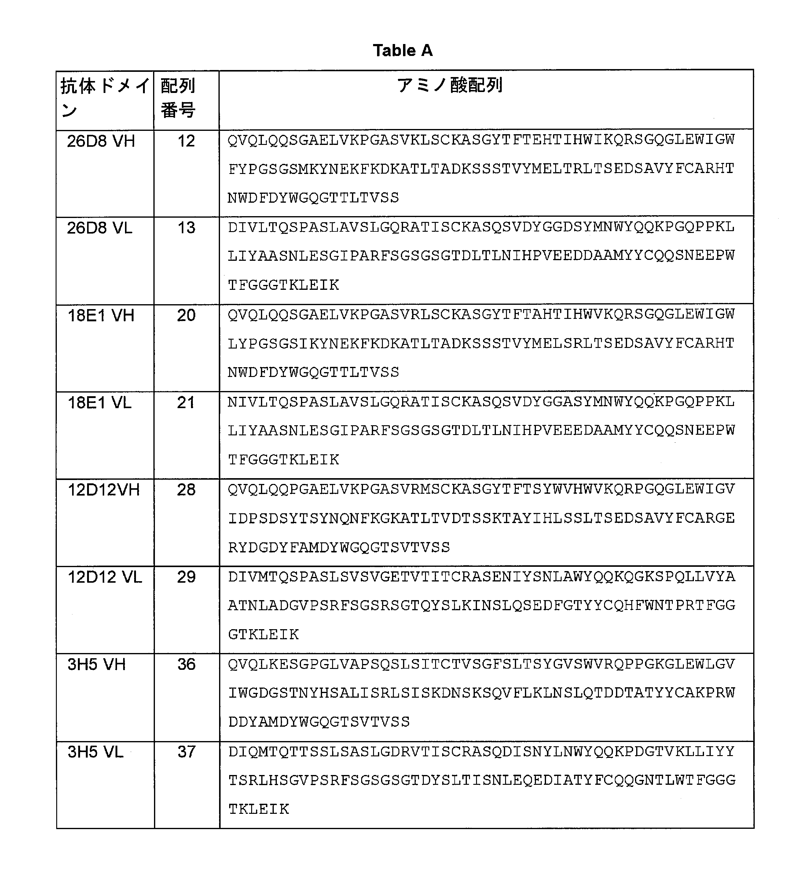
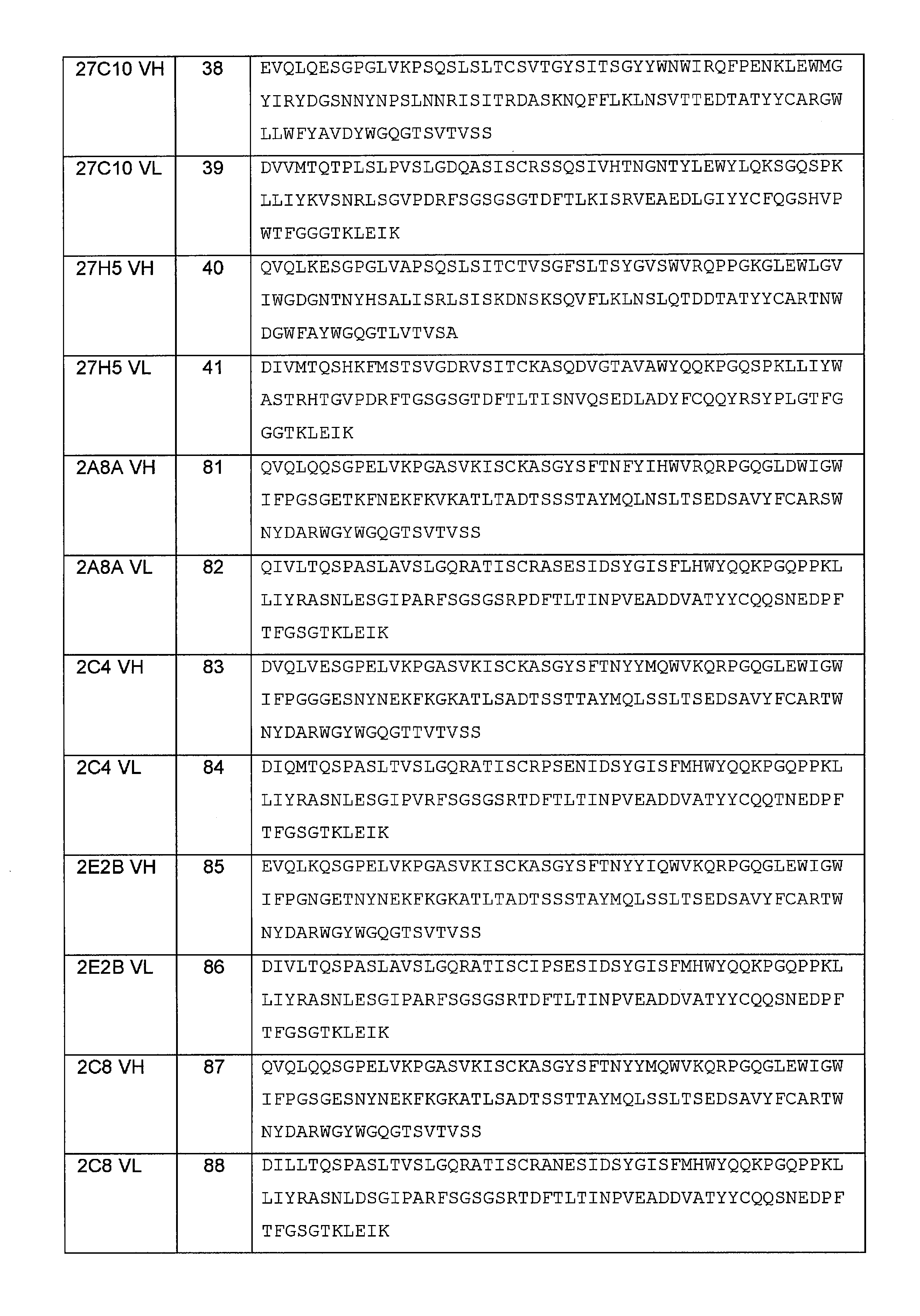

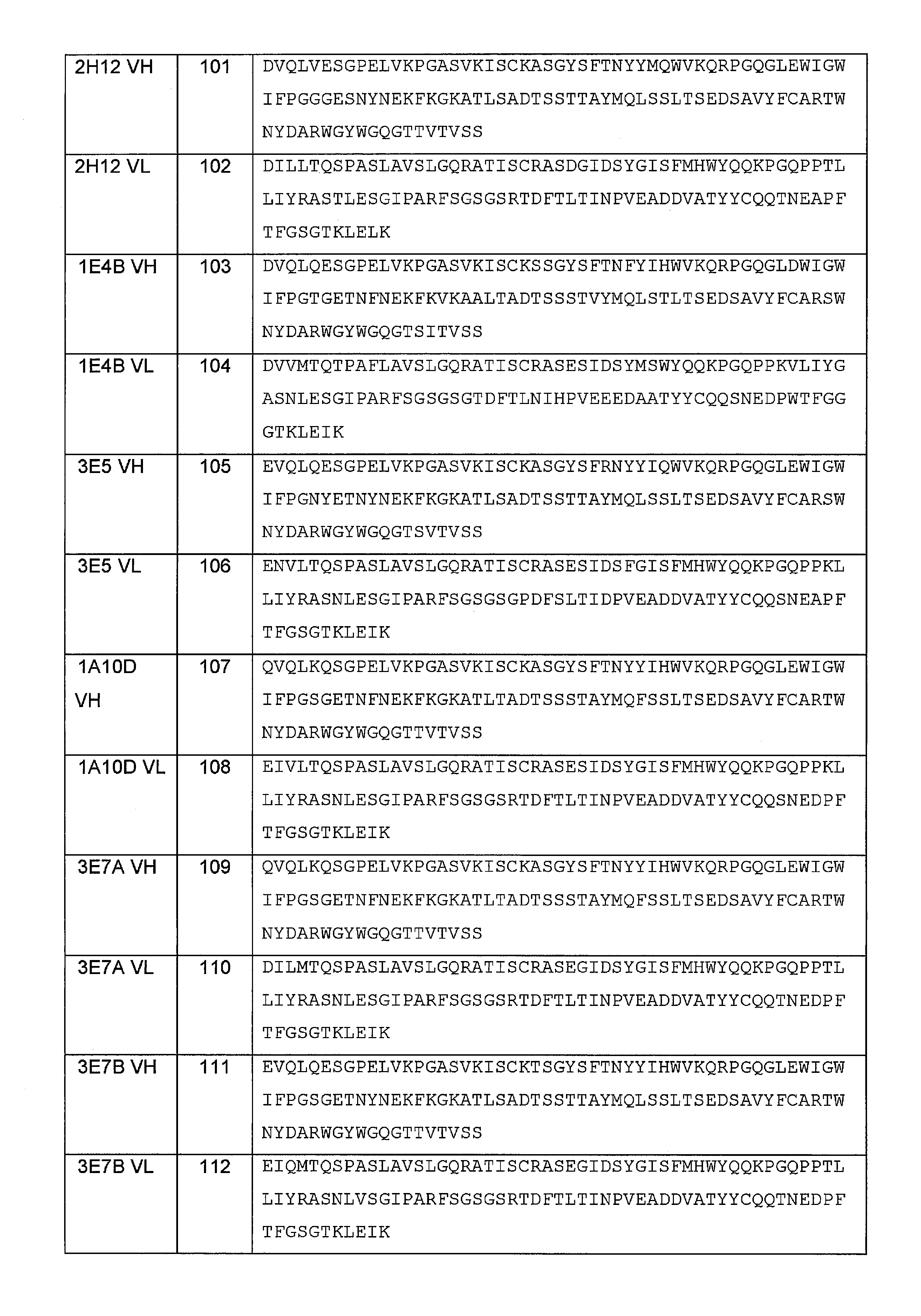
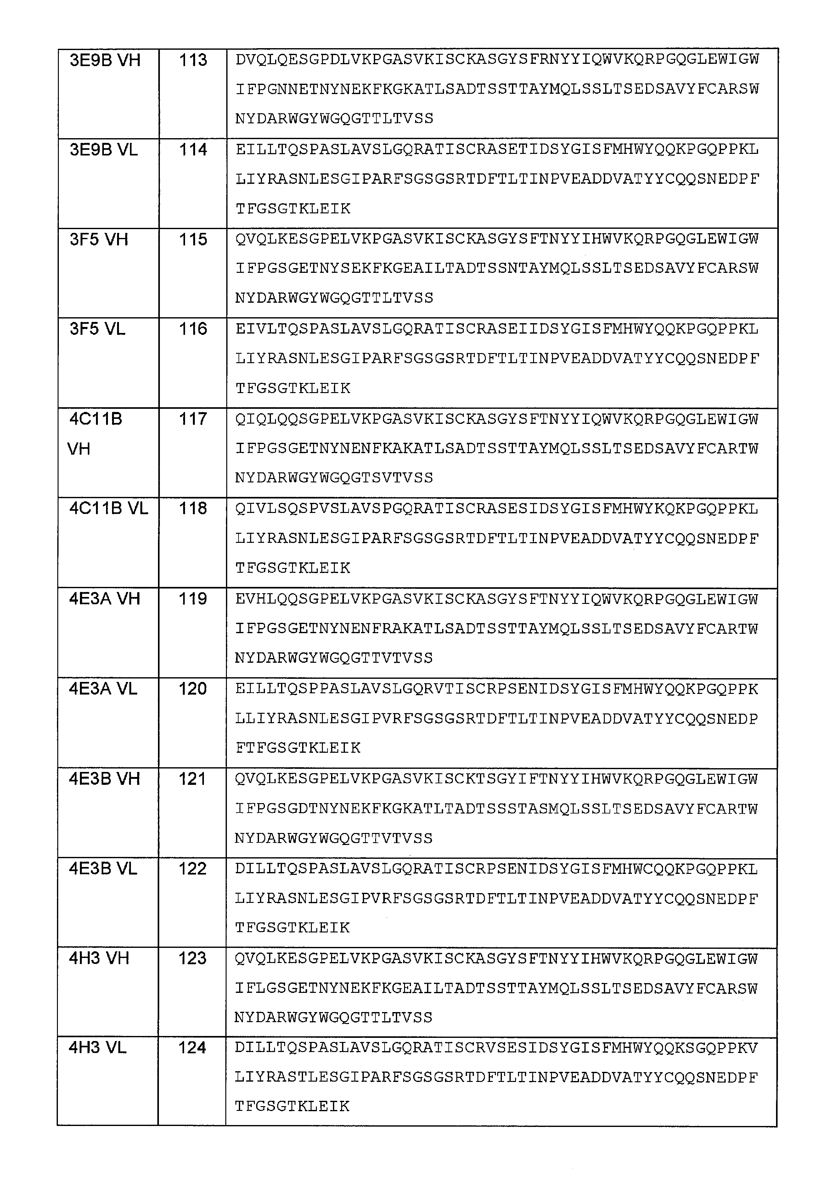


抗体の断片及び誘導体(これらは他に記述され又は文脈により明白に否定されない限り、本出願において使用される用語「抗体(単数又は複数)」によって包含される)は、本技術において既知の方法によって生成することができる。「断片」は、元のままの抗体の一部、一般には抗原結合部位又は可変領域を含む。抗体断片の例には、Fab、Fab'、Fab'-SH、F(ab')2、及びFv断片、ダイアボディ、限定なく(1)単鎖Fv分子、(2)ただ1つの軽鎖可変ドメインを含む単鎖ポリペプチド、又は付随する重鎖部分を含まず、軽鎖可変ドメインの3つのCDRを含むその断片、及び(3)ただ1つの重鎖可変領域を含む単鎖ポリペプチド、又は付随する軽鎖部分を含まず、重鎖可変領域の3つのCDRを含むその断片を含む連続するアミノ酸残基の中断されない1つの配列からなる一次構造を有するポリペプチドである任意の抗体断片、並びに抗体断片から形成された多重特異性(例えば二重特異性)抗体が含まれる。とりわけ、ナノボディ、ドメイン抗体、単一ドメイン抗体、又は「dAb」が含まれる。 Fragments and derivatives of antibodies, which are incorporated by the term "antibody (s)" as used in this application, unless otherwise stated or expressly denied by context, are by methods known in the art. Can be generated. A "fragment" comprises a portion of the original antibody, generally an antigen binding site or variable region. Examples of antibody fragments include Fab, Fab', Fab'-SH, F (ab') 2 , and Fv fragments, diabodies, without limitation (1) single chain Fv molecules, (2) only one light chain variable. A single-chain polypeptide containing a domain, or a fragment thereof that does not contain an associated heavy chain moiety and contains three CDRs of a light chain variable domain, and (3) a single-chain polypeptide that contains only one heavy chain variable region, or. Any antibody fragment that is a polypeptide having a primary structure consisting of an uninterrupted single sequence of contiguous amino acid residues, including its fragment containing the three CDRs of the heavy chain variable region, without the accompanying light chain portion, as well. Multispecific (eg, bispecific) antibodies formed from antibody fragments are included. Among other things are Nanobodies, domain antibodies, single domain antibodies, or "dAb".
ある特定の実施形態では、抗体を生成するハイブリドーマのDNAは、例えば同族の非ヒト配列の代わりにヒト重鎖及び軽鎖の定常ドメインのコーディング配列を置換することによって(Morrisonら、PNAS、6851頁 (1984))、又は非免疫グロブリンポリペプチドのコーディング配列の全部又は一部を免疫グロブリンコーディング配列に共有結合させることによって、発現ベクターへの挿入の前に改変することができる。そのようにして、元の抗体の結合特異性を有する「キメラ」又は「ハイブリッド」抗体が調製される。典型的には、そのような非免疫グロブリンポリペプチドは、抗体の定常ドメインに置換される。 In certain embodiments, the DNA of the hybridoma that produces the antibody is, for example, by substituting the coding sequences of the constant domains of human heavy and light chains instead of the homologous non-human sequences (Morrison et al., PNAS, p. 6851). (1984)), or by covalently linking all or part of the coding sequence of a non-immunoglobulin polypeptide to an immunoglobulin coding sequence, it can be modified prior to insertion into the expression vector. In that way, a "chimeric" or "hybrid" antibody with the binding specificity of the original antibody is prepared. Typically, such non-immunoglobulin polypeptides are replaced with constant domains of the antibody.
任意選択で、抗体はヒト化される。抗体の「ヒト化」形は、マウス免疫グロブリンに由来する最小限の配列を含む特定のキメラ免疫グロブリン、免疫グロブリン鎖、又はその断片(Fv、Fab、Fab'、F(ab')2、又はその他の抗体の抗原結合性サブ配列等)である。大部分、ヒト化抗体は、元の抗体の所望の特異性、親和性、及び処理能力を維持しつつ、レシピエントの相補性決定領域(CDR)からの残基が、元の抗体(ドナー抗体)のCDRからの残基によって置き換えられた、ヒト免疫グロブリン(レシピエント抗体)である。 Optionally, the antibody is humanized. The "humanized" form of an antibody is a specific chimeric immunoglobulin, immunoglobulin chain, or fragment thereof (Fv, Fab, Fab', F (ab') 2 , or a fragment thereof, containing a minimal sequence derived from mouse immunoglobulin. Antigen-binding subsequences of other antibodies, etc.). For the most part, humanized antibodies retain the desired specificity, affinity, and processing capacity of the original antibody, while the residues from the recipient's complementarity determining regions (CDRs) are the original antibody (donor antibody). ) Is a human immunoglobulin (recipient antibody) replaced by residues from the CDRs.
一部の例では、ヒト免疫グロブリンのFvフレームワーク残基が、対応する非ヒト残基によって置き換えられる。更に、ヒト化抗体は、レシピエント抗体にも輸入されたCDR又はフレームワーク配列にも見出されない残基を含み得る。これらの改変は、抗体の性能を更にリファインし最適化するために行われる。一般に、ヒト化抗体は、少なくとも1つ、典型的には2つの可変ドメインの実質的にすべてを含むことになり、その中でCDR領域のすべて又は実質的にすべては、元の抗体のCDR領域に対応し、FR領域のすべて又は実質的にすべては、ヒト免疫グロブリンコンセンサス配列のFR領域である。ヒト化抗体は、最適には免疫グロブリン定常領域(Fc)、典型的にはヒト免疫グロブリンの少なくとも一部をも含むことになる。さらなる詳細については、その開示全体が参照により本明細書に組み込まれる、Jonesら、Nature、321、522頁 (1986)、Reichmannら、Nature、332、323頁 (1988)、Presta、Curr. Op. Struct. Biol.、2、593頁 (1992)、Verhoeyenら、Science、239、1534頁、及び米国特許第4816567号を参照されたい。抗体をヒト化する方法は、当技術で公知である。 In some examples, the Fv framework residues of human immunoglobulin are replaced by the corresponding non-human residues. In addition, humanized antibodies may contain residues not found in either the recipient antibody or the imported CDR or framework sequence. These modifications are made to further refine and optimize the performance of the antibody. In general, a humanized antibody will contain substantially all of the at least one, typically two variable domains, in which all or substantially all of the CDR regions are the CDR regions of the original antibody. Corresponding to, all or substantially all of the FR regions are the FR regions of the human immunoglobulin consensus sequence. Humanized antibodies will optimally contain an immunoglobulin constant region (Fc), typically at least a portion of human immunoglobulin. For further details, the entire disclosure is incorporated herein by reference, Jones et al., Nature, 321, 522 (1986), Reichmann et al., Nature, 332, 323 (1988), Presta, Curr. Op. See Struct. Biol., 2, 593 (1992), Verhoeyen et al., Science, 239, 1534, and US Pat. No. 4,816,567. Methods of humanizing an antibody are known in the art.
ヒト化抗体の作成において使用される軽及び重のヒト可変ドメインの選択は、抗原性を低下させるために極めて重要である。いわゆる「ベストフィット」法によれば、抗体の可変ドメインの配列を、既知のヒト可変ドメイン配列のライブラリー全体に対してスクリーニングする。次いでマウスの配列に最も近いヒトの配列を、ヒト化抗体のためのヒトフレームワーク(FR)として許容する(Simsら、J. Immunol.、151、2296頁(1993)、Chothia及びLesk、J. Mol.、196、1987、901頁)。別の方法では、軽鎖又は重鎖の特定のサブグループのすべてのヒト抗体のコンセンサス配列からの特定のフレームワークを用いる。いくつかの異なるヒト化抗体のために、同じフレームワークを用いることができる(Carterら、PNAS、89、4285頁 (1992)、Prestaら、J. Immunol.、151、2623頁 (1993))。 The choice of light and heavy human variable domains used in the production of humanized antibodies is crucial for reducing antigenicity. According to the so-called "best fit" method, antibody variable domain sequences are screened against an entire library of known human variable domain sequences. The human sequence closest to the mouse sequence is then accepted as the human framework (FR) for humanized antibodies (Sims et al., J. Immunol., 151, 2296 (1993), Chothia and Lesk, J. et al. Mol., 196, 1987, p. 901). Another method uses a particular framework from the consensus sequence of all human antibodies in a particular subgroup of light or heavy chains. The same framework can be used for several different humanized antibodies (Carter et al., PNAS, 89, p. 4285 (1992), Presta et al., J. Immunol., 151, p. 2623 (1993)).
抗体がILT受容体への高い親和性及び他の好ましい生物学的特性を保持したままヒト化されることが更に重要である。この目的を達成するため、1つの方法によれば、ヒト化抗体は、親の配列及びヒト化配列の三次元モデルを用いる、親の配列及び種々の概念上のヒト化生成物の分析のプロセスによって調製される。三次元免疫グロブリンモデルは一般に利用可能であり、当業者には熟知されている。選択した候補の免疫グロブリン配列のありそうな三次元構造を図示しディスプレイするコンピュータープログラムが入手可能である。これらのディスプレイを検討することによって、候補の免疫グロブリン配列の機能化における残基の可能な役割の分析、即ち候補の免疫グロブリンがその抗原に結合する能力に影響する残基の分析が可能になる。このようにして、コンセンサス配列及び輸入配列からFR残基を選択して組み合わせ、それにより所望の抗体の特性、例えば標的抗原への親和性の増加が達成される。一般に、CDR残基は直接に且つ最も実質的に抗原結合への影響に関与している。 It is even more important that the antibody be humanized while retaining its high affinity for the ILT receptor and other favorable biological properties. To this end, according to one method, humanized antibodies are a process of analysis of parental sequences and various conceptual humanized products using a three-dimensional model of parental sequences and humanized sequences. Prepared by. Three-dimensional immunoglobulin models are generally available and are familiar to those of skill in the art. Computer programs are available that illustrate and display the likely three-dimensional structure of selected candidate immunoglobulin sequences. Examining these displays allows analysis of possible roles of residues in the functionalization of candidate immunoglobulin sequences, ie, residues that affect the ability of candidate immunoglobulins to bind their antigens. .. In this way, FR residues are selected and combined from the consensus sequence and the imported sequence, thereby achieving an increase in the properties of the desired antibody, eg, affinity for the target antigen. In general, CDR residues are directly and most substantially involved in the effect on antigen binding.
「ヒト化」モノクローナル抗体を作成する別の方法は、免疫に用いるマウスとしてXenoMouse (Abgenix, Fremont, CA)を用いることである。XenoMouseは、その免疫グロブリン遺伝子が機能性ヒト免疫グロブリン遺伝子によって置き換えられたマウスによるマウス宿主である。したがって、このマウスによって、又はこのマウスのB細胞から作成されたハイブリドーマで生成した抗体は、既にヒト化されている。XenoMouseは、参照により全体が本明細書に組み込まれる米国特許第6162963号に記載されている。 Another method of producing "humanized" monoclonal antibodies is to use XenoMouse (Abgenix, Fremont, CA) as a mouse for immunization. XenoMouse is a mouse host with mice in which its immunoglobulin gene has been replaced by a functional human immunoglobulin gene. Therefore, antibodies produced by this mouse or by hybridomas produced from the B cells of this mouse have already been humanized. XenoMouse is described in US Pat. No. 6,162,963, which is incorporated herein by reference in its entirety.
ヒト抗体は、種々の他の手法に従って、例えばヒト抗体レパートリーを発現するように操作された他のトランスジェニック動物を免疫のために用いることによって(Jakobovitzら、Nature、362、(1993) 255頁)、又はファージディスプレイ法を用いて抗体レパートリーを選択することによって、生成してもよい。そのような手法は当業者には既知であり、本出願に開示したようにモノクローナル抗体から出発して実行することができる。 Human antibodies are prepared according to various other techniques, eg, by using other transgenic animals engineered to express the human antibody repertoire for immunization (Jakobovitz et al., Nature, 362, (1993) p. 255). , Or by selecting the antibody repertoire using the phage display method. Such techniques are known to those of skill in the art and can be practiced starting with monoclonal antibodies as disclosed in this application.
一実施形態では、抗ILT2抗体がヒトFcγ受容体、例えばCD16A、CD16B、CD32A、CD32B、及び/又はCD64のいずれか1つ又は複数との実質的に特異的な結合性を有しないように抗ILT2抗体を調製することができる。そのような抗体は、Fcγ受容体への結合性を欠くか、低い結合性を有することが知られている種々の重鎖の定常領域を含み得る。或いは、Fc受容体の結合を避けるために、定常領域、例えばF(ab')2断片を含まない(又はその一部を含む)抗体断片を用いることができる。Fc受容体の結合は、例えばBIACOREアッセイにおけるFc受容体タンパク質への抗体の結合性の試験を含む当技術で既知の方法に従って評価可能である。また一般に、Fc受容体への結合を最小化又は除去するために(例えば1つ、2つ、3つ、4つ、5つ、又はそれ以上のアミノ酸置換を導入することによって)Fc部分を改変した任意の抗体IgGアイソタイプを用いることができる(例えばその開示が参照により本明細書に組み込まれるWO 03/101485を参照されたい)。Fc受容体の結合を評価するための細胞系アッセイ等のアッセイは当技術で公知であり、例えばWO03/101485に記載されている。 In one embodiment, the anti-ILT2 antibody is resistant to substantially specific binding to any one or more of human Fcγ receptors such as CD16A, CD16B, CD32A, CD32B, and / or CD64. ILT2 antibodies can be prepared. Such antibodies may contain constant regions of various heavy chains that are known to lack or have low binding to the Fcγ receptor. Alternatively, antibody fragments that do not contain (or part of) a constant region, eg, an F (ab') 2 fragment, can be used to avoid Fc receptor binding. Fc receptor binding can be assessed according to methods known in the art, including, for example, testing the binding of antibodies to Fc receptor proteins in the BIACORE assay. Also generally, the Fc moiety is modified to minimize or eliminate binding to the Fc receptor (eg, by introducing one, two, three, four, five, or more amino acid substitutions). Any antibody IgG isotypes used can be used (see, eg, WO 03/101485, the disclosure of which is incorporated herein by reference). Assays such as cell line assays for assessing Fc receptor binding are known in the art and are described, for example, in WO 03/101485.
一実施形態では、抗体は、エフェクター細胞と最小の相互作用を有する抗体をもたらすFc領域における1つ又は複数の突然変異を含み得る。抗体のFc領域における突然変異によってエフェクター機能のサイレント化が得られ、これは当技術においてN297A突然変異、LALA突然変異(Strohl, W.、2009、Curr. Opin. Biotechnol.、20(6)巻、685~691頁)、及びD265A (Baudinoら、2008、J. Immunol.、181、6664~69頁)として記載されている。Heusserら、WO2012/065950も参照されたい。これらの開示は参照により本明細書に組み込まれる。一実施形態では、抗体はヒンジ領域に1つ、2つ、3つ、又はそれ以上のアミノ酸置換を含む。一実施形態では、抗体はIgG1又はIgG2であり、残基233~236、任意選択で233~238(EUの番号付け)に1つ、2つ、又は3つの置換を含む。一実施形態では、抗体はIgG4であり、残基327、330、及び/又は331(EUの番号付け)に1つ、2つ、又は3つの置換を含む。サイレントのFc IgG1抗体の例は、IgG1 Fcアミノ酸配列においてL234A及びL235Aの突然変異を含むLALA突然変異体である。Fc突然変異の別の例は、例えばDAPA(D265A、P329A)突然変異としてIgG1抗体で用いられる残基D265における、又はD265及びP329における突然変異である(米国特許第6737056号)。別の改変されたIgG1抗体は残基N297に突然変異を含み(例えばN297A、N297S突然変異)、これは無グリコシル化/非グリコシル化抗体をもたらす。FcガンマR相互作用を低下させ及び/又は抑止する他の突然変異は、残基L234及びG237における置換(L234A/G237A)、残基S228、L235、及びR409における置換(S228P/L235E/R409K、T、M、L)、残基H268、V309、A330、及びA331における置換(H268Q/V309L/A330S/A331S)、残基C220、C226、C229、及びP238における置換(C220S/C226S/C229S/P238S)、残基C226、C229、E233、L234、及びL235における置換(C226S/C229S/E233P/L234V/L235A)、残基K322、L235、及びL235における置換(K322A/L234A/L235A)、残基L234、L235、及びP331における置換(L234F/L235E/P331S)、残基234、235、及び297における置換、残基E318、K320、及びK322における置換(L235E/E318A/K320A/K322A)、残基における置換(V234A、G237A、P238S)、残基243及び264における置換、残基297及び299における置換、EU番号付けシステムによって定義される残基233、234、235、237、及び238がPAAAP、PAAAS、及びSAAASから選択される配列を含む置換を含む(WO2011/066501を参照されたい)。 In one embodiment, the antibody may comprise one or more mutations in the Fc region that result in the antibody having minimal interaction with the effector cells. Mutations in the Fc region of the antibody result in silence of effector function, which in the art are N297A mutations, LALA mutations (Strohl, W., 2009, Curr. Opin. Biotechnol., Vol. 20 (6), 685-691), and D265A (Baudino et al., 2008, J. Immunol., 181, 6664-69). See also Heusser et al., WO2012 / 065950. These disclosures are incorporated herein by reference. In one embodiment, the antibody comprises one, two, three, or more amino acid substitutions in the hinge region. In one embodiment, the antibody is IgG1 or IgG2 and comprises 1, 2, or 3 substitutions at residues 233-236, optionally 233-238 (EU numbering). In one embodiment, the antibody is IgG4 and comprises one, two, or three substitutions at residues 327, 330, and / or 331 (EU numbering). An example of a silent Fc IgG1 antibody is a LALA mutant containing mutations in L234A and L235A in the IgG1 Fc amino acid sequence. Another example of an Fc mutation is, for example, a mutation at residue D265 used in an IgG1 antibody as a DAPA (D265A, P329A) mutation, or at D265 and P329 (US Pat. No. 6737056). Another modified IgG1 antibody contains a mutation at residue N297 (eg, N297A, N297S mutations), which results in a glycosylated / non-glycosylated antibody. Other mutations that reduce and / or suppress Fc gamma R interactions are substitutions at residues L234 and G237 (L234A / G237A), substitutions at residues S228, L235, and R409 (S228P / L235E / R409K, T). , M, L), substitutions at residues H268, V309, A330, and A331 (H268Q / V309L / A330S / A331S), substitutions at residues C220, C226, C229, and P238 (C220S / C226S / C229S / P238S), Substitutions at residues C226, C229, E233, L234, and L235 (C226S / C229S / E233P / L234V / L235A), substitutions at residues K322, L235, and L235 (K322A / L234A / L235A), residues L234, L235, And substitutions at P331 (L234F / L235E / P331S), substitutions at residues 234, 235, and 297, substitutions at residues E318, K320, and K322 (L235E / E318A / K320A / K322A), substitutions at residues (V234A, G237A, P238S), substitutions at residues 243 and 264, substitutions at residues 297 and 299, residues 233, 234, 235, 237, and 238 as defined by the EU numbering system selected from PAAAP, PAAAS, and SAAAS. Contains permutations containing the sequences that are to be (see WO 2011/066501).
一実施形態では、抗体はヒトIgG1起源のFcドメインを含んでよく、Kabat残基234、235、237、330、及び/又は331に突然変異を含む。そのようなFcドメインの一例は、Kabat残基L234、L235、及びP331における置換(例えばL234A/L235E/P331S又はL234F/L235E/P331S)を含む。そのようなFcドメインの別の例は、Kabat残基L234、L235、G237、及びP331における置換(例えばL234A/L235E/G237A/P331S)を含む。そのようなFcドメインの別の例は、Kabat残基L234、L235、G237、A330、及びP331における置換(例えばL234A/L235E/G237A/A330S/P331S)を含む。一実施形態では、抗体はL234X1置換、L235X2置換、及びP331X3置換を含む任意選択でヒトIgG1アイソタイプのFcドメインを含み、ここでX1はロイシン以外の任意のアミノ酸残基、X2はロイシン以外の任意のアミノ酸残基、X3はプロリン以外の任意のアミノ酸残基であり、任意選択でX1はアラニン若しくはフェニルアラニン、又はその保存的置換であり、任意選択でX2はグルタミン酸又はその保存的置換であり、任意選択でX3はセリン又はその保存的置換である。別の実施形態では、抗体はL234X1置換、L235X2置換、G237X4置換、及びP331X4置換を含む任意選択でヒトIgG1アイソタイプのFcドメインを含み、ここでX1はロイシン以外の任意のアミノ酸残基、X2はロイシン以外の任意のアミノ酸残基、X3はグリシン以外の任意のアミノ酸残基、X4はプロリン以外の任意のアミノ酸残基であり、任意選択でX1はアラニン若しくはフェニルアラニン、又はその保存的置換であり、任意選択でX2はグルタミン酸又はその保存的置換であり、任意選択でX3はアラニン又はその保存的置換であり、任意選択でX4はセリン又はその保存的置換である。別の実施形態では、抗体はL234X1置換、L235X2置換、G237X4置換、G330X4置換、及びP331X5置換を含む任意選択でヒトIgG1アイソタイプのFcドメインを含み、ここでX1はロイシン以外の任意のアミノ酸残基、X2はロイシン以外の任意のアミノ酸残基、X3はグリシン以外の任意のアミノ酸残基、X4はアラニン以外の任意のアミノ酸残基、X5はプロリン以外の任意のアミノ酸残基であり、任意選択でX1はアラニン若しくはフェニルアラニン、又はその保存的置換であり、任意選択でX2はグルタミン酸又はその保存的置換であり、任意選択でX3はアラニン又はその保存的置換であり、任意選択でX4はセリン又はその保存的置換であり、任意選択でX5はセリン又はその保存的置換である。本明細書で用いる簡便表記では、フォーマットは野生型残基:ポリペプチド中の位置:突然変異残基であり、残基の位置はKabatによるEU番号付けシステムに従って示している。 In one embodiment, the antibody may comprise an Fc domain of human IgG1 origin, comprising mutations in Kabat residues 234, 235, 237, 330, and / or 331. Examples of such Fc domains include substitutions at Kabat residues L234, L235, and P331 (eg L234A / L235E / P331S or L234F / L235E / P331S). Another example of such an Fc domain includes substitutions at Kabat residues L234, L235, G237, and P331 (eg L234A / L235E / G237A / P331S). Another example of such an Fc domain includes substitutions at Kabat residues L234, L235, G237, A330, and P331 (eg L234A / L235E / G237A / A330S / P331S). In one embodiment, the antibody optionally comprises the Fc domain of the human IgG1 isotype, including L234X 1 substitution, L235X 2 substitution, and P331X 3 substitution, where X 1 is any amino acid residue other than leucine and X 2 is. Any amino acid residue other than leucine, X 3 is any amino acid residue other than proline, optionally X 1 is alanine or phenylalanine, or a conservative substitution thereof, and optionally X 2 is glutamic acid or its. It is a conservative substitution, and optionally X 3 is serine or its conservative substitution. In another embodiment, the antibody optionally comprises the Fc domain of the human IgG1 isotype, including L234X 1 substitution, L235X 2 substitution, G237X 4 substitution, and P331X 4 substitution, where X 1 is any amino acid residue other than leucine. Group, X 2 is any amino acid residue other than leucine, X 3 is any amino acid residue other than glycine, X 4 is any amino acid residue other than proline, and X 1 is alanine or phenylalanine, which is optional. Or its conservative substitution, optionally X 2 is glutamic acid or its conservative substitution, optionally X 3 is alanine or its conservative substitution, and optionally X 4 is serine or its conservative substitution. Is. In another embodiment, the antibody optionally comprises an Fc domain of human IgG1 isotype including L234X 1 substitution, L235X 2 substitution, G237X 4 substitution, G330X 4 substitution, and P331X 5 substitution, where X 1 is other than leucine. Any amino acid residue, X 2 is any amino acid residue other than leucine, X 3 is any amino acid residue other than glycine, X 4 is any amino acid residue other than alanine, X 5 is any amino acid residue other than proline Amino acid residues, optionally X 1 is alanine or phenylalanine, or a conservative substitution thereof, optionally X 2 is glutamate or a conservative substitution thereof, and optionally X 3 is alanine or a conservative substitution thereof. Substitution, optionally X 4 is serine or its conservative substitution, and optionally X 5 is serine or its conservative substitution. In the brief notation used herein, the format is wild-type residue: position in polypeptide: mutant residue, and the position of the residue is shown according to Kabat's EU numbering system.
一実施形態では、抗体は、以下のアミノ酸配列、又はそれと少なくとも90%、95%、若しくは99%同一であるがKabat位置234、235、及び331(下線部)のアミノ酸残基を保持しているアミノ酸配列を含む重鎖定常領域を含む。 In one embodiment, the antibody retains the following amino acid sequence, or at least 90%, 95%, or 99% identical to it, but with amino acid residues at Kabat positions 234, 235, and 331 (underlined). Includes a heavy chain constant region containing an amino acid sequence.
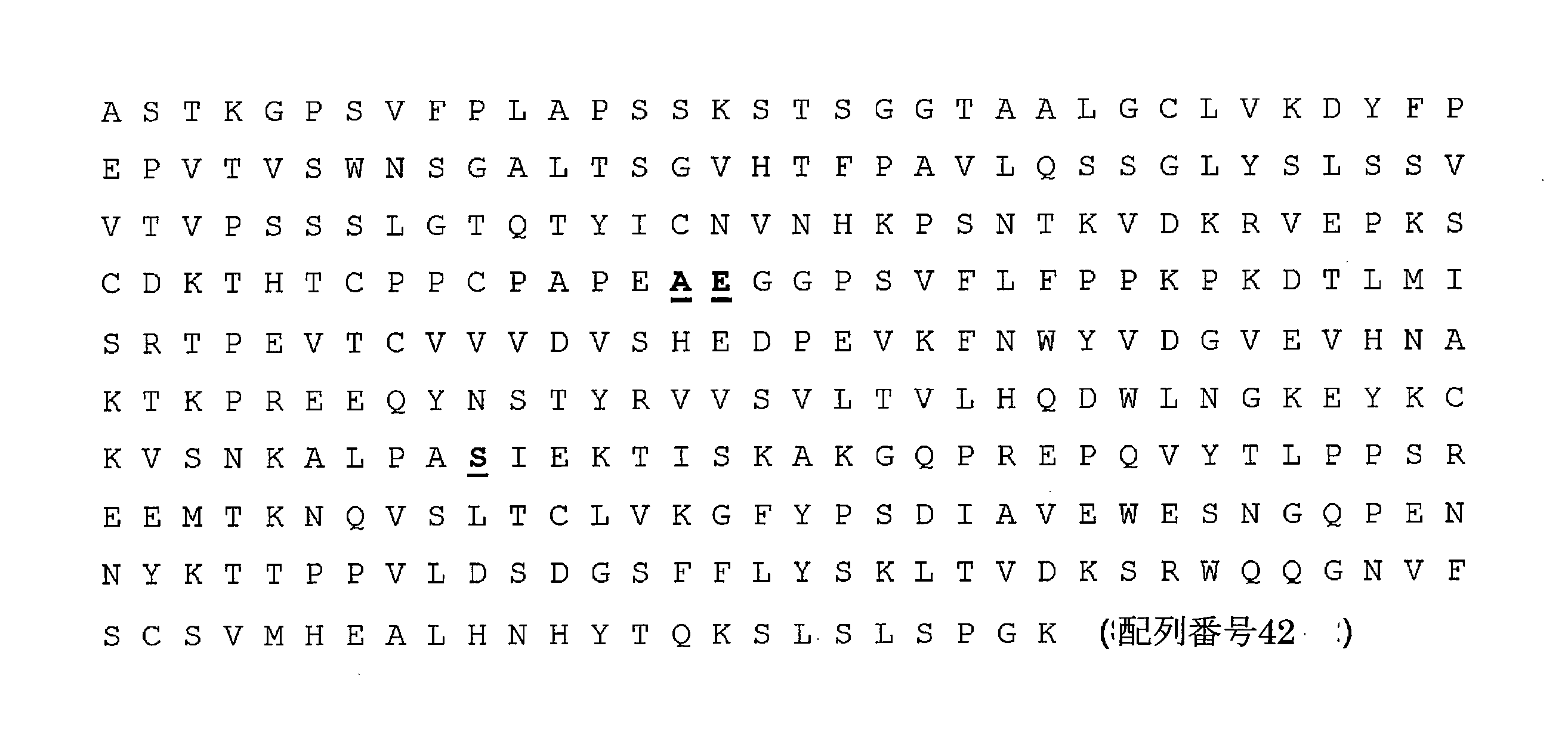
一実施形態では、抗体は、以下のアミノ酸配列、又はそれと少なくとも90%、95%、若しくは99%同一であるがKabat位置234、235、及び331(下線部)のアミノ酸残基を保持しているアミノ酸配列を含む重鎖定常領域を含む。 In one embodiment, the antibody retains the following amino acid sequence, or at least 90%, 95%, or 99% identical to it, but with amino acid residues at Kabat positions 234, 235, and 331 (underlined). Includes a heavy chain constant region containing an amino acid sequence.

一実施形態では、抗体は、以下のアミノ酸配列、又はそれと少なくとも90%、95%、若しくは99%同一であるがKabat位置234、235、237、330、及び331(下線部)のアミノ酸残基を保持しているアミノ酸配列を含む重鎖定常領域を含む。 In one embodiment, the antibody has the following amino acid sequence, or at least 90%, 95%, or 99% identical to it, but with amino acid residues at Kabat positions 234, 235, 237, 330, and 331 (underlined). Includes a heavy chain constant region containing the retained amino acid sequence.
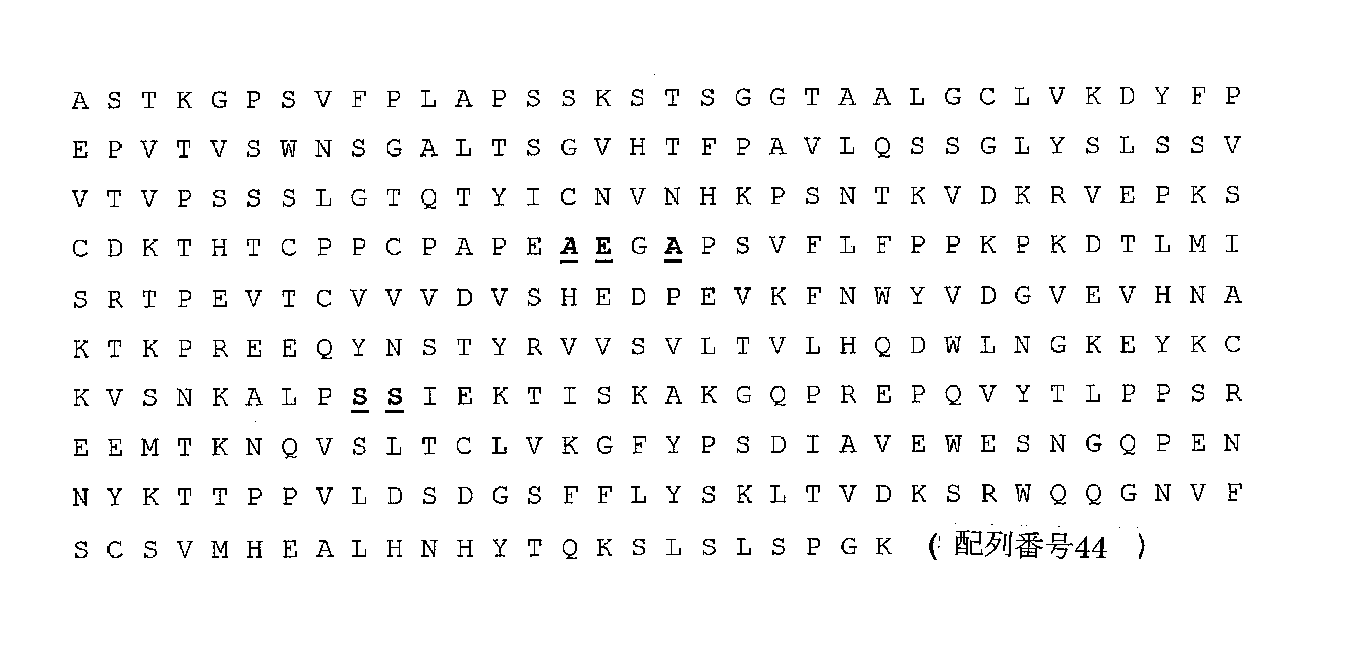
一実施形態では、抗体は、以下のアミノ酸配列、又はそれと少なくとも90%、95%、若しくは99%同一であるがKabat位置234、235、237、及び331(下線部)のアミノ酸残基を保持している配列を含む重鎖定常領域を含む。 In one embodiment, the antibody retains the following amino acid sequence, or at least 90%, 95%, or 99% identical to it, but with amino acid residues at Kabat positions 234, 235, 237, and 331 (underlined). Contains heavy chain constant regions containing the sequences that are present.
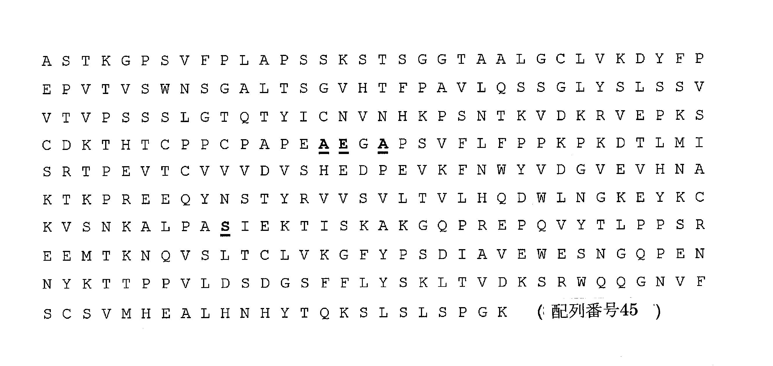
Fc相互作用が抑止されたILT2ブロッキング抗体は、ILT2におけるアゴニスト活性の欠如をもたらすことになる。そのような抗体はまた、ADCC活性の喪失又は低いADCC活性をもたらし、Fc相互作用が抑止された抗体は50%未満の特異的細胞溶解であるADCC活性を示すことを意味している。好ましくは、抗体は実質的にADCC活性を欠き、例えば抗体は5%未満又は1%未満のADCC活性(特異的細胞溶解)を示す。そのような抗体はまた、細胞(例えばNK細胞、T細胞、単球、樹状細胞、マクロファージ)の表面におけるILT2のFcγR媒介架橋の欠如をもたらし得る。 ILT2 blocking antibodies with suppressed Fc interactions will result in a lack of agonistic activity in ILT2. Such antibodies also result in loss of ADCC activity or low ADCC activity, implying that antibodies with suppressed Fc interactions exhibit ADCC activity with less than 50% specific cellular lysis. Preferably, the antibody substantially lacks ADCC activity, eg, the antibody exhibits less than 5% or less than 1% ADCC activity (specific cell lysis). Such antibodies can also result in a lack of FcγR-mediated cross-linking of ILT2 on the surface of cells (eg, NK cells, T cells, monocytes, dendritic cells, macrophages).
一実施形態では、抗体は、220、226、229、233、234、235、236、237、238、243、264、268、297、298、299、309、310、318、320、322、327、330、331、及び409からなる群から選択される残基の任意の1つ、2つ、3つ、4つ、5つ、又はそれ以上において重鎖定常領域に置換を有する(重鎖定常領域における残基の番号付けはKabatによるEU番号付けに従っている)。一実施形態では、抗体は残基234、235、及び322に置換を含む。一実施形態では、抗体は残基234、235、及び331に置換を含む。一実施形態では、抗体は残基234、235、237、及び331に置換を含む。一実施形態では、抗体は残基234、235、237、330、及び331に置換を含む。一実施形態では、FcドメインはヒトIgG1サブタイプのものである。アミノ酸残基はKabatによるEU番号付けに従って示している。 In one embodiment, the antibodies are 220, 226, 229, 233, 234, 235, 236, 237, 238, 243, 264, 268, 297, 298, 299, 309, 310, 318, 320, 322, 327, Have substitutions in the heavy chain constant region at any one, two, three, four, five, or more residues selected from the group consisting of 330, 331, and 409 (heavy chain constant region). The numbering of residues in is according to the EU numbering by Kabat). In one embodiment, the antibody comprises substitutions at residues 234, 235, and 322. In one embodiment, the antibody comprises substitutions at residues 234, 235, and 331. In one embodiment, the antibody comprises substitutions at residues 234, 235, 237, and 331. In one embodiment, the antibody comprises substitutions at residues 234, 235, 237, 330, and 331. In one embodiment, the Fc domain is of the human IgG1 subtype. Amino acid residues are shown according to EU numbering by Kabat.
抗ILT2抗体は、1mg/ml~500mg/mlの濃度で含まれて医薬製剤中に組み込むことができ、前記製剤は2.0~10.0のpHを有する。製剤は、緩衝システム、保存剤、等張剤、キレート化剤、安定剤、及び界面活性剤を更に含んでよい。一実施形態では、医薬製剤は水性製剤、即ち水を含む製剤である。そのような製剤は典型的には溶液又は懸濁液である。さらなる実施形態では、医薬製剤は水溶液である。用語「水性製剤」は、少なくとも50w/w%の水を含む製剤として定義される。同様に、用語「水溶液」は、少なくとも50w/w%の水を含む溶液として定義され、用語「水性懸濁液」は、少なくとも50w/w%の水を含む懸濁液として定義される。 The anti-ILT2 antibody is contained at a concentration of 1 mg / ml to 500 mg / ml and can be incorporated into a pharmaceutical preparation, which has a pH of 2.0 to 10.0. The pharmaceutical product may further contain a buffer system, a preservative, an isotonic agent, a chelating agent, a stabilizer, and a surfactant. In one embodiment, the pharmaceutical formulation is an aqueous formulation, i.e., a formulation containing water. Such formulations are typically solutions or suspensions. In a further embodiment, the pharmaceutical product is an aqueous solution. The term "aqueous formulation" is defined as a formulation containing at least 50 w / w% water. Similarly, the term "aqueous solution" is defined as a solution containing at least 50 w / w% water, and the term "aqueous suspension" is defined as a suspension containing at least 50 w / w% water.
別の実施形態では、医薬製剤は凍結乾燥された製剤であり、医師又は患者は使用前に、それに溶媒及び/又は希釈剤を添加する。 In another embodiment, the pharmaceutical formulation is a lyophilized formulation, to which the physician or patient adds a solvent and / or a diluent to it prior to use.
別の実施形態では、医薬製剤は前もって溶解することなしに即使用される、乾燥された(例えば凍結乾燥又は噴霧乾燥された)製剤である。 In another embodiment, the pharmaceutical formulation is a dried (eg, lyophilized or spray dried) formulation that is used immediately without pre-dissolution.
さらなる態様では、医薬製剤はそのような抗体及び緩衝剤の水溶液を含み、抗体は1mg/ml又はそれ以上の濃度で存在し、前記製剤は約2.0~約10.0のpHを有する。 In a further embodiment, the pharmaceutical formulation comprises an aqueous solution of such an antibody and buffer, the antibody is present at a concentration of 1 mg / ml or higher, and the pharmaceutical product has a pH of about 2.0 to about 10.0.
別の実施形態では、製剤のpHは約2.0~約10.0、約3.0~約9.0、約4.0~約8.5、約5.0~約8.0、及び約5.5~約7.5からなるリストから選択される範囲にある。 In another embodiment, the pH of the pharmaceutical product is in the range selected from the list consisting of about 2.0 to about 10.0, about 3.0 to about 9.0, about 4.0 to about 8.5, about 5.0 to about 8.0, and about 5.5 to about 7.5. ..
さらなる実施形態では、緩衝剤は酢酸ナトリウム、炭酸ナトリウム、クエン酸塩、グリシルグリシン、ヒスチジン、グリシン、リジン、アルギニン、リン酸二水素ナトリウム、リン酸水素二ナトリウム、リン酸ナトリウム、及びトリス(ヒドロキシメチル)アミノメタン、ビシン、トリシン、リンゴ酸、コハク酸塩、マレイン酸、フマル酸、酒石酸、アスパラギン酸、又はそれらの混合物からなる群から選択される。これらの特定の緩衝剤のそれぞれの1つが、本発明の代替の実施形態を構成する。 In a further embodiment, the buffers are sodium acetate, sodium carbonate, citrate, glycylglycine, histidine, glycine, lysine, arginine, sodium dihydrogen phosphate, disodium hydrogen phosphate, sodium phosphate, and tris (hydroxyl). It is selected from the group consisting of methyl) aminomethane, bicin, tricin, malic acid, succinate, maleic acid, fumaric acid, tartrate acid, aspartic acid, or mixtures thereof. Each one of these particular buffers constitutes an alternative embodiment of the invention.
さらなる実施形態では、製剤は薬学的に許容される保存剤を更に含む。さらなる実施形態では、製剤は等張剤を更に含む。さらなる実施形態では、製剤はキレート化剤を更に含む。本発明のさらなる実施形態では、製剤は安定剤を更に含む。本発明のさらなる実施形態では、製剤は界面活性剤を更に含む。便利のため、Remington、The Science and Practice of Pharmacy、19版、1995を参照する。 In a further embodiment, the formulation further comprises a pharmaceutically acceptable preservative. In a further embodiment, the formulation further comprises an isotonic agent. In a further embodiment, the formulation further comprises a chelating agent. In a further embodiment of the invention, the pharmaceutical product further comprises a stabilizer. In a further embodiment of the invention, the pharmaceutical product further comprises a surfactant. For convenience, see Remington, The Science and Practice of Pharmacy, 19th Edition, 1995.
本発明のペプチド医薬製剤中に他の成分が存在することも可能である。そのような追加の成分には、湿潤剤、乳化剤、抗酸化剤、増量剤、浸透圧調整剤、キレート化剤、金属イオン、油性ビヒクル、タンパク質(例えばヒト血清アルブミン、ゼラチン、又はタンパク質)、及び両性イオン(例えばベタイン、タウリン、アルギニン、グリシン、リジン、及びヒスチジン等のアミノ酸)が含まれ得る。そのような追加の成分は、当然ながら本発明の医薬製剤の全体の安定性に有害な影響を与えるべきではない。 It is also possible that other components are present in the peptide pharmaceutical formulation of the present invention. Such additional ingredients include wetting agents, emulsifiers, antioxidants, bulking agents, osmotic regulators, chelating agents, metal ions, oily vehicles, proteins (eg, human serum albumin, gelatin, or proteins), and Zwitterions (eg amino acids such as betaine, taurine, arginine, glycine, lysine, and histidine) can be included. Such additional ingredients should, of course, not have a detrimental effect on the overall stability of the pharmaceutical formulation of the invention.
本発明による抗体を含む医薬組成物は、そのような処置を必要とする患者に対して、いくつかの部位に、例えば局所部位、例えば皮膚及び粘膜の部位、吸収を迂回する部位、例えば動脈内、静脈内、心臓内における投与、及び吸収が関与する部位、例えば皮内、皮下、筋肉内、又は腹部に投与され得る。本発明による医薬組成物の投与は、いくつかの投与経路を通して、例えば皮下、筋肉内、腹腔内、静脈内、舌、舌下、頬側、口内、口腔内、胃内及び腸内、経鼻、肺内、例えば細気管支及び肺胞又はそれらの組合せを通して、表皮、真皮、経皮、膣内、直腸内、眼内、例えば結膜を通して、経尿管、及び非経口的に、そのような処置を必要とする患者に投与することができる。 Pharmaceutical compositions containing antibodies according to the invention can be applied to patients in need of such treatment at several sites, such as local sites, such as skin and mucosal sites, sites that bypass absorption, such as intra-arterial. Can be administered intravenously, intracardiacly, and at sites where absorption is involved, such as intradermal, subcutaneous, intramuscular, or abdominal. Administration of the pharmaceutical composition according to the present invention is carried out through several routes of administration, for example, subcutaneous, intramuscular, intraperitoneal, intravenous, tongue, sublingual, buccal, oral, oral, gastric and intestinal, nasal. , Through the lungs, such as the bronchioles and alveolar or a combination thereof, through the epidermis, dermatitis, percutaneous, intravaginal, rectal, intraocular, eg, conjunctival, transurethral, and parenterally, such treatment. Can be administered to patients in need.
好適な抗体製剤は、既に開発された他の治療用モノクローナル抗体での経験を検討することによって決定することもできる。Rituxan (リツキシマブ)、Herceptin (トラスツズマブ)、Xolair (オマリウマブ)、Bexxar (トシツモマブ)、Campath (アレムツズマブ)、Zevalin、Onclym等のいくつかのモノクローナル抗体が臨床の状況で有効であることが示されており、同様の製剤を本発明の抗体と共に用いることができる。例えば、モノクローナル抗体は、100mg(10mL)又は500mg(50mL)の使い捨てバイアル中、9.0mg/mLの塩化ナトリウム、7.35mg/mLのクエン酸ナトリウム二水塩、0.7mg/mLのポリソーベート80、及び注射用無菌水中、10mg/mlの濃度で供給することができる。pHは6.5に調節される。別の実施形態では、抗体は約20mMのクエン酸Na、約150mMのNaClを含む約6.0のpHの製剤で供給される。
Suitable antibody formulations can also be determined by reviewing experience with other therapeutic monoclonal antibodies that have already been developed. Several monoclonal antibodies such as Rituxan, Herceptin, Xolair, Bexxar, Campath, Zevalin, and Onclym have been shown to be effective in clinical situations. Similar formulations can be used with the antibodies of the invention. For example, monoclonal antibodies are available in 100 mg (10 mL) or 500 mg (50 mL) disposable vials, 9.0 mg / mL sodium chloride, 7.35 mg / mL sodium citrate dihydrate, 0.7 mg /
悪性腫瘍の診断及び処置
本明細書に記載されるような抗ILT2抗体を使用して、個人、特にヒト患者を処置する方法も提示される。1つの実施形態では、本発明は、ヒト患者に投与するための医薬組成物を調製する際の、本明細書に記載されるような抗体の使用を提示する。一般的に、患者は、がん又は感染性疾患、例えば細菌性又はウイルス性疾患に罹患している、又はそのリスクに晒されている。
Diagnosis and Treatment of Malignant Tumors Also presented are methods of treating individuals, especially human patients, using anti-ILT2 antibodies as described herein. In one embodiment, the invention presents the use of an antibody as described herein in preparing a pharmaceutical composition for administration to a human patient. In general, patients have or are at risk for cancer or infectious diseases, such as bacterial or viral diseases.
例えば、1つの態様では、本発明は、ILT2制限白血球、例えばリンパ球、単球、マクロファージ、樹状細胞、B細胞、NK細胞、CD8 T細胞の活性(例えば、腫瘍細胞に対する細胞傷害性)及び/又は増殖を、それを必要としている患者において増強する方法であって、本開示の中和性抗ILT2抗体を前記患者に投与する工程を含む方法を提示する。抗体は、例えば、ヒト又はヒト化の抗ILT2抗体であり得、同抗体は、一次NK細胞及び/又はCD8 T細胞において、ILT2によって媒介される阻害性シグナル伝達の、HLAによって媒介される活性化を抑制又は阻止する(例えば、本明細書に開示される方法に基づき決定される)。1つの実施形態では、方法は、リンパ球(例えば、NK及び/又はCD8+ T細胞)活性の増加が有利である疾患であって、NK又はCD8+ T細胞により溶解されやすい細胞が関係し、それに影響を及ぼし、若しくはそれにより引き起こされ、又はNK若しくはCD8+ T細胞の不十分な活性により引き起こされ、悪化、遷延し、さもなければ特徴づけられる疾患、例えばがん又は感染性疾患等を有する患者において、そのようなリンパ球の活性及び/又は数を増加させることを狙いとする。 For example, in one embodiment, the invention comprises the activity of ILT2-restricted leukocytes such as lymphocytes, monospheres, macrophages, dendritic cells, B cells, NK cells, CD8 T cells (eg, cytotoxicity against tumor cells) and / Or a method of enhancing proliferation in a patient in need thereof, the present invention presenting a method comprising the step of administering the neutralizing anti-ILT2 antibody of the present disclosure to said patient. The antibody can be, for example, a human or humanized anti-ILT2 antibody, which is an HLA-mediated activation of ILT2-mediated inhibitory signaling in primary NK cells and / or CD8 T cells. (For example, determined based on the methods disclosed herein). In one embodiment, the method involves and affects cells that are susceptible to lysis by NK or CD8 + T cells, a disease in which increased lymphocyte (eg, NK and / or CD8 + T cells) activity is advantageous. In patients with diseases that are exacerbated, protracted, or otherwise characterized, such as cancer or infectious diseases, which are caused by, or caused by, or are caused by inadequate activity of NK or CD8 + T cells. The aim is to increase the activity and / or number of such lymphocytes.
1つの実施形態では、本開示の抗体は、HLA-A2及び/又はHLA-Gの発現、任意選択でHLA-A2及び/又はHLA-の過剰発現(例えば、健康な組織、健常者における発現と比較して)により特徴づけられる腫瘍の処置で使用される。 In one embodiment, the antibodies of the present disclosure are HLA-A2 and / or HLA-G expression, optionally overexpression of HLA-A2 and / or HLA- (eg, expression in healthy tissues, healthy individuals). Used in the treatment of tumors characterized by (in comparison).
広範囲にわたるがんが、HLA-G発現腫瘍細胞により特徴づけられることが公知である。例えば、HLA-G+病変(腫瘍細胞の30%を上回る)が、皮膚黒色腫、腎明細胞癌、網膜芽細胞腫、棘状細胞癌、上皮内癌、結腸直腸がん、卵巣癌、皮膚T細胞リンパ腫、子宮内膜腺癌、皮膚B細胞リンパ腫、胃がん、乳頭部がん、胆道がん、及び膵管腺癌で報告されている。HLA-G+ 病変(腫瘍細胞の30%未満)は、白血病、基底細胞癌、膀胱がん、乳がん、悪性中皮腫、日光性角化症、及び肺癌でも報告されている。更に、HLA-Gを発現する多くのがんを含む広範囲にわたるがんが、HLA-E発現腫瘍細胞により特徴づけられることが公知であり、例えば非小細胞性肺がん(NSCLC))、腎細胞癌(RCC)、黒色腫、頭頸部の扁平上皮癌(HNSCC)、結腸直腸がん、子宮頸がん、及び卵巣がんは、高レベルの場合を含めHLA-Eを発現することが公知である。 It is known that a wide range of cancers are characterized by HLA-G expressing tumor cells. For example, HLA-G + lesions (more than 30% of tumor cells) include cutaneous melanoma, clear renal cell carcinoma, retinal adenocarcinoma, spinous cell carcinoma, carcinoma in situ, colorectal cancer, ovarian cancer, and skin T. It has been reported in cell lymphoma, carcinoma in situ, cutaneous B-cell lymphoma, gastric cancer, papillary cancer, biliary tract cancer, and pancreatic ductal adenocarcinoma. HLA-G + lesions (<30% of tumor cells) have also been reported in leukemia, basal cell carcinoma, bladder cancer, breast cancer, malignant mesothelioma, actinic keratosis, and lung cancer. Furthermore, it is known that a wide range of cancers, including many cancers expressing HLA-G, are characterized by HLA-E expressing tumor cells, such as non-small cell lung cancer (NSCLC), renal cell carcinoma. (RCC), melanoma, squamous cell carcinoma of the head and neck (HNSCC), colorectal cancer, cervical cancer, and ovarian cancer are known to express HLA-E, including at high levels. ..
1つの実施形態では、抗ILT2抗体は、膀胱がんの処置で使用される。1つの実施形態では、抗ILT2抗体は、尿路上皮癌の処置で使用される。尿路上皮癌(移行上皮癌とも呼ばれる)は、身体のその他の部分に拡散(転移)するおそれのある膀胱の悪性腫瘍である。尿路上皮癌は、腎盂、尿管、膀胱、又は尿道を含む、泌尿器の任意の部分において開始し得る。 In one embodiment, the anti-ILT2 antibody is used in the treatment of bladder cancer. In one embodiment, the anti-ILT2 antibody is used in the treatment of urothelial carcinoma. Urothelial carcinoma (also called transitional cell carcinoma) is a malignant tumor of the bladder that can spread (metastasize) to other parts of the body. Urothelial carcinoma can begin in any part of the urinary system, including the renal pelvis, ureter, bladder, or urethra.
本明細書における方法及び組成物は、腎細胞癌の処置のために利用可能である。腎細胞癌の初期症状として、血尿(診察時、罹患者の40%で生じている) ;及び/又は側腹部痛(40%);及び/又は腹部若しくは側腹部における腫瘤(25%)、及び/又は体重減少(33%);及び/又は発熱(20%);及び/又は高血圧(20%);及び/又は寝汗;及び/又は倦怠感が一般的に挙げられる。腎細胞癌は、腫瘍そのものが産生するホルモンにより、又は腫瘍に対する身体の攻撃により引き起こされた状態である「腫瘍随伴症候群」(腫瘍を実際に宿さない組織に一般的に影響を及ぼす)のいくつかとも一般的に関連する。最も一般的な症候群は、貧血又は赤血球増加症;及び/又は血中高カルシウムレベル;及び/又は血小板増加症;及び/又は続発性アミロイドーシスから選択される。 The methods and compositions herein are available for the treatment of renal cell carcinoma. As early signs of renal cell carcinoma, hematuria (occurs in 40% of affected individuals at the time of examination); and / or flank pain (40%); and / or a mass in the abdomen or flank (25%), and / Or weight loss (33%); and / or fever (20%); and / or hypertension (20%); and / or night sweats; and / or fatigue are commonly mentioned. Renal cell carcinoma is a condition caused by hormones produced by the tumor itself or by the body's attack on the tumor, a number of "paraneoplastic syndromes" (which generally affect tissues that do not actually carry the tumor). Also generally related. The most common syndromes are selected from anemia or erythrocytosis; and / or high blood calcium levels; and / or thrombocytosis; and / or secondary amyloidosis.
腎細胞癌は、転移性明細胞RCC;局所的明細胞RCC;多房性嚢胞性明細胞RCC;管状嚢胞性RCC;甲状腺様濾胞性RCC;後天性嚢胞性腎疾患関連RCC;ハイブリッドオンコサイトーマ/嫌色素性RCCを含む、一連の異なる種類のRCCを包含する一般用語であると認識される。したがって、1つの実施形態では、本明細書における方法及び組成物は、転移性明細胞RCCを処置するのに使用される。1つの実施形態では、本明細書における方法及び組成物は、局所的明細胞RCCを処置するのに使用される。1つの実施形態では、本明細書における方法及び組成物は、多房性嚢胞性明細胞RCCを処置するのに使用される。1つの実施形態では、本明細書における方法及び組成物は、管状嚢胞性RCCを処置するのに使用される。1つの実施形態では、本明細書における方法及び組成物は、甲状腺様濾胞性RCCを処置するのに使用される。1つの実施形態では、本明細書における方法及び組成物は、後天性嚢胞性腎疾患関連RCCを処置するのに使用される。1つの実施形態では、本明細書における方法及び組成物は、ハイブリッドオンコサイトーマ/嫌色素性RCCを処置するのに使用される。 Renal cell carcinoma includes metastatic clear cell RCC; localized clear cell RCC; multilocular cystic clear cell RCC; tubular cystic RCC; thyroid-like follicular RCC; acquired cystic renal disease-related RCC; hybrid oncocytoma / Recognized as a general term that includes a series of different types of RCC, including chromophobe RCC. Therefore, in one embodiment, the methods and compositions herein are used to treat metastatic clear cell RCCs. In one embodiment, the methods and compositions herein are used to treat topical clear cell RCCs. In one embodiment, the methods and compositions herein are used to treat multilocular cystic clear cell RCCs. In one embodiment, the methods and compositions herein are used to treat tubular cystic RCCs. In one embodiment, the methods and compositions herein are used to treat thyroid-like follicular RCC. In one embodiment, the methods and compositions herein are used to treat acquired cystic kidney disease-related RCCs. In one embodiment, the methods and compositions herein are used to treat hybrid oncocytoma / chromophobe RCC.
個人は、腫瘍細胞の表面上でのHLA-A2及び/又はHLA-G (及び/又はHLA-E)の発現を評価するための事前検出工程を伴い又は伴わずに、抗ILT2抗体を用いて処置され得る。腫瘍又はがんは、1つの態様では、HLA-A2及び/又はHLA-G(及び任意選択で、更にHLA-E) (又は1つ若しくは複数のILT2のその他の天然リガンド)の発現により一般的に特徴づけられることが公知である腫瘍又はがんの種類であり得る。いくつかの実施形態では、処置方法は、個人から得られた腫瘍の生体サンプル(例えば、腫瘍細胞)中のHLA-A2及び/又はHLA-G(及び任意選択で、更にHLA-E)の核酸又はポリペプチドを検出する工程を含み得る。生体サンプルが、HLA-A2及び/又はHLA-G (及び任意選択で、更にHLA-E)を発現する、例えばHLA-A2及び/又はHLA-G (及び任意選択で、更にHLA-E)を検出可能なレベルで発現する、HLA-A2及び/又はHLA-G (及び任意選択で、更にHLA-E)を少なくとも事前に決定されたレベルで発現する、HLA-A2及び/又はHLA-G (及び任意選択で、更にHLA-E)を高レベルで、或いは抗HLA-A2及び/又は抗HLA-G(及び/又は抗HLA-E)抗体を用いて染色したときに高強度で発現することの判定(いずれの場合も、任意選択で参照と比較される)は、ILT2の活性を中和する薬剤を用いた処置から特に多大な利益を有し得るがんを有するものとして患者を指定するのに使用可能である。1つの実施形態では、方法は、生体サンプル中のHLA-A2及び/又はHLA-G (及び任意選択で、更にHLA-E)の核酸又はポリペプチドの発現レベルを決定する工程、及びそのレベルと、ILT2の活性を阻害、中和する薬剤を用いた処置から利益を得る個人に対応する参照レベル(例えば、数値、高強度の細胞表面染色等)とを比較する工程を含む。生体サンプルが、参照レベルに対応し、及び/又はそれよりも増加したレベルでHLA-A2及び/又はHLA-G (及び任意選択で、更にHLA-E)の核酸又はポリペプチドを発現するとした判定は、個人は、ILT2の活性を阻害、中和する薬剤を用いた処置から特に多大な利益を有し得るがんを有することを示唆する。任意選択で、生体サンプル中のHLA-A2及び/又はHLA-G (及び任意選択で、更にHLA-E)のポリペプチドを検出する工程は、悪性細胞の表面上で発現するHLA-G 及び/又はHLA-A2 (及び任意選択で、更にHLA-E)のポリペプチドを検出する工程を含む。 Individuals use anti-ILT2 antibodies with or without pre-detection steps to assess the expression of HLA-A2 and / or HLA-G (and / or HLA-E) on the surface of tumor cells. Can be treated. Tumors or cancers are, in one embodiment, generalized by the expression of HLA-A2 and / or HLA-G (and optionally HLA-E) (or other natural ligands for one or more ILT2s). It can be a tumor or cancer type known to be characterized by. In some embodiments, the treatment method is a nucleic acid of HLA-A2 and / or HLA-G (and optionally further HLA-E) in a biological sample of tumor (eg, tumor cells) obtained from an individual. Alternatively, it may include a step of detecting a polypeptide. Biological samples express HLA-A2 and / or HLA-G (and optionally further HLA-E), eg, HLA-A2 and / or HLA-G (and optionally further HLA-E). HLA-A2 and / or HLA-G (and optionally, further HLA-E) expressed at detectable levels, HLA-A2 and / or HLA-G (and optionally, further HLA-E) expressed at least at predetermined levels. And optionally, further HLA-E) is expressed at high levels or at high intensity when stained with anti-HLA-A2 and / or anti-HLA-G (and / or anti-HLA-E) antibodies. The determination (in each case, optionally compared to the reference) designates the patient as having cancer that may benefit particularly significantly from treatment with agents that neutralize the activity of ILT2. Can be used for. In one embodiment, the method is to determine the expression level of the nucleic acid or polypeptide of HLA-A2 and / or HLA-G (and optionally further HLA-E) in the biological sample, and the level thereof. Includes the step of comparing with reference levels corresponding to individuals (eg, numerical values, high intensity cell surface staining, etc.) that benefit from treatment with agents that inhibit or neutralize the activity of ILT2. Determination that the biological sample expresses HLA-A2 and / or HLA-G (and optionally, further HLA-E) nucleic acids or polypeptides at levels corresponding to and / or increased above the reference level. Suggests that an individual has cancer that may benefit particularly significantly from treatment with agents that inhibit or neutralize ILT2 activity. The step of optionally detecting HLA-A2 and / or HLA-G (and optionally further HLA-E) polypeptides in a biological sample involves HLA-G and / / expressed on the surface of malignant cells. Alternatively, it comprises the step of detecting a polypeptide of HLA-A2 (and optionally HLA-E).
本明細書におけるがんの処置法又は予防法において、その任意の1つの実施形態では、個人におけるがんの処置又は予防は、
a)がんを有する個人内の悪性細胞(例えば、腫瘍細胞)が、ILT2のHLAクラスIリガンド(例えば、HLA-A2及び/又はHLA-G)を発現するか判定する工程と、
b)悪性細胞(例えば、腫瘍細胞)により(例えば、その表面上において)、ILT2のリガンドが発現されていることが判定されたら、抗ILT2抗体、例えば本開示の任意の態様に基づく抗体を個人に投与する工程と
を含む。
In any one embodiment thereof, in the treatment or prevention of cancer herein, the treatment or prevention of cancer in an individual.
a) A step of determining whether malignant cells (eg, tumor cells) in an individual with cancer express HLA class I ligands for ILT2 (eg, HLA-A2 and / or HLA-G).
b) Once it is determined that a ligand for ILT2 is expressed by a malignant cell (eg, a tumor cell) (eg, on its surface), an anti-ILT2 antibody, eg, an antibody based on any aspect of the present disclosure, is used by an individual. Including the step of administering to.
1つの実施形態では、生体サンプル(例えば、腫瘍細胞、腫瘍組織、及び/又は腫瘍隣接組織を含むサンプル)がILT2のリガンドを発現すると判定されると、それは、個人が、ILT2ポリペプチドを阻害する抗体で処置され得る、及び/又はそれから利益を享受し得るがんを有することを示唆する。 In one embodiment, when a biological sample (eg, a sample containing tumor cells, tumor tissue, and / or tumor flanking tissue) is determined to express a ligand for ILT2, it is determined that the individual inhibits the ILT2 polypeptide. It suggests that you have a cancer that can be treated with antibodies and / or benefit from it.
1つの実施形態では、ILT2のリガンドの有意な発現は、前記リガンドが、所定の個人から採取された腫瘍細胞の実質的な数において発現していることを意味する。正確なパーセンテージ値に拘泥するものではないが、いくつかの事例では、患者から採取された腫瘍細胞(サンプル中)の少なくとも10%、20%、30%、40%、50%、又はそれより多くにおいて検出された場合には、リガンドが発現しているということができる。 In one embodiment, significant expression of the ligand for ILT2 means that the ligand is expressed in a substantial number of tumor cells taken from a given individual. Although not bound by the exact percentage value, in some cases at least 10%, 20%, 30%, 40%, 50%, or more of the tumor cells (in the sample) taken from the patient. If it is detected in, it can be said that the ligand is expressed.
個人がHLA-Gポリペプチドを発現するがん細胞を有するか判定する工程は、例えば、がん細胞を含む個人から生体サンプルを取得する工程(例えば、生検を実施することによる)と、前記細胞を、HLA-A2及び/又はHLA-Gポリペプチドに結合する抗体と接触させる工程と、前記細胞がその表面上でHLA-A2及び/又はHLA-Gを発現するか検出する工程とを含むことができる。抗HLA-G抗体については、例えば、Fournelら、(2000) Tissue Antigens 55巻:510~518頁及び国際公開第2018/091580号(その本開示は本明細書において参考として組み込まれている)におけるMEM-G/9及びその他の抗体を参照。任意選択で、個人がHLA-A2及び/又はHLA-Gを発現するがん細胞を有するか判定する工程は、免疫組織化学アッセイを実施する工程を含む。任意選択で、個人がHLA-A2及び/又はHLA-Gを発現するがん細胞を有するか判定する工程は、フローサイトメトリーアッセイを実施する工程を含む。 The steps of determining whether an individual has cancer cells expressing the HLA-G polypeptide include, for example, the step of obtaining a biological sample from an individual containing cancer cells (eg, by performing a biopsy) and the above-mentioned step. It comprises contacting a cell with an antibody that binds to an HLA-A2 and / or HLA-G polypeptide and detecting whether the cell expresses HLA-A2 and / or HLA-G on its surface. be able to. For anti-HLA-G antibodies, see, for example, Fournel et al. (2000) Tissue Antigens Vol. 55: pp. 510-518 and WO 2018/091580 (whose disclosure is incorporated herein by reference). See MEM-G / 9 and other antibodies. Optionally, the step of determining whether an individual has cancer cells expressing HLA-A2 and / or HLA-G comprises performing an immunohistochemical assay. Optionally, the step of determining whether an individual has cancer cells expressing HLA-A2 and / or HLA-G comprises performing a flow cytometry assay.
1つの実施形態では、本開示の抗体は、NK細胞及び/又はCD8 T細胞の表面において有意及び/又は上昇したレベル(例えば、健康な組織、健常者における発現と比較して)のILT2発現を有する個人の処置で使用される。個人は、NK細胞及び/又はCD8 T細胞の表面におけるILT2発現を評価する事前の検出工程を伴い又は伴わずに、抗ILT2抗体を用いて処置可能である。腫瘍又はがんは、NK細胞及び/又はCD8 T細胞の表面において有意及び/又は上昇したレベルのILT2発現により一般的に特徴づけられることが公知の腫瘍又はがんの種類(例えば、HNSCC、NSCLC、RCC、卵巣がん)であり得る。1つの実施形態では、そのようながんは、免疫療法(例えば、PD-1ポリペプチドを阻害する薬剤を用いた処置)に対して耐性又は非応答性であるがんである。いくつかの態様では、個人から得られたNK細胞及び/又はCD8 T細胞(例えば、腫瘍又は腫瘍隣接組織から得られたNK及び/又はCD8 T細胞、循環性のNK及び/又はCD8 T細胞)の表面におけるILT2発現の存在及び/又はレベルの評価がなされたら、個人は抗ILT2抗体を用いた処置を受けるように選択され得る。1つの態様では、個人は、ILT2を発現する循環内又は腫瘍環境内の細胞の存在(例えば、数)を決定する工程、並びに/或いは循環内又は腫瘍環境内のNK及び/又はCD8 T細胞上でのILT2発現レベルを決定する工程を含む処置において、抗ILT2抗体を用いて処置可能である。NK及び/又はCD8 T細胞上でのILT2の発現上昇、並びに/或いはILT2発現NK及び/又はCD8 T細胞の数の上昇が存在すれば、それは、個人は抗ILT2抗体を用いた処置から特別な利益を導き出すことを示唆し得る。そのような個人は、抗ILT2抗体を用いて処置され得る。数又は発現レベルの上昇は、健康な(非がん)コントロール個人又は健康な(非腫瘍の)コントロール組織と比較して判定可能である。 In one embodiment, the antibodies of the present disclosure exhibit significant and / or elevated levels of ILT2 expression on the surface of NK cells and / or CD8 T cells (eg, compared to expression in healthy tissues, healthy individuals). Used in the treatment of individuals who have. Individuals can be treated with anti-ILT2 antibodies with or without prior detection steps to assess ILT2 expression on the surface of NK cells and / or CD8 T cells. Tumors or cancers are known to be generally characterized by significant and / or elevated levels of ILT2 expression on the surface of NK cells and / or CD8 T cells (eg, HNSCC, NSCLC). , RCC, ovarian cancer). In one embodiment, such cancers are cancers that are resistant or non-responsive to immunotherapy (eg, treatment with agents that inhibit the PD-1 polypeptide). In some embodiments, NK cells and / or CD8 T cells obtained from an individual (eg, NK and / or CD8 T cells obtained from a tumor or tumor-adjacent tissue, circulating NK and / or CD8 T cells). Once the presence and / or level of ILT2 expression on the surface of the cell has been assessed, the individual may be selected for treatment with anti-ILT2 antibodies. In one embodiment, the individual determines the presence (eg, number) of cells in the circulation or tumor environment expressing ILT2, and / or on NK and / or CD8 T cells in the circulation or tumor environment. It can be treated with anti-ILT2 antibodies in treatments that include determining the level of ILT2 expression in. If there is an increase in ILT2 expression on NK and / or CD8 T cells, and / or an increase in the number of ILT2-expressing NK and / or CD8 T cells, it is a special case for individuals from treatment with anti-ILT2 antibodies. It can be suggested to derive profits. Such individuals can be treated with anti-ILT2 antibodies. Increased numbers or expression levels can be determined in comparison to healthy (non-cancer) control individuals or healthy (non-tumor) control tissues.
任意の態様では、個人におけるがんの処置は、
a)該個人が、循環内及び/又は腫瘍若しくは腫瘍隣接組織(例えば、腫瘍浸潤細胞)において、ILT2発現により特徴づけられるNK及び/又はCD8 T細胞を有するか判定する工程であって、任意選択で、健常者内の循環NK及び/又はCD8 T細胞において観察される発現と比較して、細胞表面におけるILT2発現が増加している工程と、
b)該個人が、循環内及び/又は腫瘍若しくは腫瘍隣接組織内に、ILT2発現により特徴づけられるNK及び/又はCD8 T細胞を有し、任意選択で、健常者内の循環NK及び/又はCD8 T細胞において観察される発現と比較して、細胞表面におけるILT2発現が増加していると判定されたら、ヒトILT2ポリペプチドの阻害活性を中和する抗体を該個人に投与する工程と
を含み得る。
In any aspect, the treatment of cancer in an individual
a) Optional step of determining whether the individual has NK and / or CD8 T cells characterized by ILT2 expression in the circulation and / or in the tumor or tumor-adjacent tissue (eg, tumor infiltrating cells). In the process of increased ILT2 expression on the cell surface compared to the expression observed in circulating NK and / or CD8 T cells in healthy individuals.
b) The individual has NK and / or CD8 T cells characterized by ILT2 expression in the circulation and / or in the tumor or tumor-adjacent tissue and optionally the circulating NK and / or CD8 in a healthy subject. If it is determined that ILT2 expression on the cell surface is increased compared to the expression observed in T cells, it may include the step of administering to the individual an antibody that neutralizes the inhibitory activity of the human ILT2 polypeptide. ..
本明細書における方法及び組成物は、様々なその他のがん及びその他の増殖性疾患を処置するのに利用される。この方法は、リンパ球上の阻害受容体の遮断により免疫応答を強化することにより作用するので、非常に広範囲のがんに適用される。1つの実施形態では、本開示の抗ILT2抗体を用いて処置されるヒト患者は、肝がん、骨がん、膵がん、皮膚がん、頭部又は頸部のがん(例えば、HNSCC)、乳がん、肺がん、非小細胞肺がん(NSCLC)、去勢抵抗性前立腺がん(CRPC)、黒色腫、子宮がん、結腸がん、直腸がん、肛門領域のがん、胃がん、精巣がん、子宮がん、卵管の癌、子宮内膜の癌、子宮頸部の癌、膣の癌、外陰の癌、非ホジキンリンパ腫、食道のがん、小腸のがん、内分泌系のがん、甲状腺のがん、副甲状腺のがん、副腎のがん、軟組織の肉腫、尿道のがん、ペニスのがん、小児期の固形腫瘍、リンパ球性リンパ腫、膀胱のがん、腎臓又は尿管のがん、腎盂の癌、中枢神経系(CNS)の新生物、原発性CNSリンパ腫、腫瘍血管新生、脊髄軸腫瘍、脳幹神経膠腫、下垂体腺腫、カポジ肉腫、類表皮がん、扁平上皮がん、アスベストにより誘発されたがんを含む環境的に誘発されたがん、血液悪性腫瘍(例えば、多発性骨髄腫、B細胞リンパ腫、ホジキンリンパ腫/原発性縦隔B細胞リンパ腫、非ホジキンリンパ腫、急性骨髄リンパ腫、慢性骨髄性白血病、慢性リンパ性白血病、濾胞性リンパ腫、瀰漫性大細胞型B細胞リンパ腫、バーキットリンパ腫、免疫芽球性大細胞型リンパ腫、前駆Bリンパ芽球性リンパ腫、マントル細胞リンパ腫、急性リンパ芽球性白血病、菌状息肉腫、退形成性大細胞型リンパ腫、T細胞リンパ腫、及び前駆Tリンパ芽球性リンパ腫を含む)、及び前記がんの任意の組合せを有する。本発明は転移がんの処置にも適用可能である。患者は、処置の前、期間中、その後に、上記臨床特性のうちの1つ又は複数について試験又は選択され得る。 The methods and compositions herein are utilized to treat a variety of other cancers and other proliferative disorders. This method works by enhancing the immune response by blocking inhibitory receptors on lymphocytes and is therefore applicable to a very wide range of cancers. In one embodiment, human patients treated with the anti-ILT2 antibodies of the present disclosure include liver cancer, bone cancer, pancreatic cancer, skin cancer, head or neck cancer (eg, HNSCC). ), Breast cancer, lung cancer, non-small cell lung cancer (NSCLC), castor-resistant prostate cancer (CRPC), melanoma, uterine cancer, colon cancer, rectal cancer, anal region cancer, stomach cancer, testis cancer , Uterine cancer, oviduct cancer, endometrial cancer, cervical cancer, vaginal cancer, pudendal cancer, non-Hodgkin lymphoma, esophageal cancer, small bowel cancer, endocrine cancer, Cancer of the thyroid gland, cancer of the accessory thyroid gland, cancer of the adrenal gland, sarcoma of soft tissue, cancer of the urinary tract, cancer of the penis, solid tumor in childhood, lymphocytic lymphoma, cancer of the bladder, kidney or urinary tract Cancer, renal pelvis cancer, central nervous system (CNS) neoplasm, primary CNS lymphoma, tumor angiogenesis, spinal axis tumor, cerebral stem glioma, pituitary adenoma, capoic sarcoma, epidermoid cancer, squamous epithelium Environmentally induced cancers, including cancers, asbestos-induced cancers, hematological malignancies (eg, multiple myeloma, B-cell lymphoma, Hodgkin lymphoma / primary mediastinal B-cell lymphoma, non-Hodgkin lymphoma) , Acute myeloid lymphoma, Chronic myeloid leukemia, Chronic lymphocytic leukemia, Follicular lymphoma, Diffuse large cell type B cell lymphoma, Berkit lymphoma, Immunoblastic large cell type lymphoma, Precursor B lymphoblastic lymphoma, Mantle (Including cellular lymphoma, acute lymphoblastic leukemia, mycobacterial sarcoma, degenerative large cell lymphoma, T cell lymphoma, and precursor T lymphoblastic lymphoma), and any combination of said cancers. The present invention is also applicable to the treatment of metastatic cancer. Patients may be tested or selected for one or more of the above clinical characteristics before, during, and after treatment.
抗体組成物は、個人内に存在する対立遺伝子、例えばILT2の機能的阻害性アイソフォーム1、2、及び3をもたらす対立遺伝子のいずれを問わず、個人を処置するのに使用され得る。1つの実施形態では、抗体組成物は、配列番号1のアミノ酸配列を含むILT2タンパク質を発現する個人、配列番号2のアミノ酸配を含むILT2タンパク質列を発現する個人、及び配列番号3のアミノ酸配列を含むILT2タンパク質を発現する個人を処置するのに使用される。任意選択で、個人内で発現されるILT2の特定の対立遺伝子又はアイソフォームを決定するために、事前評価工程は必要とされない又は実施されない。1つの実施形態では、細胞がILT2の第1のアイソフォームを発現する個人、及びILT2の第2のアイソフォームを発現する個人を処置するのに、同一の投与レジメンが使用される;投与レジメンは、個人内で発現されるILT2の特定の対立遺伝子を問わず、同一の投与様式、同一の投薬量、及び同一の投与頻度を含み得る。
The antibody composition can be used to treat an individual regardless of any of the alleles present within the individual, eg, alleles that result in functionally
特定の態様では、抗ILT2抗体は、疲弊及び/又は免疫抑制に関する1つ又は複数のマーカーにより特徴づけられる免疫エフェクター細胞を有する個人において、がんを処置するのに使用可能である。 In certain embodiments, anti-ILT2 antibodies can be used to treat cancer in individuals with immune effector cells characterized by one or more markers for exhaustion and / or immunosuppression.
特定の態様では、抗ILT2抗体は(任意選択で、本明細書において更に記載される併用処置と組み合わせて)、免疫療法剤(例えば、PD-1ポリペプチドを阻害する薬剤、腫瘍関連抗原と結合し、及びヒトIgG1、又は腫瘍細胞を標的とするADCCに関係するその他のアイソタイプに属する抗体)に対する応答について疾患予後が不良な個人においてがんを処置するのに使用可能であり、例えば、予後不良は、十分な抗腫瘍免疫応答の欠損を示唆する、免疫疲弊を示唆する、及び/又は免疫抑制、特にPD-1ポリペプチドを阻害する薬剤(例えば、抗PD-1抗体又は抗PDL1抗体)を用いた処置に対する応答について予後不良を示唆する1つ又は複数のマーカーにより証明される。疾患予後が不良である個人は、1つ又は複数の予測因子に基づき、例えば進行についてより高いリスクに晒されている。 In certain embodiments, the anti-ILT2 antibody (optionally, in combination with the combination treatments further described herein) binds an immunotherapeutic agent (eg, a drug that inhibits a PD-1 polypeptide, a tumor-related antigen). And antibodies belonging to human IgG1 or other isotypes associated with ADCC that target tumor cells) can be used to treat cancer in individuals with a poor prognosis, eg, poor prognosis. Suggests a deficiency of sufficient antitumor immune response, suggests immune exhaustion, and / or inhibits immunosuppression, especially PD-1 polypeptide (eg, anti-PD-1 antibody or anti-PDL1 antibody). The response to the treatment used is evidenced by one or more markers suggesting a poor prognosis. Individuals with a poor prognosis are at higher risk of progression, eg, based on one or more predictors.
1つの実施形態では、予測因子は、循環内若しくは腫瘍環境内のILT2を発現する細胞の存在(例えば、数)、及び/又は循環内若しくは腫瘍環境内のNK及び/又はCD8 T細胞上でのILT2発現レベルを含む。NK及び/又はCD8 T細胞上でのILT2の発現上昇、並びに/或いはILT2発現NK及び/又はCD8 T細胞数の上昇が存在するということは、個人は、PD-1ポリペプチドを阻害する抗体を用いた処置に対する応答について予後が不良であることを示唆し得る。 In one embodiment, the predictor is the presence (eg, number) of cells expressing ILT2 in the circulation or tumor environment, and / or on NK and / or CD8 T cells in the circulation or tumor environment. Includes ILT2 expression levels. The presence of increased expression of ILT2 on NK and / or CD8 T cells, and / or increased numbers of ILT2-expressed NK and / or CD8 T cells indicates that an individual has an antibody that inhibits a PD-1 polypeptide. It may suggest a poor prognosis for the response to the treatment used.
1つの態様では、抗ILT2抗体は、PD-1軸を阻害する薬剤(例えば、抗体)に対する応答について予後が不良である、又は非応答性である、又はPD-1軸を阻害する薬剤(例えば、抗体)を用いた治療に対して部分的又は不完全な応答を経験した個人、及び/又はPD-1軸を阻害する薬剤(例えば、抗体)を用いた処置の後に疾患が進行した個人において、がん(例えば、頭頸部のがん、肺がん、腎細胞がん、膀胱がん、HNSCC、NSCLC、CCRCC、UCC)を処置するのに使用可能である。1つの実施形態では、個人は、PD-1軸を阻害する薬剤を用いる併用処置を伴わずに、抗ILT2抗体を用いて処置される(例えば、抗ILT2単剤療法、又は抗ILT2抗体とPD-1軸を阻害する薬剤以外の第2の治療剤との併用として)。別の実施形態では、個人は、PD-1軸を阻害する薬剤と併用しながら、抗ILT2抗体を用いて処置される。 In one embodiment, the anti-ILT2 antibody has a poor or non-responsive prognosis for a drug that inhibits the PD-1 axis (eg, an antibody), or a drug that inhibits the PD-1 axis (eg, an antibody). , Antibodies), and / or individuals whose disease has progressed after treatment with a drug that inhibits the PD-1 axis (eg, antibodies). , Can be used to treat cancers (eg, head and neck cancer, lung cancer, renal cell lung cancer, bladder cancer, HNSCC, NSCLC, CCRCC, UCC). In one embodiment, the individual is treated with anti-ILT2 antibody (eg, anti-ILT2 monotherapy, or anti-ILT2 antibody and PD) without combination treatment with a drug that inhibits the PD-1 axis. -As a combination with a second therapeutic agent other than the drug that inhibits the axis). In another embodiment, the individual is treated with an anti-ILT2 antibody in combination with a drug that inhibits the PD-1 axis.
特定の態様では、抗ILT2抗体(任意選択で、本明細書に更に記載されるように、併用処置と組み合わされる)は、免疫療法剤(例えば、PD-1ポリペプチドを阻害する薬剤、腫瘍関連抗原と結合し及びヒトIgG1又は腫瘍細胞を標的とするADCCに関係するその他のアイソタイプに属する抗体)に対する応答について疾患予後不良を有する、例えば十分な抗腫瘍免疫応答を欠いていることを示唆する、免疫疲弊を示唆する、及び/又は免疫抑制を示唆する1つ又は複数のマーカーにより証明される予後不良、特にPD-1ポリペプチドを阻害する薬剤(例えば、抗PD-1又は抗PDL1抗体)を用いた処置に対する応答について予後不良を有する個人のがんを処置するのに使用可能である。疾患予後が不良である個人は、1つ又は複数の予測因子に基づき、例えば進行についてより高いリスクに晒されている。 In certain embodiments, the anti-ILT2 antibody (optionally combined with concomitant treatment as further described herein) is an immunotherapeutic agent (eg, a drug that inhibits a PD-1 polypeptide, tumor-related). It has a poor prognosis for response to human IgG1 or antibodies belonging to ADCC-related other isotypes that target tumor cells, suggesting that it lacks a sufficient antitumor immune response, eg, it binds to an antigen. Drugs that inhibit poor prognosis, especially PD-1 polypeptides (eg, anti-PD-1 or anti-PDL1 antibodies), as evidenced by one or more markers that suggest immune exhaustion and / or immunosuppression. It can be used to treat cancer in individuals with a poor prognosis in response to the treatment used. Individuals with a poor prognosis are at higher risk of progression, eg, based on one or more predictors.
1つの実施形態では、予測因子は、ILT2を発現する循環内若しくは腫瘍環境内の細胞の存在(例えば、数)、及び/又は循環内若しくは腫瘍環境内のNK及び/又はCD8 T細胞上でのILT2の発現レベルを含む。NK及び/又はCD8 T細胞上でのILT2の発現上昇、並びに/或いはILT2発現NK及び/又はCD8 T細胞数の上昇が存在するということは、個人は、PD-1ポリペプチドを阻害する抗体を用いた処置に対する応答について予後が不良であることを示唆し得る。 In one embodiment, the predictor is the presence (eg, number) of cells in the circulation or tumor environment expressing ILT2, and / or on NK and / or CD8 T cells in the circulation or tumor environment. Includes ILT2 expression levels. The presence of increased expression of ILT2 on NK and / or CD8 T cells, and / or increased numbers of ILT2-expressed NK and / or CD8 T cells indicates that an individual has an antibody that inhibits a PD-1 polypeptide. It may suggest a poor prognosis for the response to the treatment used.
任意の態様では、個人におけるがんの処置は、
(a)個人は、ヒトPD-1ポリペプチドを阻害する薬剤を用いたこれまでの処置の期間中に処置に応答したが、しかし再発又は進行したがんを有するか判定する工程と、
b)該個人は、ヒトPD-1ポリペプチドを阻害する薬剤を用いたこれまでの処置の期間中に処置に応答したが、しかし再発又は進行したがんを有すると判定が下されたら、薬剤、任意選択でヒトILT2ポリペプチドの阻害活性を中和する抗体を、任意選択で、更にヒトPD-1ポリペプチドを阻害する薬剤と組み合わせて該個人に投与する工程と
を含み得る。
In any aspect, the treatment of cancer in an individual
(a) A step of determining whether an individual has responded to treatment during previous treatment with a drug that inhibits human PD-1 polypeptide, but has recurrent or advanced cancer.
b) The individual responded to treatment during previous treatment with a drug that inhibits the human PD-1 polypeptide, but if determined to have recurrent or advanced cancer, the drug. , The step of optionally administering to the individual an antibody that neutralizes the inhibitory activity of the human ILT2 polypeptide, optionally in combination with a drug that further inhibits the human PD-1 polypeptide.
任意の態様では、個人におけるがんの処置は、
a)個人は、ヒトPD-1ポリペプチドを阻害する薬剤を用いた処置に対して耐性であるがんを有するか判定する工程と、
b)該個人は、ヒトPD-1ポリペプチドを阻害する薬剤を用いた処置に対して耐性であるがんを有すると判定が下されたら、薬剤、任意選択で、(例えば、ヒト一次NK及び/又はCD8 T細胞において)ヒトILT2ポリペプチドの阻害活性を中和する抗体を、任意選択でヒトPD-1ポリペプチドを阻害する薬剤と組み合わせて該個人に投与する工程と
を含む。
In any aspect, the treatment of cancer in an individual
a) A step of determining whether an individual has cancer that is resistant to treatment with a drug that inhibits the human PD-1 polypeptide.
b) Once the individual is determined to have cancer resistant to treatment with a drug that inhibits the human PD-1 polypeptide, the drug, optionally (eg, human primary NK and). (Or in CD8 T cells) comprises the step of administering to the individual an antibody that neutralizes the inhibitory activity of the human ILT2 polypeptide, optionally in combination with a drug that inhibits the human PD-1 polypeptide.
抗ILT2抗体は、単剤療法として、又は1つ若しくは複数のその他の治療剤を用いた併用処置におい使用され得る。追加の療法又は治療剤が、処置される具体的疾患又は状態に対する単剤療法において、当該薬剤について一般的に使用される量及び処置レジメンで通常投与される。そのような治療剤として、抗がん剤及び化学療法剤が挙げられるが、但しこれらに限定されない。 Anti-ILT2 antibodies can be used as monotherapy or in combination treatment with one or more other therapeutic agents. Additional therapies or therapeutic agents are usually administered in monotherapy for the specific disease or condition being treated, in the amounts and treatment regimens commonly used for such agents. Such therapeutic agents include, but are not limited to, anti-cancer agents and chemotherapeutic agents.
別の態様では、がんの進行リスクを低下させ、イニシエーションを経た細胞集団においてがんが更に進行するリスクを低下させ、及び/又はヒト患者においてがんの進行を低下させるための治療レジメンを提供する方法が提示され、同方法は、応答(部分又は完全応答)を実現するのに十分な量及びレジメンで、1つ又は複数の第1の処置(例えば、導入療法、例えば化学療法剤等)を患者に投与する工程、次にある量の抗ILT2抗体又は関連する組成物を患者に投与する工程(又は併用投与法を適用する工程)を含む。 In another aspect, a therapeutic regimen is provided to reduce the risk of cancer progression, reduce the risk of further cancer progression in an initiated cell population, and / or reduce cancer progression in human patients. One or more first treatments (eg, induction therapy, eg, chemotherapeutic agents, etc.) with sufficient amount and regimen to achieve a response (partial or complete response). Includes the step of administering to the patient, followed by the step of administering to the patient an amount of an anti-ILT2 antibody or related composition (or applying a combination dosing regimen).
さらなる態様では、哺乳動物宿主、例えばヒト患者等において、がんの寛解を促進する方法であって、宿主においてがん寛解を促進するために、抗ILT2抗体を含む組成物を該宿主に投与する工程を含む方法が提示される。 In a further embodiment, in a mammalian host, such as a human patient, a method of promoting cancer remission, wherein a composition comprising an anti-ILT2 antibody is administered to the host in order to promote cancer remission. A method involving the steps is presented.
なおもさらなる態様では、がん(例えば、転移性又は進行したがん)を発症するリスクを低下させ、がん性状態の発現までの時間を短縮し、及び/又は初期段階において診断が下されたがんの重症度を低下させるための方法であって、所望の生理学的効果を実現するために、予防上有効量の抗ILT2抗体又は関連する組成物を宿主に投与する工程を含む方法が提示される。 Still in further embodiments, the risk of developing cancer (eg, metastatic or advanced cancer) is reduced, the time to onset of a cancerous condition is shortened, and / or the diagnosis is made in the early stages. A method for reducing the severity of cancer, comprising administering to the host a prophylactically effective amount of an anti-ILT2 antibody or related composition to achieve the desired physiological effect. Presented.
さらなる態様では、がん(例えば、頭頸部のがん、肺がん、腎細胞がん、膀胱がん、HNSCC、NSCLC、CCRCC、UCC)と診断されたヒト患者において、関連する期間にわたり生存の蓋然性を増加させる方法が提示される。別の態様では、がん患者の生活の質を改善するための方法であって、その生活の質を改善するのに有効な量で組成物を患者に投与する工程を含む方法が提示される。さらなる態様では、本明細書に記載される方法は、例えばがん細胞の合計数が低下するように、脊椎動物宿主内のがん細胞の数を有意に低下させるために適用され得る。関連する意味合いでは、脊椎動物、例えばヒトがん患者等内のがん細胞を殺傷する(例えば、がん細胞の死を直接的又は間接的に引き起こす)方法が提示される。 In a further aspect, in human patients diagnosed with cancer (eg, head and neck cancer, lung cancer, renal cell carcinoma, bladder cancer, HNSCC, NSCLC, CCRCC, UCC), the probability of survival over a relevant period of time. How to increase is presented. In another aspect, a method for improving the quality of life of a cancer patient is presented, comprising the step of administering the composition to the patient in an amount effective to improve the quality of life. .. In a further aspect, the methods described herein can be applied to significantly reduce the number of cancer cells in a vertebrate host, eg, to reduce the total number of cancer cells. In relevant implications, methods of killing cancer cells in vertebrates, such as human cancer patients (eg, causing cancer cell death directly or indirectly) are presented.
1つの実施形態では、抗ILT2中和抗体は、ヒトCD16に対する結合性を欠くが、CD16発現エフェクター細胞(例えば、NK又はエフェクターT細胞)の活性をなおも増強する。したがって、1つの実施形態では、抗ILT2組成物は、細胞を標的としてADCCを誘発する能力を有するFcドメイン含有タンパク質(同タンパク質は、例えばNK細胞により発現されるCD16を介して該細胞に結合する)と組み合わせて使用される。一般的に、そのようなFcドメイン含有タンパク質は、目的とする抗原、例えば腫瘍細胞上に存在する抗原(腫瘍抗原)と結合する抗体であり、そしてFcドメイン又はその部分を含み、及び抗原結合ドメインを介して抗原と、及びFcドメインを介してFcγ受容体(例えば、CD16)との結合性を示す。腫瘍抗原は、当技術分野において周知されており、例えば受容体チロシンキナーゼ様オーファン受容体1 (ROR1)、B7-H3、B7-H4、B7-H6、Crypto、CD4、CD20、CD30、CD19、CD38、CD47、EGFR、Her2 (ErbB2/Neu)、CD22、CD33、CD79、CD123、CD138、CD171、PSCA、PSMA、BCMA、CD52、CD56、CD80、CD70、及びCD123である。1つの実施形態では、そのADCC活性にはCD16が少なくとも一部関係している。1つの実施形態では、追加の治療剤は、天然型又は改変されたヒトFcドメイン、例えばヒトIgG1又はIgG3抗体由来のFcドメインを有する抗体である。用語「抗体依存性細胞媒介性細胞傷害」又は「ADCC」は、当技術分野において十分理解されている用語であり、Fc受容体(FcR)を発現する非特異的細胞傷害性細胞が標的細胞上の結合抗体を認識し、その後標的細胞の溶解を引き起こす、細胞によって媒介される反応を指す。ADCCに関係する非特異的細胞傷害性細胞として、ナチュラルキラー(NK)細胞、マクロファージ、単球、DC、及び好酸球が挙げられる。用語「ADCC誘発性抗体」とは、当業者にとって公知のアッセイにより測定されるADCCを示す抗体を指す。そのような活性は、Fc領域と様々なFcRとの結合により一般的に特徴づけられる。任意の特別な機構により制限を受けなければ、ADCCを示す抗体の能力は、例えば、そのサブクラス(例えばIgG1又はIgG3等)の恩恵、Fc領域に導入された突然変異、又は抗体のFc領域内の炭化水素パターンに対する改変の恩恵により得るものと、当業者は認識する。ADCCを誘発する抗体の例として、リツキシマブ(リンパ腫、CLLの処置用)、トラスツズマブ(乳がんの処置用)、アレムツズマブ(慢性リンパ球性白血病の処置用)、及びセツキシマブ(結腸直腸がん、頭頸部の扁平上皮癌の処置用)、ダラツムマブ、ドロジツマブ、ドゥリゴツマブ(duligotumab)、エノチクマブ、ガニツマブ、ネシツムマブ、オファツムマブ、パニツムマブ、パトリツマブ、プリツムマブ、ラムシルマブ、及びペルツズマブが挙げられる。ADCC強化型抗体の例として、GA-101 (低フコシル化された抗CD20)、マルゲツキシマブ(Fc強化型抗HER2)、メポリズマブ、MEDI-551 (Fcが工学的に操作された抗CD19)、オビヌツズマブ(糖鎖工学によって操作された/低フコシル化された抗CD20)、オカラツズマブ(Fcが工学操作された抗CD20)、XmAb(登録商標) 5574/MOR208 (Fcが工学操作された抗CD19)が挙げられるが、但しこれらに限定されない。その他の態様では、処置又は使用は、任意選択で、CD16と結合し、及び/又はその結合相手の細胞を標的としてADCCを誘発する能力を有する抗体又はその他の薬剤と組み合わせない(又はそれらを伴う処置を除く)ように指定され得る。 In one embodiment, the anti-ILT2 neutralizing antibody lacks binding to human CD16 but still enhances the activity of CD16 expressing effector cells (eg, NK or effector T cells). Thus, in one embodiment, the anti-ILT2 composition is an Fc domain-containing protein capable of targeting cells and inducing ADCC, which binds to the cells via, for example, CD16 expressed by NK cells. ) And used in combination. In general, such an Fc domain-containing protein is an antibody that binds to an antigen of interest, eg, an antigen present on a tumor cell (tumor antigen), and comprises the Fc domain or a portion thereof, and an antigen binding domain. It exhibits binding to the antigen via the Fc domain and to the Fcγ receptor (eg, CD16) via the Fc domain. Tumor antigens are well known in the art and include, for example, the receptor tyrosine kinase-like orphan receptor 1 (ROR1), B7-H3, B7-H4, B7-H6, Crypto, CD4, CD20, CD30, CD19, CD38, CD47, EGFR, Her2 (ErbB2 / Neu), CD22, CD33, CD79, CD123, CD138, CD171, PSCA, PSMA, BCMA, CD52, CD56, CD80, CD70, and CD123. In one embodiment, CD16 is at least partially involved in its ADCC activity. In one embodiment, the additional therapeutic agent is an antibody having an Fc domain derived from a natural or modified human Fc domain, such as a human IgG1 or IgG3 antibody. The term "antibody-dependent cellular cytotoxicity" or "ADCC" is a well-understood term in the art and that non-specific cellular cytotoxic cells expressing the Fc receptor (FcR) are on target cells. Refers to a cell-mediated reaction that recognizes the bound antibody of the cell and subsequently causes lysis of the target cell. Non-specific cytotoxic cells associated with ADCC include natural killer (NK) cells, macrophages, monocytes, DCs, and eosinophils. The term "ADCC-induced antibody" refers to an antibody indicating ADCC as measured by an assay known to those of skill in the art. Such activity is generally characterized by the binding of the Fc region to various FcRs. Unless restricted by any special mechanism, the ability of an antibody to exhibit ADCC is, for example, the benefit of its subclass (eg IgG1 or IgG3, etc.), mutations introduced into the Fc region, or within the Fc region of the antibody. Those skilled in the art will recognize that they will benefit from modifications to the hydrocarbon pattern. Examples of antibodies that induce ADCC are rituximab (for the treatment of lymphoma and CLL), trastuzumab (for the treatment of breast cancer), alemtuzumab (for the treatment of chronic lymphocytic leukemia), and cetuximab (for the treatment of colonic rectal cancer, head and neck). For the treatment of squamous cell carcinoma), daratumumab, droditumab, duligotumab, enochikumab, ganitumab, necitsumumab, ofatumumab, panitumumab, patritumab, pritumumab, ramsylumab, and pertuzumab. Examples of ADCC-enhanced antibodies include GA-101 (hypofucosylated anti-CD20), margetuximab (Fc-enhanced anti-HER2), mepolizumab, MEDI-551 (Fc-engineered anti-CD19), obinutuzumab (Fc-engineered anti-CD19), obinutuzumab (Fc-enhanced anti-CD19). Glycoengineered / hypocosylated anti-CD20), obinutuzumab (Fc engineered anti-CD20), XmAb® 5574 / MOR208 (Fc engineered anti-CD19). However, it is not limited to these. In other embodiments, treatment or use is optionally not combined with (or associated with) antibodies or other agents capable of binding to CD16 and / or targeting the cells to which it binds to induce ADCC. (Except for treatment) can be specified.
別の実施形態では、抗ILT2中和抗体は、ヒトPD-1の阻害活性を中和する、例えばPD-1とPD-L1との間の相互作用を阻害する薬剤と組み合わせて、任意選択で、更にヒトPD-1の阻害活性を中和する薬剤を用いた処置に対して応答不良者である(該処置に対して非感受性の)個人を対象として有利に使用され得る。抗ILT2中和抗体は、PD-1発現エフェクター細胞(例えば、NK又はエフェクターT細胞、例えばILT2発現NK細胞)の活性を増強するのに有用であり得る。したがって、1つの実施形態では、第2又は追加の第2の治療剤は、ヒトPD-1の阻害活性を中和する抗体又はその他の薬剤である。ヒトPD-1の阻害活性を中和する薬剤又は抗体の例として、PD1又はPD-L1と結合する抗体が挙げられる。多くのそのような抗体は公知であり、また例えば、そのような薬剤が一般的に使用される代表的な量及び/又は頻度で使用可能である。1つの実施形態では、第2又は追加の第2の治療剤は、PD-1軸を阻害する(即ち、PD-1又はPD-L1を阻害する)薬剤(例えば、抗体)である。PD1又はPD-L1と結合する抗体が、例えば以下に記載するように、例えばそのような薬剤が単剤療法として用いられる代表的な用量及び/又は頻度で使用可能である。 In another embodiment, the anti-ILT2 neutralizing antibody is optionally combined with an agent that neutralizes the inhibitory activity of human PD-1, eg, inhibits the interaction between PD-1 and PD-L1. Further, it can be advantageously used for individuals who are unresponsive (insensitive to the treatment) to treatment with agents that neutralize the inhibitory activity of human PD-1. Anti-ILT2 neutralizing antibodies may be useful for enhancing the activity of PD-1-expressing effector cells (eg, NK or effector T cells, eg, ILT2-expressing NK cells). Thus, in one embodiment, the second or additional second therapeutic agent is an antibody or other agent that neutralizes the inhibitory activity of human PD-1. Examples of drugs or antibodies that neutralize the inhibitory activity of human PD-1 include antibodies that bind to PD1 or PD-L1. Many such antibodies are known and are available, for example, in typical amounts and / or frequencies in which such agents are commonly used. In one embodiment, the second or additional second therapeutic agent is an agent (eg, an antibody) that inhibits the PD-1 axis (ie, inhibits PD-1 or PD-L1). Antibodies that bind PD1 or PD-L1 are available, eg, at typical doses and / or frequencies for which such agents are used as monotherapy, as described below.
PD-1は、受容体のCD28ファミリーの阻害性メンバー(CD28、CTLA-4、ICOS、及びBTLAも含まれる)である。PD-1は、活性化B細胞、T細胞、及び骨髄細胞上で発現している(Okazakiら、(2002) Curr. Opin. Immunol. 14巻: 391779~82頁; Bennettら、(2003) J Immunol 170巻:711~8頁)。PD-1に対する2つのリガンド、PD-L1及びPD-L2(PD-1に結合するとT細胞の活性化を下方制御することが明らかにされている)が同定されている(Freemanら、(2000) J Exp Med 192巻: 1027~34頁; Latchmanら、(2001) Nat Immunol 2巻:261~8頁; Carterら、(2002) Eur J Immunol 32巻:634~43頁)。PD-L1は、様々なヒトのがん内に豊富に存在する(Dongら、(2002) Nat. Med. 8巻:787~9頁)。PD-1とPD-L1との間の相互作用は、腫瘍浸潤リンパ球の減少、T細胞受容体によって媒介される増殖の減少、及びがん性細胞による免疫回避を引き起こす。免疫抑制は、PD-1のPD-L1との局所的相互作用を阻害することにより反転し得るが、またその効果は、PD-1のPD-L2との相互作用もやはりブロックされるとき、加算的である。PD-1の遮断は、有利には、PD-L1誘発型のPD-1シグナル伝達を、例えばその天然リガンドPD-L1との相互作用をブロックすることにより阻止する抗体の使用を伴い得る。1つの態様では、抗体はPD-1と結合し(抗PD-1抗体)、そのような抗体は、PD-1とPD-L1との間及び/又はPD-1とPD-L2との間の相互作用をブロックし得る。別の態様では、抗体は、PD-L1と結合し(抗PD-L1抗体)、そしてPD-1とPD-L1との間の相互作用をブロックする。 PD-1 is an inhibitory member of the CD28 family of receptors, including CD28, CTLA-4, ICOS, and BTLA. PD-1 is expressed on activated B cells, T cells, and bone marrow cells (Okazaki et al., (2002) Curr. Opin. Immunol. Vol. 14: 391779-82; Bennett et al., (2003) J. Immunol Volume 170: pp. 711-8). Two ligands for PD-1, PD-L1 and PD-L2, which have been shown to down-regulate T cell activation when bound to PD-1, have been identified (Freeman et al., (2000). ) J Exp Med Vol. 192: 1027-34; Latchman et al., (2001) Nat Immunol Vol. 2: 26-18; Carter et al., (2002) Eur J Immunol Vol. 32: 634-43). PD-L1 is abundant in various human cancers (Dong et al., (2002) Nat. Med. Vol. 8, pp. 787-9). The interaction between PD-1 and PD-L1 causes a decrease in tumor-infiltrating lymphocytes, a decrease in T-cell receptor-mediated proliferation, and an antigenic escape by cancerous cells. Immunosuppression can be reversed by inhibiting the local interaction of PD-1 with PD-L1, but the effect is also when PD-1's interaction with PD-L2 is also blocked. It is additive. Blocking PD-1 may advantageously involve the use of antibodies that block PD-L1-induced PD-1 signaling, eg, by blocking its interaction with its native ligand PD-L1. In one embodiment, the antibody binds to PD-1 (anti-PD-1 antibody) and such antibody is between PD-1 and PD-L1 and / or between PD-1 and PD-L2. Can block the interaction of. In another aspect, the antibody binds to PD-L1 (anti-PD-L1 antibody) and blocks the interaction between PD-1 and PD-L1.
現在のところ、市販又は臨床評価されている、PD-1/PD-L1経路をブロックする薬剤が少なくとも6つ存在し、そのいずれも、本開示の抗ILT2抗体との併用において有用であり得る。1つの薬剤は、BMS-936558 (ニボルマブ/ONO-4538、Bristol-Myers Squibb社;以前はMDX-1106)である。ニボルマブ(商標名オプジーボ(登録商標))は、PD-1及びCD80の両方に対するPD-L1リガンドの結合を阻害する、FDA承認済みの完全ヒトIgG4抗PD-L1 mAbであり、また国際公開第2006/121168号において抗体5C4として記載されており、その開示は参考として本明細書に組み込まれている。メラノーマ患者の場合、最も顕著なORは、3mg/kgの用量の時に観察された一方、その他のがん型では10mg/kgであった。ニボルマブは、がん進行まで、3週間毎に10mg/kgで一般的に投与される。別の薬剤は、AstraZeneca社/Medimmune社により開発された抗PD-L1であり、また国際公開第2011/066389号及び米国特許第2013/034559号に記載されているデュルバルマブ(Imfinzi(登録商標)、MEDI-4736)である。別の薬剤は、MK-3475 (Merck社製のヒトIgG4抗PD1 mAb)であり、ランブロリズマブ又はペンブロリズマブ(商標名Keytruda(登録商標))とも呼ばれ、黒色腫の処置についてFDAより承認されており、またその他のがんにおいて試験中である。ペンブロリズマブは、疾患進行まで、2又は3週間毎に、2mg/kg又は10mg/kgで試験された。別の薬剤は、アテゾリズマブ(Tecentriq(登録商標)、MPDL3280A/RG7446、Roche社/Genentech社)、即ちFcγR結合性を最低限に抑えることにより有効性及び安全性を最適化するように設計された工学操作されたFcドメインを含有し、そしてその結果としての抗体依存性細胞傷害活性(ADCC)を内包するヒト抗PD-L1 mAbである。1以下、10、15、及び25mg/kgの用量のMPDL3280Aが、最長1年間、3週間毎に投与された。第III相トライアルにおいて、MPDL3280Aが、NSCLCを対象に、3週間毎に、静脈内輸液により1200mgで投与される。その他の態様では、処置又は使用は、任意選択で、PD-1軸を阻害する抗体又はその他の薬剤と組み合わせない(又はそれを用いた処置を除く)ように指定され得る。 Currently, there are at least six commercially available or clinically evaluated agents that block the PD-1 / PD-L1 pathway, all of which may be useful in combination with the anti-ILT2 antibodies of the present disclosure. One drug is BMS-936558 (nivolumab / ONO-4538, Bristol-Myers Squibb; formerly MDX-1106). Nivolumab (brand name Opdivo®) is an FDA-approved fully human IgG4 anti-PD-L1 mAb that inhibits the binding of PD-L1 ligands to both PD-1 and CD80, and is also published in WO 2006. It is described as antibody 5C4 in / 121168 and its disclosure is incorporated herein by reference. For melanoma patients, the most prominent OR was observed at doses of 3 mg / kg, while for other cancer types it was 10 mg / kg. Nivolumab is commonly given at 10 mg / kg every 3 weeks until cancer progression. Another drug is anti-PD-L1 developed by AstraZeneca / Medimmune and durvalumab (Imfinzi®), as described in WO 2011/066389 and US Pat. No. 2013/034559. MEDI-4736). Another drug is MK-3475 (Merck's human IgG4 anti-PD1 mAb), also known as rambrolizumab or pembrolizumab (trade name Keytruda®), which has been approved by the FDA for the treatment of melanoma. It is also being tested for other cancers. Pembrolizumab was tested at 2 mg / kg or 10 mg / kg every 2 or 3 weeks until disease progression. Another drug is atezolizumab (Tecentriq®, MPDL3280A / RG7446, Roche / Genentech), an engineering designed to optimize efficacy and safety by minimizing FcγR binding. A human anti-PD-L1 mAb containing an engineered Fc domain and concomitantly containing antibody-dependent cellular cytotoxicity (ADCC). MPDL3280A at doses of 1 or less, 10, 15, and 25 mg / kg were administered every 3 weeks for up to 1 year. In a phase III trial, MPDL3280A will be administered to NSCLC at 1200 mg by intravenous infusion every 3 weeks. In other embodiments, treatment or use may optionally be specified not to be combined (or treated with) an antibody or other agent that inhibits the PD-1 axis.
処置方法では、抗ILT2抗体及び第2の治療剤が、個別に、同時に、若しくは連続して、又はカクテルで投与可能である。いくつかの実施形態では、抗原結合化合物は、第2の治療剤の投与前に投与される。例えば、抗ILT2抗体は、第2の治療剤の投与前、およそ0~30日間投与可能である。いくつかの実施形態では、ILT2結合化合物は、第2の治療剤の投与前、約30分~約2週間、約30分~約1週間、約1時間~約2時間、約2時間~約4時間、約4時間~約6時間、約6時間~約8時間、約8時間~1日、又は約1~5日間投与される。いくつかの実施形態では、抗ILT2抗体は、治療剤の投与と同時に投与される。いくつかの実施形態では、抗ILT2抗体は、第2の治療剤の投与の後に投与される。例えば、抗ILT2抗体は、第2の治療剤の投与後、およそ0~30日間投与可能である。いくつかの実施形態では、抗ILT抗体は、第2の治療剤の投与の後、約30分~約2週間、約30分~約1週間、約1時間~約2時間、約2時間~約4時間、約4時間~約6時間、約6時間~約8時間、約8時間~1日、又は約1~5日間投与される。 In the treatment method, the anti-ILT2 antibody and the second therapeutic agent can be administered individually, simultaneously or sequentially, or in a cocktail. In some embodiments, the antigen binding compound is administered prior to administration of the second therapeutic agent. For example, the anti-ILT2 antibody can be administered for approximately 0-30 days prior to administration of the second therapeutic agent. In some embodiments, the ILT2-binding compound is about 30 minutes to about 2 weeks, about 30 minutes to about 1 week, about 1 hour to about 2 hours, about 2 hours to about 2 hours before administration of the second therapeutic agent. It is administered for 4 hours, about 4 hours to about 6 hours, about 6 hours to about 8 hours, about 8 hours to 1 day, or about 1 to 5 days. In some embodiments, the anti-ILT2 antibody is administered at the same time as administration of the therapeutic agent. In some embodiments, the anti-ILT2 antibody is administered after administration of the second therapeutic agent. For example, the anti-ILT2 antibody can be administered for approximately 0-30 days after administration of the second therapeutic agent. In some embodiments, the anti-ILT antibody is about 30 minutes to about 2 weeks, about 30 minutes to about 1 week, about 1 hour to about 2 hours, about 2 hours to after administration of the second therapeutic agent. It is administered for about 4 hours, about 4 hours to about 6 hours, about 6 hours to about 8 hours, about 8 hours to 1 day, or about 1 to 5 days.
その他の態様では、方法は、本開示の抗体を使用してILT2+細胞を同定するために提示される。細胞(例えば、単球、DC、マクロファージ、NK細胞、T細胞)上でのILT2の同時発現を評価する工程が、診断法又は予後判定法において使用可能である。例えば、生体サンプルは、個人から(例えば、血液サンプルから、がん患者から得られたがん又はがん隣接組織から)取得可能であり、またILT2+細胞の存在について分析され得る。そのような細胞上でのILT2の発現は、例えばそのような細胞、例えばILT2ポリペプチドにより阻害される腫瘍浸潤性のNK及び/又はT細胞を有する個人を特定するのに使用可能である。そのような細胞上でのILT2の発現は、例えばILT2ポリペプチドにより阻害される腫瘍又は腫瘍環境内に、例えば免疫細胞(例えば、NK細胞及び/又はCD8 T細胞)を有する個人を特定するのに使用可能である。方法は、例えば、ILT2を中和する薬剤を用いた処置に対する応答の予測として有用であり得る。そのような細胞上でのILT2の発現は、本明細書において更に議論されるように、本開示の抗体を用いた処置に適する個人を示唆し得る。 In other embodiments, the method is presented to identify ILT2 + cells using the antibodies of the present disclosure. A step of assessing co-expression of ILT2 on cells (eg, monocytes, DCs, macrophages, NK cells, T cells) can be used in diagnostic or prognostic methods. For example, biological samples can be obtained from individuals (eg, from blood samples, from cancer obtained from cancer patients or from cancer-adjacent tissues) and can be analyzed for the presence of ILT2 + cells. Expression of ILT2 on such cells can be used, for example, to identify individuals with tumor-infiltrating NK and / or T cells that are inhibited by such cells, such as the ILT2 polypeptide. Expression of ILT2 on such cells is used to identify individuals who have, for example, immune cells (eg, NK cells and / or CD8 T cells) in a tumor or tumor environment that is inhibited by, for example, an ILT2 polypeptide. It can be used. The method can be useful, for example, as a predictor of response to treatment with agents that neutralize ILT2. Expression of ILT2 on such cells may indicate an individual suitable for treatment with the antibodies of the present disclosure, as further discussed herein.
(実施例1)
ILT2(LILRB1)は健常ヒトドナーメモリーCD8 T細胞及びCD56dim NK細胞上に発現する。
末梢血単核球上のLILRB1の発現を、健常ヒトドナーからの新鮮全血のフローサイトメトリーによって決定した。NK集団はCD3-CD56+細胞として決定した(抗CD3 AF700-BioLegend社 #300424、抗CD56 BV421-BD Biosciences社 #740076)。NK細胞の中で、CD56brightサブセットはCD16-細胞として同定し、CD56dimサブセットはCD16+細胞として同定した(抗CD16 BV650-BD Biosciences社 #563691)。CD4+及びCD8+ T細胞はそれぞれ、CD3+CD56-CD4+細胞及びCD3+CD56-CD8+細胞として同定した(CD3-上記参照、CD4 BV510-BD Biosciences社 #740161、CD8 BUV737-BD Biosciences社 #564629)。CD4+ T細胞集団の中で、Tconv及びTregはそれぞれ、CD127+CD25-/low及びCD127lowCD25high細胞として同定した(CD127 PE-Cy7-BD Biosciences社 #560822、CD25 VioBright-Miltenyi Biotec社 #130-104-274)。CD8+ T細胞集団の中で、ナイーブ、セントラルメモリー、エフェクターメモリー、及びエフェクターメモリーT細胞の集団はそれぞれ、CD45RA+CCR7+、CD45RA-CCR7+、CD45RA-CCR7-、CD45RA+CCR7-細胞として同定した(CD45RA BUV395-BD Biosciences社 #740298、CCR7 PerCP-Cy5.5-BioLegend社 #353220)。「CD3+CD56+ ly」と命名した集団は、NKT細胞及びγδT細胞を含む異種細胞集団であった。単球はCD3-CD56-CD14+細胞として同定し(CD14 BV786 - BD Biosciences社 #563691)、B細胞はCD3-CD56-CD19+細胞として同定した(CD19 BUV496 - BD Biosciences社 #564655)。抗LILRB1抗体(クローンHP-F1 - APC - BioLegend社 #17-5129-42)を用いた。全血を染色抗体混合物と共に暗所、室温で20分インキュベートし、次いで提供者のTDSに従ってOptilyse C (Beckman Coulter社 #A11895)で赤血球を溶解した。細胞をPBSで2回洗浄し、Fortessaフローサイトメーター(BD Biosciences社)で蛍光を顕示した。
(Example 1)
ILT2 (LILRB1) is expressed on healthy human donor memory CD8 T cells and CD56dim NK cells.
Expression of LILRB1 on peripheral blood mononuclear cells was determined by flow cytometry of fresh whole blood from healthy human donors. The NK population was determined as CD3-CD56 + cells (anti-CD3 AF700-BioLegend # 300424, anti-CD56 BV421-BD Biosciences # 740076). Among NK cells, the CD56 bright subset was identified as CD16-cells and the CD56dim subset was identified as CD16 + cells (anti-CD16 BV650-BD Biosciences # 563691). CD4 + and CD8 + T cells were identified as CD3 + CD56-CD4 + cells and CD3 + CD56-CD8 + cells, respectively (CD3-see above, CD4 BV510-BD Biosciences # 740161, CD8 BUV737-BD Biosciences # 564629). Within the CD4 + T cell population, Tconv and Treg were identified as CD127 + CD25- / low and CD127low CD25high cells, respectively (CD127 PE-Cy7-BD Biosciences # 560822, CD25 VioBright-Miltenyi Biotec # 130-104-274, respectively. ). Within the CD8 + T cell population, the naive, central memory, effector memory, and effector memory T cell populations were identified as CD45RA + CCR7 +, CD45RA-CCR7 +, CD45RA-CCR7-, and CD45RA + CCR7- cells, respectively (CD45RA BUV395). -BD Biosciences # 740298, CCR7 PerCP-Cy5.5-BioLegend # 353220). The population named "CD3 + CD56 + ly" was a heterologous cell population containing NKT cells and γδT cells. Monocytes were identified as CD3-CD56-CD14 + cells (CD14 BV786 --BD Biosciences # 563691) and B cells were identified as CD3-CD56-CD19 + cells (CD19 BUV496 --BD Biosciences # 564655). An anti-LILR B1 antibody (clone HP-F1-APC-BioLegend # 17-5129-42) was used. Whole blood was incubated with the stained antibody mixture in the dark at room temperature for 20 minutes, then erythrocytes were lysed with Optimise C (Beckman Coulter # A11895) according to the donor's TDS. The cells were washed twice with PBS and showed fluorescence with a Fortessa flow cytometer (BD Biosciences).
結果を図1に示す。Bリンパ球及び単球は一般に常にILT2を発現する一方、従来型のCD4 T細胞及びCD4 Treg細胞はILT2を発現しなかったが、顕著な割合のCD8 T細胞(約25%)、CD3+CD56+リンパ球(約50%)、及びNK細胞(約30%)がILT2を発現し、そのようなCD8 T細胞及びNK細胞の集団のそれぞれの割合は、例えば腫瘍細胞上に存在するHLAクラスIリガンドの機能として、ILT2によって阻害され得ることが示唆された。 The results are shown in Figure 1. B lymphocytes and monospheres generally always express ILT2, while conventional CD4 T cells and CD4 Treg cells do not express ILT2, but a significant proportion of CD8 T cells (about 25%), CD3 + CD56 +. Lymphocytes (about 50%), and NK cells (about 30%) express ILT2, and the respective proportions of such CD8 T and NK cell populations are, for example, HLA class I ligands present on tumor cells. It was suggested that the function of ILT2 could be inhibited by ILT2.
CD8 T細胞の中で、ILT2の発現はナイーブ細胞上には存在しなかったが、CD8 T細胞のエフェクターメモリー分画には存在し、セントラルメモリーCD8 T細胞上にはより少ない程度に存在した。NK細胞の中では、ILT2の発現は本質的にCD16+サブセット(CD56dim)上のみであり、CD16- NK細胞(CD56bright)上では頻度がより少なかった。 Among CD8 T cells, ILT2 expression was not present on naive cells, but was present on the effector memory fraction of CD8 T cells and to a lesser extent on central memory CD8 T cells. Among NK cells, ILT2 expression was essentially exclusively on the CD16 + subset (CD56dim) and less frequently on CD16-NK cells (CD56 bright).
(実施例2)
ILT2は多数のヒトがんにおいて上方制御される。
単球、B細胞、CD4+ T細胞、CD8+ T細胞、並びにCD16-及びCD16+のNK細胞の両方におけるILT2の発現を、ヒトがん患者ドナーの全血から精製した末梢血単核球(PBMC)におけるフローサイトメトリーによって決定した。実施例1で詳細を述べた同じ抗体混合物を用いて細胞集団を同定し、ILT2の発現を評価した。PBMCを抗体混合物と共に暗所、4℃で20分インキュベートし、染色緩衝液中で2回洗浄し、Fortessaフローサイトメーターによって蛍光を測定した。
(Example 2)
ILT2 is upregulated in many human cancers.
Expression of ILT2 in both monocytes, B cells, CD4 + T cells, CD8 + T cells, and CD16- and CD16 + NK cells in peripheral blood mononuclear cells (PBMC) purified from whole blood of human cancer patient donors. Determined by flow cytometry. Cell populations were identified using the same antibody mixture detailed in Example 1 and the expression of ILT2 was evaluated. PBMCs were incubated with the antibody mixture in the dark at 4 ° C. for 20 minutes, washed twice in staining buffer and fluorescence was measured by Fortessa flow cytometer.
がん患者試料からの結果を図2に示す。見られるように、ILT2は再びすべての単球及びB細胞上で発現した。しかしリンパ球のサブセット、NK細胞、及びCD8 T細胞上では、ILT2は3種類のがん、即ちHNSCC、NSCLC、及びRCCからの細胞の上で統計的有意に、より頻度高く発現した。ILT2は卵巣がんにおいても上方制御されたが、より多数の患者試料について研究する必要がある。がん患者試料におけるILT2のこの発現の増加は、頭頸部がん(HNSCC)、肺がん(NSCLC)、及び腎がん(RCC)におけるCD8 T細胞、γδ T細胞(αβ T細胞では発現なし)、及びCD16+ NK細胞で観察された。 Figure 2 shows the results from cancer patient samples. As can be seen, ILT2 was again expressed on all monocytes and B cells. However, on a subset of lymphocytes, NK cells, and CD8 T cells, ILT2 was expressed statistically significantly and more frequently on cells from three types of cancer: HNSCC, NSCLC, and RCC. ILT2 was also upregulated in ovarian cancer, but more patient samples need to be studied. This increased expression of ILT2 in cancer patient samples includes CD8 T cells, γδ T cells (not expressed in αβ T cells) in head and neck cancer (HNSCC), lung cancer (NSCLC), and renal cancer (RCC), And CD16 + NK cells were observed.
(実施例3)
抗ILT2抗体の産生
材料及び方法
ILT2_6xHis組換えタンパク質のクローニング及び生成
ILT2タンパク質(Uniprotアクセス番号Q8NHL6)を、Nrul及びBamHI制限部位の間でpTT-5ベクターにクローニングした。重鎖ペプチドリーダーを用いた。PCRは以下のプライマーを用いて実施した。
ILT-2_For_ACAGGCGTGCATTCGGGGCACCTCCCCAAGCCCAC (配列番号57)
ILT-2_Rev_CGAGGTCGGGGGATCCTCAATGGTGGTGATGATGGTGGTGCCTTCCCAGACCACTCTG (配列番号58)
(Example 3)
Materials and methods for producing anti-ILT2 antibody
Cloning and production of ILT2_6xHis recombinant proteins
The ILT2 protein (Uniprot access number Q8NHL6) was cloned into the pTT-5 vector between the Nrul and BamHI restriction sites. A heavy chain peptide reader was used. PCR was performed using the following primers.
ILT-2_For_ACAGGCGTGCATTCGGGGCACCTCCCCAAGCCCAC (SEQ ID NO: 57)
ILT-2_Rev_CGAGGTCGGGGGATCCTCAATGGTGGTGATGATGGTGGTGCCTCCAGACCACTCTG (SEQ ID NO: 58)
精製のために6xHisタグをタンパク質のC末端部分に付加した。過渡的生成のために、産生されたベクターをEXPI293細胞にトランスフェクトした。Ni-NTAビーズを用いて上清からタンパク質を精製し、SECを用いてモノマーを精製した。 A 6xHis tag was added to the C-terminal portion of the protein for purification. For transient production, the produced vector was transfected into EXPI293 cells. Proteins were purified from the supernatant using Ni-NTA beads and monomers were purified using SEC.
ILT2_6xHis組換えタンパク質のアミノ酸配列を以下に示す。
GHLPKPTLWAEPGSVITQGSPVTLRCQGGQETQEYRLYREKKTALWITRIPQELVKKGQFPIPSITWEHAGRYRCYYGSDTAGRSESSDPLELVVTGAYIKPTLSAQPSPVVNSGGNVILQCDSQVAFDGFSLCKEGEDEHPQCLNSQPHARGSSRAIFSVGPVSPSRRWWYRCYAYDSNSPYEWSLPSDLLELLVLGVSKKPSLSVQPGPIVAPEETLTLQCGSDAGYNRFVLYKDGERDFLQLAGAQPQAGLSQANFTLGPVSRSYGGQYRCYGAHNLSSEWSAPSDPLDILIAGQFYDRVSLSVQPGPTVASGENVTLLCQSQGWMQTFLLTKEGAADDPWRLRSTYQSQKYQAEFPMGPVTSAHAGTYRCYGSQSSKPYLLTHPSDPLELVVSGPSGGPSSPTTGPTSTSGPEDQPLTPTGSDPQSGLGRHHHHHHH(配列番号59)
The amino acid sequence of the ILT2_6xHis recombinant protein is shown below.
GHLPKPTLWAEPGSVITQGSPVTLRCQGGQETQEYRLYREKKTALWITRIPQELVKKGQFPIPSITWEHAGRYRCYYGSDTAGRSESSDPLELVVTGAYIKPTLSAQPSPVVNSGGNVILQCDSQVAFDGFSLCKEGEDEHPQCLNSQPHARGSSRAIFSVGPVSPSRRWWYRCYAYDSNSPYEWSLPSDLLELLVLGVSKKPSLSVQPGPIVAPEETLTLQCGSDAGYNRFVLYKDGERDFLQLAGAQPQAGLSQANFTLGPVSRSYGGQYRCYGAHNLSSEWSAPSDPLDILIAGQFYDRVSLSVQPGPTVASGENVTLLCQSQGWMQTFLLTKEGAADDPWRLRSTYQSQKYQAEFPMGPVTSAHAGTYRCYGSQSSKPYLLTHPSDPLELVVSGPSGGPSSPTTGPTSTSGPEDQPLTPTGSDPQSGLGRHHHHHHH (SEQ ID NO: 59)
細胞表面においてILTファミリーメンバーを発現するCHO及びKHYG細胞系の産生
以下のプライマーを用いるPCRにより、ILT-2の完全形を増幅した。
ILT-2_For ACAGGCGTGCATTCGGGGCACCTCCCCAAGCCC (配列番号60)及び
ILT-2_Rev_ CCGCCCCGACTCTAGACTAGTGGATGGCCAGAGTGG (配列番号61)
PCR産物を適切な制限部位で発現ベクターに挿入した。重鎖ペプチドリーダーを用いた。次いでベクターをCHO及びKHYG細胞系にトランスフェクトして、細胞表面でILT-2タンパク質を発現する安定なクローンを得た。次いでこれらの細胞をハイブリドーマのスクリーニングに用いた。ILT-1、ILT-3、ILT-4、ILT-5、ILT-6、ILT7、及びILT-8を発現する細胞を含む他のILTファミリーメンバーを発現するCHO細胞を同様に調製した。ILT-1、ILT-3、ILT-4、ILT-5、及びILT-6を発現する細胞を調製するために用いたILTタンパク質のアミノ酸配列を下のTable 4(表5)に提供する。
Production of CHO and KHYG cell lines expressing ILT family members on the cell surface The complete form of ILT-2 was amplified by PCR using the following primers.
ILT-2_For ACAGGCGTGCATTCGGGGCACCTCCCCAAGCCC (SEQ ID NO: 60) and
ILT-2_Rev_ CCGCCCCGACTCTAGACTAGTGGATGGCCAGAGTGG (SEQ ID NO: 61)
The PCR product was inserted into the expression vector at the appropriate restriction site. A heavy chain peptide reader was used. The vector was then transfected into CHO and KHYG cell lines to give stable clones expressing the ILT-2 protein on the cell surface. These cells were then used for hybridoma screening. CHO cells expressing other ILT family members, including cells expressing ILT-1, ILT-3, ILT-4, ILT-5, ILT-6, ILT7, and ILT-8, were similarly prepared. The amino acid sequences of the ILT proteins used to prepare cells expressing ILT-1, ILT-3, ILT-4, ILT-5, and ILT-6 are provided in Table 4 below.
細胞表面においてHLA-Gを発現するK562細胞系の産生
以下のプライマーを用いるPCRにより、HLA-G(Genbankアクセス番号NP_002118.1、配列を以下に示す)の完全形を増幅した。
HLA-G_For 5' CCAGAACACAGGATCCGCCGCCACCATGGTGGTCATGGCGCCC 3' (配列番号62)
HLA-G_Rev_5' TTTTCTAGGTCTCGAGTCAATCTGAGCTCTTCTTTC 3' (配列番号63)
PCR産物をBamHIとXhol制限部位との間でベクターに挿入し、HLA-Eを発現しなかった又はHLA-Eを安定に過発現するように操作したK562細胞系に形質導入するために用いた。
HLA-Gアミノ酸配列
1 MVVMAPRTLF LLLSGALTLT ETWAGSHSMR YFSAAVSRPG RGEPRFIAMG YVDDTQFVRF
61 DSDSACPRME PRAPWVEQEG PEYWEEETRN TKAHAQTDRM NLQTLRGYYN QSEASSHTLQ
121 WMIGCDLGSD GRLLRGYEQY AYDGKDYLAL NEDLRSWTAA DTAAQI SKRK CEAANVAEQR
181 RAYLEGTCVE WLHRYLENGK EMLQRADPPK THVTHHPVFD YEATLRCWAL GFYPAEI ILT
241 WQRDGEDQTQ DVELVETRPA GDGTFQKWAA VVVPSGEEQR YTCHVQHEGL PEPLMLRWKQ
301 SSLPTIPIMG IVAGLVVLAA VVTGAAVAAV LWRKKSSD(配列番号10)
HLA-Eアミノ酸配列(Uniprot P13747)
MVDGTLLLLL SEALALTQTW AGSHSLKYFH TSVSRPGRGE PRFISVGYVD
DTQFVRFDND AASPRMVPRA PWMEQEGSEY WDRETRSARD TAQIFRVNLR
TLRGYYNQSE AGSHTLQWMH GCELGPDGRF LRGYEQFAYD GKDYLTLNED
LRSWTAVDTA AQISEQKSND ASEAEHQRAY LEDTCVEWLH KYLEKGKETL
LHLEPPKTHV THHPISDHEA TLRCWALGFY PAEITLTWQQ DGEGHTQDTE
LVETRPAGDG TFQKWAAVVV PSGEEQRYTC HVQHEGLPEP VTLRWKPASQ
PTIPIVGIIA GLVLLGSVVS GAVVAAVIWR KKSSGGKGGS YSKAEWSDSA
QGSESHSL(配列番号11)
Production of K562 cell line expressing HLA-G on the cell surface The complete form of HLA-G (Genbank access number NP_002118.1, sequence shown below) was amplified by PCR using the following primers.
HLA-G_For 5'CCAGAACACAGGATCCGCCGCCACCATGGTGGTCATGGCGCCC 3'(SEQ ID NO: 62)
HLA-G_Rev_5'TTTCTAGGTCTCGAGTCAATCTGAGCTCTTCTTTC 3'(SEQ ID NO: 63)
PCR products were inserted into the vector between BamHI and the Xhol restriction site and used to transduce into K562 cell lines that did not express HLA-E or were engineered to stably overexpress HLA-E. ..
HLA-G amino acid sequence
1 MVVMAPRTLF LLLSGALTLT ETWAGSHSMR YFSAAVSRPG RGEPRFIAMG Y VDDTQFVRF
61 DSDSACPRME PRAPWVEQEG PEYWEEETRN TKAHAQTDRM NLQTLRGYYN QSEASSHTLQ
121 WMIGCDLGSD GRLLRGYEQY AYDGKDYLAL NEDLRSWTAA DTAAQI SKRK CEAANVAEQR
181 RAYLEGTCVE WLHRYLENGK EMLQRADPPK THVTHHPVFD YEATLRCWAL GFYPAEI ILT
241 WQRDGEDQTQ DVELVETRPA GDGTFQKWAA VVVPSGEEQR YTCHVQHEGL PEPLMLRWKQ
301 SSLPTIPIMG IVAGLVVLAA VVTGAAVAAV LWRKKSSD (SEQ ID NO: 10)
HLA-E amino acid sequence (Uniprot P13747)
MVDGTLLLLL SEALALTQTW AGSHSLKYFH TSVSRPGRGE PRFISVGYVD
DTQFVRFDND AASPRMVPRA PWMEQEGSEY WDRETRSARD TAQIFRVNLR
TLRGYYNQSE AGSHTLQWMH GCELGPDGRF LRGYEQFAYD GKDYLTLNED
LRSWTAVDTA AQISEQKSND ASEAEHQRAY LEDTCVEWLH KYLEKGKETL
LHLEPPKTHV THHPISDHEA TLRCWALGFY PAEITLTWQQ DGEGHTQDTE
LVETRPAGDG TFQKWAAVVV PSGEEQRYTC HVQHEGLPEP VTLRWKPASQ
PTIPIVGIIA GLVLLGSVVS GAVVAAVIWR KKSSGGKGGS YSKAEWSDSA
QGSESHSL (SEQ ID NO: 11)
免疫及びスクリーニング
免疫は、Balb/cマウスをILT-2_6xHisタンパク質で免疫することによって実施した。免疫プロトコールの後、マウスを犠牲死させて融合を実施し、ハイブリドーマを得た。ハイブリドーマの上清を用いてCHO-ILT2及びCHO-ILT4細胞系を染色し、フローサイトメトリー実験においてモノクローナル抗体の反応性を確認した。手短に述べると、細胞を50μlの上清と共に4℃で1時間インキュベートし、3回洗浄して、AF647に連結した二次抗体ヤギ抗マウスIgG Fc特異的抗体を用いた(Jackson Immunoresearch社、JI115-606-071)。30分間の染色の後、細胞を3回洗浄し、FACS CANTO II (Becton Dickinson社)を用いて分析した。
Immunization and screening Immunization was performed by immunizing Balb / c mice with the ILT-2_6xHis protein. After the immune protocol, mice were sacrificed to death and fusion was performed to obtain hybridomas. The CHO-ILT2 and CHO-ILT4 cell lines were stained with the hybridoma supernatant, and the reactivity of the monoclonal antibody was confirmed in a flow cytometry experiment. Briefly, cells were incubated with 50 μl supernatant at 4 ° C. for 1 hour, washed 3 times and used with a secondary antibody goat anti-mouse IgG Fc-specific antibody ligated to AF647 (Jackson Immunoresearch, JI115). -606-071). After 30 minutes of staining, cells were washed 3 times and analyzed using FACS CANTO II (Becton Dickinson).
約1500種のハイブリドーマ上清をスクリーニングして、ILT2に結合し、ILT2とHLA-Gとの相互作用をブロックする能力を有する抗体を生成するハイブリドーマを同定した。手短に述べると、6xHisタグ付けした組換えILT2を50μlのハイブリドーマ上清と室温で20分インキュベートした後、HLA-Gを発現している105個のK562細胞とインキュベートした。次いで細胞を1回洗浄し、ウサギ抗6xHIS(Bethyl lab社、A190-214A)抗体及びPEに連結した抗ウサギIgG F(ab')2抗体(Jackson lab社、111-116-114)から作成した二次複合体とインキュベートした。30分の染色の後、細胞をPBSで1回洗浄し、Cell Fix(Becton Dickinson社、340181)で固定した。分析はFACS CANTO IIフローサイトメーターで実施した。 Approximately 1500 hybridoma supernatants were screened to identify hybridomas that produce antibodies that bind to ILT2 and have the ability to block the interaction of ILT2 with HLA-G. Briefly, 6xHis-tagged recombinant ILT2 was incubated with 50 μl of hybridoma supernatant for 20 minutes at room temperature and then with 105 K562 cells expressing HLA-G. The cells were then washed once and prepared from rabbit anti-rabbit 6xHIS (Bethyl lab, A190-214A) antibody and anti-rabbit IgG F (ab') 2 antibody ligated to PE (Jackson lab, 111-116-114). Incubated with the secondary complex. After 30 minutes of staining, cells were washed once with PBS and fixed with Cell Fix (Becton Dickinson, 340181). The analysis was performed on a FACS CANTO II flow cytometer.
このアッセイにより、ILT2とそのHLAクラスIリガンドであるHLA-Gとの相互作用のブロックにおいて高度に効果的であった抗ILT2抗体のパネルの同定が可能になった。抗体3H5、12D12、26D8、18E1、27C10、27H5、1C11、1D6、9G1、19F10a、及び27G10を、良好なブロック活性を有するとして同定し、したがってさらなる研究のために選択した。 This assay allowed the identification of a panel of anti-ILT2 antibodies that were highly effective in blocking the interaction of ILT2 with its HLA class I ligand, HLA-G. Antibodies 3H5, 12D12, 26D8, 18E1, 27C10, 27H5, 1C11, 1D6, 9G1, 19F10a, and 27G10 were identified as having good blocking activity and were therefore selected for further study.
得られる抗体を、ヒトFcγ受容体CD16A、CD16B、CD32A、CD32B、及びCD64への結合性の欠失をもたらすFcドメインの突然変異L234A/L235E/G237A/A330S/P331S (Kabat EU番号付け)を有する重鎖を有する改変されたヒトIgG1抗体として生成した。これらのFcドメインが突然変異したL234A/L235E/G237A/A330S/P331S抗体を本明細書に記載した他のすべての実験に用いた。手短に述べると、それぞれの抗体のVH及びVk配列(本明細書に示すVH及びVk可変領域)を、上述の突然変異を含むhuIgG1定常ドメイン及びhuCk定常ドメインをそれぞれ含む発現ベクターにクローニングした。得られた2つのベクターをCHO細胞系に共トランスフェクトした。確立した細胞のプールを用いてCHO培地中で抗体を生成させた。 The resulting antibody has mutations in the Fc domain L234A / L235E / G237A / A330S / P331S (Kabat EU numbering) that result in a deletion of binding to the human Fcγ receptors CD16A, CD16B, CD32A, CD32B, and CD64. Generated as a modified human IgG1 antibody with a heavy chain. L234A / L235E / G237A / A330S / P331S antibodies mutated in these Fc domains were used in all other experiments described herein. Briefly, the VH and Vk sequences of each antibody (VH and Vk variable regions shown herein) were cloned into expression vectors containing the huIgG1 constant domain and the huCk constant domain containing the mutations described above, respectively. The two resulting vectors were co-transfected into the CHO cell line. Antibodies were generated in CHO medium using an established pool of cells.
(実施例4)
改変したヒトIgG1 FcドメインのFcγRへの結合
実施例3で採用したL234A/L235E/G237A/A330S/P331S Fcドメイン、並びに他のFc突然変異及び野生型の抗体を、以下のようにヒトFcγ受容体への結合を判断するために既に評価した。
(Example 4)
Binding of Modified Human IgG1 Fc Domain to FcγR The L234A / L235E / G237A / A330S / P331S Fc domain used in Example 3 and other Fc mutant and wild-type antibodies were used as follows. Already evaluated to determine binding to.
SPR(表面プラスモン共鳴)測定を、Biacore T100装置(Biacore GE Healthcare社)で、25℃で実施した。すべてのBiacore実験では、HBS-EP+(Biacore GE Healthcare社)及び10mMのNaOH、500mMのNaClをそれぞれ運転緩衝液及び再生緩衝液として供した。Biacore T100評価ソフトウェアを用いてセンサログラムを分析した。組換えヒトFcR'(CD64、CD32a、CD32b、CD16a、及びCD16b)をクローニングし、生成し、精製した。 SPR (Surface Plasmon Resonance) measurements were performed on a Biacore T100 device (Biacore GE Healthcare) at 25 ° C. In all Biacore experiments, HBS-EP + (Biacore GE Healthcare), 10 mM NaOH, and 500 mM NaCl were provided as operating and regeneration buffers, respectively. Sensorograms were analyzed using Biacore T100 evaluation software. Recombinant human FcR'(CD64, CD32a, CD32b, CD16a, and CD16b) was cloned, generated, and purified.
試験した抗体には、野生型ヒトIgG1ドメインを有する抗体、S241P置換を有するヒトIgG4ドメインを有する抗体、N297S置換を有するヒトIgG1抗体、L234F/L235E/P331S置換を有するヒトIgG1抗体、L234A/L235E/P331S置換を有するヒトIgG1抗体、L234A/L235E/G237A/A330S/P331S置換を有するヒトIgG1抗体、及びL234A/L235E/G237A/P331S置換を有するヒトIgG1抗体が含まれていた。 The antibodies tested included antibodies with wild-type human IgG1 domain, antibodies with human IgG4 domain with S241P substitution, human IgG1 antibody with N297S substitution, human IgG1 antibody with L234F / L235E / P331S substitution, L234A / L235E /. Human IgG1 antibody with P331S substitution, human IgG1 antibody with L234A / L235E / G237A / A330S / P331S substitution, and human IgG1 antibody with L234A / L235E / G237A / P331S substitution were included.
抗体を、センサーチップCM5の上のデキストラン層中のカルボキシル基に共有結合で固定化した。チップの表面をEDC/NHS(N-エチル-N'-(3-ジメチルアミノプロピル)カルボジイミド塩酸及びN-ヒドロキシスクシンイミド(Biacore GE Healthcare社))で活性化した。抗体をカップリング緩衝液(10mMの酢酸塩、pH5.6)中で10μg/mlに希釈し、適切な固定化レベル(即ち800~900RU)に達するまで注入した。残存する活性基の不活化は、100mMのエタノールアミン(pH8)(Biacore GE Healthcare社)を用いて実施した。 The antibody was covalently immobilized on the carboxyl group in the dextran layer on the sensor chip CM5. The surface of the chip was activated with EDC / NHS (N-ethyl-N'-(3-dimethylaminopropyl) carbodiimide hydrochloric acid and N-hydroxysuccinimide (Biacore GE Healthcare)). The antibody was diluted to 10 μg / ml in coupling buffer (10 mM acetate, pH 5.6) and injected until the appropriate immobilization level (ie 800-900 RU) was reached. Inactivation of the remaining active groups was performed using 100 mM ethanolamine (pH 8) (Biacore GE Healthcare).
一価の親和性の検討を、(メーカー推奨による)古典的な速度論ウィザードに従って判断した。可溶性分析物質(FcR)を、CD64については0.7~60nM、他のすべてのFcRについては60~5000nMの範囲で段階希釈して固定化二重特異的抗体に注入し、再生の前に10分間、解離させた。CD64については1:1速度論的結合モデルを用い、他のすべてのFcRについては定常状態親和性モデルを用いて、全体のセンサログラムの組を適合させた。 The examination of monovalent affinity was determined according to the classical kinetics wizard (recommended by the manufacturer). Soluble Analytical Substance (FcR) was serially diluted in the range of 0.7-60 nM for CD64 and 60-5000 nM for all other FcRs and injected into the immobilized bispecific antibody for 10 minutes prior to regeneration. Dissociated. A 1: 1 kinetic coupling model was used for CD64 and a steady-state affinity model was used for all other FcRs to fit the entire set of sensorograms.
結果を下のTable 7(表9)に示す。結果より、完全長の野生型ヒトIgG1はすべてのヒトFcγ受容体に結合し、ヒトIgG4は特に顕著にFcγRI(CD64)に結合した(KDをTable 7(表9)に示す)一方、L234A/L235E/G237A/A330S/P331S置換及びL234A/L235E/G237A/P331S置換はCD64並びにCD16aへの結合性を無効にしたことが示された。 The results are shown in Table 7 below. The results show that full-length wild-type human IgG1 bound to all human Fcγ receptors, and human IgG4 bound particularly significantly to FcγRI (CD64) (KD is shown in Table 7), while L234A / It was shown that the L235E / G237A / A330S / P331S substitution and the L234A / L235E / G237A / P331S substitution invalidated the binding to CD64 and CD16a.
(実施例5)
NK細胞の溶解を強化するILT2ブロッキング抗体の能力
NK細胞の活性化のILT2媒介阻害を制御する抗ILT2抗体の能力を、抗体の存在下で標的細胞を溶解する実施例3に記載したILT2発現KHYG細胞の処理能力によって決定した。エフェクター細胞は、ILT2及び対照としてのGFPを発現するKHYG細胞であり、標的細胞は、HLA-Gを発現するようにした51Cr担持K562細胞系(ATCC(登録商標) CCL-243(商標))であった。エフェクター細胞と標的細胞を1:10の比で混合した。抗体をエフェクター細胞と共に37℃で30分、予めインキュベートし、次いで標的細胞を37℃で4時間、共インキュベートした。TopCount NXT (Perkin Elmer社)を用いる共培養上清中の51Crの放出によって、標的細胞の特異的溶解を計算した。
(Example 5)
Ability of ILT2 blocking antibody to enhance NK cell lysis
The ability of the anti-ILT2 antibody to control ILT2-mediated inhibition of NK cell activation was determined by the ability of the ILT2-expressing KHYG cells described in Example 3 to lyse target cells in the presence of the antibody. The effector cells are KHYG cells expressing ILT2 and GFP as a control, and the target cells are 51 Cr-carrying K562 cell lines capable of expressing HLA-G (ATCC® CCL-243 ™). Met. Effector cells and target cells were mixed at a ratio of 1:10. The antibody was pre-incubated with effector cells at 37 ° C for 30 minutes, then the target cells were co-incubated at 37 ° C for 4 hours. Specific lysis of target cells was calculated by releasing 51 Cr in co-culture supernatants using TopCount NXT (Perkin Elmer).
この実験では、実施例2で同定した抗体3H5、12D12、26D8、18E1、27C10、27H5、1C11、1D6、9G1、19F10a、27G10、並びに市販の抗体GHI/75 (マウスlgG2b、Biolegend社、#333720)、292319 (マウスIgG2b、Bio-Techne社、#MAB20172)、HP-F1 (マウスlgG1、eBioscience社、#16-5129-82)、586326 (マウスlgG2b、Bio-Techne社、#MAB30851)、及び292305 (マウスlgG1、Bio-Techne社、#MAB20171)を評価した。 In this experiment, the antibodies identified in Example 2 3H5, 12D12, 26D8, 18E1, 27C10, 27H5, 1C11, 1D6, 9G1, 19F10a, 27G10, and the commercially available antibody GHI / 75 (mouse lgG2b, Biolegend, # 333720). , 292319 (mouse IgG2b, Bio-Techne, # MAB20172), HP-F1 (mouse lgG1, eBioscience, # 16-5129-82), 586326 (mouse lgG2b, Bio-Techne, # MAB30851), and 292305 (mouse lgG2b, Bio-Techne, # MAB30851). Mice lgG1, Bio-Techne, # MAB20171) were evaluated.
結果を図3に示す。ILT2/HLA-Gブロッキング抗体の大部分は、K562-HLA-G腫瘍標的細胞に対するNK細胞系による細胞傷害性のパーセントの顕著な増加を示した。しかし、ある種の抗体はNK細胞の細胞傷害性の増加が特に強力であった。抗体12D12、19F10a、及び市販の292319は、標的細胞に対するNK細胞の細胞傷害性を強化する能力において他の抗体より顕著に効果的であった。抗体18E1、26D8は効果がやや低かったが、細胞傷害性の強化剤としての活性を示し、3H5及び市販の抗体HP-F1がより低くこれに続いた。27C10、27H5、1C11、1D6、9G1、及び市販の抗体292305、586326、GHI/75を含む他の抗体は、標的細胞に対する細胞傷害性を誘発する能力において18E1、26D8よりかなり活性が低かった。
The results are shown in Figure 3. The majority of ILT2 / HLA-G blocking antibodies showed a marked increase in the percentage of cytotoxicity by the NK cell line to K562-HLA-G tumor target cells. However, certain antibodies were particularly potent in increasing the cytotoxicity of NK cells. Antibodies 12D12, 19F10a, and commercially available 292319 were significantly more effective than other antibodies in their ability to enhance the cytotoxicity of NK cells to target cells. Antibodies 18E1 and 26D8 were slightly less effective, but showed activity as cytotoxic enhancers, followed by 3H5 and the commercially available antibody HP-F1 at lower levels. Other antibodies, including 27C10, 27H5, 1C11, 1D6, 9G1, and the commercially
(実施例6)
HLAクラスI分子へのILT2の結合の遮断
HLA/ILT2ブロッキングアッセイ
細胞系の表面に発現したHLA-G又はHLA-A2と組換えILT2タンパク質との間の相互作用をブロックする抗ILT2抗体の能力をフローサイトメトリーによって判断した。手短に述べると、BirAタグ付けしたILT2タンパク質をビオチン化して、ILT2タンパク質あたり1ビオチン分子を得た。APCコンジュゲートしたストレプトアビジン(SA)をビオチン化ILT2タンパク質と混合(比はILT2タンパク質4個あたりストレプトアビジン1個)して、テトラマーを形成させた。抗ILT2抗体(12D12、18E1、26D8)をILT2-SAテトラマーと共に、染色緩衝液中4℃で30分インキュベートした。Ab-ILT2-SA複合体を、HLA-G又はHLA-A2を発現している細胞に添加し、4℃で1時間インキュベートした。細胞上の複合体の結合を、HTFCプレートローダーを備えたAccury C6フローサイトメーターで評価し、FlowJoソフトウェアを用いて分析した。
(Example 6)
Blocking the binding of ILT2 to HLA class I molecules
HLA / ILT2 Blocking Assay The ability of anti-ILT2 antibodies to block the interaction between HLA-G or HLA-A2 expressed on the surface of the cell line and the recombinant ILT2 protein was determined by flow cytometry. Briefly, the BirA-tagged ILT2 protein was biotinylated to give one biotin molecule per ILT2 protein. APC-conjugated streptavidin (SA) was mixed with biotinylated ILT2 protein (ratio: 1 streptavidin per 4 ILT2 proteins) to form tetramers. Anti-ILT2 antibodies (12D12, 18E1, 26D8) were incubated with ILT2-SA tetramer in staining buffer at 4 ° C for 30 minutes. The Ab-ILT2-SA complex was added to cells expressing HLA-G or HLA-A2 and incubated at 4 ° C. for 1 hour. Complex binding on cells was evaluated on an Accury C6 flow cytometer equipped with an HTFC plate loader and analyzed using FlowJo software.
このアッセイにより、ILT2とそのHLAクラスIリガンドであるHLA-Gとの相互作用のブロッキングにおいて高度に効果的であった抗ILT2抗体のパネルの同定が可能になった。抗体3H5、12D12、26D8、18E1、27C10、27H5、1C11、1D6、9G1、19F10a、及び27G10はすべて、HLA-G及びHLA-A2へのILT2の結合をブロックした。図4に、抗体12D12、18E1、及び26D8の代表的な結果を示す。 This assay allowed the identification of a panel of anti-ILT2 antibodies that were highly effective in blocking the interaction of ILT2 with its HLA class I ligand, HLA-G. Antibodies 3H5, 12D12, 26D8, 18E1, 27C10, 27H5, 1C11, 1D6, 9G1, 19F10a, and 27G10 all blocked ILT2 binding to HLA-G and HLA-A2. FIG. 4 shows typical results of antibodies 12D12, 18E1 and 26D8.
(実施例7)
フローサイトメトリーによるILT2発現細胞上の抗体のタイトレーション
NK細胞傷害性の誘発における相違を説明するため、未標識の抗体3H5、12D12、26D8、18E1、27C10、27H5、1C11、1D6、9G1、19F10a、及び27G10、並びに市販の抗体GHI/75、292319、HP-F1、586326、及び292305を、ヒトILT-2を発現するように改変したCHO細胞への結合性についての実験で試験した。細胞を、30μg/ml~5×10-4μg/mlの種々の濃度の未標識抗ILT2抗体と4℃で30分間インキュベートした。染色緩衝液で洗浄した後、細胞をヤギ抗ヒトH+L AF488二次抗体(Jackson Immunoresearch社、#109-546-088)、又は市販の抗体についてはヤギ抗マウスH+L AF488二次抗体(Jackson Immunoresearch社、#115-545-146)と共に4℃で30分間インキュベートした。HTFCプレートローダーを備えたAccury C6フローサイトメーターで蛍光を測定した。
(Example 7)
Antibodies titration on ILT2-expressing cells by flow cytometry
To illustrate the differences in the induction of NK cytotoxicity, the unlabeled antibodies 3H5, 12D12, 26D8, 18E1, 27C10, 27H5, 1C11, 1D6, 9G1, 19F10a, and 27G10, as well as the commercially available antibodies GHI / 75, 292319, HP-F1, 586326, and 292305 were tested in experiments for binding to CHO cells modified to express human ILT-2. Cells were incubated with various concentrations of unlabeled anti-ILT2 antibody from 30 μg / ml to 5 × 10 -4 μg / ml at 4 ° C. for 30 minutes. After washing with staining buffer, cells are subjected to goat anti-human H + L AF488 secondary antibody (Jackson Immunoresearch, # 109-546-088) or, for commercially available antibodies, goat anti-mouse H + L AF488 secondary antibody (Jackson Immunoresearch, # 109-546-088). Incubated with Jackson Immunoresearch, # 115-545-146) at 4 ° C for 30 minutes. Fluorescence was measured with an Accury C6 flow cytometer equipped with an HTFC plate loader.
結果を下のTable 1(表2)に示す。他の抗体より1ログ大きい範囲のEC50を有していた抗体GHI/75を除いて、残りの抗体はすべて同程度のEC50値を示し、結合親和性の相違はNK細胞の細胞傷害性を強化する能力における観察された相違を説明しないことが示唆された。 The results are shown in Table 1 below. Except for the antibody GHI / 75, which had an EC50 in the range one log larger than the other antibodies, all the remaining antibodies showed similar EC50 values, and the difference in binding affinity enhanced the cytotoxicity of NK cells. It was suggested that it did not explain the observed differences in the ability to do.
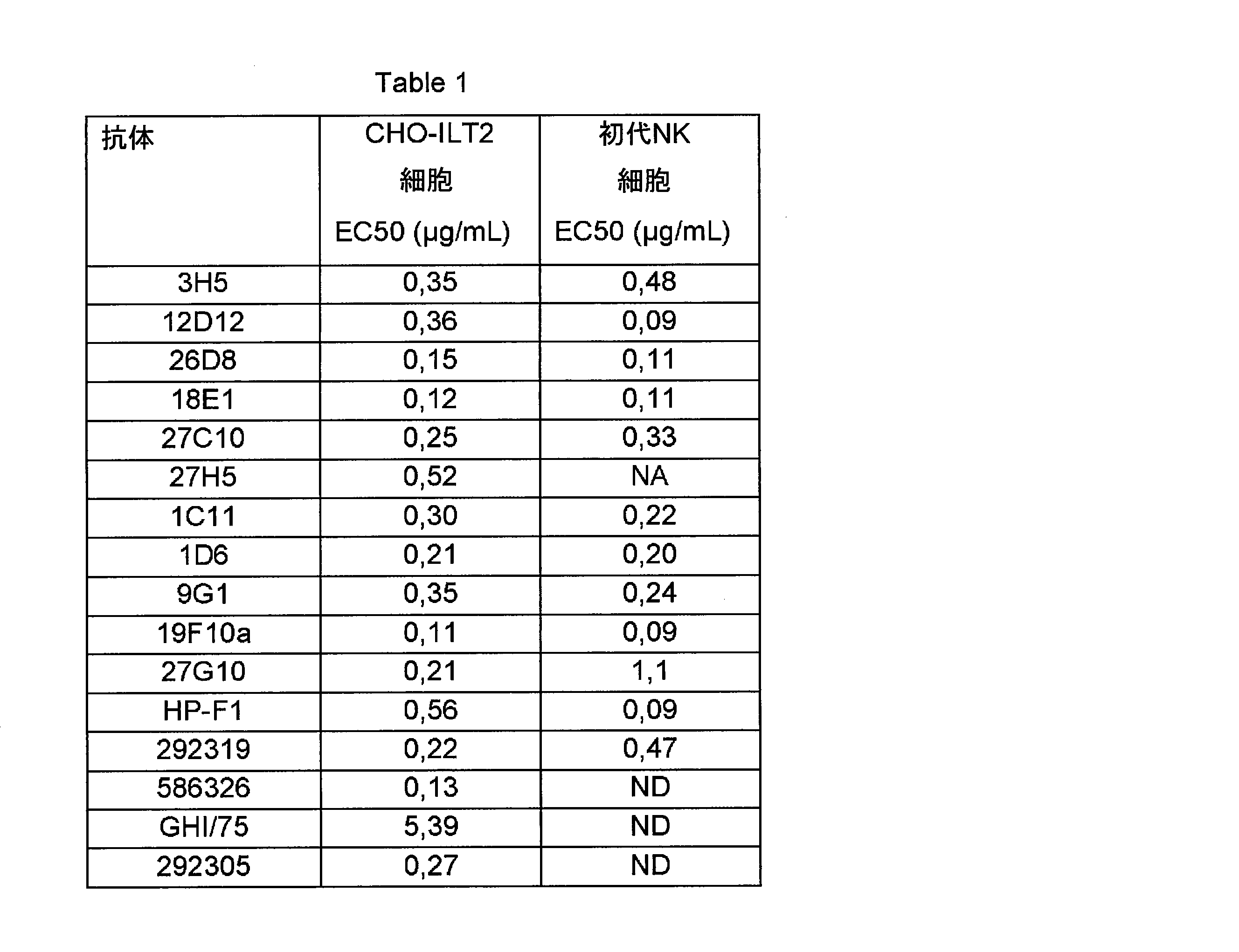
(実施例8)
一価親和性の決定
抗体3H5、12D12、26D8、18E1、27C10、27H5、1C11、1D6、9G1、19F10a、及び27G10、並びに市販の抗体GHI/75、292319、及びHP-F1を、ヒトILT2タンパク質に対する結合親和性について試験した。
(Example 8)
Determination of monovalent affinity Antibodies 3H5, 12D12, 26D8, 18E1, 27C10, 27H5, 1C11, 1D6, 9G1, 19F10a, and 27G10, as well as the commercially available antibodies GHI / 75, 292319, and HP-F1 against human ILT2 proteins. The binding affinity was tested.
SPR(表面プラスモン共鳴)法を用いて、抗体3H5、12D12、26D8、18E1、27C10、27H5、1C11、1D6、9G1、19F10a、27G10(すべてヒトIgG1アイソタイプ)を試験した。測定は、Biacore T200装置(Biacore GE Healthcare社)で、25℃で実施した。すべてのBiacore実験では、HBS-EP+(Biacore GE Healthcare社)及び10mMのNaOHをそれぞれ運転緩衝液及び再生緩衝液として供した。Biacore T100評価ソフトウェアを用いてセンサログラムを分析した。プロテインAはGE Healthcare社から購入した。ヒトILT2組換えタンパク質は、Innate Pharma社でクローニングし、生成し、精製した。プロテインAタンパク質を、センサーチップCM5の上のデキストラン層中のカルボキシル基に共有結合で固定化した。チップの表面をEDC/NHS(N-エチル-N'-(3-ジメチルアミノプロピル)カルボジイミド塩酸及びN-ヒドロキシスクシンイミド(Biacore GE Healthcare社))で活性化した。プロテインAをカップリング緩衝液(10mMの酢酸塩、pH5.6)中で10μg/mlに希釈し、適切な固定化レベル(即ち600RU)に達するまで注入した。残存する活性基の不活化は、100mMのエタノールアミン(pH8)(Biacore GE Healthcare社)を用いて実施した。2μg/mlの抗ILT2抗体をプロテインAチップに捕捉し、捕捉された抗体の上に組換えヒトILT2タンパク質を250nM~1.95nMの範囲の異なる濃度で注入した。ブランク差引きのため、ILT2タンパク質を運転緩衝液で置き換えて再びサイクルを実施した。一価親和性の分析は、メーカーの推奨による通常のCapture-Kineticプロトコール(Biacore GE Healthcare社速度論ウィザード)に従って行った。1.95nM~250nMの範囲の、ヒトILT2タンパク質の7種の連続希釈物を、捕捉した抗体の上に逐次的に注入し、再生の前に10分間、解離させた。1:1速度論的結合モデル又は2状態反応モデルを用い、曲線のプロファイルの関数として、全体のセンサログラムの組を適合させた。 Antibodies 3H5, 12D12, 26D8, 18E1, 27C10, 27H5, 1C11, 1D6, 9G1, 19F10a, 27G10 (all human IgG1 isotypes) were tested using the SPR (Surface Plasmon Resonance) method. Measurements were performed on a Biacore T200 device (Biacore GE Healthcare) at 25 ° C. In all Biacore experiments, HBS-EP + (Biacore GE Healthcare) and 10 mM NaOH were provided as operating and regeneration buffers, respectively. Sensorograms were analyzed using Biacore T100 evaluation software. Protein A was purchased from GE Healthcare. The human ILT2 recombinant protein was cloned, produced and purified by Innate Pharma. Protein A Protein was covalently immobilized on the carboxyl group in the dextran layer on the sensor chip CM5. The surface of the chip was activated with EDC / NHS (N-ethyl-N'-(3-dimethylaminopropyl) carbodiimide hydrochloric acid and N-hydroxysuccinimide (Biacore GE Healthcare)). Protein A was diluted to 10 μg / ml in coupling buffer (10 mM acetate, pH 5.6) and injected until the appropriate immobilization level (ie 600 RU) was reached. Inactivation of the remaining active groups was performed using 100 mM ethanolamine (pH 8) (Biacore GE Healthcare). 2 μg / ml anti-ILT2 antibody was captured on a protein A chip and recombinant human ILT2 protein was injected onto the captured antibody at different concentrations ranging from 250 nM to 1.95 nM. For blank deduction, the ILT2 protein was replaced with driving buffer and the cycle was performed again. The monovalent affinity analysis was performed according to the usual Capture-Kinetic protocol (Biacore GE Healthcare kinetics wizard) recommended by the manufacturer. Seven serial dilutions of human ILT2 protein, ranging from 1.95 nM to 250 nM, were sequentially injected onto the captured antibody and dissociated for 10 minutes prior to regeneration. A set of whole sensorograms was fitted as a function of the curve profile using a 1: 1 kinetic coupling model or a two-state response model.
OCTET分析を用いて、抗体GHI/75、292319、及びHP-F1(すべてマウスアイソタイプ)を評価した。測定はOCTET RED96システム(Fortebio社)で実施した。すべてのBiacore実験で、動力学緩衝液10X(Fortebio社)及び10mM、pH1.8のグリシンを、それぞれ運転緩衝液及び再生緩衝液として供した。グラフはData Analysis 9.0ソフトウェアで分析した。抗マウスIgG Fcキャプチャー(AMC)バイオセンサーを用いた。5μg/mLの抗ILT2抗体を、抗マウスIgG Fcキャプチャー(AMC)バイオセンサーに捕捉させた。組換えヒトILT2タンパク質の7種の希釈物を注入した(292319及びHP-F1については1000nM~15.625nM、GHI-75については100nM~1.5625nM)。曲線はモデル1:1を用いて適合させた。 Antibodies GHI / 75, 292319, and HP-F1 (all mouse isotypes) were evaluated using OCTET analysis. Measurements were performed on the OCTET RED 96 system (Fortebio). In all Biacore experiments, kinetic buffer 10X (Fortebio) and 10 mM, pH 1.8 glycine were provided as operating and regeneration buffers, respectively. The graph was analyzed with Data Analysis 9.0 software. An anti-mouse IgG Fc capture (AMC) biosensor was used. 5 μg / mL anti-ILT2 antibody was captured by an anti-mouse IgG Fc capture (AMC) biosensor. Seven dilutions of recombinant human ILT2 protein were injected (1000 nM to 15.625 nM for 292319 and HP-F1 and 100 nM to 1.5625 nM for GHI-75). The curves were fitted using model 1: 1.
結果を下のTable 2(表3)に示す。KDの相違は一般にNK細胞の細胞傷害性を強化する能力における相違と相関するようには見られない。したがって結合親和性はNK細胞の細胞傷害性を強化する抗体の能力における相違を説明しない。 The results are shown in Table 2 below. Differences in KD generally do not appear to correlate with differences in the ability of NK cells to enhance their cytotoxicity. Therefore, binding affinity does not explain the difference in the ability of antibodies to enhance the cytotoxicity of NK cells.
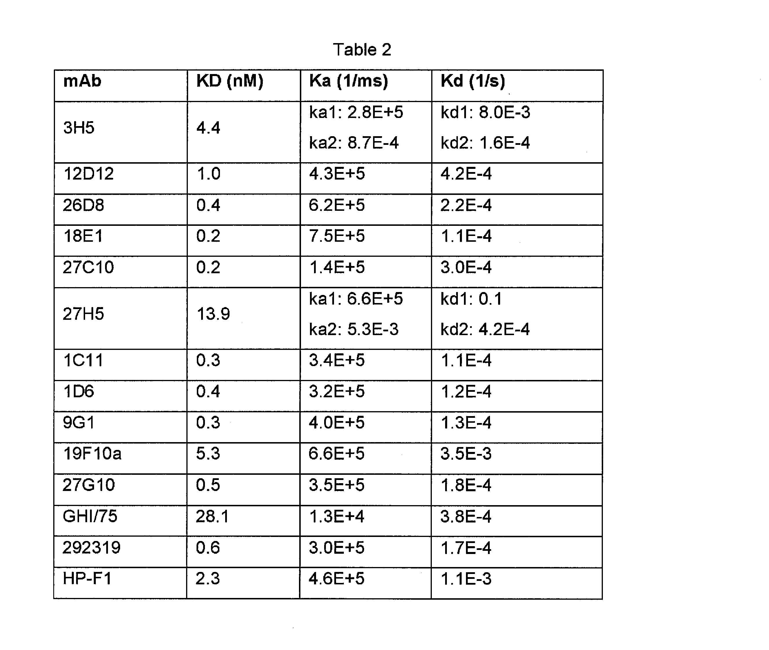
(実施例9)
初代ヒトNK細胞における細胞傷害性を増加する抗体の同定
本発明者らは、従来の抗体にNK細胞のILT2を中和する能力がなかったことが、例えばその表面により高いレベルのILT2を発現する高度に選択され又は改変されたNK細胞系と比較した初代NK細胞におけるILT2の発現の相違に関連しているかもしれないという可能性を考慮した。本発明者らは、数名の健常ヒトドナーからの初代NK細胞における抗体を研究して選択した。実施例5の抗ILT2抗体の効果を、NK細胞上のCD137の表面発現を評価する活性化アッセイによって研究した。それぞれの場合に、初代NK細胞(ドナーから精製した新鮮なNK細胞として)をエフェクター細胞として用い、HLA-E/Gを発現するK562細胞(慢性骨髄性白血病(CML))を標的として用いた。その結果、標的はILT2のリガンドであるHLA-Gだけでなく、広範囲のがん細胞の表面に発現するHLAクラスIリガンドであり、NK細胞及びCD8 T細胞の表面上の阻害性受容体と相互作用し得るHLA-Eをも発現した。
(Example 9)
Identification of antibodies that increase cytotoxicity in primary human NK cells We found that conventional antibodies did not have the ability to neutralize ILT2 in NK cells, for example, expressing higher levels of ILT2 on their surface. We considered the possibility that it may be associated with differences in ILT2 expression in primary NK cells compared to highly selected or modified NK cell lines. We have studied and selected antibodies in primary NK cells from several healthy human donors. The effect of the anti-ILT2 antibody of Example 5 was investigated by an activation assay to assess the surface expression of CD137 on NK cells. In each case, primary NK cells (as fresh NK cells purified from donors) were used as effector cells, and HLA-E / G-expressing K562 cells (chronic myelogenous leukemia (CML)) were used as targets. As a result, the target is not only HLA-G, which is a ligand for ILT2, but also HLA class I ligand, which is expressed on the surface of a wide range of cancer cells, and interacts with inhibitory receptors on the surface of NK cells and CD8 T cells. It also expressed HLA-E, which could act.
手短に述べると、NK細胞の活性化に対する抗ILT2抗体の効果を、全NK細胞、ILT2陽性NK細胞、及びILT2陰性NK細胞におけるCD137の発現のフローサイトメトリーによる分析によって決定した。エフェクター細胞は初代NK細胞(ドナーから精製した新鮮なNK細胞、使用前インキュベーション37℃、一夜)であり、標的細胞(K562 HLA-E/G細胞系)を1:1の比で混合した。CD137のアッセイは、96Uウェルプレートで、完全RPMI中、最終200μL/ウェルとして行った。抗体をエフェクター細胞と共に37℃で30分間、予めインキュベートし、標的細胞を37℃で一夜、共インキュベートした。以下のステップは、遠心分離500gで3分、染色緩衝液(SB)で2回洗浄、50μLの染色抗体混合物(抗CD3 Pacific blue - BD Biosciences社、抗CD56-PE-Vio770 - Miltenyi Biotec社、抗CD137-APC - Miltenyi Biotec社、抗ILT2-PE クローンHP-F1 - eBioscience社)の添加、4℃、30分のインキュベーション、SBによる洗浄2回、SBによるペレットの再懸濁、及びCanto II(HTS社)による蛍光の顕示であった。陰性対照はNK細胞対K562-HLA-E/G単独、アイソタイプ対照の存在下であった。 Briefly, the effect of anti-ILT2 antibody on NK cell activation was determined by flow cytometric analysis of CD137 expression in total NK cells, ILT2-positive NK cells, and ILT2-negative NK cells. Effector cells were primary NK cells (fresh NK cells purified from donors, pre-use incubation 37 ° C, overnight) and target cells (K562 HLA-E / G cell line) were mixed in a 1: 1 ratio. The assay for CD137 was performed in 96 U-well plates at final 200 μL / well in full RPMI. Antibodies were pre-incubated with effector cells at 37 ° C for 30 minutes and target cells were co-incubated at 37 ° C overnight. The following steps are performed by centrifugation at 500 g for 3 minutes, washing twice with staining buffer (SB), and 50 μL of the stained antibody mixture (anti-CD3 Pacific blue-BD Biosciences, anti-CD56-PE-Vio770-Milteyi Biotec, anti-CD3). Addition of CD137-APC-Miltenyi Biotec, anti-ILT2-PE clone HP-F1-eBioscience), incubation at 4 ° C for 30 minutes, washing twice with SB, resuspension of pellets with SB, and Canto II (HTS). It was a manifestation of fluorescence by the company). Negative controls were NK cells vs. K562-HLA-E / G alone, in the presence of isotype controls.
図5Aは、2名のヒトドナーからのNK細胞並びにHLA-E及びHLA-Gを発現させたK562腫瘍標的細胞を用いた、CD137を発現する全NK細胞の抗ILT2抗体によって媒介される増加のパーセントを示す代表的な図である。図5Bは、2名のヒトドナーからのNK細胞及びHLA-A2を発現しているB細胞系を用いた、CD137を発現しているILT2陽性(左側のパネル)及びILT2陰性(右側のパネル)のNK細胞の抗ILT2抗体によって媒介される増加のパーセントを示す代表的な図である。 Figure 5A shows the percentage of increase mediated by anti-ILT2 antibody in all NK cells expressing CD137 using NK cells from two human donors and K562 tumor target cells expressing HLA-E and HLA-G. It is a typical figure which shows. Figure 5B shows CD137-expressing ILT2-positive (left panel) and ILT2-negative (right panel) using NK cells from two human donors and a B cell line expressing HLA-A2. FIG. 6 is a representative diagram showing the percentage of increase mediated by anti-ILT2 antibodies in NK cells.
驚くべきことに、NK細胞系の細胞傷害性の強化において最も効果的であった抗体が、必ずしも初代ヒトNK細胞の活性化をすることができないということが観察された。NK細胞系の細胞傷害性の強化において最も効果的であった抗体12D12、19F10a、及び292319の中で、19F10aと292319の両方は、アイソタイプ対照抗体と比較して、初代NK細胞すべてを活性化する能力に実質的に欠けていた。 Surprisingly, it was observed that the most effective antibodies in enhancing the cytotoxicity of NK cell lines are not always capable of activating primary human NK cells. Of the antibodies 12D12, 19F10a, and 292319 that were most effective in enhancing the cytotoxicity of NK cell lines, both 19F10a and 292319 activate all primary NK cells compared to isotype control antibodies. It was virtually lacking in ability.
一方、抗体12D12、18E1、及び26D8は、初代NK細胞の強い活性化を示した。ILT2陽性NK細胞の研究は、これらの抗体が標的細胞に対するNK細胞の活性化において2倍の増加を媒介したことを示した。対照として、CD137を発現するILT2陰性NK細胞のパーセントは、抗体によって影響されなかった。 On the other hand, antibodies 12D12, 18E1 and 26D8 showed strong activation of primary NK cells. Studies of ILT2-positive NK cells have shown that these antibodies mediated a 2-fold increase in NK cell activation against target cells. As a control, the percentage of ILT2-negative NK cells expressing CD137 was unaffected by the antibody.
図6A及び図6Bは、細胞傷害性マーカーCD137の増加倍率で表した初代NK細胞の腫瘍標的細胞に対する細胞傷害性を強化する抗体の能力を示す。図6Aは、HLA-G及びHLA-Eを発現しているK562標的細胞に対する5~12名の異なるドナーからの初代NK細胞を用いた、HLA-G発現標的細胞の存在下におけるNK細胞の活性化を強化する抗体の能力を示す。図6Aは、HLA-A2を発現している標的B細胞に対する3~14名の異なるドナーからの初代NK細胞を用いた、HLA-G発現標的細胞の存在下におけるNK細胞の活性化を強化する抗体の能力を示す。それぞれの場合に、12D12、18E1、及び26D8は、実施例5でNK細胞系を用いた場合に最も強いNK細胞傷害性の強化を示した抗体の中にあった抗体の1つ(292319)と比較して、より大きなNK細胞傷害性の強化を有していた。 6A and 6B show the ability of the antibody to enhance the cytotoxicity of primary NK cells to tumor target cells, expressed at an increase factor of the cytotoxic marker CD137. FIG. 6A shows the activity of NK cells in the presence of HLA-G expressing target cells using primary NK cells from 5-12 different donors against HLA-G and HLA-E expressing K562 target cells. Shows the ability of the antibody to enhance the formation. FIG. 6A enhances NK cell activation in the presence of HLA-G expressing target cells using primary NK cells from 3-14 different donors against HLA-A2 expressing target B cells. Shows the ability of the antibody. In each case, 12D12, 18E1 and 26D8 were among the antibodies that showed the strongest enhancement of NK cytotoxicity when using the NK cell line in Example 5 (292319). In comparison, it had greater NK cytotoxic enhancement.
(実施例10)
ILTファミリーメンバーへの結合の特徴分析
抗体の結合特異性を更に特徴分析するため、様々なILTファミリータンパク質を発現させた細胞への結合性について、フローサイトメトリーによって抗体を試験した。上記のILT2(LILRB1)発現細胞に加えて、ヒトILT1 (LILRA2)、ILT3 (LILRB4)、ILT4 (LILRB2)、ILT5 (LILRB3)、ILT6 (LILRA3)、ILT7 (LILRA4)、又はILT8 (LILRA6)を発現する細胞を産生させた。
(Example 10)
Characteristic analysis of binding to ILT family members In order to further characterize the binding specificity of the antibody, the antibody was tested by flow cytometry for its binding to cells expressing various ILT family proteins. In addition to the above ILT2 (LILRB1) expressing cells, human ILT1 (LILRA2), ILT3 (LILRB4), ILT4 (LILRB2), ILT5 (LILRB3), ILT6 (LILRA3), ILT7 (LILRA4), or ILT8 (LILRA6) are expressed. Produced cells to grow.
ヒトILT遺伝子は、下のTable 3(表4)に記載したプライマーを用いるPCRによって増幅した。PCR産物を適切な制限部位で発現ベクターに挿入した。重鎖ペプチドリーダーを用い、アミノ酸配列GKPIPNPLLGLDST(配列番号80)を有するV5タグをN末端に付加した(Table 4(表5)の配列には示していない)。本明細書で用いた様々なヒトILTタンパク質についてのアミノ酸配列を、下のTable 4(表5)に示す。次いでベクターをCHO細胞系にトランスフェクトして、細胞表面に様々なILTタンパク質を発現する安定なクローンを得た。 The human ILT gene was amplified by PCR using the primers listed in Table 3 below. The PCR product was inserted into the expression vector at the appropriate restriction site. A heavy chain peptide reader was used to add a V5 tag having the amino acid sequence GKPIPNPLLGLDST (SEQ ID NO: 80) to the N-terminus (not shown in the sequence of Table 4). The amino acid sequences for the various human ILT proteins used herein are shown in Table 4 below. The vector was then transfected into the CHO cell line to obtain stable clones expressing various ILT proteins on the cell surface.


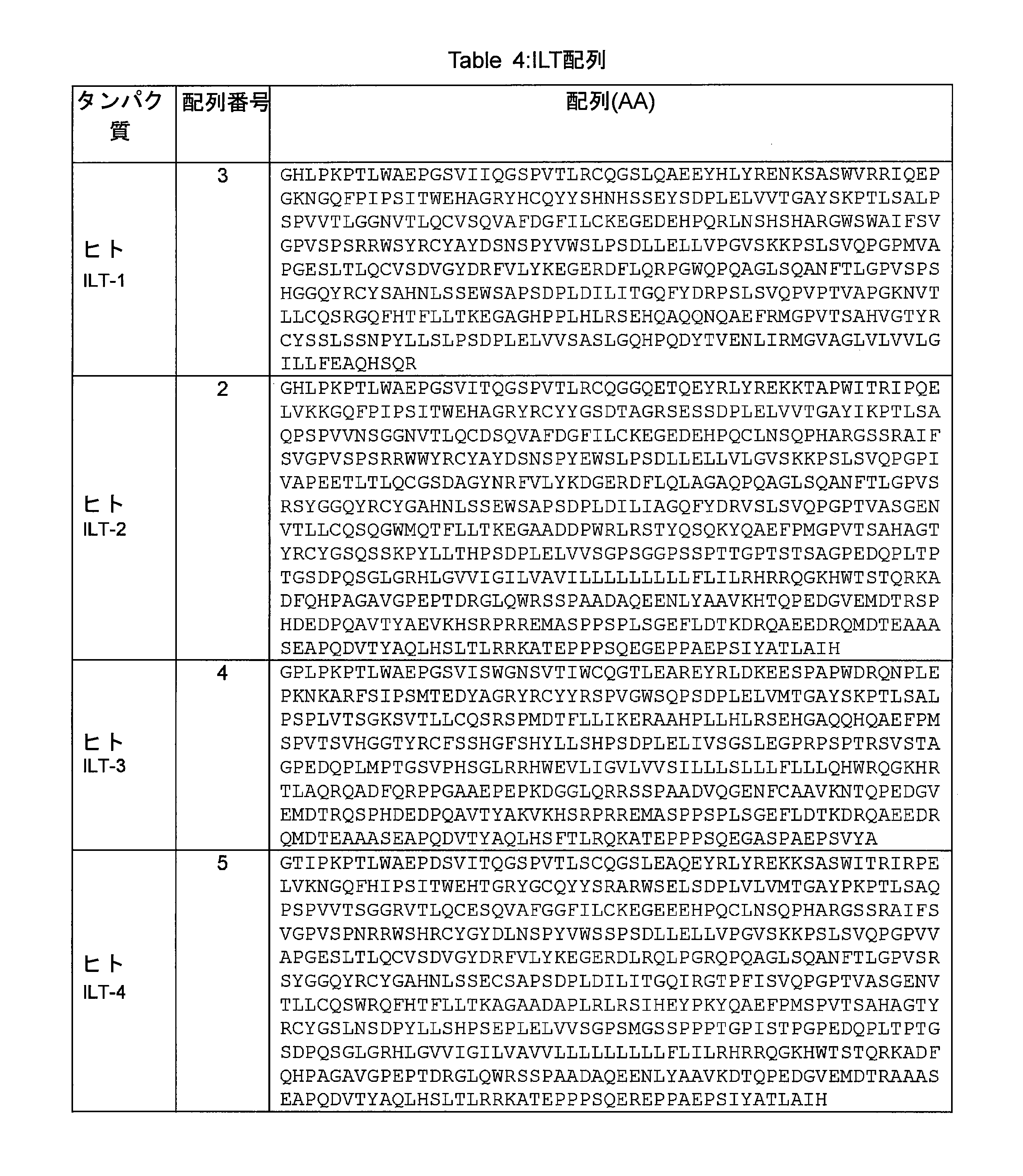

手短に述べると、フローサイトメトリースクリーニングのため、抗体をそれぞれのILT発現CHO細胞系(CHO ILT1細胞系、CHO ILT2細胞系、CHO ILT3細胞系、CHO ILT4細胞系、CHO ILT5細胞系、CHO ILT6細胞系、CHO ILT7細胞系、CHO ILT8細胞系)と共に1時間インキュベートし、染色緩衝液で2回洗浄し、PEで標識したヤギ抗マウスIgG H+L ポリクローナル抗体(pAb)(市販の抗体について、Jackson Immunoresearch社、#115-116-146)、又はPEで標識したヤギ抗ヒトIgG H+L pAb(キメラ抗体について、Jackson Immunoresearch社、#109-116-088)によって顕示し、染色緩衝液で2回洗浄し、HTFCプレートローダーを備えたAccury C6フローサイトメーターで染色を取得し、FlowJoソフトウェアを用いて分析した。 Briefly, for flow cytometry screening, antibodies are used for each ILT-expressing CHO cell line (CHO ILT1 cell line, CHO ILT2 cell line, CHO ILT3 cell line, CHO ILT4 cell line, CHO ILT5 cell line, CHO ILT6 cell line. Incubate with line, CHO ILT7 cell line, CHO ILT8 cell line) for 1 hour, washed twice with staining buffer and PE-labeled goat anti-mouse IgG H + L polyclonal antibody (pAb) (for commercial antibody, Jackson Revealed with Immunoresearch, # 115-116-146) or PE-labeled goat anti-human IgG H + L pAb (for chimeric antibodies, Jackson Immunoresearch, # 109-116-088), twice with staining buffer They were washed and stained with an Accury C6 flow cytometer equipped with an HTFC plate loader and analyzed using FlowJo software.
結果は、抗ILT2抗体の多くが単独で(即ちILT2/ILT6の交差反応)、又はILT4若しくはILT5へのさらなる結合(即ちILT2/ILT4/ILT6又はILT2/ILT5/ILT6の交差反応)と共に、ILT2に加えてILT6(LILRA3)にも結合したことを示した。抗体1C11、1D6、9G1、19F10a、27G10、市販の抗体586326及び292305は、ILT2に、またILT6にも結合した。抗体586326はILT2及びILT6に加えてILT4にも更に結合し、抗体292305はILT2及びILT6に加えてILT5に更に結合した。最後に、市販の抗体292319はILT2に加えてILT1に結合した(ILT1/ILT2の交差反応)。しかし、3H5、12D12、26D8、18E1、27C10、及び27H5によって例示される抗体のサブセットはILT2のみに結合し、他のILTのファミリーメンバータンパク質には結合しなかった。
The results show that many of the anti-ILT2 antibodies alone (ie, ILT2 / ILT6 cross-reactivity) or with further binding to ILT4 or ILT5 (ie, ILT2 / ILT4 / ILT6 or ILT2 / ILT5 / ILT6 cross-reactivity). In addition, it was shown that it also bound to ILT6 (LILRA3). Antibodies 1C11, 1D6, 9G1, 19F10a, 27G10 and commercially
(実施例11)
エピトープマッピング
係留されたILT2ドメイン断片タンパク質
ILT2タンパク質の産生
様々なヒトILT2ドメインD1(配列番号1で示される配列の残基24~121に対応)、D2(配列番号1で示される配列の残基122~222に対応)、D3(配列番号1で示される配列の残基223~321に対応)、D4(配列番号1で示される配列の残基322~458に対応)をコードする核酸配列及びその組合せを、下の表に記載したプライマーを用いるPCRによって増幅した。PCR産物を適切な制限部位で発現ベクターに挿入した。重鎖ペプチドリーダーを用い、N末端にV5タグを付加して、細胞の表面における発現をフローサイトメトリーによって確認した。D4ドメインが後に続かないドメインのすべてについては、細胞膜における係留を可能にするために、CD24 GP1アンカーを付加した。得られる様々なヒトILT2ドメイン断片含有タンパク質のアミノ酸配列を、下のTable 5(表6)に示す。次いでベクターをCHO細胞系にトランスフェクトして、細胞表面に様々なILT2ドメインタンパク質を発現する安定なクローンを得た。
(Example 11)
Epitope mapping moored ILT2 domain fragment protein
Production of ILT2 protein Various human ILT2 domains D1 (corresponding to residues 24-121 of the sequence represented by SEQ ID NO: 1), D2 (corresponding to residues 122-222 of the sequence represented by SEQ ID NO: 1), D3 (corresponding to residues 122-222 of the sequence represented by SEQ ID NO: 1). The nucleic acid sequences encoding residues 223-321 of the sequence represented by number 1), D4 (corresponding to residues 322-458 of the sequence represented by SEQ ID NO: 1) and combinations thereof are listed in the table below. Amplified by PCR with primers. The PCR product was inserted into the expression vector at the appropriate restriction site. Using a heavy chain peptide reader, a V5 tag was added to the N-terminus, and expression on the cell surface was confirmed by flow cytometry. For all domains not followed by the D4 domain, a CD24 GP1 anchor was added to allow mooring at the cell membrane. The amino acid sequences of the various obtained human ILT2 domain fragment-containing proteins are shown in Table 5 (Table 6) below. The vector was then transfected into the CHO cell line to obtain stable clones expressing various ILT2 domain proteins on the cell surface.
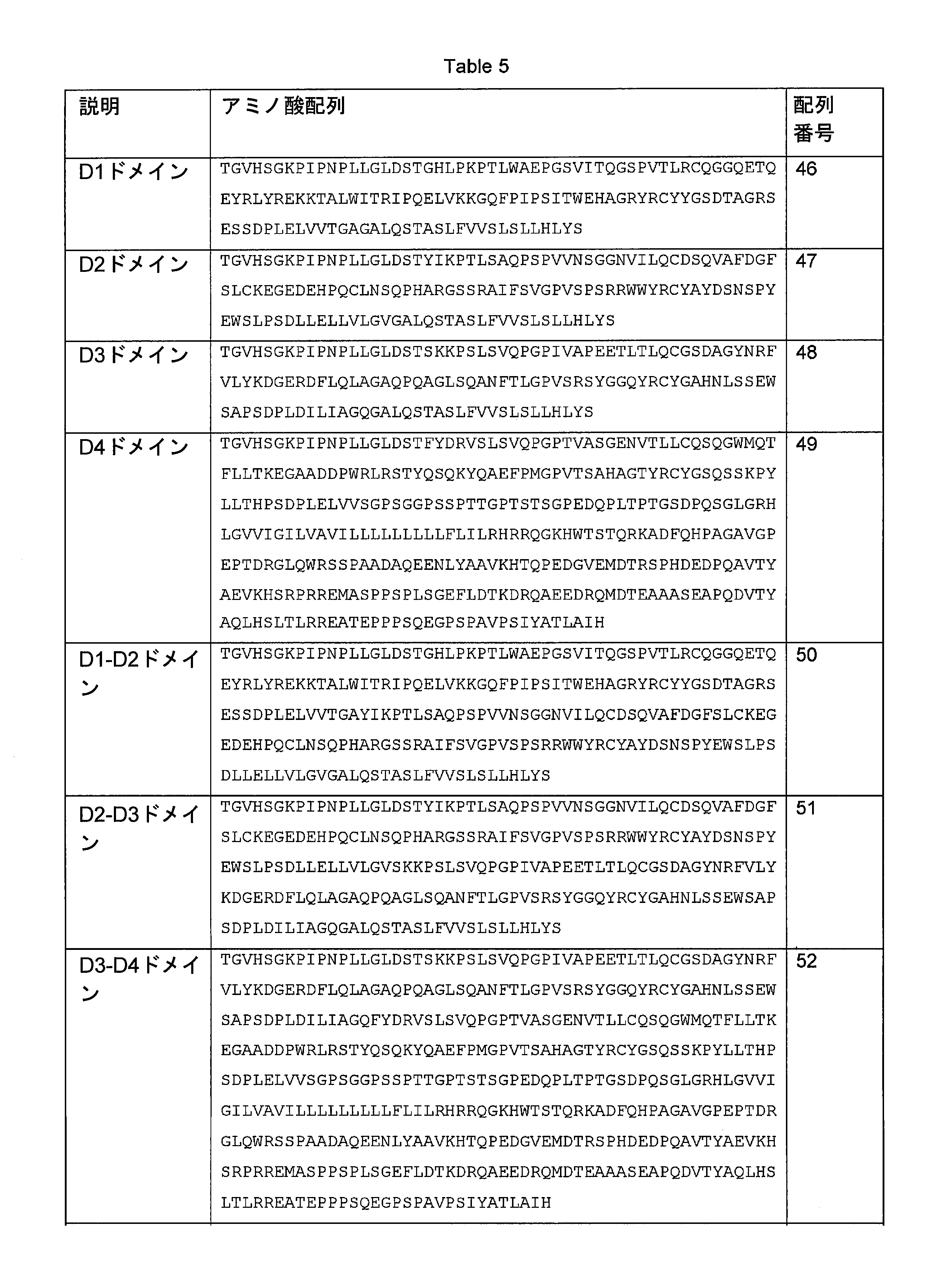
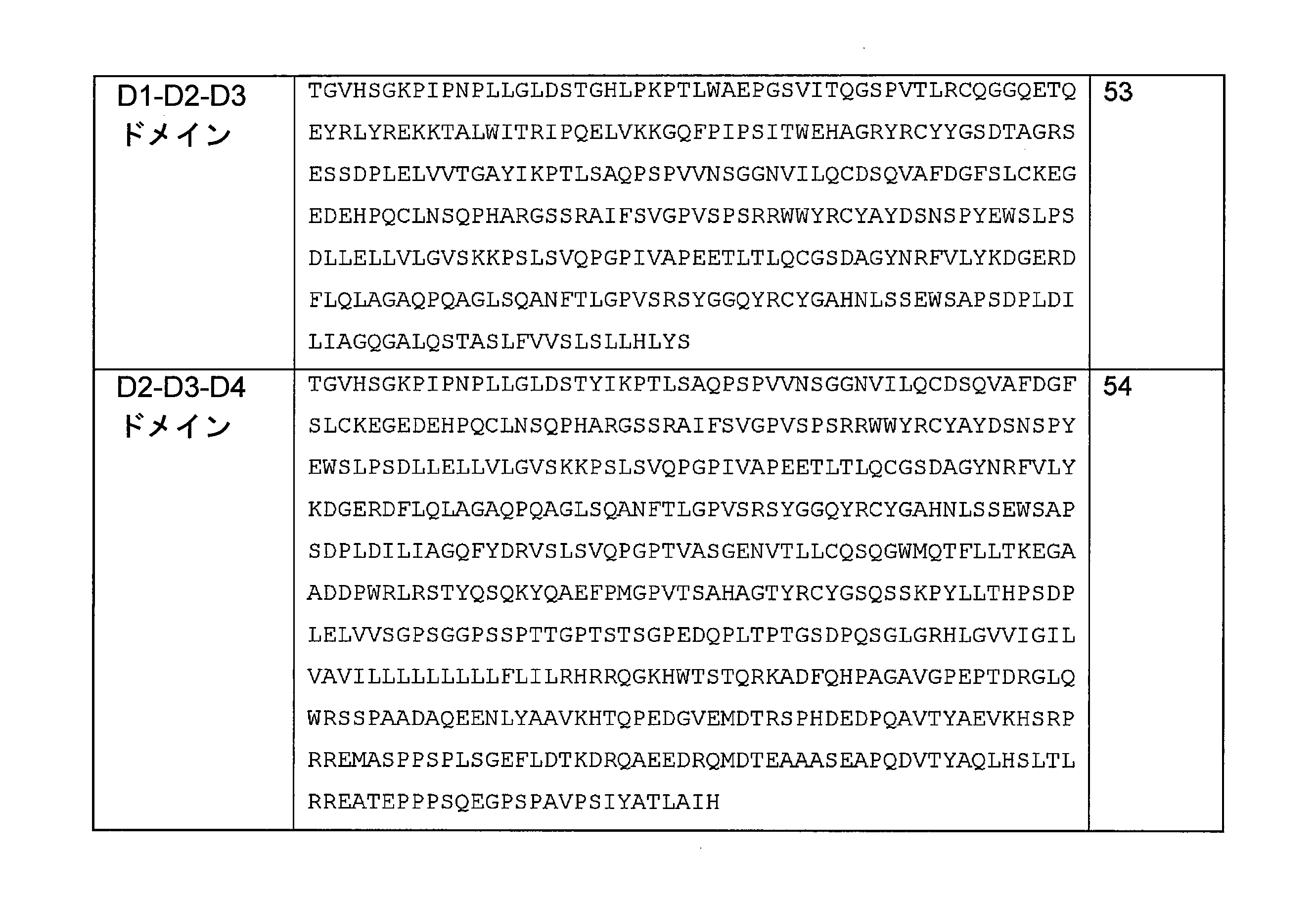
結果
ILT2選択的抗体を、係留した様々なILT2断片への結合性についてフローサイトメトリーによって試験した。3H5、12D12、及び27H5はすべて、ILT2のD1ドメインに結合した。これらの抗体は、D1ドメインを欠くILT2タンパク質(配列番号47~49、51、52、及び54のタンパク質)を発現する細胞のいずれにも結合することなく、ILT2のD1ドメインを含むタンパク質(配列番号46、50、及び53のタンパク質)を発現する細胞すべてに結合した。即ち抗体3H5、12D12、及び27H5は、配列番号1で示される配列の残基24~121によって定義されるILT2のドメイン(ドメインD1とも称する)に結合する。抗体26D8、18E1、及び27C10はすべて、ILT2のD4ドメインに結合した。これらの抗体は、D4ドメインを欠くILT2タンパク質(配列番号46~28、50、51、又は53のタンパク質)を発現する細胞のいずれにも結合することなく、ILT2のD4ドメインを含むタンパク質(配列番号49、52、及び54)を発現するすべての細胞に結合した。即ち抗体26D8、18E1、及び27C10は、配列番号1で示される配列の残基322~458によって定義されるILT2のドメインに結合する。図7は、配列番号46の係留したILT2ドメインD1断片タンパク質(左側のパネル)、配列番号48のD3ドメイン断片タンパク質(中央のパネル)、及び配列番号49のD4ドメインタンパク質(右側のパネル)への抗体の結合の代表的な例を示す。
result
ILT2-selective antibodies were tested by flow cytometry for binding to various moored ILT2 fragments. 3H5, 12D12, and 27H5 all bound to the D1 domain of ILT2. These antibodies do not bind to any of the cells expressing the ILT2 protein lacking the D1 domain (proteins of SEQ ID NOs: 47-49, 51, 52, and 54), but the protein containing the D1 domain of ILT2 (SEQ ID NO:). It bound to all cells expressing (46, 50, and 53 proteins). That is, antibodies 3H5, 12D12, and 27H5 bind to the domain of ILT2 (also referred to as domain D1) defined by residues 24-121 of the sequence set forth in SEQ ID NO: 1. Antibodies 26D8, 18E1 and 27C10 all bound to the D4 domain of ILT2. These antibodies do not bind to any of the cells expressing the ILT2 protein lacking the D4 domain (protein of SEQ ID NOs: 46-28, 50, 51, or 53) and contain the D4 domain of ILT2 (SEQ ID NO:). It bound to all cells expressing 49, 52, and 54). That is, antibodies 26D8, 18E1 and 27C10 bind to the domain of ILT2 defined by residues 322-458 of the sequence set forth in SEQ ID NO: 1. Figure 7 shows the anchored ILT2 domain D1 fragment protein of SEQ ID NO: 46 (left panel), the D3 domain fragment protein of SEQ ID NO: 48 (center panel), and the D4 domain protein of SEQ ID NO: 49 (right panel). A typical example of antibody binding is shown.
ILT2の点突然変異の研究
密接に関連するILT6に結合することなくILT2に結合する抗体の同定により、露出してILT2とILT6との間で異なるアミノ酸におけるILT2の突然変異を設計することが可能になった。したがって、ILT6と交差反応しなかった抗ILT2抗体を、ILT2のD1、D2、又はD4ドメインにアミノ酸置換を有する様々なILT2突然変異体への結合性の喪失についてマッピングすることができる。ILT2突然変異体への結合性の喪失は、ヒトILT6への結合性の喪失と共に、NK細胞の細胞傷害性を強化する抗体が結合するILT2のエピトープの同定に役立ち得る。
Study of point mutations in ILT2 Identification of antibodies that bind to ILT2 without binding to closely related ILT6 makes it possible to design mutations in ILT2 at different amino acids between ILT2 and ILT6 exposed. became. Thus, anti-ILT2 antibodies that did not cross-react with ILT6 can be mapped to loss of binding to various ILT2 mutants with amino acid substitutions in the D1, D2, or D4 domains of ILT2. Loss of binding to the ILT2 mutant, along with loss of binding to human ILT6, may help identify the epitope of ILT2 to which an antibody that enhances cytotoxicity of NK cells binds.
ILT2突然変異体の産生
ILT2突然変異体をPCRによって産生させた。増幅した配列をアガロースゲルにかけ、Macherey Nagel PCR Clean-Up Gel Extraction キット(参照番号 740609)を用いて精製した。次いでそれぞれの突然変異体について産生した精製PCR産物を、ClonTech InFusionシステムを用いて発現ベクターにライゲートした。突然変異した配列を含むベクターをMiniprepとして調製し、シーケンシングした。シーケンシングの後、突然変異した配列を含むベクターを、Promega PureYield(商標) Plasmid Midiprep Systemを用いてMidiprepとして調製した。HEK293T細胞をDMEM培地(Invitrogen社)中で増殖させ、Invitrogen社のLipofectamine 2000を用いてベクターをトランスフェクトし、CO2インキュベーター中、37℃で48時間インキュベートして、トランスジーンの発現について試験した。突然変異体は、下の表に示すように、Hek293T細胞にトランスフェクトした。標的アミノ酸突然変異を、下のTable 6(表7)に示す。この表は、野生型ILT2に存在する残基/残基の位置/突然変異体ILT2に存在する残基を列挙しており、位置参照は左の欄に配列番号2で示されるリーダーペプチドを欠くILT2タンパク質、又は右の欄に配列番号1で示されるリーダーペプチドを有するILT2タンパク質である。
Production of ILT2 mutants
The ILT2 mutant was produced by PCR. The amplified sequences were run on an agarose gel and purified using the Macherey Nagel PCR Clean-Up Gel Extraction Kit (reference number 740609). The purified PCR products produced for each mutant were then ligated into an expression vector using the ClonTech InFusion system. A vector containing the mutated sequence was prepared as a miniprep and sequenced. After sequencing, a vector containing the mutated sequence was prepared as Midiprep using the Promega PureYield ™ plasmid Midiprep System. HEK293T cells were grown in DMEM medium (Invitrogen), the vector was transfected with Invitrogen's

結果
ILT2選択的抗体を、突然変異体のそれぞれに対する結合性についてフローサイトメトリーによって試験した。最初の実験は、1つの濃度において1つ又はいくつかの突然変異体への結合性を喪失する抗体を決定するために実施した。結合性の喪失を確認するため、その結合性がILT2の突然変異によって影響されると思われる抗体について、抗体のタイトレーションを行った。試験した抗体の結合性の喪失又は低下は、特定の突然変異体の1つ若しくは複数、又はそのすべてが抗体のコアエピトープに重要であることを示し、したがってILT2の結合の領域を同定することが可能になった。
result
ILT2-selective antibodies were tested by flow cytometry for their binding to each of the mutants. The first experiment was performed to determine the antibody that lost its binding to one or several mutants at one concentration. To confirm the loss of binding, antibody titration was performed for antibodies whose binding was likely to be affected by mutations in ILT2. Loss or decrease in binding of the antibody tested indicates that one or more of the particular mutants, or all of them, are important for the core epitope of the antibody, thus identifying regions of ILT2 binding. It became possible.
抗体3H5、12D12、及び27H5は、これら3つの抗体がILT2のドメイン1(D1ドメイン)の残基34、36、76、82、及び84においてアミノ酸置換(置換E34A、R36A、Y76I、A82S、R84L)を有する突然変異体2への結合性を喪失したので、ILT2のドメイン1におけるエピトープに結合した。12D12及び27H5は他のいずれの突然変異体にも結合性を喪失しなかったが、3H5も、残基29、30、33、32、80にアミノ酸置換(置換G29S、Q30L、Q33A、T32A、D80H)を有する突然変異体1への結合性の低下(部分的喪失)を有していた。したがって、これらのアミノ酸残基は、ヒトILT6ポリペプチドへの結合性の欠如と共に、初代NK細胞の細胞傷害性を強化する抗ILT2抗体を特徴づけるエピトープを同定することができる。
Antibodies 3H5, 12D12, and 27H5 have amino acid substitutions (substitutions E34A, R36A, Y76I, A82S, R84L) at residues 34, 36, 76, 82, and 84 of domain 1 (D1 domain) of ILT2. Since it lost its binding property to
図8Aは、突然変異体1及び2への結合についての抗体3H5、12D12、及び27H5のフローサイトメトリーによるタイトレーションの代表的な例を示す。図9Aは、ドメイン1(上部、暗灰色の陰影)及びドメイン2(下部、明灰色の陰影)を含むILT2分子の部分を表すモデルを示す。図は、突然変異体1(M1)及び突然変異体2(M2)において置換されたアミノ酸残基によって定義される抗体の結合部位を示す。
FIG. 8A shows a representative example of flow cytometric titration of antibodies 3H5, 12D12, and 27H5 for binding to
抗体26D8、18E1、及び27C10はすべて、ILT2のドメインD4でエピトープに結合した。抗体26D8及び18E1は突然変異体4-1及び4-2への結合性を喪失していた。突然変異体4-1は残基299、300、301、328、378、及び381にアミノ酸置換(置換F299I、Y300R、D301A、W328G、Q378A、K381N)を有している。突然変異体4-2は残基328、330、347、349、350、及び355にアミノ酸置換(置換W328G、Q330H、R347A、T349A、Y350S、Y355A)を有している。26D8は更に突然変異体4-5への結合性を喪失しており、抗体18E1は突然変異体4-5への結合性が低下している(結合性の完全喪失ではないが)。27C10も突然変異体4-5への結合性を喪失したが、他のいずれの突然変異体への結合性も喪失していなかった。突然変異体4-5は残基341、342、344、345及び347にアミノ酸置換(置換D341A、D342S、W344L、R345A、R347A)を有している。26D8及び18E1は他のいずれの突然変異体への結合性も喪失していなかった。したがって、これらのアミノ酸残基は、ヒトILT6ポリペプチドへの結合性の欠如と共に、初代NK細胞の細胞傷害性を強化する抗ILT2抗体を特徴づけるエピトープを同定することができる。
Antibodies 26D8, 18E1 and 27C10 all bound to the epitope at domain D4 of ILT2. Antibodies 26D8 and 18E1 lost their binding to mutants 4-1 and 4-2. Mutant 4-1 has amino acid substitutions (substitutions F299I, Y300R, D301A, W328G, Q378A, K381N) at
図8Bは、D4ドメイン突然変異体4-1、4-1b、4-2、4-4、及び4-5への結合についての、抗体26D8、18E1、及び27C10のフローサイトメトリーによるタイトレーションの代表的な例を示す。 Figure 8B shows the flow cytometric titration of antibodies 26D8, 18E1 and 27C10 for binding to D4 domain mutants 4-1, 4-1b, 4-2, 4-4, and 4-5. A typical example is shown.
図9Bは、ドメイン3(上部、暗灰色の陰影)及びドメイン4(下部、明灰色の陰影)を含むILT2分子の部分を表すモデルを示す。図は、すべてがILT2のドメイン4の中に位置する、突然変異体4-1、4-2、及び4-5において置換されたアミノ酸残基によって定義される抗体の結合部位を示す。初代NK細胞の細胞傷害性を増強する抗体26D8、18E1は、突然変異体4-5によって定義される部位に結合することなく、突然変異体4-1及び4-2によって定義される部位に結合する。一方、初代NK細胞の細胞傷害性を増強しない抗体27C10は、突然変異体4-5によって定義される部位に結合する。
FIG. 9B shows a model representing a portion of the ILT2 molecule containing domain 3 (top, dark gray shade) and domain 4 (bottom, light gray shade). The figure shows the binding site of an antibody defined by amino acid residues substituted in mutants 4-1, 4-2, and 4-5, all located within
(実施例12)
ILT2-HLA-Gブロッキング抗体による初代ヒトNK細胞の細胞傷害性の強化についての親和性結合の閾値
初代NK細胞の細胞傷害性の強化において高度に活性であった抗ILT2抗体の活性の根底にある機構をより良く理解するため、実施例3に記載した方法を用い、実施例10の密接に関連するILTファミリーメンバーへの結合についてのさらなるスクリーニングと組み合わせて、さらなる免疫及びスクリーニングを行った。
(Example 12)
Threshold for Affinity Binding for Enhancement of Cytotoxicity of Primary Human NK Cells by ILT2-HLA-G Blocking Antibodies Underlies the activity of anti-ILT2 antibodies that were highly active in enhancing the cytotoxicity of primary NK cells. For a better understanding of the mechanism, further immunity and screening was performed using the method described in Example 3 in combination with further screening for binding to closely related ILT family members in Example 10.
ILT2_6XHisタンパク質でBalb/cマウスを免疫した。免疫プロトコールの後、マウスを犠牲死させて融合を実施し、ハイブリドーマを得た。ハイブリドーマの上清を用いて実施例10に記載したILT2発現CHO細胞系(それぞれがILT1 (LILRA2)、ILT3 (LILRB4)、ILT4 (LILRB2)、ILT5 (LILRB3)、ILT6 (LILRA3)、又はILT7 (LI LRA4)の1つを発現しているCHO系)を染色し、フローサイトメトリー実験においてモノクローナル抗体の反応性を確認した。手短に述べると、細胞を50μlの上清と共に4℃で1時間インキュベートし、3回洗浄して、AF647に連結した二次抗体ヤギ抗マウスIgG Fc特異的抗体を用いた(Jackson Immunoresearch社、JI115-606-071)。30分間の染色の後、細胞を3回洗浄し、FACS CANTO II (Becton Dickinson社)を用いて分析した。 Balb / c mice were immunized with the ILT2_6XHis protein. After the immune protocol, mice were sacrificed to death and fusion was performed to obtain hybridomas. ILT2-expressing CHO cell lines described in Example 10 using hybridoma supernatants (ILT1 (LILRA2), ILT3 (LILRB4), ILT4 (LILRB2), ILT5 (LILRB3), ILT6 (LILRA3), or ILT7 (LI, respectively). The CHO system) expressing one of LRA4) was stained, and the reactivity of the monoclonal antibody was confirmed in a flow cytometry experiment. Briefly, cells were incubated with 50 μl supernatant at 4 ° C. for 1 hour, washed 3 times and used with a secondary antibody goat anti-mouse IgG Fc-specific antibody ligated to AF647 (Jackson Immunoresearch, JI115). -606-071). After 30 minutes of staining, cells were washed 3 times and analyzed using FACS CANTO II (Becton Dickinson).
抗体をクローニング及びスクリーニングして、ヒトILT1、ILT3、ILT4、ILT5、ILT6、又はILT7に結合せずにILT2に結合し、ILT2とHLA-Gとの相互作用をブロックする能力を有する抗体を生成するハイブリドーマを同定した。手短に述べると、組換えビオチン化ILT2をAPCコンジュゲートしたストレプトアビジンと4℃で20時間インキュベートした後、精製した抗ILT2抗体を添加した。20分後、複合体を、HLA-Gを発現している5×10⁴個のK562細胞又はHLA-A2を発現しているWIL2-NS細胞と4℃で更に30分インキュベートした。細胞をPBSで1回洗浄し、Cell Fix(Becton Dickinson社、340181)で固定した。分析はFACS CANTO IIフローサイトメーターで実施した。 Antibodies are cloned and screened to produce antibodies capable of binding to ILT2 without binding to human ILT1, ILT3, ILT4, ILT5, ILT6, or ILT7 and blocking the interaction of ILT2 with HLA-G. A hybridoma was identified. Briefly, recombinant biotinylated ILT2 was incubated with APC-conjugated streptavidin at 4 ° C. for 20 hours before addition of purified anti-ILT2 antibody. After 20 minutes, the complex was incubated with 5 × 10 4 K562 cells expressing HLA-G or WIL2-NS cells expressing HLA-A2 at 4 ° C for an additional 30 minutes. Cells were washed once with PBS and fixed with Cell Fix (Becton Dickinson, 340181). The analysis was performed on a FACS CANTO II flow cytometer.
細胞系の表面に発現したHLA-G又はHLA-A2と組換えILT2タンパク質との相互作用をブロックする抗ILT2抗体の能力を、実施例5に記載したようにフローサイトメトリーによって評価した。これらのアッセイにより、ILT2とそのHLAクラスIリガンドであるHLA-Gとの相互作用のブロックにおいて高度に効果的であった抗ILT2抗体のパネルの同定が可能になった。抗体12D12、2A8A、2A8B、2A9、2B11、2C4、2C8、2D8、2E2B、2E2C、2E8、2E11、2G5、2H2A、2H2B、2H12、1A9、1A10B、1A10C、1A10D、1E4B、1E4C、3A7A、3A7B、3A8、3B5、3E5、3E7A、3E7B、3E9A、3E9B、3F5、4A8、4C11B、4E3A、4E3B、4H3、5C5、5D9、6C6、10H1、48F12、15D7、2C3はすべて、HLA-G及びHLA-A2へのILT2の結合をブロックした。図10Aは、抗体12D12、2H2B、48F12、1E4C、1A9、3F5、及び3A7Aについての代表的な結果を示す。得られる抗体を、図11に示すようにフローサイトメトリーによって様々な係留型ILT2断片及びILT2点突然変異体への結合性について試験し、突然変異L234A/L235E/G237A/A330S/P331Sを有するヒトIgG1 Fcドメインを有する改変されたキメラ抗体として提示した。 The ability of the anti-ILT2 antibody to block the interaction of HLA-G or HLA-A2 expressed on the surface of the cell line with the recombinant ILT2 protein was evaluated by flow cytometry as described in Example 5. These assays allowed the identification of a panel of anti-ILT2 antibodies that were highly effective in blocking the interaction of ILT2 with its HLA class I ligand, HLA-G. Antibodies 12D12, 2A8A, 2A8B, 2A9, 2B11, 2C4, 2C8, 2D8, 2E2B, 2E2C, 2E8, 2E11, 2G5, 2H2A, 2H2B, 2H12, 1A9, 1A10B, 1A10C, 1A10D, 1E4B, 1A10D, 1E4B , 3B5, 3E5, 3E7A, 3E7B, 3E9A, 3E9B, 3F5, 4A8, 4C11B, 4E3A, 4E3B, 4H3, 5C5, 5D9, 6C6, 10H1, 48F12, 15D7, 2C3 are all HLA-G and HLA-2 Blocked ILT2 binding. FIG. 10A shows representative results for antibodies 12D12, 2H2B, 48F12, 1E4C, 1A9, 3F5, and 3A7A. The resulting antibody was tested for binding to various moored ILT2 fragments and ILT 2-point mutants by flow cytometry as shown in FIG. 11 and human IgG1 with the mutant L234A / L235E / G237A / A330S / P331S. Presented as a modified chimeric antibody with an Fc domain.
初代ヒトNK細胞における細胞傷害性を増加させる抗ILT2抗体の能力を、実施例9のようにして試験した。手短に述べると、NK細胞の活性化に対する抗ILT2抗体の効果を、全NK細胞、ILT2陽性NK細胞、及びILT2陰性NK細胞におけるCD137の発現のフローサイトメトリーによって決定した。エフェクター細胞は初代NK細胞(ドナーから精製した新鮮なNK細胞、使用前インキュベーション37℃、一夜)であり、標的細胞(WIL2-NS細胞系)を1:1の比で混合した。 The ability of the anti-ILT2 antibody to increase cytotoxicity in primary human NK cells was tested as in Example 9. Briefly, the effect of anti-ILT2 antibody on NK cell activation was determined by flow cytometry of CD137 expression in total NK cells, ILT2-positive NK cells, and ILT2-negative NK cells. Effector cells were primary NK cells (fresh NK cells purified from donors, pre-use incubation 37 ° C, overnight) and target cells (WIL2-NS cell line) were mixed in a 1: 1 ratio.
図10Bは、2名のヒトドナーからのNK細胞及びHLA-A2を内因的に発現するWIL2-NSを用いた、抗ILT2抗体12D12、2H2B、48F12、1E4C、1A9、3F5、及び3A7Aによって媒介される、CD137を発現する全NK細胞のパーセントの増加を示す代表的な図である。抗体は初代NK細胞の強い活性化を示した。ILT2陽性NK細胞の研究により、これらの抗体が標的細胞に対するNK細胞の活性化の強い増加を媒介することが示された。点突然変異体上のエピトープの特徴分析により、抗体3H5、12D12、及び27H5と同様に、ドメイン結合性について試験した抗体2H2B、48F12、及び3F5はすべて、ILT2のD1ドメインに結合することが示された。これらの抗体は、D1ドメインを欠くILT2タンパク質(配列番号47~49、51、52、及び54のタンパク質)を発現する細胞のいずれとも結合することなく、ILT2のD1ドメインを含むタンパク質(配列番号46、50、及び53のタンパク質)を発現するすべての細胞に結合した。ILT2点突然変異体への結合性について試験した場合、抗体12D12、2H2B、48F12、1E4C、1A9、3F5、及び3A7AはILT2のドメインD1のエピトープと結合し、ILT2のドメイン1(D1ドメイン)の残基34、36、76、82、及び84にアミノ酸置換(置換E34A、R36A、Y76I、A82S、R84L)を有する突然変異体2への結合性を喪失していた。 FIG. 10B is mediated by anti-ILT2 antibodies 12D12, 2H2B, 48F12, 1E4C, 1A9, 3F5, and 3A7A using NK cells from two human donors and WIL2-NS that endogenously express HLA-A2. , Is a representative diagram showing an increase in the percentage of total NK cells expressing CD137. The antibody showed strong activation of primary NK cells. Studies of ILT2-positive NK cells have shown that these antibodies mediate a strong increase in NK cell activation against target cells. Feature analysis of epitopes on point mutants showed that antibodies 2H2B, 48F12, and 3F5 tested for domain binding, as well as antibodies 3H5, 12D12, and 27H5, all bind to the D1 domain of ILT2. rice field. These antibodies do not bind to any of the cells expressing the ILT2 protein lacking the D1 domain (proteins of SEQ ID NOs: 47-49, 51, 52, and 54), but the protein containing the D1 domain of ILT2 (SEQ ID NO: 46). , 50, and 53 proteins) were bound to all cells expressing. When tested for binding to ILT 2-point mutants, antibodies 12D12, 2H2B, 48F12, 1E4C, 1A9, 3F5, and 3A7A bind to the epitope of domain D1 of ILT2 and remain in domain 1 (D1 domain) of ILT2. Loss of binding to mutant 2 with amino acid substitutions (substitutions E34A, R36A, Y76I, A82S, R84L) at groups 34, 36, 76, 82, and 84.
これらの結果より、驚くべきことに、細胞の表面に発現したHLA-G又はHLA-A2の間の相互作用のブロックに効果的でILT2のD1ドメインの同じ区域に結合するいくつかの抗体が、初代ヒトNK細胞の増加を媒介し又はその細胞傷害性を回復することが必ずしもできないという観察がもたらされた。特に、図10Bに示すように、抗体1E4C、1A9、及び3A7Aは、他の抗体と同じマウスV遺伝子組合せからのものであるにも関わらず(1E4C、1A9、及び3A7AはIGKV3-5*01と組み合わせたIGHV1-66*01又はIGHV1-84*01からのものであった)、アイソタイプ対照抗体と比較して初代NK細胞すべてを活性化する能力を実質的に欠いていた。エピトープマッピングが示したところによると、これらの抗体はまさにILT2のD1ドメインに結合し、D1ドメインを欠くILT2タンパク質(配列番号47~49、51、52、及び54のタンパク質)を発現する細胞のいずれにも結合せず、ILT2のD1ドメインを含むタンパク質(配列番号46、50、及び53のタンパク質)を発現するすべての細胞に結合し、またILT2のドメイン1(D1ドメイン)の残基34、36、76、82、及び84にアミノ酸置換(置換E34A、R36A、Y76I、A82S、R84L)を有する突然変異体2への結合性の喪失を示した。 Surprisingly, these results indicate that several antibodies that are effective in blocking the interaction between HLA-G or HLA-A2 expressed on the cell surface and bind to the same region of the D1 domain of ILT2. Observations have been made that it is not always possible to mediate the growth of primary human NK cells or restore their cytotoxicity. In particular, as shown in FIG. 10B, the antibodies 1E4C, 1A9, and 3A7A are from the same mouse V gene combination as the other antibodies (1E4C, 1A9, and 3A7A are IGKV3-5 * 01). It was from the combined IGHV1-66 * 01 or IGHV1-84 * 01), which substantially lacked the ability to activate all primary NK cells compared to isotype control antibodies. Emetic mapping shows that these antibodies bind exactly to the D1 domain of ILT2 and are any of the cells expressing the ILT2 protein lacking the D1 domain (proteins of SEQ ID NOs: 47-49, 51, 52, and 54). Also binds to all cells expressing proteins containing the D1 domain of ILT2 (proteins of SEQ ID NOs: 46, 50, and 53) and residues 34, 36 of domain 1 (D1 domain) of ILT2. , 76, 82, and 84 showed loss of binding to mutant 2 with amino acid substitutions (substitutions E34A, R36A, Y76I, A82S, R84L).
これらの抗D1エピトープ抗体がなぜ初代NK細胞におけるNK細胞の細胞傷害性を強化するように機能しないのかに関する検討の一部として、本発明者らは、初代NK細胞を活性化したいくつかの抗体について、(強力なILT2-HLA-Gブロッカーであるにも関わらず)初代NK細胞を活性化しなかった密接に関連する可変領域配列を有する他の抗体もあることを観察した。したがって、(特にCDR残基における)相違が抗体の親和性に影響している可能性がある。同じ可変領域遺伝子に由来するCDRを有する抗体をグループ分けし、実施例8の方法を用いて、ヒトILT2に対する抗体の一価結合親和性について更に特徴分析した。手短に述べると、1μg/mLの抗ILT2抗体をプロテインAチップに捕捉させ、捕捉された抗体の上に組換えヒトILT2タンパク質を5μg/mLで注入した。ブランク差引きのため、ILT2タンパク質を運転緩衝液で置き換えて再びサイクルを実施した。一価親和性の分析は、メーカーの推奨による通常のCapture-Kineticプロトコール(Biacore GE Healthcare社速度論ウィザード)に従って行った。結果を下のTable 5(表8)に示す。HLA-G及びHLA-A2をブロックしたが初代ヒトNK細胞の細胞傷害性を強化しなかった抗体1E4C、1A9、及び3A7Aは、迅速にILT2タンパク質と係合した(Table 5(表8)のka)が、初代ヒトNK細胞の細胞傷害性を強化することができる抗体と比較して速い解離によって特徴づけられた。特に、1E4C、1A9、及び3A7Aは、2つの相、即ち第1の速い「kd1」相及び第2の遅い「kd2」相で抗体が解離する2状態反応によって特徴づけられた。1E4C、1A9、及び3A7Aについての第1の相は、1E-2より大きなkdによって特徴づけられた。したがって、in vitroアッセイにおいてILT2-HLA-G/A2相互作用をブロックするためには強い結合親和性(速度における)で十分であるが、NK細胞の細胞傷害性を強化するためには低い解離速度が必要であると思われる。様々なD1ドメインエピトープ抗体の間のKDの相違も一般に観察されたが、kdよりは重要性が低い。結果は、ILT2とそのHLAリガンドとの相互作用を強力にブロックする抗D1ドメインエピトープ抗体の能力にも関わらず、初代NK細胞においてNK細胞の細胞傷害性を強化するために必要な親和性の閾値が存在することを示している。 As part of a study of why these anti-D1 epitope antibodies do not function to enhance the cytotoxicity of NK cells in primary NK cells, we have identified some antibodies that activated primary NK cells. We observed that there are also other antibodies with closely related variable region sequences that did not activate primary NK cells (despite being a potent ILT2-HLA-G blocker). Therefore, differences (especially in CDR residues) may affect antibody affinity. Antibodies with CDRs derived from the same variable region gene were grouped and the monovalent binding affinity of the antibody for human ILT2 was further characterized using the method of Example 8. Briefly, 1 μg / mL anti-ILT2 antibody was captured on a protein A chip and recombinant human ILT2 protein was injected onto the captured antibody at 5 μg / mL. For blank deduction, the ILT2 protein was replaced with driving buffer and the cycle was performed again. The monovalent affinity analysis was performed according to the usual Capture-Kinetic protocol (Biacore GE Healthcare kinetics wizard) recommended by the manufacturer. The results are shown in Table 5 below. Antibodies 1E4C, 1A9, and 3A7A, which blocked HLA-G and HLA-A2 but did not enhance the cytotoxicity of primary human NK cells, rapidly engaged with the ILT2 protein (ka in Table 5). ) Was characterized by faster dissociation compared to antibodies capable of enhancing the cytotoxicity of primary human NK cells. In particular, 1E4C, 1A9, and 3A7A were characterized by a two-state reaction in which the antibody dissociated in two phases, the first fast "kd1" phase and the second slow "kd2" phase. The first phase for 1E4C, 1A9, and 3A7A was characterized by a kd greater than 1E-2. Therefore, strong binding affinity (in rate) is sufficient to block ILT2-HLA-G / A2 interactions in in vitro assays, but low dissociation rates to enhance cytotoxicity of NK cells. Seems to be necessary. Differences in KD between various D1 domain epitope antibodies were also commonly observed, but less important than kd. The results show that despite the ability of anti-D1 domain epitope antibodies to strongly block the interaction of ILT2 with its HLA ligand, the affinity threshold required to enhance the cytotoxicity of NK cells in primary NK cells. Indicates that
抗体2A8A、2A9、2C4、2C8、2D8、2E2B、2E2C、2E8、2E11、2H2A、2H12、1A10D、3E5、3E7A、3E7B、3E9B、3F5、4C11B、4E3A、4E3B、4H3、5D9、及び6C6は、マウスIGHV1-66*01遺伝子に由来する重鎖可変領域/CDR及びマウスIGKV3-5*01遺伝子に由来する軽鎖可変領域/CDRを有していた。1E4Bは、マウスIGHV1-66*01遺伝子に由来する重鎖可変領域/CDR及びマウスIGKV3-4*01に由来する軽鎖可変領域/CDRを有していた。2H2Bは、マウスIGHV1-84*01遺伝子に由来する重鎖可変領域/CDR及びマウスIGKV3-5*01遺伝子に由来する軽鎖可変領域/CDRを有していた。初代NK細胞を活性化した抗体は、Kabat位置32~35、52A、54、55、56、57、58、60、65、95、及び101としてそのVH及びHCDRにおける種々の位置に存在する可変残基並びにKabat位置24、25、26、27、27A、28、33、34、50、53、55、91、94、及び96としてそのVL及びLCDRにおける種々の位置に存在する可変残基を示した。 Antibodies 2A8A, 2A9, 2C4, 2C8, 2D8, 2E2B, 2E2C, 2E8, 2E11, 2H2A, 2H12, 1A10D, 3E5, 3E7A, 3E7B, 3E9B, 3F5, 4C11B, 4E3A, 4E3B, 4H3 It had a heavy chain variable region / CDR derived from the IGHV1-66 * 01 gene and a light chain variable region / CDR derived from the mouse IGKV3-5 * 01 gene. 1E4B had a heavy chain variable region / CDR derived from the mouse IGHV1-66 * 01 gene and a light chain variable region / CDR derived from the mouse IGKV3-4 * 01. 2H2B had a heavy chain variable region / CDR derived from the mouse IGHV1-84 * 01 gene and a light chain variable region / CDR derived from the mouse IGKV3-5 * 01 gene. Antibodies that activated primary NK cells are variable residues present at various positions in their VH and HCDR as Kabat positions 32-35, 52A, 54, 55, 56, 57, 58, 60, 65, 95, and 101. The groups and Kabat positions 24, 25, 26, 27, 27A, 28, 33, 34, 50, 53, 55, 91, 94, and 96 show variable residues present at various positions in their VL and LCDR. ..
48F12は、マウスIGHV2-3*01遺伝子に由来する重鎖可変領域/CDR及びマウスIGKV10-96*02遺伝子に由来する軽鎖可変領域/CDRを有していた。 48F12 had a heavy chain variable region / CDR derived from the mouse IGHV2-3 * 01 gene and a light chain variable region / CDR derived from the mouse IGKV10-96 * 02 gene.
NK細胞の細胞傷害性を強化する抗D1エピトープ抗体12D12、2A8A、2A9、2C4、2C8、2D8、2E2B、2E2C、2E8、2E11、2H2A、2H2B、2H12、1A10D、1 E4B、3E5、3E7A、3E7B、3E9B、3F5、4C11B、4E3A、4E3B、4H3、5D9、6C6、又は48F12は、残基34、36、76、82、及び84におけるアミノ酸置換(置換E34A、R36A、Y76I、A82S、R84L)を有するILT2突然変異体2を発現する細胞への結合性の喪失、ヒトILT-6ポリペプチドへの結合性の喪失、並びに1:1の結合フィット及び/又は1E-2若しくは1E-3未満の解離若しくはオフレート(kd(1/s))(SPRによって決定される一価結合親和性アッセイ)によって特徴づけられた。
Anti-D1 epitope antibody that enhances cytotoxicity of NK cells 12D12, 2A8A, 2A9, 2C4, 2C8, 2D8, 2E2B, 2E2C, 2E8, 2E11, 2H2A, 2H2B, 2H12, 1A10D, 1 E4B, 3E5, 3E7A, 3E7B 3E9B, 3F5, 4C11B, 4E3A, 4E3B, 4H3, 5D9, 6C6, or 48F12 are ILT2s with amino acid substitutions at residues 34, 36, 76, 82, and 84 (substitutions E34A, R36A, Y76I, A82S, R84L). Loss of binding to
(実施例13)
抗体はNK細胞媒介ADCCを強化する
抗ILT2抗体は腫瘍細胞に対するリツキシマブのNK細胞の細胞傷害性を強化する
NK細胞の活性化に対する抗ILT2抗体の効果を、ヒト腫瘍細胞からのNK細胞、ILT2陽性NK細胞、及びILT2陰性NK細胞におけるCD137の発現のフローサイトメトリーによる分析によって決定した。
(Example 13)
Antibodies enhance NK cell-mediated ADCC Anti-ILT2 antibodies enhance the cytotoxicity of rituximab to NK cells against tumor cells
The effect of anti-ILT2 antibody on NK cell activation was determined by flow cytometric analysis of CD137 expression in NK cells from human tumor cells, ILT2-positive NK cells, and ILT2-negative NK cells.
腫瘍標的細胞は、ILT-2がサイレントのWIL2-NS腫瘍標的細胞であった。エフェクター細胞(ヒト健常ドナーから精製した新鮮なNK細胞)と腫瘍標的細胞を1:1の比で混合した。CD137のアッセイは、96Uウェルプレートで、完全RPMI中、最終200μL/ウェルとして行った。用いた抗体には、濃度0.001μg/mLのリツキシマブと組み合わせた濃度10μg/mLの抗ILT2抗体12D12、18E1、及び26D8、アイソタイプ対照抗体が含まれていた。抗体をエフェクター細胞と共に37℃で30分間、予めインキュベートし、次いで標的細胞を37℃で一夜、共インキュベートした。以下のステップは、遠心分離400gで3分、染色緩衝液(SB)で2回洗浄、50μLの染色抗体混合物(抗CD3 Pacific blue - BD Biosciences社、抗CD56-PE-Vio770 - Miltenyi Biotec社、抗CD137-APC - Miltenyi Biotec社、抗ILT2-PE - クローンHP-F1、eBioscience社)の添加、4℃、30分のインキュベーション、SBによる洗浄2回、Cellfix-Becton Dickinson社によるペレットの再懸濁、及びFACS Canto IIフローサイトメーター(Becton Dickinson社)による蛍光の顕示であった。陰性対照はNK細胞対標的細胞単独及びアイソタイプ対照の存在下であった。 The tumor target cells were WIL2-NS tumor target cells in which ILT-2 was silent. Effector cells (fresh NK cells purified from healthy human donors) and tumor target cells were mixed in a 1: 1 ratio. The assay for CD137 was performed in 96 U-well plates at final 200 μL / well in full RPMI. Antibodies used included anti-ILT2 antibodies 12D12, 18E1, and 26D8, isotype control antibodies at concentrations of 10 μg / mL combined with rituximab at concentrations of 0.001 μg / mL. The antibody was pre-incubated with effector cells at 37 ° C for 30 minutes, then the target cells were co-incubated at 37 ° C overnight. The following steps are performed by centrifuging 400 g for 3 minutes, washing twice with staining buffer (SB), 50 μL of stained antibody mixture (anti-CD3 Pacific blue --BD Biosciences, anti-CD56-PE-Vio770 --Miltenyi Biotec, anti Addition of CD137-APC-Miltenyi Biotec, anti-ILT2-PE-clone HP-F1, eBioscience), incubation at 4 ° C for 30 minutes, washing with SB twice, resuspension of pellets by Cellfix-Becton Dickinson, And FACS Canto II flow cytometer (Becton Dickinson) revealed fluorescence. Negative controls were NK cells vs. target cells alone and in the presence of isotype controls.
抗ILT2抗体は、リツキシマブによって媒介されるNK細胞の細胞傷害性の強い増加を媒介することができた。驚くべきことに、抗ILT2抗体とリツキシマブの組合せは、いずれかの薬剤がそれ自体で媒介することができたよりも強い全NK細胞活性の活性化をもたらした。図11Aは、5名のヒトドナーにおける、抗ILT2抗体がある場合とない場合のリツキシマブ及び腫瘍標的細胞とのインキュベーションに続くNK細胞の活性化におけるリツキシマブ単独に対する(培地と比較した)増加倍率を示す。抗ILT2抗体12D12、18E1、及び26D8のそれぞれは、リツキシマブ単独によって媒介されるNKの細胞傷害性の増加をもたらした。組合せによって、NK細胞のLILRB1+集団及びNK細胞集団全体におけるリツキシマブのNK細胞の細胞傷害性が増加した。 Anti-ILT2 antibodies were able to mediate a strong increase in cytotoxicity of NK cells mediated by rituximab. Surprisingly, the combination of anti-ILT2 antibody and rituximab resulted in stronger activation of total NK cell activity than either drug could mediate on its own. FIG. 11A shows the rate of increase (compared to medium) in NK cell activation following incubation with rituximab and tumor target cells with and without anti-ILT2 antibody in 5 human donors. The anti-ILT2 antibodies 12D12, 18E1 and 26D8 each resulted in an increased cytotoxicity of NK mediated by rituximab alone. The combination increased the cytotoxicity of rituximab NK cells in the LILRB1 + population of NK cells and the entire NK cell population.
抗ILT2抗体は腫瘍細胞に対するセツキシマブのNK細胞の細胞傷害性を強化する
NK細胞の活性化に対する抗ILT2抗体の効果を、ヒト腫瘍細胞からのNK細胞、ILT2陽性NK細胞、及びILT2陰性NK細胞におけるCD137の発現のフローサイトメトリーによる分析によって決定した。
Anti-ILT2 antibody enhances the cytotoxicity of cetuximab NK cells to tumor cells
The effect of anti-ILT2 antibody on NK cell activation was determined by flow cytometric analysis of CD137 expression in NK cells from human tumor cells, ILT2-positive NK cells, and ILT2-negative NK cells.
腫瘍標的細胞は、HN(ヒト口腔扁平上皮癌、DMSZ(登録商標)ACC417)、FaDu(ヒト咽頭組織、HNSCC、ATCC(登録商標)HTB-43)、又はCal27(ヒト舌組織、HNSCC、ATCC(登録商標)CRL-2095(商標))であった。エフェクター細胞(ヒト健常ドナーから精製した新鮮なNK細胞)と腫瘍標的細胞を1:1の比で混合した。CD137のアッセイは、96Uウェルプレートで、完全RPMI中、最終200μL/ウェルとして行った。用いた抗体には、濃度0.01μg/mLのセツキシマブと組み合わせた濃度10μg/mLの抗ILT2抗体12D12、18E1、及び26D8、アイソタイプ対照抗体が含まれていた。抗体をエフェクター細胞と共に37℃で30分間、予めインキュベートし、次いで標的細胞を37℃で一夜、共インキュベートした。以下のステップは、遠心分離400gで3分、染色緩衝液(SB)で2回洗浄、50μLの染色抗体混合物(抗CD3 Pacific blue - BD Biosciences社、抗CD56-PE-Vio770 - Miltenyi Biotec社、抗CD137-APC - Miltenyi Biotec社、抗ILT2-PE - クローンHP-F1、eBioscience社)の添加、4℃、30分のインキュベーション、SBによる洗浄2回、Cellfix-Becton Dickinson社によるペレットの再懸濁、及びFACS Canto IIフローサイトメーター(Becton Dickinson社)による蛍光の顕示であった。陰性対照はNK細胞対標的細胞単独及びアイソタイプ対照の存在下であった。 Tumor target cells include HN (human oral squamous cell carcinoma, DMSZ® ACC417), FaDu (human pharyngeal tissue, HNSCC, ATCC® HTB-43), or Cal27 (human tongue tissue, HNSCC, ATCC). It was a registered trademark) CRL-2095 (trademark). Effector cells (fresh NK cells purified from healthy human donors) and tumor target cells were mixed in a 1: 1 ratio. The assay for CD137 was performed in 96 U-well plates at final 200 μL / well in full RPMI. The antibodies used included anti-ILT2 antibodies 12D12, 18E1, and 26D8, isotype control antibodies at concentrations of 10 μg / mL combined with cetuximab at a concentration of 0.01 μg / mL. The antibody was pre-incubated with effector cells at 37 ° C for 30 minutes, then the target cells were co-incubated at 37 ° C overnight. The following steps are performed by centrifuging 400 g for 3 minutes, washing twice with staining buffer (SB), 50 μL of stained antibody mixture (anti-CD3 Pacific blue --BD Biosciences, anti-CD56-PE-Vio770 --Miltenyi Biotec, anti Addition of CD137-APC-Miltenyi Biotec, anti-ILT2-PE-clone HP-F1, eBioscience), incubation at 4 ° C for 30 minutes, washing with SB twice, resuspension of pellets by Cellfix-Becton Dickinson, And FACS Canto II flow cytometer (Becton Dickinson) revealed fluorescence. Negative controls were NK cells vs. target cells alone and in the presence of isotype controls.
図12に示すように、HNSCC腫瘍細胞はフローサイトメトリーによって決定して、HLA-G及びHLA-A2については一貫して陰性であることが見出された。しかし、ILT2の主要な既知のリガンドが存在しないにも関わらず、抗ILT2抗体は、セツキシマブによって媒介されるNK細胞の細胞傷害性の強い増加を媒介することができた。抗ILT2抗体は、セツキシマブによって媒介されるNK細胞の細胞傷害性の強い増加を媒介することができた。驚くべきことに、抗ILT2抗体とセツキシマブの組合せは、いずれかの薬剤がそれ自体で媒介することができたよりも強い全NK細胞活性の活性化をもたらした。図11Bは、3名のヒトドナーにおける、抗ILT2抗体がある場合とない場合のセツキシマブ及びHN腫瘍標的細胞とのインキュベーションに続くNK細胞の活性化におけるセツキシマブ単独に対する(培地と比較した)増加倍率を示す。図11Cは、3名のヒトドナーにおける、抗ILT2抗体がある場合とない場合のセツキシマブ及びFaDu腫瘍標的細胞とのインキュベーションに続くNK細胞の活性化におけるセツキシマブ単独に対する(培地と比較した)増加倍率を示す。図11Dは、3名のヒトドナーにおける、抗ILT2抗体がある場合とない場合のセツキシマブ及びCal27腫瘍標的細胞とのインキュベーションに続くNK細胞の活性化におけるセツキシマブ単独に対する(培地と比較した)増加倍率を示す。抗ILT2抗体12D12、18E1、及び26D8のそれぞれは、セツキシマブ単独によって媒介されるNKの細胞傷害性の増加をもたらした。組合せによって、NK細胞のLILRB1+集団及びNK細胞集団全体におけるセツキシマブのNK細胞の細胞傷害性が増加した。 As shown in FIG. 12, HNSCC tumor cells were determined by flow cytometry and were found to be consistently negative for HLA-G and HLA-A2. However, despite the absence of a major known ligand for ILT2, anti-ILT2 antibodies were able to mediate a strong increase in cytotoxicity of NK cells mediated by cetuximab. Anti-ILT2 antibodies were able to mediate a strong increase in cytotoxicity of NK cells mediated by cetuximab. Surprisingly, the combination of anti-ILT2 antibody and cetuximab resulted in stronger activation of total NK cell activity than either drug could mediate on its own. FIG. 11B shows the rate of increase (compared to medium) in NK cell activation following incubation with cetuximab and HN tumor target cells with and without anti-ILT2 antibody in 3 human donors. .. FIG. 11C shows the rate of increase (compared to medium) in NK cell activation following incubation with cetuximab and FaDu tumor target cells with and without anti-ILT2 antibody in 3 human donors. .. FIG. 11D shows the rate of increase (compared to medium) in NK cell activation following incubation with cetuximab and Cal27 tumor target cells with and without anti-ILT2 antibody in 3 human donors. .. The anti-ILT2 antibodies 12D12, 18E1 and 26D8 each resulted in an increase in the cytotoxicity of NK mediated by cetuximab alone. The combination increased the cytotoxicity of cetuximab NK cells in the LILRB1 + population of NK cells and the entire NK cell population.
(実施例14)
マクロファージ媒介ADCPの強化
抗体を、抗体依存性細胞性ファゴサイトーシスを強化する能力について試験した。
(Example 14)
Macrophage-mediated ADCP enhancement Antibodies were tested for their ability to enhance antibody-dependent cellular cytotoxicity.
手短に述べると、健常ドナーからの単球誘導マクロファージを、平底96ウェルプレート中、100ng/mLのM-CSFを添加した完全RPMI中で6~7日培養して得た(40000細胞/ウェル)。抗体依存性細胞ファゴサイトーシス(ADCP)実験は、標的細胞を標識するために用いる色素との干渉を避けるためにフェノールレッドなしのRPMI中で実施した。マクロファージは、抗体及び標的細胞を添加する前に、FBSを含まないRPMI中で2時間、飢餓状態にした。用量範囲のリツキシマブ(10~1~0.1μg/mL)及び固定用量の抗ILT2抗体又は対照抗体(10μg/mL)をマクロファージに添加した。30000細胞/ウェルのHLA-A2発現標的細胞を、ph-Rodo Red試薬(これはマクロファージによるファゴサイトーシスの際にエンドサイトーシスビシクルの酸性pHにおいて蛍光を発する)を用いて標識し、マクロファージに添加して、Incucyte-S3イメージャー中で3~6時間インキュベートした。画像は30分毎に取得した。ADCPは、全赤色対象物統合強度(total red objet integrated intensity)(RCU×μm2/画像)測定を用いて定量した。 Briefly, monocyte-induced macrophages from healthy donors were obtained by culturing in flat-bottomed 96-well plates in complete RPMI supplemented with 100 ng / mL M-CSF for 6-7 days (40000 cells / well). .. Antibody-dependent cell phagocytosis (ADCP) experiments were performed in RPMI without phenol red to avoid interference with the dye used to label the target cells. Macrophages were starved for 2 hours in RPMI without FBS before adding the antibody and target cells. A dose range of rituximab (10-1 to 0.1 μg / mL) and a fixed dose of anti-ILT2 antibody or control antibody (10 μg / mL) were added to macrophages. 30000 cells / well of HLA-A2 expression target cells were labeled with ph-Rodo Red reagent, which fluoresces at the acidic pH of the endocytosis vehicle during phagocytosis by macrophages, to the macrophages. It was added and incubated in an Incucyte-S3 imager for 3-6 hours. Images were taken every 30 minutes. ADCP was quantified using total red objet integrated intensity (RCU x μm 2 / image) measurements.
次いで、市販の抗ILT2抗体GHI/75(マウスIgG2アイソタイプ)及びヒトFcγRへの結合性を実質的に除去するためにL234A/L235E/G237A/A330S/P331S突然変異の導入によって改変したヒトIgG1 Fcドメインを有するそのバリアント(「HUB3」)を、リツキシマブ単独と比較した、HLA-A2発現B細胞のマクロファージによるリツキシマブ媒介ファゴサイトーシスを増加させる能力について試験した。 The human IgG1 Fc domain was then modified by the introduction of the L234A / L235E / G237A / A330S / P331S mutations to substantially eliminate binding to the commercially available anti-ILT2 antibody GHI / 75 (mouse IgG2 isotype) and human FcγR. The variant with (“HUB3”) was tested for its ability to increase rituximab-mediated phagocytosis by macrophages of HLA-A2-expressing B cells compared to rituximab alone.
結果を図13に示す。ILT2ブロッキング抗体GHI/75(市販の抗体、マウスIgG2アイソタイプ)は、HLA-A2発現B細胞(B104細胞)に対するマクロファージ中の抗CD20抗体リツキシマブによって媒介されるADCPを強化した。対照的に、L234A/L235E/G237A/A330S/P331S突然変異を含むヒトIgG1 Fc改変GHI/75バリアント(図12のHUB3)は、リツキシマブによって媒介されるADCPを強化する能力の低下を示した。 The results are shown in FIG. The ILT2 blocking antibody GHI / 75 (commercially available antibody, mouse IgG2 isotype) enhanced ADCP mediated by the anti-CD20 antibody rituximab in macrophages against HLA-A2-expressing B cells (B104 cells). In contrast, the human IgG1 Fc-modified GHI / 75 variant (HUB3 in Figure 12) containing the L234A / L235E / G237A / A330S / P331S mutations showed a reduced ability to enhance rituximab-mediated ADCP.
したがって、抗ILT2抗体のFcドメインとFcγとの間の相互作用は、観察されたマクロファージ媒介細胞死において重要な役割を演じ得る。このことは、ADCPを媒介するためにFcγRに結合するFcドメイン(例えば天然のIgG1ドメイン)の維持又は包含を通してADCPを媒介する抗ILT2抗体の能力を調節できる可能性を開くものである。 Therefore, the interaction between the Fc domain of the anti-ILT2 antibody and Fcγ may play an important role in the observed macrophage-mediated cell death. This opens up the possibility of regulating the ability of anti-ILT2 antibodies to mediate ADCP through the maintenance or inclusion of Fc domains that bind to FcγR to mediate ADCP (eg, the native IgG1 domain).
(実施例15)
尿管上皮がんにおけるILT2
HLA-A2発現細胞に対する尿路上皮がん患者からの初代NK細胞の細胞傷害性の増強
NK細胞の活性化に対する抗ILT2抗体の効果を、ヒト尿管上皮癌患者からの全NK細胞、ILT2陽性NK細胞、及びILT2陰性NK細胞におけるCD137の発現のフローサイトメトリーによる分析によって決定した。
(Example 15)
ILT2 in ureteral epithelial cancer
Enhanced cytotoxicity of primary NK cells from patients with urothelial cancer to HLA-A2-expressing cells
The effect of anti-ILT2 antibody on NK cell activation was determined by flow cytometric analysis of CD137 expression in total NK cells, ILT2-positive NK cells, and ILT2-negative NK cells from human urinary epithelial cancer patients.
エフェクター細胞は初代NK細胞(ヒト尿路上皮がんドナーから精製した新鮮なNK細胞、使用前インキュベーション37℃、一夜)であり、標的細胞(HLA-A2発現B細胞系、参照B104)を1:1の比で混合した。CD137のアッセイは、96Uウェルプレートで、完全RPMI中、最終200μL/ウェルとして行った。抗体をエフェクター細胞と共に37℃で30分間、予めインキュベートし、標的細胞を37℃で一夜、共インキュベートした。以下のステップは、遠心分離500gで3分、染色緩衝液(SB)で2回洗浄、50μLの染色抗体混合物(抗CD3 Pacific blue - BD Biosciences社、抗CD56-PE-Vio770 - Miltenyi Biotec社、抗CD137-APC - Miltenyi Biotec社、抗ILT2-PE クローンHP-F1 - eBioscience社)の添加、4℃、30分のインキュベーション、SBによる洗浄2回、SBによるペレットの再懸濁、及びCanto II(HTS社)による蛍光の顕示であった。陰性対照はNK細胞対標的細胞単独及びアイソタイプ対照の存在下であった。 Effector cells are primary NK cells (fresh NK cells purified from human urothelial cancer donors, pre-use incubation 37 ° C, overnight) and target cells (HLA-A2-expressing B cell line, reference B104) 1: Mixed at a ratio of 1. The assay for CD137 was performed in 96 U-well plates at final 200 μL / well in full RPMI. Antibodies were pre-incubated with effector cells at 37 ° C for 30 minutes and target cells were co-incubated at 37 ° C overnight. The following steps are performed by centrifugation at 500 g for 3 minutes, washing twice with staining buffer (SB), and 50 μL of the stained antibody mixture (anti-CD3 Pacific blue-BD Biosciences, anti-CD56-PE-Vio770-Milteyi Biotec, anti-CD3). Addition of CD137-APC-Miltenyi Biotec, anti-ILT2-PE clone HP-F1-eBioscience), incubation at 4 ° C for 30 minutes, washing twice with SB, resuspension of pellets with SB, and Canto II (HTS). It was a manifestation of fluorescence by the company). Negative controls were NK cells vs. target cells alone and in the presence of isotype controls.
図14は、抗ILT2抗体12D12、18E1、及び26D8、並びにHLA-A2発現B細胞とのインキュベーションの後の、CD137を発現している尿管上皮がん患者のILT2陽性(右側のパネル)及びILT2陰性(中央のパネル)のNK細胞のパーセントを示す。抗ILT2抗体12D12、18E1、及び26D8のそれぞれは、NK細胞の細胞傷害性において2倍を超える増加を惹起した。 Figure 14 shows ILT2-positive (right panel) and ILT2 in patients with CD137-expressing ureteral epithelial cancer after incubation with anti-ILT2 antibodies 12D12, 18E1, and 26D8, and HLA-A2-expressing B cells. Shows the percentage of negative (middle panel) NK cells. Each of the anti-ILT2 antibodies 12D12, 18E1 and 26D8 caused a more than 2-fold increase in cytotoxicity of NK cells.
(実施例16)
明細胞腎癌におけるILT2
ヒトCCRCC患者におけるILT2の発現と生存率との相関
様々な型のがんにおける重要なゲノムの変化の多次元マップに基づくCancer Genome Atlas (National Cancer InstituteとNational Human Genome Research Instituteの協力)を用いて、ILT2遺伝子発現の研究を行った。疾患のステージ及び時間を考慮して発現のレベル(高又は低で表示)を考察した。ILT2及び腎の明細胞腎細胞癌(CCRCC)について、Cox回帰のp値に従って患者を3群(ILT2遺伝子の発現が高い、中程度、及び低い)に分割した(各群は患者の少なくとも10%を含んでいなければならない)。3群のそれぞれについて生存確率曲線を描いた。CCRCC試料について、ILT2の低発現、中発現、及び高発現の間で統計的な生存率の差異が観察され、ILT2の高発現が低い生存率を示した。図15に、低ILT2発現試料(上の線)、中ILT2発現試料(中央の線)、及び高ILT2発現試料(下の線)を示す。結果は、ILT2発現の増加が生存確率の低下と相関していることを示している。高ILT2発現試料は、中ILT2発現試料及び低ILT2発現試料と比較して低い生存確率に付随していた。
(Example 16)
ILT2 in clear cell renal cell carcinoma
Correlation of ILT2 expression and viability in human CCRCC patients using the Cancer Genome Atlas (a collaboration between the National Cancer Institute and the National Human Genome Research Institute) based on a multidimensional map of important genomic changes in various types of cancer. , ILT2 gene expression was studied. The level of expression (indicated as high or low) was considered in consideration of the stage and time of the disease. For ILT2 and clear renal cell carcinoma of the kidney (CCRCC), patients were divided into 3 groups (high, moderate, and low ILT2 gene expression) according to the p-value of Cox regression (each group was at least 10% of patients). Must contain). A survival probability curve was drawn for each of the three groups. For CCRCC samples, statistical survival rates were observed between low, medium, and high expression of ILT2, and high expression of ILT2 showed low survival. FIG. 15 shows a low ILT2 expression sample (upper line), a medium ILT2 expression sample (middle line), and a high ILT2 expression sample (lower line). The results indicate that increased ILT2 expression correlates with a reduced probability of survival. High ILT2 expression samples were associated with a lower survival probability compared to medium ILT2 expression samples and low ILT2 expression samples.
本明細書で引用した刊行物、特許出願、及び特許を含むすべての参照文献は、本明細書の他の箇所でなされた特定の書類のいずれの個別に提供された組込みにも関わらず、それぞれの参照文献が参照により個別に且つ具体的に組み込まれていることが示され、本明細書において全体が(法により許される最大の程度に)説明された場合と同程度に、参照により全体が本明細書に組み込まれる。 All references, including publications, patent applications, and patents cited herein, are each, despite the individually provided incorporation of any particular document made elsewhere in the specification. References are shown to be individually and specifically incorporated by reference, and to the same extent as described herein in whole (to the maximum extent permitted by law), by reference in whole. Incorporated herein.
用語「a」及び「an」及び「the」並びに本明細書を記載する文脈における同様の指示語は、本明細書において他に指示され又は文脈により明白に否定されない限り、単数及び複数の両方を包含すると解釈すべきである。 The terms "a" and "an" and "the" and similar directives in the context of this specification may be both singular and plural unless otherwise indicated or expressly denied by the context. It should be interpreted as including.
他に述べない限り、本明細書で提供するすべての正確な値は、対応する近似値を代表する(例えば、特定の因子又は測定に関して提供されるすべての正確な例示的な値は、適切な場合には「約」によって修飾された、対応する近似的な測定をも提供すると考えることができる)。 Unless otherwise stated, all exact values provided herein are representative of the corresponding approximations (eg, all exact exemplary values provided for a particular factor or measurement are appropriate. In some cases it can also be considered to provide a corresponding approximate measurement modified by "about").
要素(単数又は複数)に関して「含む(comprising)」、「有する(having)」、「含む(including)」、又は「含む(containing)」等の用語を用いる本発明の任意の態様又は実施形態の本明細書における記載は、他に指示され又は文脈により明白に否定されない限り、その特定の要素(単数又は複数)「からなる」、「から本質的になる」、又は「を実質的に含む」本発明の同様の態様又は実施形態の支持を提供することを意図している(例えば、特定の要素を含むと本明細書に記載された組成物は、他に指示され又は文脈により明白に否定されない限り、その要素からなる組成物を記載しているとも理解すべきである)。 Of any aspect or embodiment of the invention using terms such as "comprising", "having", "including", or "containing" with respect to an element (s). The statements herein are "consisting of," "essentially from," or "substantially including" that particular element (s), "consisting of," or "substantially" unless otherwise indicated or expressly denied by the context. It is intended to provide support for similar embodiments or embodiments of the invention (eg, compositions described herein as containing certain elements are otherwise indicated or expressly denied by context. Unless it is done, it should also be understood that it describes a composition consisting of that element).
本明細書で提供したあらゆる実施例、又は例示的な言語(例えば「等」)の使用は、単に本発明をより良く説明することを意図しており、他に主張しない限り、本発明の範囲を限定するものではない。明細書中の言語は、主張していないいずれの要素も本発明の実施に必須であることを示していると解釈すべきではない。 The use of any of the examples, or exemplary languages (eg, "etc.") provided herein is intended merely to better illustrate the invention and, unless otherwise asserted, the scope of the invention. Is not limited to. The language in the specification should not be construed as indicating that any unclaimed element is essential to the practice of the present invention.

Claims (62)
(a) (i)配列番号55に示される配列により定義されるILT2ポリペプチドのアミノ酸残基のセグメント内にあるエピトープ、又は(ii)配列番号56に示される配列により定義されるILT2ポリペプチドのアミノ酸残基のセグメント内にあるエピトープに結合し、
(b) ILT2を発現するNK細胞がヒトドナーから精製され、及びHLA-Gポリペプチドをその表面において発現する標的細胞と共にインキュベートされる細胞傷害性アッセイにおいて、NK細胞の細胞傷害性を強化する能力を有し、並びに
(c)野生型ヒトILT1、ILT4、ILT5、又はILT6タンパク質のいずれとも結合しない
モノクローナル抗体又は抗体断片。 A monoclonal antibody or antibody fragment that binds to a human ILT2 polypeptide used in the treatment of urothelial carcinoma, squamous cell carcinoma of the head and neck (HNSCC), lung cancer, renal cell carcinoma, colorectal carcinoma, or ovarian carcinoma. hand,
(a) (i) an epitope within the segment of the amino acid residue of the ILT2 polypeptide defined by the sequence set forth in SEQ ID NO: 55, or (ii) the ILT2 polypeptide defined by the sequence set forth in SEQ ID NO: 56. It binds to an epitope within a segment of amino acid residues and
(b) The ability to enhance the cytotoxicity of NK cells in cytotoxic assays in which NK cells expressing ILT2 are purified from human donors and incubated with target cells expressing the HLA-G polypeptide on their surface. Have, as well
(c) A monoclonal antibody or antibody fragment that does not bind to any of the wild-type human ILT1, ILT4, ILT5, or ILT6 proteins.
(a) (i)配列番号12の重鎖可変領域の重鎖CDR1、2、3、及び(ii)配列番号13の軽鎖可変領域の軽鎖CDR1、2、3を含む抗体、
(b) (i)配列番号20の重鎖可変領域の重鎖CDR1、2、3、及び(ii)配列番号21の軽鎖可変領域の軽鎖CDR1、2、3を含む抗体、並びに
(c) (i)配列番号28の重鎖可変領域の重鎖CDR1、2、3、及び(ii)配列番号29の軽鎖可変領域の軽鎖CDR1、2、3を含む抗体
からなる群から選択される抗体。 An antibody capable of binding to the human ILT2 protein,
(A) Antibodies comprising heavy chain CDRs 1, 2, 3 of the heavy chain variable region of SEQ ID NO: 12 and (ii) light chain CDRs 1, 2, 3 of the light chain variable region of SEQ ID NO: 13.
(i) Antibodies containing heavy chain CDRs 1, 2, 3 of the heavy chain variable region of SEQ ID NO: 20, and (ii) light chain CDRs 1, 2, 3 of the light chain variable region of SEQ ID NO: 21.
(c) From a group consisting of antibodies comprising (i) heavy chain CDRs 1, 2, 3 of the heavy chain variable region of SEQ ID NO: 28, and (ii) light chain CDRs 1, 2, 3 of the light chain variable region of SEQ ID NO: 29. The antibody of choice.
(a) (i)配列番号93の重鎖可変領域の重鎖CDR1、2、3、及び(ii)配列番号94の軽鎖可変領域の軽鎖CDR1、2、3を含む抗体、
(b) (i)配列番号131の重鎖可変領域の重鎖CDR1、2、3、及び(ii)配列番号132の軽鎖可変領域の軽鎖CDR1、2、3を含む抗体、並びに
(c) (i)配列番号115の重鎖可変領域の重鎖CDR1、2、3、及び(ii)配列番号116の軽鎖可変領域の軽鎖CDR1、2、3を含む抗体
からなる群から選択される抗体。 An antibody capable of binding to the human ILT2 protein,
(A) Antibodies comprising heavy chain CDRs 1, 2, 3 of the heavy chain variable region of SEQ ID NO: 93, and (ii) light chain CDRs 1, 2, 3 of the light chain variable region of SEQ ID NO: 94.
(i) Antibodies containing heavy chain CDRs 1, 2, 3 of the heavy chain variable region of SEQ ID NO: 131, and (ii) light chain CDRs 1, 2, 3 of the light chain variable region of SEQ ID NO: 132, and
(c) From a group consisting of antibodies comprising (i) heavy chain CDRs 1, 2, 3 of the heavy chain variable region of SEQ ID NO: 115, and (ii) light chain CDRs 1, 2, 3 of the light chain variable region of SEQ ID NO: 116. The antibody of choice.
Priority Applications (1)
| Application Number | Priority Date | Filing Date | Title |
|---|---|---|---|
| JP2024008745A JP2024054153A (en) | 2018-12-26 | 2024-01-24 | Leukocyte immunoglobulin-like receptor 2 neutralizing antibody |
Applications Claiming Priority (3)
| Application Number | Priority Date | Filing Date | Title |
|---|---|---|---|
| US201862784862P | 2018-12-26 | 2018-12-26 | |
| US62/784,862 | 2018-12-26 | ||
| PCT/EP2019/086858 WO2020136145A2 (en) | 2018-12-26 | 2019-12-20 | Leucocyte immunoglobulin-like receptor neutralizing antibodies |
Related Child Applications (1)
| Application Number | Title | Priority Date | Filing Date |
|---|---|---|---|
| JP2024008745A Division JP2024054153A (en) | 2018-12-26 | 2024-01-24 | Leukocyte immunoglobulin-like receptor 2 neutralizing antibody |
Publications (1)
| Publication Number | Publication Date |
|---|---|
| JP2022516140A true JP2022516140A (en) | 2022-02-24 |
Family
ID=69137898
Family Applications (3)
| Application Number | Title | Priority Date | Filing Date |
|---|---|---|---|
| JP2021538424A Pending JP2022516140A (en) | 2018-12-26 | 2019-12-20 | Leukocyte immunoglobulin-like receptor 2 neutralizing antibody |
| JP2021538494A Pending JP2022516161A (en) | 2018-12-26 | 2019-12-20 | Compounds and Methods for Treating Head and Neck Cancer |
| JP2024008745A Pending JP2024054153A (en) | 2018-12-26 | 2024-01-24 | Leukocyte immunoglobulin-like receptor 2 neutralizing antibody |
Family Applications After (2)
| Application Number | Title | Priority Date | Filing Date |
|---|---|---|---|
| JP2021538494A Pending JP2022516161A (en) | 2018-12-26 | 2019-12-20 | Compounds and Methods for Treating Head and Neck Cancer |
| JP2024008745A Pending JP2024054153A (en) | 2018-12-26 | 2024-01-24 | Leukocyte immunoglobulin-like receptor 2 neutralizing antibody |
Country Status (8)
| Country | Link |
|---|---|
| US (2) | US20220025056A1 (en) |
| EP (2) | EP3902829A2 (en) |
| JP (3) | JP2022516140A (en) |
| CN (1) | CN113330033B (en) |
| AU (2) | AU2019412489A1 (en) |
| CA (2) | CA3122191A1 (en) |
| IL (1) | IL284091A (en) |
| WO (2) | WO2020136145A2 (en) |
Families Citing this family (10)
| Publication number | Priority date | Publication date | Assignee | Title |
|---|---|---|---|---|
| BR112022002579A2 (en) | 2019-08-12 | 2022-06-14 | Biond Biologics Ltd | Antibodies against ilt2 and their use |
| CN114901689A (en) * | 2019-10-14 | 2022-08-12 | 先天制药公司 | Treatment of cancer with ILT-2 inhibitors |
| EP4071172A4 (en) | 2019-12-23 | 2023-10-18 | Lg Chem, Ltd. | ANTI-LILRB1 ANTIBODIES AND USES THEREOF |
| WO2022026360A2 (en) * | 2020-07-28 | 2022-02-03 | The Board Of Regents Of The University Of Texas System | Monoclonal antibodies against lilrb1 for diagnostic and therapeutic use |
| JP7508689B2 (en) * | 2020-07-28 | 2024-07-01 | エルジー・ケム・リミテッド | Anti-LILRB1 antibodies and uses thereof |
| JP2023537396A (en) * | 2020-08-12 | 2023-08-31 | ビオンド バイオロジクス リミテッド | Antibodies against ILT2 and uses thereof |
| CN114437214B (en) * | 2020-11-03 | 2023-06-02 | 南京北恒生物科技有限公司 | Antibodies targeting LIR1 and uses thereof |
| CN117321083A (en) * | 2021-03-11 | 2023-12-29 | 大学保健网 | LILRB1 and LILRB2 binding molecules and their uses |
| WO2023170434A1 (en) | 2022-03-11 | 2023-09-14 | Macomics Limited | Compositions and methods for modulation of macrophage activity |
| WO2024207855A1 (en) * | 2023-04-04 | 2024-10-10 | 成都优洛生物科技有限公司 | Anti-lilrb1 antibody or antigen-binding fragment thereof, preparation method therefor, and use thereof |
Citations (1)
| Publication number | Priority date | Publication date | Assignee | Title |
|---|---|---|---|---|
| WO2016065329A1 (en) * | 2014-10-24 | 2016-04-28 | The Board Of Trustees Of The Leland Stanford Junior University | Compositions and methods for inducing phagocytosis of mhc class i positive cells and countering anti-cd47/sirpa resistance |
Family Cites Families (39)
| Publication number | Priority date | Publication date | Assignee | Title |
|---|---|---|---|---|
| CU22545A1 (en) | 1994-11-18 | 1999-03-31 | Centro Inmunologia Molecular | OBTAINING A CHEMICAL AND HUMANIZED ANTIBODY AGAINST THE RECEPTOR OF THE EPIDERMAL GROWTH FACTOR FOR DIAGNOSTIC AND THERAPEUTIC USE |
| US4816567A (en) | 1983-04-08 | 1989-03-28 | Genentech, Inc. | Recombinant immunoglobin preparations |
| US4943533A (en) | 1984-03-01 | 1990-07-24 | The Regents Of The University Of California | Hybrid cell lines that produce monoclonal antibodies to epidermal growth factor receptor |
| US5567610A (en) | 1986-09-04 | 1996-10-22 | Bioinvent International Ab | Method of producing human monoclonal antibodies and kit therefor |
| US6673986B1 (en) | 1990-01-12 | 2004-01-06 | Abgenix, Inc. | Generation of xenogeneic antibodies |
| US5459061A (en) | 1990-01-26 | 1995-10-17 | W. Alton Jones Cell Science Center, Inc. | Hybridomas producing monoclonal antibodies which specifically bind to continuous epitope on the human EGF receptor and compete with EGF for binding to the EGF receptor |
| US5229275A (en) | 1990-04-26 | 1993-07-20 | Akzo N.V. | In-vitro method for producing antigen-specific human monoclonal antibodies |
| US5660827A (en) | 1992-03-05 | 1997-08-26 | Board Of Regents, The University Of Texas System | Antibodies that bind to endoglin |
| ATE188874T1 (en) | 1992-08-18 | 2000-02-15 | Centro Inmunologia Molecular | MONOCLONAL ANTIBODIES AGAINST THE EPIDERMAL GROWTH FACTOR RECEPTOR, CELLS AND METHODS FOR THE PRODUCTION THEREOF AND COMPOSITIONS CONTAINING THEM |
| GB9401182D0 (en) | 1994-01-21 | 1994-03-16 | Inst Of Cancer The Research | Antibodies to EGF receptor and their antitumour effect |
| IT1277827B1 (en) | 1995-03-01 | 1997-11-12 | Ministero Uni Ricerca Scient E | CHIMERIC MURINE/HUMAN MONOCLONAL ANTIBODY OR ITS FRAGMENT SPECIFIC FOR THE EGF RECEPTOR (EGF-R) |
| JPH11507535A (en) | 1995-06-07 | 1999-07-06 | イムクローン システムズ インコーポレイテッド | Antibodies and antibody fragments that suppress tumor growth |
| US5942602A (en) | 1997-02-13 | 1999-08-24 | Schering Aktiengessellschaft | Growth factor receptor antibodies |
| US6235883B1 (en) | 1997-05-05 | 2001-05-22 | Abgenix, Inc. | Human monoclonal antibodies to epidermal growth factor receptor |
| US6737056B1 (en) | 1999-01-15 | 2004-05-18 | Genentech, Inc. | Polypeptide variants with altered effector function |
| US6794494B1 (en) | 2003-04-14 | 2004-09-21 | Arius Research, Inc. | Cancerous disease modifying antibodies |
| JP2003533987A (en) | 2000-05-19 | 2003-11-18 | イス ケミカル カンパニー リミテッド | Humanized antibodies against epidermal growth factor receptor |
| JP2004519233A (en) | 2001-02-19 | 2004-07-02 | メルク パテント ゲゼルシャフト ミット ベシュレンクテル ハフトング | Modified anti-EGFR antibodies with reduced immunogenicity |
| PT1392359E (en) | 2001-05-11 | 2010-01-27 | Ludwig Inst For Cancer Res Ltd | Specific binding proteins and uses thereof |
| CN100497389C (en) | 2001-06-13 | 2009-06-10 | 根马布股份公司 | Human monoclonal antibodies to epidermal growth factor receptor (EGFR) |
| US7595378B2 (en) | 2001-06-13 | 2009-09-29 | Genmab A/S | Human monoclonal antibodies to epidermal growth factor receptor (EGFR) |
| AU2002326531A1 (en) | 2001-08-03 | 2003-02-17 | The Trustees Of The University Of Pennsylvania | Monoclonal antibodies to activated erbb family members and methods of use thereof |
| EP1421113A4 (en) | 2001-08-03 | 2005-04-13 | Commw Scient Ind Res Org | SCREENING METHODS BASED ON CRYSTALLINE EGF RECEPTOR STRUCTURE |
| DE60334453D1 (en) | 2002-05-30 | 2010-11-18 | Macrogenics Inc | CD16A BINDING PROTEINS AND USE FOR THE TREATMENT OF IMMUNE DISEASES |
| JP3803790B2 (en) | 2003-02-17 | 2006-08-02 | 株式会社東北テクノアーチ | Novel diabody-type bispecific antibody |
| BRPI0411803A (en) | 2003-06-27 | 2006-05-23 | Abgenix Inc | antibodies directed to epidermal growth factor receptor deletion mutants and their uses |
| EP1701979A2 (en) | 2003-12-03 | 2006-09-20 | Xencor, Inc. | Optimized antibodies that target the epidermal growth factor receptor |
| RU2488597C2 (en) | 2005-02-07 | 2013-07-27 | Гликарт Биотехнологи Аг | Antigen-binding molecules, which bind egfr, their coding vectors and their usage |
| PL2161336T5 (en) | 2005-05-09 | 2017-10-31 | Ono Pharmaceutical Co | Human monoclonal antibodies to programmed death 1(PD-1) and methods for treating cancer using anti-PD-1 antibodies alone or in combination with other immunotherapeutics |
| AR062223A1 (en) | 2006-08-09 | 2008-10-22 | Glycart Biotechnology Ag | MOLECULES OF ADHESION TO THE ANTIGEN THAT ADHER TO EGFR, VECTORS THAT CODE THEM, AND THEIR USES OF THESE |
| SG174963A1 (en) | 2009-03-31 | 2011-11-28 | Roche Glycart Ag | Treatment of cancer with a humanized anti-egfr igg1 antibody and irinotecan |
| US8945569B2 (en) * | 2009-11-19 | 2015-02-03 | Oncomed Pharmaceuticals, Inc. | Jagged-binding agents and uses thereof |
| CA2778714C (en) | 2009-11-24 | 2018-02-27 | Medimmune Limited | Targeted binding agents against b7-h1 |
| KR101791430B1 (en) | 2009-11-30 | 2017-10-30 | 얀센 바이오테크 인코포레이티드 | ANTIBODY Fc MUTANTS WITH ABLATED EFFECTOR FUNCTIONS |
| AR083847A1 (en) | 2010-11-15 | 2013-03-27 | Novartis Ag | FC VARIANTS (CONSTANT FRAGMENT) SILENCERS OF ANTI-CD40 ANTIBODIES |
| CA2977544C (en) * | 2015-03-06 | 2023-12-19 | The Board Of Regents Of The University Of Texas System | Anti-lilrb antibodies and their use in detecting and treating cancer |
| TW201829463A (en) | 2016-11-18 | 2018-08-16 | 瑞士商赫孚孟拉羅股份公司 | anti-HLA-G antibody and use thereof |
| US20210122819A1 (en) * | 2018-01-18 | 2021-04-29 | Adanate, Inc. | Anti-lilrb antibodies and uses thereof |
| EP3827020A1 (en) * | 2018-07-24 | 2021-06-02 | Amgen Inc. | Combination of lilrb1/2 pathway inhibitors and pd-1 pathway inhibitors |
-
2019
- 2019-12-20 JP JP2021538424A patent/JP2022516140A/en active Pending
- 2019-12-20 EP EP19832694.4A patent/EP3902829A2/en active Pending
- 2019-12-20 US US17/418,269 patent/US20220025056A1/en not_active Abandoned
- 2019-12-20 AU AU2019412489A patent/AU2019412489A1/en active Pending
- 2019-12-20 US US17/418,271 patent/US20220025045A1/en not_active Abandoned
- 2019-12-20 JP JP2021538494A patent/JP2022516161A/en active Pending
- 2019-12-20 CN CN201980088822.0A patent/CN113330033B/en active Active
- 2019-12-20 WO PCT/EP2019/086858 patent/WO2020136145A2/en not_active Ceased
- 2019-12-20 WO PCT/EP2019/086861 patent/WO2020136147A1/en not_active Ceased
- 2019-12-20 AU AU2019414793A patent/AU2019414793A1/en not_active Abandoned
- 2019-12-20 CA CA3122191A patent/CA3122191A1/en active Pending
- 2019-12-20 CA CA3120476A patent/CA3120476A1/en active Pending
- 2019-12-20 EP EP19832398.2A patent/EP3902828A1/en not_active Withdrawn
-
2021
- 2021-06-16 IL IL284091A patent/IL284091A/en unknown
-
2024
- 2024-01-24 JP JP2024008745A patent/JP2024054153A/en active Pending
Patent Citations (1)
| Publication number | Priority date | Publication date | Assignee | Title |
|---|---|---|---|---|
| WO2016065329A1 (en) * | 2014-10-24 | 2016-04-28 | The Board Of Trustees Of The Leland Stanford Junior University | Compositions and methods for inducing phagocytosis of mhc class i positive cells and countering anti-cd47/sirpa resistance |
Also Published As
| Publication number | Publication date |
|---|---|
| AU2019412489A1 (en) | 2021-06-10 |
| WO2020136145A3 (en) | 2020-08-13 |
| US20220025056A1 (en) | 2022-01-27 |
| WO2020136147A1 (en) | 2020-07-02 |
| CN113330033B (en) | 2024-11-29 |
| EP3902828A1 (en) | 2021-11-03 |
| US20220025045A1 (en) | 2022-01-27 |
| IL284091A (en) | 2021-08-31 |
| JP2024054153A (en) | 2024-04-16 |
| CA3120476A1 (en) | 2020-07-02 |
| CN113330033A (en) | 2021-08-31 |
| EP3902829A2 (en) | 2021-11-03 |
| JP2022516161A (en) | 2022-02-24 |
| CA3122191A1 (en) | 2020-07-02 |
| AU2019414793A1 (en) | 2021-06-24 |
| WO2020136145A2 (en) | 2020-07-02 |
Similar Documents
| Publication | Publication Date | Title |
|---|---|---|
| KR102677225B1 (en) | SIGLEC-9 neutralizing antibody | |
| CN113330033B (en) | Leukocyte immunoglobulin-like receptor 2 neutralizing antibodies | |
| KR102398425B1 (en) | SIGLEC neutralizing antibody | |
| JP7037359B2 (en) | CD73 block | |
| AU2018209556C1 (en) | Anti-PD-1 antibodies and uses thereof | |
| US10577416B2 (en) | Mica binding agents | |
| JP2020527144A (en) | Combination therapy using antibodies to human SIGLEC-9 and antibodies to human NKG2A to treat cancer | |
| KR20190129866A (en) | Compositions and Methods for Cancer Treatment | |
| EP3377529B1 (en) | Siglec-10 antibodies | |
| JP2017532025A (en) | Cross-reactive SIGLEC antibody | |
| CN107001466A (en) | Neutralization of inhibitory pathways in lymphocytes | |
| US20240294634A1 (en) | Treatment of cancer with ilt-2 inhibitors | |
| CN114008077A (en) | Antibodies and methods of use | |
| JP2022523145A (en) | Anti-TREM1 antibody and related methods | |
| CN116997354A (en) | Monoclonal antibodies against LILRB1 for diagnostic and therapeutic use | |
| WO2024098980A1 (en) | Anti-cd112r antibodies and use thereof | |
| HK40104849A (en) | Monoclonal antibodies against lilrb1 for diagnostic and therapeutic use | |
| HK40016537A (en) | Anti-pd-1 antibodies and uses thereof |
Legal Events
| Date | Code | Title | Description |
|---|---|---|---|
| A521 | Request for written amendment filed |
Free format text: JAPANESE INTERMEDIATE CODE: A523 Effective date: 20210831 |
|
| A621 | Written request for application examination |
Free format text: JAPANESE INTERMEDIATE CODE: A621 Effective date: 20221005 |
|
| A131 | Notification of reasons for refusal |
Free format text: JAPANESE INTERMEDIATE CODE: A131 Effective date: 20230904 |
|
| A601 | Written request for extension of time |
Free format text: JAPANESE INTERMEDIATE CODE: A601 Effective date: 20231204 |
|
| A02 | Decision of refusal |
Free format text: JAPANESE INTERMEDIATE CODE: A02 Effective date: 20240422 |