JP2022110097A - Method of producing erythrocytes without feeder cells - Google Patents
Method of producing erythrocytes without feeder cells Download PDFInfo
- Publication number
- JP2022110097A JP2022110097A JP2022081214A JP2022081214A JP2022110097A JP 2022110097 A JP2022110097 A JP 2022110097A JP 2022081214 A JP2022081214 A JP 2022081214A JP 2022081214 A JP2022081214 A JP 2022081214A JP 2022110097 A JP2022110097 A JP 2022110097A
- Authority
- JP
- Japan
- Prior art keywords
- cells
- medium
- hematopoietic
- concentration
- cell
- Prior art date
- Legal status (The legal status is an assumption and is not a legal conclusion. Google has not performed a legal analysis and makes no representation as to the accuracy of the status listed.)
- Pending
Links
- 210000004027 cell Anatomy 0.000 title claims abstract description 354
- 210000003743 erythrocyte Anatomy 0.000 title claims abstract description 214
- 238000000034 method Methods 0.000 title claims abstract description 137
- 210000003958 hematopoietic stem cell Anatomy 0.000 claims abstract description 305
- 102100031573 Hematopoietic progenitor cell antigen CD34 Human genes 0.000 claims abstract description 110
- 101000777663 Homo sapiens Hematopoietic progenitor cell antigen CD34 Proteins 0.000 claims abstract description 110
- 230000003169 placental effect Effects 0.000 claims abstract description 69
- 210000004700 fetal blood Anatomy 0.000 claims abstract description 34
- 239000002609 medium Substances 0.000 claims description 168
- 239000008280 blood Substances 0.000 claims description 62
- 210000004369 blood Anatomy 0.000 claims description 61
- 210000002826 placenta Anatomy 0.000 claims description 59
- 239000000203 mixture Substances 0.000 claims description 48
- 150000002632 lipids Chemical class 0.000 claims description 42
- JYGXADMDTFJGBT-VWUMJDOOSA-N hydrocortisone Chemical compound O=C1CC[C@]2(C)[C@H]3[C@@H](O)C[C@](C)([C@@](CC4)(O)C(=O)CO)[C@@H]4[C@@H]3CCC2=C1 JYGXADMDTFJGBT-VWUMJDOOSA-N 0.000 claims description 40
- 108010002386 Interleukin-3 Proteins 0.000 claims description 39
- HVYWMOMLDIMFJA-DPAQBDIFSA-N cholesterol Chemical compound C1C=C2C[C@@H](O)CC[C@]2(C)[C@@H]2[C@@H]1[C@@H]1CC[C@H]([C@H](C)CCCC(C)C)[C@@]1(C)CC2 HVYWMOMLDIMFJA-DPAQBDIFSA-N 0.000 claims description 36
- 101000599951 Homo sapiens Insulin-like growth factor I Proteins 0.000 claims description 23
- 102100037852 Insulin-like growth factor I Human genes 0.000 claims description 23
- 150000001875 compounds Chemical class 0.000 claims description 23
- 108091005250 Glycophorins Proteins 0.000 claims description 22
- 229960000890 hydrocortisone Drugs 0.000 claims description 20
- 229960003957 dexamethasone Drugs 0.000 claims description 19
- UREBDLICKHMUKA-CXSFZGCWSA-N dexamethasone Chemical compound C1CC2=CC(=O)C=C[C@]2(C)[C@]2(F)[C@@H]1[C@@H]1C[C@@H](C)[C@@](C(=O)CO)(O)[C@@]1(C)C[C@@H]2O UREBDLICKHMUKA-CXSFZGCWSA-N 0.000 claims description 19
- 235000012000 cholesterol Nutrition 0.000 claims description 18
- 230000002519 immonomodulatory effect Effects 0.000 claims description 18
- 108090000623 proteins and genes Proteins 0.000 claims description 17
- 102000004169 proteins and genes Human genes 0.000 claims description 13
- 239000001963 growth medium Substances 0.000 claims description 12
- 210000001185 bone marrow Anatomy 0.000 claims description 11
- 239000011886 peripheral blood Substances 0.000 claims description 10
- 210000005259 peripheral blood Anatomy 0.000 claims description 10
- 101150052863 THY1 gene Proteins 0.000 claims description 9
- 102100028404 Homeobox protein Hox-B4 Human genes 0.000 claims description 8
- 101000839788 Homo sapiens Homeobox protein Hox-B4 Proteins 0.000 claims description 8
- 229910052760 oxygen Inorganic materials 0.000 claims description 8
- 102000006354 HLA-DR Antigens Human genes 0.000 claims description 6
- 108010058597 HLA-DR Antigens Proteins 0.000 claims description 6
- 101001046686 Homo sapiens Integrin alpha-M Proteins 0.000 claims description 6
- 102100022338 Integrin alpha-M Human genes 0.000 claims description 6
- 210000005166 vasculature Anatomy 0.000 claims description 6
- 102000036693 Thrombopoietin Human genes 0.000 claims description 5
- 108010041111 Thrombopoietin Proteins 0.000 claims description 5
- 229920000609 methyl cellulose Polymers 0.000 claims description 5
- 239000001923 methylcellulose Substances 0.000 claims description 5
- 102100031650 C-X-C chemokine receptor type 4 Human genes 0.000 claims description 4
- 101000922348 Homo sapiens C-X-C chemokine receptor type 4 Proteins 0.000 claims description 4
- 102100026144 Transferrin receptor protein 1 Human genes 0.000 claims description 4
- 229910052757 nitrogen Inorganic materials 0.000 claims description 3
- 210000001671 embryonic stem cell Anatomy 0.000 claims description 2
- 229910052717 sulfur Inorganic materials 0.000 claims description 2
- 108090000723 Insulin-Like Growth Factor I Proteins 0.000 claims 2
- 102000004218 Insulin-Like Growth Factor I Human genes 0.000 claims 2
- 102000028180 Glycophorins Human genes 0.000 claims 1
- 101000835093 Homo sapiens Transferrin receptor protein 1 Proteins 0.000 claims 1
- 230000004069 differentiation Effects 0.000 abstract description 38
- 230000024245 cell differentiation Effects 0.000 abstract description 16
- 230000010261 cell growth Effects 0.000 abstract description 15
- 238000000926 separation method Methods 0.000 description 47
- 230000035755 proliferation Effects 0.000 description 40
- 102000000646 Interleukin-3 Human genes 0.000 description 35
- 229940076264 interleukin-3 Drugs 0.000 description 35
- UVSMNLNDYGZFPF-UHFFFAOYSA-N pomalidomide Chemical compound O=C1C=2C(N)=CC=CC=2C(=O)N1C1CCC(=O)NC1=O UVSMNLNDYGZFPF-UHFFFAOYSA-N 0.000 description 30
- 210000000130 stem cell Anatomy 0.000 description 30
- 230000010412 perfusion Effects 0.000 description 27
- 229960000688 pomalidomide Drugs 0.000 description 26
- 102100035716 Glycophorin-A Human genes 0.000 description 21
- 230000004663 cell proliferation Effects 0.000 description 21
- 230000014509 gene expression Effects 0.000 description 21
- 239000000243 solution Substances 0.000 description 21
- 238000004519 manufacturing process Methods 0.000 description 17
- 210000001519 tissue Anatomy 0.000 description 17
- 230000003993 interaction Effects 0.000 description 16
- 238000002955 isolation Methods 0.000 description 16
- 102000017420 CD3 protein, epsilon/gamma/delta subunit Human genes 0.000 description 14
- 230000000925 erythroid effect Effects 0.000 description 14
- 238000013400 design of experiment Methods 0.000 description 13
- 238000005516 engineering process Methods 0.000 description 13
- 238000000684 flow cytometry Methods 0.000 description 13
- 230000001965 increasing effect Effects 0.000 description 13
- 102100031585 ADP-ribosyl cyclase/cyclic ADP-ribose hydrolase 1 Human genes 0.000 description 12
- IAZDPXIOMUYVGZ-UHFFFAOYSA-N Dimethylsulphoxide Chemical compound CS(C)=O IAZDPXIOMUYVGZ-UHFFFAOYSA-N 0.000 description 12
- 108010054147 Hemoglobins Proteins 0.000 description 12
- 102000001554 Hemoglobins Human genes 0.000 description 12
- 101000777636 Homo sapiens ADP-ribosyl cyclase/cyclic ADP-ribose hydrolase 1 Proteins 0.000 description 12
- 101000738771 Homo sapiens Receptor-type tyrosine-protein phosphatase C Proteins 0.000 description 12
- 102100037422 Receptor-type tyrosine-protein phosphatase C Human genes 0.000 description 12
- 230000007159 enucleation Effects 0.000 description 12
- 238000001943 fluorescence-activated cell sorting Methods 0.000 description 12
- 210000004991 placental stem cell Anatomy 0.000 description 12
- 238000003556 assay Methods 0.000 description 11
- 238000005119 centrifugation Methods 0.000 description 11
- 230000001332 colony forming effect Effects 0.000 description 11
- 238000012258 culturing Methods 0.000 description 11
- 210000003954 umbilical cord Anatomy 0.000 description 11
- NLXLAEXVIDQMFP-UHFFFAOYSA-N Ammonia chloride Chemical compound [NH4+].[Cl-] NLXLAEXVIDQMFP-UHFFFAOYSA-N 0.000 description 10
- 102000049320 CD36 Human genes 0.000 description 10
- 108010045374 CD36 Antigens Proteins 0.000 description 10
- 239000007789 gas Substances 0.000 description 10
- 230000007774 longterm Effects 0.000 description 10
- 238000011282 treatment Methods 0.000 description 10
- 102000004190 Enzymes Human genes 0.000 description 9
- 108090000790 Enzymes Proteins 0.000 description 9
- 239000011324 bead Substances 0.000 description 9
- 230000022131 cell cycle Effects 0.000 description 9
- 239000002771 cell marker Substances 0.000 description 9
- 229940088598 enzyme Drugs 0.000 description 9
- 230000002062 proliferating effect Effects 0.000 description 9
- 210000003606 umbilical vein Anatomy 0.000 description 9
- UZOVYGYOLBIAJR-UHFFFAOYSA-N 4-isocyanato-4'-methyldiphenylmethane Chemical compound C1=CC(C)=CC=C1CC1=CC=C(N=C=O)C=C1 UZOVYGYOLBIAJR-UHFFFAOYSA-N 0.000 description 8
- 102100038126 Tenascin Human genes 0.000 description 8
- 210000001691 amnion Anatomy 0.000 description 8
- 238000004458 analytical method Methods 0.000 description 8
- 229910052739 hydrogen Inorganic materials 0.000 description 8
- 230000008774 maternal effect Effects 0.000 description 8
- 239000000047 product Substances 0.000 description 8
- 210000001644 umbilical artery Anatomy 0.000 description 8
- 125000004209 (C1-C8) alkyl group Chemical group 0.000 description 7
- 101000946889 Homo sapiens Monocyte differentiation antigen CD14 Proteins 0.000 description 7
- 101000581981 Homo sapiens Neural cell adhesion molecule 1 Proteins 0.000 description 7
- 101000884271 Homo sapiens Signal transducer CD24 Proteins 0.000 description 7
- 229920001612 Hydroxyethyl starch Polymers 0.000 description 7
- 102100035877 Monocyte differentiation antigen CD14 Human genes 0.000 description 7
- 102100027347 Neural cell adhesion molecule 1 Human genes 0.000 description 7
- 102100038081 Signal transducer CD24 Human genes 0.000 description 7
- LOKCTEFSRHRXRJ-UHFFFAOYSA-I dipotassium trisodium dihydrogen phosphate hydrogen phosphate dichloride Chemical compound P(=O)(O)(O)[O-].[K+].P(=O)(O)([O-])[O-].[Na+].[Na+].[Cl-].[K+].[Cl-].[Na+] LOKCTEFSRHRXRJ-UHFFFAOYSA-I 0.000 description 7
- 230000000694 effects Effects 0.000 description 7
- 238000002474 experimental method Methods 0.000 description 7
- 229940027278 hetastarch Drugs 0.000 description 7
- 239000001257 hydrogen Substances 0.000 description 7
- 210000004993 mammalian placenta Anatomy 0.000 description 7
- 239000002953 phosphate buffered saline Substances 0.000 description 7
- 230000008569 process Effects 0.000 description 7
- 238000010186 staining Methods 0.000 description 7
- 125000004648 C2-C8 alkenyl group Chemical group 0.000 description 6
- 125000004649 C2-C8 alkynyl group Chemical group 0.000 description 6
- 102100025470 Carcinoembryonic antigen-related cell adhesion molecule 8 Human genes 0.000 description 6
- 238000002965 ELISA Methods 0.000 description 6
- 101000914320 Homo sapiens Carcinoembryonic antigen-related cell adhesion molecule 8 Proteins 0.000 description 6
- 239000004365 Protease Substances 0.000 description 6
- 238000011529 RT qPCR Methods 0.000 description 6
- 125000000217 alkyl group Chemical group 0.000 description 6
- 125000001797 benzyl group Chemical group [H]C1=C([H])C([H])=C(C([H])=C1[H])C([H])([H])* 0.000 description 6
- 210000003995 blood forming stem cell Anatomy 0.000 description 6
- 238000004113 cell culture Methods 0.000 description 6
- 230000001605 fetal effect Effects 0.000 description 6
- 239000002243 precursor Substances 0.000 description 6
- 210000002966 serum Anatomy 0.000 description 6
- 239000012679 serum free medium Substances 0.000 description 6
- 102100024222 B-lymphocyte antigen CD19 Human genes 0.000 description 5
- 108060005980 Collagenase Proteins 0.000 description 5
- 102000029816 Collagenase Human genes 0.000 description 5
- 229920001917 Ficoll Polymers 0.000 description 5
- 101000980825 Homo sapiens B-lymphocyte antigen CD19 Proteins 0.000 description 5
- 101000917858 Homo sapiens Low affinity immunoglobulin gamma Fc region receptor III-A Proteins 0.000 description 5
- 101000917839 Homo sapiens Low affinity immunoglobulin gamma Fc region receptor III-B Proteins 0.000 description 5
- UFHFLCQGNIYNRP-UHFFFAOYSA-N Hydrogen Chemical compound [H][H] UFHFLCQGNIYNRP-UHFFFAOYSA-N 0.000 description 5
- 102100022297 Integrin alpha-X Human genes 0.000 description 5
- 102100029185 Low affinity immunoglobulin gamma Fc region receptor III-B Human genes 0.000 description 5
- 235000019270 ammonium chloride Nutrition 0.000 description 5
- 125000003118 aryl group Chemical group 0.000 description 5
- 125000004432 carbon atom Chemical group C* 0.000 description 5
- 239000006285 cell suspension Substances 0.000 description 5
- 210000001136 chorion Anatomy 0.000 description 5
- 229960002424 collagenase Drugs 0.000 description 5
- 210000004748 cultured cell Anatomy 0.000 description 5
- 230000006862 enzymatic digestion Effects 0.000 description 5
- 125000001072 heteroaryl group Chemical group 0.000 description 5
- 239000003446 ligand Substances 0.000 description 5
- 239000002245 particle Substances 0.000 description 5
- 239000004033 plastic Substances 0.000 description 5
- 229920003023 plastic Polymers 0.000 description 5
- 230000035899 viability Effects 0.000 description 5
- 108020002663 Aldehyde Dehydrogenase Proteins 0.000 description 4
- 108020004414 DNA Proteins 0.000 description 4
- 102000053602 DNA Human genes 0.000 description 4
- KCXVZYZYPLLWCC-UHFFFAOYSA-N EDTA Chemical compound OC(=O)CN(CC(O)=O)CCN(CC(O)=O)CC(O)=O KCXVZYZYPLLWCC-UHFFFAOYSA-N 0.000 description 4
- 108010044495 Fetal Hemoglobin Proteins 0.000 description 4
- 108010003272 Hyaluronate lyase Proteins 0.000 description 4
- 102000001974 Hyaluronidases Human genes 0.000 description 4
- 108090000631 Trypsin Proteins 0.000 description 4
- 102000004142 Trypsin Human genes 0.000 description 4
- 239000012190 activator Substances 0.000 description 4
- QVGXLLKOCUKJST-UHFFFAOYSA-N atomic oxygen Chemical compound [O] QVGXLLKOCUKJST-UHFFFAOYSA-N 0.000 description 4
- 238000012512 characterization method Methods 0.000 description 4
- 229940125904 compound 1 Drugs 0.000 description 4
- 230000003635 deoxygenating effect Effects 0.000 description 4
- 230000029087 digestion Effects 0.000 description 4
- -1 etc.) Substances 0.000 description 4
- 238000013401 experimental design Methods 0.000 description 4
- 238000009472 formulation Methods 0.000 description 4
- 230000012010 growth Effects 0.000 description 4
- 125000000592 heterocycloalkyl group Chemical group 0.000 description 4
- 239000012510 hollow fiber Substances 0.000 description 4
- 229960002773 hyaluronidase Drugs 0.000 description 4
- 238000002826 magnetic-activated cell sorting Methods 0.000 description 4
- 235000010981 methylcellulose Nutrition 0.000 description 4
- 238000012986 modification Methods 0.000 description 4
- 230000004048 modification Effects 0.000 description 4
- 230000003287 optical effect Effects 0.000 description 4
- 239000001301 oxygen Substances 0.000 description 4
- XNSAINXGIQZQOO-SRVKXCTJSA-N protirelin Chemical compound NC(=O)[C@@H]1CCCN1C(=O)[C@@H](NC(=O)[C@H]1NC(=O)CC1)CC1=CN=CN1 XNSAINXGIQZQOO-SRVKXCTJSA-N 0.000 description 4
- 210000001995 reticulocyte Anatomy 0.000 description 4
- 238000010561 standard procedure Methods 0.000 description 4
- 239000006228 supernatant Substances 0.000 description 4
- 239000012588 trypsin Substances 0.000 description 4
- 229960001322 trypsin Drugs 0.000 description 4
- DGVVWUTYPXICAM-UHFFFAOYSA-N β‐Mercaptoethanol Chemical compound OCCS DGVVWUTYPXICAM-UHFFFAOYSA-N 0.000 description 4
- 229940123587 Cell cycle inhibitor Drugs 0.000 description 3
- 108090000317 Chymotrypsin Proteins 0.000 description 3
- 102100031690 Erythroid transcription factor Human genes 0.000 description 3
- LYCAIKOWRPUZTN-UHFFFAOYSA-N Ethylene glycol Chemical compound OCCO LYCAIKOWRPUZTN-UHFFFAOYSA-N 0.000 description 3
- 101000976075 Homo sapiens Insulin Proteins 0.000 description 3
- 101000766306 Homo sapiens Serotransferrin Proteins 0.000 description 3
- 101000634900 Homo sapiens Transcriptional-regulating factor 1 Proteins 0.000 description 3
- XEEYBQQBJWHFJM-UHFFFAOYSA-N Iron Chemical compound [Fe] XEEYBQQBJWHFJM-UHFFFAOYSA-N 0.000 description 3
- 108090000526 Papain Proteins 0.000 description 3
- 102000035195 Peptidases Human genes 0.000 description 3
- 108091005804 Peptidases Proteins 0.000 description 3
- 108010022999 Serine Proteases Proteins 0.000 description 3
- 102000012479 Serine Proteases Human genes 0.000 description 3
- FAPWRFPIFSIZLT-UHFFFAOYSA-M Sodium chloride Chemical compound [Na+].[Cl-] FAPWRFPIFSIZLT-UHFFFAOYSA-M 0.000 description 3
- 230000000890 antigenic effect Effects 0.000 description 3
- 239000012620 biological material Substances 0.000 description 3
- 210000000601 blood cell Anatomy 0.000 description 3
- 239000012888 bovine serum Substances 0.000 description 3
- 229910052799 carbon Inorganic materials 0.000 description 3
- 239000003153 chemical reaction reagent Substances 0.000 description 3
- 229960002376 chymotrypsin Drugs 0.000 description 3
- 238000013461 design Methods 0.000 description 3
- 108010007093 dispase Proteins 0.000 description 3
- 238000011156 evaluation Methods 0.000 description 3
- 230000006872 improvement Effects 0.000 description 3
- 230000001939 inductive effect Effects 0.000 description 3
- PBGKTOXHQIOBKM-FHFVDXKLSA-N insulin (human) Chemical compound C([C@@H](C(=O)N[C@@H](CC(C)C)C(=O)N[C@H]1CSSC[C@H]2C(=O)N[C@H](C(=O)N[C@@H](CO)C(=O)N[C@H](C(=O)N[C@H](C(N[C@@H](CO)C(=O)N[C@@H](CC(C)C)C(=O)N[C@@H](CC=3C=CC(O)=CC=3)C(=O)N[C@@H](CCC(N)=O)C(=O)N[C@@H](CC(C)C)C(=O)N[C@@H](CCC(O)=O)C(=O)N[C@@H](CC(N)=O)C(=O)N[C@@H](CC=3C=CC(O)=CC=3)C(=O)N[C@@H](CSSC[C@H](NC(=O)[C@H](C(C)C)NC(=O)[C@H](CC(C)C)NC(=O)[C@H](CC=3C=CC(O)=CC=3)NC(=O)[C@H](CC(C)C)NC(=O)[C@H](C)NC(=O)[C@H](CCC(O)=O)NC(=O)[C@H](C(C)C)NC(=O)[C@H](CC(C)C)NC(=O)[C@H](CC=3NC=NC=3)NC(=O)[C@H](CO)NC(=O)CNC1=O)C(=O)NCC(=O)N[C@@H](CCC(O)=O)C(=O)N[C@@H](CCCNC(N)=N)C(=O)NCC(=O)N[C@@H](CC=1C=CC=CC=1)C(=O)N[C@@H](CC=1C=CC=CC=1)C(=O)N[C@@H](CC=1C=CC(O)=CC=1)C(=O)N[C@@H]([C@@H](C)O)C(=O)N1[C@@H](CCC1)C(=O)N[C@@H](CCCCN)C(=O)N[C@@H]([C@@H](C)O)C(O)=O)C(=O)N[C@@H](CC(N)=O)C(O)=O)=O)CSSC[C@@H](C(N2)=O)NC(=O)[C@H](CCC(N)=O)NC(=O)[C@H](CCC(O)=O)NC(=O)[C@H](C(C)C)NC(=O)[C@@H](NC(=O)CN)[C@@H](C)CC)[C@@H](C)CC)[C@@H](C)O)NC(=O)[C@H](CCC(N)=O)NC(=O)[C@H](CC(N)=O)NC(=O)[C@@H](NC(=O)[C@@H](N)CC=1C=CC=CC=1)C(C)C)C1=CN=CN1 PBGKTOXHQIOBKM-FHFVDXKLSA-N 0.000 description 3
- 210000004379 membrane Anatomy 0.000 description 3
- 239000012528 membrane Substances 0.000 description 3
- 238000012544 monitoring process Methods 0.000 description 3
- 238000005457 optimization Methods 0.000 description 3
- 229940055729 papain Drugs 0.000 description 3
- 235000019834 papain Nutrition 0.000 description 3
- 230000002572 peristaltic effect Effects 0.000 description 3
- 238000011084 recovery Methods 0.000 description 3
- 230000001105 regulatory effect Effects 0.000 description 3
- 238000003757 reverse transcription PCR Methods 0.000 description 3
- 150000003839 salts Chemical class 0.000 description 3
- 239000012453 solvate Substances 0.000 description 3
- 230000002747 voluntary effect Effects 0.000 description 3
- MZOFCQQQCNRIBI-VMXHOPILSA-N (3s)-4-[[(2s)-1-[[(2s)-1-[[(1s)-1-carboxy-2-hydroxyethyl]amino]-4-methyl-1-oxopentan-2-yl]amino]-5-(diaminomethylideneamino)-1-oxopentan-2-yl]amino]-3-[[2-[[(2s)-2,6-diaminohexanoyl]amino]acetyl]amino]-4-oxobutanoic acid Chemical compound OC[C@@H](C(O)=O)NC(=O)[C@H](CC(C)C)NC(=O)[C@H](CCCN=C(N)N)NC(=O)[C@H](CC(O)=O)NC(=O)CNC(=O)[C@@H](N)CCCCN MZOFCQQQCNRIBI-VMXHOPILSA-N 0.000 description 2
- 125000006272 (C3-C7) cycloalkyl group Chemical group 0.000 description 2
- 108091007914 CDKs Proteins 0.000 description 2
- 108010032088 Calpain Proteins 0.000 description 2
- 102000007590 Calpain Human genes 0.000 description 2
- 108010076667 Caspases Proteins 0.000 description 2
- 102000011727 Caspases Human genes 0.000 description 2
- 102000005600 Cathepsins Human genes 0.000 description 2
- 108010084457 Cathepsins Proteins 0.000 description 2
- 108090000746 Chymosin Proteins 0.000 description 2
- 102000016736 Cyclin Human genes 0.000 description 2
- 108050006400 Cyclin Proteins 0.000 description 2
- 102000002431 Cyclin G Human genes 0.000 description 2
- 108090000404 Cyclin G1 Proteins 0.000 description 2
- 108010068106 Cyclin T Proteins 0.000 description 2
- 102100025191 Cyclin-A2 Human genes 0.000 description 2
- 102100036883 Cyclin-H Human genes 0.000 description 2
- 102100024112 Cyclin-T2 Human genes 0.000 description 2
- 108090000266 Cyclin-dependent kinases Proteins 0.000 description 2
- 102000003903 Cyclin-dependent kinases Human genes 0.000 description 2
- 238000008157 ELISA kit Methods 0.000 description 2
- 102000003951 Erythropoietin Human genes 0.000 description 2
- 108090000394 Erythropoietin Proteins 0.000 description 2
- 206010015719 Exsanguination Diseases 0.000 description 2
- 102000004269 Granulocyte Colony-Stimulating Factor Human genes 0.000 description 2
- 108010017080 Granulocyte Colony-Stimulating Factor Proteins 0.000 description 2
- HTTJABKRGRZYRN-UHFFFAOYSA-N Heparin Chemical compound OC1C(NC(=O)C)C(O)OC(COS(O)(=O)=O)C1OC1C(OS(O)(=O)=O)C(O)C(OC2C(C(OS(O)(=O)=O)C(OC3C(C(O)C(O)C(O3)C(O)=O)OS(O)(=O)=O)C(CO)O2)NS(O)(=O)=O)C(C(O)=O)O1 HTTJABKRGRZYRN-UHFFFAOYSA-N 0.000 description 2
- 101001066268 Homo sapiens Erythroid transcription factor Proteins 0.000 description 2
- 101000878605 Homo sapiens Low affinity immunoglobulin epsilon Fc receptor Proteins 0.000 description 2
- 101001111742 Homo sapiens Rhombotin-2 Proteins 0.000 description 2
- 101000931374 Homo sapiens Zinc finger protein ZFPM1 Proteins 0.000 description 2
- 102000004889 Interleukin-6 Human genes 0.000 description 2
- 108090001005 Interleukin-6 Proteins 0.000 description 2
- 102100022248 Krueppel-like factor 1 Human genes 0.000 description 2
- 102100038007 Low affinity immunoglobulin epsilon Fc receptor Human genes 0.000 description 2
- 102000005741 Metalloproteases Human genes 0.000 description 2
- 108010006035 Metalloproteases Proteins 0.000 description 2
- 102000035092 Neutral proteases Human genes 0.000 description 2
- 108091005507 Neutral proteases Proteins 0.000 description 2
- 108010067372 Pancreatic elastase Proteins 0.000 description 2
- 102000016387 Pancreatic elastase Human genes 0.000 description 2
- 108090000284 Pepsin A Proteins 0.000 description 2
- 102000057297 Pepsin A Human genes 0.000 description 2
- 101710200420 Protein IL-40 Proteins 0.000 description 2
- 102100039063 Protein IL-40 Human genes 0.000 description 2
- 102100023876 Rhombotin-2 Human genes 0.000 description 2
- 108090000787 Subtilisin Proteins 0.000 description 2
- IQFYYKKMVGJFEH-XLPZGREQSA-N Thymidine Chemical compound O=C1NC(=O)C(C)=CN1[C@@H]1O[C@H](CO)[C@@H](O)C1 IQFYYKKMVGJFEH-XLPZGREQSA-N 0.000 description 2
- 108091023040 Transcription factor Proteins 0.000 description 2
- 102000040945 Transcription factor Human genes 0.000 description 2
- 102100029446 Transcriptional-regulating factor 1 Human genes 0.000 description 2
- 102000004338 Transferrin Human genes 0.000 description 2
- GLNADSQYFUSGOU-GPTZEZBUSA-J Trypan blue Chemical compound [Na+].[Na+].[Na+].[Na+].C1=C(S([O-])(=O)=O)C=C2C=C(S([O-])(=O)=O)C(/N=N/C3=CC=C(C=C3C)C=3C=C(C(=CC=3)\N=N\C=3C(=CC4=CC(=CC(N)=C4C=3O)S([O-])(=O)=O)S([O-])(=O)=O)C)=C(O)C2=C1N GLNADSQYFUSGOU-GPTZEZBUSA-J 0.000 description 2
- 108060008682 Tumor Necrosis Factor Proteins 0.000 description 2
- 102000000852 Tumor Necrosis Factor-alpha Human genes 0.000 description 2
- 102100020993 Zinc finger protein ZFPM1 Human genes 0.000 description 2
- 230000001464 adherent effect Effects 0.000 description 2
- 230000004523 agglutinating effect Effects 0.000 description 2
- 239000000427 antigen Substances 0.000 description 2
- 108091007433 antigens Proteins 0.000 description 2
- 102000036639 antigens Human genes 0.000 description 2
- 210000001367 artery Anatomy 0.000 description 2
- 239000002473 artificial blood Substances 0.000 description 2
- 230000000975 bioactive effect Effects 0.000 description 2
- 230000001413 cellular effect Effects 0.000 description 2
- 230000008859 change Effects 0.000 description 2
- YRQNKMKHABXEJZ-UVQQGXFZSA-N chembl176323 Chemical compound C1C[C@]2(C)[C@@]3(C)CC(N=C4C[C@]5(C)CCC6[C@]7(C)CC[C@@H]([C@]7(CC[C@]6(C)[C@@]5(C)CC4=N4)C)CCCCCCCC)=C4C[C@]3(C)CCC2[C@]2(C)CC[C@H](CCCCCCCC)[C@]21C YRQNKMKHABXEJZ-UVQQGXFZSA-N 0.000 description 2
- 229940080701 chymosin Drugs 0.000 description 2
- 230000001186 cumulative effect Effects 0.000 description 2
- 229940043378 cyclin-dependent kinase inhibitor Drugs 0.000 description 2
- 238000010790 dilution Methods 0.000 description 2
- 239000012895 dilution Substances 0.000 description 2
- 108010089558 erythroid Kruppel-like factor Proteins 0.000 description 2
- 229940105423 erythropoietin Drugs 0.000 description 2
- 230000007717 exclusion Effects 0.000 description 2
- 108700014844 flt3 ligand Proteins 0.000 description 2
- NBVXSUQYWXRMNV-UHFFFAOYSA-N fluoromethane Chemical compound FC NBVXSUQYWXRMNV-UHFFFAOYSA-N 0.000 description 2
- 239000012737 fresh medium Substances 0.000 description 2
- 230000005484 gravity Effects 0.000 description 2
- 210000005096 hematological system Anatomy 0.000 description 2
- 229920000669 heparin Polymers 0.000 description 2
- 229960002897 heparin Drugs 0.000 description 2
- 230000006801 homologous recombination Effects 0.000 description 2
- 238000002744 homologous recombination Methods 0.000 description 2
- 150000004677 hydrates Chemical class 0.000 description 2
- 150000002431 hydrogen Chemical class 0.000 description 2
- 239000003112 inhibitor Substances 0.000 description 2
- 230000002401 inhibitory effect Effects 0.000 description 2
- 230000005764 inhibitory process Effects 0.000 description 2
- NOESYZHRGYRDHS-UHFFFAOYSA-N insulin Chemical compound N1C(=O)C(NC(=O)C(CCC(N)=O)NC(=O)C(CCC(O)=O)NC(=O)C(C(C)C)NC(=O)C(NC(=O)CN)C(C)CC)CSSCC(C(NC(CO)C(=O)NC(CC(C)C)C(=O)NC(CC=2C=CC(O)=CC=2)C(=O)NC(CCC(N)=O)C(=O)NC(CC(C)C)C(=O)NC(CCC(O)=O)C(=O)NC(CC(N)=O)C(=O)NC(CC=2C=CC(O)=CC=2)C(=O)NC(CSSCC(NC(=O)C(C(C)C)NC(=O)C(CC(C)C)NC(=O)C(CC=2C=CC(O)=CC=2)NC(=O)C(CC(C)C)NC(=O)C(C)NC(=O)C(CCC(O)=O)NC(=O)C(C(C)C)NC(=O)C(CC(C)C)NC(=O)C(CC=2NC=NC=2)NC(=O)C(CO)NC(=O)CNC2=O)C(=O)NCC(=O)NC(CCC(O)=O)C(=O)NC(CCCNC(N)=N)C(=O)NCC(=O)NC(CC=3C=CC=CC=3)C(=O)NC(CC=3C=CC=CC=3)C(=O)NC(CC=3C=CC(O)=CC=3)C(=O)NC(C(C)O)C(=O)N3C(CCC3)C(=O)NC(CCCCN)C(=O)NC(C)C(O)=O)C(=O)NC(CC(N)=O)C(O)=O)=O)NC(=O)C(C(C)CC)NC(=O)C(CO)NC(=O)C(C(C)O)NC(=O)C1CSSCC2NC(=O)C(CC(C)C)NC(=O)C(NC(=O)C(CCC(N)=O)NC(=O)C(CC(N)=O)NC(=O)C(NC(=O)C(N)CC=1C=CC=CC=1)C(C)C)CC1=CN=CN1 NOESYZHRGYRDHS-UHFFFAOYSA-N 0.000 description 2
- 229940100601 interleukin-6 Drugs 0.000 description 2
- 238000002372 labelling Methods 0.000 description 2
- GOTYRUGSSMKFNF-UHFFFAOYSA-N lenalidomide Chemical compound C1C=2C(N)=CC=CC=2C(=O)N1C1CCC(=O)NC1=O GOTYRUGSSMKFNF-UHFFFAOYSA-N 0.000 description 2
- 210000000265 leukocyte Anatomy 0.000 description 2
- 239000003550 marker Substances 0.000 description 2
- 239000000463 material Substances 0.000 description 2
- 239000011159 matrix material Substances 0.000 description 2
- 230000001404 mediated effect Effects 0.000 description 2
- 108020004999 messenger RNA Proteins 0.000 description 2
- 238000002156 mixing Methods 0.000 description 2
- GNOLWGAJQVLBSM-UHFFFAOYSA-N n,n,5,7-tetramethyl-1,2,3,4-tetrahydronaphthalen-1-amine Chemical compound C1=C(C)C=C2C(N(C)C)CCCC2=C1C GNOLWGAJQVLBSM-UHFFFAOYSA-N 0.000 description 2
- 238000010899 nucleation Methods 0.000 description 2
- 210000000056 organ Anatomy 0.000 description 2
- 229940111202 pepsin Drugs 0.000 description 2
- 210000005059 placental tissue Anatomy 0.000 description 2
- 108010020708 plasmepsin Proteins 0.000 description 2
- 239000011148 porous material Substances 0.000 description 2
- OXCMYAYHXIHQOA-UHFFFAOYSA-N potassium;[2-butyl-5-chloro-3-[[4-[2-(1,2,4-triaza-3-azanidacyclopenta-1,4-dien-5-yl)phenyl]phenyl]methyl]imidazol-4-yl]methanol Chemical compound [K+].CCCCC1=NC(Cl)=C(CO)N1CC1=CC=C(C=2C(=CC=CC=2)C2=N[N-]N=N2)C=C1 OXCMYAYHXIHQOA-UHFFFAOYSA-N 0.000 description 2
- 238000012545 processing Methods 0.000 description 2
- 230000002829 reductive effect Effects 0.000 description 2
- 238000004062 sedimentation Methods 0.000 description 2
- 239000011780 sodium chloride Substances 0.000 description 2
- UCSJYZPVAKXKNQ-HZYVHMACSA-N streptomycin Chemical compound CN[C@H]1[C@H](O)[C@@H](O)[C@H](CO)O[C@H]1O[C@@H]1[C@](C=O)(O)[C@H](C)O[C@H]1O[C@@H]1[C@@H](NC(N)=N)[C@H](O)[C@@H](NC(N)=N)[C@H](O)[C@H]1O UCSJYZPVAKXKNQ-HZYVHMACSA-N 0.000 description 2
- 239000012581 transferrin Substances 0.000 description 2
- 210000003462 vein Anatomy 0.000 description 2
- KIUKXJAPPMFGSW-DNGZLQJQSA-N (2S,3S,4S,5R,6R)-6-[(2S,3R,4R,5S,6R)-3-Acetamido-2-[(2S,3S,4R,5R,6R)-6-[(2R,3R,4R,5S,6R)-3-acetamido-2,5-dihydroxy-6-(hydroxymethyl)oxan-4-yl]oxy-2-carboxy-4,5-dihydroxyoxan-3-yl]oxy-5-hydroxy-6-(hydroxymethyl)oxan-4-yl]oxy-3,4,5-trihydroxyoxane-2-carboxylic acid Chemical compound CC(=O)N[C@H]1[C@H](O)O[C@H](CO)[C@@H](O)[C@@H]1O[C@H]1[C@H](O)[C@@H](O)[C@H](O[C@H]2[C@@H]([C@@H](O[C@H]3[C@@H]([C@@H](O)[C@H](O)[C@H](O3)C(O)=O)O)[C@H](O)[C@@H](CO)O2)NC(C)=O)[C@@H](C(O)=O)O1 KIUKXJAPPMFGSW-DNGZLQJQSA-N 0.000 description 1
- CHADEQDQBURGHL-UHFFFAOYSA-N (6'-acetyloxy-3-oxospiro[2-benzofuran-1,9'-xanthene]-3'-yl) acetate Chemical compound O1C(=O)C2=CC=CC=C2C21C1=CC=C(OC(C)=O)C=C1OC1=CC(OC(=O)C)=CC=C21 CHADEQDQBURGHL-UHFFFAOYSA-N 0.000 description 1
- 108091032973 (ribonucleotides)n+m Proteins 0.000 description 1
- 125000001989 1,3-phenylene group Chemical group [H]C1=C([H])C([*:1])=C([H])C([*:2])=C1[H] 0.000 description 1
- 125000001140 1,4-phenylene group Chemical group [H]C1=C([H])C([*:2])=C([H])C([H])=C1[*:1] 0.000 description 1
- UEJJHQNACJXSKW-UHFFFAOYSA-N 2-(2,6-dioxopiperidin-3-yl)-1H-isoindole-1,3(2H)-dione Chemical compound O=C1C2=CC=CC=C2C(=O)N1C1CCC(=O)NC1=O UEJJHQNACJXSKW-UHFFFAOYSA-N 0.000 description 1
- GXVUZYLYWKWJIM-UHFFFAOYSA-N 2-(2-aminoethoxy)ethanamine Chemical compound NCCOCCN GXVUZYLYWKWJIM-UHFFFAOYSA-N 0.000 description 1
- GOLORTLGFDVFDW-UHFFFAOYSA-N 3-(1h-benzimidazol-2-yl)-7-(diethylamino)chromen-2-one Chemical compound C1=CC=C2NC(C3=CC4=CC=C(C=C4OC3=O)N(CC)CC)=NC2=C1 GOLORTLGFDVFDW-UHFFFAOYSA-N 0.000 description 1
- 102000005369 Aldehyde Dehydrogenase Human genes 0.000 description 1
- 241000270728 Alligator Species 0.000 description 1
- APKFDSVGJQXUKY-KKGHZKTASA-N Amphotericin-B Natural products O[C@H]1[C@@H](N)[C@H](O)[C@@H](C)O[C@H]1O[C@H]1C=CC=CC=CC=CC=CC=CC=C[C@H](C)[C@@H](O)[C@@H](C)[C@H](C)OC(=O)C[C@H](O)C[C@H](O)CC[C@@H](O)[C@H](O)C[C@H](O)C[C@](O)(C[C@H](O)[C@H]2C(O)=O)O[C@H]2C1 APKFDSVGJQXUKY-KKGHZKTASA-N 0.000 description 1
- 108020000948 Antisense Oligonucleotides Proteins 0.000 description 1
- 108090000145 Bacillolysin Proteins 0.000 description 1
- DWRXFEITVBNRMK-UHFFFAOYSA-N Beta-D-1-Arabinofuranosylthymine Natural products O=C1NC(=O)C(C)=CN1C1C(O)C(O)C(CO)O1 DWRXFEITVBNRMK-UHFFFAOYSA-N 0.000 description 1
- 108091003079 Bovine Serum Albumin Proteins 0.000 description 1
- 210000003771 C cell Anatomy 0.000 description 1
- 102100032912 CD44 antigen Human genes 0.000 description 1
- 101100294106 Caenorhabditis elegans nhr-3 gene Proteins 0.000 description 1
- 102100034744 Cell division cycle 7-related protein kinase Human genes 0.000 description 1
- 108010068192 Cyclin A Proteins 0.000 description 1
- 102000002427 Cyclin B Human genes 0.000 description 1
- 108010068150 Cyclin B Proteins 0.000 description 1
- 108010058546 Cyclin D1 Proteins 0.000 description 1
- 108010068237 Cyclin H Proteins 0.000 description 1
- 108010025464 Cyclin-Dependent Kinase 4 Proteins 0.000 description 1
- 102000013701 Cyclin-Dependent Kinase 4 Human genes 0.000 description 1
- 108010009367 Cyclin-Dependent Kinase Inhibitor p18 Proteins 0.000 description 1
- 102000009503 Cyclin-Dependent Kinase Inhibitor p18 Human genes 0.000 description 1
- 108010009361 Cyclin-Dependent Kinase Inhibitor p19 Proteins 0.000 description 1
- 102000009506 Cyclin-Dependent Kinase Inhibitor p19 Human genes 0.000 description 1
- 108010019961 Cysteine-Rich Protein 61 Proteins 0.000 description 1
- 230000004543 DNA replication Effects 0.000 description 1
- 102000016911 Deoxyribonucleases Human genes 0.000 description 1
- 108010053770 Deoxyribonucleases Proteins 0.000 description 1
- 102100031785 Endothelial transcription factor GATA-2 Human genes 0.000 description 1
- 101710100588 Erythroid transcription factor Proteins 0.000 description 1
- 238000012413 Fluorescence activated cell sorting analysis Methods 0.000 description 1
- 102100024165 G1/S-specific cyclin-D1 Human genes 0.000 description 1
- 108010017213 Granulocyte-Macrophage Colony-Stimulating Factor Proteins 0.000 description 1
- 102100039620 Granulocyte-macrophage colony-stimulating factor Human genes 0.000 description 1
- 108010009202 Growth Factor Receptors Proteins 0.000 description 1
- 102000009465 Growth Factor Receptors Human genes 0.000 description 1
- 101150092640 HES1 gene Proteins 0.000 description 1
- 102100028967 HLA class I histocompatibility antigen, alpha chain G Human genes 0.000 description 1
- 108010024164 HLA-G Antigens Proteins 0.000 description 1
- 108700005087 Homeobox Genes Proteins 0.000 description 1
- 101000868273 Homo sapiens CD44 antigen Proteins 0.000 description 1
- 101000945740 Homo sapiens Cell division cycle 7-related protein kinase Proteins 0.000 description 1
- 101000713120 Homo sapiens Cyclin-H Proteins 0.000 description 1
- 101001066265 Homo sapiens Endothelial transcription factor GATA-2 Proteins 0.000 description 1
- 101000934338 Homo sapiens Myeloid cell surface antigen CD33 Proteins 0.000 description 1
- 101150088952 IGF1 gene Proteins 0.000 description 1
- 102000004877 Insulin Human genes 0.000 description 1
- 108090001061 Insulin Proteins 0.000 description 1
- 239000007760 Iscove's Modified Dulbecco's Medium Substances 0.000 description 1
- ZDXPYRJPNDTMRX-VKHMYHEASA-N L-glutamine Chemical compound OC(=O)[C@@H](N)CCC(N)=O ZDXPYRJPNDTMRX-VKHMYHEASA-N 0.000 description 1
- 229930182816 L-glutamine Natural products 0.000 description 1
- 108010052014 Liberase Proteins 0.000 description 1
- 241000124008 Mammalia Species 0.000 description 1
- 102100025243 Myeloid cell surface antigen CD33 Human genes 0.000 description 1
- 229930182555 Penicillin Natural products 0.000 description 1
- JGSARLDLIJGVTE-MBNYWOFBSA-N Penicillin G Chemical compound N([C@H]1[C@H]2SC([C@@H](N2C1=O)C(O)=O)(C)C)C(=O)CC1=CC=CC=C1 JGSARLDLIJGVTE-MBNYWOFBSA-N 0.000 description 1
- 229920000954 Polyglycolide Polymers 0.000 description 1
- 108091030071 RNAI Proteins 0.000 description 1
- 102100020718 Receptor-type tyrosine-protein kinase FLT3 Human genes 0.000 description 1
- 239000006146 Roswell Park Memorial Institute medium Substances 0.000 description 1
- 102100025237 T-cell surface antigen CD2 Human genes 0.000 description 1
- 229920006362 Teflon® Polymers 0.000 description 1
- 239000004098 Tetracycline Substances 0.000 description 1
- QHNORJFCVHUPNH-UHFFFAOYSA-L To-Pro-3 Chemical compound [I-].[I-].S1C2=CC=CC=C2[N+](C)=C1C=CC=C1C2=CC=CC=C2N(CCC[N+](C)(C)C)C=C1 QHNORJFCVHUPNH-UHFFFAOYSA-L 0.000 description 1
- 108010033576 Transferrin Receptors Proteins 0.000 description 1
- 230000004520 agglutination Effects 0.000 description 1
- 125000003545 alkoxy group Chemical group 0.000 description 1
- 108010001122 alpha(2)-microglobulin Proteins 0.000 description 1
- APKFDSVGJQXUKY-INPOYWNPSA-N amphotericin B Chemical compound O[C@H]1[C@@H](N)[C@H](O)[C@@H](C)O[C@H]1O[C@H]1/C=C/C=C/C=C/C=C/C=C/C=C/C=C/[C@H](C)[C@@H](O)[C@@H](C)[C@H](C)OC(=O)C[C@H](O)C[C@H](O)CC[C@@H](O)[C@H](O)C[C@H](O)C[C@](O)(C[C@H](O)[C@H]2C(O)=O)O[C@H]2C1 APKFDSVGJQXUKY-INPOYWNPSA-N 0.000 description 1
- 229960003942 amphotericin b Drugs 0.000 description 1
- 239000003146 anticoagulant agent Substances 0.000 description 1
- 229940127219 anticoagulant drug Drugs 0.000 description 1
- 229940121375 antifungal agent Drugs 0.000 description 1
- 239000003429 antifungal agent Substances 0.000 description 1
- 239000003963 antioxidant agent Substances 0.000 description 1
- 239000000074 antisense oligonucleotide Substances 0.000 description 1
- 238000012230 antisense oligonucleotides Methods 0.000 description 1
- 238000013459 approach Methods 0.000 description 1
- 125000005605 benzo group Chemical group 0.000 description 1
- IQFYYKKMVGJFEH-UHFFFAOYSA-N beta-L-thymidine Natural products O=C1NC(=O)C(C)=CN1C1OC(CO)C(O)C1 IQFYYKKMVGJFEH-UHFFFAOYSA-N 0.000 description 1
- 210000002960 bfu-e Anatomy 0.000 description 1
- 230000015572 biosynthetic process Effects 0.000 description 1
- 239000010836 blood and blood product Substances 0.000 description 1
- 229940125691 blood product Drugs 0.000 description 1
- 210000004204 blood vessel Anatomy 0.000 description 1
- 229940098773 bovine serum albumin Drugs 0.000 description 1
- 150000001721 carbon Chemical group 0.000 description 1
- 230000012820 cell cycle checkpoint Effects 0.000 description 1
- 230000011712 cell development Effects 0.000 description 1
- 230000032823 cell division Effects 0.000 description 1
- 230000003915 cell function Effects 0.000 description 1
- 238000001516 cell proliferation assay Methods 0.000 description 1
- 239000002458 cell surface marker Substances 0.000 description 1
- 230000003833 cell viability Effects 0.000 description 1
- 230000005754 cellular signaling Effects 0.000 description 1
- 238000006243 chemical reaction Methods 0.000 description 1
- 239000003795 chemical substances by application Substances 0.000 description 1
- 125000001309 chloro group Chemical group Cl* 0.000 description 1
- 238000010367 cloning Methods 0.000 description 1
- 238000003501 co-culture Methods 0.000 description 1
- 230000005757 colony formation Effects 0.000 description 1
- 238000004624 confocal microscopy Methods 0.000 description 1
- 238000012937 correction Methods 0.000 description 1
- 238000005520 cutting process Methods 0.000 description 1
- 125000004122 cyclic group Chemical group 0.000 description 1
- 239000002875 cyclin dependent kinase inhibitor Substances 0.000 description 1
- 230000006378 damage Effects 0.000 description 1
- 230000003247 decreasing effect Effects 0.000 description 1
- 238000000432 density-gradient centrifugation Methods 0.000 description 1
- 238000006392 deoxygenation reaction Methods 0.000 description 1
- 229940119679 deoxyribonucleases Drugs 0.000 description 1
- 230000001419 dependent effect Effects 0.000 description 1
- 238000001514 detection method Methods 0.000 description 1
- 238000011161 development Methods 0.000 description 1
- 230000018109 developmental process Effects 0.000 description 1
- DOBMPNYZJYQDGZ-UHFFFAOYSA-N dicoumarol Chemical compound C1=CC=CC2=C1OC(=O)C(CC=1C(OC3=CC=CC=C3C=1O)=O)=C2O DOBMPNYZJYQDGZ-UHFFFAOYSA-N 0.000 description 1
- 230000009699 differential effect Effects 0.000 description 1
- 238000002224 dissection Methods 0.000 description 1
- VHJLVAABSRFDPM-QWWZWVQMSA-N dithiothreitol Chemical compound SC[C@@H](O)[C@H](O)CS VHJLVAABSRFDPM-QWWZWVQMSA-N 0.000 description 1
- 239000000975 dye Substances 0.000 description 1
- 230000002900 effect on cell Effects 0.000 description 1
- 238000010828 elution Methods 0.000 description 1
- 210000002308 embryonic cell Anatomy 0.000 description 1
- 210000000267 erythroid cell Anatomy 0.000 description 1
- 230000000913 erythropoietic effect Effects 0.000 description 1
- 230000005284 excitation Effects 0.000 description 1
- 230000001747 exhibiting effect Effects 0.000 description 1
- 238000001914 filtration Methods 0.000 description 1
- 239000007850 fluorescent dye Substances 0.000 description 1
- 125000001153 fluoro group Chemical group F* 0.000 description 1
- 108010003374 fms-Like Tyrosine Kinase 3 Proteins 0.000 description 1
- 101150046266 foxo gene Proteins 0.000 description 1
- 238000003055 full factorial design Methods 0.000 description 1
- 230000006870 function Effects 0.000 description 1
- 238000003633 gene expression assay Methods 0.000 description 1
- 238000011223 gene expression profiling Methods 0.000 description 1
- 230000009368 gene silencing by RNA Effects 0.000 description 1
- 230000002068 genetic effect Effects 0.000 description 1
- 239000003862 glucocorticoid Substances 0.000 description 1
- 125000005843 halogen group Chemical group 0.000 description 1
- 208000006454 hepatitis Diseases 0.000 description 1
- 231100000283 hepatitis Toxicity 0.000 description 1
- 210000005260 human cell Anatomy 0.000 description 1
- 229920002674 hyaluronan Polymers 0.000 description 1
- 229960003160 hyaluronic acid Drugs 0.000 description 1
- 229910052588 hydroxylapatite Inorganic materials 0.000 description 1
- 230000001976 improved effect Effects 0.000 description 1
- 238000000338 in vitro Methods 0.000 description 1
- 238000010348 incorporation Methods 0.000 description 1
- 229940125396 insulin Drugs 0.000 description 1
- 229910052742 iron Inorganic materials 0.000 description 1
- 229910000358 iron sulfate Inorganic materials 0.000 description 1
- BAUYGSIQEAFULO-UHFFFAOYSA-L iron(2+) sulfate (anhydrous) Chemical compound [Fe+2].[O-]S([O-])(=O)=O BAUYGSIQEAFULO-UHFFFAOYSA-L 0.000 description 1
- MVFCKEFYUDZOCX-UHFFFAOYSA-N iron(2+);dinitrate Chemical compound [Fe+2].[O-][N+]([O-])=O.[O-][N+]([O-])=O MVFCKEFYUDZOCX-UHFFFAOYSA-N 0.000 description 1
- 229960004942 lenalidomide Drugs 0.000 description 1
- 230000000670 limiting effect Effects 0.000 description 1
- 238000007885 magnetic separation Methods 0.000 description 1
- 210000004962 mammalian cell Anatomy 0.000 description 1
- 230000035800 maturation Effects 0.000 description 1
- 238000005259 measurement Methods 0.000 description 1
- 239000013028 medium composition Substances 0.000 description 1
- 239000002207 metabolite Substances 0.000 description 1
- 239000000113 methacrylic resin Substances 0.000 description 1
- MYWUZJCMWCOHBA-VIFPVBQESA-N methamphetamine Chemical compound CN[C@@H](C)CC1=CC=CC=C1 MYWUZJCMWCOHBA-VIFPVBQESA-N 0.000 description 1
- 239000011325 microbead Substances 0.000 description 1
- 238000000386 microscopy Methods 0.000 description 1
- 239000004005 microsphere Substances 0.000 description 1
- 210000005087 mononuclear cell Anatomy 0.000 description 1
- 230000004660 morphological change Effects 0.000 description 1
- 230000000510 mucolytic effect Effects 0.000 description 1
- 125000000449 nitro group Chemical group [O-][N+](*)=O 0.000 description 1
- 231100000252 nontoxic Toxicity 0.000 description 1
- 230000003000 nontoxic effect Effects 0.000 description 1
- 239000000162 organ preservation solution Substances 0.000 description 1
- 230000036961 partial effect Effects 0.000 description 1
- 244000052769 pathogen Species 0.000 description 1
- 239000008188 pellet Substances 0.000 description 1
- 229940049954 penicillin Drugs 0.000 description 1
- XYJRXVWERLGGKC-UHFFFAOYSA-D pentacalcium;hydroxide;triphosphate Chemical compound [OH-].[Ca+2].[Ca+2].[Ca+2].[Ca+2].[Ca+2].[O-]P([O-])([O-])=O.[O-]P([O-])([O-])=O.[O-]P([O-])([O-])=O XYJRXVWERLGGKC-UHFFFAOYSA-D 0.000 description 1
- 125000001997 phenyl group Chemical group [H]C1=C([H])C([H])=C(*)C([H])=C1[H] 0.000 description 1
- 210000002662 placental hematopoietic stem cell Anatomy 0.000 description 1
- 229920001606 poly(lactic acid-co-glycolic acid) Polymers 0.000 description 1
- 239000004633 polyglycolic acid Substances 0.000 description 1
- 239000004626 polylactic acid Substances 0.000 description 1
- 229920002635 polyurethane Polymers 0.000 description 1
- 239000004814 polyurethane Substances 0.000 description 1
- 239000013641 positive control Substances 0.000 description 1
- 239000002244 precipitate Substances 0.000 description 1
- 230000035935 pregnancy Effects 0.000 description 1
- 238000002360 preparation method Methods 0.000 description 1
- 230000000750 progressive effect Effects 0.000 description 1
- XJMOSONTPMZWPB-UHFFFAOYSA-M propidium iodide Chemical compound [I-].[I-].C12=CC(N)=CC=C2C2=CC=C(N)C=C2[N+](CCC[N+](C)(CC)CC)=C1C1=CC=CC=C1 XJMOSONTPMZWPB-UHFFFAOYSA-M 0.000 description 1
- 238000000751 protein extraction Methods 0.000 description 1
- 238000000746 purification Methods 0.000 description 1
- 238000003908 quality control method Methods 0.000 description 1
- 238000003753 real-time PCR Methods 0.000 description 1
- 230000009467 reduction Effects 0.000 description 1
- 230000008844 regulatory mechanism Effects 0.000 description 1
- 230000010076 replication Effects 0.000 description 1
- 230000000717 retained effect Effects 0.000 description 1
- 230000002441 reversible effect Effects 0.000 description 1
- 239000012266 salt solution Substances 0.000 description 1
- 229920006395 saturated elastomer Polymers 0.000 description 1
- 238000012216 screening Methods 0.000 description 1
- 230000009758 senescence Effects 0.000 description 1
- 150000003384 small molecules Chemical class 0.000 description 1
- KYITYFHKDODNCQ-UHFFFAOYSA-M sodium;2-oxo-3-(3-oxo-1-phenylbutyl)chromen-4-olate Chemical compound [Na+].[O-]C=1C2=CC=CC=C2OC(=O)C=1C(CC(=O)C)C1=CC=CC=C1 KYITYFHKDODNCQ-UHFFFAOYSA-M 0.000 description 1
- 230000023895 stem cell maintenance Effects 0.000 description 1
- 230000004936 stimulating effect Effects 0.000 description 1
- 238000003860 storage Methods 0.000 description 1
- 229960005322 streptomycin Drugs 0.000 description 1
- 230000035882 stress Effects 0.000 description 1
- 125000005504 styryl group Chemical group 0.000 description 1
- 230000008093 supporting effect Effects 0.000 description 1
- 230000008685 targeting Effects 0.000 description 1
- 238000012360 testing method Methods 0.000 description 1
- 229930101283 tetracycline Natural products 0.000 description 1
- 229960002180 tetracycline Drugs 0.000 description 1
- 235000019364 tetracycline Nutrition 0.000 description 1
- 150000003522 tetracyclines Chemical class 0.000 description 1
- 229960003433 thalidomide Drugs 0.000 description 1
- 229940104230 thymidine Drugs 0.000 description 1
- 230000036962 time dependent Effects 0.000 description 1
- 238000012546 transfer Methods 0.000 description 1
- 238000002054 transplantation Methods 0.000 description 1
- 230000000472 traumatic effect Effects 0.000 description 1
- 230000009071 up-regulation mechanism Effects 0.000 description 1
- 210000004291 uterus Anatomy 0.000 description 1
- 230000006444 vascular growth Effects 0.000 description 1
- 238000011179 visual inspection Methods 0.000 description 1
- 229960002647 warfarin sodium Drugs 0.000 description 1
Images


Classifications
-
- C—CHEMISTRY; METALLURGY
- C12—BIOCHEMISTRY; BEER; SPIRITS; WINE; VINEGAR; MICROBIOLOGY; ENZYMOLOGY; MUTATION OR GENETIC ENGINEERING
- C12N—MICROORGANISMS OR ENZYMES; COMPOSITIONS THEREOF; PROPAGATING, PRESERVING, OR MAINTAINING MICROORGANISMS; MUTATION OR GENETIC ENGINEERING; CULTURE MEDIA
- C12N5/00—Undifferentiated human, animal or plant cells, e.g. cell lines; Tissues; Cultivation or maintenance thereof; Culture media therefor
- C12N5/06—Animal cells or tissues; Human cells or tissues
- C12N5/0602—Vertebrate cells
- C12N5/0634—Cells from the blood or the immune system
- C12N5/0641—Erythrocytes
-
- C—CHEMISTRY; METALLURGY
- C12—BIOCHEMISTRY; BEER; SPIRITS; WINE; VINEGAR; MICROBIOLOGY; ENZYMOLOGY; MUTATION OR GENETIC ENGINEERING
- C12N—MICROORGANISMS OR ENZYMES; COMPOSITIONS THEREOF; PROPAGATING, PRESERVING, OR MAINTAINING MICROORGANISMS; MUTATION OR GENETIC ENGINEERING; CULTURE MEDIA
- C12N5/00—Undifferentiated human, animal or plant cells, e.g. cell lines; Tissues; Cultivation or maintenance thereof; Culture media therefor
- C12N5/0018—Culture media for cell or tissue culture
-
- C—CHEMISTRY; METALLURGY
- C12—BIOCHEMISTRY; BEER; SPIRITS; WINE; VINEGAR; MICROBIOLOGY; ENZYMOLOGY; MUTATION OR GENETIC ENGINEERING
- C12N—MICROORGANISMS OR ENZYMES; COMPOSITIONS THEREOF; PROPAGATING, PRESERVING, OR MAINTAINING MICROORGANISMS; MUTATION OR GENETIC ENGINEERING; CULTURE MEDIA
- C12N2500/00—Specific components of cell culture medium
- C12N2500/30—Organic components
- C12N2500/36—Lipids
-
- C—CHEMISTRY; METALLURGY
- C12—BIOCHEMISTRY; BEER; SPIRITS; WINE; VINEGAR; MICROBIOLOGY; ENZYMOLOGY; MUTATION OR GENETIC ENGINEERING
- C12N—MICROORGANISMS OR ENZYMES; COMPOSITIONS THEREOF; PROPAGATING, PRESERVING, OR MAINTAINING MICROORGANISMS; MUTATION OR GENETIC ENGINEERING; CULTURE MEDIA
- C12N2500/00—Specific components of cell culture medium
- C12N2500/90—Serum-free medium, which may still contain naturally-sourced components
-
- C—CHEMISTRY; METALLURGY
- C12—BIOCHEMISTRY; BEER; SPIRITS; WINE; VINEGAR; MICROBIOLOGY; ENZYMOLOGY; MUTATION OR GENETIC ENGINEERING
- C12N—MICROORGANISMS OR ENZYMES; COMPOSITIONS THEREOF; PROPAGATING, PRESERVING, OR MAINTAINING MICROORGANISMS; MUTATION OR GENETIC ENGINEERING; CULTURE MEDIA
- C12N2501/00—Active agents used in cell culture processes, e.g. differentation
- C12N2501/10—Growth factors
- C12N2501/105—Insulin-like growth factors [IGF]
-
- C—CHEMISTRY; METALLURGY
- C12—BIOCHEMISTRY; BEER; SPIRITS; WINE; VINEGAR; MICROBIOLOGY; ENZYMOLOGY; MUTATION OR GENETIC ENGINEERING
- C12N—MICROORGANISMS OR ENZYMES; COMPOSITIONS THEREOF; PROPAGATING, PRESERVING, OR MAINTAINING MICROORGANISMS; MUTATION OR GENETIC ENGINEERING; CULTURE MEDIA
- C12N2501/00—Active agents used in cell culture processes, e.g. differentation
- C12N2501/10—Growth factors
- C12N2501/125—Stem cell factor [SCF], c-kit ligand [KL]
-
- C—CHEMISTRY; METALLURGY
- C12—BIOCHEMISTRY; BEER; SPIRITS; WINE; VINEGAR; MICROBIOLOGY; ENZYMOLOGY; MUTATION OR GENETIC ENGINEERING
- C12N—MICROORGANISMS OR ENZYMES; COMPOSITIONS THEREOF; PROPAGATING, PRESERVING, OR MAINTAINING MICROORGANISMS; MUTATION OR GENETIC ENGINEERING; CULTURE MEDIA
- C12N2501/00—Active agents used in cell culture processes, e.g. differentation
- C12N2501/10—Growth factors
- C12N2501/14—Erythropoietin [EPO]
-
- C—CHEMISTRY; METALLURGY
- C12—BIOCHEMISTRY; BEER; SPIRITS; WINE; VINEGAR; MICROBIOLOGY; ENZYMOLOGY; MUTATION OR GENETIC ENGINEERING
- C12N—MICROORGANISMS OR ENZYMES; COMPOSITIONS THEREOF; PROPAGATING, PRESERVING, OR MAINTAINING MICROORGANISMS; MUTATION OR GENETIC ENGINEERING; CULTURE MEDIA
- C12N2501/00—Active agents used in cell culture processes, e.g. differentation
- C12N2501/10—Growth factors
- C12N2501/145—Thrombopoietin [TPO]
-
- C—CHEMISTRY; METALLURGY
- C12—BIOCHEMISTRY; BEER; SPIRITS; WINE; VINEGAR; MICROBIOLOGY; ENZYMOLOGY; MUTATION OR GENETIC ENGINEERING
- C12N—MICROORGANISMS OR ENZYMES; COMPOSITIONS THEREOF; PROPAGATING, PRESERVING, OR MAINTAINING MICROORGANISMS; MUTATION OR GENETIC ENGINEERING; CULTURE MEDIA
- C12N2501/00—Active agents used in cell culture processes, e.g. differentation
- C12N2501/20—Cytokines; Chemokines
- C12N2501/23—Interleukins [IL]
- C12N2501/2303—Interleukin-3 (IL-3)
-
- C—CHEMISTRY; METALLURGY
- C12—BIOCHEMISTRY; BEER; SPIRITS; WINE; VINEGAR; MICROBIOLOGY; ENZYMOLOGY; MUTATION OR GENETIC ENGINEERING
- C12N—MICROORGANISMS OR ENZYMES; COMPOSITIONS THEREOF; PROPAGATING, PRESERVING, OR MAINTAINING MICROORGANISMS; MUTATION OR GENETIC ENGINEERING; CULTURE MEDIA
- C12N2501/00—Active agents used in cell culture processes, e.g. differentation
- C12N2501/20—Cytokines; Chemokines
- C12N2501/23—Interleukins [IL]
- C12N2501/2311—Interleukin-11 (IL-11)
-
- C—CHEMISTRY; METALLURGY
- C12—BIOCHEMISTRY; BEER; SPIRITS; WINE; VINEGAR; MICROBIOLOGY; ENZYMOLOGY; MUTATION OR GENETIC ENGINEERING
- C12N—MICROORGANISMS OR ENZYMES; COMPOSITIONS THEREOF; PROPAGATING, PRESERVING, OR MAINTAINING MICROORGANISMS; MUTATION OR GENETIC ENGINEERING; CULTURE MEDIA
- C12N2501/00—Active agents used in cell culture processes, e.g. differentation
- C12N2501/20—Cytokines; Chemokines
- C12N2501/26—Flt-3 ligand (CD135L, flk-2 ligand)
-
- C—CHEMISTRY; METALLURGY
- C12—BIOCHEMISTRY; BEER; SPIRITS; WINE; VINEGAR; MICROBIOLOGY; ENZYMOLOGY; MUTATION OR GENETIC ENGINEERING
- C12N—MICROORGANISMS OR ENZYMES; COMPOSITIONS THEREOF; PROPAGATING, PRESERVING, OR MAINTAINING MICROORGANISMS; MUTATION OR GENETIC ENGINEERING; CULTURE MEDIA
- C12N2501/00—Active agents used in cell culture processes, e.g. differentation
- C12N2501/90—Polysaccharides
-
- C—CHEMISTRY; METALLURGY
- C12—BIOCHEMISTRY; BEER; SPIRITS; WINE; VINEGAR; MICROBIOLOGY; ENZYMOLOGY; MUTATION OR GENETIC ENGINEERING
- C12N—MICROORGANISMS OR ENZYMES; COMPOSITIONS THEREOF; PROPAGATING, PRESERVING, OR MAINTAINING MICROORGANISMS; MUTATION OR GENETIC ENGINEERING; CULTURE MEDIA
- C12N2501/00—Active agents used in cell culture processes, e.g. differentation
- C12N2501/998—Proteins not provided for elsewhere
-
- C—CHEMISTRY; METALLURGY
- C12—BIOCHEMISTRY; BEER; SPIRITS; WINE; VINEGAR; MICROBIOLOGY; ENZYMOLOGY; MUTATION OR GENETIC ENGINEERING
- C12N—MICROORGANISMS OR ENZYMES; COMPOSITIONS THEREOF; PROPAGATING, PRESERVING, OR MAINTAINING MICROORGANISMS; MUTATION OR GENETIC ENGINEERING; CULTURE MEDIA
- C12N2501/00—Active agents used in cell culture processes, e.g. differentation
- C12N2501/999—Small molecules not provided for elsewhere
-
- C—CHEMISTRY; METALLURGY
- C12—BIOCHEMISTRY; BEER; SPIRITS; WINE; VINEGAR; MICROBIOLOGY; ENZYMOLOGY; MUTATION OR GENETIC ENGINEERING
- C12N—MICROORGANISMS OR ENZYMES; COMPOSITIONS THEREOF; PROPAGATING, PRESERVING, OR MAINTAINING MICROORGANISMS; MUTATION OR GENETIC ENGINEERING; CULTURE MEDIA
- C12N2506/00—Differentiation of animal cells from one lineage to another; Differentiation of pluripotent cells
- C12N2506/03—Differentiation of animal cells from one lineage to another; Differentiation of pluripotent cells from non-embryonic pluripotent stem cells
Landscapes
- Health & Medical Sciences (AREA)
- Engineering & Computer Science (AREA)
- Life Sciences & Earth Sciences (AREA)
- Biomedical Technology (AREA)
- Biotechnology (AREA)
- Wood Science & Technology (AREA)
- Organic Chemistry (AREA)
- Chemical & Material Sciences (AREA)
- Zoology (AREA)
- Bioinformatics & Cheminformatics (AREA)
- Genetics & Genomics (AREA)
- Microbiology (AREA)
- Cell Biology (AREA)
- Biochemistry (AREA)
- General Engineering & Computer Science (AREA)
- General Health & Medical Sciences (AREA)
- Immunology (AREA)
- Hematology (AREA)
- Micro-Organisms Or Cultivation Processes Thereof (AREA)
- Medicines Containing Material From Animals Or Micro-Organisms (AREA)
- Peptides Or Proteins (AREA)
- Apparatus Associated With Microorganisms And Enzymes (AREA)
Abstract
Description
本願は、米国特許仮出願第61/222,930号(出願日2009年7月2日)に基
づいて優先権を主張し、該特許仮出願の内容は全て参照によって本明細書中に引用される
ものとする。
This application claims priority to U.S. Provisional Patent Application No. 61/222,930, filed July 2, 2009, the entire contents of which are incorporated herein by reference. shall be
〔1.技術分野〕
本明細書では、全体として、造血細胞集団(例えばCD34+細胞集団)を増殖させる
方法、および当該細胞集団から投与可能な赤血球のユニット(units)を生産する方法を
提供する。また、本明細書では、上記増殖および分化を達成するバイオリアクターを提供
する。
[1. Technical field〕
Provided herein generally are methods of expanding a hematopoietic cell population (eg, a CD34 + cell population) and producing administrable red blood cell units from such cell populations. Also provided herein are bioreactors that achieve the above growth and differentiation.
〔2.背景技術〕
米国では毎年、およそ1300万ユニットの血液が、輸血、または血小板等の救命用血
液製剤の生産のために用いられている。赤十字および他の機関によって、約500mL~
約1000mLの全血サンプルを得るために、自発的な献血が利用される。HIVおよび
他の外来病原体(adventitious pathogen)の発生率が比較的低い米国および西欧では、
自発的な献血のセルフスクリーニングは比較的安全かつ効果的である。しかし、HIVお
よび肝炎が蔓延している国では、輸血用の安全な血液の獲得は、大いに問題になり得る。
自発的な献血に代わるものとして、多くのグループが、長期の保存に耐えうる安全な人工
代用血液を発達させようと試みている。これらの人工代用血液のいくつかは、外傷性失血
の応急手当において大いに有望である一方、長期にわたって赤血球の機能の代わりになる
ようには作られていない。戦場または世界中に設置されている民間の病院の患者に投与可
能である、安全かつ豊富な赤血球の供給を発達させる必要性が高まっている。
[2. Background technology]
Approximately 13 million units of blood are used in the United States each year for transfusions or for the production of life-saving blood products such as platelets. From about 500 mL by the Red Cross and other agencies
Voluntary blood donations are utilized to obtain approximately 1000 mL whole blood samples. In the United States and Western Europe, where the incidence of HIV and other adventitious pathogens is relatively low,
Self-screening of voluntary blood donations is relatively safe and effective. However, in countries where HIV and hepatitis are endemic, obtaining safe blood for transfusion can be very problematic.
As an alternative to voluntary blood donation, many groups are trying to develop safe artificial blood substitutes that can withstand long-term storage. While some of these artificial blood substitutes show great promise in the first aid of traumatic blood loss, they are not designed to replace red blood cell function over the long term. There is a growing need to develop a safe and abundant supply of red blood cells that can be administered to patients on the battlefield or in civilian hospitals around the world.
従来の赤血球の生産方法は、非効率であまりに規模が小さいか、または、赤血球の継続
的な現地での生産を可能にするにはあまりに面倒であるかのどちらかである。従来のディ
ッシュまたはフラスコをベースとした培養系は、断続的な培地の交換がつきものであり、
一般的なディッシュベースの培養系は、109個を超える細胞を1バッチ(single batche
s)として扱うために用いることはできない。ディッシュからの論理的なさらなる発展は
、バッグ技術(例えば、ウェーブバイオリアクター)である。バッグ技術においては、バ
ッグを用いることで培地の体積を大幅に増加させ、浮遊性担体を用いることで細胞の付着
面積を増加させることができる。しかし、バッグ型のリアクターは、一般的に2×10⁶
~約6×10⁶細胞/ml培地で運転され、培養中の大幅な培地の希釈および煩雑な10
~100倍のデバルキング(debulking)が必要である。さらに、バッグ技術、および一
般的な全ての大型攪拌槽型バイオリアクターは、「通常の」細胞増殖および分化を促す組
織のような生理的環境を提供するものではない。
Conventional red blood cell production methods are either inefficient and too small scale, or too cumbersome to allow continuous on-site production of red blood cells. Conventional dish- or flask-based culture systems are subject to intermittent medium changes,
A typical dish-based culture system produces single batches of more than 10 9 cells.
s) cannot be used to treat as A logical further development from the dish is bag technology (eg wave bioreactors). In the bag technology, the volume of the medium can be greatly increased by using a bag, and the attachment area of cells can be increased by using a buoyant carrier. However, bag-type reactors typically have 2×10 6
Operating at ~ 6 x 10⁶ cells/ml medium, significant media dilution and cumbersome 10
~100-fold debulking is required. Moreover, bag technology, and all large stirred tank bioreactors in general, do not provide a tissue-like physiological environment conducive to "normal" cell growth and differentiation.
〔3.発明の概要〕
本明細書において、増殖した造血細胞を投与可能な赤血球に分化させるため、および赤
血球を含む細胞の投与可能なユニットの生産のために、造血細胞(例えば造血幹細胞また
は造血前駆細胞)を増殖する方法を提供する。
[3. Outline of the Invention]
As used herein, methods of expanding hematopoietic cells (e.g., hematopoietic stem cells or hematopoietic progenitor cells) to differentiate the expanded hematopoietic cells into administerable erythrocytes and to produce administerable units of cells comprising erythrocytes. I will provide a.
一つの側面では、本明細書は、赤血球を生産する方法を提供する。一実施形態では、上
記方法は、ヒト胎盤灌流液由来の造血細胞を赤血球に分化させることを含み、上記方法は
、支持細胞層の非存在下で造血細胞集団を増殖させること;および造血細胞を赤血球また
は赤血球前駆体に分化させることを含む。
In one aspect, the specification provides a method of producing red blood cells. In one embodiment, the method comprises differentiating human placental perfusate-derived hematopoietic cells into red blood cells, the method comprising expanding the hematopoietic cell population in the absence of a feeder layer; Including differentiating into erythrocytes or erythroid progenitors.
別の側面では、本明細書は、造血細胞の増殖および上記造血細胞の赤血球への分化のた
めのバイオリアクターを提供する。上記バイオリアクターは、バッチ法に比べて継続的な
赤血球の生産を容易にすることで、現在の赤血球生産方法と同等の多数の赤血球を、より
小さい体積で生産することができる。特定の実施形態では、上記バイオリアクターは、細
胞培養部材、細胞分離部材、ガス供給部材、および/または培地供給部材を備える。上記
バイオリアクターの特定の実施形態では、赤血球は、磁気ビーズによる分離によって集め
られる。上記方法の別の側面では、赤血球は、上記赤血球中のヘモグロビンを部分的また
は完全に脱酸素化し、磁場を用いて赤血球を表面に引き寄せることで集められる。
In another aspect, the description provides a bioreactor for the expansion of hematopoietic cells and the differentiation of said hematopoietic cells into red blood cells. By facilitating continuous red blood cell production compared to batch methods, the bioreactor can produce as many red blood cells in a smaller volume as current red blood cell production methods. In certain embodiments, the bioreactor comprises a cell culture member, a cell separation member, a gas supply member, and/or a medium supply member. In certain embodiments of the bioreactor described above, red blood cells are collected by magnetic bead separation. In another aspect of the method, red blood cells are collected by partially or fully deoxygenating the hemoglobin in the red blood cells and using a magnetic field to attract the red blood cells to a surface.
別の側面では、本明細書に記載されるバイオリアクターを用いて赤血球を生産する方法
が、本明細書中で提供される。特定の実施形態では、本明細書に記載される複数のバイオ
リアクターを用いて赤血球を生産することを含む、赤血球の生産方法が本明細書中で提供
される。
In another aspect, provided herein are methods of producing red blood cells using the bioreactors described herein. In certain embodiments, provided herein are methods of producing red blood cells comprising producing red blood cells using a plurality of bioreactors described herein.
一つの側面では、本明細書では、支持細胞が存在しない培地中で、造血細胞集団を増殖
させる工程であって、当該増殖させる工程の間に、上記造血細胞集団中の複数の造血細胞
が赤血球へ分化する工程と、上記培地から上記赤血球を単離する工程と、を含む、赤血球
を生産する方法であって、上記培地は、約10~約10,000ng/mLの濃度である
SCF、約0.01~約500ng/mLの濃度であるIL-3、および、約0.1~約
10IU/mLの濃度であるEpoを含み、上記SCF、IL-3およびEpoは、上記
培地の定義されていない成分(例えば血清)の中には含まれていないことを特徴とする、
赤血球を生産する方法を提供する。上記方法の特定の実施形態では、上記培地は、Flt
-3L、IL-11、トロンボポエチン(Tpo)、ホメオボックス-B4(HoxB4
)またはメチルセルロースのうち、1つ以上、またはいずれも含まない。他の特定の実施
形態では、上記培地は、約20~約2000ng/mL;約50~約1000ng/mL
;または約100ng/mLの濃度でSCFを含む。他の特定の実施形態では、上記培地
は、約0.1~約100ng/mL;約1~約50ng/mL;または約5ng/mLの
濃度でIL-3を含む。他の特定の実施形態では、上記培地は、約1~約5IU/mL;
または約2~約3IU/mLの濃度でEpoを含む。
In one aspect, the present specification provides a step of growing a hematopoietic cell population in a medium lacking feeder cells, wherein, during the growing step, a plurality of hematopoietic cells in the hematopoietic cell population become erythrocytes and isolating the red blood cells from the medium, wherein the medium contains SCF at a concentration of about 10 to about 10,000 ng/mL. IL-3 at a concentration of 0.01 to about 500 ng/mL, and Epo at a concentration of about 0.1 to about 10 IU/mL, wherein the SCF, IL-3 and Epo are defined in the medium. characterized in that it is not contained in non-toxic components (e.g. serum),
A method of producing red blood cells is provided. In certain embodiments of the above methods, the medium comprises Flt
-3L, IL-11, thrombopoietin (Tpo), homeobox-B4 (HoxB4
) or methyl cellulose, or none. In other specific embodiments, the medium contains about 20 to about 2000 ng/mL; about 50 to about 1000 ng/mL
or containing SCF at a concentration of about 100 ng/mL. In certain other embodiments, the medium comprises IL-3 at a concentration of about 0.1 to about 100 ng/mL; about 1 to about 50 ng/mL; or about 5 ng/mL. In certain other embodiments, the medium has about 1 to about 5 IU/mL;
or containing Epo at a concentration of about 2 to about 3 IU/mL.
上記方法の別の特定の実施形態では、上記培地はさらに約1~約1000ng/mLの
濃度であるインスリン様増殖因子1(IGF-1)、および、約1~約1000μg/m
Lの濃度である脂質を含み、上記脂質はたんぱく質とコレステロールとの混合物を含み;
上記培地は、約0.01~約100μMの濃度であるヒドロコルチゾン、または、約0.
01~約100μMの濃度であるデキサメタゾンを含む。さらに特定の実施形態では、上
記培地は、約10~約500ng/mL;または約20~約100ng/mLの濃度でI
GF-1を含む。他のさらに特定の実施形態では、上記培地は、約10~約500ng/
mL;または約20~約100ng/mLの濃度で脂質を含む。他のさらに特定の実施形
態では、上記培地は、約0.1~約50μM;または約0.5~約10μMの濃度でヒド
ロコルチゾンを含む。他のさらに特定の実施形態では、上記培地は、約0.05~約20
μM;または約0.1~約10μMの濃度でデキサメタゾンを含む。
In another specific embodiment of the above methods, the medium further comprises insulin-like growth factor 1 (IGF-1) at a concentration of about 1 to about 1000 ng/mL and about 1 to about 1000 μg/ml
a lipid having a concentration of L, said lipid comprising a mixture of protein and cholesterol;
The medium contains hydrocortisone at a concentration of about 0.01 to about 100 μM, or about 0.0.
01 to about 100 μM concentration of dexamethasone. In more specific embodiments, the medium contains I at a concentration of about 10 to about 500 ng/mL;
Includes GF-1. In other more particular embodiments, the medium contains about 10 to about 500 ng/
mL; or containing lipid at a concentration of about 20 to about 100 ng/mL. In other more particular embodiments, the medium comprises hydrocortisone at a concentration of about 0.1 to about 50 μM; or about 0.5 to about 10 μM. In other more particular embodiments, the medium has about 0.05 to about 20
μM; or dexamethasone at a concentration of about 0.1 to about 10 μM.
さらに特定の実施形態では、上記培地は、約100ng/mLのSCF、約3U/mL
のEpo、約40ng/mLのIGF-1、約5ng/mLのIL-3、約1μMのデキ
サメタゾン、および40μg/mLの脂質を含み、上記脂質は、たんぱく質とコレステロ
ールとの混合物を含む。別のさらに特定の実施形態では、上記培地は、約100ng/m
LのSCF、約2U/mLのEpo、約40ng/mLのIGF-1、約5ng/mLの
IL-3、約1μMのヒドロコルチゾン、および50ng/mLの脂質を含み、上記脂質
は、たんぱく質とコレステロールとの混合物を含む。
In a more specific embodiment, the medium contains about 100 ng/mL SCF, about 3 U/mL
of Epo, about 40 ng/mL IGF-1, about 5 ng/mL IL-3, about 1 μM dexamethasone, and 40 μg/mL lipid, said lipid comprising a mixture of protein and cholesterol. In another more specific embodiment, the medium contains about 100 ng/m
L of SCF, about 2 U/mL Epo, about 40 ng/mL IGF-1, about 5 ng/mL IL-3, about 1 μM hydrocortisone, and 50 ng/mL lipids, said lipids comprising proteins and cholesterol. including mixtures with
ある他の実施形態では、造血細胞は、ある実施形態で、SCF;Epo;IGF-1;
脂質;並びに、ヒドロコルチゾンまたはデキサメタゾンのどちらかを含む培地中において
、継続的な方法で増殖および分化し、上記脂質は、たんぱく質とコレステロールとの混合
物(例えば、Lipids Cholesterol Rich from adult
bovine serum;Cat.No.C7305-1G, Sigma, St
Louis, MO)を含む。特定の実施形態では、上記培地は、約10~約10,0
00ng/mL;約20~約2000ng/mL;約50~約1000ng/mL;約1
00ng/mL;または約100ng/mLの濃度であるSCFを含む。他の特定の実施
形態では、上記培地は、約1~約5IU/mL;または約2~約3IU/mLの濃度であ
るEpoを含む。他の特定の実施形態では、上記培地は、約1~約1000ng/mL;
約10~約500ng/mL;約20~約100ng/mL;または約40ng/mLの
濃度であるIGF-1を含む。他の特定の実施形態では、上記培地は、約1~約1000
μg/mL;約10~約500ng/mL;約20~約100ng/mL;または約40
ng/mLの濃度である脂質を含む。他の特定の実施形態では、上記培地は、約0.1μ
M~約10μM;約0.5μM~約5μM;または約1μMの濃度であるヒドロコルチゾ
ンを含む。他の特定の実施形態では、上記培地は、約0.1μM~約10μM;約0.5
μM~約5μM;または約1μMの濃度であるデキサメタゾンを含む。
In certain other embodiments, the hematopoietic cells are, in certain embodiments, SCF; Epo; IGF-1;
lipid; and grown and differentiated in a continuous manner in media containing either hydrocortisone or dexamethasone, the lipid being a mixture of protein and cholesterol (e.g., Lipids Cholesterol Rich from adult
bovine serum; Cat. No. C7305-1G, Sigma, St.
Louis, Mo.). In certain embodiments, the medium has about 10 to about 10,0
about 20 to about 2000 ng/mL; about 50 to about 1000 ng/mL; about 1
00 ng/mL; or SCF at a concentration of about 100 ng/mL. In certain other embodiments, the medium comprises Epo at a concentration of about 1 to about 5 IU/mL; or about 2 to about 3 IU/mL. In certain other embodiments, the medium has about 1 to about 1000 ng/mL;
IGF-1 at a concentration of about 10 to about 500 ng/mL; about 20 to about 100 ng/mL; or about 40 ng/mL. In certain other embodiments, the medium has about 1 to about 1000
μg/mL; about 10 to about 500 ng/mL; about 20 to about 100 ng/mL; or about 40
Contains lipids at a concentration of ng/mL. In certain other embodiments, the medium contains about 0.1 μ
about 0.5 μM to about 5 μM; or about 1 μM. In certain other embodiments, the medium is about 0.1 μM to about 10 μM;
μM to about 5 μM; or dexamethasone at a concentration of about 1 μM.
別の特定の実施形態では、上記培地は、免疫調節化合物を含み、上記免疫調節化合物は
、上記免疫調節化合物が存在しない状態で増殖した複数の造血細胞に比べて、造血細胞の
数を増加させるものである。
In another specific embodiment, said medium comprises an immunomodulatory compound, said immunomodulatory compound increases the number of hematopoietic cells relative to a plurality of hematopoietic cells grown in the absence of said immunomodulatory compound. It is a thing.
上記のあらゆる培地の特定の実施形態では、培地は血清を含まない。 In certain embodiments of any of the media described above, the medium is serum-free.
上記方法の別の特定の実施形態では、上記造血細胞は、CD34+である。上記方法の
別の特定の実施形態では、上記造血細胞は、Thy-1+、CXCR4+、CD133+ま
たはKDR+である。別の特定の実施形態では、上記造血細胞は、CD34-CD133+
またはCD34-CD117+である。別の特定の実施形態では、上記造血細胞は、CD4
5-である。別の特定の実施形態では、造血細胞は、CD2-、CD3-、CD11b-、C
D11c-、CD14-、CD16-、CD19+、CD24-、CD56-、CD66b-お
よび/またはグリコフォリンA-である。
In another specific embodiment of the above methods, said hematopoietic cells are CD34 + . In another specific embodiment of the above methods, said hematopoietic cells are Thy-1 + , CXCR4 + , CD133 + or KDR + . In another specific embodiment, said hematopoietic cells are CD34 − CD133 +
or CD34 − CD117 + . In another specific embodiment, said hematopoietic cells are CD4
5- . In another specific embodiment, the hematopoietic cells are CD2 − , CD3 − , CD11b − , C
D11c − , CD14 − , CD16 − , CD19 + , CD24 − , CD56 − , CD66b − and/or glycophorin A − .
別の特定の実施形態では、上記造血細胞は、臍帯血、胎盤血、末梢血、骨髄、胚性幹細
胞または誘導された多能性細胞から得られる。別の特定の実施形態では、上記造血細胞は
、胎盤灌流液から得られる。別の特定の実施形態では、上記造血細胞は、臍帯血および胎
盤灌流液から得られる。さらに特定の実施形態では、上記胎盤灌流液は、胎盤の脈管構造
のみを通る灌流液のパッセージ(passage)によって得られる。別の特定の実施形
態では、上記造血細胞は、ヒトの造血細胞である。
In another specific embodiment, the hematopoietic cells are obtained from cord blood, placental blood, peripheral blood, bone marrow, embryonic stem cells or induced pluripotent cells. In another specific embodiment, said hematopoietic cells are obtained from placental perfusate. In another specific embodiment, the hematopoietic cells are obtained from cord blood and placental perfusate. In a more specific embodiment, the placental perfusate is obtained by passage of the perfusate only through the vasculature of the placenta. In another specific embodiment, the hematopoietic cells are human hematopoietic cells.
上記方法のある実施形態では、上記造血細胞のうちの複数は、A型、O型、AB型、O
型の血液型;Rh陽性またはRh陰性;M型、N型、S型、またはs型の血液型;P1型
の血液型;Lua型、Lub型、またはLu(a)型の血液型;K(Kell)型、k(
cellano)型、Kpa型、Kpb型、K(a+)型、Kp(a-b-)型またはK
-k-Kp(a-b-)型の血液型;Le(a-b-)型、Le(a+b-)型またはL
e(a-b+)型の血液型;Fy a型、Fy b型またはFy(a-b-)型の血液型
;Jk(a-b-)型、Jk(a+b-)型、Jk(a-b+)型またはJk(a+b+
)型の血液型である。さらに特定の実施形態では、上記造血細胞は、O型、Rh陽性;O
型、Rh陰性;A型、Rh陽性;A型、Rh陰性;B型、Rh陽性;B型、Rh陰性;A
B型、Rh陽性またはAB型、Rh陰性である。上記方法の他の特定の実施形態では、上
記造血細胞のうちの90%、95%、98%もしくは99%を超える割合、またはそれぞ
れがA型、O型、AB型、O型の血液型;Rh陽性またはRh陰性;M型、N型、S型、
またはs型の血液型;P1型の血液型;Lua型、Lub型、またはLu(a)型の血液
型;K(Kell)型、k(cellano)型、Kpa型、Kpb型、K(a+)型、
Kp(a-b-)型またはK-k-Kp(a-b-)型の血液型;Le(a-b-)型、
Le(a+b-)型またはLe(a-b+)型の血液型;Fy a型、Fy b型または
Fy(a-b-)型の血液型;Jk(a-b-)型、Jk(a+b-)型、Jk(a-b
+)型またはJk(a+b+)型の血液型である。さらに特定の実施形態では、上記造血
細胞は、O型、Rh+;O型、Rh陰性;A型、Rh陽性;A型、Rh陰性;B型、Rh
陽性;B型、Rh陰性;AB型、Rh陽性またはAB型、Rh陰性である。
In some embodiments of the above methods, more than one of the hematopoietic cells are A, O, AB, O
Rh-positive or Rh-negative; M, N, S, or s blood; P1 blood; Lua, Lub, or Lu(a) blood; (Kell) type, k(
cellano) type, Kpa type, Kpb type, K(a+) type, Kp(ab-) type or K
- kKp (ab-) blood group; Le (ab-), Le (a+b-) or L
e (ab +) blood type; Fy a type, Fy b type or Fy (ab-) blood type; Jk (ab-) type, Jk (a + b-) type, Jk (a −b+) type or Jk(a+b+)
) type blood type. In a more specific embodiment, said hematopoietic cells are type O, Rh positive;
Type A, Rh positive; Type A, Rh negative; Type B, Rh positive; Type B, Rh negative;
Type B, Rh-positive or AB, Rh-negative. In other specific embodiments of the above methods, more than 90%, 95%, 98% or 99% of the hematopoietic cells, or blood groups A, O, AB, O, respectively; Rh-positive or Rh-negative; M-type, N-type, S-type,
or s blood type; P1 blood type; Lua, Lub, or Lu (a) blood type; K (Kell) type, k (cellano) type, Kpa type, Kpb type, K (a + ) type,
Kp (ab-) or KkKp (ab-) blood group; Le (ab-),
Le (a + b-) type or Le (a-b +) blood type; Fy a type, Fy b type or Fy (ab-) type blood type; Jk (ab-) type, Jk (a + b -) type, Jk (ab
+) or Jk (a+b+) blood type. In a more specific embodiment, the hematopoietic cells are type O, Rh+; type O, Rh negative; type A, Rh positive; type A, Rh negative; type B, Rh
B, Rh negative; AB, Rh positive or AB, Rh negative.
さらに、上述したあらゆる方法によって作られた組成物、例えば、赤血球を含む組成物
が、本明細書中で提供される。上記組成物の特定の実施形態では、上記組成物中の、ヘモ
グロビンを有する細胞の総数に対する胎児型ヘモグロビンを有する細胞の割合が約70~
約99%である。特定の実施形態では、ヘモグロビンを有する細胞の総数に対する成体型
ヘモグロビンを有する細胞の割合が約5~約40%である。
Further provided herein are compositions made by any of the methods described above, eg, compositions comprising red blood cells. In certain embodiments of the composition, the ratio of cells with fetal hemoglobin to the total number of cells with hemoglobin in the composition is from about 70 to
about 99%. In certain embodiments, the percentage of cells with mature hemoglobin to the total number of cells with hemoglobin is from about 5% to about 40%.
ある実施形態では、赤血球が分化する培地から赤血球を単離する工程が、継続的に行わ
れる。他の特定の実施形態では、赤血球を単離する工程が、上記造血細胞が増殖および分
化する工程の間に、例えば1、2、3、4、5、6、7、8、9、10、15、20、2
5、30、35、40、45、50、55、もしくは60分ごと、または、1、1.5、
2、2.5、3、3.5、4、4.5、5、5.5、6、6.5、7.0、7.5、8.
0、8.5、9.0、9.5、10.0、10.5、11.0、11.5、12、13、
14、15、16、17、18、19、20、21、22、23、もしくは24時間ごと
、またはそれ以上で、定期的に行われる。別の特定の実施形態では、例えば、特定の細胞
密度での培養の達成;1ミリリットルあたりの特定の細胞数が、ある赤血球マーカー(例
えばCD36もしくはグリコフォリンA)を発現する培養の達成等、1つ以上の培養条件
が満たされた場合に、上記赤血球の単離が定期的に行われる。
In some embodiments, the step of isolating red blood cells from the medium in which they differentiate is performed continuously. In other particular embodiments, the step of isolating red blood cells is performed during said hematopoietic cell proliferation and differentiation steps, e.g. 15, 20, 2
every 5, 30, 35, 40, 45, 50, 55, or 60 minutes, or 1, 1.5,
2, 2.5, 3, 3.5, 4, 4.5, 5, 5.5, 6, 6.5, 7.0, 7.5, 8.
0, 8.5, 9.0, 9.5, 10.0, 10.5, 11.0, 11.5, 12, 13,
Periodically every 14, 15, 16, 17, 18, 19, 20, 21, 22, 23, or 24 hours or more. In another specific embodiment, e.g., achieving a culture at a particular cell density; achieving a culture in which a particular number of cells per milliliter expresses a certain red blood cell marker (e.g., CD36 or glycophorin A), etc. The isolation of the red blood cells is performed periodically when one or more culture conditions are met.
別の側面では、約10~約10,000ng/mLの濃度である幹細胞刺激因子SCF
、約0.01~約500ng/mLの濃度であるIL-3、および約0.1~約10IU
/mLの濃度であるEpoを含む、造血細胞の成長または分化のための培地であって、上
記SCF、IL-3およびEpoは、上記培地の定義されていない成分の中に含まれてい
ないことを特徴とする培地が、本明細書中で提供される。特定の実施形態では、上記培地
は、Flt-3L、IL-11、トロンボポエチン(Tpo)、ホメオボックス-B4(
HoxB4)またはメチルセルロースのうち、1つ以上を含まない。他の特定の実施形態
では、上記培地は、約20~約2000ng/mL;約50~約1000ng/mL;ま
たは約100ng/mLの濃度であるSCFを含む。他の特定の実施形態では、上記培地
は、約0.1~約100ng/mL;約1~約50ng/mL;または約5ng/mLの
濃度であるIL-3を含む。他の特定の実施形態では、上記培地は、約1~約5IU/m
L;約2~約3IU/mLの濃度であるEpoを含む。
In another aspect, stem cell stimulating factor SCF at a concentration of about 10 to about 10,000 ng/mL
, IL-3 at a concentration of about 0.01 to about 500 ng/mL, and about 0.1 to about 10 IU
A medium for growth or differentiation of hematopoietic cells comprising Epo at a concentration of 1/mL, wherein said SCF, IL-3 and Epo are not among the undefined components of said medium. Provided herein is a medium characterized by: In certain embodiments, the medium contains Flt-3L, IL-11, thrombopoietin (Tpo), homeobox-B4 (
HoxB4) or methyl cellulose. In certain other embodiments, the medium comprises SCF at a concentration of about 20 to about 2000 ng/mL; about 50 to about 1000 ng/mL; or about 100 ng/mL. In certain other embodiments, the medium comprises IL-3 at a concentration of about 0.1 to about 100 ng/mL; about 1 to about 50 ng/mL; or about 5 ng/mL. In certain other embodiments, the medium contains about 1 to about 5 IU/m
L; contains Epo at a concentration of about 2 to about 3 IU/mL.
別の特定の実施形態では、上記培地は、さらに約1~約1000ng/mLの濃度であ
るインスリン様増殖因子1(IGF-1)、および、約1~約1000μg/mLの濃度
である脂質を含み、上記脂質は、たんぱく質とコレステロールとの混合物を含み、上記培
地は、約0.01~約100μMの濃度であるヒドロコルチゾン、または、約0.01~
約100μMの濃度であるデキサメタゾンを含む。さらに特定の実施形態では、上記培地
は、約10~約500ng/mL;または約20~約100ng/mLの濃度であるIG
F-1を含む。さらに特定の実施形態では、上記培地は、約10~約500ng/mL;
または約20~約100ng/mLの濃度である脂質を含む。他のさらに特定の実施形態
では、上記培地は、約0.1~約50μM;約0.5~約10μMの濃度であるヒドロコ
ルチゾンを含む。他のさらに特定の実施形態では、上記培地は、約0.05~約20μM
;約0.1~約10μMの濃度であるデキサメタゾンを含む。
In another specific embodiment, the medium further comprises insulin-like growth factor 1 (IGF-1) at a concentration of about 1 to about 1000 ng/mL and lipid at a concentration of about 1 to about 1000 μg/mL. wherein the lipid comprises a mixture of protein and cholesterol, and the medium comprises hydrocortisone at a concentration of about 0.01 to about 100 μM, or about 0.01 to
Contains dexamethasone at a concentration of approximately 100 μM. In more specific embodiments, the medium contains IG at a concentration of about 10 to about 500 ng/mL; or about 20 to about 100 ng/mL.
Including F-1. In a more specific embodiment, the medium has about 10 to about 500 ng/mL;
or containing lipids at a concentration of about 20 to about 100 ng/mL. In other more particular embodiments, the medium comprises hydrocortisone at a concentration of about 0.1 to about 50 μM; about 0.5 to about 10 μM. In other more specific embodiments, the medium has about 0.05 to about 20 μM
dexamethasone at a concentration of about 0.1 to about 10 μM;
特定の実施形態では、上記培地は、約100ng/mLのSCF、約3U/mLのEp
o、約40ng/mLのIGF-1、約5ng/mLのIL-3、約1μMのデキサメタ
ゾン、および40μg/mLの脂質を含み、上記脂質は、たんぱく質とコレステロールと
の混合物を含む。別のさらに実施形態では、上記培地は、約100ng/mLのSCF、
約2U/mLのEpo、約40ng/mLのIGF-1、約5ng/mLのIL-3、約
1μMのヒドロコルチゾン、および50ng/mLの脂質を含み、上記脂質は、たんぱく
質とコレステロールとの混合物を含む。
In certain embodiments, the medium contains about 100 ng/mL SCF, about 3 U/mL Ep
o, about 40 ng/mL IGF-1, about 5 ng/mL IL-3, about 1 μM dexamethasone, and 40 μg/mL lipid, said lipid comprising a mixture of protein and cholesterol. In another further embodiment, the medium comprises about 100 ng/mL SCF;
about 2 U/mL Epo, about 40 ng/mL IGF-1, about 5 ng/mL IL-3, about 1 μM hydrocortisone, and 50 ng/mL lipid, said lipid comprising a mixture of protein and cholesterol. include.
別の特定の実施形態では、上記培地は、イスコフ改変ダルベッコ培地またはRPMIを
含み、さらに、1%ウシ血清アルブミン;10μg/mLの組み換えヒトインスリン;1
00μg/mLのヒトトランスフェリン(鉄飽和);および0.1μMの2-メルカプト
エタノール;2mMのL-グルタミンが追加されている。
In another specific embodiment, said medium comprises Iscove's Modified Dulbecco's Medium or RPMI, further comprising: 1% bovine serum albumin; 10 μg/mL recombinant human insulin;
00 μg/mL human transferrin (iron saturated); and 0.1 μM 2-mercaptoethanol; 2 mM L-glutamine are added.
ある他の実施形態では、上記培地は、SCF;Epo;IGF-1;脂質;並びに、ヒ
ドロコルチゾンまたはデキサメタゾンのどちらかを含み、上記脂質は、たんぱく質とコレ
ステロールとの混合物(例えば、Lipids Cholesterol Rich f
rom adult bovine serum;Cat.No.C7305-1G,
Sigma, St Louis, MO)を含む。特定の実施形態では、上記培地は、
約10~約10,000ng/mL;約20~約2000ng/mL;約50~約100
0ng/mL;約100ng/mL;または約100ng/mLの濃度であるSCFを含
む。他の特定の実施形態では、上記培地は、約1~約5IU/mL;または約2~約3I
U/mLの濃度であるEpoを含む。他の特定の実施形態では、上記培地は、約1~約1
000ng/mL;約10~約500ng/mL;約20~約100ng/mL;または
約40ng/mLの濃度であるIGF-1を含む。他の特定の実施形態では、上記培地は
、約1~約1000μg/mL;約10~約500ng/mL;約20~約100ng/
mL;または約40ng/mLの濃度である脂質を含む。他の特定の実施形態では、上記
培地は、約0.1μM~約10μM;約0.5~約5μM;または約1μMの濃度である
ヒドロコルチゾンを含む。他の特定の実施形態では、上記培地は、約0.1μM~約10
μM;約0.5μM~約5μM;または約1μMの濃度であるデキサメタゾンを含む。
IGF-1; lipid; and either hydrocortisone or dexamethasone, wherein the lipid is a mixture of protein and cholesterol (e.g., Lipids Cholesterol Rich f
rom adult bovine serum; Cat. No. C7305-1G,
Sigma, St Louis, Mo.). In certain embodiments, the medium comprises
about 10 to about 10,000 ng/mL; about 20 to about 2000 ng/mL; about 50 to about 100
including SCF at a concentration of 0 ng/mL; about 100 ng/mL; or about 100 ng/mL. In certain other embodiments, the medium contains about 1 to about 5 IU/mL; or about 2 to about 3 IU/mL;
Contains Epo at a concentration of U/mL. In certain other embodiments, the medium is about 1 to about 1
about 10 to about 500 ng/mL; about 20 to about 100 ng/mL; or about 40 ng/mL. In other specific embodiments, the medium contains about 1 to about 1000 μg/mL; about 10 to about 500 ng/mL; about 20 to about 100 ng/mL;
mL; or containing lipids at a concentration of about 40 ng/mL. In certain other embodiments, the medium comprises hydrocortisone at a concentration of about 0.1 μM to about 10 μM; about 0.5 to about 5 μM; or about 1 μM. In certain other embodiments, the medium is about 0.1 μM to about 10 μM
μM; about 0.5 μM to about 5 μM; or about 1 μM concentration of dexamethasone.
本明細書で用いられる場合、用語「造血細胞」は造血幹細胞および造血前駆細胞、すな
わち赤血球に分化することができる血球を包含する。
As used herein, the term "hematopoietic cells" includes hematopoietic stem cells and hematopoietic progenitor cells, ie, blood cells capable of differentiating into red blood cells.
本明細書で用いられる場合、特定の細胞マーカーの存在を示すために用いられる「+」
は、細胞マーカーが、蛍光活性化セルソーターにおいて、アイソタイプコントロールを超
えて検出可能に存在する、または、定量的もしくは半定量的RT-PCRにおいて、バッ
クグラウンドを超えて検出可能である、ということを意味する。
As used herein, a "+" used to indicate the presence of a particular cell marker
means that the cell marker is detectably present above isotype control in a fluorescence-activated cell sorter or detectable above background in quantitative or semi-quantitative RT-PCR do.
本明細書で用いられる場合、特定の細胞マーカーの存在を示すために用いられる「-」
は、細胞マーカーが、蛍光活性化セルソーターにおいて、アイソタイプコントロールを超
えて検出可能には存在しない、または、定量的もしくは半定量的RT-PCRにおいて、
バックグラウンドを超えて検出可能ではない、ということを意味する。
As used herein, "-" used to indicate the presence of a particular cell marker
is not detectably present above an isotype control in a fluorescence-activated cell sorter, or in quantitative or semi-quantitative RT-PCR,
Means not detectable above background.
〔4.図面の簡単な説明〕
図1:HPP由来のCD34+/CD45-およびCD34+/CD45+細胞のフローサ
イトメトリー分析。
[4. Brief description of the drawing]
Figure 1: Flow cytometric analysis of HPP-derived CD34 + /CD45 - and CD34 + /CD45 + cells.
図2A、図2B:CD34+細胞のフローサイトメトリー分析。(A):単離前のCD
34+細胞;(B)単離後のCD34+細胞。
Figures 2A, 2B: Flow cytometry analysis of CD34 + cells. (A): CD before isolation
34 + cells; (B) CD34 + cells after isolation.
図3A、図3B:ポマリドマイドが追加されたIMDM培地中での細胞の増殖。図3A
:全有核細胞(TNC)の増殖倍率。図3B:CD34+細胞の増殖倍率。
Figures 3A, 3B: Growth of cells in IMDM medium supplemented with pomalidomide. Figure 3A
: fold proliferation of total nucleated cells (TNC). Figure 3B: Fold proliferation of CD34 + cells.
図4A-4C:CD34+培養物の増殖能力。図4A:TNCの増殖倍率。図4B:C
D34+細胞の増殖倍率。図4C:細胞数で表したCD34+細胞の増殖。「化合物1」は
ポマリドマイドである。
Figures 4A-4C: Proliferative capacity of CD34 + cultures. Figure 4A: Fold proliferation of TNCs. Figure 4B:C
Fold proliferation of D34 + cells. Figure 4C: Proliferation of CD34 + cells expressed in cell number. "
図5:培地組成CおよびE1中での細胞増殖。 Figure 5: Cell growth in medium formulations C and E1.
図6:培地の最適化-E1、E2、E3およびE4。エラーバーは3人のドナーの集団
平均(population mean)に対して計算した標準偏差を表す。
Figure 6: Optimization of media - E1, E2, E3 and E4. Error bars represent the standard deviation calculated relative to the population mean of 3 donors.
図7:細胞増殖における細胞密度の効果。 Figure 7: Effect of cell density on cell proliferation.
図8:BM由来およびCB由来のCD34+細胞の増殖ポテンシャルの比較。標準偏差
は3人のドナーの集団平均に対して計算した。
Figure 8: Comparison of proliferative potential of BM- and CB-derived CD34 + cells. Standard deviations were calculated relative to the population mean of 3 donors.
図9A、9B:E3培地中での長期培養。(A)細胞増殖倍率;(B)集団倍加。 Figures 9A, 9B: Long-term culture in E3 medium. (A) Cell proliferation fold; (B) Population doubling.
図10A、10B:HbFおよびHbA産物のELISA分析。(A)HbF産物;(
B)HbA産物。標準偏差は3反復の平均に対して計算した。
Figures 10A, 10B: ELISA analysis of HbF and HbA products. (A) HbF product; (
B) HbA product. Standard deviations were calculated for the average of three replicates.
図11:SCF、EpoおよびIL-3間の相互作用を表す3水準実験計画(DOE:
design of experiment)。全要因実験計画。
Figure 11: Three-level experimental design (DOE:
design of experiment). Full-factorial experimental design.
図12A-12C:SCF、EpoおよびIL-3間の相互作用を表す3水準DOE。
細胞分化への要因の効果を示すキューブプロット(図12A);細胞増殖および分化にお
けるSCF(図12B)およびEpo(図12C)の相互作用プロット。
Figures 12A-12C: 3-level DOE depicting interactions between SCF, Epo and IL-3.
Cube plots showing the effect of factors on cell differentiation (Fig. 12A); interaction plots of SCF (Fig. 12B) and Epo (Fig. 12C) on cell proliferation and differentiation.
図13A-13C:SCF、Epoおよび細胞密度間の相互作用を表す3水準DOE。
図13A:細胞増殖および分化への因子の効果を示すキューブプロット;低細胞密度、高
SCF濃度(図13B)または高細胞密度、低SCF濃度(図13C)での細胞増殖およ
び分化におけるSCFおよび細胞密度の相互作用プロット。
Figures 13A-13C: 3-level DOE representing the interaction between SCF, Epo and cell density.
Figure 13A: Cube plots showing effects of factors on cell proliferation and differentiation; Density interaction plot.
図14:細胞サイズによって選別した赤血球。赤血球集団(P1)は前方散乱(FSC
)および側方散乱(SSC)を用いて他の未熟な集団(P2およびP3)と区別できる。
Figure 14: Erythrocytes sorted by cell size. Erythrocyte population (P1) is forward scatter (FSC
) and side scatter (SSC) to distinguish them from other immature populations (P2 and P3).
図15:DRAQ染色によって選別した赤血球。 Figure 15: Erythrocytes sorted by DRAQ staining.
〔5.発明の詳細な説明〕
本明細書では、増殖した造血細胞(例えば、造血幹細胞および/または造血前駆細胞)
から赤血球を生産する方法を提供する。一実施形態では、造血細胞は、例えば胎盤灌流液
、臍帯血、胎盤血、末梢血、および/または骨髄等の細胞供給源から集められる。上記造
血細胞は、支持細胞を用いることなしに、継続的に増殖および分化される。当該単離、増
殖および分化は中央施設内で行われ、当該中央施設は、使用する場所で増殖および分化が
行えるように輸送するために、増殖された造血細胞を提供する。上記使用する場所とは、
例えば、病院、軍隊の基地、軍隊の前線等である。上記方法で生産された赤血球の採集は
、例えば分化の間に、継続的または定期的に行われる。上記方法において継続的または定
期的に分離できることにより、例えばバッチ法を用いる場合に比べ、実質的により小さな
空間で赤血球の生産を行うことができる。造血細胞の採集および増殖にかかる時間は、お
よそ5~10日であり、典型的には約7日であった。上記造血細胞の増殖および赤血球へ
の分化にかかる時間は、およそ21~28日であった。ある実施形態では、赤血球はその
後、その場で精製され、投与可能なユニットに包装される。
[5. Detailed description of the invention]
As used herein, expanded hematopoietic cells (e.g., hematopoietic stem cells and/or hematopoietic progenitor cells)
A method of producing red blood cells from In one embodiment, hematopoietic cells are collected from cell sources such as, for example, placental perfusate, cord blood, placental blood, peripheral blood, and/or bone marrow. The hematopoietic cells are continuously proliferated and differentiated without support cells. The isolation, expansion and differentiation are performed within a central facility, which provides the expanded hematopoietic cells for transport for expansion and differentiation at the site of use. The above place of use is
For example, hospitals, military bases, military front lines, and so on. Collection of erythrocytes produced by the above method is performed continuously or periodically, eg, during differentiation. The ability to separate continuously or periodically in the above method allows the production of erythrocytes to take up substantially less space than, for example, using batch methods. The time taken for hematopoietic cell collection and expansion was approximately 5-10 days, typically about 7 days. The time taken for the hematopoietic cells to proliferate and differentiate into erythrocytes was approximately 21-28 days. In some embodiments, the red blood cells are then purified on-site and packaged into administrable units.
一つの側面では、本明細書では、支持細胞が存在しない培地中で造血細胞集団を増殖さ
せ、上記造血細胞集団中の複数の造血細胞が上記増殖の間に赤血球へと分化すること、お
よび上記培地から上記赤血球を単離することを含み、上記培地は約10~約10,000
ng/mLの濃度であるSCF、約0.01~約500ng/mLの濃度であるIL-3
、および約0.1~約10IU/mLの濃度であるEpoを含み、上記SCF、IL-3
およびEpoは上記培地の定義を与えられていない成分(例えば血清)中には含まれない
ことを特徴とする、赤血球を生産する方法を提供する。特定の実施形態では、ステップ(
c)における上記赤血球の単離は継続的に行われる。他の特定の実施形態では、ステップ
(c)における上記赤血球の単離は、例えば1、2、3、4、5、6、7、8、9、10
、15、20、25、30、35、40、45、50、55もしくは60分ごとに、また
は1、1.5、2、2.5、3、3・5、4、4.5、5、5.5、6、6.5、7.0
、7.5、8.0、8.5、9.0、9.5、10.0、10.5、11.0、11.5
、12、13、14、15、16、17、18、19、20、21、22、23、もしく
は24時間ごと、またはそれ以上で、定期的に行われる。別の特定の実施形態では、ステ
ップ(c)における上記赤血球の単離は、1つ以上の培養条件が満たされたときに、定期
的に行われる。当該培養条件とは、例えば、特定の細胞密度での培養の達成、ある赤血球
マーカー(例えば、CD36またはグリコフォリンA)で表現されるミリリットルあたり
の特定の細胞数の培養の達成、等である。造血細胞の増殖および分化の方法は、後述のセ
クション5.2.2でより詳細に記載される。
In one aspect, provided herein is proliferating a hematopoietic cell population in a medium lacking feeder cells, wherein a plurality of hematopoietic cells in said hematopoietic cell population differentiate into red blood cells during said expansion, and isolating said red blood cells from a medium, said medium containing from about 10 to about 10,000
SCF at a concentration of ng/mL, IL-3 at a concentration of about 0.01 to about 500 ng/mL
, and Epo at a concentration of about 0.1 to about 10 IU/mL, the SCF, IL-3
and Epo are not included in the non-defined components of the medium (eg, serum). In certain embodiments, the step (
The isolation of the erythrocytes in c) is performed continuously. In other particular embodiments, said isolation of red blood cells in step (c) is, for example, 1, 2, 3, 4, 5, 6, 7, 8, 9, 10
, every 15, 20, 25, 30, 35, 40, 45, 50, 55 or 60 minutes or 1, 1.5, 2, 2.5, 3, 3.5, 4, 4.5, 5 , 5.5, 6, 6.5, 7.0
, 7.5, 8.0, 8.5, 9.0, 9.5, 10.0, 10.5, 11.0, 11.5
, every 12, 13, 14, 15, 16, 17, 18, 19, 20, 21, 22, 23, or 24 hours, or more. In another particular embodiment, said isolation of red blood cells in step (c) is performed periodically when one or more culture conditions are met. Such culture conditions are, for example, the achievement of culture at a particular cell density, the achievement of culture of a particular number of cells per milliliter expressed by a certain erythrocyte marker (eg, CD36 or glycophorin A), and the like. Methods of hematopoietic cell expansion and differentiation are described in more detail in Section 5.2.2 below.
<5.1.造血細胞>
本明細書で開示される方法に役立つ造血細胞は、赤血球に分化することができるあらゆ
る造血細胞を含み、例えば、前駆細胞、造血前駆細胞、造血幹細胞等が挙げられる。造血
細胞は、骨髄、臍帯血、胎盤血、末梢血等、またはそれらの組み合わせの組織供給源から
得られる。造血細胞は胎盤からも得ることができる。特定の実施形態では、上記造血細胞
は胎盤灌流液から得られる。胎盤灌流液由来の造血細胞は、胎児の造血細胞と母体の造血
細胞との混合物を含み得る。当該混合物は、例えば母体の造血細胞を造血細胞の総数の5
%よりも多く含んでいる。胎盤灌流液由来の造血細胞は少なくとも約90%、95%、9
8%、99%または99.5%の胎児型細胞を含んでいることが好ましい。
<5.1. Hematopoietic cells>
Hematopoietic cells useful in the methods disclosed herein include any hematopoietic cell capable of differentiating into red blood cells, including progenitor cells, hematopoietic progenitor cells, hematopoietic stem cells, and the like. Hematopoietic cells are obtained from tissue sources such as bone marrow, cord blood, placental blood, peripheral blood, or combinations thereof. Hematopoietic cells can also be obtained from the placenta. In certain embodiments, the hematopoietic cells are obtained from placental perfusate. Placental perfusate-derived hematopoietic cells may comprise a mixture of fetal and maternal hematopoietic cells. The mixture may, for example, contain maternal hematopoietic cells
contains more than %. Placental perfusate-derived hematopoietic cells are at least about 90%, 95%, 9
It preferably contains 8%, 99% or 99.5% embryonic cells.
ある実施形態では、上記造血細胞はCD34+である。CD34+造血細胞は、ある実施
形態では、細胞マーカーCD38を発現または欠失していてもよい。従って、特定の実施
形態では、本明細書で開示される方法に役立つ造血細胞は、CD34+CD38+またはC
D34+CD38-である。さらに特定の実施形態では、上記造血細胞はCD34+CD3
8-Lin-である。別の特定の実施形態では、上記造血細胞は、CD2-、CD3-、CD
11b-、CD11c-、CD14-、CD16-、CD19-、CD24-、CD56-、C
D66b-およびグリコフォリンA-のうちの1つ以上である。別の特定の実施形態では、
上記造血細胞は、CD2-、CD3-、CD11b-、CD11c-、CD14-、CD16-
、CD19-、CD24-、CD56-、CD66b-およびグリコフォリンA-である。別
の特定の実施形態では、上記造血細胞はCD34+およびCD133+;CD34+および
CD133-;CD34+およびCD117+;またはCD34+およびCD117-である
。別のさらに特定の実施形態では、上記造血細胞はCD34+CD38-CD33-CD1
17-である。別のさらに特定の実施形態では、上記造血細胞はCD34+CD38-CD
33-CD117-CD235-CD36-である。
In one embodiment, the hematopoietic cells are CD34 + . CD34 + hematopoietic cells may, in certain embodiments, express or lack the cell marker CD38. Thus, in certain embodiments, hematopoietic cells useful in the methods disclosed herein are CD34 + CD38 + or C
D34 + CD38 − . In a more specific embodiment, said hematopoietic cells are CD34 + CD3
8 - Lin- . In another specific embodiment, said hematopoietic cells are CD2 − , CD3 − , CD
11b − , CD11c − , CD14 − , CD16 − , CD19 − , CD24 − , CD56 − , C
One or more of D66b - and Glycophorin A - . In another particular embodiment,
The hematopoietic cells are CD2 − , CD3 − , CD11b − , CD11c − , CD14 − , CD16 −
, CD19 − , CD24 − , CD56 − , CD66b − and glycophorin A − . In another specific embodiment, said hematopoietic cells are CD34 + and CD133 + ; CD34 + and CD133 − ; CD34 + and CD117 + ; or CD34 + and CD117 − . In another more specific embodiment, said hematopoietic cells are CD34 + CD38 − CD33 − CD1
17- . In another more specific embodiment, said hematopoietic cells are CD34 + CD38 − CD
33 − CD117 − CD235 − CD36 − .
別の実施形態では、上記造血細胞はCD45-である。特定の実施形態では、上記造血
細胞はCD34+CD45-である。別の特定の実施形態では、上記造血細胞はCD34+
CD45+である。
In another embodiment, said hematopoietic cells are CD45 − . In certain embodiments, the hematopoietic cells are CD34 + CD45 − . In another specific embodiment, said hematopoietic cells are CD34 +
CD45 + .
別の実施形態では、上記造血細胞はThy-1+である。特定の実施形態では、上記造
血細胞はCD34+Thy-1+である。別の実施形態では、上記造血細胞はCD133+
である。特定の実施形態では、上記造血細胞はCD34+CD133+またはCD133+
Thy-1+である。別の特定の実施形態では、上記CD34+造血細胞はCXCR4+で
ある。別の特定の実施形態では、上記CD34+造血細胞はCXCR4-である。別の実施
形態では、上記造血細胞はKDR(血管増殖因子受容体2)に対して陽性である。特定の
実施形態では、上記造血細胞はCD34+KDR+、CD133+KDR+またはThy-1
+KDR+である。ある他の実施形態では、上記造血細胞はアルデヒド脱水素酵素に対して
陽性(ALDH+)であり、例えばCD34+ALDH+である。
In another embodiment, the hematopoietic cells are Thy-1 + . In certain embodiments, the hematopoietic cells are CD34 + Thy-1 + . In another embodiment, said hematopoietic cells are CD133 +
is. In certain embodiments, the hematopoietic cells are CD34 + CD133 + or CD133 +
Thy-1 + . In another specific embodiment, said CD34 + hematopoietic cells are CXCR4 + . In another specific embodiment, said CD34 + hematopoietic cells are CXCR4 − . In another embodiment, said hematopoietic cells are positive for KDR (vascular growth factor receptor 2). In certain embodiments, the hematopoietic cells are CD34 + KDR + , CD133 + KDR + or Thy-1
+ KDR + . In certain other embodiments, the hematopoietic cells are positive for aldehyde dehydrogenase (ALDH + ), eg, CD34 + ALDH + .
ある実施形態では、上記造血細胞はCD34-である。 In one embodiment, the hematopoietic cells are CD34 − .
上記造血細胞は、分化系列決定を示すあるマーカーを欠失していてもよい。または発達
の自由さ(developmental naivete)を欠く。例えば、別の実施形態では、上記造血細胞
はHLA-DR-である。特定の実施形態では、上記造血細胞はCD34+HLA-DR-
、CD133+HLA-DR-、Thy-1+HLA-DR-またはALDH+HLA-DR-
である。別の実施形態では、上記造血細胞は細胞系統マーカーCD2、CD3、CD11
b、CD11c、CD14、CD16、CD24、CD56、CD66bおよびグリコフ
ォリンAのうちの1つ以上、好ましくは全て、に対して陰性である。
The hematopoietic cells may lack certain markers indicative of lineage commitment. or lack developmental naivete. For example, in another embodiment, the hematopoietic cells are HLA-DR − . In certain embodiments, the hematopoietic cells are CD34 + HLA-DR −
, CD133 + HLA-DR − , Thy-1 + HLA-DR − or ALDH + HLA-DR −
is. In another embodiment, said hematopoietic cells have the cell lineage markers CD2, CD3, CD11
b, negative for one or more, preferably all, of CD11c, CD14, CD16, CD24, CD56, CD66b and glycophorin A.
従って、造血細胞集団は、未分化状態を示すマーカーが存在することに基づいて、また
は少なくともいくつかの系列分化が起こることを示す細胞系統マーカーが存在しないこと
に基づいて、本明細書で開示される方法に使用するために選択されてもよい。特定のマー
カーの存在または非存在に基づいて細胞を単離する方法は、例えば、後述するセクション
5.1.2で詳細に述べる。
Thus, hematopoietic cell populations are disclosed herein based on the presence of markers indicative of an undifferentiated state or the absence of lineage markers indicative of at least some lineage differentiation occurring. may be selected for use in a method that Methods for isolating cells based on the presence or absence of particular markers are detailed, for example, in Section 5.1.2 below.
本明細書で提供される方法で用いられる造血細胞は、ほぼ均一な集団、例えば単一の組
織供給源由来の造血細胞を少なくとも約95%、少なくとも約98%、もしくは少なくと
も約99%含む集団、または同一の造血細胞関連細胞マーカーを示す造血細胞を含む集団
であってもよい。例えば、種々の実施形態では、上記造血細胞は、骨髄、臍帯血、胎盤血
、末梢血、または胎盤(例えば胎盤灌流液)に由来する造血細胞を、少なくとも約95%
、98%、99%含んでいてもよい。
Hematopoietic cells used in the methods provided herein are substantially homogeneous populations, e.g., populations comprising at least about 95%, at least about 98%, or at least about 99% hematopoietic cells from a single tissue source; Alternatively, it may be a population containing hematopoietic cells exhibiting the same hematopoietic cell-associated cell marker. For example, in various embodiments, the hematopoietic cells are at least about 95% hematopoietic cells derived from bone marrow, cord blood, placental blood, peripheral blood, or placenta (eg, placental perfusate).
, 98%, 99%.
本明細書で提供される方法で用いられる造血細胞は、単一の個体(例えば、単一の胎盤
)、もしくは複数の個体から得たものであってもよく、または、例えば、プールされたも
のであってもよい。上記造血細胞を複数の個体から得てプールした場合、上記造血細胞は
同じ組織供給源から得ることが好ましい。従って、種々の実施形態では、上記プールされ
た造血細胞は、全て胎盤由来(例えば胎盤灌流液由来)、全て胎盤血由来、全て臍帯血由
来、全て末梢血由来等である。
Hematopoietic cells used in the methods provided herein may be obtained from a single individual (e.g., a single placenta), or multiple individuals, or may be pooled, e.g. may be When the hematopoietic cells are obtained from multiple individuals and pooled, it is preferred that the hematopoietic cells are obtained from the same tissue source. Thus, in various embodiments, the pooled hematopoietic cells are all placental-derived (eg, placental perfusate-derived), all placental blood-derived, all cord blood-derived, all peripheral blood-derived, and the like.
本明細書で提供される方法で用いられる造血細胞は、2つ以上の組織供給源由来の造血
細胞を含んでいてもよい。好ましくは、2つ以上の組織供給源由来の造血細胞を本明細書
の方法で用いるために組み合わせる場合、赤血球を生産するために用いられる上記造血細
胞の複数は、胎盤(例えば胎盤灌流液)由来の造血細胞を含む。種々の実施形態では、赤
血球を生産するために用いられる上記造血細胞は、胎盤由来および臍帯血由来;胎盤由来
および末梢血由来;胎盤由来および胎盤血由来、または胎盤由来および骨髄由来の造血細
胞を含む。好ましい実施形態では、上記造血細胞は、臍帯血由来の造血細胞と組み合わせ
られた胎盤灌流液由来の造血細胞を含み、上記臍帯血および胎盤は同一の個人由来である
。すなわち、上記灌流液および臍帯血は相性がよい。上記造血細胞が2つの組織供給源由
来の造血細胞を含む実施形態では、上記供給源由来の造血細胞は例えば、1:10、2:
9、3:8、4:7、5:6、6:5、7:4、8:3、9:2、1:10、1:9、1
:8、1:7、1:6、1:5、1:4、1:3、1:2、1:1、2:1、3:1、4
:1、5:1、6:1、7:1、8:1または9:1の割合で組み合わせてもよい。
Hematopoietic cells used in the methods provided herein may comprise hematopoietic cells from more than one tissue source. Preferably, when hematopoietic cells from more than one tissue source are combined for use in the methods herein, a plurality of said hematopoietic cells used to produce red blood cells are derived from the placenta (e.g. placental perfusate) of hematopoietic cells. In various embodiments, the hematopoietic cells used to produce red blood cells are placenta-derived and cord blood-derived; placenta-derived and peripheral blood-derived; placenta-derived and placental blood-derived; include. In a preferred embodiment, the hematopoietic cells comprise placental perfusate-derived hematopoietic cells combined with cord blood-derived hematopoietic cells, wherein the cord blood and placenta are from the same individual. That is, the above perfusate and umbilical cord blood are compatible. In embodiments in which the hematopoietic cells comprise hematopoietic cells from two tissue sources, the hematopoietic cells from the sources are, for example, 1:10, 2:
9, 3:8, 4:7, 5:6, 6:5, 7:4, 8:3, 9:2, 1:10, 1:9, 1
: 8, 1:7, 1:6, 1:5, 1:4, 1:3, 1:2, 1:1, 2:1, 3:1, 4
: 1, 5:1, 6:1, 7:1, 8:1 or 9:1.
好ましくは、本明細書で提供される方法に従って造血細胞から生産された赤血球は、血
液型に関して同一である。例えば当該赤血球は、細胞表面マーカーまたは抗原等に関して
同一である。当該同質性は、例えば、所望の血液型を有する単一の個体から造血細胞を得
ることによって達成できる。造血細胞が複数の個体からプールされる実施形態では、それ
ぞれの個体が少なくとも1つ、少なくとも2つ、もしくは少なくとも3つ、またはそれ以
上の抗原血液決定基を共有することが好ましい。種々の実施形態では、例えば、上記造血
細胞を得た個体、または上記造血細胞を得た複数の個体のそれぞれは、O型、A型、B型
、またはAB型の血液型である。他の実施形態では、上記造血細胞を得た個体、または上
記造血細胞を得た複数の個体のそれぞれは、Rh陽性またはRh陰性である。特定の実施
形態では、上記造血細胞を得た個体、または上記造血細胞を得た複数の個体のそれぞれは
、O陽性かつRh陰性である。さらに特定の実施形態では、上記造血細胞を得た個体、ま
たは上記造血細胞を得た複数の個体のそれぞれは、O陽性、O陰性、A陽性、A陰性、B
陽性、B陰性、AB陽性、またはAB陰性である。他の特定の実施形態では、上記造血細
胞を得た個体、または上記造血細胞を得た複数の個体のそれぞれは、M型、N型、S型、
またはs型の血液型である。他の特定の実施形態では、上記造血細胞を得た個体、または
上記造血細胞を得た複数の個体のそれぞれは、P1型の血液型である。他の特定の実施形
態では、上記造血細胞を得た個体、または上記造血細胞を得た複数の個体のそれぞれは、
Lua型、Lub型、またはLu(a)型の血液型である。他の特定の実施形態では、上
記造血細胞を得た個体、または上記造血細胞を得た複数の個体のそれぞれは、K(Kel
l)型、k(cellano)型、Kpa型、Kpb型、K(a+)型、Kp(a-b-
)型またはK-k-Kp(a-b-)型の血液型である。他の特定の実施形態では、上記
造血細胞を得た個体、または上記造血細胞を得た複数の個体のそれぞれは、Le(a-b
-)型、Le(a+b-)型またはLe(a-b+)型の血液型である。他の特定の実施
形態では、上記造血細胞を得た個体、または上記造血細胞を得た複数の個体のそれぞれは
、Fy a型、Fy b型またはFy(a-b-)型の血液型である。他の特定の実施形
態では、上記造血細胞を得た個体、または上記造血細胞を得た複数の個体のそれぞれは、
Jk(a-b-)型、Jk(a+b-)型、Jk(a-b+)型またはJk(a+b+)
型の血液型である。他の特定の実施形態では、上記造血細胞を得た個体は、Diego、
Cartwright、Xgm Scianna、Bombrock、Colton、L
ansteiner-Weiner、Chido/Rogers、Hh、Kx、Gerg
ich、Cromer、Knops、Indian、Ok、Raph、またはJMHの血
液型に分類することができる。他の特定の実施形態では、上記造血細胞を得た複数の個体
のそれぞれは、血液分類系または抗原決定基のグループが同一の血液型であり、上記血液
分類系または抗原決定基のグループは、Diego、Cartwright、Xgm S
cianna、Bombrock、Colton、Lansteiner-Weiner
、Chido/Rogers、Hh、Kx、Gergich、Cromer、Knops
、Indian、Ok、Raph、またはJMHである。
Preferably, red blood cells produced from hematopoietic cells according to the methods provided herein are identical with respect to blood type. For example, the red blood cells are identical with respect to cell surface markers or antigens and the like. Such homogeneity can be achieved, for example, by obtaining hematopoietic cells from a single individual with the desired blood group. In embodiments in which hematopoietic cells are pooled from multiple individuals, each individual preferably shares at least one, at least two, or at least three, or more antigenic blood determinants. In various embodiments, for example, each of the individual from which the hematopoietic cells were obtained, or the plurality of individuals from which the hematopoietic cells were obtained, is of O, A, B, or AB blood type. In other embodiments, each of the individual from which the hematopoietic cells were obtained, or the plurality of individuals from which the hematopoietic cells were obtained, is Rh-positive or Rh-negative. In certain embodiments, each of the individuals from which the hematopoietic cells were obtained, or the plurality of individuals from which the hematopoietic cells were obtained, are O-positive and Rh-negative. In a more specific embodiment, each of said hematopoietic cell-derived individual, or said hematopoietic cell-derived individual, is O positive, O negative, A positive, A negative, B
positive, B-negative, AB-positive, or AB-negative. In other specific embodiments, each of the individual from which the hematopoietic cells, or the plurality of individuals from which the hematopoietic cells were obtained, is M-type, N-type, S-type,
Or blood type s. In other specific embodiments, each of the individual from which the hematopoietic cells were obtained, or the plurality of individuals from which the hematopoietic cells were obtained, is of P1 blood type. In other specific embodiments, the individual from whom said hematopoietic cells were obtained, or each of the plurality of individuals from whom said hematopoietic cells were obtained,
Lua, Lub, or Lu(a) blood type. In other specific embodiments, the individual from whom said hematopoietic cells were obtained, or each of the individuals from whom said hematopoietic cells were obtained, is K(Kel
l) type, k (cellano) type, Kpa type, Kpb type, K (a+) type, Kp (ab-
) or KkKp (ab-) blood type. In other specific embodiments, each of the individuals from whom said hematopoietic cells were obtained, or the plurality of individuals from whom said hematopoietic cells were obtained, is Le(ab
−), Le(a+b−) or Le(a−b+) blood type. In other specific embodiments, the individual from which the hematopoietic cells were obtained, or each of the plurality of individuals from which the hematopoietic cells were obtained, has blood type Fy a, Fy b, or Fy(ab-). be. In other specific embodiments, the individual from whom said hematopoietic cells were obtained, or each of the plurality of individuals from whom said hematopoietic cells were obtained,
Jk (ab-) type, Jk (a+b-) type, Jk (a-b+) type or Jk (a+b+)
Type blood type. In other specific embodiments, the individual from whom said hematopoietic cells are obtained is Diego,
Cartwright, Xgm Scianna, Bombrock, Colton, L.
ansteiner-Weiner, Chido/Rogers, Hh, Kx, Gerg
Blood types can be classified as ich, Cromer, Knops, Indian, Ok, Raph, or JMH. In another specific embodiment, each of the plurality of individuals from whom said hematopoietic cells were obtained is of the same blood type in the hematologic system or antigenic determinant group, and wherein said hematologic system or antigenic determinant group comprises: Diego, Cartwright, XgmS
cianna, Bombrock, Colton, Lansteiner-Weiner
, Chido/Rogers, Hh, Kx, Gergich, Cromer, Knops
, Indian, Ok, Raph, or JMH.
<5.1.1.胎盤造血幹細胞>
ある実施形態では、本明細書で提供される方法に用いられる造血細胞は、胎盤造血細胞
である。本明細書で用いられる場合、「胎盤造血細胞」は胎盤血または臍帯血ではなく、
胎盤そのものから得られた造血細胞を意味する。一実施形態では、胎盤造血細胞はCD3
4+である。特定の実施形態では、上記胎盤造血細胞は、大部分(例えば、少なくとも約
50%、55%、60%、65%、70%、75%、80%、85%、90%、95%ま
たは98%)がCD34+CD38-細胞である。別の特定の実施形態では、上記胎盤造血
細胞は、大部分(例えば、少なくとも約50%、55%、60%、65%、70%、75
%、80%、85%、90%、95%または98%)がCD34+CD38+細胞である。
胎盤造血細胞は、当業者に公知のあらゆる方法、例えば灌流によって、分娩後の哺乳類(
例えばヒト)の胎盤から得られるものであってもよい。
<5.1.1. Placental hematopoietic stem cells>
In certain embodiments, the hematopoietic cells used in the methods provided herein are placental hematopoietic cells. As used herein, "placental hematopoietic cells" are not placental blood or cord blood,
It refers to hematopoietic cells obtained from the placenta itself. In one embodiment, the placental hematopoietic cells are CD3
It is 4+ . In certain embodiments, the placental hematopoietic cells are predominantly (e.g., at least about 50%, 55%, 60%, 65%, 70%, 75%, 80%, 85%, 90%, 95% or 98% %) are CD34 + CD38 − cells. In another specific embodiment, said placental hematopoietic cells are predominantly (eg, at least about 50%, 55%, 60%, 65%, 70%, 75%
%, 80%, 85%, 90%, 95% or 98%) are CD34 + CD38 + cells.
Placental hematopoietic cells can be obtained in postpartum mammals (
For example, it may be obtained from a human placenta.
別の実施形態では、上記胎盤造血細胞はCD45-である。特定の実施形態では、上記
胎盤造血細胞はCD34+CD45-である。他の特定の実施形態では、上記胎盤造血細胞
はCD34+CD45+である。
In another embodiment, the placental hematopoietic cells are CD45 − . In certain embodiments, the placental hematopoietic cells are CD34 + CD45 − . In another specific embodiment, said placental hematopoietic cells are CD34 + CD45 + .
<5.1.1.1.灌流によって胎盤造血細胞を得る>
胎盤造血細胞は灌流を利用して得てもよい。胎盤造血細胞を含む細胞を得るために哺乳
類の胎盤を灌流する方法は、米国特許第7,045,148号(タイトル「Method
of Collecting placental Stem Cells」)、米国
特許第7,255,879号(タイトル「Post-Partum Mammalian
Placenta, Its Use and Placental Stem Ce
lls Therefrom」)、および米国特許出願第2007/0190042号(
タイトル「Improved Medium for Collecting Plac
ental Stem Cells and Preserving Organs」)
に開示されており、開示の内容は全て参照によって本明細書中に引用されるものとする。
<5.1.1.1. Obtaining Placental Hematopoietic Cells by Perfusion>
Placental hematopoietic cells may be obtained using perfusion. A method of perfusing a mammalian placenta to obtain cells, including placental hematopoietic cells, is described in US Pat. No. 7,045,148 (titled "Method
of Collecting placental Stem Cells"), US Pat. No. 7,255,879 (titled "Post-Partum Mammalian
Placenta, Its Use and Placental Stem Ce
lls Therefrom"), and U.S. Patent Application No. 2007/0190042 (
Title "Improved Medium for Collecting Plac
Internal Stem Cells and Preserving Organs")
, the entire contents of which are incorporated herein by reference.
胎盤造血細胞は、例えば食塩水(例えばリン酸緩衝塩、または0.9%NaCl溶液等
)、培地または器官保存溶液を灌流溶液として用いて、例えば胎盤脈管構造(placental
vasculature)を介して灌流によって集めてもよい。一実施形態では、哺乳類の胎盤は、
臍帯動脈および臍帯静脈の両方またはどちらかに灌流溶液を通過させることで灌流される
。胎盤を通る灌流溶液の流れは、例えば重力による胎盤への流れを利用して達成される。
好ましくは、灌流溶液は、ポンプ(例えば蠕動ポンプ)を用いて胎盤へと流される。臍帯
静脈は、例えばカニューレ(例えばTEFLON(登録商標)またはプラスチック製のカ
ニューレ)が挿入されていてもよい。当該カニューレは、例えば滅菌された管等の滅菌さ
れた接続装置と接続されており、もう一方の端部は灌流連結管(perfusion manifold)と
接続されている。
Placental hematopoietic cells are induced by, for example, placental vasculature using saline (e.g., phosphate buffered saline, 0.9% NaCl solution, etc.), medium or organ preservation solution as a perfusion solution.
may be collected by perfusion through the vasculature. In one embodiment, the mammalian placenta comprises
It is perfused by passing a perfusion solution through the umbilical artery and/or the umbilical vein. Flow of the perfusion solution through the placenta is accomplished using, for example, gravity flow into the placenta.
Preferably, the perfusion solution is flowed into the placenta using a pump (eg, a peristaltic pump). The umbilical vein may, for example, be cannulated (eg, a TEFLON® or plastic cannula). The cannula is connected to a sterile connecting device, eg sterile tubing, and the other end is connected to a perfusion manifold.
灌流のための準備において、胎盤は、臍帯動脈および臍帯静脈が胎盤の最も高い位置に
位置するように配向される(例えば吊るされる)ことが好ましい。胎盤は胎盤脈管構造お
よび周囲の組織に灌流液を通過させることによって灌流されてもよい。また、胎盤は、臍
帯静脈および複数の臍帯動脈に灌流液を通過させること、または臍帯動脈および複数の臍
帯静脈に灌流液を通過させることによって灌流されてもよい。
In preparation for perfusion, the placenta is preferably oriented (eg, suspended) so that the umbilical artery and vein are located highest in the placenta. The placenta may be perfused by passing a perfusate through the placental vasculature and surrounding tissue. The placenta may also be perfused by passing perfusate through the umbilical vein and multiple umbilical arteries or by passing perfusate through the umbilical artery and multiple umbilical veins.
一実施形態では、例えば臍帯動脈および臍帯静脈は、例えば可撓性の接続装置を介して
灌流溶液の貯蔵部と接続されたピペットに同時に接続されている。灌流溶液は臍帯動脈お
よび臍帯静脈を通過する。灌流溶液は、血管の壁から胎盤の周囲の組織へと滲出および/
または通過して、妊娠期間中に母体の子宮と接着している胎盤表面から、適切な開放され
た容器(例えば滅菌された皿)の中に集められる。灌流溶液は、臍帯の開口部を介して導
入されてもよく、母体の子宮の壁と連結している胎盤の壁中の開口部から流出または浸透
させることができる。「パン」法と呼ばれる上記方法によって集められた胎盤細胞は、一
般的に胎児の細胞と母体の細胞との混合物である。
In one embodiment, for example, the umbilical artery and the umbilical vein are simultaneously connected to a pipette that is connected to a reservoir of perfusion solution, for example via a flexible connecting device. The perfusion solution passes through the umbilical artery and umbilical vein. The perfusion solution exudes and/or oozes through the wall of the blood vessel into the tissue surrounding the placenta.
or passed through and collected in a suitable open container (eg, a sterile dish) from the placental surface that adheres to the maternal uterus during pregnancy. The perfusion solution may be introduced through an opening in the umbilical cord and allowed to flow or percolate through an opening in the wall of the placenta that connects with the maternal uterine wall. Placental cells harvested by the above method, called the "pan" method, are generally a mixture of fetal and maternal cells.
別の実施形態では、灌流溶液は、臍帯静脈および複数の臍帯動脈中を、または臍帯動脈
および複数の臍帯静脈中を通過する。「閉鎖循環」法と呼ばれる上記方法によって集めら
れた胎盤細胞は、一般的にほぼ全て胎児由来のものである。
In another embodiment, the perfusion solution is passed through the umbilical vein and multiple umbilical arteries or through the umbilical artery and multiple umbilical veins. Placental cells collected by the method described above, called the "closed circulation" method, are generally almost entirely of fetal origin.
閉鎖循環灌流方式は、一実施形態として以下のように行ってもよい。分娩後の胎盤は、
産後約48時間以内に得られる。臍帯はクランプし、クランプの上部で切断する。臍帯は
廃棄してもよく、または、例えば臍帯幹細胞を得るため、および/もしくは生体適合材料
の生産のために臍帯膜を加工するために処理してもよい。羊膜は灌流の間保持されていて
もよく、または、例えば指による鈍的切開(blunt dissection)によって絨毛膜から分離
してもよい。灌流前に羊膜が絨毛膜から分離される場合、羊膜は、例えば廃棄されてもよ
く、または例えば酵素の消化作用によって幹細胞を得るため、もしくは例えば羊膜生体適
合材料を生産するために処理されてもよい。例えば上記生体適合材料は米国特許出願公報
第2004/0048796号に開示されており、開示の内容は全て参照によって本明細
書中に引用されるものとする。
A closed circulation perfusion scheme may be performed as follows in one embodiment. Postpartum placenta is
Obtained within about 48 hours after birth. The umbilical cord is clamped and cut at the top of the clamp. The umbilical cord may be discarded or processed, for example, to obtain umbilical cord stem cells and/or to process the umbilical cord membrane for the production of biomaterials. The amniotic membrane may be retained during perfusion, or may be separated from the chorion by, for example, blunt dissection with the fingers. If the amniotic membrane is separated from the chorion prior to perfusion, the amniotic membrane may be discarded, for example, or processed to obtain stem cells, for example, by enzymatic digestion, or to produce, for example, an amniotic membrane biomaterial. good. For example, such biomaterials are disclosed in US Patent Application Publication No. 2004/0048796, the entire disclosure of which is incorporated herein by reference.
例えば滅菌されたガーゼを用いて、認識できる全ての血塊および残った血液を胎盤から
取り除いた後、例えば臍帯の断面を露出させるために臍帯膜を部分的に切ることで、臍帯
の管を露出させる。例えば閉じたわに口クランプを管の切断した一端から挿入することで
、管は確認され、開かれる。その後、装置(例えば、灌流装置または蠕動ポンプに接続さ
れたプラスチック製の管)がそれぞれの胎盤動脈に挿入される。ポンプは目的に適してい
ればいずれのポンプであってもよく、例えば蠕動ポンプが挙げられる。その後、滅菌され
た採集貯蔵器(例えば、250mL採集バッグ等の血液バッグ)に接続された管が胎盤静
脈に挿入される。または、ポンプに接続された管が胎盤静脈に挿入され、貯蔵器に接続さ
れた管が胎盤動脈のうちの一つもしくは両方に挿入される。その後、胎盤には、一定量の
灌流溶液(例えば、約750mLの灌流溶液)が灌流される。その後、灌流液中の細胞は
、例えば遠心分離によって採集される。
After removing all visible clots and residual blood from the placenta, e.g., using sterile gauze, exposing the umbilical cord tube, e.g., by partially cutting the umbilical membrane to expose a section of the umbilical cord . The tube is identified and opened, for example, by inserting a closed alligator clamp through the severed end of the tube. A device (eg, a plastic tube connected to a perfusion device or peristaltic pump) is then inserted into each placental artery. The pump may be any suitable pump for the purpose, including peristaltic pumps. A tube connected to a sterile collection reservoir (eg, a blood bag such as a 250 mL collection bag) is then inserted into the placental vein. Alternatively, a tube connected to a pump is inserted into the placental vein and a tube connected to a reservoir is inserted into one or both of the placental arteries. The placenta is then perfused with a volume of perfusion solution (eg, approximately 750 mL of perfusion solution). Cells in the perfusate are then harvested, eg, by centrifugation.
一実施形態では、臍帯の基部は灌流の間、クランプされており、より好ましくは、臍帯
が胎盤に入り込んだ4~5cm(センチメートル)の部位の中でクランプされる。
In one embodiment, the base of the umbilical cord is clamped during perfusion, more preferably within the 4-5 cm (centimeter) area where the umbilical cord enters the placenta.
全採血処理の間、哺乳類の胎盤からの灌流液の最初の採集は、一般的に臍帯血および/
または胎盤血の残った赤血球で染まる。灌流が進行し、残りの臍帯血の赤血球が胎盤から
取り除かれるにつれて、灌流液はより無色に近くなる。一般的に30から100ml(ミ
リリットル)の灌流液が、最初に胎盤から全採血を行うために適切であるが、観察結果に
よって用いられる灌流液の量が多くなったり少なくなったりしてもよい。
During the exsanguination process, the initial collection of perfusate from the mammalian placenta is generally cord blood and/or
Or stained with red blood cells left over from placental blood. As perfusion progresses and the remaining cord blood red blood cells are removed from the placenta, the perfusate becomes more colorless. Generally, 30 to 100 ml (milliliters) of perfusate is adequate for initial exsanguination of the placenta, although more or less perfusate may be used depending on observations.
胎盤造血細胞を採集するために用いられる灌流液の体積は、採集する造血細胞の数、胎
盤のサイズ、一つの胎盤から作られるコレクションの数に応じて変化してもよい。種々の
実施形態で灌流液の体積は、50mL~5000mL、50mL~4000mL、50m
L~3000mL、100mL~2000mL、250mL~2000mL、500mL
~2000mL、750mL~2000mL、であってもよい。一般的に、全採血後に7
00mL~800mLの灌流液で胎盤に灌流を行う。
The volume of perfusate used to collect placental hematopoietic cells may vary depending on the number of hematopoietic cells to be collected, the size of the placenta, and the number of collections made from one placenta. In various embodiments, the volume of perfusate is 50 mL-5000 mL, 50 mL-4000 mL, 50 mL
L~3000mL, 100mL~2000mL, 250mL~2000mL, 500mL
˜2000 mL, 750 mL to 2000 mL, may be used. Generally, 7
The placenta is perfused with 00 mL to 800 mL of perfusate.
胎盤造血細胞を得るために、数時間または数日にわたって複数回、胎盤に灌流を行って
もよい。胎盤に複数回の灌流を行う場合、胎盤は、コンテナまたは他の適切な容器中に無
菌状態で維持または培養されてもよい。上記胎盤は、幹細胞集合組成物(stem ce
ll colleciton composition)(米国特許出願公報第2007
/0190042号参照。開示の内容は全て参照によって本明細書中に引用されるものと
する。)、または標準灌流溶液(例えば、リン酸緩衝塩(PBS)等の通常の塩溶液)を
用いて灌流される。上記標準灌流溶液は、抗凝血薬(例えばヘパリン、ワルファリンナト
リウム、クマリン、ビスヒドロキシクマリン)および/または抗真菌薬(例えばβ-メル
カプトエタノール(0.1mM);ストレプトマイシン(例えば40~100μg/ml
)、ペニシリン(例えば40U/ml)、アンフォテリシンB(例えば0.5μg/ml
))とともに用いてもよいし、ともに用いなくてもよい。一実施形態では、単離された胎
盤は灌流液を採集せずに、ある期間維持または培養される。上記胎盤は、例えば灌流およ
び灌流液の採集の前に、1、2、3、4、5、6、7、8、9、10、11、12、13
、14、15、16、17、18、19、20、21、22、23、もしくは24時間、
または、2、3日、もしくはそれ以上の期間、維持または培養される。灌流された胎盤は
、さらに追加で1回以上維持されてもよく、時間は例えば1、2、3、4、5、6、7、
8、9、10、11、12、13、14、15、16、17、18、19、20、21、
22、23、24、またはそれ以上の時間であり、二回目は例えば700~800mLの
灌流液を用いて灌流される。胎盤は、1、2、3、4、5、またはそれ以上の回数で灌流
されてもよく、例えば1、2、3、4、5または6時間ごとに1回灌流されてもよい。好
ましい実施形態では、胎盤の灌流、および、灌流溶液(例えば幹細胞集合組成物)の採集
は、回収される有核細胞の数が100細胞/ml以下に低下するまで繰り返される。異な
る時点での灌流液は、さらに、時間に依存する細胞集団(例えば胎盤造血細胞集団)を回
収するために、個別に処理される。また、異なる時点での灌流液はプールされてもよい。
The placenta may be perfused multiple times over hours or days to obtain placental hematopoietic cells. If the placenta is to be perfused multiple times, the placenta may be maintained or cultured under sterile conditions in a container or other suitable vessel. The placenta contains a stem cell aggregate composition (stem ce
ll collection composition) (U.S. Patent Application Publication No. 2007
/0190042. The entire disclosure content is incorporated herein by reference. ), or with a standard perfusion solution (eg, a normal salt solution such as phosphate buffered saline (PBS)). The standard perfusion solution includes anticoagulants (eg heparin, warfarin sodium, coumarin, bishydroxycoumarin) and/or antifungal agents (eg β-mercaptoethanol (0.1 mM); streptomycin (eg 40-100 μg/ml).
), penicillin (eg 40 U/ml), amphotericin B (eg 0.5 μg/ml
)) may or may not be used together. In one embodiment, the isolated placenta is maintained or cultured for a period of time without collecting perfusate. The placenta is e.g.
, 14, 15, 16, 17, 18, 19, 20, 21, 22, 23, or 24 hours,
Alternatively, maintained or cultured for a period of 2, 3 days, or longer. The perfused placenta may be maintained one or more additional times for times such as 1, 2, 3, 4, 5, 6, 7,
8, 9, 10, 11, 12, 13, 14, 15, 16, 17, 18, 19, 20, 21,
22, 23, 24, or more hours, and a second perfusion with, for example, 700-800 mL of perfusate. The placenta may be perfused 1, 2, 3, 4, 5, or more times, eg, once every 1, 2, 3, 4, 5, or 6 hours. In a preferred embodiment, perfusion of the placenta and collection of the perfusion solution (eg stem cell collection composition) is repeated until the number of recovered nucleated cells drops below 100 cells/ml. Perfusates from different time points are further treated individually to recover time-dependent cell populations (eg, placental hematopoietic cell populations). Also, perfusates from different time points may be pooled.
<5.1.1.2.組織破壊によって胎盤造血細胞を得る>
造血細胞は、1つ以上のプロテアーゼまたは他の組織破壊酵素(例えばトリプシン、コ
ラゲナーゼ、パパイン、キモトリプシン、サブチリシン、ヒアルロニダーゼ;カテプシン
、カスパーゼ、カルパイン、キモシン、プラズメプシン、またはペプシン等)を含む溶液
を灌流することによって胎盤から単離されてもよい。特定の実施形態では、胎盤または胎
盤の一部(羊膜の膜、羊膜および絨毛膜、胎盤小葉または胎盤葉、臍帯、または上記のあ
らゆる組み合わせ)を25℃~37℃の状態にし、1つ以上の組織破壊酵素とともに20
0mLの培地中で30分間培養する。灌流液由来の細胞を採集し、4℃の状態にし、5m
MのEDTA、2mMのジチオスレイトールおよび2mMのベータ-メルカプトエタノー
ルを含む冷たい阻害因子混合物で洗浄する。幹細胞は数分後、冷たい(例えば4℃)幹細
胞採集組成物で洗浄する。
<5.1.1.2. Obtaining Placental Hematopoietic Cells by Tissue Disruption>
The hematopoietic cells are perfused with a solution containing one or more proteases or other tissue-disrupting enzymes (e.g., trypsin, collagenase, papain, chymotrypsin, subtilisin, hyaluronidase; cathepsin, caspase, calpain, chymosin, plasmepsin, or pepsin, etc.). may be isolated from the placenta by In certain embodiments, the placenta or part of the placenta (amniotic membrane, amniotic membrane and chorion, placental lobules or leaves, umbilical cord, or any combination of the above) is brought to 25° C. to 37° C. and one or more 20 with tissue-disrupting enzymes
Incubate in 0 mL medium for 30 minutes. Cells from the perfusate were harvested, brought to 4°C, and plated at 5m.
Wash with cold inhibitor mixture containing M EDTA, 2 mM dithiothreitol and 2 mM beta-mercaptoethanol. The stem cells are washed after a few minutes with a cold (eg, 4° C.) stem cell collection composition.
一実施形態では、胎盤は、造血細胞を得るために機械的に(例えば、破砕する、混和す
る、さいの目に切る、または切り刻む等の方法によって)破壊されてもよい。胎盤は、全
体を用いてもよいし、または、物理的破壊および/もしくは酵素による消化、並びに造血
細胞の回収の前に構成要素に解剖してもよい。例えば、造血細胞は、羊膜の膜、絨毛膜、
臍帯、胎盤葉、または上記のあらゆる組み合わせから得てもよい。
In one embodiment, the placenta may be mechanically disrupted (eg, by crushing, blending, dicing, mincing, etc.) to obtain hematopoietic cells. The placenta may be used whole or dissected into components prior to physical disruption and/or enzymatic digestion and hematopoietic cell recovery. For example, hematopoietic cells are the membranes of the amniotic membrane, the chorion,
It may be obtained from the umbilical cord, placental leaves, or any combination of the above.
また、胎盤造血細胞は、例えばトリプシン、コラゲナーゼ、パパイン、キモトリプシン
、サブチリシン、ヒアルロニダーゼ;カテプシン、カスパーゼ、カルパイン、キモシン、
プラスメプシン、またはペプシン等の組織破壊酵素を用いて胎盤を破壊することによって
得てもよい。酵素による消化では、好ましくは酵素の組み合わせ、例えばマトリックスメ
タロプロテアーゼと中性プロテアーゼとの組み合わせ、例えばコラゲナーゼとディスパー
ゼとの組み合わせ、を用いる。一実施形態では、酵素による胎盤組織の消化には、ヒアル
ロン酸を消化するためにマトリックスメタロプロテアーゼ、中性プロテアーゼおよびムコ
多糖類加水分解酵素(mucolytic enzyme)の組み合わせを用いる。上記
組み合わせは、例えばコラゲナーゼ、ディスパーゼおよびヒアルロニダーゼの組み合わせ
、または、LIBERASE(Boehringer Mannheim Corp.,
Indianapolis, Ind.)およびヒアルロニダーゼの組み合わせである
。胎盤組織を破壊するために用いることができる他の酵素には、パパイン、デオキシリボ
ヌクレアーゼ、セリンプロテアーゼ(トリプシン、キモトリプシンまたはエラスターゼ)
が挙げられる。セリンプロテアーゼは、血清中のアルファ2ミクログロブリンによって阻
害され得るので、消化に用いる培地には通常、血清は含まれない。EDTAおよびDNa
seは、細胞回収効率を上げるため、酵素消化処理に一般的に用いられる。粘性のある消
化物中に幹細胞が捉えられることを避けるために、消化物は希釈されることが好ましい。
Placental hematopoietic cells may also include, for example, trypsin, collagenase, papain, chymotrypsin, subtilisin, hyaluronidase; cathepsin, caspase, calpain, chymosin,
It may also be obtained by disrupting the placenta with a tissue-disrupting enzyme such as plasmepsin or pepsin. Enzymatic digestion preferably uses a combination of enzymes, for example a combination of matrix metalloprotease and a neutral protease, for example a combination of collagenase and dispase. In one embodiment, enzymatic digestion of placental tissue uses a combination of matrix metalloproteases, neutral proteases and mucolytic enzymes to digest hyaluronic acid. The combination is, for example, a combination of collagenase, dispase and hyaluronidase, or LIBERASE (Boehringer Mannheim Corp.,
Indianapolis, Ind. ) and hyaluronidase. Other enzymes that can be used to destroy placental tissue include papain, deoxyribonucleases, serine proteases (trypsin, chymotrypsin or elastase)
is mentioned. Since serine proteases can be inhibited by alpha2 microglobulin in serum, the medium used for digestion is usually serum-free. EDTA and DNA
se is commonly used in enzymatic digestion treatments to increase cell recovery efficiency. The digest is preferably diluted to avoid entrapment of stem cells in the viscous digest.
組織消化酵素はいかなる組み合わせで用いてもよい。組織消化酵素の一般的な濃度とし
ては、例えばコラゲナーゼIおよびコラゲナーゼIVは50~200U/mL、ディスパ
ーゼは1~10U/mL、並びにエラスターゼは10~100U/mLが含まれる。プロ
テアーゼは組み合わせて、すなわち2つ以上のプロテアーゼを同一の消化反応で用いても
よいし、または胎盤幹細胞を遊離するために連続して用いてもよい。例えば、一実施形態
では、胎盤または胎盤の一部を、まず約2mg/mlのコラゲナーゼIを用いて30分間
消化し、次に0.25%のトリプシンを用いて37℃で10分間消化を行う。セリンプロ
テアーゼは、好ましくは他の酵素の使用後に連続して用いる。
Any combination of tissue-digesting enzymes may be used. Typical concentrations of tissue digesting enzymes include, for example, 50-200 U/mL for collagenase I and collagenase IV, 1-10 U/mL for dispase, and 10-100 U/mL for elastase. Proteases may be used in combination, ie, two or more proteases in the same digestion reaction, or may be used sequentially to release placental stem cells. For example, in one embodiment, the placenta or portion of the placenta is first digested with about 2 mg/ml collagenase I for 30 minutes, followed by digestion with 0.25% trypsin for 10 minutes at 37°C. . Serine proteases are preferably used sequentially after the use of other enzymes.
別の実施形態では、例えばエチレングリコールビス(2-アミノエチルエーテル)-N
,N,N’,N’-四酢酸(EGTA)またはエチレンジアミン四酢酸を、幹細胞を含む
幹細胞採集組成物、または、胎盤造血細胞の単離前に組織を破壊および/もしくは消化し
た溶液に加えることで、組織をさらに破壊してもよい。
In another embodiment, for example, ethylene glycol bis(2-aminoethyl ether)-N
adding ,N,N',N'-tetraacetic acid (EGTA) or ethylenediaminetetraacetic acid to a stem cell collection composition containing stem cells or to a tissue disruption and/or digestion solution prior to isolation of placental hematopoietic cells. and may further destroy tissue.
胎盤全体または胎盤の一部が胎児および母体の細胞を両方含んでいる場合(例えば胎盤
の一部が絨毛膜または胎盤葉を含む場合)、採集された胎盤造血細胞は、胎児および母体
の両方の供給源に由来する胎盤幹細胞の混合物であることが好ましい。胎盤の一部が母体
の細胞(例えば羊膜)を全く含んでいない、またはごくわずかな数しか含んでいない場合
、採集された胎盤幹細胞は、ほぼ胎児由来の胎盤幹細胞のみを含んでいる。
If the whole placenta or part of the placenta contains both fetal and maternal cells (e.g. if the part of the placenta contains chorion or placental lobes), then the collected placental hematopoietic cells may contain both fetal and maternal cells. Preferably, it is a mixture of placental stem cells from different sources. If a portion of the placenta contains no or only a very small number of maternal cells (eg, amniotic membrane), the placental stem cells collected will contain almost exclusively placental stem cells of fetal origin.
<5.1.2.細胞の単離、選別、および特定>
あらゆる供給源(例えば哺乳類の胎盤)に由来する造血細胞を含む細胞は、初めに、例
えばフィコール密度勾配遠心分離法、ヘタスターチ処理または塩化アンモニウム処理によ
って他の細胞から精製(すなわち、単離)されていてもよい。遠心分離機、例えばフィコ
ール遠心分離機(例えばGE Healthcare製、Cat.No.17-1440
-03)は、遠心分離速度等について、いかなる標準プロトコルに従ってもよい。一実施
形態では、例えば、胎盤から採集された細胞は、室温で15分、150 x gで遠心分
離を行うことで灌流液から回収される。当該遠心分離は、例えば混入している残骸および
血小板から細胞を分離する。別の実施形態では、胎盤灌流液は約200mlに濃縮され、
緩やかにフィコール上に層を形成する。そして約22℃で20分間、約1100 × g
で遠心分離され、低密度の細胞の中間層が更なる工程のために採集される。
<5.1.2. Cell Isolation, Sorting, and Identification >
Cells, including hematopoietic cells, from any source (e.g., mammalian placenta) are first purified (i.e., isolated) from other cells by, e.g., Ficoll density gradient centrifugation, hetastarch treatment or ammonium chloride treatment. may Centrifuge, such as a Ficoll centrifuge (eg, GE Healthcare, Cat. No. 17-1440
-03) may follow any standard protocol for centrifugation speed, etc. In one embodiment, for example, cells harvested from the placenta are harvested from the perfusate by centrifugation at 150 x g for 15 minutes at room temperature. The centrifugation separates cells from, for example, contaminating debris and platelets. In another embodiment, the placental perfusate is concentrated to about 200 ml,
Gently layer on Ficoll. and about 1100 x g for 20 minutes at about 22°C
and the low density cell middle layer is collected for further processing.
特定の制限されない実施形態では、ヘタスターチ(たとえばHetaSep、Stem
Cell Technologies製、カタログ No.07906)処理は、1部
(1 part)のヘタスターチ溶液を5部(5 parts)の、例えば適切なサイズのチューブ中
のヒト胎盤灌流液(HPP:human placental perfusate)ま
たは臍帯血に加えることで行ってもよい。十分に混合した後、サンプルを、血漿/RBC
の界面が総量のおよそ50%になるまで安定させる。任意で、当該ステップのためにチュ
ーブを37℃のインキュベーター中に設置して沈降速度を増加させる。明確な界面が、R
BC画分とRBCが消耗された(有核細胞が豊富な)画分との間に、ヘタスターチ溶液を
介してRBC沈殿物として形成される。その後、白血球が豊富な層を回収し、50mLの
チューブ中に入れる。当該画分は、例えば少なくとも4倍の体積の適切な培地によって、
一度洗浄される。血小板を除去するために、例えば室温(15~25℃)で10分間、1
20 × gでブレーキなしで遠心分離することによって、ゆっくりしたスピンを行う。
ある他の実施形態では、塩化アンモニウム(例えばStem Cell Technol
ogies製、カタログ No.07850)処理を、例えば緩衝化した塩化アンモニウ
ム溶液をHPPまたは臍帯血に、例えば体積の割合が4:1になるように加えることで行
ってもよい。細胞懸濁液をボルテックスにかけ、赤血球を溶解させるために10分間氷上
に置く。細胞は、使用する前に任意で、適切な培地で二度洗浄される。
In certain non-limiting embodiments, hetastarch (eg HetaSep, Stem
Cell Technologies, Catalog No. 07906) The treatment is carried out by adding 1 part hetastarch solution to 5 parts, for example human placental perfusate (HPP) or cord blood in an appropriately sized tube. may After mixing well, the sample was transferred to plasma/RBC
stabilize until the interface of is approximately 50% of the total. Optionally, place the tube in a 37° C. incubator for this step to increase sedimentation velocity. A distinct interface is defined by R
Formed as an RBC precipitate via the hetastarch solution between the BC fraction and the RBC depleted (nucleated cell rich) fraction. The leukocyte-rich layer is then collected and placed in a 50 mL tube. The fraction is for example with at least 4 times the volume of an appropriate medium
washed once. To remove platelets, for example, at room temperature (15-25° C.) for 10 minutes, 1
Slow spins are performed by centrifugation at 20 xg without brake.
In certain other embodiments, ammonium chloride (e.g. Stem Cell Technol
ogies, Catalog No. 07850) The treatment may be carried out eg by adding a buffered ammonium chloride solution to the HPP or cord blood, eg in a volume ratio of 4:1. The cell suspension is vortexed and placed on ice for 10 minutes to lyse red blood cells. Cells are optionally washed twice with appropriate medium before use.
細胞沈殿物を、例えば未使用の食塩水、または、幹細胞維持に適した培地(例えば2U
/mlのヘパリンおよび2mMのEDTA(GibcoBRL、NY)を含むIMDM無
血清培地)の中で再び懸濁してもよい。全ての単核細胞画分を、例えばLYMPHOPR
EP(登録商標)(Nycomed Pharma、Oslo、Norway)を用いて
、製造者の推奨する処理に従って、単離してもよい。
The cell pellet may be transferred to, for example, virgin saline or a medium suitable for stem cell maintenance (e.g. 2 U
/ml heparin and 2 mM EDTA (GibcoBRL, NY) in IMDM serum-free medium). All mononuclear cell fractions are treated with e.g. LYMPHOPR
EP® (Nycomed Pharma, Oslo, Norway) may be used to isolate following the manufacturer's recommended procedures.
本明細書で用いられる場合、胎盤細胞、例えば胎盤造血細胞または胎盤幹細胞を含む細
胞を「単離する」とは、単離された細胞が実際の組織(例えば哺乳類の胎盤)の中で通常
結合している細胞の少なくとも約20%、30%、40%、50%、60%、70%、8
0%、90%、95%または99%を除去することを意味する。細胞が、幹細胞が実際の
器官中で通常結合している細胞の50%よりも少ない数を含む細胞集団中に存在する場合
、組織由来の細胞が「単離された」といえる。
As used herein, "isolating" placental cells, e.g., placental hematopoietic cells or cells comprising placental stem cells, means that the isolated cells are normally associated in the actual tissue (e.g., mammalian placenta). at least about 20%, 30%, 40%, 50%, 60%, 70%, 8 of the cells
It means to remove 0%, 90%, 95% or 99%. A tissue-derived cell is said to be "isolated" if the cell is present in a cell population that contains less than 50% of the cells to which the stem cell is normally associated in the actual organ.
哺乳類の胎盤から採集された細胞の数および型は、例えば形態の変化、および、例えば
フローサイトメトリー、セルソーティング、免疫細胞化学的方法(例えば組織特異的また
は細胞マーカー特異的な抗体を用いた染色)、蛍光活性化セルソーティング(FACS:
fluorescence activated cell sorting)、磁気活
性化細胞選別(MACS:magnetic activated cell sort
ing)等の標準的な細胞検出技術を用いた細胞表面マーカーの測定、光学顕微鏡もしく
は共焦点顕微鏡を用いた細胞の形態の検査、並びに/または、例えばPCRおよび遺伝子
発現プロファイル等の当業者によく知られた技術を用いた遺伝子発現の変化の測定によっ
て、観察することができる。これらの技術は、1つ以上の特定のマーカーに対して陽性で
ある細胞を特定するためにも、用いることができる。例えば、CD34を用いれば、上述
の技術を用いて、ELISA、もしくはRIA等のアッセイで、またはFACSによって
、細胞がCD34を検出可能の量で含んでいるかどうか測定することができ、検出可能で
あればCD34+であると判断できる。同様に、細胞が、例えばOCT-4をコードする
RNAを、RT-PCRによって検出するために十分な量で含んでいれば、当該細胞はO
CT-4+である。細胞表面マーカー(例えばCD34等のCDマーカー)に対する抗体
、および、OCT-4等の幹細胞に特異的な配列は、当業者にとって周知である。
The number and type of cells collected from a mammalian placenta can be determined, for example, by morphological changes and by, for example, flow cytometry, cell sorting, immunocytochemical methods (e.g., staining with tissue-specific or cell-marker-specific antibodies). ), fluorescence-activated cell sorting (FACS:
fluorescence activated cell sorting), magnetic activated cell sorting (MACS)
ing), examination of cell morphology using light microscopy or confocal microscopy, and/or, e.g., PCR and gene expression profiling. Observations can be made by measuring changes in gene expression using known techniques. These techniques can also be used to identify cells that are positive for one or more particular markers. For example, using CD34, one can determine whether a cell contains a detectable amount of CD34 using the techniques described above, in an assay such as ELISA, or RIA, or by FACS. can be determined to be CD34 + . Similarly, if a cell contains, for example, RNA encoding OCT-4 in a sufficient amount to be detected by RT-PCR, the cell will be O
CT-4 + . Antibodies to cell surface markers (eg CD markers such as CD34) and stem cell specific sequences such as OCT-4 are well known to those of skill in the art.
胎盤細胞、特に、例えばフィコール分離、ヘタスターチ処理、塩化アンモニウム処理、
差次接着法(differential adherence)、またはそれらの組み合わせによって単離される
細胞は、蛍光活性化セルソーティング(FACS:fluorescence acti
vated cell sorting)を用いて選別することができる。FACSは、
細胞を含む粒子を、当該粒子の蛍光特性に基づいて分離する、周知の方法である(Kam
arch,1987, Methods Enzymol, 151:150-165)
。個々の粒子の蛍光成分をレーザー励起することで、小さな電荷が生み出され、混合物か
ら正の粒子および負の粒子を電磁分離することができる。一実施形態では、細胞表面マー
カーに特異的な抗体またはリガンドは、識別できる蛍光ラベルによって標識されている。
細胞を細胞選別器によって処理することで、使用した抗体と結合する能力に基づいて、細
胞の分離を行うことができる。FACSによって選別した粒子は、分離およびクローニン
グを促進するために、96ウェルプレートまたは384ウェルプレートの個々のウェルに
直接配置されてもよい。
Placental cells, in particular, e.g. Ficoll separation, hetastarch treatment, ammonium chloride treatment,
Cells isolated by differential adherence, or a combination thereof, are subjected to fluorescence activated cell sorting (FACS).
can be selected using vated cell sorting). FACS is
It is a well-known method to separate particles containing cells based on their fluorescence properties (Kam
arch, 1987, Methods Enzymol, 151:150-165)
. Laser excitation of the fluorescent components of individual particles creates a small electrical charge that allows electromagnetic separation of positive and negative particles from a mixture. In one embodiment, the cell surface marker-specific antibody or ligand is labeled with a distinguishable fluorescent label.
Separation of cells can be achieved based on their ability to bind the antibody used by treating the cells with a cell sorter. Particles sorted by FACS may be placed directly into individual wells of 96-well or 384-well plates to facilitate separation and cloning.
一実施形態では、胎盤由来の幹細胞が、CD34、CD38、CD44、CD45、C
D73、CD105、CD117、CD200、OCT-4および/またはHLA-Gの
うち、一つ以上のマーカーの発現に基づいて、選別され、例えば、単離される。
In one embodiment, the placenta-derived stem cells are CD34, CD38, CD44, CD45, C
Sorted, eg, isolated, based on expression of one or more markers among D73, CD105, CD117, CD200, OCT-4 and/or HLA-G.
別の実施形態では、例えば、CD34+、CD133+、KDR+またはThy-1+であ
る造血細胞は、未分化の造血細胞のマーカー特性に基づいて、選別され、例えば、単離さ
れる。当該選別工程は、例えば、選別されていない細胞集団であって、例えば、灌流液ま
たは組織消化物に由来する細胞集団であって、CD34+細胞が集団に存在する細胞の中
で少数である細胞集団に対して、行われてもよい。また、上記選別工程は、大半(例えば
、50%、60%、70%、80%、90%、95%、98%または99%を超える数)
が造血細胞である細胞集団に対して、例えば精製工程において行われてもよい。例えば、
特定の実施形態では、CD34+細胞、KDR+細胞、Thy-1+細胞、および/または
CD133+細胞は、未分化造血細胞集団を生産するために、選抜工程の間、維持される
。
In another embodiment, hematopoietic cells that are, eg, CD34 + , CD133 + , KDR + or Thy-1 + are sorted, eg, isolated, based on marker properties of undifferentiated hematopoietic cells. The sorting step includes, for example, an unsorted cell population, such as a cell population derived from a perfusate or tissue digest, wherein CD34 + cells are a minority of the cells present in the population. It can be done on a group basis. Also, the sorting step is mostly (e.g., greater than 50%, 60%, 70%, 80%, 90%, 95%, 98% or 99%)
are hematopoietic cells, for example in a purification step. for example,
In certain embodiments, CD34 + cells, KDR + cells, Thy-1 + cells, and/or CD133 + cells are maintained during the selection process to produce an undifferentiated hematopoietic cell population.
別の実施形態では、細胞(例えば造血細胞)は、系統分化細胞のマーカーに基づいて選
別され、例えば排除される。例えば、CD2+、CD3+、CD11b+、CD14+、CD
16+、CD19+、CD24+、CD56+、CD66b+および/またはグリコフォリン
A+である、造血細胞集団中の細胞は、未分化造血細胞集団を生産するために、造血細胞
集団から選別する工程の間、排除される。
In another embodiment, cells (eg, hematopoietic cells) are sorted, eg, eliminated, based on markers of lineage differentiated cells. For example, CD2 + , CD3 + , CD11b + , CD14 + , CD
16 + , CD19 + , CD24 + , CD56 + , CD66b + and/or glycophorin A + cells in the hematopoietic cell population are selected from the hematopoietic cell population to produce an undifferentiated hematopoietic cell population. excluded during
別の実施形態では、造血細胞は、例えば細胞系統マーカーの発現がないことに基づいて
選別され、例えば単離される。特定の実施形態では、造血細胞(例えばCD34+細胞)
は、例えば、細胞がCD2-、CD3-、CD11b-、CD11c-、CD14-、CD1
6-、CD19-、CD24-、CD56-、CD66b-および/またはグリコフォリンA-
のうちの1つ以上であるという判定に基づいて、単離されてもよい。
In another embodiment, hematopoietic cells are sorted, eg, isolated, eg, based on lack of expression of cell lineage markers. In certain embodiments, hematopoietic cells (eg, CD34 + cells)
is, for example, that the cells are CD2 − , CD3 − , CD11b − , CD11c − , CD14 − , CD1
6 − , CD19 − , CD24 − , CD56 − , CD66b − and/or glycophorin A −
may be isolated based on a determination that one or more of
別の実施形態では、磁気ビーズ(例えばDYNABEADS(登録商標)(Invit
rogen))を、細胞を分離するために用いてもよい。上記細胞が磁気ビーズ(直径0
.5~100μm)と結合する能力に基づいて、粒子を選別する方法である、磁気活性化
細胞選別(MACS)技術を用いて選別してもよい。磁気ミクロスフェアにおいて、種々
の有用な修正が行われてもよく、上記修正には、特定の細胞表面分子またはハプテンを特
異的に認識する抗体の共有結合付加が含まれる。上記ビーズはその後、結合のために細胞
と混合される。その後、細胞は磁場を通過し、特定の細胞マーカーを有する細胞が分離さ
れる。一実施形態では、その後、上記細胞を単離し、さらなる細胞表面マーカーに対する
抗体と結合している磁気ビーズと再度混合してもよい。上記細胞は、再度、磁場を通過し
、両方の抗体と結合している細胞が単離される。上記細胞を、その後、クローン単離のた
めのマイクロタイター皿等の分離皿に希釈してもよい。
In another embodiment, magnetic beads (e.g. DYNABEADS® (Invit
rogen)) may be used to separate cells. The above cells are magnetic beads (
. Magnetic activated cell sorting (MACS) technology, which is a method of sorting particles based on their ability to bind 5-100 μm), may also be used. A variety of useful modifications may be made to the magnetic microspheres, including the covalent attachment of antibodies that specifically recognize particular cell surface molecules or haptens. The beads are then mixed with cells for binding. The cells are then passed through a magnetic field and cells with specific cell markers are separated. In one embodiment, the cells may then be isolated and remixed with magnetic beads that are conjugated with antibodies against additional cell surface markers. The cells are again passed through a magnetic field and cells bound to both antibodies are isolated. The cells may then be diluted into separate dishes, such as microtiter dishes for clonal isolation.
別の実施形態では、胎盤幹細胞(例えば胎盤造血細胞または付着胎盤幹細胞)を、コロ
ニー形成ユニットアッセイによって識別および特定してもよい。コロニー形成ユニットア
ッセイは当業者に一般的に知られており、MECENCULTTM培地(Stem Cel
l Technologies, Inc., Vancouver British
Columbia)等が挙げられる。
In another embodiment, placental stem cells (eg, placental hematopoietic cells or adherent placental stem cells) may be identified and identified by colony forming unit assays. Colony forming unit assays are generally known to those skilled in the art and are based on MECENCULT ™ medium (Stem Cell
l Technologies, Inc. , Vancouver British
Columbia) and the like.
胎盤幹細胞を、当業者に知られた標準的な技術を用いて、生存率、増殖力、または寿命
について評価してもよい。上記技術には、トリパンブルー排除アッセイ、フルオレセイン
二酢酸取り込みアッセイ、ヨウ化プロビジウム取り込みアッセイ(生存率を評価する);
およびチミヂン取り込みアッセイ、MTT細胞増殖アッセイ(増殖を評価する)等が挙げ
られる。寿命は、増殖した培養物における集団倍加(population doubling)の最大数を
測定すること等の、当業者によく知られた方法によって測定してもよい。
Placental stem cells may be assessed for viability, proliferative potential, or longevity using standard techniques known to those of skill in the art. Such techniques include trypan blue exclusion assay, fluorescein diacetate uptake assay, propidium iodide uptake assay (to assess viability);
and thymidine incorporation assay, MTT cell proliferation assay (to assess proliferation), and the like. Lifespan may be measured by methods well known to those skilled in the art, such as measuring the maximum number of population doublings in grown cultures.
胎盤幹細胞は、当業者に知られた他の技術を用いて、他の胎盤細胞から分離してもよい
。上記技術には、例えば、所望の細胞の選択的増殖(正の選択)、不要な細胞の選択的破
壊(負の選択);例えば、大豆凝集素を用いた、混合集団において区別できる細胞凝集能
に基づく分離;凍結融解処理;ろ過;従来のゾーン遠心分離;遠心溶出法(逆流遠心分離
);単位重力分離;向流分配;電気泳動等が挙げられる。
Placental stem cells may be separated from other placental cells using other techniques known to those of skill in the art. Such techniques include, for example, the selective proliferation of desired cells (positive selection), the selective destruction of unwanted cells (negative selection); freeze-thaw processing; filtration; conventional zonal centrifugation; centrifugal elution methods (countercurrent centrifugation); unit gravity separation;
<5.2.造血細胞の増殖>
一度、造血細胞集団を得られれば、集団を増殖させることができる。赤血球の一つのユ
ニットは、約1~2×10¹²個の赤血球を含むとされる。造血幹細胞の集団倍加には、お
よそ36時間が必要である。従って、標準の方法に従って、約5×10⁷個の造血細胞か
ら開始し、増殖および分化における効率が100%であると仮定すると、赤血球のユニッ
トの生産には、およそ14回の造血細胞の集団倍加、すなわち、およそ3週間が必要であ
る。以下で詳細に説明する方法は、造血細胞の培養条件を改善し、増殖の間の、単位時間
あたりの造血細胞の数を増加させることで、標準の方法を改善するものである。
<5.2. Proliferation of hematopoietic cells>
Once a hematopoietic cell population is obtained, the population can be expanded. One unit of red blood cells is said to contain about 1-2×10 12 red blood cells. Population doubling of hematopoietic stem cells requires approximately 36 hours. Thus, starting with about 5×10 7 hematopoietic cells, according to standard methods, and assuming 100% efficiency in proliferation and differentiation, it takes approximately 14 populations of hematopoietic cells to produce a unit of erythrocytes. A doubling, ie approximately 3 weeks, is required. The method detailed below improves on standard methods by improving the culture conditions of the hematopoietic cells and increasing the number of hematopoietic cells per unit time during expansion.
<5.2.1.短縮された造血細胞の増殖時間>
造血細胞を含む細胞は、細胞周期調節機構を備えており、当該機構は、細胞分裂の速度
を調節するサイクリンおよびサイクリン依存性キナーゼ(CDK)を含んでいる。細胞で
は、細胞周期の進行を監視および制御するために、細胞周期チェックポイントが用いられ
ている。細胞が、ある段階での要件を満たさなければ、当該要件を満たされるまでは次の
段階に進行することができない。細胞が、細胞周期の異なる段階に進行する条件に関連す
る過程(チェックポイント制御)は比較的ゆっくりであり、最適なin vitro培養
条件下でさえ哺乳類の細胞で観察される、比較的穏やかな細胞分裂速度の一因となってい
る。
<5.2.1. Proliferation time of hematopoietic cells shortened>
Cells, including hematopoietic cells, are equipped with cell cycle regulatory mechanisms that involve cyclins and cyclin-dependent kinases (CDKs) that regulate the rate of cell division. Cells use cell cycle checkpoints to monitor and control progression through the cell cycle. If a cell does not meet the requirements of one stage, it cannot proceed to the next stage until the requirements are met. The processes associated with the conditions under which cells progress through the different stages of the cell cycle (checkpoint control) are relatively slow and relatively mild cells observed in mammalian cells even under optimal in vitro culture conditions. It contributes to the rate of division.
赤血球の生産方法の一実施形態では、上記方法には、低減された集団倍加時間を有する
造血細胞が用いられる。特定の実施形態では、上記造血細胞は、細胞周期活性化因子を通
常より高いレベルで発現するように、または、細胞周期阻害因子を通常より低いレベルで
発現するように改変されており、上記設計された細胞は、改変されていない造血細胞に比
べて、検出可能な程度に短い倍加時間を有する。さらに特定の実施形態では、上記造血細
胞は、細胞周期活性化因子であるサイクリンT2(CCNT2)、サイクリンT2B(C
CNT2B)、CDC7L1、CCN1、サイクリンG(CCNG2)、サイクリンH(
CCNH)、CDKN2C、CDKN2D、CDK4、サイクリンD1、サイクリンA、
サイクリンB、Hes1、Hox遺伝子および/またはFoxOのうち1つ以上を通常よ
り高いレベルで発現するように改変されている。
In one embodiment of the method of producing red blood cells, the method employs hematopoietic cells having a reduced population doubling time. In certain embodiments, the hematopoietic cell is modified to express a cell cycle activator at a higher than normal level or to express a cell cycle inhibitor at a lower than normal level, and the design The modified cells have detectably shorter doubling times than unmodified hematopoietic cells. In more specific embodiments, the hematopoietic cells are cell cycle activators cyclin T2 (CCNT2), cyclin T2B (C
CNT2B), CDC7L1, CCN1, cyclin G (CCNG2), cyclin H (
CCNH), CDKN2C, CDKN2D, CDK4, cyclin D1, cyclin A,
One or more of Cyclin B, Hes1, Hox genes and/or FoxO have been modified to express higher than normal levels.
別のさらに特定の実施形態では、上記造血細胞は、CDK阻害因子p21、p27およ
び/またはTReP-132を通常より低いレベルで発現する。CDK阻害因子の発現の
低減は、当業者に知られたあらゆる方法によって達成できる。上記方法には、例えば、低
分子阻害剤、p21、p27および/またはTReP-132のDNAを標的とするアン
チセンスオリゴヌクレオチド、mRNA前駆体またはmRNA配列、RNAi等の使用が
挙げられる。
In another more particular embodiment, the hematopoietic cells express below normal levels of the CDK inhibitors p21, p27 and/or TReP-132. Reduction of CDK inhibitor expression can be achieved by any method known to those of skill in the art. Such methods include, for example, the use of small molecule inhibitors, antisense oligonucleotides targeting p21, p27 and/or TReP-132 DNA, pre-mRNA or mRNA sequences, RNAi, and the like.
赤血球を生産する本方法と関連した、造血前駆細胞の改変は、赤血球は除核されており
、複製することができないので、治療上安全であると考えられる。
Modification of hematopoietic progenitor cells associated with this method of producing red blood cells is considered therapeutically safe because red blood cells are enucleated and incapable of replication.
別の特定の実施形態では、上記造血細胞は、細胞周期活性化因子を通常より高いレベル
で発現するように改変されており、上記設計された細胞は、改変されていない造血細胞に
比べて、検出可能な程度に短い倍加時間、または検出可能な程度に増加した増殖速度を有
し、上記細胞周期活性化因子の発現の増加は誘導可能である。上記改変された造血細胞を
構築するために、当業者に知られたあらゆる誘導性プロモーターを用いることができ、上
記誘導性プロモーターには、CMVプロモーターの制御下で安定的に発現する、リバース
テトラサイクリン制御性トランス活性化因子(rtTA)を用いたテトラサイクリン誘導
遺伝子発現系(例えばREVTET-ON(登録商標)System、Clontech
Laboratories, Palo Alto, Calif.);米国特許出願
公報第2007/0166366号「Autologous Upregulation
Mechanism Allowing Optimized Cell Type-
Specific and Regulated Gene Expression C
ells」;および米国特許出願公報第2007/0122880号「Vector f
or the Inducible Expression of Gene Sequ
ences」が挙げられ、それぞれの開示の内容は全て参照によって本明細書中に引用さ
れるものとする。
In another specific embodiment, the hematopoietic cells have been modified to express higher than normal levels of a cell cycle activator, and the engineered cells, compared to unmodified hematopoietic cells, Having a detectably short doubling time, or a detectably increased proliferation rate, increased expression of the cell cycle activator is inducible. Any inducible promoter known to those skilled in the art can be used to construct the modified hematopoietic cells, including reverse tetracycline-regulated promoters that stably express under the control of the CMV promoter. A tetracycline-inducible gene expression system (eg REVTET-ON® System, Clontech) using sex transactivator (rtTA)
Laboratories, Palo Alto, Calif. ); U.S. Patent Application Publication No. 2007/0166366 entitled "Autologous Upregulation
Mechanism Allowing Optimized Cell Type-
Specific and Regulated Gene Expression C
ells"; and U.S. Patent Application Publication No. 2007/0122880 "Vector f
or the Inducible Expression of Gene Sequ
ences", the disclosure of each of which is incorporated herein by reference in its entirety.
細胞周期阻害因子または負の細胞周期制御因子をコードする遺伝子の発現を、例えば、
標準的方法を用いた相同組み換え、または非相同組み換えによって、造血細胞中で阻害し
てもよい。細胞周期阻害因子または負の細胞周期制御因子の発現の阻害は、例えばp21
、p27および/またはTReP-132に対するアンチセンス分子を用いて、行っても
よい。
expression of genes encoding cell cycle inhibitors or negative cell cycle regulators, e.g.
Inhibition may be achieved in hematopoietic cells by homologous recombination using standard methods, or by non-homologous recombination. Inhibition of the expression of cell cycle inhibitors or negative cell cycle regulators, such as p21
, p27 and/or TReP-132.
別の実施形態では、赤血球を生産するために用いる造血細胞を、ノッチ1リガンド(no
tch 1 ligand)を発現するように改変する。上記ノッチ1リガンドの発現によって、上記
造血細胞の老化が、改変されていない造血細胞に比べて、検出可能な程度に低減される。
Berstein et al.,米国特許出願第2004/0067583「Meth
ods for Immortalizing Cells」を参照されたい。開示の内
容は全て参照によって本明細書中に引用されるものとする。
In another embodiment, the hematopoietic cells used to produce red blood cells are treated with Notch1 ligand (no
Berstein et al. , U.S. Patent Application No. 2004/0067583 "Meth
See "ods for Immortalizing Cells". The entire disclosure content is incorporated herein by reference.
別の特定の実施形態では、上記造血細胞が増殖する培地は、正確なDNAの複製を促進
する。例えば、上記培地は、一つ以上の酸化防止剤を含む。
In another specific embodiment, the medium in which the hematopoietic cells are grown promotes accurate DNA replication. For example, the medium contains one or more antioxidants.
好ましい実施形態では、上記赤血球を生産する方法は、本明細書で開示される方法で生
産され、単離された赤血球の最終的な集団から、あらゆる改変された造血細胞、または前
赤血球前駆体を排除する工程を含む。上記分離は、本明細書にあらゆる箇所に開示される
ように、赤血球に十分に分化していない造血細胞を特徴づける、一つ以上のマーカーに基
づいて、達成することができる。上記排除工程は、赤血球特異的マーカー(例えばCD3
6および/またはグリコフォリンA)に基づいて赤血球を選択する単離工程に続いて行わ
れる。
In a preferred embodiment, the method of producing red blood cells removes any modified hematopoietic cells, or proerythroid precursors, from the final population of isolated red blood cells produced by the methods disclosed herein. including the step of excluding. The separation can be achieved based on one or more markers that characterize hematopoietic cells that have not fully differentiated into red blood cells, as disclosed elsewhere herein. The exclusion step includes erythrocyte-specific markers (e.g. CD3
6 and/or glycophorin A).
<5.2.2.指示細胞に依存しない造血細胞の増殖および分化>
ある実施形態では、本明細書で提供される方法に用いられる造血細胞(例えば幹細胞、
または前駆細胞)は、支持細胞を用いない培地で増殖および分化する。本明細書で提供さ
れる造血細胞の培養によって、造血細胞の継続的な増殖、および上記細胞からの赤血球の
分化がもたらされる。
<5.2.2. Proliferation and differentiation of hematopoietic cells independent of indicator cells>
In certain embodiments, hematopoietic cells (e.g., stem cells,
or progenitor cells) grow and differentiate in media without feeder cells. The hematopoietic cell cultures provided herein provide for continued proliferation of hematopoietic cells and differentiation of red blood cells from the cells.
支持細胞に依存しない造血細胞の増殖および分化は、細胞培養および増殖に適合した、
あらゆる容器中で行うことができ、当該容器には、例えば、フラスコ、試験管、ビーカー
、シャーレ、複数のウェルを有するプレート、バッグ等が挙げられる。特定の実施形態で
は、支持細胞に依存しない造血細胞の増殖は、バッグ(例えば、柔軟性を有するガス透過
性のフルオロカーボン培養バッグ(例えば、American Fluoroseal製
))中で行われる。特定の実施形態では、上記造血細胞が増殖する容器は、例えば病院ま
たは戦地等の場所への輸送に好適であり、上記増殖した造血細胞は、例えば後述するバイ
オリアクターを用いることで、さらに増殖および分化する。
Growth and differentiation of hematopoietic cells independent of supporting cells adapted to cell culture and proliferation
It can be carried out in any container, and examples of such containers include flasks, test tubes, beakers, petri dishes, plates with multiple wells, bags, and the like. In certain embodiments, feeder-independent hematopoietic cell expansion is performed in a bag (eg, a flexible gas-permeable fluorocarbon culture bag (eg, from American Fluoroseal)). In certain embodiments, the container in which the hematopoietic cells are grown is suitable for transportation to a location such as a hospital or a battlefield, and the expanded hematopoietic cells can be further grown and Differentiate.
ある実施形態では、造血細胞は、ある実施形態では、幹細胞因子(SCF)、エリスロ
ポエチン(Epo)、およびインターロイキン-3(IL-3)を含む培地において、継
続的な方法で増殖および分化する。
In certain embodiments, hematopoietic cells proliferate and differentiate in a continuous manner in media containing, in certain embodiments, stem cell factor (SCF), erythropoietin (Epo), and interleukin-3 (IL-3).
従って、一つの側面では、支持細胞が存在しない培地中で、造血細胞集団を増殖および
分化させる工程であって、当該増殖させる工程の間に、上記造血細胞集団中の複数の造血
細胞が赤血球へ分化する工程と、上記培地から上記赤血球を単離する工程と、を含む、赤
血球を生産する方法であって、上記培地は、約10~約10,000ng/mLの濃度で
あるSCF、約0.01~約500ng/mLの濃度であるIL-3、および、約0.1
~約10IU/mLの濃度であるEpoを含み、上記SCF、IL-3およびEpoは、
上記培地の定義されていない成分(例えば、血清)の中には含まれていないことを特徴と
する、赤血球を生産する方法が、本明細書で提供される。上記方法の特定の実施形態では
、上記培地は、Flt-3L、IL-11、トロンボポエチン(Tpo)、ホメオボック
ス-B4(HoxB4)またはメチルセルロースのうち、1つ以上、またはいずれも含ま
ない。他の特定の実施形態では、上記培地は、約20~約2000ng/mL;約50~
約1000ng/mL;または約100ng/mLの濃度でSCFを含む。他の特定の実
施形態では、上記培地は、約0.1~約100ng/mL;約1~約50ng/mL;ま
たは約5ng/mLの濃度でIL-3を含む。他の特定の実施形態では、上記培地は、約
1~約5IU/mL;または約2~約3IU/mLの濃度でEpoを含む。
Thus, in one aspect, a step of growing and differentiating a hematopoietic cell population in a medium lacking feeder cells, wherein during the growing step, a plurality of hematopoietic cells in the hematopoietic cell population transform into erythrocytes. A method of producing red blood cells comprising differentiating and isolating said red blood cells from said medium, wherein said medium contains SCF at a concentration of about 10 to about 10,000 ng/mL, about 0 IL-3 at a concentration of .01 to about 500 ng/mL, and about 0.1
Epo at a concentration of to about 10 IU/mL, wherein said SCF, IL-3 and Epo are
Provided herein are methods of producing red blood cells characterized in that they are not included among undefined components of the medium (eg, serum). In certain embodiments of the methods, the medium does not comprise one or more or none of Flt-3L, IL-11, thrombopoietin (Tpo), homeobox-B4 (HoxB4) or methylcellulose. In certain other embodiments, the medium contains about 20 to about 2000 ng/mL;
SCF at a concentration of about 1000 ng/mL; or about 100 ng/mL. In certain other embodiments, the medium comprises IL-3 at a concentration of about 0.1 to about 100 ng/mL; about 1 to about 50 ng/mL; or about 5 ng/mL. In certain other embodiments, the medium comprises Epo at a concentration of about 1 to about 5 IU/mL; or about 2 to about 3 IU/mL.
ある実施形態では、上記培地は、培地中の造血幹細胞、例えばCD34+造血幹細胞の
増殖を促進し、上記細胞は5×10⁵個/mL以下、2.75×104個/mL以下、また
は5×10⁴個/mL以下で播種され、Epoは5IU/mL以下、3IU/mL以下、
または1IU/mL以下で存在し(例えば、培地に含まれる)、SCFは1ng/mL以
上、50ng/mL以上、または100ng/mL以上で存在する(例えば、培地に含ま
れる)。特定の実施形態では、上記細胞は5×10⁴個/mL以下で播種され、上記培地
は、1IU/mL以下のEpoおよび100ng/mL以上のSCFを含む。
In certain embodiments, the medium promotes proliferation of hematopoietic stem cells, such as CD34 + hematopoietic stem cells, in the medium, wherein the cells are 5×10 5 cells/mL or less, 2.75×10 4 cells/mL or less, or Seeded at 5×10 4 cells/mL or less, Epo at 5 IU/mL or less, 3 IU/mL or less,
or is present at 1 IU/mL or less (eg, contained in culture medium) and SCF is present at 1 ng/mL or greater, 50 ng/mL or greater, or 100 ng/mL or greater (eg, contained in culture medium). In certain embodiments, the cells are seeded at 5×10 4 cells/mL or less and the medium comprises 1 IU/mL or less Epo and 100 ng/mL or more SCF.
上記方法の別の特定の実施形態では、上記培地はさらに約1~約1000ng/mLの
濃度であるインスリン様増殖因子1(IGF-1)、および、約1~約1000μg/m
Lの濃度である脂質を含み、上記脂質はたんぱく質とコレステロールとの混合物(例えば
、Lipids Cholesterol Rich from adult bovi
ne serum;Cat.No.C7305-1G, Sigma, St Loui
s, MO)を含み;上記培地は、約0.01~約100μMの濃度であるヒドロコルチ
ゾン、または、約0.01~約100μMの濃度であるデキサメタゾンを含む。さらに特
定の実施形態では、上記培地は、約10~約500ng/mL;または約20~約100
ng/mLの濃度でIGF-1を含む。他のさらに特定の実施形態では、上記培地は、約
10~約500ng/mL;または約20~約100ng/mLの濃度で脂質を含む。他
のさらに特定の実施形態では、上記培地は、約0.1~約50μM;または約0.5~約
10μMの濃度でヒドロコルチゾンを含む。他のさらに特定の実施形態では、上記培地は
、約0.05~約20μM;または約0.1~約10μMの濃度でデキサメタゾンを含む
。
In another specific embodiment of the above methods, the medium further comprises insulin-like growth factor 1 (IGF-1) at a concentration of about 1 to about 1000 ng/mL and about 1 to about 1000 μg/ml
L concentration of lipid, said lipid being a mixture of protein and cholesterol (e.g. Lipids Cholesterol Rich from adult bovi
ne serum; Cat. No. C7305-1G, Sigma, St. Louis
s, MO); the medium contains hydrocortisone at a concentration of about 0.01 to about 100 μM, or dexamethasone at a concentration of about 0.01 to about 100 μM. In more specific embodiments, the medium contains about 10 to about 500 ng/mL; or about 20 to about 100
Contains IGF-1 at a concentration of ng/mL. In other more particular embodiments, the medium comprises lipid at a concentration of about 10 to about 500 ng/mL; or about 20 to about 100 ng/mL. In other more particular embodiments, the medium comprises hydrocortisone at a concentration of about 0.1 to about 50 μM; or about 0.5 to about 10 μM. In other more particular embodiments, the medium comprises dexamethasone at a concentration of about 0.05 to about 20 μM; or about 0.1 to about 10 μM.
上記方法のさらに特定の実施形態では、上記培地は、約100ng/mLのSCF、約
3U/mLのEpo、約40ng/mLのIGF-1、約5ng/mLのIL-3、約1
μMのデキサメタゾン、および40μg/mLの脂質を含み、上記脂質は、たんぱく質と
コレステロールとの混合物を含む。上記方法の別のさらに特定の実施形態では、上記培地
は、約100ng/mLのSCF、約2U/mLのEpo、約40ng/mLのIGF-
1、約5ng/mLのIL-3、約1μMのヒドロコルチゾン、および50ng/mLの
脂質を含み、上記脂質は、たんぱく質とコレステロールとの混合物を含む。
In a more specific embodiment of the above method, the medium comprises about 100 ng/mL SCF, about 3 U/mL Epo, about 40 ng/mL IGF-1, about 5 ng/mL IL-3, about 1
Dexamethasone at μM, and 40 μg/mL lipid, which contains a mixture of protein and cholesterol. In another more specific embodiment of the above method, the medium comprises about 100 ng/mL SCF, about 2 U/mL Epo, about 40 ng/mL IGF-
1, containing about 5 ng/mL IL-3, about 1 μM hydrocortisone, and 50 ng/mL lipid, said lipid comprising a mixture of protein and cholesterol.
ある他の実施形態では、造血細胞は、ある実施形態で、SCF;Epo;IGF-1;
脂質;並びに、ヒドロコルチゾンまたはデキサメタゾンのどちらかを含む培地中において
、継続的な方法で増殖および分化し、上記脂質は、たんぱく質とコレステロールとの混合
物(例えば、Lipids Cholesterol Rich from adult
bovine serum;Cat.No.C7305-1G, Sigma, St
Louis, MO)を含む。特定の実施形態では、上記培地は、約10~約10,0
00ng/mL;約20~約2000ng/mL;約50~約1000ng/mL;約1
00ng/mL;または約100ng/mLの濃度であるSCFを含む。他の特定の実施
形態では、上記培地は、約1~約5IU/mL;または約2~約3IU/mLの濃度であ
るEpoを含む。他の特定の実施形態では、上記培地は、約1~約1000ng/mL;
約10~約500ng/mL;約20~約100ng/mL;または約40ng/mLの
濃度であるIGF-1を含む。他の特定の実施形態では、上記培地は、約1~約1000
μg/mL;約10~約500ng/mL;約20~約100ng/mL;または約40
ng/mLの濃度である上記脂質を含む。他の特定の実施形態では、上記培地は、約0.
1μM~約10μM;約0.5μM~約5μM;または約1μMの濃度であるヒドロコル
チゾンを含む。他の特定の実施形態では、上記培地は、約0.1μM~約10μM;約0
.5μM~約5μM;または約1μMの濃度であるデキサメタゾンを含む。
In certain other embodiments, the hematopoietic cells are, in certain embodiments, SCF; Epo; IGF-1;
lipid; and grown and differentiated in a continuous manner in media containing either hydrocortisone or dexamethasone, the lipid being a mixture of protein and cholesterol (e.g., Lipids Cholesterol Rich from adult
bovine serum; Cat. No. C7305-1G, Sigma, St.
Louis, Mo.). In certain embodiments, the medium has about 10 to about 10,0
about 20 to about 2000 ng/mL; about 50 to about 1000 ng/mL; about 1
00 ng/mL; or SCF at a concentration of about 100 ng/mL. In certain other embodiments, the medium comprises Epo at a concentration of about 1 to about 5 IU/mL; or about 2 to about 3 IU/mL. In certain other embodiments, the medium has about 1 to about 1000 ng/mL;
IGF-1 at a concentration of about 10 to about 500 ng/mL; about 20 to about 100 ng/mL; or about 40 ng/mL. In certain other embodiments, the medium has about 1 to about 1000
μg/mL; about 10 to about 500 ng/mL; about 20 to about 100 ng/mL; or about 40
Contains the above lipids at a concentration of ng/mL. In certain other embodiments, the medium contains about 0.
including hydrocortisone at a concentration of 1 μM to about 10 μM; about 0.5 μM to about 5 μM; or about 1 μM. In certain other embodiments, the medium is about 0.1 μM to about 10 μM;
. 5 μM to about 5 μM; or dexamethasone at a concentration of about 1 μM.
上記方法に加えて、組成物として上述したあらゆる培地が、本明細書で提供される。本
明細書で提供されるあらゆる方法または組成物のある実施形態では、上記培地は、無血清
培地、例えばSTEMSPAN(登録商標)(Cat.No.09650,Stem C
ell Technologies, Vancouver, Canada)であって
もよい。
In addition to the above methods, provided herein are any media described above as a composition. In certain embodiments of any method or composition provided herein, the medium is a serum-free medium, such as STEMSPAN® (Cat. No. 09650, Stem C
Elll Technologies, Vancouver, Canada).
別の実施形態では、造血細胞は、免疫調節化合物(例えばTNF-α阻害化合物)と接
した状態で、当該細胞を培養することで、増殖する。当該細胞は、免疫調節化合物と接し
ていない同数の造血細胞と比べて、与えられた時間内の造血細胞の増殖が検出可能な程度
で増加するために十分な時間および量で、免疫調節化合物と接する。例えば、米国特許出
願公報第2003/0235909号を参照されたい。当該開示の内容は全て参照によっ
て本明細書中に引用されるものとする。好ましい実施形態では、上記免疫調節化合物は、
3-(4-アミノ-1-オキソ-1,3-ジヒドロイソインドル-2-yl)-ピペリジ
ン-2,6-ジオン;3-(4’アミノイソリンドリン-1’-オン)-1-ピペリジン
-2,6-ジオン;4-(アミノ)-2-(2,6-ジオキソ(3-ピペリジル))-イ
ソインドリン-1,3-ジオン;4-アミノ-2-[(3RS)-2,6-ジオキソピペ
リジン-3-イル]-2H-イソインドール-1,3-ジオン;α-(3-アミノフタル
イミド)グルタルイミド;ポマリドマイド、レナリドマイド、またはサリドマイドである
。別の実施形態では、上記免疫調節化合物は、以下の構造を有し、
In another embodiment, hematopoietic cells are grown by culturing the cells in contact with an immunomodulatory compound (eg, a TNF-α inhibitory compound). The cells are treated with an immunomodulatory compound for a time and in an amount sufficient to cause a detectable increase in proliferation of the hematopoietic cells in a given period of time as compared to the same number of hematopoietic cells not exposed to the immunomodulatory compound. touch. See, for example, US Patent Application Publication No. 2003/0235909. The entire contents of that disclosure are incorporated herein by reference. In preferred embodiments, the immunomodulatory compound comprises
3-(4-amino-1-oxo-1,3-dihydroisoindol-2-yl)-piperidine-2,6-dione; 3-(4'aminoisolindolin-1'-one)-1- Piperidine-2,6-dione; 4-(amino)-2-(2,6-dioxo(3-piperidyl))-isoindoline-1,3-dione; 4-amino-2-[(3RS)-2 ,6-dioxopiperidin-3-yl]-2H-isoindole-1,3-dione; α-(3-aminophthalimido)glutarimide; pomalidomide, lenalidomide, or thalidomide. In another embodiment, the immunomodulatory compound has the structure

式中、XおよびYの一方はC=Oであり、XおよびYのもう一方はC=OまたはCH2
であり、R2は水素もしくは低級アルキルである化合物であり、またはそれらの薬学的に
許容可能な塩、水和物、溶媒和物、クラスレート、光学異性体、ジアステレオマー、ラセ
ミ化合物、もしくは立体異性体の混合物である。別の実施形態では、上記免疫調節化合物
は、以下の構造を有し、
wherein one of X and Y is C=O and the other of X and Y is C=O or CH2
and R 2 is hydrogen or lower alkyl, or a pharmaceutically acceptable salt, hydrate, solvate, clathrate, optical isomer, diastereomer, racemate, or It is a mixture of stereoisomers. In another embodiment, the immunomodulatory compound has the structure
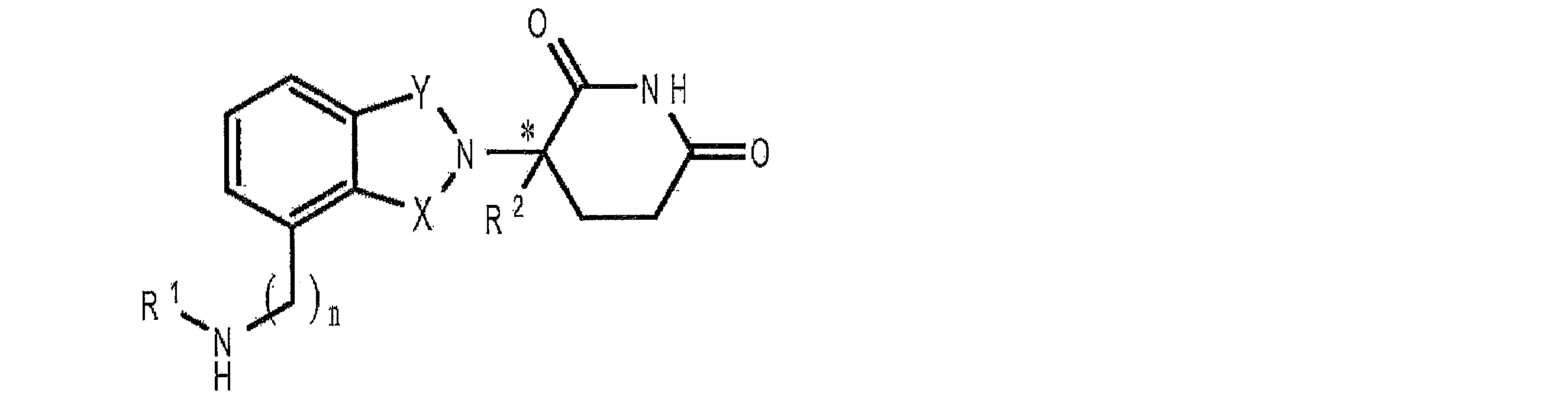
式中、XおよびYの一方はC=Oであり、もう一方はCH2またはC=Oであり、
R1は、水素、(C1-C8)アルキル、(C3-C7)シクロアルキル、(C2-C8)ア
ルケニル、(C2-C8)アルキニル、ベンジル、アリール、(C0-C4)アルキル-(C
1-C6)ヘテロシクロアルキル、(C0-C4)アルキル-(C2-C5)ヘテロアリール、
C(O)R3、C(S)R3、C(O)OR4、(C1-C8)アルキル-N(R6)2、(C1
-C8)アルキル-OR5、(C1-C8)アルキル-C(O)OR5、C(O)NHR3、C
(S)NHR3、C(O)NR3R3’、C(S)NR3R3’、または(C1-C8)アルキ
ル-O(CO)R5であり、
R2は、H、F、ベンジル、(C1-C8)アルキル、(C2-C8)アルケニル、または
(C2-C8)アルキニルであり、
R3およびR3’は独立して、(C1-C8)アルキル、(C3-C7)シクロアルキル、(
C2-C8)アルケニル、(C2-C8)アルキニル、ベンジル、アリール、(C0-C4)ア
ルキル-(C1-C6)ヘテロシクロアルキル、(C0-C4)アルキル-(C2-C5)ヘテ
ロアリール、(C0-C8)アルキル-N(R6)2、(C1-C8)アルキル-OR5、(C1
-C8)アルキル-C(O)OR5、(C1-C8)アルキル-O(CO)R5、またはC(
O)OR5であり、
R4は、(C1-C8)アルキル、(C2-C8)アルケニル、(C2-C8)アルキニル、
(C1-C4)アルキル-OR5、ベンジル、アリール、(C0-C4)アルキル-(C1-C
6)ヘテロシクロアルキル、または(C0-C4)アルキル-(C2-C5)ヘテロアリール
であり、
R5は、(C1-C8)アルキル、(C2-C8)アルケニル、(C2-C8)アルキニル、
ベンジル、アリール、または(C2-C5)ヘテロアリールであり、
それぞれに存在するR6は独立して、H、(C1-C8)アルキル、(C2-C8)アルケ
ニル、(C2-C8)アルキニル、ベンジル、アリール、(C2-C5)ヘテロアリール、も
しくは(C0-C8)アルキル-C(O)OR5であるか、または、上記R6基はヘテロシク
ロアルキル基を形成していてもよく、
nは0または1であり、
*は不斉炭素中心を表す、化合物、
または、それらの薬学的に許容可能な塩、水和物、溶媒和物、クラスレート、光学異性
体、ジアステレオマー、ラセミ化合物、もしくは立体異性体の混合物である。別の実施形
態では、上記免疫調節化合物は、以下の構造を有し、
wherein one of X and Y is C=O and the other is CH2 or C=O;
R 1 is hydrogen, (C 1 -C 8 )alkyl, (C 3 -C 7 )cycloalkyl, (C 2 -C 8 )alkenyl, (C 2 -C 8 )alkynyl, benzyl, aryl, (C 0 —C 4 )alkyl-(C
1 -C 6 )heterocycloalkyl, (C 0 -C 4 )alkyl-(C 2 -C 5 )heteroaryl,
C(O)R 3 , C(S)R 3 , C(O)OR 4 , (C 1 -C 8 )alkyl-N(R 6 ) 2 , (C 1
—C 8 )alkyl-OR 5 , (C 1 -C 8 )alkyl-C(O)OR 5 , C(O)NHR 3 , C
(S) NHR3 , C(O) NR3R3 ', C(S) NR3R3 ' , or ( C1 - C8)alkyl - O ( CO)R5;
R 2 is H, F, benzyl, (C 1 -C 8 )alkyl, (C 2 -C 8 )alkenyl, or (C 2 -C 8 )alkynyl;
R 3 and R 3 ' are independently (C 1 -C 8 )alkyl, (C 3 -C 7 )cycloalkyl, (
C 2 -C 8 )alkenyl, (C 2 -C 8 )alkynyl, benzyl, aryl, (C 0 -C 4 )alkyl-(C 1 -C 6 )heterocycloalkyl, (C 0 -C 4 )alkyl- (C 2 -C 5 )heteroaryl, (C 0 -C 8 )alkyl-N(R 6 ) 2 , (C 1 -C 8 )alkyl-OR 5 , (C 1
—C 8 )alkyl-C(O)OR 5 , (C 1 -C 8 )alkyl-O(CO)R 5 , or C(
O) OR 5 ;
R 4 is (C 1 -C 8 )alkyl, (C 2 -C 8 )alkenyl, (C 2 -C 8 )alkynyl,
(C 1 -C 4 )alkyl-OR 5 , benzyl, aryl, (C 0 -C 4 )alkyl-(C 1 -C
6 ) heterocycloalkyl or (C 0 -C 4 )alkyl-(C 2 -C 5 )heteroaryl;
R 5 is (C 1 -C 8 )alkyl, (C 2 -C 8 )alkenyl, (C 2 -C 8 )alkynyl,
benzyl, aryl, or ( C2 - C5)heteroaryl;
Each occurrence of R 6 is independently H, (C 1 -C 8 )alkyl, (C 2 -C 8 )alkenyl, (C 2 -C 8 )alkynyl, benzyl, aryl, (C 2 -C 5 ) heteroaryl or (C 0 -C 8 )alkyl-C(O)OR 5 or the R 6 groups may form a heterocycloalkyl group;
n is 0 or 1;
* represents an asymmetric carbon center, a compound,
Or, pharmaceutically acceptable salts, hydrates, solvates, clathrates, optical isomers, diastereomers, racemates, or mixtures of stereoisomers thereof. In another embodiment, the immunomodulatory compound has the structure

式中、XおよびYの一方はC=Oであり、もう一方はCH2またはC=Oであり、
Rは、HまたはCH2OCOR’であり、
(i)R1、R2、R3、またはR4のそれぞれは、他とは独立して、ハロ、炭素原子数1
~4のアルキル、もしくは炭素原子数1~4のアルコキシであるか、または(ii)R1
、R2、R3、またはR4のうちの一つがニトロ、もしくは-NHR5であり、残りのR1、
R2、R3、またはR4が水素であり、
R5は、水素、または炭素原子数1~8のアルキルであり、
R6は、水素、炭素原子数1~8のアルキル、ベンゾ、クロロ、またはフルオロであり
、
R’は、R7-CHR10-N(R8R9)であり、
R7は、m-フェニレンもしくはp-フェニレン、または-(CnH2n)-であって、n
は0~4の値であり、
R8およびR9のそれぞれは、他方とは独立して、水素、もしくは炭素原子数1~8のア
ルキルであるか、または、R8およびR9はともに、テトラメチレン、ペンタメチレン、ヘ
キサメチレン、または-CH2CH2X1CH2CH2-であって、X1は-O-、-S-、ま
たは-NH-であり、
R10は、水素、炭素原子数8までのアルキル、またはフェニルであり、
*は不斉炭素中心を表す、化合物、
または、それらの薬学的に許容可能な塩、水和物、溶媒和物、クラスレート、光学異性
体、ジアステレオマー、ラセミ化合物、もしくは立体異性体の混合物である。
wherein one of X and Y is C=O and the other is CH2 or C=O;
R is H or CH2OCOR ';
(i) each of R 1 , R 2 , R 3 , or R 4 is, independently of the others, halo, 1 carbon atom
is alkyl of ∼4 or alkoxy of 1 to 4 carbon atoms, or (ii) R 1
, R 2 , R 3 , or R 4 is nitro, or —NHR 5 and the remaining R 1 ,
R 2 , R 3 , or R 4 is hydrogen;
R 5 is hydrogen or alkyl of 1 to 8 carbon atoms;
R 6 is hydrogen, alkyl of 1 to 8 carbon atoms, benzo, chloro, or fluoro;
R′ is R 7 —CHR 10 —N(R 8 R 9 );
R 7 is m-phenylene or p-phenylene, or —(C n H 2n )—, wherein n
is a value from 0 to 4,
Each of R 8 and R 9 is, independently of the other, hydrogen or alkyl of 1 to 8 carbon atoms, or both R 8 and R 9 are tetramethylene, pentamethylene, hexamethylene, or -CH 2 CH 2 X 1 CH 2 CH 2 -, where X 1 is -O-, -S-, or -NH-;
R 10 is hydrogen, alkyl of up to 8 carbon atoms, or phenyl;
* represents an asymmetric carbon center, a compound,
Or, they are pharmaceutically acceptable salts, hydrates, solvates, clathrates, optical isomers, diastereomers, racemates, or mixtures of stereoisomers thereof.
特定の実施形態では、造血細胞の増殖は、20%BITS(BSA、組み換えヒトイン
スリンおよびトランスフェリン)、SCF、Flt-3リガンド、IL-3、4-(アミ
ノ)-2-(2,6-ジオキソ(3-ピペリジル))-イソインドリン-1,3-ジオン
(0.05%DMSO中で10μM)が追加されたIMDM中で行われる。さらに特定の
実施形態では、約5×10⁷個の造血細胞(例えばCD34+細胞)は、培地中で、約5×
1010個~約5×10¹²個に増殖し、増殖された造血細胞集団を生産するために、100
mLのIMDM中に再び懸濁される。増殖された造血細胞集団は、好ましくは輸送を容易
にするために低温保存される。
In certain embodiments, hematopoietic cell proliferation is reduced by 20% BITS (BSA, recombinant human insulin and transferrin), SCF, Flt-3 ligand, IL-3, 4-(amino)-2-(2,6-dioxo (3-Piperidyl))-isoindoline-1,3-dione (10 μM in 0.05% DMSO) is added in IMDM. In a more specific embodiment, about 5×10 7 hematopoietic cells (eg, CD34 + cells) are about 5×
To produce an expanded hematopoietic cell population from 10 10 to about 5×10 12 , 100
Resuspend in mL of IMDM. Expanded hematopoietic cell populations are preferably cryopreserved to facilitate transport.
上述した方法および培地による赤血球の生産は、好ましくは、バイオリアクター(例え
ば本明細書中のあらゆる箇所で実証されているバイオリアクター)の中で行われる。
The production of red blood cells by the methods and media described above is preferably carried out in a bioreactor, such as the bioreactor demonstrated elsewhere herein.
種々の特定の実施形態では、造血細胞の少なくとも50%、55%、60%、65%、
70%、75%、80%、85%、90%、95%、97%、98%、または99%は、
赤血球および/または多染赤血球に分化する。
In various specific embodiments, at least 50%, 55%, 60%, 65% of the hematopoietic cells,
70%, 75%, 80%, 85%, 90%, 95%, 97%, 98%, or 99% are
Differentiate into erythrocytes and/or polychromatic erythrocytes.
一実施形態では、造血細胞(例えば上述の増殖された造血細胞)の分化を、免疫調節化
合物(例えば上述したTNF-α阻害化合物)と接した状態で、当該細胞を培養すること
で、達成してもよい。当該細胞は、免疫調節化合物と接していない同数の造血細胞と比べ
て、与えられた時間内の造血細胞の増殖が検出可能な程度で増加するために十分な時間お
よび量で、免疫調節化合物と接する。例えば、米国特許出願公報第2003/02359
09号を参照されたい。当該開示の内容は全て参照によって本明細書中に引用されるもの
とする。
In one embodiment, differentiation of hematopoietic cells (e.g., expanded hematopoietic cells described above) is achieved by culturing the cells in contact with an immunomodulatory compound (e.g., a TNF-α inhibitory compound described above). may The cells are treated with an immunomodulatory compound for a time and in an amount sufficient to cause a detectable increase in proliferation of the hematopoietic cells in a given period of time as compared to the same number of hematopoietic cells not exposed to the immunomodulatory compound. touch. For example, US Patent Application Publication No. 2003/02359
See
ある実施形態では、上記造血細胞の増殖および分化の方法は、本明細書で記載されるよ
うに、増殖および分化の間に、1ミリリットルあたり約2×10⁴個から約2×10⁵個の
間で細胞が維持されるように上記造血細胞を含む細胞集団を維持する工程を含む。ある他
の実施形態では、上記造血細胞の増殖および分化の方法は、本明細書で記載されるように
、1ミリリットルあたり約1×10⁵個以下の細胞が維持されるように上記造血細胞を含
む細胞集団を維持する工程を含む。
In certain embodiments, the hematopoietic cell expansion and differentiation methods described herein comprise about 2×10 4 to about 2×10 5 cells per milliliter during expansion and differentiation as described herein. maintaining the cell population comprising the hematopoietic cells such that the cells are maintained between. In certain other embodiments, the hematopoietic cell expansion and differentiation method comprises growing the hematopoietic cells such that no more than about 1×10 5 cells per milliliter are maintained, as described herein. maintaining the cell population comprising
上記造血細胞から赤血球への分化は、例えばフローサイトメトリーによって、赤血球特
異的マーカーを検出することで、評価することができる。赤血球特異的マーカーは、限定
されるものではないが、CD36およびグリコフォリンAが挙げられる。分化は、顕微鏡
下での細胞の目視検査によって評価してもよい。典型的な両凹面(biconcave)の細胞の
存在によって、赤血球の存在を確認できる。赤血球(網状赤血球を含む)の存在は、Ho
echst 33342、TO-PRO(登録商標)-3、DRAG5等の、デオキシリ
ボ核酸(DNA)の染色によって確認できる。赤血球の有核前駆体は、DNA検出染色で
一般的に陽性であるが、一方、赤血球および網状赤血球は、一般的に陰性である。造血細
胞から赤血球への分化は、分化の間の、トランスフェリン受容体(CD71)の発現、お
よび/または色素レーザースチリル染色の進行的消失によって評価することもできる。例
えば、Bessis M and Mohandas N, “A Diffracto
metric Method for the Measurement of Cel
lular Deformability,” Blood Cells 1:307(
1975);Mohandas N. et al., “Analysis of F
actors Regulating Erythrocyte Deformabil
ity,” J. Clin. Invest. 66:563(1980);Gron
er W et al., “New Optical Technique for
Measuring Erythrocyte Deformability with
the Ektacytometer,” Clin. Chem. 26:1435
(1980)を参照されたい。十分に分化した赤血球は、約80~約108fL(フェム
トリットル)の平均赤血球容積(MCV)、約17~約31pgの平均赤血球ヘモグロビ
ン量(MCH)、および約23%~約36%の平均赤血球ヘモグロビン濃度(MCHC)
を有する。
Differentiation of the hematopoietic cells into erythrocytes can be assessed by detecting erythrocyte-specific markers, for example by flow cytometry. Erythrocyte specific markers include, but are not limited to, CD36 and glycophorin A. Differentiation may be assessed by visual inspection of cells under a microscope. The presence of erythrocytes can be confirmed by the presence of typical biconcave cells. The presence of red blood cells (including reticulocytes) is
This can be confirmed by deoxyribonucleic acid (DNA) staining such as echst 33342, TO-PRO®-3, DRAG5. Nucleated precursors of erythrocytes are generally positive on DNA detection stains, while erythrocytes and reticulocytes are generally negative. Differentiation of hematopoietic cells into erythrocytes can also be assessed by the progressive loss of transferrin receptor (CD71) expression and/or dye laser styryl staining during differentiation. See, for example, Bessis M and Mohandas N, "A Diffracto
Metric Method for the Measurement of Cel
Lular Deformability, "Blood Cells 1:307 (
1975); et al. , "Analysis of F
actors Regulating Erythrocyte Deformabil
Clinity," J. Clin. Invest. 66:563 (1980); Gron
er W et al. , “New Optical Technique for
Measuring Erythrocyte Deformability with
the Ektacytometer, "Clin. Chem. 26:1435.
(1980). A fully differentiated red blood cell has a mean corpuscular volume (MCV) of about 80 to about 108 femtoliters, a mean corpuscular hemoglobin content (MCH) of about 17 to about 31 pg, and a mean corpuscular hemoglobin of about 23% to about 36%. Concentration (MCHC)
have
造血細胞から赤血球への分化に要する時間は、約3日から約120日である。一実施形
態では、上記分化時間は、約7日~約35日である。別の実施形態では、上記分化時間は
、約14日~約28日である。
The time required for differentiation of hematopoietic cells to erythrocytes is from about 3 days to about 120 days. In one embodiment, the differentiation time is from about 7 days to about 35 days. In another embodiment, the differentiation time is from about 14 days to about 28 days.
<5.3.前駆体からの赤血球の分離>
上述した方法によって生産された赤血球は、好ましくは、造血細胞から分離され、ある
実施形態では、赤血球の前駆体から分離される。上記分離は、例えば、CD36および/
またはグリコフォリンAに対する抗体を用いることで、達成することができる。分離は、
既知の方法によって達成してもよく、例えば、抗体を介した磁気ビーズ分離、蛍光活性化
セルソーティング、CD36および/またはグリコフォリンAに対する抗体を含む表面ま
たはカラムに対する細胞の通過等が挙げられる。別の実施形態では、赤血球を含む培地か
ら酸素を除去し、続いて脱酸素された赤血球を他の細胞から磁気分離することで、赤血球
の分離を達成する。
<5.3. Separation of erythrocytes from precursors>
The red blood cells produced by the methods described above are preferably separated from the hematopoietic cells, and in some embodiments, are separated from the red blood cell precursors. The separation is for example CD36 and/or
Alternatively, it can be achieved by using an antibody against glycophorin A. Separation is
It may be accomplished by known methods, including antibody-mediated magnetic bead separation, fluorescence-activated cell sorting, passage of cells over a surface or column containing antibodies against CD36 and/or glycophorin A, and the like. In another embodiment, separation of red blood cells is accomplished by removing oxygen from the medium containing red blood cells, followed by magnetic separation of the deoxygenated red blood cells from other cells.
赤血球は、細胞集団(例えば上述したような二番目の増殖された造血細胞集団)から継
続的に分離することができ、または、周期的に分離することができる。ある実施形態では
、例えば、赤血球の単離は、例えば1、2、3、4、5、6、7、8、9、10、15、
20、25、30、35、40、45、50、55、もしくは60分ごと、または、1、
1.5、2、2.5、3、3.5、4、4.5、5、5.5、6、6.5、7.0、7.
5、8.0、8.5、9.0、9.5、10.0、10.5、11.0、11.5、12
、13、14、15、16、17、18、19、20、21、22、23、もしくは24
時間ごと、またはそれ以上、または例えば、特定の細胞密度での培養の達成;1ミリリッ
トルあたりの特定の細胞数が、ある赤血球マーカー(例えばCD36、もしくはグリコフ
ォリンA)を発現する培養の達成等、1つ以上の培養条件が満たされた場合に、行われる
。細胞集団からの赤血球の分離は、好ましくは、後述するようなバイオリアクターを用い
て行われる。
Erythrocytes can be continuously separated from the cell population (eg, a second expanded hematopoietic cell population as described above) or can be separated periodically. In certain embodiments, for example, isolation of red blood cells is performed by, for example, 1, 2, 3, 4, 5, 6, 7, 8, 9, 10, 15,
every 20, 25, 30, 35, 40, 45, 50, 55, or 60 minutes, or 1,
1.5, 2, 2.5, 3, 3.5, 4, 4.5, 5, 5.5, 6, 6.5, 7.0, 7.
5, 8.0, 8.5, 9.0, 9.5, 10.0, 10.5, 11.0, 11.5, 12
, 13, 14, 15, 16, 17, 18, 19, 20, 21, 22, 23, or 24
Achieving cultures at specific cell densities over time or more, or for example; achieving cultures in which a specific number of cells per milliliter expresses a certain red blood cell marker (e.g., CD36, or glycophorin A), etc. It is performed when one or more culture conditions are met. Separation of red blood cells from the cell population is preferably performed using a bioreactor as described below.
<5.4.バイオリアクターによる赤血球の生産>
赤血球の生産方法の別の側面では、造血細胞は、支持細胞の存在しないバイオリアクタ
ー中で増殖および分化する。造血細胞が分化するバイオリアクターは、造血細胞が増殖す
るバイオリアクターと同じであってもよいし、または別のバイオリアクターでもよい。別
の実施形態では、上記バイオリアクターは、造血細胞の増殖を完全にバイオリアクター中
で促進するように構築される。別の実施形態では、上記バイオリアクターは、支持細胞を
用いずに造血細胞の増殖を行えるように構築される。
<5.4. Red Blood Cell Production Using a Bioreactor>
In another aspect of the red blood cell production method, the hematopoietic cells are grown and differentiated in a bioreactor in the absence of feeder cells. The bioreactor in which the hematopoietic cells differentiate may be the same bioreactor in which the hematopoietic cells are grown, or it may be a separate bioreactor. In another embodiment, the bioreactor is constructed to promote hematopoietic cell growth entirely within the bioreactor. In another embodiment, the bioreactor is constructed for expansion of hematopoietic cells without feeder cells.
別の実施形態では、上記バイオリアクターは、培地中における細胞の継続的な流れを実
現し、分化した赤血球をバイオリアクター中の残りの細胞から継続的に分離することを可
能にするように構築される。上記継続的な流れおよび細胞の分離によって、上記バイオリ
アクターを、赤血球を生産するバッチ法を用いるバイオリアクターと比べて、大幅に小さ
い体積で構築することができる。別の実施形態では、上記バイオリアクターは、培地中の
細胞の、周期的な流れ(例えば断続的な流れ)を実現し、分化した赤血球をバイオリアク
ター中の残りの細胞から周期的に分離することを可能にするように構築される。上記周期
的な流れおよび細胞の分離によって、上記バイオリアクターを、赤血球を生産するバッチ
法を用いるバイオリアクターと比べて、大幅に小さい体積で構築することが好ましい。特
定の実施形態では、赤血球の単離は、例えば1、2、3、4、5、6、7、8、9、10
、15、20、25、30、35、40、45、50、55、もしくは60分ごと、また
は、1、1.5、2、2.5、3、3.5、4、4.5、5、5.5、6、6.5、7.
0、7.5、8.0、8.5、9.0、9.5、10.0、10.5、11.0、11.
5、12、13、14、15、16、17、18、19、20、21、22、23、もし
くは24時間ごと、またはそれ以上で行われる。別の特定の実施形態では、例えば、特定
の細胞密度での培養の達成;1ミリリットルあたりの特定の細胞数が、ある赤血球マーカ
ー(例えばCD36、もしくはグリコフォリンA)を発現する培養の達成等、1つ以上の
培養条件が満たされた場合に、赤血球の単離が行われる。
In another embodiment, the bioreactor is constructed to provide a continuous flow of cells in culture medium to allow continuous separation of differentiated red blood cells from the rest of the cells in the bioreactor. be. The continuous flow and separation of cells allows the bioreactor to be constructed with a significantly smaller volume compared to bioreactors using batch methods to produce red blood cells. In another embodiment, the bioreactor provides a cyclic flow (e.g., intermittent flow) of cells in culture to periodically separate differentiated red blood cells from the rest of the cells in the bioreactor. is built to allow Due to the cyclical flow and cell separation, the bioreactor is preferably constructed with a significantly smaller volume compared to bioreactors using batch methods to produce red blood cells. In certain embodiments, the isolation of red blood cells is, for example, 1, 2, 3, 4, 5, 6, 7, 8, 9, 10
, every 15, 20, 25, 30, 35, 40, 45, 50, 55, or 60 minutes, or 1, 1.5, 2, 2.5, 3, 3.5, 4, 4.5, 5, 5.5, 6, 6.5, 7.
0, 7.5, 8.0, 8.5, 9.0, 9.5, 10.0, 10.5, 11.0, 11.
Every 5, 12, 13, 14, 15, 16, 17, 18, 19, 20, 21, 22, 23, or 24 hours or more. In other specific embodiments, e.g., achieving a culture at a particular cell density; achieving a culture in which a particular number of cells per milliliter express a certain red blood cell marker (e.g., CD36, or glycophorin A), etc. Red blood cell isolation is performed when one or more culture conditions are met.
ある実施形態では、上記バイオリアクターは使い捨てである。 In some embodiments, the bioreactor is disposable.
一実施形態では、上記バイオリアクターは、培養部材および細胞分離部材を備える。別
の実施形態では、上記バイオリアクターは、培地ガス供給部材を備える。別の実施形態で
は、上記バイオリアクターは、一つ以上の生物活性化合物を含む細胞因子部材を備える。
別の実施形態では、上記バイオリアクターの部材は、モジュール式であり、例えば、お互
いから分離することができ、および/または、独立して使用できる。一実施形態では、上
記バイオリアクターの容量は、約100mL、200mL、300mL、400mL、5
00mL、600mL、700mL、800mL、もしくは約900mL、または1、2
、3、4、5、6、7、8、9、10、12、14、16、18、20、25、30、3
5、40、45、もしくは50リットルである。別の実施形態では、上記バイオリアクタ
ーは、全ての部品を含むと、約47立方フィート以下である。別の実施形態では、上記バ
イオリアクターは、例えば造血細胞を、約1010個、1011個、または約1012個、培養
することができる。
In one embodiment, the bioreactor comprises a culture component and a cell separation component. In another embodiment, the bioreactor comprises a medium gas supply member. In another embodiment, the bioreactor comprises a cell factor member containing one or more bioactive compounds.
In another embodiment, the bioreactor components are modular, eg, can be separated from each other and/or can be used independently. In one embodiment, the bioreactor has a capacity of about 100 mL, 200 mL, 300 mL, 400 mL, 5
00 mL, 600 mL, 700 mL, 800 mL, or about 900 mL, or 1, 2
, 3, 4, 5, 6, 7, 8, 9, 10, 12, 14, 16, 18, 20, 25, 30, 3
5, 40, 45 or 50 liters. In another embodiment, the bioreactor, including all parts, is about 47 cubic feet or less. In another embodiment, the bioreactor is capable of culturing about 10 10 , 10 11 , or about 10 12 hematopoietic cells, for example.
一実施形態では、上記培養部材は、培地(例えば造血細胞を培養する培地)を受容する
ことができる区画を備える。上記培養部材は、培養のために、培地および/または造血細
胞を導入することができるポートを備える。上記ポートは、上記装置のための、技術的に
許容可能なポートであれば、いかなるポートであってもよく、例えばルアーロックシール
ポートが挙げられる。また、上記培養部材は、培地を上記細胞分離部材へと通すために一
つ以上のポートを備える。上記培養部材は、任意でさらに、生物活性化合物を上記培養部
材の内部に導入するためのポートを備えており、当該ポートには、例えば、上記細胞因子
部材と上記培養部材との接続を容易にするポートが挙げられる。特定の実施形態では、分
化している造血細胞を含む、上記培養部材中の造血細胞は、赤血球、および/または網状
赤血球、および/または他の赤血球前駆体を単離するために、細胞分離部材(下記参照)
へと培地中で継続的に循環される。
In one embodiment, the culturing member comprises a compartment capable of receiving medium (eg, medium for culturing hematopoietic cells). The culturing member comprises ports through which media and/or hematopoietic cells can be introduced for culturing. The port can be any technically acceptable port for the device, such as a luer lock seal port. The culturing member also includes one or more ports for passing media to the cell separation member. The culture member optionally further comprises a port for introducing a bioactive compound into the interior of the culture member, the port, for example, facilitating connection between the cell factor member and the culture member. port to be used. In certain embodiments, the hematopoietic cells in the culture member, including differentiating hematopoietic cells, are separated from the cell separation member to isolate red blood cells and/or reticulocytes and/or other red blood cell precursors. (refer to the following)
is continuously circulated in the medium to
上記培養部材は、特定の実施形態では、複数の内部表面または内部構造を備え、当該内
部表面または内部構造には、例えば、チューブ、シリンダー、中空糸、多孔性物質等が挙
げられる。上記表面は、細胞の培養に好適なあらゆる材料から構築することができ、当該
材料には、例えば、組織培養プラスチック、可塑医薬品グレードプラスチック、ヒドロキ
シアパタイト、ポリ乳酸(PLA)、ポリグリコール酸共重合体(PLGA)、ポリウレ
タン、ポリヒドロキシエチルメタクリル樹脂等が挙げられる。中空糸は、一般的に直径約
10μm~約1000μmであり、一般的に、約5kDa、10kDa、15kDa、2
0kDa、30kDa、40kDa、50kDa、60kDa、70kDa、80kDa
、90kDa、100kDa、125kDa、150kDa、175kDa、200kD
a、150kDa、300kDa、350kDa、400kDa、450kDa、または
500kDa以下の分子を通過させることができる孔を備える。
The culture member, in certain embodiments, comprises a plurality of internal surfaces or structures, including, for example, tubes, cylinders, hollow fibers, porous materials, and the like. The surface can be constructed from any material suitable for culturing cells, including, for example, tissue culture plastics, plastic pharmaceutical grade plastics, hydroxyapatite, polylactic acid (PLA), polyglycolic acid copolymers. (PLGA), polyurethane, polyhydroxyethyl methacrylic resin, and the like. Hollow fibers are generally from about 10 μm to about 1000 μm in diameter, typically about 5 kDa, 10 kDa, 15 kDa, 2
0kDa, 30kDa, 40kDa, 50kDa, 60kDa, 70kDa, 80kDa
, 90kDa, 100kDa, 125kDa, 150kDa, 175kDa, 200kD
a, 150 kDa, 300 kDa, 350 kDa, 400 kDa, 450 kDa, or 500 kDa or less with pores that can pass through.
上記細胞分離部材は、上記培養部材から、細胞を含む培地を受容するために、少なくと
も一つのポートを備える。上記細胞分離部材は、少なくとも一つの種類の細胞(例えば赤
血球)を、上記培養部材由来の培地中の細胞から分離することを容易にする、または可能
にする、一つ以上の部品を備える。当該分離は、例えば、CD36および/またはグリコ
フォリンAに対する抗体を用いることで達成される。分離は、既知の方法によって達成し
てもよく、例えば、抗体を介した磁気ビーズ分離、蛍光活性化セルソーティング、CD3
6および/またはグリコフォリンAに対する抗体を含む表面またはカラムに対する細胞の
パッセージ等が挙げられる。分離は、細胞サイズまたは細胞密度に基づいて達成してもよ
い。特定の実施形態では、上記細胞分離部材は、上記細胞培養部材と接続されており、培
地は造血細胞を含み、分化している造血細胞および赤血球は、細胞(例えば赤血球)を上
記培地から継続的に取り除くために、上記細胞分離部材を継続的に通過する。
The cell separation member includes at least one port for receiving medium containing cells from the culture member. The cell separation member comprises one or more components that facilitate or enable separation of at least one type of cell (eg, red blood cells) from cells in medium from the culture member. Said separation is achieved, for example, by using antibodies against CD36 and/or glycophorin A. Separation may be accomplished by known methods, e.g., antibody-mediated magnetic bead separation, fluorescence-activated cell sorting, CD3
6 and/or passage of cells to a surface or column containing antibodies against glycophorin A, and the like. Separation may be achieved based on cell size or cell density. In certain embodiments, the cell separation member is connected to the cell culture member, the culture medium comprises hematopoietic cells, and differentiating hematopoietic cells and red blood cells continuously remove cells (e.g., red blood cells) from the culture medium. continuously pass through the cell separation member for removal.
別の実施形態では、赤血球の分離は、赤血球を含む培地から酸素を除去し、続いて磁気
を用いて、脱酸素された赤血球を、例えば採集物の表面または他の部分へ引き寄せること
で、達成される。
In another embodiment, separation of red blood cells is accomplished by deoxygenating the red blood cell-containing medium and subsequently using a magnet to attract the deoxygenated red blood cells to, for example, a surface or other portion of the collection. be done.
別の実施形態では、上記バイオリアクターは、細胞分離部材を備える。上記細胞分離部
材は、培地中の赤血球から、一つ以上の非赤血球系細胞(例えば、未分化の、または、分
化が終わっていない造血細胞)を分離することができる部品を備えていてもよい。ある実
施形態では、上記細胞分離部材は、生産された赤血球のおよその数を計算することができ
るか、または、ユーザーが事前に設定したパラメータに従って、あるユニットを構成する
ために十分な数の赤血球が生産されたことをユーザーに知らせることができる。
In another embodiment, the bioreactor comprises a cell separation member. The cell separation member may comprise a component capable of separating one or more non-erythroid cells (e.g., undifferentiated or undifferentiated hematopoietic cells) from red blood cells in the culture medium. . In an embodiment, the cell separation member is capable of calculating the approximate number of red blood cells produced or, according to parameters preset by the user, a sufficient number of red blood cells to form a unit. can inform the user that the has been produced.
上記バイオリアクターは、別の実施形態では、さらに、培地環境に適切なガスを供給す
るガス供給部材を備え、例えば、当該ガス供給部材は、上記培地を、80%の空気と15
%のO2と5%のCO2との混合物、または、空気中の5%のCO2等に接触させる。別の
実施形態では、上記バイオリアクターは、上記培地、上記バイオリアクター、またはその
両方を、一定の温度(例えば、約35℃~約39℃、または約37℃)に維持する、温度
部材を備える。別の実施形態では、上記バイオリアクターは、上記培地を、一定のpH(
例えば約pH7.2~pH7.6、または約pH7.4)に維持する、pHモニタリング
部材を備える。特定の実施形態では、上記温度部材および/またはpHモニタリング部材
は、温度および/またはpHが、設定されたパラメータを超えるか、または設定されたパ
ラメータ以下になったときに作動する警報装置を備える。他の特定の実施形態では、上記
温度部材および/またはpHモニタリング部材は、ある範囲から外れた温度および/また
はpHを補正することができる。
The bioreactor, in another embodiment, further comprises a gas supply member that provides suitable gases to the culture medium environment, for example, the gas supply member mixes the medium with 80% air and 15% air.
A mixture of % O2 and 5% CO2 , or 5% CO2 in air, or the like. In another embodiment, the bioreactor comprises a temperature member that maintains the medium, the bioreactor, or both at a constant temperature (eg, about 35°C to about 39°C, or about 37°C). . In another embodiment, the bioreactor maintains the medium at a constant pH (
A pH monitoring member is provided to maintain, for example, about pH 7.2 to pH 7.6, or about pH 7.4). In certain embodiments, the temperature member and/or pH monitoring member comprises an alarm device that is activated when the temperature and/or pH exceeds or falls below set parameters. In certain other embodiments, the temperature member and/or pH monitoring member is capable of correcting out of range temperature and/or pH.
特定の実施形態では、上記バイオリアクターが、細胞分離部材、および上記培養環境に
ガスを供給するガス供給部材を備えることによって、上記ガス供給部材が、赤血球の部分
的または完全な脱酸素化を可能にし、赤血球に含まれるヘモグロビンの磁気的特性に基づ
いた赤血球の分離を可能にする。さらに特定の側面では、上記バイオリアクターは、当該
バイオリアクター中で生産された赤血球を規則的に、または、繰り返し脱酸素化すること
ができる部材を備え、上記赤血球の磁気的採集を容易にする。
In certain embodiments, the bioreactor comprises a cell separation member and a gas supply member that supplies gas to the culture environment, such that the gas supply member enables partial or complete deoxygenation of red blood cells. and allows separation of red blood cells based on the magnetic properties of the hemoglobin contained in the red blood cells. In a more particular aspect, the bioreactor comprises a member capable of regularly or repeatedly deoxygenating red blood cells produced in the bioreactor to facilitate magnetic collection of the red blood cells.
別の実施形態では、上記バイオリアクターの機能は自動化され、例えば、コンピュータ
ーで制御される。上記コンピューターは例えば、デスクトップパーソナルコンピューター
、ラップトップコンピューター、Handspring、PALM(登録商標)、または
同様の手持ちサイズの装置、ミニコンピューター、メインフレームコンピューター等であ
ってもよい。
In another embodiment, the functions of the bioreactor are automated, eg, computer controlled. The computer may be, for example, a desktop personal computer, laptop computer, Handspring, PALM® or similar handheld device, minicomputer, mainframe computer, or the like.
<5.5.造血細胞から生産される赤血球のユニット>
上記のように詳細に説明した方法に従って生産された赤血球のユニットは、あらゆる利
用しやすい数の遺伝的背景、または遺伝的背景の組み合わせを含んでいてもよい。
<5.5. Erythrocyte unit produced from hematopoietic cells>
Units of red blood cells produced according to the methods detailed above may comprise any accessible number or combination of genetic backgrounds.
種々の実施形態では、本明細書で提供される方法によって生産された赤血球のユニット
は、少なくとも、多くとも、または約1×10⁸、5×10⁸、1×10⁹、5×10⁹、1
×1010、5×10¹⁰、1×10¹¹、5×10¹¹、1×10¹²個の赤血球である。種々の
他の実施形態では、上記赤血球のユニットは、少なくとも10%、15%、20%、25
%、30%、40%、50%、60%、70%、80%、90%、95%、98%、また
は99%の完全に分化した赤血球を含む。種々の他の実施形態では、上記赤血球のユニッ
トは、60%、50%、40%、30%、20%、10%、5%、2%、または1%より
少ない、あらゆる種類の赤血球前駆体を含む。ある実施形態では、本明細書で提供される
方法によって生産された赤血球のユニットは、約60%、約50%、約40%、約30%
、約20%、約19%、約18%、約17%、約16%、約15%、約14%、約14%
、約12%、約11%、約10%、約9%、約8%、約7%、約6%、約5%より少ない
、網状赤血球、または他の非赤血球系造血細胞を含む。別の実施形態では、上記ユニット
は、単一の個体に由来する造血細胞に由来する赤血球を含む。別の実施形態では、上記ユ
ニットは、複数の個体に由来する造血細胞から分化した赤血球を含む。別の実施形態では
、上記ユニットは、調和したヒト胎盤灌流液および臍帯血に由来する造血細胞に由来する
赤血球を含む。別の実施形態では、赤血球のユニット中の赤血球のほぼ全て(例えば99
%を超える)は、O型である。別の実施形態では、赤血球のユニット中の赤血球のほぼ全
て(例えば99%を超える)は、A型である。別の実施形態では、赤血球のユニット中の
赤血球のほぼ全て(例えば99%を超える)は、B型である。別の実施形態では、赤血球
のユニット中の赤血球のほぼ全て(例えば99%を超える)は、AB型である。別の実施
形態では、赤血球のユニット中の赤血球のほぼ全て(例えば99%を超える)は、Rh陽
性である。別の実施形態では、赤血球のユニット中の赤血球のほぼ全て(例えば99%を
超える)は、Rh陰性である。
In various embodiments, the unit of red blood cells produced by the methods provided herein is at least, at most, or about 1×10 8 , 5×10 8 , 1×10 9 , 5×10 9 , 1
1×10 10 , 5×10 10 , 1×10 11 , 5×10 11 , 1×10 12 red blood cells. In various other embodiments, the red blood cell units are at least 10%, 15%, 20%, 25%
%, 30%, 40%, 50%, 60%, 70%, 80%, 90%, 95%, 98%, or 99% fully differentiated red blood cells. In various other embodiments, the erythrocyte units are less than 60%, 50%, 40%, 30%, 20%, 10%, 5%, 2%, or 1% of any type of erythrocyte precursor. including. In some embodiments, the red blood cell units produced by the methods provided herein are about 60%, about 50%, about 40%, about 30%
, about 20%, about 19%, about 18%, about 17%, about 16%, about 15%, about 14%, about 14%
, about 12%, about 11%, about 10%, about 9%, about 8%, about 7%, about 6%, less than about 5% reticulocytes or other non-erythroid hematopoietic cells. In another embodiment, the unit comprises red blood cells derived from hematopoietic cells derived from a single individual. In another embodiment, the unit comprises red blood cells differentiated from hematopoietic cells from multiple individuals. In another embodiment, the unit comprises red blood cells derived from hematopoietic cells derived from matched human placental perfusate and cord blood. In another embodiment, substantially all of the red blood cells in the unit of red blood cells (eg, 99
%) are O-type. In another embodiment, substantially all (eg, greater than 99%) of the red blood cells in the unit of red blood cells are type A. In another embodiment, substantially all (eg, greater than 99%) of the red blood cells in the unit of red blood cells are type B. In another embodiment, substantially all (eg, greater than 99%) of the red blood cells in the unit of red blood cells are of type AB. In another embodiment, substantially all (eg, greater than 99%) of the red blood cells in the unit of red blood cells are Rh positive. In another embodiment, substantially all (eg, greater than 99%) of the red blood cells in the unit of red blood cells are Rh-negative.
天然に存在する赤血球は、血液が毛細血管を流れることを可能にする、ある特性を有し
ている。例えば、凝集体中の赤血球は、非ニュートン流動を示し、例えば、血液の粘性は
、せん断速度に大きく依存する。通常の赤血球は、変形しやすく、凝集体/連銭を形成す
る。赤血球の変形能は、血液中の寿命(約100~120日)に関連していると考えられ
、血液からの赤血球の除去には、変形能の喪失が関連していると考えられる。赤血球の通
常の凝集能は、毛細血管における細胞の流れを容易にするが、一方、異常に増加または減
少した凝集能は、流れを低減させる。従って、好ましい実施形態では、本明細書で開示さ
れる方法によって生産された赤血球のユニットは、品質管理の一部として、例えば、天然
由来の赤血球の一つ以上の特性について、分析される。ある実施形態では、本明細書で開
示される方法によって生産された赤血球のサンプルを、天然または人工の血漿中に懸濁し
、粘性、粘弾性、緩和時間、変形能、凝集能、血液/赤血球懸濁液降伏応力、および機械
的脆弱性について、通常の血液または通常の赤血球をコントロールまたはコンパレーター
(comparator)として用いて測定する。ある他の実施形態では、酸素運搬容量および酸素
放出容量について、本明細書で開示される方法によって生産された赤血球のサンプルを、
通常の血液または同数の天然の赤血球をコントロールとして分析する。
Naturally occurring red blood cells have certain properties that allow blood to flow through capillaries. For example, red blood cells in aggregates exhibit non-Newtonian flow, eg blood viscosity is highly dependent on shear rate. Normal red blood cells are prone to deformation and form aggregates/rouleaux. The deformability of erythrocytes is thought to be related to their life span in blood (approximately 100-120 days), and removal of erythrocytes from the blood is thought to be related to loss of deformability. The normal agglutinating capacity of red blood cells facilitates cell flow in capillaries, whereas abnormally increased or decreased agglutinating capacity reduces flow. Thus, in preferred embodiments, units of red blood cells produced by the methods disclosed herein are analyzed as part of quality control, eg, for one or more characteristics of naturally-derived red blood cells. In certain embodiments, a sample of red blood cells produced by the methods disclosed herein is suspended in natural or artificial plasma and evaluated for viscosity, viscoelasticity, relaxation time, deformability, agglutination, blood/red blood cell suspension. Turbidity yield stress and mechanical fragility are measured using normal blood or normal red blood cells as controls or comparators. In certain other embodiments, a sample of red blood cells produced by the methods disclosed herein for oxygen carrying capacity and oxygen releasing capacity is
Normal blood or an equal number of native red blood cells are analyzed as controls.
〔6.実施例〕
<6.1.実施例1:ヒト胎盤灌流液(HPP)および臍帯血(UCB)由来のCD3
4+細胞の特性>
臍帯血(UCB)を、インフォームドコンセントの下で、分娩後の胎盤から除去した。
その後、放血した胎盤を灌流し、HPPを得た。当該方法は、米国特許第7,045,1
48号に記載されており、開示の内容は全て参照によって本明細書中に引用されるものと
する。赤血球(RCB)を除去した後、全有核細胞(TNC)を採集し、凍結した。UC
Bから単離されるTNCが約5億個であるのに対し、当該方法によれば、一般的には、約
1~2.5×10⁹個のTNCが採集できる。
[6. Example〕
<6.1. Example 1: CD3 from Human Placental Perfusate (HPP) and Umbilical Cord Blood (UCB)
4+ cell characteristics>
Umbilical cord blood (UCB) was removed from postpartum placenta under informed consent.
The exsanguinated placenta was then perfused to obtain HPP. The method is described in US Pat. No. 7,045,1
48, the entire disclosure of which is incorporated herein by reference. After removing red blood cells (RCB), total nucleated cells (TNC) were collected and frozen. UC
About 1-2.5×10 9 TNCs can generally be harvested by this method, compared to about 500 million TNCs isolated from B.
放血した胎盤から単離されたTNCのフローサイトメトリー分析によって、従来の臍帯
血(UCB)から得られた細胞産物に比べ、高い割合のCD34+細胞集団が示される。
UCB中のTNCでは、約0.3%~1%のCD34+細胞を含むのに対し、上記の方法
で採集した、HPP由来のTNCは、約2%~6%のCD34+細胞を含む。
Flow cytometric analysis of TNCs isolated from exsanguinated placenta shows a higher percentage of the CD34 + cell population compared to conventional cord blood (UCB) derived cell products.
TNCs in UCB contain about 0.3% to 1% CD34 + cells, whereas HPP-derived TNCs harvested by the above method contain about 2% to 6% CD34 + cells.
HPPから単離されたTNCのフローサイトメトリー分析によって、高い割合のCD3
4+細胞集団は、CD45-であることが示される(図1)。
Flow cytometric analysis of TNCs isolated from HPP revealed a high proportion of CD3
The 4 + cell population is shown to be CD45 - (Figure 1).
HPP由来のCD34+細胞をコロニー形成ユニットアッセイによって培養し、赤芽球
コロニー形成細胞(CFU-E)に対する赤芽球バースト形成細胞(BFU-E)の割合
(%)を、顆粒球マクロファージコロニー形成細胞(CFU-GM)、および顆粒球、赤
血球、単球コロニー形成細胞(CFU-GEMM)とともに測定した(表1)。また、コ
ロニー形成能についても評価した(表1)。
HPP-derived CD34 + cells were cultured by colony forming unit assay, and the percentage of erythroid burst-forming cells (BFU-E) to erythroid colony-forming cells (CFU-E) was determined as granulocyte-macrophage colony formation. cells (CFU-GM), and granulocytic, erythroid, and monocytic colony-forming cells (CFU-GEMM) (Table 1). In addition, colony forming ability was also evaluated (Table 1).

コロニー形成ユニットアッセイは、製造者のプロトコルに従って行った(StemCe
ll Technologies,Inc.)。端的に言えば、幹細胞因子(SCF)、
顆粒球コロニー刺激因子(G-CSF)、顆粒球マクロファージコロニー刺激因子(GM
-CSF)、インターロイキン3(IL-3)、インターロイキン6(IL-6)、およ
びエリスロポエチン(Epo)を追加したメチルセルロース培地に、プレートあたり細胞
100個、300個、および1000個で、CD34+細胞懸濁液を加えた。それぞれの
細胞密度において、3反復のアッセイを行い、その後、2~3週間培養した。コロニーの
評価および計数は、光学顕微鏡を用いて行った。
Colony forming unit assays were performed according to the manufacturer's protocol (StemCe
ll Technologies, Inc. ). In short, stem cell factor (SCF),
Granulocyte Colony Stimulating Factor (G-CSF), Granulocyte Macrophage Colony Stimulating Factor (GM
-CSF ), interleukin 3 (IL-3), interleukin 6 (IL-6), and erythropoietin (Epo) in methylcellulose medium at 100, 300, and 1000 cells per plate. Cell suspension was added. Triplicate assays were performed at each cell density, followed by 2-3 weeks of culture. Colony evaluation and counting was performed using a light microscope.
別の実験では、HPPおよびUCB由来のCD34+細胞に対する(BFU-E)/(
CFU-E)の割合は、それぞれ46%および30%であった(3人のドナーに対する平
均値に基づいている)。
In another experiment, (BFU - E)/(
CFU-E) rates were 46% and 30%, respectively (based on mean values for 3 donors).
これらの結果は、UCB由来の幹細胞に比べて、HPP由来の幹細胞は、赤血球生成活
性が増加した、多数のCD34+細胞を含むことを示唆している。
These results suggest that compared to UCB-derived stem cells, HPP-derived stem cells contain a large number of CD34 + cells with increased erythropoietic activity.
<6.2.実施例2:造血幹細胞(HSC)の回収>
HPPおよびUCB由来の細胞は、一般的に、全有核細胞(TNC)を得るために、フ
ィコール、ヘタスターチ、または塩化アンモニウムのいずれかを用いて、実施例1に記載
したように精製される。その後、TNCを用いて、CD34+細胞を単離するための正の
選択処理を行った。当該処理には、製造者(StemCell Technologie
s,Inc.)によって提供されるプロトコルに従って、抗CD34ビーズおよびRob
osepを用いた。当該実験では、CD34+細胞が、90%を超える精製度で単離され
た(図2)。また、EASYSEP(登録商標)Human Progenitor C
ell Enrichment Kit(StemCell Technologies
,Inc.)を用いて、負の選択処理を行い、Human Progenitor Ce
ll Enrichment Cocktailをモノクローナル抗体とともに用いて、
系列決定済みの細胞を除去した。上記モノクローナル抗体は以下のヒト細胞表面抗原に対
する抗体である:CD2、CD3、CD11b、CD11c、CD14、CD16、CD
19、CD24、CD56、CD66bおよび/またはグリコフォリンA。負の選択工程
を利用して、90%のCD34+細胞を原料から回収した。回収したHSCの細胞組成を
、表2に要約する。
<6.2. Example 2: Recovery of hematopoietic stem cells (HSC)>
HPP- and UCB-derived cells are generally purified as described in Example 1 using either ficoll, hetastarch, or ammonium chloride to obtain total nucleated cells (TNC). TNCs were then used to positively select to isolate CD34 + cells. The treatment includes the manufacturer (StemCell Technology
s, Inc. ), anti-CD34 beads and Rob
osep was used. In that experiment, CD34 + cells were isolated with greater than 90% purity (Fig. 2). Also, EASYSEP® Human Progenitor C
Cell Enrichment Kit (StemCell Technologies)
, Inc. ), negative selection was performed using Human Progenitor Ce
Using the ll Enrichment Cocktail with a monoclonal antibody,
Lineage committed cells were removed. Said monoclonal antibodies are antibodies against the following human cell surface antigens: CD2, CD3, CD11b, CD11c, CD14, CD16, CD
19, CD24, CD56, CD66b and/or glycophorin A; A negative selection step was used to recover 90% of the CD34 + cells from the source material. The cellular composition of harvested HSCs is summarized in Table 2.
単離されたCD34+細胞のコロニー形成ユニットアッセイによって、負の選択をされ
たHSCのコロニー形成頻度は、正の選択をされたHSCと同程度であり、負の選択をさ
れたHSCのBFU-E形成頻度は、正の選択されたHSCの形成頻度より高いことが示
された。
By colony-forming unit assay of isolated CD34 + cells, the colony-forming frequency of negatively selected HSCs was similar to that of positively selected HSCs, and the BFU- The E formation frequency was shown to be higher than that of positively selected HSCs.

<6.3.実施例3:CD34+造血細胞集団の増殖>
ヒト臍帯血(UCB)ユニットにおけるCD34+細胞の含有量は、多くの場合、成人
患者に対して造血細胞の移植を行うには十分ではない。UCB由来のCD34+細胞のe
x-vivoでの増殖は、上記のCD34+細胞の投与の制限を克服するための一つのア
プローチである。本実施例では、特定の免疫調節薬剤、4-(アミノ)-2-(2,6-
ジオキソ(3-ピペリジル))-イソインドリン-1,3-ジオン(本実施例では、ポマ
リドマイドと呼ぶ)を用いたCD34+細胞の増殖について、実証する。
<6.3. Example 3: Expansion of CD34 + hematopoietic cell population>
The CD34 + cell content in human umbilical cord blood (UCB) units is often insufficient for hematopoietic cell transplantation in adult patients. e of UCB-derived CD34 + cells
Expansion in x-vivo is one approach to overcome the limitations of CD34 + cell administration described above. In this example, the specific immunomodulatory agent, 4-(amino)-2-(2,6-
Proliferation of CD34 + cells with dioxo(3-piperidyl))-isoindoline-1,3-dione (referred to as pomalidomide in this example) is demonstrated.
短期間の無血清サイトカイン追加培養系において、ポマリドマイド(pomalidomide)が
ヒトUCB由来CD34+細胞の増殖を促進する能力を評価した。CD34+前駆細胞を、
低温保存したUSBユニットから90%を超える精製度で濃縮し、SCF(50ng/m
L)、Flt-3リガンド(50ng/mL)、およびIL-3(10ng/mL)の存
在下で、1mLの培地中に播種した(104個のCD34+細胞)。当該培地は、IMD
Mおよび代替血清BIT(BSA、組み換えヒトインスリン、およびトランスフェリン、
20%)を含む。ポマリドマイドは、DMSO中に溶解させ、2.7μg/mLで追加し
た。上記培養物を37℃、5%CO2で12日間培養し、7日目に新鮮な培地を加えた。
DMSO(0.05%v/v)とポマリドマイドとを含む培養物、および、DMSOを含
まず、かつポマリドマイドを含まない培養物、をコントロールとして用いた。
The ability of pomalidomide to promote proliferation of human UCB-derived CD34 + cells was evaluated in a short-term, serum-free, cytokine-boosted culture system. CD34 + progenitor cells,
Concentrated from a cryopreserved USB unit to greater than 90% purity, SCF (50 ng/m
L), Flt-3 ligand (50 ng/mL), and IL-3 (10 ng/mL) were seeded in 1 mL medium (104 CD34 + cells). The medium is IMD
M and alternative serum BIT (BSA, recombinant human insulin, and transferrin,
20%). Pomalidomide was dissolved in DMSO and added at 2.7 μg/mL. The cultures were incubated at 37° C., 5% CO 2 for 12 days and fresh medium was added on
Cultures containing DMSO (0.05% v/v) and pomalidomide and cultures without DMSO and without pomalidomide were used as controls.
一実施形態では、ポマリドマイドの追加によって、全有核細胞の増殖に影響を与えるこ
となく、増殖した集団中でのCD34+の発現を大幅に高めることができた(200~3
50倍)。CD34+の表現型は、コントロールでは10~30%であったのに比べて、
ポマリドマイドを加えて増殖した集団中では、40~60%であった。さらに、ポマリド
マイドは、培養細胞におけるCD38の発現を抑制すると考えられた。ポマリドマイドを
加えて増殖したCD34+細胞は、主にCD38に対して陰性であり(95%)、CD1
33を低レベルで発現した(コントロールで40%であったのに対し、15%)。ポマリ
ドマイドを加えて増殖したCD34+細胞は、増殖したコントロールと比較して、累積コ
ロニー形成ユニットにおいて十分な改善が見られることが証明された。別の同様な実験で
は、ポマリドマイドの追加によって、全有核細胞の増殖に影響を与えることなく、増殖し
た集団中でのCD34+の発現を大幅に高めることが確かめられた。(200~350倍
)。CD34+の表現型は、コントロールでは10~30%であったのに比べて、ポマリ
ドマイドを加えて増殖した集団中では、40~60%であった(図3)。さらに、ポマリ
ドマイドは、培養細胞におけるCD38の発現を抑制すると考えられた。ポマリドマイド
を加えて増殖したCD34+細胞は、主にCD38に対して陰性であり(97%)、CD
133を低レベルで発現した(コントロールで32.3%であったのに対し、11.5%
)。ポマリドマイドを加えて増殖したCD34+細胞は、増殖したコントロールと比較し
て、累積コロニー形成ユニットにおいて十分な改善が見られることが証明された。
In one embodiment, the addition of pomalidomide was able to greatly enhance the expression of CD34 + in the expanded population (200-3
50 times). The CD34 + phenotype was 10-30% in controls compared to
40-60% in populations grown with pomalidomide. Furthermore, pomalidomide was thought to suppress the expression of CD38 in cultured cells. CD34 + cells expanded with pomalidomide were predominantly negative for CD38 (95%) and CD1
33 was expressed at low levels (15% compared to 40% in controls). CD34 + cells expanded with pomalidomide demonstrated significant improvement in cumulative colony forming units compared to expanded controls. Another similar experiment confirmed that the addition of pomalidomide greatly enhanced the expression of CD34 + in the expanded population without affecting total nucleated cell proliferation. (200-350 times). The CD34 + phenotype was 40-60% in the pomalidomide-expanded population compared to 10-30% in controls (FIG. 3). Furthermore, pomalidomide was thought to suppress the expression of CD38 in cultured cells. CD34 + cells expanded with pomalidomide were predominantly negative for CD38 (97%) and CD
133 expressed at low levels (11.5% compared to 32.3% in controls).
). CD34 + cells expanded with pomalidomide demonstrated significant improvement in cumulative colony forming units compared to expanded controls.
より多くのCD34+細胞の生産を実証するため、ポマリドマイドに基づくCD34+細
胞の増殖工程の規模を拡大した。CD34+細胞を、柔軟性を有するガス透過性のフルオ
ロカーボン培養バッグ(例えば、American Fluoroseal製)中で、1
04/mLのポマリドマイド追加培地中に播種した。7日間培養した後、当該培養物を遠
心分離し、当初の体積になるように未使用のポマリドマイド追加培地と3回交換した。1
2日目までのTNCおよびCD34+の増殖は、それぞれ350倍(幅:250~700
)および200倍(幅:100~450)であった(図4)。トリパンブルーによって評
価した生存率は86%(幅:80~90%)であった。全部で2000万個のCD34+
細胞を採集した。これらの結果から、ポマリドマイドは、胎盤由来のCD34+前駆体の
ex-vivoでの増殖を大幅に促進すること、および、上記工程により、赤血球の分化
に十分な量のCD34+細胞を生産できることが証明された。
To demonstrate the production of more CD34 + cells, we scaled up the pomalidomide-based CD34 + cell expansion process. CD34 + cells were cultured in flexible, gas-permeable fluorocarbon culture bags (eg, from American Fluoroseal) for 1
The cells were seeded in 0 4 /mL pomalidomide-supplemented medium. After 7 days of culture, the culture was centrifuged and replaced three times with fresh pomalidomide-supplemented medium to the original volume. 1
The expansion of TNC and CD34 + by
) and 200 times (width: 100-450) (Fig. 4). Viability as assessed by trypan blue was 86% (range: 80-90%). A total of 20 million CD34 +
Cells were harvested. These results indicate that pomalidomide greatly enhances the ex-vivo expansion of placenta-derived CD34 + progenitors, and that the above process can produce sufficient amounts of CD34 + cells for erythroid differentiation. Proven.
<6.4.実施例4:支持細胞を用いない造血幹細胞の増殖および造血幹細胞の赤血球
への分化>
本実施例では、SCF、IL-3、およびEpoを含み、Fms様チロシンキナーゼ3
リガンド(FLT-3L)、トロンボポエチン(Tpo)、およびIL-11を含まない
培地を用いた、造血幹細胞または前駆細胞の継続的な増殖、および造血幹細胞または前駆
細胞の赤血球への継続的な分化について実証する。
<6.4. Example 4: Proliferation of hematopoietic stem cells and differentiation of hematopoietic stem cells into erythrocytes without using feeder cells>
In this example, Fms-
For Continued Expansion of Hematopoietic Stem or Progenitor Cells and Differentiation of Hematopoietic Stem or Progenitor Cells into Red Blood Cells Using Ligand (FLT-3L), Thrombopoietin (Tpo), and IL-11 Free Media Demonstrate.
CD34+臍帯血細胞を、下記の組成の培地中で培養し、細胞のアリコート(aliquots
)を、細胞計数、細胞生存率、および赤血球分化の評価に用いた。
CD34 + cord blood cells were cultured in medium with the following composition and aliquots of the cells were
) were used for cell counting, cell viability, and evaluation of erythroid differentiation.
C培地:1%脱イオンBSA(Cat# A4919, Sigma)、120μg/
mLの鉄飽和ヒトトランスフェリン(Cat# T0665, Sigma)、900n
g/mLの硫酸鉄(Cat# F8048, Sigma)、90ng/mLの硝酸酸化
鉄(Cat# F8508, Sigma)および10μg/mLのインスリン(Cat
# I0908, Sigma)、100ng/mLのSCF、1μMのヒドロコルチゾ
ン(Cat# H0135, Sigma)、5ng/mLのIL-3、および3IU/
mLのEpo(Cat# 287-TC, R&D System)を追加したIMDM
培地。
Medium C: 1% deionized BSA (Cat# A4919, Sigma), 120 μg/
mL iron-saturated human transferrin (Cat# T0665, Sigma), 900n
g/mL iron sulfate (Cat# F8048, Sigma), 90 ng/mL iron nitrate oxide (Cat# F8508, Sigma) and 10 μg/mL insulin (Cat
# I0908, Sigma), 100 ng/mL SCF, 1 μM hydrocortisone (Cat# H0135, Sigma), 5 ng/mL IL-3, and 3 IU/
IMDM supplemented with mL of Epo (Cat# 287-TC, R&D System)
Culture medium.
E1培地:2IU/mLのEpo、1μMの合成糖質コルチコイドデキサメタゾン(D
ex Cat# D4902, Sigma, St Louis, MO)、40ng
/mLのインスリン様増殖因子1(IGF-1 Cat# 291-G1-250, R
&D System,Minneapolis, MN)、100ng/mLのSCF、
および40μg/mLの脂質(コレステロールに富んだ脂質混合物、Cat# C730
5-1G, Sigma, St Louis, MO)を追加した無血清培地(STE
MSPAN(登録商標)、Cat# 09650,Stem Cell Technol
ogies, Vancouver,Canada)。
E1 medium: 2 IU/mL Epo, 1 μM synthetic glucocorticoid dexamethasone (D
ex Cat# D4902, Sigma, St. Louis, Mo.), 40ng
/mL insulin-like growth factor 1 (IGF-1 Cat# 291-G1-250, R
& D System, Minneapolis, Minn.), 100 ng/mL SCF;
and 40 μg/mL lipid (cholesterol-rich lipid mixture, Cat# C730
5-1G, Sigma, St Louis, Mo.) supplemented with serum-free medium (STE
MSPAN®, Cat# 09650, Stem Cell Technol
Ogies, Vancouver, Canada).
E2培地:2IU/mLのEpo、1μMのヒドロコルチゾン、40ng/mLのIG
F-1、100ng/mLのSCF、および40μg/mLの脂質を追加した無血清培地
STEMSPAN(登録商標)。
E2 medium: 2 IU/mL Epo, 1 μM hydrocortisone, 40 ng/mL IG
Serum-free medium STEMSPAN® supplemented with F-1, 100 ng/mL SCF, and 40 μg/mL lipid.
E3培地:3IU/mLのEpo、1μMのDex、40ng/mLのIGF-1、1
00ng/mLのSCF、5ng/mLのIL-3、および40μg/mLの脂質を追加
した無血清培地STEMSPAN(登録商標)。
E3 medium: 3 IU/mL Epo, 1 μM Dex, 40 ng/mL IGF-1, 1
Serum-free medium STEMSPAN® supplemented with 00 ng/mL SCF, 5 ng/mL IL-3, and 40 μg/mL lipid.
E4培地:3IU/mLのEpo、1μMのヒドロコルチゾン、40ng/mLのIG
F-1、100ng/mLのSCF、5ng/mLのIL-3、および40μg/mLの
脂質を追加した無血清培地STEMSPAN(登録商標)。
E4 medium: 3 IU/mL Epo, 1 μM hydrocortisone, 40 ng/mL IG
Serum-free medium STEMSPAN® supplemented with F-1, 100 ng/mL SCF, 5 ng/mL IL-3, and 40 μg/mL lipid.
図5は、培地組成CおよびE1における、21日目の細胞増殖を示す。要約すると、C
培地における細胞増殖のレベルは、最高2.5×10⁵倍であり、平均7.0×10⁴倍で
あった(n=13)。また、C培地におけるCD235A+細胞のレベルは、最高37.
6%であり、除核のレベルは、最高28.1%であった。E培地における細胞増殖のレベ
ルは、最高2.6×10⁵倍であり、平均1.0×10⁵倍であった(n=10)。また、
E培地におけるCD235A+細胞のレベルは、最高92.9%であり、除核のレベルは
、最高48.2%であった。C培地およびE培地において増殖した細胞は、全て90%を
超える生存率を示した。E1培地において増殖した細胞は、CD235A+細胞および除
核細胞の高い割合で示されるように、最も大幅な赤血球の分化を示した。
FIG. 5 shows cell growth on day 21 in medium formulations C and E1. In summary, C.
The level of cell proliferation in the medium was a maximum of 2.5×10 5 fold and an average of 7.0×10 4 fold (n=13). Also, the level of CD235A + cells in C medium was up to 37.
6% and the level of enucleation was up to 28.1%. The level of cell proliferation in E medium was a maximum of 2.6×10 5 fold and an average of 1.0×10 5 fold (n=10). again,
The level of CD235A + cells in E medium was up to 92.9% and the level of enucleation was up to 48.2%. Cells grown in C and E media all showed greater than 90% viability. Cells grown in E1 medium showed the most extensive erythroid differentiation as indicated by a high percentage of CD235A + cells and enucleated cells.
三つのさらなる培地組成E2、E3、およびE4における細胞増殖を調べた(図6)。
培養物が21日目に達し、さらに28日目に達した時、E1培地およびE2培地中の細胞
は、増殖安定期(growth plateau)を示しており、一方、E3培地およびE4培地におい
ては、大幅な細胞増殖が見られた。21日間培養した細胞について、免疫表現型の特性、
FACSに基づいた、除核(TO-PRO-3、Cat# T3605、Invitro
gen)、並びに、胎児型ヘモグロビン(HbF-PE、Cat# 560041、BD
Biosciences)および成体型ヘモグロビン(HbA-FITC、Cat#
sc-21757、Santa Cruz)の生産の分析を行った(表3)。
表3A、3B。21日間培養された細胞の特性。(A)免疫表現型の特性、(B)除核、
HbFおよびHbAの特性。標準偏差は3人のドナーの母集団平均に対して計算された。
Cell proliferation in three additional media formulations E2, E3, and E4 was examined (Figure 6).
When the culture reached day 21 and then day 28, cells in E1 and E2 media showed a growth plateau, while in E3 and E4 media, Significant cell proliferation was seen. Immunophenotypic characterization of cells cultured for 21 days,
FACS-based enucleation (TO-PRO-3, Cat# T3605, Invitro
gen), as well as fetal hemoglobin (HbF-PE, Cat# 560041, BD
Biosciences) and adult hemoglobin (HbA-FITC, Cat#
sc-21757, Santa Cruz) was analyzed (Table 3).
Tables 3A, 3B. Characterization of cells cultured for 21 days. (A) immunophenotypic characteristics; (B) enucleation;
Characterization of HbF and HbA. Standard deviations were calculated for the population mean of 3 donors.


さらに、HSCマーカー(CD34、CD117、およびCD133を含む)および赤
血球マーカー(CD235A、CD36、およびCD71を含む)のFACS分析を、E
3培地中の培養物において連続的な時間間隔で、培養物のアリコートを用いて行った。赤
血球の系列決定は、7日目までに、CD117+およびCD133+細胞集団とともに、C
D34+細胞集団における未熟な前駆体マーカーの発現が減少し、一方、増殖した細胞の
56%はCD235A+であったことから、明らかになった。続く最終分化は、7日目、
14日目、21日目および28日目を通してのCD235Aの発現の継続的な増加、特に
14日目と21日目との間における除核細胞の存在の増加、並びに、7日目、14日目、
21日目および28日目を通してのHbFおよびHbAの両方の生産の増加によって、実
証された。21日目の培養物において、増殖した細胞の50%近くが除核されており、8
0%がHbF+、30%がHbA+であった。
In addition, FACS analysis of HSC markers (including CD34, CD117, and CD133) and erythroid markers (including CD235A, CD36, and CD71) were performed by E
Aliquots of cultures were performed at consecutive time intervals in cultures in
A decrease in the expression of immature progenitor markers in the D34 + cell population was evident, while 56% of the expanded cells were CD235A + . The terminal differentiation that follows is on
Continued increase in expression of CD235A through
Demonstrated by increased production of both HbF and HbA through days 21 and 28. Nearly 50% of the expanded cells were enucleated in day 21 cultures, and 8
0% HbF + and 30% HbA + .
正の選択処理および負の選択処理によって単離された細胞について、B培地における増
殖性および分化を調べた(表4)。正の選択を受けた細胞は、2.2×10⁴±2.0×
104の平均増殖倍率を示し、CD235+細胞の割合は、32.2%±14.8%であり
、除核細胞の割合は23.9%±7%であった。負の選択を受けた細胞は、4.0×10
4±3.8×10⁴の平均増殖倍率を示し、CD235+細胞の割合は、40.9%±11
.0%であり、除核細胞の割合は19.1%±4.8%であった。上記の結果に基づくと
、正の選択処理および負の選択処理に由来する細胞間では、増殖倍率および分化について
大きな差異は観察されなかった。
Cells isolated by positive and negative selection treatments were examined for proliferation and differentiation in B medium (Table 4). Cells that underwent positive selection were 2.2×10 4 ±2.0×
Showing a mean fold proliferation of 10 4 , the percentage of CD235 + cells was 32.2%±14.8% and the percentage of enucleated cells was 23.9%±7%. Cells that underwent negative selection were 4.0 x 10
It showed an average fold expansion of 4 ± 3.8 x 10 4 and the percentage of CD235 + cells was 40.9% ± 11
. 0% and the percentage of enucleated cells was 19.1%±4.8%. Based on the above results, no significant differences in fold proliferation and differentiation were observed between cells derived from positive and negative selection treatments.

<6.5.実施例5:HSCの増殖における細胞密度の効果>
HSCは、実施例1に記載したように、臍帯血から得た。HSCの培養は、実施例4に
記載したように、濃縮したCB由来のHSCを含むE3培地を培養することによって開始
した。3~4日ごとに、培養細胞について、細胞計数および特性付けを行った。その後、
培養細胞を、新鮮な培地を用いて、所望の密度になるまで希釈した。
<6.5. Example 5: Effect of Cell Density on HSC Proliferation>
HSCs were obtained from cord blood as described in Example 1. HSC culture was initiated by culturing E3 medium containing enriched CB-derived HSCs as described in Example 4. Cell counts and characterization were performed on cultured cells every 3-4 days. after that,
Cultured cells were diluted with fresh medium to the desired density.
6段階の細胞密度について、E3培地中での26日間の培養を通した細胞増殖への効果
を調べた(図7および表5)。2~5×10⁴個/mLの範囲で維持された細胞は、最も
大幅な増殖を示した。5×10⁵個/mLを超える値で保たれた細胞は、緩やかな増殖を
示した。
Six cell densities were examined for their effect on cell proliferation over 26 days of culture in E3 medium (Figure 7 and Table 5). Cells maintained in the range of 2-5×10 4 /mL showed the most significant proliferation. Cells kept above 5×10 5 /mL showed slow proliferation.

<6.6.実施例6:増殖および赤血球への分化における、胎盤および骨髄に由来する
CD34+細胞>
本実施例では、E3培地中で、CB由来のCD34+細胞と骨髄(BM)由来のCD3
4+細胞との間の、細胞増殖および分化ポテンシャルの比較を行った。実施例1に記載さ
れているように、BMまたはCBの3ユニットに由来する細胞を、評価実験に用いた。C
B由来のCD34+細胞は、BM由来のCD34+細胞に比べて、高い増殖ポテンシャルを
示したが(図8)、一方、分化ポテンシャルは同等であった(表6A)。成体型ヘモグロ
ビン(HbA)および胎児型ヘモグロビン(HbF)を含む細胞の割合を表6Bに示す(
ヘモグロビンを含む細胞の総数と比較した)。
表6A、6B。BM由来のCD34+細胞とCB由来のCD34+細胞における分化ポテン
シャルの比較。標準偏差は3人のドナーの母集団平均に対して計算された。
<6.6. Example 6: CD34 + Cells from Placenta and Bone Marrow in Proliferation and Differentiation into Erythroids>
In this example, CB-derived CD34 + cells and bone marrow (BM)-derived CD3
A comparison of cell proliferation and differentiation potential between 4 + cells was performed. Cells derived from 3 units of BM or CB, as described in Example 1, were used for evaluation experiments. C.
The B-derived CD34 + cells exhibited a higher proliferative potential compared to the BM-derived CD34 + cells (Fig. 8), while the differentiation potential was comparable (Table 6A). The percentage of cells containing adult hemoglobin (HbA) and fetal hemoglobin (HbF) is shown in Table 6B (
compared to the total number of cells containing hemoglobin).
Tables 6A, 6B. Comparison of differentiation potential in BM-derived CD34 + cells and CB-derived CD34 + cells. Standard deviations were calculated for the population mean of 3 donors.

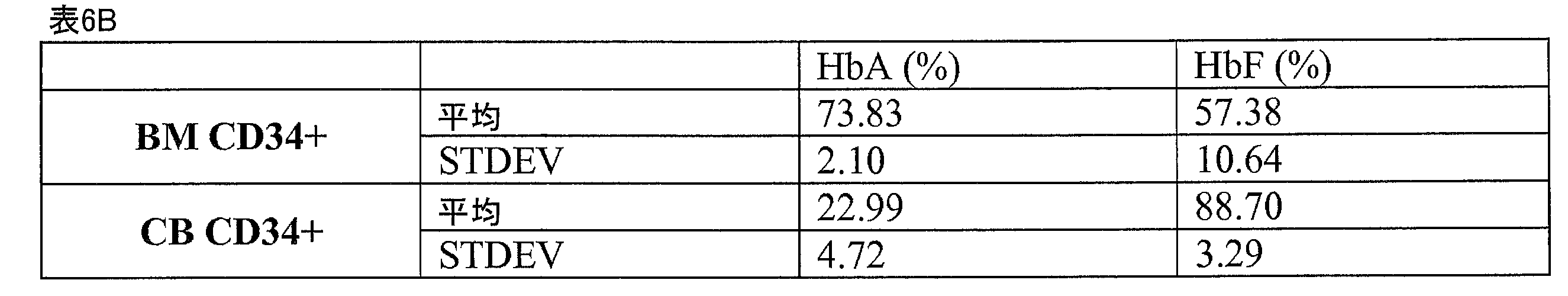
<6.7.実施例7:長期にわたる増殖および赤血球への分化>
本実験では、E3培地(上述の実施例4参照)、並びに、単一のドナーおよび混合ドナ
ーに由来するCD34+細胞を用いて、長期にわたる細胞増殖を行った。高いRBC成熟
効率を伴う、最高63日間にわたる継続した細胞増殖が実証された(図9)。63日目に
は、混合ドナー細胞では1×10⁹倍の増殖、単一ドナー細胞では1.8×10⁹倍の増殖
が、それぞれ観察され、単一ドナーでは、77%のCD235A+、および56%の除核
が達成された(表7)。
<6.7. Example 7: Long-Term Proliferation and Differentiation into Erythrocytes>
Long-term cell expansion was performed in this experiment using E3 medium (see Example 4 above) and CD34 + cells from single and mixed donors. Continued cell proliferation for up to 63 days was demonstrated with high RBC maturation efficiency (Fig. 9). On day 63, a 1×10 9 -fold expansion of mixed donor cells and a 1.8×10 9 -fold expansion of single donor cells were observed, respectively, with 77% CD235A + and 56% enucleation was achieved (Table 7).
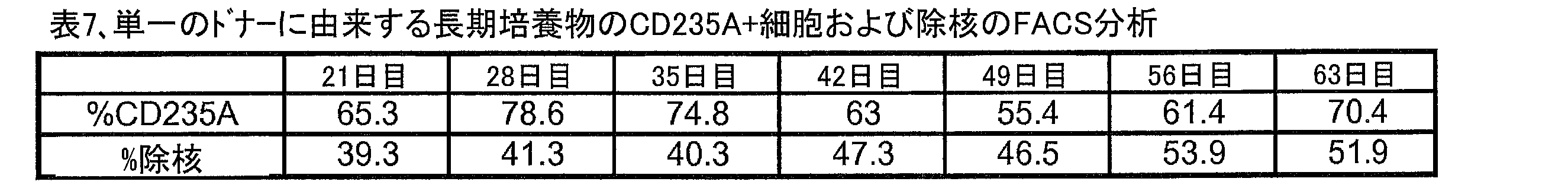
図10は、単一ドナーに由来する長期の培養物におけるHbFおよびHbAの生産のE
LISA分析を、末梢血(PB)RBCにおけるHbFおよびHbAの生産と比較して示
している。本実験では、示された時点において採集された2×10⁵個の細胞を、PBS
で洗浄し、2500 × gで10分間、スピンさせた。細胞の沈殿物を、100LM-
PER Mammalian Protein Extraction Reagent
(Cat# 78501、Thermo Scientific)を用いて溶解させ、混
合物を10分間穏やかに混合し、続いて、14,000 × gで15分間遠心分離させ
た。その後、ELISAキットを用いたヘモグロビン分析のために、上清を新しいチュー
ブに移した。ELISAキットには、ヒトヘモグロビンELISA定量セット(Cat#
E80-135)およびヒト胎児型ヘモグロビンELISA定量セット(Cat# E
80-136)が含まれる。100万個のPB赤血球から245ngのHbAが生産され
た一方、培養細胞は、培養14日目から開始して、1.3μgHbAを生産した。
Figure 10 is an E of HbF and HbA production in long-term cultures from a single donor.
LISA analysis is shown comparing HbF and HbA production in peripheral blood (PB) RBCs. In this experiment, 2×10 5 cells harvested at the indicated time points were
and spun at 2500 xg for 10 minutes. 100 LM-
PER Mammalian Protein Extraction Reagent
(Cat# 78501, Thermo Scientific) and the mixture was mixed gently for 10 minutes followed by centrifugation at 14,000 x g for 15 minutes. Supernatants were then transferred to new tubes for hemoglobin analysis using an ELISA kit. The ELISA kit includes the Human Hemoglobin ELISA Quantitative Set (Cat#
E80-135) and Human Fetal Hemoglobin ELISA Quantitative Set (Cat# E
80-136). One million PB erythrocytes produced 245 ng of HbA, while the cultured cells produced 1.3 μg HbA beginning on
表8は、単一ドナーに由来する長期の培養物における、いくつかの遺伝子の定量的リア
ルタイムPCR(qRT-PCR)分析を示している。細胞のアリコートを様々な時点で
採集し、qRT-PCR分析を行った。qRT-PCR分析には、7900HT Fas
t Real-Time PCR System(Applied Biosystem
s)およびTaqMan(登録商標)Gene Expression Assays
of EKLF(Applied Biosystems、Cat# Hs006105
92_m1)、GATA1(Applied Biosystems、Cat# Hs0
0231112_m1)、GATA2、HBβ(Applied Biosystems
、Cat# Hs00758889_s1)、HBγ(Applied Biosyst
ems、Cat# Hs00361131_g1)、LMO2(Applied Bio
systems、Cat# Hs00277106_m1)、およびZFPM1(App
lied Biosystems、Cat# Hs00419119_m1)を用いた。
qRT-PCR分析の結果によって、継続的な増殖および赤血球への分化は、HBβ、H
Bγ、並びに、EKLF、GATA1、LMO2およびZFPM1を含む、赤血球の分化
に重要ないくつかの転写因子の発現の増加と相関があることが示された。
表8。単一ドナーに由来する長期の培養物におけるqRT-PCR分析。(A)遺伝子発
現の変化倍率。(B)遺伝子の説明。標準偏差は2反復の変化倍率の平均に対して計算さ
れた。
Table 8 shows quantitative real-time PCR (qRT-PCR) analysis of several genes in long-term cultures derived from single donors. Aliquots of cells were collected at various time points and subjected to qRT-PCR analysis. For qRT-PCR analysis, 7900HT Fas
t Real-Time PCR System (Applied Biosystem
s) and TaqMan® Gene Expression Assays
of EKLF (Applied Biosystems, Cat# Hs006105
92_m1), GATA1 (Applied Biosystems, Cat# Hs0
0231112_m1), GATA2, HBβ (Applied Biosystems
, Cat# Hs00758889_s1), HBγ (Applied Biosystem
ems, Cat# Hs00361131_g1), LMO2 (Applied Bio
systems, Cat# Hs00277106_m1), and ZFPM1 (App
Lied Biosystems, Cat# Hs00419119_m1) was used.
Results from qRT-PCR analysis showed that continued proliferation and differentiation into erythrocytes was associated with HBβ, H
It was shown to correlate with increased expression of Bγ and several transcription factors important for erythroid differentiation, including EKLF, GATA1, LMO2 and ZFPM1.
Table 8. qRT-PCR analysis in long-term cultures from single donors. (A) Fold change in gene expression. (B) Gene description. Standard deviations were calculated for the average fold change of the duplicates.


<6.8.実施例8:E3培地の最適化>
本実施例では、HSCの増殖および赤血球への分化を改善するために、3水準(3要因
:SCF、Epo、およびIL-3)全要因実験計画(3-level full factorial experim
ent design)を用いて、E3培地をさらに最適化した(図11)。CD34+細胞を、図
1で記載したように得て、E3培地には、表9に示すSCF、IL-3およびEpoを追
加した。細胞計数、生存率、CD235A+細胞の割合、および除核細胞の割合について
、表10に示す時点で調べた。一つの組成(#7、65ng/mLのSCF、7ng/m
LのIL-3、および3IU/mLのEpo)では、E3培地組成に比べて、CD235
A+細胞の割合の増加、および除核細胞の割合の増加が見られた。
<6.8. Example 8: Optimization of E3 medium>
In this example, a 3-level (3 factors: SCF, Epo, and IL-3) full factorial experimental design was used to improve HSC proliferation and erythroid differentiation.
ent design) was used to further optimize the E3 medium (Fig. 11). CD34 + cells were obtained as described in FIG. 1 and E3 medium supplemented with SCF, IL-3 and Epo as indicated in Table 9. Cell counts, viability, percent CD235A + cells, and percent enucleated cells were determined at the time points shown in Table 10. One composition (#7, 65 ng/mL SCF, 7 ng/m
L IL-3, and 3 IU/mL Epo) compared to the E3 medium composition, CD235
An increased percentage of A + cells and an increased percentage of enucleated cells were seen.

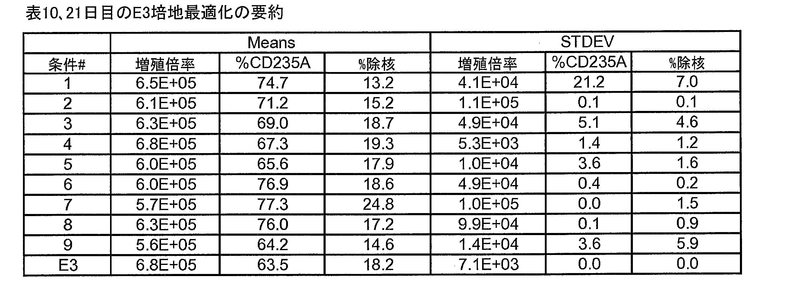
本研究によって、21日間の細胞増殖実験での、HSCの増殖および赤血球への分化に
おける、SCFおよびEpoの異なる効果が特定された(図12A-12C)。Epoは
、SCFが高レベルである場合、初期の増殖を増加させるように相乗的に働くが、SCF
が低レベルである場合、増殖を抑制させる。SCFが低レベルである場合、Epoは、C
D235Aの発現の増加に対してより効果的であり、一方、SCFが高レベルである場合
、Epoは、赤血球の分化を抑制させる。
The present study identified differential effects of SCF and Epo on HSC proliferation and differentiation into erythrocytes in a 21-day cell proliferation experiment (FIGS. 12A-12C). Epo acts synergistically to increase early proliferation when SCF is at high levels, whereas SCF
At low levels, it suppresses proliferation. When SCF is low, Epo
Epo is more effective at increasing the expression of D235A, whereas at high levels of SCF, Epo suppresses erythroid differentiation.
二番目のDOE試験では、細胞播種密度のSCFおよび/またはEpoに対する相互作
用を評価するために、3水準(SCF、Epo、および細胞密度)全要因実験計画を利用
した(図13A)。CD34+細胞を、図1で記載したように得て、E3培地には、表1
1に示す異なる細胞密度で、SCFおよびEpoを追加した。低Epo条件(最高3倍)
、低細胞密度、および高SCF濃度において、増殖の大幅な促進(約10倍)が達成され
ることが実証された(図13B)。さらに、赤血球の分化は、高細胞播種密度および低S
CF濃度で、改善されることが実証された(図13C)。
A second DOE study utilized a three-level (SCF, Epo, and cell density) full-factorial design to assess the interaction of cell seeding density on SCF and/or Epo (FIG. 13A). CD34 + cells were obtained as described in FIG.
SCF and Epo were added at different cell densities shown in 1. Low Epo conditions (up to 3x)
, low cell densities, and high SCF concentrations, a significant enhancement of proliferation (approximately 10-fold) was demonstrated (FIG. 13B). Furthermore, erythroid differentiation is associated with high cell seeding density and low S
Improvements were demonstrated at CF concentrations (FIG. 13C).
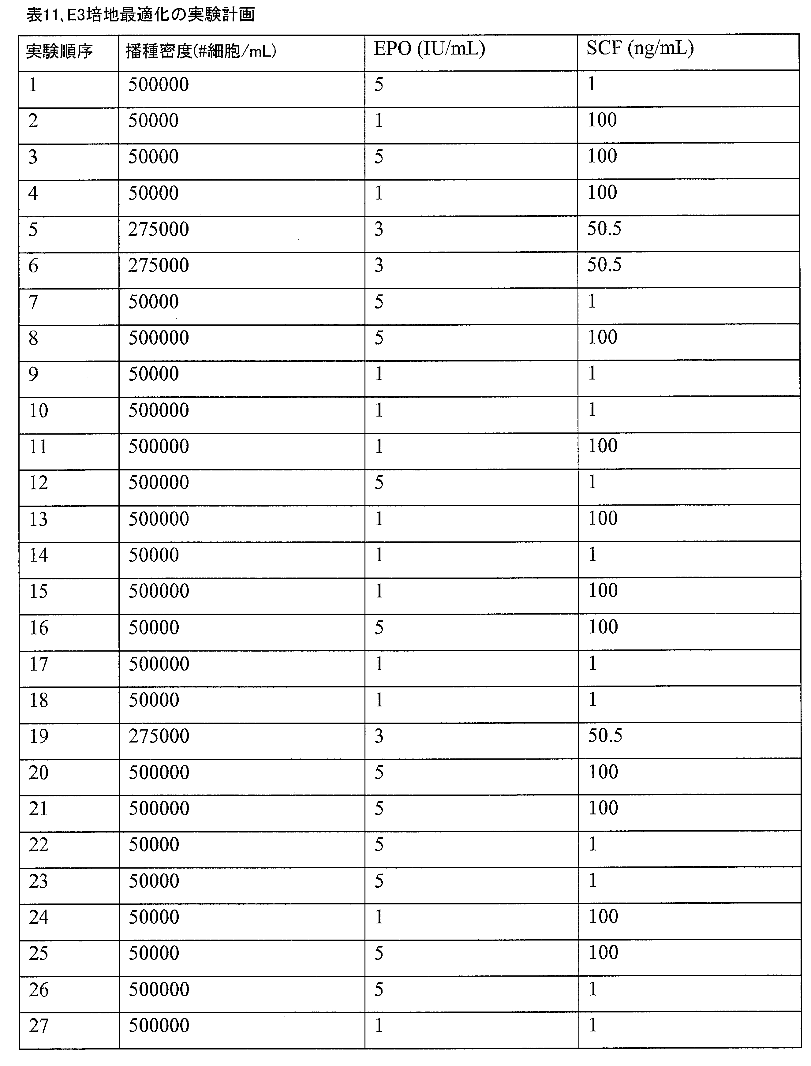
<6.9.実施例9:赤血球のユニットを生産するための方法およびバイオリアクター
>
本実施例では、赤血球の生産方法、および、成熟した赤血球のユニットの生産を可能に
するバイオリアクターを提供する。特定の実施例では、バイオリアクターは、5段階の工
程によって、投与可能な赤血球のユニットの生産を可能にする。最初の工程では、造血細
胞(例えばCD34+細胞)を単離する。二番目の工程では、CD34+細胞を、免疫調節
化合物(例えばポマリドマイド)を用いて、増殖させる。三番目の工程では、本明細書に
例示するバイオリアクター中で、付着胎盤幹細胞との共培養で、系列決定済み細胞の除去
と同時に、CD34+細胞を増殖させる。四番目には、残った未分化の造血細胞を赤血球
に分化させる。最後に、五番目の工程で、赤血球を単離し、投与可能なユニットに採集す
る。
<6.9. Example 9: Methods and Bioreactors for Producing Units of Red Blood Cells>
This example provides a method of producing red blood cells and a bioreactor that allows the production of mature red blood cell units. In certain examples, the bioreactor enables the production of administrable units of red blood cells by a five-step process. In the first step, hematopoietic cells (eg CD34 + cells) are isolated. In a second step, CD34 + cells are expanded with an immunomodulatory compound such as pomalidomide. In a third step, CD34 + cells are expanded in co-culture with adherent placental stem cells in the bioreactor exemplified herein, concurrent with the removal of lineage-committed cells. Fourth, the remaining undifferentiated hematopoietic cells are differentiated into erythrocytes. Finally, in a fifth step, red blood cells are isolated and collected into administrable units.
工程1および2、すなわち造血細胞の単離および初期の増殖は、上述の実施例3および
4に記載されたように達成される。
工程3および4は、バイオリアクターによって達成される。上記バイオリアクターは、
胎盤幹細胞(2)が播種された中空糸チャンバー(1)、および培地へのガス供給のため
の部材(3)を備える。上記バイオリアクターは、さらに、系列決定済の造血細胞、完全
に分化した赤血球、またはその両方を継続的に分離することができる、連結した細胞選別
/分離部材(4)を備える。上記細胞分離部材は、例えば、磁気細胞分離または蛍光活性
化セルソーティング技術を用いて、細胞を造血細胞から分離することができる。
It comprises a hollow fiber chamber (1) seeded with placental stem cells (2) and a member (3) for gassing the culture medium. The bioreactor further comprises a coupled cell sorting/separating member (4) capable of continuously separating lineage-committed hematopoietic cells, fully differentiated red blood cells, or both. The cell separation member can separate cells from hematopoietic cells using, for example, magnetic cell separation or fluorescence-activated cell sorting techniques.
細胞の培養を始めるために、およそ5×10⁷個の造血細胞(例えばCD34+造血細胞
)を、上記バイオリアクターに植えつける。
To initiate cell culture, approximately 5×10 7 hematopoietic cells (eg, CD34 + hematopoietic cells) are seeded into the bioreactor.
<6.10.赤血球の採集>
本実施例では、赤血球を他の系列決定済み細胞から分離する、いくつかの方法を例示す
る。
<6.10. Collection of red blood cells>
This example illustrates several methods of separating red blood cells from other lineage-committed cells.
方法1:赤血球(例えば、本明細書で例示したバイオリアクターの細胞分離部材から採
集された赤血球)およびヘタスターチ溶液をバクスター採集バッグ中で3:1(v/v)
で混合し、血漿抽出器(plasma extractor)上に直立位置で配置する。
赤血球は50~70分後に堆積する。堆積していない細胞は、血漿抽出器によって強制排
出する。バッグに残った堆積した赤血球を、さらに、400 × gで10分間の遠心分
離によって採集する。上清を取り除いた後、赤血球を適切な量の所望の培地に再び懸濁す
る。
Method 1: red blood cells (e.g., red blood cells collected from the cell separation member of the bioreactor exemplified herein) and hetastarch solution 3:1 (v/v) in a Baxter collection bag
and place in an upright position on the plasma extractor.
Erythrocytes are deposited after 50-70 minutes. Non-deposited cells are forced out by a plasma extractor. Accumulated red blood cells remaining in the bag are further collected by centrifugation at 400 xg for 10 minutes. After removing the supernatant, the red blood cells are resuspended in an appropriate amount of the desired medium.
方法2-免疫磁気的分離:グリコフォリンA+細胞(例えば、本明細書で例示したバイ
オリアクターの細胞分離部材から採集された赤血球)を、グリコフォリンA(CD235
a)マイクロビーズ(Miltenyi Biotech)によって磁気的に標識する。
その後、細胞懸濁液をチューブに入れ、EASYSEP(登録商標)マグネットの磁場の
中に配置する。磁気的に標識されたグリコフォリンA+細胞はチューブの内側に残り、一
方、標識されていない細胞は、チューブの外へ流れ出る。チューブを磁場から取り除いた
後、磁気的に維持されたグリコフォリンA+細胞を磁気ビーズから分離し、適切な量の所
望の培地に再び懸濁することができる。
a) Magnetic labeling with microbeads (Miltenyi Biotech).
The cell suspension is then placed in a tube and placed in the magnetic field of an EASYSEP® magnet. Magnetically labeled glycophorin A + cells remain inside the tube, while unlabeled cells flow out of the tube. After removing the tube from the magnetic field, the magnetically maintained glycophorin A + cells can be separated from the magnetic beads and resuspended in an appropriate amount of the desired medium.
方法3-フローサイトメトリー細胞分離:赤血球(例えば、本明細書で例示したバイオ
リアクターの細胞分離部材から採集された赤血球)を、1μLのFc Block(1/
500)を含む500μLのPBS/FBSに加える。150μLの細胞懸濁液を、96
ウェルV底ディッシュの各ウェルに加える。50μLの1°Ab Master Mix
(当該混合物は、PBS/FBSにそれぞれの一次Abを1/25で希釈したものである
。)を上記細胞に加える。1つのウェルには、補正を設定するための単一のポジティブコ
ントロールとしてそれぞれの一次Abが含まれるのと同様に、電圧を設定するためのアイ
ソタイプコントロールの組み合わせが含まれる。上記細胞を4℃で60分間培養し、その
後、1500RPMで2分間遠心分離する。上清は捨てる。ウェルはそれぞれ、200μ
LのPBS/FBSで洗浄し、ピペットを用いて混合する。その後、上記細胞を即座に、
1500RPMで2分間スピンさせ、上清を捨てる。150μLの二次Ab(すなわち、
ストレプトアビジン-TC)Master Mixを加え、4℃で30分間培養し、その
後、1500RPMで5分間遠心分離する。堆積物は、200~500μLのPBS/F
BSに再び懸濁し、5mLの流管に移す。その後、細胞を、フローサイトメーターを用い
て分離する。
500) in 500 μL PBS/FBS. 150 μL of cell suspension was added to 96
Add to each well of well V-bottom dish. 50 μL of 1° Ab Master Mix
(The mixture is a 1/25 dilution of each primary Ab in PBS/FBS) is added to the cells. One well contains a combination of isotype controls for setting voltages, as well as each primary Ab as a single positive control for setting corrections. The cells are incubated at 4° C. for 60 minutes, then centrifuged at 1500 RPM for 2 minutes. Discard the supernatant. Each well is 200μ
Wash with L PBS/FBS and mix using a pipette. The cells are then immediately
Spin at 1500 RPM for 2 minutes and discard supernatant. 150 μL of secondary Ab (i.e.
Streptavidin-TC) Master Mix is added and incubated at 4° C. for 30 minutes, followed by centrifugation at 1500 RPM for 5 minutes. The deposit was 200-500 μL of PBS/F
Resuspend in BS and transfer to a 5 mL flow tube. Cells are then separated using a flow cytometer.
方法4:赤血球を含む培地を、細胞培養部材と細胞分離部材との間の継続的な流れの中
で、ガス供給部材からの酸素の供給を低減または停止させることによって、脱酸素化し、
細胞分離部材中で磁石を作動させる。培地を、細胞分離部材を介して、十分な時間、磁石
の磁場を通過させ、細胞分離部材の表面に赤血球を集める。一旦、所定の数の赤血球が集
まるか、または採集が所定の時間行われれば、培地に酸素が送り込まれ、赤血球は表面か
ら解放される。
Method 4: deoxygenating the red blood cell-containing medium in continuous flow between the cell culture member and the cell separation member by reducing or stopping the supply of oxygen from the gas supply member;
A magnet is activated in the cell separation member. Culture medium is passed through the magnetic field of the magnet through the cell separation member for a sufficient time to collect red blood cells on the surface of the cell separation member. Once a predetermined number of red blood cells have been collected or collection has occurred for a predetermined time, the medium is oxygenated and the red blood cells are released from the surface.
方法5:堆積に基づいた、赤血球の濃縮。赤血球の濃縮は、3000rpmで15分間
遠心分離を行い、中断することで行うことができる。白血球(一番上の白い層)、未熟な
赤血球(中間のピンクの層)、および赤血球(一番下の赤い層)を分離できる。その後、
赤血球を底から採集し、適切な量の所望の培地に再び懸濁する。
Method 5: Red blood cell enrichment based on sedimentation. Erythrocyte concentration can be performed by centrifugation at 3000 rpm for 15 minutes and a break. White blood cells (top white layer), immature red blood cells (middle pink layer), and red blood cells (bottom red layer) can be separated. after that,
Erythrocytes are collected from the bottom and resuspended in appropriate volume of desired medium.
<6.11.赤血球の採集>
本実施例では、フローサイトメトリーを用いた、赤血球の採集を実証する。
<6.11. Collection of red blood cells>
This example demonstrates collection of red blood cells using flow cytometry.
赤血球の濃縮は、FACSAriaを用いた細胞選別法によって行われる。FACSA
riaを用いた細胞選別法では、光散乱を利用した細胞サイズによる赤血球の選択(前方
散乱および側方散乱、図14および表12)、またはDRAQ5標識を利用した除核の選
択(リニアAPCチャンネル蛍光、図15および表13)を行う。
Erythrocyte enrichment is performed by cell sorting using the FACSAria. FACSA
For ria-based cell sorting methods, erythrocyte selection by cell size using light scatter (forward and side scatter, FIG. 14 and Table 12) or enucleation selection using DRAQ5 labeling (linear APC channel fluorescence , FIG. 15 and Table 13).
FACSAriaによる細胞選別の後、P1、P2およびP3によってゲート(gated
)された細胞について、フローサイトメトリーによってHbA+、除核、およびCD23
5A+の割合を評価した。最も小さい細胞サイズを示すP1細胞が、HbA+、除核、およ
びCD235A+について最も高い割合を示し、そのため、大部分が赤血球であった。
After cell sorting by FACSAria, gated by P1, P2 and P3
) for HbA + , enucleation, and CD23 by flow cytometry
The percentage of 5A + was evaluated. P1 cells, which displayed the smallest cell size, had the highest percentages of HbA + , enucleation, and CD235A + and were therefore mostly erythrocytes.

表12では、細胞を、細胞透過性DNA相互作用試薬DRAQ5(Cell Sign
aling、カタログNo.#4084)によって染色し、その後、FACSAriaを
用いた細胞選別を行い、Q1およびQ2によってゲートされた細胞について、フローサイ
トメトリーによってHbA+、除核、およびCD235A+の割合を評価する。DRAQ染
色では陰性であったQ1細胞は、Q2細胞に比べて、HbA+、除核、およびCD235
A+について高い割合を示し、そのため、大部分が赤血球であった。
In Table 12, cells were treated with the cell-permeable DNA-interacting reagent DRAQ5 (Cell Sign
aling, catalog no. #4084) followed by cell sorting using FACSAria and the percentage of HbA + , enucleation, and CD235A + assessed by flow cytometry for cells gated by Q1 and Q2. Q1 cells, which were negative for DRAQ staining, showed higher levels of HbA + , enucleation, and CD235 compared to Q2 cells.
A high percentage was shown for A + and therefore mostly erythrocytes.
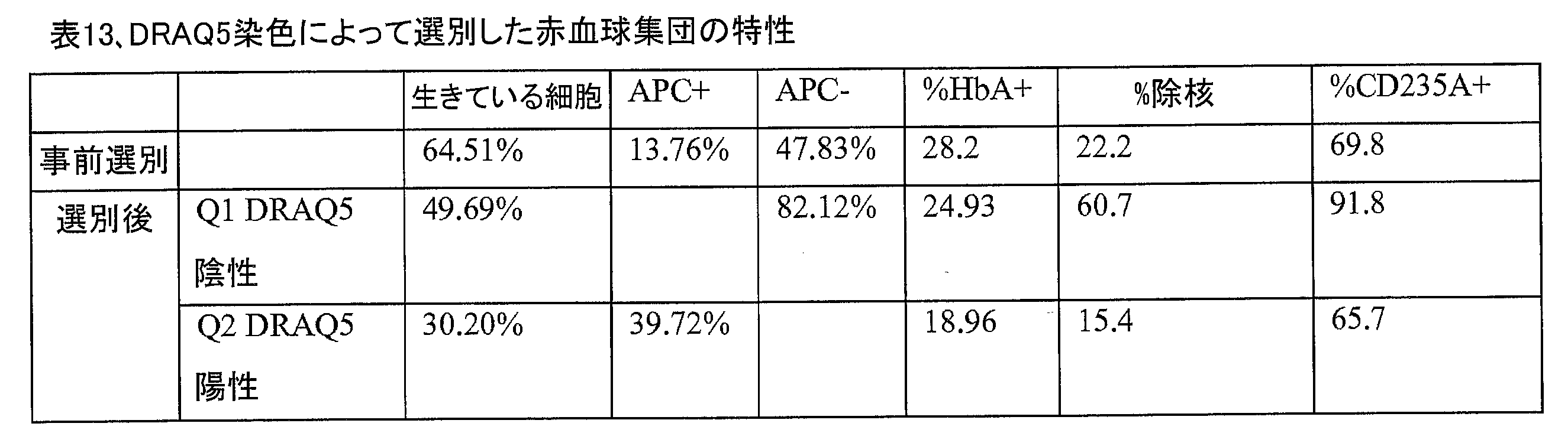
表13で示したように、細胞を、細胞透過性DNA相互作用試薬DRAQ5によって染
色した。FACSAriaによる細胞選別の後、QP1、P2およびQP3によってゲー
トされた細胞について、フローサイトメトリーによってHbA+、除核、およびCD23
5A+の割合を評価した。DRAQ染色では陰性であり、最も小さい細胞サイズを示すQ
P1細胞は、Q2細胞に比べて、HbA+、除核、およびCD235A+について高い割合
を示し、そのため、大部分が赤血球であった。
Cells were stained with the cell-permeable DNA-interacting reagent DRAQ5 as shown in Table 13. After cell sorting by FACSAria, HbA + , enucleation, and CD23 were detected by flow cytometry for cells gated by QP1, P2 and QP3.
The percentage of 5A + was evaluated. Q, which is negative for DRAQ staining and exhibits the smallest cell size
P1 cells exhibited higher percentages of HbA + , enucleation, and CD235A + than Q2 cells and were therefore predominantly red blood cells.
<6.12.実施例11:赤血球を生産するためのバイオリアクター>
本実施例では、造血細胞からの赤血球の生産を改善することができるバイオリアクター
の設計について説明する。上記バイオリアクターは、造血細胞を培養する中空糸を有する
培養部材を備える。造血細胞(例えば造血前駆細胞)を、5×10⁵個/投与量でバッグ
中に追加する。上記1回の投与量は、1ユニットの血液をもたらす。上記細胞を、第一の
ポートを介して加えられた50ng/mLのSCF、3units/mLのEpo、50
ng/mLのIGF-1を含むIMDM培地の存在下で増殖させる。造血細胞が培養され
ている上記培地に、2.7μg/mLのポマリドマイドを追加する。培養の間、培養部材
中のガス、培地の代謝産物、および培地のpHを継続的にモニタリングし、必要に応じて
、プログラム可能な制御装置を用いて、補充または交換できる。培養部材中の培地のpH
はおよそ7で維持され、培養温度は約37℃で維持される。系列決定済みの細胞(例えば
分化した細胞)は、細胞分離部材を用いて、培地から継続的に分離および回収される。上
記バイオリアクターは、遠隔地、例えば、造血細胞を初めに得る場所から離れた場所にお
いて動作できるように、独立した電源を備えている。
<6.12. Example 11: Bioreactor for Producing Red Blood Cells>
This example describes the design of a bioreactor that can improve the production of red blood cells from hematopoietic cells. The bioreactor includes a culture member having hollow fibers for culturing hematopoietic cells. Hematopoietic cells (eg, hematopoietic progenitor cells) are added into the bag at 5×10 5 /dose. The single dose yields 1 unit of blood. The cells were added via the first port at 50 ng/mL SCF, 3 units/mL Epo, 50
Grow in the presence of IMDM medium containing ng/mL IGF-1. 2.7 μg/mL of pomalidomide is added to the medium in which the hematopoietic cells are cultured. During cultivation, gases in the culture member, medium metabolites, and medium pH can be continuously monitored and replenished or replaced as needed using a programmable controller. pH of the medium in the culture member
is maintained at approximately 7 and the culture temperature is maintained at about 37°C. Lineage-committed cells (eg, differentiated cells) are continuously separated and collected from the medium using a cell separation member. The bioreactor is equipped with an independent power source so that it can be operated at a remote location, eg, remote from where the hematopoietic cells are originally obtained.
装置、組成物、および方法を含む本開示は、本明細書に記載された特定の実施形態の範
囲に限定されるものではない。実際には、本明細書の記載に加えて種々の変更が可能であ
ることが、当業者であれば上記の説明から理解できるであろう。そのような変更も、添付
の特許請求の範囲内に含まれるものである。
This disclosure, including devices, compositions, and methods, is not limited in scope to the particular embodiments described herein. Indeed, those skilled in the art will appreciate from the foregoing description that various modifications in addition to those described herein are possible. Such modifications are intended to fall within the scope of the appended claims.
本明細書で引用された全ての参照文献は、個々の刊行物、特許、または特許出願が具体
的に、かつ個別に、全ての目的について全体が参照によって引用されるのと同じ程度に、
全ての目的について全体が参照によって本明細書中に引用されるものとする。
All references cited herein are to the same extent as if each individual publication, patent, or patent application was specifically and individually incorporated by reference in its entirety for all purposes.
The entirety of which is incorporated herein by reference for all purposes.
いずれの刊行物の引用も、出願日に先行して開示されていたためであり、先行発明を理
由として、本開示が上記刊行物に先行する資格がないことを認めたと解釈されるべきでは
ない。
Citation of any publication is for disclosure prior to the filing date of the filing and should not be construed as an admission that the present disclosure is not entitled to antedate the publication by virtue of prior invention.
Claims (29)
した造血細胞集団を得る工程であって、
上記第1の造血細胞集団の当初の細胞密度は、5×10⁴個/mL以下であり、
上記第1の培地は、Epoを1IU以下の濃度で含んでおり、かつ、SCFを100
ng/mL以上の濃度で含んでいる、工程と、
(b)上記増殖した造血細胞集団を、第2の培地に接触させることによって、上記増殖
した造血細胞集団中の複数の造血細胞を、赤血球へ分化させる工程であって、
上記増殖した造血細胞集団の当初の細胞密度は、2.75×10⁵個/mL超であり
、
上記第2の培地は、Epoを3IU/mL以上の濃度で含んでおり、かつ、SCFを
50ng/mL以下の濃度で含んでいる、工程と、
(c)上記培地から上記赤血球を単離する工程と、
を含む、赤血球を生産する方法であって、
上記SCF、IL-3およびEpoは、上記第1の培地または第2の培地の定義されて
いない成分の中には含まれていないことを特徴とする、赤血球を生産する方法。 (a) growing a first hematopoietic cell population in a first culture medium in the absence of feeder cells to obtain an expanded hematopoietic cell population, comprising:
The initial cell density of the first hematopoietic cell population is 5 × 10 cells/mL or less,
The first medium contains Epo at a concentration of 1 IU or less and SCF at 100
at a concentration of ng/mL or greater; and
(b) differentiating a plurality of hematopoietic cells in the expanded hematopoietic cell population into red blood cells by contacting the expanded hematopoietic cell population with a second medium,
the initial cell density of the expanded hematopoietic cell population is greater than 2.75 x 10 cells/mL;
the second medium contains Epo at a concentration of 3 IU/mL or higher and SCF at a concentration of 50 ng/mL or lower;
(c) isolating the red blood cells from the culture medium;
A method of producing red blood cells comprising
A method of producing red blood cells, wherein said SCF, IL-3 and Epo are not included among the undefined components of said first medium or second medium.
Tpo)、ホメオボックス-B4(HoxB4)またはメチルセルロースのうち、1つ以
上を含まない、請求項1に記載の方法。 The first medium and the second medium contain Flt-3L, IL-11, thrombopoietin (
Tpo), homeobox-B4 (HoxB4) or methylcellulose.
する請求項1に記載の方法。 2. The method of claim 1, wherein said SCF is present in said first medium at a concentration of 100 ng/mL.
とを特徴とする請求項1に記載の方法。 2. The method of claim 1, wherein said IL-3 is present in said first medium at a concentration of 0.1-100 ng/mL.
徴とする請求項1に記載の方法。 2. The method of claim 1, wherein said IL-3 is present in said first medium at a concentration of 1-50 ng/mL.
る請求項1に記載の方法。 2. The method of claim 1, wherein said IL-3 is present in said first medium at a concentration of 5 ng/mL.
上記免疫調節化合物は、上記免疫調節化合物が存在しない状態で増殖した複数の造血細
胞に比べて、造血細胞の数を増加させるものであることを特徴とする請求項1に記載の方
法。 the second medium comprises an immunomodulatory compound;
2. The method of Claim 1, wherein said immunomodulatory compound increases the number of hematopoietic cells relative to a plurality of hematopoietic cells grown in the absence of said immunomodulatory compound.
特徴とする請求項1に記載の方法。 2. The method of claim 1, wherein said hematopoietic cells are Thy-1 + , CXCR4 + , CD133 + or KDR + .
とする請求項1に記載の方法。 2. The method of claim 1, wherein said hematopoietic cells are CD34 - CD133 + or CD34 - CD117 + .
11c-、CD14-、CD16-、CD24-、CD56-、CD66b-および/またはグ
リコフォリンA-であることを特徴とする請求項1に記載の方法。 The hematopoietic cells are HLA-DR − , CD71 − , CD2 − , CD3 − , CD11b − , CD
11c- , CD14- , CD16- , CD24-, CD56- , CD66b- and /or glycophorin A-.
細胞から得られることを特徴とする請求項1に記載の方法。 2. The method of claim 1, wherein said hematopoietic cells are obtained from cord blood, placental blood, peripheral blood, bone marrow, embryonic stem cells or induced pluripotent cells.
載の方法。 2. The method of claim 1, wherein said hematopoietic cells are obtained from cord blood and placental perfusate.
とを特徴とする請求項14に記載の方法。 15. The method of claim 14, wherein the placental perfusate is obtained by passage of the perfusate only through the vasculature of the placenta.
とを特徴とする、請求項15に記載の方法。 16. Method according to claim 15, characterized in that the placental perfusate is obtained by passage of the perfusate only through the vasculature of the placenta.
子1(IGF-1)、および、1~1000μg/mLの濃度である脂質を含み、
上記脂質は、たんぱく質とコレステロールとの混合物を含み、
上記第2の培地は、0.01~100μMの濃度であるヒドロコルチゾン、または、0
.01~100μMの濃度であるデキサメタゾンを含むことを特徴とする請求項1に記載
の方法。 the first medium further comprising insulin-like growth factor 1 (IGF-1) at a concentration of 1-1000 ng/mL and lipid at a concentration of 1-1000 μg/mL;
The lipid comprises a mixture of protein and cholesterol,
The second medium contains hydrocortisone at a concentration of 0.01-100 μM, or 0
. The method of claim 1, comprising dexamethasone at a concentration of 01-100 μM.
とを特徴とする請求項19に記載の方法。 20. The method of claim 19, wherein said IGF-I is present in said first medium at a concentration of 10-500 ng/mL.
とを特徴とする請求項19に記載の方法。 20. The method of claim 19, wherein said IGF-I is present in said first medium at a concentration of 20-100 ng/mL.
徴とする請求項19に記載の方法。 20. The method of claim 19, wherein said lipid is present in said first medium at a concentration of 10-500 ng/mL.
徴とする請求項19に記載の方法。 20. The method of claim 19, wherein said lipid is present in said first medium at a concentration of 20-100 ng/mL.
とを特徴とする請求項19に記載の方法。 20. The method of claim 19, wherein said hydrocortisone is present in said second medium at a concentration of 0.1-50 μM.
とを特徴とする請求項19に記載の方法。 20. The method of claim 19, wherein said hydrocortisone is present in said second medium at a concentration of 0.5-10 μM.
とを特徴とする請求項19に記載の方法。 20. The method of claim 19, wherein said dexamethasone is present in said second medium at a concentration of 0.05-20 μM.
を特徴とする請求項19に記載の方法。 20. The method of claim 19, wherein said dexamethasone is present in said second medium at a concentration of 0.1-10 μM.
h陰性;M型、N型、S型、またはs型の血液型;P1型の血液型;Lua型、Lub型
、またはLu(a)型の血液型;K(Kell)型、k(cellano)型、Kpa型
、Kpb型、K(a+)型、Kp(a-b-)型またはK-k-Kp(a-b-)型の血
液型;Le(a-b-)型、Le(a+b-)型またはLe(a-b+)型の血液型;F
y a型、Fy b型またはFy(a-b-)型の血液型;Jk(a-b-)型、Jk(
a+b-)型、Jk(a-b+)型またはJk(a+b+)型の血液型を作る能力を有す
ることを特徴とする請求項1に記載の方法。 more than one of the hematopoietic cells is blood group A, O, AB, O; Rh positive or R
M, N, S, or s blood type; P1 blood type; Lua, Lub, or Lu (a) blood type; K (Kell) type, k (cellano ), Kpa, Kpb, K (a+), Kp (ab-) or Kk-Kp (ab-) blood types; Le (ab-), Le (a+b-) or Le (a-b+) blood type; F
ya blood type a, Fy b or Fy (ab-); Jk (ab-), Jk (
2. The method of claim 1, having the ability to form a+b-), Jk(a−b+) or Jk(a+b+) blood types.
;B型、Rh陽性;B型、Rh陰性;AB型、Rh陽性またはAB型、Rh陰性を作る能
力を有することを特徴とする請求項28に記載の方法。 Type O, Rh-negative; Type A, Rh-positive; Type A, Rh-negative; Type B, Rh-positive; Type B, Rh-negative; 29. The method of claim 28, having the ability to make Rh negative.
Priority Applications (1)
| Application Number | Priority Date | Filing Date | Title |
|---|---|---|---|
| JP2024063692A JP2024095790A (en) | 2009-07-02 | 2024-04-11 | Method of producing erythrocytes without feeder cells |
Applications Claiming Priority (3)
| Application Number | Priority Date | Filing Date | Title |
|---|---|---|---|
| US22293009P | 2009-07-02 | 2009-07-02 | |
| US61/222,930 | 2009-07-02 | ||
| JP2020129086A JP2020182494A (en) | 2009-07-02 | 2020-07-30 | Method of producing erythrocytes without feeder cells |
Related Parent Applications (1)
| Application Number | Title | Priority Date | Filing Date |
|---|---|---|---|
| JP2020129086A Division JP2020182494A (en) | 2009-07-02 | 2020-07-30 | Method of producing erythrocytes without feeder cells |
Related Child Applications (1)
| Application Number | Title | Priority Date | Filing Date |
|---|---|---|---|
| JP2024063692A Division JP2024095790A (en) | 2009-07-02 | 2024-04-11 | Method of producing erythrocytes without feeder cells |
Publications (1)
| Publication Number | Publication Date |
|---|---|
| JP2022110097A true JP2022110097A (en) | 2022-07-28 |
Family
ID=42537451
Family Applications (7)
| Application Number | Title | Priority Date | Filing Date |
|---|---|---|---|
| JP2012518604A Pending JP2012531916A (en) | 2009-07-02 | 2010-07-01 | Method for producing red blood cells without using feeder cells |
| JP2015078743A Pending JP2015146816A (en) | 2009-07-02 | 2015-04-07 | Method of producing erythrocytes without feeder cells |
| JP2016186243A Pending JP2017038603A (en) | 2009-07-02 | 2016-09-23 | Method for producing erythrocytes without feeder cells |
| JP2018098578A Pending JP2018148913A (en) | 2009-07-02 | 2018-05-23 | Method for producing red blood cells without using feeder cells |
| JP2020129086A Withdrawn JP2020182494A (en) | 2009-07-02 | 2020-07-30 | Method of producing erythrocytes without feeder cells |
| JP2022081214A Pending JP2022110097A (en) | 2009-07-02 | 2022-05-18 | Method of producing erythrocytes without feeder cells |
| JP2024063692A Withdrawn JP2024095790A (en) | 2009-07-02 | 2024-04-11 | Method of producing erythrocytes without feeder cells |
Family Applications Before (5)
| Application Number | Title | Priority Date | Filing Date |
|---|---|---|---|
| JP2012518604A Pending JP2012531916A (en) | 2009-07-02 | 2010-07-01 | Method for producing red blood cells without using feeder cells |
| JP2015078743A Pending JP2015146816A (en) | 2009-07-02 | 2015-04-07 | Method of producing erythrocytes without feeder cells |
| JP2016186243A Pending JP2017038603A (en) | 2009-07-02 | 2016-09-23 | Method for producing erythrocytes without feeder cells |
| JP2018098578A Pending JP2018148913A (en) | 2009-07-02 | 2018-05-23 | Method for producing red blood cells without using feeder cells |
| JP2020129086A Withdrawn JP2020182494A (en) | 2009-07-02 | 2020-07-30 | Method of producing erythrocytes without feeder cells |
Family Applications After (1)
| Application Number | Title | Priority Date | Filing Date |
|---|---|---|---|
| JP2024063692A Withdrawn JP2024095790A (en) | 2009-07-02 | 2024-04-11 | Method of producing erythrocytes without feeder cells |
Country Status (11)
| Country | Link |
|---|---|
| US (3) | US8586360B2 (en) |
| EP (2) | EP2449095A1 (en) |
| JP (7) | JP2012531916A (en) |
| AR (2) | AR077369A1 (en) |
| AU (5) | AU2010266263B2 (en) |
| CA (1) | CA2767014C (en) |
| IL (2) | IL217257A (en) |
| MX (2) | MX353489B (en) |
| NZ (2) | NZ597436A (en) |
| WO (1) | WO2011002959A1 (en) |
| ZA (2) | ZA201200031B (en) |
Families Citing this family (43)
| Publication number | Priority date | Publication date | Assignee | Title |
|---|---|---|---|---|
| ES2522890T3 (en) * | 2000-12-06 | 2014-11-19 | Anthrogenesis Corporation | Method to collect placental stem cells |
| US20080152629A1 (en) * | 2000-12-06 | 2008-06-26 | James Edinger | Placental stem cell populations |
| US7311905B2 (en) | 2002-02-13 | 2007-12-25 | Anthrogenesis Corporation | Embryonic-like stem cells derived from post-partum mammalian placenta, and uses and methods of treatment using said cells |
| EP3246396B1 (en) * | 2001-02-14 | 2020-01-29 | Celularity, Inc. | Renovation and repopulation of decellularized tissues and cadaveric organs by stem cells |
| US7498171B2 (en) * | 2002-04-12 | 2009-03-03 | Anthrogenesis Corporation | Modulation of stem and progenitor cell differentiation, assays, and uses thereof |
| CA2505534A1 (en) * | 2002-11-26 | 2004-06-10 | Anthrogenesis Corporation | Cytotherapeutics, cytotherapeutic units and methods for treatments using them |
| JP2007531116A (en) * | 2004-03-26 | 2007-11-01 | セルジーン・コーポレーション | System and method for providing a stem cell bank |
| NZ567334A (en) * | 2005-10-13 | 2012-08-31 | Anthrogenesis Corp | Production of oligodendrocytes from placenta-derived stem cells |
| ES2452595T3 (en) * | 2005-10-13 | 2014-04-02 | Anthrogenesis Corporation | Immunomodulation using placental stem cells |
| EP2412801A1 (en) * | 2005-12-29 | 2012-02-01 | Anthrogenesis Corporation | Co-Culture of placental stem cells and stem cells from a second source |
| ES2549111T3 (en) | 2005-12-29 | 2015-10-23 | Anthrogenesis Corporation | Placental stem cell populations |
| US7993918B2 (en) * | 2006-08-04 | 2011-08-09 | Anthrogenesis Corporation | Tumor suppression using placental stem cells |
| CA2667359C (en) | 2006-10-23 | 2014-07-22 | Anthrogenesis Corporation | Methods and compositions for treatment of bone defects with placental cell populations |
| WO2008100497A1 (en) * | 2007-02-12 | 2008-08-21 | Anthrogenesis Corporation | Hepatocytes and chondrocytes from adherent placental stem cells; and cd34+, cd45- placental stem cell-enriched cell populations |
| DK2120977T3 (en) | 2007-02-12 | 2013-08-12 | Anthrogenesis Corp | Treatment of inflammatory diseases using placental stem cells |
| US20100172830A1 (en) * | 2007-03-29 | 2010-07-08 | Cellx Inc. | Extraembryonic Tissue cells and method of use thereof |
| US9200253B1 (en) | 2007-08-06 | 2015-12-01 | Anthrogenesis Corporation | Method of producing erythrocytes |
| US20090104164A1 (en) * | 2007-09-26 | 2009-04-23 | Celgene Cellular Therapeutics | Angiogenic cells from human placental perfusate |
| CN101878034B (en) * | 2007-09-28 | 2013-11-20 | 细胞基因细胞疗法公司 | Tumor suppression using human placental perfusate and human placenta-derived intermediate natural killer cells |
| ES2612469T3 (en) * | 2008-08-20 | 2017-05-17 | Anthrogenesis Corporation | Improved cell composition and methods of making it |
| WO2010060031A1 (en) * | 2008-11-21 | 2010-05-27 | Anthrogenesis Corporation | Treatment of diseases, disorders or conditions of the lung using placental cells |
| MX353489B (en) | 2009-07-02 | 2018-01-16 | Anthrogenesis Corp | Method of producing erythrocytes without feeder cells. |
| WO2011094181A1 (en) * | 2010-01-26 | 2011-08-04 | Anthrogenesis Corporation | Treatment of bone-related cancers using placental stem cells |
| LT2556145T (en) | 2010-04-07 | 2016-11-10 | Anthrogenesis Corporation | Angiogenesis using placental stem cells |
| KR20130092394A (en) | 2010-04-08 | 2013-08-20 | 안트로제네시스 코포레이션 | Treatment of sarcoidosis using placental stem cells |
| US9634855B2 (en) | 2010-05-13 | 2017-04-25 | Alexander Poltorak | Electronic personal interactive device that determines topics of interest using a conversational agent |
| US10130736B1 (en) | 2010-05-14 | 2018-11-20 | Musculoskeletal Transplant Foundation | Tissue-derived tissuegenic implants, and methods of fabricating and using same |
| US9352003B1 (en) | 2010-05-14 | 2016-05-31 | Musculoskeletal Transplant Foundation | Tissue-derived tissuegenic implants, and methods of fabricating and using same |
| US8883210B1 (en) | 2010-05-14 | 2014-11-11 | Musculoskeletal Transplant Foundation | Tissue-derived tissuegenic implants, and methods of fabricating and using same |
| JP5996533B2 (en) | 2010-07-13 | 2016-09-21 | アントフロゲネシス コーポレーション | How to generate natural killer cells |
| WO2012092485A1 (en) | 2010-12-31 | 2012-07-05 | Anthrogenesis Corporation | Enhancement of placental stem cell potency using modulatory rna molecules |
| WO2012166844A2 (en) | 2011-06-01 | 2012-12-06 | Anthrogenesis Corporation | Treatment of pain using placental stem cells |
| WO2013055476A1 (en) | 2011-09-09 | 2013-04-18 | Anthrogenesis Corporation | Treatment of amyotrophic lateral sclerosis using placental stem cells |
| AU2013203832B2 (en) | 2012-03-13 | 2016-09-15 | Celularity Inc. | Modified erythrocyte precursor cells and uses thereof |
| US9664671B2 (en) | 2012-07-24 | 2017-05-30 | Nissan Chemical Industries, Ltd. | Culture medium composition and method of culturing cell or tissue using thereof |
| US10017805B2 (en) | 2012-08-23 | 2018-07-10 | Nissan Chemical Industries, Ltd. | Enhancing ingredients for protein production from various cells |
| WO2014093417A1 (en) | 2012-12-12 | 2014-06-19 | University Of Virginia Patent Foundation | Compositions and methods for regulating erythropoiesis |
| WO2014113593A1 (en) * | 2013-01-18 | 2014-07-24 | Advanced Diagnostic Technologies, Llc | Methods and compositions for preparing blood substitutes |
| JP2016506968A (en) | 2013-02-05 | 2016-03-07 | アントフロゲネシス コーポレーション | Placenta-derived natural killer cells |
| US20160060601A1 (en) * | 2013-04-17 | 2016-03-03 | Nissan Chemical Industries, Ltd. | Medium composition and method for producing red blood cells using same |
| EP3297694A1 (en) | 2015-05-21 | 2018-03-28 | Musculoskeletal Transplant Foundation | Modified demineralized cortical bone fibers |
| US11965175B2 (en) * | 2016-05-25 | 2024-04-23 | Terumo Bct, Inc. | Cell expansion |
| US11708559B2 (en) | 2017-10-27 | 2023-07-25 | The Children's Hospital Of Philadelphia | Engineered red blood cells having rare antigen phenotypes |
Family Cites Families (154)
| Publication number | Priority date | Publication date | Assignee | Title |
|---|---|---|---|---|
| US3862002A (en) | 1962-05-08 | 1975-01-21 | Sanfar Lab Inc | Production of physiologically active placental substances |
| US4829000A (en) | 1985-08-30 | 1989-05-09 | The United States Of America As Represented By The Secretary Of The Department Of Health And Human Services | Reconstituted basement membrane complex with biological activity |
| US5902741A (en) | 1986-04-18 | 1999-05-11 | Advanced Tissue Sciences, Inc. | Three-dimensional cartilage cultures |
| US5192553A (en) | 1987-11-12 | 1993-03-09 | Biocyte Corporation | Isolation and preservation of fetal and neonatal hematopoietic stem and progenitor cells of the blood and methods of therapeutic use |
| US5605822A (en) | 1989-06-15 | 1997-02-25 | The Regents Of The University Of Michigan | Methods, compositions and devices for growing human hematopoietic cells |
| US5635386A (en) | 1989-06-15 | 1997-06-03 | The Regents Of The University Of Michigan | Methods for regulating the specific lineages of cells produced in a human hematopoietic cell culture |
| US5464764A (en) | 1989-08-22 | 1995-11-07 | University Of Utah Research Foundation | Positive-negative selection methods and vectors |
| US5272071A (en) | 1989-12-22 | 1993-12-21 | Applied Research Systems Ars Holding N.V. | Method for the modification of the expression characteristics of an endogenous gene of a given cell line |
| US5061620A (en) | 1990-03-30 | 1991-10-29 | Systemix, Inc. | Human hematopoietic stem cell |
| US5837539A (en) | 1990-11-16 | 1998-11-17 | Osiris Therapeutics, Inc. | Monoclonal antibodies for human mesenchymal stem cells |
| US5811094A (en) | 1990-11-16 | 1998-09-22 | Osiris Therapeutics, Inc. | Connective tissue regeneration using human mesenchymal stem cell preparations |
| US5197985A (en) | 1990-11-16 | 1993-03-30 | Caplan Arnold I | Method for enhancing the implantation and differentiation of marrow-derived mesenchymal cells |
| US5733542A (en) | 1990-11-16 | 1998-03-31 | Haynesworth; Stephen E. | Enhancing bone marrow engraftment using MSCS |
| US5226914A (en) | 1990-11-16 | 1993-07-13 | Caplan Arnold I | Method for treating connective tissue disorders |
| US6010696A (en) | 1990-11-16 | 2000-01-04 | Osiris Therapeutics, Inc. | Enhancing hematopoietic progenitor cell engraftment using mesenchymal stem cells |
| AU1531492A (en) | 1991-02-14 | 1992-09-15 | Rockefeller University, The | Method for controlling abnormal concentration tnf alpha in human tissues |
| US5744361A (en) | 1991-04-09 | 1998-04-28 | Indiana University | Expansion of human hematopoietic progenitor cells in a liquid medium |
| US5750376A (en) | 1991-07-08 | 1998-05-12 | Neurospheres Holdings Ltd. | In vitro growth and proliferation of genetically modified multipotent neural stem cells and their progeny |
| AU679436B2 (en) | 1991-12-23 | 1997-07-03 | British Bio-Technology Limited | Stem cell inhibiting proteins |
| US6120735A (en) | 1992-02-26 | 2000-09-19 | The Ohio States University | Fractional cell sorter |
| EP0629236B1 (en) * | 1992-03-04 | 2002-10-09 | The Regents Of The University Of Michigan | Methods, compositions and devices for maintaining and growing human stem and/or hematopoietic cells |
| EP0590156A4 (en) | 1992-03-31 | 1994-07-13 | Toray Industries | Novel, physiologically active protein and hemopoietic stem cell growth promoter |
| US5460964A (en) | 1992-04-03 | 1995-10-24 | Regents Of The University Of Minnesota | Method for culturing hematopoietic cells |
| WO1994000484A1 (en) | 1992-06-22 | 1994-01-06 | Young Henry E | Scar inhibitory factor and use thereof |
| US5693482A (en) | 1992-07-27 | 1997-12-02 | California Institute Of Technology | Neural chest stem cell assay |
| US5849553A (en) | 1992-07-27 | 1998-12-15 | California Institute Of Technology | Mammalian multipotent neural stem cells |
| US5672346A (en) | 1992-07-27 | 1997-09-30 | Indiana University Foundation | Human stem cell compositions and methods |
| WO1994011493A1 (en) | 1992-11-16 | 1994-05-26 | Applied Immune Sciences, Inc. | Pluripotential quiescent stem cell population |
| US5772992A (en) | 1992-11-24 | 1998-06-30 | G.D. Searle & Co. | Compositions for co-administration of interleukin-3 mutants and other cytokines and hematopoietic factors |
| ES2185649T3 (en) | 1993-03-31 | 2003-05-01 | Wellstat Therapeutics Corp | INHIBITORS OF THE PROLIFERATION OF GERMINAL CELLS AND THEIR USES. |
| GB9308271D0 (en) | 1993-04-21 | 1993-06-02 | Univ Edinburgh | Method of isolating and/or enriching and/or selectively propagating pluripotential animal cells and animals for use in said method |
| US5709854A (en) | 1993-04-30 | 1998-01-20 | Massachusetts Institute Of Technology | Tissue formation by injecting a cell-polymeric solution that gels in vivo |
| US5599705A (en) | 1993-11-16 | 1997-02-04 | Cameron; Robert B. | In vitro method for producing differentiated universally compatible mature human blood cells |
| US5591625A (en) | 1993-11-24 | 1997-01-07 | Case Western Reserve University | Transduced mesenchymal stem cells |
| US6288030B1 (en) | 1993-12-22 | 2001-09-11 | Amgen Inc. | Stem cell factor formulations and methods |
| US6001654A (en) | 1994-01-28 | 1999-12-14 | California Institute Of Technology | Methods for differentiating neural stem cells to neurons or smooth muscle cells using TGT-β super family growth factors |
| US5942496A (en) | 1994-02-18 | 1999-08-24 | The Regent Of The University Of Michigan | Methods and compositions for multiple gene transfer into bone cells |
| WO1995024873A1 (en) | 1994-03-14 | 1995-09-21 | Cryolife, Inc. | Treated tissue for implantation and preparation methods |
| DE4422667A1 (en) | 1994-06-30 | 1996-01-04 | Boehringer Ingelheim Int | Process for the production and cultivation of hematopoietic progenitor cells |
| US6103522A (en) | 1994-07-20 | 2000-08-15 | Fred Hutchinson Cancer Research Center | Human marrow stromal cell lines which sustain hematopoiesis |
| US5516532A (en) | 1994-08-05 | 1996-05-14 | Children's Medical Center Corporation | Injectable non-immunogenic cartilage and bone preparation |
| US5914268A (en) | 1994-11-21 | 1999-06-22 | National Jewish Center For Immunology & Respiratory Medicine | Embryonic cell populations and methods to isolate such populations |
| US5789147A (en) | 1994-12-05 | 1998-08-04 | New York Blood Center, Inc. | Method for concentrating white cells from whole blood by adding a red cell sedimentation reagent to whole anticoagulated blood |
| US5736396A (en) | 1995-01-24 | 1998-04-07 | Case Western Reserve University | Lineage-directed induction of human mesenchymal stem cell differentiation |
| US6011000A (en) | 1995-03-03 | 2000-01-04 | Perrine; Susan P. | Compositions for the treatment of blood disorders |
| US5906934A (en) | 1995-03-14 | 1999-05-25 | Morphogen Pharmaceuticals, Inc. | Mesenchymal stem cells for cartilage repair |
| US5716616A (en) | 1995-03-28 | 1998-02-10 | Thomas Jefferson University | Isolated stromal cells for treating diseases, disorders or conditions characterized by bone defects |
| US5925567A (en) | 1995-05-19 | 1999-07-20 | T. Breeders, Inc. | Selective expansion of target cell populations |
| US5908782A (en) | 1995-06-05 | 1999-06-01 | Osiris Therapeutics, Inc. | Chemically defined medium for human mesenchymal stem cells |
| AU6223296A (en) | 1995-06-07 | 1996-12-30 | Novartis Ag | Methods for obtaining compositions enriched for hematopoieti c stem cells and antibodies for use therein |
| US5877299A (en) | 1995-06-16 | 1999-03-02 | Stemcell Technologies Inc. | Methods for preparing enriched human hematopoietic cell preparations |
| US5654381A (en) | 1995-06-16 | 1997-08-05 | Massachusetts Institute Of Technology | Functionalized polyester graft copolymers |
| US5858782A (en) | 1995-11-13 | 1999-01-12 | Regents Of The University Of Michigan | Functional human hematopoietic cells |
| WO1997018298A1 (en) | 1995-11-14 | 1997-05-22 | Regents Of The University Of Minnesota | Ex vivo culture of stem cells |
| JP3781433B2 (en) | 1995-11-16 | 2006-05-31 | ケース ウエスターン リザーブ ユニバーシティ | In vitro chondrogenesis induction of human mesenchymal stem cells |
| CA2248142A1 (en) * | 1996-03-12 | 1997-09-18 | Life Technologies, Inc. | Hematopoietic cell culture nutrient supplement |
| US5716794A (en) | 1996-03-29 | 1998-02-10 | Xybernaut Corporation | Celiac antigen |
| US5919176A (en) | 1996-05-14 | 1999-07-06 | Children's Hospital Medical Center Of Northern California | Apparatus and method for collecting blood from an umbilical cord |
| US5827740A (en) | 1996-07-30 | 1998-10-27 | Osiris Therapeutics, Inc. | Adipogenic differentiation of human mesenchymal stem cells |
| US5916202A (en) | 1996-08-30 | 1999-06-29 | Haswell; John N. | Umbilical cord blood collection |
| US6227202B1 (en) | 1996-09-03 | 2001-05-08 | Maulana Azad Medical College | Method of organogenesis and tissue regeneration/repair using surgical techniques |
| US5969105A (en) | 1996-10-25 | 1999-10-19 | Feng; Yiqing | Stem cell factor receptor agonists |
| US5968820A (en) | 1997-02-26 | 1999-10-19 | The Cleveland Clinic Foundation | Method for magnetically separating cells into fractionated flow streams |
| US6152142A (en) | 1997-02-28 | 2000-11-28 | Tseng; Scheffer C. G. | Grafts made from amniotic membrane; methods of separating, preserving, and using such grafts in surgeries |
| US6231880B1 (en) | 1997-05-30 | 2001-05-15 | Susan P. Perrine | Compositions and administration of compositions for the treatment of blood disorders |
| US6077708A (en) | 1997-07-18 | 2000-06-20 | Collins; Paul C. | Method of determining progenitor cell content of a hematopoietic cell culture |
| US5879318A (en) | 1997-08-18 | 1999-03-09 | Npbi International B.V. | Method of and closed system for collecting and processing umbilical cord blood |
| US5968829A (en) | 1997-09-05 | 1999-10-19 | Cytotherapeutics, Inc. | Human CNS neural stem cells |
| US6093531A (en) | 1997-09-29 | 2000-07-25 | Neurospheres Holdings Ltd. | Generation of hematopoietic cells from multipotent neural stem cells |
| US6248587B1 (en) | 1997-11-26 | 2001-06-19 | University Of Southern Cailfornia | Method for promoting mesenchymal stem and lineage-specific cell proliferation |
| US6059968A (en) | 1998-01-20 | 2000-05-09 | Baxter International Inc. | Systems for processing and storing placenta/umbilical cord blood |
| US6300314B1 (en) | 1998-05-04 | 2001-10-09 | Point Therapeutics, Inc. | Hematopoietic stimulation |
| ATE375799T1 (en) | 1998-06-08 | 2007-11-15 | Osiris Therapeutics Inc | REGULATION OF HEMATOPOIETIC STEM CELL DIFFERENTIATION BY USING HUMAN MESENCHYMAL STEM CELLS |
| US6361998B1 (en) * | 1998-06-25 | 2002-03-26 | Hemosol Inc. | Efficient culture of stem cells for the production of hemoglobin |
| US5958767A (en) | 1998-08-14 | 1999-09-28 | The Children's Medical Center Corp. | Engraftable human neural stem cells |
| JP3517359B2 (en) | 1998-09-14 | 2004-04-12 | テルモ株式会社 | Cell separation / collection apparatus and cell separation / collection method |
| US6184035B1 (en) | 1998-11-18 | 2001-02-06 | California Institute Of Technology | Methods for isolation and activation of, and control of differentiation from, skeletal muscle stem or progenitor cells |
| US6328765B1 (en) | 1998-12-03 | 2001-12-11 | Gore Enterprise Holdings, Inc. | Methods and articles for regenerating living tissue |
| IN191359B (en) | 1999-04-20 | 2003-11-29 | Nat Inst Immunology | |
| US7629360B2 (en) | 1999-05-07 | 2009-12-08 | Celgene Corporation | Methods for the treatment of cachexia and graft v. host disease |
| JP2003508211A (en) | 1999-09-03 | 2003-03-04 | ザ クリーブランド クリニック ファウンデーション | Continuous particle and molecule separation with an annular flow channel |
| JP3699399B2 (en) | 2000-01-12 | 2005-09-28 | ヴェンタナ メディカル システムズ インコーポレイテッド | Methods for determining response to cancer treatment |
| US20010038836A1 (en) | 2000-04-04 | 2001-11-08 | Matthew During | Application of myeloid-origin cells to the nervous system |
| US6455306B1 (en) | 2000-06-09 | 2002-09-24 | Transcyte, Inc. | Transfusable oxygenating composition |
| WO2002059285A1 (en) | 2000-10-27 | 2002-08-01 | Fred Hutchinson Cancer Research Center | Methods for immortalizing cells |
| US20080152629A1 (en) | 2000-12-06 | 2008-06-26 | James Edinger | Placental stem cell populations |
| ES2522890T3 (en) | 2000-12-06 | 2014-11-19 | Anthrogenesis Corporation | Method to collect placental stem cells |
| US7311905B2 (en) | 2002-02-13 | 2007-12-25 | Anthrogenesis Corporation | Embryonic-like stem cells derived from post-partum mammalian placenta, and uses and methods of treatment using said cells |
| IL157350A0 (en) | 2001-02-14 | 2004-02-19 | Anthrogenesis Corp | Post-partum mammalian placenta, its use and placental stem cells therefrom |
| EP3246396B1 (en) | 2001-02-14 | 2020-01-29 | Celularity, Inc. | Renovation and repopulation of decellularized tissues and cadaveric organs by stem cells |
| US20020132343A1 (en) | 2001-03-19 | 2002-09-19 | Clark Lum | System and method for delivering umbilical cord-derived tissue-matched stem cells for transplantation |
| WO2003018780A1 (en) | 2001-08-27 | 2003-03-06 | Advanced Cell Technology, Inc. | De-differentiation and re-differentiation of somatic cells and production of cells for cell therapies |
| WO2003042405A2 (en) | 2001-11-15 | 2003-05-22 | Children's Medical Center Corporation | Methods of isolation, expansion and differentiation of fetal stem cells from chorionic villus, amniotic fluid, and placenta and therapeutic uses thereof |
| US7799324B2 (en) | 2001-12-07 | 2010-09-21 | Geron Corporation | Using undifferentiated embryonic stem cells to control the immune system |
| NZ534643A (en) | 2002-02-13 | 2010-06-25 | Anthrogenesis Corp | Embryonic-like stem cells derived from post-partum mammalian placenta and uses and methods of treatment using said cells |
| US20030187515A1 (en) | 2002-03-26 | 2003-10-02 | Hariri Robert J. | Collagen biofabric and methods of preparing and using the collagen biofabric |
| ZA200408369B (en) | 2002-04-12 | 2006-11-29 | Celgene Corp | Methods for identification of modulators of angiogenesis, compounds discovered thereby, and methods of treatment using the compounds |
| US7498171B2 (en) | 2002-04-12 | 2009-03-03 | Anthrogenesis Corporation | Modulation of stem and progenitor cell differentiation, assays, and uses thereof |
| KR20050000400A (en) | 2002-04-12 | 2005-01-03 | 셀진 코포레이션 | Modulation of stem and progenitor cell differentiation, assays, and uses thereof |
| EP1525308A4 (en) | 2002-05-30 | 2006-11-02 | Celgene Corp | Methods of using jnk or mkk inhibitors to modulate cell differentiation and to treat myeloproliferative disorders and myelodysplastic syndromes |
| CA2505534A1 (en) | 2002-11-26 | 2004-06-10 | Anthrogenesis Corporation | Cytotherapeutics, cytotherapeutic units and methods for treatments using them |
| JP2006517975A (en) | 2003-02-13 | 2006-08-03 | アンスロジェネシス コーポレーション | Use of umbilical cord blood to treat an individual suffering from a disease, disorder or condition |
| CA2524912A1 (en) | 2003-05-01 | 2004-11-18 | Musc Foundation For Research Development | An autologous upregulation mechanism allowing optimized cell type-specific and regulated gene expression cells |
| CN101966183A (en) | 2003-12-02 | 2011-02-09 | 细胞基因公司 | Methods and compositions for the treatment and management of hemoglobinopathy and anemia |
| US20050266391A1 (en) | 2004-01-15 | 2005-12-01 | Bennett Brydon L | Methods for preserving tissue |
| CA2564679C (en) | 2004-03-22 | 2015-06-23 | Osiris Therapeutics, Inc. | Mesenchymal stem cells and uses therefor |
| JP2007531116A (en) | 2004-03-26 | 2007-11-01 | セルジーン・コーポレーション | System and method for providing a stem cell bank |
| US20050221482A1 (en) * | 2004-03-31 | 2005-10-06 | Burt Richard K | Methods and compositions for obtaining hematopoietic stem cells derived from embryonic stem cells and uses thereof |
| US7244759B2 (en) | 2004-07-28 | 2007-07-17 | Celgene Corporation | Isoindoline compounds and methods of making and using the same |
| US7909806B2 (en) | 2004-09-23 | 2011-03-22 | Anthrogenesis Corporation | Cord blood and placenta collection kit |
| US7147626B2 (en) | 2004-09-23 | 2006-12-12 | Celgene Corporation | Cord blood and placenta collection kit |
| US7850960B2 (en) | 2004-12-30 | 2010-12-14 | University Of Washington | Methods for regulation of stem cells |
| WO2006135843A2 (en) | 2005-06-10 | 2006-12-21 | Celgene Corporation | Human placental collagen compositions, processes for their preparation, methods of their use and kits comprising the compositions |
| BRPI0612121A2 (en) | 2005-06-30 | 2016-09-06 | Anthrogenesis Corp | method of repairing a tympanic membrane that has a deformity, and collagen |
| US7928280B2 (en) | 2005-07-13 | 2011-04-19 | Anthrogenesis Corporation | Treatment of leg ulcers using placenta derived collagen biofabric |
| WO2007009061A2 (en) | 2005-07-13 | 2007-01-18 | Anthrogenesis Corporation | Ocular plug formed from placenta derived collagen biofabric |
| ES2452595T3 (en) | 2005-10-13 | 2014-04-02 | Anthrogenesis Corporation | Immunomodulation using placental stem cells |
| NZ567334A (en) | 2005-10-13 | 2012-08-31 | Anthrogenesis Corp | Production of oligodendrocytes from placenta-derived stem cells |
| US20070122880A1 (en) | 2005-10-19 | 2007-05-31 | Institut Curie | Vector for the inducible expression of gene sequences |
| EP1974013A2 (en) | 2005-12-29 | 2008-10-01 | Anthrogenesis Corporation | Improved composition for collecting and preserving placental stem cells and methods of using the composition |
| ES2549111T3 (en) | 2005-12-29 | 2015-10-23 | Anthrogenesis Corporation | Placental stem cell populations |
| EP2412801A1 (en) | 2005-12-29 | 2012-02-01 | Anthrogenesis Corporation | Co-Culture of placental stem cells and stem cells from a second source |
| CN106434527A (en) | 2006-04-14 | 2017-02-22 | 安斯泰来再生医药协会 | Hemangio-colony forming cells |
| US20110190166A1 (en) * | 2006-05-26 | 2011-08-04 | Susan Wong | Erythroid progenitor cells and methods for producing parvovirus b19 therein |
| ZA200810412B (en) | 2006-06-09 | 2010-03-31 | Anthrogenesis Corp | Placental niche and use thereof to culture stem cells |
| US7993918B2 (en) | 2006-08-04 | 2011-08-09 | Anthrogenesis Corporation | Tumor suppression using placental stem cells |
| US8105634B2 (en) | 2006-08-15 | 2012-01-31 | Anthrogenesis Corporation | Umbilical cord biomaterial for medical use |
| WO2008042441A1 (en) | 2006-10-03 | 2008-04-10 | Anthrogenesis Corporation | Use of umbilical cord biomaterial for ocular surgery |
| WO2008060377A2 (en) | 2006-10-04 | 2008-05-22 | Anthrogenesis Corporation | Placental or umbilical cord tissue compositions |
| EP2076279B1 (en) | 2006-10-06 | 2014-08-27 | Anthrogenesis Corporation | Native (telopeptide) placental collagen compositions |
| CA2667359C (en) | 2006-10-23 | 2014-07-22 | Anthrogenesis Corporation | Methods and compositions for treatment of bone defects with placental cell populations |
| WO2008100497A1 (en) | 2007-02-12 | 2008-08-21 | Anthrogenesis Corporation | Hepatocytes and chondrocytes from adherent placental stem cells; and cd34+, cd45- placental stem cell-enriched cell populations |
| DK2120977T3 (en) | 2007-02-12 | 2013-08-12 | Anthrogenesis Corp | Treatment of inflammatory diseases using placental stem cells |
| US20100172830A1 (en) | 2007-03-29 | 2010-07-08 | Cellx Inc. | Extraembryonic Tissue cells and method of use thereof |
| US20090104164A1 (en) | 2007-09-26 | 2009-04-23 | Celgene Cellular Therapeutics | Angiogenic cells from human placental perfusate |
| CN101878034B (en) | 2007-09-28 | 2013-11-20 | 细胞基因细胞疗法公司 | Tumor suppression using human placental perfusate and human placenta-derived intermediate natural killer cells |
| AU2008325142A1 (en) | 2007-11-07 | 2009-05-14 | Anthrogenesis Corporation | Use of umbilical cord blood in the treatment of premature birth complications |
| KR20200143506A (en) | 2008-08-20 | 2020-12-23 | 안트로제네시스 코포레이션 | Treatment of stroke using isolated placental cells |
| ES2612469T3 (en) | 2008-08-20 | 2017-05-17 | Anthrogenesis Corporation | Improved cell composition and methods of making it |
| CA2734446C (en) | 2008-08-22 | 2017-06-20 | Anthrogenesis Corporation | Methods and compositions for treatment of bone defects with placental cell populations |
| JP5869342B2 (en) | 2008-11-19 | 2016-02-24 | アンスロジェネシス コーポレーション | Amnion-derived adherent cells |
| WO2010060031A1 (en) | 2008-11-21 | 2010-05-27 | Anthrogenesis Corporation | Treatment of diseases, disorders or conditions of the lung using placental cells |
| WO2010141654A1 (en) | 2009-06-05 | 2010-12-09 | Anthrogenesis Corporation | Improved method of collecting placental cells |
| MX353489B (en) | 2009-07-02 | 2018-01-16 | Anthrogenesis Corp | Method of producing erythrocytes without feeder cells. |
| WO2011094181A1 (en) | 2010-01-26 | 2011-08-04 | Anthrogenesis Corporation | Treatment of bone-related cancers using placental stem cells |
| US20110280849A1 (en) | 2010-03-26 | 2011-11-17 | Anthrogenesis Corporation | Tumor suppression using human placenta-derived intermediate natural killer cells and immunomodulatory compounds |
| LT2556145T (en) | 2010-04-07 | 2016-11-10 | Anthrogenesis Corporation | Angiogenesis using placental stem cells |
| KR20130092394A (en) | 2010-04-08 | 2013-08-20 | 안트로제네시스 코포레이션 | Treatment of sarcoidosis using placental stem cells |
| JP5996533B2 (en) | 2010-07-13 | 2016-09-21 | アントフロゲネシス コーポレーション | How to generate natural killer cells |
| AR084753A1 (en) | 2010-12-30 | 2013-06-05 | Anthrogenesis Corp | COMPOSITIONS THAT INCLUDE ADHERENT CELLS DERIVED FROM AMNIOTIC SACO (AMDAC) AND PLASMA RICH IN PLATES (PRP) |
| US20120171161A1 (en) | 2010-12-30 | 2012-07-05 | Sascha Abramson | Compositions comprising placental stem cells and platelet rich plasma, and methods of use thereof |
| EP2658556A4 (en) | 2010-12-30 | 2015-03-04 | Anthrogenesis Corp | METHODS OF CRYOPRESERVATION AND ENCAPSULATION OF CELLS |
| WO2012092485A1 (en) | 2010-12-31 | 2012-07-05 | Anthrogenesis Corporation | Enhancement of placental stem cell potency using modulatory rna molecules |
| WO2012166844A2 (en) | 2011-06-01 | 2012-12-06 | Anthrogenesis Corporation | Treatment of pain using placental stem cells |
-
2010
- 2010-07-01 MX MX2013012849A patent/MX353489B/en unknown
- 2010-07-01 EP EP10730018A patent/EP2449095A1/en not_active Withdrawn
- 2010-07-01 CA CA2767014A patent/CA2767014C/en active Active
- 2010-07-01 US US12/829,326 patent/US8586360B2/en not_active Expired - Fee Related
- 2010-07-01 WO PCT/US2010/040707 patent/WO2011002959A1/en not_active Ceased
- 2010-07-01 EP EP16201418.7A patent/EP3190179A1/en not_active Withdrawn
- 2010-07-01 NZ NZ597436A patent/NZ597436A/en not_active IP Right Cessation
- 2010-07-01 AU AU2010266263A patent/AU2010266263B2/en not_active Ceased
- 2010-07-01 NZ NZ619359A patent/NZ619359A/en not_active IP Right Cessation
- 2010-07-01 JP JP2012518604A patent/JP2012531916A/en active Pending
- 2010-07-01 MX MX2012000110A patent/MX2012000110A/en active IP Right Grant
- 2010-07-01 AR ARP100102362A patent/AR077369A1/en active IP Right Grant
-
2011
- 2011-12-28 IL IL217257A patent/IL217257A/en active IP Right Grant
-
2012
- 2012-01-03 ZA ZA2012/00031A patent/ZA201200031B/en unknown
-
2013
- 2013-10-11 US US14/052,524 patent/US9255248B2/en active Active
-
2015
- 2015-02-02 ZA ZA2015/00754A patent/ZA201500754B/en unknown
- 2015-04-07 JP JP2015078743A patent/JP2015146816A/en active Pending
- 2015-12-22 US US14/979,327 patent/US10041043B2/en active Active
-
2016
- 2016-08-12 AU AU2016213843A patent/AU2016213843A1/en not_active Abandoned
- 2016-09-23 JP JP2016186243A patent/JP2017038603A/en active Pending
-
2017
- 2017-12-19 IL IL256430A patent/IL256430A/en unknown
-
2018
- 2018-05-10 AR ARP180101236A patent/AR111785A2/en unknown
- 2018-05-23 JP JP2018098578A patent/JP2018148913A/en active Pending
- 2018-09-18 AU AU2018232905A patent/AU2018232905A1/en not_active Abandoned
-
2020
- 2020-07-30 JP JP2020129086A patent/JP2020182494A/en not_active Withdrawn
-
2021
- 2021-04-28 AU AU2021202607A patent/AU2021202607A1/en not_active Abandoned
-
2022
- 2022-05-18 JP JP2022081214A patent/JP2022110097A/en active Pending
-
2023
- 2023-10-16 AU AU2023251392A patent/AU2023251392A1/en not_active Abandoned
-
2024
- 2024-04-11 JP JP2024063692A patent/JP2024095790A/en not_active Withdrawn
Also Published As
Similar Documents
| Publication | Publication Date | Title |
|---|---|---|
| JP2022110097A (en) | Method of producing erythrocytes without feeder cells | |
| US10260043B2 (en) | Human extensively self-renewing erythroblasts (ESRE) | |
| US20210032596A1 (en) | Method of producing erythrocytes | |
| CN106834224A (en) | It is a kind of to set up the method that human pluripotent stem cells are induced to differentiate into mature blood cell | |
| KR102777940B1 (en) | Method for obtaining mesenchymal stromal cells from mesenchymal stromal cells and umbilical cord | |
| US11162074B2 (en) | Xeno-free generation of tissue-specific progenitor cells | |
| HK1240973A1 (en) | Method of producing erythrocytes without feeder cells | |
| CN119895031A (en) | Use of vitisin compounds for producing hematopoietic cells | |
| US20210254010A1 (en) | Isolation of Mesenchymal Stromal Cells | |
| Zhang et al. | EX VIVO EXPANSION POTENTIAL OF HEMATOPOIETIC STEM CELLS: A RARE PROPERTY ONLY PARTIALLY PREDICTED BY PHENOTYPE |
Legal Events
| Date | Code | Title | Description |
|---|---|---|---|
| A521 | Request for written amendment filed |
Free format text: JAPANESE INTERMEDIATE CODE: A523 Effective date: 20220615 |
|
| A621 | Written request for application examination |
Free format text: JAPANESE INTERMEDIATE CODE: A621 Effective date: 20220615 |
|
| A131 | Notification of reasons for refusal |
Free format text: JAPANESE INTERMEDIATE CODE: A131 Effective date: 20230418 |
|
| A601 | Written request for extension of time |
Free format text: JAPANESE INTERMEDIATE CODE: A601 Effective date: 20230713 |
|
| A02 | Decision of refusal |
Free format text: JAPANESE INTERMEDIATE CODE: A02 Effective date: 20231212 |