JP2022058772A - Devices and methods for sample analysis - Google Patents
Devices and methods for sample analysis Download PDFInfo
- Publication number
- JP2022058772A JP2022058772A JP2022010665A JP2022010665A JP2022058772A JP 2022058772 A JP2022058772 A JP 2022058772A JP 2022010665 A JP2022010665 A JP 2022010665A JP 2022010665 A JP2022010665 A JP 2022010665A JP 2022058772 A JP2022058772 A JP 2022058772A
- Authority
- JP
- Japan
- Prior art keywords
- sample
- cartridge
- binding member
- layer
- electrodes
- Prior art date
- Legal status (The legal status is an assumption and is not a legal conclusion. Google has not performed a legal analysis and makes no representation as to the accuracy of the status listed.)
- Pending
Links
- 238000000034 method Methods 0.000 title claims abstract description 263
- 238000004458 analytical method Methods 0.000 title abstract description 44
- 238000009739 binding Methods 0.000 claims abstract description 552
- 230000027455 binding Effects 0.000 claims abstract description 551
- 239000000758 substrate Substances 0.000 claims description 352
- 238000001514 detection method Methods 0.000 claims description 202
- 230000009870 specific binding Effects 0.000 claims description 161
- 239000007787 solid Substances 0.000 claims description 154
- 238000003556 assay Methods 0.000 claims description 85
- 239000011148 porous material Substances 0.000 claims description 69
- 239000003153 chemical reaction reagent Substances 0.000 claims description 62
- 102000004190 Enzymes Human genes 0.000 claims description 61
- 108090000790 Enzymes Proteins 0.000 claims description 61
- 230000017105 transposition Effects 0.000 claims description 50
- 238000003776 cleavage reaction Methods 0.000 claims description 46
- 230000007017 scission Effects 0.000 claims description 46
- 239000000463 material Substances 0.000 claims description 45
- 239000011324 bead Substances 0.000 claims description 41
- 230000003287 optical effect Effects 0.000 claims description 39
- 238000003018 immunoassay Methods 0.000 claims description 37
- 230000033001 locomotion Effects 0.000 claims description 35
- 239000000203 mixture Substances 0.000 claims description 19
- 230000009471 action Effects 0.000 claims description 18
- 210000004369 blood Anatomy 0.000 claims description 18
- 239000008280 blood Substances 0.000 claims description 18
- 230000006870 function Effects 0.000 claims description 17
- 230000008569 process Effects 0.000 claims description 15
- 238000010494 dissociation reaction Methods 0.000 claims description 13
- 230000005593 dissociations Effects 0.000 claims description 13
- 238000012545 processing Methods 0.000 claims description 12
- 238000000835 electrochemical detection Methods 0.000 claims description 11
- 230000004913 activation Effects 0.000 claims description 10
- 238000003780 insertion Methods 0.000 claims description 8
- 230000037431 insertion Effects 0.000 claims description 8
- 239000004065 semiconductor Substances 0.000 claims description 8
- 239000007795 chemical reaction product Substances 0.000 claims description 7
- 230000008859 change Effects 0.000 claims description 6
- 230000000295 complement effect Effects 0.000 claims description 6
- 230000002209 hydrophobic effect Effects 0.000 claims description 6
- 238000003384 imaging method Methods 0.000 claims description 6
- 230000004044 response Effects 0.000 claims description 5
- 229910044991 metal oxide Inorganic materials 0.000 claims description 3
- 150000004706 metal oxides Chemical class 0.000 claims description 3
- 230000009849 deactivation Effects 0.000 claims description 2
- 230000003213 activating effect Effects 0.000 claims 12
- 230000002776 aggregation Effects 0.000 claims 1
- 238000004220 aggregation Methods 0.000 claims 1
- 239000000523 sample Substances 0.000 abstract description 975
- 239000012472 biological sample Substances 0.000 abstract description 41
- 239000012491 analyte Substances 0.000 abstract 3
- 239000010410 layer Substances 0.000 description 510
- 108091023037 Aptamer Proteins 0.000 description 223
- 108091006146 Channels Proteins 0.000 description 200
- 238000012546 transfer Methods 0.000 description 88
- 125000005647 linker group Chemical group 0.000 description 78
- 239000012528 membrane Substances 0.000 description 73
- 229920000642 polymer Polymers 0.000 description 69
- 239000012530 fluid Substances 0.000 description 58
- 239000002105 nanoparticle Substances 0.000 description 51
- 229940088598 enzyme Drugs 0.000 description 49
- 108020004414 DNA Proteins 0.000 description 42
- 150000007523 nucleic acids Chemical class 0.000 description 42
- 102000004169 proteins and genes Human genes 0.000 description 40
- 108090000623 proteins and genes Proteins 0.000 description 40
- 102000039446 nucleic acids Human genes 0.000 description 39
- 108020004707 nucleic acids Proteins 0.000 description 39
- 238000012360 testing method Methods 0.000 description 39
- 235000018102 proteins Nutrition 0.000 description 38
- 230000005945 translocation Effects 0.000 description 38
- -1 antibodies Proteins 0.000 description 37
- 239000003795 chemical substances by application Substances 0.000 description 36
- 238000010897 surface acoustic wave method Methods 0.000 description 36
- 239000007788 liquid Substances 0.000 description 33
- 241000894007 species Species 0.000 description 33
- 230000005291 magnetic effect Effects 0.000 description 31
- 230000005684 electric field Effects 0.000 description 28
- 108091034117 Oligonucleotide Proteins 0.000 description 27
- 239000012634 fragment Substances 0.000 description 27
- 239000000872 buffer Substances 0.000 description 26
- 239000003446 ligand Substances 0.000 description 26
- 102000005962 receptors Human genes 0.000 description 25
- 108020003175 receptors Proteins 0.000 description 25
- 238000004519 manufacturing process Methods 0.000 description 24
- 239000002245 particle Substances 0.000 description 24
- 210000004027 cell Anatomy 0.000 description 23
- 230000003100 immobilizing effect Effects 0.000 description 23
- 238000005520 cutting process Methods 0.000 description 22
- 230000032258 transport Effects 0.000 description 22
- 102000053602 DNA Human genes 0.000 description 21
- 108090000765 processed proteins & peptides Proteins 0.000 description 21
- 239000000126 substance Substances 0.000 description 21
- 238000005259 measurement Methods 0.000 description 20
- 239000000243 solution Substances 0.000 description 20
- 238000003860 storage Methods 0.000 description 20
- 235000000346 sugar Nutrition 0.000 description 20
- 239000002585 base Substances 0.000 description 19
- 238000010586 diagram Methods 0.000 description 18
- 239000010931 gold Substances 0.000 description 18
- 229910052751 metal Inorganic materials 0.000 description 18
- 239000002184 metal Substances 0.000 description 18
- 125000003729 nucleotide group Chemical group 0.000 description 18
- PCHJSUWPFVWCPO-UHFFFAOYSA-N gold Chemical compound [Au] PCHJSUWPFVWCPO-UHFFFAOYSA-N 0.000 description 17
- XUIMIQQOPSSXEZ-UHFFFAOYSA-N Silicon Chemical compound [Si] XUIMIQQOPSSXEZ-UHFFFAOYSA-N 0.000 description 16
- 230000015572 biosynthetic process Effects 0.000 description 16
- 239000010408 film Substances 0.000 description 16
- 229910052710 silicon Inorganic materials 0.000 description 16
- 125000006850 spacer group Chemical group 0.000 description 16
- 238000005406 washing Methods 0.000 description 16
- 239000000427 antigen Substances 0.000 description 15
- 108091007433 antigens Proteins 0.000 description 15
- 102000036639 antigens Human genes 0.000 description 15
- 229910052737 gold Inorganic materials 0.000 description 15
- 239000002102 nanobead Substances 0.000 description 15
- 239000002773 nucleotide Substances 0.000 description 15
- 239000012266 salt solution Substances 0.000 description 15
- 238000000926 separation method Methods 0.000 description 15
- 239000010703 silicon Substances 0.000 description 15
- 108091032973 (ribonucleotides)n+m Proteins 0.000 description 14
- VYPSYNLAJGMNEJ-UHFFFAOYSA-N Silicium dioxide Chemical compound O=[Si]=O VYPSYNLAJGMNEJ-UHFFFAOYSA-N 0.000 description 13
- 238000007792 addition Methods 0.000 description 13
- 239000002090 nanochannel Substances 0.000 description 13
- 210000002381 plasma Anatomy 0.000 description 13
- HQVNEWCFYHHQES-UHFFFAOYSA-N silicon nitride Chemical compound N12[Si]34N5[Si]62N3[Si]51N64 HQVNEWCFYHHQES-UHFFFAOYSA-N 0.000 description 13
- 210000001519 tissue Anatomy 0.000 description 13
- 238000002820 assay format Methods 0.000 description 12
- 229910052799 carbon Inorganic materials 0.000 description 12
- 238000002156 mixing Methods 0.000 description 12
- 108091033319 polynucleotide Proteins 0.000 description 11
- 102000004196 processed proteins & peptides Human genes 0.000 description 11
- YBJHBAHKTGYVGT-ZKWXMUAHSA-N (+)-Biotin Chemical compound N1C(=O)N[C@@H]2[C@H](CCCCC(=O)O)SC[C@@H]21 YBJHBAHKTGYVGT-ZKWXMUAHSA-N 0.000 description 10
- MHAJPDPJQMAIIY-UHFFFAOYSA-N Hydrogen peroxide Chemical compound OO MHAJPDPJQMAIIY-UHFFFAOYSA-N 0.000 description 10
- BQCADISMDOOEFD-UHFFFAOYSA-N Silver Chemical compound [Ag] BQCADISMDOOEFD-UHFFFAOYSA-N 0.000 description 10
- JLCPHMBAVCMARE-UHFFFAOYSA-N [3-[[3-[[3-[[3-[[3-[[3-[[3-[[3-[[3-[[3-[[3-[[5-(2-amino-6-oxo-1H-purin-9-yl)-3-[[3-[[3-[[3-[[3-[[3-[[5-(2-amino-6-oxo-1H-purin-9-yl)-3-[[5-(2-amino-6-oxo-1H-purin-9-yl)-3-hydroxyoxolan-2-yl]methoxy-hydroxyphosphoryl]oxyoxolan-2-yl]methoxy-hydroxyphosphoryl]oxy-5-(5-methyl-2,4-dioxopyrimidin-1-yl)oxolan-2-yl]methoxy-hydroxyphosphoryl]oxy-5-(6-aminopurin-9-yl)oxolan-2-yl]methoxy-hydroxyphosphoryl]oxy-5-(6-aminopurin-9-yl)oxolan-2-yl]methoxy-hydroxyphosphoryl]oxy-5-(6-aminopurin-9-yl)oxolan-2-yl]methoxy-hydroxyphosphoryl]oxy-5-(6-aminopurin-9-yl)oxolan-2-yl]methoxy-hydroxyphosphoryl]oxyoxolan-2-yl]methoxy-hydroxyphosphoryl]oxy-5-(5-methyl-2,4-dioxopyrimidin-1-yl)oxolan-2-yl]methoxy-hydroxyphosphoryl]oxy-5-(4-amino-2-oxopyrimidin-1-yl)oxolan-2-yl]methoxy-hydroxyphosphoryl]oxy-5-(5-methyl-2,4-dioxopyrimidin-1-yl)oxolan-2-yl]methoxy-hydroxyphosphoryl]oxy-5-(5-methyl-2,4-dioxopyrimidin-1-yl)oxolan-2-yl]methoxy-hydroxyphosphoryl]oxy-5-(6-aminopurin-9-yl)oxolan-2-yl]methoxy-hydroxyphosphoryl]oxy-5-(6-aminopurin-9-yl)oxolan-2-yl]methoxy-hydroxyphosphoryl]oxy-5-(4-amino-2-oxopyrimidin-1-yl)oxolan-2-yl]methoxy-hydroxyphosphoryl]oxy-5-(4-amino-2-oxopyrimidin-1-yl)oxolan-2-yl]methoxy-hydroxyphosphoryl]oxy-5-(4-amino-2-oxopyrimidin-1-yl)oxolan-2-yl]methoxy-hydroxyphosphoryl]oxy-5-(6-aminopurin-9-yl)oxolan-2-yl]methoxy-hydroxyphosphoryl]oxy-5-(4-amino-2-oxopyrimidin-1-yl)oxolan-2-yl]methyl [5-(6-aminopurin-9-yl)-2-(hydroxymethyl)oxolan-3-yl] hydrogen phosphate Polymers Cc1cn(C2CC(OP(O)(=O)OCC3OC(CC3OP(O)(=O)OCC3OC(CC3O)n3cnc4c3nc(N)[nH]c4=O)n3cnc4c3nc(N)[nH]c4=O)C(COP(O)(=O)OC3CC(OC3COP(O)(=O)OC3CC(OC3COP(O)(=O)OC3CC(OC3COP(O)(=O)OC3CC(OC3COP(O)(=O)OC3CC(OC3COP(O)(=O)OC3CC(OC3COP(O)(=O)OC3CC(OC3COP(O)(=O)OC3CC(OC3COP(O)(=O)OC3CC(OC3COP(O)(=O)OC3CC(OC3COP(O)(=O)OC3CC(OC3COP(O)(=O)OC3CC(OC3COP(O)(=O)OC3CC(OC3COP(O)(=O)OC3CC(OC3COP(O)(=O)OC3CC(OC3COP(O)(=O)OC3CC(OC3COP(O)(=O)OC3CC(OC3CO)n3cnc4c(N)ncnc34)n3ccc(N)nc3=O)n3cnc4c(N)ncnc34)n3ccc(N)nc3=O)n3ccc(N)nc3=O)n3ccc(N)nc3=O)n3cnc4c(N)ncnc34)n3cnc4c(N)ncnc34)n3cc(C)c(=O)[nH]c3=O)n3cc(C)c(=O)[nH]c3=O)n3ccc(N)nc3=O)n3cc(C)c(=O)[nH]c3=O)n3cnc4c3nc(N)[nH]c4=O)n3cnc4c(N)ncnc34)n3cnc4c(N)ncnc34)n3cnc4c(N)ncnc34)n3cnc4c(N)ncnc34)O2)c(=O)[nH]c1=O JLCPHMBAVCMARE-UHFFFAOYSA-N 0.000 description 10
- 238000000151 deposition Methods 0.000 description 10
- 238000001962 electrophoresis Methods 0.000 description 10
- 230000003993 interaction Effects 0.000 description 10
- 229910052709 silver Inorganic materials 0.000 description 10
- 239000004332 silver Substances 0.000 description 10
- OKTJSMMVPCPJKN-UHFFFAOYSA-N Carbon Chemical compound [C] OKTJSMMVPCPJKN-UHFFFAOYSA-N 0.000 description 9
- WCUXLLCKKVVCTQ-UHFFFAOYSA-M Potassium chloride Chemical compound [Cl-].[K+] WCUXLLCKKVVCTQ-UHFFFAOYSA-M 0.000 description 9
- 238000011088 calibration curve Methods 0.000 description 9
- 238000004720 dielectrophoresis Methods 0.000 description 9
- 239000004205 dimethyl polysiloxane Substances 0.000 description 9
- 230000000694 effects Effects 0.000 description 9
- 239000011810 insulating material Substances 0.000 description 9
- 150000002500 ions Chemical class 0.000 description 9
- 229920000435 poly(dimethylsiloxane) Polymers 0.000 description 9
- 238000003752 polymerase chain reaction Methods 0.000 description 9
- 102000040430 polynucleotide Human genes 0.000 description 9
- 239000002157 polynucleotide Substances 0.000 description 9
- KDLHZDBZIXYQEI-UHFFFAOYSA-N Palladium Chemical compound [Pd] KDLHZDBZIXYQEI-UHFFFAOYSA-N 0.000 description 8
- 108010013381 Porins Proteins 0.000 description 8
- 239000012148 binding buffer Substances 0.000 description 8
- 229940127121 immunoconjugate Drugs 0.000 description 8
- 239000002608 ionic liquid Substances 0.000 description 8
- 230000000670 limiting effect Effects 0.000 description 8
- 229910052762 osmium Inorganic materials 0.000 description 8
- 238000002360 preparation method Methods 0.000 description 8
- 230000002829 reductive effect Effects 0.000 description 8
- 230000005641 tunneling Effects 0.000 description 8
- 102000017033 Porins Human genes 0.000 description 7
- 108020004682 Single-Stranded DNA Proteins 0.000 description 7
- 238000006243 chemical reaction Methods 0.000 description 7
- 238000000576 coating method Methods 0.000 description 7
- 150000001875 compounds Chemical class 0.000 description 7
- 230000008878 coupling Effects 0.000 description 7
- 238000010168 coupling process Methods 0.000 description 7
- 238000005859 coupling reaction Methods 0.000 description 7
- 239000000412 dendrimer Substances 0.000 description 7
- 229920000736 dendritic polymer Polymers 0.000 description 7
- 230000008021 deposition Effects 0.000 description 7
- 208000037265 diseases, disorders, signs and symptoms Diseases 0.000 description 7
- 239000011521 glass Substances 0.000 description 7
- 230000001965 increasing effect Effects 0.000 description 7
- 239000011859 microparticle Substances 0.000 description 7
- 229920001223 polyethylene glycol Polymers 0.000 description 7
- 229920001184 polypeptide Polymers 0.000 description 7
- 108091008146 restriction endonucleases Proteins 0.000 description 7
- 230000002441 reversible effect Effects 0.000 description 7
- 150000003839 salts Chemical class 0.000 description 7
- 150000008163 sugars Chemical class 0.000 description 7
- HMFHBZSHGGEWLO-SOOFDHNKSA-N D-ribofuranose Chemical compound OC[C@H]1OC(O)[C@H](O)[C@@H]1O HMFHBZSHGGEWLO-SOOFDHNKSA-N 0.000 description 6
- 238000002965 ELISA Methods 0.000 description 6
- 239000002202 Polyethylene glycol Substances 0.000 description 6
- PYMYPHUHKUWMLA-LMVFSUKVSA-N Ribose Natural products OC[C@@H](O)[C@@H](O)[C@@H](O)C=O PYMYPHUHKUWMLA-LMVFSUKVSA-N 0.000 description 6
- 229910052581 Si3N4 Inorganic materials 0.000 description 6
- HMFHBZSHGGEWLO-UHFFFAOYSA-N alpha-D-Furanose-Ribose Natural products OCC1OC(O)C(O)C1O HMFHBZSHGGEWLO-UHFFFAOYSA-N 0.000 description 6
- 238000001574 biopsy Methods 0.000 description 6
- 230000000903 blocking effect Effects 0.000 description 6
- 230000015556 catabolic process Effects 0.000 description 6
- 238000012875 competitive assay Methods 0.000 description 6
- 238000011156 evaluation Methods 0.000 description 6
- 238000011534 incubation Methods 0.000 description 6
- XEEYBQQBJWHFJM-UHFFFAOYSA-N iron Substances [Fe] XEEYBQQBJWHFJM-UHFFFAOYSA-N 0.000 description 6
- KWGKDLIKAYFUFQ-UHFFFAOYSA-M lithium chloride Chemical compound [Li+].[Cl-] KWGKDLIKAYFUFQ-UHFFFAOYSA-M 0.000 description 6
- 230000005012 migration Effects 0.000 description 6
- 238000013508 migration Methods 0.000 description 6
- BASFCYQUMIYNBI-UHFFFAOYSA-N platinum Chemical compound [Pt] BASFCYQUMIYNBI-UHFFFAOYSA-N 0.000 description 6
- 239000002243 precursor Substances 0.000 description 6
- 210000002966 serum Anatomy 0.000 description 6
- 150000003384 small molecules Chemical class 0.000 description 6
- 108010050375 Glucose 1-Dehydrogenase Proteins 0.000 description 5
- 108010079855 Peptide Aptamers Proteins 0.000 description 5
- 239000004793 Polystyrene Substances 0.000 description 5
- 230000005540 biological transmission Effects 0.000 description 5
- 239000011616 biotin Substances 0.000 description 5
- 235000020958 biotin Nutrition 0.000 description 5
- 229960002685 biotin Drugs 0.000 description 5
- 238000004891 communication Methods 0.000 description 5
- 230000006854 communication Effects 0.000 description 5
- 239000004020 conductor Substances 0.000 description 5
- 238000013461 design Methods 0.000 description 5
- LOKCTEFSRHRXRJ-UHFFFAOYSA-I dipotassium trisodium dihydrogen phosphate hydrogen phosphate dichloride Chemical compound P(=O)(O)(O)[O-].[K+].P(=O)(O)([O-])[O-].[Na+].[Na+].[Cl-].[K+].[Cl-].[Na+] LOKCTEFSRHRXRJ-UHFFFAOYSA-I 0.000 description 5
- 229940079593 drug Drugs 0.000 description 5
- 239000003814 drug Substances 0.000 description 5
- 239000012992 electron transfer agent Substances 0.000 description 5
- 230000010354 integration Effects 0.000 description 5
- 150000002632 lipids Chemical class 0.000 description 5
- 230000004048 modification Effects 0.000 description 5
- 238000012986 modification Methods 0.000 description 5
- 239000002777 nucleoside Substances 0.000 description 5
- 239000003921 oil Substances 0.000 description 5
- 239000002953 phosphate buffered saline Substances 0.000 description 5
- 150000003013 phosphoric acid derivatives Chemical class 0.000 description 5
- 230000008707 rearrangement Effects 0.000 description 5
- 230000009467 reduction Effects 0.000 description 5
- 230000035945 sensitivity Effects 0.000 description 5
- 239000000377 silicon dioxide Substances 0.000 description 5
- 210000002700 urine Anatomy 0.000 description 5
- 239000011534 wash buffer Substances 0.000 description 5
- XLYOFNOQVPJJNP-UHFFFAOYSA-N water Substances O XLYOFNOQVPJJNP-UHFFFAOYSA-N 0.000 description 5
- 229910001868 water Inorganic materials 0.000 description 5
- 108010088751 Albumins Proteins 0.000 description 4
- 102000009027 Albumins Human genes 0.000 description 4
- 101710092462 Alpha-hemolysin Proteins 0.000 description 4
- ROFVEXUMMXZLPA-UHFFFAOYSA-N Bipyridyl Chemical compound N1=CC=CC=C1C1=CC=CC=N1 ROFVEXUMMXZLPA-UHFFFAOYSA-N 0.000 description 4
- RTZKZFJDLAIYFH-UHFFFAOYSA-N Diethyl ether Chemical compound CCOCC RTZKZFJDLAIYFH-UHFFFAOYSA-N 0.000 description 4
- 206010028980 Neoplasm Diseases 0.000 description 4
- PXHVJJICTQNCMI-UHFFFAOYSA-N Nickel Chemical compound [Ni] PXHVJJICTQNCMI-UHFFFAOYSA-N 0.000 description 4
- 229910019142 PO4 Inorganic materials 0.000 description 4
- JUJWROOIHBZHMG-UHFFFAOYSA-N Pyridine Chemical compound C1=CC=NC=C1 JUJWROOIHBZHMG-UHFFFAOYSA-N 0.000 description 4
- ISAKRJDGNUQOIC-UHFFFAOYSA-N Uracil Chemical compound O=C1C=CNC(=O)N1 ISAKRJDGNUQOIC-UHFFFAOYSA-N 0.000 description 4
- 241000700605 Viruses Species 0.000 description 4
- 239000002253 acid Substances 0.000 description 4
- 229910045601 alloy Inorganic materials 0.000 description 4
- 239000000956 alloy Substances 0.000 description 4
- CVSVTCORWBXHQV-UHFFFAOYSA-N anhydrous creatine Natural products NC(=[NH2+])N(C)CC([O-])=O CVSVTCORWBXHQV-UHFFFAOYSA-N 0.000 description 4
- 210000001124 body fluid Anatomy 0.000 description 4
- 239000010839 body fluid Substances 0.000 description 4
- 201000011510 cancer Diseases 0.000 description 4
- 239000011248 coating agent Substances 0.000 description 4
- 230000006957 competitive inhibition Effects 0.000 description 4
- 229920001577 copolymer Polymers 0.000 description 4
- 239000013078 crystal Substances 0.000 description 4
- OPTASPLRGRRNAP-UHFFFAOYSA-N cytosine Chemical compound NC=1C=CNC(=O)N=1 OPTASPLRGRRNAP-UHFFFAOYSA-N 0.000 description 4
- 201000010099 disease Diseases 0.000 description 4
- 231100000673 dose–response relationship Toxicity 0.000 description 4
- 238000005370 electroosmosis Methods 0.000 description 4
- 238000005516 engineering process Methods 0.000 description 4
- 239000010419 fine particle Substances 0.000 description 4
- UYTPUPDQBNUYGX-UHFFFAOYSA-N guanine Chemical compound O=C1NC(N)=NC2=C1N=CN2 UYTPUPDQBNUYGX-UHFFFAOYSA-N 0.000 description 4
- AMGQUBHHOARCQH-UHFFFAOYSA-N indium;oxotin Chemical compound [In].[Sn]=O AMGQUBHHOARCQH-UHFFFAOYSA-N 0.000 description 4
- 230000001939 inductive effect Effects 0.000 description 4
- 238000010884 ion-beam technique Methods 0.000 description 4
- JVTAAEKCZFNVCJ-UHFFFAOYSA-N lactic acid Chemical compound CC(O)C(O)=O JVTAAEKCZFNVCJ-UHFFFAOYSA-N 0.000 description 4
- 238000001459 lithography Methods 0.000 description 4
- 239000006249 magnetic particle Substances 0.000 description 4
- 238000004949 mass spectrometry Methods 0.000 description 4
- 150000002739 metals Chemical class 0.000 description 4
- 108091008104 nucleic acid aptamers Proteins 0.000 description 4
- 150000003833 nucleoside derivatives Chemical class 0.000 description 4
- 230000003647 oxidation Effects 0.000 description 4
- 238000007254 oxidation reaction Methods 0.000 description 4
- 229910052763 palladium Inorganic materials 0.000 description 4
- 230000009057 passive transport Effects 0.000 description 4
- 229920002223 polystyrene Polymers 0.000 description 4
- 239000001103 potassium chloride Substances 0.000 description 4
- 235000011164 potassium chloride Nutrition 0.000 description 4
- 238000012284 sample analysis method Methods 0.000 description 4
- 238000013207 serial dilution Methods 0.000 description 4
- 235000012239 silicon dioxide Nutrition 0.000 description 4
- 239000007790 solid phase Substances 0.000 description 4
- 239000002904 solvent Substances 0.000 description 4
- 239000010409 thin film Substances 0.000 description 4
- 150000003573 thiols Chemical class 0.000 description 4
- 235000012431 wafers Nutrition 0.000 description 4
- ASJSAQIRZKANQN-CRCLSJGQSA-N 2-deoxy-D-ribose Chemical group OC[C@@H](O)[C@@H](O)CC=O ASJSAQIRZKANQN-CRCLSJGQSA-N 0.000 description 3
- 108020004774 Alkaline Phosphatase Proteins 0.000 description 3
- 102000002260 Alkaline Phosphatase Human genes 0.000 description 3
- 244000063299 Bacillus subtilis Species 0.000 description 3
- 235000014469 Bacillus subtilis Nutrition 0.000 description 3
- 241000894006 Bacteria Species 0.000 description 3
- 108010074051 C-Reactive Protein Proteins 0.000 description 3
- 102100032752 C-reactive protein Human genes 0.000 description 3
- 102000014914 Carrier Proteins Human genes 0.000 description 3
- 102000004420 Creatine Kinase Human genes 0.000 description 3
- 108010042126 Creatine kinase Proteins 0.000 description 3
- 108091008102 DNA aptamers Proteins 0.000 description 3
- 101001055144 Homo sapiens Interleukin-2 receptor subunit alpha Proteins 0.000 description 3
- 102000004157 Hydrolases Human genes 0.000 description 3
- 108090000604 Hydrolases Proteins 0.000 description 3
- 102100026878 Interleukin-2 receptor subunit alpha Human genes 0.000 description 3
- 102000004890 Interleukin-8 Human genes 0.000 description 3
- 108090001007 Interleukin-8 Proteins 0.000 description 3
- 239000000232 Lipid Bilayer Substances 0.000 description 3
- 108091028043 Nucleic acid sequence Proteins 0.000 description 3
- 108091008103 RNA aptamers Proteins 0.000 description 3
- 108010003723 Single-Domain Antibodies Proteins 0.000 description 3
- 108010090804 Streptavidin Proteins 0.000 description 3
- NINIDFKCEFEMDL-UHFFFAOYSA-N Sulfur Chemical compound [S] NINIDFKCEFEMDL-UHFFFAOYSA-N 0.000 description 3
- 108060008682 Tumor Necrosis Factor Proteins 0.000 description 3
- 102000000852 Tumor Necrosis Factor-alpha Human genes 0.000 description 3
- 239000003570 air Substances 0.000 description 3
- 150000001413 amino acids Chemical group 0.000 description 3
- 229920006318 anionic polymer Polymers 0.000 description 3
- 108091008324 binding proteins Proteins 0.000 description 3
- 239000000090 biomarker Substances 0.000 description 3
- 238000004364 calculation method Methods 0.000 description 3
- 150000001720 carbohydrates Chemical class 0.000 description 3
- 235000014633 carbohydrates Nutrition 0.000 description 3
- 239000003054 catalyst Substances 0.000 description 3
- 239000003638 chemical reducing agent Substances 0.000 description 3
- ZPUCINDJVBIVPJ-LJISPDSOSA-N cocaine Chemical compound O([C@H]1C[C@@H]2CC[C@@H](N2C)[C@H]1C(=O)OC)C(=O)C1=CC=CC=C1 ZPUCINDJVBIVPJ-LJISPDSOSA-N 0.000 description 3
- 230000009918 complex formation Effects 0.000 description 3
- 230000003750 conditioning effect Effects 0.000 description 3
- 230000021615 conjugation Effects 0.000 description 3
- 230000000875 corresponding effect Effects 0.000 description 3
- 229960003624 creatine Drugs 0.000 description 3
- 239000006046 creatine Substances 0.000 description 3
- 230000001186 cumulative effect Effects 0.000 description 3
- 238000003795 desorption Methods 0.000 description 3
- 239000003599 detergent Substances 0.000 description 3
- 238000009792 diffusion process Methods 0.000 description 3
- 239000003085 diluting agent Substances 0.000 description 3
- 208000035475 disorder Diseases 0.000 description 3
- 238000006073 displacement reaction Methods 0.000 description 3
- VHJLVAABSRFDPM-QWWZWVQMSA-N dithiothreitol Chemical compound SC[C@@H](O)[C@H](O)CS VHJLVAABSRFDPM-QWWZWVQMSA-N 0.000 description 3
- 238000004070 electrodeposition Methods 0.000 description 3
- 238000010894 electron beam technology Methods 0.000 description 3
- 238000003379 elimination reaction Methods 0.000 description 3
- 238000004049 embossing Methods 0.000 description 3
- 150000002148 esters Chemical class 0.000 description 3
- 238000005530 etching Methods 0.000 description 3
- 125000000524 functional group Chemical group 0.000 description 3
- 230000004927 fusion Effects 0.000 description 3
- 102000034238 globular proteins Human genes 0.000 description 3
- 108091005896 globular proteins Proteins 0.000 description 3
- 229910021389 graphene Inorganic materials 0.000 description 3
- 229940088597 hormone Drugs 0.000 description 3
- 239000005556 hormone Substances 0.000 description 3
- 125000002887 hydroxy group Chemical group [H]O* 0.000 description 3
- RAXXELZNTBOGNW-UHFFFAOYSA-N imidazole Natural products C1=CNC=N1 RAXXELZNTBOGNW-UHFFFAOYSA-N 0.000 description 3
- 229910052741 iridium Inorganic materials 0.000 description 3
- GKOZUEZYRPOHIO-UHFFFAOYSA-N iridium atom Chemical compound [Ir] GKOZUEZYRPOHIO-UHFFFAOYSA-N 0.000 description 3
- 238000002372 labelling Methods 0.000 description 3
- 239000004816 latex Substances 0.000 description 3
- 229920000126 latex Polymers 0.000 description 3
- 230000001404 mediated effect Effects 0.000 description 3
- QSHDDOUJBYECFT-UHFFFAOYSA-N mercury Chemical compound [Hg] QSHDDOUJBYECFT-UHFFFAOYSA-N 0.000 description 3
- 229910052753 mercury Inorganic materials 0.000 description 3
- 229910021645 metal ion Inorganic materials 0.000 description 3
- 102000035118 modified proteins Human genes 0.000 description 3
- 108091005573 modified proteins Proteins 0.000 description 3
- 125000006502 nitrobenzyl group Chemical group 0.000 description 3
- 230000000269 nucleophilic effect Effects 0.000 description 3
- SYQBFIAQOQZEGI-UHFFFAOYSA-N osmium atom Chemical compound [Os] SYQBFIAQOQZEGI-UHFFFAOYSA-N 0.000 description 3
- 235000021317 phosphate Nutrition 0.000 description 3
- 229910052697 platinum Inorganic materials 0.000 description 3
- 229920002006 poly(N-vinylimidazole) polymer Polymers 0.000 description 3
- 229920003229 poly(methyl methacrylate) Polymers 0.000 description 3
- 239000004926 polymethyl methacrylate Substances 0.000 description 3
- 238000012205 qualitative assay Methods 0.000 description 3
- 238000003908 quality control method Methods 0.000 description 3
- 239000002516 radical scavenger Substances 0.000 description 3
- 239000000700 radioactive tracer Substances 0.000 description 3
- 229920002477 rna polymer Polymers 0.000 description 3
- 229910052711 selenium Chemical group 0.000 description 3
- 239000011669 selenium Chemical group 0.000 description 3
- 239000011343 solid material Substances 0.000 description 3
- 229910052717 sulfur Inorganic materials 0.000 description 3
- 239000011593 sulfur Substances 0.000 description 3
- 239000003053 toxin Substances 0.000 description 3
- 231100000765 toxin Toxicity 0.000 description 3
- 108700012359 toxins Proteins 0.000 description 3
- LMDZBCPBFSXMTL-UHFFFAOYSA-N 1-ethyl-3-(3-dimethylaminopropyl)carbodiimide Chemical compound CCN=C=NCCCN(C)C LMDZBCPBFSXMTL-UHFFFAOYSA-N 0.000 description 2
- 102000003737 3-Phosphoinositide-Dependent Protein Kinases Human genes 0.000 description 2
- 108010082078 3-Phosphoinositide-Dependent Protein Kinases Proteins 0.000 description 2
- SLXKOJJOQWFEFD-UHFFFAOYSA-N 6-aminohexanoic acid Chemical compound NCCCCCC(O)=O SLXKOJJOQWFEFD-UHFFFAOYSA-N 0.000 description 2
- PEHVGBZKEYRQSX-UHFFFAOYSA-N 7-deaza-adenine Chemical compound NC1=NC=NC2=C1C=CN2 PEHVGBZKEYRQSX-UHFFFAOYSA-N 0.000 description 2
- KDCGOANMDULRCW-UHFFFAOYSA-N 7H-purine Chemical compound N1=CNC2=NC=NC2=C1 KDCGOANMDULRCW-UHFFFAOYSA-N 0.000 description 2
- 229930024421 Adenine Natural products 0.000 description 2
- GFFGJBXGBJISGV-UHFFFAOYSA-N Adenine Chemical compound NC1=NC=NC2=C1N=CN2 GFFGJBXGBJISGV-UHFFFAOYSA-N 0.000 description 2
- 102100022987 Angiogenin Human genes 0.000 description 2
- 102100025277 C-X-C motif chemokine 13 Human genes 0.000 description 2
- 102000000844 Cell Surface Receptors Human genes 0.000 description 2
- 108010001857 Cell Surface Receptors Proteins 0.000 description 2
- 208000035473 Communicable disease Diseases 0.000 description 2
- RYGMFSIKBFXOCR-UHFFFAOYSA-N Copper Chemical compound [Cu] RYGMFSIKBFXOCR-UHFFFAOYSA-N 0.000 description 2
- 108010077078 Creatinase Proteins 0.000 description 2
- SRBFZHDQGSBBOR-IOVATXLUSA-N D-xylopyranose Chemical compound O[C@@H]1COC(O)[C@H](O)[C@H]1O SRBFZHDQGSBBOR-IOVATXLUSA-N 0.000 description 2
- 230000007018 DNA scission Effects 0.000 description 2
- 101710088194 Dehydrogenase Proteins 0.000 description 2
- MYMOFIZGZYHOMD-UHFFFAOYSA-N Dioxygen Chemical compound O=O MYMOFIZGZYHOMD-UHFFFAOYSA-N 0.000 description 2
- 102000001388 E2F Transcription Factors Human genes 0.000 description 2
- 108010093502 E2F Transcription Factors Proteins 0.000 description 2
- 102100031856 ERBB receptor feedback inhibitor 1 Human genes 0.000 description 2
- 101710156695 ERBB receptor feedback inhibitor 1 Proteins 0.000 description 2
- 108010042407 Endonucleases Proteins 0.000 description 2
- 102000030914 Fatty Acid-Binding Human genes 0.000 description 2
- 102100031752 Fibrinogen alpha chain Human genes 0.000 description 2
- 101710137044 Fibrinogen alpha chain Proteins 0.000 description 2
- 102100040510 Galectin-3-binding protein Human genes 0.000 description 2
- GYHNNYVSQQEPJS-UHFFFAOYSA-N Gallium Chemical compound [Ga] GYHNNYVSQQEPJS-UHFFFAOYSA-N 0.000 description 2
- 108010015776 Glucose oxidase Proteins 0.000 description 2
- 239000004366 Glucose oxidase Substances 0.000 description 2
- 102100031132 Glucose-6-phosphate isomerase Human genes 0.000 description 2
- 108010070600 Glucose-6-phosphate isomerase Proteins 0.000 description 2
- 102100041003 Glutamate carboxypeptidase 2 Human genes 0.000 description 2
- 102000005720 Glutathione transferase Human genes 0.000 description 2
- 108010070675 Glutathione transferase Proteins 0.000 description 2
- 102000003886 Glycoproteins Human genes 0.000 description 2
- 108090000288 Glycoproteins Proteins 0.000 description 2
- 102100034459 Hepatitis A virus cellular receptor 1 Human genes 0.000 description 2
- 101710185991 Hepatitis A virus cellular receptor 1 homolog Proteins 0.000 description 2
- 101000858064 Homo sapiens C-X-C motif chemokine 13 Proteins 0.000 description 2
- 101000892862 Homo sapiens Glutamate carboxypeptidase 2 Proteins 0.000 description 2
- 101000946889 Homo sapiens Monocyte differentiation antigen CD14 Proteins 0.000 description 2
- 101001012157 Homo sapiens Receptor tyrosine-protein kinase erbB-2 Proteins 0.000 description 2
- 101000637415 Homo sapiens Rho guanine nucleotide exchange factor TIAM1 Proteins 0.000 description 2
- OAKJQQAXSVQMHS-UHFFFAOYSA-N Hydrazine Chemical compound NN OAKJQQAXSVQMHS-UHFFFAOYSA-N 0.000 description 2
- 108010050332 IQ motif containing GTPase activating protein 1 Proteins 0.000 description 2
- 102100039813 Inactive tyrosine-protein kinase 7 Human genes 0.000 description 2
- 101710099452 Inactive tyrosine-protein kinase 7 Proteins 0.000 description 2
- 102000004375 Insulin-like growth factor-binding protein 1 Human genes 0.000 description 2
- 108090000957 Insulin-like growth factor-binding protein 1 Proteins 0.000 description 2
- 102000003810 Interleukin-18 Human genes 0.000 description 2
- 108090000171 Interleukin-18 Proteins 0.000 description 2
- 102000004889 Interleukin-6 Human genes 0.000 description 2
- 108090001005 Interleukin-6 Proteins 0.000 description 2
- 102000004856 Lectins Human genes 0.000 description 2
- 108090001090 Lectins Proteins 0.000 description 2
- 102000016267 Leptin Human genes 0.000 description 2
- 108010092277 Leptin Proteins 0.000 description 2
- 102000019298 Lipocalin Human genes 0.000 description 2
- 108050006654 Lipocalin Proteins 0.000 description 2
- 102000009151 Luteinizing Hormone Human genes 0.000 description 2
- 108010073521 Luteinizing Hormone Proteins 0.000 description 2
- TWRXJAOTZQYOKJ-UHFFFAOYSA-L Magnesium chloride Chemical compound [Mg+2].[Cl-].[Cl-] TWRXJAOTZQYOKJ-UHFFFAOYSA-L 0.000 description 2
- 241000124008 Mammalia Species 0.000 description 2
- 241001465754 Metazoa Species 0.000 description 2
- 102100035877 Monocyte differentiation antigen CD14 Human genes 0.000 description 2
- 102000003896 Myeloperoxidases Human genes 0.000 description 2
- 108090000235 Myeloperoxidases Proteins 0.000 description 2
- 102000004316 Oxidoreductases Human genes 0.000 description 2
- 108090000854 Oxidoreductases Proteins 0.000 description 2
- 108091093037 Peptide nucleic acid Proteins 0.000 description 2
- OAICVXFJPJFONN-UHFFFAOYSA-N Phosphorus Chemical compound [P] OAICVXFJPJFONN-UHFFFAOYSA-N 0.000 description 2
- 108091000080 Phosphotransferase Proteins 0.000 description 2
- 229920001213 Polysorbate 20 Polymers 0.000 description 2
- 102100037632 Progranulin Human genes 0.000 description 2
- 101710114165 Progranulin Proteins 0.000 description 2
- 102100029811 Protein S100-A11 Human genes 0.000 description 2
- 101710110945 Protein S100-A11 Proteins 0.000 description 2
- 102100034419 Ras GTPase-activating-like protein IQGAP1 Human genes 0.000 description 2
- 102100030086 Receptor tyrosine-protein kinase erbB-2 Human genes 0.000 description 2
- 102100029986 Receptor tyrosine-protein kinase erbB-3 Human genes 0.000 description 2
- 101710100969 Receptor tyrosine-protein kinase erbB-3 Proteins 0.000 description 2
- 102100032200 Rho guanine nucleotide exchange factor TIAM1 Human genes 0.000 description 2
- 108091028664 Ribonucleotide Proteins 0.000 description 2
- 241000607142 Salmonella Species 0.000 description 2
- 108090000184 Selectins Proteins 0.000 description 2
- 102000003800 Selectins Human genes 0.000 description 2
- BUGBHKTXTAQXES-UHFFFAOYSA-N Selenium Chemical compound [Se] BUGBHKTXTAQXES-UHFFFAOYSA-N 0.000 description 2
- 102100023085 Serine/threonine-protein kinase mTOR Human genes 0.000 description 2
- 229910004298 SiO 2 Inorganic materials 0.000 description 2
- 206010041067 Small cell lung cancer Diseases 0.000 description 2
- FAPWRFPIFSIZLT-UHFFFAOYSA-M Sodium chloride Chemical compound [Na+].[Cl-] FAPWRFPIFSIZLT-UHFFFAOYSA-M 0.000 description 2
- 102100021669 Stromal cell-derived factor 1 Human genes 0.000 description 2
- 102100036407 Thioredoxin Human genes 0.000 description 2
- 108090000190 Thrombin Proteins 0.000 description 2
- 102000011923 Thyrotropin Human genes 0.000 description 2
- 108010061174 Thyrotropin Proteins 0.000 description 2
- 238000005411 Van der Waals force Methods 0.000 description 2
- 108010000134 Vascular Cell Adhesion Molecule-1 Proteins 0.000 description 2
- 108010073929 Vascular Endothelial Growth Factor A Proteins 0.000 description 2
- 102000005789 Vascular Endothelial Growth Factors Human genes 0.000 description 2
- 108010019530 Vascular Endothelial Growth Factors Proteins 0.000 description 2
- 102100023543 Vascular cell adhesion protein 1 Human genes 0.000 description 2
- 102000004962 Voltage-dependent anion channels Human genes 0.000 description 2
- 108090001129 Voltage-dependent anion channels Proteins 0.000 description 2
- HCHKCACWOHOZIP-UHFFFAOYSA-N Zinc Chemical compound [Zn] HCHKCACWOHOZIP-UHFFFAOYSA-N 0.000 description 2
- XLOMVQKBTHCTTD-UHFFFAOYSA-N Zinc monoxide Chemical compound [Zn]=O XLOMVQKBTHCTTD-UHFFFAOYSA-N 0.000 description 2
- DHKHKXVYLBGOIT-UHFFFAOYSA-N acetaldehyde Diethyl Acetal Natural products CCOC(C)OCC DHKHKXVYLBGOIT-UHFFFAOYSA-N 0.000 description 2
- 125000002777 acetyl group Chemical class [H]C([H])([H])C(*)=O 0.000 description 2
- 229960000643 adenine Drugs 0.000 description 2
- 230000001464 adherent effect Effects 0.000 description 2
- 125000002947 alkylene group Chemical group 0.000 description 2
- 102000012005 alpha-2-HS-Glycoprotein Human genes 0.000 description 2
- 108010075843 alpha-2-HS-Glycoprotein Proteins 0.000 description 2
- 150000001412 amines Chemical class 0.000 description 2
- 229960002684 aminocaproic acid Drugs 0.000 description 2
- 108010072788 angiogenin Proteins 0.000 description 2
- PYMYPHUHKUWMLA-UHFFFAOYSA-N arabinose Natural products OCC(O)C(O)C(O)C=O PYMYPHUHKUWMLA-UHFFFAOYSA-N 0.000 description 2
- 239000000823 artificial membrane Substances 0.000 description 2
- 150000001540 azides Chemical class 0.000 description 2
- ISAOCJYIOMOJEB-UHFFFAOYSA-N benzoin Chemical compound C=1C=CC=CC=1C(O)C(=O)C1=CC=CC=C1 ISAOCJYIOMOJEB-UHFFFAOYSA-N 0.000 description 2
- 125000001797 benzyl group Chemical group [H]C1=C([H])C([H])=C(C([H])=C1[H])C([H])([H])* 0.000 description 2
- SRBFZHDQGSBBOR-UHFFFAOYSA-N beta-D-Pyranose-Lyxose Natural products OC1COC(O)C(O)C1O SRBFZHDQGSBBOR-UHFFFAOYSA-N 0.000 description 2
- WQZGKKKJIJFFOK-VFUOTHLCSA-N beta-D-glucose Chemical compound OC[C@H]1O[C@@H](O)[C@H](O)[C@@H](O)[C@@H]1O WQZGKKKJIJFFOK-VFUOTHLCSA-N 0.000 description 2
- 239000012620 biological material Substances 0.000 description 2
- 238000000225 bioluminescence resonance energy transfer Methods 0.000 description 2
- 230000036770 blood supply Effects 0.000 description 2
- 238000004422 calculation algorithm Methods 0.000 description 2
- 229920006317 cationic polymer Polymers 0.000 description 2
- 238000005119 centrifugation Methods 0.000 description 2
- 210000001175 cerebrospinal fluid Anatomy 0.000 description 2
- 238000003486 chemical etching Methods 0.000 description 2
- 238000004140 cleaning Methods 0.000 description 2
- 239000000084 colloidal system Substances 0.000 description 2
- 238000004590 computer program Methods 0.000 description 2
- 229910052802 copper Inorganic materials 0.000 description 2
- 239000010949 copper Substances 0.000 description 2
- 238000003869 coulometry Methods 0.000 description 2
- 239000007822 coupling agent Substances 0.000 description 2
- 229940104302 cytosine Drugs 0.000 description 2
- 238000007405 data analysis Methods 0.000 description 2
- 230000036425 denaturation Effects 0.000 description 2
- 238000004925 denaturation Methods 0.000 description 2
- 239000005547 deoxyribonucleotide Substances 0.000 description 2
- 230000001419 dependent effect Effects 0.000 description 2
- 238000003745 diagnosis Methods 0.000 description 2
- 239000003989 dielectric material Substances 0.000 description 2
- 238000007865 diluting Methods 0.000 description 2
- 229910001882 dioxygen Inorganic materials 0.000 description 2
- 239000006185 dispersion Substances 0.000 description 2
- 238000009826 distribution Methods 0.000 description 2
- 230000009977 dual effect Effects 0.000 description 2
- 239000000975 dye Substances 0.000 description 2
- 230000005611 electricity Effects 0.000 description 2
- 239000007772 electrode material Substances 0.000 description 2
- 238000007772 electroless plating Methods 0.000 description 2
- 230000005672 electromagnetic field Effects 0.000 description 2
- 230000008030 elimination Effects 0.000 description 2
- 239000000839 emulsion Substances 0.000 description 2
- 230000007613 environmental effect Effects 0.000 description 2
- 108091022862 fatty acid binding Proteins 0.000 description 2
- 230000001605 fetal effect Effects 0.000 description 2
- 238000001914 filtration Methods 0.000 description 2
- 235000013305 food Nutrition 0.000 description 2
- 238000001298 force spectroscopy Methods 0.000 description 2
- 229910052733 gallium Inorganic materials 0.000 description 2
- 239000007789 gas Substances 0.000 description 2
- 229940116332 glucose oxidase Drugs 0.000 description 2
- 235000019420 glucose oxidase Nutrition 0.000 description 2
- 102000006602 glyceraldehyde-3-phosphate dehydrogenase Human genes 0.000 description 2
- 108020004445 glyceraldehyde-3-phosphate dehydrogenase Proteins 0.000 description 2
- 238000004442 gravimetric analysis Methods 0.000 description 2
- 230000005484 gravity Effects 0.000 description 2
- 239000003102 growth factor Substances 0.000 description 2
- 239000001257 hydrogen Substances 0.000 description 2
- 229910052739 hydrogen Inorganic materials 0.000 description 2
- FDGQSTZJBFJUBT-UHFFFAOYSA-N hypoxanthine Chemical compound O=C1NC=NC2=C1NC=N2 FDGQSTZJBFJUBT-UHFFFAOYSA-N 0.000 description 2
- 238000010191 image analysis Methods 0.000 description 2
- 238000010348 incorporation Methods 0.000 description 2
- 229920000592 inorganic polymer Polymers 0.000 description 2
- 239000012212 insulator Substances 0.000 description 2
- NOESYZHRGYRDHS-UHFFFAOYSA-N insulin Chemical compound N1C(=O)C(NC(=O)C(CCC(N)=O)NC(=O)C(CCC(O)=O)NC(=O)C(C(C)C)NC(=O)C(NC(=O)CN)C(C)CC)CSSCC(C(NC(CO)C(=O)NC(CC(C)C)C(=O)NC(CC=2C=CC(O)=CC=2)C(=O)NC(CCC(N)=O)C(=O)NC(CC(C)C)C(=O)NC(CCC(O)=O)C(=O)NC(CC(N)=O)C(=O)NC(CC=2C=CC(O)=CC=2)C(=O)NC(CSSCC(NC(=O)C(C(C)C)NC(=O)C(CC(C)C)NC(=O)C(CC=2C=CC(O)=CC=2)NC(=O)C(CC(C)C)NC(=O)C(C)NC(=O)C(CCC(O)=O)NC(=O)C(C(C)C)NC(=O)C(CC(C)C)NC(=O)C(CC=2NC=NC=2)NC(=O)C(CO)NC(=O)CNC2=O)C(=O)NCC(=O)NC(CCC(O)=O)C(=O)NC(CCCNC(N)=N)C(=O)NCC(=O)NC(CC=3C=CC=CC=3)C(=O)NC(CC=3C=CC=CC=3)C(=O)NC(CC=3C=CC(O)=CC=3)C(=O)NC(C(C)O)C(=O)N3C(CCC3)C(=O)NC(CCCCN)C(=O)NC(C)C(O)=O)C(=O)NC(CC(N)=O)C(O)=O)=O)NC(=O)C(C(C)CC)NC(=O)C(CO)NC(=O)C(C(C)O)NC(=O)C1CSSCC2NC(=O)C(CC(C)C)NC(=O)C(NC(=O)C(CCC(N)=O)NC(=O)C(CC(N)=O)NC(=O)C(NC(=O)C(N)CC=1C=CC=CC=1)C(C)C)CC1=CN=CN1 NOESYZHRGYRDHS-UHFFFAOYSA-N 0.000 description 2
- 229940100601 interleukin-6 Drugs 0.000 description 2
- 229910052742 iron Inorganic materials 0.000 description 2
- 239000004310 lactic acid Substances 0.000 description 2
- 235000014655 lactic acid Nutrition 0.000 description 2
- 238000000608 laser ablation Methods 0.000 description 2
- 239000011133 lead Substances 0.000 description 2
- 239000002523 lectin Substances 0.000 description 2
- NRYBAZVQPHGZNS-ZSOCWYAHSA-N leptin Chemical compound O=C([C@H](CO)NC(=O)[C@H](CC(C)C)NC(=O)[C@H](CC(O)=O)NC(=O)[C@H](CC(C)C)NC(=O)[C@H](CCC(N)=O)NC(=O)[C@H](CC=1C2=CC=CC=C2NC=1)NC(=O)[C@H](CC(C)C)NC(=O)[C@@H](NC(=O)[C@H](CC(O)=O)NC(=O)[C@H](CCC(N)=O)NC(=O)[C@H](CC(C)C)NC(=O)[C@H](CO)NC(=O)CNC(=O)[C@H](CCC(N)=O)NC(=O)[C@@H](N)CC(C)C)CCSC)N1CCC[C@H]1C(=O)NCC(=O)N[C@@H](CS)C(O)=O NRYBAZVQPHGZNS-ZSOCWYAHSA-N 0.000 description 2
- 229940039781 leptin Drugs 0.000 description 2
- 238000011068 loading method Methods 0.000 description 2
- 210000004072 lung Anatomy 0.000 description 2
- 229940040129 luteinizing hormone Drugs 0.000 description 2
- 230000007246 mechanism Effects 0.000 description 2
- 238000001465 metallisation Methods 0.000 description 2
- 239000004005 microsphere Substances 0.000 description 2
- 230000002438 mitochondrial effect Effects 0.000 description 2
- 238000012544 monitoring process Methods 0.000 description 2
- 239000002071 nanotube Substances 0.000 description 2
- 229910052759 nickel Inorganic materials 0.000 description 2
- 208000002154 non-small cell lung carcinoma Diseases 0.000 description 2
- 230000009871 nonspecific binding Effects 0.000 description 2
- 239000012038 nucleophile Substances 0.000 description 2
- 235000015097 nutrients Nutrition 0.000 description 2
- 210000000056 organ Anatomy 0.000 description 2
- 229920000620 organic polymer Polymers 0.000 description 2
- 230000000149 penetrating effect Effects 0.000 description 2
- 150000002972 pentoses Chemical class 0.000 description 2
- 230000010363 phase shift Effects 0.000 description 2
- YBYRMVIVWMBXKQ-UHFFFAOYSA-N phenylmethanesulfonyl fluoride Chemical compound FS(=O)(=O)CC1=CC=CC=C1 YBYRMVIVWMBXKQ-UHFFFAOYSA-N 0.000 description 2
- 239000010452 phosphate Substances 0.000 description 2
- 239000002694 phosphate binding agent Substances 0.000 description 2
- 102000020233 phosphotransferase Human genes 0.000 description 2
- 239000004417 polycarbonate Substances 0.000 description 2
- 229920000515 polycarbonate Polymers 0.000 description 2
- 239000000256 polyoxyethylene sorbitan monolaurate Substances 0.000 description 2
- 235000010486 polyoxyethylene sorbitan monolaurate Nutrition 0.000 description 2
- 230000004481 post-translational protein modification Effects 0.000 description 2
- 239000000047 product Substances 0.000 description 2
- 125000006239 protecting group Chemical group 0.000 description 2
- 238000005086 pumping Methods 0.000 description 2
- UMJSCPRVCHMLSP-UHFFFAOYSA-N pyridine Natural products COC1=CC=CN=C1 UMJSCPRVCHMLSP-UHFFFAOYSA-N 0.000 description 2
- 239000002096 quantum dot Substances 0.000 description 2
- 239000010453 quartz Substances 0.000 description 2
- 102000016914 ras Proteins Human genes 0.000 description 2
- 108010014186 ras Proteins Proteins 0.000 description 2
- 238000006479 redox reaction Methods 0.000 description 2
- 230000003252 repetitive effect Effects 0.000 description 2
- 230000027756 respiratory electron transport chain Effects 0.000 description 2
- 230000000717 retained effect Effects 0.000 description 2
- 102000013088 rho Guanine Nucleotide Dissociation Inhibitor beta Human genes 0.000 description 2
- 108010065332 rho Guanine Nucleotide Dissociation Inhibitor beta Proteins 0.000 description 2
- 229910052703 rhodium Inorganic materials 0.000 description 2
- 239000010948 rhodium Substances 0.000 description 2
- MHOVAHRLVXNVSD-UHFFFAOYSA-N rhodium atom Chemical compound [Rh] MHOVAHRLVXNVSD-UHFFFAOYSA-N 0.000 description 2
- 239000002336 ribonucleotide Substances 0.000 description 2
- 125000002652 ribonucleotide group Chemical group 0.000 description 2
- 210000003296 saliva Anatomy 0.000 description 2
- 238000012216 screening Methods 0.000 description 2
- 238000007423 screening assay Methods 0.000 description 2
- 210000000582 semen Anatomy 0.000 description 2
- 229910021428 silicene Inorganic materials 0.000 description 2
- 238000004088 simulation Methods 0.000 description 2
- 239000002356 single layer Substances 0.000 description 2
- 208000000587 small cell lung carcinoma Diseases 0.000 description 2
- 238000004544 sputter deposition Methods 0.000 description 2
- 239000000725 suspension Substances 0.000 description 2
- 108010026424 tau Proteins Proteins 0.000 description 2
- 102000013498 tau Proteins Human genes 0.000 description 2
- ILMRJRBKQSSXGY-UHFFFAOYSA-N tert-butyl(dimethyl)silicon Chemical compound C[Si](C)C(C)(C)C ILMRJRBKQSSXGY-UHFFFAOYSA-N 0.000 description 2
- 238000001089 thermophoresis Methods 0.000 description 2
- 108060008226 thioredoxin Proteins 0.000 description 2
- 229940094937 thioredoxin Drugs 0.000 description 2
- RWQNBRDOKXIBIV-UHFFFAOYSA-N thymine Chemical compound CC1=CNC(=O)NC1=O RWQNBRDOKXIBIV-UHFFFAOYSA-N 0.000 description 2
- ZGYICYBLPGRURT-UHFFFAOYSA-N tri(propan-2-yl)silicon Chemical group CC(C)[Si](C(C)C)C(C)C ZGYICYBLPGRURT-UHFFFAOYSA-N 0.000 description 2
- XFNJVJPLKCPIBV-UHFFFAOYSA-N trimethylenediamine Chemical compound NCCCN XFNJVJPLKCPIBV-UHFFFAOYSA-N 0.000 description 2
- 229940035893 uracil Drugs 0.000 description 2
- 230000003612 virological effect Effects 0.000 description 2
- 239000002699 waste material Substances 0.000 description 2
- 229910052725 zinc Inorganic materials 0.000 description 2
- 239000011701 zinc Substances 0.000 description 2
- FRYHAPZZBBGVHZ-WDCZJNDASA-N (2r,3r,4r)-2-azido-2,3,4,5-tetrahydroxypentanal Chemical compound OC[C@@H](O)[C@@H](O)[C@@](O)(C=O)N=[N+]=[N-] FRYHAPZZBBGVHZ-WDCZJNDASA-N 0.000 description 1
- OOIBFPKQHULHSQ-UHFFFAOYSA-N (3-hydroxy-1-adamantyl) 2-methylprop-2-enoate Chemical compound C1C(C2)CC3CC2(O)CC1(OC(=O)C(=C)C)C3 OOIBFPKQHULHSQ-UHFFFAOYSA-N 0.000 description 1
- MZOFCQQQCNRIBI-VMXHOPILSA-N (3s)-4-[[(2s)-1-[[(2s)-1-[[(1s)-1-carboxy-2-hydroxyethyl]amino]-4-methyl-1-oxopentan-2-yl]amino]-5-(diaminomethylideneamino)-1-oxopentan-2-yl]amino]-3-[[2-[[(2s)-2,6-diaminohexanoyl]amino]acetyl]amino]-4-oxobutanoic acid Chemical compound OC[C@@H](C(O)=O)NC(=O)[C@H](CC(C)C)NC(=O)[C@H](CCCN=C(N)N)NC(=O)[C@H](CC(O)=O)NC(=O)CNC(=O)[C@@H](N)CCCCN MZOFCQQQCNRIBI-VMXHOPILSA-N 0.000 description 1
- PSBDWGZCVUAZQS-UHFFFAOYSA-N (dimethylsulfonio)acetate Chemical compound C[S+](C)CC([O-])=O PSBDWGZCVUAZQS-UHFFFAOYSA-N 0.000 description 1
- 125000003088 (fluoren-9-ylmethoxy)carbonyl group Chemical group 0.000 description 1
- 102000040650 (ribonucleotides)n+m Human genes 0.000 description 1
- 150000005045 1,10-phenanthrolines Chemical class 0.000 description 1
- YQRRFIKLPFQWAO-UHFFFAOYSA-N 1-(2,3-dihydro-1h-inden-1-yloxy)-2,3-dihydro-1h-indene Chemical compound C1CC2=CC=CC=C2C1OC1C2=CC=CC=C2CC1 YQRRFIKLPFQWAO-UHFFFAOYSA-N 0.000 description 1
- OSSNTDFYBPYIEC-UHFFFAOYSA-N 1-ethenylimidazole Chemical compound C=CN1C=CN=C1 OSSNTDFYBPYIEC-UHFFFAOYSA-N 0.000 description 1
- OTFUHUOLKBPNPB-UHFFFAOYSA-N 1-nitro-2-[[(2-nitrophenyl)-phenylmethoxy]-phenylmethyl]benzene Chemical compound [O-][N+](=O)C1=CC=CC=C1C(C=1C=CC=CC=1)OC(C=1C(=CC=CC=1)[N+]([O-])=O)C1=CC=CC=C1 OTFUHUOLKBPNPB-UHFFFAOYSA-N 0.000 description 1
- 102100024341 10 kDa heat shock protein, mitochondrial Human genes 0.000 description 1
- HIKSZUSWJHARSZ-UHFFFAOYSA-N 1h-pyrrole;quinoline-2,3-dione Chemical compound C=1C=CNC=1.C1=CC=CC2=NC(=O)C(=O)C=C21 HIKSZUSWJHARSZ-UHFFFAOYSA-N 0.000 description 1
- JRYMOPZHXMVHTA-DAGMQNCNSA-N 2-amino-7-[(2r,3r,4s,5r)-3,4-dihydroxy-5-(hydroxymethyl)oxolan-2-yl]-1h-pyrrolo[2,3-d]pyrimidin-4-one Chemical compound C1=CC=2C(=O)NC(N)=NC=2N1[C@@H]1O[C@H](CO)[C@@H](O)[C@H]1O JRYMOPZHXMVHTA-DAGMQNCNSA-N 0.000 description 1
- MWBWWFOAEOYUST-UHFFFAOYSA-N 2-aminopurine Chemical compound NC1=NC=C2N=CNC2=N1 MWBWWFOAEOYUST-UHFFFAOYSA-N 0.000 description 1
- 125000001731 2-cyanoethyl group Chemical group [H]C([H])(*)C([H])([H])C#N 0.000 description 1
- 125000003903 2-propenyl group Chemical group [H]C([*])([H])C([H])=C([H])[H] 0.000 description 1
- 102000034279 3-hydroxybutyrate dehydrogenases Human genes 0.000 description 1
- 108090000124 3-hydroxybutyrate dehydrogenases Proteins 0.000 description 1
- WHBMMWSBFZVSSR-UHFFFAOYSA-N 3-hydroxybutyric acid Chemical compound CC(O)CC(O)=O WHBMMWSBFZVSSR-UHFFFAOYSA-N 0.000 description 1
- AUDYZXNUHIIGRB-UHFFFAOYSA-N 3-thiophen-2-ylpyrrole-2,5-dione Chemical compound O=C1NC(=O)C(C=2SC=CC=2)=C1 AUDYZXNUHIIGRB-UHFFFAOYSA-N 0.000 description 1
- VOUAQYXWVJDEQY-QENPJCQMSA-N 33017-11-7 Chemical compound OC(=O)CC[C@H](N)C(=O)N[C@@H](C)C(=O)N[C@@H](CCC(O)=O)C(=O)N[C@@H](CC(O)=O)C(=O)N[C@@H](CC(C)C)C(=O)N[C@@H](CCC(N)=O)C(=O)N[C@@H](C(C)C)C(=O)NCC(=O)N[C@@H](CCC(N)=O)C(=O)N[C@@H](C(C)C)C(=O)N[C@@H](CCC(O)=O)C(=O)N[C@@H](CC(C)C)C(=O)NCC(=O)NCC(=O)NCC(=O)N1CCC[C@H]1C(=O)NCC(=O)N[C@@H](C)C(=O)NCC(=O)N[C@@H](CO)C(=O)N[C@@H](CC(C)C)C(=O)N[C@@H](CCC(N)=O)C(=O)N1[C@H](C(=O)N[C@@H](CC(C)C)C(=O)N[C@@H](C)C(=O)N[C@@H](CC(C)C)C(=O)N[C@@H](CCC(O)=O)C(=O)NCC(=O)N[C@@H](CO)C(=O)N[C@@H](CC(C)C)C(=O)N[C@@H](CCC(N)=O)C(O)=O)CCC1 VOUAQYXWVJDEQY-QENPJCQMSA-N 0.000 description 1
- JIVLDFFWTQYGSR-UHFFFAOYSA-N 4,7-dimethyl-[1,10]phenanthroline Chemical compound C1=CC2=C(C)C=CN=C2C2=C1C(C)=CC=N2 JIVLDFFWTQYGSR-UHFFFAOYSA-N 0.000 description 1
- IMEVSAIFJKKDAP-UHFFFAOYSA-N 4-methoxy-2-(4-methoxypyridin-2-yl)pyridine Chemical compound COC1=CC=NC(C=2N=CC=C(OC)C=2)=C1 IMEVSAIFJKKDAP-UHFFFAOYSA-N 0.000 description 1
- NBPGPQJFYXNFKN-UHFFFAOYSA-N 4-methyl-2-(4-methylpyridin-2-yl)pyridine Chemical compound CC1=CC=NC(C=2N=CC=C(C)C=2)=C1 NBPGPQJFYXNFKN-UHFFFAOYSA-N 0.000 description 1
- QRXMUCSWCMTJGU-UHFFFAOYSA-N 5-bromo-4-chloro-3-indolyl phosphate Chemical compound C1=C(Br)C(Cl)=C2C(OP(O)(=O)O)=CNC2=C1 QRXMUCSWCMTJGU-UHFFFAOYSA-N 0.000 description 1
- ZLAQATDNGLKIEV-UHFFFAOYSA-N 5-methyl-2-sulfanylidene-1h-pyrimidin-4-one Chemical compound CC1=CNC(=S)NC1=O ZLAQATDNGLKIEV-UHFFFAOYSA-N 0.000 description 1
- LRSASMSXMSNRBT-UHFFFAOYSA-N 5-methylcytosine Chemical compound CC1=CNC(=O)N=C1N LRSASMSXMSNRBT-UHFFFAOYSA-N 0.000 description 1
- UJBCLAXPPIDQEE-UHFFFAOYSA-N 5-prop-1-ynyl-1h-pyrimidine-2,4-dione Chemical compound CC#CC1=CNC(=O)NC1=O UJBCLAXPPIDQEE-UHFFFAOYSA-N 0.000 description 1
- RYYIULNRIVUMTQ-UHFFFAOYSA-N 6-chloroguanine Chemical compound NC1=NC(Cl)=C2N=CNC2=N1 RYYIULNRIVUMTQ-UHFFFAOYSA-N 0.000 description 1
- LOSIULRWFAEMFL-UHFFFAOYSA-N 7-deazaguanine Chemical compound O=C1NC(N)=NC2=C1CC=N2 LOSIULRWFAEMFL-UHFFFAOYSA-N 0.000 description 1
- ZCYVEMRRCGMTRW-UHFFFAOYSA-N 7553-56-2 Chemical compound [I] ZCYVEMRRCGMTRW-UHFFFAOYSA-N 0.000 description 1
- 229960005508 8-azaguanine Drugs 0.000 description 1
- MSSXOMSJDRHRMC-UHFFFAOYSA-N 9H-purine-2,6-diamine Chemical compound NC1=NC(N)=C2NC=NC2=N1 MSSXOMSJDRHRMC-UHFFFAOYSA-N 0.000 description 1
- 101150037123 APOE gene Proteins 0.000 description 1
- GDALETGZDYOOGB-UHFFFAOYSA-N Acridone Natural products C1=C(O)C=C2N(C)C3=CC=CC=C3C(=O)C2=C1O GDALETGZDYOOGB-UHFFFAOYSA-N 0.000 description 1
- HRPVXLWXLXDGHG-UHFFFAOYSA-N Acrylamide Chemical compound NC(=O)C=C HRPVXLWXLXDGHG-UHFFFAOYSA-N 0.000 description 1
- NLHHRLWOUZZQLW-UHFFFAOYSA-N Acrylonitrile Chemical compound C=CC#N NLHHRLWOUZZQLW-UHFFFAOYSA-N 0.000 description 1
- 241000251468 Actinopterygii Species 0.000 description 1
- 208000024893 Acute lymphoblastic leukemia Diseases 0.000 description 1
- 208000014697 Acute lymphocytic leukaemia Diseases 0.000 description 1
- 208000031261 Acute myeloid leukaemia Diseases 0.000 description 1
- 208000010507 Adenocarcinoma of Lung Diseases 0.000 description 1
- 229920000936 Agarose Polymers 0.000 description 1
- 102100036475 Alanine aminotransferase 1 Human genes 0.000 description 1
- 108010082126 Alanine transaminase Proteins 0.000 description 1
- 102100023635 Alpha-fetoprotein Human genes 0.000 description 1
- 102100034452 Alternative prion protein Human genes 0.000 description 1
- 229910000497 Amalgam Inorganic materials 0.000 description 1
- 102100038778 Amphiregulin Human genes 0.000 description 1
- 108010033760 Amphiregulin Proteins 0.000 description 1
- 239000004382 Amylase Substances 0.000 description 1
- 102000013142 Amylases Human genes 0.000 description 1
- 108010065511 Amylases Proteins 0.000 description 1
- 102100035765 Angiotensin-converting enzyme 2 Human genes 0.000 description 1
- 108090000975 Angiotensin-converting enzyme 2 Proteins 0.000 description 1
- 108010049777 Ankyrins Proteins 0.000 description 1
- 102000008102 Ankyrins Human genes 0.000 description 1
- 101150104773 Apoh gene Proteins 0.000 description 1
- 102100029470 Apolipoprotein E Human genes 0.000 description 1
- 108090001004 Aquaporin 1 Proteins 0.000 description 1
- 102000004888 Aquaporin 1 Human genes 0.000 description 1
- 239000004475 Arginine Substances 0.000 description 1
- JBRZTFJDHDCESZ-UHFFFAOYSA-N AsGa Chemical compound [As]#[Ga] JBRZTFJDHDCESZ-UHFFFAOYSA-N 0.000 description 1
- 108010003415 Aspartate Aminotransferases Proteins 0.000 description 1
- 102000004625 Aspartate Aminotransferases Human genes 0.000 description 1
- 241000972773 Aulopiformes Species 0.000 description 1
- 108090001008 Avidin Proteins 0.000 description 1
- 101150010153 BARF1 gene Proteins 0.000 description 1
- 102100036597 Basement membrane-specific heparan sulfate proteoglycan core protein Human genes 0.000 description 1
- 102100027314 Beta-2-microglobulin Human genes 0.000 description 1
- 102000006734 Beta-Globulins Human genes 0.000 description 1
- 108010087504 Beta-Globulins Proteins 0.000 description 1
- 102000004506 Blood Proteins Human genes 0.000 description 1
- 108010017384 Blood Proteins Proteins 0.000 description 1
- 108030001720 Bontoxilysin Proteins 0.000 description 1
- 108091003079 Bovine Serum Albumin Proteins 0.000 description 1
- 206010006187 Breast cancer Diseases 0.000 description 1
- 208000026310 Breast neoplasm Diseases 0.000 description 1
- 108010075254 C-Peptide Proteins 0.000 description 1
- 102000007269 CA-125 Antigen Human genes 0.000 description 1
- 108010008629 CA-125 Antigen Proteins 0.000 description 1
- 102000000905 Cadherin Human genes 0.000 description 1
- 108050007957 Cadherin Proteins 0.000 description 1
- UXVMQQNJUSDDNG-UHFFFAOYSA-L Calcium chloride Chemical compound [Cl-].[Cl-].[Ca+2] UXVMQQNJUSDDNG-UHFFFAOYSA-L 0.000 description 1
- 240000001548 Camellia japonica Species 0.000 description 1
- 241000282836 Camelus dromedarius Species 0.000 description 1
- 241001631457 Cannula Species 0.000 description 1
- 108090000565 Capsid Proteins Proteins 0.000 description 1
- 101710167800 Capsid assembly scaffolding protein Proteins 0.000 description 1
- 102100028914 Catenin beta-1 Human genes 0.000 description 1
- 102100032219 Cathepsin D Human genes 0.000 description 1
- 108010067225 Cell Adhesion Molecules Proteins 0.000 description 1
- 102000016289 Cell Adhesion Molecules Human genes 0.000 description 1
- 206010057248 Cell death Diseases 0.000 description 1
- 102100023321 Ceruloplasmin Human genes 0.000 description 1
- 108010089254 Cholesterol oxidase Proteins 0.000 description 1
- VYZAMTAEIAYCRO-UHFFFAOYSA-N Chromium Chemical compound [Cr] VYZAMTAEIAYCRO-UHFFFAOYSA-N 0.000 description 1
- 208000005443 Circulating Neoplastic Cells Diseases 0.000 description 1
- 241000193163 Clostridioides difficile Species 0.000 description 1
- 229910003321 CoFe Inorganic materials 0.000 description 1
- 102000004360 Cofilin 1 Human genes 0.000 description 1
- 108090000996 Cofilin 1 Proteins 0.000 description 1
- 206010009944 Colon cancer Diseases 0.000 description 1
- 208000001333 Colorectal Neoplasms Diseases 0.000 description 1
- 108010066906 Creatininase Proteins 0.000 description 1
- 108010024986 Cyclin-Dependent Kinase 2 Proteins 0.000 description 1
- 108010016777 Cyclin-Dependent Kinase Inhibitor p27 Proteins 0.000 description 1
- 102000000577 Cyclin-Dependent Kinase Inhibitor p27 Human genes 0.000 description 1
- 102100036873 Cyclin-I Human genes 0.000 description 1
- 102100036239 Cyclin-dependent kinase 2 Human genes 0.000 description 1
- PMATZTZNYRCHOR-CGLBZJNRSA-N Cyclosporin A Chemical compound CC[C@@H]1NC(=O)[C@H]([C@H](O)[C@H](C)C\C=C\C)N(C)C(=O)[C@H](C(C)C)N(C)C(=O)[C@H](CC(C)C)N(C)C(=O)[C@H](CC(C)C)N(C)C(=O)[C@@H](C)NC(=O)[C@H](C)NC(=O)[C@H](CC(C)C)N(C)C(=O)[C@H](C(C)C)NC(=O)[C@H](CC(C)C)N(C)C(=O)CN(C)C1=O PMATZTZNYRCHOR-CGLBZJNRSA-N 0.000 description 1
- 108010036949 Cyclosporine Proteins 0.000 description 1
- 108010025905 Cystine-Knot Miniproteins Proteins 0.000 description 1
- 102000004127 Cytokines Human genes 0.000 description 1
- 108090000695 Cytokines Proteins 0.000 description 1
- IGXWBGJHJZYPQS-SSDOTTSWSA-N D-Luciferin Chemical compound OC(=O)[C@H]1CSC(C=2SC3=CC=C(O)C=C3N=2)=N1 IGXWBGJHJZYPQS-SSDOTTSWSA-N 0.000 description 1
- 239000011665 D-biotin Substances 0.000 description 1
- HAIWUXASLYEWLM-UHFFFAOYSA-N D-manno-Heptulose Natural products OCC1OC(O)(CO)C(O)C(O)C1O HAIWUXASLYEWLM-UHFFFAOYSA-N 0.000 description 1
- 102100033587 DNA topoisomerase 2-alpha Human genes 0.000 description 1
- 101100239628 Danio rerio myca gene Proteins 0.000 description 1
- 101100372758 Danio rerio vegfaa gene Proteins 0.000 description 1
- CYCGRDQQIOGCKX-UHFFFAOYSA-N Dehydro-luciferin Natural products OC(=O)C1=CSC(C=2SC3=CC(O)=CC=C3N=2)=N1 CYCGRDQQIOGCKX-UHFFFAOYSA-N 0.000 description 1
- 102100023933 Deoxyuridine 5'-triphosphate nucleotidohydrolase, mitochondrial Human genes 0.000 description 1
- 108700022150 Designed Ankyrin Repeat Proteins Proteins 0.000 description 1
- 102100037985 Dickkopf-related protein 3 Human genes 0.000 description 1
- 101710099550 Dickkopf-related protein 3 Proteins 0.000 description 1
- BWGNESOTFCXPMA-UHFFFAOYSA-N Dihydrogen disulfide Chemical compound SS BWGNESOTFCXPMA-UHFFFAOYSA-N 0.000 description 1
- 108010053187 Diphtheria Toxin Proteins 0.000 description 1
- 102000016607 Diphtheria Toxin Human genes 0.000 description 1
- 101100015729 Drosophila melanogaster drk gene Proteins 0.000 description 1
- 102100031480 Dual specificity mitogen-activated protein kinase kinase 1 Human genes 0.000 description 1
- 229910052692 Dysprosium Inorganic materials 0.000 description 1
- 102100031918 E3 ubiquitin-protein ligase NEDD4 Human genes 0.000 description 1
- 201000011001 Ebola Hemorrhagic Fever Diseases 0.000 description 1
- 241000196324 Embryophyta Species 0.000 description 1
- 102100031780 Endonuclease Human genes 0.000 description 1
- 102000004533 Endonucleases Human genes 0.000 description 1
- 102100029727 Enteropeptidase Human genes 0.000 description 1
- 108010013369 Enteropeptidase Proteins 0.000 description 1
- 101710146739 Enterotoxin Proteins 0.000 description 1
- 239000004593 Epoxy Substances 0.000 description 1
- 108090000371 Esterases Proteins 0.000 description 1
- 102100038595 Estrogen receptor Human genes 0.000 description 1
- 102100029951 Estrogen receptor beta Human genes 0.000 description 1
- 102000008857 Ferritin Human genes 0.000 description 1
- 108050000784 Ferritin Proteins 0.000 description 1
- 238000008416 Ferritin Methods 0.000 description 1
- 102100020760 Ferritin heavy chain Human genes 0.000 description 1
- 108010073385 Fibrin Proteins 0.000 description 1
- 102000009123 Fibrin Human genes 0.000 description 1
- BWGVNKXGVNDBDI-UHFFFAOYSA-N Fibrin monomer Chemical compound CNC(=O)CNC(=O)CN BWGVNKXGVNDBDI-UHFFFAOYSA-N 0.000 description 1
- 102100031734 Fibroblast growth factor 19 Human genes 0.000 description 1
- 102100021066 Fibroblast growth factor receptor substrate 2 Human genes 0.000 description 1
- 102000002090 Fibronectin type III Human genes 0.000 description 1
- 108050009401 Fibronectin type III Proteins 0.000 description 1
- BJGNCJDXODQBOB-UHFFFAOYSA-N Fivefly Luciferin Natural products OC(=O)C1CSC(C=2SC3=CC(O)=CC=C3N=2)=N1 BJGNCJDXODQBOB-UHFFFAOYSA-N 0.000 description 1
- 102100035421 Forkhead box protein O3 Human genes 0.000 description 1
- 102000016251 GREB1 Human genes 0.000 description 1
- 108050004787 GREB1 Proteins 0.000 description 1
- 101150056079 Gab2 gene Proteins 0.000 description 1
- 101710197901 Galectin-3-binding protein Proteins 0.000 description 1
- 229910001218 Gallium arsenide Inorganic materials 0.000 description 1
- 101710107035 Gamma-glutamyltranspeptidase Proteins 0.000 description 1
- WQZGKKKJIJFFOK-GASJEMHNSA-N Glucose Natural products OC[C@H]1OC(O)[C@H](O)[C@@H](O)[C@@H]1O WQZGKKKJIJFFOK-GASJEMHNSA-N 0.000 description 1
- 101710173228 Glutathione hydrolase proenzyme Proteins 0.000 description 1
- 108010014663 Glycated Hemoglobin A Proteins 0.000 description 1
- 102000017011 Glycated Hemoglobin A Human genes 0.000 description 1
- 108700016170 Glycerol kinases Proteins 0.000 description 1
- 102000057621 Glycerol kinases Human genes 0.000 description 1
- 229930186217 Glycolipid Natural products 0.000 description 1
- 108010026389 Gramicidin Proteins 0.000 description 1
- 102100031561 Hamartin Human genes 0.000 description 1
- 101710121697 Heat-stable enterotoxin Proteins 0.000 description 1
- 102000001554 Hemoglobins Human genes 0.000 description 1
- 108010054147 Hemoglobins Proteins 0.000 description 1
- 108010006464 Hemolysin Proteins Proteins 0.000 description 1
- 241000238631 Hexapoda Species 0.000 description 1
- 102000005548 Hexokinase Human genes 0.000 description 1
- 108700040460 Hexokinases Proteins 0.000 description 1
- 108010093488 His-His-His-His-His-His Proteins 0.000 description 1
- 102100027755 Histone-lysine N-methyltransferase 2C Human genes 0.000 description 1
- 101000980303 Homo sapiens 10 kDa heat shock protein, mitochondrial Proteins 0.000 description 1
- 101001000001 Homo sapiens Basement membrane-specific heparan sulfate proteoglycan core protein Proteins 0.000 description 1
- 101000916173 Homo sapiens Catenin beta-1 Proteins 0.000 description 1
- 101000869010 Homo sapiens Cathepsin D Proteins 0.000 description 1
- 101000713124 Homo sapiens Cyclin-I Proteins 0.000 description 1
- 101000801505 Homo sapiens DNA topoisomerase 2-alpha Proteins 0.000 description 1
- 101000636713 Homo sapiens E3 ubiquitin-protein ligase NEDD4 Proteins 0.000 description 1
- 101000619542 Homo sapiens E3 ubiquitin-protein ligase parkin Proteins 0.000 description 1
- 101000882584 Homo sapiens Estrogen receptor Proteins 0.000 description 1
- 101001010910 Homo sapiens Estrogen receptor beta Proteins 0.000 description 1
- 101001002987 Homo sapiens Ferritin heavy chain Proteins 0.000 description 1
- 101000846394 Homo sapiens Fibroblast growth factor 19 Proteins 0.000 description 1
- 101000818410 Homo sapiens Fibroblast growth factor receptor substrate 2 Proteins 0.000 description 1
- 101001027128 Homo sapiens Fibronectin Proteins 0.000 description 1
- 101000877681 Homo sapiens Forkhead box protein O3 Proteins 0.000 description 1
- 101000967904 Homo sapiens Galectin-3-binding protein Proteins 0.000 description 1
- 101000795643 Homo sapiens Hamartin Proteins 0.000 description 1
- 101001008892 Homo sapiens Histone-lysine N-methyltransferase 2C Proteins 0.000 description 1
- 101000840273 Homo sapiens Immunoglobulin lambda constant 1 Proteins 0.000 description 1
- 101000840271 Homo sapiens Immunoglobulin lambda constant 2 Proteins 0.000 description 1
- 101001057504 Homo sapiens Interferon-stimulated gene 20 kDa protein Proteins 0.000 description 1
- 101001038440 Homo sapiens Leucine zipper putative tumor suppressor 1 Proteins 0.000 description 1
- 101001133056 Homo sapiens Mucin-1 Proteins 0.000 description 1
- 101000623901 Homo sapiens Mucin-16 Proteins 0.000 description 1
- 101000972286 Homo sapiens Mucin-4 Proteins 0.000 description 1
- 101000979333 Homo sapiens Neurofilament light polypeptide Proteins 0.000 description 1
- 101000851058 Homo sapiens Neutrophil elastase Proteins 0.000 description 1
- 101001023833 Homo sapiens Neutrophil gelatinase-associated lipocalin Proteins 0.000 description 1
- 101001129621 Homo sapiens PH domain leucine-rich repeat-containing protein phosphatase 1 Proteins 0.000 description 1
- 101001098517 Homo sapiens Paxillin Proteins 0.000 description 1
- 101001131990 Homo sapiens Peroxidasin homolog Proteins 0.000 description 1
- 101000619805 Homo sapiens Peroxiredoxin-5, mitochondrial Proteins 0.000 description 1
- 101000619708 Homo sapiens Peroxiredoxin-6 Proteins 0.000 description 1
- 101001123331 Homo sapiens Peroxisome proliferator-activated receptor gamma coactivator 1-alpha Proteins 0.000 description 1
- 101000616502 Homo sapiens Phosphatidylinositol 3,4,5-trisphosphate 5-phosphatase 1 Proteins 0.000 description 1
- 101000605639 Homo sapiens Phosphatidylinositol 4,5-bisphosphate 3-kinase catalytic subunit alpha isoform Proteins 0.000 description 1
- 101000595669 Homo sapiens Pituitary homeobox 2 Proteins 0.000 description 1
- 101000887201 Homo sapiens Polyamine-transporting ATPase 13A2 Proteins 0.000 description 1
- 101000577619 Homo sapiens Profilin-1 Proteins 0.000 description 1
- 101000945496 Homo sapiens Proliferation marker protein Ki-67 Proteins 0.000 description 1
- 101000797623 Homo sapiens Protein AMBP Proteins 0.000 description 1
- 101000686996 Homo sapiens Protein phosphatase 1 regulatory subunit 1B Proteins 0.000 description 1
- 101000984753 Homo sapiens Serine/threonine-protein kinase B-raf Proteins 0.000 description 1
- 101000623857 Homo sapiens Serine/threonine-protein kinase mTOR Proteins 0.000 description 1
- 101000617130 Homo sapiens Stromal cell-derived factor 1 Proteins 0.000 description 1
- 101000795659 Homo sapiens Tuberin Proteins 0.000 description 1
- 206010020843 Hyperthermia Diseases 0.000 description 1
- UGQMRVRMYYASKQ-UHFFFAOYSA-N Hypoxanthine nucleoside Natural products OC1C(O)C(CO)OC1N1C(NC=NC2=O)=C2N=C1 UGQMRVRMYYASKQ-UHFFFAOYSA-N 0.000 description 1
- 108091058560 IL8 Proteins 0.000 description 1
- 108060003951 Immunoglobulin Proteins 0.000 description 1
- 102100029610 Immunoglobulin lambda constant 1 Human genes 0.000 description 1
- 102100029620 Immunoglobulin lambda constant 2 Human genes 0.000 description 1
- 102100020985 Immunoglobulin mu heavy chain Human genes 0.000 description 1
- 101710099741 Immunoglobulin mu heavy chain Proteins 0.000 description 1
- 102000004877 Insulin Human genes 0.000 description 1
- 108090001061 Insulin Proteins 0.000 description 1
- 102000014158 Interleukin-12 Subunit p40 Human genes 0.000 description 1
- 108010011429 Interleukin-12 Subunit p40 Proteins 0.000 description 1
- 102000004310 Ion Channels Human genes 0.000 description 1
- 108090000862 Ion Channels Proteins 0.000 description 1
- 229910001030 Iron–nickel alloy Inorganic materials 0.000 description 1
- 239000013283 Janus particle Substances 0.000 description 1
- 108050003242 Kelch-like protein 41 Proteins 0.000 description 1
- 102100037644 Kelch-like protein 41 Human genes 0.000 description 1
- HSNZZMHEPUFJNZ-UHFFFAOYSA-N L-galacto-2-Heptulose Natural products OCC(O)C(O)C(O)C(O)C(=O)CO HSNZZMHEPUFJNZ-UHFFFAOYSA-N 0.000 description 1
- 108010069325 L-glutamate oxidase Proteins 0.000 description 1
- 102000003855 L-lactate dehydrogenase Human genes 0.000 description 1
- 108700023483 L-lactate dehydrogenases Proteins 0.000 description 1
- ROHFNLRQFUQHCH-YFKPBYRVSA-N L-leucine Chemical compound CC(C)C[C@H](N)C(O)=O ROHFNLRQFUQHCH-YFKPBYRVSA-N 0.000 description 1
- 101150113776 LMP1 gene Proteins 0.000 description 1
- 108010063045 Lactoferrin Proteins 0.000 description 1
- 102000010445 Lactoferrin Human genes 0.000 description 1
- 235000019687 Lamb Nutrition 0.000 description 1
- 241000713666 Lentivirus Species 0.000 description 1
- 241000270322 Lepidosauria Species 0.000 description 1
- ROHFNLRQFUQHCH-UHFFFAOYSA-N Leucine Natural products CC(C)CC(N)C(O)=O ROHFNLRQFUQHCH-UHFFFAOYSA-N 0.000 description 1
- 102100040275 Leucine zipper putative tumor suppressor 1 Human genes 0.000 description 1
- 102000001109 Leukocyte L1 Antigen Complex Human genes 0.000 description 1
- 108010069316 Leukocyte L1 Antigen Complex Proteins 0.000 description 1
- 229910013641 LiNbO 3 Inorganic materials 0.000 description 1
- 102000004882 Lipase Human genes 0.000 description 1
- 108090001060 Lipase Proteins 0.000 description 1
- 239000004367 Lipase Substances 0.000 description 1
- 102000013519 Lipocalin-2 Human genes 0.000 description 1
- 108010051335 Lipocalin-2 Proteins 0.000 description 1
- 102000004895 Lipoproteins Human genes 0.000 description 1
- 108090001030 Lipoproteins Proteins 0.000 description 1
- 241000186779 Listeria monocytogenes Species 0.000 description 1
- DDWFXDSYGUXRAY-UHFFFAOYSA-N Luciferin Natural products CCc1c(C)c(CC2NC(=O)C(=C2C=C)C)[nH]c1Cc3[nH]c4C(=C5/NC(CC(=O)O)C(C)C5CC(=O)O)CC(=O)c4c3C DDWFXDSYGUXRAY-UHFFFAOYSA-N 0.000 description 1
- 108010068342 MAP Kinase Kinase 1 Proteins 0.000 description 1
- 102000043136 MAP kinase family Human genes 0.000 description 1
- 108091054455 MAP kinase family Proteins 0.000 description 1
- FYYHWMGAXLPEAU-UHFFFAOYSA-N Magnesium Chemical compound [Mg] FYYHWMGAXLPEAU-UHFFFAOYSA-N 0.000 description 1
- 102000004232 Mitogen-Activated Protein Kinase Kinases Human genes 0.000 description 1
- 108090000744 Mitogen-Activated Protein Kinase Kinases Proteins 0.000 description 1
- 229910016629 MnBi Inorganic materials 0.000 description 1
- 102100034256 Mucin-1 Human genes 0.000 description 1
- 102100023123 Mucin-16 Human genes 0.000 description 1
- 102100022693 Mucin-4 Human genes 0.000 description 1
- 102000007474 Multiprotein Complexes Human genes 0.000 description 1
- 108010085220 Multiprotein Complexes Proteins 0.000 description 1
- NQTADLQHYWFPDB-UHFFFAOYSA-N N-Hydroxysuccinimide Chemical compound ON1C(=O)CCC1=O NQTADLQHYWFPDB-UHFFFAOYSA-N 0.000 description 1
- 108050000637 N-cadherin Proteins 0.000 description 1
- FSYKKLYZXJSNPZ-UHFFFAOYSA-N N-methylaminoacetic acid Natural products C[NH2+]CC([O-])=O FSYKKLYZXJSNPZ-UHFFFAOYSA-N 0.000 description 1
- 102000006746 NADH Dehydrogenase Human genes 0.000 description 1
- 108010007843 NADH oxidase Proteins 0.000 description 1
- 102000003945 NF-kappa B Human genes 0.000 description 1
- 108010057466 NF-kappa B Proteins 0.000 description 1
- 102000048238 Neuregulin-1 Human genes 0.000 description 1
- 108090000556 Neuregulin-1 Proteins 0.000 description 1
- 102100023057 Neurofilament light polypeptide Human genes 0.000 description 1
- 102100033174 Neutrophil elastase Human genes 0.000 description 1
- 102000004264 Osteopontin Human genes 0.000 description 1
- 108010081689 Osteopontin Proteins 0.000 description 1
- 101710160107 Outer membrane protein A Proteins 0.000 description 1
- 108010058846 Ovalbumin Proteins 0.000 description 1
- 102100031152 PH domain leucine-rich repeat-containing protein phosphatase 1 Human genes 0.000 description 1
- 108010011536 PTEN Phosphohydrolase Proteins 0.000 description 1
- 102000014160 PTEN Phosphohydrolase Human genes 0.000 description 1
- 206010061902 Pancreatic neoplasm Diseases 0.000 description 1
- 108091005804 Peptidases Proteins 0.000 description 1
- 102100034601 Peroxidasin homolog Human genes 0.000 description 1
- 102100022078 Peroxiredoxin-5, mitochondrial Human genes 0.000 description 1
- 102100028960 Peroxisome proliferator-activated receptor gamma coactivator 1-alpha Human genes 0.000 description 1
- YNPNZTXNASCQKK-UHFFFAOYSA-N Phenanthrene Natural products C1=CC=C2C3=CC=CC=C3C=CC2=C1 YNPNZTXNASCQKK-UHFFFAOYSA-N 0.000 description 1
- 102100021797 Phosphatidylinositol 3,4,5-trisphosphate 5-phosphatase 1 Human genes 0.000 description 1
- 102100038332 Phosphatidylinositol 4,5-bisphosphate 3-kinase catalytic subunit alpha isoform Human genes 0.000 description 1
- 108010089430 Phosphoproteins Proteins 0.000 description 1
- 102000007982 Phosphoproteins Human genes 0.000 description 1
- 102100036090 Pituitary homeobox 2 Human genes 0.000 description 1
- 102000010752 Plasminogen Inactivators Human genes 0.000 description 1
- 108010077971 Plasminogen Inactivators Proteins 0.000 description 1
- 239000004952 Polyamide Substances 0.000 description 1
- 102100039917 Polyamine-transporting ATPase 13A2 Human genes 0.000 description 1
- 239000004642 Polyimide Substances 0.000 description 1
- 208000006664 Precursor Cell Lymphoblastic Leukemia-Lymphoma Diseases 0.000 description 1
- 108091000054 Prion Proteins 0.000 description 1
- 101710130420 Probable capsid assembly scaffolding protein Proteins 0.000 description 1
- 206010036790 Productive cough Diseases 0.000 description 1
- 102100028857 Profilin-1 Human genes 0.000 description 1
- 102100034836 Proliferation marker protein Ki-67 Human genes 0.000 description 1
- 239000004365 Protease Substances 0.000 description 1
- 102100032859 Protein AMBP Human genes 0.000 description 1
- 101800004937 Protein C Proteins 0.000 description 1
- 102000017975 Protein C Human genes 0.000 description 1
- 102000007659 Protein Deglycase DJ-1 Human genes 0.000 description 1
- 108010032428 Protein Deglycase DJ-1 Proteins 0.000 description 1
- 108091008611 Protein Kinase B Proteins 0.000 description 1
- 102100024556 Protein phosphatase 1 regulatory subunit 1B Human genes 0.000 description 1
- 241000589517 Pseudomonas aeruginosa Species 0.000 description 1
- CZPWVGJYEJSRLH-UHFFFAOYSA-N Pyrimidine Chemical compound C1=CN=CN=C1 CZPWVGJYEJSRLH-UHFFFAOYSA-N 0.000 description 1
- 108010042687 Pyruvate Oxidase Proteins 0.000 description 1
- 102100029981 Receptor tyrosine-protein kinase erbB-4 Human genes 0.000 description 1
- 101710100963 Receptor tyrosine-protein kinase erbB-4 Proteins 0.000 description 1
- 108010008281 Recombinant Fusion Proteins Proteins 0.000 description 1
- 102000007056 Recombinant Fusion Proteins Human genes 0.000 description 1
- 108091081062 Repeated sequence (DNA) Proteins 0.000 description 1
- 102100037486 Reverse transcriptase/ribonuclease H Human genes 0.000 description 1
- 101000944644 Rickettsia typhi (strain ATCC VR-144 / Wilmington) Co-chaperonin GroES Proteins 0.000 description 1
- KJTLSVCANCCWHF-UHFFFAOYSA-N Ruthenium Chemical compound [Ru] KJTLSVCANCCWHF-UHFFFAOYSA-N 0.000 description 1
- 240000004808 Saccharomyces cerevisiae Species 0.000 description 1
- 101800001700 Saposin-D Proteins 0.000 description 1
- 101710204410 Scaffold protein Proteins 0.000 description 1
- 108010002533 Secretogranin II Proteins 0.000 description 1
- 102000000705 Secretogranin II Human genes 0.000 description 1
- HAIWUXASLYEWLM-AZEWMMITSA-N Sedoheptulose Natural products OC[C@H]1[C@H](O)[C@H](O)[C@H](O)[C@@](O)(CO)O1 HAIWUXASLYEWLM-AZEWMMITSA-N 0.000 description 1
- 229920002684 Sepharose Polymers 0.000 description 1
- 102100027103 Serine/threonine-protein kinase B-raf Human genes 0.000 description 1
- 102100038376 Serine/threonine-protein kinase PINK1, mitochondrial Human genes 0.000 description 1
- 101710168567 Serine/threonine-protein kinase PINK1, mitochondrial Proteins 0.000 description 1
- 108010079723 Shiga Toxin Proteins 0.000 description 1
- 108010091769 Shiga Toxin 1 Proteins 0.000 description 1
- 108010090763 Shiga Toxin 2 Proteins 0.000 description 1
- 101710084578 Short neurotoxin 1 Proteins 0.000 description 1
- BLRPTPMANUNPDV-UHFFFAOYSA-N Silane Chemical compound [SiH4] BLRPTPMANUNPDV-UHFFFAOYSA-N 0.000 description 1
- 229910021607 Silver chloride Inorganic materials 0.000 description 1
- 241000191967 Staphylococcus aureus Species 0.000 description 1
- 108010055297 Sterol Esterase Proteins 0.000 description 1
- 102000000019 Sterol Esterase Human genes 0.000 description 1
- 208000005718 Stomach Neoplasms Diseases 0.000 description 1
- 101710088580 Stromal cell-derived factor 1 Proteins 0.000 description 1
- 244000028419 Styrax benzoin Species 0.000 description 1
- 235000000126 Styrax benzoin Nutrition 0.000 description 1
- 235000008411 Sumatra benzointree Nutrition 0.000 description 1
- 108090000054 Syndecan-2 Proteins 0.000 description 1
- 102000003711 Syndecan-2 Human genes 0.000 description 1
- 210000001744 T-lymphocyte Anatomy 0.000 description 1
- PZBFGYYEXUXCOF-UHFFFAOYSA-N TCEP Chemical compound OC(=O)CCP(CCC(O)=O)CCC(O)=O PZBFGYYEXUXCOF-UHFFFAOYSA-N 0.000 description 1
- 108010065917 TOR Serine-Threonine Kinases Proteins 0.000 description 1
- 229920006362 Teflon® Polymers 0.000 description 1
- 108010055044 Tetanus Toxin Proteins 0.000 description 1
- RYYWUUFWQRZTIU-UHFFFAOYSA-N Thiophosphoric acid Chemical class OP(O)(S)=O RYYWUUFWQRZTIU-UHFFFAOYSA-N 0.000 description 1
- 108010046075 Thymosin Proteins 0.000 description 1
- 102000007501 Thymosin Human genes 0.000 description 1
- 208000024799 Thyroid disease Diseases 0.000 description 1
- ATJFFYVFTNAWJD-UHFFFAOYSA-N Tin Chemical compound [Sn] ATJFFYVFTNAWJD-UHFFFAOYSA-N 0.000 description 1
- RTAQQCXQSZGOHL-UHFFFAOYSA-N Titanium Chemical compound [Ti] RTAQQCXQSZGOHL-UHFFFAOYSA-N 0.000 description 1
- 101710182223 Toxin B Proteins 0.000 description 1
- 101710182532 Toxin a Proteins 0.000 description 1
- 239000013504 Triton X-100 Substances 0.000 description 1
- 229920004890 Triton X-100 Polymers 0.000 description 1
- 101710090322 Truncated surface protein Proteins 0.000 description 1
- 102100031638 Tuberin Human genes 0.000 description 1
- 108010092464 Urate Oxidase Proteins 0.000 description 1
- 108010046334 Urease Proteins 0.000 description 1
- 101150030763 Vegfa gene Proteins 0.000 description 1
- 241000251539 Vertebrata <Metazoa> Species 0.000 description 1
- 229930003316 Vitamin D Natural products 0.000 description 1
- 102000050760 Vitamin D-binding protein Human genes 0.000 description 1
- 101710179590 Vitamin D-binding protein Proteins 0.000 description 1
- QYSXJUFSXHHAJI-XFEUOLMDSA-N Vitamin D3 Natural products C1(/[C@@H]2CC[C@@H]([C@]2(CCC1)C)[C@H](C)CCCC(C)C)=C/C=C1\C[C@@H](O)CCC1=C QYSXJUFSXHHAJI-XFEUOLMDSA-N 0.000 description 1
- 108010022133 Voltage-Dependent Anion Channel 1 Proteins 0.000 description 1
- 102000012290 Voltage-Dependent Anion Channel 1 Human genes 0.000 description 1
- 102100022748 Wilms tumor protein Human genes 0.000 description 1
- 101710127857 Wilms tumor protein Proteins 0.000 description 1
- DGEZNRSVGBDHLK-UHFFFAOYSA-N [1,10]phenanthroline Chemical compound C1=CN=C2C3=NC=CC=C3C=CC2=C1 DGEZNRSVGBDHLK-UHFFFAOYSA-N 0.000 description 1
- BAWFJGJZGIEFAR-DQQFMEOOSA-N [[(2r,3s,4r,5r)-5-(6-aminopurin-9-yl)-3,4-dihydroxyoxolan-2-yl]methoxy-hydroxyphosphoryl] [(2s,3r,4s,5s)-5-(3-carbamoylpyridin-1-ium-1-yl)-3,4-dihydroxyoxolan-2-yl]methyl phosphate Chemical compound NC(=O)C1=CC=C[N+]([C@@H]2[C@H]([C@@H](O)[C@H](COP([O-])(=O)O[P@@](O)(=O)OC[C@@H]3[C@H]([C@@H](O)[C@@H](O3)N3C4=NC=NC(N)=C4N=C3)O)O2)O)=C1 BAWFJGJZGIEFAR-DQQFMEOOSA-N 0.000 description 1
- 239000002250 absorbent Substances 0.000 description 1
- 230000002745 absorbent Effects 0.000 description 1
- 238000010521 absorption reaction Methods 0.000 description 1
- 239000012445 acidic reagent Substances 0.000 description 1
- 230000002378 acidificating effect Effects 0.000 description 1
- 150000007513 acids Chemical class 0.000 description 1
- QBFNAVMTMIICEI-UHFFFAOYSA-N acridine-9-carboxamide Chemical compound C1=CC=C2C(C(=O)N)=C(C=CC=C3)C3=NC2=C1 QBFNAVMTMIICEI-UHFFFAOYSA-N 0.000 description 1
- DZBUGLKDJFMEHC-UHFFFAOYSA-O acridine;hydron Chemical compound C1=CC=CC2=CC3=CC=CC=C3[NH+]=C21 DZBUGLKDJFMEHC-UHFFFAOYSA-O 0.000 description 1
- FZEYVTFCMJSGMP-UHFFFAOYSA-N acridone Chemical compound C1=CC=C2C(=O)C3=CC=CC=C3NC2=C1 FZEYVTFCMJSGMP-UHFFFAOYSA-N 0.000 description 1
- 230000002730 additional effect Effects 0.000 description 1
- 239000000853 adhesive Substances 0.000 description 1
- 230000001070 adhesive effect Effects 0.000 description 1
- 150000001299 aldehydes Chemical class 0.000 description 1
- 150000001335 aliphatic alkanes Chemical class 0.000 description 1
- 125000005600 alkyl phosphonate group Chemical group 0.000 description 1
- 125000000304 alkynyl group Chemical group 0.000 description 1
- 102000004139 alpha-Amylases Human genes 0.000 description 1
- 108090000637 alpha-Amylases Proteins 0.000 description 1
- SRBFZHDQGSBBOR-STGXQOJASA-N alpha-D-lyxopyranose Chemical compound O[C@@H]1CO[C@H](O)[C@@H](O)[C@H]1O SRBFZHDQGSBBOR-STGXQOJASA-N 0.000 description 1
- 108010026331 alpha-Fetoproteins Proteins 0.000 description 1
- 229940024171 alpha-amylase Drugs 0.000 description 1
- 229910052782 aluminium Inorganic materials 0.000 description 1
- XAGFODPZIPBFFR-UHFFFAOYSA-N aluminium Chemical compound [Al] XAGFODPZIPBFFR-UHFFFAOYSA-N 0.000 description 1
- 239000012080 ambient air Substances 0.000 description 1
- 229940024606 amino acid Drugs 0.000 description 1
- 235000001014 amino acid Nutrition 0.000 description 1
- 210000004381 amniotic fluid Anatomy 0.000 description 1
- 235000019418 amylase Nutrition 0.000 description 1
- 108010064539 amyloid beta-protein (1-42) Proteins 0.000 description 1
- 125000000129 anionic group Chemical group 0.000 description 1
- 230000002547 anomalous effect Effects 0.000 description 1
- 230000003302 anti-idiotype Effects 0.000 description 1
- 230000002788 anti-peptide Effects 0.000 description 1
- 229930193936 anticarin Natural products 0.000 description 1
- 239000007864 aqueous solution Substances 0.000 description 1
- PYMYPHUHKUWMLA-WDCZJNDASA-N arabinose Chemical compound OC[C@@H](O)[C@@H](O)[C@H](O)C=O PYMYPHUHKUWMLA-WDCZJNDASA-N 0.000 description 1
- ODKSFYDXXFIFQN-UHFFFAOYSA-N arginine Natural products OC(=O)C(N)CCCNC(N)=N ODKSFYDXXFIFQN-UHFFFAOYSA-N 0.000 description 1
- 238000003491 array Methods 0.000 description 1
- 239000012131 assay buffer Substances 0.000 description 1
- 238000007836 assay cartridge Methods 0.000 description 1
- 238000011504 assay standardization Methods 0.000 description 1
- 238000000231 atomic layer deposition Methods 0.000 description 1
- 244000052616 bacterial pathogen Species 0.000 description 1
- 229920005601 base polymer Polymers 0.000 description 1
- 239000003637 basic solution Substances 0.000 description 1
- 238000005452 bending Methods 0.000 description 1
- 230000008901 benefit Effects 0.000 description 1
- 229960002130 benzoin Drugs 0.000 description 1
- SIKJAQJRHWYJAI-UHFFFAOYSA-N benzopyrrole Chemical group C1=CC=C2NC=CC2=C1 SIKJAQJRHWYJAI-UHFFFAOYSA-N 0.000 description 1
- 125000001584 benzyloxycarbonyl group Chemical group C(=O)(OCC1=CC=CC=C1)* 0.000 description 1
- 108010081355 beta 2-Microglobulin Proteins 0.000 description 1
- 239000003809 bile pigment Substances 0.000 description 1
- 230000000975 bioactive effect Effects 0.000 description 1
- 239000002981 blocking agent Substances 0.000 description 1
- 210000001185 bone marrow Anatomy 0.000 description 1
- 229940053031 botulinum toxin Drugs 0.000 description 1
- 229940098773 bovine serum albumin Drugs 0.000 description 1
- 210000005013 brain tissue Anatomy 0.000 description 1
- 239000007853 buffer solution Substances 0.000 description 1
- 239000011575 calcium Substances 0.000 description 1
- 239000001110 calcium chloride Substances 0.000 description 1
- 229910001628 calcium chloride Inorganic materials 0.000 description 1
- 125000002837 carbocyclic group Chemical group 0.000 description 1
- 150000001728 carbonyl compounds Chemical class 0.000 description 1
- 125000003178 carboxy group Chemical group [H]OC(*)=O 0.000 description 1
- 230000000747 cardiac effect Effects 0.000 description 1
- 210000000845 cartilage Anatomy 0.000 description 1
- 239000005018 casein Substances 0.000 description 1
- BECPQYXYKAMYBN-UHFFFAOYSA-N casein, tech. Chemical compound NCCCCC(C(O)=O)N=C(O)C(CC(O)=O)N=C(O)C(CCC(O)=N)N=C(O)C(CC(C)C)N=C(O)C(CCC(O)=O)N=C(O)C(CC(O)=O)N=C(O)C(CCC(O)=O)N=C(O)C(C(C)O)N=C(O)C(CCC(O)=N)N=C(O)C(CCC(O)=N)N=C(O)C(CCC(O)=N)N=C(O)C(CCC(O)=O)N=C(O)C(CCC(O)=O)N=C(O)C(COP(O)(O)=O)N=C(O)C(CCC(O)=N)N=C(O)C(N)CC1=CC=CC=C1 BECPQYXYKAMYBN-UHFFFAOYSA-N 0.000 description 1
- 235000021240 caseins Nutrition 0.000 description 1
- 230000003197 catalytic effect Effects 0.000 description 1
- 210000000170 cell membrane Anatomy 0.000 description 1
- 230000003196 chaotropic effect Effects 0.000 description 1
- 238000012512 characterization method Methods 0.000 description 1
- 239000013043 chemical agent Substances 0.000 description 1
- 238000012412 chemical coupling Methods 0.000 description 1
- 238000005234 chemical deposition Methods 0.000 description 1
- 239000013626 chemical specie Substances 0.000 description 1
- 238000004587 chromatography analysis Methods 0.000 description 1
- 239000012504 chromatography matrix Substances 0.000 description 1
- 229910052804 chromium Inorganic materials 0.000 description 1
- 239000011651 chromium Substances 0.000 description 1
- 229910000424 chromium(II) oxide Inorganic materials 0.000 description 1
- 229960001265 ciclosporin Drugs 0.000 description 1
- 238000012650 click reaction Methods 0.000 description 1
- 238000004581 coalescence Methods 0.000 description 1
- 229910017052 cobalt Inorganic materials 0.000 description 1
- 239000010941 cobalt Substances 0.000 description 1
- GUTLYIVDDKVIGB-UHFFFAOYSA-N cobalt atom Chemical compound [Co] GUTLYIVDDKVIGB-UHFFFAOYSA-N 0.000 description 1
- AGVAZMGAQJOSFJ-WZHZPDAFSA-M cobalt(2+);[(2r,3s,4r,5s)-5-(5,6-dimethylbenzimidazol-1-yl)-4-hydroxy-2-(hydroxymethyl)oxolan-3-yl] [(2r)-1-[3-[(1r,2r,3r,4z,7s,9z,12s,13s,14z,17s,18s,19r)-2,13,18-tris(2-amino-2-oxoethyl)-7,12,17-tris(3-amino-3-oxopropyl)-3,5,8,8,13,15,18,19-octamethyl-2 Chemical compound [Co+2].N#[C-].[N-]([C@@H]1[C@H](CC(N)=O)[C@@]2(C)CCC(=O)NC[C@@H](C)OP(O)(=O)O[C@H]3[C@H]([C@H](O[C@@H]3CO)N3C4=CC(C)=C(C)C=C4N=C3)O)\C2=C(C)/C([C@H](C\2(C)C)CCC(N)=O)=N/C/2=C\C([C@H]([C@@]/2(CC(N)=O)C)CCC(N)=O)=N\C\2=C(C)/C2=N[C@]1(C)[C@@](C)(CC(N)=O)[C@@H]2CCC(N)=O AGVAZMGAQJOSFJ-WZHZPDAFSA-M 0.000 description 1
- 229960003920 cocaine Drugs 0.000 description 1
- 238000004040 coloring Methods 0.000 description 1
- 235000018597 common camellia Nutrition 0.000 description 1
- 230000002860 competitive effect Effects 0.000 description 1
- 230000000536 complexating effect Effects 0.000 description 1
- 239000002131 composite material Substances 0.000 description 1
- 238000002809 confirmatory assay Methods 0.000 description 1
- 230000001268 conjugating effect Effects 0.000 description 1
- 239000000356 contaminant Substances 0.000 description 1
- 230000001276 controlling effect Effects 0.000 description 1
- 238000007796 conventional method Methods 0.000 description 1
- 230000002596 correlated effect Effects 0.000 description 1
- 125000004122 cyclic group Chemical group 0.000 description 1
- 229930182912 cyclosporin Natural products 0.000 description 1
- XUJNEKJLAYXESH-UHFFFAOYSA-N cysteine Natural products SCC(N)C(O)=O XUJNEKJLAYXESH-UHFFFAOYSA-N 0.000 description 1
- 235000018417 cysteine Nutrition 0.000 description 1
- 230000009089 cytolysis Effects 0.000 description 1
- 230000001086 cytosolic effect Effects 0.000 description 1
- 108010011219 dUTP pyrophosphatase Proteins 0.000 description 1
- 230000006378 damage Effects 0.000 description 1
- 238000001212 derivatisation Methods 0.000 description 1
- MHDVGSVTJDSBDK-UHFFFAOYSA-N dibenzyl ether Chemical class C=1C=CC=CC=1COCC1=CC=CC=C1 MHDVGSVTJDSBDK-UHFFFAOYSA-N 0.000 description 1
- 235000015872 dietary supplement Nutrition 0.000 description 1
- 230000003292 diminished effect Effects 0.000 description 1
- 229940042399 direct acting antivirals protease inhibitors Drugs 0.000 description 1
- 208000016097 disease of metabolism Diseases 0.000 description 1
- 125000002228 disulfide group Chemical group 0.000 description 1
- 150000002019 disulfides Chemical class 0.000 description 1
- 230000005685 electric field effect Effects 0.000 description 1
- 238000000840 electrochemical analysis Methods 0.000 description 1
- 230000005520 electrodynamics Effects 0.000 description 1
- 239000008151 electrolyte solution Substances 0.000 description 1
- 238000000609 electron-beam lithography Methods 0.000 description 1
- 238000000313 electron-beam-induced deposition Methods 0.000 description 1
- 239000012039 electrophile Substances 0.000 description 1
- 238000009713 electroplating Methods 0.000 description 1
- 230000009881 electrostatic interaction Effects 0.000 description 1
- 238000005421 electrostatic potential Methods 0.000 description 1
- 238000001861 endoscopic biopsy Methods 0.000 description 1
- 239000000147 enterotoxin Substances 0.000 description 1
- 231100000655 enterotoxin Toxicity 0.000 description 1
- 230000007071 enzymatic hydrolysis Effects 0.000 description 1
- 238000006047 enzymatic hydrolysis reaction Methods 0.000 description 1
- 239000002532 enzyme inhibitor Substances 0.000 description 1
- 229940125532 enzyme inhibitor Drugs 0.000 description 1
- 102000052116 epidermal growth factor receptor activity proteins Human genes 0.000 description 1
- 108700015053 epidermal growth factor receptor activity proteins Proteins 0.000 description 1
- 230000032050 esterification Effects 0.000 description 1
- 238000005886 esterification reaction Methods 0.000 description 1
- 238000001704 evaporation Methods 0.000 description 1
- 230000008020 evaporation Effects 0.000 description 1
- 238000002474 experimental method Methods 0.000 description 1
- 210000003722 extracellular fluid Anatomy 0.000 description 1
- 239000000284 extract Substances 0.000 description 1
- 238000013213 extrapolation Methods 0.000 description 1
- 210000003608 fece Anatomy 0.000 description 1
- YAGKRVSRTSUGEY-UHFFFAOYSA-N ferricyanide Chemical compound [Fe+3].N#[C-].N#[C-].N#[C-].N#[C-].N#[C-].N#[C-] YAGKRVSRTSUGEY-UHFFFAOYSA-N 0.000 description 1
- 230000005293 ferrimagnetic effect Effects 0.000 description 1
- 239000002902 ferrimagnetic material Substances 0.000 description 1
- KTWOOEGAPBSYNW-UHFFFAOYSA-N ferrocene Chemical compound [Fe+2].C=1C=C[CH-]C=1.C=1C=C[CH-]C=1 KTWOOEGAPBSYNW-UHFFFAOYSA-N 0.000 description 1
- 239000011554 ferrofluid Substances 0.000 description 1
- 230000005294 ferromagnetic effect Effects 0.000 description 1
- 239000003302 ferromagnetic material Substances 0.000 description 1
- 229950003499 fibrin Drugs 0.000 description 1
- 238000011049 filling Methods 0.000 description 1
- 235000019688 fish Nutrition 0.000 description 1
- VWWQXMAJTJZDQX-UYBVJOGSSA-N flavin adenine dinucleotide Chemical compound C1=NC2=C(N)N=CN=C2N1[C@@H]([C@H](O)[C@@H]1O)O[C@@H]1CO[P@](O)(=O)O[P@@](O)(=O)OC[C@@H](O)[C@@H](O)[C@@H](O)CN1C2=NC(=O)NC(=O)C2=NC2=C1C=C(C)C(C)=C2 VWWQXMAJTJZDQX-UYBVJOGSSA-N 0.000 description 1
- 235000019162 flavin adenine dinucleotide Nutrition 0.000 description 1
- 239000011714 flavin adenine dinucleotide Substances 0.000 description 1
- 229940093632 flavin-adenine dinucleotide Drugs 0.000 description 1
- 238000007667 floating Methods 0.000 description 1
- 238000001917 fluorescence detection Methods 0.000 description 1
- 239000007850 fluorescent dye Substances 0.000 description 1
- 238000001215 fluorescent labelling Methods 0.000 description 1
- 230000003325 follicular Effects 0.000 description 1
- 230000005714 functional activity Effects 0.000 description 1
- 238000007306 functionalization reaction Methods 0.000 description 1
- 150000002243 furanoses Chemical class 0.000 description 1
- 102000006640 gamma-Glutamyltransferase Human genes 0.000 description 1
- 206010017758 gastric cancer Diseases 0.000 description 1
- 229910052732 germanium Inorganic materials 0.000 description 1
- GNPVGFCGXDBREM-UHFFFAOYSA-N germanium atom Chemical compound [Ge] GNPVGFCGXDBREM-UHFFFAOYSA-N 0.000 description 1
- 208000005017 glioblastoma Diseases 0.000 description 1
- 239000008103 glucose Substances 0.000 description 1
- 108010054790 glycerol-3-phosphate oxidase Proteins 0.000 description 1
- 102000035122 glycosylated proteins Human genes 0.000 description 1
- 108091005608 glycosylated proteins Proteins 0.000 description 1
- 125000003630 glycyl group Chemical group [H]N([H])C([H])([H])C(*)=O 0.000 description 1
- 239000003163 gonadal steroid hormone Substances 0.000 description 1
- 229960004905 gramicidin Drugs 0.000 description 1
- ZWCXYZRRTRDGQE-SORVKSEFSA-N gramicidina Chemical compound C1=CC=C2C(C[C@H](NC(=O)[C@@H](CC(C)C)NC(=O)[C@H](CC=3C4=CC=CC=C4NC=3)NC(=O)[C@@H](CC(C)C)NC(=O)[C@H](CC=3C4=CC=CC=C4NC=3)NC(=O)[C@@H](CC(C)C)NC(=O)[C@H](CC=3C4=CC=CC=C4NC=3)NC(=O)[C@H](C(C)C)NC(=O)[C@H](C(C)C)NC(=O)[C@@H](C(C)C)NC(=O)[C@H](C)NC(=O)[C@H](NC(=O)[C@H](C)NC(=O)CNC(=O)[C@@H](NC=O)C(C)C)CC(C)C)C(=O)NCCO)=CNC2=C1 ZWCXYZRRTRDGQE-SORVKSEFSA-N 0.000 description 1
- 229910002804 graphite Inorganic materials 0.000 description 1
- 239000010439 graphite Substances 0.000 description 1
- 101150098203 grb2 gene Proteins 0.000 description 1
- 235000019382 gum benzoic Nutrition 0.000 description 1
- 125000001475 halogen functional group Chemical group 0.000 description 1
- 208000019622 heart disease Diseases 0.000 description 1
- 238000010438 heat treatment Methods 0.000 description 1
- 239000003228 hemolysin Substances 0.000 description 1
- 208000006454 hepatitis Diseases 0.000 description 1
- 231100000283 hepatitis Toxicity 0.000 description 1
- 125000000623 heterocyclic group Chemical group 0.000 description 1
- 239000004312 hexamethylene tetramine Substances 0.000 description 1
- 235000010299 hexamethylene tetramine Nutrition 0.000 description 1
- VKYKSIONXSXAKP-UHFFFAOYSA-N hexamethylenetetramine Chemical compound C1N(C2)CN3CN1CN2C3 VKYKSIONXSXAKP-UHFFFAOYSA-N 0.000 description 1
- 108091008039 hormone receptors Proteins 0.000 description 1
- 102000047202 human LCN2 Human genes 0.000 description 1
- 230000036571 hydration Effects 0.000 description 1
- 238000006703 hydration reaction Methods 0.000 description 1
- 229930195733 hydrocarbon Natural products 0.000 description 1
- 150000002430 hydrocarbons Chemical class 0.000 description 1
- 238000005984 hydrogenation reaction Methods 0.000 description 1
- 230000036031 hyperthermia Effects 0.000 description 1
- 125000002883 imidazolyl group Chemical group 0.000 description 1
- 102000018358 immunoglobulin Human genes 0.000 description 1
- 102000028557 immunoglobulin binding proteins Human genes 0.000 description 1
- 108091009323 immunoglobulin binding proteins Proteins 0.000 description 1
- 230000001976 improved effect Effects 0.000 description 1
- 239000012535 impurity Substances 0.000 description 1
- 229910052738 indium Inorganic materials 0.000 description 1
- APFVFJFRJDLVQX-UHFFFAOYSA-N indium atom Chemical compound [In] APFVFJFRJDLVQX-UHFFFAOYSA-N 0.000 description 1
- PZOUSPYUWWUPPK-UHFFFAOYSA-N indole Chemical group CC1=CC=CC2=C1C=CN2 PZOUSPYUWWUPPK-UHFFFAOYSA-N 0.000 description 1
- RKJUIXBNRJVNHR-UHFFFAOYSA-N indolenine Chemical group C1=CC=C2CC=NC2=C1 RKJUIXBNRJVNHR-UHFFFAOYSA-N 0.000 description 1
- 208000015181 infectious disease Diseases 0.000 description 1
- 206010022000 influenza Diseases 0.000 description 1
- 239000004615 ingredient Substances 0.000 description 1
- 239000003112 inhibitor Substances 0.000 description 1
- 238000009413 insulation Methods 0.000 description 1
- 229940125396 insulin Drugs 0.000 description 1
- 108010044426 integrins Proteins 0.000 description 1
- 102000006495 integrins Human genes 0.000 description 1
- 230000002452 interceptive effect Effects 0.000 description 1
- 238000003402 intramolecular cyclocondensation reaction Methods 0.000 description 1
- 239000011630 iodine Substances 0.000 description 1
- 229910052740 iodine Inorganic materials 0.000 description 1
- 238000001659 ion-beam spectroscopy Methods 0.000 description 1
- 239000002555 ionophore Substances 0.000 description 1
- 230000000236 ionophoric effect Effects 0.000 description 1
- 238000002955 isolation Methods 0.000 description 1
- 238000002218 isotachophoresis Methods 0.000 description 1
- 238000005304 joining Methods 0.000 description 1
- 150000002576 ketones Chemical class 0.000 description 1
- CSSYQJWUGATIHM-IKGCZBKSSA-N l-phenylalanyl-l-lysyl-l-cysteinyl-l-arginyl-l-arginyl-l-tryptophyl-l-glutaminyl-l-tryptophyl-l-arginyl-l-methionyl-l-lysyl-l-lysyl-l-leucylglycyl-l-alanyl-l-prolyl-l-seryl-l-isoleucyl-l-threonyl-l-cysteinyl-l-valyl-l-arginyl-l-arginyl-l-alanyl-l-phenylal Chemical compound C([C@H](N)C(=O)N[C@@H](CCCCN)C(=O)N[C@@H](CS)C(=O)N[C@@H](CCCNC(N)=N)C(=O)N[C@@H](CCCNC(N)=N)C(=O)N[C@@H](CC=1C2=CC=CC=C2NC=1)C(=O)N[C@@H](CCC(N)=O)C(=O)N[C@@H](CC=1C2=CC=CC=C2NC=1)C(=O)N[C@@H](CCCNC(N)=N)C(=O)N[C@@H](CCSC)C(=O)N[C@@H](CCCCN)C(=O)N[C@@H](CCCCN)C(=O)N[C@@H](CC(C)C)C(=O)NCC(=O)N[C@@H](C)C(=O)N1CCC[C@H]1C(=O)N[C@@H](CO)C(=O)N[C@@H]([C@@H](C)CC)C(=O)N[C@@H]([C@@H](C)O)C(=O)N[C@@H](CS)C(=O)N[C@@H](C(C)C)C(=O)N[C@@H](CCCNC(N)=N)C(=O)N[C@@H](CCCNC(N)=N)C(=O)N[C@@H](C)C(=O)N[C@@H](CC=1C=CC=CC=1)C(O)=O)C1=CC=CC=C1 CSSYQJWUGATIHM-IKGCZBKSSA-N 0.000 description 1
- 229940078795 lactoferrin Drugs 0.000 description 1
- 235000021242 lactoferrin Nutrition 0.000 description 1
- JOOXCMJARBKPKM-UHFFFAOYSA-N laevulinic acid Natural products CC(=O)CCC(O)=O JOOXCMJARBKPKM-UHFFFAOYSA-N 0.000 description 1
- WABPQHHGFIMREM-UHFFFAOYSA-N lead(0) Chemical compound [Pb] WABPQHHGFIMREM-UHFFFAOYSA-N 0.000 description 1
- 229940040102 levulinic acid Drugs 0.000 description 1
- 235000019421 lipase Nutrition 0.000 description 1
- GQYHUHYESMUTHG-UHFFFAOYSA-N lithium niobate Chemical compound [Li+].[O-][Nb](=O)=O GQYHUHYESMUTHG-UHFFFAOYSA-N 0.000 description 1
- 210000005228 liver tissue Anatomy 0.000 description 1
- 238000005461 lubrication Methods 0.000 description 1
- 201000005249 lung adenocarcinoma Diseases 0.000 description 1
- 210000002751 lymph Anatomy 0.000 description 1
- 125000003588 lysine group Chemical group [H]N([H])C([H])([H])C([H])([H])C([H])([H])C([H])([H])C([H])(N([H])[H])C(*)=O 0.000 description 1
- 229920002521 macromolecule Polymers 0.000 description 1
- 229910052749 magnesium Inorganic materials 0.000 description 1
- 239000011777 magnesium Substances 0.000 description 1
- 229910001629 magnesium chloride Inorganic materials 0.000 description 1
- 239000000696 magnetic material Substances 0.000 description 1
- 230000014759 maintenance of location Effects 0.000 description 1
- FPYJFEHAWHCUMM-UHFFFAOYSA-N maleic anhydride Chemical compound O=C1OC(=O)C=C1 FPYJFEHAWHCUMM-UHFFFAOYSA-N 0.000 description 1
- 208000015486 malignant pancreatic neoplasm Diseases 0.000 description 1
- 108040007791 maltose transporting porin activity proteins Proteins 0.000 description 1
- 230000005055 memory storage Effects 0.000 description 1
- 208000030159 metabolic disease Diseases 0.000 description 1
- 239000002207 metabolite Substances 0.000 description 1
- 150000002736 metal compounds Chemical class 0.000 description 1
- 239000002082 metal nanoparticle Substances 0.000 description 1
- 239000002923 metal particle Substances 0.000 description 1
- VNWKTOKETHGBQD-UHFFFAOYSA-N methane Chemical compound C VNWKTOKETHGBQD-UHFFFAOYSA-N 0.000 description 1
- 125000002496 methyl group Chemical group [H]C([H])([H])* 0.000 description 1
- 239000011325 microbead Substances 0.000 description 1
- 239000013080 microcrystalline material Substances 0.000 description 1
- 239000002480 mineral oil Substances 0.000 description 1
- 210000003470 mitochondria Anatomy 0.000 description 1
- 210000001700 mitochondrial membrane Anatomy 0.000 description 1
- 230000001483 mobilizing effect Effects 0.000 description 1
- 239000002052 molecular layer Substances 0.000 description 1
- CWQXQMHSOZUFJS-UHFFFAOYSA-N molybdenum disulfide Chemical compound S=[Mo]=S CWQXQMHSOZUFJS-UHFFFAOYSA-N 0.000 description 1
- 229910052982 molybdenum disulfide Inorganic materials 0.000 description 1
- 239000000178 monomer Substances 0.000 description 1
- 210000003097 mucus Anatomy 0.000 description 1
- 230000002107 myocardial effect Effects 0.000 description 1
- 208000010125 myocardial infarction Diseases 0.000 description 1
- YOHYSYJDKVYCJI-UHFFFAOYSA-N n-[3-[[6-[3-(trifluoromethyl)anilino]pyrimidin-4-yl]amino]phenyl]cyclopropanecarboxamide Chemical compound FC(F)(F)C1=CC=CC(NC=2N=CN=C(NC=3C=C(NC(=O)C4CC4)C=CC=3)C=2)=C1 YOHYSYJDKVYCJI-UHFFFAOYSA-N 0.000 description 1
- 239000005543 nano-size silicon particle Substances 0.000 description 1
- 239000002707 nanocrystalline material Substances 0.000 description 1
- 238000001127 nanoimprint lithography Methods 0.000 description 1
- 238000005329 nanolithography Methods 0.000 description 1
- 239000002073 nanorod Substances 0.000 description 1
- 239000002078 nanoshell Substances 0.000 description 1
- 210000005036 nerve Anatomy 0.000 description 1
- 210000000440 neutrophil Anatomy 0.000 description 1
- 229910052758 niobium Inorganic materials 0.000 description 1
- 239000010955 niobium Substances 0.000 description 1
- GUCVJGMIXFAOAE-UHFFFAOYSA-N niobium atom Chemical compound [Nb] GUCVJGMIXFAOAE-UHFFFAOYSA-N 0.000 description 1
- 238000010534 nucleophilic substitution reaction Methods 0.000 description 1
- 125000003835 nucleoside group Chemical group 0.000 description 1
- 125000000962 organic group Chemical group 0.000 description 1
- 125000002524 organometallic group Chemical group 0.000 description 1
- NPKKRSHVJIQBKU-UHFFFAOYSA-N ornogenin Natural products CC(OC(=O)C=Cc1ccccc1)C2(O)CCC3(O)C4(O)CC=C5CC(O)CCC5(C)C4CC(OC(=O)C=Cc6ccccc6)C23C NPKKRSHVJIQBKU-UHFFFAOYSA-N 0.000 description 1
- 229940092253 ovalbumin Drugs 0.000 description 1
- 239000007800 oxidant agent Substances 0.000 description 1
- 230000033116 oxidation-reduction process Effects 0.000 description 1
- 230000001590 oxidative effect Effects 0.000 description 1
- 239000005022 packaging material Substances 0.000 description 1
- 238000004806 packaging method and process Methods 0.000 description 1
- 201000002528 pancreatic cancer Diseases 0.000 description 1
- 208000008443 pancreatic carcinoma Diseases 0.000 description 1
- 239000012188 paraffin wax Substances 0.000 description 1
- 230000005298 paramagnetic effect Effects 0.000 description 1
- FIKAKWIAUPDISJ-UHFFFAOYSA-L paraquat dichloride Chemical compound [Cl-].[Cl-].C1=C[N+](C)=CC=C1C1=CC=[N+](C)C=C1 FIKAKWIAUPDISJ-UHFFFAOYSA-L 0.000 description 1
- 244000045947 parasite Species 0.000 description 1
- 102000045222 parkin Human genes 0.000 description 1
- 230000036961 partial effect Effects 0.000 description 1
- 238000002161 passivation Methods 0.000 description 1
- 238000012402 patch clamp technique Methods 0.000 description 1
- 244000052769 pathogen Species 0.000 description 1
- 230000007170 pathology Effects 0.000 description 1
- 230000035515 penetration Effects 0.000 description 1
- 239000000137 peptide hydrolase inhibitor Substances 0.000 description 1
- 239000012071 phase Substances 0.000 description 1
- 125000002467 phosphate group Chemical group [H]OP(=O)(O[H])O[*] 0.000 description 1
- 150000008300 phosphoramidites Chemical class 0.000 description 1
- 238000005289 physical deposition Methods 0.000 description 1
- 230000004962 physiological condition Effects 0.000 description 1
- 239000000049 pigment Substances 0.000 description 1
- 238000001020 plasma etching Methods 0.000 description 1
- 239000002797 plasminogen activator inhibitor Substances 0.000 description 1
- 229920003023 plastic Polymers 0.000 description 1
- 239000004033 plastic Substances 0.000 description 1
- 229920006255 plastic film Polymers 0.000 description 1
- 239000002985 plastic film Substances 0.000 description 1
- 238000005498 polishing Methods 0.000 description 1
- 229920000075 poly(4-vinylpyridine) Polymers 0.000 description 1
- 229920001481 poly(stearyl methacrylate) Polymers 0.000 description 1
- 229920002401 polyacrylamide Polymers 0.000 description 1
- 229920002647 polyamide Polymers 0.000 description 1
- 229920006289 polycarbonate film Polymers 0.000 description 1
- 229910021420 polycrystalline silicon Inorganic materials 0.000 description 1
- 229920000867 polyelectrolyte Polymers 0.000 description 1
- 229920002704 polyhistidine Polymers 0.000 description 1
- 229920001721 polyimide Polymers 0.000 description 1
- 229920000656 polylysine Polymers 0.000 description 1
- 229920006254 polymer film Polymers 0.000 description 1
- 239000002861 polymer material Substances 0.000 description 1
- 229920000098 polyolefin Polymers 0.000 description 1
- 229920001343 polytetrafluoroethylene Polymers 0.000 description 1
- 239000004810 polytetrafluoroethylene Substances 0.000 description 1
- 239000013641 positive control Substances 0.000 description 1
- 102000035123 post-translationally modified proteins Human genes 0.000 description 1
- 108091005626 post-translationally modified proteins Proteins 0.000 description 1
- 238000005036 potential barrier Methods 0.000 description 1
- 239000010970 precious metal Substances 0.000 description 1
- 238000001556 precipitation Methods 0.000 description 1
- 238000004094 preconcentration Methods 0.000 description 1
- 125000002924 primary amino group Chemical group [H]N([H])* 0.000 description 1
- 238000007639 printing Methods 0.000 description 1
- 102000003998 progesterone receptors Human genes 0.000 description 1
- 108090000468 progesterone receptors Proteins 0.000 description 1
- KCXFHTAICRTXLI-UHFFFAOYSA-M propane-1-sulfonate Chemical compound CCCS([O-])(=O)=O KCXFHTAICRTXLI-UHFFFAOYSA-M 0.000 description 1
- 229960000856 protein c Drugs 0.000 description 1
- 230000006916 protein interaction Effects 0.000 description 1
- 238000007388 punch biopsy Methods 0.000 description 1
- 150000003214 pyranose derivatives Chemical class 0.000 description 1
- 238000004451 qualitative analysis Methods 0.000 description 1
- 238000011002 quantification Methods 0.000 description 1
- 238000004445 quantitative analysis Methods 0.000 description 1
- 238000012207 quantitative assay Methods 0.000 description 1
- 150000003254 radicals Chemical class 0.000 description 1
- 230000009257 reactivity Effects 0.000 description 1
- 238000006894 reductive elimination reaction Methods 0.000 description 1
- 239000012925 reference material Substances 0.000 description 1
- 210000005084 renal tissue Anatomy 0.000 description 1
- 230000000284 resting effect Effects 0.000 description 1
- 150000004492 retinoid derivatives Chemical class 0.000 description 1
- 229910052707 ruthenium Inorganic materials 0.000 description 1
- 235000019515 salmon Nutrition 0.000 description 1
- HSNZZMHEPUFJNZ-SHUUEZRQSA-N sedoheptulose Chemical compound OC[C@@H](O)[C@@H](O)[C@@H](O)[C@H](O)C(=O)CO HSNZZMHEPUFJNZ-SHUUEZRQSA-N 0.000 description 1
- 210000002265 sensory receptor cell Anatomy 0.000 description 1
- 238000004904 shortening Methods 0.000 description 1
- 229910000077 silane Inorganic materials 0.000 description 1
- 229910052814 silicon oxide Inorganic materials 0.000 description 1
- 229920002545 silicone oil Polymers 0.000 description 1
- HKZLPVFGJNLROG-UHFFFAOYSA-M silver monochloride Chemical compound [Cl-].[Ag+] HKZLPVFGJNLROG-UHFFFAOYSA-M 0.000 description 1
- 210000002027 skeletal muscle Anatomy 0.000 description 1
- 239000002893 slag Substances 0.000 description 1
- 239000011734 sodium Substances 0.000 description 1
- 239000011780 sodium chloride Substances 0.000 description 1
- 238000002174 soft lithography Methods 0.000 description 1
- 239000002689 soil Substances 0.000 description 1
- 238000010532 solid phase synthesis reaction Methods 0.000 description 1
- 230000003381 solubilizing effect Effects 0.000 description 1
- 238000001179 sorption measurement Methods 0.000 description 1
- 210000003802 sputum Anatomy 0.000 description 1
- 208000024794 sputum Diseases 0.000 description 1
- 150000003431 steroids Chemical class 0.000 description 1
- 230000004936 stimulating effect Effects 0.000 description 1
- 201000011549 stomach cancer Diseases 0.000 description 1
- 239000012089 stop solution Substances 0.000 description 1
- 125000001424 substituent group Chemical group 0.000 description 1
- 125000000547 substituted alkyl group Chemical group 0.000 description 1
- 125000000446 sulfanediyl group Chemical group *S* 0.000 description 1
- 229940117986 sulfobetaine Drugs 0.000 description 1
- 229940124530 sulfonamide Drugs 0.000 description 1
- 150000003456 sulfonamides Chemical class 0.000 description 1
- 239000004094 surface-active agent Substances 0.000 description 1
- 210000004243 sweat Anatomy 0.000 description 1
- 238000003786 synthesis reaction Methods 0.000 description 1
- 229910052715 tantalum Inorganic materials 0.000 description 1
- GUVRBAGPIYLISA-UHFFFAOYSA-N tantalum atom Chemical compound [Ta] GUVRBAGPIYLISA-UHFFFAOYSA-N 0.000 description 1
- 210000001138 tear Anatomy 0.000 description 1
- 125000000999 tert-butyl group Chemical group [H]C([H])([H])C(*)(C([H])([H])[H])C([H])([H])[H] 0.000 description 1
- 230000002381 testicular Effects 0.000 description 1
- 229940118376 tetanus toxin Drugs 0.000 description 1
- 238000000015 thermotherapy Methods 0.000 description 1
- 150000003555 thioacetals Chemical class 0.000 description 1
- 125000003396 thiol group Chemical group [H]S* 0.000 description 1
- ZEMGGZBWXRYJHK-UHFFFAOYSA-N thiouracil Chemical compound O=C1C=CNC(=S)N1 ZEMGGZBWXRYJHK-UHFFFAOYSA-N 0.000 description 1
- 229960004072 thrombin Drugs 0.000 description 1
- 229940113082 thymine Drugs 0.000 description 1
- LCJVIYPJPCBWKS-NXPQJCNCSA-N thymosin Chemical compound SC[C@@H](N)C(=O)N[C@H](CO)C(=O)N[C@H](CC(O)=O)C(=O)N[C@@H](C)C(=O)N[C@@H](C)C(=O)N[C@H](C(C)C)C(=O)N[C@H](CC(O)=O)C(=O)N[C@H](C(C)C)C(=O)N[C@H](CO)C(=O)N[C@H](CO)C(=O)N[C@H](CCC(O)=O)C(=O)N[C@H]([C@@H](C)CC)C(=O)N[C@H]([C@H](C)O)C(=O)N[C@H](C(C)C)C(=O)N[C@H](CCCCN)C(=O)N[C@H](CC(O)=O)C(=O)N[C@H](CC(C)C)C(=O)N[C@H](CCCCN)C(=O)N[C@H](CCC(O)=O)C(=O)N[C@H](CCCCN)C(=O)N[C@H](CCCCN)C(=O)N[C@H](CCC(O)=O)C(=O)N[C@H](C(C)C)C(=O)N[C@H](C(C)C)C(=O)N[C@H](CCC(O)=O)C(=O)N[C@H](CCC(O)=O)C(=O)N[C@@H](C)C(=O)N[C@H](CCC(O)=O)C(O)=O LCJVIYPJPCBWKS-NXPQJCNCSA-N 0.000 description 1
- 208000021510 thyroid gland disease Diseases 0.000 description 1
- 239000011135 tin Substances 0.000 description 1
- 229910052718 tin Inorganic materials 0.000 description 1
- XOLBLPGZBRYERU-UHFFFAOYSA-N tin dioxide Chemical compound O=[Sn]=O XOLBLPGZBRYERU-UHFFFAOYSA-N 0.000 description 1
- 229910001887 tin oxide Inorganic materials 0.000 description 1
- 229910052719 titanium Inorganic materials 0.000 description 1
- 239000010936 titanium Substances 0.000 description 1
- 125000002088 tosyl group Chemical group [H]C1=C([H])C(=C([H])C([H])=C1C([H])([H])[H])S(*)(=O)=O 0.000 description 1
- 230000007704 transition Effects 0.000 description 1
- 229910052723 transition metal Inorganic materials 0.000 description 1
- 239000012780 transparent material Substances 0.000 description 1
- 239000001226 triphosphate Substances 0.000 description 1
- 235000011178 triphosphate Nutrition 0.000 description 1
- 125000002221 trityl group Chemical group [H]C1=C([H])C([H])=C([H])C([H])=C1C([*])(C1=C(C(=C(C(=C1[H])[H])[H])[H])[H])C1=C([H])C([H])=C([H])C([H])=C1[H] 0.000 description 1
- 239000000439 tumor marker Substances 0.000 description 1
- 208000029729 tumor suppressor gene on chromosome 11 Diseases 0.000 description 1
- WFKWXMTUELFFGS-UHFFFAOYSA-N tungsten Chemical compound [W] WFKWXMTUELFFGS-UHFFFAOYSA-N 0.000 description 1
- 229910052721 tungsten Inorganic materials 0.000 description 1
- 239000010937 tungsten Substances 0.000 description 1
- 241000701161 unidentified adenovirus Species 0.000 description 1
- 241001529453 unidentified herpesvirus Species 0.000 description 1
- 241001430294 unidentified retrovirus Species 0.000 description 1
- 238000011144 upstream manufacturing Methods 0.000 description 1
- 229910052720 vanadium Inorganic materials 0.000 description 1
- GPPXJZIENCGNKB-UHFFFAOYSA-N vanadium Chemical compound [V]#[V] GPPXJZIENCGNKB-UHFFFAOYSA-N 0.000 description 1
- 229940088594 vitamin Drugs 0.000 description 1
- 229930003231 vitamin Natural products 0.000 description 1
- 235000013343 vitamin Nutrition 0.000 description 1
- 239000011782 vitamin Substances 0.000 description 1
- 235000019166 vitamin D Nutrition 0.000 description 1
- 239000011710 vitamin D Substances 0.000 description 1
- 150000003710 vitamin D derivatives Chemical class 0.000 description 1
- 229940046008 vitamin d Drugs 0.000 description 1
- 229960005289 voclosporin Drugs 0.000 description 1
- BICRTLVBTLFLRD-PTWUADNWSA-N voclosporin Chemical compound CC[C@@H]1NC(=O)[C@H]([C@H](O)[C@H](C)C\C=C\C=C)N(C)C(=O)[C@H](C(C)C)N(C)C(=O)[C@H](CC(C)C)N(C)C(=O)[C@H](CC(C)C)N(C)C(=O)[C@@H](C)NC(=O)[C@H](C)NC(=O)[C@H](CC(C)C)N(C)C(=O)[C@H](C(C)C)NC(=O)[C@H](CC(C)C)N(C)C(=O)CN(C)C1=O BICRTLVBTLFLRD-PTWUADNWSA-N 0.000 description 1
- 108010057559 voclosporin Proteins 0.000 description 1
- 239000001993 wax Substances 0.000 description 1
- 238000009736 wetting Methods 0.000 description 1
- 239000011787 zinc oxide Substances 0.000 description 1
- DGVVWUTYPXICAM-UHFFFAOYSA-N β‐Mercaptoethanol Chemical compound OCCS DGVVWUTYPXICAM-UHFFFAOYSA-N 0.000 description 1
Images


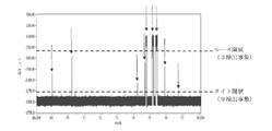


Classifications
-
- G—PHYSICS
- G01—MEASURING; TESTING
- G01N—INVESTIGATING OR ANALYSING MATERIALS BY DETERMINING THEIR CHEMICAL OR PHYSICAL PROPERTIES
- G01N27/00—Investigating or analysing materials by the use of electric, electrochemical, or magnetic means
- G01N27/26—Investigating or analysing materials by the use of electric, electrochemical, or magnetic means by investigating electrochemical variables; by using electrolysis or electrophoresis
- G01N27/28—Electrolytic cell components
- G01N27/30—Electrodes, e.g. test electrodes; Half-cells
- G01N27/327—Biochemical electrodes, e.g. electrical or mechanical details for in vitro measurements
- G01N27/3271—Amperometric enzyme electrodes for analytes in body fluids, e.g. glucose in blood
- G01N27/3272—Test elements therefor, i.e. disposable laminated substrates with electrodes, reagent and channels
-
- C—CHEMISTRY; METALLURGY
- C12—BIOCHEMISTRY; BEER; SPIRITS; WINE; VINEGAR; MICROBIOLOGY; ENZYMOLOGY; MUTATION OR GENETIC ENGINEERING
- C12Q—MEASURING OR TESTING PROCESSES INVOLVING ENZYMES, NUCLEIC ACIDS OR MICROORGANISMS; COMPOSITIONS OR TEST PAPERS THEREFOR; PROCESSES OF PREPARING SUCH COMPOSITIONS; CONDITION-RESPONSIVE CONTROL IN MICROBIOLOGICAL OR ENZYMOLOGICAL PROCESSES
- C12Q1/00—Measuring or testing processes involving enzymes, nucleic acids or microorganisms; Compositions therefor; Processes of preparing such compositions
- C12Q1/001—Enzyme electrodes
- C12Q1/004—Enzyme electrodes mediator-assisted
-
- B—PERFORMING OPERATIONS; TRANSPORTING
- B01—PHYSICAL OR CHEMICAL PROCESSES OR APPARATUS IN GENERAL
- B01L—CHEMICAL OR PHYSICAL LABORATORY APPARATUS FOR GENERAL USE
- B01L3/00—Containers or dishes for laboratory use, e.g. laboratory glassware; Droppers
- B01L3/50—Containers for the purpose of retaining a material to be analysed, e.g. test tubes
- B01L3/502—Containers for the purpose of retaining a material to be analysed, e.g. test tubes with fluid transport, e.g. in multi-compartment structures
- B01L3/5027—Containers for the purpose of retaining a material to be analysed, e.g. test tubes with fluid transport, e.g. in multi-compartment structures by integrated microfluidic structures, i.e. dimensions of channels and chambers are such that surface tension forces are important, e.g. lab-on-a-chip
- B01L3/502715—Containers for the purpose of retaining a material to be analysed, e.g. test tubes with fluid transport, e.g. in multi-compartment structures by integrated microfluidic structures, i.e. dimensions of channels and chambers are such that surface tension forces are important, e.g. lab-on-a-chip characterised by interfacing components, e.g. fluidic, electrical, optical or mechanical interfaces
-
- B—PERFORMING OPERATIONS; TRANSPORTING
- B01—PHYSICAL OR CHEMICAL PROCESSES OR APPARATUS IN GENERAL
- B01L—CHEMICAL OR PHYSICAL LABORATORY APPARATUS FOR GENERAL USE
- B01L3/00—Containers or dishes for laboratory use, e.g. laboratory glassware; Droppers
- B01L3/50—Containers for the purpose of retaining a material to be analysed, e.g. test tubes
- B01L3/502—Containers for the purpose of retaining a material to be analysed, e.g. test tubes with fluid transport, e.g. in multi-compartment structures
- B01L3/5027—Containers for the purpose of retaining a material to be analysed, e.g. test tubes with fluid transport, e.g. in multi-compartment structures by integrated microfluidic structures, i.e. dimensions of channels and chambers are such that surface tension forces are important, e.g. lab-on-a-chip
- B01L3/50273—Containers for the purpose of retaining a material to be analysed, e.g. test tubes with fluid transport, e.g. in multi-compartment structures by integrated microfluidic structures, i.e. dimensions of channels and chambers are such that surface tension forces are important, e.g. lab-on-a-chip characterised by the means or forces applied to move the fluids
-
- B—PERFORMING OPERATIONS; TRANSPORTING
- B01—PHYSICAL OR CHEMICAL PROCESSES OR APPARATUS IN GENERAL
- B01L—CHEMICAL OR PHYSICAL LABORATORY APPARATUS FOR GENERAL USE
- B01L3/00—Containers or dishes for laboratory use, e.g. laboratory glassware; Droppers
- B01L3/50—Containers for the purpose of retaining a material to be analysed, e.g. test tubes
- B01L3/502—Containers for the purpose of retaining a material to be analysed, e.g. test tubes with fluid transport, e.g. in multi-compartment structures
- B01L3/5027—Containers for the purpose of retaining a material to be analysed, e.g. test tubes with fluid transport, e.g. in multi-compartment structures by integrated microfluidic structures, i.e. dimensions of channels and chambers are such that surface tension forces are important, e.g. lab-on-a-chip
- B01L3/502761—Containers for the purpose of retaining a material to be analysed, e.g. test tubes with fluid transport, e.g. in multi-compartment structures by integrated microfluidic structures, i.e. dimensions of channels and chambers are such that surface tension forces are important, e.g. lab-on-a-chip specially adapted for handling suspended solids or molecules independently from the bulk fluid flow, e.g. for trapping or sorting beads, for physically stretching molecules
-
- B—PERFORMING OPERATIONS; TRANSPORTING
- B01—PHYSICAL OR CHEMICAL PROCESSES OR APPARATUS IN GENERAL
- B01L—CHEMICAL OR PHYSICAL LABORATORY APPARATUS FOR GENERAL USE
- B01L3/00—Containers or dishes for laboratory use, e.g. laboratory glassware; Droppers
- B01L3/50—Containers for the purpose of retaining a material to be analysed, e.g. test tubes
- B01L3/502—Containers for the purpose of retaining a material to be analysed, e.g. test tubes with fluid transport, e.g. in multi-compartment structures
- B01L3/5027—Containers for the purpose of retaining a material to be analysed, e.g. test tubes with fluid transport, e.g. in multi-compartment structures by integrated microfluidic structures, i.e. dimensions of channels and chambers are such that surface tension forces are important, e.g. lab-on-a-chip
- B01L3/502769—Containers for the purpose of retaining a material to be analysed, e.g. test tubes with fluid transport, e.g. in multi-compartment structures by integrated microfluidic structures, i.e. dimensions of channels and chambers are such that surface tension forces are important, e.g. lab-on-a-chip characterised by multiphase flow arrangements
- B01L3/502784—Containers for the purpose of retaining a material to be analysed, e.g. test tubes with fluid transport, e.g. in multi-compartment structures by integrated microfluidic structures, i.e. dimensions of channels and chambers are such that surface tension forces are important, e.g. lab-on-a-chip characterised by multiphase flow arrangements specially adapted for droplet or plug flow, e.g. digital microfluidics
-
- C—CHEMISTRY; METALLURGY
- C12—BIOCHEMISTRY; BEER; SPIRITS; WINE; VINEGAR; MICROBIOLOGY; ENZYMOLOGY; MUTATION OR GENETIC ENGINEERING
- C12Q—MEASURING OR TESTING PROCESSES INVOLVING ENZYMES, NUCLEIC ACIDS OR MICROORGANISMS; COMPOSITIONS OR TEST PAPERS THEREFOR; PROCESSES OF PREPARING SUCH COMPOSITIONS; CONDITION-RESPONSIVE CONTROL IN MICROBIOLOGICAL OR ENZYMOLOGICAL PROCESSES
- C12Q1/00—Measuring or testing processes involving enzymes, nucleic acids or microorganisms; Compositions therefor; Processes of preparing such compositions
- C12Q1/68—Measuring or testing processes involving enzymes, nucleic acids or microorganisms; Compositions therefor; Processes of preparing such compositions involving nucleic acids
- C12Q1/6869—Methods for sequencing
-
- C—CHEMISTRY; METALLURGY
- C40—COMBINATORIAL TECHNOLOGY
- C40B—COMBINATORIAL CHEMISTRY; LIBRARIES, e.g. CHEMICAL LIBRARIES
- C40B60/00—Apparatus specially adapted for use in combinatorial chemistry or with libraries
- C40B60/12—Apparatus specially adapted for use in combinatorial chemistry or with libraries for screening libraries
-
- G—PHYSICS
- G01—MEASURING; TESTING
- G01N—INVESTIGATING OR ANALYSING MATERIALS BY DETERMINING THEIR CHEMICAL OR PHYSICAL PROPERTIES
- G01N33/00—Investigating or analysing materials by specific methods not covered by groups G01N1/00 - G01N31/00
- G01N33/48—Biological material, e.g. blood, urine; Haemocytometers
- G01N33/483—Physical analysis of biological material
- G01N33/487—Physical analysis of biological material of liquid biological material
- G01N33/48707—Physical analysis of biological material of liquid biological material by electrical means
- G01N33/48721—Investigating individual macromolecules, e.g. by translocation through nanopores
-
- G—PHYSICS
- G01—MEASURING; TESTING
- G01N—INVESTIGATING OR ANALYSING MATERIALS BY DETERMINING THEIR CHEMICAL OR PHYSICAL PROPERTIES
- G01N33/00—Investigating or analysing materials by specific methods not covered by groups G01N1/00 - G01N31/00
- G01N33/48—Biological material, e.g. blood, urine; Haemocytometers
- G01N33/50—Chemical analysis of biological material, e.g. blood, urine; Testing involving biospecific ligand binding methods; Immunological testing
- G01N33/53—Immunoassay; Biospecific binding assay; Materials therefor
- G01N33/543—Immunoassay; Biospecific binding assay; Materials therefor with an insoluble carrier for immobilising immunochemicals
- G01N33/54313—Immunoassay; Biospecific binding assay; Materials therefor with an insoluble carrier for immobilising immunochemicals the carrier being characterised by its particulate form
- G01N33/54326—Magnetic particles
-
- G—PHYSICS
- G01—MEASURING; TESTING
- G01N—INVESTIGATING OR ANALYSING MATERIALS BY DETERMINING THEIR CHEMICAL OR PHYSICAL PROPERTIES
- G01N33/00—Investigating or analysing materials by specific methods not covered by groups G01N1/00 - G01N31/00
- G01N33/48—Biological material, e.g. blood, urine; Haemocytometers
- G01N33/50—Chemical analysis of biological material, e.g. blood, urine; Testing involving biospecific ligand binding methods; Immunological testing
- G01N33/53—Immunoassay; Biospecific binding assay; Materials therefor
- G01N33/543—Immunoassay; Biospecific binding assay; Materials therefor with an insoluble carrier for immobilising immunochemicals
- G01N33/54366—Apparatus specially adapted for solid-phase testing
- G01N33/54373—Apparatus specially adapted for solid-phase testing involving physiochemical end-point determination, e.g. wave-guides, FETS, gratings
- G01N33/5438—Electrodes
-
- B—PERFORMING OPERATIONS; TRANSPORTING
- B01—PHYSICAL OR CHEMICAL PROCESSES OR APPARATUS IN GENERAL
- B01L—CHEMICAL OR PHYSICAL LABORATORY APPARATUS FOR GENERAL USE
- B01L2200/00—Solutions for specific problems relating to chemical or physical laboratory apparatus
- B01L2200/06—Fluid handling related problems
- B01L2200/0647—Handling flowable solids, e.g. microscopic beads, cells, particles
-
- B—PERFORMING OPERATIONS; TRANSPORTING
- B01—PHYSICAL OR CHEMICAL PROCESSES OR APPARATUS IN GENERAL
- B01L—CHEMICAL OR PHYSICAL LABORATORY APPARATUS FOR GENERAL USE
- B01L2200/00—Solutions for specific problems relating to chemical or physical laboratory apparatus
- B01L2200/10—Integrating sample preparation and analysis in single entity, e.g. lab-on-a-chip concept
-
- B—PERFORMING OPERATIONS; TRANSPORTING
- B01—PHYSICAL OR CHEMICAL PROCESSES OR APPARATUS IN GENERAL
- B01L—CHEMICAL OR PHYSICAL LABORATORY APPARATUS FOR GENERAL USE
- B01L2300/00—Additional constructional details
- B01L2300/08—Geometry, shape and general structure
- B01L2300/0809—Geometry, shape and general structure rectangular shaped
- B01L2300/0816—Cards, e.g. flat sample carriers usually with flow in two horizontal directions
-
- B—PERFORMING OPERATIONS; TRANSPORTING
- B01—PHYSICAL OR CHEMICAL PROCESSES OR APPARATUS IN GENERAL
- B01L—CHEMICAL OR PHYSICAL LABORATORY APPARATUS FOR GENERAL USE
- B01L2300/00—Additional constructional details
- B01L2300/08—Geometry, shape and general structure
- B01L2300/0861—Configuration of multiple channels and/or chambers in a single devices
- B01L2300/0874—Three dimensional network
-
- B—PERFORMING OPERATIONS; TRANSPORTING
- B01—PHYSICAL OR CHEMICAL PROCESSES OR APPARATUS IN GENERAL
- B01L—CHEMICAL OR PHYSICAL LABORATORY APPARATUS FOR GENERAL USE
- B01L2300/00—Additional constructional details
- B01L2300/16—Surface properties and coatings
-
- B—PERFORMING OPERATIONS; TRANSPORTING
- B01—PHYSICAL OR CHEMICAL PROCESSES OR APPARATUS IN GENERAL
- B01L—CHEMICAL OR PHYSICAL LABORATORY APPARATUS FOR GENERAL USE
- B01L2300/00—Additional constructional details
- B01L2300/16—Surface properties and coatings
- B01L2300/161—Control and use of surface tension forces, e.g. hydrophobic, hydrophilic
- B01L2300/165—Specific details about hydrophobic, oleophobic surfaces
-
- B—PERFORMING OPERATIONS; TRANSPORTING
- B01—PHYSICAL OR CHEMICAL PROCESSES OR APPARATUS IN GENERAL
- B01L—CHEMICAL OR PHYSICAL LABORATORY APPARATUS FOR GENERAL USE
- B01L2400/00—Moving or stopping fluids
- B01L2400/04—Moving fluids with specific forces or mechanical means
- B01L2400/0403—Moving fluids with specific forces or mechanical means specific forces
- B01L2400/0406—Moving fluids with specific forces or mechanical means specific forces capillary forces
-
- B—PERFORMING OPERATIONS; TRANSPORTING
- B01—PHYSICAL OR CHEMICAL PROCESSES OR APPARATUS IN GENERAL
- B01L—CHEMICAL OR PHYSICAL LABORATORY APPARATUS FOR GENERAL USE
- B01L2400/00—Moving or stopping fluids
- B01L2400/04—Moving fluids with specific forces or mechanical means
- B01L2400/0403—Moving fluids with specific forces or mechanical means specific forces
- B01L2400/0415—Moving fluids with specific forces or mechanical means specific forces electrical forces, e.g. electrokinetic
- B01L2400/0418—Moving fluids with specific forces or mechanical means specific forces electrical forces, e.g. electrokinetic electro-osmotic flow [EOF]
-
- B—PERFORMING OPERATIONS; TRANSPORTING
- B01—PHYSICAL OR CHEMICAL PROCESSES OR APPARATUS IN GENERAL
- B01L—CHEMICAL OR PHYSICAL LABORATORY APPARATUS FOR GENERAL USE
- B01L2400/00—Moving or stopping fluids
- B01L2400/04—Moving fluids with specific forces or mechanical means
- B01L2400/0403—Moving fluids with specific forces or mechanical means specific forces
- B01L2400/0415—Moving fluids with specific forces or mechanical means specific forces electrical forces, e.g. electrokinetic
- B01L2400/0427—Electrowetting
-
- B—PERFORMING OPERATIONS; TRANSPORTING
- B82—NANOTECHNOLOGY
- B82Y—SPECIFIC USES OR APPLICATIONS OF NANOSTRUCTURES; MEASUREMENT OR ANALYSIS OF NANOSTRUCTURES; MANUFACTURE OR TREATMENT OF NANOSTRUCTURES
- B82Y15/00—Nanotechnology for interacting, sensing or actuating, e.g. quantum dots as markers in protein assays or molecular motors
-
- G—PHYSICS
- G01—MEASURING; TESTING
- G01N—INVESTIGATING OR ANALYSING MATERIALS BY DETERMINING THEIR CHEMICAL OR PHYSICAL PROPERTIES
- G01N21/00—Investigating or analysing materials by the use of optical means, i.e. using sub-millimetre waves, infrared, visible or ultraviolet light
- G01N21/62—Systems in which the material investigated is excited whereby it emits light or causes a change in wavelength of the incident light
- G01N21/63—Systems in which the material investigated is excited whereby it emits light or causes a change in wavelength of the incident light optically excited
- G01N21/64—Fluorescence; Phosphorescence
- G01N21/645—Specially adapted constructive features of fluorimeters
- G01N2021/6482—Sample cells, cuvettes
-
- G—PHYSICS
- G01—MEASURING; TESTING
- G01N—INVESTIGATING OR ANALYSING MATERIALS BY DETERMINING THEIR CHEMICAL OR PHYSICAL PROPERTIES
- G01N21/00—Investigating or analysing materials by the use of optical means, i.e. using sub-millimetre waves, infrared, visible or ultraviolet light
- G01N21/75—Systems in which material is subjected to a chemical reaction, the progress or the result of the reaction being investigated
- G01N21/77—Systems in which material is subjected to a chemical reaction, the progress or the result of the reaction being investigated by observing the effect on a chemical indicator
- G01N21/82—Systems in which material is subjected to a chemical reaction, the progress or the result of the reaction being investigated by observing the effect on a chemical indicator producing a precipitate or turbidity
- G01N2021/825—Agglutination
-
- G—PHYSICS
- G01—MEASURING; TESTING
- G01N—INVESTIGATING OR ANALYSING MATERIALS BY DETERMINING THEIR CHEMICAL OR PHYSICAL PROPERTIES
- G01N21/00—Investigating or analysing materials by the use of optical means, i.e. using sub-millimetre waves, infrared, visible or ultraviolet light
- G01N21/75—Systems in which material is subjected to a chemical reaction, the progress or the result of the reaction being investigated
- G01N21/77—Systems in which material is subjected to a chemical reaction, the progress or the result of the reaction being investigated by observing the effect on a chemical indicator
- G01N21/78—Systems in which material is subjected to a chemical reaction, the progress or the result of the reaction being investigated by observing the effect on a chemical indicator producing a change of colour
-
- G—PHYSICS
- G01—MEASURING; TESTING
- G01N—INVESTIGATING OR ANALYSING MATERIALS BY DETERMINING THEIR CHEMICAL OR PHYSICAL PROPERTIES
- G01N2458/00—Labels used in chemical analysis of biological material
- G01N2458/10—Oligonucleotides as tagging agents for labelling antibodies
-
- G—PHYSICS
- G01—MEASURING; TESTING
- G01N—INVESTIGATING OR ANALYSING MATERIALS BY DETERMINING THEIR CHEMICAL OR PHYSICAL PROPERTIES
- G01N27/00—Investigating or analysing materials by the use of electric, electrochemical, or magnetic means
- G01N27/26—Investigating or analysing materials by the use of electric, electrochemical, or magnetic means by investigating electrochemical variables; by using electrolysis or electrophoresis
- G01N27/28—Electrolytic cell components
- G01N27/30—Electrodes, e.g. test electrodes; Half-cells
- G01N27/327—Biochemical electrodes, e.g. electrical or mechanical details for in vitro measurements
- G01N27/3271—Amperometric enzyme electrodes for analytes in body fluids, e.g. glucose in blood
-
- G—PHYSICS
- G01—MEASURING; TESTING
- G01N—INVESTIGATING OR ANALYSING MATERIALS BY DETERMINING THEIR CHEMICAL OR PHYSICAL PROPERTIES
- G01N33/00—Investigating or analysing materials by specific methods not covered by groups G01N1/00 - G01N31/00
- G01N33/48—Biological material, e.g. blood, urine; Haemocytometers
- G01N33/483—Physical analysis of biological material
- G01N33/487—Physical analysis of biological material of liquid biological material
- G01N33/48707—Physical analysis of biological material of liquid biological material by electrical means
Landscapes
- Health & Medical Sciences (AREA)
- Chemical & Material Sciences (AREA)
- Life Sciences & Earth Sciences (AREA)
- Engineering & Computer Science (AREA)
- Immunology (AREA)
- Hematology (AREA)
- Analytical Chemistry (AREA)
- General Health & Medical Sciences (AREA)
- Physics & Mathematics (AREA)
- Molecular Biology (AREA)
- Biomedical Technology (AREA)
- Organic Chemistry (AREA)
- Biochemistry (AREA)
- Urology & Nephrology (AREA)
- Chemical Kinetics & Catalysis (AREA)
- Dispersion Chemistry (AREA)
- Proteomics, Peptides & Aminoacids (AREA)
- Clinical Laboratory Science (AREA)
- Microbiology (AREA)
- General Physics & Mathematics (AREA)
- Pathology (AREA)
- Biotechnology (AREA)
- Medicinal Chemistry (AREA)
- Wood Science & Technology (AREA)
- Zoology (AREA)
- Biophysics (AREA)
- Food Science & Technology (AREA)
- Cell Biology (AREA)
- General Engineering & Computer Science (AREA)
- Genetics & Genomics (AREA)
- Bioinformatics & Cheminformatics (AREA)
- Nanotechnology (AREA)
- Spectroscopy & Molecular Physics (AREA)
- Fluid Mechanics (AREA)
- Electrochemistry (AREA)
- General Chemical & Material Sciences (AREA)
- Apparatus Associated With Microorganisms And Enzymes (AREA)
- Investigating Or Analyzing Materials By The Use Of Electric Means (AREA)
- Automatic Analysis And Handling Materials Therefor (AREA)
- Measuring Or Testing Involving Enzymes Or Micro-Organisms (AREA)
Abstract
Description
関連出願の相互参照
本出願は、2016年10月5日に出願された米国仮出願第62/404,722号および2016年11月21日に出願された米国仮出願第62/424,996号の優先権を主張し、これらの出願の開示は、参照により本明細書に組み込まれる。
Mutual Reference of Related Applications This application is a US provisional application No. 62 / 404,722 filed on October 5, 2016 and a US provisional application No. 62 / 424,996 filed on November 21, 2016. The disclosures of these applications are incorporated herein by reference.
本開示は、例えば、マイクロ流体デバイスと動作可能に結合された検体検出デバイスを使用する、検体分析のための方法、デバイス、およびシステムに関する。 The present disclosure relates to methods, devices, and systems for sample analysis, eg, using a sample detection device operably coupled to a microfluidic device.
試料中の目的の検体(複数可)を正確に分析することができる方法およびデバイスは、診断、予後診断、環境評価、食品の安全性、化学兵器または生物兵器の検出等に不可欠である。そのような方法およびデバイスは、正確で、精密、かつ高感度である必要があるだけでなく、微量試料が迅速にかつ最小の器具使用で分析されるときにも有利である。そのようなものとして、改良された試料分析能力を有する方法およびデバイスに関心がある。 Methods and devices capable of accurately analyzing the desired sample (s) in the sample are essential for diagnosis, prognostic diagnosis, environmental assessment, food safety, detection of chemical or biological weapons, and the like. Not only do such methods and devices need to be accurate, precise and sensitive, but they are also advantageous when trace samples are analyzed quickly and with minimal instrumentation. As such, we are interested in methods and devices with improved sample analysis capabilities.
本開示の実施形態は、試料中の検体(複数可)の分析のための方法、システム、およびデバイスに関する。ある特定の実施形態では、試料は、生物学的試料であり得る。 Embodiments of the present disclosure relate to methods, systems, and devices for the analysis of a sample (s) in a sample. In certain embodiments, the sample can be a biological sample.
試料中の検体の分析のための方法は、試料を第1の結合メンバーと接触させることであって、第1の結合メンバーが、固体支持体上に固定化され、第1の結合メンバーが、検体に特異的に結合する、接触させることと、固体支持体を第2の結合メンバーと接触させることであって、第2の結合メンバーが、検体に特異的に結合し、第2の結合メンバーが、それに付着した切断可能なタグを含む、接触させることと、第1の結合メンバーに結合した検体に結合していない第2の結合メンバーを除去することと、第1の結合メンバーに結合した検体に結合している第2の結合メンバーに付着したタグを切断することと、切断されたタグを層内のナノポアを通してまたはそれを横切って転位させることと、層を通って転位するタグの数を決定することと、層を通って転位するタグの数に基づいて試料中の検体の濃度を決定することと、を必要とし得る。ある特定の実施形態では、検体の濃度は、単位時間当たりに層を通って転位するタグの数をカウントすることによって決定され得る。他の実施形態では、検体の濃度は、層を通って転位するタグの数が閾値に達する時間を決定することによって、または期間を設定し、その設定された期間内の累積カウント数をカウントすることによって決定され得る。 A method for analyzing a sample in a sample is to contact the sample with a first binding member, the first binding member being immobilized on a solid support and the first binding member being. By specifically binding and contacting the sample and by contacting the solid support with the second binding member, the second binding member specifically binds to and contacts the sample and the second binding member. Includes a cleavable tag attached to it, contacting, removing the second binding member that is not bound to the sample bound to the first binding member, and binding to the first binding member. Cutting the tag attached to the second binding member bound to the sample, shifting the cut tag through or across the nanopores in the layer, and the number of tags translocated through the layer. And to determine the concentration of the sample in the sample based on the number of tags transposed through the layer. In certain embodiments, the concentration of the specimen can be determined by counting the number of tags translocated through the layer per unit time. In other embodiments, the concentration of the specimen counts the cumulative count within that set time period, either by determining the time at which the number of tags translocated through the layer reaches a threshold, or by setting a time period. Can be determined by
別の実施形態では、方法は、標的検体を含有する試料を既知量の標的検体または競合物分子と組み合わせることを含んでもよく、標的検体(試料と組み合わされる)または競合物分子は、切断可能なリンカーを介してタグに付着して、タグ付き検体またはタグ付き競合物分子をそれぞれ産生し、タグ付き検体またはタグ付き競合物分子は、第1の結合メンバーへの結合について標的検体と競合する。この方法はさらに、組み合わせた試料を第1の結合メンバーと接触させることであって、第1の結合メンバーが、固体支持体上に固定化され、第1の結合メンバーが、標的検体(およびタグ付き検体またはタグ付き競合物分子)に特異的に結合する、接触させることと、任意の洗浄ステップのために固体支持体を緩衝液と接触させることと、固体支持体上に固定化された第1の結合メンバーに結合しているタグ付き検体またはタグ付き競合物に付着しているタグを切断することと、切断されたタグを層内のナノポアを通してまたはそれを横切って転位させることと、層を通って転位するタグの数を決定することと、層を通って転位するタグの数に基づいて試料中の検体の濃度を決定することと、を含み得る。ある特定の実施形態では、検体の濃度は、単位時間当たりに層を通って転位するタグの数をカウントすることによって決定され得る。他の実施形態では、検体の濃度は、層を通って転位するタグの数が閾値に達する時間を決定することによって、または期間を設定し、その設定された期間内の累積カウント数をカウントすることによって決定され得る。この実施形態では、ナノポアを通って転位するタグの数、または層を通って転位するタグの数が閾値に達する時間は、試料中の検体の濃度と逆相関し得る。例えば、カウント数が少ないほど、または閾値に達する時間が長いほど、試料中の標的検体の濃度は高くなる。 In another embodiment, the method may comprise combining a sample containing the target sample with a known amount of the target sample or competitor molecule, the target sample (combined with the sample) or the competitor molecule can be cleaved. Attached to the tag via the linker to produce a tagged sample or tagged competitor molecule, respectively, the tagged sample or tagged competitor molecule competes with the target sample for binding to the first binding member. The method is further to contact the combined sample with a first binding member, where the first binding member is immobilized on a solid support and the first binding member is the target sample (and tag). Specific binding to (attached sample or tagged competitor molecule), contacting, contacting the solid support with buffer for any washing step, and immobilization on the solid support. Cutting a tag attached to a tagged sample or tagged competitor bound to a binding member of 1 and translocating the cut tag through or across a nanopore in the layer, and the layer. It may include determining the number of tags translocated through the layer and determining the concentration of the sample in the sample based on the number of tags translocated through the layer. In certain embodiments, the concentration of the specimen can be determined by counting the number of tags translocated through the layer per unit time. In other embodiments, the concentration of the specimen counts the cumulative count within that set time period, either by determining the time at which the number of tags translocated through the layer reaches a threshold, or by setting a time period. Can be determined by In this embodiment, the number of tags that dislocate through the nanopores, or the time that the number of tags that dislocate through the layer reaches the threshold, can be inversely correlated with the concentration of the sample in the sample. For example, the smaller the count number or the longer the time to reach the threshold value, the higher the concentration of the target sample in the sample.
一態様では、本発明は、生物学的試料中に存在する検体を測定または検出するための方法に関する。この方法は、試料を第1の結合メンバーと接触させることであって、第1の結合メンバーが、固体支持体上に固定化され、第1の結合メンバーが、検体に特異的に結合する、接触させることと、検体を第2の結合メンバーと接触させることであって、第2の結合メンバーが、検体に特異的に結合し、第2の結合メンバーが、それに付着した切断可能なタグを含む、接触させることと、第1の結合メンバーに結合した検体に結合していない第2の結合メンバーを除去することと、第1の結合メンバーに結合した検体に結合している第2の結合メンバーに付着したタグを切断することと、切断されたタグを層内の1つ以上のナノポアを通してまたはそれを横切って転位させることと、層を通って転位するタグを評価することと、を含み、層を通って転位するタグの数を測定することが、試料中に存在する検体の量を測定するか、または層を通って転位するタグを検出することが、検体が試料中に存在することを検出する。いくつかの実施形態では、層を通って転位するタグを測定することが評価され、層を通って転位するタグの数が、試料中に存在する検体の量を測定する。いくつかの実施形態では、層を通って転位するタグを検出することが評価され、層を通って転位するタグを検出することは、検体が試料中に存在することを検出する。 In one aspect, the invention relates to a method for measuring or detecting a sample present in a biological sample. This method involves contacting the sample with a first binding member, wherein the first binding member is immobilized on a solid support and the first binding member specifically binds to the sample. Contacting and contacting the sample with the second binding member, where the second binding member specifically binds to the sample and the second binding member attaches a cleavable tag to it. Including, contacting, removing the second binding member that is not bound to the sample bound to the first binding member, and the second binding that is bound to the sample bound to the first binding member. Includes cutting the tag attached to the member, shifting the cut tag through or across one or more nanopores in the layer, and evaluating the tag that shifts through the layer. , Measuring the number of tags that dislocate through the layer can measure the amount of sample present in the sample, or detecting tags that dislocate through the layer can be present in the sample. Detect that. In some embodiments, measuring the tags that dislocate through the layer is evaluated, and the number of tags that dislocate through the layer measures the amount of sample present in the sample. In some embodiments, detecting a tag that dislocates through a layer is evaluated, and detecting a tag that dislocates through a layer detects the presence of a sample in the sample.
一態様では、本発明は、生物学的試料中に存在する検体を測定または検出するための方法に関する。この方法は、試料を第1の結合メンバーと接触させることであって、第1の結合メンバーが、固体支持体上に固定化され、第1の結合メンバーが、検体に特異的に結合する、接触させることと、検体を第2の結合メンバーと接触させることであって、第2の結合メンバーが、検体に特異的に結合し、第2の結合メンバーが、アプタマーを含む、接触させることと、固体基質に結合した検体に結合していないアプタマーを除去することと、検体に結合したアプタマーを解離させ、解離したアプタマーを層内の1つ以上のナノポアを通してまたはそれを横切って転位させることと、層を通って転位するアプタマーを評価することと、を含み、層を通って転位するアプタマーの数を測定することが、試料中に存在する検体の量を測定するか、または層を通って転位するアプタマーを検出することが、検体が試料中に存在することを検出する。いくつかの実施形態では、層を通って転位するアプタマーを測定することが評価され、層を通って転位するアプタマーの数は、試料中に存在する検体の量を測定する。いくつかの実施形態では、層を通って転位するアプタマーを検出することが評価され、層を通って転位するアプタマーを検出することは、検体が試料中に存在することを検出する。 In one aspect, the invention relates to a method for measuring or detecting a sample present in a biological sample. This method involves contacting the sample with a first binding member, wherein the first binding member is immobilized on a solid support and the first binding member specifically binds to the sample. Contacting and contacting the sample with a second binding member, wherein the second binding member specifically binds to the sample and the second binding member comprises an aptamer. To remove aptamers that are not bound to a sample bound to a solid substrate, to dissociate the aptamers bound to the sample, and to dislocate the dissociated aptamers through or across one or more nanopores in the layer. Evaluating aptamers that dislocate through a layer, including measuring the number of aptamers that dislocate through a layer, measures the amount of sample present in the sample, or through the layer. Detecting a transposed aptamer detects the presence of a sample in the sample. In some embodiments, measuring the aptamer that translocates through the layer is evaluated, and the number of aptamers that translocate through the layer measures the amount of sample present in the sample. In some embodiments, detecting an aptamer translocating through a layer is evaluated, and detecting an aptamer translocating through a layer detects the presence of a sample in the sample.
一態様では、本発明は、電極のアレイを含む底部基板、底部基板から離間している上部基板、および底部基板と上部基板との間に配設されたナノポア層を備える、集積デジタルマイクロ流体ナノポアデバイスに関する。このデバイスは、近位部分および遠位部分を含み、ナノポア層は、遠位部分に配設されている。近位部分の電極のアレイは、液滴を生成するように構成されている。電極のアレイは、液滴がナノポア層によって第1の部分と第2の部分とに分割されるように、ナノポア層を横切って液滴を位置付けるように構成され、電極のアレイの少なくとも2つの電極は、ナノポア層を横切って位置付けられ、これらの2つの電極は、アノードおよびカソードを形成し、液滴がナノポア層を横切って位置付けられたときにナノポア層内のナノポアを通して電流を駆動するように動作する。 In one aspect, the invention comprises an integrated digital microfluidic nanopore comprising a bottom substrate comprising an array of electrodes, a top substrate separated from the bottom substrate, and a nanopore layer disposed between the bottom substrate and the top substrate. Regarding the device. The device includes a proximal part and a distal part, and the nanopore layer is disposed in the distal part. The array of electrodes in the proximal portion is configured to produce droplets. The array of electrodes is configured to position the droplet across the nanopore layer so that the droplet is divided into a first portion and a second portion by the nanopore layer, at least two electrodes in the array of electrodes. Is positioned across the nanopore layer, these two electrodes form an anode and cathode and act to drive an electric current through the nanopores within the nanopore layer when the droplet is positioned across the nanopore layer. do.
一態様では、本発明は、電極のアレイを含む底部基板、底部基板から離間し、電極を含む上部基板、および底部基板と上部基板との間に配設されたナノポア層を備える、集積デジタルマイクロ流体ナノポアデバイスに関する。このデバイスは、近位部分および遠位部分を含み、ナノポア層は、遠位部分に配設されている。近位部分の電極のアレイおよび電極は、液滴を生成するように構成されている。電極のアレイおよび電極は、ナノポア層が液滴を第1の部分と第2の部分とに分割するように、ナノポア層を横切って液滴を位置付けるように構成され、電極のアレイの少なくとも1つの電極は、ナノポア層を横切って位置付けられた液滴の第1の部分と接触し、上部基板内の電極は、ナノポア層を横切って位置付けられた液滴の第2の部分と接触するように位置付けられ、2つの電極は、アノードおよびカソードを形成し、液滴がナノポア層を横切って位置付けられたときにナノポア層内のナノポアを通して電流を駆動するように動作する。 In one aspect, the invention comprises an integrated digital micro, comprising a bottom substrate containing an array of electrodes, a top substrate separated from the bottom substrate and containing the electrodes, and a nanopore layer disposed between the bottom substrate and the top substrate. Regarding fluid nanopore devices. The device includes a proximal part and a distal part, and the nanopore layer is disposed in the distal part. The array of electrodes in the proximal portion and the electrodes are configured to generate droplets. The array of electrodes and the electrodes are configured to position the droplets across the nanopore layer such that the nanopore layer divides the droplet into a first portion and a second portion, at least one of the array of electrodes. The electrode is positioned to contact the first portion of the droplet positioned across the nanopore layer, and the electrode in the top substrate is positioned to contact the second portion of the droplet positioned across the nanopore layer. The two electrodes form an anode and a cathode and act to drive an electric current through the nanopores within the nanopore layer when the droplet is positioned across the nanopore layer.
一態様では、本発明は、生物学的試料中に存在する検体を測定または検出するための方法に関する。この方法は、試料を結合メンバーと接触させることであって、結合メンバーが、固体支持体上に固定化され、結合メンバーが、検体に特異的に結合する、接触させることと、結合メンバーに結合される検体を含有し得る試料を、標識検体と接触させることであって、標識検体が、切断可能なタグで標識される、接触させることと、結合メンバーに結合していない標識検体を除去することと、結合メンバーに結合した標識検体に付着したタグを切断することと、切断されたタグを層内の1つ以上のナノポアを通してまたはそれを横切って転位させることと、層を通って転位するタグを評価することと、を含み、層を通って転位するタグの数を測定することが、試料中に存在する検体の量を測定するか、または層を通って転位するタグを検出することが、検体が試料中に存在することを検出する。いくつかの実施形態では、層を通って転位するタグを測定することが評価され、層を通って転位するタグの数が、試料中に存在する検体の量を測定する。いくつかの実施形態では、層を通って転位するタグを検出することが評価され、層を通って転位するタグを検出することは、検体が試料中に存在することを検出する。 In one aspect, the invention relates to a method for measuring or detecting a sample present in a biological sample. This method involves contacting the sample with the binding member, where the binding member is immobilized on a solid support and the binding member specifically binds to, contacts the sample, and binds to the binding member. By contacting a sample that may contain the sample to be labeled with the labeled sample, the labeled sample is labeled with a cleavable tag, contacting and removing the labeled sample that is not bound to the binding member. That, cutting the tag attached to the labeled sample bound to the binding member, shifting the cut tag through or across one or more nanopores in the layer, and shifting through the layer. Evaluating tags and measuring the number of tags that contain and dislocate through the layer can measure the amount of sample present in the sample or detect tags that dislocate through the layer. Detects the presence of the sample in the sample. In some embodiments, measuring the tags that dislocate through the layer is evaluated, and the number of tags that dislocate through the layer measures the amount of sample present in the sample. In some embodiments, detecting a tag that dislocates through a layer is evaluated, and detecting a tag that dislocates through a layer detects the presence of a sample in the sample.
一態様では、本発明は、生物学的試料中に存在する検体を測定または検出するための方法に関する。この方法は、試料を結合メンバーと接触させることであって、結合メンバーが、固体支持体上に固定化され、結合メンバーが、検体に特異的に結合する、接触させることと、結合メンバーに結合される検体を含有し得る試料を、標識検体と接触させることであって、標識検体が、アプタマーを含む、接触させることと、結合メンバーに結合していない標識検体を除去することと、結合メンバーに結合した標識検体に結合したアプタマーを解離させ、解離したアプタマーを層内の1つ以上のナノポアを通してまたはそれを横切って転位させることと、層を通って転位するアプタマーを評価することと、を含み、層を通って転位するアプタマーの数を測定することが、試料中に存在する検体の量を測定するか、または層を通って転位するアプタマーを検出することが、検体が試料中に存在することを検出する。いくつかの実施形態では、層を通って転位するアプタマーを測定することが評価され、層を通って転位するアプタマーの数は、試料中に存在する検体の量を測定する。いくつかの実施形態では、層を通って転位するアプタマーを検出することが評価され、層を通って転位するアプタマーを検出することは、検体が試料中に存在することを検出する。 In one aspect, the invention relates to a method for measuring or detecting a sample present in a biological sample. This method involves contacting the sample with the binding member, where the binding member is immobilized on a solid support and the binding member specifically binds to, contacts the sample, and binds to the binding member. By contacting a sample that may contain a sample to be labeled with a labeled sample, the labeled sample contains an aptamer, contacting, removing a labeled sample that is not bound to a binding member, and binding members. Dissociating the aptamer bound to the labeled sample bound to and translocating the dissociated aptamer through or across one or more nanopores in the layer and evaluating the aptamer translocating through the layer. The sample is present in the sample, including, measuring the number of aptamers translocating through the layer, measuring the amount of sample present in the sample, or detecting the aptamer translocating through the layer. Detect what to do. In some embodiments, measuring the aptamer that translocates through the layer is evaluated, and the number of aptamers that translocate through the layer measures the amount of sample present in the sample. In some embodiments, detecting an aptamer translocating through a layer is evaluated, and detecting an aptamer translocating through a layer detects the presence of a sample in the sample.
一態様では、本発明は、生物学的試料中に存在する検体を測定または検出するための方法に関する。この方法は、試料を結合メンバーと接触させることであって、結合メンバーが、検体に特異的に結合し、結合メンバーが、切断可能なタグで標識される、接触させることと、結合メンバーに結合される検体を含有し得る試料を、固定化された検体と接触させることであって、固定化された検体が、固体支持体上で固定化される、接触させることと、固定化された検体に結合していない結合メンバーを除去することと、固定化された検体に結合した結合メンバーに付着したタグを切断することと、切断されたタグを層内の1つ以上のナノポアを通してまたはそれを横切って転位させることと、層を通って転位するタグを評価することと、を含み、層を通って転位するタグの数を測定することが、試料中に存在する検体の量を測定するか、または層を通って転位するタグを検出することが、検体が試料中に存在することを検出する。いくつかの実施形態では、層を通って転位するタグを測定することが評価され、層を通って転位するタグの数が、試料中に存在する検体の量を測定する。いくつかの実施形態では、層を通って転位するタグが評価され、層を通って転位するタグを検出することは、検体が試料中に存在することを検出する。 In one aspect, the invention relates to a method for measuring or detecting a sample present in a biological sample. The method is to contact the sample with the binding member, where the binding member specifically binds to the sample and the binding member is labeled with a cleavable tag, contacting and binding to the binding member. By contacting a sample that may contain the sample to be subjected to contacting the immobilized sample, the immobilized sample is immobilized on a solid support, contacted, and the immobilized sample. Removing binding members that are not bound to, cutting tags attached to binding members bound to the immobilized sample, and passing the cut tags through or through one or more nanopores in the layer. Whether measuring the number of tags translocating through a layer, including translocating across and evaluating tags translocating through a layer, measures the amount of sample present in the sample. , Or by detecting a tag that translocates through a layer, detects the presence of the sample in the sample. In some embodiments, measuring the tags that dislocate through the layer is evaluated, and the number of tags that dislocate through the layer measures the amount of sample present in the sample. In some embodiments, tags that dislocate through the layer are evaluated, and detecting tags that dislocate through the layer detects the presence of the sample in the sample.
一態様では、本発明は、生物学的試料中に存在する検体を測定または検出するための方法に関する。この方法は、試料を結合メンバーと接触させることであって、結合メンバーが、検体に特異的に結合し、結合メンバーが、アプタマーを含む、接触させることと、結合メンバーに結合される検体を含有し得る試料を、固定化された検体と接触させることであって、固定化された検体が、固体支持体上で固定化される、接触させることと、固定化された検体に結合していない結合メンバーを除去することと、固定化された検体に結合した結合メンバーに結合したアプタマーを解離させ、解離したアプタマーを層内の1つ以上のナノポアを通してまたはそれを横切って転位させることと、層を通って転位するアプタマーを評価することと、を含み、層を通って転位するアプタマーの数を測定することが、試料中に存在する検体の量を測定するか、または層を通って転位するアプタマーを検出することが、検体が試料中に存在することを検出する。いくつかの実施形態では、層を通って転位するアプタマーを測定することが評価され、層を通って転位するアプタマーの数は、試料中に存在する検体の量を測定する。いくつかの実施形態では、層を通って転位するアプタマーを検出することが評価され、層を通って転位するアプタマーを検出することが、検体が試料中に存在することを検出する。 In one aspect, the invention relates to a method for measuring or detecting a sample present in a biological sample. This method involves contacting the sample with a binding member, wherein the binding member specifically binds to the sample and the binding member comprises contacting, including an aptamer, and the sample bound to the binding member. Possible contact of the sample with the immobilized sample, the immobilized sample being immobilized on a solid support, contacting and not binding to the immobilized sample. Removing the binding member, dissociating the aptamer bound to the binding member bound to the immobilized sample, and translocating the dissociated aptamer through or across one or more nanopores in the layer, and the layer. Evaluating an aptamer translocating through a layer and measuring the number of aptamers translocating through a layer can measure the amount of sample present in the sample or translocate through the layer. Detecting an aptamer detects the presence of a sample in a sample. In some embodiments, measuring the aptamer that translocates through the layer is evaluated, and the number of aptamers that translocate through the layer measures the amount of sample present in the sample. In some embodiments, detecting an aptamer translocating through a layer is evaluated, and detecting an aptamer translocating through a layer detects the presence of a sample in the sample.
ある特定の態様では、タグは、アニオン性ポリマー、カチオン性ポリマー、またはナノ粒子であり得る。ある特定の場合において、タグは、オリゴヌクレオチドポリマーなどのアニオン性ポリマーを含み得る。ある特定の場合において、オリゴヌクレオチドポリマーは、デオキシリボ核酸またはリボ核酸であり得る。ある特定の場合において、オリゴヌクレオチドポリマーは、DNAアプタマーまたはRNAアプタマーであり得、アプタマーは検体に結合しない。例示的な場合において、タグは、正電荷ナノ粒子または負電荷ナノ粒子であり得るナノ粒子を含み得る。 In certain embodiments, the tag can be an anionic polymer, a cationic polymer, or nanoparticles. In certain cases, the tag may include anionic polymers such as oligonucleotide polymers. In certain cases, the oligonucleotide polymer can be deoxyribonucleic acid or ribonucleic acid. In certain cases, the oligonucleotide polymer can be a DNA aptamer or an RNA aptamer, which does not bind to the sample. In an exemplary case, the tag may include nanoparticles that may be positively charged nanoparticles or negatively charged nanoparticles.
ある特定の実施形態では、タグは、デンドリマー、ビーズ、ナノ粒子、例えばナノビーズ等の球状タグであり得る。ある特定の実施形態では、タグは、リボースまたはデオキシリボース単位のポリマー、オリゴヌクレオチド、および核酸、例えばDNAまたはRNAなどの線状もしくは実質的に線状でないか、または細長い形状でない場合がある。 In certain embodiments, the tag can be a spherical tag such as a dendrimer, beads, nanoparticles, eg, nanobeads. In certain embodiments, the tag may not be linear or substantially linear or elongated in shape, such as polymers, oligonucleotides, and nucleic acids, such as ribose or deoxyribose units, such as DNA or RNA.
ある特定の場合において、第1および第2の結合メンバーは、アプタマー、抗体、または受容体であり得る。例えば、第1の結合メンバーは、受容体であり得、第2の結合メンバーは、抗体であり得るか、または第1の結合メンバーは、抗体であり得、第2の結合メンバーは、受容体であり得る。ある特定の例において、第1の結合メンバーは、第1の抗体であり得、第2の結合メンバーは、第2の抗体であり得る。 In certain cases, the first and second binding members can be aptamers, antibodies, or receptors. For example, the first binding member can be a receptor and the second binding member can be an antibody, or the first binding member can be an antibody and the second binding member can be a receptor. Can be. In certain examples, the first binding member can be the first antibody and the second binding member can be the second antibody.
ある特定の例において、タグは、負電荷であり得、転位は、層を横切って正電位を印加することによって、層を横切ってタグを転位させることを含み得る。 In certain examples, the tag can be negatively charged and the dislocation can include translocating the tag across the layer by applying a positive potential across the layer.
ある特定の例において、タグは、正電荷であり得、転位は、層を横切って負電位を印加することによって、タグを層を横切って転位させることを含み得る。 In certain examples, the tag can be positively charged and the dislocation can include translocating the tag across the layer by applying a negative potential across the layer.
他の実施形態では、タグは、核酸であり得、タグは、転位前にタグの配列に相補的な配列を含むオリゴヌクレオチドにハイブリダイズされ得る。 In other embodiments, the tag can be a nucleic acid and the tag can be hybridized to an oligonucleotide containing a sequence complementary to the sequence of the tag prior to translocation.
別の実施形態では、アプタマーを第2の結合メンバーとして使用することによって生物学的試料中に存在する検体を測定するための方法が提供される。例えば、この方法は、試料を第1の結合メンバーと接触させることであって、第1の結合メンバーが、固体支持体上に固定化され、第1の結合メンバーが、検体に特異的に結合する、接触させることと、検体を第2の結合メンバーと接触させることであって、第2の結合メンバーが、検体に特異的に結合し、第2の結合メンバーが、アプタマーを含む、接触させることと、固体基質に結合した検体に結合していないアプタマーを除去することと、固体基質に結合している検体からアプタマーを解離させ、解離したアプタマーを層内のナノポア(複数可)を通して転位させることと、層を通って転位するアプタマーの数を決定することと、層を通って転位するアプタマーの数に基づいて試料中の検体を測定することと、を含み得る。この実施形態では、第2の結合メンバーが、ナノポア(複数可)によって直接検出されるため、第2の結合メンバーは、タグに付着していない。 In another embodiment, a method for measuring a sample present in a biological sample is provided by using the aptamer as a second binding member. For example, this method involves contacting the sample with a first binding member, where the first binding member is immobilized on a solid support and the first binding member specifically binds to the sample. The contacting and contacting the sample with the second binding member, wherein the second binding member specifically binds to the sample and the second binding member comprises an aptamer. That, the aptamer that is not bound to the sample bound to the solid substrate is removed, the aptamer is dissociated from the sample bound to the solid substrate, and the dislocated aptamer is dislocated through the nanopores (s) in the layer. It may include determining the number of aptamers translocating through the layer and measuring the sample in the sample based on the number of aptamers translocating through the layer. In this embodiment, the second binding member is not attached to the tag because the second binding member is directly detected by the nanopores (s).
アプタマーは、DNAアプタマーまたはRNAアプタマーであり得る。第1の結合メンバーは、抗体であり得る。ある特定の例において、検体は、リガンドであり得、第1の結合メンバーは、受容体であり得る。 The aptamer can be a DNA aptamer or an RNA aptamer. The first binding member can be an antibody. In certain examples, the specimen can be a ligand and the first binding member can be a receptor.
試料中の複数の異なる検体を同時に分析するための方法もまた、本明細書に開示されている。例えば、この方法は、第1および第2の検体、第1、第2、および第3の検体等の分析を含み得る。ある特定の場合において、試料中の複数の異なる検体の分析方法は、試料を複数の異なる第1の結合メンバーと接触させることを含み、異なる第1の結合メンバーの第1の結合メンバーは、複数の異なる検体のうちの第1の検体に特異的に結合し、異なる第1の結合メンバーのうちの第2の結合メンバーは、複数の異なる検体のうちの第2の検体に特異的に結合する等である。この方法は、異なる検体を複数の第2の結合メンバーと接触させることをさらに含み得、複数の第2の結合メンバーのうちの第1の結合メンバーは、第1の検体に結合し、複数の第2の結合メンバーのうちの第2の結合メンバーは、第2の検体に結合する等である。ある特定の例において、複数の異なる第2の結合メンバーの各々は、互いに別個であるかまたは区別可能なタグを含み得る(例えば、異なる第2の結合メンバーの各々は異なるタグを有する)。例えば、複数の第2の結合メンバーのうちの第1の結合メンバーは、第1のタグを含み得、複数の第2の結合メンバーのうちの第2の結合メンバーは、第2のタグを含み得る等であり、第1のタグおよび第2のタグは、互いに区別可能である。タグを区別することは、任意の好適な方法を使用して、例えばタグの性質または特性に基づいて行われ得る。 Also disclosed herein are methods for simultaneously analyzing a plurality of different samples in a sample. For example, this method may include analysis of first and second specimens, first, second, and third specimens and the like. In certain cases, a method of analyzing a plurality of different samples in a sample comprises contacting the sample with a plurality of different first binding members, the first binding member of a different first binding member being plural. The second binding member of the different first binding members specifically binds to the first sample of the different specimens, and the second binding member of the different first binding members specifically binds to the second sample of the plurality of different specimens. And so on. This method may further comprise contacting different specimens with a plurality of second binding members, wherein the first binding member of the plurality of second binding members binds to the first specimen and is plurality. The second binding member of the second binding member binds to the second sample and the like. In one particular example, each of the different second binding members may contain tags that are distinct or distinguishable from each other (eg, each of the different second binding members has a different tag). For example, the first join member of the plurality of second join members may include the first tag, and the second join member of the plurality of second join members may include the second tag. The first tag and the second tag are distinguishable from each other. Distinguishing tags can be done using any suitable method, eg, based on the nature or properties of the tag.
この方法は、未結合の第2の結合メンバーを除去することと、検体に結合した複数の第2の結合メンバーに付着したタグを切断することと、層内のナノポアを通してタグを転位させることと、層を通って転位する各タグの数を決定することと、層を通って転位する各タグの数に基づいて、試料中の複数の異なる検体を測定することと、をさらに含み得る。ある特定の実施形態では、検体の濃度は、単位時間当たりに層を通って転位するタグの数をカウントすることによって決定され得る。他の実施形態では、検体の濃度は、層を通って転位するタグの数が閾値に達する時間を決定することによって決定されてもよい。本明細書に記載されるように、ある特定の場合において、第2の結合メンバーは、複数のアプタマーであり得、これらのアプタマーは、アプタマーがカウントされるときにタグに付着されていない。これらの実施形態では、アプタマーは、ナノポア(複数可)を通してまたはそれを横切って転位する前に、検体から解離させることができる。 This method involves removing the unbound second binding member, cleaving the tag attached to multiple second binding members bound to the sample, and translocating the tag through the nanopores in the layer. , Determining the number of each tag translocated through the layer, and measuring a plurality of different samples in the sample based on the number of each tag translocated through the layer may further be included. In certain embodiments, the concentration of the specimen can be determined by counting the number of tags translocated through the layer per unit time. In other embodiments, the concentration of the specimen may be determined by determining the time at which the number of tags translocated through the layer reaches a threshold. As described herein, in certain cases, the second binding member can be multiple aptamers, which are not attached to the tag when the aptamers are counted. In these embodiments, the aptamer can be dissociated from the sample prior to translocation through or across the nanopores (s).
ある特定の場合において、異なるアプタマーなどの異なるタグは、ナノポア力分光法、光学的手段もしくは電気的手段、またはそれらの組み合わせによって互いに区別可能であり得る。 In certain cases, different tags, such as different aptamers, may be distinguishable from each other by nanopore force spectroscopy, optical or electrical means, or a combination thereof.
開示された方法を実行するためのキット、システム、およびデバイスもまた、本明細書で提供される。キット、システム、およびデバイスを使用して、自動化または半自動化された方法で検体分析を実行することができ、任意選択で、検体分析に利用される使い捨て/消耗品構成要素を含んでもよい。自動および半自動デバイスは、マイクロ流体を利用することができる。例示的なマイクロ流体としては、デジタルマイクロ流体(DMF)、表面音響波(SAW)マイクロ流体、液滴ベースのマイクロ流体デバイス等が挙げられる。例示的なマイクロ流体はまた、完全に集積されたDMFおよびナノポアデバイス、または完全に集積されたSAWおよびナノポアデバイスを含む。ある特定の場合において、開示された方法を実行するためのデバイスは、ナノポアデバイスと共に使用されるデジタルマイクロ流体デバイスであり得る。他の実施形態では、開示された方法を実行するためのデバイスは、集積デジタルマイクロ流体ナノポアデバイスであり得る。これらのデバイスは、使い捨てデバイスであり得るか、または再使用可能であり得る(検体分析に複数回使用される)。本明細書に記載のデジタルマイクロ流体デバイスおよびナノポアデバイスは、小型化された低コストの検体分析を提供することができ、低コストの技術を使用して製造され得る。 Kits, systems, and devices for performing the disclosed methods are also provided herein. Kits, systems, and devices can be used to perform sample analysis in an automated or semi-automated manner and may optionally include disposable / consumable components used for sample analysis. Automatic and semi-automatic devices can utilize microfluidics. Exemplary microfluidics include digital microfluidics (DMFs), surface acoustic wave (SAW) microfluidics, droplet-based microfluidic devices, and the like. Exemplary microfluidics also include fully integrated DMF and nanopore devices, or fully integrated SAW and nanopore devices. In certain cases, the device for performing the disclosed method can be a digital microfluidic device used with a nanopore device. In other embodiments, the device for performing the disclosed method may be an integrated digital microfluidic nanopore device. These devices can be disposable devices or can be reused (used multiple times for sample analysis). The digital microfluidic devices and nanopore devices described herein can provide miniaturized, low cost sample analysis and can be manufactured using low cost techniques.
マイクロ流体モジュールおよびナノポアモジュールを含む集積デジタルマイクロ流体ナノポアデバイスもまた、本明細書に開示され、マイクロ流体モジュールは、電極のアレイの少なくとも一部と重なる大きさの単一電極から離間している電極のアレイを含み、電極のアレイおよび単一電極は、少なくとも1滴の流体を電極のアレイ内の転送電極に輸送し、転送電極は、マイクロ流体モジュールとナノポアモジュールとを動作可能に結合する界面に位置付けられ、ナノポアモジュールは、第1の基板の第1の表面上に位置付けられた第1のマイクロチャネルを含み、第2のマイクロチャネルは、第2の基板の第1の表面上に位置付けられ、第1の基板の第1の表面は、第2の基板の第1の表面と接触し、それによって第1のマイクロチャネルおよび第2のマイクロチャネルを取り囲み、第1の毛細管チャネルおよび第2の毛細管チャネルをそれぞれ提供し、少なくとも第1の毛細管チャネルは、マイクロ流体モジュールとナノポアモジュールとの間の界面まで延在し、転送電極に隣接し、転送電極上に位置付けられた液滴を受け取るように位置付けられ、第1の毛細管チャネルは、第2の毛細管チャネルと交差し、ナノポア層は、第1および第2の毛細管チャネルが交差する位置で、第1の基板と第2の基板との間に位置付けられる。 Integrated digital microfluidic nanopore devices, including microfluidic modules and nanopore modules, are also disclosed herein, where the microfluidic module is an electrode separated from a single electrode sized to overlap at least a portion of the array of electrodes. An array of electrodes and a single electrode transport at least one drop of fluid to the transfer electrode in the array of electrodes, where the transfer electrode is at the interface that operably connects the microfluidic module and the nanopore module. Positioned, the nanopore module comprises a first microchannel positioned on the first surface of the first substrate, the second microchannel is positioned on the first surface of the second substrate. The first surface of the first substrate contacts the first surface of the second substrate, thereby surrounding the first microchannel and the second microchannel, the first capillary channel and the second capillary. Each provides a channel, at least the first capillary channel extends to the interface between the microfluidic module and the nanopore module, adjacent to the transfer electrode and positioned to receive the droplets located on the transfer electrode. The first capillary channel intersects the second capillary channel, and the nanopore layer is positioned between the first substrate and the second substrate at the location where the first and second capillary channels intersect. Will be.
ある特定の実施形態では、電極のアレイは、電極を転送する第1および第2の転送電極を含み得、各々が転送電極の表面上に液滴を位置付けるように構成されており、第1の毛細管チャネルは、マイクロ流体モジュールとナノポアモジュールとの間の界面まで延在し、第1の転送電極に隣接し、第1の転送電極上に位置する液滴を受け取るように位置付けられ、第2の毛細管は、マイクロ流体モジュールとナノポアモジュールとの間の界面まで延在し、第2の転送電極に隣接し、第2の転送電極上に位置する液滴を受け取るように位置付けられている。 In certain embodiments, the array of electrodes may include first and second transfer electrodes that transfer the electrodes, each configured to position a droplet on the surface of the transfer electrode, the first. The capillary channel extends to the interface between the microfluidic module and the nanopore module, is adjacent to the first transfer electrode, and is positioned to receive droplets located on the first transfer electrode. Capillaries extend to the interface between the microfluidic module and the nanopore module, adjacent to the second transfer electrode, and positioned to receive droplets located on the second transfer electrode.
ある特定の実施形態では、第2の毛細管チャネルは、界面まで延在していなくてもよく、マイクロ流体モジュールの電極に接続されなくてもよく、第2の毛細管の一方または両方の端部の通気口またはリザーバに接続されてもよい。ある特定の場合において、第2の毛細管は、一端で第1のリザーバに、他端で第2のリザーバに接続されている。 In certain embodiments, the second capillary channel may not extend to the interface and may not be connected to the electrodes of the microfluidic module, at the ends of one or both ends of the second capillary. It may be connected to a vent or reservoir. In certain cases, the second capillary is connected to the first reservoir at one end and to the second reservoir at the other end.
ある特定の実施形態では、第1のリザーバおよび/または第2のリザーバは、交差部で第1の毛細管チャネルを横切って位置付けられる流体を含み、この流体は、ナノポア層の動作を促進してナノポア層のナノポアを通して電流を駆動する。ある特定の実施形態では、第1の毛細管チャネルおよび/または第2の毛細管チャネルは、交差部のいずれかの側の幅と比較して、交差部で幅が減少するように、毛細管の長さを横切って断面幅が変化する。 In certain embodiments, the first reservoir and / or the second reservoir comprises a fluid located at the intersection across the first capillary channel, which facilitates the operation of the nanopore layer and promotes nanopore operation. Drives current through the nanopores of the layer. In certain embodiments, the length of the capillary is such that the first capillary channel and / or the second capillary channel is reduced in width at the intersection compared to the width on either side of the intersection. The cross-sectional width changes across.
いくつかの実施形態では、第1の毛細管は、第1の電極対を含み、第2の毛細管は、第2の電極対を含み、第1の電極対は、第1の毛細管チャネル内に位置付けられ、ナノポア層内のナノポアに隣接し、第2の電極対は、第2の毛細管チャネル内に位置付けられ、ナノポア層内のナノポアに隣接する。液滴は、ナノポア層内のナノポアを通して輸送することによって検出および/またはカウントされる分子を含む液滴であり得る。 In some embodiments, the first capillary contains a first pair of electrodes, the second capillary contains a second pair of electrodes, and the first pair of electrodes is positioned within the first capillary channel. And adjacent to the nanopores in the nanopore layer, the second electrode pair is located in the second capillary channel and adjacent to the nanopores in the nanopore layer. The droplet can be a droplet containing molecules that are detected and / or counted by transporting through the nanopores within the nanopore layer.
ある特定の実施形態では、液滴は、異なる組成を有し、第1の液滴および第2の液滴であり、第1の液滴は、ナノポアを通してナノポア層を横切って輸送することにより検出および/またはカウントされる分子を含み、第2の液滴は、その分子を欠く導電性流体を含み、この導電性流体は、ナノポアを介してナノポア層を横切る分子の輸送を容易にする。 In certain embodiments, the droplets have different compositions and are a first droplet and a second droplet, the first droplet being detected by transporting it through the nanopores across the nanopore layer. And / or including a molecule to be counted, the second droplet contains a conductive fluid lacking that molecule, which facilitates the transport of molecules across the nanopore layer through the nanopores.
ある特定の実施形態では、第1の毛細管チャネルは、ナノポア層に近接して位置付けられた第1の電極を含み、第2の毛細管チャネルは、ナノポア層に近接して位置付けられた第2の電極を含み、第1および第2の電極の各々は、それらが毛細管チャネル内に存在する流体と接触するように毛細管チャネル内で露出され、第1および第2の電極は、液体が第1および第2の毛細管チャネル内のナノポア層を横切って位置付けられると、ナノポア層内のナノポアを通して電流を駆動するように動作する。 In certain embodiments, the first capillary channel comprises a first electrode located close to the nanopore layer and the second capillary channel is a second electrode located close to the nanopore layer. Each of the first and second electrodes is exposed in the capillary channel so that they come into contact with the fluid present in the capillary channel, and the first and second electrodes are liquid first and second. When positioned across the nanopore layer in the capillary channel of 2, it operates to drive a current through the nanopores in the nanopore layer.
ある特定の実施形態では、転送電極および第1の毛細管チャネルは、実質的に同じ平面上にあり、液滴は、第1の毛細管チャネルの開口部と整列している。 In certain embodiments, the transfer electrode and the first capillary channel are substantially coplanar and the droplets are aligned with the opening of the first capillary channel.
いくつかの実施形態では、転送電極は、第1の毛細管チャネルよりも高い平面にあり、デバイスは、液滴を第1の毛細管チャネルの開口部まで転送するための垂直ポートを備えて構成されている。 In some embodiments, the transfer electrode is in a plane higher than the first capillary channel and the device is configured with a vertical port for transferring the droplet to the opening of the first capillary channel. There is.
特定の実施形態では、第1の基板の第1の表面は、電極のアレイが配設される第1の領域と、第1のマイクロチャネルが形成される第2の領域とを含み、電極のアレイは、第1のマイクロチャネルが形成される平面よりも高い平面上にある。 In certain embodiments, the first surface of the first substrate comprises a first region in which an array of electrodes is disposed and a second region in which a first microchannel is formed, which of the electrodes. The array is on a plane higher than the plane on which the first microchannel is formed.
いくつかの実施形態では、第2の基板は、界面に位置する側縁部にノッチを含み、ノッチは、第1の毛細管チャネル上に整列され、転送電極に位置する液滴を第1の毛細管チャネルの開口部に輸送するための垂直ポートを提供する。 In some embodiments, the second substrate comprises a notch at the side edge located at the interface, the notch being aligned on the first capillary channel and the droplets located at the transfer electrode in the first capillary. Provides a vertical port for transport to the opening of the channel.
場合によって、単一電極は、転送電極上に延在し、転送電極との二平面構成であり、単一電極および転送電極は、液滴を転送電極に移動させるように動作する。 In some cases, the single electrode extends over the transfer electrode and is biplanar with the transfer electrode, and the single electrode and the transfer electrode act to move the droplet to the transfer electrode.
他の場合において、単一電極は、転送電極上に延在し、転送電極との二平面構成であり、単一電極および転送電極は、液滴を転送電極に移動させるように動作する。 In other cases, the single electrode extends over the transfer electrode and is biplanar with the transfer electrode, the single electrode and the transfer electrode acting to move the droplet to the transfer electrode.
ある特定の実施形態では、単一電極は、転送電極上に延在せず、転送電極との二平面構成ではなく、液滴は、共面電極を使用することによって転送電極に移動される。 In certain embodiments, the single electrode does not extend over the transfer electrode, and rather than a biplanar configuration with the transfer electrode, the droplets are transferred to the transfer electrode by using a coplanar electrode.
ある特定の実施形態では、単一電極は、転送電極上に延在せず、転送電極との二平面構成ではなく、液滴は、共面電極を使用することによって転送電極に移動される。 In certain embodiments, the single electrode does not extend over the transfer electrode, and rather than a biplanar configuration with the transfer electrode, the droplets are transferred to the transfer electrode by using a coplanar electrode.
したがって、本明細書に記載のデバイス、キット、システム、および方法を使用して、生物学的試料中に存在する検体を測定することができ、患者を診断することができる。 Accordingly, the devices, kits, systems, and methods described herein can be used to measure specimens present in biological samples and to diagnose patients.
別の態様では、本発明は、生物学的試料中に存在する検体を測定または検出する方法に関し、(a)試料を第1の結合メンバーと接触させることであって、第1の結合メンバーが、固体支持体上に固定化され、第1の結合メンバーが、検体に特異的に結合する、接触させることと、(b)検体を第2結合メンバーと接触させることであって、第2の結合メンバーが、検体に特異的に結合し、第2の結合メンバーが、それに付着した切断可能なタグを含む、接触させることと、(c)第1の結合メンバーに結合した検体に結合していない第2の結合メンバーを除去することと、(d)第1の結合メンバーに結合した検体に結合した第2の結合メンバーに付着したタグを切断することと、(e)層内の1つ以上のナノポアを通してタグを転位させることと、(f)層を通って転位するタグを評価することと、を含み、層を通って転位するタグの数を測定することは、試料中に存在する検体の量を測定するか、または層を通って転位するタグを検出することは、検体が試料中に存在することを検出する。 In another aspect, the invention relates to a method of measuring or detecting a sample present in a biological sample, wherein (a) the sample is contacted with a first binding member, wherein the first binding member. , Immobilized on a solid support, with the first binding member specifically binding to and contacting the sample, and (b) contacting the sample with the second binding member, the second. The binding member specifically binds to the sample and the second binding member is attached to the sample, including the cleavable tag attached to it, and (c) bound to the sample bound to the first binding member. Removing the second binding member that is not present, (d) cutting the tag attached to the second binding member bound to the sample bound to the first binding member, and (e) one in the layer. Dislocation of tags through the above nanopores, evaluation of tags that dislocate through layer (f), and measurement of the number of tags that dislocate through layers are present in the sample. Measuring the amount of a sample or detecting a tag that dislocates through a layer detects the presence of the sample in the sample.
別の態様では、本発明は、生物学的試料中に存在する目的の検体を測定または検出する方法に関し、(a)試料を固体支持体、第1の特異的結合メンバー、および第2の特異的結合メンバーと接触させることであって、固体支持体が、固定化剤を含み、第1の特異的結合メンバーが、固定化剤に対するリガンドを含み、第1の特異的結合メンバーが、目的の検体に特異的に結合し、第2の特異的結合メンバーが、切断可能なタグを含み、固体支持体/第1の特異的結合メンバー/目的の検体/第2の特異的結合メンバーの複合体が形成される、接触させることと、(b)固体支持体/第1の特異的結合メンバー/検体/第2の特異的結合メンバーの複合体に結合していない第2の特異的結合メンバーを除去することと、(c)固体支持体/第1の特異的結合メンバー/目的の検体/第2の特異的結合メンバーの複合体に結合した標識検体に付着したタグを切断することと、(d)層内の1つ以上のナノポアを通してタグを転位させることと、(e)層を通って転位するタグを評価することと、を含み、層を通って転位するタグの数を測定することは、試料中に存在する検体の量を測定するか、または層を通って転位するタグを検出することは、検体が試料中に存在することを検出する。 In another aspect, the invention relates to a method of measuring or detecting a sample of interest present in a biological sample, wherein (a) the sample is a solid support, a first specific binding member, and a second specific. In contact with the target binding member, the solid support comprises an immobilizing agent, the first specific binding member comprises a ligand for the immobilizing agent, and the first specific binding member is of interest. A complex of a solid support / first specific binding member / target sample / second specific binding member that specifically binds to the sample and the second specific binding member contains a cleavable tag. Formed, contacted and (b) a second specific binding member that is not bound to the complex of solid support / first specific binding member / sample / second specific binding member. Removing and (c) cleaving the tag attached to the labeled sample bound to the complex of solid support / first specific binding member / target sample / second specific binding member ( d) Dislocating tags through one or more nanopores in a layer, (e) Evaluating tags translocating through a layer, and measuring the number of tags translocating through a layer. Measuring the amount of sample present in the sample or detecting a tag that shifts through a layer detects the presence of the sample in the sample.
別の態様では、本発明は、生物学的試料中に存在する検体を測定または検出する方法に関し、(a)試料を第1の結合メンバーと接触させることであって、第1の結合メンバーが、固体支持体上に固定化され、第1の結合メンバーが、検体に特異的に結合する、接触させることと、(b)検体を第2結合メンバーと接触させることであって、第2の結合メンバーが、検体に特異的に結合し、第2の結合メンバーが、アプタマーを含む、接触させることと、(c)固体基材に結合した検体に結合していないアプタマーを除去することと、(d)検体に結合したアプタマーを解離させることと、(e)解離したアプタマーを、層内の1つ以上のナノポアを通して転位させることと、(f)層を通って転位するアプタマーを評価することと、を含み、層を通って転位するアプタマーの数を測定することは、試料中に存在する検体の量を測定するか、または層を通って転位するアプタマーを検出することは、検体が試料中に存在することを検出する。 In another aspect, the invention relates to a method of measuring or detecting a sample present in a biological sample, wherein (a) the sample is contacted with a first binding member, wherein the first binding member. , Immobilized on a solid support, the first binding member specifically binding to and contacting the sample, and (b) contacting the sample with the second binding member, the second. The binding member specifically binds to the sample and the second binding member contains and contacts the aptamer, and (c) removes the aptamer that is not bound to the sample bound to the solid substrate. (D) Dissociating the aptamer bound to the sample, (e) Dissociating the dissociated aptamer through one or more nanopores in the layer, and (f) Evaluating the aptamer translocating through the layer. To measure the number of aptamers translocating through a layer, including, to measure the amount of sample present in the sample, or to detect an aptamer translocating through a layer, the sample is a sample. Detects the presence in.
一態様では、本発明は、電極のアレイを含む第1の基板と、該第1の基板から離間した第2の基板と、第1の基板と第2の基板との間に配設されたナノポア層と、を備える、集積デジタルマイクロ流体ナノポアデバイスに関し、電極のアレイは、液滴がナノポア層によって第1の部分と第2の部分とに分割されるように、ナノポア層を横切って液滴を位置付けるように構成され、電極のアレイの少なくとも2つの電極が、ナノポア層を横切って位置付けられ、2つの電極が、アノードおよびカソードを形成し、液滴がナノポア層を横切って位置付けられると、ナノポア層内のナノポアを通して電流を流すように動作する。 In one aspect, the invention is disposed between a first substrate containing an array of electrodes, a second substrate separated from the first substrate, and a first substrate and a second substrate. With respect to an integrated digital microfluidic nanopore device comprising a nanopore layer, an array of electrodes is a droplet across the nanopore layer such that the droplet is divided into a first portion and a second portion by the nanopore layer. At least two electrodes in the array of electrodes are positioned across the nanopore layer, the two electrodes form the anode and cathode, and the droplet is positioned across the nanopore layer, the nanopore. It operates to pass an electric current through the nanopores in the layer.
さらに別の態様では、本発明は、集積デジタルマイクロ流体ナノポアデバイスに関する。第2の基板は、第1の基板から離間している。電極のアレイは、ナノポア層が液滴を第1の部分と第2の部分とに分割するように、ナノポア層を横切って液滴を配置するように構成されている。電極のアレイはナノポア層を横切って配置された液滴の第1の部分と接触し、第2の基板内の電極はナノポア層を横切って配置された液滴の第2の部分と接触するように配置される。液滴がナノポア層を横切って配置されているとき、カソードであり、ナノポア層内のナノポアを通して電流を流すように動作する。 In yet another aspect, the invention relates to an integrated digital microfluidic nanopore device. The second substrate is separated from the first substrate. The array of electrodes is configured to place the droplet across the nanopore layer so that the nanopore layer divides the droplet into a first portion and a second portion. The array of electrodes contacts the first portion of the droplet placed across the nanopore layer, and the electrodes in the second substrate contact the second portion of the droplet placed across the nanopore layer. Is placed in. When the droplet is placed across the nanopore layer, it is the cathode and acts to carry an electric current through the nanopores within the nanopore layer.
さらに別の態様では、本発明は、生物学的試料中に存在する検体を測定または検出する方法に関し、(a)試料を結合メンバーと接触させることであって、結合メンバーが、固体支持体上に固定化され、結合メンバーが、検体に特異的に結合する、接触させることと、(b)試料を標識検体と接触させることであって、標識検体が、切断可能なタグで標識される、接触させることと、(c)結合メンバーに結合していない標識検体を除去することと、(d)結合メンバーに結合された標識検体に付着したタグを切断することと、(e)層内の1つ以上のナノポアを通してタグを転位させることと、(f)層を通って転位するタグを評価することと、を含み、層を通って転位するタグの数を測定することは、試料中に存在する検体の量を測定するか、または層を通って転位するタグを検出することは、検体が試料中に存在することを検出する。 In yet another aspect, the invention relates to a method of measuring or detecting a sample present in a biological sample, wherein (a) the sample is brought into contact with a binding member, wherein the binding member is on a solid support. The binding member specifically binds to and contacts the sample, and (b) contacts the sample with the labeled sample, wherein the labeled sample is labeled with a cleavable tag. Contacting, (c) removing the labeled sample not bound to the binding member, (d) cutting the tag attached to the labeled sample bound to the binding member, and (e) in the layer. Dislocation of tags through one or more nanopores, evaluation of tags that dislocate through layer (f), and measurement of the number of tags that translocate through layers include in the sample. Measuring the amount of sample present or detecting a tag that translocates through a layer detects the presence of the sample in the sample.
さらに別の態様では、本発明は、生物学的試料中に存在する検体を測定または検出するための方法に関し、該方法は、(a)試料を結合メンバーと接触させることであって、結合メンバーが、固体支持体上に固定化され、結合メンバーが、検体に特異的に結合する、接触させることと、(b)試料を標識検体と接触させることであって、標識検体が、アプタマーを含む、接触させることと、(c)結合メンバーに結合していない標識検体を除去することと、(d)標識検体に結合したアプタマーを解離させ、解離したアプタマーを、層内の1つ以上のナノポアを通して転位させることと、(e)層を通って転位するアプタマーを評価することと、を含み、層を通って転位するアプタマーの数を測定することは、試料中に存在する検体の量を測定するか、または層を通って転位するアプタマーを検出することは、検体が試料中に存在することを検出する。 In yet another aspect, the invention relates to a method for measuring or detecting a sample present in a biological sample, wherein the method is (a) contacting the sample with a binding member. Is immobilized on a solid support and the binding member specifically binds to and contacts the sample, and (b) the sample is brought into contact with the labeled sample, wherein the labeled sample contains an aptamer. , Contacting, (c) removing the labeled sample not bound to the binding member, (d) dissociating the aptamer bound to the labeled sample, and dissociating the dissociated aptamer to one or more nanopores in the layer. Measuring the number of aptamers translocating through a layer, including transposing through and evaluating (e) aptamers translocating through the layer, measures the amount of sample present in the sample. Detecting an aptamer that is transposed through a layer or through a layer detects the presence of the sample in the sample.
さらに別の態様では、本発明は、生物学的試料中に存在する検体を測定または検出するための方法であって、(a)試料を結合メンバーと接触させることであって、結合メンバーが、検体に特異的に結合し、結合メンバーが、切断可能なタグで標識される、接触させることと、(b)試料を固定化検体と接触させることであって、固定化検体が、固体支持体上に固定化される、接触させることと、(c)固定化検体に結合していない結合メンバーを除去することと、(d)固定化検体に結合した結合メンバーに付着したタグを切断することと、(e)層内の1つ以上のナノポアを通して転位させることと、(f)層を通って転位するタグを評価することと、を含み、層を通って転位するタグの数を測定することは、試料中に存在する検体の量を測定するか、または層を通って転位するタグを検出することは、検体が試料中に存在することを検出する。 In yet another aspect, the invention is a method for measuring or detecting a sample present in a biological sample, wherein (a) the sample is brought into contact with a binding member, wherein the binding member. By specifically binding to the sample and contacting the binding member, labeled with a cleavable tag, and (b) contacting the sample with the immobilized sample, the immobilized sample is a solid support. Immobilizing on, contacting, (c) removing binding members that are not bound to the immobilized sample, and (d) cutting the tags attached to the binding members that are bound to the immobilized sample. And (e) translocating through one or more nanopores in the layer and (f) evaluating tags translocating through the layer, and measuring the number of tags translocating through the layer. That is, measuring the amount of sample present in the sample, or detecting tags that dislocate through the layer, detects the presence of the sample in the sample.
さらに別の態様では、本発明は、生物学的試料中に存在する検体を測定または検出するための方法に関し、(a)試料を結合メンバーと接触させることであって、結合メンバーが、検体に特異的に結合し、結合メンバーが、アプタマーを含む、接触させることと、(b)試料を固定化検体と接触させることであって、固定化検体が、固体支持体上で固定化される、接触させることと、(c)固定化検体に結合していない結合メンバーを除去することと、(d)固定化検体に結合した結合メンバーに結合したアプタマーを解離させ、層内の1つ以上のナノポアを通して解離したアプタマーを転位させることと、(e)層を通って転位するアプタマーを評価することと、を含み、層を通って転位するアプタマーの数を測定することは、試料中に存在する検体の量を測定するか、または層を通って転位するアプタマーを検出することは、検体が試料中に存在することを検出する。 In yet another aspect, the invention relates to a method for measuring or detecting a sample present in a biological sample, wherein (a) the sample is brought into contact with a binding member, wherein the binding member is attached to the sample. Specific binding, the binding member containing the aptamer, contacting, and (b) contacting the sample with the immobilized sample, wherein the immobilized sample is immobilized on a solid support. Contacting, (c) removing binding members not bound to the immobilized sample, and (d) dissociating the aptamer bound to the binding member bound to the immobilized sample, one or more in the layer. Displacement of aptamers dissociated through nanopores, (e) evaluation of aptamers translocation through the layer, and measurement of the number of aptamers dissociated through the layer are present in the sample. Measuring the amount of the sample or detecting an aptamer that translocates through the layer detects the presence of the sample in the sample.
さらに別の態様では、本発明は、マイクロ流体モジュールおよびナノポアモジュールを備える、集積デジタルマイクロ流体ナノポアデバイスに関し、マイクロ流体モジュールが、電極のアレイを含み、該電極のアレイが、少なくとも1つの液滴を、電極のアレイにおける第1の転送位置に輸送し、第1の転送位置が、マイクロ流体モジュールとナノポアモジュールとの間の界面にあり、ナノポアモジュールが、第1の毛細管チャネルと、第2の毛細管チャネルとを備え、少なくとも第1の毛細管チャネルが、界面まで延在し、第1の転送位置に隣接し、該第1の転送位置に位置付けられた液滴を受け取るように位置付けられ、第1の毛細管チャネルが、第2の毛細管チャネルと交差し、ナノポア層が、第1の毛細管チャネルと第2の毛細管チャネルとが交差する位置で、第1の毛細管チャネルと第2の毛細管チャネルとの間に位置付けられる。 In yet another aspect, the invention relates to an integrated digital microfluidic nanopore device comprising a microfluidic module and a nanopore module, wherein the microfluidic module comprises an array of electrodes, the array of electrodes comprising at least one droplet. , Transport to a first transfer position in the array of electrodes, where the first transfer position is at the interface between the microfluidic module and the nanopore module, where the nanopore module is the first capillary channel and the second capillary. With a channel, at least the first capillary channel extends to the interface, adjacent to the first transfer position, and is positioned to receive the droplets located at the first transfer position. Between the first capillary channel and the second capillary channel, where the capillary channel intersects the second capillary channel and the nanopore layer intersects the first capillary channel and the second capillary channel. Positioned.
さらに別の態様では、本発明は、生物学的試料中に存在する検体を測定するための方法に関し、(a)試料を第1の結合メンバーと接触させることであって、第1の結合メンバーが、固体支持体上に固定化され、第1の結合メンバーが、検体に特異的に結合する、接触させることと、(b)検体を第2結合メンバーと接触させることであって、第2の結合メンバーが、検体に特異的に結合し、第2の結合メンバーが、それに付着した切断可能なタグを含み、(c)第1の結合メンバーに結合した検体に結合していない第2の結合メンバーを除去することと、(d)第1の結合メンバーに結合した検体に結合した第2の結合メンバーに付着したタグを切断することと、(e)層内の1つ以上のナノポアを通してタグを転位させることと、(f)層を通って転位するタグを評価することと、を含み、層を通って転位する各タグは、転位事象であり、転位事象の数を測定することは、試料中に存在する検体の量を測定し、試料中に存在する検体の量は、i)設定された期間中の転位事象の数をカウントし、転位事象の数を対照と相関させること、ii)設定された数の転位事象が起こるまでの時間量を測定し、対照と相関させること、またはiii)転位事象が起こる平均時間間隔を測定し、対照と相関させることによって決定され、対照は、較正曲線、標準付加、またはデジタルポリメラーゼ連鎖反応を含む参照標準であり、サブセクションi)内の標準曲線は、設定された期間中の検体の対照濃度について、転位事象の数を測定することによって決定され、サブセクションii)内の標準曲線は、検体の対照濃度について、設定された数の転位事象が起こるまでにかかる時間を測定することによって決定され、サブセクションiii)内の標準曲線は、検体の対照濃度について、転位事象が起こる平均時間間隔を測定することによって決定される。 In yet another aspect, the invention relates to a method for measuring a sample present in a biological sample, wherein (a) the sample is contacted with a first binding member, the first binding member. Is immobilized on a solid support, and the first binding member specifically binds to and contacts the sample, and (b) the sample is brought into contact with the second binding member. The second binding member specifically binds to the sample, the second binding member contains a cleavable tag attached to it, and (c) the second binding member is not bound to the sample bound to the first binding member. Removing the binding member, (d) cutting the tag attached to the second binding member bound to the sample bound to the first binding member, and (e) through one or more nanopores in the layer. Each tag that translocates through a layer, including transposing the tag and (f) evaluating the tag that translocates through the layer, is a transposition event and measuring the number of transposition events is not possible. , Measuring the amount of sample present in the sample, i) counting the number of transposition events during the set period and correlating the number of translocation events with the control. The control is determined by ii) measuring the amount of time before a set number of transposition events occur and correlating with the control, or iii) measuring the average time interval at which the transposition events occur and correlating with the control. , A calibration curve, a standard addition, or a reference standard containing a digital polymerase chain reaction, the standard curve within subsection i) by measuring the number of transposition events for the control concentration of the sample over a set period of time. Determined, the standard curve within subsection ii) is determined by measuring the time it takes for a set number of translocation events to occur for the control concentration of the sample, and the standard curve within subsection iii) is determined. The control concentration of the sample is determined by measuring the average time interval at which the translocation event occurs.
さらに別の態様では、本発明は、生物学的試料中に存在する検体を測定するための方法に関し、(a)試料を第1の結合メンバーと接触させることであって、第1の結合メンバーが、固体支持体上に固定化され、第1の結合メンバーが、検体に特異的に結合する、接触させることと、(b)検体を第2結合メンバーと接触させることであって、第2の結合メンバーが、検体に特異的に結合し、第2の結合メンバーが、アプタマーを含み、(c)固体基材に結合した検体に結合していないアプタマーを除去することと、(d)検体に結合したアプタマーを解離させることと、(e)解離したアプタマーを、層内の1つ以上のナノポアを通して転位させることと、(f)層を通って転位するアプタマーを評価することと、を含み、層を通って転位する各アプタマーは、転位事象であり、転位事象の数を測定することは、試料中に存在する検体の量を測定し、試料中に存在する検体の量は、i)設定された期間中の転位事象の数をカウントし、転位事象の数を対照と相関させること、ii)設定された数の転位事象が起こるまでの時間量を測定し、対照と相関させること、またはiii)転位事象が起こる平均時間間隔を測定し、対照と相関させることによって決定され、対照は、較正曲線、標準付加、またはデジタルポリメラーゼ連鎖反応を含む参照標準であり、サブセクションi)内の標準曲線は、設定された期間中の検体の対照濃度について、転位事象の数を測定することによって決定され、サブセクションii)内の標準曲線は、検体の対照濃度について、設定された数の転位事象が起こるまでにかかる時間を測定することによって決定され、サブセクションiii)内の標準曲線は、検体の対照濃度について、転位事象が起こる平均時間間隔を測定することによって決定される。 In yet another aspect, the invention relates to a method for measuring a sample present in a biological sample, wherein (a) the sample is contacted with a first binding member, the first binding member. Is immobilized on a solid support, and the first binding member specifically binds to and contacts the sample, and (b) the sample is brought into contact with the second binding member. The binding member of the sample specifically binds to the sample, the second binding member contains the aptamer, and (c) removes the aptamer that is not bound to the sample bound to the solid substrate, and (d) the sample. Includes dissociating the aptamer bound to, (e) translocating the dissociated aptamer through one or more nanopores in the layer, and (f) evaluating the aptamer translocating through the layer. Each aptamer translocated through the layer is a translocation event, measuring the number of transposition events measures the amount of sample present in the sample, and the amount of sample present in the sample is i). Counting the number of transposition events during a set period and correlating the number of transposition events with the control, ii) measuring the amount of time until a set number of transposition events occur and correlating with the control, Or iii) Determined by measuring the mean time interval at which the transposition event occurs and correlating with the control, the control is a reference standard containing a calibration curve, standard addition, or digital polymerase chain reaction and within subsection i). The standard curve is determined by measuring the number of transposition events for the control concentration of the sample during the set period, and the standard curve within subsection ii) is the set number of transpositions for the control concentration of the sample. It is determined by measuring the time it takes for the event to occur, and the standard curve within subsection iii) is determined by measuring the average time interval at which the translocation event occurs for the control concentration of the sample.
さらに別の態様では、本発明は、生物学的試料中に存在する検体を測定するための方法に関し、(a)試料を第1の結合メンバーと接触させることであって、結合メンバーが、固体支持体上に固定化され、結合メンバーが、検体に特異的に結合する、接触させることと、(b)試料を標識検体と接触させることであって、標識検体が、切断可能なタグで標識される、接触させることと、(c)結合メンバーに結合していない標識検体を除去することと、(d)結合メンバーに結合した標識検体に付着したタグを切断することと、(e)層内の1つ以上のナノポアを通してタグを転位させることと、(f)層を通って転位するタグを評価することと、を含み、層を通って転位する各タグは、転位事象であり、転位事象の数を測定することは、試料中に存在する検体の量を測定し、試料中に存在する検体の量は、i)設定された期間中の転位事象の数をカウントし、転位事象の数を対照と相関させること、ii)設定された数の転位事象が起こるまでの時間量を測定し、対照と相関させること、またはiii)転位事象が起こる平均時間間隔を測定し、対照と相関させることによって決定され、対照は、較正曲線、標準付加、またはデジタルポリメラーゼ連鎖反応を含む参照標準であり、サブセクションi)内の標準曲線は、設定された期間中の検体の対照濃度について、転位事象の数を測定することによって決定され、サブセクションii)内の標準曲線は、検体の対照濃度について、設定された数の転位事象が起こるまでにかかる時間を測定することによって決定され、サブセクションiii)内の標準曲線は、検体の対照濃度について、転位事象が起こる平均時間間隔を測定することによって決定される。 In yet another aspect, the invention relates to a method for measuring a sample present in a biological sample, wherein (a) the sample is contacted with a first binding member, wherein the binding member is solid. Immobilization on the support, the binding member specifically binding to and contacting the sample, and (b) contacting the sample with the labeled sample, wherein the labeled sample is labeled with a cleaveable tag. To be contacted, (c) to remove the labeled sample not bound to the binding member, (d) to cleave the tag attached to the labeled sample bound to the binding member, and (e) layer. Each tag that translocates through a layer, including transposing the tag through one or more of the nanopores and evaluating the tag translocating through the layer (f), is a transposition event and transposition. Measuring the number of events measures the amount of sample present in the sample, and the amount of sample present in the sample i) counts the number of transposition events during the set period and of the translocation events. Correlate the number with the control, ii) measure the amount of time until a set number of transposition events occur and correlate with the control, or ii) measure the average time interval at which the translocation events occur and correlate with the control. The control is a reference standard containing a calibration curve, standard addition, or digital polymerase chain reaction, and the standard curve within subsection i) is rearranged for the control concentration of the sample during the set period. Determined by measuring the number of events, the standard curve within subsection ii) is determined by measuring the time it takes for a set number of translocation events to occur for the control concentration of the sample, subsection. The standard curve in iii) is determined by measuring the mean time interval at which the translocation event occurs for the control concentration of the sample.
別の態様では、本発明は、生物学的試料中に存在する検体を測定するための方法に関し、(a)試料を結合メンバーと接触させることであって、結合メンバーが、固体支持体上に固定化され、結合メンバーが、検体に特異的に結合する、接触させることと、(b)試料を標識検体と接触させることであって、標識検体が、アプタマーを含み、(c)結合メンバーに結合していない標識検体を除去することと、(d)標識検体に結合したアプタマーを解離させ、解離したアプタマーを、層内の1つ以上のナノポアを通して転位させることと、(e)層を通って転位するアプタマーを評価することと、を含み、層を通って転位する各アプタマーは、転位事象であり、転位事象の数を測定することは、試料中に存在する検体の量を測定し、試料中に存在する検体の量は、i)設定された期間中の転位事象の数をカウントし、転位事象の数を対照と相関させること、ii)設定された数の転位事象が起こるまでの時間量を測定し、対照と相関させること、またはiii)転位事象が起こる平均時間間隔を測定し、対照と相関させることによって決定され、対照は、較正曲線、標準付加、またはデジタルポリメラーゼ連鎖反応を含む参照標準であり、サブセクションi)内の標準曲線は、設定された期間中の検体の対照濃度について、転位事象の数を測定することによって決定され、サブセクションii)内の標準曲線は、検体の対照濃度について、設定された数の転位事象が起こるまでにかかる時間を測定することによって決定され、サブセクションiii)内の標準曲線は、検体の対照濃度について、転位事象が起こる平均時間間隔を測定することによって決定される。 In another aspect, the invention relates to a method for measuring a sample present in a biological sample, wherein (a) the sample is brought into contact with a binding member, the binding member resting on a solid support. Immobilization, the binding member specifically binding to and contacting the sample, and (b) contacting the sample with the labeled sample, wherein the labeled sample comprises an aptamer and (c) to the binding member. Removing the unbound labeled sample, (d) dissociating the aptamer bound to the labeled sample, transposing the dissociated aptamer through one or more nanopores in the layer, and (e) through the layer. Each aptamer that translocates through a layer, including assessing the aptamers that translocate, is a transposition event, and measuring the number of transposition events measures the amount of sample present in the sample. The amount of sample present in the sample is i) counting the number of transposition events during the set period and correlating the number of translocation events with the control, ii) until the set number of translocation events occur. Determined by measuring the amount of time and correlating with the control, or iii) measuring the average time interval at which the translocation event occurs and correlating with the control, the control is subjected to a calibration curve, standard addition, or digital polymerase chain reaction. A reference standard that includes, the standard curve within subsection i) is determined by measuring the number of transposition events for the control concentration of the sample during the set period, and the standard curve within subsection ii) is The control concentration of the sample is determined by measuring the time it takes for a set number of transposition events to occur, and the standard curve within subsection iii) is the average time interval at which the translocation events occur for the control concentration of the sample. Is determined by measuring.
さらに別の態様では、本発明は、生物学的試料中に存在する検体を測定するための方法に関し、(a)試料を結合メンバーと接触させることであって、結合メンバーが、検体に特異的に結合し、結合メンバーが、切断可能なタグで標識される、接触させることと、(b)試料を固定化検体と接触させることであって、固定化検体が、固体支持体上に固定化される、接触させることと、(c)固定化検体に結合していない結合メンバーを除去することと、(d)固定化検体に結合した結合メンバーに付着したタグを切断することと、(e)層内の1つ以上のナノポアを通してタグを転位させることと、(f)層を通って転位するタグを評価することと、を含み、層を通って転位する各タグは、転位事象であり、転位事象の数を測定することは、試料中に存在する検体の量を測定し、試料中に存在する検体の量は、i)設定された期間中の転位事象の数をカウントし、転位事象の数を対照と相関させること、ii)設定された数の転位事象が起こるまでの時間量を測定し、対照と相関させること、またはiii)転位事象が起こる平均時間間隔を測定し、対照と相関させることによって決定され、対照は、較正曲線、標準付加、またはデジタルポリメラーゼ連鎖反応を含む参照標準であり、サブセクションi)内の標準曲線は、設定された期間中の検体の対照濃度について、転位事象の数を測定することによって決定され、サブセクションii)内の標準曲線は、検体の対照濃度について、設定された数の転位事象が起こるまでにかかる時間を測定することによって決定され、サブセクションiii)内の標準曲線は、検体の対照濃度について、転位事象が起こる平均時間間隔を測定することによって決定される。 In yet another aspect, the invention relates to a method for measuring a sample present in a biological sample, wherein (a) the sample is brought into contact with a binding member, wherein the binding member is specific to the sample. The immobilized sample is immobilized on a solid support by contacting, which is labeled with a cleavable tag and the binding member is in contact with (b) the immobilized sample. To be contacted, (c) to remove the binding member not bound to the immobilized sample, (d) to cut the tag attached to the binding member bound to the immobilized sample, and (e). Each tag that translocates through a layer, including transposing a tag through one or more nanopores in the layer and (f) evaluating a tag that translocates through the layer, is a transposition event. To measure the number of transposition events, measure the amount of sample present in the sample, and the amount of sample present in the sample is i) count the number of transposition events during the set period and translocate. Correlate the number of events with the control, ii) measure the amount of time until a set number of translocation events occur and correlate with the control, or ii) measure the average time interval at which the translocation events occur and control. Determined by correlating with, the control is a reference standard containing a calibration curve, standard addition, or digital polymerase chain reaction, and the standard curve within subsection i) is for the control concentration of the sample during the set period. , Determined by measuring the number of transposition events, the standard curve within subsection ii) is determined by measuring the time it takes for a set number of translocation events to occur for the control concentration of the sample. The standard curve within subsection iii) is determined by measuring the mean time interval at which the translocation event occurs for the control concentration of the sample.
さらに別の態様では、本発明は、生物学的試料中に存在する検体を測定するための方法に関し、(a)試料を結合メンバーと接触させることであって、結合メンバーが、検体に特異的に結合し、結合メンバーが、アプタマーを含む、接触させることと、(b)試料を固定化検体と接触させることであって、固定化検体が、固体支持体上に固定化される、接触させることと、(c)固定化検体に結合していない結合メンバーを除去することと、(d)固定化検体に結合した結合メンバーに結合したアプタマーを解離させ、解離したアプタマーを、層内の1つ以上のナノポアを通して転位させることと、(e)層を通って転位するアプタマーを評価することと、を含み、層を通って転位する各アプタマーは、転位事象であり、転位事象の数を測定することは、試料中に存在する検体の量を測定し、試料中に存在する検体の量は、i)設定された期間中の転位事象の数をカウントし、転位事象の数を対照と相関させること、ii)設定された数の転位事象が起こるまでの時間量を測定し、対照と相関させること、またはiii)転位事象が起こる平均時間間隔を測定し、対照と相関させることによって決定され、対照は、較正曲線、標準付加、またはデジタルポリメラーゼ連鎖反応を含む参照標準であり、サブセクションi)内の標準曲線は、設定された期間中の検体の対照濃度について、転位事象の数を測定することによって決定され、サブセクションii)内の標準曲線は、検体の対照濃度について、設定された数の転位事象が起こるまでにかかる時間を測定することによって決定され、サブセクションiii)内の標準曲線は、検体の対照濃度について、転位事象が起こる平均時間間隔を測定することによって決定される。 In yet another aspect, the invention relates to a method for measuring a sample present in a biological sample, wherein (a) the sample is brought into contact with a binding member, wherein the binding member is specific to the sample. The binding member contains and contacts the aptamer, and (b) the sample is contacted with the immobilized sample, wherein the immobilized sample is immobilized and contacted on the solid support. That, (c) the binding member not bound to the immobilized sample is removed, and (d) the aptamer bound to the bound member bound to the immobilized sample is dissociated, and the dissociated aptamer is one in the layer. Each aptamer translocating through a layer is a transposition event and measures the number of transposition events, including transposing through one or more nanopores and evaluating (e) aptamers translocating through the layer. To do so, measure the amount of sample present in the sample, i) count the number of transposition events during the set period and correlate the number of translocation events with the control. Determined by ii) measuring the amount of time until a set number of transposition events occur and correlating with the control, or ii) measuring the average time interval at which translocation events occur and correlating with the control. , The control is a reference standard containing a calibration curve, standard addition, or digital polymerase chain reaction, and the standard curve in subsection i) measures the number of transposition events for the control concentration of the sample during a set period. The standard curve within the subsection ii) is determined by measuring the time it takes for a set number of translocation events to occur for the control concentration of the sample. The curve is determined by measuring the mean time interval at which the translocation event occurs for the control concentration of the sample.
さらに別の態様では、本発明は、生物学的試料中に存在する検体を測定するための方法に関し、(a)試料を第1の結合メンバーと接触させることであって、結合メンバーが、固体支持体上に固定化され、結合メンバーが、固体支持体上に固定化され、結合メンバーが、それに付着した切断可能なタグを含み、結合メンバーが、検体に特異的に結合する、接触させることと、(b)検体に結合していない結合メンバーを除去することと、(c)検体に結合した結合メンバーに付着したタグを切断することと、(d)層内の1つ以上のナノポアを通してタグを転位させることと、(e)層を通って転位するタグを評価することと、を含み、層を通って転位する各タグは、転位事象であり、転位事象の数を測定することは、試料中に存在する検体の量を測定し、試料中に存在する検体の量は、i)設定された期間中の転位事象の数をカウントし、転位事象の数を対照と相関させること、ii)設定された数の転位事象が起こるまでの時間量を測定し、対照と相関させること、またはiii)転位事象が起こる平均時間間隔を測定し、対照と相関させることによって決定され、対照は、較正曲線、標準付加、またはデジタルポリメラーゼ連鎖反応を含む参照標準である。 In yet another aspect, the invention relates to a method for measuring a sample present in a biological sample, wherein (a) the sample is contacted with a first binding member, wherein the binding member is solid. Immobilized on a support, the binding member is immobilized on a solid support, the binding member contains a cleavable tag attached to it, and the binding member specifically binds to and contacts the sample. And (b) removing the binding members that are not bound to the sample, (c) cutting the tags attached to the binding members that are bound to the sample, and (d) through one or more nanopores in the layer. Each tag that translocates through a layer, including transposing the tag and evaluating the tag that translocates through the layer (e), is a transposition event and measuring the number of transposition events is not possible. , Measuring the amount of sample present in the sample, i) counting the number of transposition events during the set period and correlating the number of translocation events with the control. The control is determined by ii) measuring the amount of time before a set number of transposition events occur and correlating with the control, or iii) measuring the average time interval at which the translocation events occur and correlating with the control. , Calibration curve, standard addition, or reference standard including digital polymerase chain reaction.
さらに別の態様では、本発明は、マイクロ流体モジュールおよびナノポアモジュールを備える集積デジタルマイクロ流体ナノポア対応デバイスに関し、マイクロ流体モジュールは、電極のアレイの少なくとも一部と重なる大きさの単一電極から離間している電極のアレイを含み、電極のアレイおよび単一電極は、少なくとも1滴の流体を電極のアレイ内の転送電極に輸送し、転送電極は、マイクロ流体モジュールとナノポアモジュールとの間の界面に位置付けられ、ナノポア対応モジュールは、第1の基板の第1の表面上に位置付けられた第1のマイクロチャネルを含み、第2のマイクロチャネルは、第2の基板の第1の表面上に位置付けられ、第1の基板の第1の表面は、第2の基板の第1の表面と接触し、それによって第1のマイクロチャネルおよび第2のマイクロチャネルを取り囲み、第1の毛細管チャネルおよび第2の毛細管チャネルをそれぞれ提供し、少なくとも第1の毛細管チャネルは、マイクロ流体モジュールとナノポア対応モジュールとの間の界面まで延在し、転送電極に隣接し、転送電極上に位置付けられた液滴を受け取るように位置付けられ、第1の毛細管チャネルは、第2の毛細管チャネルと交差し、層は、第1および第2の毛細管チャネルが交差する位置で、第1の基板と第2の基板との間に位置付けられ、層は、ナノポアを欠き、第1および第2の毛細管チャネル内に存在するイオン性液体を分離し、第1および第2の毛細管チャネルは、第1および第2の毛細管チャネルの交差部で層にナノポアを形成するために、第1の毛細管チャネルから第2の毛細管チャネルへ、またはその逆に電圧を駆動するために電極と電気的に接続している。 In yet another aspect, the invention relates to an integrated digital microfluidic nanopore capable device comprising a microfluidic module and a nanopore module, wherein the microfluidic module is separated from a single electrode sized to overlap at least a portion of the array of electrodes. The array of electrodes and the single electrode carry at least one drop of fluid to the transfer electrode in the array of electrodes, which is at the interface between the microfluidic module and the nanopore module. The positioned, nanopore-enabled module comprises a first microchannel positioned on the first surface of the first substrate, and the second microchannel is positioned on the first surface of the second substrate. , The first surface of the first substrate contacts the first surface of the second substrate, thereby surrounding the first microchannel and the second microchannel, the first capillary channel and the second. Each capillary channel is provided so that at least the first capillary channel extends to the interface between the microfluidic module and the nanopore-capable module, adjacent to the transfer electrode, and receives droplets located on the transfer electrode. The first capillary channel intersects the second capillary channel and the layer is located between the first substrate and the second substrate at the location where the first and second capillary channels intersect. Positioned, the layer lacks nanopores and separates the ionic liquids present within the first and second capillary channels, the first and second capillary channels are the intersections of the first and second capillary channels. In order to form nanopores in the layer, it is electrically connected to the electrode to drive a voltage from the first capillary channel to the second capillary channel and vice versa.
さらに別の態様では、本発明は、集積デジタルマイクロ流体ナノポア対応デバイスにおいてナノポアを生成するための方法に関し、該方法は、本明細書で前述される集積デジタルマイクロ流体ナノポア対応デバイスを提供することと、第1および第2の毛細管チャネル内に電圧を印加して、層を通して電流を駆動することと、層を横切るコンダクタンスを測定することと、層内のナノポアの生成を示すコンダクタンスの検出時に電圧の印加を終了することと、を含む。 In yet another aspect, the invention relates to a method for producing nanopores in an integrated digital microfluidic nanopore capable device, wherein the method provides the integrated digital microfluidic nanopore capable device described herein. Applying a voltage into the first and second capillary channels to drive the current through the layer, measuring the conductance across the layer, and detecting the conductance indicating the formation of nanopores in the layer. Includes ending the application.
さらに別の態様では、本発明は、電極のアレイを含む第1の基板と、第1の基板から離間した第2の基板と、ナノポアを含むナノポア層と流体連通している第1または第2の基板における開口部と、ナノポアを通して電界を印加するように構成されている電極対と、を備える、集積デジタルマイクロ流体ナノポアデバイスに関し、電極のアレイが、少なくとも1滴の流体を開口部に輸送するように構成されている。 In yet another aspect, the invention is a first or second fluid communication with a first substrate containing an array of electrodes, a second substrate separated from the first substrate, and a nanopore layer containing nanopores. For an integrated digital microfluidic nanopore device comprising an opening in the substrate and a pair of electrodes configured to apply an electric field through the nanopore, an array of electrodes transports at least one drop of fluid to the opening. It is configured as follows.
さらに別の態様では、本発明は、本明細書で前述される第1の集積デジタルマイクロ流体ナノポアデバイスであって、単一電極が、第1の単一電極であり、毛細管チャネルが、第1の毛細管チャネルである、第1の集積デジタルマイクロ流体ナノポアデバイスと、第5の側面および該第5の側面に対向する第6の側面を含み、第5の側面が、電極のアレイを含む、第3の基板、ならびに該第3の基板から離間した第4の基板を備える、第2の集積デジタルマイクロ流体ナノポアデバイスと、を含む、集積デジタルマイクロ流体ナノポアデバイスの対に関し、第4の基板は、第3の基板の第5の側面に面する第7の側面と、該第7の側面に対向する第8の側面とを含み、第7の側面は、第2の単一電極を含み、ナノポア層は、第8の側面上に配設され、第4の基板は、該第4の基板の第7の側面から第8の側面まで延在する第2の毛細管チャネルを含み、ナノポア層は、毛細管チャネルの開口部上に位置付けられ、ナノポア層は、ナノポアが、第1の毛細管チャネルと第2の毛細管チャネルとの間の電気浸透導管を提供するように、第2の基板と第4の基板との間に挿入され、検出電極の対が、第2の単一電極である第2の検出電極を含む。 In yet another aspect, the invention is the first integrated digital microfluidic nanopore device described herein, wherein the single electrode is the first single electrode and the capillary channel is the first. Includes a first integrated digital microfluidic nanopore device, which is a capillary channel of the, and a fifth aspect and a sixth aspect facing the fifth aspect, wherein the fifth aspect comprises an array of electrodes. With respect to a pair of integrated digital microfluidic nanopore devices, the fourth substrate comprises a second integrated digital microfluidic nanopore device comprising a substrate of 3 and a fourth substrate isolated from the third substrate. Includes a seventh side surface facing the fifth side surface of the third substrate and an eighth side surface facing the seventh side surface, the seventh side surface comprising a second single electrode and nanopores. The layer is disposed on the eighth side surface, the fourth substrate contains a second capillary channel extending from the seventh side surface to the eighth side surface of the fourth substrate, and the nanopore layer is a nanopore layer. Positioned on the opening of the capillary channel, the nanopore layer is a second substrate and a fourth substrate such that the nanopore provides an electroosmotic conduit between the first capillary channel and the second capillary channel. Inserted between and a pair of detection electrodes includes a second detection electrode, which is a second single electrode.
さらに別の態様では、本発明は、第1の側面および該第1の側面に対向する第2の側面を含み、第1の側面が、電極のアレイを含む、第1の基板と、第1の基板から離間した第2の基板であって、第1の基板の第1の側面に面する第3の側面および該第3の側面に対向する第4の側面を含む第2の基板と、ナノポアを欠き、デバイスの外部側面に配設されたナノポア対応層であって、外部側面が、第2の側面または第4の側面から選択され、外側側面を含む第1の基板または第2の基板の一方が、第1の基板の第1の側面から第2の側面まで、または第2の基板の第3の側面から第4の側面まで延在する毛細管チャネルを含み、ナノポア対応層が、毛細管チャネルの開口部上に位置付けられる、ナノポア対応層と、該ナノポア対応層を横切って電界を印加するように構成された電極対であって、電極のアレイが、少なくとも一滴の流体を毛細管チャネルに輸送するように構成されている、電極対と、を備える、集積デジタルマイクロ流体ナノポア対応デバイスに関する。 In yet another aspect, the invention comprises a first substrate and a first substrate comprising an array of electrodes, the first aspect comprising a first aspect and a second aspect facing the first aspect. A second substrate that is separated from the substrate and includes a third side surface facing the first side surface of the first substrate and a fourth side surface facing the third side surface. A first or second substrate lacking nanopores and disposed on the outer side surface of the device, wherein the outer side surface is selected from the second or fourth side surface and includes the outer side surface. One contains a capillary channel extending from the first side surface to the second side surface of the first substrate or from the third side surface to the fourth side surface of the second substrate, and the nanopore-compatible layer is a capillary tube. A pair of electrodes configured to apply an electric field across the nanopore-compatible layer, located above the opening of the channel, in which an array of electrodes transports at least one drop of fluid to the capillary channel. With respect to an integrated digital microfluidic nanopore capable device, comprising an electrode pair, which is configured to.
さらに別の態様では、本発明は、集積デジタルマイクロ流体ナノポア対応デバイスにおいてナノポアを生成するための方法に関し、本明細書で前述される集積デジタルマイクロ流体ナノポア対応デバイスを提供することと、層の各側のイオン性液体が、一対の検出電極のいずれか一方と電気的に接触するように、ナノポア対応層の両側をイオン液体に浸すことと、一対の検出電極間に電圧を印加して層を通して電流を駆動することと、層を横切るコンダクタンスを測定することと、層内にナノポアが生成されたことを示すコンダクタンスの検出時に、電圧の印加を終了することと、を含む。 In yet another aspect, the present invention relates to a method for producing nanopores in an integrated digital microfluidic nanopore capable device, providing the integrated digital microfluidic nanopore capable device described herein with respect to each of the layers. Immerse both sides of the nanopore-compatible layer in the ionic liquid so that the ionic liquid on the side is in electrical contact with one of the pair of detection electrodes, and apply a voltage between the pair of detection electrodes to pass through the layer. It involves driving a current, measuring the conductance across the layer, and terminating the application of voltage upon detection of the conductance indicating that nanopores have been formed in the layer.
別の態様では、本発明は、結合メンバー、タグおよびスペーサを含む組成物に関する。 In another aspect, the invention relates to a composition comprising binding members, tags and spacers.
さらに別の態様では、本発明は、電極のアレイを含む第1の基板と、第1の基板から離間した第2の基板と、第1の基板と代2の基板との間に配設された第1の表面および第2の表面を有するナノポア層と、を備える、集積デジタルマイクロ流体ナノポアデバイスに関し、電極のアレイは、ナノポア層の第1の表面に第1の液滴を位置付けるように構成され、電極のアレイの少なくとも2つの電極が、ナノポア層を横切って位置付けられ、2つの電極が、アノードおよびカソードを形成し、液滴がナノポア層の第1の表面にあるとき、ナノポア層内のナノポアを通して電流を駆動するように動作する。 In yet another aspect, the invention is disposed between a first substrate containing an array of electrodes, a second substrate separated from the first substrate, and a first substrate and a second substrate. For an integrated digital microfluidic nanopore device comprising a first surface and a nanopore layer having a second surface, the array of electrodes is configured to position the first droplet on the first surface of the nanopore layer. And when at least two electrodes in the array of electrodes are positioned across the nanopore layer, the two electrodes form the anode and cathode, and the droplet is on the first surface of the nanopore layer, within the nanopore layer. It operates to drive an electric current through the nanopores.
さらに別の態様では、本発明は、マイクロ流体モジュールおよびナノポアモジュールを備える集積デジタルマイクロ流体ナノポアデバイスに関し、マイクロ流体モジュールは、電極のアレイを含み、該電極のアレイは、少なくとも1滴の流体を電極のアレイ内の転送位置に輸送し、転送位置は、マイクロ流体モジュールとナノポアモジュールとの間の界面にあり、ナノポアモジュールは、転送位置からナノポア層まで延在する第1の毛細管チャネルを含む。 In yet another aspect, the invention relates to an integrated digital microfluidic nanopore device comprising a microfluidic module and a nanopore module, wherein the microfluidic module comprises an array of electrodes, the array of electrodes electrodeing at least one drop of fluid. The transfer position is at the interface between the microfluidic module and the nanopore module, which comprises a first capillary channel extending from the transfer position to the nanopore layer.
さらに別の態様では、本発明は、電極のアレイを含む第1の基板と、該第1の基板から離間した第2の基板と、その中に1つ以上のナノポアを有する第1のナノポア層と、その中に1つ以上のナノポアを有する第2のナノポア層と、第1および第2のナノポア層内のナノポアを通してタグを駆動する電界を生成するための少なくとも2つの電極と、を備える、集積デジタルマイクロ流体ナノポアデバイスに関する。 In yet another aspect, the invention comprises a first substrate comprising an array of electrodes, a second substrate separated from the first substrate, and a first nanopore layer having one or more nanopores therein. A second nanopore layer having one or more nanopores therein, and at least two electrodes for generating an electric field to drive the tag through the nanopores in the first and second nanopore layers. Concerning integrated digital microfluidic nanopore devices.
さらに別の態様では、本発明は、前述の方法のうちのいずれかに使用するための、前述のデバイスのうちのいずれかを含むキットに関する。 In yet another aspect, the invention relates to a kit comprising any of the aforementioned devices for use in any of the aforementioned methods.
さらに別の態様では、本発明は、生物学的試料中に存在する検体を測定もしくは検出するため、または患者を診断するか、もしくは血液供給をスクリーニングするための、前述のデバイスのうちのいずれかを使用する方法に関する。 In yet another aspect, the invention is any of the aforementioned devices for measuring or detecting a sample present in a biological sample, diagnosing a patient, or screening a blood supply. Regarding how to use.
さらに別の態様では、本発明は、生物学的試料中に存在する目的の検体を測定または検出するための方法に関し、(a)試料を、固体支持体、結合メンバー、および切断可能なタグで標識された標識検体と接触させることであって、固体支持体が、固定化剤を含み、結合メンバーが、固定化剤に対するリガンドを含み、結合メンバーが、目的の検体に特異的に結合して、固体支持体/結合メンバー/目的の検体の複合体または固体支持体/結合メンバー/標識検体の複合体のいずれかを形成する、接触させることと、(b)固体支持体/結合メンバー/標識検体の複合体中の結合メンバーに結合していない標識検体を除去することと、(c)固体支持体/結合メンバー/標識検体の複合体中の結合メンバーに結合した標識検体に付着したタグを切断することと、(d)層内の1つ以上のナノポアを通してタグを転位させることと、(e)層を通って転位するタグを評価することと、を含み、層を通って転位するタグの数を測定することは、試料中に存在する検体の量を測定するか、または層を通って転位するタグを検出することは、検体が試料中に存在することを検出する。 In yet another aspect, the invention relates to a method for measuring or detecting a sample of interest present in a biological sample, wherein (a) the sample is a solid support, a binding member, and a cleaveable tag. Upon contact with the labeled sample, the solid support comprises an immobilizing agent, the binding member comprises a ligand for the immobilizing agent, and the binding member specifically binds to the sample of interest. Forming, contacting, and (b) solid support / binding member / labeling to form either a solid support / binding member / target sample complex or a solid support / binding member / labeled sample complex. Remove the labeled sample that is not bound to the binding member in the sample complex, and (c) attach the tag attached to the labeled sample that is bound to the binding member in the solid support / binding member / labeled sample complex. A tag that translocates through a layer, including (d) translocating the tag through one or more nanopores in the layer, and (e) evaluating a tag that translocates through the layer. Measuring the number of samples measures the amount of sample present in the sample, or detecting a tag that translocates through a layer detects the presence of the sample in the sample.
さらに別の態様では、本発明は、生物学的試料中に存在する目的の検体を測定または検出するための方法に関し、(a)試料を、固体支持体、結合メンバー、および外因性検体と接触させることであって、固体支持体が、固定化剤を含み、外因性検体が、固定化剤に対するリガンドを含み、固体支持体/固定化検体の複合体を形成するように、固体支持体に結合し、結合メンバーが、切断可能なタグを含み、目的の検体に特異的に結合して、固体支持体/目的の検体/結合メンバーの複合体または固体支持体/固定化検体/結合メンバーの複合体を形成する、接触させることと、(b)固体支持体/固定化検体/結合メンバーの複合体または固体支持体/目的の検体/結合メンバーの複合体のいずれかにおいて結合していない結合メンバーを除去することと、(c)固体支持体/固定化検体/結合メンバーの複合体中の結合メンバーに付着したタグを切断することと、(d)層内の1つ以上のナノポアを通してタグを転位させることと、(e)層を通って転位するタグを評価することと、を含み、層を通って転位するタグの数を測定することは、試料中に存在する検体の量を測定するか、または層を通って転位するタグを検出することは、検体が試料中に存在することを検出する。 In yet another aspect, the invention relates to a method for measuring or detecting a sample of interest present in a biological sample, wherein (a) the sample is contacted with a solid support, a binding member, and an extrinsic sample. In order to allow the solid support to contain an immobilizing agent and the exogenous sample to contain a ligand for the immobilizing agent to form a solid support / immobilized sample complex. Bound, the binding member contains a cleavable tag and specifically binds to the sample of interest to form a solid support / target sample / binding member complex or solid support / immobilized sample / binding member. Binding that forms a complex, is contacted, and is not bound in either (b) a solid support / immobilized sample / binding member complex or a solid support / target sample / binding member complex. Removing the member, (c) cutting the tag attached to the binding member in the solid support / immobilized sample / binding member complex, and (d) tagging through one or more nanopores in the layer. To measure the number of tags translocated through the layer, including (e) evaluating the tags that translocate through the layer, measures the amount of sample present in the sample. Detecting a tag that is transposed through a layer or through a layer detects the presence of the sample in the sample.
さらに別の態様では、本発明は、生物学的試料中に存在する目的の検体を測定または検出するための方法に関し、(a)試料を固体支持体、結合メンバー、および切断可能なタグで標識された標識検体と接触させることであって、固体支持体が、固定化剤を含み、結合メンバーが、固定化剤に対するリガンドを含み、結合メンバーが、固体支持体/結合メンバー/目的の検体の複合体または固体支持体/結合メンバー/標識検体の複合体のいずれかを形成するように、目的の検体に特異的に結合し、(b)固体支持体/結合メンバー/標識検体の複合体中の結合メンバーに結合していない標識検体を除去することと、(c)固体支持体/結合メンバー/標識検体の複合体中の結合メンバーに結合した標識検体に付着したタグを切断することと、(d)層内の1つ以上のナノポアを通してタグを転位させることと、(e)層を通って転位するタグを評価することと、を含み、層を通って転位するタグの数を測定することは、試料中に存在する検体の量を測定するか、または層を通って転位するタグを検出することは、検体が試料中に存在することを検出する。 In yet another aspect, the invention relates to a method for measuring or detecting a sample of interest present in a biological sample, wherein (a) the sample is labeled with a solid support, a binding member, and a cleaveable tag. By contacting with the labeled sample, the solid support comprises an immobilizing agent, the binding member comprises a ligand for the immobilizing agent, and the binding member is the solid support / binding member / sample of interest. It specifically binds to the sample of interest so as to form either a complex or a complex of solid support / binding member / labeled sample, and (b) in the complex of solid support / binding member / labeled sample. To remove the labeled sample that is not bound to the binding member of, and (c) to cleave the tag attached to the labeled sample that is bound to the binding member in the complex of the solid support / binding member / labeled sample. The number of tags translocated through the layer is measured, including (d) translocating the tag through one or more nanopores in the layer and (e) evaluating the tag translocating through the layer. That is, measuring the amount of sample present in the sample, or detecting a tag that shifts through a layer, detects the presence of the sample in the sample.
さらに別の態様では、本発明は、生物学的試料中に存在する目的の検体を測定または検出するための方法に関し、該方法は、(a)試料を固体支持体、結合メンバー、および外因性検体と接触させることであって、固体支持体が、固定化剤を含み、外因性検体が、固定化剤に対するリガンドを含み、固体支持体/固定化検体の複合体を形成するように、固体支持体に結合し、結合メンバーが、切断可能なタグを含み、固体支持体/目的の検体/結合メンバーの複合体または固体支持体/固定化検体/結合メンバーの複合体のいずれかを形成するように、目的の検体に特異的に結合する、接触させることと、(b)固体支持体/固定化検体/結合メンバーの複合体または固体支持体/目的の検体/結合メンバーの複合体のいずれかにおいて結合していない結合メンバーを除去することと、(c)固体支持体/固定化検体/結合メンバーの複合体中の結合メンバーに付着したタグを切断することと、(d)層内の1つ以上のナノポアを通してタグを転位させることと、(e)層を通って転位するタグを評価することと、を含み、層を通って転位するタグの数を測定することは、試料中に存在する検体の量を測定するか、または層を通って転位するタグを検出することは、検体が試料中に存在することを検出する。 In yet another aspect, the invention relates to a method for measuring or detecting a sample of interest present in a biological sample, wherein (a) the sample is a solid support, a binding member, and exogenous. In contact with the sample, such that the solid support contains an immobilizing agent and the exogenous sample contains a ligand for the immobilizing agent to form a solid support / immobilized sample complex. It binds to a support and the binding member contains a cleavable tag to form either a solid support / target sample / binding member complex or a solid support / immobilized sample / binding member complex. As such, either specifically binding or contacting the sample of interest and (b) a complex of solid support / immobilized sample / binding member or a complex of solid support / target sample / binding member. Removing unbound binding members, (c) cleaving tags attached to binding members in the solid support / immobilized sample / binding member complex, and (d) in the layer. Displacement of tags through one or more nanopores, evaluation of tags translocation through layer (e), and measurement of the number of tags translocation through layers include in the sample. Measuring the amount of sample present or detecting a tag that translocates through a layer detects the presence of the sample in the sample.
さらに別の態様では、本発明は、生物学的試料中に存在する目的の検体を測定または検出するための方法に関し、該方法は、(a)試料を、固体支持体、結合メンバー、およびアプタマーで標識された標識検体と接触させることであって、固体支持体が、固定化剤を含み、結合メンバーが、固定化剤に対するリガンドを含み、結合メンバーが、固体支持体/結合メンバー/目的の検体の複合体または固体支持体/結合メンバー/標識検体の複合体のいずれかを形成するように、目的の検体に特異的に結合する、接触させることと、(b)固体支持体/結合メンバー/標識検体の複合体中の結合メンバーに結合していない標識検体を除去することと、(c)固体支持体/結合メンバー/標識検体の複合体中の結合メンバーに結合した標識検体に付着したアプタマーを解離させることと、(d)解離したアプタマーを、層内の1つ以上のナノポアを通して転位させることと、(e)層を通って転位するアプタマーを評価することと、を含み、層を通って転位するアプタマーの数を測定することは、試料中に存在する検体の量を測定するか、または層を通って転位するアプタマーを検出することは、検体が試料中に存在することを検出する。 In yet another aspect, the invention relates to a method for measuring or detecting a sample of interest present in a biological sample, wherein (a) the sample is a solid support, a binding member, and an aptamer. By contacting with a labeled sample labeled with, the solid support comprises an immobilizing agent, the binding member comprises a ligand for the immobilizing agent, and the binding member is the solid support / binding member / desired. Specific binding and contact to and contacting the sample of interest to form either a sample complex or a solid support / binding member / labeled sample complex, and (b) solid support / binding member. / The labeled sample that was not bound to the bound member in the complex of the labeled sample was removed, and (c) it adhered to the labeled sample that was bound to the bound member in the complex of the solid support / bound member / labeled sample. The layer comprises dissociating the aptamer, (d) translocating the dissociated aptamer through one or more nanopores in the layer, and (e) evaluating the aptamer translocating through the layer. Measuring the number of aptamers translocating through a sample measures the amount of sample present in the sample, or detecting aptamers translocating through a layer detects the presence of a sample in the sample. do.
さらに別の態様では、本発明は、生物学的試料中に存在する目的の検体を測定または検出するための方法に関し、該方法は、(a)試料を、固体支持体、結合メンバー、および外因性検体と接触させることであって、固体支持体が、固定化剤を含み、外因性検体が、固定化剤に対するリガンドを含み、固体支持体/固定化検体の複合体を形成するように、固体支持体に結合し、結合メンバーが、アプタマーを含み、固体支持体/目的の検体/結合メンバーの複合体または固体支持体/固定化検体/結合メンバーの複合体のいずれかを形成するように、目的の検体に特異的に結合する、接触させることと、(b)固体支持体/固定化検体/結合メンバーの複合体または固体支持体/目的の検体/結合メンバーの複合体のいずれかにおいて結合していない結合メンバーを除去することと、(c)固体支持体/固定化検体/結合メンバーの複合体中の結合メンバーに結合したアプタマーを解離させることと、(d)層内の1つ以上のナノポアを通してタグを転位させることと、(e)層を通って転位するタグを評価することと、を含み、層を通って転位するタグの数を測定することは、試料中に存在する検体の量を測定するか、または層を通って転位するタグを検出することは、検体が試料中に存在することを検出する。 In yet another aspect, the invention relates to a method for measuring or detecting a sample of interest present in a biological sample, wherein (a) the sample is a solid support, a binding member, and an extrinsic factor. In contact with the sex sample, such that the solid support contains an immobilizing agent and the extrinsic sample contains a ligand for the immobilizing agent to form a solid support / immobilized sample complex. It binds to a solid support so that the binding member contains an aptamer and forms either a solid support / target sample / binding member complex or a solid support / immobilized sample / binding member complex. In either (b) a solid support / immobilized sample / binding member complex or a solid support / target sample / binding member complex. Removing unbound bound members, (c) dissociating aptamers bound to bound members in the solid support / immobilized sample / bound member complex, and (d) one in the layer. Displacement of tags through the above nanopores, evaluation of tags translocation through layer (e), and measurement of the number of tags translocation through layers are present in the sample. Measuring the amount of a sample or detecting a tag that shifts through a layer detects the presence of the sample in the sample.
本明細書ではまた、液滴を作動させるための電極のアレイを含み、さらに電気化学種感知領域を含むカートリッジを動作させるようにプログラムされた器具が提供される。この器具を使用して、液滴を作動させるための電極のアレイを含み、さらにナノポアを通る分子の転位を検出するためのナノポア層を含む、カートリッジ内の試料を分析することができる。液滴を作動させるための電極のアレイを含み、さらに電気化学種感知領域を含む第1のカートリッジと、液滴を作動させるための電極のアレイを含み、さらにナノポアを通る分子の転位を検出するためのナノポア層をさらに含む第2のカートリッジとを動作させるように構成された器具が開示されている。液滴を作動させるための電極のアレイ、電気化学種感知領域、およびナノポアを通る分子の転位を検出するためのナノポア層を含むカートリッジを動作させるように構成された器具が開示されている。 Also provided herein is an instrument programmed to operate a cartridge that includes an array of electrodes for actuating droplets and further comprises an electrochemical species sensing area. This instrument can be used to analyze a sample in a cartridge that includes an array of electrodes for actuating droplets and a nanopore layer for detecting molecular dislocations through the nanopores. It contains an array of electrodes to actuate the droplets, a first cartridge containing an electrochemical species sensing region, an array of electrodes to actuate the droplets, and further detects molecular transpositions through the nanopores. Disclosed is an instrument configured to operate with a second cartridge further comprising a nanopore layer for the purpose. Disclosed are instruments configured to operate cartridges that include an array of electrodes for actuating droplets, an electrochemical species sensing area, and a nanopore layer for detecting molecular dislocations through the nanopores.
本明細書に記載された主題の詳細は、その構造および動作の両方に関して、添付の図面を検討することによって明らかになり得、図面において、同じ参照番号は同じ部分を指す。図中の構成要素は、必ずしも縮尺どおりではなく、代わりに主題の原理を例示することに重点が置かれている。さらに、全ての図は概念を伝えることを意図しており、相対的なサイズ、形状、および他の詳細な属性は、文字どおりまたは正確にではなく概略的に示され得る。 The details of the subject matter described herein may be revealed by reviewing the accompanying drawings, both in terms of their structure and operation, in which the same reference numbers refer to the same parts. The components in the figure are not necessarily to scale, but instead focus on exemplifying the principles of the subject. In addition, all figures are intended to convey a concept, and relative size, shape, and other detailed attributes may be shown graphically rather than literally or exactly.
本開示の実施形態は、試料中の検体(複数可)の分析のための方法、システム、およびデバイスに関する。ある特定の実施形態では、試料は、生物学的試料であり得る。 Embodiments of the present disclosure relate to methods, systems, and devices for the analysis of a sample (s) in a sample. In certain embodiments, the sample can be a biological sample.
1.定義
本開示の実施形態が説明される前に、本発明が、説明される特定の実施形態に限定されず、そのようなものとして、当然のことながら変化し得ることを理解されたい。また、本明細書で使用される用語は、特定の実施形態を説明するためのものに過ぎず、限定的であることを意図するものではないことも理解されたい。
1. 1. Definitions Before the embodiments of the present disclosure are described, it should be understood that the invention is not limited to the particular embodiments described and can, of course, change as such. It should also be understood that the terminology used herein is merely intended to describe a particular embodiment and is not intended to be limiting.
本明細書で使用される場合、「含む(comprise(s))」、「含む(include(s))」、「有する(having)」、「有する(has)」、「できる(can)」、「含有する(contain(s))」、およびそれらの変形は、追加の行為または構造の可能性を排除しない無制限の移行句、用語、または単語であることが意図されている。単数形「a」、「and」、および「the」は、文脈が明らかにそうでないことを示さない限り、複数の言及を含む。本開示はまた、明示的に記載されているか否かにかかわらず、本明細書に提示された実施形態または要素「を含む」、「からなる」、および「から本質的になる」他の実施形態も企図する。 As used herein, "comprise (s)", "include (s)", "having", "has", "can", "Contain (s)", and variations thereof, are intended to be unlimited transitional phrases, terms, or words that do not preclude the possibility of additional actions or structures. The singular forms "a", "and", and "the" include multiple references unless the context clearly indicates otherwise. The present disclosure also includes, consists of, and essentially consists of embodiments or elements presented herein, whether expressly or not. The form is also planned.
本明細書における数値範囲の列挙のために、同じ程度の精度で、その間の各介在数が明示的に企図される。例えば、6~9の範囲については、6および9に加えて7および8の数が企図され、6.0~7.0の範囲については、6.0、6.1、6.2、6.3、6.4、6.5、6.6、6.7、6.8、6.9、および7.0の数が明示的に企図される。
For the enumeration of numerical ranges herein, each intervening number in between is explicitly contemplated with the same degree of accuracy. For example, for the range 6-9,
本明細書で互換的に使用される「親和性」および「結合親和性」は、結合メンバーの検体への結合の傾向または強度を指す。例えば、結合親和性は、平衡解離定数(KD)、解離速度(kd)、または会合速度(ka)によって表すことができる。 As used interchangeably herein, "affinity" and "binding affinity" refer to the tendency or strength of binding of a binding member to a sample. For example, binding affinity can be expressed by the equilibrium dissociation constant ( KD ), dissociation rate (k d ), or association rate ( ka ).
本明細書で使用される「類似体」は、目的の分子と類似する構造を有する分子(例えば、ヌクレオシド類似体、ヌクレオチド類似体、糖リン酸類似体、検体類似体等)を指す。検体類似体は、検体と構造的に類似しているが、結合メンバーが異なる親和性を有する分子である。 As used herein, "analog" refers to a molecule having a structure similar to that of the molecule of interest (eg, nucleoside analogs, nucleotide analogs, sugar phosphate analogs, sample analogs, etc.). Specimen analogs are molecules that are structurally similar to the sample but have different affinities for their binding members.
本明細書で使用される「アプタマー」は、とりわけ高親和性および特異性を有する小分子、タンパク質、およびペプチドを含む予め選択された標的に結合し得るオリゴヌクレオチドまたはペプチド分子を指す。アプタマーは、らせんおよび一本鎖ループを形成する傾向があるために様々な形状を想定し得る。オリゴヌクレオチドまたは核酸アプタマーは、一本鎖DNAまたはRNA(ssDNAまたはssRNA)分子であり得る。ペプチドアプタマーは、両端でタンパク質足場に付着した短い可変ペプチドドメインを含み得る。 As used herein, "aptamer" refers to an oligonucleotide or peptide molecule capable of binding to a preselected target, including small molecules, proteins, and peptides with high affinity and specificity, among others. Aptamers can envision a variety of shapes due to their tendency to form spirals and single-stranded loops. The oligonucleotide or nucleic acid aptamer can be a single-stranded DNA or RNA (ssDNA or ssRNA) molecule. Peptide aptamers may contain short variable peptide domains attached to protein scaffolds at both ends.
「ビーズ」および「粒子」は、本明細書では互換的に使用され、実質的に球形の固体支持体を指す。 "Beads" and "particles" are used interchangeably herein to refer to a substantially spherical solid support.
「成分」、「複数の成分」、または「少なくとも1つの成分」は、一般に、捕捉抗体、検出試薬または接合体、較正物質、対照、感度パネル、容器、緩衝剤、希釈剤、塩、酵素、酵素の補因子、検出試薬、前処理試薬/溶液、基質(例えば、溶液として)、停止溶液等を指し、これらは本明細書に記載の方法および当技術分野で知られている他の方法に従い、患者の尿、血清、全血、組織吸引物、または血漿試料などの検査試料のアッセイのためのキットに含まれ得る。いくつかの成分は、アッセイに使用するために溶液中に存在するか、または再構成のために凍結乾燥させることができる。 "Ingredients", "multiple components", or "at least one component" are generally capture antibodies, detection reagents or conjugates, calibrators, controls, sensitivity panels, containers, buffers, diluents, salts, enzymes, Refers to enzyme cofactors, detection reagents, pretreatment reagents / solutions, substrates (eg, as solutions), stop solutions, etc., which are in accordance with the methods described herein and other methods known in the art. , May be included in a kit for assaying test samples such as a patient's urine, serum, whole blood, tissue aspirates, or plasma samples. Some components are present in solution for use in the assay or can be lyophilized for reconstruction.
本明細書で使用される「対照」は、当技術分野で知られているかもしくは許容されているか、または一般的に用いられているような許容可能な手段を使用して経験的に決定される検体の参照標準を指す。「参照標準」とは、類似物質の測定基準として使用される標準化物質である。例えば、米国薬局方協会(USP-NF)、食品用公定化学品集(Food Chemicals Codex)、および栄養補助食品大全(Dietary Supplements Compendium)(全てhttp://www.usp.orgにおいて入手可能)、ならびに他の周知の情報源に公開されている参照標準が記録されている。参照物を標準化するための方法は、文献に記載されている。検体についての較正曲線の使用によって、または代替の参照標準との比較によって、存在する検体の量を定量するための手段もまた周知である。標準曲線は、質量分析法、重量法、および当技術分野で知られている他の技法によって、既知濃度の検体の段階希釈物または溶液を使用して生成することができる。文献に記載されている代替参照標準には、標準付加(標準付加法としても知られている)、またはデジタルポリメラーゼ連鎖反応が含まれる。 The "control" used herein is empirically determined using acceptable means known, accepted, or commonly used in the art. Refers to the reference standard for the sample. A "reference standard" is a standardized substance used as a metric for similar substances. For example, United States Pharmacopeia Association (USP-NF), Food Chemicals Codex, and Dietary Supplements Compendium (all available at http://www.usp.org). As well as reference standards published to other well-known sources are recorded. Methods for standardizing references are described in the literature. Means for quantifying the amount of sample present, either by using a calibration curve for the sample or by comparison with an alternative reference standard, are also well known. Standard curves can be generated using serial dilutions or solutions of known concentrations of specimen by mass spectrometry, gravimetric methods, and other techniques known in the art. Alternative reference standards described in the literature include standard additions (also known as standard addition methods), or digital polymerase chain reactions.
本明細書で互換的に使用される「デジタルマイクロ流体(DMF)」、「デジタルマイクロ流体モジュール(DMFモジュール)」、または「デジタルマイクロ流体デバイス(DMFデバイス)」は、デジタルまたは液滴に基づくマイクロ流体技法を利用して、個別の少量の液体を液滴の形で操作する、モジュールまたはデバイスを指す。デジタルマイクロ流体は、エマルジョンサイエンスの原理を使用して、チャネル内への流体-流体分散を創出する(主に油中水エマルジョン)。それは単分散液滴/泡の産生または非常に低い多分散性を可能にする。デジタルマイクロ流体は、再構成可能なネットワーク内の不連続な液滴の顕微操作に基づいている。複雑な命令は、液滴形成、転位、分割、および合体の基本操作を組み合わせることによってプログラムすることができる。 As interchangeably used herein, "digital microfluidic (DMF)," "digital microfluidic module (DMF module)," or "digital microfluidic device (DMF device)" is a digital or droplet-based micro. Refers to a module or device that uses fluid techniques to manipulate a small amount of individual liquids in the form of droplets. Digital microfluids use the principles of emulsion science to create fluid-fluid dispersion within channels (mainly water-in-oil emulsions). It allows the production of monodisperse droplets / bubbles or very low polydispersity. Digital microfluidics are based on the micromanipulation of discontinuous droplets in a reconstructable network. Complex instructions can be programmed by combining the basic operations of droplet formation, dislocation, splitting, and coalescence.
デジタルマイクロ流体は、バイナリの電気シグナルによって操作され得る個別体積の流体上で作動する。個別の単位体積の液滴を使用することによって、マイクロ流体操作は、一連の繰り返される基本操作、すなわち1単位の流体を1単位の距離にわたって移動させることとして定義することができる。液滴は、液体の表面張力特性を使用して形成することができる。液滴の作動は、液滴が位置している底面の下に配置された電極によって発生する静電力の存在に基づいている。異なる種類の静電力を使用して、液滴の形状および動きを制御することができる。前述の静電力を発生させるために使用することができる1つの技法は、誘電泳動に基づいており、これは液滴と周囲の媒体との間の誘電率の差に依存し、高周波交流電界を利用することができる。前述の静電力を発生させるために使用することができる他の技法は、エレクトロウェッティングに基づいており、これは表面上に存在する液滴とその表面との間の表面張力の、該表面に印加される電界への依存性に依存する。 Digital microfluidics operate on individual volumes of fluid that can be manipulated by binary electrical signals. By using individual unit volumes of droplets, a microfluidic operation can be defined as a series of repetitive basic operations, i.e., moving a unit of fluid over a distance of one unit. Droplets can be formed using the surface tension properties of the liquid. The operation of the droplet is based on the presence of electrostatic force generated by the electrodes located below the bottom surface where the droplet is located. Different types of electrostatic forces can be used to control the shape and movement of the droplet. One technique that can be used to generate the aforementioned electrostatic forces is based on dielectrophoresis, which depends on the difference in permittivity between the droplet and the surrounding medium, creating a high frequency AC electric field. It can be used. Other techniques that can be used to generate the aforementioned electrostatic forces are based on electrowetting, which is the surface tension between the droplets present on the surface and its surface, on the surface. It depends on the dependence on the applied electric field.
「ドラッグタグ」は、移動度変更子を指す。ドラッグタグは、大きく、水溶性、かつ完全に単分散であるように設計された、遺伝子操作された高度に反復性のポリペプチド(「タンパク質ポリマー」)であり得る。正電荷アルギニンは、規則的な間隔でアミノ酸配列に意図的に導入されて、ドラッグタグの長さを増加させることなく流体力学的抗力を増加させ得る。ドラッグタグは、参照により本明細書に組み込まれる、米国特許公開第2012/0141997号に記載されている。 The "drag tag" refers to the mobility changer. The drug tag can be a genetically engineered, highly repetitive polypeptide (“protein polymer”) designed to be large, water soluble, and completely monodisperse. Positively charged arginine can be deliberately introduced into the amino acid sequence at regular intervals to increase hydrodynamic drag without increasing the length of the drug tag. Drag tags are described in US Patent Publication No. 2012/0141997, which is incorporated herein by reference.
本明細書で使用される「酵素的切断可能配列」は、酵素によって切断され得る任意の核酸配列を指す。例えば、酵素は、プロテアーゼまたはエンドヌクレアーゼ、例えば制限エンドヌクレアーゼ(制限酵素とも呼ばれる)であり得る。制限エンドヌクレアーゼは、所定のヌクレオチド間の特異的DNA切断部位でDNA分子を認識し切断することができる。例えばFok1などのいくつかのエンドヌクレアーゼは、この位置に存在するヌクレオチドにかかわらず、ある特定の位置でDNAを非特異的に切断する切断ドメインを含む。いくつかの実施形態では、制限エンドヌクレアーゼの特異的DNA切断部位およびDNA認識部位は同一である。 As used herein, "enzymatically cleavable sequence" refers to any nucleic acid sequence that can be cleaved by an enzyme. For example, the enzyme can be a protease or endonuclease, for example a restriction endonuclease (also called a restriction enzyme). Restriction endonucleases can recognize and cleave DNA molecules at specific DNA cleavage sites between predetermined nucleotides. Some endonucleases, such as Fok1, contain cleavage domains that nonspecifically cleave DNA at certain positions, regardless of the nucleotides present at this position. In some embodiments, the specific DNA cleavage site and DNA recognition site of the restriction endonuclease are identical.
「球状タンパク質」は、ほぼ球形の形状を有する水溶性タンパク質を指す。球状タンパク質の例としては、オボアルブミン、βグロブリン、C反応性タンパク質、フィブリン、ヘモグロビン、IgG、IgM、およびトロンビンが挙げられるが、これらに限定されない。 "Globular protein" refers to a water-soluble protein having an almost spherical shape. Examples of globular proteins include, but are not limited to, ovalbumin, β-globulin, C-reactive proteins, fibrin, hemoglobin, IgG, IgM, and thrombin.
本明細書で互換的に使用される「標識」または「検出可能な標識」は、切断可能なリンカーによって特異的結合メンバーまたは検体に付着したタグを指す。 As used interchangeably herein, "label" or "detectable label" refers to a tag attached to a specific binding member or specimen by a cleavable linker.
「ナノ粒子(複数可)」および「ナノビーズ(複数可)」は、本明細書では互換的に使用され、それを通過するナノビーズ/ナノ粒子の数をカウントするために使用されるナノポアを通ってまたは横断して転位するようにサイズ決定されたナノビーズまたはナノ粒子を指す。 "Nanoparticles (s)" and "nanobeads (s)" are used interchangeably herein through nanopores used to count the number of nanoparticles / nanoparticles passing through them. Or refers to nanoparticles or nanoparticles sized to translocate across.
「核酸塩基」または「塩基」は、ポリマーを生成するための核酸もしくはポリヌクレオチド技術またはペプチド核酸技術の分野で一般的に知られている天然に存在するものおよび合成複素環式部分を意味する。好適な核酸塩基の非限定的な例としては、アデニン、シトシン、グアニン、チミン、ウラシル、5-プロピニル-ウラシル、2-チオ-5-プロピニル-ウラシル、5-メチルシトシン、プソイドイソシトシン、2-チオウラシルおよび2-チオチミン、2-アミノプリン、N9-(2-アミノ-6-クロロプリン)、N9-(2,6-ジアミノプリン)、ヒポキサンチン、N9-(7-デアザ-グアニン)、N9-(7-デアザ-8-アザ-グアニン)、およびN8-(7-デアザ-8-アザ-アデニン)が挙げられる。核酸塩基は、ヌクレオシド、ヌクレオチド、およびヌクレオシド/チド類似体を形成するために他の部分に結合することができる。 "Nucleobase" or "base" means a naturally occurring and synthetic heterocyclic moiety commonly known in the field of nucleic acid or polynucleotide technology or peptide nucleic acid technology for producing polymers. Non-limiting examples of suitable nucleic acid bases include adenine, cytosine, guanine, timine, uracil, 5-propynyl-uracil, 2-thio-5-propynyl-uracil, 5-methylcytosine, pseudoisocitosine, 2 -Thiouracil and 2-thiothymine, 2-aminopurine, N9- (2-amino-6-chloropurine), N9- (2,6-diaminopurine), hypoxanthine, N9- (7-deaza-guanine), N9 -(7-Daza-8-Aza-guanine) and N8- (7-Daza-8-Aza-Adenine). Nucleobases can bind to nucleosides, nucleotides, and other moieties to form nucleoside / tide analogs.
「ヌクレオシド」は、プリン、デアザプリン、またはピリミジン核酸塩基、例えば、アデニン、グアニン、シトシン、ウラシル、チミン、7-デアザアデニン、7-デアザグアノシンからなる化合物を指し、1′位におけるペントース糖のアノマー炭素、例えばリボース、2′-デオキシリボース、または2′3′-ジ-デオキシリボースに連結している。 "Nucleoside" refers to a compound consisting of purine, deazapurine, or pyrimidine nucleobases such as adenine, guanine, cytosine, uracil, thymine, 7-deazaadenine, 7-deazaguanosine, and the anomeric carbon of the pentose sugar at the 1'position. , For example, ribose, 2'-deoxyribose, or 2'3'-di-deoxyribose.
本明細書で使用される「ヌクレオチド」は、ヌクレオシドのリン酸エステル、例えば、モノ-、ジ-、またはトリリン酸エステルを指し、ここで最も一般的なエステル化部位は、ペントースのC-5位に付着したヒドロキシル基である。 As used herein, "nucleotide" refers to a phosphate ester of a nucleoside, such as a mono-, di-, or triphosphate ester, where the most common esterification site is the C-5 position of pentose. It is a hydroxyl group attached to.
「核酸塩基ポリマー」または「核酸塩基オリゴマー」は、結合によって接続されてオリゴマーを形成する2つ以上の核酸塩基を指す。核酸塩基ポリマーまたはオリゴマーとしては、ポリおよびオリゴヌクレオチド(例えば、DNAおよびRNAポリマーおよびオリゴマー)、ポリおよびオリゴヌクレオチド類似体、ならびにポリアミドまたはペプチド核酸などのポリおよびオリゴヌクレオチド模倣体が挙げられるが、これらに限定されない。核酸塩基ポリマーまたはオリゴマーは、数個の核酸塩基から数百個の核酸塩基または数千個の核酸塩基までサイズが変化し得る。核酸塩基ポリマーまたはオリゴマーは、約2~100個の核酸塩基または約8000~10000個の核酸塩基を含み得る。例えば、核酸塩基ポリマーまたはオリゴマーは、少なくとも約2個の核酸塩基、少なくとも約5個の核酸塩基、少なくとも約10個の核酸塩基、少なくとも約20個の核酸塩基、少なくとも約30個の核酸塩基、少なくとも約40個の核酸塩基、少なくとも約50個の核酸塩基、少なくとも約60個の核酸塩基、少なくとも約70個の核酸塩基、少なくとも約80個の核酸塩基、少なくとも約90個の核酸塩基、少なくとも約100個の核酸塩基、少なくとも約200個の核酸塩基、少なくとも約300個の核酸塩基、少なくとも約400個の核酸塩基、少なくとも約500個の核酸塩基、少なくとも約600個の核酸塩基、少なくとも約700個の核酸塩基、少なくとも約800個の核酸塩基、少なくとも約900個の核酸塩基、少なくとも約1000個の核酸塩基、少なくとも約2000個の核酸塩基、少なくとも約3000個の核酸塩基、少なくとも約4000個の核酸塩基、少なくとも約5000個の核酸塩基、少なくとも約6000個の核酸塩基、少なくとも約7000個の核酸塩基、少なくとも約8000個の核酸塩基、少なくとも約9000個の核酸塩基、または少なくとも約10000個の核酸塩基を有し得る。 A "nucleobase polymer" or "nucleobase oligomer" refers to two or more nucleobases that are linked by binding to form an oligomer. Nucleic acid-based polymers or oligomers include polys and oligonucleotides (eg, DNA and RNA polymers and oligomers), poly and oligonucleotide analogs, and poly and oligonucleotide mimetics such as polyamides or peptide nucleic acids. Not limited. Nucleobase Polymers or oligomers can vary in size from a few nucleobases to hundreds or thousands of nucleobases. The nucleobase polymer or oligomer may contain from about 2 to 100 nucleobases or from about 8000 to 10000 nucleobases. For example, a nucleobase polymer or oligomer may be at least about 2 nucleobases, at least about 5 nucleobases, at least about 10 nucleobases, at least about 20 nucleobases, at least about 30 nucleobases, at least. About 40 nucleobases, at least about 50 nucleobases, at least about 60 nucleobases, at least about 70 nucleobases, at least about 80 nucleobases, at least about 90 nucleobases, at least about 100 Nucleobases, at least about 200 nucleobases, at least about 300 nucleobases, at least about 400 nucleobases, at least about 500 nucleobases, at least about 600 nucleobases, at least about 700 nucleobases Nucleobase, at least about 800 nucleobases, at least about 900 nucleobases, at least about 1000 nucleobases, at least about 2000 nucleobases, at least about 3000 nucleobases, at least about 4000 nucleobases , At least about 5,000 nucleobases, at least about 6,000 nucleobases, at least about 7,000 nucleobases, at least about 8,000 nucleobases, at least about 9000 nucleobases, or at least about 10,000 nucleobases. Can have.
「層内の1つ以上のナノポア」とは、単一の膜構造または複数の膜構造において、1つのナノポアがあるか、または互いに隣接する(例えば、並んでいる)複数のナノポアがあることを意味する。1つ以上のナノポア(例えば、技術的に可能な限り1、2、3、4、5、6、または他の数のナノポア)が存在する場合、任意選択で、それらは並んで(例えば、互いに隣り合って)、または連続して(例えば、存在する1つの層内の1つのナノポアが、別の層内の別のナノポアから離れているか、もしくはその上に(たとえば上または上部に)積層される等)、または例えば当業者に明らかな代替構造で存在する。任意選択で、そのようなナノポアは、例えば、各々がそれ自体の別々の区画内にある(例えば、任意の他のナノポアから壁を隔てている)ことによって、独立してアドレス指定可能であるか、または代替として、独立した検出回路によってアドレス指定することができる。 "One or more nanopores in a layer" means that there is one nanopore in a single membrane structure or multiple membrane structures, or there are multiple nanopores adjacent to each other (eg, side by side). means. If one or more nanopores (eg, 1, 2, 3, 4, 5, 6, or any other number of nanopores as technically possible) are present, they are optionally side by side (eg, each other). One nanopore in one existing layer is separated from or stacked on top of it (eg, on top of it or on top of it), either adjacently or sequentially (eg, one nanopore in another layer). , Etc.), or, for example, an alternative structure apparent to those skilled in the art. Optionally, are such nanopores independently addressable, for example, by having each in its own separate compartment (eg, separating the wall from any other nanopore)? , Or, as an alternative, can be addressed by an independent detection circuit.
「ポリマーブラシ」は、表面に一端が付着したポリマーの層を指す。ポリマーは互いに近接しており、それ自体の環境を形成する層またはコーティングを形成する。ブラシは、ぶら下がっている鎖が溶剤に浸されているときは溶媒状態であり得るか、またはぶら下がっている鎖が使用可能な空間を完全に満たすときは溶融状態であり得る。さらに、ポリマー鎖自体が静電荷を帯びている場合、別々のクラスの高分子電解質ブラシがある。ブラシは、高密度のグラフト鎖を特徴とし得る。次いで限定された空間は、鎖の強力な拡張とシステムの異常な特性につながる。ブラシを使用して、コロイドを安定させ、表面間の摩擦を低減し、人工関節に潤滑を提供することができる。 "Polymer brush" refers to a layer of polymer with one end attached to the surface. The polymers are in close proximity to each other and form layers or coatings that form their own environment. The brush can be in a solvent state when the hanging chains are immersed in a solvent, or can be in a molten state when the hanging chains completely fill the available space. In addition, if the polymer chain itself is electrostatically charged, there are separate classes of polyelectrolyte brushes. The brush may be characterized by a dense graft chain. The limited space then leads to strong expansion of the chain and anomalous properties of the system. Brushes can be used to stabilize colloids, reduce friction between surfaces and provide lubrication to artificial joints.
「ポリヌクレオチド」または「オリゴヌクレオチド」は、核酸塩基が糖リン酸結合(糖-リン酸骨格)によって接続されている、核酸塩基ポリマーまたはオリゴマーを指す。例示的なポリおよびオリゴヌクレオチドとしては、2′-デオキシリボヌクレオチド(DNA)のポリマーおよびリボヌクレオチド(RNA)のポリマーが挙げられる。ポリヌクレオチドは、完全にリボヌクレオチド、完全に2′-デオキシリボヌクレオチド、またはそれらの組み合わせから構成され得る。「核酸」は、「ポリヌクレオチド」および「オリゴヌクレオチド」を包含し、ヌクレオチドモノマーの一本鎖および二本鎖ポリマーを含む。 "Polynucleotide" or "oligonucleotide" refers to a nucleobase polymer or oligomer to which a nucleobase is linked by a nucleobase bond (sugar-phosphate skeleton). Exemplary polys and oligonucleotides include polymers of 2'-deoxyribonucleotides (DNA) and polymers of ribonucleotides (RNA). Polynucleotides can be composed entirely of ribonucleotides, completely 2'-deoxyribonucleotides, or a combination thereof. "Nucleic acid" includes "polynucleotides" and "oligonucleotides" and includes single-stranded and double-stranded polymers of nucleotide monomers.
「ポリヌクレオチド類似体」または「オリゴヌクレオチド類似体」は、核酸塩基が1つ以上の糖リン酸類似体を含む糖リン酸骨格によって接続されている核酸塩基ポリマーまたはオリゴマーを指す。典型的な糖リン酸類似体としては、糖アルキルホスホネート、糖ホスホルアミダイト、糖アルキル-または置換アルキルホスホトリエステル、糖ホスホロチオエート、糖ホスホロジチオエート、糖リン酸、および糖が2′-デオキシリボースまたはリボース以外の糖リン酸類似体を含み、核酸塩基ポリマーは、米国特許第6,013,785号および米国特許第5,696,253号に記載されているものなどの、正電荷糖-グアニジル相互結合を有する。 "Polynucleotide analog" or "oligonucleotide analog" refers to a nucleobase polymer or oligomer in which a nucleobase is linked by a glycophosphate backbone comprising one or more nucleobase analogs. Typical sugar phosphate analogs include sugar alkylphosphonates, sugar phosphoramidite, sugar alkyl-or substituted alkyl phosphotriesters, sugar phosphorothioates, sugar phosphates, sugar phosphates, and sugars 2'-deoxy. Nucleic acid base polymers, including ribose or sugar phosphate analogs other than ribose, are positively charged sugars, such as those described in US Pat. No. 6,013,785 and US Pat. No. 5,696,253. Has a guanidyl interconnect.
本明細書で使用されるとき、「細孔」(代替として本明細書では「ナノポア」と称される)または「チャネル」(代替として本明細書では「ナノポア」もしくは「ナノチャネル」と称される)は、膜/層内のオリフィス、ギャップ、導管、または溝を指し、ここで細孔またはチャネルは、一度に(例えば、一連のように1つずつ)単一分子(例えば、タグ)の通過または分析を可能にするのに十分な寸法のものである。 As used herein, it is referred to as a "pore" (alternatively referred to herein as "nanopore") or a "channel" (alternatively referred to herein as "nanopore" or "nanochannel". Refers to an orifice, gap, conduit, or groove in a membrane / layer, where pores or channels are of a single molecule (eg, tag) at a time (eg, one at a time, such as a series). It is sized enough to allow passage or analysis.
本明細書で使用される「受容体」は、内因性化学シグナルを認識し応答するタンパク質分子を指す。そのような内因性化学シグナルが受容体に結合すると、それらは何らかの形態の細胞/組織応答を引き起こす。受容体の例としては、神経受容体、ホルモン受容体、栄養受容体、および細胞表面受容体が挙げられるが、これらに限定されない。 As used herein, "receptor" refers to a protein molecule that recognizes and responds to endogenous chemical signals. When such endogenous chemical signals bind to the receptor, they elicit some form of cell / tissue response. Examples of receptors include, but are not limited to, neuroreceptors, hormone receptors, nutrient receptors, and cell surface receptors.
本明細書で使用される場合、「スペーサ」は、特異的結合メンバーからの切断可能な基を拡張するか、または結合メンバーと支持体との間に結合を提供するか、またはその光切断可能な部分から標識/タグを拡張する、化学部分を指す。いくつかの実施形態では、特異的結合メンバーから配列を最適に遠ざけるために、ポリペプチドまたはヌクレオチドベースのタグまたは標識のN末端またはC末端に1つ以上のスペーサを含めることができる。スペーサとしては、6-アミノカプロン酸;6-アミノヘキサン酸;1,3-ジアミノプロパン;1,3-ジアミノエタン;1~5個のアミノ酸のポリエチレングリコール(PEG)ポリマー基、短いアミノ酸配列、およびポリグリシン配列などが挙げられるが、これらに限定されない。いくつかの実施形態では、スペーサは、ニトロベンジル基、ジチオエチルアミノ、6炭素スペーサ、12炭素スペーサ、または3-(9-((3-カルボキシプロピル)(トシル)カルバモイル)アクリジン-10-イウム-10-イル)プロパン-1-スルホネートである。 As used herein, a "spacer" extends a cleavable group from a specific binding member, or provides a bond between the binding member and the support, or is photocleavable thereof. Refers to the chemical part that extends the sign / tag from the part. In some embodiments, one or more spacers can be included at the N-terminus or C-terminus of the polypeptide or nucleotide-based tag or label to optimally distance the sequence from the specific binding member. Spacers include 6-aminocaproic acid; 6-aminohexanoic acid; 1,3-diaminopropane; 1,3-diaminoethane; polyethylene glycol (PEG) polymer groups of 1-5 amino acids, short amino acid sequences, and poly. Examples include, but are not limited to, glycine sequences. In some embodiments, the spacer is a nitrobenzyl group, dithioethylamino, 6 carbon spacer, 12 carbon spacer, or 3-(9-((3-carboxypropyl) (tosyl) carbamoyl) acridine-10-ium-. 10-yl) Propyl-1-sulfonate.
本明細書で互換的に使用される「特異的結合パートナー」または「特異的結合メンバー」は、他の分子の実質的に少ない認識と比較して、他の分子を特異的に認識する2つ以上の異なる分子のうちの1つを指す。2つの異なる分子のうちの1つは、表面上またはキャビティ内に、他の分子の特定の空間的および極性の構成と特異的に結合し、それによって相補的であると定義される領域を有する。分子は特異的結合対のメンバーであり得る。例えば、特異的結合メンバーは、受容体などのタンパク質、酵素、および抗体を含み得るが、これらに限定されない。 Two "specific binding partners" or "specific binding members" used interchangeably herein are two that specifically recognize other molecules as compared to substantially less recognition of other molecules. Refers to one of the above different molecules. One of the two different molecules has a region on the surface or within the cavity that is specifically bound to and thereby complementary to the particular spatial and polar composition of the other molecule. .. Molecules can be members of specific bond pairs. For example, specific binding members can include, but are not limited to, proteins such as receptors, enzymes, and antibodies.
本明細書で使用される場合、「タグ」または「タグ分子」の両方は、ナノポアを通してまたは横切って転位し、試料中の検体のレベルの指標を提供する分子(例えば、第2の結合メンバーまたは標的検体から解離したアプタマーから切断される)を指す。これらの用語は、単一のタグ分子または複数の同じタグ分子を指す。同様に、「タグ」は、別途特定されない限り、1つまたは1つ以上のタグを指す。 As used herein, both a "tag" or a "tag molecule" is a molecule that rearranges through or across nanopores to provide an indicator of the level of the sample in the sample (eg, a second binding member or It is cleaved from the aptamer dissociated from the target sample). These terms refer to a single tag molecule or multiple identical tag molecules. Similarly, "tag" refers to one or more tags, unless otherwise specified.
本明細書で使用される場合、「閾値」は、それを超えると取得データが「シグナル」と見なされ、それを下回ると取得データが「ノイズ」と見なされる、経験的に決定された主観的カットオフレベルを指す。デジタルシグナルをカウントするための閾値の使用は、図29に示されている。CUSUM(累積合計アルゴリズム)に基づくコンピュータプログラムを用いて、取得したデータを処理し、ユーザからの閾値入力に基づいて事象を検出する。ユーザ間の変動は、可能な限り多くの事象を検出し、続いて特定の目的のために後にデータをフィルタリングすることによって回避される。例えば、この図からわかるように、設定された閾値を超えて検出された事象は、シグナルとしてカウントされる事象の集団に影響を与える。「緩い」閾値では、より少数の事象がシグナルとしてカウントされる。「厳しい」閾値では、より多数の事象がシグナルとしてカウントされる。閾値を緩くまたは厳しく設定することは、アッセイに対する所望の感度または特異性、および所与の評価において偽陽性または偽陰性が好ましいかどうかに基づく主観的選択である。DNA転位からの電流遮断サインは、1.2nAであると計算され、これは電流の変化をDNAの直径およびナノポア膜の厚さと関連付ける実験式に基づいていた(H.Kwok,et al.,PLoS ONE 9(3),392880,2014)。 As used herein, a "threshold" is an empirically determined subjective, above which the acquired data is considered a "signal" and below that the acquired data is considered "noise". Refers to the cutoff level. The use of thresholds for counting digital signals is shown in FIG. A computer program based on CUSUM (cumulative total algorithm) is used to process the acquired data and detect an event based on a threshold input from the user. Fluctuations between users are avoided by detecting as many events as possible and then filtering the data later for a particular purpose. For example, as can be seen from this figure, events detected above a set threshold affect the population of events that are counted as signals. At the "loose" threshold, fewer events are counted as signals. At the "strict" threshold, more events are counted as signals. Setting the threshold loosely or tightly is a subjective choice based on the desired sensitivity or specificity for the assay and whether false positives or false negatives are preferred in a given assessment. The current cutoff sign from DNA dislocations was calculated to be 1.2 nA, which was based on an empirical formula that correlates changes in current with the diameter of the DNA and the thickness of the nanopore membrane (H. Kwalk, et al., PLoS). ONE 9 (3), 392880, 2014).
本明細書で使用される場合、ナノポアを「通るまたは横切る」(例えば、ナノ粒子、タグ、タグ分子、または他の)移動への言及は、代替として、通してまたは横切ること、言い換えれば、ナノポアの一方から他方へ、例えば、シス側からトランス側へ、またはその逆を意味する。 As used herein, reference to "passing or crossing" (eg, nanoparticles, tags, tag molecules, or other) movements through or across nanopores is an alternative, in other words, nanopores. It means from one to the other, for example, from the cis side to the trance side, or vice versa.
本明細書で使用される「トレーサ」は、タグまたは標識に接合した検体または検体断片を指し、タグまたは標識に接合した検体は、その検体に特異的な抗体上の部位について検体と効果的に競合し得る。例えば、トレーサは、検体または検体の類似体、例えばシクロスポリンまたはその類似体ISA247、ビタミンDおよびその類似体、性ホルモンおよびそれらの類似体等であってもよい。 As used herein, "tracer" refers to a sample or sample fragment attached to a tag or label, and the sample attached to the tag or label is effectively with the sample for a site on an antibody specific to that sample. Can compete. For example, the tracer may be a sample or an analog of a sample, such as cyclosporine or its analog ISA247, vitamin D and its analogs, sex hormones and their analogs, and the like.
本明細書で使用される場合、「転位事象」は、タグが層またはナノポアを通ってまたは横切って(例えば、シス側からトランス側へ、またはその逆)転位する事象を指す。 As used herein, "dislocation event" refers to an event in which a tag translocates through or across a layer or nanopore (eg, from the cis side to the trans side, or vice versa).
別途定義されない限り、本明細書で使用される全ての技術用語および科学用語は、当業者に一般に理解されるものと同じ意味を有する。矛盾がある場合は、定義を含む本文書が優先するものとする。本明細書に記載されるものと同様または同等の方法および材料はまた、本発明の実施または試験において使用することができるが、好ましい方法および材料を以下で説明する。本明細書で言及される全ての刊行物、特許出願、特許、および他の参照文献は、それらの刊行物が引用されるものに関連して方法および/または材料を開示し、説明するために、参照によりそれら全体が組み込まれる。本明細書に開示された材料、方法、および実施例は、単に例示的であり、限定的であることを意図しない。 Unless otherwise defined, all technical and scientific terms used herein have the same meaning as commonly understood by one of ordinary skill in the art. In the event of any inconsistency, this document containing the definition shall prevail. Methods and materials similar to or equivalent to those described herein can also be used in the practice or testing of the present invention, but preferred methods and materials are described below. All publications, patent applications, patents, and other references referred to herein are to disclose and explain methods and / or materials in connection with those cited in those publications. , All of them are incorporated by reference. The materials, methods, and examples disclosed herein are merely exemplary and are not intended to be limiting.
2.検体分析のための方法
検体分析のための方法が本明細書に提供される。この方法は、単一分子のカウントを必要とし得る。ある特定の実施形態では、検体分析のための方法は、試料中に存在する検体を評価することを必要とし得る。ある特定の実施形態では、評価は、試料中の検体の存在および/または濃度を決定するために使用され得る。ある特定の実施形態では、方法はまた、試料中に存在する複数の異なる検体の存在および/または濃度を決定するためにも使用され得る。
2. 2. Methods for Specimen Analysis Methods for sample analysis are provided herein. This method may require a single molecule count. In certain embodiments, the method for sample analysis may require evaluation of the sample present in the sample. In certain embodiments, the assessment can be used to determine the presence and / or concentration of the sample in the sample. In certain embodiments, the method can also be used to determine the presence and / or concentration of a plurality of different specimens present in a sample.
本明細書に提供されるのは、生物学的試料中に存在する検体を測定または検出するための方法である。この方法は、試料を第1の結合メンバーと接触させることであって、第1の結合メンバーが、固体支持体上に固定化され、第1の結合メンバーが、検体に特異的に結合する、接触させることと、検体を第2の結合メンバーと接触させることであって、第2の結合メンバーが、検体に特異的に結合し、第2の結合メンバーが、それに付着した切断可能なタグを含む、接触させることと、第1の結合メンバーに結合した検体に結合していない第2の結合メンバーを除去することと、第1の結合メンバーに結合した検体に結合している第2の結合メンバーに付着したタグを切断することと、切断されたタグを層内の1つ以上のナノポアを通してまたはそれを横切って転位させることと、層を通って転位するタグを検出または測定することと、層を通って転位するタグを評価することと、を含み、層を通って転位するタグの数を測定することが、試料中に存在する検体の量を測定するか、または層を通って転位するタグを検出することが、検体が試料中に存在することを検出する。いくつかの実施形態では、層を通って転位するタグを測定することが評価され、層を通って転位するタグの数が、試料中に存在する検体の量を測定する。いくつかの実施形態では、層を通って転位するタグを検出することが評価され、層を通って転位するタグを検出することは、検体が試料中に存在することを検出する。 Provided herein are methods for measuring or detecting a sample present in a biological sample. This method involves contacting the sample with a first binding member, wherein the first binding member is immobilized on a solid support and the first binding member specifically binds to the sample. Contacting and contacting the sample with the second binding member, where the second binding member specifically binds to the sample and the second binding member attaches a cleavable tag to it. Including, contacting, removing the second binding member that is not bound to the sample bound to the first binding member, and the second binding that is bound to the sample bound to the first binding member. Cutting tags attached to members, shifting the cut tags through or across one or more nanopores in the layer, and detecting or measuring tags that shift through the layer. Evaluating tags that translocate through a layer and measuring the number of tags that translocate through a layer can measure the amount of sample present in the sample or translocate through a layer. Detecting the tag to detect the presence of the sample in the sample. In some embodiments, measuring the tags that dislocate through the layer is evaluated, and the number of tags that dislocate through the layer measures the amount of sample present in the sample. In some embodiments, detecting a tag that dislocates through a layer is evaluated, and detecting a tag that dislocates through a layer detects the presence of a sample in the sample.
本明細書に提供されるのは、生物学的試料中に存在する検体を測定または検出するための方法である。この方法は、試料を第1の結合メンバーと接触させることであって、第1の結合メンバーが、固体支持体上に固定化され、第1の結合メンバーが、検体に特異的に結合する、接触させることと、検体を第2の結合メンバーと接触させることであって、第2の結合メンバーが、検体に特異的に結合し、第2の結合メンバーが、アプタマーを含む、接触させることと、固体基質に結合した検体に結合していないアプタマーを除去することと、検体に結合したアプタマーを解離させ、解離したアプタマーを層内の1つ以上のナノポアを通してまたはそれを横切って転位させることと、層を通って転位するアプタマーを評価することと、を含み、層を通って転位するアプタマーの数を測定することが、試料中に存在する検体の量を測定するか、または層を通って転位するアプタマーを検出することが、検体が試料中に存在することを検出する。いくつかの実施形態では、層を通って転位するアプタマーを測定することが評価され、層を通って転位するアプタマーの数は、試料中に存在する検体の量を測定する。いくつかの実施形態では、層を通って転位するアプタマーを検出することが評価され、層を通って転位するアプタマーを検出することが、検体が試料中に存在することを検出する。 Provided herein are methods for measuring or detecting a sample present in a biological sample. This method involves contacting the sample with a first binding member, wherein the first binding member is immobilized on a solid support and the first binding member specifically binds to the sample. Contacting and contacting the sample with a second binding member, wherein the second binding member specifically binds to the sample and the second binding member comprises an aptamer. To remove aptamers that are not bound to a sample bound to a solid substrate, to dissociate the aptamers bound to the sample, and to dislocate the dissociated aptamers through or across one or more nanopores in the layer. Evaluating aptamers that dislocate through a layer, including measuring the number of aptamers that dislocate through a layer, measures the amount of sample present in the sample, or through the layer. Detecting a transposed aptamer detects the presence of a sample in the sample. In some embodiments, measuring the aptamer that translocates through the layer is evaluated, and the number of aptamers that translocate through the layer measures the amount of sample present in the sample. In some embodiments, detecting an aptamer translocating through a layer is evaluated, and detecting an aptamer translocating through a layer detects the presence of a sample in the sample.
いくつかの実施形態では、層を通って転位するアプタマーなどの各タグは、転位事象である。転位事象の数を測定することは、試料中に存在する検体の量を測定する。いくつかの実施形態では、試料中に存在する検体の量は、設定された期間中の転位事象の数をカウントし、転位事象の数を対照と相関させることによって決定することができる。標準曲線は、設定された期間中に検体の対照濃度について転位事象の数を測定することによって決定することができる。いくつかの実施形態では、試料中に存在する検体の量は、設定された数の転位事象が起こるための時間量を測定し、対照と相関させることによって決定され得る。標準曲線は、対照濃度の検体について、設定された数の転位事象が起こるのにかかる時間を測定することによって決定することができる。いくつかの実施形態では、試料中に存在する検体の量は、転位事象が起こる平均時間間隔を測定し、対照と相関させることによって決定することができる。標準曲線は、対照濃度の検体について、転位事象が起こる平均時間間隔を測定することによって決定することができる。いくつかの実施形態では、対照は、較正曲線、標準付加、またはデジタルポリメラーゼ連鎖反応を含む参照標準であり得る。 In some embodiments, each tag, such as an aptamer that translocates through a layer, is a translocation event. Measuring the number of dislocation events measures the amount of sample present in the sample. In some embodiments, the amount of sample present in the sample can be determined by counting the number of transposition events during a set period and correlating the number of translocation events with the control. The standard curve can be determined by measuring the number of dislocation events for the control concentration of the sample during a set period. In some embodiments, the amount of sample present in the sample can be determined by measuring the amount of time for a set number of dislocation events to occur and correlating with a control. The standard curve can be determined by measuring the time it takes for a set number of dislocation events to occur for a sample at a control concentration. In some embodiments, the amount of sample present in the sample can be determined by measuring the average time interval at which the translocation event occurs and correlating with the control. The standard curve can be determined by measuring the mean time interval during which the translocation event occurs for a control concentration sample. In some embodiments, the control can be a calibration curve, a standard addition, or a reference standard that includes a digital polymerase chain reaction.
例示的な場合において、この方法は、試料を第1の結合メンバー(「結合メンバー」、あるいは「特異的結合メンバー」とも称され、下記セクションc)に記載される)と接触させることであって、第1の結合メンバーが、固体支持体上で固定され、第1の結合メンバーが、検体に特異的に結合する、接触させることと、検体を第2の結合メンバーと接触させることであって、第2の結合メンバーが、検体に特異的に結合し、第2の結合メンバーが、それに付着した切断可能なタグ(本明細書に定義され、下記セクションd)に記載される「タグ」)を含み、第1の結合メンバーに結合した検体に結合していない第2の結合メンバーを除去することと、第1の結合メンバーに結合した検体に結合した第2の結合メンバーに付着したタグを切断することと、層内のナノポアを通してタグを転位させることと、層を通って転位するタグの数を決定することと、層を通って転位するタグの数に基づいて試料中の検体の濃度を決定することと、を含み得る。ある特定の実施形態では、検体の濃度は、単位時間当たりに層を通って転位するタグの数をカウントすることによって決定され得る。他の実施形態では、検体の濃度は、層を通って転位するタグの数が閾値に達する時間を決定することによって決定されてもよい。 In an exemplary case, the method is to contact the sample with a first binding member (also referred to as a "binding member" or "specific binding member", described in Section c below). The first binding member is immobilized on a solid support and the first binding member specifically binds to, contacts the sample, and contacts the sample with the second binding member. , A cleavable tag (defined herein, described in Section d below), wherein the second binding member specifically binds to the sample and the second binding member attaches to it). To remove the second binding member that is not bound to the sample bound to the first binding member, and to attach the tag attached to the second binding member bound to the sample bound to the first binding member. The concentration of the sample in the sample based on the number of tags that are cleaved, the tags are transposed through the nanopores in the layer, the number of tags that are transposed through the layer is determined, and the number of tags that are transposed through the layer. To determine and may include. In certain embodiments, the concentration of the specimen can be determined by counting the number of tags translocated through the layer per unit time. In other embodiments, the concentration of the specimen may be determined by determining the time at which the number of tags translocated through the layer reaches a threshold.
試料は、目的の検体を含有する、または含有することが疑われる任意の試験試料であり得る。本明細書で使用される場合、「検体」、「標的検体」、「目的の検体」は互換的に使用され、本明細書に開示される方法およびデバイスにおいて測定される検体を指す。目的の検体は、以下でさらに説明される。 The sample can be any test sample that contains or is suspected of containing the sample of interest. As used herein, "specimen," "targeted specimen," and "specimen of interest" are used interchangeably and refer to specimens measured in the methods and devices disclosed herein. The specimen of interest is further described below.
本明細書で使用される「接触する」およびその文法上の等価物は、結合メンバーが試料中の目的の検体と十分に接近するようにして、結合メンバーに特異的な目的の検体が試料中に存在する場合に結合相互作用が起こるような任意のタイプの組み合わせ作用を指す。接触は、試料を結合メンバーと組み合わせること、結合メンバーを検体に近接して導入することによって標的検体を結合メンバーに曝露すること等を含む、様々な異なる方法で達成することができる。 As used herein, "contact" and its grammatical equivalents are such that the binding member is sufficiently close to the sample of interest in the sample so that the sample of interest specific to the binding member is in the sample. Refers to any type of combinatorial action in which a binding interaction occurs when present in. Contact can be achieved in a variety of different ways, including combining the sample with the binding member, exposing the target sample to the binding member by introducing the binding member in close proximity to the sample, and the like.
ある特定の場合において、第1の結合メンバーは、固体支持体上に固定化され得る。本明細書中で使用される場合、「固定化された」とは、第1の結合メンバーと固体支持体の表面との安定した会合を指す。「安定した会合」とは、例えば生理的条件下で、会合の平均半減期が1日以上である2つの実態間の物理的会合を意味する。ある特定の態様では、2つの実体間の物理的関連は、4℃のPBS中で2日以上、1週間以上、1ヶ月以上、6ヶ月以上を含めて、例えば1年以上の平均半減期を有する。ある特定の実施形態によれば、安定した会合は、2つの実体間の共有結合、2つの実体間の非共有結合(例えば、イオンもしくは金属結合)、または他の形態の化学誘引、例えば水素結合、ファン・デル・ワールス力等から生じる。 In certain cases, the first binding member may be immobilized on a solid support. As used herein, "immobilized" refers to the stable association of the first binding member with the surface of the solid support. "Stable association" means a physical association between two entities having an average half-life of one day or more, for example under physiological conditions. In certain embodiments, the physical association between the two entities has an average half-life of, for example, 1 year or more, including 2 days or more, 1 week or more, 1 month or more, 6 months or more in PBS at 4 ° C. Have. According to certain embodiments, stable associations are covalent bonds between two entities, non-covalent bonds between two entities (eg, ionic or metal bonds), or other forms of chemical attraction, such as hydrogen bonds. , Van der Waals force, etc.
結合試薬が固定化されている表面を有する固体支持体は、マイクロ流体チップの表面、チャンバの内面、ビーズの外面(本明細書で定義される)、または多孔性ビーズの内面および/もしくは外面などの平面もしくは非平面構造の任意の都合のよい表面であり得る。例えば、第1の結合メンバーは、例えば、ラテックス、アガロース、セファロース、ストレプトアビジン、トシル活性化、エポキシ、ポリスチレン、アミノビーズ、アミンビーズ、カルボキシルビーズ等のビーズに共有結合または非共有結合され得る。ある特定の実施形態では、ビーズは、粒子、例えば微粒子であり得る。いくつかの実施形態では、微粒子は、約0.1nm~約10ミクロン、約50nm~約5ミクロン、約100nm~約1ミクロン、約0.1nm~約700nm、約500nm~約10ミクロン、約500nm~約5ミクロン、約500nm~約3ミクロン、約100nm~700nm、または約500nm~700nmであり得る。例えば、微粒子は、約4~6ミクロン、約2~3ミクロン、または約0.5~1.5ミクロンであり得る。約500nm未満の粒子は、ナノ粒子と見なされる場合がある。したがって、微粒子は、任意選択で、約0.1nm~約500nm、約10nm~約500nm、約50nm~約500nm、約100nm~約500nm、約100nm、約150nm、約200nm、約250nm、約300nm、約350nm、約400nm、約450nm、または約500nmであり得る。 A solid support with a surface on which the binding reagent is immobilized may be the surface of a microfluidic chip, the inner surface of a chamber, the outer surface of a bead (as defined herein), or the inner and / or outer surface of a porous bead. Can be any convenient surface of the planar or non-planar structure of. For example, the first binding member can be covalently or non-covalently attached to beads such as latex, agarose, sepharose, streptavidin, tosyl activation, epoxy, polystyrene, amino beads, amine beads, carboxyl beads and the like. In certain embodiments, the beads can be particles, such as fine particles. In some embodiments, the microparticles are about 0.1 nm to about 10 microns, about 50 nm to about 5 microns, about 100 nm to about 1 micron, about 0.1 nm to about 700 nm, about 500 nm to about 10 microns, about 500 nm. It can be from about 5 microns, about 500 nm to about 3 microns, about 100 nm to 700 nm, or about 500 nm to 700 nm. For example, the microparticles can be about 4-6 microns, about 2-3 microns, or about 0.5-1.5 microns. Particles smaller than about 500 nm may be considered nanoparticles. Therefore, the fine particles are optionally selected to be about 0.1 nm to about 500 nm, about 10 nm to about 500 nm, about 50 nm to about 500 nm, about 100 nm to about 500 nm, about 100 nm, about 150 nm, about 200 nm, about 250 nm, about 300 nm. It can be about 350 nm, about 400 nm, about 450 nm, or about 500 nm.
ある特定の実施形態では、ビーズは、磁性ビーズまたは磁性粒子であり得る。磁性ビーズ/粒子は、強磁性、フェリ磁性、常磁性、超常磁性、または磁性流体であり得る。例示的な強磁性材料としては、Fe、Co、Ni、Gd、Dy、CrO2、MnAs、MnBi、EuO、NiO/Feが挙げられる。フェリ磁性材料の例としては、NiFe2O4、CoFe2O4、Fe3O4(またはFeO.Fe2O3)が挙げられる。ビーズは、磁性であり、1つ以上の非磁性層によって取り囲まれている中実コア部分を有することができる。あるいは、磁性部分は、非磁性コアの周囲の層であり得る。第1の結合メンバーが固定化されている固体支持体は、乾燥形態または液体で貯蔵されてもよい。磁性ビーズは、第1の結合メンバーが固定化されている磁性ビーズと試料とを接触させる前または後に磁界に曝すことができる。 In certain embodiments, the beads can be magnetic beads or magnetic particles. Magnetic beads / particles can be ferromagnetic, ferrimagnetic, paramagnetic, superparamagnetic, or ferrofluid. Exemplary ferromagnetic materials include Fe, Co, Ni, Gd, Dy, CrO 2 , MnAs, MnBi, EuO, and NiO / Fe. Examples of ferrimagnetic materials include NiFe 2 O 4 , CoFe 2 O 4 , and Fe 3 O 4 (or FeO . Fe 2 O 3 ). The beads are magnetic and can have a solid core portion surrounded by one or more non-magnetic layers. Alternatively, the magnetic moiety can be a layer around the non-magnetic core. The solid support to which the first binding member is immobilized may be stored in dry form or in liquid form. The magnetic beads can be exposed to a magnetic field before or after contacting the sample with the magnetic beads to which the first binding member is immobilized.
接触ステップの後、試料および第1の結合メンバーを、結合メンバーと検体との間の結合相互作用が起こることを可能にするのに十分な期間にわたってインキュベートすることができる。さらに、インキュベーションは、特異的結合相互作用を促進する結合緩衝液中であり得る。第1の結合メンバーおよび/または第2の結合メンバーの結合親和性および/または特異性は、結合緩衝液を変えることによってアッセイにおいて操作または変更することができる。いくつかの実施形態では、結合親和性および/または特異性は、結合緩衝液を変えることによって増加させることができる。いくつかの実施形態では、結合親和性および/または特異性は、結合緩衝液を変えることによって減少させることができる。 After the contact step, the sample and the first binding member can be incubated for a period sufficient to allow the binding interaction between the binding member and the sample to occur. In addition, the incubation can be in a binding buffer that promotes specific binding interactions. The binding affinity and / or specificity of the first binding member and / or the second binding member can be manipulated or altered in the assay by varying the binding buffer. In some embodiments, binding affinity and / or specificity can be increased by altering the binding buffer. In some embodiments, binding affinity and / or specificity can be reduced by altering the binding buffer.
第1の結合メンバーおよび/または第2の結合メンバーの結合親和性および/または特異性は、以下に記載される開示された方法およびデバイスを使用して測定され得る。いくつかの実施形態では、1アリコートの試料を、一組の条件を使用してアッセイし、異なる組の条件を使用してアッセイされた別のアリコートの試料と比較し、それによって結合親和性および/または特異性に対する条件の影響を決定する。例えば、条件を変更または改変することは、試料から標的検体を除去すること、結合について標的検体またはリガンドと競合する分子を付加すること、およびpH、塩濃度、または温度を変更することのうちの1つ以上であり得る。追加としてまたは代替として、期間は変数であり得、条件を変更することは、検出方法を再度実行する前にその期間を待つことを含み得る。 The binding affinity and / or specificity of the first binding member and / or the second binding member can be measured using the disclosed methods and devices described below. In some embodiments, one aliquot sample is assayed using a set of conditions and compared to another aliquot sample assayed using a different set of conditions, thereby binding affinity and binding affinity and. / Or determine the effect of the condition on specificity. For example, changing or modifying the conditions includes removing the target sample from the sample, adding a molecule that competes with the target sample or ligand for binding, and changing the pH, salt concentration, or temperature. It can be one or more. As an addition or alternative, the period can be a variable, and changing the condition can include waiting for that period before re-running the detection method.
いくつかの実施形態では、タグまたはアプタマーがナノポアデバイスの細孔を通過した後、例えば、測定結果を確認するための感染症アッセイに関する確認アッセイにおいて、タグまたはアプタマーが再び細孔を通過して再測定または再検出され得るように、タグまたはアプタマーの移動方向を逆にするようにデバイスを再構成することができる。 In some embodiments, after the tag or aptamer has passed through the pores of the nanopore device, for example, in a confirmatory assay for an infectious disease assay to confirm measurement results, the tag or aptamer has passed through the pores again and re-. The device can be reconfigured to reverse the direction of movement of the tag or aptamer so that it can be measured or rediscovered.
結合緩衝液は、アルブミン(例えば、BSA)、非イオン性界面活性剤(Tween-20、Triton X-100)、および/またはプロテアーゼ阻害剤(例えば、PMSF)などの抗原-抗体結合緩衝液について標準的な分子を含み得る。ある特定の場合において、結合緩衝液は、試料を添加する前または後に、マイクロ流体チップ、チャンバ等に添加され得る。ある特定の場合において、第1の結合メンバーは、試料と接触する前に結合緩衝液中に存在してもよい。結合メンバーと検体との間の結合相互作用が生じるための時間の長さは、経験的に決定することができ、結合メンバーと検体との間の結合親和性および結合能に依存し得る。ある特定の実施形態では、接触またはインキュベーションは、5秒~1時間、例えば10秒~30分、または1分~15分、または5分~10分、例えば10秒、15秒、30秒、1分、5分、10分、15分、30分、45分、1時間、または2時間の期間であり得る。結合相互作用に関する他の条件、例えば温度、塩濃度もまた経験的に決定され得るか、または製造業者の指示に基づき得る。例えば、接触は、室温(21℃~28℃、例えば、23℃~25℃)、37℃、または4℃で実行され得る。ある特定の実施形態では、試料と第1の結合メンバーとの任意の混合は、接触ステップ中に実行されてもよい。 Binding buffers are standard for antigen-antibody binding buffers such as albumin (eg BSA), nonionic detergents (Tween-20, Triton X-100), and / or protease inhibitors (eg PMSF). Molecules may be included. In certain cases, the binding buffer may be added to the microfluidic chip, chamber, etc. before or after adding the sample. In certain cases, the first binding member may be present in the binding buffer prior to contact with the sample. The length of time for a binding interaction between a binding member and a specimen to occur can be determined empirically and may depend on the binding affinity and binding ability between the binding member and the specimen. In certain embodiments, the contact or incubation is 5 seconds to 1 hour, such as 10 seconds to 30 minutes, or 1 minute to 15 minutes, or 5 minutes to 10 minutes, such as 10 seconds, 15 seconds, 30 seconds, 1 It can be a period of minutes, 5 minutes, 10 minutes, 15 minutes, 30 minutes, 45 minutes, 1 hour, or 2 hours. Other conditions for binding interactions, such as temperature, salt concentration, may also be empirically determined or may be based on the manufacturer's instructions. For example, the contact can be performed at room temperature (21 ° C. to 28 ° C., eg, 23 ° C. to 25 ° C.), 37 ° C., or 4 ° C. In certain embodiments, any mixing of the sample with the first binding member may be performed during the contact step.
固定化された第1の結合メンバーと検体との間の複合体形成に続いて、未結合の検体は、試料と共に第1の結合メンバーの近傍から除去され得るが、第1の結合メンバーおよび検体の複合体は、固体支持体との会合に起因して保持され得る。任意選択で、固体支持体を洗浄緩衝液と接触させて、固体支持体に非特異的に結合したいかなる分子も除去することができる。 Following complex formation between the immobilized first binding member and the sample, the unbound sample can be removed from the vicinity of the first binding member along with the sample, but the first binding member and the sample. Complexes can be retained due to association with solid supports. Optionally, the solid support can be contacted with wash buffer to remove any molecules that are non-specifically bound to the solid support.
第1の接触ステップ、ならびに任意の試料の除去および/または任意の洗浄ステップの後、第1の結合メンバーおよび検体の複合体を第2の結合メンバーと接触させることができ、それにより検体が2つの結合メンバーによって結合されているサンドイッチ複合体の形成につながる。第2のメンバーと第1の結合メンバー-検体の複合体との任意の混合は、第2の接触ステップ中に実行されてもよい。いくつかの実施形態では、表面に対する検体分子の固定化は、表面から検体分子を除去することを懸念することなく、溶液からいかなる超過の第2の結合メンバーも除去するのを助け得る。いくつかの実施形態では、第2の結合メンバーは、それに付着した切断可能なタグなどのタグを含むことができる。 After the first contact step, and any sample removal and / or any washing step, the first binding member and the sample complex can be contacted with the second binding member, whereby the sample is 2 It leads to the formation of a sandwich complex that is bound by two binding members. Any mixing of the second member with the first binding member-specimen complex may be performed during the second contact step. In some embodiments, immobilization of the sample molecule on the surface can help remove any excess second binding member from the solution without worrying about removing the sample molecule from the surface. In some embodiments, the second binding member can include a tag, such as a severable tag, attached to it.
上記のように、第2の接触ステップは、検体と第2の結合メンバーとの間の結合相互作用に十分な条件で実行され得る。第2の接触ステップに続いて、未結合の第2結合メンバーを除去し、続いて任意の洗浄ステップを行ってもよい。任意の未結合の第2の結合メンバーは、液滴作動、電気泳動、エレクトロウェッティング、誘電泳動、静電作動、電界媒介、電極媒介、毛細管力、クロマトグラフィー、遠心分離、または吸引などの好適な手段によって、第1の結合メンバー-検体-第2の結合メンバーの複合体から分離され得る。第1の結合メンバー-検体-第2の結合メンバーの複合体の近傍から未結合の第2の結合メンバーを除去すると、第1の結合メンバー-検体-第2の結合メンバーの複合体中に存在する第2の結合メンバーに付着したタグは、好適な手段によって分離され得る。いくつかの実施形態では、タグは、未結合試薬の除去後に残る複合体から切断または解離される。例えば、タグは、切断可能なリンカー(以下のセクションf)に記載される「切断可能なリンカー」)を介して第2の結合メンバーに付着され得る。第1の結合メンバー-検体-第2の結合メンバーの複合体は、切断可能なリンカーの切断を媒介する切断剤に曝露されてもよい。 As mentioned above, the second contact step can be performed under conditions sufficient for the binding interaction between the sample and the second binding member. Following the second contact step, the unbound second bound member may be removed, followed by any wash step. Any unbound second binding member is suitable for droplet actuation, electrophoresis, electrowetting, dielectrophoresis, electrostatic actuation, electric field mediation, electrode mediation, capillary force, chromatography, centrifugation, or suction. It can be separated from the first binding member-sample-second binding member complex by any means. When the unbound second binding member is removed from the vicinity of the first binding member-specimen-second binding member complex, it is present in the first binding member-specimen-second binding member complex. The tag attached to the second binding member can be separated by suitable means. In some embodiments, the tag is cleaved or dissociated from the complex that remains after removal of the unbound reagent. For example, the tag may be attached to a second binding member via a cleavable linker (the "cleavable linker") described in Section f below). The first binding member-specimen-second binding member complex may be exposed to a cleavage agent that mediates cleavage of the cleavable linker.
ある特定の実施形態では、第1の結合メンバー-検体-第2の結合メンバーの複合体からのタグの分離は、複合体の破壊をもたらさず、複合体からタグのみの遊離をもたらす条件下で実行される。他の場合では、第1の結合メンバー-検体-第2の結合メンバーの複合体からのタグの分離は、複合体の破壊をもたらし得る条件下で実行され、タグ、ならびに第2の結合メンバー、検体、第1の結合メンバーの、複合体からの遊離をもたらす。ある特定の実施形態では、タグをカウントするために使用されるナノポアのサイズは、第2の結合メンバー、検体、第1の結合メンバーがナノポアを通って転位するのを防止し得る。他の実施形態では、第2の結合メンバー、検体、第1の結合メンバーの複合体が固体支持体上に保持される場合、ナノポアは、第2の結合メンバー、検体、および第1の結合メンバーを排除するようにサイズ決定されないことがある。 In certain embodiments, separation of the tag from the first binding member-specimen-second binding member complex does not result in disruption of the complex, but results in the release of only the tag from the complex. Will be executed. In other cases, the separation of the tag from the first binding member-specimen-second binding member complex is performed under conditions that can result in disruption of the complex, as well as the tag, as well as the second binding member. It results in the release of the specimen, the first binding member, from the complex. In certain embodiments, the size of the nanopores used to count the tags can prevent the second binding member, specimen, first binding member from translocating through the nanopores. In another embodiment, if the complex of the second binding member, the specimen, the first binding member is retained on the solid support, the nanopore will be the second binding member, the specimen, and the first binding member. May not be sized to eliminate.
分離ステップは、電界の影響下で(以下のセクションf)に記載される)ナノポアまたはナノポア層を通してまたは横切って転位させることができる遊離タグの生成をもたらす。ある特定の場合において、切断ステップは、第1の結合メンバー-検体-第2の結合メンバーの複合体中の第2の結合メンバーの各々に付着した実質的に全てのタグ分子(複数可)の分離をもたらし得る。タグ分子の数は、試料中の検体の濃度に比例する複合体中の検体分子の数に相関し得る。ある特定の実施形態では、カウントされたタグと検体濃度との間の相関は、直接的であり得る(より多いタグ分子の数は、より高い検体濃度に関連する)。(本明細書で定義されるような)トレーサなどのタグ付き競合物またはタグ付き検体が、試料と組み合わされ、このタグ付き競合物またはタグ付き検体が、第1の結合メンバーへの結合に関して試料中の検体と競合する試料と組み合わされる実施形態では、カウントされたタグと検体濃度との間の相関は逆でもよい(より少ないタグ分子の数は、より高い検体濃度に関連する)。タグ分子の数と検体濃度との間の相関は、直接的であれ逆であれ、線形または対数的であり得る。したがって、ナノポアを通って転位するタグ分子の数を使用して、試料中の検体濃度を決定することができる。ある特定の実施形態では、検体の濃度は、単位時間当たりに層を通って転位するタグの数をカウントすることによって決定され得る。他の実施形態では、検体の濃度は、層を通って転位するタグの数が閾値に達する時間を決定することによって決定されてもよい。ある特定の実施形態では、ナノポアを通ってまたは横切って転位するタグ分子の数は、単位時間当たりのナノポアでの電流遮断の頻度によって決定され得る。シグナル検出は、以下のセクションg)でさらに記載される。以下のセクションd)に記載されるように、タグ分子は、ナノ粒子またはナノビーズ(本明細書に定義される「ナノ粒子」および「ナノビーズ」)であり得る。 The separation step results in the generation of free tags that can be dislocated through or across the nanopores or nanopore layers (described in Section f below) under the influence of an electric field. In certain cases, the cleavage step is for substantially all tag molecules (s) attached to each of the second binding members in the first binding member-specimen-second binding member complex. Can result in separation. The number of tag molecules can correlate with the number of sample molecules in the complex, which is proportional to the concentration of the sample in the sample. In certain embodiments, the correlation between the counted tags and the sample concentration can be direct (the number of higher tag molecules is associated with the higher sample concentration). A tagged competitor or tagged sample, such as a tracer (as defined herein), is combined with the sample, and this tagged competitor or tagged sample is the sample with respect to binding to the first binding member. In embodiments where the sample in is combined with a competing sample, the correlation between the counted tag and the sample concentration may be reversed (a smaller number of tag molecules is associated with a higher sample concentration). The correlation between the number of tag molecules and the sample concentration can be linear or logarithmic, direct or vice versa. Therefore, the number of tag molecules that dislocate through the nanopores can be used to determine the sample concentration in the sample. In certain embodiments, the concentration of the specimen can be determined by counting the number of tags translocated through the layer per unit time. In other embodiments, the concentration of the specimen may be determined by determining the time at which the number of tags translocated through the layer reaches a threshold. In certain embodiments, the number of tag molecules translocated through or across the nanopores can be determined by the frequency of current interruptions at the nanopores per unit time. Signal detection is further described in section g) below. As described in section d) below, the tag molecule can be nanoparticles or nanoparticles (“nanoparticles” and “nanobeads” as defined herein).
第2の結合メンバーに組み込まれたタグの数(すなわち、タグ/第2の結合メンバー接合体中のタグの数)は、検体に関して規定の化学量論を提供する。ある特定の実施形態では、タグは、各第2の結合メンバーに付着した一定数のタグ(複数可)を生じる手順を使用して、第2の結合メンバーに付着してもよい。タグの数は、カウントの速度に基づいて最適化することができる。カウントレートは、濃度に依存するため、結合メンバーにより多くのタグを含めることによって、より速い読み取り速度を得ることができる。タグの数は、タグ組み込みの化学量論、例えば1:1または1:4の組み込み速度に基づいて最適化することができる。いくつかの実施形態では、1:5の組み込み速度である。例えば、1つの第2の結合メンバーは、それに付着した1個のタグ分子、2個のタグ分子、3個のタグ分子、4個のタグ分子、または最大10個のタグ分子を有し得る。いくつかの実施形態では、1つの第2の結合メンバーは、それに付着した5個のタグ分子を有し得る。タグを第2の結合メンバー(例えば、ペプチド、ポリペプチド、核酸)に接合するための多数の接合方法が知られており、それらのうちのいずれかを使用して、本方法およびデバイスで使用するためのタグ付きの第2の結合メンバーを調製することができる。例えば、検体特異的抗体へのタグの部位特異的接合は、チオール-マレイミド化学、アミン-スクシンイミジル化学、THIOBRIDGE(商標)技術を使用し、CまたはN末端ヘキサヒスチジンタグを有する抗体、アルデヒドを有する抗体、銅を含まないクリック反応等を使用して実行され得る。 The number of tags incorporated into the second binding member (ie, the number of tags in the tag / second binding member conjugate) provides a defined stoichiometry for the sample. In certain embodiments, the tags may be attached to the second binding member using a procedure that yields a fixed number of tags (s) attached to each second binding member. The number of tags can be optimized based on the speed of counting. Since the count rate depends on the concentration, faster reading speeds can be obtained by including more tags in the binding members. The number of tags can be optimized based on the stoichiometry of tag integration, eg 1: 1 or 1: 4 integration rate. In some embodiments, the integration speed is 1: 5. For example, one second binding member may have one tag molecule, two tag molecules, three tag molecules, four tag molecules, or up to 10 tag molecules attached to it. In some embodiments, one second binding member may have five tag molecules attached to it. Numerous conjugation methods are known for conjugating a tag to a second binding member (eg, peptide, polypeptide, nucleic acid), one of which is used in the method and the device. A second binding member can be prepared for the purpose. For example, site-specific conjugation of a tag to a specimen-specific antibody uses thiol-maleimide chemistry, amine-succinimidyl chemistry, THIOBRIDGE ™ technology, an antibody with a C or N-terminal hexahistidine tag, an antibody with an aldehyde. , Can be performed using a copper-free click reaction, etc.
いくつかの実施形態では、方法は、転位事象の数を決定することによって検体の量を測定することができる。いくつかの実施形態では、1つ以上の転位事象(複数可)は、特異的結合メンバーへのタグ組み込みの化学量論に依存して、結合メンバーと検体との間の結合事象に対応し得る。例えば、結合メンバー当たり1つのタグが組み込まれている場合、1つの転位事象が、結合メンバーの検体への結合を表し、結合メンバー当たり2つのタグが組み込まれている場合、2つの転位事象が、結合メンバーの検体への結合を表し、結合メンバー当たり3つのタグが組み込まれている場合、3つの転位事象が、結合メンバーの検体への結合を表す等である。 In some embodiments, the method can measure the amount of specimen by determining the number of transposition events. In some embodiments, one or more transposition events (s) may correspond to binding events between binding members and specimens, depending on the stoichiometry of tag incorporation into specific binding members. .. For example, if one tag is incorporated per binding member, one transposition event represents the binding of the binding member to the sample, and if two tags per binding member are incorporated, the two translocation events are. It represents the binding of a binding member to a sample, and if three tags are incorporated per binding member, the three translocation events represent the binding of the binding member to the sample, and so on.
別の実施形態では、第2の結合メンバーは、検体に特異的に結合するアプタマーであり得る。この実施形態では、タグは、アプタマーに付着していなくてもよい。むしろアプタマーは、それがナノポアを通ってまたは横切って転位するときにカウントされ、すなわち、アプタマーは、第2の結合メンバーであることおよびタグであることの二重の機能を果たす。これらの実施形態では、第1の結合メンバー-検体-アプタマー複合体の複合体中のアプタマーは、任意の好適な方法によって複合体から解離させることができる。例えば、ナノポアを通るまたはナノポアを横切る転位の前に、第1の結合メンバー-検体の複合体に結合したアプタマーは、変性ステップを介して解離され得る。変性ステップは、カオトロピック試薬、高塩溶液、酸性試薬、塩基性試薬、溶媒、または加熱ステップへの曝露を含み得る。次いで、アプタマーは、ナノポアを通ってまたは横切って転位し得、ナノポアを通ってまたは横切って転位するアプタマー分子の数を使用して、試料中の検体の濃度を決定することができる。 In another embodiment, the second binding member can be an aptamer that specifically binds to the specimen. In this embodiment, the tag does not have to be attached to the aptamer. Rather, the aptamer is counted as it translocates through or across the nanopore, i.e., the aptamer serves the dual function of being a second binding member and being a tag. In these embodiments, the aptamers in the first binding member-specimen-aptamer complex can be dissociated from the complex by any suitable method. For example, an aptamer bound to a first binding member-specimen complex can be dissociated via a denaturation step prior to the transposition through or across the nanopore. The denaturation step can include exposure to a chaotropic reagent, a high salt solution, an acidic reagent, a basic reagent, a solvent, or a heating step. The aptamer can then be rearranged through or across the nanopores, and the number of aptamer molecules that can be rearranged through or across the nanopores can be used to determine the concentration of the sample in the sample.
本明細書に記載されるように、タグまたはアプタマーは核酸を含み得る。ある特定の実施形態では、ナノポアを使用するカウントステップは、タグ/アプタマー中に存在する核酸配列の少なくとも一部の同一性を決定することによって、タグまたはアプタマーの同一性を決定することを含まない。例えば、カウントステップは、タグ/アプタマーの配列を決定することを含まなくてもよい。他の実施形態では、タグ/アプタマーは配列決定されなくてもよいが、タグ/アプタマーの同一性は、そのサイズ、立体配座、電荷、電荷量等に起因して、タグ/アプタマーに関連する識別可能なシグナルに基づいて1つのタグ/アプタマーが別のタグ/アプタマーから区別され得る程度で決定され得る。タグ/アプタマーの同定は、試料中の複数の異なる検体、例えば試料中の2つ、3つ、4つ、またはそれ以上の異なる検体の同時分析を含む方法において有用であり得る。 As described herein, the tag or aptamer may contain nucleic acids. In certain embodiments, counting steps using nanopores do not involve determining the identity of a tag or aptamer by determining the identity of at least a portion of the nucleic acid sequence present in the tag / aptamer. .. For example, the count step may not include determining the sequence of tags / aptamers. In other embodiments, the tag / aptamer does not have to be sequenced, but the identity of the tag / aptamer is related to the tag / aptamer due to its size, conformation, charge, amount of charge, etc. Based on the identifiable signal, one tag / aptamer can be determined to the extent that it can be distinguished from another tag / aptamer. Identification of tags / aptamers can be useful in methods involving simultaneous analysis of a plurality of different samples in a sample, eg, two, three, four, or more different samples in a sample.
ある特定の実施形態では、単一の試料中の複数の検体の同時分析は、一対の第1および第2の結合メンバーが、試料中の単一の検体に特異的である、複数の異なる第1および第2の結合メンバーを使用することによって行われ得る。これらの実施形態では、単一の検体に特異的な第1の結合メンバーと第2の結合メンバーの第1の対の第2の結合メンバーに関連するタグは、異なる検体に特異的な第1の結合メンバーと第2の結合メンバーの第2の対の第2の結合メンバーに関連するタグから区別可能であり得る。上記のように、第1のタグは、寸法、電荷等の違いに基づいて、第2のタグから区別可能であり得る。 In certain embodiments, simultaneous analysis of a plurality of samples in a single sample comprises a plurality of different first and second binding members in which the pair of first and second binding members is specific for the single sample in the sample. This can be done by using the first and second binding members. In these embodiments, the tags associated with the second binding member of the first pair of the first binding member specific to a single specimen and the second binding member are the first binding members specific to different specimens. It may be distinguishable from the tags associated with the second binding member of the second pair of joining members of and the second binding member. As mentioned above, the first tag may be distinguishable from the second tag based on differences in dimensions, charges, etc.
いくつかの実施形態では、実質的に正確に決定することができる流体試料中の検体の濃度は、約5000fM(フェムトモル)未満、約3000fM未満、約2000fM未満、約1000fM未満、約500fM、約300fM未満、約200fM未満、約100fM未満、約50fM未満、約25fM未満、約10fM未満、約5fM未満、約2fM未満約1fM未満、約500aM未満(アトモル)、約100aM未満、約10aM未満、約5aM未満、約1aM未満、約0.1aM未満、約500zM(ゼプトモル)未満、約100zM未満、約10zM未満、約5zM未満、約1zM未満、約0.1zM未満、またはそれ以下である。 In some embodiments, the concentration of the sample in the fluid sample that can be determined substantially accurately is less than about 5000 fM (femtomol), less than about 3000 fM, less than about 2000 fM, less than about 1000 fM, about 500 fM, about 300 fM. Less than, less than about 200fM, less than about 100fM, less than about 50fM, less than about 25fM, less than about 10fM, less than about 5fM, less than about 2fM, less than about 1fM, less than about 500aM (atmol), less than about 100aM, less than about 10aM, about 5aM Less than, less than about 1aM, less than about 0.1aM, less than about 500zM (zeptmol), less than about 100zM, less than about 10zM, less than about 5zM, less than about 1zM, less than about 0.1zM, or less.
いくつかの場合において、検出限界(例えば、溶液中で決定され得る検体の最低濃度)は、約100fM、約50fM、約25fM、約10fM、約5fM、約2fM、約1fM、約500aM(アトモル)、約100aM、約50aM、約10aM、約5aM、約1aM、約0.1μM、約500μM(ゼプトモル)、約100zM、約50zM、約10zM、約5zM、約1zM、約0.1zM、またはそれ以下である。いくつかの実施形態では、実質的に正確に決定することができる流体試料中の検体の濃度は、約5000fM~約0.1fM、約3000fM~約0.1fM、約1000fM~約0.1fM、約1000fM~約0.1μM、約100fM~約1zM、約100aM~約0.1zM、またはそれ以下である。 In some cases, the detection limit (eg, the lowest concentration of sample that can be determined in solution) is about 100 fM, about 50 fM, about 25 fM, about 10 fM, about 5 fM, about 2 fM, about 1 fM, about 500 aM (atmol). , About 100aM, about 50aM, about 10aM, about 5aM, about 1aM, about 0.1μM, about 500μM (Zeptmol), about 100zM, about 50zM, about 10zM, about 5zM, about 1zM, about 0.1zM, or less. Is. In some embodiments, the concentration of the sample in the fluid sample that can be determined substantially accurately is from about 5000 fM to about 0.1 fM, from about 3000 fM to about 0.1 fM, from about 1000 fM to about 0.1 fM. It is about 1000 fM to about 0.1 μM, about 100 fM to about 1 zM, about 100 aM to about 0.1 zM, or less.
検出上限(例えば、溶液中で決定され得る検体の最高濃度)は、少なくとも約100fM、少なくとも約1000fM、少なくとも約10pM(ピコモル)、少なくとも約100pM、少なくとも約100pM、少なくとも約10nM(ナノモル)、少なくとも約100nM、少なくとも約1000nM、少なくとも約10μM、少なくとも約100μM、少なくとも約1000μM、少なくとも約10mM、少なくとも約100mM、少なくとも約1000mM、またはそれ以上である。 The upper limit of detection (eg, the highest concentration of sample that can be determined in solution) is at least about 100 fM, at least about 1000 fM, at least about 10 pM (picomoles), at least about 100 pM, at least about 100 pM, at least about 10 nM (nanomoles), at least about. 100 nM, at least about 1000 nM, at least about 10 μM, at least about 100 μM, at least about 1000 μM, at least about 10 mM, at least about 100 mM, at least about 1000 mM, or more.
いくつかの場合において、試料中の検体の存在および/または濃度は、通常約1時間、例えば45分、30分、15分、10分、5分、1分、または30秒未満で迅速に検出され得る。 In some cases, the presence and / or concentration of the sample in the sample is usually detected rapidly in about 1 hour, eg 45 minutes, 30 minutes, 15 minutes, 10 minutes, 5 minutes, 1 minute, or less than 30 seconds. Can be done.
ある特定の実施形態では、本明細書に記載の方法のうちの少なくともいくつかのステップは、以下のセクション3に記載されるデバイスなどのデジタルマイクロ流体デバイス上で実行され得る。ある特定の実施形態では、本開示の方法は、ナノポアデバイスと共にデジタルマイクロ流体デバイスを使用して実行される。例えば、デジタルマイクロ流体デバイスおよびナノポアデバイスは、別々のデバイスであり得、切断されたタグ(複数可)または解離したアプタマー(複数可)を含む液滴は、マイクロ流体デバイス中で生成され得、ナノポアデバイスに輸送され得る。ある特定の実施形態では、切断されたタグ(複数可)または解離したアプタマー(複数可)を含む液滴は、マイクロ流体デバイスから吸引され得、ユーザまたはロボットによって操作されるピペットを使用して、ナノポアデバイスに輸送され得る。
In certain embodiments, at least some of the steps described herein can be performed on a digital microfluidic device, such as the device described in
ある特定の実施形態では、本開示の方法は、デジタルマイクロ流体モジュールがナノポアモジュールと集積されているデバイス、例えば下記のデバイスを使用して実行される。ある特定の実施形態では、デジタルマイクロ流体モジュールおよびナノポアモジュールは、可逆的に集積されてもよい。例えば、2つのモジュールを物理的に組み合わせて集積デバイスを形成し、次いでそのデバイスを個々のモジュールに分離することができる。ある特定の実施形態では、本開示の方法は、内蔵ナノポアモジュールを有するマイクロ流体モジュールを含む使い捨てカートリッジを使用して実行される。本明細書で提供される方法を行うために使用されるデバイスの例示的な実施形態は、次のセクションでさらに説明される。 In certain embodiments, the methods of the present disclosure are performed using a device in which the digital microfluidic module is integrated with the nanopore module, eg, the device described below. In certain embodiments, the digital microfluidic module and the nanopore module may be reversibly integrated. For example, two modules can be physically combined to form an integrated device, which can then be separated into individual modules. In certain embodiments, the methods of the present disclosure are performed using disposable cartridges that include a microfluidic module with a built-in nanopore module. Exemplary embodiments of devices used to perform the methods provided herein are further described in the next section.
ある特定の場合において、ナノポアモジュールと(可逆的にまたは完全に)集積されたデバイスのマイクロ流体デバイスまたはマイクロ流体モジュールは、間隔を空けて配置された第1の基板および第2の基板を含み得、第1の基板は、ギャップ/空間によって第2の基板から分離され、少なくとも試料を第1の結合メンバーと接触させるステップ、検体を第2の結合メンバーと接触させるステップ、第1の結合メンバーに結合した検体に結合していない第2の結合メンバーを除去するステップ、および第2の結合メンバーに付着したタグ(第1の結合メンバーに結合した検体に結合したままである)を切断するステップは、第1の基板と第2の基板との間の空間/ギャップで実行される。 In certain cases, the microfluidic device or microfluidic module of the device integrated with the nanopore module (reversibly or completely) may include a first substrate and a second substrate spaced apart from each other. , The first substrate is separated from the second substrate by a gap / space, at least in the step of contacting the sample with the first binding member, the step of contacting the sample with the second binding member, to the first binding member. The step of removing the second binding member that is not bound to the bound sample and the step of cutting the tag attached to the second binding member (which remains bound to the sample bound to the first binding member) are , Performed in the space / gap between the first substrate and the second substrate.
本方法の例示的な実施形態は、試料の液滴を生成すること、および試料の液滴を第1の結合メンバーを含む液滴と混合して単一の液滴を生成することを含む。第1の結合メンバーは、ビーズ(例えば、磁気ビーズ)などの固体基材上に固定化されてもよい。単一の液滴は、試料液滴中に存在する検体への第1の結合メンバーの結合を可能にするのに十分な時間インキュベートされ得る。任意選択で、単一の液滴を撹拌して、試料と第1の結合メンバーとの混合を容易にすることができる。混合は、単一の液滴を前後に移動させること、単一の液滴を複数の電極上で移動させること、液滴を分割し、次いで液滴を合体させること、またはSAWを使用すること等によって達成され得る。次に、液滴を移動させて第2の結合メンバーを含む液滴と交換しながら、単一の液滴に磁力をかけてビーズをデバイス内の位置に保持することができる。第2の結合メンバーを添加する前に、磁力を使用してビーズが保持されている位置に洗浄緩衝液の液滴を移動させることによって、任意選択の洗浄ステップを行うことができる。第2の結合メンバーが第1の結合メンバーに結合した検体に結合するのに十分な期間の後、ビーズが第1の位置に保持されている間に第2の結合メンバーを含有する液滴を移動させることができる。ビーズは、洗浄緩衝液の液滴を使用して洗浄し、続いてビーズを切断試薬を含有する液滴と接触させて、第2の結合メンバーに付着したタグを切断することができる。タグが光切断可能なリンカーを介して第2の結合メンバーに付着している実施形態では、ビーズを適切な波長の光に曝露してリンカーを切断することができる。ある特定の場合において、光切断可能なリンカーの切断前に、ビーズを緩衝液の液滴に曝露してもよい。任意選択で、未結合の第2の結合メンバーを除去する洗浄ステップの後、液滴を含有する緩衝液は、ビーズを被覆したままであり得、ビーズを第1の位置に保持する磁力を除去することができ、ビーズを含む緩衝液の液滴を、光切断が実行され得る第2の位置に移動させることができる。次いで、切断されたタグを含有する液滴を、ナノポアデバイスまたは集積デバイスのナノポアモジュール部分に移動させることができる。第2の結合メンバーとしてアプタマーを使用する実施形態では、未結合アプタマーを除去する洗浄ステップの後、液滴を含有する緩衝液は、ビーズを被覆したままであり得、ビーズを第1の位置に保持する磁力を除去することができ、ビーズを含む緩衝液の液滴を、アプタマーの解離が実行され得る第2の位置に移動させることができる。他の実施形態では、洗浄ステップの後、ビーズを、検体に結合したアプタマーを解離させるための試薬の液滴に曝露してもよい。磁石を使用してビーズを適所に保持しながら、解離したアプタマーを含有する液滴をナノポアに移動させることができる。解離したアプタマーを含有する液滴は、ナノポアデバイスまたは集積デバイスのナノポアモジュール部分に移動させることができる。 An exemplary embodiment of the method comprises producing a droplet of a sample and mixing the droplet of the sample with a droplet containing a first binding member to produce a single droplet. The first binding member may be immobilized on a solid substrate such as beads (eg, magnetic beads). A single droplet can be incubated for a sufficient amount of time to allow binding of the first binding member to the sample present in the sample droplet. Optionally, a single droplet can be agitated to facilitate mixing of the sample with the first binding member. Mixing involves moving a single droplet back and forth, moving a single droplet on multiple electrodes, splitting a droplet and then coalescing the droplets, or using SAW. And so on. The bead can then be held in place within the device by applying a magnetic force to the single droplet while moving the droplet and exchanging it for a droplet containing a second binding member. An optional wash step can be performed by using magnetic force to move the wash buffer droplets to a location where the beads are held prior to adding the second binding member. After a sufficient period of time for the second binding member to bind to the specimen bound to the first binding member, the droplet containing the second binding member is removed while the beads are held in the first position. Can be moved. The beads can be washed using droplets of wash buffer, followed by contacting the beads with the droplets containing the cleaving reagent to cleave the tag attached to the second binding member. In embodiments where the tag is attached to the second binding member via a photo-cleavable linker, the beads can be exposed to light of the appropriate wavelength to cleave the linker. In certain cases, the beads may be exposed to droplets of buffer prior to cleavage of the photocleavable linker. Optionally, after a washing step to remove the unbound second binding member, the buffer containing the droplets may remain coated on the beads, removing the magnetic force that holds the beads in the first position. And the droplets of buffer containing the beads can be moved to a second position where photocutting can be performed. Droplets containing the cut tag can then be moved to the nanopore module portion of the nanopore device or integrated device. In embodiments where the aptamer is used as the second binding member, the buffer containing the droplets may remain coated on the beads after the washing step to remove the unbound aptamer, placing the beads in the first position. The holding magnetic force can be removed and the droplets of buffer containing the beads can be moved to a second position where aptamer dissociation can be performed. In another embodiment, after the wash step, the beads may be exposed to droplets of reagent for dissociating the aptamer bound to the specimen. A magnet can be used to move the dissociated aptamer-containing droplets to the nanopore while holding the beads in place. Droplets containing dissociated aptamers can be transferred to the nanopore module portion of the nanopore device or integrated device.
代替実施形態では、第1の結合メンバーは、ギャップ/空間内の位置で第1または第2の基板の表面に固定化されてもよい。試料を第1の結合メンバーと接触させるステップは、第1の結合メンバーが固定化されているギャップ/空間内の位置に試料の液滴を移動させることを含み得る。その後のステップは、磁気ビーズ上に固定化された第1の結合メンバーについて上述したものと実質的に同様であり得る。 In an alternative embodiment, the first coupling member may be immobilized on the surface of the first or second substrate at a position in the gap / space. The step of contacting the sample with the first binding member may include moving the sample droplet to a position within the gap / space where the first binding member is immobilized. Subsequent steps can be substantially similar to those described above for the first binding member immobilized on the magnetic beads.
切断/解離ステップの後、切断したタグ(複数可)/解離したアプタマー(複数可)を含有する液滴を、ナノポアデバイスまたは集積デバイスのナノポアモジュールに移動させることができる。上記のように、液滴(複数可)は、ピペットなどの液体転送システムを使用して移動させることができる。ある特定の場合において、マイクロ流体モジュールは、ナノポアモジュールに流体的に接続されてもよい。流体接続は、マイクロ流体モジュールをチャネルを介してナノポアモジュールに接続することによって、またはナノポアモジュールをマイクロ流体モジュール内に可逆的にまたは集積デバイスの製造プロセス中に配置することによって達成することができる。そのようなデバイスは、以下のセクションでさらに説明される。 After the cleavage / dissociation step, droplets containing the truncated tag (s) / dissociated aptamers (s) can be transferred to the nanopore module of the nanopore device or integrated device. As mentioned above, the droplets (s) can be moved using a liquid transfer system such as a pipette. In certain cases, the microfluidic module may be fluidly connected to the nanopore module. Fluid connection can be achieved by connecting the microfluidic module to the nanopore module via a channel, or by placing the nanopore module reversibly within the microfluidic module or during the manufacturing process of the integrated device. Such devices are described further in the sections below.
上記の実施形態では、任意選択で、組み合わせた後、液滴を操作して(例えば、前後に動かす、円周方向に動かす、振動させる、分割/合体させる、SAWに曝露する等)、試料とアッセイ試薬、例えば第1の結合メンバー、第2の結合メンバー等との混合を促進する。 In the above embodiments, the droplets are optionally combined and then manipulated (eg, moved back and forth, moved circumferentially, vibrated, split / coalesced, exposed to SAW, etc.) with the sample. Promotes mixing with assay reagents such as first binding member, second binding member and the like.
集積マイクロ流体ナノポアデバイス内の液滴の移動は、電力(例えば、エレクトロウェッティング、誘電泳動、電極媒介、光エレクトロウェッティング、電界媒介、および静電作動)、圧力、表面弾性波等を使用して実行され得る。液滴を移動させるために使用される力は、以下のセクションa)~g)に記載されているデバイスの詳細に基づいて、またセクション3に記載される特定のデバイスについて決定されてもよい。
The movement of droplets within an integrated microfluidic nanopore device uses power (eg, electrowetting, dielectrophoresis, electrode mediation, photoelectrowetting, field mediation, and electrostatic actuation), pressure, surface acoustic waves, etc. Can be executed. The force used to move the droplet may be determined based on the device details described in Sections a) to g) below and for the particular device described in
a)多重化
方法は、多重化アッセイにおいて試料中の1つ以上(または代替として2つ以上)の標的検体を検出するための1つ以上(または代替として2つ以上)の特異的結合メンバーを含み得る。1つ以上(または代替として2つ以上)の特異的結合メンバーの各々は、異なる標的検体に結合し、各特異的結合メンバーは、異なるタグおよび/またはアプタマーで標識される。例えば、第1の特異的結合メンバーは、第1の標的検体に結合し、第2の特異的結合メンバーは、第2の標的検体に結合し、第3の特異的結合メンバーは、第3の標的検体に結合する等であり、第1の特異的結合メンバーは、第1のタグおよび/またはアプタマーで標識され、第2の特異的結合メンバーは、第2のタグおよび/またはアプタマーで標識され、第3の特異的結合メンバーは、第3のタグおよび/またはアプタマーで標識される等である。いくつかの実施形態では、第1の状態は、第1の特異的結合メンバーがタグで標識される場合、第1のタグの切断もしくは遊離を引き起こすか、または第1の特異的結合メンバーがアプタマーで標識される場合、第1のアプタマーの解離もしくは遊離を引き起こし、第2の状態は、第2の特異的結合メンバーがタグで標識される場合、第2のタグの切断もしくは遊離を引き起こすか、または第2の特異的結合メンバーがアプタマーで標識される場合、第2のアプタマーの解離もしくは遊離を引き起こし、第3の状態は、第3の特異的結合メンバーがタグで標識される場合、第3のタグの切断もしくは遊離を引き起こすか、または第3の特異的結合メンバーがアプタマーで標識される場合、第3のアプタマーの解離もしくは遊離を引き起こす等である。いくつかの実施形態では、試料の状態は、アッセイの間の様々な時間で変更され得、第1のタグまたはアプタマー、第2のタグまたはアプタマー、第3のタグまたはアプタマー等の検出を可能にし、それによって1つ以上の(代替として2つ以上の)標的検体を検出することができる。いくつかの実施形態では、1つ以上(代替として2つ以上)の切断タグおよび/または解離アプタマーは、ナノポア内の滞留期間、電流インピーダンスの大きさ、またはそれらの組み合わせに基づいて、細孔を通して同時に検出される。
a) The multiplexing method comprises one or more (or two or more alternatives) specific binding members for detecting one or more (or two or more alternatives) target specimens in the sample in the multiplexing assay. Can include. Each of one or more (or two or more alternatives) specific binding members binds to different target specimens, and each specific binding member is labeled with a different tag and / or aptamer. For example, the first specific binding member binds to the first target sample, the second specific binding member binds to the second target sample, and the third specific binding member binds to the third target sample. The first specific binding member is labeled with the first tag and / or aptamer, and the second specific binding member is labeled with the second tag and / or aptamer, such as binding to a target specimen. , The third specific binding member is labeled with a third tag and / or aptamer, and so on. In some embodiments, the first state causes cleavage or release of the first tag if the first specific binding member is labeled with a tag, or the first specific binding member is an aptamer. When labeled with, cause dissociation or release of the first aptamer, and the second state causes cleavage or release of the second tag when the second specific binding member is labeled with a tag. Or if the second specific binding member is labeled with an aptamer, it causes the dissociation or release of the second aptamer, and the third state is the third, if the third specific binding member is tagged with a tag. It causes cleavage or release of the tag, or if the third specific binding member is labeled with an aptamer, it causes dissociation or release of the third aptamer, and so on. In some embodiments, the state of the sample can change at various times during the assay, allowing detection of a first tag or aptamer, a second tag or aptamer, a third tag or aptamer, and the like. , It is possible to detect one or more (alternatively two or more) target specimens. In some embodiments, one or more (alternatively two or more) cleavage tags and / or dissociation aptamers pass through the pores based on residence time in the nanopore, magnitude of current impedance, or a combination thereof. It is detected at the same time.
b)例示的な標的検体
当業者によって理解されるように、第1の結合メンバーおよび第2の結合メンバーによって特異的に結合され得る任意の検体は、本開示の方法およびデバイスを使用して検出され、任意選択で定量され得る。
b) Exemplary Target Specimens As will be appreciated by those skilled in the art, any specimen that may be specifically bound by a first binding member and a second binding member will be detected using the methods and devices of the present disclosure. And can be quantified at will.
いくつかの実施形態では、検体は、生体分子であり得る。生体分子の非限定的な例としては、タンパク質、脂質、および炭水化物などの高分子が挙げられる。ある特定の例において、検体は、ホルモン、抗体、成長因子、サイトカイン、酵素、受容体(例えば神経、ホルモン、栄養素、および細胞表面受容体)またはそれらのリガンド、癌マーカー(例えばPSA、TNF-α)、心筋梗塞のマーカー(例えば、トロポニン、クレアチンキナーゼ等)、毒素、薬物(例えば、中毒薬)、代謝剤(例えば、ビタミンを含む)等であり得る。タンパク質検体の非限定的な実施形態としては、ペプチド、ポリペプチド、タンパク質断片、タンパク質複合体、融合タンパク質、組み換えタンパク質、リンタンパク質、糖タンパク質、リポタンパク質等が挙げられる。 In some embodiments, the specimen can be a biomolecule. Non-limiting examples of biomolecules include macromolecules such as proteins, lipids, and carbohydrates. In certain examples, the specimen is a hormone, antibody, growth factor, cytokine, enzyme, receptor (eg, nerve, hormone, nutrient, and cell surface receptor) or their ligand, cancer marker (eg, PSA, TNF-α). ), Markers of myocardial infarction (eg, troponin, creatine kinase, etc.), toxins, drugs (eg, addicts), metabolites (eg, including vitamins) and the like. Non-limiting embodiments of protein specimens include peptides, polypeptides, protein fragments, protein complexes, fusion proteins, recombinant proteins, phosphoproteins, glycoproteins, lipoproteins and the like.
ある特定の実施形態では、検体は、翻訳後修飾タンパク質(例えば、リン酸化、メチル化、グリコシル化タンパク質)であり得、第1または第2の結合メンバーは、翻訳後修飾に特異的な抗体であり得る。修飾タンパク質は、固体支持体上に固定化された第1の結合メンバーに結合してもよく、ここで第1の結合メンバーは、修飾タンパク質に結合するが、非修飾タンパク質には結合しない。他の実施形態では、第1の結合メンバーは、未修飾タンパク質および修飾タンパク質の両方に結合し得、第2の結合メンバーは、翻訳後修飾タンパク質に特異的であり得る。 In certain embodiments, the specimen can be a post-translational modification protein (eg, a phosphorylated, methylated, glycosylated protein) and the first or second binding member is an antibody specific for the post-translational modification. possible. The modified protein may bind to a first binding member immobilized on a solid support, where the first binding member binds to the modified protein but not to the unmodified protein. In other embodiments, the first binding member may bind to both the unmodified protein and the modified protein, and the second binding member may be specific for the post-translational modified protein.
いくつかの実施形態では、検体は、循環腫瘍細胞、病原性細菌、ウイルス(レトロウイルス、ヘルペスウイルス、アデノウイルス、レンチウイルスを含む)、フィロウイルス(エボラ)、肝炎ウイルス(例えば、A、B、C、D、およびE)、HPV等、胞子等であり得る。 In some embodiments, the specimen is a circulating tumor cell, pathogenic bacterium, virus (including retrovirus, herpesvirus, adenovirus, lentivirus), phyllovirus (Ebola), hepatitis virus (eg, A, B, etc.). It can be C, D, and E), HPV, etc., spores, etc.
本明細書に提示される方法によって分析され得る検体の非限定的な一覧には、Aβ42アミロイドβタンパク質、フェチュイン-A、タウ、セクレトグラニンII、プリオンタンパク質、アルファシヌクレイン、タウタンパク質、ニューロフィラメント軽鎖、パーキン、PTEN誘導推定上のキナーゼ1、DJ-1、ロイシンリッチリピートキナーゼ2、突然変異型ATP13A2、アポH、セルロプラスミン、ペルオキシソーム増殖因子活性化受容体γコアクチベータ-1α(PGC-1α)、トランスサイレチン、ビタミンD結合タンパク質、プロアポトーシスキナーゼR(PKR)およびそのリン酸化PKR(pPKR)、CXCL13、IL-12p40、CXCL13、IL-8、Dkk-3(精液)、p14エンドカン断片、血清、ACE2、CD25に対する自己抗体、hTERT、CAI25(MUC16)、VeGF、sIL-2、オステオポンチン、ヒト精巣上体タンパク質4(HE4)、αフェトプロテイン、アルブミン、アルブミン尿、微量アルブミン尿、好中球ゼラチナーゼ関連リポカリン(NGAL)、インターロイキン18(IL-18)、腎臓損傷分子-1(KIM-1)、肝臓脂肪酸結合タンパク質(L-FABP)、LMP1、BARF1、IL-8、癌胎児性抗原(CEA)、BRAF、CCNI、EGRF、FGF19、FRS2、GREB1、およびLZTS1、α-アミラーゼ、癌胎児性抗原、CA 125、IL8、チオレドキシン、β-2ミクログロブリンレベル-ウイルスの活性のモニタリング、腫瘍壊死因子-α受容体-ウイルスの活性のモニタリング、CA15-3、卵胞刺激ホルモン(FSH)、黄体形成ホルモン(LH)、T細胞リンパ腫浸潤および転移1(TIAM1)、N-カドヘリン、EC39、アンフィレグリン、dUTPase、分泌ゲルゾリン(pGSN)、PSA(前立腺特異抗原)、チモシンβ15、インスリン、血漿C-ペプチド、グリコシル化ヘモグロビン(HBA1c)、C反応性タンパク質(CRP)、インターロイキン-6(IL-6)、ARHGDIB(Rho GDP解離阻害剤2)、CFL1(コフィリン-1)、PFN1(プロフィリン-1)、GSTP1(グルタチオンS-トランスフェラーゼP)、S100A11(タンパク質S100-A11)、PRDX6(ペルオキシレドキシン-6)、HSPE1(10kDa熱ショックタンパク質、ミトコンドリア)、LYZ(リゾチームC前駆体)、GPI(グルコース-6-リン酸イソメラーゼ)、HIST2H2AA(ヒストンH2Aタイプ2-A)、GAPDH(グリセルアルデヒド-3-リン酸デヒドロゲナーゼ)、HSPG2(基底膜特異的ヘパラン硫酸プロテオグリカンコアタンパク質前駆体)、LGALS3BP(ガレクチン-3-結合タンパク質前駆体)、CTSD(カテプシンD前駆体)、APOE(アポリポタンパク質E前駆体)、IQGAP1(Ras GTPase活性化様タンパク質IQGAP1)、CP(セルロプラスミン前駆体)、およびIGLC2(IGLC1タンパク質)、PCDGF/GP88、EGFR、HER2、MUC4、IGF-IR、p27(kip1)、Akt、HER3、HER4、PTEN、PIK3CA、SHIP、Grb2、Gab2、PDK-1(3-ホスホイノシチド依存性タンパク質キナーゼ1)、TSC1、TSC2、mTOR、MIG-6(ERBB受容体フィードバック阻害剤1)、S6K、src、KRAS、MEKマイトジェン活性化タンパク質キナーゼ1、cMYC、TOPO IIトポイソメラーゼ(DNA)IIα170kDa、FRAP1、NRG1、ESR1、ESR2、PGR、CDKN1B、MAP2K1、NEDD4-1、FOXO3A、PPP1R1B、PXN、ELA2、CTNNB1、AR、EPHB2、KLF6、ANXA7、NKX3-1、PITX2、MKI67、PHLPP、ADIPONE、(ADIPON)フィブリノーゲンα鎖(FGA)、レプチン(LEP)、高度グリコシル化最終産物特異的受容体(AGER、別名RAGE)、α-2-HS-糖タンパク質(AHSG)、アンジオゲニン(ANG)、CD14分子(CD14)、フェリチン(FTH1)、インスリン様成長因子結合タンパク質1(IGFBP1)、インターロイキン2受容体、α(IL2RA)、血管細胞接着分子1(VCAM1)およびフォンビルブランド因子(VWF)、ミエロペルオキシダーゼ(MPO)、IL1α、TNFα、核周囲抗好中球細胞質抗体(p-ANCA)、ラクトフェリン、カルプロテクチン、ウィルムス腫瘍-1タンパク質、アクアポリン-1、MLL3、AMBP、VDAC1、E.coliエンテロトキシン(熱不安定性外毒素、熱安定性エンテロトキシン)、インフルエンザHA抗原、破傷風毒素、ジフテリア毒素、ボツリヌス毒素、志賀毒素、志賀様毒素I、志賀様毒素II、クロストリジウム-ディフィシル毒素AおよびB等が含まれる。
A non-limiting list of specimens that can be analyzed by the methods presented herein includes Aβ42 amyloid β protein, fetuin-A, tau, secretogranin II, prion protein, alphasinucrane, tau protein, neurofilament light. Chain, Parkin, PTEN-induced putative kinase 1, DJ-1, leucin-rich repeat kinase 2, mutant ATP13A2, apoH, celluloplasmin, peroxysome growth factor activated receptor γ coactivator-1α (PGC-1α) , Transsiletin, Vitamin D-binding protein, Pro-appropriate kinase R (PKR) and its phosphorylated PKR (pPKR), CXCL13, IL-12p40, CXCL13, IL-8, Dkk-3 (semen), p14 endocan fragment, serum , ACE2, self-antibody against CD25, hTERT, CAI25 (MUC16), VeGF, sIL-2, osteopontin, human testicular protein 4 (HE4), α-fetoprotein, albumin, albumin urine, microalbumin urine, neutrophil zelatinase related Lipocalin (NGAL), interleukin 18 (IL-18), kidney injury molecule-1 (KIM-1), liver fatty acid binding protein (L-FABP), LMP1, BARF1, IL-8, cancer fetal antigen (CEA) , BRAF, CCNI, EGRF, FGF19, FRS2, GREB1, and LZTS1, α-amylase, cancer fetal antigen, CA 125, IL8, thioredoxin, β-2 microglobulin level-monitoring viral activity, tumor necrosis factor-α Receptor-monitoring viral activity, CA15-3, follicular stimulating hormone (FSH), luteinizing hormone (LH), T-cell lymphoma invasion and metastasis 1 (TIAM1), N-cadherin, EC39, amphiregulin, dUTPase, Secreted gelzoline (pGSN), PSA (prostatic specific antigen), thymosin β15, insulin, plasma C-peptide, glycosylated hemoglobin (HBA1c), C-reactive protein (CRP), interleukin-6 (IL-6), ARHGDIB ( Rho GDP dissociation inhibitor 2), CFL1 (cophyllin-1), PFN1 (profylline-1), GSTP1 (glutathione S-transferase P), S100A11 (protein S100-A11), PRDX6 (peroxyredoxin-6), HSPE1 (10 kDa heat shock protein, mitochondria), LYZ (lysoteam C precursor), GPI (glucose-6-phosphate isomerase), HIST2H2AA (histon H2A type 2-A), GAPDH (glyceraldehyde-3-phosphate dehydrogenase), HSPG2 (basal membrane-specific heparan sulfate proteoglycan core protein precursor), LGALS3BP (galectin-3-binding protein precursor), CTSD (catepsin D precursor), APOE (apolypoprotein E precursor), IQGAP1 (Ras GTPase activation) Like proteins IQGAP1), CP (celluloplasmin precursor), and IGLC2 (IGLC1 protein), PCDGF / GP88, EGFR, HER2, MUC4, IGF-IR, p27 (kip1), Akt, HER3, HER4, PTEN, PIK3CA, SHIP. , Grb2, Gab2, PDK-1 (3-phosphoinositide-dependent protein kinase 1), TSC1, TSC2, mTOR, MIG-6 (ERBB receptor feedback inhibitor 1), S6K, src, KRAS, MEK mitogen-activated protein kinase. 1, cMYC, TOPO II topoisomerase (DNA) IIα170 kDa, FRAP1, NRG1, ESR1, ESR2, PGR, CDKN1B, MAP2K1, NEDD4-1, FOXO3A, PPP1R1B, PXN, ELA2, CTNNB1, AR, 1, PITX2, MKI67, PHLPP, ADIPONE, (ADIPON) fibrinogen α chain (FGA), leptin (LEP), highly glycosylated end product-specific acceptor (AGER, also known as RAGE), α-2-HS-sugar protein ( AHSG), angiogenin (ANG), CD14 molecule (CD14), ferritin (FTH1), insulin-like growth factor binding protein 1 (IGFBP1),
対象の方法およびデバイスを使用して、環境試料、必要とする患者または被験体から得られた生物学的試料のような試料中で測定され得る核酸アプタマーの例示的な標的としては、乱用薬物(例えば、コカイン)、タンパク質バイオマーカー(ヌクレオリン、核因子-kB必須モジュレータ(NEMO)、CD-30、タンパク質チロシンキナーゼ7(PTK7)、血管内皮増殖因子(VEGF)、MUC1グリコフォーム、免疫グロブリンμ重鎖(IGHM)、免疫グロブリンE、αvβ3インテグリン、α-トロンビン、HIV gp120、NF-kB、E2F転写因子、HER3、プラスミノーゲンアクチベータ阻害因子、テネイシンC、CXCL12/SDF-1、前立腺特異的膜抗原(PSMA)、胃癌細胞、HGC-27);細胞(非小細胞肺癌(NSCLC)、結腸直腸癌細胞(DLD-1)、H23肺腺癌細胞、Ramos細胞、T細胞急性リンパ芽球性白血病(T-ALL)細胞、CCRF-CEM、急性骨髄性白血病(AML)細胞(HL60)、小細胞肺癌(SCLC)細胞、NCIH69、ヒト膠芽腫細胞、U118-MG、PC-3細胞、HER-2過剰発現ヒト乳癌細胞、SK-BR-3、膵臓癌細胞株(Mia-PaCa-2)を含むが、これらに限定されない);および感染因子(Mycobacterium tuberculosis、Staphylococcus aureus、Shigella dysenteriae、Escherichia coli O157:H7、Campylobacter jejuni、Listeria monocytogenes、Pseudomonas aeruginosa、Salmonella O8、Salmonella enteritidisを含むが、これらに限定されない)が挙げられる。 Abuse drugs are exemplary targets for nucleic acid aptamers that can be measured in samples such as environmental samples, biological samples obtained from patients or subjects in need, using the methods and devices of interest. For example, cocaine), protein biomarkers (nucleoline, nuclear factor-kB essential modulator (NEMO), CD-30, protein tyrosine kinase 7 (PTK7), vascular endothelial growth factor (VEGF), MUC1 glycoform, immunoglobulin μ heavy chain. (IGHM), immunoglobulin E, αvβ3 integrin, α-thrombin, HIV gp120, NF-kB, E2F transcription factor, HER3, plasminogen activator inhibitor, tenesin C, CXCL12 / SDF-1, prostate-specific membrane antigen ( PSMA), gastric cancer cells, HGC-27); cells (non-small cell lung cancer (NSCLC), colorectal cancer cells (DLD-1), H23 lung adenocarcinoma cells, Ramos cells, T cells acute lymphoblastic leukemia (T) -ALL) cells, CCRF-CEM, acute myeloid leukemia (AML) cells (HL60), small cell lung cancer (SCLC) cells, NCIH69, human glioblastoma cells, U118-MG, PC-3 cells, HER-2 excess Expression human breast cancer cells, SK-BR-3, pancreatic cancer cell line (Mia-PaCa-2), but not limited to these); , Campyllobacter jejuni, Listeria monocytogenes, Pseudomonas aeruginosa, Salmonella O8, Salmonella enteridisis, but not limited to these).
対象の方法およびデバイスを使用して必要とする患者または被験体から得られた試料中で測定され得るタンパク質またはペプチドアプタマーの例示的な標的としては、HBVコアカプシドタンパク質、CDK2、E2F転写因子、チミジレートシンターゼ、Ras、EB1、および高度糖化最終産物受容体(RAGE)が挙げられるが、これらに限定されない。アプタマー、ならびにその使用および産生方法は、例えば、Shum et al.,J Cancer Ther.2013 4:872、Zhang et al.,Curr MedChem.2011;18:4185、Zhu et al.,Chem Commun(Camb).2012 48:10472、Crawford et al.,Brief Funct Genomic Proteomic.2003 2:72、Reverdatto et al.,PLoS One.2013 8:e65180において検討されている。 Exemplary targets of proteins or peptide aptamers that can be measured in samples obtained from patients or subjects in need using the methods and devices of interest include HBV core capsid proteins, CDK2, E2F transcription factors, chimi. Examples include, but are not limited to, dilate synthase, Ras, EB1, and highly saccharified end product receptor (RAGE). Aptamers and their use and production methods are described, for example, in Shum et al. , J Cancer The. 2013 4:872, Zhang et al. , Curr MedChem. 2011; 18: 4185, Zhu et al. , Chem Commun (Camb). 2012 48: 10472, Crawford et al. , Brief Funct Genomic Proteomic. 2003 2:72, Reverdatto et al. , PLoS One. 2013 8: Considered in e65180.
c)試料
本明細書で使用される場合、「試料」、「試験試料」、「生物学的試料」は、目的の検体を含有するかまたは含有することが疑われる流体試料を指す。試料は、任意の好適な供給源に由来し得る。いくつかの場合において、試料は、液体、流動性粒子状固体、または固体粒子の流体懸濁液を含み得る。いくつかの場合において、本明細書に記載の分析の前に試料を処理してもよい。例えば、試料は、分析前にその供給源から分離または精製することができるが、ある特定の実施形態では、検体を含有する未処理試料を直接アッセイすることができる。検体分子の供給源は、合成(例えば、実験室で産生された)、環境(例えば、空気、土壌、流体試料、例えば水道等)、動物、例えば哺乳動物、植物、またはそれらの任意の組み合わせであり得る。特定の例では、検体の供給源は、ヒト身体物質(例えば、体液、血液、血清、血漿、尿、唾液、汗、痰、精液、粘液、涙液、リンパ液、羊水、間質液、肺洗浄液、脳脊髄液、糞便、組織、臓器等)である。組織には、骨格筋組織、肝臓組織、肺組織、腎臓組織、心筋組織、脳組織、骨髄、子宮頸部組織、皮膚等が含まれ得るが、これらに限定されない。試料は、液体試料または固体試料の液体抽出物であり得る。ある特定の場合において、試料の供給源は、組織崩壊/細胞溶解によって可溶化され得る、生検試料などの器官または組織であり得る。
c) Sample As used herein, "sample", "test sample", "biological sample" refers to a fluid sample that contains or is suspected of containing the sample of interest. The sample can be derived from any suitable source. In some cases, the sample may include a liquid, a fluid particulate solid, or a fluid suspension of solid particles. In some cases, the sample may be processed prior to the analysis described herein. For example, the sample can be separated or purified from its source prior to analysis, but in certain embodiments, the untreated sample containing the sample can be assayed directly. Sources of sample molecules can be synthetic (eg, laboratory-produced), environment (eg, air, soil, fluid samples, eg water, etc.), animals such as mammals, plants, or any combination thereof. possible. In certain cases, the source of the specimen is a human body fluid (eg, body fluid, blood, serum, plasma, urine, saliva, sweat, sputum, semen, mucus, tears, lymph, amniotic fluid, interstitial fluid, lung lavage fluid). , Cerebrospinal fluid, feces, tissues, organs, etc.). Tissues may include, but are not limited to, skeletal muscle tissue, liver tissue, lung tissue, kidney tissue, myocardial tissue, brain tissue, bone marrow, cervical tissue, skin and the like. The sample can be a liquid sample or a liquid extract of a solid sample. In certain cases, the source of the sample can be an organ or tissue, such as a biopsy sample, which can be solubilized by tissue disintegration / cytolysis.
広範囲の体積の流体試料を分析することができる。いくつかの例示的な実施形態では、試料体積は、約0.5nL、約1nL、約3nL、約0.01μL、約0.1μL、約1μL、約5μL、約10μL、約50μL、約100μL、約1mL、約5mL、約10mL等であり得る。いくつかの場合において、流体試料の体積は、約0.01μL~約10mL、約0.01μL~約1mL、約0.01μL~約100μL、約0.1μL~約10μL、約1μL~約100μL、約10μL~約100μL、または約10μL~約75μLである。 A wide range of volumes of fluid samples can be analyzed. In some exemplary embodiments, the sample volume is about 0.5 nL, about 1 nL, about 3 nL, about 0.01 μL, about 0.1 μL, about 1 μL, about 5 μL, about 10 μL, about 50 μL, about 100 μL, It can be about 1 mL, about 5 mL, about 10 mL, etc. In some cases, the volume of the fluid sample is about 0.01 μL to about 10 mL, about 0.01 μL to about 1 mL, about 0.01 μL to about 100 μL, about 0.1 μL to about 10 μL, about 1 μL to about 100 μL, It is about 10 μL to about 100 μL, or about 10 μL to about 75 μL.
いくつかの場合において、流体試料は、アッセイに使用する前に希釈することができる。例えば、検体分子の供給源がヒト体液(例えば、血液、血清)である実施形態では、その流体を適切な溶媒(例えば、PBS緩衝液などの緩衝液)で希釈することができる。流体試料は、使用前に約1倍、約2倍、約3倍、約4倍、約5倍、約6倍、約10倍、約100倍、またはそれ以上に希釈することができる。 In some cases, the fluid sample can be diluted prior to use in the assay. For example, in embodiments where the source of the sample molecule is human body fluid (eg, blood, serum), the fluid can be diluted with a suitable solvent (eg, buffer such as PBS buffer). The fluid sample can be diluted about 1-fold, about 2-fold, about 3-fold, about 4-fold, about 5-fold, about 6-fold, about 10-fold, about 100-fold, or more before use.
いくつかの場合において、試料は、分析前処理を受けてもよい。分析前処理は、非特異的タンパク質除去および/または効果的ではあるが安価に実施可能な混合機能などの追加の機能を提供し得る。前分析処理の一般的な方法としては、動電トラッピング、AC動電学、表面弾性波、等速電気泳動、誘電泳動、電気泳動、または当技術分野において知られている他の予備濃縮技法の使用を挙げることができる。いくつかの場合において、流体試料は、アッセイに使用する前に濃縮することができる。例えば、検体分子の供給源がヒト体液(例えば、血液、血清)である実施形態では、その流体は、沈殿、蒸発、濾過、遠心分離、またはそれらの組み合わせによって濃縮することができる。流体試料は、使用前に約1倍、約2倍、約3倍、約4倍、約5倍、約6倍、約10倍、約100倍、またはそれ以上に濃縮することができる。 In some cases, the sample may undergo pre-analysis treatment. Pre-analytical treatment may provide additional functions such as non-specific protein removal and / or mixing functions that are effective but inexpensively feasible. Common methods of pre-analytical processing include electrodynamic trapping, AC surface acoustic waves, isotachophoresis, dielectrophoresis, electrophoresis, or other pre-concentration techniques known in the art. Use can be mentioned. In some cases, the fluid sample can be concentrated prior to use in the assay. For example, in embodiments where the source of the sample molecule is a human body fluid (eg, blood, serum), the fluid can be concentrated by precipitation, evaporation, filtration, centrifugation, or a combination thereof. The fluid sample can be concentrated about 1-fold, about 2-fold, about 3-fold, about 4-fold, about 5-fold, about 6-fold, about 10-fold, about 100-fold, or more before use.
ある特定の実施形態では、検体の測定前に、検体は増幅されない(すなわち、検体のコピー数は増加しない)。例えば、検体がDNAまたはRNAである場合、検体は、検体のコピー数を増加させるために複製されない。ある特定の場合において、検体は、タンパク質または小分子である。 In certain embodiments, the sample is not amplified (ie, the number of copies of the sample is not increased) prior to measurement of the sample. For example, if the sample is DNA or RNA, the sample will not be replicated to increase the number of copies of the sample. In certain cases, the specimen is a protein or small molecule.
d)特異的結合メンバー
当業者によって理解されるように、結合メンバーは、分析される検体によって決定されるであろう。多種多様な標的分子に対する結合メンバーが知られているか、または公知の技法を使用して容易に発見または開発することができる。例えば、標的検体がタンパク質である場合、結合メンバーは、タンパク質、特に抗体またはその断片(例えば、抗原結合断片(Fab)、Fab′断片、F(ab′)2断片、組み換え抗体、キメラ抗体、一本鎖Fvs(「scFv」)、一本鎖抗体、単一ドメイン抗体、例えばラクダ科の動物に由来する可変重鎖ドメイン(「VHH」、「VHH断片」としても知られる)(VHHおよびそれらを作製する方法は、Gottlin et al.,Journal of Biomolecular Screening,14:77-85(2009)に記載されている)、組み換えVHH単一ドメイン抗体、およびVNAR断片、ジスルフィド結合Fv(「sdFv」)、および抗イディオタイプ(「抗Id」)抗体、ならびに上記のうちのいずれかの機能的に活性なエピトープ結合断片、全長ポリクローナル抗体またはモノクローナル抗体、抗体様断片等)、受容体タンパク質、タンパク質A、タンパク質C等の他のタンパク質を含み得る。検体が、ステロイド、ビリン、レチノイド、および脂質などの小分子である場合、第1および/または第2の結合メンバーは、足場タンパク質(例えば、リポカリン)または受容体であり得る。いくつかの場合において、タンパク質検体のための結合メンバーは、ペプチドであり得る。例えば、標的検体が酵素である場合、好適な結合メンバーは、酵素基質および/またはペプチド、小分子等であり得る酵素阻害剤を含み得る。いくつかの場合において、標的検体がリン酸化種である場合、結合メンバーは、リン酸結合剤を含み得る。例えば、リン酸結合剤は、米国特許第7,070,921号および米国特許出願第2006/0121544号に記載されているものなどの金属イオン親和性媒体を含むことができる。
d) Specific binding members As will be appreciated by those of skill in the art, binding members will be determined by the specimen being analyzed. Binding members for a wide variety of target molecules are known or can be easily discovered or developed using known techniques. For example, if the target sample is a protein, the binding member may be a protein, in particular an antibody or fragment thereof (eg, antigen-binding fragment (Fab), Fab'fragment, F (ab') 2 fragment, recombinant antibody, chimeric antibody, 1). Main chain Fvs (“scFv”), single chain antibody, single domain antibody, eg variable heavy chain domain (also known as “VHH”, “VHH fragment”) from animals of the family Camellia (VHH and them). Methods for making are described in Gottlin et al., Journal of Biomolecular Screening, 14: 77-85 (2009)), recombinant VHH single domain antibodies, and VNAR fragments, disulfide-bound Fv (“sdFv”). , And anti-idiotype (“anti-Id”) antibodies, as well as functionally active epitope-binding fragments of any of the above, full-length polyclonal or monoclonal antibodies, antibody-like fragments, etc.), receptor proteins, protein A, Other proteins such as protein C may be included. If the specimen is a small molecule such as a steroid, bilin, retinoid, and lipid, the first and / or second binding member can be a scaffold protein (eg, lipocalin) or a receptor. In some cases, the binding member for a protein sample can be a peptide. For example, if the target specimen is an enzyme, suitable binding members may include an enzyme substrate and / or an enzyme inhibitor that may be a peptide, small molecule, or the like. In some cases, if the target specimen is a phosphorylated species, the binding member may include a phosphate binding agent. For example, the phosphate binder can include metal ion affinity media such as those described in US Pat. No. 7,070,921 and US Patent Application No. 2006/0121544.
ある特定の場合において、結合メンバーのうちの少なくとも1つは、アプタマー、例えば米国特許第5,270,163号、第5,475,096号、第5,567,588号、第5,595,877号、第5,637,459号、第5,683,867号、第5,705,337号に記載されているものであり得る。核酸アプタマー(例えば、一本鎖DNA分子または一本鎖RNA分子)は、事実上あらゆる標的分子を捕捉するために開発され得る。より低い結合親和性を有するアプタマーを選択することができるが、アプタマーは、典型的には非常に高い親和性で、非常に特異的な立体構造依存的様式で標的分子に結合する。アプタマーは、メチルまたはヒドロキシル基の存在または不在などの非常に小さい構造上の違いに基づいて標的検体分子を区別することができ、ある特定のアプタマーは、D-およびL-エナンチオマーとジアステレオマーとを区別することができる。アプタマーは、薬物、金属イオン、および有機染料、ペプチド、ビオチン、およびタンパク質を含む小分子標的に結合し得る。アプタマーは、ビオチン化、蛍光標識後、ならびにガラス表面および微小球に付着したときも機能的活性を保持することができる。 In certain cases, at least one of the binding members is an aptamer such as US Pat. Nos. 5,270,163, 5,475,096, 5,567,588, 5,595. It may be one described in 877, 5,637,459, 5,683,867, 5,705,337. Nucleic acid aptamers (eg, single-stranded DNA molecules or single-stranded RNA molecules) can be developed to capture virtually any target molecule. Aptamers with lower binding affinities can be selected, but aptamers typically have very high affinities and bind to the target molecule in a very specific conformation-dependent manner. Aptamers can distinguish target sample molecules based on very small structural differences such as the presence or absence of methyl or hydroxyl groups, and certain aptamers are D- and L-enantiomers and diastereomers. Can be distinguished. Aptamers can bind to small molecule targets including drugs, metal ions, and organic dyes, peptides, biotin, and proteins. Aptamers can retain their functional activity after biotinogenesis, fluorescent labeling, and when attached to glass surfaces and microspheres.
核酸アプタマーは、一本鎖オリゴデオキシヌクレオチド、オリゴリボヌクレオチド、または修飾オリゴデオキシヌクレオチドもしくはオリゴリボヌクレオチドであり得る、オリゴヌクレオチドである。「修飾された」は、共有結合的に修飾された塩基および/または糖を有するヌクレオチドを包含する。例えば、修飾ヌクレオチドには、3′位のヒドロキシル基以外および5′位のリン酸基以外の低分子量有機基に共有結合している糖を有するヌクレオチドが含まれる。したがって、修飾ヌクレオチドにはまた、2′置換糖、例えば2′-O-メチル-;2-O-アルキル;2-O-アリル;2′-S-アルキル;2′-S-アリル;2′-フルオロ-;2′-ハロ、または2-アジド-リボース、炭素環式糖類似体、α-アノマー糖;エピマー糖、例えばアラビノース、キシロースまたはリキソース、ピラノース糖、フラノース糖、およびセドヘプツロースが含まれ得る。いくつかの実施形態では、結合メンバーは、配列番号1~11のうちのいずれか1つに記載のヌクレオチド配列を含む核酸を含む。 Nucleic acid aptamers are oligonucleotides, which can be single-stranded oligonucleotides, oligoribonucleotides, or modified oligonucleotides or oligonucleotides. "Modified" includes nucleotides with covalently modified bases and / or sugars. For example, modified nucleotides include nucleotides having sugars covalently attached to low molecular weight organic groups other than the hydroxyl group at the 3'position and the phosphate group at the 5'position. Therefore, modified nucleotides also include 2'substituted sugars such as 2'-O-methyl-; 2-O-alkyl; 2-O-allyl; 2'-S-alkyl; 2'-S-allyl; 2'. -Fluoro-; 2'-halo, or 2-azido-ribose, carbocyclic sugar analogs, α-anomeric sugars; epimer sugars such as arabinose, xylose or lyxose, pyranose sugar, furanose sugar, and sedoheptulose may be included. .. In some embodiments, the binding member comprises a nucleic acid comprising the nucleotide sequence set forth in any one of SEQ ID NOs: 1-11.
ペプチドアプタマーは、タンパク質相互作用を干渉するように設計され得る。ペプチドアプタマーは、可変ペプチドループが上に付着しているタンパク質足場に基づいてもよく、それによりアプタマーの立体配座を制限する。いくつかの場合において、ペプチドアプタマーの足場部分は、細菌チオレドキシンA(TrxA)に由来する。 Peptide aptamers can be designed to interfere with protein interactions. Peptide aptamers may be based on a protein scaffold on which a variable peptide loop is attached, thereby limiting the conformation of the aptamer. In some cases, the scaffold portion of the peptide aptamer is derived from the bacterium thioredoxin A (TrxA).
標的分子が炭水化物である場合、(本明細書で定義される)潜在的に好適な捕捉成分は、例えば、抗体、レクチン、およびセレクチンを含む。当業者によって理解されるように、目的の標的分子と特異的に会合し得る任意の分子が、結合メンバーとして潜在的に使用され得る。 If the target molecule is a carbohydrate, potentially suitable capture components (as defined herein) include, for example, antibodies, lectins, and selectins. As will be appreciated by those of skill in the art, any molecule capable of specifically associating with the target molecule of interest can potentially be used as a binding member.
ある特定の実施形態について、好適な標的検体/結合メンバーの複合体としては、抗体/抗原、抗原/抗体、受容体/リガンド、リガンド/受容体、タンパク質/核酸、酵素/基質および/または阻害剤、炭水化物(糖タンパク質および糖脂質を含む)/レクチンおよび/またはセレクチン、タンパク質/タンパク質、タンパク質/小分子等を挙げることができるが、これらに限定されない。 For certain embodiments, suitable target specimen / binding member complexes include antibodies / antigens, antigens / antibodies, receptors / ligands, ligands / receptors, proteins / nucleic acids, enzymes / substrates and / or inhibitors. , Carbohydrates (including glycoproteins and glycolipids) / lectins and / or selectins, proteins / proteins, proteins / small molecules and the like, but are not limited thereto.
特定の実施形態では、第1の結合メンバーは、結合を介して固体支持体に付着することができ、これは結合メンバーの支持体への付着を促進する支持体および/または結合メンバーの任意の部分、官能化、または修飾を含み得る。結合メンバーと支持体との間の結合は、1つ以上の化学的または物理的(例えば、ファン・デル・ワールス力による非特異的付着、水素結合、静電相互作用、疎水性/親水性相互作用等)結合および/またはそのような結合(複数可)を提供する化学スペーサを含み得る。 In certain embodiments, the first binding member can attach to the solid support via the binding, which facilitates the attachment of the binding member to the support and / or any of the binding members. It may include partials, functionalizations, or modifications. Bonds between bond members and supports are one or more chemical or physical (eg, non-specific attachments by van der Waals forces, hydrogen bonds, electrostatic interactions, hydrophobic / hydrophilic interactions). (Effects, etc.) may include chemical spacers that provide binding and / or such binding (s).
ある特定の実施形態では、固体支持体はまた、アッセイ中に結合表面への非捕捉成分(例えば、検体分子、結合メンバー)の非特異的付着を排除するかまたは最小限に抑えることができる保護層、遮断層、または不動態化層を含み得、これは検出中の偽陽性シグナルまたはシグナルの喪失につながり得る。不動態化層を形成するためにある特定の実施形態において利用され得る材料の例としては、タンパク質の非特異的結合を拒絶するポリ(エチレングリコール)などのポリマー、血清アルブミンおよびカゼリンなどのこの特性を有する天然に存在するタンパク質、界面活性剤、例えばスルホベタインなどの双性イオン界面活性剤、天然に存在する長鎖脂質、ポリマーブラシ、ならびにサケ精子DNAなどの核酸が挙げられるが、これらに限定されない。 In certain embodiments, the solid support can also eliminate or minimize non-specific attachment of non-capturing components (eg, sample molecules, binding members) to the binding surface during the assay. It may include a layer, a blocking layer, or a passivation layer, which can lead to false positive signals or loss of signals during detection. Examples of materials that can be utilized in certain embodiments to form a mobilized layer are polymers such as poly (ethylene glycol) that reject non-specific binding of proteins, serum albumins and caselins and the like. Naturally occurring proteins with, such as zwitterionic detergents such as sulfobetaine, naturally occurring long chain lipids, polymer brushes, and nucleic acids such as salmon sperm DNA, but are limited thereto. Not done.
ある特定の実施形態は、タンパク質またはポリペプチドである結合メンバーを利用する。当技術分野において知られているように、任意の数の技法を使用して、ポリペプチドを多種多様な固体支持体に付着させることができる。例えば、米国特許第5,620,850号に概説されている方法などの、タンパク質に反応性部分を付加するための多種多様な技法が知られている。さらに、タンパク質を表面に付着させる方法は公知であり、例えば、Heller,Acc.Chem.Res.23:128(1990)を参照されたい。 Certain embodiments utilize binding members that are proteins or polypeptides. As is known in the art, any number of techniques can be used to attach the polypeptide to a wide variety of solid supports. A wide variety of techniques for adding reactive moieties to proteins are known, for example, the methods outlined in US Pat. No. 5,620,850. Further, methods for attaching proteins to the surface are known, for example, Heller, Ac. Chem. Res. See 23: 128 (1990).
本明細書で説明されるように、結合メンバーと検体との間の結合は、例えば、結合メンバーおよび検体が結合対の相補的部分である場合のように特異的である。ある特定の実施形態では、結合メンバーは、検体に特異的に結合する。「特異的に結合する」または「結合特異性」とは、結合メンバーが、検体分子と試験試料の他の成分または汚染物質とを区別するのに十分な特異性で検体分子に結合することを意味する。例えば、一実施形態によれば、結合メンバーは、検体上のエピトープに特異的に結合する抗体であり得る。抗体は、一実施形態によれば、目的の検体に特異的に結合することができる任意の抗体であり得る。例えば、適切な抗体としては、モノクローナル抗体、二重特異性抗体、ミニボディ、ドメイン抗体(dAb)(例えば、Holt et al.(2014)Trends in Biotechnology 21:484-490に記載されるものなど、また天然に存在するか(例えば、軟骨魚類およびラクダ科と同様)または合成である(例えば、ナノボディ、VHH、もしくは他のドメイン構造)単一ドメイン抗体sdAbを含む)、合成抗体(抗体模倣体と称される場合がある)、キメラ抗体、ヒト化抗体、抗体融合(「抗体接合体」と称される場合がある)、ならびに各々の断片それぞれが挙げられるが、これらに限定されない。別の例として、検体分子は、抗体であり得、第1の結合メンバーは、抗原であり得、第2の結合メンバーは、標的抗体に特異的に結合する二次抗体であり得るか、または第1の結合メンバーは、標的抗体に特異的に結合する二次抗体であり、第2の結合メンバーは、抗原であり得る。 As described herein, the binding between the binding member and the specimen is specific, for example, when the binding member and the specimen are complementary portions of the binding pair. In certain embodiments, the binding member specifically binds to the specimen. "Specific binding" or "binding specificity" means that the binding member binds to the sample molecule with sufficient specificity to distinguish the sample molecule from other components or contaminants of the test sample. means. For example, according to one embodiment, the binding member can be an antibody that specifically binds to an epitope on a sample. The antibody can be any antibody that, according to one embodiment, can specifically bind to the sample of interest. For example, suitable antibodies include monoclonal antibodies, bispecific antibodies, minibodies, domain antibodies (dAbs) (eg, those described in Holt et al. (2014) Trends in Biotechnology 21: 484-490, etc. Also naturally occurring (eg, similar to cartilage fish and camel families) or synthetic (eg, nanobody, VHH, or other domain structures) including single domain antibody sdAbs) or synthetic antibodies (with antibody mimetics). (Sometimes referred to), chimeric antibodies, humanized antibodies, antibody fusions (sometimes referred to as "antibody conjugates"), and individual fragments thereof, but not limited to these. As another example, the sample molecule can be an antibody, the first binding member can be an antigen, and the second binding member can be a secondary antibody that specifically binds to the target antibody, or can be. The first binding member can be a secondary antibody that specifically binds to the target antibody and the second binding member can be an antigen.
いくつかの実施形態では、結合メンバーは、化学的にプログラムされた抗体(cpAb)(Rader(2014)Trends in Biotechnology 32:186-197に記載される)、二重特異性cpAb、抗体動員分子(ARM)(McEnaney et al.(2012)ACSChem.Biol.7:1139-1151に記載される)、トリリガンド捕捉剤などの分枝状捕捉剤(Millward et al.(2011)J.Am.Chem.Soc.133:18280-18288に記載される)、非抗体足場に由来する操作された結合タンパク質、例えばモノボディ(ヒトフィブロネクチンの10番目のフィブロネクチンIII型ドメインに由来する)、アフィボディ(免疫グロブリン結合タンパク質Aに由来する)、DARPins(アンキリン繰り返しモジュールに基づく)、アンチカリン(リポカリンビリン結合タンパク質およびヒトリポカリン2に由来する)、ならびにシステインノットペプチド(knottins)(Gilbreth and Koide,(2012)Current Opinion in Structural Biology 22:1-8、Banta et al.(2013)Annu.Rev.Biomed.Eng.15:93-113に記載される)、WWドメイン(Patel et al.(2013)Protein Engineering,Design&Selection 26(4):307-314に記載される)、再利用受容体リガンド、アフィチン(Behar et al.(2013)26:267-275に記載される)、および/またはAdhirons(Tiede et al.(2014)Protein Engineering,Design&Selection 27:145-155に記載される)であり得る。 In some embodiments, the binding member is a chemically programmed antibody (cpAb) (described in Rader (2014) Trends in Biotechnology 32: 186-197), bispecific cpAb, antibody mobilizing molecule (described in Rader (2014) Trends in Biotechnology 32: 186-197). ARM) (McEnany et al. (2012) ACSChem. Biol. 7: 1139-1151), branched trapping agents such as triligand trapping agents (Millward et al. (2011) J. Am. Chem. Soc). .133: 18280-18288), engineered binding proteins from non-antibody scaffolds, such as monobodys (derived from the 10th fibronectin type III domain of human fibronectin), affibodi (immunoglobulin binding proteins). (Derived from A), DARPins (based on the Ankyrin repeat module), anti-carin (derived from lipocalymbylline binding protein and human lipocalin 2), and cysteine knottins (Gilbreth and Koide, (2012) Chemistry Opinion in Program). Biologic 22: 1-8, Banta et al. (2013) Annu. Rev. Biomed. Eng. 15: 93-113), WW domain (Patel et al. (2013) Protein Engineering, Design & Selection 26 (4). ): Reusable receptor ligand (described in Behar et al. (2013) 26: 267-275), and / or Adhirons (Tiede et al. (2014) Protein). It can be described in Engineering, Design & Selection 27: 145-155).
検体が生物学的細胞(例えば、哺乳動物、鳥類、爬虫類、他の脊椎動物、昆虫、酵母、細菌、細胞等)である一実施形態によれば、結合メンバーは、細胞表面抗原(例えば、細胞表面受容体)に対して特異的親和性を有するリガンドであり得る。一実施形態では、結合メンバーは、標的細胞型の表面に発現される細胞接着分子に対する結合特異性を有する、接着分子受容体またはその一部であり得る。使用中、接着分子受容体は、標的細胞の細胞外表面上の接着分子と結合し、それにより細胞を固定化または捕捉し、次いで、結合した細胞は、第1の結合と同じであり得るか、または細胞の表面に発現される異なる分子に結合し得る第2の結合メンバーを使用することによって検出され得る。 According to one embodiment in which the specimen is a biological cell (eg, mammal, bird, reptile, other vertebrate, insect, yeast, bacterium, cell, etc.), the binding member is a cell surface antigen (eg, cell). It can be a ligand that has a specific affinity for a surface receptor). In one embodiment, the binding member can be an adhesion molecule receptor or part thereof that has binding specificity for cell adhesion molecules expressed on the surface of the target cell type. During use, the adherent molecule receptor binds to the adherent molecule on the extracellular surface of the target cell, thereby immobilizing or capturing the cell, and then the bound cell can be the same as the first binding. , Or by using a second binding member that can bind to different molecules expressed on the surface of the cell.
いくつかの実施形態では、検体分子と結合メンバーとの間の結合親和性は、アッセイの条件下で結合を維持するのに十分であるべきであり、非特異的に結合している分子または粒子を除去するための洗浄ステップを含む。いくつかの場合において、例えばある特定の生体分子の検出において、その相補的結合メンバーに対する検体分子の結合定数は、少なくとも約104~約106M-1、少なくとも約105~約109M-1、少なくとも約107~約109M-1、約109M-1超、またはそれ以上であり得る。 In some embodiments, the binding affinity between the sample molecule and the binding member should be sufficient to maintain binding under the conditions of the assay, and the molecule or particle that is non-specifically bound. Includes a cleaning step to remove. In some cases, for example, in the detection of a particular biomolecule, the binding constant of the sample molecule to its complementary binding member is at least about 10 4 to about 10 6 M -1 , and at least about 105 to about 10 9 M. -1 , at least about 10 7 to about 10 9 M -1 , about 10 9 M -1 , or more.
e)タグまたは標識
本明細書に記載の方法は、インピーダンスによって検体を分析するために、標識などのタグに結合した特異的結合メンバーを含み得る。組み込まれたタグまたは標識は、反応スキームの実施を実質的に干渉しない。例えば、組み込まれたタグまたは標識は、検体とその相補的結合メンバーとの結合定数またはそれらの間の相互作用を干渉しない。組み込まれたタグまたは標識のサイズおよび数は、捕捉速度および読み取り速度に関連し得る。取り込み速度および読み取り速度は、組み込まれたタグまたは標識のサイズおよび/または数を増やすことによって増加させることができる。例えば、組み込まれたタグまたは標識のサイズおよび数は、電荷を増加させ、ナノポアの捕捉ゾーンを増加させることができる。組み込まれたタグまたは標識は、結合メンバーの動態、例えば抗体動態、または反応スキームを改変しない。例示的なタグとしては、アニオン性ポリマーまたはカチオン性ポリマー(例えば、ポリヒスチジンまたはポリリジンなどの正味の正電荷を有するポリペプチド)などのポリマー(ポリマーの長さは約5~1000残基である)、結合メンバーと交差反応しない、および/またはアッセイを干渉しないタンパク質(例えば、球状タンパク質)、デンドリマー、例えばDNAデンドリマー、および電荷ナノ粒子、例えばナノビーズが挙げられる。ポリマータグは、デオキシリボ核酸またはリボ核酸などの核酸を含み得る。ポリマータグは、核酸塩基ポリマーを含み得る。ある特定の場合において、タグは、DNAまたはRNAアプタマーであり得、アプタマーは、検体に結合しない。タグがアプタマーである場合、それはナノポアを通る転位の前に任意選択で変性されてもよい。ポリマータグまたはナノ粒子(例えば、ナノビーズ)は、それがナノポアを通ってまたは横切って転位するときに、再現性のあるシグナルを発生させるのに十分に大きくてもよい。アプタマーは、20~220塩基長、例えば20~60塩基長であり得る。ナノ粒子(例えば、ナノビーズまたはデンドリマー)のサイズは、直径が約1nm~約950nm、例えば10nm~900nm、20nm~800nm、30nm~700nm、50nm~600nm、80nm~500nm、100nm~500nm、200nm~500nm、300nm~500nm、または400nm~500nmの範囲、例えば10nm、20nm、30nm、50nm、100nm、200nm、300nm、400nm、500nm、600nm、700nm、800nm、または900nmであり得る。タグとして使用されるとき、ナノ粒子のための好ましいサイズは、(本明細書にさらに記載されるように)ナノポアを通過し得るかまたは横切ることができるものである。ある特定の場合において、ナノビーズ/ナノ粒子は、正味の負電荷もしくは正電荷を有するか、または正味の負電荷もしくは正電荷を有するように処理することができる材料で作製することができる。例示的なナノビーズ/ナノ粒子は、有機または無機ポリマーから作製されたものを含む。有機ポリマーは、例えば、ポリスチレン、カーボン、ポリアクリルアミド等のポリマーを含む。無機ポリマーは、シリコンまたは金属ナノビーズ/ナノ粒子を含む。ある特定の場合において、ナノビーズ/ナノ粒子は、磁性でなくてもよい。
e) Tags or Labels The methods described herein may include specific binding members attached to tags such as labels to analyze the specimen by impedance. The incorporated tags or labels do not substantially interfere with the implementation of the reaction scheme. For example, the incorporated tag or label does not interfere with the binding constants or interactions between the specimen and its complementary binding members. The size and number of incorporated tags or labels may be related to capture and read speeds. The uptake and read speeds can be increased by increasing the size and / or number of incorporated tags or labels. For example, the size and number of incorporated tags or labels can increase charge and increase nanopore capture zones. Incorporated tags or labels do not alter binding member kinetics, such as antibody kinetics, or reaction schemes. Exemplary tags include polymers such as anionic or cationic polymers (eg, polymers with a net positive charge such as polyhistidine or polylysine) (polymers are about 5 to 1000 residues in length). , Proteins that do not cross-react with binding members and / or interfere with the assay (eg, spherical proteins), dendrimers such as DNA dendrimers, and charged nanoparticles such as nanobeads. The polymer tag may include nucleic acids such as deoxyribonucleic acid or ribonucleic acid. The polymer tag may include a nucleobase polymer. In certain cases, the tag can be a DNA or RNA aptamer, which does not bind to the sample. If the tag is an aptamer, it may optionally be denatured prior to dislocations through the nanopores. The polymer tag or nanoparticles (eg, nanoparticles) may be large enough to generate a reproducible signal as it translocates through or across the nanopores. Aptamers can be 20-220 bases long, eg 20-60 bases long. The size of nanoparticles (eg, nanoparticles or dendrimers) is about 1 nm to about 950 nm in diameter, such as 10 nm to 900 nm, 20 nm to 800 nm, 30 nm to 700 nm, 50 nm to 600 nm, 80 nm to 500 nm, 100 nm to 500 nm, 200 nm to 500 nm. It can be in the range of 300 nm to 500 nm, or 400 nm to 500 nm, such as 10 nm, 20 nm, 30 nm, 50 nm, 100 nm, 200 nm, 300 nm, 400 nm, 500 nm, 600 nm, 700 nm, 800 nm, or 900 nm. When used as a tag, the preferred size for nanoparticles is one that can pass through or cross the nanoparticles (as further described herein). In certain cases, nanoparticles / nanoparticles can be made of a material that has a net negative or positive charge or can be treated to have a net negative or positive charge. Exemplary nanoparticles / nanoparticles include those made from organic or inorganic polymers. Organic polymers include, for example, polymers such as polystyrene, carbon and polyacrylamide. Inorganic polymers include silicon or metal nanoparticles / nanoparticles. In certain cases, the nanoparticles / nanoparticles do not have to be magnetic.
ある特定の場合において、タグは、一本鎖DNAまたはRNAであり得る。一本鎖DNAまたはRNAは、ナノポアを通るまたは横切る転位の前に、プローブ分子にハイブリダイズされ得る。ある特定の場合において、この方法は、単一試料中の複数の検体の分析を含み得る。試料中の異なる検体に結合する第2の結合メンバーは、タグとしてそれに付着した異なる一本鎖DNAまたはRNAを含み得、異なる一本鎖DNAまたはRNAは、異なる一本鎖DNAまたはRNAがナノポアを通って移動するときにそれらをさらに区別する異なるプローブにハイブリダイズされ得る。他の実施形態では、異なる第2の結合メンバーに付着したタグは、そのタグがナノポアを通過するかまたは横切るときに区別可能な異なるヘアピン構造(例えば、ヘアピン構造の長さ)を有し得る。さらに別の実施形態では、異なる第2の結合メンバーに付着したタグは、タグがナノポアを通ってまたは横切って移動するときに区別可能な異なる長さを有し得る。例えば、タグは、異なる長さ(例えば、25bp、50bp、75bp、100bp、150bp、200bp、またはそれ以上)の二本鎖DNAであり得る。ある特定の場合において、異なる第2の結合メンバーに付着したタグは、異なる長さのポリエチレングリコール(PEG)を有し得るか、またはPEGで異なって修飾されたDNAまたはRNAであってもよい。 In certain cases, the tag can be single-stranded DNA or RNA. Single-stranded DNA or RNA can be hybridized to the probe molecule prior to transposition through or across the nanopore. In certain cases, this method may include analysis of multiple samples in a single sample. A second binding member that binds to a different sample in the sample may contain a different single-stranded DNA or RNA attached to it as a tag, and a different single-stranded DNA or RNA may be a different single-stranded DNA or RNA with a nanopore. It can be hybridized to different probes that further distinguish them as they travel through. In other embodiments, the tag attached to a different second binding member may have a different hairpin structure (eg, the length of the hairpin structure) that is distinguishable as the tag passes through or crosses the nanopore. In yet another embodiment, the tags attached to different second binding members may have different lengths that are distinguishable as the tag moves through or across the nanopores. For example, the tag can be double-stranded DNA of different lengths (eg, 25 bp, 50 bp, 75 bp, 100 bp, 150 bp, 200 bp, or more). In certain cases, the tags attached to different second binding members may have different lengths of polyethylene glycol (PEG) or may be DNA or RNA differently modified with PEG.
タグまたはタグ分子への言及は、単一のタグまたは単一のタグ分子、ならびに複数のタグ(全てが同一であり得る)を包含することに留意されたい。ナノポアは、例えば基板、膜等の単一層に存在する単一のナノポアならびに複数のナノポアを包含することにさらに留意されたい。そのようなものとして、層/シート/膜内のナノポアを通ってまたは横切って転位するタグの数をカウントすることは、層/シート/膜内の1つ以上のナノポアを通ってまたは横切って転位する複数のタグのカウントを指す。ナノポアは、基板または膜などの単一層に存在してもよく、この層は、電気的に絶縁性であるか、または脂質二重層、誘電材料、例えば、窒化ケイ素およびシリカ、原子的に薄い膜、例えばグラフェン、ケイ素、シリセン、モリブデンジスルフィド(MoS2)等、またはそれらの組み合わせなどの高い電気抵抗を有する任意の好適な材料で作製され得る。 Note that references to tags or tag molecules include a single tag or a single tag molecule, as well as multiple tags, all of which can be the same. It should be further noted that the nanopores include a single nanopore and a plurality of nanopores present in a single layer such as a substrate, a membrane or the like. As such, counting the number of tags that dislocate through or across nanopores within a layer / sheet / membrane can be dislocated through or across one or more nanopores within a layer / sheet / membrane. Refers to the count of multiple tags. The nanopores may be present in a single layer such as a substrate or membrane, which may be electrically insulating or a lipid double layer, a dielectric material such as silicon nitride and silica, an atomically thin film. It can be made of any suitable material with high electrical resistance, such as graphene, silicon, silicene, molybdenum disulfide (MoS 2 ), or a combination thereof.
タグは、任意のサイズまたは形状であり得る。いくつかの実施形態では、タグは、直径約10~950nm、例えば、直径20~900nm、30~800nm、40~700nm、50~600nm、60~500nm、70~400nm、80~300nm、90~200nm、100~150nm、200~600nm、400~500nm、2~10nm、2~4nm、または3~4nmのナノ粒子またはナノビーズであり得る。タグは、実質的に球形、例えば球形ビーズもしくはナノビーズ、または半球形であり得る。タグは、約0.5kDa~約50kDaのサイズ、例えば約0.5kDa~約400kDa、約0.8kDa~約400kDa、約1.0kDa~約400kDa、約1.5kDa~約400kDa、約2.0kDa~約400kDa、約5kDa~約400kDa、約10kDa~約400kDa、約50kDa~約400kDa、約100kDa~約400kDa、約150kDa~約400kDa、約200kDa~約400kDa、約250kDa~約400kDa、約300kDa~約400kDa、約0.5kDa~約300kDa、約0.8kDa~約300kDa、約1.0kDa~約300kDa、約1.5kDa~約300kDa、約2.0kDa~約300kDa、約5kDa~約300kDa、約10kDa~約300kDa、約50kDa~約300kDa、約100kDa~約300kDa、約150kDa~約300kDa、約200kDa~約300kDa、約250kDa~約300kDa、約0.5kDa~約250kDa、約0.8kDa~約250kDa、約1.0kDa~約250kDa、約1.5kDa~約250kDa、約2.0kDa~約250kDAのサイズ、約5kDa~約250kDa、約10kDa~約250kDa、約50kDa~約250kDa、約100kDa~約250kDa、約150kDa~約250kDa、約200kDa~約250kDa、約0.5kDa~約200kDa、約0.8kDa~約200kDa、約1.0kDa~約200kDa、約1.5kDa~約200kDa、約2.0kDa~約200kDaのサイズ、約5kDa~約200kDa、約10kDa~約200kDa、約50kDa~約200kDa、約100kDa~約200kDa、約150kDa~約200kDa、約0.5kDa~約100kDa、約0.8kDa~約100kDa、約1.0kDa~約100kDa、約1.5kDa~約100kDa、約2.0kDa~約100kDa、約5kDa~約100kDa、約10kDa~約100kDa、約50kDa~約100kDa、約0.5kDa~約50kDa、約0.8kDa~約50kDa、約1.0kDa~約50kDa、約1.5kDa~約50kDa、約2.0kDa~約50kDa、約5kDa~約50kDa、約10kDa~約50kDA、約10kDa~約90kDa、約10kDa~約80kDa、約10kDa~約70kDa、約10kDa~約60kDa、約20kDa~約90kDa、約20kDa~約80kDa、約20kDa~約70kDa、約20kDa~約60kDa、約40kDa~約90kDa、約40kDa~約80kDa、約40kDa~約70kDa、または約40kDa~約60kDaのタンパク質であり得る。 The tag can be of any size or shape. In some embodiments, the tag has a diameter of about 10-950 nm, eg, 20-900 nm, 30-800 nm, 40-700 nm, 50-600 nm, 60-500 nm, 70-400 nm, 80-300 nm, 90-200 nm. , 100-150 nm, 200-600 nm, 400-500 nm, 2-10 nm, 2-4 nm, or 3-4 nm nanoparticles or nanoparticles. The tag can be substantially spherical, such as spherical or nanobeads, or hemispherical. The tags have a size of about 0.5 kDa to about 50 kDa, for example, about 0.5 kDa to about 400 kDa, about 0.8 kDa to about 400 kDa, about 1.0 kDa to about 400 kDa, about 1.5 kDa to about 400 kDa, and about 2.0 kDa. ~ 400 kDa, about 5 kDa to about 400 kDa, about 10 kDa to about 400 kDa, about 50 kDa to about 400 kDa, about 100 kDa to about 400 kDa, about 150 kDa to about 400 kDa, about 200 kDa to about 400 kDa, about 250 kDa to about 300 kDa, and so on. 400 kDa, about 0.5 kDa to about 300 kDa, about 0.8 kDa to about 300 kDa, about 1.0 kDa to about 300 kDa, about 1.5 kDa to about 300 kDa, about 2.0 kDa to about 300 kDa, about 5 kDa to about 300 kDa, about 10 kDa. ~ About 300 kDa, about 50 kDa ~ about 300 kDa, about 100 kDa ~ about 300 kDa, about 150 kDa ~ about 300 kDa, about 200 kDa ~ about 300 kDa, about 250 kDa ~ about 300 kDa, about 0.5 kDa ~ about 250 kDa, about 0.8 kDa ~ about 250 kDa, Sizes of about 1.0 kDa to about 250 kDa, about 1.5 kDa to about 250 kDa, about 2.0 kDa to about 250 kDa, about 5 kDa to about 250 kDa, about 10 kDa to about 250 kDa, about 50 kDa to about 250 kDa, about 100 kDa to about 250 kDa, About 150 kDa to about 250 kDa, about 200 kDa to about 250 kDa, about 0.5 kDa to about 200 kDa, about 0.8 kDa to about 200 kDa, about 1.0 kDa to about 200 kDa, about 1.5 kDa to about 200 kDa, about 2.0 kDa to about Size of 200 kDa, about 5 kDa to about 200 kDa, about 10 kDa to about 200 kDa, about 50 kDa to about 200 kDa, about 100 kDa to about 200 kDa, about 150 kDa to about 200 kDa, about 0.5 kDa to about 100 kDa, about 0.8 kDa to about 100 kDa, About 1.0 kDa to about 100 kDa, about 1.5 kDa to about 100 kDa, about 2.0 kDa to about 100 kDa, about 5 kDa to about 100 kDa, about 10 kDa to about 100 kDa, about 50 kDa to about 100 kDa, about 0.5 kDa to about 50 kDa, About 0.8 kDa to about 50 kDa, about 1.0 kDa to about 50 kDa, about 1.5 kDa to about 50 kDa, about 2.0 kDa to about 50 kDa, about 5 kDa to about 50 kDa, about 10 kDa to about 50 kDa, about 10 kDa to about 90 kDa, About 10 kDa ~ 80 kDa, about 10 kDa ~ about 70 kDa, about 10 kDa ~ about 60 kDa, about 20 kDa ~ about 90 kDa, about 20 kDa ~ about 80 kDa, about 20 kDa ~ about 70 kDa, about 20 kDa ~ about 60 kDa, about 40 kDa ~ about 40 kDa ~ It can be a protein of 80 kDa, about 40 kDa to about 70 kDa, or about 40 kDa to about 60 kDa.
ある特定の実施形態では、タグは、ナノ粒子であり得る。本明細書に記載されるように、ナノ粒子は、第2の結合メンバーに可逆的に(例えば、切断可能に)付着してもよい。ある特定の態様では、ナノ粒子は、ナノポア層によって測定されたナノビーズの特性であり得る、定義された直径のナノビーズであり得る。ある特定の場合において、本開示の方法、システム、およびデバイスを使用して、試料中の複数の異なる検体を同時に分析することができる。そのような分析のために、各々が同族検体に特異的に結合する複数の第2の結合メンバーを使用することができる。異なる第2の結合メンバーの各々は、第2の結合メンバーを識別するために使用され得る異なるサイズのナノビーズに付着され得る。例えば、異なるナノビーズタグは、例えば1nm、2nm、4nm、6nm、8nm、10nm、12nm、14nm、またはそれ以上、例えば最大20nm、30nm、50nm、100nm、200nm、300nm、400nm、500nm、600nm、700nm、800nm、900nm、950nm、または990nmの異なる直径を有し得る。 In certain embodiments, the tag can be nanoparticles. As described herein, the nanoparticles may reversibly (eg, cleavably) attach to the second binding member. In certain embodiments, the nanoparticles can be nanoparticles of a defined diameter, which can be a characteristic of the nanoparticles as measured by the nanopore layer. In certain cases, the methods, systems, and devices of the present disclosure can be used to analyze multiple different samples in a sample simultaneously. For such analysis, a plurality of second binding members, each of which specifically binds to a cognate sample, can be used. Each of the different second binding members can be attached to nanobeads of different sizes that can be used to identify the second binding member. For example, different nanobead tags are available, for example, 1 nm, 2 nm, 4 nm, 6 nm, 8 nm, 10 nm, 12 nm, 14 nm, or more, such as up to 20 nm, 30 nm, 50 nm, 100 nm, 200 nm, 300 nm, 400 nm, 500 nm, 600 nm, 700 nm. It can have different diameters of 800 nm, 900 nm, 950 nm, or 990 nm.
ある特定の実施形態では、異なる直径のナノビーズは全て、単一直径のナノポアを有するナノポア層を通って転位し得、異なるサイズのナノビーズは、ナノポア内の滞留期間、電流インピーダンスの大きさ、またはそれらの組み合わせに基づいて識別され得る。ある特定の場合において、第1の層が第1の直径のナノポアを有し、第2の層が第2の直径のナノポアを有することができる、複数のナノポア層を含む積層ナノポア層デバイスを使用して、ナノポアを通ってまたは横切って転位するナノビーズを検出し、カウントすることができる。複数のナノポア層は、より大きい直径のナノポアを有する層が、より小さい直径のナノポアを有する層の上流に置かれるように配置されてもよい。例示的な積層ナノポア層は、US2012/0080361に開示されている。 In certain embodiments, all nanobeads of different diameters can be rearranged through a layer of nanopores with nanopores of a single diameter, and nanobeads of different sizes can have residence time in the nanopores, magnitude of current impedance, or them. Can be identified based on the combination of. In certain cases, use a laminated nanopore layer device containing multiple nanopore layers, where the first layer can have first diameter nanopores and the second layer can have second diameter nanopores. Nanobeads that dislocate through or across nanopores can then be detected and counted. The plurality of nanopore layers may be arranged such that the layer having the larger diameter nanopores is placed upstream of the layer having the smaller diameter nanopores. An exemplary laminated nanopore layer is disclosed in US2012 / 0080361.
本方法においてタグとして使用され得る例示的なナノ粒子は、直径が5nm~950nmの範囲の金ナノ粒子またはポリスチレンナノ粒子を含む。 Exemplary nanoparticles that can be used as tags in the method include gold nanoparticles or polystyrene nanoparticles in the range of 5 nm to 950 nm in diameter.
ある特定の場合において、タグは、核酸などのポリマーであり得る。タグの存在は、ポリマータグのサイズまたは長さに関連するシグナルなどの、タグに特徴的なシグナルを検出することによって決定され得る。ポリマータグのサイズまたは長さは、細孔またはチャネル内でのその滞留時間を測定することによって、例えば、電流の一時的な遮断の持続時間を測定することによって決定され得る。 In certain cases, the tag can be a polymer such as nucleic acid. The presence of the tag can be determined by detecting signals characteristic of the tag, such as signals related to the size or length of the polymer tag. The size or length of the polymer tag can be determined by measuring its residence time in the pores or channels, for example by measuring the duration of temporary interruption of current.
タグまたは標識の一部であり得るか、全てであり得るか、それと関連し得るか、またはそれに付着され得る要素としては、ナノ粒子;金粒子;銀粒子;銀、銅、亜鉛、または他の金属コーティングもしくは堆積物;ポリマー;ドラッグ-タグ(本明細書に定義される);磁性粒子;浮遊性粒子;金属粒子;電荷部分;誘電泳動タグ、二酸化ケイ素(不純物(例えば、石英、ガラス等)を含む/含まない);ポリ(メチルメタクリレート)(PMMA);ポリイミド;窒化ケイ素;金;銀;量子ドット(CdS量子ドットを含む);カーボンドット;蛍光体;消光剤;ポリマー;ポリスチレン;ヤヌス粒子;散乱粒子;蛍光粒子;燐光性粒子;球体;立方体;絶縁体;導体;バーコードもしくは標識粒子;多孔性粒子;固体粒子;ナノシェル;ナノロッド;微小球;ウイルス、細胞、寄生虫、および生物などの検体;核酸;タンパク質;分子認識要素;スペーサ;PEG;デンドリマー;電荷調整因子;磁性材料;酵素;アプタマー配列を含むDNA;増幅可能DNA;DNAの反復配列;検出可能な要素の、分子認識要素との融合もしくは接合(例えば、操作された結合メンバー);抗抗体アプタマー;抗体結合タンパク質に向けられたアプタマー;吸収もしくは吸着された検出可能な化合物;ヘム;ルシフェリン;ホスホル;蛍光体;アジド、もしくはアルキレン(例えば、末端もしくは非末端アルキレン)、または他のクリック化学関与物が挙げられる。 Elements that can be part of, all, associated with, or attached to a tag or label include nanoparticles; gold particles; silver particles; silver, copper, zinc, or other. Metallic coatings or deposits; Polymers; Drag-tags (as defined herein); Magnetic particles; Floating particles; Metal particles; Charged moieties; Dielectric migration tags, Silicon dioxide (impurities (eg, quartz, glass, etc.)) Includes / does not contain); Poly (methylmethacrylate) (PMMA); Polyimide; Silicon nitride; Gold; Silver; Quantum dots (including CdS quantum dots); Carbon dots; Phosphorants; Dimming agents; Polymers; Polystyrene; Janus particles Scattered particles; fluorescent particles; phosphorescent particles; spheres; cubes; insulators; conductors; bar cords or labeled particles; porous particles; solid particles; nanoshells; nanorods; microspheres; viruses, cells, parasites, and organisms, etc. Specimens; Nucleic Acids; Proteins; Molecular Recognition Elements; Spacers; PEG; Dendrimers; Charge Regulators; Magnetic Materials; Enzymes; DNA Containing Aptamer Sequences; Amplifiable DNA; Repeated Sequences of DNA; Molecular Recognition Elements of Detectable Elements Fusion or conjugation with (eg, engineered binding member); anti-antibody aptamer; aptamer directed at antibody-binding protein; detectable compound absorbed or adsorbed; hem; luciferin; phosphor; phosphor; azide, or Alkylene (eg, terminal or non-terminal alkylene), or other click chemical involvement.
ある特定の実施形態では、タグは、試料の迅速な分析を可能にするのに十分に高い捕捉速度を提供するように選択され得る。ある特定の実施形態では、タグの捕捉速度は、10秒当たり約1事象、5秒当たり1事象、1秒当たり1事象、またはそれ以上であり得る。ある特定の実施形態では、リボースポリマー、デオキシリボースポリマー、オリゴヌクレオチド、DNA、またはRNAなどの線状ポリマータグを使用することができる。典型的には、1nMのDNA溶液について、塩勾配なし、200~800mVの電圧、および1Mの塩(KCl)濃度で、固体ナノポア(Si3N4)を使用すると、捕捉速度は、およそ1事象sec-1である。 In certain embodiments, the tag may be selected to provide a high enough capture rate to allow rapid analysis of the sample. In certain embodiments, the tag capture rate can be about 1 event per 10 seconds, 1 event per 5 seconds, or 1 event per second or higher. In certain embodiments, linear polymer tags such as ribose polymers, deoxyribose polymers, oligonucleotides, DNA, or RNA can be used. Typically, for a 1 nM DNA solution, using solid nanopores (Si 3N 4 ) with no salt gradient, a voltage of 200-800 mV, and a salt (KCl) concentration of 1 M, the capture rate is approximately one event. sec -1 .
ある特定の場合において、リボースポリマー、デオキシリボースポリマー、オリゴヌクレオチド、DNA、またはRNAなどの線状ポリマータグは、これらのタグの捕捉速度がある特定の用途には低すぎる場合があるため使用することができない。半球形、球形、または実質的に球形の形状のタグは、ナノポアを通って急速に転位し、したがってアッセイ期間が短縮され、より速いタグカウントを必要とする用途に使用することができる。ある特定の場合において、球形または半球形のタグのサイズは、アッセイに必要な捕捉速度に基づいて選択され得る。例えば、より高い捕捉速度のためには、より大きなサイズの球形または半球形のタグを選択することができる。ある特定の場合において、タグは、同じ測定条件下での、DNAタグなどの線状タグの捕捉速度よりも約10倍、30倍、50倍、100倍、300倍、500倍、または1000倍速い捕捉速度を有する、ナノ粒子/ナノビーズなどの球形タグであり得る。 In certain cases, linear polymer tags such as ribose polymers, deoxyribose polymers, oligonucleotides, DNA, or RNA should be used because the capture rate of these tags may be too low for certain applications. Can't. Tags in the hemispherical, spherical, or substantially spherical shape can be used in applications that require rapid rearrangement through nanopores, thus shortening the assay period and requiring faster tag counts. In certain cases, the size of the spherical or hemispherical tag can be selected based on the capture rate required for the assay. For example, for higher capture speeds, larger size spherical or hemispherical tags can be selected. In certain cases, the tag is approximately 10x, 30x, 50x, 100x, 300x, 500x, or 1000x faster than the capture rate of linear tags such as DNA tags under the same measurement conditions. It can be a spherical tag such as a nanoparticle / nanobead with a high capture rate.
いくつかの実施形態では、タグは、抗体、例えばCPSP抗体接合体に接合することができる。いくつかの実施形態では、タグは、スペーサとともに抗体に接合されてもよく、例えば、スペーサとのCPSP抗体接合体であり得る。いくつかの実施形態では、タグは、オリゴヌクレオチドおよび抗体に接合されてもよく、例えば、CPSPオリゴヌクレオチド-抗体接合体であり得る。いくつかの実施形態では、タグは、スペーサとともにオリゴヌクレオチドおよび抗体に接合されてもよく、例えば、スペーサとのCPSPオリゴヌクレオチド-抗体接合体であり得る。いくつかの実施形態では、タグは、オリゴヌクレオチドに接合されてもよく、例えば、CPSPオリゴヌクレオチド接合体であり得る。いくつかの実施形態では、スペーサは、ニトロベンジル基、ジチオエチルアミノ、6炭素スペーサ、12炭素スペーサ、または3-(9-((3-カルボキシプロピル)(トシル)カルバモイル)アクリジン-10-イウム-10-イル)プロパン-1-スルホネートを含む。いくつかの実施形態では、スペーサは、ニトロベンジル基を含み、タグは、DNA分子である。いくつかの実施形態では、スペーサは、ジチオエチルアミノであり、タグは、カルボキシル化ナノ粒子である。いくつかの実施形態では、スペーサは、3-(9-((3-カルボキシプロピル)(トシル)カルバモイル)アクリジン-10-イウム-10-イル)プロパン-1-スルホネートであり、タグはオリゴヌクレオチドである。いくつかの実施形態では、スペーサは、6炭素スペーサまたは12炭素スペーサを含み、タグは、ビオチンである。 In some embodiments, the tag can be attached to an antibody, eg, a CPSP antibody conjugate. In some embodiments, the tag may be attached to the antibody with a spacer, eg, a CPSP antibody conjugate with a spacer. In some embodiments, the tag may be conjugated to an oligonucleotide and an antibody, eg, a CPSP oligonucleotide-antibody conjugate. In some embodiments, the tag may be conjugated to an oligonucleotide and antibody with a spacer, eg, a CPSP oligonucleotide-antibody conjugate with a spacer. In some embodiments, the tag may be conjugated to an oligonucleotide, eg, a CPSP oligonucleotide conjugate. In some embodiments, the spacer is a nitrobenzyl group, dithioethylamino, 6 carbon spacer, 12 carbon spacer, or 3-(9-((3-carboxypropyl) (tosyl) carbamoyl) acridine-10-ium-. 10-yl) Contains propane-1-sulfonate. In some embodiments, the spacer comprises a nitrobenzyl group and the tag is a DNA molecule. In some embodiments, the spacer is dithioethylamino and the tag is carboxylated nanoparticles. In some embodiments, the spacer is 3- (9-((3-carboxypropyl) (tosyl) carbamoyl) acridine-10-ium-10-yl) propan-1-sulfonate and the tag is an oligonucleotide. be. In some embodiments, the spacer comprises a 6 carbon spacer or a 12 carbon spacer and the tag is biotin.
f)切断可能なリンカー
本明細書に記載の方法で使用されるタグは、一般的なリンカーによって特異的結合メンバーに付着させることができる。切断可能なリンカーは、タグを確実に除去できるようにする。一般的なリンカーは、切断可能なリンカーであり得る。例えば、タグは、切断可能なリンカーを介して第2の結合メンバーに付着されてもよい。第1の結合メンバー-検体-第2の結合メンバーの複合体は、切断可能なリンカーの切断を媒介する切断剤に曝露されてもよい。リンカーは、酸、塩基、求核剤、求電子剤、ラジカル、金属、還元剤または酸化剤、光、温度、酵素等への曝露を含む、任意の好適な方法によって切断され得る。好適なリンカーは、Greene&Wuts,Protective Groups in Organic Synthesis,John Wiley&Sons.に開示されているように、標準化学遮断基から適合され得る。固相合成において使用されるさらなる好適な切断可能なリンカーは、Guillierら(Chem.Rev.100:2092-2157,2000)に開示されている。リンカーは、酸切断可能、塩基切断可能、または光切断可能であり得る。酸化還元反応は、切断スキームの一部であり得る。切断可能なリンカーは、電荷ポリマーであり得る。
f) Cleaveable Linker The tags used in the methods described herein can be attached to specific binding members by a common linker. A cleavable linker ensures that the tag can be removed. A common linker can be a cleavable linker. For example, the tag may be attached to a second binding member via a cleavable linker. The first binding member-specimen-second binding member complex may be exposed to a cleavage agent that mediates cleavage of the cleavable linker. The linker can be cleaved by any suitable method, including exposure to acids, bases, nucleophiles, electrophiles, radicals, metals, reducing or oxidizing agents, light, temperature, enzymes and the like. Suitable linkers are Greene & Wuts, Protective Groups in Organic Synthesis, John Wiley & Sons. Can be adapted from standard chemical blocking groups as disclosed in. Further suitable cleavable linkers used in solid phase synthesis are disclosed in Guillier et al. (Chem. Rev. 100: 2092-2157, 2000). The linker can be acid-cleavable, base-cleavable, or photo-cleavable. The redox reaction can be part of the cleavage scheme. The cleavable linker can be a charged polymer.
リンカーは、光切断可能なリンカー、化学的に切断可能なリンカー、または熱的に切断可能なリンカーであり得る。実施形態では、リンカーは、感熱性の切断可能なリンカーであり得る。リンカーが光切断可能な基である場合、切断剤は、光切断可能な基を破壊または開裂する適切な波長の光であり得る。多くの実施形態では、光切断可能な結合基を切断するために使用される光の波長は、約180nm~400nm、例えば、約250nm~400nm、または約300nm~400nmの範囲である。切断を活性化するために必要とされる光は、検体の他の成分に影響を及ぼさないことが好ましい。好適なリンカーとしては、O-ニトロベンジル化合物およびニトロベラトリル化合物に基づくものが挙げられる。ベンゾイン化学に基づくリンカーを使用することもできる(Lee et al.,J.Org.Chem.64:3454-3460,1999)。いくつかの実施形態では、光切断可能なリンカーは、以下の部分から誘導されてもよい。
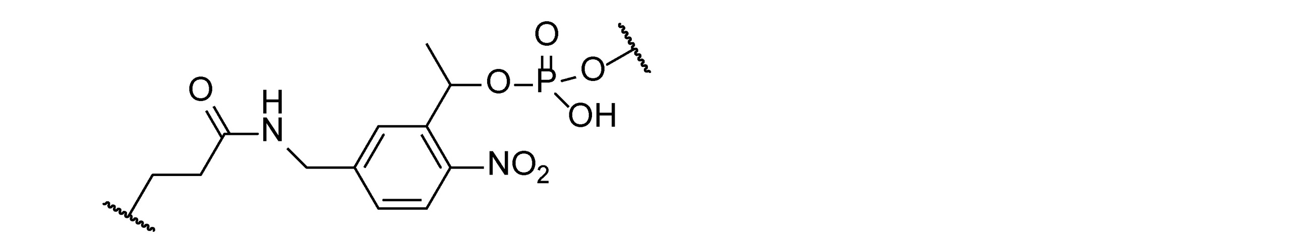
あるいは、切断リンカーが化学的に切断可能な基である場合、切断剤は、その基を切断することができる化学剤であり得る。化学的に切断可能なリンカーは、酸化/還元に基づく切断、酸触媒による切断、塩基触媒による切断、または求核置換によって切断することができる。例えば、結合基がジスルフィドである場合、ジチオトレイトールまたはβメルカプトエタノールによるチオール媒介切断を使用して、タグを遊離させることができる。結合基が制限部位であるさらに他の実施形態では、薬剤は、加水分解酵素、制限酵素、または結合基を切断する他の酵素であり得る酵素などの触媒剤である。例えば、制限酵素は、I型、II型、IIS型、III型、およびIV型制限酵素であり得る。 Alternatively, if the cleavage linker is a chemically cleaveable group, the cleaving agent can be a chemical agent capable of cleaving the group. Chemically cleavable linkers can be cleaved by oxidation / reduction based cleavage, acid catalyzed cleavage, base catalyzed cleavage, or nucleophilic substitution. For example, if the linking group is a disulfide, the tag can be released using thiol-mediated cleavage with dithiothreitol or β-mercaptoethanol. In yet other embodiments where the binding group is the restriction site, the agent is a catalytic agent, such as a hydrolase, a restriction enzyme, or an enzyme that can be another enzyme that cleaves the binding group. For example, restriction enzymes can be type I, type II, type IIS, type III, and type IV restriction enzymes.
いくつかの実施形態では、切断リンカーは、酵素的に切断可能な配列である。本明細書の実施形態のうちのいずれかの一態様では、酵素的に切断可能な配列は、長さが2、3、4、5、6、7、8、9、または10ヌクレオチドの核酸配列である。一実施形態では、酵素的に切断可能な配列は、少なくとも10ヌクレオチドの配列を含む。一実施形態では、酵素的に切断可能な配列は、2~20ヌクレオチドの配列を含む。一実施形態では、酵素的に切断可能な配列は、2~15ヌクレオチドの配列を含む。一実施形態では、酵素的に切断可能な配列は、4~10ヌクレオチドの配列を含む。一実施形態では、酵素的に切断可能な配列は、4~15ヌクレオチドの配列を含む。
In some embodiments, the cleavage linker is an enzymatically cleaveable sequence. In one aspect of any of the embodiments herein, the enzymatically cleavable sequence is a nucleic acid sequence of
例えば、切断可能なリンカーは、アクリジニウム、置換ベンジルエーテルなどのエーテル、または酸性もしくは緩還元状態(例えば、アクリドンおよびスルホンアミドを産生する過酸化水素)によって切断され得るその誘導体(例えば、ベンジルヒドリルエーテル、インダニルエーテル等)、P-脱離を使用して生成された電荷ポリマーであり得、緩塩基は、産物であるアセタールを、そのチオ類似体を含めて遊離させるように機能することができ、脱離は、特に捕捉カルボニル化合物、感光性結合(例えば、O-ニトロベンゾイル、7-ニトロインダニル、2-ニトロベンズヒドリルエーテルもしくはエステル等)、または酵素的加水分解に供されるペプチドリンカー(例えば、酵素的に切断可能なリンカー)の存在下で緩酸によって達成され、特に酵素は、第Xa因子もしくはエンテロキナーゼのペプチドなどの特異的配列を認識する。リンカーの例としては、ジスルフィドリンカー、酸に不安定なリンカー(ジアルコキシベンジルリンカーを含む)、シーバーリンカー、インドールリンカー、t-ブチルシーバーリンカー、求電子的に切断可能なリンカー、求核的に切断可能なリンカー、光切断可能なリンカー、還元条件、酸化条件下での切断、セーフティキャッチリンカーの使用による切断、および脱離機構による切断が挙げられるが、これらに限定されない。 For example, the cleavable linker may be an ether such as acridinium, a substituted benzyl ether, or a derivative thereof (eg, a benzylhydryl ether, which can be cleaved by an acidic or slowly reducing state (eg, hydrogen peroxide producing acridone and a sulfonamide). (Indanyl ether, etc.), which can be a charged polymer produced using P-desorption, the loose base can function to liberate the product acetal, including its thio analogs. Desorption is particularly capture carbonyl compounds, photosensitive bonds (eg, O-nitrobenzoyl, 7-nitroindanyl, 2-nitrobenzhydryl ether or ester, etc.), or peptide linkers that are subjected to enzymatic hydrolysis (eg, eg). , Achieved by slow acid in the presence of an enzymatically cleavable linker), in particular the enzyme recognizes specific sequences such as factors Xa or peptides of enterokinase. Examples of linkers include disulfide linkers, acid-labile linkers (including dialkoxybenzyl linkers), seaver linkers, indole linkers, t-butyl seaver linkers, electrophilic linkers, nucleophilic cleavage. Possible but not limited to linkers, photocleavable linkers, reduction conditions, cleavage under oxidative conditions, cleavage by the use of safety catch linkers, and cleavage by a desorption mechanism.
求電子的に切断されたリンカーは、典型的にはプロトンによって切断され、酸に敏感な切断を含む。好適なリンカーとしては、トリチル、p-アルコキシベンジルエステル、およびp-アルコキシベンジルアミドなどの修飾ベンジル系が挙げられる。他の好適なリンカーとしては、tert-ブチルオキシカルボニル(Boc)基およびアセタール系が挙げられる。チオアセタールまたは他の硫黄含有保護基の切断における、ニッケル、銀または水銀などのチオフィリック金属の使用もまた、好適なリンカー分子の調製のために考慮され得る。 Electrophilically cleaved linkers are typically cleaved by protons and include acid-sensitive cleavage. Suitable linkers include modified benzyls such as trityl, p-alkoxybenzyl ester, and p-alkoxybenzylamide. Other suitable linkers include tert-butyloxycarbonyl (Boc) groups and acetal systems. The use of thiophyllic metals such as nickel, silver or mercury in cleavage of thioacetals or other sulfur-containing protecting groups may also be considered for the preparation of suitable linker molecules.
求核切断のために、水中で不安定である(すなわち、単純に塩基性pHで切断され得る)エステルなどの基、および非水性求核剤に対して不安定である基を使用することができる。フッ化物イオンを使用して、トリイソプロピルシラン(TIPS)またはt-ブチルジメチルシラン(TBDMS)などの基中のケイ素-酸素結合を切断することができる。 For nucleophilic cleavage, it is possible to use groups such as esters that are unstable in water (ie, can simply be cleaved at basic pH), and groups that are unstable to non-aqueous nucleophiles. can. Fluoride ions can be used to cleave silicon-oxygen bonds in groups such as triisopropylsilane (TIPS) or t-butyldimethylsilane (TBDMS).
ジスルフィド結合の還元の場合のように、還元的切断を受けやすいリンカーを使用することができる。ベンジルおよびベンジルオキシカルボニル基を切断するために、パラジウム系触媒を使用する接触水素化が使用されてきた。 Linkers that are susceptible to reductive cleavage can be used, as in the case of disulfide bond reduction. Contact hydrogenation using a palladium-based catalyst has been used to cleave the benzyl and benzyloxycarbonyl groups.
酸化ベースの手法は、当技術分野において周知である。これらには、p-アルコキシベンジル基の酸化ならびに硫黄およびセレンリンカーの酸化が含まれる。ジスルフィドおよび他の硫黄またはセレン系リンカーを切断するための水性ヨウ素もまた、使用することができる。 Oxidation-based techniques are well known in the art. These include the oxidation of p-alkoxybenzyl groups as well as the oxidation of sulfur and selenium linkers. Aqueous iodine for cleaving disulfides and other sulfur or selenium-based linkers can also be used.
セーフティキャッチリンカーは、2つのステップで切断するものである。好ましい系では、第1のステップは、反応性求核中心の生成であり、続いて第2のステップは、切断をもたらす分子内環化を伴う。例えば、レブリン酸エステル結合は、ヒドラジンまたは光化学で処理して、活性アミンを遊離させることができ、次いでこれを環化して分子内の他の場所でエステルを切断することができる(Burgess et al.,J.Org.Chem.62:5165-5168,1997)。 The safety catch linker cuts in two steps. In a preferred system, the first step is the generation of reactive nucleophilic centers, followed by the second step with intramolecular cyclization resulting in cleavage. For example, the levulinic acid ester bond can be treated with hydrazine or photochemistry to liberate the active amine, which can then be cyclized to cleave the ester elsewhere in the molecule (Burgess et al. , J. Org. Chem. 62: 5165-5168, 1997).
脱離反応もまた使用され得る。例えば、Fmocおよびシアノエチルなどの基の塩基触媒脱離、ならびにアリル系のパラジウム触媒還元脱離を使用することができる。 Elimination reactions can also be used. For example, base-catalyzed elimination of groups such as Fmoc and cyanoethyl, and allyl-based palladium-catalyzed reduction elimination can be used.
リンカーが熱切断可能なリンカーまたは感熱性リンカーである場合、切断剤は、熱切断可能な基を破壊または切断するために閾値を超える局所的な温度上昇であり得る。いくつかの実施形態では、粒子温熱療法を誘導するためにマイクロ波照射を使用して局所領域で温度を上昇させることができる。DutzおよびHergt(Nanotechnology,25:452001(2014))に概説され、米国特許第7,718,445号、米国特許公開第2003/0082633号、国際特許公開第2002/029076号、国際特許公開第2002/029076号、および米国特許公開第2002/0197645号(各々が参照により本明細書に組み込まれる)に記載されるものなどの粒子温熱療法を使用することができる。いくつかの実施形態では、温度上昇は、エネルギーを光から吸収性標的、例えば染料、顔料、または水に伝達することによって光熱的に達成することができる。一態様では、光源はレーザーである。いくつかの実施形態では、高温は、二本鎖DNAの熱分離を引き起こし得る。 If the linker is a heat-cleavable or heat-sensitive linker, the cleaving agent can be a local temperature rise above the threshold for breaking or cleaving the heat-cleavable group. In some embodiments, microwave irradiation can be used to raise the temperature in the local area to induce particle hyperthermia. Outlined in Dutz and Hergt (Nanotechnology, 25: 452001 (2014)), US Pat. Nos. 7,718,445, US Patent Publication Nos. 2003/0082633, International Patent Publication No. 2002/0290776, International Patent Publication No. 2002. Particle thermotherapy such as those described in / 029076 and US Patent Publication No. 2002/0197645 (each incorporated herein by reference) can be used. In some embodiments, the temperature rise can be achieved photothermally by transferring energy from the light to an absorbent target such as a dye, pigment, or water. In one aspect, the light source is a laser. In some embodiments, high temperatures can cause thermal separation of double-stranded DNA.
g)ナノポア層
本開示において、タグ(例えば、ポリマー、アプタマー、ナノ粒子)の検出および/またはカウントは、ナノポアまたはナノチャネルを通してまたは横切ってタグを転位させることによって実行され得る。いくつかの実施形態では、タグ(例えば、ポリマー、アプタマー、ナノ粒子)の検出および/またはカウントは、少なくとも1つ以上のナノポアまたはナノチャネルを通してまたは横切ってタグを転位させることによって実行され得る。いくつかの実施形態では、少なくともつ以上のナノポアまたはナノチャネルが並んでまたは連続して提示される。いくつかの実施形態では、ナノポアまたはナノチャネルは、一度に1つ以下のタグの転位のために寸法決めされる。したがって、いくつかの実施形態におけるナノポアの寸法は、典型的には検査されるタグの寸法に依存するであろう。二本鎖領域を有するタグは、完全に一本鎖であるタグの転位に十分なものよりも大きいナノポア寸法を必要とし得る。さらに、ナノビーズタグなどのナノ粒子タグは、オリゴマータグよりも大きい細孔またはチャネルを必要とし得る。典型的には、直径約1nmの細孔は、一本鎖ポリマーの通過を許容し得るが、直径2nm以上の細孔寸法は、二本鎖核酸分子の通過を許容するであろう。いくつかの実施形態では、ナノポアまたはナノチャネルは、一本鎖タグ(例えば、直径約1nm~2nm未満)に対して選択的であるが、他の実施形態では、ナノポアまたはナノチャネルは、二本鎖ポリヌクレオチドの通過を可能にするのに十分な直径(例えば、2nm以上)である。選択された孔径は、目的の検体について最適なシグナル対ノイズ比を提供する。
g) Nanopore Layer In the present disclosure, detection and / or counting of tags (eg, polymers, aptamers, nanoparticles) can be performed by rearranging the tags through or across nanopores or nanoparticles. In some embodiments, detection and / or counting of tags (eg, polymers, aptamers, nanoparticles) can be performed by translocating the tags through or across at least one or more nanopores or nanoparticles. In some embodiments, at least one or more nanopores or nanochannels are presented side by side or sequentially. In some embodiments, nanopores or nanochannels are dimensioned for one or less tag rearrangements at a time. Therefore, the dimensions of the nanopores in some embodiments will typically depend on the dimensions of the tag being inspected. Tags with double-stranded regions may require nanopore dimensions that are larger than sufficient for transposition of tags that are completely single-stranded. In addition, nanoparticle tags, such as nanobead tags, may require larger pores or channels than oligomeric tags. Typically, pores with a diameter of about 1 nm may allow the passage of single-stranded polymers, while pore dimensions of 2 nm or more in diameter will allow the passage of double-stranded nucleic acid molecules. In some embodiments, the nanopores or nanochannels are selective for single-stranded tags (eg, about 1 nm to less than 2 nm in diameter), whereas in other embodiments, the nanopores or nanochannels are two. The diameter is sufficient to allow passage of chain polynucleotides (eg, 2 nm and above). The pore size selected provides the optimum signal-to-noise ratio for the sample of interest.
いくつかの実施形態では、細孔は、直径約0.1nm~約1000nm、約50nm~約1000nm、約100nm~1000nm、約0.1nm~約700nm、約50nm~約700nm、約100nm~700nm、約0.1nm~約500nm、約50nm~約500nm、または約100nm~500nmであり得る。例えば、細孔は、直径約0.1nm、約0.2nm、約0.3nm、約0.4nm、約0.5nm、約0.6nm、約0.7nm、約0.8nm、約0.9nm、約1.0nm、約1.5nm、約2.0nm、約2.5nm、約3.0nm、約3.5nm、約4.0nm、約4.5nm、約5.0nm、約7.5nm、約10nm、約15nm、約20nm、約25nm、約30nm、約35nm、約40nm、約45nm、約50nm、約55nm、約60nm、約65nm、約70nm、約75nm、約80nm、約85nm、約90nm、約95nm、約100nm、約150nm、約200nm、約250nm、約300nm、約3500nm、約400nm、約450nm、約500nm、約550nm、約600nm、約650nm、約700nm、約750nm、約800nm、約850nm、約900nm、約950nm、または約1000nmであり得る。 In some embodiments, the pores have a diameter of about 0.1 nm to about 1000 nm, about 50 nm to about 1000 nm, about 100 nm to 1000 nm, about 0.1 nm to about 700 nm, about 50 nm to about 700 nm, about 100 nm to 700 nm, It can be from about 0.1 nm to about 500 nm, from about 50 nm to about 500 nm, or from about 100 nm to 500 nm. For example, the pores have diameters of about 0.1 nm, about 0.2 nm, about 0.3 nm, about 0.4 nm, about 0.5 nm, about 0.6 nm, about 0.7 nm, about 0.8 nm, about 0. 9 nm, about 1.0 nm, about 1.5 nm, about 2.0 nm, about 2.5 nm, about 3.0 nm, about 3.5 nm, about 4.0 nm, about 4.5 nm, about 5.0 nm, about 7. 5 nm, about 10 nm, about 15 nm, about 20 nm, about 25 nm, about 30 nm, about 35 nm, about 40 nm, about 45 nm, about 50 nm, about 55 nm, about 60 nm, about 65 nm, about 70 nm, about 75 nm, about 80 nm, about 85 nm, About 90 nm, about 95 nm, about 100 nm, about 150 nm, about 200 nm, about 250 nm, about 300 nm, about 3500 nm, about 400 nm, about 450 nm, about 500 nm, about 550 nm, about 600 nm, about 650 nm, about 700 nm, about 750 nm, about 800 nm. , About 850 nm, about 900 nm, about 950 nm, or about 1000 nm.
一般に、ナノポアは、ナノチャネルよりも長さが短い。ナノチャネルは、ナノポアよりも実質的に長く、分子がそれを通って転位するのにかかる時間を(同じ直径のナノポアを通ってまたは横切って転位するのにかかる時間と比較して)増加させることが望ましい用途において有用であり得る。ナノポアの長さは、約0.1nm~約200nm未満の範囲であり得る。ナノチャネルの長さは、約500nm~約100μm、またはそれ以上の範囲であり得る。ナノポアおよびナノチャネルの直径は類似していてもよい。 In general, nanopores are shorter in length than nanochannels. Nanochannels are substantially longer than nanopores and increase the time it takes for a molecule to dislocate through it (compared to the time it takes for a molecule to dislocate through or across nanopores of the same diameter). Can be useful in desirable applications. The length of the nanopores can range from about 0.1 nm to less than about 200 nm. The length of the nanochannel can range from about 500 nm to about 100 μm or more. The diameters of the nanopores and nanochannels may be similar.
タグ/アプタマーを分析するために様々な種類のナノポアを使用することができる。これらには、とりわけ、膜に埋め込まれた生物学的細孔またはチャネルを用いる生物学的ナノポアが含まれる。別の種類のナノポア層は、チャネルまたは細孔が、全体的にまたは部分的に、ケイ素などの製造または彫刻された固体成分から作製される固体ナノポアである。いくつかの実施形態では、ナノポアは、制御された絶縁破壊を使用して産生された固体ナノポアである。いくつかの実施形態では、ナノポアは、制御された絶縁破壊以外の方法によって産生された固体ナノポアである。 Various types of nanopores can be used to analyze tags / aptamers. These include, among other things, biological nanopores with biological pores or channels embedded in the membrane. Another type of nanopore layer is a solid nanopore in which the channels or pores are made entirely or partially from a manufactured or engraved solid component such as silicon. In some embodiments, the nanopores are solid nanopores produced using controlled breakdown. In some embodiments, the nanopores are solid nanopores produced by methods other than controlled breakdown.
ある特定の実施形態では、ナノポアの長さは、最大約200nm、例えば、約0.1nm~約30nm、約10~約80nm、約1~約50nm、約0.1nm~約0.5nm、約0.3nm~約1nm、約1nm~約2nm、約0.3nm~約10nm、または約10~約30nmであり得る。ナノポア層内のナノポアの数は、約1、2、3、4、5、10、30、100、300、1000、3000、10000、30000、100000、300000、またはそれ以上であり得る。中心間の層内のナノポア間の距離は、約100nm~約300nm、約300nm~約500nm、約500nm~約1000nm、例えば100nm、150nm、200nm、または100nmであり得る。 In certain embodiments, the length of the nanopores is up to about 200 nm, eg, about 0.1 nm to about 30 nm, about 10 to about 80 nm, about 1 to about 50 nm, about 0.1 nm to about 0.5 nm, about. It can be from 0.3 nm to about 1 nm, from about 1 nm to about 2 nm, from about 0.3 nm to about 10 nm, or from about 10 to about 30 nm. The number of nanopores in the nanopore layer can be about 1, 2, 3, 4, 5, 10, 30, 100, 300, 1000, 3000, 10000, 30000, 100,000, 300000, or more. The distance between the nanopores in the layer between the centers can be from about 100 nm to about 300 nm, from about 300 nm to about 500 nm, from about 500 nm to about 1000 nm, such as 100 nm, 150 nm, 200 nm, or 100 nm.
ある特定の実施形態では、各々がつ以上のナノポアを含有する複数のナノポア層は、タグ(例えば、ポリマー、アプタマー、ナノ粒子)を検出および/またはカウントするために、互いに連続して配置することができる。この場合、タグの検出および/またはカウントは、各ナノポア層を通してまたは横切ってタグを転位させることによって実行することができる。そのようなものとして、層/シート/膜内のナノポアを通ってまたは横切って転位するタグの数をカウントすることは、層/シート/膜内の1つ以上のナノポアを通ってまたは横切って転位する複数のタグのカウントを指す。ある特定の実施形態では、2つ以上のナノポア層(例えば、技術的に可能な限り1、2、3、4、5、6、または他の数のナノポア層)が存在する場合、任意選択で、それらは少なくとも1つのナノポアが連続して存在し、ここで1つの層内の少なくとも1つのナノポアは、別の層内の別のナノポア等とは別個であるか、またはその上に(例えば、上または上部に)積層されている。ナノポア層が連続している場合、少なくとも2つの電極を使用して、細孔を通してタグを駆動するための電界を生じさせるために少なくとも2つの電極を使用することができ、任意選択的で、ナノポア層間に位置付けられた追加の電極が、駆動電流をさらに提供し得る。 In certain embodiments, multiple nanopore layers, each containing one or more nanopores, are placed consecutively with each other to detect and / or count tags (eg, polymers, aptamers, nanoparticles). Can be done. In this case, tag detection and / or counting can be performed by transposing the tag through or across each nanopore layer. As such, counting the number of tags that dislocate through or across nanopores within a layer / sheet / membrane can be dislocated through or across one or more nanopores within a layer / sheet / membrane. Refers to the count of multiple tags. In certain embodiments, if there are more than one nanopore layer (eg, 1, 2, 3, 4, 5, 6, or any other number of nanopore layers technically possible), it is optional. , They have at least one nanopore in a row, where at least one nanopore in one layer is separate from, or on top of, another nanopore, etc. in another layer (eg,). Laminated (on top or top). If the nanopore layer is contiguous, at least two electrodes can be used and at least two electrodes can be used to generate an electric field to drive the tag through the pores, which is optional and nanopore. Additional electrodes located between the layers may provide additional drive current.
i)生物学的細孔
タグ/アプタマーを検出し、任意選択でカウントするために、タグの転位を許すチャネル寸法を有する任意の生物学的細孔を使用することができる。2つの広いカテゴリーの生物学的チャネルが、本明細書に開示されている方法に好適である。無電圧ゲートチャネルは、チャネルを活性化または開放するために膜電位の変化を必要とせずに、細孔を通る分子の通過を可能にする。一方で、電圧依存性チャネルは、チャネル開口部を活性化するために特定範囲の膜電位を必要とする。生物学的ナノポアを用いたほとんどの研究は、Staphylococcus aureusに見出される長さ約10nmのキノコ型ホモオリゴマー七量体チャネルであるα溶血素を使用した。各サブユニットは、2本のβ鎖が14鎖の逆平行βバレルを形成するのに寄与する。βバレル構造によって形成された細孔は、リジン残基の環を含み、約3.6nmの直径を有する内部空洞に開口するおよそ2.6nmの直径を有する入口を有する。脂質二重層を貫通する溶血素孔の幹は、前庭と幹との間に1.5nmの狭窄部を有する約2.0nmの平均内径を有する。幹の寸法は、一本鎖核酸の通過に十分であるが、二本鎖核酸の通過には十分ではない。したがって、α溶血素孔は、一本鎖ポリヌクレオチドおよび同様の寸法の他のポリマーに対して選択的なナノポアとして使用され得る。
i) Biological pores Any biological pores with channel dimensions that allow transposition of tags can be used to detect tags / aptamers and optionally count. Two broad categories of biological channels are suitable for the methods disclosed herein. The no-voltage gate channel allows the passage of molecules through the pores without requiring a change in membrane potential to activate or open the channel. On the other hand, voltage-dependent channels require a specific range of membrane potentials to activate the channel openings. Most studies using biological nanopores used α-hemolysin, a mushroom-type homooligomer heptamer channel found in Staphylococcus aureus with a length of about 10 nm. Each subunit contributes to the formation of a 14-chain antiparallel β-barrel with two β-chains. The pores formed by the β-barrel structure contain a ring of lysine residues and have an inlet with a diameter of approximately 2.6 nm that opens into an internal cavity with a diameter of approximately 3.6 nm. The stem of the hemolysin pores penetrating the lipid bilayer has an average inner diameter of about 2.0 nm with a 1.5 nm constriction between the vestibule and the trunk. The size of the stem is sufficient for the passage of single-stranded nucleic acids, but not sufficient for the passage of double-stranded nucleic acids. Therefore, α-hemolysin pores can be used as selective nanopores for single-stranded polynucleotides and other polymers of similar size.
他の実施形態では、生物学的ナノポアは、一本鎖核酸よりも大きいポリマーの通過のために十分な寸法である。例示的な細孔は、ミトコンドリアポリンタンパク質、ミトコンドリア外膜に局在する電圧依存性アニオンチャネル(VDAC)である。ポリンタンパク質は、精製された形態で入手可能であり、人工脂質二重層に再構成されると、二本鎖核酸の通過を許すことができる機能的チャネルを生成する(Szabo et al.,1998,FASEBJ.12:495-502)。構造研究は、ポリンもまた、13本または16本の鎖を有するβバレル型構造を有することを示唆している(Rauch et al.,1994,Biochem Biophys Res Comm 200:908-915)。ポリンは、α溶血素、マルトポリン(LamB)、およびグラミシジンによって形成される細孔のコンダクタンスと比較してより大きいコンダクタンスを示す。ポリン支持体研究のより大きいコンダクタンス特性は、ポリンチャネルが二本鎖核酸の通過に十分な寸法であることを示している。ポリン分子の孔径は、4nmと推定される。非コイル状二本鎖核酸の直径は、約2nmと推定される。 In other embodiments, the biological nanopores are sized enough for the passage of polymers larger than single-stranded nucleic acids. An exemplary pore is a mitochondrial porin protein, a voltage-dependent anion channel (VDAC) localized in the outer mitochondrial membrane. Porin proteins are available in purified form and, when reconstituted into artificial lipid bilayers, produce functional channels that allow the passage of double-stranded nucleic acids (Szabo et al., 1998, FASEBJ. 12: 495-502). Structural studies suggest that porin also has a β-barreled structure with 13 or 16 strands (Rauch et al., 1994, Biochem Biophyss Res Com 200: 908-915). Porin exhibits greater conductance compared to the conductance of the pores formed by α-hemolysin, maltoporin (LamB), and gramicidin. The larger conductance properties of the porin support study indicate that the porin channel is sized enough for the passage of double-stranded nucleic acids. The pore size of the porin molecule is estimated to be 4 nm. The diameter of the non-coiled double-stranded nucleic acid is estimated to be about 2 nm.
二本鎖ポリヌクレオチドをスキャンするのに好適であり得る別の生物学的チャネルは、枯草菌に見られるチャネルである(Szabo et al.,1997,J.Biol.Chem.272:25275-25282)。枯草菌から作製され、人工膜に組み込まれた原形質膜小胞は、膜を横切る二本鎖DNAの通過を可能にする。枯草菌膜調製物によって形成されたチャネルのコンダクタンスは、ミトコンドリアポリンのものと同様である。これらのチャネルの不完全な特徴付け(例えば、精製形態)があるが、本明細書の目的のために精製形態を有する必要はない。適切な洗剤に可溶化すること、または十分な表面積の人工脂質膜に組み込むことのいずれかによって、原形質膜調製物を希釈することは、検出装置中の単一チャネルを単離することができる。適切に時限洗浄することによって膜調製物(またはタンパク質調製物)と人工膜との接触の持続時間を制限することは、人工脂質二重層に単一チャネルを組み込むための別の方法を提供する。コンダクタンス特性を使用して、二重層に組み込まれたチャネルを特徴付けることができる。 Another biological channel that may be suitable for scanning double-stranded polynucleotides is that found in Bacillus subtilis (Szabo et al., 1997, J. Biol. Chem. 272: 25275-25282). .. Protoplasmic membrane vesicles made from Bacillus subtilis and integrated into artificial membranes allow the passage of double-stranded DNA across the membrane. The conductance of the channels formed by the Bacillus subtilis membrane preparation is similar to that of mitochondrial porin. There are incomplete characterizations of these channels (eg, purified forms), but it is not necessary to have purified forms for the purposes herein. Diluting the plasma membrane preparation, either by solubilizing it in a suitable detergent or incorporating it into an artificial lipid membrane of sufficient surface area, can isolate a single channel in the detector. .. Limiting the duration of contact between the membrane preparation (or protein preparation) and the artificial membrane by proper timed washing provides another method for incorporating a single channel into the artificial lipid bilayer. The conductance property can be used to characterize the channels incorporated in the double layer.
ある特定の場合において、ナノポアは、ハイブリッドナノポアであってもよく、ここで生物学的細孔は、固体ナノポア、例えば非生物学的材料で製造されたナノポアに導入される。例えば、α-溶血素孔は、固体ナノポアに挿入されてもよい。ある特定の場合において、ナノポアは、Hall et al.,Nature Nanotechnology,28 November 2010,vol.5,pg.874-877に記載されているハイブリッドナノポアであり得る。 In certain cases, the nanopores may be hybrid nanopores, where biological pores are introduced into solid nanopores, eg nanopores made of non-biological materials. For example, α-hemolysin pores may be inserted into solid nanopores. In certain cases, nanopores are described in Hall et al. , Nature Nanotechnology, 28 November 2010, vol. 5, pg. It can be the hybrid nanopore described in 874-877.
ii)固体細孔
他の実施形態では、タグの分析は、非生物学的材料から製造されたナノポアまたはナノチャネルを通してまたは横切ってタグを転位させることによって実行される。ナノポアまたはナノチャネルは、とりわけ化学堆積、電気化学体積、電気めっき、電子ビーム彫刻、イオンビーム彫刻、ナノリソグラフィー、化学エッチング、レーザーアブレーション、集束イオンビーム、原子層堆積、および当技術分野において周知の他の方法を含む、多くの異なる技法を使用して、様々な固体材料から作製され得る(例えば、Li et al.,2001,Nature 412:166-169、およびWO2004/085609を参照)。
ii) Solid pores In other embodiments, tag analysis is performed by transposing the tag through or across nanopores or nanochannels made from non-biological materials. Nanopores or nanochannels are known in the art, among others, chemical deposition, electrochemical volume, electroplating, electron beam engraving, ion beam engraving, nanolithography, chemical etching, laser ablation, focused ion beam, atomic layer deposition, and others in the art. Can be made from a variety of solid materials using many different techniques, including the method of (see, eg, Li et al., 20011, Nature 412: 166-169, and WO 2004/08569).
特定の実施形態では、ナノポアは、WO13167952A1またはWO13167955A1に記載されているナノポアであり得る。WO13167952A1またはWO13167955A1に記載されているように、正確かつ均一な孔径を有するナノポアは、膜中に形成されたナノポアを精密に拡大することによって形成することができる。この方法は、ナノポアにわたって高電位を印加することによってナノポアを拡大することと、ナノポアを流れる電流を測定することと、測定された電流に部分的に基づいてナノポアのサイズを決定することと、ナノポアのサイズが所望のサイズに対応するとき、ナノポアに印加された電位を除去することと、を含み得る。ある特定の場合において、印加電位は、高い値と低い値との間で振動するパルス波形を有することができ、電位が低い値でナノポアに印加されている間に、ナノポアを流れる電流を測定することができる。 In certain embodiments, the nanopores can be the nanopores described in WO13167952A1 or WO13167955A1. As described in WO13167952A1 or WO13167955A1, nanopores with accurate and uniform pore size can be formed by precisely enlarging the nanopores formed in the membrane. This method expands the nanopore by applying a high potential across the nanopore, measures the current flowing through the nanopore, determines the size of the nanopore based in part on the measured current, and nanopore. When the size of is corresponding to the desired size, it may include removing the potential applied to the nanopores. In certain cases, the applied potential can have a pulse waveform that oscillates between high and low values, measuring the current flowing through the nanopore while the potential is applied to the nanopore at a low value. be able to.
固体材料としては、限定ではなく例として、任意の公知の半導体材料、絶縁材料、および絶縁材料でコーティングされた金属が挙げられる。したがって、ナノポア(複数可)の少なくとも一部は、限定されないが、ケイ素、シリカ、シリセン、酸化ケイ素、グラフェン、窒化ケイ素、ゲルマニウム、ヒ化ガリウム、または絶縁材料でコーティングされた金属、金属酸化物、および金属コロイドを含み得る。 Examples of solid materials include, but are not limited to, any known semiconductor material, insulating material, and metal coated with the insulating material. Thus, at least some of the nanopores (s) are, but are not limited to, silicon, silica, silicene, silicon oxide, graphene, silicon nitride, germanium, gallium arsenide, or metals, metal oxides, coated with an insulating material. And may contain metal colloids.
ナノメートル寸法の細孔を作製するために、製造プロセスにおいて様々なフィードバック手順を用いることができる。イオンが穴を通過する実施形態では、固体材料を通るイオンの流れを検出することは、製造中に生成された孔径を測定する方法を提供する(例えば、米国特許出願公開第2005/0126905号を参照)。他の実施形態では、電極が孔径を定義する場合、電極間の電子トンネル電流は、電極間のギャップに関する情報を与える。トンネル電流の増加は、電極間のギャップ間隔の減少を示す。他のフィードバック技法は、当業者に明らかとなるであろう。 Various feedback procedures can be used in the manufacturing process to create nanometer-sized pores. In embodiments where ions pass through a hole, detecting the flow of ions through a solid material provides a method of measuring the pore size generated during manufacturing (eg, US Patent Application Publication No. 2005/0126905). reference). In another embodiment, when the electrodes define the pore size, the electron tunneling current between the electrodes provides information about the gap between the electrodes. An increase in tunnel current indicates a decrease in the gap spacing between the electrodes. Other feedback techniques will be apparent to those of skill in the art.
いくつかの実施形態では、ナノポアは、Li et al.,2003,Nature Materials 2:611-615に記載されているように、イオンビーム彫刻を使用して製造される。いくつかの実施形態では、ナノポアは、WO13167952A1またはWO13167955A1に記載されるように、高電流を使用して製造される。他の実施形態では、ナノポアは、電子ビームリソグラフィと高エネルギー電子ビーム彫刻との組み合わせによって作製することができる(例えば、Storm et al.,2003,Nature Materials 2:537-540を参照)。イオンビームスパッタリング技法によって好適なナノポアを生成するための同様の手法は、Heng et al.,2004,Biophy J 87:2905-2911に記載されている。ナノポアは、超薄膜を産生するための一般的な技法と組み合わせた金属酸化物半導体(CMOS)上に集束高エネルギー電子ビームを用いるリソグラフィを使用して形成される。他の実施形態では、ナノポアは、窒化ケイ素の彫刻によって、米国特許第6,627,067号、同第6,464,842号、同第6,783,643号、および米国公開第2005/0006224号に提供されるように構成される。
In some embodiments, the nanopores are described in Li et al. , 2003, Nature Materials 2: 611-615, manufactured using ion beam engraving. In some embodiments, the nanopores are manufactured using high currents, as described in WO13167952A1 or WO13167955A1. In other embodiments, nanopores can be made by a combination of electron beam lithography and high energy electron beam engraving (see, eg, Stone et al., 2003, Nature Materials 2: 537-540). Similar techniques for producing suitable nanopores by ion beam sputtering techniques are described in Heng et al. , 2004, Biophy J 87: 2905-2911. Nanopores are formed using lithography using focused high energy electron beams on metal oxide semiconductors (CMOS) combined with common techniques for producing ultrathin films. In other embodiments, nanopores are engraved with silicon nitride in US Pat. Nos. 6,627,067, 6,464,842, 6,783,643, and
いくつかの実施形態では、ナノチャネルは、金または銀ナノチューブとして構成され得る。これらのナノチャネルは、エッチング飛跡法を使用して調製されたポリカーボネートフィルタなどの多孔質材料のテンプレートを使用し、多孔質材料の表面に金または他の好適な金属を堆積させることによって形成される。エッチング飛跡ポリカーボネート膜は、典型的には、固体膜材料を高エネルギー核粒子に曝露することによって形成され、これが膜材料中に飛跡を形成する。次いで化学エッチングを用いて、エッチングされた飛跡を細孔に変換する。形成された細孔は、約10nm以上の直径を有する。核粒子の強度を調整することは、膜に形成される細孔の密度を制御する。無電解めっき法を介して飛跡エッチングされた細孔に金属、典型的には金または銀を堆積させることによって、エッチングされた膜上にナノチューブを形成する(Menon et al.,1995,Anal Chem 67:1920-1928)。この金属堆積方法は、細孔材料の表面に堆積された触媒を使用し、これが次いでAu(I)および還元剤を含有する溶液中に浸漬される。Au(I)から金属Auへの還元は、触媒を含む表面上で起こる。堆積した金の量は、インキュベーション時間に依存し、それによりインキュベーション時間を増加させると、フィルタ材料中の細孔の内径が減少する。したがって、孔径は、細孔に堆積する金属の量を調整することによって制御することができる。得られる細孔寸法は、種々の技法、例えば、単純拡散を使用するガス輸送特性を使用して、またはパッチクランプ型システムを使用して細孔を通るイオン流を測定することによって測定される。支持体材料は、無傷のまま残されるか、または金ナノチューブを残すために除去される。無電解めっき技法は、直径が約1nm未満~約5nm、または必要に応じてそれ以上の孔径を形成することができる。約0.6nmの孔径を有する金ナノチューブは、Ru(bpy)2+2とメチルビオロゲンとを区別するように見え、金ナノポアの選択性を実証している(Jirage et al.,1997,Science 278:655-658)。金ナノチューブ表面の修飾は、チオール含有化合物を金表面に付着させることによって、または金表面を他の官能基で誘導体化することによって容易に達成される。この特徴は、本明細書で論じられるように、細孔修飾化合物および感知標識の付着を許す。本明細書に記載の生物学的細孔に使用されるシス/トランス装置などのデバイスを金ナノポアと共に使用して、単一コード分子を分析することができる。 In some embodiments, the nanochannel can be configured as gold or silver nanotubes. These nanochannels are formed by depositing gold or other suitable metal on the surface of the porous material using a template of the porous material such as a polycarbonate filter prepared using the etching track method. .. Etching track Polycarbonate films are typically formed by exposing a solid film material to high-energy nuclear particles, which form tracks in the film material. Chemical etching is then used to convert the etched tracks into pores. The formed pores have a diameter of about 10 nm or more. Adjusting the strength of the nuclear particles controls the density of the pores formed in the membrane. Nanotubes are formed on the etched film by depositing metal, typically gold or silver, in the pores that are track-etched via electroless plating (Menon et al., 1995, Anal Chem 67). : 1920-1928). This metal deposition method uses a catalyst deposited on the surface of the pore material, which is then immersed in a solution containing Au (I) and a reducing agent. The reduction of Au (I) to metal Au occurs on the surface containing the catalyst. The amount of gold deposited depends on the incubation time, which increases the incubation time and reduces the inner diameter of the pores in the filter material. Therefore, the pore size can be controlled by adjusting the amount of metal deposited in the pores. The resulting pore size is measured using a variety of techniques, eg, gas transport properties using simple diffusion, or by measuring the ion flow through the pores using a patch clamp type system. The support material is either left intact or removed to leave the gold nanotubes. The electroless plating technique can form pore diameters of less than about 1 nm to about 5 nm, or even larger if desired. Gold nanotubes with a pore size of about 0.6 nm appear to distinguish between Ru (bpy) 2 + 2 and methylviologen, demonstrating the selectivity of gold nanopores (Jirage et al., 1997, Science 278: 655). -658). Modification of the gold nanotube surface is easily accomplished by attaching a thiol-containing compound to the gold surface or by derivatizing the gold surface with other functional groups. This feature allows the attachment of pore-modifying compounds and sensing labels, as discussed herein. Devices such as the cis / trans device used for the biological pores described herein can be used with gold nanopores to analyze single coding molecules.
タグを検出するモードがタグを通る電流(例えば、電子トンネル電流)を含む場合、固体膜は、様々な技法によって金属化され得る。導電層は、鎖の長さに沿ってタグを調べるのに好適な電極、例えば、長手方向の電子トンネル電流を生成するために膜の両側に堆積されてもよい。他の実施形態では、導電層を膜の一方の表面に堆積させて、細孔を横切るタグ、例えば、横方向トンネル電流を調べるのに好適な電極を形成することができる。スパッタ堆積(すなわち、物理蒸着)、無電解堆積(例えば、コロイド懸濁液)、および電解堆積を含む、導電性材料を堆積するための様々な方法が知られている。他の金属堆積技法は、フィラメント蒸着、金属層蒸着、電子ビーム蒸着、フラッシュ蒸着、および誘導蒸着であり、当業者には明らかとなるであろう。 If the mode for detecting the tag includes a current through the tag (eg, electron tunneling current), the solid membrane can be metallized by a variety of techniques. Conductive layers may be deposited on either side of the membrane to generate electrodes suitable for examining tags along the length of the chain, eg, longitudinal electron tunneling currents. In other embodiments, a conductive layer can be deposited on one surface of the membrane to form a tag that traverses the pores, eg, an electrode suitable for examining transverse tunneling currents. Various methods for depositing conductive materials are known, including sputter deposition (ie, physical deposition), electroless deposition (eg, colloidal suspension), and electrolytic deposition. Other metal deposition techniques are filament deposition, metal layer deposition, electron beam deposition, flash deposition, and inductive deposition, which will be apparent to those of skill in the art.
いくつかの実施形態では、検出電極は、スパッタ堆積によって形成され、ここでイオンビームは、金属ブロックに衝突して金属原子を蒸発させ、次いで薄膜の形態でウェハ材料上に堆積される。使用されるリソグラフィ方法に応じて、金属フィルムは、次いで反応性イオンエッチングによってエッチングされるか、または化学機械研磨を使用して研磨される。金属フィルムは、予め形成されたナノポア上に堆積され得るか、またはポアの製造前に堆積されてもよい。 In some embodiments, the detection electrode is formed by sputter deposition, where the ion beam collides with the metal block to evaporate the metal atoms and then deposits on the wafer material in the form of a thin film. Depending on the lithography method used, the metal film is then etched by reactive ion etching or polished using chemical mechanical polishing. The metal film may be deposited on preformed nanopores or may be deposited prior to the production of the pores.
いくつかの実施形態では、検出電極は、電着によって製造される(例えば、Xiang et al.,2005,Angew.Chem.Int.Ed.44:1265-1268、Li et al.,Applied Physics Lett.77(24):3995-3997、および米国出願公開第2003/0141189号を参照)。この製造方法は、固体フィルムの一方の面上に位置付けられたナノポアおよび対応する検出電極を生成するため、例えば横方向電子トンネルを検出するために好適である。最初に、従来のリソグラフィプロセスを用いて、シリコンウェハ上に支持されている二酸化ケイ素層上に一対の対向電極を形成する。電解液が電極を覆い、電極対に電流を通すことによって、金属イオンが一方の電極に堆積する。経時的な電極上の金属を堆積は、電極間のギャップ距離を減少させ、検出電極だけでなくコード化分子の転位のためのナノメートル寸法のギャップも作り出す。電極間のギャップ距離は、多くのフィードバックプロセスによって制御することができる。 In some embodiments, the detection electrode is manufactured by electrodeposition (eg, Xiang et al., 2005, Angew. Chem. Int. Ed. 44: 1265-1268, Li et al., Applied Physics Let. 77 (24): 3995-3997, and US Application Publication No. 2003/0141189). This manufacturing method is suitable for detecting, for example, lateral electron tunnels, because it produces nanopores and corresponding detection electrodes located on one surface of the solid film. First, a conventional lithography process is used to form a pair of counter electrodes on a silicon dioxide layer supported on a silicon wafer. The electrolytic solution covers the electrodes and conducts an electric current through the pair of electrodes, so that metal ions are deposited on one of the electrodes. The deposition of metal on the electrodes over time reduces the gap distance between the electrodes and creates nanometer-sized gaps for dislocations of the coding molecules as well as the detection electrodes. The gap distance between the electrodes can be controlled by many feedback processes.
検出が電荷誘導電界効果の撮像に基づく場合、米国特許第6,413,792号および米国出願公開第2003/0211502号に記載されているように、半導体を製造することができる。これらのナノポアデバイスを製造する方法は、他の固体ナノポアを製造するために用いられるものと同様の技法を使用することができる。 If the detection is based on imaging of charge-induced electric field effects, semiconductors can be manufactured as described in US Pat. No. 6,413,792 and US Application Publication No. 2003/0211502. Methods for making these nanopore devices can use similar techniques as those used to make other solid nanopores.
ポリヌクレオチドなどのタグの検出は、以下でさらに記載されるように実行される。タグの分析のために、ナノポアは様々なフォーマットで構成されてもよい。いくつかの実施形態では、デバイスは、シスおよびトランスチャンバとも称される、2つのリザーバの間に保持されたナノポアを含む、生物学的または固体のいずれかの膜を含む(例えば、米国特許第6,627,067号を参照)。2つのチャンバ間の電子移動のための導管は、2つのチャンバの電気的接触を可能にし、2つのチャンバ間の電圧バイアスは、ナノポアを通るタグの転位を駆動する。この構成の変形は、米国特許第6,015,714号および第6,428,959号、ならびにKasianowiscz et al.,1996,Proc Natl Acad Sci USA 93:13770-13773(これらの開示は、参照により本明細書に組み込まれ)に記載されているように、ナノポアを通る電流の分析に使用される。 Detection of tags such as polynucleotides is performed as described further below. For tag analysis, nanopores may be configured in various formats. In some embodiments, the device comprises either a biological or solid membrane containing nanopores held between two reservoirs, also referred to as cis and transchambers (eg, U.S. Patent No. 1). See Nos. 6,627,067). The conduit for electron transfer between the two chambers allows electrical contact between the two chambers, and the voltage bias between the two chambers drives the dislocation of the tag through the nanopores. Modifications of this configuration are described in US Pat. Nos. 6,015,714 and 6,428,959, as well as Kasianowiscz et al. , 1996, Proc Natl Acad Sci USA 93: 13770-1373 (these disclosures are incorporated herein by reference) and are used for the analysis of current through nanopores.
上記デバイスの変形は、米国出願公開第2003/0141189号に開示されている。電着によって製造された一対のナノ電極が、基板表面上に位置付けられる。電極は互いに向き合っており、単一核酸の通過に十分なギャップ距離を有する。絶縁材料がナノ電極を保護し、核酸の検出のためにナノ電極の先端のみを露出させる。絶縁材料およびナノ電極は、試料リザーバとして働くチャンバと、ポリマーが転位によって送達されるチャンバとを分離する。カソード電極およびアノード電極は、タグを試料チャンバから送達チャンバへ駆動するための電気泳動電界を提供する。 A variant of the device is disclosed in US Application Publication No. 2003/0141189. A pair of nanoelectrodes manufactured by electrodeposition are positioned on the surface of the substrate. The electrodes are facing each other and have a sufficient gap distance for the passage of a single nucleic acid. The insulating material protects the nanoelectrode and exposes only the tip of the nanoelectrode for nucleic acid detection. The insulating material and nanoelectrodes separate the chamber that acts as the sample reservoir from the chamber in which the polymer is delivered by dislocation. The cathode and anode electrodes provide an electrophoretic electric field to drive the tag from the sample chamber to the delivery chamber.
ナノポアを通してタグを駆動するために使用される電流バイアスは、ナノポアを通して向けられた電界を印加することによって発生させることができる。いくつかの実施形態では、電界は、定電圧または定電流バイアスである。他の実施形態では、タグの移動は、電気泳動電界パラメータのパルス操作を通して制御される(例えば、米国特許出願第2003/141189号および米国特許第6,627,067号を参照)。電流パルスは、規定の期間にわたってオリゴヌクレオチドタグの1つまたは少数の塩基を細孔を通して正確に転位させ、タグをその細孔内に短時間保持する方法を提供することができ、それによりタグの電気的特性のより優れた分解能を提供する。 The current bias used to drive the tag through the nanopores can be generated by applying an electric field directed through the nanopores. In some embodiments, the electric field is a constant voltage or constant current bias. In other embodiments, tag movement is controlled through pulse manipulation of electrophoretic field parameters (see, eg, U.S. Patent Application No. 2003/141189 and U.S. Pat. No. 6,627,067). Current pulses can provide a method of precisely rearranging one or a few bases of an oligonucleotide tag through a pore over a defined period of time, thereby retaining the tag in the pore for a short period of time. Provides better resolution of electrical properties.
ナノポアデバイスは、オリゴヌクレオチドタグがナノポアを通過するときに、オリゴヌクレオチドタグの配向を制限するための電界または電磁場をさらに含んでもよい。この保持場を使用して、細孔内のオリゴヌクレオチドタグの移動を減少させることができる。いくつかの実施形態では、転位の方向に直交する電界が、ナノポア内のタグ分子の移動を制限するために提供される。これは、米国特許出願公開第2003/0141189号に、試料プレートの上下に2つの平行な導電プレートを使用することによって示されている。これらの電極は、タグ分子の転位方向と直交する電界を生成し、したがってタグ分子を試料プレートのうちの1つに保持する。DNAの負電荷骨格、または一方の鎖に負電荷を有するように修飾された核酸は、アノードプレートの上に配向され、それによってタグ分子の動きを制限する。 The nanopore device may further include an electric or electromagnetic field to limit the orientation of the oligonucleotide tag as it passes through the nanopore. This retention field can be used to reduce the movement of oligonucleotide tags within the pores. In some embodiments, an electric field orthogonal to the direction of dislocations is provided to limit the movement of tag molecules within the nanopores. This is demonstrated in US Patent Application Publication No. 2003/0141189 by using two parallel conductive plates above and below the sample plate. These electrodes generate an electric field orthogonal to the dislocation direction of the tag molecule and thus hold the tag molecule in one of the sample plates. The negatively charged backbone of DNA, or nucleic acid modified to have a negative charge on one strand, is oriented onto the anode plate, thereby limiting the movement of the tag molecule.
さらに他の実施形態では、タグの位置を制御することは、米国特許出願公開第2004/0149580号に記載されている方法によって実行され、これはナノポアの近くまたはナノポア上の一連の電極位置を介してポア内に生じる電磁場を用いる。これらの実施形態では、一組の電極は、直流電圧と高周波電位を印加し、一方で第2の電極の組が反対の直流電圧と、第1の電極の組によって発生した高周波電位に関して180度位相がずれた高周波電位とを印加する。この高周波四重極は、電界の中心(すなわち、細孔の中心)に電荷粒子(例えば、核酸)を保持する。 In yet another embodiment, controlling the position of the tag is performed by the method described in US Patent Application Publication No. 2004/0149580, which is near or via a series of electrode positions on the nanopore. The electromagnetic field generated in the pore is used. In these embodiments, a set of electrodes applies a DC voltage and a high frequency potential, while the second set of electrodes has the opposite DC voltage and 180 degrees with respect to the high frequency potential generated by the first set of electrodes. A high-frequency potential that is out of phase is applied. This high frequency quadrupole holds charged particles (eg, nucleic acids) at the center of the electric field (ie, the center of the pores).
例示的な実施形態では、ナノポア膜は、導電層および誘電体層の多層スタックであってもよく、埋め込まれた導電層または導電層ゲートは、タグが転位するナノポア内およびその周囲に、よく制御され測定可能な電界を提供する。態様では、導電層は、グラフェンであってもよい。積層されたナノポア膜の例は、例えば、US2008/0187915およびUS2014/0174927に見られる。 In an exemplary embodiment, the nanopore membrane may be a multi-layer stack of conductive and dielectric layers, and the embedded conductive layer or conductive layer gate is well controlled in and around the nanopore to which the tag dislocates. And provides a measurable electric field. In aspects, the conductive layer may be graphene. Examples of laminated nanopore films can be found, for example, in US2008 / 0187915 and US2014 / 0174927.
ナノポアは、膜、層、または他の基板に位置してもよく、これらの用語は、ナノポアを含む二次元基板を説明するために互換的に使用されてきた。 Nanopores may be located on a membrane, layer, or other substrate, and these terms have been used interchangeably to describe a two-dimensional substrate containing nanopores.
ある特定の実施形態では、ナノポアは、そのナノポアを使用して検体の濃度を検出および/または決定するためのアッセイプロセスの一部として形成され得る。具体的には、ナノポアを使用して検体の濃度を検出および/または決定するためのデバイスは、膜または層にナノポアを形成することなく最初に提供されてもよい。デバイスは、膜の反対側で2つのチャンバ(シスチャンバおよびトランスチャンバ)を分離する膜を含み得る。シスチャンバおよびトランスチャンバは、塩溶液を含み得、電源に接続され得る。ナノポアが膜内に形成される場合、電圧がシスおよびトランスチャンバ内の塩溶液に印加され、膜を通るコンダクタンスが測定される。ナノポアの形成前に、膜を横切って測定される電流は全くないかまたは最小である。ナノポアの形成後、膜を横切って測定された電流は増加する。電圧は、所望の直径のナノポアを形成するのに十分な時間量にわたって印加することができる。ナノポアの形成に続いて、検体またはタグをナノポアを通して転位させ、転位事象を検出することができる。ある特定の実施形態では、同じ塩溶液を、ナノポアの形成、ならびにナノポアを通る検体またはタグの転位の検出に使用することができる。ナノポアの形成および/またはナノポアを通る検体もしくはタグの転位のために、任意の好適な塩溶液を利用することができる。カウント標識を損傷しない任意の塩溶液を使用することができる。例示的な塩溶液としては、塩化リチウム、塩化カリウム、塩化ナトリウム、塩化カルシウム、塩化マグネシウム等が挙げられる。塩溶液の濃度は、塩溶液の所望の導電率に基づいて選択することができる。ある特定の実施形態では、塩溶液は、1mM~10M、例えば10mM~10M、30mM~10M、100mM~10M、1M~10M、10mM~5M、10mM~5M、10mM~3M、10mM~1M、30mM~5M、30mM~3M、30mM~1M、100mM~5M、100mM~3M、100mM~1M、500mM~5M、500mM~3M、または500mM~1Mの範囲、例えば、10mM、30mM、100mM、500mM、1M、3M、5M、または10Mの濃度を有し得る。 In certain embodiments, nanopores can be formed as part of an assay process for detecting and / or determining the concentration of a sample using the nanopores. Specifically, a device for detecting and / or determining the concentration of a sample using nanopores may be initially provided without forming nanopores on the membrane or layer. The device may include a membrane that separates the two chambers (cis chamber and transchamber) on the opposite side of the membrane. The syschamber and transchamber may contain salt solutions and may be connected to a power source. When nanopores are formed in the membrane, a voltage is applied to the salt solution in the cis and transchamber and the conductance through the membrane is measured. Prior to the formation of nanopores, no or minimal current is measured across the membrane. After the formation of nanopores, the current measured across the membrane increases. The voltage can be applied over an amount of time sufficient to form nanopores of the desired diameter. Following the formation of nanopores, the specimen or tag can be translocated through the nanopores to detect translocation events. In certain embodiments, the same salt solution can be used to form nanopores as well as detect dislocations of specimens or tags through the nanopores. Any suitable salt solution can be utilized for the formation of nanopores and / or the transposition of specimens or tags through the nanopores. Any salt solution can be used that does not damage the count label. Exemplary salt solutions include lithium chloride, potassium chloride, sodium chloride, calcium chloride, magnesium chloride and the like. The concentration of the salt solution can be selected based on the desired conductivity of the salt solution. In certain embodiments, the salt solution is 1 mM to 10 M, such as 10 mM to 10 M, 30 mM to 10 M, 100 mM to 10 M, 1 M to 10 M, 10 mM to 5 M, 10 mM to 5 M, 10 mM to 3 M, 10 mM to 1 M, 30 mM to. 5M, 30 mM to 3M, 30 mM to 1M, 100 mM to 5M, 100 mM to 3M, 100 mM to 1M, 500 mM to 5M, 500 mM to 3M, or 500 mM to 1 M, for example, 10 mM, 30 mM, 100 mM, 500 mM, 1M, 3M. It can have a concentration of 5, M, or 10 M.
いくつかの実施形態では、ナノポアは遮断されてもよく、遮断されたナノポアは、ナノポア層または膜を横切って電極によって印加される電圧のパターンを変調することによって除去される。いくつかの場合において、遮断されたナノポアは、ナノポア層または膜を横切る電圧の極性を反転させることによって除去される。場合によっては、遮断されたナノポアは、ナノポア層または膜を横切って印加される電圧の大きさを増加させることによって除去される。電圧の増加は、10秒以下、例えば8秒以下、6秒以下、5秒以下、4秒以下、3秒以下、2秒以下、1秒以下、0.5秒以下、0.4秒以下、0.3秒以下、0.2秒以下、0.1秒以下を含めて継続する一時的な増加であり得る。 In some embodiments, the nanopores may be blocked and the blocked nanopores are removed by modulating the pattern of voltage applied by the electrodes across the nanopore layer or membrane. In some cases, the blocked nanopores are removed by reversing the polarity of the voltage across the nanopore layer or membrane. In some cases, the blocked nanopores are removed by increasing the magnitude of the voltage applied across the nanopore layer or membrane. The increase in voltage is 10 seconds or less, for example, 8 seconds or less, 6 seconds or less, 5 seconds or less, 4 seconds or less, 3 seconds or less, 2 seconds or less, 1 second or less, 0.5 seconds or less, 0.4 seconds or less, It can be a continuous temporary increase including 0.3 seconds or less, 0.2 seconds or less, and 0.1 seconds or less.
h)シグナル検出
ナノポアを通るまたは横切る転位によってタグ/アプタマーを調べること、および検出可能な特性を検出することは、タグ/アプタマーをカウントする(すなわち、量もしくは濃度を決定する)および/または識別する(すなわち、存在を決定する)ために使用できるシグナルを発生させる。用いられる検出方法の種類は、タグについて検出されている特性に対応し得る。
h) Signal detection Examining tags / aptamers by dislocations across or across nanopores, and detecting detectable properties, counts (ie, determines amounts or concentrations) and / or identifies tag / aptamers. Generate a signal that can be used to (ie, determine existence). The type of detection method used may correspond to the characteristics being detected for the tag.
いくつかの実施形態では、検出可能な特性は、タグが細孔を通って転位するときのナノポアの電気的特性に対するタグの効果である。ナノポアの電気的性質は、とりわけ、電流振幅、インピーダンス、持続時間、および周波数を含む。ある特定の場合において、タグは、ナノポア力分光法を使用することによって識別され得る(例えば、Tropini C.and Marziali A.,Biophysical Journal,2007,Vol.92,1632-1637を参照)。細孔の電気的特性を検出するためのデバイスは、薄膜または膜などの層に組み込まれたナノポアを含むことができ、このフィルムまたは膜は、導電性ブリッジによって接続されたシスチャンバとトランスチャンバとを分離する。分析されるタグは、ナノポアのシス側に、典型的には塩化カリウムなどの1つ以上の溶解塩を含む水溶液中に存在してもよい。ナノポアのシス側およびトランス側に位置付けられた電極を使用してポアを横切って電界を印加することは、ナノポアを通るタグの転位を引き起こし、これがポアを通るイオンの移動に影響を与え、それによってポアの電気的特性を変える。電流は、電流シグナルパターンを検出するのに十分なデータ点を得るために好適な時間周波数で測定され得る。次いで、生成されたシグナルパターンは、試料中の検体に公知の検体濃度で結合した公知のタグの単一の集団の検査から各参照パターンが得られる、参照パターンの組と比較することができる。前述のように、ナノポア(複数可)を通って転位する同種のタグの数は、単位時間当たり、例えば、15分、13分、10分、8分、6分、4分、2分、1分、30秒、20秒当たり、15秒当たり、10秒当たり、5秒当たり、1秒当たり、100ミリ秒当たり、10ミリ秒当たり、または1ミリ秒当たりにナノポア(複数可)を通ってまたは横切って転位する同種のタグの数をカウントすることができる。いくつかの場合において、ナノポア(複数可)を通って転位する同種のタグの数を一定期間にわたってカウントして、閾値カウントに達するまでの時間量を決定することができる。電流振幅、電流持続時間、電流周波数、および電流の大きさのシフトは、タグのシグナルパターンを定義し、異なるタグを互いに区別するために使用され得る。パッチクランプ技法などによるナノポアの電流特性の測定は、上述の刊行物および様々な参照文献、例えば、Hille,B,2001,Ion Channels of Excitable Membranes,3rd Ed.,Sinauer Associates,Inc.,Sunderland,Massに記載されている。ある期間にわたって測定されたカウント数(カウント/時間)は、ナノポアを通ってまたは横切って転位する分子(例えば、タグ)の濃度に比例する。タグの濃度は、標準曲線を作成することによって決定することができる。例えば、一連の異なる濃度の標準分子を、ナノポアを通して転位させ、カウント/時間を測定して各濃度のカウント速度を計算することができる。測定されているタグのカウント速度を標準曲線と比較して、タグの濃度を計算する。 In some embodiments, the detectable property is the effect of the tag on the electrical properties of the nanopores as the tag dislocates through the pores. The electrical properties of nanopores include, among other things, current amplitude, impedance, duration, and frequency. In certain cases, tags can be identified by using nanopore force spectroscopy (see, eg, Tropini C. and Marziali A., Biophysical Journal, 2007, Vol. 92, 1632-1637). Devices for detecting the electrical properties of pores can include nanopores incorporated into layers such as thin films or membranes, which film or membrane has a syschamber and transchamber connected by a conductive bridge. To separate. The tag being analyzed may be present on the cis side of the nanopore in an aqueous solution containing one or more dissolved salts, typically such as potassium chloride. Applying an electric field across the pores using electrodes located on the cis and transformer sides of the nanopores causes dislocations of the tags through the nanopores, which affects the movement of ions through the pores. Change the electrical properties of the pores. The current can be measured at a suitable time frequency to obtain sufficient data points to detect the current signal pattern. The generated signal pattern can then be compared to a set of reference patterns from which each reference pattern is obtained from testing a single population of known tags bound to the sample in the sample at a known sample concentration. As mentioned above, the number of tags of the same type that translocate through nanopores (s) is, for example, 15 minutes, 13 minutes, 10 minutes, 8 minutes, 6 minutes, 4 minutes, 2 minutes, 1 per unit time. Through nanopores (s) per minute, 30 seconds, 20 seconds, 15 seconds, 10 seconds, 5 seconds, 1 second, 100 milliseconds, 10 milliseconds, or 1 millisecond or It is possible to count the number of tags of the same type that shift across. In some cases, the number of homologous tags translocated through nanopores (s) can be counted over a period of time to determine the amount of time to reach the threshold count. Current amplitude, current duration, current frequency, and current magnitude shifts can be used to define tag signal patterns and distinguish different tags from each other. Measurements of the current characteristics of nanopores, such as by patch clamp technique, are described in the publications described above and various references such as Hille, B, 2001, Ion Channels of Exclusive Membranes, 3rd Ed. , Sinauer Associates, Inc. , Sunderland, Mass. The number of counts (counts / hour) measured over a period of time is proportional to the concentration of molecules (eg, tags) that translocate through or across nanopores. The density of the tag can be determined by creating a standard curve. For example, a series of standard molecules of different concentrations can be transposed through nanopores and the count / time can be measured to calculate the count rate for each concentration. Calculate the tag concentration by comparing the measured tag count rate with the standard curve.
いくつかの実施形態では、タグの検出可能な特性は、電子の量子トンネルであり得る。量子トンネルは、粒子の量子波特性を介した古典的に禁制されたエネルギー状態を通る遷移の量子力学的効果である。電子トンネルは、ドナーとアクセプターとの間の電子の移動に対して電位障壁が存在するところで起こる。電子トンネルを検出するために、微細加工電極チップを標本から約2ナノメートルに位置付けることができる。適切な分離距離では、電子はチップと試料との間の領域をトンネルし、チップと試料との間に電圧が印加される場合、電子の正味の電流(すなわち、トンネル電流)は、ギャップを通って電圧バイアスの方向に流れる。ナノデバイスがトンネル電流を測定するために検出電極を使用する場合、電極は、検出電極とタグとの間に電子トンネルがあるように、転位タグに近接して位置付けられる。以下でさらに論じられるように、転位タグに対する電極の配置は、タグを通して生じる電子輸送の種類を決定し得る。 In some embodiments, the detectable property of the tag can be a quantum tunnel of electrons. Quantum tunneling is the quantum mechanical effect of transitions through classically forbidden energy states through the quantum wave properties of particles. Electron tunneling occurs where there is a potential barrier to the movement of electrons between the donor and acceptor. Microfabricated electrode chips can be positioned approximately 2 nanometers from the sample to detect electron tunnels. At a suitable separation distance, the electrons tunnel the region between the chip and the sample, and when a voltage is applied between the chip and the sample, the net current of the electrons (ie, the tunnel current) passes through the gap. Flows in the direction of the voltage bias. When the nanodevice uses a detection electrode to measure the tunnel current, the electrode is positioned close to the dislocation tag so that there is an electron tunnel between the detection electrode and the tag. As further discussed below, the placement of the electrodes with respect to the dislocation tag can determine the type of electron transport that occurs through the tag.
いくつかの実施形態では、タグの分析は、核酸鎖を通って(すなわち、核酸鎖に沿って長手方向に)生じる電流の流れを検出することを含み得る(Murphy et al.,1994,Proc Natl Acad Sci USA 91(12):5315-9)。電子伝達の正確な機構は知られていないが、電子トンネルは、DNAの輸送特性に対する1つの説明として与えられている。しかしながら、二本鎖核酸を通る電子輸送の基礎をなす物理学は、本明細書の目的のために制限するものではなく、核酸を流れる電流の検出は、1つのポリマータグを別のポリマータグから区別するのに役立つ。タグ分子鎖を通って長手方向に生じる電子流を検出するために、検出電極は、タグ分子転位の方向に対して長手方向に位置付けられてもよく、それにより、拡張タグ分子の鎖に対して平行な電極間にギャップが存在する。様々な実施形態では、検出電極は、ナノポアの2つの側面を分離する層(複数可)(例えば、膜)の反対側に配置され得るが、他の実施形態では、検出電極は、ナノポアの2つの側面を分離する層(複数可)内に位置付けられ得る。
In some embodiments, tag analysis may include detecting the flow of current occurring through the nucleic acid strand (ie, longitudinally along the nucleic acid strand) (Murphy et al., 1994, Proc Natl). Acad Sci USA 91 (12): 5315-9). Although the exact mechanism of electron transfer is unknown, electron tunneling is given as an explanation for the transport properties of DNA. However, the physics underlying electron transport through double-stranded nucleic acids is not limiting for the purposes of this specification, and the detection of current through a nucleic acid is one polymer tag from another polymer tag. Helps to distinguish. To detect the electron flow occurring longitudinally through the tag molecule chain, the detection electrode may be positioned longitudinally with respect to the direction of the tag molecule rearrangement, thereby with respect to the chain of the extended tag molecule. There is a gap between the parallel electrodes. In various embodiments, the detection electrode may be placed on the opposite side of a layer (s) (eg, membrane) that separates the two sides of the nanopore, whereas in other embodiments the detection electrode is the
核酸中の電子の流れの別の形態は、核酸を横切って、例えば伸長した核酸鎖を横断する方向に(例えば、二本鎖核酸の直径を横切って)起こるものである。二本鎖核酸では、電子輸送は、対塩基を通して起こり得るが、一本鎖核酸では、電子輸送は、単一の非対塩基を通して起こり得る。さらに、化学組成、水和構造、電荷イオンとの相互作用、各塩基の空間的配向、および異なる塩基対の組み合わせにおける違いは、横方向電子輸送特性を変化させ、したがって配列および/またはポリマー骨格が異なるタグ分子を区別するための基礎を提供し得る。タグ分子を横切る(すなわち、伸長した核酸鎖を横断する)電子の流れを検出するために、検出電極は、ナノポアを通るのではなく横切るタグ分子を調べるためにナノポアの片側に位置付けられる。 Another form of electron flow in a nucleic acid occurs across the nucleic acid, eg, in a direction across an elongated nucleic acid strand (eg, across the diameter of a double-stranded nucleic acid). In double-stranded nucleic acids, electron transport can occur through a base, whereas in single-stranded nucleic acids, electron transport can occur through a single unpaired base. In addition, differences in chemical composition, hydration structure, interaction with charged ions, spatial orientation of each base, and different base pair combinations alter lateral electron transport properties, thus resulting in sequence and / or polymer skeleton. It may provide the basis for distinguishing between different tag molecules. To detect the flow of electrons across the tag molecule (ie, across the extended nucleic acid strand), the detection electrode is positioned on one side of the nanopore to examine the tag molecule that crosses the nanopore rather than through it.
長手方向または横方向の検出の実施形態では、電極の厚さは、電極によって調べられる塩基の総数を決定し得る。横方向検出のために、検出電極の先端は、(本明細書中で定義されるように)単一核酸塩基を調べるために寸法決めされ得、それにより単一塩基分解能を得る。他の実施形態では、検出電極の寸法は、複数の核酸塩基を調べるために配置されている。したがって、いくつかの実施形態では、一度に調べられる核酸塩基の数は、タグ分子の様々なポリマー配列の違いを検出するのに必要な分解能に応じて、約2以上、約5以上、約10以上、または約20以上であり得る。 In embodiments of longitudinal or lateral detection, the thickness of the electrode can determine the total number of bases examined by the electrode. For lateral detection, the tips of the detection electrodes can be sized to examine a single nucleobase (as defined herein), thereby obtaining single base resolution. In other embodiments, the dimensions of the detection electrode are arranged to examine multiple nucleobases. Therefore, in some embodiments, the number of nucleobases examined at one time is about 2 or more, about 5 or more, about 10 depending on the resolution required to detect differences in the various polymer sequences of the tag molecule. Or more, or can be about 20 or more.
他の実施形態では、タグの構造の違いは、静電容量の違いとして検出され得る。この種の測定は、US2003/0141189に示されている。静電容量は、定義された印加周波数およびインピーダンスで印加AC電圧に位相シフトを引き起こす。各核酸塩基についての位相シフト特性は、公知の配列および構造の核酸について決定され、個々の塩基特性を特定するための参照標準として使用される。最近傍分析は、静電容量測定値が複数の核酸塩基に及ぶことを許し得る。 In other embodiments, differences in tag structure can be detected as differences in capacitance. This type of measurement is shown in US2003 / 0141189. Capacitance causes a phase shift in the applied AC voltage at the defined applied frequency and impedance. The phase shift property for each nucleic acid base is determined for nucleic acids of known sequence and structure and is used as a reference standard for identifying individual base properties. Nearest neighbor analysis may allow capacitance measurements to span multiple nucleobases.
他の実施形態では、検出技法は、米国特許第6,413,792号および米国公開出願第2003/0211502号に記載されているように(それらの開示は参照により本明細書に組み込まれる)、電荷誘導性電界を撮像することに基づいてもよい。電荷誘導性電界に基づいてタグを検出するために、上述の半導体デバイスが使用される。ソース領域とドレイン領域との間に電圧を印加すると、半導体中に電流が流れるためのチャネルが形成される場合には、ソースからドレインへの電流の流れが生じる。各核酸塩基は、関連する電荷を有するため、タグ分子が半導体細孔を通過すると、細孔を裏打ちする半導体材料の導電率が変化し、それによって特定の大きさおよび波形の電流が誘導される。電荷、電荷の分布、および塩基のサイズの違いに起因して、大きさや波形の異なる電流は、異なる塩基によって産生される。米国特許第6,413,792号に開示されている実施形態では、ポリマーは、p型シリコン層で形成された細孔を通過する。タグ分子の転位は、上記のように、ポリマーを他の種類のチャネルを通して移動させるために使用されるものと同様の方法によって達成される。電流の大きさは、マイクロアンペアの範囲であると予想され、それは電子トンネルによって検出される予想されるピコアンペアの電流よりはるかに高い。タグ分子中のポリマーブロック領域は、単一の核酸塩基より多くを含むため、これらのブロックポリマー領域は、ブロックポリマー領域の電荷および電荷分布を反映した明確なシグナルを発生させるはずである。 In other embodiments, detection techniques are described in US Pat. No. 6,413,792 and US Publication No. 2003/0211502 (these disclosures are incorporated herein by reference). It may be based on imaging a charge-induced electric field. The semiconductor devices described above are used to detect tags based on charge-induced electric fields. When a voltage is applied between the source region and the drain region, a current flow from the source to the drain occurs when a channel for the current to flow is formed in the semiconductor. Because each nucleobase has an associated charge, as the tag molecule passes through the semiconductor pores, the conductivity of the semiconductor material that lines the pores changes, thereby inducing currents of a particular size and waveform. .. Due to differences in charge, charge distribution, and base size, currents of different magnitudes and waveforms are produced by different bases. In embodiments disclosed in US Pat. No. 6,413,792, the polymer passes through pores formed by a p-type silicon layer. The rearrangement of the tag molecule is achieved as described above by a method similar to that used to move the polymer through other types of channels. The magnitude of the current is expected to be in the microamper range, which is much higher than the expected picoamper current detected by the electron tunnel. Since the polymer block regions in the tag molecule contain more than a single nucleobase, these block polymer regions should generate a clear signal that reflects the charge and charge distribution of the block polymer regions.
上記の説明は、個々の検出技法に関するものであるが、いくつかの実施形態では、単一のタグ分子を調べるために複数の異なる技法を使用することができる(例えば、Kassies et al.,2005,J Microsc 217:109-16を参照)。複数の検出モードの例としては、とりわけ、電子トンネル電流と組み合わせた電流遮断、および撮像電荷誘導電界と組み合わせた電流遮断が挙げられる。異なる検出モードによる同時検出を使用して、異なる検出モード間で結果として生じるシグナルの検出時間を相関させることによって、タグ分子を識別することができる。 Although the above description relates to individual detection techniques, in some embodiments a plurality of different techniques can be used to examine a single tag molecule (eg, Cassies et al., 2005). , J Microsc 217: 109-16). Examples of multiple detection modes include, among other things, current cutoff in combination with electron tunneling current and current cutoff in combination with an imaging charge-induced electric field. Simultaneous detection with different detection modes can be used to identify tag molecules by correlating the detection times of the resulting signals between different detection modes.
いくつかの実施形態では、層を通って転位するタグの数を測定すること、または層を通って転位するタグを検出することは、ナノポアに対するタグの電流遮断効果を観察することを含む。いくつかの実施形態では、電流遮断効果が閾値レベルを超えるとき、検体は試料中に存在する。 In some embodiments, measuring the number of tags that dislocate through the layer, or detecting tags that dislocate through the layer, comprises observing the current blocking effect of the tag on the nanopores. In some embodiments, the sample is present in the sample when the current cutoff effect exceeds a threshold level.
3.検体分析のためのデバイス
本開示は、ナノポアデバイスおよび集積マイクロ流体ナノポアデバイスと共に使用されるマイクロ流体デバイスを説明している。ナノポアデバイスおよび集積マイクロ流体ナノポアデバイスと共に使用される開示されたマイクロ流体デバイスは、上記のように、検体分析の方法において使用され得る。しかしながら、ある特定の場合において、本明細書に記載のデバイスは、他の用途に使用されてもよい。同様に、ある特定の場合において、本明細書に記載の方法を他のデバイスと共に使用することができる。
3. 3. Devices for Specimen Analysis This disclosure describes microfluidic devices used with nanopore devices and integrated microfluidic nanopore devices. Nanopore Devices and Integrated Microfluidics The disclosed microfluidic devices used with nanopore devices can be used in methods of sample analysis, as described above. However, in certain cases, the devices described herein may be used for other purposes. Similarly, in certain cases, the methods described herein can be used with other devices.
ナノポアデバイスと共に使用されるマイクロ流体デバイスは、図1Aおよび図1Bに示されている。マイクロ流体デバイス10は、ナノポアデバイス15内で分析されることになる液滴11を用いて示されている。液滴は、ナノポアデバイスを使用してカウントされることになるタグ(例えば、切断タグまたはアプタマー)を含み得る。ナノポアデバイス15は、第1のチャンバ16、ナノポア18を有する層17、および第2のチャンバ19を含む。図1Aおよび図1Bは、液滴11がマイクロ流体デバイス10から除去され、ナノポアデバイス15内に配置される液体転送ステップ1を示す。図1Aに示すように、液滴が層17を横切って分割され、ナノポア18に位置付けられるように、液滴11が層17上に堆積される。液滴は、入口(図示せず)を介してナノポアデバイス15に導入することができる。入口ポートは、層17の一部の上に位置付けられてもよい。例えば、入口ポートは、ナノポアを含む層が位置付けられているチャンバの壁の開口部に位置することができる。図1(B)では、液滴11が第1のチャンバ16内に堆積している。緩衝液添加ステップ2は、第2のチャンバ19に緩衝液を導入する。他の実施形態では、液滴11を第1のチャンバ16に導入する前に、緩衝液を第2のチャンバ19に添加することができる。さらに他の実施形態では、液滴11は、緩衝液が第1のチャンバ16に添加される前または後に、第2のチャンバ19内に堆積されてもよい。図1Aでは、いずれかのチャンバに緩衝液を添加するステップは不要である。
Microfluidic devices used with nanopore devices are shown in FIGS. 1A and 1B. The
別の実施形態では、デバイスは、集積デバイスであり得る。集積デバイスは、別々に構築され、次いで組み合わされて集積デバイスを形成することができるマイクロ流体モジュールおよびナノポアモジュールを含み得るか、またはマイクロ流体モジュールおよびナノポアモジュールは、単一デバイスに一緒に組み込まれ得る。 In another embodiment, the device can be an integrated device. The integrated device may include a microfluidic module and a nanopore module that can be constructed separately and then combined to form an integrated device, or the microfluidic module and the nanopore module can be integrated together in a single device. ..
図2Aおよび2Bは、ナノポアモジュールと組み合わされたマイクロ流体モジュールを有し、2つのモジュールがチャネルを使用してそれらを接続することによって集積されている、集積デバイスの概略図を示す。図2Aおよび図2Bは、集積デバイスを生成するために組み合わされる個々のモジュールを含むデバイスを示しているが、図2Aおよび図2Bのデバイスはまた、2つのモジュールが接続される単一デバイスとして製造することもできることが理解される。 2A and 2B show a schematic of an integrated device having a microfluidic module combined with a nanopore module, in which the two modules are integrated by connecting them using channels. 2A and 2B show devices containing individual modules that are combined to generate an integrated device, whereas the devices of FIGS. 2A and 2B are also manufactured as a single device to which the two modules are connected. It is understood that it can also be done.
図2Aおよび図2Bの上部パネルにおいて、マイクロ流体モジュール20は、ナノポアデバイス30内で分析されることになる液滴25と共に示されている。ナノポアモジュール30は、第1のチャンバ31、ナノポア33を有する層32、および第2のチャンバ34を含む。マイクロ流体モジュール20は、チャネル40を介してナノポアモジュール30と集積されている。チャネルは、2つのモジュールを流体的に接続し、マイクロ流体モジュール20からナノポアモジュール30への液滴25の移動を容易にする。中央のパネルは、マイクロ流体モジュール20からチャネル40を介したナノポアモジュール30への液滴25の移動を示す。図2Aに示すように、チャネルは、マイクロ流体モジュール20をナノポアモジュール30の入口ポートに接続することができる。入口ポート(図示せず)は、液滴25が層32を横切って分割され、ナノポア33に位置付けられるように、液滴25が層32上に堆積されるように位置付けることができる。転送プロセスの最後に、液滴は、ナノポア33を横切って位置付けられる(図2A、下パネル)。他の実施形態では、チャネル40は、マイクロ流体モジュール20をナノポアモジュール30の第1または第2のチャンバ内の入口ポートに接続することができる。そのような実施形態が図2Bに示されており、ここでチャネル40は、マイクロ流体モジュール20をナノポアモジュール30の第1のチャンバ31内の入口ポートに接続する。液滴25を第1のチャンバ31に転送した後、または転送前に、緩衝液を第2のチャンバに添加することができる。図2Bのステップ2では、液滴25を第1のチャンバ31に転送した後に、緩衝液を第2のチャンバ34に添加する。任意選択で、転送が完了した後、チャネル40を取り外して2つのモジュールを分離することができる。図1A、図1B、図2Aおよび図2Bにそれぞれ示すマイクロ流体およびナノポアデバイスおよびモジュールは、各々個々に機能的である。
In the upper panels of FIGS. 2A and 2B, the
図2C~2Hは、デジタルマイクロ流体モジュール50およびナノポアモジュール60を含む、集積デバイスの実施形態を示す。デジタルマイクロ流体モジュールは、ナノポアモジュールに輸送される液滴の生成に使用される複数の試薬リザーバ51に動作可能に接続された電極49のアレイと共に示されている。リザーバ51のうちの1つ以上は、試薬または試料を含み得る。異なる試薬は、異なるリザーバに存在してもよい。マイクロ流体モジュール50には、電極49のアレイを電源(図示せず)に接続する接触パッド53も示されている。電極49のアレイを接触パッドに接続するトレース線は描かれていない。電極49のアレイは、1つ以上の液滴(緩衝液滴、または緩衝液および/もしくはタグ(例えば、切断されたタグもしくは解離したアプタマー)を含む液滴)を、デジタルマイクロ流体モジュール50とナノポアモジュール60との間の界面100に位置する転送電極71および72の一方または両方に輸送する。デジタルマイクロ流体モジュール50およびナノポアモジュール60は、インターフェース100において動作可能に接続されている。ナノポアモジュール60は、ナノポア層70が配設されている位置で互いに交差する、少なくとも2つのマイクロ流体毛細管チャネル61および62を含む。2つのマイクロ流体毛細管チャネル61および62は、ナノポアモジュール内の2つの異なる基板に位置している(図2Dに示される)。したがって、ナノポアモジュールは、第1の基板63の上面にマイクロ流体毛細管チャネル61を含む第1の基板63(例えば、底部基板)を含み、第2の基板の第1の表面にマイクロ流体毛細管チャネル62を有する、第2の基板64(例えば、上部基板)をさらに含む。第2の基板64は、マイクロ流体毛細管チャネル61を覆い、第1の基板63は、マイクロ流体毛細管チャネル62の下にある。毛細管チャネル62は、ナノポア層70の位置で2つのチャネルの交差部で毛細管チャネル61を覆っている(図2D、下パネルも参照)。2つの毛細管チャネルは、交差部に置かれたナノポア層70によって交差部で物理的に分離されている。ナノポア層70は、毛細管チャネルの交差部に位置付けられ、ナノポアを通して1つの毛細管チャネルから他のものへの分子の輸送を可能にする、少なくとも1つのナノポア(図示せず)を含む。毛細管チャネル61および62は、毛細管チャネルの第1の端部で界面100で開口し、毛細管チャネルの第2の端部でリザーバ/通気口(図2Cに見られるように84および85)に開口している。また、図2Cに示されているのは、電極49のアレイ上に位置付けられているカバー基板101である。カバー基板101は、マイクロ流体モジュール内に液滴が操作されるギャップを画定する。カバー基板101は、任意選択で、マイクロ流体モジュール50内の液滴を操作するための二平面電極構成を提供する、カバー基板101の底面に配設された電極55(例えば、参照電極)を含み得る。二平面電極構成がない場合、液滴は、例えば電極49のアレイまたは別の共面電極構成を使用して、共面電極作動を使用することによってマイクロ流体モジュール50内で操作することができる。例えば、US6911132に記載されている共面電極は、マイクロ流体モジュール50内の液滴を操作するために使用することができる。
2C-2H show embodiments of an integrated device including a digital
図2Dの上パネルは、デジタルマイクロ流体モジュール50とナノポアモジュール60が動作可能に接続されている、インターフェース100の断面の正面図の概略図を示す。転送電極72におけるデバイスの断面の側面図の概略図が、図2Dの下パネルに描かれている。図2Dの上パネルは、マイクロ流体モジュール50とナノポアモジュール60との間の界面100に位置する2つの転送電極71および72上に位置付けられた2つの液滴(65aおよび65b)を示す。図2Dの上パネルに示すように、電極71に位置付けられた液滴65aは、毛細管チャネル61の開口部と整列し、電極72に位置付けられた液滴65bは、毛細管チャネル62の開口部と整列する。図2Dの下パネルは、転送電極72上の液滴65bの配置を示す集積デバイスの断面の側面図を示す。液滴65bは、毛細管チャネル62内に移動するように位置付けられている。毛細管チャネル61も示されている。しかしながら、毛細管チャネルは、転送電極72から一定の距離にあり、転送電極71(図示せず)と整列している。カバー基板101の底面に電極55が配設されたカバー基板101も描かれている。図2D~2Hに示す集積デバイスの実施形態では、ナノポアモジュールは、マイクロ流体モジュールの電極アレイと同じ基板上に配設されている。
The upper panel of FIG. 2D shows a schematic front view of a cross section of the
転送電極の上面と毛細管チャネルへの入口との間の垂直距離は、マイクロ流体モジュールおよびナノポアモジュールの下部を形成する基板の厚さによって決定され得る。垂直距離は、ナノポアモジュールに移される液滴の体積に基づいて設定されてもよい。垂直距離は、基板の厚さを変えることによって調整することができる。例えば、ナノポアモジュールの基板(例えば、基板63)を比較的薄く保つことができ、または(例えば、より厚い基板を使用することによって)転送電極が配設される基板の厚さを増加させて、液滴が毛細管チャネルの入口と整列することを保証する。より厚い底部基板を有するマイクロ流体モジュールを使用することによって、液滴を毛細管チャネルへの入口と整列させる例示的なデバイスを図2Eに示す。図2Eに示すデバイスは、図2C~図2Dについて説明したのと同じ構成を有する。しかしながら、電極アレイが位置付けられている基板59aの厚さは、ナノポアモジュールが配設されている基板の部分の厚さに対して増加する。図2Eの上パネルは、マイクロ流体モジュールとナノポアモジュールとの間の界面100における断面の正面図を示す。図2Eの下パネルは、転送電極72および毛細管チャネル62の位置における断面の側面図を示す。図2Eに示すように、電極アレイ49と転送電極71、72が配設された基板59aは、ナノポアモジュールが配設された基板59bよりも厚い。図2Eの下パネルに示すように、基板59aは、第1の高さH1を有し、基板59bは、第2の高さH2を有し、ここでH1はH2より大きい。基板59aと59bとの間の高さの差により、ナノポアモジュール内の毛細管チャネル61および62が、それぞれ電極71および72上に位置付けられた液滴と整列する。図2Eの下パネルにはまた、チャネル61が描かれている。図2Cから明らかなように、毛細管チャネル62は、ナノポア層70の位置で毛細管チャネル61に対して垂直である。チャネル61は、転送電極71と整列しており、転送電極71上に位置付けられた液滴65aを受け取るように構成されている。2つの毛細管チャネルは交差部で互いに垂直であるように描かれているが、2つのチャネルが90度以外の角度で交差する他の構成もまた想定される。
The vertical distance between the top surface of the transfer electrode and the inlet to the capillary channel can be determined by the thickness of the substrate forming the bottom of the microfluidic module and the nanopore module. The vertical distance may be set based on the volume of droplets transferred to the nanopore module. The vertical distance can be adjusted by changing the thickness of the substrate. For example, the substrate of the nanopore module (eg, substrate 63) can be kept relatively thin, or the thickness of the substrate on which the transfer electrodes are located (eg, by using a thicker substrate) can be increased. Ensure that the droplets are aligned with the entrance of the capillary channel. An exemplary device for aligning a droplet with an inlet to a capillary channel by using a microfluidic module with a thicker bottom substrate is shown in FIG. 2E. The device shown in FIG. 2E has the same configuration as described for FIGS. 2C-2D. However, the thickness of the
毛細管チャネルと接触すると、液滴は、毛細管作用などの任意の好適な手段を介して毛細管チャネル内に移動する。毛細管チャネルへの液滴の移動は、追加の方法/材料によって促進され得る。例えば、液滴は、拡散、ブラウン運動、対流、ポンピング、印加圧力、重力駆動流、密度勾配、温度勾配、化学勾配、圧力勾配(正または負)、空気圧、気体生成化学反応、遠心流、毛細管圧、ウィッキング、電界媒介、電極媒介、電気泳動、誘電泳動、磁気泳動、磁場、磁気駆動流れ、光学力、走化性、走光性、表面張力勾配駆動流れ、マランゴニ応力、熱-毛細管対流、表面エネルギー勾配、音響泳動、表面弾性波、電気浸透流、熱泳動、エレクトロウェッティング、オプトエレクトロウェッティング、またはそれらの組み合わせを介して毛細管チャネル内に移動し得る。追加としてまたは代替として、毛細管チャネル内への液滴の移動は、例えば、本明細書に開示されるものなどの作動力を使用すること、毛細管に親水性コーティングを使用すること、毛細管チャネルのサイズ(例えば、幅および/または高さおよび/または直径および/または長さ)を変えることによって容易にすることができる。 Upon contact with the capillary channel, the droplets move into the capillary channel via any suitable means, such as capillary action. The transfer of droplets to the capillary channel can be facilitated by additional methods / materials. For example, droplets can be diffused, brown motion, convection, pumping, applied pressure, gravity driven flow, density gradient, temperature gradient, chemical gradient, pressure gradient (positive or negative), pneumatic, gaseous chemical reaction, centrifugal flow, capillary tube. Pressure, wicking, electric field mediation, electrode mediation, electrophoresis, dielectric migration, magnetic migration, magnetic field, magnetic drive flow, optical force, runnability, luminous property, surface tension gradient drive flow, marangoni stress, heat-capillary convection, It can move into the capillary channel via surface energy gradients, acoustic migration, surface elastic waves, electrophoretic flow, thermophoresis, electrowetting, opt-electrowetting, or a combination thereof. In addition or as an alternative, the movement of droplets into the capillary channel uses, for example, the actuation force as disclosed herein, the use of a hydrophilic coating on the capillary, the size of the capillary channel. It can be facilitated by varying (eg, width and / or height and / or diameter and / or length).
図2C~図2Hに示す実施形態では、毛細管チャネル61および62を横切る流体の流れは、毛細管の断面を変化させることによって少なくとも部分的に制御され、流体は最初に、毛細管のより狭い部分に入るまで比較的速く動く。一方または両方の液滴は、ナノポアによる分析のために検出またはカウントされる検体(または切断されたタグもしくは解離したアプタマー)または導電性溶液(例えば、検体を含まない緩衝液)を含有する液滴であり得る。ある特定の場合において、一方の液滴65aは、検体/タグ/アプタマーを含有する液滴であり得、他方の液滴65bは、緩衝液滴であり得る。単一の液滴が各チャネルについて描かれているが、実際には、複数の液滴がナノポアモジュールに輸送され得る。例えば、複数の液滴は、順次にナノポアモジュールに輸送されてもよい。いくつかの場合において、複数の液滴を一方または両方の転送電極に集めて、より大きな液滴を生成し、それをナノポアモジュールに輸送することができる。
In the embodiments shown in FIGS. 2C-2H, the flow of fluid across the
図2Fは、集積デバイスに使用される様々な電極の例示的な構成を示す。上述のように、単一の連続電極55(図2Fには示されていない)は、マイクロ流体モジュール60内の電極49のアレイから間隔を空けて位置付けられている。電極のアレイは、一連の個々に制御可能な電極を含む。電極55は、カバー基板101の下面に配設されている。電極55および電極のアレイは、液滴を転送電極上に移動させる。電極55は、転送電極71および72を覆わないことが示されているが、ある特定の例示的なデバイスでは、カバー基板101および電極55は、転送電極を覆って延在することができる。電極55が転送電極を覆っていない実施形態では、共面電極を使用して、液滴を転送電極に移動させることができる(例えば、US6911132に記載されているような共面作動)。本明細書に記載されるように、単一電極55は、基準電極または接地電極として機能することができ、一方で電極49のアレイは、個々に制御可能であり得る(例えば、電極のアレイは独立に作動できる作動電極であり得る)。電極対:80aと80bの対および90aと90bの対が、ナノポアモジュール内に位置付けられている。電極対80a、80bおよび90a、90bを使用して、ナノポア層70内のナノポアを通して電荷分子を駆動するために、ナノポア層70を横切って反対の極性を確立する。いくつかの実施形態では、電極対80aおよび80bは正電極であり得、電極対90aおよび90bは負電極であり得る。図2Gは、(4つではなく)2つの電極80および90が、ナノポア層70を横切って極性差を確立するために使用される、ナノポアモジュールの代替電極構成を示す。これらの実施例は、ナノポアを通して電荷分子を転位させるために、ナノポア層を横切って電位勾配を生成する対称(4電極)または非対称(2電極)電極構成の使用を実証する。
FIG. 2F shows exemplary configurations of various electrodes used in integrated devices. As mentioned above, the single continuous electrode 55 (not shown in FIG. 2F) is positioned spaced apart from the array of
図2Hは、1つのチャネル61のみが界面100でマイクロ流体モジュールに接続されている毛細管チャネルの代替構成を示す。他のチャネル62は、ナノポアを横切る電荷分子の移動を容易にするために、導電性液体で満たされ得る2つのリザーバに接続される。
FIG. 2H shows an alternative configuration of a capillary channel in which only one
ある特定の場合において、本明細書で提供される集積デバイスは、第1の基板の上面の第1の領域上にデジタルマイクロ流体モジュール部分用のリザーバおよび電極のアレイを形成することによって製造され得る。第2の基板は、第2の基板の底面に単一電極(例えば、電極55)を配設することによって調製され、個々に制御可能な電極のアレイ上に間隔を空けて位置付けられて、二平面液滴作動のために単一電極と電極のアレイとの間に対向配向を提供することができる。本明細書で使用される場合、「液滴作動」は、本明細書で開示されるようなマイクロ流体デバイスを使用するか、またはUS6,911,132、US6,773,566、もしくはUS6,565,727(これらの開示は参照により本明細書に組み込まれる)に開示される液滴アクチュエータを使用する液滴の操作を指す。したがって、本明細書に開示されるデバイスの二平面電極または電極のアレイの構成は、US6,911,132、US6,773,566、またはUS6,565,727に開示されるものと同様であり得る。第2の基板上の電極55はまた、参照電極とも称され得る。マイクロ流体モジュール内の電極は、任意選択で、誘電材料でコーティングされていてもよい。疎水性コーティングもまた、誘電体上に設けることができる。
In certain cases, the integrated device provided herein can be manufactured by forming an array of reservoirs and electrodes for a portion of a digital microfluidic module on a first region of the top surface of a first substrate. .. The second substrate is prepared by disposing a single electrode (eg, electrode 55) on the bottom surface of the second substrate and is spaced apart from each other on an array of individually controllable electrodes. Opposite orientation can be provided between a single electrode and an array of electrodes for planar droplet operation. As used herein, "droplet actuation" uses a microfluidic device as disclosed herein, or US6,911,132, US6,773,566, or US6,565. , 727 (these disclosures are incorporated herein by reference) refer to the manipulation of droplets using the droplet actuators disclosed. Thus, the configuration of the two-plane electrodes or array of electrodes of the devices disclosed herein may be similar to those disclosed in US6,911,132, US6,773,566, or US6,565,727. .. The
ある特定の実施形態では、マイクロチャネルは、電極49のアレイが配設されている第1の基板の第2の領域上に配設され得る、第3の基板上に形成され得る。例えば、第3の基板は、マイクロ流体電極アレイが第1の領域に配設されている第1の基板上の第2の領域の上に接着されてもよい。基板は、予め形成されたマイクロチャネルを有してもよく、またはマイクロチャネルは、接着ステップの後に形成されてもよい。第2のマイクロチャネルを有する第4の基板を、マイクロチャネルを含む基板の上に配設して、図2C~図2Hに示される集積デバイスを提供することができる。ナノポア層は、2つのマイクロチャネルの交差位置でいずれかのマイクロチャネル上に配設されてもよい。したがって、ナノポアモジュールを形成する基板は、いずれかの端部および片側で開いているマイクロチャネルを含み得る。第3の基板上に第4の基板を配置することにより、マイクロチャネルが閉じられ、それによって毛細管チャネル(例えば、61および62)が形成される。
In certain embodiments, the microchannel can be formed on a third substrate, which can be disposed on a second region of the first substrate on which the array of
ある特定の実施形態では、マイクロチャネルは、電極のマイクロ流体アレイが配設されている第1の基板上に配設されてもよい別々の基板上に形成されてもよい。例えば、別の基板は、マイクロ流体電極アレイが第1の領域に配設されている第1の基板上の第2の領域の上に接着されてもよい。基板は、予め形成されたマイクロチャネルを有してもよく、またはマイクロチャネルは、接着ステップの後に形成されてもよい。第2のマイクロチャネルを有する別の基板を、マイクロチャネルを含む基板の上に配設して、図2C~図2Hに示される集積デバイスを提供することができる。ナノポア層は、2つのマイクロチャネル61および62の交差位置でいずれかのマイクロチャネル上に配設され得る。
In certain embodiments, the microchannels may be formed on a separate substrate, which may be disposed on a first substrate on which a microfluidic array of electrodes is disposed. For example, another substrate may be glued onto a second region on the first substrate in which the microfluidic electrode array is located in the first region. The substrate may have preformed microchannels, or the microchannels may be formed after the bonding step. Another substrate having the second microchannel can be disposed on the substrate containing the microchannel to provide the integrated device shown in FIGS. 2C-2H. The nanopore layer may be disposed on either microchannel at the intersection of the two
いくつかの実施形態では、マイクロチャネルは、電極のマイクロ流体アレイが配設されている第1の基板上の第1の領域に隣接する第2の領域に導入されてもよい。例えば、マイクロチャネルは、第2の領域の上面にエッチングされてもよい。ナノポア層は、マイクロチャネル上の位置に配置されてもよい。ナノポア層は、予め形成されたナノポア(複数可)を含み得る。代替実施形態では、ナノポア(複数可)は、マイクロチャネル上の位置に層を位置付けた後に形成することができる。第3の基板は、第3の基板の底面にマイクロチャネルを導入することによって調製され得る。第3の基板は、第1の基板の第2の領域の上面がその上面を横切って第3の基板の底面と接触し、それにより閉じた毛細管チャネル61および62を形成するように、第1の基板上の第2の領域の上に位置付けられてもよい。
In some embodiments, the microchannel may be introduced into a second region adjacent to the first region on the first substrate on which the microfluidic array of electrodes is located. For example, the microchannel may be etched on the top surface of the second region. The nanopore layer may be placed in a position on the microchannel. The nanopore layer may include preformed nanopores (s). In an alternative embodiment, nanopores (s) can be formed after positioning the layer at a location on the microchannel. The third substrate can be prepared by introducing microchannels on the bottom surface of the third substrate. The first substrate is such that the top surface of the second region of the first substrate crosses the top surface and contacts the bottom surface of the third substrate, thereby forming closed
図2I~2Kは、デジタルマイクロ流体モジュール250およびナノポアモジュール260が、マイクロ流体モジュールの電極249のアレイ(一連の個々の制御可能な電極)が、第1の領域上に配設され、マイクロ流体チャネル261が第2の領域に形成される、共通の底部(第1の)基板210を共有するデバイスを示す。第1の基板内のマイクロ流体チャネル261は、転送電極271と整列している。単一の連続電極255(例えば、参照電極)を有する第2の基板220は、デジタルマイクロ流体モジュール250内の電極のアレイ249から間隔を空けて配設されている。第3の基板の下面に形成されたマイクロ流体チャネル262を含む第3の基板230は、第1の表面210の第2の領域上に置かれ、それによってマイクロ流体チャネル261が形成される第1の基板の上面を覆う。ナノポアモジュール内の第1の基板および第3の基板は、マイクロ流体チャネル261および262を包囲し、それによって毛細管チャネル261および262を提供する。「マイクロ流体チャネル(複数可)」および「マイクロチャネル(複数可)」は、本明細書では互換的に使用され、基板の表面の通路または切り欠き部を指すことが理解される。通路上に基板を配置すると、通路は包囲されて毛細管チャネルを形成する。図2Cと同様に、毛細管チャネルは、マイクロ流体モジュール250とナノポアモジュール260との間の界面100で一端でマイクロ流体モジュールに流体的に接続されてもよく、他端にリザーバまたは通気口を有してもよい。他の実施形態では、第2の毛細管チャネル262は、図2Hの毛細管チャネル62と同様に構成され得る。すなわち、第2の毛細管チャネル262は、いずれかの端部でマイクロ流体モジュールに接続されない場合があり、かつ両端でリザーバ/通気口に接続され得る。デバイスの上面図を図2Iに示し、モジュール間の界面におけるデバイスの断面の正面図を図2Iに示す(続き)。正面図から明らかなように、液滴265aは、毛細管チャネル261への入口よりも高い平面上にある。液滴265aを毛細管チャネル261内に流すことを可能にするために、ノッチ280が第3の基板230の側縁に形成され、液滴がマイクロチャネル261内に移動するための空間を提供する。したがって、マイクロ流体モジュールとナノポアモジュールとの間の流体接続は、界面100における第1の毛細管チャネル261の一端で第1の毛細管チャネル261の上部に開口部を提供するノッチ280によって形成される垂直ポートによって提供される。ノッチ280は図21は、一定の大きさに描かれておらず、界面100において転送電極271と第1の毛細管チャネル261との間の流体通信を可能にする任意の好適なサイズであり得ることが理解される。さらに、ノッチは大きさが異なっていてもよい。例えば、ノッチは、界面100で第3の基板230の側縁の長さに沿って延在する切り欠き部であり得、転送電極271の幅もしくは毛細管チャネル261の幅またはそれらの間の長さに一致するように比例し得る。切り欠き部は、毛細管チャネル261の比較的小さい領域が被覆されないように、名目上第3の基板230の幅に沿って延在することができる。他の実施形態では、切り欠き部は、毛細管チャネル261のかなりの長さにわたって延在してもよい。ナノポアを含む層270は、2つの毛細管チャネルが交差する位置で第1の毛細管チャネル261を横切って位置付けられる。層270は、支持基板275内に位置付けられている。ある特定の場合において、第1の基板210は、ガラス基板であり得、支持基板275は、PDMSガスケットであり得る。
2I-2K show the digital
図2Iに示すデバイスの断面の側面図を図2Jに示す。断面は、第1の毛細管チャネル261が、第1の転送電極271と整列しているデバイスの領域にある。また、電極249のアレイを有するマイクロ流体モジュール250の一部、電極249のアレイから間隔を空けて位置付けられた単一電極255(例えば、参照電極)を有する第2の基板220も示されている。図2Jに示すように、単一電極255は、転送電極を被覆しない。これらの図には示されていないが、第2の基板220および単一電極255(これは参照電極であり得る)が、転送電極271および272を被覆して、二平面電極構成を提供することができる。この実施形態では、二平面電極を使用して、液滴を転送電極271および272に移動させることができる。第1の毛細管261は、第1の基板210内に位置し、液滴265aが存在する平面よりも低い平面内に位置する。第2のマイクロチャネル(毛細管チャネル262を提供するために第1の基板210の上面によって包囲されている)を含む第3の基板230が、第1の基板上に配設されている。第3の基板230は、界面100のマイクロ流体モジュールに隣接する側縁にノッチ280(または切り欠き部)を含む。ノッチ280は、毛細管チャネル261の端部の上部で毛細管261を開き、毛細管チャネル261に入るための垂直ポートを提供する。矢印の方向によって示されるように、液滴は、毛細管261まで下降し、次いで第1および第2の毛細管チャネルの交差部に向かって流れるように進む。第2の毛細管チャネル262は、ナノポア層270の位置で第1の毛細管261と交差する。第1の毛細管チャネル261の上(および第2の毛細管チャネル262の下)に位置付けられた支持基板275が描かれている。支持基板275は、ナノポア層270を含む。挿入図に示されているナノポア層の上面図に示されているように、支持基板275は、ナノポア層を取り囲んでいる。いくつかの実施形態では、支持基板は、中心に切り欠き部を有する第1の層および中心に切り欠き部を有する第2の層であり得る。ナノポア層は、第1層と第2層との間の切り欠き部に配設することができる。支持基板内のナノポア層は、底部基板210がガラスで作製されたデバイスに使用することができる。
A side view of a cross section of the device shown in FIG. 2I is shown in FIG. 2J. The cross section is in the area of the device where the first
図2Kは、図2Iに示されるデバイスの断面の追加の側面図を示す。図2Jでは、断面は、第1の転送電極271の位置にある。図2Kでは、断面は、第2の転送電極272の位置にある。図2Kに示すように、第2の毛細管チャネル262への入口は、第2の転送電極272上に存在する液滴265bの位置と整列している。図2Kにはまた、ナノポア層270の位置で第2の毛細管チャネル262と交差する第1の毛細管チャネル261が描かれている。
FIG. 2K shows an additional side view of the cross section of the device shown in FIG. 2I. In FIG. 2J, the cross section is at the position of the
別の実施形態では、図2Lに示すように、第1の基板210は、電極249のアレイならびに転送電極271および272が配設されている第1の部分210aと、毛細管チャネルを含む基板290が配設されている第2の部分210bと、を含み得る。図2I~2Kに示されるデバイスと同様に、毛細管チャネル261aは、転送電極が位置している平面の下にある。毛細管チャネル262は、毛細管チャネル262への入口がマイクロ流体モジュール250内の転送電極と同じ平面にある基板230内に位置している。さらに、図2I~図2Kと同様に、毛細管チャネル262への入口は、転送電極272と整列している。したがって、電極272上に位置付けられた液滴は、毛細管チャネル262に対して実質的に水平に移動することができる。図2I~図2Kに示すデバイスと同様に、基板230は、転送電極271上に位置付けられた液滴が、基板290内に位置する毛細管261aまで下方に移動するための空間を提供するために、基板230の側縁にノッチ280を含む。また図2Lには、ナノポア層270が描かれている。この実施形態では、ナノポア層は、支持層275の不在下で基板290上に直接配設されている。例えば、チャネルを含む両方の基板がPDMSから形成される実施形態では、ナノポア層は、支持基板の不在下で基板間に直接配設されてもよい。図2Lの上パネルは、転送電極271および毛細管チャネル261aが位置している位置におけるデバイスの断面の側面図を示す。図2Lの下パネルは、転送電極272および毛細管チャネル262が位置している位置におけるデバイスを通る断面の側面図を示す。上から見ると、このデバイスは、図2Iに示すデバイスと同じに見える。したがって、転送電極271および272は、図2Iに示すデバイスの転送電極71および72と同じ間隔で配置される。
In another embodiment, as shown in FIG. 2L, the
ナノポア(複数可)を介してナノポア層を横切って分子を輸送するためのナノポアモジュール内の電極は、デバイス内にナノポア層を位置付けた後に製造することができる。例えば、電極は、基板に導入された開口部内に配設され、それらが毛細管チャネル内に曝露され、毛細管チャネル内に存在する流体と接触するように毛細管チャネル内に位置付けられてもよい。ナノポアからの電極の距離は、毛細管チャネル(複数可)の抵抗、幅、直径、および/または長さに基づいて経験的に決定され得る。 Electrodes in the nanopore module for transporting molecules across the nanopore layer via the nanopore (s) can be manufactured after the nanopore layer has been positioned within the device. For example, the electrodes may be disposed within an opening introduced into the substrate, exposed within the capillary channel, and positioned within the capillary channel to contact fluid present within the capillary channel. The distance of the electrode from the nanopore can be empirically determined based on the resistance, width, diameter, and / or length of the capillary channel (s).
ナノポア層は、いずれかのチャネル上に配設することができる。ナノポア層は、プラズマ結合によって、またはガスケットなどの圧縮性要素を介して、マイクロチャネルを含む基板の表面に接着することができる。ある特定の場合において、第1のチャネルを含む基板は、ガラス基板であり得る。この実施形態では、PDMS層などの支持基板を、ナノポア層の位置付けに使用することができる。例えば、ナノポア層は、PDMSガスケットを備えていてもよい。 The nanopore layer can be disposed on any channel. The nanopore layer can be adhered to the surface of the substrate containing the microchannels by plasma coupling or via a compressible element such as a gasket. In certain cases, the substrate containing the first channel can be a glass substrate. In this embodiment, a support substrate such as a PDMS layer can be used for positioning the nanopore layer. For example, the nanopore layer may include a PDMS gasket.
基板上にチャネルを形成するために、任意の好適な方法を用いることができる。ある特定の場合において、リソグラフィまたはエンボス加工を使用して、ナノポアモジュール用のチャネルを形成することができる。他の実施形態では、チャネルは、基板にエッチングされてもよい。ある特定の実施形態では、好適な方法の組み合わせを使用して、基板にチャネルを形成することができる。例えば、エッチングプロセスを使用して、チャネルをガラス基板に形成することができ、ソフトリソグラフィ、ナノインプリントリソグラフィ、レーザーアブレーション、またはエンボス加工(例えば、ソフトエンボス加工)などの適切な方法を使用して、別のチャネルをPDMS基板に形成することができる。マイクロチャネルの高さ/幅/直径は、経験的に決定することができる。マイクロチャネルの高さ/幅/直径は、0.5μm~約50μmの範囲、例えば、0.5μm~40μm、1μm~30μm、2μm~20μm、3μm~10μm、5μm~10μm、例えば、0.5μm、1μm、2μm、3μm、4μm、5μm、6μm、7μm、8μm、9μm、10μm、15μm、20μm、30μm、40μm、または50μmであり得る。本明細書に記載されるように、チャネルの高さ/幅/直径は、チャネルの長さに沿って変わり得る。 Any suitable method can be used to form the channel on the substrate. In certain cases, lithography or embossing can be used to form channels for nanopore modules. In other embodiments, the channel may be etched into the substrate. In certain embodiments, a combination of suitable methods can be used to form channels on the substrate. For example, an etching process can be used to form the channel on a glass substrate, using appropriate methods such as soft lithography, nanoimprint lithography, laser ablation, or embossing (eg, soft embossing). Channels can be formed on a PDMS substrate. The height / width / diameter of the microchannel can be determined empirically. The height / width / diameter of the microchannel ranges from 0.5 μm to about 50 μm, eg 0.5 μm to 40 μm, 1 μm to 30 μm, 2 μm to 20 μm, 3 μm to 10 μm, 5 μm to 10 μm, eg 0.5 μm, It can be 1 μm, 2 μm, 3 μm, 4 μm, 5 μm, 6 μm, 7 μm, 8 μm, 9 μm, 10 μm, 15 μm, 20 μm, 30 μm, 40 μm, or 50 μm. As described herein, the height / width / diameter of the channel can vary along the length of the channel.
ある特定の実施形態では、ナノポア層(例えば、70または270)は、ナノポア層の片側または両側に絶縁材料のコーティングを含み得る。絶縁材料は、接触容量を低減し、ナノポア層内のナノポア(複数可)を通る分子の転位の検出に関連するノイズを減少させることができる。別の実施形態では、ナノポア層と流体接触している毛細管チャネル(例えば、毛細管チャネル61および62または261および262)内の流体に曝露されるナノポア層の表面積を低減することができる。ナノポア(複数可)によって検出またはカウントされる分子を含む流体と接触しているナノポア層の表面積を低減することは、接触容量を最小化し、バックグラウンドノイズを低減することができる。毛細管チャネル内の流体と接触しているナノポア層の表面積は、ナノポア層の位置での毛細管チャネルのサイズを低減することによって低減することができる。例えば、ナノポア層の位置における毛細管チャネルの高さもしくは幅、またはそれらの両方(例えば、直径)を低減することができる。別の実施形態では、ナノポア層の表面積を低減することができる。ある特定のデバイスでは、接触容量を低減するためのこれらの実施形態の組み合わせを含めることができる。例えば、ある特定の実施形態では、本明細書に開示されるような集積デバイスは、ナノポア層の位置で減少した寸法を有する毛細管チャネルを含み得、かつ/またはナノポア層の片側もしくは両側に、絶縁材料(例えば、PDMS)でコーティングされるナノポア層を含み得、かつ/または最小表面積を有するナノポア層を含むことができる。
In certain embodiments, the nanopore layer (eg, 70 or 270) may include a coating of insulating material on one or both sides of the nanopore layer. The insulating material can reduce the contact capacitance and reduce the noise associated with the detection of dislocations of molecules through the nanopores (s) in the nanopore layer. In another embodiment, the surface area of the nanopore layer exposed to fluid within the capillary channels (eg,
図3は、マイクロ流体モジュール300およびナノポアモジュール325を含む、別の例示的な集積デバイスを示す。図1A、図1B、図2Aおよび図2Bのナノポアモジュールとは対照的に、ナノポアモジュール325は、独立型デバイスとして機能しないが、マイクロ流体モジュール300と集積されるとナノポアとして機能する。マイクロ流体モジュール300は、ナノポアモジュール325の挿入を可能にするサイズの開口部302を含む。図3に示すように、マイクロ流体モジュールは、ナノポアモジュール325を使用して分析される液滴301を含み、これはナノポア305を有する層311を含む。ナノポアモジュール325をマイクロ流体モジュール300に挿入すると、層311によって分離された第1のチャンバ306および第2のチャンバ307が形成される。層311はまた、ナノポア305を横切って液滴301を分割する。
FIG. 3 shows another exemplary integrated device, including a
図4は、デジタルマイクロ流体モジュールが、内蔵ナノポアモジュールを含む、集積デバイス400を提供する。図4では、ナノポアモジュールは、液滴401が生成されるマイクロ流体モジュール内の領域から下流に位置付けられている。マイクロ流体モジュールは、液滴401が層402を横切って分割され、ナノポア403に位置付けられるように、液滴401をナノポアモジュールに移動させる。図4は、デバイスの上面図を示す。明確にするために、上部基板は示されていない。ナノポア層402内のナノポア403が描かれているが、上面図からはナノポア403は見えないであろう。ナノポア層402は、底部基板または上部基板のいずれかに付着させることができる。
FIG. 4 provides an
図1A、図1B、図2A、図2B、図3および図4では、単一のナノポアが示されているが、層は1つ以上のナノポアを含み得ることが理解される。さらに、ナノポアモジュールまたはデバイス内に複数の液滴を位置付けることができる。液滴(複数可)は、ナノポア(複数可)を横切って電圧を印加することによって分析され得る。電圧を印加することは、ナノポア(複数可)を横切って電荷分子の移動をもたらし得る。タグがナノポア(複数可)を通って転位するとき、ナノポアを横切る電流の減少は、転位の指標を提供する。ある特定の実施形態では、ナノポアモジュールのチャンバは、導電性溶液(例えば、緩衝液)で満たされなくてもよく、導電性溶液は、ナノポア層を横切って位置付けられると液滴によって提供されてもよい。ある特定の場合において、液滴中に存在するタグ/アプタマーの転位を測定するために電圧が印加される第1および第2のチャンバは、ナノポアデバイスおよびナノポア層の壁によって画定され得る(例えば、図1Bおよび2Bを参照)。第1および第2のチャンバは、液滴の導入前は空であってもよく、または導電性流体を含んでもよい。他の場合において、第1および第2のチャンバは、マイクロ流体モジュールおよびナノポア層の画定された壁であり得る(例えば、図3を参照)。他の場合において、第1および第2のチャンバは、ナノポア層を横切る液滴の分割によって画定され得る(例えば、図1A、2A、および4を参照)。ある特定の場合において、例えば、チャンバ内に導電性溶液が存在しない実施形態では、ナノポア(複数可)を横切って電荷分子を伝導するための電圧を液滴に印加することができる。液滴と直接または間接的に接触している電極を介して液滴に電圧を印加することができる。液滴が層を横切って分割され、ナノポア(複数可)を介してのみ接続されるように、ナノポア層の寸法は、液滴の寸法よりも大きいことが理解される。 Although a single nanopore is shown in FIGS. 1A, 1B, 2A, 2B, 3 and 4, it is understood that the layer can contain one or more nanopores. In addition, multiple droplets can be positioned within the nanopore module or device. Droplets (s) can be analyzed by applying a voltage across the nanopores (s). Applying a voltage can result in the movement of charged molecules across the nanopores (s). As the tag translocates through the nanopores, the decrease in current across the nanopores provides an indicator of dislocation. In certain embodiments, the chamber of the nanopore module does not have to be filled with a conductive solution (eg, buffer), even if the conductive solution is provided by droplets when positioned across the nanopore layer. good. In certain cases, the first and second chambers to which a voltage is applied to measure the dislocations of tags / aptamers present in the droplet can be defined by the nanopore device and the wall of the nanopore layer (eg, for example. See Figures 1B and 2B). The first and second chambers may be empty prior to the introduction of the droplets or may contain a conductive fluid. In other cases, the first and second chambers can be delimited walls of microfluidic modules and nanopore layers (see, eg, FIG. 3). In other cases, the first and second chambers can be defined by the division of droplets across the nanopore layer (see, eg, FIGS. 1A, 2A, and 4). In certain cases, for example, in embodiments where there is no conductive solution in the chamber, a voltage can be applied to the droplets to conduct charged molecules across the nanopores (s). A voltage can be applied to the droplet through an electrode that is in direct or indirect contact with the droplet. It is understood that the dimensions of the nanopore layer are larger than the dimensions of the droplet so that the droplets are split across the layer and connected only via the nanopores (s).
図5A、図5B、図6および図7は、デジタルマイクロ流体モジュールおよびナノポア層を有するデバイス内の液滴の動きを示す。図5Aでは、集積デジタルマイクロ流体/ナノポアデバイス450の構成要素が描かれている。上面図は、ナノポア層402内のナノポア403を使用して分析される液滴401が、ナノポア層402を横切って位置付けられていることを示している。ナノポア403は、上面図からは見えないが、ここではナノポアは説明の目的で示されている。デバイス450は、基板411を含み、その上に電極405のアレイが配設される。電極のアレイを使用して、ナノポア層402を横切って液滴を分割することによって液滴401を位置付ける。矢印451および452は、液滴401が電極のアレイを横切ってナノポア層402に移動することができる方向を示す。ナノポア層402を横切って液滴401を位置付けると、液滴401の下に位置付けられた電極404および406を作動させてナノポア層402を横切って差動電圧を提供し、それによってナノポア403を横切る液滴401中の分子(例えば、切断タグまたはアプタマー)の移動を促進する。電極404および406は二重機能電極であり、それらは液滴をナノポア層に移動させ、タグ/アプタマーをナノポア403を横切って駆動するように働く。
5A, 5B, 6 and 7 show the movement of droplets within a device with a digital microfluidic module and a nanopore layer. In FIG. 5A, the components of the integrated digital microfluidic /
図5Bは、デバイス450の側面図を示し、図5Aに示される上面図で省略された上面基板412がここに描かれる。上部基板412は、電極414を含むように示されている。電極414は、単一電極であっても電極アレイであってもよい。ナノポア層は、上部基板から下部基板まで延在する。液滴401は、ナノポア層402を横切って分割される。二平面電極が図5Bに描かれているが、デバイスは両方の基板に電極を含まなくてもよく、むしろ、上部または底部基板は、共面電極を含み得る。液滴401の近傍の電極404および406は、反対の極性を有し、タグ/アプタマーをナノポア403を横切って駆動する。
FIG. 5B shows a side view of the
図6は、ナノポア403を有するナノポア層402を横切る液滴401の分割を示す。1aは、ナノポア層402に向かって矢印で示される方向に、電極405によって移動されている液滴を示す。2aにおいて、液滴401は、ナノポア層402によって分割され、液滴がナノポア403を介して接続されるように位置付けられている。3aにおいて、液滴401の下のナノポア層402を位置付けられた電極が活性化されて、アノード(-)およびカソード(+)を提供する。活性化電極は、ナノポア403を介して液滴401中に存在する負電荷分子(カウントされるタグ/アプタマーを含む)を駆動する。タグ/アプタマーがナノポア403を通って転位するとき、タグ/アプタマーの数は、本明細書で説明されるようにカウントされ得る。ステップ3aは、液滴が分割されたときに、液滴の片側でナノポア層を横切って分割された全てのタグ/アプタマーを収集するのに役立つ。
FIG. 6 shows the division of the
実質的に全てのタグ/アプタマーが、ナノポア膜の片側に転位すると、4aに示すように、電極の極性を反転させ、タグ/アプタマーをナノポア層402の反対側に転位させてカウントすることができる。ステップ3aでカウントされたタグの数は、ステップ4aで得られたカウントのおよそ半分になるはずである。電極の極性を反転させ、タグ/アプタマーをカウントするステップは、液滴中のタグ/アプタマーの数の複数の読み取りを得るために任意の回数繰り返すことができる。
When substantially all tags / aptamers dislocate to one side of the nanopore membrane, the polarity of the electrodes can be reversed and the tags / aptamers can be dislocated to the opposite side of the
図7の1bおよび2bは、矢印で示す方向にナノポア層604に移動している2つの液滴600aおよび600bを示す。ナノポア層604に達すると、液滴はナノポア層を濡らし、ナノポア605を介して流体的に接続される(3b)。ステップ4bにおいて、液滴600aの下に位置付けられた電極は、カソードとして機能するように活性化され、液滴600bの下に位置付けられた電極は、アノードとして機能するように活性化され、負電荷切断タグ/解離アプタマーは、液滴600aに駆動されカウントされる。ステップ5bにおいて、電極の極性が反転され、液滴600a中に存在する負電荷切断タグ/解離アプタマーは、液滴600bに駆動されてカウントされる。電極の極性を反転させ、切断タグ/解離アプタマーをカウントするステップは、液滴中の切断タグ/解離アプタマーの数の複数の読み取りを得るために任意の回数繰り返すことができる。2つの液滴600aおよび600bは、試料液滴(例えば、カウントされる分子を含む液滴)または緩衝液滴(例えば、試料液滴(複数可)をナノポアに位置付ける前のナノ層を湿潤させるための液滴)であり得る。いくつかの実施形態では、一方の液滴は緩衝液滴であり得、他方の液滴は試料液滴であり得る。タグ/アプタマーは、1回または複数回カウントすることができる。
1b and 2b in FIG. 7 show two
図8は、側面からの集積デジタルマイクロ流体およびナノポアデバイスを示す。基板91および92は、間隔を空けて位置付けられている。基板92は、電極97を含み、基板91は、電極アレイ95を含む。支持構造98は、ナノポア層94を基板92に付着させる。他の実施形態では、支持構造98を、底部基板91に付着させることができる。電極アレイ95は、液滴99をナノポア層94に移動させるために使用され、ナノポア層は、液滴を分割し、ナノポア93を介して液滴の両側を流体的に接続する。電極96および97は、ナノポア93を通して液滴99中のタグ/アプタマーを駆動するのに役立つ。上記のように、電極96および97の極性を反転させて、タグ/アプタマーをナノポアを通して何度も転位させることができる。
FIG. 8 shows integrated digital microfluidics and nanopore devices from the sides. The
図は単一のナノポアを描いているが、ナノポア層には1つより多くのナノポアが存在し得ることが理解される。ナノポア層に隣接し、ナノポア層を横切って電圧差を提供するために使用される電極は、ナノポア層に位置付けられた液滴と直接接触してもしなくてもよい。 Although the figure depicts a single nanopore, it is understood that there can be more than one nanopore in the nanopore layer. The electrodes adjacent to the nanopore layer and used to provide the voltage difference across the nanopore layer may or may not be in direct contact with the droplets located in the nanopore layer.
マイクロ流体デバイスおよびナノポアデバイス、モジュール、ならびに集積デバイス内の液滴の移動は、任意の好適な手段を介して実行されてもよい。適用可能であれば、異なるデバイス/モジュールおよびチャネル内で液滴を移動させるための手段は、同じでも異なってもよい。例えば、エレクトロウェッティング、誘電泳動、オプトエレクトロウェッティング、電極媒介、電界媒介、静電作動等、またはそれらの組み合わせなどの流体操作力を使用して、液滴をマイクロ流体デバイスまたはモジュール内で移動させることができる。チャネルなどの流体接続を介したマイクロ流体モジュールからナノポアモジュールへの液滴の移動は、拡散、ブラウン運動、対流、ポンピング、印加圧力、重力駆動流、密度勾配、温度勾配、化学勾配、圧力勾配(正または負)、空気圧、ガス生成化学反応、遠心流、毛細管圧力、ウィッキング、電界媒介、電極媒介、電気泳動、誘電泳動、磁気泳動、磁場、磁気駆動流、光学力、走化性、走光性、表面張力勾配駆動流、マランゴニ応力、熱毛細管対流、表面エネルギー勾配、音響泳動、表面弾性波、電気浸透流、熱泳動、エレクトロウェッティング、オプトエレクトロウェッティング、またはそれらの組み合わせを介してもよい。液滴は、エレクトロウェッティング、誘電泳動、オプトエレクトロウェッティング、電極媒介、電界媒介、静電作動等の流体操作力、または組み合わせなどを介して、ナノポアモジュール内を移動し、ナノポア層を横切って位置付けられ得る。液滴中のタグ/アプタマーは、電位、静電電位、界面動電流、電気浸透流、圧力誘起流、電気泳動、電気泳動輸送、電気浸透輸送、拡散輸送、電気泳動、電界媒介流、タグ/アプタマーの誘電泳動媒介輸送、および当業者に公知の他の方法、またはそれらの組み合わせを使用して、ナノポア(複数可)を通して転位させることができる。 The movement of droplets within microfluidic and nanopore devices, modules, and integrated devices may be performed via any suitable means. Where applicable, the means for moving droplets within different devices / modules and channels may be the same or different. Move droplets within a microfluidic device or module using fluid manipulation forces such as electrowetting, dielectrophoresis, optoelectrowetting, electrode mediation, field mediation, electrostatic actuation, etc., or a combination thereof. Can be made to. The movement of droplets from a microfluidic module to a nanopore module via a fluid connection such as a channel is diffusion, brown motion, convection, pumping, applied pressure, gravity driven flow, density gradient, temperature gradient, chemical gradient, pressure gradient ( Positive or negative), pneumatic, gas-producing chemical reaction, centrifugal flow, capillary pressure, wicking, electric field mediation, electrode mediation, electrophoresis, dielectric migration, magnetic migration, magnetic field, magnetic drive flow, optical force, runnability, running light Sexuality, Surface Tension Gradient Driven Flow, Marangoni Stress, Thermal Capillary Convection, Surface Energy Gradient, Acoustic Electrophoresis, Surface Elastic Waves, Electropermeation Flow, Thermophoresis, Electrowetting, Optoelectrowetting, or a Combination thereof good. Droplets travel within the nanopore module and across the nanopore layer via electrowetting, dielectrophoresis, optoelectrowetting, electrode mediation, electric field mediation, fluid manipulation forces such as electrostatic actuation, or combinations. Can be positioned. The tags / aptamers in the droplets are potential, electrostatic potential, interfacial current, electroosmotic flow, pressure-induced flow, electrophoresis, electrophoresis transport, electroosmotic transport, diffusion transport, electrophoresis, electric field-mediated flow, tag / Electrophoresis-mediated transport of aptamers, and other methods known to those of skill in the art, or combinations thereof, can be used to translocate through nanopores (s).
本開示の例示的な実施形態は、最初に実質的に全てのタグをナノポア層の同じ側に転位させて、シスまたはトランスチャンバ内の全てのタグを収集し、続いてナノポア層の反対側にタグを転位させることによって、ナノポア層を横切って位置付けられる液滴中に存在するタグの数をカウントすることと、ナノポア層内のナノポア(複数可)を横断するタグの数をカウントすることと、を含む。本明細書で使用される場合、ナノポア層の文脈における「シス」および「トランス」は、ナノポア層の反対側を指す。これらの用語は、ナノポア層の側面に関して、またナノポア層の側面上のチャンバに関しても使用される。デバイスの説明から理解されるように、シスおよびトランスチャンバは、壁、基板等によって画定される物理的構造によって画定され得る。いくつかの場合において、シスおよびトランスチャンバは、ナノポア層を横切って配置される液滴によって画定され得る。液滴は、液滴の1つ以上の側面上の壁または基板と接触していてもよい。ある特定の場合において、シスおよびトランスチャンバは、液滴によって画定されてもよく、シスチャンバは、ナノポア層のシス側からシス側の液滴の部分の周囲まで延在してもよく、トランスチャンバは、ナノポア層のトランス側からトランス側の液滴の一部の周囲まで延在し得る。シス側およびトランス側の各々の液滴の一部は、基材と接触していてもよい。したがって、シスおよびトランスチャンバは、液滴の周囲、基板の一部およびナノポア層の組み合わせによって画定され得る。 An exemplary embodiment of the present disclosure first dislocates substantially all tags to the same side of the nanopore layer to collect all tags in the cis or transchamber, followed by the opposite side of the nanopore layer. By transposing the tags, counting the number of tags present in the droplet positioned across the nanopore layer, counting the number of tags crossing the nanopores (s) in the nanopore layer, and including. As used herein, "cis" and "trans" in the context of the nanopore layer refer to the opposite side of the nanopore layer. These terms are used with respect to the sides of the nanopore layer and also to the chambers on the sides of the nanopore layer. As understood from the device description, the cis and transchamber can be defined by physical structures defined by walls, substrates, etc. In some cases, the cis and transchambers can be defined by droplets placed across the nanopore layer. The droplet may be in contact with a wall or substrate on one or more sides of the droplet. In certain cases, the cis and transchamber may be defined by a droplet, the cischab may extend from the cis side of the nanopore layer to the perimeter of the portion of the cis side droplet, and the transchamber may be. , Can extend from the transformer side of the nanopore layer to the perimeter of some of the droplets on the transformer side. A part of each of the cis-side and trance-side droplets may be in contact with the substrate. Thus, the cis and transchamber can be defined by a combination of droplet perimeter, substrate parts and nanopore layers.
ある特定の場合において、マイクロ流体デバイスおよび/またはマイクロ流体モジュールは、試料液滴および試薬液滴と不混和性の不活性流体を含み得る。例えば、不活性流体は、マイクロ流体モジュール内で生成および処理されている液滴と不混和性の油など、水よりも密度の高い重質流体であり得る。不活性流体は、液滴の形成を容易にすると共に、液滴の形状の安定性を高めることができ、異なる液滴を互いに空間的に分離した状態に保つためにさらに有用であり得る。例示的な不活性流体としては、極性液体、シリコーン油、フルオロシリコーン油、炭化水素、アルカン、鉱油、およびパラフィン油が挙げられる。ある特定の場合において、マイクロ流体デバイスまたはモジュールおよび不活性流体は、US2007/0242105(その全体が参照により本明細書に組み込まれる)に開示されているとおりであり得る。他の実施形態では、不混和性流体は、デバイスに含まれていない。これらの実施形態では、周囲空気がデバイス内の空間を満たす。 In certain cases, the microfluidic device and / or the microfluidic module may include an inert fluid that is immiscible with sample droplets and reagent droplets. For example, the inert fluid can be a heavier fluid that is denser than water, such as oil immiscible with droplets produced and processed within the microfluidic module. The Inactive fluid can facilitate the formation of droplets and enhance the stability of the shape of the droplets, and may be more useful for keeping different droplets spatially separated from each other. Exemplary inert fluids include polar liquids, silicone oils, fluorosilicone oils, hydrocarbons, alkanes, mineral oils, and paraffin oils. In certain cases, the microfluidic device or module and the Inactive Fluid may be as disclosed in US2007 / 0242105, which is incorporated herein by reference in its entirety. In other embodiments, the immiscible fluid is not included in the device. In these embodiments, ambient air fills the space within the device.
本明細書で使用される場合、「液滴(複数可)」および「液滴(複数可)」は、ほぼ球形であり、少なくとも2つの側面でマイクロ流体デバイス、ナノポアデバイス、マイクロ流体モジュール、またはナノポアモジュールの壁または基板によって境界が定められている、目立たない量の液体を指すために互換的に使用される。液滴の文脈における略球形とは、球形、部分的に平らな球形、例えば円板形、スラグ形、切頭球形、楕円形、半球形、または卵形などの形状を指す。本明細書に開示されるマイクロ流体およびナノポアモジュールおよびデバイス中の液滴の体積は、約10μL~約5μL、例えば10μL~1pL、7.5μL~10pL、5μL~1nL、2.5μL~10nL、または1μL~100nL、例えば、10μL、1μL、800nL、400nL、100nL、10nL、またはそれ以下の範囲であり得る。 As used herein, a "droplet (s)" and a "droplet (s)" are approximately spherical and have at least two sides a microfluidic device, a nanopore device, a microfluidic module, or. Used interchangeably to refer to an inconspicuous amount of liquid bounded by the wall or substrate of a nanopore module. Approximately spherical in the context of a droplet refers to a spherical shape, a partially flat spherical shape such as a disk, a slag, a truncated sphere, an ellipse, a hemisphere, or an oval. The volume of droplets in the microfluidics and nanopore modules and devices disclosed herein is from about 10 μL to about 5 μL, such as 10 μL to 1 pL, 7.5 μL to 10 pL, 5 μL to 1 nL, 2.5 μL to 10 nL, or. It can range from 1 μL to 100 nL, for example 10 μL, 1 μL, 800 nL, 400 nL, 100 nL, 10 nL, or less.
ある特定の実施形態では、集積デバイスは、内蔵ナノポアモジュールを有するマイクロ流体モジュールを含み得る。集積デバイスは、第1の基板と第2の基板とを隔てるギャップを有する第1の基板と第2の基板とを含み、ギャップ(空気または不混和性液体で満たされ得る)は、試料液滴が第1の結合メンバーと接触する空間を提供し(磁気ビーズ上または2つの基板のうちの一方に固定化されている)、任意選択で、洗浄ステップが行われ、続いて、第1の結合メンバーに結合した検体を第2の結合メンバーと接触させ、任意選択で、混合および洗浄ステップを行ってもよく、第2の結合メンバーに付着したタグを切断して、切断されたタグを含む液滴を生成する。次いで、切断されたタグを含有する液滴を、第1の基板と第2の基板との間のギャップに位置するナノポア層を横切って位置付けることができる。 In certain embodiments, the integrated device may include a microfluidic module with an internal nanopore module. The integrated device includes a first substrate and a second substrate having a gap separating the first substrate and the second substrate, and the gap (which can be filled with air or an immiscible liquid) is a sample droplet. Provides space for contact with the first binding member (immobilized on the magnetic beads or on one of the two substrates), optionally a cleaning step is performed, followed by the first binding. The sample bound to the member may be brought into contact with the second binding member and optionally mixed and washed steps may be performed by cutting the tag attached to the second binding member and containing the cleaved tag. Produces drops. Droplets containing the cut tag can then be positioned across the nanopore layer located in the gap between the first substrate and the second substrate.
本明細書に記載されるように、液滴は、プログラム可能な流体操作力(例えば、エレクトロウェッティング、誘電泳動、静電作動、電界媒介力、電極媒介力、SAW等)を使用するなど、多数の方法を介して集積デバイス内を移動し得る。ある特定の場合において、マイクロ流体デバイスおよびモジュールは、電極を使用することによって検体分析を実施するために試料および試薬の液滴を移動させることができる。電極は、共面であり得、すなわち同じ基板上に存在するか、または対面配向(二平面)にあり得、すなわち、第1および第2の基板中に存在することができる。ある特定の場合において、マイクロ流体デバイスまたはモジュールは、米国特許第6,911,132号(参照によりその全体が本明細書に組み込まれる)に記載されているような電極構成を有することができる。ある特定の場合において、デバイスは、ギャップによって第2の基板から分離された第1の基板を含んでもよい。第1の基板は、上面に位置付けられた一連の電極を含むことができる。誘電体層は、第1の基板の上面に配設され得、一連の電極を被覆して、液滴の移動のための実質的に平坦な表面を提供することができる。任意選択で、疎水性材料の層を、誘電体層の上面に配置して、実質的に平坦な表面を提供することができる。ある特定の場合において、第1の基板は、共面電極、例えば単一基板上に存在する駆動/制御および参照電極を含み得る。他の場合において、第1の基板上に位置付けられた第2の基板は、第2の基板の下面上に電極を含み得、第2の基板の下面は、第1の基板の上面に面している。第2の基板上の電極は、絶縁材料で被覆されていてもよい。一連の電極は、マイクロ流体モジュールの長さに沿って長手方向に、もしくはマイクロ流体モジュールの幅に沿って横方向に、またはその両方(例えば、二次元アレイもしくはグリッド)に配置されてもよい。ある特定の場合において、電極のアレイは、プログラム可能な様式で液滴を移動させるためにデバイスに動作可能に結合されたコンピュータのプロセッサによって活性化され得る(例えば、オンおよびオフにされ得る)。マイクロ流体デバイスにおいて液滴を作動させるためのデバイスおよび方法が知られている。例示的な場合において、マイクロ流体モジュールは、当分野で知られている液滴アクチュエータと同様であり得る。例えば、第1の(底部)基板は、個々に制御可能な電極のパターン化アレイを含み得、第2の(上部)基板は、連続接地電極を含むことができる。疎水性物質でコーティングされた誘電絶縁体を電極の上にコーティングして、表面の濡れ性を低下させ、液滴と制御電極(電極のパターン化アレイ)との間に静電容量を加えることができる。液滴を移動させるために、制御電圧を液滴に隣接する(電極のアレイ内の)電極に印加することができ、同時に、液滴の真下の電極を非活性化する。電極の線形アレイに沿って電位を変えることによって、エレクトロウェッティングを使用して、この電極の線に沿って液滴を移動させることができる。 As described herein, droplets use programmable fluid manipulation forces such as electrowetting, dielectrophoresis, electrostatic actuation, electric field mediation forces, electrode mediation forces, SAW, etc. It can be moved within the integrated device via a number of methods. In certain cases, microfluidic devices and modules can move sample and reagent droplets to perform sample analysis by using electrodes. The electrodes can be coplanar, i.e. on the same substrate, or face-to-face (two-planar), i.e., in the first and second substrates. In certain cases, the microfluidic device or module can have an electrode configuration as described in US Pat. No. 6,911,132, which is incorporated herein by reference in its entirety. In certain cases, the device may include a first substrate separated from the second substrate by a gap. The first substrate can include a series of electrodes located on the top surface. The dielectric layer can be disposed on the top surface of the first substrate and can cover a series of electrodes to provide a substantially flat surface for the movement of droplets. Optionally, a layer of hydrophobic material can be placed on top of the dielectric layer to provide a substantially flat surface. In certain cases, the first substrate may include a coplanar electrode, eg, a drive / control and reference electrode present on a single substrate. In other cases, the second substrate positioned on the first substrate may include electrodes on the lower surface of the second substrate, the lower surface of the second substrate facing the upper surface of the first substrate. ing. The electrodes on the second substrate may be coated with an insulating material. The series of electrodes may be arranged longitudinally along the length of the microfluidic module, laterally along the width of the microfluidic module, or both (eg, a two-dimensional array or grid). In certain cases, the array of electrodes can be activated (eg, turned on and off) by a computer processor operably coupled to the device to move the droplets in a programmable manner. Devices and methods for actuating droplets in microfluidic devices are known. In an exemplary case, the microfluidic module can be similar to a droplet actuator known in the art. For example, the first (bottom) substrate may include a patterned array of individually controllable electrodes, and the second (top) substrate may include a continuous ground electrode. A dielectric insulator coated with a hydrophobic substance can be coated on the electrodes to reduce the wettability of the surface and add capacitance between the droplets and the control electrodes (patterned array of electrodes). can. To move the droplet, a control voltage can be applied to the electrodes (in the array of electrodes) adjacent to the droplet, while at the same time deactivating the electrode beneath the droplet. By varying the potential along a linear array of electrodes, electrowetting can be used to move the droplet along the lines of this electrode.
第1および第2の基板は、任意の好適な材料から作製され得る。好適な材料としては、限定されないが、紙、薄膜ポリマー、シリカ、シリコン、加工シリコン、ガラス(硬質または軟質)、ポリマー(硬質、軟質、不透明、または透明)(例えばポリメチルメタクリレート(PMMA)および環状オレフィンコポリマー(COC))、ポリスチレン(PS)、ポリカーボネート(PC)、プリント回路基板、ならびにポリジメチルシロキサン(PDMS)が挙げられる。ある特定の場合において、少なくとも第1の基板または第2の基板は、実質的に透明であり得る。実質的に透明な基板は、第2の結合メンバーに付着したタグの光切断が行われるデバイスにおいて使用され得る。共面電極が一方の基板に存在する実施形態では、電極は、透明であってもなくてもよい。例えば、電極が対向配向にある(両方の基板に存在する)他の実施形態では、基板の少なくとも一方の電極は、実質的に透明であり得、例えば、電極は酸化インジウムスズから作製され得る。電極は、任意の好適な材料から作製され得る。電極は、純粋な金属または合金などの任意の導電材料、または他の導電材料で作製することができる。例としては、アルミニウム、炭素(グラファイトなど)、クロム、コバルト、銅、ガリウム、金、インジウム、イリジウム、鉄、鉛、マグネシウム、水銀(アマルガムとして)、ニッケル、ニオブ、オスミウム、パラジウム、白金、レニウム、ロジウム、セレン、シリコン(高ドープ多結晶シリコンなど)、銀、タンタル、スズ、チタン、タングステン、バナジウム、亜鉛、ジルコニウム、それらの混合物、およびこれらの元素の合金または金属化合物が挙げられる。ある特定の実施形態では、導電性材料は、炭素、金、白金、パラジウム、イリジウム、またはこれらの金属の合金を含む。なぜなら、そのような貴金属およびそれらの合金は、水性環境において非反応性である。 The first and second substrates can be made from any suitable material. Suitable materials include, but are not limited to, paper, thin film polymers, silica, silicon, processed silicon, glass (hard or soft), polymers (hard, soft, opaque, or transparent) (eg, polymethylmethicone (PMMA) and cyclic). Olefin polymers (COC)), polystyrene (PS), polycarbonate (PC), printed circuit boards, and polydimethylsiloxane (PDMS). In certain cases, at least the first substrate or the second substrate may be substantially transparent. A substantially transparent substrate can be used in devices where photocutting of the tag attached to the second binding member takes place. In embodiments where the coplanar electrode is on one substrate, the electrode may or may not be transparent. For example, in other embodiments where the electrodes are oriented in opposition (present on both substrates), at least one of the electrodes on the substrate can be substantially transparent, for example, the electrodes can be made from indium tin oxide. The electrodes can be made from any suitable material. The electrodes can be made of any conductive material, such as pure metal or alloy, or other conductive material. Examples include aluminum, carbon (such as graphite), chromium, cobalt, copper, gallium, gold, indium, iridium, iron, lead, magnesium, mercury (as amalgam), nickel, niobium, osmium, palladium, platinum, renium, Examples include rhodium, selenium, silicon (such as highly doped polycrystalline silicon), silver, tantalum, tin, titanium, tungsten, vanadium, zinc, gallium, mixtures thereof, and alloys or metal compounds of these elements. In certain embodiments, the conductive material comprises carbon, gold, platinum, palladium, iridium, or an alloy of these metals. Because such precious metals and their alloys are non-reactive in an aqueous environment.
ある特定の場合において、第1または第2の基板は、ギャップ内に固定された第1の結合メンバーを有してもよい。例えば、第2の基板の表面と対面関係にある第1の基板の表面は、第1の結合メンバーが配設される領域を含み得る。本明細書中に記載されるように、第1の結合メンバー(例えば、ポリペプチド、例えば、受容体、抗体、またはそれらの機能的断片)は、任意の従来の方法を使用して固体基材の表面に固定化され得る。ある特定の場合において、ギャップ内の第1または第2の基板の表面上の第1の位置は、1種類の結合メンバー(例えば、単一種類の抗体)のみを含み得る。他の実施形態では、ギャップ内の第1または第2の基板の表面上の第1の位置は、複数の検体の分析のために、複数の異なる結合メンバーのみを含み得る。あるいは、デバイスは、第1または第2の基板の表面上に複数の位置を含み得、各位置は、その上に固定化された異なる第1の結合メンバーを含み得る。 In certain cases, the first or second substrate may have a first coupling member fixed within the gap. For example, the surface of the first substrate facing the surface of the second substrate may include a region in which the first coupling member is disposed. As described herein, the first binding member (eg, a polypeptide, eg, a receptor, an antibody, or a functional fragment thereof) is a solid substrate using any conventional method. Can be immobilized on the surface of. In certain cases, the first position on the surface of the first or second substrate in the gap may contain only one type of binding member (eg, a single type of antibody). In other embodiments, the first position on the surface of the first or second substrate within the gap may contain only a plurality of different binding members for the analysis of the plurality of specimens. Alternatively, the device may include multiple positions on the surface of the first or second substrate, each position may include a different first binding member immobilized on it.
ギャップ内の第1の基板または第2の基板の表面が、異なる第1の結合メンバーが固定化される複数の位置を有する実施形態では、これらの位置は、デバイスの長さに沿って直線的に配置されてもよい。試料液滴を直線的に移動させて、複数の位置の各々に順次接触させることができる。別の実施形態では、試料は、複数の液滴に分割されてもよく、各液滴は、複数の位置の各々に独立して接触してもよい。本明細書に記載されるように、第1の結合メンバーは、第1または第2の基板に付着させることができず、マイクロ流体デバイスに、例えば液滴として導入され得るビーズに付着させることができる。 In embodiments where the surface of the first or second substrate in the gap has multiple positions where different first coupling members are immobilized, these positions are linear along the length of the device. May be placed in. The sample droplet can be moved linearly to sequentially contact each of the plurality of positions. In another embodiment, the sample may be divided into a plurality of droplets, each droplet may contact each of the plurality of positions independently. As described herein, the first binding member cannot be attached to the first or second substrate and can be attached to a microfluidic device, eg, beads that can be introduced as droplets. can.
本明細書に記載されるように、試料および試料をアッセイするための任意の試薬は、プログラム可能な流体操作力(例えば、エレクトロウェッティング、誘電泳動、静電駆動、電界媒介、電極媒介力等)を使用して、第1の基板と第2の基板との間を移動し得る離散体積の流体として操作され得る。例えば、第1および第2の基板の少なくとも一方は、離散体積の流体を操作するため、例えば、第1の基板と第2の基板との間で液滴をある位置から別の位置に移動させる、混合、合体分割、希釈などのための電極のアレイを含み得る。別の例では、検体分析方法のために表面音響波を使用して液滴を移動させることができる。 As described herein, a sample and any reagent for assaying a sample can be a programmable fluid manipulation force (eg, electrowetting, dielectrophoresis, electrostatic drive, electric field mediation, electrode mediation force, etc.). ) Can be operated as a fluid of discrete volume that can move between the first substrate and the second substrate. For example, at least one of the first and second substrates moves droplets from one position to another, for example, between the first and second substrates in order to manipulate a discrete volume of fluid. , May include an array of electrodes for mixing, coalescing, diluting, etc. In another example, surface acoustic waves can be used to move droplets for sample analysis methods.
別の実施形態では、マイクロ流体モジュールは、表面音響波を使用することによって、検体分析を実行するために試料および試薬の液滴を移動させることができる。これらの実施形態では、第1の基板は、表面音響波の伝播を助長する薄い平面材料であり得る。第1の基板は、ニオブ酸リチウム(LiNbO3)、石英、LiTaO3ウェハなどの圧電結晶層であってもよい。ある特定の場合において、圧電ウェハは、超基板に取り外し可能に結合されてもよく、トランスデューサから生成された表面音響波(SAW)は、圧電結晶層と超基板との間に配設された接触媒質を介して超基板に伝達される。超基板の上面は、第2の基板によって重ね合わされてもよく、液滴は、圧電結晶層に接続された櫛形トランスデューサによって生成されたSAWを介して、第2の基板と超基板の上面との間の空間内を移動してもよい。ある特定の場合において、マイクロ流体モジュールは、WO2011/023949(参照により本明細書に組み込まれる)に記載されているSAWマイクロ流体デバイスであってもよい。 In another embodiment, the microfluidic module can move sample and reagent droplets to perform sample analysis by using surface acoustic waves. In these embodiments, the first substrate can be a thin planar material that facilitates the propagation of surface acoustic waves. The first substrate may be a piezoelectric crystal layer such as lithium niobate (LiNbO 3 ), quartz, or LiTaO 3 wafer. In certain cases, the piezoelectric wafer may be detachably coupled to the super substrate and the surface acoustic wave (SAW) generated from the transducer is a contact disposed between the piezoelectric crystal layer and the super substrate. It is transmitted to the super substrate via the medium. The upper surface of the super substrate may be superposed by the second substrate, and the droplets may be superimposed on the second substrate and the upper surface of the super substrate via the SAW generated by the comb-shaped transducer connected to the piezoelectric crystal layer. You may move in the space between them. In certain cases, the microfluidic module may be the SAW microfluidic device described in WO2011 / 023949 (incorporated herein by reference).
代替実施形態では、マイクロ流体モジュールは、第1の表面と第2の表面との間に空間を有して第2の表面から分離された第1の表面を含み得、ここで試料および試薬の液滴は、本明細書に開示される試料分析を行うために操作される。マイクロ流体デバイスは、第1の表面に結合された表面音響波(SAW)発生材料の層と、第1の表面上の液滴への伝達のための第1の表面に表面音響波(SAW)を提供するために、SAW発生材料層に配置されたトランスデューサ電極構造と、をさらに含み得、ここで第1の表面は、第1の表面におけるSAWの伝達、分散、および/または動作に影響を及ぼすために、少なくとも1つのSAW散乱要素を有し、SAW発生材料は、多結晶材料、配向多結晶材料、二軸配向多結晶材料、微結晶材料、ナノ結晶材料、非晶質材料、および複合材料からなる群から選択される。ある特定の場合において、SAW発生材料は、強誘電材料、焦電材料、圧電材料、または磁歪材料であり得る。SAW散乱要素の配置は、事実上、第1の表面における音場と相互作用するか、またはそれに影響を及ぼすフォノニック結晶構造を提供して、第1の表面上の液滴の移動に影響を及ぼすことができる。ある特定の場合において、マイクロ流体モジュールは、US2013/0330247(参照により本明細書に組み込まれる)に記載されているSAWマイクロ流体デバイスであってもよい。SAWマイクロ流体デバイスは、ナノポアデバイスと共に使用され得るか、またはそれと集積されたナノポアモジュールを有し得る。 In an alternative embodiment, the microfluidic module may include a first surface that has a space between the first surface and the second surface and is separated from the second surface, wherein the sample and reagent. The droplets are engineered to perform the sample analysis disclosed herein. A microfluidic device has a layer of surface acoustic wave (SAW) generating material coupled to a first surface and a surface acoustic wave (SAW) on the first surface for transmission to droplets on the first surface. Can further comprise a transducer electrode structure disposed in a SAW generating material layer, wherein the first surface affects the transmission, dispersion, and / or operation of SAW on the first surface. To exert, the SAW generating material has at least one SAW scattering element, the SAW generating material is polycrystalline material, oriented polycrystalline material, biaxially oriented polycrystalline material, microcrystalline material, nanocrystalline material, amorphous material, and composite. Selected from the group of materials. In certain cases, the SAW generating material can be a ferroelectric material, a pyroelectric material, a piezoelectric material, or a magnetostrictive material. The arrangement of SAW scattering elements effectively affects the movement of droplets on the first surface by providing a phononic crystal structure that interacts with or influences the sound field on the first surface. be able to. In certain cases, the microfluidic module may be the SAW microfluidic device described in US 2013/0330247 (incorporated herein by reference). The SAW microfluidic device may have a nanopore module that can be used with or integrated with the nanopore device.
本明細書に記載のデバイスは、電源、音波発生器等の別のデバイス(複数可)と共に使用することができる。 The devices described herein can be used with other devices (s) such as power supplies, sound wave generators and the like.
本明細書に記載の方法ステップを実行するために使用することができるデバイスはまた、試薬を供給し、廃棄物を収集するための手段を含むことができる。そのような手段は、チャンバ、吸収パッド、リザーバ等を含み得る。これらの手段は、デバイスに流体的に接続され得る。 Devices that can be used to perform the method steps described herein can also include means for supplying reagents and collecting waste. Such means may include chambers, absorption pads, reservoirs and the like. These means may be fluidly connected to the device.
マイクロ流体モジュールは、例えば、第1の結合メンバー、第2の結合メンバー、洗浄緩衝液、切断誘導試薬等の試料分析試薬を供給するためのリザーバに流体的に接続されてもよい。ナノポアモジュールは、廃棄物を収集するためのリザーバ、シスおよびトランスチャンバに導電性溶液を供給するためのリザーバ等に流体的に接続することができる。 The microfluidic module may be fluidly connected to a reservoir for supplying sample analysis reagents such as, for example, a first binding member, a second binding member, a wash buffer, a cleavage inducing reagent and the like. The nanopore module can be fluidly connected to a reservoir for collecting waste, a reservoir for supplying a conductive solution to the cis and transchambers, and the like.
集積デバイスは、自動または半自動であってよく、また電圧を供給するための電源および試料を第1の結合メンバーと接触させるための命令であって、第1の結合メンバーが、固体支持体上に固定化され、第1の結合メンバーが、検体に特異的に結合する、命令と、検体を第2の結合メンバーと接触させるための命令であって、第2の結合メンバーが、検体に特異的に結合し、第2の結合メンバーが、それに付着した切断可能なタグを含む、命令と、第1の結合メンバーに結合した検体に結合していない第2の結合メンバーを除去するための命令と、第1の結合メンバーに結合した検体に結合した第2の結合メンバーに付着したタグを切断するための命令と、層内のナノポアを通してまたは横切ってタグを転位させるための命令と、層を通って転位するタグの数を決定するための命令と、層を通って転位するタグの数または一定の時間間隔で公知のタグ数を転位させる時間に基づいて、試料中の検体を測定するための命令と、を格納するためのランダムアクセスメモリを備える筐体に取り外し可能に結合されてもよい。本明細書に記載されるように、検体分析方法は、デバイスを制御するプロセッサを使用して実行することができる。例えば、デバイスは、本明細書に開示されるようないかなる任意選択の混合、インキュベート、および洗浄ステップも含む、本明細書に開示される検体分析を行うようにプログラムされてもよい。筐体は、メモリに格納された命令を実行するためのプロセッサをさらに含み得る。本明細書に記載のデバイスは、ナノポアデバイスまたはモジュールからの電気シグナルを処理するためのデータ取得モジュール(DAQ)を含み得る。ある特定の場合において、電気シグナルを処理し、最適なシグナル対ノイズ比を達成するためのパッチクランプ増幅器も含まれ得る。 The integrated device may be automatic or semi-automatic, and is a command for contacting a power source and a sample to supply a voltage with the first coupling member, the first coupling member on a solid support. An immobilized, first binding member specifically binds to the sample, and a command to contact the sample with the second binding member, wherein the second binding member is specific to the sample. And a command to remove the second binding member that is not bound to the sample bound to the first binding member and that the second binding member contains a cleavable tag attached to it. , Instructions for cutting the tag attached to the second binding member attached to the sample bound to the first binding member, and instructions for translocating the tag through or across the nanopores in the layer, through the layer. To measure a sample in a sample based on an instruction to determine the number of tags to dislocate and the number of tags to dislocate through the layer or the time to dislocate a known number of tags at regular time intervals. The instructions may be detachably coupled to a housing with random access memory for storing. As described herein, the sample analysis method can be performed using a processor that controls the device. For example, the device may be programmed to perform sample analysis disclosed herein, including any optional mixing, incubating, and washing steps as disclosed herein. The enclosure may further include a processor for executing instructions stored in memory. The devices described herein may include a data acquisition module (DAT) for processing electrical signals from nanopore devices or modules. In certain cases, a patch clamp amplifier may also be included to process the electrical signal and achieve the optimum signal-to-noise ratio.
ある特定の場合において、本明細書に記載のデバイスは、検体分析方法の少なくともいくつかのステップを自動的に行うためのシステムと関連付けることができる。そのようなシステムの例を図9に示す。例示的なシステムは、ディスプレイ61に動作可能に結合されたプロセッサおよびメモリを有するデータ処理ユニット63を含む処理コンポーネント60と、本開示のデバイス68の受信機/送信機ユニット69と通信する送信機/受信機ユニット62とを含む。デバイス68は、本明細書に開示される検体分析方法の少なくともいくつかのステップを行うための命令(プログラムのステップ)を実行する処理構成要素60によって制御される。ある特定の場合において、処理構成要素60は、コンピュータ、集積デバイスを挿入するための開口部を有する計器(開口部は、デバイスを収容し、デバイスに動作可能に接続されるサイズおよび形状のスロットであり得る)、またはそれらの組み合わせであり得る。処理構成要素60とデバイス68との間の通信64は、有線でも無線でもよい。デバイス68は、マイクロ流体66およびナノポア67の機能性を有する本明細書に記載の任意のデバイスであり得る。ある特定の場合において、本明細書に開示されるデバイス内の液滴の移動は、参照によりその全体が本明細書に組み込まれる米国特許第6,294,063号に開示されているようにプログラムすることができる。
In certain cases, the devices described herein can be associated with a system for automatically performing at least some steps of a sample analysis method. An example of such a system is shown in FIG. An exemplary system is a transmitter / transmitter that communicates with a
本明細書の実施形態に関連して記載される様々な例示的プロセスは、汎用プロセッサ、デジタルシグナルプロセッサ(DSP)、特定用途向け集積回路(ASIC)、フィールドプログラマブルゲートアレイ(FPGA)、または本明細書に記載の機能を行うように設計された他のプログラム可能な論理デバイス、ディスクリートゲートまたはトランジスタロジック、ディスクリートハードウェアコンポーネント、またはそれらの任意の組み合わせで実装または実行できる。汎用プロセッサは、マイクロプロセッサであり得るが、代替として、プロセッサは、任意の従来のプロセッサ、コントローラ、マイクロコントローラ、または状態機械であり得る。プロセッサは、ユーザインターフェースと通信し、ユーザによって入力されたコマンドを受信するユーザインターフェースポートも有し、プロセッサの制御下、ユーザインターフェースポートを介した通信により動作するプログラムを含む電子情報を記憶する少なくとも1つのメモリ(例えば、ハードドライブまたは他の同等の記憶装置、およびランダムアクセスメモリ)、ならびに任意の種類のビデオ出力フォーマット、例えば、VGA、DVI、HDMI、DisplayPort、または任意の他の形態を介してその出力を生成するビデオ出力を有する、コンピューティングシステムの一部であり得る。 The various exemplary processes described in connection with embodiments herein are general purpose processors, digital signal processors (DSPs), application specific integrated circuits (ASICs), field programmable gate arrays (FPGAs), or the present specification. It can be implemented or executed in any other programmable logic device, discrete gate or transistor logic, discrete hardware component, or any combination thereof designed to perform the functions described in the document. The general purpose processor can be a microprocessor, but in the alternative, the processor can be any conventional processor, controller, microcontroller, or state machine. The processor also has a user interface port that communicates with the user interface and receives commands entered by the user, and under the control of the processor, stores at least one electronic information including a program that operates by communication via the user interface port. Its through one memory (eg, a hard drive or other equivalent storage, and random access memory), and any kind of video output format, such as VGA, DVI, HDMI, DisplayPort, or any other form. It can be part of a computing system that has a video output that produces the output.
プロセッサはまた、コンピューティングデバイスの組み合わせ、例えば、DSPとマイクロプロセッサの組み合わせ、複数のマイクロプロセッサ、DSPコアと連携した1つ以上のマイクロプロセッサ、または任意の他のそのような構成として実装され得る。これらのデバイスを使用して、本明細書に記載されるデバイスのための値を選択することもできる。カメラは、フォトチューブ、フォトダイオード、アクティブピクセルセンサ(CMOS)、CCD、フォトレジスタ、光電池または他のデジタル画像キャプチャ技術に基づくカメラであり得る。 Processors can also be implemented as a combination of computing devices, such as a combination of DSPs and microprocessors, multiple microprocessors, one or more microprocessors associated with a DSP core, or any other such configuration. These devices can also be used to select values for the devices described herein. The camera can be a camera based on a phototube, photodiode, active pixel sensor (CMOS), CCD, photoresistor, photovoltaic or other digital image capture technology.
本明細書に開示された実施形態に関連して記載される方法またはアルゴリズムのステップは、ハードウェアで直接、プロセッサによって実行されるソフトウェアモジュールで、またはその2つの組み合わせで実施することができる。ソフトウェアモジュールは、ランダムアクセスメモリ(RAM)、フラッシュメモリ、リードオンリーメモリ(ROM)、電気的にプログラム可能なROM(EPROM)、電気的に消去可能なプログラマブルROM(EEPROM)、レジスタ、ハードディスク、リムーバブルディスク、CD-ROM、クラウド、または当技術分野で知られている任意の他の形態の記憶媒体内に存在し得る。例示的な記憶媒体は、プロセッサが記憶媒体から情報を読み取り、記憶媒体に情報を書き込むことができるようにプロセッサに結合される。代替として、記憶媒体は、プロセッサと一体であり得る。プロセッサおよび記憶媒体は、ASIC内に存在し得る。ASICは、ユーザ端末内に存在してもよい。代替として、プロセッサおよび記憶媒体は、ユーザ端末内に個別のコンポーネントとして存在し得る。 The steps of methods or algorithms described in connection with the embodiments disclosed herein can be performed in software modules executed by a processor directly in hardware, or in combination thereof. Software modules include random access memory (RAM), flash memory, read-only memory (ROM), electrically programmable ROM (EPROM), electrically erasable programmable ROM (EEPROM), registers, hard disks, and removable disks. , CD-ROM, cloud, or any other form of storage medium known in the art. An exemplary storage medium is coupled to the processor so that the processor can read information from the storage medium and write the information to the storage medium. Alternatively, the storage medium can be integrated with the processor. The processor and storage medium may be in the ASIC. The ASIC may exist in the user terminal. Alternatively, the processor and storage medium may exist as separate components within the user terminal.
1つ以上の例示的な実施形態では、説明された機能は、ハードウェア、ソフトウェア、ファームウェア、またはそれらの任意の組合せで実装され得る。ソフトウェアで実施される場合、機能は、コンピュータ可読媒体上の1つ以上の命令、コードまたは他の情報として格納され、その上で送信され、または結果として得られる分析/計算データ出力であり得る。コンピュータ可読媒体は、ある場所から別の場所へのコンピュータプログラムの転送を容易にする任意の媒体を含む、コンピュータ記憶媒体と通信媒体の両方を含む。記憶媒体は、コンピュータによってアクセスされ得る任意の利用可能な非一時的媒体であり得る。例として、そのようなコンピュータ可読媒体は、RAM、ROM、EEPROM、CD-ROM、または他の光ディスク記憶装置、磁気ディスク記憶装置、または他の磁気記憶装置、または所望のプログラムコードを命令またはデータ構造の形で搬送または格納するために使用することができ、コンピュータによってアクセスできる任意の他の媒体を含むことができる。メモリ記憶装置はまた、回転磁気ハードディスクドライブ、光ディスクドライブ、またはフラッシュメモリベースの記憶ドライブ、または他のそのような固体、磁気、もしくは光記憶デバイスであり得る。本明細書で使用されるディスク(disk)およびディスク(disc)は、コンパクトディスク(CD)、レーザーディスク、光ディスク、デジタル多用途ディスク(DVD)、フロッピーディスクおよびブルーレイディスクを含み、ディスク(disk)は、通常磁気的にデータを再生するが、ディスク(disc)は、光学的にレーザーでデータを再生する。上記の組み合わせもまた、コンピュータ可読媒体の範囲内に含まれるべきである。 In one or more exemplary embodiments, the described functionality may be implemented in hardware, software, firmware, or any combination thereof. When implemented in software, the function can be analytical / computational data output stored as one or more instructions, codes or other information on a computer readable medium, transmitted on it, or obtained as a result. Computer-readable media include both computer storage media and communication media, including any medium that facilitates the transfer of computer programs from one location to another. The storage medium can be any available non-temporary medium that can be accessed by a computer. By way of example, such computer readable media may instruct or structure RAM, ROM, EEPROM, CD-ROM, or other optical disk storage, magnetic disk storage, or other magnetic storage, or the desired program code. Can be used to transport or store in the form of, and can include any other medium accessible by a computer. The memory storage device can also be a rotating magnetic hard disk drive, an optical disk drive, or a flash memory based storage drive, or other such solid, magnetic, or optical storage device. The discs and discs used herein include compact discs (CDs), laser discs, optical discs, digital versatile discs (DVDs), floppy discs and Blu-ray discs, and discs include discs. Normally, the data is reproduced magnetically, but the disc (disk) optically reproduces the data with a laser. The above combinations should also be included within the scope of computer readable media.
本明細書に開示される実施形態が、メモリ、記憶装置、および/またはコンピュータ可読媒体を含むか、それらと関連して動作する限りにおいて、そのメモリ、記憶装置、および/またはコンピュータ可読媒体は、非一時的である。したがって、メモリ、記憶装置、および/またはコンピュータ可読媒体が1つ以上の特許請求の範囲によってカバーされる限りにおいて、そのメモリ、記憶装置、および/またはコンピュータ可読媒体は、非一時的なものにすぎない。 As long as the embodiments disclosed herein include, or operate in connection with, a memory, a storage device, and / or a computer-readable medium, the memory, storage device, and / or computer-readable medium. It is non-temporary. Thus, as long as the memory, storage, and / or computer-readable medium is covered by one or more claims, the memory, storage, and / or computer-readable medium is only non-temporary. not.
ある特定の場合において、デバイスは、ラボオンチップデバイス、連続流マイクロ流体デバイス、または液滴ベースのマイクロ流体デバイスなどのマイクロ流体デバイスであってもよく、検体分析は、検体を含むかまたは含んでいることが疑われる試料の液滴中で実行され得る。本方法において使用され得る例示的なマイクロ流体デバイスとしては、WO2007/136386、US8287808、WO2009/111431、WO2010/040227、WO2011/137533、WO2013/066441、WO2014/062551、またはWO2014/066704に記載されるものが挙げられる。ある特定の場合において、デバイスは、デジタルマイクロ流体デバイス(DMF)、表面音響波ベースのマイクロ流体デバイス(SAW)、完全集積DMFおよびナノポアデバイス、または完全集積SAWおよびナノポアデバイスであり得る。いくつかの実施形態では、DMF要素とナノポア要素は、完全集積DMFおよびナノポアデバイスに動作可能に結合されるか、またはSAW要素およびナノポア要素は、完全集積SAWおよびナノポアデバイスに動作可能に結合される。いくつかの実施形態では、DMFデバイスまたはSAWデバイスは、ロールツーロールベースの印刷エレクトロニクス方法によって製造される。いくつかの実施形態では、DMF素子またはSAW素子は、ロールツーロールベースの印刷電子方法によって製造される。いくつかの実施形態では、完全集積DMFおよびナノポアデバイス、または完全集積SAWおよびナノポアデバイスは、マイクロ流体導管を含む。いくつかの実施形態では、マイクロ流体導管は、DMF要素をナノポア要素に結合し、マイクロ流体導管は、受動的な力または能動的な力によって誘導される流体の流れを含む。 In certain cases, the device may be a microfluidic device such as a lab-on-chip device, a continuous flow microfluidic device, or a droplet-based microfluidic device, and the sample analysis comprises or contains a sample. It can be performed in a droplet of a suspected sample. Exemplary microfluidic devices that can be used in this method are those described in WO2007 / 136386, US828788, WO2009 / 111431, WO2010 / 04227, WO2011 / 137533, WO2013 / 066441, WO2014 / 062551, or WO2014 / 066704. Can be mentioned. In certain cases, the device can be a digital microfluidic device (DMF), a surface acoustic wave based microfluidic device (SAW), a fully integrated DMF and nanopore device, or a fully integrated SAW and nanopore device. In some embodiments, the DMF and nanopore elements are operably coupled to fully integrated DMF and nanopore devices, or the SAW and nanopore elements are operably coupled to fully integrated SAW and nanopore devices. .. In some embodiments, the DMF or SAW device is manufactured by a roll-to-roll based printed electronics method. In some embodiments, the DMF or SAW device is manufactured by a roll-to-roll based printing electronic method. In some embodiments, fully integrated DMF and nanopore devices, or fully integrated SAW and nanopore devices, include microfluidic conduits. In some embodiments, the microfluidic conduit binds the DMF element to the nanopore element, and the microfluidic conduit comprises a flow of fluid induced by passive or active forces.
例示的なエレクトロウェッティング技術は、US8637242に見出すことができる。WO2011/057197に記載されているようなマイクロスケールでの電気泳動もまた利用され得る。例示的な誘電泳動技法は、US6294063に記載されている。 An exemplary electrowetting technique can be found in US8637242. Microscale electrophoresis as described in WO2011 / 057197 can also be utilized. An exemplary dielectrophoresis technique is described in US6294063.
本開示のデバイスは、一般に外部ポンプおよびバルブを含まず、したがって製造および使用するのに経済的である。本明細書に開示されているデバイスおよび関連システム、ならびに本明細書に開示されている全ての方法は、(例えば、診療所、病院、内科医院、中核研究所施設、自宅等での)ポイントオブケアなど、試料の供給源における試料の分析などの分野における用途に有用である。いくつかの場合において、本開示のデバイスまたはシステムは(例えば、本明細書に開示される方法でも使用されるように)、熱源または光源が活性化されると、本明細書に記載されるように、タグを検体に結合する熱的に切断可能なまたは光切断可能なリンカーの切断を誘導するように構成された熱源または光源を含む。 The devices of the present disclosure generally do not include external pumps and valves and are therefore economical to manufacture and use. The devices and related systems disclosed herein, as well as all methods disclosed herein, are point-of-points (eg, in clinics, hospitals, medical clinics, core laboratory facilities, homes, etc.). It is useful for applications in fields such as sample analysis at a sample source, such as care. In some cases, the devices or systems of the present disclosure (eg, as also used in the methods disclosed herein) are described herein as a heat source or light source is activated. Includes a heat source or light source configured to induce cleavage of the thermally cleaveable or photocleavable linker that binds the tag to the specimen.
本開示はまた、ナノポア対応デバイスおよび集積マイクロ流体ナノポア対応デバイスと共に使用されるマイクロ流体デバイスについて記載する。ナノポア対応デバイスとは、ナノポアを形成することができる層または膜を含むデバイスを指す。本開示のナノポア対応デバイスは、層または膜によって分離された2つのチャンバを含み、2つのチャンバは、電流を伝導するためのイオン液体(例えば、目的の検体を含むまたは含まない、塩溶液)を含む。チャンバ内のイオン性液体(例えば、目的の検体を含むまたは含まない、塩溶液)を使用して、層全体に電圧を印加することによって、ナノポア対応デバイスの層にナノポアを形成することができる。理解されるように、本明細書に記載のナノポアデバイス(マイクロ流体デバイスと共に使用されるか、またはマイクロ流体モジュールと集積される)のうちのいずれもが、ナノポアが形成され得るが、ナノポアが欠けている層を含むナノポア対応デバイスとして最初に提供され得る。タグの転位を検出するためにナノポアを使用する前など、使用中にナノポア対応デバイスにナノポアを形成することができる。ある特定の実施形態では、ナノポアによって検出されるタグを含むイオン液体、例えば塩溶液は、ナノポアを形成するためおよび形成されたナノポアを横切ってタグを移動させるための両方に使用することができる。 The present disclosure also describes microfluidic devices used with nanopore-enabled devices and integrated microfluidic nanopore-enabled devices. A nanopore-compatible device refers to a device containing a layer or membrane capable of forming nanopores. The nanopore-compatible devices of the present disclosure include two chambers separated by a layer or membrane, the two chambers containing an ionic liquid (eg, a salt solution containing or not containing a sample of interest) for conducting an electric current. include. An ionic liquid in the chamber (eg, a salt solution containing or not containing the sample of interest) can be used to form nanopores in the layer of the nanopore-enabled device by applying a voltage across the layer. As will be appreciated, any of the nanopore devices described herein (used with microfluidic devices or integrated with microfluidic modules) can form nanopores, but lack nanopores. It may be first offered as a nanopore compatible device containing a layer. Nanopores can be formed on nanopore-compatible devices during use, such as before using nanopores to detect tag dislocations. In certain embodiments, an ionic liquid containing a tag detected by a nanopore, such as a salt solution, can be used both to form the nanopore and to move the tag across the formed nanopore.
いくつかの実施形態では、上述のように層を横切って電圧を印加することによって形成されるナノポアの品質は、ベースライン電圧がナノポア層または膜を横切って印加されたときに測定される電流におけるノイズのレベルによって評価される。 In some embodiments, the quality of the nanopores formed by applying a voltage across the layer as described above is in the current measured when the baseline voltage is applied across the nanopore layer or membrane. Evaluated by noise level.
いくつかの場合において、上述のように、層を横切って電圧を印加することによって形成されたナノポアは、ナノポアを物理的に変化させ、所望の電気浸透特性を得る、例えば、ナノポア層または膜を横切って電圧が印加されると、孔径を増加させる、および/またはナノポアを横切る測定電流中のノイズを低減するように調整され得る。したがって、いくつかの実施形態では、集積デジタルマイクロ流体ナノポア対応デバイスにおいてナノポアを生成する方法は、ナノポアを調整することを含み得る。調整は、代替として、第1の極性を有する第1の電圧および第1の極性とは反対の第2の極性を有する第2の電圧を、ナノポア層または膜を横切って印加することであって、第1の電圧および第2の電圧が各々、少なくとも1回印加される、印加することと、ナノポアのサイズに関して電気浸透特性を測定することと、を含み得る。いくつかの場合において、ナノポアのサイズに関する電気浸透特性を、調整前に測定して、ナノポアのサイズの初期推定値を得る。 In some cases, as described above, the nanopores formed by applying a voltage across the layer physically alter the nanopores to obtain the desired electroosmotic properties, eg, a nanopore layer or membrane. When a voltage is applied across the pores, it can be adjusted to increase the pore size and / or reduce noise in the measured current across the nanopores. Thus, in some embodiments, the method of producing nanopores in an integrated digital microfluidic nanopore capable device may include adjusting the nanopores. The conditioning is, as an alternative, to apply a first voltage with a first polarity and a second voltage with a second polarity opposite to the first polarity across the nanopore layer or membrane. , The first voltage and the second voltage are each applied at least once, which may include applying and measuring the electroosmotic properties with respect to the size of the nanopores. In some cases, the electroosmotic properties with respect to the size of the nanopores are measured prior to adjustment to give an initial estimate of the size of the nanopores.
電気浸透特性は、ナノポアのサイズの推定値を提供する任意の好適な特性であり得る。いくつかの場合において、電気浸透特性は、ある範囲の電圧(-1V~1Vの範囲、例えば、-500mV~500mV、-250mV~250mV、-200~200mV、10mV~500mV、10mV~250mV、10mV~200mV、15mV~200mVを含む)にわたって得られる電流-電圧曲線によって表される。いくつかの場合において、電気浸透特性は、ナノポア層または膜を横切って測定されたコンダクタンスまたは抵抗である。 The electroosmosis property can be any suitable property that provides an estimate of the size of the nanopores. In some cases, the electroosmosis characteristic is a voltage in a range (-1V to 1V range, eg, -500mV to 500mV, -250mV to 250mV, -200 to 200mV, 10mV to 500mV, 10mV to 250mV, 10mV to. Represented by a current-voltage curve obtained over 200 mV, including 15 mV to 200 mV). In some cases, the electroosmotic property is conductance or resistance measured across the nanopore layer or membrane.
第1および第2の電圧は、ナノポアを修飾し、所望の電気浸透特性を得るために任意の好適な大きさを有し得る。いくつかの場合において、第1および第2の電圧は、100mV以上の大きさ、例えば、200mV以上、500mV以上、750mV以上、1.0V以上、2.0V以上、3.0V以上(4.0V以上を含む)を有し、いくつかの場合において、10V以下の大きさ、例えば9.0V以下、8.0V以下、6.0V以下、4.0V以下を有する。いくつかの実施形態では、第1および第2の電圧は、100mV~10V、例えば200mV~9.0V、250mV~9.0V、500mV~9.0V、1.0V~8.0V(2.0V~6.0Vを含む)の範囲の大きさを有する。 The first and second voltages can have any suitable magnitude to modify the nanopores and obtain the desired electroosmotic properties. In some cases, the first and second voltages are 100 mV or greater, eg, 200 mV or higher, 500 mV or higher, 750 mV or higher, 1.0 V or higher, 2.0 V or higher, 3.0 V or higher (4.0 V). Including the above), and in some cases, it has a magnitude of 10 V or less, for example, 9.0 V or less, 8.0 V or less, 6.0 V or less, 4.0 V or less. In some embodiments, the first and second voltages are 100 mV to 10 V, such as 200 mV to 9.0 V, 250 mV to 9.0 V, 500 mV to 9.0 V, 1.0 V to 8.0 V (2.0 V). It has a size in the range of (including 6.0V).
第1および第2の電圧は各々、ナノポアを修飾するため、および所望の電気浸透特性を得るために、任意の好適な長さの時間にわたって印加することができる。いくつかの場合において、第1および第2の電圧は各々、10ミリ秒(ms)以上、例えば100ms以上、200ms以上、500ms以上、1秒(s)以上、2s以上、3s以上にわたって印加され、いくつかの場合において、10s以下、例えば5s以下、4s以下、3s以下、2s以下、1s以下、500ms以下、または200ms以下、100ms以下にわたって印加される。いくつかの場合において、第1および第2の電圧は各々、10ms~100ms、100ms~200ms、200ms~500ms、500ms~1s、1s~2s、2s~3s、3s~4s、3s~5s、または3s~10sの範囲の期間にわたって印加される。 The first and second voltages can be applied over any suitable length of time, respectively, to modify the nanopores and to obtain the desired electroosmotic properties. In some cases, the first and second voltages are applied over 10 ms (ms) or more, eg 100 ms or more, 200 ms or more, 500 ms or more, 1 second (s) or more, 2s or more, 3s or more, respectively. In some cases, it is applied over 10 s or less, for example 5 s or less, 4 s or less, 3 s or less, 2 s or less, 1 s or less, 500 ms or less, or 200 ms or less, 100 ms or less. In some cases, the first and second voltages are 10 ms-100 ms, 100 ms-200 ms, 200 ms-500 ms, 500 ms-1s, 1s-2s, 2s-3s, 3s-4s, 3s-5s, or 3s, respectively. It is applied over a period in the range of ~ 10s.
第1および第2の電圧は各々、ナノポアを修飾するため、および所望の電気浸透特性を得るために、任意の好適な回数印加することができる。いくつかの場合において、第1および第2の電圧は各々、2回以上、3回以上、4回以上、5回以上、7回以上、10回以上、20回以上、30回以上、50回以上、100回以上、200回以上、500回以上印加され、いくつかの実施形態では、10,000回以下、例えば5,000回以下、1,000回以下、500回以下、400回以下、200回以下、100回以下、50回以下印加される。いくつかの実施形態では、第1および第2の電圧は各々、2~50回、10~50回、30~50回、50~100回、100~200回、100~500回、500~1,000回、500~1,000回、または500~10,000回印加される。 The first and second voltages can be applied any suitable number of times to modify the nanopores and to obtain the desired electroosmotic properties, respectively. In some cases, the first and second voltages are 2 times or more, 3 times or more, 4 times or more, 5 times or more, 7 times or more, 10 times or more, 20 times or more, 30 times or more, 50 times, respectively. More than 100 times, more than 200 times, more than 500 times, and in some embodiments, 10,000 times or less, for example, 5,000 times or less, 1,000 times or less, 500 times or less, 400 times or less, It is applied 200 times or less, 100 times or less, and 50 times or less. In some embodiments, the first and second voltages are 2-50 times, 10-50 times, 30-50 times, 50-100 times, 100-200 times, 100-500 times, 500-1 times, respectively. It is applied 000 times, 500 to 1,000 times, or 500 to 10,000 times.
4.DMFモジュールの片側へのナノポアモジュールの統合
本開示の一態様は、デジタルマイクロ流体(DMF)モジュールと、DMFモジュールの一方の外側に位置付けられたナノポア層と、を含む集積デバイスを含む(図40)。ナノポア層のナノポアは、DMFモジュールの第1の(例えば、上部)または第2の(例えば、底部)基板に存在する穴(「開口部」とも呼ばれる)を通して、または第1の基板と第2の基板との間のDMFモジュールの側面を通して、DMFモジュールの内部空間内の液滴によってアクセスすることができる。上述のように、ナノポア層は、ナノポア膜または基板を含むことができ、いくつかの場合において、透過型電子顕微鏡(TEM)ウィンドウ内の市販の窒化ケイ素(SiNx)膜であり得る。ナノポア層は、ナノポアが存在しない場合(すなわち、本明細書に記載されるようにナノポアの製造前)に、DMFモジュール内のある量の液体が、ナノポア層の外側またはその周りの任意の量の液体から物理的に単離されるように、穴を覆うシールを形成する。いくつかの場合において、ナノポア層は、ナノポアモジュールの一部であり、ナノポア層は、ナノポアモジュール内の区画を、DMFモジュール内のある量の液体から分離する(例えば、上述のように基板の穴内の液滴)から分離する。ナノポア層またはモジュールは、ある量の液体(例えば、基板の穴の中の液滴)が外部環境から物理的に単離されるように、基板の外面に封止される。
4. Integration of Nanopore Modules on One Side of the DMF Module One aspect of the present disclosure includes an integrated device comprising a digital microfluidic (DMF) module and a nanopore layer located on the outside of one side of the DMF module (FIG. 40). .. The nanopores in the nanopore layer are through holes (also referred to as "openings") present in the first (eg, top) or second (eg, bottom) substrate of the DMF module, or through the first substrate and the second substrate. It can be accessed by droplets in the interior space of the DMF module through the sides of the DMF module between the substrate. As mentioned above, the nanopore layer can include a nanopore film or substrate and in some cases can be a commercially available silicon nitride (SiN x ) film in a transmission electron microscope (TEM) window. The nanopore layer is such that in the absence of nanopores (ie, prior to the manufacture of the nanopores as described herein), a certain amount of liquid in the DMF module is in any amount outside or around the nanopore layer. Form a seal over the hole so that it is physically isolated from the liquid. In some cases, the nanopore layer is part of the nanopore module, which separates the compartments within the nanopore module from a certain amount of liquid in the DMF module (eg, in a hole in the substrate as described above). Separate from the droplets). The nanopore layer or module is sealed on the outer surface of the substrate so that a certain amount of liquid (eg, droplets in a hole in the substrate) is physically isolated from the external environment.
DMF中の液滴が、それを通ってナノポア層にアクセスする基板中の穴は、液滴が毛細管作用によって穴を通って移動するのに適切であるように寸法決定することができる。したがって、基板の穴は、毛細管チャネルであり得る。穴は、穴を通しての液滴の移動を、例えば毛細管作用によって受動的に支持するための任意の好適な断面形状および寸法を有することができる。いくつかの場合において、穴の直径は、外側(すなわちナノポア層に面する側)の穴の直径よりも、DMFの側で広い。いくつかの場合において、基板の底面と穴の壁との間の角度は、直角または鈍角(例えば、90°以上、例えば、95°以上、100°以上を含む)である。 The holes in the substrate through which the droplets in DMF access the nanopore layer can be sized so that the droplets are suitable for moving through the holes by capillarity. Therefore, the holes in the substrate can be capillary channels. The hole can have any suitable cross-sectional shape and dimensions to passively support the movement of the droplet through the hole, eg, by capillary action. In some cases, the hole diameter is wider on the DMF side than on the outer (ie, side facing the nanopore layer) hole diameter. In some cases, the angle between the bottom surface of the substrate and the wall of the hole is a right angle or an obtuse angle (including, for example, 90 ° or more, such as 95 ° or more, 100 ° or more).
集積DMF-ナノポアモジュールデバイスは、一対の電極を含んでよく、これらは、ナノポア層内にナノポアを製造する際、および/または本明細書の他の箇所に記載されるようにDMFモジュールによって処理された目的の検体を検出するために使用できる。一対の電極は、インジウムスズ酸化物(ITO)を含むがこれに限定されない、任意の好適な材料から作製されてもよい。対電極は、任意の好適な方法で構成することができる。いくつかの実施形態では、基板を物理的に貫通してナノポア層の反対側の液体の量にアクセスすることによって、1つの電極は、ナノポアモジュール内の区画に位置付けられ、第2の電極は、DMFモジュール内に位置付けられる(図40)。 The integrated DMF-nanopore module device may include a pair of electrodes, which are processed by the DMF module when producing nanopores in the nanopore layer and / or as described elsewhere herein. It can be used to detect the desired sample. The pair of electrodes may be made of any suitable material, including but not limited to indium tin oxide (ITO). The counter electrode can be configured by any suitable method. In some embodiments, one electrode is located in a compartment within the nanopore module and a second electrode is located by physically penetrating the substrate to access the amount of liquid on the opposite side of the nanopore layer. It is located inside the DMF module (Fig. 40).
いくつかの実施形態では、第1の電極は、DMFモジュールで使用される単一の連続電極(例えば、参照電極)と同じ電極であり得、第2の電極は、第1の電極が位置付けられる底面の反対側の基板の上面(すなわち、外面)に配設され得る(図43)。そのような場合において、上面は、底面と同様の方法で処理されてもよい(例えば、酸化インジウムスズなどの電極材料、およびポリテトラフルオロエチレン(Teflon(登録商標)を含む)などのポリマーでのコーティング)。したがって、いくつかの場合において、第2の電極が、ナノポア層/モジュールが付着している基板の上面上の電極である場合、DMFモジュールに対するナノポア層の外面上の液体の量は、第2の電極と電気的に接触する。ナノポア製造のための電気経路は、第2の電極→液体(外部)→ナノポア膜(ナノポアなし)→液体(DMFモジュールの内部)→第1の電極(DMFの単一の連続電極と同じ)と表すことができる。第2の電極はまた、いくつかの場合において、ナノポアモジュール内に含まれ得るナノポア膜の外側の液体に電流を流すように、ナノポア層/モジュールが付着した領域に存在しなくてもよい。 In some embodiments, the first electrode can be the same electrode as the single continuous electrode (eg, reference electrode) used in the DMF module, and the second electrode is positioned with the first electrode. It may be disposed on the top surface (ie, the outer surface) of the substrate opposite the bottom surface (FIG. 43). In such cases, the top surface may be treated in the same manner as the bottom surface (eg, with an electrode material such as indium tin oxide and a polymer such as polytetrafluoroethylene (including Teflon®)). coating). Therefore, in some cases, if the second electrode is the electrode on the top surface of the substrate to which the nanopore layer / module is attached, the amount of liquid on the outer surface of the nanopore layer relative to the DMF module will be the second. Electrical contact with electrodes. The electrical path for nanopore production is as follows: second electrode → liquid (outside) → nanopore membrane (without nanopore) → liquid (inside the DMF module) → first electrode (same as a single continuous electrode in the DMF). Can be represented. The second electrode may also not be present in the region to which the nanopore layer / module is attached so as to pass an electric current through the liquid outside the nanopore membrane that may be contained within the nanopore module in some cases.
いくつかの実施形態では、図44に示すように、第1の電極は、第1のDMFモジュール(例えば、図44の「底部DMFチップ」)で使用される単一の連続電極(例えば、参照電極)と同じ電極であり、第2の電極は、第2のDMFモジュールの単一の連続電極に関連する対応する上部基板に穴を有する第2のDMFモジュール(例えば、図44の「上部DMFチップ」)によって提供されてもよく、それぞれの基板の穴の間の2つのDMFモジュールの間に挿入される。したがって、第1および第2のDMFモジュールは、第1のDMFモジュールの単一の連続電極に関連付けられた上部基板が、第2のDMFモジュールの単一の連続電極に関連付けられた上部基板に近接し、対向するように互いに対して配向を反転させることができる。2つのDMFモジュールは、ナノポア層にナノポアがあるときに、2つのDMFモジュールがナノポア膜を介して流体的かつ電気的に一緒に結合されるように、互いに対して位置付けられてもよい。ナノポアを形成する前に、2つのDMFモジュール内の2つの体積の流体を互いに単離されてもよい。いくつかの場合において、2つのDMFモジュールの間に構造層を挿入させて、構造的支持を提供し、曲げを低減する。 In some embodiments, as shown in FIG. 44, the first electrode is a single continuous electrode (eg, reference) used in the first DMF module (eg, "bottom DMF chip" in FIG. 44). The same electrode as the electrode), the second electrode is the second DMF module (eg, "upper DMF" in FIG. 44) having a hole in the corresponding upper substrate associated with the single continuous electrode of the second DMF module. It may be provided by a "chip") and is inserted between two DMF modules between the holes in each substrate. Thus, in the first and second DMF modules, the top substrate associated with the single continuous electrode of the first DMF module is close to the top substrate associated with the single continuous electrode of the second DMF module. However, the orientations can be reversed with respect to each other so as to face each other. The two DMF modules may be positioned relative to each other such that when the nanopores are in the nanopore layer, the two DMF modules are fluidly and electrically coupled together via the nanopore membrane. Two volumes of fluid in two DMF modules may be isolated from each other prior to forming nanopores. In some cases, a structural layer is inserted between the two DMF modules to provide structural support and reduce bending.
本明細書ではまた、上述のように、集積DMF-ナノポアモジュールデバイス内のナノポア対応層にナノポアを作製する方法も提供される。この方法の実施は、本明細書に記載されるように、イオン性液体、例えば塩溶液(例えば、LiCl、KCl等)を任意の好適な方法を使用してDMFモジュールの穴に位置付けることと、穴を通して液体を移動させる毛細管作用を可能にすることと、を含む(例えば、図40を参照)。イオン性液体、例えば塩溶液は、ナノポア対応層の反対側(すなわち、ナノポアを作製する前のナノポア膜)に位置付けることができる。ナノポアモジュールは、例えば、限定されないが、PDMS、圧力、ワックス、接着剤等の任意の好適な方法を使用して、DMFモジュールから封止され、それにより穴内の液体体積が、ナノポア膜の反対側の液体体積から単離される。ナノポア対応層を横切る電圧などの電界の印加は、最終的にナノポアの形成につながり、これは、例えば電流トレースにおける絶縁破壊として容易に検出することができる。 Also provided herein are methods of making nanopores in the nanopore-compatible layer within an integrated DMF-nanopore module device, as described above. Implementations of this method include positioning an ionic liquid, such as a salt solution (eg, LiCl, KCl, etc.) in a hole in the DMF module using any suitable method, as described herein. Includes enabling capillary action to move the liquid through the hole (see, eg, FIG. 40). Ionic liquids, such as salt solutions, can be positioned on the opposite side of the nanopore-compatible layer (ie, the nanopore membrane prior to making the nanopores). The nanopore module is sealed from the DMF module using any suitable method, such as, but not limited to, PDMS, pressure, wax, adhesive, etc., whereby the liquid volume in the hole is on the opposite side of the nanopore membrane. Isolated from the liquid volume of. The application of an electric field, such as a voltage across the nanopore-compatible layer, ultimately leads to the formation of nanopores, which can be easily detected, for example, as dielectric breakdown in current traces.
ナノポア層内にナノポアを形成した後、いくつかの場合において、ナノポアを物理的に修飾してシグナルをクリーンにするために調整プロセスを実行することができる。いくつかの場合において、調整は、ナノポアに印加される電圧を経時的に変化させることを含む。 After forming the nanopores in the nanopore layer, in some cases a conditioning process can be performed to physically modify the nanopores to clean the signal. In some cases, the adjustment involves changing the voltage applied to the nanopore over time.
ナノポア製造後、DMFモジュールを再活性化して、転位のための任意の液体前処理ステップ(例えば、KClをLiClで置き換えるなど、DMF中の溶液を置き換える)を完了させることができる。前処理の後、DMF液体体積、例えば目的の検体を含有する液体試料を穴の中に位置付けることができる。次いで、DMFシステムを非活性化し、ナノポアモジュールを、転位事象を可能にしそして検出するために使用可能にし得る。 After nanopore fabrication, the DMF module can be reactivated to complete any liquid pretreatment step for rearrangement (eg, replacing the solution in DMF, such as replacing KCl with LiCl). After pretreatment, a DMF liquid volume, eg, a liquid sample containing the sample of interest, can be positioned in the hole. The DMF system can then be deactivated and the nanopore module can be used to enable and detect dislocation events.
5.方法およびデバイスの使用の変形
試料中に存在する目的の検体の存在または量を決定する開示された方法、およびマイクロ流体デバイスの使用は、上記のとおりであり得る。開示されたマイクロ流体デバイスの方法および使用はまた、検体を分析するための他の方法を考慮して適合させることができる。周知の変形の例としては、サンドイッチイムノアッセイなどのイムノアッセイ(例えば、モノクローナル-ポリクローナルサンドイッチイムノアッセイ、酵素検出を含む(酵素イムノアッセイ(EIA)または酵素結合免疫吸収アッセイ(ELISA)、競合阻害イムノアッセイ(例えば、フォワードおよびリバース)、酵素増幅イムノアッセイ技法(EMIT)、競合結合アッセイ、生物発光共鳴エネルギー転移(BRET)、一段階抗体検出アッセイ、均一アッセイ、異種アッセイ、キャプチャオンザフライアッセイ等が挙げられるが、これらに限定されない。場合によっては、以下の説明は、上記の方法と重複することがあり、他の場合において、以下の説明は、代替例を提供し得る。
5. Modifications of Method and Device Use The disclosed methods for determining the presence or amount of a sample of interest present in a sample, and the use of microfluidic devices can be as described above. The methods and uses of the disclosed microfluidic devices can also be adapted in consideration of other methods for analyzing specimens. Examples of well-known variants include immunoassays such as sandwich immunoassays (eg, monoclonal- polyclonal sandwich immunoassays, including enzyme detection (enzyme immunoassay (EIA) or enzyme-linked immunosorbent assay (ELISA)), competitive inhibition immunoassays (eg, forward and). Reverse), enzyme-linked immunosorbent assay technique (EMIT), competitive binding assay, bioluminescence resonance energy transfer (BRET), one-step antibody detection assay, uniform assay, heterologous assay, capture-on-the-fly assay, and the like. In some cases, the following description may overlap with the above method, and in other cases, the following description may provide an alternative.
a)イムノアッセイ
目的の検体、および/またはペプチドもしくはその断片は、イムノアッセイを使用して分析することができる。目的の検体の存在または量は、本明細書に記載の抗体を使用し、目的の検体への特異的結合を検出することによって決定することができる。任意のイムノアッセイを利用することができる。イムノアッセイは、例えば酵素結合イムノアッセイ(ELISA)、フォワードまたはリバース競合阻害アッセイなどの競合阻害アッセイ、または競合結合アッセイであり得る。いくつかの実施形態では、1つのタグが捕捉抗体および検出抗体に付着している。代替として、捕捉のために用いられる微粒子またはナノ粒子はまた、検出のために機能し得る(例えば、それが何らかの手段によって切断可能なリンカーに付着または会合している場合)。
a) Immunoassay The specimen of interest and / or the peptide or fragment thereof can be analyzed using the immunoassay. The presence or amount of the sample of interest can be determined by using the antibodies described herein and detecting specific binding to the sample of interest. Any immunoassay can be used. The immunoassay can be, for example, an enzyme-linked immunosorbent assay (ELISA), a competitive inhibition assay such as a forward or reverse competitive inhibition assay, or a competitive binding assay. In some embodiments, one tag is attached to the capture and detection antibodies. Alternatively, the particulates or nanoparticles used for capture may also function for detection (eg, if it is attached or associated with a cleavable linker by some means).
異種フォーマットが使用されてもよい。例えば、試験試料が対象から得られた後、第1の混合物が調製される。混合物は、目的の検体について評価される試験試料および第1の特異的結合パートナーを含み、第1の特異的結合パートナーおよび試験試料に含まれる任意の目的の検体は、第1の特異的結合パートナー-目的の検体の複合体を形成する。好ましくは、第1の特異的結合パートナーは、目的の抗検体抗体またはその断片である。試験試料および第1の特異的結合パートナーを添加して、混合物を形成する順序は重要ではない。好ましくは、第1の特異的結合パートナーは、固相に固定化される。イムノアッセイに使用される固相(第1の特異的結合パートナー、および任意選択で、第2の特異的結合パートナー)は、磁性粒子、ビーズ、ナノビーズ、マイクロビーズ、ナノ粒子、マイクロ粒子、膜、足場分子、フィルム、ろ紙、ディスク、またはチップ(例えば、マイクロ流体チップ)などであるが、これらに限定されない当技術分野で公知の任意の固相であり得る。 Heterogeneous formats may be used. For example, after a test sample is obtained from the subject, a first mixture is prepared. The mixture comprises a test sample and a first specific binding partner evaluated for the sample of interest, the first specific binding partner and any target sample contained in the test sample is a first specific binding partner. -Form a complex of the sample of interest. Preferably, the first specific binding partner is the anti-specimen antibody of interest or a fragment thereof. The order in which the test sample and the first specific binding partner are added to form the mixture is not important. Preferably, the first specific binding partner is immobilized on the solid phase. The solid phase used in the immunoassay (first specific binding partner and, optionally, second specific binding partner) is a magnetic particle, bead, nanobead, microbead, nanoparticle, microparticle, membrane, scaffold. It can be any solid phase known in the art, such as, but not limited to, molecules, films, filter papers, disks, or chips (eg, microfluidic chips).
第1の特異的結合パートナー-目的の検体の複合体を含有する混合物が形成された後、当技術分野で公知の任意の技法を使用して、任意の未結合の目的の検体が複合体から除去される。例えば、未結合の目的の検体は、洗浄によって除去することができる。しかしながら、望ましくは、第1の特異的結合パートナーは、試験試料中に存在する目的の全ての検体が第1の特異的結合パートナーによって結合されるように、試験試料中に存在するいかなる目的の検体も超えて存在する。 After the mixture containing the complex of the first specific binding partner-the sample of interest has been formed, any unbound target sample of interest can be removed from the complex using any technique known in the art. Will be removed. For example, the unbound target sample can be removed by washing. However, preferably, the first specific binding partner is a sample of any purpose present in the test sample such that all samples of interest present in the test sample are bound by the first specific binding partner. Exists beyond.
いかなる未結合の目的の検体も除去した後、第2の特異的結合パートナーを混合物に添加して、第1の特異的結合パートナー-目的の検体-第2の特異的結合パートナーの複合体を形成する。第2の特異的結合パートナーは、好ましくは、第1の特異的結合パートナーによって結合された目的の検体上のエピトープとは異なる目的の検体上のエピトープに結合する目的の抗検体抗体である。さらに、また好ましくは、第2の特異的結合パートナーは、検出可能な標識(例えば、上記のように切断可能なリンカーによって結合された標識)で標識されるか、またはそれを含む。 After removing any unbound target sample, a second specific binding partner is added to the mixture to form a complex of first specific binding partner-target sample-second specific binding partner. do. The second specific binding partner is preferably an anti-specimen antibody of interest that binds to an epitope on a target sample that is different from the epitope on the target sample bound by the first specific binding partner. Furthermore, more preferably, the second specific binding partner is labeled with or comprises a detectable label (eg, a label bound by a cleavable linker as described above).
固定化抗体またはその断片の使用は、イムノアッセイに組み込まれてもよい。抗体は、磁性またはクロマトグラフィーマトリックス粒子、ラテックス粒子または修飾表面ラテックス粒子、ポリマーまたはポリマーフィルム、プラスチックまたはプラスチックフィルム、平面基板、マイクロ流体表面、固体基材材料片等の様々な支持体に固定化することができる。 The use of immobilized antibody or fragments thereof may be incorporated into an immunoassay. Antibodies are immobilized on various supports such as magnetic or chromatography matrix particles, latex particles or modified surface latex particles, polymers or polymer films, plastics or plastic films, flat substrates, microfluidic surfaces, solid substrate material pieces, etc. be able to.
b)サンドイッチイムノアッセイ
サンドイッチイムノアッセイは、抗体の2つの層(すなわち捕捉抗体(すなわち少なくとも1つの捕捉抗体))と検出抗体(すなわち少なくとも1つの検出抗体)との間の抗原の量を測定する。捕捉抗体および検出抗体は、抗原上の異なるエピトープ、例えば目的の検体に結合する。望ましくは、捕捉抗体のエピトープへの結合は、検出抗体のエピトープへの結合を干渉しない。サンドイッチイムノアッセイにおける捕捉抗体および検出抗体として、モノクローナル抗体またはポリクローナル抗体のいずれかを使用することができる。
b) Sandwich immunoassay A sandwich immunoassay measures the amount of antigen between two layers of antibody (ie, a capture antibody (ie, at least one capture antibody)) and a detection antibody (ie, at least one detection antibody). The capture and detection antibodies bind to different epitopes on the antigen, such as the sample of interest. Desirably, binding of the capture antibody to the epitope does not interfere with binding of the detection antibody to the epitope. Either a monoclonal antibody or a polyclonal antibody can be used as a capture antibody and a detection antibody in the sandwich immunoassay.
一般に、試験試料中の目的の検体を分離し定量するために、少なくとも2つの抗体が用いられる。より具体的には、少なくとも2つの抗体は、「サンドイッチ」と呼ばれる免疫複合体を形成する目的の検体または目的の検体断片の、ある特定のエピトープに結合する。試験試料中の目的の検体を捕捉するために1つ以上の抗体を使用することができ(これらの抗体は、しばしば「捕捉」抗体または「捕捉」抗体(複数)と呼ばれる)、目的の検体にも結合する検出可能な標識(例えば、切断可能なリンカーによって付着されたタグ)を有する1つ以上の抗体(これらの抗体は、しばしば「検出」抗体または「検出」抗体(複数)と呼ばれる)を使用して、サンドイッチを完成させることができる。いくつかの実施形態では、アプタマーは、第2の結合メンバーとして使用され得、検出可能なタグとして役立ち得る。サンドイッチアッセイでは、抗体のそのエピトープへの結合は、アッセイ中の任意の他の抗体の、それぞれのエピトープへの結合によって減少しないことが望ましい。言い換えれば、目的の検体を含むことが疑われる試験試料と接触した1つ以上の第1の抗体が、第2の抗体または後続の抗体によって認識されるエピトープの全部または一部に結合しないように選択され、それにより1つ以上の第2の検出抗体が目的の検体に結合する能力を干渉する。 Generally, at least two antibodies are used to separate and quantify the sample of interest in the test sample. More specifically, at least two antibodies bind to a particular epitope of a sample or sample fragment of interest that forms an immune complex called a "sandwich." One or more antibodies can be used to capture the sample of interest in the test sample (these antibodies are often referred to as "capture" or "capture" antibodies (s)) to capture the sample of interest. One or more antibodies with a detectable label (eg, a tag attached by a cleavable linker) that also binds (these antibodies are often referred to as "detection" or "detection" antibodies). Can be used to complete the sandwich. In some embodiments, the aptamer can be used as a second binding member and can serve as a detectable tag. In a sandwich assay, it is desirable that the binding of an antibody to its epitope is not diminished by the binding of any other antibody in the assay to its respective epitope. In other words, ensure that one or more first antibodies in contact with a test sample suspected of containing the sample of interest do not bind to all or part of the epitope recognized by the second antibody or subsequent antibody. Selected, thereby interfering with the ability of one or more second detection antibodies to bind to the sample of interest.
好ましい実施形態では、目的の検体を含むことが疑われる試験試料を、少なくとも1つの捕捉抗体(単数または複数)および少なくとも1つの検出抗体と同時にまたは順次に接触させることができる。サンドイッチアッセイフォーマットでは、目的の検体(目的の膜関連検体、目的の可溶性検体、目的の膜関連検体の断片、目的の可溶性検体の断片、目的の検体の変異体(目的の膜会合型もしくは可溶性の検体)またはそれらの任意の組み合わせ))を含むことが疑われる試験試料を、最初に、抗体-目的の検体の複合体の形成を可能にする条件下で特定のエピトープに特異的に結合する少なくとも1つの捕捉抗体と接触させる。複数の捕捉抗体を使用する場合、多重捕捉抗体-目的の検体の複合体が形成される。サンドイッチアッセイでは、抗体、好ましくは少なくとも1つの捕捉抗体は、試験試料中に予想される最大量の目的の検体または目的の検体断片のモル過剰量で使用される。 In a preferred embodiment, the test sample suspected of containing the sample of interest can be contacted simultaneously or sequentially with at least one capture antibody (s) and at least one detection antibody. In the sandwich assay format, the target sample (target membrane-related sample, target soluble sample, target membrane-related sample fragment, target soluble sample fragment, target sample variant (target membrane-associated or soluble) A test sample suspected of containing the sample) or any combination thereof)) is first specifically bound to a particular epitope under conditions that allow the formation of an antibody-target sample complex. Contact with one capture antibody. When multiple capture antibodies are used, a multiple capture antibody-target sample complex is formed. In the sandwich assay, the antibody, preferably at least one capture antibody, is used in the maximum amount expected in the test sample in molar excess of the sample of interest or fragment of interest.
任意選択で、試験試料を少なくとも1つの第1の捕捉抗体と接触させる前に、少なくとも1つの捕捉抗体を固体支持体に結合させることができ、これは試験試料からの抗体-目的の検体の複合体の分離を促進する。平面基板またはビーズ等の形態でポリマー材料から作製された固体支持体が挙げられるが、これらに限定されない、当技術分野において公知の任意の固体支持体を使用することができる。抗体(単数または複数)は、吸着によって、化学カップリング剤を使用する共有結合によって、または当技術分野で公知の他の手段によって、固体支持体に結合することができるが、そのような結合が目的の検体または目的の検体断片に結合する抗体の能力を干渉しないことを条件とする。さらに、必要であれば、固体支持体を誘導体化して、抗体上の様々な官能基との反応性を可能にすることができる。そのような誘導体化は、無水マレイン酸、N-ヒドロキシスクシンイミド、アジド、アルキニル、および1-エチル-3-(3-ジメチルアミノプロピル)カルボジイミドなどの、ある特定のカップリング剤の使用を必要とする。 Optionally, at least one capture antibody can be attached to a solid support prior to contacting the test sample with at least one first capture antibody, which is an antibody-target sample complex from the test sample. Promotes body separation. Any solid support known in the art can be used, including, but not limited to, solid supports made from polymer materials in the form of flat substrates, beads and the like. Antibodies (s) can be attached to a solid support by adsorption, by covalent binding using a chemical coupling agent, or by other means known in the art, such binding. The condition is that it does not interfere with the ability of the antibody to bind to the target sample or target sample fragment. In addition, if desired, the solid support can be derivatized to allow reactivity with various functional groups on the antibody. Such derivatization requires the use of certain coupling agents such as maleic anhydride, N-hydroxysuccinimide, azide, alkynyl, and 1-ethyl-3- (3-dimethylaminopropyl) carbodiimide. ..
目的の検体を含むことが疑われる試験試料を少なくとも1つの捕捉抗体と接触させた後、捕捉抗体(または複数の捕捉抗体)-目的の検体の複合体の形成を可能にするために、試験試料をインキュベートする。インキュベーションは、約4.5~約10.0のpH、約2℃~約45℃の温度、少なくとも約1分~約18時間、約2~6分、または約3~4分の期間にわたって実行することができる。 After contacting the test sample suspected of containing the sample of interest with at least one capture antibody, the test sample to allow the formation of a capture antibody (or multiple capture antibodies) -sample of interest complex. Incubate. Incubation is performed at a pH of about 4.5 to about 10.0, a temperature of about 2 ° C to about 45 ° C, for a period of at least about 1 minute to about 18 hours, about 2 to 6 minutes, or about 3 to 4 minutes. can do.
捕捉抗体(複数の抗体)-目的の検体の複合体の形成後、複合体は、少なくとも1つの検出抗体と(捕捉抗体(複数の抗体)-目的の検体-検出抗体(複数の抗体)の複合体の形成を可能にする条件下で)接触させられる。捕捉抗体-目的の検体の複合体が、1つより多い検出抗体と接触する場合、次いで捕捉抗体(複数の抗体)-目的の検体-検出抗体(複数の抗体)検出複合体が形成される。捕捉抗体と同様に、少なくとも1つの検出(およびその後の)抗体を捕捉抗体-目的の検体の複合体と接触させるとき、捕捉抗体(複数の抗体)-目的の検体-検出抗体(複数の抗体)の複合体の形成には、上記と同様の条件下でのインキュベーション期間が必要とされる。好ましくは、少なくとも1つの検出抗体は、検出可能な標識(例えば、切断可能なリンカーによって付着されたタグ)を含む。検出可能な標識は、捕捉抗体(複数の抗体)-目的の検体-検出抗体(複数の抗体)の複合体の形成の前に、それと同時に、またはその後に、少なくとも1つの検出抗体に結合することができる。当技術分野で公知の任意の検出可能な標識、例えば、本明細書で考察されるような切断可能なリンカー、および当技術分野で公知の他のものを使用することができる。 After formation of a complex of capture antibody (multiple antibodies) -target sample, the complex is a complex of at least one detection antibody (capture antibody (multiple antibodies) -target sample-detection antibody (multiple antibodies)). Contacted (under conditions that allow the formation of the body). When the capture antibody-target sample complex comes into contact with more than one detection antibody, then the capture antibody (multiple antibodies) -target sample-detection antibody (multiple antibodies) detection complex is formed. Similar to capture antibody, capture antibody (multiple antibodies) -target sample-detection antibody (multiple antibodies) when at least one detection (and subsequent) antibody is contacted with the capture antibody-target sample complex. The formation of the complex requires an incubation period under the same conditions as described above. Preferably, the at least one detection antibody comprises a detectable label (eg, a tag attached by a cleavable linker). The detectable label binds to at least one detection antibody prior to, at the same time as, or after the formation of the capture antibody (multiple antibodies) -target sample-detection antibody (multiple antibodies) complex. Can be done. Any detectable label known in the art, such as a cleavable linker as discussed herein, and others known in the art can be used.
試験試料および特異的結合パートナー(複数可)を添加してアッセイ用の混合物を形成する順序は重要ではない。第1の特異的結合パートナーが、検出可能に標識されている場合(例えば、切断可能なリンカーで付着されたタグ)、次いで検出可能に標識された第1の特異的結合パートナー-目的の検体の複合体が形成される。代替として、第2の特異的結合パートナーが使用され、第2の特異的結合パートナーが検出可能に標識されている場合(例えば、切断可能なリンカーで付着されたタグ)、第1の特異的結合パートナー-目的の検体-第2の特異的結合パートナーの検出可能に標識された複合体が形成される。標識されていてもいなくても、任意の未結合の特異的結合パートナーは、洗浄などの当技術分野で公知の任意の技法を使用して混合物から除去することができる。 The order in which the test sample and the specific binding partner (s) are added to form the assay mixture is not important. If the first specific binding partner is detectably labeled (eg, a tag attached with a cleavable linker), then the detectably labeled first specific binding partner-of the specimen of interest. A complex is formed. Alternatively, if a second specific binding partner is used and the second specific binding partner is detectably labeled (eg, a tag attached with a cleavable linker), the first specific binding. A detectably labeled complex of partner-specimen of interest-second specific binding partner is formed. Any unbound specific binding partner, whether labeled or unlabeled, can be removed from the mixture using any technique known in the art, such as washing.
次に、目的の検体またはその断片の存在を示すシグナルが生成される。生成されたシグナルのパラメータに基づいて、試料中の目的の検体の量を定量することができる。任意選択で、標準曲線は、質量分析法、重量法、および当技術分野で知られている他の技法によって、既知濃度の目的の検体の段階希釈物または溶液を使用して生成することができる。 Next, a signal indicating the presence of the sample of interest or a fragment thereof is generated. The amount of the desired sample in the sample can be quantified based on the parameters of the generated signal. Optionally, standard curves can be generated using serial dilutions or solutions of the sample of interest at known concentrations by mass spectrometry, gravimetric methods, and other techniques known in the art. ..
c)フォワード競合阻害
フォワード競合フォーマットでは、既知の濃度の標識された目的の検体(例えば、切断可能なリンカーで付着されたタグを有する検体)のアリコートが、目的の検体抗体への結合について試験試料中の目的の検体と競合するために使用される。
c) Forward competitive inhibition In the forward competitive format, an aliquot of a labeled sample of interest at a known concentration (eg, a sample with a tag attached with a cleavable linker) is tested for binding to the antibody of interest. Used to compete with the sample of interest in.
フォワード競合アッセイでは、固定化された特異的結合パートナー(例えば、抗体)を、試験試料および標識された目的の検体、目的の検体の断片またはそれらの目的の検体の変異体と順次または同時に接触させることができる。目的の検体ペプチド、目的の検体断片、または目的の検体変異体は、切断可能なリンカーで付着されたタグを含む検出可能な標識を含む、任意の検出可能な標識で標識することができる。このアッセイでは、抗体を固体支持体上に固定化することができる。あるいは、抗体は、微粒子または平面基板などの固体支持体上に固定化されている抗種抗体などの抗体に結合することができる。 In a forward competition assay, an immobilized specific binding partner (eg, an antibody) is contacted sequentially or simultaneously with a test sample and a labeled sample of interest, a fragment of the sample of interest or a variant of those samples of interest. be able to. The sample peptide of interest, the sample fragment of interest, or the sample variant of interest can be labeled with any detectable label, including a detectable label, including a tag attached with a cleavable linker. In this assay, the antibody can be immobilized on a solid support. Alternatively, the antibody can bind to an antibody such as an anti-species antibody immobilized on a solid support such as a microparticle or a flat substrate.
本明細書に提供されるのは、生物学的試料中に存在する検体を測定または検出するための方法である。この方法は、試料を結合メンバーと接触させることであって、結合メンバーが、固体支持体上に固定化され、結合メンバーが、検体に特異的に結合する、接触させることと、結合メンバーに結合される検体を含有し得る試料を、標識検体と接触させることであって、標識検体が、切断可能なタグで標識される、接触させることと、結合メンバーに結合していない標識検体を除去することと、結合メンバーに結合した標識検体に付着したタグを切断することと、切断されたタグを層内の1つ以上のナノポアを通してまたはそれを横切って転位させることと、層を通って転位するタグを評価することと、を含み、層を通って転位するタグの数を測定することが、試料中に存在する検体の量を測定するか、または層を通って転位するタグを検出することが、検体が試料中に存在することを検出する。いくつかの実施形態では、層を通って転位するタグを測定することが評価され、層を通って転位するタグの数が、試料中に存在する検体の量を測定する。いくつかの実施形態では、層を通って転位するタグを検出することが評価され、層を通って転位するタグを検出することは、検体が試料中に存在することを検出する。 Provided herein are methods for measuring or detecting a sample present in a biological sample. This method involves contacting the sample with the binding member, where the binding member is immobilized on a solid support and the binding member specifically binds to, contacts the sample, and binds to the binding member. By contacting a sample that may contain the sample to be labeled with the labeled sample, the labeled sample is labeled with a cleavable tag, contacting and removing the labeled sample that is not bound to the binding member. That, cutting the tag attached to the labeled sample bound to the binding member, shifting the cut tag through or across one or more nanopores in the layer, and shifting through the layer. Evaluating tags and measuring the number of tags that contain and dislocate through the layer can measure the amount of sample present in the sample or detect tags that dislocate through the layer. Detects the presence of the sample in the sample. In some embodiments, measuring the tags that dislocate through the layer is evaluated, and the number of tags that dislocate through the layer measures the amount of sample present in the sample. In some embodiments, detecting a tag that dislocates through a layer is evaluated, and detecting a tag that dislocates through a layer detects the presence of a sample in the sample.
本明細書に提供されるのは、生物学的試料中に存在する検体を測定または検出するための方法である。この方法は、試料を結合メンバーと接触させることであって、結合メンバーが、固体支持体上に固定化され、結合メンバーが、検体に特異的に結合する、接触させることと、結合メンバーに結合される検体を含有し得る試料を、標識検体と接触させることであって、標識検体が、アプタマーを含む、接触させることと、結合メンバーに結合していない標識検体を除去することと、結合メンバーに結合した標識検体に結合したアプタマーを解離させ、解離したアプタマーを層内の1つ以上のナノポアを通してまたはそれを横切って転位させることと、層を通って転位するアプタマーを評価することと、を含み、層を通って転位するアプタマーの数を測定することが、試料中に存在する検体の量を測定するか、または層を通って転位するアプタマーを検出することが、検体が試料中に存在することを検出する。いくつかの実施形態では、層を通って転位するアプタマーを測定することが評価され、層を通って転位するアプタマーの数は、試料中に存在する検体の量を測定する。いくつかの実施形態では、層を通って転位するアプタマーを検出することが評価され、層を通って転位するアプタマーを検出することは、検体が試料中に存在することを検出する。 Provided herein are methods for measuring or detecting a sample present in a biological sample. This method involves contacting the sample with the binding member, where the binding member is immobilized on a solid support and the binding member specifically binds to, contacts the sample, and binds to the binding member. By contacting a sample that may contain a sample to be labeled with a labeled sample, the labeled sample contains an aptamer, contacting, removing a labeled sample that is not bound to a binding member, and binding members. Dissociating the aptamer bound to the labeled sample bound to and translocating the dissociated aptamer through or across one or more nanopores in the layer and evaluating the aptamer translocating through the layer. Specimens are present in the sample, including, measuring the number of aptamers translocating through the layer, measuring the amount of sample present in the sample, or detecting aptamers translocating through the layer. Detect what to do. In some embodiments, measuring the aptamer that translocates through the layer is evaluated, and the number of aptamers that translocate through the layer measures the amount of sample present in the sample. In some embodiments, detecting an aptamer translocating through a layer is evaluated, and detecting an aptamer translocating through a layer detects the presence of a sample in the sample.
目的の標識検体、試験試料および抗体を、サンドイッチアッセイフォーマットに関して上記したものと同様の条件下でインキュベートする。次いで、2つの異なる種の抗体-目的の検体の複合体が生成され得る。具体的には、生成された抗体-目的の検体の複合体のうちの1つは、検出可能な標識(例えば、タグ)を含むが、他の抗体-目的の検体の複合体は、検出可能な標識を含まない。抗体-目的の検体の複合体は、検出可能な標識の定量化の前に、試験試料の残りから分離することができるが、そうである必要はない。抗体-目的の検体の複合体が試験試料の残りの部分から分離されているかどうかにかかわらず、その後、抗体-目的の検体の複合体中の検出可能な標識の量を定量する。試験試料中の目的の検体(目的の膜関連検体、目的の可溶性検体、目的の可溶性検体の断片、目的の検体の変異体(対象の膜関連もしくは可溶性検体)、またはそれらの任意の組み合わせ)の濃度は、次いで、例えば上記のように決定することができる。有用ならば、抗体-目的の検体の複合体中の検出可能な標識の量を標準曲線と比較することによって、決定を行うことができる。標準曲線は、目的の検体の段階希釈(目的の膜関連検体、目的の可溶性検体、目的の可溶性検体の断片、目的の検体の変異体(膜関連または可溶性検体)など)を使用して生成することができ、濃度は、質量分析法、重量分析法、および当技術分野で公知の他の技法によって決定される。 Incubate the labeled sample, test sample and antibody of interest under the same conditions as described above for the sandwich assay format. A complex of two different species of antibody-the sample of interest can then be generated. Specifically, one of the generated antibody-target specimen complexes contains a detectable label (eg, a tag), while other antibody-target specimen complexes are detectable. Does not include signs. The antibody-target sample complex can be separated from the rest of the test sample prior to quantification of the detectable label, but it does not have to be. The amount of detectable label in the antibody-target sample complex is then quantified regardless of whether the antibody-target sample complex is separated from the rest of the test sample. Of the target sample in the test sample (target membrane-related sample, target soluble sample, target fragment of the target soluble sample, variant of the target sample (target membrane-related or soluble sample), or any combination thereof) The concentration can then be determined, for example, as described above. If useful, the determination can be made by comparing the amount of detectable label in the antibody-specimen of interest complex with the standard curve. Standard curves are generated using serial dilutions of the sample of interest (such as the membrane-related sample of interest, the soluble sample of interest, the fragment of the soluble sample of interest, the variant of the sample of interest (membrane-related or soluble sample), etc.). The concentration can be determined by mass spectrometry, gravimetric analysis, and other techniques known in the art.
任意選択で、抗体-目的の検体の複合体は、サンドイッチアッセイフォーマットに関連して上述した固体支持体などの固体支持体に抗体を結合させ、次いで試験試料の残りを固体支持体との接触から分離することができる。 Optionally, the antibody-target sample complex binds the antibody to a solid support such as the solid support described above in connection with the sandwich assay format, and then the rest of the test sample is from contact with the solid support. Can be separated.
d)リバース競合アッセイ
リバース競合アッセイでは、目的の固定化検体を、試験試料および少なくとも1つの標識抗体と順次または同時に接触させることができる。
d) Reverse competition assay In the reverse competition assay, the immobilized sample of interest can be contacted sequentially or simultaneously with the test sample and at least one labeled antibody.
本明細書に提供されるのは、生物学的試料中に存在する検体を測定または検出するための方法である。この方法は、試料を結合メンバーと接触させることであって、結合メンバーが、検体に特異的に結合し、結合メンバーが、切断可能なタグで標識される、接触させることと、結合メンバーに結合される検体を含有し得る試料を、固定化された検体と接触させることであって、固定化された検体が、固体支持体上で固定化される、接触させることと、固定化された検体に結合していない結合メンバーを除去することと、固定化された検体に結合した結合メンバーに付着したタグを切断することと、切断されたタグを層内の1つ以上のナノポアを通してまたはそれを横切って転位させることと、層を通って転位するタグを評価することと、を含み、層を通って転位するタグの数を測定することが、試料中に存在する検体の量を測定するか、または層を通って転位するタグを検出することが、検体が試料中に存在することを検出する。いくつかの実施形態では、層を通って転位するタグを測定することが評価され、層を通って転位するタグの数が、試料中に存在する検体の量を測定する。いくつかの実施形態では、層を通って転位するタグを検出することが評価され、層を通って転位するタグを検出することは、検体が試料中に存在することを検出する。 Provided herein are methods for measuring or detecting a sample present in a biological sample. This method involves contacting the sample with the binding member, where the binding member specifically binds to the sample and the binding member is labeled with a cleavable tag, contacting and binding to the binding member. By contacting a sample that may contain the sample to be subjected to contacting the immobilized sample, the immobilized sample is immobilized on a solid support, contacted, and the immobilized sample. Removing binding members that are not bound to, cutting tags attached to binding members bound to the immobilized sample, and passing the cut tags through or through one or more nanopores in the layer. Whether measuring the number of tags translocating through a layer, including translocating across and evaluating tags translocating through a layer, measures the amount of sample present in the sample. , Or by detecting a tag that translocates through a layer, detects the presence of the sample in the sample. In some embodiments, measuring the tags that dislocate through the layer is evaluated, and the number of tags that dislocate through the layer measures the amount of sample present in the sample. In some embodiments, detecting a tag that dislocates through a layer is evaluated, and detecting a tag that dislocates through a layer detects the presence of a sample in the sample.
本明細書に提供されるのは、生物学的試料中に存在する検体を測定または検出するための方法である。この方法は、試料を結合メンバーと接触させることであって、結合メンバーが、検体に特異的に結合し、結合メンバーが、アプタマーを含む、接触させることと、結合メンバーに結合される検体を含有し得る試料を、固定化された検体と接触させることであって、固定化された検体が、固体支持体上で固定化される、接触させることと、固定化された検体に結合していない結合メンバーを除去することと、固定化された検体に結合した結合メンバーに結合したアプタマーを解離させ、解離したアプタマーを層内の1つ以上のナノポアを通してまたはそれを横切って転位させることと、層を通って転位するアプタマーを評価することと、を含み、層を通って転位するアプタマーの数を測定することが、試料中に存在する検体の量を測定するか、または層を通って転位するアプタマーを検出することが、検体が試料中に存在することを検出する。いくつかの実施形態では、層を通って転位するアプタマーを測定することが評価され、層を通って転位するアプタマーの数は、試料中に存在する検体の量を測定する。いくつかの実施形態では、層を通って転位するアプタマーを検出することが評価され、層を通って転位するアプタマーを検出することは、検体が試料中に存在することを検出する。 Provided herein are methods for measuring or detecting a sample present in a biological sample. This method involves contacting the sample with a binding member, wherein the binding member specifically binds to the sample and the binding member comprises contacting, including an aptamer, and the sample bound to the binding member. Possible contact of the sample with the immobilized sample, the immobilized sample being immobilized on a solid support, contacting and not binding to the immobilized sample. Removing the binding member, dissociating the aptamer bound to the binding member bound to the immobilized sample, and translocating the dissociated aptamer through or across one or more nanopores in the layer, and the layer. Evaluating an aptamer translocating through a layer and measuring the number of aptamers translocating through a layer can measure the amount of sample present in the sample or translocate through the layer. Detecting an aptamer detects the presence of a sample in a sample. In some embodiments, measuring the aptamer that translocates through the layer is evaluated, and the number of aptamers that translocate through the layer measures the amount of sample present in the sample. In some embodiments, detecting an aptamer translocating through a layer is evaluated, and detecting an aptamer translocating through a layer detects the presence of a sample in the sample.
目的の検体は、サンドイッチアッセイフォーマットに関連して上述した固体支持体などの固体支持体に結合することができる。 The specimen of interest can be attached to a solid support such as the solid support described above in connection with the sandwich assay format.
目的の固定化検体、試験試料および少なくとも1つの標識抗体を、サンドイッチアッセイフォーマットに関して上記のものと同様の条件下でインキュベートする。次いで、2つの異なる種の目的の検体-抗体の複合体が生成される。具体的には、生成された目的の検体-抗体の複合体のうちの1つは、固定化され、検出可能な標識(例えば、切断可能なリンカーで付着されたタグ)を含み、一方で他の目的の検体-抗体の複合体は、固定化されず、検出可能な標識を含む。固定化されていない目的の検体-抗体の複合体および残りの試験試料は、洗浄などの当技術分野において公知の技法を介して、固定化された目的の検体-抗体の複合体の存在から除去される。一旦固定化されていない目的の検体-抗体の複合体が除去されると、固定化された目的の検体-抗体の複合体中の検出可能な標識の量がタグの切断後に定量される。次いで、試験試料中の目的の検体の濃度は、上記のように検出可能な標識の量を比較することによって決定され得る。有用ならば、これは標準曲線を使用して行うことができる。標準曲線は、質量分析法、重量分析法および当技術分野で公知の他の技法によって決定される、公知濃度の目的の検体または目的の検体の断片の段階希釈物を使用して生成することができる。 The immobilized sample, test sample and at least one labeled antibody of interest are incubated under conditions similar to those described above with respect to the sandwich assay format. Two different species of target sample-antibody complexes are then generated. Specifically, one of the sample-antibody complexes of interest generated contains an immobilized and detectable label (eg, a tag attached with a cleavable linker), while the other. The sample-antibody complex of interest is non-immobilized and contains a detectable label. The unimmobilized target sample-antibody complex and the remaining test sample are removed from the presence of the immobilized target sample-antibody complex via techniques known in the art such as washing. Will be done. Once the unimmobilized sample-antibody complex of interest is removed, the amount of detectable label in the immobilized sample-antibody complex is quantified after cleavage of the tag. The concentration of the sample of interest in the test sample can then be determined by comparing the amount of detectable label as described above. If useful, this can be done using standard curves. Standard curves can be generated using serial dilutions of the sample of interest or fragments of the sample of interest at known concentrations as determined by mass spectrometry, gravimetric analysis and other techniques known in the art. can.
e)ワンステップイムノアッセイまたはキャプチャオンザフライアッセイ
ワンステップイムノアッセイまたはキャプチャオンザフライアッセイでは、固体基材を固定化剤で予めコーティングする。捕捉剤、検体および検出剤を一緒に固体基材に添加し、続いて検出前に洗浄ステップを行う。捕捉剤は検体と結合することができ、固定化剤のためのリガンドを含む。捕捉剤および検出剤は、抗体、または本明細書に記載されているか、もしくは当技術分野において公知の捕捉または検出が可能な任意の他の部分であり得る。リガンドは、ペプチドタグを含み、固定化剤は、抗ペプチドタグ抗体を含み得る。代替として、リガンドおよび固定化剤は、キャプチャオンザフライアッセイに用いられるために一緒に結合することができる任意の対の作用物質(例えば、特異的結合対、および他の当技術分野で公知のものなど)であり得る。複数の検体を測定することができる。いくつかの実施形態では、固体基材を抗原でコーティングすることができ、分析される検体は抗体である。
e) One-step immunoassay or capture-on-the-fly assay In one-step immunoassay or capture-on-the-fly assay, the solid substrate is pre-coated with an immobilizing agent. The scavenger, specimen and detection agent are added together to the solid substrate, followed by a wash step prior to detection. The scavenger can bind to the specimen and contains a ligand for the immobilizing agent. The scavenger and detector can be an antibody, or any other moiety described herein or known in the art that can be scavenged or detected. The ligand may include a peptide tag and the immobilizing agent may include an anti-peptide tag antibody. Alternatively, the ligand and immobilization agent can be any pair of agents that can be bound together for use in the capture-on-the-fly assay, such as specific binding pairs, and other known in the art. ) Can be. Multiple samples can be measured. In some embodiments, the solid substrate can be coated with an antigen and the sample analyzed is an antibody.
いくつかの実施形態では、固定化剤(例えば、ビオチン、ストレプトアビジン等)で予めコーティングされた固体支持体(微粒子など)、ならびに少なくとも第1の特異的結合メンバーおよび第2の特異的結合メンバー(それぞれ捕捉試薬および検出試薬として機能する)として機能する)が使用される。第1の特異的結合メンバーは、固定化剤に対するリガンド(例えば、固体支持体上の固定化剤がストレプトアビジンである場合、第1の特異的結合メンバー上のリガンドはビオチンであり得る)を含み、また目的の検体に結合する。第2の特異的結合メンバーは、検出可能な標識を含み、目的の検体に結合する。固体支持体ならびに第1および第2の特異的結合メンバーは、試験試料に(順次または同時に)添加され得る。第1の特異的結合メンバー上のリガンドは、固体支持体上の固定化剤に結合して、固体支持体/第1の特異的結合メンバーの複合体を形成する。試料中に存在する任意の目的の検体は、固体支持体/第1の特異的結合メンバーの複合体に結合して、固体支持体/第1の特異的結合メンバー/検体の複合体を形成する。第2の特異的結合メンバーは、固体支持体/第1の特異的結合メンバー/検体の複合体に結合し、検出可能な標識が検出される。検出の前に、任意の洗浄ステップを用いてもよい。ある特定の実施形態では、ワンステップアッセイにおいて、2つ以上の検体が測定され得る。ある特定の他の実施形態では、3つ以上の特異的結合メンバーを用いることができる。ある特定の他の実施形態では、複数の検出可能な標識を付加することができる。ある特定の他の実施形態では、目的の複数の検体を検出することができる。 In some embodiments, a solid support precoated with an immobilizing agent (eg, biotin, streptavidin, etc.), as well as at least a first specific binding member and a second specific binding member (eg, microparticles, etc.). Acting as a capture reagent and a detection reagent, respectively)) are used. The first specific binding member comprises a ligand for the immobilizing agent (eg, if the immobilizing agent on the solid support is streptavidin, the ligand on the first specific binding member can be biotin). , Also binds to the desired sample. The second specific binding member comprises a detectable label and binds to the specimen of interest. The solid support and the first and second specific binding members can be added (sequentially or simultaneously) to the test sample. The ligand on the first specific binding member binds to the immobilizing agent on the solid support to form a solid support / first specific binding member complex. Any target sample present in the sample binds to the solid support / first specific binding member / sample complex to form a solid support / first specific binding member / sample complex. .. The second specific binding member binds to the solid support / first specific binding member / sample complex and a detectable label is detected. Any wash step may be used prior to detection. In certain embodiments, two or more specimens can be measured in a one-step assay. In certain other embodiments, three or more specific binding members can be used. In certain other embodiments, a plurality of detectable labels can be added. In certain other embodiments, a plurality of specimens of interest can be detected.
ワンステップイムノアッセイまたはキャプチャオンザフライアッセイの使用は、本明細書中に記載され、当技術分野で公知の種々のフォーマットで行われ得る。例えば、フォーマットは上記のようなサンドイッチアッセイであり得るが、代替として競合アッセイであり得、単一の特異的結合メンバーを用い得るか、または公知のような他の変形を使用し得る。 The use of one-step immunoassays or capture-on-the-fly assays can be performed in a variety of formats described herein and known in the art. For example, the format can be a sandwich assay as described above, but an alternative can be a competitive assay, using a single specific binding member, or using other variants as known.
f)組み合わせアッセイ(Ag/Abによる微粒子の同時コーティング)
組み合わせアッセイでは、微粒子などの固体基材を抗原および抗体で同時コーティングして、それぞれ試料から抗体および抗原を捕捉する。固体支持体は、試料から2つ以上の異なる抗体を捕捉するために、2つ以上の異なる抗原で同時コーティングされてもよい。固体支持体は、試料から2つ以上の異なる抗原を捕捉するために、2つ以上の異なる抗体で同時コーティングされてもよい。
f) Combination assay (simultaneous coating of fine particles with Ag / Ab)
In the combination assay, solid substrates such as microparticles are co-coated with antigen and antibody to capture the antibody and antigen from the sample, respectively. The solid support may be co-coated with two or more different antigens in order to capture two or more different antibodies from the sample. The solid support may be co-coated with two or more different antibodies in order to capture two or more different antigens from the sample.
さらに、本明細書に記載の方法は、アッセイ化合物間の特異的または非特異的結合反応(例えば、HAMAの懸念)を防ぐためにブロッキング剤を使用してもよい。作用物質(および任意選択で、任意の対照)が支持体上に固定化されると、作用物質の残りの結合部位は、支持体上でブロックされ得る。当業者に知られている任意の好適なブロッキング試薬を使用することができる。例えば、ウシ血清アルブミン(「BSA」)、PBS中のカゼインのリン酸緩衝生理食塩水(「PBS」)溶液、Tween 20(商標)(Sigma Chemical Company,St.Louis,Mo.)、または他の好適な界面活性剤、ならびに他のブロッキング試薬を用いてもよい。
In addition, the methods described herein may use blocking agents to prevent specific or non-specific binding reactions (eg, HAMA concerns) between assay compounds. Once the agent (and optionally any control) is immobilized on the support, the remaining binding sites of the agent can be blocked on the support. Any suitable blocking reagent known to those of skill in the art can be used. For example, bovine serum albumin (“BSA”), phosphate buffered saline (“PBS”) solution of casein in PBS,
本開示から明らかなように、変形を含む、本明細書に開示される方法およびデバイスは、疾患、障害、または病態を有することが疑われる対象における疾患、障害、または病態を診断するために使用され得る。例えば、試料分析は、癌マーカー、心臓病態のマーカー、毒素、ウイルスなどの病原体、細菌、またはそれらの一部などの疾患マーカーを検出するのに有用であり得る。方法およびデバイスはまた、生物学的試料中に存在する検体を測定するために使用され得る。方法およびデバイスはまた、標的検体を検出するための血液スクリーニングアッセイにおいても使用され得る。血液スクリーニングアッセイを使用して、血液供給をスクリーニングすることができる。 As will be apparent from the present disclosure, the methods and devices disclosed herein, including modifications, are used to diagnose a disease, disorder, or condition in a subject suspected of having the disease, disorder, or condition. Can be done. For example, sample analysis can be useful in detecting disease markers such as cancer markers, markers of cardiac pathology, toxins, pathogens such as viruses, bacteria, or parts thereof. Methods and devices can also be used to measure specimens present in biological samples. Methods and devices can also be used in blood screening assays to detect targeted specimens. A blood screening assay can be used to screen the blood supply.
6.カウントおよびデータ分析
転位事象の数は、当技術分野で公知の任意の日常的技法を使用して定性的または定量的に決定され得る。いくつかの実施形態では、転位事象の数は、実験的試験条件下、以下の等式を使用して、二本鎖DNA転位事象において見出される予想される電流変化を最初に計算することによって決定することができ、
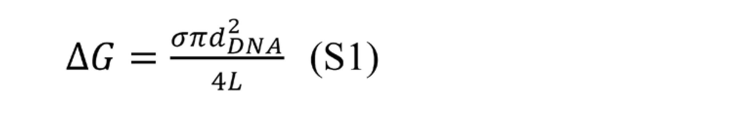
7.定性的分析
定性的アッセイは、本明細書中に記載される方法およびステップのプロセスを使用して行われ得る。図25に示されているチオールベースの切断ステップを用いて、実施例17に記載されているように、切断可能なリンカー接合体を使用して直接アッセイを実施することができる。そのようなアッセイを実施するための他の切断可能なリンカー手法はまた、本明細書中に記載されるように、種々のタグのカウントを可能にするための、リンカーの切断の種々の他の方法を含み得るがこれらに限定されないことが理解される。さらに、アプタマーを用いることができる。例えば、実施例17に記載の方法に加えて、そのような他の代替の切断方法および/または試薬は、本明細書に記載され、当業者に知られている他の切断方法に加えて、実施例16、実施例18、実施例19、実施例20および実施例21に記載のものを含み得る。この実施例(実施例24)において実証されたアッセイフォーマットは、直接アッセイを表すが、サンドイッチイムノアッセイフォーマットおよび/または様々な競合アッセイフォーマットなどの、当業者に知られているものなどのキャプチャオンザフライフォーマットを含む他のフォーマットが、記載された方法を使用してアッセイを実施するために同様に実施され得ることも理解される。
7. Qualitative Analysis Qualitative assays can be performed using the methods and steps described herein. Using the thiol-based cleavage step shown in FIG. 25, the assay can be performed directly using a cleavable linker conjugate as described in Example 17. Other cleavable linker techniques for performing such assays are also various other cleavable linker techniques to allow counting of different tags, as described herein. It is understood that methods may be included, but not limited to these. In addition, aptamers can be used. For example, in addition to the methods described in Example 17, such other alternative cleavage methods and / or reagents are described herein and in addition to other cleavage methods known to those of skill in the art. It may include those described in Example 16, Example 18, Example 19, Example 20 and Example 21. The assay format demonstrated in this Example (Example 24) represents a direct assay, but capture-on-the-fly formats such as those known to those of skill in the art, such as sandwich immunoassay formats and / or various competing assay formats. It is also understood that other formats, including, can be performed as well to perform the assay using the described methods.
例えば、実施例9に記載されているように、TSH(甲状腺刺激ホルモン)の検出のためのサンドイッチイムノアッセイフォーマットは、低コストDMFチップ上でそのようなアッセイを実施する能力を実証した。さらに、他の方法で当業者に知られている他の切断可能なリンカーに加えて、実施例1、実施例2、実施例3、実施例4、実施例5および実施例6に記載されているものなどの様々なヘテロ二官能性の切断可能なリンカーを使用して、イムノ接合体の生成に有用な多数の様々なバイオ接合試薬または切断可能なリンカーを有する他の活性特異的結合メンバーが合成され得る。本発明の実施に有用な免疫接合体は、実施例3、実施例4、実施例5および実施例6に記載されるものなどの方法によって、ならびに当業者に公知の他の方法によって合成することができる。さらに、実施例8は、サンドイッチおよび競合アッセイフォーマットを含み、キャプチャオンザフライフォーマットむ様々なアッセイフォーマット、ならびに当業者に知られているそれらの他の変形を実行するために必要な様々なステップを容易にすることができる、低コストチップ上の様々な液滴操作の機能を示す。実施例11は、アッセイにおいて切断可能な標識をカウントするために使用することができるナノポアの製造を示すが、当業者に知られているナノポア製造のための他の方法もまた、この目的のために使用することができることが理解される。実施例16はまた、切断が行われるアッセイの実施に有用な別の構築物を表し、したがって、この実施例に記載されるように、ナノポアカウント法を使用してカウントできるように、カウント可能な標識が遊離される。この構築物および当業者に明らかである他のものは、本明細書に記載されるようなアッセイにおいて使用され得る。 For example, as described in Example 9, the sandwich immunoassay format for the detection of TSH (thyroid stimulating hormone) demonstrated the ability to perform such an assay on a low cost DMF chip. Further described in Example 1, Example 2, Example 3, Example 4, Example 5 and Example 6 in addition to other cleavable linkers known to those of skill in the art by other methods. Various heterobifunctional cleavable linkers, such as those available, can be used to generate a large number of different bioconjugating reagents or other activity-specific binding members with cleavable linkers that are useful in the production of immunoconjugates. Can be synthesized. Immunoconjugates useful for carrying out the present invention are synthesized by methods such as those described in Examples 3, 4, 5, 5 and 6, and by other methods known to those of skill in the art. Can be done. In addition, Example 8 facilitates various assay formats, including sandwich and competitive assay formats, as well as the various steps required to perform those other variants known to those of skill in the art. Shows the various droplet manipulation features on low cost chips that can be done. Example 11 shows the production of nanopores that can be used to count cleavable labels in the assay, but other methods of nanopore production known to those of skill in the art are also for this purpose. It is understood that it can be used for. Example 16 also represents another construct useful for performing the assay in which cleavage is performed, and therefore, as described in this example, a countable label so that it can be counted using the nanopoacing method. Is released. This construct and others of skill in the art can be used in assays as described herein.
実施例22は、一般に、ナノポアを横断する様々な標識の存在に関する転位事象を測定することができるように、カウントをどのようにして行うことができるかを示す。図29は、カウントアッセイにおいてデータの質を操作することができるようにするための、シグナルの閾値化の概念を示す。図28は、この実施例に記載されるようなアッセイ方法を使用して、検体の存在を決定するために使用され得るデータの種類を表す定性的アッセイデータを示す。この特定の実施例では、dsDNAを標識として使用したが、ナノビーズ、デンドリマー等を含むがこれらに限定されない実施例5および/または実施例22に記載の標識などの他の標識を利用することもできる。さらに、他の公知の標識もまた用いることができる。適切な試薬を生成するために必要とされるそのような構築物は、本出願において本明細書に記載される様々な実施例を通して、またはそうでなければ当業者に知られている方法を介して合成され得る。 Example 22 shows how counting can be done so that dislocation events with respect to the presence of various labels across nanopores can generally be measured. FIG. 29 shows the concept of signal thresholding to allow manipulation of data quality in count assays. FIG. 28 shows qualitative assay data representing the types of data that can be used to determine the presence of a sample using an assay method as described in this example. Although dsDNA was used as a label in this particular example, other labels such as those described in Example 5 and / or Example 22 may also be utilized, including but not limited to nanobeads, dendrimers and the like. .. In addition, other known labels can also be used. Such constructs required to produce suitable reagents are provided through the various examples described herein in this application, or otherwise through methods known to those of skill in the art. Can be synthesized.
8.定量的分析
定量的アッセイは、本明細書に記載されるような方法およびステップのプロセスを使用して実施され得る。図25に示されているチオールベースの切断ステップを用いて、実施例17に記載されているように、切断可能なリンカー接合体を使用して直接アッセイを実施することができる。そのようなアッセイを実施するための他の切断可能なリンカー手法はまた、本明細書中に記載されるように、ナノポアを使用する種々のタグのカウントを可能にするための、リンカーの切断の種々の他の方法を含み得るがこれらに限定されないことが理解される。さらに、アプタマーを用いることができる。例えば、実施例17に記載の方法に加えて、そのような他の切断方法としては、本明細書に記載され、当業者に知られている方法に加えて、実施例18、実施例19、実施例20および実施例21に記載の方法が挙げられるが、これらに限定されない。この実施例(実施例25)において実証されたアッセイフォーマットは、直接アッセイを表すが、サンドイッチイムノアッセイフォーマットおよび/または様々な競合アッセイフォーマットなどの、当業者に知られているようなキャプチャオンザフライフォーマットを含む他のフォーマットが、アッセイを実施するために同様に実施され得ることも理解される。
8. Quantitative Analysis Quantitative assays can be performed using processes of methods and steps as described herein. Using the thiol-based cleavage step shown in FIG. 25, the assay can be performed directly using a cleavable linker conjugate as described in Example 17. Other cleavable linker techniques for performing such assays are also as described herein, for cleavage of the linker to allow counting of various tags using nanopores. It is understood that various other methods may be included, but are not limited thereto. In addition, aptamers can be used. For example, in addition to the method described in Example 17, such other cutting methods include, in addition to the methods described herein and known to those of skill in the art, Examples 18, 19, 19. Examples include, but are not limited to, the methods described in Example 20 and Example 21. The assay formats demonstrated in this Example (Example 25) represent direct assays, but include capture-on-the-fly formats as known to those of skill in the art, such as sandwich immunoassay formats and / or various competing assay formats. It is also understood that other formats can be performed as well to perform the assay.
例えば、実施例9に記載されているように、TSH(甲状腺刺激ホルモン)の検出のためのサンドイッチイムノアッセイフォーマットは、低コストDMFチップ上でそのようなアッセイを実施する能力を実証した。さらに、他の方法で当業者に知られている他の切断可能なリンカーに加えて、実施例1、実施例2、実施例3、実施例4、実施例5および実施例6に記載されているものなどの様々なヘテロ二官能性の切断可能なリンカーを使用して、イムノ接合体の生成に有用な多数の様々なバイオ接合試薬または切断可能なリンカーを有する他の活性特異的結合メンバーが、当業者により合成され得る。さらに、実施例8は、サンドイッチおよび競合アッセイフォーマットを含み、キャプチャオンザフライフォーマットを含む様々なアッセイフォーマット、ならびに当業者に知られているそれらの他の変形を実行するために必要な様々なステップを容易にすることができる、低コストチップ上の様々な液滴操作の機能を示す。実施例16はまた、切断が行われるアッセイの実施に有用な別の構築物を表し、したがって、この実施例に記載されるように、ナノポアカウント法を使用してカウントできるように、カウント可能な標識が遊離される。この構築物ならびに当業者に明らかである他のものは、本明細書に記載されるようなアッセイにおいて使用され得る。 For example, as described in Example 9, the sandwich immunoassay format for the detection of TSH (thyroid stimulating hormone) demonstrated the ability to perform such an assay on a low cost DMF chip. Further described in Example 1, Example 2, Example 3, Example 4, Example 5 and Example 6 in addition to other cleavable linkers known to those of skill in the art by other methods. Various heterobifunctional cleavable linkers, such as those skilled in the art, can be used by a number of different bioconjugating reagents or other activity-specific binding members with cleavable linkers to help generate immunoconjugates. , Can be synthesized by one of ordinary skill in the art. In addition, Example 8 facilitates various assay formats, including sandwich and competitive assay formats, including capture-on-the-fly formats, as well as the various steps required to perform those other variants known to those of skill in the art. Shows the functions of various droplet manipulations on low cost chips that can be made. Example 16 also represents another construct useful for performing the assay in which cleavage is performed, and therefore, as described in this example, a countable label so that it can be counted using the nanopoacing method. Is released. This construct as well as others of skill in the art can be used in assays as described herein.
実施例22は、一般に、ナノポアを横断する標識の存在に関する転位事象を測定することができるように、カウントをどのようにして行うことができるかを示す。図29は、カウントアッセイにおいてデータの質を操作することができるようにするための、シグナルの閾値化の概念を示す。図31、32および33は、この実施例に記載されるようなアッセイ方法を使用して、検体の量を決定するために使用され得るデータの種類を表す定性的アッセイデータ出力を示す。図34は、化学的方法を使用して切断された構築物から生成された標準曲線を示す。この特定の実施例では、dsDNAを標識として使用したが、ナノビーズ、デンドリマー等を含むがこれらに限定されない実施例5に記載の標識などの他の標識を利用することもできる。さらに、他の公知の標識もまた用いることができる。適切な試薬を生成するために必要とされるそのような構築物は、本明細書に記載されるように、または当業者に知られている方法を介して合成され得る。 Example 22 shows how counting can be done so that dislocation events with respect to the presence of markers across nanopores can generally be measured. FIG. 29 shows the concept of signal thresholding to allow manipulation of data quality in count assays. FIGS. 31, 32 and 33 show qualitative assay data output representing the types of data that can be used to determine the volume of a sample using an assay method as described in this example. FIG. 34 shows a standard curve generated from a structure cut using a chemical method. In this particular example, dsDNA was used as the label, but other labels such as the label described in Example 5 including, but not limited to, nanobeads, dendrimers and the like can also be used. In addition, other known labels can also be used. Such constructs required to produce suitable reagents can be synthesized as described herein or via methods known to those of skill in the art.
9.キットおよびカートリッジ
本明細書ではまた、開示されたデバイスを用いてまたは用いずに、上記の方法を実施する際に使用するためのキットも提供される。このキットは、開示されたデバイスを用いて検体を分析するための説明書を含み得る。キットに含まれる説明書は、包装材料に添付されてもよく、または添付文書として含まれてもよい。説明書は、書面または印刷物であってもよいが、それに限定されない。そのような説明書を保存し、それらをエンドユーザに伝達することができる任意の媒体が、本開示によって企図される。そのような媒体としては、電子記憶媒体(例えば、磁気ディスク、テープ、カートリッジ、チップ)、光学媒体(例えば、CD-ROM)等が挙げられるが、これらに限定されない。本明細書で使用される場合、「説明書」は、その説明書を提供するインターネットサイトのアドレスを含み得る。
9. Kits and Cartridges Also provided herein are kits for use in performing the above methods with or without the disclosed devices. The kit may include instructions for analyzing the specimen using the disclosed device. The instructions included in the kit may be attached to the packaging material or may be included as a package insert. The instructions may be in writing or printed, but are not limited thereto. Any medium in which such instructions can be stored and communicated to the end user is contemplated by the present disclosure. Examples of such media include, but are not limited to, electronic storage media (eg, magnetic disks, tapes, cartridges, chips), optical media (eg, CD-ROMs), and the like. As used herein, "manual" may include the address of the internet site that provides the manual.
このキットは、上述のように、内蔵ナノポアモジュールと共にマイクロ流体モジュールを含むカートリッジを含んでもよい。いくつかの実施形態では、マイクロ流体モジュールおよびナノポアモジュールは、一緒に可逆的に集積するための別々のコンポーネントであり得るか、または完全にまたは不可逆的にカートリッジに集積され得る。カートリッジは使い捨てであり得る。カートリッジは、上に開示された方法を実施するのに有用な1つ以上の試薬を含み得る。カートリッジは、1つ以上の別々の組成物として、または任意選択で、試薬の適合性が許す場合の混和物として、試薬を保持する1つ以上の容器を含み得る。カートリッジはまた、緩衝液(複数可)、希釈剤(複数可)、標準物質(複数可)(例えば、較正物質および対照)、ならびに/または試料処理、洗浄、もしくはアッセイの任意の他のステップの実施において有用な任意の他の材料などの、ユーザの観点から望ましい可能性がある他の材料(複数可)を含み得る。カートリッジは、上記の特異的結合メンバーのうちの1つ以上を含み得る。 The kit may include a cartridge containing a microfluidic module as well as a built-in nanopore module, as described above. In some embodiments, the microfluidic module and the nanopore module can be separate components for reversible integration together, or can be completely or irreversibly integrated into the cartridge. The cartridge can be disposable. The cartridge may contain one or more reagents useful for carrying out the methods disclosed above. The cartridge may include one or more containers holding the reagents as one or more separate compositions or, optionally, as an admixture where reagent compatibility allows. Cartridges are also buffers (s), diluents (s), reference materials (s) (eg, calibrators and controls), and / or any other step in sample processing, washing, or assay. It may include other materials (s) that may be desirable from the user's point of view, such as any other material useful in practice. The cartridge may include one or more of the specific binding members described above.
代替としてまたは追加として、キットは、較正物質または対照、例えば、精製された、および任意選択で凍結乾燥した目的の検体、またはカートリッジ上の液体、ゲル、もしくは他の形態の、または別個の目的の検体、ならびに/あるいは上記のデバイスおよび方法と共に使用するための少なくとも1つの容器(例えば、チューブ、マイクロタイタープレート、もしくはストリップ)、および/またはアッセイ緩衝液もしくは洗浄緩衝液などの緩衝液(それらのうちのいずれか1つは、濃縮溶液として提供され得る)を含み得る。いくつかの実施形態では、キットは、アッセイを行うのに必要な全ての構成要素、すなわち試薬、標準物質、緩衝液、希釈剤等を含む。説明書はまた、標準曲線を生成するための説明書を含み得る。 As an alternative or addition, the kit is a calibrator or control, eg, a purified and optionally lyophilized sample of interest, or a liquid, gel, or other form of liquid, gel, or other form on a cartridge, or for a separate purpose. Specimens and / or at least one container for use with the devices and methods described above (eg, tubes, microtiter plates, or strips) and / or buffers such as assay buffers or wash buffers (among them). Any one of the above may be provided as a concentrated solution). In some embodiments, the kit comprises all the components necessary to perform the assay, namely reagents, standards, buffers, diluents and the like. The instructions may also include instructions for generating a standard curve.
キットは、目的の検体を定量するための参照標準をさらに含み得る。参照標準は、目的の検体濃度の内挿および/または外挿のための標準曲線を確立するために用いられ得る。キットは、濃度レベルに関して異なる参照標準を含み得る。例えば、キットは、高濃度レベル、中濃度レベル、または低濃度レベルのいずれかを有する1つ以上の参照標準を含み得る。参照標準の濃度範囲に関しては、これはアッセイごとに最適化することができる。参照標準の例示的な濃度範囲としては、例えば、約10fg/mL、約20fg/mL、約50fg/mL、約75fg/mL、約100fg/mL、約150fg、約200fg/mL、約250fg/mL、約500fg/mL、約750fg/mL、約1000fg/mL、約10pg/mL、約20pg/mL、約50pg/mL、約75pg/mL、約100pg/mL、約150pg/mL、約200pg/mL、約250pg/mL、約500pg/mL、約750pg/mL、約1ng/mL、約5ng/mL、約10ng/mL、約12.5ng/mL、約15ng/mL、約20ng/mL、約25ng/mL、約40ng/mL、約45ng/mL、約50ng/mL、約55ng/mL、約60ng/mL、約75ng/mL、約80ng/mL、約85ng/mL、約90ng/mL、約95ng/mL、約100ng/mL約125ng/mL、約150ng/mL、約165ng/mL、約175ng/mL、約200ng/mL、約225ng/mL、約250ng/mL、約275ng/mL、300ng/mL、約400ng/mL、約425ng/mL、約450ng/mL、約465ng/mL、約475ng/mL、約500ng/mL、約525ng/mL、約550ng/mL、約575ng/mL、約600ng/mL、約700ng/mL、約725ng/mL、約750ng/mL、約765ng/mL、約775ng/mL、約800ng/mL、約825ng/mL、約850ng/mL、約875ng/mL、約900ng/mL、約925ng/mL、約950ng/mL、約975ng/mL、約1000ng/mL、約2μg/mL、約3μg/mL、約4μg/mL、約5μg/mL、約6μg/mL、約7μg/mL、約8μg/mL、約9μg/mL、約10μg/mL、約20μg/mL、約30μg/mL、約40μg/mL、約50μg/mL、約60μg/mL、約70μg/mL、約80μg/mL、約90μg/mL、約100μg/mL、約200μg/mL、約300μg/mL、約400μg/mL、約500μg/mL、約600μg/mL、約700μg/mL、約800μg/mL、約900μg/mL、約1000μg/mL、約2000μg/mL、約3000μg/mL、約4000μg/mL、約5000μg/mL、約6000μg/mL、約7000μg/mL、約8000μg/mL、約9000μg/mL、または約10000μg/mLが挙げられるが、これらに限定されない。 The kit may further include a reference standard for quantifying the sample of interest. Reference standards can be used to establish a standard curve for interpolation and / or extrapolation of the sample concentration of interest. The kit may contain different reference standards for concentration levels. For example, the kit may include one or more reference standards having either a high concentration level, a medium concentration level, or a low concentration level. For the reference standard concentration range, this can be optimized for each assay. Exemplary concentration ranges of the reference standard include, for example, about 10 fg / mL, about 20 fg / mL, about 50 fg / mL, about 75 fg / mL, about 100 fg / mL, about 150 fg, about 200 fg / mL, about 250 fg / mL. , About 500 fg / mL, about 750 fg / mL, about 1000 fg / mL, about 10 pg / mL, about 20 pg / mL, about 50 pg / mL, about 75 pg / mL, about 100 pg / mL, about 150 pg / mL, about 200 pg / mL , About 250 pg / mL, about 500 pg / mL, about 750 pg / mL, about 1 ng / mL, about 5 ng / mL, about 10 ng / mL, about 12.5 ng / mL, about 15 ng / mL, about 20 ng / mL, about 25 ng / ML, about 40 ng / mL, about 45 ng / mL, about 50 ng / mL, about 55 ng / mL, about 60 ng / mL, about 75 ng / mL, about 80 ng / mL, about 85 ng / mL, about 90 ng / mL, about 95 ng / ML, about 100 ng / mL about 125 ng / mL, about 150 ng / mL, about 165 ng / mL, about 175 ng / mL, about 200 ng / mL, about 225 ng / mL, about 250 ng / mL, about 275 ng / mL, 300 ng / mL , About 400 ng / mL, about 425 ng / mL, about 450 ng / mL, about 465 ng / mL, about 475 ng / mL, about 500 ng / mL, about 525 ng / mL, about 550 ng / mL, about 575 ng / mL, about 600 ng / mL , About 700 ng / mL, about 725 ng / mL, about 750 ng / mL, about 765 ng / mL, about 775 ng / mL, about 800 ng / mL, about 825 ng / mL, about 850 ng / mL, about 875 ng / mL, about 900 ng / mL , About 925 ng / mL, about 950 ng / mL, about 975 ng / mL, about 1000 ng / mL, about 2 μg / mL, about 3 μg / mL, about 4 μg / mL, about 5 μg / mL, about 6 μg / mL, about 7 μg / mL , About 8 μg / mL, about 9 μg / mL, about 10 μg / mL, about 20 μg / mL, about 30 μg / mL, about 40 μg / mL, about 50 μg / mL, about 60 μg / mL, about 70 μg / mL, about 80 μg / mL , About 90 μg / mL, about 100 μg / mL, about 200 μg / mL, about 300 μg / mL, about 400 μg / mL, about 500 μg / mL, about 600 μg / mL, about 700 μg / mL, about 800 μg / mL, about 900 μg / mL , About 1000 μg / mL, about 2000 μg / mL, about 3000 μg / mL, about 4000 μg / mL, about 5000 μg / ML, about 6000 μg / mL, about 7000 μg / mL, about 8000 μg / mL, about 9000 μg / mL, or about 10000 μg / mL, but is not limited to these.
キットで提供される任意の特異的結合メンバーは、例えば蛍光体、酵素、アプタマー、ビーズ、ナノ粒子、微粒子、ポリマー、タンパク質、ビオチン/アビジン標識等のタグもしくは標識を組み込み得るか、あるいはキットは、特異的結合メンバーを標識するための試薬、または特異的結合メンバーを検出するためおよび/もしくは検体を標識するための試薬、または検体を検出するための試薬を含み得る。必要に応じて、キットは、1つ以上の異なるタグまたは標識を含み得る。キットはまた、切断媒介試薬などの、切断を誘発するための成分を含み得る。例えば、切断媒介試薬は、ジチオトレイトール(DTT)またはトリス(2-カルボキシエチル)ホスフィン)TCEPなどの還元剤を含み得る。特異的結合メンバー、較正物質、および/もしくは対照は、別々の容器に入れて提供するか、または適切なアッセイフォーマットもしくはカートリッジに予め分注することができる。タグは、開示されたデバイスを使用して検出されてもよい。 Any specific binding member provided in the kit may incorporate tags or labels such as, for example, reagents, enzymes, aptamers, beads, nanoparticles, microparticles, polymers, proteins, biotin / avidin labels, or the kit may incorporate tags or labels. It may include a reagent for labeling a specific binding member, or a reagent for detecting a specific binding member and / or a reagent for labeling a sample, or a reagent for detecting a sample. If desired, the kit may include one or more different tags or labels. The kit may also contain components for inducing cleavage, such as cleavage mediator reagents. For example, cleavage mediator reagents can include reducing agents such as dithiothreitol (DTT) or tris (2-carboxyethyl) phosphine) TCEP. Specific binding members, calibrators, and / or controls can be provided in separate containers or pre-dispensed into suitable assay formats or cartridges. The tag may be detected using the disclosed device.
キットは、例えば、多重化アッセイにおいて試料中の1つ以上の標的検体を検出するための1つ以上の特異的結合メンバーを含み得る。キット中の異なる種類の特異的結合メンバーの数は、キットの意図する用途に応じて広範囲におよび得る。キット中の特異的結合メンバーの数は、1~約10、またはそれ以上の範囲であり得る。例えば、キットは、1~10個の特異的結合メンバー、1~9個の特異的結合メンバー、1~8個の特異的結合メンバー、1~7個の特異的結合メンバー、1~6個の特異的結合メンバー、1~5個の特異的結合メンバー、1~4個の特異的結合メンバー、1~3個の特異的結合メンバー、1~2個の特異的結合メンバー、2~10個の特異的結合メンバー、2~9個の特異的結合メンバー、2~8個の特異的結合メンバー、2~7個の特異的結合メンバー、2~6個の特異的結合メンバー、2~5個の特異的結合メンバー、2~4個の特異的結合メンバー、3~10個の特異的結合メンバー、3~9個の特異的結合メンバー、3~8個の特異的結合メンバー、3~7個の特異的結合メンバー、3~6個特異的結合メンバー、3~5個の特異的結合メンバー、3~4個の特異的結合メンバー、4~10個の特異的結合メンバー、4~9個の特異的結合メンバー、4~8個の特異的結合メンバー、4~7個の特異的結合メンバー、4~6個の特異的結合メンバー、5~10個の特異的結合メンバー、5~9個の特異的結合メンバー、5~8個の特異的結合メンバー、5~7個の特異的結合メンバー、5~6個の特異的結合メンバー、6~10個の特異的結合メンバー、6~9個の特異的結合メンバー、6~8個の特異的結合メンバー、6~7個の特異的結合メンバー、7~10個の特異的結合メンバー、7~9個の特異的結合メンバー、7~8個の特異的結合メンバー、8~10個の特異的結合メンバー、8~9個の特異的結合メンバー、または9~10個の特異的結合メンバーを含み得る。1つ以上の特異的結合メンバーの各々は、異なる標的検体に結合し得、各特異的結合メンバーは、異なるタグおよび/またはアプタマーで標識され得る。例えば、キットは、第1の標的検体に結合する第1の特異的結合メンバー、第2の標的検体に結合する第2の特異的結合メンバー、および第3の標的検体に結合する第3の特異的結合メンバー等を含み得、第1の特異的結合メンバーは、第1のタグおよび/またはアプタマーで標識され、第2の特異的結合メンバーは、第2のタグおよび/またはアプタマーで標識され、第3の特異的結合メンバーは、第3のタグおよび/またはアプタマーで標識される等である。1つ以上の特異的結合メンバーに加えて、キットは、1つ以上の追加のアッセイ成分、例えば好適な緩衝媒体等をさらに含み得る。キットはまた、上記のものなどの、タグおよび/またはアプタマーを検出および測定するためのデバイスを含み得る。最後に、キットは、本発明による検体検出の方法において特異的結合メンバーを使用するための説明書を含んでもよく、使用のためのこれらの説明書は、キット包装および/または添付文書に存在してもよい。 The kit may include, for example, one or more specific binding members for detecting one or more target specimens in a sample in a multiplexing assay. The number of different types of specific binding members in the kit can range widely depending on the intended use of the kit. The number of specific binding members in the kit can range from 1 to about 10 or more. For example, the kit contains 1 to 10 specific binding members, 1 to 9 specific binding members, 1 to 8 specific binding members, 1 to 7 specific binding members, and 1 to 6 specific binding members. Specific binding members, 1 to 5 specific binding members, 1 to 4 specific binding members, 1 to 3 specific binding members, 1 to 2 specific binding members, 2 to 10 specific binding members. Specific binding members, 2-9 specific binding members, 2-8 specific binding members, 2-7 specific binding members, 2-6 specific binding members, 2-5 specific binding members. Specific binding members, 2-4 specific binding members, 3-10 specific binding members, 3-9 specific binding members, 3-8 specific binding members, 3-7 specific binding members. Specific binding members, 3-6 specific binding members, 3-5 specific binding members, 3-4 specific binding members, 4-10 specific binding members, 4-9 specifics Target binding members, 4 to 8 specific binding members, 4 to 7 specific binding members, 4 to 6 specific binding members, 5 to 10 specific binding members, 5 to 9 specific binding members. Target binding members, 5 to 8 specific binding members, 5 to 7 specific binding members, 5 to 6 specific binding members, 6 to 10 specific binding members, 6 to 9 specific binding members. Specific binding members, 6-8 specific binding members, 6-7 specific binding members, 7-10 specific binding members, 7-9 specific binding members, 7-8 specifics. It may include target binding members, 8-10 specific binding members, 8-9 specific binding members, or 9-10 specific binding members. Each of the one or more specific binding members can bind to different target specimens, and each specific binding member can be labeled with a different tag and / or aptamer. For example, the kit includes a first specific binding member that binds to a first target sample, a second specific binding member that binds to a second target sample, and a third specific binding member that binds to a third target sample. The first specific binding member may be labeled with a first tag and / or an aptamer, and the second specific binding member may be labeled with a second tag and / or aptamer. The third specific binding member is labeled with a third tag and / or aptamer, and so on. In addition to one or more specific binding members, the kit may further include one or more additional assay components, such as suitable buffer media. The kit may also include devices for detecting and measuring tags and / or aptamers, such as those described above. Finally, the kit may include instructions for using specific binding members in the method of specimen detection according to the invention, these instructions for use are present in the kit packaging and / or package insert. You may.
任意選択で、キットには、品質管理構成要素(例えば、感度パネル、較正物質、および正の対照)が含まれる。品質管理試薬の調製は、当技術分野において周知であり、種々の免疫診断製品のための挿入シートに記載されている。感度パネルメンバーを任意選択で使用して、アッセイ性能特性を確立し、さらに任意選択で、キット試薬の完全性およびアッセイの標準化の有用な指標である。 Optionally, the kit includes quality control components (eg, sensitivity panel, calibrator, and positive control). The preparation of quality control reagents is well known in the art and is described on insert sheets for various immunodiagnostic products. Sensitivity panel members are optionally used to establish assay performance characteristics and, optionally, are useful indicators of kit reagent integrity and assay standardization.
キットはまた、任意選択で、診断アッセイを行うか、または例えば緩衝液、塩、酵素、酵素共因子、基質、検出試薬等の品質管理評価を容易にするために必要な他の試薬を含むことができる。試験試料の単離および/または処理のための緩衝液および溶液(例えば、前処理試薬)などの他の成分もまた、キットに含めることができる。キットは、追加として1つ以上の他の対照を含み得る。キットの構成要素のうちの1つ以上は、凍結乾燥することができ、その場合キットは、凍結乾燥された構成要素の再構成に適した試薬をさらに含むことができる。構成要素のうちの1つ以上は、液体形態であり得る。 The kit may also optionally include other reagents necessary for performing diagnostic assays or facilitating quality control assessments such as buffers, salts, enzymes, enzyme cofactors, substrates, detection reagents, etc. Can be done. Other components such as buffers and solutions (eg, pretreatment reagents) for isolation and / or treatment of test samples can also be included in the kit. The kit may additionally include one or more other controls. One or more of the components of the kit can be lyophilized, in which case the kit can further include reagents suitable for reconstitution of the lyophilized components. One or more of the components may be in liquid form.
キットの様々な構成要素は、任意選択で、必要に応じて好適な容器で提供される。キットは、試料を保持または保存するための容器(例えば、尿、唾液、血漿、脳脊髄液、または血清試料用の容器またはカートリッジ、あるいは組織吸引をもたらすために組織を保存、輸送または処理するための適切な容器)をさらに含み得る。適切な場合、キットは、任意選択で、反応容器、混合容器、および試薬または試験試料の調製を容易にする他の成分も含み得る。キットはまた、マイクロサンプリングデバイス、マイクロニードル、または他の低侵襲性の無痛性の採血方法などの様々な採血/転送デバイスなどの、試験試料の取得を支援するための1つ以上の試料収集/取得器具;採血管(複数可);ランセット;毛細管採血管;他の単一の指先刺し採血方法;頬スワブ、鼻/咽頭スワブ;組織試料を入手、保存、もしくは吸引するための16ゲージまたは他のサイズの針、パンチ生検用の円形ブレード(例えば、1~8mm、もしくは他の適切なサイズ)、外科用ナイフもしくはレーザー(例えば、特に手持ち式)、注射器、滅菌容器、またはカニューレ等を含み得る。キットは、関節吸引、円錐生検、穿孔生検、細針吸引生検、画像誘導経皮針吸引生検、気管支肺胞洗浄、内視鏡生検、および腹腔鏡生検を補助するための1つ以上の器具を含み得る。 The various components of the kit are optionally provided in suitable containers as needed. Kits are containers for holding or storing samples (eg, containers or cartridges for urine, saliva, plasma, cerebrospinal fluid, or serum samples, or for storing, transporting, or processing tissue to provide tissue aspiration. Suitable container) may further contain. Where appropriate, the kit may optionally include reaction vessels, mixing vessels, and other components that facilitate the preparation of reagents or test samples. The kit also collects one or more samples to assist in the acquisition of test samples, such as various blood collection / transfer devices such as microsampling devices, microneedles, or other minimally invasive, painless blood collection methods. Acquisition device; blood collection tube (s); lancet; capillary blood collection tube; other single fingertip puncture blood collection method; cheek swab, nasal / pharyngeal swab; 16 gauge or other for obtaining, storing, or aspirating tissue sample Includes sized needles, circular blades for punch biopsy (eg, 1-8 mm, or other suitable size), surgical knives or lasers (eg, especially handheld), syringes, sterile vessels, or cannulas, etc. obtain. The kit assists joint aspiration, cone biopsy, perforation biopsy, fine needle aspiration biopsy, image-guided percutaneous needle aspiration biopsy, bronchoalveolar lavage, endoscopic biopsy, and laparoscopic biopsy. It may include one or more instruments.
タグまたは検出可能な標識が、少なくとも1つのアクリジニウム化合物であるか、またはそれを含む場合、キットは、少なくとも1つのアクリジニウム-9-カルボキシアミド、少なくとも1つのアクリジニウム-9-カルボキシレートアリールエステル、またはそれらの任意の組み合わせを含み得る。タグまたは検出可能な標識が、少なくとも1つのアクリジニウム化合物であるか、またはそれを含む場合、キットはまた、緩衝液、溶液、および/または少なくとも1つの塩基性溶液などの過酸化水素源を含み得る。必要に応じて、キットは、磁性粒子、ビーズ、膜、足場分子、フィルム、ろ紙、ディスク、またはチップなどの固相を含むことができる。 If the tag or detectable label is, or contains, at least one acridinium compound, the kit includes at least one acridinium-9-carboxamide, at least one acridinium-9-carboxylate aryl ester, or them. Can include any combination of. If the tag or detectable label is or contains at least one acridinium compound, the kit may also include a hydrogen peroxide source such as a buffer, a solution, and / or at least one basic solution. .. If desired, the kit can include a solid phase such as magnetic particles, beads, membranes, scaffold molecules, films, filter papers, discs, or chips.
所望される場合、キットは、試験試料を別の検体についてアッセイするための、1つ以上の成分を、単独でまたは使用説明書と組み合わせてさらに含むことができ、これらは感染症、心臓病、代謝性疾患、甲状腺疾患等の疾患状態または障害のバイオマーカーなどのバイオマーカーであり得る。 If desired, the kit may further include one or more components for assaying the test sample against another sample, either alone or in combination with instructions for infectious diseases, heart disease, etc. It can be a biomarker such as a biomarker of a disease state or disorder such as metabolic disease, thyroid disease.
本発明は、本明細書に提供される非限定的な実施例によって例示される複数の態様を有する。 The present invention has a plurality of embodiments exemplified by the non-limiting examples provided herein.
集積DMF-電気化学的/電気的/光学的検出チップ、デバイス、およびシステム
前述のセクションで述べたように、検体検出チップを操作して試料を調製し、検体検出チップ内の調製された試料から検体関連シグナルを検出するように構成された検体検出デバイスが開示されている。検体検出チップは、デジタルマイクロ流体(DMF)領域と、DMF領域と重なり得るか、または空間的に分離されていてもよい検体検出領域と、を含んでもよい。前のセクションに記載されるように、DMF領域およびナノポア領域は、動作可能に接続されているか、または単一のカートリッジに集積されていることがある個々のデバイスに分離することができる。部分的にまたは完全に集積されたデバイスのDMF領域およびナノポア領域に作用して、液滴の移動に影響を及ぼし、ナノポアからの電気シグナルを検出するための器具もまた、本明細書に提供される。この器具については、前のセクションで詳細に説明されている。この器具は、異なるタイプの検体検出デバイスを操作するための構成要素をさらに含み得、このデバイスは、臨床化学を実施するためのカートリッジであり得る。
Integrated DMF-Electrochemical / Electrical / Optical Detection Chips, Devices, and Systems As mentioned in the previous section, the sample detection chip is manipulated to prepare a sample and from the prepared sample in the sample detection chip. A sample detection device configured to detect a sample-related signal is disclosed. The sample detection chip may include a digital microfluidic (DMF) region and a sample detection region that may overlap or be spatially separated from the DMF region. As described in the previous section, the DMF and nanopore regions can be separated into individual devices that may be operably connected or integrated into a single cartridge. Also provided herein are instruments for acting on the DMF and nanopore regions of a partially or fully integrated device to affect the movement of droplets and detect electrical signals from the nanopores. To. This device is described in detail in the previous section. The instrument may further include components for manipulating different types of specimen detection devices, which devices may be cartridges for performing clinical chemistry.
ある特定の場合において、臨床化学は、基質に対する酵素の作用によって生成される電気化学種または発色反応産物の検出を含み得る。例えば、基質は、試料中に存在する検体であり得、酵素は、検体に特異的であり得、検体と触媒的に反応して電気化学種または着色反応産物を生成し得る。他の場合において、臨床化学は、第1の結合メンバーを使用して検体を捕捉して、検体および第1の結合メンバーを含む第1の複合体を生成することと、その複合体を、検体に結合する第2の結合メンバーと接触させて、検体、第1の結合メンバー、および第2の結合メンバーを含む第2の複合体を生成することと、を含み得る。第2の結合メンバーは、好適な基質への曝露時に電気化学種または発色反応産物を生成する酵素に接合している。 In certain cases, clinical chemistry may include detection of an electrochemical species or color reaction product produced by the action of an enzyme on a substrate. For example, the substrate can be a sample present in the sample, the enzyme can be specific to the sample, and can catalytically react with the sample to produce an electrochemical species or coloring reaction product. In other cases, clinical chemistry uses a first binding member to capture a sample to produce a first complex containing the sample and the first binding member, and the complex is a sample. It may include contacting with a second binding member that binds to to form a second complex comprising a sample, a first binding member, and a second binding member. The second binding member is attached to an enzyme that produces an electrochemical species or color reaction product upon exposure to a suitable substrate.
ある特定の実施形態では、臨床化学を実施するためのカートリッジの検体検出領域は、検体が試料中に存在するときに生成される電気化学種を検出するための電極を含み得る。そのようなカートリッジはまた、本明細書ではDMF電気化学チップとも呼ばれる。他の実施形態では、臨床化学を実施するためのカートリッジの検体検出領域は、検体が試料中に存在するときに生成される光学シグナルの検出のために構成されてもよい。そのようなカートリッジは、本明細書ではDMF光学チップとも呼ばれる。臨床化学を実施するためのカートリッジは、DMF電気化学チップと呼ばれることがあることに留意されたい。 In certain embodiments, the sample detection area of the cartridge for performing clinical chemistry may include electrodes for detecting the electrochemical species produced when the sample is present in the sample. Such cartridges are also referred to herein as DMF electrochemical chips. In other embodiments, the sample detection area of the cartridge for performing clinical chemistry may be configured for the detection of optical signals generated when the sample is present in the sample. Such cartridges are also referred to herein as DMF optical chips. It should be noted that cartridges for performing clinical chemistry are sometimes referred to as DMF electrochemical chips.
さらに他の実施形態では、カートリッジは、電気化学的検出およびナノポア層を使用する検出のために構成された多機能であり得る。そのようなものとして、本明細書に開示されている器具は、複数の単一機能カートリッジ上ならびに多機能カートリッジ(複数可)上で動作することができる。 In yet another embodiment, the cartridge can be multifunctional configured for electrochemical detection and detection using a nanopore layer. As such, the instruments disclosed herein can operate on multiple single-function cartridges as well as on multiple-function cartridges (s).
DMF領域を使用して、分析のための液滴を、検出領域に転送することができ、ここで液滴が、電気的に(例えば、ナノポアを使用して)、電気化学的に(例えば、臨床化学を使用して)、および/または光学的に(例えば、臨床化学を使用して)分析される。光学的検出は、比色検出、濁度検出、蛍光検出、および/または画像分析であり得る。画像分析は、検体検出カートリッジからの光学シグナルの検出を含み得る。光学シグナルは、比色、濁度、または蛍光シグナルなどの光シグナルであり得る。電気化学的検出は、電流測定、クーロメトリー、電位差測定、電圧測定、インピーダンス、またはそれらの組み合わせを含み得る。 The DMF region can be used to transfer the droplet for analysis to the detection region, where the droplet is electrically (eg, using nanopores) and electrochemically (eg, using nanopores). Analyzed (using clinical chemistry) and / or optically (eg, using clinical chemistry). Optical detection can be colorimetric detection, turbidity detection, fluorescence detection, and / or image analysis. Image analysis may include detection of optical signals from specimen detection cartridges. The optical signal can be an optical signal such as colorimetric, turbidity, or fluorescence signal. Electrochemical detection can include current measurement, coulometry, potentiometric measurement, voltage measurement, impedance, or a combination thereof.
「検体検出チップ」、「検体検出カートリッジ」という語句、ならびに「チップ」および「カートリッジ」という語句は、本明細書において互換的に使用され、本明細書に開示される検体検出器具と適合する使い捨てまたは再使用可能な試料処理デバイスを指す。本明細書に開示される検体検出器具はまた、本明細書に提供されるチップ内の試料を処理するために使用される検体検出デバイスとも呼ばれる。ある特定の実施形態では、検体検出チップは、第1の基板と第2の基板とを含み得、第2の基板は、第1の基板上に位置付けられ、ギャップによって第1の基板から分離されている。第1または第2の基板は、複数のDMF電極を含み得る。複数のDMF電極は、活性化および非活性化のために個々に制御可能な電極のアレイまたは一連の電極であり得る。複数のDMF電極は、DMF電極を電気的に絶縁するために絶縁材料で覆われてもよい。ある特定の実施形態では、第1の基板と第2の基板との間の空間/ギャップは、空気またはオイルなどの不活性流体で充填されてもよい。ある特定の実施形態では、DMF電極は、本明細書の前のセクションに記載されているように配置することができる。例示的な実施形態では、一連のDMF電極を第1の基板上に配設することができ、単一の電極を第1の基板上の一連の電極と対面構成で第2の基板上に配設することができる。一連の電極および単一電極は、絶縁層で被覆されていてもよい。他の場合において、第1の基板上の一連のまたは複数の電極は、共面電極として構成され、第2の基板は、電極を含まなくてもよい。DMF電極の様々な構成は、集積マイクロ流体デバイスおよびナノポアデバイスについて説明している前のセクションに記載されている。DMF電極のこれらの構成のいずれも、本明細書に開示されている追加のカートリッジに存在することができる。 The terms "specimen detection chip", "specimen detection cartridge", and the terms "chip" and "cartridge" are used interchangeably herein and are disposable with the specimen detection instruments disclosed herein. Or refers to a reusable sample processing device. The sample detection device disclosed herein is also referred to as a sample detection device used to process the sample in the chip provided herein. In certain embodiments, the sample detection chip may include a first substrate and a second substrate, the second substrate being positioned on the first substrate and separated from the first substrate by a gap. ing. The first or second substrate may include a plurality of DMF electrodes. Multiple DMF electrodes can be an array of individually controllable electrodes or a set of electrodes for activation and deactivation. The plurality of DMF electrodes may be covered with an insulating material in order to electrically insulate the DMF electrodes. In certain embodiments, the space / gap between the first substrate and the second substrate may be filled with an inert fluid such as air or oil. In certain embodiments, the DMF electrode can be arranged as described in the previous section of the specification. In an exemplary embodiment, a series of DMF electrodes can be placed on the first substrate and a single electrode is placed on the second substrate in a face-to-face configuration with the series of electrodes on the first substrate. Can be set up. The series of electrodes and the single electrode may be coated with an insulating layer. In other cases, the series or plurality of electrodes on the first substrate may be configured as coplanar electrodes and the second substrate may be free of electrodes. Various configurations of DMF electrodes are described in the previous section describing integrated microfluidic and nanopore devices. Any of these configurations of DMF electrodes can be present in the additional cartridges disclosed herein.
前のセクションで説明したように、第1層および/または第2層に存在する電極は、例えばインジウム錫酸化物、フッ素ドープ酸化錫(FTO)、ドープ酸化亜鉛等の実質的に透明な材料から製造することができる。さらに、一方または両方の基板は、光学的インタロゲーションを容易にするために実質的に透明であり得る。 As described in the previous section, the electrodes present in the first and / or second layers are from a substantially transparent material such as indium tin oxide, fluorine-doped tin oxide (FTO), doped zinc oxide, etc. Can be manufactured. In addition, one or both substrates can be substantially transparent to facilitate optical intervention.
検体検出デバイスは、そのデバイスに挿入された検体検出チップ内の光学シグナル、電気化学的、電気的シグナルを検出するための光学的、電気化学的、および/または電気的手段を含み得る。さらに、検体検出デバイスは、検体検出チップに存在するDMF電極を操作するための手段を含む。本明細書に開示される検体検出デバイスは、カートリッジと相互作用するための1つまたは複数のインターフェースを含み得る。ある特定の場合において、カートリッジインターフェースは、スロットなどの挿入領域であり得る。他の場合において、インターフェースは、カートリッジを受け入れるための凹部であり得、ドアまたは蓋によって包囲され得る。検体検出デバイスは、複数の検体検出チップと適合し得る単一のインターフェースを含み得る。例えば、挿入スロットは、電気化学シグナルを検出する検体検出チップ、光学シグナルを検出する検体検出チップ、および/または電気シグナルを検出する検体検出チップと適合し得る。ある特定の実施形態では、検体検出デバイスは、例えば、複数のチップを使用して異なる試料中の同じ検体を検出するため、または複数の異なるチップを使用して同じ試料中の複数の異なる検体を同時に検出するために、複数の検体検出チップを同時に動作させるように構成され得る。そのような実施形態では、デバイスは、挿入領域などの複数のインターフェースを含むことができる。 The sample detection device may include optical, electrochemical, and / or electrical means for detecting optical signals, electrochemical, electrical signals in a sample detection chip inserted into the device. Further, the sample detection device includes means for manipulating the DMF electrode present on the sample detection chip. The specimen detection device disclosed herein may include one or more interfaces for interacting with the cartridge. In certain cases, the cartridge interface can be an insertion area such as a slot. In other cases, the interface can be a recess for receiving the cartridge and can be surrounded by a door or lid. The sample detection device may include a single interface that may be compatible with multiple sample detection chips. For example, the insertion slot may be compatible with a sample detection chip that detects an electrochemical signal, a sample detection chip that detects an optical signal, and / or a sample detection chip that detects an electrical signal. In certain embodiments, the sample detection device may use, for example, multiple chips to detect the same sample in different samples, or multiple different chips to use multiple different samples in the same sample. Multiple sample detection chips may be configured to operate simultaneously for simultaneous detection. In such an embodiment, the device can include multiple interfaces such as an insertion area.
本開示の検体検出チップは、任意選択で、血漿分離構成要素を含み得る。ある特定の実施形態では、血漿分離構成要素は、全血試料中に存在する細胞を捕捉し、血漿を濾過して分析のために試料液滴(複数可)に加工することを可能にするフィルタを含み得る。他の実施形態では、血漿分離構成要素は、流体分離要素であり得る。検体検出チップの実施形態を以下に開示する。以下に記載される検体検出チップのうちのいずれも、任意選択で、血漿分離構成要素を含み得る。ある特定の場合において、血漿分離構成要素は、市販の膜であり得る。ある特定の実施形態では、International Point of Care,Inc.から入手可能なもの(例えば、Primecare(商標)親水性非対称膜)またはPall Corporation(例えば、Vivid(商標)血漿分離膜)から入手可能なものなどの市販の膜を、血漿の分離に使用することができる。ある特定の場合において、膜は、本開示のカートリッジに集積されてもよい。他の実施形態では、本開示のチップ、器具、およびシステムは、全血試料中の検体を検出するように構成され得る。 The sample detection chip of the present disclosure may optionally include plasma separation components. In certain embodiments, the plasma separation component is a filter that captures cells present in a whole blood sample and allows the plasma to be filtered and processed into sample droplets (s) for analysis. May include. In other embodiments, the plasma separation component can be a fluid separation component. An embodiment of the sample detection chip is disclosed below. Any of the sample detection chips described below may optionally include plasma separation components. In certain cases, the plasma separation component can be a commercially available membrane. In certain embodiments, International Point of Care, Inc. Commercially available membranes, such as those available from (eg, Primecare ™ hydrophilic asymmetric membranes) or Pall Corporation (eg, Vivid ™ plasma separation membranes), are used for plasma separation. Can be done. In certain cases, the membrane may be integrated into the cartridges of the present disclosure. In other embodiments, the chips, instruments, and systems of the present disclosure may be configured to detect a sample in a whole blood sample.
i.DMF電気化学検出カートリッジ
ある特定の実施形態では、本明細書に開示されるカートリッジは、重なり合うかまたは空間的に分離されていてもよいDMF領域および検体検出領域を含む。DMF領域を使用して、分析のための液滴を検出領域に転送することができ、ここで液滴は、(臨床化学アッセイで生成された電気化学種の検出のために)電気化学的に分析される。電気化学分析は、試料中の検体の存在によって生成される電気活性種によって発生する電気シグナルを検出する作用電極を利用することによって行われる。電気シグナルは、試料中に存在する検体の量に比例するため、検出された電気シグナルを定量して、試料中の検体の存在または濃度を決定することができる。電気化学的検出は、本開示において提供される器具によって行われる電流測定、クーロメトリー、電位差測定、電圧測定、インピーダンス、またはそれらの組み合わせを含み得る。
i. DMF Electrochemical Detection Cartridge In certain embodiments, the cartridges disclosed herein include a DMF region and a specimen detection region that may overlap or be spatially separated. The DMF region can be used to transfer droplets for analysis to the detection region, where the droplets are electrochemically (for the detection of electrochemical species produced in the clinical chemistry assay). Be analyzed. Electrochemical analysis is performed by utilizing a working electrode that detects an electrical signal generated by an electrically active species produced by the presence of the specimen in the sample. Since the electrical signal is proportional to the amount of sample present in the sample, the detected electrical signal can be quantified to determine the presence or concentration of the sample in the sample. The electrochemical detection may include current measurement, coulometry, potentiometric measurement, voltage measurement, impedance, or a combination thereof performed by the instruments provided in the present disclosure.
ある特定の実施形態では、電気化学種は、検体に対する検体特異的酵素の作用によって生成され得る。他の実施形態では、電気化学種は、基質に対する酵素の作用によって生成されてもよい。そのような実施形態では、酵素は、検体に特異的ではない。むしろ、酵素は、検体に特異的に結合する結合メンバーに接合されている。ある特定の実施形態では、電気化学種によって生成された電気シグナルを増幅するために、酸化還元媒介物質が含まれてもよい。検体特異的酵素および酸化還元媒介物質は周知であり、所望の感受性および/または特異性に基づいて選択され得る。 In certain embodiments, the electrochemical species can be produced by the action of a specimen-specific enzyme on the specimen. In other embodiments, the electrochemical species may be produced by the action of an enzyme on a substrate. In such embodiments, the enzyme is not sample-specific. Rather, the enzyme is attached to a binding member that specifically binds to the specimen. In certain embodiments, redox mediators may be included to amplify the electrical signal produced by the electrochemical species. Specimen-specific enzymes and redox mediators are well known and can be selected based on the desired sensitivity and / or specificity.
電気化学種を検出するための電極は、数多くの構成で提供することができる。そのような電極は、DMF電極とは別個であり得るか、または電気化学的感知のために電極に修飾されたDMF電極であり得る。本明細書で提供される器具は、電極のアレイならびに電気化学的感知のための電極に印加される電力を制御する電気回路を含む。DMF電極および電気化学的感知用の電極を含む、検体検出チップの例示的な構成は、以下でさらに説明される。 Electrodes for detecting electrochemical species can be provided in a number of configurations. Such an electrode can be separate from the DMF electrode or can be a DMF electrode modified to the electrode for electrochemical sensing. The instruments provided herein include an array of electrodes as well as electrical circuits that control the power applied to the electrodes for electrochemical sensing. An exemplary configuration of a sample detection chip, including a DMF electrode and an electrode for electrochemical sensing, is further described below.
図48A~48Fは、本開示のチップ内に存在する電極の概略図を提供する。図48Aは、液滴をチップのセンサ領域511に転送するために使用されるDMF電極510を示す。センサ領域511は、作用電極512および参照電極513を含む。図48Bは、センサ領域511上に位置付けられた液滴514を示す。図48Cは、センサ領域511、作用電極512、および参照電極513を示し、ここで電極は半円形であり、共面構成で配設されている。ここには示されていないが、一方の電極を、他方の電極と対面構成に配置することができる。そのような実施形態では、電気化学的検出用のセンサ領域は、作用電極と参照電極とを隔てるギャップを含むことができ、電極は液滴がセンサ領域内に移動すると電気的に接続される。作用電極および参照電極は、リード線515を介して接触パッド516および517に接続されている。接触パッドは、チップを動作させるデバイスに動作可能に接続されている。目的の検体の電気化学的検出のための電極の追加の構成は、図48D~図48Eに示されている。図48Dにおいて、作用電極512は円形であり、一方参照電極513は弧状であり、作用電極と同心であり、作用電極を取り囲んでいる。図48Eにおいて、センサ領域は、3つの電極、すなわち作用電極512、参照電極513、および対電極518を含む。液滴および電極は、液滴が作用電極および参照電極(および存在する場合には対電極)の両方と接触するようなサイズである。図48Fは、相対サイズの液滴および作用電極を示し、電極(複数可)のサイズおよび形状は、液滴サイズに一致するように構成される。この実施形態では、参照電極が作用電極に面する構成で存在することに留意されたい。作用電極513は、第2の直径Bを有する液滴514よりも小さい第1の直径(A)を有する。第1の直径Aは、約50μm~1.9mmであり得る。第2の直径Bは、約100μm~2mmであり得る。液滴直径に対する電極直径の他の比率もまた、本開示のチップにおいて使用され得る。作用電極および参照電極(および存在する場合は対電極)が共面構成にある実施形態では、電極の総面積(電極間の任意のギャップを含む)は、液滴の直径に一致するようなサイズである(図48Bを参照)。
48A-48F provide schematic views of the electrodes present in the chips of the present disclosure. FIG. 48A shows the
ある特定の実施形態では、図48A~図48Fに示すものなどの電気化学センサは、単一の上部電極などのDMF表面とは反対側の表面上にあってもよい。このようにして、DMF電極は、試料液滴を移動させて電気化学センサと接触させることができ、ここで試料液滴は、DMF電極とも接触させながら調べることができる。 In certain embodiments, the electrochemical sensor, such as that shown in FIGS. 48A-48F, may be on a surface opposite the DMF surface, such as a single top electrode. In this way, the DMF electrode can move the sample droplet and bring it into contact with the electrochemical sensor, where the sample droplet can be examined while also in contact with the DMF electrode.
図49A~49Cは、DMF電極が電気化学的検出に適した感知電極(510A、510B、または510C)に修飾されているDMF電極(510)を含む検体検出チップを示す。図49Aにおいて、DMF電極510Aは、DMF電極上に配設された絶縁層に開口部を形成することによって修飾され、この開口部は、電気化学的感知のための、液滴と修飾DMF電極510Aとの間に接触領域を提供する。図49Bにおいて、修飾DMF電極510Bは、液滴と修飾DMF電極との間の接触のためにDMF電極を被覆する絶縁層に複数のピンホール開口部を含む。図49Cにおいて、DMF電極は、露光によって除去可能な絶縁層で被覆されている。1つ以上のDMF電極を光511に曝して絶縁層を除去することができ、それによって絶縁層によって被覆されず、したがって液滴に接触することができる修飾DMF電極を作成することができる。図49Cにおいて、電極510Chは、露光されて感光性絶縁層を除去し、電極を露出する。DMF電極510と対面構成で配設されたDMF電極もまた露出させて、参照電極を提供することができる。例えば、DMF電極は、電極510A、510B、または510Cを有する対面構成の領域に1つまたは複数の開口部を含むように修飾することができる。
49A-49C show a sample detection chip comprising a DMF electrode (510) in which the DMF electrode is modified to a sensing electrode (510A, 510B, or 510C) suitable for electrochemical detection. In FIG. 49A, the
別の実施形態では、DMF電極を第1の基板上に配設し、電気化学的感知用の電極のうちの少なくとも1つを第2の基板上に配設することができる。いくつかの実施形態では、DMF電極は、第1の基板上に配設することができ、電気化学的感知用の作用電極および参照電極は、第2の基板上に配設することができる。 In another embodiment, the DMF electrode can be disposed on the first substrate and at least one of the electrochemical sensing electrodes can be disposed on the second substrate. In some embodiments, the DMF electrode can be disposed on a first substrate and the working electrode and reference electrode for electrochemical sensing can be disposed on a second substrate.
ある特定の実施形態では、DMF電気化学チップは、毛細管領域を含むことができ、電気化学的感知用の電極は、毛細管領域に配設することができる。毛細管領域は、電気化学的感知の毛細管領域への液滴の移動を容易にすることができる。 In certain embodiments, the DMF electrochemical chip can include a capillary region and electrodes for electrochemical sensing can be disposed in the capillary region. The capillary region can facilitate the movement of droplets into the electrochemically sensed capillary region.
ある特定の実施形態では、本開示の検体検出チップは、米国特許第5,200,051号に開示されているような感知領域を含み得る。米国特許第5,200,051号に記載されているように、目的の検体の存在および/または濃度を決定するために有用な感知電極が提供された。感知電極は、検体に対する酵素の作用によって検体に応答して生成された電気化学種を検出する。感知電極は、作用電極とも呼ばれる。文献で知られているように、電気化学種の生成は、酸化還元媒介物質の使用を含み得る。さらに、酵素および/または酸化還元媒介物質は、感知電極に局在する試薬混合物中に存在してもよい。他の場合では、酵素および/または酸化還元媒介物質は、チップに接続されたデポーから酵素および/または酸化還元媒介物質を含む液滴を輸送するためにDMF電極を用いてチップに導入されてもよい。 In certain embodiments, the sample detection chip of the present disclosure may include a sensing region as disclosed in US Pat. No. 5,200,051. As described in US Pat. No. 5,200,051, sensing electrodes useful for determining the presence and / or concentration of a sample of interest have been provided. The sensing electrode detects the electrochemical species produced in response to the sample by the action of the enzyme on the sample. The sensing electrode is also called a working electrode. As is known in the literature, the production of electrochemical species may involve the use of redox mediators. In addition, the enzyme and / or redox mediator may be present in the reagent mixture localized to the sensing electrode. In other cases, the enzyme and / or redox mediator may be introduced into the chip using a DMF electrode to transport droplets containing the enzyme and / or redox mediator from a depot connected to the chip. good.
他の実施形態では、イムノアッセイを利用することができる。手短に言えば、例示的なイムノアッセイにおいて、検体は、検体に結合する第1の結合メンバー(例えば、受容体、アプタマー、または抗体)によって捕捉されてもよい。任意選択の洗浄ステップの後、検体に結合する二次抗体を使用して複合体を形成することができる。第2の結合メンバー(例えば、抗体またはアプタマー)を酵素に接合してもよく、その酵素は、基質に作用して作用電極によって検出される電気化学種を生成してもよい。ある特定の場合において、酵素は、基質を加水分解し得る。次いで、この加水分解基質は、本開示の検体検出チップを用いて電気化学的に検出される、電気活性種(例えば、二酸素および過酸化水素)の濃度の変化を生じる反応を受けることがある。そのようなイムノアッセイはまた、第2の結合メンバーに接合しているアルカリホスファターゼによって例示される。アルカリホスファターゼは、基質(5-ブロモ-4-クロロ-3-インドキシルホスフェート)と反応して、DMF電気化学検出チップで電気化学的に検出される電気活性種(二酸素および過酸化水素)の濃度変化を生じる。サンドイッチアッセイおよび競合アッセイの両方が、米国特許第5,200,051号に記載されている手順を使用して達成され得る。これらのアッセイにおいて、DMF電極に加えて、作用(または感知)電極および任意選択の参照電極が含まれてもよい。生物活性層を作用電極上に固定化することができ、この生物活性層は、目的の検体に結合する第1の特異的結合メンバー(例えば、受容体または抗体)を含む。他の実施形態では、試料は、DMF電極を使用して処理され、電気化学種の検出のために作用/参照電極に輸送されてもよい。 In other embodiments, immunoassays can be utilized. Briefly, in an exemplary immunoassay, the specimen may be captured by a first binding member that binds to the specimen (eg, a receptor, aptamer, or antibody). After an optional wash step, the secondary antibody that binds to the specimen can be used to form a complex. A second binding member (eg, an antibody or aptamer) may be attached to the enzyme, which may act on the substrate to produce the electrochemical species detected by the working electrode. In certain cases, the enzyme can hydrolyze the substrate. The hydrolyzed substrate may then undergo a reaction that causes changes in the concentration of electroactive species (eg, dioxygen and hydrogen peroxide) that are electrochemically detected using the sample detection chips of the present disclosure. .. Such an immunoassay is also exemplified by alkaline phosphatase attached to a second binding member. Alkaline phosphatase is an electrochemically detected species (dioxyl and hydrogen peroxide) that reacts with the substrate (5-bromo-4-chloro-3-indoxyl phosphate) and is electrochemically detected by the DMF electrochemical detection chip. Causes a change in concentration. Both sandwich and competitive assays can be achieved using the procedures described in US Pat. No. 5,200,051. In these assays, in addition to the DMF electrode, a working (or sensing) electrode and an optional reference electrode may be included. A bioactive layer can be immobilized on a working electrode, which comprises a first specific binding member (eg, a receptor or antibody) that binds to the specimen of interest. In other embodiments, the sample may be processed using a DMF electrode and transported to an action / reference electrode for electrochemical species detection.
本開示の一実施形態では、検体検出チップを使用して、電気化学種を含む液滴を調製することができる。例えば、試料液滴を、検体に作用する酵素を含む液滴と混合して電気化学種を形成するステップは、チップのDMF電極と、電気化学種の検出および任意選択で測定のための作用電極および参照電極に移動した電気化学種を含む液滴(またはその一部)と、によって行われ得る。 In one embodiment of the present disclosure, a sample detection chip can be used to prepare droplets containing electrochemical species. For example, the step of mixing a sample droplet with a droplet containing an enzyme that acts on the sample to form an electrochemical species is a DMF electrode on the chip and a working electrode for detection and optional measurement of the electrochemical species. And can be done by droplets (or part thereof) containing the electrochemical species that have been transferred to the reference electrode.
他の実施形態では、DMF電極は、試料液滴を、磁性ビーズに接合した第1の結合メンバー(例えば、受容体または抗体)を含む液滴と混合するステップを行うことができる。得られた液滴は、酵素に接合した二次抗体を含む別の液滴と混合することができる。次いで、得られた液滴を緩衝液滴と混合して、未結合の第2の抗体を洗い流し、液滴を酵素の基質を含む液滴と混合し、基質に対する酵素の作用によって生成された電気化学種を検出するために、得られた液滴を作用/参照電極に移動させることができる。そのような実施形態では、作用電極は、結合メンバー(例えば、検体に結合する受容体、アプタマー、または抗体)の付着によって官能化される必要がないため、同じ検体検出チップを、検出/測定されている検体に特異的な結合メンバーを含有する液滴を単に装填することによって、異なる種類の検体を検出するために使用できる。前のセクションに記載されているものなどの任意のイムノアッセイフォーマットを使用することができる。DMF電極は、例えば、前のセクションに記載された方法で、イムノアッセイのための試料調製を行うために利用され得る。 In another embodiment, the DMF electrode can perform the step of mixing the sample droplet with a droplet containing a first binding member (eg, a receptor or antibody) attached to a magnetic bead. The resulting droplet can be mixed with another droplet containing the secondary antibody conjugated to the enzyme. The resulting droplet is then mixed with the buffered droplet to wash away the unbound second antibody, the droplet is mixed with the droplet containing the substrate of the enzyme and the electricity generated by the action of the enzyme on the substrate The resulting droplets can be moved to the action / reference electrode to detect the chemical species. In such an embodiment, the same sample detection chip is detected / measured because the working electrode does not need to be functionalized by attachment of a binding member (eg, a receptor, aptamer, or antibody that binds to the sample). It can be used to detect different types of specimens by simply loading a droplet containing a binding member specific to the specimen. Any immunoassay format, such as those described in the previous section, can be used. The DMF electrode can be utilized, for example, for sample preparation for immunoassays, as described in the previous section.
検体が酵素の作用によって直接検出される、開示された検体検出チップによって同様の利点が実現される。例えば、酵素(および酸化還元媒介物質などの追加の試薬)を作用/感知電極上に局在化させる代わりに、試薬を含有する液滴を試料液滴と混合し、検体が試料中に存在するときに酵素によって生成された電気化学種の検出のために、得られた液滴を作用/感知電極に移動させてもよい。そのようなものとして、検出/測定される検体に作用する酵素を含有する液滴を単に装填することによって、同じチップを使用して異なる検体を検出することができる。例えば、グルコースオキシダーゼまたはデヒドロゲナーゼなどの酵素を、グルコースの検出、乳酸の検出には乳酸デヒドロゲナーゼ、クレアチンの検出にはクレアチンアミドヒドロラーゼ、クレアチナーゼ、またはクレアチンキナーゼ等を使用することができる。いくつかの例では、グルコースデヒドロゲナーゼは、ニコチンアミドジヌクレオチドグルコースデヒドロゲナーゼ(NAD-GDH)、ピロールキノリンキノングルコースデヒドロゲナーゼ(PQQ-GDH)またはフラビン-アデニンジヌクレオチドグルコースデヒドロゲナーゼ(FAD-GDH)であり得る。他の実施例では、検体は、β-ヒドロキシブチレート(ケトン)であり得、酵素は、ヒドロキシブチレートデヒドロゲナーゼであり得る。 Similar benefits are realized by the disclosed sample detection chip, where the sample is detected directly by the action of the enzyme. For example, instead of localizing the enzyme (and additional reagents such as oxidation-reduction mediators) on the action / sensing electrode, a droplet containing the reagent is mixed with the sample droplet and the sample is present in the sample. Occasionally, the resulting droplets may be moved to an action / sensing electrode for detection of the electrochemical species produced by the enzyme. As such, different specimens can be detected using the same chip by simply loading a droplet containing an enzyme that acts on the specimen to be detected / measured. For example, an enzyme such as glucose oxidase or dehydrogenase, lactate dehydrogenase for glucose detection and lactic acid detection, creatine amide hydrolase, creatinase, creatine kinase and the like can be used for creatine detection. In some examples, the glucose dehydrogenase can be nicotinamide dinucleotide glucose dehydrogenase (NAD-GDH), pyrrole quinoline quinone glucose dehydrogenase (PQQ-GDH) or flavin-adenine dinucleotide glucose dehydrogenase (FAD-GDH). In other examples, the specimen can be β-hydroxybutyrate (ketone) and the enzyme can be hydroxybutyrate dehydrogenase.
電気化学種の検出に必要な電極(例えば、作用電極および参照電極)のサイズおよび形状は、経験的に決定することができ、または文献に基づくことができる。例えば、電極は、参照によりその全体が本明細書に組み込まれる米国特許第5,200,051号に開示されているものと同様であり得る。電極の材料は、電気化学的感知を助長する任意の材料であり得る。例示的な電極材料としては、炭素、白金、金、銀、ロジウム、イリジウム、ルテニウム、水銀、パラジウム、およびオスミウムが挙げられる。ある特定の場合において、作用電極は、銀から作製され、参照電極は、銀/ハロゲン化銀(例えば、塩化銀)であり得る。 The size and shape of the electrodes required for the detection of electrochemical species (eg, working electrode and reference electrode) can be empirically determined or can be based on the literature. For example, the electrodes may be similar to those disclosed in US Pat. No. 5,200,051 which is incorporated herein by reference in its entirety. The material of the electrodes can be any material that facilitates electrochemical sensing. Exemplary electrode materials include carbon, platinum, gold, silver, rhodium, iridium, ruthenium, mercury, palladium, and osmium. In certain cases, the working electrode can be made of silver and the reference electrode can be silver / silver halide (eg, silver chloride).
ある特定の実施形態では、作用電極(および任意選択の参照電極)は、選択的透過性層で被覆されてもよい。選択的透過性層は、約120kDa以上の分子量を有する分子を実質的に排除しながら、約50kDa以下の分子量を有する分子の自由浸透を可能にし得る。 In certain embodiments, the working electrode (and optional reference electrode) may be coated with a selective permeable layer. The selective permeable layer may allow free penetration of molecules having a molecular weight of about 50 kDa or less while substantially eliminating molecules having a molecular weight of about 120 kDa or more.
ある特定の実施形態では、米国特許第5,200,051号に記載されている選択的透過性シラン層を用いることによって、所望の閾値を超える(例えば、約120kDa)を超える分子量を有する干渉電気活性種を、作用電極表面との相互作用から効果的に除外することができる。しかしながら、そのような選択透過層は、二酸素および過酸化水素のような低分子量の電気活性種を低下させて、基礎となる電極表面と酸化還元反応を起こすことを可能にする。そのような選択透過層は、電流測定において特に有用であり得る。 In certain embodiments, the selective permeable silane layer described in US Pat. No. 5,200,051 is used to allow interference electricity having a molecular weight above a desired threshold (eg, about 120 kDa). Active species can be effectively excluded from interaction with the surface of the working electrode. However, such selective permeation layers allow low molecular weight electroactive species such as dioxygen and hydrogen peroxide to be reduced to cause a redox reaction with the underlying electrode surface. Such selective transmission layers can be particularly useful in current measurements.
電位差測定において、ある特定のイオノホア化合物のさらなる組み込みに導電性の官能基および化学的特性を有するポリマー材料は、検体検出チップの作用電極上に確立される半透性イオン感受性フィルムとして使用され得る。電極-フィルム界面における電位の発生は、平衡状態で確立された、いくつかの予め選択されたイオン種の電荷密度に依存する。そのようなイオン種の同一性は、半透膜に組み込まれたイオノフォアの選択によって決定される。次に、本明細書に記載のバイオレイヤー中に固定化されている酵素は、試料中に存在する特定の検体の、予め選択されたイオン種への変換を触媒する。本明細書に記載されるように、酵素は、バイオレイヤーに固定化されず、むしろ酵素を含む液滴を試料液滴に輸送するDMF電極および2つの液滴の融合によって検体に近接させられてもよい。 In potentiometric measurements, polymeric materials with conductive functional groups and chemical properties for further incorporation of certain ionohore compounds can be used as semi-permeable ion sensitive films established on the working electrode of the sample detection chip. The generation of potentials at the electrode-film interface depends on the charge densities of some preselected ionic species established in equilibrium. The identity of such ionic species is determined by the selection of ionophores incorporated into the semipermeable membrane. The enzymes immobilized in the biolayer described herein then catalyze the conversion of a particular sample present in the sample to a preselected ionic species. As described herein, the enzyme is not immobilized on the biolayer, but rather is brought closer to the sample by a DMF electrode that transports the droplet containing the enzyme to the sample droplet and the fusion of the two droplets. May be good.
別の態様では、本開示の検体検出チップは、試料液滴の輸送および任意選択の処理に使用されるDMF電極、およびイオン、例えばNa2+、K+、Ca2+等の検体の検出に使用される修飾DMF電極を含み得る。試料中のイオンを検出するために、修飾DMF電極は、DMF電極を被覆するイオン不透過性絶縁層の代わりにイオン選択性膜で被覆されてもよい。 In another aspect, the sample detection chip of the present disclosure is used for transporting sample droplets and detecting DMF electrodes used for optional processing and detection of samples such as ions such as Na 2+ , K + , Ca 2+ and the like. May include modified DMF electrodes. To detect ions in the sample, the modified DMF electrode may be coated with an ion-selective membrane instead of the ion-impermeable insulating layer that coats the DMF electrode.
酸化還元媒介物質
本開示のチップ中に存在し得るか、または液滴を介して本開示のチップ中に導入され得る酸化還元媒介物質の代表例としては、フェロセンを含むメタロセンなどの有機金属酸化還元種、またはヘキサシアノ鉄(III)、ルテニウムヘキサミン等の無機酸化還元種が挙げられる。本発明のセンサにおける酸化還元媒介物質として使用可能な追加の好適な電子移動剤は、1つ以上のリガンドを有するオスミウム遷移金属複合体であり、各リガンドは、2,2′-ビピリジン、1,10-フェナントロリン、1-メチル、2-ピリジルビイミダゾール、またはそれらの誘導体などの窒素含有複素環を有する。電子移動剤はまた、ポリマー中に共有結合した1つ以上のリガンドを有してもよく、各リガンドは、ピリジン、イミダゾール、またはそれらの誘導体などの少なくとも1つの窒素含有複素環を有する。電子移動剤の一例としては、(a)ピリジンまたはイミダゾール官能基を有するポリマーまたはコポリマー、および(b)2つのリガンドと複合したオスミウムカチオンが挙げられ、各リガンドが、2,2′-ビピリジン、1,10-フェナントロリン、または誘導体を含有し、2つのリガンドは必ずしも同じではない。オスミウムカチオンとの複合体形成のための2,2′-ビピリジンのいくつかの誘導体としては、4,4′-ジメチル-2,2′-ビピリジンならびにモノ-、ジ-、およびポリアルコキシ-2,2′-ビピリジン、4,4′-ジメトキシ-2,2′-ビピリジンが挙げられるが、これらに限定されない。オスミウムカチオンとの複合体形成のための1,10-フェナントロリンの誘導体としては、4,7-ジメチル-1,10-フェナントロリン、ならびにモノ、ジ、およびポリアルコキシ-1,10-フェナントロリン、例えば4,7-ジメトキシ-1,10-フェナントロリンが挙げられるが、これらに限定されない。オスミウムカチオンとの複合体化のためのポリマーとしては、ポリ(1-ビニルイミダゾール)(「PVI」と呼ばれる)およびポリ(4-ビニルピリジン)(「PVP」と呼ばれる)のポリマーおよびコポリマーが挙げられるが、これらに限定されない。ポリ(1-ビニルイミダゾール)の好適なコポリマー置換基には、アクリロニトリル、アクリルアミド、および置換または四級化N-ビニルイミダゾール、例えば、ポリ(1-ビニルイミダゾール)のポリマーまたはコポリマーに複合体化したオスミウムを有する電子移動剤が含まれる。実施形態は、標準カロメル電極(SCE)に対して約-200mV~約+200mVの範囲の酸化還元電位を有する電子移動剤を用い得る。
Redox mediators Representative examples of redox mediators that can be present in the chips of the present disclosure or can be introduced into the chips of the present disclosure via droplets are organometallic redoxes such as ferrocene-containing metallocene. Species or inorganic redox species such as hexacyanoferrate (III), lutenium hexamine and the like. An additional suitable electron transfer agent that can be used as a redox mediator in the sensors of the invention is an osmium transition metal complex with one or more ligands, each of which is 2,2'-bipyridine, 1, It has a nitrogen-containing heterocycle such as 10-phenanthroline, 1-methyl, 2-pyridylbiimidazole, or derivatives thereof. The electron transfer agent may also have one or more covalently bonded ligands in the polymer, each ligand having at least one nitrogen-containing heterocycle such as pyridine, imidazole, or a derivative thereof. Examples of electron transfer agents include (a) a polymer or copolymer having a pyridine or imidazole functional group, and (b) an osmium cation complexed with two ligands, each ligand being 2,2'-bipyridine, 1 , 10-Phenantroline, or a derivative, the two ligands are not necessarily the same. Some derivatives of 2,2'-bipyridine for complex formation with osmium cations include 4,4'-dimethyl-2,2'-bipyridine and mono-, di-, and polyalkoxy-2, Examples include, but are not limited to, 2'-bipyridine and 4,4'-dimethoxy-2,2'-bipyridine. Derivatives of 1,10-phenanthroline for complex formation with osmium cations include 4,7-dimethyl-1,10-phenanthroline, and mono, di, and polyalkoxy-1,10-phenanthroline, such as 4, Examples include, but are not limited to, 7-dimethoxy-1,10-phenanthroline. Polymers for complexing with osmium cations include polymers and copolymers of poly (1-vinylimidazole) (called "PVI") and poly (4-vinylpyridine) (called "PVP"). However, it is not limited to these. Suitable copolymer substituents for poly (1-vinylimidazole) include acrylonitrile, acrylamide, and substituted or quaternized N-vinylimidazole, eg, osmium complexed with a polymer or copolymer of poly (1-vinylimidazole). Includes electron transfer agents having. Embodiments may use an electron transfer agent having a redox potential in the range of about −200 mV to about + 200 mV with respect to a standard caromel electrode (SCE).
酵素
本開示の検体検出チップと共に使用される酵素は、検出される検体または利用される基質に基づいて(例えば、イムノアッセイにおいて)選択され得る。検体特異的酵素の非限定的な例としては、グルコースオキシダーゼ、グルコースデヒドロゲナーゼ、NADHオキシダーゼ、ウリカーゼ、ウレアーゼ、クレアチニナーゼ、サルコシンオキシダーゼ、クレアチナーゼ、クレアチンキナーゼ、クレアチンアミドヒドロラーゼ、コレステロールエステラーゼ、コレステロールオキシダーゼ、グリセロールキナーゼ、ヘキソキナーゼ、グリセロール-3-リン酸オキシダーゼ、乳酸デヒドロゲナーゼ、アルカリホスファターゼ、アラニントランスアミナーゼ、アスパラギン酸トランスアミナーゼ、アミラーゼ、リパーゼ、エステラーゼ、γ-グルタミルトランスペプチダーゼ、L-グルタミン酸オキシダーゼ、ピルビン酸オキシダーゼ、ジアホラーゼ、ビリルビンオキシダーゼ、およびそれらの混合物のうちの1つ以上が挙げられる。
Enzyme The enzyme used with the specimen detection chip of the present disclosure can be selected based on the specimen detected or the substrate utilized (eg, in an immunoassay). Non-limiting examples of sample-specific enzymes include glucose oxidase, glucose dehydrogenase, NADH oxidase, uricase, urease, creatininase, sarcosin oxidase, creatinase, creatine kinase, creatine amide hydrolase, cholesterol esterase, cholesterol oxidase, glycerol kinase. , Hexokinase, glycerol-3-phosphate oxidase, lactic acid dehydrogenase, alkaline phosphatase, alanine transaminase, aspartate transaminase, amylase, lipase, esterase, γ-glutamyl transpeptidase, L-glutamate oxidase, pyruvate oxidase, diaholase, birylbin oxidase, And one or more of their mixtures.
ii.DMF光学チップ
ある特定の実施形態では、検体検出チップを使用して、チップによって分析されている試料中の検体の存在を示す、光学シグナルを発生させることができる。光学シグナルは、例えば、比色シグナル、濁度シグナル、および/または蛍光シグナルであり得る。光学シグナルの大きさは、検体の量に比例してもよく、試料中の検体の存在または濃度を決定するために使用されてもよい。
ii. DMF Optical Chip In certain embodiments, a sample detection chip can be used to generate an optical signal indicating the presence of a sample in the sample being analyzed by the chip. The optical signal can be, for example, a colorimetric signal, a turbidity signal, and / or a fluorescent signal. The magnitude of the optical signal may be proportional to the volume of the sample or may be used to determine the presence or concentration of the sample in the sample.
ある特定の実施形態では、検体検出チップの基板のうちの少なくとも1つは、光学シグナルの検出を容易にするために透明であり得る。さらに、DMF電極は透明であり得る。 In certain embodiments, at least one of the substrates of the sample detection chip may be transparent to facilitate the detection of optical signals. In addition, the DMF electrode can be transparent.
DMF電極を使用して、試料中の検体の存在を示す光学シグナルを発生させるために試料液滴を処理することができる。光学シグナルは、基質に対する酵素の作用によって発生させることができる。比色アッセイ(例えば、臨床化学アッセイにおいて生じる発色反応を検出する)、イムノアッセイ、サンドイッチイムノアッセイ(例えば、酵素検出を含むモノクローナル-ポリクローナルサンドイッチイムノアッセイ)、酵素免疫測定法(EIA)または酵素結合免疫吸着測定法(ELISA)、競合阻害免疫測定法(例えば、フォワードおよびリバース)、酵素増幅免疫測定法(EMIT)、粒子増強比濁法阻害免疫測定法(PETINIA)、均一酵素免疫測定法(HEIA)、競合結合アッセイ、生物発光共鳴エネルギー移動(BRET)、ワンステップ抗体検出アッセイ、均一アッセイ、不均一アッセイ、キャプチャオンザフライアッセイ等の任意のアッセイフォーマットを用いて、光学シグナルを発生することができる。例示的なアッセイフォーマットが、本明細書に記載される。 A DMF electrode can be used to process a sample droplet to generate an optical signal indicating the presence of the sample in the sample. Optical signals can be generated by the action of enzymes on the substrate. Colorimetric assay (eg, detecting the color reaction that occurs in a clinical chemical assay), immunoassay, sandwich immunoassay (eg, a monoclonal- polyclonal sandwich immunoassay including enzyme detection), enzyme-linked immunosorbent assay (EIA) or enzyme-linked immunosorbent assay. (ELISA), Competitive Inhibition Immunometrics (eg, Forward and Reverse), Enzyme Amplified Immunometrics (EMIT), Particle Augmented Turbidity Inhibiting Immunometrics (PETINIA), Uniform Enzyme Immunometrics (HEIA), Competitive Binding. Optical signals can be generated using any assay format such as assay, bioluminescence resonance energy transfer (BRET), one-step antibody detection assay, uniform assay, heterogeneous assay, capture-on-the-fly assay. Exemplary assay formats are described herein.
測定され得る光学シグナルとしては、蛍光、化学発光、比色、比濁等が挙げられる。ある特定の実施形態では、光学シグナルは、分光光度計を使用して検出され得る。例えば、光学シグナルは、その全体が参照により本明細書に組み込まれる、Anal Bioanal Chem(2015)407:7467-7475に記載されているように検出され得る。この技法では、カスタムマニホールドが、光ファイバーをデジタルマイクロ流体チップと整列させ、デバイスの面内で光学測定を行うことを可能する。デバイス上の液滴の幅対厚さが大きいため、この技法の面内位置合わせは、デジタルマイクロ流体(DMF)デバイス上の垂直吸光度測定の感度を上回ることを可能にする。他の実施形態では、光学シグナルは、チップに垂直な平面で測定されてもよい。 Examples of the optical signal that can be measured include fluorescence, chemiluminescence, colorimetricity, nephelometer, and the like. In certain embodiments, the optical signal can be detected using a spectrophotometer. For example, optical signals can be detected as described in Anal Bioanaal Chem (2015) 407: 7467-7475, which is incorporated herein by reference in its entirety. In this technique, a custom manifold allows the optical fiber to be aligned with a digital microfluidic chip to make optical measurements in the plane of the device. Due to the large width vs. thickness of the droplets on the device, the in-plane alignment of this technique makes it possible to exceed the sensitivity of vertical absorbance measurements on digital microfluidic (DMF) devices. In other embodiments, the optical signal may be measured in a plane perpendicular to the chip.
ある特定の実施形態では、DMF光学カートリッジは、カートリッジ内の液滴を照らすための内蔵または別個の構成要素を含み得る。照明された液滴からの光を検出するために、内蔵または別個の構成要素を使用することもできる。例えば、導波路を使用してカートリッジ内の液滴を照らすことができる。液滴からの光学シグナルを検出するために、導波路を使用することもできる。ある特定の場合において、DMF光学カートリッジの領域は、導波路材料から製造されてもよい。ある特定の場合において、DMF光学カートリッジの一方または両方の基板は、導波路であり得る。最小限の損失で光を伝播することができる任意の好適な導波路を、そのようなカートリッジに使用することができる。 In certain embodiments, the DMF optical cartridge may include a built-in or separate component for illuminating the droplets within the cartridge. Built-in or separate components can also be used to detect light from illuminated droplets. For example, a waveguide can be used to illuminate the droplets in the cartridge. Waveguides can also be used to detect optical signals from droplets. In certain cases, the area of the DMF optical cartridge may be manufactured from the waveguide material. In certain cases, one or both substrates of the DMF optical cartridge can be a waveguide. Any suitable waveguide capable of propagating light with minimal loss can be used for such cartridges.
光学シグナル生成は、光源を用いて液滴を照射し、分光計またはCMOS検出器などの検出器を使用して液滴からの光を測定することを含み得る。 Optical signal generation may include illuminating the droplet with a light source and measuring the light from the droplet using a detector such as a spectrometer or CMOS detector.
酵素の作用によって発生したシグナルの光学的検出のための例示的なチップは、Anal Bioanal Chem(2015)407:7467-7475の図7に示されている。図7は、本明細書では図50として再現される。図50に示すように、DMFチップは、検体が吸収されるPPMディスクを含む。DMFチップを使用して、反応区域においてPPMディスクから検体フルオレセインを抽出し、検出区域に移動させ、光ファイバーに隣接して位置付けられた。レーザーを使用してフルオレセインを励起し、その発光を光ファイバーを使用して測定した。 An exemplary chip for the optical detection of signals generated by the action of enzymes is shown in FIG. 7 of Anal Bioanaal Chem (2015) 407: 7467-7475. FIG. 7 is reproduced herein as FIG. As shown in FIG. 50, the DMF chip contains a PPM disk into which the sample is absorbed. Using a DMF chip, sample fluorescein was extracted from the PPM disk in the reaction zone, transferred to the detection zone and positioned adjacent to the optical fiber. A laser was used to excite fluorescein and its emission was measured using fiber optics.
ある特定の実施形態では、DMF光学チップは、光ファイバーを用いて構成されなくてもよい。これらの実施形態では、液滴を垂直方向から調べて、液滴からの反射光、放射光、または吸光度のいずれかを測定することができる。 In certain embodiments, the DMF optical chip may not be configured with optical fiber. In these embodiments, the droplet can be examined vertically to measure either reflected light, synchrotron radiation, or absorbance from the droplet.
iii.DMF電気検出チップ
また本明細書で提供されるのは、前のセクションに記載したようにナノポア層を使用して、検体分析用に構成されたDMFチップである。そのようなチップは、試料を処理するためのおよびナノポア層でシグナルを測定するための電極の活性化および非活性化を操作するために構成された回路を含む器具を使用して処理することができ、このシグナルは、タグまたは検体特異的結合メンバー(例えば、アプタマー)がナノポアを横断するときに発生し、このシグナルは、試料中の検体の存在を示し、試料中の検体の濃度に比例し得る。
iii. DMF Electrical Detection Chip Also provided herein is a DMF chip configured for sample analysis using the nanopore layer as described in the previous section. Such chips can be processed using instruments that include circuits configured to manipulate the activation and deactivation of electrodes for processing samples and measuring signals in the nanopore layer. This signal can occur when a tag or sample-specific binding member (eg, aptamer) traverses the nanopore, indicating the presence of the sample in the sample and proportional to the concentration of the sample in the sample. obtain.
iv.DMF撮像チップ
画像分析用に構成されたDMFチップもまた本明細書で提供される。DMFチップは、第1の基板上に(個々にまたは集合的にエネルギー供給可能な)電極のアレイを含み、その電極は絶縁層で被覆されている。第1の基板は、第2の基板から離間していてもよい。ある特定の場合において、第2の基板は、電極のアレイに面する構成の接地電極を含むことができる。
iv. DMF Imaging Chips DMF chips configured for image analysis are also provided herein. The DMF chip contains an array of electrodes (which can be individually or collectively energy-supplied) on a first substrate, the electrodes of which are coated with an insulating layer. The first substrate may be separated from the second substrate. In certain cases, the second substrate may include a ground electrode configured to face an array of electrodes.
2つの基板は、ギャップによって分離されている。ある特定の場合において、基板は、1μmなど、約5μm以下の狭いギャップによって分離されている。ある特定の場合において、DMFチップの一部は、1μmなど、約5μm以下の狭いギャップによって基板が分離されている領域を含み得る。例えば、試料が導入される領域における基板間のギャップは比較的広くてもよく(約100μm)、試料液滴(または処理された試料液滴)が撮像されるギャップはより狭くてもよい。より小さなギャップ高さは、画像化および分析することができる単層の粒子の形成をもたらし、したがって単一粒子の分析をより直接的にする。 The two substrates are separated by a gap. In certain cases, the substrates are separated by narrow gaps of about 5 μm or less, such as 1 μm. In certain cases, a portion of the DMF chip may include a region where the substrate is separated by a narrow gap of about 5 μm or less, such as 1 μm. For example, the gap between the substrates in the region where the sample is introduced may be relatively wide (about 100 μm), and the gap in which the sample droplet (or processed sample droplet) is imaged may be narrower. Smaller gap heights result in the formation of single layer particles that can be imaged and analyzed, thus making single particle analysis more direct.
図51Aは、赤血球(RBC)が楕円によって表される可能な表現を示す。ギャップ高さは、RBCがギャップ内に複数の層を形成するのを制限する。DMFチップの上に位置する画像センサは、分析用の光学データを収集するために使用される。この実施形態では、照明は、画像センサと同じ位置にある。上部基板は、照明および撮像のために光学的に透明でなければならない。DMFチップから面外には、分析のために光学データを収集するために使用される撮像検出器がある。撮像素子技術としては、CMOSおよびCCD技術を挙げることができるが、それらに限定されない。 FIG. 51A shows a possible representation of red blood cells (RBC) represented by an ellipse. The gap height limits the RBC from forming multiple layers within the gap. An image sensor located on top of the DMF chip is used to collect optical data for analysis. In this embodiment, the illumination is in the same position as the image sensor. The top substrate must be optically transparent for illumination and imaging. Off-plane from the DMF chip is an imaging detector used to collect optical data for analysis. Examples of the image sensor technology include, but are not limited to, CMOS and CCD technologies.
図51Bは、粒子を単一層に分散させるような寸法のギャップを有する毛細管流動領域に接続されたより大きなギャップによって分離された基板をチップの一部が含む、DMF撮像チップを示す。チップのDMF部分は、流体の流れを能動的に制御し、流体を混合し、チップ上の異なる活性試薬領域に粒子を移動または分離するか、または分析操作(希釈等)に有用な他の作用に有用であるが、毛細管流動領域は、チップのDMF部分上に存在する液滴から毛細管力による狭い毛細管ギャップへの流動ギャップ遷移によって粒子単層を形成するチャネルである。これにより、液滴中に存在する物質を、毛細管流動領域上に位置決めされ集束される撮像検出器を介して分析することが可能になる。チップのDMF部分は流体を制御し、毛細管流動領域は比色、吸光度、透過率、蛍光粒子カウント、および撮像(細胞など)のための分析領域を形成する。毛細管チャネル内のカウントおよび撮像は、粒子の静止位置を用いて、または粒子がチャネルを通って流れるときに行うことができる。 FIG. 51B shows a DMF imaging chip in which a portion of the chip comprises a substrate separated by a larger gap connected to a capillary flow region having a dimensional gap such that the particles are dispersed in a single layer. The DMF portion of the chip actively controls the flow of the fluid, mixes the fluid, moves or separates the particles to different active reagent regions on the chip, or has other effects useful for analytical operations (dilution, etc.). However, the capillary flow region is a channel that forms a particle monolayer by the flow gap transition from the droplet present on the DMF portion of the chip to the narrow capillary gap due to capillary force. This makes it possible to analyze the substance present in the droplet via the imaging detector that is positioned and focused on the capillary flow region. The DMF portion of the chip controls the fluid and the capillary flow region forms the analysis region for colorimetricity, absorbance, transmittance, fluorescent particle count, and imaging (cells, etc.). Counting and imaging within the capillary channel can be done using the stationary position of the particle or when the particle flows through the channel.
図51Bのチップ内のDMF領域内のギャップは、粒子状物質の単層についてのギャップ高さに制約される必要はなく、むしろ、撮像検出器は、毛細管流動領域に焦点を合わせているため、毛細管流動領域のギャップは、複数の層が形成されるのを防ぐ値に保持される。撮像検出器アセンブリは、毛細管チャネル内の内容物に焦点を合わせるか、または異なる毛細管高さに適応するために、複数の被写界深度およびレンズの移動を有することができる。 The gap in the DMF region within the chip of FIG. 51B need not be constrained by the gap height for a single layer of particulate matter, but rather because the image detector is focused on the capillary flow region. The gap in the capillary flow region is maintained at a value that prevents the formation of multiple layers. The imaging detector assembly can have multiple depths of field and lens movements to focus on the contents within the capillary channel or to adapt to different capillary heights.
図52Aおよび52Bは、一体型試料調製および試料分析カートリッジの実施形態を示す。DMF素子と撮像チャンバとを有するカートリッジ1000の概略図が、図52Aに提供されている。DMF電極1003を含む第1の基板1001は、第2の基板1002から離間して配設されている。第1基板と第2基板との間の空間は、カートリッジの第1領域が高さh1を有する第1のチャンバを含み、第2領域が高さh2を有する第2のチャンバを含むように変化する。図52Aに示すように、h1は、h2より大きい。ある特定の実施形態では、h1は、20~200μm(ミクロン)、例えば、50~200ミクロン、75~200ミクロン、100~200ミクロン、125~200ミクロン、100~175ミクロンの範囲、例えば、150ミクロンであり得、h2は、2~10μm(ミクロン)、例えば、2~8ミクロン、2~6ミクロン、3~6ミクロンの範囲、例えば、4ミクロンであり得る。開示されたカートリッジ1000の態様は、第1のチャンバが、第1のチャンバ内で血液液滴を移動させるように、試料液滴、例えば血液液滴を作動させるために構成される実施形態を含み、それにより液滴は、第1のチャンバ内に配設された試薬に接触し、それにより第2のチャンバにおける後次分析のための試料の処理および調製を容易にする。例えば、第1のチャンバは、血液試料中に存在する細胞を染色して、細胞の検出/カウント、全血球カウントおよび/または他の血液学的測定、例えば細菌、RBC、WBC、および/または血小板等の染色、カウント、および/または形態分析を容易にするための試薬を含み得る。本明細書に開示されるように、DMF電極は、乾燥形態または液滴の形態で配設された試薬(例えば、染色試薬、例えば、核酸に結合する染料、例えばアクリジンオレンジ、臭化エチジウム、TOTO、TO-PRO、またはSYTOX)を有する第1のチャンバ内の領域に試料液滴を移動させるように動作させることができる。試料液滴は、試料液滴中の試薬の均一な分布を提供するために試薬と混合されてもよい。染色試薬の少なくとも80%が試料液滴内に均一に分布するまで、試料液滴を分割して合体させることによって混合を行うことができる。第2のチャンバは、第1のチャンバから第2のチャンバに移された試料の光学分析を容易にするために、少なくとも撮像領域1004において透明であり得る。示されるように、第2のチャンバは、単層として試料中に存在する細胞の分布を容易にするように構成され得、細胞の光学分析においてエラーを導入する傾向がある重複する細胞を回避する。第2の基板1002は、第1の平面領域と、第2の平面領域を基準にして第1の平面領域の平面の高さを変えるための肩部または段差要素1006を導入する傾斜領域によって分離された第2の平面領域と、を含み得る。2段の第2の基板は、高さの異なる2つのチャンバを含むカートリッジを提供するために実質的に平坦な第1の基板の上に配設される。本明細書で論じるように、試料は、毛細管作用、DMF電極、SAW、または他の方法によって第1のチャンバから第2のチャンバへ移動させることができる。
52A and 52B show embodiments of an integrated sample preparation and sample analysis cartridge. A schematic diagram of a
図52Bは、高さh3を有するスペーサによって離間した第1の基板2002および第2の基板2003によって画定された第1のチャンバ2001を含む、カートリッジ2000の実施形態の概略図を提供する。カートリッジ2000はまた、高さh4を有するビーズ2007によって離間した第3の基板2006および第4の基板2005によって画定された第2のチャンバ2004を含む。本明細書に記載されるように、DMF電極は、第2の基板上または両方の基板上に存在してもよいが、第1の基板は、DMF電極2008と共に示される。図52Aのカートリッジと同様に、第1のチャンバは第2のチャンバよりも高い高さを有する。ある特定の実施形態では、h3は、20~200μm(ミクロン)、例えば、50~200ミクロン、75~200ミクロン、100~200ミクロン、125~200ミクロン、100~175ミクロンの範囲、例えば、150ミクロンであり得、h4は、2~10μm(ミクロン)、例えば、2~8ミクロン、2~6ミクロン、3~6ミクロンの範囲、例えば、4ミクロンであり得る。図52Bに示すカートリッジは、単層としての細胞の分布を容易にするために第2のチャンバ内で均一な高さを画定するために、第3の基板と第4の基板との間に分散したポリスチレンビーズを含む。第2のチャンバの少なくとも一部は、光学デバイス2010によってインタロゲーションを容易にするために透明であり得る。図52Aのカートリッジと同様に、第1のチャンバは、第2のチャンバ内で(例えば、染色試薬と混合することによって)分析の準備のために試料2012を作動させる。第2のチャンバ内の単層内に分散した細胞2015もまた示されている。光学デバイスは、第3または第4の基板を介して試料を調べるように位置付けられてもよい。ある特定の実施形態では、カートリッジが共通の底部基板を有するように、第1の基板2002および第3の基板2006を単一の基板から形成することができる。第1および第2のチャンバは、毛細管作用、DMF電極、SAW、または他の方法を利用して、試料が第1のチャンバから第2のチャンバへ移動することを可能にするように構成され得る。
FIG. 52B provides a schematic representation of an embodiment of a
DMFチャンバおよび撮像チャンバのための追加の構成は、図52C~52Eに示される実施形態を含む。カートリッジは、撮像チャンバに動作可能に接続された試料調製(例えば、試料液滴を染色試薬と混合する)のためのDMF電極を含むDMFチャンバを含み得る。図52Aおよび図52Bの説明で述べたように、DMFチャンバの高さ(h1)は、20~200μm(ミクロン)、例えば、50~200ミクロン、75~200ミクロン、100~200ミクロン、125~200ミクロン、100~175ミクロンの範囲、例えば150ミクロンであり得、撮像チャンバの高さ(h2)は、2~10μm(ミクロン)、例えば2~8ミクロン、2~6ミクロン、3~6ミクロンの範囲、例えば4ミクロンであり得る。図52Cは、第2の基板1102上に配設された第1の基板1101によって画定されたDMFチャンバを含むカートリッジ1100aを示す。DMF電極は図示されておらず、第1および/または第2の基板上に存在してもよい。第2の基板1102は、第2の基板1102および第3の基板1104によって画定されている撮像チャンバまで延在する。スペーサ1103は、DMFチャンバの遠位端を画定する。スペーサ1103は、第1の基板1101の下面と基板1104の上面とに接触してもよい。図52Dは、2つの部分からなるスペーサ1103a~1103bを介して撮像チャンバがDMFチャンバに動作可能に接続されているカートリッジ1100bを示し、スペーサの第1の部分(1103a)は、第1の基板1101と第3の基板1104との間に配設され、スペーサの第2の部分(1103b)は、第2の基板1102と第4の基板1105との間に配設されている。図52Eは、撮像チャンバがDMFチャンバの遠位領域に配設されているカートリッジ1100cを示す。DMFチャンバは、基板1101および1102によって画定されている。撮像チャンバは、基板1101と基板1104によって画定されている。スペーサ1103は、基板1104を支持する。
Additional configurations for the DMF chamber and imaging chamber include the embodiments shown in FIGS. 52C-52E. The cartridge may include a DMF chamber containing a DMF electrode for sample preparation (eg, mixing sample droplets with staining reagent) operably connected to the imaging chamber. As described in FIGS. 52A and 52B, the height of the DMF chamber (h 1 ) is 20-200 μm (microns), eg, 50-200 microns, 75-200 microns, 100-200 microns, 125- It can be in the range of 200 microns, 100-175 microns, eg 150 microns, and the height of the imaging chamber (h 2 ) is 2-10 μm (microns), eg 2-8 microns, 2-6 microns, 3-6 microns. Can be in the range of, eg, 4 microns. FIG. 52C shows a
本明細書に記載されるように、DMFチャンバは、検体検出領域(電気化学的検出、電気的検出、光学的検出等)に可逆的に結合されて、一体型または半一体型カートリッジを形成することができる。DMFチャンバと検体検出領域との結合は、本明細書に記載のように行うことができる。 As described herein, the DMF chamber is reversibly coupled to the sample detection region (electrochemical detection, electrical detection, optical detection, etc.) to form an integrated or semi-integrated cartridge. be able to. Coupling of the DMF chamber to the specimen detection region can be performed as described herein.
ある特定の実施形態では、カートリッジは、血液試料の分析用の試薬を含み得、米国特許第6,004,821号または米国特許第8,367,012号(参照によりそれら全体が本明細書に組み込まれる)に開示されるように構成され得る。ある特定の実施形態では、DMF電極および試料調製用チャンバは、WO2016/161400、WO2016/161402、またはUS2015/0298124(参照によりその全体が本明細書に組み込まれる)に開示されているように構成されてもよい。DMF電極の代わりにまたはそれに加えて、カートリッジは、SAWによって試料液滴を作動させるように構成されてもよいことが理解される。 In certain embodiments, the cartridge may include reagents for the analysis of blood samples, US Pat. No. 6,004,821 or US Pat. No. 8,367,012 (all of which are herein by reference). Incorporated) can be configured to be disclosed. In certain embodiments, the DMF electrode and sample preparation chamber are configured as disclosed in WO2016 / 161400, WO2016 / 161402, or US2015 / 0298124, which is incorporated herein by reference in its entirety. You may. It is understood that instead of or in addition to the DMF electrode, the cartridge may be configured to actuate the sample droplets by SAW.
v.複数の検出領域を有するDMFチップ
本明細書ではまた、単一のDMFチップで複数の分析および技法を利用することを可能にするDMFチップも提供される。図53は、特定の検出ゾーンが形成されたDMFチップのレイアウトを示し、特異な検出技術を用いるのではなく、むしろ検出技術自体に対して構成されたゾーンを形成する。例えば、ゾーン550aは、電気化学的検出を可能にするように形成および構成されてもよく、ゾーン550bは、画像分析に特異的であり、ゾーン550cは、吸光度に基づく測定用である。
v. DMF Chips with Multiple Detection Regions Also provided herein are DMF chips that allow a single DMF chip to utilize multiple analyzes and techniques. FIG. 53 shows the layout of a DMF chip in which a particular detection zone is formed, rather than using a unique detection technique, but rather forming a zone configured for the detection technique itself. For example,
図53に示されるチップは、異なる検出技術を利用する複数の典型的な診断テストが実行され得る、コンパクトなDMFチップを提供する。例えば、血液学的測定は、典型的には画像分析に依存し、一方臨床化学的またはイムノアッセイ測定は、典型的には電気化学的または光学的検出に依存する。このチップを使用して、ユーザは、複数の診断分析デバイスにわたって単一の血液採取を利用することができ、したがって診断プロセスおよびユーザに結果をもたらすまでの時間を非常に単純化する。 The chip shown in FIG. 53 provides a compact DMF chip in which multiple typical diagnostic tests utilizing different detection techniques can be performed. For example, hematological measurements typically rely on image analysis, while clinical chemical or immunoassay measurements typically rely on electrochemical or optical detection. Using this chip, the user can utilize a single blood sampling across multiple diagnostic analysis devices, thus greatly simplifying the diagnostic process and the time to deliver results to the user.
図54は、撮像、光子、および電気化学的感知からなるDMFチップレイアウトを示す。DMF構成は、診断要件に応じて、任意の組み合わせおよび/または数の感知ゾーンであり得る。CMOS技術などの撮像センサを利用することができる。より高い感度が必要とされる場合には、CCDまたは強化CCD(eCCD)を使用することができる。CMOS検出器は、それらが適切なコーティングを有する電気化学センサとして使用され得るように用途が広い。高感度のフォトンセンシングが必要な場合(最高から最低まで)、光電子増倍管(PMT)またはアバランシェフォトダイオード検出器(APD)またはフォトダイオードを使用することができる。照明は、発光ダイオード(LED)または固体型レーザーを使用することによって達成され得る。示されている照明構成は、明視野および蛍光励起の両方に対処している。二色性またはビームスプリッタは、蛍光励起波長を反射し、発光波長を透過させる。二色性の帯域通過波長は、透過明視野波長がセンサに伝達されることを可能にする。二色性光学部品は、透過光分析には必要ではなく、蛍光のみが必要である。異なるアッセイのために追加の励起および発光フィルタが必要とされ得、それぞれの光路に含まれ得る。そのようなチップに適合する検体検出器具は、光学シグナルおよび電気シグナルを検出するための手段を含むように構成され得る。例えば、検体検出器具は、CMOS、CCD、または強化CCD(eCCD)カメラ、PMT、APDなどの撮像センサを含み得る。さらに、検体検出器具は、LED、レーザー等のような試料照明用の手段を含んでもよい。 FIG. 54 shows a DMF chip layout consisting of imaging, photons, and electrochemical sensing. The DMF configuration can be any combination and / or number of sensing zones, depending on the diagnostic requirements. Imaging sensors such as CMOS technology can be used. If higher sensitivity is required, a CCD or enhanced CCD (eCCD) can be used. CMOS detectors are versatile so that they can be used as electrochemical sensors with the appropriate coating. If sensitive photon sensing is required (from highest to lowest), a photomultiplier tube (PMT) or avalanche photodiode detector (APD) or photodiode can be used. Illumination can be achieved by using light emitting diodes (LEDs) or solid-state lasers. The illumination configuration shown addresses both brightfield and fluorescent excitation. A dichroic or beam splitter reflects the fluorescence excitation wavelength and transmits the emission wavelength. The dichroic bandpass wavelength allows the transmitted brightfield wavelength to be transmitted to the sensor. Dichroic optics are not required for transmitted light analysis, only fluorescence. Additional excitation and emission filters may be required for different assays and may be included in each optical path. Specimen detectors compatible with such chips may be configured to include means for detecting optical and electrical signals. For example, the sample detector may include a CMOS, CCD, or an imaging sensor such as an enhanced CCD (eCDCD) camera, PMT, APD. Further, the sample detection device may include means for sample illumination such as LED, laser and the like.
複数の検出領域を有するDMFチップの実施形態を図55に示す。図55のチップは、20~80μLの全血試料がチップ上に装填され、試料が1/2時間以上まで存在し得る再懸濁領域に直ちに転送される試料採取ポートを含む。再懸濁領域を使用して、血液が0.5~1μLの体積の5つのアリコートに分配される前に、血液を再懸濁する。再懸濁は、流体経路が血液の完全な反転を可能にするように血液試料を往復運動させることによって達成され、流体経路は、血液スラグの長さの2倍超である。 An embodiment of a DMF chip having a plurality of detection regions is shown in FIG. 55. The chip of FIG. 55 includes a sampling port in which 20-80 μL of whole blood sample is loaded onto the chip and immediately transferred to a resuspension region where the sample can be present for up to 1/2 hour or more. The resuspension region is used to resuspend the blood before it is dispensed into 5 aliquots in a volume of 0.5-1 μL. Resuspension is achieved by reciprocating the blood sample so that the fluid pathway allows complete inversion of the blood, which is more than twice the length of the blood slag.
再懸濁された血液は転送され、いくつかのアリコートに分けられる。個々に、各アリコートを試薬区画を通過する感知/撮像領域に転送して、細胞を染色、球状化、または溶解する。この領域内でアリコートを往復運動させることによって、混合が達成される。この図は、血液学測定用の異なる試薬と撮像領域を示している。消耗品中の全ての試薬は乾燥されている。流体経路内の他のコーティングは、液体を操作するための疎水性表面および親水性表面を提供する。電気化学的感知および光学的感知を行うための代替構成が含まれてもよい。血小板(PLT)、網状赤血球(RETC)、および有核赤血球(NRBC)は、赤血球(RBC)および白血球(WBC)と同じ撮像領域で撮像することができる。DMFは、取得ポートに試料を収集し、試料を再懸濁し、任意選択でアリコート分割を含め、試料全体を染色するために使用することができる。そのようなチップは、例えば、血液型を決定することなど、血液凝集をアッセイするために使用され得る。 The resuspended blood is transferred and divided into several aliquots. Individually, each aliquot is transferred to a sensing / imaging region that passes through the reagent compartment to stain, spheroidize, or lyse the cells. Mixing is achieved by reciprocating the aliquots within this region. This figure shows different reagents and imaging regions for hematology measurements. All reagents in the consumables are dried. Other coatings in the fluid path provide hydrophobic and hydrophilic surfaces for manipulating the liquid. Alternative configurations may be included for electrochemical and optical sensing. Platelets (PLT), reticulocytes (RETC), and nucleated red blood cells (NRBC) can be imaged in the same imaging region as red blood cells (RBC) and white blood cells (WBC). DMF can be used to collect the sample in the acquisition port, resuspend the sample, and optionally include an aliquot split to stain the entire sample. Such chips can be used to assay blood agglutination, for example, to determine blood type.
図56および図57は、電気化学的および光学的検出が可能なDMFチップを示す。20~80μLの全血試料を充填口で患者から採取し、直ちに試料が1/2時間以上まで存在できる再懸濁領域に転送する。再懸濁領域を使用して、血液が分配される前に血液を再懸濁する。再懸濁は、流体経路が血液の完全な反転を可能にするように血液試料を往復運動させることによって達成され、流体経路は、血液スラグの長さの2倍超である。試料を転送し、1つは血漿分離用、もう1つは全血分析用の2つのアリコートに分ける。血漿分離は、分離媒体を用いて達成することができる。すなわち、濾過、またはDMFの能力を利用する流体的方法による。消耗品は、図55の血液学的構築物と同じ撮像センサレイアウトを利用する。各撮像領域は、試薬混合のための対応する領域を有する。「染色および混合領域」の全ての試薬は乾燥している。試料を洗浄するために、洗浄用の試薬パックを流体設計に追加することができる(図56)。 56 and 57 show DMF chips capable of electrochemical and optical detection. A 20-80 μL whole blood sample is taken from the patient at the filling port and immediately transferred to a resuspension region where the sample can be present for up to 1/2 hour or more. The resuspension area is used to resuspend the blood before it is distributed. Resuspension is achieved by reciprocating the blood sample so that the fluid pathway allows complete inversion of blood, which is more than twice the length of the blood slag. Transfer the sample and divide it into two aliquots, one for plasma separation and the other for complete blood analysis. Plasma separation can be achieved using a separation medium. That is, by filtration or a fluid method that utilizes the power of DMF. The consumables utilize the same imaging sensor layout as the hematological construct of FIG. Each imaging region has a corresponding region for reagent mixing. All reagents in the "staining and mixing area" are dry. A cleaning reagent pack can be added to the fluid design to clean the sample (FIG. 56).
vi.検体検出デバイス
本明細書で述べたように、検体検出チップと相互作用するためのカートリッジインターフェースを含む検体検出デバイスが提供される。ある特定の実施形態では、検体検出デバイスは、1種類の検体検出チップのみと適合し得る。そのような検体検出デバイスは、単一のカートリッジインターフェース、例えば単一の挿入スロットを含み得、スロットに挿入された単一のチップ上で動作し得る。他の実施形態では、検体検出デバイスは、複数のカートリッジインターフェース、例えば複数のチップ(例えば、異なる種類の試料を装填した同じ種類の)を操作するために使用できる挿入スロットを含むことができる。さらに他の実施形態では、検体検出器具は、単一の検体検出チップを挿入することができる単一のカートリッジインターフェースを含むことができる。いくつかの実施形態では、検体検出デバイスは、複数の異なる種類の検体検出チップ、例えば2つ以上のDMF電気化学検出チップ(例えば、臨床化学用)、DMF光学検出チップ(例えば、臨床化学用)、DMF電気チップ(例えば、ナノポア層を含む)、および多重検出領域を有するDMFチップ上で動作することができる多機能器具または汎用器具であり得る。いくつかの実施形態では、汎用器具は、異なる検体検出チップごとに別々の挿入スロットを含み得る。他の実施形態では、汎用器具は、異なる種類の検体検出チップと適合する単一の挿入スロットを有することができる。多機能または汎用の検体検出器具は、光学的検出ユニットおよび電気的検出ユニットを含み得る。
vi. Specimen Detection Device As described herein, a sample detection device is provided that includes a cartridge interface for interacting with the sample detection chip. In certain embodiments, the sample detection device may be compatible with only one type of sample detection chip. Such a specimen detection device may include a single cartridge interface, eg, a single insertion slot, and may operate on a single chip inserted into the slot. In other embodiments, the specimen detection device can include multiple cartridge interfaces, eg, insertion slots that can be used to operate multiple chips (eg, of the same type loaded with different types of samples). In yet another embodiment, the specimen detection instrument can include a single cartridge interface into which a single specimen detection chip can be inserted. In some embodiments, the specimen detection device is a plurality of different types of specimen detection chips, such as two or more DMF electrochemical detection chips (eg, for clinical chemistry), DMF optical detection chips (eg, for clinical chemistry). , A DMF electrical chip (eg, including a nanopore layer), and a multifunctional or general purpose instrument capable of operating on a DMF chip having multiple detection regions. In some embodiments, the general purpose instrument may include a separate insertion slot for different sample detection chips. In other embodiments, the general purpose instrument can have a single insertion slot that is compatible with different types of sample detection chips. A multifunctional or general purpose sample detector may include an optical detector unit and an electrical detector unit.
検体検出デバイスは、電源と、DMF電極を作動させるための回路と、を含むことができる。デバイスが動作する検体検出チップに応じて、検体検出デバイスは、(電気化学的検出のために)作用電極からの電気シグナルを検出するための回路、ナノポアからの電気シグナルを検出するための回路、光シグナルを検出するためのセンサおよび/または撮像用カメラ(複数可)を含むことができる光学的検出、ならびにそれらの組み合わせを含み得る。検体検出デバイスは、メモリを含み得るか、または検体検出チップの動作のための命令を記憶するメモリに動作可能に接続され得る。ある特定の場合において、デバイスは、検体関連シグナルを発生させ、シグナルを検出するために必要なステップを実行するためのプログラムを実行するプロセッサによって動作させることができる。検体検出デバイスはまた、検出された電気シグナルまたは光学シグナルに基づいて検体の濃度を計算するためのアルゴリズム(複数可)を含み得る。 The specimen detection device can include a power source and a circuit for operating the DMF electrode. Depending on the sample detection chip on which the device operates, the sample detection device may be a circuit for detecting an electrical signal from a working electrode (for electrochemical detection), a circuit for detecting an electrical signal from a nanopore, It may include optical detection, which may include sensors and / or imaging cameras (s) for detecting optical signals, and combinations thereof. The sample detection device may include memory or be operably connected to memory that stores instructions for the operation of the sample detection chip. In certain cases, the device can be run by a processor running a program to generate a sample-related signal and perform the steps required to detect the signal. The sample detection device may also include an algorithm (s) for calculating the concentration of a sample based on the detected electrical or optical signal.
本明細書に開示されている検体検出デバイスは、参照によりその全体が本明細書に組み込まれているPCT/US2016/025787に開示されているものなどのDMFナノポアデバイス上で動作するように構成することもできる。ある特定の実施形態では、DMF電気化学検出チップ、DMF光学検出チップ等のDMF部分は、PCT/US2016/025785またはPCT/US2016/025787に開示されているように構成および形成することができる。 The sample detection device disclosed herein is configured to operate on a DMF nanopore device, such as that disclosed in PCT / US2016 / 025787, which is incorporated herein by reference in its entirety. You can also do it. In certain embodiments, the DMF portion, such as the DMF electrochemical detection chip, DMF optical detection chip, etc., can be configured and formed as disclosed in PCT / US2016 / 0257785 or PCT / US2016 / 025787.
図58Aおよび図58Bは、検体検出デバイスを示す。図58Aの検体検出デバイス410aは、単一種類の検体検出チップと適合する。図58Aのデバイス410aは、単一種類の検体検出チップ用の単一スロット411aなどの単一インターフェースを含むことができる。ある特定の場合において、図58Aのデバイス410aは、各々が同じ種類の検体検出チップを受け入れるスロット(411a、411b、4111c、411d)などの複数のインターフェースを含むことができる。図58Aのデバイス410aは、検体の存在について複数の試料を同時に分析するために使用されてもよい。ある特定の実施形態では、図58Aのデバイスは、検出チップを使用するためのプログラミングを含むメモリに動作可能に接続されたプロセッサ413を含むハウジングを含み得る。スロット411aおよび追加のスロット(存在する場合)は、全てハウジング内に収容することができる。他の実施形態では、ハウジングはプロセッサのみを含んでもよく、任意選択でスクリーンまたはモニタ、ならびにスロット411a~411dなどの1つ以上のスロットを含む別個のデバイスに接続して動作するのに十分なハードウェアおよびソフトウェアを含んでもよい。したがって、いくつかの実施形態では、デバイスのオペレーティングシステムは、カートリッジが配置されるスロットから物理的に分離可能であり得る。
58A and 58B show sample detection devices. The
図58Bのデバイス410bは、複数のスロット412a、412b、412c、および412dを含む。スロット412aは、DMF電気化学検出チップと適合する。スロット412bは、DMF光学検出チップと適合する。スロット412cおよび412dは、それぞれDMFナノポアおよびDMF臨床化学チップと適合する。DMF臨床化学チップは、電気化学的検出領域または光学的検出領域を有し得る。デバイス410aおよび410bはまた、検出チップを使用するためのプログラミングを含むメモリに動作可能に接続されているプロセッサ413を含む。デバイス410aと同様に、デバイス410bは、プロセッサ413を収容する筐体、任意のスクリーン、またはモニタを含み、スロット412a~412dまたは筐体は、デバイス410bに接続された別々のデバイス(複数可)に存在し得るスロット412a~412dを含まない。
The
図58Cは、図58Aおよび図58Bに示す検体検出デバイスと適合する検体検出チップを示す。ある特定の実施形態では、本明細書に記載のデバイスおよびシステムは、単一のスロットを異なる種類のカートリッジに適合させるために利用することができるカートリッジアダプタ(複数可)を含むことができる。例えば、カートリッジアダプタ1は、スロット1に接続するための第1のインターフェースと、カートリッジ1に接続するための第2のインターフェースとを含み得、カートリッジアダプタ2は、スロット1に接続するための第1のインターフェースと、カートリッジ2に接続するための第2のインターフェースとを含み得る。カートリッジアダプタおよびカートリッジアダプタと適合するカートリッジを、図58Dおよび図58Eに示す。図58Dは、プロセッサを含むか、またはカートリッジのDMF領域内で試料を調製するためおよび/または調製された試料を分析する(例えば、検体関連シグナルを検出する)ために必要なステップを行うための命令により、プロセッサに(物理的または無線的に)接続可能なデバイスに存在するスロットと適合するピン5810aおよび5810bを含む第1のインターフェースを含むカートリッジアダプタ5812aを示す。カートリッジアダプタ5812aの第2のインターフェースは、カートリッジ5814(例えば、イムノアッセイカートリッジ)に存在するピン5813と嵌合するポート5811を含む。図58Eは、ピン5810aおよび5810bの存在によりカートリッジアダプタ5812aと適合するものと同じスロットと適合するカートリッジアダプタ5812bを示す。しかしながら、カートリッジアダプタ5812bの第2のインターフェースは、カートリッジ5815(例えば、血液学カートリッジ)を収容し、それに接続可能であるが、カートリッジ5814には接続できない空洞を含む。したがって、カートリッジアダプタを使用して、スロットを適応させて複数の異なる種類のカートリッジと接続することができる。
FIG. 58C shows a sample detection chip compatible with the sample detection devices shown in FIGS. 58A and 58B. In certain embodiments, the devices and systems described herein can include cartridge adapters (s) that can be utilized to adapt a single slot to different types of cartridges. For example, the
図59Aは、本明細書に記載の方法に従って試料の分析を行うために使用することができる検体検出チップを示す。検体検出チップは、遠位領域に開口部を含み、この開口部は、試料を検体検出チップに導入するための入口(図59Aにおいて矢印で印を付けた)を提供する。本明細書に記載のように、ある特定の場合において、試料は全血試料であり得る。ある特定の実施形態では、試料は、検体検出チップの開口部にピペットで入れることができる。他の実施形態では、試料液滴は、指先などの皮膚の切開領域から検体検出チップに直接装填することができる。検体検出チップの遠位領域は、試料中に存在する1つ以上の検体を検出するために、試料を処理するおよび/または試料を検体検出チップ内の適切な領域に転送するための要素を含み得る。検体検出チップの近位領域は、試料分析用の検体検出デバイスに挿入可能である。ある特定の場合において、検体検出チップは、カバーを含み、チップの近位領域のカバーの一部は、近位領域の内部を露出するように移動可能である。カバーの可動部分は、チップのカバーにヒンジで取り付けられてもよく、チップの内部を露出させるように枢動されてもよい。他の場合において、カバーの可動部分は、近位領域でチップの内部を露出させるために遠位領域に向かってスライド可能であり得る。ある特定の場合において、検体検出チップは、図59Bに示される検体検出デバイスと適合性があり得る。本明細書で述べたように、チップは、ナノポアおよび/またはナノウェルを含み得る。さらに、チップは、電極、例えば、デジタルマイクロ流体用の電極のアレイを含み得る。 FIG. 59A shows a sample detection chip that can be used to perform sample analysis according to the methods described herein. The specimen detection chip comprises an opening in the distal region, which provides an inlet (marked with an arrow in FIG. 59A) for introducing the sample into the specimen detection chip. As described herein, in certain cases, the sample can be a whole blood sample. In certain embodiments, the sample can be pipette into the opening of the sample detection chip. In another embodiment, the sample droplet can be loaded directly into the sample detection chip from an incision area of the skin such as a fingertip. The distal region of the sample detection chip contains elements for processing the sample and / or transferring the sample to the appropriate region within the sample detection chip to detect one or more samples present in the sample. obtain. The proximal region of the sample detection chip can be inserted into a sample detection device for sample analysis. In certain cases, the specimen detection chip includes a cover, and a portion of the cover in the proximal region of the chip is movable to expose the interior of the proximal region. The moving part of the cover may be hinged to the cover of the chip or may be pivoted to expose the inside of the chip. In other cases, the moving part of the cover may be slidable towards the distal region to expose the interior of the chip in the proximal region. In certain cases, the sample detection chip may be compatible with the sample detection device shown in FIG. 59B. As mentioned herein, the chip may include nanopores and / or nanowells. In addition, the chip may include an array of electrodes, eg, electrodes for digital microfluidics.
図59Bは、本明細書に記載の検体検出チップと適合する検体検出デバイスを示す。例えば、検体検出デバイスは、図59Aに示す検体検出チップと適合する。図59Bの検体検出デバイスは、検体検出チップの少なくとも近位領域が挿入される単一の挿入スロット(矢印で示す)を含む。ある特定の場合において、チップ全体または実質的に全体が挿入スロットに挿入される。いくつかの場合において、この検体検出デバイスは、複数の挿入スロットなどの複数のインターフェースを含むように構成することもできる。図59Bに示されるように、検体検出デバイスは、約12インチの範囲の高さを有するベンチトップデバイスに理想的なサイズを有する。 FIG. 59B shows a sample detection device compatible with the sample detection chip described herein. For example, the sample detection device is compatible with the sample detection chip shown in FIG. 59A. The sample detection device of FIG. 59B includes a single insertion slot (indicated by an arrow) into which at least the proximal region of the sample detection chip is inserted. In certain cases, the entire chip or substantially the entire chip is inserted into the insertion slot. In some cases, the specimen detection device can also be configured to include multiple interfaces, such as multiple insertion slots. As shown in FIG. 59B, the specimen detection device has an ideal size for a benchtop device with a height in the range of about 12 inches.
本明細書に開示されているチップ、デバイス、およびシステムは、試料分析の分野において多くの利点を提供する。これらのチップおよびデバイスは、少量の試料でも信頼性が高く、他の試料分析デバイスに代わる低コストのものである。デバイスの設置面積が小さいことに加えて、これらのデバイスは使い易く、イムノアッセイおよび/または臨床化学を含む複数のコアラボ試験を行うために使用することができる。本明細書で説明されるように、開示されたチップ、デバイス、およびシステムは、少量の試料量の分析を可能にする高感度を提供する。さらに、チップおよびデバイスの構成は、最小限のユーザ入力を必要とし、最小限の訓練を受けたユーザが試料を分析するためにデバイスおよびチップを操作することを可能にする。本開示のチップおよびデバイスは、製造コストおよび/またはメンテナンスコストもまた低減しながら、またデバイスの寿命を延ばす可動部品を全く有しないか、または最小限の可動部品を有している。 The chips, devices, and systems disclosed herein offer many advantages in the field of sample analysis. These chips and devices are reliable even in small samples and are low cost alternatives to other sample analysis devices. In addition to the small footprint of the devices, these devices are easy to use and can be used to perform multiple core lab tests, including immunoassays and / or clinical chemistry. As described herein, the disclosed chips, devices, and systems provide high sensitivity that allows analysis of small sample quantities. In addition, the chip and device configuration requires minimal user input and allows a minimally trained user to operate the device and chip to analyze the sample. The chips and devices of the present disclosure have no or minimal moving parts that extend the life of the device while also reducing manufacturing and / or maintenance costs.
検体検出器具は、CMOS、CCD、または強化CCD(eCCD)カメラ、PMT、APDなどの撮像センサを含み得る。さらに、検体検出器具は、LED、レーザー等のような試料照明用の手段を含んでもよい。検体検出器具はまた、DMFチップを操作するための、およびDMF電気化学/電気チップを操作するための電気回路を含み得る。 The specimen detection instrument may include a CMOS, CCD, or an imaging sensor such as an enhanced CCD (eCDCD) camera, PMT, APD. Further, the sample detection device may include means for sample illumination such as LED, laser and the like. Specimen detectors may also include electrical circuits for manipulating the DMF chip and for manipulating the DMF electrochemical / electrical chip.
いくつかの実施形態では、検体検出は、一定レベルの感度を必要とし得る。所望の感度に応じて、DMF-光学チップ(例えば、DMF-臨床化学チップ)またはDMF-電気化学(例えば、DMF-臨床化学チップ)またはDMF-電気(例えば、DMF-ナノポアチップ)チップ利用することができる。検体検出のためのさらに他のアッセイでは、例えば、DMF電極上に存在する液滴が(例えば、分光光度計を使用して)光学的にインタロゲーションされるDMF撮像チップを使用することができる。 In some embodiments, specimen detection may require a certain level of sensitivity. Utilizing a DMF-optical chip (eg, DMF-clinical chemistry chip) or DMF-electrochemistry (eg, DMF-clinical chemistry chip) or DMF-electric (eg, DMF-nanopore chip) chip, depending on the desired sensitivity. Can be done. Yet another assay for sample detection can use, for example, a DMF imaging chip in which the droplets present on the DMF electrode are optically interlogated (eg, using a spectrophotometer). ..
vii.検体検出システム
また、本明細書に開示されているのは、検体検出チップおよびそのチップと適合する検体検出器具を含むシステムである。本開示に記載されるように、器具は、単一の多機能チップを使用して、または異なるチップを使用して、複数のアッセイを行うことができる。例えば、器具は、電気シグナル(DMF-臨床化学チップの作用電極および参照電極と接触している電気化学種からのもの、またはDMF-ナノポアチップ内のナノポアを横断するタグ/検体特異的結合メンバーからのものなど)、およびDMF光学チップを撮像すること、DMF光学チップから検体関連シグナルを検出すること、および/またはDMF撮像チップ上に液滴を撮像することを含む、光学シグナルを検出することができる。本明細書に記載されるように、DMF電気化学チップにおいて、DMF電極は、単一基板上の作用電極および参照電極に隣接してもよく、または作用電極および参照電極は、チップのDMF電極含有領域に流体接続された毛細管に浸されてもよい。いくつかの場合において、ナノポア層の位置を基準としたDMF電気チップ上のDMF電極のアレイの位置は、前述のセクションで説明したとおりであり得る。
vii. Specimen Detection System Also disclosed herein is a system that includes a sample detection chip and a sample detection device compatible with the chip. As described in the present disclosure, the instrument can perform multiple assays using a single multifunction chip or using different chips. For example, the instrument is from an electrical signal (from an electrochemical species in contact with the working and reference electrodes of the DMF-clinical chemical chip, or from a tag / specimen-specific binding member that traverses the nanopores within the DMF-nanopore chip. ), And the detection of optical signals, including imaging the DMF optical chip, detecting sample-related signals from the DMF optical chip, and / or imaging droplets on the DMF imaging chip. can. As described herein, in a DMF electrochemical chip, the DMF electrode may be adjacent to the working and reference electrodes on a single substrate, or the working and reference electrodes contain the DMF electrode of the chip. It may be immersed in a capillary tube fluidly connected to the region. In some cases, the location of the array of DMF electrodes on the DMF electrical chip relative to the location of the nanopore layer may be as described in the previous section.
本開示のシステムは、試料中の検体(複数可)の分析のための試験のメニューを行うためににプログラムされてもよい。例えば、器具は、器具内に配置されたチップの種類を検出してもよく、チップ上で行われるアッセイを選択してもよい。器具は、DMF電極を活性化および非活性化して、試料液滴(複数可)を処理し、電気的または光学的に調べることができる液滴を生成することができる。例えば、器具は、液滴中の電気化学種および/または液滴中の光学活性分子(例えば、発色分子、蛍光分子等)を検出することができる。さらに、器具は、ナノポア層に液滴を位置付け、ナノポア層を通るタグまたは検体特異的結合メンバー(例えば、アプタマー)の転位を測定することができる。 The system of the present disclosure may be programmed to perform a menu of tests for the analysis of a sample (s) in a sample. For example, the instrument may detect the type of chip placed within the instrument or may select an assay performed on the chip. The instrument can activate and deactivate the DMF electrode to process the sample droplet (s) and produce a droplet that can be examined electrically or optically. For example, the instrument can detect electrochemical species in the droplet and / or optically active molecules (eg, color-developing molecules, fluorescent molecules, etc.) in the droplet. In addition, the instrument can position the droplet on the nanopore layer and measure dislocations of tags or specimen-specific binding members (eg, aptamers) through the nanopore layer.
システムは、DMF電極を制御するため、および電気化学的検出に使用される電極を制御するため、または光学検出ユニットを制御する等のために、システムに含まれる(例えば、器具に含まれる)プロセッサで実行される命令を有するメモリをさらに含み得る。 The system is a processor included in the system (eg, included in an instrument) to control the DMF electrodes, to control the electrodes used for electrochemical detection, to control the optical detection unit, and so on. It may further include memory with instructions executed in.
ある特定の実施形態では、本開示の検体検出システムは、試料液滴/緩衝液滴/試薬液滴等の移動のために、DMF電極を最初に活性化させるための命令でプログラムを実行するためのプロセッサを含む、検体検出デバイスを含み得る。命令は、DMF電極を非活性化すること、および試料中の検体の存在に応答して生成された電気化学種を検出するための作用電極からの電気シグナルを測定することをさらに含み得る。システムは、例えば、検体の濃度を決定する前にノイズを除去するために、チップから記録されたシグナルを正規化するためのアルゴリズムをさらに含み得る。アルゴリズムは、検体濃度を決定するのを助けるための較正曲線を含み得る。 In certain embodiments, the sample detection system of the present disclosure executes a program with instructions to first activate the DMF electrode for movement of sample droplets / buffered droplets / reagent droplets, etc. Can include a sample detection device, including a processor of. The instructions may further include deactivating the DMF electrode and measuring the electrical signal from the working electrode to detect the electrochemical species produced in response to the presence of the specimen in the sample. The system may further include an algorithm for normalizing the signal recorded from the chip, for example, to remove noise before determining the concentration of the sample. The algorithm may include a calibration curve to help determine the sample concentration.
本明細書に開示されるシステムを使用して、(例えば、作用電極および参照電極を使用して測定された電気化学種から、またはナノポアを通るタグまたは検体特異的結合メンバーの転位から)電気シグナル、および/または試料中の検体の存在を示す光学シグナルを発生させるために試料液滴を処理することができる。電気シグナルおよび/または光学的シグナルは、臨床化学アッセイにおいて基質上の酵素の作用によって発生させることができる。電気シグナルは、ナノポアを通るタグまたは検体特異的結合メンバーの転位によって発生させることができる。試料は、本開示に記載されている1つ以上のアッセイフォーマットを利用して処理され得る。 Using the systems disclosed herein, electrical signals (eg, from electrochemical species measured using working and reference electrodes, or from transposition of tags or sample-specific binding members through nanopores). And / or the sample droplets can be processed to generate an optical signal indicating the presence of the sample in the sample. Electrical and / or optical signals can be generated by the action of enzymes on the substrate in clinical chemistry assays. Electrical signals can be generated by transposition of tags or specimen-specific binding members through nanopores. Samples can be processed utilizing one or more assay formats described in the present disclosure.
本開示のシステムは、試料中の検体の電気化学的検出方法に使用することができる。この方法は、(a)試料をカートリッジに導入することであって、カートリッジが、第1の基板と、第2の基板と、第1の基板を第2の基板から分離するギャップと、液滴に対する電気作用力を生成する複数の電極と、を備える、導入することと、(b)複数の電極を作動させて、検体を含む第1の液滴を提供することと、(c)複数の電極を作動させて、検体に特異的な酵素を含む第2の液滴を提供することと、(d)複数の電極を作動させて、第1および第2の液滴を合体させて混合物を形成することと、(e)複数の電極を作動させて、混合物の全部または一部を電気化学的感知領域に移動させることと、(f)作用電極および参照電極を介して、検体に対する酵素の作用によって発生した電気化学種の電気シグナルを検出することと、を含み得る。 The system of the present disclosure can be used for an electrochemical detection method of a sample in a sample. In this method, (a) a sample is introduced into a cartridge, and the cartridge has a first substrate, a second substrate, a gap separating the first substrate from the second substrate, and droplets. To include, introduce, and (b) activate the plurality of electrodes to provide a first droplet containing the specimen, and (c) the plurality of electrodes. The electrodes are actuated to provide a second droplet containing a sample-specific enzyme, and (d) multiple electrodes are actuated to coalesce the first and second droplets to form a mixture. Forming, (e) activating multiple electrodes to move all or part of the mixture to the electrochemical sensing region, and (f) through the working and reference electrodes of the enzyme to the specimen. It may include detecting the electrical signal of the electrochemical species generated by the action.
いくつかの場合において、第2の液滴は、酸化還元媒介物質も含み得る。いくつかの場合において、システムは、電気シグナルに基づいて検体の濃度を決定してもよい。いくつかの場合において、電気化学的感知領域は、カートリッジ内の毛細管領域に位置する。 In some cases, the second droplet may also contain a redox mediator. In some cases, the system may determine the concentration of the sample based on electrical signals. In some cases, the electrochemical sensing area is located in the capillary area within the cartridge.
ある特定の実施形態では、この方法は、(a)試料をカートリッジに導入することであって、カートリッジが、第1の基板と、第2の基板と、第2の基板を第1の基板から分離するギャップと、液滴に対する電気作動力を発生させる複数の電極と、作用電極および参照電極を含む電気化学種感知領域とを含む、導入することと、(b)複数の電極を作動させて、検体を含む第1の液滴を提供することと、(c)複数の電極を作動させて、検体に特異的に結合する第1の結合メンバーを含む固体基質を含む第2の液滴を提供することと、(d)複数の電極を作動させて、第1の液滴および第2の液滴を合体させて混合物を形成することと、(e)複数の電極を作動させて、混合物の全部または一部を、検体に特異的に結合する第2の結合メンバーを含む第3の液滴と合体させることと、(f)固体基材を所定の位置に保持しながら、複数の電極を作動させて、任意の未結合の検体および/または第2の結合メンバーを除去することと、(g)複数の電極を作動させて、固体基材を、第2の結合メンバーに接合した酵素の基質分子と接触させることと、(h)作用電極および参照電極を介して、基質分子に対する酵素の作用によって発生した電気化学種の電気シグナルを検出することと、を含み得る。 In certain embodiments, the method is (a) introducing a sample into a cartridge, wherein the cartridge is a first substrate, a second substrate, and a second substrate from the first substrate. Introducing, including a gap to separate, a plurality of electrodes generating an electroactive force on the droplet, and an electrochemical species sensing region including an working electrode and a reference electrode, and (b) actuating the plurality of electrodes. A second droplet containing a solid substrate containing a first binding member that specifically binds to the specimen by activating multiple electrodes (c) to provide a first droplet containing the specimen. To provide, (d) actuate multiple electrodes to coalesce the first and second droplets to form a mixture, and (e) actuate multiple electrodes to actuate the mixture. All or part of the above is combined with a third droplet containing a second binding member that specifically binds to the specimen, and (f) a plurality of electrodes while holding the solid substrate in place. To remove any unbound specimen and / or the second binding member, and (g) actuate multiple electrodes to attach the solid substrate to the second binding member. It may include contacting with the substrate molecule of (h) and detecting the electrical signal of the electrochemical species generated by the action of the enzyme on the substrate molecule via the working and reference electrodes.
いくつかの場合において、方法は、ステップ(g)および(h)の前に、ステップ(f)から固体の第2の基板を含む液滴を、電気化学的感知領域に移動させることを含み得る。他の場合において、方法は、ステップ(g)からの固体の第2の基質および酵素基質を含む液滴を、電気化学的感知領域に移動させることを含み得る。 In some cases, the method may include moving a droplet containing a solid second substrate from step (f) to the electrochemical sensing region prior to steps (g) and (h). .. In other cases, the method may include moving a droplet containing a solid second substrate and enzyme substrate from step (g) into the electrochemical sensing region.
いくつかの場合において、第2の液滴は、酸化還元媒介物質も含み得る。いくつかの場合において、システムは、電気シグナルに基づいて検体の濃度を決定してもよい。いくつかの場合において、電気化学的感知領域は、カートリッジ内の毛細管領域に位置する。本明細書に記載されるように、システムおよび器具は、多機能カートリッジ(複数可)を使用して、または複数の別々の単一アッセイカートリッジを使用して、2つ以上の別々のアッセイを行うことができる。 In some cases, the second droplet may also contain a redox mediator. In some cases, the system may determine the concentration of the sample based on electrical signals. In some cases, the electrochemical sensing area is located in the capillary area within the cartridge. As described herein, systems and instruments perform two or more separate assays using a multifunction cartridge (s) or using multiple separate single assay cartridges. be able to.
ある特定の実施形態では、器具を使用して検体検出を行うための方法が開示されている。この方法は、1つ以上の検体検出カートリッジに動作可能な接続のためのカートリッジインターフェースを備える検体検出器具を提供することと、液滴に対する電気作動力を発生させるために、複数の電極を有する複数のカートリッジを提供することと、第1のカートリッジを器具とインターフェース接続し、カートリッジ中の液滴からの検体関連シグナルを検出することと、第2のカートリッジを器具とインターフェース接続し、カートリッジ内のナノポア層の細孔を通って転位するタグ/検体特異的結合メンバーからの検体関連シグナルを検出することと、を含み得る。 In certain embodiments, methods for performing specimen detection using an instrument are disclosed. This method provides a sample detector with a cartridge interface for operable connection to one or more sample detection cartridges and has a plurality of electrodes to generate electrical actuation on a droplet. 1. It may include detecting specimen-related signals from tag / specimen-specific binding members that translocate through the pores of the layer.
ある特定の場合において、システムは、複数の器具の各々が複数のアッセイを実施する複数の器具と相互作用することを可能にするプログラミングを含み得、ここで複数の器具は、異なるかまたは同一である。例えば、図60Aに示されるようなシステムは、複数のデバイス411に動作可能に接続されたプロセッサ413を含む制御ユニットを含み得、複数のデバイスは、各々少なくとも1つのスロット(411a~411d)を含むことができ、そこでカートリッジは、プロセッサによって挿入および操作され得る。デバイス411は、同一でもよく、同時に処理することができる試料の数を増やすための手段を提供してもよい。ある特定の実施形態では、システムは、プログラミングを含んでもよく、またはそれらが利用可能になったときに追加のまたは代替の器具と相互作用することを可能にするプログラミングを含むようにアップグレードされてもよい。図60Bに示すようなシステムは、複数の異なるデバイス414a~414dに動作可能に接続されたプロセッサ413を含むことができ、複数のデバイスは各々、少なくとも1つのスロット(412a~411d)を含んでもよく、そこで異なる種類のカートリッジがプロセッサによって挿入および操作され得る。例えば、第1のデバイスは、イムノアッセイを実施するように構成することができ、第2のデバイスは、電気化学的アッセイを実施するように構成することができ、第3のデバイスは、血液学的アッセイを実施するように構成することができる等である。そのような実施形態では、デバイスおよびそのデバイスと適合するカートリッジは、(試料分析のための)試料調製および検体関連シグナルの検出のための手段を含み得る。プロセッサ413によって実行されるプログラミングは、アッセイを実行するために必要なステップを行う、例えばDMF電極を作動させるか、または試料調製のためのSAWを生成するため、および調製された試料からシグナルを検出するための、例えばカメラ、顕微鏡、電気化学センサ等の検出モジュールを制御するために、デバイスに通信される命令を含み得る。システムは、アッセイ結果を提供する前に収集されたデータを分析するためのアルゴリズムをさらに含み得る。システムは、システムに無線で接続された遠隔デバイス上でアッセイ結果を提供するために、無線通信用に装備されてもよい。ある特定の場合において、遠隔デバイスは、リアルタイムで結果を受信することができる。ある特定の場合において、アッセイ結果のプリントアウトを提供するために、プリンターをシステムに接続することもできる。
In certain cases, the system may include programming that allows each of the instruments to interact with the instruments that perform multiple assays, where the instruments are different or identical. be. For example, a system as shown in FIG. 60A may include a control unit comprising a
図60Aおよび図60Bに示されているシステムのモジュール方式は、例えばポイントオブケア施設で消費者に柔軟性を提供するためにシステムの機能に追加すること、またはそこから機能を削除することを可能にする。 The modular scheme of the system shown in FIGS. 60A and 60B can be added to or removed from the functions of the system, for example in a point of care facility to provide consumers with flexibility. To.
検体
本明細書に提示される方法、チップ、器具および方法によって分析され得る検体の非限定的なリストは、血液試料(またはその一部、例えば血清もしくは血漿)などの生物学的試料中に存在する分子を含む。目的の例示的な検体としては、核酸、低密度リポタンパク質(LDL)、高密度リポタンパク質(HDL)、コレステロール、トリグリセリド、グルコース、ヘモグロビン(Hb)、HbA1c、アルブミン、ミクロアルブミン、全タンパク質、ナトリウム(Na+)、カリウム(K+)、塩化物(Cl-)、二酸化炭素、酸素、クレアチニン、カルシウム(Ca2+)、血中尿素窒素(BUN)、pH、乳酸、ケトン体、アラニンアミノトランスフェラーゼ(ALT)、アスパラギン酸アミノトランスフェラーゼ(AST)、アルカリホスファターゼ(ALP)、ビリルビン、フェリチン、アルコール(血中アルコール)、アンフェタミン、メタンフェタミン、大麻、アヘン剤、バルビツル酸、ベンゾジアザピン、三環酸、コカイン、およびフェンシクリジン(PCP)のうちの1つ以上が挙げられる。本明細書に開示されているDMF-電気化学的/電気的/光学的検出チップを使用して検出され、任意選択で測定され得るさらなる検体は、前述のセクションにおいて検出される検体の1つ以上を含む。本開示の方法、デバイス、およびシステムを使用して検出および測定され得る検体のさらなる例としては、血球、インフルエンザウイルス、連鎖球菌、ラウス肉腫ウイルス、アデノウイルス、単核球症、結核、B-HCG、HIV、HCV、HBV、梅毒、ヘルペス、トロポニン、BNP、CK-MB、ミオグロビン、D-ダイマー、PSA、TSH、T3、T4、FSH、LH、エストラジオール、テストステロン、ビタミンD、B12、およびHが挙げられる。ピロリ
Specimens A non-limiting list of specimens that can be analyzed by the methods, chips, instruments and methods presented herein is present in a biological sample such as a blood sample (or portion thereof, eg serum or plasma). Contains molecules that do. Exemplary specimens of interest include nucleic acids, low density lipoproteins (LDL), high density lipoproteins (HDL), cholesterol, triglycerides, glucose, hemoglobin (Hb), HbA1c, albumin, microalbumin, total protein, sodium ( Na + ), potassium (K + ), chloride ( Cl- ), carbon dioxide, oxygen, creatinine, calcium (Ca 2+ ), blood urea nitrogen (BUN), pH, lactic acid, ketones, alanine aminotransferase (ALT) ), Aspartate Aminotransferase (AST), Alkaline Phosphatase (ALP), Billirubin, Feritin, Alcohol (Blood Alcohol), Amphetamine, Methanfetamine, Cannabis, Achen, Barbitulic Acid, Benzodiazapine, Tricyclic Acid, Cocaine, and Fensic One or more of lysine (PCP) can be mentioned. Further specimens that are detected using the DMF-electrochemical / electrical / optical detection chips disclosed herein and can be optionally measured are one or more of the specimens detected in the previous section. including. Further examples of specimens that can be detected and measured using the methods, devices, and systems of the present disclosure include blood cells, influenza virus, streptococcus, lauss sarcoma virus, adenovirus, mononuclear sphere, tuberculosis, B-HCG. , HIV, HCV, HBV, syphilis, herpes, troponin, BNP, CK-MB, myoglobin, D-dimer, PSA, TSH, T3, T4, FSH, LH, estradiol, testosterone, vitamins D, B12, and H. Be done. Helicobacter pylori
検体が検出されている試料は、血液、固体組織、別の体液(例えば、尿、痰、唾液、脳脊髄液)の試料、ならびに環境試料(例えば、水、土壌、食品サンプル等)などの、本明細書に開示される任意の試料であり得る。 The samples in which the samples are detected include blood, solid tissues, samples of other body fluids (eg, urine, sputum, saliva, cerebrospinal fluid), and environmental samples (eg, water, soil, food samples, etc.). It can be any sample disclosed herein.
10.実施例
実施例1
光切断可能な2-ニトロベンジルスクシンイミジル/マレイミジル二官能性リンカーの合成
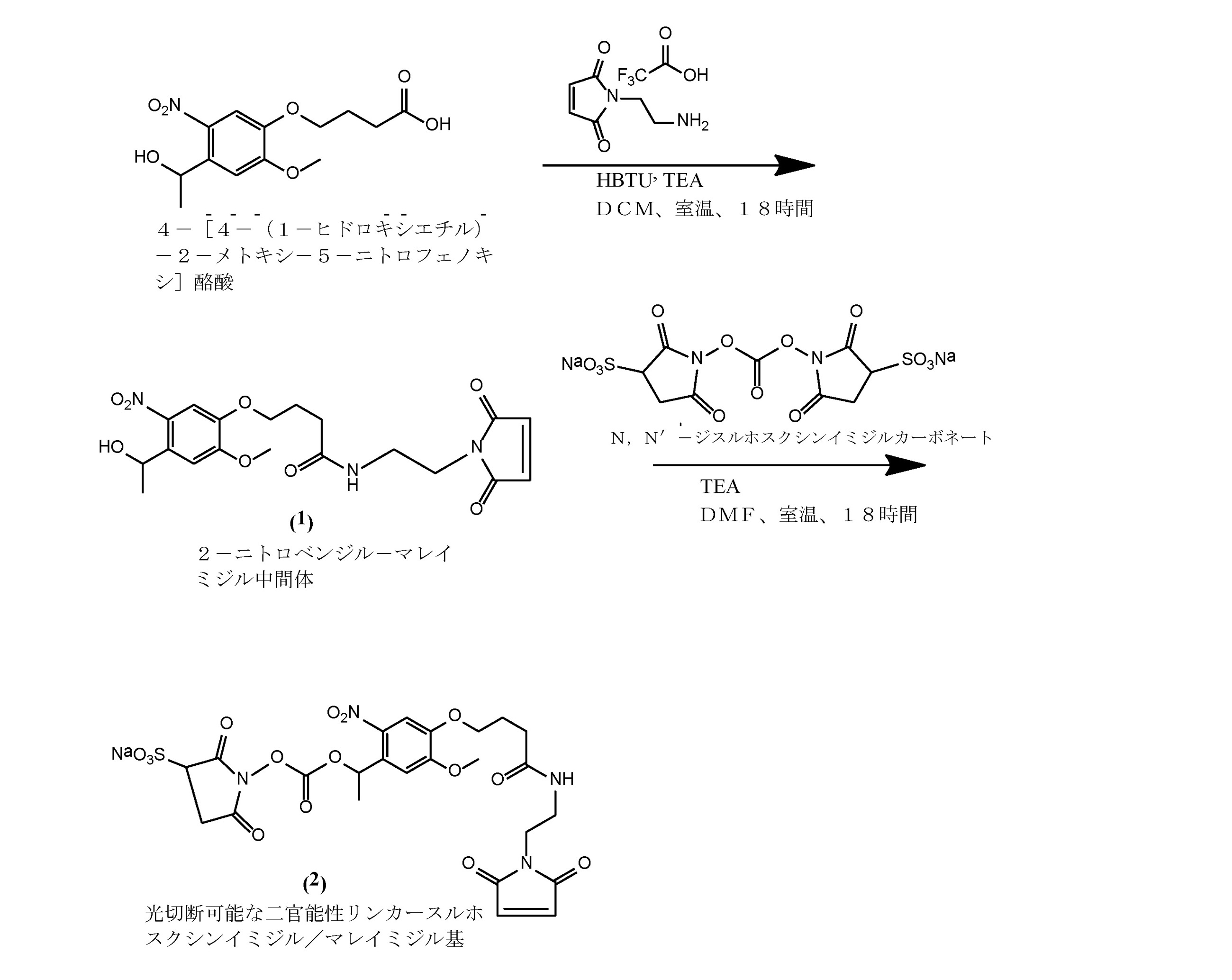
化合物2の合成:光切断可能なスルホスクシンイミジル/マレイミジルリンカーの合成は、Agasti,et al.,J.Am.Chem.Soc.,134(45),18499-18502,2012に由来する。簡潔には、出発物質4-[4-(1-ヒドロキシエチル)-2-メトキシ-5-ニトロフェノキシ]酪酸(0.334mmol)をアルゴン雰囲気下で乾燥ジクロロメタン(DCM)に溶解する。フラスコを、氷浴中に置くことによって0℃に冷却する。化合物2-(1H-ベンゾトリアゾール-1-イル)-1,1,3,3-テトラメチルウロニウムヘキサフルオロホスフェート(HBTU)(0.368mmol)、およびトリメチルアミン(TEA)(0.835mmol)を溶液に添加する。反応混合物を0℃で5分間撹拌し、続いてN-(2-アミノエチル)マレイミドトリフルオロ酢酸塩(0.368mmol)を添加する。0℃で15分間撹拌した後、反応混合物を、室温(RT)に上昇させ、さらに18時間撹拌する。反応混合物をDCM(45mL)で希釈した後、有機相を水(2×)、飽和NaCl溶液(1×)で洗浄し、硫酸ナトリウム上で乾燥させる。有機層を減圧下で濃縮し、SiO2カラム(溶出液:100%DCMに対してDCM中3%メタノール、v/v)を使用するフラッシュクロマトグラフィーにより精製する。化合物1(0.024mmol)を、無水ジメチルホルムアミド(1mL)に溶解する。N,N′-ジスルホスクシンイミジルカーボネート(DSC)(0.071mmol)およびTEA(0.096mmol)を連続的に溶液に添加する。反応混合物を室温で18時間撹拌する。反応混合物を、C18逆相カラム(溶出液:水中5%アセトニトリル対水中95%アセトニトリル、v/v)に直接装填することにより精製する。合成に使用される出発物質および他の化学物質は、Sigma-Aldrichから購入することができる。
10. Example Example 1
Synthesis of photocleavable 2-nitrobenzyl succinimidyl / maleemidyl bifunctional linker
Synthesis of Compound 2: The synthesis of a photocleavable sulfosuccinimidyl / maleimidyl linker is described in Agasti, et al. , J. Am. Chem. Soc. , 134 (45), 18499-18502, 2012. Briefly, the starting material 4- [4- (1-hydroxyethyl) -2-methoxy-5-nitrophenoxy] butyric acid (0.334 mmol) is dissolved in dry dichloromethane (DCM) under an argon atmosphere. The flask is cooled to 0 ° C. by placing it in an ice bath. Solution of compound 2- (1H-benzotriazole-1-yl) -1,1,3,3-tetramethyluronium hexafluorophosphate (HBTU) (0.368 mmol) and trimethylamine (TEA) (0.835 mmol) Add to. The reaction mixture is stirred at 0 ° C. for 5 minutes, followed by the addition of N- (2-aminoethyl) maleimide trifluoroacetate (0.368 mmol). After stirring at 0 ° C. for 15 minutes, the reaction mixture is raised to room temperature (RT) and stirred for an additional 18 hours. After diluting the reaction mixture with DCM (45 mL), the organic phase is washed with water (2x), saturated NaCl solution (1x) and dried over sodium sulfate. The organic layer is concentrated under reduced pressure and purified by flash chromatography using a SiO 2 column (eluent: 3% methanol in DCM relative to 100% DCM, v / v). Compound 1 (0.024 mmol) is dissolved in anhydrous dimethylformamide (1 mL). N, N'-disulfosuccinimidyl carbonate (DSC) (0.071 mmol) and TEA (0.096 mmol) are continuously added to the solution. The reaction mixture is stirred at room temperature for 18 hours. The reaction mixture is purified by loading directly onto a C18 reverse phase column (eluent: 5% acetonitrile in water vs. 95% acetonitrile in water, v / v). Starting materials and other chemicals used in the synthesis can be purchased from Sigma-Aldrich.
実施例2
光切断可能なスルホスクシンイミジル/DBCO 2-ニトロベンジル二官能性リンカーの合成
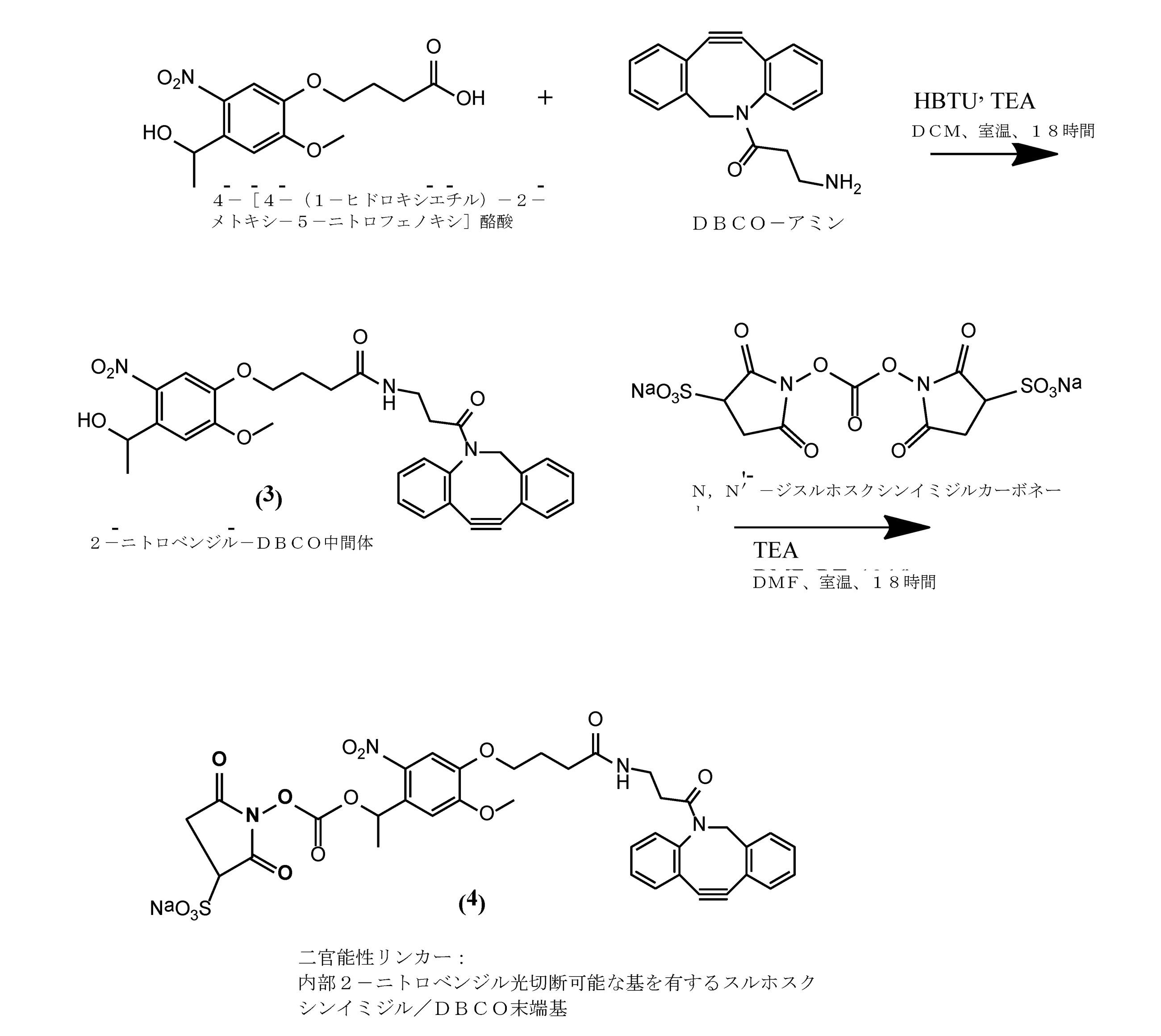
化合物4の合成:光切断可能なスルホスクシンイミジル/ジベンゾシクロオクチル(DBCO)アルキニルリンカーの合成は、Agasti,et al.,J.Am.Chem.Soc.,134(45),18499-18502,2012に記載されているのと同様の手順から誘導される。簡潔には、出発物質4-[4-(1-ヒドロキシエチル)-2-メトキシ-5-ニトロフェノキシ]酪酸(0.334mmol)をアルゴン雰囲気下で乾燥ジクロロメタン(DCM)に溶解する。フラスコを、氷浴中に置くことによって0℃に冷却する。化合物2-(1H-ベンゾトリアゾール-1-イル)-1,1,3,3-テトラメチルウロニウムヘキサフルオロホスフェート(HBTU)(0.368mmol)、およびトリメチルアミン(TEA)(0.835mmol)を溶液に添加する。反応混合物を0℃で5分間撹拌し、続いてDBCO-アミン(0.368mmol)を添加する。0℃で15分間撹拌した後、反応混合物を、室温に上昇させ、さらに18時間撹拌する。反応混合物をDCM(45mL)で希釈した後、有機相を水(2×)、飽和NaCl溶液(1×)で洗浄し、硫酸ナトリウム上で乾燥させる。有機層を減圧下で濃縮し、SiO2カラム(溶出液:100%DCMに対してDCM中3%メタノール、v/v)を使用するフラッシュクロマトグラフィーにより精製する。化合物3(0.024mmol)を、無水ジメチルホルムアミド(1mL)に溶解する。N,N′-ジスルホスクシンイミジルカーボネート(DSC)(0.071mmol)およびTEA(0.096mmol)を連続的に溶液に添加する。反応混合物を室温で18時間撹拌する。反応混合物を、C18逆相カラム(溶出液:水中5%アセトニトリル対水中95%アセトニトリル、v/v)に直接装填することにより精製する。合成に使用される出発物質および他の化学物質は、Sigma-Aldrichから購入することができる。
Example 2
Synthesis of photoclepable sulfosuccinimidyl / DBCO 2-nitrobenzyl bifunctional linker
Synthesis of Compound 4: Photo-cleavable sulfosuccinimidyl / dibenzocyclooctyl (DBCO) alkynyl linkers are synthesized by Agasti, et al. , J. Am. Chem. Soc. , 134 (45), 18499-18502, 2012, derived from a procedure similar to that described. Briefly, the starting material 4- [4- (1-hydroxyethyl) -2-methoxy-5-nitrophenoxy] butyric acid (0.334 mmol) is dissolved in dry dichloromethane (DCM) under an argon atmosphere. The flask is cooled to 0 ° C. by placing it in an ice bath. Solution of compound 2- (1H-benzotriazole-1-yl) -1,1,3,3-tetramethyluronium hexafluorophosphate (HBTU) (0.368 mmol) and trimethylamine (TEA) (0.835 mmol) Add to. The reaction mixture is stirred at 0 ° C. for 5 minutes, followed by the addition of DBCO-amine (0.368 mmol). After stirring at 0 ° C. for 15 minutes, the reaction mixture is raised to room temperature and stirred for an additional 18 hours. After diluting the reaction mixture with DCM (45 mL), the organic phase is washed with water (2x), saturated NaCl solution (1x) and dried over sodium sulfate. The organic layer is concentrated under reduced pressure and purified by flash chromatography using a SiO 2 column (eluent: 3% methanol in DCM relative to 100% DCM, v / v). Compound 3 (0.024 mmol) is dissolved in anhydrous dimethylformamide (1 mL). N, N'-disulfosuccinimidyl carbonate (DSC) (0.071 mmol) and TEA (0.096 mmol) are continuously added to the solution. The reaction mixture is stirred at room temperature for 18 hours. The reaction mixture is purified by loading directly onto a C18 reverse phase column (eluent: 5% acetonitrile in water vs. 95% acetonitrile in water, v / v). Starting materials and other chemicals used in the synthesis can be purchased from Sigma-Aldrich.
実施例3
スルホスクシンイミジル/マレイミジル2-ニトロベンジル二官能性リンカーを使用する抗体-DNA接合体のカップリングおよび光化学的切断
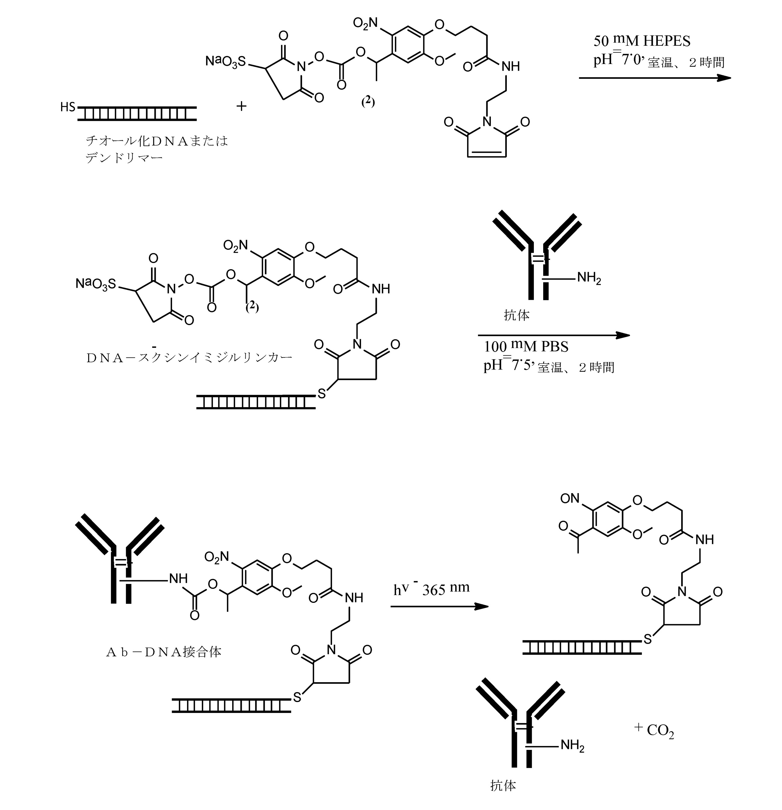
Coupling and photochemical cleavage of antibody-DNA conjugates using sulfosuccinimidyl / maleemidyl 2-nitrobenzyl bifunctional linker
ナノポア検出の前に、365nmのUVランプで照射することによって、接合体を切断することができる。この例は、同じ生体共役化学を使用してDNAデンドリマーに使用することもできる。 The conjugate can be cut by irradiation with a 365 nm UV lamp prior to nanopore detection. This example can also be used for DNA dendrimers using the same bioconjugated chemistry.
実施例4
スルホスクシンイミジル/DBCO2-ニトロベンジル二官能性リンカーを使用する抗体-DNA接合体のカップリングおよび光化学的切断

Coupling and photochemical cleavage of antibody-DNA conjugate using sulfosuccinimidyl / DBCO2-nitrobenzyl bifunctional linker
ナノポア検出の前に、365nmのUVランプで照射することによって、接合体を切断することができる。この例は、同じ生体共役化学を使用してDNAデンドリマーに使用することもできる。 The conjugate can be cut by irradiation with a 365 nm UV lamp prior to nanopore detection. This example can also be used for DNA dendrimers using the same bioconjugated chemistry.
実施例5
デジタルイムノアッセイのためのナノ粒子-抗体接合体(ナノポアカウント)
この実施例は、Bangs Labs(Fishers,IN,USA)から得ることができるものなどの、26nmのカルボキシル化ポリスチレンナノ粒子(NP、PC02N)への抗体の共有結合を記載する。26nmNPは、528.7μeq/gの表面電荷、68.4sq.Å/群のパーキング面積を有する(製造元情報)。
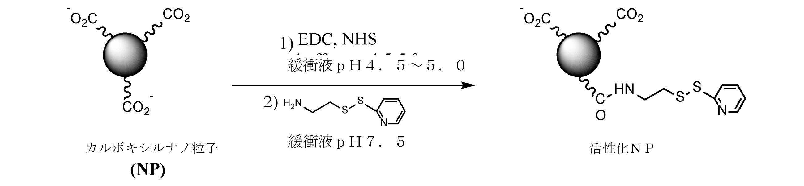
Nanoparticle-antibody conjugate for digital immunoassay (nanopoaccount)
This example describes covalent attachment of an antibody to 26 nm carboxylated polystyrene nanoparticles (NP, PC02N), such as that obtained from Bangs Labs (Fishers, IN, USA). 26 nmNP has a surface charge of 528.7 μeq / g, 68.4 sq. Has a parking area of Å / group (manufacturer information).
カルボキシル-ポリスチレンナノ粒子の活性化:1.0mL(100mg/mL)の26nmのカルボキシル化-NPを、10mLの0.1M MES(2-[N-モルホリノ]エタンスルホン酸、pH4.5~5.0)で洗浄する。洗浄後、ペレットを100mgの0.1M MES(pH4.5~5.0)に1.0mg/mLのNP濃度(固形分0.1%)で再懸濁する。10.0mLのナノ粒子懸濁液(10mg NP、5.28μeqカルボキシル)をバイアルに移し、10μL(5.28μモル、1.0equiv/CO2H eq)の新たに調製された水中の10mg/mL EDC溶液(1-エチル-3-[3-ジメチルアミノプロピル]カルボジイミド塩酸塩)および17μL(7.93μモル、1.5equiv/1equiv EDC)のスルホ-NHS水溶液の10mg/mL溶液(N-ヒドロキシスルホスクシンイミド、Sigma,Cat#56485)と、室温で15分間、連続混合しながら反応させる。反応した懸濁液を、6,500gで遠心分離し、溶液を廃棄する。ペレットを20mLの20mM PBS/5mM EDTA(pH7.5)で洗浄し、6,500gでの遠心分離により沈降させる。上清を除去する。スクシンイミド活性化カルボキシル-NPペレットを、50mM PBS(pH7.5)に再懸濁し、9.8μL(52.8nmol、0.01equiv/1CO2Heq)の1.0mg/mLピリジンジチオエチルアミン水溶液を直ちに添加して、室温で2~4時間連続的に撹拌しながら反応させる。ピリジル誘導体化カルボキシル-NPを、10mLの20mM PBS/5mM EDTA(pH7.5)で洗浄し、10.0mLの同じ緩衝液に再懸濁する。ナノ粒子濃度は、カルボキシル-NP較正曲線を使用するUV-Vis分光法(600nm、散乱)を使用して決定される。NPへのピリジルリガンド負荷は、規定量のNPを10mM TCEPまたはDTTで還元し、遠心分離により還元剤を除去し、ピリジル活性化NPペレットをPBS/EDTA(pH7.2)に再懸濁し、エルマン試薬と反応させることにより決定される(上清のA412を測定する)。活性化されたNPは、抗体接合のために同じ日に使用されない場合、4℃で貯蔵される。 Activation of carboxyl-polystyrene nanoparticles: 1.0 mL (100 mg / mL) of 26 nm carboxylated-NP, 10 mL of 0.1 M MES (2- [N-morpholino] ethanesulfonic acid, pH 4.5-5. Wash with 0). After washing, the pellet is resuspended in 100 mg 0.1 M MES (pH 4.5-5.0) at a NP concentration of 1.0 mg / mL (solid content 0.1%). Transfer the 10.0 mL nanoparticle suspension (10 mg NP, 5.28 μeqcarboxyl) to a vial and 10 μL (5.28 μmol, 1.0 equiv / CO 2 Heq) of 10 mg / mL in freshly prepared water. A 10 mg / mL solution (N-hydroxysulfo) of an EDC solution (1-ethyl-3- [3-dimethylaminopropyl] carbodiimide hydrochloride) and a sulfo-NHS aqueous solution of 17 μL (7.93 μmol, 1.5 equiv / 1 equiv EDC). Succinimide, Sigma, Cat # 56485) is reacted with continuous mixing at room temperature for 15 minutes. Centrifuge the reacted suspension at 6,500 g and discard the solution. The pellet is washed with 20 mL of 20 mM PBS / 5 mM EDTA (pH 7.5) and settled by centrifugation at 6,500 g. Remove the supernatant. The succinimide-activated carboxyl-NP pellet was resuspended in 50 mM PBS (pH 7.5) and immediately added with 1.0 mg / mL pyridinedithioethylamine aqueous solution of 9.8 μL (52.8 nmol, 0.01 equiv / 1CO 2 Heq). Then, the reaction is carried out at room temperature for 2 to 4 hours with continuous stirring. The pyridyl derivatized carboxyl-NP is washed with 10 mL of 20 mM PBS / 5 mM EDTA (pH 7.5) and resuspended in 10.0 mL of the same buffer. Nanoparticle concentration is determined using UV-Vis spectroscopy (600 nm, scattering) using a carboxyl-NP calibration curve. For pyridyl ligand loading on NPs, the specified amount of NP is reduced with 10 mM TCEP or DTT, the reducing agent is removed by centrifugation, the pyridyl activated NP pellets are resuspended in PBS / EDTA (pH 7.2), and Ellman. Determined by reacting with reagents (measuring A412 in supernatant). Activated NPs are stored at 4 ° C. if not used on the same day for antibody conjugation.
別個の抗体-ナノ粒子接合体を調製するための所望の化学量論を決定するために、ある範囲のEDC/NHSおよびピリジンジチオエチルアミンモル入力が評価される。所望のNP活性化結果を達成するために、反応パラメータ(pH、温度、時間)を評価する。
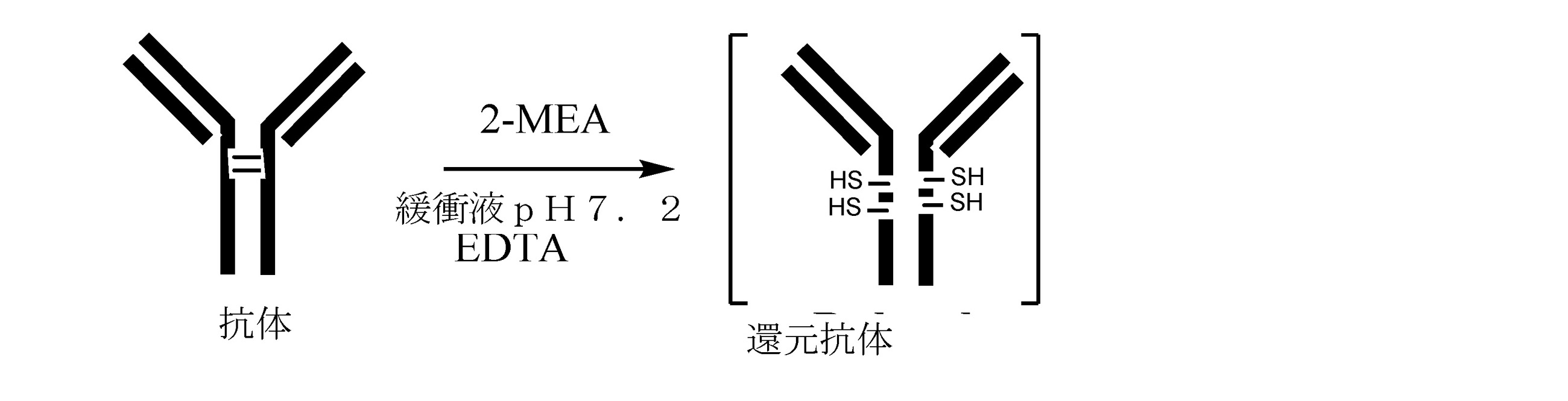
抗体の還元:1.0mLの10mg/mL抗体溶液(10mg)を、38μLの新たに調製した30mg/mL 2-MEA溶液(反応濃度10mM)(2-メルカプトエチルアミン塩酸塩)とよく混合し、次いで蓋をして、37℃で90分間置く。この溶液を室温にし、過剰の2-MEAを脱塩カラムで除去し、20mM PBS/5mM EDTA(pH7.5)中で予め平衡化した。還元抗体の濃度は、A280でのUV-Vis吸光度(タンパク質吸光度)およびA320(散乱補正)を使用して決定される。遊離チオールの数は、エルマン検定を使用して決定される。条件は、2または4個の遊離チオール(抗体ヒンジ領域のCys)を生成するために、必要に応じて最適化される。還元抗体は、ピリジル誘導体化カルボキシル-NPへのカップリングに直ちに使用される。

活性化ナノ粒子への還元抗体の結合:(1)抗体パーキングエリアは、45nm2であり、(2)26nmのナノ粒子表面積は、2,120nm2であり、(3)47個の抗体分子が理論的には26nmのNPの表面に適合すると仮定する。 Binding of reducing antibody to activated nanoparticles: (1) antibody parking area is 45 nm 2 , (2) nanoparticle surface area at 26 nm is 2,120 nm 2 , and (3) 47 antibody molecules. Theoretically, it is assumed to be compatible with the surface of NP at 26 nm.
手順:20mM PBS/5mM EDTA(pH7.5)中のピリジル活性化カルボキシナノ粒子の0.1%溶液10mL(10mg)に、0.10mg(0.66nmol、0.10mL)の還元抗体を、1.0mg/mLで同じEDTA含有緩衝液中に添加する。混合物を、室温で2時間混合しながら反応させ、遠心分離して未結合分子を除去し、そして吸引する。ペレットを、10mLのPBS(pH7.2)で洗浄し、遠心分離し、そして吸引する。抗体-NP接合体を、10.0mLのPBS(pH7.2)に懸濁する。接合体NP濃度(固形分%)は、UV-Vis分光法(600nm)を使用して決定される。粒子接合体を、SEMによって調べ、サイズ/電荷分布をZetaSizerを使用して決定する。サイズ排除クロマトグラフィーを使用して、接合体集団の潜在的分布から別個の接合体を単離することができる。抗体対NP取り込み比は、蛍光標識抗原接合体を使用するか、またはマイクロBCA(uBCA)アッセイを使用するフローサイトメトリーによって決定することができる。抗体からNPへのモル入力の範囲を、接合温度およびpHと共に評価して、別個の接合体の均一な集団を生成することができる(すなわち、2または4のNP取り込み比)。 Procedure: Add 0.10 mg (0.66 nmol, 0.10 mL) of reducing antibody to 10 mL (10 mg) of a 0.1% solution of pyridyl-activated carboxy nanoparticles in 20 mM PBS / 5 mM EDTA (pH 7.5). Add at 0.0 mg / mL to the same EDTA-containing buffer. The mixture is reacted at room temperature with mixing for 2 hours and centrifuged to remove unbound molecules and then aspirated. The pellet is washed with 10 mL PBS (pH 7.2), centrifuged and aspirated. The antibody-NP junction is suspended in 10.0 mL of PBS (pH 7.2). The conjugate NP concentration (% solid content) is determined using UV-Vis spectroscopy (600 nm). Particle junctions are examined by SEM and size / charge distribution is determined using ZetaSizer. Size exclusion chromatography can be used to isolate separate conjugates from the potential distribution of the conjugate population. The antibody-to-NP uptake ratio can be determined by flow cytometry using a fluorescently labeled antigen conjugate or using a micro BCA (uBCA) assay. The range of molar input from antibody to NP can be evaluated with junction temperature and pH to produce a uniform population of separate conjugates (ie, NP uptake ratio of 2 or 4).
ナノポアカウントイムノアッセイ
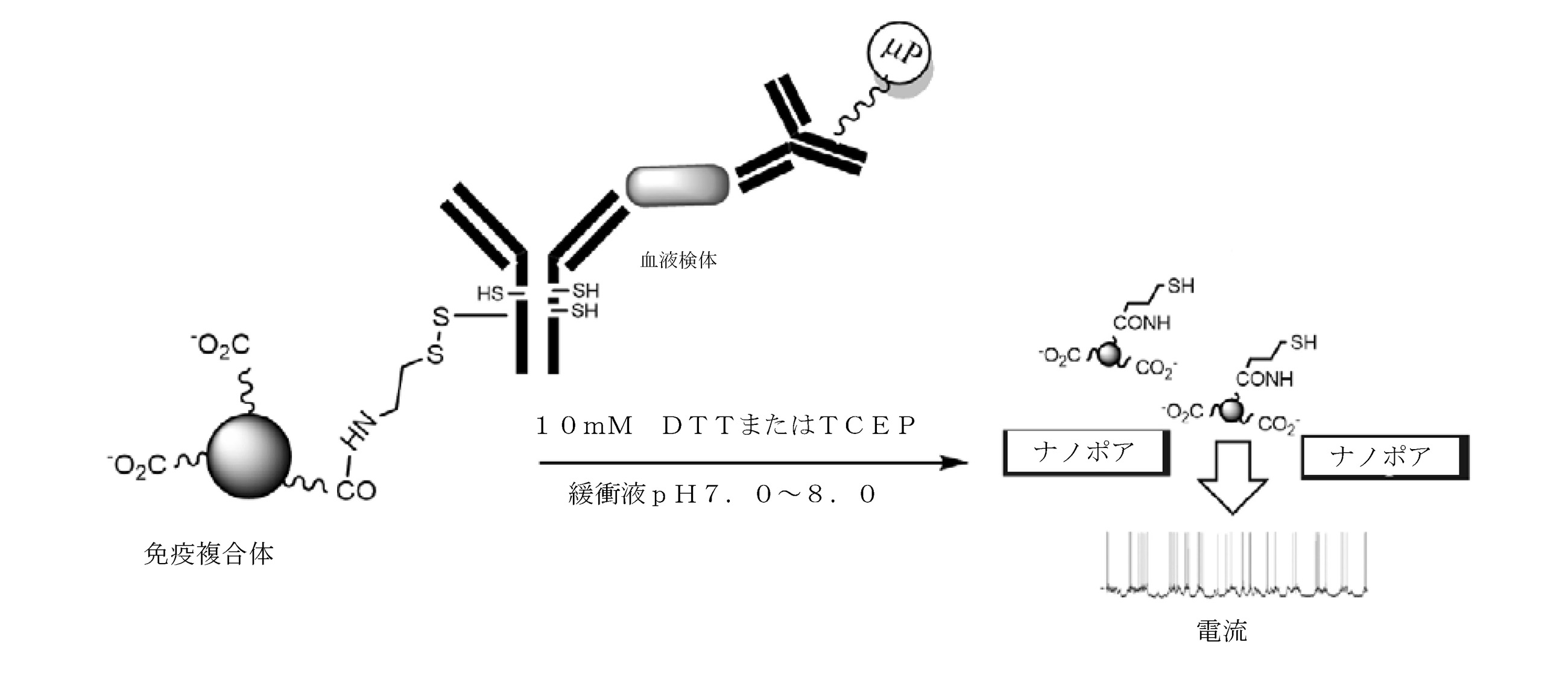
上記のスキームは、調製が上述されている還元抗体活性化ナノ粒子接合体を利用するナノポアカウントアッセイを例示する。イムノアッセイの過程で形成された免疫複合体は、ジスルフィド結合リンカーの還元によって切断されて、遊離抗体-検体-抗体複合体および遊離ナノ粒子タグを形成することができ、これによりナノポアを通過するときにナノ粒子タグをカウントするのを可能にする。 The above scheme exemplifies a nanopoacing assay utilizing the reduced antibody activated nanoparticle conjugate whose preparation is described above. Immune complexes formed during the course of the immunoassay can be cleaved by the reduction of disulfide bond linkers to form free antibody-specimen-antibody complexes and free nanoparticle tags, thereby passing through nanopores. Allows you to count nanoparticle tags.
実施例6
CPSP接合体の合成
A.CPSP抗体接合体。
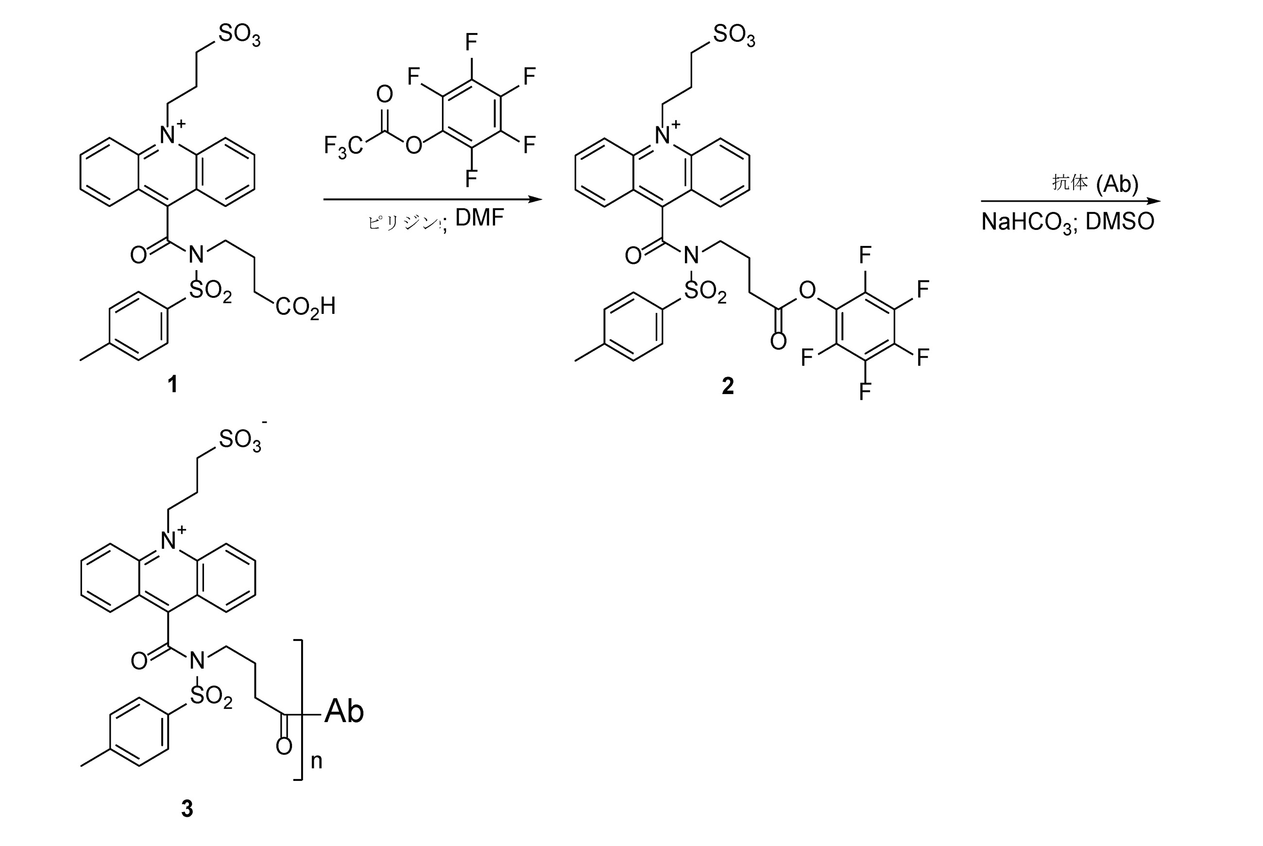
Synthesis of CPSP conjugate A. CPSP antibody conjugate.
3-(9-((4-オキソ-4-(ペルフルオロフェノキシ)ブチル)(トシル)カルバモイル)アクリジン-10-イウム-10-イル)プロパン-1-スルホネート(2):磁気撹拌棒および窒素導入口を備えた25mLの丸底フラスコに、3-(9-((3-カルボキシプロピル)(トシル)カルバモイル)アクリジン-10-イウム-10-イル)プロパン-1-スルホネート(CPSP)(1)(1mmol)、ピリジン(5mmol)、およびジメチルホルムアミド(10mL)を充填した。溶液を氷浴中で冷却し、トリフルオロ酢酸ペンタフルオロフェニル(1.3mmol)を滴下添加した。氷浴を外し、反応物を室温で3時間撹拌した。揮発性成分を真空で反応物から除去し、残渣をメタノールに取り込み、逆相HPLCにより精製して表題化合物を得た。 3- (9-((4-oxo-4- (perfluorophenoxy) butyl) (tosyl) carbamoyl) acridine-10-ium-10-yl) propan-1-sulfonate (2): magnetic stirring rod and nitrogen inlet In a 25 mL round bottom flask equipped with 3- (9-((3-carboxypropyl) (tosyl) carbamoyl) acridine-10-ium-10-yl) propan-1-sulfonate (CPSP) (1) (1 mmol). ), Pyridine (5 mmol), and dimethylformamide (10 mL). The solution was cooled in an ice bath and pentafluorophenyl trifluoroacetate (1.3 mmol) was added dropwise. The ice bath was removed and the reaction was stirred at room temperature for 3 hours. Volatile components were removed from the reactants in vacuo and the residue was taken up in methanol and purified by reverse phase HPLC to give the title compound.
CPSP抗体接合体(3):2の溶液(1μLのDMSO中10mM溶液)を抗体溶液(100μLの10μM水溶液)および水性重炭酸ナトリウム(10μLの1M溶液)に添加した。得られた混合物を、室温で4時間撹拌した。産物の精製を、スピンカラム上で行い、CPSP抗体接合体3を得た。「n」の値は、抗体依存的に変化する。活性エステル濃度(すなわち、化合物2、5、9および13)を上昇または低下させることによって、および/または反応中にpHを上昇または低下させることによって、取り込みをある程度制御することができるが、常に取り込み値の分布が生じる。平均取り込み比(「IR」)は、反応後に実験的に決定される。通常、「n」は、1から10の間の任意の値である。
A solution of CPSP antibody conjugate (3): 2 (10 mM solution in 1 μL DMSO) was added to the antibody solution (100
B.スペーサを有するCPSP抗体接合体。
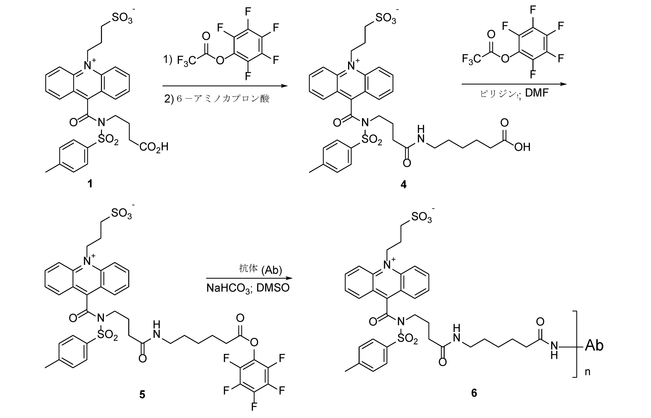
3-(9-((4-((5-カルボキシペンチル)アミノ)-4-オキソブチル)(トシル)カルバモイル)アクリジン-10-イウム-10-イル)プロパン-1-スルホネート(4):磁気撹拌棒および窒素導入口を備えた25mLの丸底フラスコに、3-(9-((3-カルボキシプロピル)(トシル)カルバモイル)アクリジン-10-イウム-10-イル)プロパン-1-スルホネート(CPSP)(1)(1mmol)、ピリジン(5mmol)、およびジメチルホルムアミド(10mL)を充填した。溶液を氷浴中で冷却し、トリフルオロ酢酸ペンタフルオロフェニル(1.3mmol)を滴下添加した。氷浴を外し、反応物を室温で3時間撹拌した。次いで、6-アミノカプロン酸(1.3mmol)を少量ずつ反応物に添加し、続いてN,N-ジイソプロピルエチルアミン(5mmol)を添加し、反応物を室温で1時間撹拌した。この後、揮発性成分を、真空で反応物から除去し、残渣を逆相HPLCにより精製して表題化合物を得た。 3- (9-((4-((5-carboxypentyl) amino) -4-oxobutyl) (tosyl) carbamoyl) acridine-10-ium-10-yl) propane-1-sulfonate (4): Magnetic stirring rod And in a 25 mL round bottom flask with a nitrogen inlet, 3- (9-((3-carboxypropyl) (tosyl) carbamoyl) acridine-10-ium-10-yl) propane-1-sulfonate (CPSP) ( 1) Filled with (1 mmol), pyridine (5 mmol), and dimethylformamide (10 mL). The solution was cooled in an ice bath and pentafluorophenyl trifluoroacetate (1.3 mmol) was added dropwise. The ice bath was removed and the reaction was stirred at room temperature for 3 hours. Then, 6-aminocaproic acid (1.3 mmol) was added little by little to the reaction, followed by N, N-diisopropylethylamine (5 mmol), and the reaction was stirred at room temperature for 1 hour. After this, the volatile components were removed from the reactants in vacuo and the residue was purified by reverse phase HPLC to give the title compound.
3-(9-((4-オキソ-4-((6-オキソ-6-(ペルフルオロフェノキシ)ヘキシル)アミノ)ブチル)(トシル)カルバモイル)アクリジン-10-イウム-10-イル)プロパン-1-スルホネート(5):磁気撹拌棒および窒素導入口を備えた25mLの丸底フラスコに、4(1mmol)、ピリジン(5mmol)、およびジメチルホルムアミド(10mL)を充填した。溶液を氷浴中で冷却し、トリフルオロ酢酸ペンタフルオロフェニル(1.3mmol)を滴下添加した。氷浴を外し、反応物を室温で3時間撹拌した。この後、揮発性成分を、窒素流下で反応物から除去し、残渣を逆相HPLCにより精製して表題化合物を得た。 3- (9-((4-oxo-4-((6-oxo-6- (perfluorophenoxy) hexyl) amino) butyl) (tosyl) carbamoyl) acridine-10-ium-10-yl) propan-1- Ssulfonate (5): A 25 mL round bottom flask equipped with a magnetic stir bar and a nitrogen inlet was filled with 4 (1 mmol), pyridine (5 mmol), and dimethylformamide (10 mL). The solution was cooled in an ice bath and pentafluorophenyl trifluoroacetate (1.3 mmol) was added dropwise. The ice bath was removed and the reaction was stirred at room temperature for 3 hours. After this, the volatile components were removed from the reactants under a stream of nitrogen and the residue was purified by reverse phase HPLC to give the title compound.
スペーサを有するCPSP抗体接合体(6):5の溶液(1μLのDMSO中10mM溶液)を、抗体溶液(100μLの10μM水溶液)および水性重炭酸ナトリウム(10μLの1M溶液)に添加した。得られた混合物を、室温で4時間撹拌した。産物の精製を、スピンカラム上で行い、スペーサを有するCPSP抗体接合体6を得た。通常、「n」は、1~10の間の任意の値である。
A solution of CPSP antibody conjugate (6) with spacers (6): 5 (10 mM solution in 1 μL DMSO) was added to the antibody solution (100
C.CPSPオリゴヌクレオチド-抗体接合体。
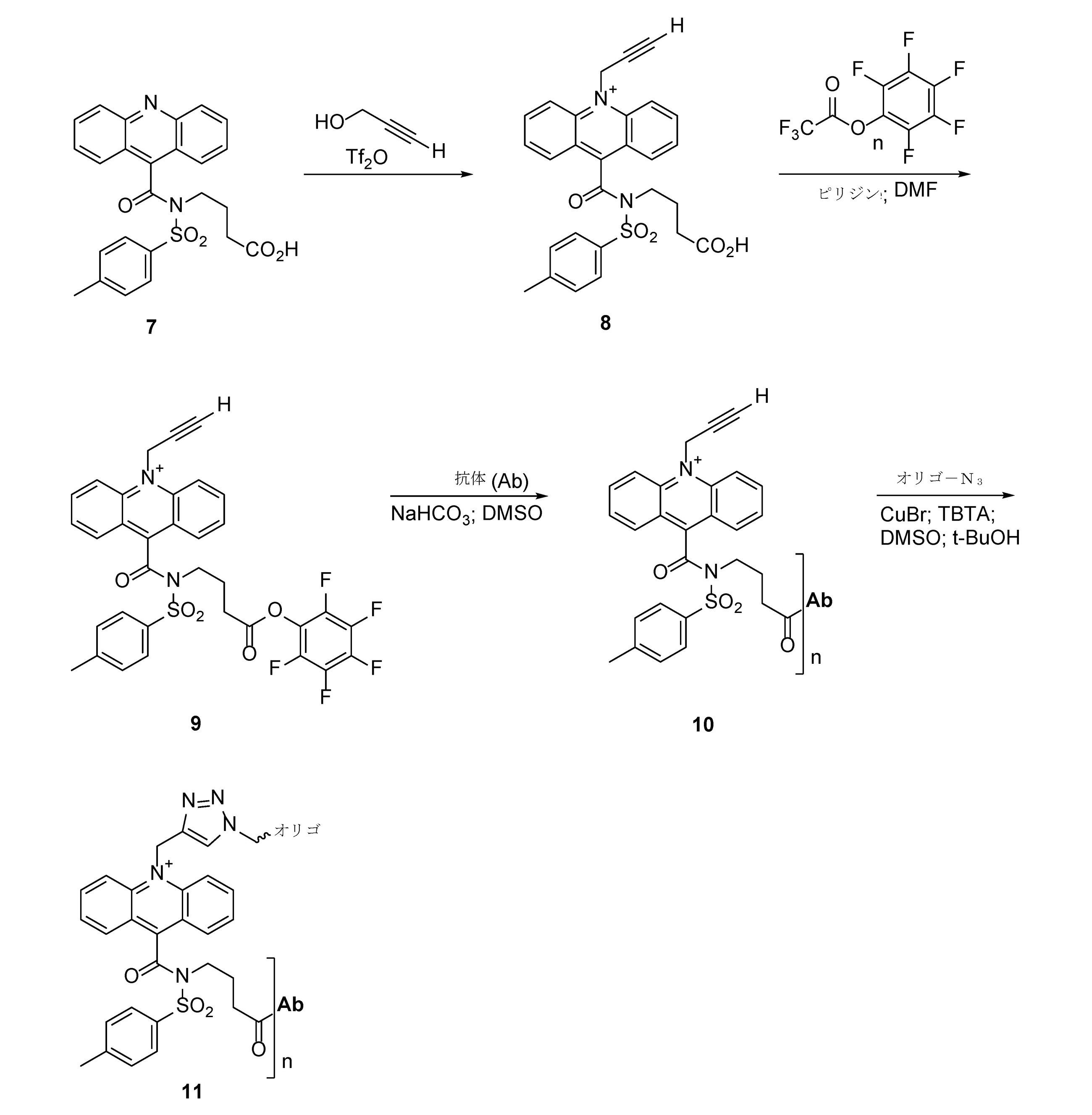
9-((3-カルボキシプロピル)(トシル)カルバモイル)-10-(プロプ-2-イン-1-イル)アクリジン-10-イウム(8):磁気撹拌棒および窒素導入口を備えた100mLの丸底フラスコに、プロパルギルアルコール(10mmol)、2,6-ジ-tert-ブチルピリジン(10mmol)および塩化メチレン(50mL)を充填し、-20℃に冷却した。次いで、トリフリン酸無水物を溶液に滴下添加し、反応物を-20℃で2時間撹拌した。ペンタン(25mL)を反応物に添加し、結果として生じる沈殿した塩をろ過により分離した。揮発性成分を真空中で蒸発させ、残渣を100mLの丸底フラスコ中の塩化メチレン(25mL)に再溶解した。4-(N-トシルアクリジン-9-カルボキサミド)ブタン酸(CP-アクリジン)(7)(1mmol)を少量ずつ添加し、反応物を室温で18時間撹拌した。揮発性成分を真空で蒸発させ、残留物をメタノール(5mL)中に取り込み、逆相HPLCにより精製して表題化合物を得た。 9-((3-carboxypropyl) (tosyl) carbamoyl) -10- (prop-2-in-1-yl) acridine-10-ium (8): 100 mL round with magnetic stir bar and nitrogen inlet The bottom flask was filled with propargyl alcohol (10 mmol), 2,6-di-tert-butylpyridine (10 mmol) and methylene chloride (50 mL) and cooled to −20 ° C. Trifuric acid anhydride was then added dropwise to the solution and the reaction was stirred at −20 ° C. for 2 hours. Pentane (25 mL) was added to the reactants and the resulting precipitated salt was separated by filtration. The volatile components were evaporated in vacuo and the residue was redissolved in methylene chloride (25 mL) in a 100 mL round bottom flask. 4- (N-tosylacridine-9-carboxamide) butanoic acid (CP-acridine) (7) (1 mmol) was added in small portions and the reaction was stirred at room temperature for 18 hours. The volatile components were evaporated in vacuo and the residue was taken up in methanol (5 mL) and purified by reverse phase HPLC to give the title compound.
9-((4-オキソ-4-(ペルフルオロフェノキシ)ブチル)(トシル)カルバモイル)-10-(プロプ-2-イン-1-イル)アクリジン-10-イウム(9):磁気撹拌棒および窒素導入口を備えた25mLの丸底フラスコに、8(1mmol)、ピリジン(5mmol)およびジメチルホルムアミド(10mL)を充填した。溶液を氷浴中で冷却し、トリフルオロ酢酸ペンタフルオロフェニル(1.3mmol)を滴下添加した。氷浴を外し、反応物を室温で3時間撹拌した。揮発性成分を真空で反応物から除去し、残渣をメタノールに取り込み、逆相HPLCにより精製して表題化合物を得た。
9-((4-oxo-4- (perfluorophenoxy) butyl) (tosyl) carbamoyl) -10- (prop-2-in-1-yl) acridine-10-ium (9): magnetic stir bar and
CPSP抗体接合体(10):9の溶液(1μLのDMSO中10mM溶液)を、抗体溶液(100μLの10μM水溶液)および水性重炭酸ナトリウム(10μLの1M溶液)に添加した。得られた混合物を、室温で4時間撹拌した。産物の精製を、スピンカラム上で行い、CPSP抗体接合体10を得た。通常、「n」は、1~10の間の任意の値である。
A solution of CPSP antibody conjugate (10): 9 (10 mM solution in 1 μL DMSO) was added to the antibody solution (100
CPSPオリゴヌクレオチド-抗体接合体(11):オリゴアジド(5μLの水中10nmol)、CPSP抗体接合体10(10μLの水中10nmol)および新たに調製した0.1M「クリック溶液」(3μL、下記参照)の混合物を、室温で4時間振とうした。反応物を0.3M酢酸ナトリウム(100μL)で希釈し、DNA接合体を、EtOH(1mL)を添加することによって沈殿させた。上清を除去し、残渣を冷たいEtOH(2×1mL)で2回洗浄した。残渣を水(20μL)に取り込み、CPSPオリゴヌクレオチド-抗体接合体11の溶液を、さらに精製することなく使用した。通常、「n」は、1~10の間の任意の値である。
CPSP oligonucleotide-antibody conjugate (11): a mixture of oligoazide (5 μL 10 nmol in water), CPSP antibody conjugate 10 (10 μL 10 nmol in water) and a freshly prepared 0.1 M “click solution” (3 μL, see below). Was shaken at room temperature for 4 hours. The reaction was diluted with 0.3 M sodium acetate (100 μL) and the DNA conjugate was precipitated by adding EtOH (1 mL). The supernatant was removed and the residue was washed twice with cold EtOH (2 x 1 mL). The residue was taken up in water (20 μL) and the solution of CPSP oligonucleotide-
「クリック溶液」:CuBr(1mg)を、70μLのDMSO/t-BuOH 3:1に溶解して0.1M溶液を形成した。(この溶液は、新たに調製しなければならず、貯蔵することはできない。)トリス(ベンジルトリアゾリルメチル)アミン(TBTA)(54mg)を、1mLのDMSO/t-BuOH 3:1に溶解して0.1M溶液を形成した。(この溶液は、-20℃で貯蔵できる。)1体積の0.1M CuBr溶液を、2体積の0.1M TBTA溶液に添加して、「クリック」溶液を得た。 "Click solution": CuBr (1 mg) was dissolved in 70 μL DMSO / t-BuOH 3: 1 to form a 0.1 M solution. (This solution must be freshly prepared and cannot be stored.) Dissolve tris (benzyltriazolylmethyl) amine (TBTA) (54 mg) in 1 mL DMSO / t-BuOH 3: 1. To form a 0.1 M solution. (This solution can be stored at −20 ° C.) 1 volume of 0.1 M CuBr solution was added to 2 volumes of 0.1 M TBTA solution to give a “click” solution.
D.スペーサを有するCPSPオリゴヌクレオチド-抗体接合体

9-((4-((5-カルボキシペンチル)アミノ-4-オキソブチル)(トシル)カルバモイル)-10-(プロプ-2-イン-1-イル)アクリジン-10-イウム(12):磁気撹拌棒および窒素導入口を備えた25mLの丸底フラスコに、8(1mmol)、ピリジン(5mmol)およびジメチルホルムアミド(10mL)を充填した。溶液を氷浴中で冷却し、トリフルオロ酢酸ペンタフルオロフェニル(1.3mmol)を滴下添加した。氷浴を外し、反応物を室温で3時間撹拌した。次いで、6-アミノカプロン酸(1.3mmol)を少量ずつ反応物に添加し、続いてN,N-ジイソプロピルエチルアミン(5mmol)を添加し、反応物を室温で1時間撹拌した。この後、揮発性成分を、真空で反応物から除去し、残渣を逆相HPLCにより精製して表題化合物を得た。 9-((4-((5-carboxypentyl) amino-4-oxobutyl) (tosyl) carbamoyl) -10- (prop-2-in-1-yl) acridine-10-ium (12): magnetic stirring rod And a 25 mL round bottom flask with a nitrogen inlet was filled with 8 (1 mmol), pyridine (5 mmol) and dimethylformamide (10 mL). The solution was cooled in an ice bath and trifluoroacetate pentafluorophenyl (1). 3 mmol) was added dropwise. The ice bath was removed and the reaction was stirred at room temperature for 3 hours. Then 6-aminocaproic acid (1.3 mmol) was added to the reaction in small portions, followed by N, N-diisopropyl. Ethylamine (5 mmol) was added and the reaction was stirred at room temperature for 1 hour, after which the volatile components were removed from the reaction under vacuum and the residue was purified by reverse phase HPLC to give the title compound.
9-((4-オキソ-4-((6-オキソ-6-(ペルフルオロフェノキシ)ヘキシル)アミノ)ブチル)(トシル)カルバモイル)-10-(プロプ-2-イン-1-イル)アクリジン-10-イウム(13):磁気撹拌棒および窒素導入口を備えた25mLの丸底フラスコに、12(1mmol)、ピリジン(5mmol)、およびジメチルホルムアミド(10mL)を充填した。溶液を氷浴中で冷却し、トリフルオロ酢酸ペンタフルオロフェニル(1.3mmol)を滴下添加した。氷浴を外し、反応物を室温で3時間撹拌した。この後、揮発性成分を、窒素流下で反応物から除去し、残渣を逆相HPLCにより精製して表題化合物を得た。 9-((4-oxo-4- ((6-oxo-6- (perfluorophenoxy) hexyl) amino) butyl) (tosyl) carbamoyl) -10- (prop-2-in-1-yl) acridine-10 -Ium (13): A 25 mL round bottom flask equipped with a magnetic stir bar and a nitrogen inlet was filled with 12 (1 mmol), pyridine (5 mmol), and dimethylformamide (10 mL). The solution was cooled in an ice bath and pentafluorophenyl trifluoroacetate (1.3 mmol) was added dropwise. The ice bath was removed and the reaction was stirred at room temperature for 3 hours. After this, the volatile components were removed from the reactants under a stream of nitrogen and the residue was purified by reverse phase HPLC to give the title compound.
スペーサを有するCPSP抗体接合体(14):13の溶液(1μLのDMSO中10mM溶液)を、抗体溶液(100μLの10μM水溶液)および水性重炭酸ナトリウム(10μLの1M溶液)に添加した。これを室温で4時間撹拌した。産物の精製を、スピンカラム上で行い、スペーサを有するCPSP抗体接合体14を得た。通常、「n」は、1~10の間の任意の値である。
A solution of CPSP antibody conjugate (14) with spacers (14): 13 (10 mM solution in 1 μL DMSO) was added to the antibody solution (100
CPSPオリゴヌクレオチド-抗体接合体(15):オリゴアジド(例えば、市販のものなど)(5μLの水中10nmol)、スペーサを有するCPSP抗体接合体14(10μLの水中10nmol)および新たに調製された0.1Mの「クリック溶液」(3μL、実施例6.Cを参照)の混合物を、室温で4時間振とうした。通常、「n」は、1~10の間の任意の値である。反応物を0.3M酢酸ナトリウム(100μL)で希釈し、DNA接合体を、EtOH(1mL)を添加することによって沈殿させた。上清を除去し、残渣を冷たいEtOH(2×1mL)で2回洗浄した。残渣を水(20μL)に取り込み、スペーサを有するCPSPオリゴヌクレオチド-抗体接合体15の溶液を、さらに精製することなく使用した。
CPSP oligonucleotide-antibody conjugate (15): oligoazide (eg, commercially available) (5 μL 10 nmol in water),
スペーサを有するかまたは有しないCPSP抗体接合体およびスペーサを有するかまたは有しないCPSPオリゴヌクレオチド-抗体接合体の切断。スペーサを有するかまたは有しないCPSP抗体接合体およびスペーサを有するかまたは有しないCPSPオリゴヌクレオチド-抗体接合体を、記載されているように、塩基性過酸化水素溶液を使用して切断または「誘発」される。ARCHITECT(登録商標)システムでは、励起状態のアクリドン中間体が、光子を産生し、それを測定する。さらに、切断産物は、アクリドンおよびスルホンアミドである。実施例6.A~Dの接合体は、アクリドンおよび/またはスルホンアミド分子をカウントすることによって、開示されたデバイスと共に使用される。 Cleavage of CPSP antibody conjugates with or without spacers and CPSP oligonucleotide-antibody conjugates with or without spacers. Cleavage or "induction" of CPSP antibody conjugates with or without spacers and CPSP oligonucleotide-antibody conjugates with or without spacers using a basic hydrogen peroxide solution as described. Will be done. In the ARCHITEC® system, an excited acridone intermediate produces and measures photons. In addition, the cleavage products are acridone and sulfonamides. Example 6. The conjugates A to D are used with the disclosed devices by counting acridone and / or sulfonamide molecules.
E.抗体を含まないCPSPオリゴヌクレオチド接合体。

抗体を含まないCPSPオリゴヌクレオチド-抗体接合体(16):オリゴアジド(例えば、市販のものなど)(5μLの水中10nmol)、プロパルギル-CPSP8(10μLの水中10nmol)および新たに調製された0.1Mの「クリック溶液」(3μL、実施例6.Cを参照)の混合物を、室温で4時間振とうすることができる。反応物を、0.3M酢酸ナトリウム(100μL)で希釈し、EtOH(1mL)を添加することによってDNA接合体を沈殿させることができる。上清を除去し、残渣を冷たいEtOH(2×1mL)で2回洗浄することができる。残渣を水(20μL)に取り込み、スペーサを有するCPSPオリゴヌクレオチド-抗体接合体16の溶液を、さらに精製することなく使用することができる。
Antibodies-free CPSP oligonucleotide-antibody conjugate (16): oligoazide (eg, commercially available) (5 μL 10 nmol in water), propargyl-CPSP8 (10 μL 10 nmol in water) and freshly prepared 0.1 M. A mixture of "click solution" (3 μL, see Example 6.C) can be shaken at room temperature for 4 hours. The reaction can be diluted with 0.3 M sodium acetate (100 μL) and the DNA conjugate can be precipitated by adding EtOH (1 mL). The supernatant can be removed and the residue can be washed twice with cold EtOH (2 x 1 mL). The residue can be incorporated into water (20 μL) and the solution of CPSP oligonucleotide-
実施例7
低コストDMFボトムチップの製造
低コスト可撓性DMFチップを、Lo C-Y et al.,Microelectronic Engineering 86(2009)979-983に記載されるプロセスを、いくつかの修正を加えて使用することによって、電極パターン化のためのリフトオフプロセスと組み合わせたロールツーロール(R2R)フレキソ印刷を使用して製造した。製造プロセスの概略を図10に示す。DME電極印刷用の出発材料として、MELINEX(登録商標)ST506ポリエチレンテレフタレート(PET)5.0ミル基板(1)のロールを使用した。黄色のインク(Sun Chemical)の層を、厚さ1.14mmの印刷プレート(Flint MCO3)を使用して、アニロックスローラーアセンブリ上で3.8mL/m2のインク転写量を使用して、10m/分の速度でPET基板上にフレキソ印刷した(2)。DMF電極パターンのネガ像は、フレキソ印刷ステップ(3)から生じる。金属堆積の前に、インクを熱風オーブン(2×100℃)で2回乾燥させた。EVA R2Rメタルエバポレータを使用して、印刷されたPET基板上に銀金属層を堆積させ、厚さ80nmの厚さで銀の均一なコーティングを形成した(4)。1m/分の速度で超音波浴のアセトン+超音波の組み合わせを使用して、金属化インキ-フィルム支持体(5)を湿式リフトオフプロセスに供した(6)。この化学的/物理的処理により、銀のみの層を無傷に保ちながら、銀インク層を溶解させることができる。インク-銀層を除去すると、50または140μmの電極ギャップ間隔を有する80個の作動電極(2.25×2.25mm)からなるDMF印刷電極パターンが得られた(7)。QCチェックとして、単一ロールからの合計80~90個のランダムチップを、電極ギャップ間隔およびコネクタリード幅変動について目視検査した。許容できるギャップ仕様を有すると決定されたチップの典型的な歩留まりは、100%に近かった。単一の製造された可撓性チップを、図11に示す。製造されたフレキシブルチップは、3″×2″の寸法であり、電極、リザーバ、コンタクトパッドおよびリードを含む。
Example 7
Manufacture of Low Cost DMF Bottom Chips Low cost flexible DMF chips are available from LoCY et al. , Microelectronic Engineering 86 (2009) 979-983, using roll-to-roll (R2R) flexographic printing combined with a lift-off process for electrode patterning by using with some modifications. Manufactured. The outline of the manufacturing process is shown in FIG. A roll of MELINEX® ST506 polyethylene terephthalate (PET) 5.0 mil substrate (1) was used as a starting material for DME electrode printing. A layer of yellow ink (Sun Chemical), using a 1.14 mm thick printing plate (Flint MCO3), using an ink transfer rate of 3.8 mL / m 2 on an Anilox roller assembly, 10 m / m /. Flexographic printing was performed on the PET substrate at a speed of minutes (2). The negative image of the DMF electrode pattern arises from the flexographic printing step (3). Prior to metal deposition, the ink was dried twice in a hot air oven (2 x 100 ° C.). An EVA R2R metal evaporator was used to deposit a silver metal layer on a printed PET substrate to form a uniform coating of silver with a thickness of 80 nm (4). The metallized ink-film support (5) was subjected to a wet lift-off process using a combination of acetone + ultrasonic waves in an ultrasonic bath at a rate of 1 m / min (6). This chemical / physical treatment can dissolve the silver ink layer while leaving the silver-only layer intact. Removal of the ink-silver layer yielded a DMF printed electrode pattern consisting of 80 working electrodes (2.25 x 2.25 mm) with an electrode gap spacing of 50 or 140 μm (7). As a QC check, a total of 80-90 random chips from a single roll were visually inspected for electrode gap spacing and connector lead width variation. The typical yield of chips determined to have acceptable gap specifications was close to 100%. A single manufactured flexible tip is shown in FIG. The manufactured flexible tip is 3 "x 2" in size and includes electrodes, reservoirs, contact pads and leads.
ロータリースクリーン印刷またはグラビア印刷のいずれかを使用することによって、誘電体コーティングを電極およびリザーバに適用した。ロータリースクリーン印刷については、Henkel EDAC PF-455Bを、2m/分の印刷速度および50%のUV硬化速度で、Gallus NF(400L)スクリーンを用いて印刷することによって誘電体コーティングとして使用した。典型的な誘電体の厚さは、10~15μmであった。グラビア印刷では、シリンダは、IPD-350(Inkron)などの高粘度誘電体インクを、50mL/m2インク体積を使用して、2m/分の速度で印刷するように設計された。グラビア印刷の典型的な誘電体の厚さは、7~8μmであった。Millidyne Avalon 87またはCytonix Fluoropel PFC804UCコーティングのいずれかを、グラビアシリンダ(140~180L)および8m/分の印刷速度で、続いて4つの連続オーブン乾燥ステップ(4×140℃)で使用して、最終疎水性層を印刷した。典型的な疎水性厚さは、40~100nmであった。 Dielectric coatings were applied to the electrodes and reservoirs by using either rotary screen printing or gravure printing. For rotary screen printing, Henkel EDAC PF-455B was used as a dielectric coating by printing with a Gallus NF (400L) screen at a printing rate of 2 m / min and a UV curing rate of 50%. The typical dielectric thickness was 10-15 μm. In gravure printing, the cylinder was designed to print high viscosity dielectric inks such as IPD-350 (Incron) at a rate of 2 m / min using a 50 mL / m 2 ink volume. The typical dielectric thickness for gravure printing was 7-8 μm. One of the Millidyne Avalon 87 or Cytonix Fluoropel PFC804UC coatings is used in a gravure cylinder (140-180 L) and a printing rate of 8 m / min, followed by four continuous oven drying steps (4 x 140 ° C.) for final hydrophobicity. The sex layer was printed. The typical hydrophobic thickness was 40-100 nm.
あるいは、個々のチップの小バッチに対して、誘電性コーティングおよび疎水性コーティングは、それぞれ化学気相堆積(CVD)およびスピンコーティングを使用して適用され得る。 Alternatively, for small batches of individual chips, a dielectric coating and a hydrophobic coating can be applied using chemical vapor deposition (CVD) and spin coating, respectively.
実施例8
低コストDMFチップの機能試験
上記の実施例7に概説したように製造した3″×2″のPET系DMFボトムチップを作動能力について試験した。図12は、厚さ0.7mmのガラス基板(3)がその上に位置した3″×2″のPETベースのDMFチップ(1)を示す。ガラス基板(3)は、ガラス基板の下面に透明な酸化インジウム錫(ITO)電極と、ITO電極を覆うTeflonコーティングとを含む。DMFチップは、8個の緩衝液リザーバと共に、ストレートエッジ電極設計および電極間に50μmのギャップを有する、80個の銀作動電極を含む(上記実施例7を参照)。
Example 8
Functional Testing of Low-Cost
底部電極を、CVDおよびスピンコーティングによって、それぞれ誘電体Parylene-C(6~7μm厚)およびTeflonの最終コーティング(50nm厚)でコーティングした。0.1%の界面活性剤を含むおよそ50μLのPBS緩衝液(2)を、底部DMFチップ上の4つの隣接するリザーバにピペットで入れた。液滴サイズは700~1,500nL(1滴または2滴)の範囲であり、混合に必要な円形掃引パターンに加えて、垂直方向および水平方向の両方の横方向の動きについて調べた(4)。液滴作動は、90Vrmsの電圧を使用して達成された。チップ上の作動電極のおよそ90%が試験され、完全に機能的であることがわかった。 The bottom electrodes were coated by CVD and spin coating with dielectric Parylene-C (6-7 μm thick) and Teflon final coating (50 nm thick), respectively. Approximately 50 μL of PBS buffer (2) containing 0.1% detergent was pipetted into four adjacent reservoirs on the bottom DMF chip. Droplet sizes ranged from 700 to 1,500 nL (1 or 2 drops), and in addition to the circular sweep pattern required for mixing, both vertical and horizontal lateral movements were investigated (4). .. Droplet operation was achieved using a voltage of 90 Vrms . Approximately 90% of the working electrodes on the chip were tested and found to be fully functional.
実施例9
低コストDMFチップ上のTSHイムノアッセイ
上記の実施例8に記載されているようにガラス基板で覆われた3′×2″PETベースのDMFチップを、化学発光検出を使用して、甲状腺刺激ホルモン(TSH)イムノアッセイを実行するその能力について試験した。模擬試料は、遮断剤および界面活性剤を含有するトリス緩衝食塩水(TBS)緩衝液にスパイクしたTSH較正材料を含んでいた。0、4、40μIU/mLの3つの試料を試験した。5μmの磁性微粒子(3×108粒子/mL)上にコーティングされた2μLの抗βTSH捕捉抗体を、微粒子リザーバからDMF電極アレイの中央に分注した。磁性微粒子を、DMFチップ下で、ネオジミウム磁石棒(3インチ×1/2インチ×1/4インチ厚、相対透過性μr=1.05、残留フィールド強度Br=1.32T)を係合することによって、緩衝液から分離した(図13A)。5μLの試料を、微小粒子スラグに移し、続いて微粒子懸濁液(図13B)を四電極正方形構成上で5分間混合した。微粒子を、磁石によって試料から分離し、上清を廃棄物リザーバに移した(図13Cおよび13D)。西洋ワサビペルオキシダーゼ(HRP)に接合した2μLの1μg/mL抗TSH検出抗体を、微粒子スラグに移し、2分間混合した。微粒子を磁石によって分離し、上清を廃棄物リザーバに移した。イムノアッセイサンドイッチ複合体を含有する微粒子を、0.1%界面活性剤を含有する4×2μLのPBS洗浄緩衝液で合計4回洗浄した。各洗浄ステップからの洗浄緩衝液は、ステップが完了した後に廃棄物に移動させた。化学発光基質は、1μLのSuperSignal H2O2および1μLのルミノール(ThermoFisher Scientific)からなり、これを微粒子スラグに移し、続いて6分間混合した。化学発光シグナルは、5VのDC源を備えた一体型Hamamatsu H10682-110PMTを使用して427nm発光(347nm励起)で測定した。用量反応曲線を、相対発光に対してプロットした(図13Eを参照)。
Example 9
TSH Immunoassay on Low-Cost DMF Chips Thyroid-stimulating hormone (
実施例10
ナノポアモジュール製造
ナノポアモジュールは、TEMウィンドウ(Norcada)に埋め込まれた市販の窒化ケイ素(SiNx)膜の統合と組み合わせた標準的なソフトリソグラフィ製造方法を使用して製造された。このモジュールは、4つの別々のPDMS層、すなわち転送マイクロチャネルを含む上部および底部PDMS基板、ならびにTEMウィンドウを封止するための2つの任意選択の中間PDMS層からなっていた。
Example 10
Manufacture of Nanopore Modules Nanopore modules were manufactured using standard soft lithography manufacturing methods combined with the integration of commercially available silicon nitride (SiN x ) films embedded in a TEM window (Norcada). The module consisted of four separate PDMS layers, namely top and bottom PDMS substrates containing transfer microchannels, and two optional intermediate PDMS layers for encapsulating the TEM window.
SU8マスターモールド製造:清浄な乾燥ガラス基板を、フォトレジスト(SU8-50)で所望の厚さにスピンコートした。次いで、フォトマスクを使用して、コーティングされた基板の領域を近紫外線に選択的に露光した。マスクは、転送マイクロチャネルおよびリザーバの形状が残ることになる領域においてのみフォトレジストをUV光に露光する。露光後、露光されたフォトレジストの架橋領域へのベーキングが続いた。次いで、SU8現像液を使用して、基板から残りの未露光のフォトレジストを除去した。最終製品は、マスターモールド、すなわちパターン化された転送マイクロチャネルと硬いフォトレジストのリザーバを有するガラス基板である。 SU8 Master Mold Manufacturing: A clean dry glass substrate was spin coated with a photoresist (SU8-50) to a desired thickness. Areas of the coated substrate were then selectively exposed to near UV light using a photomask. The mask exposes the photoresist to UV light only in areas where the shape of the transfer microchannel and reservoir will remain. After exposure, baking of the exposed photoresist into the crosslinked region continued. The SU8 developer was then used to remove the remaining unexposed photoresist from the substrate. The final product is a master mold, a glass substrate with a patterned transfer microchannel and a reservoir of rigid photoresist.
中間PDMS層製造:中間PDMS層を製造するために、PDMSモノマーとその硬化剤(Sylgard 184シリコーンエラストマー)を、7:1のPDMSモノマー:硬化剤の比で含む溶液を、ガラススライド上にスピンコートし、続いて70℃で30分間ホットプレート上で加熱した。PDMS層をガラス基板から剥がし、1.25mmの切り欠き部をPDMS層に打ち抜いて、TEMウィンドウへのアクセスを可能にする開口部を設けた。PDMS層の表面は、コロナ処理機を使用して8mmの距離で30秒間プラズマ処理することによって親水性にした。第2のプラズマ処理(5秒)を使用して、2つの中間PDMS層の間にSiNxTEMウィンドウを接合する前に、PDMS層およびTEMウィンドウの表面を処理した。 Intermediate PDMS layer production: To produce an intermediate PDMS layer, a solution containing a PDMS monomer and its curing agent (Silgard 184 silicone elastomer) in a ratio of 7: 1 PDMS monomer: curing agent is spin-coated on a glass slide. Then, it was heated on a hot plate at 70 ° C. for 30 minutes. The PDMS layer was peeled off from the glass substrate and a 1.25 mm notch was punched into the PDMS layer to provide an opening to allow access to the TEM window. The surface of the PDMS layer was made hydrophilic by plasma treatment at a distance of 8 mm for 30 seconds using a corona treatment machine. A second plasma treatment (5 seconds) was used to treat the surface of the PDMS layer and the TEM window before joining the SiN x TEM window between the two intermediate PDMS layers.
上部および下部PDMSの製造:マイクロチャネルを含む上部および下部PDMS基板を、図14Bに示すように、PDMSモノマーおよび硬化剤を7:1のPDMSモノマー:硬化剤の比で混合することと、転送マイクロチャネルおよびリザーバでパターン化したSU8パターン化型(6)を含有するガラス上に注ぐことによって製造した(上記のSU8種型製造を参照)。マイクロチャネルは、幅がおよそ110~135μm、深さが50μmと測定された。15分間脱気した後、SU8型をホットプレート上で70℃で60分間加熱した(7)。硬化後、PDMS基板をSU8型(8)から剥がし、切断して、長さ30mm×幅20mm×深さ3mmのおよその寸法を有する長方形のPDMS基板を得た。その後の電極のマイクロチャネルへの挿入を可能にするために、アクセス穴(直径1.25mm)をPDMS基板に穿孔した。最終アセンブリは、図14Aに示され、下から上へ、1つのマイクロチャネルを含む下部PDMS基板(1)と、マイクロチャネル上に位置付けられた切り欠き部を含む第1の中間PDMS層(2)と、TEMウィンドウ内のSiNx膜(3)と、同様に切り欠き部を含む第2の中間PDMS層(4)と、第2のマイクロチャネルを含む上部PDMS基板と、を含む。
Manufacture of Upper and Lower PDMS: Upper and lower PDMS substrates containing microchannels are mixed with PDMS monomer and curing agent in a 7: 1 PDMS monomer: curing agent ratio and transfer micro, as shown in FIG. 14B. Manufactured by pouring onto glass containing SU8 patterned form (6) patterned with channels and reservoirs (see SU8 type fabrication above). Microchannels were measured to be approximately 110-135 μm wide and 50 μm deep. After degassing for 15 minutes, SU8 was heated on a hot plate at 70 ° C. for 60 minutes (7). After curing, the PDMS substrate was peeled off from SU8 type (8) and cut to obtain a rectangular PDMS substrate having approximate dimensions of
上部および下部PDMS基板の整列:PDMS下部基板(上記の「上部および下部PDMSの製造」で概説したように調製した)を30秒間プラズマ処理し、続いて第1の中間PDMS層(上記「中間体PDMS層の製造」に概説したように調製した)をPDMS下部基板の上に接合した。同様に、PDMS上部基板を、30秒間プラズマ処理し、続いて第2の中間PDMS層をPDMS上部基板上に接合した。中間層の切り欠き部は、マイクロチャネルと整列していた。上部および下部PDMS片の両方を、30秒間酸素プラズマ処理し、続いて上部片と下部片との間にSiNx膜ウィンドウを配置し、中間PDMS層の切り欠き部と整列させた。底部片と整列したSiNx膜と整列した上部片を、全ての気泡が放出されるまで一緒に押圧した。最終ナノポアPDMSアセンブリを、ホットプレート上で100℃で30分間加熱し、5分間プラズマ処理した。図14Cに示す最終モジュールアセンブリ(9a)は、2つのチャネル(1つの直線および1つの「L字型」チャネル)を含み、各々が溶液用のリザーバ(例えば緩衝液)で終了する。SiNx膜を含むTEMウィンドウは、2つの垂直マイクロチャネルの交差部に位置付けられる(図14C、9b)。 Alignment of Upper and Lower PDMS Substrate: The PDMS lower substrate (prepared as outlined in "Manufacturing Upper and Lower PDMS" above) was plasma treated for 30 seconds, followed by a first intermediate PDMS layer ("Intermediate" above). Prepared as outlined in "Manufacturing a PDMS layer") was bonded onto a PDMS lower substrate. Similarly, the PDMS upper substrate was plasma treated for 30 seconds, followed by bonding a second intermediate PDMS layer onto the PDMS upper substrate. The notch in the middle layer was aligned with the microchannel. Both the upper and lower PDMS pieces were treated with oxygen plasma for 30 seconds, followed by placing a SiNx membrane window between the upper and lower pieces and aligning with the notch in the intermediate PDMS layer. The top piece aligned with the SiNx membrane aligned with the bottom piece was pressed together until all air bubbles were released. The final nanopore PDMS assembly was heated on a hot plate at 100 ° C. for 30 minutes and plasma treated for 5 minutes. The final module assembly (9a) shown in FIG. 14C contains two channels (one straight line and one "L-shaped" channel), each ending with a reservoir for solution (eg, buffer). The TEM window containing the SiNx membrane is located at the intersection of the two vertical microchannels (FIGS. 14C, 9b).
実施例11
ナノポアの製造
ナノポアの製造は、2つのPDMS層の間に収容されたSiNxTEMウィンドウを、絶縁破壊が起こるまで電位バイアスにかけることによって達成され、それにより膜に小さな直径の穴を開けた。これにより、検体の検出前に、マイクロ流体デバイス内に細孔を原位置形成することが可能になる。絶縁破壊によるナノポア形成は、固体誘電体膜における小径細孔の迅速な製造に有用であることが以前に示されている(H.Kwok,K.Briggs,V.Tabard-Cossa,PLoS-One,9(3),2014)。
Example 11
Manufacture of Nanopores Manufacture of nanopores was accomplished by subjecting a SiN x TEM window housed between two PDMS layers to a potential bias until dielectric breakdown occurred, thereby drilling small diameter holes in the membrane. This allows pores to be in-situ formed within the microfluidic device prior to detection of the specimen. Nanopore formation by dielectric breakdown has previously been shown to be useful for the rapid production of small diameter pores in solid dielectric films (H. Kwalk, K. Briggs, V. Tabard-Cossa, PLoS-One, 9 (3), 2014).
透過型電子顕微鏡(TEM)ウィンドウ(Norcada)として市販されているSiNx膜を上記の実施例10)に概説したように組み立てられたPDMSモジュールに埋め込んで、ナノポアを生成するために使用した。垂直マイクロチャネル接合部を、SiNx TEMウィンドウの断面積(50μm×50μm)を膜の対向側(シスおよびトランス)に配設された塩溶液(1M KCl)に曝露した。Ag/AgCl電極を、SiNx TEMウィンドウの中心からPDMS基板を通して穿孔された穴の中へ、およそ3mmの各マイクロチャネル内に配置した。鈍針を含むシリンジを使用して、液体がモジュール端部のチャネル開口部から出ることが観察されるまで、エタノールを2つのリザーバに添加することによってシスおよびトランスマイクロチャネルの両方を充填した。抵抗を測定して、適切な封止を確認し、TEM-SiNx膜が無傷であることを確実にした。MΩ程度の抵抗は、良好な封止性および無傷で損傷していない膜を示した。エタノールを脱イオン水でマイクロチャネルから洗い流し、2つのリザーバに注入することによって1MのKCl溶液と交換した。適切な封止性をチェックするために抵抗を再度測定した。 A SiN x membrane commercially available as a transmission electron microscope (TEM) window (Norcada) was embedded in a PDMS module assembled as outlined in Example 10) above and used to generate nanopores. The vertical microchannel junction was exposed to a salt solution (1M KCl) disposed on opposite sides of the membrane (cis and trans) with a cross section (50 μm × 50 μm) of the SiN x TEM window. Ag / AgCl electrodes were placed in each microchannel of approximately 3 mm from the center of the SiN x TEM window into the holes drilled through the PDMS substrate. Using a syringe containing a blunt needle, both cis and transmicrochannels were filled by adding ethanol to the two reservoirs until liquid was observed to exit the channel opening at the end of the module. Resistance was measured to confirm proper encapsulation and ensure that the TEM-SiN x membrane was intact. A resistance of about MΩ showed good sealing and an intact and undamaged membrane. Ethanol was flushed from the microchannels with deionized water and replaced with 1M KCl solution by injecting into two reservoirs. Resistance was measured again to check for proper sealability.
4.4Vの定電圧を膜アセンブリに印加し、漏れ電流をリアルタイムで監視した。リアルタイムで測定された漏れ電流が、図15Aにプロットされている。図15Aは、ナノポア形成前の漏れ電流(1)を示す。5nA超の閾値をカットオフ値として使用した、すなわち、細孔形成を意味する。およそ10分後、リーク電流の増加が見られた(2)。漏れ電流の増加を検出した直後に電圧を切った。形成された細孔の直径は、以下の関係式により決定されるように、6.9nmであった。

細孔形成後、電流-電圧(I-V)曲線(図15Bを参照)を使用して、ナノポアがオーム挙動を示すことを検証し、ナノポアの形状が対称であり、抵抗が印加電圧または電流と無関係であることを示した。細孔製造およびI-V曲線の両方に同じ1M KCl溶液を使用した。 After pore formation, a current-voltage (IV) curve (see Figure 15B) was used to verify that the nanopores behave ohms, the shape of the nanopores is symmetric, and the resistance is the applied voltage or current. Showed to be irrelevant. The same 1M KCl solution was used for both pore making and IV curves.
実施例12
乾燥マイクロチャネル充填
組み立てられたPDMSモジュール(すなわち、DMFモジュールおよびナノポアモジュールを含む集積デバイス)に含まれる毛細管は、DMF電極アセンブリからの高塩溶液を自然に満たす能力について試験された(図16A~図16C)。充填は、自発的毛細管流動(SCF)を介して達成された。マイクロチャネルのより良い視覚化を可能にするために、ナノポア膜は含まれていなかった。図16Aを参照すると、80個の作動電極(1)(2.25mm×2.25mm、Cr-200nm厚)を含むガラスDMFチップ(3″×2″×0.0276″)を使用して、3.6M LiCl、0.05% Brij35および青色染料(可視化を補助するため)の液滴(2)を移動させた。PDMSモジュール(3)は、DMF電極アレイ(4)に面する2つの開口部、2つのリザーバ(5)および2つのマイクロチャネル、すなわち1つの直線チャネル(6)および1つのL字型チャネル(7)を含んでいた。2つのチャネル開口部が、DMF電極アレイの内部に面するように、モジュールアセンブリをDMFガラス表面上に置いた。上部接地電極チップが使用されなかったため、液滴の移動は、駆動電位を生成するために共面底部電極を使用することによって達成された。
Example 12
Dry Microchannel Filling Capillaries contained in an assembled PDMS module (ie, an integrated device containing a DMF module and a nanopore module) were tested for their ability to naturally fill a high salt solution from a DMF electrode assembly (FIGS. 16A-Figure). 16C). Filling was achieved via spontaneous capillary flow (SCF). Nanopore membranes were not included to allow better visualization of microchannels. Referring to FIG. 16A, using a glass DMF chip (3 ″ × 2 ″ × 0.0276 ″) containing 80 working electrodes (1) (2.25 mm × 2.25 mm, Cr-200 nm thickness), Droplets (2) of 3.6M LiCl, 0.05% Brij35 and blue dye (to aid visualization) were moved. The PDMS module (3) has two openings facing the DMF electrode array (4). It contained two reservoirs (5) and two microchannels, namely one linear channel (6) and one L-shaped channel (7). Two channel openings were inside the DMF electrode array. The module assembly was placed on the surface of the DMF glass so as to face. Since the top ground electrode tip was not used, the movement of the droplets was achieved by using the confronted bottom electrode to generate the driving potential. rice field.
10μLの青色のLiCl塩溶液の液滴を、DMF電極アレイの中央の電極上に置いた。100Vrms(10kHz)の電圧を使用して、液滴を直線状マイクロチャネル開口部に隣接した転送電極に移動させた。図16Bに示すように、液滴がPDMS表面(8)に接触した後、直径130μmの直線状チャネル(9)を満たしてリザーバに到達するのに必要な時間を測定した。図16Cに示すように、およそ30秒後、液滴の体積は目に見えて小さくなり(10)、チャネルは半分満たされていた(11)。乾燥マイクロチャネル全体(直径130μm)を満たすのに53秒の合計時間が必要であった。 A droplet of 10 μL of blue LiCl salt solution was placed on the central electrode of the DMF electrode array. A voltage of 100 Vrms (10 kHz) was used to move the droplet to the transfer electrode adjacent to the linear microchannel opening. As shown in FIG. 16B, the time required for the droplet to fill the 130 μm diameter linear channel (9) and reach the reservoir after contacting the PDMS surface (8) was measured. As shown in FIG. 16C, after approximately 30 seconds, the volume of the droplets was visibly smaller (10) and the channels were half filled (11). A total time of 53 seconds was required to fill the entire dry microchannel (130 μm in diameter).
湿潤マイクロチャネル充填:10μLの青色のLiCl塩溶液の液滴を、DMF電極アレイの中央の電極上に置いた。100Vrms(10kHz)の電圧を使用して、液滴を直線状マイクロチャネル開口部に隣接した転送電極に移動させた。予め湿らせたチャネルを模倣するために、チャネルをエタノールで予め満たした。液滴がPDMS表面に接触した後、リザーバまでチャネルを満たすのに1秒未満の時間が必要であった。これは、乾燥チャネルよりも有意に速く、親水性溶液による事前湿潤が、マイクロチャネル充填速度を高めることを示唆している。 Wet microchannel filling: Droplets of 10 μL of blue LiCl salt solution were placed on the central electrode of the DMF electrode array. A voltage of 100 Vrms (10 kHz) was used to move the droplet to the transfer electrode adjacent to the linear microchannel opening. The channels were prefilled with ethanol to mimic the pre-moistened channels. After the droplets contacted the PDMS surface, it took less than a second to fill the channel to the reservoir. This is significantly faster than the dry channel, suggesting that pre-wetting with a hydrophilic solution increases the microchannel filling rate.
実施例13
集積シリコンNPデバイスにおけるDMF液滴転送
PDMSなどの可撓性基板に加えて、剛性基板(例えば、シリコン)を使用して、ナノポアモジュールを製造することができる。図17は、作動電極(4)を含むデジタルマイクロ流体(DMF)チップ(1)を示し、そこから液滴が、ナノポアセンサ(2)を含むシリコンマイクロ流体チップに転送される。液滴は、ナノポアセンサを含むマイクロ流体チップの上面のアクセスポート(3)によって、2つの構成要素チップ間で転送さる。アクセスポートは、マイクロ流体チャネル(6)によってナノポアセンサ(5)に接続されている。液滴は、毛細管力によってアクセスポートからマイクロ流体チャネルを通って移動し、移動は、マイクロピラーのアレイ(7)から製造された受動型紙ポンプによって補助されてもよい(図18)。受動ポンプはまた、マイクロチャネルから流体を除去し、汚染することなく(例えば、ナノポア形成およびナノポア感知のための溶液間で)異なる流体溶液を連続して使用することを可能にし得る。
Example 13
In addition to flexible substrates such as DMF droplet transfer PDMS in integrated silicon NP devices, rigid substrates (eg, silicon) can be used to make nanopore modules. FIG. 17 shows a digital microfluidic (DMF) chip (1) containing a working electrode (4) from which droplets are transferred to a silicon microfluidic chip (2) containing a nanopore sensor (2). The droplet is transferred between the two component chips by the access port (3) on the top surface of the microfluidic chip containing the nanopore sensor. The access port is connected to the nanopore sensor (5) by a microfluidic channel (6). Droplets travel from the access port through the microfluidic channel by capillary force, and the movement may be assisted by a passive paper pump manufactured from the array of micropillars (7) (FIG. 18). Passive pumps can also remove fluid from microchannels and allow the continuous use of different fluid solutions (eg, between solutions for nanopore formation and nanopore sensing) without contamination.
シリコンナノポアモジュールの製造は、標準的なCMOSフォトリソグラフィおよびエッチングプロセスを使用することを含み得る。図18は、ナノポア緩衝液(複数可)を充填するための前面チャネル(シス)および背面チャネル(トランス)を有する、およそのダイサイズが10mm×10mmである、シリコンナノポアモジュール設計の例を示す。前面チャネルは、30μmの幅および深さを有し、長さ11mmである。背面チャネルは、幅50μm、深さ200μmを有し、長さ11mmである。マイクロピラーの寸法は、30μmのピラー直径、30μmの間隔および200μmの深さである。 Manufacture of silicon nanopore modules may include using standard CMOS photolithography and etching processes. FIG. 18 shows an example of a silicon nanopore module design with an approximate die size of 10 mm × 10 mm, having a front channel (cis) and a back channel (trans) for filling the nanopore buffer (s). The front channel has a width and depth of 30 μm and is 11 mm in length. The dorsal channel has a width of 50 μm, a depth of 200 μm, and a length of 11 mm. The dimensions of the micropillars are a pillar diameter of 30 μm, an interval of 30 μm and a depth of 200 μm.
DMFとナノポアモジュールは、成形プラスチックから製造された界面を使用して、または直接結合によって接合することができる(図19および20)。アクセスポートと整列したDMFチップ内の電極(4)上に位置付けられた液滴は、インターポーザ(7)によって促進された毛細管力によって転送される。代替として、DMFチップの上部電極(8)は、作動電極(4)をインターポーザ(8)と接続する穴を導入することによって、このプロセスをさらに容易にするように修正されてもよい(図20)。 DMF and nanopore modules can be joined using an interface made from molded plastic or by direct bonding (FIGS. 19 and 20). The droplets located on the electrode (4) in the DMF chip aligned with the access port are transferred by the capillary force promoted by the interposer (7). Alternatively, the top electrode (8) of the DMF chip may be modified to further facilitate this process by introducing a hole connecting the working electrode (4) to the interposer (8) (FIG. 20). ).
実施例14
毛細管力によるDMFとナノポアモジュールとの間の液滴転送
高塩転位緩衝液をDMFチップから好適なナノポア膜を含むモジュールへ移動させる能力を、シリコンマイクロ流体チップで試験した。蛇行マイクロチャネルを、自発的毛細管流動(SCF)を唯一の推進力として使用して、1M KClの液滴(pH=8)を受動的に移動させる能力について試験した。マイクロチャネル全体はシリコンで製造され、CMOSベースのシリコン環境における流体転送のモデルとして機能した。蛇行マイクロチャネルは、2つのアクセスポート(流体装填用)を有するように設計された。チャネル寸法は、直径160μm、長さおよそ2.5cmであった。絶縁破壊によるナノポアの形成に適した溶液の液滴は、受動的毛細管力を使用してシリコンマイクロ流体構造を満たすことが実証された。
Example 14
Capillary force droplet transfer between DMF and nanopore module The ability to transfer high salt dislocation buffer from a DMF chip to a module containing a suitable nanopore membrane was tested with a silicon microfluidic chip. Meandering microchannels were tested for their ability to passively move 1M KCl droplets (pH = 8) using spontaneous capillary flow (SCF) as the sole driving force. The entire microchannel was made of silicon and served as a model for fluid transfer in a CMOS-based silicon environment. The meandering microchannel was designed to have two access ports (for fluid loading). The channel dimensions were 160 μm in diameter and approximately 2.5 cm in length. Droplets of solution suitable for the formation of nanopores by dielectric breakdown have been demonstrated to fill silicon microfluidic structures using passive capillary forces.
図21を参照すると、1M KCl溶液(pH=8.0)の個々の液滴が、輸送用マイクロ流体チャネル(2)に接続する入口ポート(1)のうちの1つに配置され、長さ2.5cmの蛇行チャネル(3)をもたらした。チャネルは、大気圧に曝されたポート(図示せず)で終端した(4)。蛇行チャネルの拡大図を図22に示す。光学顕微鏡に取り付けられたsCMOSカメラを使用して、毛細管充填をモニターした。塩溶液を入口ポートに堆積させると、数mm/秒の速度で受動的毛細管力によってマイクロチャネルが自然に充填され、それによりマイクロチャネル内の流体をナノポア膜に転送する能力を実証する。 Referring to FIG. 21, individual droplets of 1M KCl solution (pH = 8.0) are placed in one of the inlet ports (1) connecting to the transport microfluidic channel (2) and have a length. It resulted in a 2.5 cm meandering channel (3). The channel was terminated at a port (not shown) exposed to atmospheric pressure (4). An enlarged view of the meandering channel is shown in FIG. Capillary filling was monitored using an sCMOS camera attached to a light microscope. When the salt solution is deposited at the inlet port, the microchannels are spontaneously filled by passive capillary force at a rate of several mm / sec, thereby demonstrating the ability to transfer the fluid within the microchannels to the nanopore membrane.
転送速度のさらなる試験として、チャネルからKCl溶液を取り除き、窒素流下で乾燥させた。1M KCl溶液(pH=8.0)のさらなる液滴を、乾燥マイクロチャネルの入口ポートに入れ、毛細管充填を光学顕微鏡を使用してモニターした。「乾燥」チャネルと比較して(すなわち、KCl溶液がチャネルに最初に導入されたときと比較して)、より速い充填速度が観察され、それにより親水性溶液によるシリコンマイクロチャネルの予備充填は、その後の流体充填を増強したことを示した。 As a further test of transfer rate, the KCl solution was removed from the channel and dried under a stream of nitrogen. Further droplets of 1M KCl solution (pH = 8.0) were placed in the inlet port of the dry microchannel and capillary filling was monitored using a light microscope. A faster filling rate is observed compared to the "dry" channel (ie, compared to when the KCl solution was first introduced into the channel), thereby prefilling the silicon microchannel with the hydrophilic solution. It was shown that the subsequent fluid filling was enhanced.
実施例15
流体マイクロチャネルを有する集積ナノポアセンサの製造
流体マイクロチャネル内の集積ナノポアセンサは、フォトリソグラフィおよびエッチングプロセスを使用して、酸化シリコン(SOI)ウェハを修正するように製造される(図23A~図23B)。
Example 15
Manufacture of Integrated Nanopore Sensors with Fluid Microchannels Integrated nanopore sensors within fluid microchannels are manufactured to modify silicon oxide (SOI) wafers using photolithography and etching processes (FIGS. 23A-23B). ).
SOIウェハ(1)をフォトリソグラフィおよびエッチング(2)にかけて、幅30μmおよびチャネル深さ10~30μmの寸法を有する小さな流体体積(3)の移動に適した構造を生成する。
The SOI wafer (1) is photolithographically and etched (2) to produce a structure suitable for movement of a small fluid volume (3) with dimensions of
窒化ケイ素(SiN)材料(5)が、蒸発(4)によってパターン化されたSOIウェハ上に堆積される。 The silicon nitride (SiN) material (5) is deposited on the SOI wafer patterned by evaporation (4).
酸化物材料の層(7)は、蒸発(6)によって窒化ケイ素(5)の上に堆積される。下層のシリコン(1)は、フォトリソグラフィおよびエッチングの組み合わせを使用して、微細構造のうちの1つを被覆する上層の酸化物および窒化物材料を選択的に除去することによって露出される(6)。この構造は、少量の流体を作動させるためのマイクロチャネルを形成するであろう。 The layer of oxide material (7) is deposited on silicon nitride (5) by evaporation (6). The underlying silicon (1) is exposed by selectively removing the oxide and nitride material in the upper layer covering one of the microstructures using a combination of photolithography and etching (6). ). This structure will form microchannels for actuating small amounts of fluid.
第2の微細構造内の下層の窒化ケイ素は、フォトリソグラフィとエッチングの組み合わせを使用してのみ、上層の酸化物層を除去することによって選択的に露出される(8)。 The lower silicon nitride in the second microstructure is selectively exposed by removing the upper oxide layer only using a combination of photolithography and etching (8).
露出した微細構造は、キャリアウェハ(9)に恒久的に接合され、構造は、さらなる処理のために反転される(10)。SOIウェハの裏面上の酸化物材料は、フォトリソグラフィとエッチングの組み合わせを使用して選択的にパターン化され、各微細構造の背面を露出させる(11)。 The exposed microstructure is permanently bonded to the carrier wafer (9) and the structure is inverted for further processing (10). The oxide material on the back surface of the SOI wafer is selectively patterned using a combination of photolithography and etching to expose the back surface of each microstructure (11).
実施例16
切断可能なDNA-ビオチン構築物の合成
非ビオチン化二本鎖DNA(NP1)の合成:標準的なホスホルアミダイト化学(Integrated DNA Technologies)を使用して、2つの一本鎖50merを合成した。オリゴNP1-1Sは、C-12炭素スペーサ(配列番号1)によってDNAから分離された、5′末端にアミノ基を含有する50ヌクレオチドのDNA配列からなる(1、MW=15,522.3g/モル、ε=502,100M-1cm-1)。オリゴNP1-2ASは、NP1-1Sに相補的な50ヌクレオチドDNA配列(配列番号2)からなっていた(2、MW=15,507.1g/モル、ε=487,900M-1cm-1)。その後の操作の前に、両方のオリゴヌクレオチドを定量し、凍結乾燥した。

Synthesis of cleavable DNA-biotin constructs Synthesis of non-biotinylated double-stranded DNA (NP1): Two single-stranded 50 mers were synthesized using standard Integrated DNA Technologies. Oligo NP1-1S consists of a 50 nucleotide DNA sequence containing an amino group at the 5'end separated from the DNA by a C-12 carbon spacer (SEQ ID NO: 1) (1, MW = 15,522.3 g /). Mole, ε = 502,100M -1 cm -1 ). The oligo NP1-2AS consisted of a 50 nucleotide DNA sequence (SEQ ID NO: 2) complementary to NP1-1S (2, MW = 15,507.1 g / mol, ε = 487,900 M -1 cm -1 ). .. Prior to subsequent manipulation, both oligonucleotides were quantified and lyophilized.
非ビオチン化二本鎖50bp DNA構築物の合成:NP1-2AS(1.44mg、93.4ナノモル)を、0.5mLの蒸留水中で再構成して187μMの溶液を得た。NP1-1S(1.32mg、85.3ナノモル)を、0.5mLの50mMリン酸、75mM塩化ナトリウム緩衝液(pH7.5)中に再構成して、171μM溶液を得た。二本鎖構築物(3)(配列番号1-フォワード鎖(上);配列番号2-リバース鎖(下))を、60μLのNP1-1S溶液(10.2μモル)を40μLのNP1-2AS溶液(7.47μモル)でアニーリングすることにより作製した。混合物を85℃の加熱ブロックに30分間入れ、続いて2時間かけてゆっくりと室温に冷却した。10mMのPBS緩衝液(pH7.2)で平衡化したTosoH G3000SWカラム(7.8mm×300mm)に総アニーリング量(100μL)を注入することによって、二本鎖材料を精製した。カラム溶出液を、260および280nmでモニターした。二本鎖物質(3)は、7.9分(およそ20分)で溶出した。0.5mLのAmiconフィルタ濃縮器(MWカットオフ10,000Da)を使用して、DNAを150μLに濃縮した。A260吸光度により決定したとき、最終DNA濃度は、40.5μMであると計算された。
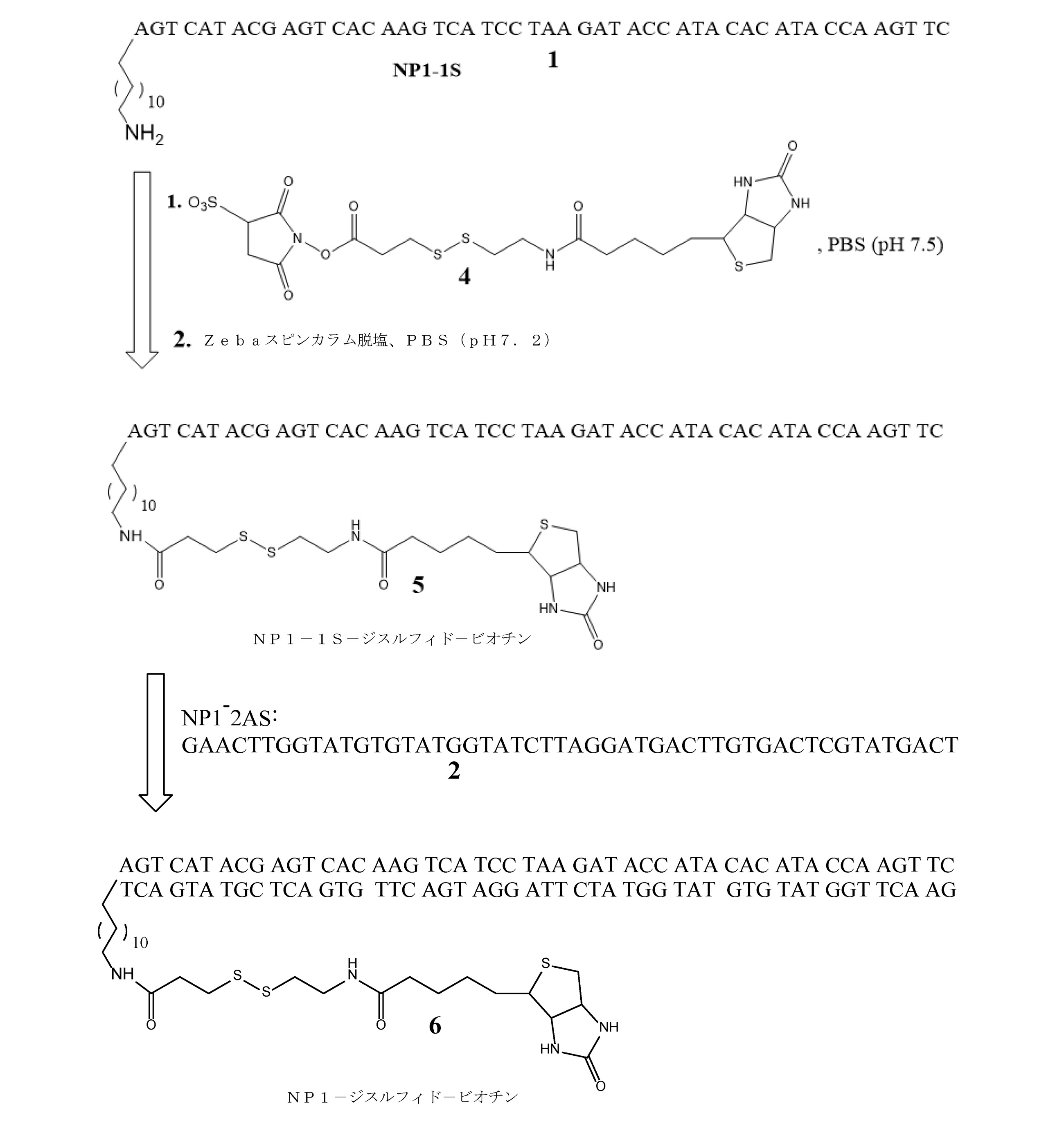
一本鎖5′-アミノオリゴのビオチン化:スルホ-NHS-SS-ビオチン(4、ThermoFisher Scientific)の100mM溶液は、6mgの粉末を0.099mLの無水DMSO(Sigma Aldrich)に溶解することによって作製した。溶液をボルテックスし、直ちに5′-アミノ-DNAをビオチン化するために使用した。50mM PBS(pH7.5)中のおよそ100μLのssDNA(1、171μM、17.1μモル、0.265mg)(配列番号1)溶液を、DMSO中の3.4μLの0.1mMビオチン化試薬(34.1μモル、ssDNA上の20倍モル過剰)と混合した。混合物を混合し、室温で2時間反応させた。2つの0.5mL Zebaスピン脱塩カラム(MWカットオフ7,000Da、ThermoFisher Scientific)を10mM PBS(pH7.2)中で平衡化した。粗ビオチン化ssDNA溶液を、1つのZebaカラムに添加し、4,600rpmで1.3分間溶出した。溶出液を、第2のZebaカラムに転送し、記載のように溶出した。精製NP1-1S-SS-ビオチン(5)(配列番号1)の濃度は、A260吸光度(2.03mg/mL、131μM)を測定することによって決定した。 Biotinlation of Single Chain 5'-Amino Oligo: A 100 mM solution of sulfo-NHS-SS-biotin (4, Thermo Fisher Scientific) is made by dissolving 6 mg of powder in 0.099 mL of anhydrous DMSO (Sigma Aldrich). bottom. The solution was vortexed and immediately used to biotinylated 5'-amino-DNA. Approximately 100 μL of ssDNA (1,171 μM, 17.1 μmol, 0.265 mg) (SEQ ID NO: 1) solution in 50 mM PBS (pH 7.5), 3.4 μL of 0.1 mM biotinylated reagent (34) in DMSO. .1 μmol, 20-fold molar excess on ssDNA) was mixed. The mixture was mixed and reacted at room temperature for 2 hours. Two 0.5 mL Zeba spin desalting columns (MW cutoff 7,000 Da, Thermo Fisher Scientific) were equilibrated in 10 mM PBS (pH 7.2). Crude biotinylated ssDNA solution was added to one Zeba column and eluted at 4,600 rpm for 1.3 minutes. The eluate was transferred to a second Zeba column and eluted as described. The concentration of purified NP1-1S-SS-biotin (5) (SEQ ID NO: 1) was determined by measuring A 260 absorbance (2.03 mg / mL, 131 μM).
ビオチン化二本鎖DNAの形成:およそ60μLのNP1-1S-SS-ビオチン溶液(5、7.85μモル、131μM、2.03mg/mL)を、42μLのNP1-2AS溶液(2、7.85μモル、187μmol/L)と混合した(配列番号2)。溶液を85℃の加熱ブロックに30分間入れ、続いて2時間かけてゆっくりと室温に冷却した。全アニーリング量(およそ100μL)を注入することにより、10mM PBS(pH7.2)を使用して、TosoH G3000SWカラム(7.8mm×300mm)で二本鎖産物を精製した。二本鎖ビオチン化物質は、A260吸光度でモニターしたとき、7.9分(20分の実行時間)で溶出した。0.5mLのAmiconフィルタ濃縮器(MWカットオフ10,000Da)を使用して、溶出液量を480μLに低減した。最終NP1-ジチオ-ビオチン(6)(配列番号1-フォワード鎖(上);配列番号2-リバース鎖(下))の濃度は、A260吸光度によって決定されるように、16.3μMであると計算された。 Formation of biotinylated double-stranded DNA: Approximately 60 μL of NP1-1S-SS-biotin solution (5,7.85 μmol, 131 μM, 2.03 mg / mL) and 42 μL of NP1-2AS solution (2,7.85 μm). Mole, 187 μmol / L) was mixed (SEQ ID NO: 2). The solution was placed in a heating block at 85 ° C. for 30 minutes and then slowly cooled to room temperature over 2 hours. Double-stranded products were purified on a TosoH G3000SW column (7.8 mm × 300 mm) using 10 mM PBS (pH 7.2) by injecting a total annealing amount (approximately 100 μL). The double-stranded biotinylated material was eluted in 7.9 minutes (20 minutes run time) when monitored at A 260 absorbance. A 0.5 mL Amicon filter concentrator (MW cutoff 10,000 Da) was used to reduce the amount of eluate to 480 μL. The concentration of the final NP1-dithio-biotin (6) (SEQ ID NO: 1-forward chain (top); SEQ ID NO: 2-reverse chain (bottom)) is 16.3 μM, as determined by A260 absorbance. calculated.
実施例17
切断可能なDNA-ビオチン構築物の代替合成
相補的DNA配列(NP-31aおよびNP-31b):標準的なホスホルアミダイト化学(Integrated DNA Technologies)を使用して、2つの一本鎖60merを合成した。オリゴNP-31aは、C-6炭素スペーサ(配列番号1)によってDNAから分離された、5′末端にアミノ基を含有する60ヌクレオチドのDNA配列からなる(1、MW=18,841.2g/モル、1.7μM/OD)。オリゴNP-31bは、NP-31aに相補的な60ヌクレオチドのDNA配列(配列番号4)から構成されていた(2、MW=18292.8g/モル、1.8μM/OD)。その後の操作の前に、両方のオリゴヌクレオチドを定量し、凍結乾燥した。
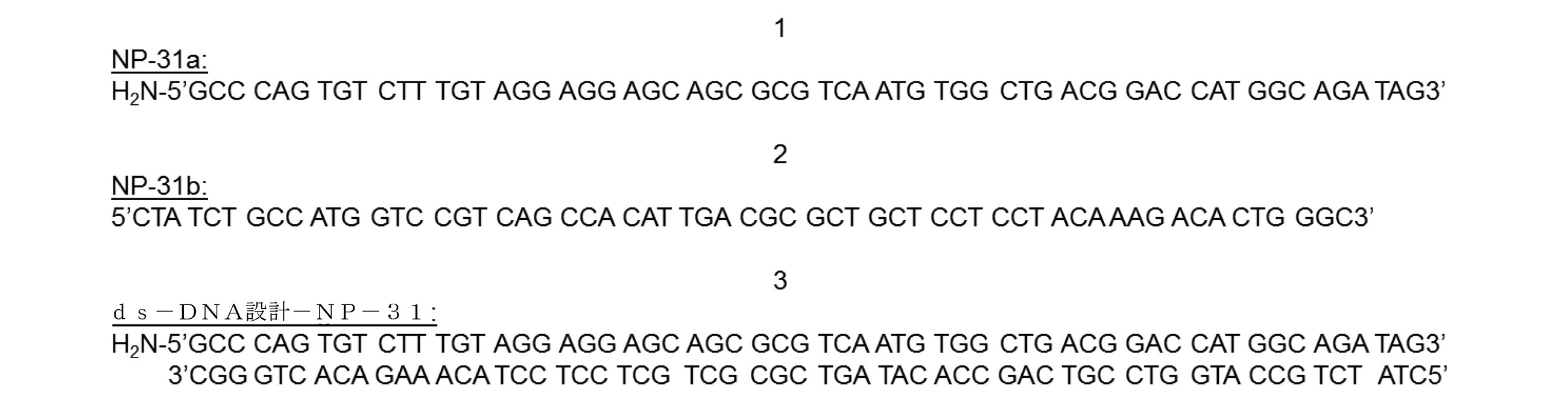
Alternative Synthesis of Cleavable DNA-Biotin Constructs Complementary DNA Sequences (NP-31a and NP-31b): Two single-stranded 60mers were synthesized using standard Integrated DNA Technologies. .. Oligo NP-31a consists of a 60 nucleotide DNA sequence containing an amino group at the 5'end separated from the DNA by a C-6 carbon spacer (SEQ ID NO: 1) (1, MW = 18,841.2 g /). Mol, 1.7 μM / OD). The oligo NP-31b was composed of a 60 nucleotide DNA sequence (SEQ ID NO: 4) complementary to NP-31a (2, MW = 18292.8 g / mol, 1.8 μM / OD). Prior to subsequent manipulation, both oligonucleotides were quantified and lyophilized.
一本鎖5′-アミノオリゴNP-31aのビオチン化:NHS-SS-dPEG4-ビオチン(4、MW=751.94g/モル、Quanta BioDesign,Ltd)の10mM溶液は、15.04mgの粉末を2.0mLのジメチルホルムアミド(Sigma Aldrich)に溶解することによって調製した。溶液をボルテックスし、直ちに5′-アミノ-DNAをビオチン化するために使用した。10mMのリン酸緩衝生理食塩水(PBS)(pH7.4)中のおよそ100μLのssDNA(1、100μM、0.01μモル、0.188mg)(配列番号3)溶液を、DMF中の10μLの10mMビオチン化試薬(0.1μモル、ssDNAに対して10倍モル過剰)と混合した。混合物を混合し、室温で2時間反応させた。2つの0.5mL Zebaスピン脱塩カラム(MWカットオフ7,000Da、ThermoFisher Scientific)を10mM PBS(pH7.2)中で平衡化した。粗ビオチン化ssDNA溶液を、1つのZebaカラムに添加し、4,600rpmで2分間溶出した。溶出液を、第2のZebaカラムに転送し、記載のように溶出した。精製NP-31-SS-ビオチン(5)(配列番号3)の濃度は、A260吸光度(1.45mg/mL、77μM)を測定することによって決定した。

ビオチン化二本鎖DNAの形成:およそ60μLのNP-31-SS-ビオチン溶液(5、77μM)(配列番号3)を、50μLのNP-31b溶液(2、100μM)と混合した。溶液を85℃の加熱ブロックに30分間入れ、続いて2時間かけてゆっくりと室温に冷却した。二本鎖産物(配列番号3-フォワード鎖(上);配列番号4-リバース鎖(下))を、10mM PBS(pH7.2)を使用して、全アニーリング容量(およそ100μL)を注入することによって、TosoH G3000SWカラム(7.8mm×300mm)で精製した。二本鎖ビオチン化物質は、A260吸光度でモニターしたとき、7.57分で溶出した。0.5mLのAmiconフィルタ濃縮器(MWカットオフ10,000Da)を使用して、溶出液量を低減した。最終dsNP-31-SS-ビオチン(6)(配列番号3-フォワード鎖(上);配列番号4-リバース鎖(下))の濃度は、A260吸光度によって決定されるように、11μMであると計算された。 Formation of biotinylated double-stranded DNA: Approximately 60 μL of NP-31-SS-biotin solution (5,77 μM) (SEQ ID NO: 3) was mixed with 50 μL of NP-31b solution (2,100 μM). The solution was placed in a heating block at 85 ° C. for 30 minutes and then slowly cooled to room temperature over 2 hours. The double-stranded product (SEQ ID NO: 3-forward strand (top); SEQ ID NO: 4-reverse strand (bottom)) is injected with a total annealing volume (approximately 100 μL) using 10 mM PBS (pH 7.2). Purified with a Tosoh G3000SW column (7.8 mm × 300 mm). The double-stranded biotinylated material eluted in 7.57 minutes when monitored at A 260 absorbance. A 0.5 mL Amicon filter concentrator (MW cutoff 10,000 Da) was used to reduce the amount of eluate. The concentration of the final dsNP-31-SS-biotin (6) (SEQ ID NO: 3-forward chain (top); SEQ ID NO: 4-reverse chain (bottom)) is 11 μM, as determined by A260 absorbance. calculated.
実施例18
切断可能なDNA-ビオチン-チオール媒介切断構築物の合成
ssNP-31-SS-ビオチンのストレプトアビジンコーティング磁性微粒子(SA-MP)への結合および化学的切断(TCEPまたはDTT):化学的切断実験を、以下の方法により磁性微粒子上で行った(図25を参照)。100μLのPBS(pH7.2)中77μMの修飾オリゴヌクレオチドssNP-31-SS-ビオチン溶液を、室温で30分間、1μLの0.1%ストレプトアビジン常磁性微粒子と共にインキュベートした。粒子を磁石に引きつけ、PBST緩衝液(pH7.4)で10回洗浄することによって、過剰のオリゴを除去した。オリゴ結合粒子を、PBS(pH7.4)中の様々な濃度のDTTまたはTCEPのいずれかと共に15分間インキュベートした。微粒子をPBST緩衝液(pH7.4)で10回洗浄して、切断されたオリゴヌクレオチドを除去した。発蛍光団を含有する相補配列NP-31c(配列番号5)(7、MW=7494.6g/モル、5.2μM/OD)を、PBS(pH7.4)中で30分間微粒子と共にインキュベートして、粒子上にそのまま残っている任意の未切断ssNP-31-SS-ビオチンと結合させた。微粒子を磁石に引きつけ、PBST緩衝液(pH7.4)で10回洗浄して、過剰のNP-31cセグメントを除去した。洗浄したが化学的切断を受けなかった、上記のように調製したコーティング微粒子を対照として用いた。粒子上の蛍光シグナルを、蛍光顕微鏡によって測定した。最大切断効率は、表1に示すように、DTTおよびTCEPについてそれぞれ79%および93%と測定された。


Synthesis of cleavable DNA-biotin-thiol-mediated cleavage constructs Binding and chemical cleavage of ssNP-31-SS-biotin to streptavidin-coated magnetic particles (SA-MP) (TCEP or DTT): Chemical cleavage experiments. The procedure was performed on magnetic fine particles by the following method (see FIG. 25). A 77 μM modified oligonucleotide ssNP-31-SS-biotin solution in 100 μL PBS (pH 7.2) was incubated with 1 μL of 0.1% streptavidin paramagnetic particles for 30 minutes at room temperature. Excess oligos were removed by attracting the particles to the magnet and washing 10 times with PBST buffer (pH 7.4). Oligo-bound particles were incubated with either various concentrations of DTT or TCEP in PBS (pH 7.4) for 15 minutes. The microparticles were washed 10 times with PBST buffer (pH 7.4) to remove the cleaved oligonucleotide. Complementary sequence NP-31c (SEQ ID NO: 5) (7, MW = 7944.6 g / mol, 5.2 μM / OD) containing the fluorophore was incubated with the microparticles in PBS (pH 7.4) for 30 minutes. , Any uncut ssNP-31-SS-biotin remaining intact on the particles. The particles were attracted to the magnet and washed 10 times with PBST buffer (pH 7.4) to remove excess NP-31c segments. The coated fine particles prepared as described above, which were washed but not chemically cleaved, were used as a control. The fluorescence signal on the particles was measured by a fluorescence microscope. Maximum cutting efficiency was measured as 79% and 93% for DTT and TCEP, respectively, as shown in Table 1.
実施例19
切断可能なDNA-ビオチン-光切断構築物の合成
光切断可能なDNA配列および微粒子上での切断効率の評価:一本鎖DNAの光切断可能な配列を、標準的なホスホルアミダイト化学(Integrated DNA Technologies)を使用して合成した。オリゴヌクレオチドは、2つの光切断可能な部分によって分離された2つのオリゴセグメント(オリゴ8-1(配列番号6)およびオリゴ8-2(配列番号7))を構成する48ヌクレオチドからなる(8、MW=15,430.1g/モル、441800L mol-1cm-1)。5′末端は、C-6炭素スペーサによってDNAから分離されたアミノ基を含んでいた。蛍光タグを含むオリゴ8-2に対する相補鎖を合成した(9、MW=7738.8g/モル、212700L mol-1cm-1)(配列番号8)。その後の操作の前に、両方のオリゴヌクレオチドを定量し、凍結乾燥した。
オリゴ8-1(配列番号6):5′AAA AAA GGT CCG CAT CGA CTG CAT TCA3′
オリゴ8-2(配列番号7):5′CCC TCG TCC CCA GCT ACG CCT3′
NP-8(8)(2つの光切断可能な部分(「PC」)によって接合されたオリゴ8-1(配列番号6)およびオリゴ8-2(配列番号7)):
H2N-5′AAAAAAGGTCCGCATCGACTGCATTCA-PC-PC-CCCTCGTCCCCAGCTACGCCT3′
NP-9(9)(配列番号8):AlexaFluor546-5′AGG CGT AGC TGG GGA CGA GGG3′

Cleavable DNA-Biotin-Synthesis of Photo-Cut Constructs Evaluation of Photo-Cleavable DNA Sequences and Cleavage Efficiency on Fine Particles: Photo-cleavable sequences of single-stranded DNA can be obtained from standard integrated DNA. Synthesized using Technologies). Oligonucleotides consist of 48 nucleotides constituting two oligo segments (oligo 8-1 (SEQ ID NO: 6) and oligo 8-2 (SEQ ID NO: 7)) separated by two photocleavable moieties (8, MW = 15,430.1 g / mol, 441800 L mol -1 cm -1 ). The 5'end contained an amino group separated from the DNA by a C-6 carbon spacer. A complementary strand to oligo 8-2 containing a fluorescent tag was synthesized (9, MW = 7338.8 g / mol, 212700 L mol -1 cm -1 ) (SEQ ID NO: 8). Prior to subsequent manipulation, both oligonucleotides were quantified and lyophilized.
Oligo 8-1 (SEQ ID NO: 6): 5'AAA AAA GGT CCG CAT CGA CTG CAT TCA3'
Oligo 8-2 (SEQ ID NO: 7): 5'CCC TCG TCC CCA GCT ACG CCT3'
NP-8 (8) (Oligo 8-1 (SEQ ID NO: 6) and Oligo 8-2 (SEQ ID NO: 7) joined by two photocleavable moieties (“PC”)):
H 2 N-5'AAAAAAAGGTCCGCATCGACTGCATTCA-PC-PC-CCCTCGTCCCCAGCTAGCCCT3'
NP-9 (9) (SEQ ID NO: 8): AlexaFluor546-5'AGG CGT AGC TGG GGA CGA GGG3'
光切断実験は、以下の方法により磁性微粒子に対して行われた(図26Aおよび26Bを参照)。NP-8を抗体に共有結合させて、Ab-オリゴ複合体(Biosynthesis Inc.により調製)を生成した。100μLの33nM抗体-オリゴ複合体を、1μLの0.1%固形分のヤギ抗マウス微粒子と共に室温で30分間インキュベートした。粒子を磁石に引きつけ、PBST緩衝液(pH7.4)で10回洗浄することによって、過剰の抗体-オリゴ複合体を除去した。微粒子複合体溶液を、UV光(300~350nmの波長)下で5分間照射した。微粒子を磁石に引きつけ、PBST緩衝液(pH7.4)で10回洗浄して、任意の切断されたオリゴセグメントを除去した。PBST緩衝液(pH7.4)に粒子を再懸濁した後、蛍光標識オリゴ9(配列番号8)を、照射した微粒子に添加し、室温で30分間インキュベートした。洗浄されたがUV照射を受けなかった上記のように調製されたコーティング微粒子(切断されていないオリゴ8-2)を対照として用いた。粒子上の蛍光シグナル(AlexaFluor(登録商標)546)を、蛍光顕微鏡によって撮像した。表2に示すように、常磁性微粒子に結合したときの切断効率は74%であった。

実施例20
熱切断可能なリンカー
この実施例は、熱切断可能なリンカーおよびそれらの切断について記載する。そのような熱切断可能なリンカーは、本明細書に記載されるように、例えば、DMFチップ、液滴ベースのマイクロ流体チップ、SAWチップ等において用いられ得る。
Example 20
Thermally Cleaveable Linkers This example describes thermally cleaveable linkers and their cleavage. Such thermally cleaveable linkers can be used, for example, in DMF chips, droplet-based microfluidic chips, SAW chips and the like, as described herein.
熱切断可能なリンカーは、二本鎖DNAの熱分離の場合のように、温度を閾値以上に上昇させることによって切断される。DMFチップ内の温度上昇は、光から吸収標的にエネルギーを転送することによって光熱的に達成することができる。一方法では、約980nm(約930nm~約1040nmの範囲)の波長を有するレーザーなどの光源を、流体試料の領域のDMFチップに適用することができる。光は流体中の水分子によって吸収され得、結果として温度上昇およびリンカーの切断をもたらす。加熱のレベルおよび期間は、パルス長、パルスエネルギー、パルス数、およびパルス繰り返し率によって制御することができる。例えば、水の吸収帯を使用する光熱加熱は、例えば、米国特許第6,027,496号に記載されている。 Thermally cleaveable linkers are cleaved by raising the temperature above the threshold, as in the case of thermal separation of double-stranded DNA. The temperature rise in the DMF chip can be photothermally achieved by transferring energy from the light to the absorption target. In one method, a light source such as a laser having a wavelength of about 980 nm (range from about 930 nm to about 1040 nm) can be applied to the DMF chip in the region of the fluid sample. Light can be absorbed by water molecules in the fluid, resulting in increased temperature and cleavage of the linker. The level and duration of heating can be controlled by pulse length, pulse energy, pulse count, and pulse repetition rate. For example, photothermal heating using a water absorption band is described, for example, in US Pat. No. 6,027,496.
光熱加熱はまた、光源を染料または顔料含有標的と結合することによって達成することもできる。この場合、DMFチップの目標領域は、吸収染料または顔料、例えばカーボンブラックで印刷される。流体が標的と接触すると、光源、例えば市販のレーザーダイオードが、光吸収標的に向けられ、結果として局所的な温度上昇およびリンカーの切断をもたらす。加熱のレベルおよび持続時間は、標的の吸光特性、光波長、パルス長、パルスエネルギー、パルス数、およびパルス繰り返し率によって制御することができる。例えば、光吸収標的と組み合わせて光源を使用する光熱加熱は、米国特許第6,679,841号に記載されている。 Photothermal heating can also be achieved by binding the light source to a dye or pigment-containing target. In this case, the target area of the DMF chip is printed with an absorbent dye or pigment, such as carbon black. When the fluid comes into contact with the target, a light source, such as a commercially available laser diode, is directed at the light absorbing target, resulting in local temperature rise and cleavage of the linker. The level and duration of heating can be controlled by the target's absorbance characteristics, light wavelength, pulse length, pulse energy, pulse count, and pulse repetition rate. For example, photothermal heating using a light source in combination with a light absorption target is described in US Pat. No. 6,679,841.
光熱加熱の第3の方法では、吸収染料または顔料を、DMFチップ内の流体に導入することができる。次いで、光はDMFチップを透過し、エネルギーは溶解または懸濁した吸収材料に転送され、結果として局所的な温度上昇およびリンカーの切断をもたらす。加熱のレベルおよび持続期間は、標的材料の吸光特性、光波長、パルス長、パルスエネルギー、パルス数、およびパルス繰り返し率によって制御る。この方法の一実施形態では、光吸収標的は、デバイスに使用される磁性微粒子懸濁液である。例えば、液滴中の懸濁ナノ粒子を使用する光熱加熱は、Walsh et al.,Analyst,140(5),1535-42(2015)に記載されている。Walshらの参考文献もまた、光熱用途において達成することができる制御のうちのいくつかを実証している。 In the third method of photothermal heating, the absorbent dye or pigment can be introduced into the fluid in the DMF chip. Light is then transmitted through the DMF chip and energy is transferred to the dissolved or suspended absorbent material, resulting in local temperature rise and cleavage of the linker. The level and duration of heating is controlled by the absorbance characteristics of the target material, light wavelength, pulse length, pulse energy, number of pulses, and pulse repetition rate. In one embodiment of this method, the light absorption target is a magnetic particulate suspension used in the device. For example, photothermal heating using suspended nanoparticles in a droplet can be described by Walsh et al. , Analyst, 140 (5), 1535-42 (2015). The reference by Walsh et al. Also demonstrates some of the controls that can be achieved in photothermal applications.
実施例21
マイクロ波誘導粒子温熱療法により達成された熱的切断
この実施例は、イムノアッセイ検出が低電力マイクロ波放射の使用により加速され得るため、感熱性の切断可能なリンカーを介してカウント可能な部分を遊離する熱変性(dsDNA変性、逆マイケル反応、逆デールスアルダー、および他の排除)を促進するためのマイクロ波誘導粒子温熱療法の使用について説明している。そのような感熱性の切断可能なリンカーは、本明細書に記載されるように、例えばDMFチップにおいて用いられ得る。
Example 21
Thermal cleavage achieved by microwave-induced particle hyperthermia In this example, the immunoassay detection can be accelerated by the use of low-power microwave radiation, thus freeing countable moieties through a heat-sensitive cleavable linker. Describes the use of microwave-induced particle hyperthermia to promote heat denaturation (dsDNA denaturation, reverse Michael reaction, reverse Dale Alder, and other exclusions). Such heat-sensitive cleavable linkers can be used, for example, in DMF chips, as described herein.
この実施例では、40~55℃の範囲の二本鎖Tmを有する15bp配列などの直交性官能化短dsDNAセグメントの形成が、熱遊離剤として役立つ。dsDNAセグメントを、スルフヒドリル-マレイミド相互作用および26nmカルボキシル化ポリスチレンナノ粒子(NP)、例えばアミン活性化カルボン酸化学などの付着化学を介してBangs Labs(Fishers,IN,USA)から得られるものなどの、抗体と付着化学を介して反応させることができる。26nm NPは、528.7μeq/gの表面電荷および68.4sq.Å/群のパーキング面積を有する(製造元情報)。抗体およびナノ粒子は、熱的に誘発された遊離可能なリンカーを形成するdsDNAセグメントを介して会合している。熱リンカーは、粒子温熱療法および局所的温度勾配を引き起こすためのマイクロ波照射などの技法を使用して切断することができる。
DNA配列1(10)(配列番号9):H2N-5′CAA GCC CGG TCG TAA3′
DNA配列1b(11)(配列番号10):マレイミド-5′TTA CGA CCG GGC TTG3′
dsDNA配列(12)(配列番号9-フォワード鎖(上);配列番号10-リバース鎖(下)):
H2N-5′CAA GCC CGG TCG TAA3′
3′GTT CGG GCC AGC ATT5′-マレイミド。
In this example, the formation of orthogonal functionalized short dsDNA segments, such as a 15bp sequence with a double-stranded Tm in the range of 40-55 ° C., serves as a heat release agent. The dsDNA segments are obtained from Bangs Labs (Fishers, IN, USA) via sulfhydryl-maleimide interactions and adhesion chemistry such as 26 nm carboxylated polystyrene nanoparticles (NP), eg amine-activated carboxylic acid chemistry. It can be reacted with an antibody via adherent chemistry. 26 nm NP has a surface charge of 528.7 μeq / g and 68.4 sq. Has a parking area of Å / group (manufacturer information). Antibodies and nanoparticles are associated via dsDNA segments that form a thermally induced liberable linker. Thermal linkers can be cleaved using techniques such as particle hyperthermia and microwave irradiation to cause a local temperature gradient.
DNA Sequence 1 (10) (SEQ ID NO: 9): H 2 N-5'CAA GCC CGG TCG TAA3'
dsDNA sequence (12) (SEQ ID NO: 9-forward strand (top); SEQ ID NO: 10-reverse strand (bottom)):
H 2 N-5'CAA GCC CGG TCG TAA3'
3'GTT CGG GCC AGC ATT5'-maleimide.
直行性官能化相補DNA配列複合体のアニーリング:PBS(pH7.5)中のおよそ100μM DNA配列1(配列番号9)の溶液を、PBS(pH7.5)中1.0モル等量のDNA配列1b(Integrated DNA Technologiesオリゴアナライザツールに従う理論上のTm 51.6℃)(配列番号10)と混合し、60℃で30分間加熱ブロックに配置した後、室温で2時間ゆっくり冷却することができる。得られたdsDNA産物を、10mM PBS(pH7.2)を使用して、TosoH G3000SWカラム(7.8mm×300mm)で全アニーリング量を注入することにより精製する。溶出液量は、0.5mLのAmiconフィルタ濃縮器を使用して低減する。最終dsDNA濃度は、A260吸光度によって決定される。反応スキームを以下に示す(「Mal」はマレイミドである)。

カルボキシル-ポリスチレンナノ粒子の活性化および二本鎖DNAの付加:カルボキシルナノ粒子は、実施例5の「カルボキシル-ポリスチレンナノ粒子の活性化」のセクションに記載されるように予備活性化される。NPへのDNA負荷は、結合したDNA鎖の熱変性、粒子洗浄、蛍光標識された相補DNA配列(AlexaFluor546-5′-TTA CGA CCG GGC TTG3′(配列番号11)など)のアニーリングによって決定され、蛍光顕微鏡を使用して定量した。
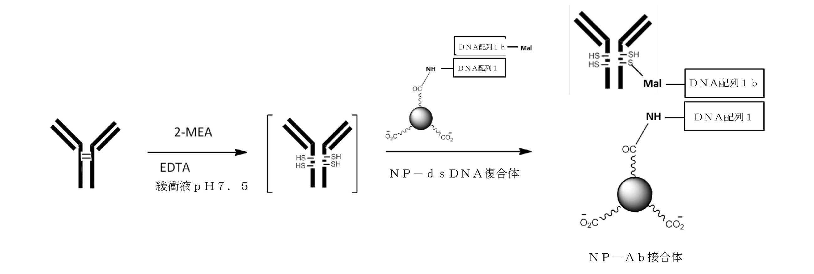
抗体の低減およびNP-dsDNA複合体への接合:実施例5の「抗体の低減」のセクションに記載されているように、抗体を低減させる。還元抗体は、NP-dsDNA複合体へのカップリングに直ちに使用することができる。得られた接合体を6,500gで遠心分離し、上澄みをデカントによって除去する。洗浄手順をPBS(pH7.5)で5回繰り返して、ナノ粒子から遊離抗体を除去する。ナノ粒子の取り込みに対する活性抗体の比率は、所定の抗体に対する蛍光標識抗原を用いて定量することができる。接合体NP濃度(固形分%)は、UV-Vis分光法(600nm)を使用して決定される。粒子接合体を、SEMによって調べ、サイズ/電荷分布をZetaSizerを使用して決定する。

マイクロ波誘導粒子温熱療法およびナノポアカウントイムノアッセイ:上記のスキームは、調製が上述されている熱変性抗体-ナノ粒子接合体を利用するナノポアカウントアッセイを例示する。サンドイッチ型イムノアッセイは、検体捕捉剤でコーティングされた磁性微粒子を使用して調製することができ、ここで血液検体は、磁性微粒子でインキュベートされ、洗浄され、記載の抗体-ナノ粒子接合体でインキュベートされる。粒子温熱療法は、マイクロ波照射を使用して粒子表面近くに局所的な温度勾配を生じさせることによって誘導され得る。Dutz and Hergt(Nanotechnology,25:452001(2014))において検討されているものなどの粒子温熱療法を使用することができる。イムノアッセイ設定における局所熱変性へのこれらの技法の適応は、(ナノ粒子などの)カウント部分を遊離する方法を提供することができる。磁性微粒子を除去した後、カウント部分(ナノ粒子)は、ナノポアを通過する際にカウントされる。 Microwave-Induced Particle Hyperthermia and Nanopoain Immunoassay: The above scheme exemplifies a nanopoamental assay utilizing the heat-denatured antibody-nanoparticle conjugate whose preparation is described above. Sandwich immunoassays can be prepared using magnetic microparticles coated with a sample capture agent, where blood samples are incubated with magnetic microparticles, washed and incubated with the antibody-nanoparticle conjugates described. To. Particle hyperthermia can be induced by using microwave irradiation to create a local temperature gradient near the particle surface. Particle hyperthermia such as those studied in Dutz and Hergt (Nanotechnology, 25: 452001 (2014)) can be used. Adaptation of these techniques to local heat denaturation in an immunoassay setting can provide a method of releasing count moieties (such as nanoparticles). After removing the magnetic particles, the counting portion (nanoparticles) is counted as it passes through the nanopores.
実施例22
ナノポアカウントデータ
この実施例は、種々のタグ、例えばポリエチレングリコールを有するssDNAハイブリッド分子(DNA-STAR)、dsDNA、DBCOで標識されたdsDNA、およびPAMAMコハク酸デンドリマーについてのナノポアカウントデータを記載する。ナノポア最適化を提供するために、異なるサイズのナノポアと共にこれらの異なるタグを使用した。異なる分子ポリマー標識を、適切な塩緩衝液に懸濁し、標準的な流体セルカセットを使用して検出した。
Example 22
Nanopoaccount Data This example describes nanopoaccount data for various tags such as ssDNA hybrid molecule (DNA-STAR) with polyethylene glycol, dsDNA, DBCO labeled dsDNA, and PAMAM succinate dendrimer. These different tags were used with nanopores of different sizes to provide nanopore optimization. Different molecular polymer labels were suspended in a suitable salt buffer and detected using a standard fluid cell cassette.
社内器具を使用して、電流-電圧(i-V)記録(ボルタンメトリーデータ)および電流-時間(i-t)記録を記録した。CUSUMと呼ばれるコンピュータソフトウェアプログラムを用いて、取得したデータを調べ、ユーザによって入力された閾値に基づいて事象を検出した。評価における主観性の影響は、できるだけ多くの事象を検出し、後で特定の目的のためにフィルタリングすることによって最小限に抑えられた。 Current-voltage (i-V) recordings (voltammetry data) and current-time (it) recordings were recorded using in-house equipment. A computer software program called CUSUM was used to examine the acquired data and detect events based on thresholds entered by the user. The effect of subjectivity on the assessment was minimized by detecting as many events as possible and later filtering for specific purposes.
膜のシス側にタグを付加して、初期実験を行った。標識溶液に200mVの電気バイアスを印加し、Axopatch 200B増幅器およびCUSUMソフトウェアを使用して電流遮断をモニターした。 An initial experiment was performed by adding a tag to the cis side of the membrane. An electrical bias of 200 mV was applied to the labeled solution and current interruption was monitored using the Axopatch 200B amplifier and CUSUM software.
細孔サイズがそれらの通過を制限しない限り、小分子はナノポアを非常に速く通過し得ることが知られている。高速事象の電流遮断は、システムの制限された帯域幅のために変形し得る。より速い分子は、特定のシステムによって完全に検出されないことさえあり得る。 It is known that small molecules can pass through nanopores very quickly unless the pore size limits their passage. Current interruptions in fast events can be modified due to the limited bandwidth of the system. Faster molecules may not even be completely detected by a particular system.
我々の研究では、より大きなポリマーおよび大きな基修飾剤で標識された分子のみが検出された。実験条件および検出可能な事象の数を表3に示す。

これらのデータは、DNAデンドリマー、ポリマー、およびPAMAMデンドリマーが、固体ナノポアセンサ用の検出標識として使用できることを確認している。 These data confirm that DNA dendrimers, polymers, and PAMAM dendrimers can be used as detection markers for solid nanopore sensors.
実施例23
生体分子のナノポア分化
この実施例では、ナノポアを使用して、生体分子(例えば、dsDNAスター、DBCO修飾dsDNAおよび通常のdsDNA)を区別した。この方法は、異なる標識型を使用した多重化に使用することができる。
Example 23
Nanopore Differentiation of Biomolecules In this example, nanopores were used to distinguish biomolecules (eg, dsDNA stars, DBCO-modified dsDNA and normal dsDNA). This method can be used for multiplexing with different marker types.
この実施例は、一本鎖オリゴヌクレオチドが共有結合した(DNA-Star)、中央に分岐点を含む50bpオリゴヌクレオチド(bp#25)、中央にジベンジルシクロオクチン(DBCO)修飾を含む二本鎖50bpオリゴヌクレオチド(塩基#25)、5′-チオール修飾二本鎖DNAオリゴヌクレオチドを用いた。 In this example, a single-stranded oligonucleotide is covalently attached (DNA-Star), a 50bp oligonucleotide (bp # 25) containing a branch point in the center, and a double-stranded oligonucleotide containing a dibenzylcyclooctinate (DBCO) modification in the center. A 50bp oligonucleotide (base # 25), a 5'-thiol-modified double-stranded DNA oligonucleotide, was used.
これらの様々な修飾DNA分子を、3.6M LiCl緩衝液中の3つの異なるSiNxナノポアを使用して分析した。DNAスター分子は、直径4.0nmの細孔で分析し、DBCO修飾DNAは、直径3.7nmの細孔で分析し、チオール修飾DNAは、直径4.2nmの細孔で分析した。ナノポアが3つの異なる生体分子を区別する能力を示すために、現在の遮断レベル(pA)をナノポア持続時間(μsec)に対してプロットした。集団レベルでは、散布図における明確なパターンの違いによって実証されるように、3つの異なる標識は区別可能であるように思われる(図24A~図24C)。個々の事象をリアルタイムで識別するには、シグナルとノイズを区別する方法として、追加レベルの遮断レベルおよび時間情報が必要である。異なるナノポア標識を区別する能力は、ナノポアを様々なアッセイにおける多重化に用いることができることを実証している。 These various modified DNA molecules were analyzed using three different SiNx nanopores in 3.6M LiCl buffer. DNA star molecules were analyzed in pores with a diameter of 4.0 nm, DBCO-modified DNA was analyzed in pores with a diameter of 3.7 nm, and thiol-modified DNA was analyzed in pores with a diameter of 4.2 nm. Current blocking levels (pA) were plotted against nanopore duration (μsec) to show the ability of nanopores to distinguish between three different biomolecules. At the population level, the three different labels appear to be distinguishable, as evidenced by the distinct pattern differences in the scatter plot (FIGS. 24A-24C). To identify individual events in real time, additional levels of cutoff level and time information are needed as a way to distinguish between signal and noise. The ability to distinguish between different nanopore labels demonstrates that nanopores can be used for multiplexing in various assays.
実施例24
定性的分析
以下の実施例は、定性的アッセイを実施するための方法を記載する。基本的に、この実施例では、構築物を使用して、DMFチップ上でアッセイの原理を実証し、構築物を切断し、標識を解放し、次いでナノポアを転位させたときにシグナルを発生させるようにナノポアを使用してカウントし、dsDNA標識のこの切断およびその後のカウントが、アッセイ中に生じた特異的結合と相関している、2つの特異的結合メンバー対(ストレプトアビジンおよびビオチン)の結合を示す。さらに、適切な対照実験を行って、ナノポア転位測定中にカウントされた標識から発生したシグナルが、アッセイプロセス流に導入されるチオール切断試薬の存在と相関するのではなく、アッセイプロセス中に生じた特異的結合事象によることを確認した。実施した実験の詳細は、以下のとおりである。
Example 24
Qualitative Analysis The following examples describe methods for performing qualitative assays. Basically, in this example, constructs are used to demonstrate the principles of the assay on a DMF chip, to generate a signal when the construct is cleaved, the label is released, and then the nanopores are transposed. Counting using nanopores, this cleavage of the dsDNA label and subsequent counts indicate binding of two specific binding member pairs (streptavidin and biotin) that correlate with the specific binding that occurred during the assay. .. In addition, with appropriate control experiments, the signals generated from the labels counted during the nanopore dislocation measurement occurred during the assay process rather than correlating with the presence of thiol cleavage reagents introduced into the assay process stream. It was confirmed that it was due to a specific binding event. The details of the experiments conducted are as follows.
DMFを使用したチオール媒介切断:切断可能なジスルフィド結合を含むビオチン標識二本鎖DNA(実施例17の(6))を、ナノポア検出/カウントのための標的として使用した。結合アッセイは、DMFチップ上のストレプトアビジン磁性微粒子にビオチンDNAを結合させ、続いてチオール媒介化学的切断ステップからなる(図25を参照)。DMFチップ上の試薬の配置を図27に示す。ストレプトアビジン磁石粒子に結合した種から分離された切断されたDNA標的を、制御された絶縁破壊によって形成されたプレドリルナノポアを有する固体窒化ケイ素(SiNx)膜を含むナノポア流体セルに転送した(H.Kwok,et al.,PLoS,9(3),2014)。DNA標的物質をカウントし、オープンソースCUSUMソフトウェア分析パッケージ(NIST)を使用して分析した。 Thiol-mediated cleavage using DMF: Biotin-labeled double-stranded DNA containing cleavable disulfide bonds ((6) in Example 17) was used as a target for nanopore detection / counting. The binding assay consists of binding biotin DNA to streptavidin magnetic microparticles on a DMF chip followed by a thiol-mediated chemical cleavage step (see Figure 25). The arrangement of the reagents on the DMF chip is shown in FIG. 27. Cleaved DNA targets separated from the species bound to the streptavidin magnet particles were transferred to a nanopore fluid cell containing a solid silicon nitride (SiN x ) membrane with predrill nanopores formed by controlled breakdown (SiN x). H. Kwalk, et al., PLoS, 9 (3), 2014). DNA target substances were counted and analyzed using the open source CUSUM software analysis package (NIST).
適切な試薬を、8個の試薬リザーバを含むガラス製DMFチップ(3″×2″×0.0276″)に負荷した。廃棄物リザーバを除いて、各リザーバは、およそ5μLの各試薬を含んでいた。試薬の濃度は以下のとおりであった。PBS中11μMのビオチン-SS-DNA(pH=7.2)、10mg/mL(w/v)M-270 2.7μmのストレプトアビジンでコーティングされた磁性微粒子(Life Technologies)、PBS洗浄緩衝液(pH=7.2)+0.05% ETKT(エチレンテトラ-KIS(エトキシレート-ブロック-プロポキシレート)テトロ)、50mMトリス-(2-カルボキシエチル)ホスフィン(TCEP)。分注されたDMF液滴のおよそのサイズは、1.5μLであった。 Suitable reagents were loaded onto a glass DMF chip (3 ″ × 2 ″ × 0.0276 ″) containing 8 reagent reservoirs. Except for the waste reservoir, each reservoir contained approximately 5 μL of each reagent. The concentration of the reagent was as follows. Coated with 11 μM biotin-SS-DNA (pH = 7.2), 10 mg / mL (w / v) M-270 2.7 μm streptavidin in PBS. Magnetic fine particles (Life Technologies), PBS wash buffer (pH = 7.2) + 0.05% ETKT (ethylenetetra-KIS (ethoxylate-block-propoxylate) tetro), 50 mM tris- (2-carboxyethyl) ) Phosphin (TCEP). The approximate size of the dispensed DMF droplets was 1.5 μL.
1滴のM-270ストレプトアビジンでコーティングされた微粒子を分注し、1滴のdsNP-31-SS-ビオチンとおよそ40分間混合した。2滴を複合し、DMFチップ上で12個の電極(3×4)上を円形パターンで動かすことによって混合を達成した。底部磁石を係合させて微粒子を収集し、上清を廃棄物リザーバに移した。次に、2滴のPBS/ETKT緩衝液を分注して、微粒子スラグに移し、次いでこれを溶液に再懸濁した。懸濁液を5分間混合した後、磁石を再び係合させ、上澄み液を廃棄物リザーバに移した。混合時間を45分まで徐々に増やしながら、粒子洗浄ステップを合計11回繰り返した。最後の洗浄上清を空のリザーバに移した。追加の5滴のPBS/ETKTを同じリザーバに移した。ナノポア分析の準備として、34-AWG非金属シリンジ(Microfil 34-AWG)を使用して、リザーバ内の洗浄液およびPBS/ETKTを除去し、1.5mLのエッペンドルフ管に転送した。2滴のTCEP試薬を微粒子スラグに移し、45分間混合することによって、切断を開始した。底部磁石を係合させ、上清(切断されたDNAを含む)を空のリザーバに移した。さらに5滴のPBS/ETKT洗浄緩衝液を、同じリザーバに移した。ナノポア分析の準備として、34ゲージの非金属製シリンジを使用して、最終抽出物をDMFチップから取り出し、1.5mLのエッペンドルフ管に転送した。切断溶出液を30秒間微量遠心分離し、1分間磁気ラックに入れて微量の微粒子を除去した。 One drop of M-270 streptavidin-coated microparticles was dispensed and mixed with one drop of dsNP-31-SS-biotin for approximately 40 minutes. Mixing was achieved by combining the two drops and moving them in a circular pattern on 12 electrodes (3x4) on the DMF chip. The bottom magnet was engaged to collect the particulates and the supernatant was transferred to the waste reservoir. Two drops of PBS / ETKT buffer were then dispensed and transferred to microparticulate slag, which was then resuspended in solution. After mixing the suspension for 5 minutes, the magnets were reengaged and the supernatant was transferred to the waste reservoir. The particle washing step was repeated a total of 11 times, gradually increasing the mixing time to 45 minutes. The final wash supernatant was transferred to an empty reservoir. An additional 5 drops of PBS / ETKT were transferred to the same reservoir. In preparation for nanopore analysis, a 34-AWG non-metal syringe (Microfil 34-AWG) was used to remove the wash solution and PBS / ETKT in the reservoir and transfer to a 1.5 mL Eppendorf tube. Cleavage was initiated by transferring 2 drops of TCEP reagent to the particulate slag and mixing for 45 minutes. The bottom magnet was engaged and the supernatant (including the truncated DNA) was transferred to an empty reservoir. An additional 5 drops of PBS / ETKT wash buffer were transferred to the same reservoir. In preparation for nanopore analysis, a 34 gauge non-metal syringe was used to remove the final extract from the DMF chip and transfer it to a 1.5 mL Eppendorf tube. The cut eluate was centrifuged for 30 seconds and placed in a magnetic rack for 1 minute to remove trace particles.
ナノポア分析:TEMウィンドウ(0.05μm×0.05μm)に埋め込まれた10nm厚のSiNx膜(Norcada NT0052、低応力SiNx)の制御された絶縁破壊(CBD)を使用して、ナノポア製造を達成した。この方法は、高精度かつ最小のコストで小径の固体細孔を形成することができる。TEM-SiNx膜を、2つの緩衝液チャンバを含むポリテトラフルオロエチレン(PTFE)流体セルに入れ、2つのシリコーンエラストマーガスケットを使用して封止した。流体セルは、セルの底部に16μLの体積チャネルを含み、これは上部チャンバの塩溶液をナノポア膜に接続した。ナノポア製造のために、流体セルを最初に脱気エタノールで満たし、脱気脱イオン水で交換し、次いで18MΩ脱イオン水中で重炭酸ナトリウムでpH10に緩衝した脱気0.5M KClで満たした。8Vのバイアス電圧を使用する増幅器を用いて製造を行った。流体セルの両側を、銀/塩化銀ワイヤを使用して接続した。Kwokらに記載されているように、8Vの固定電圧を設定している間、電流はリアルタイムで静電容量(電流の低減)を呈する。電流が増加すると、電力がセルから除去される。製造のためのサンプリングレートは25KHzである。漏れ電流の増加は、細孔の形成を示し、それによって電圧が切られた。孔径は、以下のコンダクタンスに基づく式から求めた。

製造塩緩衝液を、3.6M LiClと交換し、これを転位事象を検出するための感知緩衝液として使用した。Axopatch 200B増幅器と、ナノポア膜を収容する流体セルへの銀/塩化銀接続との間にヘッドステージを配置した。 The production salt buffer was replaced with 3.6M LiCl and used as a sensing buffer to detect dislocation events. A headstage was placed between the Axopatch 200B amplifier and the silver / silver chloride connection to the fluid cell containing the nanopore membrane.
およそ0.2μLのTCEP切断ds-DNA標的を、1.8μLのPBS緩衝液(これは、TCEP切断溶出液の10倍希釈液に相当する)で希釈し、およそ30μLの3.6M LiCl塩溶液を含んでいたナノポア細胞チャンバに全量を装填し混合した。最後のDMF洗浄溶出液を、陰性切断対照として使用した(これは希釈しなかった)。TCEP溶出液および陰性対照について、DNA転位の数をそれぞれ23分および65分間測定し、流動速度(sec-1)に変換した。図28に示す結果は、ds-SS-DNA標的がDMFを使用してM-270ストレプトアビジン粒子から首尾よく切断され、検出器として固体ナノポアを使用して検出されたことを実証する。ナノポア流動速度から測定して、SNRは21.9であると決定された。 Approximately 0.2 μL of TCEP cleavage ds-DNA target is diluted with 1.8 μL of PBS buffer, which corresponds to a 10-fold dilution of TCEP cleavage eluate, and approximately 30 μL of 3.6M LiCl salt solution. The nanopore cell chamber containing was loaded in full and mixed. The final DMF wash eluate was used as a negative cleavage control (which was not diluted). For TCEP eluate and negative control, the number of DNA dislocations was measured for 23 and 65 minutes, respectively, and converted to a flow rate (sec -1 ). The results shown in FIG. 28 demonstrate that the ds-SS-DNA target was successfully cleaved from M-270 streptavidin particles using DMF and detected using solid nanopores as a detector. The SNR was determined to be 21.9, as measured from the nanopore flow velocity.
データ分析:転位事象の数は、実験的試験条件下、以下の等式を使用して、二本鎖DNA転位事象において見出される予想電流変化を最初に計算することによって決定することができ、

要約:この実施例は、本明細書中に記載されるようなステップのプロセスを実施することによる定性的アッセイを記載する。図25に示されているチオールベースの切断ステップを用いて、実施例17に記載されるように、切断可能なリンカー接合体を使用して直接アッセイを実施した。そのようなアッセイを実施するための他の切断可能なリンカー手法はまた、本明細書中に記載されるように、種々のタグのカウントを可能にするための、リンカーの切断の種々の他の方法を含み得るがこれらに限定されないことが理解される。例えば、実施例17に記載の方法に加えて、そのような他の代替の切断方法および/または試薬は、本明細書に記載され、当業者に知られている他の切断方法に加えて、実施例16、実施例18、実施例19、実施例20および実施例21に記載のものを含み得る。この実施例(実施例24)において実証されたアッセイフォーマットは、直接アッセイを表すが、サンドイッチイムノアッセイフォーマットおよび/または様々な競合アッセイフォーマットなど、当業者に知られているものなどの他のフォーマットが、記載された方法を使用してアッセイを実施するために同様に実装され得ることも理解される。 Abstract: This example describes a qualitative assay by performing a process of steps as described herein. Using the thiol-based cleavage step shown in FIG. 25, the assay was performed directly with a cleavable linker conjugate as described in Example 17. Other cleavable linker techniques for performing such assays are also various other cleavable linker techniques to allow counting of different tags, as described herein. It is understood that methods may be included, but not limited to these. For example, in addition to the methods described in Example 17, such other alternative cleavage methods and / or reagents are described herein and in addition to other cleavage methods known to those of skill in the art. It may include those described in Example 16, Example 18, Example 19, Example 20 and Example 21. The assay formats demonstrated in this Example (Example 24) represent direct assays, but other formats, such as those known to those of skill in the art, such as sandwich immunoassay formats and / or various competing assay formats. It is also understood that it can be implemented as well to perform the assay using the described method.
例えば、実施例9に記載されているように、TSH(甲状腺刺激ホルモン)の検出のためのサンドイッチイムノアッセイフォーマットは、低コストDMFチップ上でそのようなアッセイを実施する能力を実証した。さらに、他の方法で当業者に知られている他の切断可能なリンカーに加えて、実施例1、実施例2、実施例3、実施例4、実施例5および実施例6に記載されているものなどの様々なヘテロ二官能性の切断可能なリンカーを使用して、イムノ接合体の生成に有用な多数の様々なバイオ接合試薬または切断可能なリンカーを有する他の活性特異的結合メンバーが合成され得る。本発明の実施に有用な免疫接合体は、実施例3、実施例4、実施例5および実施例6に記載されるものなどの方法によって、ならびに当業者に公知の他の方法によって合成することができる。さらに、実施例8は、サンドイッチおよび競合アッセイフォーマットを含む様々なアッセイフォーマット、ならびに当業者に知られているそれらの他の変形を実行するために必要な様々なステップを容易にすることができる、低コストチップ上の様々な液滴操作の機能を示す。実施例11は、アッセイにおいて切断可能な標識をカウントするために使用することができるナノポアの製造を示すが、当業者に知られているナノポア製造のための他の方法もまた、この目的のために使用することができることが理解される。実施例16はまた、切断が行われるアッセイの実施に有用な別の構築物を表し、したがって、この実施例に記載されるように、ナノポアカウント法を使用してカウントできるように、カウント可能な標識が遊離される。 For example, as described in Example 9, the sandwich immunoassay format for the detection of TSH (thyroid stimulating hormone) demonstrated the ability to perform such an assay on a low cost DMF chip. Further described in Example 1, Example 2, Example 3, Example 4, Example 5 and Example 6 in addition to other cleavable linkers known to those of skill in the art by other methods. Various heterobifunctional cleavable linkers, such as those available, can be used to generate a large number of different bioconjugating reagents or other activity-specific binding members with cleavable linkers that are useful in the production of immunoconjugates. Can be synthesized. Immunoconjugates useful for carrying out the present invention are synthesized by methods such as those described in Examples 3, 4, 5, 5 and 6, and by other methods known to those of skill in the art. Can be done. In addition, Example 8 can facilitate various assay formats, including sandwiches and competitive assay formats, as well as the various steps required to perform those other variants known to those of skill in the art. Shown the features of various droplet manipulations on low cost chips. Example 11 shows the production of nanopores that can be used to count cleavable labels in the assay, but other methods of nanopore production known to those of skill in the art are also for this purpose. It is understood that it can be used for. Example 16 also represents another construct useful for performing the assay in which cleavage is performed, and therefore, as described in this example, a countable label so that it can be counted using the nanopoacing method. Is released.
実施例22は、一般に、ナノポアを横断する様々な標識の存在に関する転位事象を測定することができるように、カウントをどのようにして行うことができるかを示す。図29は、カウントアッセイにおいてデータの質を操作することができるようにするための、シグナルの閾値化の概念を示す。図28は、この実施例に記載されるようなアッセイ方法を使用して、検体の存在を決定するために使用され得るデータの種類を表す定性的アッセイデータを示す。この特定の実施例では、dsDNAを標識として使用したが、ナノビーズ、デンドリマー等を含むがこれらに限定されない実施例5および/または実施例22に記載の標識などの他の標識も利用され得ることも理解される。適切な試薬を生成するために必要とされるそのような構築物は、本出願において本明細書における様々な実施例を通して、またはそうでなければ当業者に知られている方法を介して合成され得る。 Example 22 shows how counting can be done so that dislocation events with respect to the presence of various labels across nanopores can generally be measured. FIG. 29 shows the concept of signal thresholding to allow manipulation of data quality in count assays. FIG. 28 shows qualitative assay data representing the types of data that can be used to determine the presence of a sample using an assay method as described in this example. Although dsDNA was used as a label in this particular example, other labels such as those described in Example 5 and / or Example 22 may also be utilized, including but not limited to nanobeads, dendrimers and the like. Understood. Such constructs required to produce suitable reagents can be synthesized in this application through various examples herein or otherwise through methods known to those of skill in the art. ..
実施例25
定量的分析
以下の実施例は、定量的アッセイを実施するための方法を記載する。基本的に、この実施例では、実施例24の延長として、増加した量のカウント標識が(この場合はカウント可能な標識がdsDNA分子である)、標準曲線上で、アッセイ(結合)ステップにおいて結合された(順に元の試料中に存在する検体の量と相関する)特定の結合剤の量に相関することを実証するように標準曲線を生成した。この特定の実験の標準曲線は、データ分析の様々な異なる方法に基づく図31、図32、図34、または標準曲線を生成するために流動に依存する図34に見出すことができる。後者の場合、図34に示す測定方法は、事象/時間(カウント事象の流動)に基づいているが、所与の試料中で測定される検体濃度の量に相関する標準曲線を生成するために他の測定方法も使用できることが理解される。実施した実験の詳細は、以下のとおりである。
Example 25
Quantitative Analysis The following examples describe methods for performing quantitative assays. Essentially, in this example, as an extension of Example 24, an increased amount of count label (in this case the countable label is the dsDNA molecule) binds in the assay (binding) step on a standard curve. A standard curve was generated to demonstrate that it correlates with the amount of specific binder (which in turn correlates with the amount of sample present in the original sample). The standard curve for this particular experiment can be found in FIGS. 31, 32, 34, or FIG. 34, which relies on flow to generate the standard curve, based on a variety of different methods of data analysis. In the latter case, the measurement method shown in FIG. 34 is based on event / time (flow of counting events), but to generate a standard curve that correlates with the amount of sample concentration measured in a given sample. It is understood that other measurement methods can be used. The details of the experiments conducted are as follows.
ナノポア製造:TEMウィンドウ(0.05μm×0.05μm)に埋め込まれた10nm厚のSiNx膜(Norcada NT0052、低応力SiNx)の制御された絶縁破壊(CBD)を使用して、ナノポア製造を達成し、この方法は、高精度かつ最小のコストで小径の固体細孔を形成することができる。TEM-SiNx膜を、2つの緩衝液チャンバを含むポリテトラフルオロエチレン(PTFE)流体セルに入れ、2つのシリコーンエラストマーガスケットを使用して封止した。流体セルは、セルの底部に16μLの体積チャネルを含み、これは上部チャンバの塩溶液をナノポア膜に接続した。ナノポア製造のために、流体セルを最初に脱気エタノールで満たし、脱気脱イオン水で交換し、次いで18MΩ脱イオン水中で重炭酸ナトリウムでpH10に緩衝した脱気0.5M KClで満たした。8Vのバイアス電圧を使用する増幅器を用いて製造を行った。流体セルの両側を、銀/塩化銀ワイヤを使用して接続した。Kwokらに記載されているように、8Vの固定電圧を設定している間、電流はリアルタイムで静電容量(電流の低減)を呈する。電流が増加すると、電力がセルから除去される。製造のためのサンプリングレートは25KHzであった。漏れ電流の急な増加は、細孔の形成を示し、それによって電圧が切られた。0.5M KCl緩衝液を、3.6M LiCl(pH=8.3)で置き換えた。
Nanopore fabrication: Nanopore fabrication using controlled breakdown (CBD) of a 10 nm thick SiN x film (Norcada NT0052, low stress SiN x ) embedded in a TEM window (0.05 μm x 0.05 μm). Achieved, this method is capable of forming small diameter solid pores with high accuracy and at minimal cost. The TEM-SiN x membrane was placed in a polytetrafluoroethylene (PTFE) fluid cell containing two buffer chambers and sealed using two silicone elastomer gaskets. The fluid cell contained a 16 μL volume channel at the bottom of the cell, which connected the salt solution in the upper chamber to the nanopore membrane. For nanopore production, fluid cells were first filled with degassed ethanol, replaced with degassed deionized water, and then filled with degassed 0.5 M KCl buffered to
孔径は、以下のコンダクタンスに基づく等式から求めた。

DNA用量反応:DNA標準を較正物質として使用して、漸増濃度のDNAに伴うナノポア流動速度の変化を決定することによって用量反応曲線を観察した。これにより標準曲線が生成され、これをイムノアッセイにおける切断DNA標識の定量に使用することができる。2μLの1.5μM 100bp DNA標準(ThermoScientific)を、30μLの3.6M LiCl塩溶液を含有するPTFE流体セルにピペットで入れ、94nM DNAの最終濃度を得た。ナノポア分析の前に、溶液を数回上下にピペット操作することによって試薬を混合した。セルを+200mVのDCバイアスにかけ、60分にわたって電流遮断についてモニターした。CUSUM分析ソフトウェアを使用して、電気シグナルおよびカウント率を特徴付けた。この手順を2回繰り返して、標準曲線上に182nMおよび266nMの2つの追加の点を得た。3つの標準全てについて、異なる期間にわたる電流遮断を示している。すなわち94nMで41秒(図30A)、182nMで24秒(図30B)、266nMで8秒間(図30C)。ベースラインノイズは、経験的に、図30A、図30Bおよび図30Cについてそれぞれおよそ900pA、900pAおよび1000pAであると推定された。 DNA Dose Response: Using a DNA standard as a calibrator, a dose response curve was observed by determining the change in nanopore flow velocity with increasing concentrations of DNA. This produces a standard curve that can be used to quantify the truncated DNA label in the immunoassay. 2 μL of 1.5 μM 100 bp DNA standard (Thermo Scientific) was pipetted into a PTFE fluid cell containing 30 μL of 3.6 M LiCl salt solution to obtain the final concentration of 94 nM DNA. Prior to nanopore analysis, the reagents were mixed by pipetting the solution up and down several times. The cell was subjected to a DC bias of +200 mV and current interruption was monitored for 60 minutes. CUSUM analysis software was used to characterize electrical signals and count rates. This procedure was repeated twice to obtain two additional points on the standard curve, 182 nM and 266 nM. All three standards show current interruptions over different periods. That is, 94 nM for 41 seconds (FIG. 30A), 182 nM for 24 seconds (FIG. 30B), and 266 nM for 8 seconds (FIG. 30C). Baseline noise was empirically estimated to be approximately 900pA, 900pA and 1000pA for FIGS. 30A, 30B and 30C, respectively.
実行から得られたデータを使用して、3種類の用量反応曲線を生成した。一定の時間量(5分)にわたる事象数(図31)、一定数の事象(200事象)に必要な時間(図32)、および単位時間当たりの事象数(図33)。これらの曲線のそれぞれは、標識としてDNAを使用する、定量的なナノポアベースのイムノアッセイのための標準曲線として使用され得る。同様に、デンドリマー、ポリマー、ナノ粒子等のような他の標識を使用して、様々な検体を定量することができる。 The data obtained from the runs were used to generate three dose-response curves. The number of events over a fixed amount of time (5 minutes) (FIG. 31), the time required for a fixed number of events (200 events) (FIG. 32), and the number of events per unit time (FIG. 33). Each of these curves can be used as a standard curve for a quantitative nanopore-based immunoassay using DNA as a label. Similarly, other labels such as dendrimers, polymers, nanoparticles, etc. can be used to quantify various specimens.
Seq31-SS-ビオチンDNA用量反応:合成DNA構築物、Seq31-SS-ビオチンを原料物質として使用して、用量反応曲線を生成した(図47)。この標的を使用して、定性的アッセイにおいてストレプトアビジンビーズから切断された切断標識NP-Seq31-SS-ビオチンを定量することができる。この物質は、切断標識Seq31-SS-ビオチンとほぼ同じMWおよび電荷密度を有するため、TCEPおよび/またはDTTを使用してストレプトアビジン微粒子から切断標的を定量するために較正曲線に使用することができる。 Seq31-SS-Biotin DNA Dose Response: A dose-response curve was generated using the synthetic DNA construct, Seq31-SS-biotin, as a starting material (FIG. 47). This target can be used to quantify cleavage-labeled NP-Seq31-SS-biotin cleaved from streptavidin beads in a qualitative assay. Since this material has approximately the same MW and charge density as the cleavage labeled Seq31-SS-biotin, it can be used in calibration curves to quantify cleavage targets from streptavidin microparticles using TCEP and / or DTT. ..
データ分析:転位事象の数は、実験的試験条件下、以下の等式を使用して、二本鎖DNA転位事象において見出される予想される電流変化を最初に計算することによって決定することができ、
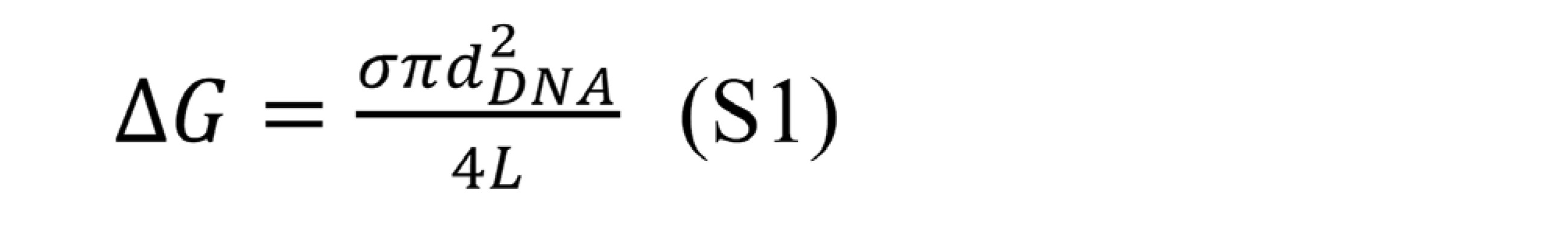
要約:この実施例は、本明細書中に記載されるようなステップのプロセスを実施することによる定量的アッセイを記載する。図25に示されているチオールベースの切断ステップを用いて、実施例17に記載されているように、切断可能なリンカー接合体を使用して直接アッセイを実施した。そのようなアッセイを実施するための他の切断可能なリンカー手法はまた、本明細書中に記載されるように、ナノポアを使用する種々のタグのカウントを可能にするための、リンカーの切断の種々の他の方法を含み得るがこれらに限定されないことが理解される。例えば、実施例17に記載された方法に加えて、そのような他の切断方法としては、本明細書に記載され、当業者に知られている方法に加えて、実施例18、実施例19、実施例20および実施例21に記載の方法が挙げられるが、これらに限定されない。この実施例(実施例25)において実証されたアッセイフォーマットは、直接アッセイを表すが、サンドイッチイムノアッセイフォーマットおよび/または様々な競合アッセイフォーマットなど、当業者に知られているものなどの他のフォーマットが、アッセイを実施するために同様に実施され得ることも理解される。 Abstract: This example describes a quantitative assay by performing a process of steps as described herein. Using the thiol-based cleavage step shown in FIG. 25, the assay was performed directly with a cleavable linker conjugate as described in Example 17. Other cleavable linker techniques for performing such assays are also as described herein, for cleavage of the linker to allow counting of various tags using nanopores. It is understood that various other methods may be included, but are not limited thereto. For example, in addition to the methods described in Example 17, such other cutting methods include those described herein and known to those of skill in the art, as well as Examples 18 and 19. , But are not limited to the methods described in Example 20 and Example 21. The assay formats demonstrated in this Example (Example 25) represent direct assays, but other formats, such as those known to those of skill in the art, such as sandwich immunoassay formats and / or various competing assay formats. It is also understood that an assay can be performed as well to perform the assay.
例えば、実施例9に記載されているように、TSH(甲状腺刺激ホルモン)の検出のためのサンドイッチイムノアッセイフォーマットは、低コストDMFチップ上でそのようなアッセイを実施する能力を実証した。さらに、他の方法で当業者に知られている他の切断可能なリンカーに加えて、実施例1、実施例2、実施例3、実施例4、実施例5および実施例6に記載されているものなどの様々なヘテロ二官能性の切断可能なリンカーを使用して、イムノ接合体の生成に有用な多数の様々なバイオ接合試薬または切断可能なリンカーを有する他の活性特異的結合メンバーが、当業者により合成され得る。さらに、実施例8は、サンドイッチおよび競合アッセイフォーマットを含む様々なアッセイフォーマット、ならびに当業者に知られているそれらの他の変形を実行するために必要な様々なステップを容易にすることができる、低コストチップ上の様々な液滴操作の機能を示す。実施例16はまた、切断が行われるアッセイの実施に有用な別の構築物を表し、したがって、この実施例に記載されるように、ナノポアカウント法を使用してカウントできるように、カウント可能な標識が遊離される。 For example, as described in Example 9, the sandwich immunoassay format for the detection of TSH (thyroid stimulating hormone) demonstrated the ability to perform such an assay on a low cost DMF chip. Further described in Example 1, Example 2, Example 3, Example 4, Example 5 and Example 6 in addition to other cleavable linkers known to those of skill in the art by other methods. Various heterobifunctional cleavable linkers, such as those skilled in the art, can be used by a number of different bioconjugating reagents or other activity-specific binding members with cleavable linkers to help generate immunoconjugates. , Can be synthesized by one of ordinary skill in the art. In addition, Example 8 can facilitate various assay formats, including sandwiches and competitive assay formats, as well as the various steps required to perform those other variants known to those of skill in the art. Shown the features of various droplet manipulations on low cost chips. Example 16 also represents another construct useful for performing the assay in which cleavage is performed, and therefore, as described in this example, a countable label so that it can be counted using the nanopoacing method. Is released.
実施例22は、一般に、ナノポアを横断する標識の存在に関する転位事象を測定することができるように、カウントをどのようにして行うことができるかを示す。図29は、カウントアッセイにおいてデータの質を操作することができるようにするための、シグナルの閾値化の概念を示す。図31、32および33は、この実施例に記載されるようなアッセイ方法を使用して、検体の量を決定するために使用され得るデータの種類を表す定性的アッセイデータ出力を示す。図34は、化学的方法を使用して切断された構築物から生成された標準曲線を示す。この特定の実施例では、dsDNAを標識として使用したが、ナノビーズ、デンドリマー等を含むがこれらに限定されない実施例5に記載の標識などの他の標識を利用することもできる。そのような構築物は、当業者に公知の方法によって合成することができる。 Example 22 shows how counting can be done so that dislocation events with respect to the presence of markers across nanopores can generally be measured. FIG. 29 shows the concept of signal thresholding to allow manipulation of data quality in count assays. FIGS. 31, 32 and 33 show qualitative assay data output representing the types of data that can be used to determine the volume of a sample using an assay method as described in this example. FIG. 34 shows a standard curve generated from a structure cut using a chemical method. In this particular example, dsDNA was used as the label, but other labels such as the label described in Example 5 including, but not limited to, nanobeads, dendrimers and the like can also be used. Such constructs can be synthesized by methods known to those of skill in the art.
実施例26
ナノポア電界シミュレーション
理論的な直径10nmのナノポアを通る対イオン濃度および電気浸透流速に対するSiO2ビアのサイズの影響を研究するために、シリコンモジュールで使用される提案されたナノポア膜設計について一連のCOMSOLシミュレーション実験を行った。SiO2の最上層は、複数の目的を果たした。1)SiNx膜に絶縁層を提供し、それによりナノポアの容量性ノイズを低減する。2)シリコン基板内のSiNx膜の堅牢性および強度を高める。3)溶液に曝露されるSiNx領域のサイズを小さくし、それにより制御された絶縁破壊(CBD)プロセスから膜上の細孔の位置付けを改善する。電界シミュレーションを使用して、局所対イオン濃度および細孔を通る電気浸透流に対するSiO2層の干渉を決定した。
Example 26
Nanopore electric field simulation A series of COMSOL simulations of the proposed nanopore film design used in silicon modules to study the effect of SiO 2 via size on counterion concentration and electroosmotic flow velocity through a theoretical 10 nm diameter nanopore. An experiment was conducted. The top layer of SiO 2 served multiple purposes. 1) An insulating layer is provided on the SiN x film, thereby reducing the capacitive noise of the nanopores. 2) Improve the robustness and strength of the SiN x film in the silicon substrate. 3) Reduce the size of the SiNx region exposed to the solution and thereby improve the positioning of pores on the membrane from a controlled breakdown (CBD) process. Electric field simulations were used to determine the interference of the SiO2 layer with local counterion concentrations and electroosmotic flow through the pores.
図35を参照すると、シリコン基板(1)をエッチングして、SiNx膜の上下に位置するシスおよびトランスチャンバを得た。SiNx膜(50μm×50μm)(2)を、厚さ300μmのSiO2の最下層と、厚さ300μmのSiO2の最上層(3)との間に積層した。最上層を製造して(4)を介してSiO2を形成し、これによりCBD中にナノポアの形成が可能となった。SiO2ビアの最適直径は、シミュレーションによって決定された。 Referring to FIG. 35, the silicon substrate (1) was etched to obtain cis and transchambers located above and below the SiN x film. A SiN x film (50 μm × 50 μm) (2) was laminated between the bottom layer of SiO 2 having a thickness of 300 μm and the top layer (3) of SiO 2 having a thickness of 300 μm. The top layer was manufactured to form SiO 2 via (4), which allowed the formation of nanopores in the CBD. The optimum diameter of the SiO2 via was determined by simulation.
COMSOLシミュレーション結果:COMSOL電界シミュレーションは、材料、静電気学、分子輸送および層流特性に基づく物理モデルを使用した。電位は、ポアソン方程式に基づいた。イオン流動は、ネルンスト-プランク方程式に基づいた。流速は、ストークス方程式に基づいた。シミュレーションに使用した物理的パラメータは、図36に示す表1に定義されている。 COMSOL simulation results: The COMSOL electric field simulation used physical models based on materials, electrostatics, molecular transport and laminar flow characteristics. The potential was based on Poisson's equation. Ion flow was based on the Nernst-Planck equation. The flow velocity was based on the Stokes equation. The physical parameters used in the simulation are defined in Table 1 shown in FIG.
細孔付近の対イオン濃度勾配についてのCOMSOLの結果は、図37に示されており、SiO2ビア直径が、直径50nmを超えたときにイオン濃度の影響をほとんどまたは全く示さない。50nm未満では、細孔の口の近くに正味電荷の蓄積が生じた。最も深刻な影響は、25nmの直径で観察され、大きなイオン勾配が細孔の近くに形成された。結果は、ナノポアがSiO2壁から25~50nm未満離れているとき、SiO2表面のかなり大きな影響を示した。 The COMSOL results for the counterion concentration gradient near the pores are shown in FIG. 37, showing little or no effect of ion concentration when the SiO 2 via diameter exceeds 50 nm in diameter. Below 50 nm, net charge accumulation occurred near the mouth of the pores. The most serious effects were observed at a diameter of 25 nm, with large ion gradients forming near the pores. The results showed a fairly large effect on the SiO 2 surface when the nanopores were less than 25-50 nm away from the SiO 2 wall.
細孔を通る対イオンの電気浸透流速は、SiO2層がナノポアセンシングに及ぼし得る影響を決定する方法としてシミュレートされた(図38)。電気浸透流の最高速度は、より大きいビア直径(100~4,500nm)で生じた。細孔を通る流速の低減が、50nmのSiO2ビアについて観察され、続いて25nmのビアについて有意な低減が観察された。 The electroosmotic flow rate of counterions through the pores was simulated as a way to determine the possible effects of the SiO 2 layer on nanopore sensing (FIG. 38). The maximum velocity of electroosmotic flow occurred at larger via diameters (100-4,500 nm). A reduction in flow velocity through the pores was observed for 40 nm SiO 2 vias, followed by a significant reduction for 25 nm vias.
図39に示されるように、細孔対ビア直径を通るコンダクタンスの測定は、100nmを超える飽和曲線を示し、ビア直径が100nm~25nmのサイズに低減するにつれてコンダクタンスが減少した。 As shown in FIG. 39, measurements of conductance through the pore-to-via diameter showed a saturation curve above 100 nm, with the conductance decreasing as the via diameter decreased to a size of 100 nm to 25 nm.
実施例27
ナノポアモジュールをデジタルマイクロ流体(DMF)モジュールに統合する
ナノポアモジュールは、DMFモジュールの片側に位置していた。細孔の形成および検体の検出のためにDMFモジュールからナノポアモジュールへの液体輸送を可能にするために、DMFモジュールに穴が存在した(例えば、図40を参照)。
Example 27
Integrating the Nanopore Module into a Digital Microfluidic (DMF) Module The nanopore module was located on one side of the DMF module. Holes were present in the DMF module to allow liquid transport from the DMF module to the nanopore module for pore formation and specimen detection (see, eg, FIG. 40).
ナノポアモジュールからの1つの電極は、ナノポアモジュール内の流体体積内で終端した。もう一方の電極は、DMFモジュール内の流体体積内で終端した。この電極は、DMFモジュールの第2の穴を経由した。液体がDMFモジュール内の穴を通って移動することができたことを実証するために、液体が適所に移動された後に平らな紙をチップの外面上で押した。この紙の濡れは、液体が毛細管力を介してDMFモジュールからこの穴の上に位置する別のモジュールに移動できることを示した(図41)。 One electrode from the nanopore module was terminated within the fluid volume within the nanopore module. The other electrode was terminated within the fluid volume within the DMF module. This electrode passed through a second hole in the DMF module. To demonstrate that the liquid was able to move through the holes in the DMF module, a flat piece of paper was pressed onto the outer surface of the chip after the liquid had been moved in place. This paper wetting showed that the liquid could move from the DMF module to another module located above this hole via capillary force (FIG. 41).
図42を参照すると、DMFモジュールは、ナノポア製造を制御するためにAg/AgCl電極を備えていた。この設定では、ナノポアモジュール上の液体体積は、LiClの外液滴であった。この液体をナノポアモジュール上に直接分注し、電極端子をこの液滴内に吊り下げた。 Referring to FIG. 42, the DMF module was equipped with Ag / AgCl electrodes to control nanopore production. In this setting, the liquid volume on the nanopore module was an outer droplet of LiCl. The liquid was dispensed directly onto the nanopore module and the electrode terminals were suspended within the droplet.
DMF技術を使用して、試料をDMFモジュールの穴に移動させた。試料は、受動的に穴を通って移動し、ナノポア形成のためにナノポアモジュールに曝露された。ナノポアモジュールは、DMFモジュールに封止され(例えば、PDMS、圧力、ワックス等を使用して)、各モジュール内に保持されている液体体積を隔離する。図45は、ナノポアの製造中の時間の関数としての電流を示す。 Using DMF technology, the sample was moved into a hole in the DMF module. The sample passively moved through the hole and was exposed to the nanopore module for nanopore formation. The nanopore modules are sealed in DMF modules (eg, using PDMS, pressure, wax, etc.) to isolate the liquid volume held within each module. FIG. 45 shows the current as a function of time during the manufacture of nanopores.
ナノポアが形成されたら、調整プロセス(経時的に電圧を変化させる)を使用して、ナノポアを物理的に修飾してシグナルをクリーンにした。このプロセスは、I-V曲線の対称性を改善した。前後のI-V曲線を、それぞれ図46Aおよび46Bに示す。 Once the nanopores were formed, a conditioning process (changing the voltage over time) was used to physically modify the nanopores to clean the signal. This process improved the symmetry of the IV curve. The front and rear IV curves are shown in FIGS. 46A and 46B, respectively.
実施例28
カウント標識および孔径分析
一連の実験は、孔径およびカウント標識サイズに対してある特定の属性を分析し、実証するために、様々な一連の条件下で二本鎖DNAを使用して行われた。これらの実験では、検出電圧、DNA長、DNA濃度、塩濃度および塩組成、膜材料、膜厚、ナノポア直径および他の要因を含む様々なパラメータを調べた。
Example 28
Count Labeling and Pore Diameter Analysis A series of experiments were performed using double-stranded DNA under a variety of conditions to analyze and demonstrate certain attributes for pore size and count label size. These experiments examined various parameters including detection voltage, DNA length, DNA concentration, salt concentration and salt composition, membrane material, film thickness, nanopore diameter and other factors.
次いで、データセットをシグナル対ノイズ比に関して分析し、その因子をカウント標識サイズ(推定分子直径)に対して種々の孔径と比較した。例えば、膜材料および厚さなどの、ある特定の因子は、この一連の実験において一定に保たれたが、他の因子は変動した。 The dataset was then analyzed in terms of signal-to-noise ratio and the factors were compared to various pore sizes relative to the count label size (estimated molecular diameter). Certain factors, such as membrane material and thickness, remained constant in this series of experiments, while others fluctuated.
集計データセット分析から、実験で決定されたSNR(シグナル対ノイズ比)に対するカウント標識平均直径とナノポアサイズとの間の比の平均をプロットした(図47)。図47は、一般に、有用なカウントデータが、この特定のデータセットでは約約0.4~0.8のような比率の範囲から得られることを実証し、そのような比率は、およそ約2.0nmのdsDNAの分子直径を仮定する。線状dsDNAは、その分子直径についてであることが文献から知られており、分析は、DNAがその線状立体配座で細孔を通って進むと仮定する。表4は、計算データを示す。
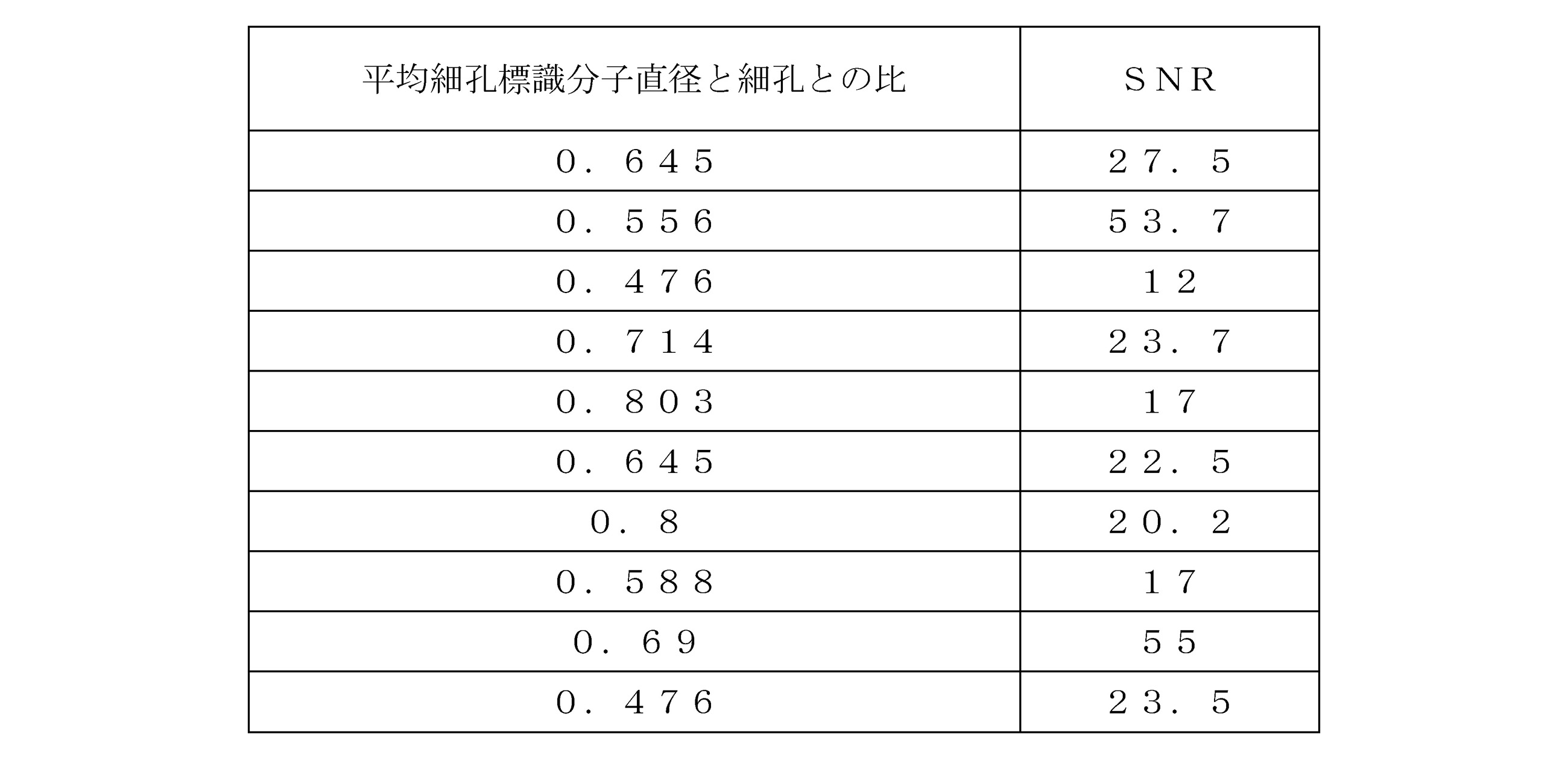
この実施例で前述したように、条件は変化したが、このデータセットの一般的な範囲は、この範囲内で合理的なシグナル/ノイズ比のデータをカウントできることを示している。さらに、当業者であれば、合理的なSNRを達成するために、他のカウント標識分子直径とナノポア直径との比を利用できることを認識するであろうことに留意すべきである。加えて、一般に標識は、細孔を通過することができるように、ナノポアのサイズより小さいその分子直径の少なくとも1つの寸法を有するべきであること、または言い換えれば、標識分子直径とナノポア直径とのこの比は、恐らくナノポア力分光法と呼ばれる技術において説明されるような条件が使用される場合を除いて、一般に標識が細孔を通過できるものよりも小さいはずであり、ここでエネルギーをシステムに付加して、標識に立体配座の変化を起こしやすくし、それによってそのような転位事象が起こることを可能にするレベルまで変形した後に細孔を通過させることができるようにする。 As mentioned earlier in this example, the conditions have changed, but the general range of this dataset shows that reasonable signal-to-noise ratio data can be counted within this range. Further, it should be noted that one of ordinary skill in the art will recognize that ratios of other count-labeled molecular diameters to nanopore diameters can be used to achieve reasonable SNR. In addition, in general, the label should have at least one dimension of its molecular diameter that is smaller than the size of the nanopore so that it can pass through the pores, or in other words, the labeled molecular diameter and the nanopore diameter. This ratio should generally be smaller than what the label can pass through the pores, except where conditions as described in a technique called nanopore force spectroscopy are used, where energy is applied to the system. In addition, the label is susceptible to changes in the configuration, thereby allowing it to pass through the pores after being transformed to a level that allows such transposition events to occur.
この実施例に記載されるように、dsDNA以外の他の標識をカウントするために利用することができ、それらはこのグラフに示されるものとは異なる挙動を有し得ることもまた当業者に理解されるべきである。さらに、そのような標識の分子カウントを可能にするために、他の分子直径とナノポアとの比から許容できるSNRを得ることもまた可能であり、現在の遮断は、以下の等式に記載されるように、そのようなカウント標識の分子直径に関連し得ることが理解されるべきであり、

この一連のナノポアカウント実験の集合内で変動したある特定の特定条件は以下を含んでいた。
●イオン強度-3または3.6M
●DNAの長さ-10kbp、50bpまたは1kbp
●使用されるイオン塩-LiClまたはKCl
●膜材料-SiNx(データセット全体で一定)
●膜厚-10nm(データセット全体で一定)
●DNA濃度-3nM~約306nMの間で変動
●電圧-50~600mVの増分を含む変動
●ナノポア直径-8.0、1.1、3.6、4.2、2.8、2.5、7.7、3.1、2.7、2.6、2.9および4.2(全てナノメートル単位)を含む様々な孔径。
Certain specific conditions that varied within this set of nanopoaccount experiments included:
● Ionic strength -3 or 3.6M
● DNA length-10 kbp, 50 bp or 1 kbp
● Ion salt used-LiCl or KCl
● Membrane material-SiNx (constant throughout the data set)
● Film thickness -10nm (constant throughout the data set)
● DNA concentration fluctuates between -3 nM and about 306 nM ● Fluctuations including voltage -50 to 600 mV increments ● Nanopore diameter −8.0, 1.1, 3.6, 4.2, 2.8, 2.5 , 7.7, 3.1, 2.7, 2.6, 2.9 and 4.2 (all in nanometer units).
これらを含むが限定されない様々な条件は、計算可能であるが、この実施例で参照されるようなKwokらのこの等式の量に従って電圧が印加されるとき、細孔の直径より小さいカウント可能な標識が、細孔を横切るイオン電流の流動の遮断を引き起こし得る状況において得られることを示すことが結論され得る[Kwok et al.,″Nanopore Fabrication by controlled Dielectric Breakdown″Supplementary Information Section 8 and/or Kwok,H.;Briggs,K.;and Tabard-Cossa,V.;″Nanopore Fabrication by Controlled Dielectric Breakdown″-PLoS ONE 9(3):e92880(2014)]。
Various conditions, including but not limited to these, are calculable but can be counted less than the diameter of the pores when a voltage is applied according to the quantity of this equation by Kwok et al. As referenced in this example. It can be concluded that the label is obtained in situations where it can cause a blockage of the flow of ionic current across the pores [Kwok et al. , "Nanopore Fabrication by Controlled Electrical Breakdown"
これらの条件は、デンドリマー、ヘミデンドリマー、ナノビーズ、アニオン性またはカチオン性ポリマー、変性線状化アプタマー、負電荷もしくは正電荷ポリペプチド、または他の電荷ポリマーもしくはカウント可能な分子実体等を含むが、これらに限定されないdsDNA以外の他の標識についてノイズに対する合理的なシグナルで機能する、孔径に対するカウント標識分子直径を示すように適用され得ることも理解されるであろう。 These conditions include, but include, dendrimers, hemidendrimers, nanobeads, anionic or cationic polymers, modified linearized aptamers, negatively or positively charged polypeptides, or other charged polymers or countable molecular entities. It will also be appreciated that other labels other than dsDNA can be applied to indicate a count labeled molecular diameter relative to pore size that functions with a reasonable signal to noise.
最後に、本発明の様々な態様および特徴を本明細書の様々な実施形態および具体例に関して説明してきたが、それらは全て従来どおりに作製または実行することができるが、本発明は特許請求の全範囲内で保護する権利があることを理解する。 Finally, various aspects and features of the invention have been described with respect to various embodiments and examples herein, all of which can be made or practiced as conventional, but the invention is claimed. Understand that you have the right to protection within the full scope.
前述の詳細な説明および添付の実施例は、単なる例示であり、添付の特許請求の範囲およびそれらの均等物によってのみ定義される、本発明の範囲に対する限定として解釈されるべきではないことが理解される。 It is understood that the above detailed description and attached examples are merely exemplary and should not be construed as a limitation to the scope of the invention as defined solely by the appended claims and their equivalents. Will be done.
開示された実施形態に対する様々な変更および修正は、当業者に明らかであろう。本発明の化学構造、置換基、誘導体、中間体、合成、組成物、配合物、または使用方法に関するものを含むがこれらに限定されないそのような変更および修正は、その趣旨および範囲から逸脱することなく行われ得る。 Various changes and amendments to the disclosed embodiments will be apparent to those of skill in the art. Such changes and modifications relating to, but not limited to, the chemical structure, substituents, derivatives, intermediates, synthesis, compositions, formulations, or methods of use of the invention deviate from its intent and scope. Can be done without.
完全を期すために、本発明の様々な態様を以下の番号を付けた項に記載する。 For completeness, various aspects of the invention are described in the numbers below.
項1.生物学的試料中に存在する検体を測定または検出する方法であって、(a)試料を第1の結合メンバーと接触させることであって、第1の結合メンバーが、固体支持体上に固定化され、第1の結合メンバーが、検体に特異的に結合する、接触させることと、(b)検体を第2結合メンバーと接触させることであって、第2の結合メンバーが、検体に特異的に結合し、第2の結合メンバーが、それに付着した切断可能なタグを含む、接触させることと、(c)第1の結合メンバーに結合した検体に結合していない第2の結合メンバーを除去することと、(d)第1の結合メンバーに結合した検体に結合した第2の結合メンバーに付着したタグを切断することと、(e)層内の1つ以上のナノポアを通してタグを移動させることと、(f)層を通って転位するタグを評価することと、を含み、層を通って転位するタグの数を測定することが、試料中に存在する検体の量を測定するか、または層を通って転位するタグを検出することが、検体が試料中に存在することを検出する、方法。
項2.層を通って転位する各タグが、転位事象であり、転位事象の数を測定することが、試料中に存在する検体の量を測定し、試料中に存在する検体の量が、i)一定期間中の転位事象の数をカウントし、転位事象の数を対照と相関させること、ii)一定数の転位事象が発生するまでの時間量を測定し、対照と相関させること、またはiii)転位事象が発生する間の平均時間を測定し、対照と相関させることによって決定され、対照が、較正曲線、標準付加、またはデジタルポリメラーゼ連鎖反応を含む参照標準である、項1に記載の方法。
項3.サブセクションi)における標準曲線が、一定期間の間の検体の対照濃度について転位事象の数を測定することによって決定され、サブセクションii)における標準曲線が、検体の対照濃度について設定数の転位事象が起こるまでの時間を測定することによって決定され、サブセクションiii)における標準曲線が、検体の対照濃度について転位事象間の平均時間を測定することによって決定される、項2に記載の方法。
項4.方法が、単一分子カウントを含む、項1~3のいずれか一項に記載の方法。
項5.タグが、アニオン性ポリマー、カチオン性ポリマー、デンドリマー、およびナノ粒子からなる群から選択される、項1~4のいずれか一項に記載の方法。
項6.タグが、実質的に球形または半球形である、項1または5に記載の方法。
項7.タグが、実質的に球形であり、ナノ粒子を含む、項1~6のいずれか一項に記載の方法。
項8.タグが、実質的に球形または半球形であり、デンドリマーを含む、項1~7のいずれか一項に記載の方法。
項9.デンドリマーが、正電荷または負電荷である、項8に記載の方法。
項10.ナノ粒子が、正電荷ナノ粒子を含む、項5または7に記載の方法。
項11.ナノ粒子が、負電荷ナノ粒子を含む、項10に記載の方法。
項12.第1および第2の結合メンバーが、抗体または受容体である、項1~11のいずれか一項に記載の方法。
項13.第1の結合メンバーが受容体であり、第2の結合メンバーが抗体であるか、または第1の結合メンバーが抗体であり、第2の結合メンバーが受容体である、項1~12のいずれか一項に記載の方法。
Item 13.
項14.第1の結合メンバーが第1の抗体であり、第2の結合メンバーが第2の抗体である、項1~12のいずれか一項に記載の方法。
項15.タグが負電荷であり、転位することが、層を横切って正の電位を印加し、それにより層を通してタグを転位させることを含む、項1~14のいずれか一項に記載の方法。
項16.タグが正電荷であり、転位することが、層を横切って負の電位を印加し、それにより層を通してタグを転位させることを含む、項1~14のいずれか一項に記載の方法。
項17.少なくともステップ(a)~(d)が、マイクロ流体デバイス、液滴ベースのマイクロ流体デバイス、デジタルマイクロ流体デバイス(DMF)、表面音響波ベースのマイクロ流体デバイス(SAW)、完全集積DMFおよびナノポアデバイス、または完全集積SAWおよびナノポアデバイスにおいて実行される、項1~16のいずれか一項に記載の方法。
項18.DMF要素およびナノポア要素が、完全集積DMFおよびナノポアデバイスに動作可能に結合されるか、またはSAW要素およびナノポア要素が、完全集積SAWおよびナノポアデバイスに動作可能に結合される、項17に記載の方法。
項19.DMFデバイスまたはSAWデバイスが、ロールツーロールベースの印刷エレクトロニクス方法によって製造される、項17に記載の方法。
項20.DMF素子またはSAW素子が、ロールツーロールベースの印刷電子方法によって製造される、項18に記載の方法。
項21.完全集積DMFおよびナノポアデバイス、または完全集積SAWおよびナノポアデバイスが、マイクロ流体導管を含む、項17に記載の方法。
項22.マイクロ流体導管が、DMF要素をナノポア要素に結合し、マイクロ流体導管は、受動的な力または能動的な力によって誘導される流体の流れを含む、項21に記載の方法。
項23.ナノポアが、固体ナノポアまたは生物学的ナノポアである、項1~22のいずれか一項に記載の方法。
項24.層を通って転位するタグの数を測定することが、タグとナノポアとの相互作用によって誘導される電流の変化を観察することを含む、項1~23のいずれか一項に記載の方法。
項25.電流変化が、閾値レベルを超える大きさを有するときに、検体が試料中に存在する、項24に記載の方法。
項26.ステップ(d)で得られたタグを含む液滴をナノポアデバイスに輸送することと、ナノポアデバイスに存在するナノポア層を横切って液滴を配置することをさらに含み、それにより液滴が、ナノポア層によって分割され、ナノポア層に存在するナノポア(複数可)によって接続され、タグが、ナノポア層の両側の液滴に存在する、項23に記載の方法。
項27.方法が、ナノポアを横切ってナノポア層の第1の側に存在するタグを、ナノポア層の第2の側に転位させ、それによりナノポア層の第2の側上の分割液滴注のタグを収集することを含む、項26に記載の方法。
項28.タグをナノポア層の第1の側に転位させることと、液滴中に存在するタグの数を決定することと、をさらに含む、項26に記載の方法。
項29.タグが、切断可能なリンカーを含む、項1~28のいずれか一項に記載の方法。
Item 29.
項30.生物学的試料中に存在する目的の検体を測定または検出するための方法であって、(a)試料を固体支持体、第1の特異的結合メンバー、および第2の特異的結合メンバーと接触させることであって、固体支持体が、固定化剤を含み、第1の特異的結合メンバーが、固定化剤に対するリガンドを含み、第1の特異的結合メンバーが、目的の検体に特異的に結合し、第2の特異的結合メンバーが、切断可能なタグを含み、固体支持体/第1の特異的結合メンバー/目的の検体/第2の特異的結合メンバーの複合体が形成される、接触させることと、(b)固体支持体/第1の特異的結合メンバー/検体/第2の特異的結合メンバーの複合体に結合していない第2の特異的結合メンバーを除去することと、(c)固体支持体/第1の特異的結合メンバー/目的の検体/第2の特異的結合メンバーの複合体に結合した標識検体に付着したタグを切断することと、(d)層内の1つ以上のナノポアを通してタグを転位させることと、(e)層を通って転位するタグを評価することと、を含み、層を通って転位するタグの数を測定することが、試料中に存在する検体の量を測定するか、または層を通って転位するタグを検出することが、検体が試料中に存在することを検出する、方法。
項31.生物学的試料中に存在する検体を測定または検出するための方法であって、(a)試料を第1の結合メンバーと接触させることであって、第1の結合メンバーが、固体支持体上に固定化され、第1の結合メンバーが、検体に特異的に結合する、接触させることと、(b)検体を第2結合メンバーと接触させることであって、第2の結合メンバーが、検体に特異的に結合し、第2の結合メンバーが、アプタマーを含む、接触させることと、(c)固体基材に結合した検体に結合していないアプタマーを除去することと、(d)検体に結合したアプタマーを解離させることと、(e)解離したアプタマーを、層内の1つ以上のナノポアを通して転位させることと、(f)層を通って転位するアプタマーを評価することと、を含み、層を通って転位するアプタマーの数を測定することは、試料中に存在する検体の量を測定するか、または層を通って転位するアプタマーを検出することは、検体が試料中に存在することを検出する、方法。
項32.層を通って転位する各アプタマーが、転位事象であり、転位事象の数を測定することが、試料中に存在する検体の量を測定し、試料中に存在する検体の量が、i)一定期間中の転位事象の数をカウントし、転位事象の数を対照と相関させること、ii)一定数の転位事象が発生するまでの時間量を測定し、対照と相関させること、またはiii)転位事象が発生する間の平均時間を測定し、対照と相関させることによって決定され、対照が、較正曲線、標準付加、またはデジタルポリメラーゼ連鎖反応を含む参照標準である、項31に記載の方法。
項33.サブセクションi)における標準曲線が、一定期間の間の検体の対照濃度について転位事象の数を測定することによって決定され、サブセクションii)における標準曲線が、検体の対照濃度について設定数の転位事象が起こるまでの時間を測定することによって決定され、サブセクションiii)における標準曲線が、検体の対照濃度について転位事象間の平均時間を測定することによって決定される、項32に記載の方法。
項34.方法が、単一分子カウントを含む、項31~33のいずれか一項に記載の方法。
項35.アプタマーが、DNAアプタマーである、項31~34のいずれか一項に記載の方法。
項36.アプタマーが、RNAアプタマーである、項31~34のいずれか一項に記載の方法。
項37.第1の結合メンバーが抗体である、項31~36のいずれか一項に記載の方法。
Item 37.
項38.検体がリガンドであり、第1の結合メンバーが受容体である、項31~36のいずれか一項に記載の方法。
Item 38.
項39.少なくともステップ(a)~(d)が、マイクロ流体デバイス、液滴ベースのマイクロ流体デバイス、デジタルマイクロ流体デバイス(DMF)、表面音響波ベースのマイクロ流体デバイス(SAW)、完全集積DMFおよびナノポアデバイス、または完全集積SAWおよびナノポアデバイスにおいて実行される、項31~38のいずれか一項に記載の方法。
Item 39. At least steps (a)-(d) are microfluidic devices, droplet-based microfluidic devices, digital microfluidic devices (DMFs), surface acoustic wave-based microfluidic devices (SAWs), fully integrated DMFs and nanopore devices. The method according to any one of
項40.DMF要素およびナノポア要素が、完全集積DMFおよびナノポアデバイスに動作可能に結合されるか、またはSAW要素およびナノポア要素が、完全集積SAWおよびナノポアデバイスに動作可能に結合される、項39に記載の方法。
項41.DMFデバイスまたはSAWデバイスが、ロールツーロールベースの印刷エレクトロニクス方法によって製造される、項39に記載の方法。 Item 41. 39. The method of item 39, wherein the DMF device or SAW device is manufactured by a roll-to-roll based printed electronics method.
項42.DMF素子またはSAW素子が、ロールツーロールベースの印刷電子方法によって製造される、項40に記載の方法。
Item 42.
項43.完全集積DMFおよびナノポアデバイス、または完全集積SAWおよびナノポアデバイスが、マイクロ流体導管を含む、項39に記載の方法。 Item 43. 39. The method of item 39, wherein the fully integrated DMF and nanopore device, or fully integrated SAW and nanopore device, comprises a microfluidic conduit.
項44.マイクロ流体導管が、DMF要素をナノポア要素に結合し、マイクロ流体導管は、受動的な力または能動的な力によって誘導される流体の流れを含む、項43に記載の方法。
項45.ナノポアが、固体ナノポアまたは生物学的ナノポアである、項31~44のいずれか一項に記載の方法。
項46.集積デジタルマイクロ流体ナノポアデバイスであって、電極のアレイを含む第1の基板と、該第1の基板から離間した第2の基板と、第1の基板と第2の基板との間に配設されたナノポア層と、を備える、集積デジタルマイクロ流体ナノポアデバイスに関し、電極のアレイは、液滴がナノポア層によって第1の部分と第2の部分とに分割されるように、ナノポア層を横切って液滴を位置付けるように構成され、電極のアレイの少なくとも2つの電極が、ナノポア層を横切って位置付けられ、2つの電極が、アノードおよびカソードを形成し、液滴がナノポア層を横切って位置付けられると、ナノポア層内のナノポアを通して電流を流すように動作する、集積デジタルマイクロ流体ナノポアデバイス。 Item 46. An integrated digital microfluidic nanopore device, disposed between a first substrate containing an array of electrodes, a second substrate separated from the first substrate, and a first substrate and a second substrate. With respect to the integrated digital microfluidic nanopore device comprising the nanopore layer, the array of electrodes traverses the nanopore layer such that the droplet is divided into a first part and a second part by the nanopore layer. When configured to position a droplet, at least two electrodes in an array of electrodes are positioned across the nanopore layer, two electrodes form an anode and a cathode, and the droplet is positioned across the nanopore layer. An integrated digital microfluidic nanopore device that operates to carry current through the nanopores in the nanopore layer.
項47.ナノポア層が、第1および第2の基板に付着している、項46に記載のデバイス。
Item 47.
項48.ナノポア層が、第1または第2の基板に付着している、項46に記載のデバイス。
Item 48.
項49.電極が透明である、項46~48のいずれか一項に記載のデバイス。
項50.電極が、格子パターンに配設されている、項46~49のいずれか一項に記載のデバイス。
項51.ナノポア層を横切って位置付けられた電極のアレイの少なくとも2つの電極が、ナノポア層に隣接し、ナノポア層を横切って位置付けられていない、項46~50のいずれか一項に記載のデバイス。
項52.電極が互いに組み合わされる、項46~51のいずれか一項に記載のデバイス。
Item 52.
項53.電極のアレイが、電源による活性化のために構成されており、電源が、順次電極を活性化する、項46~51のいずれか一項に記載のデバイス。
項54.順次方法が、1つ以上の電極をオンまたはオフにすることを含む、項53に記載のデバイス。
Item 54. 53. The device of
項55.電源による電極のアレイの活性化が、電源を制御するプロセッサによって実行される一組の命令によって制御される、項52~54のいずれか一項に記載のデバイス。
項56.集積デジタルマイクロ流体ナノポアデバイスであって、電極のアレイを含む第1の基板と、第1の基板から離間している第2の基板と、第1の基板と第2の基板との間に配設されたナノポア層と、を備え、電極のアレイが、ナノポア層が液滴を第1の部分と第2の部分とに分割するように、ナノポア層を横切って液滴を位置付けるように構成され、電極のアレイの少なくとも1つの電極が、ナノポア層を横切って位置付けられた液滴の第1の部分と接触し、第2の基板内の電極が、ナノポア層を横切って位置付けられた液滴の第2の部分と接触するように位置付けられ、液滴がナノポア層を横切って位置付けられているとき、2つの電極がアノードおよびカソードを形成し、ナノポア層内のナノポアを通して電流を流すように動作する、集積デジタルマイクロ流体ナノポアデバイス。 Item 56. An integrated digital microfluidic nanopore device, located between a first substrate containing an array of electrodes, a second substrate separated from the first substrate, and between the first substrate and the second substrate. An array of electrodes comprising an provided nanopore layer is configured to position the droplet across the nanopore layer such that the nanopore layer divides the droplet into a first portion and a second portion. At least one electrode in the array of electrodes is in contact with the first portion of the droplet positioned across the nanopore layer, and the electrodes in the second substrate are of the droplet positioned across the nanopore layer. When positioned to contact the second portion and the droplet is positioned across the nanopore layer, the two electrodes form an anode and cathode and act to carry current through the nanopores within the nanopore layer. , Integrated digital microfluidic nanopore device.
項57.ナノポア層が、第1の基板に付着している、項56に記載のデバイス。
Item 57.
項58.ナノポア層が、第2の基板に付着している、項56または57に記載のデバイス。
Item 58.
項59.第1および/または第2の基板が透明である、項56~58のいずれか一項に記載のデバイス。
項60.電極のアレイが透明である、項56~59のいずれか一項に記載のデバイス。
項61.項46~60のいずれか一項に記載のデバイスを含む、または項1~45のいずれか一項に記載の方法において使用するためのキット。
項62.追加の試薬をさらに含み、少なくとも1つの試薬が、デバイスのナノポア層を介した転位によって検出され得るタグを含む、項61に記載のキット。
項63.生物学的試料中に存在する検体を測定もしくは検出するため、または患者を診断するため、または血液供給をスクリーニングするために、項46~60のいずれか一項に記載のデバイスを使用する方法、または項1~45のいずれか一項に記載の方法。
項64.少なくともステップ(a)~(d)が、項46~60のいずれか一項に記載のデバイスを使用して実行される、項1~45のいずれか一項に記載の方法。
項65.患者を診断するか、もしくは血液供給をスクリーニングする方法における、または生物学的試料中に存在する検体を測定もしくは検出するための、項46~60のいずれか一項に記載のデバイスの使用、または項1~45のいずれか一項に記載の方法の使用。
Item 65. Use of the device according to any one of Items 46-60, in a method of diagnosing a patient or screening a blood supply, or for measuring or detecting a sample present in a biological sample, or Use of the method according to any one of
項66.生物学的試料中に存在する検体を測定または検出する方法であって、(a)試料を結合メンバーと接触させることであって、結合メンバーが、固体支持体上に固定化され、結合メンバーが、検体に特異的に結合する、接触させることと、(b)試料を標識検体と接触させることであって、標識検体が、切断可能なタグで標識される、接触させることと、(c)結合メンバーに結合していない標識検体を除去することと、(d)結合メンバーに結合された標識検体に付着したタグを切断することと、(e)層内の1つ以上のナノポアを通してタグを転位させることと、(f)層を通って転位するタグを評価することと、を含み、層を通って転位するタグの数を測定することが、試料中に存在する検体の量を測定するか、または層を通って転位するタグを検出することが、検体が試料中に存在することを検出する、方法。
項67.生物学的試料中に存在する検体を測定または検出するための方法であって、(a)試料を結合メンバーと接触させることであって、結合メンバーが、固体支持体上に固定化され、結合メンバーが、検体に特異的に結合する、接触させることと、(b)試料を標識検体と接触させることであって、標識検体が、アプタマーを含む、接触させることと、(c)結合メンバーに結合していない標識検体を除去することと、(d)標識検体に結合したアプタマーを解離させ、解離したアプタマーを、層内の1つ以上のナノポアを通して転位させることと、(e)層を通って転位するアプタマーを評価することと、を含み、層を通って転位するアプタマーの数を測定することは、試料中に存在する検体の量を測定するか、または層を通って転位するアプタマーを検出することは、検体が試料中に存在することを検出することと、を含む、方法。
項68.生物学的試料中に存在する検体を測定または検出するための方法であって、(a)試料を結合メンバーと接触させることであって、結合メンバーが、検体に特異的に結合し、結合メンバーが、切断可能なタグで標識される、接触させることと、(b)試料を固定化検体と接触させることであって、固定化検体が、固体支持体上に固定化される、接触させることと、(c)固定化検体に結合していない結合メンバーを除去することと、(d)固定化検体に結合した結合メンバーに付着したタグを切断することと、(e)層内の1つ以上のナノポアを通して転位させることと、(f)層を通って転位するタグを評価することと、を含み、層を通って転位するタグの数を測定することが、試料中に存在する検体の量を測定するか、または層を通って転位するタグを検出することが、検体が試料中に存在することを検出する、方法。
項69.生物学的試料中に存在する検体を測定または検出するための方法であって、(a)試料を結合メンバーと接触させることであって、結合メンバーが、検体に特異的に結合し、結合メンバーが、アプタマーを含む、接触させることと、(b)試料を固定化検体と接触させることであって、固定化検体が、固体支持体上で固定化される、接触させることと、(c)固定化検体に結合していない結合メンバーを除去することと、(d)固定化検体に結合した結合メンバーに結合したアプタマーを解離させ、層内の1つ以上のナノポアを通して解離したアプタマーを転位させることと、(e)層を通って転位するアプタマーを評価することと、を含み、層を通って転位するアプタマーの数を測定することは、試料中に存在する検体の量を測定するか、または層を通って転位するアプタマーを検出することは、検体が試料中に存在することを検出する、方法。
項70.層を通って転位する各アプタマーが、転位事象であり、転位事象の数を測定することが、試料中に存在する検体の量を測定し、試料中に存在する検体の量が、i)一定期間中の転位事象の数をカウントし、転位事象の数を対照と相関させること、ii)一定数の転位事象が発生するまでの時間量を測定し、対照と相関させること、またはiii)転位事象が発生する間の平均時間を測定し、対照と相関させることによって決定され、対照が、較正曲線、標準付加、またはデジタルポリメラーゼ連鎖反応を含む参照標準である、項66または68に記載の方法。
項71.層を通って転位する各アプタマーが、転位事象であり、転位事象の数を測定することが、試料中に存在する検体の量を測定し、試料中に存在する検体の量が、i)一定期間中の転位事象の数をカウントし、転位事象の数を対照と相関させること、ii)一定数の転位事象が発生するまでの時間量を測定し、対照と相関させること、またはiii)転位事象が発生する間の平均時間を測定し、対照と相関させることによって決定され、対照が、較正曲線、標準付加、またはデジタルポリメラーゼ連鎖反応を含む参照標準である、項67または69に記載の方法。
項72.サブセクションi)における標準曲線が、一定期間の間の検体の対照濃度について転位事象の数を測定することによって決定され、サブセクションii)における標準曲線が、検体の対照濃度について設定数の転位事象が起こるまでの時間を測定することによって決定され、サブセクションiii)における標準曲線が、検体の対照濃度について転位事象間の平均時間を測定することによって決定される、項70または71に記載の方法。
項73.方法が、単一分子カウントを含む、項66~72のいずれか一項に記載の方法。
項74.少なくともステップ(a)~(d)が、項46~60のいずれか一項に記載のデバイスを使用して実行される、項66、68、70、72、または73のいずれか一項に記載の方法。 Item 74. 26, 68, 70, 72, or 73, wherein at least steps (a)-(d) are performed using the device according to any one of items 46-60. the method of.
項75.少なくともステップ(a)~(d)が、項46~60のいずれか一項に記載のデバイスを使用して実行される、項67、69、71、72、または73のいずれか一項に記載の方法。
Item 75.
項76.タグが、アニオン性ポリマー、カチオン性ポリマー、デンドリマー、およびナノ粒子からなる群から選択される、項66、68、70、72、73、または74のいずれか一項に記載の方法。
Item 76.
項77.タグが、実質的に球形または半球形である、項66、68、70、72、73、または74のいずれか一項に記載の方法。
Item 77.
項78.タグが、実質的に球形であり、ナノ粒子を含む、項66、68、70、72、73、または74のいずれか一項に記載の方法。
Item 78.
項79.タグが、実質的に球形または半球形である、項66、68、70、72、73、または74のいずれか一項に記載の方法。
Item 79.
項80.デンドリマーが、正電荷または負電荷である、項79に記載の方法。
項81.ナノ粒子が、正電荷ナノ粒子を含む、項79に記載の方法。
Item 81.
項82.ナノ粒子が、負電荷ナノ粒子を含む、項76または78に記載の方法。
Item 82.
項83.結合メンバーが、抗体または受容体である、項66、68、70、72、73、74、または76~82のいずれか一項に記載の方法。
Item 83.
項84.タグが負電荷であり、転位することが、層を横切って正の電位を印加し、それにより層を通してタグを転位させることを含む、項66、68、70、72、73、74、または76~83のいずれか一項に記載の方法。
項85.タグが正電荷であり、転位することが、層を横切って負の電位を印加し、それにより層を通してタグを転位させることを含む、項66、68、70、72、73、74、または76~84のいずれか一項に記載の方法。
項86.層を通って転位するタグの数を測定することが、ナノポアに対するタグの電流遮断効果を観察することを含む、項66、68、70、72、73、74、または76~85のいずれか一項に記載の方法。
Item 86.
項87.電流遮断効果が、閾値レベルを超えるときに、検体が試料中に存在する、項86に記載の方法。
Item 87.
項88.アプタマーが、DNAアプタマーである、項67、69、71、72、73、または75のいずれか一項に記載の方法。
Item 88.
項89.アプタマーが、RNAアプタマーである、項67、69、71、72、73、または75のいずれか一項に記載の方法。
項90.結合メンバーが、抗体である、項67、69、71、72、73、75、88、または89のいずれか一項に記載の方法。
項91.検体がリガンドであり、結合メンバーが受容体である、項67、69、71、72、73、75、88、または89のいずれか一項に記載の方法。
項92.方法が、タグを含む液滴をナノポアデバイスに輸送することと、ナノポアデバイスに存在するナノポア層を横切って液滴を配置することをさらに含み、それにより液滴が、ナノポア層によって分割され、ナノポア層に存在するナノポア(複数可)によって接続され、タグが、ナノポア層の両側の液滴に存在する、項67、69、71、72、73、75、または88~91のいずれか一項に記載の方法。
項93.方法が、ナノポアを横切ってナノポア層の第1の側に存在するタグを、ナノポア層の第2の側に転位させ、それによりナノポア層の第2の側上の分割液滴注のタグを収集することを含む、項92に記載の方法。
項94.タグをナノポア層の第1の側に転位させることと、液滴中に存在するタグの数を決定することと、をさらに含む、項92に記載の方法。
項95.方法が、アプタマーを含む液滴をナノポアデバイスに輸送することと、ナノポアデバイスに存在するナノポア層を横切って液滴を配置することをさらに含み、それにより液滴が、ナノポア層によって分割され、ナノポア層に存在するナノポア(複数可)によって接続され、アプタマーが、ナノポア層の両側の液滴に存在する、項67、69、71、72、73、75、または88~94のいずれか一項に記載の方法。
項96.方法が、ナノポアを横切ってナノポア層の第1の側に存在するアプタマーを、ナノポア層の第2の側に転位させ、それによりナノポア層の第2の側上の分割液滴注のアプタマーを収集することを含む、項95に記載の方法。
項97.アプタマーをナノポア層の第1の側に転位させることと、液滴中に存在するアプタマーの数を決定することと、をさらに含む、項95に記載の方法。
項98.ナノポアが、固体ナノポアまたは生物学的ナノポアである、項66~97のいずれか一項に記載の方法。
項99.第2の結合メンバーが、スペーサをさらに含む、項1~45または63~98のいずれか一項に記載の方法。
項100.スペーサが、ニトロベンジル基、ジチオエチルアミノ、6炭素スペーサ、12炭素スペーサ、または3-(9-((3-カルボキシプロピル)(トシル)カルバモイル)アクリジン-10-イウム-10-イル)プロパン-1-スルホネートを含む、項99に記載の方法。
項101.スペーサが、ニトロベンジル基を含み、タグが、DNA分子である、項100に記載の方法。
項102.スペーサが、ジチオエチルアミノであり、タグが、カルボキシル化ナノ粒子である、項100に記載の方法。
項103.スペーサが、3-(9-((3-カルボキシプロピル)(トシル)カルバモイル)アクリジン-10-イウム-10-イル)プロパン-1-スルホネートであり、タグが、オリゴヌクレオチドである、項100に記載の方法。
項104.スペーサが、6炭素スペーサまたは12炭素スペーサを含み、タグが、ビオチンである、項103に記載の方法。
項105.第2の結合メンバーが、配列番号1~11のうちのいずれか1つに記載のヌクレオチド配列を含む核酸を含む、項104に記載の方法。
項106.マイクロ流体モジュールおよびナノポアモジュールを備える、集積デジタルマイクロ流体ナノポアデバイスであって、マイクロ流体モジュールが、電極のアレイを含み、該電極のアレイが、少なくとも1つの液滴を、電極のアレイにおける第1の転送位置に輸送し、第1の転送位置が、マイクロ流体モジュールとナノポアモジュールとの間の界面にあり、ナノポアモジュールが、第1の毛細管チャネルと、第2の毛細管チャネルとを備え、少なくとも第1の毛細管チャネルが、界面まで延在し、第1の転送位置に隣接し、該第1の転送位置に位置付けられた液滴を受け取るように位置付けられ、第1の毛細管チャネルが、第2の毛細管チャネルと交差し、ナノポア層が、第1の毛細管チャネルと第2の毛細管チャネルとが交差する位置で、第1の毛細管チャネルと第2の毛細管チャネルとの間に位置付けられる、集積デジタルマイクロ流体ナノポアデバイス。
項107.電極のアレイが、少なくとも1つの液滴を、電極のアレイにおける第2の転送位置に輸送し、第2の転送位置が、マイクロ流体モジュールとナノポアモジュールとの間の界面にあり、第2の毛細管チャネルが、界面まで延在し、第2の転送位置に隣接し、第2の転送位置に位置付けられた液滴を受容するように位置付けられる、項106に記載のデバイス。
Item 107. The array of electrodes transports at least one droplet to a second transfer position in the array of electrodes, where the second transfer position is at the interface between the microfluidic module and the nanopore module and the second capillary.
項108.第2の毛細管チャネルが、第2の毛細管チャネルの一方または両方の端部の通気口またはリザーバの間に延在する、項106に記載のデバイス。
Item 108.
項109.第2の毛細管チャネルが、一端で第1のリザーバに、他端で第2のリザーバに接続されている、項108に記載のデバイス。
項110.第1のリザーバおよび/または第2のリザーバは、交差部で第2の毛細管チャネル内に位置付けられる流体を含み、この流体は、ナノポア層の動作を促進してナノポア層のナノポアを通して電流を流す、項109に記載のデバイス。
Item 110. The first reservoir and / or the second reservoir contains a fluid located in the second capillary channel at the intersection, which facilitates the operation of the nanopore layer to carry current through the nanopores of the nanopore layer.
項111.第1の毛細管チャネルおよび/または第2の毛細管チャネルは、交差部のいずれかの側の幅と比較して、交差部で幅が減少するように、毛細管の長さを横切って断面幅が変化する、項109に記載のデバイス。
Item 111. The first capillary channel and / or the second capillary channel varies in cross-sectional width across the length of the capillary so that the width is reduced at the intersection compared to the width on either side of the intersection.
項112.第1の毛細管チャネルが、第1の電極対を含み、第2の毛細管チャネルが、第2の電極対を含み、第1の電極対が、第1の毛細管チャネル内に位置付けられ、ナノポア層内のナノポアに隣接し、第2の電極対が、第2の毛細管チャネル内に位置付けられ、ナノポア層内のナノポアに隣接する、項106に記載のデバイス。
Item 112. The first capillary channel contains a first pair of electrodes, the second capillary channel contains a second pair of electrodes, the first pair of electrodes is located within the first capillary channel and within the nanopore layer. 106. The device of
項113.液滴が、ナノポア層内のナノポアを通って輸送することによってカウントされる分子を含む液滴である、条項107~112のいずれか一項に記載のデバイス。 Item 113. The device of any one of clauses 107-112, wherein the droplet is a droplet comprising a molecule that is counted by being transported through the nanopore within the nanopore layer.
項114.液滴が、異なる組成を有し、第1の液滴および第2の液滴であり、第1の液滴が、ナノポアを通してナノポア層を横切って輸送することによりカウントされる分子を含み、第2の液滴が、その分子を欠く導電性流体を含み、導電性流体が、ナノポアを介してナノポア層を横切る分子の輸送を容易にする、項107に記載のデバイス。
Item 114. The droplets have different compositions and are a first droplet and a second droplet, the first droplet comprising a molecule that is counted by transporting it through the nanopores across the nanopore layer.
項115.第1の毛細管チャネルが、ナノポア層に近接して位置付けられた第1の電極を含み、第2の毛細管チャネルが、ナノポア層に近接して位置付けられた第2の電極を含み、第1および第2の電極の各々が、それらが毛細管チャネル内に存在する流体と接触するように毛細管チャネル内で露出され、第1および第2の電極が、液体が第1および第2の毛細管チャネル内のナノポア層を横切って位置付けられると、ナノポア層内のナノポアを通して電流を流すように動作する、項106~114のいずれか一項に記載のデバイス。
Item 115. The first capillary channel comprises a first electrode located in close proximity to the nanopore layer, the second capillary channel comprises a second electrode located in close proximity to the nanopore layer, the first and first. Each of the two electrodes is exposed in the capillary channel so that they are in contact with the fluid present in the capillary channel, and the first and second electrodes are nanopores in which the liquid is in the first and second capillary channels.
項116.第1の転送位置および第1の毛細管チャネルが、実質的に同じ平面上にあり、液滴が、第1の毛細管チャネルの開口部と整列している、項106~115のいずれか一項に記載のデバイス。 Item 116. In any one of Items 106-115, where the first transfer position and the first capillary channel are in substantially the same plane and the droplets are aligned with the opening of the first capillary channel. Described device.
項117.第1の転送位置が、第1の毛細管チャネルよりも高い平面にあり、デバイスが、液滴を第1の毛細管チャネルの開口部まで転送するための垂直ポートを備えて構成されている、項106~115のいずれか一項に記載のデバイス。
Item 117.
項118.第1の基板の第1の表面が、電極のアレイが配設される第1の領域と、第1のマイクロチャネルが形成される第2の領域とを含み、電極のアレイが、第1のマイクロチャネルが形成される平面よりも高い平面上にある、項117に記載のデバイス。
Item 118. The first surface of the first substrate comprises a first region in which the array of electrodes is arranged and a second region in which the first microchannel is formed, and the array of electrodes is the first.
項119.第2の基板が、界面に位置する側縁部にノッチを含み、ノッチが、第1の毛細管チャネル上に整列され、転送電極に位置する液滴を第1の毛細管チャネルの開口部に輸送するための垂直ポートを提供する、項117に記載のデバイス。 Item 119. The second substrate contains a notch at the side edge located at the interface, the notch is aligned on the first capillary channel and transports the droplet located at the transfer electrode to the opening of the first capillary channel. 117. The device of item 117, which provides a vertical port for.
項120.電極のアレイから離間した単一電極をさらに含み、単一電極が、第1の転写位置において電極のアレイの少なくとも一部の上に延在し、かつ第1の転送位置に電極のアレイの少なくとも一部を有する、二平面構成である、項106~119のうちのいずれか一項に記載のデバイス。
項121.電極のアレイから離間した単一電極をさらに含む、項107~119のいずれか一項に記載のデバイス。
Item 121.
項122.電極のアレイから離間した単一電極をさらに含み、単一電極が、第1の転送位置を超えて延在せず、電極のアレイと二平面構成ではなく、液滴が共面電極を使用することによって第1の転送位置に移動される、項106~119のいずれか一項に記載のデバイス。
Item 122. It further comprises a single electrode separated from the array of electrodes, the single electrode does not extend beyond the first transfer position, and the droplet uses a coplanar electrode rather than a biplanar configuration with the array of electrodes.
項123.電極のアレイから離間した単一電極をさらに含み、単一電極が、第1の転送位置を超えて延在せず、電極のアレイと二平面構成ではなく、液滴が共面電極を使用することによって転送位置に移動される、項106~119のいずれか一項に記載のデバイス。
Item 123. It further comprises a single electrode separated from the array of electrodes, the single electrode does not extend beyond the first transfer position, and the droplet uses a coplanar electrode rather than a biplanar configuration with the array of electrodes.
項124.生物学的試料中に存在する検体を測定もしくは検出するため、または患者を診断するため、または血液供給をスクリーニングするために、項106~123のいずれか一項に記載のデバイスを使用する方法、または項66~105のいずれか一項に記載の方法。
Item 124.
項125.少なくともステップ(a)~(d)が、項106~123のいずれか一項に記載のデバイスを使用して実行される、項1~45または66~105のいずれか一項に記載の方法。
Item 125.
項126.患者を診断するか、もしくは血液供給をスクリーニングする方法における、または生物学的試料中に存在する検体を測定もしくは検出するための、項106~123のいずれか一項に記載のデバイスの使用、または項1~45のいずれか一項に記載の方法の使用。
Item 126. Use of the device according to any one of Items 106-123, or for measuring or detecting a sample present in a biological sample, or in a method of diagnosing a patient or screening a blood supply. Use of the method according to any one of
項127.生物学的試料中に存在する検体を測定するための方法であって、(a)試料を第1の結合メンバーと接触させることであって、第1の結合メンバーが、固体支持体上に固定化され、第1の結合メンバーが、検体に特異的に結合する、接触させることと、(b)検体を第2結合メンバーと接触させることであって、第2の結合メンバーが、検体に特異的に結合し、第2の結合メンバーが、それに付着した切断可能なタグを含み、(c)第1の結合メンバーに結合した検体に結合していない第2の結合メンバーを除去することと、(d)第1の結合メンバーに結合した検体に結合した第2の結合メンバーに付着したタグを切断することと、(e)層内の1つ以上のナノポアを通してタグを転位させることと、(f)層を通って転位するタグを評価することと、を含み、層を通って転位する各タグは、転位事象であり、転位事象の数を測定することは、試料中に存在する検体の量を測定し、試料中に存在する検体の量は、i)設定された期間中の転位事象の数をカウントし、転位事象の数を対照と相関させること、ii)設定された数の転位事象が起こるまでの時間量を測定し、対照と相関させること、またはiii)転位事象が起こる平均時間間隔を測定し、対照と相関させることによって決定され、対照は、較正曲線、標準付加、またはデジタルポリメラーゼ連鎖反応を含む参照標準であり、サブセクションi)内の標準曲線は、設定された期間中の検体の対照濃度について、転位事象の数を測定することによって決定され、サブセクションii)内の標準曲線は、検体の対照濃度について、設定された数の転位事象が起こるまでにかかる時間を測定することによって決定され、サブセクションiii)内の標準曲線は、検体の対照濃度について、転位事象が起こる平均時間間隔を測定することによって決定される、方法。 Item 127. A method for measuring a sample present in a biological sample, (a) contacting the sample with a first binding member, wherein the first binding member is immobilized on a solid support. The first binding member is specifically bound to and contacted with the sample, and (b) the sample is brought into contact with the second binding member, and the second binding member is specific to the sample. The second binding member contains a cleavable tag attached thereto, and (c) removes the second binding member that is not bound to the sample bound to the first binding member. (D) Cleaving the tag attached to the second binding member bound to the sample bound to the first binding member, and (e) translocating the tag through one or more nanopores in the layer. f) Evaluating the tags that translocate through the layer, including each tag that translocates through the layer is a transposition event, and measuring the number of transposition events is the measurement of the sample present in the sample. The amount is measured and the amount of sample present in the sample is i) counting the number of transposition events during the set period and correlating the number of transposition events with the control, ii) the set number of translocations. Determined by measuring the amount of time before an event occurs and correlating with a control, or iii) measuring the average time interval at which a translocation event occurs and correlating with a control, the control is a calibration curve, standard addition, or. A reference standard containing a digital polymerase chain reaction, the standard curve within subsection i) is determined by measuring the number of transposition events for the control concentration of the sample over a set period of time and within subsection ii). The standard curve for the sample control concentration is determined by measuring the time it takes for a set number of translocation events to occur, and the standard curve within subsection iii) is for the sample control concentration. A method determined by measuring the average time interval at which it occurs.
項128.生物学的試料中に存在する検体を測定するための方法であって、(a)試料を第1の結合メンバーと接触させることであって、第1の結合メンバーが、固体支持体上に固定化され、第1の結合メンバーが、検体に特異的に結合する、接触させることと、(b)検体を第2結合メンバーと接触させることであって、第2の結合メンバーが、検体に特異的に結合し、第2の結合メンバーが、アプタマーを含み、(c)固体基材に結合した検体に結合していないアプタマーを除去することと、(d)検体に結合したアプタマーを解離させることと、(e)解離したアプタマーを、層内の1つ以上のナノポアを通して転位させることと、(f)層を通って転位するアプタマーを評価することと、を含み、層を通って転位する各アプタマーは、転位事象であり、転位事象の数を測定することは、試料中に存在する検体の量を測定し、試料中に存在する検体の量は、i)設定された期間中の転位事象の数をカウントし、転位事象の数を対照と相関させること、ii)設定された数の転位事象が起こるまでの時間量を測定し、対照と相関させること、またはiii)転位事象が起こる平均時間間隔を測定し、対照と相関させることによって決定され、対照は、較正曲線、標準付加、またはデジタルポリメラーゼ連鎖反応を含む参照標準であり、サブセクションi)内の標準曲線は、設定された期間中の検体の対照濃度について、転位事象の数を測定することによって決定され、サブセクションii)内の標準曲線は、検体の対照濃度について、設定された数の転位事象が起こるまでにかかる時間を測定することによって決定され、サブセクションiii)内の標準曲線は、検体の対照濃度について、転位事象が起こる平均時間間隔を測定することによって決定される、方法。 Item 128. A method for measuring a sample present in a biological sample, (a) contacting the sample with a first binding member, wherein the first binding member is immobilized on a solid support. The first binding member is specifically bound to and contacted with the sample, and (b) the sample is brought into contact with the second binding member, and the second binding member is specific to the sample. The second binding member contains the aptamer and (c) removes the aptamer that is not bound to the sample bound to the solid substrate, and (d) dissociates the aptamer bound to the sample. And (e) translocating the dissociated aptamer through one or more nanopores in the layer, and (f) evaluating the aptamer translocating through the layer, each translocating through the layer. The aptamer is a transposition event, measuring the number of translocation events measures the amount of sample present in the sample, and the amount of sample present in the sample is i) the translocation event during the set period. Count the number of transposition events and correlate the number of transposition events with the control, ii) measure the amount of time until a set number of translocation events occur and correlate with the control, or ii) average the translocation events to occur. Determined by measuring the time interval and correlating with the control, the control is a reference standard containing a calibration curve, standard addition, or digital polymerase chain reaction, and the standard curve within subsection i) is the set period. The control concentration of the sample in is determined by measuring the number of transposition events, and the standard curve within subsection ii) is the time it takes for the set number of translocation events to occur for the control concentration of the sample. A method determined by measurement, the standard curve within subsection iii) is determined by measuring the average time interval at which the translocation event occurs for the control concentration of the sample.
項129.生物学的試料中に存在する検体を測定するための方法であって、(a)試料を第1の結合メンバーと接触させることであって、結合メンバーが、固体支持体上に固定化され、結合メンバーが、検体に特異的に結合する、接触させることと、(b)試料を標識検体と接触させることであって、標識検体が、切断可能なタグで標識される、接触させることと、(c)結合メンバーに結合していない標識検体を除去することと、(d)結合メンバーに結合した標識検体に付着したタグを切断することと、(e)層内の1つ以上のナノポアを通してタグを転位させることと、(f)層を通って転位するタグを評価することと、を含み、層を通って転位する各タグは、転位事象であり、転位事象の数を測定することは、試料中に存在する検体の量を測定し、試料中に存在する検体の量は、i)設定された期間中の転位事象の数をカウントし、転位事象の数を対照と相関させること、ii)設定された数の転位事象が起こるまでの時間量を測定し、対照と相関させること、またはiii)転位事象が起こる平均時間間隔を測定し、対照と相関させることによって決定され、対照は、較正曲線、標準付加、またはデジタルポリメラーゼ連鎖反応を含む参照標準であり、サブセクションi)内の標準曲線は、設定された期間中の検体の対照濃度について、転位事象の数を測定することによって決定され、サブセクションii)内の標準曲線は、検体の対照濃度について、設定された数の転位事象が起こるまでにかかる時間を測定することによって決定され、サブセクションiii)内の標準曲線は、検体の対照濃度について、転位事象が起こる平均時間間隔を測定することによって決定される、方法。 Item 129. A method for measuring a sample present in a biological sample, wherein (a) the sample is brought into contact with a first binding member, the binding member being immobilized on a solid support. The binding member specifically binds to and contacts the sample, and (b) contacts the sample with the labeled sample, wherein the labeled sample is labeled and contacted with a cleavable tag. (C) Removing the labeled specimen that is not bound to the binding member, (d) cutting the tag attached to the labeled specimen bound to the binding member, and (e) through one or more nanopores in the layer. Each tag that translocates through a layer, including transposing the tag and (f) evaluating the tag that translocates through the layer, is a transposition event and measuring the number of transposition events is not possible. , Measuring the amount of sample present in the sample, i) counting the number of transposition events during the set period and correlating the number of translocation events with the control. The control is determined by ii) measuring the amount of time before a set number of transposition events occur and correlating with the control, or ii) measuring the average time interval during which the translocation events occur and correlating with the control. , A calibration curve, a standard addition, or a reference standard containing a digital polymerase chain reaction, the standard curve within subsection i) by measuring the number of transposition events for the control concentration of the sample over a set period of time. Determined, the standard curve within subsection ii) is determined by measuring the time it takes for a set number of translocation events to occur for the control concentration of the sample, and the standard curve within subsection ii) is determined. A method, the control concentration of a sample, determined by measuring the average time interval at which a translocation event occurs.
項130.生物学的試料中に存在する検体を測定するための方法であって、(a)試料を結合メンバーと接触させることであって、結合メンバーが、固体支持体上に固定化され、結合メンバーが、検体に特異的に結合する、接触させることと、(b)試料を標識検体と接触させることであって、標識検体が、アプタマーを含み、(c)結合メンバーに結合していない標識検体を除去することと、(d)標識検体に結合したアプタマーを解離させ、解離したアプタマーを、層内の1つ以上のナノポアを通して転位させることと、(e)層を通って転位するアプタマーを評価することと、を含み、層を通って転位する各アプタマーは、転位事象であり、転位事象の数を測定することは、試料中に存在する検体の量を測定し、試料中に存在する検体の量は、i)設定された期間中の転位事象の数をカウントし、転位事象の数を対照と相関させること、ii)設定された数の転位事象が起こるまでの時間量を測定し、対照と相関させること、またはiii)転位事象が起こる平均時間間隔を測定し、対照と相関させることによって決定され、対照は、較正曲線、標準付加、またはデジタルポリメラーゼ連鎖反応を含む参照標準であり、サブセクションi)内の標準曲線は、設定された期間中の検体の対照濃度について、転位事象の数を測定することによって決定され、サブセクションii)内の標準曲線は、検体の対照濃度について、設定された数の転位事象が起こるまでにかかる時間を測定することによって決定され、サブセクションiii)内の標準曲線は、検体の対照濃度について、転位事象が起こる平均時間間隔を測定することによって決定される、方法。 Item 130. A method for measuring a sample present in a biological sample, wherein (a) the sample is brought into contact with a binding member, the binding member is immobilized on a solid support and the binding member is A labeled sample that specifically binds to or contacts the sample, and (b) contacts the sample with the labeled sample, wherein the labeled sample contains an aptamer and (c) is not bound to a binding member. Evaluate the removal, (d) dissociate the aptamer bound to the labeled sample, dissociate the dissociated aptamer through one or more nanopores in the layer, and (e) dissociate through the layer. Each aptamer that translocates through the layer, including that, is a transposition event, and measuring the number of transposition events measures the amount of sample present in the sample and of the sample present in the sample. The quantity is i) counting the number of transposition events during a set period and correlating the number of transposition events with the control, ii) measuring the amount of time until a set number of translocation events occur and controlling. Determined by correlating with, or iii) the mean time interval at which translocation events occur and correlating with the control, the control is a reference standard containing a calibration curve, standard addition, or digital polymerase chain reaction and is sub. The standard curve in section i) is determined by measuring the number of transposition events for the control concentration of the sample during the set period, and the standard curve in subsection ii) is set for the control concentration of the sample. The standard curve within subsection iii) is determined by measuring the time interval at which the translocation events occur, with respect to the control concentration of the sample. How to do it.
項131.生物学的試料中に存在する検体を測定するための方法であって、(a)試料を結合メンバーと接触させることであって、結合メンバーが、検体に特異的に結合し、結合メンバーが、切断可能なタグで標識される、接触させることと、(b)試料を固定化検体と接触させることであって、固定化検体が、固体支持体上に固定化される、接触させることと、(c)固定化検体に結合していない結合メンバーを除去することと、(d)固定化検体に結合した結合メンバーに付着したタグを切断することと、(e)層内の1つ以上のナノポアを通してタグを転位させることと、(f)層を通って転位するタグを評価することと、を含み、層を通って転位する各タグは、転位事象であり、転位事象の数を測定することは、試料中に存在する検体の量を測定し、試料中に存在する検体の量は、i)設定された期間中の転位事象の数をカウントし、転位事象の数を対照と相関させること、ii)設定された数の転位事象が起こるまでの時間量を測定し、対照と相関させること、またはiii)転位事象が起こる平均時間間隔を測定し、対照と相関させることによって決定され、対照は、較正曲線、標準付加、またはデジタルポリメラーゼ連鎖反応を含む参照標準であり、サブセクションi)内の標準曲線は、設定された期間中の検体の対照濃度について、転位事象の数を測定することによって決定され、サブセクションii)内の標準曲線は、検体の対照濃度について、設定された数の転位事象が起こるまでにかかる時間を測定することによって決定され、サブセクションiii)内の標準曲線は、検体の対照濃度について、転位事象が起こる平均時間間隔を測定することによって決定される、方法。 Item 131. A method for measuring a sample present in a biological sample, wherein (a) the sample is brought into contact with a binding member, the binding member specifically binds to the sample and the binding member. Labeled with a cleaveable tag, contacting, and (b) contacting the sample with the immobilized sample, wherein the immobilized sample is immobilized on a solid support, contacted. (C) Removing binding members that are not bound to the immobilized sample, (d) cutting the tags attached to the binding members bound to the immobilized sample, and (e) one or more in the layer. Each tag that translocates through the layer, including transposing the tag through the nanopore and evaluating the tag translocating through the layer (f), is a transposition event and measures the number of transposition events. That is, the amount of sample present in the sample is measured, and the amount of sample present in the sample i) counts the number of transposition events during the set period and correlates the number of translocation events with the control. It is determined by ii) measuring the amount of time until a set number of transposition events occur and correlating with the control, or ii) measuring the average time interval at which translocation events occur and correlating with the control. The control is a reference standard containing a calibration curve, standard addition, or digital polymerase chain reaction, and the standard curve in subsection i) measures the number of transposition events for the control concentration of the sample during the set period. The standard curve within the subsection ii) is determined by measuring the time it takes for a set number of translocation events to occur for the control concentration of the sample. Is determined by measuring the mean time interval at which the translocation event occurs with respect to the control concentration of the sample.
項132.生物学的試料中に存在する検体を測定するための方法であって、(a)試料を結合メンバーと接触させることであって、結合メンバーが、検体に特異的に結合し、結合メンバーが、アプタマーを含む、接触させることと、(b)試料を固定化検体と接触させることであって、固定化検体が、固体支持体上に固定化される、接触させることと、(c)固定化検体に結合していない結合メンバーを除去することと、(d)固定化検体に結合した結合メンバーに結合したアプタマーを解離させ、解離したアプタマーを、層内の1つ以上のナノポアを通して転位させることと、(e)層を通って転位するアプタマーを評価することと、を含み、層を通って転位する各アプタマーは、転位事象であり、転位事象の数を測定することは、試料中に存在する検体の量を測定し、試料中に存在する検体の量は、i)設定された期間中の転位事象の数をカウントし、転位事象の数を対照と相関させること、ii)設定された数の転位事象が起こるまでの時間量を測定し、対照と相関させること、またはiii)転位事象が起こる平均時間間隔を測定し、対照と相関させることによって決定され、対照は、較正曲線、標準付加、またはデジタルポリメラーゼ連鎖反応を含む参照標準であり、サブセクションi)内の標準曲線は、設定された期間中の検体の対照濃度について、転位事象の数を測定することによって決定され、サブセクションii)内の標準曲線は、検体の対照濃度について、設定された数の転位事象が起こるまでにかかる時間を測定することによって決定され、サブセクションiii)内の標準曲線は、検体の対照濃度について、転位事象が起こる平均時間間隔を測定することによって決定される、方法。 Item 132. A method for measuring a sample present in a biological sample, wherein (a) the sample is brought into contact with a binding member, the binding member specifically binds to the sample, and the binding member. Contacting, including the aptamer, and (b) contacting the sample with the immobilized sample, the immobilized sample is immobilized on a solid support, contacting, and (c) immobilization. Removing binding members that are not bound to the sample and (d) dissociating the aptamer bound to the binding member bound to the immobilized sample and translocating the dissociated aptamer through one or more nanopores in the layer. And (e) evaluating the aptamers that translocate through the layer, each aptamer translocating through the layer is a transposition event, and measuring the number of transposition events is present in the sample. The amount of sample to be measured is measured and the amount of sample present in the sample is i) set by counting the number of transposition events during the set period and correlating the number of transposition events with the control, ii) Determined by measuring the amount of time before a number of transposition events occur and correlating with a control, or iii) measuring the average time interval at which transposition events occur and correlating with a control, the control is a calibration curve, standard. A reference standard involving additions or digital polymerase chain reactions, the standard curve within subsection i) is determined by measuring the number of transposition events for the control concentration of the sample during a set period and subsection. The standard curve in ii) is determined for the control concentration of the sample by measuring the time it takes for a set number of transposition events to occur, and the standard curve in subsection iii) is for the control concentration of the sample. , A method determined by measuring the average time interval at which a translocation event occurs.
項133.生物学的試料中に存在する検体を測定するための方法であって、(a)試料を第1の結合メンバーと接触させることであって、結合メンバーが、固体支持体上に固定化され、結合メンバーが、固体支持体上に固定化され、結合メンバーが、それに付着した切断可能なタグを含み、結合メンバーが、検体に特異的に結合する、接触させることと、(b)検体に結合していない結合メンバーを除去することと、(c)検体に結合した結合メンバーに付着したタグを切断することと、(d)層内の1つ以上のナノポアを通してタグを転位させることと、(e)層を通って転位するタグを評価することと、を含み、層を通って転位する各タグは、転位事象であり、転位事象の数を測定することは、試料中に存在する検体の量を測定し、試料中に存在する検体の量は、i)設定された期間中の転位事象の数をカウントし、転位事象の数を対照と相関させること、ii)設定された数の転位事象が起こるまでの時間量を測定し、対照と相関させること、またはiii)転位事象が起こる平均時間間隔を測定し、対照と相関させることによって決定され、対照は、較正曲線、標準付加、またはデジタルポリメラーゼ連鎖反応を含む参照標準であり、サブセクションi)内の標準曲線は、設定された期間中の検体の対照濃度について、転位事象の数を測定することによって決定され、サブセクションii)内の標準曲線は、検体の対照濃度について、設定された数の転位事象が起こるまでにかかる時間を測定することによって決定され、サブセクションiii)内の標準曲線は、検体の対照濃度について、転位事象が起こる平均時間間隔を測定することによって決定される、方法。 Item 133. A method for measuring a sample present in a biological sample, wherein (a) the sample is brought into contact with a first binding member, the binding member being immobilized on a solid support. The binding member is immobilized on a solid support, the binding member contains a cleavable tag attached to it, and the binding member specifically binds to or contacts the specimen, and (b) binds to the specimen. Removing unbound binding members, (c) cleaving the tags attached to the binding members bound to the specimen, and (d) translocating the tags through one or more nanopores in the layer. e) Evaluating the tags that translocate through the layer, including each tag that translocates through the layer is a transposition event, and measuring the number of transposition events is the measurement of the sample present in the sample. The amount is measured and the amount of sample present in the sample is i) counting the number of transposition events during the set period and correlating the number of transposition events with the control, ii) the set number of translocations. Determined by measuring the amount of time before an event occurs and correlating with a control, or iii) measuring the average time interval at which a translocation event occurs and correlating with a control, the control is a calibration curve, standard addition, or. A reference standard containing a digital polymerase chain reaction, the standard curve within subsection i) is determined by measuring the number of transposition events for the control concentration of the sample over a set period of time and within subsection ii). The standard curve for the sample control concentration is determined by measuring the time it takes for a set number of translocation events to occur, and the standard curve within subsection iii) is for the sample control concentration. A method determined by measuring the average time interval at which it occurs.
項134.サブセクションi)における標準曲線が、一定期間の間の検体の対照濃度について転位事象の数を測定することによって決定され、サブセクションii)における標準曲線が、検体の対照濃度について設定数の転位事象が起こるまでの時間を測定することによって決定され、サブセクションiii)における標準曲線が、検体の対照濃度について転位事象間の平均時間を測定することによって決定される、項133に記載の方法。 Item 134. The standard curve in subsection i) is determined by measuring the number of transposition events for the control concentration of the sample over a period of time, and the standard curve in subsection ii) is the set number of translocation events for the control concentration of the sample. 133. The method of item 133, wherein the standard curve in subsection iii) is determined by measuring the time to onset and by measuring the average time between translocation events with respect to the control concentration of the sample.
項135.マイクロ流体モジュールおよびナノポアモジュールを備える集積デジタルマイクロ流体ナノポア対応デバイスであって、マイクロ流体モジュールは、電極のアレイの少なくとも一部と重なる大きさの単一電極から離間している電極のアレイを含み、電極のアレイおよび単一電極は、少なくとも1滴の流体を電極のアレイ内の転送電極に輸送し、転送電極は、マイクロ流体モジュールとナノポアモジュールとの間の界面に位置付けられ、ナノポア対応モジュールは、第1の基板の第1の表面上に位置付けられた第1のマイクロチャネルを含み、第2のマイクロチャネルは、第2の基板の第1の表面上に位置付けられ、第1の基板の第1の表面は、第2の基板の第1の表面と接触し、それによって第1のマイクロチャネルおよび第2のマイクロチャネルを取り囲み、第1の毛細管チャネルおよび第2の毛細管チャネルをそれぞれ提供し、少なくとも第1の毛細管チャネルは、マイクロ流体モジュールとナノポア対応モジュールとの間の界面まで延在し、転送電極に隣接し、転送電極上に位置付けられた液滴を受け取るように位置付けられ、第1の毛細管チャネルは、第2の毛細管チャネルと交差し、ナノポア層は、第1および第2の毛細管チャネルが交差する位置で、第1の基板と第2の基板との間に位置付けられる、集積デジタルマイクロ流体ナノポア対応デバイス。 Item 135. An integrated digital microfluidic nanopore-capable device comprising a microfluidic module and a nanopore module, the microfluidic module comprises an array of electrodes separated from a single electrode sized to overlap at least a portion of the array of electrodes. The array of electrodes and a single electrode transport at least one drop of fluid to the transfer electrode in the array of electrodes, the transfer electrode is located at the interface between the microfluidic module and the nanopore module, and the nanopore compatible module is The first microchannel is located on the first surface of the first substrate, the second microchannel is located on the first surface of the second substrate, and the first of the first substrate is included. The surface of the second substrate contacts the first surface of the second substrate, thereby surrounding the first microchannel and the second microchannel, providing a first capillary channel and a second capillary channel, respectively, at least. The first capillary channel extends to the interface between the microfluidic module and the nanopore-capable module, is adjacent to the transfer electrode, and is positioned to receive the droplets located on the transfer electrode. The channel intersects the second capillary channel and the nanopore layer is an integrated digital microfluidic located between the first substrate and the second substrate at the location where the first and second capillary channels intersect. Nanopore compatible device.
項136.イオン性液体が水溶液である、項135に記載のデバイス。
Item 136.
項137.水溶液が塩溶液である、項136に記載のデバイス。
Item 137.
項138.イオン性液体が、目的の検体を含み、イオン性液体中の検体の有無を検出するように構成されている、項135~137のいずれか一項に記載のデバイス。
Item 138.
項139.集積デジタルマイクロ流体ナノポア対応デバイスにおいてナノポアを生成するための方法であって、項135~138のいずれか一項に記載の集積デジタルマイクロ流体ナノポア対応デバイスを提供することと、第1および第2の毛細管チャネル内に電圧を印加して、層を通して電流を流すことと、層を横切るコンダクタンスを測定することと、層内のナノポアの生成を示すコンダクタンスの検出時に電圧の印加を終了することと、を含む、方法。 Item 139. A method for generating nanopores in an integrated digital microfluidic nanopore-compatible device, wherein the integrated digital microfluidic nanopore-compatible device according to any one of Items 135 to 138 is provided, and the first and second items are provided. Applying a voltage into the capillary channel to allow current to flow through the layer, measuring the conductance across the layer, and terminating the voltage application when detecting conductance indicating the formation of nanopores in the layer. Including, method.
項140.電極のアレイを含む第1の基板と、第1の基板から離間した第2の基板と、ナノポアを含むナノポア層と流体連通している第1または第2の基板における開口部と、ナノポアを通して電界を印加するように構成されている電極対と、を備え、電極のアレイが、少なくとも1滴の流体を開口部に輸送するように構成されている、集積デジタルマイクロ流体ナノポア対応デバイス。
項141.開口部が、毛細管チャネルである、項140に記載のデバイス。
Item 141. 140. The device of
項142.毛細管チャネルが、第1または第2の基板の第2の面上の開口よりも広い第1または第2の基板の第1の面上の開口を有する、項141に記載のデバイス。
Item 142.
項143.一対の検出電極が、単一電極である第1の検出電極を含む、項142に記載のデバイス。
Item 143.
項144.一対の検出電極が、第2の側に配設された第2の検出電極を含む、項142または143に記載のデバイス。 Item 144. 143. The device of item 142 or 143, wherein the pair of detection electrodes comprises a second detection electrode disposed on the second side.
項145.一対の集積デジタルマイクロ流体ナノポアデバイスであって、 Item 145. A pair of integrated digital microfluidic nanopore devices
項142による第1の集積デジタルマイクロ流体ナノポアデバイスであって、単一電極が、第1の単一電極であり、毛細管チャネルが、第1の毛細管チャネルである、第1の集積デジタルマイクロ流体ナノポアデバイスと、第5の側面および該第5の側面に対向する第6の側面を含み、第5の側面が、電極のアレイを含む、第3の基板、ならびに該第3の基板から離間した第4の基板を備える、第2の集積デジタルマイクロ流体ナノポアデバイスと、を含む、集積デジタルマイクロ流体ナノポアデバイスの対に関し、第4の基板は、第3の基板の第5の側面に面する第7の側面と、該第7の側面に対向する第8の側面とを含み、第7の側面は、第2の単一電極を含み、ナノポア層は、第8の側面上に配設され、第4の基板は、該第4の基板の第7の側面から第8の側面まで延在する第2の毛細管チャネルを含み、ナノポア層は、毛細管チャネルの開口部上に位置付けられ、ナノポア層は、ナノポアが、第1の毛細管チャネルと第2の毛細管チャネルとの間の電気浸透導管を提供するように、第2の基板と第4の基板との間に挿入され、検出電極の対が、第2の単一電極である第2の検出電極を含む、一対の集積デジタルマイクロ流体ナノポアデバイス。 The first integrated digital microfluidic nanopore device according to Item 142, wherein the single electrode is the first single electrode and the capillary channel is the first capillary channel. A third substrate comprising a device and a fifth aspect and a sixth aspect facing the fifth aspect, wherein the fifth aspect comprises an array of electrodes, as well as a third substrate. With respect to a pair of integrated digital microfluidic nanopore devices comprising, including a second integrated digital microfluidic nanopore device comprising four substrates, the fourth substrate faces a fifth aspect of the third substrate. The side surface comprises an eighth side surface facing the seventh side surface, the seventh side surface comprises a second single electrode, and the nanopore layer is disposed on the eighth side surface, The substrate of 4 includes a second capillary channel extending from the seventh side surface to the eighth side surface of the fourth substrate, the nanopore layer is positioned on the opening of the capillary channel, and the nanopore layer is: A pair of detection electrodes is inserted between the second substrate and the fourth substrate so that the nanopores provide an electroosmotic conduit between the first capillary channel and the second capillary channel. A pair of integrated digital microfluidic nanopore devices comprising a second detection electrode, which is two single electrodes.
項146.集積デジタルマイクロ流体ナノポア対応デバイスであって、第1の側面および該第1の側面に対向する第2の側面を含み、第1の側面が、電極のアレイを含む、第1の基板と、第1の基板から離間した第2の基板であって、第1の基板の第1の側面に面する第3の側面および該第3の側面に対向する第4の側面を含む第2の基板と、ナノポアを欠き、デバイスの外部側面に配設されたナノポア対応層であって、外部側面が、第2の側面または第4の側面から選択され、外側側面を含む第1の基板または第2の基板の一方が、第1の基板の第1の側面から第2の側面まで、または第2の基板の第3の側面から第4の側面まで延在する毛細管チャネルを含み、ナノポア対応層が、毛細管チャネルの開口部上に位置付けられる、ナノポア対応層と、該ナノポア対応層を横切って電界を印加するように構成された電極対であって、少なくとも一滴の流体を毛細管チャネルに輸送するように構成されている、電極対と、を備える、集積デジタルマイクロ流体ナノポア対応デバイス。 Item 146. An integrated digital microfluidic nanopore capable device comprising a first side surface and a second side surface facing the first side surface, the first side surface comprising an array of electrodes, a first substrate and a first. A second substrate separated from the first substrate, the second substrate including a third side surface facing the first side surface of the first substrate and a fourth side surface facing the third side surface. A nanopore-compatible layer lacking a nanopore and disposed on the outer side surface of the device, wherein the outer side surface is selected from a second side surface or a fourth side surface and includes a first substrate or a second side surface including the outer side surface. One of the substrates comprises a capillary channel extending from the first side to the second side of the first substrate, or from the third side to the fourth side of the second substrate, and the nanopore-compatible layer comprises a nanopore-compatible layer. A pair of nanopores located above the opening of the capillary channel and an electrode pair configured to apply an electric field across the nanopore-compatible layer, configured to transport at least one drop of fluid to the capillary channel. An integrated digital microfluidic nanopore compatible device, with a pair of electrodes.
項147.集積デジタルマイクロ流体ナノポア対応デバイスにおいてナノポアを生成するための方法であって、項143に記載の集積デジタルマイクロ流体ナノポア対応デバイスを提供することと、層の各側面上のイオン液体が、一対の検出電極のいずれか一方と電気的に接触するように、ナノポア対応層の両側をイオン液体に浸すことと、一対の検出電極間に電圧を印加して層を通して電流を流すことと、層を横切るコンダクタンスを測定することと、層内にナノポアが生成されたことを示すコンダクタンスの検出時に、電圧の印加を終了することと、を含む、方法。 Item 147. A method for generating nanopores in an integrated digital microfluidic nanopore-enabled device, wherein the integrated digital microfluidic nanopore-enabled device of Item 143 is provided with a pair of detections of ionic liquids on each side of the layer. Immersing both sides of the nanopore-compatible layer in an ionic liquid so that it is in electrical contact with one of the electrodes, applying a voltage between the pair of detection electrodes to allow current to flow through the layer, and conductance across the layer. A method comprising measuring and terminating the application of voltage upon detection of conductance indicating that nanopores have been formed in the layer.
項148.イオン性液体が塩溶液である、項147に記載の方法。 Item 148. 147. The method of Item 147, wherein the ionic liquid is a salt solution.
項149.イオン性液体が、目的の検体を含み、イオン性液体中の検体の有無を検出するように構成されている、項147または148に記載のデバイス。 Item 149. 147 or 148. The device of item 147 or 148, wherein the ionic liquid comprises a sample of interest and is configured to detect the presence or absence of a sample in the ionic liquid.
項150.生成されたナノポアを調整することをさらに含む、項139または147~149のいずれか一項に記載の方法。
項151.調整が、代替として、第1の極性を有する第1の電圧および第1の極性とは反対の第2の極性を有する第2の電圧を、ナノポア膜を横切って印加することであって、第1の電圧および第2の電圧が各々、少なくとも1回印加される、印加することと、ナノポアのサイズに関連する電気浸透特性を測定することと、を含む、項150に記載の方法。
Item 151. The adjustment is, as an alternative, to apply a first voltage having a first polarity and a second voltage having a second polarity opposite to the first polarity across the nanopore membrane.
項152.調整前に、ナノポアのサイズに関連する電気浸透特性を測定することをさらに含む、項150または151に記載の方法。
Item 152.
項153.結合メンバー、タグおよびスペーサを含む、組成物。 Item 153. A composition comprising binding members, tags and spacers.
項154.スペーサが、ニトロベンジル基、ジチオエチルアミノ、6炭素スペーサ、12炭素スペーサ、または3-(9-((3-カルボキシプロピル)(トシル)カルバモイル)アクリジン-10-イウム-10-イル)プロパン-1-スルホネートを含む、項153に記載の組成物。 Item 154. The spacer is a nitrobenzyl group, dithioethylamino, 6 carbon spacer, 12 carbon spacer, or 3- (9-((3-carboxypropyl) (tosyl) carbamoyl) acridine-10-ium-10-yl) propane-1. 153. The composition according to Item 153, which comprises a sulfonate.
項155.スペーサが、ニトロベンジル基を含み、タグが、DNA分子である、154に記載の組成物。 Item 155. 154. The composition according to 154, wherein the spacer comprises a nitrobenzyl group and the tag is a DNA molecule.
項156.スペーサが、ジチオエチルアミノであり、タグが、カルボキシル化ナノ粒子である、154に記載の組成物。 Item 156. 154. The composition according to 154, wherein the spacer is dithioethylamino and the tag is carboxylated nanoparticles.
項157.スペーサが、3-(9-((3-カルボキシプロピル)(トシル)カルバモイル)アクリジン-10-イウム-10-イル)プロパン-1-スルホネートであり、タグが、オリゴヌクレオチドである、項154に記載の組成物。 Item 157. 154. The spacer is 3- (9-((3-carboxypropyl) (tosyl) carbamoyl) acridine-10-ium-10-yl) propan-1-sulfonate and the tag is an oligonucleotide. Composition.
項158.スペーサが、6炭素スペーサまたは12炭素スペーサを含み、タグが、ビオチンである、項154に記載の組成物。 Item 158. 154. The composition of Item 154, wherein the spacer comprises a 6-carbon spacer or a 12-carbon spacer and the tag is biotin.
項159.第2の結合メンバーが、配列番号1~11のうちのいずれか1つに記載のヌクレオチド配列を含む核酸を含む、項158に記載の組成物。 Item 159. 158. The composition of Item 158, wherein the second binding member comprises a nucleic acid comprising the nucleotide sequence set forth in any one of SEQ ID NOs: 1-11.
項160.タグが、切断可能なリンカーを含む、項153~159のいずれか一項に記載の組成物。
項161.切断可能なリンカーが、光切断可能なリンカー、化学的に切断可能なリンカー、熱的に切断可能なリンカー、感熱性の切断可能なリンカー、および酵素的に切断可能なリンカーからなる群から選択される、項160に記載の組成物。
Item 161. The cleavable linker is selected from the group consisting of a photocleavable linker, a chemically cleavable linker, a thermally cleavable linker, a heat-sensitive cleavable linker, and an enzymatically cleavable linker. 160. The composition according to
項162.切断可能なリンカーが、光切断可能なリンカーであり、光切断可能なリンカーが、以下に由来する光切断可能な部分を含む、項161に記載の組成物。

項163.切断可能なリンカーが、熱的に切断可能なリンカーであり、局所的な温度上昇を用いて切断される、項161に記載の組成物。 Item 163. 161. The composition according to Item 161. The cleavable linker is a thermally cleavable linker and is cleaved by using a local temperature increase.
項164.局所的な温度上昇が、光熱的またはマイクロ波照射によって発生する、項163に記載の組成物。 Item 164. 163. The composition according to Item 163, wherein the local temperature rise is generated by photothermal or microwave irradiation.
項165.光からのエネルギーが、吸収性標的に転送される、項164に記載の組成物。 Item 165. 164. The composition of Item 164, wherein the energy from the light is transferred to the absorptive target.
項166.吸収性標的が、染料、顔料、または水を含む、項165に記載の組成物。 Item 166. 165. The composition of Item 165, wherein the absorbable target comprises a dye, pigment, or water.
項167.切断可能なリンカーが、二本鎖DNAを含む、項163~166のいずれか一項に記載の組成物。
Item 167.
項168.切断可能なリンカーが、化学的に切断可能なリンカーであり、切断が、チオールによって媒介される、項161に記載の組成物。 Item 168. 161. The composition of Item 161 wherein the cleavable linker is a chemically cleavable linker and cleavage is mediated by a thiol.
項169.タグが、切断可能なリンカーを含む、項29、66、68、70、72~86、92~94、98~105、127、129、131、133、および134のいずれか1項に記載の方法。 Item 169. 29, 66, 68, 70, 72-86, 92-94, 98-105, 127, 129, 131, 133, and 134, wherein the tag comprises a cleavable linker. ..
項170.切断可能なリンカーが、光切断可能なリンカー、化学的に切断可能なリンカー、熱的に切断可能なリンカー、感熱性の切断可能なリンカー、および酵素的に切断可能なリンカーからなる群から選択される、項169に記載の方法。 Item 170. The cleavable linker is selected from the group consisting of a photocleavable linker, a chemically cleavable linker, a thermally cleavable linker, a heat-sensitive cleavable linker, and an enzymatically cleavable linker. 169. The method according to Item 169.
項171.切断可能なリンカーが、光切断可能なリンカーであり、光切断可能なリンカーが、以下に由来する光切断可能な部分を含む、項170に記載の方法。

項172.切断可能なリンカーが、熱的に切断可能なリンカーであり、局所的な温度上昇を用いて切断される、項170に記載の方法。
Item 172.
項173.局所的な温度上昇が、光熱的またはマイクロ波照射によって発生する、項172に記載の方法。 Item 173. 172. The method of Item 172, wherein the local temperature rise is generated by photothermal or microwave irradiation.
項174.光からのエネルギーが、吸収性標的に転送される、項173に記載の方法。 Item 174. 173. The method of item 173, wherein the energy from the light is transferred to an absorptive target.
項175.吸収性標的が、染料、顔料、または水を含む、項174に記載の方法。 Item 175. 174. The method of Item 174, wherein the absorbent target comprises a dye, pigment, or water.
項176.切断可能なリンカーが、二本鎖DNAを含む、項172~175のいずれか一項に記載の方法。
Item 176.
項177.切断可能なリンカーが、化学的に切断可能なリンカーであり、チオールによって切断される、項30および170に記載の方法。
Item 177.
項178.1つ以上の転位事象が、結合メンバーの検体への結合事象に対応する、項2~30、31~45、64、70~105、125、127~134、および169~177のいずれか一項に記載の方法。
Item 178.1 Any of Items 2-30, 31-45, 64, 70-105, 125, 127-134, and 169-177, wherein one or more transposition events correspond to binding events of the binding member to the specimen. The method described in
項179.1つの転位事象が、結合メンバーの検体への結合事象に対応する、項178に記載の方法。 Item 179.1. Item 178. The method of Item 178, wherein one translocation event corresponds to a binding event of a binding member to a sample.
項180.2つ以上の転位事象が、結合メンバーの検体への結合事象に対応する、項178に記載の方法。 Item 178. The method of Item 178, wherein the 180.2 or more transposition events correspond to the binding event of the binding member to the sample.
項181.結合メンバー当たり2つ以上のタグが組み込まれており、2つ以上の転位事象が、検体への結合メンバーの結合を表す、項180に記載の方法。
Item 181.
項182.少なくとも2つ以上のナノポアが層内にある、項1~45、63~105、124、125、127~134、および169~181のいずれか一項に記載の方法。
Item 182.
項183.少なくとも2つ以上のナノポアが、並んでまたは連続して存在する、項182に記載の方法。 Item 183. 182. The method of Item 182, wherein at least two or more nanopores are present side by side or consecutively.
項184.電極のアレイを含む第1の基板と、第1の基板から離間した第2の基板と、第1の基板と第2の基板との間に配設された第1の表面および第2の表面を有するナノポア層と、を備える、集積デジタルマイクロ流体ナノポアデバイスであって、電極のアレイは、ナノポア層の第1の表面に第1の液滴を位置付けるように構成され、電極のアレイの少なくとも2つの電極が、ナノポア層を横切って位置付けられ、2つの電極が、アノードおよびカソードを形成し、液滴がナノポア層の第1の表面にあるとき、ナノポア層内のナノポアを通して電流を流すように動作する、集積デジタルマイクロ流体ナノポアデバイス。 Item 184. A first substrate containing an array of electrodes, a second substrate separated from the first substrate, and a first surface and a second surface disposed between the first substrate and the second substrate. An integrated digital microfluidic nanopore device comprising a nanopore layer comprising an array of electrodes configured to position a first droplet on the first surface of the nanopore layer and at least two of the array of electrodes. One electrode is positioned across the nanopore layer and the two electrodes form an anode and cathode and act to carry current through the nanopores within the nanopore layer when the droplet is on the first surface of the nanopore layer. Integrated digital microfluidic nanopore device.
項185.項173に記載の電極のアレイは、ナノポア層の第2の表面に第2の液滴を位置付けるようにさらに構成される。 Item 185. Item 173. The array of electrodes is further configured to position the second droplet on the second surface of the nanopore layer.
項186.マイクロ流体モジュールおよびナノポアモジュールを備える集積デジタルマイクロ流体ナノポアデバイスであって、マイクロ流体モジュールは、電極のアレイを含み、該電極のアレイは、少なくとも1滴の流体を電極のアレイ内の転送位置に輸送し、転送位置は、マイクロ流体モジュールとナノポアモジュールとの間の界面にあり、ナノポアモジュールは、転送位置からナノポア層まで延在する第1の毛細管チャネルを含む、集積デジタルマイクロ流体ナノポアデバイス。 Item 186. An integrated digital microfluidic nanopore device comprising a microfluidic module and a nanopore module, the microfluidic module comprising an array of electrodes, the array of electrodes transporting at least one drop of fluid to a transfer position within the array of electrodes. The transfer position is at the interface between the microfluidic module and the nanopore module, where the nanopore module is an integrated digital microfluidic nanopore device that includes a first capillary channel that extends from the transfer position to the nanopore layer.
項187.電極のアレイを含む第1の基板と、該第1の基板から離間した第2の基板と、その中に1つ以上のナノポアを有する第1のナノポア層と、その中に1つ以上のナノポアを有する第2のナノポア層と、第1および第2のナノポア層内のナノポアを通してタグを駆動する電界を生成するための少なくとも2つの電極と、を備える、集積デジタルマイクロ流体ナノポアデバイス。 Item 187. A first substrate containing an array of electrodes, a second substrate separated from the first substrate, a first nanopore layer having one or more nanopores therein, and one or more nanopores therein. An integrated digital microfluidic nanopore device comprising a second nanopore layer comprising, and at least two electrodes for generating an electric field to drive a tag through the nanopores in the first and second nanopore layers.
項188.固定化剤が、ビオチンまたはストレプトアビジンを含む、項30に記載の方法。
Item 188.
項189.固定化剤が、ビオチンを含み、リガンドが、ストレプトアビジンを含む、項188に記載の方法。 Item 189. 188. The method of Item 188, wherein the immobilizing agent comprises biotin and the ligand comprises streptavidin.
項190.固定化剤が、ストレプトアビジンを含み、リガンドが、ビオチンを含む、項188に記載の方法。 Item 190. 188. The method of Item 188, wherein the immobilizing agent comprises streptavidin and the ligand comprises biotin.
項191.固体支持体、第1の結合メンバー、および第2の結合メンバーが、連続的または同時に試料に添加される、項30および188~190のいずれか一項に記載の方法。
Item 191.
項192.タグに対する細孔のサイズの比が1.0以下である、項1~45、63~105、124、125、127~134、169~181、183、および188~191のいずれか一項に記載の方法。
Item 192.
項193.生物学的試料中に存在する目的の検体を測定または検出するための方法であって、(a)試料を固体支持体、結合メンバー、および切断可能なタグで標識された標識検体と接触させることであって、固体支持体が、固定化剤を含み、結合メンバーが、固定化剤に対するリガンドを含み、結合メンバーが、固体支持体/結合メンバー/目的の検体の複合体または固体支持体/結合メンバー/標識検体の複合体のいずれかを形成するように、目的の検体に特異的に結合し、(b)固体支持体/結合メンバー/標識検体の複合体中の結合メンバーに結合していない標識検体を除去することと、(c)固体支持体/結合メンバー/標識検体の複合体中の結合メンバーに結合した標識検体に付着したタグを切断することと、(d)層内の1つ以上のナノポアを通してタグを転位させることと、(e)層を通って転位するタグを評価することと、を含み、層を通って転位するタグの数を測定することが、試料中に存在する検体の量を測定するか、または層を通って転位するタグを検出することが、検体が試料中に存在することを検出する、方法。 Item 193. A method for measuring or detecting a sample of interest present in a biological sample, wherein (a) the sample is contacted with a solid support, a binding member, and a labeled sample labeled with a cleavable tag. The solid support comprises an immobilizing agent, the binding member comprises a ligand for the immobilizing agent, and the binding member is a solid support / binding member / complex of target sample or solid support / binding. It specifically binds to the sample of interest so as to form one of the member / labeled sample complexes and does not (b) bind to the binding member in the solid support / binding member / labeled sample complex. Removing the labeled sample, (c) cutting the tag attached to the labeled sample bound to the bound member in the solid support / binding member / labeled sample complex, and (d) one in the layer. It is present in the sample to measure the number of tags translocated through the layer, including translocating the tags through the nanopores described above and evaluating the tags translocated through the layer (e). A method of detecting the presence of a sample in a sample by measuring the amount of the sample or detecting a tag that shifts through a layer.
項194.固定化剤が、ビオチンまたはストレプトアビジンを含む、項193に記載の方法。 Item 194. 193. The method of Item 193, wherein the immobilizing agent comprises biotin or streptavidin.
項195.固定化剤が、ビオチンを含み、リガンドが、ストレプトアビジンを含む、項194に記載の方法。 Item 195. 194. The method of Item 194, wherein the immobilizing agent comprises biotin and the ligand comprises streptavidin.
項196.固定化剤が、ストレプトアビジンを含み、リガンドが、ビオチンを含む、項194に記載の方法。 Item 196. 194. The method of Item 194, wherein the immobilizing agent comprises streptavidin and the ligand comprises biotin.
項197.固体支持体、結合メンバー、および標識検体が、連続的または同時に試料に添加される、項193~196のいずれか一項に記載の方法。
Item 197.
項198.生物学的試料中に存在する目的の検体を測定または検出するための方法であって、該方法は、(a)試料を固体支持体、結合メンバー、および外因性検体と接触させることであって、固体支持体が、固定化剤を含み、外因性検体が、固定化剤に対するリガンドを含み、固体支持体/固定化検体の複合体を形成するように、固体支持体に結合し、結合メンバーが、切断可能なタグを含み、固体支持体/目的の検体/結合メンバーの複合体または固体支持体/固定化検体/結合メンバーの複合体のいずれかを形成するように、目的の検体に特異的に結合する、接触させることと、(b)固体支持体/固定化検体/結合メンバーの複合体または固体支持体/目的の検体/結合メンバーの複合体のいずれかにおいて結合していない結合メンバーを除去することと、(c)固体支持体/固定化検体/結合メンバーの複合体中の結合メンバーに付着したタグを切断することと、(d)層内の1つ以上のナノポアを通してタグを転位させることと、(e)層を通って転位するタグを評価することと、を含み、層を通って転位するタグの数を測定することが、試料中に存在する検体の量を測定するか、または層を通って転位するタグを検出することが、検体が試料中に存在することを検出する、方法。 Item 198. A method for measuring or detecting a sample of interest present in a biological sample, wherein the method is (a) contacting the sample with a solid support, a binding member, and an extrinsic sample. , The solid support contains an immobilizing agent, the exogenous sample contains a ligand for the immobilizing agent, and binds to the solid support to form a solid support / immobilized sample complex and is a binding member. Is specific to the sample of interest such that it contains a cleavable tag and forms either a solid support / sample of interest / binding member complex or a solid support / immobilized sample / binding member complex. Binding members that are not bound in either (b) a solid support / immobilized sample / binding member complex or a solid support / target sample / binding member complex. And (c) cutting the tag attached to the binding member in the solid support / immobilized sample / binding member complex, and (d) cutting the tag through one or more nanopores in the layer. Measuring the number of tags that translocate through the layer, including transposing and evaluating the tags that translocate through the layer (e), measures the amount of sample present in the sample. Or a method of detecting the presence of a sample in a sample by detecting a tag that translocates through a layer.
項199.固定化剤が、ビオチンまたはストレプトアビジンを含む、項198に記載の方法。 Item 199. 198. The method of Item 198, wherein the immobilizing agent comprises biotin or streptavidin.
項200.固定化剤が、ビオチンを含み、リガンドが、ストレプトアビジンを含む、項199に記載の方法。
項201.固定化剤が、ストレプトアビジンを含み、リガンドが、ビオチンを含む、項199に記載の方法。
項202.固体支持体、結合メンバー、および外因性検体が、連続的または同時に試料に添加される、項198~201のいずれか一項に記載の方法。 Item 202. 198. The method of any one of Items 198-201, wherein the solid support, binding member, and exogenous sample are added to the sample continuously or simultaneously.
項203.ステップ(c)でタグを切断する前に、固体支持体/固定化検体/結合メンバーの複合体が単離される、条項198~201のいずれか一項に記載の方法。 Item 203. The method of any one of clauses 198-201, wherein the solid support / immobilized specimen / binding member complex is isolated prior to cutting the tag in step (c).
項204.単離が、磁界を使用して行うことができる、項203に記載の方法。
Item 204.
項205.生物学的試料中に存在する目的の検体を測定または検出するための方法であって、(a)試料を固体支持体、結合メンバー、およびアプタマーで標識された標識検体と接触させることであって、固体支持体が、固定化剤を含み、結合メンバーが、固定化剤に対するリガンドを含み、結合メンバーが、固体支持体/結合メンバー/目的の検体の複合体または固体支持体/結合メンバー/標識検体の複合体のいずれかを形成するように、目的の検体に特異的に結合し、(b)固体支持体/結合メンバー/標識検体の複合体中の結合メンバーに結合していない標識検体を除去することと、(c)固体支持体/結合メンバー/標識検体の複合体中の結合メンバーに結合した標識検体に付着したアプタマーを解離することと、(d)層内の1つ以上のナノポアを通してタグを転位させることと、(e)層を通って転位するアプタマーを評価することと、を含み、層を通って転位するアプタマーの数を測定することは、試料中に存在する検体の量を測定するか、または層を通って転位するアプタマーを検出することは、検体が試料中に存在することを検出する、方法。 Item 205. A method for measuring or detecting a sample of interest present in a biological sample, wherein (a) the sample is contacted with a solid support, a binding member, and an aptamer-labeled labeled sample. , The solid support comprises an immobilizing agent, the binding member comprises a ligand for the immobilizing agent, and the binding member is a solid support / binding member / complex of the sample of interest or a solid support / binding member / label. A labeled sample that specifically binds to the target sample so as to form one of the sample complexes and is not (b) bound to the binding member in the solid support / binding member / labeled sample complex. Removal, (c) dissociation of the aptamer attached to the labeled sample bound to the bound member in the complex of solid support / bound member / labeled sample, and (d) one or more nanopores in the layer. Measuring the number of aptamers translocating through a layer, including transposing the tag through and evaluating (e) aptamers translocating through the layer, is the amount of sample present in the sample. The method of detecting the presence of a sample in a sample is to measure or detect an aptamer that translocates through a layer.
項206.固定化剤が、ビオチンまたはストレプトアビジンを含む、項205に記載の方法。
Item 206.
項207.固定化剤が、ビオチンを含み、リガンドが、ストレプトアビジンを含む、項206に記載の方法。
Item 207.
項208.固定化剤が、ストレプトアビジンを含み、リガンドが、ビオチンを含む、項206に記載の方法。
Item 208.
項209.固体支持体、結合メンバー、および標識検体が、連続的または同時に試料に添加される、項205~208のいずれか一項に記載の方法。
Item 209.
項210.生物学的試料中に存在する目的の検体を測定または検出するための方法であって、該方法は、(a)試料を固体支持体、結合メンバー、および外因性検体と接触させることであって、固体支持体が、固定化剤を含み、外因性検体が、固定化剤に対するリガンドを含み、固体支持体/固定化検体の複合体を形成するように、固体支持体に結合し、結合メンバーが、アプタマーを含み、固体支持体/目的の検体/結合メンバーの複合体または固体支持体/固定化検体/結合メンバーの複合体のいずれかを形成するように、目的の検体に特異的に結合する、接触させることと、(b)固体支持体/固定化検体/結合メンバーの複合体または固体支持体/目的の検体/結合メンバーの複合体のいずれかにおいて結合していない結合メンバーを除去することと、(c)固体支持体/固定化検体/結合メンバーの複合体中の結合メンバーに結合したアプタマーを解離することと、(d)層内の1つ以上のナノポアを通してタグを転位させることと、(e)層を通って転位するタグを評価することと、を含み、層を通って転位するタグの数を測定することが、試料中に存在する検体の量を測定するか、または層を通って転位するタグを検出することが、検体が試料中に存在することを検出する、方法。
項211.固定化剤が、ビオチンまたはストレプトアビジンを含む、項210に記載の方法。
Item 211.
項212.固定化剤が、ビオチンを含み、リガンドが、ストレプトアビジンを含む、項211に記載の方法。
Item 212.
項213.固定化剤が、ストレプトアビジンを含み、リガンドが、ビオチンを含む、項211に記載の方法。
Item 213.
項214.固体支持体、結合メンバー、および外因性検体が、連続的または同時に試料に添加される、項210~213のいずれか一項に記載の方法。
Item 214.
項215.ステップ(c)でアプタマーを解離する前に、固体支持体/固定化検体/結合メンバーの複合体が単離される、項210~213のいずれか一項に記載の方法。
Item 215.
項216.磁界を使用して単離を行うことができる、項215に記載の方法。 Item 216. 215. The method of Item 215, wherein the isolation can be performed using a magnetic field.
項217.生物学的試料中に存在する検体を測定または検出するための方法であって、 Item 217. A method for measuring or detecting a sample present in a biological sample.
I.(a)試料を第1の結合メンバーと接触させることであって、第1の結合メンバーが、固体支持体上に固定化され、第1の結合メンバーが、検体に特異的に結合する、接触させることと、(b)検体を第2の結合メンバーと接触させることであって、第2の結合メンバーが、検体に特異的に結合し、第2の結合メンバーが、アプタマーを含む、接触させることと、(c)固体基質に結合した検体に結合していないアプタマーを除去することと、(d)検体に結合したアプタマーを解離させ、解離したアプタマーを層内の1つ以上のナノポアを通してまたはそれを横切って転位させることと、(e)層を通って転位するアプタマーを評価することと、を含み、層を通って転位するアプタマーの数を測定することが、試料中に存在する検体の量を測定するか、または層を通って転位するアプタマーを検出することが、検体が試料中に存在することを検出する、 I. (A) Contacting the sample with a first binding member, wherein the first binding member is immobilized on a solid support and the first binding member specifically binds to the sample. And (b) contacting the sample with the second binding member, wherein the second binding member specifically binds to the sample and the second binding member comprises the aptamer. And (c) removing the aptamer that is not bound to the sample bound to the solid substrate, and (d) dislocating the aptamer bound to the sample and passing the dissociated aptamer through one or more nanopores in the layer or Dislocation across the sample, including (e) evaluating the aptamers that dislocate through the layer, and measuring the number of aptamers that dislocate through the layer are the specimens present in the sample. Measuring the amount or detecting the aptamer translocating through the layer detects the presence of the sample in the sample.
II.(a)試料を結合メンバーと接触させることであって、結合メンバーが、固体支持体上に固定化され、結合メンバーが、検体に特異的に結合する、接触させることと、(b)試料を標識検体と接触させることであって、標識検体が、切断可能なタグで標識される、接触させることと、(c)結合メンバーに結合していない標識検体を除去することと、(d)結合メンバーに結合された標識検体に付着したタグを切断することと、(e)層内の1つ以上のナノポアを通してタグを転位させることと、(f)層を通って転位するタグを評価することと、を含み、層を通って転位するタグの数を測定することが、試料中に存在する検体の量を測定するか、または層を通って転位するタグを検出することが、検体が試料中に存在することを検出する、 II. (A) Contacting the sample with a binding member, wherein the binding member is immobilized on a solid support and the binding member specifically binds to or contacts the sample, and (b) the sample. Contacting with the labeled sample, the labeled sample being labeled with a cleavable tag, contacting, (c) removing the labeled sample not bound to the binding member, and (d) binding. Cutting the tag attached to the labeled sample bound to the member, translocating the tag through one or more nanopores in the layer (e), and evaluating the tag translocating through the layer (f). And, including, measuring the number of tags that dislocate through the layer can measure the amount of sample present in the sample, or detecting tags that dislocate through the layer can sample the sample. Detects the presence in,
III.(a)試料を結合メンバーと接触させることであって、結合メンバーが、固体支持体上に固定化され、結合メンバーが、検体に特異的に結合する、接触させることと、(b)試料を、標識検体と接触させることであって、標識検体が、アプタマーを含む、接触させることと、(c)結合メンバーに結合していない標識検体を除去することと、(d)結合メンバーに結合した標識検体に結合したアプタマーを解離させ、解離したアプタマーを層内の1つ以上のナノポアを通してまたはそれを横切って転位させることと、(e)層を通って転位するアプタマーを評価することと、を含み、層を通って転位するアプタマーの数を測定することが、試料中に存在する検体の量を測定するか、または層を通って転位するアプタマーを検出することが、検体が試料中に存在することを検出する、 III. (A) Contacting the sample with a binding member, wherein the binding member is immobilized on a solid support and the binding member specifically binds to or contacts the sample, and (b) the sample. , The labeled sample contains an aptamer, is in contact with the labeled sample, (c) removes the labeled sample that is not bound to the binding member, and (d) is bound to the binding member. Dissociating the aptamer bound to the labeled sample and translocating the dissociated aptamer through or across one or more nanopores in the layer and evaluating the aptamer translocating through layer (e). Specimens are present in the sample, including, measuring the number of aptamers translocating through the layer, measuring the amount of sample present in the sample, or detecting aptamers translocating through the layer. Detect what to do,
IV.(a)試料を結合メンバーと接触させることであって、結合メンバーが、検体に特異的に結合し、結合メンバーが、切断可能なタグで標識される、接触させることと、(b)試料を、固定化された検体と接触させることであって、固定化された検体が、固体支持体上で固定化される、接触させることと、(c)固定化された検体に結合していない結合メンバーを除去することと、(d)固定化された検体に結合した結合メンバーに付着したタグを切断することと、(e)切断されたタグを層内の1つ以上のナノポアを通してまたはそれを横切って転位させることと、(f)層を通って転位するタグを評価することと、を含み、層を通って転位するタグの数を測定することが、試料中に存在する検体の量を測定するか、または層を通って転位するタグを検出することが、検体が試料中に存在することを検出する、 IV. (A) Contacting the sample with a binding member, wherein the binding member specifically binds to the sample and the binding member is labeled with a cleavable tag, and (b) the sample. In contact with the immobilized sample, the immobilized sample is immobilized on a solid support, in contact with, and (c) binding not bound to the immobilized sample. Removing the member, (d) cleaving the tag attached to the bound member bound to the immobilized sample, and (e) passing the cleaved tag through or through one or more nanopores in the layer. Measuring the number of tags that dislocate through the layer, including translocating across and (f) evaluating tags that dislocate through the layer, determines the amount of sample present in the sample. Measuring or detecting tags that dislocate through the layer detects the presence of the sample in the sample,
V.(a)試料を結合メンバーと接触させることであって、結合メンバーが、検体に特異的に結合し、結合メンバーが、アプタマーを含む、接触させることと、(b)結合メンバーに結合される検体を含有し得る試料を、固定化された検体と接触させることであって、固定化された検体が、固体支持体上で固定化される、接触させることと、(c)固定化された検体に結合していない結合メンバーを除去することと、(d)固定化された検体に結合した結合メンバーに結合したアプタマーを解離させ、解離したアプタマーを層内の1つ以上のナノポアを通してまたはそれを横切って転位させることと、(e)層を通って転位するアプタマーを評価することと、を含み、層を通って転位するアプタマーの数を測定することが、試料中に存在する検体の量を測定するか、または層を通って転位するアプタマーを検出することが、検体が試料中に存在することを検出する、方法。 V. (A) Contacting a sample with a binding member, wherein the binding member specifically binds to the sample and the binding member comprises and contacts an aptamer, and (b) a sample bound to the binding member. By contacting a sample that may contain the above with an immobilized sample, the immobilized sample is immobilized on a solid support, contacted, and (c) the immobilized sample. Removing binding members that are not bound to (d) dissociating the aptamer bound to the binding member bound to the immobilized sample and passing the dissociated aptamer through or through one or more nanopores in the layer. The amount of sample present in a sample can be measured by measuring the number of aptamers that dislocate through the layer, including transposing across and (e) assessing the aptamers that dislocate through the layer. A method of detecting the presence of a sample in a sample by measuring or detecting an aptamer that translocates through a layer.
項218.細孔のサイズとタグとの比が、1.0以下である、項193~217のいずれか一項に記載の方法。
Item 218.
Claims (84)
制御ユニットと、
検出ユニットと、
前記試料を含む1つ以上のカートリッジへの動作可能な接続のためのカートリッジインターフェースと、を備え、
前記制御ユニットが、前記カートリッジ内に存在する試料液滴を移動させるための前記カートリッジ内の電極のアレイの活性化を制御するために構成され、
前記検出ユニットが、
i)カートリッジ内の液滴からの第1の検体関連シグナルと、
ii)
(a)カートリッジ内のナノポア層の細孔を通って転位するタグ、または
(b)カートリッジ内のナノポア層の細孔を通って転位する検体特異的結合メンバーからの第2の検体関連シグナルと、を検出するように構成されている、器具。 An instrument for detecting a sample in a sample, and the instrument is
With the control unit
With the detection unit
A cartridge interface for an operable connection to one or more cartridges containing said sample.
The control unit is configured to control activation of an array of electrodes in the cartridge to move sample droplets present in the cartridge.
The detection unit
i) The first sample-related signal from the droplet in the cartridge,
ii)
(A) a tag that dislocates through the pores of the nanopore layer in the cartridge, or (b) a second specimen-related signal from a specimen-specific binding member that dislocates through the pores of the nanopore layer in the cartridge. An instrument that is configured to detect.
前記検体における検体特異的酵素の作用から産生された電気化学種、または
基質分子における酵素の作用から産生された電気化学種によって発生され、前記酵素が、前記検体に特異的に結合する抗体に接合される、請求項12に記載の器具。 The electrical circuit is operably connected to a recording device for recording the electrical signal, and the electrical signal is
It is generated by an electrochemical species produced from the action of a sample-specific enzyme in the sample, or an electrochemical species produced by the action of an enzyme on a substrate molecule, and the enzyme binds to an antibody that specifically binds to the sample. The device according to claim 12.
第1の基板と、
第2の基板と、
前記第1の基板から前記第1の基板を分離するギャップと、
液滴への電気作動力を発生させるための電極のアレイと、
作用電極および参照電極を含む電気化学種感知領域と、を含む、カートリッジ。 It ’s a cartridge,
The first board and
The second board and
A gap that separates the first substrate from the first substrate,
An array of electrodes to generate electrical actuation on the droplets,
A cartridge, including an electrochemical species sensing area, including a working electrode and a reference electrode.
第1の基板と、
第2の基板と、
前記第1の基板から前記第1の基板を分離するギャップと、
液滴への電気作動力を発生させるための複数の電極と、
作用電極および参照電極を含む電気化学種感知領域、
ナノポアを含むナノポア層を含む電気的検出領域、および
光学的に透明であり、液滴を作動させるための電極を含み、かつ前記液滴の光学的インタロゲーションのために構成されている、光学的検出領域、のうちの2つ以上と、を含む、多機能カートリッジ。 It ’s a multifunctional cartridge.
The first board and
The second board and
A gap that separates the first substrate from the first substrate,
With multiple electrodes to generate electrical actuation on the droplets,
Electrochemical species sensing area, including working electrode and reference electrode,
An optical detection area containing a nanopore layer containing nanopores, and optics that are optically transparent, include electrodes for actuating droplets, and are configured for optical interrogation of said droplets. A multifunctional cartridge that includes two or more of the target detection areas.
作用電極および参照電極を含む、電気化学種感知領域と、
ナノポアを含むナノポア層を含む、電気的検出領域と、を含む、請求項30に記載の多機能カートリッジ。 The cartridge
Electrochemical species sensing area, including working electrode and reference electrode,
30. The multifunctional cartridge of claim 30, comprising an electrical detection region comprising a nanopore layer comprising nanopores.
作用電極および参照電極を含む電気化学種感知領域を含む、請求項30に記載の多機能カートリッジ。 The cartridge
30. The multifunctional cartridge of claim 30, comprising an electrochemical species sensing region comprising a working electrode and a reference electrode.
作用電極および参照電極を含む、電気化学種感知領域と、
光学的に透明であり、液滴を作動させるための電極を含み、かつ前記液滴の光学的インタロゲーションのために構成されている、光学的検出領域と、を含む、請求項30に記載の多機能カートリッジ。 The cartridge
Electrochemical species sensing area, including working electrode and reference electrode,
40. Multi-function cartridge.
ナノポアを含むナノポア層を含む、電気的検出領域を含む、請求項30に記載の多機能カートリッジ。 The cartridge
30. The multifunctional cartridge of claim 30, comprising an electrical detection region comprising a nanopore layer comprising nanopores.
光学的に透明であり、液滴を作動させるための電極を含み、かつ前記液滴の光学的インタロゲーションのために構成されている、光学的検出領域を含む、請求項30に記載の多機能カートリッジ。 The cartridge
30. The multi. Functional cartridge.
検体検出器具と、
1つ以上の検体検出カートリッジと、を備え、
前記検体検出器具が、
制御ユニットと、
検出ユニットと、
前記1つ以上の検体検出カートリッジへの動作可能な接続のためのカートリッジインターフェースと、を備え、
前記制御ユニットが、前記カートリッジ内に存在する液滴を移動させるためのカートリッジ内の複数の電極の活性化を制御するために構成され、
前記検出ユニットが、
i)カートリッジ内の液滴からの第1の検体関連シグナル、および
ii)カートリッジ内のナノポア層の細孔を通って転位するタグまたは検体特異的結合メンバーからの第2の検体関連シグナルを検出するように構成され、
各前記1つ以上の検体検出カートリッジが、
前記制御ユニットに応答して液滴への電気作動力を発生させるための複数の電極、
前記カートリッジ内の液滴から検体関連シグナルを発生させるための検出領域、および/または
前記カートリッジ内のナノポア層の細孔を通して転位するタグもしくは検体特異的結合メンバーから検体関連シグナルを発生させるための検出領域を備える、システム。 A system for detecting a sample in a sample, wherein the system
Specimen detection device and
With one or more sample detection cartridges,
The sample detection device
With the control unit
With the detection unit
A cartridge interface for an operable connection to the one or more specimen detection cartridges.
The control unit is configured to control the activation of a plurality of electrodes in the cartridge for moving droplets present in the cartridge.
The detection unit
i) Detect a first sample-related signal from a droplet in the cartridge, and ii) a second sample-related signal from a tag or sample-specific binding member that dislocates through the pores of the nanopore layer in the cartridge. Is configured as
Each of the above-mentioned one or more sample detection cartridges
A plurality of electrodes for generating an electrically actuating force on a droplet in response to the control unit,
Detection to generate specimen-related signals from droplets in the cartridge and / or from tags or specimen-specific binding members that dislocate through the pores of the nanopore layer in the cartridge. A system with an area.
電気的検出領域を含み、前記電気的検出領域が、
検体が前記試料中に存在するときに発生される電気化学種からの電気シグナルを検出するための作用電極および参照電極、または
前記第1の基板と第2の基板との間に配置されたナノポア層を含み、前記複数の電極が、前記ナノポア層を横切ってタグまたは検体特異的結合メンバーを転位させるために構成され、前記電気的検出ユニットが、前記転位を検出する、請求項38~42のいずれか一項に記載のシステム。 The cartridge interface comprises an insertion slot for accommodating the cartridge.
The electrical detection region includes the electrical detection region.
A working electrode and a reference electrode for detecting an electrical signal from an electrochemical species generated when a sample is present in the sample, or a nanopore placed between the first substrate and the second substrate. 38-42, wherein the plurality of electrodes comprising a layer are configured to translocate a tag or specimen-specific binding member across the nanopore layer and the electrical detection unit detects the transposition. The system described in any one of the sections.
(a)前記試料をカートリッジに導入することであって、前記カートリッジが、
第1の基板、第2の基板、前記第1の基板から前記第1の基板を分離するギャップ、液滴への電気作動力を発生させるための複数の電極、ならびに作用電極および参照電極を含む電気化学種感知領域を含む、導入することと、
(b)前記検体を含む第1の液滴を提供するために、前記複数の電極を作動させることと、
(c)前記検体に対して選択的な酵素を含む第2の液滴を提供するために、前記複数の電極を作動させることと、
(d)混合物を生成するように前記第1および第2の液滴を合体させるために、前記複数の電極を作動させることと、
(e)前記混合物の全部または一部を前記電気化学的感知領域に移動させるために、前記複数の電極を作動させることと、
(f)前記作用電極および参照電極を介して、前記検体に対する前記酵素の作用によって発生した電気化学種の電気シグナルを検出することと、を含む、方法。 A method for electrochemical detection of a sample in a sample.
(A) Introducing the sample into a cartridge, wherein the cartridge
Includes a first substrate, a second substrate, a gap separating the first substrate from the first substrate, a plurality of electrodes for generating an electrochemical force on the droplets, and a working electrode and a reference electrode. Introducing and including the electrochemical species sensing area,
(B) Activating the plurality of electrodes to provide a first droplet containing the sample.
(C) Activating the plurality of electrodes to provide a second droplet containing a selective enzyme for the sample.
(D) Activating the plurality of electrodes to coalesce the first and second droplets to produce a mixture.
(E) Activating the plurality of electrodes to move all or part of the mixture to the electrochemical sensing region.
(F) A method comprising detecting an electrochemical signal of an electrochemical species generated by the action of the enzyme on the sample via the working electrode and the reference electrode.
(a)前記試料をカートリッジに導入することであって、前記カートリッジが、
第1の基板、第2の基板、前記第1の基板から前記第1の基板を分離するギャップ、液滴への電気作動力を発生させるための複数の電極、ならびに作用電極および参照電極を含む電気化学種感知領域を含む、導入することと、
(b)前記検体を含む第1の液滴を提供するために、前記複数の電極を作動させることと、
(c)前記検体に対して特異的に結合するように第1の結合メンバーを含む固体基材を含む第2の液滴を提供するために、前記複数の電極を作動させることと、
(d)混合物を生成するように前記第1および第2の液滴を合体させるために、前記複数の電極を作動させることと、
(e)前記混合物の全部または一部を前記検体に特異的に結合する第2の結合メンバーを含む第3の液滴と合体させるために、前記複数の電極を作動させることと、
(f)前記複数の電極を作動させながら、いかなる未結合の検体および/または第2の結合メンバーを、除去するために、前記固体基材を所定の位置に保持することと、
(g)前記固体基材、前記第2の結合メンバーに接合された前記酵素の基質分子と接触させるために、前記複数の電極を作動させることと、
(h)前記作用電極および参照電極を介して、前記基質分子に対する前記酵素の作用によって発生した電気化学種の電気シグナルを検出することと、を含む、方法。 A method for electrochemical detection of a sample in a sample.
(A) Introducing the sample into a cartridge, wherein the cartridge
Includes a first substrate, a second substrate, a gap separating the first substrate from the first substrate, a plurality of electrodes for generating an electrochemical force on the droplets, and a working electrode and a reference electrode. Introducing and including the electrochemical species sensing area,
(B) Activating the plurality of electrodes to provide a first droplet containing the sample.
(C) Activating the plurality of electrodes to provide a second droplet comprising a solid substrate comprising a first binding member such that it specifically binds to the specimen.
(D) Activating the plurality of electrodes to coalesce the first and second droplets to produce a mixture.
(E) Activating the plurality of electrodes to combine all or part of the mixture with a third droplet containing a second binding member that specifically binds to the sample.
(F) Holding the solid substrate in place to remove any unbound specimen and / or second binding member while activating the plurality of electrodes.
(G) Activating the plurality of electrodes to contact the solid substrate and the substrate molecule of the enzyme attached to the second binding member.
(H) A method comprising detecting an electrochemical signal of an electrochemical species generated by the action of the enzyme on the substrate molecule via the working electrode and the reference electrode.
前記1つ以上の検体検出カートリッジへの動作可能な接続のためのカートリッジインターフェースを備える、検体検出器具を提供することと、
液滴への電気作動力を発生させるために複数の電極を有する、複数のカートリッジを提供することと、
第1のカートリッジを前記器具とインターフェース接続し、カートリッジ内の液滴から検体関連シグナルを検出することと、
第2のカートリッジを前記器具とインターフェース接続し、前記カートリッジ内のナノポア層の細孔を通って転位するタグ/検体特異的結合メンバーからの検体関連シグナルを検出することと、を含む、方法。 A method for performing sample detection using an instrument,
To provide a sample detection device with a cartridge interface for an operable connection to the one or more sample detection cartridges.
To provide multiple cartridges with multiple electrodes to generate electrical actuation on the droplets,
To interface the first cartridge with the device and detect sample-related signals from the droplets in the cartridge.
A method comprising interfacing a second cartridge with the instrument to detect specimen-related signals from tag / specimen-specific binding members that dislocate through the pores of the nanopore layer within the cartridge.
(a)臨床化学アッセイおよびイムノアッセイ、
(b)2つの臨床化学アッセイ、
(c)2つのイムノアッセイ、
(d)臨床化学アッセイおよび血液学アッセイ、
(e)イムノアッセイおよび血液学アッセイ、
(f)2つの血液学アッセイ、
(g)臨床化学アッセイ、イムノアッセイ、および血液学アッセイ、
(h)3つの臨床化学アッセイ、
(i)3つのイムノアッセイ、
(j)2つの臨床化学アッセイおよび血液学アッセイ、
(k)2つのイムノアッセイおよび血液学アッセイ、
(l)2つの血液学アッセイおよび臨床化学アッセイ、
(m)2つの血液学アッセイおよびイムノアッセイ、ならびに
(n)3つの血液学アッセイからなる群から選択される、請求項70に記載の方法。 The at least two assays said
(A) Clinical chemistry assay and immunoassay,
(B) Two clinical chemistry assays,
(C) Two immunoassays,
(D) Clinical chemistry and hematology assays,
(E) Immunoassay and hematology assays,
(F) Two hematology assays,
(G) Clinical Chemistry Assay, Immunoassay, and Hematology Assay,
(H) Three clinical chemistry assays,
(I) Three immunoassays,
(J) Two clinical chemistry assays and a hematology assay,
(K) Two immunoassays and a hematology assay,
(L) Two hematology and clinical chemistry assays,
The method of claim 70, wherein (m) is selected from the group consisting of two hematology and immunoassays, and (n) three hematology assays.
制御ユニットと、
検出ユニットと、
前記試料を含む1つ以上のカートリッジへの動作可能な接続のためのカートリッジインターフェースと、を備え、
前記制御ユニットが、前記カートリッジ内に存在する試料液滴を移動させるための前記カートリッジ内の電極のアレイの活性化を制御するために構成され、
前記検出ユニットが、
i)カートリッジ内の液滴からの第1の検体関連シグナル、および
ii)カートリッジ内の単一分子からの第2の検体関連シグナルを検出するように構成されている、器具。 An instrument for detecting a sample in a sample, and the instrument is
With the control unit
With the detection unit
A cartridge interface for an operable connection to one or more cartridges containing said sample.
The control unit is configured to control activation of an array of electrodes in the cartridge to move sample droplets present in the cartridge.
The detection unit
An instrument configured to detect i) a first sample-related signal from a droplet in a cartridge, and ii) a second sample-related signal from a single molecule in a cartridge.
Applications Claiming Priority (6)
| Application Number | Priority Date | Filing Date | Title |
|---|---|---|---|
| US201662404722P | 2016-10-05 | 2016-10-05 | |
| US62/404,722 | 2016-10-05 | ||
| US201662424996P | 2016-11-21 | 2016-11-21 | |
| US62/424,996 | 2016-11-21 | ||
| JP2018567697A JP2020502478A (en) | 2016-10-05 | 2017-10-05 | Device and method for sample analysis |
| PCT/US2017/055425 WO2018067878A1 (en) | 2016-10-05 | 2017-10-05 | Devices and methods for sample analysis |
Related Parent Applications (1)
| Application Number | Title | Priority Date | Filing Date |
|---|---|---|---|
| JP2018567697A Division JP2020502478A (en) | 2016-10-05 | 2017-10-05 | Device and method for sample analysis |
Publications (1)
| Publication Number | Publication Date |
|---|---|
| JP2022058772A true JP2022058772A (en) | 2022-04-12 |
Family
ID=61832021
Family Applications (2)
| Application Number | Title | Priority Date | Filing Date |
|---|---|---|---|
| JP2018567697A Pending JP2020502478A (en) | 2016-10-05 | 2017-10-05 | Device and method for sample analysis |
| JP2022010665A Pending JP2022058772A (en) | 2016-10-05 | 2022-01-27 | Devices and methods for sample analysis |
Family Applications Before (1)
| Application Number | Title | Priority Date | Filing Date |
|---|---|---|---|
| JP2018567697A Pending JP2020502478A (en) | 2016-10-05 | 2017-10-05 | Device and method for sample analysis |
Country Status (7)
| Country | Link |
|---|---|
| US (2) | US11016053B2 (en) |
| EP (1) | EP3523640B1 (en) |
| JP (2) | JP2020502478A (en) |
| CN (1) | CN109863391B (en) |
| BR (1) | BR112019006930A2 (en) |
| WO (1) | WO2018067878A1 (en) |
| ZA (1) | ZA201901985B (en) |
Families Citing this family (78)
| Publication number | Priority date | Publication date | Assignee | Title |
|---|---|---|---|---|
| CN107884560A (en) * | 2012-04-04 | 2018-04-06 | 辛辛那提大学 | sweat simulation, collection and sensing system |
| ES2974312T3 (en) | 2016-03-04 | 2024-06-26 | Abbott Diabetes Care Inc | Sensitive NAD(P)-dependent enzymes, electrodes and sensors and procedures for their manufacture and use |
| JP6569048B2 (en) * | 2017-03-22 | 2019-09-04 | 国立研究開発法人日本原子力研究開発機構 | Ion beam functional permeable membrane, beam line device using ion beam functional permeable membrane, filter device using ion beam functional permeable membrane, adjustment method of filter device |
| CN107689193B (en) * | 2017-09-28 | 2020-03-17 | 京东方科技集团股份有限公司 | Glass layer for display screen and display screen |
| WO2019068392A1 (en) * | 2017-10-05 | 2019-04-11 | Boehringer Ingelheim Vetmedica Gmbh | Cartridge, analysis system and method for testing a sample |
| US11137368B2 (en) * | 2018-01-04 | 2021-10-05 | Lyten, Inc. | Resonant gas sensor |
| CN119044254A (en) * | 2018-01-04 | 2024-11-29 | 利腾股份有限公司 | Resonant gas sensor |
| CN111919118B (en) * | 2018-04-13 | 2025-09-05 | 豪夫迈·罗氏有限公司 | Methods and compositions for detecting and analyzing analytes |
| JP7318951B2 (en) * | 2018-07-11 | 2023-08-01 | アイポア株式会社 | flow path |
| WO2020017608A1 (en) * | 2018-07-19 | 2020-01-23 | 国立大学法人大阪大学 | Virus measuring method, virus measuring device, virus determining program, stress determining method, and stress determining device |
| CN109731621B (en) * | 2019-01-02 | 2020-07-24 | 京东方科技集团股份有限公司 | Microfluidic substrate and preparation method thereof, and microfluidic panel |
| US11413613B2 (en) * | 2019-01-15 | 2022-08-16 | Board Of Regents, The University Of Texas System | Electrowetting on dielectric (EWOD) device to perform liquid-to-liquid extraction (LLE) of biomolecules and systems and methods for using the EWOD device |
| US20200237275A1 (en) * | 2019-01-28 | 2020-07-30 | Abbott Diabetes Care Inc. | Analyte sensors and sensing methods featuring dual detection of glucose and ketones |
| US11219898B2 (en) * | 2019-02-15 | 2022-01-11 | Sharp Life Science (Eu) Limited | Molecular separation by diffusion using an EWOD device |
| CN110026258B (en) * | 2019-04-26 | 2023-11-28 | 珠海市迪奇孚瑞生物科技有限公司 | Detection circuits, devices and DNA or RNA detection devices based on digital microfluidic chips |
| US12151238B2 (en) | 2019-04-29 | 2024-11-26 | Siemens Healthcare Diagnostics Inc. | Liquid sensor assemblies, apparatus, and methods |
| GB201906331D0 (en) * | 2019-05-03 | 2019-06-19 | Governing Council Of The Univ Of Toronto | Digital microfluidic agglutination assays |
| EP3971577A4 (en) * | 2019-05-13 | 2023-03-29 | Hitachi High-Tech Corporation | AUTOMATIC ANALYSIS APPARATUS AND METHOD FOR CLEANING THEREOF |
| CN110152748B (en) * | 2019-06-12 | 2022-03-15 | 河北工业大学 | A method for microdroplet cutting in a microfluidic chip |
| EP4470670A1 (en) | 2019-06-25 | 2024-12-04 | Sharp Life Science (EU) Limited | Microfluidic device and a method of manipulating droplets therein |
| EP3756762B1 (en) | 2019-06-25 | 2025-08-06 | Sharp Life Science (EU) Limited | Method of manipulating droplets in a microfluidic device |
| WO2021025998A1 (en) * | 2019-08-02 | 2021-02-11 | The Board Of Regents Of The University Of Texas System | Systems and methods for liquid-liquid electrochemical sensing |
| CA3152784A1 (en) * | 2019-08-29 | 2021-03-04 | Siemens Healthcare Diagnostics Inc. | Device and method to evaluate a fluid sample on a single-use multianalyte consumable |
| CN110596401B (en) * | 2019-09-11 | 2024-05-14 | 宁波盘福生物科技有限公司 | High-field asymmetric waveform ion mobility device and method for protein detection |
| CN111753613B (en) * | 2019-09-18 | 2023-10-10 | 杭州海康威视数字技术股份有限公司 | Image analysis methods, devices, equipment and storage media based on experimental operations |
| US10883873B1 (en) * | 2019-10-17 | 2021-01-05 | King Fahd University Of Petroleum And Minerals | Rotating sample platform for SERS analysis |
| US20230221233A1 (en) * | 2019-10-18 | 2023-07-13 | Qatch Technologies | Apparatus and method for real time measuring of rheological properties of a fluid |
| CN111152452B (en) * | 2020-01-14 | 2023-04-18 | 青岛理工大学 | PDMS/SiC functional gradient substrate and preparation method and application thereof |
| CN111248888B (en) * | 2020-01-17 | 2021-06-08 | 中国科学院长春应用化学研究所 | Elastomer film with surface multilevel microstructure, preparation method thereof and flexible pressure sensor containing elastomer film |
| JP2023511602A (en) * | 2020-01-24 | 2023-03-20 | コレリア バイオシステムズ,インコーポレーテッド | Kits for use with automated specimen measurement systems and measurement systems |
| CN111269815B (en) * | 2020-02-12 | 2023-06-20 | 贵州理工学院 | Image acquisition device for bacteria calculation |
| JP7098677B2 (en) | 2020-03-25 | 2022-07-11 | 株式会社Kokusai Electric | Manufacturing methods and programs for substrate processing equipment and semiconductor equipment |
| EP4153773A4 (en) * | 2020-05-20 | 2024-06-05 | Yale University | INTEGRATED DIELECTROPHORESIS CAPTURE AND NANOWELL TRANSFER APPROACH TO ENABLE RNA SEQUENCING WITH DOUBLE SUBPOISSON SINGLE CELLS |
| US20220026430A1 (en) * | 2020-07-27 | 2022-01-27 | Oncogenesis, Inc. | Screening systems and methods for hpv-associated cervical disease |
| WO2022076413A1 (en) * | 2020-10-05 | 2022-04-14 | Clara Biotech, Inc. | Apparatus, system, and method for analyte release |
| CN112345677B (en) * | 2020-11-09 | 2021-10-22 | 四川大学 | A dual-column single-side self-unwinding high-speed countercurrent chromatograph |
| EP4016068B1 (en) * | 2020-12-21 | 2025-03-05 | F. Hoffmann-La Roche AG | Sensor assembly |
| CN112857961B (en) * | 2021-02-01 | 2021-11-19 | 北京大学 | Classification measurement method and system for atmospheric organic nitrate |
| KR102579071B1 (en) * | 2021-02-18 | 2023-09-15 | 고려대학교 산학협력단 | Micro-fluidic apparatus for detecting nucleic acid with pore structure |
| US12241857B2 (en) | 2021-03-16 | 2025-03-04 | Arizona Board Of Regents On Behalf Of Arizona State University | High density and multiplexed nanopore devices with transverse tunneling junction for biomolecule detection and sequencing |
| US12350682B2 (en) * | 2021-03-19 | 2025-07-08 | National Yang Ming Chiao Tung University | Microelectrode device, microfluidic chip, and microfluidic examination method |
| US11994484B2 (en) * | 2021-03-25 | 2024-05-28 | The Regents Of The University Of California | Apparatus and method for single cell discrimination |
| WO2022213077A1 (en) * | 2021-03-29 | 2022-10-06 | Berkeley Lights, Inc. | Methods of assaying biomolecules within a microfluidic device |
| CN113203859B (en) * | 2021-05-13 | 2022-08-26 | 桂林电子科技大学 | Method for visually detecting GPC3 based on H-rGO-Pt @ Pd NPs nanoenzyme |
| EP4356132B1 (en) * | 2021-06-17 | 2025-10-01 | F. Hoffmann-La Roche AG | Method for immunosensing on a lipid layer |
| JP7610735B2 (en) * | 2021-06-17 | 2025-01-08 | エフ. ホフマン-ラ ロシュ アーゲー | Method for immunosensing on lipid layers using magnetic tunnel junctions |
| EP4356133B1 (en) * | 2021-06-17 | 2025-07-30 | Roche Diagnostics GmbH | Method for immunosensing on a lipid layer using magnetic tunnel junctions ii |
| TR2021014408A2 (en) * | 2021-09-14 | 2021-10-21 | Chil Tibbi Malzeme Sanayi Ve Ticaret Ltd Sirketi | A DIAGNOSTIC-DETERMINATION SYSTEM AND METHOD |
| CN114703536B (en) * | 2021-09-24 | 2023-12-05 | 南京大学 | Micro-region electroplating device and application thereof in preparation of surface acoustic wave phonon crystal |
| CA3233487A1 (en) * | 2021-10-01 | 2023-04-06 | Amir SANATI-NEZHAD | Self-powered capillary microfluidic-based electrochemical biosensing devices, systems, and methods |
| WO2023057823A1 (en) * | 2021-10-10 | 2023-04-13 | Alipour Elias | Detecting an antigen of a virus using electrochemical analysis |
| CN114295606B (en) * | 2021-11-10 | 2023-06-09 | 扬州大学 | A microfluidic biologic gate for marine copper ion detection |
| CN114199967B (en) * | 2021-11-11 | 2023-08-22 | 江苏大学 | Construction method and application of a ratiometric self-powered aptasensor based on light-assisted fuel cell |
| CN114199971B (en) * | 2021-12-16 | 2024-03-22 | 中国科学院重庆绿色智能技术研究院 | Method for detecting integrin conformation based on chemically modified nanopores |
| CN114113023B (en) * | 2021-12-16 | 2024-04-26 | 郑州轻工业大学 | Preparation method and application of nitrogen-doped carbon dot based on listeria monocytogenes source |
| US12259353B2 (en) * | 2022-04-19 | 2025-03-25 | Western Digital Technologies, Inc. | Amplifiers for biological sensing applications |
| TWI825704B (en) * | 2022-05-06 | 2023-12-11 | 國立清華大學 | Micropore microfluidic chip system and use thereof for measuring electrical properties of bacteria |
| US20230417702A1 (en) * | 2022-06-23 | 2023-12-28 | Exokeryx, Inc. | Systems and Methods for Digital, Multiplexed, Extracellular Vesicle-Derived Biomarker Diagnostic Lab-on-a-Chip |
| US20260029345A1 (en) * | 2022-07-28 | 2026-01-29 | Armonica Technologies, Inc. | Polymer sequencing apparatus |
| TWI795332B (en) * | 2022-08-10 | 2023-03-01 | 國立中正大學 | Biosensor chip |
| US20240165611A1 (en) * | 2022-11-22 | 2024-05-23 | Visera Technologies Company Limited | Biosensor and cell manipulation method |
| EP4388998A1 (en) * | 2022-12-19 | 2024-06-26 | Withings | Test strip for a urine optical analysis device cartridge |
| TWI849676B (en) * | 2023-01-05 | 2024-07-21 | 中國醫藥大學 | Electrochemical biosensor and the manufacturing method thereof |
| FR3145211A1 (en) * | 2023-01-19 | 2024-07-26 | Dreampore | Peptide biomarker detection method |
| WO2024167960A1 (en) * | 2023-02-10 | 2024-08-15 | Idexx Laboratories, Inc. | Methods, systems, and devices for linear electrowetting cartridges |
| CN116359306B (en) * | 2023-04-05 | 2025-10-24 | 天津大学 | A human blood glucose sensor with high area utilization and preparation method thereof |
| PL444945A1 (en) * | 2023-05-22 | 2024-11-25 | Politechnika Wrocławska | Microfluidic electrochemical photovoltaic panel with energy storage system |
| PL444947A1 (en) * | 2023-05-22 | 2024-11-25 | Politechnika Wrocławska | Microfluidic system for generating and storing energy |
| US20250020623A1 (en) * | 2023-07-11 | 2025-01-16 | Goeppert, LLC | Water Monitoring with Solid State Nanopores |
| CN117110397B (en) * | 2023-07-27 | 2026-01-02 | 江苏大学 | A method for fabricating and applying a 17β-estradiol photoelectric-electrochemical dual ratio sensor based on splitting aptamers. |
| WO2025054392A1 (en) * | 2023-09-08 | 2025-03-13 | Idexx Laboratories, Inc. | Methods, systems, and devices for modular cartridge testing and assays |
| US20250196144A1 (en) * | 2023-10-25 | 2025-06-19 | Idexx Laboratories, Inc. | Methods, Systems, and Device for Bead Dispersion |
| WO2025129046A1 (en) * | 2023-12-15 | 2025-06-19 | The Board Of Trustees Of The Leland Stanford Junior University | Electrochemical sensors and methods cross-reference to related applications |
| WO2025193633A1 (en) * | 2024-03-11 | 2025-09-18 | Molariti, Inc. | Devices, systems, and methods for analyzing biomolecules |
| CN118707112B (en) * | 2024-08-28 | 2024-12-13 | 浙江格物致知生物科技有限公司 | A GFAP, NFL and UCH-L1 combined detection kit and its use in early detection of brain trauma |
| CN119228399B (en) * | 2024-09-12 | 2025-06-20 | 泸州职业技术学院 | A traceable liquor packaging anti-counterfeiting method based on the Internet of Things |
| CN119846044B (en) * | 2025-03-21 | 2025-05-30 | 浙江大学 | Electrochemical imaging system based on critical angle reflection and real-time monitoring method |
| CN119881285B (en) * | 2025-03-25 | 2025-07-11 | 吉林大学 | Intelligent analysis system and method for endocrine abnormality detection |
Citations (6)
| Publication number | Priority date | Publication date | Assignee | Title |
|---|---|---|---|---|
| US20050014162A1 (en) * | 2003-07-17 | 2005-01-20 | Barth Phillip W. | Apparatus and method for threading a biopolymer through a nanopore |
| JP2006313131A (en) * | 2005-05-09 | 2006-11-16 | Nippon Telegr & Teleph Corp <Ntt> | Micro oxygen electrode and micro oxygen electrode flow cell |
| US20070122313A1 (en) * | 2005-11-30 | 2007-05-31 | Zhiyong Li | Nanochannel apparatus and method of fabricating |
| JP2009534653A (en) * | 2006-04-18 | 2009-09-24 | アドバンスド・リキッド・ロジック・インコーポレイテッド | Biochemistry based on droplets |
| JP2012526556A (en) * | 2009-05-12 | 2012-11-01 | ダニエル ワイ−チョン ソウ | Method and apparatus for molecular analysis and identification |
| US20160199832A1 (en) * | 2013-08-30 | 2016-07-14 | Advanced Liquid Logic France Sas | Manipulation of droplets on hydrophilic or variegated-hydrophilic surfaces |
Family Cites Families (86)
| Publication number | Priority date | Publication date | Assignee | Title |
|---|---|---|---|---|
| US5200051A (en) | 1988-11-14 | 1993-04-06 | I-Stat Corporation | Wholly microfabricated biosensors and process for the manufacture and use thereof |
| US5270163A (en) | 1990-06-11 | 1993-12-14 | University Research Corporation | Methods for identifying nucleic acid ligands |
| US5637459A (en) | 1990-06-11 | 1997-06-10 | Nexstar Pharmaceuticals, Inc. | Systematic evolution of ligands by exponential enrichment: chimeric selex |
| US5496938A (en) | 1990-06-11 | 1996-03-05 | Nexstar Pharmaceuticals, Inc. | Nucleic acid ligands to HIV-RT and HIV-1 rev |
| US5683867A (en) | 1990-06-11 | 1997-11-04 | Nexstar Pharmaceuticals, Inc. | Systematic evolution of ligands by exponential enrichment: blended SELEX |
| CA2084987C (en) | 1990-06-11 | 2007-02-13 | Larry Gold | Nucleic acid ligands |
| US5567588A (en) | 1990-06-11 | 1996-10-22 | University Research Corporation | Systematic evolution of ligands by exponential enrichment: Solution SELEX |
| US5705337A (en) | 1990-06-11 | 1998-01-06 | Nexstar Pharmaceuticals, Inc. | Systematic evolution of ligands by exponential enrichment: chemi-SELEX |
| US5589326A (en) * | 1993-12-30 | 1996-12-31 | Boehringer Mannheim Corporation | Osmium-containing redox mediator |
| US5696253A (en) | 1994-06-30 | 1997-12-09 | The Regents Of The University Of California | Polynucleoside chain with 3'→5' guanidyl linkages |
| US5620850A (en) | 1994-09-26 | 1997-04-15 | President And Fellows Of Harvard College | Molecular recognition at surfaces derivatized with self-assembled monolayers |
| US5795782A (en) | 1995-03-17 | 1998-08-18 | President & Fellows Of Harvard College | Characterization of individual polymer molecules based on monomer-interface interactions |
| US6027496A (en) | 1997-03-25 | 2000-02-22 | Abbott Laboratories | Removal of stratum corneum by means of light |
| US6764581B1 (en) * | 1997-09-05 | 2004-07-20 | Abbott Laboratories | Electrode with thin working layer |
| US7070921B2 (en) | 2000-04-28 | 2006-07-04 | Molecular Devices Corporation | Molecular modification assays |
| US7632651B2 (en) | 1997-09-15 | 2009-12-15 | Mds Analytical Technologies (Us) Inc. | Molecular modification assays |
| ATE308924T1 (en) | 1998-02-17 | 2005-11-15 | Abbott Lab | DEVICE FOR SAMPLING AND ANALYZING INTERSTITIAL FLUID |
| US6723290B1 (en) | 1998-03-07 | 2004-04-20 | Levine Robert A | Container for holding biologic fluid for analysis |
| US6004821A (en) | 1998-03-07 | 1999-12-21 | Levine; Robert A. | Method and apparatus for performing chemical, qualitative, quantitative, and semi-quantitative analyses of a urine sample |
| US6565727B1 (en) | 1999-01-25 | 2003-05-20 | Nanolytics, Inc. | Actuators for microfluidics without moving parts |
| US6294063B1 (en) | 1999-02-12 | 2001-09-25 | Board Of Regents, The University Of Texas System | Method and apparatus for programmable fluidic processing |
| US7118657B2 (en) | 1999-06-22 | 2006-10-10 | President And Fellows Of Harvard College | Pulsed ion beam control of solid state features |
| US20050126905A1 (en) | 1999-06-22 | 2005-06-16 | President And Fellows Of Harvard College | High-precision feedback control for ion sculpting of solid state features |
| EP1192453B1 (en) | 1999-06-22 | 2012-02-15 | President and Fellows of Harvard College | Molecular and atomic scale evaluation of biopolymers |
| US6464842B1 (en) | 1999-06-22 | 2002-10-15 | President And Fellows Of Harvard College | Control of solid state dimensional features |
| US6783643B2 (en) | 1999-06-22 | 2004-08-31 | President And Fellows Of Harvard College | Control of solid state dimensional features |
| WO2001018251A1 (en) | 1999-09-07 | 2001-03-15 | The Regents Of The University Of California | Methods of determining the presence of double stranded nucleic acids in a sample |
| US7001792B2 (en) | 2000-04-24 | 2006-02-21 | Eagle Research & Development, Llc | Ultra-fast nucleic acid sequencing device and a method for making and using the same |
| US6413792B1 (en) | 2000-04-24 | 2002-07-02 | Eagle Research Development, Llc | Ultra-fast nucleic acid sequencing device and a method for making and using the same |
| US7351376B1 (en) * | 2000-06-05 | 2008-04-01 | California Institute Of Technology | Integrated active flux microfluidic devices and methods |
| US6773566B2 (en) | 2000-08-31 | 2004-08-10 | Nanolytics, Inc. | Electrostatic actuators for microfluidics and methods for using same |
| JP4833498B2 (en) | 2000-10-03 | 2011-12-07 | ミラリ バイオサイエンシズ,インコーポレーテッド | Methods and compositions for directed microwave chemistry |
| US7348182B2 (en) | 2000-10-03 | 2008-03-25 | Mirari Biosciences, Inc. | Directed microwave chemistry |
| US6905586B2 (en) | 2002-01-28 | 2005-06-14 | Ut-Battelle, Llc | DNA and RNA sequencing by nanoscale reading through programmable electrophoresis and nanoelectrode-gated tunneling and dielectric detection |
| US6911132B2 (en) | 2002-09-24 | 2005-06-28 | Duke University | Apparatus for manipulating droplets by electrowetting-based techniques |
| US7282130B2 (en) | 2003-01-31 | 2007-10-16 | Agilent Technologies, Inc. | Apparatus and method for control of biopolymer translocation through a nanopore |
| AU2004223493A1 (en) | 2003-02-28 | 2004-10-07 | Brown University | Nanopores, methods for using same, methods for making same and methods for characterizing biomolecules using same |
| US20060105461A1 (en) * | 2004-10-22 | 2006-05-18 | May Tom-Moy | Nanopore analysis system |
| WO2007136386A2 (en) | 2005-06-06 | 2007-11-29 | The Regents Of The University Of California | Droplet-based on-chip sample preparation for mass spectrometry |
| US8287808B2 (en) | 2005-09-15 | 2012-10-16 | Alcatel Lucent | Surface for reversible wetting-dewetting |
| GB2436616A (en) | 2006-03-29 | 2007-10-03 | Inverness Medical Switzerland | Assay device and method |
| US8613889B2 (en) * | 2006-04-13 | 2013-12-24 | Advanced Liquid Logic, Inc. | Droplet-based washing |
| US9476856B2 (en) * | 2006-04-13 | 2016-10-25 | Advanced Liquid Logic, Inc. | Droplet-based affinity assays |
| US7901947B2 (en) * | 2006-04-18 | 2011-03-08 | Advanced Liquid Logic, Inc. | Droplet-based particle sorting |
| US8389297B2 (en) * | 2006-04-18 | 2013-03-05 | Duke University | Droplet-based affinity assay device and system |
| US8980198B2 (en) | 2006-04-18 | 2015-03-17 | Advanced Liquid Logic, Inc. | Filler fluids for droplet operations |
| US8114599B2 (en) | 2006-12-19 | 2012-02-14 | Northwestern University | Compositions and methods for free-solution conjugate nucleic acid analysis |
| US8003319B2 (en) | 2007-02-02 | 2011-08-23 | International Business Machines Corporation | Systems and methods for controlling position of charged polymer inside nanopore |
| NZ579083A (en) * | 2007-02-20 | 2012-07-27 | Oxford Nanopore Tech Ltd | Lipid bilayer sensor system |
| WO2009052354A2 (en) * | 2007-10-17 | 2009-04-23 | Advanced Liquid Logic, Inc. | Droplet actuator structures |
| JP2011514528A (en) | 2008-03-04 | 2011-05-06 | ウオーターズ・テクノロジーズ・コーポレイシヨン | Interface with digital microfluidic devices |
| DK2279405T3 (en) | 2008-05-13 | 2014-01-13 | Advanced Liquid Logic Inc | Drip actuator devices, systems and methods |
| GB0809486D0 (en) | 2008-05-23 | 2008-07-02 | Iti Scotland Ltd | Triple function elctrodes |
| US9156010B2 (en) * | 2008-09-23 | 2015-10-13 | Bio-Rad Laboratories, Inc. | Droplet-based assay system |
| WO2010040227A1 (en) | 2008-10-10 | 2010-04-15 | The Governing Council Of The University Of Toronto | Hybrid digital and channel microfluidic devices and methods of use thereof |
| GB0914762D0 (en) | 2009-08-24 | 2009-09-30 | Univ Glasgow | Fluidics apparatus and fluidics substrate |
| US9376713B2 (en) * | 2009-09-23 | 2016-06-28 | The Board Of Trustees Of The University Of Illinois | Label free detection of nucleic acid amplification |
| WO2011057197A2 (en) | 2009-11-06 | 2011-05-12 | Advanced Liquid Logic, Inc. | Integrated droplet actuator for gel electrophoresis and molecular analysis |
| CA2781581C (en) * | 2009-12-01 | 2018-03-06 | Oxford Nanopore Technologies Limited | Biochemical analysis instrument |
| EP2516669B1 (en) * | 2009-12-21 | 2016-10-12 | Advanced Liquid Logic, Inc. | Enzyme assays on a droplet actuator |
| EP2521796B1 (en) * | 2010-01-04 | 2015-07-29 | Life Technologies Corporation | Dna sequencing methods and detectors and systems for carrying out the same |
| WO2011137533A1 (en) | 2010-05-05 | 2011-11-10 | The Governing Council Of The University Of Toronto | Method of processing dried samples using digital microfluidic device |
| US9089819B2 (en) | 2010-09-30 | 2015-07-28 | California Institute Of Technology | Particulate nanosorting stack |
| GB201103211D0 (en) | 2011-02-24 | 2011-04-13 | Univ Glasgow | Fluidics apparatus, use of fluidics apparatus and process for the manufacture of fluidics apparatus |
| US10175195B2 (en) | 2011-07-27 | 2019-01-08 | The Board Of Trustees Of The University Of Illinois | Nanopore sensors for biomolecular characterization |
| US8980075B2 (en) | 2011-07-29 | 2015-03-17 | The Texas A & M University System | Digital microfluidic platform for actuating and heating individual liquid droplets |
| US8691067B2 (en) * | 2011-09-16 | 2014-04-08 | International Business Machines Corporation | Charged entities as locomotive to control motion of polymers through a nanochannel |
| WO2013070627A2 (en) | 2011-11-07 | 2013-05-16 | Illumina, Inc. | Integrated sequencing apparatuses and methods of use |
| WO2013096819A2 (en) * | 2011-12-22 | 2013-06-27 | Ibis Biosciences, Inc. | Macromolecule positioning by electrical potential |
| WO2013096801A1 (en) * | 2011-12-23 | 2013-06-27 | Abbott Point Of Care Inc | Reader devices for optical and electrochemical test devices |
| ES2630064T3 (en) | 2012-05-07 | 2017-08-17 | The University Of Ottawa | Manufacture of nanopores using powerful electric fields |
| US9863913B2 (en) | 2012-10-15 | 2018-01-09 | Advanced Liquid Logic, Inc. | Digital microfluidics cartridge and system for operating a flow cell |
| US20140322706A1 (en) * | 2012-10-24 | 2014-10-30 | Jon Faiz Kayyem | Integrated multipelx target analysis |
| AU2013334189B2 (en) * | 2012-10-24 | 2018-08-02 | Genmark Diagnostics, Inc. | Integrated multiplex target analysis |
| CN105102627B (en) * | 2013-03-15 | 2018-10-19 | 纽约哥伦比亚大学理事会 | Method for detecting a variety of predetermined compounds in sample |
| KR102144995B1 (en) * | 2013-09-12 | 2020-08-14 | 삼성전자주식회사 | Nanopore device including graphene nanopore and method of fabricating the same |
| WO2015048173A2 (en) * | 2013-09-24 | 2015-04-02 | The Regents Of The University Of California | Encapsulated sensors and sensing systems for bioassays and diagnostics and methods for making and using them |
| CA2927289C (en) * | 2013-10-23 | 2022-03-29 | The Governing Council Of The University Of Toronto | Printed digital microfluidic devices methods of use and manufacture thereof |
| TW201602549A (en) * | 2014-03-12 | 2016-01-16 | Mc10公司 | Quantification of a change in assay |
| CN108656746B (en) | 2014-04-16 | 2020-03-20 | 雅培制药有限公司 | Droplet actuator manufacturing apparatus, systems, and related methods |
| US9598722B2 (en) * | 2014-11-11 | 2017-03-21 | Genmark Diagnostics, Inc. | Cartridge for performing assays in a closed sample preparation and reaction system |
| JP6433804B2 (en) * | 2015-02-09 | 2018-12-05 | 株式会社東芝 | Micro analysis package and package substrate |
| MX2017012742A (en) | 2015-04-03 | 2017-11-15 | Abbott Lab | Devices and methods for sample analysis. |
| JP7079092B2 (en) | 2015-04-03 | 2022-06-01 | アボット・ラボラトリーズ | Devices and methods for sample analysis |
| EP3329012B1 (en) * | 2015-07-27 | 2021-07-21 | Illumina, Inc. | Spatial mapping of nucleic acid sequence information |
| EP3523651A4 (en) * | 2016-10-05 | 2020-04-08 | Abbott Laboratories | DEVICES AND METHODS FOR SAMPLE ANALYSIS |
-
2017
- 2017-10-05 WO PCT/US2017/055425 patent/WO2018067878A1/en not_active Ceased
- 2017-10-05 CN CN201780061729.1A patent/CN109863391B/en active Active
- 2017-10-05 JP JP2018567697A patent/JP2020502478A/en active Pending
- 2017-10-05 US US15/726,298 patent/US11016053B2/en active Active
- 2017-10-05 EP EP17859223.4A patent/EP3523640B1/en active Active
- 2017-10-05 BR BR112019006930A patent/BR112019006930A2/en not_active Application Discontinuation
-
2019
- 2019-03-29 ZA ZA2019/01985A patent/ZA201901985B/en unknown
-
2021
- 2021-04-02 US US17/221,544 patent/US20210325334A1/en not_active Abandoned
-
2022
- 2022-01-27 JP JP2022010665A patent/JP2022058772A/en active Pending
Patent Citations (6)
| Publication number | Priority date | Publication date | Assignee | Title |
|---|---|---|---|---|
| US20050014162A1 (en) * | 2003-07-17 | 2005-01-20 | Barth Phillip W. | Apparatus and method for threading a biopolymer through a nanopore |
| JP2006313131A (en) * | 2005-05-09 | 2006-11-16 | Nippon Telegr & Teleph Corp <Ntt> | Micro oxygen electrode and micro oxygen electrode flow cell |
| US20070122313A1 (en) * | 2005-11-30 | 2007-05-31 | Zhiyong Li | Nanochannel apparatus and method of fabricating |
| JP2009534653A (en) * | 2006-04-18 | 2009-09-24 | アドバンスド・リキッド・ロジック・インコーポレイテッド | Biochemistry based on droplets |
| JP2012526556A (en) * | 2009-05-12 | 2012-11-01 | ダニエル ワイ−チョン ソウ | Method and apparatus for molecular analysis and identification |
| US20160199832A1 (en) * | 2013-08-30 | 2016-07-14 | Advanced Liquid Logic France Sas | Manipulation of droplets on hydrophilic or variegated-hydrophilic surfaces |
Also Published As
| Publication number | Publication date |
|---|---|
| ZA201901985B (en) | 2019-12-18 |
| EP3523640B1 (en) | 2023-01-18 |
| WO2018067878A1 (en) | 2018-04-12 |
| US20180275088A1 (en) | 2018-09-27 |
| US20210325334A1 (en) | 2021-10-21 |
| US11016053B2 (en) | 2021-05-25 |
| EP3523640A4 (en) | 2020-07-15 |
| EP3523640A1 (en) | 2019-08-14 |
| BR112019006930A2 (en) | 2019-07-02 |
| JP2020502478A (en) | 2020-01-23 |
| CN109863391A (en) | 2019-06-07 |
| CN109863391B (en) | 2021-11-05 |
Similar Documents
| Publication | Publication Date | Title |
|---|---|---|
| JP2022058772A (en) | Devices and methods for sample analysis | |
| JP7079092B2 (en) | Devices and methods for sample analysis | |
| US11369963B2 (en) | Devices and methods for sample analysis | |
| US20180095067A1 (en) | Devices and methods for sample analysis | |
| RU2712610C2 (en) | Devices and methods for sample analysis | |
| WO2017004463A1 (en) | Devices and methods for sample analysis | |
| JP2018518655A5 (en) | ||
| HK1249179B (en) | Devices and methods for sample analysis | |
| Sett | Threading Precision: Recent Advances and Emerging Trends in Aptamer-Based Nanopore Sensing |
Legal Events
| Date | Code | Title | Description |
|---|---|---|---|
| A521 | Request for written amendment filed |
Free format text: JAPANESE INTERMEDIATE CODE: A523 Effective date: 20220202 |
|
| A621 | Written request for application examination |
Free format text: JAPANESE INTERMEDIATE CODE: A621 Effective date: 20220202 |
|
| A977 | Report on retrieval |
Free format text: JAPANESE INTERMEDIATE CODE: A971007 Effective date: 20230130 |
|
| A131 | Notification of reasons for refusal |
Free format text: JAPANESE INTERMEDIATE CODE: A131 Effective date: 20230228 |
|
| A02 | Decision of refusal |
Free format text: JAPANESE INTERMEDIATE CODE: A02 Effective date: 20231003 |