JP2017512620A - Articles and methods for negative pressure wound closure therapy - Google Patents
Articles and methods for negative pressure wound closure therapy Download PDFInfo
- Publication number
- JP2017512620A JP2017512620A JP2017502917A JP2017502917A JP2017512620A JP 2017512620 A JP2017512620 A JP 2017512620A JP 2017502917 A JP2017502917 A JP 2017502917A JP 2017502917 A JP2017502917 A JP 2017502917A JP 2017512620 A JP2017512620 A JP 2017512620A
- Authority
- JP
- Japan
- Prior art keywords
- wound
- article
- macroporous
- layer
- microporous
- Prior art date
- Legal status (The legal status is an assumption and is not a legal conclusion. Google has not performed a legal analysis and makes no representation as to the accuracy of the status listed.)
- Pending
Links
- 238000000034 method Methods 0.000 title claims abstract description 59
- 238000002560 therapeutic procedure Methods 0.000 title description 10
- 239000000463 material Substances 0.000 claims abstract description 151
- 238000012856 packing Methods 0.000 claims abstract description 82
- 239000011148 porous material Substances 0.000 claims abstract description 65
- 239000007788 liquid Substances 0.000 claims description 23
- 239000012530 fluid Substances 0.000 claims description 21
- 238000004891 communication Methods 0.000 claims description 18
- 239000013543 active substance Substances 0.000 claims description 14
- 239000004744 fabric Substances 0.000 claims description 14
- 206010052428 Wound Diseases 0.000 description 285
- 208000027418 Wounds and injury Diseases 0.000 description 284
- 239000010410 layer Substances 0.000 description 139
- 210000001519 tissue Anatomy 0.000 description 34
- 239000006260 foam Substances 0.000 description 27
- 206010063560 Excessive granulation tissue Diseases 0.000 description 13
- 210000004027 cell Anatomy 0.000 description 13
- 210000001126 granulation tissue Anatomy 0.000 description 13
- 230000035876 healing Effects 0.000 description 10
- 230000029663 wound healing Effects 0.000 description 10
- 210000000416 exudates and transudate Anatomy 0.000 description 9
- 230000008569 process Effects 0.000 description 9
- 230000012010 growth Effects 0.000 description 6
- 239000000853 adhesive Substances 0.000 description 5
- 230000001070 adhesive effect Effects 0.000 description 5
- 238000000605 extraction Methods 0.000 description 5
- 230000008467 tissue growth Effects 0.000 description 5
- MWUXSHHQAYIFBG-UHFFFAOYSA-N Nitric oxide Chemical compound O=[N] MWUXSHHQAYIFBG-UHFFFAOYSA-N 0.000 description 4
- 239000004677 Nylon Substances 0.000 description 4
- 239000013060 biological fluid Substances 0.000 description 4
- 230000015572 biosynthetic process Effects 0.000 description 4
- 230000000694 effects Effects 0.000 description 4
- 230000003179 granulation Effects 0.000 description 4
- 238000005469 granulation Methods 0.000 description 4
- 239000000203 mixture Substances 0.000 description 4
- 229920001778 nylon Polymers 0.000 description 4
- 229920005830 Polyurethane Foam Polymers 0.000 description 3
- 239000004820 Pressure-sensitive adhesive Substances 0.000 description 3
- 239000003814 drug Substances 0.000 description 3
- 229940079593 drug Drugs 0.000 description 3
- 230000005670 electromagnetic radiation Effects 0.000 description 3
- 239000000835 fiber Substances 0.000 description 3
- 238000011049 filling Methods 0.000 description 3
- 238000012423 maintenance Methods 0.000 description 3
- 229920000728 polyester Polymers 0.000 description 3
- 239000011496 polyurethane foam Substances 0.000 description 3
- 238000002360 preparation method Methods 0.000 description 3
- 238000003825 pressing Methods 0.000 description 3
- 102000004169 proteins and genes Human genes 0.000 description 3
- 108090000623 proteins and genes Proteins 0.000 description 3
- 230000001954 sterilising effect Effects 0.000 description 3
- 238000004659 sterilization and disinfection Methods 0.000 description 3
- 239000012815 thermoplastic material Substances 0.000 description 3
- 229920002683 Glycosaminoglycan Polymers 0.000 description 2
- 206010030113 Oedema Diseases 0.000 description 2
- 239000002390 adhesive tape Substances 0.000 description 2
- 239000002671 adjuvant Substances 0.000 description 2
- 239000002870 angiogenesis inducing agent Substances 0.000 description 2
- 230000000740 bleeding effect Effects 0.000 description 2
- 230000008878 coupling Effects 0.000 description 2
- 238000010168 coupling process Methods 0.000 description 2
- 238000005859 coupling reaction Methods 0.000 description 2
- 230000006378 damage Effects 0.000 description 2
- 238000010586 diagram Methods 0.000 description 2
- 230000014509 gene expression Effects 0.000 description 2
- 239000003102 growth factor Substances 0.000 description 2
- 238000005259 measurement Methods 0.000 description 2
- 239000012229 microporous material Substances 0.000 description 2
- 238000012986 modification Methods 0.000 description 2
- 230000004048 modification Effects 0.000 description 2
- 230000001737 promoting effect Effects 0.000 description 2
- 238000001356 surgical procedure Methods 0.000 description 2
- 238000012546 transfer Methods 0.000 description 2
- XLYOFNOQVPJJNP-UHFFFAOYSA-N water Substances O XLYOFNOQVPJJNP-UHFFFAOYSA-N 0.000 description 2
- 239000002759 woven fabric Substances 0.000 description 2
- 206010002091 Anaesthesia Diseases 0.000 description 1
- 102000008186 Collagen Human genes 0.000 description 1
- 108010035532 Collagen Proteins 0.000 description 1
- 229920000742 Cotton Polymers 0.000 description 1
- 206010033372 Pain and discomfort Diseases 0.000 description 1
- 206010072170 Skin wound Diseases 0.000 description 1
- 241000282887 Suidae Species 0.000 description 1
- 230000002745 absorbent Effects 0.000 description 1
- 239000002250 absorbent Substances 0.000 description 1
- 238000009825 accumulation Methods 0.000 description 1
- 238000004026 adhesive bonding Methods 0.000 description 1
- 230000002411 adverse Effects 0.000 description 1
- 230000037005 anaesthesia Effects 0.000 description 1
- 230000003444 anaesthetic effect Effects 0.000 description 1
- 239000003242 anti bacterial agent Substances 0.000 description 1
- 230000000844 anti-bacterial effect Effects 0.000 description 1
- 239000012871 anti-fungal composition Substances 0.000 description 1
- 230000000840 anti-viral effect Effects 0.000 description 1
- 239000004599 antimicrobial Substances 0.000 description 1
- QVGXLLKOCUKJST-UHFFFAOYSA-N atomic oxygen Chemical compound [O] QVGXLLKOCUKJST-UHFFFAOYSA-N 0.000 description 1
- 230000003385 bacteriostatic effect Effects 0.000 description 1
- 210000002469 basement membrane Anatomy 0.000 description 1
- 230000009286 beneficial effect Effects 0.000 description 1
- 239000011230 binding agent Substances 0.000 description 1
- 210000004369 blood Anatomy 0.000 description 1
- 239000008280 blood Substances 0.000 description 1
- 230000001413 cellular effect Effects 0.000 description 1
- 230000001684 chronic effect Effects 0.000 description 1
- 229920001436 collagen Polymers 0.000 description 1
- 150000001875 compounds Chemical class 0.000 description 1
- 210000002808 connective tissue Anatomy 0.000 description 1
- 238000010276 construction Methods 0.000 description 1
- 239000000356 contaminant Substances 0.000 description 1
- 238000005520 cutting process Methods 0.000 description 1
- 238000013461 design Methods 0.000 description 1
- 238000003618 dip coating Methods 0.000 description 1
- 239000012153 distilled water Substances 0.000 description 1
- 210000004177 elastic tissue Anatomy 0.000 description 1
- 229920001971 elastomer Polymers 0.000 description 1
- 230000007613 environmental effect Effects 0.000 description 1
- 210000002744 extracellular matrix Anatomy 0.000 description 1
- 210000002950 fibroblast Anatomy 0.000 description 1
- 239000006261 foam material Substances 0.000 description 1
- 239000012634 fragment Substances 0.000 description 1
- 150000004676 glycans Chemical class 0.000 description 1
- 230000006698 induction Effects 0.000 description 1
- 210000000265 leukocyte Anatomy 0.000 description 1
- 238000002803 maceration Methods 0.000 description 1
- 239000002840 nitric oxide donor Substances 0.000 description 1
- 235000015097 nutrients Nutrition 0.000 description 1
- 229910052760 oxygen Inorganic materials 0.000 description 1
- 239000001301 oxygen Substances 0.000 description 1
- 230000036407 pain Effects 0.000 description 1
- 229920001282 polysaccharide Polymers 0.000 description 1
- 239000005017 polysaccharide Substances 0.000 description 1
- 238000011084 recovery Methods 0.000 description 1
- 230000008929 regeneration Effects 0.000 description 1
- 238000011069 regeneration method Methods 0.000 description 1
- 230000008439 repair process Effects 0.000 description 1
- 238000000926 separation method Methods 0.000 description 1
- 238000005507 spraying Methods 0.000 description 1
- 238000011477 surgical intervention Methods 0.000 description 1
- 230000000451 tissue damage Effects 0.000 description 1
- 231100000827 tissue damage Toxicity 0.000 description 1
- 230000009772 tissue formation Effects 0.000 description 1
- 230000009026 tissue transition Effects 0.000 description 1
- 230000005641 tunneling Effects 0.000 description 1
- 238000009489 vacuum treatment Methods 0.000 description 1
- 230000000007 visual effect Effects 0.000 description 1
- 239000011800 void material Substances 0.000 description 1
- 239000002699 waste material Substances 0.000 description 1
Images


Classifications
-
- A—HUMAN NECESSITIES
- A61—MEDICAL OR VETERINARY SCIENCE; HYGIENE
- A61F—FILTERS IMPLANTABLE INTO BLOOD VESSELS; PROSTHESES; DEVICES PROVIDING PATENCY TO, OR PREVENTING COLLAPSING OF, TUBULAR STRUCTURES OF THE BODY, e.g. STENTS; ORTHOPAEDIC, NURSING OR CONTRACEPTIVE DEVICES; FOMENTATION; TREATMENT OR PROTECTION OF EYES OR EARS; BANDAGES, DRESSINGS OR ABSORBENT PADS; FIRST-AID KITS
- A61F13/00—Bandages or dressings; Absorbent pads
- A61F13/05—Bandages or dressings; Absorbent pads specially adapted for use with sub-pressure or over-pressure therapy, wound drainage or wound irrigation, e.g. for use with negative-pressure wound therapy [NPWT]
-
- A—HUMAN NECESSITIES
- A61—MEDICAL OR VETERINARY SCIENCE; HYGIENE
- A61F—FILTERS IMPLANTABLE INTO BLOOD VESSELS; PROSTHESES; DEVICES PROVIDING PATENCY TO, OR PREVENTING COLLAPSING OF, TUBULAR STRUCTURES OF THE BODY, e.g. STENTS; ORTHOPAEDIC, NURSING OR CONTRACEPTIVE DEVICES; FOMENTATION; TREATMENT OR PROTECTION OF EYES OR EARS; BANDAGES, DRESSINGS OR ABSORBENT PADS; FIRST-AID KITS
- A61F13/00—Bandages or dressings; Absorbent pads
- A61F13/01—Non-adhesive bandages or dressings
- A61F13/01021—Non-adhesive bandages or dressings characterised by the structure of the dressing
-
- A—HUMAN NECESSITIES
- A61—MEDICAL OR VETERINARY SCIENCE; HYGIENE
- A61F—FILTERS IMPLANTABLE INTO BLOOD VESSELS; PROSTHESES; DEVICES PROVIDING PATENCY TO, OR PREVENTING COLLAPSING OF, TUBULAR STRUCTURES OF THE BODY, e.g. STENTS; ORTHOPAEDIC, NURSING OR CONTRACEPTIVE DEVICES; FOMENTATION; TREATMENT OR PROTECTION OF EYES OR EARS; BANDAGES, DRESSINGS OR ABSORBENT PADS; FIRST-AID KITS
- A61F13/00—Bandages or dressings; Absorbent pads
- A61F13/01—Non-adhesive bandages or dressings
- A61F13/01034—Non-adhesive bandages or dressings characterised by a property
-
- A—HUMAN NECESSITIES
- A61—MEDICAL OR VETERINARY SCIENCE; HYGIENE
- A61F—FILTERS IMPLANTABLE INTO BLOOD VESSELS; PROSTHESES; DEVICES PROVIDING PATENCY TO, OR PREVENTING COLLAPSING OF, TUBULAR STRUCTURES OF THE BODY, e.g. STENTS; ORTHOPAEDIC, NURSING OR CONTRACEPTIVE DEVICES; FOMENTATION; TREATMENT OR PROTECTION OF EYES OR EARS; BANDAGES, DRESSINGS OR ABSORBENT PADS; FIRST-AID KITS
- A61F13/00—Bandages or dressings; Absorbent pads
- A61F2013/00089—Wound bandages
- A61F2013/00314—Wound bandages with surface treatments
- A61F2013/00327—Wound bandages with surface treatments to create projections or depressions in surface
Landscapes
- Health & Medical Sciences (AREA)
- Engineering & Computer Science (AREA)
- Biomedical Technology (AREA)
- Heart & Thoracic Surgery (AREA)
- Vascular Medicine (AREA)
- Life Sciences & Earth Sciences (AREA)
- Animal Behavior & Ethology (AREA)
- General Health & Medical Sciences (AREA)
- Public Health (AREA)
- Veterinary Medicine (AREA)
- Materials For Medical Uses (AREA)
- Media Introduction/Drainage Providing Device (AREA)
- External Artificial Organs (AREA)
Abstract
マクロ多孔性創傷パッキング材料及び微小波型微多孔性創傷接触層を備える物品が提供される。創傷パッキング材料は、創傷接触層に結合される、及び/又は創傷接触層によって実質的に取り囲まれる。微多孔性層の平均孔径は、約90μm又はそれ未満である。微小波型微多孔性創傷接触層及びマクロ多孔性創傷パッキング材料を使用して創傷を処置する方法もまた提供される。An article comprising a macroporous wound packing material and a microwave microporous wound contact layer is provided. The wound packing material is bonded to and / or substantially surrounded by the wound contact layer. The average pore size of the microporous layer is about 90 μm or less. A method of treating a wound using a microwave microporous wound contact layer and a macroporous wound packing material is also provided.
Description
(関連出願の相互参照)
本出願は、2014年3月28日に出願された米国特許仮出願第61/971,679号の優先権を主張するものであり、その全開示内容が参照により本明細書に組み込まれる。
(Cross-reference of related applications)
This application claims priority from US Provisional Application No. 61 / 971,679, filed Mar. 28, 2014, the entire disclosure of which is incorporated herein by reference.
創傷治癒は、基本的な修復プロセスである。適切な材料を用いて創傷をドレッシングすることは、自然な再生プロセスに役立つことが分かっている。従来、そのような材料は、ガーゼなどの綿繊維で作製されてきた。これらのドレッシング材は、損傷を受けた組織を外部の汚染物質から隔離するので、また、潜在的に有害な創傷滲出液を吸収するので、治癒プロセスには有益である。治癒を向上させるために湿潤創傷環境を提供するデバイス及びドレッシング材が有用であることが分かっている。 Wound healing is a basic repair process. It has been found that dressing a wound with the appropriate material helps the natural regeneration process. Traditionally, such materials have been made of cotton fibers such as gauze. These dressings are beneficial to the healing process because they isolate damaged tissue from external contaminants and absorb potentially harmful wound exudates. Devices and dressings that provide a moist wound environment to improve healing have been found useful.
創傷区域からの滲出液の除去を促進するために、陰圧閉鎖療法が使用されてきた。ガーゼの創傷ドレッシング材に吸引を適用すると、そのドレッシング材は、平坦状態へと圧縮され、事実上、ガーゼの繊維間のあらゆるスペースがなくなる。更に、ガーゼのドレッシング材からの吸引によって創傷滲出液を除去している時でも、ガーゼは、飽和し、創傷に対して押圧されたままになり、創傷の上方にスペースが残らず、したがって、新しい組織の成長が抑制される。 Negative pressure closure therapy has been used to facilitate removal of exudate from the wound area. When suction is applied to the gauze wound dressing, the dressing is compressed to a flat state, virtually eliminating any space between the gauze fibers. Furthermore, even when removing wound exudate by aspiration from the dressing of the gauze, the gauze saturates and remains pressed against the wound, leaving no space above the wound and thus new Tissue growth is suppressed.
吸引と共に発泡体ドレッシング材を使用すると、発泡体の細孔が圧壊し、創傷面の上方のスペースがなくなる。創傷面の上方に有意なオープンスペースがないと、特定の細孔率の連続気泡発泡体を使用した時に、新しい組織は発泡体中へと成長する。組織が発泡体細孔中に成長しているので、発泡体ドレッシング材の型通りの除去により、新しい組織の破壊、多量の出血、及び患者に対する不要な痛みを引き起こす。組織は、発泡体の圧壊したセル構造又は多孔構造中以外には成長する場所を有しないので、発泡体中への組織の内部成長は大きな問題である。対照的に、創傷内に配置される柔軟性のない、又は剛性の構造化材料を有するドレッシング材は、患者に不要な痛み及び不快感を生じ、創傷部位から液体の除去を促進しないことがある。 When foam dressing is used with suction, the foam pores are crushed and there is no space above the wound surface. Without significant open space above the wound surface, new tissue grows into the foam when using an open cell foam with a specific porosity. As tissue grows in the foam pores, routine removal of the foam dressing causes destruction of new tissue, heavy bleeding, and unnecessary pain to the patient. Since tissue has no place to grow except in the collapsed cell structure or porous structure of the foam, tissue ingrowth into the foam is a major problem. In contrast, dressings with inflexible or rigid structured materials placed within the wound may cause unnecessary pain and discomfort to the patient and may not facilitate removal of fluid from the wound site. .
吸引創傷療法で使用するための創傷ドレッシング材は、好ましくは、ドレッシング材が、創傷に対して可撓性で、かつ適合しなければならないこと、ドレッシング材が、創傷面から離れる創傷滲出液の移送を効果的に可能にしなければならないこと、並びに新たな組織が遮られずに成長するように、吸引の適用時にドレッシング材には、創傷の上方に十分な空隙の余地がなければならないこと、という特徴及び特性のうちのいくつか又は全部を有する。ドレッシング材は、湿潤時に構造的一体性を維持しなければならず、組織成長を積極的に助長するジオメトリを有しなければならない。ドレッシング材は、ドレッシング材材料への健康な新組織のもつれを抑制するか、又は最小限に抑えなければならない。 The wound dressing for use in aspiration wound therapy is preferably that the dressing must be flexible and conformable to the wound, the transfer of wound exudate from which the dressing leaves the wound surface And that the dressing must have sufficient space above the wound so that new tissue can grow unobstructed, and when applying suction Has some or all of the features and characteristics. The dressing must maintain structural integrity when wet and must have a geometry that actively promotes tissue growth. The dressing must control or minimize the entanglement of healthy new tissue into the dressing material.
陰圧創傷閉鎖療法のために使用されるドレッシング材が進歩したにもかかわらず、使いやすさ、組織肉芽形成の効率、及び一定の若しくは変動する陰圧源を含めて、創傷ドレッシング材の使用時に継続的に対処する必要がある課題が残っている。したがって、開放創のための陰圧閉鎖ドレッシング材を絶えず向上させる必要性が残っている。 Despite the advances in dressings used for negative pressure wound closure therapy, including the ease of use, the efficiency of tissue granulation, and the source of constant or variable negative pressure, Issues remain that need to be addressed on an ongoing basis. Therefore, there remains a need to constantly improve negative pressure closure dressings for open wounds.
本開示は、概して、創傷治療の方法、及びその方法で使用するための物品に関する。詳細には、本開示は、微小波型微多孔性層を創傷面と接触させて配置し、その微多孔性層に隣接してマクロ多孔性創傷パッキング材料を配置し、創傷面、微小波型微多孔性層及び創傷パッキング材料の上方に液体不透過性ドレープを配置する方法に関する。陰圧源が創傷パッキング材料と流体連通するように配置され、それにより、微小波型微多孔性層に対して創傷面を圧接させ、組織を通して創傷面から離れるよう液体(例えば、生物流体)が引き出される。本開示の発明の物品は、微小波型微多孔性層とマクロ多孔性創傷パッキング材料の双方を、どちらも同時に適切な向きで創傷面に適用することができるように配列するための方法において採用することができる。 The present disclosure relates generally to methods of wound treatment and articles for use in the methods. In particular, the present disclosure places a microwave-type microporous layer in contact with a wound surface, and places a macroporous wound packing material adjacent to the microporous layer, It relates to a method of placing a liquid impervious drape over a microporous layer and a wound packing material. A negative pressure source is placed in fluid communication with the wound packing material, thereby pressing the wound surface against the microwave-type microporous layer and liquid (eg, biological fluid) away from the wound surface through the tissue. Pulled out. The inventive article of the present disclosure employs in a method for aligning both a microwave microporous layer and a macroporous wound packing material so that both can be applied to the wound surface in the proper orientation simultaneously. can do.
一態様では、本開示は物品を提供する。本物品は、微小波型微多孔性創傷接触層に結合されたマクロ多孔性創傷パッキング材料を備えることができる。創傷接触層は、平均孔径を有する複数の微小孔を備える。微小孔の平均孔径は、約90μm又はそれ未満とすることができる。 In one aspect, the present disclosure provides an article. The article can comprise a macroporous wound packing material bonded to a microwave type microporous wound contact layer. The wound contact layer comprises a plurality of micropores having an average pore size. The average pore size of the micropores can be about 90 μm or less.
別の態様において、本開示はある物品を提供するものである。本物品は、微小波型微多孔性創傷接触層中に実質的に包囲されたマクロ多孔性創傷パッキング材料を備えることができる。創傷接触層は、平均孔径を有する複数の微小孔を備える。微小孔の平均孔径は、約90μm又はそれ未満とすることができる。 In another aspect, the present disclosure provides an article. The article can comprise a macroporous wound packing material substantially enclosed in a microwave microporous wound contact layer. The wound contact layer comprises a plurality of micropores having an average pore size. The average pore size of the micropores can be about 90 μm or less.
更に別の態様では、本開示は、方法を提供する。本方法は、微小波型微多孔性層の第1の主表面を創傷床の創傷面と接触させて配置することと、微多孔性層の第2の主表面に近接してマクロ多孔性創傷パッキング材料を配置することと、創傷面、層及びマクロ多孔性材料を液体不透過性ドレープで覆うことと、マクロ多孔性材料を陰圧源と流体連通に配置することと、を含むことができる。微多孔性層は、創傷床中に配置されるように寸法決定することができる。創傷接触層は、平均孔径を有する複数の微小孔を備えることができる。微小孔の平均孔径は、約90μm又はそれ未満とすることができる。微多孔性層に近接してマクロ多孔性材料を配置することは、創傷面とマクロ多孔性材料との間に微多孔性層を配置することを含むことができる。 In yet another aspect, the present disclosure provides a method. The method includes placing a first major surface of a microwave microporous layer in contact with a wound surface of a wound bed, and proximate to a second major surface of the microporous layer. Disposing a packing material; covering the wound surface, layer and macroporous material with a liquid impervious drape; and disposing the macroporous material in fluid communication with a negative pressure source. . The microporous layer can be sized to be placed in the wound bed. The wound contact layer can comprise a plurality of micropores having an average pore size. The average pore size of the micropores can be about 90 μm or less. Arranging the macroporous material proximate to the microporous layer can include disposing the microporous layer between the wound surface and the macroporous material.
更に別の態様において、本開示はキットを提供する。本キットは、物品を備えることができる。本物品は、微小波型微多孔性創傷接触層に結合されたマクロ多孔性創傷パッキング材料を備えることができる。創傷接触層は、平均孔径を有する複数の微小孔を備える。微小孔の平均孔径は、約90μm又はそれ未満とすることができる。 In yet another aspect, the present disclosure provides a kit. The kit can comprise an article. The article can comprise a macroporous wound packing material bonded to a microwave type microporous wound contact layer. The wound contact layer comprises a plurality of micropores having an average pore size. The average pore size of the micropores can be about 90 μm or less.
更に別の態様において、本開示はキットを提供する。本物品は、微小波型微多孔性創傷接触層中に実質的に包囲されたマクロ多孔性創傷パッキング材料を備えることができる。創傷接触層は、平均孔径を有する複数の微小孔を備える。微小孔の平均孔径は、約90μm又はそれ未満とすることができる。 In yet another aspect, the present disclosure provides a kit. The article can comprise a macroporous wound packing material substantially enclosed in a microwave microporous wound contact layer. The wound contact layer comprises a plurality of micropores having an average pore size. The average pore size of the micropores can be about 90 μm or less.
更に別の態様において、本開示はキットを提供する。本キットは、複数の微小孔を備える微小波型微多孔性層及びマクロ多孔性創傷パッキング材料を備えることができる。複数の微小孔は、平均孔径を有することができる。微小孔の平均孔径は、約90μm又はそれ未満、約75μm又はそれ未満、又は約50μm又はそれ未満とすることができる。 In yet another aspect, the present disclosure provides a kit. The kit can comprise a microwave microporous layer comprising a plurality of micropores and a macroporous wound packing material. The plurality of micropores can have an average pore diameter. The average pore size of the micropores can be about 90 μm or less, about 75 μm or less, or about 50 μm or less.
用語「微小波型」は、本明細書中で使用する場合、乾燥した弛緩状態において、交互のリッジ及び溝を含む表面形状を有する材料を指す。 The term “microwave form” as used herein refers to a material having a surface shape that includes alternating ridges and grooves in a dry relaxed state.
用語「微多孔性層」は、本明細書で使用する場合、第1及び第2の主表面、並びに第1の主表面から第2の主表面に延びる、約90μm又はそれ未満の平均孔径を有する複数の細孔を有する材料を指す。第1及び第2の主表面は、実質的に同一でも、あるいは実質的に同一でなくてもよい。 The term “microporous layer” as used herein has an average pore size of about 90 μm or less extending from the first and second major surfaces and from the first major surface to the second major surface. It refers to a material having a plurality of pores. The first and second major surfaces may be substantially the same or not substantially the same.
用語「含む」及びこの変形は、特許請求の範囲及び明細書においてこれらの用語が現れる箇所で限定する意味を持たない。 The terms “comprising” and variations thereof do not have a limiting meaning where these terms appear in the claims and specification.
本明細書で使用するとき、「a」、「an」、「the」、「少なくとも1つの」及び「1つ以上の」は、同じ意味で使用される。したがって、例えば、層(a layer)は、「1つ以上の」層を意味するように、解釈され得る。 As used herein, “a”, “an”, “the”, “at least one” and “one or more” are used interchangeably. Thus, for example, a layer can be interpreted to mean "one or more" layers.
用語「及び/又は」は、列挙した要素のうちの1つ若しくは全て、又は列挙した要素のうちの任意の2つ以上の要素の組み合わせを意味する。 The term “and / or” means one or all of the listed elements, or a combination of any two or more of the listed elements.
また本明細書において、端点による数の範囲の列挙には、その範囲内に包含される全ての数が含まれる(例えば1〜5は、1、1.5、2、2.75、3、3.80、4、5、などを含む)。 Also in this specification, the recitation of numerical ranges by endpoints includes all numbers subsumed within that range (eg 1 to 5 is 1, 1.5, 2, 2.75, 3, 3.80, 4, 5, etc.).
本発明の上記の「発明の概要」は、本発明の開示された各実施形態又は全ての実装を説明することを意図したものではない。以下の説明は、例示的実施形態をより詳細に例示する。本出願の全体を通じていくつかの箇所で、例の一覧によって指針が与えられるが、これらの例は様々な組み合わせで使用することができる。いずれの場合も、記載される一覧は、あくまで代表的な群としてのみの役割を果たすものであって、排他的な一覧として解釈するべきではない。 The above summary of the present invention is not intended to describe each disclosed embodiment or every implementation of the present invention. The following description illustrates exemplary embodiments in more detail. In several places throughout the application, guidance is provided through lists of examples, which examples can be used in various combinations. In any case, the listed list serves only as a representative group and should not be interpreted as an exclusive list.
これらの実施形態及び他の実施形態の更なる詳細を、添付の図面及び以下の説明文に記載する。他の特徴、目的、及び利点は、その説明と図面、及び「特許請求の範囲」から明らかとなろう。 Further details of these and other embodiments are set forth in the accompanying drawings and the description below. Other features, objects, and advantages will be apparent from the description and drawings, and from the claims.
本開示のいずれかの実施形態を詳細に説明する前に、本発明は、以下の説明に記載されるか、若しくは以下の図面に例示される構成の詳細及び構成要素の配置に、その適用が限定されないことを理解されたい。本発明は、他の実施形態が可能であり、様々な方法で実施又は実行することができる。また、本明細書で使用される専門語句及び専門用語は説明を目的としたものであり、発明を限定するものとして見なされるべきではない点が理解されるべきである。「含む(including)」、「備える・含む(comprising)」、又は「有する(having)」、及びこれらの変化形は、その後に列記される要素及びそれらの均等物、並びに更なる要素を包含することを意図する。別段の指定又は限定のない限り、用語「接続される」及び「結合される」並びにその変化形は、幅広く使用され、直接的並びに間接的な接続及び結合の双方を包含する。更に、「接続される」及び「結合される」とは物理的又は機械的な連結若しくは結合に限定されるものではない。他の実施形態が利用されてもよく、本開示の範囲から逸脱することなく、構造的又は論理的な変更がなされてもよいことが理解されるべきである。更に、「前」、「後」、「上」、「下」といった用語は、互いに関連があるように要素を説明するために使用されるだけであり、装置の特定の向きを詳細説明するもの、装置の必要とされる若しくは求められる向きを指示又は示唆するもの、又は本明細書に記載される発明が使用時にどのように使用、装着、表示、又は配設されるかを特定するものでは決してない。 Before describing any embodiment of the present disclosure in detail, the invention may be applied to the details of construction and the arrangement of components described in the following description or illustrated in the following drawings. It should be understood that it is not limited. The invention is capable of other embodiments and of being practiced or carried out in various ways. It is also to be understood that the terminology and terminology used herein are for the purpose of description and should not be viewed as limiting the invention. “Including”, “comprising”, or “having” and variations thereof include the elements listed thereafter and their equivalents, as well as further elements. I intend to. Unless otherwise specified or limited, the terms “connected” and “coupled” and variations thereof are widely used and encompass both direct and indirect connections and couplings. Further, “connected” and “coupled” are not limited to physical or mechanical linkages or couplings. It is to be understood that other embodiments may be utilized and structural or logical changes may be made without departing from the scope of the present disclosure. Furthermore, the terms “front”, “rear”, “upper”, “lower” are only used to describe the elements so that they relate to each other, and describe the specific orientation of the device in detail. To indicate or indicate the required or required orientation of the device, or to specify how the invention described herein is used, mounted, displayed or disposed in use never.
本開示は、概して、創傷治療の方法、及びその方法で使用するための物品に関する。詳細には、本開示は、微小波型微多孔性層を創傷面と接触させて配置し、その微多孔性層に隣接してマクロ多孔性創傷パッキング材料を配置し、創傷面、微小波型微多孔性層及び創傷パッキング材料の上方に液体不透過性ドレープを配置する方法に関する。陰圧源が創傷パッキング材料と流体連通するように配置され、それにより、微小波型微多孔性層に対して創傷面を圧接させ、組織を通して創傷面から離して液体(例えば、生物流体)が引き出される。本開示の発明の物品は、微小波型微多孔性層とマクロ多孔性創傷パッキング材料の双方を、どちらも同時に適切な向きで創傷面に適用することができるように配列するための方法において採用することができる。 The present disclosure relates generally to methods of wound treatment and articles for use in the methods. In particular, the present disclosure places a microwave-type microporous layer in contact with a wound surface, and places a macroporous wound packing material adjacent to the microporous layer, It relates to a method of placing a liquid impervious drape over a microporous layer and a wound packing material. A negative pressure source is placed in fluid communication with the wound packing material, thereby pressing the wound surface against the microwave-type microporous layer and allowing liquid (eg, biological fluid) to move away from the wound surface through the tissue. Pulled out. The inventive article of the present disclosure employs in a method for aligning both a microwave microporous layer and a macroporous wound packing material so that both can be applied to the wound surface in the proper orientation simultaneously. can do.
現在、陰圧治療と併せて使用される既存の創傷パッキング材料には、それらの設計に関係する悪影響があることが知られている。本発明の研究者は、例えば、創傷パッキング材料が創傷床中にステープルで留められており、外科的除去が必要であったことを示す報告、創傷パッキング材料(白い発泡体)が創傷内に残っていたことを示す報告、外科的介入なしに創傷パッキング材料(発泡体)を創傷から除去することができなかったという報告、発泡体材料の断片が、発泡体創傷パッキング材料を患者から除去する際に創傷に付着したまま残るほどアグレッシブに創傷面に付着したという報告、創傷部位からの発泡体創傷パッキング材料の除去が、入院加療を要する多量の出血を引き起こしたという報告、及び創傷パッキング材料を創傷内で発見することができず、それを発見し除去するために外科手術が必要であったという報告などを、米国食品医薬品局に提出された文書化された課題が含んでいることを発見した。発明の物品及び方法は、上述の報告された課題の全てを解決し、更に、従来の陰圧創傷閉鎖療法において使用される物品及び方法よりも迅速な治癒を促進する組織成長を刺激するという利益を提供する。 Currently, existing wound packing materials used in conjunction with negative pressure treatment are known to have adverse effects related to their design. Researchers of the present invention report, for example, that the wound packing material has been stapled into the wound bed and needed surgical removal, wound packing material (white foam) left in the wound Reported that the wound packing material (foam) could not be removed from the wound without surgical intervention, a fragment of foam material was removed from the patient when the foam wound packing material was removed from the patient Reports that the wound surface was attached aggressively enough to remain attached to the wound, reports that removal of the foam wound packing material from the wound site caused a large amount of bleeding requiring hospitalization, and wound packing material was wounded Reports to the US Food and Drug Administration that reports that surgery was necessary to find and remove it Found that Shoka been problem contains. The article and method of the invention solves all of the above reported problems and further stimulates tissue growth that promotes faster healing than the article and method used in conventional negative pressure wound closure therapy. I will provide a.
微小波型微多孔性層は、交互の複数のリッジ及び溝を備える。使用時、溝は、新しい組織がその中に成長できる空白のスペースを提供する。有利には、溝は、創傷面の肉芽組織の比較的長い(例えば、0.5〜10.0cmの)連続路の成長を促進することができる。更に、微多孔性層の孔径(例えば、直径約90μm又はそれ未満)により、細孔内への、及び/又はそれを通る肉芽組織の成長が実質的に抵抗され、それにより、微多孔性層(又は物品)を処置の最後に除去する時の新たな組織の破壊が最小限に抑えられる。陰圧創傷閉鎖療法における最新の物品(例えば、発泡体物品)の使用についての製造業者のガイドラインは、現在、適用後3日以内に物品を創傷床から除去し、任意選択的に物品を交換すべきことを推奨している。物品を除去する時に損傷し得る物品内への新たな組織の成長を低減するために、現在の処置にこの制限が課される。対照的に、微小波型微多孔性層は、微多孔性層内への又はそれを通る新たな組織の成長を実質的に防止するように寸法決定された細孔を備えるので、本開示の物品及び方法により、組織内成長と関連するリスクを増大させることなく、より長い(>3日)処置時間が可能になる。有利なことに、これにより、医療従事者によるメンテナンスを減らし、医療費を下げながら回復時間を向上させることが可能になる。 The microwave microporous layer comprises a plurality of alternating ridges and grooves. In use, the groove provides a blank space in which new tissue can grow. Advantageously, the grooves can facilitate the growth of relatively long (eg, 0.5-10.0 cm) continuous channels of granulation tissue on the wound surface. In addition, the pore size of the microporous layer (eg, about 90 μm or less in diameter) substantially resists the growth of granulation tissue into and / or through the pores, thereby providing a microporous layer. The destruction of new tissue when the (or article) is removed at the end of the procedure is minimized. Manufacturer guidelines for the use of modern articles (eg, foam articles) in negative pressure wound closure therapy currently remove articles from the wound bed and optionally replace articles within 3 days after application It is recommended to do. This restriction is imposed on current procedures to reduce the growth of new tissue in the article that can be damaged when the article is removed. In contrast, the microwave-type microporous layer comprises pores that are sized to substantially prevent the growth of new tissue into or through the microporous layer. The articles and methods allow for longer (> 3 days) treatment times without increasing the risks associated with tissue ingrowth. Advantageously, this makes it possible to reduce the maintenance by medical personnel and improve the recovery time while lowering medical costs.
アクティブに治癒している創傷の表面に肉芽組織が形成される。肉芽組織の形成は、創傷区域への細胞の移行を含む。肉芽組織は、細胞(例えば、線維芽細胞)、線維(例えば、コラーゲン線維、弾性線維及び細網線維)、並びに細胞外マトリックス(例えば、間質性マトリックス、多糖類、タンパク質及び基底膜)を含む結合組織を含む。更に、肉芽組織は、酸素、栄養及び白血球を治癒創傷に供給し、治癒プロセスを刺激するために細胞老廃物を除去する毛細血管を含む。 Granulation tissue forms on the surface of the actively healing wound. The formation of granulation tissue involves the transfer of cells to the wound area. Granulation tissue includes cells (eg, fibroblasts), fibers (eg, collagen fibers, elastic fibers and reticulofibers), and extracellular matrices (eg, interstitial matrices, polysaccharides, proteins, and basement membranes). Includes connective tissue. In addition, the granulation tissue contains capillaries that supply oxygen, nutrients and white blood cells to the healing wound and remove cellular waste products to stimulate the healing process.
健康な肉芽組織の形成及び維持は、正常な創傷治癒と相関する。したがって、例えば、湿潤創傷環境を維持することなど、創傷ケアについてのプロトコルは、肉芽組織の形成及び維持を促進するように設計される。ただし、いくつかの状況では、創傷床における過剰な組織流体の集積は、組織浸軟(即ち、肉芽組織の軟化及び摩滅)につながることがある。したがって、創傷区域から過剰な流体を除去するために、陰圧創傷閉鎖療法が開発された。 The formation and maintenance of healthy granulation tissue correlates with normal wound healing. Thus, for example, protocols for wound care, such as maintaining a moist wound environment, are designed to promote the formation and maintenance of granulation tissue. However, in some situations, excessive tissue fluid accumulation in the wound bed can lead to tissue maceration (ie, granulation tissue softening and attrition). Thus, negative pressure wound closure therapy has been developed to remove excess fluid from the wound area.
陰圧創傷閉鎖療法は、一般的に、液体非透過性カバーと創傷部位を取り囲む皮膚との間に(例えば、感圧接着層による)封止を形成するために、真空包帯を使用することを含む。典型的には、真空包帯は、創傷の外周を封止し、創傷面に作用するようにその下に真空を確立するカバーを有する包帯である。創傷面に印加されたこの真空により、慢性創傷の治癒が促進される。典型的には、吸引チューブは、創傷から離して滲出液を引き出すことを目的として提供され、この吸引を使用して、カバーの下に真空を生成することができる。カバーが、典型的には患者にとってより快適である可撓性カバーである場合、真空が形成されるスペースを提供するために、カバーの下に何かの種類の多孔性創傷パッキングが提供され得る。真空処置包帯及びデバイスは、参照として全体が本明細書に組み込まれる、米国特許第6,095,992号、同第6,080,189号、同第6,071,304号、同第5,645,081号、同第5,636,643号、同第5,358,494号、同第5,298,015号、同第4,969,880号、同第4,655,754号、同第4,569,674号、同第4,382,441号、及び同第4,112,947号に開示される。 Negative pressure wound closure therapy generally uses a vacuum dressing to form a seal (eg, with a pressure sensitive adhesive layer) between the liquid impermeable cover and the skin surrounding the wound site. Including. Typically, a vacuum dressing is a dressing having a cover that seals the outer periphery of the wound and establishes a vacuum beneath it to act on the wound surface. This vacuum applied to the wound surface promotes healing of the chronic wound. Typically, a suction tube is provided for the purpose of drawing exudate away from the wound, and this suction can be used to create a vacuum under the cover. If the cover is a flexible cover that is typically more comfortable for the patient, some kind of porous wound packing may be provided under the cover to provide a space in which a vacuum is formed. . Vacuum treatment bandages and devices are described in U.S. Patent Nos. 6,095,992, 6,080,189, 6,071,304, and 5, which are incorporated herein by reference in their entirety. No. 645,081, No. 5,636,643, No. 5,358,494, No. 5,298,015, No. 4,969,880, No. 4,655,754, Nos. 4,569,674, 4,382,441, and 4,112,947.
例えば、米国特許第5,645,081号に示されるように、創傷に陰圧を印加することによって組織損傷を処置する方法が提供される。創傷の閉鎖を促進するために、組織移行を助長するのに十分な継続期間及び大きさで陰圧が提供される。連続気泡ポリエステル発泡体セクションが創傷面を覆い、可撓性の中空チューブの一端が発泡体セクションに挿入され、もう一端が真空ポンプに取り付けられており、接着シートが、発泡体セクションとチューブとに重なり、吸引ポンプが動作している時に真空の生成を可能にする封止を形成するために、創傷の周囲の皮膚に付着する。 For example, as shown in US Pat. No. 5,645,081, a method of treating tissue damage by applying negative pressure to a wound is provided. To promote wound closure, negative pressure is provided for a duration and size sufficient to facilitate tissue transition. An open cell polyester foam section covers the wound surface, one end of a flexible hollow tube is inserted into the foam section, the other end is attached to a vacuum pump, and an adhesive sheet is placed between the foam section and the tube. Overlap and adhere to the skin surrounding the wound to form a seal that allows the generation of a vacuum when the suction pump is operating.
Lockwood等(国際公開第03/045492)は、真空包帯で使用するための薄い可撓性部材が提供されることを開示する。その部材は、創傷の創傷面と接触し、適合するように構成された創傷接触面を含む。その部材は、創傷接触面中に形成された複数の不連続の空孔、真空源と連通するポート、及び空孔とポートとの間の連通手段を更に含む。その部材は、概ね非圧縮性の材料で作製される。更に、この非圧縮性材料は一般に、透明かつ非多孔質である。 Lockwood et al. (WO 03/045492) disclose that a thin flexible member is provided for use in a vacuum dressing. The member includes a wound contact surface configured to contact and conform to the wound surface of the wound. The member further includes a plurality of discrete holes formed in the wound contact surface, a port in communication with a vacuum source, and communication means between the holes. The member is made of a generally incompressible material. Furthermore, the incompressible material is generally transparent and non-porous.
創傷面に対して微小波型微多孔性層を接触させ、その層に対して創傷面を(例えば、陰圧により)圧接することによって、創傷治癒を促進し得ることが現在知られている。理論的な制限なく、固有の微小波型構造は、Kane等(その全体が本明細書に組み込まれる「Controlled induction of distributed microdeformation in wounded tissue via a microchamber array dressing」;J.Biomed.Mat.Res.95A:333〜340)に記載されたものと同様の方法で組織成長を刺激する圧力ポイント(即ち、リッジ)と、組織が成長することができる空隙スペース(即ち、溝)とを提供すると考えられている。有利なことに、比較的長い(例えば、0.5cm〜25cmの)連続溝は、治癒創傷における新たな組織の比較的長い連続路の成長を促進することができる。 It is now known that wound healing can be promoted by contacting a wound surface with a microwave microporous layer and pressing the wound surface against the layer (eg, by negative pressure). Without being bound by theory, the inherent microwave structure is described by Kane et al. ("Controlled induction of distributed micro-structure in wounded via via Rim.J.R.M."), which is incorporated herein in its entirety; 95A: 333-340) and is believed to provide pressure points (i.e., ridges) that stimulate tissue growth and void spaces (i.e., grooves) in which the tissue can grow. ing. Advantageously, a relatively long (e.g., 0.5 cm to 25 cm) continuous groove can promote the growth of a relatively long continuous path of new tissue in the healing wound.
一態様では、本開示は物品を提供する。物品は、創傷を処置する方法において使用することができる。任意の実施形態では、方法は、創傷の治癒を促進するために物品への陰圧の印加を含むことができる。図1は、本開示による物品100の一実施形態を示す。物品100は、矩形の平行六面体形状を有しており、マクロ多孔性創傷パッキング材料20を実質的に取り囲む微小波型微多孔性創傷接触層10を備える。任意の実施形態では、マクロ多孔性創傷パッキング材料20は、微多孔性層10よりも可撓性が乏しく、物品100の形状を実質的に画定する。また、図1には、微多孔性層10の縁部を固定するために、任意選択的には、微多孔性層10を下のマクロ多孔性創傷パッキング材料20に結合するために使用され得る任意選択のシーム15が示されている。任意の実施形態では、シーム15は、糸状材料(例えば、ポリエステル糸)、接着剤、熱ボンド(例えば、超音波ボンド)接着テープ、ステーブル、クランプなどを含むことができる。
In one aspect, the present disclosure provides an article. The article can be used in a method of treating a wound. In any embodiment, the method can include applying a negative pressure to the article to promote wound healing. FIG. 1 illustrates one embodiment of an
図2は、本開示による物品200の別の実施形態を示す。物品200は、円筒形状を有しており、微小波型微多孔性創傷接触層10と、マクロ多孔性創傷パッキング材料20とを備える。円筒形状の各端部において、微多孔性創傷接触層10を(例えば、ひだを付ける又は捻回することによって)集め、締結しても(図示せず)、あるいは、図1の物品100について上述したような封止を用いて固定してもよい。
FIG. 2 illustrates another embodiment of an
図3は、拡張された長手方向寸法(長さ)を有する物品300の部分断面平面図を示す。物品300は、本明細書で説明するように、微小波型微多孔性創傷接触層10と、マクロ多孔性創傷パッキング材料20とを備える。創傷接触層10は、図4に詳細に示されるように、交互の複数のリッジ12及び溝14を備える。任意の実施形態では、リッジ及び溝は全て、実質的に同様の方向に延びる(例えば、それらは実質的に平行であり得る)。任意の実施形態では、リッジ及び溝は、物品の長手方向軸に実質的に沿って延びることができる。任意の実施形態では、リッジ及び溝は、物品の長手方向軸に実質的に直交する方向に延びることができる。
FIG. 3 shows a partial cross-sectional plan view of an
任意の実施形態では、本開示の物品は、創傷床中に配置されるように寸法設定される。例えば、任意の実施形態では、物品は、表在性の創傷床中に配置されるように寸法設定することができる。これらの実施形態では、物品の面積は、処置される創傷よりも小さいか、それよりも大きいか、又はそれとほぼ同じ面積であり得る。したがって、本開示の物品は、様々なサイズ及び形状(例えば、図1に例示した矩形平行六面体形状などの平行六面体形状、図2に例示した直円筒形状などの円筒形状)で提供され得ることが企図される。 In any embodiment, the articles of the present disclosure are dimensioned to be placed in the wound bed. For example, in any embodiment, the article can be sized to be placed in a superficial wound bed. In these embodiments, the area of the article can be smaller than, larger than, or about the same as the wound being treated. Therefore, the articles of the present disclosure can be provided in various sizes and shapes (for example, a parallelepiped shape such as the rectangular parallelepiped shape illustrated in FIG. 1 and a cylindrical shape such as the right cylindrical shape illustrated in FIG. 2). Intended.
任意の実施形態では、本開示の物品は、少なくとも約1cm、少なくとも約2cm、少なくとも約3cm、少なくとも約4cm、少なくとも約5cm、少なくとも約10cm、少なくとも約15cm、少なくとも約20cm、少なくとも約25cm、少なくとも約30cm、少なくとも約35cm、少なくとも約50cm、又は少なくとも約100cmの長さを有することができる。創傷腔を満たすために必要な長さを使用して、拡張された長さを有する物品を創傷内に配置することができる。任意の過剰な長さが存在する場合には、例えば、物品及び創傷面を覆うためにカバー(例えば、液体不透過性ドレープ)を適用する前に、単に剪刀又はメスを使用して切断することができる。図1〜図3に示した物品の形状に加えて、例えば、本開示の物品は、円盤、楔形、切頭体、錐体、球体又は環状体の形状で提供され得ることが企図される。 In any embodiment, an article of the present disclosure is at least about 1 cm, at least about 2 cm, at least about 3 cm, at least about 4 cm, at least about 5 cm, at least about 10 cm, at least about 15 cm, at least about 20 cm, at least about 25 cm, at least about It can have a length of 30 cm, at least about 35 cm, at least about 50 cm, or at least about 100 cm. Using the length required to fill the wound cavity, an article having an extended length can be placed in the wound. If there is any excess length, simply cut using a scissor or scalpel before applying a cover (eg, liquid impervious drape) to cover the article and wound surface, for example. Can do. In addition to the shape of the articles shown in FIGS. 1-3, it is contemplated that, for example, the articles of the present disclosure can be provided in the form of a disc, wedge, truncated, cone, sphere or annulus.
任意の実施形態では、創傷接触層10は、微小波型微多孔性層である。創傷接触層10は、層を貫通する複数の細孔(図示せず)を備える。微多孔性層10中の各細孔は、孔径を有する。孔径は、微多孔性層内への、及び/又はそれを通る組織の十分な成長によって、(例えば、微多孔性材料との組織のもつれにより)治癒組織と創傷接触層10との間に有意な結合を生じるほど大きくないことが望ましい。そのような結合は、創傷接触層10を創傷面から除去する時に、治癒創傷面からの肉芽組織の実質的部分の望ましくない分離を生じることがある。したがって、任意の実施形態では、微多孔性創傷接触層中の複数の細孔の平均孔径は、約90μm又はそれ未満、約70μm又はそれ未満、約50μm又はそれ未満、又は約25μm又はそれ未満である。
In any embodiment, the
任意の実施形態では、微多孔性創傷接触層10は、ファブリックを備え得る。任意の実施形態では、ファブリックは、実質的に一様な寸法を任意選択的に有する細孔を有する織布又は編地を備え得る。織布を作製する方法は、当該技術分野において周知である。任意の実施形態では、微多孔性創傷接触層10は、熱可塑性材料(例えば、ナイロン、ポリエステル)を備え得る。任意の実施形態では、微多孔性創傷接触層10は、適合性である。任意の実施形態では、微多孔性創傷接触層10は、中適合性から高適合性である。適合性は、例えば、その全体が本明細書に参照として組み込まれる方法番号D 1388−96オプションB(Standard Test Method for Stiffness of Fabrics」(ASTM International)を使用して測定することができる。任意の実施形態では、ファブリックの6”×8”(15cm×20cm)シートの剛性はASTM方法D 1388オプションBに従って測定された時に約10ニュートン又はそれ未満であり得る。任意の実施形態では、ファブリックの6”×8”(15cm×20cm)シートの剛性はASTM方法D 1388オプションBに従って測定された時に約5ニュートン又はそれ未満である。任意の実施形態では、ファブリックの6”×8”(15cm×20cm)シートの剛性はASTM方法D 1388オプションBに従って測定された時に約2ニュートン又はそれ未満である。任意の実施形態では、ファブリックの6”×8”(15cm×20cm)シートの剛性はASTM方法D 1388オプションBに従って測定された時に約1ニュートン又はそれ未満である。好ましい実施形態では、ファブリックの6”×8”(15cm×20cm)シートの剛性はASTM方法D 1388オプションBに従って測定された時に約0.8ニュートンである。
In any embodiment, the microporous wound
好ましい実施形態では、創傷接触層10について使用される材料は、滅菌プロセスにより実質的には劣化しない。本開示の物品で使用するための好適な微多孔性創傷接触層の非限定的な例は、3M TEGADERM(登録商標)Non−Adherent Contact Layer)(3M Company(St.Paul,MN))において使用されるナイロン織布である。任意の実施形態では、微多孔性創傷接触層10は、適合性である。任意の実施形態では、微多孔性創傷接触層10は、高適合性である。
In a preferred embodiment, the material used for the
任意の実施形態では、微多孔性創傷接触層10は、傷ついた組織に関して放射線不透過性である材料(例えば、インク、染料)を含み得る(即ち、材料は、電磁放射線の通過を実質的に抑制し、したがって、X線像において視覚的に区別可能である)。
In any embodiment, the microporous wound
微多孔性創傷接触層10は、微小波型であり、したがって、交互の複数のリッジ及び溝を備える。創傷接触層10に好適な材料は、典型的には創傷部位において見られる条件に曝露された時に微小波を保持する材料である。例えば、層10は、湿潤時(例えば、創傷部位において生物流体と接触している時)、微小波型構造を保持する。更に、層10は、典型的には皮膚の表面又はより深い組織において見られる温度(例えば、約32〜41℃)に曝露された時、及び/又は典型的には創傷部位の処置において使用される陰圧に曝露された時に微小波を保持する。
The microporous wound
使用前(即ち、周囲条件において)、物品100の微多孔性創傷接触層10は、実質的に乾燥した弛緩状態で存在する。乾燥した弛緩状態で、微多孔性創傷接触層10は、交互の複数のリッジ12及び溝14を備える。図4を参照すると、乾燥した弛緩状態において、隣接するリッジ12間に所定の距離「d」が存在する。距離dは、本明細書で説明するように、材料を微小波加工するために使用される条件により、あらかじめ決定される。任意の実施形態では、隣接するリッジ間の距離dは、約0.4mm〜約5.0mmである。
Prior to use (ie, at ambient conditions), the microporous wound
更に、複数の溝の各溝14は、隣接するリッジから隣接するリッジ間の溝の最下点まで延びる、線「M」に直交する線「h」に沿って測定される所定の深さを有する。任意の実施形態では、深さhは、約0.2mmから約2mmである。
Further, each
微多孔性創傷接触層10を作製する微多孔性材料は、当業者に既知の装置及びプロセスを使用して波型加工することができる。シート材料を波型加工するための装置及びプロセスは、例えば、全体が参照として本明細書に組み込まれる米国特許第1,764,676号に記載されている。
The microporous material from which the microporous wound
任意の実施形態では、本開示の物品は、マクロ多孔性創傷パッキング材料20を備える。マクロ多孔性創傷パッキング材料20は、物品を通して生物流体を移送するための導管として機能する細孔を備える。典型的には、細孔の孔径は一様ではない。ただし、一様なサイズの直径を有する細孔を有する創傷パッキング材料が許容可能である。任意の実施形態では、マクロ多孔性創傷パッキング材料は、約200μmから約5000μmの孔径を有する細孔を備える。
In any embodiment, an article of the present disclosure comprises a macroporous
マクロ多孔性創傷パッキング材料20は、創傷面(図示せず)において分泌される体液(例えば、血液、組織浮腫)、並びに/あるいは創傷部位及び/又は創傷ドレッシング材に適用された処置流体(例えば、洗浄液)をパッシブに吸収する、又は吸収するのに役立ち得る。マクロ多孔性創傷パッキング材料20は、更に、創傷面から、好適な収集部位(例えば、創傷面及び陰圧源(図示せず)と流体連通する液体トラップ、容器及び/又は吸収材料)まで液体(例えば、組織浮腫からの創傷滲出液)を通過させるための導管として役立つ。任意の実施形態では、創傷滲出液はまた、陰圧源に向かって物品の外側部の周りを流れることができる。
The macroporous
好ましくは、複数の溝の各溝14は、比較的長い(例えば、>0.5cmの)長手方向寸法を有し、それにより、創傷面における新たな組織の比較的長い連続路の成長が可能になる。任意の実施形態では、複数の溝の各溝は、少なくとも0.5cm延びる。任意の実施形態では、複数の溝の各溝は、少なくとも1.0cm延びる。任意の実施形態では、複数の溝の各溝は、少なくとも1.5cm延びる。任意の実施形態では、複数の溝の各溝は、少なくとも2.0cm延びる。任意の実施形態では、複数の溝の各溝は、少なくとも3.0cm延びる。任意の実施形態では、複数の溝の各溝は、少なくとも5.0cm延びる。任意の実施形態では、複数の溝の各溝は、5.0cm超延びる。任意の実施形態では、複数の溝の各溝は、約0.5cm〜10.0cm(両端値を含む)延びる。任意の実施形態では、複数の溝の各溝は、約0.5cm〜25.0cm(両端値を含む)延びる。
Preferably, each
任意の実施形態では、マクロ多孔性創傷パッキング材料20は、連続気泡発泡体を含む。任意の実施形態では、マクロ多孔性創傷パッキング材料20は、圧縮可能な連続気泡発泡体を含む。好ましくは、連続気泡発泡体は、典型的には創傷処置療法において使用される陰圧(例えば、約−20Torr〜約−300Torr(約−0.003MPa〜約−0.040MPa)(両端値を含む)の陰圧)を受けた時に圧縮される。任意の実施形態では、少なくとも1つの線形寸法を、物品がその乾燥した弛緩状態で存在する時の線形寸法と比較して(即ち、物品が温度、相対湿度及び圧力の環境条件において保持される時の線形寸法と比較して)、その大きさの約50%まで減少させる程度まで、連続気泡発泡体を(約−20Torrから約−300Torrされる陰圧(例えば、約−20Torr〜約−300Torr(約−0.003MPa〜約−0.040MPa)(両端値を含む)の陰圧下で)圧縮することができる。好ましい実施形態では、少なくとも1つの線形寸法を、物品がその乾燥した弛緩状態で存在する時の線形寸法と比較して(即ち、物品が温度、相対湿度及び圧力の環境条件において保持される時の線形寸法と比較して)、その大きさの少なくとも約85%減少させる程度まで、連続気泡発泡体を(約−125Torr(約−0.017MPa)の陰圧下で)圧縮することができる。任意の実施形態では、連続気泡発泡体は、網状ポリウレタン発泡体を含む。任意の実施形態では、マクロ多孔性創傷パッキング材料20について使用される材料は、滅菌プロセスにより実質的には劣化しない。
In any embodiment, the macroporous
任意の実施形態では、マクロ多孔性創傷パッキング材料20は、傷ついた組織に関して放射線不透過性である材料(例えば、インク、染料)を含むことができる(即ち、材料は、電磁放射線の通過を実質的に抑制し、したがって、X線像において視覚的に区別可能である。
In any embodiment, the macroporous
任意の実施形態では、本開示の物品は更に、活性薬剤(図示せず)を含む。活性薬剤は、創傷治癒を直接的又は間接的に促進することができる。任意の実施形態では、活性薬剤は、微多孔性層10上に、及び/又はその中に配設される。それに代えて、又はそれに加えて、任意の実施形態では、活性薬剤は、マクロ多孔性創傷パッキング材料20上に、及び/又はその中に配設される。任意の実施形態では、活性薬剤は、(例えば、所定の時間期間内に)微多孔性創傷接触層及び/又は創傷パッキング材料から放出され得る。活性薬剤は、創傷床における肉芽組織の成長を促進する任意の活性薬剤(例えば、材料及び/又は化合物)であり得る。好適な活性薬剤の非限定的な例として、抗菌剤(例えば、殺菌組成物、静菌性組成物、抗真菌性組成物、及び抗ウイルス性組成物)、増殖因子、脈管形成因子、麻酔薬、ムコ多糖、タンパク質、補助剤、一酸化窒素(NO)放出組成物、並びに上記の活性薬剤のうちの任意の2つ以上の組み合わせが挙げられる。
In any embodiment, the article of the present disclosure further comprises an active agent (not shown). The active agent can promote wound healing directly or indirectly. In any embodiment, the active agent is disposed on and / or in the
活性薬剤は、浸漬コーティング又はスプレーコーティングなどの任意の好適なプロセスを使用して、微多孔性層10中に、及び/又はその上に堆積させることができる。当業者には、微多孔性層の多孔性及び微小波型を維持しながら微多孔性層に特定の活性薬剤を塗布するための好適なプロセスが認識されよう。
The active agent can be deposited in and / or on the
別の態様では、本開示は、複数のセグメントを備える物品を提供する。図5は、本開示による、複数のセグメント403を備える物品400の一実施形態の部分断面平面図を示す。各セグメント403は、本明細書に開示する任意の実施形態による微小波型微多孔性創傷接触層に結合されたマクロ多孔性創傷パッキング材料と、本明細書に開示する任意の実施形態によるマクロ多孔性創傷パッキング材料とを備える。マクロ多孔性創傷パッキング材料20は、微小波型微多孔性創傷接触層10中に実質的に包囲されている。微多孔性層10は、平均孔径を有する複数の細孔を備え、平均孔径は、約90μm又はそれ未満、約70μm又はそれ未満、約50μm又はそれ未満、又は約25μm又はそれ未満である。任意選択的に、セグメント403のうちの少なくとも1つは、本明細書で説明するように、シーム(図示せず)を更に備え得る。
In another aspect, the present disclosure provides an article comprising a plurality of segments. FIG. 5 illustrates a partial cross-sectional plan view of one embodiment of an
セグメント403の各々は、テザー405を介して、少なくとも1つの他のセグメントに接続されている。任意の実施形態では、テザー405は、適合材料(例えば、糸、リボン、ストリングなどのような糸状材料)を含む。テザー405の好適な材料の非限定的な例は、Covidien(Mansfield、MA)から取得されるCURITY(登録商標)Plain Packing Strip(1/4”X15’)((0.64cm×4.6m))である。好ましい実施形態では、テザー405のために使用される材料は、滅菌プロセスにより実質的には劣化しない。
Each of the
限定的ではなく、微多孔性層材料を用いて結び目を形成すること、微多孔性層材料の周りに結び目を結ぶこと、セグメント403の周りに結び目を結ぶこと、又はステッチ、ステーブル、クランプ、接着剤、接着テープを介して微多孔性層材料及び/若しくは創傷パッキング材料にテザーを固定することを含む様々な手段により、テザーをセグメント403に結合することができる。
Forming a knot using, but not limited to, a microporous layer material, tying a knot around the microporous layer material, tying a knot around the
任意の実施形態では、テザー405は、傷ついた組織に関して放射線不透過性である材料(例えば、インク、染料)を含むことができる(即ち、材料は、電磁放射線の通過を実質的に抑制し、したがって、X線像において視覚的に区別可能である。
In any embodiment,
任意の実施形態では、物品400は、抜去要素407を更に備えることができる。図4に示すように、物品400の末端セグメント403のうちの1つに(例えば、テザー405について上述したように)抜去要素を固定することができる。任意の実施形態では、抜去要素407は、テザー405と同じ材料を備えることができる。任意の実施形態では、抜去要素407は、テザー405と視覚的に区別可能な材料を備えることができる。
In any embodiment, the
使用時、複数のセグメント403を備える物品400は、(例えば、剪刀又はメスを用いてテザーのうちの1つを切断することによって)特定の創傷面又は創傷腔を満たすのに十分な数のセグメントを有する長さに切断することができる。有利には、創傷腔を満たす前に、又は創傷腔を満たした後に物品400を切断することができる。物品は特に、例えば、開口部が比較的小さい創傷、皮膚の下の体内へと延びている創傷(例えば、「穿掘性(undermining)」創傷又は「トンネル状(tunneling)」創傷)を処置する際に有用である。これらの実施形態では、個々のセグメントを、創傷開口部へ導入することができ、創傷腔が満たされるまで、次のセグメントを用いて創傷へと更に圧接することができる。創傷腔を満たした後、創傷腔から突出している任意の追加のセグメント403が存在する場合には、それを物品400から切断することができる。これらの実施形態において、物品から1つ以上のセグメントを切断した結果、切断されたテザーから抜去要素を作成できることが当業者には認識されよう。
In use, an
任意の実施形態では、本開示の細長い物品(例えば、物品300又は物品400)が、(例えば、図8に示すように渦巻き状に巻くことによって、又はランダムにパッキングする(図示せず)ことによって)創傷中に置かれた時、創傷中の物品の隣接部分間にチャネルが形成される(図8を参照)。有利なことに、これらのチャネルは、陰圧下で創傷部位から離れて移送される創傷滲出液のための追加の経路を提供する。したがって、物品及びチャネルの両方が、創傷部位から離れる生物流体の移動を促進する。
In any embodiment, an elongated article of the present disclosure (eg,
更に別の態様において、本開示はキットを提供する。一実施形態では、キットは、本開示による、マクロ多孔性創傷パッキング材料と微小波型微多孔性創傷接触層とを備える任意の物品を備える。任意の実施形態では、物品は、本明細書で説明するように、創傷床中に配置されるように寸法決定される。 In yet another aspect, the present disclosure provides a kit. In one embodiment, the kit comprises any article comprising a macroporous wound packing material and a microwave microporous wound contact layer according to the present disclosure. In any embodiment, the article is sized to be placed in the wound bed, as described herein.
別の実施形態では、キットは、前述のように、平均孔径を有する複数の微小孔であって、微小孔の平均孔径が約90μm又はそれ未満である微小孔を備える微小波型微多孔性層と、マクロ多孔性創傷パッキング材料とを備える。任意の実施形態では、微多孔性層の微小孔の平均孔径は、約70μm又はそれ未満、約50μm又はそれ未満、又は約25μm又はそれ未満である。 In another embodiment, the kit is a microwave microporous layer comprising a plurality of micropores having an average pore size, as described above, wherein the micropores have an average pore size of about 90 μm or less. And a macroporous wound packing material. In any embodiment, the average pore size of the microporous layers of the microporous layer is about 70 μm or less, about 50 μm or less, or about 25 μm or less.
キットの任意の実施形態では、キットは、本開示の微小波型微多孔性層、マクロ多孔性創傷パッキング材料及び/又は物品を使用するためのインストラクションを更に備える。キットの任意の実施形態では、キットは、液体不透過性ドレープを更に備える。キットの任意の実施形態では、キットは、バルブを備える液体不透過性ドレープを更に備える。キットの任意の実施形態では、微小波型微多孔性層は、交互の複数のリッジ及び溝を備え、複数のリッジは、隣接するリッジ間にある距離を有し、弛緩した乾燥状態において、複数のリッジの隣接するリッジ間の平均距離は、約0.4mmから約5.0mmである。キットの任意の実施形態では、微多孔性層は、各々がある深さを有する交互の複数のリッジ及び溝を備え、弛緩した乾燥状態において、深さは、約0.2mmから約2mmである。 In any embodiment of the kit, the kit further comprises instructions for using the microwave-type microporous layer, macroporous wound packing material and / or article of the present disclosure. In any embodiment of the kit, the kit further comprises a liquid impermeable drape. In any embodiment of the kit, the kit further comprises a liquid impermeable drape comprising a valve. In any embodiment of the kit, the microwave microporous layer comprises alternating ridges and grooves, the ridges having a distance between adjacent ridges, and in a relaxed dry state, The average distance between adjacent ridges is about 0.4 mm to about 5.0 mm. In any embodiment of the kit, the microporous layer comprises alternating ridges and grooves each having a depth, and in the relaxed dry state the depth is from about 0.2 mm to about 2 mm. .
更に別の態様では、本開示は、方法を提供する。方法は、創傷を処置するために使用することができる。図6は、本開示による、創傷を処置するための方法500の一実施形態のブロック図を示している。方法500は、微小波型微多孔性層の第1の主表面を創傷床の創傷面と接触させて配置するステップ590を含む。微多孔性層は、本明細書で説明するように、創傷床に対して配置されるように寸法決定され、任意選択的には整形される。微多孔性層は、平均直径を有する複数の微小孔を備え、微小孔の平均孔径は、約90m又はそれ未満、約90μm又はそれ未満、約70μm又はそれ未満、又は約50μm又はそれ未満である。
In yet another aspect, the present disclosure provides a method. The method can be used to treat a wound. FIG. 6 shows a block diagram of an embodiment of a
方法500は、微多孔性層の第2の主表面に近接してマクロ多孔性創傷パッキング材料を配置するステップ592を更に含む。マクロ多孔性材料は、本明細書に記載されるような任意の好適なマクロ多孔性材料であり得る。微多孔性層に近接してマクロ多孔性材料を配置することは、創傷面とマクロ多孔性材料との間に微多孔性層を配置することを含む。方法500のステップ590及び592は、本開示による物品(例えば、図1の物品100、図2の物品200又は図3の物品300)を創傷床に配置する時に同時に達成され得ることが企図される。
方法500は、創傷面、層及びマクロ多孔性材料を液体不透過性ドレープで覆うステップ594を更に含む。図7は、ステップ594の完了後に方法の一実施形態に従って処置される皮膚60の区域における創傷面65の概略断面図を示す。微多孔性層10は、創傷面65と接触させて配置される。マクロ多孔性創傷パッキング材料20は、微多孔性層10に近接して配置される。微多孔性層10は、創傷面65とマクロ多孔性創傷パッキング材料20との間に配置される。皮膚60、微多孔性層10及びマクロ多孔性創傷パッキング材料20をドレープ30が覆う。好ましくは、ドレープ30は、皮膚とドレープとの間に接着シールを形成する接着剤35(例えば、ドレープの外周に配設されるか、又は任意選択的に、ドレープ全体の下に配設される感圧接着剤)を備える。代替的には、ドレープ30の縁部は、ドレープの外周を皮膚に固定する感圧接着テープ(図示せず)を使用することによって皮膚に封止され得る。したがって、任意の実施形態では、液体不透過性ドレープを用いて創傷面を覆うことは、創傷面に近接した患者の表面(例えば、皮膚)に、液体不透過性ドレープを(例えば、直接的又は間接的に)接着することを含む。
The
方法500は、マクロ多孔性創傷パッキング材料20を陰圧源(真空ポンプ72、図6)と流体連通に配置するステップ596を更に含む。マクロ多孔性創傷パッキング材料20は、創傷面から離れる液体(例えば、創傷滲出液)の移動を促進する導管として働く。マクロ多孔性創傷パッキング材料20を陰圧源72と流体連通に配置することは、例えば、ドレープを通して又はドレープの下で、真空源に接続されたチューブ74の部品をドレープとマクロ多孔性材料との間のスペースへと通すことを含むことができる。代替的には、任意の実施形態では、ドレープ30は、図6に示すように、ポート76(例えば、任意選択的に、バルブを有するポート)を備え得る。任意の実施形態では、ポートは、陰圧源に結合され得る。ポートを陰圧源に結合すると、マクロ多孔性材料は、陰圧源と流体連通に配置される。任意の実施形態では、創傷区域を陰圧源と流体連通に配置することは、創傷療法において使用するために選択される陰圧(例えば、約−20Torrから約−300Torr(約−0.003MPaから約−0.040MPa)の陰圧)を創傷区域にかけることを含む。
The
方法の任意の実施形態では、マクロ多孔性材料は、微多孔性層に結合される、及び/又は微多孔性層に実質的に取り囲まれる(例えば、マクロ多孔性材料及び微多孔性層は、本開示による物品の一部分である)。これらの実施形態では、創傷面に近接して微多孔性層の第1の主表面を配置することは、創傷面に近接してマクロ多孔性材料を同時に配置することを含み、微多孔性層は、創傷面とマクロ多孔性材料との間に配置される。 In any embodiment of the method, the macroporous material is bonded to and / or substantially surrounded by the microporous layer (eg, the macroporous material and the microporous layer are Part of an article according to the present disclosure). In these embodiments, disposing the first major surface of the microporous layer proximate to the wound surface includes simultaneously disposing the macroporous material proximate to the wound surface, wherein the microporous layer Is placed between the wound surface and the macroporous material.
図8は、創傷部位68に操作的に配置された本開示の細長い物品300を示す。物品300及び創傷部位68は、透明なドレープ30で覆われている。ドレープ30は、チューブ74に接続されたポート76を備える。チューブ74は、陰圧源(図示せず)と流体連通している。また、図8には、物品300の渦巻きの間に位置するチャネル82が示されている。チャネル82は、ポート76を介して創傷区域に陰圧が印加された時に、創傷面から離れる生物流体の移動を促進するように機能する。
FIG. 8 shows the
例示的実施形態
実施形態Aは、微小波型微多孔性創傷接触層に結合されたマクロ多孔性創傷パッキング材料を備える物品であり、微多孔性層は、平均孔径を有する複数の細孔を備え、平均孔径は、約90μm又はそれ未満である。
Exemplary Embodiments Embodiment A is an article comprising a macroporous wound packing material bonded to a microwave-type microporous wound contact layer, the microporous layer comprising a plurality of pores having an average pore size The average pore size is about 90 μm or less.
実施形態Bは、微小波型微多孔性創傷接触層中に実質的に包囲されたマクロ多孔性創傷パッキング材料を備える物品であり、微多孔性層は、平均孔径を有する複数の細孔を備え、平均孔径は、約90μm又はそれ未満である。 Embodiment B is an article comprising a macroporous wound packing material substantially enclosed in a microwave-type microporous wound contact layer, the microporous layer comprising a plurality of pores having an average pore size The average pore size is about 90 μm or less.
実施形態Cは、実施形態A又は実施形態Bの物品であり、層は、織布又は編地を備える。 Embodiment C is the article of embodiment A or embodiment B, wherein the layer comprises a woven or knitted fabric.
実施形態Dは、上記実施形態のうちのいずれか1つの物品であり、微多孔性層は、ナイロンを備える。 Embodiment D is the article of any one of the above embodiments, and the microporous layer comprises nylon.
実施形態Eは、上記実施形態のうちのいずれか1つの物品であり、創傷パッキング材料は、層の少なくとも一部分に結合される。 Embodiment E is the article of any one of the above embodiments, wherein the wound packing material is bonded to at least a portion of the layer.
実施形態Fは、上記実施形態のうちのいずれか1つの物品であり、複数のリッジは、隣接するリッジ間にある距離を有し、弛緩した乾燥状態において、複数のリッジの隣接するリッジ間の平均距離は、約0.4mmから約5.0mmである。 Embodiment F is the article of any one of the preceding embodiments, wherein the plurality of ridges have a distance between adjacent ridges, and between the adjacent ridges of the plurality of ridges in a relaxed dry state The average distance is about 0.4 mm to about 5.0 mm.
実施形態Gは、上記実施形態のうちのいずれか1つの物品であり、微多孔性層は、各々がある深さを有する交互の複数のリッジ及び溝を備え、弛緩した乾燥状態において、深さは、約0.2mmから約2mmである。 Embodiment G is the article of any one of the above embodiments, wherein the microporous layer comprises alternating ridges and grooves each having a depth, and in the relaxed dry state, the depth Is about 0.2 mm to about 2 mm.
実施形態Hは、実施形態Gの物品であり、創傷パッキング材料は、連続気泡発泡体を備える。 Embodiment H is the article of embodiment G wherein the wound packing material comprises an open cell foam.
実施形態Iは、上記実施形態のうちのいずれか1つの物品であり、マクロ多孔性材料は、連続気泡発泡体を備える。 Embodiment I is the article of any one of the above embodiments, wherein the macroporous material comprises an open cell foam.
実施形態Jは、実施形態Iの物品であり、連続気泡発泡体は、網状ポリウレタン発泡体を備える。 Embodiment J is the article of embodiment I, wherein the open cell foam comprises a reticulated polyurethane foam.
実施形態Kは、上記実施形態のうちのいずれか1つの物品であり、マクロ多孔性材料は、約200μmから約5000μmの孔径を有する細孔を備える。 Embodiment K is the article of any one of the above embodiments, wherein the macroporous material comprises pores having a pore size of about 200 μm to about 5000 μm.
実施形態Lは、上記実施形態のうちのいずれか1つの物品であり、創傷パッキング材料は、微多孔性層に面する主表面を備え、その表面の少なくとも一部分は、交互の複数のリッジ及び溝を備える。 Embodiment L is the article of any one of the preceding embodiments, wherein the wound packing material comprises a major surface facing the microporous layer, at least a portion of the surface comprising a plurality of alternating ridges and grooves Is provided.
実施形態Mは、上記実施形態のうちのいずれか1つの物品であり、微多孔性層上に、及び/又はその中に配設された活性薬剤を更に備える。 Embodiment M is the article of any one of the above embodiments, further comprising an active agent disposed on and / or within the microporous layer.
実施形態Nは、実施形態Mの物品であり、活性薬剤は、抗菌性化合物、増殖因子、脈管形成因子、麻酔薬、ムコ多糖、タンパク質、補助剤、一酸化窒素放出化合物、及び上記の活性薬剤のうちの任意の2つ以上の組み合わせからなる群から選択される。 Embodiment N is the article of embodiment M, wherein the active agent is an antimicrobial compound, a growth factor, an angiogenic factor, an anesthetic, a mucopolysaccharide, a protein, an adjuvant, a nitric oxide releasing compound, and the activity described above Selected from the group consisting of a combination of any two or more of the drugs.
実施形態Oは、上記実施形態のうちのいずれか1つの物品であり、微多孔性層は、交互の複数のリッジ及び溝を備え、各溝が長手方向寸法を有し、弛緩した乾燥状態において、長手方向寸法は、少なくとも約0.5mm伸びている。 Embodiment O is the article of any one of the preceding embodiments, wherein the microporous layer comprises a plurality of alternating ridges and grooves, each groove having a longitudinal dimension, in a relaxed dry state The longitudinal dimension extends at least about 0.5 mm.
実施形態Pは、
微小波型微多孔性創傷接触層中に実質的に包囲されたマクロ多孔性創傷パッキング材料を各々が備える複数のセグメントであって、
微多孔性層は、平均孔径を有する複数の細孔を備え、平均孔径は、約90μm又はそれ未満である、複数のセグメントと、
第1のセグメントを第2のセグメントに接続するテザーと、
を備える、物品である。
Embodiment P is
A plurality of segments each comprising a macroporous wound packing material substantially enclosed in a microwave-type microporous wound contact layer,
The microporous layer comprises a plurality of pores having an average pore size, the average pore size being about 90 μm or less, and a plurality of segments;
A tether connecting the first segment to the second segment;
It is an article provided with.
実施形態Qは、実施形態Pの物品であり、複数のセグメントのセグメントは、線形アレイ状に接続されている。 Embodiment Q is the article of embodiment P, in which the segments of the plurality of segments are connected in a linear array.
実施形態Rは、実施形態P又は実施形態Qの物品であり、複数のセグメントは、2つの末端セグメントを備える。 Embodiment R is the article of embodiment P or embodiment Q, wherein the plurality of segments comprises two end segments.
実施形態Sは、実施形態Rの物品であり、末端セグメントのうちの少なくとも1つに取り付けられた抜去要素を更に備える。 Embodiment S is the article of embodiment R, further comprising an extraction element attached to at least one of the end segments.
実施形態Tは、上記実施形態のうちのいずれか1つの物品であり、創傷接触層、創傷パッキング層及び/又はテザーは、存在する場合、創傷組織に対して放射線不透過性である材料を備えている。 Embodiment T is the article of any one of the above embodiments, wherein the wound contact layer, wound packing layer, and / or tether comprises a material that is radiopaque to the wound tissue, if present. ing.
実施形態Uは、
微小波型微多孔性層の第1の主表面を創傷床の創傷面と接触させて配置することであって、
微多孔性層が、創傷床に対して配置されるように寸法決定され、
微多孔性層が、平均孔径を有する複数の微小孔を備え、微小孔の平均孔径が、約90μm又はそれ未満である、
ことと、
微多孔性層の第2の主表面に近接してマクロ多孔性創傷パッキング材料を配置することであって、
微多孔性層に近接してマクロ多孔性材料を配置することが、創傷面とマクロ多孔性材料との間に微多孔性層を配置することを含む、
ことと、
液体不透過性ドレープで創傷面、層及びマクロ多孔性材料を覆うことと、
マクロ多孔性材料を陰圧源と流体連通に配置することと、
を含む方法である。
Embodiment U is
Placing the first major surface of the microwave microporous layer in contact with the wound surface of the wound bed,
The microporous layer is dimensioned to be positioned against the wound bed;
The microporous layer comprises a plurality of micropores having an average pore size, and the average pore size of the micropores is about 90 μm or less;
And
Placing a macroporous wound packing material proximate to the second major surface of the microporous layer, comprising:
Disposing the macroporous material proximate to the microporous layer includes disposing the microporous layer between the wound surface and the macroporous material;
And
Covering the wound surface, layer and macroporous material with a liquid impervious drape;
Placing the macroporous material in fluid communication with the negative pressure source;
It is a method including.
実施形態Vは、実施形態Uの方法であり、マクロ多孔性材料は、微多孔性層に結合され、及び/又は微多孔性層によって実質的に取り囲まれ、創傷面に近接して微多孔性層の第1の主表面を配置することは、創傷面に近接してマクロ多孔性材料を同時に配置することを含み、微多孔性層は、創傷面とマクロ多孔性材料との間に配置される。 Embodiment V is the method of embodiment U wherein the macroporous material is bonded to and / or substantially surrounded by the microporous layer and is microporous close to the wound surface Placing the first major surface of the layer includes simultaneously placing the macroporous material in proximity to the wound surface, the microporous layer being disposed between the wound surface and the macroporous material. The
実施形態Wは、実施形態U又は実施形態Vの方法であり、液体不透過性ドレープで創傷面を覆うことは、創傷面に近接した患者の表面に液体不透過性ドレープを接着することを含む。 Embodiment W is the method of embodiment U or embodiment V, wherein covering the wound surface with a liquid impermeable drape includes adhering the liquid impermeable drape to a patient surface proximate the wound surface. .
実施形態Xは、実施形態Wの方法であり、創傷面は、創傷辺縁を備え、区域にドレープを接着することは、創傷辺縁の外側に液体不透過性封止を形成することを含む。 Embodiment X is the method of embodiment W wherein the wound surface comprises a wound margin and gluing the drape to the area includes forming a liquid impermeable seal outside the wound margin. .
実施形態Yは、実施形態UからXのうちのいずれか1つの方法であり、ドレープは、ポートを更に備え、マクロ多孔性材料を陰圧源と流体連通に配置することが、陰圧源をポートに接続することを含む。 Embodiment Y is the method of any one of embodiments U through X, wherein the drape further comprises a port, and placing the macroporous material in fluid communication with the negative pressure source Includes connecting to a port.
実施形態Zは、実施形態UからYのうちのいずれか1つの方法であり、創傷区域を陰圧源と流体連通に配置することが、約−20Torrから約−300Torr(約−0.003MPaから約−0.040MPa)の陰圧を創傷区域にかけることを含む。 Embodiment Z is the method of any one of Embodiments U through Y, and placing the wound area in fluid communication with a negative pressure source can be from about −20 Torr to about −300 Torr (from about −0.003 MPa). Applying a negative pressure of about -0.040 MPa) to the wound area.
実施形態AAは、実施形態AからTのうちのいずれか1つの物品を備えるキットであり、物品は、創傷床中に配置されるように寸法決定される。 Embodiment AA is a kit comprising the article of any one of embodiments A to T, and the article is dimensioned to be placed in the wound bed.
実施形態BBは、実施形態AAのキットであり、キットは、複数の物品を備える。 Embodiment BB is the kit of embodiment AA, the kit comprising a plurality of articles.
実施形態CCは、実施形態AAのキットであり、物品は、複数のセグメントを備え、複数のセグメントの各セグメントは、微小波型微多孔性層及びマクロ多孔性創傷パッキング材料を備え、複数のセグメントは、第1のセグメント、及び第1のセグメントに結合された第2のセグメントを備える。 Embodiment CC is the kit of embodiment AA, wherein the article comprises a plurality of segments, each segment of the plurality of segments comprising a microwave-type microporous layer and a macroporous wound packing material, the plurality of segments Comprises a first segment and a second segment coupled to the first segment.
実施形態DDは、
平均孔径を有する複数の微小孔を備える微小波型微多孔性層であって、微小孔の平均孔径が、約90μm又はそれ未満である、微小波型微多孔性層と、
マクロ多孔性創傷パッキング材料と、
を備える、キットである。
Embodiment DD is
A microwave type microporous layer comprising a plurality of micropores having an average pore size, wherein the micropores have an average pore size of about 90 μm or less;
Macroporous wound packing material;
It is a kit provided with.
実施形態EEは、実施形態AAからDDのうちのいずれか1つのキットであり、液体不透過性ドレープを更に備える。 Embodiment EE is the kit of any one of embodiments AA to DD, further comprising a liquid impervious drape.
実施形態FFは、実施形態EEのキットであり、液体不透過性ドレープは、通常時閉バルブを備える。 Embodiment FF is the kit of embodiment EE, and the liquid impervious drape comprises a normally closed valve.
実施形態GGは、実施形態AAからFFのうちのいずれか1つのキットであり、層は、ファブリックを備える。 Embodiment GG is a kit of any one of embodiments AA to FF, wherein the layer comprises a fabric.
実施形態HHは、実施形態GGのキットであり、ファブリックは、編地を備える。 Embodiment HH is a kit of embodiment GG and the fabric comprises a knitted fabric.
実施形態IIは、実施形態GG又は実施形態HHのキットであり、ファブリックは、熱可塑性材料を備える。 Embodiment II is a kit of Embodiment GG or Embodiment HH, wherein the fabric comprises a thermoplastic material.
実施形態JJは、実施形態IIのキットであり、熱可塑性材料は、ナイロンを備える。 Embodiment JJ is the kit of embodiment II and the thermoplastic material comprises nylon.
実施形態KKは、実施形態AAからJJのうちのいずれか1つのキットであり、微多孔性層は、交互の複数のリッジ及び溝を備え、複数のリッジは、隣接するリッジ間にある距離を有し、弛緩した乾燥状態において、複数のリッジの隣接するリッジ間の平均距離は、約0.4mmから約5.0mmである。 Embodiment KK is a kit of any one of embodiments AA to JJ, wherein the microporous layer comprises alternating ridges and grooves, the ridges having a distance between adjacent ridges. In the relaxed and dry state, the average distance between adjacent ridges of the plurality of ridges is about 0.4 mm to about 5.0 mm.
実施形態LLは、実施形態VからFFのうちのいずれか1つのキットであり、微多孔性層は、各々がある深さを有する交互の複数のリッジ及び溝を備え、弛緩した乾燥状態において、深さは、約0.2mmから約2mmである。 Embodiment LL is a kit of any one of Embodiments V through FF, wherein the microporous layer comprises alternating ridges and grooves each having a depth, in a relaxed dry state, The depth is about 0.2 mm to about 2 mm.
本発明の目的及び利点は、以下の実施例によって更に例示されるが、これらの実施例において詳述される特定の材料及びその量は、他の諸条件及び詳細と同様に、本発明を不当に制限するように解釈すべきではない。特に断らない限り、部及び百分率は全て重量を基準としたものであり、水は全て蒸留水であり、分子量は全て重量平均分子量である。 The objects and advantages of this invention are further illustrated by the following examples, which, however, are not limited by the specific materials and amounts detailed in these examples, as well as other conditions and details. Should not be construed as limiting. Unless otherwise indicated, all parts and percentages are on a weight basis, all water is distilled water, and all molecular weights are weight average molecular weights.
材料各実施例の準備に用いた材料を表1に示す。 Materials The materials used for the preparation of each example are shown in Table 1.
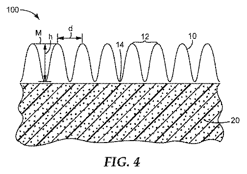
実施例1.本開示による物品の準備
3M TEGADERM(登録商標)Non−Adherent Contact Layerのシートは、米国特許第1,764,676号に記載されたものと同様の波型加工プロセスを対象とし、装置及びローラー速度は、乾燥した弛緩状態において、約1.5mm間隔のリッジと深さが約0.7mmの溝とを有する微小波型シートを形成するために設定される(深さは、本明細書に記載するように、隣接するリッジから隣接するリッジ間の溝の最下点まで延びる、第2のラインに直交する第1のラインに沿って測定される)。弛緩した乾燥微小波型シートを、長さ約12cm×幅7.5cmに切断した。微小波型をシートの幅全体に拡げた。
Example 1. Preparation of Articles According to the Present Disclosure 3M TEGADERM® Non-Adherent Contact Layer sheets are directed to a corrugating process similar to that described in US Pat. No. 1,764,676, and apparatus and roller speeds Is set to form a microwave sheet in the dry relaxed state with ridges spaced about 1.5 mm apart and grooves about 0.7 mm deep (depths are described herein) As measured along a first line orthogonal to a second line extending from adjacent ridges to the lowest point of the groove between adjacent ridges). The relaxed dry microwave sheet was cut into a length of about 12 cm and a width of 7.5 cm. The micro wave pattern was expanded to the entire width of the sheet.
円筒の外周にリッジが延び、円筒がシート内のほぼ中心に位置するように、連続気泡ポリウレタン発泡体の円筒ピース約2.5cm(直径)×約3cm(長さ)を、長さ12cm×7.5cmの微小波型シートで包んだ。円筒の両端部を越えて延びた過剰なシート材料を、数回ねじり、ストリング又はゴムバインダで固定した。 The cylindrical piece of open-cell polyurethane foam is about 2.5 cm (diameter) x about 3 cm (length), and the length is 12 cm x 7 so that the ridge extends to the outer periphery of the cylinder and the cylinder is located approximately in the center of the sheet. Wrapped in a 5 cm microwave sheet. Excess sheet material extending beyond the ends of the cylinder was twisted several times and secured with a string or rubber binder.
実施例2.本開示による複数のセグメントを有する物品の準備
実施例1に記載したような物品を6つ準備した。各物品の両端で微多孔性層をねじった後に、ねじった端部を表1に記載したテザー(長さ約6cm)を使用して結んだ。2つの別個の物品を結ぶために各テザーを使用した。したがって、各テザーは、図5に例示するように、端と端をつないだ6つのセグメントを備える単一の物品を形成するために、2つの隣接する物品(即ち、セグメント)を連結した。
Example 2 Preparation of Articles with Multiple Segments According to the Disclosure Six articles as described in Example 1 were prepared. After twisting the microporous layer at both ends of each article, the twisted ends were tied using a tether (about 6 cm long) as described in Table 1. Each tether was used to tie two separate items. Thus, each tether connected two adjacent articles (i.e., segments) to form a single article with six segments connected end to end, as illustrated in FIG.
実施例3.創傷の陰圧処置中の創傷治癒に対するパッキング材料の効果
Borgquist等(Borgquist,O.,Gustafsson,L,Ingemansson,R.等.「陰圧閉鎖治療中のフォーム内への、しかし、ガーゼ内へはない、組織形成(Tissue ingrowth into foam but not into gauze during negative pressure wound therapy) Wounds 2009;21:11,302〜309;その全体が本明細書に参照として組み込まれる)に記載されたものと同様のプロトコルを使用して、創傷治癒上に微小波型微多孔性創傷接触層を有する物品の効果を評価した。陰圧下でどれくらいうまく肉芽形成されるかについて判断する方法として、他の創傷ドレッシング材について以前に開発された全厚さブタ創傷モデルを使用した。
Example 3 FIG. Effect of packing material on wound healing during negative pressure treatment of wounds Borgquist et al. (Borgquist, O., Gustafsson, L, Ingemanson, R. et al. “Into foam during negative pressure closure treatment, but into gauze No, tissue formation into foam but not into gauze durging negative pressure wound Wounds 2009; 21:11, 302-309; incorporated herein by reference in its entirety) The protocol was used to evaluate the effect of an article having a microwave-type microporous wound contact layer on wound healing, as a method to determine how well granulation occurs under negative pressure. Using a total thickness porcine wound model was developed previously for dressing.
各損傷の測定値が直径4cmである、4つの傍脊柱創傷(両側に2つずつ)全厚さ皮膚創傷を外科的に、創傷間約5cmでブタに形成した。表2に列挙したように、各創傷を異なる材料で満たした。 Four paraspinal wounds (two on each side) full thickness skin wounds, each with a measurement of 4 cm in diameter, were surgically formed in pigs at approximately 5 cm between wounds. As listed in Table 2, each wound was filled with a different material.
全ての創傷パッキング(ドレッシング材)を(Kinetic Concepts,Inc.の)KCI 1−2−Blue drapeで覆い、真空は、全ての部位に125mm Hg(0.017MPa)で真空を印加した。3日後、ドレッシング材を除去し、創傷組織を撮影して、視覚的に観察した。3日後、対照1の創傷では良好な肉芽形成が観測され、対照2の創傷では肉芽形成が部分的に観察された。実施例3によって処置される創傷では、微小波型接触層の微小波型構造を鏡映した細溝構造を有する肉芽組織が観察された。対照3の創傷では、創傷の一部分のみで肉芽組織が観察された。 All wound packings (dressing materials) were covered with KCI 1-2 Blue drape (from Kinetic Concepts, Inc.) and a vacuum was applied to all sites at 125 mm Hg (0.017 MPa). Three days later, the dressing was removed and the wound tissue was photographed and visually observed. After 3 days, good granulation was observed in the control 1 wound and partial granulation was observed in the control 2 wound. In the wound treated according to Example 3, granulation tissue having a fine groove structure reflecting the microwave structure of the microwave contact layer was observed. In the control 3 wound, granulation tissue was observed in only a portion of the wound.
創傷を観察し、撮影した後、各創傷ドレッシング材を同じタイプのドレッシング材と取り替え、ドレッシング材を覆い、上述したように真空を再印加した。4日後(即ち、創傷を形成した7日後)、パッキング材料を創傷の各々から引っ張った。鉗子を使用して創傷パッキングを除去するために必要な力の量を判断するために、Mark−10(Copiague,NY)のフォースゲージM5−50型(シリーズ5フォースゲージ)を使用した。瞬間力読み出し値がディスプレイに示されるが、ピーク値はそれに応じて記録された。各創傷からのパッキングの除去をビデオ録画した。3日目及び7日目の観察結果を表3にまとめた。力測定の結果、創傷の少なくともいくつかにおいて組織の一部分が除去されたので、創傷組織の視覚的評価は7日目には実用的ではなかった。 After observing and filming the wound, each wound dressing was replaced with the same type of dressing, the dressing was covered, and the vacuum was reapplied as described above. After 4 days (ie, 7 days after wound formation), the packing material was pulled from each of the wounds. In order to determine the amount of force required to remove the wound packing using forceps, a Mark-10 (Copiague, NY) force gauge model M5-50 (series 5 force gauge) was used. The instantaneous force reading is shown on the display, but the peak value was recorded accordingly. The removal of packing from each wound was video recorded. The observation results on the third and seventh days are summarized in Table 3. As a result of the force measurement, visual assessment of the wound tissue was not practical on day 7, as at least some of the tissue was removed in at least some of the wounds.
本明細書に引用する全ての特許、特許出願、及び公開公報、並びに電子的に入手可能な資料の開示内容の全体が、参照により組み込まれる。本出願の開示内容と本明細書に組み込まれたいずれかの文献の開示内容との間になんらかの矛盾が存在する場合には、本出願の開示内容が優先されるものとする。上記の発明を実施するための形態及び実施例は、あくまで明確に理解されるよう示したものである。これらによって不要な限定をするものと理解されるべきではない。本発明は、図示及び説明された厳密な詳細に限定されるものではなく、当業者には明らかな変形例は特許請求の範囲によって定義された本発明に含まれるものとする。 The entire disclosures of all patents, patent applications, and publications, and electronically available materials cited herein are incorporated by reference. In the event of any inconsistency between the disclosure content of the present application and the disclosure content of any document incorporated herein, the disclosure content of the present application shall prevail. The embodiments and examples for carrying out the invention described above are shown for the sake of clarity. These should not be construed as making unnecessary limitations. The present invention is not limited to the exact details shown and described, for variations obvious to one skilled in the art are intended to be included within the invention as defined by the claims.
全ての見出しは読者の便宜のためのものであって、特に断らない限り、見出しの後に続く文面の意味を限定するために使用されるものではない。 All headings are for the convenience of the reader and are not used to limit the meaning of the text that follows the heading, unless otherwise specified.
本明細書で例示的に説明された本発明は、本明細書に具体的に開示されていない任意の要素(単数又は複数)の非存在下で適切に実施され得る。したがって、例えば、本明細書のそれぞれの例では、用語「含む」、「本質的に〜からなる(consisting essentially of)」及び「からなる(consisting of)」のいずれも、他の2つの用語のどちらかと置換され得る。利用された用語及び表現は、限定としてではなく説明の用語として使用され、図示及び説明された特徴又はその一部の任意の等価物を除外するような用語及び表現の使用を意図しないが、様々な修正が請求の範囲に記載されている本発明の範囲内で可能であることが理解される。したがって、本発明は好ましい実施形態及び最適な特徴によって具体的に開示されたが、当業者であれば、本明細書で開示された概念の修正及び変更が可能であり、このような修正及び変更は、添付した請求の範囲によって定義される本発明の範囲内であると考えられることを理解すべきである。 The invention described herein by way of example can be suitably practiced in the absence of any element or elements not specifically disclosed herein. Thus, for example, in each example herein, the terms “comprising”, “consisting essentially of”, and “consisting of” are both of the other two terms. Can be replaced with either. The terms and expressions employed are used as descriptive terms, not as limitations, and are not intended to use terms and expressions that exclude the illustrated and described features or any equivalents thereof, It will be understood that various modifications are possible within the scope of the invention as set forth in the claims. Thus, although the present invention has been specifically disclosed by means of preferred embodiments and optimal features, those skilled in the art can modify and modify the concepts disclosed herein, and such modifications and changes. Should be understood to be within the scope of the invention as defined by the appended claims.
Claims (20)
前記微多孔性層が、平均孔径を有する複数の細孔を備え、前記平均孔径が、約90μm又はそれ未満である、
複数のセグメントと、
第1のセグメントを第2のセグメントに接続するテザーと、
を備える、物品。 A plurality of segments each comprising a macroporous wound packing material substantially enclosed in a microwave-type microporous wound contact layer,
The microporous layer comprises a plurality of pores having an average pore size, wherein the average pore size is about 90 μm or less;
Multiple segments,
A tether connecting the first segment to the second segment;
An article comprising.
前記微多孔性層が、前記創傷床に対して配置されるように寸法決定され、
前記微多孔性層が、平均孔径を有する複数の微小孔を備え、前記微小孔の前記平均孔径が、約90μm又はそれ未満である、
ことと、
前記微多孔性層の第2の主表面に近接してマクロ多孔性創傷パッキング材料を配置することであって、
前記微多孔性層に近接して前記マクロ多孔性材料を配置することが、前記創傷面と前記マクロ多孔性材料との間に前記微多孔性層を配置することを含む、
ことと、
前記創傷面、前記層及び前記マクロ多孔性材料を液体不透過性ドレープで覆うことと、
前記マクロ多孔性材料を陰圧源と流体連通に配置することと、
を含む、方法。 Placing the first major surface of the microwave microporous layer in contact with the wound surface of the wound bed,
The microporous layer is dimensioned to be positioned against the wound bed;
The microporous layer comprises a plurality of micropores having an average pore size, and the average pore size of the micropores is about 90 μm or less;
And
Placing a macroporous wound packing material proximate to the second major surface of the microporous layer, comprising:
Disposing the macroporous material proximate to the microporous layer comprises disposing the microporous layer between the wound surface and the macroporous material;
And
Covering the wound surface, the layer and the macroporous material with a liquid impervious drape;
Placing the macroporous material in fluid communication with a negative pressure source;
Including the method.
Applications Claiming Priority (3)
| Application Number | Priority Date | Filing Date | Title |
|---|---|---|---|
| US201461971679P | 2014-03-28 | 2014-03-28 | |
| US61/971,679 | 2014-03-28 | ||
| PCT/US2015/022440 WO2015148636A1 (en) | 2014-03-28 | 2015-03-25 | Article and method for negative pressure wound therapy |
Publications (1)
| Publication Number | Publication Date |
|---|---|
| JP2017512620A true JP2017512620A (en) | 2017-05-25 |
Family
ID=52875780
Family Applications (1)
| Application Number | Title | Priority Date | Filing Date |
|---|---|---|---|
| JP2017502917A Pending JP2017512620A (en) | 2014-03-28 | 2015-03-25 | Articles and methods for negative pressure wound closure therapy |
Country Status (5)
| Country | Link |
|---|---|
| US (1) | US10813793B2 (en) |
| EP (1) | EP3122301B1 (en) |
| JP (1) | JP2017512620A (en) |
| CN (1) | CN106132365B (en) |
| WO (1) | WO2015148636A1 (en) |
Families Citing this family (7)
| Publication number | Priority date | Publication date | Assignee | Title |
|---|---|---|---|---|
| CN103889476B (en) | 2011-06-07 | 2017-04-26 | 史密夫及内修公开有限公司 | Wound contacting members and methods |
| WO2018108784A1 (en) * | 2016-12-12 | 2018-06-21 | Smith & Nephew Plc | Wound dressing |
| CN110997028B (en) | 2017-02-22 | 2022-12-30 | 康奈尔大学 | Mechanical vacuum dressing for mechanical management, protection and aspiration of small incision wounds |
| WO2019089856A1 (en) * | 2017-10-31 | 2019-05-09 | InMEDBio, LLC | Absorbent, breathable and pathogen blocking/killing wound care dressing and fabrication thereof |
| JP7424638B2 (en) | 2017-12-06 | 2024-01-30 | コーネル ユニヴァーシティー | Manually operated negative pressure wound therapy (NPWT) bandage with improved pump efficiency, automatic pressure indicator and automatic pressure limiter |
| US11511032B2 (en) | 2018-03-12 | 2022-11-29 | Kci Licensing, Inc. | Dressing using differential surface features for use in compartment spaces |
| CN115232782B (en) * | 2022-07-16 | 2023-10-31 | 南昌大学 | Double-exposure micro-fold vascular processing platform and method based on coaxial microfluid |
Family Cites Families (38)
| Publication number | Priority date | Publication date | Assignee | Title |
|---|---|---|---|---|
| US1764676A (en) | 1924-12-20 | 1930-06-17 | Samuel J Campbell | Creping machine |
| US4058123A (en) | 1975-10-01 | 1977-11-15 | International Paper Company | Combined irrigator and evacuator for closed wounds |
| DE2953373A1 (en) | 1978-12-06 | 1981-01-08 | P Svedman | Device for treating tissues,for example skin |
| US4569674A (en) | 1982-08-03 | 1986-02-11 | Stryker Corporation | Continuous vacuum wound drainage system |
| US4655754A (en) | 1984-11-09 | 1987-04-07 | Stryker Corporation | Vacuum wound drainage system and lipids baffle therefor |
| US4969880A (en) | 1989-04-03 | 1990-11-13 | Zamierowski David S | Wound dressing and treatment method |
| US5358494A (en) | 1989-07-11 | 1994-10-25 | Svedman Paul | Irrigation dressing |
| JP2719671B2 (en) | 1989-07-11 | 1998-02-25 | 日本ゼオン株式会社 | Wound dressing |
| US4991234A (en) | 1989-10-10 | 1991-02-12 | Bert Greenberg | Body support band |
| US5645081A (en) | 1991-11-14 | 1997-07-08 | Wake Forest University | Method of treating tissue damage and apparatus for same |
| US5636643A (en) | 1991-11-14 | 1997-06-10 | Wake Forest University | Wound treatment employing reduced pressure |
| US6071304A (en) | 1998-04-06 | 2000-06-06 | Augustine Medical, Inc. | Wound treatment apparatus with a heater adhesively joined to a bandage |
| US6095992A (en) | 1998-04-06 | 2000-08-01 | Augustine Medical, Inc. | Wound treatment apparatus for normothermic treatment of wounds |
| US6080189A (en) | 1998-04-06 | 2000-06-27 | Augustine Medical, Inc. | Wound treatment apparatus including a heater and an IR-Transparent or IR-Transmissive bandage |
| US6060079A (en) * | 1998-09-09 | 2000-05-09 | Freeman; Frank | Device for topical localized administration of zinc to tissue |
| FR2783412B1 (en) | 1998-09-18 | 2000-12-15 | Lhd Lab Hygiene Dietetique | NON-ADHERENT STERILE COMPRESS |
| US6855135B2 (en) | 2000-11-29 | 2005-02-15 | Hill-Rom Services, Inc. | Vacuum therapy and cleansing dressing for wounds |
| US7700819B2 (en) * | 2001-02-16 | 2010-04-20 | Kci Licensing, Inc. | Biocompatible wound dressing |
| WO2003057485A1 (en) * | 2001-12-21 | 2003-07-17 | Avery Dennison Corporation | Process for the manufacture of multi-layered wound dressings |
| EP1374812A1 (en) * | 2002-06-28 | 2004-01-02 | Biopol Co., Ltd. | Multilayered microporous foam dressing and method for manufacturing the same |
| US20050205840A1 (en) | 2003-10-03 | 2005-09-22 | Farneth William E | Oxygen scavenging compositions and methods of use |
| MX2008002882A (en) | 2005-09-07 | 2008-03-27 | Tyco Healthcare | Wound dressing with vacuum reservoir. |
| JP2009542353A (en) | 2006-07-07 | 2009-12-03 | ボリンジャー・テクノロジーズ・エル・ピー | Growth-stimulated wound dressing with improved contact surface |
| US20090234306A1 (en) * | 2008-03-13 | 2009-09-17 | Tyco Healthcare Group Lp | Vacuum wound therapy wound dressing with variable performance zones |
| US9414968B2 (en) * | 2008-09-05 | 2016-08-16 | Smith & Nephew, Inc. | Three-dimensional porous film contact layer with improved wound healing |
| DE102008062824A1 (en) | 2008-12-23 | 2010-07-01 | Paul Hartmann Ag | wound dressing |
| MX2011006808A (en) * | 2008-12-24 | 2011-07-12 | Kci Licensing Inc | Membranes, systems, and methods for applying reduced pressure to a subcutaneous tissue site. |
| US8529528B2 (en) * | 2008-12-24 | 2013-09-10 | Kci Licensing, Inc. | Reduced-pressure wound treatment systems and methods employing microstrain-inducing manifolds |
| US20100191196A1 (en) | 2009-01-26 | 2010-07-29 | Heagle David G | Fibrous Wound Filler Material for Negative Pressure Wound Therapy |
| US9421309B2 (en) | 2009-06-02 | 2016-08-23 | Kci Licensing, Inc. | Reduced-pressure treatment systems and methods employing hydrogel reservoir members |
| AU2010341491B2 (en) | 2009-12-22 | 2015-05-14 | Smith & Nephew, Inc. | Apparatuses and methods for negative pressure wound therapy |
| KR101813300B1 (en) * | 2010-04-08 | 2017-12-28 | 힐리오닉스 코포레이션 | Implantable medical devices having microporous surface layers and method for reducing foreign body response to the same |
| CN102002817A (en) | 2010-11-22 | 2011-04-06 | 山东俊富非织造材料有限公司 | Punched spunbonded nonwoven material and manufacturing method thereof |
| AU2012282287B2 (en) | 2011-07-14 | 2017-06-01 | Smith & Nephew Plc | Wound dressing and method of treatment |
| JP6382185B2 (en) | 2012-05-22 | 2018-08-29 | スミス アンド ネフュー ピーエルシーSmith & Nephew Public Limited Company | Apparatus and method for wound treatment |
| CA2888241C (en) * | 2012-10-16 | 2020-12-29 | Surmodics, Inc. | Wound packing device and methods |
| CN203246125U (en) | 2013-05-20 | 2013-10-23 | 嘉兴港恒针织有限公司 | Fabric with strip-type bulges |
| CN103655046B (en) | 2013-12-03 | 2016-02-03 | 佛山市优特医疗科技有限公司 | A kind of wound dressing with three-decker and preparation method thereof |
-
2015
- 2015-03-25 US US15/129,434 patent/US10813793B2/en active Active
- 2015-03-25 WO PCT/US2015/022440 patent/WO2015148636A1/en not_active Ceased
- 2015-03-25 CN CN201580017124.3A patent/CN106132365B/en active Active
- 2015-03-25 EP EP15716924.4A patent/EP3122301B1/en active Active
- 2015-03-25 JP JP2017502917A patent/JP2017512620A/en active Pending
Also Published As
| Publication number | Publication date |
|---|---|
| EP3122301B1 (en) | 2018-04-25 |
| EP3122301A1 (en) | 2017-02-01 |
| US10813793B2 (en) | 2020-10-27 |
| WO2015148636A1 (en) | 2015-10-01 |
| CN106132365B (en) | 2020-07-14 |
| US20170172806A1 (en) | 2017-06-22 |
| CN106132365A (en) | 2016-11-16 |
Similar Documents
| Publication | Publication Date | Title |
|---|---|---|
| JP6942768B2 (en) | Wound bandage | |
| US20240197537A1 (en) | Dressing with fluid acquisition and distribution characteristics | |
| JP2017512620A (en) | Articles and methods for negative pressure wound closure therapy | |
| RU2615075C2 (en) | Wound dressing and methods | |
| JP6742283B2 (en) | Tissue healing | |
| EP2694005B1 (en) | Negative pressure wound therapy dressing | |
| US9302034B2 (en) | Negative pressure wound therapy dressing | |
| US20140188090A1 (en) | Wound care article | |
| CN106255481A (en) | Collapsible Dressings for Negative Pressure Wound Therapy | |
| CN111134959A (en) | Dressings with Shrinking Layers for Linear Tissue Sites | |
| CN114126673A (en) | Absorbent parts | |
| US20140221946A1 (en) | Absorbent body for the therapeutic treatment of a wound by means of negative pressure |